CN112351999A - anti-MUC 1 antibody-drug conjugates - Google Patents
anti-MUC 1 antibody-drug conjugates Download PDFInfo
- Publication number
- CN112351999A CN112351999A CN201980033159.4A CN201980033159A CN112351999A CN 112351999 A CN112351999 A CN 112351999A CN 201980033159 A CN201980033159 A CN 201980033159A CN 112351999 A CN112351999 A CN 112351999A
- Authority
- CN
- China
- Prior art keywords
- antibody
- amino acid
- acid sequence
- seq
- cancer
- Prior art date
- Legal status (The legal status is an assumption and is not a legal conclusion. Google has not performed a legal analysis and makes no representation as to the accuracy of the status listed.)
- Pending
Links
Images


Classifications
-
- A—HUMAN NECESSITIES
- A61—MEDICAL OR VETERINARY SCIENCE; HYGIENE
- A61K—PREPARATIONS FOR MEDICAL, DENTAL OR TOILETRY PURPOSES
- A61K47/00—Medicinal preparations characterised by the non-active ingredients used, e.g. carriers or inert additives; Targeting or modifying agents chemically bound to the active ingredient
- A61K47/50—Medicinal preparations characterised by the non-active ingredients used, e.g. carriers or inert additives; Targeting or modifying agents chemically bound to the active ingredient the non-active ingredient being chemically bound to the active ingredient, e.g. polymer-drug conjugates
- A61K47/51—Medicinal preparations characterised by the non-active ingredients used, e.g. carriers or inert additives; Targeting or modifying agents chemically bound to the active ingredient the non-active ingredient being chemically bound to the active ingredient, e.g. polymer-drug conjugates the non-active ingredient being a modifying agent
- A61K47/68—Medicinal preparations characterised by the non-active ingredients used, e.g. carriers or inert additives; Targeting or modifying agents chemically bound to the active ingredient the non-active ingredient being chemically bound to the active ingredient, e.g. polymer-drug conjugates the non-active ingredient being a modifying agent the modifying agent being an antibody, an immunoglobulin or a fragment thereof, e.g. an Fc-fragment
- A61K47/6835—Medicinal preparations characterised by the non-active ingredients used, e.g. carriers or inert additives; Targeting or modifying agents chemically bound to the active ingredient the non-active ingredient being chemically bound to the active ingredient, e.g. polymer-drug conjugates the non-active ingredient being a modifying agent the modifying agent being an antibody, an immunoglobulin or a fragment thereof, e.g. an Fc-fragment the modifying agent being an antibody or an immunoglobulin bearing at least one antigen-binding site
- A61K47/6851—Medicinal preparations characterised by the non-active ingredients used, e.g. carriers or inert additives; Targeting or modifying agents chemically bound to the active ingredient the non-active ingredient being chemically bound to the active ingredient, e.g. polymer-drug conjugates the non-active ingredient being a modifying agent the modifying agent being an antibody, an immunoglobulin or a fragment thereof, e.g. an Fc-fragment the modifying agent being an antibody or an immunoglobulin bearing at least one antigen-binding site the antibody targeting a determinant of a tumour cell
-
- A—HUMAN NECESSITIES
- A61—MEDICAL OR VETERINARY SCIENCE; HYGIENE
- A61K—PREPARATIONS FOR MEDICAL, DENTAL OR TOILETRY PURPOSES
- A61K31/00—Medicinal preparations containing organic active ingredients
- A61K31/33—Heterocyclic compounds
- A61K31/395—Heterocyclic compounds having nitrogen as a ring hetero atom, e.g. guanethidine or rifamycins
- A61K31/435—Heterocyclic compounds having nitrogen as a ring hetero atom, e.g. guanethidine or rifamycins having six-membered rings with one nitrogen as the only ring hetero atom
- A61K31/47—Quinolines; Isoquinolines
- A61K31/4738—Quinolines; Isoquinolines ortho- or peri-condensed with heterocyclic ring systems
- A61K31/4745—Quinolines; Isoquinolines ortho- or peri-condensed with heterocyclic ring systems condensed with ring systems having nitrogen as a ring hetero atom, e.g. phenantrolines
-
- A—HUMAN NECESSITIES
- A61—MEDICAL OR VETERINARY SCIENCE; HYGIENE
- A61K—PREPARATIONS FOR MEDICAL, DENTAL OR TOILETRY PURPOSES
- A61K39/00—Medicinal preparations containing antigens or antibodies
- A61K39/395—Antibodies; Immunoglobulins; Immune serum, e.g. antilymphocytic serum
- A61K39/39533—Antibodies; Immunoglobulins; Immune serum, e.g. antilymphocytic serum against materials from animals
- A61K39/39558—Antibodies; Immunoglobulins; Immune serum, e.g. antilymphocytic serum against materials from animals against tumor tissues, cells, antigens
-
- A—HUMAN NECESSITIES
- A61—MEDICAL OR VETERINARY SCIENCE; HYGIENE
- A61K—PREPARATIONS FOR MEDICAL, DENTAL OR TOILETRY PURPOSES
- A61K45/00—Medicinal preparations containing active ingredients not provided for in groups A61K31/00 - A61K41/00
- A61K45/06—Mixtures of active ingredients without chemical characterisation, e.g. antiphlogistics and cardiaca
-
- A—HUMAN NECESSITIES
- A61—MEDICAL OR VETERINARY SCIENCE; HYGIENE
- A61K—PREPARATIONS FOR MEDICAL, DENTAL OR TOILETRY PURPOSES
- A61K47/00—Medicinal preparations characterised by the non-active ingredients used, e.g. carriers or inert additives; Targeting or modifying agents chemically bound to the active ingredient
- A61K47/06—Organic compounds, e.g. natural or synthetic hydrocarbons, polyolefins, mineral oil, petrolatum or ozokerite
- A61K47/22—Heterocyclic compounds, e.g. ascorbic acid, tocopherol or pyrrolidones
-
- A—HUMAN NECESSITIES
- A61—MEDICAL OR VETERINARY SCIENCE; HYGIENE
- A61K—PREPARATIONS FOR MEDICAL, DENTAL OR TOILETRY PURPOSES
- A61K47/00—Medicinal preparations characterised by the non-active ingredients used, e.g. carriers or inert additives; Targeting or modifying agents chemically bound to the active ingredient
- A61K47/50—Medicinal preparations characterised by the non-active ingredients used, e.g. carriers or inert additives; Targeting or modifying agents chemically bound to the active ingredient the non-active ingredient being chemically bound to the active ingredient, e.g. polymer-drug conjugates
- A61K47/51—Medicinal preparations characterised by the non-active ingredients used, e.g. carriers or inert additives; Targeting or modifying agents chemically bound to the active ingredient the non-active ingredient being chemically bound to the active ingredient, e.g. polymer-drug conjugates the non-active ingredient being a modifying agent
- A61K47/54—Medicinal preparations characterised by the non-active ingredients used, e.g. carriers or inert additives; Targeting or modifying agents chemically bound to the active ingredient the non-active ingredient being chemically bound to the active ingredient, e.g. polymer-drug conjugates the non-active ingredient being a modifying agent the modifying agent being an organic compound
- A61K47/545—Heterocyclic compounds
-
- A—HUMAN NECESSITIES
- A61—MEDICAL OR VETERINARY SCIENCE; HYGIENE
- A61K—PREPARATIONS FOR MEDICAL, DENTAL OR TOILETRY PURPOSES
- A61K47/00—Medicinal preparations characterised by the non-active ingredients used, e.g. carriers or inert additives; Targeting or modifying agents chemically bound to the active ingredient
- A61K47/50—Medicinal preparations characterised by the non-active ingredients used, e.g. carriers or inert additives; Targeting or modifying agents chemically bound to the active ingredient the non-active ingredient being chemically bound to the active ingredient, e.g. polymer-drug conjugates
- A61K47/51—Medicinal preparations characterised by the non-active ingredients used, e.g. carriers or inert additives; Targeting or modifying agents chemically bound to the active ingredient the non-active ingredient being chemically bound to the active ingredient, e.g. polymer-drug conjugates the non-active ingredient being a modifying agent
- A61K47/62—Medicinal preparations characterised by the non-active ingredients used, e.g. carriers or inert additives; Targeting or modifying agents chemically bound to the active ingredient the non-active ingredient being chemically bound to the active ingredient, e.g. polymer-drug conjugates the non-active ingredient being a modifying agent the modifying agent being a protein, peptide or polyamino acid
- A61K47/65—Peptidic linkers, binders or spacers, e.g. peptidic enzyme-labile linkers
-
- A—HUMAN NECESSITIES
- A61—MEDICAL OR VETERINARY SCIENCE; HYGIENE
- A61K—PREPARATIONS FOR MEDICAL, DENTAL OR TOILETRY PURPOSES
- A61K47/00—Medicinal preparations characterised by the non-active ingredients used, e.g. carriers or inert additives; Targeting or modifying agents chemically bound to the active ingredient
- A61K47/50—Medicinal preparations characterised by the non-active ingredients used, e.g. carriers or inert additives; Targeting or modifying agents chemically bound to the active ingredient the non-active ingredient being chemically bound to the active ingredient, e.g. polymer-drug conjugates
- A61K47/51—Medicinal preparations characterised by the non-active ingredients used, e.g. carriers or inert additives; Targeting or modifying agents chemically bound to the active ingredient the non-active ingredient being chemically bound to the active ingredient, e.g. polymer-drug conjugates the non-active ingredient being a modifying agent
- A61K47/68—Medicinal preparations characterised by the non-active ingredients used, e.g. carriers or inert additives; Targeting or modifying agents chemically bound to the active ingredient the non-active ingredient being chemically bound to the active ingredient, e.g. polymer-drug conjugates the non-active ingredient being a modifying agent the modifying agent being an antibody, an immunoglobulin or a fragment thereof, e.g. an Fc-fragment
- A61K47/6801—Drug-antibody or immunoglobulin conjugates defined by the pharmacologically or therapeutically active agent
- A61K47/6803—Drugs conjugated to an antibody or immunoglobulin, e.g. cisplatin-antibody conjugates
-
- A—HUMAN NECESSITIES
- A61—MEDICAL OR VETERINARY SCIENCE; HYGIENE
- A61P—SPECIFIC THERAPEUTIC ACTIVITY OF CHEMICAL COMPOUNDS OR MEDICINAL PREPARATIONS
- A61P35/00—Antineoplastic agents
-
- A—HUMAN NECESSITIES
- A61—MEDICAL OR VETERINARY SCIENCE; HYGIENE
- A61P—SPECIFIC THERAPEUTIC ACTIVITY OF CHEMICAL COMPOUNDS OR MEDICINAL PREPARATIONS
- A61P35/00—Antineoplastic agents
- A61P35/02—Antineoplastic agents specific for leukemia
-
- C—CHEMISTRY; METALLURGY
- C07—ORGANIC CHEMISTRY
- C07K—PEPTIDES
- C07K16/00—Immunoglobulins [IGs], e.g. monoclonal or polyclonal antibodies
- C07K16/18—Immunoglobulins [IGs], e.g. monoclonal or polyclonal antibodies against material from animals or humans
- C07K16/28—Immunoglobulins [IGs], e.g. monoclonal or polyclonal antibodies against material from animals or humans against receptors, cell surface antigens or cell surface determinants
- C07K16/30—Immunoglobulins [IGs], e.g. monoclonal or polyclonal antibodies against material from animals or humans against receptors, cell surface antigens or cell surface determinants from tumour cells
- C07K16/3076—Immunoglobulins [IGs], e.g. monoclonal or polyclonal antibodies against material from animals or humans against receptors, cell surface antigens or cell surface determinants from tumour cells against structure-related tumour-associated moieties
- C07K16/3092—Immunoglobulins [IGs], e.g. monoclonal or polyclonal antibodies against material from animals or humans against receptors, cell surface antigens or cell surface determinants from tumour cells against structure-related tumour-associated moieties against tumour-associated mucins
-
- A—HUMAN NECESSITIES
- A61—MEDICAL OR VETERINARY SCIENCE; HYGIENE
- A61K—PREPARATIONS FOR MEDICAL, DENTAL OR TOILETRY PURPOSES
- A61K39/00—Medicinal preparations containing antigens or antibodies
- A61K2039/505—Medicinal preparations containing antigens or antibodies comprising antibodies
-
- C—CHEMISTRY; METALLURGY
- C07—ORGANIC CHEMISTRY
- C07K—PEPTIDES
- C07K2317/00—Immunoglobulins specific features
- C07K2317/10—Immunoglobulins specific features characterized by their source of isolation or production
- C07K2317/14—Specific host cells or culture conditions, e.g. components, pH or temperature
-
- C—CHEMISTRY; METALLURGY
- C07—ORGANIC CHEMISTRY
- C07K—PEPTIDES
- C07K2317/00—Immunoglobulins specific features
- C07K2317/20—Immunoglobulins specific features characterized by taxonomic origin
- C07K2317/24—Immunoglobulins specific features characterized by taxonomic origin containing regions, domains or residues from different species, e.g. chimeric, humanized or veneered
-
- C—CHEMISTRY; METALLURGY
- C07—ORGANIC CHEMISTRY
- C07K—PEPTIDES
- C07K2317/00—Immunoglobulins specific features
- C07K2317/40—Immunoglobulins specific features characterized by post-translational modification
- C07K2317/41—Glycosylation, sialylation, or fucosylation
-
- C—CHEMISTRY; METALLURGY
- C07—ORGANIC CHEMISTRY
- C07K—PEPTIDES
- C07K2317/00—Immunoglobulins specific features
- C07K2317/50—Immunoglobulins specific features characterized by immunoglobulin fragments
- C07K2317/55—Fab or Fab'
-
- C—CHEMISTRY; METALLURGY
- C07—ORGANIC CHEMISTRY
- C07K—PEPTIDES
- C07K2317/00—Immunoglobulins specific features
- C07K2317/50—Immunoglobulins specific features characterized by immunoglobulin fragments
- C07K2317/56—Immunoglobulins specific features characterized by immunoglobulin fragments variable (Fv) region, i.e. VH and/or VL
- C07K2317/565—Complementarity determining region [CDR]
-
- C—CHEMISTRY; METALLURGY
- C07—ORGANIC CHEMISTRY
- C07K—PEPTIDES
- C07K2317/00—Immunoglobulins specific features
- C07K2317/70—Immunoglobulins specific features characterized by effect upon binding to a cell or to an antigen
- C07K2317/73—Inducing cell death, e.g. apoptosis, necrosis or inhibition of cell proliferation
-
- C—CHEMISTRY; METALLURGY
- C07—ORGANIC CHEMISTRY
- C07K—PEPTIDES
- C07K2317/00—Immunoglobulins specific features
- C07K2317/70—Immunoglobulins specific features characterized by effect upon binding to a cell or to an antigen
- C07K2317/76—Antagonist effect on antigen, e.g. neutralization or inhibition of binding
-
- C—CHEMISTRY; METALLURGY
- C07—ORGANIC CHEMISTRY
- C07K—PEPTIDES
- C07K2317/00—Immunoglobulins specific features
- C07K2317/90—Immunoglobulins specific features characterized by (pharmaco)kinetic aspects or by stability of the immunoglobulin
- C07K2317/92—Affinity (KD), association rate (Ka), dissociation rate (Kd) or EC50 value
Abstract
The present disclosure relates to antibody drug conjugates directed against the cancer antigen MUC 1. Specifically, by deleting glycosylation sites in the CDR-H2 of the known anti-MUC 1 antibody, an antibody with improved antigen binding is obtained. The conjugate consists of an irinotecan derivative coupled to an anti-MUC 1 antibody.
Description
Technical Field
The present invention relates to the field of Antibody Drug Conjugates (ADCs). The ADCs of the invention comprise an anti-MUC 1 antibody or a mutated anti-MUC 1 antibody. ADCs with mutated anti-MUC 1 antibodies having increased antigen binding affinity are provided. Specifically, in the mutant form of the humanized antibody PankoMab asparagine 57 of the heavy chain variable region is replaced by another amino acid. Thus, the glycosylation sites in the CDR2 region were deleted and antigen binding affinity was increased. ADCs showed significant antitumor efficacy. In particular embodiments, the invention relates to therapeutic and diagnostic uses of the antibody drug conjugates and methods of producing such antibody drug conjugates.
Background
Antibodies against tumor-associated antigens are widely used therapeutic agents against cancer. Today, many anti-cancer antibodies are approved for human therapy. Some of these antibodies act by blocking certain signaling pathways critical to the survival or proliferation of a particular cancer cell. Other anti-cancer antibodies activate the patient's immune response against the targeted cancer cells, such as initiating antibody-dependent cellular cytotoxicity (ADCC) by natural killer cells. This mechanism is induced by the binding of the Fc portion of the antibody to Fc receptors on immune cells.
An interesting and important group of antibodies are antibodies against mucins. Mucins are a family of high molecular weight, highly glycosylated proteins produced by many epithelial tissues in vertebrates. They can be subdivided into membrane-bound mucins due to the presence of hydrophobic transmembrane domains, which facilitate retention in the plasma membrane, and mucins that are secreted to mucosal surfaces or secreted to become components of saliva. The human mucin family consists of many family members, including membrane-bound MUC 1.
Increased mucin production occurs in many adenocarcinomas, including pancreatic, lung, breast, ovarian, colon, and the like. Mucins are also overexpressed in lung diseases such as asthma, bronchitis, chronic obstructive pulmonary disease, or cystic fibrosis. Two membrane mucins, MUC1 and MUC4, have been extensively studied for their pathological significance in the disease process. In addition, mucins have also been studied for their potential as diagnostic markers. Several antibodies against mucins (Clin. Cancer Res., 2011 Nov 1; 17(21):6822-30, PLoS One, 2011 Jan 14;6(1): e15921), in particular MUC1, are known in the art. However, their therapeutic efficacy can still be improved.
In view of this, there is a need in the art to provide therapeutic anti-MUC 1 antibodies with improved properties.
The ADC consists of three distinct components (antibody, linker and drug/payload) that are responsible for the specific delivery of the payload to the target cell. To date, four ADCs (Geotuzumab Osmax (Mylotarg), Iguzumab Osmax (Besponsa), Brentuximab vetodin (Adcetris), trastuzumab emtansine (T-DM1; KadCyla)) have entered the market. In addition, over 60 ADCs are under development for a variety of hematologic cancers and solid tumors. ADCs create a new paradigm for novel cancer chemotherapies. By virtue of the specificity of monoclonal antibodies and the cytotoxic ability of small molecule drugs, ADCs are expected to become an important component in the future of precision medicine and combination therapy. Accordingly, there is a continuing need to provide other ADCs and means, methods and uses for the treatment and/or diagnosis of disease.
As ADCs, ADCs are known in which exatecan is conjugated to an antibody (e.g. an anti-HER 2 antibody) via a linker (WO2014/057687, WO 2015/115091). However, ADCs in which irinotecan is conjugated to anti-MUC 1 antibodies are not known.
Disclosure of Invention
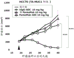
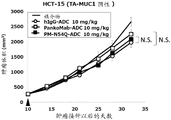
The inventors of the present invention have found that the deletion of the glycosylation site in the heavy chain variable region of the PankoMab anti-MUC 1 antibody does not abrogate antigen binding, but rather unexpectedly increases the antigen affinity of the antibody. This is particularly surprising given that the glycosylation site is located in the second complementarity determining region of the heavy chain variable region (CDR-H2). CDRs are those regions of an antibody that are directly involved in antigen binding and provide contact with an epitope. Thus, it is generally expected that amino acids that modify the CDRs will be detrimental to antigen binding affinity. The humanized PankoMab antibody additionally contains a glycosylation site in CDR-H2, which has a large carbohydrate structure. The carbohydrate structure is present directly at the binding interface with the antigen and is therefore thought to be involved in antigen binding. However, as demonstrated in the examples, the PankoMab variant (PM-N54Q) in which the glycosylation site is deleted by substitution of an amino acid with a carbohydrate structure, shows increased antigen binding affinity. In addition, the inventors of the present invention have found that a conjugate or an antibody-drug conjugate (ADC) comprising PankoMab or a PankoMab variant (PM-N54Q) exhibits significant anti-tumor efficacy against MUC 1-positive tumors, and PM-N54Q-ADC exhibits significant anti-tumor efficacy compared to PankoMab-ADC.
Thus, in a first aspect, the invention relates to a conjugate comprising an antibody conjugated to a cytotoxic agent, wherein the antibody is capable of binding MUC1 and comprises
(i) A heavy chain variable region comprising the following Complementarity Determining Regions (CDRs): CDR-H1 having the amino acid sequence of SEQ ID NO. 1, CDR-H2 having the amino acid sequence of SEQ ID NO. 2, and CDR-H3 having the amino acid sequence of SEQ ID NO. 3, and
(ii) a light chain variable region comprising the following Complementarity Determining Regions (CDRs): CDR-L1 having the amino acid sequence of SEQ ID NO. 4, CDR-L2 having the amino acid sequence of SEQ ID NO. 5, and CDR-L3 having the amino acid sequence of SEQ ID NO. 6.
In a second aspect, the invention relates to a composition comprising a conjugate according to the invention.
According to a third aspect, the present invention provides a composition or conjugate according to the invention for use in medicine, in particular in the treatment, prevention or diagnosis of cancer.
In a fourth aspect, the present invention provides a method of treating cancer in a subject in need thereof, the method comprising administering to the subject having cancer a therapeutically effective amount of a conjugate according to the present invention.
In a fifth aspect, the invention provides kits or devices comprising conjugates according to the invention and related methods, which are useful for the diagnosis, detection or monitoring of MUC 1-associated disorders such as cancer.
Other objects, features, advantages and aspects of the present invention will become apparent to those skilled in the art from the following description and appended claims. It should be understood, however, that the following description, appended claims, and specific examples, while indicating preferred embodiments of the application, are given by way of illustration only. Various changes and modifications within the spirit and scope of the disclosed invention will become readily apparent to those skilled in the art from reading the following.
Definition of
The following expressions used herein are generally intended to preferably have the meanings set forth below, unless the context in which they are used indicates otherwise.
The expression "comprising" as used herein also includes and specifically refers to the expressions "consisting essentially of and" consisting of, in addition to their literal meaning. Thus, the expression "comprising" refers to embodiments in which the subject matter that "comprises" the specifically listed elements does not comprise other elements, as well as embodiments in which the subject matter that "comprises" the specifically listed elements may and/or does comprise other elements. Likewise, the expression "having" is to be understood as meaning the expression "comprising" and also including and in particular referring to the expressions "consisting essentially of and" consisting of. The term "consisting essentially of" particularly refers to embodiments in which the subject matter comprises 20% or less, particularly 15% or less, 10% or less, or especially 5% or less of the other elements, in addition to the specifically listed elements that substantially make up the subject matter, where possible.
The term "antibody" particularly refers to a protein comprising at least two heavy chains and two light chains linked by disulfide bonds. Each heavy chain is composed of 1 heavy chain variable region (V)H) And 1 heavy chain constant region (C)H) And (4) forming. Each light chain is composed of 1 light chain variable region (V)L) And 1 light chain constant region (C)L) And (4) forming. The heavy chain constant region comprises three or (in the case of IgM-or IgE-type antibodies) four heavy chain constant domains (C)H1、CH2、CH3And CH4) Wherein the first constant domain CH1Adjacent to the variable region and may be connected to a second constant domain C by a hinge regionH2. The light chain constant region consists of only one constant domain. The variable regions may be further subdivided into hypervariable regions (referred to as Complementarity Determining Regions (CDRs)) interspersed with more conserved regions (referred to as Framework Regions (FRs)), where each variable region comprises three CDRs and four FRs. The variable regions of the heavy and light chains contain binding domains that interact with antigens. The heavy chain constant region can be of any type, such as a gamma-, delta-, alpha-, mu-, or epsilon-type heavy chain. Preferably, the heavy chain of the antibody is a gamma chain. In addition, the light chain constant region can also be of any type, such as kappa-or lambda-type light chains. Preferably, the light chain of the antibody is a kappa chain. The terms "heavy chain of the γ - (δ -, α -, μ -or e-) type" and "light chain of the κ - (λ -) type" refer to an antibody heavy chain or an antibody light chain, respectively: it has a constant region amino acid sequence derived from a naturally occurring heavy or light chain constant region amino acid sequence, particularly a human heavy or light chain constant region amino acid sequence. In particular, the amino acid sequence of the constant domain of a gamma-type (especially gamma-type 1) heavy chain is at least 95%, especially at least 98% identical to the amino acid sequence of the constant domain of a human gamma (especially human gamma 1) antibody heavy chain. Furthermore, the amino acid sequence of the constant domain of the kappa-type light chain is in particular at least 95%, in particular at least 98% identical to the amino acid sequence of the constant domain of the human kappa antibody light chain. The constant region of the antibody may mediate the binding of the immunoglobulin to host tissues or factors, including various cells of the immune system (e.g., effector cells) and the first component of the classical complement system (C1 q). The antibody may be, for example, a humanized, human or chimeric antibody.
An antigen-binding portion of an antibody generally refers to the full length or one or more fragments of an antibody that retain the ability to specifically bind to an antigen. It has been demonstrated that the antigen binding function of an antibody can be performed by fragments of a full-length antibody. Examples of binding fragments of antibodies include Fab fragments, which are composed of VL、VH、CLAnd CH1Monovalent fragments consisting of domains; f (ab)2Segments, which are contained by hingesA bivalent fragment of two Fab fragments linked by a disulfide bond of regions, each Fab fragment binding to the same antigen; from VHAnd CH1Domain-forming Fd fragments; v with one arm consisting of antibodyLAnd VH(iii) an Fv fragment consisting of a domain; and is composed of VHDomain-composed dAb fragments.
The "Fab part" of an antibody is in particular meant to comprise the heavy and light chain variable regions (V)HAnd VL) And the first domain of the heavy chain and the light chain constant region (C)H1And CL) A portion of the antibody of (1). In the case of antibodies not comprising all these regions, the term "Fab part" refers only to the region VH、VL、CH1And CLThose present in antibodies. Preferably, the "Fab portion" refers to a portion of an antibody that corresponds to a fragment containing the antigen binding activity of the antibody obtained by digestion of a native antibody with papain. In particular, the Fab portion of the antibody includes an antigen binding site or its antigen binding ability. Preferably, the Fab part comprises at least the V of the antibodyHAnd (4) a region.
The "Fc portion" of an antibody is intended in particular to comprise the heavy chain constant regions 2,3 and, where applicable, 4 (C)H2、CH3And CH4) A portion of the antibody of (1). In particular, the Fc portion includes two of each of these regions. In the case of antibodies that do not contain all of these regions, then the term "Fc portion" refers only to region CH2、CH3And CH4Of (a) are those present in the antibody. Preferably, the Fc portion comprises at least C of the antibodyH2And (4) a region. Preferably, the "Fc portion" refers to a portion of an antibody that corresponds to a fragment obtained by digesting a native antibody with papain that does not contain the antigen binding activity of the antibody. In particular, the Fc portion of an antibody is capable of binding to an Fc receptor and thus, for example, comprises an Fc receptor binding site or Fc receptor binding capacity.
The terms "antibody" and "antibody construct" as used herein refer in certain embodiments to the same class of antibody or population of antibody constructs, respectively. In particular, all antibodies or antibody constructs of the population show the characteristics used to define the antibody or antibody construct. In certain embodiments, all antibodies or antibody constructs in a population have the same amino acid sequence. Reference to a particular class of antibody (such as an antibody capable of specifically binding MUC1) is particularly directed to a population of such antibodies.
The term "antibody" as used herein also includes fragments and derivatives of said antibody. A "fragment or derivative" of an antibody is in particular a protein or glycoprotein derived from said antibody and capable of binding to the same antigen, in particular to the same epitope, as said antibody. Thus, a fragment or derivative of an antibody herein generally refers to a functional fragment or derivative. In a particularly preferred embodiment, the fragment or derivative of the antibody comprises a heavy chain variable region. It has been demonstrated that the antigen binding function of an antibody can be performed by fragments of a full-length antibody or a derivative thereof. Examples of antibody fragments include: (i) a Fab fragment, which is a monovalent fragment consisting of the variable region and the first constant region of each of the heavy and light chains; (ii) f (ab)2A fragment which is a bivalent fragment comprising two Fab fragments linked by a disulfide bond at the hinge region; (iii) a variable region and a first constant region C of a heavy chainH1A composed Fd fragment; (iv) (ii) an Fv fragment consisting of the variable regions of the heavy and light chains of a single arm of an antibody; (v) a scFv fragment, which is an Fv fragment consisting of a single polypeptide chain; (vi) consisting of two Fv fragments covalently linked together (Fv)2A fragment; (vii) a heavy chain variable domain; and (viii) a multimer consisting of a heavy chain variable region and a light chain variable region that are covalently linked together in such a way that association of the heavy chain and light chain variable regions can occur only intermolecularly, not intramolecularly. Derivatives of an antibody specifically include antibodies that bind to or compete for the same antigen as the parent antibody but have an amino acid sequence that is different from the parent antibody from which they are derived. These antibody fragments and derivatives are obtained using conventional techniques known to those skilled in the art.
A target amino acid sequence is "derived from" or "corresponds to" a reference amino acid sequence if it has at least 75%, more preferably at least 80%, at least 85%, at least 90%, at least 93%, at least 95%, at least 97%, at least 98%, or at least 99% homology or identity over its entire length to the corresponding portion of the reference amino acid sequence. By "corresponding portion" is meant, for example, that framework region 1(FRH1) of the heavy chain variable region of the target antibody corresponds to framework region 1 of the heavy chain variable region of the reference antibody. In particular embodiments, a target amino acid sequence "derived from" or "corresponding to" a reference amino acid sequence is 100% homologous, or in particular 100% identical, over its entire length to the corresponding portion of the reference amino acid sequence. "homology" or "identity" of amino acid sequences or nucleotide sequences is preferably determined according to the invention over the entire length of the reference sequence or over the entire length of the corresponding part of the reference sequence (which corresponds to the sequence for which homology or identity is defined). An antibody derived from a parent antibody, which is defined by one or more amino acid sequences (e.g. a specific CDR sequence or a specific variable region sequence), in particular an antibody having an amino acid sequence, e.g. a CDR sequence or a variable region sequence, which is at least 75%, preferably at least 80%, at least 85%, at least 90%, at least 93%, at least 95%, at least 97%, at least 98% or at least 99% homologous or identical, in particular identical, to the respective amino acid sequence of the parent antibody. In certain embodiments, an antibody derived from a parent antibody (i.e., a derivative thereof) comprises the same CDR sequences as the parent antibody, but differs in the remaining sequences of the variable region.
The term "antibody" as used herein also refers to multivalent and multispecific antibodies, i.e. antibody constructs having more than two binding sites each binding to the same epitope, and antibody constructs having one or more binding sites that bind a first epitope and one or more binding sites that bind a second epitope, and optionally other binding sites that even bind to other epitopes.
By "specifically binds" is preferably meant that an agent, e.g., an antibody, binds more strongly to its specific target, e.g., an epitope, than to another target. Examples of criteria for determining whether binding is specific may include the dissociation constant (referred to herein as "KD"). Dissociation constant (K) if the agent binds to the first targetd) Lower than for the second targetAnd a dissociation constant of (a), then binding to the first target is stronger compared to the second target. Preferably, the dissociation constant of a target to which an agent specifically binds is more than 100-fold, 200-fold, 500-fold, or more than 1000-fold lower than the dissociation constant of a target to which the agent does not specifically bind. Furthermore, the term "specific binding" especially indicates that the binding affinity between the binding partners has at least 106 M-1Preferably at least 107 M-1More preferably at least 108 M-1Affinity constant K ofa. An antibody specific for an antigen is in particular capable of having at least 106 M-1Preferably at least 107 M-1More preferably at least 108 M-1K ofaAn antibody that binds to the antigen. For example, the term "anti-MUC 1 antibody" particularly refers to an antibody that specifically binds MUC1, and which preferably can have at least 106 M-1Preferably at least 107 M-1More preferably at least 108 M-1K ofaBinds to MUC 1.
The term "MUC 1" denotes the protein MUC1, also known as mucin-1, Polymorphic Epithelial Mucin (PEM) or cancer antigen 15-3, in particular human MUC1 (accession number P15941). MUC1 is a member of the mucin family and encodes a membrane-bound glycosylated phosphoprotein. MUC1 has a core protein mass of 120-225 kDa, which is increased to 250-500 kDa by glycosylation. It extends 200-500 nm beyond the cell surface. The protein is anchored on the apical surface of many epithelial cells by a transmembrane domain. The extracellular domain comprises a Variable Number Tandem Repeat (VNTR) domain of 20 amino acids, wherein the number of repeats varies from 20 to 120 in different individuals. These repeats are rich in serine, threonine and proline residues, which allow for severe O-glycosylation. In certain embodiments, the term "MUC 1" refers to tumor-associated MUC1 ("TA-MUC 1"). TA-MUC1 is MUC1 present on cancer cells. This MUC1 differs from MUC1 present on non-cancer cells in its much higher expression level, its localization and its glycosylation. In particular, TA-MUC1 is present apolely on the entire cell surface of cancer cells, whereas in non-cancer cells, MUC1 has strict apical expression and therefore cannot be used for systemically administered antibodies. In addition, TA-MUC1 has aberrant O-glycosylation, which exposes a novel peptide epitope on the MUC1 protein backbone and a novel carbohydrate tumor antigen, such as Thomsen-Friedenreich antigen α (TF α).
"TF α", also known as Thomsen-Friedenreich antigen α or Core-1, refers to the disaccharide Gal- β 1,3-GalNAc, which is O-glycosidically linked in the cancer cell in an α -anomeric configuration to the protein's hydroxyl amino acid serine or threonine.
The term "sialic acid" especially refers to any N-or O-substituted derivative of neuraminic acid. It may refer to 5-N-acetylneuraminic acid and 5-N-glycolylneuraminic acid, but preferably only 5-N-acetylneuraminic acid. Sialic acids, in particular 5-N-acetylneuraminic acid, are preferably attached to the carbohydrate chain via a2, 3-or 2, 6-linkage. Preferably, in the antibodies described herein, 2, 3-as well as 2, 6-conjugated sialic acids are present.
"relative amount of glycans" according to the invention refers to a specific percentage or range of percentages of glycans attached to an antibody in an antibody preparation or composition comprising an antibody, respectively. In particular, the relative amount of glycans refers to a specific percentage or range of percentages of all glycans comprised in the antibody and thus attached to the polypeptide chain of the antibody in the antibody preparation or composition comprising the antibody. 100% glycans refer to all glycans attached to the antibody in the antibody preparation or composition comprising the antibody, respectively. For example, a relative amount of 10% glycans carrying bisecting GlcNAc refers to such antibody-containing compositions: wherein 10% of all glycans comprised in the antibody and thus attached to the antibody polypeptide chains in said composition comprise bisecting GlcNAc residues, and 90% of all glycans comprised in the antibody and thus attached to the antibody polypeptide chains in said composition do not comprise bisecting GlcNAc residues. The corresponding reference amount representing 100% glycans can be all glycan structures attached to the antibody in the composition, or all N-glycans, i.e. all glycan structures attached to asparagine residues of the antibody in the composition, or all complex glycans. The reference set of glycan structures is usually explicitly indicated by the skilled person or derived directly from the environment.
The term "N-glycosylation" refers to all glycans attached to asparagine residues of a protein polypeptide chain. These asparagine residues are typically part of an N-glycosylation site having the amino acid sequence Asn-Xaa-Ser/Thr, where Xaa can be any amino acid except proline. Likewise, an "N-glycan" is a glycan attached to an asparagine residue of a polypeptide chain. The terms "glycan", "glycan structure", "carbohydrate chain" and "carbohydrate structure" are generally used synonymously herein. N-glycans typically have a common core structure consisting of two N-acetylglucosamine (GlcNAc) residues and three mannose residues, with the structure Man α 1,6- (Man α 1,3-) Man β 1,4-GlcNAc β 1,4-GlcNAc β 1-Asn, where Asn is an asparagine residue in a polypeptide chain. N-glycans are subdivided into three distinct types, complex glycans, hybrid glycans, and high mannose glycans.
The numbers given herein, in particular the relative amounts of specific glycosylation properties, are preferably understood as approximate numbers. In particular, the number may preferably be up to 10% higher and/or lower, in particular up to 9%, 8%, 7%, 6%, 5%, 4%, 3%, 2% or 1% higher and/or lower.
The term "Antibody Drug Conjugate (ADC)" or "conjugate" as used herein generally refers to the linkage of an antibody or antigen-binding fragment thereof to another agent, such as a chemotherapeutic agent, toxin, immunotherapeutic agent, imaging probe, or the like. The linkage may be covalent or non-covalent, such as by electrostatic force. To form an antibody drug conjugate, various linkers known in the art and described herein can be employed. In addition, the antibody drug conjugates can be provided in the form of fusion proteins that can be expressed from polynucleotides encoding the immunoconjugates. As used herein, "fusion protein" means a protein produced by the ligation of two or more genes or gene fragments that originally encode separate proteins (including peptides and polypeptides). Translation of the fusion gene results in a single protein with functional properties from each of the original proteins.
In a "conjugate," two or more compounds are linked together. In certain embodiments, at least some of the properties from each compound are retained in the conjugate. Attachment may be achieved by covalent or non-covalent bonds. Preferably, the compounds of the conjugate are linked by a covalent bond. The different compounds of the conjugate may be directly bound to each other by one or more covalent bonds between the atoms of the compounds. Alternatively, the compounds may be bound to each other by a chemical moiety such as a linker molecule, wherein the linker is covalently attached to an atom of the compound. If the conjugate consists of more than two compounds, these compounds may be linked, for example, in a chain conformation, one compound linked to the next, or several compounds each linked to a central compound.
The term "nucleic acid" includes single and double stranded nucleic acids and ribonucleic acids as well as deoxyribonucleic acids. It may comprise naturally occurring as well as synthetic nucleotides and may be modified naturally or synthetically, for example by methylation, 5 '-and/or 3' -capping.
The term "expression cassette" particularly refers to a nucleic acid construct capable of effecting and regulating the expression of the coding nucleic acid sequence introduced therein. The expression cassette may contain a promoter, ribosome binding site, enhancer and other control elements which regulate gene transcription or mRNA translation. The exact structure of the expression cassette may vary depending on the species or cell type, but typically includes 5' -nontranscribed and 5' -and 3' -untranslated sequences, such as TATA boxes, capping sequences, CAAT sequences, and the like, that are involved in initiation of transcription and translation, respectively. More specifically, the 5' -non-transcribed expression control sequence comprises a promoter region that includes a promoter sequence for transcriptional control of an operably linked nucleic acid. The expression cassette may also comprise an enhancer sequence or an upstream activator sequence.
According to the invention, the term "promoter" refers to a nucleic acid sequence located upstream (5') of the nucleic acid sequence to be expressed and controlling the expression of the sequence by providing recognition and binding sites for RNA polymerase. "promoters" may include other recognition and binding sites for other factors involved in the regulation of gene transcription. Promoters may control the transcription of prokaryotic or eukaryotic genes. In addition, a promoter may be "inducible," i.e., transcription is initiated in response to an inducing agent, or may be "constitutive" if transcription is not controlled by an inducing agent. If no inducer is present, the gene under the control of the inducible promoter is not expressed or is expressed only to a small extent. In the presence of an inducer, the gene is opened or the level of transcription is increased. Typically, this is mediated by the binding of specific transcription factors.
The term "vector" is used herein in its most general sense and includes any intermediate vehicle for a nucleic acid that enables the nucleic acid to be introduced, for example, into prokaryotic and/or eukaryotic cells and, where appropriate, integrated into the genome. Vectors of this type preferably replicate and/or express in cells. Vectors include plasmids, phagemids, bacteriophages or viral genomes. The term "plasmid" as used herein generally relates to a construct of extrachromosomal genetic material, typically a circular DNA duplex, which can replicate independently of chromosomal DNA.
According to the present invention, the term "host cell" relates to any cell which can be transformed or transfected with an exogenous nucleic acid. According to the present invention, the term "host cell" includes prokaryotic (e.g.E.coli) or eukaryotic cells (e.g.mammalian cells, in particular human cells, yeast cells and insect cells). Mammalian cells, such as those from humans, mice, hamsters, pigs, goats or primates, are particularly preferred. Cells can be derived from a variety of tissue types and include primary cells and cell lines. The nucleic acid may be present in the host cell in a single copy or in two or more copies, and in one embodiment, is expressed in the host cell.
The term "patient" according to the present invention refers to a human, a non-human primate or other animal, in particular a mammal, such as a cow, a horse, a pig, a sheep, a goat, a dog, a cat or a rodent, e.g. a mouse and a rat. In a particularly preferred embodiment, the patient is a human.
The term "cancer" according to the present invention especially includes leukemia, seminoma, melanoma, carcinoma, teratoma, lymphoma, sarcoma, mesothelioma, neuroblastoma, glioma, rectal cancer, endometrial cancer, kidney cancer, adrenal cancer, thyroid cancer, blood cancer, skin cancer, brain cancer, cervical cancer, intestinal cancer, liver cancer, colon cancer, stomach cancer, intestinal cancer, head and neck cancer, gastrointestinal cancer, lymph node cancer, esophageal cancer, colorectal cancer, pancreatic cancer, ear, nose and throat (ENT) cancer, breast cancer, prostate cancer, bladder cancer, uterine cancer, ovarian cancer and lung cancer and metastases thereof. The term cancer according to the present invention also includes cancer metastases.
The term "tumor" refers to a group of cells or tissues formed by the proliferation of misregulated cells. Tumors may exhibit a partial or complete lack of structural organization and functional coordination with normal tissue, and often form distinct tissue masses, which may be benign or malignant.
The terms "tumor" and "cancer" are used interchangeably.
The term "metastasis" refers to the spread of cancer cells from their original site to another part of the body. The formation of metastases is a very complex process and typically involves the in-growth of cancer cells in normal tissues that detach from the primary tumor, enter the systemic circulation, and settle elsewhere in the body. When tumor cells metastasize, the new tumor is called a secondary or metastatic tumor, whose cells are often similar to the original tumor. This means, for example, that if breast cancer metastasizes to the lungs, the secondary tumor consists of abnormal breast cells, not abnormal lung cells. Tumors in the lung were then termed metastatic breast cancer, but not lung cancer.
The term "pharmaceutical composition" especially refers to a composition suitable for administration to a human or animal, i.e. a composition comprising pharmaceutically acceptable components. Preferably, the pharmaceutical composition comprises the active compound or a salt or prodrug thereof and a carrier, diluent or pharmaceutical excipient, such as a buffer, preservative and tonicity modifier.
Numerical ranges described herein include the numbers defining the range. The headings provided herein are not limitations of the various aspects or embodiments of the invention which can be read with reference to the specification as a whole. According to one embodiment, the subject matter described herein as comprising certain steps in the case of a method or certain ingredients in the case of a composition means a subject matter consisting of the individual steps or ingredients. Preferably, the preferred aspects and embodiments described herein are selected and combined, and the specific subject matter resulting from the respective combinations of the preferred embodiments also belongs to the present disclosure.
Detailed Description
The present invention is based on the development of a variant of the humanized anti-MUC 1 antibody PankoMab in which the glycosylation site in CDR-H2 is deleted (PM-N54Q). Deletion of the glycosylation site is achieved by replacing the amino acid Asn (asparagine) 57 (i.e., amino acid number: 57 of SEQ ID NO: 11) of the heavy chain variable region with another amino acid, particularly Gln (glutamine). Asn 57 is the acceptor amino acid residue of the glycosylation site to which the carbohydrate structure is attached. Replacement of the asparagine residue with another residue will eliminate glycosylation because the carbohydrate structure can only be transferred to the asparagine residue by the enzyme of the host cell. It was surprisingly found that the deletion of the glycosylation site in CDR-H2 of PankoMab increases the antigen binding affinity of the antibody.
In view of these findings, the present invention provides a conjugate comprising an antibody conjugated to a cytotoxic agent, wherein the antibody is capable of binding to MUC1 and comprises
(i) A heavy chain variable region comprising the following Complementarity Determining Regions (CDRs): CDR-H1 having the amino acid sequence of SEQ ID NO. 1, CDR-H2 having the amino acid sequence of SEQ ID NO. 2, and CDR-H3 having the amino acid sequence of SEQ ID NO. 3, and
(ii) a light chain variable region comprising the following Complementarity Determining Regions (CDRs): CDR-L1 having the amino acid sequence of SEQ ID NO. 4, CDR-L2 having the amino acid sequence of SEQ ID NO. 5, and CDR-L3 having the amino acid sequence of SEQ ID NO. 6.
Incorporating MUC1
The antibody specifically binds to an epitope of MUC 1. This epitope is in the extracellular tandem repeat of MUC 1. In certain embodiments, the antibody binds MUC1 in a glycosylation dependent manner. In particular, the antibody binds more strongly if the tandem repeat is glycosylated with N-acetylgalactosamine (Tn), sialyl α 2-6N-acetylgalactosamine (sTn), galactosyl 1-3N-acetylgalactosamine (TF) or galactosyl 1-3 (sialyl α 2-6) N-acetylgalactosamine (sTF), preferably Tn or TF, at the threonine residue. Preferably, the carbohydrate moiety is bound to the threonine residue via an alpha-O-glycosidic bond. The epitope in the tandem repeat domain of MUC1 comprises in particular the amino acid sequence PDTR (SEQ ID NO: 13) or PESR (SEQ ID NO: 14). As mentioned above, the binding to this epitope is preferably glycosylation dependent, wherein the binding is increased in particular if the above carbohydrate moiety is attached to a threonine residue of the sequence PDTR or PESR (SEQ ID NO: 13 and 14), respectively.
This epitope is the tumor-associated MUC1 epitope (TA-MUC 1). The TA-MUC1 epitope refers in particular to an epitope of MUC1 which is present on tumor cells but not on normal cells and/or is accessible to antibodies in the host circulation only when present on tumor cells but not on normal cells. In certain embodiments, the antibody binds more strongly to cells expressing the TA-MUC1 epitope than to cells expressing normal, non-tumor MUC 1. Preferably, the binding is at least 1.5 times stronger, preferably at least 2 times stronger, at least 5 times stronger, at least 10 times stronger or at least 100 times stronger. For TA-MUC1 binding, the antibody preferably specifically binds to a glycosylated MUC1 tumor epitope such that the strength of the bond is increased at least 2-fold, preferably 4-fold or 10-fold, most preferably 20-fold, compared to the bond of an unglycosylated peptide of the same length and the same peptide sequence. The binding can be measured or determined by ELISA, RIA, surface plasmon resonance (hereinafter, referred to as "SPR") analysis, or the like. Examples of devices used in SPR analysis may include BlAcore (TM) (manufactured by GE Healthcare Bio-Sciences crop, ProteOn (TM) (manufactured by Bio-Rad Laboratories, Inc.), DRX2 Biosensor (manufactured by dynamics biosensers GmbH), SPR-Navi (TM) (manufactured by BioNavis Oy Ltd.), Spreeeta (TM) (manufactured by Texas Instruments Inc.), SPRi-PlexII (TM) (manufactured by Horiba, Ltd.), and Autolab SPR (TM) (manufactured by Metrohm). The binding of the antibody to the antigen expressed on the cell surface can be measured by flow cytometry or the like.
In addition, the antibody may exhibit antigen binding properties similar to a reference antibody comprising a heavy chain variable region having the amino acid sequence of SEQ ID NO. 11 or SEQ ID NO. 10 and a light chain variable region having the amino acid sequence of SEQ ID NO. 12. Preferably, the reference antibody is a humanized antibody PankoMab. In particular, the antibody specifically binds to the same antigen as the reference antibody, and preferably binds to said antigen with higher affinity. That is, the antibody preferably binds to the antigen with an affinity that is lower than the dissociation constant of the reference antibody, more preferably at least 10% lower, at least 20% lower, at least 30% lower or at least 50% lower. Furthermore, the antibody preferably shows cross-specificity with a reference antibody comprising a heavy chain variable region having the amino acid sequence of SEQ ID NO. 11 or SEQ ID NO. 10 and a light chain variable region having the amino acid sequence of SEQ ID NO. 12. In particular, the humanized antibody is capable of blocking the binding of the reference antibody to MUC1 if present at a sufficiently high concentration. This is possible if the binding of the reference antibody to MUC1 is blocked when the antibody has bound to the antigen MUC 1.
anti-MUC 1 antibodies
An antibody capable of binding MUC1 comprises a heavy chain variable region comprising the following Complementarity Determining Regions (CDRs): CDR-H1 having the amino acid sequence of SEQ ID NO:1, CDR-H2 having the amino acid sequence of SEQ ID NO: 2, and CDR-H3 having the amino acid sequence of SEQ ID NO: 3, said light chain variable region comprising the following Complementarity Determining Regions (CDRs): CDR-L1 having the amino acid sequence of SEQ ID NO. 4, CDR-L2 having the amino acid sequence of SEQ ID NO. 5, and CDR-L3 having the amino acid sequence of SEQ ID NO. 6.
In certain embodiments, the heavy chain variable region comprises an amino acid sequence having at least 90% identity to the amino acid sequence of SEQ ID No. 9. In particular, the heavy chain variable region comprises an amino acid sequence having at least 95%, in particular at least 98%, identity with the amino acid sequence of SEQ ID NO 9. In these embodiments, the heavy chain variable region still comprises the CDRs having the amino acid sequences of SEQ ID NOs 1,2 and 3. Thus, any sequence deviation from SEQ ID NO 9 is located in the framework regions, but not in the CDRs. Specifically, the heavy chain variable region comprises the amino acid sequence of SEQ ID NO 9.
In certain embodiments, CDR-H2 has the amino acid sequence of SEQ ID NO. 2, wherein the amino acid at position 8 of SEQ ID NO. 2 is selected from the group consisting of glutamine, alanine, valine, histidine, tryptophan, tyrosine, lysine, and arginine; in particular glutamine, histidine, tryptophan, tyrosine, lysine and arginine. Preferably, the amino acid at position 8 of SEQ ID NO 2 is glutamine, histidine, tryptophan, lysine or arginine, in particular glutamine. Specifically, CDR-H2 has the amino acid sequence of SEQ ID NO. 7.
In certain embodiments, CDR-H2 has the amino acid sequence of SEQ ID NO. 8.
In a specific embodiment, the heavy chain variable region comprises an amino acid sequence having at least 90% identity to the amino acid sequence of SEQ ID NO. 10. In particular, the heavy chain variable region comprises an amino acid sequence having at least 95%, in particular at least 98%, identity with the amino acid sequence of SEQ ID NO 10. In these embodiments, the heavy chain variable region comprises CDR-H1 having the amino acid sequence of SEQ ID NO:1, CDR-H2 having the amino acid sequence of SEQ ID NO: 7 and CDR-H3 having the amino acid sequence of SEQ ID NO: 3. Thus, any sequence deviation from SEQ ID NO 10 is located in the framework regions, but not in the CDRs. Specifically, the heavy chain variable region comprises the amino acid sequence of SEQ ID NO 10.
In a specific embodiment, the heavy chain variable region comprises an amino acid sequence having at least 90% identity to the amino acid sequence of SEQ ID NO. 11. In particular, the heavy chain variable region comprises an amino acid sequence having at least 95%, in particular at least 98%, identity with the amino acid sequence of SEQ ID NO. 11. In these embodiments, the heavy chain variable region comprises CDR-H1 having the amino acid sequence of SEQ ID NO. 1, CDR-H2 having the amino acid sequence of SEQ ID NO. 8, and CDR-H3 having the amino acid sequence of SEQ ID NO. 3. Thus, any sequence deviation from SEQ ID NO 11 is located in the framework regions, but not in the CDRs. Specifically, the heavy chain variable region comprises the amino acid sequence of SEQ ID NO 11.
In certain embodiments, the light chain variable region comprises an amino acid sequence having at least 90% identity to the amino acid sequence of SEQ ID NO. 12. In particular, the light chain variable region comprises an amino acid sequence having at least 95%, in particular at least 98%, identity with the amino acid sequence of SEQ ID NO 12. In these embodiments, the light chain variable region still comprises the CDRs having the amino acid sequences of SEQ ID NOs 4, 5 and 6. Thus, any sequence deviation from SEQ ID NO 12 is located in the framework regions, but not in the CDRs. Specifically, the light chain variable region comprises the amino acid sequence of SEQ ID NO 12.
In specific embodiments, the heavy chain variable region has an amino acid sequence with at least 90% identity to the amino acid sequence of SEQ ID NO 9, wherein the CDRs still have the amino acid sequences of SEQ ID NO 1,2 and 3, and the light chain variable region has an amino acid sequence with at least 90% identity to the amino acid sequence of SEQ ID NO 12, wherein the CDRs still have the amino acid sequences of SEQ ID NO 4, 5 and 6. Specifically, the heavy chain variable region has an amino acid sequence with at least 95% identity to the amino acid sequence of SEQ ID NO 9, wherein the CDRs still have the amino acid sequences of SEQ ID NO 1,2 and 3, and the light chain variable region has an amino acid sequence with at least 95% identity to the amino acid sequence of SEQ ID NO 12, wherein the CDRs still have the amino acid sequences of SEQ ID NO 4, 5 and 6.
In specific embodiments, the heavy chain variable region has an amino acid sequence with at least 90% identity to the amino acid sequence of SEQ ID NO 10, wherein the CDRs still have the amino acid sequences of SEQ ID NO 1, 7 and 3, and the light chain variable region has an amino acid sequence with at least 90% identity to the amino acid sequence of SEQ ID NO 12, wherein the CDRs still have the amino acid sequences of SEQ ID NO 4, 5 and 6. Specifically, the heavy chain variable region has an amino acid sequence with at least 95% identity to the amino acid sequence of SEQ ID NO 10, wherein the CDRs still have the amino acid sequences of SEQ ID NO 1, 7 and 3, and the light chain variable region has an amino acid sequence with at least 95% identity to the amino acid sequence of SEQ ID NO 12, wherein the CDRs still have the amino acid sequences of SEQ ID NO 4, 5 and 6.
In specific embodiments, the heavy chain variable region has an amino acid sequence with at least 90% identity to the amino acid sequence of SEQ ID NO 11, wherein the CDRs still have the amino acid sequences of SEQ ID NO 1, 8 and 3, and the light chain variable region has an amino acid sequence with at least 90% identity to the amino acid sequence of SEQ ID NO 12, wherein the CDRs still have the amino acid sequences of SEQ ID NO 4, 5 and 6. Specifically, the heavy chain variable region has an amino acid sequence with at least 95% identity to the amino acid sequence of SEQ ID NO. 11, wherein the CDRs still have the amino acid sequences of SEQ ID NO. 1, 8 and 3, and the light chain variable region has an amino acid sequence with at least 95% identity to the amino acid sequence of SEQ ID NO. 12, wherein the CDRs still have the amino acid sequences of SEQ ID NO. 4, 5 and 6.
In a specific embodiment, the heavy chain variable region comprises the amino acid sequence: which has at least 90% identity with the amino acid sequence represented by amino acid numbers 20-136 of SEQ ID NO 20. In particular, the heavy chain variable region comprises the amino acid sequence: it has at least 95%, in particular at least 98% identity with the amino acid sequence represented by amino acid numbers 20 to 136 of SEQ ID NO 20. In these embodiments, the heavy chain variable region comprises CDR-H1 having the amino acid sequence of SEQ ID NO:1, CDR-H2 having the amino acid sequence of SEQ ID NO: 2, and CDR-H3 having the amino acid sequence of SEQ ID NO: 3. Thus, any sequence deviations from the amino acid sequence represented by amino acid numbers 20-136 of SEQ ID NO 20 are located in the framework regions, but not in the CDRs. Specifically, the heavy chain variable region comprises the amino acid sequence represented by amino acid numbers 20-136 of SEQ ID NO 20. In certain embodiments, the amino acid at position 76 of SEQ ID NO 20 is selected from the group consisting of glutamine, alanine, valine, histidine, tryptophan, tyrosine, lysine, and arginine; in particular glutamine, histidine, tryptophan, tyrosine, lysine and arginine. Preferably, the amino acid at position 76 of SEQ ID NO: 20 is glutamine, histidine, tryptophan, lysine or arginine, in particular glutamine. Specifically, CDR-H2 has the amino acid sequence of SEQ ID NO. 7 and/or the heavy chain variable region comprises the amino acid sequence represented by amino acid numbers 20-136 of SEQ ID NO. 23.
In a specific embodiment, the light chain variable region comprises the amino acid sequence: which has at least 90% identity with the amino acid sequence represented by amino acid numbers 21-133 of SEQ ID NO 21. In particular, the light chain variable region comprises the amino acid sequence: it has at least 95%, in particular at least 98% identity with the amino acid sequence represented by amino acid numbers 21 to 133 of SEQ ID NO 21. In these embodiments, the light chain variable region still comprises the CDRs having the amino acid sequences of SEQ ID NOs 4, 5 and 6. Thus, any sequence deviations from the amino acid sequence represented by amino acid numbers 21-133 of SEQ ID NO 21 are located in the framework regions, but not in the CDRs. Specifically, the light chain variable region comprises an amino acid sequence represented by amino acid numbers 21 to 133 of SEQ ID NO 21.
In particular embodiments, the heavy chain variable region has the amino acid sequence: which has at least 90% identity to the amino acid sequence represented by amino acid numbers 20-136 of SEQ ID NO. 20, wherein the CDRs still have the amino acid sequences of SEQ ID NO. 1, 7 and 3, and the light chain variable region has the amino acid sequence: which has at least 90% identity with the amino acid sequence represented by amino acid numbers 21-133 of SEQ ID NO 21, wherein the CDRs still have the amino acid sequences of SEQ ID NO 4, 5 and 6. Specifically, the heavy chain variable region has an amino acid sequence with at least 95% identity to the amino acid sequence represented by amino acid numbers 20-136 of SEQ ID NO 20, wherein the CDRs still have the amino acid sequences of SEQ ID NO 1, 7 and 3, and the light chain variable region has an amino acid sequence with at least 95% identity to the amino acid sequence represented by amino acid numbers 21-133 of SEQ ID NO 21, wherein the CDRs still have the amino acid sequences of SEQ ID NO 4, 5 and 6.
In a specific embodiment, the heavy chain comprises an amino acid sequence having at least 90% identity to the amino acid sequence of SEQ ID NO. 15. In particular, the heavy chain comprises an amino acid sequence having at least 95%, in particular at least 98%, identity with the amino acid sequence of SEQ ID NO. 15. In these embodiments, the heavy chain comprises CDR-H1 having the amino acid sequence of SEQ ID NO. 1, CDR-H2 having the amino acid sequence of SEQ ID NO. 2, and CDR-H3 having the amino acid sequence of SEQ ID NO. 3. Thus, any sequence deviation from SEQ ID NO 15 is located in the framework regions, but not in the CDRs. Specifically, the heavy chain comprises the amino acid sequence of SEQ ID NO. 15. In certain embodiments, the amino acid at position 57 of SEQ ID NO. 15 is selected from the group consisting of glutamine, alanine, valine, histidine, tryptophan, tyrosine, lysine, and arginine; in particular glutamine, histidine, tryptophan, tyrosine, lysine and arginine. Preferably, the amino acid at position 57 of SEQ ID NO. 15 is glutamine, histidine, tryptophan, lysine or arginine, in particular glutamine. Specifically, CDR-H2 has the amino acid sequence of SEQ ID NO. 7 and/or the heavy chain variable region comprises the amino acid sequence represented by amino acid numbers 20-136 of SEQ ID NO. 22.
In a specific embodiment, the heavy chain comprises an amino acid sequence having at least 90% identity to the amino acid sequence of SEQ ID NO 19. In particular, the heavy chain comprises an amino acid sequence having at least 95%, in particular at least 98%, identity with the amino acid sequence of SEQ ID NO 19. In these embodiments, the heavy chain comprises CDR-H1 having the amino acid sequence of SEQ ID NO. 1, CDR-H2 having the amino acid sequence of SEQ ID NO. 8, and CDR-H3 having the amino acid sequence of SEQ ID NO. 3. Thus, any sequence deviation from SEQ ID NO 19 is located in the framework regions, but not in the CDRs. Specifically, the heavy chain comprises the amino acid sequence of SEQ ID NO 19.
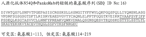
In specific embodiments, the light chain comprises an amino acid sequence having at least 90% identity to the amino acid sequence of SEQ ID NO. 16. In particular, the light chain comprises an amino acid sequence having at least 95%, in particular at least 98%, identity with the amino acid sequence of SEQ ID NO 16. In these embodiments, the light chain still comprises CDRs having the amino acid sequences of SEQ ID NOs 4, 5, and 6. Thus, any sequence deviation from SEQ ID NO 16 is located in the framework regions, but not in the CDRs. Specifically, the light chain comprises the amino acid sequence of SEQ ID NO 16.
In a specific embodiment, the heavy chain has an amino acid sequence with at least 90% identity to the amino acid sequence of SEQ ID NO. 15, wherein the CDRs still have the amino acid sequences of SEQ ID NO. 1, 7 and 3, and the light chain variable region has an amino acid sequence with at least 90% identity to the amino acid sequence of SEQ ID NO. 16, wherein the CDRs still have the amino acid sequences of SEQ ID NO. 4, 5 and 6. Specifically, the heavy chain has an amino acid sequence with at least 95% identity to the amino acid sequence of SEQ ID NO. 15, wherein the CDRs still have the amino acid sequences of SEQ ID NO. 1, 7 and 3, and the light chain has an amino acid sequence with at least 95% identity to the amino acid sequence of SEQ ID NO. 16, wherein the CDRs still have the amino acid sequences of SEQ ID NO. 4, 5 and 6.
In a specific embodiment, the heavy chain has an amino acid sequence with at least 90% identity to the amino acid sequence of SEQ ID NO 19, wherein the CDRs still have the amino acid sequences of SEQ ID NO 1, 8 and 3, and the light chain has an amino acid sequence with at least 90% identity to the amino acid sequence of SEQ ID NO 16, wherein the CDRs still have the amino acid sequences of SEQ ID NO 4, 5 and 6. Specifically, the heavy chain has an amino acid sequence with at least 95% identity to the amino acid sequence of SEQ ID NO 19, wherein the CDRs still have the amino acid sequences of SEQ ID NO 1, 8 and 3, and the light chain has an amino acid sequence with at least 95% identity to the amino acid sequence of SEQ ID NO 16, wherein the CDRs still have the amino acid sequences of SEQ ID NO 4, 5 and 6.
The antibodies include and encompass modified forms thereof. Modified forms of antibodies refer to antibodies with chemical or biological modifications. Chemically modified forms include forms having an amino acid backbone conjugated to a chemical moiety, forms having a chemically modified N-linked or O-linked carbohydrate chain, and the like. The chemical moiety or form may be toxic or cytotoxic. Biologically modified forms include forms that have undergone post-translational modification (e.g., N-linked or O-linked glycosylation, N-or C-terminal processing, deamidation, isomerization of aspartic acid, or oxidation of methionine), forms that contain a methionine residue added to the N-terminus by expression using a prokaryotic host cell, and the like. Such modified forms are also intended to include forms that are labeled to allow detection or isolation of the antibody or antigen, for example, enzyme-labeled forms, fluorescently labeled forms, or affinity-labeled forms. Such modified forms of the antibodies can be used to improve the stability or blood retention of the original antibody, reduce antigenicity, detect or isolate the antibody or antigen, and the like.
In particular, the antibody may comprise one or more modifications selected from: defucosylation, reduced fucose, N-linked glycosylation, O-linked glycosylation, N-terminal processing, C-terminal processing, deamidation, isomerization of aspartic acid, oxidation of methionine, substitution of 2 leucine (L) residues at positions 234 and 235 (according to the EU index) of the heavy chain to alanine (a) (LALA), amidation of proline residues, and deletion or absence of 1,2 or 3 amino acids at the carboxy terminus. In particular embodiments, the antibody lacks 1,2 or 3 carboxy-terminal amino acids in one or both heavy chains, or it lacks 2 carboxy-terminal amino acids and the carboxy-terminal proline residue is amidated at one or both heavy chains.
Such modifications may be made at any position or desired position of the antibody thereof. Alternatively, the same or 2 or more different modifications may be made at one or two or more positions therein.
For example, it is known that antibodies produced by cultured mammalian cells lack the carboxyl-terminal lysine residue in their heavy chains (Journal of Chromatography A, 705: 129-134 (1995)). It is also known that occasionally 2 carboxy-terminal amino acid residues of the heavy chain (i.e., glycine and lysine) are deleted, and that the proline residue newly located at the carboxy-terminal end is amidated (Analytical Biochemistry, 360: 75-83 (2007)). However, such deletions or modifications in these heavy chain sequences do not affect either the ability of the antibody to bind its antigen or the effector functions of the antibody (complement activation, antibody-dependent cytotoxicity, etc.).
In certain embodiments, the antibody comprises a deletion or absence of 1 or 2 amino acids in the carboxy-terminus of the heavy chain and has an amidated residue (e.g., an amidated proline residue at the carboxy-terminus position of the heavy chain). However, the antibody is not limited to the above type as long as the deletion mutant retains the ability to bind to an antigen.
In certain embodiments, the two heavy chains of an antibody may consist of any type of heavy chain selected from the group consisting of: a full-length heavy chain and a heavy chain of a deletion mutant, or may consist of a combination of any two types selected therefrom. The quantitative ratio of deletion variant heavy chains depends on the type of mammalian cell in culture producing the antibody and the culture conditions of the cell.
In particular embodiments, the antibody may comprise two heavy chains, both of which lack a carboxy-terminal amino acid residue.
In a specific embodiment, the antibody comprises a heavy chain having an amino acid sequence represented by amino acid numbers 1-446 of SEQ ID NO 15 or 22 and a light chain having an amino acid sequence represented by amino acid numbers 1-219 of SEQ ID NO 16. In certain embodiments, the amino acid at position 57 of SEQ ID NO. 15 is selected from the group consisting of glutamine, alanine, valine, histidine, tryptophan, tyrosine, lysine, and arginine; in particular glutamine, histidine, tryptophan, tyrosine, lysine and arginine. Preferably, the amino acid at position 57 of SEQ ID NO. 15 is glutamine, histidine, tryptophan, lysine or arginine, in particular glutamine.
In a specific embodiment, the antibody comprises a heavy chain having an amino acid sequence represented by amino acid numbers 1-446 of SEQ ID NO 19 and a light chain having an amino acid sequence represented by amino acid numbers 1-219 of SEQ ID NO 16.
In certain embodiments, the antibody competes for binding to TA-MUC1 with: an antibody comprising a heavy chain variable region having the amino acid sequence of SEQ ID NO. 10 and a light chain variable region having the amino acid sequence of SEQ ID NO. 12, or an antibody comprising a heavy chain variable region having the amino acid sequence of SEQ ID NO. 11 and a light chain variable region having the amino acid sequence of SEQ ID NO. 12.
In certain embodiments, the antibody has the following properties: (a) specifically binds to MUC1, and/or (b) has the activity of being internalized into a cell expressing MUC1 by binding to MUC 1. In certain embodiments, the antibody comprises at least one antibody heavy chain. In particular, the antibody comprises two antibody heavy chains. The antibody heavy chain comprises in particular a VH domain, a CH1 domain, a hinge region, a CH2 domain and a CH3 domain. In certain other embodiments, the antibody heavy chain comprises a CH2 domain and a CH3 domain, but does not comprise a CH1 domain. In other embodiments, one or more constant domains of the heavy chain may be replaced by other domains, in particular similar domains, such as albumin. Antibody heavy chains may be of any type, including gamma-, alpha-, epsilon-, delta-, and mu-chains, and are preferably gamma-chains, including gamma 1-, gamma 2-, gamma 3-, and gamma 4-chains, especially gamma 1-chains. Thus, the antibody is preferably an IgG-type antibody, such as an IgG1-, IgG 3-or IgG 4-type antibody, in particular an IgG 1-type antibody.
In particular, the antibody further comprises at least one antibody light chain, in particular two antibody light chains. The antibody light chain comprises in particular a VL domain and a CL domain. The antibody light chain may be a kappa-chain or a lambda-chain, especially a kappa-chain.
In certain embodiments, the antibody comprises two antibody heavy chains and two antibody light chains. In particular, the antibody comprises two gamma 1-type antibody heavy chains each comprising a VH domain, a CH1 domain, a hinge region, a CH2 domain and a CH3 domain and two kappa-type antibody light chains each comprising a VL domain and a CL domain.
In an alternative embodiment, the antibody does not comprise an antibody light chain. In these embodiments, the light chain variable region may be fused to the N-terminus of the heavy chain variable region or inserted into the C-terminus of the heavy chain variable region. A peptide linker may be present to link the light chain variable region to the remainder of the heavy chain.
In a preferred embodiment, the antibody comprises an Fc region. The antibody may in particular be a whole antibody comprising two heavy chains, each heavy chain comprising the domains VH, CH1, hinge region, CH2 and CH3, and two light chains, each light chain comprising the domains VL and CL. The antibodies are particularly capable of binding to one or more human Fc γ receptors, particularly human Fc γ receptor IIIA. In an alternative embodiment, the antibody does not or does not significantly bind to human Fc γ receptor IIIA, in particular does not or does not significantly bind to any human Fc γ receptor. In these embodiments, the antibody specifically does not comprise a glycosylation site in the CH2 domain.
In alternative embodiments, the antibody does not comprise an Fc region. In these embodiments, the antibody is in particular a single chain variable fragment (scFv) or another antibody fragment which does not comprise an Fc region.
Glycosylation of anti-MUC 1 antibodies
The anti-MUC 1 antibody may comprise one or more CH2 domains in the heavy chain of the antibody. Natural human antibodies of the IgG class contain an N-glycosylation site in the CH2 domain. The CH2 domain present in an antibody may or may not contain an N-glycosylation site.
In certain embodiments, the antibody does not comprise a glycosylation site in the CH2 domain. In particular, according to the IMGT/Eu numbering system, the antibody does not comprise an asparagine residue at a position in the heavy chain corresponding to position 297. For example, the antibody may comprise an Ala297 mutation in the heavy chain. In these embodiments, the antibody preferably has a strongly reduced ability or is completely devoid of the ability to induce antibody-dependent cellular cytotoxicity (ADCC) and/or antibody-dependent phagocytosis (ADCP) and/or complement-dependent cytotoxicity (CDC) by binding to Fc γ receptors. In this respect, strongly reduced capacity refers in particular to an activity that is reduced to 10% or less, in particular 3% or less, 1% or less or 0.1% or less, compared to the same antibody comprising N-glycosylation sites in its CH2 domain and having a common mammalian glycosylation pattern (such as those obtainable e.g. by production in a human cell line or CHO cell line, e.g. the glycosylation pattern described herein). In these embodiments, the antibody is specifically an IgG 1-type antibody.
In an alternative embodiment, the CH2 domain present in the antibody comprises an N-glycosylation site. The glycosylation site is in particular at an amino acid position corresponding to amino acid position 297 of the heavy chain according to the IMGT/Eu numbering system and has the amino acid sequence motif Asn Xaa Ser/Thr, wherein Xaa may be any amino acid except proline. N-linked glycosylation at Asn297 is conserved in the homologous regions of mammalian IgG and other antibody isotypes. The actual position of this conserved glycosylation site may vary in the amino acid sequence of the antibody due to optional additional amino acid or other sequence modifications that may be present in the variable region. Preferably, the glycan to which the antibody is attached is a bi-antennary complex N-linked carbohydrate structure, preferably comprising at least the following structure:
Asn - GlcNAc - GlcNAc - Man - (Man - GlcNAc)2
wherein Asn is an asparagine residue in the polypeptide portion of the antibody; GlcNAc is N-acetylglucosamine, and Man is mannose. The terminal GlcNAc residue may further carry a galactose residue, which optionally may carry a sialic acid residue. Another GlcNAc residue (referred to as a bisecting GlcNAc) may be attached to the Man of the closest polypeptide. Fucose can bind to GlcNAc attached to Asn. In these embodiments, the antibody is specifically an IgG 1-type antibody.
In preferred embodiments, the antibody does not comprise N-glycolyl neuraminic acid (NeuGc) or a detectable amount of NeuGc. Furthermore, the antibody preferably also does not comprise a Galili epitope (Gal α 1,3-Gal structure) or a detectable amount of a Galili epitope. In particular, the relative amount of glycans carrying NeuGc and/or Gal α 1,3-Gal structures is less than 0.1% or even less than 0.02% of the total amount of glycans attached to the CH2 domain of the antibody in the population of antibodies.
In particular, the antibodies have a human glycosylation pattern. Due to these glycosylation properties, there are no foreign immunogenic non-human structures inducing side effects, which means that undesired side effects or disadvantages known to be caused by certain foreign sugar structures, such as immunogenic non-human sialic acids (NeuGc) or Galili epitopes (Gal-Gal structures) known for rodent production systems, or other structures such as immunogenic high mannose structures known from e.g. yeast systems, are avoided.
In particular embodiments, the antibody comprises a glycosylation pattern of glycans carrying bisecting GlcNAc residues in a detectable amount. In particular, the relative amount of glycans carrying bisecting GlcNAc residues is at least 0.5%, especially at least 1%, of the total amount of glycans attached to the glycosylation sites of the antibody in the composition. Further, in certain embodiments, the glycosylation pattern comprises a relative amount of glycans carrying at least one galactose residue of at least 25% of the total amount of glycans attached to the antibody in the composition. In particular, the relative amount of glycans carrying at least one galactose residue is at least 30%, in particular at least 35% or at least 40% of the total amount of glycans attached to the antibody in the composition. In a specific embodiment, the glycosylation pattern comprises glycans carrying at least one sialic acid residue in a relative amount of at least 1% of the total amount of glycans attached to the antibody in the composition. In particular, the relative amount of glycans carrying at least one sialic acid residue is at least 1.5%, in particular at least 2%, of the total amount of glycans attached to the antibody in the composition.
The antibody may have a glycosylation pattern such that: it has a large amount of core fucose or a small amount of core fucose. A reduced amount of fucosylation increases the antibody's ability to induce ADCC. In certain embodiments, the relative amount of glycans carrying core fucose residues is 40% or less, in particular 30% or less or 20% or less, of the total amount of glycans attached to the antibody in the composition. In alternative embodiments, the relative amount of glycans carrying core fucose residues is at least 60%, in particular at least 65% or at least 70% of the total amount of glycans attached to the antibody in the composition.
The ability of an antibody to induce ADCC and the strength of said ADCC induction can be controlled by the presence or absence of a glycosylation site in the CH2 domain of the anti-MUC 1 antibody and the presence or absence of fucose in the glycan structure at said glycosylation site. ADCC activity is increased by glycosylation of the Fc portion of the antibody, and further by decreasing the amount of fucosylation in the glycosylation. In certain applications, fine-tuning of ADCC activity is important. Thus, in certain instances, antibodies without glycosylation sites in the CH2 domain, antibodies with glycosylation sites in the CH2 domain and with a high amount of fucosylation, or antibodies with glycosylation sites in the CH2 domain and with a low amount of fucosylation may be most advantageous.
Production of anti-MUC 1 antibodies
Preferably, the antibody is recombinantly produced in a host cell. The host cell used to produce the antibody may be any host cell useful for antibody production. Suitable host cells are in particular eukaryotic host cells, especially mammalian host cells. Exemplary host cells include yeast cells such as Pichia pastoris cell lines, insect cells such as SF9 and SF21 cell lines, plant cells, avian cells such as EB66 duck cell lines, rodent cells such as CHO, NS0, SP2/0 and YB2/0 cell lines and human cells such as HEK293, PER. C6, CAP-T, AGE1.HN, Mutz-3 and KG1 cell lines.
In certain embodiments, the antibodies are recombinantly produced in a human blood cell line, particularly in a human myeloid leukemia cell line. Preferred human cell lines useful for the production of antibodies and suitable production procedures are described in WO 2008/028686 a 2. In a specific embodiment, the antibody is obtained by expression in a human myeloid leukemia cell line selected from NM-H9D8, NM-H9D8-E6 and NM-H9D8-E6Q12 and cell lines derived therefrom. These cell lines were deposited under the Budapest treaty under the accession numbers DSM ACC2806 (NM-H9D 8; deposited at 15.9.2006), DSM ACC2807 (NM-H9D 8-E6; deposited at 5.10.2006) and DSM ACC2856 (NM-H9D8-E6Q 12; deposited at 8.8.2007) from Glycotope GmbH, Robert-R ssle-Str. 10, 13125 Berlin (DE) at Deutsche Sammlung von Mikroorganismen und Zellkulturen (DSMZ), Inhoffenttra E7B, 38124 Braunschweig (DE). NM-H9D8 cells provide glycosylation patterns with high sialylation, high bisection of GlycNAc, high galactosylation, and high fucosylation. NM-H9D8-E6 and NM-H9D8-E6Q12 cells provide similar glycosylation patterns as NM-H9D8 cells, except that the degree of fucosylation is very low. Other suitable cell lines include K562, a human myeloid leukemia cell line (ATCC CCL-243) found in the American type culture Collection, and cell lines derived from the above cell lines.
In other embodiments, the antibody is recombinantly produced in CHO cells. Specifically, the antibodies can be produced recombinantly in a CHO dhfr-cell line such as the cell line of ATCC number CRL-9096.
Conjugates of anti-MUC 1 antibodies
According to the invention, the antibody is conjugated to one or more cytotoxic agents. The cytotoxic agent may be any cytotoxic agent suitable for conjugation to an antibody. If more than one cytotoxic agent is present in the antibody, these cytotoxic agents may be the same or different, and in particular all the same. Conjugation of the cytotoxic agent to the antibody can be accomplished using any method known in the art. The cytotoxic agent may be attached to the antibody covalently (particularly by fusion or chemical coupling) or non-covalently. In certain embodiments, the cytotoxic agent is covalently attached to the antibody, particularly via a linker moiety. The linker moiety may be any chemical entity suitable for attaching a cytotoxic agent to an antibody.
In addition to cytotoxic agents, the conjugates according to the invention may further comprise other agents conjugated thereto. The further agent is preferably used for the treatment, diagnosis, prognosis and/or monitoring of diseases, in particular cancer. For example, the additional agent may be selected from radionuclides, chemotherapeutic agents, antibodies or antibody fragments, particularly those with different specificities from the anti-MUC 1 antibody, such as checkpoint antibodies that block or activate immunomodulatory targets, enzymes, interacting domains, detectable labels, toxins, cytolytic components, immunomodulatory agents, immune effectors, MHC class I or class II antigens, and liposomes.
One particularly preferred cytotoxic agent is a radionuclide or a cytotoxic agent capable of killing cancer cells, such as a chemotherapeutic agent. In certain preferred embodiments, the chemotherapeutic agent is linked to the anti-MUC 1 antibody to form a conjugate. There is no particular limitation on the chemotherapeutic agent as long as the compound has an antitumor effect and has a substituent or partial structure that can be attached to the linker structure. Upon cleavage of part or all of the linker in the tumor cell, the chemotherapeutic or anti-tumor compound is partially released, and the chemotherapeutic exhibits an anti-tumor effect. As the linker is cleaved at the point of attachment to the agent, the chemotherapeutic agent is released in its original structure to exert its original anti-tumor effect.
Specific examples of chemotherapeutic agents that may be conjugated as cytotoxic agents include alkylating agents such as cisplatin, antimetabolites, plant alkaloids and terpenoids, vinca alkaloids, podophyllotoxins, taxanes such as taxol, topoisomerase inhibitors such as irinotecan and topotecan, antineoplastic agents such as doxorubicin, or microtubule inhibitors such as maytansine/maytansine.
The chemotherapeutic agent may be chosen in particular from: inhibitors of V-ATPase, pro-apoptotic agents, Bcl2 inhibitors, MCL1 inhibitors, HSP90 inhibitors, IAP inhibitors, mTor inhibitors, microtubule stabilizing agents, microtubule destabilizing agents, dolastatin, maytansine, maytansinoids, amanitin, methionine aminopeptidase, inhibitors of nuclear export protein CRM1, DPPIV inhibitors, proteasome inhibitors, inhibitors of phosphoryl transfer reactions in mitochondria, inhibitors of protein synthesis, kinase inhibitors, CDK2 inhibitors, CDK9 inhibitors, kinesin inhibitors, HDAC inhibitors, topoisomerase I inhibitors, DNA damaging agents, DNA alkylating agents, DNA intercalating agents, DNA minor groove binding agents, DHFR inhibitors, inhibitors of microtubule formation, stabilizing agents for microtubules, stabilizing agents for actin, topoisomerase II inhibitors, platinum compounds, ribosome inhibitors, RNA polymerase II inhibitors, and bacterial toxins. In particular embodiments, the chemotherapeutic agent is attached to an anti-MUC 1 antibody selected from the group consisting of: microtubule inhibitors such as maytansinoids, topoisomerase I inhibitors, DNA damaging agents, DNA alkylating agents, and DNA minor groove binders.
In certain embodiments, the chemotherapeutic agent is maytansine or a maytansinoid. Specific examples of maytansinoids useful for conjugation include maytansinol, and the like,N 2'Deacetylation-N 2'- (3-mercapto-1-oxopropyl) -maytansine (DM1),N 2'Deacetylation-N 2'- (4-mercapto-1-oxopentyl) -maytansine (DM3) andN 2'deacetylation-N 2'- (4-methyl-4-mercapto-1-oxopentyl) -maytansine (DM 4). Specifically, DM1 or DM4 was attached to an anti-MUC 1 antibody. In certain embodiments, the chemotherapeutic agent attached to the anti-MUC 1 antibody is a DNA minor groove binding agent, in particular Pyrrolobenzodiazepine (PBD), pyrrolobenzodiazepine dimer (PBD dimer), dactinomycin-hydroxybenzamide-azaindole (DUBA), seco-dactinomycin-hydroxybenzamide-azaindole (duca), or doxorubicin. In certain embodiments, the chemotherapeutic agent attached to the anti-MUC 1 antibody is a DNA alkylating agent, particularly an indolinobenzodiazepine (indolinobenzodiazepine) or oxazolidinobenzodiazepine (oxazobisazepine). In certain embodiments, the chemotherapeutic agent attached to the anti-MUC 1 antibody is a DNA damaging agent, particularly calicheamicin. In certain embodiments, the chemotherapeutic agent attached to the anti-MUC 1 antibody is a topoisomerase I inhibitor, particularly camptothecin and its derivatives such as 7-ethyl-10-hydroxy-camptothecin (SN-38), (S) -9-dimethylaminomethyl-10-hydroxycamptothecin (topotecan), (1S,9S) -1-amino-9-ethyl-5-fluoro-1, 2,3,9,12, 15-hexahydro-9-hydroxy-4-methyl-10H, 13H-benzo [ de ] de]Pyrano [3',4':6,7]Indolizino [1,2-b ]]Quinoline-10, 13-dione (irinotecan (DX-8951)) and N- [ (1S,9S) -9-ethyl-5-fluoro-9-hydroxy-4-methyl-10, 13-dioxo-2, 3,9,10,13, 15-hexahydro-1H, 12H-benzo [ de]Pyrano [3',4':6,7]Indolizino [1,2-b ]]Quinolin-1-yl]-2-hydroxyacetamide (DXd). In certain embodiments, the chemotherapeutic agent attached to the anti-MUC 1 antibody is an inhibitor of microtubule formationFormulations, in particular tubulysin, ansamitocin, podophyllotoxin or vinblastine. In certain embodiments, the chemotherapeutic agent attached to the anti-MUC 1 antibody is a stabilizer of microtubules, in particular paclitaxel or epothilone. In certain embodiments, the chemotherapeutic agent attached to the anti-MUC 1 antibody is a stabilizer of actin, particularly phallotoxin. In certain embodiments, the chemotherapeutic agent attached to the anti-MUC 1 antibody is a topoisomerase II inhibitor, particularly teniposide, XK469, razoxane, amsacrine, idarubicin, or mebarone. In certain embodiments, the chemotherapeutic agent attached to the anti-MUC 1 antibody is a platinum compound, particularly cisplatin, carboplatin, oxaliplatin, nedaplatin, triplatin tetranitrate, phenonthriplatin, picoplatin, or satraplatin. In certain embodiments, the chemotherapeutic agent attached to the anti-MUC 1 antibody is a ribosome inhibitor, particularly ricin, saporin, abrin, diphtheria toxin, or exotoxin a. In certain embodiments, the chemotherapeutic agent attached to the anti-MUC 1 antibody is an RNA polymerase II inhibitor, particularly an amatoxin, e.g., amanitin. In certain embodiments, the chemotherapeutic agent attached to the anti-MUC 1 antibody is a bacterial toxin, particularly anthrax toxin. Suitable antibody drug conjugates are also described in EP 16151774.3 and LU 92659, to which reference is expressly made herein.
In a preferred embodiment, the chemotherapeutic agent is (1S,9S) -1-amino-9-ethyl-5-fluoro-1, 2,3,9,12, 15-hexahydro-9-hydroxy-4-methyl-10H, 13H-benzo [ de ] pyrano [3',4':6,7] indolizino [1,2-b ] quinoline-10, 13-dione (irinotecan (DX-8951)) or DXd.
Irinotecan (DX-8951) is an anti-tumor compound represented by the formula:
[ formula 1]

The compounds can be readily obtained, for example, by the method described in U.S. patent publication No. US2016/0297890 or other known methods, and the amino group at position 1 can be preferably used as a linking site to a linker structure. Furthermore, irinotecan can be released in tumor cells with a portion of the linker still attached to it. However, even in such a state, the compound exhibits an excellent antitumor effect.
DXd is a compound represented by the formula:
[ formula 2]

Since irinotecan or DXd has a camptothecin structure, it is known that in acidic aqueous media (e.g., on the order of pH 3), the equilibrium shifts to a structure with a lactone ring formed (ring closure), while in basic aqueous media (e.g., on the order of pH 10), the equilibrium shifts to a structure with an open lactone ring (ring opening). Drug conjugates in which an irinotecan residue corresponding to such closed-loop structure and open-loop structure has been introduced are also expected to have equivalent antitumor effects, and needless to say, any such drug conjugates are included within the scope of the present invention. In certain embodiments, the additional agent is a polypeptide of a protein. The polypeptide or protein may in particular be fused to a polypeptide chain of an antibody. In certain embodiments, the additional agent, which is a polypeptide or protein, is fused to the C-terminus of the antibody light chain of the antibody. In embodiments where the antibody comprises two antibody light chains, additional agents, which are polypeptides or proteins, may be fused to the C-terminus of each of the two antibody light chains. In other embodiments, other agents that are polypeptides or proteins are fused to the C-terminus of the antibody heavy chain of the antibody. In embodiments where the antibody comprises two antibody heavy chains, additional agents, which are polypeptides or proteins, may be fused to the C-terminus of each of the two antibody heavy chains. The other agents may be the same or different and in particular have the same amino acid sequence. Suitable examples of such other agents as polypeptides or proteins may be selected from cytokines, chemokines, antibodies, antigen binding fragments, enzymes and interacting domains.
In certain embodiments, the additional agent that is a polypeptide or protein is a checkpoint antibody that blocks and/or triggers an activation signal. Examples of various targets include CD40, CD3, CD137 (4-1BB), OX40, GITR, CD27, CD278 (ICOS), CD154 (CD40 ligand), CD270 (HVEM) and CD258 (LIGHT) as activation targets, CTLA4, PD1, CD80, CD244, A2AR, B7-H3 (CD276), B7-H4 (VTCN1), BTLA, IDO, KIR, LAG3, TIM-3, VISTA and phosphatidylserine as inhibition targets, and their corresponding ligands such as PDL 1. In particular examples, the anti-MUC 1 antibody comprises two heavy chains and two light chains as described herein, wherein an scFv fragment that specifically binds CD3 is fused to the C-terminus of each heavy chain; or wherein an scFv fragment that specifically binds PDL1 is fused to the C-terminus of each light chain.
In other embodiments, the other agent that is a polypeptide or protein is an immunomodulatory compound such as a chemokine, cytokine, or growth factor. Suitable cytokines in this regard include interferons such as interferon- α, interferon- β and interferon- γ and interleukins. Suitable growth factors include G-CSF and GM-CSF.
Specific examples of the linker include structures represented by any one of the following formulae (a) to (f):
(a) - (succinimidin-3-yl-N) -CH2CH2-C(=O)-GGFG-NH-CH2CH2CH2-C(=O)-,
(b) - (succinimidin-3-yl-N) -CH2CH2CH2CH2CH2-C(=O)-GGFG-NH-CH2CH2CH2-C(=O)-,
(c) - (succinimidin-3-yl-N) -CH2CH2CH2CH2CH2-C(=O)-GGFG-NH-CH2-O-CH2-C(=O)-,
(d) - (succinimidin-3-yl-N) -CH2CH2CH2CH2CH2-C(=O)-GGFG-NH-CH2CH2-O-CH2-C(=O)-,
(e) - (succinimidin-3-yl-N) -CH2CH2-C(=O)-NH-CH2CH2O-CH2CH2O-CH2CH2-C(=O)-GGFG-NH-CH2CH2CH2-C (= O) -, and
(f) - (succinimidin-3-yl-N) -CH2CH2-C(=O)-NH-CH2CH2O-CH2CH2O-CH2CH2O- CH2CH2O-CH2CH2-C(=O)-GGFG-NH-CH2CH2CH2-C (= O) -, wherein- (succinimid-3-yl-N) -has a structure represented by the formula:
[ formula 3]

In a specific embodiment, the linker comprises a structure represented by any one of the following formulae (a) to (c):
(a) - (succinimidin-3-yl-N) -CH2CH2CH2CH2CH2-C(=O)-GGFG-NH-CH2-O-CH2-C(=O)-,
(b) - (succinimidin-3-yl-N) -CH2CH2CH2CH2CH2-C(=O)-GGFG-NH-CH2CH2-O-CH2-C (= O) -, and
(c) - (succinimidin-3-yl-N) -CH2CH2-C(=O)-NH-CH2CH2O-CH2CH2O-CH2CH2-C(=O)-GGFG-NH-CH2CH2CH2-C(=O)-。
In a preferred embodiment, the linker comprises a structure represented by any one of the following formulae (a):
(a) - (succinimidin-3-yl-N) -CH2CH2CH2CH2CH2-C(=O)-GGFG-NH-CH2-O-CH2-C(=O)-,
In an alternative embodiment, the conjugate has a drug-linker structure represented by the formula wherein the antibody is conjugated to the drug-linker structure represented by the formula via a thioether bond, the asterisk indicates the point of attachment to the antibody:
[ formula 4]

In a preferred embodiment, the conjugate has a drug-linker structure represented by the formula,
[ formula 5]

Wherein AB represents an antibody, y represents the average number of units of a drug-linker structure conjugated to the antibody itself, the antibody being conjugated to the drug-linker structure represented by the above formula via a thioether bond, and the antibody represents the aforementioned anti-MUC 1 antibody, preferably the antibody is any one of the following combinations a) to d) of heavy and light chain variable regions, or heavy and light chains:
(a) a heavy chain variable region having the amino acid sequence of SEQ ID NO. 10 and a light chain variable region having the amino acid sequence of SEQ ID NO. 12,
(b) a heavy chain variable region having the amino acid sequence of SEQ ID NO. 11 and a light chain variable region having the amino acid sequence of SEQ ID NO. 12,
(c) a heavy chain having the amino acid sequence of SEQ ID NO. 15 and a light chain having the amino acid sequence of SEQ ID NO. 16, and
(d) a heavy chain having the amino acid sequence of SEQ ID NO. 19 and a light chain having the amino acid sequence of SEQ ID NO. 16:
in the above conjugates, the number of conjugated drug molecules (or cytotoxic agents) per antibody molecule is a key factor affecting its efficacy and safety. The production of the antibody-drug conjugate (or conjugates) was performed as follows: reaction conditions, such as the amounts of starting materials and reagents used in the reaction, are specified so that a constant number of conjugated drug molecules is obtained. Unlike the chemical reactions of low molecular weight compounds, mixtures containing different numbers of conjugated drug molecules are often obtained. The number of conjugated drug molecules per antibody molecule is defined and expressed as an average value, i.e. the average number of conjugated drug molecules. Unless otherwise indicated, i.e., except in the case of representing an antibody-drug conjugate having a specific number of conjugated drug molecules (which is included in a mixture of antibody-drug conjugates having different numbers of conjugated drug molecules), the number of conjugated drug molecules according to the present invention is also generally referred to as an average value. The number of irinotecan molecules or DXd conjugated to the antibody molecule is controllable and as an average number of conjugated drug molecules per antibody, about 1-10 irinotecan molecules or 1-10 DXd can be conjugated. The number of the irinotecan molecules or DXd is preferably 2 to 8, more preferably 4 to 8, further preferably 7 to 8, still further preferably 8. It should be noted that one skilled in the art, based on the description of the examples herein, can design reactions for conjugating a desired number of drug molecules to antibody molecules and can obtain antibody-drug conjugates with a controlled number of conjugated irinotecan molecules.
In the preferred embodiment above, after the conjugate is transferred to the interior of the tumor cell, the linker moiety is cleaved and then DXd is released to exert an anti-tumor effect (Clinical Cancer Research, 2016, 10, 15; 22(20): 5097-.
Conjugates labeled with various radioactive or non-radioactive isotopes are also included in the invention. One or more of the atoms comprising the conjugates of the invention may contain unnatural proportions of atomic isotopes. Examples of atomic isotopes include deuterium (2H) Tritium (a)3H) Iodine-125 (1251) And carbon-14 (14C) In that respect In addition, radioactive isotopes such as tritium(s) (iii)3H) Iodine-125 (125I. Carbon-14 (C)14C) Copper 64: (64Cu), zirconium-89 (89Zr), iodine-124 (124I) Fluorine-18 (18F) Indium-111 (111I) Carbon-11 (C)11C) And iodine-131 (131I) The conjugate is radiolabeled. Conjugates labeled with radioisotopes useful as therapeutic or prophylactic agentsReagents for research (e.g., assay reagents) and for diagnosis (e.g., in vivo diagnostic imaging agents). Regardless of the radioactivity, any isotopic variant type of conjugate is within the scope of the present invention.
Nucleic acids, expression cassettes, vectors, cell lines and compositions
The antibody portion of the conjugate according to the invention may be encoded by a nucleic acid. The nucleic acid sequence of the nucleic acid may have any nucleotide sequence suitable for encoding an antibody. Preferably, however, the nucleic acid sequence is adapted at least in part to the particular codon usage, in particular human codon usage, of the host cell or organism in which the nucleic acid is to be expressed. The nucleic acid may be double-stranded or single-stranded DNA or RNA, preferably double-stranded DNA such as cDNA or single-stranded RNA such as mRNA. It may be one continuous nucleic acid molecule, or it may consist of several nucleic acid molecules, each encoding a different part of the antibody. The nucleotide sequence of the heavy chain of the PankoMab variant (PM-N54Q) may be represented by SEQ ID NO. 17, and the nucleotide sequence of the light chain of the PankoMab variant (PM-N54Q) may be represented by SEQ ID NO. 18.
If the antibody is composed of more than one distinct amino acid chain, e.g., a light chain and a heavy chain of an antibody, the nucleic acid may, for example, be a single nucleic acid molecule containing several coding regions (each encoding one of the amino acid chains of the antibody, preferably separated by a regulatory element such as an IRES element) to produce separate amino acid chains, or the nucleic acid may be composed of several nucleic acid molecules, wherein each nucleic acid molecule contains one or more coding regions, each coding region encoding one of the amino acid chains of the antibody. In addition to the coding region encoding the antibody, the nucleic acid may also comprise other nucleic acid sequences or other modifications, for example, it may encode other proteins, may affect the transcription and/or translation of the coding region, may affect the stability or other physical or chemical properties of the nucleic acid, or may not function at all.
The expression cassette or vector may comprise the nucleic acid and a promoter operably linked to the nucleic acid. In addition, the expression cassette or vector may comprise further elements, in particular elements capable of influencing and/or regulating the transcription and/or translation of the nucleic acid, the amplification and/or replication of the expression cassette or vector, the integration of the expression cassette or vector into the genome of the host cell, and/or the copy number of the expression cassette or vector in the host cell. Suitable expression cassettes and vectors comprising the various expression cassettes for expression of the antibody are well known in the art and need not be described further herein.
The host cell may comprise said nucleic acid or said expression cassette or vector. The host cell may be any host cell. It may be an isolated cell or a cell contained in a tissue. Preferably, the host cell is a cultured cell, in particular a primary cell or a cell of an established cell line, preferably a tumor-derived cell. Preferably, it is a bacterial cell such as e.coli, a yeast cell such as saccharomyces cell, in particular saccharomyces cerevisiae, an insect cell such as Sf9 cell, or a mammalian cell, in particular a human cell such as a tumor-derived human cell, a hamster cell such as CHO or a primate cell. In a preferred embodiment, the host cell is derived from a human myeloid leukemia cell. Preferably, it is selected from the following cells or cell lines: k562, KG1, MUTZ-3 or a cell or cell line derived therefrom, or a cell or cell line mixture comprising at least one of the aforementioned cells. The host cell is preferably selected from NM-H9D8, NM-H9D8-E6, NM H9D8-E6Q12, and a cell or cell line derived from any one of said host cells. These cell lines and their properties are described in detail in PCT application WO 2008/028686A 2. In other embodiments, the host cell is a CHO dhfr-cell line such as that of ATCC number CRL-9096. In a preferred embodiment, the host cell is optimized for expression of glycoproteins having a particular glycosylation pattern, in particular antibodies. Preferably, the codon usage and/or promoters in the coding region of the nucleic acid and other elements of the expression cassette or vector are compatible with and, more preferably, optimized for the type of host cell used. Preferably, the antibody is produced by a host cell or cell line as described above.
Methods of producing antibodies can use the host cells described herein. The method comprises in particular the following steps: providing a host cell comprising a nucleic acid encoding an antibody, culturing the host cell under conditions suitable for expression of the antibody, and obtaining the antibody expressed by the host cell. The antibodies described herein may be obtained or obtainable by the methods described.
In another aspect, the invention provides a composition comprising a conjugate according to the invention. In addition, the composition may comprise one or more further components selected from solvents, diluents and excipients. Preferably, the composition is a pharmaceutical composition. In this embodiment, the components of the composition are preferably all pharmaceutically acceptable. The composition may be a solid or fluid composition, in particular a solution (preferably an aqueous solution), an emulsion or suspension or a lyophilized powder.
Use in medicine
The conjugates are particularly useful in medicine, in particular for the treatment, diagnosis, prognosis, detection and/or monitoring of diseases (in particular diseases as described herein, preferably cancer, infection, inflammatory disease, graft versus host disease and immunodeficiency).
Thus, in another aspect, the invention provides a conjugate or composition for medical use. Preferably, the use in medicine is for the treatment, prognosis, diagnosis, detection and/or monitoring of diseases, for example, diseases associated with abnormal cell growth, e.g. cancer, infections such as bacterial, viral, fungal or parasitic infections, inflammatory diseases such as autoimmune diseases and inflammatory bowel diseases, and diseases associated with reduced immune activity such as immunodeficiency. In a preferred embodiment, the disease is cancer.
Preferably, the cancer has detectable expression of MUC1 (TA-MUC1), preferably detectable by immunohistochemistry, ELISA, RIA, Enzyme Linked Immunospot (ELISPOT) assay, dot blot, Ouchterlony test or Convection Immunoelectrophoresis (CIE) or in situ hybridization. It includes, inter alia, cells with MUC1 (TA-MUC1) expression, which can be detected by immunohistochemistry or in situ hybridization. Prior to administration of anti-MUC 1 antibodies, the cancer may be tested at the MUC1 (TA-MUC1) level.
The invention further provides kits and devices comprising conjugates according to the invention, as well as related methods useful in the diagnosis, detection or monitoring of MUC 1-related disorders, such as cancer. In certain embodiments, a sandwich ELISA kit for testing or diagnosis is provided comprising the conjugate of the invention. The kit may further comprise one or more of the following: a solution of MUC1 (TA-MUC1) protein standard, a colorant, a buffer solution for dilution, an antibody for solid phase, an antibody for detection, a washing solution, and the like. Preferably, the amount of conjugate bound to the antigen can be measured by application methods such as absorbance, fluorescence, luminescence, or Radioisotope (RI) methods. Preferably, an absorbance plate reader, a fluorescence plate reader, a luminescence plate reader, an RI liquid scintillation counter, or the like is used in the measurement.
The antibodies may be used in Immunohistochemical (IHC) assays.
The immunohistochemistry is not particularly limited as long as the protocol includes reacting the tissue section with an antigen-binding antibody (primary antibody) and detecting the primary antibody bound to the antigen.
Different forms of cancer, including metastases, can be treated with the conjugates according to the invention. The cancer may be selected from colon cancer, lung cancer, ovarian cancer, breast cancer such as triple negative breast cancer, pancreatic cancer, cervical cancer, endometrial cancer, gastrointestinal cancer, renal cancer, head and neck cancer, thyroid cancer and urothelial cancer, among others. The cancer may further be selected in particular from the group consisting of gastric cancer, liver cancer, bladder cancer, skin cancer, prostate cancer and blood cancer. In certain embodiments, the cancer is a metastatic cancer. The cancer may include any type of metastasis, such as skin metastases, lymph node metastases, lung metastases, liver metastases, peritoneal metastases, pleural metastases, and/or brain metastases. In certain embodiments, the cancer has an inflammatory phenotype. In these embodiments, any of the above cancer types can be an inflammatory cancer.
In certain embodiments, the viral infection is caused by human immunodeficiency virus, herpes simplex virus, epstein-barr virus, influenza virus, lymphocytic choriomeningitis virus, hepatitis b virus, or hepatitis c virus. The inflammatory disease may be selected from inflammatory bowel disease, pelvic inflammatory disease, ischemic stroke, alzheimer's disease, asthma, pemphigus vulgaris and dermatitis/eczema. The autoimmune disease may be selected from celiac disease, type I diabetes, graves' disease, inflammatory bowel disease, multiple sclerosis, psoriasis, rheumatoid arthritis, systemic lupus erythematosus, vitiligo, psoriatic arthritis, atopic dermatitis, scleroderma, sarcoidosis, primary biliary cirrhosis, guillain-barre syndrome, autoimmune hepatitis, and ankylosing spondylitis. In certain embodiments, the disease comprises or is associated with a cell expressing MUC1, particularly TA-MUC 1. For example, the cancer to be treated is MUC1 positive, in particular TA-MUC1 positive, i.e. comprises cancer cells expressing MUC1, in particular TA-MUC 1.
In particular embodiments, the conjugates are used in combination therapy with another therapeutic agent, particularly in combination with another anti-cancer agent to treat cancer. The additional therapeutic agent may be any known anti-cancer agent. Suitable anti-cancer therapeutic agents that can be combined with the conjugates according to the invention can be chemotherapeutic agents, other antibodies, immunostimulants, cytokines, chemokines and vaccines. In addition, therapy with the conjugates can be combined with radiation therapy, surgery, and/or traditional Chinese medicine.
The anti-cancer agent that may be used in combination with the conjugate may be selected from any chemotherapeutic agent, in particular a chemotherapeutic agent known to be effective in treating MUC 1-positive cancers. The type of chemotherapeutic agent also depends on the cancer to be treated. The combination partner may be selected from: taxanes such as paclitaxel (taxol), docetaxel (taxotere) and SB-T-1214; cyclophosphamide; imatinib; pazopanib; capecitabine; cytarabine; vinorelbine; gemcitabine; anthracyclines such as daunorubicin, doxorubicin, epirubicin, idarubicin, valrubicin, and mitoxantrone; aromatase inhibitors such as aminoglutethimide, testolactone (Teslac), anastrozole (runing), letrozole (frolone), exemestane (anoxin), vorozole (rivzor), formestane (lantalone), fadrozole (Afema), 4-hydroxyandrostenedione, 1,4, 6-androstatriene-3, 17-dione (ATD) and 4-androstene-3, 6, 17-trione (6-oxo); topoisomerase inhibitors such as irinotecan, topotecan, camptothecin, lamellarin D, etoposide (VP-16), teniposide, doxorubicin, daunorubicin, mitoxantrone, amsacrine, erietidine, aurintricarboxylic acid, and HU-331; platinum-based chemotherapeutic agents such as cis-diamminebischloroplatinum (II) (cisplatin), cis-diamminedicarboxylate (carboxylato) platinum (II) (carboplatin) and [ (1R,2R) -cyclohexane-1, 2-diamine ] (ethanediato-O, O') platinum (II) (oxaliplatin); PARP inhibitors such as olaparib, rucaparib and niraprib; TLR agonists such as imiquimod and resiquimod; and antimetabolites, especially antifolates such as methotrexate, pemetrexed, raltitrexed and pralatrexate, pyrimidine analogs such as fluorouracil, gemcitabine, fluorouridine, 5-fluorouracil and tegafur-uracil, and purine analogs, selective estrogen receptor modulators and estrogen receptor down-modulators.
In addition, therapeutic antibodies may also be used as further combination partners. It may be any antibody useful in cancer therapy other than the anti-MUC 1 antibody. In particular, other antibodies have been approved for cancer therapy by regulatory agencies such as the U.S. Food and Drug Administration (FDA), the european drug administration (EMA, predecessor EMEA), and the bundesist fur Arzneimittel und Medizinproduke (BfArM). Examples of other antibodies that may be used in combination therapy are anti-EGFR antibodies such as cetuximab, tomuzuximab, parlimumab, zalutumumab, nimotuzumab, matuzumab and nefirumab; anti-HER 2 antibodies such as trastuzumab, Timigutuzumab, and pertuzumab; anti-VEGF antibodies such as bevacizumab (atorvastatin); anti-CD 52 antibodies such as alemtuzumab (Campath); anti-CD 30 antibodies such as brentuximab (additris); anti-CD 33 antibodies such as gemtuzumab ozogamicin (Mylotarg); and anti-CD 20 antibodies such as rituximab (Rituxan ), tositumomab (Bexxar), and ibritumomab (zeuglin). Other exemplary antibodies suitable for use in the cancer therapy combinations described herein include antibodies to antigens selected from the group consisting of: Thomsen-Friedenreich antigens (TF. alpha., TF. beta.), Tn, Lewis Y, CD44, folate receptor. alpha., NeuGc-GM3 ganglioside, DLL-3, RANKL, PTK7, Notch-3, Ephrin A4, insulin-like growth factor receptor 1, activin receptor-like kinase-1, blocker-6, disialoganglioside GD2, endoglin, transmembrane NMglycoprotein B, CD56, tumor-associated calcium signal transduction protein 2, tissue factor, ectonucleotide pyrophosphatase/phosphodiesterase 3, CD70, P-cadherin, mesothelin, six transmembrane epithelial antigen 1 of prostate (STEAP1), carcinoembryonic antigen-associated cell adhesion molecule 5 (CEACAM5), connexin 4, guanylate cyclase C, solute carrier family member 44 (SLC44A4), prostate-specific membrane antigen (PSMA), zinc transporter ZIP6 (LIV1 (ZIP6)), SLIT and NTRK-like protein 6 (SLITRK6), trophoblast glycoprotein (TPBG; 5T4), Fyn3, carbonic anhydrase 9, NaPi2B, fibronectin ectodomain B, endothelin receptor ETB, VEGFR2 (CD309), tenascin c, collagen IV and periostin.
The conjugate may further be combined with a checkpoint antibody (i.e., an antibody that blocks or activates an immunomodulatory target). Thereby, the inhibitory signal against the immune response may be blocked and/or the activating signal may be triggered. Examples of various targets include CD40, CD3, CD137 (4-1BB), OX40, GITR, CD27, CD278 (ICOS), CD154 (CD40 ligand), CD270 (HVEM) and CD258 (LIGHT) as activation targets, CTLA4, PD1, CD80, CD244, A2AR, B7-H3 (CD276), B7-H4 (VTCN1), BTLA, IDO, KIR, LAG3, TIM-3, VISTA and phosphatidylserine as inhibition targets, and their corresponding ligands such as PDL 1.
In other embodiments, the conjugates can be combined with treatment with immunomodulatory compounds such as chemokines, cytokines, growth factors, and vaccines. In this regard, suitable cytokines include interferons such as interferon- α, interferon- β and interferon- γ, and interleukins. Suitable growth factors include G-CSF and GM-CSF.
The conjugates are preferably used for the treatment of primary tumors, recurrent tumors and/or metastases of such tumors, and in particular for the treatment before, during or after surgery, and for the prevention or treatment of metastases. The conjugates are particularly useful as adjuvant therapy for treating patients. In certain embodiments, the conjugates are used to treat patients as neoadjuvant therapy or combined neoadjuvant-adjuvant therapy. Furthermore, the conjugates are useful as palliative therapy for treating patients.
Cancer treatment with the conjugates preferably results in inhibition of tumor growth, in particular a reduction in tumor size. In addition, further metastases are prevented and/or their number is reduced by the treatment. The treatment preferably results in an increase in progression-free survival; and/or increased longevity, thereby improving overall survival.
The invention further provides methods of treating, diagnosing, prognosing, detecting and/or monitoring diseases using the conjugates according to the invention. The embodiments and examples of the use of the conjugates in medicine are equally applicable to medical methods. In particular, there is provided a method of treating a disease in a subject in need thereof, the method comprising administering to the subject a therapeutically effective amount of a conjugate according to the invention.
For example, the present invention provides a method of treating cancer in a subject in need thereof, the method comprising administering to the subject having cancer a therapeutically effective amount of a conjugate according to the present invention. In a specific embodiment, the cancer is characterized by expression of TA-MUC 1. The cancer may be selected from ovarian cancer, breast cancer, pancreatic cancer, lung cancer, colon cancer, stomach cancer, liver cancer, kidney cancer, blood cancer, endometrial cancer, thyroid cancer, leukemia, seminoma, melanoma, carcinoma, teratoma, lymphoma, sarcoma, mesothelioma, neuroblastoma, glioma, rectal cancer, adrenal cancer, skin cancer, brain cancer, cervical cancer, intestinal cancer, head and neck cancer, gastrointestinal cancer, lymph node cancer, esophageal cancer, colorectal cancer, ear, nose and throat (ENT) cancer, prostate cancer, bladder cancer, uterine cancer, and metastases thereof.
Furthermore, the present invention provides a method for diagnosing, detecting or monitoring cancer, comprising the step of contacting a test sample with a conjugate according to the present invention.
Method of increasing MUC1 binding affinity
A method of increasing the MUC1 binding affinity of an antibody may comprise
(i) A heavy chain variable region comprising the following Complementarity Determining Regions (CDRs): CDR-H1 having the amino acid sequence of SEQ ID NO. 1, CDR-H2 having the amino acid sequence of SEQ ID NO. 8, and CDR-H3 having the amino acid sequence of SEQ ID NO. 3, and
(ii) a light chain variable region comprising the following Complementarity Determining Regions (CDRs): CDR-L1 having the amino acid sequence of SEQ ID NO. 4, CDR-L2 having the amino acid sequence of SEQ ID NO. 5, and CDR-L3 having the amino acid sequence of SEQ ID NO. 6,
the method comprises the following steps: the amino acid residue at position 8 of CDR-H2 was replaced with any amino acid residue other than asparagine, thereby generating CDR-H2 having the amino acid sequence of SEQ ID NO. 2.
The antibody whose MUC1 binding affinity is to be increased is in particular an antibody capable of binding MUC1 as described herein, with the exception that it comprises an asparagine at position 8 of the CDR-H2 sequence.
In certain embodiments, the heavy chain variable region of an antibody whose MUC1 binding affinity is to be increased comprises an amino acid sequence having at least 90% identity to the amino acid sequence of SEQ ID NO. 11. In particular, the heavy chain variable region comprises an amino acid sequence having at least 95%, in particular at least 98%, identity with the amino acid sequence of SEQ ID NO. 11. In these embodiments, the heavy chain variable region still comprises the CDRs having the amino acid sequences of SEQ ID NOs 1, 8 and 3. Thus, any sequence deviation from SEQ ID NO 11 is located in the framework regions, but not in the CDRs. Specifically, the heavy chain variable region comprises the amino acid sequence of SEQ ID NO 11.
In certain embodiments, the light chain variable region of the antibody whose MUC1 binding affinity is to be increased comprises an amino acid sequence having at least 90% identity to the amino acid sequence of SEQ ID NO. 12. In particular, the light chain variable region comprises an amino acid sequence having at least 95%, in particular at least 98%, identity with the amino acid sequence of SEQ ID NO 12. In these embodiments, the light chain variable region still comprises the CDRs having the amino acid sequences of SEQ ID NOs 4, 5 and 6. Thus, any sequence deviation from SEQ ID NO 12 is located in the framework regions, but not in the CDRs. Specifically, the light chain variable region comprises the amino acid sequence of SEQ ID NO 12.
In particular embodiments, the heavy chain variable region of an antibody whose MUC1 binding affinity is to be increased has an amino acid sequence at least 90% identical to the amino acid sequence of SEQ ID NO. 11, wherein the CDRs still have the amino acid sequences of SEQ ID NO. 1, 8 and 3, and the light chain variable region has an amino acid sequence at least 90% identical to the amino acid sequence of SEQ ID NO. 12, wherein the CDRs still have the amino acid sequences of SEQ ID NO. 4, 5 and 6. Specifically, the heavy chain variable region has an amino acid sequence with at least 95% identity to the amino acid sequence of SEQ ID NO. 11, wherein the CDRs still have the amino acid sequences of SEQ ID NO. 1, 8 and 3, and the light chain variable region has an amino acid sequence with at least 95% identity to the amino acid sequence of SEQ ID NO. 12, wherein the CDRs still have the amino acid sequences of SEQ ID NO. 4, 5 and 6.
For example, the antibody whose MUC1 binding affinity is to be increased is an anti-MUC 1 antibody disclosed in WO 2004/065423 a2 or WO 2011/012309 a 1. Specifically, the antibody whose MUC1 binding affinity is to be increased is gatituzumab (gatipotuzumab) or PankoMab.
The antibody whose MUC1 binding affinity is to be increased is in particular an antibody capable of binding MUC1 as described herein.
In certain embodiments, MUC1 is incorporated as described herein. Increasing MUC1 binding affinity specifically indicates an increase of at least 10%, at least 20%, at least 33% or at least 50%. In preferred embodiments, MUC1 binding affinity is increased by at least 50%. As described in the examples, MUC1 binding affinities may be determined, in particular using surface plasmon resonance analysis or switchSENSE @ (DRX2 BIOSENSOR, manufactured by Dynamic BIOSENSORs GmbH), for example as described in examples 4a and b.
In certain embodiments, the step of replacing the amino acid residue at position 8 of CDR-H2 is achieved by introducing a mutation into the nucleic acid encoding the antibody, wherein the mutation is introduced into the codon encoding said amino acid residue. Introduction of the mutation may be accomplished by any method. Several suitable methods are known in the art and the skilled person is able to perform the necessary tasks to introduce mutations. The mutated nucleic acid may then be expressed, for example, in a host cell, resulting in an antibody with increased MUC1 binding affinity. Nucleic acids, host cells, and methods for producing antibodies are described herein and may be used in methods of increasing MUC1 binding affinity.
In particular embodiments, a method of increasing the MUC1 binding affinity of an antibody comprises the steps of:
(a) providing a nucleic acid encoding an antibody whose MUC1 binding affinity is to be increased
(b) Introducing a mutation into the nucleic acid to produce a mutated nucleic acid, wherein the mutation is introduced into a codon encoding the amino acid residue at position 8 of CDR-H2 such that the codon encodes any amino acid residue other than asparagine; and
(c) expressing the mutated nucleic acid to produce an antibody having increased MUC1 binding affinity.
Methods of producing an antibody with increased MUC1 binding affinity may comprise
(a) Providing a nucleic acid encoding an antibody comprising
(i) A heavy chain variable region comprising the following Complementarity Determining Regions (CDRs): CDR-H1 having the amino acid sequence of SEQ ID NO. 1, CDR-H2 having the amino acid sequence of SEQ ID NO. 8, and CDR-H3 having the amino acid sequence of SEQ ID NO. 3, and
(ii) a light chain variable region comprising the following Complementarity Determining Regions (CDRs): CDR-L1 having the amino acid sequence of SEQ ID NO. 4, CDR-L2 having the amino acid sequence of SEQ ID NO. 5, and CDR-L3 having the amino acid sequence of SEQ ID NO. 6;
(b) introducing a mutation into the nucleic acid to produce a mutated nucleic acid, wherein the mutation is introduced into a codon encoding the amino acid residue at position 8 of CDR-H2 such that the codon encodes any amino acid residue other than asparagine; and
(c) by expressing the mutated nucleic acid in a host cell, antibodies with increased MUC1 binding affinity are produced.
With respect to other aspects, particularly with respect to methods of increasing MUC1 binding affinity of an antibody, the embodiments, features and examples described herein are equally applicable to methods of producing an antibody having increased MUC1 binding affinity.
In certain embodiments, the method of producing an antibody with increased MUC1 binding affinity further comprises the step (d) of processing the antibody with increased MUC1 binding affinity.
For example, processing an antibody with increased MUC1 binding affinity may comprise isolating the antibody from a cell culture. Isolation of the antibody specifically means that the antibody is separated from the rest of the components of the cell culture. Isolation of the antibody from the cell culture medium can be carried out, for example, by chromatography. Suitable methods and means for isolating antibodies are known in the art and can be readily applied by those skilled in the art.
The obtained antibody may optionally be subjected to further processing steps, e.g., modification steps, such as chemical or enzymatic coupling of other reagents to the antibody, and/or formulation steps, to produce an antibody of desired quality and composition. Such further processing steps and methods are generally known in the art.
In other embodiments, step (d) further comprises the step of providing a pharmaceutical formulation comprising the antibody. Providing a pharmaceutical formulation comprising an antibody or formulating an antibody into a pharmaceutical composition particularly comprises exchanging the buffer solution or buffer solution components of the composition comprising the antibody. In addition, this step may include lyophilization of the antibody. In particular, the antibody is transferred to a composition comprising only pharmaceutically acceptable ingredients.
An antibody-drug conjugate represented by formula (1) given below, wherein the antibody is linked to the linker moiety through a thioether, can be produced as follows: an antibody having a thiol group, which is converted from a disulfide bond by antibody reduction, is reacted with compound (2) which can be obtained by a known method (e.g., can be obtained by a method described in patent publication US2016/297890 (e.g., a method described in paragraphs to). The antibody-drug conjugate can be produced, for example, by the following method.
[ formula 6]

Wherein AB represents an antibody (3a) having a thiol group, wherein
L1 has a structure represented by- (succinimid-3-yl-N) -, and
l1' represents a maleimide group represented by the following formula.
[ formula 7]

-L1-LX has a structure represented by any one of the following formulae:
- (succinimidin-3-yl-N) -CH2CH2-C(=O)-GGFG-NH-CH2CH2CH2-C(=O)-,
- (succinimidin-3-yl-N) -CH2CH2CH2CH2CH2-C(=O)-GGFG-NH-CH2CH2CH2-C(=O)-,
- (succinimidin-3-yl-N) -CH2CH2CH2CH2CH2-C(=O)-GGFG-NH-CH2-O-CH2-C(=O)-,
- (succinimidin-3-yl-N) -CH2CH2CH2CH2CH2-C(=O)-GGFG-NH-CH2CH2-O-CH2-C(=O)-,
- (succinimidin-3-yl-N) -CH2CH2-C(=O)-NH-CH2CH2O-CH2CH2O-CH2CH2-C(=O)-GGFG-NH-CH2CH2CH2-C (= O) -, and
- (succinimidin-3-yl-N) -CH2CH2-C(=O)-NH-CH2CH2O-CH2CH2O-CH2CH2O-CH2CH2O-CH2CH2-C(=O)-GGFG-NH-CH2CH2CH2-C(=O)-
Among them, more preferred are the following:
- (succinimidin-3-yl-N) -CH2CH2CH2CH2CH2-C(=O)-GGFG-NH-CH2-O-CH2-C(=O)-,
- (succinimidin-3-yl-N) -CH2CH2CH2CH2CH2-C(=O)-GGFG-NH-CH2CH2-O-CH2-C (= O) -, and
- (succinimidin-3-yl-N) -CH2CH2-C(=O)-NH-CH2CH2O-CH2CH2O-CH2CH2-C(=O)-GGFG-NH-CH2CH2CH2-C(=O)-。
Further preferred are the following:
- (succinimidin-3-yl-N) -CH2CH2CH2CH2CH2-C(=O)-GGFG-NH-CH2-O-CH2-C (= O) -, and
- (succinimidin-3-yl-N) -CH2CH2-C(=O)-NH-CH2CH2O-CH2CH2O-CH2CH2-C(=O)-GGFG-NH--CH2CH2CH2-C(=O)-。
(NH-DX) has a structure represented by the following formula:
[ formula 8]

And it represents a group derived by removing one hydrogen atom of the amino group at position 1 of irinotecan. In the above reaction scheme (formula 8), the compound of formula (1) may be interpreted as having the structure: wherein a moiety from the drug to the end of the linker is linked to an antibody. However, this description is given for convenience, and actually in many cases, a plurality of the above-described structural portions are linked to one antibody molecule. The same applies to the production method described below.
Specifically, the antibody-drug conjugate (1) can be produced by reacting the compound (2) obtainable by a known method (e.g., obtainable by a method described in patent publication US2016/297890 (e.g., a method described in paragraphs to) with the antibody (3a) having a thiol group).
Provision of the thiol group on antibody (3a) can be accomplished by methods well known to those skilled in the art (Hermanson, G.T., Bioconjugate Techniques, pp.56-136, 456-493, Academic Press (1996)). Examples of such methods may include, but are not limited to: reacting the Traut's reagent with the amino group of the antibody; reacting N-succinimidyl S-acetylsulfanyl alkanoate with an amino group of an antibody, and then with hydroxylamine; reacting N-succinimidyl 3- (pyridyldithio) propionate with an antibody and then with a reducing agent; the antibody is reacted with a reducing agent such as dithiothreitol, 2-mercaptoethanol, or tris (2-carboxyethyl) phosphine hydrochloride (TCEP) to reduce interchain disulfide bonds in the antibody, thereby forming sulfhydryl groups.
Specifically, an antibody having partially or completely reduced interchain disulfide bonds can be obtained by using 0.3 to 3 molar equivalents of TCEP as a reducing agent for each interchain disulfide bond in the antibody and reacting the reducing agent with the antibody in a buffer solution containing a chelating agent. Examples of the chelating agent may include ethylenediaminetetraacetic acid (EDTA) and diethylenetriaminepentaacetic acid (DTPA). The chelating agent may be used at a concentration of 1 mM to 20 mM. A solution of sodium phosphate, sodium borate, sodium acetate, or the like may be used as the buffer solution. As a specific example, the antibody (3a) having a partially or completely reduced thiol group can be obtained by reacting the antibody with TCEP at 4 ℃ to 37 ℃ for 1 to 4 hours.
It should be noted that by performing an addition reaction of a thiol group to a drug-linker moiety, the drug-linker moiety may be conjugated through a thioether bond.
Then, using 2 to 20 molar equivalents of compound (2) per antibody (3a) having a thiol group, an antibody-drug conjugate (1) can be produced in which 2 to 8 drug molecules are conjugated per antibody. Specifically, a solution containing the compound (2) dissolved therein may be added to a buffer solution containing the antibody (3a) having a thiol group to carry out a reaction. In this context, sodium acetate solution, sodium phosphate, sodium borate, etc. may be used as the buffer solution. The reaction pH is 5 to 9, and more preferably, the reaction may be carried out at around pH 7. An organic solvent such as dimethyl sulfoxide (DMSO), Dimethylformamide (DMF), Dimethylacetamide (DMA), or N-methyl-2-pyrrolidone (NMP) can be used as a solvent for dissolving the compound (2). The reaction can be carried out by adding a solution containing the compound (2) dissolved in an organic solvent at 1 to 20% (v/v) to a buffer solution containing the antibody (3a) having a thiol group. The reaction temperature is 0 to 37 ℃, more preferably 10 to 25 ℃, and the reaction time is 0.5 to 2 hours. The reaction can be terminated by inactivating the reactivity of the unreacted compound (2) with the thiol-containing reagent. Thiol-containing agents are, for example, cysteine or N-acetyl-L-cysteine (NAC). More specifically, the reaction can be terminated as follows: to the compound (2) used, 1-2 molar equivalents of NAC were added, and the resulting mixture was incubated at room temperature for 10-30 minutes.
Identification of antibody-drug conjugates
The produced antibody-drug conjugate (e.g., antibody-drug conjugate (1)) can be concentrated, buffer exchanged, purified, and measured for antibody concentration and average number of conjugated drug molecules per antibody molecule according to the following general procedure to identify the antibody-drug conjugate (1).
1. General procedure a concentration of an aqueous solution of an antibody or antibody-drug conjugate.
To Amicon Ultra (50,000 MWCO, Millipore Corporation) vessel, a solution of antibody or antibody-drug conjugate is added and the solution of antibody or antibody-drug conjugate is concentrated by centrifugation (5-30 minutes at 2000G to 4000G) using a centrifuge (Allegra X-15R, Beckman Coulter, Inc.).
2. General procedure B measurement of antibody concentration
Antibody concentration measurements were performed using a UV detector (Nanodrop 1000, Thermo Fisher Scientific Inc.) according to the manufacturer's defined method. In this connection, the absorption coefficient at 280 nm (1.3 mLmg) which differs between antibodies was used-1cm-1To 1.8 mLmg-1cm-1)。
3. General procedure C buffer exchange of antibodies
A NAP-25 column using Sephadex G-25 vector (catalog No. 17-0852-02, GE Healthcare Japan Corporation) was equilibrated with a phosphate buffer (50 mM, pH 6.0) containing sodium chloride (50 mM) and EDTA (2 mM) (referred to as PBS6.0/EDTA in the present specification) according to the manufacturer's defined method. An aqueous solution of the antibody was applied in an amount of 2.5 mL per NAP-25 column, and thereafter, a fraction eluted with 3.5 mL of PBS6.0/EDTA (3.5 mL) was collected. The fraction was concentrated by general procedure a. After the antibody concentration was measured using general procedure B, the antibody concentration was adjusted to 20 mg/mL using PBS 6.0/EDTA.
4. General procedure D purification of antibody-drug conjugates
The NAP-25 column is equilibrated with any commercially available buffer solution, such as acetate buffer (10 mM, pH 5.5; referred to as ABS in this specification) containing sorbitol (5%). An aqueous reaction solution (about 2.5 mL) of the antibody-drug conjugate was applied to a NAP-25 column, and thereafter, elution was performed with a buffer solution in an amount defined by the manufacturer, thereby collecting an antibody fraction. The gel filtration purification process was repeated 2 or 3 times, wherein the collected fractions were again loaded on the NAP-25 column and eluted with a buffer solution to give an antibody-drug conjugate free of unconjugated drug linker and low molecular weight compounds (tris (2-carboxyethyl) phosphine hydrochloride (TCEP), N-acetyl-L-cysteine (NAC), and dimethylsulfoxide).
5. General procedure E concentration of antibody in antibody-drug conjugates and average conjugated drug per antibody molecule
Measurement of the number of molecules
The conjugate drug concentration in the antibody-drug conjugate can be calculated as follows: the ultraviolet absorbance of the aqueous solution of the antibody-drug conjugate was measured at 2 wavelengths of 280 nm and 370 nm, and the calculation shown below was performed thereafter.
The total absorbance at any given wavelength is equal to the sum of the absorbances of all light absorbing chemicals present in the system [ the additivity of the absorbances ]. Therefore, assuming that the molar absorption coefficients of the antibody and the drug do not change before and after the conjugation between the antibody and the drug, the antibody concentration and the drug concentration in the antibody-drug conjugate are expressed by the following equations based thereon:
A280 = AD,280 + AA,280 = εD,280CD + εA,280CAequation (1)
A370 = AD,370 + AA,370 = εD,370CD + εA,370CAEquation (2)
In this context, A280Represents the absorbance at 280 nm of an aqueous solution of the antibody-drug conjugate, A370Represents the absorbance at 370 nm of an aqueous solution of the antibody-drug conjugate, AA280 denotes the absorbance of the antibody at 280 nm, AA,370Represents the absorbance of the antibody at 370 nm, AD,280Represents the absorbance at 280 nm of the conjugate precursor, AD,370Represents the absorbance, ε, of the conjugate precursor at 370 nmA,280Represents the molar absorption coefficient, epsilon, of the antibody at 280 nmA,370Represents the molar absorption coefficient, ε, of the antibody at 370 nmD,280Represents the molar absorption coefficient, ε, of the conjugate precursor at 280 nmD,370Represents the molar absorption coefficient at 370 nm of the conjugate precursor, CARepresents the concentration of antibody in the antibody-drug conjugate, and CDRepresents the drug concentration in the antibody-drug conjugate.
In this context, with respect to εA、280、εA、370、εD、280And εD、370A previously prepared value (based on a calculated value or an estimated value of a measured value obtained by ultraviolet measurement of a compound) is used. For example,ε can be estimated from the amino acid sequence of an antibody by a known calculation method (Protein Science, 1995, Vol.4, 2411-2423)A,280。εA,370Typically 0. By measuring the absorbance of a solution in which the conjugate precursor used is dissolved at a particular molar concentration, ∈ can be obtained according to the Lambert-Beer law (absorbance = molar concentration x molar absorption coefficient x cavity path length)D,280And εD,370. C can be determined as followsAAnd CD: measurement of A of an aqueous solution of antibody-drug conjugate280And A370Then, simultaneous equations (1) and (2) are solved by replacing these values. Further, by mixing CDIs divided by CAThe average number of conjugated drug molecules per antibody can be determined.
6. General procedure F determination of the average number of conjugated drug molecules per antibody molecule in antibody-drug conjugates
Quantity- (2)
In addition to the foregoing "5. general procedure E", the average number of conjugated drug molecules per antibody molecule in an antibody-drug conjugate can be determined by High Performance Liquid Chromatography (HPLC) analysis using the method described below. Hereinafter, a method of measuring the average number of conjugated drug molecules by HPLC when an antibody is conjugated to a drug linker through a disulfide bond will be described. Referring to this method, one skilled in the art can appropriately measure the average number of conjugated drug molecules by HPLC, depending on the manner of linkage between the antibody and the drug linker.
F-1 preparation of samples for HPLC analysis (reduction of antibody-drug conjugates)
The antibody-drug conjugate solution (approximately 1 mg/mL, 60. mu.L) was mixed with an aqueous solution of Dithiothreitol (DTT) (100 mM, 15. mu.L). The disulfide bond between the light and heavy chains of the antibody-drug conjugate was cleaved by incubating the mixture at 37 ℃ for 30 minutes. The resulting sample was used in HPLC analysis.
F-2 HPLC analysis
HPLC analysis was carried out under the following measurement conditions.
HPLC System Agilent 1290 HPLC System (Agilent Technologies, Inc.)
Detector-ultraviolet absorption spectrometer (measuring wavelength: 280 nm)
Column: ACQUITY UPLC BEH Phenyl (2.1X 50 mm, 1.7 μm, 130. ANG.; Waters Corp., P/N186002884)
The column temperature was 80 deg.C
Mobile phase a aqueous solution containing 0.10% trifluoroacetic acid (TFA) and 15% 2-propanol
Mobile phase B acetonitrile solution containing 0.075% TFA and 15% 2-propanol
Gradient program of 14% -36% (0 min-15 min), 36% -80% (15 min-17 min), 80% -14% (17 min-17.01 min.), and 14% (17.01 min-25 min)
Sample injection of 10. mu.L
Or
HPLC System Agilent 1290 HPLC System (Agilent Technologies, Inc.)
Detector-ultraviolet absorption spectrometer (measuring wavelength: 280 nm)
Column: PLRP-S (2.1X 50 mm, 8 μm, 1000 angstroms; Agilent Technologies, Inc., P/N PL1912-1802)
The column temperature was 80 deg.C
Mobile phase A0.04% aqueous TFA
Mobile phase B acetonitrile solution containing 0.04% TFA
Gradient program 29% -36% (0 min-12.5 min), 36% -42% (12.5 min-15 min), 42% -29% (15 min-15.1 min), and 29% -29% (15.1 min-25 min)
Sample injection of 15. mu.L
F-3. data analysis
F-3-1. the light chain and the heavy chain of the antibody are represented by Li and Hi, respectively, depending on the number of conjugated drug molecules (wherein i represents the number of conjugated drug molecules, i.e., the number of conjugated drug molecules according to the present invention is represented by L0, L1, H0, H1, H2, H3, etc.).
And unconjugated antibody light chain (L)0) And heavy chain (H)0) In contrast, light chain (L) bound to 1 drug molecule1) Heavy chain (H) binding to 1 drug molecule1) Combining 2 medicamentsHeavy chain of molecule (H)2) And heavy chain (H) binding 3 drug molecules3) Exhibits higher hydrophobicity in proportion to the number of conjugated drug molecules, and thus has a greater retention time. Thus, the chains are according to L0And L1Or H0、H1、H2And H3And (3) elution in sequence. By combining the retention time with L0And H0In contrast, a detection peak can be assigned to L0、L1、H0、H1、H2And H3Any one of them.
F-3-2. since the drug linker absorbs ultraviolet light, the peak area value is corrected in response to the number of conjugated drug linker molecules according to the following expression using the molar absorption coefficients of the light chain or heavy chain and the drug linker.
[ expression 1]
Correction of peak area of light chain binding to i drug molecules (ALi)

εL,280Molar absorption coefficient of light chain at 280 nm
εD,280The molar absorption coefficient of the drug linker at 280 nm
i number of conjugated drug molecules
[ expression 2]
Correction of the peak area of the light chain bound to i drug molecules (A)Hi)

εH,280Molar absorption coefficient of heavy chain at 280 nm
εD,280The molar absorption coefficient of the drug linker at 280 nm
i the number of conjugated drug molecules.
In this context, the value estimated from the amino acid sequence of the light chain or heavy chain of each antibody by a known calculation method (Protein Science, 1995, volume 4, 2411-2423) can be used as the molar absorption coefficient (280 nm) of the light chain or heavy chain of the antibody. The actually measured molar absorption coefficient (280 nm) of the compound in which a maleimide group was converted to succinimide sulfide by the reaction of each drug linker with mercaptoethanol or N-acetylcysteine was used as the molar absorption coefficient (280 nm) of the drug linker. The wavelength of the absorbance measurement may be appropriately set by those skilled in the art, but is preferably a wavelength at which an antibody peak can be measured, more preferably 280 nm.
F-3-3.the peak area ratio (%) of each strand was calculated for the sum of corrected values of peak areas according to the following expression.
[ expression 3]

F-3-4. the average number of conjugated drug molecules per antibody molecule in the antibody-drug conjugate was calculated according to the following expression.
Average number of conjugated drug molecules = (L)0Peak area ratio x 0 + L0Peak area ratio x 1 + H0Peak area ratio x 0 + H1Peak area ratio x 1 + H2Peak area ratio x2 + H3Peak area ratio x 3)/100 x2
It should be noted that to ensure the amount of conjugate, multiple conjugates produced under similar conditions with nearly the same average number of conjugated drug molecules (e.g., on the order of ± 1) can be mixed to make a new batch. In this case, the average number of drug molecules falls between the average number of drug molecules before mixing.
Detailed description of the preferred embodiments
In the following, specific embodiments of the antibody portion of the conjugate according to the invention are described.
(i) A heavy chain variable region comprising the following Complementarity Determining Regions (CDRs): CDR-H1 having the amino acid sequence of SEQ ID NO. 1, CDR-H2 having the amino acid sequence of SEQ ID NO. 2, and CDR-H3 having the amino acid sequence of SEQ ID NO. 3, and
(ii) a light chain variable region comprising the following Complementarity Determining Regions (CDRs): CDR-L1 having the amino acid sequence of SEQ ID NO. 4, CDR-L2 having the amino acid sequence of SEQ ID NO. 5, and CDR-L3 having the amino acid sequence of SEQ ID NO. 6.
Embodiment 2. the antibody according to embodiment 1, wherein the amino acid at position 8 of CDR-H2 is selected from the group consisting of glutamine, alanine, valine, histidine, tryptophan, tyrosine, lysine and arginine, in particular glutamine, histidine, tryptophan, tyrosine, lysine and arginine, especially glutamine.
Embodiment 6 an antibody capable of binding to MUC1, comprising
(i) Heavy chain variable region of
(a) Having an amino acid sequence which has at least 90% identity with the amino acid sequence of SEQ ID NO 9, and
(b) comprising the following Complementarity Determining Regions (CDRs): CDR-H1 having the amino acid sequence of SEQ ID NO. 1, CDR-H2 having the amino acid sequence of SEQ ID NO. 2, and CDR-H3 having the amino acid sequence of SEQ ID NO. 3, and
(ii) light chain variable region of
(a) Having an amino acid sequence which has at least 90% identity with the amino acid sequence of SEQ ID NO 12, and
(b) comprising the following Complementarity Determining Regions (CDRs): CDR-L1 having the amino acid sequence of SEQ ID NO. 4, CDR-L2 having the amino acid sequence of SEQ ID NO. 5, and CDR-L3 having the amino acid sequence of SEQ ID NO. 6.
Embodiment 7 an antibody capable of binding to MUC1 comprising
(i) Heavy chain variable region of
(a) Having an amino acid sequence with at least 95% identity to the amino acid sequence of SEQ ID NO 9, and
(b) comprising the following Complementarity Determining Regions (CDRs): CDR-H1 having the amino acid sequence of SEQ ID NO. 1, CDR-H2 having the amino acid sequence of SEQ ID NO. 2, and CDR-H3 having the amino acid sequence of SEQ ID NO. 3, and
(ii) light chain variable region of
(a) Has an amino acid sequence having at least 95% identity to the amino acid sequence of SEQ ID NO 12, and
(b) comprising the following Complementarity Determining Regions (CDRs): CDR-L1 having the amino acid sequence of SEQ ID NO. 4, CDR-L2 having the amino acid sequence of SEQ ID NO. 5, and CDR-L3 having the amino acid sequence of SEQ ID NO. 6.
Embodiment 8 the antibody according to embodiment 6 or 7, wherein the amino acid at position 8 of CDR-H2 is selected from the group consisting of glutamine, alanine, valine, histidine, tryptophan, tyrosine, lysine and arginine, in particular glutamine, histidine, tryptophan, tyrosine, lysine and arginine, especially glutamine.
Embodiment 9 the antibody according to embodiment 7 or 8, wherein the amino acid at position 8 of CDR-H2 is glutamine, histidine, arginine, tryptophan or lysine.
(i) Heavy chain variable region of
(a) Having an amino acid sequence which has at least 90% identity with the amino acid sequence of SEQ ID NO 10, and
(b) comprising the following Complementarity Determining Regions (CDRs): CDR-H1 having the amino acid sequence of SEQ ID NO. 1, CDR-H2 having the amino acid sequence of SEQ ID NO. 7 and CDR-H3 having the amino acid sequence of SEQ ID NO. 3, and
(ii) light chain variable region of
(a) Having an amino acid sequence which has at least 90% identity with the amino acid sequence of SEQ ID NO 12, and
(b) comprising the following Complementarity Determining Regions (CDRs): CDR-L1 having the amino acid sequence of SEQ ID NO. 4, CDR-L2 having the amino acid sequence of SEQ ID NO. 5, and CDR-L3 having the amino acid sequence of SEQ ID NO. 6.
Embodiment 11 an antibody capable of binding MUC1, comprising
(i) Heavy chain variable region of
(a) Having an amino acid sequence with at least 95% identity to the amino acid sequence of SEQ ID NO 10, and
(b) comprising the following Complementarity Determining Regions (CDRs): CDR-H1 having the amino acid sequence of SEQ ID NO. 1, CDR-H2 having the amino acid sequence of SEQ ID NO. 7 and CDR-H3 having the amino acid sequence of SEQ ID NO. 3, and
(ii) light chain variable region of
(a) Has an amino acid sequence having at least 95% identity to the amino acid sequence of SEQ ID NO 12, and
(b) comprising the following Complementarity Determining Regions (CDRs): CDR-L1 having the amino acid sequence of SEQ ID NO. 4, CDR-L2 having the amino acid sequence of SEQ ID NO. 5, and CDR-L3 having the amino acid sequence of SEQ ID NO. 6.
Embodiment 12 an antibody capable of binding MUC1, comprising
(i) A heavy chain variable region having the amino acid sequence of SEQ ID NO 9, and
(ii) the light chain variable region having the amino acid sequence of SEQ ID NO 12.
Embodiment 13 the antibody according to embodiment 12, wherein the amino acid at position 57 of SEQ ID NO: 9 is selected from the group consisting of glutamine, alanine, valine, histidine, tryptophan, tyrosine, lysine and arginine, in particular glutamine, histidine, tryptophan, tyrosine, lysine and arginine, especially glutamine.
Embodiment 14 the antibody according to embodiment 12, wherein the amino acid at position 57 of SEQ ID NO: 9 is glutamine, histidine, arginine, tryptophan or lysine.
(i) A heavy chain variable region having the amino acid sequence of SEQ ID NO 10, and
(ii) the light chain variable region having the amino acid sequence of SEQ ID NO 12.
Embodiment 16 an antibody capable of binding MUC1, comprising
(i) A heavy chain variable region having an amino acid sequence represented by amino acid numbers 20 to 136 of SEQ ID NO 20 or 23, and
(ii) a light chain variable region having an amino acid sequence represented by amino acid numbers 21 to 133 of SEQ ID NO: 21.
Embodiment 17 an antibody capable of binding MUC1, comprising
(i) Heavy chain of
(a) Having an amino acid sequence which has at least 90% or at least 95% identity with the amino acid sequence of SEQ ID NO. 15, and
(b) comprising the following Complementarity Determining Regions (CDRs): CDR-H1 having the amino acid sequence of SEQ ID NO. 1, CDR-H2 having the amino acid sequence of SEQ ID NO. 2 or 7, and CDR-H3 having the amino acid sequence of SEQ ID NO. 3, and
(ii) light chain variable region of
(a) Has an amino acid sequence having at least 90% or at least 95% identity to the amino acid sequence of SEQ ID NO 16, and
(b) comprising the following Complementarity Determining Regions (CDRs): CDR-L1 having the amino acid sequence of SEQ ID NO. 4, CDR-L2 having the amino acid sequence of SEQ ID NO. 5, and CDR-L3 having the amino acid sequence of SEQ ID NO. 6.
Embodiment 18 an antibody capable of binding MUC1, comprising
(i) A heavy chain variable region having the amino acid sequence of SEQ ID NO 15 or SEQ ID NO 22, and
(ii) the light chain variable region having the amino acid sequence of SEQ ID NO 16.
Embodiment 19 an antibody capable of binding MUC1, comprising
(i) Heavy chain variable region of
(a) Has an amino acid sequence having at least 90% or at least 95% identity to the amino acid sequence of SEQ ID NO 19, and
(b) comprising the following Complementarity Determining Regions (CDRs): CDR-H1 having the amino acid sequence of SEQ ID NO. 1, CDR-H2 having the amino acid sequence of SEQ ID NO. 8, and CDR-H3 having the amino acid sequence of SEQ ID NO. 3, and
(ii) light chain variable region of
(a) Has an amino acid sequence having at least 90% or 95% identity to the amino acid sequence of SEQ ID NO 16, and
(b) comprising the following Complementarity Determining Regions (CDRs): CDR-L1 having the amino acid sequence of SEQ ID NO. 4, CDR-L2 having the amino acid sequence of SEQ ID NO. 5, and CDR-L3 having the amino acid sequence of SEQ ID NO. 6.
(i) A heavy chain variable region having an amino acid sequence represented by amino acid numbers 20 to 460 of SEQ ID NO 19, and
(ii) a light chain variable region having an amino acid sequence represented by amino acid numbers 21 to 239 of SEQ ID NO 16.
Embodiment 21 the antibody according to any one of embodiments 1-20, wherein the antibody comprises at least one heavy chain comprising a heavy chain variable region, a CH1 domain, a hinge region, a CH2 domain and a CH3 domain.
Embodiment 22 the antibody according to any one of embodiments 1-20, wherein the antibody comprises two heavy chains, each heavy chain comprising a heavy chain variable region, a CH1 domain, a hinge region, a CH2 domain and a CH3 domain.
Embodiment 23. the antibody according to embodiment 21 or 22, wherein the antibody is an IgG-type antibody, in particular an IgG1, IgG2 or IgG 4-type antibody.
Embodiment 24 the antibody according to any one of embodiments 1-23, wherein said antibody comprises at least one light chain comprising a light chain variable region and a CL domain.
Embodiment 26 the antibody according to embodiment 24 or 25, wherein the light chain is a kappa-type light chain.
Embodiment 27 the antibody according to any one of embodiments 1-26, wherein said antibody does not comprise an N-glycosylation site in the CH2 domain.
Embodiment 28 the antibody according to any one of embodiments 1-26, wherein said antibody comprises an N-glycosylation site in the CH2 domain of the heavy chain of the antibody.
Embodiment 29 the antibody according to embodiment 28, wherein the antibody has a glycosylation pattern with one or more of the following characteristics:
(i) (ii) the relative amount of glycans carrying bisecting GlcNAc residues is at least 0.5% of the total amount of glycans attached to the glycosylation sites of the antibody in the composition;
(ii) the relative amount of glycans carrying at least one galactose residue is at least 30% of the total amount of glycans attached to the glycosylation sites of the antibody in the composition;
(iii) the relative amount of glycans carrying core fucose residues is at least 60% of the total amount of glycans attached to the glycosylation sites of the antibody in the composition.
(i) (ii) the relative amount of glycans carrying bisecting GlcNAc residues is at least 0.5% of the total amount of glycans attached to the glycosylation sites of the antibody in the composition;
(ii) the relative amount of glycans carrying at least one galactose residue is at least 30% of the total amount of glycans attached to the glycosylation sites of the antibody in the composition;
(iii) the relative amount of glycans carrying core fucose residues is 40% or less of the total amount of glycans attached to glycosylation sites of the antibody in the composition.
In the following, specific embodiments of the conjugates according to the invention are described.
Embodiment 31. the antibody according to any one of embodiments 1 to 30, comprising a further agent, preferably a cytotoxic agent, conjugated thereto.
Embodiment 32 the antibody according to embodiment 31, wherein the cytotoxic agent is a chemotherapeutic agent conjugated to an antibody.
Embodiment 33 the antibody according to embodiment 32, wherein the chemotherapeutic agent is selected from microtubule inhibitors such as maytansinoids, topoisomerase I inhibitors, DNA damaging agents, DNA alkylating agents and DNA minor groove binders.
Embodiment 34 the antibody according to embodiment 32, wherein the chemotherapeutic agent is selected from the group consisting of maytansinol, methadol,N 2'deacetylation-N 2'- (3-mercapto-1-oxopropyl) -maytansine (DM1),N 2'Deacetylation-N 2'- (4-mercapto-1-oxopentyl) -maytansine (DM3) andN 2'deacetylation-N 2'- (4-methyl-4-mercapto-1-oxopentyl) -maytansine (DM 4).
Embodiment 36 the antibody according to embodiment 32, wherein the chemotherapeutic agent is selected from the group consisting of indolinobenzodiazepine (indolinobenzodiazepine) and oxazolidinobenzodiazepine (oxazolidinobenzodiazepine).
Embodiment 37 the antibody according to embodiment 32, wherein the chemotherapeutic agent is calicheamicin.
Embodiment 38 the antibody according to embodiment 32, wherein the chemotherapeutic agent is selected from camptothecin, 7-ethyl-10-hydroxy-camptothecin (SN-38), (S) -9-dimethylaminomethyl-10-hydroxycamptothecin (topotecan), (1S,9S) -1-amino-9-ethyl-5-fluoro-1, 2,3,9,12, 15-hexahydro-9-hydroxy-4-methyl-10H, 13H-benzo [ de ] pyrano [3',4':6,7] indolizino [1,2-b ] quinoline-10, 13-dione (irinotecan (DX-8951)) and DXd.
Embodiment 39 the antibody according to embodiment 32, wherein the chemotherapeutic agent is an anti-neoplastic compound represented by the formula:
[ formula 1]

[ formula 2]

Embodiment 41. the antibody according to embodiment 31, wherein in addition the further agent, being a polypeptide or a protein, is fused to the polypeptide chain of the antibody.
Embodiment 42. the antibody according to embodiment 41, wherein said antibody comprises two antibody heavy chains and two antibody light chains, and is fused as a polypeptide or protein to each C-terminus of said antibody heavy chains or to each C-terminus of said antibody light chains.
Embodiment 43 the antibody according to embodiment 41 or 42, wherein the further agent is selected from the group consisting of cytokines, chemokines, further antibodies, antigen binding fragments, enzymes and binding domains.
Embodiment 44 the antibody according to embodiment 42, wherein the further agents are scFv fragments that specifically bind CD3 and one of the further agents is fused to the C-terminus of each antibody heavy chain.
Embodiment 46. the antibody according to embodiments 31-45, wherein the cytotoxic agent, preferably a topoisomerase I inhibitor such as DX-8951 or DXd, is conjugated thereto by a linker having any structure selected from the following formulae (a) to (f):
(a) - (succinimidin-3-yl-N) -CH2CH2-C(=O)-GGFG-NH-CH2CH2CH2-C(=O)-,
(b) - (succinimidin-3-yl-N) -CH2CH2CH2CH2CH2-C(=O)-GGFG-NH-CH2CH2CH2-C(=O)-,
(c) - (succinimidin-3-yl-N) -CH2CH2CH2CH2CH2-C(=O)-GGFG-NH-CH2-O-CH2-C(=O)-,
(d) - (succinimidin-3-yl-N) -CH2CH2CH2CH2CH2-C(=O)-GGFG-NH-CH2CH2-O-CH2-C(=O)-,
(e) - (succinimidin-3-yl-N) -CH2CH2-C(=O)-NH-CH2CH2O-CH2CH2O-CH2CH2-C(=O)-GGFG-NH-CH2CH2CH2-C (= O) -, and
(f) - (succinimidin-3-yl-N) -CH2CH2-C(=O)-NH-CH2CH2O-CH2CH2O-CH2CH2O-CH2CH2O-CH2CH2-C(=O)-GGFG-NH-CH2CH2CH2-C(=O)-,
Wherein the antibody is linked to the terminus of- (succinimid-3-yl-N) and the anti-tumor compound is linked to- (CH) at the rightmost side of formulae (a) to (f)2)n2-C (= O) -moiety (n)2Represents an integer of 1 or 3) with the nitrogen atom of the amino group at position 1 as the linking position, GGFG represents glycine-benzene linked by a peptide bondAn amino acid sequence consisting of alanine-glycine, and
- (succinimid-3-yl-N) -has a structure represented by the formula:
[ formula 3]

It is attached to the antibody at position 3 and to the methylene group in the linker structure containing the structure on the nitrogen atom at position 1.
Embodiment 47. a conjugate comprising an antibody according to any one of embodiments 1-30 conjugated to a cytotoxic agent.
Embodiment 48 the conjugate according to embodiment 47, wherein the cytotoxic agent is a chemotherapeutic agent.
Embodiment 49 the conjugate according to embodiment 48, wherein the chemotherapeutic agent is selected from the group consisting of a microtubule inhibitor, a topoisomerase I inhibitor, a DNA damaging agent, a DNA alkylating agent, and a DNA minor groove binding agent.
Embodiment 51. the conjugate according to embodiment 48, wherein the chemotherapeutic agent is selected from Pyrrolobenzodiazepine (PBD), pyrrolobenzodiazepine dimer (PBD dimer), dacomicin-hydroxybenzamide-azaindole (DUBA), seco-dacomicin-hydroxybenzamide-azaindole (seco-DUBA) and doxorubicin.
Embodiment 52. the conjugate according to embodiment 48, wherein the chemotherapeutic agent is selected from the group consisting of indolinobenzodiazepine (indolinobenzodiazepine) and oxazolidinobenzodiazepine (oxazolidinobenzodiazepine).
Embodiment 53. the conjugate according to embodiment 48, wherein the chemotherapeutic agent is calicheamicin.
Embodiment 54 the conjugate according to embodiment 48, wherein the chemotherapeutic agent is selected from camptothecin, 7-ethyl-10-hydroxy-camptothecin (SN-38), (S) -9-dimethylaminomethyl-10-hydroxycamptothecin (topotecan), (1S,9S) -1-amino-9-ethyl-5-fluoro-1, 2,3,9,12, 15-hexahydro-9-hydroxy-4-methyl-10H, 13H-benzo [ de ] pyrano [3',4':6,7] indolizino [1,2-b ] quinoline-10, 13-dione (irinotecan (DX-8951)) and DXd.
Embodiment 55. the conjugate according to embodiment 48, wherein the chemotherapeutic agent is an anti-neoplastic compound represented by the formula:
[ formula 1]

Embodiment 56. the conjugate according to embodiment 48, wherein the chemotherapeutic agent is an anti-neoplastic compound represented by the formula:
[ formula 2]

Embodiment 57 the conjugate according to embodiment 47, wherein the further agent which is a polypeptide or protein is fused to the polypeptide chain of the antibody.
Embodiment 58. the conjugate according to embodiment 57, wherein said antibody comprises two antibody heavy chains and two antibody light chains, and is fused as a further agent of a polypeptide or protein to each C-terminus of said antibody heavy chains or to each C-terminus of said antibody light chains.
Embodiment 59. the conjugate according to embodiment 57 or 58, wherein the additional agent is selected from the group consisting of cytokines, chemokines, additional antibodies, antigen binding fragments, enzymes and binding domains.
Embodiment 61 the conjugate according to embodiment 58, wherein the other agent is an scFv fragment that specifically binds PDL1 and one of the other agents is fused to the C-terminus of each antibody light chain.
Embodiment 62 the conjugate according to any one of embodiments 47 to 61, wherein the antibody is conjugated to a further agent or chemotherapeutic agent, preferably a topoisomerase I inhibitor such as DX-8951 or DXd, via a linker having any structure selected from the group consisting of formulae (a) to (f) below:
(a) - (succinimidin-3-yl-N) -CH2CH2-C(=O)-GGFG-NH-CH2CH2CH2-C(=O)-,
(b) - (succinimidin-3-yl-N) -CH2CH2CH2CH2CH2-C(=O)-GGFG-NH-CH2CH2CH2-C(=O)-,
(c) - (succinimidin-3-yl-N) -CH2CH2CH2CH2CH2-C(=O)-GGFG-NH-CH2-O-CH2-C(=O)-,
(d) - (succinimidin-3-yl-N) -CH2CH2CH2CH2CH2-C(=O)-GGFG-NH-CH2CH2-O-CH2-C(=O)-,
(e) - (succinimidin-3-yl-N) -CH2CH2-C(=O)-NH-CH2CH2O-CH2CH2O-CH2CH2-C(=O)-GGFG-NH-CH2CH2CH2-C (= O) -, and
(f) - (succinimidin-3-yl-N) -CH2CH2-C(=O)-NH-CH2CH2O-CH2CH2O-CH2CH2O-CH2CH2O-CH2CH2-C(=O)-GGFG-NH-CH2CH2CH2-C(=O)-,
Wherein the antibody is linked to the terminus of- (succinimid-3-yl-N) and the anti-tumor compound is linked to- (CH) at the rightmost side of formulae (a) to (f)2)n2-C (= O) -moiety (n)2Represents an integer of 1 or 3) with the nitrogen atom of the amino group at position 1 as the linking position, GGFG represents an amino acid sequence consisting of glycine-phenylalanine-glycine linked by a peptide bond, and
- (succinimid-3-yl-N) -has a structure represented by the formula:
[ formula 3]

It is attached to the antibody at position 3 and to the methylene group in the linker structure containing the structure on the nitrogen atom at position 1.
Embodiment 63 a conjugate according to any one of embodiments 47 to 61, wherein the antibody or antigen binding fragment thereof is conjugated via a thioether bond to a drug linker represented by the formula (wherein the asterisks indicate the point of attachment to the antibody) comprising any one of the following combinations a) to f) of a heavy chain variable region and a light chain variable region or a heavy chain and a light chain:
(a) a heavy chain variable region having the amino acid sequence of SEQ ID NO. 10 and a light chain variable region having the amino acid sequence of SEQ ID NO. 12,
(b) a heavy chain variable region having the amino acid sequence of SEQ ID NO. 11 and a light chain variable region having the amino acid sequence of SEQ ID NO. 12,
(c) a heavy chain comprising the amino acid sequence of SEQ ID NO 15 or 22 and a light chain comprising the amino acid sequence of SEQ ID NO 16,
(d) a heavy chain comprising the amino acid sequence of SEQ ID NO 19 and a light chain comprising the amino acid sequence of SEQ ID NO 16.
(e) A heavy chain having an amino acid sequence represented by amino acid numbers 1 to 446 of SEQ ID NO. 15 or 22 and a light chain having an amino acid sequence represented by amino acid numbers 1 to 219 of SEQ ID NO. 16, and
(f) a heavy chain having an amino acid sequence represented by amino acid numbers 1 to 446 of SEQ ID NO. 19 and a light chain having an amino acid sequence represented by amino acid numbers 1 to 219 of SEQ ID NO. 16:
[ formula 4]

Embodiment 64. conjugates or antibodies represented by the formula,
[ formula 5]

Wherein AB represents an antibody comprising a heavy chain variable region having an amino acid sequence of SEQ ID NO:10 and a light chain variable region having an amino acid sequence of SEQ ID NO:12, y represents the average number of units of the drug-linker structure conjugated to the antibody itself, and y is in the range of 1-10, in the range of 2-8, in the range of 3-8, in the range of 7-8, or in the range of 7.5-8, and the antibody is conjugated to the drug-linker structure represented by the above formula via a thioether bond.
Embodiment 65. conjugates or antibodies represented by the formula,
[ formula 5]

Wherein AB represents an antibody comprising a heavy chain variable region having an amino acid sequence of SEQ ID NO. 11 and a light chain variable region having an amino acid sequence of SEQ ID NO. 12, y represents the average number of units of the drug-linker structure conjugated to the antibody itself, and y is in the range of 1-10, in the range of 2-8, in the range of 3-8, in the range of 7-8, or in the range of 7.5-8, and the antibody is conjugated to the drug-linker structure represented by the above formula via a thioether bond.
Embodiment 66. conjugates or antibodies represented by the formula,
[ formula 5]

Wherein AB represents an antibody comprising a heavy chain having an amino acid sequence of SEQ ID NO. 15 or 22 and a light chain having an amino acid sequence of SEQ ID NO. 16, y represents the average number of units of the drug-linker structure conjugated to the antibody itself, and y is in the range of 1-10, in the range of 2-8, in the range of 3-8, in the range of 7-8, or in the range of 7.5-8, and the antibody is conjugated to the drug-linker structure represented by the above formula via a thioether bond.
Embodiment 67. conjugates or antibodies represented by the formula,
[ formula 5]

Wherein AB represents an antibody comprising a heavy chain having the amino acid sequence of SEQ ID NO. 19 and a light chain having the amino acid sequence of SEQ ID NO. 16, y represents the average number of units of the drug-linker moiety conjugated to the antibody itself, and y is in the range of 1-10, in the range of 2-8, in the range of 3-8, in the range of 7-8, or in the range of 7.5-8, and the antibody is conjugated to the drug-linker moiety represented by the above formula via a thioether bond.
Embodiment 68. the conjugate or antibody according to any one of embodiments 31-67, wherein the antibody comprises one or more modifications selected from the group consisting of: defucosylation, reduced fucose, N-linked glycosylation, O-linked glycosylation, N-terminal processing, C-terminal processing, deamidation, isomerization of aspartic acid, oxidation of methionine, substitution of 2 leucine (L) residues to alanine (A) at positions 234 and 235 of the heavy chain (LALA), amidation of proline residues, and deletion or absence of one or two amino acids at the carboxy terminus.
Embodiment 69. the conjugate or antibody according to embodiment 68, wherein the antibody comprises a deletion or absence of 1 or 2 amino acids at the carboxy-terminus of the heavy chain.
Embodiment 70. a conjugate or antibody according to embodiment 69, wherein the antibody comprises two heavy chains, both of which lack one carboxy-terminal amino acid residue.
Embodiment 71A conjugate or antibody represented by the formula,
[ formula 5]

Wherein AB represents an antibody comprising a heavy chain having an amino acid sequence represented by amino acid numbers 1 to 446 of SEQ ID NO. 15 or 22 and a light chain having an amino acid sequence represented by amino acid numbers 1 to 219 of SEQ ID NO. 16, y represents the average number of units of the drug-linker structure conjugated to the antibody itself, and y is in the range of 1 to 10, in the range of 2 to 8, in the range of 3 to 8, in the range of 7 to 8, or in the range of 7.5 to 8, and the antibody is conjugated to the drug-linker structure represented by the above formula via a thioether bond.
Embodiment 72A conjugate or antibody represented by the formula,
[ formula 5]

Wherein AB represents an antibody comprising a heavy chain having an amino acid sequence represented by amino acid numbers 1 to 446 of SEQ ID NO. 19 and a light chain having an amino acid sequence represented by amino acid numbers 1 to 219 of SEQ ID NO. 16, y represents the average number of units of a drug-linker structure conjugated to the antibody itself, and y is in the range of 1 to 10, in the range of 2 to 8, in the range of 3 to 8, in the range of 7 to 8, or in the range of 7.5 to 8, and the antibody is conjugated to the drug-linker structure represented by the above formula via a thioether bond.
Embodiment 73. the conjugate or antibody according to any one of embodiments 31-72, wherein the number of conjugated drug molecules per antibody molecule is 8.
Embodiment 74. a pharmaceutical composition comprising an antibody or conjugate according to any one of embodiments 31-73 and one or more other components selected from solvents, diluents and excipients.
Embodiment 75. the antibody or conjugate according to any one of embodiments 31 to 73 or the pharmaceutical composition according to embodiment 74 for use in medicine.
Embodiment 76. the antibody or conjugate according to any one of embodiments 31 to 73 or the pharmaceutical composition according to embodiment 74 for use in the treatment, prognosis, diagnosis and/or monitoring of: diseases associated with abnormal cell growth such as cancer; infections such as bacterial, viral, fungal or parasitic infections; inflammatory diseases such as autoimmune diseases and inflammatory bowel diseases; and diseases associated with decreased immune activity such as immunodeficiency.
Embodiment 77 the antibody, conjugate or pharmaceutical composition according to embodiment 76 for use in the treatment of cancer, in particular TA-MUC1 expressing cancer, wherein the cancer is selected from the group consisting of ovarian cancer, breast cancer, pancreatic cancer, lung cancer, colon cancer, gastric cancer, liver cancer, kidney cancer, blood cancer, endometrial cancer, thyroid cancer, leukemia, seminoma, melanoma, carcinoma, teratoma, lymphoma, sarcoma, mesothelioma, neuroblastoma, glioma, rectal cancer, adrenal cancer, skin cancer, brain cancer, cervical cancer, intestinal cancer, head and neck cancer, gastrointestinal cancer, lymph node cancer, esophageal cancer, colorectal cancer, ear, nose and throat (ENT) cancer, prostate cancer, bladder cancer, uterine cancer and metastases thereof.
Embodiment 78 the antibody, conjugate or pharmaceutical composition according to embodiment 76 for use in the treatment of an infection, wherein the infection is selected from the group consisting of a bacterial infection, a viral infection, a fungal infection and a parasitic infection.
Embodiment 79. the antibody, conjugate or pharmaceutical composition according to embodiment 76 for use in the treatment of an autoimmune disease, wherein said autoimmune disease is selected from the group consisting of celiac disease, type I diabetes, graves' disease, inflammatory bowel disease, multiple sclerosis, psoriasis, rheumatoid arthritis and systemic lupus erythematosus.
Embodiment 80 a method of treating cancer in a subject in need thereof, the method comprising administering to a subject having cancer, in particular a TA-MUC 1-expressing cancer, a therapeutically effective amount of a conjugate or antibody according to any one of embodiments 31-73 or a composition according to embodiment 74.
Embodiment 81 the method for treating cancer according to embodiment 80, wherein the cancer is selected from the group consisting of ovarian cancer, breast cancer, pancreatic cancer, lung cancer, colon cancer, gastric cancer, liver cancer, kidney cancer, blood cancer, endometrial cancer, thyroid cancer, leukemia, seminoma, melanoma, carcinoma, teratoma, lymphoma, sarcoma, mesothelioma, neuroblastoma, glioma, rectal cancer, adrenal cancer, skin cancer, brain cancer, cervical cancer, intestinal cancer, head and neck cancer, gastrointestinal cancer, lymph node cancer, esophageal cancer, colorectal cancer, ear, nose and throat (ENT) cancer, prostate cancer, bladder cancer, uterine cancer, and metastases thereof.
Drawing (A)
Figure 1 shows ELISA binding curves of anti-MUC 1 antibodies with different MUC1 peptides. (A) PankoMab N54Q (PM-N54Q) lacking Fab glycosylation and PankoMab (PM) containing Fab glycosylation are shown to bind to the antigen of the MUC1 peptide containing the epitope sequence PDTR. The threonine of MUC1 peptide is glycosylated with Tn, sTn, TF or sTF. (B) Binding of PankoMab and PM-N54Q to MUC1 peptide comprising an epitope sequence variant PESR is shown. The serine of MUC1 peptide was glycosylated with Tn. (C) Binding of PM-N54Q to MUC1 peptide comprising the epitope sequence PDTR is shown. The threonine of MUC1 peptide is either glycosylated with Tn or unglycosylated. (D) The binding of several N54X variants to Tn-PDTR MUC1 peptide is shown compared to PankoMab containing Fab glycosylation diluted from cell culture supernatant of transiently transfected cells. (E) Binding curves for three purified N54X variants without Fab glycosylation are shown compared to PankoMab and unglycosylated PDTR MUC1 peptides with Fab glycosylation on Tn-PDTR, TF-PDTR. (F) The binding of two framework variants of PM-N54Q to the Tn-PDTR MUC1 peptide is shown compared to PankoMab with Fab glycosylation. For the framework variant mf-a nine amino acids were mutated in the VH and three amino acids in the VL framework, and for mf-b nine amino acids were also mutated in the VH and four amino acids in the VL framework.
FIG. 2 shows surface plasmon resonance (Biacore) binding of anti-MUC 1 antibodies PM and PM-N54Q to glycosylated PDTR-MUC1 peptide. The maximum binding signals for different concentrations of PM-N54Q and PankoMab were plotted against antibody concentration.
Figure 3 shows the results of fluorescence proximity sensing on DRX instruments. Association and dissociation curves are shown. (A) PM with Fab glycosylation relative to (B) PM-N54Q without Fab glycosylation.
FIG. 4 shows an SDS acrylamide gel of PM-N54Q and PankoMab electrophoretically separated under non-reducing (left) and reducing (right) conditions. Lane 1: PM-N54Q after the capturing step; lane 2: PM-N54Q after the polishing step; lane 3: PankoMab after the capture step; lane 4: PankoMab after the polishing step; lane 5: and (4) marking molecular weight.
FIG. 5 shows a Coomassie blue stained gel as determined by isoelectric focusing, in which PM-N54Q lacks Fab glycosylation and PankoMab is Fab glycosylated. Lane 1: PankoMab with Fab glycosylation; lane 2: PM-N54Q without Fab glycosylation.
Figure 6 shows that anti-MUC 1 antibody binds Fc γ receptor IIIa. Increasing concentrations of antibody PM-N54Q or PankoMab replaced rabbit-anti-mouse coupled acceptor beads from Fc γ RIIIa loaded donor beads, thereby reducing the detected chemiluminescence. Low fucosylated antibodies in fig. 6A and high fucosylated antibodies in fig. 6B were applied in the assay.
FIG. 7 shows the binding of anti-MUC 1 antibodies PM-N54Q, PM-N54D, and PM with Fab glycosylation to tumor cell lines (A) CaOV-3 and (B) HSC-4 as analyzed by flow cytometry.
FIG. 8 shows the cytotoxic activity of A) control hIgG-ADC, naked PankoMab and PankoMab-ADC against the cancer cell line MDA-MB-468 with TA-MUC1 protein expression, B) control hIgG-ADC, naked PankoMab and PankoMab-ADC against the cancer cell line HCT-15 without TA-MUC1 protein expression, C) control hIgG-ADC, naked PankoMab, PankoMab-ADC, naked PM-N54Q and PM-N54Q-ADC against the cancer cell line NCI-H441 with TA-MUC1 protein expression, D) control hIgG-ADC, naked Pankob PankoMab-ADC, naked PM-N54Q and PM-N54Q-ADC against the cancer cell line NCI-H441 with TA-MUC1 protein expression. Cells were treated with each compound for 6 days, and cell survival (%) was calculated by ATP assay. Data represent mean ± SD (N = 3).
FIG. 9 shows the antitumor efficacy of control hIgG-ADC, naked PankoMab and PankoMab-ADC in nude mice with MDA-MB-468. A single dose of control hIgG-ADC, naked PankoMab and PankoMab-ADC or vehicle (acetate buffer solution) at a dose of 3 mg/kg was administered intravenously into nude mice with MDA-MB-468 (N = 6/group). Data for estimated tumor volumes represent mean ± SEM. The arrows show the timing of administration. The estimated tumor volume 21 days after administration of PankoMab-ADC was compared to the value of control hIgG-ADC or the value of naked PankoMab treated group by Student t-test. P < 0.001.
FIG. 10 shows the antitumor efficacy of control hIgG-ADC, naked PankoMab and PankoMab-ADC in nude mice with HCC 70. A single dose of control hIgG-ADC, naked PankoMab and PankoMab-ADC or vehicle (acetate buffer) at a dose of 10 mg/kg was administered intravenously into nude mice bearing HCC70 (N = 6/group). Data for estimated tumor volumes represent mean ± SEM. The arrows show the timing of administration. The estimated tumor volume 21 days after administration of PankoMab-ADC was compared to the values of control hIgG-ADC or naked PankoMab treated groups by Student t-test. P < 0.001.
FIG. 11 shows the anti-tumor efficacy of control hIgG-ADC, naked PM-N54Q, PankoMab-ADC, and PM-N54Q-ADC in nude mice bearing HPAC. A single dose of control hIgG-ADC, naked PM-N54Q, PankoMab-ADC, and PM-N54Q-ADC or vehicle (acetate buffer) at a dose of 10 mg/kg was administered intravenously into HPAC-bearing nude mice (N = 6/group). Data for estimated tumor volumes represent mean ± SEM. The arrows show the timing of administration. The estimated tumor volumes 21 days after the administration of PankoMab-ADC and PM-N54Q-ADC were compared to the values of the control hIgG-ADC treated groups by Dunnett's test. P < 0.001.
FIG. 12 shows the antitumor efficacy of control hIgG-ADC, naked PankoMab, naked PM-N54Q, PankoMab-ADC, and PM-N54Q-ADC in nude mice bearing NCI-H441. A single dose of naked PankoMab, naked PM-N54Q, a 3 mg/kg dose of control hIgG-ADC, PankoMab-ADC and PM-N54Q-ADC or vehicle (acetate buffer) was administered intravenously into nude mice bearing NCI-H441 (N = 6/group). Data for estimated tumor volumes represent mean ± SEM. The arrows show the timing of administration. The estimated tumor volumes 31 days after the administration of PankoMab-ADC and PM-N54Q-ADC were compared to the values of the control hIgG-ADC treated groups by Dunnett's test. P < 0.001.
FIG. 13 shows the anti-tumor efficacy of control hIgG-ADC, naked PankoMab, naked PM-N54Q, PankoMab-ADC, and PM-N54Q-ADC in nude mice bearing OVCAR-5. A single dose of control hIgG-ADC, naked PankoMab, naked PM-N54Q, PankoMab-ADC, and PM-N54Q-ADC or vehicle (acetate buffer) at a dose of 10 mg/kg was administered intravenously into OVCAR-5-bearing nude mice (N = 6/group). Data for estimated tumor volumes represent mean ± SEM. The arrows show the timing of administration. The estimated tumor volumes 14 days after the administration of PankoMab-ADC and PM-N54Q-ADC were compared to the values of the control hIgG-ADC treated group by Dunnett's test. The estimated tumor volume 14 days after PM-N54Q-ADC administration was compared to the PankoMab-ADC treatment group values by Student's t-test. P < 0.001.
FIG. 14 shows the anti-tumor efficacy of control hIgG-ADC, PankoMab-ADC and PM-N54Q-ADC in nude mice bearing HCT-15. A single dose of 10 mg/kg doses of control hIgG-ADC, PankoMab-ADC and PM-N54Q-ADC or vehicle (acetate buffer solution) was administered intravenously into nude mice bearing HCT-15 (N = 6/group). Data for estimated tumor volumes represent mean ± SEM. The arrows show the timing of administration. The estimated tumor volumes 21 days after the administration of PankoMab-ADC and PM-N54Q-ADC were compared to the values of the control hIgG-ADC treated groups by Dunnett's test. P < 0.001.
FIG. 15 shows the amino acid sequence of the heavy chain of humanized antibody PM N54Q (SEQ ID No: 15, wherein the amino acid at position 57 is Gln, SEQ ID No: 22).
FIG. 16 shows the amino acid sequences of the light chains of humanized antibody PM N54Q and PankoMab (SEQ ID NO: 16).
FIG. 17 shows the amino acid sequence of the heavy chain of the humanized antibody PankoMab (SEQ ID No: 19).
FIG. 18 shows the amino acid sequence of the heavy chain of chimeric antibody PM N54Q (SEQ ID No: 20, where the amino acid at position 76 is Gln, SEQ ID No: 23).
FIG. 19 shows the amino acid sequence of the light chain of chimeric antibody PM N54Q (SEQ ID No: 21).
Examples
Example 1:
1. production of anti-MUC 1 antibody
The nucleic acid sequence of the heavy chain of the humanized PankoMab antibody was modified by mutating the codon for Asn54 (amino acid position 57 in SEQ ID NO: 11) according to the Kabat/Eu numbering system to a codon for any amino acid other than Asn, particularly Gln (see, e.g., WO 2011/012309).
1) Production of anti-MUC 1 antibodies in human myeloid leukemia derived cell lines
A vector comprising coding sequences for the gamma 1-type heavy chain and the kappa-type light chain of the mutated antibody was transfected into the human myeloid leukemia derived cell line NM-H9D8 (DSM ACC 2806). Different α MUC 1-antibodies (PankoMab N54X/PM-N54X, wherein X is any amino acid other than N/Asn) comprising an N54X mutation or an amino acid mutation in the framework sequences of VH and VL were expressed in the resulting clones, thereby producing constructs with human glycosylation patterns. The concentration of α MUC 1-antibody in the supernatant was determined by Octet measurement using a protein a coated needle or quantified by UV280 absorbance after protein a chromatographic purification. The binding characteristics of the different alpha MUC 1-antibodies were determined by antigen-ELISA (see example 2) and also by Scatchard analysis (see example 3), by Biacore (see example 4a), by DRX2switchSENSE @ (see example 4b) or by flow cytometry (examples)7) Selected purified antibodies are analyzed.
In addition, PM-N54Q and non-mutated PankoMab with Fab-glycosylation were also expressed in the human myeloid leukemia derived cell line NM-H9D8-E6Q12 (DSM ACC2856) expressing antibodies with reduced fucose. Together with the same antibodies expressed in NM-H9D8, these antibodies were purified and analyzed in example 6 for their binding behavior to Fc γ receptor III a.
2) Production of anti-MUC 1 antibodies in CHO cell lines
The PM-N54Q coding sequence (the nucleotide sequence of the heavy chain of PM-N54Q represented by SEQ ID NO: 17 and the nucleotide sequence of the light chain of PM-N54Q represented by SEQ ID NO: 18) synthesized by GeneArtTM of ThermoFisher scientific was cloned into an expression vector and the resulting plasmid was electroporated into CHO cells. The pooled cells grown under selective pressure were used to make a PM-N54Q mutant antibody using general procedures. anti-MUC 1 antibody (PM-N54Q) produced in a CHO cell line was used for examples 8 and 9.
2. PankoMabADC, N54Q ADC and DXd
PankoMab-GEX, which represents a humanized anti-TA-MUC 1 monoclonal antibody comprising a glycosylation site (Fab glycosylation) in CDR2-H2, and PM-N54Q, which represents a humanized anti-TA-MUC 1 monoclonal antibody lacking Fab glycosylation (example 1-1), PankoMab-ADC and PM-N54Q-ADC were produced by known methods such as WO2014/057687 and WO 2015/115091. The PankoMab-GEX antibody comprises a heavy chain comprising SEQ ID NO 19 and a light chain comprising SEQ ID NO 16, and thus the PankoMab-GEX antibody is linked to a drug-linker of formula 2. The PM-N54Q antibody described above comprises a heavy chain comprising SEQ ID NO. 15 and a light chain comprising SEQ ID NO. 16, and thus the PM-N54Q antibody is linked to a drug-linker of formula 2.
[ formula 2]

Such PankoMab-ADC and PM-N54Q-ADC structures are shown in the following formula 5 (y: the number of conjugated drug molecules per antibody molecule is 4-8, i.e., the average number of conjugated drug molecules per antibody (y): about 8), and AB represents PankoMab or PM-N54Q.
[ formula 5]

The control hIgG-ADC consisted of a humanized IgG1 isotype control monoclonal antibody, did not bind to mammalian cells, and was the same drug-linker as the PnakoMab-ADC and PM-N54Q-ADC. The ADC payload is produced by known methods such as WO2014/057687 and WO2015/115091 (DXd).
Example 2:antigen ELISA
The antigen binding characteristics of PankoMab N54X (in which the N-glycosylation site in the Fab portion was knocked out) were compared to PankoMab having an N-glycosylation site in its Fab portion. The Fab-deglycosylated form of the MUC 1-specific antibody PankoMab (PM-N54Q) was analyzed for binding characteristics relative to (glycosylated) PankoMab-GEX @, in an ELISA study using differently glycosylated and unglycosylated MUC 1-derived tandem repeat peptides. In general, both antibodies showed the same rank by virtue of binding to a glycosylated PDTR peptide (APPAHGVTSAPD-T (x) -RPAPGSTAPPAHGVTSA) with different glycosylation at T: gal beta 1-3GalNAc is observed and carriedα(TF) PDTR peptide with strongest binding, followed by sialylated TF and GalNAcα(Tn) O-glycosylation. With sialylated GalNAcαThe binding of (sTn) O-glycosylation is significantly lower. As PankoMab-GEX, PM-N54Q showed only very weak binding affinity to the unglycosylated MUC1 PDTR peptide, indicating appropriate tumor specificity (FIG. 1C).
However, compared with PankoMab-GEX, the rubber products carrying GalNAcαA4-fold higher binding to PM-N54Q was found in the TA-MUC1 antigen ELISA of biotinylated glycopeptides of (Tn) O-glycans. GalNAc when sialylatedα(sTn) glycosylation, PM-N54Q bound approximately 7-fold better to the same MUC1 peptide. At threonine of the PDTR-sequence with Gal beta 1-3GalNAcαBinding of (TF) and sialylated TF (sTF) (FIG. 1A) vs PM-N54Q was 2 times better.
Both antibodies showed a strongly reduced binding to the MUC1 peptide variant APPAHGVTSAPE-s (Tn) -RPAPGSTAPPAHGVTSA with Tn glycosylation at serine compared to at the pdt (Tn) R-peptide. However, here too, Fab-deglycosylated PM-N54Q bound significantly more strongly than PankoMab-GEX (FIG. 1B).
Various other Fab-deglycosylated PM-N54X variants were compared to pankomabs with N-glycosylation in their Fab portion. First, all variants were compared directly from the supernatant without purification. The concentration was determined by Octet. All PM-N54X variants bound better than Fab-glycosylated PM. In addition, a clear trend is visible depending on the chemical properties of the amino acid side chains. The carboxylic acid groups at the side chains showed the lowest binding enhancement. For amino acids with 1 or 2 nitrogens (as primary or secondary amines), best binding was observed (fig. 1D).
In addition, selected Fab-deglycosylated variants (PM-N54H, -W, and-Q) were purified by protein a chromatography and analyzed on ELISA (fig. 1E). The improvement in binding to the TF-MUC1 peptide and to the Tn-MUC1 peptide was about 5-8 fold and about 2-3 fold, respectively, compared to the PankoMab with Fab-glycosylation.
Furthermore, two different framework variants of PM-N54Q were analyzed for binding to the Tn-glycosylated PDTR-MUC1 peptide in an ELISA (see figure 1F). The framework variant mf-a carries 9 amino acid mutations in the VH and 3 amino acid mutations in the VL framework; the variant mf-b also carries 9 amino acid mutations in the VH and 4 amino acid mutations in the VL framework. Both mutated variants showed similar binding compared to the PM-N54Q antibody.
Example 3:saturation binding assay of anti-MUC 1 antibodies to MCF-7 and ZR-75-1 cells
Two factors are particularly important for the therapeutic suitability of an antibody: affinity of the antibody on the tumor cells and the number of binding sites.
MUC 1-specific was evaluated by saturated binding analysis on human milk cancer cell lines ZR-75-1 and MCF-7 using radiolabeled antibodies in comparison to Fab-glycosylated PankoMab-GEX ®Binding of the Fab-deglycosylated form of the sexual antibody PankoMab (PM-N54Q) on a TA-MUC-1 positive human tumor cell line. The antibody was chelated with 12-fold molar excess of p-SCN-benzyl-DTPA in 50 mM sodium carbonate, 150 mM NaCl (pH 8.7) for 2h at 37 ℃ followed by overnight incubation at 2-8 ℃. Free chelator was removed via desalting column and dead-end filtration (50 kDa cut-off, 6 Xbuffer exchange to PBS). Chelating the antibody with carrier-free in 6 mM phosphate, 1.6 mM KCl, 80 mM NaCl, 0.2M sodium acetate, 0.1M HCl111In (2 μ Ci/μ g antibody) was radiolabeled for 1h at 37 ℃. The preparation was neutralized by adding 8-9 volumes of 10-fold concentrated PBS. About 1/50 volumes of fetal bovine serum were added to the neutralized labeled antibody preparation. 1 x 10 was used per cell binding protocol6And (4) cells. Several concentrations of labeled antibody were added to the pelleted cells (30-1000 ng/200 μ L in 1% BSA/PBS). The resuspended cell-antibody mixture was measured in a gamma-counter and incubated for 1h at 4 ℃. Cells with bound antibody were separated by centrifugation and washed with 1% BSA/PBS for an additional 1 hour at 4 ℃. Then in a gamma counter for binding111The In-labeled antibody measures the cell pellet. Evaluation was performed by "single site specific ka" in GraphPad Prism. The data obtained are summarized in table 1. The data indicate high affinity and very high number of binding sites for PM-N54Q on these tumor cells. The binding was more than 2.5 times higher than PankoMab-GEX and the number of binding sites slightly increased.
TABLE 1 in MUC1+Association constants and antigen binding sites on tumor cells

Example 4a surface plasmon resonance(BiaCore) analysis
The binding of the Fab-deglycosylated form of the MUC 1-specific antibody PankoMab (PM-N54Q) on TA-MUC-1 derived glycosylated peptides was evaluated by surface plasmon resonance analysis (Biacore). Streptavidin sensor chip was applied with biotinylated TA-MUC1Peptide (either Tn-glycosylated or not) coating. PankoMab and PM-N54Q were sequentially diluted 1:3 in HPS-EP from 3,600 nM to 4.9 nM. The diluent was injected at 50 μ L/min. The maximum binding at each concentration was determined as the Response Unit (RU) and evaluated using GraphPad Prism using "single site specific binding". FIG. 2 shows the binding curves obtained with PM-N54Q in comparison with PankoMab-GEX ®. Calculate the affinities (K) of 388 nM and 652 nM for PM-N54Q and PankoMab-GEX, respectivelyD). Thus, in this experimental scenario, a nearly 2-fold increase in affinity was detectable.
Example 4b:fluorescent proximity sensing (by DRX) 2 , Dynamic Biosensors)
One new method to determine the binding constant and affinity is to use the fluorescent proximity sensing of single stranded DNA (96-mer) imprinted on a chip and complementary DNA coupled to a ligand. In this study, streptavidin was used as a ligand to capture the biotinylated TA-MUC1 peptide. Binding of PankoMab to the peptide results in a change in fluorescence. Association and dissociation rates can be calculated during association and dissociation. Due to the higher sensitivity, faster interaction compared to surface plasmon resonance can be monitored. This results in binding kinetics that are different from SPR, but more comparable to the "gold standard" method KinExA (measured in liquid systems).
PankoMab and PM-N54Q were diluted from 300 nM to 3.67 nM in PE140 buffer in a 1:9 ladder and applied to the chip-bound peptides. Binding curves were evaluated by single-exponential ensemble fitting (instrument software). The binding curves for PM and PM-N54Q are shown in FIGS. 3A and B for example. The calculated affinities of PankoMab variants are shown in table 2:
TABLE 2 dissociation constants of PankoMab variants for antigen peptides

Example 5:biochemical characterization
Non-reducing and reducing SDS-PAGE was used to analyze the purity and identity of the antibodies. The band pattern in the non-reducing gel showed a major band of approximately 160 kDa, and systematic artifacts of heavy and light chains and combinations thereof (-25, 50-55, 75, 110, 135 kDa). The reducing gel showed unique light and heavy chain bands at 25 and 50-55 kDa. As expected, PM-N54Q has a smaller heavy chain due to the lack of Fab glycosylation (see figure 4 right).
The charge profiles are significantly different as shown by isoelectric focusing (IEF; see FIG. 5). Fab glycosylation is significant sialylation, whereas Fc glycosylation is only minimal sialylation. Thus, PankoMab-GEX had more charged isoforms than PM-N54Q, reflecting a higher level of negatively charged sialic acid in the Fab part.
Example 6:fc gamma receptor binding
Fc γ R binding assay for Fc γ RIIIa (CD16a) was based on the AlphaScreen @ technology of Perkinelmer. The AlphaScreen @ platform relies on the simple bead-based technology of Perkinelmer and is a more efficient alternative to traditional ELISA because no washing step is required.
For the receptor binding assay, a Ni-chelate donor bead was used to capture His-tagged Fc γ riiia (glycotope gmbh). The anti-MUC 1 antibody and rabbit-anti-mouse coupled receptor beads compete for binding to Fc γ R. In the case of Fc γ R interaction with rabbit-anti-mouse-bound acceptor beads, the donor and acceptor beads are in close proximity, which results in luminescence under 680nm laser excitation. Maximum signal (signal) is obtained without competitormax). In the competitive case, when the test antibody binds to Fc γ R, the signalmaxDecrease in a concentration-dependent manner. Chemiluminescence was quantified by measurement at 520-. All results are expressed as mean ± standard deviation of replicate samples. Data were evaluated and calculated using nonlinear curve fitting (sigmoidal dose-response variable slope) with GraphPad Prism 5 software. As a result, a concentration-dependent sigmoidal curve is obtained consisting of an upper plateau, a lower plateau, a slope and an EC50And (4) defining.
As shown in fig. 6A and B, Fc γ RIIIa binding affinity was comparable for PankoMab N54Q and PankoMab, thus low fucosylated antibodies in panel a and high fucosylated antibodies in panel B were used in the assay. Thus, removal of Fab glycosylation does not affect the receptor interaction of the antibody.
Example 7 binding to the cell TA-MUC1
N54Q and N54D were transiently expressed and purified by protein a chromatography. Binding of the two variants to cell surface TA-MUC1 was compared to PM with Fab glycosylation using two different cancer cell lines. TA-MUC1 was expressed to an equal extent in the tongue squamous cell carcinoma line HSC-4, while the ovarian cancer cell line CaOV-3 was highly expressed. Tumor cells were incubated with serial dilutions of the antibodies and bound antibodies were detected using phycoerythrin-conjugated goat anti-human IgG (heavy and light chain) antibodies. Human IgG controls were included to control background staining. Binding was analyzed by flow cytometry.
Compared to the human IgG1 control, the constructs analyzed, PM-N54Q, and PM-N54D, showed strong and specific binding to HSC-4 and CaOV-3 cells expressing TA-MUC1 (FIG. 7). PM-N54D and TA-MUC1Height ofCaOV-3 cells bound comparable to PM with Fab glycosylation, while PM-N54Q showed slightly better binding (FIG. 7A). Using HSC-4 cancer cells expressing TA-MUC1 at moderate levels, the variant PM-N54Q bound significantly better to the cellular TA-MUC1 compared to PM, while PM-N54D showed weaker binding compared to PM with Fab glycosylation (fig. 7B).
Example 8 PankoMabEvaluation of the in vitro potency of ADC and PM-N54Q-ADC
8.1 cell lines
Human breast cancer cell line MDA-MB-468, human pancreatic cancer cell line HPAC, and human lung cancer cell line NCI-H441 were used as medium to high expression TA-MUC1 cells. Human colorectal cancer cell line HCT-15 was used as TA-MUC1 negative cells. These cell lines were purchased from ATCC. Each cell line was cultured according to the instruction manual. The expression level of TA-MUC1 on each cancer cell line was confirmed by flow cytometry.
8.2 PankoMabEvaluation of the in vitro potency of ADC
By using a culture mediumMDA-MB-468 suspension was prepared to have a 1.25 x 10⁴Concentration of individual cells/mL and add 80 uL/well (1000 cells/well) to each well of a black clear bottom 96-well plate. For blank wells, individual media was added to the wells at 80 uL/well (N = 3). All cells were incubated overnight under the appropriate conditions for MDA-MB-468.
HCT-15 suspension was prepared to have 3.1X 10 by using medium3Concentration of individual cells/mL and add the suspension to each well of a black clear bottom 96-well plate at 80 uL/well (250 cells/well). For blank wells, individual media was added to the wells at 80 uL/well (N = 3). All cells were incubated overnight under the appropriate conditions for HCT-15.
On the following day, naked PankoMab, control hIgG-ADC and PankoMab-ADC were serially diluted 3-fold with each medium from 500 nM to 0.2 nM. 20. mu.l of these diluted solutions were added to the appropriate wells (final concentration: 100 nM to 0.04 nM). For blank wells and untreated wells, 20 uL of each medium alone was added to the wells. All plates were incubated for 6 days under the appropriate conditions for each cell line.
After incubation, the amount of ATP in each well was measured using CellTiter-Glo luminescennt Cell Viability Assay (Promega). Luminescence was measured by a multi-label counter (ARVO X3, PerkinElmer Japan co., Ltd.). The assay was performed in triplicate.
The cell viability for each sample was calculated by the following equation:
cell survival (%) = 100 x (T-B)/(C-B)
T luminous intensity of test hole
Average luminescence intensity of untreated wells
B, average luminous intensity of blank holes.
8.3 PankoMabIn vitro potency comparison between ADC and PM-N54Q-ADC
HPAC suspension was prepared to have a 1.25 x 10 by using medium4Concentration of individual cells/mL and add 80 uL/well (1000 cells/well) to each well of a black clear bottom 96-well plate. For blank wells, individual media was added to the wells at 80 uL/well (N = 3). All will beCells were incubated overnight under the appropriate conditions of HPAC.
NCI-H441 suspension was prepared to have a 1.25X 10 by using medium4Concentration of individual cells/mL and add 80 uL/well (1000 cells/well) to each well of a black clear bottom 96-well plate. For blank wells, individual media was added to the wells at 80 uL/well (N = 3). All cells were incubated overnight under the appropriate conditions of NCI-H441.
On the following day, naked PankoMab, naked PM-N54Q, hIgG-ADC, PankoMab-ADC, and PM-N54Q-ADC were serially diluted 3-fold with each medium from 500 nM to 0.2 nM. 20. mu.l of these diluted solutions were added to the appropriate wells (final concentration: 100 nM to 0.04 nM). For blank wells and untreated wells, 20 uL of each medium alone was added to the wells. All plates were incubated for 6 days under the appropriate conditions for each cell line.
After incubation, the amount of ATP in each well was measured using CellTiter-Glo luminescennt Cell Viability Assay (Promega). Luminescence was measured by a multi-label counter (ARVO X3, PerkinElmer Japan co., Ltd.). The assay was performed in triplicate.
The cell viability for each sample was calculated by the following equation:
cell survival (%) = 100 x (T-B)/(C-B)
T luminous intensity of test hole
Average luminescence intensity of untreated wells
B, average luminous intensity of blank holes.
The potency ratio of PankoMab-ADC relative to PM-N54Q-ADC for the cytotoxic activity of HPAC and NCI-H441 and their 95% CI were calculated as a post hoc analysis using the EXSUS version 8.1 (CAC Croit, Tolyo, Japan) based on SAS issue 9.4 (SAS Institute Japan) (Emax: 100, Emin: estimate) using a 3-parameter logarithmic parallel line analysis (common slope). The difference in potency of cytotoxic activity is considered significant if 95% of the CI for the potency ratio does not include 1.
Example 9 PankoMabEvaluation of in vivo potency of-ADC and PM-N54Q-ADC
9.1 cell lines
Human breast cancer cell lines MDA-MB-468 and HCC70, human pancreatic cancer cell line HPAC, and human lung cancer cell line NCI-H441 were used as tumor cells with moderate to high expression of TA-MUC 1. Human colorectal cancer cell line HCT-15 was used as TA-MUC1 negative tumor cells. These cell lines were purchased from ATCC. The human ovarian cancer cell line OVCAR-5 was purchased from the national cancer institute and used as a tumor cell that underexpresses TA-MUC 1. Each cell line was cultured according to the instruction manual. The expression level of TA-MUC1 on each cancer cell line was confirmed by flow cytometry and IHC staining.
9.2 evaluation of in vivo potency of PankoMab-ADC
MDA-MB-468 cells were suspended in matrigel (BD) and 1X 10⁷Individual cells were transplanted subcutaneously to the right side of the body of each female nude mouse (day 0), and mice were randomly grouped on day 20 (N = 6). After grouping, naked PankoMab, control hIgG-ADC or PankoMab-ADC solutions were administered intravenously in a single dose of 3 mg/kg. Vehicle (acetate buffer solution) administration group served as control group. After administration, tumor length and width were measured 2 times per week with a digital caliper for 21 days in each mouse.
HCC70 cells were suspended in physiological saline (Otsuka Pharmaceutical Factory, Inc.) and 1X 10 cells were added7Individual cells were transplanted subcutaneously to the right side of the body of each female nude mouse (day 0), and mice were randomly grouped on day 19 (N = 6). After grouping, naked PankoMab, control hIgG-ADC or PankoMab-ADC solutions were administered intravenously in single doses of 10 mg/kg. Vehicle (acetate buffer solution) administration group served as control group. After administration, tumor length and width were measured 2 times per week with a digital caliper for 21 days in each mouse.
The estimated tumor volume for each mouse was calculated by the following equation:
estimation of tumor volume (mm)3) = 1/2 x length (mm) x width (mm)2
Tumor growth inhibition (TGI,%) of the vehicle-treated groups on the last measurement day was also calculated for each group according to the following equation and rounded to an integer.
TGI (%) = (1−T/C)×100
T mean estimated tumor volume (mm) of naked PankoMab, control hIgG-ADC or PankoMab-ADC3)
Mean estimated tumor volume (mm) of vehicle treated group3)。
To evaluate the antitumor efficacy of PankoMab-ADC, the tumor volume per mouse on the last measurement day (MDA-MB-468: day 41, HCC70: day 40) of the PankoMab-ADC treatment group was compared with the value of the control hIgG-ADC treatment group or the value of the naked PankoMab treatment group by Student t-test. All statistical analyses were performed as post-hoc analyses using SAS System Release 9.2 (SAS Institute Inc.). Considered less than 0.05PThe values are statistically significant.
9.3 PankoMabIn vivo potency comparison of ADC and PM-N54Q-ADC
HPAC cells were suspended in physiological saline (Otsuka Pharmaceutical Factory, Inc.) and 3X 10 cells were added6Individual cells were transplanted subcutaneously to the right side of the body of each female nude mouse (day 0), and mice were randomly grouped on day 11 (N = 6). Following grouping, naked PM-N54Q, control hIgG-ADC, PankoMab-ADC, or PM-N54Q-ADC solutions were administered intravenously in single doses of 10 mg/kg. Vehicle (acetate buffer solution) administration group served as control group. After administration, tumor length and width were measured 2 times per week with a digital caliper for 21 days in each mouse.
NCI-H441 cells were suspended in matrigel (BD) and 5X 10 cells were added6Individual cells were transplanted subcutaneously to the right side of the body of each female nude mouse (day 0), and mice were randomly grouped on day 7 (N = 6). Following the grouping, naked PankoMab or naked PM-N54Q solutions were administered intravenously in a single dose of 10 mg/kg and control hIgG-ADC, PankoMab-ADC or PM-N54Q-ADC solutions were administered intravenously in a single dose of 3 mg/kg. Vehicle (acetate buffer solution) administration group served as control group. After administration, tumor length and width were measured 2 times per week with a digital caliper for 31 days in each mouse.
OVCAR-5 cells were suspended in physiological saline (Otsuka Pharmaceutical Factory, Inc.) and 5 × 10 cells were added6Is smallThe cells were transplanted subcutaneously to the right side of the body of each female nude mouse (day 0), and the mice were randomly grouped on day 12 (N = 6). Following grouping, naked PankoMab, naked PM-N54Q, control hIgG-ADC, PankoMab-ADC or PM-N54Q-ADC solutions were administered intravenously in single doses of 10 mg/kg. Vehicle (acetate buffer solution) administration group served as control group. After administration, tumor length and width were measured 2 times per week with a digital caliper for 21 days in each mouse.
HCT-15 cells were suspended in physiological saline (Otsuka Pharmaceutical Factory, Inc.) and 5X 10 cells were added6Individual cells were transplanted subcutaneously to the right side of the body of each female nude mouse (day 0), and mice were randomly grouped on day 10 (N = 6). After grouping, control hIgG-ADC, PankoMab-ADC or PM-N54Q-ADC solutions were administered intravenously in a single dose of 10 mg/kg. Vehicle (acetate buffer solution) administration group served as control group. After administration, tumor length and width were measured 2 times per week with a digital caliper for 21 days in each mouse.
Tumor volume was calculated for each mouse by the following equation:
estimation of tumor volume (mm)3) = 1/2 x length (mm) x width (mm)2
Tumor growth inhibition (TGI,%) of each mouse was also calculated according to the following equation on the last measured day of vehicle-treated groups or the last day that all groups survived, and rounded to an integer.
TGI (%) = (1−T/C)×100
T mean estimated tumor volume (mm) of naked PankoMab, naked PM-N54Q, control hIgG-ADC, PankoMab-ADC or PM-N54Q-ADC3)
Mean estimated tumor volume (mm) of vehicle treated group3)。
To evaluate the antitumor efficacy of each compound against mice bearing HPAC, NCI-H441, OVCAR-5 and HCT-15, the tumor volume of each mouse on the last measurement day of the control hIgG-ADC-treated group (HPAC: day 32, NCI-H441: day 38, HCT-15: day 32) or on the last day on which all groups survived (OVCAR-5: day 26) was compared to the control hIgG-ADC-treated group by Dunnett's testThe values of (a) are compared. In addition, the Student's t-test compared the tumor volume of OVCAR-5 bearing nude mice on day 33 between the PankoMab-ADC and PM-N54Q-ADC treatment groups. All statistical analyses were performed as post-hoc analyses using SAS System Release 9.2 (SAS Institute Inc.). Considered less than 0.05PThe values are statistically significant.
Example 10:results
10.1 PankoMabCytotoxic Activity of ADC against TA-MUC1 Positive cancer cell line and negative cells in vitro
To investigate whether PankoMab-ADC shows target-and drug-dependent cytotoxic activity against human cancer cell lines, the in vitro potency of naked PankoMab, control hIgG-ADC and PankoMab-ADC against human breast cancer cells MDA-MB-468 (TA-MUC1 positive) and human colorectal cancer cells HCT-15 (TA-MUC1 negative) was evaluated. As shown in FIG. 8, naked PankoMab and hIgG-ADC showed little activity on each cell line (IC)50 >100 nM). Under these conditions, PankoMab-ADC exhibited dose-dependent cytotoxic activity against TA-MUC1 positive cells MDA-MB-468 (FIG. 8A, IC)50 <10 nM). However, it showed no activity against HCT-15, a TA-MUC1 negative cell (FIG. 8B, IC)50 >100 nM). Based on these results, it was concluded that PankoMab-ADC showed both target-dependent and drug-dependent cytotoxic activity against the TA-MUC1 positive cancer cell line in vitro.
10.2 PankoMabIn vitro cytotoxicity between ADC and PM-N54Q-ADC on TA-MUC1 Positive cells in vitro Comparison of Activity
To investigate whether an improvement in antigen binding affinity might promote an increase in cytotoxic activity, the in vitro efficacy of PankoMab-ADC and PM-N54Q-ADC on human pancreatic cancer cell line HPAC and human lung cancer cell line NCI-H441 were evaluated. PM-N54Q-ADCs had over 1.5-fold higher cytotoxic activity than PankoMab-ADCs (FIGS. 8C and 8D). The PM-N54Q-ADC had a potency ratio to PankoMab-ADC on HPAC of 1.917(1.611-2.280, 95% CI) and on NCI-H441 of 1.663 (1.495-1.849, 95% CI at EC 50). These data demonstrate that the cytotoxic activity of PM-N54Q-ADC is significantly more potent than that of PankoMab-ADC. These results suggest that the improvement in antigen binding affinity of PankoMab-ADC may contribute to a significant enhancement in cell killing activity.
10.3 PankoMabAntitumor efficacy of ADC on TA-MUC 1-positive tumors
To investigate whether PankoMab-ADC shows not only in vitro efficacy but also in vivo efficacy, the antitumor efficacy of naked PankoMab, control hIgG-ADC and PankoMab-ADC on mice bearing MDA-MB-468 was evaluated. As shown in FIG. 9, naked PankoMab and control IgG-ADC (3 mg/kg, single administration) showed no anti-tumor efficacy (-18% TGI for both days 41). In contrast, PankoMab-ADC (3 mg/kg, single administration) significantly inhibited tumor growth (97% TGI on day 41). Furthermore, it showed significant antitumor efficacy compared to control hIgG-ADC and naked PankoMab (both P <0.001 at day 41). With respect to the weight change, no weight loss by drug treatment was observed in all drug-treated groups.
Naked PankoMab, control hIgG-ADC and PankoMab-ADC were also evaluated for anti-tumor efficacy against mice bearing HCC 70. As shown in fig. 10, naked PankoMab and control hIgG-ADC (10 mg/kg, single administration) showed weak antitumor efficacy against these xenograft models (10% and 29% TGI at day 40, respectively). In contrast, PankoMab-ADC (10 mg/kg, single administration) significantly inhibited tumor growth (95% TGI on day 40). Furthermore, it showed statistically significant antitumor efficacy compared to control hIgG-ADC (both P <0.001 at day 40). With respect to the weight change, no weight loss by drug treatment was observed in all drug-treated groups. These results suggest that PankoMab-ADC has strong antitumor efficacy and that it shows both target-dependent and drug-dependent antitumor efficacy against various TA-MUC1 positive xenograft models.
10.4 In PankoMabbetween-ADC and PM-N54Q-ADC on TA-MUC 1-positive tumors in vivoHaving anti-tumour effectComparison of
To investigate whether PM-N54Q-ADC had equal or greater antitumor efficacy on TA-MUC1 positive tumor cells than PankoMab-ADC, the antitumor efficacy of PankoMab-ADC and PM-N54Q-ADC on different types of TA-MUC1 positive tumor cells was compared.
Initially, we evaluated the in vivo efficacy on HPAC and NCI-H441 tumor cells with moderate to high TA-MUC1 expression. As shown in FIG. 11, naked PM-N54Q and control hIgG-ADC (10 mg/kg, single administration) showed weak antitumor efficacy against HPAC-bearing mice (27% and 18% TGI at day 32, respectively). In contrast, PankoMab-ADC and PM-N54Q-ADC (10 mg/kg, single administration) significantly inhibited tumor growth (93% TGI for both on day 32). Furthermore, PankoMab-ADC and PM-N54Q-ADC (10 mg/kg, single administration) showed statistically significant antitumor efficacy compared to control hIgG-ADC (both P <0.001 on day 32). With respect to the weight change, no weight loss by drug treatment was observed in all drug-treated groups.
As shown in FIG. 12, naked PankoMab and naked PM-N54Q (10 mg/kg, single administration) showed weak antitumor efficacy against mice bearing NCI-H441 (TGI 8% and 12% at day 38, respectively). Although the control hIgG-ADC (3 mg/kg, single administration) treated group showed anti-tumor efficacy 2 weeks after administration, tumor regrowth was observed after day 21 (TGI 71% at day 38). In contrast, PankoMab-ADC and PM-N54Q-ADC (3 mg/kg, single administration) significantly inhibited tumor growth (99% TGI on day 38). Furthermore, PankoMab-ADC and PM-N54Q-ADC showed statistically significant antitumor efficacy compared to control hIgG-ADC (P <0.001, respectively, on day 38). With respect to the weight change, no weight loss by drug treatment was observed in all drug-treated groups.
Next, we evaluated the in vivo efficacy against OVCAR-5 tumor cells (in which TA-MUC1 is under expressed). As shown in FIG. 13, naked PankoMab, PM-N54Q, and control hIgG-ADC (10 mg/kg, single administration) showed little anti-tumor efficacy in mice bearing OVCAR5 (1%, 11%, and 3% TGI at day 26, respectively). In this model, the antitumor efficacy of PankoMab-ADC (10 mg/kg, single administration) was limited (TGI 37% at day 26), but PM-N54Q-ADC (10 mg/kg, single administration) showed strong antitumor efficacy (TGI 73% at day 26). Furthermore, PankoMab-ADC and PM-N54Q-ADC showed statistically significant antitumor efficacy compared to control hIgG-ADC (P =0.01 and P <0.001, respectively, on day 26). In addition, PM-N54Q-ADC showed statistically significant antitumor efficacy compared to PankoMab-ADC (P <0.001 on day 26). With respect to the weight change, no weight loss by drug treatment was observed in all drug-treated groups.
Finally, we evaluated the in vivo efficacy against HCT-15 tumor cells (which were TA-MUC1 negative).
As shown in fig. 14, naked PankoMab and PM-N54Q (10 mg/kg, single administration) showed little antitumor efficacy against this model (7%, 4% TGI at day 32, respectively). Furthermore, PankoMab-ADC, PM-N54Q-ADC and control hIgG-ADC also showed little anti-tumor efficacy for this model (15%, 22% and 26% TGI at day 32, respectively).
Based on these results, it was concluded that the anti-tumor efficacy of PankoMab-ADC and PM-N54Q-ADC was both target-dependent and drug-dependent. Also, the improved antigen binding affinity may contribute to an increase in the anti-tumor efficacy against TA-MUC1 positive tumor cells.
Identification of deposited biological materials
On the date indicated in the table below, the cell lines DSM ACC2806, DSM ACC2807 and DSM ACC2856 were deposited with the DSMZ-Deutsche Sammlung von Mikroorganismen und Zellkulturen GmbH, Inhoffenttra e 7B, 38124 Braunschweig (DE) on the cell lines Glycotope GmbH, Robert-R ribbon-Str.10, 13125 Berlin (DE).
| Cell line name | Login number | Depositor | Date of storage |
| NM-H9D8 | DSM ACC 2806 | |
2006, 9 and 15 days |
| NM-H9D8-E6 | DSM ACC 2807 | |
2006, 10 months and 5 days |
| NM-H9D8-E6Q12 | DSM ACC 2856 | Glycotope GmbH | 8/2007 |


Claims (55)
1. A conjugate comprising an antibody conjugated to a cytotoxic agent, wherein the antibody is capable of binding MUC1, comprising
(i) A heavy chain variable region comprising the following Complementarity Determining Regions (CDRs): CDR-H1 having the amino acid sequence of SEQ ID NO. 1, CDR-H2 having the amino acid sequence of SEQ ID NO. 2, and CDR-H3 having the amino acid sequence of SEQ ID NO. 3, and
(ii) a light chain variable region comprising the following Complementarity Determining Regions (CDRs): CDR-L1 having the amino acid sequence of SEQ ID NO. 4, CDR-L2 having the amino acid sequence of SEQ ID NO. 5, and CDR-L3 having the amino acid sequence of SEQ ID NO. 6.
2. The conjugate according to claim 1, wherein the amino acid at position 8 of SEQ ID No. 2 is selected from the group consisting of glutamine, histidine, tryptophan, tyrosine, lysine and arginine, or wherein said CDR-H2 has the amino acid sequence of SEQ ID No. 7.
3. The conjugate according to claim 1, wherein the heavy chain variable region of the antibody has the amino acid sequence of SEQ ID No. 9 or an amino acid sequence with at least 90% identity to the amino acid sequence of SEQ ID No. 9.
4. The conjugate according to any one of claims 1 to 3, wherein the heavy chain variable region of the antibody has the amino acid sequence of SEQ ID No. 10 or an amino acid sequence with at least 90% identity to the amino acid sequence of SEQ ID No. 10.
5. The conjugate according to any one of claims 1 to 4, wherein the light chain variable region of the antibody has the amino acid sequence of SEQ ID No. 12 or an amino acid sequence with at least 90% identity to the amino acid sequence of SEQ ID No. 12.
6. The conjugate according to any one of claims 1-5, wherein the heavy chain variable region of the antibody has the amino acid sequence of SEQ ID No. 10 and the light chain variable region of the antibody has the amino acid sequence of SEQ ID No. 12.
7. The conjugate according to any one of claims 1-6, wherein the antibody comprises an Fc region and is preferably an IgG1, IgG2 or IgG 4-type antibody.
8. The conjugate according to any of claims 1 to 7, wherein the heavy chain of said antibody has the amino acid sequence of SEQ ID No. 15, in particular SEQ ID No. 22, and the light chain of said antibody has the amino acid sequence of SEQ ID No. 16.
9. The conjugate according to claim 1, wherein the CDR-H2 has the amino acid sequence of SEQ ID NO. 8.
10. The conjugate according to claim 1 or 9, wherein the heavy chain variable region of the antibody has the amino acid sequence of SEQ ID No. 11 or an amino acid sequence having at least 90% identity with the amino acid sequence of SEQ ID No. 11.
11. The conjugate according to any one of claims 1, 9 and 10, wherein the heavy chain variable region of the antibody has the amino acid sequence of SEQ ID No. 12 or an amino acid sequence having at least 90% identity to the amino acid sequence of SEQ ID No. 12.
12. The conjugate according to any one of claims 1 and 9-11, wherein the heavy chain variable region of the antibody has the amino acid sequence of SEQ ID No. 11 and the light chain variable region of the antibody has the amino acid sequence of SEQ ID No. 12.
13. The conjugate according to any one of claims 1 and 9-12, wherein the antibody comprises an Fc region and is preferably an IgG1, IgG2 or IgG 4-type antibody.
14. The conjugate according to any one of claims 1 and 9-13, wherein the heavy chain of said antibody has the amino acid sequence of SEQ ID No. 19 and the light chain of said antibody has the amino acid sequence of SEQ ID No. 16.
15. The conjugate according to claims 7, 8, 13 and 14, wherein the antibody comprises a glycosylation pattern having one or more of the following characteristics:
(i) a detectable amount of glycans carrying bisecting GlcNAc residues;
(ii) the relative amount of glycans carrying at least one galactose residue is at least 25% of the total amount of glycans attached to the Fc glycosylation site of the antibody in the composition.
16. The conjugate according to any one of claims 1 to 15, wherein the antibody is obtainable by production in a mammalian cell.
17. The conjugate according to any one of claims 1-16, wherein the antibody is obtainable by production in a human cell line selected from the group consisting of NM-H9D8 (DSM ACC2806), NM-H9D8-E6 (DSM ACC 2807), NM-H9D8-E6Q12 (DSM ACC2856) and cell lines derived therefrom.
18. The conjugate according to any of claims 1 to 16, wherein the antibody is obtainable by production in a CHO cell line and cell lines derived therefrom.
19. The conjugate according to any one of claims 1 to 18, wherein the antibody is obtainable by a production process comprising the steps of: (i) providing a host cell comprising a nucleic acid encoding an antibody according to claims 1-14, (ii) culturing said host cell under conditions suitable for expression of said antibody, and (iii) obtaining the antibody expressed by said host cell.
20. The conjugate according to any of claims 1-19, wherein the antibody competes for binding to TA-MUC1 with: an antibody comprising a heavy chain variable region having the amino acid sequence of SEQ ID NO. 10 and a light chain variable region having the amino acid sequence of SEQ ID NO. 12, or an antibody comprising a heavy chain variable region having the amino acid sequence of SEQ ID NO. 11 and a light chain variable region having the amino acid sequence of SEQ ID NO. 12.
21. The conjugate according to any one of claims 1 to 20, wherein the antibody has the activity of being internalized into a cell expressing MUC1 by binding MUC-1.
22. The conjugate according to any one of claims 1-21, wherein the cytotoxic agent is an antineoplastic agent.
23. The conjugate according to any one of claims 1-22, wherein the cytotoxic agent is a chemotherapeutic agent.
24. The conjugate according to claim 23, wherein said chemotherapeutic agent is selected from the group consisting of a microtubule inhibitor, a topoisomerase I inhibitor, a DNA damaging agent, a DNA alkylating agent, and a DNA minor groove binding agent.
25. The conjugate according to claim 24, wherein the chemotherapeutic agent is a topoisomerase I inhibitor.
26. The conjugate according to claim 25, wherein said topoisomerase I inhibitor is an anti-neoplastic compound represented by the formula:
[ formula 1]

27. The conjugate according to any one of claims 1-26, wherein the antibody is conjugated to the cytotoxic agent or the anti-tumor compound through a linker having any structure selected from the group consisting of the following formulae (a) to (f):
(a) - (succinimidin-3-yl-N) -CH2CH2-C(=O)-GGFG-NH-CH2CH2CH2-C(=O)-,
(b) - (succinimidin-3-yl-N) -CH2CH2CH2CH2CH2-C(=O)-GGFG-NH-CH2CH2CH2-C(=O)-,
(c) - (succinimidin-3-yl-N) -CH2CH2CH2CH2CH2-C(=O)-GGFG-NH-CH2-O-CH2-C(=O)-,
(d) - (succinimidin-3-yl-N) -CH2CH2CH2CH2CH2-C(=O)-GGFG-NH-CH2CH2-O-CH2-C(=O)-,
(e) - (succinimidin-3-yl-N) -CH2CH2-C(=O)-NH-CH2CH2O-CH2CH2O-CH2CH2-C(=O)-GGFG-NH-CH2CH2CH2-C (= O) -, and
(f) - (succinimidin-3-yl-N) -CH2CH2-C(=O)-NH-CH2CH2O-CH2CH2O-CH2CH2O-CH2CH2O-CH2CH2-C(=O)-GGFG-NH-CH2CH2CH2-C(=O)-,
Wherein the antibody is linked to the terminal of- (succinimide-3-yl-N), the cytotoxic agent or the antitumor compound is linked to the carbonyl group at the rightmost side of formulae (a) to (f), wherein the nitrogen atom of the amino group at position 1 serves as the linking position, GGFG represents an amino acid sequence consisting of glycine-phenylalanine-glycine linked by a peptide bond, and
- (succinimid-3-yl-N) -has a structure represented by the formula:
[ formula 2]

It is attached to the antibody at position 3 and to the methylene group in the linker structure containing the structure on the nitrogen atom at position 1.
28. The conjugate according to claim 27, wherein the linker is represented by any formula selected from the group consisting of the following formulae (a) to (c):
(a) - (succinimidin-3-yl-N) -CH2CH2CH2CH2CH2-C(=O)-GGFG-NH-CH2-O-CH2-C(=O)-,
(b) - (succinimidin-3-yl-N) -CH2CH2CH2CH2CH2-C(=O)-GGFG-NH-CH2CH2-O-CH2-C (= O) -, and
(c) - (succinimidin-3-yl-N) -CH2CH2-C(=O)-NH-CH2CH2O-CH2CH2O-CH2CH2-C(=O)-GGFG-NH-CH2CH2CH2-C(=O)-。
29. The conjugate according to any one of claims 27-28, wherein the linker is represented by the following formula (a):
(a) - (succinimidin-3-yl-N) -CH2CH2CH2CH2CH2-C(=O)-GGFG-NH-CH2-O-CH2-C(=O)-。
30. The conjugate according to any one of claims 26-29, wherein the antibody is conjugated to a drug linker represented by the formula (wherein asterisks indicate the point of attachment to the antibody) via a thioether bond:
[ formula 3]

31. The conjugate according to any one of claims 26-30, which is represented by the formula;
[ formula 4]

Wherein AB represents an antibody thereof, y represents an average unit number of the drug-linker structure conjugated to the antibody itself, and the antibody is conjugated to the drug linker represented by the above formula through a thioether bond.
32. A conjugate according to claim 30 or 31, wherein the antibody comprises any one of the following combinations a) to d) of a heavy chain variable region and a light chain variable region or a heavy chain and a light chain:
(a) a heavy chain variable region having the amino acid sequence of SEQ ID NO. 10 and a light chain variable region having the amino acid sequence of SEQ ID NO. 12,
(b) a heavy chain variable region having the amino acid sequence of SEQ ID NO. 11 and a light chain variable region having the amino acid sequence of SEQ ID NO. 12,
(c) a heavy chain having the amino acid sequence of SEQ ID NO. 22 and a light chain having the amino acid sequence of SEQ ID NO. 16, and
(d) a heavy chain having the amino acid sequence of SEQ ID NO 19 and a light chain having the amino acid sequence of SEQ ID NO 16.
33. A conjugate represented by the following formula,
[ formula 5]

Wherein AB represents an antibody comprising a heavy chain variable region having an amino acid sequence of SEQ ID NO. 10 and a light chain variable region having an amino acid sequence of SEQ ID NO. 12, y represents the average number of units of a drug-linker structure conjugated to the antibody itself, and the antibody is conjugated to the drug linker represented by the above formula through a thioether bond.
34. A conjugate represented by the following formula,
[ formula 6]

Wherein AB represents an antibody comprising a heavy chain variable region having an amino acid sequence of SEQ ID NO. 11 and a light chain variable region having an amino acid sequence of SEQ ID NO. 12, and y represents the average number of units of a drug-linker structure conjugated to the antibody itself, which is conjugated to the drug linker represented by the above formula through a thioether bond.
35. A conjugate represented by the following formula,
[ formula 7]

Wherein AB represents an antibody comprising a heavy chain having an amino acid sequence of SEQ ID NO. 22 and a light chain having an amino acid sequence of SEQ ID NO. 16, y represents the average number of units of a drug-linker structure conjugated to the antibody itself, and the antibody is conjugated to the drug linker represented by the above formula through a thioether bond.
36. A conjugate represented by the following formula,
[ formula 8]

Wherein AB represents an antibody comprising a heavy chain having an amino acid sequence of SEQ ID NO. 19 and a light chain having an amino acid sequence of SEQ ID NO. 16, y represents the average number of units of a drug-linker structure conjugated to the antibody itself, and the antibody is conjugated to the drug linker represented by the above formula through a thioether bond.
37. The conjugate according to any one of claims 1 to 36, wherein the antibody comprises one or more modifications selected from: defucosylation, reduced fucose, N-linked glycosylation, O-linked glycosylation, N-terminal processing, C-terminal processing, deamidation, isomerization of aspartic acid, oxidation of methionine, substitution of 2 leucine (L) residues to alanine (A) at positions 234 and 235 of the heavy chain (LALA), amidation of proline residues, and deletion or absence of one or two amino acids at the carboxy terminus.
38. A conjugate according to claim 37, wherein the antibody comprises a deletion or absence of 1 or 2 amino acids at the carboxy-terminus of the heavy chain.
39. A conjugate according to claim 38, wherein the antibody comprises two heavy chains, both of which lack one carboxy-terminal amino acid residue.
40. The conjugate according to any of claims 1 to 39, wherein the average number y of cytotoxic agents, in particular drug-linker moieties, conjugated per antibody is in the range of 1 to 10.
41. The conjugate according to any of claims 1 to 40, wherein the average number y of units of each antibody conjugated cytotoxic agent, in particular of a selected one of the drug-linker structures, is in the range of 2 to 8.
42. The conjugate according to any of claims 1 to 41, wherein the average number y of units of each antibody conjugated cytotoxic agent, in particular of a selected one of the drug-linker structures, is in the range of 3 to 8.
43. The conjugate according to any of claims 1 to 42, wherein the average number y of units of each antibody conjugated cytotoxic agent, in particular of a selected one of the drug-linker structures, is in the range of 7-8.
44. The conjugate according to any of claims 1 to 43, wherein the average number y of units of each antibody conjugated cytotoxic agent, in particular of a selected one of the drug-linker structures, is in the range of 7.5-8.
45. The conjugate according to any one of claims 1 to 44, wherein the number of cytotoxic agents conjugated per antibody molecule is 8.
46. A composition comprising a conjugate according to any one of claims 1-45.
47. A conjugate according to any one of claims 1 to 45 for use in medicine.
48. A conjugate according to claim 47 or a composition according to claim 46 for use in the treatment of cancer, infection, autoimmune disease or immunodeficiency disorders.
49. The conjugate or composition according to claim 47 or 48, wherein the cancer is characterized by expression of TA-MUC 1.
50. A conjugate or composition according to claim 49, wherein the cancer is selected from ovarian cancer, breast cancer, pancreatic cancer, lung cancer, colon cancer, gastric cancer, liver cancer, kidney cancer, blood cancer, endometrial cancer, thyroid cancer, leukemia, seminoma, melanoma, carcinoma, teratoma, lymphoma, sarcoma, mesothelioma, neuroblastoma, glioma, rectal cancer, adrenal cancer, skin cancer, brain cancer, cervical cancer, intestinal cancer, head and neck cancer, gastrointestinal cancer, lymph node cancer, esophageal cancer, colorectal cancer, ear, nose and throat (ENT) cancer, prostate cancer, bladder cancer, uterine cancer and metastases thereof.
51. A conjugate or composition according to any of claims 46 to 50, wherein the composition is used in combination with other agents.
52. A method of treating cancer in a subject in need thereof, the method comprising administering to the subject having cancer a therapeutically effective amount of a conjugate according to any one of claims 1-45 or a composition according to claim 46.
53. The method of claim 52, wherein the cancer is characterized by expression of TA-MUC 1.
54. A method for treating cancer according to claim 52 or 53, wherein the cancer is selected from ovarian cancer, breast cancer, pancreatic cancer, lung cancer, colon cancer, stomach cancer, liver cancer, kidney cancer, blood cancer, endometrial cancer, thyroid cancer, leukemia, seminoma, melanoma, carcinoma, teratoma, lymphoma, sarcoma, mesothelioma, neuroblastoma, glioma, rectal cancer, adrenal cancer, skin cancer, brain cancer, cervical cancer, intestinal cancer, head and neck cancer, gastrointestinal cancer, lymph node cancer, esophageal cancer, colorectal cancer, ear, nose and throat (ENT) cancer, prostate cancer, bladder cancer, uterine cancer and metastases thereof.
55. The method for treating cancer according to claim 54, further comprising administering an additional therapeutic agent.
Priority Applications (1)
| Application Number | Priority Date | Filing Date | Title |
|---|---|---|---|
| CN202311750783.0A CN117815404A (en) | 2018-05-18 | 2019-05-17 | anti-MUC 1 antibody-drug conjugates |
Applications Claiming Priority (3)
| Application Number | Priority Date | Filing Date | Title |
|---|---|---|---|
| EP18173253 | 2018-05-18 | ||
| EP18173253.8 | 2018-05-18 | ||
| PCT/EP2019/062758 WO2019219891A1 (en) | 2018-05-18 | 2019-05-17 | Anti-muc1 antibody-drug conjugate |
Related Child Applications (1)
| Application Number | Title | Priority Date | Filing Date |
|---|---|---|---|
| CN202311750783.0A Division CN117815404A (en) | 2018-05-18 | 2019-05-17 | anti-MUC 1 antibody-drug conjugates |
Publications (1)
| Publication Number | Publication Date |
|---|---|
| CN112351999A true CN112351999A (en) | 2021-02-09 |
Family
ID=62217845
Family Applications (3)
| Application Number | Title | Priority Date | Filing Date |
|---|---|---|---|
| CN202311750783.0A Pending CN117815404A (en) | 2018-05-18 | 2019-05-17 | anti-MUC 1 antibody-drug conjugates |
| CN201980033159.4A Pending CN112351999A (en) | 2018-05-18 | 2019-05-17 | anti-MUC 1 antibody-drug conjugates |
| CN201980033037.5A Pending CN112449641A (en) | 2018-05-18 | 2019-05-17 | anti-MUC 1 antibodies |
Family Applications Before (1)
| Application Number | Title | Priority Date | Filing Date |
|---|---|---|---|
| CN202311750783.0A Pending CN117815404A (en) | 2018-05-18 | 2019-05-17 | anti-MUC 1 antibody-drug conjugates |
Family Applications After (1)
| Application Number | Title | Priority Date | Filing Date |
|---|---|---|---|
| CN201980033037.5A Pending CN112449641A (en) | 2018-05-18 | 2019-05-17 | anti-MUC 1 antibodies |
Country Status (21)
| Country | Link |
|---|---|
| US (4) | US20210221910A1 (en) |
| EP (4) | EP3794042B1 (en) |
| JP (3) | JP2021524740A (en) |
| KR (2) | KR20210010565A (en) |
| CN (3) | CN117815404A (en) |
| AU (2) | AU2019270459A1 (en) |
| BR (2) | BR112020023373A2 (en) |
| CA (2) | CA3100745A1 (en) |
| CO (2) | CO2020013664A2 (en) |
| DK (1) | DK3794042T3 (en) |
| ES (1) | ES2951674T3 (en) |
| HR (1) | HRP20230787T1 (en) |
| HU (1) | HUE063066T2 (en) |
| MX (2) | MX2020011996A (en) |
| PH (2) | PH12020551969A1 (en) |
| PL (1) | PL3794041T3 (en) |
| RS (1) | RS64379B1 (en) |
| SG (2) | SG11202010493XA (en) |
| TW (2) | TWI825098B (en) |
| WO (2) | WO2019219891A1 (en) |
| ZA (1) | ZA202006684B (en) |
Cited By (1)
| Publication number | Priority date | Publication date | Assignee | Title |
|---|---|---|---|---|
| CN116059339A (en) * | 2021-11-04 | 2023-05-05 | 元本(珠海横琴)生物科技有限公司 | Medicine for treating and preventing cancer and application thereof |
Families Citing this family (11)
| Publication number | Priority date | Publication date | Assignee | Title |
|---|---|---|---|---|
| BR112020023373A2 (en) * | 2018-05-18 | 2021-02-09 | Daiichi Sankyo Company, Limited | conjugate, composition, and, use of a conjugate or composition |
| EA202190403A1 (en) * | 2018-07-31 | 2021-05-24 | Дайити Санкио Компани, Лимитед | TREATMENT OF METASTATIC BRAIN TUMOR BY INJECTING AN ANTIBODY-DRUG CONJUGATE |
| CN114341162A (en) | 2019-07-10 | 2022-04-12 | 赛博克萨2公司 | Peptide conjugates of cytotoxins as therapeutic agents |
| KR20220051332A (en) | 2019-07-10 | 2022-04-26 | 싸이브렉사 3, 인크. | Peptide Conjugates of Microtubule Targeting Agents as Therapeutic Agents |
| MX2023000544A (en) * | 2020-07-13 | 2023-02-13 | Regeneron Pharma | Camptothecin analogs conjugated to a glutamine residue in a protein, and their use. |
| CN113941007A (en) * | 2020-07-16 | 2022-01-18 | 成都科岭源医药技术有限公司 | Serial-connection double-medicine link assembly unit and application thereof |
| US11806405B1 (en) | 2021-07-19 | 2023-11-07 | Zeno Management, Inc. | Immunoconjugates and methods |
| US20230287138A1 (en) * | 2022-01-12 | 2023-09-14 | Regneron Pharmaceuticals, Inc. | Protein-drug conjugates comprising camptothecin analogs and methods of use thereof |
| WO2023201234A2 (en) * | 2022-04-12 | 2023-10-19 | Minerva Biotechnologies Corporation | Anti-variable muc1* antibodies and uses thereof |
| TW202400140A (en) | 2022-04-27 | 2024-01-01 | 日商第一三共股份有限公司 | Combination of antibody-drug conjugate with ezh1 and/or ezh2 inhibitor |
| WO2024050524A1 (en) | 2022-09-01 | 2024-03-07 | University Of Georgia Research Foundation, Inc. | Compositions and methods for directing apolipoprotein l1 to induce mammalian cell death |
Citations (2)
| Publication number | Priority date | Publication date | Assignee | Title |
|---|---|---|---|---|
| CN102574926A (en) * | 2009-07-31 | 2012-07-11 | 葛莱高托普有限公司 | MUC1 antibodies |
| US20150005474A1 (en) * | 2003-01-23 | 2015-01-01 | Glycotope Gmbh | Recognition molecules for the treatment and detection of tumors |
Family Cites Families (190)
| Publication number | Priority date | Publication date | Assignee | Title |
|---|---|---|---|---|
| US4966843A (en) | 1982-11-01 | 1990-10-30 | Cetus Corporation | Expression of interferon genes in Chinese hamster ovary cells |
| AU2353384A (en) | 1983-01-19 | 1984-07-26 | Genentech Inc. | Amplification in eukaryotic host cells |
| KR850004274A (en) | 1983-12-13 | 1985-07-11 | 원본미기재 | Method for preparing erythropoietin |
| US4943533A (en) | 1984-03-01 | 1990-07-24 | The Regents Of The University Of California | Hybrid cell lines that produce monoclonal antibodies to epidermal growth factor receptor |
| US4931275A (en) | 1985-12-02 | 1990-06-05 | Yeda Research & Development Co., Ltd. | Anti-tumor vaccines and their preparation |
| US5683674A (en) | 1987-01-07 | 1997-11-04 | Imperial Cancer Research Technology Ltd. | Antibody against human mucin core protein and method of preparing and using same |
| JP3040121B2 (en) | 1988-01-12 | 2000-05-08 | ジェネンテク,インコーポレイテッド | Methods of treating tumor cells by inhibiting growth factor receptor function |
| US5183884A (en) | 1989-12-01 | 1993-02-02 | United States Of America | Dna segment encoding a gene for a receptor related to the epidermal growth factor receptor |
| JP3008226B2 (en) | 1991-01-16 | 2000-02-14 | 第一製薬株式会社 | Hexacyclic compounds |
| US5658920A (en) | 1991-01-16 | 1997-08-19 | Daiichi Pharmaceutical Co., Ltd. | Substituted 1H,12H-benz-[DE]pyrano[3',4':6,7] indolizino[1,2-B]quinoline-10,13(9H,15H)-dione compound |
| ES2194838T3 (en) | 1991-02-27 | 2003-12-01 | Micromet Ag | PEPTIDIC BINDERS RICH IN SERINA. |
| LU91067I2 (en) | 1991-06-14 | 2004-04-02 | Genentech Inc | Trastuzumab and its variants and immunochemical derivatives including immotoxins |
| GB9200415D0 (en) | 1992-01-09 | 1992-02-26 | Bagshawe Kenneth D | Inactivation of cytotoxic drugs |
| EP0636029B1 (en) | 1992-04-13 | 2000-11-08 | Dana-Farber Cancer Institute, Inc. | Antibodies specific for carcinoma-associated antigens |
| JP3359955B2 (en) | 1992-07-16 | 2002-12-24 | 第一製薬株式会社 | Antitumor agent |
| US5804187A (en) | 1992-11-16 | 1998-09-08 | Cancer Research Fund Of Contra Costa | Modified antibodies with human milk fat globule specificity |
| US6214345B1 (en) | 1993-05-14 | 2001-04-10 | Bristol-Myers Squibb Co. | Lysosomal enzyme-cleavable antitumor drug conjugates |
| WO1994029469A2 (en) | 1993-06-07 | 1994-12-22 | Vical Incorporated | Plasmids suitable for gene therapy |
| DE4329004A1 (en) | 1993-08-28 | 1995-03-09 | Max Delbrueck Centrum | Monoclonal antibodies against the Thomsen-Friedenreich antigen, their preparation and their use for tumour detection |
| WO1995010538A1 (en) | 1993-10-15 | 1995-04-20 | The University Of North Carolina At Chapel Hill | Dna encoding the human p2u receptor and null cells expressing p2u receptors |
| US5443953A (en) | 1993-12-08 | 1995-08-22 | Immunomedics, Inc. | Preparation and use of immunoconjugates |
| US5997873A (en) | 1994-01-13 | 1999-12-07 | Mount Sinai School Of Medicine Of The City University Of New York | Method of preparation of heat shock protein 70-peptide complexes |
| US5961979A (en) | 1994-03-16 | 1999-10-05 | Mount Sinai School Of Medicine Of The City University Of New York | Stress protein-peptide complexes as prophylactic and therapeutic vaccines against intracellular pathogens |
| US5888773A (en) | 1994-08-17 | 1999-03-30 | The United States Of America As Represented By The Department Of Health And Human Services | Method of producing single-chain Fv molecules |
| US6504029B1 (en) | 1995-04-10 | 2003-01-07 | Daiichi Pharmaceutical Co., Ltd. | Condensed-hexacyclic compounds and a process therefor |
| JPH08337584A (en) | 1995-04-10 | 1996-12-24 | Dai Ichi Seiyaku Co Ltd | Condensed six-membered cyclic amino compound, medicine containing the same and production of the same |
| US5739277A (en) | 1995-04-14 | 1998-04-14 | Genentech Inc. | Altered polypeptides with increased half-life |
| JPH11507535A (en) | 1995-06-07 | 1999-07-06 | イムクローン システムズ インコーポレイテッド | Antibodies and antibody fragments that suppress tumor growth |
| WO1997000957A1 (en) | 1995-06-23 | 1997-01-09 | President And Fellows Of Harvard College | Transcriptional regulation of genes encoding vascular endothelial growth factor receptors |
| SG50747A1 (en) | 1995-08-02 | 1998-07-20 | Tanabe Seiyaku Co | Comptothecin derivatives |
| CA2192725C (en) | 1995-12-28 | 2004-04-20 | Kenji Tsujihara | Camptothecin derivatives |
| JPH1095802A (en) | 1995-12-28 | 1998-04-14 | Tanabe Seiyaku Co Ltd | Camptothecine derivative |
| GB9603256D0 (en) | 1996-02-16 | 1996-04-17 | Wellcome Found | Antibodies |
| US5968511A (en) | 1996-03-27 | 1999-10-19 | Genentech, Inc. | ErbB3 antibodies |
| EP0906444A1 (en) | 1996-04-19 | 1999-04-07 | Gabriele Pecher | Genetically transfected human dendritic cells, their production and use, preferably as vaccines |
| TW527183B (en) | 1996-06-06 | 2003-04-11 | Daiichi Seiyaku Co | Drug complex |
| TW409058B (en) | 1996-06-06 | 2000-10-21 | Daiichi Seiyaku Co | Method for preparation of a drug complex |
| JPH1171280A (en) | 1997-06-26 | 1999-03-16 | Tanabe Seiyaku Co Ltd | Medicine composition |
| US6172213B1 (en) | 1997-07-02 | 2001-01-09 | Genentech, Inc. | Anti-IgE antibodies and method of improving polypeptides |
| JPH1192405A (en) | 1997-09-19 | 1999-04-06 | Dai Ichi Seiyaku Co Ltd | Medicinal complex |
| CA2309766C (en) | 1997-11-20 | 2008-09-30 | Vical Incorporated | Treatment of cancer using cytokine-expressing polynucleotides and compositions therefor |
| US5948646A (en) | 1997-12-11 | 1999-09-07 | Fordham University | Methods for preparation of vaccines against cancer comprising heat shock protein-peptide complexes |
| ES2340112T3 (en) | 1998-04-20 | 2010-05-28 | Glycart Biotechnology Ag | ANTIBODY GLICOSILATION ENGINEERING FOR THE IMPROVEMENT OF DEPENDENT CELLULAR CYTOTOXICITY OF ANTIBODIES. |
| TWI253935B (en) | 1998-05-22 | 2006-05-01 | Daiichi Seiyaku Co | Drug complex |
| CN1191093C (en) | 1998-10-30 | 2005-03-02 | 第一制药株式会社 | DDS compounds and method for assaying the same |
| US6465220B1 (en) | 1998-12-21 | 2002-10-15 | Glycozym Aps | Glycosylation using GalNac-T4 transferase |
| AUPP816899A0 (en) | 1999-01-14 | 1999-02-11 | Food Technology Innovations Pty Limited | Improved microbial products |
| EP1399538A2 (en) | 1999-03-02 | 2004-03-24 | Human Genome Sciences, Inc. | Engineering intracellular sialylation pathways |
| DK1167537T3 (en) | 1999-03-30 | 2008-11-10 | Japan Tobacco Inc | Process for producing a monoclonal antibody |
| EP1176195B1 (en) | 1999-04-09 | 2013-05-22 | Kyowa Hakko Kirin Co., Ltd. | Method for controlling the activity of immunologically functional molecule |
| SI2803367T1 (en) | 1999-06-25 | 2018-04-30 | Immunogen, Inc. | Methods of treatment using anti-ERBB antibody-maytansinoid conjugates |
| DE60018761T2 (en) | 1999-08-18 | 2006-02-02 | Altarex Medical Corp., Edmonton | THERAPEUTIC ANTIBODY AGAINST MUC-1 ANTIGEN AND METHOD OF USE THEREOF |
| US7147850B2 (en) | 1999-08-18 | 2006-12-12 | Altarex Medical Corp. | Therapeutic binding agents against MUC-1 antigen and methods for their use |
| US6984384B1 (en) | 1999-09-30 | 2006-01-10 | Health Research, Inc. | Stress protein compositions and methods for prevention and treatment of cancer and infectious disease |
| JP2002060351A (en) | 2000-03-22 | 2002-02-26 | Dai Ichi Seiyaku Co Ltd | Dds compound comprising drug having hydroxy group |
| WO2001075110A2 (en) | 2000-03-30 | 2001-10-11 | Dyax Corp. | Mucin-1 specific binding members and methods of use thereof |
| GB2362531A (en) | 2000-05-15 | 2001-11-21 | Nokia Mobile Phones Ltd | Indicating the temporal order of reference frames in a video sequence |
| FR2809312B1 (en) | 2000-05-25 | 2002-07-12 | Gervais Danone Sa | USE OF L. CASEI IN IMMUNOSTIMULATING COMPOSITIONS |
| KR20030031502A (en) | 2000-06-29 | 2003-04-21 | 다이이찌 세이야꾸 가부시기가이샤 | DDS Compound and Process for the Preparation Thereof |
| BR0112417A (en) | 2000-07-13 | 2003-07-01 | Daiichi Seiyaku Co | Pharmaceutical compositions containing dds compound |
| FR2814096B1 (en) | 2000-09-15 | 2002-12-27 | Montupet Sa | METHOD OF MANUFACTURING FOUNDRY PIECES EQUIPPED WITH INSERTS WITH IMPROVED PIECE / INSERT MECHANICAL COHESION, AND INSERT THAT CAN BE USED IN SUCH A PROCESS |
| AU2001294175A1 (en) | 2000-10-06 | 2002-04-22 | Kyowa Hakko Kogyo Co. Ltd. | Method of purifying antibody |
| US6946292B2 (en) | 2000-10-06 | 2005-09-20 | Kyowa Hakko Kogyo Co., Ltd. | Cells producing antibody compositions with increased antibody dependent cytotoxic activity |
| GB0029360D0 (en) | 2000-12-01 | 2001-01-17 | Univ Nottingham | Humanised antibodies and uses thereof |
| WO2002061118A1 (en) | 2001-02-01 | 2002-08-08 | Kabushiki Kaisha Yakult Honsha | Method of evaluating the extent of reaching the intestine of bifidobacterium in fermented milk food or drink |
| US7183388B2 (en) | 2001-03-30 | 2007-02-27 | The Regents Of The University Of California | Anti-MUC-1 single chain antibodies for tumor targeting |
| EP1283053A1 (en) | 2001-08-09 | 2003-02-12 | Max-Planck-Gesellschaft zur Förderung der Wissenschaften e.V. | Inhibitors of HER3 activity |
| DE10139428A1 (en) | 2001-08-17 | 2003-03-27 | Nemod Immuntherapie Ag | Preparation of dendritic cells, useful e.g. as antitumor or antimicrobial vaccines, by treating CD124- and CD116-positive cells with stimulatory molecules |
| EP1419176A2 (en) | 2001-08-17 | 2004-05-19 | Deutsches Krebsforschungszentrum Stiftung des öffentlichen Rechts | Glycoconjugates of sialic acid derivates, methods for their production and use thereof |
| JP2005503789A (en) | 2001-08-17 | 2005-02-10 | イーライ・リリー・アンド・カンパニー | Anti-Aβ antibody |
| TWI313609B (en) | 2001-08-21 | 2009-08-21 | Mitsubishi Tanabe Pharma Corp | Pharmaceutical composition for inhibiting the metastasis or preventing the recurrence of malignant tumor |
| EA007030B1 (en) | 2001-10-22 | 2006-06-30 | Апплайд Резеч Системз Арс Холдинг Н.В. | Gonadotrophines for folliculogenesis |
| GB0125473D0 (en) | 2001-10-24 | 2001-12-12 | Syngenta Ltd | Chemical process |
| WO2003035835A2 (en) | 2001-10-25 | 2003-05-01 | Genentech, Inc. | Glycoprotein compositions |
| AU2002321931A1 (en) | 2001-11-20 | 2003-06-10 | Atgen Co., Ltd. | Novel peptides conferring environmental stress resistance and fusion proteins including said peptides |
| WO2003043583A2 (en) | 2001-11-20 | 2003-05-30 | Seattle Genetics, Inc. | Treatment of immunological disorders using anti-cd30 antibodies |
| US8877901B2 (en) | 2002-12-13 | 2014-11-04 | Immunomedics, Inc. | Camptothecin-binding moiety conjugates |
| US7585491B2 (en) | 2002-12-13 | 2009-09-08 | Immunomedics, Inc. | Immunoconjugates with an intracellularly-cleavable linkage |
| AU2003217912A1 (en) | 2002-03-01 | 2003-09-16 | Xencor | Antibody optimization |
| DE60325184D1 (en) | 2002-03-01 | 2009-01-22 | Immunomedics Inc | RS7 ANTIBODY |
| US9745380B2 (en) | 2002-03-01 | 2017-08-29 | Immunomedics, Inc. | RS7 antibodies |
| GB0212046D0 (en) | 2002-05-24 | 2002-07-03 | Glaxo Group Ltd | Vaccines |
| AU2003266236A1 (en) | 2002-07-22 | 2004-02-09 | Nemod Immuntherapie Ag | Method for the production of an immunostimulating mucin (muc1) |
| ES2353753T3 (en) | 2002-08-16 | 2011-03-04 | Glycotope Gmbh | PROCEDURE FOR THE PRODUCTION OF LISTS OF TUMOR CELLS INDUCED BY TEMPERATURE FOR USE AS IMMUNOGEN COMPOUNDS. |
| DE10256900A1 (en) | 2002-11-29 | 2004-06-24 | Nemod Immuntherapie Ag | Tumor-specific recognition molecules |
| US20040202666A1 (en) | 2003-01-24 | 2004-10-14 | Immunomedics, Inc. | Anti-cancer anthracycline drug-antibody conjugates |
| US7709610B2 (en) | 2003-05-08 | 2010-05-04 | Facet Biotech Corporation | Therapeutic use of anti-CS1 antibodies |
| WO2005019258A2 (en) | 2003-08-11 | 2005-03-03 | Genentech, Inc. | Compositions and methods for the treatment of immune related diseases |
| US7785594B2 (en) | 2003-08-14 | 2010-08-31 | Life Sciences Research Partners Vzw | Factor VIII inhibitory antibodies with reduced glycosylation |
| WO2005017130A2 (en) | 2003-08-18 | 2005-02-24 | Glycotope Gmbh | Tumour cell lines nm-f9 (dsm acc2606) and nm-d4 (dsm acc2605), uses thereof |
| FR2861080B1 (en) | 2003-10-20 | 2006-02-17 | Lab Francais Du Fractionnement | ANTIBODIES HAVING AN OPTIMIZED FUCOSE AND GALACTOSE RATE |
| US8609370B2 (en) | 2004-02-13 | 2013-12-17 | Glycotope Gmbh | Highly active glycoproteins-process conditions and an efficient method for their production |
| CN1964716A (en) | 2004-02-26 | 2007-05-16 | 伊诺泰克制药公司 | Isoquinoline derivatives and methods of use thereof |
| EP1725586B1 (en) | 2004-03-02 | 2015-01-14 | Seattle Genetics, Inc. | Partially loaded antibodies and methods of their conjugation |
| ES2393539T3 (en) | 2004-03-29 | 2012-12-26 | Abbott Biotherapeutics Corp. | Therapeutic use of anti-CS1 antibodies |
| WO2005108423A1 (en) | 2004-05-11 | 2005-11-17 | Atgen Co., Ltd. | Novel peptides conferring environmental stress resistance and fusion proteins including said peptides |
| US20060051353A1 (en) | 2004-05-14 | 2006-03-09 | Jean-Frederic Colombel | Methods and compositions to evaluate antibody treatment response |
| NZ550934A (en) | 2004-05-19 | 2010-05-28 | Medarex Inc | Chemical linkers and conjugates thereof |
| KR20120064120A (en) | 2004-06-01 | 2012-06-18 | 제넨테크, 인크. | Antibody drug conjugates and methods |
| US7560111B2 (en) | 2004-07-22 | 2009-07-14 | Genentech, Inc. | HER2 antibody composition |
| CA2582252C (en) | 2004-07-26 | 2014-08-26 | The Research Foundation Of State University Of New York | Therapeutic use of anti-tf-antigen antibody |
| US8802820B2 (en) | 2004-11-12 | 2014-08-12 | Xencor, Inc. | Fc variants with altered binding to FcRn |
| JP2008521828A (en) | 2004-11-29 | 2008-06-26 | シアトル ジェネティックス, インコーポレイテッド | Engineered antibodies and immunoconjugates |
| CN102580084B (en) | 2005-01-21 | 2016-11-23 | 健泰科生物技术公司 | The fixed dosage of HER antibody is administered |
| WO2006081553A2 (en) * | 2005-01-28 | 2006-08-03 | Ramot At Tel Aviv University, Ltd. | ANTI-MUC1 α/ß ANTIBODIES |
| DE102005009099A1 (en) | 2005-02-28 | 2006-08-31 | Ktb Tumorforschungsgesellschaft Mbh | New camptothecin-peptide derivatives, useful for treating cancer |
| DE102005009084A1 (en) | 2005-02-28 | 2006-08-31 | Ktb Tumorforschungsgesellschaft Mbh | New anthracyclin-peptide derivatives, useful for treating cancer, especially of the prostate, are cleaved, in the tumor, by prostate-specific antigen to release active antitumor agent and are transported by serum albumen |
| US7833979B2 (en) | 2005-04-22 | 2010-11-16 | Amgen Inc. | Toxin peptide therapeutic agents |
| US8858935B2 (en) | 2005-05-19 | 2014-10-14 | Amgen Inc. | Compositions and methods for increasing the stability of antibodies |
| CN101506238B (en) | 2005-06-30 | 2013-11-06 | 森托科尔公司 | Methods and compositions with enhanced therapeutic activity |
| GB0519398D0 (en) * | 2005-09-23 | 2005-11-02 | Antisoma Plc | Biological materials and uses thereof |
| JP4919146B2 (en) * | 2005-09-27 | 2012-04-18 | 独立行政法人産業技術総合研究所 | Switching element |
| WO2007064957A1 (en) | 2005-11-30 | 2007-06-07 | University Of Southern California | FCγ POLYMORPHISMS FOR PREDICTING DISEASE AND TREATMENT OUTCOME |
| AR056857A1 (en) | 2005-12-30 | 2007-10-24 | U3 Pharma Ag | DIRECTED ANTIBODIES TO HER-3 (RECEIVER OF THE HUMAN EPIDERMAL GROWTH FACTOR-3) AND ITS USES |
| US20090046556A1 (en) | 2006-02-14 | 2009-02-19 | Koninklijke Philips Electronics N.V. | Bit detection for optical disc reading |
| US20080131428A1 (en) | 2006-02-24 | 2008-06-05 | Arius Research, Inc. | Cytotoxicity mediation of cells evidencing surface expression of TROP-2 |
| ES2395969T3 (en) | 2006-03-24 | 2013-02-18 | Merck Patent Gmbh | Genetically modified heterodimeric protein domains |
| WO2007124992A1 (en) | 2006-04-28 | 2007-11-08 | Unilever N.V. | Method of manufacturing a cultured edible product comprising omega-3 polyunsaturated fatty acids |
| ZA200809776B (en) | 2006-05-30 | 2010-03-31 | Genentech Inc | Antibodies and immunoconjugates and uses therefor |
| RS53168B (en) | 2006-05-30 | 2014-06-30 | Genentech Inc. | Antibodies and immunoconjugates and uses therefor |
| ITMI20061473A1 (en) | 2006-07-26 | 2008-01-27 | Indena Spa | DERIVATIVES OF CAMPTOTECIN WITH ANTITUMORAL ACTIVITY |
| EP1900750A1 (en) | 2006-09-18 | 2008-03-19 | Glycotope Gmbh | Fully human high yield production system for improved antibodies |
| CN103436574B (en) | 2006-09-10 | 2016-08-10 | 葛莱高托普有限公司 | The cell in myeloid leukemia source purposes in antibody expression |
| EP1911766A1 (en) | 2006-10-13 | 2008-04-16 | Glycotope Gmbh | Use of human cells of myeloid leukaemia origin for expression of antibodies |
| EP1920781B1 (en) | 2006-11-10 | 2015-03-04 | Glycotope GmbH | Compositions comprising a core-1 positive microorganism and their use for the treatment or prophylaxis of tumors |
| MX2009006885A (en) | 2006-12-21 | 2009-08-28 | Schering Corp | Pyrrolo [3, 2-a] pyridine derivatives for inhibiting ksp kinesin activity. |
| SG178789A1 (en) | 2007-02-16 | 2012-03-29 | Merrimack Pharmaceuticals Inc | Antibodies against erbb3 and uses thereof |
| WO2008101177A2 (en) | 2007-02-16 | 2008-08-21 | University Of Virginia Patent Foundation | Ige antibodies to chimeric or humanized igg therapeutic monoclonal antibodies as a screening test for anaphylaxis |
| CA2681159A1 (en) | 2007-03-13 | 2008-09-18 | Peros Systems Technologies, Inc. | Immunoactive compositions for improved oral delivery of vaccines and therapeutic agents |
| JP2010523478A (en) | 2007-03-22 | 2010-07-15 | スローン − ケタリング・インスティテュート・フォー・キャンサー・リサーチ | Use of monoclonal antibody 8H9 |
| FI2155783T4 (en) | 2007-04-03 | 2022-12-15 | Cross-species-specific cd3-epsilon binding domain | |
| JP5769616B2 (en) | 2008-04-30 | 2015-08-26 | イミュノジェン・インコーポレーテッド | Crosslinkers and their use |
| US8741365B2 (en) | 2008-05-13 | 2014-06-03 | Glycotope Gmbh | Fermentation process |
| ES2544971T3 (en) | 2008-05-13 | 2015-09-07 | Genentech, Inc. | Analysis of drug and antibody conjugates by mass spectrometry with affinity capture based on spheres |
| US9109026B2 (en) | 2008-06-03 | 2015-08-18 | Abbvie, Inc. | Dual variable domain immunoglobulins and uses thereof |
| US8722856B2 (en) | 2008-10-28 | 2014-05-13 | National University Corporation Hokkaido University | Anti-MUC1 antibody |
| KR20110112301A (en) | 2008-11-18 | 2011-10-12 | 메리맥 파마슈티컬즈, 인크. | Human serum albumin linkers and conjugates thereof |
| LT3912643T (en) | 2009-02-13 | 2023-02-10 | Immunomedics Inc. | Immunoconjugates with an intracellularly-cleavable linkage |
| JP5746018B2 (en) | 2009-04-16 | 2015-07-08 | 国立大学法人 東京大学 | Diagnosis and treatment of cancer using anti-TMPRSS11E antibody |
| US20120171201A1 (en) | 2009-07-22 | 2012-07-05 | Enzon Pharmaceuticals, Inc. | Methods of treating her2 positive cancer with her2 receptor antagonist in combination with multi-arm polymeric conjugates of 7-ethyl-10-hydroxycamptothecin |
| WO2011021397A1 (en) | 2009-08-20 | 2011-02-24 | 国立大学法人千葉大学 | Colchicine derivatives |
| WO2011038159A2 (en) | 2009-09-24 | 2011-03-31 | Seattle Genetics, Inc. | Dr5 ligand drug conjugates |
| CN105999263B (en) | 2009-11-13 | 2021-06-29 | 第一三共欧洲有限公司 | Materials and methods for treating or preventing human epidermal growth factor receptor-3 (HER-3) related diseases |
| CA2782194C (en) | 2009-12-02 | 2018-01-16 | Immunomedics, Inc. | Combination of radiolabelled antibodies (rait) and antibody-drug conjugates (adc) for treatment of pancreatic cancer |
| ES2701626T3 (en) | 2009-12-28 | 2019-02-25 | Oncotherapy Science Inc | Anti-CDH3 antibodies and their uses |
| EP2347769A1 (en) | 2010-01-20 | 2011-07-27 | Glycotope GmbH | Cancer stem cell markers and uses thereof |
| US9127088B2 (en) | 2010-05-13 | 2015-09-08 | Indiana University Research And Technology Corporation | Glucagon superfamily peptides exhibiting nuclear hormone receptor activity |
| EA035852B1 (en) | 2010-05-17 | 2020-08-20 | Ливтех, Инк. | Anti-human trop-2 antibody having anti-tumor activity in vivo |
| CA2799202C (en) | 2010-05-18 | 2016-07-05 | Cerulean Pharma Inc. | Compositions and methods for treatment of autoimmune and other diseases |
| JPWO2011155579A1 (en) | 2010-06-10 | 2013-08-15 | 北海道公立大学法人 札幌医科大学 | Anti-Trop-2 antibody |
| CA2804185C (en) | 2010-07-12 | 2017-03-21 | Covx Technologies Ireland Limited | Multifunctional antibody conjugates |
| WO2012019024A2 (en) | 2010-08-04 | 2012-02-09 | Immunogen, Inc. | Her3-binding molecules and immunoconjugates thereof |
| AU2011286407A1 (en) | 2010-08-06 | 2013-02-21 | Amgen | Use of HER3 binding agents in prostate treatment |
| US9359439B2 (en) | 2010-08-10 | 2016-06-07 | Glycotope Gmbh | Fab-glycosylated antibodies |
| US9051370B2 (en) | 2010-08-10 | 2015-06-09 | Glycotope Gmbh | Humanized EGFR antibodies |
| US20130330350A1 (en) | 2010-11-09 | 2013-12-12 | Medimmune, Llc | Antibody Scaffold For Homogenous Conjugation |
| ES2544608T3 (en) | 2010-11-17 | 2015-09-02 | Genentech, Inc. | Antibody and alaninyl-maitansinol conjugates |
| US9175092B2 (en) | 2011-06-28 | 2015-11-03 | Oxford Biotherapeutics Ltd | Antibodies to bone marrow stromal antigen 1 |
| CA2854720C (en) | 2011-11-11 | 2018-12-18 | Rinat Neuroscience Corp. | Antibodies specific for trop-2 and their uses |
| US9427464B2 (en) | 2011-11-22 | 2016-08-30 | Chiome Bioscience Inc. | Anti-human TROP-2 antibody having an antitumor activity in vivo |
| MX2014007121A (en) | 2011-12-14 | 2014-09-04 | Seattle Genetics Inc | Fgfr antibody drug conjugates (adcs) and the use thereof. |
| EP2794656B1 (en) | 2011-12-21 | 2019-02-27 | Novartis AG | Compositions comprising antibodies targeting factor p and c5 |
| US20130280282A1 (en) | 2012-04-24 | 2013-10-24 | Daiichi Sankyo Co., Ltd. | Dr5 ligand drug conjugates |
| KR20150023729A (en) | 2012-06-14 | 2015-03-05 | 암브룩스, 인코포레이티드 | Anti-psma antibodies conjugated to nuclear receptor ligand polypeptides |
| EP3632471A1 (en) | 2012-10-11 | 2020-04-08 | Daiichi Sankyo Company, Limited | Antibody-drug conjugate |
| JP6272230B2 (en) | 2012-10-19 | 2018-01-31 | 第一三共株式会社 | Antibody-drug conjugate conjugated with a linker containing a hydrophilic structure |
| US10093736B2 (en) | 2012-11-13 | 2018-10-09 | Biontech Ag | Agents for treatment of claudin expressing cancer diseases |
| CN105051032B (en) | 2013-01-03 | 2017-08-15 | 赛特瑞恩股份有限公司 | Antibody connexon drug conjugates, its preparation method and the anticancer pharmaceutical composition comprising it |
| EP2945964A2 (en) | 2013-01-18 | 2015-11-25 | Glycotope GmbH | Fusion peptides for enhancing protein expression |
| CA2919932A1 (en) * | 2013-08-02 | 2015-02-05 | Sanofi | Use of anti-muc1 maytansinoid immunoconjugate antibody for the treatment of solid tumors |
| AU2014364601A1 (en) | 2013-12-17 | 2016-07-07 | Genentech, Inc. | Methods of treating HER2-positive cancers using PD-1 axis binding antagonists and anti-HER2 antibodies |
| SI3088419T1 (en) | 2013-12-25 | 2019-01-31 | Daiichi Sankyo Company | Anti-trop2 antibody-drug conjugate |
| JP2015138634A (en) | 2014-01-22 | 2015-07-30 | 株式会社オートネットワーク技術研究所 | Electric wire with terminal fitting and manufacturing method thereof |
| CN106132992B (en) * | 2014-01-29 | 2020-08-07 | 达娜-法勃肿瘤研究所公司 | Antibodies against MUC 1-C/extracellular domain (MUC1-C/ECD) |
| KR20230162159A (en) | 2014-01-31 | 2023-11-28 | 다이이찌 산쿄 가부시키가이샤 | Anti-her2 antibody-drug conjugate |
| IL300540B1 (en) | 2014-04-10 | 2024-04-01 | Daiichi Sankyo Co Ltd | Method for producing an anti-her3 antibody-drug conjugate |
| GB201406767D0 (en) * | 2014-04-15 | 2014-05-28 | Cancer Rec Tech Ltd | Humanized anti-Tn-MUC1 antibodies anf their conjugates |
| WO2015166934A1 (en) | 2014-04-28 | 2015-11-05 | 医化学創薬株式会社 | Anti-muc1 antibody or antigen-binding fragment of same, and use thereof |
| ES2899890T3 (en) | 2014-06-30 | 2022-03-15 | Altor Bioscience Corp | IL-15-based molecules and methods of using the same |
| LU92659B1 (en) | 2015-02-23 | 2016-08-24 | Glycotope Gmbh | Glycooptimized antibody drug conjugates |
| RU2725799C2 (en) | 2015-03-17 | 2020-07-06 | Тилт Байотерапьютикс Ой | Oncolytic adenoviruses encoding bispecific antibodies, as well as methods and applications associated therewith |
| US10524738B2 (en) | 2015-05-04 | 2020-01-07 | Cercacor Laboratories, Inc. | Noninvasive sensor system with visual infographic display |
| JP2020514304A (en) | 2017-01-27 | 2020-05-21 | グリコトープ ゲーエムベーハー | Anti-cancer treatment using anti-MUC1 antibody and ErbB inhibitor |
| JP2020515532A (en) | 2017-03-29 | 2020-05-28 | グリコトープ ゲーエムベーハー | Multispecific antibody constructs that bind to MUC1 and CD3 |
| JP2020512382A (en) | 2017-03-29 | 2020-04-23 | グリコトープ ゲーエムベーハー | PD-L1 antibody and TA-MUC1 antibody |
| WO2018178123A1 (en) | 2017-03-29 | 2018-10-04 | Glycotope Gmbh | BISPECIFIC MUC-1 x PD-L1 ANTIBODIES |
| JP2020511959A (en) | 2017-03-29 | 2020-04-23 | グリコトープ ゲーエムベーハー | Humanized anti-CD40 antibody |
| US10561686B2 (en) | 2018-01-12 | 2020-02-18 | Innovative Cellular Therapeutics CO., LTD. | Modified cell expansion and uses thereof |
| CA3090407A1 (en) | 2018-03-01 | 2019-09-06 | Glycotope Gmbh | Fusion protein constructs comprising an anti-muc1 antibody and il-15 |
| BR112020023373A2 (en) | 2018-05-18 | 2021-02-09 | Daiichi Sankyo Company, Limited | conjugate, composition, and, use of a conjugate or composition |
-
2019
- 2019-05-17 BR BR112020023373-0A patent/BR112020023373A2/en unknown
- 2019-05-17 TW TW108117151A patent/TWI825098B/en active
- 2019-05-17 US US17/055,303 patent/US20210221910A1/en active Pending
- 2019-05-17 WO PCT/EP2019/062758 patent/WO2019219891A1/en active Application Filing
- 2019-05-17 KR KR1020207036343A patent/KR20210010565A/en unknown
- 2019-05-17 CA CA3100745A patent/CA3100745A1/en active Pending
- 2019-05-17 AU AU2019270459A patent/AU2019270459A1/en active Pending
- 2019-05-17 PL PL19724502.0T patent/PL3794041T3/en unknown
- 2019-05-17 WO PCT/EP2019/062756 patent/WO2019219889A1/en active Application Filing
- 2019-05-17 EP EP19724824.8A patent/EP3794042B1/en active Active
- 2019-05-17 JP JP2020564656A patent/JP2021524740A/en active Pending
- 2019-05-17 HR HRP20230787TT patent/HRP20230787T1/en unknown
- 2019-05-17 SG SG11202010493XA patent/SG11202010493XA/en unknown
- 2019-05-17 CA CA3100608A patent/CA3100608A1/en active Pending
- 2019-05-17 EP EP23180419.6A patent/EP4257611A3/en active Pending
- 2019-05-17 SG SG11202010496WA patent/SG11202010496WA/en unknown
- 2019-05-17 TW TW112137234A patent/TW202402806A/en unknown
- 2019-05-17 US US17/055,323 patent/US20210187118A1/en active Pending
- 2019-05-17 JP JP2020564569A patent/JP7257422B2/en active Active
- 2019-05-17 EP EP19724502.0A patent/EP3794041B1/en active Active
- 2019-05-17 BR BR112020023346-3A patent/BR112020023346A2/en unknown
- 2019-05-17 KR KR1020207036382A patent/KR20210013107A/en unknown
- 2019-05-17 CN CN202311750783.0A patent/CN117815404A/en active Pending
- 2019-05-17 AU AU2019270457A patent/AU2019270457A1/en active Pending
- 2019-05-17 CN CN201980033159.4A patent/CN112351999A/en active Pending
- 2019-05-17 EP EP23174276.8A patent/EP4249002A3/en active Pending
- 2019-05-17 DK DK19724824.8T patent/DK3794042T3/en active
- 2019-05-17 HU HUE19724502A patent/HUE063066T2/en unknown
- 2019-05-17 RS RS20230586A patent/RS64379B1/en unknown
- 2019-05-17 ES ES19724502T patent/ES2951674T3/en active Active
- 2019-05-17 MX MX2020011996A patent/MX2020011996A/en unknown
- 2019-05-17 CN CN201980033037.5A patent/CN112449641A/en active Pending
- 2019-05-17 MX MX2020011997A patent/MX2020011997A/en unknown
-
2020
- 2020-10-27 ZA ZA2020/06684A patent/ZA202006684B/en unknown
- 2020-10-30 CO CONC2020/0013664A patent/CO2020013664A2/en unknown
- 2020-11-18 PH PH12020551969A patent/PH12020551969A1/en unknown
- 2020-11-18 PH PH12020551970A patent/PH12020551970A1/en unknown
- 2020-11-20 CO CONC2020/0014435A patent/CO2020014435A2/en unknown
-
2022
- 2022-08-01 US US17/878,611 patent/US11872289B2/en active Active
- 2022-12-13 JP JP2022198556A patent/JP2023027259A/en active Pending
-
2023
- 2023-07-10 US US18/349,438 patent/US20230372520A1/en active Pending
Patent Citations (2)
| Publication number | Priority date | Publication date | Assignee | Title |
|---|---|---|---|---|
| US20150005474A1 (en) * | 2003-01-23 | 2015-01-01 | Glycotope Gmbh | Recognition molecules for the treatment and detection of tumors |
| CN102574926A (en) * | 2009-07-31 | 2012-07-11 | 葛莱高托普有限公司 | MUC1 antibodies |
Non-Patent Citations (3)
| Title |
|---|
| J TAYLOR-PAPADIMITRIOU ET AL.: "MUC1 and cancer", 《BIOCHIMICA ET BIOPHYSICA ACTA(BBA)-MOLECULAR BASIS OF DISEASE》, pages 301 - 313 * |
| YUSUKE OGITANI ET AL.: "Bystander killing effect of DS-8201a, a novel anti-human epidermal growth factor receptor 2 antibody-drug conjugate, in tumors with human epidermal growth factor receptor 2 heterogeneity", 《CANCER SCIENCE》, pages 1041 * |
| 孔北华等: "单链抗体导向卵巢上皮癌进行靶向基因转移的实验研究", 《现代妇产科进展》, pages 324 - 327 * |
Cited By (2)
| Publication number | Priority date | Publication date | Assignee | Title |
|---|---|---|---|---|
| CN116059339A (en) * | 2021-11-04 | 2023-05-05 | 元本(珠海横琴)生物科技有限公司 | Medicine for treating and preventing cancer and application thereof |
| CN116059339B (en) * | 2021-11-04 | 2023-09-22 | 元本(珠海横琴)生物科技有限公司 | Medicine for treating and preventing cancer and application thereof |
Also Published As
Similar Documents
| Publication | Publication Date | Title |
|---|---|---|
| US11872289B2 (en) | Anti-MUC1 antibody-drug conjugate | |
| KR102330681B1 (en) | Site-specific antibody-drug conjugation through glycoengineering | |
| AU2011288464B2 (en) | Fab-glycosylated antibodies | |
| RU2804703C2 (en) | Anti-muc1 drug conjugate | |
| RU2792347C2 (en) | Antibody against muc1 | |
| WO2023175117A1 (en) | Antibodies against lypd3 | |
| KR20230030626A (en) | Humanized antibody to Lewis Y |
Legal Events
| Date | Code | Title | Description |
|---|---|---|---|
| PB01 | Publication | ||
| PB01 | Publication | ||
| SE01 | Entry into force of request for substantive examination | ||
| SE01 | Entry into force of request for substantive examination |