CN105999263B - Materials and methods for treating or preventing human epidermal growth factor receptor-3 (HER-3) related diseases - Google Patents
Materials and methods for treating or preventing human epidermal growth factor receptor-3 (HER-3) related diseases Download PDFInfo
- Publication number
- CN105999263B CN105999263B CN201610373755.5A CN201610373755A CN105999263B CN 105999263 B CN105999263 B CN 105999263B CN 201610373755 A CN201610373755 A CN 201610373755A CN 105999263 B CN105999263 B CN 105999263B
- Authority
- CN
- China
- Prior art keywords
- antibody
- cancer
- agent
- cells
- antibodies
- Prior art date
- Legal status (The legal status is an assumption and is not a legal conclusion. Google has not performed a legal analysis and makes no representation as to the accuracy of the status listed.)
- Active
Links
- 238000000034 method Methods 0.000 title claims abstract description 127
- 208000037265 diseases, disorders, signs and symptoms Diseases 0.000 title claims abstract description 89
- 201000010099 disease Diseases 0.000 title claims abstract description 88
- 101001010819 Homo sapiens Receptor tyrosine-protein kinase erbB-3 Proteins 0.000 title abstract description 297
- 102100029986 Receptor tyrosine-protein kinase erbB-3 Human genes 0.000 title abstract description 284
- 239000000463 material Substances 0.000 title abstract description 12
- 239000003795 chemical substances by application Substances 0.000 claims abstract description 183
- 102000025171 antigen binding proteins Human genes 0.000 claims abstract description 57
- 108091000831 antigen binding proteins Proteins 0.000 claims abstract description 57
- 238000011282 treatment Methods 0.000 claims description 103
- 230000027455 binding Effects 0.000 claims description 69
- 206010006187 Breast cancer Diseases 0.000 claims description 52
- 208000026310 Breast neoplasm Diseases 0.000 claims description 52
- 239000002136 L01XE07 - Lapatinib Substances 0.000 claims description 47
- BCFGMOOMADDAQU-UHFFFAOYSA-N lapatinib Chemical compound O1C(CNCCS(=O)(=O)C)=CC=C1C1=CC=C(N=CN=C2NC=3C=C(Cl)C(OCC=4C=C(F)C=CC=4)=CC=3)C2=C1 BCFGMOOMADDAQU-UHFFFAOYSA-N 0.000 claims description 47
- 229960004891 lapatinib Drugs 0.000 claims description 46
- 206010009944 Colon cancer Diseases 0.000 claims description 44
- -1 anthracyclines Chemical compound 0.000 claims description 43
- 239000003814 drug Substances 0.000 claims description 40
- 201000001441 melanoma Diseases 0.000 claims description 36
- 230000026731 phosphorylation Effects 0.000 claims description 35
- 238000006366 phosphorylation reaction Methods 0.000 claims description 35
- 230000001225 therapeutic effect Effects 0.000 claims description 34
- 206010061902 Pancreatic neoplasm Diseases 0.000 claims description 32
- 201000002528 pancreatic cancer Diseases 0.000 claims description 32
- RCINICONZNJXQF-MZXODVADSA-N taxol Chemical compound O([C@@H]1[C@@]2(C[C@@H](C(C)=C(C2(C)C)[C@H](C([C@]2(C)[C@@H](O)C[C@H]3OC[C@]3([C@H]21)OC(C)=O)=O)OC(=O)C)OC(=O)[C@H](O)[C@@H](NC(=O)C=1C=CC=CC=1)C=1C=CC=CC=1)O)C(=O)C1=CC=CC=C1 RCINICONZNJXQF-MZXODVADSA-N 0.000 claims description 30
- 229960001592 paclitaxel Drugs 0.000 claims description 29
- 229960001972 panitumumab Drugs 0.000 claims description 28
- 229930012538 Paclitaxel Natural products 0.000 claims description 27
- 239000012634 fragment Substances 0.000 claims description 27
- 239000002246 antineoplastic agent Substances 0.000 claims description 26
- 230000001404 mediated effect Effects 0.000 claims description 26
- AOJJSUZBOXZQNB-TZSSRYMLSA-N Doxorubicin Chemical compound O([C@H]1C[C@@](O)(CC=2C(O)=C3C(=O)C=4C=CC=C(C=4C(=O)C3=C(O)C=21)OC)C(=O)CO)[C@H]1C[C@H](N)[C@H](O)[C@H](C)O1 AOJJSUZBOXZQNB-TZSSRYMLSA-N 0.000 claims description 24
- 206010033128 Ovarian cancer Diseases 0.000 claims description 24
- 206010061535 Ovarian neoplasm Diseases 0.000 claims description 24
- 208000029742 colonic neoplasm Diseases 0.000 claims description 24
- 208000015486 malignant pancreatic neoplasm Diseases 0.000 claims description 23
- 208000008443 pancreatic carcinoma Diseases 0.000 claims description 23
- 208000001333 Colorectal Neoplasms Diseases 0.000 claims description 20
- 239000005551 L01XE03 - Erlotinib Substances 0.000 claims description 20
- 206010058467 Lung neoplasm malignant Diseases 0.000 claims description 20
- 206010060862 Prostate cancer Diseases 0.000 claims description 20
- 208000000236 Prostatic Neoplasms Diseases 0.000 claims description 20
- AAKJLRGGTJKAMG-UHFFFAOYSA-N erlotinib Chemical compound C=12C=C(OCCOC)C(OCCOC)=CC2=NC=NC=1NC1=CC=CC(C#C)=C1 AAKJLRGGTJKAMG-UHFFFAOYSA-N 0.000 claims description 20
- 229960001433 erlotinib Drugs 0.000 claims description 20
- 201000005202 lung cancer Diseases 0.000 claims description 20
- 208000020816 lung neoplasm Diseases 0.000 claims description 20
- 208000008839 Kidney Neoplasms Diseases 0.000 claims description 16
- 206010038389 Renal cancer Diseases 0.000 claims description 16
- 229940127089 cytotoxic agent Drugs 0.000 claims description 16
- 229960005277 gemcitabine Drugs 0.000 claims description 16
- SDUQYLNIPVEERB-QPPQHZFASA-N gemcitabine Chemical compound O=C1N=C(N)C=CN1[C@H]1C(F)(F)[C@H](O)[C@@H](CO)O1 SDUQYLNIPVEERB-QPPQHZFASA-N 0.000 claims description 16
- 230000003463 hyperproliferative effect Effects 0.000 claims description 16
- 201000010982 kidney cancer Diseases 0.000 claims description 16
- 239000012636 effector Substances 0.000 claims description 15
- ZDZOTLJHXYCWBA-VCVYQWHSSA-N N-debenzoyl-N-(tert-butoxycarbonyl)-10-deacetyltaxol Chemical compound O([C@H]1[C@H]2[C@@](C([C@H](O)C3=C(C)[C@@H](OC(=O)[C@H](O)[C@@H](NC(=O)OC(C)(C)C)C=4C=CC=CC=4)C[C@]1(O)C3(C)C)=O)(C)[C@@H](O)C[C@H]1OC[C@]12OC(=O)C)C(=O)C1=CC=CC=C1 ZDZOTLJHXYCWBA-VCVYQWHSSA-N 0.000 claims description 14
- 206010061306 Nasopharyngeal cancer Diseases 0.000 claims description 13
- 230000000259 anti-tumor effect Effects 0.000 claims description 13
- 229960003668 docetaxel Drugs 0.000 claims description 13
- 229960004679 doxorubicin Drugs 0.000 claims description 13
- 201000008808 Fibrosarcoma Diseases 0.000 claims description 12
- DQLATGHUWYMOKM-UHFFFAOYSA-L cisplatin Chemical compound N[Pt](N)(Cl)Cl DQLATGHUWYMOKM-UHFFFAOYSA-L 0.000 claims description 12
- 229960004316 cisplatin Drugs 0.000 claims description 11
- 206010041823 squamous cell carcinoma Diseases 0.000 claims description 10
- 102000008394 Immunoglobulin Fragments Human genes 0.000 claims description 9
- 108010021625 Immunoglobulin Fragments Proteins 0.000 claims description 9
- 206010073324 Keratinising squamous cell carcinoma of nasopharynx Diseases 0.000 claims description 9
- 208000028133 nasopharyngeal squamous cell carcinoma Diseases 0.000 claims description 9
- DZMVCVHATYROOS-ZBFGKEHZSA-N soblidotin Chemical compound CC(C)[C@H](N(C)C)C(=O)N[C@@H](C(C)C)C(=O)N(C)[C@@H]([C@@H](C)CC)[C@H](OC)CC(=O)N1CCC[C@H]1[C@H](OC)[C@@H](C)C(=O)NCCC1=CC=CC=C1 DZMVCVHATYROOS-ZBFGKEHZSA-N 0.000 claims description 9
- 108010047846 soblidotin Proteins 0.000 claims description 9
- GAGWJHPBXLXJQN-UORFTKCHSA-N Capecitabine Chemical compound C1=C(F)C(NC(=O)OCCCCC)=NC(=O)N1[C@H]1[C@H](O)[C@H](O)[C@@H](C)O1 GAGWJHPBXLXJQN-UORFTKCHSA-N 0.000 claims description 8
- 208000002454 Nasopharyngeal Carcinoma Diseases 0.000 claims description 8
- 230000004663 cell proliferation Effects 0.000 claims description 8
- 238000004519 manufacturing process Methods 0.000 claims description 8
- 201000011216 nasopharynx carcinoma Diseases 0.000 claims description 8
- 229940124597 therapeutic agent Drugs 0.000 claims description 8
- GAGWJHPBXLXJQN-UHFFFAOYSA-N Capecitabine Natural products C1=C(F)C(NC(=O)OCCCCC)=NC(=O)N1C1C(O)C(O)C(C)O1 GAGWJHPBXLXJQN-UHFFFAOYSA-N 0.000 claims description 7
- 229940045799 anthracyclines and related substance Drugs 0.000 claims description 7
- 229960004117 capecitabine Drugs 0.000 claims description 7
- 230000012292 cell migration Effects 0.000 claims description 7
- WKPWGQKGSOKKOO-RSFHAFMBSA-N maytansine Chemical compound CO[C@@H]([C@@]1(O)C[C@](OC(=O)N1)([C@H]([C@@H]1O[C@@]1(C)[C@@H](OC(=O)[C@H](C)N(C)C(C)=O)CC(=O)N1C)C)[H])\C=C\C=C(C)\CC2=CC(OC)=C(Cl)C1=C2 WKPWGQKGSOKKOO-RSFHAFMBSA-N 0.000 claims description 7
- JRZJKWGQFNTSRN-UHFFFAOYSA-N Geldanamycin Natural products C1C(C)CC(OC)C(O)C(C)C=C(C)C(OC(N)=O)C(OC)CCC=C(C)C(=O)NC2=CC(=O)C(OC)=C1C2=O JRZJKWGQFNTSRN-UHFFFAOYSA-N 0.000 claims description 6
- 229930126263 Maytansine Natural products 0.000 claims description 6
- 230000037396 body weight Effects 0.000 claims description 6
- QTQAWLPCGQOSGP-GBTDJJJQSA-N geldanamycin Chemical compound N1C(=O)\C(C)=C/C=C\[C@@H](OC)[C@H](OC(N)=O)\C(C)=C/[C@@H](C)[C@@H](O)[C@H](OC)C[C@@H](C)CC2=C(OC)C(=O)C=C1C2=O QTQAWLPCGQOSGP-GBTDJJJQSA-N 0.000 claims description 6
- 108010054477 Immunoglobulin Fab Fragments Proteins 0.000 claims description 5
- 102000001706 Immunoglobulin Fab Fragments Human genes 0.000 claims description 5
- 238000012544 monitoring process Methods 0.000 claims description 5
- 108010067060 Immunoglobulin Variable Region Proteins 0.000 claims description 4
- 102000017727 Immunoglobulin Variable Region Human genes 0.000 claims description 4
- 229930195731 calicheamicin Natural products 0.000 claims description 4
- HXCHCVDVKSCDHU-LULTVBGHSA-N calicheamicin Chemical compound C1[C@H](OC)[C@@H](NCC)CO[C@H]1O[C@H]1[C@H](O[C@@H]2C\3=C(NC(=O)OC)C(=O)C[C@](C/3=C/CSSSC)(O)C#C\C=C/C#C2)O[C@H](C)[C@@H](NO[C@@H]2O[C@H](C)[C@@H](SC(=O)C=3C(=C(OC)C(O[C@H]4[C@@H]([C@H](OC)[C@@H](O)[C@H](C)O4)O)=C(I)C=3C)OC)[C@@H](O)C2)[C@@H]1O HXCHCVDVKSCDHU-LULTVBGHSA-N 0.000 claims description 4
- 230000003828 downregulation Effects 0.000 claims description 4
- 239000003550 marker Substances 0.000 claims description 4
- 238000001959 radiotherapy Methods 0.000 claims description 4
- 229960004562 carboplatin Drugs 0.000 claims description 3
- 201000010536 head and neck cancer Diseases 0.000 claims description 3
- 208000014829 head and neck neoplasm Diseases 0.000 claims description 3
- 230000019491 signal transduction Effects 0.000 claims description 3
- 239000003053 toxin Substances 0.000 claims description 3
- 231100000765 toxin Toxicity 0.000 claims description 3
- CMSMOCZEIVJLDB-UHFFFAOYSA-N Cyclophosphamide Chemical compound ClCCN(CCCl)P1(=O)NCCCO1 CMSMOCZEIVJLDB-UHFFFAOYSA-N 0.000 claims description 2
- 229960004397 cyclophosphamide Drugs 0.000 claims description 2
- 125000003275 alpha amino acid group Chemical group 0.000 claims 2
- 229940034982 antineoplastic agent Drugs 0.000 claims 2
- 190000008236 carboplatin Chemical compound 0.000 claims 1
- 150000003384 small molecules Chemical class 0.000 abstract description 15
- 229940121358 tyrosine kinase inhibitor Drugs 0.000 abstract description 12
- 239000005483 tyrosine kinase inhibitor Substances 0.000 abstract description 12
- 239000003124 biologic agent Substances 0.000 abstract description 2
- 210000004027 cell Anatomy 0.000 description 267
- 206010028980 Neoplasm Diseases 0.000 description 126
- 101000851181 Homo sapiens Epidermal growth factor receptor Proteins 0.000 description 99
- 150000001413 amino acids Chemical group 0.000 description 80
- 230000000694 effects Effects 0.000 description 76
- 108090000623 proteins and genes Proteins 0.000 description 67
- 239000000203 mixture Substances 0.000 description 64
- 239000000427 antigen Substances 0.000 description 62
- 108091007433 antigens Proteins 0.000 description 61
- 102000036639 antigens Human genes 0.000 description 61
- 201000011510 cancer Diseases 0.000 description 55
- 241000699670 Mus sp. Species 0.000 description 53
- 102000004169 proteins and genes Human genes 0.000 description 48
- 235000018102 proteins Nutrition 0.000 description 47
- 229960000575 trastuzumab Drugs 0.000 description 44
- 210000004408 hybridoma Anatomy 0.000 description 40
- 235000001014 amino acid Nutrition 0.000 description 31
- 238000001943 fluorescence-activated cell sorting Methods 0.000 description 31
- 102000014914 Carrier Proteins Human genes 0.000 description 29
- 102000001301 EGF receptor Human genes 0.000 description 29
- 241000699666 Mus <mouse, genus> Species 0.000 description 29
- 229940024606 amino acid Drugs 0.000 description 29
- 108091008324 binding proteins Proteins 0.000 description 29
- 108060006698 EGF receptor Proteins 0.000 description 28
- 230000014509 gene expression Effects 0.000 description 28
- 230000012010 growth Effects 0.000 description 27
- 102400000058 Neuregulin-1 Human genes 0.000 description 26
- 108010044853 histidine-rich proteins Proteins 0.000 description 26
- 238000002965 ELISA Methods 0.000 description 25
- 101001012157 Homo sapiens Receptor tyrosine-protein kinase erbB-2 Proteins 0.000 description 25
- 102100030086 Receptor tyrosine-protein kinase erbB-2 Human genes 0.000 description 25
- 208000002154 non-small cell lung carcinoma Diseases 0.000 description 25
- 239000006228 supernatant Substances 0.000 description 25
- 150000001875 compounds Chemical class 0.000 description 24
- 238000009472 formulation Methods 0.000 description 24
- 239000002609 medium Substances 0.000 description 24
- 108090000765 processed proteins & peptides Proteins 0.000 description 24
- 208000029729 tumor suppressor gene on chromosome 11 Diseases 0.000 description 24
- 241001465754 Metazoa Species 0.000 description 23
- 229960005395 cetuximab Drugs 0.000 description 23
- 230000002829 reductive effect Effects 0.000 description 23
- 102000004196 processed proteins & peptides Human genes 0.000 description 22
- 239000000872 buffer Substances 0.000 description 21
- 230000005764 inhibitory process Effects 0.000 description 21
- 239000003446 ligand Substances 0.000 description 21
- 239000000523 sample Substances 0.000 description 21
- 230000004614 tumor growth Effects 0.000 description 21
- XLYOFNOQVPJJNP-UHFFFAOYSA-N water Substances O XLYOFNOQVPJJNP-UHFFFAOYSA-N 0.000 description 21
- 229920001184 polypeptide Polymers 0.000 description 20
- 206010055113 Breast cancer metastatic Diseases 0.000 description 19
- 208000032612 Glial tumor Diseases 0.000 description 19
- 206010018338 Glioma Diseases 0.000 description 19
- 238000003556 assay Methods 0.000 description 19
- 239000003112 inhibitor Substances 0.000 description 19
- 229960002087 pertuzumab Drugs 0.000 description 18
- 239000008194 pharmaceutical composition Substances 0.000 description 18
- 230000009467 reduction Effects 0.000 description 18
- 238000012360 testing method Methods 0.000 description 18
- 238000002649 immunization Methods 0.000 description 17
- 230000011664 signaling Effects 0.000 description 17
- 239000011534 wash buffer Substances 0.000 description 17
- 206010017993 Gastrointestinal neoplasms Diseases 0.000 description 16
- 108091008611 Protein Kinase B Proteins 0.000 description 16
- 208000005718 Stomach Neoplasms Diseases 0.000 description 16
- 230000004913 activation Effects 0.000 description 16
- 238000002474 experimental method Methods 0.000 description 16
- 230000004927 fusion Effects 0.000 description 16
- 206010017758 gastric cancer Diseases 0.000 description 16
- XGALLCVXEZPNRQ-UHFFFAOYSA-N gefitinib Chemical compound C=12C=C(OCCCN3CCOCC3)C(OC)=CC2=NC=NC=1NC1=CC=C(F)C(Cl)=C1 XGALLCVXEZPNRQ-UHFFFAOYSA-N 0.000 description 16
- 239000000047 product Substances 0.000 description 16
- 201000011549 stomach cancer Diseases 0.000 description 16
- 210000001519 tissue Anatomy 0.000 description 16
- 239000005411 L01XE02 - Gefitinib Substances 0.000 description 15
- 238000010790 dilution Methods 0.000 description 15
- 239000012895 dilution Substances 0.000 description 15
- 229960002584 gefitinib Drugs 0.000 description 15
- 230000003053 immunization Effects 0.000 description 15
- 206010005003 Bladder cancer Diseases 0.000 description 14
- 206010014733 Endometrial cancer Diseases 0.000 description 14
- 206010014759 Endometrial neoplasm Diseases 0.000 description 14
- WSFSSNUMVMOOMR-UHFFFAOYSA-N Formaldehyde Chemical compound O=C WSFSSNUMVMOOMR-UHFFFAOYSA-N 0.000 description 14
- 208000004337 Salivary Gland Neoplasms Diseases 0.000 description 14
- 206010061934 Salivary gland cancer Diseases 0.000 description 14
- 208000024770 Thyroid neoplasm Diseases 0.000 description 14
- 208000007097 Urinary Bladder Neoplasms Diseases 0.000 description 14
- 229940079593 drug Drugs 0.000 description 14
- 239000007924 injection Substances 0.000 description 14
- 238000002347 injection Methods 0.000 description 14
- 201000002510 thyroid cancer Diseases 0.000 description 14
- 201000005112 urinary bladder cancer Diseases 0.000 description 14
- VEXZGXHMUGYJMC-UHFFFAOYSA-N Hydrochloric acid Chemical compound Cl VEXZGXHMUGYJMC-UHFFFAOYSA-N 0.000 description 13
- 239000011230 binding agent Substances 0.000 description 13
- 239000006285 cell suspension Substances 0.000 description 13
- 238000006243 chemical reaction Methods 0.000 description 13
- 150000003839 salts Chemical class 0.000 description 13
- 210000002966 serum Anatomy 0.000 description 13
- 241000283707 Capra Species 0.000 description 12
- IAZDPXIOMUYVGZ-UHFFFAOYSA-N Dimethylsulphoxide Chemical compound CS(C)=O IAZDPXIOMUYVGZ-UHFFFAOYSA-N 0.000 description 12
- 241000700159 Rattus Species 0.000 description 12
- 238000002512 chemotherapy Methods 0.000 description 12
- 238000001514 detection method Methods 0.000 description 12
- 238000005516 engineering process Methods 0.000 description 12
- 238000001727 in vivo Methods 0.000 description 12
- 125000002924 primary amino group Chemical group [H]N([H])* 0.000 description 12
- 239000000243 solution Substances 0.000 description 12
- 230000008685 targeting Effects 0.000 description 12
- 229960001612 trastuzumab emtansine Drugs 0.000 description 12
- 210000004881 tumor cell Anatomy 0.000 description 12
- 101710167800 Capsid assembly scaffolding protein Proteins 0.000 description 11
- 108010047041 Complementarity Determining Regions Proteins 0.000 description 11
- 101710130420 Probable capsid assembly scaffolding protein Proteins 0.000 description 11
- 101710204410 Scaffold protein Proteins 0.000 description 11
- 238000000338 in vitro Methods 0.000 description 11
- 102000039446 nucleic acids Human genes 0.000 description 11
- 108020004707 nucleic acids Proteins 0.000 description 11
- 150000007523 nucleic acids Chemical class 0.000 description 11
- 230000035755 proliferation Effects 0.000 description 11
- 102000005962 receptors Human genes 0.000 description 11
- 108020003175 receptors Proteins 0.000 description 11
- 230000003442 weekly effect Effects 0.000 description 11
- 238000001262 western blot Methods 0.000 description 11
- 108091003079 Bovine Serum Albumin Proteins 0.000 description 10
- GHASVSINZRGABV-UHFFFAOYSA-N Fluorouracil Chemical compound FC1=CNC(=O)NC1=O GHASVSINZRGABV-UHFFFAOYSA-N 0.000 description 10
- PXIPVTKHYLBLMZ-UHFFFAOYSA-N Sodium azide Chemical compound [Na+].[N-]=[N+]=[N-] PXIPVTKHYLBLMZ-UHFFFAOYSA-N 0.000 description 10
- 230000010261 cell growth Effects 0.000 description 10
- GURKHSYORGJETM-WAQYZQTGSA-N irinotecan hydrochloride (anhydrous) Chemical compound Cl.C1=C2C(CC)=C3CN(C(C4=C([C@@](C(=O)OC4)(O)CC)C=4)=O)C=4C3=NC2=CC=C1OC(=O)N(CC1)CCC1N1CCCCC1 GURKHSYORGJETM-WAQYZQTGSA-N 0.000 description 10
- 230000001613 neoplastic effect Effects 0.000 description 10
- 238000006467 substitution reaction Methods 0.000 description 10
- 239000013598 vector Substances 0.000 description 10
- QTBSBXVTEAMEQO-UHFFFAOYSA-N Acetic acid Chemical compound CC(O)=O QTBSBXVTEAMEQO-UHFFFAOYSA-N 0.000 description 9
- LFQSCWFLJHTTHZ-UHFFFAOYSA-N Ethanol Chemical compound CCO LFQSCWFLJHTTHZ-UHFFFAOYSA-N 0.000 description 9
- 108010007457 Extracellular Signal-Regulated MAP Kinases Proteins 0.000 description 9
- 102000007665 Extracellular Signal-Regulated MAP Kinases Human genes 0.000 description 9
- 241000283973 Oryctolagus cuniculus Species 0.000 description 9
- 229940098773 bovine serum albumin Drugs 0.000 description 9
- 238000002648 combination therapy Methods 0.000 description 9
- 239000002299 complementary DNA Substances 0.000 description 9
- 239000003937 drug carrier Substances 0.000 description 9
- 229940127121 immunoconjugate Drugs 0.000 description 9
- 229950008835 neratinib Drugs 0.000 description 9
- JWNPDZNEKVCWMY-VQHVLOKHSA-N neratinib Chemical compound C=12C=C(NC(=O)\C=C\CN(C)C)C(OCC)=CC2=NC=C(C#N)C=1NC(C=C1Cl)=CC=C1OCC1=CC=CC=N1 JWNPDZNEKVCWMY-VQHVLOKHSA-N 0.000 description 9
- 238000003752 polymerase chain reaction Methods 0.000 description 9
- 108091033319 polynucleotide Proteins 0.000 description 9
- 239000002157 polynucleotide Substances 0.000 description 9
- 102000040430 polynucleotide Human genes 0.000 description 9
- 239000000126 substance Substances 0.000 description 9
- 102000004190 Enzymes Human genes 0.000 description 8
- 108090000790 Enzymes Proteins 0.000 description 8
- MHAJPDPJQMAIIY-UHFFFAOYSA-N Hydrogen peroxide Chemical compound OO MHAJPDPJQMAIIY-UHFFFAOYSA-N 0.000 description 8
- 108060003951 Immunoglobulin Proteins 0.000 description 8
- 238000004458 analytical method Methods 0.000 description 8
- 235000018417 cysteine Nutrition 0.000 description 8
- 229940088598 enzyme Drugs 0.000 description 8
- 229960002949 fluorouracil Drugs 0.000 description 8
- 239000001963 growth medium Substances 0.000 description 8
- 102000018358 immunoglobulin Human genes 0.000 description 8
- 239000002502 liposome Substances 0.000 description 8
- 235000020183 skimmed milk Nutrition 0.000 description 8
- OZFAFGSSMRRTDW-UHFFFAOYSA-N (2,4-dichlorophenyl) benzenesulfonate Chemical compound ClC1=CC(Cl)=CC=C1OS(=O)(=O)C1=CC=CC=C1 OZFAFGSSMRRTDW-UHFFFAOYSA-N 0.000 description 7
- 108020004414 DNA Proteins 0.000 description 7
- 239000012591 Dulbecco’s Phosphate Buffered Saline Substances 0.000 description 7
- 241000124008 Mammalia Species 0.000 description 7
- 229940123237 Taxane Drugs 0.000 description 7
- 239000013543 active substance Substances 0.000 description 7
- 230000000903 blocking effect Effects 0.000 description 7
- 210000004369 blood Anatomy 0.000 description 7
- 239000008280 blood Substances 0.000 description 7
- 229940082789 erbitux Drugs 0.000 description 7
- 108010082117 matrigel Proteins 0.000 description 7
- 239000013642 negative control Substances 0.000 description 7
- 238000012216 screening Methods 0.000 description 7
- 229920001817 Agar Polymers 0.000 description 6
- MWWSFMDVAYGXBV-RUELKSSGSA-N Doxorubicin hydrochloride Chemical compound Cl.O([C@H]1C[C@@](O)(CC=2C(O)=C3C(=O)C=4C=CC=C(C=4C(=O)C3=C(O)C=21)OC)C(=O)CO)[C@H]1C[C@H](N)[C@H](O)[C@H](C)O1 MWWSFMDVAYGXBV-RUELKSSGSA-N 0.000 description 6
- 241000282412 Homo Species 0.000 description 6
- 108010001336 Horseradish Peroxidase Proteins 0.000 description 6
- XUJNEKJLAYXESH-REOHCLBHSA-N L-Cysteine Chemical compound SC[C@H](N)C(O)=O XUJNEKJLAYXESH-REOHCLBHSA-N 0.000 description 6
- OUYCCCASQSFEME-QMMMGPOBSA-N L-tyrosine Chemical compound OC(=O)[C@@H](N)CC1=CC=C(O)C=C1 OUYCCCASQSFEME-QMMMGPOBSA-N 0.000 description 6
- 206010035226 Plasma cell myeloma Diseases 0.000 description 6
- 239000008272 agar Substances 0.000 description 6
- 150000001540 azides Chemical class 0.000 description 6
- 230000008901 benefit Effects 0.000 description 6
- 210000000481 breast Anatomy 0.000 description 6
- 238000005119 centrifugation Methods 0.000 description 6
- 230000000973 chemotherapeutic effect Effects 0.000 description 6
- 239000000562 conjugate Substances 0.000 description 6
- XUJNEKJLAYXESH-UHFFFAOYSA-N cysteine Natural products SCC(N)C(O)=O XUJNEKJLAYXESH-UHFFFAOYSA-N 0.000 description 6
- 238000006471 dimerization reaction Methods 0.000 description 6
- 239000000839 emulsion Substances 0.000 description 6
- 239000013604 expression vector Substances 0.000 description 6
- 239000012091 fetal bovine serum Substances 0.000 description 6
- 230000006870 function Effects 0.000 description 6
- 238000011534 incubation Methods 0.000 description 6
- 230000002401 inhibitory effect Effects 0.000 description 6
- 238000002156 mixing Methods 0.000 description 6
- 239000002773 nucleotide Substances 0.000 description 6
- 125000003729 nucleotide group Chemical group 0.000 description 6
- 230000036961 partial effect Effects 0.000 description 6
- 125000001997 phenyl group Chemical group [H]C1=C([H])C([H])=C(*)C([H])=C1[H] 0.000 description 6
- BASFCYQUMIYNBI-UHFFFAOYSA-N platinum Chemical compound [Pt] BASFCYQUMIYNBI-UHFFFAOYSA-N 0.000 description 6
- 229920001223 polyethylene glycol Polymers 0.000 description 6
- 238000010186 staining Methods 0.000 description 6
- 238000002560 therapeutic procedure Methods 0.000 description 6
- OUYCCCASQSFEME-UHFFFAOYSA-N tyrosine Natural products OC(=O)C(N)CC1=CC=C(O)C=C1 OUYCCCASQSFEME-UHFFFAOYSA-N 0.000 description 6
- 239000006144 Dulbecco’s modified Eagle's medium Substances 0.000 description 5
- KCXVZYZYPLLWCC-UHFFFAOYSA-N EDTA Chemical compound OC(=O)CN(CC(O)=O)CCN(CC(O)=O)CC(O)=O KCXVZYZYPLLWCC-UHFFFAOYSA-N 0.000 description 5
- ZDXPYRJPNDTMRX-VKHMYHEASA-N L-glutamine Chemical compound OC(=O)[C@@H](N)CCC(N)=O ZDXPYRJPNDTMRX-VKHMYHEASA-N 0.000 description 5
- 208000001894 Nasopharyngeal Neoplasms Diseases 0.000 description 5
- 239000004698 Polyethylene Substances 0.000 description 5
- 229920001213 Polysorbate 20 Polymers 0.000 description 5
- 102100033810 RAC-alpha serine/threonine-protein kinase Human genes 0.000 description 5
- 235000014680 Saccharomyces cerevisiae Nutrition 0.000 description 5
- 241000700605 Viruses Species 0.000 description 5
- RJURFGZVJUQBHK-UHFFFAOYSA-N actinomycin D Chemical compound CC1OC(=O)C(C(C)C)N(C)C(=O)CN(C)C(=O)C2CCCN2C(=O)C(C(C)C)NC(=O)C1NC(=O)C1=C(N)C(=O)C(C)=C2OC(C(C)=CC=C3C(=O)NC4C(=O)NC(C(N5CCCC5C(=O)N(C)CC(=O)N(C)C(C(C)C)C(=O)OC4C)=O)C(C)C)=C3N=C21 RJURFGZVJUQBHK-UHFFFAOYSA-N 0.000 description 5
- 239000002671 adjuvant Substances 0.000 description 5
- 239000000556 agonist Substances 0.000 description 5
- 210000003719 b-lymphocyte Anatomy 0.000 description 5
- 229960000074 biopharmaceutical Drugs 0.000 description 5
- 230000015572 biosynthetic process Effects 0.000 description 5
- 230000007910 cell fusion Effects 0.000 description 5
- 230000005754 cellular signaling Effects 0.000 description 5
- 230000029087 digestion Effects 0.000 description 5
- 239000000539 dimer Substances 0.000 description 5
- APFVFJFRJDLVQX-AHCXROLUSA-N indium-111 Chemical compound [111In] APFVFJFRJDLVQX-AHCXROLUSA-N 0.000 description 5
- 239000004615 ingredient Substances 0.000 description 5
- 238000003780 insertion Methods 0.000 description 5
- 230000037431 insertion Effects 0.000 description 5
- 239000007928 intraperitoneal injection Substances 0.000 description 5
- 229910052740 iodine Inorganic materials 0.000 description 5
- 239000011630 iodine Substances 0.000 description 5
- 230000000670 limiting effect Effects 0.000 description 5
- 230000001394 metastastic effect Effects 0.000 description 5
- 206010061289 metastatic neoplasm Diseases 0.000 description 5
- 201000000050 myeloid neoplasm Diseases 0.000 description 5
- 239000005445 natural material Substances 0.000 description 5
- ZWLPBLYKEWSWPD-UHFFFAOYSA-N o-toluic acid Chemical compound CC1=CC=CC=C1C(O)=O ZWLPBLYKEWSWPD-UHFFFAOYSA-N 0.000 description 5
- 238000002360 preparation method Methods 0.000 description 5
- 230000002285 radioactive effect Effects 0.000 description 5
- 238000012163 sequencing technique Methods 0.000 description 5
- 238000009097 single-agent therapy Methods 0.000 description 5
- 239000007787 solid Substances 0.000 description 5
- 238000000672 surface-enhanced laser desorption--ionisation Methods 0.000 description 5
- 208000024891 symptom Diseases 0.000 description 5
- 238000003786 synthesis reaction Methods 0.000 description 5
- 238000003041 virtual screening Methods 0.000 description 5
- 238000005406 washing Methods 0.000 description 5
- YBJHBAHKTGYVGT-ZKWXMUAHSA-N (+)-Biotin Chemical compound N1C(=O)N[C@@H]2[C@H](CCCCC(=O)O)SC[C@@H]21 YBJHBAHKTGYVGT-ZKWXMUAHSA-N 0.000 description 4
- CIWBSHSKHKDKBQ-JLAZNSOCSA-N Ascorbic acid Chemical compound OC[C@H](O)[C@H]1OC(=O)C(O)=C1O CIWBSHSKHKDKBQ-JLAZNSOCSA-N 0.000 description 4
- 108090001008 Avidin Proteins 0.000 description 4
- OYPRJOBELJOOCE-UHFFFAOYSA-N Calcium Chemical compound [Ca] OYPRJOBELJOOCE-UHFFFAOYSA-N 0.000 description 4
- 102000012422 Collagen Type I Human genes 0.000 description 4
- 108010022452 Collagen Type I Proteins 0.000 description 4
- 238000012286 ELISA Assay Methods 0.000 description 4
- 241000588724 Escherichia coli Species 0.000 description 4
- DHMQDGOQFOQNFH-UHFFFAOYSA-N Glycine Chemical compound NCC(O)=O DHMQDGOQFOQNFH-UHFFFAOYSA-N 0.000 description 4
- 206010027476 Metastases Diseases 0.000 description 4
- 241000133556 Nandina Species 0.000 description 4
- 108091000080 Phosphotransferase Proteins 0.000 description 4
- 102000004022 Protein-Tyrosine Kinases Human genes 0.000 description 4
- 108090000412 Protein-Tyrosine Kinases Proteins 0.000 description 4
- 240000004808 Saccharomyces cerevisiae Species 0.000 description 4
- MTCFGRXMJLQNBG-UHFFFAOYSA-N Serine Natural products OCC(N)C(O)=O MTCFGRXMJLQNBG-UHFFFAOYSA-N 0.000 description 4
- 108020004459 Small interfering RNA Proteins 0.000 description 4
- AYFVYJQAPQTCCC-UHFFFAOYSA-N Threonine Natural products CC(O)C(N)C(O)=O AYFVYJQAPQTCCC-UHFFFAOYSA-N 0.000 description 4
- 239000004473 Threonine Substances 0.000 description 4
- 238000002835 absorbance Methods 0.000 description 4
- 235000011054 acetic acid Nutrition 0.000 description 4
- 230000009471 action Effects 0.000 description 4
- 230000003321 amplification Effects 0.000 description 4
- 239000000969 carrier Substances 0.000 description 4
- 210000001072 colon Anatomy 0.000 description 4
- 230000004540 complement-dependent cytotoxicity Effects 0.000 description 4
- 238000011161 development Methods 0.000 description 4
- 239000003085 diluting agent Substances 0.000 description 4
- 230000001747 exhibiting effect Effects 0.000 description 4
- 210000004602 germ cell Anatomy 0.000 description 4
- 229940072221 immunoglobulins Drugs 0.000 description 4
- 238000003364 immunohistochemistry Methods 0.000 description 4
- 229960004768 irinotecan Drugs 0.000 description 4
- 210000003734 kidney Anatomy 0.000 description 4
- 239000007788 liquid Substances 0.000 description 4
- 210000004698 lymphocyte Anatomy 0.000 description 4
- 210000004962 mammalian cell Anatomy 0.000 description 4
- 125000002496 methyl group Chemical group [H]C([H])([H])* 0.000 description 4
- 238000003199 nucleic acid amplification method Methods 0.000 description 4
- YBYRMVIVWMBXKQ-UHFFFAOYSA-N phenylmethanesulfonyl fluoride Chemical compound FS(=O)(=O)CC1=CC=CC=C1 YBYRMVIVWMBXKQ-UHFFFAOYSA-N 0.000 description 4
- 102000020233 phosphotransferase Human genes 0.000 description 4
- HYAFETHFCAUJAY-UHFFFAOYSA-N pioglitazone Chemical compound N1=CC(CC)=CC=C1CCOC(C=C1)=CC=C1CC1C(=O)NC(=O)S1 HYAFETHFCAUJAY-UHFFFAOYSA-N 0.000 description 4
- 229920000136 polysorbate Polymers 0.000 description 4
- 230000002265 prevention Effects 0.000 description 4
- 229940002612 prodrug Drugs 0.000 description 4
- 239000000651 prodrug Substances 0.000 description 4
- 239000012679 serum free medium Substances 0.000 description 4
- 239000003381 stabilizer Substances 0.000 description 4
- UCSJYZPVAKXKNQ-HZYVHMACSA-N streptomycin Chemical compound CN[C@H]1[C@H](O)[C@@H](O)[C@H](CO)O[C@H]1O[C@@H]1[C@](C=O)(O)[C@H](C)O[C@H]1O[C@@H]1[C@@H](NC(N)=N)[C@H](O)[C@@H](NC(N)=N)[C@H](O)[C@H]1O UCSJYZPVAKXKNQ-HZYVHMACSA-N 0.000 description 4
- 239000000725 suspension Substances 0.000 description 4
- DKPFODGZWDEEBT-QFIAKTPHSA-N taxane Chemical group C([C@]1(C)CCC[C@@H](C)[C@H]1C1)C[C@H]2[C@H](C)CC[C@@H]1C2(C)C DKPFODGZWDEEBT-QFIAKTPHSA-N 0.000 description 4
- 238000001890 transfection Methods 0.000 description 4
- MTCFGRXMJLQNBG-REOHCLBHSA-N (2S)-2-Amino-3-hydroxypropansäure Chemical compound OC[C@H](N)C(O)=O MTCFGRXMJLQNBG-REOHCLBHSA-N 0.000 description 3
- HYJVYOWKYPNSTK-UONOGXRCSA-N (2r,3s)-3-benzamido-2-hydroxy-3-phenylpropanoic acid Chemical compound N([C@H]([C@@H](O)C(O)=O)C=1C=CC=CC=1)C(=O)C1=CC=CC=C1 HYJVYOWKYPNSTK-UONOGXRCSA-N 0.000 description 3
- 125000002941 2-furyl group Chemical group O1C([*])=C([H])C([H])=C1[H] 0.000 description 3
- 125000004105 2-pyridyl group Chemical group N1=C([*])C([H])=C([H])C([H])=C1[H] 0.000 description 3
- YRNWIFYIFSBPAU-UHFFFAOYSA-N 4-[4-(dimethylamino)phenyl]-n,n-dimethylaniline Chemical compound C1=CC(N(C)C)=CC=C1C1=CC=C(N(C)C)C=C1 YRNWIFYIFSBPAU-UHFFFAOYSA-N 0.000 description 3
- DCXYFEDJOCDNAF-UHFFFAOYSA-N Asparagine Natural products OC(=O)C(N)CC(N)=O DCXYFEDJOCDNAF-UHFFFAOYSA-N 0.000 description 3
- 108091026890 Coding region Proteins 0.000 description 3
- 206010052358 Colorectal cancer metastatic Diseases 0.000 description 3
- 238000012413 Fluorescence activated cell sorting analysis Methods 0.000 description 3
- 229940125497 HER2 kinase inhibitor Drugs 0.000 description 3
- 108091006054 His-tagged proteins Proteins 0.000 description 3
- 241000235058 Komagataella pastoris Species 0.000 description 3
- DCXYFEDJOCDNAF-REOHCLBHSA-N L-asparagine Chemical compound OC(=O)[C@@H](N)CC(N)=O DCXYFEDJOCDNAF-REOHCLBHSA-N 0.000 description 3
- AGPKZVBTJJNPAG-WHFBIAKZSA-N L-isoleucine Chemical compound CC[C@H](C)[C@H](N)C(O)=O AGPKZVBTJJNPAG-WHFBIAKZSA-N 0.000 description 3
- AYFVYJQAPQTCCC-GBXIJSLDSA-N L-threonine Chemical compound C[C@@H](O)[C@H](N)C(O)=O AYFVYJQAPQTCCC-GBXIJSLDSA-N 0.000 description 3
- KZSNJWFQEVHDMF-BYPYZUCNSA-N L-valine Chemical compound CC(C)[C@H](N)C(O)=O KZSNJWFQEVHDMF-BYPYZUCNSA-N 0.000 description 3
- ROHFNLRQFUQHCH-UHFFFAOYSA-N Leucine Natural products CC(C)CC(N)C(O)=O ROHFNLRQFUQHCH-UHFFFAOYSA-N 0.000 description 3
- 102000029749 Microtubule Human genes 0.000 description 3
- 108091022875 Microtubule Proteins 0.000 description 3
- 241000699660 Mus musculus Species 0.000 description 3
- 206010061309 Neoplasm progression Diseases 0.000 description 3
- 108090000284 Pepsin A Proteins 0.000 description 3
- 102000057297 Pepsin A Human genes 0.000 description 3
- 239000002202 Polyethylene glycol Substances 0.000 description 3
- 108010059712 Pronase Proteins 0.000 description 3
- 239000004365 Protease Substances 0.000 description 3
- 101710100969 Receptor tyrosine-protein kinase erbB-3 Proteins 0.000 description 3
- 238000012300 Sequence Analysis Methods 0.000 description 3
- FAPWRFPIFSIZLT-UHFFFAOYSA-M Sodium chloride Chemical compound [Na+].[Cl-] FAPWRFPIFSIZLT-UHFFFAOYSA-M 0.000 description 3
- 241000282898 Sus scrofa Species 0.000 description 3
- KZSNJWFQEVHDMF-UHFFFAOYSA-N Valine Natural products CC(C)C(N)C(O)=O KZSNJWFQEVHDMF-UHFFFAOYSA-N 0.000 description 3
- 108010073929 Vascular Endothelial Growth Factor A Proteins 0.000 description 3
- 102000005789 Vascular Endothelial Growth Factors Human genes 0.000 description 3
- 108010019530 Vascular Endothelial Growth Factors Proteins 0.000 description 3
- VWQVUPCCIRVNHF-OUBTZVSYSA-N Yttrium-90 Chemical compound [90Y] VWQVUPCCIRVNHF-OUBTZVSYSA-N 0.000 description 3
- 230000002411 adverse Effects 0.000 description 3
- 239000005557 antagonist Substances 0.000 description 3
- 230000010056 antibody-dependent cellular cytotoxicity Effects 0.000 description 3
- 230000006907 apoptotic process Effects 0.000 description 3
- 235000009582 asparagine Nutrition 0.000 description 3
- 229960001230 asparagine Drugs 0.000 description 3
- 125000005605 benzo group Chemical group 0.000 description 3
- 239000011575 calcium Substances 0.000 description 3
- 229910052791 calcium Inorganic materials 0.000 description 3
- 239000001506 calcium phosphate Substances 0.000 description 3
- 229910000389 calcium phosphate Inorganic materials 0.000 description 3
- 235000011010 calcium phosphates Nutrition 0.000 description 3
- 125000002915 carbonyl group Chemical group [*:2]C([*:1])=O 0.000 description 3
- 239000002738 chelating agent Substances 0.000 description 3
- 230000005757 colony formation Effects 0.000 description 3
- 238000012790 confirmation Methods 0.000 description 3
- 125000000151 cysteine group Chemical class N[C@@H](CS)C(=O)* 0.000 description 3
- 230000003247 decreasing effect Effects 0.000 description 3
- 238000012217 deletion Methods 0.000 description 3
- 230000037430 deletion Effects 0.000 description 3
- 230000001419 dependent effect Effects 0.000 description 3
- 238000003745 diagnosis Methods 0.000 description 3
- 239000012153 distilled water Substances 0.000 description 3
- 239000002552 dosage form Substances 0.000 description 3
- 229940115080 doxil Drugs 0.000 description 3
- 150000002148 esters Chemical class 0.000 description 3
- 239000008098 formaldehyde solution Substances 0.000 description 3
- 108020001507 fusion proteins Proteins 0.000 description 3
- 102000037865 fusion proteins Human genes 0.000 description 3
- 239000000499 gel Substances 0.000 description 3
- 239000008273 gelatin Substances 0.000 description 3
- 229920000159 gelatin Polymers 0.000 description 3
- ZDXPYRJPNDTMRX-UHFFFAOYSA-N glutamine Natural products OC(=O)C(N)CCC(N)=O ZDXPYRJPNDTMRX-UHFFFAOYSA-N 0.000 description 3
- 238000001802 infusion Methods 0.000 description 3
- 238000011081 inoculation Methods 0.000 description 3
- 229960000779 irinotecan hydrochloride Drugs 0.000 description 3
- 229960000310 isoleucine Drugs 0.000 description 3
- AGPKZVBTJJNPAG-UHFFFAOYSA-N isoleucine Natural products CCC(C)C(N)C(O)=O AGPKZVBTJJNPAG-UHFFFAOYSA-N 0.000 description 3
- 108020001756 ligand binding domains Proteins 0.000 description 3
- 125000005647 linker group Chemical group 0.000 description 3
- 210000004072 lung Anatomy 0.000 description 3
- 239000006166 lysate Substances 0.000 description 3
- 238000013507 mapping Methods 0.000 description 3
- 238000005259 measurement Methods 0.000 description 3
- 239000012528 membrane Substances 0.000 description 3
- 230000009401 metastasis Effects 0.000 description 3
- 125000000956 methoxy group Chemical group [H]C([H])([H])O* 0.000 description 3
- 210000004688 microtubule Anatomy 0.000 description 3
- 230000007935 neutral effect Effects 0.000 description 3
- 229910052757 nitrogen Inorganic materials 0.000 description 3
- 239000002777 nucleoside Substances 0.000 description 3
- 238000011580 nude mouse model Methods 0.000 description 3
- 239000003921 oil Substances 0.000 description 3
- 210000001672 ovary Anatomy 0.000 description 3
- 239000012188 paraffin wax Substances 0.000 description 3
- 230000037361 pathway Effects 0.000 description 3
- 239000008188 pellet Substances 0.000 description 3
- 229940111202 pepsin Drugs 0.000 description 3
- 239000000546 pharmaceutical excipient Substances 0.000 description 3
- 239000012071 phase Substances 0.000 description 3
- 239000013612 plasmid Substances 0.000 description 3
- 229910052697 platinum Inorganic materials 0.000 description 3
- 239000000256 polyoxyethylene sorbitan monolaurate Substances 0.000 description 3
- 235000010486 polyoxyethylene sorbitan monolaurate Nutrition 0.000 description 3
- 210000002307 prostate Anatomy 0.000 description 3
- 238000010188 recombinant method Methods 0.000 description 3
- 230000001105 regulatory effect Effects 0.000 description 3
- 230000004044 response Effects 0.000 description 3
- 238000003757 reverse transcription PCR Methods 0.000 description 3
- 210000003079 salivary gland Anatomy 0.000 description 3
- 230000006641 stabilisation Effects 0.000 description 3
- 238000011105 stabilization Methods 0.000 description 3
- 210000001685 thyroid gland Anatomy 0.000 description 3
- 231100000331 toxic Toxicity 0.000 description 3
- 230000002588 toxic effect Effects 0.000 description 3
- 238000012384 transportation and delivery Methods 0.000 description 3
- QORWJWZARLRLPR-UHFFFAOYSA-H tricalcium bis(phosphate) Chemical compound [Ca+2].[Ca+2].[Ca+2].[O-]P([O-])([O-])=O.[O-]P([O-])([O-])=O QORWJWZARLRLPR-UHFFFAOYSA-H 0.000 description 3
- 230000005751 tumor progression Effects 0.000 description 3
- 239000004474 valine Substances 0.000 description 3
- 239000003981 vehicle Substances 0.000 description 3
- CSGQVNMSRKWUSH-IAGOWNOFSA-N (3r,4r)-4-amino-1-[[4-(3-methoxyanilino)pyrrolo[2,1-f][1,2,4]triazin-5-yl]methyl]piperidin-3-ol Chemical compound COC1=CC=CC(NC=2C3=C(CN4C[C@@H](O)[C@H](N)CC4)C=CN3N=CN=2)=C1 CSGQVNMSRKWUSH-IAGOWNOFSA-N 0.000 description 2
- VHZOZCRALQEBDG-UHFFFAOYSA-N (E)-sarcodictyin A Natural products C1=CC2(C)OC1(O)C(C(=O)OC)=CC1C(C(C)C)CC=C(C)C1CC2OC(=O)C=CC1=CN(C)C=N1 VHZOZCRALQEBDG-UHFFFAOYSA-N 0.000 description 2
- 108091032973 (ribonucleotides)n+m Proteins 0.000 description 2
- CBIAKDAYHRWZCU-UHFFFAOYSA-N 2-bromo-4-[(6,7-dimethoxyquinazolin-4-yl)amino]phenol Chemical compound C=12C=C(OC)C(OC)=CC2=NC=NC=1NC1=CC=C(O)C(Br)=C1 CBIAKDAYHRWZCU-UHFFFAOYSA-N 0.000 description 2
- UAIUNKRWKOVEES-UHFFFAOYSA-N 3,3',5,5'-tetramethylbenzidine Chemical compound CC1=C(N)C(C)=CC(C=2C=C(C)C(N)=C(C)C=2)=C1 UAIUNKRWKOVEES-UHFFFAOYSA-N 0.000 description 2
- PLIVFNIUGLLCEK-UHFFFAOYSA-N 7-[4-(3-ethynylanilino)-7-methoxyquinazolin-6-yl]oxy-n-hydroxyheptanamide Chemical compound C=12C=C(OCCCCCCC(=O)NO)C(OC)=CC2=NC=NC=1NC1=CC=CC(C#C)=C1 PLIVFNIUGLLCEK-UHFFFAOYSA-N 0.000 description 2
- OONFNUWBHFSNBT-HXUWFJFHSA-N AEE788 Chemical compound C1CN(CC)CCN1CC1=CC=C(C=2NC3=NC=NC(N[C@H](C)C=4C=CC=CC=4)=C3C=2)C=C1 OONFNUWBHFSNBT-HXUWFJFHSA-N 0.000 description 2
- DLFVBJFMPXGRIB-UHFFFAOYSA-N Acetamide Chemical compound CC(N)=O DLFVBJFMPXGRIB-UHFFFAOYSA-N 0.000 description 2
- 206010069754 Acquired gene mutation Diseases 0.000 description 2
- ULXXDDBFHOBEHA-ONEGZZNKSA-N Afatinib Chemical compound N1=CN=C2C=C(OC3COCC3)C(NC(=O)/C=C/CN(C)C)=CC2=C1NC1=CC=C(F)C(Cl)=C1 ULXXDDBFHOBEHA-ONEGZZNKSA-N 0.000 description 2
- 108010088751 Albumins Proteins 0.000 description 2
- 102000009027 Albumins Human genes 0.000 description 2
- 102000008102 Ankyrins Human genes 0.000 description 2
- 108010049777 Ankyrins Proteins 0.000 description 2
- 239000004475 Arginine Substances 0.000 description 2
- BPYKTIZUTYGOLE-IFADSCNNSA-N Bilirubin Chemical compound N1C(=O)C(C)=C(C=C)\C1=C\C1=C(C)C(CCC(O)=O)=C(CC2=C(C(C)=C(\C=C/3C(=C(C=C)C(=O)N\3)C)N2)CCC(O)=O)N1 BPYKTIZUTYGOLE-IFADSCNNSA-N 0.000 description 2
- 241000283690 Bos taurus Species 0.000 description 2
- 101100123850 Caenorhabditis elegans her-1 gene Proteins 0.000 description 2
- 241000282832 Camelidae Species 0.000 description 2
- 241000282472 Canis lupus familiaris Species 0.000 description 2
- 241000282693 Cercopithecidae Species 0.000 description 2
- 241000699800 Cricetinae Species 0.000 description 2
- FBPFZTCFMRRESA-FSIIMWSLSA-N D-Glucitol Natural products OC[C@H](O)[C@H](O)[C@@H](O)[C@H](O)CO FBPFZTCFMRRESA-FSIIMWSLSA-N 0.000 description 2
- FBPFZTCFMRRESA-JGWLITMVSA-N D-glucitol Chemical compound OC[C@H](O)[C@@H](O)[C@H](O)[C@H](O)CO FBPFZTCFMRRESA-JGWLITMVSA-N 0.000 description 2
- 108010092160 Dactinomycin Proteins 0.000 description 2
- 229920002307 Dextran Polymers 0.000 description 2
- 101000876610 Dictyostelium discoideum Extracellular signal-regulated kinase 2 Proteins 0.000 description 2
- 101150029707 ERBB2 gene Proteins 0.000 description 2
- 241000196324 Embryophyta Species 0.000 description 2
- 241000282326 Felis catus Species 0.000 description 2
- 108010010803 Gelatin Proteins 0.000 description 2
- WHUUTDBJXJRKMK-UHFFFAOYSA-N Glutamic acid Natural products OC(=O)C(N)CCC(O)=O WHUUTDBJXJRKMK-UHFFFAOYSA-N 0.000 description 2
- 239000004471 Glycine Substances 0.000 description 2
- 102100033067 Growth factor receptor-bound protein 2 Human genes 0.000 description 2
- 229920000209 Hexadimethrine bromide Polymers 0.000 description 2
- 101000871017 Homo sapiens Growth factor receptor-bound protein 2 Proteins 0.000 description 2
- 101001052493 Homo sapiens Mitogen-activated protein kinase 1 Proteins 0.000 description 2
- 101000878540 Homo sapiens Protein-tyrosine kinase 2-beta Proteins 0.000 description 2
- 101000955064 Homo sapiens WAP four-disulfide core domain protein 1 Proteins 0.000 description 2
- PMMYEEVYMWASQN-DMTCNVIQSA-N Hydroxyproline Chemical compound O[C@H]1CN[C@H](C(O)=O)C1 PMMYEEVYMWASQN-DMTCNVIQSA-N 0.000 description 2
- DGAQECJNVWCQMB-PUAWFVPOSA-M Ilexoside XXIX Chemical compound C[C@@H]1CC[C@@]2(CC[C@@]3(C(=CC[C@H]4[C@]3(CC[C@@H]5[C@@]4(CC[C@@H](C5(C)C)OS(=O)(=O)[O-])C)C)[C@@H]2[C@]1(C)O)C)C(=O)O[C@H]6[C@@H]([C@H]([C@@H]([C@H](O6)CO)O)O)O.[Na+] DGAQECJNVWCQMB-PUAWFVPOSA-M 0.000 description 2
- SIKJAQJRHWYJAI-UHFFFAOYSA-N Indole Chemical compound C1=CC=C2NC=CC2=C1 SIKJAQJRHWYJAI-UHFFFAOYSA-N 0.000 description 2
- ODKSFYDXXFIFQN-BYPYZUCNSA-P L-argininium(2+) Chemical compound NC(=[NH2+])NCCC[C@H]([NH3+])C(O)=O ODKSFYDXXFIFQN-BYPYZUCNSA-P 0.000 description 2
- WHUUTDBJXJRKMK-VKHMYHEASA-N L-glutamic acid Chemical compound OC(=O)[C@@H](N)CCC(O)=O WHUUTDBJXJRKMK-VKHMYHEASA-N 0.000 description 2
- 229930182816 L-glutamine Natural products 0.000 description 2
- ROHFNLRQFUQHCH-YFKPBYRVSA-N L-leucine Chemical compound CC(C)C[C@H](N)C(O)=O ROHFNLRQFUQHCH-YFKPBYRVSA-N 0.000 description 2
- COLNVLDHVKWLRT-QMMMGPOBSA-N L-phenylalanine Chemical compound OC(=O)[C@@H](N)CC1=CC=CC=C1 COLNVLDHVKWLRT-QMMMGPOBSA-N 0.000 description 2
- QIVBCDIJIAJPQS-VIFPVBQESA-N L-tryptophane Chemical compound C1=CC=C2C(C[C@H](N)C(O)=O)=CNC2=C1 QIVBCDIJIAJPQS-VIFPVBQESA-N 0.000 description 2
- 102000019298 Lipocalin Human genes 0.000 description 2
- 108050006654 Lipocalin Proteins 0.000 description 2
- KDXKERNSBIXSRK-UHFFFAOYSA-N Lysine Natural products NCCCCC(N)C(O)=O KDXKERNSBIXSRK-UHFFFAOYSA-N 0.000 description 2
- 239000004472 Lysine Substances 0.000 description 2
- 102000043136 MAP kinase family Human genes 0.000 description 2
- 108091054455 MAP kinase family Proteins 0.000 description 2
- 102100024193 Mitogen-activated protein kinase 1 Human genes 0.000 description 2
- 229930192392 Mitomycin Natural products 0.000 description 2
- NWIBSHFKIJFRCO-WUDYKRTCSA-N Mytomycin Chemical compound C1N2C(C(C(C)=C(N)C3=O)=O)=C3[C@@H](COC(N)=O)[C@@]2(OC)[C@@H]2[C@H]1N2 NWIBSHFKIJFRCO-WUDYKRTCSA-N 0.000 description 2
- 108091007491 NSP3 Papain-like protease domains Proteins 0.000 description 2
- LSPANGZZENHZNJ-UHFFFAOYSA-N PD-153035 Chemical compound C=12C=C(OC)C(OC)=CC2=NC=NC=1NC1=CC=CC(Br)=C1 LSPANGZZENHZNJ-UHFFFAOYSA-N 0.000 description 2
- 108090000526 Papain Proteins 0.000 description 2
- 241001494479 Pecora Species 0.000 description 2
- 229930182555 Penicillin Natural products 0.000 description 2
- JGSARLDLIJGVTE-MBNYWOFBSA-N Penicillin G Chemical compound N([C@H]1[C@H]2SC([C@@H](N2C1=O)C(O)=O)(C)C)C(=O)CC1=CC=CC=C1 JGSARLDLIJGVTE-MBNYWOFBSA-N 0.000 description 2
- 241000255972 Pieris <butterfly> Species 0.000 description 2
- 102000001253 Protein Kinase Human genes 0.000 description 2
- 102000009516 Protein Serine-Threonine Kinases Human genes 0.000 description 2
- 108010009341 Protein Serine-Threonine Kinases Proteins 0.000 description 2
- 102100037787 Protein-tyrosine kinase 2-beta Human genes 0.000 description 2
- 239000012083 RIPA buffer Substances 0.000 description 2
- 101000720702 Rattus norvegicus L-xylulose reductase Proteins 0.000 description 2
- 102000004278 Receptor Protein-Tyrosine Kinases Human genes 0.000 description 2
- 108090000873 Receptor Protein-Tyrosine Kinases Proteins 0.000 description 2
- IWUCXVSUMQZMFG-AFCXAGJDSA-N Ribavirin Chemical compound N1=C(C(=O)N)N=CN1[C@H]1[C@H](O)[C@H](O)[C@@H](CO)O1 IWUCXVSUMQZMFG-AFCXAGJDSA-N 0.000 description 2
- 241000283984 Rodentia Species 0.000 description 2
- BQCADISMDOOEFD-UHFFFAOYSA-N Silver Chemical compound [Ag] BQCADISMDOOEFD-UHFFFAOYSA-N 0.000 description 2
- DBMJMQXJHONAFJ-UHFFFAOYSA-M Sodium laurylsulphate Chemical compound [Na+].CCCCCCCCCCCCOS([O-])(=O)=O DBMJMQXJHONAFJ-UHFFFAOYSA-M 0.000 description 2
- 241000191967 Staphylococcus aureus Species 0.000 description 2
- 229920002472 Starch Polymers 0.000 description 2
- QAOWNCQODCNURD-UHFFFAOYSA-N Sulfuric acid Chemical compound OS(O)(=O)=O QAOWNCQODCNURD-UHFFFAOYSA-N 0.000 description 2
- QIVBCDIJIAJPQS-UHFFFAOYSA-N Tryptophan Natural products C1=CC=C2C(CC(N)C(O)=O)=CNC2=C1 QIVBCDIJIAJPQS-UHFFFAOYSA-N 0.000 description 2
- OIRDTQYFTABQOQ-UHTZMRCNSA-N Vidarabine Chemical compound C1=NC=2C(N)=NC=NC=2N1[C@@H]1O[C@H](CO)[C@@H](O)[C@@H]1O OIRDTQYFTABQOQ-UHTZMRCNSA-N 0.000 description 2
- 102100038968 WAP four-disulfide core domain protein 1 Human genes 0.000 description 2
- LUJZZYWHBDHDQX-QFIPXVFZSA-N [(3s)-morpholin-3-yl]methyl n-[4-[[1-[(3-fluorophenyl)methyl]indazol-5-yl]amino]-5-methylpyrrolo[2,1-f][1,2,4]triazin-6-yl]carbamate Chemical compound C=1N2N=CN=C(NC=3C=C4C=NN(CC=5C=C(F)C=CC=5)C4=CC=3)C2=C(C)C=1NC(=O)OC[C@@H]1COCCN1 LUJZZYWHBDHDQX-QFIPXVFZSA-N 0.000 description 2
- 125000003668 acetyloxy group Chemical group [H]C([H])([H])C(=O)O[*] 0.000 description 2
- 229960004150 aciclovir Drugs 0.000 description 2
- MKUXAQIIEYXACX-UHFFFAOYSA-N aciclovir Chemical compound N1C(N)=NC(=O)C2=C1N(COCCO)C=N2 MKUXAQIIEYXACX-UHFFFAOYSA-N 0.000 description 2
- 230000002378 acidificating effect Effects 0.000 description 2
- RJURFGZVJUQBHK-IIXSONLDSA-N actinomycin D Chemical compound C[C@H]1OC(=O)[C@H](C(C)C)N(C)C(=O)CN(C)C(=O)[C@@H]2CCCN2C(=O)[C@@H](C(C)C)NC(=O)[C@H]1NC(=O)C1=C(N)C(=O)C(C)=C2OC(C(C)=CC=C3C(=O)N[C@@H]4C(=O)N[C@@H](C(N5CCC[C@H]5C(=O)N(C)CC(=O)N(C)[C@@H](C(C)C)C(=O)O[C@@H]4C)=O)C(C)C)=C3N=C21 RJURFGZVJUQBHK-IIXSONLDSA-N 0.000 description 2
- 239000004480 active ingredient Substances 0.000 description 2
- 235000004279 alanine Nutrition 0.000 description 2
- 125000003295 alanine group Chemical group N[C@@H](C)C(=O)* 0.000 description 2
- 229940037003 alum Drugs 0.000 description 2
- 210000002821 alveolar epithelial cell Anatomy 0.000 description 2
- 239000004037 angiogenesis inhibitor Substances 0.000 description 2
- 230000001093 anti-cancer Effects 0.000 description 2
- 230000000692 anti-sense effect Effects 0.000 description 2
- 239000003963 antioxidant agent Substances 0.000 description 2
- 235000006708 antioxidants Nutrition 0.000 description 2
- 238000013459 approach Methods 0.000 description 2
- 239000007864 aqueous solution Substances 0.000 description 2
- 239000007900 aqueous suspension Substances 0.000 description 2
- ODKSFYDXXFIFQN-UHFFFAOYSA-N arginine Natural products OC(=O)C(N)CCCNC(N)=N ODKSFYDXXFIFQN-UHFFFAOYSA-N 0.000 description 2
- 125000003118 aryl group Chemical group 0.000 description 2
- 235000010323 ascorbic acid Nutrition 0.000 description 2
- 229960005070 ascorbic acid Drugs 0.000 description 2
- 239000011668 ascorbic acid Substances 0.000 description 2
- 235000003704 aspartic acid Nutrition 0.000 description 2
- CKLJMWTZIZZHCS-REOHCLBHSA-N aspartic acid group Chemical group N[C@@H](CC(=O)O)C(=O)O CKLJMWTZIZZHCS-REOHCLBHSA-N 0.000 description 2
- VSRXQHXAPYXROS-UHFFFAOYSA-N azanide;cyclobutane-1,1-dicarboxylic acid;platinum(2+) Chemical compound [NH2-].[NH2-].[Pt+2].OC(=O)C1(C(O)=O)CCC1 VSRXQHXAPYXROS-UHFFFAOYSA-N 0.000 description 2
- 239000011324 bead Substances 0.000 description 2
- 125000000043 benzamido group Chemical group [H]N([*])C(=O)C1=C([H])C([H])=C([H])C([H])=C1[H] 0.000 description 2
- 125000003236 benzoyl group Chemical group [H]C1=C([H])C([H])=C(C([H])=C1[H])C(*)=O 0.000 description 2
- OQFSQFPPLPISGP-UHFFFAOYSA-N beta-carboxyaspartic acid Natural products OC(=O)C(N)C(C(O)=O)C(O)=O OQFSQFPPLPISGP-UHFFFAOYSA-N 0.000 description 2
- 230000004071 biological effect Effects 0.000 description 2
- 239000012472 biological sample Substances 0.000 description 2
- 238000000225 bioluminescence resonance energy transfer Methods 0.000 description 2
- 229960002685 biotin Drugs 0.000 description 2
- 235000020958 biotin Nutrition 0.000 description 2
- 239000011616 biotin Substances 0.000 description 2
- OYVAGSVQBOHSSS-UAPAGMARSA-O bleomycin A2 Chemical compound N([C@H](C(=O)N[C@H](C)[C@@H](O)[C@H](C)C(=O)N[C@@H]([C@H](O)C)C(=O)NCCC=1SC=C(N=1)C=1SC=C(N=1)C(=O)NCCC[S+](C)C)[C@@H](O[C@H]1[C@H]([C@@H](O)[C@H](O)[C@H](CO)O1)O[C@@H]1[C@H]([C@@H](OC(N)=O)[C@H](O)[C@@H](CO)O1)O)C=1N=CNC=1)C(=O)C1=NC([C@H](CC(N)=O)NC[C@H](N)C(N)=O)=NC(N)=C1C OYVAGSVQBOHSSS-UAPAGMARSA-O 0.000 description 2
- OSGAYBCDTDRGGQ-UHFFFAOYSA-L calcium sulfate Chemical compound [Ca+2].[O-]S([O-])(=O)=O OSGAYBCDTDRGGQ-UHFFFAOYSA-L 0.000 description 2
- 229940088954 camptosar Drugs 0.000 description 2
- 230000009702 cancer cell proliferation Effects 0.000 description 2
- 239000002775 capsule Substances 0.000 description 2
- 230000022131 cell cycle Effects 0.000 description 2
- 230000001413 cellular effect Effects 0.000 description 2
- 230000008859 change Effects 0.000 description 2
- 238000012512 characterization method Methods 0.000 description 2
- 239000003153 chemical reaction reagent Substances 0.000 description 2
- 239000003593 chromogenic compound Substances 0.000 description 2
- 210000000349 chromosome Anatomy 0.000 description 2
- 238000010367 cloning Methods 0.000 description 2
- 239000011248 coating agent Substances 0.000 description 2
- 238000000576 coating method Methods 0.000 description 2
- 230000002860 competitive effect Effects 0.000 description 2
- 230000000295 complement effect Effects 0.000 description 2
- 238000007796 conventional method Methods 0.000 description 2
- 230000000875 corresponding effect Effects 0.000 description 2
- 230000009260 cross reactivity Effects 0.000 description 2
- 239000012228 culture supernatant Substances 0.000 description 2
- 229960000640 dactinomycin Drugs 0.000 description 2
- 230000008034 disappearance Effects 0.000 description 2
- PMMYEEVYMWASQN-UHFFFAOYSA-N dl-hydroxyproline Natural products OC1C[NH2+]C(C([O-])=O)C1 PMMYEEVYMWASQN-UHFFFAOYSA-N 0.000 description 2
- 230000007783 downstream signaling Effects 0.000 description 2
- VQNATVDKACXKTF-XELLLNAOSA-N duocarmycin Chemical compound COC1=C(OC)C(OC)=C2NC(C(=O)N3C4=CC(=O)C5=C([C@@]64C[C@@H]6C3)C=C(N5)C(=O)OC)=CC2=C1 VQNATVDKACXKTF-XELLLNAOSA-N 0.000 description 2
- 229960005501 duocarmycin Drugs 0.000 description 2
- 229930184221 duocarmycin Natural products 0.000 description 2
- 239000000975 dye Substances 0.000 description 2
- 210000004696 endometrium Anatomy 0.000 description 2
- 230000006862 enzymatic digestion Effects 0.000 description 2
- QXRSDHAAWVKZLJ-PVYNADRNSA-N epothilone B Chemical compound C/C([C@@H]1C[C@@H]2O[C@]2(C)CCC[C@@H]([C@@H]([C@@H](C)C(=O)C(C)(C)[C@@H](O)CC(=O)O1)O)C)=C\C1=CSC(C)=N1 QXRSDHAAWVKZLJ-PVYNADRNSA-N 0.000 description 2
- GTTBEUCJPZQMDZ-UHFFFAOYSA-N erlotinib hydrochloride Chemical compound [H+].[Cl-].C=12C=C(OCCOC)C(OCCOC)=CC2=NC=NC=1NC1=CC=CC(C#C)=C1 GTTBEUCJPZQMDZ-UHFFFAOYSA-N 0.000 description 2
- 125000001495 ethyl group Chemical group [H]C([H])([H])C([H])([H])* 0.000 description 2
- 210000003527 eukaryotic cell Anatomy 0.000 description 2
- 238000002866 fluorescence resonance energy transfer Methods 0.000 description 2
- 230000002538 fungal effect Effects 0.000 description 2
- 229960002963 ganciclovir Drugs 0.000 description 2
- IRSCQMHQWWYFCW-UHFFFAOYSA-N ganciclovir Chemical compound O=C1NC(N)=NC2=C1N=CN2COC(CO)CO IRSCQMHQWWYFCW-UHFFFAOYSA-N 0.000 description 2
- 230000002496 gastric effect Effects 0.000 description 2
- 235000019322 gelatine Nutrition 0.000 description 2
- 235000011852 gelatine desserts Nutrition 0.000 description 2
- 102000034356 gene-regulatory proteins Human genes 0.000 description 2
- 108091006104 gene-regulatory proteins Proteins 0.000 description 2
- 210000002816 gill Anatomy 0.000 description 2
- 229960002989 glutamic acid Drugs 0.000 description 2
- 235000013922 glutamic acid Nutrition 0.000 description 2
- 239000004220 glutamic acid Substances 0.000 description 2
- 229940093915 gynecological organic acid Drugs 0.000 description 2
- 238000003306 harvesting Methods 0.000 description 2
- 229940116978 human epidermal growth factor Drugs 0.000 description 2
- 238000009396 hybridization Methods 0.000 description 2
- 125000004029 hydroxymethyl group Chemical group [H]OC([H])([H])* 0.000 description 2
- 229960002591 hydroxyproline Drugs 0.000 description 2
- 229960001001 ibritumomab tiuxetan Drugs 0.000 description 2
- 230000028993 immune response Effects 0.000 description 2
- 238000000126 in silico method Methods 0.000 description 2
- 238000000099 in vitro assay Methods 0.000 description 2
- 230000003834 intracellular effect Effects 0.000 description 2
- 238000007913 intrathecal administration Methods 0.000 description 2
- 238000001990 intravenous administration Methods 0.000 description 2
- 238000007914 intraventricular administration Methods 0.000 description 2
- JVTAAEKCZFNVCJ-UHFFFAOYSA-N lactic acid Chemical compound CC(O)C(O)=O JVTAAEKCZFNVCJ-UHFFFAOYSA-N 0.000 description 2
- 150000002632 lipids Chemical class 0.000 description 2
- 239000007937 lozenge Substances 0.000 description 2
- 239000000314 lubricant Substances 0.000 description 2
- HCZKYJDFEPMADG-TXEJJXNPSA-N masoprocol Chemical compound C([C@H](C)[C@H](C)CC=1C=C(O)C(O)=CC=1)C1=CC=C(O)C(O)=C1 HCZKYJDFEPMADG-TXEJJXNPSA-N 0.000 description 2
- 230000007246 mechanism Effects 0.000 description 2
- 229910044991 metal oxide Inorganic materials 0.000 description 2
- 150000004706 metal oxides Chemical class 0.000 description 2
- 229960004857 mitomycin Drugs 0.000 description 2
- 230000004048 modification Effects 0.000 description 2
- 238000012986 modification Methods 0.000 description 2
- 238000001823 molecular biology technique Methods 0.000 description 2
- 238000010369 molecular cloning Methods 0.000 description 2
- 125000004573 morpholin-4-yl group Chemical group N1(CCOCC1)* 0.000 description 2
- 108010087904 neutravidin Proteins 0.000 description 2
- 238000010606 normalization Methods 0.000 description 2
- 150000003833 nucleoside derivatives Chemical class 0.000 description 2
- 210000004940 nucleus Anatomy 0.000 description 2
- 230000005959 oncogenic signaling Effects 0.000 description 2
- 230000003287 optical effect Effects 0.000 description 2
- 150000007524 organic acids Chemical class 0.000 description 2
- 235000005985 organic acids Nutrition 0.000 description 2
- 230000002611 ovarian Effects 0.000 description 2
- 238000004806 packaging method and process Methods 0.000 description 2
- 108700027936 paclitaxel poliglumex Proteins 0.000 description 2
- 210000000496 pancreas Anatomy 0.000 description 2
- 235000019834 papain Nutrition 0.000 description 2
- 229940055729 papain Drugs 0.000 description 2
- 239000006072 paste Substances 0.000 description 2
- WVUNYSQLFKLYNI-AATRIKPKSA-N pelitinib Chemical compound C=12C=C(NC(=O)\C=C\CN(C)C)C(OCC)=CC2=NC=C(C#N)C=1NC1=CC=C(F)C(Cl)=C1 WVUNYSQLFKLYNI-AATRIKPKSA-N 0.000 description 2
- 229950006299 pelitinib Drugs 0.000 description 2
- 230000035515 penetration Effects 0.000 description 2
- 229940049954 penicillin Drugs 0.000 description 2
- 239000000137 peptide hydrolase inhibitor Substances 0.000 description 2
- 230000000737 periodic effect Effects 0.000 description 2
- 230000003285 pharmacodynamic effect Effects 0.000 description 2
- COLNVLDHVKWLRT-UHFFFAOYSA-N phenylalanine Natural products OC(=O)C(N)CC1=CC=CC=C1 COLNVLDHVKWLRT-UHFFFAOYSA-N 0.000 description 2
- 230000008884 pinocytosis Effects 0.000 description 2
- 229960005095 pioglitazone Drugs 0.000 description 2
- 239000001267 polyvinylpyrrolidone Substances 0.000 description 2
- 229920000036 polyvinylpyrrolidone Polymers 0.000 description 2
- 235000013855 polyvinylpyrrolidone Nutrition 0.000 description 2
- 230000003389 potentiating effect Effects 0.000 description 2
- 239000000843 powder Substances 0.000 description 2
- 239000003755 preservative agent Substances 0.000 description 2
- 238000012545 processing Methods 0.000 description 2
- 210000001236 prokaryotic cell Anatomy 0.000 description 2
- JOOIICMXVKYRGU-UHFFFAOYSA-N prop-2-enamide;dihydrochloride Chemical compound Cl.Cl.NC(=O)C=C JOOIICMXVKYRGU-UHFFFAOYSA-N 0.000 description 2
- 238000011321 prophylaxis Methods 0.000 description 2
- 125000002572 propoxy group Chemical group [*]OC([H])([H])C(C([H])([H])[H])([H])[H] 0.000 description 2
- 108060006633 protein kinase Proteins 0.000 description 2
- 230000002797 proteolythic effect Effects 0.000 description 2
- 230000007420 reactivation Effects 0.000 description 2
- 230000033300 receptor internalization Effects 0.000 description 2
- 238000011160 research Methods 0.000 description 2
- 230000002441 reversible effect Effects 0.000 description 2
- 229960000329 ribavirin Drugs 0.000 description 2
- HZCAHMRRMINHDJ-DBRKOABJSA-N ribavirin Natural products O[C@@H]1[C@H](O)[C@@H](CO)O[C@H]1N1N=CN=C1 HZCAHMRRMINHDJ-DBRKOABJSA-N 0.000 description 2
- 210000003705 ribosome Anatomy 0.000 description 2
- 238000003118 sandwich ELISA Methods 0.000 description 2
- VHZOZCRALQEBDG-BEMXCNERSA-N sarcodictyin a Chemical compound O([C@H]1C[C@H]2C(C)=CC[C@@H]([C@H]2\C=C(/[C@]2(O)O[C@@]1(C)C=C2)C(=O)OC)C(C)C)C(=O)\C=C\C1=CN(C)C=N1 VHZOZCRALQEBDG-BEMXCNERSA-N 0.000 description 2
- 238000013207 serial dilution Methods 0.000 description 2
- 229910052709 silver Inorganic materials 0.000 description 2
- 239000004332 silver Substances 0.000 description 2
- BQCADISMDOOEFD-AKLPVKDBSA-N silver-111 Chemical compound [111Ag] BQCADISMDOOEFD-AKLPVKDBSA-N 0.000 description 2
- 239000011734 sodium Substances 0.000 description 2
- 229910052708 sodium Inorganic materials 0.000 description 2
- 239000011780 sodium chloride Substances 0.000 description 2
- 235000019333 sodium laurylsulphate Nutrition 0.000 description 2
- DAEPDZWVDSPTHF-UHFFFAOYSA-M sodium pyruvate Chemical compound [Na+].CC(=O)C([O-])=O DAEPDZWVDSPTHF-UHFFFAOYSA-M 0.000 description 2
- 230000037439 somatic mutation Effects 0.000 description 2
- 239000000600 sorbitol Substances 0.000 description 2
- PFNFFQXMRSDOHW-UHFFFAOYSA-N spermine Chemical compound NCCCNCCCCNCCCN PFNFFQXMRSDOHW-UHFFFAOYSA-N 0.000 description 2
- 208000017572 squamous cell neoplasm Diseases 0.000 description 2
- 239000008107 starch Substances 0.000 description 2
- 235000019698 starch Nutrition 0.000 description 2
- 210000002784 stomach Anatomy 0.000 description 2
- 229960005322 streptomycin Drugs 0.000 description 2
- 238000007920 subcutaneous administration Methods 0.000 description 2
- 235000000346 sugar Nutrition 0.000 description 2
- 230000004083 survival effect Effects 0.000 description 2
- 125000000999 tert-butyl group Chemical group [H]C([H])([H])C(*)(C([H])([H])[H])C([H])([H])[H] 0.000 description 2
- 238000001269 time-of-flight mass spectrometry Methods 0.000 description 2
- 238000004448 titration Methods 0.000 description 2
- 230000000699 topical effect Effects 0.000 description 2
- 230000005030 transcription termination Effects 0.000 description 2
- 238000012546 transfer Methods 0.000 description 2
- 230000009466 transformation Effects 0.000 description 2
- 238000003146 transient transfection Methods 0.000 description 2
- GXPHKUHSUJUWKP-UHFFFAOYSA-N troglitazone Chemical compound C1CC=2C(C)=C(O)C(C)=C(C)C=2OC1(C)COC(C=C1)=CC=C1CC1SC(=O)NC1=O GXPHKUHSUJUWKP-UHFFFAOYSA-N 0.000 description 2
- 229960001641 troglitazone Drugs 0.000 description 2
- GXPHKUHSUJUWKP-NTKDMRAZSA-N troglitazone Natural products C([C@@]1(OC=2C(C)=C(C(=C(C)C=2CC1)O)C)C)OC(C=C1)=CC=C1C[C@H]1SC(=O)NC1=O GXPHKUHSUJUWKP-NTKDMRAZSA-N 0.000 description 2
- 210000003932 urinary bladder Anatomy 0.000 description 2
- 229960005486 vaccine Drugs 0.000 description 2
- 229960003636 vidarabine Drugs 0.000 description 2
- 239000000080 wetting agent Substances 0.000 description 2
- AADVCYNFEREWOS-UHFFFAOYSA-N (+)-DDM Natural products C=CC=CC(C)C(OC(N)=O)C(C)C(O)C(C)CC(C)=CC(C)C(O)C(C)C=CC(O)CC1OC(=O)C(C)C(O)C1C AADVCYNFEREWOS-UHFFFAOYSA-N 0.000 description 1
- PFJFPBDHCFMQPN-RGJAOAFDSA-N (1s,3s,7s,10r,11s,12s,16r)-3-[(e)-1-[2-(aminomethyl)-1,3-thiazol-4-yl]prop-1-en-2-yl]-7,11-dihydroxy-8,8,10,12,16-pentamethyl-4,17-dioxabicyclo[14.1.0]heptadecane-5,9-dione Chemical compound C/C([C@@H]1C[C@@H]2O[C@]2(C)CCC[C@@H]([C@@H]([C@@H](C)C(=O)C(C)(C)[C@@H](O)CC(=O)O1)O)C)=C\C1=CSC(CN)=N1 PFJFPBDHCFMQPN-RGJAOAFDSA-N 0.000 description 1
- RCGXNDQKCXNWLO-WLEIXIPESA-N (2r)-n-[(2s)-5-amino-1-[[(2r,3r)-1-[[(3s,6z,9s,12r,15r,18r,19s)-9-benzyl-15-[(2r)-butan-2-yl]-6-ethylidene-19-methyl-2,5,8,11,14,17-hexaoxo-3,12-di(propan-2-yl)-1-oxa-4,7,10,13,16-pentazacyclononadec-18-yl]amino]-3-methyl-1-oxopentan-2-yl]amino]-1-oxopent Chemical compound N([C@@H](CCCN)C(=O)N[C@H]([C@H](C)CC)C(=O)N[C@H]1C(N[C@@H](C(=O)N[C@@H](C(=O)N[C@@H](CC=2C=CC=CC=2)C(=O)NC(/C(=O)N[C@H](C(=O)O[C@H]1C)C(C)C)=C\C)C(C)C)[C@H](C)CC)=O)C(=O)[C@H]1CCCN1C(=O)[C@H](NC(=O)[C@@H](NC(=O)[C@@H](NC(=O)[C@H](NC(=O)CCCC(C)C)C(C)C)[C@@H](C)O)C(C)C)C(C)C RCGXNDQKCXNWLO-WLEIXIPESA-N 0.000 description 1
- JNSWIYCWZPFQQF-JGVFFNPUSA-N (2r,3s)-3-(carboxyamino)-2-hydroxy-3-phenylpropanoic acid Chemical compound OC(=O)[C@H](O)[C@@H](NC(O)=O)C1=CC=CC=C1 JNSWIYCWZPFQQF-JGVFFNPUSA-N 0.000 description 1
- TURJYGRXEJIBGT-OCOMGVANSA-N (2s)-2-[[4-[(2-amino-4-oxo-1h-pteridin-6-yl)methylamino]benzoyl]amino]-5-[[(2s)-3-carboxy-1-[[(2s)-1-[[(2s)-3-carboxy-1-[[(1r)-1-carboxy-2-[2-[2-[(1r,5s,6s,7r,10s,14s,16s)-6,10-dihydroxy-5,7,9,9-tetramethyl-14-[(e)-1-(2-methyl-1,3-thiazol-4-yl)prop-1-en-2 Chemical compound C/C([C@@H]1C[C@H]2[C@H](N2CCOC(=O)OCCSSC[C@H](NC(=O)[C@H](CC(O)=O)NC(=O)[C@H](CCCN=C(N)N)NC(=O)[C@H](CC(O)=O)NC(=O)CC[C@H](NC(=O)C=2C=CC(NCC=3N=C4C(=O)N=C(N)NC4=NC=3)=CC=2)C(O)=O)C(O)=O)CCC[C@@H]([C@@H]([C@@H](C)C(=O)C(C)(C)[C@@H](O)CC(=O)O1)O)C)=C\C1=CSC(C)=N1 TURJYGRXEJIBGT-OCOMGVANSA-N 0.000 description 1
- XAYAKDZVINDZGB-BMVMHAJPSA-N (4s,7r,8s,9s,10e,13z,16s)-4,8-dihydroxy-5,5,7,9,13-pentamethyl-16-[(e)-1-(2-methyl-1,3-thiazol-4-yl)prop-1-en-2-yl]-1-oxacyclohexadeca-10,13-diene-2,6-dione Chemical compound O1C(=O)C[C@H](O)C(C)(C)C(=O)[C@H](C)[C@@H](O)[C@@H](C)\C=C\C\C(C)=C/C[C@H]1C(\C)=C\C1=CSC(C)=N1 XAYAKDZVINDZGB-BMVMHAJPSA-N 0.000 description 1
- FDKXTQMXEQVLRF-ZHACJKMWSA-N (E)-dacarbazine Chemical compound CN(C)\N=N\c1[nH]cnc1C(N)=O FDKXTQMXEQVLRF-ZHACJKMWSA-N 0.000 description 1
- CITHEXJVPOWHKC-UUWRZZSWSA-N 1,2-di-O-myristoyl-sn-glycero-3-phosphocholine Chemical compound CCCCCCCCCCCCCC(=O)OC[C@H](COP([O-])(=O)OCC[N+](C)(C)C)OC(=O)CCCCCCCCCCCCC CITHEXJVPOWHKC-UUWRZZSWSA-N 0.000 description 1
- KILNVBDSWZSGLL-KXQOOQHDSA-N 1,2-dihexadecanoyl-sn-glycero-3-phosphocholine Chemical compound CCCCCCCCCCCCCCCC(=O)OC[C@H](COP([O-])(=O)OCC[N+](C)(C)C)OC(=O)CCCCCCCCCCCCCCC KILNVBDSWZSGLL-KXQOOQHDSA-N 0.000 description 1
- GEYOCULIXLDCMW-UHFFFAOYSA-N 1,2-phenylenediamine Chemical compound NC1=CC=CC=C1N GEYOCULIXLDCMW-UHFFFAOYSA-N 0.000 description 1
- UHDGCWIWMRVCDJ-UHFFFAOYSA-N 1-beta-D-Xylofuranosyl-NH-Cytosine Natural products O=C1N=C(N)C=CN1C1C(O)C(O)C(CO)O1 UHDGCWIWMRVCDJ-UHFFFAOYSA-N 0.000 description 1
- CKTSBUTUHBMZGZ-SHYZEUOFSA-N 2'‐deoxycytidine Chemical compound O=C1N=C(N)C=CN1[C@@H]1O[C@H](CO)[C@@H](O)C1 CKTSBUTUHBMZGZ-SHYZEUOFSA-N 0.000 description 1
- SMZOUWXMTYCWNB-UHFFFAOYSA-N 2-(2-methoxy-5-methylphenyl)ethanamine Chemical compound COC1=CC=C(C)C=C1CCN SMZOUWXMTYCWNB-UHFFFAOYSA-N 0.000 description 1
- NIXOWILDQLNWCW-UHFFFAOYSA-N 2-Propenoic acid Natural products OC(=O)C=C NIXOWILDQLNWCW-UHFFFAOYSA-N 0.000 description 1
- JOIXGLLMSDPZDN-UHFFFAOYSA-N 2-[4-phenyl-1-[4-(quinolin-2-ylmethoxy)phenyl]butyl]sulfanylacetic acid Chemical compound C=1C=C(OCC=2N=C3C=CC=CC3=CC=2)C=CC=1C(SCC(=O)O)CCCC1=CC=CC=C1 JOIXGLLMSDPZDN-UHFFFAOYSA-N 0.000 description 1
- RTQWWZBSTRGEAV-PKHIMPSTSA-N 2-[[(2s)-2-[bis(carboxymethyl)amino]-3-[4-(methylcarbamoylamino)phenyl]propyl]-[2-[bis(carboxymethyl)amino]propyl]amino]acetic acid Chemical compound CNC(=O)NC1=CC=C(C[C@@H](CN(CC(C)N(CC(O)=O)CC(O)=O)CC(O)=O)N(CC(O)=O)CC(O)=O)C=C1 RTQWWZBSTRGEAV-PKHIMPSTSA-N 0.000 description 1
- FZDFGHZZPBUTGP-UHFFFAOYSA-N 2-[[2-[bis(carboxymethyl)amino]-3-(4-isothiocyanatophenyl)propyl]-[2-[bis(carboxymethyl)amino]propyl]amino]acetic acid Chemical compound OC(=O)CN(CC(O)=O)C(C)CN(CC(O)=O)CC(N(CC(O)=O)CC(O)=O)CC1=CC=C(N=C=S)C=C1 FZDFGHZZPBUTGP-UHFFFAOYSA-N 0.000 description 1
- AXAVXPMQTGXXJZ-UHFFFAOYSA-N 2-aminoacetic acid;2-amino-2-(hydroxymethyl)propane-1,3-diol Chemical compound NCC(O)=O.OCC(N)(CO)CO AXAVXPMQTGXXJZ-UHFFFAOYSA-N 0.000 description 1
- BRMWTNUJHUMWMS-UHFFFAOYSA-N 3-Methylhistidine Natural products CN1C=NC(CC(N)C(O)=O)=C1 BRMWTNUJHUMWMS-UHFFFAOYSA-N 0.000 description 1
- 125000006275 3-bromophenyl group Chemical group [H]C1=C([H])C(Br)=C([H])C(*)=C1[H] 0.000 description 1
- 125000006284 3-fluorobenzyl group Chemical group [H]C1=C([H])C(=C([H])C(F)=C1[H])C([H])([H])* 0.000 description 1
- 125000004180 3-fluorophenyl group Chemical group [H]C1=C([H])C(*)=C([H])C(F)=C1[H] 0.000 description 1
- PBLQSFOIWOTFNY-UHFFFAOYSA-N 3-methylbut-2-enyl 4-methoxy-8-(3-methylbut-2-enoxy)quinoline-2-carboxylate Chemical compound C1=CC=C2C(OC)=CC(C(=O)OCC=C(C)C)=NC2=C1OCC=C(C)C PBLQSFOIWOTFNY-UHFFFAOYSA-N 0.000 description 1
- FWBHETKCLVMNFS-UHFFFAOYSA-N 4',6-Diamino-2-phenylindol Chemical compound C1=CC(C(=N)N)=CC=C1C1=CC2=CC=C(C(N)=N)C=C2N1 FWBHETKCLVMNFS-UHFFFAOYSA-N 0.000 description 1
- IEQGCIBXRKUBNN-UHFFFAOYSA-N 4,5-dianilinoisoindole-1,3-dione Chemical compound O=C1NC(=O)C(C=2NC=3C=CC=CC=3)=C1C=CC=2NC1=CC=CC=C1 IEQGCIBXRKUBNN-UHFFFAOYSA-N 0.000 description 1
- AAALVYBICLMAMA-UHFFFAOYSA-N 4,5-dianilinophthalimide Chemical compound C=1C=CC=CC=1NC=1C=C2C(=O)NC(=O)C2=CC=1NC1=CC=CC=C1 AAALVYBICLMAMA-UHFFFAOYSA-N 0.000 description 1
- SYYMNUFXRFAELA-BTQNPOSSSA-N 4-[4-[[(1r)-1-phenylethyl]amino]-7h-pyrrolo[2,3-d]pyrimidin-6-yl]phenol;hydrobromide Chemical compound Br.N([C@H](C)C=1C=CC=CC=1)C(C=1C=2)=NC=NC=1NC=2C1=CC=C(O)C=C1 SYYMNUFXRFAELA-BTQNPOSSSA-N 0.000 description 1
- DQAZPZIYEOGZAF-UHFFFAOYSA-N 4-ethyl-n-[4-(3-ethynylanilino)-7-methoxyquinazolin-6-yl]piperazine-1-carboxamide Chemical compound C1CN(CC)CCN1C(=O)NC(C(=CC1=NC=N2)OC)=CC1=C2NC1=CC=CC(C#C)=C1 DQAZPZIYEOGZAF-UHFFFAOYSA-N 0.000 description 1
- UZOVYGYOLBIAJR-UHFFFAOYSA-N 4-isocyanato-4'-methyldiphenylmethane Chemical compound C1=CC(C)=CC=C1CC1=CC=C(N=C=O)C=C1 UZOVYGYOLBIAJR-UHFFFAOYSA-N 0.000 description 1
- QSUPQMGDXOHVLK-FFXKMJQXSA-N 4-n-[3-chloro-4-(1,3-thiazol-2-ylmethoxy)phenyl]-6-n-[(4r)-4-methyl-4,5-dihydro-1,3-oxazol-2-yl]quinazoline-4,6-diamine;4-methylbenzenesulfonic acid Chemical compound CC1=CC=C(S(O)(=O)=O)C=C1.CC1=CC=C(S(O)(=O)=O)C=C1.C[C@@H]1COC(NC=2C=C3C(NC=4C=C(Cl)C(OCC=5SC=CN=5)=CC=4)=NC=NC3=CC=2)=N1 QSUPQMGDXOHVLK-FFXKMJQXSA-N 0.000 description 1
- NKOHRVBBQISBSB-UHFFFAOYSA-N 5-[(4-hydroxyphenyl)methyl]-1,3-thiazolidine-2,4-dione Chemical compound C1=CC(O)=CC=C1CC1C(=O)NC(=O)S1 NKOHRVBBQISBSB-UHFFFAOYSA-N 0.000 description 1
- ZGMLACURZVGTPK-UHFFFAOYSA-N 5-fluoranyl-1h-pyrimidine-2,4-dione Chemical compound OC1=NC=C(F)C(O)=N1.FC1=CNC(=O)NC1=O ZGMLACURZVGTPK-UHFFFAOYSA-N 0.000 description 1
- 229940117976 5-hydroxylysine Drugs 0.000 description 1
- SDEAXTCZPQIFQM-UHFFFAOYSA-N 6-n-(4,4-dimethyl-5h-1,3-oxazol-2-yl)-4-n-[3-methyl-4-([1,2,4]triazolo[1,5-a]pyridin-7-yloxy)phenyl]quinazoline-4,6-diamine Chemical compound C=1C=C(OC2=CC3=NC=NN3C=C2)C(C)=CC=1NC(C1=C2)=NC=NC1=CC=C2NC1=NC(C)(C)CO1 SDEAXTCZPQIFQM-UHFFFAOYSA-N 0.000 description 1
- STQGQHZAVUOBTE-UHFFFAOYSA-N 7-Cyan-hept-2t-en-4,6-diinsaeure Natural products C1=2C(O)=C3C(=O)C=4C(OC)=CC=CC=4C(=O)C3=C(O)C=2CC(O)(C(C)=O)CC1OC1CC(N)C(O)C(C)O1 STQGQHZAVUOBTE-UHFFFAOYSA-N 0.000 description 1
- ZCYVEMRRCGMTRW-UHFFFAOYSA-N 7553-56-2 Chemical compound [I] ZCYVEMRRCGMTRW-UHFFFAOYSA-N 0.000 description 1
- 230000007730 Akt signaling Effects 0.000 description 1
- 108010012934 Albumin-Bound Paclitaxel Proteins 0.000 description 1
- 101100067974 Arabidopsis thaliana POP2 gene Proteins 0.000 description 1
- 241000894006 Bacteria Species 0.000 description 1
- 108010006654 Bleomycin Proteins 0.000 description 1
- 101001011741 Bos taurus Insulin Proteins 0.000 description 1
- 208000003174 Brain Neoplasms Diseases 0.000 description 1
- 230000005724 C-terminal phosphorylation Effects 0.000 description 1
- 235000005881 Calendula officinalis Nutrition 0.000 description 1
- OKTJSMMVPCPJKN-UHFFFAOYSA-N Carbon Chemical compound [C] OKTJSMMVPCPJKN-UHFFFAOYSA-N 0.000 description 1
- 201000009030 Carcinoma Diseases 0.000 description 1
- 229940123587 Cell cycle inhibitor Drugs 0.000 description 1
- VEXZGXHMUGYJMC-UHFFFAOYSA-M Chloride anion Chemical compound [Cl-] VEXZGXHMUGYJMC-UHFFFAOYSA-M 0.000 description 1
- KRKNYBCHXYNGOX-UHFFFAOYSA-K Citrate Chemical compound [O-]C(=O)CC(O)(CC([O-])=O)C([O-])=O KRKNYBCHXYNGOX-UHFFFAOYSA-K 0.000 description 1
- 241000759568 Corixa Species 0.000 description 1
- 241000699802 Cricetulus griseus Species 0.000 description 1
- 241000527994 Cyclotella gamma Species 0.000 description 1
- UHDGCWIWMRVCDJ-PSQAKQOGSA-N Cytidine Natural products O=C1N=C(N)C=CN1[C@@H]1[C@@H](O)[C@@H](O)[C@H](CO)O1 UHDGCWIWMRVCDJ-PSQAKQOGSA-N 0.000 description 1
- 108090000695 Cytokines Proteins 0.000 description 1
- 102000004127 Cytokines Human genes 0.000 description 1
- 101150097493 D gene Proteins 0.000 description 1
- FBPFZTCFMRRESA-KVTDHHQDSA-N D-Mannitol Chemical compound OC[C@@H](O)[C@@H](O)[C@H](O)[C@H](O)CO FBPFZTCFMRRESA-KVTDHHQDSA-N 0.000 description 1
- 150000008574 D-amino acids Chemical class 0.000 description 1
- WQZGKKKJIJFFOK-QTVWNMPRSA-N D-mannopyranose Chemical compound OC[C@H]1OC(O)[C@@H](O)[C@@H](O)[C@@H]1O WQZGKKKJIJFFOK-QTVWNMPRSA-N 0.000 description 1
- 230000004543 DNA replication Effects 0.000 description 1
- 230000006820 DNA synthesis Effects 0.000 description 1
- 108010014303 DNA-directed DNA polymerase Proteins 0.000 description 1
- 102000016928 DNA-directed DNA polymerase Human genes 0.000 description 1
- 102000009058 Death Domain Receptors Human genes 0.000 description 1
- 108010049207 Death Domain Receptors Proteins 0.000 description 1
- CKTSBUTUHBMZGZ-UHFFFAOYSA-N Deoxycytidine Natural products O=C1N=C(N)C=CN1C1OC(CO)C(O)C1 CKTSBUTUHBMZGZ-UHFFFAOYSA-N 0.000 description 1
- 239000004375 Dextrin Substances 0.000 description 1
- 229920001353 Dextrin Polymers 0.000 description 1
- QMMFVYPAHWMCMS-UHFFFAOYSA-N Dimethyl sulfide Chemical compound CSC QMMFVYPAHWMCMS-UHFFFAOYSA-N 0.000 description 1
- AADVCYNFEREWOS-OBRABYBLSA-N Discodermolide Chemical compound C=C\C=C/[C@H](C)[C@H](OC(N)=O)[C@@H](C)[C@H](O)[C@@H](C)C\C(C)=C/[C@H](C)[C@@H](O)[C@@H](C)\C=C/[C@@H](O)C[C@@H]1OC(=O)[C@H](C)[C@@H](O)[C@H]1C AADVCYNFEREWOS-OBRABYBLSA-N 0.000 description 1
- OFDNQWIFNXBECV-UHFFFAOYSA-N Dolastatin 10 Natural products CC(C)C(N(C)C)C(=O)NC(C(C)C)C(=O)N(C)C(C(C)CC)C(OC)CC(=O)N1CCCC1C(OC)C(C)C(=O)NC(C=1SC=CN=1)CC1=CC=CC=C1 OFDNQWIFNXBECV-UHFFFAOYSA-N 0.000 description 1
- 108010067770 Endopeptidase K Proteins 0.000 description 1
- 241000792859 Enema Species 0.000 description 1
- 241000283086 Equidae Species 0.000 description 1
- 241000283073 Equus caballus Species 0.000 description 1
- 241000206602 Eukaryota Species 0.000 description 1
- 102000009109 Fc receptors Human genes 0.000 description 1
- 108010087819 Fc receptors Proteins 0.000 description 1
- 102100037362 Fibronectin Human genes 0.000 description 1
- 108010067306 Fibronectins Proteins 0.000 description 1
- WQZGKKKJIJFFOK-GASJEMHNSA-N Glucose Natural products OC[C@H]1OC(O)[C@H](O)[C@@H](O)[C@@H]1O WQZGKKKJIJFFOK-GASJEMHNSA-N 0.000 description 1
- 208000032843 Hemorrhage Diseases 0.000 description 1
- 101100118549 Homo sapiens EGFR gene Proteins 0.000 description 1
- 101500025419 Homo sapiens Epidermal growth factor Proteins 0.000 description 1
- 101001027128 Homo sapiens Fibronectin Proteins 0.000 description 1
- 101000800116 Homo sapiens Thy-1 membrane glycoprotein Proteins 0.000 description 1
- 101001050288 Homo sapiens Transcription factor Jun Proteins 0.000 description 1
- 241000876435 Hucho hucho Species 0.000 description 1
- 206010020751 Hypersensitivity Diseases 0.000 description 1
- XDXDZDZNSLXDNA-TZNDIEGXSA-N Idarubicin Chemical compound C1[C@H](N)[C@H](O)[C@H](C)O[C@H]1O[C@@H]1C2=C(O)C(C(=O)C3=CC=CC=C3C3=O)=C3C(O)=C2C[C@@](O)(C(C)=O)C1 XDXDZDZNSLXDNA-TZNDIEGXSA-N 0.000 description 1
- 235000003332 Ilex aquifolium Nutrition 0.000 description 1
- 241000209027 Ilex aquifolium Species 0.000 description 1
- 206010061598 Immunodeficiency Diseases 0.000 description 1
- 108090001005 Interleukin-6 Proteins 0.000 description 1
- 101150008942 J gene Proteins 0.000 description 1
- 101100193693 Kirsten murine sarcoma virus K-RAS gene Proteins 0.000 description 1
- ONIBWKKTOPOVIA-BYPYZUCNSA-N L-Proline Chemical compound OC(=O)[C@@H]1CCCN1 ONIBWKKTOPOVIA-BYPYZUCNSA-N 0.000 description 1
- HNDVDQJCIGZPNO-YFKPBYRVSA-N L-histidine Chemical compound OC(=O)[C@@H](N)CC1=CN=CN1 HNDVDQJCIGZPNO-YFKPBYRVSA-N 0.000 description 1
- KDXKERNSBIXSRK-YFKPBYRVSA-N L-lysine Chemical compound NCCCC[C@H](N)C(O)=O KDXKERNSBIXSRK-YFKPBYRVSA-N 0.000 description 1
- FFEARJCKVFRZRR-BYPYZUCNSA-N L-methionine Chemical compound CSCC[C@H](N)C(O)=O FFEARJCKVFRZRR-BYPYZUCNSA-N 0.000 description 1
- 239000002118 L01XE12 - Vandetanib Substances 0.000 description 1
- GUBGYTABKSRVRQ-QKKXKWKRSA-N Lactose Natural products OC[C@H]1O[C@@H](O[C@H]2[C@H](O)[C@@H](O)C(O)O[C@@H]2CO)[C@H](O)[C@@H](O)[C@H]1O GUBGYTABKSRVRQ-QKKXKWKRSA-N 0.000 description 1
- 229930195725 Mannitol Natural products 0.000 description 1
- 102000014171 Milk Proteins Human genes 0.000 description 1
- 108010011756 Milk Proteins Proteins 0.000 description 1
- 102000044589 Mitogen-Activated Protein Kinase 1 Human genes 0.000 description 1
- 108700015928 Mitogen-activated protein kinase 13 Proteins 0.000 description 1
- 241001529936 Murinae Species 0.000 description 1
- HSHXDCVZWHOWCS-UHFFFAOYSA-N N'-hexadecylthiophene-2-carbohydrazide Chemical compound CCCCCCCCCCCCCCCCNNC(=O)c1cccs1 HSHXDCVZWHOWCS-UHFFFAOYSA-N 0.000 description 1
- MXNRLFUSFKVQSK-QMMMGPOBSA-O N(6),N(6),N(6)-trimethyl-L-lysine Chemical compound C[N+](C)(C)CCCC[C@H]([NH3+])C([O-])=O MXNRLFUSFKVQSK-QMMMGPOBSA-O 0.000 description 1
- DTERQYGMUDWYAZ-ZETCQYMHSA-N N(6)-acetyl-L-lysine Chemical compound CC(=O)NCCCC[C@H]([NH3+])C([O-])=O DTERQYGMUDWYAZ-ZETCQYMHSA-N 0.000 description 1
- JDHILDINMRGULE-LURJTMIESA-N N(pros)-methyl-L-histidine Chemical compound CN1C=NC=C1C[C@H](N)C(O)=O JDHILDINMRGULE-LURJTMIESA-N 0.000 description 1
- JJIHLJJYMXLCOY-BYPYZUCNSA-N N-acetyl-L-serine Chemical compound CC(=O)N[C@@H](CO)C(O)=O JJIHLJJYMXLCOY-BYPYZUCNSA-N 0.000 description 1
- PYUSHNKNPOHWEZ-YFKPBYRVSA-N N-formyl-L-methionine Chemical compound CSCC[C@@H](C(O)=O)NC=O PYUSHNKNPOHWEZ-YFKPBYRVSA-N 0.000 description 1
- FTFRZXFNZVCRSK-UHFFFAOYSA-N N4-(3-chloro-4-fluorophenyl)-N6-(1-methyl-4-piperidinyl)pyrimido[5,4-d]pyrimidine-4,6-diamine Chemical compound C1CN(C)CCC1NC1=NC=C(N=CN=C2NC=3C=C(Cl)C(F)=CC=3)C2=N1 FTFRZXFNZVCRSK-UHFFFAOYSA-N 0.000 description 1
- 238000011786 NMRI nude mouse Methods 0.000 description 1
- 238000011887 Necropsy Methods 0.000 description 1
- 102000048238 Neuregulin-1 Human genes 0.000 description 1
- 108090000556 Neuregulin-1 Proteins 0.000 description 1
- 244000061176 Nicotiana tabacum Species 0.000 description 1
- 235000002637 Nicotiana tabacum Nutrition 0.000 description 1
- GRYLNZFGIOXLOG-UHFFFAOYSA-N Nitric acid Chemical class O[N+]([O-])=O GRYLNZFGIOXLOG-UHFFFAOYSA-N 0.000 description 1
- 239000000020 Nitrocellulose Substances 0.000 description 1
- CTQNGGLPUBDAKN-UHFFFAOYSA-N O-Xylene Chemical compound CC1=CC=CC=C1C CTQNGGLPUBDAKN-UHFFFAOYSA-N 0.000 description 1
- PSSVSAXRAMDXPH-DELBANIYSA-N O.O.O.C(C)(C)(C)OC(=O)N[C@H]([C@H](C(=O)O)O)C1=CC=CC=C1 Chemical compound O.O.O.C(C)(C)(C)OC(=O)N[C@H]([C@H](C(=O)O)O)C1=CC=CC=C1 PSSVSAXRAMDXPH-DELBANIYSA-N 0.000 description 1
- SPQIPBMVQUEAAV-UHFFFAOYSA-N O.S(=O)(=O)(O)C1=CC=C(C)C=C1.S(=O)(=O)(O)C1=CC=C(C)C=C1.N1=CN=C(C2=CC=CC=C12)N Chemical compound O.S(=O)(=O)(O)C1=CC=C(C)C=C1.S(=O)(=O)(O)C1=CC=C(C)C=C1.N1=CN=C(C2=CC=CC=C12)N SPQIPBMVQUEAAV-UHFFFAOYSA-N 0.000 description 1
- 108700026244 Open Reading Frames Proteins 0.000 description 1
- 102000016979 Other receptors Human genes 0.000 description 1
- 238000009004 PCR Kit Methods 0.000 description 1
- 238000012408 PCR amplification Methods 0.000 description 1
- 239000012828 PI3K inhibitor Substances 0.000 description 1
- 229910019142 PO4 Inorganic materials 0.000 description 1
- 235000021314 Palmitic acid Nutrition 0.000 description 1
- 244000289453 Parkinsonia aculeata Species 0.000 description 1
- 108091005804 Peptidases Proteins 0.000 description 1
- 102000035195 Peptidases Human genes 0.000 description 1
- 241000276498 Pollachius virens Species 0.000 description 1
- 244000134540 Polymnia sonchifolia Species 0.000 description 1
- 235000003406 Polymnia sonchifolia Nutrition 0.000 description 1
- 229920002685 Polyoxyl 35CastorOil Polymers 0.000 description 1
- ZLMJMSJWJFRBEC-UHFFFAOYSA-N Potassium Chemical compound [K] ZLMJMSJWJFRBEC-UHFFFAOYSA-N 0.000 description 1
- ONIBWKKTOPOVIA-UHFFFAOYSA-N Proline Natural products OC(=O)C1CCCN1 ONIBWKKTOPOVIA-UHFFFAOYSA-N 0.000 description 1
- XBDQKXXYIPTUBI-UHFFFAOYSA-N Propionic acid Chemical compound CCC(O)=O XBDQKXXYIPTUBI-UHFFFAOYSA-N 0.000 description 1
- 229940124158 Protease/peptidase inhibitor Drugs 0.000 description 1
- 102000018471 Proto-Oncogene Proteins B-raf Human genes 0.000 description 1
- 108010091528 Proto-Oncogene Proteins B-raf Proteins 0.000 description 1
- 241000394658 Prymnesium kappa Species 0.000 description 1
- LCTONWCANYUPML-UHFFFAOYSA-M Pyruvate Chemical compound CC(=O)C([O-])=O LCTONWCANYUPML-UHFFFAOYSA-M 0.000 description 1
- 241000219492 Quercus Species 0.000 description 1
- 108091034057 RNA (poly(A)) Proteins 0.000 description 1
- 238000010240 RT-PCR analysis Methods 0.000 description 1
- 229940078123 Ras inhibitor Drugs 0.000 description 1
- 101100064649 Rattus norvegicus Ehhadh gene Proteins 0.000 description 1
- 102100029981 Receptor tyrosine-protein kinase erbB-4 Human genes 0.000 description 1
- 101710100963 Receptor tyrosine-protein kinase erbB-4 Proteins 0.000 description 1
- 208000015634 Rectal Neoplasms Diseases 0.000 description 1
- PLXBWHJQWKZRKG-UHFFFAOYSA-N Resazurin Chemical compound C1=CC(=O)C=C2OC3=CC(O)=CC=C3[N+]([O-])=C21 PLXBWHJQWKZRKG-UHFFFAOYSA-N 0.000 description 1
- 108091028664 Ribonucleotide Proteins 0.000 description 1
- 238000011579 SCID mouse model Methods 0.000 description 1
- 101100123851 Saccharomyces cerevisiae (strain ATCC 204508 / S288c) HER1 gene Proteins 0.000 description 1
- 108010071390 Serum Albumin Proteins 0.000 description 1
- 102000007562 Serum Albumin Human genes 0.000 description 1
- 108010003723 Single-Domain Antibodies Proteins 0.000 description 1
- 102220497176 Small vasohibin-binding protein_T47D_mutation Human genes 0.000 description 1
- VMHLLURERBWHNL-UHFFFAOYSA-M Sodium acetate Chemical compound [Na+].CC([O-])=O VMHLLURERBWHNL-UHFFFAOYSA-M 0.000 description 1
- UIIMBOGNXHQVGW-DEQYMQKBSA-M Sodium bicarbonate-14C Chemical compound [Na+].O[14C]([O-])=O UIIMBOGNXHQVGW-DEQYMQKBSA-M 0.000 description 1
- 239000004141 Sodium laurylsulphate Substances 0.000 description 1
- 208000000102 Squamous Cell Carcinoma of Head and Neck Diseases 0.000 description 1
- 108010090804 Streptavidin Proteins 0.000 description 1
- 229930006000 Sucrose Natural products 0.000 description 1
- CZMRCDWAGMRECN-UGDNZRGBSA-N Sucrose Chemical compound O[C@H]1[C@H](O)[C@@H](CO)O[C@@]1(CO)O[C@@H]1[C@H](O)[C@@H](O)[C@H](O)[C@@H](CO)O1 CZMRCDWAGMRECN-UGDNZRGBSA-N 0.000 description 1
- 241000282887 Suidae Species 0.000 description 1
- NINIDFKCEFEMDL-UHFFFAOYSA-N Sulfur Chemical compound [S] NINIDFKCEFEMDL-UHFFFAOYSA-N 0.000 description 1
- 240000000785 Tagetes erecta Species 0.000 description 1
- 102100033523 Thy-1 membrane glycoprotein Human genes 0.000 description 1
- 102100023132 Transcription factor Jun Human genes 0.000 description 1
- 108700019146 Transgenes Proteins 0.000 description 1
- 239000013504 Triton X-100 Substances 0.000 description 1
- 229920004890 Triton X-100 Polymers 0.000 description 1
- 108090000631 Trypsin Proteins 0.000 description 1
- 102000004142 Trypsin Human genes 0.000 description 1
- 102000004243 Tubulin Human genes 0.000 description 1
- 108090000704 Tubulin Proteins 0.000 description 1
- 206010053614 Type III immune complex mediated reaction Diseases 0.000 description 1
- 241001645362 Valsa Species 0.000 description 1
- ZZXDRXVIRVJQBT-UHFFFAOYSA-M Xylenesulfonate Chemical compound CC1=CC=CC(S([O-])(=O)=O)=C1C ZZXDRXVIRVJQBT-UHFFFAOYSA-M 0.000 description 1
- 240000008042 Zea mays Species 0.000 description 1
- 235000005824 Zea mays ssp. parviglumis Nutrition 0.000 description 1
- 235000002017 Zea mays subsp mays Nutrition 0.000 description 1
- UKFDBDGJFYRBHE-QWSJSTABSA-N [(3e,5r,6s,7s,9s)-5-[(2r,3s)-3-acetyloxy-2-(2-hydroxyethyl)oxetan-3-yl]-3,7-dihydroxy-9-[(2r,3s)-2-hydroxy-5-methyl-3-[(2-methylpropan-2-yl)oxycarbonylamino]hexanoyl]oxy-4,10,11,11-tetramethyl-2-oxo-6-bicyclo[5.3.1]undeca-1(10),3-dienyl] benzoate Chemical compound O([C@@H]1[C@]2(O)C[C@@H](C(=C(C(=O)\C(O)=C(C)/[C@H]1[C@]1([C@H](OC1)CCO)OC(C)=O)C2(C)C)C)OC(=O)[C@H](O)[C@@H](NC(=O)OC(C)(C)C)CC(C)C)C(=O)C1=CC=CC=C1 UKFDBDGJFYRBHE-QWSJSTABSA-N 0.000 description 1
- JRUIHLBCASPDAR-ZEDZUCNESA-N [Ca].C(=O)OC(CC[C@@H](C(=O)O)NC(=O)C1=CC=C(NCC2CNC=3N=C(N)NC(=O)C3N2)C=C1)=O Chemical compound [Ca].C(=O)OC(CC[C@@H](C(=O)O)NC(=O)C1=CC=C(NCC2CNC=3N=C(N)NC(=O)C3N2)C=C1)=O JRUIHLBCASPDAR-ZEDZUCNESA-N 0.000 description 1
- 210000000683 abdominal cavity Anatomy 0.000 description 1
- 230000002159 abnormal effect Effects 0.000 description 1
- 229940028652 abraxane Drugs 0.000 description 1
- 239000002250 absorbent Substances 0.000 description 1
- 230000002745 absorbent Effects 0.000 description 1
- 238000010521 absorption reaction Methods 0.000 description 1
- 125000000218 acetic acid group Chemical group C(C)(=O)* 0.000 description 1
- DPXJVFZANSGRMM-UHFFFAOYSA-N acetic acid;2,3,4,5,6-pentahydroxyhexanal;sodium Chemical compound [Na].CC(O)=O.OCC(O)C(O)C(O)C(O)C=O DPXJVFZANSGRMM-UHFFFAOYSA-N 0.000 description 1
- 239000002253 acid Substances 0.000 description 1
- 229930183665 actinomycin Natural products 0.000 description 1
- 239000011149 active material Substances 0.000 description 1
- 239000000654 additive Substances 0.000 description 1
- 239000000443 aerosol Substances 0.000 description 1
- 238000001042 affinity chromatography Methods 0.000 description 1
- 125000001931 aliphatic group Chemical group 0.000 description 1
- 229940100198 alkylating agent Drugs 0.000 description 1
- 239000002168 alkylating agent Substances 0.000 description 1
- 150000001408 amides Chemical class 0.000 description 1
- 238000003277 amino acid sequence analysis Methods 0.000 description 1
- 125000003277 amino group Chemical group 0.000 description 1
- 239000012491 analyte Substances 0.000 description 1
- 125000002490 anilino group Chemical group [H]N(*)C1=C([H])C([H])=C([H])C([H])=C1[H] 0.000 description 1
- 210000004102 animal cell Anatomy 0.000 description 1
- 238000010171 animal model Methods 0.000 description 1
- 125000000129 anionic group Chemical group 0.000 description 1
- 238000000137 annealing Methods 0.000 description 1
- 239000003242 anti bacterial agent Substances 0.000 description 1
- 230000002280 anti-androgenic effect Effects 0.000 description 1
- 229940121363 anti-inflammatory agent Drugs 0.000 description 1
- 239000002260 anti-inflammatory agent Substances 0.000 description 1
- 229940124599 anti-inflammatory drug Drugs 0.000 description 1
- 230000000340 anti-metabolite Effects 0.000 description 1
- 230000001946 anti-microtubular Effects 0.000 description 1
- 229940044684 anti-microtubule agent Drugs 0.000 description 1
- 230000002946 anti-pancreatic effect Effects 0.000 description 1
- 230000001139 anti-pruritic effect Effects 0.000 description 1
- 239000000051 antiandrogen Substances 0.000 description 1
- 229940030495 antiandrogen sex hormone and modulator of the genital system Drugs 0.000 description 1
- 229940088710 antibiotic agent Drugs 0.000 description 1
- 239000000611 antibody drug conjugate Substances 0.000 description 1
- 229940049595 antibody-drug conjugate Drugs 0.000 description 1
- 229940037157 anticorticosteroids Drugs 0.000 description 1
- 230000000890 antigenic effect Effects 0.000 description 1
- 239000013059 antihormonal agent Substances 0.000 description 1
- 229940100197 antimetabolite Drugs 0.000 description 1
- 239000002256 antimetabolite Substances 0.000 description 1
- 239000003908 antipruritic agent Substances 0.000 description 1
- 239000003443 antiviral agent Substances 0.000 description 1
- 239000003212 astringent agent Substances 0.000 description 1
- 108010044540 auristatin Proteins 0.000 description 1
- 230000035578 autophosphorylation Effects 0.000 description 1
- WPYMKLBDIGXBTP-UHFFFAOYSA-N benzoic acid Chemical compound OC(=O)C1=CC=CC=C1 WPYMKLBDIGXBTP-UHFFFAOYSA-N 0.000 description 1
- 125000001231 benzoyloxy group Chemical group C(C1=CC=CC=C1)(=O)O* 0.000 description 1
- WQZGKKKJIJFFOK-VFUOTHLCSA-N beta-D-glucose Chemical compound OC[C@H]1O[C@@H](O)[C@H](O)[C@@H](O)[C@@H]1O WQZGKKKJIJFFOK-VFUOTHLCSA-N 0.000 description 1
- 229960000397 bevacizumab Drugs 0.000 description 1
- 230000001588 bifunctional effect Effects 0.000 description 1
- 239000000090 biomarker Substances 0.000 description 1
- 230000002051 biphasic effect Effects 0.000 description 1
- JCXGWMGPZLAOME-RNFDNDRNSA-N bismuth-213 Chemical compound [213Bi] JCXGWMGPZLAOME-RNFDNDRNSA-N 0.000 description 1
- 230000000740 bleeding effect Effects 0.000 description 1
- 229960001561 bleomycin Drugs 0.000 description 1
- 239000002981 blocking agent Substances 0.000 description 1
- 230000008499 blood brain barrier function Effects 0.000 description 1
- 210000001218 blood-brain barrier Anatomy 0.000 description 1
- UBJAHGAUPNGZFF-XOVTVWCYSA-N bms-184476 Chemical compound O([C@H]1[C@@H]2[C@]3(OC(C)=O)CO[C@@H]3C[C@@H]([C@]2(C(=O)[C@H](OC(C)=O)C2=C(C)[C@@H](OC(=O)[C@H](O)[C@@H](NC(=O)C=3C=CC=CC=3)C=3C=CC=CC=3)C[C@]1(O)C2(C)C)C)OCSC)C(=O)C1=CC=CC=C1 UBJAHGAUPNGZFF-XOVTVWCYSA-N 0.000 description 1
- GMJWGJSDPOAZTP-MIDYMNAOSA-N bms-188797 Chemical compound O([C@H]1[C@H]2[C@@](C([C@H](OC(C)=O)C3=C(C)[C@@H](OC(=O)[C@H](O)[C@@H](NC(=O)C=4C=CC=CC=4)C=4C=CC=CC=4)C[C@]1(O)C3(C)C)=O)(C)[C@@H](O)C[C@H]1OC[C@]12OC(=O)OC)C(=O)C1=CC=CC=C1 GMJWGJSDPOAZTP-MIDYMNAOSA-N 0.000 description 1
- IXIBAKNTJSCKJM-BUBXBXGNSA-N bovine insulin Chemical compound C([C@@H](C(=O)N[C@@H](CC(C)C)C(=O)N[C@H]1CSSC[C@H]2C(=O)N[C@@H](C)C(=O)N[C@@H](CO)C(=O)N[C@H](C(=O)N[C@H](C(N[C@@H](CO)C(=O)N[C@@H](CC(C)C)C(=O)N[C@@H](CC=3C=CC(O)=CC=3)C(=O)N[C@@H](CCC(N)=O)C(=O)N[C@@H](CC(C)C)C(=O)N[C@@H](CCC(O)=O)C(=O)N[C@@H](CC(N)=O)C(=O)N[C@@H](CC=3C=CC(O)=CC=3)C(=O)N[C@@H](CSSC[C@H](NC(=O)[C@H](C(C)C)NC(=O)[C@H](CC(C)C)NC(=O)[C@H](CC=3C=CC(O)=CC=3)NC(=O)[C@H](CC(C)C)NC(=O)[C@H](C)NC(=O)[C@H](CCC(O)=O)NC(=O)[C@H](C(C)C)NC(=O)[C@H](CC(C)C)NC(=O)[C@H](CC=3NC=NC=3)NC(=O)[C@H](CO)NC(=O)CNC1=O)C(=O)NCC(=O)N[C@@H](CCC(O)=O)C(=O)N[C@@H](CCCNC(N)=N)C(=O)NCC(=O)N[C@@H](CC=1C=CC=CC=1)C(=O)N[C@@H](CC=1C=CC=CC=1)C(=O)N[C@@H](CC=1C=CC(O)=CC=1)C(=O)N[C@@H]([C@@H](C)O)C(=O)N1[C@@H](CCC1)C(=O)N[C@@H](CCCCN)C(=O)N[C@@H](C)C(O)=O)C(=O)N[C@@H](CC(N)=O)C(O)=O)=O)CSSC[C@@H](C(N2)=O)NC(=O)[C@H](CCC(N)=O)NC(=O)[C@H](CCC(O)=O)NC(=O)[C@H](C(C)C)NC(=O)[C@@H](NC(=O)CN)[C@@H](C)CC)C(C)C)NC(=O)[C@H](CCC(N)=O)NC(=O)[C@H](CC(N)=O)NC(=O)[C@@H](NC(=O)[C@@H](N)CC=1C=CC=CC=1)C(C)C)C1=CN=CN1 IXIBAKNTJSCKJM-BUBXBXGNSA-N 0.000 description 1
- 238000002725 brachytherapy Methods 0.000 description 1
- 210000004556 brain Anatomy 0.000 description 1
- 239000008366 buffered solution Substances 0.000 description 1
- 210000004899 c-terminal region Anatomy 0.000 description 1
- 238000010804 cDNA synthesis Methods 0.000 description 1
- 229960001573 cabazitaxel Drugs 0.000 description 1
- BMQGVNUXMIRLCK-OAGWZNDDSA-N cabazitaxel Chemical compound O([C@H]1[C@@H]2[C@]3(OC(C)=O)CO[C@@H]3C[C@@H]([C@]2(C(=O)[C@H](OC)C2=C(C)[C@@H](OC(=O)[C@H](O)[C@@H](NC(=O)OC(C)(C)C)C=3C=CC=CC=3)C[C@]1(O)C2(C)C)C)OC)C(=O)C1=CC=CC=C1 BMQGVNUXMIRLCK-OAGWZNDDSA-N 0.000 description 1
- VSGNNIFQASZAOI-UHFFFAOYSA-L calcium acetate Chemical compound [Ca+2].CC([O-])=O.CC([O-])=O VSGNNIFQASZAOI-UHFFFAOYSA-L 0.000 description 1
- 239000001639 calcium acetate Substances 0.000 description 1
- 235000011092 calcium acetate Nutrition 0.000 description 1
- 229960005147 calcium acetate Drugs 0.000 description 1
- 239000001175 calcium sulphate Substances 0.000 description 1
- 235000011132 calcium sulphate Nutrition 0.000 description 1
- 238000004364 calculation method Methods 0.000 description 1
- 230000003327 cancerostatic effect Effects 0.000 description 1
- 150000001720 carbohydrates Chemical class 0.000 description 1
- 235000014633 carbohydrates Nutrition 0.000 description 1
- 229910052799 carbon Inorganic materials 0.000 description 1
- 125000005708 carbonyloxy group Chemical group [*:2]OC([*:1])=O 0.000 description 1
- 239000001768 carboxy methyl cellulose Substances 0.000 description 1
- UHBYWPGGCSDKFX-UHFFFAOYSA-N carboxyglutamic acid Chemical compound OC(=O)C(N)CC(C(O)=O)C(O)=O UHBYWPGGCSDKFX-UHFFFAOYSA-N 0.000 description 1
- 125000002091 cationic group Chemical group 0.000 description 1
- 150000001768 cations Chemical class 0.000 description 1
- 238000000423 cell based assay Methods 0.000 description 1
- 210000005056 cell body Anatomy 0.000 description 1
- 238000004113 cell culture Methods 0.000 description 1
- 230000025084 cell cycle arrest Effects 0.000 description 1
- 230000004709 cell invasion Effects 0.000 description 1
- 210000000170 cell membrane Anatomy 0.000 description 1
- 210000003169 central nervous system Anatomy 0.000 description 1
- 210000001175 cerebrospinal fluid Anatomy 0.000 description 1
- GPUADMRJQVPIAS-QCVDVZFFSA-M cerivastatin sodium Chemical compound [Na+].COCC1=C(C(C)C)N=C(C(C)C)C(\C=C\[C@@H](O)C[C@@H](O)CC([O-])=O)=C1C1=CC=C(F)C=C1 GPUADMRJQVPIAS-QCVDVZFFSA-M 0.000 description 1
- GBVKRUOMSUTVPW-AHNVSIPUSA-N chembl1089636 Chemical compound N([C@H]([C@@H](OC(=O)CCC(=O)N[C@@H](C(O)C)C(=O)N[C@@H](CC=1C=CC=CC=1)C(=O)N[C@@H](CC=1C=CC=CC=1)C(=O)N[C@@H](CC=1C=CC(O)=CC=1)C(=O)NCC(=O)NCC(=O)N[C@@H](CO)C(=O)N[C@@H](CCCNC(N)=N)C(=O)NCC(=O)N[C@@H](CCCCNC(=O)CCC(=O)O[C@H]([C@@H](NC(=O)C=1C=CC=CC=1)C=1C=CC=CC=1)C(=O)O[C@@H]1C(=C2[C@@H](OC(C)=O)C(=O)[C@]3(C)[C@@H](O)C[C@H]4OC[C@]4([C@H]3[C@H](OC(=O)C=3C=CC=CC=3)[C@](C2(C)C)(O)C1)OC(C)=O)C)C(=O)N[C@@H](CCCNC(N)=N)C(=O)N[C@@H](CC(N)=O)C(=O)N[C@@H](CC(N)=O)C(=O)N[C@@H](CC=1C=CC=CC=1)C(=O)N[C@@H](CCCCNC(=O)CCC(=O)O[C@H]([C@@H](NC(=O)C=1C=CC=CC=1)C=1C=CC=CC=1)C(=O)O[C@@H]1C(=C2[C@@H](OC(C)=O)C(=O)[C@]3(C)[C@@H](O)C[C@H]4OC[C@]4([C@H]3[C@H](OC(=O)C=3C=CC=CC=3)[C@](C2(C)C)(O)C1)OC(C)=O)C)C(=O)N[C@@H]([C@@H](C)O)C(=O)N[C@@H](CCC(O)=O)C(=O)N[C@@H](CCC(O)=O)C(=O)N[C@@H](CC=1C=CC(O)=CC=1)C(O)=O)C(=O)O[C@@H]1C(=C2[C@@H](OC(C)=O)C(=O)[C@]3(C)[C@@H](O)C[C@H]4OC[C@]4([C@H]3[C@H](OC(=O)C=3C=CC=CC=3)[C@](C2(C)C)(O)C1)OC(C)=O)C)C=1C=CC=CC=1)C(=O)C1=CC=CC=C1 GBVKRUOMSUTVPW-AHNVSIPUSA-N 0.000 description 1
- 238000010382 chemical cross-linking Methods 0.000 description 1
- 238000001311 chemical methods and process Methods 0.000 description 1
- 239000005081 chemiluminescent agent Substances 0.000 description 1
- 229940044683 chemotherapy drug Drugs 0.000 description 1
- 210000004978 chinese hamster ovary cell Anatomy 0.000 description 1
- 235000015165 citric acid Nutrition 0.000 description 1
- KRKNYBCHXYNGOX-UHFFFAOYSA-N citric acid group Chemical group C(CC(O)(C(=O)O)CC(=O)O)(=O)O KRKNYBCHXYNGOX-UHFFFAOYSA-N 0.000 description 1
- 238000010372 cloning stem cell Methods 0.000 description 1
- 238000012411 cloning technique Methods 0.000 description 1
- 239000000084 colloidal system Substances 0.000 description 1
- 238000010293 colony formation assay Methods 0.000 description 1
- 230000001332 colony forming effect Effects 0.000 description 1
- 230000009918 complex formation Effects 0.000 description 1
- 239000002131 composite material Substances 0.000 description 1
- 239000003636 conditioned culture medium Substances 0.000 description 1
- 230000021615 conjugation Effects 0.000 description 1
- 235000005822 corn Nutrition 0.000 description 1
- 230000002596 correlated effect Effects 0.000 description 1
- 239000003246 corticosteroid Substances 0.000 description 1
- 238000004132 cross linking Methods 0.000 description 1
- UHDGCWIWMRVCDJ-ZAKLUEHWSA-N cytidine Chemical compound O=C1N=C(N)C=CN1[C@H]1[C@H](O)[C@@H](O)[C@H](CO)O1 UHDGCWIWMRVCDJ-ZAKLUEHWSA-N 0.000 description 1
- 231100000433 cytotoxic Toxicity 0.000 description 1
- 239000002254 cytotoxic agent Substances 0.000 description 1
- 231100000599 cytotoxic agent Toxicity 0.000 description 1
- 230000001472 cytotoxic effect Effects 0.000 description 1
- 229960003901 dacarbazine Drugs 0.000 description 1
- 230000006378 damage Effects 0.000 description 1
- 238000007405 data analysis Methods 0.000 description 1
- 229960000975 daunorubicin Drugs 0.000 description 1
- STQGQHZAVUOBTE-VGBVRHCVSA-N daunorubicin Chemical compound O([C@H]1C[C@@](O)(CC=2C(O)=C3C(=O)C=4C=CC=C(C=4C(=O)C3=C(O)C=21)OC)C(C)=O)[C@H]1C[C@H](N)[C@H](O)[C@H](C)O1 STQGQHZAVUOBTE-VGBVRHCVSA-N 0.000 description 1
- 230000001934 delay Effects 0.000 description 1
- 229940124447 delivery agent Drugs 0.000 description 1
- YSMODUONRAFBET-UHFFFAOYSA-N delta-DL-hydroxylysine Natural products NCC(O)CCC(N)C(O)=O YSMODUONRAFBET-UHFFFAOYSA-N 0.000 description 1
- 238000004925 denaturation Methods 0.000 description 1
- 230000036425 denaturation Effects 0.000 description 1
- 229960003964 deoxycholic acid Drugs 0.000 description 1
- 235000019425 dextrin Nutrition 0.000 description 1
- 239000000032 diagnostic agent Substances 0.000 description 1
- 229940039227 diagnostic agent Drugs 0.000 description 1
- 239000013024 dilution buffer Substances 0.000 description 1
- 229960003724 dimyristoylphosphatidylcholine Drugs 0.000 description 1
- 229960005160 dimyristoylphosphatidylglycerol Drugs 0.000 description 1
- MWRBNPKJOOWZPW-CLFAGFIQSA-N dioleoyl phosphatidylethanolamine Chemical compound CCCCCCCC\C=C/CCCCCCCC(=O)OCC(COP(O)(=O)OCCN)OC(=O)CCCCCCC\C=C/CCCCCCCC MWRBNPKJOOWZPW-CLFAGFIQSA-N 0.000 description 1
- 229940042399 direct acting antivirals protease inhibitors Drugs 0.000 description 1
- 150000002016 disaccharides Chemical class 0.000 description 1
- 239000007884 disintegrant Substances 0.000 description 1
- 208000035475 disorder Diseases 0.000 description 1
- 239000006185 dispersion Substances 0.000 description 1
- 238000010494 dissociation reaction Methods 0.000 description 1
- 230000005593 dissociations Effects 0.000 description 1
- 238000004090 dissolution Methods 0.000 description 1
- 238000009826 distribution Methods 0.000 description 1
- BPHQZTVXXXJVHI-AJQTZOPKSA-N ditetradecanoyl phosphatidylglycerol Chemical compound CCCCCCCCCCCCCC(=O)OC[C@H](COP(O)(=O)OC[C@@H](O)CO)OC(=O)CCCCCCCCCCCCC BPHQZTVXXXJVHI-AJQTZOPKSA-N 0.000 description 1
- 239000003534 dna topoisomerase inhibitor Substances 0.000 description 1
- AMRJKAQTDDKMCE-UHFFFAOYSA-N dolastatin Chemical class CC(C)C(N(C)C)C(=O)NC(C(C)C)C(=O)N(C)C(C(C)C)C(OC)CC(=O)N1CCCC1C(OC)C(C)C(=O)NC(C=1SC=CN=1)CC1=CC=CC=C1 AMRJKAQTDDKMCE-UHFFFAOYSA-N 0.000 description 1
- OFDNQWIFNXBECV-VFSYNPLYSA-N dolastatin 10 Chemical compound CC(C)[C@H](N(C)C)C(=O)N[C@@H](C(C)C)C(=O)N(C)[C@@H]([C@@H](C)CC)[C@H](OC)CC(=O)N1CCC[C@H]1[C@H](OC)[C@@H](C)C(=O)N[C@H](C=1SC=CN=1)CC1=CC=CC=C1 OFDNQWIFNXBECV-VFSYNPLYSA-N 0.000 description 1
- 108010045524 dolastatin 10 Proteins 0.000 description 1
- 231100000673 dose–response relationship Toxicity 0.000 description 1
- 230000002222 downregulating effect Effects 0.000 description 1
- ZWAOHEXOSAUJHY-ZIYNGMLESA-N doxifluridine Chemical compound O[C@@H]1[C@H](O)[C@@H](C)O[C@H]1N1C(=O)NC(=O)C(F)=C1 ZWAOHEXOSAUJHY-ZIYNGMLESA-N 0.000 description 1
- 229960002918 doxorubicin hydrochloride Drugs 0.000 description 1
- 229940063519 doxorubicin hydrochloride liposome Drugs 0.000 description 1
- 239000008298 dragée Substances 0.000 description 1
- 238000012377 drug delivery Methods 0.000 description 1
- 230000009977 dual effect Effects 0.000 description 1
- 238000004520 electroporation Methods 0.000 description 1
- XOPYFXBZMVTEJF-PDACKIITSA-N eleutherobin Chemical compound C(/[C@H]1[C@H](C(=CC[C@@H]1C(C)C)C)C[C@@H]([C@@]1(C)O[C@@]2(C=C1)OC)OC(=O)\C=C\C=1N=CN(C)C=1)=C2\CO[C@@H]1OC[C@@H](O)[C@@H](O)[C@@H]1OC(C)=O XOPYFXBZMVTEJF-PDACKIITSA-N 0.000 description 1
- XOPYFXBZMVTEJF-UHFFFAOYSA-N eleutherobin Natural products C1=CC2(OC)OC1(C)C(OC(=O)C=CC=1N=CN(C)C=1)CC(C(=CCC1C(C)C)C)C1C=C2COC1OCC(O)C(O)C1OC(C)=O XOPYFXBZMVTEJF-UHFFFAOYSA-N 0.000 description 1
- 239000003995 emulsifying agent Substances 0.000 description 1
- 238000005538 encapsulation Methods 0.000 description 1
- 230000002357 endometrial effect Effects 0.000 description 1
- 239000007920 enema Substances 0.000 description 1
- 229940079360 enema for constipation Drugs 0.000 description 1
- 239000003623 enhancer Substances 0.000 description 1
- 230000002255 enzymatic effect Effects 0.000 description 1
- 238000006911 enzymatic reaction Methods 0.000 description 1
- 229930013356 epothilone Natural products 0.000 description 1
- HESCAJZNRMSMJG-HGYUPSKWSA-N epothilone A Natural products O=C1[C@H](C)[C@H](O)[C@H](C)CCC[C@H]2O[C@H]2C[C@@H](/C(=C\c2nc(C)sc2)/C)OC(=O)C[C@H](O)C1(C)C HESCAJZNRMSMJG-HGYUPSKWSA-N 0.000 description 1
- XOZIUKBZLSUILX-GIQCAXHBSA-N epothilone D Chemical compound O1C(=O)C[C@H](O)C(C)(C)C(=O)[C@H](C)[C@@H](O)[C@@H](C)CCC\C(C)=C/C[C@H]1C(\C)=C\C1=CSC(C)=N1 XOZIUKBZLSUILX-GIQCAXHBSA-N 0.000 description 1
- 150000003883 epothilone derivatives Chemical class 0.000 description 1
- 229960005073 erlotinib hydrochloride Drugs 0.000 description 1
- YSMODUONRAFBET-UHNVWZDZSA-N erythro-5-hydroxy-L-lysine Chemical compound NC[C@H](O)CC[C@H](N)C(O)=O YSMODUONRAFBET-UHNVWZDZSA-N 0.000 description 1
- 238000005886 esterification reaction Methods 0.000 description 1
- 229940011871 estrogen Drugs 0.000 description 1
- 239000000262 estrogen Substances 0.000 description 1
- 239000006277 exogenous ligand Substances 0.000 description 1
- 239000013613 expression plasmid Substances 0.000 description 1
- 239000003925 fat Substances 0.000 description 1
- 230000002349 favourable effect Effects 0.000 description 1
- 239000000945 filler Substances 0.000 description 1
- 238000001914 filtration Methods 0.000 description 1
- 239000000796 flavoring agent Substances 0.000 description 1
- 239000012530 fluid Substances 0.000 description 1
- 229910052731 fluorine Inorganic materials 0.000 description 1
- 125000001153 fluoro group Chemical group F* 0.000 description 1
- 125000001207 fluorophenyl group Chemical group 0.000 description 1
- MKXKFYHWDHIYRV-UHFFFAOYSA-N flutamide Chemical compound CC(C)C(=O)NC1=CC=C([N+]([O-])=O)C(C(F)(F)F)=C1 MKXKFYHWDHIYRV-UHFFFAOYSA-N 0.000 description 1
- 235000013355 food flavoring agent Nutrition 0.000 description 1
- 239000012737 fresh medium Substances 0.000 description 1
- 235000011087 fumaric acid Nutrition 0.000 description 1
- 150000002238 fumaric acids Chemical class 0.000 description 1
- 238000002825 functional assay Methods 0.000 description 1
- 239000008103 glucose Substances 0.000 description 1
- 230000013595 glycosylation Effects 0.000 description 1
- 238000006206 glycosylation reaction Methods 0.000 description 1
- 239000003102 growth factor Substances 0.000 description 1
- 201000000459 head and neck squamous cell carcinoma Diseases 0.000 description 1
- 230000036541 health Effects 0.000 description 1
- 239000003481 heat shock protein 90 inhibitor Substances 0.000 description 1
- 206010073071 hepatocellular carcinoma Diseases 0.000 description 1
- 231100000844 hepatocellular carcinoma Toxicity 0.000 description 1
- 108010011705 herstatin Proteins 0.000 description 1
- HNDVDQJCIGZPNO-UHFFFAOYSA-N histidine Natural products OC(=O)C(N)CC1=CN=CN1 HNDVDQJCIGZPNO-UHFFFAOYSA-N 0.000 description 1
- 229940121372 histone deacetylase inhibitor Drugs 0.000 description 1
- 239000003276 histone deacetylase inhibitor Substances 0.000 description 1
- 230000028996 humoral immune response Effects 0.000 description 1
- 229910052739 hydrogen Inorganic materials 0.000 description 1
- 239000001257 hydrogen Substances 0.000 description 1
- 125000004435 hydrogen atom Chemical group [H]* 0.000 description 1
- 229920001477 hydrophilic polymer Polymers 0.000 description 1
- 239000001866 hydroxypropyl methyl cellulose Substances 0.000 description 1
- 235000010979 hydroxypropyl methyl cellulose Nutrition 0.000 description 1
- 229920003088 hydroxypropyl methyl cellulose Polymers 0.000 description 1
- 229960001176 idarubicin hydrochloride Drugs 0.000 description 1
- 238000003384 imaging method Methods 0.000 description 1
- 125000000336 imidazol-5-yl group Chemical group [H]N1C([H])=NC([H])=C1[*] 0.000 description 1
- 150000002466 imines Chemical class 0.000 description 1
- 230000002519 immonomodulatory effect Effects 0.000 description 1
- 230000016178 immune complex formation Effects 0.000 description 1
- 230000001900 immune effect Effects 0.000 description 1
- 230000036039 immunity Effects 0.000 description 1
- 238000003119 immunoblot Methods 0.000 description 1
- 239000000367 immunologic factor Substances 0.000 description 1
- 239000002955 immunomodulating agent Substances 0.000 description 1
- 238000001114 immunoprecipitation Methods 0.000 description 1
- 238000002513 implantation Methods 0.000 description 1
- 230000006872 improvement Effects 0.000 description 1
- 208000015181 infectious disease Diseases 0.000 description 1
- 230000000977 initiatory effect Effects 0.000 description 1
- 230000003993 interaction Effects 0.000 description 1
- 238000007918 intramuscular administration Methods 0.000 description 1
- 239000007927 intramuscular injection Substances 0.000 description 1
- 238000011835 investigation Methods 0.000 description 1
- 238000002955 isolation Methods 0.000 description 1
- FABUFPQFXZVHFB-PVYNADRNSA-N ixabepilone Chemical compound C/C([C@@H]1C[C@@H]2O[C@]2(C)CCC[C@@H]([C@@H]([C@@H](C)C(=O)C(C)(C)[C@@H](O)CC(=O)N1)O)C)=C\C1=CSC(C)=N1 FABUFPQFXZVHFB-PVYNADRNSA-N 0.000 description 1
- 108010091711 kahalalide F Proteins 0.000 description 1
- 210000003292 kidney cell Anatomy 0.000 description 1
- 229940043355 kinase inhibitor Drugs 0.000 description 1
- 238000011005 laboratory method Methods 0.000 description 1
- 239000004310 lactic acid Substances 0.000 description 1
- 235000014655 lactic acid Nutrition 0.000 description 1
- 239000008101 lactose Substances 0.000 description 1
- 229950005692 larotaxel Drugs 0.000 description 1
- SEFGUGYLLVNFIJ-QDRLFVHASA-N larotaxel dihydrate Chemical compound O.O.O([C@@H]1[C@@]2(C[C@@H](C(C)=C(C2(C)C)[C@H](C([C@@]23[C@H]1[C@@]1(CO[C@@H]1C[C@@H]2C3)OC(C)=O)=O)OC(=O)C)OC(=O)[C@H](O)[C@@H](NC(=O)OC(C)(C)C)C=1C=CC=CC=1)O)C(=O)C1=CC=CC=C1 SEFGUGYLLVNFIJ-QDRLFVHASA-N 0.000 description 1
- 230000002045 lasting effect Effects 0.000 description 1
- 150000002614 leucines Chemical class 0.000 description 1
- 239000007791 liquid phase Substances 0.000 description 1
- 210000004185 liver Anatomy 0.000 description 1
- 238000011068 loading method Methods 0.000 description 1
- 239000003589 local anesthetic agent Substances 0.000 description 1
- 229960005015 local anesthetics Drugs 0.000 description 1
- 230000004807 localization Effects 0.000 description 1
- 238000012153 long-term therapy Methods 0.000 description 1
- 210000001165 lymph node Anatomy 0.000 description 1
- 125000003588 lysine group Chemical group [H]N([H])C([H])([H])C([H])([H])C([H])([H])C([H])([H])C([H])(N([H])[H])C(*)=O 0.000 description 1
- 229940124302 mTOR inhibitor Drugs 0.000 description 1
- UEGPKNKPLBYCNK-UHFFFAOYSA-L magnesium acetate Chemical compound [Mg+2].CC([O-])=O.CC([O-])=O UEGPKNKPLBYCNK-UHFFFAOYSA-L 0.000 description 1
- 235000011285 magnesium acetate Nutrition 0.000 description 1
- 239000011654 magnesium acetate Substances 0.000 description 1
- 229940069446 magnesium acetate Drugs 0.000 description 1
- 238000012423 maintenance Methods 0.000 description 1
- 230000014759 maintenance of location Effects 0.000 description 1
- 238000009115 maintenance therapy Methods 0.000 description 1
- 230000036210 malignancy Effects 0.000 description 1
- 230000003211 malignant effect Effects 0.000 description 1
- 239000003628 mammalian target of rapamycin inhibitor Substances 0.000 description 1
- 238000007726 management method Methods 0.000 description 1
- 239000000594 mannitol Substances 0.000 description 1
- 235000010355 mannitol Nutrition 0.000 description 1
- 230000010534 mechanism of action Effects 0.000 description 1
- 239000002207 metabolite Substances 0.000 description 1
- 229930182817 methionine Natural products 0.000 description 1
- 125000000250 methylamino group Chemical group [H]N(*)C([H])([H])[H] 0.000 description 1
- 125000004170 methylsulfonyl group Chemical group [H]C([H])([H])S(*)(=O)=O 0.000 description 1
- AQCHWTWZEMGIFD-UHFFFAOYSA-N metolazone Chemical compound CC1NC2=CC(Cl)=C(S(N)(=O)=O)C=C2C(=O)N1C1=CC=CC=C1C AQCHWTWZEMGIFD-UHFFFAOYSA-N 0.000 description 1
- 238000000520 microinjection Methods 0.000 description 1
- 238000013508 migration Methods 0.000 description 1
- 238000010232 migration assay Methods 0.000 description 1
- 235000021239 milk protein Nutrition 0.000 description 1
- 150000007522 mineralic acids Chemical class 0.000 description 1
- 239000002829 mitogen activated protein kinase inhibitor Substances 0.000 description 1
- 230000000394 mitotic effect Effects 0.000 description 1
- 239000003607 modifier Substances 0.000 description 1
- 210000001616 monocyte Anatomy 0.000 description 1
- 150000002772 monosaccharides Chemical class 0.000 description 1
- ZTFBIUXIQYRUNT-MDWZMJQESA-N mubritinib Chemical compound C1=CC(C(F)(F)F)=CC=C1\C=C\C1=NC(COC=2C=CC(CCCCN3N=NC=C3)=CC=2)=CO1 ZTFBIUXIQYRUNT-MDWZMJQESA-N 0.000 description 1
- 210000000066 myeloid cell Anatomy 0.000 description 1
- DXASQZJWWGZNSF-UHFFFAOYSA-N n,n-dimethylmethanamine;sulfur trioxide Chemical group CN(C)C.O=S(=O)=O DXASQZJWWGZNSF-UHFFFAOYSA-N 0.000 description 1
- ZYQXEVJIFYIBHZ-UHFFFAOYSA-N n-[2-[4-[3-chloro-4-[3-(trifluoromethyl)phenoxy]anilino]pyrrolo[3,2-d]pyrimidin-5-yl]ethyl]-3-hydroxy-3-methylbutanamide Chemical compound C=12N(CCNC(=O)CC(C)(O)C)C=CC2=NC=NC=1NC(C=C1Cl)=CC=C1OC1=CC=CC(C(F)(F)F)=C1 ZYQXEVJIFYIBHZ-UHFFFAOYSA-N 0.000 description 1
- 239000002105 nanoparticle Substances 0.000 description 1
- 230000017066 negative regulation of growth Effects 0.000 description 1
- GVUGOAYIVIDWIO-UFWWTJHBSA-N nepidermin Chemical compound C([C@@H](C(=O)N[C@@H]([C@@H](C)CC)C(=O)NCC(=O)N[C@@H](CCC(O)=O)C(=O)N[C@@H](CCCNC(N)=N)C(=O)N[C@@H](CS)C(=O)N[C@@H](CCC(N)=O)C(=O)N[C@@H](CC=1C=CC(O)=CC=1)C(=O)N[C@@H](CCCNC(N)=N)C(=O)N[C@@H](CC(O)=O)C(=O)N[C@@H](CC(C)C)C(=O)N[C@@H](CCCCN)C(=O)N[C@@H](CC=1C2=CC=CC=C2NC=1)C(=O)N[C@@H](CC=1C2=CC=CC=C2NC=1)C(=O)N[C@@H](CCC(O)=O)C(=O)N[C@@H](CC(C)C)C(=O)N[C@@H](CCCNC(N)=N)C(O)=O)NC(=O)CNC(=O)[C@@H](NC(=O)[C@@H](NC(=O)[C@H](CS)NC(=O)[C@H](CC(N)=O)NC(=O)[C@H](CS)NC(=O)[C@H](C)NC(=O)[C@H](CC=1C=CC(O)=CC=1)NC(=O)[C@H](CCCCN)NC(=O)[C@H](CC(O)=O)NC(=O)[C@H](CC(C)C)NC(=O)[C@H](C)NC(=O)[C@H](CCC(O)=O)NC(=O)[C@@H](NC(=O)[C@H](CC=1C=CC(O)=CC=1)NC(=O)[C@H](CCSC)NC(=O)[C@H](CS)NC(=O)[C@@H](NC(=O)CNC(=O)[C@H](CC(O)=O)NC(=O)[C@H](CC=1NC=NC=1)NC(=O)[C@H](CC(C)C)NC(=O)[C@H](CS)NC(=O)[C@H](CC=1C=CC(O)=CC=1)NC(=O)CNC(=O)[C@H](CC(O)=O)NC(=O)[C@H](CC=1NC=NC=1)NC(=O)[C@H](CO)NC(=O)[C@H](CC(C)C)NC(=O)[C@H]1N(CCC1)C(=O)[C@H](CS)NC(=O)[C@H](CCC(O)=O)NC(=O)[C@H](CO)NC(=O)[C@H](CC(O)=O)NC(=O)[C@H](CO)NC(=O)[C@@H](N)CC(N)=O)C(C)C)[C@@H](C)CC)C(C)C)C(C)C)C1=CC=C(O)C=C1 GVUGOAYIVIDWIO-UFWWTJHBSA-N 0.000 description 1
- 238000006386 neutralization reaction Methods 0.000 description 1
- 229920001220 nitrocellulos Polymers 0.000 description 1
- 229940021182 non-steroidal anti-inflammatory drug Drugs 0.000 description 1
- 239000002736 nonionic surfactant Substances 0.000 description 1
- 230000009871 nonspecific binding Effects 0.000 description 1
- 231100000252 nontoxic Toxicity 0.000 description 1
- 230000003000 nontoxic effect Effects 0.000 description 1
- 238000001668 nucleic acid synthesis Methods 0.000 description 1
- 125000003835 nucleoside group Chemical group 0.000 description 1
- 239000002674 ointment Substances 0.000 description 1
- 239000003605 opacifier Substances 0.000 description 1
- 230000003204 osmotic effect Effects 0.000 description 1
- 229960003552 other antineoplastic agent in atc Drugs 0.000 description 1
- 230000002018 overexpression Effects 0.000 description 1
- 235000006408 oxalic acid Nutrition 0.000 description 1
- 150000002913 oxalic acids Chemical class 0.000 description 1
- KHPXUQMNIQBQEV-UHFFFAOYSA-N oxaloacetic acid Chemical compound OC(=O)CC(=O)C(O)=O KHPXUQMNIQBQEV-UHFFFAOYSA-N 0.000 description 1
- KJIFKLIQANRMOU-UHFFFAOYSA-N oxidanium;4-methylbenzenesulfonate Chemical compound O.CC1=CC=C(S(O)(=O)=O)C=C1 KJIFKLIQANRMOU-UHFFFAOYSA-N 0.000 description 1
- 230000003647 oxidation Effects 0.000 description 1
- 238000007254 oxidation reaction Methods 0.000 description 1
- QUANRIQJNFHVEU-UHFFFAOYSA-N oxirane;propane-1,2,3-triol Chemical compound C1CO1.OCC(O)CO QUANRIQJNFHVEU-UHFFFAOYSA-N 0.000 description 1
- 239000005022 packaging material Substances 0.000 description 1
- 229960002239 paclitaxel poliglumex Drugs 0.000 description 1
- 150000002943 palmitic acids Chemical class 0.000 description 1
- 238000007911 parenteral administration Methods 0.000 description 1
- 239000002245 particle Substances 0.000 description 1
- 230000001575 pathological effect Effects 0.000 description 1
- 230000007170 pathology Effects 0.000 description 1
- 229950007460 patupilone Drugs 0.000 description 1
- 230000010412 perfusion Effects 0.000 description 1
- 102000013415 peroxidase activity proteins Human genes 0.000 description 1
- 108040007629 peroxidase activity proteins Proteins 0.000 description 1
- 238000002823 phage display Methods 0.000 description 1
- NBIIXXVUZAFLBC-UHFFFAOYSA-K phosphate Chemical compound [O-]P([O-])([O-])=O NBIIXXVUZAFLBC-UHFFFAOYSA-K 0.000 description 1
- 239000010452 phosphate Substances 0.000 description 1
- WTJKGGKOPKCXLL-RRHRGVEJSA-N phosphatidylcholine Chemical compound CCCCCCCCCCCCCCCC(=O)OC[C@H](COP([O-])(=O)OCC[N+](C)(C)C)OC(=O)CCCCCCCC=CCCCCCCCC WTJKGGKOPKCXLL-RRHRGVEJSA-N 0.000 description 1
- 229940043441 phosphoinositide 3-kinase inhibitor Drugs 0.000 description 1
- 150000003904 phospholipids Chemical class 0.000 description 1
- LFGREXWGYUGZLY-UHFFFAOYSA-N phosphoryl Chemical group [P]=O LFGREXWGYUGZLY-UHFFFAOYSA-N 0.000 description 1
- BZQFBWGGLXLEPQ-REOHCLBHSA-N phosphoserine Chemical compound OC(=O)[C@@H](N)COP(O)(O)=O BZQFBWGGLXLEPQ-REOHCLBHSA-N 0.000 description 1
- 239000003757 phosphotransferase inhibitor Substances 0.000 description 1
- 239000000049 pigment Substances 0.000 description 1
- 238000011518 platinum-based chemotherapy Methods 0.000 description 1
- 229920000768 polyamine Polymers 0.000 description 1
- 239000008389 polyethoxylated castor oil Substances 0.000 description 1
- 210000004896 polypeptide structure Anatomy 0.000 description 1
- 239000011148 porous material Substances 0.000 description 1
- 239000013641 positive control Substances 0.000 description 1
- 239000011591 potassium Substances 0.000 description 1
- 229910052700 potassium Inorganic materials 0.000 description 1
- 238000001556 precipitation Methods 0.000 description 1
- 230000008569 process Effects 0.000 description 1
- 230000002062 proliferating effect Effects 0.000 description 1
- 230000001737 promoting effect Effects 0.000 description 1
- 230000000069 prophylactic effect Effects 0.000 description 1
- 238000000159 protein binding assay Methods 0.000 description 1
- 238000002818 protein evolution Methods 0.000 description 1
- 239000003197 protein kinase B inhibitor Substances 0.000 description 1
- 230000017854 proteolysis Effects 0.000 description 1
- 210000001938 protoplast Anatomy 0.000 description 1
- 230000002685 pulmonary effect Effects 0.000 description 1
- 238000000746 purification Methods 0.000 description 1
- 150000003230 pyrimidines Chemical class 0.000 description 1
- 229940076788 pyruvate Drugs 0.000 description 1
- 239000001397 quillaja saponaria molina bark Substances 0.000 description 1
- 125000004547 quinazolin-6-yl group Chemical group N1=CN=CC2=CC(=CC=C12)* 0.000 description 1
- 230000005855 radiation Effects 0.000 description 1
- 150000003254 radicals Chemical class 0.000 description 1
- 238000011363 radioimmunotherapy Methods 0.000 description 1
- 239000002287 radioligand Substances 0.000 description 1
- 238000001525 receptor binding assay Methods 0.000 description 1
- 229940075993 receptor modulator Drugs 0.000 description 1
- 230000006798 recombination Effects 0.000 description 1
- 238000005215 recombination Methods 0.000 description 1
- 238000011084 recovery Methods 0.000 description 1
- 206010038038 rectal cancer Diseases 0.000 description 1
- 201000001275 rectum cancer Diseases 0.000 description 1
- 238000009256 replacement therapy Methods 0.000 description 1
- 230000010076 replication Effects 0.000 description 1
- 238000002271 resection Methods 0.000 description 1
- 230000004043 responsiveness Effects 0.000 description 1
- 108091008146 restriction endonucleases Proteins 0.000 description 1
- 230000001177 retroviral effect Effects 0.000 description 1
- 238000010839 reverse transcription Methods 0.000 description 1
- 239000002336 ribonucleotide Substances 0.000 description 1
- 125000002652 ribonucleotide group Chemical group 0.000 description 1
- 238000002702 ribosome display Methods 0.000 description 1
- 229930182490 saponin Natural products 0.000 description 1
- 150000007949 saponins Chemical class 0.000 description 1
- 238000013391 scatchard analysis Methods 0.000 description 1
- 230000003248 secreting effect Effects 0.000 description 1
- 230000035945 sensitivity Effects 0.000 description 1
- 125000003607 serino group Chemical group [H]N([H])[C@]([H])(C(=O)[*])C(O[H])([H])[H] 0.000 description 1
- 238000007493 shaping process Methods 0.000 description 1
- 239000001632 sodium acetate Substances 0.000 description 1
- 235000017281 sodium acetate Nutrition 0.000 description 1
- 235000019812 sodium carboxymethyl cellulose Nutrition 0.000 description 1
- 229920001027 sodium carboxymethylcellulose Polymers 0.000 description 1
- FHHPUSMSKHSNKW-SMOYURAASA-M sodium deoxycholate Chemical compound [Na+].C([C@H]1CC2)[C@H](O)CC[C@]1(C)[C@@H]1[C@@H]2[C@@H]2CC[C@H]([C@@H](CCC([O-])=O)C)[C@@]2(C)[C@@H](O)C1 FHHPUSMSKHSNKW-SMOYURAASA-M 0.000 description 1
- 229940054269 sodium pyruvate Drugs 0.000 description 1
- 239000008109 sodium starch glycolate Substances 0.000 description 1
- 229940079832 sodium starch glycolate Drugs 0.000 description 1
- 229920003109 sodium starch glycolate Polymers 0.000 description 1
- 239000008247 solid mixture Substances 0.000 description 1
- 239000007790 solid phase Substances 0.000 description 1
- 239000002904 solvent Substances 0.000 description 1
- 238000001179 sorption measurement Methods 0.000 description 1
- 125000006850 spacer group Chemical group 0.000 description 1
- 241000894007 species Species 0.000 description 1
- 230000009870 specific binding Effects 0.000 description 1
- 229940063675 spermine Drugs 0.000 description 1
- 229930185156 spinosyn Natural products 0.000 description 1
- 238000010561 standard procedure Methods 0.000 description 1
- 238000011272 standard treatment Methods 0.000 description 1
- 229940032147 starch Drugs 0.000 description 1
- 229940071117 starch glycolate Drugs 0.000 description 1
- 210000000130 stem cell Anatomy 0.000 description 1
- 230000001954 sterilising effect Effects 0.000 description 1
- 238000004659 sterilization and disinfection Methods 0.000 description 1
- 230000000638 stimulation Effects 0.000 description 1
- 238000012916 structural analysis Methods 0.000 description 1
- 239000000758 substrate Substances 0.000 description 1
- 239000005720 sucrose Substances 0.000 description 1
- 150000005846 sugar alcohols Chemical class 0.000 description 1
- 150000008163 sugars Chemical class 0.000 description 1
- 239000011593 sulfur Substances 0.000 description 1
- 229910052717 sulfur Inorganic materials 0.000 description 1
- 239000013589 supplement Substances 0.000 description 1
- 239000000829 suppository Substances 0.000 description 1
- 230000001629 suppression Effects 0.000 description 1
- 239000004094 surface-active agent Substances 0.000 description 1
- 230000004654 survival pathway Effects 0.000 description 1
- 239000000375 suspending agent Substances 0.000 description 1
- 238000013268 sustained release Methods 0.000 description 1
- 239000012730 sustained-release form Substances 0.000 description 1
- 230000002195 synergetic effect Effects 0.000 description 1
- 235000020357 syrup Nutrition 0.000 description 1
- 239000006188 syrup Substances 0.000 description 1
- 239000003826 tablet Substances 0.000 description 1
- 229940063683 taxotere Drugs 0.000 description 1
- 229910052713 technetium Inorganic materials 0.000 description 1
- GKLVYJBZJHMRIY-UHFFFAOYSA-N technetium atom Chemical compound [Tc] GKLVYJBZJHMRIY-UHFFFAOYSA-N 0.000 description 1
- 239000003277 telomerase inhibitor Substances 0.000 description 1
- 125000004213 tert-butoxy group Chemical group [H]C([H])([H])C(O*)(C([H])([H])[H])C([H])([H])[H] 0.000 description 1
- 125000006633 tert-butoxycarbonylamino group Chemical group 0.000 description 1
- 229940124598 therapeutic candidate Drugs 0.000 description 1
- 230000004797 therapeutic response Effects 0.000 description 1
- 239000002562 thickening agent Substances 0.000 description 1
- RTKIYNMVFMVABJ-UHFFFAOYSA-L thimerosal Chemical compound [Na+].CC[Hg]SC1=CC=CC=C1C([O-])=O RTKIYNMVFMVABJ-UHFFFAOYSA-L 0.000 description 1
- 229940033663 thimerosal Drugs 0.000 description 1
- 150000003568 thioethers Chemical class 0.000 description 1
- 150000003587 threonine derivatives Chemical class 0.000 description 1
- 229940044693 topoisomerase inhibitor Drugs 0.000 description 1
- 230000001988 toxicity Effects 0.000 description 1
- 231100000419 toxicity Toxicity 0.000 description 1
- FDTAUJJRHBRHIJ-FDJAAIFISA-N tpi-287 Chemical compound O([C@@H]1[C@]2(O)C[C@@H](C(=C([C@H](OC(C)=O)[C@@H]3OC(O[C@H]4C[C@H]5OC[C@]5([C@@H]1[C@]34C)OC(C)=O)C=C)C2(C)C)C)OC(=O)[C@H](O)[C@@H](NC(=O)OC(C)(C)C)CC(C)C)C(=O)C1=CC=CC=C1 FDTAUJJRHBRHIJ-FDJAAIFISA-N 0.000 description 1
- 230000002463 transducing effect Effects 0.000 description 1
- 238000003151 transfection method Methods 0.000 description 1
- 238000013519 translation Methods 0.000 description 1
- 238000002054 transplantation Methods 0.000 description 1
- 230000032258 transport Effects 0.000 description 1
- 238000011269 treatment regimen Methods 0.000 description 1
- CUGZEDSDRBMZMY-UHFFFAOYSA-N trihydrate;hydrochloride Chemical compound O.O.O.Cl CUGZEDSDRBMZMY-UHFFFAOYSA-N 0.000 description 1
- 150000004684 trihydrates Chemical class 0.000 description 1
- IHIXIJGXTJIKRB-UHFFFAOYSA-N trisodium vanadate Chemical compound [Na+].[Na+].[Na+].[O-][V]([O-])([O-])=O IHIXIJGXTJIKRB-UHFFFAOYSA-N 0.000 description 1
- 239000012588 trypsin Substances 0.000 description 1
- 238000013414 tumor xenograft model Methods 0.000 description 1
- 229940094060 tykerb Drugs 0.000 description 1
- 150000004917 tyrosine kinase inhibitor derivatives Chemical class 0.000 description 1
- AHXICHPPXIGCBN-GPWPDEGDSA-N uqc681jjiv Chemical compound O([C@H]1[C@H]2[C@@](C([C@H](OC(C)=O)C3=C(C)[C@@H](OC(=O)[C@H](O)[C@@H](NC(=O)OC(C)(C)C)C(C)(C)C)C[C@]1(O)C3(C)C)=O)(C)[C@@H](O)C[C@H]1OC[C@]12OC(=O)OC)C(=O)C1=CC=CC=C1 AHXICHPPXIGCBN-GPWPDEGDSA-N 0.000 description 1
- 229960000241 vandetanib Drugs 0.000 description 1
- UHTHHESEBZOYNR-UHFFFAOYSA-N vandetanib Chemical compound COC1=CC(C(/N=CN2)=N/C=3C(=CC(Br)=CC=3)F)=C2C=C1OCC1CCN(C)CC1 UHTHHESEBZOYNR-UHFFFAOYSA-N 0.000 description 1
- 230000002792 vascular Effects 0.000 description 1
- 239000013603 viral vector Substances 0.000 description 1
- 238000012800 visualization Methods 0.000 description 1
- 239000001993 wax Substances 0.000 description 1
- 229940053867 xeloda Drugs 0.000 description 1
- 239000008096 xylene Substances 0.000 description 1
- 229940071104 xylenesulfonate Drugs 0.000 description 1
- ZPUHVPYXSITYDI-HEUWMMRCSA-N xyotax Chemical compound OC(=O)[C@@H](N)CCC(O)=O.O([C@@H]1[C@@]2(C[C@@H](C(C)=C(C2(C)C)[C@H](C([C@]2(C)[C@@H](O)C[C@H]3OC[C@]3([C@H]21)OC(C)=O)=O)OC(=O)C)OC(=O)[C@H](O)[C@@H](NC(=O)C=1C=CC=CC=1)C=1C=CC=CC=1)O)C(=O)C1=CC=CC=C1 ZPUHVPYXSITYDI-HEUWMMRCSA-N 0.000 description 1
- 210000005253 yeast cell Anatomy 0.000 description 1
Images


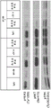


Classifications
-
- C—CHEMISTRY; METALLURGY
- C07—ORGANIC CHEMISTRY
- C07K—PEPTIDES
- C07K16/00—Immunoglobulins [IGs], e.g. monoclonal or polyclonal antibodies
- C07K16/40—Immunoglobulins [IGs], e.g. monoclonal or polyclonal antibodies against enzymes
-
- A—HUMAN NECESSITIES
- A61—MEDICAL OR VETERINARY SCIENCE; HYGIENE
- A61K—PREPARATIONS FOR MEDICAL, DENTAL OR TOILETRY PURPOSES
- A61K31/00—Medicinal preparations containing organic active ingredients
- A61K31/33—Heterocyclic compounds
- A61K31/395—Heterocyclic compounds having nitrogen as a ring hetero atom, e.g. guanethidine or rifamycins
- A61K31/535—Heterocyclic compounds having nitrogen as a ring hetero atom, e.g. guanethidine or rifamycins having six-membered rings with at least one nitrogen and one oxygen as the ring hetero atoms, e.g. 1,2-oxazines
- A61K31/5375—1,4-Oxazines, e.g. morpholine
- A61K31/5377—1,4-Oxazines, e.g. morpholine not condensed and containing further heterocyclic rings, e.g. timolol
-
- A—HUMAN NECESSITIES
- A61—MEDICAL OR VETERINARY SCIENCE; HYGIENE
- A61K—PREPARATIONS FOR MEDICAL, DENTAL OR TOILETRY PURPOSES
- A61K38/00—Medicinal preparations containing peptides
- A61K38/16—Peptides having more than 20 amino acids; Gastrins; Somatostatins; Melanotropins; Derivatives thereof
- A61K38/17—Peptides having more than 20 amino acids; Gastrins; Somatostatins; Melanotropins; Derivatives thereof from animals; from humans
- A61K38/18—Growth factors; Growth regulators
-
- A—HUMAN NECESSITIES
- A61—MEDICAL OR VETERINARY SCIENCE; HYGIENE
- A61K—PREPARATIONS FOR MEDICAL, DENTAL OR TOILETRY PURPOSES
- A61K38/00—Medicinal preparations containing peptides
- A61K38/16—Peptides having more than 20 amino acids; Gastrins; Somatostatins; Melanotropins; Derivatives thereof
- A61K38/17—Peptides having more than 20 amino acids; Gastrins; Somatostatins; Melanotropins; Derivatives thereof from animals; from humans
- A61K38/19—Cytokines; Lymphokines; Interferons
-
- A—HUMAN NECESSITIES
- A61—MEDICAL OR VETERINARY SCIENCE; HYGIENE
- A61K—PREPARATIONS FOR MEDICAL, DENTAL OR TOILETRY PURPOSES
- A61K39/00—Medicinal preparations containing antigens or antibodies
- A61K39/395—Antibodies; Immunoglobulins; Immune serum, e.g. antilymphocytic serum
-
- A—HUMAN NECESSITIES
- A61—MEDICAL OR VETERINARY SCIENCE; HYGIENE
- A61K—PREPARATIONS FOR MEDICAL, DENTAL OR TOILETRY PURPOSES
- A61K39/00—Medicinal preparations containing antigens or antibodies
- A61K39/395—Antibodies; Immunoglobulins; Immune serum, e.g. antilymphocytic serum
- A61K39/39533—Antibodies; Immunoglobulins; Immune serum, e.g. antilymphocytic serum against materials from animals
- A61K39/3955—Antibodies; Immunoglobulins; Immune serum, e.g. antilymphocytic serum against materials from animals against proteinaceous materials, e.g. enzymes, hormones, lymphokines
-
- A—HUMAN NECESSITIES
- A61—MEDICAL OR VETERINARY SCIENCE; HYGIENE
- A61K—PREPARATIONS FOR MEDICAL, DENTAL OR TOILETRY PURPOSES
- A61K39/00—Medicinal preparations containing antigens or antibodies
- A61K39/395—Antibodies; Immunoglobulins; Immune serum, e.g. antilymphocytic serum
- A61K39/39533—Antibodies; Immunoglobulins; Immune serum, e.g. antilymphocytic serum against materials from animals
- A61K39/39558—Antibodies; Immunoglobulins; Immune serum, e.g. antilymphocytic serum against materials from animals against tumor tissues, cells, antigens
-
- A—HUMAN NECESSITIES
- A61—MEDICAL OR VETERINARY SCIENCE; HYGIENE
- A61K—PREPARATIONS FOR MEDICAL, DENTAL OR TOILETRY PURPOSES
- A61K45/00—Medicinal preparations containing active ingredients not provided for in groups A61K31/00 - A61K41/00
- A61K45/06—Mixtures of active ingredients without chemical characterisation, e.g. antiphlogistics and cardiaca
-
- A—HUMAN NECESSITIES
- A61—MEDICAL OR VETERINARY SCIENCE; HYGIENE
- A61K—PREPARATIONS FOR MEDICAL, DENTAL OR TOILETRY PURPOSES
- A61K47/00—Medicinal preparations characterised by the non-active ingredients used, e.g. carriers or inert additives; Targeting or modifying agents chemically bound to the active ingredient
- A61K47/50—Medicinal preparations characterised by the non-active ingredients used, e.g. carriers or inert additives; Targeting or modifying agents chemically bound to the active ingredient the non-active ingredient being chemically bound to the active ingredient, e.g. polymer-drug conjugates
- A61K47/51—Medicinal preparations characterised by the non-active ingredients used, e.g. carriers or inert additives; Targeting or modifying agents chemically bound to the active ingredient the non-active ingredient being chemically bound to the active ingredient, e.g. polymer-drug conjugates the non-active ingredient being a modifying agent
- A61K47/68—Medicinal preparations characterised by the non-active ingredients used, e.g. carriers or inert additives; Targeting or modifying agents chemically bound to the active ingredient the non-active ingredient being chemically bound to the active ingredient, e.g. polymer-drug conjugates the non-active ingredient being a modifying agent the modifying agent being an antibody, an immunoglobulin or a fragment thereof, e.g. an Fc-fragment
- A61K47/6835—Medicinal preparations characterised by the non-active ingredients used, e.g. carriers or inert additives; Targeting or modifying agents chemically bound to the active ingredient the non-active ingredient being chemically bound to the active ingredient, e.g. polymer-drug conjugates the non-active ingredient being a modifying agent the modifying agent being an antibody, an immunoglobulin or a fragment thereof, e.g. an Fc-fragment the modifying agent being an antibody or an immunoglobulin bearing at least one antigen-binding site
- A61K47/6851—Medicinal preparations characterised by the non-active ingredients used, e.g. carriers or inert additives; Targeting or modifying agents chemically bound to the active ingredient the non-active ingredient being chemically bound to the active ingredient, e.g. polymer-drug conjugates the non-active ingredient being a modifying agent the modifying agent being an antibody, an immunoglobulin or a fragment thereof, e.g. an Fc-fragment the modifying agent being an antibody or an immunoglobulin bearing at least one antigen-binding site the antibody targeting a determinant of a tumour cell
-
- A—HUMAN NECESSITIES
- A61—MEDICAL OR VETERINARY SCIENCE; HYGIENE
- A61K—PREPARATIONS FOR MEDICAL, DENTAL OR TOILETRY PURPOSES
- A61K47/00—Medicinal preparations characterised by the non-active ingredients used, e.g. carriers or inert additives; Targeting or modifying agents chemically bound to the active ingredient
- A61K47/50—Medicinal preparations characterised by the non-active ingredients used, e.g. carriers or inert additives; Targeting or modifying agents chemically bound to the active ingredient the non-active ingredient being chemically bound to the active ingredient, e.g. polymer-drug conjugates
- A61K47/51—Medicinal preparations characterised by the non-active ingredients used, e.g. carriers or inert additives; Targeting or modifying agents chemically bound to the active ingredient the non-active ingredient being chemically bound to the active ingredient, e.g. polymer-drug conjugates the non-active ingredient being a modifying agent
- A61K47/68—Medicinal preparations characterised by the non-active ingredients used, e.g. carriers or inert additives; Targeting or modifying agents chemically bound to the active ingredient the non-active ingredient being chemically bound to the active ingredient, e.g. polymer-drug conjugates the non-active ingredient being a modifying agent the modifying agent being an antibody, an immunoglobulin or a fragment thereof, e.g. an Fc-fragment
- A61K47/6835—Medicinal preparations characterised by the non-active ingredients used, e.g. carriers or inert additives; Targeting or modifying agents chemically bound to the active ingredient the non-active ingredient being chemically bound to the active ingredient, e.g. polymer-drug conjugates the non-active ingredient being a modifying agent the modifying agent being an antibody, an immunoglobulin or a fragment thereof, e.g. an Fc-fragment the modifying agent being an antibody or an immunoglobulin bearing at least one antigen-binding site
- A61K47/6871—Medicinal preparations characterised by the non-active ingredients used, e.g. carriers or inert additives; Targeting or modifying agents chemically bound to the active ingredient the non-active ingredient being chemically bound to the active ingredient, e.g. polymer-drug conjugates the non-active ingredient being a modifying agent the modifying agent being an antibody, an immunoglobulin or a fragment thereof, e.g. an Fc-fragment the modifying agent being an antibody or an immunoglobulin bearing at least one antigen-binding site the antibody targeting an enzyme
-
- A—HUMAN NECESSITIES
- A61—MEDICAL OR VETERINARY SCIENCE; HYGIENE
- A61N—ELECTROTHERAPY; MAGNETOTHERAPY; RADIATION THERAPY; ULTRASOUND THERAPY
- A61N5/00—Radiation therapy
- A61N5/10—X-ray therapy; Gamma-ray therapy; Particle-irradiation therapy
-
- A—HUMAN NECESSITIES
- A61—MEDICAL OR VETERINARY SCIENCE; HYGIENE
- A61P—SPECIFIC THERAPEUTIC ACTIVITY OF CHEMICAL COMPOUNDS OR MEDICINAL PREPARATIONS
- A61P35/00—Antineoplastic agents
-
- A—HUMAN NECESSITIES
- A61—MEDICAL OR VETERINARY SCIENCE; HYGIENE
- A61P—SPECIFIC THERAPEUTIC ACTIVITY OF CHEMICAL COMPOUNDS OR MEDICINAL PREPARATIONS
- A61P43/00—Drugs for specific purposes, not provided for in groups A61P1/00-A61P41/00
-
- C—CHEMISTRY; METALLURGY
- C07—ORGANIC CHEMISTRY
- C07K—PEPTIDES
- C07K14/00—Peptides having more than 20 amino acids; Gastrins; Somatostatins; Melanotropins; Derivatives thereof
- C07K14/435—Peptides having more than 20 amino acids; Gastrins; Somatostatins; Melanotropins; Derivatives thereof from animals; from humans
- C07K14/475—Growth factors; Growth regulators
-
- C—CHEMISTRY; METALLURGY
- C07—ORGANIC CHEMISTRY
- C07K—PEPTIDES
- C07K16/00—Immunoglobulins [IGs], e.g. monoclonal or polyclonal antibodies
- C07K16/18—Immunoglobulins [IGs], e.g. monoclonal or polyclonal antibodies against material from animals or humans
-
- C—CHEMISTRY; METALLURGY
- C07—ORGANIC CHEMISTRY
- C07K—PEPTIDES
- C07K16/00—Immunoglobulins [IGs], e.g. monoclonal or polyclonal antibodies
- C07K16/18—Immunoglobulins [IGs], e.g. monoclonal or polyclonal antibodies against material from animals or humans
- C07K16/22—Immunoglobulins [IGs], e.g. monoclonal or polyclonal antibodies against material from animals or humans against growth factors ; against growth regulators
-
- C—CHEMISTRY; METALLURGY
- C07—ORGANIC CHEMISTRY
- C07K—PEPTIDES
- C07K16/00—Immunoglobulins [IGs], e.g. monoclonal or polyclonal antibodies
- C07K16/18—Immunoglobulins [IGs], e.g. monoclonal or polyclonal antibodies against material from animals or humans
- C07K16/24—Immunoglobulins [IGs], e.g. monoclonal or polyclonal antibodies against material from animals or humans against cytokines, lymphokines or interferons
-
- C—CHEMISTRY; METALLURGY
- C07—ORGANIC CHEMISTRY
- C07K—PEPTIDES
- C07K16/00—Immunoglobulins [IGs], e.g. monoclonal or polyclonal antibodies
- C07K16/18—Immunoglobulins [IGs], e.g. monoclonal or polyclonal antibodies against material from animals or humans
- C07K16/28—Immunoglobulins [IGs], e.g. monoclonal or polyclonal antibodies against material from animals or humans against receptors, cell surface antigens or cell surface determinants
- C07K16/2863—Immunoglobulins [IGs], e.g. monoclonal or polyclonal antibodies against material from animals or humans against receptors, cell surface antigens or cell surface determinants against receptors for growth factors, growth regulators
-
- C—CHEMISTRY; METALLURGY
- C07—ORGANIC CHEMISTRY
- C07K—PEPTIDES
- C07K16/00—Immunoglobulins [IGs], e.g. monoclonal or polyclonal antibodies
- C07K16/18—Immunoglobulins [IGs], e.g. monoclonal or polyclonal antibodies against material from animals or humans
- C07K16/28—Immunoglobulins [IGs], e.g. monoclonal or polyclonal antibodies against material from animals or humans against receptors, cell surface antigens or cell surface determinants
- C07K16/30—Immunoglobulins [IGs], e.g. monoclonal or polyclonal antibodies against material from animals or humans against receptors, cell surface antigens or cell surface determinants from tumour cells
-
- C—CHEMISTRY; METALLURGY
- C07—ORGANIC CHEMISTRY
- C07K—PEPTIDES
- C07K16/00—Immunoglobulins [IGs], e.g. monoclonal or polyclonal antibodies
- C07K16/18—Immunoglobulins [IGs], e.g. monoclonal or polyclonal antibodies against material from animals or humans
- C07K16/32—Immunoglobulins [IGs], e.g. monoclonal or polyclonal antibodies against material from animals or humans against translation products of oncogenes
-
- A—HUMAN NECESSITIES
- A61—MEDICAL OR VETERINARY SCIENCE; HYGIENE
- A61K—PREPARATIONS FOR MEDICAL, DENTAL OR TOILETRY PURPOSES
- A61K39/00—Medicinal preparations containing antigens or antibodies
- A61K2039/505—Medicinal preparations containing antigens or antibodies comprising antibodies
-
- A—HUMAN NECESSITIES
- A61—MEDICAL OR VETERINARY SCIENCE; HYGIENE
- A61K—PREPARATIONS FOR MEDICAL, DENTAL OR TOILETRY PURPOSES
- A61K39/00—Medicinal preparations containing antigens or antibodies
- A61K2039/505—Medicinal preparations containing antigens or antibodies comprising antibodies
- A61K2039/507—Comprising a combination of two or more separate antibodies
-
- A—HUMAN NECESSITIES
- A61—MEDICAL OR VETERINARY SCIENCE; HYGIENE
- A61K—PREPARATIONS FOR MEDICAL, DENTAL OR TOILETRY PURPOSES
- A61K31/00—Medicinal preparations containing organic active ingredients
- A61K31/33—Heterocyclic compounds
- A61K31/395—Heterocyclic compounds having nitrogen as a ring hetero atom, e.g. guanethidine or rifamycins
- A61K31/435—Heterocyclic compounds having nitrogen as a ring hetero atom, e.g. guanethidine or rifamycins having six-membered rings with one nitrogen as the only ring hetero atom
- A61K31/47—Quinolines; Isoquinolines
- A61K31/4709—Non-condensed quinolines and containing further heterocyclic rings
-
- A—HUMAN NECESSITIES
- A61—MEDICAL OR VETERINARY SCIENCE; HYGIENE
- A61K—PREPARATIONS FOR MEDICAL, DENTAL OR TOILETRY PURPOSES
- A61K31/00—Medicinal preparations containing organic active ingredients
- A61K31/33—Heterocyclic compounds
- A61K31/395—Heterocyclic compounds having nitrogen as a ring hetero atom, e.g. guanethidine or rifamycins
- A61K31/495—Heterocyclic compounds having nitrogen as a ring hetero atom, e.g. guanethidine or rifamycins having six-membered rings with two or more nitrogen atoms as the only ring heteroatoms, e.g. piperazine or tetrazines
- A61K31/496—Non-condensed piperazines containing further heterocyclic rings, e.g. rifampin, thiothixene or sparfloxacin
-
- A—HUMAN NECESSITIES
- A61—MEDICAL OR VETERINARY SCIENCE; HYGIENE
- A61K—PREPARATIONS FOR MEDICAL, DENTAL OR TOILETRY PURPOSES
- A61K31/00—Medicinal preparations containing organic active ingredients
- A61K31/33—Heterocyclic compounds
- A61K31/395—Heterocyclic compounds having nitrogen as a ring hetero atom, e.g. guanethidine or rifamycins
- A61K31/495—Heterocyclic compounds having nitrogen as a ring hetero atom, e.g. guanethidine or rifamycins having six-membered rings with two or more nitrogen atoms as the only ring heteroatoms, e.g. piperazine or tetrazines
- A61K31/505—Pyrimidines; Hydrogenated pyrimidines, e.g. trimethoprim
- A61K31/506—Pyrimidines; Hydrogenated pyrimidines, e.g. trimethoprim not condensed and containing further heterocyclic rings
-
- C—CHEMISTRY; METALLURGY
- C07—ORGANIC CHEMISTRY
- C07K—PEPTIDES
- C07K2317/00—Immunoglobulins specific features
- C07K2317/20—Immunoglobulins specific features characterized by taxonomic origin
- C07K2317/21—Immunoglobulins specific features characterized by taxonomic origin from primates, e.g. man
-
- C—CHEMISTRY; METALLURGY
- C07—ORGANIC CHEMISTRY
- C07K—PEPTIDES
- C07K2317/00—Immunoglobulins specific features
- C07K2317/20—Immunoglobulins specific features characterized by taxonomic origin
- C07K2317/24—Immunoglobulins specific features characterized by taxonomic origin containing regions, domains or residues from different species, e.g. chimeric, humanized or veneered
-
- C—CHEMISTRY; METALLURGY
- C07—ORGANIC CHEMISTRY
- C07K—PEPTIDES
- C07K2317/00—Immunoglobulins specific features
- C07K2317/30—Immunoglobulins specific features characterized by aspects of specificity or valency
- C07K2317/31—Immunoglobulins specific features characterized by aspects of specificity or valency multispecific
-
- C—CHEMISTRY; METALLURGY
- C07—ORGANIC CHEMISTRY
- C07K—PEPTIDES
- C07K2317/00—Immunoglobulins specific features
- C07K2317/50—Immunoglobulins specific features characterized by immunoglobulin fragments
- C07K2317/54—F(ab')2
-
- C—CHEMISTRY; METALLURGY
- C07—ORGANIC CHEMISTRY
- C07K—PEPTIDES
- C07K2317/00—Immunoglobulins specific features
- C07K2317/50—Immunoglobulins specific features characterized by immunoglobulin fragments
- C07K2317/55—Fab or Fab'
-
- C—CHEMISTRY; METALLURGY
- C07—ORGANIC CHEMISTRY
- C07K—PEPTIDES
- C07K2317/00—Immunoglobulins specific features
- C07K2317/50—Immunoglobulins specific features characterized by immunoglobulin fragments
- C07K2317/56—Immunoglobulins specific features characterized by immunoglobulin fragments variable (Fv) region, i.e. VH and/or VL
- C07K2317/565—Complementarity determining region [CDR]
-
- C—CHEMISTRY; METALLURGY
- C07—ORGANIC CHEMISTRY
- C07K—PEPTIDES
- C07K2317/00—Immunoglobulins specific features
- C07K2317/60—Immunoglobulins specific features characterized by non-natural combinations of immunoglobulin fragments
- C07K2317/62—Immunoglobulins specific features characterized by non-natural combinations of immunoglobulin fragments comprising only variable region components
- C07K2317/622—Single chain antibody (scFv)
-
- C—CHEMISTRY; METALLURGY
- C07—ORGANIC CHEMISTRY
- C07K—PEPTIDES
- C07K2317/00—Immunoglobulins specific features
- C07K2317/60—Immunoglobulins specific features characterized by non-natural combinations of immunoglobulin fragments
- C07K2317/62—Immunoglobulins specific features characterized by non-natural combinations of immunoglobulin fragments comprising only variable region components
- C07K2317/626—Diabody or triabody
-
- C—CHEMISTRY; METALLURGY
- C07—ORGANIC CHEMISTRY
- C07K—PEPTIDES
- C07K2317/00—Immunoglobulins specific features
- C07K2317/70—Immunoglobulins specific features characterized by effect upon binding to a cell or to an antigen
- C07K2317/76—Antagonist effect on antigen, e.g. neutralization or inhibition of binding
-
- C—CHEMISTRY; METALLURGY
- C07—ORGANIC CHEMISTRY
- C07K—PEPTIDES
- C07K2317/00—Immunoglobulins specific features
- C07K2317/90—Immunoglobulins specific features characterized by (pharmaco)kinetic aspects or by stability of the immunoglobulin
- C07K2317/92—Affinity (KD), association rate (Ka), dissociation rate (Kd) or EC50 value
Landscapes
- Health & Medical Sciences (AREA)
- Chemical & Material Sciences (AREA)
- Life Sciences & Earth Sciences (AREA)
- Medicinal Chemistry (AREA)
- General Health & Medical Sciences (AREA)
- Organic Chemistry (AREA)
- Immunology (AREA)
- Animal Behavior & Ethology (AREA)
- Public Health (AREA)
- Veterinary Medicine (AREA)
- Pharmacology & Pharmacy (AREA)
- Epidemiology (AREA)
- Engineering & Computer Science (AREA)
- Proteomics, Peptides & Aminoacids (AREA)
- Biochemistry (AREA)
- Biophysics (AREA)
- Genetics & Genomics (AREA)
- Molecular Biology (AREA)
- Bioinformatics & Cheminformatics (AREA)
- Oncology (AREA)
- Mycology (AREA)
- Microbiology (AREA)
- Biomedical Technology (AREA)
- Zoology (AREA)
- Gastroenterology & Hepatology (AREA)
- Nuclear Medicine, Radiotherapy & Molecular Imaging (AREA)
- General Chemical & Material Sciences (AREA)
- Cell Biology (AREA)
- Toxicology (AREA)
- Chemical Kinetics & Catalysis (AREA)
- Pathology (AREA)
- Radiology & Medical Imaging (AREA)
- Endocrinology (AREA)
- Medicines Containing Antibodies Or Antigens For Use As Internal Diagnostic Agents (AREA)
- Peptides Or Proteins (AREA)
- Pharmaceuticals Containing Other Organic And Inorganic Compounds (AREA)
- Acyclic And Carbocyclic Compounds In Medicinal Compositions (AREA)
- Medicines That Contain Protein Lipid Enzymes And Other Medicines (AREA)
- Medicinal Preparation (AREA)
- Plural Heterocyclic Compounds (AREA)
Abstract
The present invention describes materials and methods for treating a subject having a human epidermal growth factor receptor-3 (HER-3) -associated disease by administering a combination of a first agent that binds to HER-3 and a second agent that binds to and/or inhibits another member of the HER family. The first and second agents may be biological agents such as, for example, antigen binding proteins or small molecule tyrosine kinase inhibitors.
Description
The application is a divisional application of an invention patent application with the international application date of 11/12 2010 and the Chinese national application number of 201080052236.X, and the invention name of the invention is 'materials and methods for treating or preventing diseases related to human epidermal growth factor receptor-3 (HER-3').
Technical Field
The present invention relates to materials and methods for treating a subject having a human epidermal growth factor receptor-3 (HER-3) associated disease by administering a combination of a first agent that binds to HER-3 and a second agent that binds to and/or inhibits another member of the human epidermal growth factor receptor (HER) family. The first and second agents may be any kind of molecule that binds to HER-3 or to another member of the HER family and/or inhibits that member, respectively, including but not limited to biological compounds such as antigen binding proteins, small molecule tyrosine kinase inhibitors, sirnas or natural substances.
Prior Art
HER-3 (also known as Erb3) is a receptor protein tyrosine kinase belonging to the epidermal growth factor receptor (EGF-R) family of receptor protein tyrosine kinases, which also includes HER-1 (also known as EGF-R or erbB), HER-2 (also known as erbB2) and HER-4 (also known as erbB4) (Plowman et al, (1990) Proc. Natl. Acad. Sci. US).874905 and 4909; kraus et al, (1989) proc.natl.acad.sci.us.869193-9197; and Kraus et al, (1993) proc.natl.acad.sci.us.90:2900-2904). Like the prototype epidermal growth factor receptor, the transmembrane receptor HER-3 is composed of an extracellular ligand-binding domain (ECD), a dimerization domain within the ECD, a transmembrane domain (TMD), an intracellular protein Tyrosine Kinase Domain (TKD), and a C-terminal phosphorylation domain.
The ligand of HER-3, also known as human epidermal growth factor receptor regulatory protein (HRG), binds to the extracellular domain of HER-3 and activates receptor-mediated signaling by promoting dimerization with other human epidermal growth factor receptor (HER) family members, followed by transphosphorylation of the intracellular HER-3 domain and activation of downstream signaling cascades. Dimer formation with multiple HER family members amplifies the signaling potential of HER-3 and is a way to diversify and amplify the signal.
Disclosure of Invention
The present invention relates to materials and methods for treating a subject having a human epidermal growth factor receptor-3 (HER-3) -associated disease by administering a first agent that binds to HER-3 and a second agent that binds to and/or inhibits another member of the HER family in combination. The first and second agents may be any kind of molecule that binds to HER-3 or to another member of the HER family and/or inhibits that member, respectively, including but not limited to biological compounds such as antigen binding proteins, small molecule tyrosine kinase inhibitors, sirnas or natural substances.
In one aspect, the invention features a method of treating or preventing a HER-3 associated disease in a subject, the method comprising administering to the subject a first agent and a second agent, wherein the first agent binds to HER-3 and the second agent binds to and/or inhibits the activity of another member of the HER family. The first agent can be a small molecule compound or antigen binding protein that binds to HER-3. The first agent can be an antigen binding protein that binds to HER-3, and the antigen binding protein comprises a heavy chain amino acid sequence and a light chain amino acid sequence; the heavy chain amino acid sequence comprises a CDRH1 selected from the group consisting of SEQ ID NOs 236, 251, 252 and 256, a CDRH2 selected from the group consisting of SEQ ID NOs 258, 278, 280 and 282, and a CDRH3 selected from the group consisting of SEQ ID NOs 283, 285, 309, 313 and 315; and the light chain amino acid sequence comprises a CDRL1 selected from the group consisting of SEQ ID NOs 320, 334, 337 and 340, a CDRL2 selected from the group consisting of SEQ ID NOs 343, 356, 351 and 344, and a CDRL3 selected from the group consisting of SEQ ID NOs 360, 381, 385 and 387. The first agent can be an antigen binding protein that binds to HER-3, and the antigen binding protein comprises a heavy chain amino acid sequence comprising at least one CDR selected from the group consisting of: (a) CDRH 1as shown in SEQ ID NOs 236, 251, 252 and 256; (b) CDRH2 as shown in SEQ ID NOs 258, 278, 280 and 282; and (c) CDRH3 as shown in SEQ ID NOs: 283, 285, 309, 313 and 315. The first agent can be an antigen binding protein that binds to HER-3, and the antigen binding protein comprises a light chain amino acid sequence comprising at least one CDR selected from the group consisting of: (d) CDRL 1as shown in SEQ ID NOs 320, 334, 337 and 340; (e) CDRL2 as shown in SEQ ID NOs 343, 356, 351 and 344; and (f) CDRL3 as shown in SEQ ID NOs: 360, 381, 385 and 387.
The first agent can be an antigen binding protein that binds to HER-3, and the antigen binding protein comprises a heavy chain amino acid sequence comprising at least one of the CDRs selected from the group consisting of: (a) CDRH 1as shown in SEQ ID NOs 236, 251, 252 and 256; (b) CDRH2 as shown in SEQ ID NOs 258, 278, 280 and 282; and (c) CDRH3 as shown in SEQ ID NOs 283, 285, 309, 313 and 315; and the light chain amino acid sequence comprises at least one CDR selected from the group consisting of: (d) CDRL 1as shown in SEQ ID NOs 320, 334, 337 and 340; (e) CDRL2 as shown in SEQ ID NOs 343, 356, 351 and 344; and (f) CDRL3 as shown in SEQ ID NOs: 360, 381, 385 and 387. The first agent can be an antigen binding protein that binds to HER-3, and the antigen binding protein comprises a heavy chain amino acid sequence or a light chain amino acid sequence; the heavy chain amino acid sequence comprises a CDRH1 selected from the group consisting of SEQ ID NOs 236, 251, 252 and 256, a CDRH2 selected from the group consisting of SEQ ID NOs 258, 278, 280 and 282, and a CDRH3 selected from the group consisting of SEQ ID NOs 283, 285, 309, 313 and 315; and the light chain amino acid sequence comprises a CDRL1 selected from the group consisting of SEQ ID NOs 320, 334, 337 and 340, a CDRL2 selected from the group consisting of SEQ ID NOs 343, 356, 351 and 344, and a CDRL3 selected from the group consisting of SEQ ID NOs 360, 381, 385 and 387.
The first agent can be an antigen binding protein that binds to HER-3, and the antigen binding protein comprises a heavy chain amino acid sequence selected from the group consisting of SEQ ID NOs 42, 54, 70, 92, and 96. The antigen binding protein may comprise a light chain amino acid sequence selected from the group consisting of SEQ ID NOs 44, 56, 72, 94, and 98.
The first agent can be an antigen binding protein that binds to HER-3, and the antigen binding protein comprises a heavy chain amino acid sequence selected from the group consisting of SEQ ID NOs 42, 54, 70, 92, and 96 and a light chain amino acid sequence selected from the group consisting of SEQ ID NOs 44, 56, 72, 94, and 98.
The first agent can be an antigen binding protein that binds to HER-3, and the antigen binding protein comprises the heavy chain amino acid sequence of SEQ ID NO. 42 and the light chain amino acid sequence of SEQ ID NO. 44. The first agent can be an antigen binding protein that binds to HER-3 and comprises the heavy chain amino acid sequence of SEQ ID NO. 54 and the light chain amino acid sequence of SEQ ID NO. 56. The first agent can be an antigen binding protein that binds to HER-3, and the antigen binding protein comprises the heavy chain amino acid sequence of SEQ ID NO. 70 and the light chain amino acid sequence of SEQ ID NO. 72. The first agent can be an antigen binding protein that binds to HER-3, and the antigen binding protein comprises CDRH3 selected from the group consisting of SEQ ID NOs: 283, 285, 309, 313, and 315. The first agent can be an antigen binding protein that binds to HER-3, and the antigen binding protein comprises CDRL3 selected from the group consisting of SEQ ID NOs 360, 381, 385, and 387.
The antigen binding protein may be directed against the extracellular domain of HER-3. Binding of the antigen binding protein to HER-3 may reduce HER-3 mediated signaling, reduce phosphorylation of HER-3, reduce cell proliferation, reduce cell migration, and/or increase downregulation of HER-3.
The antigen binding protein that binds to HER-3 can be an antibody. The antibody can be a monoclonal antibody, a polyclonal antibody, a recombinant antibody, a humanized antibody, a chimeric antibody, a multispecific antibody or antibody fragment thereof (e.g., a Fab fragment, Fab 'fragment, F (ab') 2 fragment, Fv fragment, diabody, or single chain antibody molecule). The antibody may be of the IgG1, IgG2, IgG3 or IgG4 type.
The first agent can be an antigen binding protein that binds to HER-3, and the antigen binding protein can be coupled to an effector group. The effector group may be a radioisotope or radionuclide, a toxin, or a therapeutic or chemotherapeutic group (e.g., a therapeutic or chemotherapeutic group selected from the group consisting of calicheamicin (calicheamicin), auristatin-PE (auristatin-PE), geldanamycin (geldanamycin), maytansine (maytansine) and derivatives thereof).
The second agent can be a small molecule compound or an antigen binding protein. The second agent can be, for example, trastuzumab (trastuzumab), lapatinib (lapatinib), neratinib (neratinib), panitumumab (panitumumab), erlotinib (erlotinib), cetuximab (cetuximab), pertuzumab (pertuzumab), and T-DM 1.
In another aspect, the invention features a method of treating or preventing a HER-3 associated disease in a subject, the method comprising administering to the subject a first agent and a second agent, wherein the first agent is an antigen binding protein that binds to HER-3 and comprises the heavy chain amino acid sequence SEQ ID No. 42 and the light chain amino acid sequence SEQ ID No. 44, and wherein the second agent is selected from the group consisting of erlotinib, lapatinib, and lenatinib. Furthermore, the invention features a method of treating or preventing a HER-3 related disease in a subject, the method comprising administering to the subject a first agent and a second agent, wherein the first agent is an antigen binding protein that binds to HER-3 and comprises the heavy chain amino acid sequence SEQ ID No. 54 and the light chain amino acid sequence SEQ ID No. 56, or an antigen binding protein that binds to HER-3 and comprises the heavy chain amino acid sequence SEQ ID No. 70 and the light chain amino acid sequence SEQ ID No. 72, and wherein the second agent is selected from the group consisting of erlotinib, lapatinib, and lenatinib.
The invention also features a method of treating or preventing a HER-3 associated disease in a subject, the method comprising administering to the subject a first agent and a second agent, wherein the first agent is an antigen binding protein that binds to HER-3 and comprises a heavy chain amino acid sequence of SEQ ID NO 42 and a light chain amino acid sequence of SEQ ID NO 44, and wherein the second agent is selected from the group consisting of trastuzumab, T-DM1, panitumumab, and cetuximab.
The methods provided herein can optionally comprise administering a third or other therapeutic agent and/or radiation therapy. The third or further therapeutic agent may be an anti-tumour agent (e.g. an anti-tumour antibody or a chemotherapeutic agent such as capecitabine, anthracycline, doxorubicin, cyclophosphamide, paclitaxel, docetaxel, cisplatin, gemcitabine or carboplatin).
The first and second doses may be administered intravenously, subcutaneously, intramuscularly or orally. The disease may be a hyperproliferative disease (e.g., a disease selected from the group consisting of breast cancer, ovarian cancer, prostate cancer, colon cancer, rectal cancer, lung cancer, pancreatic cancer, epidermoid cancer, fibrosarcoma, melanoma, nasopharyngeal cancer, and squamous cell carcinoma).
The methods provided herein can include administering the first dose at a frequency of at least once every 6 weeks of about 1 to about 20mg/kg body weight dose. The method can include administering a dose of about 1 to about 20mg/kg body weight of the second dose at a frequency of at least once every 6 weeks. The method can further comprise prior to administration using a method comprising analyzing the predictive markers to select a subject having a HER-3 related disease. The method may further comprise monitoring treatment outcome after administration.
Unless defined otherwise, all technical and scientific terms used herein have the same meaning as commonly understood by one of ordinary skill in the art to which this invention pertains. Although methods and materials similar or equivalent to those described herein can be used in the practice of the present invention, suitable methods and materials are described below. Each of the publications, applications, patents, and other references mentioned herein is incorporated by reference in its entirety. In case of conflict, the present specification, including definitions, will control. In addition, the materials, methods, and examples are illustrative only and not intended to be limiting.
The details of one or more embodiments of the invention are set forth in the accompanying drawings and the description below. Other features, objects, and advantages of the invention will be apparent from the description and drawings, and from the claims.
Brief Description of Drawings
FIG. 1 illustrates the effect of using human anti-HER-3 antibody and panitumumab, alone or in combination, on the growth of non-small cell lung cancer (NSCLC) xenograft tumors (Calu-3).
FIG. 2 illustrates the effect of using human anti-HER-3 antibody and erlotinib alone or in combination on the growth of Calu-3.
FIG. 3 illustrates the effect of using a human anti-HER-3 antibody alone or in combination with C2C4(HER2 dimerization inhibitor) or trastuzumab on basal anchorage-independent growth of SKBr-3 breast cancer cells.
Figure 4 illustrates the effect of using human anti-HER-3 antibody alone or in combination with C2C4, trastuzumab or cetuximab on HRG-stimulated anchorage-independent growth of SkBr-3 breast cancer cells.
FIG. 5 illustrates the effect of using a human anti-HER-3 antibody alone or in combination with C2C4, trastuzumab or cetuximab on basal anchorage-independent growth of MDA-MB-435 ovarian cancer cells.
Figures 6A-6D illustrate the effect of using a human anti-HER-3 antibody alone or in combination with trastuzumab (figure 6A), lapatinib (figure 6B), gemcitabine (figure 6C), or cisplatin (figure 6D) on the proliferation of MDA-MB-175VII breast cancer cells.
FIG. 7 illustrates the effect of use of a human anti-HER-3 antibody alone or in combination with C2C4, trastuzumab, or lapatinib on HRG-stimulated proliferation of ZR-75-30 breast cancer cells.
Figure 8 illustrates the effect of using a human anti-HER-3 antibody alone or in combination with C2C4, trastuzumab, or lapatinib on HRG-stimulated proliferation of BT474 breast cancer cells.
FIG. 9 illustrates the effect of using a human anti-HER-3 antibody alone or in combination with cetuximab, C2C4, or trastuzumab on the proliferation of HRG-stimulated DLD-1 colon cancer cells.
Figure 10 illustrates the effect of using a human anti-HER-3 antibody alone or in combination with C2C4 or trastuzumab or lapatinib on HRG-stimulated proliferation of HCC-1569 breast cancer cells.
Figure 11 illustrates the effect of using a human anti-HER-3 antibody alone or in combination with C2C4, trastuzumab, or lapatinib on HRG-stimulated proliferation of SkBr-3 breast cancer cells.
FIG. 12 illustrates the effect of using a human anti-HER-3 antibody alone or in combination with panitumumab on the proliferation of FaDu head and neck cancer cells.
FIG. 13 is a Western blot photograph showing the effect of using a human anti-HER-3 antibody alone or in combination with cetuximab, C2C4, or trastuzumab on the phosphorylation of HER-3 (top panel), Akt (middle panel), and ERK (bottom panel) in MDA-MB-175VII breast cancer cells.
FIG. 14 is a Western blot photograph showing the effect of using human anti-HER-3 antibody alone or in combination with cetuximab, C2C4, trastuzumab, or lapatinib on phosphorylation of HER-3 (top panel), Akt (middle panel), and ERK (bottom panel) in HRG-stimulated SkBr-3 breast cancer cells.
FIG. 15 is a Western blot photograph showing the effect of using a human anti-HER-3 antibody alone or in combination with cetuximab, pertuzumab (C2C4), or trastuzumab on phosphorylation of HER-3 (top panel) or Akt (bottom panel) in HRG-stimulated Ls174T colon cancer cells.
FIG. 16 is a Western blot photograph showing the effect of using human anti-HER-3 antibody alone or in combination with cetuximab, C2C4, or trastuzumab on phosphorylation of HER-3 (top panel), Akt (middle panel), and ERK (bottom panel) in HRG-stimulated HCC1569 breast cancer cells.
FIG. 17 is a Western blot photograph showing the effect of using a human anti-HER-3 antibody alone or in combination with panitumumab on phosphorylation of Akt, PGFR, HER-2, HER-3, HER-4 and ERK in A549 alveolar epithelial cells. Row 1, IgG control group; line 2, panitumumab alone; line 3, U1-59 alone; line 4, use U1-59 in combination with panitumumab. Tubulin was used as a control for the same loading.
FIG. 18 is a Western blot photograph showing the effect of using a human anti-HER-3 antibody alone or in combination with panitumumab or lapatinib on the phosphorylation of HER-3, Akt, HER-2, ERK and EGF-R in Calu3NSCLC cells. Row 1, IgG control group; line 2, panitumumab alone; line 3, U1-59 alone; line 4, lapatinib alone; line 5, use U1-59 in combination with panitumumab; line 6, use U1-59 in combination with lapatinib.
Figure 19 illustrates the effect of using human anti-HER-3 antibody and lapatinib alone or in combination on the growth of breast cancer xenograft tumors (HCC-1569).
Figure 20 shows that treatment of a549NSCLC cells with U1-59 inhibited HER3 phosphorylation and reduced reactivation following gefitinib (gefitinib) treatment. A549 cells were treated with gefitinib, U1-59, or both, and phosphorylation of HER3 was assessed by ELISA analysis. Treatment with gefitinib for 1 hour resulted in partial inhibition of HER phosphorylation, but reversed to control amounts after 24 hours. Conversely, treatment with U1-59 resulted in a greater inhibition of HER phosphorylation that persisted after 24 hours. Treatment with the combination of both agents prevented the reversal of this inhibitory effect seen in cells after 24 hours of treatment with gefitinib alone. Experiments were performed in triplicate wells and repeated at least twice. Results are expressed as mean ± standard deviation.
Detailed Description
The section headings used herein are for organizational purposes only and are not to be construed as limiting the subject matter described.
Unless otherwise defined herein, scientific and technical terms used in connection with the present application shall have the meaning commonly understood by one of ordinary skill in the art. In addition, unless the context requires otherwise, singular terms shall include the plural and plural terms shall include the singular.
Generally, the nomenclature and the techniques used in connection with, cell and tissue culture, molecular biology, immunology, microbiology, genetics and protein and nucleic acid chemistry and hybridization described herein are those well known and commonly employed in the art. The methods and techniques of the present application are generally performed according to conventional methods well known in the art and, unless otherwise indicated, are described in various general and more specific references that are cited and discussed throughout the present specification. See, for example, Sambrook et al, Molecular Cloning: A Laboratory Manual, third edition, Cold Spring Harbor Laboratory Press, Cold Spring Harbor, N.Y. (2001), Ausubel et al, Current Protocols in Molecular Biology (Current programs in Molecular Biology), Green Publishing Associates (1992) and Harlow and Lane Antibodies: A Laboratory Manual, Cold Spring Harbor Laboratory Press, Cold Spring Harbor, N.Y. (1990), the contents of each reference being incorporated herein by reference in its entirety. Enzymatic reactions and purification techniques are performed according to the manufacturer's instructions in a manner commonly accomplished in the art or described herein. The terms used in connection with analytical chemistry, synthetic organic chemistry, medical and pharmaceutical chemistry, and laboratory methods and techniques described herein are those well known and commonly used in the art. Standard techniques can be used for chemical synthesis, chemical analysis, pharmaceutical formulation, formulation and delivery, and patient treatment.
It is to be understood that this invention is not limited to the particular methodology, procedures, reagents, etc. described herein as such may vary. The terminology used herein is for the purpose of describing particular embodiments only and is not intended to limit the scope defined only by the claims.
Except in the operating examples, or where otherwise indicated, all numbers expressing quantities of ingredients or reaction conditions used herein are to be understood as being modified in all instances by the term "about". The term "about" when used with a percentage may mean +/-1%.
1. General overview
The present invention provides materials and methods relating to the use of a first agent that binds to HER-3 and a second agent that binds to and/or inhibits the activity of other members of the HER family in combination to treat or prevent HER-3 related diseases. The first and second agents may be biological compounds, such as antigen binding proteins or small molecule tyrosine kinase inhibitors. For example, the invention provides isolated polypeptides (e.g., binding proteins such as antibodies) and/or small molecule tyrosine kinase inhibitors that bind to and/or inhibit a single or multiple members of the HER family (such as HER-3, HER-2, EGF-R, HER-4, and/or any other member of the HER family). The invention also provides compositions comprising a first agent that binds to HER-3 and a second agent that binds to one or more of the other HER family members and/or inhibits the activity of such members, and methods of using the same for treating or preventing HER-3 related diseases.
Certain first and/or second agents described herein are biological agents, such as antigen binding proteins. In certain embodiments, the polypeptide structure of the antigen binding protein is based on antibodies, including but not limited to monoclonal antibodies, bispecific antibodies, minibodies (minibodies), domain antibodies, synthetic antibodies (sometimes referred to as "antibody mimetics"), chimeric antibodies, humanized antibodies, human antibodies, antibody fusions (sometimes referred to as "antibody conjugates"), and fragments thereof, respectively. Various structures are further described below. In other embodiments, the first and/or second agent is a small molecule tyrosine kinase inhibitor. In yet further embodiments, the first agent and/or the second agent is an siRNA. In yet further embodiments, the first agent and/or the second agent is a natural substance.
The compositions described herein and methods of using the same have demonstrated increased inhibition of growth of solid tumors expressing HER-3 and at least one other member of the HER family. In particular, administration of a combination of a first agent that binds to HER-3 or a second agent that binds to and/or inhibits at least one other member of the HER family has demonstrated increased efficacy in inhibiting growth of various tumors in the present invention as compared to administration of the first agent alone or the second agent. Thus, the compositions and methods disclosed herein have demonstrated utility in improved methods of treating and preventing neoplastic diseases, such as cancer.
HER-3 binding agents
As described herein, an agent that binds to HER-3 can be a biological compound, including but not limited to an antigen binding protein, such as an antibody or a small molecule tyrosine kinase inhibitor. An "antigen binding protein" as used herein or a "binding protein" as used herein refers to a protein that specifically binds to a particular antigen of interest, such as a member of the HER family (e.g., HER-3). When the dissociation constant (KD) of the antigen binding protein and the target antigen is less than or equal to 10-8M, is said to be "specific binding". When K isDIs less than or equal to 5 multiplied by 10-9M to the same extent as"high affinity" for antigen binding when KDIs less than or equal to 5 multiplied by 10-10M is a "very high affinity" binding. In one embodiment, the antibody has ≦ 10-9K of MDAnd has a thickness of about 1X 10⁻⁴Off rate per second. In one embodiment, the off-rate is about 1 × 10⁵In seconds. In other embodiments, the antibody will be about 10-8M to 10-10K between MDBinds to a particular member of the HER family, and in yet another embodiment it will be at KD≤2×10⁻¹⁰And (4) combining. In addition, as used herein, a small molecule compound is a low molecular weight compound that is chemically synthesized to inhibit the enzymatic activity of one or more protein kinases, including serine, threonine, or tyrosine kinases.
In some embodiments, when the HER-3 binding agent is a biological compound, the agent is an antigen binding protein, such as an antibody that can bind to HER-3. The invention therefore provides HER binding proteins, including anti-HER-3 antibodies, for use in compositions and methods for treating HER-3 related diseases. In some embodiments, an antibody targeting HER-3 can target the extracellular domain (ECD) of anti-HER-3. For example, as described herein, an anti-HER-3 antibody can interact with at least one epitope of the extracellular portion of HER-3. The epitope may be located in a combination of the amino-terminal L1 domain (amino acids 19 to 184), S1 (amino acids 185 to 327) and S2 (amino acids 500 to 632) cysteine-rich domains and the L2 domain (328 to 499) or HER-3 domains flanked by cysteine-rich domains. The epitope may also be located in a combination of domains, such as but not limited to an epitope comprising part of L1 and S1.
Another characterization of HER-3 binding proteins is that their binding to HER-3 reduces HER-3 mediated signaling. The reduced HER-3 mediated signaling may be due, for example, to the downregulation of HER-3 resulting in at least partial disappearance of the HER-3 molecule from the cell surface, or to the stabilization of HER-3 on the cell surface in an essentially inactive form (i.e. a form exhibiting lower signaling than the non-stabilized form). Alternatively, the reduced HER-3 mediated signaling may also be due to an effect (e.g., reduction or inhibition) of binding of a ligand or another member of the HER family to HER-3. For example, reduced HER-3 mediated signaling can also be caused by reduced formation of HER-3 containing dimers with other HER family members (e.g., EGF-R).
The HER-3 binding agent can be a scaffold protein or an antibody (i.e., an anti-HER-3 antibody) having antibody-like binding activity (e.g., having activity similar to an anti-HER-3 antibody). The term "scaffold protein" as used herein refers to a polypeptide or protein having an exposed surface region that is highly tolerant to amino acid insertions, substitutions or deletions. Examples of scaffold proteins that can be used in the present invention include protein A from Staphylococcus aureus (Staphylococcus aureus), bilirubin-binding protein or other lipocalins (lipocalins) from Pieris brasiliensis (Pieri brassicae), ankyrin (ankyrin) repeat protein, and human fibronectin (fibronectin) (reviewed in Binz and Pluckthun (2005) curr. Opin. Biotechnol.16: 459-69). Engineering of scaffold proteins can be viewed as grafting or embedding affinity functions onto or into the structural backbone of a stably folded protein. Affinity function refers to the binding affinity of a protein according to the invention. The scaffold may be structurally separate from the amino acid sequence conferring binding specificity. In general, proteins that appear to be suitable for the development of such artificial affinity agents may be obtained by rational or most common combinatorial protein engineering techniques, such as screening anti-HER-3 (whether purified or displayed on the cell surface) binding agents in artificial scaffold libraries displayed in vitro, as is known in the art (see, e.g., Skerra (2000) J. mol. Recog.13: 167-87; and Binz and Pluckthun, supra). Furthermore, a scaffold protein having antibody-like binding activity may be derived from an acceptor (acceptor) polypeptide comprising the scaffold domain, which acceptor polypeptide may be subjected to a transplantation of the binding domain of a donor polypeptide to confer the binding specificity of the donor polypeptide to the acceptor polypeptide comprising the scaffold domain. The inserted binding domain may be, for example, a Complementarity Determining Region (CDR) of an antibody, particularly an anti-HER-3 antibody. Insertion can be accomplished by a variety of methods known to those skilled in the art, including, for example, polypeptide synthesis, nucleic acid synthesis encoding amino acids, and various forms of recombinant methods well known to those skilled in the art.
The term "antibody" includes monoclonal antibodies, polyclonal antibodies, recombinant antibodies, humanized antibodies (Jones et al (1986) Nature 321: 522-525; Riechmann et al (1988) Nature 332: 323-329; and Presta (1992) curr. Op. struct. biol.2:593-596), chimeric antibodies (Morrison et al (1984) Proc. Natl.Acad. Sci.US.81:6851-6855), multispecific antibodies (e.g., bispecific antibodies) formed from at least two antibodies, or antibody fragments thereof. The term "antibody fragment" encompasses any portion of the foregoing antibodies, such as their antigen binding or variable regions. Examples of antibody fragments include Fab fragments, Fab 'fragments, (Fab') 2 fragments, Fv fragments, diabodies (Hollinger et al, (1993) Proc. Natl. Acad. Sci. US 90: 6444-:The Pharmacology of Monoclonal Antibodies(pharmacology of monoclonal antibodies) 113, edited by Rosenburg and Moore, Springer Verlag, N.Y. (1994),269-315) and other fragments that exhibit the desired ability to bind to HER-3.
Furthermore, the term "antibody" as used herein includes antibody-like molecules or naturally occurring antibody variants that contain engineered subdomains of the antibody. These antibody-like molecules may be single domain antibodies, such as V-only antibodies derived from natural sources such as camels (Muydermans et al (2001) Rev. mol. Biotechnol.74:277-302) or obtained via in vitro display libraries of human, camels or other species (Holt et al (2003) Trends Biotechnol.21:484-90)HOr only VLThe domain of (a).
An "Fv fragment" is the smallest antibody fragment that contains the entire antigen recognition and antigen binding site. This region is composed of a dimer of a heavy chain variable region domain and a light chain variable region domain in a tight, non-covalent linkage. In this configuration, the three CDRs of each variable domain interact to define at VH-VLAn antigen binding site on the surface of the dimer. These six CDRs confer antigen binding specificity to the antibody. However, even a single variable domain (or half of an Fv comprising only 3 CDRs specific for an antigen) has recognition and binding propertiesThe ability to bind antigen, although usually with a lower affinity than the intact binding site. The "Fab fragment" also comprises the constant domain of the light chain and the first constant domain of the heavy chain (CH 1). The "Fab fragment" differs from the "Fab 'fragment" in that several residues, including one or more cysteines from the antibody hinge region, have been added to the carboxy terminus of the heavy chain CH1 domain of the Fab' fragment. The "F (ab ') 2 fragments" were originally generated as a pair of "Fab' fragments" with hinge cysteines in between. Methods of making such antibody fragments (such as papain or pepsin digestion) are known to those skilled in the art.
The antibody may be of the IgA, IgD, IgE, IgG, or IgM class, including IgG or IgM classes, such as, but not limited to, IgG1, IgG2, IgG3, IgG4, IgM1, and IgM2 classes. For example, in certain instances, the antibody is of the IgG1, IgG2, or IgG4 type.
In certain protocols (e.g., with respect to the generation of antibodies as therapeutic candidates against HER-3), antibodies capable of fixing complement and participating in Complement Dependent Cytotoxicity (CDC) are desirable. There are some antibodies of isotypes with the same effect, including, for example, mouse IgM, mouse IgG2a, mouse IgG2b, mouse IgG3, human IgM, human IgG1, human IgG3, and human IgA. It will be appreciated that the antibody produced does not necessarily have to possess the isotype initially, the antibody produced may instead have any isotype and the antibody may be isotype switched by adding the molecularly cloned V region gene or cDNA to the molecularly cloned constant region gene or cDNA in a suitable expression vector using conventional molecular biology techniques well known in the art, followed by expression of the antibody in a host cell using techniques known in the art. The isotype-switched antibodies may also be provided with Fc regions that have been molecularly engineered to have a stronger CDC than the naturally occurring variants (Idusogene et al (2001) J. Immunol.166:2571-2575) and recombinantly expressed in host cells using techniques known in the art. Such techniques include the use of direct recombinant techniques (see, e.g., U.S. Pat. No. 4,816,397), cell-cell fusion techniques (see, e.g., U.S. Pat. nos. 5,916,771 and 6,207,418), and the like. In cell-cell fusion techniques, a myeloma or other cell line having heavy chains with any desired isotype such as CHO is prepared and another myeloma or other cell line having light chains such as CHO is prepared. The cells can then be fused and cell lines expressing intact antibodies isolated. For example, a human anti-HER-3 IgG4 antibody having the desired binding to HER-3 antigen can be readily isotype switched to produce a human IgM, human IgG1, or human IgG3 isotype while still having the same variable region (which defines the specificity and partial affinity of the antibody). This molecule may then be able to fix complement and participate in CDC.
In addition, the antibody may also be capable of binding to Fc receptors on effector cells such as monocytes and Natural Killer (NK) cells and participating in antibody-dependent cellular cytotoxicity (ADCC). There are some antibody isotypes that have the same effect, including but not limited to the following: mouse IgG2a, mouse IgG2b, mouse IgG3, human IgG1, and human IgG 3. It will be appreciated that the antibody produced does not necessarily have to possess the isotype initially, the antibody may instead possess any isotype and the antibody may be isotype switched by adding the molecularly cloned V region gene or cDNA to the molecularly cloned constant region gene or cDNA in a suitable expression vector using conventional molecular biology techniques well known in the art, followed by expression of the antibody in a host cell using techniques known in the art. The isotype-switched antibody may also be provided with an Fc region that has been molecularly engineered to have greater ADCC compared to the naturally occurring variants (Shield et al (2001) J.biol. chem.276:6591-604) and recombinantly expressed in a host cell using techniques known in the art. Such techniques include the use of direct recombinant techniques (see, e.g., U.S. Pat. No. 4,816,397), cell-cell fusion techniques (see, e.g., U.S. Pat. nos. 5,916,771 and 6,207,418), and the like. In cell-cell fusion techniques, a myeloma or other cell line having heavy chains with any desired isotype such as CHO is prepared and another myeloma or other cell line having light chains such as CHO is prepared. The cells can then be fused and cell lines expressing intact antibodies isolated. For example, a human anti-HER-3 IgG4 antibody with the desired binding to HER-3 antigen can be readily isotype switched to produce a human IgG1 or human IgG3 isotype while still having the same variable region (which defines the specificity and partial affinity of the antibody). This molecule may then be able to bind Fc γ R on effector cells and participate in ADDC.
Table 10 herein provides the amino acid sequences of various CDRs that may be included in an anti-HER-3 antibody. In some embodiments, an isolated HER-3 targeting binding protein can comprise a heavy chain amino acid sequence comprising at least one CDR selected from the group consisting of seq id nos: (a) CDRH 1as shown in SEQ ID NOs 2,6, 10, 14, 18, 22, 26, 30, 34, 36, 40, 42, 46, 50, 54, 60, 62, 66, 70, 74, 78, 80, 84, 88, 92, 96, 100, 104, 108, 112, 116, 120, 122, 126, 130, 134, 138, 142, 146, 150, 154, 158, 162, 166, 170, 174, 178, 182, 186, 190, 194, 198, 202, 206, 210, 214, 218, 222, 226 and 230; (b) CDRH2 as shown in SEQ ID NOs 2,6, 10, 14, 18, 22, 26, 30, 34, 36, 40, 42, 46, 50, 54, 60, 62, 66, 70, 74, 78, 80, 84, 88, 92, 96, 100, 104, 108, 112, 116, 120, 122, 126, 130, 134, 138, 142, 146, 150, 154, 158, 162, 166, 170, 174, 178, 182, 186, 190, 194, 198, 202, 206, 210, 214, 218, 222, 226 and 230; and (c) CDRH3 as set forth in SEQ ID NOs 2,6, 10, 14, 18, 22, 26, 30, 34, 36, 40, 42, 46, 50, 54, 60, 62, 66, 70, 74, 78, 80, 84, 88, 92, 96, 100, 104, 108, 112, 116, 120, 122, 126, 130, 134, 138, 142, 146, 150, 154, 158, 162, 166, 170, 174, 178, 182, 186, 190, 194, 198, 202, 206, 210, 214, 218, 222, 226, and 230; and the light chain amino acid sequence comprises at least one CDR selected from the group consisting of: (d) 4,8, 12,16, 20, 24, 28, 32, 38, 44, 48, 52, 56, 58, 64, 68, 72, 76, 82, 86, 90, 94, 98, 102, 106, 110, 114, 118, 124, 128, 132, 136, 140, 144, 148, 152, 156, 160, 164, 168, 172, 176, 180, 184, 188, 192, 196, 200, 204, 208, 212, 216, 220, 224, 228, and 232; (e) 4,8, 12,16, 20, 24, 28, 32, 38, 44, 48, 52, 56, 58, 64, 68, 72, 76, 82, 86, 90, 94, 98, 102, 106, 110, 114, 118, 124, 128, 132, 136, 140, 144, 148, 152, 156, 160, 164, 168, 172, 176, 180, 184, 188, 192, 196, 200, 204, 208, 212, 216, 220, 224, 228, and 232; and (f) CDRL3 as set forth in SEQ ID NOs 4,8, 12,16, 20, 24, 28, 32, 38, 44, 48, 52, 56, 58, 64, 68, 72, 76, 82, 86, 90, 94, 98, 102, 106, 110, 114, 118, 124, 128, 132, 136, 140, 144, 148, 152, 156, 160, 164, 168, 172, 176, 180, 184, 188, 192, 196, 200, 204, 208, 212, 216, 220, 224, 228, and 232 as set forth in the sequence listing filed herewith.
In some embodiments, an isolated HER-3 targeting binding protein can comprise a heavy chain amino acid sequence selected from the group consisting of SEQ ID NOs 2,6, 10, 14, 18, 22, 26, 30, 34, 36, 40, 42, 46, 50, 54, 60, 62, 66, 70, 74, 78, 80, 84, 88, 92, 96, 100, 104, 108, 112, 116, 120, 122, 126, 130, 134, 138, 142, 146, 150, 154, 158, 162, 166, 170, 174, 178, 182, 186, 190, 194, 198, 202, 206, 210, 214, 218, 222, 226, and 230 and/or a heavy chain amino acid sequence selected from the group consisting of SEQ ID NOs 4,8, 12,16, 20, 24, 28, 32, 38, 44, 48, 52, 56, 58, 64, 68, 72, 76, 82, 86, 90, 94, 98, 102, 106, 110, 114, 118, 124, 152, 160, 136, 156, 148, 144, 148, and/or a heavy chain amino acid sequence selected from the group, 164. 168, 172, 176, 180, 184, 188, 192, 196, 200, 204, 208, 212, 216, 220, 224, 228, and 232 as set forth in the sequence listing filed herewith.
In some embodiments, an anti-HER-3 antibody can comprise the amino acid sequence as set forth in SEQ ID NOs 2 and 4,6 and 8,10 and 12, 14 and 16,18 and 20, 22 and 24, 26 and 28, 30 and 32, 36 and 38, 42 and 44, 46 and 48, 50 and 52, 54 and 56, 60 and 58, 62 and 64, 66 and 68, 70 and 72, 74 and 76, 78 and 82, 80 and 82, 84 and 86, 88 and 90, 92 and 94, 96 and 98, 100 and 102, 104 and 106, 108 and 110, 112 and 114, 116 and 118, 122 and 124, 126 and 128, 130 and 132, 134 and 136, 138 and 140, 142 and 144, 146 and 148, 150 and 152, 154 and 156, 158 and 160, 162 and 164, 166 and 168, 170 and 172, 174 and 176, 178 and 180, 182 and 184, 186 and 188, 190 and 192, 194 and 196, 198 and 200, 202 and 204, 206 and 206, 226 and 208, 226 and 220, 222 and 220 and 222, or a heavy chain amino acid sequence as set forth in any one of SEQ ID NOs 34, 40, 60, 62, or 120, or a light chain amino acid sequence as set forth in SEQ ID NOs 58 or 64, as set forth in the sequence Listing filed herewith.
In some embodiments, a protein that targets HER-3 can be a scaffold protein with antibody-like binding activity (e.g., having activity similar to an anti-HER-3 antibody) or an antibody (e.g., an anti-HER-3 antibody). The anti-HER-3 antibody can be selected from the group consisting of the antibodies designated as U1-1, U1-2, U1-3, U1-4, U1-5, U1-6, U1-7, U1-8, U1-9, U1-10, U1-11, U1-12, U1-13, U1-14, U1-15, U1-16, U1-17, U1-18, U1-19, U1-20, U1-21, U1-22, U1-23, U1-24, U1-25, U1-26, U1-27, U1-28, U1-29, U1-30, U1-31, U1-32, U1-33, U1-34, U1-36, U1-38, U1-1, U1-38, U1-1, U36, U1-41, U1-42, U1-43, U1-44, U1-45, U1-46, U1-47, U1-48, U1-49, U1-50, U1-51, U1-52, U1-53, U1-55.1, U1-55, U1-57.1, U1-57, U1-58, U1-59, U1-61.1, U1-61, and U1-62 antibodies, or antibodies having at least one heavy or light chain of one of the foregoing antibodies. Antibodies designated U1-49(SEQ ID NO:42/44), U1-53(SEQ ID NO:54/56), and U1-59(SEQ ID NO:70/72) or antibodies having at least one heavy or light chain of one of the antibodies may be particularly useful.
It will be appreciated that the amino acid sequence of the HER-3 binding protein provided herein is not limited to 20 conventional amino acids (see Immunology-Synthesis, second edition, edited by Golub and Gren, Sinauer Associates, Sunderland, Mass, 1991, which is incorporated herein by reference in its entirety). for example, the amino acids may comprise stereoisomers of the 20 conventional amino acids (e.g. D-amino acids), unnatural amino acids such as α -, α -disubstituted amino acids, N-alkyl amino acids, lactic acid and other unconventional amino acids, examples of unconventional amino acids, which may also be suitable components of the binding proteins provided herein, include 4-hydroxyproline, γ -carboxyglutamate, ε -N, N-trimethyllysine, ε -N-acetyl lysine, O-phosphoserine, N-acetylserine, N-formylmethionine, 3-methylhistidine, 5-hydroxylysine, sigma-N-methylarginine and other similar amino acids and imines, such as 4-hydroxyproline.
In addition, slight variations in the amino acid sequences shown in SEQ ID NOs: 1 to 390 (listed in appendixes filed herewith) are considered to be encompassed by the present invention, provided that the variations in the amino acid sequences maintain at least 75% (e.g., at least 80%, 90%, 95%, or 99%) of the sequences shown in SEQ ID NOs: 1 to 390. The variation may occur within the framework regions (i.e., outside the CDRs), within the CDRs, or within the framework regions and CDRs. In some embodiments, the variation, i.e. deletion, insertion and/or substitution of at least one amino acid, in the amino acid sequence shown in SEQ ID NOs 1 to 390 may occur near the boundaries of the functional domains. Structural and functional domains can be confirmed by comparing the nucleotide and/or amino acid sequence data to public or proprietary sequence databases. Computer comparison methods can be used to determine sequence motifs or predicted protein conformation domains present in other binding proteins of known structure and/or function. Methods for determining the sequence of a protein that folds into a known three-dimensional structure are known in the art. (see, e.g., Bowie et al (1991) Science 253: 164; Proteins, Structures and Molecular Principles (Creighton, eds., W.H.Freeman and Company, New York (1984)); Introduction to Protein Structure (Branden and Tooze, Garland Publishing, New York, N.Y. (1991); and Thornton et al (1991) Nature 354:105, which are incorporated herein by reference in their entirety) thus, one skilled in the art can identify sequence motifs and structural conformations that can be used to define the structural and functional domains of the Proteins described herein.
Variations in the amino acid sequences shown in SEQ ID NO 1 to 390 may include those that result in reduced susceptibility to proteolysis or oxidation, altered glycosylation patterns or altered binding affinity, or impart or modify other physicochemical or functional properties of the binding protein. In particular conservative amino acid substitutions are contemplated. Conservative substitutions are those that occur within a family of amino acids that are side chain related. The amino acid family includes the following: the acidic family is aspartic acid, glutamic acid; basic family ═ lysine, arginine, histidine; non-polar family ═ alanine, valine, leucine, isoleucine, proline, phenylalanine, methionine, tryptophan; and uncharged polar families ═ glycine, asparagine, glutamine, cysteine, serine, threonine, tyrosine. Alternatively the families include: aliphatic hydroxyl family ═ serine and threonine; the family of amides, asparagine and glutamine; the aliphatic family ═ alanine, valine, leucine, and isoleucine; and the aromatic family ═ phenylalanine, tryptophan, and tyrosine. For example, it is reasonable to expect that an independent substitution of leucine with isoleucine or valine, an independent substitution of aspartic acid with glutamic acid, an independent substitution of threonine with serine, or a similar substitution of an amino acid with a structurally related amino acid will not have a significant effect on the binding or properties of the resulting binding protein, particularly if the substitution does not involve an amino acid within the framework regions. However, all other possible amino acid substitutions are also encompassed by the present invention. Whether an amino acid change results in a functional HER-3 binding protein that reduces HER-3 signaling can be readily determined by ELISA or FACS assays of the specific HER-3 binding activity of the binding protein formed or functional assays in vitro or in vivo.
In some embodiments, the HER-3 binding protein may be coupled to an effector group. The binding proteins are particularly useful in therapeutic applications. The term "effector group" as used herein refers to a cytotoxic group such as a radioisotope or radionuclide, a toxin, a therapeutic group, or other effector group known in the art. Examples of suitable effector groups are radioisotopes or radionuclides (e.g. as3H、14C、15N、35S、90Y、99Tc、111In、125I、131I) Or non-radioactive isotopes (e.g. of the type2D) Spinosyns, dolastatin analogs such as auristatins, and chemotherapeutic agents such as geldanamycin and maytansine derivatives including DM 1. Thus, in some cases, the groups may be a marker group and an effector group. Various methods of attaching effector groups to polypeptides or glycopolypeptides (such as antibodies) are known in the art and may be used to prepare and perform the compositions and methods described herein. In some embodiments, linking an effector group to a binding protein through spacer arms of different lengths may be useful to reduce, for example, potential steric hindrance.
The invention also relates to a method for producing an isolated HER-3 binding protein, the method comprising the step of producing the protein from a host cell expressing the protein. Host cells that can be used include, but are not limited to, hybridomas, eukaryotic cells (e.g., mammalian cells such as hamster, rabbit, rat, pig, or mouse cells), plant cells, fungal cells, yeast cells (e.g., brewers yeast (Saccharomyces cerevisiae) or Pichia pastoris (Pichia pastoris) cells), prokaryotic cells (e.g., escherichia coli (e.coli) cells), and other cells used to produce binding proteins. Various methods of making and isolating binding proteins (such as scaffold proteins or antibodies) from host cells are known in the art and may be used to perform the methods described herein. In addition, methods of making binding protein fragments (e.g., scaffold protein fragments or antibody fragments), such as papain or pepsin digestion, modern cloning techniques, techniques for making single chain antibody molecules (Pluckthun, supra) and diabodies (Hollinger et al, supra), are also known to those of skill in the art and can be used to practice the methods described herein.
In some embodiments, a HER-3 binding protein may be prepared from a hybridoma that secretes the protein. See, e.g., Kohler et al (1975) Nature 256: 495.
In some embodiments, the HER-3 binding protein may be recombinantly produced by optimizing and/or amplifying expression of the binding protein in a host cell and isolating the binding protein from the host cell. To this end, the host cell may be transformed or transfected with DNA (e.g.a vector) encoding a HER-3 binding protein and cultured under suitable conditions for production of the binding protein. See, for example, U.S. patent No. 4,816,567. Useful host cells include, for example, CHO cells, NS/0 myeloma cells, human embryonic kidney 293 cells, E.coli cells, and brewer's mother cells.
HER-3 binding proteins that are antibodies can be prepared from animals genetically engineered to produce antibodies of full human origin, or from antibody display libraries of phage, yeast, ribosomes, or e. See, e.g., Clackson et al (1991) Nature 352: 624-; marks et al (1991) J.mol.biol.222: 581-597; feldhaus and Siegel (2004) J.Immunol.methods 290: 69-80; groves and Osbourn (2005) Expert Opin. Bio i. ther.5: 125-135; and Jostock and Dubel (2005) comb. chem. high through Screen 8: 127-.
In some embodiments, an antibody provided herein can be a fully human or humanized antibody. Human antibodies avoid certain problems associated with xenogeneic antibodies, such as antibodies having mouse or rat variable and/or constant regions. The presence of heterologous proteins can lead to the patient developing an immune response against the antibody, which in turn leads to rapid clearance of the antibody, loss of therapeutic efficacy through neutralization of the antibody, and/or severe, even fatal, allergic reactions. To avoid the use of antibodies of mouse or rat origin, antibodies of fully human origin can be produced by introducing a functional human antibody locus into a rodent or other mammal or animal such that the rodent, other mammal or animal produces antibodies of fully human origin.
A method for producing fully human antibodies is by A strain of mouse engineered to contain germline-configured fragments of 245kb and 190kb size of the human heavy chain locus and the kappa light chain locus. Others
 Mouse strain comprising human heavy chain980kb and 800kb sized germline configuration fragments of the locus and kappa light chain locus. And others as well
 The strain mice contained 980kb and 800kb sized germline configured fragments of the human heavy chain locus and the kappa light chain locus in a fully human lambda light chain locus in a germline configuration of 740kb size. See Mendez et al, (1997) Nature Genetics15: 146-.
 The strain is available from the company of Thousand Oaks, Calif. (Amgen).

Alternatively, the "minilocus (minilocus)" approach may be used. In the minilocus approach, exogenous Ig loci are mimicked by the inclusion of fragments (individual genes) from the Ig locus. Thus, one or more VHGene, one or more DHGene, J or JHThe gene, mu constant region, and second constant region (e.g., gamma constant region) form a construct for insertion into an animal. This method is described in U.S. Pat. Nos. 5,545,806, 5,545,807, 5,569,825, 5,591,669, 5,612,205, 5,625,126, 5,625,825, 5,633,425, 5,643,763, 5,661,016, 5,721,367, 5,770,429, 5,789,215, 5,789,650, 5,814,318, 5,874,299, 5,877,397, 5,981,175, 6,023,010, 6,255,458, the disclosures of which are incorporated herein by referenceThis is incorporated by reference herein in its entirety. See also European patent No. 0546073, PCT publication Nos. WO 92/03918, WO 92/22645, WO 92/22647, WO 92/22670, WO 93/12227, WO 94/00569, WO 94/25585, WO 96/14436, WO 97/13852 and WO 98/24884, the disclosures of which are incorporated herein by reference in their entirety.
Human antibodies can also be produced from mice that have been introduced into large fragments of chromosomes or whole chromosomes via minicell fusion. See european patent application nos. 773288 and 843961, the disclosures of which are incorporated herein by reference in their entirety. In addition, KM has been developedTMMice, which were the result of crossing Tc mice of the company kylin (Kinrin) with small locus (Humab) mice of the company Medarex (Medarex). These mice had HC transfectants from kylin mice and the kappa chain transgene from Metarx mice (Ishida et al (2002) Cloning Stem Cells 4: 91-102).
Human antibodies may also be derived from in vitro methods. Suitable examples include, but are not limited to, phage display (e.g., Cambridge Antibody Technology, Inc., Morphesys, Darcx, Dyax, Bomit (Biosite), Inc./Medarex, Inc., Soma (Xoma), Cymenephun, Inc., Yamado (Alexion), Inc. (Proliferon, Inc.) and Aflimd (Affimed), ribosome display (commercialized by Cambridge Antibody Technology, Inc.), yeast display, and the like.
The antibodies described in the present invention are obtained by using the following Prepared by the technology. The mouse can produce human immunoglobulin molecules and antibodies, but cannot produce mouse immunoglobulin molecules and antibodies. Techniques for achieving the same are disclosed in the patents, applications, and references disclosed herein. For example, transgenically produced mice and antibodies produced from such mice are disclosed in U.S. patent application No. 08/759,620 (filed 12.3.1996) and International patent applications WO 98/24893 (published 6.11.1998) and WO 00/76310 (published 21.12.2000), the disclosures of which are made byThe entire contents of which are incorporated herein by reference. See also Mendez et al (1997) Nature Genetics15: 146-.
Full-length human monoclonal antibodies to various antigens can be generated using the techniques described herein. For example, a HER-3 antigen of interest (e.g., HER-3 or a fragment thereof) can be used for immunization Mice, lymphocytes (such as B cells) can be collected from the antibody-expressing mice, and the collected cell lines can be fused with a myeloid cell line to make immortalized hybridoma cell lines. These hybridoma cell lines can be screened and selected to identify hybridoma cell lines that produce antibodies specific for the antigen of interest. The present invention provides methods for generating multiple hybridoma cell lines that produce antibodies specific for HER-3. The invention further provides methods for characterizing antibodies produced by the cell lines, including nucleotide and amino acid sequence analysis of the heavy and light chains of the antibodies.
In general, the antibodies produced by the fused hybridomas described below are human IgG1 heavy chain and fully human kappa light chain, although some of the antibodies described herein have human IgG4 heavy chain and IgG1 heavy chain. Antibodies may also be of other human isotypes, including IgG2 and IgG 3. The antibodies typically have a high affinity, typically about 10 when measured in solid phase and cell substrate techniques-6To about 10-13K of M or lessD。
The invention also provides isolated nucleic acid molecules encoding a HER-3 binding protein as described herein. The term "isolated nucleic acid molecule" as used herein refers to a polynucleotide of genomic, cDNA, or synthetic origin, or some combination thereof, which (1) is not associated with all or part of a polynucleotide found in nature with which it is an "isolated polynucleotide" (2) is operably linked to a polynucleotide that is not associated in nature, or (3) does not occur in nature as part of a larger sequence. In addition, the term "nucleic acid molecule" as used herein refers to a polymeric form of nucleotides at least 10 bases in length, whether in the form of ribonucleotides or deoxynucleotides or modifications of any type of nucleotide, such as nucleotides with modified or substituted sugar groups and analogues. The term also includes single-stranded and double-stranded forms of DNA.
In some embodiments, the nucleic acid molecule can be operably linked to a control sequence. The term "control sequences" as used herein refers to polynucleotide sequences necessary to affect the expression and processing of the coding sequences to which they are ligated. The nature of the control sequences will vary depending on the host organism. In prokaryotes, the control sequences will typically include a promoter, a ribosome binding site and a transcription termination sequence. In eukaryotes, the control sequences typically include a promoter and a transcription termination sequence. The term "control sequences" is intended to include at least all components whose presence is necessary for expression and processing, and may also include additional components whose presence is advantageous, such as leader sequences and fusion partner sequences. In addition, the term "operably linked" as used herein refers to a relationship of the positions of the components so described as to allow them to function in a predetermined manner. In addition, an expression control sequence operably linked to a coding sequence is linked in a manner such that expression of the coding sequence is achieved under conditions compatible with the expression control sequence.
The invention also provides a vector comprising a nucleic acid molecule encoding a binding protein as disclosed herein. The nucleic acid molecule may be operably linked to a control sequence. In addition, the vector may further comprise an origin of replication or a selectable marker gene. Examples of vectors that can be used include, for example, plasmids, cosmids (cosmids), phages and viruses.
The invention also provides host cells transformed with the nucleic acid molecules or vectors described herein. Transformation can be performed by any known method for introducing a polynucleotide into a host cell, including, for example, packaging the polynucleotide into a virus (or viral vector) and transducing the host cell with the virus (or vector), or by transfection methods known in the art, as shown in U.S. Pat. nos. 4,399,216, 4,912,040, 4,740,461, and 4,959,455, the disclosures of which are incorporated herein by reference in their entirety. Methods for introducing heterologous polynucleotides into mammalian cells are well known in the art and include, but are not limited to, dextran-mediated transfection, calcium phosphate precipitation, polybrene-mediated transfection, protoplast fusion, electroporation, encapsulation of the polynucleotide into liposomes, and direct microinjection of DNA into the nucleus. Examples of host cells that can be used include hybridomas, eukaryotic cells (e.g., mammalian cells such as hamster, rabbit, rat, pig, mouse, or other animal cells), plant cells (e.g., corn and tobacco cells), fungal cells (e.g., brewer's yeast or pichia pastoris cells), prokaryotic cells such as escherichia coli, and other cells used in the art to produce antibodies. Mammalian cells useful as hosts for expression are well known in the art and include, for example, a number of immortalized cell lines available from the American Type Culture Collection (ATCC) (Masassas, Va.). These include, but are not limited to, Chinese Hamster Ovary (CHO) cells, HeLa (HeLa) cells, Baby Hamster Kidney (BHK) cells, monkey kidney Cells (COS), human hepatocellular carcinoma cells (e.g., Hep G2 cells), and some other cell lines.
In other embodiments, the agent that binds to HER-3 is a small molecule compound. The compounds can be identified using, for example, libraries of physical or virtual molecules of small molecules. In some embodiments, for example, useful small molecule compounds can be identified using a common virtual screening (consensus virtual screening) method based on known HER-3 inhibitors and models of HER-3 activation and inactive state structure. The compounds of potential interest can be further analyzed for structural novelty and desirable physicochemical properties. Candidate compounds identified by virtual screening can be tested in vitro for their ability to inhibit, for example, the growth of cells overexpressing HER-3. In other embodiments, useful small molecule compounds can be identified from a library of small molecule compounds using a high throughput method to screen a large number of compounds for their ability to bind to HER-3 and/or inhibit HER-3 activity (e.g., in cells that overexpress HER-3). Small molecule HER-3 inhibitors can be synthesized, for example, using standard chemical synthesis methods.
In another embodiment, the agent that binds to HER-3 may be an siRNA that interferes with HER-3 expression. Examples of siRNAs are EZN-3920 (antisense targeting of erbB3mRNA) (Santaris Pharma, Inc., Danish Holm, Denmark) Santali.
In yet another embodiment, the agent that binds to HER-3 may be a natural substance. For example, a marine derived cahalapeptide F (Kahalalide F) has been shown to inhibit HER-3 oncogenic signaling by down-regulating HER-3 protein expression and AKT signaling (Janmaat et al (2005) mol. Pharmacol.68:502-510) (Jimeno et al (2006) J. translational Med.4: 3).
In other embodiments, the agent that binds to HRE-3 can be an artificially synthesized or naturally occurring scaffold that is not an anti-HER-3 antibody, but that has an antibody-like activity (e.g., has activity similar to an anti-HER-3 antibody).
3. Agents that bind to other HER family members
As summarized above, the compositions and methods provided herein for treating HER-3 related diseases include a combination of a first agent that binds to HER-3 and a second agent that binds to and/or inhibits at least one other member of the HER family, including but not limited to EGF-R, HER-2, HER-4. The second agent can be, but is not limited to, a biologic, such as a binding protein (such as an antibody that specifically binds to a member of the HER family), a small molecule compound that binds to and/or alters (e.g., inhibits) the activity of at least one member of the HER family other than HER-3, an siRNA, or a natural substance. The terms "other HER family member" and "another HER family member" as used herein refer to a HER family member that is not HER-3. Examples are EGF-R, HER-2 and HER-4, but "HER family members" also include family members that have not yet been identified.
The second agent can alter (e.g., increase or decrease) the activity of other HER family members, whether by directly affecting or indirectly affecting the HER family member. It should be noted, however, that all of the second agents provided herein will have an effect on the function and activity of the HER family.
In some cases, for example, the second agent can be an antibody that can bind to another HER family member (e.g., EGF-R, HER-2 or HER-4), or an antibody that can bind to another molecule that in turn affects the activity of the other HER family member. The antibody may target, for example, the extracellular domain of other HER family members or with any other suitable domain thereof (e.g., a kinase domain or dimerization domain).
The second agent may be further characterized in that its effect on another HER family member reduces HER-mediated signaling. HER-mediated reduction of signaling may be due, for example, to down-regulation of the targeted HER family member resulting in at least partial disappearance of the HER molecule from the cell or stabilization of the HER family member via a substantially inactive form. In addition, HER-mediated reduction in signaling may be due to an effect (e.g., reduction or inhibition) of ligand binding to a member of the HER family, binding of a member of the HER family to HER-3, or binding of GRB2 to HER-2 or GRB2 to SHC, or due to inhibition of receptor tyrosine phosphorylation, AKT phosphorylation, PYK2 tyrosine phosphorylation or ERK2 phosphorylation or any other cellular component affecting HER family-mediated signaling pathways. For example, reduced HER-mediated signaling can be caused by a reduction in dimer formation comprising HER-3 and another HER family member (e.g., EGF-R, HER-2 or HER-4). Regardless of the mechanism behind this function, it should be noted that this second agent can be used to amplify the effect of the first agent targeting HER-3.
In some embodiments, the agent that binds to another HER family member or that has activity that adversely affects the binding activity of another protein of another HER family member may be a scaffold protein or an antibody (e.g., an anti-EGF-R, anti-HER-2, or anti-HER-4 antibody) that has antibody-like binding activity (e.g., has activity similar to an anti-HER-3 antibody). The scaffold proteins and antibodies in this context are as defined and described above for agents targeting HER-3. The scaffold may be synthetic or naturally occurring.
It is noted that in some embodiments, the first agent that binds to HER-3 and the second agent that binds to and/or inhibits another HER family member are combined in one compound, such as a bispecific antibody.
Also as described above, the amino acid sequence of a protein that binds to other HER family members or to other proteins that adversely affect the activity of another HER family member is not limited to 20 conventional amino acids. In addition, a HER-3 binding protein, as described herein, an agent that binds to another HER family member or otherwise affects the activity of that member can be coupled to an effector group.
The invention also relates to methods of making isolated proteins (e.g., antibodies) that, for example, bind to other HER family members. The methods include those described above in the context of HER-3 binding proteins. In some embodiments, antibodies (e.g., anti-HER-2, or anti-HER-4 antibodies, respectively) can be prepared from animals engineered to produce fully human antibodies, or from antibody display libraries of phage, yeast, ribosomes, or e. Additionally, an antibody that directly or indirectly targets another HER family member may be a fully human or humanized antibody, as described above.
Also provided herein are isolated nucleic acid molecules (e.g., vectors) that express proteins that can bind to and other proteins that can affect the activity of other HER family members. The protein coding sequence within the nucleic acid molecule may be operably linked to one or more control sequences as described above. Alternatively, the nucleic acid molecule may be transformed or transfected into a host cell as described above.
In some embodiments, the second agent is a small molecule tyrosine kinase inhibitor, provided that the agent can affect (directly or indirectly) the activity of a HER family member other than HER-3. The inhibitors can be identified using, for example, a library of physical or virtual molecules of small molecules. In some embodiments, for example, useful small molecule compounds can be identified using a common virtual screening method based on models of known tyrosine kinase inhibitors and HER family member activation and inactive state structures. Compounds preliminarily identified as potentially interesting may be further analyzed for structural novelty and desirable physicochemical properties. Candidate compounds identified by virtual screening can be tested in vitro, for example, for the ability to inhibit the growth of cells overexpressing a member of the HER family other than HER-3. In other embodiments, useful small molecule tyrosine kinase inhibitors can be identified from libraries of small molecule compounds and screened using high throughput methods for the ability of a large number of compounds to bind to and/or inhibit the activity of one or more HER family members other than HER-3 (e.g., in cells that overexpress the HER protein). Small molecule tyrosine kinase inhibitors can be synthesized, for example, using standard chemical synthesis methods.
Agents which may influence the activity of EGF-R (HER) include AEE-788 (Basel, Switzerland Novaris), BIBW-2992(N- [4- (3-chloro-4-fluorophenyl) amino ] -7- [ [ (3S) -tetrahydro-3-furanyl ] oxy ] -6-quinazolinyl ] -4- (dimethylamino) -2-butenamide (Inverheim, Germany) best Vargohan (Boehringer Ingelheim), BMS-599626 (New York, NY) best Marigold (Story Bril-Myers Squibb), BMS-690514 (New York, N.Y. Biogekur), and Caratinib (di-Ki) (N- [4- [ 3-fluorophenyl ] amino ] -3-fluorophenyl) amino ] -7- (4-morpholinyl) propoxy ] -quinazolin-6-yl ] acrylamide dihydrochloride) (Pfizer, new york, ny), CNX-222 (Waltham, MA) availa therapy (Avila Therapeutics), CUDC-101 (criss, Curis), U.S. Pat. No. 7,547,781), demersat (Dimercept) (Palo Alto, CA) Receptor bio logic (Receptor Biologix), lapatinib xylene sulfonate hydrate (N- [ 3-chloro-4- [ (3-fluorobenzyl) oxy ] phenyl ] -6- [5- [ [ [2- (methylsulfonyl) ethyl ] amino ] methyl ] furan-2-yl ] quinazolin-4-amindi (4-methylbenzenesulfonic acid) monohydrate (smith gehrin (glaxosmith corporation), MP-412 (Mitsubishi Tanabe Pharma Co., Japan), neratinib ((2E) -N- [4- [ [ 3-chloro-4- [ (pyridin-2-yl) methoxy ] phenyl ] ] -3-cyano-7-ethoxyquinolin-6-yl ] -4- (dimethylamino) but-2-enamide) (Madison, NJ Whitman, N.J.), (Wyeth)), S-222611 (Shionogi, Japan), Varitinib (4-N- [ 3-chloro-4- (thiazol-2-ylmethoxy) phenyl ] -6-N- [ (4R) -4-methyl-4, 5-dihydrooxazol-2-yl ] quinazoline-4, 6-diamine bis (4-methylbenzenesulfonate)) (Border, CO, Colorado, Array biopharmaceuticals (Array BioPharma), AGT-2000 (Santa Monica, CA) Amur technology (Armengen Technologies), AZD-4769 (AstraZeneca, England Ashitaba), BIBX-1382 (Invitrogen, Germany), CGP-52411(4, 5-bis (phenylamino) -1H-isoindole-1, 3(2H) -dione) (Switbara), CL-387785(N- [4- [ (3-bromophenyl) amino ] -6-quinazolinyl ] -2-butyrylamide) (Whitzerland Whitson, N.J.) CP-292597 (Peyer company, N.Y.), DAB-1059 (Osaka Mitsubishi pharmaceutical, NY.), erlotinib hydrochloride (4- (3-ethynylphenylamino) -6, 7-bis (2-methoxyethoxy) -quinazoline hydrochloride (OSI pharmaceutical, Long island, N.Y., U.S. Pat. No. 5,747,498), gefitinib (gefitinib) (4- (3-chloro-4-fluorophenylamino) -7-methoxy-6- [3- (4-morpholinyl) propoxy ] quinazoline) (Astro, Arster Jiekang, England, U.S. Pat. No. 5,821,246), HMPL-813 (Huchon China Meditech, hong Kong.), MDP-01 (Plan-Les-Ouates, Rinkun, switzerland) medical Discovery (Med Discovery) company), MT-062 (Minneapolis, MN), Madison Technologies, Inc., ONC-101 (Schlieren, Switzerland) Oncali (Oncali), PD-153035(4- (3-bromophenylamino) -6, 7-dimethoxyquinazoline, England Arstricken), PD-169540 (Gilley de Ney., N.Y.), pelitinib (pelitinib) (Madison Whitzerland, N.Y.), PF-299804 (NY.W.Y.), PKI-166(4- (R) -phenethylamino-6- (hydroxy) phenyl-7H-pyrrolo [2.3-d ] pyrimidine) ((Rev.W.Y.), Van.Y.) (Van.6- (Van.) (Van.6- (fluorophenyl, Van.B.) -6- (Van.Y.) (Van.) -6-bromo-phenyl, N.Y.) -methoxy-7- [ (1-methylpiperidin-4-yl) methoxy ] quinazolin-4-amine) (astemikang, london, uk), VGA-1102 (tokyo major pharmaceutical (Taisho Pharmaceuticals, japan)), WHI-P154(4- (3 '-bromo-4' -hydroxyphenyl) -amino-6, 7-dimethoxyquinazoline), ZD-1838 (astemikang, london, uk), cetuximab (endocrins system, new york, ny), panitumumab (Thousand Oaks, CA) ann, gen).
Agents that may affect HER2 activity include AEE-788 (basel nova, switzerland), ARRY-333786 (bold array biopharmaceutical, colorado), ARRY-380 (bold array biopharmaceutical, colorado), BIBW-2992(N- [4- (3-chloro-4-fluorophenyl) amino ] -7- [ [ (3S) -tetrahydro-3-furanyl ] oxy ] -6-quinazolinyl ] -4- (dimethylamino) -2-butenamide (negliger, bailingjia, germany), BMS-599626 (nigger schunglook, new york), BMS-690514 (BMS-35nigger schunglook, new york, no), canatinib dihydrochloride (N- [4- [ N- (3-chloro-4-fluorophenyl) amino ] -7-, ("N-", "c" "b", "" b "" b "," b3- (4-Morpholinyl) propoxy ] -quinazolin-6-yl ] acrylamide dihydrochloride) (Gilles de Ney, N.Y.), CNF-201 (San Diego, CA) biogene (Biogen Idec), CNX-222 (Warsan Avela therapy, N.Y.), CP-654577 (Long island OSI pharmaceuticals, N.Y.), CP-724714 (2-methoxy-N- [3- [4- [ 3-methyl-4- (6-methyl-pyridin-3-yloxy) phenylamino ] quinazolin-6-yl ] -E-allyl ] acetamide) (Long island OSI pharmaceuticals, N.Y.), CUDC-101 (Cambridge, MA) Kreis, N., U.S. Pat. No. 7,547,781), D-69491 (Deerfield, IL) Bexter International (Baxter International), Delmoexist (Paloney recipient Biologicals, Calif.), EH-102 (ExonHit Therapeutics, Parkinsonia, France), HER2 antagonists (Santa Clay Therapeutics, Calif.), HER/neu vaccines (Seattle, Corixa), Herzyme (Herzyme), Hu-Her 2 (Danish Benhagab), INSM-18 (Richardy Mongolian, Richardy City, Va.), and Immunity (Richards Therapeutics, Calif.), Hu-Her 2 (Danish Bengham Genmab), INSM-18 (Richardy City, Inc. [3- (N-5-benzyl-2-phenyl-3-N-5-N-phenyl-sulfonyl ] N-2-N-5-N-6-N3-N3 Yl) Ethyl ] amino ] methyl ] furan-2-yl ] quinazolin-4-amine bis (4-methylbenzenesulfonic acid) monohydrate (London Kulansu Schk pharmaceutical factory, UK), MP-412 (Sakakona Mitsubishi pharmaceutical Co., Japan), mu-4-D-5 (Oceanside, Calif.) Gene technology (Genentech), Mutinib (mutinib) (1- [4- [4- [ [2- [ (E) -2- [4- (trifluoromethyl) phenyl ] vinyl ] oxazol-4-yl ] methoxy ] phenyl ] butyl ] -1H-1,2, 3-triazole) (Takeda Pharmaceuticals), Illinotinib ((2E) -N- [4- [ [ 3-chloro-4- [ (pyridin-2-yl) methoxy ] Phenyl ] ] -3-cyano-7-ethoxyquinolin-6-yl ] -4- (dimethylamino) but-2-enamide (Madison Whitman, N.J.), pertuzumab (Genech technologies, California), PX-103.1 (Denmark Gompha method Meisa (Pharmexa)), PX-103.2 (Denmark Gompha method Meisa), PX-104.1 (Denmark Gompha method Meisa), S-222611 (Nippon Osa salt Yeyi pharmacia), TAK-285 (Dulbenza Wuta, Illinois), trastuzumab (Genech technologies, California), Trastuzumab-DM 1 (Valsa immune genes, Munogen), Imvatinib (4-N- [ 3-chloro-4- (thiazol-2-ylmethoxy) phenyl ] -6-N- [3- (Gen-ylmethoxy) phenyl ] -C - [ (4R) -4-methyl-4, 5-dihydrooxazol-2-yl ] quinazoline-4, 6-diamine bis (4-methylbenzenesulfonate)) (Border array biopharmaceutical, Colorado), VM-206 (Minnesibolii Virus medicine, Minnesota).
Agents that may affect HER4 activity include delmosite (parrozette receptor bio-logic, ca), lenatinib ((2E) -N- [4- [ [ 3-chloro-4- [ (pyridin-2-yl) methoxy ] phenyl ] amino ] -3-cyano-7-ethoxyquinolin-6-yl ] -4- (dimethylamino) but-2-enamide) (madison hui, nj).
Specific non-limiting examples of agents that can bind to and/or alter the activity of other HER family members and that can be used in the compositions and methods provided by the present invention include, but are not limited to, panitumumab (anjin, ca), erlotinib (nandina, ca; long island OSI pharmaceutical company, switzerland (Roche) pharmaceutical factory), lapatinib (purcell smith pharmaceutical factory, london, uk), pertuzumab (nandina, ca), trastuzumab (nandina, ca), cetuximab (new york lnclone (clone, n.y., and new york bitorubix, ny), lenatinib (menyork, n.y), and T-DM1 (nandina, ca; rei, r, r.y., and r.y Gefitinib (astemikang, london, uk and petaktikvar (pentah Tikva) dewar (Teva) inc.). These drugs are described in more detail below.
Panitumumab is sold under the trade name It is a fully human monoclonal antibody specific for EGF-R. In some embodiments, a combination for treating a HER 3-associated disease may be U1-1, U1-2, U1-3, U1-4, U1-5, U1-6, U1-7, U1-8, U1-9, U1-10, U1-11, U1-12, U1-13, U1-14, U1-15, U1-16, U1-17, U1-18, U1-19, U1-20, U1-21, U1-22, U1-23, U1-24, U1-25, U1-26, U1-27, U1-28, U1-29, U1-30, U1-31, U1-32, U1-33, U1-34, U1-36, U1-38, U1-36, U1-19, U1-20, U1-21, U1-3, U1, U1-40, U1-41, U1-42, U1-43, U1-44, U1-45, U1-46, U1-47, U1-48, U1-49, U1-50, U1-51, U1-52, U1-53, U1-55.1, U1-55, U1-57.1, U1-57, U1-58, U1-59, U1-61.1, U1-61 or the combination of U1-62 and panitumumab, or U1-1, U1-2, U1-3, U1-4, U1-5, U1-6, U1-7, U1-8, U1-9, U1-10, U1-11, U1-12, U1-72, U1-19, U1-1, U1-3617, U1-19, U1-1, U1-19, U1-3617, U36, U1-20, U1-21, U1-22, U1-23, U1-24, U1-25, U1-26, U1-27, U1-28, U1-29, U1-30, U1-31, U1-32, U1-33, U1-34, U1-35, U1-36, U1-37, U1-38, U1-39, U1-40, U1-41, U-42, U1-43, U1-44, U1-45, U1-46, U1-47, U1-48, U1-49, U1-50, U1-51, U1-52, U1-53, U1-55.1, U1-55, U1-57, U1-72-57, U1-61, U1-72-61, U1-55.1, U1-57, U1-72-61, U1, U1-1, U1-2, U1-3, U1-4, U1-5, U1-6, U1-7, U1-8, U1-9, U1-10, U1-11, U1-12, U1-13, U1-14, U1-15, U1-16, U1-17, U1-18, U1-19, U1-20, U1-21, U1-22, U1-23, U1-24, U1-25, U1-26, U1-27, U1-28, U1-29, U1-30, U1-31, U1-32, U1-33, U1-34, U1-35, U1-36, U1-72, U1-38, U1-1, U1-40, U1-3641, U1-1, U1-38, U1-40, U, U1-44, U1-45, U1-46, U1-47, U1-48, U1-49, U1-50, U1-51, U1-52, U1-53, U1-55.1, U1-55, U1-57.1, U1-57, U1-58, U1-59, U1-61.1, U1-61 or U1-62 in combination with panitumumab for the treatment of a neoplastic disease such as cancer. Can utilize the combination therapyExamples of cancer types treated are breast cancer, gastrointestinal cancer, pancreatic cancer, prostate cancer, ovarian cancer, gastric cancer, endometrial cancer, salivary gland cancer, lung cancer, kidney cancer, colon cancer, colorectal cancer, thyroid cancer, bladder cancer, glioma, melanoma, metastatic breast cancer, non-small cell lung cancer, epidermoid carcinoma, fibrosarcoma, melanoma, nasopharyngeal carcinoma and squamous cell carcinoma.
Erlotinib (trade name Delosul TM (TARCEVATM)TM) Is a drug for the treatment of NSCLC, pancreatic cancer, and several other types of cancer. Erlotinib specifically targets the EGF-R tyrosine kinase and reversibly binds to the ATP binding site of this receptor. In some embodiments, the composition for treating a HER-3 associated disease may be U1-1, U1-2, U1-3, U1-4, U1-5, U1-6, U1-7, U1-8, U1-9, U1-10, U1-11, U1-12, U1-13, U1-14, U1-15, U1-16, U1-17, U1-18, U1-19, U1-20, U1-21, U1-22, U1-23, U1-24, U1-25, U1-26, U1-27, U1-28, U1-29, U1-30, U1-31, U1-32, U1-33, U1-34, U1-36, U1-38, U1-34, U1-38, U1-9, U1-12, U1-3, U1-40, U1-41, U1-42, U1-43, U1-44, U1-45, U1-46, U1-47, U1-48, U1-49, U1-50, U1-51, U1-52, U1-53, U1-55.1, U1-55, U1-57.1, U1-57, U1-58, U1-59, U1-61.1, U1-61 or U1-62, U1-1, U1-2, U1-3, U1-4, U1-5, U1-6, U1-7, U1-8, U1-9, U1-10, U1-11, U1-12, U1-72-14, U1-19, U1-1, U1-19, U1-3617, U1-19, U1-1, U1-19, U, U1-21, U1-22, U1-23, U1-24, U1-25, U1-26, U1-27, U1-28, U1-29, U1-30, U1-31, U1-32, U1-33, U1-34, U1-35, U1-36, U1-37, U1-38, U1-39, U1-40, U1-41, U1-42, U1-43, U1-44, U1-45, U1-46, U1-47, U1-48, U1-49, U1-50, U1-51, U1-52, U1-53, U1-55.1, U1-55, U1-57.1, U1-72, U1-59, U1-1, U1-3659, U1-1, U1-3661, U1-3649, U1-1, U1-3649, U1, U1-53 or U1-59 in combination with erlotinib, or U1-1, U1-2, U1-3, U1-4, U1-5, U1-6, U1-7, U1-8, U1-9, U1-10, U1-11, U1-12, U1-13, U1-14, U1-15, U1-16, U1-17, U1-18, U1-19, U1-20, U1-21, U1-22, U1-23, U1-24, U1-25, U1-26, U1-27, U1-28, U1-29, U1-30, U1-31, U1-32, U1-33, U1-34, U1-72, U1-38, U1-1, U1-36, U1-1, U1-38, U1-1, U1-36, U1-, U1-41, U1-42,U1-43, U1-44, U1-45, U1-46, U1-47, U1-48, U1-49, U1-50, U1-51, U1-52, U1-53, U1-55.1, U1-55, U1-57.1, U1-57, U1-58, U1-59, U1-61.1, U1-61 or U1-62, U1-1, U1-2, U1-3, U1-4, U1-5, U1-6, U1-7, U1-8, U1-9, U1-10, U1-11, U1-12, U1-13, U1-14, U1-15, U1-72, U1-16, U1-72-19, U1-1, U1-20, U1-1, U1-19, U1-1, U1-20, U1-1, u1-24, U1-25, U1-26, U1-27, U1-28, U1-29, U1-30, U1-31, U1-32, U1-33, U1-34, U1-35, U1-36, U1-37, U1-38, U1-39, U1-40, U1-41, U1-42, U1-43, U1-44, U1-45, U1-46, U1-47, U1-48, U1-49, U1-50, U1-51, U1-52, U1-53, U1-55.1, U1-57.1, U1-57.58, U1-58, U1-59, U1-72, U1-53, U1-59, U1-72-53, U1-1, U1-55.1 or other medicaments, for the treatment of neoplastic diseases such as cancer, including but not limited to breast cancer, gastrointestinal cancer, pancreatic cancer, prostate cancer, ovarian cancer, gastric cancer, endometrial cancer, salivary gland cancer, lung cancer, renal cancer, colon cancer, colorectal cancer, thyroid cancer, bladder cancer, glioma, melanoma, metastatic breast cancer, non-small cell lung cancer, epidermoid carcinoma, fibrosarcoma, melanoma, nasopharyngeal carcinoma or squamous cell carcinoma. In some preferred embodiments, a combination of U1-49, U1-53, or U1-59 with erlotinib can be used to treat patients with cancer, including locally advanced non-small cell lung cancer (NSCLC), locally advanced NSCLC, and metastatic NSCLC, after failure of at least one previous chemotherapeutic regimen.
Lapatinib (tradename taja lozenge (Tykerb)) is an orally active small molecule used to treat solid tumors such as breast cancer. Lapatinib is a dual tyrosine kinase inhibitor that inhibits tyrosine kinase activity associated with EGF-R and HER2/neu (human EGF-R type 2). In some embodiments, the composition for treating a HER-3 associated disease may be U1-1, U1-2, U1-3, U1-4, U1-5, U1-6, U1-7, U1-8, U1-9, U1-10, U1-11, U1-12, U1-13, U1-14, U1-15, U1-16, U1-17, U1-18, U1-19, U1-20, U1-21, U1-22, U1-23, U1-24, U1-25, U1-26, U1-27, U1-28, U1-29, U1-30, U1-31, U1-32, U1-33, U1-34, U1-36, U1-38, U1-34, U1-38, U1-9, U1-12, U1-3, U1-40, U1-41, U1-42, U1-43, U1-44, U1-45, U1-46, U1-47, U1-48, U1-49, U1-50, U1-51, U1-52, U1-53, U1-55.1, U1-55, U1-57.1, U1-57, U1-58, U1-59, U1-61.1, U1-61 or U1-62, U1-1, U1-2, U1-3, U1-4, U1-5, U1-6, U1-7, U1-8, U1-9, U1-10, U1-11, U1-12, U1-72-14, U1-19, U1-1, U1-19, U1-3617, U1-19, U1-1, U1-19, U, U1-21, U1-22, U1-23, U1-24, U1-25, U1-26, U1-27, U1-28, U1-29, U1-30, U1-31, U1-32, U1-33, U1-34, U1-35, U1-36, U1-37, U1-38, U1-39, U1-40, U1-41, U1-42, U1-43, U1-44, U1-45, U1-46, U1-47, U1-48, U1-49, U1-50, U1-51, U1-52, U1-53, U1-55.1, U1-55, U1-57.1, U1-72, U1-59, U1-1, U1-3659, U1-1, U1-3661, U1-3649, U1-1, U1-3649, U1, U1-53 or U1-59 in combination with lapatinib, or U1-1, U1-2, U1-3, U1-4, U1-5, U1-6, U1-7, U1-8, U1-9, U1-10, U1-11, U1-12, U1-13, U1-14, U1-15, U1-16, U1-17, U1-18, U1-19, U1-20, U1-21, U1-22, U1-23, U1-24, U1-25, U1-26, U1-27, U1-28, U1-29, U1-30, U1-31, U1-32, U1-33, U1-34, U1-72, U1-38, U1-1, U1-36, U3639, U1-36, U, U1-41, U1-42, U1-43, U1-44, U1-45, U1-46, U1-47, U1-48, U1-49, U1-50, U1-51, U1-52, U1-53, U1-55.1, U1-55, U1-57.1, U1-57, U1-58, U1-59, U1-61.1, U1-61 or U1-62, U1-1, U1-2, U1-3, U1-4, U1-5, U1-6, U1-7, U1-8, U1-9, U1-10, U1-11, U1-12, U1-13, U1-72, U1-19, U1-1, U1-19, U1, U1-22, U1-23, U1-24, U1-25, U1-26, U1-27, U1-28, U1-29, U1-30, U1-31, U1-32, U1-33, U1-34, U1-35, U1-36, U1-37, U1-38, U1-39, U1-40, U1-41, U1-42, U1-43, U1-44, U1-45, U1-46, U1-47, U1-48, U1-49, U1-50, U1-51, U1-52, U1-53, U1-55.1, U1-55, U1-57.1, U1-72, U1-72-59, U1-72, U3659, U1-3659, or a combination with another agent such as a paclitaxel, for the treatment of neoplastic diseases such as cancer, wherein the cancer is for example breast cancer, gastrointestinal cancer, pancreatic cancer, prostate cancer, ovarian cancer, gastric cancer, endometrial cancer, salivary gland cancer, lung cancer, renal cancer, colon cancer, colorectal cancer, thyroid cancer, bladder cancer, glioma, melanoma, metastatic breast cancer, non-small cell lung cancer, epidermoid carcinoma, fibrosarcoma, melanoma, nasopharyngeal carcinoma or squamous cell carcinoma. In some preferred embodiments, a combination of U1-49, U1-53, or U1-59 with lapatinib, or with lapatinib and capecitabine, may be used to treat patients with cancer, including breast cancer and metastatic breast cancer, whose tumors express or overexpress the HER-2 protein and who have previously received chemotherapy including anthracyclines (e.g., doxorubicin (doxorubicin) or related agents) and/or taxanes (taxanes) (e.g., paclitaxel or docetaxel) and trastuzumab.
Trastuzumab (also known as ananaphylon) (
( ) Is a humanized monoclonal antibody which interferes with the HER2/neu receptor. In some embodiments, the composition for treating a HER-3 associated disease may be U1-1, U1-2, U1-3, U1-4, U1-5, U1-6, U1-7, U1-8, U1-9, U1-10, U1-11, U1-12, U1-13, U1-14, U1-15, U1-16, U1-17, U1-18, U1-19, U1-20, U1-21, U1-22, U1-23, U1-24, U1-25, U1-26, U1-27, U1-28, U1-29, U1-30, U1-31, U1-32, U1-33, U1-34, U1-36, U1-38, U1-34, U1-38, U1-9, U1-12, U1-3, U1-40, U1-41, U1-42, U1-43, U1-44, U1-45, U1-46, U1-47, U1-48, U1-49, U1-50, U1-51, U1-52, U1-53, U1-55.1, U1-55, U1-57.1, U1-57, U1-58, U1-59, U1-61.1, U1-61 or U1-62, U1-1, U1-2, U1-3, U1-4, U1-5, U1-6, U1-7, U1-8, U1-9, U1-10, U1-11, U1-12, U1-72-14, U1-19, U1-1, U1-19, U1-3617, U1-19, U1-1, U1-19, U, U1-21, U1-22, U1-23, U1-24, U1-25, U1-26, U1-27, U1-28, U1-29, U1-30, U1-31, U1-32, U1-33, U1-34, U1-35, U1-36, U1-37, U1-38, U1-39, U1-40, U1-41, U1-42, U1-43, U1-44, U1-45, U1-46, U1-47, U1-48, U1-49, U1-50, U1-51, U1-52, U1-53, U1-55.1, U1-55, U1-57.1, U1-72, U1-59, U1-1, U1-3659, U1-1, U1-3661, U1-3649, U1-1, U1-3649, U1, U1-53 or U1-59 in combination with trastuzumab, or U1-1, U1-2, U1-3, U1-4, U1-5U1-6, U1-7, U1-8, U1-9, U1-10, U1-11, U1-12, U1-13, U1-14, U1-15, U1-16, U1-17, U1-18, U1-19, U1-20, U1-21, U1-22, U1-23, U1-24, U1-25, U1-26, U1-27, U1-28, U1-29, U1-30, U1-31, U1-32, U1-33, U1-34, U1-35, U1-36, U1-37, U1-38, U1-39, U1-40, U1-41, U1-72, U1-42, U1-47, U1-1, U1-3648, U1-1, U3648, U1-1, U1-46, U1-1, U1-3648, U36, U1-49, U1-50, U1-51, U1-52, U1-53, U1-55.1, U1-55, U1-57.1, U1-57, U1-58, U1-59, U1-61.1, U1-61 or U1-62, U1-1, U1-2, U1-3, U1-4, U1-5, U1-6, U1-7, U1-8, U1-9, U1-10, U1-11, U1-12, U1-13, U1-14, U1-15, U1-16, U1-17, U1-18, U1-19, U1-20, U1-21, U1-72, U1-22, U1-72, U1-28, U1-1, U1-28, U1-26, U1-28, U1-1, U1-30, U1-31, U1-32, U1-33, U1-34, U1-35, U1-36, U1-37, U1-38, U1-39, U1-40, U1-41, U1-42, U1-43, U1-44, U1-45, U1-46, U1-47, U1-48, U1-49, U1-50, U1-51, U1-52, U1-53, U1-55.1, U1-55, U1-57.1, U1-57, U1-58, U1-59, U1-61.1, U1-61 or U1-62, U1-49, U1-59, and other antineoplastic agents such as paclitaxel, such as CERTA, Pancreatic cancer, prostate cancer, ovarian cancer, gastric cancer, endometrial cancer, salivary gland cancer, lung cancer, kidney cancer, colon cancer, colorectal cancer, thyroid cancer, bladder cancer, glioma, melanoma, metastatic breast cancer, non-small cell lung cancer, epidermoid carcinoma, fibrosarcoma, melanoma, nasopharyngeal carcinoma, or squamous cell carcinoma. In some preferred embodiments, the combination of U1-49, U1-53, or U1-59 with trastuzumab and paclitaxel, or with trastuzumab and docetaxel, can be used to treat a patient with cancer, including breast cancer and metastatic breast cancer, whose tumor expresses or overexpresses the HER-2 protein and whose (metastatic) disease has not previously been treated with chemotherapy.
T-DM1 is an antibody-drug conjugate comprising trastuzumab and a chemically linked potent anti-microtubule drug derived from maytansine (DM 1). Maytansine has been used as a free drug and has shown efficacy in, for example, breast and lung cancer patients. An anti-reducing thioether MCC linker was used for T-DM1 to provide a stable bond between trastuzumab and DM1, prolonging exposure and reducing toxicity of T-DM1 while maintaining activity. In some embodiments, a method for treating a HER-3 associated disease may comprise administering U1-1, U1-2, U1-3, U1-4, U1-5, U1-6, U1-7, U1-8, U1-9, U1-10, U1-11, U1-12, U1-13, U1-14, U1-15, U1-16, U1-17, U1-18, U1-19, U1-20, U1-21, U1-22, U1-23, U1-24, U1-25, U1-26, U1-27, U1-28, U1-29, U1-30, U1-31, U1-32, U1-33, U1-34, U1-38, U1-1, U1-19, U1, U1-20, U1, U1-39, U1-40, U1-41, U1-42, U1-43, U1-44, U1-45, U1-46, U1-47, U1-48, U1-49, U1-50, U1-51, U1-52, U1-53, U1-55.1, U1-55, U1-57.1, U1-57, U1-58, U1-59, U1-61.1, U1-61 or U1-62, U1-1, U1-2, U1-3, U1-4, U1-5, U1-6, U1-7, U1-8, U1-9, U1-10, U1-11, U1-12, U1-72, U1-72, U1-19, U1-3617, U1-1, U1-19, U1-16, U1-1, U1-19, U, U1-20, U1-21, U1-22, U1-23, U1-24, U1-25, U1-26, U1-27, U1-28, U1-29, U1-30, U1-31, U1-32, U1-33, U1-34, U1-35, U1-36, U1-37, U1-38, U1-39, U1-40, U1-41, U1-42, U1-43, U1-44, U1-45, U1-46, U1-47, U1-48, U1-49, U1-50, U1-51, U1-52, U1-53, U1-55.1, U1-55, U1-57, U1-72-61, U1-72, U1-57, U1-1, U1-61, U1-72-55.1, U1-61, U1-57, U1-36, U1-49, U1-53 or U1-59 in combination (e.g., simultaneously or separately) with T-DM1, or U1-1, U1-2, U1-3, U1-4, U1-5, U1-6, U1-7, U1-8, U1-9, U1-10, U1-11, U1-12, U1-13, U1-14, U1-15, U1-16, U1-17, U1-18, U1-19, U1-20, U1-21, U1-22, U1-23, U1-24, U1-25, U1-26, U1-27, U1-28, U1-29, U1-30, U1-31, U1-72, U1-34, U1-1, U1-3637, U1-1, U3635, U1-1, U1-3637, U1-1, U1, U1-38, U1-39, U1-40, U1-41, U1-42, U1-43, U1-44, U1-45, U1-46, U1-47, U1-48, U1-49, U1-50, U1-51, U1-52, U1-53, U1-55.1, U1-55, U1-57.1, U1-57, U1-58, U1-59, U1-61.1, U1-61 or U1-62, U1-1, U1-2, U1-3, U1-4, U1-5, U1-6, U1-7, U1-8, U1-9, U1-10, U1-72, U1-14, U1-1, U1-3614, U1-1, U1-16, U1-3614, U1-1, U1-36, U1-19, U1-20, U1-21, U1-22, U1-23, U1-24, U1-25, U1-26, U1-27, U1-28, U1-29, U1-30, U1-31, U1-32, U1-33, U1-34, U1-35, U1-36, U1-37, U1-38, U1-39, U1-40, U1-41, U1-42, U1-43, U1-44, U1-45, U1-46, U1-47, U1-48, U1-49, U1-50, U1-51, U1-52, U1-53, U1-55, U1-57, U1-72, U1-57, U1-1, U1-3659, U1-61, U1-1, U1-55, U1-3659, U1-55, U1-, U1-49, U1-53 or U1-59 in combination with T-DM1 and other agents such as docetaxel or paclitaxel for the treatment of neoplastic diseases such as cancer, including cancers such as gastrointestinal cancer, pancreatic cancer, prostate cancer, ovarian cancer, gastric cancer, endometrial cancer, salivary gland cancer, renal cancer, colon cancer, thyroid cancer, bladder cancer, glioma, melanoma, lung cancer including non-small cell lung cancer, colorectal cancer and/or breast cancer including metastatic breast cancer. In some preferred embodiments, the combination of U1-49, U1-53, or U1-59 with T-DM1 and paclitaxel, or with T-DM1 and docetaxel, may be used to treat a patient with cancer, including breast cancer and metastatic breast cancer, whose tumor expresses or overexpresses the HER-2 protein and whose (metastatic) disease has not previously been treated with chemotherapy.
Pertuzumab (2C4) (trade name or possible trade name of OmnitagTM) Is a monoclonal antibody that inhibits dimerization of HER2 with other HER receptors. In some embodiments, the composition for treating a HER-3 associated disease may be U1-1, U1-2, U1-3, U1-4, U1-5, U1-6, U1-7, U1-8, U1-9, U1-10, U1-11, U1-12, U1-13, U1-14, U1-15, U1-16, U1-17, U1-18, U1-19, U1-20, U1-21, U1-22, U1-23, U1-24, U1-25, U1-26, U1-27, U1-28, U1-29, U1-30, U1-31, U1-32, U1-33, U1-34, U1-36, U1-38, U1-34, U1-38, U1-9, U1-12, U1-3, U1-40, U1-41, U1-42, U1-43, U1-44, U1-45, U1-46, U1-47, U1-48, U1-49, U1-50, U1-51, U1-52, U1-53, U1-55.1, U1-55, U1-57.1, U1-57, U1-58, U1-59, U1-61.1, U1-61 or U1-62, U1-1, U1-2, U1-3, U1-4, U1-5, U1-6, U1-7, U1-8, U1-9, U1-10, U1-11, U1-12, U1-72-14, U1-19, U1-1, U1-19, U1-3617, U1-19, U1-1, U1-19, U, U1-21, U1-22, U1-23, U1-24, U1-25, U1-26, U1-27, U1-28, U1-29, U1-30, U1-31, U1-32, U1-33, U1-34, U1-35, U1-36, U1-37, U1-38, U1-39, U1-40, U1-41, U1-42, U1-43, U1-44, U1-45, U1-46, U1-47, U1-48, U1-49, U1-50, U1-51, U1-521-53, U1-55.1, U1-55, U1-57.1, U1-57, U1-58, U1-59, U1-61.1, U1-61 or U1-62, U1-49, U1-53 or U1-59 in combination with pertuzumab, or U1-1, U1-2, U1-3, U1-4, U1-5, U1-6, U1-7, U1-8, U1-9, U1-10, U1-11, U1-12, U1-13, U1-14, U1-15, U1-16, U1-17, U1-18, U1-19, U1-20, U1-21, U1-72, U1-22, U1-72, U1-28, U1-1, U1-28, U1-28, U, U1-30, U1-31, U1-32, U1-33, U1-34, U1-35, U1-36, U1-37, U1-38, U1-39, U1-40, U1-41, U1-42, U1-43, U1-44, U1-45, U1-46, U1-47, U1-48, U1-49, U1-50, U1-51, U1-52, U1-53, U1-55.1, U1-55, U1-57.1, U1-57, U1-58, U1-59, U1-61.1, U1-61 or U1-62, U1-1, U1-72, U1-1, U368, U1-1, U1-1, U36, U1-11, U1-12, U1-13, U1-14, U1-15, U1-16, U1-17, U1-18, U1-19, U1-20, U1-21, U1-22, U1-23, U1-24, U1-25, U1-26, U1-27, U1-28, U1-29, U1-30, U1-31, U1-32, U1-33, U1-34, U1-35, U1-36, U1-37, U1-38, U1-39, U1-40, U1-41, U1-42, U1-43, U1-44, U1-45, U1-46, U1-72, U1-47, U1-72, U1-49, U1-1, U3649, U1-49, U3649, U1-3649, U1-, U1-55.1, U1-55, U1-57.1, U1-57, U1-58, U1-59, U1-61.1, U1-61 or U1-62, U1-49, U1-53 or U1-59 in combination with pertuzumab and other agents for the treatment of neoplastic disease such as cancer including, for example, breast cancer, gastrointestinal cancer, pancreatic cancer, prostate cancer, ovarian cancer, gastric cancer, endometrial cancer, salivary gland cancer, lung cancer, kidney cancer, colon cancer, colorectal cancer, thyroid cancer, bladder cancer, glioma, melanoma, metastatic breast cancer, non-small cell lung cancer, epidermoid cancer, fibrosarcoma, melanoma, nasopharyngeal cancer and squamous cell cancer.
Cetuximab (under the trade name erbitux) ) Is a chimeric (mouse/human) monoclonal antibody that binds to and inhibits EGF-R. In some embodiments, the composition for treating a HER-3 associated disease may be U1-1, U1-2, U1-3, U1-4, U1-5, U1-6, U1-7, U1-8, U1-9, U1-10, U1-11, U1-12, U1-13, U1-14, U1-15, U1-16, U1-17, U1-11-18, U1-19, U1-20, U1-21, U1-22, U1-23, U1-24, U1-25, U1-26, U1-27, U1-28, U1-29, U1-30, U1-31, U1-32, U1-33, U1-34, U1-35, U1-36, U1-37, U1-38, U1-39, U1-40, U1-41, U1-42, U1-43, U1-44, U1-45, U1-46, U1-47, U1-48, U1-49, U1-50, U1-51, U1-52, U1-53, U1-72-55, U1-72, U1-57, U1-72, U1-3659, U1-1, U1-55, U1-3659, U1-72, U1-1, U3659, U1-55, U1-, U1-61 or U1-62, U1-1, U1-2, U1-3, U1-4, U1-5, U1-6, U1-7, U1-8, U1-9, U1-10, U1-11, U1-12, U1-13, U1-14, U1-15, U1-16, U1-17, U1-18, U1-19, U1-20, U1-21, U1-22, U1-23, U1-24, U1-25, U1-26, U1-27, U1-28, U1-29, U1-30, U1-31, U1-32, U1-33, U1-34, U1-72, U1-38, U1-1, U1-38, U1-3639, U1-1, U1-38, U1-1, U1, U1-42, U1-43, U1-44, U1-45, U1-46, U1-47, U1-48, U1-49, U1-50, U1-51, U1-52, U1-53, U1-55.1, U1-55, U1-57.1, U1-57, U1-58, U1-59, U1-61.1, U1-61 or U1-62, U1-49, U1-53 or U1-59 in combination with cetuximab, or U1-1, U1-2, U1-3, U1-4, U1-5, U1-6, U1-7, U1-8, U1-9, U1-10, U1-72, U1-3, U1-4, U1-5, U1-6, U1-72, U1-1, U1-14, U1-3614, U1-3614, U36, U1-19, U1-20, U1-21, U1-22, U1-23, U1-24, U1-25, U1-26, U1-27, U1-28, U1-29, U1-30, U1-31, U1-32, U1-33, U1-34, U1-35, U1-36, U1-37, U1-38, U1-39, U1-40, U1-41, U1-42, U1-43, U1-44, U1-45, U1-46, U1-47, U1-48, U1-49, U1-50, U1-51, U1-52, U1-53, U1-55, U1-57, U1-72, U1-57, U1-1, U1-3659, U1-61, U1-1, U1-55, U1-3659, U1-55, U1-, U1-1, U1-2, U1-3, U1-4, U1-5, U1-6, U1-7, U1-8, U1-9, U1-10, U1-11, U1-12, U1-13, U1-14, U1-15, U1-16, U1-17, U1-18, U1-19, U1-20, U1-21, U1-22, U1-23, U1-24, U1-25, U1-26, U1-27, U1-28, U1-29, U1-30, U1-31, U1-32, U1-33, U1-34, U1-35, U1-36, U1-72, U1-38, U1-1, U1-40, U1-3641, U1-1, U1-38, U1-40, U, U1-44, U1-45, U1-46, U1-47, U1-48, U1-49, U1-50, U1-51, U1-52, U1-53, U1-55.1, U1-55, U1-57.1, U1-57, U1-58, U1-59, U1-61.1, U1-61 or U1-62, U1-49, U1-53 or U1-59 in combination with cetuximab for the treatment of neoplastic diseases such as cancer, including for example breast cancer, gastrointestinal cancer, pancreatic cancer, prostate cancer, ovarian cancer, gastrointestinal cancer, pancreatic cancer, prostate cancer, ovarian cancer, and cetuximabCancer, gastric cancer, endometrial cancer, salivary gland cancer, lung cancer, kidney cancer, colon cancer, colorectal cancer, thyroid cancer, bladder cancer, glioma, melanoma, metastatic breast cancer, non-small cell lung cancer, epidermoid carcinoma, fibrosarcoma, melanoma, nasopharyngeal carcinoma, and squamous cell carcinoma. In some preferred embodiments, a combination of U1-49, U1-53, or U1-59 with cetuximab and irinotecan (irinotecan) may be used to treat patients with cancer, including colorectal cancer and metastatic colorectal cancer, following failure of 5-fluorouracil (5-fluorouracil) -based chemotherapy.
Gefitinib (trade name is Elizabeth) ) Is a drug with a similar mode of action to erlotinib. Gefitinib selectively inhibits the tyrosine kinase domain of EGF-R. In some embodiments, the composition for treating a HER-3 associated disease may be U1-1, U1-2, U1-3, U1-4, U1-5, U1-6, U1-7, U1-8, U1-9, U1-10, U1-11, U1-12, U1-13, U1-14, U1-15, U1-16, U1-17, U1-18, U1-19, U1-20, U1-21, U1-22, U1-23, U1-24, U1-25, U1-26, U1-27, U1-28, U1-29, U1-30, U1-31, U1-32, U1-33, U1-34, U1-36, U1-38, U1-34, U1-38, U1-9, U1-12, U1-3, U1-40, U1-41, U1-42, U1-43, U1-44, U1-45, U1-46, U1-47, U1-48, U1-49, U1-50, U1-51, U1-52, U1-53, U1-55.1, U1-55, U1-57.1, U1-57, U1-58, U1-59, U1-61.1, U1-61 or U1-62, U1-1, U1-2, U1-3, U1-4, U1-5, U1-6, U1-7, U1-8, U1-9, U1-10, U1-11, U1-12, U1-72-14, U1-19, U1-1, U1-19, U1-3617, U1-19, U1-1, U1-19, U, U1-21, U1-22, U1-23, U1-24, U1-25, U1-26, U1-27, U1-28, U1-29, U1-30, U1-31, U1-32, U1-33, U1-34, U1-35, U1-36, U1-37, U1-38, U1-39, U1-40, U1-41, U1-42, U1-43, U1-44, U1-45, U1-46, U1-47, U1-48, U1-49, U1-50, U1-51, U1-52, U1-53, U1-55.1, U1-55, U1-57.1, U1-72, U1-59, U1-1, U1-3659, U1-1, U1-3661, U1-3649, U1-1, U1-3649, U1, U1-53 or U1-59 in combination with gefitinib, or U1-1, U1-2, U1-3, U1-4, U1-5, U1-6, U1-7, U1-8, U1-9, U1-10, U1-11, U1-12, U1-13, U1-111-14, U1-15, U1-16, U1-17, U1-18, U1-19, U1-20, U1-21, U1-22, U1-23, U1-24, U1-25, U1-26, U1-27, U1-28, U1-29, U1-30, U1-31, U1-32, U1-33, U1-34, U1-35, U1-36, U1-37, U1-38, U1-39, U1-40, U1-41, U1-42, U1-43, U1-44, U1-45, U1-46, U1-47, U1-48, U1-49, U1-72, U1-50, U1-72, U3655, U1-55, U1, U1-57, U1-58, U1-59, U1-61.1, U1-61 or U1-62, U1-1, U1-2, U1-3, U1-4, U1-5, U1-6, U1-7, U1-8, U1-9, U1-10, U1-11, U1-12, U1-13, U1-14, U1-15, U1-16, U1-17, U1-18, U1-19, U1-20, U1-21, U1-22, U1-23, U1-24, U1-25, U1-26, U1-27, U1-28, U1-29, U1-30, U1-72, U1-31, U1-34, U1-1, U1-34, U1-34, U, U1-38, U1-39, U1-40, U1-41, U1-42, U1-43, U1-44, U1-45, U1-46, U1-47, U1-48, U1-49, U1-50, U1-51, U1-52, U1-53, U1-55.1, U1-55, U1-57.1, U1-57, U1-58, U1-59, U1-61.1, U1-61 or U1-62, U1-49, U1-53 or U1-59 in combination with gefitinib and other agents for the treatment of neoplastic diseases such as cancer, including, for example, breast cancer, gastrointestinal cancer, salivary gland cancer, prostate cancer, ovarian cancer, stomach cancer, endometrial cancer, lung cancer, renal cancer, colon cancer, bladder cancer, thyroid cancer, glioma, colon cancer, glioma, and other agents, Melanoma, metastatic breast cancer, non-small cell lung cancer, epidermoid carcinoma, fibrosarcoma, melanoma, nasopharyngeal carcinoma, and squamous cell carcinoma.
Neratinib is an inhibitor of HER-2 receptor tyrosine kinases. Neratinib binds irreversibly to the HER-2 receptor apparently by targeting cysteine residues in the ATP binding pocket of the receptor thereby reducing autophosphorylation of the cell. Treatment of cells with neratinib results in downstream signaling events and inhibition of cell cycle regulatory pathways, halting the cell cycle at the G1-S-switch phase and ultimately reducing cell proliferation. Furthermore, neratinib inhibits the proliferation of EGF-R kinase and EGF-R dependent cells. In some embodiments, a method for treating a HER-3 associated disease may comprise administering U1-1, U1-2, U1-3, U1-4, U1-5, U1-6, U1-7, U1-8, U1-9, U1-10, U1-11, U1-12, U1-13, U1-14, U1-15, U1-16, U1-17, U1-18, U1-19, U1-20, U1-21, U1-22, U1-23, U1-24, U1-25, U1-26, U1-27, U1-28, U1-29, U1-30, U1-31, U1-32, U1-33, U1-34, U1-38, U1-1, U1-19, U1, U1-20, U1, U1-39, U1-40, U1-41, U1-42, U1-43, U1-44, U1-45, U1-46, U1-47, U1-48, U1-49, U1-50, U1-51, U1-52, U1-53, U1-55.1, U1-55, U1-57.1, U1-57, U1-58, U1-59, U1-61.1, U1-61 or U1-62, U1-1, U1-2, U1-3, U1-4, U1-5, U1-6, U1-7, U1-8, U1-9, U1-10, U1-11, U1-12, U1-72, U1-72, U1-19, U1-3617, U1-1, U1-19, U1-16, U1-1, U1-19, U, U1-20, U1-21, U1-22, U1-23, U1-24, U1-25, U1-26, U1-27, U1-28, U1-29, U1-30, U1-31, U1-32, U1-33, U1-34, U1-35, U1-36, U1-37, U1-38, U1-39, U1-40, U1-41, U1-42, U1-43, U1-44, U1-45, U1-46, U1-47, U1-48, U1-49, U1-50, U1-51, U1-52, U1-53, U1-55.1, U1-55, U1-57, U1-72-61, U1-72, U1-57, U1-1, U1-61, U1-72-55.1, U1-61, U1-57, U1-36, U1-49, U1-53 or U1-59 in combination (e.g. simultaneously or separately) with neratinib, or U1-1, U1-2, U1-3, U1-4, U1-5, U1-6, U1-7, U1-8, U1-9, U1-10, U1-11, U1-12, U1-13, U1-14, U1-15, U1-16, U1-17, U1-18, U1-19, U1-20, U1-21, U1-22, U1-23, U1-24, U1-25, U1-26, U1-27, U1-28, U1-29, U1-30, U1-31, U1-72-34, U1-1, U1-34, U1-1, U1-34, U36, U1-38, U1-39, U1-40, U1-41, U1-42, U1-43, U1-44, U1-45, U1-46, U1-47, U1-48, U1-49, U1-50, U1-51, U1-52, U1-53, U1-55.1, U1-55, U1-57.1, U1-57, U1-58, U1-59, U1-61.1, U1-61 or U1-62, U1-1, U1-2, U1-3, U1-4, U1-5, U1-6, U1-7, U1-8, U1-9, U1-10, U1-72, U1-14, U1-1, U1-3614, U1-1, U1-16, U1-3614, U1-1, U1-36, U1-19, U1-20, U1-21, U1-22, U1-23, U1-24, U1-25, U1-26, U1-27, U1-28, U1-29, U1-30, U1-31, U1-32, U1-33, U1-34, U1-35, U1-36, U1-37, U1-38, U1-39, U1-40, U1-41, U1-42, U1-43, U1-44, U1-45, U1-46, U1-47, U1-48, U1-49, U1-50, U1-51, U1-52, U1-53, U1-55, U1-57, U1-72, U1-57, U1-1, U1-3659, U1-61, U1-1, U1-55, U1-3659, U1-55, U1-, U1-49, U1-53 or U1-59 in combination with neratinib and other agents to treat neoplastic diseases such as cancers including, for example, gastrointestinal cancer, pancreatic cancer, prostate cancer, ovarian cancer, gastric cancer, endometrial cancer, salivary gland cancer, renal cancer, colon cancer, thyroid cancer, bladder cancer, glioma, melanoma, lung cancer including non-small cell lung cancer, colorectal cancer and/or breast cancer including metastatic breast cancer.
4. Other agents useful in the compositions and methods disclosed herein
Other agents may be added to the first and second agents that bind to HER-3 and bind to and/or inhibit another HER family member, respectively, as disclosed herein. In some embodiments, these agents will be chemotherapeutic drugs. Other agents useful in the compositions and methods disclosed herein may also be used as a second agent in place of an agent of the present invention that binds to and/or inhibits another member of the HER family. In other words, the first agent that binds to HER-3 may be used in certain treatments in combination with any of the other agents described below, andnot using or substitutingSaid second agent that binds to and/or inhibits another member of the HER family.
For example, drugs acting as microtubule stimulators include NK-105 (paclitaxel) [ (-) - (1S,2R,3S,4S,5R,7S,8S,10R,13S) -4, 10-diacetoxy-2-benzoyloxy-5, 20-epoxy-1, 7-dihydroxy-9-oxotax-11-en-13-yl (2R,3S) -3-benzoylamino-2-hydroxy-3-phenylpropionate](Nmocarrier county Nano Carrier (NanoCarrier Co., Ltd., Japan)), melaxel (milliaxel) (1, 10. beta. -dihydroxy-9-oxo-5. beta., 20-epoxy-3 ζ -tax-11-ene-2. alpha., 4, 7. beta. -13. alpha. -tetrayl 4-acetate 2-benzoate 13- [ (2R,3R) -3- (tert-butoxycarbonylamino) -3- (furan-2-yl) -2-hydroxypropionic acid]7-propanoate ester) (Fairfield, NJ) Thiessenlock (Taxolog, N.J.), rolimalilide (laulimamide) (Hayward, CA) Koison Bioscience (Kosan Biosciences, Bingtuo-Probeo Co., Ltd.), Sarcodictyin A (sarcodictyin A) acrylic acid (1R,4aR,6S,7S,10R,12aR) -11-methoxycarbonyl-7, 10-epoxy-10-hydroxy-1-isopropyl-4, 7-dimethyl-1, 2,4a,5,6,7,10,12 a-octahydrobenzocyclododecen-6-yl ester) (Gilles de, N.Y.), Semitaxal (simotare, N.Y.) (2aR,4S,4aS,6R,9S,11S,12S,12aR,12bS) -4,11Dihydroxy-4 a,8,13, 13-tetramethyl-5-oxo-2 a,3,4,4a,5,6,9,10,11,12,12a,12 b-dodecahydro-7, 11-methylene-1H-cyclodeca [3,4, 9]Benzo [1,2-b ]]Oxocyclobutene-6, 9,12,12 b-tetrayl 12 b-acetate 12 benzoate 6-cyclopentanecarboxylate 9- [ (2R,3R) -2-hydroxy-3- [ [ (1-methylethoxy) carbonyl ] carbonyl]Amino group]-3- (phenylthio-2-yl) propanoate]) (Filfield Thailand Rockwell, N.J.), SYN-2001 (Nice, France) CLL pharmaceuticals (CLL Pharma), TL-310 (Filfield Thailand Rockwell, N.J.), TL1836 (Filfield Thailand Rockwell, N.J.), paclitaxel (2' - [ (dimethylamino) methyl]-1-hydroxy-5 beta, 20-epoxy-9 alpha, 10 alpha-dihydro [1, 3]]Bisoxazoles [4',5':9,10 ]]Taxol-11-ene-2 alpha, 4,13 alpha-triyl 4-acetate 2-benzoate 13- [ (2R,3S) -3- [ (tert-butoxycarbonyl) amino group]-3- (3-fluoropyridin-2-yl) -2-hydroxypropionate) (Tokyo, Kyoto Co., Ltd.), TL-1892 (Filfield Deltson, N.J.), TPI-287((2' R,3' S) -2' -hydroxy-N-carboxy-3 ' -amino-5 ' -methyl-hexanoic acid, N-tert-butyl ester, 13 ester 5. beta. -20-epoxy-1, 2. alpha., 4, 7. alpha., 1, 9. alpha.0, 10. alpha.3, 13. alpha.4-heptyloxy-4, 10-diacetate-2-benzoate-7, 9-acrolein acetal-taxol-11-ene (pharmaceutical Co., Tapesy, Bordeti, Colorado), Octagotaxel (2aR- [2a alpha 5,4 alpha 2,4a beta, 6 beta, 9 alpha (2R,3S),10 beta, 11 beta, 12 alpha, 12a alpha, 12b alpha]-3- (tert-butoxycarbonylamino) -2-hydroxy-5-methyl-hexanoic acid 6,12 b-diacetoxy-12-benzoyloxy-10, 11-carbonyldioxy-4-hydroxy-4 a,8,13, 13-tetramethyl-5-oxo-2 a,3,4,4a,5,6,9,10,11,12,12a,12 b-dodecahydro-1H-7, 11-methylcyclodecane- [3,4, 4a,5,6,9,10,11]Benzo [1,2-b ]]Oxetan-9-yl ester (Milanenta, Italy), polyglutamic acid paclitaxel (paclitaxel poliglumex) (L-pyroglutamyl poly-L-glutamyl-L-glutamic acid, which is prepared from (1R,2S) -2- (benzoylamino) -1- [ [ [ (2aR,4S,4aS,6R,9S,11S,12S,12aR,12bS) -6,12 b-bis (acetoxy) -12 (benzoyloxy) -4, 11-dihydroxy-4 a,8,13, 13-tetramethyl-5-oxo-2 a,3,4,4a,5,6,9,10,11,12,12a,12 b-dodecahydro-7, 11-methylene-1H-cyclodec [ 3], 4]Benzo [1,2-b ]]Oxetan-9-yl radicals]Oxy radical]Carbonyl radical]-2-phenylethyl moiety gamma-esterification) (Cell Therapeutics, seattle, washington), paclitaxel protein-binding particles (taiping)Paclitaxel: (-) - (1S,2R,3S,4S,5R,7S,8S,10R,13S) -4, 10-diacetoxy-2-benzoyloxy-5, 20-epoxy-1, 7-dihydroxy-9-oxotax-11-en-13-yl (2R,3S) -3-benzoylamino-2-hydroxy-3-phenylpropionate) (Abraxis Bioscience, Calif.), paclitaxel (NCI) ((-) - (1S,2R,3S,4S,5R,7S,8S,10R,13S) -4, 10-diacetoxy-2-benzoyloxy-5, 20-epoxy-1, 7-dihydroxy-9-oxotax-11-en-13-yl (2R,3S) -3-benzoylamino-2-hydroxy-3-phenylpropionate) (NCI (NIH)), paclitaxel (Lake Bluff, IL) Nippo pharmaceuticals (Neopharm) ((-) - (1S,2R,3S,4S,5R,7S,8S,10R,13S) -4, 10-diacetoxy-2-benzoyloxy-5, 20-epoxy-1, 7-dihydroxy-9-oxotax-11-en-13-yl (2R,3S) -3-benzoylamino-2-hydroxy-3-phenylpropionate) (Bullenhu ni pharmaceuticals of Lienoy), Patapirone (patupilone) ((1S,3S,7S,10R,11S,12S,16R) -7, 11-dihydroxy-8, 8,10,12, 16-pentamethyl-3- [ (1E) -1- (2-methyl-1, 3-thiazol-4-yl) prop-1-en-2-yl]-4, 17-dioxabicyclo [14.1.0]Heptadecane-5, 9-dione) (U.S. patent publication No. 2003/0104625, baselenols, switzerland), PEG-paclitaxel (enozo Pharmaceuticals, ny), docetaxel hydrate ((-) - (1S,2S,3R,4S,5R,7S,8S,10R,13S) -4-acetoxy-2-benzoyloxy-5, 20-epoxy-1, 7, 10-trihydroxy-9-oxotax-11-en-13-yl (2R,3S) -3-tert-butoxycarbonylamino-2-hydroxy-3-phenylpropionate trihydrate) (bolimegawatt (Bridgewater, NJ) cinofenil-afidean (Sanofi-Aventis)) (n.p.), n., Eleutherobin (3- (1-methylimidazol-4-yl) -2(E) -acrylic acid (1R,4aR,6S,7S,10R,12aR) -11- (2-O-acetyl-. beta. -D-arabinopyranosyloxymethyl) -7, 10-epoxy-1-isopropyl-10-methoxy-4, 7-dimethyl-1, 2,4a,5,6,7,10,12 a-octahydrobenzocyclododecen-6-yl ester (Bingotu Schuiben, N.Y.), IDN-5390 (Mirabelidina, Italy), Ixapirone (((1S, 3S,7S,10R,11S,12S,16R) -7, 11-dihydroxy-8, 8,10,12, 16-pentamethyl-3- [ (1E) -1-methyl-2- (2-methylthiazol-4-yl) ethenyl]-17-oxa-4-azabicyclo [14.1.0]Heptadecane-5, 9-dione) (Bizhituo Shibao, New York, N.Y.), KOS-1584 (sea, Calif.)KOS-1803 (17-iso-oxazole 26-trifluoro-9, 10-dehydro-12, 13-deoxy-epothilone (epothilone) B) (kowson biotechnology, haworth, california), KOS-862 (kowson biotechnology, haworth, california); U.S. Pat. Nos. 6204388 and 6303342), larotaxel (1-hydroxy-9-oxo-5 beta, 20-epoxy-7 beta, 19-cyclotax-11-ene-2 alpha, 4,10 beta, 13 alpha-tetrayl 4, 10-diacetate 2-benzoate 13- [ (2R,3S) -3- (tert-butoxycarbonyl) amino group]-2-hydroxy-3-phenylpropionic acid ester]Anhydrates) (boli megawatt, n.j., PCT publication nos. WO 95/26961 and WO 96/1259), ANG-1005 (Angiopep) -2/pacific paclitaxel conjugates) (monte roche AngioChem, canada, U.S. patent No. 7557182), BMS-184476 (bidytho treasures, n.y., EP publication No. 639577), BMS-188797 (bidytho treasures, n.y.), BMS-275183(3 '-tert-butyl-3' -N-tert-butyloxycarbonyl-4-deacetyl-3 '-debenzoyl-3' -N-debenzoyl-4-O-methyloxycarbonyl-paclitaxel) (bidytho treasures, n.y., N.) (r. b.), BMS-310705 (Bingzhi Shibao Co., N.Y.), BMS-753493 (Bizhi Shibao Co., N.Y.), carbazel (cabazitaxel) (1-hydroxy-7 beta, 10 beta-dimethoxy-9-oxo-5 beta, 20-epoxytax-11-en-2 alpha, 4,13 alpha-triyl 4-acetate 2-benzoate 13- [ (2R,3S) -3- [ (tert-butoxy) carbonyl ] carbonyl]Amino group]-2-hydroxy-3-phenylpropionic acid ester]) (Bockiwate Saprofenant, N.J.), DHA-Pacific paclitaxel (King of Prussia, Pa.) Protarga (Protarga) Inc., ) Discodermolide (dissemolide) ([3S [3 α,4 β,5 β,6 α (2R, 3Z,5R, 6R, 7S, 8Z,11R, 12S, 13S, 14S, 15R, 16E)]]-6[14[ (aminocarbonyl) oxy)]2,6, 12-trihydroxy-5, 7,9,11,13, 15-hexamethyl-3, 8,16, 18-nonadecatetraenyl]Tetrahydro-4-hydroxy-3, 5-dimethyl-2H-pipran-2-one) (baselenhua switzerland, U.S. patent nos. 4939168 and 5681847). Chemical ligation of some of these microtubule stimulatorsHas a taxane ring; the compound having a taxane ring is referred to herein as a "taxane".
Anthracyclines include actinomycins such aS actinomycin D (dactinomycin): 2-amino-N, N '-bis [ (6S,9R,10S,13R,18aS) -6, 13-diisopropyl-2, 5, 9-trimethyl-1, 4,7,11, 14-pentaoxohexadecahydro-1H-pyrrolo [2,1-i ] [1,4,7,10,13] oxatetraazacyclohexadecan-10-yl ] -4, 6-dimethyl-3-oxo-3H-phenoxazine-1, 9-dicarboxamide), bleomycin (bleomycin hydrochloride: (3- { [ (2' - { (5S,8S,9S,10R,13S) -15- { 6-amino-2- [ (1S) -3-amino-1-) { [ (2S) -2, 3-diamino-3-oxopropyl ] amino } -3-oxopropyl ] -5-methylpyrimidin-4-yl } -13- [ { [ (2R,3S,4S,5S,6S) -3- { [ (2R,3S,4S,5R,6R) -4- (carbamoyloxy) -3, 5-dihydroxy-6- (hydroxymethyl) tetrahydro-2H-pyran-2-yl ] oxy } -4, 5-dihydroxy-6- (hydroxymethyl) tetrahydro-2H-pyran-2-yl ] oxy } (1H-imidazol-5-yl) methyl ] -9-hydroxy-5- [ (1R) -1-hydroxyethyl ] -8, 10-dimethyl-4, 7,12, 15-tetraoxo-3, 6,11, 14-tetraazapentadecn-1-yl } -2,4' -di-1, 3-thiazol-4-yl) carbonyl ] amino } propyl) (dimethyl) sulfonium), dactinomycin hydrochloride (daunorubicin) (dactinomycin: 8S-cis) -8-acetyl-10- ((3-amino-2, 3, 6-trideoxy- α -L-lysu-hexopyranosyl) oxy) -7,8,9, 10-tetrahydro-6, 8, 11-trihydroxy-1-methoxy-5, 12-thick tetraphenedione hydrochloride), doxorubicin (doxorubicin) hydrochloride (doxorubicin: (8S,10S) -10- [ (3-amino-2, 3, 6-trideoxy-. alpha. -L-lyxol-hexopyranosyl) oxy ] -8-glycolyl-7, 8,9, 10-tetrahydro-6, 8, 11-trihydroxy-1-methoxy-5, 12-fused tetraphenedione hydrochloride (Mountain View, CA) Alza, Calif.) idarubicin hydrochloride ((7S,9S) -9-acetyl-7, 8,9, 10-tetrahydro-6, 7,9, 11-tetrahydroxy-7-O- (2,3, 6-trideoxy-3-amino-. alpha. -L-lyxol-hexopyranosyl) -5, 12-thicknenedione hydrochloride) (pfeiffe, ny, U.S. patent nos. 4046878 and 4471052) and mitomycin (mitomycin) ((1aS,8S,8aR,8bR) -6-amino-4, 7-dioxo-1, 1a,2,8,8a,8 b-hexahydro-8 a-methoxy-5-methylazimido [2,3:3,4] pyrrolo [1,2- α ] indol-8-ylmethylcarbamate) (Kyowa-kayasu kakko-Kirin japan).
Cisplatin and gemcitabine are chemotherapeutic agents. Cis-transPlatinum or cis-diamminedichloroplatinum (II) is a platinum-based drug used to treat various types of cancer. Cisplatin platinum complex reacts in vivo, binds to DNA and causes cross-linking of DNA, eventually triggering apoptosis. Gemcitabine is a nucleoside analog in which a hydrogen atom on the 2' carbon of deoxycytidine is replaced with a fluorine atom. Like fluorouracil and other pyrimidine analogs, gemcitabine replaces cytidine upon DNA replication, which stops tumor growth because other nucleosides cannot be linked to the "wrong" nucleoside, leading to apoptosis. Gemcitabine is a health food Is marketed by lilay corporation (Eli Lilly and Company) (indianapolis, indiana). In some embodiments, the combination for treating HER 3-related disease may be a combination of U1-49, U1-53, or U1-59 with a second agent described herein and cisplatin or gemcitabine and other agents for treating cancer that is gastrointestinal cancer, pancreatic cancer, prostate cancer, ovarian cancer, gastric cancer, endometrial cancer, salivary gland cancer, renal cancer, colon cancer, thyroid cancer, bladder cancer, glioma, melanoma, lung cancer including non-small cell lung cancer, colorectal cancer, and/or breast cancer including metastatic breast cancer.
Capecitabine (pentyl [1- (3, 4-dihydroxy-5-methyl-tetrahydrofuran-2-yl) -5-fluoro-2-oxo-1H-pyrimidin-4-yl ] carbamate, sequela (Xeloda), Roche (Roche) pharmaceutical) is an orally administered chemotherapeutic agent. Capecitabine is a prodrug enzymatically converted to 5-fluorouracil in tumors, which inhibits DNA synthesis and delays growth of tumor tissues. In some embodiments, the combination for treating HER 3-related disease may be a combination of U1-49, U1-53, or U1-59 with a second agent described herein (e.g., lapatinib) and capecitabine, for treating a cancer, wherein the cancer is a cancer of the gastrointestinal tract, pancreas, prostate, ovary, stomach, endometrium, salivary gland, kidney, colon, thyroid, bladder, glioma, melanoma, lung, including non-small cell lung cancer, colorectal cancer, and/or breast cancer including metastatic breast cancer. In some cases, the combination may be administered after failure of a previous treatment, e.g., an anthracycline or taxane. In some preferred embodiments, the combination of U1-49, U1-53, or U1-59 with lapatinib and capecitabine may be used to treat patients with cancer, including breast cancer and metastatic breast cancer, whose tumors express or overexpress the HER-2 protein and who have previously received chemotherapy including anthracyclines (e.g., doxorubicin or related agents) and/or taxanes (e.g., paclitaxel or docetaxel) and trastuzumab.
Docetaxel ((2R,3S) -N-carboxy-3-phenylisoserine, N-tert-butyl ester, 13-ester and 5, 20-epoxy-1, 2,4,7,10, 13-hexahydroxy-tax-11-en-9-one 4-acetate 2-benzoate, trihydrate) and paclitaxel (paclitaxel) ((2 α,4 α,5 β,7 β,10 β,13 α) -4, 10-bis (acetoxy) -13- { [ (2R,3S) -3- (benzoylamino) -2-hydroxy-3-phenylpropionyl ] oxy } -1, 7-dihydroxy-9-oxo-5, 20-epoxy-tax-11-en-2-yl) are chemotherapeutic agents. Docetaxel is marketed by Xenofoamantant under the tradename "anticancer" by Taxotere. Paclitaxel is marketed under the trade name of Taituo Shigui Bao (Taxol). In the formulation of the carcinostatic peptide, paclitaxel was dissolved in Cremophor EL and ethanol as delivery agents. Formulations in which paclitaxel was conjugated to albumin were marketed as Abumaxane (abraxane). In some embodiments, the combination for treating HER 3-related disease may be a combination of U1-49, U1-53, or U1-59 with a second agent described herein (e.g., trastuzumab) and docetaxel or paclitaxel and other agents such as trastuzumab for treating cancer, wherein the cancer is cancer of the gastrointestinal tract, pancreas, prostate, ovary, stomach, endometrium, salivary gland, kidney, colon, thyroid, bladder, glioma, melanoma, lung cancer including non-small cell lung cancer, colorectal cancer, and/or breast cancer including metastatic breast cancer. In some preferred embodiments, the combination of U1-49, U1-53, or U1-59 with trastuzumab and paclitaxel, with T-DM1 and paclitaxel, with trastuzumab and docetaxel, or with T-DM1 and docetaxel, can be used to treat patients with cancers, including breast cancer and metastatic breast cancer, whose tumors express or overexpress the HER-2 protein and whose (metastatic) disease has not previously been treated with chemotherapy.
Doxorubicin hydrochloride liposomal injections are marketed as Doxil (Doxil), which is a liposomal formulation containing doxorubicin chloride. In some embodiments, combination therapy for HER-3-related diseases may comprise administering U1-49, U1-53, or U1-59 in combination with a second agent described herein and a doxorubicin hydrochloride liposome injection, with or without one or more other agents such as paclitaxel or platinum-based chemotherapeutic agents, for the treatment of cancers such as breast cancer, gastrointestinal cancer, pancreatic cancer, prostate cancer, ovarian cancer, gastric cancer, endometrial cancer, salivary gland cancer, lung cancer, kidney cancer, colon cancer, colorectal cancer, thyroid cancer, bladder cancer, glioma, melanoma, metastatic breast cancer, non-small cell lung cancer, epidermoid cancer, fibrosarcoma, melanoma, nasopharyngeal cancer, and squamous cell cancer. In some preferred embodiments, the combination of U1-49, U1-53, or U1-59 with doxorubicin HCl liposome injection (Doxil) is useful for treating patients with cancer, including ovarian cancer, whose disease has progressed or relapsed after platinum-based chemotherapy.
Irinotecan hydrochloride hydrate (irinotecan: (+) - (4S) -4, 11-diethyl-4-hydroxy-9- [ (4-piperidinylpiperidinyl) carbonyloxy ] -1H-piperana [3',4':6,7] benzazole [1-2-b ] quinoline-3, 14(4H,12H) -dione hydrochloride trihydrate) (yoledol, EP publications 137145 and 56692) was marketed as anticancer agent (Campto), Camptosar (Camptosar) and aken (Ircan). In some embodiments, combination therapy for HER-3-related diseases may comprise administering U1-49, U1-53, or U1-59 in combination with a second agent described herein and irinotecan hydrochloride hydrate, or U1-49, U1-53, or U1-59 in combination with a second agent described herein, irinotecan hydrochloride hydrate, and one or more additional agents such as 5-FU (5' -deoxy-5-fluorouridine or 5-fluoro-2, 4(1H,3H) -pyrimidinedione), calcium formyltetrahydrofolate (calcium N- [4- [ [ (2-amino-5-formyl-1, 4,5,6,7, 8-hexahydro-4-oxo-6-pteridinyl) methylamino ] benzoyl ] -L-glutamate (1:1)) or calcium levoformyltetrahydrofolate ((-) - -calcium N- [4- [ [ [ (6S) -2-amino-5-formyl-1, 4,5,6,7, 8-hexahydro-4-oxo-6-pteridinyl ] methyl ] amino ] benzoyl ] -L-glutamate) and combinations thereof for use in the treatment of cancers such as breast, gastrointestinal, pancreatic, prostate, ovarian, gastric, endometrial, salivary gland, lung, renal, colon, colorectal, thyroid, bladder, glioma, melanoma, metastatic breast, non-small cell lung, epidermoid, fibrosarcoma, melanoma, nasopharyngeal and squamous cell carcinoma.
In some preferred embodiments, the combination of U1-49, U1-53, or U1-59 with 5-fluorouracil-based chemotherapy can be used to treat patients with cancer, including colorectal cancer and metastatic colorectal cancer, after failure of 5-fluorouracil-based chemotherapy. In some other embodiments, the combination of U1-49, U1-53, or U1-59 with cetuximab and irinotecan may be used to treat patients with cancer, including colorectal cancer with wild-type K-RAS and metastatic colorectal cancer, after failure of 5-fluorouracil-based chemotherapy.
In some embodiments, additional agents for use in the compositions and methods disclosed herein can be exchanged with a second agent disclosed herein, which can be a synthetic or naturally occurring scaffold that is not an antibody but has antibody-like activity (e.g., has antibody-like activity).
In some other embodiments, additional agents that can be exchanged with the second agents disclosed herein can be agents that inhibit, block or reduce (e.g., the effect of an antagonist) or activate, stimulate or enhance (e.g., the effect of an agonist) the activity of other targets, including but not limited to those that affect cell growth and/or survival pathways, such as PI3K inhibitors, AKT inhibitors, mTOR inhibitors, RAF/B-RAF inhibitors, RAS inhibitors, MEK inhibitors, death receptor inhibitors including DR4 and DR5 agonists such as anti-DR 4 or DR5 agonist antibodies (e.g., cetriluzumab, tikatuzumab, doxycumab, conatatumumab), gamma agonists (e.g., ivastazinone (efatazolone), troglitazone (troglitazone), pioglitazone (pioglitazone), glitazone (metazone) and metazoline (e.g., mezzolone inhibitor), gamma agonists, Hsp-90 inhibitors and telomerase inhibitors.
In some other embodiments, the additional agent that can be exchanged with the second agent disclosed herein can be an anti-angiogenic agent, including but not limited to VEGF antagonists/inhibitors (e.g., bevacizumab, vandetanib).
In some other embodiments, the additional agent that can be exchanged with the second agent disclosed herein can be an immunotherapeutic agent such as a vaccine or a cell therapeutic agent.
These and other agents may be included in the compositions provided herein and may be administered in a variety of different forms, combinations, and dosages, as further described below.
5. Composition comprising a metal oxide and a metal oxide
The invention also provides a pharmaceutical composition comprising a HER-3 binding agent as described herein in combination with one or more pharmaceutically acceptable carriers, diluents and/or adjuvants, and a second agent which is directed against another HER family protein or is a chemotherapeutic compound. The term "pharmaceutical composition" as used herein refers to a chemical compound or composition that, when properly administered to a patient, elicits a desired therapeutic effect: (The McGraw-Hill Dictionary of Chemical Terms (McGraw-Hill dictionary of chemical terminology)Edited by Parker, McGraw-Hill, San Francisco (1985)). The potency of the pharmaceutical compositions provided herein is generally based on the binding of at least one binding protein to HER-3. In some embodiments, this binding may result in a decrease in HER-3 mediated signaling.
By "pharmaceutically acceptable carrier" (also referred to herein as "excipient" or "carrier") is meant a pharmaceutically acceptable solvent, suspending agent, stabilizing agent, or any other pharmacologically inert carrier for delivering one or more therapeutic compounds (e.g., HER binding proteins) to a subject that is non-toxic to cells or mammals exposed to the dosages and concentrations at which it is used. Pharmaceutically acceptable carriers can be liquid or solid and can be selected according to the intended mode of administration to provide the desired volume, consistency and other related transport and chemical properties when combined with one or more therapeutic compounds and any other ingredients of a given pharmaceutical composition. Typical pharmaceutically acceptable carriers that do not deleteriously react with the amino acid include, for example, but are not limited to: water, saline solutionBinding agents (e.g. polyvinylpyrrolidone or hydroxypropylmethylcellulose), fillers (e.g. lactose and other sugars, gelatin or calcium sulphate), lubricants (e.g. starch, polyethylene glycol or sodium acetate), disintegrants (e.g. starch or sodium starch glycolate) and wetting agents (e.g. sodium lauryl sulphate). Pharmaceutically acceptable carriers also include aqueous pH buffered solutions or liposomes (vesicles composed of various types of fats, phospholipids and/or surfactants, which can be used to deliver drugs to mammals). Other examples of pharmaceutically acceptable carriers include buffers such as phosphate, citrate, and other organic acids; antioxidants such as ascorbic acid; low molecular weight (less than about 10 residues) polypeptides; proteins such as serum albumin, gelatin, or immunoglobulins; hydrophilic polymers such as polyvinylpyrrolidone; amino acids such as glycine, glutamine, asparagine, arginine or lysine; monosaccharides, disaccharides, and other carbohydrates including glucose, mannose, or dextrins; chelating agents such as EDTA; sugar alcohols such as mannitol or sorbitol; salt-forming counterions such as sodium; and/or a non-ionic surfactant such as tween (tween)TM) Polyethylene glycol (PEG) and PluronicTM)。
Liposomes are vesicles having a membrane formed from a lipophilic substance and an aqueous interior space that can contain the composition to be delivered. Liposomes can be particularly useful because of their specificity and the duration of action they provide from a drug delivery standpoint. The liposome composition may be formed from, for example, phosphatidylcholine, dimyristoylphosphatidylcholine, dipalmitoylphosphatidylcholine, dimyristoylphosphatidylglycerol, or dioleoylphosphatidylethanolamine. Various lipophilic agents are commercially available, including (Invitrogen corporation/Life technologies, Cassbad, Calif.) and EFFECTENETMTM(Qiagen, Valencia, Calif.).
In some embodiments, at least one agent comprised in the pharmaceutical composition (e.g. a HER-3 binding agent or an agent that binds to and/or inhibits another HER family member) may be coupled with an effector such as calicheamicin, duocarmycin (duocarmycin), auristatin, maytansinoids (maytansinoids), radioisotopes or toxic chemotherapeutic agents such as geldanamycin and maytansine. The conjugates may be particularly useful for targeting cells that express HER-3 (e.g., cancer cells).
Linking the binding protein to a radioisotope can provide a therapeutic benefit in the treatment of tumors. Unlike chemotherapy and other forms of cancer treatment, radioimmunotherapy or administration of a radioisotope-binding protein combination can directly target cancer cells and thus cause minimal damage to surrounding normal healthy tissue. With such "magic bullets," patients can be treated with far fewer amounts of radioisotopes than other forms of treatment currently available. Suitable radioisotopes include, for example, yttrium90(90Y), indium111(111In)、131Iodine,99Technetium, radioactive silver-111, radioactive silver-199 and bismuth213. Attachment of the radioisotope to the binding protein can be performed using, for example, a conventional bifunctional chelating agent. Since silver is monovalent, a sulfur-based linker (Hazra et al (1994) Cell Biophys) can be used to link radioactive silver-111 to radioactive silver-199.24-25:1-7). The linking of the silver radioisotope may involve the reduction of the immunoglobulin with ascorbic acid. In addition, titamum (tiuxetan) is a MX-DTPA-linked chelator (wittig (2001) Cancer chemother, pharmacol) linked to imazamab to form ibritumomab (ibritumomab tiuxetan).48(supplement 1): 91-95). The ibritumomab may be separately combined with a radioisotope such as indium111(111In) or90Y reacts to form111In-ibritumomab and90y-ibritumomab tiuxetan.
The binding proteins described herein may be conjugated to toxic chemotherapeutic agents, such as maytansinoids (Hamann et al (2002) bioconjugate. chem), particularly for the treatment of cancer.1340-46), geldanamycin (Mandler et al, (2000) J.Natl.cancer Inst.921549-1551) and maytansinoids such as maytansinoid drug DM1 (L)iu et al, (1996) Proc.Natl.Acad.Sci.US93:8618-8623). Linkers that release the drug under acidic or reducing conditions or upon exposure to specific proteases may be used in this technique. The binding protein may be conjugated as described in the art.
In some embodiments, the binding protein may be conjugated to auristatin-PE. auristatin-PE is an antimicrotubule agent, which is a structural modifier of dolastatin 10, a marine non-shell soft animal peptide component. auristatin-PE has both anti-tumor and anti-tumor vascular activity (Otani et al (2000) Jpn. J. cancer Res.91: 837-44). For example, auristatin-PE inhibits cell growth and induces cell cycle arrest and apoptosis in pancreatic cancer cell lines (Li et al, (1999) int.J.mol.Med.3: 647-53). Thus, to specifically target the anti-tumor activity and anti-tumor vasoactivity of auristatin-PE to a particular tumor, auristatin-PE can be conjugated to a binding protein provided by the invention.
The pharmaceutical compositions provided herein may also comprise at least one additional active agent. Examples of other active agents include other receptor protein kinases such as IGFR-1 and c-met, receptor ligands such as antibodies to Vascular Endothelial Growth Factor (VEGF) or low molecular weight inhibitors, cytotoxic agents such as doxorubicin, cisplatin or carboplatin, cytokines or anti-cancer agents. Many anticancer agents are known in the art. In some embodiments, the anti-cancer agent may be selected from therapeutic proteins, including but not limited to antibodies and immunomodulatory proteins. In some embodiments, the anti-cancer agent may be selected from small molecule inhibitors and chemotherapeutic agents, including mitotic inhibitors, kinase inhibitors, alkylating agents, antimetabolites, chimeric antibiotics, growth factor inhibitors, cell cycle inhibitors, enzymes, topoisomerase inhibitors, histone deacetylase inhibitors, anti-survival agents, biological response modifiers, anti-hormonal agents (e.g., anti-androgens), microtubule stimulators, anthracyclines, and anti-angiogenic agents. When the anti-cancer agent is radiation, treatment can be effected from an in vivo source (e.g., brachytherapy) or an in vitro source (e.g., in vitro radiotherapy). The one or more additional active agents and the HER-3 binding agent and second agent may be administered simultaneously or separately in a single formulation or in separate (separate) formulations of each active agent.
The pharmaceutical compositions provided herein are particularly suitable for use in the diagnosis, prevention or treatment of hyperproliferative diseases. The hyperproliferative diseases may be associated with increased HER family signaling. In particular, the disease may be associated with increased phosphorylation of HER-3, increased complex formation between HER-3 and other HER family members, PI3Increased kinase activity, increased c-jun terminal kinase activity and/or AKT activity, increased ERK2 and/or PYK2 activity, or any combination thereof. The hyperproliferative disease may be, for example, a cancer selected from breast cancer, gastrointestinal cancer, pancreatic cancer, prostate cancer, ovarian cancer, gastric cancer, endometrial cancer, salivary gland cancer, lung cancer, renal cancer, colon cancer, colorectal cancer, thyroid cancer, bladder cancer, glioma, melanoma or other HER-3 expressing or overexpressing cancer and the formation of tumor metastases.
Pharmaceutical compositions may be formulated by mixing one or more active agents with one or more physiologically acceptable carriers, diluents and/or adjuvants and optionally other agents typically included in formulations to provide improved transfer, delivery, tolerability and the like. The pharmaceutical compositions may be formulated, for example, as freeze-dried preparations, aqueous solutions, dispersions or solid preparations such as lozenges, dragees or capsules. Many suitable formulations can be found in all pharmacopoeias well known to pharmaceutical chemists:Remington’s Pharmaceutical Sciences(Remington pharmacy) (18 th edition, Mack Publishing Company, Easton, Pa. (1990)), particularly Chapter 87, which is written by Block, Lawrence, therein. These include, for example, powders, pastes, ointments, gels, waxes, oils, lipids, lipid-containing (cationic or anionic) vesicles (such as lipofectINTM)TM) DNA conjugates, anhydrous absorbent pastes, oil-in-water and water-in-oil emulsions, emulsion carbowaxes (polyethylene glycols of various molecular weights), semi-solid gels, and semi-solid mixtures containing carbowaxes. Any of the foregoing mixtures may be suitable for use in the treatments and therapies described herein, provided that the active agent in the formulation is not inactivated by the formulation and the formulation is physiologically compatible and tolerable with the route of administration. See also Baldrick (2000) regul.32:210-218;Wang(2000)Int.J.Pharm.203:1-60;Charman(2000)J.Pharm.Sci.89967 and 978; and Powell et al, (1998) PDA j.pharm.sci.technol.52238-; and other information about formulations, excipients and carriers that are well known to pharmaceutical chemists.
The invention also relates to the use of at least one agent (e.g. an isolated HER-3 binding protein) and at least one other active agent (e.g. an agent that binds to another HER family member or a chemotherapeutic compound) as described herein, in admixture with a pharmaceutically acceptable carrier, diluent and/or adjuvant for the manufacture of a pharmaceutical composition for the diagnosis, prevention or treatment of a hyperproliferative disease, such as a disease associated with HER-3. The pharmaceutical composition may be a pharmaceutical composition as described herein and the hyperproliferative disease may be a hyperproliferative disease as described herein.
Methods for formulating and subsequently administering therapeutic compositions are well known to those skilled in the art. The dosage will generally depend on the severity and responsiveness of the disease state to be treated, with the course of treatment lasting from days to months or until a cure is reached or a reduction in the disease state is achieved. Optimal dosages, methods of administration, and repetition rates are routinely determined by one of ordinary skill in the art. The optimal dose may vary depending on the relative potency of the individual polypeptides, and effective ECs can generally be found in vitro and in vivo animal models50On the basis of which it is estimated. Typically, the dose is from 0.1 μ g per kg body weight to 100mg per kg body weight, and may be administered once or more times per day, twice per week, once per month, or even less frequently. After successful treatment, it may be desirable to continue maintenance therapy for the patient to prevent recurrence of the disease state.
The pharmaceutical composition may be administered by a variety of methods depending on whether it is desired to treat the area to be treated, either topically or systemically. Administration can be, for example, topical (e.g., transdermal, sublingual, ocular or intranasal), pulmonary (e.g., by inhalation or insufflation of powders or aerosols), oral or parenteral (e.g., by subcutaneous, spinal intrathecal, intraventricular, intramuscular or intraperitoneal injection or by intravenous drip). Administration can be rapid (e.g., via injection) or can occur over a period of time (e.g., by slow infusion or administration of a sustained release formulation). For the treatment of tissues of the central nervous system, the HER-3 binding protein may be administered by injection or infusion into the cerebrospinal fluid, usually accompanied by one or more agents that facilitate the penetration of the polypeptide through the blood-brain barrier.
Compositions and formulations for parenteral, intrathecal or intraventricular administration may include sterile aqueous solutions which may also contain buffers, diluents and other suitable additives (e.g., penetration enhancers, carrier compounds and other pharmaceutically acceptable carriers).
Pharmaceutical compositions include, but are not limited to, solutions, emulsions, aqueous suspensions, and liposome-containing formulations. These compositions can be produced from a variety of ingredients, including, for example, preformed liquids, galactized solids, and galactized semisolids. Emulsions are generally biphasic systems comprising two immiscible, but intimately mixed and dispersed liquid phases; generally, emulsions are either water-in-oil (w/o) or oil-in-water (o/w) types. Emulsion formulations have been widely used for oral delivery of therapeutic agents because of their ease of formulation and dissolution, efficiency of absorption, and bioavailability.
The HER binding agent may additionally comprise any pharmaceutically acceptable salt, ester or salt of such an ester, or any other compound which, when administered to an animal, including a human, provides (directly or indirectly) a biologically active metabolite or residue thereof. Thus, for example, the present application provides pharmaceutically acceptable salts of small molecules and polypeptides, prodrugs, and pharmaceutically acceptable salts of such prodrugs and other bioequivalents. The term "prodrug" refers to a therapeutic agent that is prepared in an inactive form and is converted to an active form (i.e., drug) within the body or its cells by the action of endogenous enzymes or other chemicals and/or conditions. The term "pharmaceutically acceptable salt" refers to physiologically and pharmaceutically acceptable salts of the polypeptides provided herein (i.e., salts that retain the desired biological activity of the parent polypeptide but do not impart undesired toxic effects). Examples of pharmaceutically acceptable salts include, but are not limited to, salts formed from cations (e.g., sodium, potassium, calcium, or polyamines such as spermine), acid addition salts formed from inorganic acids (e.g., hydrochloric, hydrobromic, sulfuric, phosphoric, or nitric acids), and salts formed from organic acids (e.g., acetic, citric, oxalic, palmitic, or fumaric acids).
Some embodiments provided herein include pharmaceutical compositions comprising: (a) one or more HER-3 binding agents, (b) one or more agents that bind to another HER family member, and (c) one or more other agents that act by different mechanisms. For example, one or more of the agents in (c) can be exchanged with the agent in (b); anti-inflammatory drugs including, but not limited to, non-steroidal anti-inflammatory drugs and corticosteroids and antiviral drugs including, but not limited to, ribavirin (ribavirin), vidarabine (vidarabine), acyclovir (acyclovir), and ganciclovir (ganciclovir) may be included in the composition. Other non-polypeptide agents (e.g., chemotherapeutic agents) are also within the scope of the invention. The compounds of the combination may be used together or sequentially.
The composition may additionally comprise other additional ingredients conventionally present in pharmaceutical compositions. Thus, the compositions may also contain compatible pharmaceutically active materials such as, for example, antipruritics, astringents, local anesthetics, or anti-inflammatory agents, or other materials that aid in the physical formulation of the various dosage forms of the compositions provided herein, such as dyes, flavoring agents, preservatives, antioxidants, opacifiers, thickeners, and stabilizers. In addition, the compositions may be mixed with adjuvants, such as lubricants, preservatives, stabilizers, wetting agents, emulsifiers, salts for influencing osmotic pressure, buffers, pigments, flavorings and aromatic substances. However, when added, these materials should not unduly interfere with the biological activity of the polypeptide ingredients in the compositions provided herein. The preparation can be sterilized if necessary.
Pharmaceutical formulations which may be conveniently presented in unit dosage form may be prepared according to conventional techniques well known in the pharmaceutical industry. The techniques include the step of associating an active ingredient (e.g., a HER family binding agent as provided herein) with a desired pharmaceutical carrier or excipient. In general, the formulations can be prepared by uniformly and intimately bringing into association the active ingredient with liquid carriers or finely divided solid carriers or both, and then, if necessary, shaping the product. The formulation may be sterilized if desired, provided that the sterilization process does not interfere with the effectiveness of the polypeptides contained in the formulation.
The compositions described herein may be formulated into any of a number of possible dosage forms, such as, but not limited to, tablets, capsules, liquid syrups, soft gels, suppositories, and enemas. The compositions may also be formulated as suspensions in aqueous, non-aqueous or mixed media. Aqueous suspensions may additionally contain substances which increase the viscosity of the suspension, including, for example, sodium carboxymethylcellulose, sorbitol and/or dextran. The suspension may also contain a stabilizer.
The HER binding protein may be combined with packaging material and sold as a kit for the treatment of HER-3 related diseases. Compositions and methods for producing articles are well known. The preparation may combine one or more of the polypeptides and compounds described above. In addition, the article of manufacture may also include, for example, buffers or other control reagents to reduce or monitor the reduced immune complex formation. Instructions describing how the polypeptide is effective in treating HER-3 related diseases may be included in the kit. Any of the first, second and other agents may be delivered in the form of nanoparticles or liposomes or any other suitable form.
6. Method of producing a composite material
Also provided herein are methods of treating or preventing diseases and conditions associated with HER-3 expression. For example, a method may comprise contacting a subject (e.g., a mammal such as a human) or a biological sample from a subject with a combination of a HER-3 binding protein and a second agent as described herein. The sample may be a cell showing HER-3 expression, such as a tumor cell, a blood sample or another suitable sample. In some embodiments, the contacting occurs in vivo, such as when a composition comprising a HER-3 binding agent and a second agent that binds to another HER family member is administered to a subject in need thereof. Diseases or conditions associated with HER-3 expression that can be treated using the methods described herein include, for example, hyperproliferative diseases such as breast cancer, gastrointestinal cancer, pancreatic cancer, prostate cancer, ovarian cancer, gastric cancer, endometrial cancer, salivary gland cancer, lung cancer, kidney cancer, colon cancer, colorectal cancer, thyroid cancer, bladder cancer, glioma, melanoma, renal cancer, metastatic breast cancer, non-small cell lung cancer, epidermoid cancer, fibrosarcoma, melanoma, nasopharyngeal cancer, and squamous cell carcinoma, and other cancers that are HER-3 positive, HER-3 expression, or HER-3 overexpression.
The term "prevention or treatment" as used herein refers to both therapeutic treatment and prophylactic (preventative) or preventative measures, both of which may be used to prevent, alleviate or lessen the effect of the targeted pathological condition or disease. Those in need of prevention or treatment may include those already with the disease, as well as those that may be susceptible to developing the disease or those in which the disease is to be prevented. A patient in need of prophylaxis or treatment may be a mammalian patient (i.e., any animal classified as a mammal, including humans, domestic and farm animals, and zoo, racing or pet animals, such as dogs, cats, cows, horses, sheep, pigs, goats, rabbits, etc.). In some embodiments, the patient in need of treatment is a human patient.
A method for preventing or treating a disease or condition associated with HER-3 expression in a patient in need thereof may comprise administering to the patient an effective amount of at least one HER-3 binding agent as described herein and at least one other agent or chemotherapeutic compound that is anti-another HER family member (e.g., at least one "additional/other" agent as described above that may be exchanged for a second agent that binds to and/or inhibits another HER family). The treatment may, for example, inhibit abnormal cell growth, migration or invasion. The anti-HER-3 agent and the at least one other agent may be administered simultaneously (e.g. they are comprised in the same composition or by mixing into the same intravenous bag) or separately (e.g. sequentially). Diseases or conditions associated with HER-3 expression that can be treated using the methods provided herein include, for example, hyperproliferative diseases listed herein. A patient in need of prophylaxis or treatment may be a mammal (e.g., a human, a domestic or farm animal, or a zoo, a racing, a pet animal such as a dog, cat, cow, horse, sheep, pig, goat, or rabbit). In some cases, the patient is a human patient.
The term "effective amount" as used herein is an amount of an agent that results in a reduction or stabilization of one or more symptoms or clinical signs of a HER-3 related condition being treated. For example, administration of an effective amount of a composition described herein can result in slowed tumor growth progression, reduced tumor size, or reduced activation of HER-3 or a HER-3-reactive biomarker (e.g., Akt, HER-2, ERK, or EGF-R). The slowing or reduction may be any reduction (e.g., a 5%, 10%, 20%, 25% or more than 25% reduction in symptoms or signs) as compared to the previous value. In some embodiments, an "effective amount" may result in a stable disease.
In addition to traditional modes of administration of potential binding protein therapeutics, such as via the formulations described above, newly developed methods of administration may also be useful. For example, topical application after surgical resection131I-labeled monoclonal antibodies have been reported to treat primary brain tumors. In addition, direct stereotactic intracerebral injection of monoclonal antibodies and fragments thereof has also been studied in clinical and preclinical testing. Carotid hypertonic perfusion is an experimental strategy for targeting primary brain malignant tumors by drug-conjugated human monoclonal antibodies.
As noted above, the dosage of the agent administered may depend on a variety of factors. These factors include, for example, the nature of the agent, the type of tumor, and the route of administration. It should be emphasized that the method of the invention is not limited to any particular dosage. Methods of determining appropriate dosages are known in the art, including those described in the examples herein.
Depending on the type and severity of the condition to be treated, up to about 20mg/kg of each HER binding protein may be administered to a patient in need thereof, e.g. by one or more separate administrations, or by continuous infusion. Typical daily dosages may range from about 1 μ g/day to about 100 mg/day or more depending on the factors mentioned above. Repeated administration for days or more, depending on the condition to be treated, the treatment may continue until the desired suppression of disease symptoms occurs.
In some embodiments, the methods provided herein can include analyzing a particular marker (e.g., HER-3) in a biological sample from a subject to determine whether the subject has a disease associated with HER-3 expression. The method can be used to screen subjects for HER-3 related disorders. In the said wayIn the method, the analyzing step may be performed prior to the administering step, as the patient screening may avoid potentially ineffective treatment. Thus, in some cases, the methods provided herein can additionally comprise detecting the HER-3 antigen intracellularly or on a cell to determine the HER-3 antigen concentration of a patient suffering from a hyperproliferative disease as described above or to stage the hyperproliferative disease in the patient. To stage the hyperproliferative disease of the subject under study, or to characterize the subject's response to a course of treatment, a blood sample can be taken from the subject and the concentration of HER-3 antigen present in the sample determined. The concentration so obtained can be used to determine in which concentration range the value falls. The ranges so determined may be correlated with progression or treatment phases determined in different populations of diagnosed subjects, thereby providing staging of the subject in question. A live sample of diseased (e.g., cancerous) tissue taken from a patient may also be used to detect the presence of the HER-3 antigen. The amount of HER-3 antigen present in the diseased tissue can be determined, for example, by immunohistochemistry, ELISA, or an antibody array using the HER-3 antibodies described herein. Other parameters of diagnostic interest are the dimerization state of the HER-3 protein and its dimeric ligand (patner) and the activation state of the HER-3 protein and its ligand. Methods of protein analysis to determine these parameters are well known in the art and include western blotting and immunoprecipitation techniques, FACS analysis, chemical cross-linking, bioluminescence resonance energy transfer (BERT), Fluorescence Resonance Energy Transfer (FRET), and the like (e.g., Price et al (2002) Methods mol.218255-268, or eTag technology (WO05/03707, WO04/091384, and WO 04/011900)).
In some cases, the methods provided herein can include one or more steps for monitoring the treatment outcome of the treatment. For example, the symptoms of the disease in the subject can be monitored to determine whether a reduction in symptoms has occurred. The subject may also be monitored for potential adverse effects such as treatment. This monitoring may be performed after the administration step, and in some embodiments multiple monitoring may be performed (e.g., between administrations if more than one administration is given). The methods can be used to assess the efficacy and safety of, for example, the therapeutic methods described herein.
The invention will be further illustrated in the following examples, which do not limit the scope of the invention described in the claims.
Examples
Example 1: preparation of HER-3 antigen and cell line
Recombinant HER-3 protein is prepared. The cDNA for the extracellular domain (ECD) of HER-3 was derived from pcDNA3-HER-3 (an expression plasmid containing full-length human HER-3, Wallacch et al (1995) EMBO J) by Polymerase Chain Reaction (PCR) using primers based on the HER-3 sequence (GeneBank accession No. NM-001982).144267-: a forward primer: 5' -CGGGATCCATGTCCTAGCCTAGGGGC-3' (SEQ ID NO: 233); reverse primer: 5' -GCTCTAGATTAAT GATGATGATGATGATGTTGTCCTAAACAGTCTTG-3'(SEQ ID NO:234)。
The PCR product was digested with BamH1 and XbaI and ligated with BamH1 and XbaI digested pcDNA3 (Invitrogen). Using calcium phosphate (CaPO)4) The method transfects the plasmid into HEK293 cells. The HER-3-HIS fusion protein was purified from the harvested conditioned medium by Ni-NTA affinity chromatography.
Ratl HER-3 cells were generated using retroviral gene transfer. Briefly, GP + E86 cells (3X 10)5) Were inoculated on 60 mm plates and transfected with 2. mu.g/ml of either plXSN vector or plXSN-HER-3cDNA using the calcium phosphate method (doctor C. Wallacch, Martinsried, MaxPlanck Institute of Biochemistry, Germany). After 24 hours the medium was changed to fresh medium and GP + E86 cells were incubated for 4 to 8 hours. Followed by the reaction of the GP + E86 cell supernatant and the virus P (4 mg/ml; Aldrich) which releases pLXSN or pLXSN-HER-3 at high titer in the presence of polybrene>1×10⁶G418 colony forming units/ml; multiplicity of infection 10) sub-confluent (subonfluent) Rat1 cells (2X 10)5Cells/6 cm dish) for 4 to 12 hours. After medium exchange, the filtration of Ratl cells with G418 was started. Usually, the selection can be carried out 21 days after the screeningStable clones were selected.
Example 2: expression of HER-3 in human cancer cell lines
The expression of HER-3 was quantified in a panel of human cancer cell lines to illustrate the role of HER-3 in human cancer development. Cancer cell lines were cultured in the manner suggested by ATCC. In detail, 10 was collected with PBS containing 10 mM EDTA5Cells were washed once with FACS buffer (PBS, 3% FCS, 0.4% azide) and seeded onto 96-well round-bottom plates. The cells were centrifuged at 1000 rpm for 3 minutes to remove the supernatant and then resuspended (WO03013602) (3. mu.g/ml) with the a-HER-3 antibody 2D1D 12. The cell suspension was incubated on ice for 1 hour, washed twice with FACS buffer, and resuspended in secondary antibody (100 μ l/well) donkey-anti-human-PE (Jackson) diluted 1:50 in FACS buffer. The cell suspension was incubated on ice and in the dark for 30 minutes, washed twice with FACS buffer and analyzed (FACS, Beckman Coulter). HER-3 is expressed in a variety of human cancer cell lines, including a variety of breast, colon, epidermoid, melanoma, nasopharyngeal, ovarian, pancreatic and prostate cancer cell lines. See the drawings of published U.S. patent publication No. 20080124345, which is incorporated herein by reference in its entirety.
Example 3: immunization and titration
HER-3ECD protein and C32 cells (human melanoma; ATCC # CRL-1585) prepared as described in example 1 were used as antigens. Monoclonal antibodies against HER-3 using sequential immunization Mice (XMG1 and XMG4 strain, abri gene (Abgenix) inc, Fremont, CA).
 Animals were immunized by footpad injection. The total volume per injection was 50 microliters per mouse and 25 microliters per footpad.
In the first group (10 XMG1 mice), each mouseMouse and TITERMAX Primary immunization was performed with 10 μ g of HER-3ECD protein (okville, ON) Sigma (Sigma) company, ontario, province. The next 5 booster immunizations were performed with 10 micrograms of HER-3ECD protein mixed with 100 micrograms of alum gum (okv sigma, ontario) in 1:1(v/v) pyrogen-free D-PBS. 6 th enhanced immune response and TITERMAX
 1:1(v/v) combined 10 microgram of HER-3ECD protein. The 7 th injection consisted of 10 micrograms of HER-3ECD protein mixed with 100 micrograms of alum gum 1:1 (v/v). The last booster immunization was performed with 10 μ g of the HER-3ECD protein in non-adjuvanted pyrogen-free DPBS. In this procedure
 Mice were immunized on
 days 0,4, 7,11, 15, 20, 24 and 29 and fusion was performed on day 33. Secondary blood collection was performed after 4 th booster immunization on day 13 and after 6 th booster immunization on day 19 using the posterior orbital bleeding method. There is no second group. In the third group (10 XMG1 mice) and the fourth group (10 XMG4 mice), 10 mice were used per mouse7C32 cells were incubated with TITERMAX in pyrogen-free Dalbecco's PBS (DPBS) The primary immunization was performed with 1:1(v/v) mixing. The next 4 booster immunizations were 10 per mouse7Individual C32 cells were mixed with 25 micrograms of Adju-Phos and 10 micrograms of CpG in pyrogen-free DPBS. The 6 th booster immunization was 10 per mouse7C32 cells in pyrogen-free DPBS, and TITERMAX
 1:1(v/v) mixing. 7 th, 8 th and 9 th booster immunizations were performed on 10 mice per mouse7A single C32 cell was mixed with 25. mu.g of Adju-Phos and 10. mu.g of CpG in pyrogen-free DPBS. From 10 to 14 booster immunizations were performed with 5. mu.g of HER-3ECD protein per mouse mixed with 25. mu.g of Adju-Phos and 10. mu.g of CpG in pyrogen-free DPBS. The last booster immunization consisted of 5 micrograms of HER-3ECD protein in pyrogen-free DPBS without adjuvant. Mice in
 groups 3 and 4 were immunized on days 0, 3,7, 11,14, 17, 21,24, 28, 33, 35, 38, 42 and 45 and fused on day 49. Three blood collections were taken after 4 th booster immunization on day 12, 6 th booster immunization on day 19, and 12 th booster immunization on day 40 using the posterior orbital bleeds.
Harvest animals were screened for titer: in group 1, anti-HER-3 antibody titers in sera from immunized mice were determined by ELISA against the HER-3ECD protein. Each one of which is The specific titer of the animals was determined at an optical density of 650 nm and is shown in table 1 below. The value of the titer is the reciprocal of the maximum dilution of serum with an optical density reading of 2-fold against background. Thus, the higher the value, the stronger the humoral immune response to HER-3 ECD.
TABLE 1

In groups 3 and 4, anti-HER-3 antibody titers in the sera of the immunized mice were determined by FACS using Rat1/HER-3 cells (antigen positive cell line) and Rat1/pLSXN cells (antigen negative cell line). The data are shown in tables 2 and 3 and are expressed as geometric mean (GeoMean) fluorescence intensity of cellular anti-HER-3 cell staining of serially diluted serum samples.
TABLE 2

TABLE 3

Example 4: lymphocyte recovery, B cell isolation, hybridoma fusion and production
Immunized mice were sacrificed and lymph nodes were harvested and pooled from each group. Lymphoid cells were isolated by triturating the tissue in DMEM to release the cells from the tissue and suspending the cells in DMEM. Cells will be counted and 0.9 ml of DMEM per 1 million lymphocytes added to the cell pellet to gently but thoroughly resuspend the cells. Cells were labeled by incubating the cells with magnetic beads for 15 minutes at 4 ℃ using 100 microliters of CD90+ magnetic beads per 1 hundred million cells. Will contain up to 108Positive cells (or up to 2X 10)9Total cells) was loaded onto LS + columns and the columns were washed with DMEM. The total eluate was collected as the CD 90-negative fraction (these cells are mostly considered B cells).
Fusion was performed by mixing the above washed enriched B cells with non-secretory myeloma P3X63Ag8.653 cells (product number CRL 1580) purchased from ATCC at a ratio of 1:1 (Kearney et al (1979) J.Immunol.123: 1548-. The cell mixture was gently pelleted by centrifugation at 800 g. After complete removal of the supernatant, the cells were treated with 2 to 4 ml of pronase (pronase) solution (Calbiochem, Inc., product No. 53702; 0.5 mg/ml in PBS) for no more than 2 minutes. Then, 3 to 5 ml of FBS was added to stop the enzyme activity, and the total volume of the suspension was adjusted to 40 ml using an electro cell fusion fluid (ECFS) (0.3 mol of sucrose, Sigma (Sigma) product No. S7903, 0.1 mmol of magnesium acetate, Sigma product No. M2545, 0.1 mmol of calcium acetate, Sigma product No. C4705). After centrifugation, the supernatant was removed and the cells were resuspended in 40 ml of ECFS. This washing step was repeated and the cells were washed at 2X 10⁶The concentration of cells/ml was resuspended in ECFS.
The electrofusion was performed using a fusion generator (model ECM2001, San Diego, CA) genealogical (Genetronic) corporation). The fusion chamber was 2.0 ml in size and set up using the following instrument: calibration conditions: voltage: 50 volts, time: 50 seconds; and (3) film rupture: voltage: 3000 volts, time: 30 microseconds; post-fusion retention time: for 3 seconds.
After electrofusion of the cells, the cell suspension is removed from the fusion chamber under sterile conditions and transferred to sterile tubes containing the same volume of hybridoma medium (DMEM (JRH Biosciences), 15% FBS (Hyclone), supplemented with L-glutamine, penicillin/streptomycin, OPI (oxaloacetate, pyruvate, bovine insulin), both from Sigma (Sigma) and IL-6 (Boehringer Mannheim) the cells are incubated at 37 ℃ for 15 to 30 minutes followed by centrifugation at 400g for 5 minutes the cells are gently resuspended in a small amount of hybridoma selection medium (hybridoma medium supplemented with 0.5-fold HA (Sigma product number a 9666)), the volume is adjusted appropriately by inoculation with more hybridoma selection medium, a total of 5 × 10 based on the final 5 × 10 of each well plate 966B cells and 200 microliters per well. The cells were gently mixed and transferred to a 96-well plate with a micropipette and allowed to grow. Half of the medium was removed on day 7 or 10 and the hybridoma selection medium was re-added to re-feed the cells.
Example 5: screening of candidate antibodies by ELISA
After 14 days of culture, group 1 (mice of group 1 were arbitrarily divided into fusion 1 and fusion 2) hybridoma supernatants with HER-3 specific antibodies were preliminarily screened by ELISA using purified his-tagged HER-3ECD, and irrelevant his-tagged proteins were back-screened by ELISA using goat anti-huIgGFc-HRP (Caltag) product number H10507, diluted at a concentration of 1: 2000) to detect human IgG bound to HER-3ECD immobilized on an ELISA plate. Old culture supernatant in the primary screen-based positive hybridoma cell growth wells was removed, and the HER-3 positive hybridoma cells were suspended in fresh hybridoma culture medium and transferred to 24-well plates. After 2 days of culture, these supernatants were used for a second confirmation screen. In the second confirmation of screening for fully human IgGk antibodies specific for HER-3, the primary screen was positive by ELISA with two sets of detection antibodies: goat anti-huIgGFc-HRP (product number H10507 from catack, using 1:2000 dilution) for human gamma chain detection and goat anti-higg-HRP (product number 2060-05 from Southern Biotechnology) for human kappa light chain detection. Group 1 produced 91 fully human IgG/kappa HER-3 specific monoclonal antibodies.
Example 6: screening of candidate antibodies using FMAT/FACS
After 14 days of culture, hybridoma supernatants having HER-3 specific monoclonal antibodies in groups 3 and 4 (fusion 3 and fusion 4) were screened using FMAT. In the primary screening, hybridoma supernatants were incubated for 6 hours at room temperature at a final dilution of 1:10 with Rat1-HER-3 cells expressing human HER-3 and 400 ng/ml of Cy5 conjugated goat F (ab') 2 anti-human IgG Fc specific antibody (Jackson ImmunoResearch, Inc. product No. 109-. Binding of antibody and detection antibody to cells was measured by FMAT (Applied Biosystems). Non-specific binding of antibodies to cells is then determined by their binding to parental Rat1 cells. A total of 420 hybridomas producing HER-3 specific antibodies were selected from the preliminary screen for fusion 3. Supernatants from these amplification cultures were again tested using the same FMAT program, of which 262 strain was confirmed to bind specifically to HER-3 expressing cells. A total of 193 hybridomas producing HER-3 specific antibodies were selected from the primary screen for fusion 4. Supernatants from these expanded cultures were examined by FACS, 138 of which were confirmed to bind specifically to HER-3 expressing cells. In the FACS confirmation assay, Rat1-XHER-3 cells and parental Rat1 cells (negative control) were incubated with hybridoma supernatant at 1:2 dilution in PBS containing 2% FBS at 40 ℃ for 1 hour. After washing with PBS, the binding of the antibody to the cells was detected by 2.5. mu.g/ml of Cy 5-conjugated goat F (ab ') 2 anti-human IgG Fc-specific antibody (JIR #109-176-098) and 5. mu.g/ml of PE-conjugated goat F (ab') 2 anti-human kappa-specific antibody (SB # 2063-09). After washing with PBS to remove unbound antibody, cells were fixed at a 1:4 dilution with cytofix (BD #51-2090KZ) and analyzed by FACSCalibur.
Example 7: screening for hybridomas for cloning
Antibodies from group 1 were screened for hybridoma clones to have specificity for HER-3 over HER1(EGF-R), HER-2, and HER-4 in an ELISA using purified recombinant extracellular domains (obtained, for example, from Minneapolis (MN) R & D Biosystems, Inc.) in Minnesota. The mean fluorescence intensity of FACS analysis of human tumor cell lines expressing different HER family members and FACS staining of HER-3 positive cells was increased more than 5 fold over background. According to these criteria, a total of 23 hybridoma cells were selected for clones used for limiting dilution cell inoculation.
Antibodies from groups 3 and 4 were screened for hybridoma clones based on specificity for HER-3 over HER-1(EGF-R), HER-2, and HER-4, as well as other three criteria. The first criterion was to screen antibodies with an epitope contained within the L2 domain of HER-3 in ELISA (see example 8 below).
The second criterion is to neutralize the binding of biotinylated human epidermal growth factor receptor regulatory protein (heregulin) -alpha to HER-3 expressing cells in a FACS-based assay. SKBR-3 cells were harvested, washed in medium, centrifuged to pellet and resuspended in medium. Resuspended cells were aliquoted into 96-well plates. The well plates were centrifuged to pellet the cells. Test antibody in depleted hybridoma supernatant was added at 25 μ l/well and incubated on ice for 1 hour to allow antibody binding. To each well, 50. mu.l of a 10-nM solution of human epidermal growth factor receptor-regulatory protein-alpha (R & D biosystems) was added to bring the final concentration to 5 nM, and incubated on ice for 1.5 hours. Cells were washed with 150 μ l PBS, pelleted by centrifugation and the supernatant removed. Cells were resuspended in 50 microliters of 10 μ g/ml goat anti-HRG- α polyclonal antibody and incubated on ice for 45 minutes. Cells were washed with 200 μ l PBS, pelleted by centrifugation and the supernatant removed. 50 microliters of a5 microgram/ml rabbit Cy 5-labeled anti-goat polyclonal antibody solution and 10 microgram/ml 7AAD were added and incubated on ice for 15 minutes. Cells were washed with 200 μ l PBS, pelleted by centrifugation and the supernatant removed. The cells were resuspended in 100. mu.l of FACS buffer and the FACS values were read. The HER-3 test antibodies that reduce binding of human epidermal growth factor receptor modulator protein-alpha are those with the lowest fluorescence intensity. As positive controls, 1:5 serial dilutions of mouse HER-3 monoclonal antibody (105.5) or human IgG1HER-3 monoclonal antibody U1-49 from 10000 ng/ml to 16 ng/ml were used. Negative controls were human epidermal growth factor receptor regulatory protein-alpha only, cell only, goat anti-human epidermal growth factor receptor regulatory protein-alpha only polyclonal antibody, and rabbit Cy 5-labeled anti-goat polyclonal antibody only.
The third criterion is relative ordering of affinities and/or higher relative mean fluorescence intensity in FACS using HER-3 expressing cell lines. The relative ordering of affinities was performed as follows using normalization of the concentrations of HER-3 specific antibodies and plotting of the data from the limited antigen ELISA.
Antigen-specific antibody concentration normalization using high antigen ELISA: the concentration of antigen-specific antibodies in the supernatant was normalized by the method of ELISA. A standard curve was drawn by parallel titration using known concentrations of two anti-HER-3 human IgG1 antibodies from group 1, and the amount of antigen-specific antibody in the test hybridoma supernatants from groups 3 and 4 was compared to the standard. In this manner, the concentration of human HER-3IgG antibody in each hybridoma culture medium was estimated.
Neutral avidin (Neutravidin) (8 μ g/ml in 1X PBS/0.05% sodium azide) plates were prepared on Costar 3368 medium binding plates by incubating the plates overnight at 4 ℃ in an amount of 50 μ l/well. The plate was blocked by adding 1 XPBS/1% skim milk every other day. Photobiotinylated his-tagged HER-3ECD (500 ng/mL in 1 XPBS/1% skim milk) was reacted at room temperature for 1 hour to bind to neutral avidin plates. Hybridoma supernatants were serially diluted 1:2.5 from an initial dilution of 1:31 with 1X PBS/1% skim milk/0.05% azide to a final dilution of 1:7568, 50 microliters was added per well and then reacted at room temperature for 20 hours. Serial dilutions were used to ensure that OD readings were obtained for each unknown concentration in the linear range of the assay. Then 50 μ l/well of a secondary detection antibody goat anti-human IgG Fc HRP (400 ng/ml in 1X PBX/1% skim milk) was added. After 1 hour at room temperature, the plate was washed again 5 times with water and 50 μ l of one-part TMB substrate was added to each well. After 30 minutes, 50 μ l of 1 mol hydrochloric acid was added to each well to stop the reaction, and the plate was read at a wavelength of 450 nm. A standard curve was drawn from two IgG1HER-3 monoclonal antibodies from group 1 serially diluted 1:2 from 1000 ng/ml to 0.06 ng/ml and assayed by ELISA using the procedure above. For each unknown sample, the concentration of human HER-3IgG in each sample was estimated using the OD readings in the linear range of the assay.
Limited antigen analysis is a method of ordering the affinity of antigen-specific antibodies prepared in B cell culture supernatants relative to all other antigen-specific antibodies. When present with a very low antigen coating, it is important that only the highest affinity antibody can bind in any detectable amount at equilibrium. (see, e.g., PCT publication No. WO/03048730A 2.) in this case, two known concentrations from group 1 and known KDThe monoclonal antibodies of (3) are used as standards for the assay.
Neutral avidin (8. mu.g/ml in 1 XPBS/0.05% sodium azide) was coated onto Costar 3368 medium binding plates in an amount of 50. mu.l/well incubated overnight at 4 ℃. The plate was blocked by adding 1 XPBS/1% skim milk every other day. Biotinylated his-tagged HER-3ECD (50 μ l/well) was applied at 5 concentrations: 125. 62.5, 31.2, 15.6 and 7.8 ng/ml in 1X PBS/1% skim milk at room temperature for 1 hour to bind in 96-well neutravidin plates. Each plate was washed 5 times with water. Hybridoma supernatants diluted 1:31 in 1X PBS/1% skim milk/0.05% azide were added at 50 μ l/well. After incubation on a shaker for 20 hours at room temperature, the plates were washed 5 times with distilled water. Then 50 μ l/well of 400 ng/ml of secondary detection antibody goat anti-human IgG Fc HRP (horseradish peroxidase) in 1 XPBS/1% skim milk was added. After 1 hour at room temperature, the plate was washed again 5 times with distilled water and 50 microliters of one-part TMB substrate was added to each well. After 30 minutes, 50 μ l of 1 mol hydrochloric acid was added to each well to stop the reaction, and the plate was read at a wavelength of 450 nm. OD readings of antigen concentration that produce OD values in the linear range are used for data analysis.
Plotting high antigen data (which comparatively estimates specific antibody concentrations; see above for details) against limited antigen OD values shows antibodies of relatively higher affinity, such as those with higher OD in limited antigen assays but lower amounts of IgG HER-3 antibody binders in the supernatant. The 33-strain hybridoma antibodies from groups 3 and 4 that performed best in the assays of these groups were further cloned by limiting dilution hybridoma inoculation.
Alternatively, FACS analysis of HER-3 expression by Rat1/pLXSN and Rat1/HER-3 cells showed similar results (no cross-reactivity with endogenous Rat epitopes). In detail, 1X 10 harvest was performed with PBS containing 10 mM EDTA5Cells were washed once with FACS buffer (PBS, 3% FCS, 0.4% azide) and seeded onto 96-well round-bottom plates. The cells were centrifuged at 1000 rpm for 3 minutes to remove supernatant and then resuspended (3 μ g/ml) with specific HER-family antibodies. The cell suspension was incubated on ice for 45 minutes, washed twice with FACS buffer, and resuspended in donkey-anti-human-PE (jackson immune research, bingo) diluted 50-fold with FACS buffer (100 μ l/well). The cell suspension was incubated on ice and in the dark for 30 minutes, washed twice with FACS buffer and analyzed (FACS, beckmann coulter).
Example 8: structural analysis of anti-HER-3 antibody
The following discussion provides structural data regarding antibodies made according to the present invention. To analyze the structure of the antibody, genes encoding the heavy and light chain fragments were amplified from specific hybridomas. Sequencing was performed as follows:
amplification of V from individual hybridoma clones in 96-well plates using reverse transcriptase polymerase chain reaction (RT-PCR)HAnd VLA transcript. Using Fast-Track kit (Invitrogen), from about 2X 10⁵Hybridization ofIsolating poly (A) -mRNA from the tumor cells. Four PCR reactions were performed per hybridoma: kappa light chain (kappa) twice, gamma heavy chain (gamma) twice. The QIAGEN single-step room temperature PCR kit was used for amplification (Qiagen, Inc. product No. 210212). In a pair-wise room temperature PCR reaction, cDNA synthesis was performed using a mixture of a room temperature enzyme (Omniscript and Sensiccript) and an antisense sequence-specific primer corresponding to the common sequence of the C-. kappa.or the CH1 region of the corresponding C.gamma.gene. Reverse transcription was performed at 50 ℃ for 1 hour, after which PCR amplification of cDNA was performed using HotStarTaq DNA polymerase with high specificity and sensitivity. A mixture of 5' sense primers was used for each PCR reaction; primer sequences are represented by V available in Vbase websiteHAnd VKBased on the leader sequence of (http:// vbase. mrc-cpe. cam. ac. uk /).
The PCR reaction was a first hot start at 94 ℃ for 15 minutes followed by 40 cycles of 94 ℃ for 30 seconds (denaturation), 60 ℃ for 30 seconds (annealing) and 72 ℃ for 1 minute (extension).
The PCR products were purified and directly sequenced using forward and reverse PCR primers and ABI PRISM speed kit (BigDye terminator cycle sequencing reaction kit) (Perkin Elmer Co.). Both strands were sequenced using Prism dye termination sequencing kit and ABI 377 sequencer.
Sequence analysis: human V heavy chain and V kappa cDNA sequence analysis of HER3 antibody was performed by aligning the HER-3 sequence with human germline V heavy chain and V kappa sequences using internal software (5AS) of Eiboga Gene (Abgenix). The software finds the use of the V, D and J genes, and nucleotide insertions at recombination junctions and somatic mutations. Amino acid sequences were also generated in silico to confirm somatic mutations. Similar results can be obtained using commercially available sequence analysis software and publicly available information on human V, D and J gene sequences, e.g., Vbase: (B)http://vbase.mrc-cpe.cam.ac.uk/) And (4) obtaining.
Molecular cloning of monoclonal antibody U1-59: total RNA was extracted from tissue culture containing multiple hybridoma cell lines, including the hybridoma cell line secreting antibody U1-59. Heavy chain variable region amplification by 5 'leader VH family specific primer and 3' -C-gamma primerAnd (5) increasing. The major band was amplified using the VH4 primer, and no other bands were seen. The VH4-34 γ fragment was cloned into a pCDNA expression vector in-frame with a human γ 1 constant region gene.
The IgM heavy chain variable region was amplified using 5 'VH family specific primers and 3'm μ constant region primers. The major band was amplified using the VH2 primer, and no other bands were seen. The VH2-5 μ fragment was cloned into a pCDNA expression vector in-frame with a human mm constant region gene. The vk chain was amplified and sequenced. 4 kappa chain RT-PCR products were identified. After sequencing the products and sequence analysis via in silico translation, only three of them have an open reading frame. These three functional kappa chains were cloned from well-defined oligo-cloned U1-59 hybridomas using VK genes such as (1) VK1A3-JK2, (2) VK1A20-JK3, and (3) B3-JK 1. All V- κs were cloned into pCDNA expression vectors in-frame with the human κ light chain constant region gene.
Transfection: each heavy chain was transfected with each kappa chain in a transient transfection manner to give a total of 6 heavy/kappa light chain pairs. Transfection of gamma chains with a20 kappa chains resulted in poor antibody expression, whereas no antibody was secreted or detected when a20 kappa chains were co-transfected with the mu chains. A total of three IgG supernatants and two IgM supernatants were available for HER-3 binding assays.
| Chain | VH | D | J | Constant region | ORF |
| Heavy chain | VH4-34 | D1-20 | JH2 | γ | Is that |
| Heavy chain | VH2-5 | D6-6 | JH4b | μ | Is that |
| Light chain | A3 | JK2 | κ | Is that | |
| Light chain | A20 | JK3 | κ | Is that | |
| Light chain | B3 | JK1 | κ | Is that | |
| Light chain | A27 | JK3 | κ | Whether or not |
The binding activity of an IgG1 monoclonal antibody consisting of VH4-34 and B3 kappa chains to a HER-3+ cell line was tested in FACS. No other VH/Vk combination produced a fluorescence signal above background in FACS using the HER-3+ cell line.
Binding competition for anti-HER-3 antibodies: multiple competitive antibody typing (Multiplexed competitive antibody binding) was performed as disclosed in Jia et al (2004) J immunological methods.288, 91-98 to detect HER-3 antibody clustering that competed for binding to HER-3. The test HER-3 antibodies from group 1 cluster into 5 classes based on binding competition.
| |
|
|
|
|
| U1-42 | U1-48 | U1-52 | U1-38 | U1-45 |
| U1-44 | U1-50 | U1-39 | U1-40 | |
| U1-62 | U1-51 | U1-41 | ||
| U1-46 | U1-43 | |||
| U1-47 | U1-49 | U1-61 | ||
| U1-58 | U1-53 | |||
| U1-55 |
Epitope characterization of anti-HER-3 antibodies: epitopes of human anti-HER-3 antibodies were characterized. Dot blot analysis of the first reduced, denatured HER-3-His tagged purified ECD protein showed no binding of the tested anti-HER-3 antibodies (U1-59, U1-61, U1-41, U1-46, U1-53, U1-43, U1-44, U1-47, U1-52, U1-40, U1-49), demonstrating that all antibodies have epitopes sensitive to disulfide bond reduction, indicating that all antibodies have discrete epitopes. The antibody was then mapped to a defined domain in the HER-3 molecule by engineering different human-murine HER-3 chimeric molecules, according to the HER-3 extracellular domain divided into 4 domains:
1) l1 (D1): a secondary ligand-binding domain which is,
2) s1 (D2): a first cysteine-rich domain, wherein the first cysteine-rich domain,
3) l2 (D3): a major ligand-binding domain, and
4) s2 (D4): a second cysteine-rich domain.
The extracellular domain (ECD) of the human HER-3cDNA was amplified from RAT1-HER-3 cells. Rat HER-3cDNA was amplified from rat liver RNA by RT-PCR and confirmed by sequencing. The cDNA for ECD expressing human and rat HER-3 was cloned into a mammalian expression vector as a V5-His fusion protein. The domains of the human HER-3ECD were exchanged into the scaffold protein provided by the rat HER-3ECD using Mfe1, BstX1, and DraIII internal restriction enzyme sites. In this way, various chimeric rat/human HER-3ECD HIS fusion proteins (amino acids 1 to 160, 161 to 358, 359 to 575, 1 to 358 or 359 to 604) were constructed and expressed via transient transfection of HEK 293T cells. Expression of the construct was confirmed using a murine polyclonal antibody against human HER-3. Human monoclonal antibodies were tested for binding to secreted chimeric ECD in an ELISA.
Two human antibodies (including antibody U1-59) cross-react with rat HER-3. To assign binding domains, these monoclonal antibodies were tested using a truncated form of HER-3 consisting of L1-S1-V5 his-tagged protein, purified from the supernatant of HEK 293T cells transfected with plasmid DNA encoding expression of the L1-S1 extracellular domain of HER-3, L1-S1-V5 his-tagged protein. Monoclonal antibody U1-59 bound to the L1-S1 protein in ELISA, indicating that its epitope is located in L1-S1. Monoclonal antibody 2.5.1 did not bind to the L1-S1 protein, indicating that its epitope is located in L2-S2. Further mapping of antibody U1-59 was performed using SELDI time-of-flight mass spectrometry with proteolytic digestion on-chip of monoclonal antibody-HER-3 ECD complex.
Localization of epitopes of U1-59 by SELDI: further mapping of antibody U1-59 was performed using SELDI time-of-flight mass spectrometry with proteolytic digestion on-chip of monoclonal antibody-HER-3 ECD complex. Protein A was covalently bound to the PS20 protein chip array and used to capture monoclonal antibody U1-59. The PS20 protein chip was then incubated with the monoclonal antibody complex along with HER-3-His purified antigen. The antibody-antigen complex is then digested with a high concentration of Asp-N. The chip was washed, leaving only the HER-3 peptide bound to the antibody on the chip. This epitope was determined by SELDI and identified by the quality of the fragment. The identified 6814 dalton fragment corresponds to two possible predicted peptides resulting from partial digestion of the HER-3-his ECD. The two overlapping peptides are mapped to domain S1. The epitope was mapped to residues 251 to 325 by binding the results of SELDI to the deletion construct of HER-3.
Table 4 summarizes the position of the binding domain in the extracellular portion of HER-3 recognized by the human anti-HER-3 monoclonal antibody. The results of mapping the epitope domains are consistent with the results of antibody competition binding competition typing, where antibodies that cross-compete with each other for binding to HER-3 are also mapped to the same domain on that HER-3.
TABLE 4
Summary of monoclonal antibody binding domains based on ELISA assay results
| MAb | Rat XR | Binding domains | mAb | Rat XR | Binding domains |
| U1-59 | Is that | S1 | U1-2 | Whether or not | L2 |
| U1-61 | Whether or not | L2 | U1-7 | Whether or not | L2 |
| U1-41 | Whether or not | L2 | U1-9 | Whether or not | L2 |
| U1-46 | Whether or not | S1 | U1-10 | Whether or not | L2 |
| U1-53 | Whether or not | L2 | U1-12 | Whether or not | L2 |
| U1-43 | Whether or not | L2 | U1-13 | Whether or not | L2 |
| U1-44 | Whether or not | S1 | U1-14 | Whether or not | L2 |
| U1-47 | Whether or not | S1 | U1-15 | Whether or not | L2 |
| U1-52 | Is that | L2S2 | U1-19 | Whether or not | L2 |
| U1-40 | Whether or not | L2 | U1-20 | Whether or not | L2 |
| U1-49 | Whether or not | L1 | U1-21 | Whether or not | L2 |
| U1-21 | Whether or not | L2 | U1-28 | Whether or not | L2 |
| U1-22 | Whether or not | L2 | (U1-31) | Whether or not | L2 |
| U1-23 | Whether or not | L2 | U1-32 | Whether or not | L2 |
| U1-24 | Whether or not | L2 | (U1-35) | Whether or not | L2 |
| U1-25 | Whether or not | L2 | U1-36 | Whether or not | L2 |
| U1-26 | Whether or not | L2 | (U1-37) | Whether or not | L2 |
| U1-27 | Whether or not | L2 |
XR ═ cross-reactivity
Example 9: determining standard type of antibody
Antibody structures have been described in terms of "standard types" of the hypervariable regions of each immunoglobulin chain (Chothia et al (1987) J. mol. biol. 196: 901-17). The atomic structures of Fab and VL fragments of different immunoglobulins are analyzed to determine the relationship between their amino acid sequences and the three-dimensional configuration of their antigen-binding sites. Chothia et al found that there are a relatively few residues, through their packaging, hydrogen bonding or adoption of unusual The ability to adopt either the psi or omega configuration is primarily responsible for the backbone configuration of the hypervariable region. These residues were found at sites within the hypervariable region and in the conserved β -sheet framework. By examining the sequences of immunoglobulins with unknown structures, Chothia et al showed that many immunoglobulins have hypervariable regions of similar size to one of the known structures and contain identical residues at sites that form the observed conformation.
Their findings suggest that these hypervariable regions have a similar configuration to those of known structurants. In the 5 hypervariable regions, conformational changes appear to be restricted to a relatively small number of unrelated structural types. These frequently occurring high diversity backbone configurations are referred to as "standard structures". Other studies by Chothia et al (Nature (1989) 342: 877-83) and others (Martin, et al (1996) J.mol.biol.263: 800-15) demonstrated that the backbone configuration of at least 5 of the 6 hypervariable regions of the antibody did not change much.
The CDRs were analyzed for each of the above antibodies to determine their standard type. It is known that standard types are assigned only to the CDR1 and CDR2 of the antibody heavy chain and to the CDR1, CDR2 and CDR3 of the antibody light chain. The following table summarizes the results of this analysis. Standard types of data are presented in the form of HCDR1-HCDR2-LCDR1-LCDR2-LCDR3, where "HCDR" refers to the heavy chain CDRs and "LCDR" refers to the light chain CDRs. Thus, for example, a standard type of 1-3-2-1-5 refers to an antibody having an HCDR1 of standard type 1, an HCDR2 of standard type 3, an LCDR1 of standard type 2, an LCDR2 of standard type 1, and an LCDR3 of standard type 5.
An amino acid in an antibody may be assigned to a particular standard type when the amino acids in the antibody have 70% or greater identity to the amino acids defined by each standard type. Amino acids defined by each antibody can be found, for example, in Chothia, et al, supra. Tables 5 and 6 report standard type data for each of the HER-3 antibodies. When the agreement is below 70%, the standard type is assigned an asterisk (") to indicate that the best guess for the appropriate standard type is made based on the length of each CDR and the overall data. If there is no standard type of the same CDR length available for matching, the standard type is assigned the letter s and a number designation, such as s18, indicating that the CDR size is 18. If one of the heavy or light chains has no sequence data, the standard type is indicated with a "Z".
TABLE 5

Table 6 is an analysis of the number of antibodies in each type. The number of antibodies with the particular standard type shown in the left column is shown in the right column. There were 4 monoclonal antibodies that lacked sequence data for one chain and therefore had a "Z" in the standard class and were not included in this calculation.
The most common structure is 3-1-2-1-1: 21 of the 41 monoclonal antibodies had this combined heavy and light chain sequence.
TABLE 6
| H1-H2-L1-L2-L3 | Number of |
| 1-1-3-1-1 | 2 |
| 1-1-4-1*-1 | 1 |
| 1-2-2-1-1 | 4 |
| 1-2-8-1-1 | 1 |
| 1-3-2-1-1 | 3 |
| 1-3-4-1-1 | 1 |
| 3-1-2-1-1 | 21 |
| 3-1-4-1-1 | 5 |
| 3-1-8-1-1 | 2 |
| 3-s18-2-1-1 | 1 |
Example 10: determination of antibody affinity
The affinity determination of the anti-HER-3 antibody was performed using indirect FACS Scatchard (Scatchard) analysis. Therefore, 10 will5The cells of interest or SK-Br 3 cells were collected in PBS containing 10 mM EDTA, washed once with FACS buffer (PBS, 3% FCS, 0.4% azide) and seeded onto 96-well round-bottom plates. The cells were centrifuged at 1000 rpm for 3 minutes to remove supernatant and then resuspended with alpha-HER-3 antibody (3. mu.g/ml) or diluted from 20. mu.g/ml with a 1:2 dilution step using FACS bufferAntibody dilutions (100 μ l/well) of the antibody were resuspended. The cell suspension was incubated on ice for 1 hour, washed twice with FACS buffer, and resuspended with donkey-anti-human-PE (jackson) diluted 50-fold with FACS buffer. The cell suspension was incubated on ice and in the dark for 30 minutes, washed twice with FACS buffer and analyzed (FACS, beckmann coulter). The mean value of fluorescence was calculated for each measurement according to FACS scatchard analysis. The mean value of each fluorescence was subtracted off the background staining value (no primary antibody). The x-axis is the mean fluorescence and the y-axis is the mean fluorescence/concentration of monoclonal antibody (nM) plotted at scatchard. KD is calculated as the absolute value of the 1/slope (m) of the linear equation. Affinity measurements for certain antibodies screened in this manner are provided in table 7.
TABLE 7
| Cloning | KD(nm) |
| U1-38 | Without data |
| U1-39 | 102 |
| U1-40 | 6.7 |
| U1-41 | 0.18 |
| U1-42 | Without data |
| U1-43 | 0.57 |
| U1-44 | 4 |
| U1-52 | 16.8 |
| U1-61 | 0.13 |
| U1-62 | 20.4 |
| U1-46 | 13.8 |
| U1-47 | 9.38 |
| U1-49 | 1 |
| U1-50 | 39.3 |
| U1-51 | 131.6 |
| U1-53 | 0.082 |
| U1-55.1 | 3.7 |
| U1-58 | 6.4 |
| U1-59 | 3.69 |
| U1-24 | 0.06 |
| U1-7 | 0.02 |
Example 11: anti-HER-3 antibodies induce HER-3 receptor pinocytosis
HER-3 has been identified as a factor that can influence the initiation and progression of hyperproliferative diseases by acting as an important gatekeeper molecule for HER family-mediated cellular signaling. Thus, if HER-3 on the cell surface/membrane is effectively cleared by receptor internalization, cell signaling and thus transformation and/or maintenance of cells in malignancies can ultimately be reduced or inhibited.
To investigate whether anti-HER-3 antibodies can induce accelerated pinocytosis of HER-3, the relative amounts of HER-3 molecules on the cell surface were compared after 0.5 and 4 hours incubation of the cells with the anti-HER-3 antibody. 3X 10⁵Individual cells were seeded in normal growth medium in 24-well plates and allowed to grow overnight. Cells were preincubated in normal growth medium at 37 ℃ with 10. mu.g/ml of anti-HER-3 monoclonal antibody for the indicated time. The cells were detached with 10 mmol EDTA and incubated at 4 ℃ with 10. mu.g/ml anti-HER-3 monoclonal antibody in washing buffer (PBS, 3% FCS, 0.04% azide) for 45 min. Cells were washed twice with wash buffer at 4 ℃ in 1:100 dilutions of donkey-anti-human-PE secondary antibody (Jackson) were incubated for 45 min, washed twice with wash buffer and analyzed by FACS (Beckman Coulter, EXPO). Percent internalization was calculated from the decrease in mean fluorescence intensity of the anti-HER-3 treated samples relative to the control-treated samples. These experiments show that treatment of cells with anti-HER-3 antibodies results in receptor internalization. See U.S. patent publication No. 20080124345 fig. 5.
Example 12: human anti-HER-3 antibody inhibits binding of ligand to human cancer cell SKBr3
Radioligand competition assays were performed to quantify the ability of anti-HER-3 antibodies to inhibit ligand binding to HER-3 in cell-based assays. Thus, 4X 10 for HER-3 receptor binding assays5The individual SK-BR-3 cells were incubated with different concentrations of antibody on ice for 30 minutes. Adding 1.25 nanomole of iodine per hole125]-α-HRG/[125Iodine]- β -HRG, incubation on ice for 2 hours. The plates were washed 5 times, air dried, and counted in a scintillation counter. The antibody can specifically reduce125Iodine]-α-HRG/[125Iodine]Binding of β -HRG to cells expressing endogenous HER-3. See fig. 6a through 6e of U.S. patent publication No. 20080124345.
Example 13: human anti-HER-3 antibodies inhibit ligand-induced HER-3 phosphorylation
ELISA assays were performed to investigate whether the antibodies could block ligand β -HRG mediated HER-3 activation. Ligand-mediated HER-3 activation is detected by increased receptor tyrosine phosphorylation.
Day 1: 1X96 well plates were coated with 20. mu.g/ml collagen I in 0.1 mol acetic acid at 37 ℃ for 4 hours. Mixing 2.5X 10⁵Individual cells were seeded in normal growth medium.
Day 2: cells were starved for 24 hours in 100 μ l serum-free medium.
Day 3: cells were preincubated with 10. mu.g/ml anti-HER-3 monoclonal antibody for 1 hour at 37 ℃ and then treated with 30 ng/ml β -HRG-EGF domain (R & D systems) for 10 minutes. The medium was flicked off and the cells were fixed with 4% formaldehyde in PBS for 1 hour at room temperature. The formaldehyde solution was removed and the cells were washed with washing buffer (PBS/0.1% Tween (Tween) 20). Cells were quenched by addition of wash buffer containing 1% hydrogen peroxide, 0.1% sodium azide, incubated at room temperature for 20 minutes, followed by blocking with NET-gelatin at 4 ℃ for 5 hours. Primary antibody phospho-HER-3 (Tyr1289) (rabbit polyclonal antibody; Cell signaling) # 4791; 1:300) was added and reacted overnight at 4 ℃.
Day 4: the plates were washed 3 times with wash buffer, then incubated with 3000-fold dilution of anti-rabbit-POD in PBS, and incubated with 0.5% BSA per well for 1.5 hours at room temperature. The plates were washed 3 times with wash buffer and 1 time with PBS. Tetramethylbenzidine (TMB, Calbiochem) was added and monitored at 650 nm. 100 μ l of 250 nmol HCl was added to stop the reaction and the absorbance was read at 450 nm using a Vmax well plate reader (Thermo Lab Systems) with a reference wavelength of 650 nm.
These experiments show that anti-HER-3 antibodies can reduce ligand-mediated HER-3 activation, as indicated by a decrease in receptor tyrosine phosphorylation. See figure 7a of U.S. patent publication No. 20080124345.
To test the potency of monoclonal antibody U1-53 to inhibit ligand-induced HER-3 activation, MCF-7 cells were starved for 24 hours, incubated with monoclonal antibody U1-53 for 1 hour at 37 ℃ and stimulated with 10 nanomolar HRG-beta for 10 minutes. Lysates were transferred to 1B4 (mouse anti-HER-3 monoclonal antibody) ELISA plates and analyzed for HER-3 phosphorylation with antibody 4G 10. Phosphorylation of HER-3 is almost completely inhibited in a dose-dependent manner, IC50Is 0.14 nanomolar. See figure 7b of U.S. patent publication No. 20080124345.
Example 14: human anti-HER-3 antibodies inhibit ligand-induced p42/p44 MAP-kinase phosphorylation
Subsequent ELISA assays were performed to investigate whether the antibodies could block ligand β -HRG mediated activation of p42/p44MAP kinase. Ligand-mediated HER-3 activation was detected by increased protein (Thr202/Tyr204) phosphorylation.
Day 1: 1X96 well plates were coated with 20. mu.g/ml collagen I in 0.1 mol acetic acid at 37 ℃ for 4 hours. 3X 10⁵Individual cells were seeded in normal growth medium.
Day 2: cells were starved for 24 hours in 100 μ l serum-free medium.
Day 3: cells were preincubated with 5. mu.g/ml anti-HER-3 monoclonal antibody for 1 hour at 37 ℃ and then treated with 20 ng/ml β -HRG-EGF domain (R & D systems) for 10 minutes. The medium was flicked off and the cells were fixed with 4% formaldehyde in PBS for 1 hour at room temperature. The formaldehyde solution was removed and the cells were washed with wash buffer (PBS/0.1% Tween 20). The cells were quenched by addition of wash buffer containing 1% hydrogen peroxide, 0.1% sodium azide, incubated at room temperature for 20 minutes, followed by blocking with PBS/0.5% BSA at 4 ℃ for 5 hours. The primary antibody phosphoryl-p 44/p42MAP kinase (Thr202/Tyr204) (rabbit polyclonal antibody; cell Signal # 9101; 1:300) was added and reacted overnight at 4 ℃.
Day 5: the plates were washed 3 times with washing buffer, then incubated with anti-rabbit-HRP diluted 5000-fold in PBS, and 0.5% BSA was added to each well for 1.5 hours at room temperature. The plates were washed 3 times with wash buffer and 1 time with PBS. Tetramethylbenzidine (TMB, Calbiochem) was added and monitored at 650 nm. 100 μ l of 250 nmol HCl was added to stop the reaction and the absorbance was read at 450 nm using a Vmax well plate reader (Thermo Lab Systems) with a reference wavelength of 650 nm. These experiments showed that the antibodies reduced ligand-mediated p42/p44 MAP-kinase activation, as indicated by decreased phosphorylation. See figure 8 of U.S. patent publication No. 20080124345.
Example 15: human anti-HER-3 antibodies inhibit beta-HRG-induced phosphoryl-AKT phosphorylation
In the following ELISA assay, we investigated whether the anti-HER-3 antibody could block ligand β -HRG mediated AKT kinase activation. Ligand-mediated AKT activation was detected by increased protein (Ser473) phosphorylation.
Day: 1X96 well plates were coated with 20. mu.g/ml collagen I in 0.1 mol acetic acid for 4 hours at 37 ℃. 3X 10⁵Individual cells were seeded in normal growth medium.
Day 2: cells were starved for 24 hours in 100 μ l serum-free medium.
Day 3: cells were preincubated with 5. mu.g/ml anti-HER-3 monoclonal antibody for 1 hour at 37 ℃ and then treated with 20 ng/ml β -HRG-EGF domain (R & D systems) for 10 minutes. The medium was flicked off and the cells were fixed with 4% formaldehyde in PBS for 1 hour at room temperature. The formaldehyde solution was removed and the cells were washed with washing buffer (PBS/0.1% Tween (Tween) 20). The cells were quenched by addition of wash buffer containing 1% hydrogen peroxide, 0.1% sodium azide, incubated at room temperature for 20 minutes, followed by blocking with PBS/0.5% BSA at 4 ℃ for 5 hours. The primary antibody phosphoryl-Akt (Ser473) (rabbit polyclonal antibody; cell Signaling # 9217; 1:1000) was added and reacted overnight at 4 ℃.
Day 4: the plates were washed 3 times with washing buffer, then incubated with anti-rabbit-HRP diluted 5000-fold in PBS, and 0.5% BSA was added to each well for 1.5 hours at room temperature. The plates were washed 3 times with wash buffer and 1 time with PBS. Tetramethylbenzidine (TMB, Calbiochem) was added and monitored at 650 nm. 100 μ l of 250 nmol HCl was added to stop the reaction and the absorbance was read at 450 nm using a Vmax well plate reader (Thermo Lab Systems) with a reference wavelength of 650 nm. The anti-HER-3 antibody reduces β -HRG-mediated activation of AKT, which is expressed as decreased phosphorylation. See figure 9 of U.S. patent publication No. 20080124345.
Example 16: human anti-HER-3 antibodies inhibit alpha-HRG/beta-HRG-mediated MCF7 cell proliferation
An in vitro assay was performed to determine the ability of the antibodies to inhibit HRG-stimulated cell proliferation. 2000 MCF7 cells were seeded overnight in FCS-containing medium on 96-well plates. Cells were preincubated with antibody diluted in 0.5% FCS for 1 hour at 37 ℃ in quadruplicate. Directly adding the ligand 30 ng/ml alpha-or 20 ng/ml beta-HRG (R)&System D) was added to the antibody solution to stimulate the cells, followed by allowing the cells to grow for 72 hours at rest. Adding Amania blue (ALAMAREBLUETM)TM) (Bioresources) and incubated at 37 ℃ in the dark. The absorbance was measured at 590 nm wavelength every 30 minutes. The data were measured 90 minutes after the addition of alamar blue. These experiments show that the representative antibodies can inhibit HRG-induced cell growth in human cancer cells. See figure 10 of U.S. patent publication No. 20080124345.
Example 17: human anti-HER-3 antibodies inhibit beta-HRG-induced MCF7 cell migration
Cell migration assays were performed to investigate whether the antibodies could block cell migration. Serum-starved MCF7 cells were pre-incubated by adding the indicated amount of antibody to the cell suspension, and the two were incubated at 37 ℃ for 45 minutes. Subsequently, 500. mu.l of a cell suspension (50,000 cells) was placed in the upper chamber of a collagen I-coated insert plate (Trenswell) (BD Fiken (BD Falcon) company, 8 μm pore size). The lower chamber used a medium containing only 750 microliters of medium (MEM, amino acids, sodium pyruvate, penicillin/streptomycin, 0.1% BSA, no fetal bovine serum) or supplemented with the ligand β -HRG-EGF domain (R & D systems). Cells were allowed to migrate for 8 hours at 37 ℃ and stained with DAPI. Manually counting stained nuclei; percent inhibition is expressed as inhibition relative to control antibody. These experiments show that the representative anti-HER-3 antibodies can reduce HRG-induced cell migration. See figure 11 of U.S. patent publication No. 20080124345.
Example 18: colony formation test (Soft agar test)
Soft agar assays were performed to investigate the ability of the anti-HER-3 antibodies to inhibit anchorage-independent cell growth. The soft agar colony formation assay is a standard in vitro assay for testing transformed cells, since only the transformed cells can grow on soft agar.
750 to 2000 cells (depending on the cell line) were preincubated for 30 minutes in IMDM medium (Gibbo) containing 10. mu.g/ml of the labeled antibody and resuspended in 0.4% agar (Nobel agar from Difco). The cell suspension was seeded in quadruplicate in 0.75% yacon underlayer in 96-well plates with 20% FCS. Colony formation was allowed within 14 days, followed by staining overnight with 50 microliters MTT (0.5 mg/ml in PBS) calculated with Scanalyzer HTS camera system (leinatec, inc., ulnerlun (Wuerselen)). anti-HER-3 antibodies reduce anchorage-independent cell growth of MDA-MB361 and NCI-ADR breast cancer cells, MKN-28 gastric cancer cells, HT144 melanoma cells, Skov3 ovarian cancer cells, PPC-1 prostate cancer cells, BX-PC3 pancreatic cancer cells, A431 epidermoid cancer cells, and lung cancer cells. See fig. 12a to 12i of U.S. patent publication No. 20080124345.
Example 19: human anti-HER-3 antibodies inhibit the growth of human breast cancer in nude mice
The anti-tumor efficacy of therapeutic antibodies is generally evaluated in human xenograft tumor trials. In these experiments, human tumors were grown in immunocompromised mice as xenografts and the effect of treatment was measured by the degree of tumor growth inhibition. To determine whether the anti-HER-3 antibody interferes with tumor growth of human breast cancer cells in nude mice, 5X 10⁶Individual T47D cells were implanted into female NMRI nude/nude mice. Tumors grew in the subcutaneous area of the back of mice. When the average tumor volume reaches 20 mm3Treatment was initiated at this time 8 days post-implantation. Mice were randomized and statistically tested prior to the first treatment to determine the consistency of initial tumor volume (mean, median and standard deviation) among treatment groups. Treatment was started with a starting dose of 50 mg/kg, followed by intraperitoneal injections of 25 mg/kg once a week. The control group received doxorubicin (pharmaceutical grade) treatment. All animals were supplemented with 0.5 mg/kg/week of intraperitoneal injection of estrogen. Details of the treatment groups are shown in table 8 below. These experiments show that administration of anti-HER-3 antibodies results in a reduction in tumor growth. See figure 13 of U.S. patent publication No. 20080124345.
TABLE 8

Doxorubicin therapy is described in Boven et al Cancer Research, 1992.
Example 20: human anti-HER-3 antibody inhibits growth of human pancreatic tumors in SCID mice
To test the therapeutic effect of anti-HER-3 antibodies on other solid tumor types, anti-HER-3 antibodies U1-53 and U1-59 were tested in mice with tumors that had been established to be derived from the human pancreatic tumor cell line BxPC 3.Control mice received treatment including the vehicle control agent PBS or the established therapeutic antibody Erbitux. 5X 10⁶BxPC3 cells were inoculated subcutaneously into CB17SCiD mice without matrigel (matrigel). Established tumors have an average volume of up to 140 mm2Mice received 50 mg/kg of U1-53, U1-59, erbitux or an equal volume of PBS injected intraperitoneally. Mice received a 25 mg/kg injection once a week for the following experimental period.
U1-53 and U1-59 reduced the growth of human pancreatic tumors in a manner that inhibited cell quiescence. See figure 14 of U.S. patent publication No. 20080124345. In this test, U1-53 and U1-59 were more effective than the EGF-R targeting antibody erbitux in delaying tumor growth. These experiments show that the therapeutic effect of anti-HER-3 antibodies is comparable to that of the targeted therapeutic.
Example 21: combination of human anti-HER-3 antibody with anti-EGF-R antibody increases anti-tumor activity
Monotherapy with targeting antibodies for hyperproliferative diseases often suffers from problems such as, on the one hand, the development of resistance to the drug and, on the other hand, antigenic changes. For example, loss of antigenicity after long-term therapy may render tumor cells insensitive to therapeutic antibodies because these tumor cells that do not express or lose the targeted antigen have the benefit of selective growth. These problems may potentially be avoided by using antibodies in combination with therapeutic antibodies or other anti-tumor agents that target different receptors on tumor cells. Interference with multiple signaling pathways or interference with even related pathway pathways in multiple intervening steps may also provide therapeutic benefits. These combined treatment regimens may be more effective because they combine two anticancer agents, each of which acts via a different mechanism of action.
To demonstrate the feasibility of the anti-HER-3 antibodies U1-53 and U1-59 as appropriate combination agents, we compared U1-53 or U1-59 monotherapy with therapy in which U1-53 or U1-59 is combined with the anti-EGR specific antibody erbitux. 5X 10⁶BxPC3 cells were inoculated subcutaneously with matrigel (matrigel) into CB17SCID mice. When the tumor volume reaches 200 mm3At this time, mice were randomly assigned to each treatment group. Intraperitoneal injections were weekly administered with single agents U1-53, U1-59 and erbitux or a combination of either anti-HER-3 antibody and erbitux or a cocktail of two anti-HER-3 antibodies. All antibodies were administered in a single initial dose of 50 mg/kg/week followed by a weekly injection of 25 mg/kg for 6 weeks. The control group received gemcitabine (gemcitabine) once every two weeks (120 mg/kg), mixed human IgG weekly or vehicle (PBS) weekly injections. These therapeutic formulations are detailed in table 9 below.
TABLE 9

Antibodies U1-53 and U1-59 when administered as a single agent achieved the same degree of delay in the growth of human pancreatic tumors as gemcitabine, which is commonly used as standard anti-pancreatic cancer chemotherapy. Coadministration of erbitu with U1-53 or U1-59 resulted in a more significant reduction in tumor growth compared to monotherapy with any of U1-53, U1-59, or erbitu. Thus, a favorable therapeutic response can be achieved by combining an anti-HER-3 antibody with an appropriate antibody targeting a different tumor antigen. See figure 15 of U.S. patent publication No. 20080124345.
In summary, the anti-HER-3 antibody has potent therapeutic effects against human tumors in vivo. They can be effectively combined with other antitumor therapeutic agents to increase antitumor activity.
Example 22: human anti-HER-3 antibodies inhibit tumor growth of human melanoma in nu/nu mice
Members of the erbB receptor family, including HER-3, are aberrantly expressed in a variety of epithelial cancers and are known to be important for the growth and survival of many such solid tumors. These tumors include melanoma, head and neck squamous cell carcinoma, non-small cell lung cancer, prostate cancer, glioma, gastric cancer, breast cancer, colorectal cancer, pancreatic cancer, and ovarian cancer. To confirm the anti-cancer activity of anti-HER-3 antibodies is not limited to a single tumor type such as pancreatic cancer (see belowExample 21) that can be used to treat a number of HER-3 dependent tumors, we tested U1-53 and U1-59 in other xenograft trials. Human melanoma cell HT144 (5X 10)5) CB17SCID mice were injected subcutaneously followed immediately by intraperitoneal injection of 50 mg/kg of U1-53 and U1-59, equal volumes of PBS or 200 mg/kg of Dacarbazine (DITC). Thereafter, mice received 25 mg/kg of U1-53 or U1-59 once a week, whereas DITC was injected at 200 mg/kg once a week.
The mean tumor volume was calculated for each treatment group. Administration of the antibody results in a reduction in the growth of the human melanoma as compared to tumors treated with vehicle controls. See figure 16 of U.S. patent publication No. 20080124345. These results demonstrate that the therapeutic activity of the antibodies of the invention is not limited and that a wide variety of HER-3 expressing cancers can be targeted.
Example 23: human anti-HER-3 antibody inhibits growth of colon cancer xenografts in mice
HT-29 human colon cancer cells were suspended in medium containing matrigel at a 2:1 ratio to a final concentration of 10X 106Cells/ml. 0.2 ml of the cell suspension was injected subcutaneously into the right flank of 4 to 5 week old CD1nu/nu mice. A total of 95 mice were used.
Mice were randomly assigned to control and treatment groups. Treatment started on the same day. The treatment period is 29 days. At the end of the trial, 3 tumors were collected from each group 3 hours after treatment administration. Tumors were snap frozen and stored at-80 ℃.
The following treatment procedures were performed:
-control group: nonspecific human IgG 25 mg/kg, twice a week, intra-abdominal cavity
-treatment group: antibody U1-53, 25 mg/kg, twice weekly, intraperitoneally
-treatment group: antibody U1-7, 25 mg/kg, twice weekly, intraperitoneally
-treatment group: antibody U1-59, 25 mg/kg, twice weekly, intraperitoneally
Treatment group 5-FU: 5-Fluorouracil, 50 mg/kg, 9 days X5, in the abdominal cavity
The mean tumor volume for each group was calculated. Administration of the antibody resulted in a reduction in the growth of the HT-29 colon cancer tumor compared to the tumor treated with non-specific human IgG 1. See figure 17 of U.S. patent publication No. 20080124345.
Example 24: human anti-HER-3 antibody for inhibiting growth of lung cancer in mice
Calu-3 human non-small cell lung carcinoma cells were suspended in medium containing matrigel in a ratio of 1:1 to a final concentration of 5X 10⁶Cells/ml. 0.05 ml of the cell suspension was injected subcutaneously into the right flank of 9-week old mother CB17scid mice. A total of 60 mice were used.
Mice were randomly selected to control and treatment groups. Treatment started on the same day. The treatment period is 32 days.
The following treatment procedures were performed:
PBS vector group
-hG control group: non-specific human IgG: 25 mg/kg, twice a week, intra-abdominal cavity
Therapeutic group antibody U1-53: 25 mg/kg, twice a week, intra-abdominal cavity
Therapeutic group antibody U1-7: 25 mg/kg, twice a week, intra-abdominal cavity
Therapeutic group antibody U1-59: 25 mg/kg, twice a week, intra-abdominal cavity
The mean tumor volume was calculated for each control and treatment group. Administration of the antibody results in reduced growth of the human non-small cell lung cancer xenograft compared to tumors treated with PBS vector control or non-specific human IgG. See figure 18 of U.S. patent publication No. 20080124345.
Example 25: human anti-HER-3 antibody for inhibiting growth of human pancreatic tumor in Balb/C-mouse
Human pancreatic BxPC3 tumor cells were suspended in medium containing matrigel at a 2:1 ratio to a final concentration of 5X 10⁶Cells/ml. 0.2 ml of the cell suspension was injected subcutaneously into the right flank of 5 to 7 week old mother BalbC nu/nu mice. A total of 100 mice were used.
Mice were randomly assigned to control and treatment groups. Treatment started on the same day. The treatment period is 27 days.
The following treatment procedures were performed:
-control group of hIgG: nonspecific human IgG2, 25 mg/kg, twice weekly, intraperitoneally
Therapeutic group antibody U1-53: 25 mg/kg, twice a week, intra-abdominal cavity
Therapeutic group antibody U1-7: 25 mg/kg, twice a week, intra-abdominal cavity
Therapeutic group antibody U1-59: 25 mg/kg, once a week, intra-abdominal cavity
-Jianze treatment group: gemcitabine, 80 mg/kg, once a week, intraperitoneally
The mean tumor volume was calculated for each control and treatment group. Administration of the antibody results in reduced growth of the human pancreatic tumor compared to tumors treated with non-specific human IgG or a Jianze. See figure 19 of U.S. patent publication No. 20080124345.
Inhibition of HER-3 in human pancreatic tumors has also been shown in pharmacodynamic experiments. BxPC3 tumor xenografts were cultured as above. 3 mice received 500 micrograms of IgG1 control antibody treatment, 3 mice received 500 micrograms of anti-HER-3 antibody U1-59 treatment. Mice were treated on days 1 and 4, then sacrificed on day 5 to measure antibody-dependent inhibition of HER-3 phosphorylation (pHER-3).
Tumors were homogenized in standard RIPA buffer containing protease inhibitors. 50 micrograms of clear lysate were separated on a4 to 20% Tris-glycine colloid, transferred to nitrocellulose membrane and blocked with 3% Bovine Serum Albumin (BSA). Immunoblotting was performed using an anti-pHER-3 antibody (antibody 21D3, Cell signaling technology, Inc.). Anti-actin antibody (AB a-2066, Sigma) was used as a control.
Expression was detected using an enhanced chemiluminescent agent (Piscataway, NJ) Amaraysia Biotech (Amersham Biosciences) Inc.). Images were captured using a Versadoc 5000 visualization system (Hercules, CA) burle (BioRad). After administration of the human anti-HER-3-antibody U1-59, no phosphorylation of HER-3 could be detected. See figure 20 of U.S. patent publication No. 20080124345. Thus, the antibodies can significantly reduce HER-3 activation in human pancreatic tumor cells.
Example 26: combination of U1-59 with a second agent inhibits tumor growth in xenograft assays
The Calu-3NSCLC tumor xenograft model was used to evaluate the efficacy of anti-HER-3 antibodies (U1-59) alone or in combination with panitumumab or erlotinib. For in vivo efficacy measurements, the load was 200 mm3Calu-3NSCLC xenografted mice were treated twice weekly with an inhibitor of the anti-HER family or a control. Other assays were performed with a549 cells. In the combined test with panitumumab, IgG1 was used as a negative control for U1-59 and IgG2 was used as a negative control for panitumumab. As shown in figure 1, while the use of 100 micrograms of U1-59 or 100 micrograms of panitumumab alone significantly reduced tumor growth relative to the control group, the combination of the two agents at 100 micrograms each completely inhibited tumor growth (combination therapy versus p treated with each agent alone)<0.0001). In the combined assay with erlotinib, IgG1 was used as a negative control for U1-59 and erlotinib carrier was used as a negative control for erlotinib. As shown in figure 2, the combination of 100 micrograms U1-59 and 25 micrograms erlotinib had a higher inhibitory effect than either agent alone. The combination of U1-59 with erlotinib was significantly more effective than U1-59 alone (p 0.0376).
Example 27: combination of U1-59 and HER inhibitors inhibits anchorage-independent growth of breast and ovarian cancer cells
Experiments were performed to evaluate the effect of U1-59 in combination with the HER inhibitors pertuzumab, trastuzumab, or cetuximab on the anchorage-independent growth of SkBr-3 (basal or HRG-stimulated) and MDA-MB-435 (basal) cancer cells. IgG was used as a negative control for all experiments. Tumor cells formed colonies in the presence or absence of HRG over 6 to 10 days and were quantitated after 4 to 6 hours of MTT staining. U1-59 did not inhibit colony growth of MDA-MB 435 cells as monotherapy, but inhibited colony growth of SkBr-3 cells by 50% (p <0.001), and by up to 95% (p <0.05) in combination with other HER inhibitors. For example, the combination of 5 μ g/ml pertuzumab or trastuzumab with 5 μ g/ml U1-59 significantly reduced anchorage-independent growth of basal SkBr-3 breast cancer cells compared to either dose alone (fig. 3), and the combination of pertuzumab, trastuzumab or cetuximab with U1-59 (p <0.006) significantly more effectively reduced HRG-stimulated SkBr-3 cell growth than U1-59 alone (fig. 4). Similarly, the combination of U1-59 with pertuzumab, trastuzumab, or cetuximab inhibited colony formation of basal ovarian cancer cells (MDA-MB-435) significantly better (p <0.002) than the effect of U1-59 alone (fig. 5).
Example 28: combination of U1-59 with HER-2 inhibitors or chemotherapeutic agents reduces cancer cell proliferation
Assays were performed to assess the effect of U1-59 in combination with a HER-2 inhibitor or chemotherapeutic agent on cancer cell proliferation. The following MDA-MB-175VII breast cancer cells were specifically tested:
u1-59 and trastuzumab
Control DMSO +75 μ g/ml IgG1+ PBS
10 microgram/ml U1-59
75 microgram/ml trastuzumab
10 microgram/ml U1-59+75 microgram/ml trastuzumab
U1-59 and lapatinib
Control DMSO +150 μ g/ml IgG1
73.5 micrograms/ml U1-59
0.1 micromolar lapatinib
73.5 micrograms/ml U1-59+0.1 micromole lapatinib
U1-59 and gemcitabine
Control DMSO +75 μ g/ml IgG1+ PBS
10 microgram/ml U1-59
1 microgram/ml Gemcitabine
10 microgram/ml U1-59+1 microgram/ml gemcitabine
U1-59 and cisplatin
Control DMSO +75 μ g/ml IgG1+ PBS
10 microgram/ml U1-59
1 microgram/ml cisplatin
10 microgram/ml U1-59+1 microgram/ml cisplatin
MDA-MB-175VII breast cancer cells were incubated with U1-59 and/or other agents for 1 hour prior to HRG stimulation. After 4 days, the treated cells grew from Almaran blue (ALOMAR BLUETM)TM) And (6) measuring. In these experiments, U1-59 reduced HRG-stimulated MDA-MB-175VII proliferation by up to 40% (p) when used as a single agent<0.05), and reduces cell proliferation (p) by up to 80% when combined with trastuzumab or lapatinib<0.05) (fig. 6A and 6B). Notably, when U1-59 was combined with standard therapeutic chemotherapeutic agents (gemcitabine and cisplatin; p<0.05 vs. either agent alone), increased activity was also observed in MDA-MB-175VII cells (fig. 6C and 6D). In any of these assays, the combination of U1-59 and a HER-2 inhibitor more effectively reduced proliferation of MDA-MB175VII cells than either agent alone.
Similar experiments were performed with U1-59 and pertuzumab, trastuzumab, or lapatinib in HRG-stimulated ZR-75-30 breast cancer cells and HRG-stimulated BT474 breast cancer cells (fig. 7 and 8, respectively). In each example, the combination of U1-59 and lapatinib had the highest inhibitory effect on cell proliferation. The combination of U1-59 with pertuzumab or trastuzumab or lapatinib was significantly more effective (p <0.004) than U1-59 alone compared to single agent treatment. Combination U1-59 had similar effects in HRG-stimulated DLD-1 colon cancer cells and HRG-stimulated HCC-1569 breast cancer cells with one or more of pertuzumab, trastuzumab, and cetuximab, as shown in fig. 9 and 10. Furthermore, the combination of U1-59 with trastuzumab or lapatinib was also more effective in HRG-stimulated SkBr-3 breast cancer cells (p <0.004) than U1-59 alone (FIG. 11).
In other assays, head and neck cancer cells (FaDu) were incubated in growth medium (MEM + 10% FBS +1X PSG) and incubated with IgG control,U1-59, panitumumab or a combination therapy of U1-59 with panitumumab. After incubation at 37 ℃ for 5 days, with Almaran blue (ALOMAR BLUETM)TM) Proliferation was measured. U1-59 reduced FaDu cell proliferation by 15% to 20% when used as a single agent, whereas the combination of U1-59 with panitumumab resulted in a reduction of greater than 80%. The combination of U1-59 with panitumumab resulted in a significant (p ═ 0.001 compared to optimal single agent activity) improvement in the use of either agent alone (fig. 12).
Example 29: combination of U1-59 with other HER inhibitors inhibits signaling
The effect on signaling using U1-59 alone or U1-59 in combination with cetuximab, pertuzumab, trastuzumab or lapatinib was measured in unstimulated MDA-MB-175VII breast cancer cells, HRG-stimulated SkBr-3 breast cancer cells, HRG-stimulated Ls174T colon cancer cells and HRG-stimulated HCC-1569 breast cancer cells. Cells were treated with the agents shown in FIGS. 13-16 and Western blots with phosphoryl-specific antibodies to assess phosphorylation of HER-3, Akt and ERK. U1-59 in combination with either pertuzumab, trastuzumab or lapatinib further reduced phosphorylation of HER-3, Akt and ERK in all cell types tested compared to single agent treatment. The combination of U1-59 with cetuximab appeared to be less effective in these experiments.
Similar assays were performed in A549 alveolar epithelial cells (FIG. 17) and Calu3NSCLC cells (FIG. 18) treated with U1-59 alone or U1-59 in combination with panitumumab or lapatinib, and phosphorylation of Akt, EGF-R, HER-2, HER-3, HER-4 and ERK was assessed using Western blotting. The combination of U1-59 and panitumumab had the most significant effect on phosphorylation of HER-3 in A549 cells, whereas the combination was more effective on phosphorylation of Akt and EGF-R in Calu3 cells.
Additional experiments were performed to evaluate the in vitro efficacy and anchorage-independent growth of a549 cells treated with 10 μ g/ml U1-59, other HER family antibodies, or control monoclonal antibodies in serum-containing medium. Tumor cell colonies were formed over 10 days in the absence of exogenous ligand, quantified by MTT staining and using a Scanalyzer HTS camera imaging system. U1-59 inhibited colony growth by 50% in the a549 cell line (p <0.001) and comparison to IgG controls or other HER monoclonal antibodies (p <0.05) resulted in tumor arrest in the a549NSCLC xenograft model.
These results demonstrate that U1-59 inhibits proximal and distal HER-3 oncogenic signaling of breast cell lines in vitro, and that breast cancer cells are sensitive to single agent therapy with U1-59 and combination therapy with anti-HER agents.
Example 30: u1-59 sensitizes the in vivo activity of lapatinib
To evaluate the in vivo combination effect of U1-59 and lapatinib, mice were implanted with human breast cancer cells (HCC-1569) and treated with U1-59 or lapatinib alone or in combination. Allowing the tumor to grow more than or equal to 100 mm3Followed by treatment of the mice with a control, lapatinib, U1-59, or a combination of U1-59 and lapatinib. As shown in fig. 19, U1-59 alone did not inhibit HCC-1569 tumor growth, and lapatinib alone caused some, but no significant, inhibition of tumor growth compared to the control (p ═ 0.16). However, the combination of lapatinib with U1-59 resulted in significant inhibition of tumor growth (p)<0.02 comparison with control group or p<0.05 vs lapatinib).
These results show that the combination of U1-59 with lapatinib results in synergistic inhibition of HCC-1569 tumor growth in vivo. This result is of particular interest and excitement as it shows that even tumor types that may not respond to U1-59 or lapatinib alone can be treated very effectively in combination.
Example 31: use of anti-HER-3 antibodies as diagnostic agents
The anti-HER-3 monoclonal antibody can be used for diagnosing malignant diseases. HER-3 is expressed on tumor cells in a very different manner compared to normal tissue, and therefore, analysis of HER-3 expression is helpful for preliminary diagnosis of solid tumors, staging and grading of solid tumors, assessment of prognostic criteria for proliferative diseases and tumors, and risk management of patients with HER-3 positive tumors.
A. Detection of HER-3 antigen in a sample
An enzyme-linked immunosorbent assay (ELISA) was developed for the detection of the HER-3 antigen in a sample. In this assay, wells of a microtiter plate (e.g., a 96-well microtiter plate or 384-well microtiter plate) are allowed to adsorb for hours primary fully human monoclonal antibodies directed against the HER-3 antigen. The immobilized antibody acts as a capture antibody to capture any HER-3 antigen that may be present in the test sample. The wells are washed and treated with a blocking agent such as milk protein or albumin to avoid non-specific adsorption of the analyte.
The wells are then treated with a test sample suspected of containing the HER-3 antigen or a solution containing a standard amount of the HER-3 antigen. Such a sample is, for example, a serum sample from a subject whose amount of circulating HER-3 antigen is suspected to be diagnostic of a pathology. After the test sample or standard is rinsed away, the wells are treated with a secondary fully human anti-HER-3 monoclonal antibody labeled with biotin conjugation. The labeled anti-HER-3 antibody serves as a detection antibody. After rinsing away excess secondary antibody, the wells are treated with avidin-conjugated horseradish peroxidase (HRP) and an appropriate chromogenic substrate. The concentration of the HER-3 antigen in the test sample is determined by comparison to a standard curve obtained from a standard sample.
B. Detection of HER-3 antigens using Immunohistochemistry (IHC)
To determine the HER-3 antigen in IHC assay tissue sections, paraffin embedded tissues were first deparaffinized in xylene for 2 x 5 minutes, then hydrated with 100% ethanol for 2 x 3 minutes, 95% ethanol for 1 minute and rinsed with distilled water. Epitopes masked by formalin fixation and paraffin embedding are exposed by epitope unmasking, enzymatic digestion or saponin exposure. For epitope unmasking, paraffin sections are heated in an epitope retrieval solution such as 2N HCl solution (pH1.0) in an oven, water bath or microwave for 20 to 40 minutes. In terms of enzymatic digestion, tissue sections are incubated in different enzyme solutions (such as proteinase K, trypsin, pronase, pepsin, etc.) for 10 to 30 minutes at 37 ℃.
After rinsing to remove the epitope retrieval solution or excess enzyme, the tissue sections were treated with blocking buffer to prevent non-specific interactions. Primary antibody was added at appropriate dilution in dilution buffer and incubated for 1 hour or overnight at room temperature. Rinse to remove excess primary antibody, and incubate the tissue sections in peroxidase blocking solution for 10 minutes at room temperature. After another washing step, the tissue sections are incubated with a secondary antibody labeled with a group that can act as a point of action for the enzyme. An example is a secondary antibody labeled with biotin, which can be recognized by horseradish peroxidase coupled to streptavidin. Detection of the antibody/enzyme complex is accomplished by incubation with an appropriate chromogenic substrate.
C. Determination of HER-3 antigen concentration in patient serum
A sandwich ELISA was developed to quantify HER-3 concentrations in human serum. The two fully human monoclonal anti-HER-3 antibodies used in the sandwich ELISA recognize different domains of the HER-3 molecule and do not compete for binding (see, e.g., example 8). The ELISA was performed as follows: a50. mu.l concentration of capture anti-HER-3 antibody in coating buffer (0.1 mol sodium bicarbonate, pH9.6) at 2. mu.g/ml was coated on ELISA plates (Fisher). After overnight incubation at 4 ℃, the plates were treated with 200 μ l of blocking buffer (0.5% BSA, 0.1% tween 20, 0.01% thimerosal in PBS) for 1 hour at 25 ℃. The plates were washed (3 times) with 0.05% tween 20 in PBS (wash buffer, WB). Normal or patient sera (Clinomics, Bioimprovement (Biorecaimation)) were diluted in blocking buffer containing 50% human serum. The plates were incubated with serum samples overnight at 4 ℃, washed with wash buffer, and then incubated with 100. mu.l/well of a biotinylated detection anti-HER-3 antibody for 1 hour at 25 ℃. After washing, the plates were incubated with HRP-streptavidin for 15 minutes, washed as before, and then treated with 100 μ l/well of o-phenylenediamine (chromophoric solution by Sigma (Sigma) corporation) in hydrogen peroxide for chromophoric purposes. The reaction was stopped with 50 μ l/well of sulfuric acid (2 moles) and analyzed at 492 nm using an ELISA plate reader. The concentration of the HER-3 antigen in the serum sample was calculated by comparison with a dilution of purified HER-3 antigen using a four parameter curve fitting procedure.
Staging cancer in a patient: based on the foregoing results and the discussion of items A, B and C, it is possible to stage a subject's cancer based on the amount of expression of the HER-3 antigen. In a given type of cancer, blood samples are taken from subjects diagnosed as being at different stages in the progression of the disease and/or at different points in the therapeutic treatment of the cancer. The concentration of the HER-3 antigen in a blood sample is determined using a method that specifically determines the amount of antigen present. The methods include ELISA methods such as those described in items a and B. Using a population of samples that provides statistically significant results in each stage of performance or treatment, a range of concentrations of HER-3 antigen that is considered to be characteristic of each stage can be defined.
To stage the cancer of the subject under investigation, or to characterize the subject's response to a course of treatment, a blood sample is taken from the subject and the concentration of HER-3 antigen present in the sample is determined. The obtained concentration is used to determine in which concentration range the value falls. The determined ranges are associated with progression or treatment phases determined in different populations of diagnosed subjects, thereby providing a staging of the subject under study.
The anti-HER-3 antibodies described herein are useful for treating certain hyperproliferative or HER-3 related diseases, based on a variety of factors, such as the expression of HER-3. Tumor types such as breast cancer, gastrointestinal cancer, pancreatic cancer, prostate cancer, ovarian cancer, gastric cancer, endometrial cancer, salivary gland cancer, lung cancer, kidney cancer, colon cancer, colorectal cancer, thyroid cancer, bladder cancer, glioma, melanoma and other HER-3 expressing or overexpressing cancers are examples of indications for treatment with the combination therapies described herein, although the indications are not limited to the above-mentioned diseases. In addition, the following patient populations may benefit from the treatments described herein:
patients ineligible for treatment with anti-HER-2 monoclonal antibodies
Patients resistant to anti-HER-1 monoclonal antibodies or small molecule anti-EGF-R inhibitors
NSCLC patients with resistance to erlotinib or gefitinib
anti-HER-3 antibodies are used in combination with one or more other agents in a so-called "combination therapy". Such combination therapies include, but are not limited to, the agents disclosed herein. Combination therapy of anti-HER-3 antibodies with other agents can prolong patient survival, increase tumor progression time, or improve patient quality of life. The procedure and dosing regimen will explore the therapeutic efficacy and ability to reduce the usual dose of standard treatments such as chemotherapy or radiation therapy.
Treatment of humans with anti-HER-3 antibodies: to determine the in vivo efficacy of anti-HER-3 antibody treatment in a human neoplastic disease, the human patient receives an injection of an effective amount of anti-HER-3 antibody over a period of time. Human patients are monitored at periodic time points during treatment to determine their tumor progression, particularly whether the tumor is growing and metastasizing.
The amount of tumor growth and/or metastasis in a tumor patient treated with an anti-HER-3 antibody is lower compared to the amount of tumor growth and metastasis in a tumor patient treated with current standard medical treatment.
Treatment with anti-HER-3 antibody conjugates: to determine the in vivo effect of the anti-HER-3 antibody conjugate, human patients or animals exhibiting tumors receive injections of an effective amount of the anti-HER-3 antibody conjugate over a period of time. For example, the anti-HER-3 antibody conjugate administered is a DM 1-anti-HER-3 antibody conjugate, an auristatin-anti-HER-3 antibody conjugate, or a radioisotope-anti-HER-3 antibody conjugate. At periodic time points during treatment, human patients or animals are monitored to determine their tumor progression, particularly whether the tumor is growing and metastasizing.
The amount of metastasis of tumor growth is lower in human patients or animals exhibiting tumors and receiving treatment with, for example, DM 1-anti-HER-3 antibody or radioisotope-anti-HER-3 antibody conjugate compared to control patients or animals exhibiting tumors and receiving treatment with replacement therapy. Control DM 1-antibodies useful in animals include conjugates comprising DM1 linked to an antibody of the same isotype as the anti-HER-3 antibody, but more specifically not having the ability to bind to a HER-3 tumor antigen. Control radioisotope-antibodies useful in animal testing include conjugates comprising a radioisotope linked to an antibody of the same isotype as the anti-HER-3 antibody, but more particularly not having the ability to bind to a HER-3 tumor antigen. Note that: the control conjugate was not administered to humans.
Example 33: identification of anti-HER-3 monoclonal antibodies based on preclinical pharmacokinetics, pharmacodynamics, and efficacy data
First-time dosing and scheduling of antibodies in humans
Studies were conducted to predict the minimum effective dose formulation for the target response using preclinical Pharmacokinetic (PK), BxPC3 xenograft mouse antitumor efficacy, and pharmaco-Pharmacodynamic (PD) data in preclinical models.
Loaded at about 200 mm3Established BxPC3 pancreatic xenografted mice received two 25, 100, 200, 500 micrograms/mouse U1-59 treatments weekly. pHER inhibition in BxPC3 xenograft tumors was analyzed by western blotting. A PK/PD/efficacy model (according to Simeoni et al (2004) Cancer Res.64:1094-1101) was used to prospectively select the dose and time of administration for subsequent trials. To confirm this PK/PD/efficacy model, BxPC3 pancreatic tumor-laden mice were treated with 400 micrograms/mouse once every two weeks and 200 micrograms/mouse once every two weeks, once every week, and twice weekly. Weight (BW) -based cross-species germplasm metrics were used to predict PK parameters for U1-59 in humans based on serum concentrations obtained in mice, rats, and monkeys. The relationship between drug concentration, pHER-3 inhibition and cross-species PK quality in animals was used to select the minimum effective dose for human first use studies.
Treatment of BxPC3 xenografts with U1-59 resulted in statistically significant inhibition of tumor growth and pHER-3 levels (p) both dose and dose time dependently<0.05). Treatment of U1-59400 ug/mouse once every week and 200 ug/mouse once every week, once every week and twice every week resulted in 50%, 33%, 74% and 70% inhibition of tumor growth (p) compared to the IgG control treated group, respectively<0.05) and 30%, 58%, 23% and 20% inhibition of pHER-3(quantitative western blot analysis). The serum concentrations of U1-59 at necropsy were (mean (standard deviation)) 2.07(0.97), 0.45(0.21), 3.08(0.82), and 34.9(9.1) micrograms/ml for each dose group, respectively. To achieve 90% maximum pHER-3 Inhibition (IC)90) The predicted minimum concentration required is estimated to be about 3 micrograms/ml. The PK/PD/efficacy model developed predicted mean tumor volume (R2 ═ 0.925). Clearance (CL) and initial distribution volume (Vd) in humans were estimated to be 11 ml/day/kg and 28 ml/kg. Comparison of simulated human PK profiles indicates that linear PK should be exhibited every two weeks>A dose of 3 mg/kg may result during two weeks of administration>90% inhibition of pHER-3.
Considering the development of the PK/PD/efficacy relationship, antitumor efficacy in the BxPC3 pancreatic xenograft model was associated with increased serum concentrations of U1-59 and decreased levels of pHER-3. This relationship was used to determine the dose and time of administration of U1-59 in human first use (FIH) studies.
Example 34: reactivation test
A549 cells were inoculated into Ham's F-12 medium (Gibbo corporation), all supplemented with 10% FBS (Logan, UT) Haimaichong (Hyclone) and 1-fold L-glutamine (Gibbo corporation). Cells were serum starved overnight. The medium was changed to fresh serum-free medium and the cells were treated with 50 μ g/ml of U1-59 or 5 μmol gefitinib alone or in combination with U1-59 and gefitinib for 1 or 24 hours at 37 ℃. Cells were washed with cold PBS after the individual treatment time points and lysed using RIPA buffer (20 mmol Tris-HCI pH 7.5, 1% Igepal, 1% sodium deoxycholate, 150 mmol NaCl, 0.1% SDS, 1% Triton X-100) containing 200 mmol phenylmethylsulfonyl fluoride (PMSF) (fuluca biochemical (Fluka Biochemica), 200 mmol interim protease inhibitor cocktail kit (Pierce biotechnology) and 200 mmol sodium orthovanadate (st. louis, MO) Sigma Aldrich). The lysate was passed through a QIA shredder column (Qiagen) and the eluted fractions were quantified using a spectrophotometric instrument (Fullerton, Calif.) Beckman Coulter (Beckman Coulter). Per well 50 μ g of protein pHER3 was analyzed in duplicate (duplicate) using ELISA Duoset (R & D systems) according to the manufacturer's instructions. The results are shown in FIG. 20.
Other embodiments
It is to be understood that while the invention has been described in conjunction with the detailed description thereof, the foregoing description is intended to illustrate and not limit the scope of the invention, which is defined by the appended claims. Other aspects, advantages, and modifications are within the scope of the following claims.
Table 10: CDR sequences


Claims (27)
1. Use of a first agent and a second agent in the manufacture of a medicament or kit for treating or preventing a HER-3 related disease in a subject, wherein the first agent is an antigen binding protein that binds to HER-3 and the antigen binding protein comprises the heavy chain amino acid sequence of SEQ ID No. 70 and the light chain amino acid sequence of SEQ ID No. 72; and is
Wherein the second agent is selected from the group consisting of panitumumab, lapatinib, erlotinib.
2. The use of claim 1, wherein the antigen binding protein is directed against the extracellular domain of HER-3.
3. The use of claim 1, wherein the binding of the antigen binding protein to HER-3 reduces HER-3 mediated signal transduction.
4. The use of claim 1, wherein the binding of the antigen binding protein to HER-3 reduces phosphorylation of HER-3.
5. The use of claim 1, wherein binding of the antigen binding protein to HER-3 reduces cell proliferation.
6. The use of claim 1, wherein the binding of the antigen binding protein to HER-3 reduces cell migration.
7. The use of claim 1, wherein binding of the antigen binding protein to HER-3 increases the down-regulation of HER-3.
8. The use of claim 1, wherein the antigen binding protein is a monoclonal antibody, a polyclonal antibody, a recombinant antibody, a humanized antibody, a human antibody, a chimeric antibody, a multispecific antibody, or an antibody fragment thereof.
9. The use of claim 8, wherein the antibody fragment is a Fab fragment, a Fab 'fragment, a F (ab') 2 fragment, a Fv fragment, a diabody, or a single chain antibody molecule.
10. The use of any one of claims 1-9, wherein the antigen binding protein is of the IgG1, IgG2, IgG3 or IgG4 type.
11. The use of any one of claims 1-9, wherein the first agent is an antigen binding protein that binds to HER-3, and wherein the antigen binding protein is coupled to an effector group.
12. The use of claim 11, wherein the effector group is a radioisotope, toxin, or therapeutic moiety.
13. The use according to claim 12, wherein the therapeutic moiety is selected from the group consisting of calicheamicin, auristatin-PE, geldanamycin, maytansine and derivatives thereof.
14. The use of any one of claims 1-9, wherein the second agent is panitumumab.
15. The use according to any one of claims 1-9, wherein the second agent is lapatinib.
16. The use according to any one of claims 1-9, wherein the second agent is erlotinib.
17. The use according to any one of claims 1-9, wherein the medicament or kit further comprises an additional therapeutic agent and/or radiotherapy.
18. The use of claim 17, wherein the other therapeutic agent is an anti-neoplastic agent.
19. The use of claim 18, wherein the anti-neoplastic agent is an anti-tumor antibody or a chemotherapeutic agent.
20. The use of claim 19, wherein the chemotherapeutic agent is selected from the group consisting of capecitabine, anthracyclines, doxorubicin, cyclophosphamide, paclitaxel, docetaxel, cisplatin, gemcitabine, or carboplatin.
21. The use of any one of claims 1-9, wherein the first and second agents are administered intravenously, subcutaneously, intramuscularly, or orally.
22. The use according to any one of claims 1 to 9, wherein the disease is a hyperproliferative disease.
23. The use of claim 22, wherein the disease is selected from the group consisting of breast cancer, ovarian cancer, prostate cancer, colon cancer, colorectal cancer, renal cancer, lung cancer, pancreatic cancer, head and neck cancer, epidermoid carcinoma, fibrosarcoma, melanoma, nasopharyngeal carcinoma, and squamous cell carcinoma.
24. The use according to any one of claims 1-9, comprising administering a dose of 1 to 20mg/kg body weight of said first dose at a frequency of at least once every 6 weeks.
25. The use according to any one of claims 1-9, comprising administering a dose of said second agent of 1 to 20mg/kg body weight at a frequency of at least once every 6 weeks.
26. The use of any one of claims 1-9, further comprising the use of a method comprising analyzing the predictive marker to select a subject having a HER-3 related disease prior to administration.
27. The use of any one of claims 1-9, further comprising monitoring treatment outcome after administration.
Applications Claiming Priority (3)
| Application Number | Priority Date | Filing Date | Title |
|---|---|---|---|
| US26114909P | 2009-11-13 | 2009-11-13 | |
| US61/261,149 | 2009-11-13 | ||
| CN201080052236.XA CN102812045B (en) | 2009-11-13 | 2010-11-12 | For treating or preventing the material and method of human epidermal growth factor acceptor 3 (HER 3) relevant disease |
Related Parent Applications (1)
| Application Number | Title | Priority Date | Filing Date |
|---|---|---|---|
| CN201080052236.XA Division CN102812045B (en) | 2009-11-13 | 2010-11-12 | For treating or preventing the material and method of human epidermal growth factor acceptor 3 (HER 3) relevant disease |
Publications (2)
| Publication Number | Publication Date |
|---|---|
| CN105999263A CN105999263A (en) | 2016-10-12 |
| CN105999263B true CN105999263B (en) | 2021-06-29 |
Family
ID=43618172
Family Applications (2)
| Application Number | Title | Priority Date | Filing Date |
|---|---|---|---|
| CN201080052236.XA Active CN102812045B (en) | 2009-11-13 | 2010-11-12 | For treating or preventing the material and method of human epidermal growth factor acceptor 3 (HER 3) relevant disease |
| CN201610373755.5A Active CN105999263B (en) | 2009-11-13 | 2010-11-12 | Materials and methods for treating or preventing human epidermal growth factor receptor-3 (HER-3) related diseases |
Family Applications Before (1)
| Application Number | Title | Priority Date | Filing Date |
|---|---|---|---|
| CN201080052236.XA Active CN102812045B (en) | 2009-11-13 | 2010-11-12 | For treating or preventing the material and method of human epidermal growth factor acceptor 3 (HER 3) relevant disease |
Country Status (25)
| Country | Link |
|---|---|
| US (4) | US9101760B2 (en) |
| EP (5) | EP2499162A2 (en) |
| JP (5) | JP5931736B2 (en) |
| KR (1) | KR101806323B1 (en) |
| CN (2) | CN102812045B (en) |
| AR (1) | AR080564A1 (en) |
| AU (1) | AU2010319483B2 (en) |
| BR (1) | BR112012012160B1 (en) |
| CA (1) | CA2780935C (en) |
| CY (2) | CY1119751T1 (en) |
| DK (2) | DK3351558T3 (en) |
| ES (3) | ES2655737T3 (en) |
| HK (1) | HK1207651A1 (en) |
| HR (2) | HRP20180104T1 (en) |
| HU (2) | HUE049880T2 (en) |
| LT (2) | LT2719708T (en) |
| MX (3) | MX358013B (en) |
| NO (1) | NO2719708T3 (en) |
| PL (2) | PL3351558T3 (en) |
| PT (2) | PT3351558T (en) |
| RS (2) | RS56741B1 (en) |
| SI (2) | SI3351558T1 (en) |
| TW (1) | TWI630916B (en) |
| UY (1) | UY33034A (en) |
| WO (1) | WO2011060206A2 (en) |
Families Citing this family (60)
| Publication number | Priority date | Publication date | Assignee | Title |
|---|---|---|---|---|
| AU2006210572B2 (en) | 2005-02-03 | 2011-08-04 | The General Hospital Corporation | Method for treating gefitinib resistant cancer |
| CN103110948A (en) | 2005-11-04 | 2013-05-22 | 惠氏公司 | Antineoplastic combinations with mTOR inhibitor,herceptin, and/or HKI-272 |
| AR056857A1 (en) * | 2005-12-30 | 2007-10-24 | U3 Pharma Ag | DIRECTED ANTIBODIES TO HER-3 (RECEIVER OF THE HUMAN EPIDERMAL GROWTH FACTOR-3) AND ITS USES |
| US9365634B2 (en) * | 2007-05-29 | 2016-06-14 | Angiochem Inc. | Aprotinin-like polypeptides for delivering agents conjugated thereto to tissues |
| US8022216B2 (en) | 2007-10-17 | 2011-09-20 | Wyeth Llc | Maleate salts of (E)-N-{4-[3-chloro-4-(2-pyridinylmethoxy)anilino]-3-cyano-7-ethoxy-6-quinolinyl}-4-(dimethylamino)-2-butenamide and crystalline forms thereof |
| CA2725598C (en) | 2008-06-17 | 2013-10-08 | Wyeth Llc | Antineoplastic combinations containing hki-272 and vinorelbine |
| WO2010017163A1 (en) | 2008-08-04 | 2010-02-11 | Wyeth | Antineoplastic combinations of 4-anilino-3-cyanoquinolines and capecitabine |
| BRPI0920209A2 (en) | 2008-10-15 | 2015-12-22 | Angiochem Inc | conjugates of glp-1 agonists and their uses |
| MY152068A (en) | 2009-03-20 | 2014-08-15 | Genentech Inc | Bispecific anti-her antibodies |
| KR20140036332A (en) | 2009-04-06 | 2014-03-25 | 와이어쓰 엘엘씨 | Treatment regimen utilizing neratinib for breast cancer |
| EP2484678B1 (en) | 2009-09-28 | 2015-01-21 | Qilu Pharmaceutical Co., Ltd | 4-(substituted anilino)quinazoline derivatives as tyrosine kinase inhibitors |
| ES2860453T3 (en) | 2009-10-30 | 2021-10-05 | Novartis Ag | Universal libraries of type III fibronectin underside binding domain |
| AU2015201262B2 (en) * | 2009-11-13 | 2017-04-27 | Amgen Inc. | Material and methods for treating or preventing her-3 associated diseases |
| KR20120098911A (en) | 2009-12-22 | 2012-09-05 | 로슈 글리카트 아게 | Anti-her3 antibodies and uses thereof |
| AU2011283646B2 (en) | 2010-07-30 | 2015-07-09 | Novartis Ag | Fibronectin cradle molecules and libraries thereof |
| SI2606070T1 (en) | 2010-08-20 | 2017-04-26 | Novartis Ag | Antibodies for epidermal growth factor receptor 3 (her3) |
| TW201302793A (en) | 2010-09-03 | 2013-01-16 | Glaxo Group Ltd | Novel antigen binding proteins |
| US9155802B2 (en) | 2010-11-01 | 2015-10-13 | Symphogen A/S | Pan-HER antibody composition |
| ES2692379T3 (en) | 2010-11-01 | 2018-12-03 | Symphogen A/S | Anti-HER3 antibodies and compositions |
| US9273143B2 (en) | 2011-09-30 | 2016-03-01 | Regeneron Pharmaceuticals, Inc. | Methods and compositions comprising a combination of an anti-ErbB3 antibody and an anti-EGFR antibody |
| ES2694153T3 (en) | 2011-09-30 | 2018-12-18 | Regeneron Pharmaceuticals, Inc. | Antibodies anti-ErbB3 and uses thereof |
| EP2773659A2 (en) | 2011-10-31 | 2014-09-10 | Bristol-Myers Squibb Company | Fibronectin binding domains with reduced immunogenicity |
| BR112014012539B1 (en) | 2011-11-23 | 2022-12-20 | Medimmune, Llc | ANTIBODY SPECIFICALLY BINDING HER3, COMPOSITION COMPRISING THE SAME, AND USES OF THE ANTIBODY |
| ES2758433T3 (en) * | 2011-12-05 | 2020-05-05 | Novartis Ag | Antibodies to epidermal growth factor receptor 3 (HER3) |
| KR20140099315A (en) * | 2011-12-05 | 2014-08-11 | 노파르티스 아게 | Antibodies for epidermal growth factor receptor 3 (her3) directed to domain iii and domain iv of her3 |
| US20150037336A1 (en) * | 2012-02-22 | 2015-02-05 | U3 Pharma Gmbh | Combination of hb-egf binding protein and egfr inhibitor |
| ES2842201T3 (en) | 2012-02-23 | 2021-07-13 | Daiichi Sankyo Europe Gmbh | HER3 inhibitor for radiosensitivity modulation |
| WO2013164689A2 (en) | 2012-05-02 | 2013-11-07 | Lantto, Johan | Humanized pan-her antibody compositions |
| WO2014026286A1 (en) * | 2012-08-14 | 2014-02-20 | Angiochem Inc. | Conjugates including an antibody moiety, a polypeptide that traverses the blood-brain barrier, and a cytotoxin |
| EP2914629A1 (en) | 2012-11-05 | 2015-09-09 | MAB Discovery GmbH | Method for the production of multispecific antibodies |
| EP2727942A1 (en) | 2012-11-05 | 2014-05-07 | MAB Discovery GmbH | Bispecific antibodies against human EGFR, HER2, and HER3 |
| EP2727941A1 (en) | 2012-11-05 | 2014-05-07 | MAB Discovery GmbH | Method for the production of multispecific antibodies |
| EP2727943A1 (en) | 2012-11-05 | 2014-05-07 | MAB Discovery GmbH | Trispecific antibodies against human EGFR, HER2 and HER3 |
| CA2891190C (en) * | 2012-11-12 | 2021-08-31 | Angiochem Inc. | Aprotinin-derived polypeptide-antibody conjugates |
| AR094403A1 (en) * | 2013-01-11 | 2015-07-29 | Hoffmann La Roche | ANTI-HER3 ANTIBODY COMBINATION THERAPY |
| US11305012B2 (en) | 2013-09-24 | 2022-04-19 | Medimmune, Llc | Binding molecules specific for HER3 and uses thereof |
| EP3087394A2 (en) | 2013-12-27 | 2016-11-02 | Merrimack Pharmaceuticals, Inc. | Biomarker profiles for predicting outcomes of cancer therapy with erbb3 inhibitors and/or chemotherapies |
| EP3129063B1 (en) * | 2014-04-10 | 2021-01-27 | Daiichi Sankyo Company, Limited | Anti-her3 antibody-drug conjugate |
| WO2015157634A1 (en) | 2014-04-11 | 2015-10-15 | Kolltan Pharmaceuticals, Inc. | Anti-erbb antibodies and methods of use thereof |
| JP2017534574A (en) | 2014-09-08 | 2017-11-24 | イェダ リサーチ アンド ディベロップメント カンパニー リミテッドYeda Research And Development Co.Ltd. | Compositions and methods for treating cancer resistant to tyrosine kinase inhibitors (TKI) |
| WO2016038609A1 (en) * | 2014-09-08 | 2016-03-17 | Yeda Research And Development Co. Ltd. | Anti-her3 antibodies and uses of same |
| CN104861068B (en) * | 2015-01-23 | 2020-11-17 | 中国人民解放军军事科学院军事医学研究院 | Fully human anti-HER 3 antibody and application thereof in treating related diseases |
| US10184006B2 (en) | 2015-06-04 | 2019-01-22 | Merrimack Pharmaceuticals, Inc. | Biomarkers for predicting outcomes of cancer therapy with ErbB3 inhibitors |
| JP6823055B2 (en) | 2015-06-15 | 2021-01-27 | アンジオケム インコーポレーテッド | How to treat soft meningeal carcinomatosis |
| BR112017027690A2 (en) | 2015-06-29 | 2018-10-09 | Daiichi Sankyo Co Ltd | “method for producing an antibody-drug conjugate composition, and antibody-drug conjugate composition |
| WO2017102789A1 (en) * | 2015-12-17 | 2017-06-22 | F. Hoffmann-La Roche Ag | Combination therapy of anti-her3 antibodies and anti-her2 antibodies |
| WO2018050848A1 (en) * | 2016-09-15 | 2018-03-22 | Universität Stuttgart | Antigen binding protein against her3 |
| US11273155B2 (en) | 2016-12-12 | 2022-03-15 | Daiichi Sankyo Company, Limited | Combination of antibody-drug conjugate and immune checkpoint inhibitor |
| IL268102B1 (en) | 2017-01-17 | 2024-08-01 | Daiichi Sankyo Co Ltd | Anti-gpr20 antibody and anti-gpr20 antibody-drug conjugate |
| CN110475569B (en) * | 2017-02-28 | 2023-11-21 | 第一三共株式会社 | EGFR-TKI-resistant non-small cell lung cancer therapeutic agent and application of anti-HER 3 antibody-drug conjugate |
| AU2018258663B2 (en) * | 2017-04-28 | 2022-08-04 | Seagen Inc. | Treatment of HER2 positive cancers |
| TWI794230B (en) | 2017-05-15 | 2023-03-01 | 日商第一三共股份有限公司 | Anti cdh6 antibodies and anti cdh6 antibody drug conjugates, as well as manufacturing method thereof |
| BR112020003646A2 (en) | 2017-08-31 | 2020-09-01 | Daiichi Sankyo Company, Limited | crystals, methods for producing crystals and an antibody-drug conjugate, and, salt. |
| SG11202001514XA (en) | 2017-08-31 | 2020-03-30 | Daiichi Sankyo Co Ltd | Novel method for producing antibody-drug conjugate |
| SI3794042T1 (en) | 2018-05-18 | 2024-07-31 | Daiichi Sankyo Co., Ltd. | Anti-muc1- exatecan antibody-drug conjugate |
| US20210246223A1 (en) * | 2018-06-22 | 2021-08-12 | Crd Pharmaceuticals Inc | ANTI-HER3 Antibody and uses thereof |
| KR20210095781A (en) | 2020-01-24 | 2021-08-03 | 주식회사 에이프릴바이오 | A multi-specific antibody comprising a fusion construct consisting of a Fab and a bioactive effector moiety |
| CN115279456A (en) | 2020-01-28 | 2022-11-01 | 反射医疗公司 | Combined optimization of radionuclides and external beam radiotherapy |
| WO2023165475A1 (en) * | 2022-03-03 | 2023-09-07 | 四川科伦博泰生物医药股份有限公司 | Her3 binding protein and use thereof |
| TW202400245A (en) * | 2022-04-29 | 2024-01-01 | 大陸商四川科倫博泰生物醫藥股份有限公司 | Antibody-drug conjugates and preparation methods and use thereof |
Citations (1)
| Publication number | Priority date | Publication date | Assignee | Title |
|---|---|---|---|---|
| CN101365723A (en) * | 2005-12-30 | 2009-02-11 | U3制药股份公司 | Antibodies directed to her-3 and uses thereof |
Family Cites Families (91)
| Publication number | Priority date | Publication date | Assignee | Title |
|---|---|---|---|---|
| GB1467383A (en) | 1974-06-12 | 1977-03-16 | Farmaceutici Italia | Daunomycin analogues |
| US4399216A (en) | 1980-02-25 | 1983-08-16 | The Trustees Of Columbia University | Processes for inserting DNA into eucaryotic cells and for producing proteinaceous materials |
| US4399276A (en) | 1981-01-09 | 1983-08-16 | Kabushiki Kaisha Yakult Honsha | 7-Substituted camptothecin derivatives |
| US4471052A (en) | 1982-01-18 | 1984-09-11 | Adria Laboratories, Inc. | Biosynthesis of simplified anthracyclines |
| JPS58166633A (en) | 1982-03-29 | 1983-10-01 | Toshiba Corp | Positive electrode for organic solvent cell |
| JPS58166634A (en) | 1982-03-29 | 1983-10-01 | Toshiba Corp | Positive electrode for organic solvent cell |
| GB8308235D0 (en) | 1983-03-25 | 1983-05-05 | Celltech Ltd | Polypeptides |
| US4816567A (en) | 1983-04-08 | 1989-03-28 | Genentech, Inc. | Recombinant immunoglobin preparations |
| JPS6019790A (en) | 1983-07-14 | 1985-01-31 | Yakult Honsha Co Ltd | Novel camptothecin derivative |
| US4740461A (en) | 1983-12-27 | 1988-04-26 | Genetics Institute, Inc. | Vectors and methods for transformation of eucaryotic cells |
| JPS62170639A (en) | 1986-01-22 | 1987-07-27 | 株式会社システムメンテナンス | Method for mounting ant-proof panel |
| US4959455A (en) | 1986-07-14 | 1990-09-25 | Genetics Institute, Inc. | Primate hematopoietic growth factors IL-3 and pharmaceutical compositions |
| US4912040A (en) | 1986-11-14 | 1990-03-27 | Genetics Institute, Inc. | Eucaryotic expression system |
| GB8823869D0 (en) | 1988-10-12 | 1988-11-16 | Medical Res Council | Production of antibodies |
| US5175384A (en) | 1988-12-05 | 1992-12-29 | Genpharm International | Transgenic mice depleted in mature t-cells and methods for making transgenic mice |
| US4939168A (en) | 1989-08-11 | 1990-07-03 | Harbor Branch Oceanographics Institution, Inc. | Discodermolide compounds, compositions containing same and methods of preparation and use |
| US5183884A (en) * | 1989-12-01 | 1993-02-02 | United States Of America | Dna segment encoding a gene for a receptor related to the epidermal growth factor receptor |
| US6150584A (en) | 1990-01-12 | 2000-11-21 | Abgenix, Inc. | Human antibodies derived from immunized xenomice |
| US6673986B1 (en) | 1990-01-12 | 2004-01-06 | Abgenix, Inc. | Generation of xenogeneic antibodies |
| US6075181A (en) | 1990-01-12 | 2000-06-13 | Abgenix, Inc. | Human antibodies derived from immunized xenomice |
| DE69133566T2 (en) | 1990-01-12 | 2007-12-06 | Amgen Fremont Inc. | Formation of xenogenic antibodies |
| WO1994025585A1 (en) | 1993-04-26 | 1994-11-10 | Genpharm International, Inc. | Transgenic non-human animals capable of producing heterologous antibodies |
| US5625126A (en) | 1990-08-29 | 1997-04-29 | Genpharm International, Inc. | Transgenic non-human animals for producing heterologous antibodies |
| US5877397A (en) | 1990-08-29 | 1999-03-02 | Genpharm International Inc. | Transgenic non-human animals capable of producing heterologous antibodies of various isotypes |
| US6300129B1 (en) | 1990-08-29 | 2001-10-09 | Genpharm International | Transgenic non-human animals for producing heterologous antibodies |
| US6255458B1 (en) | 1990-08-29 | 2001-07-03 | Genpharm International | High affinity human antibodies and human antibodies against digoxin |
| EP0814159B1 (en) | 1990-08-29 | 2005-07-27 | GenPharm International, Inc. | Transgenic mice capable of producing heterologous antibodies |
| US5874299A (en) | 1990-08-29 | 1999-02-23 | Genpharm International, Inc. | Transgenic non-human animals capable of producing heterologous antibodies |
| US5789650A (en) * | 1990-08-29 | 1998-08-04 | Genpharm International, Inc. | Transgenic non-human animals for producing heterologous antibodies |
| US5770429A (en) | 1990-08-29 | 1998-06-23 | Genpharm International, Inc. | Transgenic non-human animals capable of producing heterologous antibodies |
| US5545806A (en) | 1990-08-29 | 1996-08-13 | Genpharm International, Inc. | Ransgenic non-human animals for producing heterologous antibodies |
| US5814318A (en) | 1990-08-29 | 1998-09-29 | Genpharm International Inc. | Transgenic non-human animals for producing heterologous antibodies |
| US5661016A (en) | 1990-08-29 | 1997-08-26 | Genpharm International Inc. | Transgenic non-human animals capable of producing heterologous antibodies of various isotypes |
| US5633425A (en) | 1990-08-29 | 1997-05-27 | Genpharm International, Inc. | Transgenic non-human animals capable of producing heterologous antibodies |
| EP0546091B1 (en) | 1990-08-29 | 2007-01-24 | Pharming Intellectual Property BV | Homologous recombination in mammalian cells |
| WO1992022670A1 (en) | 1991-06-12 | 1992-12-23 | Genpharm International, Inc. | Early detection of transgenic embryos |
| WO1992022645A1 (en) | 1991-06-14 | 1992-12-23 | Genpharm International, Inc. | Transgenic immunodeficient non-human animals |
| AU2515992A (en) | 1991-08-20 | 1993-03-16 | Genpharm International, Inc. | Gene targeting in animal cells using isogenic dna constructs |
| CA2124967C (en) | 1991-12-17 | 2008-04-08 | Nils Lonberg | Transgenic non-human animals capable of producing heterologous antibodies |
| JPH07508410A (en) | 1992-06-18 | 1995-09-21 | ジェンファーム インターナショナル インコーポレイテッド | Method for producing transgenic non-human animals having yeast artificial chromosomes |
| EP0652950B1 (en) | 1992-07-24 | 2007-12-19 | Amgen Fremont Inc. | Generation of xenogeneic antibodies |
| US5981175A (en) | 1993-01-07 | 1999-11-09 | Genpharm Internation, Inc. | Methods for producing recombinant mammalian cells harboring a yeast artificial chromosome |
| CA2129288C (en) | 1993-08-17 | 2000-05-16 | Jerzy Golik | Phosphonooxymethyl esters of taxane derivatives |
| US5625825A (en) | 1993-10-21 | 1997-04-29 | Lsi Logic Corporation | Random number generating apparatus for an interface unit of a carrier sense with multiple access and collision detect (CSMA/CD) ethernet data network |
| WO1995014776A1 (en) * | 1993-11-23 | 1995-06-01 | Genentech, Inc. | PROTEIN TYROSINE KINASES NAMED Rse |
| FR2718135B1 (en) | 1994-04-05 | 1996-04-26 | Rhone Poulenc Rorer Sa | Process for the preparation of hydroxy-7 taxanes. |
| FR2721928A1 (en) | 1994-07-04 | 1996-01-05 | Rhone Poulenc Rorer Sa | NOVEL TAXOIDS, THEIR PREPARATION AND THE PHARMACEUTICAL COMPOSITIONS CONTAINING THEM |
| US5643763A (en) | 1994-11-04 | 1997-07-01 | Genpharm International, Inc. | Method for making recombinant yeast artificial chromosomes by minimizing diploid doubling during mating |
| TW321649B (en) | 1994-11-12 | 1997-12-01 | Zeneca Ltd | |
| US5837815A (en) * | 1994-12-15 | 1998-11-17 | Sugen, Inc. | PYK2 related polypeptide products |
| AU2466895A (en) | 1995-04-28 | 1996-11-18 | Abgenix, Inc. | Human antibodies derived from immunized xenomice |
| US5747498A (en) | 1996-05-28 | 1998-05-05 | Pfizer Inc. | Alkynyl and azido-substituted 4-anilinoquinazolines |
| US6150581A (en) * | 1995-06-07 | 2000-11-21 | United States Surgical Corporation | Chitosan/alginate anti-adhesion barrier |
| EP0843961B1 (en) | 1995-08-29 | 2007-01-24 | Kirin Beer Kabushiki Kaisha | Chimeric animal and method for constructing the same |
| US5681847A (en) | 1995-12-05 | 1997-10-28 | Harbor Branch Oceanographic Institution, Inc. | Methods of using discodermolide compounds |
| US5968511A (en) * | 1996-03-27 | 1999-10-19 | Genentech, Inc. | ErbB3 antibodies |
| ES2274537T5 (en) | 1996-03-27 | 2015-09-14 | Genentech, Inc. | ErbB3 antibodies |
| US6114595A (en) * | 1996-04-11 | 2000-09-05 | The Procter & Gamble Company | Stretchable, extensible composite topsheet for absorbent articles |
| US5916771A (en) | 1996-10-11 | 1999-06-29 | Abgenix, Inc. | Production of a multimeric protein by cell fusion method |
| AU5702298A (en) | 1996-12-03 | 1998-06-29 | Abgenix, Inc. | Transgenic mammals having human Ig loci including plural VH and VK regions nd antibodies produced therefrom |
| US6204388B1 (en) | 1996-12-03 | 2001-03-20 | Sloan-Kettering Institute For Cancer Research | Synthesis of epothilones, intermediates thereto and analogues thereof |
| WO1998026054A2 (en) | 1996-12-11 | 1998-06-18 | Sugen, Inc. | Pyk2 related products and methods |
| US6235883B1 (en) | 1997-05-05 | 2001-05-22 | Abgenix, Inc. | Human monoclonal antibodies to epidermal growth factor receptor |
| NZ511722A (en) | 1998-11-20 | 2004-05-28 | Kosan Biosciences Inc | Recombinant methods and materials for producing epothilone and epothilone derivatives |
| AU2209899A (en) | 1998-12-30 | 2000-07-24 | Sugen, Inc. | Pyk2 (raftk) and inflammation |
| US6833268B1 (en) | 1999-06-10 | 2004-12-21 | Abgenix, Inc. | Transgenic animals for producing specific isotypes of human antibodies via non-cognate switch regions |
| AUPQ105799A0 (en) * | 1999-06-18 | 1999-07-08 | Victor Chang Cardiac Research Institute, The | Cell growth inhibition |
| DE10084743T1 (en) | 1999-06-25 | 2002-08-14 | Genentech Inc | Humanized anti-ErbB2 antibodies and treatment with anti-ErbB2 antibodies |
| US6949245B1 (en) * | 1999-06-25 | 2005-09-27 | Genentech, Inc. | Humanized anti-ErbB2 antibodies and treatment with anti-ErbB2 antibodies |
| TR200200472T2 (en) | 1999-08-27 | 2002-06-21 | Genentech, Inc. | Dosages for treatment with anti-Erb B2 antibodies |
| FR2817258B1 (en) * | 2000-11-27 | 2003-01-10 | Atofina | PROCESS FOR THE PHOTOCHEMICAL SULFOCHLORINATION OF GASEOUS ALKANES |
| IL152420A0 (en) | 2001-02-23 | 2003-05-29 | Novartis Ag | Novel oncolytic adenoviral vectors |
| EP1283053A1 (en) | 2001-08-09 | 2003-02-12 | Max-Planck-Gesellschaft zur Förderung der Wissenschaften e.V. | Inhibitors of HER3 activity |
| CA2847885C (en) * | 2001-11-30 | 2022-03-22 | Amgen Fremont Inc. | Transgenic animals bearing human ig.lambda. light chain genes |
| EP1554576B9 (en) | 2001-12-03 | 2008-08-20 | Amgen Fremont Inc. | Identification of high affinity molecules by limited dilution screening |
| US7919098B2 (en) | 2002-03-26 | 2011-04-05 | Zensun ( Shanghai ) Sci & Tech Co., Ltd. | ErbB-3 based methods and compositions for treating neoplasms |
| US7332580B2 (en) * | 2002-04-05 | 2008-02-19 | The Regents Of The University Of California | Bispecific single chain Fv antibody molecules and methods of use thereof |
| WO2004011900A2 (en) | 2002-07-25 | 2004-02-05 | Aclara Biosciences, Inc. | Detecting receptor oligomerization |
| DE60333228D1 (en) | 2002-12-02 | 2010-08-12 | Amgen Fremont Inc | AGAINST THE TUMOR NEKROSE FACTOR TARGETED ANTIBODIES AND THEIR USES |
| CA2521082A1 (en) | 2003-04-01 | 2004-10-28 | Monogram Biosciences, Inc. | Surface receptor complexes as biomarkers |
| JP2005024385A (en) | 2003-07-02 | 2005-01-27 | Matsushita Electric Ind Co Ltd | Pressure sensitive sensor |
| BRPI0520032A2 (en) | 2005-02-18 | 2009-04-14 | Angiochem Inc | molecules to carry a compound through the blood brain barrier |
| PE20070207A1 (en) * | 2005-07-22 | 2007-03-09 | Genentech Inc | COMBINED TREATMENT OF TUMORS THAT EXPRESS HER |
| ZA200802854B (en) * | 2005-10-05 | 2009-06-24 | Astrazeneca Ltd | Method to predict or monitor the response of a patient to an ErbB receptor drug |
| US7547781B2 (en) | 2006-09-11 | 2009-06-16 | Curis, Inc. | Quinazoline based EGFR inhibitors containing a zinc binding moiety |
| PL2129396T3 (en) * | 2007-02-16 | 2014-02-28 | Merrimack Pharmaceuticals Inc | Antibodies against erbb3 and uses thereof |
| GB0706633D0 (en) * | 2007-04-04 | 2007-05-16 | Cyclacel Ltd | Combination |
| PE20090368A1 (en) * | 2007-06-19 | 2009-04-28 | Boehringer Ingelheim Int | ANTI-IGF ANTIBODIES |
| UY31800A (en) * | 2008-05-05 | 2009-11-10 | Smithkline Beckman Corp | CANCER TREATMENT METHOD USING A CMET AND AXL INHIBITOR AND AN ERBB INHIBITOR |
| EP2138511A1 (en) * | 2008-06-27 | 2009-12-30 | Max-Planck-Gesellschaft zur Förderung der Wissenschaften e.V. | HER3 as a determinant for the prognosis of melanoma |
| MX336091B (en) | 2009-08-21 | 2016-01-08 | Merrimack Pharmaceuticals Inc | Antibodies against the ectodomain of erbb3 and uses thereof. |
-
2010
- 2010-11-12 JP JP2012538994A patent/JP5931736B2/en active Active
- 2010-11-12 RS RS20180048A patent/RS56741B1/en unknown
- 2010-11-12 PL PL17197711T patent/PL3351558T3/en unknown
- 2010-11-12 SI SI201032011T patent/SI3351558T1/en unknown
- 2010-11-12 ES ES13189092.3T patent/ES2655737T3/en active Active
- 2010-11-12 MX MX2016008205A patent/MX358013B/en unknown
- 2010-11-12 US US12/944,764 patent/US9101760B2/en active Active
- 2010-11-12 PT PT171977119T patent/PT3351558T/en unknown
- 2010-11-12 EP EP10785251A patent/EP2499162A2/en not_active Withdrawn
- 2010-11-12 LT LTEP13189092.3T patent/LT2719708T/en unknown
- 2010-11-12 ES ES15154165.3T patent/ES2647466T3/en active Active
- 2010-11-12 KR KR1020127015255A patent/KR101806323B1/en active IP Right Grant
- 2010-11-12 EP EP15154165.3A patent/EP2896632B1/en active Active
- 2010-11-12 PT PT131890923T patent/PT2719708T/en unknown
- 2010-11-12 EP EP17197711.9A patent/EP3351558B1/en active Active
- 2010-11-12 CN CN201080052236.XA patent/CN102812045B/en active Active
- 2010-11-12 HU HUE17197711A patent/HUE049880T2/en unknown
- 2010-11-12 MX MX2012005589A patent/MX349513B/en active IP Right Grant
- 2010-11-12 LT LTEP17197711.9T patent/LT3351558T/en unknown
- 2010-11-12 ES ES17197711T patent/ES2781299T3/en active Active
- 2010-11-12 TW TW099139038A patent/TWI630916B/en active
- 2010-11-12 NO NO13189092A patent/NO2719708T3/no unknown
- 2010-11-12 PL PL13189092T patent/PL2719708T3/en unknown
- 2010-11-12 EP EP20151640.8A patent/EP3670539A1/en active Pending
- 2010-11-12 RS RS20200651A patent/RS60390B1/en unknown
- 2010-11-12 DK DK17197711.9T patent/DK3351558T3/en active
- 2010-11-12 MX MX2016002120A patent/MX357972B/en unknown
- 2010-11-12 CN CN201610373755.5A patent/CN105999263B/en active Active
- 2010-11-12 CA CA2780935A patent/CA2780935C/en active Active
- 2010-11-12 BR BR112012012160-0A patent/BR112012012160B1/en active IP Right Grant
- 2010-11-12 DK DK13189092.3T patent/DK2719708T3/en active
- 2010-11-12 EP EP13189092.3A patent/EP2719708B1/en active Active
- 2010-11-12 AU AU2010319483A patent/AU2010319483B2/en active Active
- 2010-11-12 WO PCT/US2010/056430 patent/WO2011060206A2/en active Application Filing
- 2010-11-12 SI SI201031627T patent/SI2719708T1/en unknown
- 2010-11-12 HU HUE13189092A patent/HUE035605T2/en unknown
- 2010-11-15 AR ARP100104211 patent/AR080564A1/en unknown
- 2010-11-15 UY UY33034A patent/UY33034A/en not_active Application Discontinuation
-
2013
- 2013-04-25 US US13/870,796 patent/US9803025B2/en active Active
-
2015
- 2015-08-20 HK HK15108089.8A patent/HK1207651A1/en unknown
-
2016
- 2016-01-05 JP JP2016000552A patent/JP6235620B2/en active Active
- 2016-01-18 US US15/000,005 patent/US9988462B2/en active Active
-
2017
- 2017-10-26 JP JP2017206911A patent/JP2018076300A/en active Pending
- 2017-10-27 US US15/796,361 patent/US20180134805A1/en active Pending
- 2017-12-13 CY CY20171101305T patent/CY1119751T1/en unknown
-
2018
- 2018-01-19 HR HRP20180104TT patent/HRP20180104T1/en unknown
-
2019
- 2019-09-20 JP JP2019172295A patent/JP7317650B2/en active Active
-
2020
- 2020-06-05 CY CY20201100505T patent/CY1123030T1/en unknown
- 2020-06-08 HR HRP20200921TT patent/HRP20200921T1/en unknown
-
2021
- 2021-09-17 JP JP2021152652A patent/JP2022000445A/en active Pending
Patent Citations (1)
| Publication number | Priority date | Publication date | Assignee | Title |
|---|---|---|---|---|
| CN101365723A (en) * | 2005-12-30 | 2009-02-11 | U3制药股份公司 | Antibodies directed to her-3 and uses thereof |
Non-Patent Citations (2)
| Title |
|---|
| 309 POSTER Fully human anti-HER3 mAb U3-1287 (AMG 888) demonstrates unique in vitro and in vivo activities versus other HER family inhibitors in NSCLC models;Treder M et al;《european journal of cancer》;20080101;全文 * |
| targeting her proteins in cancer therapy and the role of the non-target her3;Hsien A C et al;《british journal of cancer nature》;20070801;453-457 * |
Also Published As
Similar Documents
| Publication | Publication Date | Title |
|---|---|---|
| JP7317650B2 (en) | Materials and methods for treating or preventing HER-3-related diseases | |
| US20120156130A1 (en) | Use of her3 binding agents in prostate treatment | |
| AU2015201263B2 (en) | Material and methods for treating or preventing her-3 associated diseases | |
| AU2015201262B2 (en) | Material and methods for treating or preventing her-3 associated diseases |
Legal Events
| Date | Code | Title | Description |
|---|---|---|---|
| C06 | Publication | ||
| PB01 | Publication | ||
| C10 | Entry into substantive examination | ||
| SE01 | Entry into force of request for substantive examination | ||
| TA01 | Transfer of patent application right | ||
| TA01 | Transfer of patent application right |
Effective date of registration: 20170523 Address after: Munich, Germany Applicant after: Daiichi Sankyo Europe Ltd Applicant after: Amgen Inc Address before: German Martin Reid Applicant before: U3 Pharma GmbH Applicant before: Amgen Inc |
|
| GR01 | Patent grant | ||
| GR01 | Patent grant |