CN112105721A - Endothelial cytokines and methods thereof - Google Patents
Endothelial cytokines and methods thereof Download PDFInfo
- Publication number
- CN112105721A CN112105721A CN201980020598.1A CN201980020598A CN112105721A CN 112105721 A CN112105721 A CN 112105721A CN 201980020598 A CN201980020598 A CN 201980020598A CN 112105721 A CN112105721 A CN 112105721A
- Authority
- CN
- China
- Prior art keywords
- niche
- endothelial
- cell
- cells
- family
- Prior art date
- Legal status (The legal status is an assumption and is not a legal conclusion. Google has not performed a legal analysis and makes no representation as to the accuracy of the status listed.)
- Pending
Links
- 230000003511 endothelial effect Effects 0.000 title claims abstract description 150
- 238000000034 method Methods 0.000 title claims abstract description 147
- 108090000695 Cytokines Proteins 0.000 title description 2
- 102000004127 Cytokines Human genes 0.000 title description 2
- 210000004027 cell Anatomy 0.000 claims abstract description 201
- 102000040945 Transcription factor Human genes 0.000 claims abstract description 156
- 108091023040 Transcription factor Proteins 0.000 claims abstract description 156
- 210000002889 endothelial cell Anatomy 0.000 claims abstract description 99
- 239000013598 vector Substances 0.000 claims abstract description 86
- 239000000203 mixture Substances 0.000 claims abstract description 60
- 229940088597 hormone Drugs 0.000 claims abstract description 20
- 239000005556 hormone Substances 0.000 claims abstract description 20
- 108090000623 proteins and genes Proteins 0.000 claims description 164
- 101000813735 Homo sapiens ETS translocation variant 2 Proteins 0.000 claims description 92
- 102100039579 ETS translocation variant 2 Human genes 0.000 claims description 87
- 101000642523 Homo sapiens Transcription factor SOX-7 Proteins 0.000 claims description 80
- 102100036730 Transcription factor SOX-7 Human genes 0.000 claims description 80
- 101001105486 Homo sapiens Proteasome subunit alpha type-7 Proteins 0.000 claims description 79
- 102100021201 Proteasome subunit alpha type-7 Human genes 0.000 claims description 79
- 102100035178 Retinoic acid receptor RXR-alpha Human genes 0.000 claims description 72
- 101000876829 Homo sapiens Protein C-ets-1 Proteins 0.000 claims description 70
- 101001093899 Homo sapiens Retinoic acid receptor RXR-alpha Proteins 0.000 claims description 70
- 102100035251 Protein C-ets-1 Human genes 0.000 claims description 68
- PZNPLUBHRSSFHT-RRHRGVEJSA-N 1-hexadecanoyl-2-octadecanoyl-sn-glycero-3-phosphocholine Chemical compound CCCCCCCCCCCCCCCCCC(=O)O[C@@H](COP([O-])(=O)OCC[N+](C)(C)C)COC(=O)CCCCCCCCCCCCCCC PZNPLUBHRSSFHT-RRHRGVEJSA-N 0.000 claims description 66
- 108020005497 Nuclear hormone receptor Proteins 0.000 claims description 65
- 150000007523 nucleic acids Chemical group 0.000 claims description 62
- 102000007399 Nuclear hormone receptor Human genes 0.000 claims description 61
- 210000001185 bone marrow Anatomy 0.000 claims description 40
- 108091028043 Nucleic acid sequence Proteins 0.000 claims description 31
- 230000002792 vascular Effects 0.000 claims description 31
- 241000282414 Homo sapiens Species 0.000 claims description 30
- 230000011132 hemopoiesis Effects 0.000 claims description 27
- 108091008640 NR2F Proteins 0.000 claims description 17
- 238000012258 culturing Methods 0.000 claims description 13
- 238000000338 in vitro Methods 0.000 claims description 13
- 239000003153 chemical reaction reagent Substances 0.000 claims description 12
- 102100030334 Friend leukemia integration 1 transcription factor Human genes 0.000 claims description 11
- 101001062996 Homo sapiens Friend leukemia integration 1 transcription factor Proteins 0.000 claims description 11
- 239000003937 drug carrier Substances 0.000 claims description 11
- BSFODEXXVBBYOC-UHFFFAOYSA-N 8-[4-(dimethylamino)butan-2-ylamino]quinolin-6-ol Chemical compound C1=CN=C2C(NC(CCN(C)C)C)=CC(O)=CC2=C1 BSFODEXXVBBYOC-UHFFFAOYSA-N 0.000 claims description 8
- 238000003501 co-culture Methods 0.000 claims description 8
- 101150109675 LGMN gene Proteins 0.000 claims description 5
- 239000003814 drug Substances 0.000 claims description 5
- 230000012010 growth Effects 0.000 claims description 5
- 238000002513 implantation Methods 0.000 claims description 5
- 101000652326 Homo sapiens Transcription factor SOX-18 Proteins 0.000 claims description 4
- 102100030249 Transcription factor SOX-18 Human genes 0.000 claims description 4
- 230000002708 enhancing effect Effects 0.000 claims description 4
- 210000000245 forearm Anatomy 0.000 claims description 4
- 239000000725 suspension Substances 0.000 claims description 4
- 101150071246 Hexb gene Proteins 0.000 claims description 3
- 101000893493 Homo sapiens Protein flightless-1 homolog Proteins 0.000 claims description 3
- 229940124597 therapeutic agent Drugs 0.000 claims description 3
- 101150105613 tll1 gene Proteins 0.000 claims description 3
- -1 kits Substances 0.000 abstract description 123
- 238000005516 engineering process Methods 0.000 abstract description 7
- 230000014509 gene expression Effects 0.000 description 146
- 239000005090 green fluorescent protein Substances 0.000 description 100
- 108010043121 Green Fluorescent Proteins Proteins 0.000 description 92
- 102000004144 Green Fluorescent Proteins Human genes 0.000 description 92
- 206010028537 myelofibrosis Diseases 0.000 description 67
- 102100028226 COUP transcription factor 2 Human genes 0.000 description 64
- 241000252212 Danio rerio Species 0.000 description 64
- 101000860860 Homo sapiens COUP transcription factor 2 Proteins 0.000 description 59
- 210000002257 embryonic structure Anatomy 0.000 description 58
- 108090000765 processed proteins & peptides Proteins 0.000 description 56
- 229920001184 polypeptide Polymers 0.000 description 53
- 102000004196 processed proteins & peptides Human genes 0.000 description 53
- 102000004169 proteins and genes Human genes 0.000 description 51
- 235000018102 proteins Nutrition 0.000 description 49
- 238000011282 treatment Methods 0.000 description 46
- 208000014767 Myeloproliferative disease Diseases 0.000 description 36
- 102000039446 nucleic acids Human genes 0.000 description 35
- 108020004707 nucleic acids Proteins 0.000 description 35
- 239000000523 sample Substances 0.000 description 35
- 108020004414 DNA Proteins 0.000 description 34
- 230000003394 haemopoietic effect Effects 0.000 description 31
- 208000037265 diseases, disorders, signs and symptoms Diseases 0.000 description 27
- 201000007224 Myeloproliferative neoplasm Diseases 0.000 description 26
- 235000001014 amino acid Nutrition 0.000 description 25
- 150000001413 amino acids Chemical class 0.000 description 25
- 208000024891 symptom Diseases 0.000 description 24
- 101150015020 Nr2f2 gene Proteins 0.000 description 23
- 239000003623 enhancer Substances 0.000 description 22
- 239000007924 injection Substances 0.000 description 22
- 238000002347 injection Methods 0.000 description 22
- 230000002829 reductive effect Effects 0.000 description 22
- 108091032973 (ribonucleotides)n+m Proteins 0.000 description 21
- 101150031329 Ets1 gene Proteins 0.000 description 21
- 108700019146 Transgenes Proteins 0.000 description 20
- 238000003556 assay Methods 0.000 description 20
- 230000005014 ectopic expression Effects 0.000 description 20
- 230000027455 binding Effects 0.000 description 19
- 210000001519 tissue Anatomy 0.000 description 19
- 201000010099 disease Diseases 0.000 description 18
- 230000001105 regulatory effect Effects 0.000 description 18
- 238000006467 substitution reaction Methods 0.000 description 18
- 238000003559 RNA-seq method Methods 0.000 description 17
- 239000008280 blood Substances 0.000 description 15
- 239000003795 chemical substances by application Substances 0.000 description 15
- 210000000130 stem cell Anatomy 0.000 description 15
- 210000004369 blood Anatomy 0.000 description 14
- 230000000694 effects Effects 0.000 description 14
- 238000013518 transcription Methods 0.000 description 14
- 230000035897 transcription Effects 0.000 description 14
- 230000009261 transgenic effect Effects 0.000 description 14
- 241000269370 Xenopus <genus> Species 0.000 description 13
- 230000006870 function Effects 0.000 description 13
- 239000003550 marker Substances 0.000 description 13
- 239000000306 component Substances 0.000 description 12
- 230000000670 limiting effect Effects 0.000 description 12
- 239000013612 plasmid Substances 0.000 description 12
- 108010077544 Chromatin Proteins 0.000 description 11
- 108700028146 Genetic Enhancer Elements Proteins 0.000 description 11
- 241000699666 Mus <mouse, genus> Species 0.000 description 11
- 125000003275 alpha amino acid group Chemical group 0.000 description 11
- 210000003483 chromatin Anatomy 0.000 description 11
- 238000011161 development Methods 0.000 description 11
- 230000018109 developmental process Effects 0.000 description 11
- 239000012634 fragment Substances 0.000 description 11
- 238000001727 in vivo Methods 0.000 description 11
- 210000004185 liver Anatomy 0.000 description 11
- 239000000463 material Substances 0.000 description 11
- 230000035772 mutation Effects 0.000 description 11
- 230000002018 overexpression Effects 0.000 description 11
- 239000000126 substance Substances 0.000 description 11
- AOJJSUZBOXZQNB-TZSSRYMLSA-N Doxorubicin Chemical compound O([C@H]1C[C@@](O)(CC=2C(O)=C3C(=O)C=4C=CC=C(C=4C(=O)C3=C(O)C=21)OC)C(=O)CO)[C@H]1C[C@H](N)[C@H](O)[C@H](C)O1 AOJJSUZBOXZQNB-TZSSRYMLSA-N 0.000 description 10
- 241000124008 Mammalia Species 0.000 description 10
- 239000013604 expression vector Substances 0.000 description 10
- 230000001605 fetal effect Effects 0.000 description 10
- 108020004017 nuclear receptors Proteins 0.000 description 10
- 239000002773 nucleotide Substances 0.000 description 10
- 125000003729 nucleotide group Chemical group 0.000 description 10
- 101100540419 Danio rerio kdrl gene Proteins 0.000 description 9
- PEDCQBHIVMGVHV-UHFFFAOYSA-N Glycerine Chemical compound OCC(O)CO PEDCQBHIVMGVHV-UHFFFAOYSA-N 0.000 description 9
- 241000282412 Homo Species 0.000 description 9
- 241001465754 Metazoa Species 0.000 description 9
- 238000004458 analytical method Methods 0.000 description 9
- 238000006243 chemical reaction Methods 0.000 description 9
- 230000007423 decrease Effects 0.000 description 9
- 208000035475 disorder Diseases 0.000 description 9
- 238000002474 experimental method Methods 0.000 description 9
- 238000001943 fluorescence-activated cell sorting Methods 0.000 description 9
- 210000003734 kidney Anatomy 0.000 description 9
- 239000008194 pharmaceutical composition Substances 0.000 description 9
- 239000000047 product Substances 0.000 description 9
- 230000010076 replication Effects 0.000 description 9
- 241000894007 species Species 0.000 description 9
- 210000002536 stromal cell Anatomy 0.000 description 9
- 230000001225 therapeutic effect Effects 0.000 description 9
- 238000002054 transplantation Methods 0.000 description 9
- 238000011144 upstream manufacturing Methods 0.000 description 9
- 108020004705 Codon Proteins 0.000 description 8
- LFQSCWFLJHTTHZ-UHFFFAOYSA-N Ethanol Chemical compound CCO LFQSCWFLJHTTHZ-UHFFFAOYSA-N 0.000 description 8
- 108010038912 Retinoid X Receptors Proteins 0.000 description 8
- 102000034527 Retinoid X Receptors Human genes 0.000 description 8
- 101100043972 Xenopus laevis sox18-b gene Proteins 0.000 description 8
- 238000003384 imaging method Methods 0.000 description 8
- 230000004807 localization Effects 0.000 description 8
- 238000012360 testing method Methods 0.000 description 8
- 101100310650 Mus musculus Sox18 gene Proteins 0.000 description 7
- 241000699670 Mus sp. Species 0.000 description 7
- 238000010171 animal model Methods 0.000 description 7
- 238000010367 cloning Methods 0.000 description 7
- 238000001514 detection method Methods 0.000 description 7
- 210000000056 organ Anatomy 0.000 description 7
- 102000005962 receptors Human genes 0.000 description 7
- 108020003175 receptors Proteins 0.000 description 7
- 239000013603 viral vector Substances 0.000 description 7
- UHOVQNZJYSORNB-UHFFFAOYSA-N Benzene Chemical compound C1=CC=CC=C1 UHOVQNZJYSORNB-UHFFFAOYSA-N 0.000 description 6
- 108010020650 COUP Transcription Factor II Proteins 0.000 description 6
- 108091026890 Coding region Proteins 0.000 description 6
- 102000053602 DNA Human genes 0.000 description 6
- 102000040848 ETS family Human genes 0.000 description 6
- 108091071901 ETS family Proteins 0.000 description 6
- 101100043062 Mus musculus Sox7 gene Proteins 0.000 description 6
- DNIAPMSPPWPWGF-UHFFFAOYSA-N Propylene glycol Chemical compound CC(O)CO DNIAPMSPPWPWGF-UHFFFAOYSA-N 0.000 description 6
- 206010041660 Splenomegaly Diseases 0.000 description 6
- 101100043971 Xenopus laevis sox18-a gene Proteins 0.000 description 6
- 210000000601 blood cell Anatomy 0.000 description 6
- 238000012217 deletion Methods 0.000 description 6
- 230000037430 deletion Effects 0.000 description 6
- 229960004679 doxorubicin Drugs 0.000 description 6
- 230000006698 induction Effects 0.000 description 6
- 239000006201 parenteral dosage form Substances 0.000 description 6
- 238000012163 sequencing technique Methods 0.000 description 6
- 239000000243 solution Substances 0.000 description 6
- 238000003325 tomography Methods 0.000 description 6
- 238000013519 translation Methods 0.000 description 6
- 241000251468 Actinopterygii Species 0.000 description 5
- 101100202236 Danio rerio rxraa gene Proteins 0.000 description 5
- 208000002193 Pain Diseases 0.000 description 5
- 241000700159 Rattus Species 0.000 description 5
- 230000008901 benefit Effects 0.000 description 5
- 210000001772 blood platelet Anatomy 0.000 description 5
- 150000001875 compounds Chemical class 0.000 description 5
- 230000004069 differentiation Effects 0.000 description 5
- 235000019688 fish Nutrition 0.000 description 5
- VVIAGPKUTFNRDU-ABLWVSNPSA-N folinic acid Chemical compound C1NC=2NC(N)=NC(=O)C=2N(C=O)C1CNC1=CC=C(C(=O)N[C@@H](CCC(O)=O)C(O)=O)C=C1 VVIAGPKUTFNRDU-ABLWVSNPSA-N 0.000 description 5
- 239000000499 gel Substances 0.000 description 5
- 210000003958 hematopoietic stem cell Anatomy 0.000 description 5
- 230000013632 homeostatic process Effects 0.000 description 5
- 238000007901 in situ hybridization Methods 0.000 description 5
- 230000005764 inhibitory process Effects 0.000 description 5
- UWKQSNNFCGGAFS-XIFFEERXSA-N irinotecan Chemical compound C1=C2C(CC)=C3CN(C(C4=C([C@@](C(=O)OC4)(O)CC)C=4)=O)C=4C3=NC2=CC=C1OC(=O)N(CC1)CCC1N1CCCCC1 UWKQSNNFCGGAFS-XIFFEERXSA-N 0.000 description 5
- 230000009467 reduction Effects 0.000 description 5
- 230000003612 virological effect Effects 0.000 description 5
- VVIAGPKUTFNRDU-UHFFFAOYSA-N 6S-folinic acid Natural products C1NC=2NC(N)=NC(=O)C=2N(C=O)C1CNC1=CC=C(C(=O)NC(CCC(O)=O)C(O)=O)C=C1 VVIAGPKUTFNRDU-UHFFFAOYSA-N 0.000 description 4
- 230000004568 DNA-binding Effects 0.000 description 4
- 102100039578 ETS translocation variant 4 Human genes 0.000 description 4
- 102100021519 Hemoglobin subunit beta Human genes 0.000 description 4
- 108091005904 Hemoglobin subunit beta Proteins 0.000 description 4
- 101000813747 Homo sapiens ETS translocation variant 4 Proteins 0.000 description 4
- 206010061218 Inflammation Diseases 0.000 description 4
- FBOZXECLQNJBKD-ZDUSSCGKSA-N L-methotrexate Chemical compound C=1N=C2N=C(N)N=C(N)C2=NC=1CN(C)C1=CC=C(C(=O)N[C@@H](CCC(O)=O)C(O)=O)C=C1 FBOZXECLQNJBKD-ZDUSSCGKSA-N 0.000 description 4
- 108091008747 NR2F3 Proteins 0.000 description 4
- 108091034117 Oligonucleotide Proteins 0.000 description 4
- 102100034018 SAM pointed domain-containing Ets transcription factor Human genes 0.000 description 4
- FOCVUCIESVLUNU-UHFFFAOYSA-N Thiotepa Chemical compound C1CN1P(N1CC1)(=S)N1CC1 FOCVUCIESVLUNU-UHFFFAOYSA-N 0.000 description 4
- 230000004913 activation Effects 0.000 description 4
- 239000004480 active ingredient Substances 0.000 description 4
- 239000003242 anti bacterial agent Substances 0.000 description 4
- 229940088710 antibiotic agent Drugs 0.000 description 4
- 230000001580 bacterial effect Effects 0.000 description 4
- 230000017531 blood circulation Effects 0.000 description 4
- 210000004204 blood vessel Anatomy 0.000 description 4
- 229960004630 chlorambucil Drugs 0.000 description 4
- JCKYGMPEJWAADB-UHFFFAOYSA-N chlorambucil Chemical compound OC(=O)CCCC1=CC=C(N(CCCl)CCCl)C=C1 JCKYGMPEJWAADB-UHFFFAOYSA-N 0.000 description 4
- 230000004087 circulation Effects 0.000 description 4
- 238000003745 diagnosis Methods 0.000 description 4
- 210000002969 egg yolk Anatomy 0.000 description 4
- 235000008191 folinic acid Nutrition 0.000 description 4
- 239000011672 folinic acid Substances 0.000 description 4
- 238000009472 formulation Methods 0.000 description 4
- 230000004054 inflammatory process Effects 0.000 description 4
- 238000003780 insertion Methods 0.000 description 4
- 230000037431 insertion Effects 0.000 description 4
- 229960001691 leucovorin Drugs 0.000 description 4
- 210000001161 mammalian embryo Anatomy 0.000 description 4
- 238000004519 manufacturing process Methods 0.000 description 4
- 229960000485 methotrexate Drugs 0.000 description 4
- 229960001756 oxaliplatin Drugs 0.000 description 4
- DWAFYCQODLXJNR-BNTLRKBRSA-L oxaliplatin Chemical compound O1C(=O)C(=O)O[Pt]11N[C@@H]2CCCC[C@H]2N1 DWAFYCQODLXJNR-BNTLRKBRSA-L 0.000 description 4
- 239000002245 particle Substances 0.000 description 4
- 230000008569 process Effects 0.000 description 4
- 230000007115 recruitment Effects 0.000 description 4
- 230000008672 reprogramming Effects 0.000 description 4
- 230000004044 response Effects 0.000 description 4
- 101150108218 sox7 gene Proteins 0.000 description 4
- WYWHKKSPHMUBEB-UHFFFAOYSA-N tioguanine Chemical compound N1C(N)=NC(=S)C2=C1N=CN2 WYWHKKSPHMUBEB-UHFFFAOYSA-N 0.000 description 4
- 230000002103 transcriptional effect Effects 0.000 description 4
- 238000011269 treatment regimen Methods 0.000 description 4
- 210000005166 vasculature Anatomy 0.000 description 4
- XLYOFNOQVPJJNP-UHFFFAOYSA-N water Substances O XLYOFNOQVPJJNP-UHFFFAOYSA-N 0.000 description 4
- STQGQHZAVUOBTE-UHFFFAOYSA-N 7-Cyan-hept-2t-en-4,6-diinsaeure Natural products C1=2C(O)=C3C(=O)C=4C(OC)=CC=CC=4C(=O)C3=C(O)C=2CC(O)(C(C)=O)CC1OC1CC(N)C(O)C(C)O1 STQGQHZAVUOBTE-UHFFFAOYSA-N 0.000 description 3
- 101710188750 COUP transcription factor 2 Proteins 0.000 description 3
- 108010092160 Dactinomycin Proteins 0.000 description 3
- WEAHRLBPCANXCN-UHFFFAOYSA-N Daunomycin Natural products CCC1(O)CC(OC2CC(N)C(O)C(C)O2)c3cc4C(=O)c5c(OC)cccc5C(=O)c4c(O)c3C1 WEAHRLBPCANXCN-UHFFFAOYSA-N 0.000 description 3
- 102000004190 Enzymes Human genes 0.000 description 3
- 108090000790 Enzymes Proteins 0.000 description 3
- GHASVSINZRGABV-UHFFFAOYSA-N Fluorouracil Chemical compound FC1=CNC(=O)NC1=O GHASVSINZRGABV-UHFFFAOYSA-N 0.000 description 3
- MPJKWIXIYCLVCU-UHFFFAOYSA-N Folinic acid Natural products NC1=NC2=C(N(C=O)C(CNc3ccc(cc3)C(=O)NC(CCC(=O)O)CC(=O)O)CN2)C(=O)N1 MPJKWIXIYCLVCU-UHFFFAOYSA-N 0.000 description 3
- 108700039691 Genetic Promoter Regions Proteins 0.000 description 3
- 208000032843 Hemorrhage Diseases 0.000 description 3
- 101000711466 Homo sapiens SAM pointed domain-containing Ets transcription factor Proteins 0.000 description 3
- 229930012538 Paclitaxel Natural products 0.000 description 3
- 239000002202 Polyethylene glycol Substances 0.000 description 3
- 241000288906 Primates Species 0.000 description 3
- KDCGOANMDULRCW-UHFFFAOYSA-N Purine Natural products N1=CNC2=NC=NC2=C1 KDCGOANMDULRCW-UHFFFAOYSA-N 0.000 description 3
- 108700032475 Sex-Determining Region Y Proteins 0.000 description 3
- 102100022978 Sex-determining region Y protein Human genes 0.000 description 3
- FAPWRFPIFSIZLT-UHFFFAOYSA-M Sodium chloride Chemical compound [Na+].[Cl-] FAPWRFPIFSIZLT-UHFFFAOYSA-M 0.000 description 3
- YXFVVABEGXRONW-UHFFFAOYSA-N Toluene Chemical compound CC1=CC=CC=C1 YXFVVABEGXRONW-UHFFFAOYSA-N 0.000 description 3
- 229930183665 actinomycin Natural products 0.000 description 3
- 239000012190 activator Substances 0.000 description 3
- 238000007792 addition Methods 0.000 description 3
- BBDAGFIXKZCXAH-CCXZUQQUSA-N ancitabine Chemical compound N=C1C=CN2[C@@H]3O[C@H](CO)[C@@H](O)[C@@H]3OC2=N1 BBDAGFIXKZCXAH-CCXZUQQUSA-N 0.000 description 3
- 229950000242 ancitabine Drugs 0.000 description 3
- 208000007502 anemia Diseases 0.000 description 3
- 230000009286 beneficial effect Effects 0.000 description 3
- 230000033228 biological regulation Effects 0.000 description 3
- 230000015572 biosynthetic process Effects 0.000 description 3
- 208000034158 bleeding Diseases 0.000 description 3
- 230000000740 bleeding effect Effects 0.000 description 3
- 210000000988 bone and bone Anatomy 0.000 description 3
- 238000004113 cell culture Methods 0.000 description 3
- 230000001684 chronic effect Effects 0.000 description 3
- STQGQHZAVUOBTE-VGBVRHCVSA-N daunorubicin Chemical compound O([C@H]1C[C@@](O)(CC=2C(O)=C3C(=O)C=4C=CC=C(C=4C(=O)C3=C(O)C=21)OC)C(C)=O)[C@H]1C[C@H](N)[C@H](O)[C@H](C)O1 STQGQHZAVUOBTE-VGBVRHCVSA-N 0.000 description 3
- 238000010586 diagram Methods 0.000 description 3
- 238000010494 dissociation reaction Methods 0.000 description 3
- 239000002552 dosage form Substances 0.000 description 3
- FSIRXIHZBIXHKT-MHTVFEQDSA-N edatrexate Chemical compound C=1N=C2N=C(N)N=C(N)C2=NC=1CC(CC)C1=CC=C(C(=O)N[C@@H](CCC(O)=O)C(O)=O)C=C1 FSIRXIHZBIXHKT-MHTVFEQDSA-N 0.000 description 3
- 229950006700 edatrexate Drugs 0.000 description 3
- 235000019441 ethanol Nutrition 0.000 description 3
- 229960002949 fluorouracil Drugs 0.000 description 3
- 230000002068 genetic effect Effects 0.000 description 3
- 102000048856 human ETV2 Human genes 0.000 description 3
- 210000005260 human cell Anatomy 0.000 description 3
- 229960001101 ifosfamide Drugs 0.000 description 3
- HOMGKSMUEGBAAB-UHFFFAOYSA-N ifosfamide Chemical compound ClCCNP1(=O)OCCCN1CCCl HOMGKSMUEGBAAB-UHFFFAOYSA-N 0.000 description 3
- 230000010354 integration Effects 0.000 description 3
- 229960004768 irinotecan Drugs 0.000 description 3
- 239000003446 ligand Substances 0.000 description 3
- 210000002540 macrophage Anatomy 0.000 description 3
- GLVAUDGFNGKCSF-UHFFFAOYSA-N mercaptopurine Chemical compound S=C1NC=NC2=C1NC=N2 GLVAUDGFNGKCSF-UHFFFAOYSA-N 0.000 description 3
- KKZJGLLVHKMTCM-UHFFFAOYSA-N mitoxantrone Chemical compound O=C1C2=C(O)C=CC(O)=C2C(=O)C2=C1C(NCCNCCO)=CC=C2NCCNCCO KKZJGLLVHKMTCM-UHFFFAOYSA-N 0.000 description 3
- 238000012986 modification Methods 0.000 description 3
- 238000010369 molecular cloning Methods 0.000 description 3
- 229960001592 paclitaxel Drugs 0.000 description 3
- 229920001223 polyethylene glycol Polymers 0.000 description 3
- 230000005855 radiation Effects 0.000 description 3
- 238000001959 radiotherapy Methods 0.000 description 3
- 230000022532 regulation of transcription, DNA-dependent Effects 0.000 description 3
- 230000002441 reversible effect Effects 0.000 description 3
- HXCHCVDVKSCDHU-PJKCJEBCSA-N s-[(2r,3s,4s,6s)-6-[[(2r,3s,4s,5r,6r)-5-[(2s,4s,5s)-5-(ethylamino)-4-methoxyoxan-2-yl]oxy-4-hydroxy-6-[[(2s,5z,9r,13e)-9-hydroxy-12-(methoxycarbonylamino)-13-[2-(methyltrisulfanyl)ethylidene]-11-oxo-2-bicyclo[7.3.1]trideca-1(12),5-dien-3,7-diynyl]oxy]-2-m Chemical compound C1[C@H](OC)[C@@H](NCC)CO[C@H]1O[C@H]1[C@H](O[C@@H]2C\3=C(NC(=O)OC)C(=O)C[C@@](C/3=C/CSSSC)(O)C#C\C=C/C#C2)O[C@H](C)[C@@H](NO[C@@H]2O[C@H](C)[C@@H](SC(=O)C=3C(=C(OC)C(O[C@H]4[C@@H]([C@H](OC)[C@@H](O)[C@H](C)O4)O)=C(I)C=3C)OC)[C@@H](O)C2)[C@@H]1O HXCHCVDVKSCDHU-PJKCJEBCSA-N 0.000 description 3
- 230000009870 specific binding Effects 0.000 description 3
- 210000000952 spleen Anatomy 0.000 description 3
- 150000003431 steroids Chemical class 0.000 description 3
- RCINICONZNJXQF-MZXODVADSA-N taxol Chemical compound O([C@@H]1[C@@]2(C[C@@H](C(C)=C(C2(C)C)[C@H](C([C@]2(C)[C@@H](O)C[C@H]3OC[C@]3([C@H]21)OC(C)=O)=O)OC(=O)C)OC(=O)[C@H](O)[C@@H](NC(=O)C=1C=CC=CC=1)C=1C=CC=CC=1)O)C(=O)C1=CC=CC=C1 RCINICONZNJXQF-MZXODVADSA-N 0.000 description 3
- 238000002560 therapeutic procedure Methods 0.000 description 3
- 230000001052 transient effect Effects 0.000 description 3
- 229940072040 tricaine Drugs 0.000 description 3
- FQZJYWMRQDKBQN-UHFFFAOYSA-N tricaine methanesulfonate Chemical compound CS([O-])(=O)=O.CCOC(=O)C1=CC=CC([NH3+])=C1 FQZJYWMRQDKBQN-UHFFFAOYSA-N 0.000 description 3
- 210000003462 vein Anatomy 0.000 description 3
- CGMTUJFWROPELF-YPAAEMCBSA-N (3E,5S)-5-[(2S)-butan-2-yl]-3-(1-hydroxyethylidene)pyrrolidine-2,4-dione Chemical compound CC[C@H](C)[C@@H]1NC(=O)\C(=C(/C)O)C1=O CGMTUJFWROPELF-YPAAEMCBSA-N 0.000 description 2
- XRBSKUSTLXISAB-XVVDYKMHSA-N (5r,6r,7r,8r)-8-hydroxy-7-(hydroxymethyl)-5-(3,4,5-trimethoxyphenyl)-5,6,7,8-tetrahydrobenzo[f][1,3]benzodioxole-6-carboxylic acid Chemical compound COC1=C(OC)C(OC)=CC([C@@H]2C3=CC=4OCOC=4C=C3[C@H](O)[C@@H](CO)[C@@H]2C(O)=O)=C1 XRBSKUSTLXISAB-XVVDYKMHSA-N 0.000 description 2
- RCFNNLSZHVHCEK-IMHLAKCZSA-N (7s,9s)-7-(4-amino-6-methyloxan-2-yl)oxy-6,9,11-trihydroxy-9-(2-hydroxyacetyl)-4-methoxy-8,10-dihydro-7h-tetracene-5,12-dione;hydrochloride Chemical compound [Cl-].O([C@H]1C[C@@](O)(CC=2C(O)=C3C(=O)C=4C=CC=C(C=4C(=O)C3=C(O)C=21)OC)C(=O)CO)C1CC([NH3+])CC(C)O1 RCFNNLSZHVHCEK-IMHLAKCZSA-N 0.000 description 2
- FPVKHBSQESCIEP-UHFFFAOYSA-N (8S)-3-(2-deoxy-beta-D-erythro-pentofuranosyl)-3,6,7,8-tetrahydroimidazo[4,5-d][1,3]diazepin-8-ol Natural products C1C(O)C(CO)OC1N1C(NC=NCC2O)=C2N=C1 FPVKHBSQESCIEP-UHFFFAOYSA-N 0.000 description 2
- IAKHMKGGTNLKSZ-INIZCTEOSA-N (S)-colchicine Chemical compound C1([C@@H](NC(C)=O)CC2)=CC(=O)C(OC)=CC=C1C1=C2C=C(OC)C(OC)=C1OC IAKHMKGGTNLKSZ-INIZCTEOSA-N 0.000 description 2
- FONKWHRXTPJODV-DNQXCXABSA-N 1,3-bis[2-[(8s)-8-(chloromethyl)-4-hydroxy-1-methyl-7,8-dihydro-3h-pyrrolo[3,2-e]indole-6-carbonyl]-1h-indol-5-yl]urea Chemical compound C1([C@H](CCl)CN2C(=O)C=3NC4=CC=C(C=C4C=3)NC(=O)NC=3C=C4C=C(NC4=CC=3)C(=O)N3C4=CC(O)=C5NC=C(C5=C4[C@H](CCl)C3)C)=C2C=C(O)C2=C1C(C)=CN2 FONKWHRXTPJODV-DNQXCXABSA-N 0.000 description 2
- AZQWKYJCGOJGHM-UHFFFAOYSA-N 1,4-benzoquinone Chemical compound O=C1C=CC(=O)C=C1 AZQWKYJCGOJGHM-UHFFFAOYSA-N 0.000 description 2
- BTOTXLJHDSNXMW-POYBYMJQSA-N 2,3-dideoxyuridine Chemical compound O1[C@H](CO)CC[C@@H]1N1C(=O)NC(=O)C=C1 BTOTXLJHDSNXMW-POYBYMJQSA-N 0.000 description 2
- LGZKGOGODCLQHG-CYBMUJFWSA-N 5-[(2r)-2-hydroxy-2-(3,4,5-trimethoxyphenyl)ethyl]-2-methoxyphenol Chemical compound C1=C(O)C(OC)=CC=C1C[C@@H](O)C1=CC(OC)=C(OC)C(OC)=C1 LGZKGOGODCLQHG-CYBMUJFWSA-N 0.000 description 2
- IDPUKCWIGUEADI-UHFFFAOYSA-N 5-[bis(2-chloroethyl)amino]uracil Chemical compound ClCCN(CCCl)C1=CNC(=O)NC1=O IDPUKCWIGUEADI-UHFFFAOYSA-N 0.000 description 2
- 108700028369 Alleles Proteins 0.000 description 2
- CEIZFXOZIQNICU-UHFFFAOYSA-N Alternaria alternata Crofton-weed toxin Natural products CCC(C)C1NC(=O)C(C(C)=O)=C1O CEIZFXOZIQNICU-UHFFFAOYSA-N 0.000 description 2
- IJGRMHOSHXDMSA-UHFFFAOYSA-N Atomic nitrogen Chemical compound N#N IJGRMHOSHXDMSA-UHFFFAOYSA-N 0.000 description 2
- 241000271566 Aves Species 0.000 description 2
- NOWKCMXCCJGMRR-UHFFFAOYSA-N Aziridine Chemical compound C1CN1 NOWKCMXCCJGMRR-UHFFFAOYSA-N 0.000 description 2
- 241000283726 Bison Species 0.000 description 2
- 206010006002 Bone pain Diseases 0.000 description 2
- 241000283690 Bos taurus Species 0.000 description 2
- 244000056139 Brassica cretica Species 0.000 description 2
- 235000003351 Brassica cretica Nutrition 0.000 description 2
- 235000003343 Brassica rupestris Nutrition 0.000 description 2
- MBABCNBNDNGODA-LTGLSHGVSA-N Bullatacin Natural products O=C1C(C[C@H](O)CCCCCCCCCC[C@@H](O)[C@@H]2O[C@@H]([C@@H]3O[C@H]([C@@H](O)CCCCCCCCCC)CC3)CC2)=C[C@H](C)O1 MBABCNBNDNGODA-LTGLSHGVSA-N 0.000 description 2
- 108010065376 COUP Transcription Factor I Proteins 0.000 description 2
- 102100028228 COUP transcription factor 1 Human genes 0.000 description 2
- 101100227322 Caenorhabditis elegans fli-1 gene Proteins 0.000 description 2
- KLWPJMFMVPTNCC-UHFFFAOYSA-N Camptothecin Natural products CCC1(O)C(=O)OCC2=C1C=C3C4Nc5ccccc5C=C4CN3C2=O KLWPJMFMVPTNCC-UHFFFAOYSA-N 0.000 description 2
- 241000282421 Canidae Species 0.000 description 2
- 241000282472 Canis lupus familiaris Species 0.000 description 2
- AOCCBINRVIKJHY-UHFFFAOYSA-N Carmofur Chemical compound CCCCCCNC(=O)N1C=C(F)C(=O)NC1=O AOCCBINRVIKJHY-UHFFFAOYSA-N 0.000 description 2
- MKQWTWSXVILIKJ-LXGUWJNJSA-N Chlorozotocin Chemical compound OC[C@@H](O)[C@@H](O)[C@H](O)[C@H](C=O)NC(=O)N(N=O)CCCl MKQWTWSXVILIKJ-LXGUWJNJSA-N 0.000 description 2
- 108010060434 Co-Repressor Proteins Proteins 0.000 description 2
- 102000008169 Co-Repressor Proteins Human genes 0.000 description 2
- 102000002664 Core Binding Factor Alpha 2 Subunit Human genes 0.000 description 2
- 108010043471 Core Binding Factor Alpha 2 Subunit Proteins 0.000 description 2
- CMSMOCZEIVJLDB-UHFFFAOYSA-N Cyclophosphamide Chemical compound ClCCN(CCCl)P1(=O)NCCCO1 CMSMOCZEIVJLDB-UHFFFAOYSA-N 0.000 description 2
- UHDGCWIWMRVCDJ-CCXZUQQUSA-N Cytarabine Chemical compound O=C1N=C(N)C=CN1[C@H]1[C@@H](O)[C@H](O)[C@@H](CO)O1 UHDGCWIWMRVCDJ-CCXZUQQUSA-N 0.000 description 2
- 241000702421 Dependoparvovirus Species 0.000 description 2
- 102100023794 ETS domain-containing protein Elk-3 Human genes 0.000 description 2
- 102100023792 ETS domain-containing protein Elk-4 Human genes 0.000 description 2
- 102100035078 ETS-related transcription factor Elf-2 Human genes 0.000 description 2
- 102100035079 ETS-related transcription factor Elf-3 Human genes 0.000 description 2
- 102100039244 ETS-related transcription factor Elf-5 Human genes 0.000 description 2
- LVGKNOAMLMIIKO-UHFFFAOYSA-N Elaidinsaeure-aethylester Natural products CCCCCCCCC=CCCCCCCCC(=O)OCC LVGKNOAMLMIIKO-UHFFFAOYSA-N 0.000 description 2
- SAMRUMKYXPVKPA-VFKOLLTISA-N Enocitabine Chemical compound O=C1N=C(NC(=O)CCCCCCCCCCCCCCCCCCCCC)C=CN1[C@H]1[C@@H](O)[C@H](O)[C@@H](CO)O1 SAMRUMKYXPVKPA-VFKOLLTISA-N 0.000 description 2
- OBMLHUPNRURLOK-XGRAFVIBSA-N Epitiostanol Chemical compound C1[C@@H]2S[C@@H]2C[C@]2(C)[C@H]3CC[C@](C)([C@H](CC4)O)[C@@H]4[C@@H]3CC[C@H]21 OBMLHUPNRURLOK-XGRAFVIBSA-N 0.000 description 2
- 208000032027 Essential Thrombocythemia Diseases 0.000 description 2
- JOYRKODLDBILNP-UHFFFAOYSA-N Ethyl urethane Chemical compound CCOC(N)=O JOYRKODLDBILNP-UHFFFAOYSA-N 0.000 description 2
- 241000282326 Felis catus Species 0.000 description 2
- 101000609762 Gallus gallus Ovalbumin Proteins 0.000 description 2
- WQZGKKKJIJFFOK-GASJEMHNSA-N Glucose Natural products OC[C@H]1OC(O)[C@H](O)[C@@H](O)[C@@H]1O WQZGKKKJIJFFOK-GASJEMHNSA-N 0.000 description 2
- 101001048716 Homo sapiens ETS domain-containing protein Elk-4 Proteins 0.000 description 2
- 101000877377 Homo sapiens ETS-related transcription factor Elf-2 Proteins 0.000 description 2
- 101000877379 Homo sapiens ETS-related transcription factor Elf-3 Proteins 0.000 description 2
- 101001092930 Homo sapiens Prosaposin Proteins 0.000 description 2
- 101001057127 Homo sapiens Transcription factor ETV7 Proteins 0.000 description 2
- 206010021079 Hypopnoea Diseases 0.000 description 2
- MPBVHIBUJCELCL-UHFFFAOYSA-N Ibandronate Chemical compound CCCCCN(C)CCC(O)(P(O)(O)=O)P(O)(O)=O MPBVHIBUJCELCL-UHFFFAOYSA-N 0.000 description 2
- 108091092195 Intron Proteins 0.000 description 2
- 239000002136 L01XE07 - Lapatinib Substances 0.000 description 2
- 108010052014 Liberase Proteins 0.000 description 2
- 241000282553 Macaca Species 0.000 description 2
- 241001529936 Murinae Species 0.000 description 2
- 101100281205 Mus musculus Fli1 gene Proteins 0.000 description 2
- NWIBSHFKIJFRCO-WUDYKRTCSA-N Mytomycin Chemical compound C1N2C(C(C(C)=C(N)C3=O)=O)=C3[C@@H](COC(N)=O)[C@@]2(OC)[C@@H]2[C@H]1N2 NWIBSHFKIJFRCO-WUDYKRTCSA-N 0.000 description 2
- KGTDRFCXGRULNK-UHFFFAOYSA-N Nogalamycin Natural products COC1C(OC)(C)C(OC)C(C)OC1OC1C2=C(O)C(C(=O)C3=C(O)C=C4C5(C)OC(C(C(C5O)N(C)C)O)OC4=C3C3=O)=C3C=C2C(C(=O)OC)C(C)(O)C1 KGTDRFCXGRULNK-UHFFFAOYSA-N 0.000 description 2
- 108700026244 Open Reading Frames Proteins 0.000 description 2
- 238000012408 PCR amplification Methods 0.000 description 2
- 235000019483 Peanut oil Nutrition 0.000 description 2
- 108090000029 Peroxisome Proliferator-Activated Receptors Proteins 0.000 description 2
- 102000003728 Peroxisome Proliferator-Activated Receptors Human genes 0.000 description 2
- KMSKQZKKOZQFFG-HSUXVGOQSA-N Pirarubicin Chemical compound O([C@H]1[C@@H](N)C[C@@H](O[C@H]1C)O[C@H]1C[C@@](O)(CC=2C(O)=C3C(=O)C=4C=CC=C(C=4C(=O)C3=C(O)C=21)OC)C(=O)CO)[C@H]1CCCCO1 KMSKQZKKOZQFFG-HSUXVGOQSA-N 0.000 description 2
- HFVNWDWLWUCIHC-GUPDPFMOSA-N Prednimustine Chemical compound O=C([C@@]1(O)CC[C@H]2[C@H]3[C@@H]([C@]4(C=CC(=O)C=C4CC3)C)[C@@H](O)C[C@@]21C)COC(=O)CCCC1=CC=C(N(CCCl)CCCl)C=C1 HFVNWDWLWUCIHC-GUPDPFMOSA-N 0.000 description 2
- 101710204015 Protein C-ets-1 Proteins 0.000 description 2
- 108010018070 Proto-Oncogene Proteins c-ets Proteins 0.000 description 2
- 102000004053 Proto-Oncogene Proteins c-ets Human genes 0.000 description 2
- 101710172556 Retinoic acid receptor RXR-alpha Proteins 0.000 description 2
- 108010066463 Retinoid X Receptor alpha Proteins 0.000 description 2
- 239000008156 Ringer's lactate solution Substances 0.000 description 2
- 241000283984 Rodentia Species 0.000 description 2
- CGMTUJFWROPELF-UHFFFAOYSA-N Tenuazonic acid Natural products CCC(C)C1NC(=O)C(=C(C)/O)C1=O CGMTUJFWROPELF-UHFFFAOYSA-N 0.000 description 2
- 108700009124 Transcription Initiation Site Proteins 0.000 description 2
- 102100027263 Transcription factor ETV7 Human genes 0.000 description 2
- 102100027654 Transcription factor PU.1 Human genes 0.000 description 2
- 101710176385 Transcription factor SOX-18 Proteins 0.000 description 2
- 101710198024 Transcription factor Sox-7 Proteins 0.000 description 2
- 102100029983 Transcriptional regulator ERG Human genes 0.000 description 2
- 102100033444 Tyrosine-protein kinase JAK2 Human genes 0.000 description 2
- 230000002159 abnormal effect Effects 0.000 description 2
- 230000002378 acidificating effect Effects 0.000 description 2
- 229930188522 aclacinomycin Natural products 0.000 description 2
- USZYSDMBJDPRIF-SVEJIMAYSA-N aclacinomycin A Chemical compound O([C@H]1[C@@H](O)C[C@@H](O[C@H]1C)O[C@H]1[C@H](C[C@@H](O[C@H]1C)O[C@H]1C[C@]([C@@H](C2=CC=3C(=O)C4=CC=CC(O)=C4C(=O)C=3C(O)=C21)C(=O)OC)(O)CC)N(C)C)[C@H]1CCC(=O)[C@H](C)O1 USZYSDMBJDPRIF-SVEJIMAYSA-N 0.000 description 2
- 229960004176 aclarubicin Drugs 0.000 description 2
- 208000017733 acquired polycythemia vera Diseases 0.000 description 2
- 230000009471 action Effects 0.000 description 2
- 239000013543 active substance Substances 0.000 description 2
- BYRVKDUQDLJUBX-JJCDCTGGSA-N adozelesin Chemical compound C1=CC=C2OC(C(=O)NC=3C=C4C=C(NC4=CC=3)C(=O)N3C[C@H]4C[C@]44C5=C(C(C=C43)=O)NC=C5C)=CC2=C1 BYRVKDUQDLJUBX-JJCDCTGGSA-N 0.000 description 2
- 229950004955 adozelesin Drugs 0.000 description 2
- 239000000556 agonist Substances 0.000 description 2
- FPIPGXGPPPQFEQ-OVSJKPMPSA-N all-trans-retinol Chemical compound OC\C=C(/C)\C=C\C=C(/C)\C=C\C1=C(C)CCCC1(C)C FPIPGXGPPPQFEQ-OVSJKPMPSA-N 0.000 description 2
- 229960000473 altretamine Drugs 0.000 description 2
- ROBVIMPUHSLWNV-UHFFFAOYSA-N aminoglutethimide Chemical compound C=1C=C(N)C=CC=1C1(CC)CCC(=O)NC1=O ROBVIMPUHSLWNV-UHFFFAOYSA-N 0.000 description 2
- 229960003437 aminoglutethimide Drugs 0.000 description 2
- 230000003321 amplification Effects 0.000 description 2
- 229960001220 amsacrine Drugs 0.000 description 2
- XCPGHVQEEXUHNC-UHFFFAOYSA-N amsacrine Chemical compound COC1=CC(NS(C)(=O)=O)=CC=C1NC1=C(C=CC=C2)C2=NC2=CC=CC=C12 XCPGHVQEEXUHNC-UHFFFAOYSA-N 0.000 description 2
- 238000000137 annealing Methods 0.000 description 2
- 239000007864 aqueous solution Substances 0.000 description 2
- VSRXQHXAPYXROS-UHFFFAOYSA-N azanide;cyclobutane-1,1-dicarboxylic acid;platinum(2+) Chemical compound [NH2-].[NH2-].[Pt+2].OC(=O)C1(C(O)=O)CCC1 VSRXQHXAPYXROS-UHFFFAOYSA-N 0.000 description 2
- SESFRYSPDFLNCH-UHFFFAOYSA-N benzyl benzoate Chemical compound C=1C=CC=CC=1C(=O)OCC1=CC=CC=C1 SESFRYSPDFLNCH-UHFFFAOYSA-N 0.000 description 2
- WQZGKKKJIJFFOK-VFUOTHLCSA-N beta-D-glucose Chemical compound OC[C@H]1O[C@@H](O)[C@H](O)[C@@H](O)[C@@H]1O WQZGKKKJIJFFOK-VFUOTHLCSA-N 0.000 description 2
- 239000000090 biomarker Substances 0.000 description 2
- QKSKPIVNLNLAAV-UHFFFAOYSA-N bis(2-chloroethyl) sulfide Chemical compound ClCCSCCCl QKSKPIVNLNLAAV-UHFFFAOYSA-N 0.000 description 2
- 229950006844 bizelesin Drugs 0.000 description 2
- 238000004820 blood count Methods 0.000 description 2
- 238000010322 bone marrow transplantation Methods 0.000 description 2
- MBABCNBNDNGODA-LUVUIASKSA-N bullatacin Chemical compound O1[C@@H]([C@@H](O)CCCCCCCCCC)CC[C@@H]1[C@@H]1O[C@@H]([C@H](O)CCCCCCCCCC[C@@H](O)CC=2C(O[C@@H](C)C=2)=O)CC1 MBABCNBNDNGODA-LUVUIASKSA-N 0.000 description 2
- 229930195731 calicheamicin Natural products 0.000 description 2
- 229940127093 camptothecin Drugs 0.000 description 2
- VSJKWCGYPAHWDS-FQEVSTJZSA-N camptothecin Chemical compound C1=CC=C2C=C(CN3C4=CC5=C(C3=O)COC(=O)[C@]5(O)CC)C4=NC2=C1 VSJKWCGYPAHWDS-FQEVSTJZSA-N 0.000 description 2
- 229960004562 carboplatin Drugs 0.000 description 2
- 229960003261 carmofur Drugs 0.000 description 2
- 230000004663 cell proliferation Effects 0.000 description 2
- 230000008859 change Effects 0.000 description 2
- 229960001480 chlorozotocin Drugs 0.000 description 2
- HVYWMOMLDIMFJA-DPAQBDIFSA-N cholesterol Chemical compound C1C=C2C[C@@H](O)CC[C@]2(C)[C@@H]2[C@@H]1[C@@H]1CC[C@H]([C@H](C)CCCC(C)C)[C@@]1(C)CC2 HVYWMOMLDIMFJA-DPAQBDIFSA-N 0.000 description 2
- 238000000576 coating method Methods 0.000 description 2
- 238000002648 combination therapy Methods 0.000 description 2
- LGZKGOGODCLQHG-UHFFFAOYSA-N combretastatin Natural products C1=C(O)C(OC)=CC=C1CC(O)C1=CC(OC)=C(OC)C(OC)=C1 LGZKGOGODCLQHG-UHFFFAOYSA-N 0.000 description 2
- 238000004891 communication Methods 0.000 description 2
- 230000000295 complement effect Effects 0.000 description 2
- 239000002299 complementary DNA Substances 0.000 description 2
- 235000005687 corn oil Nutrition 0.000 description 2
- 239000002285 corn oil Substances 0.000 description 2
- 235000012343 cottonseed oil Nutrition 0.000 description 2
- 239000002385 cottonseed oil Substances 0.000 description 2
- PSNOPSMXOBPNNV-VVCTWANISA-N cryptophycin 1 Chemical compound C1=C(Cl)C(OC)=CC=C1C[C@@H]1C(=O)NC[C@@H](C)C(=O)O[C@@H](CC(C)C)C(=O)O[C@H]([C@H](C)[C@@H]2[C@H](O2)C=2C=CC=CC=2)C/C=C/C(=O)N1 PSNOPSMXOBPNNV-VVCTWANISA-N 0.000 description 2
- PSNOPSMXOBPNNV-UHFFFAOYSA-N cryptophycin-327 Natural products C1=C(Cl)C(OC)=CC=C1CC1C(=O)NCC(C)C(=O)OC(CC(C)C)C(=O)OC(C(C)C2C(O2)C=2C=CC=CC=2)CC=CC(=O)N1 PSNOPSMXOBPNNV-UHFFFAOYSA-N 0.000 description 2
- 229960004397 cyclophosphamide Drugs 0.000 description 2
- 238000002716 delivery method Methods 0.000 description 2
- 238000013461 design Methods 0.000 description 2
- 239000003085 diluting agent Substances 0.000 description 2
- 238000002224 dissection Methods 0.000 description 2
- 230000005593 dissociations Effects 0.000 description 2
- VSJKWCGYPAHWDS-UHFFFAOYSA-N dl-camptothecin Natural products C1=CC=C2C=C(CN3C4=CC5=C(C3=O)COC(=O)C5(O)CC)C4=NC2=C1 VSJKWCGYPAHWDS-UHFFFAOYSA-N 0.000 description 2
- AMRJKAQTDDKMCE-UHFFFAOYSA-N dolastatin Chemical compound CC(C)C(N(C)C)C(=O)NC(C(C)C)C(=O)N(C)C(C(C)C)C(OC)CC(=O)N1CCCC1C(OC)C(C)C(=O)NC(C=1SC=CN=1)CC1=CC=CC=C1 AMRJKAQTDDKMCE-UHFFFAOYSA-N 0.000 description 2
- 229930188854 dolastatin Natural products 0.000 description 2
- ZWAOHEXOSAUJHY-ZIYNGMLESA-N doxifluridine Chemical compound O[C@@H]1[C@H](O)[C@@H](C)O[C@H]1N1C(=O)NC(=O)C(F)=C1 ZWAOHEXOSAUJHY-ZIYNGMLESA-N 0.000 description 2
- 229950005454 doxifluridine Drugs 0.000 description 2
- 229940079593 drug Drugs 0.000 description 2
- 238000002337 electrophoretic mobility shift assay Methods 0.000 description 2
- XOPYFXBZMVTEJF-UHFFFAOYSA-N eleutherobin Natural products C1=CC2(OC)OC1(C)C(OC(=O)C=CC=1N=CN(C)C=1)CC(C(=CCC1C(C)C)C)C1C=C2COC1OCC(O)C(O)C1OC(C)=O XOPYFXBZMVTEJF-UHFFFAOYSA-N 0.000 description 2
- XOPYFXBZMVTEJF-PDACKIITSA-N eleutherobin Chemical compound C(/[C@H]1[C@H](C(=CC[C@@H]1C(C)C)C)C[C@@H]([C@@]1(C)O[C@@]2(C=C1)OC)OC(=O)\C=C\C=1N=CN(C)C=1)=C2\CO[C@@H]1OC[C@@H](O)[C@@H](O)[C@@H]1OC(C)=O XOPYFXBZMVTEJF-PDACKIITSA-N 0.000 description 2
- 230000013020 embryo development Effects 0.000 description 2
- 239000000839 emulsion Substances 0.000 description 2
- JOZGNYDSEBIJDH-UHFFFAOYSA-N eniluracil Chemical compound O=C1NC=C(C#C)C(=O)N1 JOZGNYDSEBIJDH-UHFFFAOYSA-N 0.000 description 2
- 229950010213 eniluracil Drugs 0.000 description 2
- 229950011487 enocitabine Drugs 0.000 description 2
- 238000010201 enrichment analysis Methods 0.000 description 2
- 229930013356 epothilone Natural products 0.000 description 2
- 150000003883 epothilone derivatives Chemical class 0.000 description 2
- 210000003743 erythrocyte Anatomy 0.000 description 2
- ITSGNOIFAJAQHJ-BMFNZSJVSA-N esorubicin Chemical compound O([C@H]1C[C@@](O)(CC=2C(O)=C3C(=O)C=4C=CC=C(C=4C(=O)C3=C(O)C=21)OC)C(=O)CO)[C@H]1C[C@H](N)C[C@H](C)O1 ITSGNOIFAJAQHJ-BMFNZSJVSA-N 0.000 description 2
- 229950002017 esorubicin Drugs 0.000 description 2
- 229960001842 estramustine Drugs 0.000 description 2
- FRPJXPJMRWBBIH-RBRWEJTLSA-N estramustine Chemical compound ClCCN(CCCl)C(=O)OC1=CC=C2[C@H]3CC[C@](C)([C@H](CC4)O)[C@@H]4[C@@H]3CCC2=C1 FRPJXPJMRWBBIH-RBRWEJTLSA-N 0.000 description 2
- 102000015694 estrogen receptors Human genes 0.000 description 2
- 108010038795 estrogen receptors Proteins 0.000 description 2
- MMXKVMNBHPAILY-UHFFFAOYSA-N ethyl laurate Chemical compound CCCCCCCCCCCC(=O)OCC MMXKVMNBHPAILY-UHFFFAOYSA-N 0.000 description 2
- LVGKNOAMLMIIKO-QXMHVHEDSA-N ethyl oleate Chemical compound CCCCCCCC\C=C/CCCCCCCC(=O)OCC LVGKNOAMLMIIKO-QXMHVHEDSA-N 0.000 description 2
- 229940093471 ethyl oleate Drugs 0.000 description 2
- 230000001747 exhibiting effect Effects 0.000 description 2
- 238000010195 expression analysis Methods 0.000 description 2
- OVBPIULPVIDEAO-LBPRGKRZSA-N folic acid Chemical compound C=1N=C2NC(N)=NC(=O)C2=NC=1CNC1=CC=C(C(=O)N[C@@H](CCC(O)=O)C(O)=O)C=C1 OVBPIULPVIDEAO-LBPRGKRZSA-N 0.000 description 2
- 230000004927 fusion Effects 0.000 description 2
- CHPZKNULDCNCBW-UHFFFAOYSA-N gallium nitrate Chemical compound [Ga+3].[O-][N+]([O-])=O.[O-][N+]([O-])=O.[O-][N+]([O-])=O CHPZKNULDCNCBW-UHFFFAOYSA-N 0.000 description 2
- 229960005277 gemcitabine Drugs 0.000 description 2
- SDUQYLNIPVEERB-QPPQHZFASA-N gemcitabine Chemical compound O=C1N=C(N)C=CN1[C@H]1C(F)(F)[C@H](O)[C@@H](CO)O1 SDUQYLNIPVEERB-QPPQHZFASA-N 0.000 description 2
- 239000008103 glucose Substances 0.000 description 2
- 210000002360 granulocyte-macrophage progenitor cell Anatomy 0.000 description 2
- 201000005787 hematologic cancer Diseases 0.000 description 2
- 208000014951 hematologic disease Diseases 0.000 description 2
- 230000002489 hematologic effect Effects 0.000 description 2
- 210000000777 hematopoietic system Anatomy 0.000 description 2
- 208000018706 hematopoietic system disease Diseases 0.000 description 2
- UUVWYPNAQBNQJQ-UHFFFAOYSA-N hexamethylmelamine Chemical compound CN(C)C1=NC(N(C)C)=NC(N(C)C)=N1 UUVWYPNAQBNQJQ-UHFFFAOYSA-N 0.000 description 2
- 238000009396 hybridization Methods 0.000 description 2
- JYGXADMDTFJGBT-VWUMJDOOSA-N hydrocortisone Chemical compound O=C1CC[C@]2(C)[C@H]3[C@@H](O)C[C@](C)([C@@](CC4)(O)C(=O)CO)[C@@H]4[C@@H]3CCC2=C1 JYGXADMDTFJGBT-VWUMJDOOSA-N 0.000 description 2
- 230000002209 hydrophobic effect Effects 0.000 description 2
- 229940015872 ibandronate Drugs 0.000 description 2
- 230000001771 impaired effect Effects 0.000 description 2
- 238000011065 in-situ storage Methods 0.000 description 2
- 230000001939 inductive effect Effects 0.000 description 2
- 239000003317 industrial substance Substances 0.000 description 2
- 230000000977 initiatory effect Effects 0.000 description 2
- 230000003993 interaction Effects 0.000 description 2
- 230000003834 intracellular effect Effects 0.000 description 2
- BCFGMOOMADDAQU-UHFFFAOYSA-N lapatinib Chemical compound O1C(CNCCS(=O)(=O)C)=CC=C1C1=CC=C(N=CN=C2NC=3C=C(Cl)C(OCC=4C=C(F)C=CC=4)=CC=3)C2=C1 BCFGMOOMADDAQU-UHFFFAOYSA-N 0.000 description 2
- 208000032839 leukemia Diseases 0.000 description 2
- 210000000265 leukocyte Anatomy 0.000 description 2
- 239000006193 liquid solution Substances 0.000 description 2
- 230000007774 longterm Effects 0.000 description 2
- 229960003538 lonidamine Drugs 0.000 description 2
- WDRYRZXSPDWGEB-UHFFFAOYSA-N lonidamine Chemical compound C12=CC=CC=C2C(C(=O)O)=NN1CC1=CC=C(Cl)C=C1Cl WDRYRZXSPDWGEB-UHFFFAOYSA-N 0.000 description 2
- YROQEQPFUCPDCP-UHFFFAOYSA-N losoxantrone Chemical compound OCCNCCN1N=C2C3=CC=CC(O)=C3C(=O)C3=C2C1=CC=C3NCCNCCO YROQEQPFUCPDCP-UHFFFAOYSA-N 0.000 description 2
- 229950008745 losoxantrone Drugs 0.000 description 2
- HQKMJHAJHXVSDF-UHFFFAOYSA-L magnesium stearate Chemical compound [Mg+2].CCCCCCCCCCCCCCCCCC([O-])=O.CCCCCCCCCCCCCCCCCC([O-])=O HQKMJHAJHXVSDF-UHFFFAOYSA-L 0.000 description 2
- 238000012423 maintenance Methods 0.000 description 2
- 239000011159 matrix material Substances 0.000 description 2
- 229960001428 mercaptopurine Drugs 0.000 description 2
- 229920000609 methyl cellulose Polymers 0.000 description 2
- 239000001923 methylcellulose Substances 0.000 description 2
- 235000010981 methylcellulose Nutrition 0.000 description 2
- HPNSFSBZBAHARI-UHFFFAOYSA-N micophenolic acid Natural products OC1=C(CC=C(C)CCC(O)=O)C(OC)=C(C)C2=C1C(=O)OC2 HPNSFSBZBAHARI-UHFFFAOYSA-N 0.000 description 2
- 229960001156 mitoxantrone Drugs 0.000 description 2
- 230000004048 modification Effects 0.000 description 2
- FOYWNSCCNCUEPU-UHFFFAOYSA-N mopidamol Chemical compound C12=NC(N(CCO)CCO)=NC=C2N=C(N(CCO)CCO)N=C1N1CCCCC1 FOYWNSCCNCUEPU-UHFFFAOYSA-N 0.000 description 2
- 229950010718 mopidamol Drugs 0.000 description 2
- BQJCRHHNABKAKU-KBQPJGBKSA-N morphine Chemical compound O([C@H]1[C@H](C=C[C@H]23)O)C4=C5[C@@]12CCN(C)[C@@H]3CC5=CC=C4O BQJCRHHNABKAKU-KBQPJGBKSA-N 0.000 description 2
- 235000010460 mustard Nutrition 0.000 description 2
- 229960000951 mycophenolic acid Drugs 0.000 description 2
- HPNSFSBZBAHARI-RUDMXATFSA-N mycophenolic acid Chemical compound OC1=C(C\C=C(/C)CCC(O)=O)C(OC)=C(C)C2=C1C(=O)OC2 HPNSFSBZBAHARI-RUDMXATFSA-N 0.000 description 2
- 239000002105 nanoparticle Substances 0.000 description 2
- 229960001420 nimustine Drugs 0.000 description 2
- VFEDRRNHLBGPNN-UHFFFAOYSA-N nimustine Chemical compound CC1=NC=C(CNC(=O)N(CCCl)N=O)C(N)=N1 VFEDRRNHLBGPNN-UHFFFAOYSA-N 0.000 description 2
- KGTDRFCXGRULNK-JYOBTZKQSA-N nogalamycin Chemical compound CO[C@@H]1[C@@](OC)(C)[C@@H](OC)[C@H](C)O[C@H]1O[C@@H]1C2=C(O)C(C(=O)C3=C(O)C=C4[C@@]5(C)O[C@H]([C@H]([C@@H]([C@H]5O)N(C)C)O)OC4=C3C3=O)=C3C=C2[C@@H](C(=O)OC)[C@@](C)(O)C1 KGTDRFCXGRULNK-JYOBTZKQSA-N 0.000 description 2
- 229950009266 nogalamycin Drugs 0.000 description 2
- 229940021182 non-steroidal anti-inflammatory drug Drugs 0.000 description 2
- 231100000252 nontoxic Toxicity 0.000 description 2
- 230000003000 nontoxic effect Effects 0.000 description 2
- 102000027419 nuclear receptor subfamilies Human genes 0.000 description 2
- 108091008607 nuclear receptor subfamilies Proteins 0.000 description 2
- 102000027507 nuclear receptors type II Human genes 0.000 description 2
- 108091008686 nuclear receptors type II Proteins 0.000 description 2
- 238000003199 nucleic acid amplification method Methods 0.000 description 2
- 230000036961 partial effect Effects 0.000 description 2
- 239000000312 peanut oil Substances 0.000 description 2
- 229960002340 pentostatin Drugs 0.000 description 2
- FPVKHBSQESCIEP-JQCXWYLXSA-N pentostatin Chemical compound C1[C@H](O)[C@@H](CO)O[C@H]1N1C(N=CNC[C@H]2O)=C2N=C1 FPVKHBSQESCIEP-JQCXWYLXSA-N 0.000 description 2
- 239000000546 pharmaceutical excipient Substances 0.000 description 2
- 230000037081 physical activity Effects 0.000 description 2
- 229960001221 pirarubicin Drugs 0.000 description 2
- BASFCYQUMIYNBI-UHFFFAOYSA-N platinum Chemical compound [Pt] BASFCYQUMIYNBI-UHFFFAOYSA-N 0.000 description 2
- 208000037244 polycythemia vera Diseases 0.000 description 2
- 239000008389 polyethoxylated castor oil Substances 0.000 description 2
- 229920000642 polymer Polymers 0.000 description 2
- 210000003240 portal vein Anatomy 0.000 description 2
- 229960004694 prednimustine Drugs 0.000 description 2
- 238000002360 preparation method Methods 0.000 description 2
- 208000003476 primary myelofibrosis Diseases 0.000 description 2
- CPTBDICYNRMXFX-UHFFFAOYSA-N procarbazine Chemical compound CNNCC1=CC=C(C(=O)NC(C)C)C=C1 CPTBDICYNRMXFX-UHFFFAOYSA-N 0.000 description 2
- 229960000624 procarbazine Drugs 0.000 description 2
- 238000012545 processing Methods 0.000 description 2
- VYXXMAGSIYIYGD-NWAYQTQBSA-N propan-2-yl 2-[[[(2R)-1-(6-aminopurin-9-yl)propan-2-yl]oxymethyl-(pyrimidine-4-carbonylamino)phosphoryl]amino]-2-methylpropanoate Chemical compound CC(C)OC(=O)C(C)(C)NP(=O)(CO[C@H](C)Cn1cnc2c(N)ncnc12)NC(=O)c1ccncn1 VYXXMAGSIYIYGD-NWAYQTQBSA-N 0.000 description 2
- RXWNCPJZOCPEPQ-NVWDDTSBSA-N puromycin Chemical compound C1=CC(OC)=CC=C1C[C@H](N)C(=O)N[C@H]1[C@@H](O)[C@H](N2C3=NC=NC(=C3N=C2)N(C)C)O[C@@H]1CO RXWNCPJZOCPEPQ-NVWDDTSBSA-N 0.000 description 2
- 238000011084 recovery Methods 0.000 description 2
- 230000014493 regulation of gene expression Effects 0.000 description 2
- 102000003702 retinoic acid receptors Human genes 0.000 description 2
- 108090000064 retinoic acid receptors Proteins 0.000 description 2
- 238000010839 reverse transcription Methods 0.000 description 2
- 238000003757 reverse transcription PCR Methods 0.000 description 2
- MBABCNBNDNGODA-WPZDJQSSSA-N rolliniastatin 1 Natural products O1[C@@H]([C@@H](O)CCCCCCCCCC)CC[C@H]1[C@H]1O[C@@H]([C@H](O)CCCCCCCCCC[C@@H](O)CC=2C(O[C@@H](C)C=2)=O)CC1 MBABCNBNDNGODA-WPZDJQSSSA-N 0.000 description 2
- 150000003839 salts Chemical class 0.000 description 2
- 230000035945 sensitivity Effects 0.000 description 2
- 239000008159 sesame oil Substances 0.000 description 2
- 235000011803 sesame oil Nutrition 0.000 description 2
- 230000020509 sex determination Effects 0.000 description 2
- 238000002741 site-directed mutagenesis Methods 0.000 description 2
- 230000003595 spectral effect Effects 0.000 description 2
- 210000000278 spinal cord Anatomy 0.000 description 2
- 238000010186 staining Methods 0.000 description 2
- 230000008093 supporting effect Effects 0.000 description 2
- 230000003319 supportive effect Effects 0.000 description 2
- 238000001356 surgical procedure Methods 0.000 description 2
- 238000003786 synthesis reaction Methods 0.000 description 2
- 230000009885 systemic effect Effects 0.000 description 2
- 231100001274 therapeutic index Toxicity 0.000 description 2
- 229960001196 thiotepa Drugs 0.000 description 2
- 238000002287 time-lapse microscopy Methods 0.000 description 2
- 229960003087 tioguanine Drugs 0.000 description 2
- 231100000419 toxicity Toxicity 0.000 description 2
- 230000001988 toxicity Effects 0.000 description 2
- 230000009466 transformation Effects 0.000 description 2
- 229960005294 triamcinolone Drugs 0.000 description 2
- GFNANZIMVAIWHM-OBYCQNJPSA-N triamcinolone Chemical compound O=C1C=C[C@]2(C)[C@@]3(F)[C@@H](O)C[C@](C)([C@@]([C@H](O)C4)(O)C(=O)CO)[C@@H]4[C@@H]3CCC2=C1 GFNANZIMVAIWHM-OBYCQNJPSA-N 0.000 description 2
- 229960001670 trilostane Drugs 0.000 description 2
- KVJXBPDAXMEYOA-CXANFOAXSA-N trilostane Chemical compound OC1=C(C#N)C[C@]2(C)[C@H]3CC[C@](C)([C@H](CC4)O)[C@@H]4[C@@H]3CC[C@@]32O[C@@H]31 KVJXBPDAXMEYOA-CXANFOAXSA-N 0.000 description 2
- 230000007306 turnover Effects 0.000 description 2
- 229960001055 uracil mustard Drugs 0.000 description 2
- 210000005167 vascular cell Anatomy 0.000 description 2
- 210000003556 vascular endothelial cell Anatomy 0.000 description 2
- 239000003981 vehicle Substances 0.000 description 2
- 238000012800 visualization Methods 0.000 description 2
- 108700033799 zebrafish Sox7 Proteins 0.000 description 2
- 229960000641 zorubicin Drugs 0.000 description 2
- FBTUMDXHSRTGRV-ALTNURHMSA-N zorubicin Chemical compound O([C@H]1C[C@@](O)(CC=2C(O)=C3C(=O)C=4C=CC=C(C=4C(=O)C3=C(O)C=21)OC)C(\C)=N\NC(=O)C=1C=CC=CC=1)[C@H]1C[C@H](N)[C@H](O)[C@H](C)O1 FBTUMDXHSRTGRV-ALTNURHMSA-N 0.000 description 2
- NNJPGOLRFBJNIW-HNNXBMFYSA-N (-)-demecolcine Chemical compound C1=C(OC)C(=O)C=C2[C@@H](NC)CCC3=CC(OC)=C(OC)C(OC)=C3C2=C1 NNJPGOLRFBJNIW-HNNXBMFYSA-N 0.000 description 1
- LNAZSHAWQACDHT-XIYTZBAFSA-N (2r,3r,4s,5r,6s)-4,5-dimethoxy-2-(methoxymethyl)-3-[(2s,3r,4s,5r,6r)-3,4,5-trimethoxy-6-(methoxymethyl)oxan-2-yl]oxy-6-[(2r,3r,4s,5r,6r)-4,5,6-trimethoxy-2-(methoxymethyl)oxan-3-yl]oxyoxane Chemical compound CO[C@@H]1[C@@H](OC)[C@H](OC)[C@@H](COC)O[C@H]1O[C@H]1[C@H](OC)[C@@H](OC)[C@H](O[C@H]2[C@@H]([C@@H](OC)[C@H](OC)O[C@@H]2COC)OC)O[C@@H]1COC LNAZSHAWQACDHT-XIYTZBAFSA-N 0.000 description 1
- YXTKHLHCVFUPPT-YYFJYKOTSA-N (2s)-2-[[4-[(2-amino-5-formyl-4-oxo-1,6,7,8-tetrahydropteridin-6-yl)methylamino]benzoyl]amino]pentanedioic acid;(1r,2r)-1,2-dimethanidylcyclohexane;5-fluoro-1h-pyrimidine-2,4-dione;oxalic acid;platinum(2+) Chemical compound [Pt+2].OC(=O)C(O)=O.[CH2-][C@@H]1CCCC[C@H]1[CH2-].FC1=CNC(=O)NC1=O.C1NC=2NC(N)=NC(=O)C=2N(C=O)C1CNC1=CC=C(C(=O)N[C@@H](CCC(O)=O)C(O)=O)C=C1 YXTKHLHCVFUPPT-YYFJYKOTSA-N 0.000 description 1
- FLWWDYNPWOSLEO-HQVZTVAUSA-N (2s)-2-[[4-[1-(2-amino-4-oxo-1h-pteridin-6-yl)ethyl-methylamino]benzoyl]amino]pentanedioic acid Chemical compound C=1N=C2NC(N)=NC(=O)C2=NC=1C(C)N(C)C1=CC=C(C(=O)N[C@@H](CCC(O)=O)C(O)=O)C=C1 FLWWDYNPWOSLEO-HQVZTVAUSA-N 0.000 description 1
- TVIRNGFXQVMMGB-OFWIHYRESA-N (3s,6r,10r,13e,16s)-16-[(2r,3r,4s)-4-chloro-3-hydroxy-4-phenylbutan-2-yl]-10-[(3-chloro-4-methoxyphenyl)methyl]-6-methyl-3-(2-methylpropyl)-1,4-dioxa-8,11-diazacyclohexadec-13-ene-2,5,9,12-tetrone Chemical compound C1=C(Cl)C(OC)=CC=C1C[C@@H]1C(=O)NC[C@@H](C)C(=O)O[C@@H](CC(C)C)C(=O)O[C@H]([C@H](C)[C@@H](O)[C@@H](Cl)C=2C=CC=CC=2)C/C=C/C(=O)N1 TVIRNGFXQVMMGB-OFWIHYRESA-N 0.000 description 1
- XRBSKUSTLXISAB-UHFFFAOYSA-N (7R,7'R,8R,8'R)-form-Podophyllic acid Natural products COC1=C(OC)C(OC)=CC(C2C3=CC=4OCOC=4C=C3C(O)C(CO)C2C(O)=O)=C1 XRBSKUSTLXISAB-UHFFFAOYSA-N 0.000 description 1
- AESVUZLWRXEGEX-DKCAWCKPSA-N (7S,9R)-7-[(2S,4R,5R,6R)-4-amino-5-hydroxy-6-methyloxan-2-yl]oxy-6,9,11-trihydroxy-9-(2-hydroxyacetyl)-4-methoxy-8,10-dihydro-7H-tetracene-5,12-dione iron(3+) Chemical compound [Fe+3].COc1cccc2C(=O)c3c(O)c4C[C@@](O)(C[C@H](O[C@@H]5C[C@@H](N)[C@@H](O)[C@@H](C)O5)c4c(O)c3C(=O)c12)C(=O)CO AESVUZLWRXEGEX-DKCAWCKPSA-N 0.000 description 1
- JXVAMODRWBNUSF-KZQKBALLSA-N (7s,9r,10r)-7-[(2r,4s,5s,6s)-5-[[(2s,4as,5as,7s,9s,9ar,10ar)-2,9-dimethyl-3-oxo-4,4a,5a,6,7,9,9a,10a-octahydrodipyrano[4,2-a:4',3'-e][1,4]dioxin-7-yl]oxy]-4-(dimethylamino)-6-methyloxan-2-yl]oxy-10-[(2s,4s,5s,6s)-4-(dimethylamino)-5-hydroxy-6-methyloxan-2 Chemical compound O([C@@H]1C2=C(O)C=3C(=O)C4=CC=CC(O)=C4C(=O)C=3C(O)=C2[C@@H](O[C@@H]2O[C@@H](C)[C@@H](O[C@@H]3O[C@@H](C)[C@H]4O[C@@H]5O[C@@H](C)C(=O)C[C@@H]5O[C@H]4C3)[C@H](C2)N(C)C)C[C@]1(O)CC)[C@H]1C[C@H](N(C)C)[C@H](O)[C@H](C)O1 JXVAMODRWBNUSF-KZQKBALLSA-N 0.000 description 1
- INAUWOVKEZHHDM-PEDBPRJASA-N (7s,9s)-6,9,11-trihydroxy-9-(2-hydroxyacetyl)-7-[(2r,4s,5s,6s)-5-hydroxy-6-methyl-4-morpholin-4-yloxan-2-yl]oxy-4-methoxy-8,10-dihydro-7h-tetracene-5,12-dione;hydrochloride Chemical compound Cl.N1([C@H]2C[C@@H](O[C@@H](C)[C@H]2O)O[C@H]2C[C@@](O)(CC=3C(O)=C4C(=O)C=5C=CC=C(C=5C(=O)C4=C(O)C=32)OC)C(=O)CO)CCOCC1 INAUWOVKEZHHDM-PEDBPRJASA-N 0.000 description 1
- FDKXTQMXEQVLRF-ZHACJKMWSA-N (E)-dacarbazine Chemical compound CN(C)\N=N\c1[nH]cnc1C(N)=O FDKXTQMXEQVLRF-ZHACJKMWSA-N 0.000 description 1
- AGNGYMCLFWQVGX-AGFFZDDWSA-N (e)-1-[(2s)-2-amino-2-carboxyethoxy]-2-diazonioethenolate Chemical compound OC(=O)[C@@H](N)CO\C([O-])=C\[N+]#N AGNGYMCLFWQVGX-AGFFZDDWSA-N 0.000 description 1
- HCUOEKSZWPGJIM-IYNMRSRQSA-N (e,2z)-2-hydroxyimino-6-methoxy-4-methyl-5-nitrohex-3-enamide Chemical compound COCC([N+]([O-])=O)\C(C)=C\C(=N\O)\C(N)=O HCUOEKSZWPGJIM-IYNMRSRQSA-N 0.000 description 1
- 102000040650 (ribonucleotides)n+m Human genes 0.000 description 1
- FPIPGXGPPPQFEQ-UHFFFAOYSA-N 13-cis retinol Natural products OCC=C(C)C=CC=C(C)C=CC1=C(C)CCCC1(C)C FPIPGXGPPPQFEQ-UHFFFAOYSA-N 0.000 description 1
- QCXJFISCRQIYID-IAEPZHFASA-N 2-amino-1-n-[(3s,6s,7r,10s,16s)-3-[(2s)-butan-2-yl]-7,11,14-trimethyl-2,5,9,12,15-pentaoxo-10-propan-2-yl-8-oxa-1,4,11,14-tetrazabicyclo[14.3.0]nonadecan-6-yl]-4,6-dimethyl-3-oxo-9-n-[(3s,6s,7r,10s,16s)-7,11,14-trimethyl-2,5,9,12,15-pentaoxo-3,10-di(propa Chemical compound C[C@H]1OC(=O)[C@H](C(C)C)N(C)C(=O)CN(C)C(=O)[C@@H]2CCCN2C(=O)[C@H](C(C)C)NC(=O)[C@H]1NC(=O)C1=C(N=C2C(C(=O)N[C@@H]3C(=O)N[C@H](C(N4CCC[C@H]4C(=O)N(C)CC(=O)N(C)[C@@H](C(C)C)C(=O)O[C@@H]3C)=O)[C@@H](C)CC)=C(N)C(=O)C(C)=C2O2)C2=C(C)C=C1 QCXJFISCRQIYID-IAEPZHFASA-N 0.000 description 1
- ALHXTYGCVYTWIY-UHFFFAOYSA-N 2-amino-3,7-dihydropurine-6-thione Chemical compound N1C(N)=NC(=S)C2=C1N=CN2.N1C(N)=NC(=S)C2=C1N=CN2 ALHXTYGCVYTWIY-UHFFFAOYSA-N 0.000 description 1
- CTRPRMNBTVRDFH-UHFFFAOYSA-N 2-n-methyl-1,3,5-triazine-2,4,6-triamine Chemical compound CNC1=NC(N)=NC(N)=N1 CTRPRMNBTVRDFH-UHFFFAOYSA-N 0.000 description 1
- 108020005345 3' Untranslated Regions Proteins 0.000 description 1
- YIMDLWDNDGKDTJ-QLKYHASDSA-N 3'-deamino-3'-(3-cyanomorpholin-4-yl)doxorubicin Chemical compound N1([C@H]2C[C@@H](O[C@@H](C)[C@H]2O)O[C@H]2C[C@@](O)(CC=3C(O)=C4C(=O)C=5C=CC=C(C=5C(=O)C4=C(O)C=32)OC)C(=O)CO)CCOCC1C#N YIMDLWDNDGKDTJ-QLKYHASDSA-N 0.000 description 1
- NDMPLJNOPCLANR-UHFFFAOYSA-N 3,4-dihydroxy-15-(4-hydroxy-18-methoxycarbonyl-5,18-seco-ibogamin-18-yl)-16-methoxy-1-methyl-6,7-didehydro-aspidospermidine-3-carboxylic acid methyl ester Natural products C1C(CC)(O)CC(CC2(C(=O)OC)C=3C(=CC4=C(C56C(C(C(O)C7(CC)C=CCN(C67)CC5)(O)C(=O)OC)N4C)C=3)OC)CN1CCC1=C2NC2=CC=CC=C12 NDMPLJNOPCLANR-UHFFFAOYSA-N 0.000 description 1
- PWMYMKOUNYTVQN-UHFFFAOYSA-N 3-(8,8-diethyl-2-aza-8-germaspiro[4.5]decan-2-yl)-n,n-dimethylpropan-1-amine Chemical compound C1C[Ge](CC)(CC)CCC11CN(CCCN(C)C)CC1 PWMYMKOUNYTVQN-UHFFFAOYSA-N 0.000 description 1
- 108020004021 3-ketosteroid receptors Proteins 0.000 description 1
- AOJJSUZBOXZQNB-VTZDEGQISA-N 4'-epidoxorubicin Chemical compound O([C@H]1C[C@@](O)(CC=2C(O)=C3C(=O)C=4C=CC=C(C=4C(=O)C3=C(O)C=21)OC)C(=O)CO)[C@H]1C[C@H](N)[C@@H](O)[C@H](C)O1 AOJJSUZBOXZQNB-VTZDEGQISA-N 0.000 description 1
- TVZGACDUOSZQKY-LBPRGKRZSA-N 4-aminofolic acid Chemical compound C1=NC2=NC(N)=NC(N)=C2N=C1CNC1=CC=C(C(=O)N[C@@H](CCC(O)=O)C(O)=O)C=C1 TVZGACDUOSZQKY-LBPRGKRZSA-N 0.000 description 1
- HIQIXEFWDLTDED-UHFFFAOYSA-N 4-hydroxy-1-piperidin-4-ylpyrrolidin-2-one Chemical compound O=C1CC(O)CN1C1CCNCC1 HIQIXEFWDLTDED-UHFFFAOYSA-N 0.000 description 1
- 108020003589 5' Untranslated Regions Proteins 0.000 description 1
- NMUSYJAQQFHJEW-KVTDHHQDSA-N 5-azacytidine Chemical compound O=C1N=C(N)N=CN1[C@H]1[C@H](O)[C@H](O)[C@@H](CO)O1 NMUSYJAQQFHJEW-KVTDHHQDSA-N 0.000 description 1
- WYXSYVWAUAUWLD-SHUUEZRQSA-N 6-azauridine Chemical compound O[C@@H]1[C@H](O)[C@@H](CO)O[C@H]1N1C(=O)NC(=O)C=N1 WYXSYVWAUAUWLD-SHUUEZRQSA-N 0.000 description 1
- FVFVNNKYKYZTJU-UHFFFAOYSA-N 6-chloro-1,3,5-triazine-2,4-diamine Chemical group NC1=NC(N)=NC(Cl)=N1 FVFVNNKYKYZTJU-UHFFFAOYSA-N 0.000 description 1
- ZGXJTSGNIOSYLO-UHFFFAOYSA-N 88755TAZ87 Chemical compound NCC(=O)CCC(O)=O ZGXJTSGNIOSYLO-UHFFFAOYSA-N 0.000 description 1
- SHGAZHPCJJPHSC-ZVCIMWCZSA-N 9-cis-retinoic acid Chemical compound OC(=O)/C=C(\C)/C=C/C=C(/C)\C=C\C1=C(C)CCCC1(C)C SHGAZHPCJJPHSC-ZVCIMWCZSA-N 0.000 description 1
- HDZZVAMISRMYHH-UHFFFAOYSA-N 9beta-Ribofuranosyl-7-deazaadenin Natural products C1=CC=2C(N)=NC=NC=2N1C1OC(CO)C(O)C1O HDZZVAMISRMYHH-UHFFFAOYSA-N 0.000 description 1
- HPLNQCPCUACXLM-PGUFJCEWSA-N ABT-737 Chemical compound C([C@@H](CCN(C)C)NC=1C(=CC(=CC=1)S(=O)(=O)NC(=O)C=1C=CC(=CC=1)N1CCN(CC=2C(=CC=CC=2)C=2C=CC(Cl)=CC=2)CC1)[N+]([O-])=O)SC1=CC=CC=C1 HPLNQCPCUACXLM-PGUFJCEWSA-N 0.000 description 1
- 102100038776 ADP-ribosylation factor-related protein 1 Human genes 0.000 description 1
- 101710165611 ADP-ribosylation factor-related protein 1 Proteins 0.000 description 1
- 208000004998 Abdominal Pain Diseases 0.000 description 1
- QTBSBXVTEAMEQO-UHFFFAOYSA-M Acetate Chemical compound CC([O-])=O QTBSBXVTEAMEQO-UHFFFAOYSA-M 0.000 description 1
- 206010000830 Acute leukaemia Diseases 0.000 description 1
- 208000031261 Acute myeloid leukaemia Diseases 0.000 description 1
- 102100030379 Acyl-coenzyme A synthetase ACSM2A, mitochondrial Human genes 0.000 description 1
- 229920001817 Agar Polymers 0.000 description 1
- 229920000936 Agarose Polymers 0.000 description 1
- 102000002260 Alkaline Phosphatase Human genes 0.000 description 1
- 108020004774 Alkaline Phosphatase Proteins 0.000 description 1
- GUBGYTABKSRVRQ-XLOQQCSPSA-N Alpha-Lactose Chemical compound O[C@@H]1[C@@H](O)[C@@H](O)[C@@H](CO)O[C@H]1O[C@@H]1[C@@H](CO)O[C@H](O)[C@H](O)[C@H]1O GUBGYTABKSRVRQ-XLOQQCSPSA-N 0.000 description 1
- 101710101449 Alpha-centractin Proteins 0.000 description 1
- QGZKDVFQNNGYKY-UHFFFAOYSA-O Ammonium Chemical compound [NH4+] QGZKDVFQNNGYKY-UHFFFAOYSA-O 0.000 description 1
- 108020000948 Antisense Oligonucleotides Proteins 0.000 description 1
- 108020005544 Antisense RNA Proteins 0.000 description 1
- 108091023037 Aptamer Proteins 0.000 description 1
- 101100004644 Arabidopsis thaliana BAT1 gene Proteins 0.000 description 1
- 101100065582 Arabidopsis thaliana ERF071 gene Proteins 0.000 description 1
- 208000006820 Arthralgia Diseases 0.000 description 1
- BSYNRYMUTXBXSQ-UHFFFAOYSA-N Aspirin Chemical compound CC(=O)OC1=CC=CC=C1C(O)=O BSYNRYMUTXBXSQ-UHFFFAOYSA-N 0.000 description 1
- 241000282672 Ateles sp. Species 0.000 description 1
- 241000972773 Aulopiformes Species 0.000 description 1
- 241000713840 Avian erythroblastosis virus Species 0.000 description 1
- 208000032791 BCR-ABL1 positive chronic myelogenous leukemia Diseases 0.000 description 1
- 208000008035 Back Pain Diseases 0.000 description 1
- 241000894006 Bacteria Species 0.000 description 1
- 102100026189 Beta-galactosidase Human genes 0.000 description 1
- 108010006654 Bleomycin Proteins 0.000 description 1
- 206010065553 Bone marrow failure Diseases 0.000 description 1
- COVZYZSDYWQREU-UHFFFAOYSA-N Busulfan Chemical compound CS(=O)(=O)OCCCCOS(C)(=O)=O COVZYZSDYWQREU-UHFFFAOYSA-N 0.000 description 1
- 102000013014 COUP Transcription Factor I Human genes 0.000 description 1
- 102000008523 COUP Transcription Factor II Human genes 0.000 description 1
- 108091033409 CRISPR Proteins 0.000 description 1
- 238000010354 CRISPR gene editing Methods 0.000 description 1
- 101100257372 Caenorhabditis elegans sox-3 gene Proteins 0.000 description 1
- 241000282465 Canis Species 0.000 description 1
- GAGWJHPBXLXJQN-UHFFFAOYSA-N Capecitabine Natural products C1=C(F)C(NC(=O)OCCCCC)=NC(=O)N1C1C(O)C(O)C(C)O1 GAGWJHPBXLXJQN-UHFFFAOYSA-N 0.000 description 1
- GAGWJHPBXLXJQN-UORFTKCHSA-N Capecitabine Chemical compound C1=C(F)C(NC(=O)OCCCCC)=NC(=O)N1[C@H]1[C@H](O)[C@H](O)[C@@H](C)O1 GAGWJHPBXLXJQN-UORFTKCHSA-N 0.000 description 1
- CURLTUGMZLYLDI-UHFFFAOYSA-N Carbon dioxide Chemical compound O=C=O CURLTUGMZLYLDI-UHFFFAOYSA-N 0.000 description 1
- SHHKQEUPHAENFK-UHFFFAOYSA-N Carboquone Chemical compound O=C1C(C)=C(N2CC2)C(=O)C(C(COC(N)=O)OC)=C1N1CC1 SHHKQEUPHAENFK-UHFFFAOYSA-N 0.000 description 1
- DLGOEMSEDOSKAD-UHFFFAOYSA-N Carmustine Chemical compound ClCCNC(=O)N(N=O)CCCl DLGOEMSEDOSKAD-UHFFFAOYSA-N 0.000 description 1
- 102000004172 Cathepsin L Human genes 0.000 description 1
- 108090000624 Cathepsin L Proteins 0.000 description 1
- 241000282994 Cervidae Species 0.000 description 1
- 108091006146 Channels Proteins 0.000 description 1
- JWBOIMRXGHLCPP-UHFFFAOYSA-N Chloditan Chemical compound C=1C=CC=C(Cl)C=1C(C(Cl)Cl)C1=CC=C(Cl)C=C1 JWBOIMRXGHLCPP-UHFFFAOYSA-N 0.000 description 1
- 208000010833 Chronic myeloid leukaemia Diseases 0.000 description 1
- 241000222511 Coprinus Species 0.000 description 1
- 229920002261 Corn starch Polymers 0.000 description 1
- 241000699800 Cricetinae Species 0.000 description 1
- 229930188224 Cryptophycin Natural products 0.000 description 1
- FBPFZTCFMRRESA-FSIIMWSLSA-N D-Glucitol Natural products OC[C@H](O)[C@H](O)[C@@H](O)[C@H](O)CO FBPFZTCFMRRESA-FSIIMWSLSA-N 0.000 description 1
- FBPFZTCFMRRESA-KVTDHHQDSA-N D-Mannitol Chemical compound OC[C@@H](O)[C@@H](O)[C@H](O)[C@H](O)CO FBPFZTCFMRRESA-KVTDHHQDSA-N 0.000 description 1
- FBPFZTCFMRRESA-JGWLITMVSA-N D-glucitol Chemical compound OC[C@H](O)[C@@H](O)[C@H](O)[C@H](O)CO FBPFZTCFMRRESA-JGWLITMVSA-N 0.000 description 1
- 102000004594 DNA Polymerase I Human genes 0.000 description 1
- 108010017826 DNA Polymerase I Proteins 0.000 description 1
- 239000003298 DNA probe Substances 0.000 description 1
- 102000016928 DNA-directed DNA polymerase Human genes 0.000 description 1
- 108010014303 DNA-directed DNA polymerase Proteins 0.000 description 1
- NNJPGOLRFBJNIW-UHFFFAOYSA-N Demecolcine Natural products C1=C(OC)C(=O)C=C2C(NC)CCC3=CC(OC)=C(OC)C(OC)=C3C2=C1 NNJPGOLRFBJNIW-UHFFFAOYSA-N 0.000 description 1
- AUGQEEXBDZWUJY-ZLJUKNTDSA-N Diacetoxyscirpenol Chemical compound C([C@]12[C@]3(C)[C@H](OC(C)=O)[C@@H](O)[C@H]1O[C@@H]1C=C(C)CC[C@@]13COC(=O)C)O2 AUGQEEXBDZWUJY-ZLJUKNTDSA-N 0.000 description 1
- AUGQEEXBDZWUJY-UHFFFAOYSA-N Diacetoxyscirpenol Natural products CC(=O)OCC12CCC(C)=CC1OC1C(O)C(OC(C)=O)C2(C)C11CO1 AUGQEEXBDZWUJY-UHFFFAOYSA-N 0.000 description 1
- SHIBSTMRCDJXLN-UHFFFAOYSA-N Digoxigenin Natural products C1CC(C2C(C3(C)CCC(O)CC3CC2)CC2O)(O)C2(C)C1C1=CC(=O)OC1 SHIBSTMRCDJXLN-UHFFFAOYSA-N 0.000 description 1
- 206010061818 Disease progression Diseases 0.000 description 1
- 241000271559 Dromaiidae Species 0.000 description 1
- 229930193152 Dynemicin Natural products 0.000 description 1
- 108010065372 Dynorphins Proteins 0.000 description 1
- 108010024212 E-Selectin Proteins 0.000 description 1
- 102100032057 ETS domain-containing protein Elk-1 Human genes 0.000 description 1
- 102100039563 ETS translocation variant 1 Human genes 0.000 description 1
- 101710179955 ETS translocation variant 2 Proteins 0.000 description 1
- 102100039562 ETS translocation variant 3 Human genes 0.000 description 1
- 102100039577 ETS translocation variant 5 Human genes 0.000 description 1
- 102100035075 ETS-related transcription factor Elf-1 Human genes 0.000 description 1
- 102100031780 Endonuclease Human genes 0.000 description 1
- 108010042407 Endonucleases Proteins 0.000 description 1
- 108010049140 Endorphins Proteins 0.000 description 1
- 102000009025 Endorphins Human genes 0.000 description 1
- 108010092674 Enkephalins Proteins 0.000 description 1
- HTIJFSOGRVMCQR-UHFFFAOYSA-N Epirubicin Natural products COc1cccc2C(=O)c3c(O)c4CC(O)(CC(OC5CC(N)C(=O)C(C)O5)c4c(O)c3C(=O)c12)C(=O)CO HTIJFSOGRVMCQR-UHFFFAOYSA-N 0.000 description 1
- 241000283086 Equidae Species 0.000 description 1
- 241000283073 Equus caballus Species 0.000 description 1
- 101000823089 Equus caballus Alpha-1-antiproteinase 1 Proteins 0.000 description 1
- 241000588724 Escherichia coli Species 0.000 description 1
- 229930189413 Esperamicin Natural products 0.000 description 1
- 239000001856 Ethyl cellulose Substances 0.000 description 1
- ZZSNKZQZMQGXPY-UHFFFAOYSA-N Ethyl cellulose Chemical compound CCOCC1OC(OC)C(OCC)C(OCC)C1OC1C(O)C(O)C(OC)C(CO)O1 ZZSNKZQZMQGXPY-UHFFFAOYSA-N 0.000 description 1
- 101000914063 Eucalyptus globulus Leafy/floricaula homolog FL1 Proteins 0.000 description 1
- 244000239659 Eucalyptus pulverulenta Species 0.000 description 1
- 208000006168 Ewing Sarcoma Diseases 0.000 description 1
- 108700024394 Exon Proteins 0.000 description 1
- 206010073306 Exposure to radiation Diseases 0.000 description 1
- 108091092566 Extrachromosomal DNA Proteins 0.000 description 1
- 241000282324 Felis Species 0.000 description 1
- 102100037362 Fibronectin Human genes 0.000 description 1
- 108010067306 Fibronectins Proteins 0.000 description 1
- 108700037539 Friend leukemia integration 1 transcription factor Proteins 0.000 description 1
- 241000287828 Gallus gallus Species 0.000 description 1
- 108010010803 Gelatin Proteins 0.000 description 1
- 108010061414 Hepatocyte Nuclear Factor 1-beta Proteins 0.000 description 1
- 102000006752 Hepatocyte Nuclear Factor 4 Human genes 0.000 description 1
- 102100022123 Hepatocyte nuclear factor 1-beta Human genes 0.000 description 1
- 241000238631 Hexapoda Species 0.000 description 1
- 102000010029 Homer Scaffolding Proteins Human genes 0.000 description 1
- 108010077223 Homer Scaffolding Proteins Proteins 0.000 description 1
- 101100054737 Homo sapiens ACSM2A gene Proteins 0.000 description 1
- 101000884714 Homo sapiens Beta-defensin 4A Proteins 0.000 description 1
- 101000860854 Homo sapiens COUP transcription factor 1 Proteins 0.000 description 1
- 101100011489 Homo sapiens ELF5 gene Proteins 0.000 description 1
- 101001048720 Homo sapiens ETS domain-containing protein Elk-3 Proteins 0.000 description 1
- 101000813729 Homo sapiens ETS translocation variant 1 Proteins 0.000 description 1
- 101000813726 Homo sapiens ETS translocation variant 3 Proteins 0.000 description 1
- 101000813745 Homo sapiens ETS translocation variant 5 Proteins 0.000 description 1
- 101000877395 Homo sapiens ETS-related transcription factor Elf-1 Proteins 0.000 description 1
- 101000813141 Homo sapiens ETS-related transcription factor Elf-5 Proteins 0.000 description 1
- 101100012008 Homo sapiens ETV2 gene Proteins 0.000 description 1
- 101001109700 Homo sapiens Nuclear receptor subfamily 4 group A member 1 Proteins 0.000 description 1
- 101001109698 Homo sapiens Nuclear receptor subfamily 4 group A member 2 Proteins 0.000 description 1
- 101000640876 Homo sapiens Retinoic acid receptor RXR-beta Proteins 0.000 description 1
- 101000640882 Homo sapiens Retinoic acid receptor RXR-gamma Proteins 0.000 description 1
- 101000617130 Homo sapiens Stromal cell-derived factor 1 Proteins 0.000 description 1
- 101000597183 Homo sapiens Telomere length regulation protein TEL2 homolog Proteins 0.000 description 1
- 101000881764 Homo sapiens Transcription elongation factor 1 homolog Proteins 0.000 description 1
- 101000813738 Homo sapiens Transcription factor ETV6 Proteins 0.000 description 1
- 101000651211 Homo sapiens Transcription factor PU.1 Proteins 0.000 description 1
- 101000652332 Homo sapiens Transcription factor SOX-1 Proteins 0.000 description 1
- 101000825086 Homo sapiens Transcription factor SOX-11 Proteins 0.000 description 1
- 101000825079 Homo sapiens Transcription factor SOX-13 Proteins 0.000 description 1
- 101000825060 Homo sapiens Transcription factor SOX-14 Proteins 0.000 description 1
- 101000652324 Homo sapiens Transcription factor SOX-17 Proteins 0.000 description 1
- 101000687905 Homo sapiens Transcription factor SOX-2 Proteins 0.000 description 1
- 101000642514 Homo sapiens Transcription factor SOX-4 Proteins 0.000 description 1
- 101000642512 Homo sapiens Transcription factor SOX-5 Proteins 0.000 description 1
- 101000642517 Homo sapiens Transcription factor SOX-6 Proteins 0.000 description 1
- 101000642528 Homo sapiens Transcription factor SOX-8 Proteins 0.000 description 1
- 101000711846 Homo sapiens Transcription factor SOX-9 Proteins 0.000 description 1
- 101000825182 Homo sapiens Transcription factor Spi-B Proteins 0.000 description 1
- 101000825161 Homo sapiens Transcription factor Spi-C Proteins 0.000 description 1
- 101000997832 Homo sapiens Tyrosine-protein kinase JAK2 Proteins 0.000 description 1
- VSNHCAURESNICA-UHFFFAOYSA-N Hydroxyurea Chemical compound NC(=O)NO VSNHCAURESNICA-UHFFFAOYSA-N 0.000 description 1
- 206010020772 Hypertension Diseases 0.000 description 1
- HEFNNWSXXWATRW-UHFFFAOYSA-N Ibuprofen Chemical compound CC(C)CC1=CC=C(C(C)C(O)=O)C=C1 HEFNNWSXXWATRW-UHFFFAOYSA-N 0.000 description 1
- XDXDZDZNSLXDNA-TZNDIEGXSA-N Idarubicin Chemical compound C1[C@H](N)[C@H](O)[C@H](C)O[C@H]1O[C@@H]1C2=C(O)C(C(=O)C3=CC=CC=C3C3=O)=C3C(O)=C2C[C@@](O)(C(C)=O)C1 XDXDZDZNSLXDNA-TZNDIEGXSA-N 0.000 description 1
- 102100027303 Interferon-induced protein with tetratricopeptide repeats 2 Human genes 0.000 description 1
- 101710166698 Interferon-induced protein with tetratricopeptide repeats 2 Proteins 0.000 description 1
- UETNIIAIRMUTSM-UHFFFAOYSA-N Jacareubin Natural products CC1(C)OC2=CC3Oc4c(O)c(O)ccc4C(=O)C3C(=C2C=C1)O UETNIIAIRMUTSM-UHFFFAOYSA-N 0.000 description 1
- 108010019437 Janus Kinase 2 Proteins 0.000 description 1
- 229940122245 Janus kinase inhibitor Drugs 0.000 description 1
- 101000978704 Kluyveromyces lactis (strain ATCC 8585 / CBS 2359 / DSM 70799 / NBRC 1267 / NRRL Y-1140 / WM37) Mating-type protein A1 Proteins 0.000 description 1
- FFEARJCKVFRZRR-BYPYZUCNSA-N L-methionine Chemical compound CSCC[C@H](N)C(O)=O FFEARJCKVFRZRR-BYPYZUCNSA-N 0.000 description 1
- LRQKBLKVPFOOQJ-YFKPBYRVSA-N L-norleucine Chemical compound CCCC[C@H]([NH3+])C([O-])=O LRQKBLKVPFOOQJ-YFKPBYRVSA-N 0.000 description 1
- 239000005551 L01XE03 - Erlotinib Substances 0.000 description 1
- GUBGYTABKSRVRQ-QKKXKWKRSA-N Lactose Natural products OC[C@H]1O[C@@H](O[C@H]2[C@H](O)[C@@H](O)C(O)O[C@@H]2CO)[C@H](O)[C@@H](O)[C@H]1O GUBGYTABKSRVRQ-QKKXKWKRSA-N 0.000 description 1
- 229920001491 Lentinan Polymers 0.000 description 1
- 241000713666 Lentivirus Species 0.000 description 1
- 241000270322 Lepidosauria Species 0.000 description 1
- URLZCHNOLZSCCA-VABKMULXSA-N Leu-enkephalin Chemical class C([C@@H](C(=O)N[C@@H](CC(C)C)C(O)=O)NC(=O)CNC(=O)CNC(=O)[C@@H](N)CC=1C=CC(O)=CC=1)C1=CC=CC=C1 URLZCHNOLZSCCA-VABKMULXSA-N 0.000 description 1
- 206010024291 Leukaemias acute myeloid Diseases 0.000 description 1
- NNJVILVZKWQKPM-UHFFFAOYSA-N Lidocaine Chemical compound CCN(CC)CC(=O)NC1=C(C)C=CC=C1C NNJVILVZKWQKPM-UHFFFAOYSA-N 0.000 description 1
- GQYIWUVLTXOXAJ-UHFFFAOYSA-N Lomustine Chemical compound ClCCN(N=O)C(=O)NC1CCCCC1 GQYIWUVLTXOXAJ-UHFFFAOYSA-N 0.000 description 1
- 108060001084 Luciferase Proteins 0.000 description 1
- 239000005089 Luciferase Substances 0.000 description 1
- 241000282560 Macaca mulatta Species 0.000 description 1
- 229930195725 Mannitol Natural products 0.000 description 1
- VJRAUFKOOPNFIQ-UHFFFAOYSA-N Marcellomycin Natural products C12=C(O)C=3C(=O)C4=C(O)C=CC(O)=C4C(=O)C=3C=C2C(C(=O)OC)C(CC)(O)CC1OC(OC1C)CC(N(C)C)C1OC(OC1C)CC(O)C1OC1CC(O)C(O)C(C)O1 VJRAUFKOOPNFIQ-UHFFFAOYSA-N 0.000 description 1
- 241000283923 Marmota monax Species 0.000 description 1
- 229930126263 Maytansine Natural products 0.000 description 1
- FQISKWAFAHGMGT-SGJOWKDISA-M Methylprednisolone sodium succinate Chemical compound [Na+].C([C@@]12C)=CC(=O)C=C1[C@@H](C)C[C@@H]1[C@@H]2[C@@H](O)C[C@]2(C)[C@@](O)(C(=O)COC(=O)CCC([O-])=O)CC[C@H]21 FQISKWAFAHGMGT-SGJOWKDISA-M 0.000 description 1
- 229920000168 Microcrystalline cellulose Polymers 0.000 description 1
- VFKZTMPDYBFSTM-KVTDHHQDSA-N Mitobronitol Chemical compound BrC[C@@H](O)[C@@H](O)[C@H](O)[C@H](O)CBr VFKZTMPDYBFSTM-KVTDHHQDSA-N 0.000 description 1
- 229930192392 Mitomycin Natural products 0.000 description 1
- 101100509424 Mus musculus Itsn1 gene Proteins 0.000 description 1
- 101100509428 Mus musculus Itsn2 gene Proteins 0.000 description 1
- 101100384870 Mus musculus Nr2f2 gene Proteins 0.000 description 1
- 101100149887 Mus musculus Sox10 gene Proteins 0.000 description 1
- 101100366231 Mus musculus Sox12 gene Proteins 0.000 description 1
- 101100310645 Mus musculus Sox15 gene Proteins 0.000 description 1
- 101100257376 Mus musculus Sox3 gene Proteins 0.000 description 1
- 101000635833 Mus musculus Tissue factor Proteins 0.000 description 1
- 241000282339 Mustela Species 0.000 description 1
- 208000033761 Myelogenous Chronic BCR-ABL Positive Leukemia Diseases 0.000 description 1
- 208000033776 Myeloid Acute Leukemia Diseases 0.000 description 1
- OVBPIULPVIDEAO-UHFFFAOYSA-N N-Pteroyl-L-glutaminsaeure Natural products C=1N=C2NC(N)=NC(=O)C2=NC=1CNC1=CC=C(C(=O)NC(CCC(O)=O)C(O)=O)C=C1 OVBPIULPVIDEAO-UHFFFAOYSA-N 0.000 description 1
- WNYOPINACXXCOV-BCAICVSZSA-N N-[(4S,7R,11S,17S,20R,24S)-2,4,12,15,17,25-hexamethyl-11,24-bis[(2R)-3-methylbutan-2-yl]-27-methylsulfanyl-3,6,10,13,16,19,23,26-octaoxo-20-(quinoxaline-2-carbonylamino)-9,22-dioxa-28-thia-2,5,12,15,18,25-hexazabicyclo[12.12.3]nonacosan-7-yl]quinoxaline-2-carboxamide Chemical compound CSC1SCC2N(C)C(=O)[C@H](C)NC(=O)[C@@H](COC(=O)[C@H]([C@H](C)C(C)C)N(C)C(=O)C1N(C)C(=O)[C@H](C)NC(=O)[C@@H](COC(=O)[C@H]([C@H](C)C(C)C)N(C)C2=O)NC(=O)c1cnc2ccccc2n1)NC(=O)c1cnc2ccccc2n1 WNYOPINACXXCOV-BCAICVSZSA-N 0.000 description 1
- GXCLVBGFBYZDAG-UHFFFAOYSA-N N-[2-(1H-indol-3-yl)ethyl]-N-methylprop-2-en-1-amine Chemical compound CN(CCC1=CNC2=C1C=CC=C2)CC=C GXCLVBGFBYZDAG-UHFFFAOYSA-N 0.000 description 1
- 102000034570 NR1 subfamily Human genes 0.000 description 1
- 108020001305 NR1 subfamily Proteins 0.000 description 1
- 102000038100 NR2 subfamily Human genes 0.000 description 1
- 108020002076 NR2 subfamily Proteins 0.000 description 1
- 108020002144 NR4 subfamily Proteins 0.000 description 1
- 108020002397 NR6 subfamily Proteins 0.000 description 1
- 108091061960 Naked DNA Proteins 0.000 description 1
- CMWTZPSULFXXJA-UHFFFAOYSA-N Naproxen Natural products C1=C(C(C)C(O)=O)C=CC2=CC(OC)=CC=C21 CMWTZPSULFXXJA-UHFFFAOYSA-N 0.000 description 1
- 241000244206 Nematoda Species 0.000 description 1
- 208000033755 Neutrophilic Chronic Leukemia Diseases 0.000 description 1
- SYNHCENRCUAUNM-UHFFFAOYSA-N Nitrogen mustard N-oxide hydrochloride Chemical compound Cl.ClCC[N+]([O-])(C)CCCl SYNHCENRCUAUNM-UHFFFAOYSA-N 0.000 description 1
- 102100022679 Nuclear receptor subfamily 4 group A member 1 Human genes 0.000 description 1
- 102100022676 Nuclear receptor subfamily 4 group A member 2 Human genes 0.000 description 1
- 102100022670 Nuclear receptor subfamily 6 group A member 1 Human genes 0.000 description 1
- 101710093927 Nuclear receptor subfamily 6 group A member 1 Proteins 0.000 description 1
- 229930187135 Olivomycin Natural products 0.000 description 1
- 108700020796 Oncogene Proteins 0.000 description 1
- 241000283973 Oryctolagus cuniculus Species 0.000 description 1
- 240000007594 Oryza sativa Species 0.000 description 1
- 235000007164 Oryza sativa Nutrition 0.000 description 1
- BRUQQQPBMZOVGD-XFKAJCMBSA-N Oxycodone Chemical compound O=C([C@@H]1O2)CC[C@@]3(O)[C@H]4CC5=CC=C(OC)C2=C5[C@@]13CCN4C BRUQQQPBMZOVGD-XFKAJCMBSA-N 0.000 description 1
- TUVCWJQQGGETHL-UHFFFAOYSA-N PI-103 Chemical compound OC1=CC=CC(C=2N=C3C4=CC=CN=C4OC3=C(N3CCOCC3)N=2)=C1 TUVCWJQQGGETHL-UHFFFAOYSA-N 0.000 description 1
- 241000282579 Pan Species 0.000 description 1
- VREZDOWOLGNDPW-ALTGWBOUSA-N Pancratistatin Chemical compound C1=C2[C@H]3[C@@H](O)[C@H](O)[C@@H](O)[C@@H](O)[C@@H]3NC(=O)C2=C(O)C2=C1OCO2 VREZDOWOLGNDPW-ALTGWBOUSA-N 0.000 description 1
- VREZDOWOLGNDPW-MYVCAWNPSA-N Pancratistatin Natural products O=C1N[C@H]2[C@H](O)[C@H](O)[C@H](O)[C@H](O)[C@@H]2c2c1c(O)c1OCOc1c2 VREZDOWOLGNDPW-MYVCAWNPSA-N 0.000 description 1
- 229920002732 Polyanhydride Polymers 0.000 description 1
- 239000004698 Polyethylene Substances 0.000 description 1
- 101710163352 Potassium voltage-gated channel subfamily H member 4 Proteins 0.000 description 1
- 101710163348 Potassium voltage-gated channel subfamily H member 8 Proteins 0.000 description 1
- 102100026534 Procathepsin L Human genes 0.000 description 1
- XBDQKXXYIPTUBI-UHFFFAOYSA-M Propionate Chemical compound CCC([O-])=O XBDQKXXYIPTUBI-UHFFFAOYSA-M 0.000 description 1
- 108010076504 Protein Sorting Signals Proteins 0.000 description 1
- 102100024924 Protein kinase C alpha type Human genes 0.000 description 1
- 101710109947 Protein kinase C alpha type Proteins 0.000 description 1
- 108010080989 Proto-Oncogene Protein c-fli-1 Proteins 0.000 description 1
- 206010037660 Pyrexia Diseases 0.000 description 1
- CZPWVGJYEJSRLH-UHFFFAOYSA-N Pyrimidine Chemical compound C1=CN=CN=C1 CZPWVGJYEJSRLH-UHFFFAOYSA-N 0.000 description 1
- 108091008680 RAR-related orphan receptors Proteins 0.000 description 1
- AHHFEZNOXOZZQA-ZEBDFXRSSA-N Ranimustine Chemical compound CO[C@H]1O[C@H](CNC(=O)N(CCCl)N=O)[C@@H](O)[C@H](O)[C@H]1O AHHFEZNOXOZZQA-ZEBDFXRSSA-N 0.000 description 1
- 108020004511 Recombinant DNA Proteins 0.000 description 1
- 108091027981 Response element Proteins 0.000 description 1
- 102100034253 Retinoic acid receptor RXR-beta Human genes 0.000 description 1
- OWPCHSCAPHNHAV-UHFFFAOYSA-N Rhizoxin Natural products C1C(O)C2(C)OC2C=CC(C)C(OC(=O)C2)CC2CC2OC2C(=O)OC1C(C)C(OC)C(C)=CC=CC(C)=CC1=COC(C)=N1 OWPCHSCAPHNHAV-UHFFFAOYSA-N 0.000 description 1
- 240000004808 Saccharomyces cerevisiae Species 0.000 description 1
- 235000019485 Safflower oil Nutrition 0.000 description 1
- 241000277331 Salmonidae Species 0.000 description 1
- 208000034189 Sclerosis Diseases 0.000 description 1
- MTCFGRXMJLQNBG-UHFFFAOYSA-N Serine Natural products OCC(N)C(O)=O MTCFGRXMJLQNBG-UHFFFAOYSA-N 0.000 description 1
- 241000270295 Serpentes Species 0.000 description 1
- 108010071390 Serum Albumin Proteins 0.000 description 1
- 102000007562 Serum Albumin Human genes 0.000 description 1
- DBMJMQXJHONAFJ-UHFFFAOYSA-M Sodium laurylsulphate Chemical compound [Na+].CCCCCCCCCCCCOS([O-])(=O)=O DBMJMQXJHONAFJ-UHFFFAOYSA-M 0.000 description 1
- 239000004141 Sodium laurylsulphate Substances 0.000 description 1
- 229920002472 Starch Polymers 0.000 description 1
- ZSJLQEPLLKMAKR-UHFFFAOYSA-N Streptozotocin Natural products O=NN(C)C(=O)NC1C(O)OC(CO)C(O)C1O ZSJLQEPLLKMAKR-UHFFFAOYSA-N 0.000 description 1
- 102100021669 Stromal cell-derived factor 1 Human genes 0.000 description 1
- 241000271567 Struthioniformes Species 0.000 description 1
- CZMRCDWAGMRECN-UGDNZRGBSA-N Sucrose Chemical compound O[C@H]1[C@H](O)[C@@H](CO)O[C@@]1(CO)O[C@@H]1[C@H](O)[C@@H](O)[C@H](O)[C@@H](CO)O1 CZMRCDWAGMRECN-UGDNZRGBSA-N 0.000 description 1
- 229930006000 Sucrose Natural products 0.000 description 1
- 241000282887 Suidae Species 0.000 description 1
- BXFOFFBJRFZBQZ-QYWOHJEZSA-N T-2 toxin Chemical compound C([C@@]12[C@]3(C)[C@H](OC(C)=O)[C@@H](O)[C@H]1O[C@H]1[C@]3(COC(C)=O)C[C@@H](C(=C1)C)OC(=O)CC(C)C)O2 BXFOFFBJRFZBQZ-QYWOHJEZSA-N 0.000 description 1
- 229940123237 Taxane Drugs 0.000 description 1
- BPEGJWRSRHCHSN-UHFFFAOYSA-N Temozolomide Chemical compound O=C1N(C)N=NC2=C(C(N)=O)N=CN21 BPEGJWRSRHCHSN-UHFFFAOYSA-N 0.000 description 1
- 101150001671 Tfec gene Proteins 0.000 description 1
- 229920001615 Tragacanth Polymers 0.000 description 1
- 230000010632 Transcription Factor Activity Effects 0.000 description 1
- 102100039580 Transcription factor ETV6 Human genes 0.000 description 1
- 102100030248 Transcription factor SOX-1 Human genes 0.000 description 1
- 102100022415 Transcription factor SOX-11 Human genes 0.000 description 1
- 102100022435 Transcription factor SOX-13 Human genes 0.000 description 1
- 102100022431 Transcription factor SOX-14 Human genes 0.000 description 1
- 102100030243 Transcription factor SOX-17 Human genes 0.000 description 1
- 102100024270 Transcription factor SOX-2 Human genes 0.000 description 1
- 102100036693 Transcription factor SOX-4 Human genes 0.000 description 1
- 102100036692 Transcription factor SOX-5 Human genes 0.000 description 1
- 102100036694 Transcription factor SOX-6 Human genes 0.000 description 1
- 102100036731 Transcription factor SOX-8 Human genes 0.000 description 1
- 102100034204 Transcription factor SOX-9 Human genes 0.000 description 1
- 102100022281 Transcription factor Spi-B Human genes 0.000 description 1
- 102100022285 Transcription factor Spi-C Human genes 0.000 description 1
- UMILHIMHKXVDGH-UHFFFAOYSA-N Triethylene glycol diglycidyl ether Chemical compound C1OC1COCCOCCOCCOCC1CO1 UMILHIMHKXVDGH-UHFFFAOYSA-N 0.000 description 1
- 239000007983 Tris buffer Substances 0.000 description 1
- FYAMXEPQQLNQDM-UHFFFAOYSA-N Tris(1-aziridinyl)phosphine oxide Chemical compound C1CN1P(N1CC1)(=O)N1CC1 FYAMXEPQQLNQDM-UHFFFAOYSA-N 0.000 description 1
- 108090000848 Ubiquitin Proteins 0.000 description 1
- 102000044159 Ubiquitin Human genes 0.000 description 1
- 108010073929 Vascular Endothelial Growth Factor A Proteins 0.000 description 1
- 102000016549 Vascular Endothelial Growth Factor Receptor-2 Human genes 0.000 description 1
- 208000032594 Vascular Remodeling Diseases 0.000 description 1
- 102100039037 Vascular endothelial growth factor A Human genes 0.000 description 1
- 241000251539 Vertebrata <Metazoa> Species 0.000 description 1
- JXLYSJRDGCGARV-WWYNWVTFSA-N Vinblastine Natural products O=C(O[C@H]1[C@](O)(C(=O)OC)[C@@H]2N(C)c3c(cc(c(OC)c3)[C@]3(C(=O)OC)c4[nH]c5c(c4CCN4C[C@](O)(CC)C[C@H](C3)C4)cccc5)[C@@]32[C@H]2[C@@]1(CC)C=CCN2CC3)C JXLYSJRDGCGARV-WWYNWVTFSA-N 0.000 description 1
- 108700005077 Viral Genes Proteins 0.000 description 1
- 241000700605 Viruses Species 0.000 description 1
- 241000269368 Xenopus laevis Species 0.000 description 1
- 241000269457 Xenopus tropicalis Species 0.000 description 1
- 210000002593 Y chromosome Anatomy 0.000 description 1
- PVNFMCBFDPTNQI-UIBOPQHZSA-N [(1S,2R,5S,6S,16E,18E,20R,21S)-11-chloro-21-hydroxy-12,20-dimethoxy-2,5,9,16-tetramethyl-8,23-dioxo-4,24-dioxa-9,22-diazatetracyclo[19.3.1.110,14.03,5]hexacosa-10,12,14(26),16,18-pentaen-6-yl] acetate [(1S,2R,5S,6S,16E,18E,20R,21S)-11-chloro-21-hydroxy-12,20-dimethoxy-2,5,9,16-tetramethyl-8,23-dioxo-4,24-dioxa-9,22-diazatetracyclo[19.3.1.110,14.03,5]hexacosa-10,12,14(26),16,18-pentaen-6-yl] 3-methylbutanoate [(1S,2R,5S,6S,16E,18E,20R,21S)-11-chloro-21-hydroxy-12,20-dimethoxy-2,5,9,16-tetramethyl-8,23-dioxo-4,24-dioxa-9,22-diazatetracyclo[19.3.1.110,14.03,5]hexacosa-10,12,14(26),16,18-pentaen-6-yl] 2-methylpropanoate [(1S,2R,5S,6S,16E,18E,20R,21S)-11-chloro-21-hydroxy-12,20-dimethoxy-2,5,9,16-tetramethyl-8,23-dioxo-4,24-dioxa-9,22-diazatetracyclo[19.3.1.110,14.03,5]hexacosa-10,12,14(26),16,18-pentaen-6-yl] propanoate Chemical compound CO[C@@H]1\C=C\C=C(C)\Cc2cc(OC)c(Cl)c(c2)N(C)C(=O)C[C@H](OC(C)=O)[C@]2(C)OC2[C@H](C)[C@@H]2C[C@@]1(O)NC(=O)O2.CCC(=O)O[C@H]1CC(=O)N(C)c2cc(C\C(C)=C\C=C\[C@@H](OC)[C@@]3(O)C[C@H](OC(=O)N3)[C@@H](C)C3O[C@@]13C)cc(OC)c2Cl.CO[C@@H]1\C=C\C=C(C)\Cc2cc(OC)c(Cl)c(c2)N(C)C(=O)C[C@H](OC(=O)C(C)C)[C@]2(C)OC2[C@H](C)[C@@H]2C[C@@]1(O)NC(=O)O2.CO[C@@H]1\C=C\C=C(C)\Cc2cc(OC)c(Cl)c(c2)N(C)C(=O)C[C@H](OC(=O)CC(C)C)[C@]2(C)OC2[C@H](C)[C@@H]2C[C@@]1(O)NC(=O)O2 PVNFMCBFDPTNQI-UIBOPQHZSA-N 0.000 description 1
- SPJCRMJCFSJKDE-ZWBUGVOYSA-N [(3s,8s,9s,10r,13r,14s,17r)-10,13-dimethyl-17-[(2r)-6-methylheptan-2-yl]-2,3,4,7,8,9,11,12,14,15,16,17-dodecahydro-1h-cyclopenta[a]phenanthren-3-yl] 2-[4-[bis(2-chloroethyl)amino]phenyl]acetate Chemical compound O([C@@H]1CC2=CC[C@H]3[C@@H]4CC[C@@H]([C@]4(CC[C@@H]3[C@@]2(C)CC1)C)[C@H](C)CCCC(C)C)C(=O)CC1=CC=C(N(CCCl)CCCl)C=C1 SPJCRMJCFSJKDE-ZWBUGVOYSA-N 0.000 description 1
- IFJUINDAXYAPTO-UUBSBJJBSA-N [(8r,9s,13s,14s,17s)-17-[2-[4-[4-[bis(2-chloroethyl)amino]phenyl]butanoyloxy]acetyl]oxy-13-methyl-6,7,8,9,11,12,14,15,16,17-decahydrocyclopenta[a]phenanthren-3-yl] benzoate Chemical compound C([C@@H]1[C@@H](C2=CC=3)CC[C@]4([C@H]1CC[C@@H]4OC(=O)COC(=O)CCCC=1C=CC(=CC=1)N(CCCl)CCCl)C)CC2=CC=3OC(=O)C1=CC=CC=C1 IFJUINDAXYAPTO-UUBSBJJBSA-N 0.000 description 1
- JLCPHMBAVCMARE-UHFFFAOYSA-N [3-[[3-[[3-[[3-[[3-[[3-[[3-[[3-[[3-[[3-[[3-[[5-(2-amino-6-oxo-1H-purin-9-yl)-3-[[3-[[3-[[3-[[3-[[3-[[5-(2-amino-6-oxo-1H-purin-9-yl)-3-[[5-(2-amino-6-oxo-1H-purin-9-yl)-3-hydroxyoxolan-2-yl]methoxy-hydroxyphosphoryl]oxyoxolan-2-yl]methoxy-hydroxyphosphoryl]oxy-5-(5-methyl-2,4-dioxopyrimidin-1-yl)oxolan-2-yl]methoxy-hydroxyphosphoryl]oxy-5-(6-aminopurin-9-yl)oxolan-2-yl]methoxy-hydroxyphosphoryl]oxy-5-(6-aminopurin-9-yl)oxolan-2-yl]methoxy-hydroxyphosphoryl]oxy-5-(6-aminopurin-9-yl)oxolan-2-yl]methoxy-hydroxyphosphoryl]oxy-5-(6-aminopurin-9-yl)oxolan-2-yl]methoxy-hydroxyphosphoryl]oxyoxolan-2-yl]methoxy-hydroxyphosphoryl]oxy-5-(5-methyl-2,4-dioxopyrimidin-1-yl)oxolan-2-yl]methoxy-hydroxyphosphoryl]oxy-5-(4-amino-2-oxopyrimidin-1-yl)oxolan-2-yl]methoxy-hydroxyphosphoryl]oxy-5-(5-methyl-2,4-dioxopyrimidin-1-yl)oxolan-2-yl]methoxy-hydroxyphosphoryl]oxy-5-(5-methyl-2,4-dioxopyrimidin-1-yl)oxolan-2-yl]methoxy-hydroxyphosphoryl]oxy-5-(6-aminopurin-9-yl)oxolan-2-yl]methoxy-hydroxyphosphoryl]oxy-5-(6-aminopurin-9-yl)oxolan-2-yl]methoxy-hydroxyphosphoryl]oxy-5-(4-amino-2-oxopyrimidin-1-yl)oxolan-2-yl]methoxy-hydroxyphosphoryl]oxy-5-(4-amino-2-oxopyrimidin-1-yl)oxolan-2-yl]methoxy-hydroxyphosphoryl]oxy-5-(4-amino-2-oxopyrimidin-1-yl)oxolan-2-yl]methoxy-hydroxyphosphoryl]oxy-5-(6-aminopurin-9-yl)oxolan-2-yl]methoxy-hydroxyphosphoryl]oxy-5-(4-amino-2-oxopyrimidin-1-yl)oxolan-2-yl]methyl [5-(6-aminopurin-9-yl)-2-(hydroxymethyl)oxolan-3-yl] hydrogen phosphate Polymers Cc1cn(C2CC(OP(O)(=O)OCC3OC(CC3OP(O)(=O)OCC3OC(CC3O)n3cnc4c3nc(N)[nH]c4=O)n3cnc4c3nc(N)[nH]c4=O)C(COP(O)(=O)OC3CC(OC3COP(O)(=O)OC3CC(OC3COP(O)(=O)OC3CC(OC3COP(O)(=O)OC3CC(OC3COP(O)(=O)OC3CC(OC3COP(O)(=O)OC3CC(OC3COP(O)(=O)OC3CC(OC3COP(O)(=O)OC3CC(OC3COP(O)(=O)OC3CC(OC3COP(O)(=O)OC3CC(OC3COP(O)(=O)OC3CC(OC3COP(O)(=O)OC3CC(OC3COP(O)(=O)OC3CC(OC3COP(O)(=O)OC3CC(OC3COP(O)(=O)OC3CC(OC3COP(O)(=O)OC3CC(OC3COP(O)(=O)OC3CC(OC3CO)n3cnc4c(N)ncnc34)n3ccc(N)nc3=O)n3cnc4c(N)ncnc34)n3ccc(N)nc3=O)n3ccc(N)nc3=O)n3ccc(N)nc3=O)n3cnc4c(N)ncnc34)n3cnc4c(N)ncnc34)n3cc(C)c(=O)[nH]c3=O)n3cc(C)c(=O)[nH]c3=O)n3ccc(N)nc3=O)n3cc(C)c(=O)[nH]c3=O)n3cnc4c3nc(N)[nH]c4=O)n3cnc4c(N)ncnc34)n3cnc4c(N)ncnc34)n3cnc4c(N)ncnc34)n3cnc4c(N)ncnc34)O2)c(=O)[nH]c1=O JLCPHMBAVCMARE-UHFFFAOYSA-N 0.000 description 1
- USDJGQLNFPZEON-UHFFFAOYSA-N [[4,6-bis(hydroxymethylamino)-1,3,5-triazin-2-yl]amino]methanol Chemical compound OCNC1=NC(NCO)=NC(NCO)=N1 USDJGQLNFPZEON-UHFFFAOYSA-N 0.000 description 1
- 230000003187 abdominal effect Effects 0.000 description 1
- 238000005299 abrasion Methods 0.000 description 1
- 238000009825 accumulation Methods 0.000 description 1
- DPXJVFZANSGRMM-UHFFFAOYSA-N acetic acid;2,3,4,5,6-pentahydroxyhexanal;sodium Chemical compound [Na].CC(O)=O.OCC(O)C(O)C(O)C(O)C=O DPXJVFZANSGRMM-UHFFFAOYSA-N 0.000 description 1
- 229960001138 acetylsalicylic acid Drugs 0.000 description 1
- 239000002253 acid Substances 0.000 description 1
- 150000007513 acids Chemical class 0.000 description 1
- 239000000654 additive Substances 0.000 description 1
- 230000000996 additive effect Effects 0.000 description 1
- 230000002411 adverse Effects 0.000 description 1
- 239000000443 aerosol Substances 0.000 description 1
- 239000008272 agar Substances 0.000 description 1
- 235000010443 alginic acid Nutrition 0.000 description 1
- 239000000783 alginic acid Substances 0.000 description 1
- 229920000615 alginic acid Polymers 0.000 description 1
- 229960001126 alginic acid Drugs 0.000 description 1
- 150000004781 alginic acids Chemical class 0.000 description 1
- 125000001931 aliphatic group Chemical group 0.000 description 1
- 229960001445 alitretinoin Drugs 0.000 description 1
- 229940045714 alkyl sulfonate alkylating agent Drugs 0.000 description 1
- 150000008052 alkyl sulfonates Chemical class 0.000 description 1
- 229940100198 alkylating agent Drugs 0.000 description 1
- 239000002168 alkylating agent Substances 0.000 description 1
- SHGAZHPCJJPHSC-YCNIQYBTSA-N all-trans-retinoic acid Chemical compound OC(=O)\C=C(/C)\C=C\C=C(/C)\C=C\C1=C(C)CCCC1(C)C SHGAZHPCJJPHSC-YCNIQYBTSA-N 0.000 description 1
- 208000026935 allergic disease Diseases 0.000 description 1
- 230000004075 alteration Effects 0.000 description 1
- WNROFYMDJYEPJX-UHFFFAOYSA-K aluminium hydroxide Chemical compound [OH-].[OH-].[OH-].[Al+3] WNROFYMDJYEPJX-UHFFFAOYSA-K 0.000 description 1
- 125000000539 amino acid group Chemical class 0.000 description 1
- 229960002749 aminolevulinic acid Drugs 0.000 description 1
- 229960003896 aminopterin Drugs 0.000 description 1
- 229960003022 amoxicillin Drugs 0.000 description 1
- LSQZJLSUYDQPKJ-NJBDSQKTSA-N amoxicillin Chemical compound C1([C@@H](N)C(=O)N[C@H]2[C@H]3SC([C@@H](N3C2=O)C(O)=O)(C)C)=CC=C(O)C=C1 LSQZJLSUYDQPKJ-NJBDSQKTSA-N 0.000 description 1
- 229940035676 analgesics Drugs 0.000 description 1
- 239000012491 analyte Substances 0.000 description 1
- 239000003098 androgen Substances 0.000 description 1
- 229940030486 androgens Drugs 0.000 description 1
- 230000033115 angiogenesis Effects 0.000 description 1
- 239000000730 antalgic agent Substances 0.000 description 1
- 229940121363 anti-inflammatory agent Drugs 0.000 description 1
- 239000002260 anti-inflammatory agent Substances 0.000 description 1
- 230000000340 anti-metabolite Effects 0.000 description 1
- 229940100197 antimetabolite Drugs 0.000 description 1
- 239000002256 antimetabolite Substances 0.000 description 1
- 229940045687 antimetabolites folic acid analogs Drugs 0.000 description 1
- 239000003963 antioxidant agent Substances 0.000 description 1
- 235000006708 antioxidants Nutrition 0.000 description 1
- 239000000074 antisense oligonucleotide Substances 0.000 description 1
- 238000012230 antisense oligonucleotides Methods 0.000 description 1
- 210000000709 aorta Anatomy 0.000 description 1
- 238000003782 apoptosis assay Methods 0.000 description 1
- 230000006907 apoptotic process Effects 0.000 description 1
- 239000012062 aqueous buffer Substances 0.000 description 1
- 239000008135 aqueous vehicle Substances 0.000 description 1
- 150000008209 arabinosides Chemical class 0.000 description 1
- 125000003118 aryl group Chemical group 0.000 description 1
- 230000010455 autoregulation Effects 0.000 description 1
- 229960002756 azacitidine Drugs 0.000 description 1
- 229950011321 azaserine Drugs 0.000 description 1
- 150000001541 aziridines Chemical class 0.000 description 1
- 229940125717 barbiturate Drugs 0.000 description 1
- 229940092705 beclomethasone Drugs 0.000 description 1
- NBMKJKDGKREAPL-DVTGEIKXSA-N beclomethasone Chemical compound C1CC2=CC(=O)C=C[C@]2(C)[C@]2(Cl)[C@@H]1[C@@H]1C[C@H](C)[C@@](C(=O)CO)(O)[C@@]1(C)C[C@@H]2O NBMKJKDGKREAPL-DVTGEIKXSA-N 0.000 description 1
- 230000006399 behavior Effects 0.000 description 1
- 230000003542 behavioural effect Effects 0.000 description 1
- 229960002903 benzyl benzoate Drugs 0.000 description 1
- 108010005774 beta-Galactosidase Proteins 0.000 description 1
- 229960002537 betamethasone Drugs 0.000 description 1
- UREBDLICKHMUKA-DVTGEIKXSA-N betamethasone Chemical compound C1CC2=CC(=O)C=C[C@]2(C)[C@]2(F)[C@@H]1[C@@H]1C[C@H](C)[C@@](C(=O)CO)(O)[C@@]1(C)C[C@@H]2O UREBDLICKHMUKA-DVTGEIKXSA-N 0.000 description 1
- 238000004166 bioassay Methods 0.000 description 1
- 238000002306 biochemical method Methods 0.000 description 1
- 230000004071 biological effect Effects 0.000 description 1
- 239000012620 biological material Substances 0.000 description 1
- 229920001222 biopolymer Polymers 0.000 description 1
- 238000001574 biopsy Methods 0.000 description 1
- 229950008548 bisantrene Drugs 0.000 description 1
- 229960001561 bleomycin Drugs 0.000 description 1
- OYVAGSVQBOHSSS-UAPAGMARSA-O bleomycin A2 Chemical compound N([C@H](C(=O)N[C@H](C)[C@@H](O)[C@H](C)C(=O)N[C@@H]([C@H](O)C)C(=O)NCCC=1SC=C(N=1)C=1SC=C(N=1)C(=O)NCCC[S+](C)C)[C@@H](O[C@H]1[C@H]([C@@H](O)[C@H](O)[C@H](CO)O1)O[C@@H]1[C@H]([C@@H](OC(N)=O)[C@H](O)[C@@H](CO)O1)O)C=1N=CNC=1)C(=O)C1=NC([C@H](CC(N)=O)NC[C@H](N)C(N)=O)=NC(N)=C1C OYVAGSVQBOHSSS-UAPAGMARSA-O 0.000 description 1
- 238000009534 blood test Methods 0.000 description 1
- 208000015322 bone marrow disease Diseases 0.000 description 1
- 229960001467 bortezomib Drugs 0.000 description 1
- GXJABQQUPOEUTA-RDJZCZTQSA-N bortezomib Chemical compound C([C@@H](C(=O)N[C@@H](CC(C)C)B(O)O)NC(=O)C=1N=CC=NC=1)C1=CC=CC=C1 GXJABQQUPOEUTA-RDJZCZTQSA-N 0.000 description 1
- 229960005520 bryostatin Drugs 0.000 description 1
- MJQUEDHRCUIRLF-TVIXENOKSA-N bryostatin 1 Chemical compound C([C@@H]1CC(/[C@@H]([C@@](C(C)(C)/C=C/2)(O)O1)OC(=O)/C=C/C=C/CCC)=C\C(=O)OC)[C@H]([C@@H](C)O)OC(=O)C[C@H](O)C[C@@H](O1)C[C@H](OC(C)=O)C(C)(C)[C@]1(O)C[C@@H]1C\C(=C\C(=O)OC)C[C@H]\2O1 MJQUEDHRCUIRLF-TVIXENOKSA-N 0.000 description 1
- MUIWQCKLQMOUAT-AKUNNTHJSA-N bryostatin 20 Natural products COC(=O)C=C1C[C@@]2(C)C[C@]3(O)O[C@](C)(C[C@@H](O)CC(=O)O[C@](C)(C[C@@]4(C)O[C@](O)(CC5=CC(=O)O[C@]45C)C(C)(C)C=C[C@@](C)(C1)O2)[C@@H](C)O)C[C@H](OC(=O)C(C)(C)C)C3(C)C MUIWQCKLQMOUAT-AKUNNTHJSA-N 0.000 description 1
- 239000007853 buffer solution Substances 0.000 description 1
- 239000006172 buffering agent Substances 0.000 description 1
- 239000004067 bulking agent Substances 0.000 description 1
- 229960002092 busulfan Drugs 0.000 description 1
- 108700002839 cactinomycin Proteins 0.000 description 1
- 229950009908 cactinomycin Drugs 0.000 description 1
- BPKIGYQJPYCAOW-FFJTTWKXSA-I calcium;potassium;disodium;(2s)-2-hydroxypropanoate;dichloride;dihydroxide;hydrate Chemical compound O.[OH-].[OH-].[Na+].[Na+].[Cl-].[Cl-].[K+].[Ca+2].C[C@H](O)C([O-])=O BPKIGYQJPYCAOW-FFJTTWKXSA-I 0.000 description 1
- BMLSTPRTEKLIPM-UHFFFAOYSA-I calcium;potassium;disodium;hydrogen carbonate;dichloride;dihydroxide;hydrate Chemical compound O.[OH-].[OH-].[Na+].[Na+].[Cl-].[Cl-].[K+].[Ca+2].OC([O-])=O BMLSTPRTEKLIPM-UHFFFAOYSA-I 0.000 description 1
- IVFYLRMMHVYGJH-PVPPCFLZSA-N calusterone Chemical compound C1C[C@]2(C)[C@](O)(C)CC[C@H]2[C@@H]2[C@@H](C)CC3=CC(=O)CC[C@]3(C)[C@H]21 IVFYLRMMHVYGJH-PVPPCFLZSA-N 0.000 description 1
- 229950009823 calusterone Drugs 0.000 description 1
- 229940088954 camptosar Drugs 0.000 description 1
- 229960004117 capecitabine Drugs 0.000 description 1
- 235000011089 carbon dioxide Nutrition 0.000 description 1
- 229960002115 carboquone Drugs 0.000 description 1
- 125000003178 carboxy group Chemical group [H]OC(*)=O 0.000 description 1
- 239000001768 carboxy methyl cellulose Substances 0.000 description 1
- XREUEWVEMYWFFA-CSKJXFQVSA-N carminomycin Chemical compound C1[C@H](N)[C@H](O)[C@H](C)O[C@H]1O[C@@H]1C2=C(O)C(C(=O)C3=C(O)C=CC=C3C3=O)=C3C(O)=C2C[C@@](O)(C(C)=O)C1 XREUEWVEMYWFFA-CSKJXFQVSA-N 0.000 description 1
- XREUEWVEMYWFFA-UHFFFAOYSA-N carminomycin I Natural products C1C(N)C(O)C(C)OC1OC1C2=C(O)C(C(=O)C3=C(O)C=CC=C3C3=O)=C3C(O)=C2CC(O)(C(C)=O)C1 XREUEWVEMYWFFA-UHFFFAOYSA-N 0.000 description 1
- 229960005243 carmustine Drugs 0.000 description 1
- 239000000969 carrier Substances 0.000 description 1
- 229950001725 carubicin Drugs 0.000 description 1
- BBZDXMBRAFTCAA-AREMUKBSSA-N carzelesin Chemical compound C1=2NC=C(C)C=2C([C@H](CCl)CN2C(=O)C=3NC4=CC=C(C=C4C=3)NC(=O)C3=CC4=CC=C(C=C4O3)N(CC)CC)=C2C=C1OC(=O)NC1=CC=CC=C1 BBZDXMBRAFTCAA-AREMUKBSSA-N 0.000 description 1
- 229950007509 carzelesin Drugs 0.000 description 1
- 230000015556 catabolic process Effects 0.000 description 1
- 241001233037 catfish Species 0.000 description 1
- 239000006143 cell culture medium Substances 0.000 description 1
- 230000024245 cell differentiation Effects 0.000 description 1
- 230000003915 cell function Effects 0.000 description 1
- 230000010261 cell growth Effects 0.000 description 1
- 239000002771 cell marker Substances 0.000 description 1
- 230000012292 cell migration Effects 0.000 description 1
- 230000001413 cellular effect Effects 0.000 description 1
- 230000033077 cellular process Effects 0.000 description 1
- 229920002678 cellulose Polymers 0.000 description 1
- 239000001913 cellulose Substances 0.000 description 1
- 235000010980 cellulose Nutrition 0.000 description 1
- 229920002301 cellulose acetate Polymers 0.000 description 1
- 230000002925 chemical effect Effects 0.000 description 1
- 235000013330 chicken meat Nutrition 0.000 description 1
- CYDMQBQPVICBEU-UHFFFAOYSA-N chlorotetracycline Natural products C1=CC(Cl)=C2C(O)(C)C3CC4C(N(C)C)C(O)=C(C(N)=O)C(=O)C4(O)C(O)=C3C(=O)C2=C1O CYDMQBQPVICBEU-UHFFFAOYSA-N 0.000 description 1
- 229960004475 chlortetracycline Drugs 0.000 description 1
- CYDMQBQPVICBEU-XRNKAMNCSA-N chlortetracycline Chemical compound C1=CC(Cl)=C2[C@](O)(C)[C@H]3C[C@H]4[C@H](N(C)C)C(O)=C(C(N)=O)C(=O)[C@@]4(O)C(O)=C3C(=O)C2=C1O CYDMQBQPVICBEU-XRNKAMNCSA-N 0.000 description 1
- 235000019365 chlortetracycline Nutrition 0.000 description 1
- 235000012000 cholesterol Nutrition 0.000 description 1
- ZYVSOIYQKUDENJ-WKSBCEQHSA-N chromomycin A3 Chemical compound O([C@@H]1C[C@@H](O[C@H](C)[C@@H]1OC(C)=O)OC=1C=C2C=C3C[C@H]([C@@H](C(=O)C3=C(O)C2=C(O)C=1C)O[C@@H]1O[C@H](C)[C@@H](O)[C@H](O[C@@H]2O[C@H](C)[C@@H](O)[C@H](O[C@@H]3O[C@@H](C)[C@H](OC(C)=O)[C@@](C)(O)C3)C2)C1)[C@H](OC)C(=O)[C@@H](O)[C@@H](C)O)[C@@H]1C[C@@H](O)[C@@H](OC)[C@@H](C)O1 ZYVSOIYQKUDENJ-WKSBCEQHSA-N 0.000 description 1
- 230000002759 chromosomal effect Effects 0.000 description 1
- 210000000349 chromosome Anatomy 0.000 description 1
- 208000021668 chronic eosinophilic leukemia Diseases 0.000 description 1
- 208000024207 chronic leukemia Diseases 0.000 description 1
- 201000010903 chronic neutrophilic leukemia Diseases 0.000 description 1
- 101150111834 cht gene Proteins 0.000 description 1
- 229960004912 cilastatin Drugs 0.000 description 1
- DHSUYTOATWAVLW-WFVMDLQDSA-N cilastatin Chemical compound CC1(C)C[C@@H]1C(=O)N\C(=C/CCCCSC[C@H](N)C(O)=O)C(O)=O DHSUYTOATWAVLW-WFVMDLQDSA-N 0.000 description 1
- DQLATGHUWYMOKM-UHFFFAOYSA-L cisplatin Chemical compound N[Pt](N)(Cl)Cl DQLATGHUWYMOKM-UHFFFAOYSA-L 0.000 description 1
- 229960004316 cisplatin Drugs 0.000 description 1
- AGOYDEPGAOXOCK-KCBOHYOISA-N clarithromycin Chemical compound O([C@@H]1[C@@H](C)C(=O)O[C@@H]([C@@]([C@H](O)[C@@H](C)C(=O)[C@H](C)C[C@](C)([C@H](O[C@H]2[C@@H]([C@H](C[C@@H](C)O2)N(C)C)O)[C@H]1C)OC)(C)O)CC)[C@H]1C[C@@](C)(OC)[C@@H](O)[C@H](C)O1 AGOYDEPGAOXOCK-KCBOHYOISA-N 0.000 description 1
- 229960002626 clarithromycin Drugs 0.000 description 1
- 238000003776 cleavage reaction Methods 0.000 description 1
- ACSIXWWBWUQEHA-UHFFFAOYSA-N clodronic acid Chemical compound OP(O)(=O)C(Cl)(Cl)P(O)(O)=O ACSIXWWBWUQEHA-UHFFFAOYSA-N 0.000 description 1
- 229960002286 clodronic acid Drugs 0.000 description 1
- 239000013599 cloning vector Substances 0.000 description 1
- 239000011248 coating agent Substances 0.000 description 1
- 229940110456 cocoa butter Drugs 0.000 description 1
- 235000019868 cocoa butter Nutrition 0.000 description 1
- 229960001338 colchicine Drugs 0.000 description 1
- 239000003086 colorant Substances 0.000 description 1
- 239000003184 complementary RNA Substances 0.000 description 1
- 239000002131 composite material Substances 0.000 description 1
- 238000004590 computer program Methods 0.000 description 1
- 238000004624 confocal microscopy Methods 0.000 description 1
- 210000002808 connective tissue Anatomy 0.000 description 1
- 108091036078 conserved sequence Proteins 0.000 description 1
- 239000000356 contaminant Substances 0.000 description 1
- 239000002872 contrast media Substances 0.000 description 1
- 238000013270 controlled release Methods 0.000 description 1
- 230000001276 controlling effect Effects 0.000 description 1
- 239000008358 core component Substances 0.000 description 1
- 239000008120 corn starch Substances 0.000 description 1
- 239000003246 corticosteroid Substances 0.000 description 1
- 229960001334 corticosteroids Drugs 0.000 description 1
- 239000006071 cream Substances 0.000 description 1
- 238000004132 cross linking Methods 0.000 description 1
- 108010006226 cryptophycin Proteins 0.000 description 1
- 108010089438 cryptophycin 1 Proteins 0.000 description 1
- 108010090203 cryptophycin 8 Proteins 0.000 description 1
- 210000004748 cultured cell Anatomy 0.000 description 1
- 238000005520 cutting process Methods 0.000 description 1
- XUJNEKJLAYXESH-UHFFFAOYSA-N cysteine Natural products SCC(N)C(O)=O XUJNEKJLAYXESH-UHFFFAOYSA-N 0.000 description 1
- 235000018417 cysteine Nutrition 0.000 description 1
- 125000000151 cysteine group Chemical group N[C@@H](CS)C(=O)* 0.000 description 1
- 229960000684 cytarabine Drugs 0.000 description 1
- 229960003901 dacarbazine Drugs 0.000 description 1
- 230000003247 decreasing effect Effects 0.000 description 1
- 230000002950 deficient Effects 0.000 description 1
- 230000007850 degeneration Effects 0.000 description 1
- 238000006731 degradation reaction Methods 0.000 description 1
- 229960005052 demecolcine Drugs 0.000 description 1
- 229960003957 dexamethasone Drugs 0.000 description 1
- UREBDLICKHMUKA-CXSFZGCWSA-N dexamethasone Chemical compound C1CC2=CC(=O)C=C[C@]2(C)[C@]2(F)[C@@H]1[C@@H]1C[C@@H](C)[C@@](C(=O)CO)(O)[C@@]1(C)C[C@@H]2O UREBDLICKHMUKA-CXSFZGCWSA-N 0.000 description 1
- 239000008356 dextrose and sodium chloride injection Substances 0.000 description 1
- 230000009274 differential gene expression Effects 0.000 description 1
- 238000001152 differential interference contrast microscopy Methods 0.000 description 1
- QONQRTHLHBTMGP-UHFFFAOYSA-N digitoxigenin Natural products CC12CCC(C3(CCC(O)CC3CC3)C)C3C11OC1CC2C1=CC(=O)OC1 QONQRTHLHBTMGP-UHFFFAOYSA-N 0.000 description 1
- SHIBSTMRCDJXLN-KCZCNTNESA-N digoxigenin Chemical compound C1([C@@H]2[C@@]3([C@@](CC2)(O)[C@H]2[C@@H]([C@@]4(C)CC[C@H](O)C[C@H]4CC2)C[C@H]3O)C)=CC(=O)OC1 SHIBSTMRCDJXLN-KCZCNTNESA-N 0.000 description 1
- 239000001177 diphosphate Substances 0.000 description 1
- XPPKVPWEQAFLFU-UHFFFAOYSA-J diphosphate(4-) Chemical class [O-]P([O-])(=O)OP([O-])([O-])=O XPPKVPWEQAFLFU-UHFFFAOYSA-J 0.000 description 1
- 235000011180 diphosphates Nutrition 0.000 description 1
- 230000005750 disease progression Effects 0.000 description 1
- 239000002612 dispersion medium Substances 0.000 description 1
- 239000003534 dna topoisomerase inhibitor Substances 0.000 description 1
- 229940017825 dromostanolone Drugs 0.000 description 1
- NOTIQUSPUUHHEH-UXOVVSIBSA-N dromostanolone propionate Chemical compound C([C@@H]1CC2)C(=O)[C@H](C)C[C@]1(C)[C@@H]1[C@@H]2[C@@H]2CC[C@H](OC(=O)CC)[C@@]2(C)CC1 NOTIQUSPUUHHEH-UXOVVSIBSA-N 0.000 description 1
- 229960005501 duocarmycin Drugs 0.000 description 1
- 229930184221 duocarmycin Natural products 0.000 description 1
- JMNJYGMAUMANNW-FIXZTSJVSA-N dynorphin a Chemical compound C([C@@H](C(=O)N[C@@H](CC(C)C)C(=O)N[C@@H](CCCNC(N)=N)C(=O)N[C@@H](CCCNC(N)=N)C(=O)N[C@@H]([C@@H](C)CC)C(=O)N[C@@H](CCCNC(N)=N)C(=O)N1[C@@H](CCC1)C(=O)N[C@@H](CCCCN)C(=O)N[C@@H](CC(C)C)C(=O)N[C@@H](CCCCN)C(=O)N[C@@H](CC=1C2=CC=CC=C2NC=1)C(=O)N[C@@H](CC(O)=O)C(=O)N[C@@H](CC(N)=O)C(=O)N[C@@H](CCC(N)=O)C(O)=O)NC(=O)CNC(=O)CNC(=O)[C@@H](N)CC=1C=CC(O)=CC=1)C1=CC=CC=C1 JMNJYGMAUMANNW-FIXZTSJVSA-N 0.000 description 1
- 230000002500 effect on skin Effects 0.000 description 1
- VLCYCQAOQCDTCN-UHFFFAOYSA-N eflornithine Chemical compound NCCCC(N)(C(F)F)C(O)=O VLCYCQAOQCDTCN-UHFFFAOYSA-N 0.000 description 1
- 238000010291 electrical method Methods 0.000 description 1
- 238000004520 electroporation Methods 0.000 description 1
- 230000012202 endocytosis Effects 0.000 description 1
- 108010048367 enhanced green fluorescent protein Proteins 0.000 description 1
- 102000052116 epidermal growth factor receptor activity proteins Human genes 0.000 description 1
- 108700015053 epidermal growth factor receptor activity proteins Proteins 0.000 description 1
- 210000002615 epidermis Anatomy 0.000 description 1
- 229960001904 epirubicin Drugs 0.000 description 1
- 229950002973 epitiostanol Drugs 0.000 description 1
- 229960001433 erlotinib Drugs 0.000 description 1
- AAKJLRGGTJKAMG-UHFFFAOYSA-N erlotinib Chemical compound C=12C=C(OCCOC)C(OCCOC)=CC2=NC=NC=1NC1=CC=CC(C#C)=C1 AAKJLRGGTJKAMG-UHFFFAOYSA-N 0.000 description 1
- 210000003238 esophagus Anatomy 0.000 description 1
- LJQQFQHBKUKHIS-WJHRIEJJSA-N esperamicin Chemical compound O1CC(NC(C)C)C(OC)CC1OC1C(O)C(NOC2OC(C)C(SC)C(O)C2)C(C)OC1OC1C(\C2=C/CSSSC)=C(NC(=O)OC)C(=O)C(OC3OC(C)C(O)C(OC(=O)C=4C(=CC(OC)=C(OC)C=4)NC(=O)C(=C)OC)C3)C2(O)C#C\C=C/C#C1 LJQQFQHBKUKHIS-WJHRIEJJSA-N 0.000 description 1
- 150000002148 esters Chemical class 0.000 description 1
- 108020004067 estrogen-related receptors Proteins 0.000 description 1
- 235000019325 ethyl cellulose Nutrition 0.000 description 1
- 229920001249 ethyl cellulose Polymers 0.000 description 1
- 229960005237 etoglucid Drugs 0.000 description 1
- VJJPUSNTGOMMGY-MRVIYFEKSA-N etoposide Chemical compound COC1=C(O)C(OC)=CC([C@@H]2C3=CC=4OCOC=4C=C3[C@@H](O[C@H]3[C@@H]([C@@H](O)[C@@H]4O[C@H](C)OC[C@H]4O3)O)[C@@H]3[C@@H]2C(OC3)=O)=C1 VJJPUSNTGOMMGY-MRVIYFEKSA-N 0.000 description 1
- 230000005713 exacerbation Effects 0.000 description 1
- 239000013613 expression plasmid Substances 0.000 description 1
- 230000004720 fertilization Effects 0.000 description 1
- 210000004700 fetal blood Anatomy 0.000 description 1
- 239000000835 fiber Substances 0.000 description 1
- 239000000945 filler Substances 0.000 description 1
- 239000000796 flavoring agent Substances 0.000 description 1
- 101150029652 fli-1 gene Proteins 0.000 description 1
- 238000000684 flow cytometry Methods 0.000 description 1
- 229960000961 floxuridine Drugs 0.000 description 1
- ODKNJVUHOIMIIZ-RRKCRQDMSA-N floxuridine Chemical compound C1[C@H](O)[C@@H](CO)O[C@H]1N1C(=O)NC(=O)C(F)=C1 ODKNJVUHOIMIIZ-RRKCRQDMSA-N 0.000 description 1
- 229960000390 fludarabine Drugs 0.000 description 1
- GIUYCYHIANZCFB-FJFJXFQQSA-N fludarabine phosphate Chemical compound C1=NC=2C(N)=NC(F)=NC=2N1[C@@H]1O[C@H](COP(O)(O)=O)[C@@H](O)[C@@H]1O GIUYCYHIANZCFB-FJFJXFQQSA-N 0.000 description 1
- 108010021843 fluorescent protein 583 Proteins 0.000 description 1
- 239000006260 foam Substances 0.000 description 1
- 235000019152 folic acid Nutrition 0.000 description 1
- 239000011724 folic acid Substances 0.000 description 1
- 229960000304 folic acid Drugs 0.000 description 1
- 150000002224 folic acids Chemical class 0.000 description 1
- 235000013355 food flavoring agent Nutrition 0.000 description 1
- 230000037406 food intake Effects 0.000 description 1
- 235000003599 food sweetener Nutrition 0.000 description 1
- 229960004783 fotemustine Drugs 0.000 description 1
- YAKWPXVTIGTRJH-UHFFFAOYSA-N fotemustine Chemical compound CCOP(=O)(OCC)C(C)NC(=O)N(CCCl)N=O YAKWPXVTIGTRJH-UHFFFAOYSA-N 0.000 description 1
- 239000003205 fragrance Substances 0.000 description 1
- 231100000221 frame shift mutation induction Toxicity 0.000 description 1
- 230000037433 frameshift Effects 0.000 description 1
- 230000008014 freezing Effects 0.000 description 1
- 238000007710 freezing Methods 0.000 description 1
- 239000012595 freezing medium Substances 0.000 description 1
- 238000002825 functional assay Methods 0.000 description 1
- 230000002538 fungal effect Effects 0.000 description 1
- 108020001507 fusion proteins Proteins 0.000 description 1
- 102000037865 fusion proteins Human genes 0.000 description 1
- 229940044658 gallium nitrate Drugs 0.000 description 1
- 238000001502 gel electrophoresis Methods 0.000 description 1
- 239000008273 gelatin Substances 0.000 description 1
- 229920000159 gelatin Polymers 0.000 description 1
- 235000019322 gelatine Nutrition 0.000 description 1
- 235000011852 gelatine desserts Nutrition 0.000 description 1
- 238000001879 gelation Methods 0.000 description 1
- 238000012239 gene modification Methods 0.000 description 1
- 238000010353 genetic engineering Methods 0.000 description 1
- 230000005017 genetic modification Effects 0.000 description 1
- 235000013617 genetically modified food Nutrition 0.000 description 1
- 238000011331 genomic analysis Methods 0.000 description 1
- 229910052732 germanium Inorganic materials 0.000 description 1
- GNPVGFCGXDBREM-UHFFFAOYSA-N germanium atom Chemical compound [Ge] GNPVGFCGXDBREM-UHFFFAOYSA-N 0.000 description 1
- 239000003862 glucocorticoid Substances 0.000 description 1
- 150000002334 glycols Chemical class 0.000 description 1
- 229930182470 glycoside Natural products 0.000 description 1
- 208000035474 group of disease Diseases 0.000 description 1
- 239000003102 growth factor Substances 0.000 description 1
- 230000036541 health Effects 0.000 description 1
- 210000002216 heart Anatomy 0.000 description 1
- 208000024200 hematopoietic and lymphoid system neoplasm Diseases 0.000 description 1
- 108091008634 hepatocyte nuclear factors 4 Proteins 0.000 description 1
- 238000004128 high performance liquid chromatography Methods 0.000 description 1
- 102000051882 human ETS1 Human genes 0.000 description 1
- 102000050757 human FLI1 Human genes 0.000 description 1
- 102000048816 human NR2F2 Human genes 0.000 description 1
- 102000046668 human RXRA Human genes 0.000 description 1
- 102000048688 human SOX18 Human genes 0.000 description 1
- 102000052241 human SOX7 Human genes 0.000 description 1
- 239000003906 humectant Substances 0.000 description 1
- 229960000890 hydrocortisone Drugs 0.000 description 1
- 239000000017 hydrogel Substances 0.000 description 1
- 229960001330 hydroxycarbamide Drugs 0.000 description 1
- 229960001680 ibuprofen Drugs 0.000 description 1
- 229940099279 idamycin Drugs 0.000 description 1
- 238000010191 image analysis Methods 0.000 description 1
- 210000002865 immune cell Anatomy 0.000 description 1
- 208000026278 immune system disease Diseases 0.000 description 1
- 230000000984 immunochemical effect Effects 0.000 description 1
- DBIGHPPNXATHOF-UHFFFAOYSA-N improsulfan Chemical compound CS(=O)(=O)OCCCNCCCOS(C)(=O)=O DBIGHPPNXATHOF-UHFFFAOYSA-N 0.000 description 1
- 229950008097 improsulfan Drugs 0.000 description 1
- 230000001976 improved effect Effects 0.000 description 1
- 230000006872 improvement Effects 0.000 description 1
- 238000012750 in vivo screening Methods 0.000 description 1
- 238000011534 incubation Methods 0.000 description 1
- 239000004615 ingredient Substances 0.000 description 1
- 239000003112 inhibitor Substances 0.000 description 1
- 238000011221 initial treatment Methods 0.000 description 1
- 238000007918 intramuscular administration Methods 0.000 description 1
- 230000002601 intratumoral effect Effects 0.000 description 1
- 238000001990 intravenous administration Methods 0.000 description 1
- 230000007794 irritation Effects 0.000 description 1
- 238000002955 isolation Methods 0.000 description 1
- 229940074928 isopropyl myristate Drugs 0.000 description 1
- 238000011005 laboratory method Methods 0.000 description 1
- 239000008101 lactose Substances 0.000 description 1
- 229960004891 lapatinib Drugs 0.000 description 1
- 229960000681 leflunomide Drugs 0.000 description 1
- VHOGYURTWQBHIL-UHFFFAOYSA-N leflunomide Chemical compound O1N=CC(C(=O)NC=2C=CC(=CC=2)C(F)(F)F)=C1C VHOGYURTWQBHIL-UHFFFAOYSA-N 0.000 description 1
- 229940115286 lentinan Drugs 0.000 description 1
- 231100000518 lethal Toxicity 0.000 description 1
- 230000001665 lethal effect Effects 0.000 description 1
- 229960004194 lidocaine Drugs 0.000 description 1
- 239000002502 liposome Substances 0.000 description 1
- 239000007788 liquid Substances 0.000 description 1
- 238000010859 live-cell imaging Methods 0.000 description 1
- 108091008780 liver X receptor-like Proteins 0.000 description 1
- 102000027528 liver X receptor-like Human genes 0.000 description 1
- 244000144972 livestock Species 0.000 description 1
- 229960002247 lomustine Drugs 0.000 description 1
- 239000000314 lubricant Substances 0.000 description 1
- 210000004072 lung Anatomy 0.000 description 1
- 230000001926 lymphatic effect Effects 0.000 description 1
- 210000003738 lymphoid progenitor cell Anatomy 0.000 description 1
- 230000001320 lysogenic effect Effects 0.000 description 1
- 230000002101 lytic effect Effects 0.000 description 1
- VTHJTEIRLNZDEV-UHFFFAOYSA-L magnesium dihydroxide Chemical compound [OH-].[OH-].[Mg+2] VTHJTEIRLNZDEV-UHFFFAOYSA-L 0.000 description 1
- 239000000347 magnesium hydroxide Substances 0.000 description 1
- 229910001862 magnesium hydroxide Inorganic materials 0.000 description 1
- 235000019359 magnesium stearate Nutrition 0.000 description 1
- 210000004962 mammalian cell Anatomy 0.000 description 1
- 239000000594 mannitol Substances 0.000 description 1
- 235000010355 mannitol Nutrition 0.000 description 1
- WKPWGQKGSOKKOO-RSFHAFMBSA-N maytansine Chemical compound CO[C@@H]([C@@]1(O)C[C@](OC(=O)N1)([C@H]([C@@H]1O[C@@]1(C)[C@@H](OC(=O)[C@H](C)N(C)C(C)=O)CC(=O)N1C)C)[H])\C=C\C=C(C)\CC2=CC(OC)=C(Cl)C1=C2 WKPWGQKGSOKKOO-RSFHAFMBSA-N 0.000 description 1
- HAWPXGHAZFHHAD-UHFFFAOYSA-N mechlorethamine Chemical compound ClCCN(C)CCCl HAWPXGHAZFHHAD-UHFFFAOYSA-N 0.000 description 1
- 229960004961 mechlorethamine Drugs 0.000 description 1
- 229960002868 mechlorethamine hydrochloride Drugs 0.000 description 1
- QZIQJVCYUQZDIR-UHFFFAOYSA-N mechlorethamine hydrochloride Chemical compound Cl.ClCCN(C)CCCl QZIQJVCYUQZDIR-UHFFFAOYSA-N 0.000 description 1
- 239000002609 medium Substances 0.000 description 1
- 210000000135 megakaryocyte-erythroid progenitor cell Anatomy 0.000 description 1
- 229960001924 melphalan Drugs 0.000 description 1
- SGDBTWWWUNNDEQ-LBPRGKRZSA-N melphalan Chemical compound OC(=O)[C@@H](N)CC1=CC=C(N(CCCl)CCCl)C=C1 SGDBTWWWUNNDEQ-LBPRGKRZSA-N 0.000 description 1
- 230000004060 metabolic process Effects 0.000 description 1
- 229930182817 methionine Natural products 0.000 description 1
- VJRAUFKOOPNFIQ-TVEKBUMESA-N methyl (1r,2r,4s)-4-[(2r,4s,5s,6s)-5-[(2s,4s,5s,6s)-5-[(2s,4s,5s,6s)-4,5-dihydroxy-6-methyloxan-2-yl]oxy-4-hydroxy-6-methyloxan-2-yl]oxy-4-(dimethylamino)-6-methyloxan-2-yl]oxy-2-ethyl-2,5,7,10-tetrahydroxy-6,11-dioxo-3,4-dihydro-1h-tetracene-1-carboxylat Chemical compound O([C@H]1[C@@H](O)C[C@@H](O[C@H]1C)O[C@H]1[C@H](C[C@@H](O[C@H]1C)O[C@H]1C[C@]([C@@H](C2=CC=3C(=O)C4=C(O)C=CC(O)=C4C(=O)C=3C(O)=C21)C(=O)OC)(O)CC)N(C)C)[C@H]1C[C@H](O)[C@H](O)[C@H](C)O1 VJRAUFKOOPNFIQ-TVEKBUMESA-N 0.000 description 1
- 229960004584 methylprednisolone Drugs 0.000 description 1
- 235000019813 microcrystalline cellulose Nutrition 0.000 description 1
- 239000008108 microcrystalline cellulose Substances 0.000 description 1
- 229940016286 microcrystalline cellulose Drugs 0.000 description 1
- 230000005012 migration Effects 0.000 description 1
- 238000013508 migration Methods 0.000 description 1
- 229960005485 mitobronitol Drugs 0.000 description 1
- 229960003539 mitoguazone Drugs 0.000 description 1
- MXWHMTNPTTVWDM-NXOFHUPFSA-N mitoguazone Chemical compound NC(N)=N\N=C(/C)\C=N\N=C(N)N MXWHMTNPTTVWDM-NXOFHUPFSA-N 0.000 description 1
- VFKZTMPDYBFSTM-GUCUJZIJSA-N mitolactol Chemical compound BrC[C@H](O)[C@@H](O)[C@@H](O)[C@H](O)CBr VFKZTMPDYBFSTM-GUCUJZIJSA-N 0.000 description 1
- 229950010913 mitolactol Drugs 0.000 description 1
- 229960004857 mitomycin Drugs 0.000 description 1
- 230000011278 mitosis Effects 0.000 description 1
- 229960000350 mitotane Drugs 0.000 description 1
- 238000002156 mixing Methods 0.000 description 1
- 102000035118 modified proteins Human genes 0.000 description 1
- 108091005573 modified proteins Proteins 0.000 description 1
- 239000006082 mold release agent Substances 0.000 description 1
- 230000003990 molecular pathway Effects 0.000 description 1
- 229960005181 morphine Drugs 0.000 description 1
- 210000003205 muscle Anatomy 0.000 description 1
- 210000000663 muscle cell Anatomy 0.000 description 1
- 238000002703 mutagenesis Methods 0.000 description 1
- 231100000350 mutagenesis Toxicity 0.000 description 1
- 230000000869 mutational effect Effects 0.000 description 1
- RTGDFNSFWBGLEC-SYZQJQIISA-N mycophenolate mofetil Chemical compound COC1=C(C)C=2COC(=O)C=2C(O)=C1C\C=C(/C)CCC(=O)OCCN1CCOCC1 RTGDFNSFWBGLEC-SYZQJQIISA-N 0.000 description 1
- 229960004866 mycophenolate mofetil Drugs 0.000 description 1
- 210000003643 myeloid progenitor cell Anatomy 0.000 description 1
- NJSMWLQOCQIOPE-OCHFTUDZSA-N n-[(e)-[10-[(e)-(4,5-dihydro-1h-imidazol-2-ylhydrazinylidene)methyl]anthracen-9-yl]methylideneamino]-4,5-dihydro-1h-imidazol-2-amine Chemical compound N1CCN=C1N\N=C\C(C1=CC=CC=C11)=C(C=CC=C2)C2=C1\C=N\NC1=NCCN1 NJSMWLQOCQIOPE-OCHFTUDZSA-N 0.000 description 1
- YOHYSYJDKVYCJI-UHFFFAOYSA-N n-[3-[[6-[3-(trifluoromethyl)anilino]pyrimidin-4-yl]amino]phenyl]cyclopropanecarboxamide Chemical compound FC(F)(F)C1=CC=CC(NC=2N=CN=C(NC=3C=C(NC(=O)C4CC4)C=CC=3)C=2)=C1 YOHYSYJDKVYCJI-UHFFFAOYSA-N 0.000 description 1
- 229960002009 naproxen Drugs 0.000 description 1
- CMWTZPSULFXXJA-VIFPVBQESA-N naproxen Chemical compound C1=C([C@H](C)C(O)=O)C=CC2=CC(OC)=CC=C21 CMWTZPSULFXXJA-VIFPVBQESA-N 0.000 description 1
- 230000037125 natural defense Effects 0.000 description 1
- 229930014626 natural product Natural products 0.000 description 1
- 229940086322 navelbine Drugs 0.000 description 1
- 239000013642 negative control Substances 0.000 description 1
- 230000007472 neurodevelopment Effects 0.000 description 1
- 230000007935 neutral effect Effects 0.000 description 1
- 210000000440 neutrophil Anatomy 0.000 description 1
- 230000036565 night sweats Effects 0.000 description 1
- 206010029410 night sweats Diseases 0.000 description 1
- YMVWGSQGCWCDGW-UHFFFAOYSA-N nitracrine Chemical compound C1=CC([N+]([O-])=O)=C2C(NCCCN(C)C)=C(C=CC=C3)C3=NC2=C1 YMVWGSQGCWCDGW-UHFFFAOYSA-N 0.000 description 1
- 229950008607 nitracrine Drugs 0.000 description 1
- 229910052757 nitrogen Inorganic materials 0.000 description 1
- 239000000041 non-steroidal anti-inflammatory agent Substances 0.000 description 1
- 239000002687 nonaqueous vehicle Substances 0.000 description 1
- 210000004940 nucleus Anatomy 0.000 description 1
- 239000003921 oil Substances 0.000 description 1
- 235000019198 oils Nutrition 0.000 description 1
- 239000002674 ointment Substances 0.000 description 1
- 238000006384 oligomerization reaction Methods 0.000 description 1
- 239000004006 olive oil Substances 0.000 description 1
- 235000008390 olive oil Nutrition 0.000 description 1
- CZDBNBLGZNWKMC-MWQNXGTOSA-N olivomycin Chemical compound O([C@@H]1C[C@@H](O[C@H](C)[C@@H]1O)OC=1C=C2C=C3C[C@H]([C@@H](C(=O)C3=C(O)C2=C(O)C=1)O[C@H]1O[C@@H](C)[C@H](O)[C@@H](OC2O[C@@H](C)[C@H](O)[C@@H](O)C2)C1)[C@H](OC)C(=O)[C@@H](O)[C@@H](C)O)[C@H]1C[C@H](O)[C@H](OC)[C@H](C)O1 CZDBNBLGZNWKMC-MWQNXGTOSA-N 0.000 description 1
- 229950005848 olivomycin Drugs 0.000 description 1
- 238000001543 one-way ANOVA Methods 0.000 description 1
- 230000005868 ontogenesis Effects 0.000 description 1
- 229940005483 opioid analgesics Drugs 0.000 description 1
- 230000003287 optical effect Effects 0.000 description 1
- 230000001590 oxidative effect Effects 0.000 description 1
- 229960002085 oxycodone Drugs 0.000 description 1
- LSQZJLSUYDQPKJ-UHFFFAOYSA-N p-Hydroxyampicillin Natural products O=C1N2C(C(O)=O)C(C)(C)SC2C1NC(=O)C(N)C1=CC=C(O)C=C1 LSQZJLSUYDQPKJ-UHFFFAOYSA-N 0.000 description 1
- 238000002638 palliative care Methods 0.000 description 1
- VREZDOWOLGNDPW-UHFFFAOYSA-N pancratistatine Natural products C1=C2C3C(O)C(O)C(O)C(O)C3NC(=O)C2=C(O)C2=C1OCO2 VREZDOWOLGNDPW-UHFFFAOYSA-N 0.000 description 1
- 239000012188 paraffin wax Substances 0.000 description 1
- 238000007911 parenteral administration Methods 0.000 description 1
- 230000010412 perfusion Effects 0.000 description 1
- 230000010399 physical interaction Effects 0.000 description 1
- 229960000952 pipobroman Drugs 0.000 description 1
- NJBFOOCLYDNZJN-UHFFFAOYSA-N pipobroman Chemical compound BrCCC(=O)N1CCN(C(=O)CCBr)CC1 NJBFOOCLYDNZJN-UHFFFAOYSA-N 0.000 description 1
- NUKCGLDCWQXYOQ-UHFFFAOYSA-N piposulfan Chemical compound CS(=O)(=O)OCCC(=O)N1CCN(C(=O)CCOS(C)(=O)=O)CC1 NUKCGLDCWQXYOQ-UHFFFAOYSA-N 0.000 description 1
- 229950001100 piposulfan Drugs 0.000 description 1
- 230000036470 plasma concentration Effects 0.000 description 1
- 150000003057 platinum Chemical class 0.000 description 1
- 229910052697 platinum Inorganic materials 0.000 description 1
- 229920000729 poly(L-lysine) polymer Polymers 0.000 description 1
- 229920000515 polycarbonate Polymers 0.000 description 1
- 239000004417 polycarbonate Substances 0.000 description 1
- 229920000728 polyester Polymers 0.000 description 1
- 102000040430 polynucleotide Human genes 0.000 description 1
- 108091033319 polynucleotide Proteins 0.000 description 1
- 239000002157 polynucleotide Substances 0.000 description 1
- 229920005862 polyol Polymers 0.000 description 1
- 150000003077 polyols Chemical class 0.000 description 1
- 229920001282 polysaccharide Polymers 0.000 description 1
- 239000005017 polysaccharide Substances 0.000 description 1
- 150000004804 polysaccharides Chemical class 0.000 description 1
- 238000011176 pooling Methods 0.000 description 1
- 208000007232 portal hypertension Diseases 0.000 description 1
- 239000013641 positive control Substances 0.000 description 1
- 229920001592 potato starch Polymers 0.000 description 1
- 230000003334 potential effect Effects 0.000 description 1
- 239000002243 precursor Substances 0.000 description 1
- 229960005205 prednisolone Drugs 0.000 description 1
- OIGNJSKKLXVSLS-VWUMJDOOSA-N prednisolone Chemical compound O=C1C=C[C@]2(C)[C@H]3[C@@H](O)C[C@](C)([C@@](CC4)(O)C(=O)CO)[C@@H]4[C@@H]3CCC2=C1 OIGNJSKKLXVSLS-VWUMJDOOSA-N 0.000 description 1
- 229960004618 prednisone Drugs 0.000 description 1
- XOFYZVNMUHMLCC-ZPOLXVRWSA-N prednisone Chemical compound O=C1C=C[C@]2(C)[C@H]3C(=O)C[C@](C)([C@@](CC4)(O)C(=O)CO)[C@@H]4[C@@H]3CCC2=C1 XOFYZVNMUHMLCC-ZPOLXVRWSA-N 0.000 description 1
- 239000003755 preservative agent Substances 0.000 description 1
- 230000002265 prevention Effects 0.000 description 1
- 230000005522 programmed cell death Effects 0.000 description 1
- 239000002325 prokinetic agent Substances 0.000 description 1
- 230000035755 proliferation Effects 0.000 description 1
- 230000000644 propagated effect Effects 0.000 description 1
- 230000012846 protein folding Effects 0.000 description 1
- 108010008359 protein kinase C lambda Proteins 0.000 description 1
- 230000009145 protein modification Effects 0.000 description 1
- 230000020978 protein processing Effects 0.000 description 1
- 230000004850 protein–protein interaction Effects 0.000 description 1
- 108010008929 proto-oncogene protein Spi-1 Proteins 0.000 description 1
- WOLQREOUPKZMEX-UHFFFAOYSA-N pteroyltriglutamic acid Chemical compound C=1N=C2NC(N)=NC(=O)C2=NC=1CNC1=CC=C(C(=O)NC(CCC(=O)NC(CCC(=O)NC(CCC(O)=O)C(O)=O)C(O)=O)C(O)=O)C=C1 WOLQREOUPKZMEX-UHFFFAOYSA-N 0.000 description 1
- 230000002685 pulmonary effect Effects 0.000 description 1
- 150000003212 purines Chemical class 0.000 description 1
- 125000000561 purinyl group Chemical group N1=C(N=C2N=CNC2=C1)* 0.000 description 1
- 229950010131 puromycin Drugs 0.000 description 1
- 150000003230 pyrimidines Chemical class 0.000 description 1
- 238000003908 quality control method Methods 0.000 description 1
- 238000011002 quantification Methods 0.000 description 1
- 229930183794 quinomycin Natural products 0.000 description 1
- 108700038839 quinomycin Proteins 0.000 description 1
- 239000002516 radical scavenger Substances 0.000 description 1
- 230000002285 radioactive effect Effects 0.000 description 1
- 229960002185 ranimustine Drugs 0.000 description 1
- ZAHRKKWIAAJSAO-UHFFFAOYSA-N rapamycin Natural products COCC(O)C(=C/C(C)C(=O)CC(OC(=O)C1CCCCN1C(=O)C(=O)C2(O)OC(CC(OC)C(=CC=CC=CC(C)CC(C)C(=O)C)C)CCC2C)C(C)CC3CCC(O)C(C3)OC)C ZAHRKKWIAAJSAO-UHFFFAOYSA-N 0.000 description 1
- BMKDZUISNHGIBY-UHFFFAOYSA-N razoxane Chemical compound C1C(=O)NC(=O)CN1C(C)CN1CC(=O)NC(=O)C1 BMKDZUISNHGIBY-UHFFFAOYSA-N 0.000 description 1
- 229960000460 razoxane Drugs 0.000 description 1
- 238000003259 recombinant expression Methods 0.000 description 1
- 230000008929 regeneration Effects 0.000 description 1
- 238000011069 regeneration method Methods 0.000 description 1
- 230000022983 regulation of cell cycle Effects 0.000 description 1
- 230000009703 regulation of cell differentiation Effects 0.000 description 1
- 238000001403 relative X-ray reflectometry Methods 0.000 description 1
- 238000011160 research Methods 0.000 description 1
- 230000000717 retained effect Effects 0.000 description 1
- 150000004492 retinoid derivatives Chemical class 0.000 description 1
- 229960003471 retinol Drugs 0.000 description 1
- 235000020944 retinol Nutrition 0.000 description 1
- 239000011607 retinol Substances 0.000 description 1
- OWPCHSCAPHNHAV-LMONGJCWSA-N rhizoxin Chemical compound C/C([C@H](OC)[C@@H](C)[C@@H]1C[C@H](O)[C@]2(C)O[C@@H]2/C=C/[C@@H](C)[C@]2([H])OC(=O)C[C@@](C2)(C[C@@H]2O[C@H]2C(=O)O1)[H])=C\C=C\C(\C)=C\C1=COC(C)=N1 OWPCHSCAPHNHAV-LMONGJCWSA-N 0.000 description 1
- 235000009566 rice Nutrition 0.000 description 1
- 229960004641 rituximab Drugs 0.000 description 1
- 229920002477 rna polymer Polymers 0.000 description 1
- 229950004892 rodorubicin Drugs 0.000 description 1
- VHXNKPBCCMUMSW-FQEVSTJZSA-N rubitecan Chemical compound C1=CC([N+]([O-])=O)=C2C=C(CN3C4=CC5=C(C3=O)COC(=O)[C@]5(O)CC)C4=NC2=C1 VHXNKPBCCMUMSW-FQEVSTJZSA-N 0.000 description 1
- 235000005713 safflower oil Nutrition 0.000 description 1
- 239000003813 safflower oil Substances 0.000 description 1
- 235000019515 salmon Nutrition 0.000 description 1
- 229930182947 sarcodictyin Natural products 0.000 description 1
- 230000037390 scarring Effects 0.000 description 1
- 230000007017 scission Effects 0.000 description 1
- 238000012216 screening Methods 0.000 description 1
- 230000028327 secretion Effects 0.000 description 1
- 210000002966 serum Anatomy 0.000 description 1
- 230000011664 signaling Effects 0.000 description 1
- 238000009097 single-agent therapy Methods 0.000 description 1
- 229960002930 sirolimus Drugs 0.000 description 1
- QFJCIRLUMZQUOT-HPLJOQBZSA-N sirolimus Chemical compound C1C[C@@H](O)[C@H](OC)C[C@@H]1C[C@@H](C)[C@H]1OC(=O)[C@@H]2CCCCN2C(=O)C(=O)[C@](O)(O2)[C@H](C)CC[C@H]2C[C@H](OC)/C(C)=C/C=C/C=C/[C@@H](C)C[C@@H](C)C(=O)[C@H](OC)[C@H](O)/C(C)=C/[C@@H](C)C(=O)C1 QFJCIRLUMZQUOT-HPLJOQBZSA-N 0.000 description 1
- 210000003625 skull Anatomy 0.000 description 1
- 235000019812 sodium carboxymethyl cellulose Nutrition 0.000 description 1
- 229920001027 sodium carboxymethylcellulose Polymers 0.000 description 1
- 239000011780 sodium chloride Substances 0.000 description 1
- 239000008354 sodium chloride injection Substances 0.000 description 1
- 235000019333 sodium laurylsulphate Nutrition 0.000 description 1
- 239000002904 solvent Substances 0.000 description 1
- 239000000600 sorbitol Substances 0.000 description 1
- 239000003549 soybean oil Substances 0.000 description 1
- 235000012424 soybean oil Nutrition 0.000 description 1
- 238000004611 spectroscopical analysis Methods 0.000 description 1
- 229950006315 spirogermanium Drugs 0.000 description 1
- ICXJVZHDZFXYQC-UHFFFAOYSA-N spongistatin 1 Natural products OC1C(O2)(O)CC(O)C(C)C2CCCC=CC(O2)CC(O)CC2(O2)CC(OC)CC2CC(=O)C(C)C(OC(C)=O)C(C)C(=C)CC(O2)CC(C)(O)CC2(O2)CC(OC(C)=O)CC2CC(=O)OC2C(O)C(CC(=C)CC(O)C=CC(Cl)=C)OC1C2C ICXJVZHDZFXYQC-UHFFFAOYSA-N 0.000 description 1
- 238000012289 standard assay Methods 0.000 description 1
- 238000010561 standard procedure Methods 0.000 description 1
- 235000019698 starch Nutrition 0.000 description 1
- 210000000603 stem cell niche Anatomy 0.000 description 1
- 239000008223 sterile water Substances 0.000 description 1
- 230000000638 stimulation Effects 0.000 description 1
- 210000002784 stomach Anatomy 0.000 description 1
- 229960001052 streptozocin Drugs 0.000 description 1
- ZSJLQEPLLKMAKR-GKHCUFPYSA-N streptozocin Chemical compound O=NN(C)C(=O)N[C@H]1[C@@H](O)O[C@H](CO)[C@@H](O)[C@@H]1O ZSJLQEPLLKMAKR-GKHCUFPYSA-N 0.000 description 1
- 238000007920 subcutaneous administration Methods 0.000 description 1
- 239000005720 sucrose Substances 0.000 description 1
- 235000000346 sugar Nutrition 0.000 description 1
- 150000008163 sugars Chemical class 0.000 description 1
- NCEXYHBECQHGNR-QZQOTICOSA-N sulfasalazine Chemical compound C1=C(O)C(C(=O)O)=CC(\N=N\C=2C=CC(=CC=2)S(=O)(=O)NC=2N=CC=CC=2)=C1 NCEXYHBECQHGNR-QZQOTICOSA-N 0.000 description 1
- NCEXYHBECQHGNR-UHFFFAOYSA-N sulfasalazine Natural products C1=C(O)C(C(=O)O)=CC(N=NC=2C=CC(=CC=2)S(=O)(=O)NC=2N=CC=CC=2)=C1 NCEXYHBECQHGNR-UHFFFAOYSA-N 0.000 description 1
- 229960001940 sulfasalazine Drugs 0.000 description 1
- 239000013589 supplement Substances 0.000 description 1
- 239000000829 suppository Substances 0.000 description 1
- 230000035900 sweating Effects 0.000 description 1
- 239000003765 sweetening agent Substances 0.000 description 1
- 230000002194 synthesizing effect Effects 0.000 description 1
- 238000007910 systemic administration Methods 0.000 description 1
- 229940037128 systemic glucocorticoids Drugs 0.000 description 1
- 229960004964 temozolomide Drugs 0.000 description 1
- 230000002123 temporal effect Effects 0.000 description 1
- NRUKOCRGYNPUPR-QBPJDGROSA-N teniposide Chemical compound COC1=C(O)C(OC)=CC([C@@H]2C3=CC=4OCOC=4C=C3[C@@H](O[C@H]3[C@@H]([C@@H](O)[C@@H]4O[C@@H](OC[C@H]4O3)C=3SC=CC=3)O)[C@@H]3[C@@H]2C(OC3)=O)=C1 NRUKOCRGYNPUPR-QBPJDGROSA-N 0.000 description 1
- 229960001278 teniposide Drugs 0.000 description 1
- 210000001550 testis Anatomy 0.000 description 1
- 229960005353 testolactone Drugs 0.000 description 1
- BPEWUONYVDABNZ-DZBHQSCQSA-N testolactone Chemical compound O=C1C=C[C@]2(C)[C@H]3CC[C@](C)(OC(=O)CC4)[C@@H]4[C@@H]3CCC2=C1 BPEWUONYVDABNZ-DZBHQSCQSA-N 0.000 description 1
- 206010043554 thrombocytopenia Diseases 0.000 description 1
- 239000005495 thyroid hormone Substances 0.000 description 1
- 229940036555 thyroid hormone Drugs 0.000 description 1
- 102000004217 thyroid hormone receptors Human genes 0.000 description 1
- 108090000721 thyroid hormone receptors Proteins 0.000 description 1
- 238000011200 topical administration Methods 0.000 description 1
- 229940044693 topoisomerase inhibitor Drugs 0.000 description 1
- 229960000303 topotecan Drugs 0.000 description 1
- UCFGDBYHRUNTLO-QHCPKHFHSA-N topotecan Chemical compound C1=C(O)C(CN(C)C)=C2C=C(CN3C4=CC5=C(C3=O)COC(=O)[C@]5(O)CC)C4=NC2=C1 UCFGDBYHRUNTLO-QHCPKHFHSA-N 0.000 description 1
- 231100000331 toxic Toxicity 0.000 description 1
- 230000002588 toxic effect Effects 0.000 description 1
- 238000010361 transduction Methods 0.000 description 1
- 230000026683 transduction Effects 0.000 description 1
- 238000012546 transfer Methods 0.000 description 1
- 238000006276 transfer reaction Methods 0.000 description 1
- 230000001131 transforming effect Effects 0.000 description 1
- 230000014616 translation Effects 0.000 description 1
- 230000005945 translocation Effects 0.000 description 1
- IUCJMVBFZDHPDX-UHFFFAOYSA-N tretamine Chemical compound C1CN1C1=NC(N2CC2)=NC(N2CC2)=N1 IUCJMVBFZDHPDX-UHFFFAOYSA-N 0.000 description 1
- 229950001353 tretamine Drugs 0.000 description 1
- 229960001727 tretinoin Drugs 0.000 description 1
- 229960004560 triaziquone Drugs 0.000 description 1
- PXSOHRWMIRDKMP-UHFFFAOYSA-N triaziquone Chemical compound O=C1C(N2CC2)=C(N2CC2)C(=O)C=C1N1CC1 PXSOHRWMIRDKMP-UHFFFAOYSA-N 0.000 description 1
- 229930013292 trichothecene Natural products 0.000 description 1
- 150000003327 trichothecene derivatives Chemical class 0.000 description 1
- NOYPYLRCIDNJJB-UHFFFAOYSA-N trimetrexate Chemical compound COC1=C(OC)C(OC)=CC(NCC=2C(=C3C(N)=NC(N)=NC3=CC=2)C)=C1 NOYPYLRCIDNJJB-UHFFFAOYSA-N 0.000 description 1
- 229960001099 trimetrexate Drugs 0.000 description 1
- LENZDBCJOHFCAS-UHFFFAOYSA-N tris Chemical compound OCC(N)(CO)CO LENZDBCJOHFCAS-UHFFFAOYSA-N 0.000 description 1
- 229960000875 trofosfamide Drugs 0.000 description 1
- UMKFEPPTGMDVMI-UHFFFAOYSA-N trofosfamide Chemical compound ClCCN(CCCl)P1(=O)OCCCN1CCCl UMKFEPPTGMDVMI-UHFFFAOYSA-N 0.000 description 1
- HDZZVAMISRMYHH-LITAXDCLSA-N tubercidin Chemical compound C1=CC=2C(N)=NC=NC=2N1[C@@H]1O[C@@H](CO)[C@H](O)[C@H]1O HDZZVAMISRMYHH-LITAXDCLSA-N 0.000 description 1
- 229940094060 tykerb Drugs 0.000 description 1
- 241000701161 unidentified adenovirus Species 0.000 description 1
- 241001515965 unidentified phage Species 0.000 description 1
- 241001430294 unidentified retrovirus Species 0.000 description 1
- SPDZFJLQFWSJGA-UHFFFAOYSA-N uredepa Chemical compound C1CN1P(=O)(NC(=O)OCC)N1CC1 SPDZFJLQFWSJGA-UHFFFAOYSA-N 0.000 description 1
- 229950006929 uredepa Drugs 0.000 description 1
- 230000035899 viability Effects 0.000 description 1
- 229960003048 vinblastine Drugs 0.000 description 1
- JXLYSJRDGCGARV-XQKSVPLYSA-N vincaleukoblastine Chemical compound C([C@@H](C[C@]1(C(=O)OC)C=2C(=CC3=C([C@]45[C@H]([C@@]([C@H](OC(C)=O)[C@]6(CC)C=CCN([C@H]56)CC4)(O)C(=O)OC)N3C)C=2)OC)C[C@@](C2)(O)CC)N2CCC2=C1NC1=CC=CC=C21 JXLYSJRDGCGARV-XQKSVPLYSA-N 0.000 description 1
- 229960004528 vincristine Drugs 0.000 description 1
- OGWKCGZFUXNPDA-UHFFFAOYSA-N vincristine Natural products C1C(CC)(O)CC(CC2(C(=O)OC)C=3C(=CC4=C(C56C(C(C(OC(C)=O)C7(CC)C=CCN(C67)CC5)(O)C(=O)OC)N4C=O)C=3)OC)CN1CCC1=C2NC2=CC=CC=C12 OGWKCGZFUXNPDA-UHFFFAOYSA-N 0.000 description 1
- OGWKCGZFUXNPDA-XQKSVPLYSA-N vincristine Chemical compound C([N@]1C[C@@H](C[C@]2(C(=O)OC)C=3C(=CC4=C([C@]56[C@H]([C@@]([C@H](OC(C)=O)[C@]7(CC)C=CCN([C@H]67)CC5)(O)C(=O)OC)N4C=O)C=3)OC)C[C@@](C1)(O)CC)CC1=C2NC2=CC=CC=C12 OGWKCGZFUXNPDA-XQKSVPLYSA-N 0.000 description 1
- 229960004355 vindesine Drugs 0.000 description 1
- UGGWPQSBPIFKDZ-KOTLKJBCSA-N vindesine Chemical compound C([C@@H](C[C@]1(C(=O)OC)C=2C(=CC3=C([C@]45[C@H]([C@@]([C@H](O)[C@]6(CC)C=CCN([C@H]56)CC4)(O)C(N)=O)N3C)C=2)OC)C[C@@](C2)(O)CC)N2CCC2=C1N=C1[C]2C=CC=C1 UGGWPQSBPIFKDZ-KOTLKJBCSA-N 0.000 description 1
- GBABOYUKABKIAF-IELIFDKJSA-N vinorelbine Chemical compound C1N(CC=2C3=CC=CC=C3NC=22)CC(CC)=C[C@H]1C[C@]2(C(=O)OC)C1=CC([C@]23[C@H]([C@@]([C@H](OC(C)=O)[C@]4(CC)C=CCN([C@H]34)CC2)(O)C(=O)OC)N2C)=C2C=C1OC GBABOYUKABKIAF-IELIFDKJSA-N 0.000 description 1
- 229960002066 vinorelbine Drugs 0.000 description 1
- CILBMBUYJCWATM-PYGJLNRPSA-N vinorelbine ditartrate Chemical compound OC(=O)[C@H](O)[C@@H](O)C(O)=O.OC(=O)[C@H](O)[C@@H](O)C(O)=O.C1N(CC=2C3=CC=CC=C3NC=22)CC(CC)=C[C@H]1C[C@]2(C(=O)OC)C1=CC([C@]23[C@H]([C@@]([C@H](OC(C)=O)[C@]4(CC)C=CCN([C@H]34)CC2)(O)C(=O)OC)N2C)=C2C=C1OC CILBMBUYJCWATM-PYGJLNRPSA-N 0.000 description 1
- 239000000277 virosome Substances 0.000 description 1
- 108091008781 vitamin D receptor-like Proteins 0.000 description 1
- 102000027529 vitamin D receptor-like Human genes 0.000 description 1
- 229960000237 vorinostat Drugs 0.000 description 1
- WAEXFXRVDQXREF-UHFFFAOYSA-N vorinostat Chemical compound ONC(=O)CCCCCCC(=O)NC1=CC=CC=C1 WAEXFXRVDQXREF-UHFFFAOYSA-N 0.000 description 1
- 239000002699 waste material Substances 0.000 description 1
- 239000008215 water for injection Substances 0.000 description 1
- 239000008136 water-miscible vehicle Substances 0.000 description 1
- 239000001993 wax Substances 0.000 description 1
- 210000005253 yeast cell Anatomy 0.000 description 1
- 108700028382 zebrafish Etv2 Proteins 0.000 description 1
- 108700033804 zebrafish Sox18 Proteins 0.000 description 1
- 108700024526 zebrafish sox32 Proteins 0.000 description 1
Images


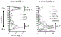


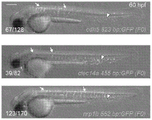


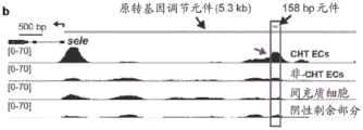


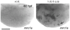


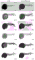


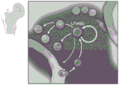


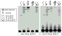


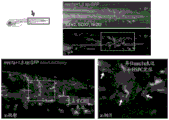


Classifications
-
- C—CHEMISTRY; METALLURGY
- C12—BIOCHEMISTRY; BEER; SPIRITS; WINE; VINEGAR; MICROBIOLOGY; ENZYMOLOGY; MUTATION OR GENETIC ENGINEERING
- C12N—MICROORGANISMS OR ENZYMES; COMPOSITIONS THEREOF; PROPAGATING, PRESERVING, OR MAINTAINING MICROORGANISMS; MUTATION OR GENETIC ENGINEERING; CULTURE MEDIA
- C12N5/00—Undifferentiated human, animal or plant cells, e.g. cell lines; Tissues; Cultivation or maintenance thereof; Culture media therefor
- C12N5/06—Animal cells or tissues; Human cells or tissues
- C12N5/0602—Vertebrate cells
- C12N5/069—Vascular Endothelial cells
-
- C—CHEMISTRY; METALLURGY
- C12—BIOCHEMISTRY; BEER; SPIRITS; WINE; VINEGAR; MICROBIOLOGY; ENZYMOLOGY; MUTATION OR GENETIC ENGINEERING
- C12N—MICROORGANISMS OR ENZYMES; COMPOSITIONS THEREOF; PROPAGATING, PRESERVING, OR MAINTAINING MICROORGANISMS; MUTATION OR GENETIC ENGINEERING; CULTURE MEDIA
- C12N5/00—Undifferentiated human, animal or plant cells, e.g. cell lines; Tissues; Cultivation or maintenance thereof; Culture media therefor
- C12N5/06—Animal cells or tissues; Human cells or tissues
- C12N5/0602—Vertebrate cells
- C12N5/0634—Cells from the blood or the immune system
- C12N5/0647—Haematopoietic stem cells; Uncommitted or multipotent progenitors
-
- A—HUMAN NECESSITIES
- A61—MEDICAL OR VETERINARY SCIENCE; HYGIENE
- A61K—PREPARATIONS FOR MEDICAL, DENTAL OR TOILETRY PURPOSES
- A61K35/00—Medicinal preparations containing materials or reaction products thereof with undetermined constitution
- A61K35/12—Materials from mammals; Compositions comprising non-specified tissues or cells; Compositions comprising non-embryonic stem cells; Genetically modified cells
- A61K35/28—Bone marrow; Haematopoietic stem cells; Mesenchymal stem cells of any origin, e.g. adipose-derived stem cells
-
- A—HUMAN NECESSITIES
- A61—MEDICAL OR VETERINARY SCIENCE; HYGIENE
- A61K—PREPARATIONS FOR MEDICAL, DENTAL OR TOILETRY PURPOSES
- A61K35/00—Medicinal preparations containing materials or reaction products thereof with undetermined constitution
- A61K35/12—Materials from mammals; Compositions comprising non-specified tissues or cells; Compositions comprising non-embryonic stem cells; Genetically modified cells
- A61K35/44—Vessels; Vascular smooth muscle cells; Endothelial cells; Endothelial progenitor cells
-
- C—CHEMISTRY; METALLURGY
- C12—BIOCHEMISTRY; BEER; SPIRITS; WINE; VINEGAR; MICROBIOLOGY; ENZYMOLOGY; MUTATION OR GENETIC ENGINEERING
- C12N—MICROORGANISMS OR ENZYMES; COMPOSITIONS THEREOF; PROPAGATING, PRESERVING, OR MAINTAINING MICROORGANISMS; MUTATION OR GENETIC ENGINEERING; CULTURE MEDIA
- C12N15/00—Mutation or genetic engineering; DNA or RNA concerning genetic engineering, vectors, e.g. plasmids, or their isolation, preparation or purification; Use of hosts therefor
- C12N15/09—Recombinant DNA-technology
- C12N15/87—Introduction of foreign genetic material using processes not otherwise provided for, e.g. co-transformation
- C12N15/90—Stable introduction of foreign DNA into chromosome
- C12N15/902—Stable introduction of foreign DNA into chromosome using homologous recombination
- C12N15/907—Stable introduction of foreign DNA into chromosome using homologous recombination in mammalian cells
-
- C—CHEMISTRY; METALLURGY
- C12—BIOCHEMISTRY; BEER; SPIRITS; WINE; VINEGAR; MICROBIOLOGY; ENZYMOLOGY; MUTATION OR GENETIC ENGINEERING
- C12N—MICROORGANISMS OR ENZYMES; COMPOSITIONS THEREOF; PROPAGATING, PRESERVING, OR MAINTAINING MICROORGANISMS; MUTATION OR GENETIC ENGINEERING; CULTURE MEDIA
- C12N5/00—Undifferentiated human, animal or plant cells, e.g. cell lines; Tissues; Cultivation or maintenance thereof; Culture media therefor
- C12N5/10—Cells modified by introduction of foreign genetic material
-
- C—CHEMISTRY; METALLURGY
- C12—BIOCHEMISTRY; BEER; SPIRITS; WINE; VINEGAR; MICROBIOLOGY; ENZYMOLOGY; MUTATION OR GENETIC ENGINEERING
- C12N—MICROORGANISMS OR ENZYMES; COMPOSITIONS THEREOF; PROPAGATING, PRESERVING, OR MAINTAINING MICROORGANISMS; MUTATION OR GENETIC ENGINEERING; CULTURE MEDIA
- C12N2501/00—Active agents used in cell culture processes, e.g. differentation
- C12N2501/30—Hormones
- C12N2501/38—Hormones with nuclear receptors
-
- C—CHEMISTRY; METALLURGY
- C12—BIOCHEMISTRY; BEER; SPIRITS; WINE; VINEGAR; MICROBIOLOGY; ENZYMOLOGY; MUTATION OR GENETIC ENGINEERING
- C12N—MICROORGANISMS OR ENZYMES; COMPOSITIONS THEREOF; PROPAGATING, PRESERVING, OR MAINTAINING MICROORGANISMS; MUTATION OR GENETIC ENGINEERING; CULTURE MEDIA
- C12N2501/00—Active agents used in cell culture processes, e.g. differentation
- C12N2501/60—Transcription factors
- C12N2501/602—Sox-2
-
- C—CHEMISTRY; METALLURGY
- C12—BIOCHEMISTRY; BEER; SPIRITS; WINE; VINEGAR; MICROBIOLOGY; ENZYMOLOGY; MUTATION OR GENETIC ENGINEERING
- C12N—MICROORGANISMS OR ENZYMES; COMPOSITIONS THEREOF; PROPAGATING, PRESERVING, OR MAINTAINING MICROORGANISMS; MUTATION OR GENETIC ENGINEERING; CULTURE MEDIA
- C12N2502/00—Coculture with; Conditioned medium produced by
- C12N2502/11—Coculture with; Conditioned medium produced by blood or immune system cells
- C12N2502/1171—Haematopoietic stem cells
-
- C—CHEMISTRY; METALLURGY
- C12—BIOCHEMISTRY; BEER; SPIRITS; WINE; VINEGAR; MICROBIOLOGY; ENZYMOLOGY; MUTATION OR GENETIC ENGINEERING
- C12N—MICROORGANISMS OR ENZYMES; COMPOSITIONS THEREOF; PROPAGATING, PRESERVING, OR MAINTAINING MICROORGANISMS; MUTATION OR GENETIC ENGINEERING; CULTURE MEDIA
- C12N2502/00—Coculture with; Conditioned medium produced by
- C12N2502/28—Vascular endothelial cells
-
- C—CHEMISTRY; METALLURGY
- C12—BIOCHEMISTRY; BEER; SPIRITS; WINE; VINEGAR; MICROBIOLOGY; ENZYMOLOGY; MUTATION OR GENETIC ENGINEERING
- C12N—MICROORGANISMS OR ENZYMES; COMPOSITIONS THEREOF; PROPAGATING, PRESERVING, OR MAINTAINING MICROORGANISMS; MUTATION OR GENETIC ENGINEERING; CULTURE MEDIA
- C12N2510/00—Genetically modified cells
Landscapes
- Health & Medical Sciences (AREA)
- Life Sciences & Earth Sciences (AREA)
- Engineering & Computer Science (AREA)
- Genetics & Genomics (AREA)
- Biomedical Technology (AREA)
- Biotechnology (AREA)
- Chemical & Material Sciences (AREA)
- Zoology (AREA)
- Wood Science & Technology (AREA)
- Bioinformatics & Cheminformatics (AREA)
- Organic Chemistry (AREA)
- Cell Biology (AREA)
- General Health & Medical Sciences (AREA)
- General Engineering & Computer Science (AREA)
- Microbiology (AREA)
- Biochemistry (AREA)
- Molecular Biology (AREA)
- Developmental Biology & Embryology (AREA)
- Immunology (AREA)
- Vascular Medicine (AREA)
- Hematology (AREA)
- Medicinal Chemistry (AREA)
- Epidemiology (AREA)
- Plant Pathology (AREA)
- Physics & Mathematics (AREA)
- Virology (AREA)
- Mycology (AREA)
- Pharmacology & Pharmacy (AREA)
- Biophysics (AREA)
- Animal Behavior & Ethology (AREA)
- Public Health (AREA)
- Veterinary Medicine (AREA)
- Medicines That Contain Protein Lipid Enzymes And Other Medicines (AREA)
- Micro-Organisms Or Cultivation Processes Thereof (AREA)
- Medicines Containing Material From Animals Or Micro-Organisms (AREA)
Abstract
The technology described herein relates to compositions and methods for generating endothelial niche cells. Embodiments of the technology described herein include compositions, kits, vectors, and methods related to the generation or engineering of endothelial niche cells. One aspect includes a method of generating/engineering an endothelial niche cell comprising expressing one or more transcription factors in an endothelial cell, wherein the one or more transcription factors are from the Ets family, the Sox family, and/or the Nuclear Hormone (NHR) family.
Description
Cross Reference to Related Applications
Priority of U.S. provisional application No. 62/647,433 filed 2018, 3, 23, 35 u.s.c § 119(e), the contents of which are incorporated herein in their entirety.
Government support
The invention was made with government support under grant numbers DK111790 and HL048801 awarded by the national institutes of health. The government has certain rights in the invention.
Sequence listing
The present application contains a sequence listing submitted in ASCII format through EFS-Web and is incorporated herein in its entirety. The ASCII copy created on day 22 of month 3 2019 is named 701039-.
Technical Field
The technology described herein relates to compositions and methods for generating endothelial niche cells.
Background
Hematopoietic Stem and Progenitor Cells (HSPCs) are rare populations of cells that are capable of reconstituting the entire blood system following transplantation. As a functional unit of bone marrow transplants, these cells provide an effective treatment for many blood and immune diseases. Unfortunately, transplantation is not a viable treatment option for many individuals, particularly those who lack an immune-matched donor. A long-term goal of hematological studies has been to culture and expand HSPCs for transplantation and/or genetic modification in vitro. Although cord blood-derived HSPCs are somewhat adaptable in vitro amplification, maintaining and inducing self-renewal of adult-derived HSPCs in the absence of a niche signal has proven challenging.
In an attempt to outline the microenvironment or "niche" that supports HSPCs in vivo, strategies for in vitro expansion have HSPCs co-cultured with supporting cells. In adult bone marrow, a variety of cell types are thought to include the HSPC niche, with the major contributors being Endothelial Cells (ECs) and perivascular mesenchymal stromal cells. Different endothelial cell subtypes in the bone marrow may differentially regulate HSPC homeostasis. Arterial ec (aec) is less permeable and is thought to promote HSPC quiescence, while sinus ec (sec) is leaky and supports HSPC differentiation and mobilization. Furthermore, during hematopoietic recovery following myelosuppression, EC plays a key role in niche reconstruction and reconstitution of multilineage hematopoiesis. HSPCs may also be supported extramedullary during embryonic development and under stress conditions that induce extramedullary hematopoiesis in tissues such as the liver, spleen and skull. As in the bone marrow, EC is thought to function as a key core component of the HSPC niche in these tissues.
Researchers have focused on the development of in vitro cultures in which HSPCs can be grown in the laboratory with other cell types that support the maintenance or expansion of HSPCs for subsequent use in transplantation. However, to date, these in vitro cultures have been only moderately successful.
SUMMARY
In the study of the vascular HSPC niche of zebrafish embryos, combinations of transcription factors (from Ets, Sox and nuclear hormone families) normally expressed in endogenous niche endothelial cells were studied. When these human orthologs of the same transcription factor are expressed ectopically, an ectopic vascular niche is created in zebrafish embryos where HSPCs are recruited and maintained.
As a step in transforming these findings into clinical use, transcription factors, originally identified in zebrafish studies, can be expressed in human endothelial cells to reprogram these cells into HSPC niche-like identities. These niche endothelial cells can be used in co-culture with HSPCs to expand HSPC numbers or to extend culture time for subsequent transplantation.
For example, transcription factors that bind to Ets, Sox, and nuclear hormone motifs are known to be expressed in niche endothelial cells. In the Ets family, these factors include etv2, fli1a, and Ets 1; wherein the corresponding human factors are ETV2, FLI1 and ETS 1. In the Sox family, these transcription factors include Sox18 and Sox 7; wherein the corresponding human factors are SOX7 and SOX 18. In the nuclear hormone family, these transcription factors include rxraa and nr2f 2; wherein the corresponding human factors are RXRA and NR2R 2.
The present invention provides methods for preparing synthetic niche endothelial cells for stimulation of blood stem cells. Transcription factors include the Ets family: etv2, fli1a, ets 1; the SOX family: sox18, sox7, and the nuclear hormone family: rxraa, nr2f2 and corresponding human factors: ETV2, FLI1, ETS1, SOX7, SOX18, RXRA, and NR2F 2.
The methods include expressing transcription factors in endothelial cells (e.g., humans) to reprogram these cells to HSPC niche-like identities.
In another embodiment, niche endothelial cells are used in co-culture with HSPCs for subsequent use in transplantation in order to expand HSPC numbers or extend culture time.
One aspect provides a method of generating/engineering an endothelial niche cell comprising expressing one or more transcription factors in an endothelial cell, wherein the one or more transcription factors are from the Ets family, the Sox family, and/or the nuclear hormone receptor family.
Another aspect provides an engineered endothelial niche cell comprising one or more exogenous nucleic acid sequences encoding one or more transcription factors, wherein the one or more transcription factors are from the Ets family, the Sox family, and/or the nuclear hormone family.
Another aspect provides compositions comprising an engineered endothelial niche described herein.
Another aspect provides a method of culturing HSPCs comprising culturing HSPCs in the presence of an engineered endothelial niche cell population.
Another aspect provides a method of treating a subject, the method comprising transplanting a composition comprising a HSPC and an engineered endothelial niche cell population into the subject.
Another aspect provides a method of enhancing HSPC engraftment comprising administering to a subject in need thereof a composition comprising HSPC and an engineered endothelial niche cell population.
Another aspect provides co-cultures comprising an engineered endothelial niche cell and a HSPC.
Another aspect provides a kit for culturing HSPCs, the kit comprising: engineered endothelial niche cell populations, reagents and instructions for their use.
Another aspect provides a kit for producing an engineered endothelial niche cell comprising a vector comprising one or more exogenous nucleic acid sequences encoding one or more transcription factors of the Ets family, the Sox family, or the nuclear hormone family and instructions for use thereof.
Another aspect provides a method of producing an ectopic vascular niche comprising administering an engineered endothelial niche cell to a target site of a subject in need thereof.
Another aspect provides a method for extramedullary hematopoiesis comprising transplanting an engineered niche endothelial cell into a subject at a location outside of the bone marrow (e.g., the forearm), thereby creating a synthetic niche.
In another aspect, vectors are provided comprising one or more exogenous nucleic acid sequences encoding one or more transcription factors of the Ets family, the Sox family, or the nuclear hormone family operably linked to a promoter.
Brief description of the drawings
Figures 1A-1E are a series of images and graphs showing endothelial expression characteristics in the fetal HSPC niche. (FIG. 1A) schematic illustration of hematopoietic tissue of zebrafish embryos (top) and sectioning strategy for RNA tomography (tomo-seq) on CHT (bottom; showing double transgenic embryos carrying HSPC markers cd41: GFP and runx1: mCherry). (FIG. 1B) schematic cross-section and (FIG. 1B sequential) hierarchical clustering heatmaps revealed clusters of gene expression corresponding to different tissues along the dorsal-ventral axis of zebrafish tail. (FIG. 1C) schematic depicts the strategy of isolating EC from whole embryos for analysis by RNA-seq using kdr1: GFP transgenic embryos and FACS. (FIG. 1D) shows individual tomo-seq expression traces of the pan-endothelial expressed gene (left) and the CHT EC enriched gene (right). (FIG. 1E) image shows global in situ hybridization (WISH) of pan-endothelial gene kdr1 (top) and CHT EC-enriched gene identified by Tomo-seq and tissue-specific RNA-seq. Arrows point to expressions in the dorsal vasculature and wedge symbols point to expressions in the CHT. Unless otherwise stated, the scale bar represents 250 μm.
Figures 2A-2C are a series of images and graphs showing endothelial niche-specific cis regulatory elements. (FIG. 2A) images and schematic representations depict four cell populations isolated from mrc1a: GFP, kdr1: mCherry double positive embryos for ATAC-seq analysis. (FIG. 2B) Gene traces are shown at mCherry+;GFP+The only open chromatin region in the CHT EC fraction (boxes and arrows). (FIG. 2C) image shows embryos injected with CHT EC enhancer-GFP reporter construct corresponding to boxed region in FIG. 2B. The wedge symbols point to GFP expression in CHT ECs. Unless otherwise stated, the scale bar represents 250 μm.
Figures 3A-3F are a series of images and graphs showing that Ets, Sox and NHR binding sites are necessary for selective expression in niche EC. (FIG. 3A) Gene traces showed a chromatin region upstream of mrc1a (boxes and arrows below) that was only open in the double positive CHT EC fraction, but not in the other three cell populations. The bars indicate the positions of the 125bp enhancer sequence and the 1.3kb sequence used to generate the mrc1a GFP reporter transgene. (FIG. 3B) image shows transient GFP expression in F0 embryos injected with a 125bp enhancer sequence linked to a minimal promoter and GFP. (FIG. 3C) image shows F0 embryos injected simultaneously with kdr1: mCherry and mrc1a 125bp: GFP plasmid. (FIG. 3D) images show embryos expressing stably integrated mrc1a 125bp: GFP transgene. (FIG. 3E) shows the wild-type sequence of the 125bp src 1a enhancer (see, e.g., SEQ ID NO:12), with annotations highlighting the Ets, Sox and NHR binding motifs. Schematic diagrams depict enhancer-reporter constructs in which each type of motif or control region is targeted by a mutation. X represents the position of the target site. mp-GFP: mouse beta-globin minimal promoter fused to GFP. (FIG. 3F) panel reports the frequency of embryos with GFP expression in CHT EC after injection of mutant variants of the wild type sequence or mrc1a 125bp (top) or sele158bp (bottom) enhancer. Data were normalized to the corresponding wild type control for each experiment (44% for the mrc1a 125bp enhancer (155/356) and 23% for the sel 158bp enhancer (176/775)). Mean +/-standard error of mean (s.e.m.), one-way ANOVA; p <0.01, P < 0.001. Unless otherwise stated, the scale bar represents 250 μm.
FIGS. 4A-4F are a series of images and graphs showing that overexpression of defined factors induces ectopic vascular gene expression outside of CHT. (FIG. 4A) schematic representation of the strategy used in transcription factor overexpression experiments. (FIG. 4B) images show embryos injected (right) with control DNA (left) or a pool of 7 transcription factors from the Ets, Sox and NHR families (FLI1, ETV2, ETS1, SOX7, SOX18, Nr2f2 and RXRA) and then stained by WISH against mrc1a (top) or sele (bottom). The arrows indicate regions of ectopic expression, and the wedge symbols point to the normal expression domains in all of FIGS. 4A-4E. (FIG. 4C) image shows mrc1a: GFP injected with control DNA or 7-factor combinations; kdr1 mCherry double transgenic embryos. (FIG. 4D) injection of 3-factor combinations containing ETV2, SOX7 and Nr2f2 resulted in ectopic expression of mrc1a GFP (arrows). (FIG. 4E) image shows WISH against mrc1a in control embryos (top), or after injection of 3-factor sets containing ETV2, SOX7 and Nr2f2 (middle) or ETS1, SOX7 and Nr2f2 (bottom). (FIG. 4F) graph reports quantification of the percentage of injected embryos showing ectopic mrc1a expression after transcription factor overexpression. Checking a chi square; p <0.01, P < 0.001. The scale bar in fig. 4B and 4E represents 250 μm, and the scale bar in fig. 4C and 4D represents 100 μm.
Figures 5A-5E are a series of images and graphs showing that HSPCs are localized to ectopic niche endothelial gene expression regions. (FIG. 5A) the image shows dorsal ectopy mrc1a: runx1: mCherry within the GFP expression region located outside the CHT in embryos injected with the pool of ETV2, SOX7 and Nr2f2 (top) or the pool of ETS1, SOX7 and Nr2f2 (bottom) located outside the CHT+HSPC. The inset with gray scale image magnification for each channel is shown on the right. The arrows point to ectopic expressions or localizations, while the wedge symbols point to normal expressions or localizations in all of figures 5A-5E. (FIG. 5B) WISH against runx1 shows the localization of HSPC in control (top) and factor 3 injected embryos (bottom). (FIG. 5C) ectopic expression mrc1a EC and cxcl12a of GFP and DsRed2+Stromal cell-related, similar to EC in CHT. Asterisks indicate chordal expression of cxcl12a: DsRed 2. (FIG. 5D) time delay series shows runx1: mCherry+HSPCs initially reach CHT and subsequently divide. Time is expressed as hh mm. (FIG. 5E) toTime-delayed series from embryos different from that in FIG. 5D show runx1: mCherry+HSPCs divide and migrate into the circulation. The scale bar represents 100 μm in fig. 5A to 5C, and the scale bar represents 30 μm in fig. 5D to 5E.
Figures 6A-6B are a series of images and graphs showing conserved endothelial expression features in the HSPC niche. (FIG. 6A) heatmap shows the expression of 29 CHT EC genes in different cell populations including adult zebrafish kidney bone marrow. The spectral scale shows normalized expression from 0 (low) to 1 (high). (FIG. 6B) heatmap shows expression of orthologs of the CHT EC gene in EC from different developmental stages and different organs of mice transformed into adulthood after birth. Arrows indicate hematopoietic tissues at the respective developmental stages. Black brackets indicate genes that were enriched in fetal liver EC at stage E14-17, then later in adult bone marrow. The spectral scale reports the z-score. BM: bone marrow.
Figures 7A-7B are a series of images and graphs showing RNA tomography and niche-specific endothelial gene expression. (FIG. 7A) is a graph showing a trace of tomo-seq expression of each tissue-specific gene. Images showing global in situ hybridization (WISH) of 35 CHT-enriched genes were available on zfin. (FIG. 7B) WISH demonstrated CHT-enriched expression of CHT EC genes identified using a combination of tomo-seq and tissue-specific RNA-seq (wedge symbols). Unless otherwise stated, the scale bar represents 250 μm.
Figures 8A-8E are a series of images and graphs showing EC in the GFP reporter transgene selectable marker HSPC niche. (FIG. 8A) image shows double transgenic embryos carrying the pan-endothelial marker kdr1: mCherry and mrc1a: GFP transgene which is selectively expressed in CHT EC. An enlarged view of the boxed area is shown on the right. (FIG. 8B) image display runx1: mCherry+Mrc1a in the HSPC and CHT niches for GFP+EC interact directly (arrow). The middle panel shows an enlarged view of the boxed area. Additional magnification (bottom) shows mrc1a GFP+HSPC in EC pocket. (FIG. 8C) cxcl12a: DsRed2+Stromal cells with mrc1a GFP in CHT+EC are closely related. (FIG. 8D) image shows the transgene carrying the pan-endothelial marker kdr1: mCherry and sel: GFP, which is selectively expressed in CHT ECDouble transgenic embryos. An enlarged view of the boxed area is shown on the right. (FIG. 8E) image showing runx1: select: GFP in the mCherry + HSPC and CHT niches+EC interact directly (arrow). An enlarged view of the boxed area is shown on the right. The scale bar in fig. 8A and 8D represents 250 μm, and the scale bar in fig. 8B, 8C, and 8E represents 100 μm.
FIGS. 9A-9C are a series of images and graphs showing pan-endothelial regulatory element and whole genome motif enrichment assays. (FIG. 9A) Gene traces are shown at mCherry+GFP+(CHT EC) and mCherry+GFP-Areas of chromatin that were all open in the (non-CHT EC) population (boxes and straight arrows). (FIG. 9B) image shows embryos injected with pan endothelial enhancer-GFP reporter construct corresponding to the box region in FIG. 9A. Arrows point to GFP expression in non-CHT ECs and wedge symbols point to expression in CHT ECs. (FIG. 9C) image shows the most enriched transcription factor binding motif within the CHT EC region (top) or pan-endothelial region (bottom). Unless otherwise stated, the scale bar represents 250 μm.
FIGS. 10A-10E are a series of images and graphs showing CHT endothelial cis regulatory elements. (FIG. 10A) the map reports the anatomical location of endothelial expression in F0 embryos injected with mrc1A 125bp: GFP and kdr1: mCherry plasmid. (FIG. 10B) Gene traces show the only chromatin region upstream of sel that was open in the double positive CHT EC fraction, while the other three cell populations were not (boxes and arrows below). The bars indicate the positions of the 158bp enhancer sequence and the 5.3kb sequence used to generate the sel: GFP reporter transgene. (FIG. 10C) image shows the transient F0 (top) and stable F2 expression (bottom) of sel 158bp GFP construct. (FIG. 10D) shows the wild-type sequence of the 158bp sel enhancer (see, e.g., SEQ ID NO:13), with annotations highlighting the Ets, Sox and NHR binding motifs (top). The schematic depicts sequence variants in which each type of motif or control region is targeted by mutation. X represents the position of the target site. mp-GFP: mouse beta-globin minimal promoter fused to GFP. (FIG. 10E) image shows electrophoretic mobility shift assay with recombinant Nr2f2-GST incubated with DNA sequences spanning the NHR motif present in mrc1a (left two gels) of 125bp or the sel (right gel) enhancer sequence of 158 bp. Arrows point to DNA: protein binding, while the wedge symbols point to hypermigrated DNA: a protein complex. Labeled DNA: the protein complex was overtaken by the unlabeled wild-type probe (lane 4) but not by the probe in which the NHR motif was disrupted by mutation (arrow with asterisk). Unless otherwise stated, the scale bar represents 250 μm.
FIGS. 11A-11C are a series of images and graphs showing the procedure for induction of ectopic CHT endothelial cells by transcription factor overexpression. (FIG. 11A) the image shows WISH against mrc1A on the yolk sphere in control (left) and 7-factor injected embryos (right). Arrows point to ectopic expression and wedge symbols point to the normal expression domains in all of FIGS. 11A-11C. (FIG. 11B) image shows the ectopic expression of mrc1a: GFP and kdr1: mCherry transgenes on the yolk extension of 7-factor injected embryos. An enlarged view of the boxed area is shown at the bottom. (FIG. 11C) the image shows WISH for sel, gpr182 and lgmn in control embryos (left) and embryos injected with a combination of ETV2, SOX7 and Nr2f2 (right). The scale bar in FIGS. 11A-11C represents 100 μm.
Figures 12A-12D are a series of images and graphs showing that CHT niche endothelial gene expression is ectopically induced by transcription factor overexpression. (FIGS. 12A-12B) injection of ETV2 alone induced ectopic expression of both the endogenous mrc1a gene (FIG. 12A) and mrc1a GFP transgene (FIG. 12B). Arrows point to ectopic expression, black wedge symbols point to the normal expression domains of all of FIGS. 12A-12D. (FIG. 12C) injection of human ETV2 alone induced ectopic expression of zebrafish transcription factors including sox7, sox18, fli1a and ETV 2. (FIG. 12D) injection of 3-factor combinations containing ETS1, SOX7, and Nr2f2 resulted in ectopic expression of mrc1, 1a: GFP. The scale bar indicates 250 μm in fig. 12A and 12C, and the scale bar indicates 50 μm in fig. 12B and 12D.
Figures 13A-13B are a series of images and graphs showing niche endothelial transgene expression in adult kidney ECs. (FIG. 13A) image shows a segment of vasculature dissected from kidney of kdrl: mCherry, mrc1a: GFP double transgenic adult zebrafish (white arrow). (FIG. 13B) images show serial sections through adult kidney isolated from sel: GFP transgenic fish. Slicing H&E (left)) And antibody to GFP (right). Black arrow points to GFP+Vascular endothelial cells. The scale bar in FIG. 13A represents 50 μm, and the scale bar in FIG. 13B represents 100 μm.
FIG. 14 is a schematic diagram showing the self-renewal and differentiation of hematopoietic stem cells. LT-HSC represent long-term hematopoietic stem cells. CMP represents a common bone marrow progenitor cell. MEP means megakaryocyte-erythroid progenitor cells. GMP stands for granulocyte-macrophage progenitor cells. CLP represents a common lymphoid progenitor.
Figures 15A-15B are a series of images showing visualization of niche colonization by HSPC in vivo. Fig. 15A shows visualization of the dorsal aorta and caudal hematopoietic tissues. Figure 15B shows HSPCs surrounded by 5 endothelial cells and attached to stromal cells.
FIG. 16 is a series of images showing the detection of gene expression in the HSPC niche using RNA tomography (tomo-seq).
FIG. 17 is a series of images and graphs showing how Tomo-seq + endothelial RNA-seq identifies about 20 genes selectively enriched in niche endothelial cells.
Figure 18 is a series of images showing that sel and mrc1a promoter-GFP fusions label endothelial cells in the HSPC niche.
Figure 19 is a series of images showing that specific ATAC-seq peaks near the original 20 genes can drive expression in niche endothelial cells. 13 of the 19 cloned ATAC-seq peaks can drive GFP expression in CHT endothelial cells (when linked to a minimal promoter).
Figure 20 is an image showing the most enriched TF binding motif in the open chromatin of niche endothelial cells. The motif enrichment analysis was performed on 6,710 unique ATAC-seq peaks using HOMER.
FIG. 21 is a series of images and graphs showing that the Ets, Sox and NHR sites are essential for niche expression of the 158bp sel enhancer (see, e.g., SEQ ID NO: 14).
FIG. 22 is a series of images and graphs showing that the Ets, Sox and NHR motifs are essential for niche expression of the 125bp mrc1a enhancer (see, e.g., SEQ ID NO: 15).
Figure 23 is a series of images showing that mouse TF can bind zebrafish sequences in vitro. Disruption of enhancers the same mutation in GFP expression abolished TF binding.
FIG. 24 is an image showing that transcription factors known to bind the Ets, Sox and NH motifs are expressed in niche endothelial cells.
Figure 25 is a series of images showing reprogramming of niche endothelial cells.
Figure 26 is a series of images and graphs showing that 3-TF pool injection results in ectopic niche endothelial gene expression.
FIGS. 27A-27C are schematic diagrams showing that ectopic vascular patch can recruit runx1+A series of images of HSPCs.
Figure 28 is a series of images and heat maps showing the endothelial characteristics of the HSPC niche also in adult bone marrow.
Figure 29 is a heat map showing similar niche endothelial features found in mammalian fetal liver and bone marrow.
Figure 30 is a graph showing that RNA tomography of the HSPC niche yields the following data: about 20 genes were selectively enriched in HSPC niche endothelial cells; ectopic expression of only 3 TFs can induce niche endothelial gene expression and recruit HSPCs; ets, Sox and nuclear hormone motifs are required for expression in niche endothelial cells.
Detailed Description
Embodiments of the technology described herein include compositions, kits, vectors, and methods related to the generation or engineering of endothelial niche cells. One aspect includes a method of generating/engineering an endothelial niche cell comprising expressing one or more transcription factors in an endothelial cell, wherein the one or more transcription factors are from the Ets family, the Sox family, and/or the Nuclear Hormone Receptor (NHR) family.
In some embodiments, the at least one transcription factor is selected from the Ets family. In some embodiments, the at least one transcription factor is selected from the Sox family. In some embodiments, the at least one transcription factor is selected from the NHR family.
In some embodiments, the at least one transcription factor is selected from the Ets family and the at least one transcription factor is selected from the Sox family. In some embodiments, the at least one transcription factor is selected from the Ets family and the at least one transcription factor is selected from the NHR family. In some embodiments, the at least one transcription factor is selected from the Sox family and the at least one transcription factor is selected from the NHR family.
In some embodiments, the at least one transcription factor is selected from the Ets family, the at least one transcription factor is selected from the Sox family, and the at least one transcription factor is selected from the NHR family. In some embodiments, the at least one transcription factor is selected from the Ets family, the at least one transcription factor is selected from the Sox family, or the at least one transcription factor is selected from the NHR family.
Ets family
In some embodiments of either aspect, the endothelial niche cell expresses a transcription factor from the ETS family. The ETS (E26 transformation specific or E-26) family is one of the largest transcription factor families and is animal-specific. The ETS family is divided into 12 subfamilies: 1) ELF (e.g., ELF1, ELF2/NERF, ELF 4/MEF); 2) ELGs (e.g., GABP α, ELG); 3) ERG (e.g., ERG, FL1, FEV); 4) ERF (e.g., ERF/PE2, ETV3/PE 1); 5) ESEs (e.g., ELF3/ESE1/ESX, ELF5/ESE2, ESE 3/EHF); 6) ETS (e.g., ETS1, ETS, POINTED); 7) PDEF (e.g., SPDEF/PDEF/PSE); 8) PEA3 (e.g., ETV4/PEA3/E1AF, ETV5/ERM, ETV1/ER 81); 9) ERF71 (e.g., ETV2/ER 71); 10) SPI (e.g., SPI1/PU.1, SPIB, SPIC); 11) TCFs (e.g., ELK1, ELK4/SAP1, ELK3/NET/SAP2, LIN); 12) TEL (e.g., ETV6/TEL, ETV7/TEL2, YAN).
All ETS family members are identified by a highly conserved DNA binding domain, the ETS domain, which is a winged helix-turn-helix structure capable of binding to a DNA site with a central GGA (a/T) DNA sequence. The Ets family of DNA motifs may also comprise a central TTCCT sequence (e.g., on the DNA strand complementary to the first motif). In addition to DNA binding function, evidence suggests that ETS domains are also involved in protein-protein interactions.
The ETS family is present throughout the body and is involved in a variety of functions, including regulation of cell differentiation, cell cycle control, cell migration, cell proliferation, apoptosis (programmed cell death), and angiogenesis.
Non-limiting examples of human Ets family members associated with endothelial niche cells include ETV2, FLI1, and Ets 1. The corresponding factors in zebrafish include etv2, fli1 and ets 1.
ETV2 may also be referred to herein as ETS variant 2, ETS translocation variant 2, ETS-associated protein 71, ETS variant gene 2, ETSRP71, or ER 71.
Friend leukemia integrates a1 transcription factor (FLI1), also known as transcription factor ERGB, a protein encoded by the FLI1 gene in humans. FLI1 may also be referred to herein as Fli-1 protooncogene, ETS transcription factor, friend leukemia integration 1 transcription factor, friend leukemia Virus integration 1, transcription factor ERGB, Ewing's sarcoma breakpoint region, protooncogene Fli-1, BDPLT21, EWSR2, or SIC-1.
ETS1 or protein C-ETS-1 is a protein encoded by the ETS1 gene in humans. The protein encoded by the gene belongs to the ETS transcription factor family. ETS1 may also be referred to herein as ETS proto-oncogene 1 transcription factor, avian erythroblastosis virus E26(V-ETS) oncogene homolog-1, V-Ets avian erythroblastosis virus E26 oncogene homolog 1, protein C-Ets-1, EWSR2, P54, V-Ets avian erythroblastosis virus E2 oncogene homolog 1, Ets protein, C-Ets-1, or ETS-1.
In some embodiments of any aspect, the cell is produced or engineered to express an ETS family member selected from ETV2, FLI1, and ETS 1.
In some embodiments, the Ets gene or protein may be ETV2 or corresponding zebrafish ETV 2. In some embodiments, the Ets gene or protein may be FLI1 or the corresponding zebrafish FLI 1. In some embodiments, the Ets gene or protein may be Ets1 or the corresponding zebrafish Ets 1.
In some embodiments, the Ets gene or protein may be ETV2 and FLI1 or corresponding zebrafish factor. In some embodiments, the Ets gene or protein may be ETV2 and Ets1 or the corresponding zebrafish factor. In some embodiments, the Ets gene or protein may be Ets1 and FLI1 or corresponding zebrafish factor. In some embodiments, the Ets gene or protein may be ETV2, FLI1, and Ets1 or the corresponding zebrafish factor. In some embodiments, the Ets gene or protein may be ETV2, FLI1, or Ets1, or the corresponding zebrafish factor.
The amino acid sequences of the polypeptides described herein have been assigned NCBI accession numbers for different species such as human, mouse, rat and zebrafish. Specifically, NCBI accession numbers for non-limiting examples of the amino acid sequences of human ETV2 (e.g., SEQ ID NO:1), human FLI1 (e.g., SEQ ID NO:2), and human ETS1 (e.g., SEQ ID NO:3) are included herein.
1 (homo sapiens ETV2, NCBI accession No. AAI40747, 342 amino acids (aa):
MDLWNWDEASPQEVPPGNKLAGLEGAKLGFCFPDLALQGDTPT ATAETCWKGTSSSLASFPQLDWGSALLHPEVPWGAEPDSQALPWSGD WTDMACTAWDSWSGASQTLGPAPLGPIPAAGSEGAAGQNCVPVAGE ATSWSRAQAAGSNTSWDCSVGPDGDTYWGSGLGGEPRTDCTISWGG PAGPDCTTSWNPGLHAGGTTSLKRYQSSALTVCSEPSPQSDRASLARC PKTNHRGPIQLWQFLLELLHDGARSSCIRWTGNSREFQLCDPKEVARL WGERKPGMNYEKLSRGLRYYYYRRDIVRKSGGRKYTYRFGGRVPSL AYPDCAGGGRGAETQ
2 (homo sapiens FLI1, NCBI accession No. AAH10115.1, 452aa):
MDGTIKEALSVVSDDQSLFDSAYGAAAHLPKADMTASGSPDYG QPHKINPLPPQQEWINQPVRVNVKREYDHMNGSRESPVDCSVSKCSK LVGGGESNPMNYNSYMDEKNGPPPPNMTTNERRVIVPADPTLWTQEH VRQWLEWAIKEYSLMEIDTSFFQNMDGKELMNKEDFLRATTLYNTEV LLSHLSYLRESSLLAYNTTSHTDQSSRLSVKEDPSYDSVRRGAWGNNN MNSGLNKSPPLGGAQTISKNTEQRPQPDPYQILGPTSSRLANPGSGQIQ LWQFLLELLSDSANASCITWEGTNGEFKMTDPDEVARRWGERKSKPN MNYDKLSRALRYYDKNIMTKVHGKRYAYKFDFHGIAQALQPHPTESS MYKYYYYPSDISYMPSYHAHQQKVNFVPPHPSSMPVTSSSFFGAASQ YWTSPTGGIYPNPNVPRHPNTHVPSHLGSYY
3 (homo sapiens ETS1, NCBI accession number CAG47050.1, 441aa):
MKAAVDLKPTLTIIKTEKVDLELFPSPDMECADVPLLTPSSKEMM SQALKATFSGFTKEQQRLGIPKDPRQWTETHVRDWVMWAVNEFSLK GVDFQKFCMNGAALCALGKDCFLELAPDFVGDILWEHLEILQKEDVK PYQVNGVNPAYPESSRYTSDYFISYGIEHAQCVPPSEFSEPSFITESYQT LHPISSEELLSLKYENDYPSVILRDPLQTDTLQNDYFAIKQEVVTPDNM CMGRTSRGKLGGQDSFESIESYDSCDRLTQSWSSQSSFNSLQRVPSYD SFDSEDYPAALPNHKPKGTFKDYVRDRADLNKDKPVIPAAALAGYTG SGPIQLRQFLLLTDKSCQSFISWTGDGWEFKLSDPDEVARRWGKRKNK PKMNYEKLSRGLRYYDKNIIHKTAGKRYVYRFVCDLQSLLGYTPEEL HAMLDVKPDADE
in some embodiments, the ETV2 amino acid or DNA sequence may be at least 90%, at least 91%, at least 92%, at least 93%, at least 94%, at least 95%, at least 96%, at least 97%, at least 98%, at least 99% or more identical to the native or reference sequence. In some embodiments, the FLI1 amino acid or DNA sequence may be at least 90%, at least 91%, at least 92%, at least 93%, at least 94%, at least 95%, at least 96%, at least 97%, at least 98%, at least 99% or more identical to the native or reference sequence. In some embodiments, the ETS amino acid or DNA sequence may be at least 90%, at least 91%, at least 92%, at least 93%, at least 94%, at least 95%, at least 96%, at least 97%, at least 98%, at least 99% or more identical to the native or reference sequence.
Family of SOX
SOX genes encode a family of transcription factors that bind to the minor groove in DNA and belong to a gene superfamily characterized by homologous sequences called HMG-boxes (for the high mobility group). This HMG box is a DNA binding domain that is highly conserved in eukaryotic species. Homologs have been identified in insects, nematodes, amphibians, reptiles, birds and a range of mammals.
The Sox gene is defined as an HMG box containing a gene involved in sex determination (called SRY), which is located on the Y chromosome (Sox stands for SRY-related HMG box). There are 20 SOX genes in humans and mice. This family is divided into subgroups based on homology within the HMG domain and other structural motifs as well as on functional assays. In humans, the members of the SOX group include: 1) SoxA (e.g., SRY); 2) SoxB1 (e.g., SOX1, SOX2, SOX 3); 3) SoxB2 (e.g., SOX14, SOX 21); 4) SoxC (e.g., SOX4, SOX11, SOX 12); 5) SoxD (e.g., SOX5, SOX6, SOX 13); 6) SoxE (e.g., SOX8, SOX9, SOX 10); 7) SoxF (e.g., SOX7, SOX17, SOX 18); 8) SoxG (e.g., SOX 15); 9) SoxH (e.g., SOX 30).
The developmentally important Sox family has no single function and many members have the ability to regulate several different aspects of development. Although many Sox genes are involved in sex determination, some genes are also important in processes such as neuronal development. Sox proteins bind to the sequence WWCAAW and similar sequences (W ═ a or T). The Sox family of DNA motifs may also contain a central ATTGT sequence (e.g., on the DNA strand complementary to the first motif).
Non-limiting examples of human Sox family members associated with endothelial niche cells include Sox18 and Sox 7. Corresponding factors in zebrafish or xenopus include sox18 and sox 7.
SOX18 may also be referred to herein as SRY-Box18, SRY (sex determining region Y) -Box18, transcription factor SOX-18, SRY-Box18, HLTRS or HLTS.
SOX7 may also be referred to herein as SRY-Box7, SRY (sex determining region Y) -Box7, transcription factor SOX-7, or SRY-Box 7.
In some embodiments of any aspect, the cell is produced or engineered to express a SOX family member selected from SOX18 and SOX 7. In some embodiments, the Sox gene or protein may be Sox18 or the corresponding zebrafish Sox18 or xenopus Sox 18. In some embodiments, the Ets gene or protein may be SOX7 or the corresponding zebrafish SOX 7. In some embodiments, the Sox gene or protein may be Sox18 and Sox7 or the corresponding zebrafish or xenopus factors. In some embodiments, the Sox gene or protein may be Sox18 or Sox7 or the corresponding zebrafish or xenopus factor.
The amino acid sequences of the polypeptides described herein have been assigned NCBI accession numbers for different species such as human, mouse, rat and zebrafish. In particular, NCBI accession numbers for non-limiting examples of amino acid sequences of human SOX18 (e.g., SEQ ID NO:4), human SOX7 (e.g., SEQ ID NO:5), and Xenopus SOX18 (e.g., SEQ ID NO:8) are included herein.
4 (homo sapiens SOX18, NCBI accession No. BAA94874.1, 384aa):
MQRSPPGYGAQDDPPARRDCAWAPGHGAAADTRGLAAGPAALA APAPAPASPPSPQRSPPRSPEPGRYGLSPAGRGERQAADESRIRRPMNA FMVWAKDERKRLAQQNPDLHNAVLSKMLGKAWKELNAAEKRPFVE EAERLRVQHLRDHPNYKYRPRRKKQARKARRLEPGLLLPGLAPPQPP PEPFPAASGSARAFRELPPLGAEFDGLPTPERSPLDGLEPGEAAFFPPPA APEDCALRPFRAPYAPTELSRDPGGCYGAPLALRTAPPAAPLAGLYYG TLGTPGPYPGPLPPEAPPLEPLGPAADLWADVDLTEFDQYLNCSRTRPD APGLPYHVALAKLGPRAMSCPEESSLISALSDASVYSACISG
5 (homo sapiens SOX7, NCBI accession number, CAC84226.1, 388aa):
MASLLGAYPWPEGLECPALDAELSDGQSPPAVPRPPGDKGSESRI RRPMNAFMVWAKDERKRLAVQNPDLHNAELSKMLGKSWKALTLSQ KRPYVDEAERLRLQHMQDYPNYKYRPRRKKQAKRLCKRVDPGFLLS SLSRDQNALPEKRSGSRGALGEKEDRGEYSPGTALPSLRGCYHEGPAG GGGGGTPSSVDTYPYGLPTPPEMSPLDVLEPEQTFFSSPCQEEHGHPR RIPHLPGHPYSPEYSPEYAPSPLHCSHPLGSLALGQSPGVSMMSPVPGC PPSPAYSPYSPATYHPLHSNLQAHLGQLSPPPEHPGFDALDQLSQVELL GDMDRNEFDQYLNTPGHPDSATGAMALSGHVPVSQVTPTGPTETSLI SVLADATYYNSYSVS
SEQ ID NO:8 (Xenopus tropicalis) SOX18, NCI accession No.: AAI67402.1, 362aa):
MHRPEPSYCREEPTPCQGVNSTWVPPADTVPETSPTPSSPPAPDSP TPSPQPGYGYSPCEEKPGDPRIRRPMNAFMVWAKDERKRLAQQNPDL HNAVLSKMLGQSWKNLSSAEKRPFVEEAERLRVQHLQDHPNYKYRP RRKKQAKKLKRVDPSPLLRNEGYRGQAMANLSHFRDLHPLGGSGDL ESYGLPTPEMSPLDVVEPSEPAFFPPHMREEADPGPFRTYQHGVDFGQ EKTLREISLPYSSSPSHMGGFLRTPTASAFYNPHGGSPACTPLGQLSPPP EAPALEAMDHLGPAELWGDFDRNEFDQYLNMSRTQGPGYPFPMSKL GAPRTIPCEESSLISALSDASTAMYYTPCITG
in some embodiments, the SOX7 amino acid or DNA sequence may be at least 90%, at least 91%, at least 92%, at least 93%, at least 94%, at least 95%, at least 96%, at least 97%, at least 98%, at least 99% or more identical to the native or reference sequence. In some embodiments, the SOX18 amino acid or DNA sequence may be at least 90%, at least 91%, at least 92%, at least 93%, at least 94%, at least 95%, at least 96%, at least 97%, at least 98%, at least 99% or more identical to the native or reference sequence.
NHR family
The Nuclear Hormone Receptor (NHR) family, also known as nuclear receptors, is a class of proteins found in cells responsible for sensing steroids and thyroid hormones, as well as certain other molecules. In response, these receptors, along with other proteins, regulate the expression of specific genes, thereby controlling the development, homeostasis, and metabolism of the organism. The unique property of nuclear receptors that distinguishes them from other types of receptors is their ability to interact directly with and control the expression of genomic DNA. Thus, nuclear receptors play a key role in embryonic development and adult homeostasis. Non-limiting examples of DNA motifs for NHR family members include RRGGTCA, wherein R represents a purine (e.g., A or G).
At least 48 nuclear receptors have been identified in humans, classified into the following subfamilies: 1) thyroid hormone receptor-like (e.g., thyroid hormone receptor, retinoic acid receptor, peroxisome proliferator-activated receptor, Rev-Erba, RAR-related orphan receptor, liver X receptor-like, vitamin D receptor-like, nr having two DNA binding domains (rora)); 2) retinoid X receptor-like (e.g., hepatocyte nuclear factor-4, retinoid X receptor, testis receptor, TLX, PNR, COUP, EAR, RXRA, NR2F 2); 3) estrogen receptor like (e.g., estrogen receptor, estrogen related receptor, 3-ketosteroid receptor); 4) nerve growth factor IB-like (e.g., NGFIB, NURR1, NOR 1); 5) steroid factor-like (e.g., SF1, LRH 1); 6) germ cell nuclear factor-like (e.g., GCNF); 7) heteronuclear receptors (e.g., DX, SHP 1).
Non-limiting examples of human NHR family members associated with endothelial niche cells include RXRA and NR2F 2. The corresponding factors in zebrafish include rxraa and nr2f 2.
RXRA is a nuclear receptor belonging to the group of RXR transcription factors. RXRA may also be referred to herein as retinoid X receptor alpha, nuclear receptor subfamily 2 group B member 1, retinoic acid receptor RXR-alpha, NR2B1, retinoid X nuclear receptor alpha, or retinoid X receptor alpha.
Retinoid X Receptor (RXR) is a nuclear receptor activated by 9-cis retinoic acid and 9-cis-13, 14-dihydro-retinoic acid, which may be the major endogenous mammalian RXR selective agonists. There are three Retinol X Receptors (RXR) encoded by RXRA, RXRB, RXRG genes, respectively, RXR- α, RXR- β and RXR- γ. RXR heterodimerizes with subfamily 1 nuclear receptors including CAR, FXR, LXR, PPAR, PXR, RAR, TR and VDR. Like other type II nuclear receptors, RXR heterodimers bind to hormone responsive elements that complex with corepressors in the absence of a ligand. Binding of agonist ligands to RXRs results in dissociation of co-repressors and recruitment of co-activator proteins, which in turn promote transcription of downstream target genes into mRNA and ultimately proteins.
NR2F2 is a nuclear receptor belonging to the COUP transcription factor group. NR2F2 may also be referred to herein as nuclear receptor subfamily group 2F member 2, apolipoprotein A-I regulatory protein 1, COUP transcription factor II, COUP transcription factor 2, TFCOUP2, ARP-1, ARP1, chicken ovalbumin upstream promoter transcription factor 2, chicken ovalbumin upstream promoter transcription factor I, nuclear receptor subfamily group 2F member 2, ADP-ribosylation factor-related protein 1, apolipoprotein AI regulatory protein 1, COUP-TF II, COUPTFII, COUPTF2, COUPTF2, COUPTFB, CHTD4, NF-E3, or SVP 40.
The chicken ovalbumin upstream promoter transcription factor (COUP-TFs) protein is a member of the nuclear receptor family of intracellular transcription factors. There are two COUP-TFs variants, labeled COUP-TFI and COUP-TFII encoded by the NR2F1 and NR2F2 genes, respectively. COUP-TFs play a key role in the development of organisms.
In some embodiments of any aspect, the cell is produced or engineered to express a NHR family member selected from RXRA and NR2F 2. In some embodiments, the NHR gene or protein may be RXRA or corresponding zebrafish rxraa. In some embodiments, the NHR gene or protein may be NR2F2 or the corresponding zebrafish NR2F 2. In some embodiments, the NHR gene or protein may be RXRA and NR2F2 or the corresponding zebrafish factor. In some embodiments, the NHR gene or protein may be RXRA or NR2F2 or the corresponding zebrafish factor.
The amino acid sequences of the polypeptides described herein have been assigned NCBI accession numbers for different species such as human, mouse, rat and zebrafish. Specifically, NCBI accession numbers for non-limiting examples of amino acid sequences of human RXRA (e.g., SEQ ID NO:6), human NR2F2 (e.g., SEQ ID NO:7), and zebrafish Nr2F2 (e.g., SEQ ID NO:9) are included herein.
6 (homo sapiens RXRA isoform A, NCBI accession No. J1.1, 462aa):
MDTKHFLPLDFSTQVNSSLTSPTGRGSMAAPSLHPSLGPGIGSPGQ LHSPISTLSSPINGMGPPFSVISSPMGPHSMSVPTTPTLGFSTGSPQLSSP MNPVSSSEDIKPPLGLNGVLKVPAHPSGNMASFTKHICAICGDRSSGK HYGVYSCCKEGCKGFFKRT.VRKDLTYTCRDNKDCLIDKRQRNRCQY CRYQKCLAMGMKREAVQEERQRGKDRNENEVESTSSANEDMPVERI LEAELAVEPKTETYVEANMGLNPSSPNPVTNICQAADKQLFTLVEWA KRIPHFSELPLDDQVILLRAGWNELLIASFSHRSIAVKDGIL.LATGLHV HRNSAHSAGVGAIFDRVLTELVSKMRDMQMDKTELGCLRAIVLFNPD SKGLSNPAEVEALREKVYASLEAYCKHKYPEQPGRFAKLLLRLPALRSI GLKCLEHLFFFKLIGDTPIDTFLMEMLEAPHQMT
SEQ ID NO:7 (homo sapiens NR2F2 isoform A, NCBI accession No. F1.1, 414aa):
MAMVVSTWRDPQDEVPGSQGSQASQAPPVPGPPPGAPHTPQTPG QGGPASTPAQTAAGGQGGPGGPGSDKQQQQQQQHIECVVCGDKSSG KHYGQFTCEGCKSFFKRSVRRNLSYTCRANRNCPIDQHHRNQCQYCR LKKCLKVGMRREAVQRG.RMPPTQPTHGQFALTNGDPLNCHSYLSGYI SLRAEPYPTSRFGSQCMQPNNMGNICELAARMLFSAVEWARNIPFFPD LQITDQVALLRLTWSELFVLNAAQCSMPLHVAPLLAAAGLHASPMSA DRVAFMDHIRIFQEQVEKLKALHVDSAEYSCLKAIVLFTSDACGLSDV AHVESLQEKSQCALEEYVRSQYPNQPTRFGKLLLRLPSLRTVSSSVIEQ LFFVRLVGKTPIETLIRDMLLSGSSFNWPYMAIQ
SEQ ID NO:9 (zebrafish Nr2f 2; NCBI accession No.: AAI 62484.1; 428aa):
MAMVVWRGSQDDVAETHGTLSSQTQGGLSLPTPQPGQLGLTAS QVAPPTPQTPVQGPPNNNNNTQSTPTNQTTQSQSEKQQPQHIECVVCG DKSSGKHYGQFTCEGCKSFKRSVRRNLTYTCRANRNCPIDQHHRNQC QYCRLKKCLKVGMRREVSLFTAAVQRGRMPPTQPHHGQFALTNGDPL HCHSYLSGYISLLLRAEPYPTSRYGSQCMQPNNIMGNICELAARMLFS AVEWARNIPFFPDLQITDQVALLRLTWSELFVLNAAQCSMPLHVAPLL AAAGLHASPMSADRVVAFMDHIRIFQEQVEKLKALHVDSAEYSCLKA IVLFTSDACGLSDVAHVESLQEKSQCALEEYVRSQYPNQPTRFGKLLL RLPSLRTVSSSVIEQLFFVRLVGKTPIETLIRDMLLSGSSFNWPYMSIQ
in some embodiments, the RXRA amino acid or DNA sequence may be at least 90%, at least 91%, at least 92%, at least 93%, at least 94%, at least 95%, at least 96%, at least 97%, at least 98%, at least 99% or more identical to the native or reference sequence. In some embodiments, the NR2F2 amino acid or DNA sequence may be at least 90%, at least 91%, at least 92%, at least 93%, at least 94%, at least 95%, at least 96%, at least 97%, at least 98%, at least 99% or more identical to a native or reference sequence.
In some embodiments, the transcription factor may be selected from ETV2, FLI1, ETS1, SOX18, SOX7, RXRA, and NR2F2 or corresponding zebrafish or xenopus factors. In some embodiments, the transcription factor may be ETV2, FLI1, ETS1, SOX18, SOX7, RXRA or NR2F2 or a corresponding zebrafish or xenopus factor. In some embodiments, the transcription factor may be ETV2, FLI1, ETS1, SOX18, SOX7, RXRA, and NR2F2 or corresponding zebrafish or xenopus factors.
In some embodiments, the transcription factor may be ETV2, SOX7, and NR2F2 or the corresponding zebrafish or xenopus factor. In some embodiments, the transcription factor may be ETS1, SOX7, and NR2F2 or the corresponding zebrafish or xenopus factor. In some embodiments, the transcription factor may be ETV2 alone or the corresponding zebrafish or xenopus factor.
In some embodiments, the transcription factor may be at least 1 factor, at least 2 factors, at least 3 factors, at least 4 factors, at least 5 factors, at least 6 factors, or at least 7 factors selected from ETV2, FLI1, ETS1, SOX18, SOX7, RXRA, and NR2F2 or corresponding zebrafish or xenopus factors.
In some embodiments, the transcription factor may be ETV2 and at least 1 factor selected from FLI1, ETS1, SOX18, SOX7, RXRA and NR2F2 or the corresponding zebrafish or xenopus factors. In some embodiments, the transcription factor may be ETS1 and at least 1 factor selected from ETV2, FLI1, SOX18, SOX7, RXRA and NR2F2 or the corresponding zebrafish or xenopus factors.
Development of hematopoietic system
The hematopoietic system, including the development of cell populations and molecular pathways, is highly conserved between fish and mammals. HSPCs originate in the aorto-gonadal-mesorenal (AGM) region and then migrate to the transient fetal niche, the fetal liver of mammals or the vascular plexus of the fish tail known as the tail hematopoietic tissue (CHT). HSPCs reside and expand in these developmental sites for several days before migrating to the adult niche, either the mammalian bone marrow or the fish kidney bone marrow.
CHT is composed primarily of low-fluidity blood sinuses surrounded by mesenchymal stromal cells. HSPC initially passed through a locus within the vascular plexus and directly associated with cxcl12a+Depending on the stromal cell interactionIn the CHT niche. In a characteristic vascular remodeling step, Endothelial Cells (ECs) reorganize to form a supporting pocket around HSPCs, which together with stromal cells and possibly other cell types form the niche for stem cells (endothelial cells surrounding HSPCs may be referred to herein as endothelial niche cells). In mammals and zebrafish, specific signaling molecules, adhesion proteins and transcription factors have been implicated in mediating communication and physical interactions between stem cells and ECs in the niche. Collectively, these studies indicate that EC within the vascular niche of hematopoietic organs express niche-specific gene programs. However, to date, no comprehensive study of transcriptional loops specifying niche identities of ECs in the HSPC niche has been conducted. Understanding this regulation has guided new strategies to improve the efficacy and availability of bone marrow transplantation therapies.
Endothelial niche cells
As described herein, the endothelial niche cells are endothelial cells that provide a guide niche for HSPC differentiation. Endothelial niche cells are normally present in bone marrow. However, as described herein, exogenous expression of specific transcription factors (e.g., ETV2, FLI1, ETS1, SOX18, SOX7, RXRA, NR2F2) can result in the discovery of endothelial niche cells in non-bone marrow tissues, thereby providing extramedullary hematopoiesis.
In some embodiments of either aspect, the endothelial niche cells comprise cells that express one or more genes selected from the group consisting of: sele, exoc312a, snx8a, cltca, aqp7, ap1b1, lgmn, prcp, cldn11a, lyve1b, adra1d, hya12a, hya12 6312 12b, tll1, i113ra2, glu1a, hexb, slc16a9a and sepp1 a. In some embodiments, the endothelial cells are human.
In some embodiments of any aspect, an endothelial niche cell is produced or engineered that expresses a transcription factor comprising at least one of the human transcription factors ETV2, FLI1, ETS1, SOX18, SOX7, RXRA, or NR2F 2. In some embodiments, the transcription factor comprises at least one transcription factor from the Ets family, at least one transcription factor from the Sox family, and at least one transcription factor from the nuclear hormone receptor family. In some embodiments, the transcription factor comprises ETV2, FLI1, ETS1, SOX18, SOX7, RXRA, and NR2F 2.
In some embodiments of any aspect, the transcription factor is expressed by at least one vector. In some embodiments, the vector comprises one or more exogenous nucleic acid sequences encoding one or more transcription factors. In some embodiments, the exogenous nucleic acid sequence is incorporated into the genome of the endothelial cell. As non-limiting examples, exogenous nucleic acid sequences can be incorporated into a genome using viral vectors (e.g., AAV, lentivirus) or CRISPR techniques.
One aspect provides a composition comprising an engineered endothelial niche cell comprising one or more exogenous nucleic acid sequences encoding one or more transcription factors, wherein the one or more transcription factors are from the Ets family, the Sox family, and/or the nuclear hormone family. In some embodiments of any aspect, the composition can comprise an engineered endothelial niche cell. In some embodiments, the composition is a therapeutic agent, or the composition further comprises a pharmaceutically acceptable carrier. In some embodiments, the composition further comprises a culture dish, a 3D cell system, or a suspension system. In some embodiments, the composition comprises a scaffold.
Another aspect provides a method of culturing HSPCs comprising culturing HSPCs in the presence of an engineered endothelial niche cell population. In some embodiments of any aspect, the method is performed in vitro. In some embodiments, the engineered endothelial niche cells secrete factors (e.g., growth factors) that affect the growth and/or expansion of HSPC cells.
In some embodiments, HSPCs cultured in the presence of engineered endothelial niche cells can be cultured for at least 3 more days than HSPCs cultured in the absence of such engineered endothelial niche cells. In some embodiments, a HSPC cultured in the presence of an engineered endothelial niche cell can be cultured for at least 1, at least 2, at least 3, at least 4, at least 5, at least 6, at least 7, at least 8, at least 9, at least 10, at least 11, at least 12, at least 13, or at least 14 days in excess of a HSPC cultured in the absence of such an engineered endothelial niche cell.
In some embodiments, the cells are cultured on a biocompatible scaffold. Non-limiting examples of biocompatible scaffolds include: a hydrogel, a biopolymer, or another biomaterial with the ability to culture cells in vitro in preparation for transplantation. In some embodiments, the HSPCs cultured in the presence of the engineered endothelial cells have increased engraftment compared to the engraftment of substantially similar HSPCs not cultured with the engineered endothelial niche cells when administered to a subject. As used herein, "implantation" refers to the process by which transplanted HSPCs begin to grow and produce healthy blood cells. Engraftment is a key milestone for recovery from HSPC grafts.
Another aspect provides a method of treating a subject comprising transplanting a composition comprising an engineered endothelial niche cell population into the subject. As a non-limiting example, the methods can be used to treat myelofibrosis or other hematopoietic diseases in which the endogenous bone marrow niche is impaired, non-limiting examples of which are disclosed herein. In some embodiments, the method can comprise transplanting a composition comprising a HSPC population into a subject. In some embodiments, the methods can comprise transplanting a composition comprising a HSPC population and engineered endothelial niche cells into a subject.
Another aspect provides a method of enhancing HSPC engraftment comprising administering a composition comprising a HSPC and an engineered endothelial niche cell population to a subject in need thereof. In some embodiments of any aspect, the engraftment of the HSPCs is increased by at least 10% as compared to the engraftment of substantially similar HSPCs in the absence of the engineered endothelial niche cells. In some embodiments of any aspect, the engraftment of the HSPCs is increased by at least 1%, at least 2%, at least 3%, at least 4%, at least 5%, at least 6%, at least 7%, at least 8%, at least 9%, at least 10%, at least 15%, at least 20%, at least 25%, at least 30%, at least 35%, at least 40%, at least 45%, at least 50%, at least 55%, at least 60%, at least 65%, at least 70%, at least 75%, at least 80%, at least 85%, at least 90%, at least 95%, or at least 100% as compared to the engraftment of substantially similar HSPCs in the absence of engineered endothelial niche cells.
Another aspect provides co-cultures comprising an engineered endothelial niche cell and a HSPC. In some embodiments of any aspect, the endothelial cells are prepared by the methods described herein.
In another aspect, a method of generating an ectopic vascular niche is provided, the method comprising: the engineered endothelial niche cells are administered to a target site of a subject in need thereof. As used herein, "ectopic vascular niche" refers to an atypical site of endothelial niche cells. For example, the vascular niche can be found outside the bone marrow. The ectopic vascular niche comprising the generated or engineered endothelial niche cells can be anywhere in the body. Ectopic vascular niches can be found at sites injected with HSPC, generated or engineered endothelial niche cells, and/or their associated transcription factors (e.g., ETV2, FLI1, ETS1, SOX18, SOX7, RXRA or NR2F 2).
Another aspect provides a method for extramedullary hematopoiesis comprising transplanting an engineered niche endothelial cell into a subject at a location outside of the bone marrow (e.g., the forearm), thereby creating a synthetic niche. As used herein, "extramedullary hematopoiesis" refers to hematopoiesis occurring in an extramedullary organ. In some embodiments of any aspect, the endothelial cells are prepared by the methods described herein.
Myelofibrosis
In some embodiments of any aspect, the produced or engineered endothelial niche cells or their associated transcription factors (e.g., ETV2, FLI1, ETS1, SOX18, SOX7, RXRA or NR2F2) can be used to treat myelofibrosis.
Myelofibrosis is a rare type of chronic leukemia. Myelofibrosis belongs to a group of diseases known as myeloproliferative disorders, usually in chronic form. Chronic myeloproliferative disorders are a group of slow growing blood cancers in which the bone marrow produces an excess of abnormal red blood cells, white blood cells or platelets, which accumulate in the blood. Non-limiting examples of chronic myeloproliferative tumors include: chronic myelogenous leukemia, polycythemia vera, primary myelofibrosis (also known as chronic idiopathic myelofibrosis), primary thrombocythemia, chronic neutrophilic leukemia, and chronic eosinophilic leukemia.
Myelofibrosis is a serious bone marrow disorder that destroys the body's normal blood cell production. The result is extensive scarring in the bone marrow, resulting in severe anemia, weakness and often enlarged spleen. Many subjects or patients with myelofibrosis gradually worsen, and some subjects or patients may eventually develop a more severe form of leukemia. Myelofibrosis can occur when blood stem cells (e.g., HSPCs) undergo genetic mutation. Several specific genetic mutations have been identified in humans with myelofibrosis. The most common is the Janus kinase 2(JAK2) gene.
Although the cause of myelofibrosis is generally unknown, certain factors are known to increase risk. The increase in age may be associated with the development of myelofibrosis. Myelofibrosis can affect any person, but it is most often diagnosed in people over the age of 50. Patients with another blood cell disorder are at higher risk of developing myelofibrosis. A small proportion of people with myelofibrosis develop disorders that are a complication of essential thrombocythemia or polycythemia vera. Exposure to certain chemicals may increase the risk of myelofibrosis. Myelofibrosis is associated with exposure to industrial chemicals such as toluene and benzene. Exposure to radiation may increase the risk of myelofibrosis. Persons exposed to high levels of radiation (e.g., survivors of atomic bomb attacks) have an increased risk of myelofibrosis. Some people who receive a radio-contrast material called thorotras (used through the fifties of the twentieth century) have developed myelofibrosis.
Myelofibrosis can lead to a variety of complications. Complications of myelofibrosis may include increased pressure of blood flowing into the liver of a patient. Typically, blood flow from the spleen enters the liver through large blood vessels called the portal vein. Blood flow from the enlarged spleen can cause hypertension in the portal vein (e.g., portal hypertension). This in turn can force excess blood into smaller veins in the stomach and esophagus, potentially causing these veins to rupture and bleed. Pain may be another complication of myelofibrosis. Severely enlarged spleens can cause abdominal and back pain. Myelofibrosis can lead to growth in other areas of the body. Myelofibrosis may be associated with bleeding complications. As the disease progresses, platelet counts tend to fall below normal (thrombocytopenia) and platelet function is impaired. Inadequate platelet counts can lead to easy bleeding. Myelofibrosis may also be associated with painful bones and joints. Myelofibrosis can lead to bone marrow sclerosis and inflammation of the connective tissue found around bones. This can lead to bone and joint pain. Myelofibrosis may also be associated with the development of acute leukemia. Some patients with myelofibrosis develop acute myeloid leukemia, a rapidly developing hematologic and myeloid cancer.
Bone marrow transplantation is currently the only approved treatment for myelofibrosis. Additional treatments can only ameliorate the symptoms of myelofibrosis (e.g., anemia, enlarged spleen). Ruxotinib, a JAK inhibitor that targets genetic mutations found in most cases of myelofibrosis, can be used to alleviate symptoms of enlarged spleen.
Method of treatment
As described herein, functional hematopoietic levels may be reduced in a subject having and/or suffering from myelofibrosis. As used herein, "functional hematopoiesis" refers to the hematopoiesis that produces normal levels and ratios of blood cells (e.g., red blood cells, white blood cells, platelets). In some embodiments of any aspect, the hematopoietic level may be decreased in a myelofibrotic or myeloproliferative disorder and/or a subject having a myelofibrotic or myeloproliferative disorder. Thus, in one aspect of any of the embodiments, described herein is a method of treating a myelofibrosis or myeloproliferative disorder in a subject in need thereof, the method comprising administering to the subject HSPCs, engineered endothelial cells, and/or transcription factors (e.g., ETV2, FLI1, ETS1, SOX18, SOX7, RXRA, or NR2F2), the subject determined to have a reduced functional hematopoietic level relative to a reference. In one aspect of any of the embodiments, described herein is a method of treating a myelofibrosis or myeloproliferative disorder in a subject in need thereof, the method comprising: a) determining a level of functional hematopoiesis in a sample obtained from a subject; and b) administering to the subject HSPCs, engineered endothelial cells, and/or transcription factors (e.g., ETV2, FLI1, ETS1, SOX18, SOX7, RXRA, or NR2F2) if the functional hematopoietic level is reduced relative to the reference.
In some embodiments of any aspect, the method comprises administering HSPCs, engineered endothelial cells, and/or transcription factors (e.g., ETV2, FLI1, ETS1, SOX18, SOX7, RXRA, or NR2F2) to a subject previously determined to have a reduced functional hematopoietic level relative to a reference. In some embodiments of any aspect, described herein is a method of treating myelofibrosis or a myeloproliferative disorder in a subject in need thereof, the method comprising: a) first determining a functional hematopoietic level in a sample obtained from a subject; and b) administering to the subject HSPCs, engineered endothelial cells, and/or transcription factors (e.g., ETV2, FLI1, ETS1, SOX18, SOX7, RXRA, or NR2F2) if the functional hematopoietic level is reduced relative to the reference.
In one aspect of any of the embodiments, described herein is a method of treating a myelofibrosis or myeloproliferative disorder in a subject in need thereof, the method comprising: a) determining whether the subject has a reduced level of hematopoietic disease; and b) administering to the subject HSPCs, engineered endothelial cells, and/or transcription factors (e.g., ETV2, FLI1, ETS1, SOX18, SOX7, RXRA, or NR2F2) if the functional hematopoietic level is reduced relative to the reference. In some embodiments of any aspect, the step of determining whether the subject has a reduced functional hematopoietic level may comprise i) obtaining or having obtained a sample from the subject, and ii) performing or having performed an assay on the sample obtained from the subject to determine/measure the hematopoietic level in the subject. In some embodiments of any aspect, the step of determining whether the subject has a reduced functional hematopoietic level may comprise performing or having performed an assay on a sample obtained from the subject to determine/measure the hematopoietic level in the subject. In some embodiments of any aspect, the step of determining whether the subject has a reduced functional hematopoietic level may comprise instructing or requesting an assay on a sample obtained from the subject to determine/measure the hematopoietic level in the subject. In some embodiments of any aspect, the step of determining whether the subject has a reduced level of functional hematopoiesis may comprise receiving an assay result for a sample obtained from the subject to determine/measure the level of functional hematopoiesis in the subject. In some embodiments of any aspect, the step of determining whether the subject has a reduced functional hematopoietic level may comprise receiving a report, a result, or other means of identifying the subject as a subject having a reduced functional hematopoietic level.
In one aspect of any of the embodiments, described herein is a method of treating a myelofibrosis or myeloproliferative disorder in a subject in need thereof, the method comprising: a) determining whether the subject has a reduced level of functional hematopoiesis; and b) directing or directing administration of HSPCs, engineered endothelial niche cells, and/or transcription factors (e.g., ETV2, FLI1, ETS1, SOX18, SOX7, RXRA, or NR2F2) to the subject if the functional hematopoietic level is reduced relative to the reference. In some embodiments of any aspect, the step of determining whether the subject has a reduced level of functional hematopoiesis may comprise i) obtaining or having obtained a sample from the subject, and ii) performing or having performed an assay on the sample obtained from the subject to determine/measure the level of functional hematopoiesis in the subject. In some embodiments of any aspect, the step of determining whether the subject has a reduced level of functional hematopoiesis may comprise performing or having performed an assay on a sample obtained from the subject to determine/measure the level of functional hematopoiesis in the subject. In some embodiments of any aspect, the step of determining whether the subject has a functional level of functional hematopoiesis may comprise instructing or requesting an assay on a sample obtained from the subject to determine/measure the functional level of hematopoiesis in the subject. In some embodiments of any aspect, the step of directing or directing the administration of a particular treatment to the subject may comprise providing a report of the assay result. In some embodiments of any aspect, the step of directing or directing administration of a particular treatment to the subject may comprise providing the assay result and/or a report of treatment recommendations in view of the assay result.
Administration of
In some embodiments, the methods described herein relate to treating a subject having or diagnosed with a myelofibrosis or myeloproliferative disorder. Subjects with myelofibrosis or a myeloproliferative disorder can be identified by a physician using current methods of diagnosing myelofibrosis or a myeloproliferative disorder. The symptoms and/or complications of myelofibrosis or myeloproliferative disorders, which characterize these disorders and aid in diagnosis, are well known in the art and include, but are not limited to, anemia, splenomegaly (i.e., enlarged and painful spleen), weakness, hypopnea or hypopnea, pain or fullness under the left rib, easy abrasion, easy bleeding, excessive sweating during sleep (night sweats), fever, and/or bone pain. Tests that may aid in the diagnosis of myelofibrosis or myeloproliferative disorders include, but are not limited to, blood tests (e.g., complete blood count) or bone marrow biopsy. Myelofibrosis or myeloproliferative disorders can also be detected by physical examination, imaging tests or genetic tests. A family history of myelofibrosis or myeloproliferative disorders, or exposure to risk factors (e.g., industrial chemicals, radiation) of myelofibrosis or myeloproliferative disorders may also be helpful in determining whether a subject is likely to have or making a diagnosis of myelofibrosis or myeloproliferative disorders.
The compositions and methods described herein may be administered to a subject having or diagnosed with myelofibrosis or a myeloproliferative disease. In some embodiments, the methods described herein comprise administering to the subject an effective amount of a composition described herein, e.g., HSPCs, engineered endothelial niche cells, and/or transcription factors (e.g., ETV2, FLI1, ETS1, SOX18, SOX7, RXRA, or NR2F2), to alleviate a symptom of a myelofibrosis or myeloproliferative disorder. As used herein, "alleviating a symptom of myelofibrosis or a myeloproliferative disorder" is ameliorating any disorder or symptom associated with myelofibrosis or myeloproliferative disorders. The reduction is at least 5%, 10%, 20%, 40%, 50%, 60%, 80%, 90%, 95%, 99% or more as measured by any standard technique as compared to an equivalent untreated control. Various means for administering the compositions described herein to a subject are known to those of skill in the art. These methods may include, but are not limited to, oral administration, parenteral administration, intravenous administration, intramuscular administration, subcutaneous administration, transdermal administration, airway (aerosol) administration, pulmonary administration, dermal administration, topical administration, injection administration, or intratumoral administration. Administration may be local or systemic.
The term "effective amount" as used herein refers to the amount of HSPC, engineered endothelial niche cells, and/or transcription factors (e.g., ETV2, FLI1, ETS1, SOX18, SOX7, RXRA, or NR2F2) needed to alleviate at least one or more symptoms of a disease or disorder, and to a sufficient amount of a pharmaceutical composition to provide the desired effect. Thus, the term "therapeutically effective amount" refers to an amount of HSPC, engineered endothelial niche cells, and/or transcription factors (e.g., ETV2, FLI1, ETS1, SOX18, SOX7, RXRA, or NR2F2) that is sufficient to provide a particular effect against myelofibrosis or a myeloproliferative disorder when administered to a typical subject. An effective amount in various contexts, as used herein, also includes an amount sufficient to delay the development of, alter the progression of (e.g., without limitation, slow the progression of) or reverse the symptoms of a disease. Therefore, it is generally not feasible to specify an exact "effective amount". However, for any given situation, an appropriate "effective amount" may be determined by one of skill in the art using only routine experimentation.
Effective amount, toxicity and therapeutic efficacy can be determined by standard pharmaceutical procedures in cell cultures or experimental animals, for example, for determining LD50 (the dose lethal to 50% of the population) and ED50 (the dose therapeutically effective in 50% of the population). The dosage may vary depending on the dosage form used and the route of administration used. The dose ratio between toxic and therapeutic effects is the therapeutic index and can be expressed as the ratio LD50/ED 50. Compositions and methods that exhibit a large therapeutic index are preferred. The therapeutically effective dose can be estimated initially from cell culture assays. In addition, doses can be formulated in animal models to achieve a circulating plasma concentration range that includes IC50 (i.e., the concentration of HSPCs, engineered endothelial niche cells, and/or transcription factors (e.g., ETV2, FLI1, ETS1, SOX18, SOX7, RXRA, or NR2F2) that achieves half maximal inhibition of symptoms) as determined in cell culture or in an appropriate animal model. The level in plasma can be measured, for example, by high performance liquid chromatography. The effect of any particular dose can be monitored by a suitable bioassay. The dosage can be determined by a physician and adjusted as necessary to suit the observed therapeutic effect.
In some embodiments, the technology described herein relates to a pharmaceutical composition comprising HSPCs, engineered endothelial niche cells, and/or transcription factors described herein (e.g., ETV2, FLI1, ETS1, SOX18, SOX7, RXRA, or NR2F2), and optionally a pharmaceutically acceptable carrier. In some embodiments, the active ingredient of the pharmaceutical composition comprises HSPCs, engineered endothelial niche cells, and/or transcription factors described herein (e.g., ETV2, FLI1, ETS1, SOX18, SOX7, RXRA, or NR2F 2). In some embodiments, the active ingredient of the pharmaceutical composition consists essentially of HSPCs, engineered endothelial niche cells, and/or transcription factors described herein (e.g., ETV2, FLI1, ETS1, SOX18, SOX7, RXRA, or NR2F 2). In some embodiments, the active ingredient of the pharmaceutical composition consists of HSPCs, engineered endothelial niche cells, and/or transcription factors described herein (e.g., ETV2, FLI1, ETS1, SOX18, SOX7, RXRA, or NR2F 2).
Pharmaceutically acceptable carriers and diluents include saline, aqueous buffer solutions, solvents and/or dispersion media. The use of such carriers and diluents is well known in the art. Some non-limiting examples of materials that can be used as pharmaceutically acceptable carriers include: (1) sugars such as lactose, glucose and sucrose; (2) starches, such as corn starch and potato starch; (3) cellulose and its derivatives, such as sodium carboxymethyl cellulose, methyl cellulose, ethyl cellulose, microcrystalline cellulose and cellulose acetate; (4) powdered gum tragacanth; (5) malt; (6) gelatin; (7) lubricating agents, e.g. magnesium stearate, sodium lauryl sulphate andtalc; (8) excipients, such as cocoa butter and suppository waxes; (9) oils such as peanut oil, cottonseed oil, safflower oil, sesame oil, olive oil, corn oil and soybean oil; (10) glycols, such as propylene glycol; (11) polyols such as glycerol, sorbitol, mannitol and polyethylene glycol (PEG); (12) esters such as ethyl oleate and ethyl laurate; (13) agar; (14) buffering agents such as magnesium hydroxide and aluminum hydroxide; (15) alginic acid; (16) pyrogen-free water; (17) isotonic saline; (18) ringer's solution; (19) ethyl alcohol; (20) a pH buffer solution; (21) polyesters, polycarbonates and/or polyanhydrides; (22) bulking agents, such as polypeptides and amino acids; (23) serum components, such as serum albumin, HDL, and LDL; (22) c2-C12Alcohols, such as ethanol; and (23) other non-toxic compatible materials used in pharmaceutical formulations. Humectants, colorants, mold release agents, coatings, sweeteners, flavoring agents, fragrances, preservatives, and antioxidants may also be present in the formulation. Terms such as "excipient", "carrier", "pharmaceutically acceptable carrier", and the like are used interchangeably herein. In some embodiments, the vector inhibits degradation of an active agent described herein (e.g., HSPCs, engineered endothelial niche cells, and/or transcription factors (e.g., ETV2, FLI1, ETS1, SOX18, SOX7, RXRA, or NR2F 2)).
In some embodiments, a pharmaceutical composition comprising HSPCs, engineered endothelial niche cells, and/or transcription factors described herein (e.g., ETV2, FLI1, ETS1, SOX18, SOX7, RXRA, or NR2F2) can be a parenteral dosage form. Since administration of parenteral dosage forms typically bypasses the natural defenses of the patient against contaminants, the parenteral dosage forms are preferably sterile or capable of being sterilized prior to administration to the patient. Examples of parenteral dosage forms include, but are not limited to, solutions ready for injection, dry products ready to be dissolved or suspended in a pharmaceutically acceptable vehicle for injection, suspensions and emulsions ready for injection. In addition, controlled release parenteral dosage forms for administration to a patient may be prepared, including but not limited to Type dosage form and dose dumping.
Suitable vehicles disclosed that can be used to provide parenteral dosage forms of HSPCs, engineered endothelial niche cells, and/or transcription factors (e.g., ETV2, FLI1, ETS1, SOX18, SOX7, RXRA, or NR2F2) are well known to those skilled in the art. Examples include, but are not limited to: sterile water; USP water for injection; a saline solution; a glucose solution; aqueous vehicles such as, but not limited to, sodium chloride injection, ringer's injection, dextrose and sodium chloride injection, and lactated ringer's injection; water-miscible vehicles such as, but not limited to, ethanol, polyethylene glycol, and propylene glycol; and non-aqueous vehicles such as, but not limited to, corn oil, cottonseed oil, peanut oil, sesame oil, ethyl oleate, isopropyl myristate, and benzyl benzoate.
In some embodiments of any aspect, the HSPCs, engineered endothelial niche cells, and/or transcription factors described herein (e.g., ETV2, FLI1, ETS1, SOX18, SOX7, RXRA, or NR2F2) are administered as a monotherapy, e.g., without administering to the subject another treatment for myelofibrosis or a myeloproliferative disorder.
In some embodiments of any of the aspects, the methods described herein can further comprise administering a second agent and/or treatment to the subject, e.g., as part of a combination therapy. Non-limiting examples of second agents and/or treatments may include radiation therapy, surgery, gemcitabine, cilastatin, paclitaxel, carboplatin, bortezomib, AMG479, vorinostat, rituximab, temozolomide, rapamycin, ABT-737, PI-103; alkylating agents, such as thiotepa and cyclophosphamide; alkyl sulfonates such as busulfan, improsulfan, and piposulfan; aziridines such as phenyledopa (benzodopa), carbaquinone (carboquone), metotepipa (meturedpa), and uredepa (uredpa); ethyleneimine (ethylenimine) and methylmelamine (melamelamines), including altretamine (altretamine), triethylenemelamine (triethyleneemelamine), triethylenephosphoramide (triethylenephosphorami)de), triethylenethiophosphoramide (triethylenethiophosphoramide) and trimethylolmelamine (trimethlomelamine); polyacetogenin (especially bullatacin and bullatacin); camptothecin (camptothecin) (including the synthetic analogue topotecan); bryostatin; callystatin; CC-1065 (including its adozelesin (adozelesin), carvelesin (carzelesin), and bizelesin (bizelesin) synthetic analogs); cryptophycin (especially cryptophycin 1 and cryptophycin 8); dolastatin (dolastatin); duocarmycins (including the synthetic analogs KW-2189 and CB1-TM 1); eleutherobin (eleutherobin); coprinus atrata base (pancratistatin); sarcodictyin; spongistatin; nitrogen mustards such as chlorambucil (chlorambucil), chlorambucil (chlorenaphazine), cholorophosphamide (cholorophosphamide), estramustine (estramustine), ifosfamide (ifosfamide), dichloromethyldiethanamine, mechlorethamine hydrochloride (mechlorethamine oxide hydrochloride), melphalan, neomustard (novembiensin), benzene mustard cholesterol (phenesterine), prednimustine (prednimustine), triamcinolone (trofosfamide), uracil mustard (uracil mustard); nitroureas such as carmustine, chlorozotocin (chlorozotocin), fotemustine, lomustine, nimustine (nimustine), and ranimustine (ranirnustine); antibiotics such as enediyne antibiotics (e.g., calicheamicin, especially calicheamicin γ 1I and calicheamicin ω I1 (see, e.g., Agnew, chem. Intl. Ed. Engl. 33:183-186(1994)), dalinomycin (dynemicin), including dalinomycin A; diphosphates such as clodronate; esperamicin; and neooncostatin chromophore and related chromoproteenediyne antibiotics chromophores), aclacinomycin (aclacinomycin), actinomycin (actinomycin), antromycin (aureomycin), azaserine, bleomycin, actinomycin (cactinomycin), carubicin (carabicin), clarithromycin (caminomycin), daunomycin (chromomycin), doxorubicin (doxorubicin), doxorubicin (norubicin), norubicin (5-6-subunit of leucine-5-norubicin-5-D, norubicin, and pharmaceutically acceptable salts thereof,
 Doxorubicin (doxorubicin) (including morpholino-doxorubicin, cyanomorpholino-doxorubicin, 2-pyrroline-doxorubicin, deoxydoxorubicin (deoxydoxorubicin)), epirubicin, esorubicin (esorubicin), idamycin (idambicin), mariomycin (marcellomycin), mitomycins such as mitomycin C, mycophenolic acid (mycophenolic acid), nogalamycin (nogalamycin), olivomycin, pelomycin (pelomycin), pofiomycin (potfiromycin), puromycin, quinomycin (quelamycin), rodobicin (rodorubicin), streptonigrosine, streptozotocin, tubercidin, ubmestatin (enimeimex), jing (zinozinomycin), zorubicin (zorubicin); antimetabolites such as methotrexate and 5-fluorouracil (5-FU); folic acid analogs such as denopterin, methotrexate, pteropterin, trimetrexate; purine analogs such as fludarabine, 6-mercaptopurine, thioguanine (thiamirine), thioguanine; pyrimidine analogs such as ancitabine (ancitabine), azacitidine (ancitabine), 6-azauridine, carmofur (carmofur), cytarabine, dideoxyuridine (dideoxyuridine), doxifluridine (doxifluridine), enocitabine (enocitabine), floxuridine; androgens such as carotinone (calusterone), drostandrosterone (dromostanolone) propionate, epithioandrostanol (epitiostanol), mepiquitane (mepitistane), testolactone; anti-adrenals such as aminoglutethimide (aminoglutethimide), mitotane and trilostane (trilostane); folic acid supplements such as folinic acid (folinic acid); acetoglucurolactone (acegultone); (ii) an aldophosphamide glycoside; (ii) aminolevulinic acid; eniluracil (eniluracil); amsacrine (amsacrine); amoxicillin (bestrabucil); bisantrene; edatrexate (edatraxate); desphosphamide (defofamine); colchicine (demecolcine); diazaquinone (diaziqutone); elfornitine; ammonium etitanium acetate; epothilone (epothilone); etoglut (etoglucid); gallium nitrate; a hydroxyurea; lentinan; lonidamine (lonidamine); maytansinoids such as maytansine and maytansinoids (maytansinoids) (A)ansamitocin); mitoguazone (mitoguzone); mitoxantrone (mitoxantrone); mopidamol (mopidamol); nitraminoacrridine (nitracrine); pentostatin (pentostatin); methionine mustard (phenamett); pirarubicin (pirarubicin); losoxantrone (losoxantrone); podophyllinic acid (podophyllic acid); 2-acetohydrazide (2-ethylhydrazide); procarbazine (procarbazine);
 polysaccharide complex (JHS Natural Products, Eugene, Oreg.); razoxane (rizoxane); lisoproxil (rhizoxin); zeaf iotan (sizofuran); helical germanium (spirogermanium); tenuazonic acid (tenuazonic acid); triimine quinone (triaziquone); 2,2' -trichlorotriethylamine; trichothecenes (especially T-2 toxin, verrucomicin A, bacillocin A and snakes (anguidine), ethyl carbamate (urethan), vindesine, dacarbazine, mechlorethamine, dibromomannitol, dibromodulcitol, pipobromane (pipobroman), gacytosine, arabinoside ("Ara-C"), cyclophosphamide, thiotepa, taxanes (taxoids), for example,
 paclitaxel (Bristol-Myers Squibb Oncology, Princeton, N.J.),
 Cremophor (Cremophor) null, albumin-engineered paclitaxel nanoparticle formulations (American Pharmaceutical Partners, Schaumberg, Ill.) and
 docetaxel (doxetaxel) (Rhone-Poulenc Rorer, Anthony, France); chlorambucil;
 gemcitabine; 6-thioguanine (6-thioguanine); mercaptopurine; methotrexate; platinum analogs such as cisplatin, oxaliplatin, and carboplatin; vinblastine; platinum; etoposide (VP-16); ifosfamide; rice supportAnthraquinone; vincristine; navelbine.rtm. vinorelbine; noxiatrone (novantrone); (ii) teniposide; edatrexate (edatrexate); daunomycin (daunomycin); aminopterin; (xiloda); ibandronate (ibandronate); irinotecan (irinotecan) (Camptosar, CPT-11) (treatment regimens involving irinotecan with 5-FU and leucovorin);
 topoisomerase inhibitor RFS 2000; difluoromethyl ornithine (DMFO); retinoids such as tretinoin; capecitabine; combretastatin (combretastatin); folinic acid (LV); oxaliplatin (oxaliplatin), including oxaliplatin treatment regimen (FOLFOX); lapatinib (typatinib) (tykerb. rtm.); PKC-alpha, Raf, H-Ras, EGFR (e.g., erlotinib) for reducing cell proliferation ) And inhibitors of VEGF-A, and pharmaceutically acceptable salts, acids or derivatives of any of the above.
In addition, the method of treatment may also include the use of radiation or radiotherapy. In addition, the method of treatment may also include the use of surgical treatment.
The methods described herein can further comprise administering a second agent and/or treatment to the subject, e.g., as part of a combination therapy. As a non-limiting example, if the subject is to be treated for pain or inflammation according to the methods described herein, a second agent and/or treatment known to be beneficial to subjects suffering from pain or inflammation may also be administered to the subject. In some embodiments, the second agent is an anti-inflammatory agent. Examples of such agents and/or treatments include, but are not limited to, non-steroidal anti-inflammatory drugs (NSAIDs, such as aspirin, ibuprofen, or naproxen); corticosteroids including glucocorticoids (e.g., cortisol, prednisone, prednisolone, methylprednisolone, dexamethasone, betamethasone, triamcinolone, and beclomethasone); methotrexate; sulfasalazine; leflunomide; an anti-TNF drug; cyclophosphamide; a prokinetic agent; mycophenolate mofetil; or opioids (e.g., endorphins, enkephalins, and dynorphins), steroids, analgesics, barbiturates, oxycodone, morphine, lidocaine, and the like.
In certain embodiments, an effective dose of a composition comprising HSPCs, engineered endothelial niche cells, and/or transcription factors described herein (e.g., ETV2, FLI1, ETS1, SOX18, SOX7, RXRA, or NR2F2) can be administered once to a patient. In certain embodiments, an effective dose of a composition comprising HSPCs, engineered endothelial niche cells, and/or transcription factors (e.g., ETV2, FLI1, ETS1, SOX18, SOX7, RXRA, or NR2F2) can be repeatedly administered to a patient. For systemic administration, a therapeutic amount of a composition comprising HSPCs, engineered endothelial niche cells, and/or transcription factors (e.g., ETV2, FLI1, ETS1, SOX18, SOX7, RXRA, or NR2F2), e.g., 0.1mg/kg, 0.5mg/kg, 1.0mg/kg, 2.0mg/kg, 2.5mg/kg, 5mg/kg, 10mg/kg, 15mg/kg, 20mg/kg, 25mg/kg, 30mg/kg, 40mg/kg, 50mg/kg or more, can be administered to the subject.
In some embodiments, the treatment may be administered less frequently after the initial treatment regimen. For example, after three months of treatment every two weeks, the treatment may be repeated once a month for six months or a year or more. Treatment according to the methods described herein can reduce the level of a marker or symptom of a condition (e.g., HSPC, engineered endothelial niche cells, and/or transcription factors (e.g., ETV2, FLI1, ETS1, SOX18, SOX7, RXRA, or NR2F2)) by at least 10%, at least 15%, at least 20%, at least 25%, at least 30%, at least 40%, at least 50%, at least 60%, at least 70%, at least 80%, or at least 90% or more.
The dosage of the compositions described herein can be determined by a physician and adjusted as necessary to suit the observed therapeutic effect. In terms of duration and frequency of treatment, a skilled clinician typically monitors the subject to determine when the treatment provides a therapeutic benefit, and whether to increase or decrease the dosage, increase or decrease the frequency of administration, stop the treatment, resume the treatment, or make other changes to the treatment regimen. The dosing regimen may vary from once a week to daily depending on a variety of clinical factors, such as the subject's sensitivity to HSPCs, engineered endothelial niche cells, and/or transcription factors (e.g., ETV2, FLI1, ETS1, SOX18, SOX7, RXRA, or NR2F 2). The required dose or amount of activation may be administered at one time or divided into sub-doses, e.g. 2-4 sub-doses, and administered over a period of time, e.g. at appropriate intervals or other appropriate schedule throughout the day. In some embodiments, administration may be chronic, e.g., once or multiple daily doses and/or treatments over weeks or months. Examples of dosing and/or treatment regimens are administration daily, twice daily, three times daily, or four or more times daily over a period of 1 week, 2 weeks, 3 weeks, 4 weeks, 1 month, 2 months, 3 months, 4 months, 5 months, or 6 months or more. Compositions comprising HSPCs, engineered endothelial niche cells, and/or transcription factors (e.g., ETV2, FLI1, ETS1, SOX18, SOX7, RXRA, or NR2F2) can be administered over a period of time, such as over a period of 5 minutes, 10 minutes, 15 minutes, 20 minutes, or 25 minutes.
According to the methods described herein, the dosage range for administering HSPCs, engineered endothelial niche cells, and/or transcription factors (e.g., ETV2, FLI1, ETS1, SOX18, SOX7, RXRA, or NR2F2) depends on, for example, its form, its potency, and the extent to which symptoms, markers, or indicators of the conditions described herein are desired to be reduced. For example, myelofibrosis or myeloproliferative disorders are expected to be reduced, to the extent functional hematopoiesis is expected to be reduced. The dosage should not be so large as to cause adverse side effects, such as excessive hematopoiesis or excessive extramedullary hematopoiesis. In general, the dosage will vary with the age, condition and sex of the patient and can be determined by one skilled in the art. In the case of any complication, the dosage may also be adjusted by the individual physician.
The efficacy of HSPCs, engineered endothelial niche cells, and/or transcription factors (e.g., ETV2, FLI1, ETS1, SOX18, SOX7, RXRA, or NR2F2) in, for example, treating a condition described herein or inducing a response as described herein (e.g., functional hematopoiesis) can be determined by the skilled clinician. However, treatment is considered to be "effective treatment" as the term is used herein if one or more signs or symptoms of the conditions described herein change in a beneficial manner, other clinically acceptable symptoms improve, or even alleviate, or induce a desired response, e.g., at least 10%, after treatment according to the methods described herein. Efficacy may be assessed, for example, by measuring markers, indices, symptoms, and/or incidence of a condition treated according to the methods described herein, or any other suitable measurable parameter (e.g., blood cell count). Efficacy can also be measured by the absence of exacerbations in the individual, as assessed by hospitalization or the need for medical intervention (i.e., cessation of progression of the disease). Methods of measuring these indices are known to those skilled in the art and/or described herein. Treatment includes any treatment of a disease in a subject or animal (some non-limiting examples include humans or animals), and includes: (1) inhibition of disease, e.g., prevention of worsening of symptoms (e.g., pain or inflammation); or (2) reducing the severity of the disease, e.g., causing regression of symptoms. An effective amount for treating a disease refers to an amount sufficient to result in effective treatment of the disease (as that term is defined herein) when administered to a subject in need thereof. The efficacy of an agent can be determined by assessing a physical indicator of the condition or the desired response. It is well within the ability of the person skilled in the art to monitor the efficacy of administration and/or treatment by measuring any of these parameters or any combination of parameters. Efficacy may be assessed in animal models of the conditions described herein (e.g., treatment of myelofibrosis or myeloproliferative disorders). When statistically significant changes in the markers were observed when using the experimental animal model, the efficacy of the treatment was demonstrated.
Reagent kit
One aspect described herein provides a kit for culturing HSPCs, the kit comprising: engineered endothelial niche cell populations, reagents, and instructions for use thereof. Another aspect provides a kit for generating an engineered endothelial niche cell comprising: a vector comprising one or more exogenous nucleic acid sequences encoding one or more transcription factors of the Ets family, the Sox family, or the nuclear hormone family and instructions for use thereof. Described herein are kit components that can be included in one or more of the kits described herein.
In some embodiments, the kit comprises an effective amount of a reagent for culturing HSPC and/or endothelial niche cells. As will be appreciated by those skilled in the art, the reagents may be provided in lyophilized form or concentrated form, which may be diluted prior to use with the cultured cells. Preferred agents include those that are non-toxic to cells and/or do not affect growth rate or viability, etc., and the agent may be provided in aliquots or unit doses.
In some embodiments, the kit further comprises a vector comprising a nucleic acid encoding a gene of one or more transcription factors of the Ets family, the Sox family, or the nuclear hormone family under the control of a promoter.
In some embodiments, the components described herein may be provided as a kit, alone or in any combination. The kits comprise components described herein, e.g., a composition comprising HSPCs, engineered endothelial niche cells, and/or transcription factors (e.g., ETV2, FLI1, ETS1, SOX18, SOX7, RXRA, or NR2F2), a composition comprising a vector comprising a gene for one or more transcription factors of the ETS family, the SOX family, or the nuclear hormone family, e.g., under the control of a promoter, as described throughout the specification. In addition, the kit optionally comprises informational material. The kit may also comprise a culture dish and/or a matrix for coating the culture dish, such as laminin, fibronectin, poly-L-lysine, or methylcellulose.
In some embodiments, the compositions of the kit may be provided in a waterproof or airtight container, which in some embodiments is substantially free of other components of the kit. For example, HSPC, engineered endothelial niche cells, and/or transcription factor (e.g., ETV2, FLI1, ETS1, SOX18, SOX7, RXRA, or NR2F2) compositions can be provided in more than one container, e.g., they can be provided in containers with sufficient agent for a predetermined number of experiments, e.g., 1,2, 3, or more times. One or more of the components described herein may be provided in any form, such as a liquid, dried, or lyophilized form. Preferably, the components described herein are substantially pure and/or sterile. When the components described herein are provided as liquid solutions, the liquid solutions are preferably aqueous solutions, preferably sterile aqueous solutions.
The informational material may be descriptive, instructive, marketing, or other material related to the methods described herein. The information material of the kit is not limited to its form. In one embodiment, the informational material can include information regarding production, concentration, expiration date, batch or production site information, etc. of the endothelial niche cells and/or HSPCs. In one embodiment, the informational material relates to a method of using or administering a component of the kit.
The kit may comprise components for detecting markers of HSPC differentiation and/or endothelial niche cell differentiation. In addition, the kit may comprise one or more antibodies capable of binding to a cellular marker, or primers for an RT-PCR or PCR reaction (e.g., a semi-quantitative or quantitative RT-PCR or PCR reaction). Such compositions can be used to assess activation of mature markers or loss of immature cell markers of endothelial niche cells and/or HSPCs. If the detection reagent is an antibody, it may be provided in a dry formulation (e.g., lyophilized) or in solution. The antibody or other detection reagent may be linked to a label for detection, such as a radioactive, fluorescent (e.g., GFP) or colorimetric label. If the detection reagent is a primer, it may be provided in a dry formulation, e.g., lyophilized, or provided in solution.
The kit is typically provided such that the various components thereof are contained in a package, such as a fiber-based, e.g., cardboard, or polymeric, e.g., foam, case. The housing may be configured to maintain a temperature differential between the interior and exterior, for example, it may provide insulating properties to maintain the reagent at a preselected temperature for a preselected period of time.
Carrier
In some embodiments, one or more of the agents described herein is expressed in a recombinant expression vector or plasmid. As used herein, the term "vector" refers to a polynucleotide sequence suitable for transferring a transgene into a host cell. The term "vector" includes plasmids, minichromosomes, bacteriophages, naked DNA, and the like. See, for example, U.S. patent nos. 4,980,285; 5,631,150; 5,707,828, respectively; 5,759,828, respectively; 5,888,783 and 5,919,670, and Sambrook et al, Molecular Cloning: A Laboratory Manual, 2 nd edition, Cold Spring Harbor Press (1989). One type of vector is a "plasmid," which refers to a circular double-stranded DNA loop into which additional DNA segments are ligated. Another type of vector is a viral vector, in which additional DNA segments are ligated into the viral genome. Certain vectors are capable of autonomous replication in a host cell into which they are introduced (e.g., bacterial vectors having a bacterial origin of replication and episomal mammalian vectors). In addition, certain vectors are capable of directing the expression of genes to which they are operably linked. Such vectors are referred to herein as "expression vectors". In general, expression vectors of utility in recombinant DNA techniques are often in the form of plasmids. In the present specification, "plasmid" and "vector" are used interchangeably, as plasmids are the most commonly used form of vector. However, the present invention is intended to include such other forms of expression vectors, such as viral vectors (e.g., replication defective retroviruses, adenoviruses and adeno-associated viruses), which serve equivalent functions.
A cloning vector is a vector that is capable of autonomous replication or integration in the genome of a host cell, and is further characterized by one or more endonuclease restriction sites at which the vector can be cut in a determinable fashion and to which a desired DNA sequence can be ligated such that the new recombinant vector retains its ability to replicate in the host cell. In the case of plasmids, replication of the desired sequence may occur many times as the copy number of the plasmid increases in a host cell, such as a host bacterium, or may occur only once per host before the host is propagated through mitosis. In the case of phage, replication can occur actively during the lytic phase or passively during the lysogenic phase.
An expression vector is a vector into which a desired DNA sequence can be inserted by restriction and ligation, such that it is operably linked to regulatory sequences and can be expressed as an RNA transcript. The vector may also contain one or more marker sequences suitable for identifying cells that have been transformed or transfected with the vector or cells that have not been transformed or transfected with the vector. Markers include, for example, genes encoding proteins that increase or decrease resistance or sensitivity to antibiotics or other compounds, genes encoding enzymes whose activity is detectable by standard assays known in the art (e.g., β -galactosidase, luciferase, or alkaline phosphatase), and genes that visibly affect the phenotype of transformed or transfected cells, hosts, colonies, or plaques (e.g., green fluorescent protein). In certain embodiments, the vectors used herein are capable of autonomous replication and expression of the structural gene product present in the DNA fragment to which it is operably linked.
As used herein, a coding sequence and a regulatory sequence are said to be "operably" linked when they are covalently linked in such a way that expression or transcription of the coding sequence is under the influence or control of the regulatory sequence. Two DNA sequences are considered to be operably linked if the induction of a promoter in the 5' regulatory sequence results in the transcription of the coding sequence and if the nature of the linkage between the two DNA sequences (1) does not result in the introduction of a frame shift mutation, (2) does not interfere with the ability of the promoter region to direct transcription of the coding sequence, or (3) does not interfere with the ability of the corresponding RNA transcript to translate into a protein, if translation of the coding sequence into a functional protein is desired. Thus, a promoter region will be operably linked to a coding sequence if it is capable of effecting transcription of the DNA sequence such that the resulting transcript can be translated into the desired protein or polypeptide.
When a nucleic acid molecule encoding any of the factors/polypeptides described herein is expressed in a cell, various transcriptional control sequences (e.g., promoter/enhancer sequences) can be used to direct its expression. The promoter may be a native promoter, i.e., a promoter of a gene in its endogenous environment, which provides for the normal regulation of gene expression. In some embodiments, a promoter may be constitutive, i.e., the promoter is unregulated, allowing for continuous transcription of its associated gene. A variety of conditional promoters can also be used, such as promoters controlled by the presence or absence of the molecule.
The precise nature of the regulatory sequences required for gene expression may vary between species or cell types, but may generally include 5 'nontranscribed sequences and 5' nontranslated sequences involved in initiation of transcription and translation, respectively, such as TATA boxes, capping sequences, CAAT sequences, and the like, as desired. In particular, such 5' non-transcriptional regulatory sequences will include a promoter region that includes a promoter sequence for transcriptional control of an operably linked gene. Regulatory sequences may also include enhancer sequences or upstream activator sequences as desired. The vectors of the invention may optionally include a 5' leader sequence or signal sequence. The selection and design of an appropriate vector is within the ability and judgment of one skilled in the art.
Expression vectors containing all the necessary elements for expression are commercially available and known to those skilled in the art. See, e.g., Sambrook et al, Molecular Cloning: A Laboratory Manual, 2 nd edition, Cold Spring Harbor Laboratory Press, 1989. Cells are genetically engineered by introducing heterologous dna (rna) into the cell. The heterologous DNA (rna) is placed under the operable control of a transcription element to allow expression of the heterologous DNA in the host cell.
In some embodiments, the vector is a pME Gateway vector (Invitrogen)TM). In some embodiments, the vector is p5EGatewayTMAnd (3) a carrier. In some other embodiments, the vector is pGEX2TKTMAnd (3) a carrier. In some other embodiments, the vector is TOPO-TATMAnd (3) a carrier.
Without limitation, the genes described herein may be contained in one vector or separate vectors. For example, at least one gene from the Ets family (e.g., ETV2, FLI1, Ets1) and at least one gene from the SOX family (e.g., SOX18, SOX7) and at least one gene from the NHR family (e.g., RXRA, NR2F2) may be contained in the same vector.
In some embodiments, at least one gene from the Ets family (e.g., ETV2, FLI1, Ets1) and at least one gene from the SOX family (e.g., SOX18, SOX7) can be included in a first vector and at least one gene from the NHR family (e.g., RXRA, NR2F2) can be included in a second vector.
In some embodiments, at least one gene from the NHR family (e.g., RXRA, NR2F2) and at least one gene from the SOX family (e.g., SOX18, SOX7) may be comprised in a first vector and at least one gene from the Ets family (e.g., ETV2, FLI1, Ets1) may be comprised in a second vector.
In some embodiments, at least one gene from the Ets family (e.g., ETV2, FLI1, Ets1) and at least one gene from the NHR family (e.g., RXRA, NR2F2) may be contained in a first vector and at least one gene from the SOX family (e.g., SOX18, SOX7) may be contained in a second vector.
In some embodiments, at least one gene from the Ets family (e.g., ETV2, FLI1, Ets1) may be contained in a first vector, at least one gene from the SOX family (e.g., SOX18, SOX7) may be contained in a second vector, and at least one gene from the NHR family (e.g., RXRA, NR2F2) may be contained in a third vector.
In some embodiments, the promoter operably linked to the gene may be a zebrafish ubi promoter.
In some embodiments, one or more recombinantly expressed genes may be integrated into the genome of the cell.
Nucleic acid molecules encoding the claimed enzymes of the invention can be introduced into one or more cells using methods and techniques standard in the art. For example, nucleic acid molecules can be introduced by standard protocols, such as transformation, including chemical transformation and electroporation, transduction, particle bombardment, and the like. Expression of a nucleic acid molecule encoding an enzyme of the claimed invention may also be achieved by integration of the nucleic acid molecule into the genome.
Definition of
For convenience, the meanings of certain terms and phrases used in the specification, examples and appended claims are provided below. Unless otherwise indicated, or implied from the context, the following terms and phrases include the meanings provided below. These definitions are provided to aid in the description of particular embodiments and are not intended to limit the claimed invention, as the scope of the invention is defined only by the claims. Unless defined otherwise, all technical and scientific terms used herein have the same meaning as commonly understood by one of ordinary skill in the art to which this invention belongs. In the event that there is a significant difference between the use of a term in the art and its definition provided herein, the definition provided within the specification controls.
For convenience, certain terms used herein in the specification, examples, and appended claims are collected here.
The terms "reduce", "decrease", "alleviate" or "inhibit" are used herein to mean a decrease in a statistically significant amount. In some embodiments, "reduce," "decrease," or "reduce" or "inhibit" generally refers to a decrease of at least 10% as compared to a reference level (e.g., in the absence of a given treatment or agent), and may include, for example, a decrease of at least about 10%, at least about 20%, at least about 25%, at least about 30%, at least about 35%, at least about 40%, at least about 45%, at least about 50%, at least about 55%, at least about 60%, at least about 65%, at least about 70%, at least about 75%, at least about 80%, at least about 85%, at least about 90%, at least about 95%, at least about 98%, at least about 99%, or more. As used herein, "reduce" or "inhibit" does not include complete inhibition or reduction as compared to a reference level. "complete inhibition" is 100% inhibition compared to a reference level. For individuals without a given condition, it may be preferable to reduce the reduction to an acceptable level within the normal range.
The terms "increased", "increase", "enhancement" or "activation" are used herein to denote an increase in a statistically significant amount. In some embodiments, the terms "increased", "increase", "enhancement" or "activation" may mean an increase of at least 10% compared to a reference level, for example an increase of at least about 20%, or at least about 30%, or at least about 40%, or at least about 50%, or at least about 60%, or at least about 70%, or at least about 80%, or at least about 90%, or an increase of up to and including 100%, or any increase of 10-100% compared to a reference level, or at least about 2-fold, or at least about 3-fold, or at least about 4-fold, or at least about 5-fold or at least about 10-fold, or any increase of 2-fold to 10-fold or more compared to a reference level. In the context of a marker or symptom, an "increase" is a statistically significant increase in such levels.
As used herein, "subject" refers to a human or an animal. Typically, the animal is a vertebrate, such as a primate, rodent, domestic animal or hunting animal. Primates include chimpanzees, macaques, spider monkeys, and macaques, such as rhesus monkeys. Rodents include mice, rats, woodchucks, ferrets, rabbits, and hamsters. Livestock and game animals include cattle, horses, pigs, deer, bison (bison), buffalo, felines, e.g., domestic cats, canines, e.g., dogs, foxes, wolves, birds, e.g., chickens, emus, ostriches, and fish, e.g., trout, catfish, and salmon. In some embodiments, the subject is a mammal, e.g., a primate, e.g., a human. The terms "individual", "patient" and "subject" are used interchangeably herein.
Preferably, the subject is a mammal. The mammal may be a human, non-human primate, mouse, rat, dog, cat, horse, or cow, but is not limited to these examples. Mammals other than humans may be advantageously used as subjects representing animal models of myelofibrosis or myeloproliferative disorders. The subject may be male or female.
The subject may be a subject who has been previously diagnosed or identified as suffering from or suffering from a condition in need of treatment (e.g., a myelofibrosis or myeloproliferative disorder) or one or more complications associated with such a condition, and optionally has undergone treatment for a myelofibrosis or myeloproliferative disorder or one or more complications associated with a myelofibrosis or myeloproliferative disorder. Alternatively, the subject may also be a subject that has not been previously diagnosed with myelofibrosis or a myeloproliferative disorder, or one or more complications associated with myelofibrosis or a myeloproliferative disorder. For example, the subject may be a subject exhibiting one or more risk factors for myelofibrosis or a myeloproliferative disorder, or one or more complications associated with myelofibrosis or a myeloproliferative disorder, or a subject not exhibiting a risk factor.
A "subject in need of treatment of a particular condition" can be a subject suffering from the condition, diagnosed as suffering from the disorder, or at risk of developing the disorder.
The terms "protein" and "polypeptide" as used herein are used interchangeably and refer to a series of amino acid residues that are linked to each other by peptide bonds between the alpha-amino and carboxyl groups of adjacent residues. The terms "protein" and "polypeptide" refer to polymers of amino acids, including modified amino acids (e.g., phosphorylated, glycated, glycosylated, etc.) and amino acid analogs, regardless of size or function. "protein" and "polypeptide" are often used to refer to relatively large polypeptides, while the term "peptide" is often used to refer to small polypeptides, but these terms overlap in use in the art. The terms "protein" and "polypeptide" are used interchangeably herein when referring to gene products and fragments thereof. Thus, exemplary polypeptides or proteins include gene products, naturally occurring proteins, homologs, orthologs, paralogs, fragments and other equivalents, variants, fragments and analogs of the foregoing.
In various embodiments described herein, variants (naturally occurring or otherwise), alleles, homologs, conservatively modified variants, and/or conservatively substituted variants are also contemplated, encompassing any of the particular polypeptides described. With respect to amino acid sequences, those skilled in the art will recognize that changes to a nucleic acid, peptide, polypeptide, or protein sequence, which encode individual substitutions, deletions, or additions of a single amino acid or a small percentage of amino acids in the sequence, are "conservatively modified variants" if the change results in the substitution of an amino acid with a chemically similar amino acid and retains the desired activity of the polypeptide. Such conservatively modified variants are in addition to and do not exclude polymorphic variants, interspecies homologs, and alleles consistent with the present disclosure.
A given amino acid may be substituted by a residue having similar physicochemical characteristics, e.g., substitution of one aliphatic residue for another (e.g., Ile, Val, Leu, or Ala for another), or substitution of one polar residue for another (e.g., between Lys and Arg; Glu and Asp; or Gln and Asn). Other such conservative substitutions, for example, substitutions of entire regions with similar hydrophobic characteristics, are well known. Polypeptides comprising conservative amino acid substitutions may be tested in any of the assays described herein to confirm that the desired activity, e.g., transcription factor activity and specificity, of the native or reference polypeptide is retained.
Amino acids can be grouped according to similarity in their side chain properties (in A.L. Lehninger, Biochemistry, 2 nd edition, pp.73-75, Worth Publishers, New York (1975)) by (1) nonpolar: ala (A), Val (V), Leu (L), Ile (I), Pro (P), Phe (F), Trp (W), Met (M); (2) polar without electrical charge: gly (G), Ser (S), Thr (T), Cys (C), Tyr (Y), Asn (N), Gln (Q); (3) acidic: asp (D), Glu (E); (4) basic: lys (K), Arg (R), His (H). Alternatively, naturally occurring residues may be grouped based on common side chain properties: (1) hydrophobic: norleucine, Met, Ala, Val, Leu, Ile; (2) neutral hydrophilic: cys, Ser, Thr, Asn, Gln; (3) acidic: asp and Glu; (4) basic: his, Lys, Arg; (5) residues that influence chain orientation: gly, Pro; (6) aromatic: trp, Tyr, Phe. Non-conservative substitutions will entail replacing a member of one of these classes with another class. Specific conservative substitutions include, for example, Ala to Gly or Ser; substitution of Arg to Lys; asn to Gln or His; asp for Glu; cys to Ser; gln to Asn; glu to Asp; replacement of Gly to Ala or Pro; his to Asn or Gln; substitution of Ile to Leu or Val; substitution of Leu to Ile or Val; lys for Arg, Gln, or Glu; met to Leu, Tyr or Ile; phe to Met, Leu or Tyr; ser to Thr; thr to Ser; substitution of Trp to Tyr; replacement of Tyr to Trp; and/or Phe to Val, Ile or Leu.
In some embodiments, the polypeptides described herein (or the nucleic acids encoding these polypeptides) may be a functional fragment of one of the amino acid sequences described herein. As used herein, a "functional fragment" is a fragment or segment of a peptide that retains at least 50% of the activity of a wild-type reference polypeptide according to the assay described below. Functional fragments may comprise conservative substitutions of the sequences disclosed herein.
In some embodiments, the polypeptides described herein may be variants of the sequences described herein. In some embodiments, the variant is a conservatively modified variant. Conservatively substituted variants may be obtained, for example, by mutation of the native nucleotide sequence. As used herein, a "variant" is a polypeptide that is substantially homologous to a native or reference polypeptide, but has an amino acid sequence that differs from that of the native or reference polypeptide by one or more deletions, insertions, or substitutions. DNA sequences encoding variant polypeptides include sequences that contain one or more additions, deletions, or substitutions of nucleotides compared to the native or reference DNA sequence, but which encode a variant protein or fragment thereof that retains activity. A variety of PCR-based site-specific mutagenesis methods are known in the art and can be applied by those skilled in the art.
A variant amino acid or DNA sequence may be at least 90%, at least 91%, at least 92%, at least 93%, at least 94%, at least 95%, at least 96%, at least 97%, at least 98%, at least 99% or more identical to a native or reference sequence. The degree of homology (percent identity) between the native and mutated sequences can be determined, for example, by comparing the two sequences using a freely available computer program (e.g., BLASTp or BLASTn with default settings) commonly used for this purpose on the world wide web.
Alteration of the native amino acid sequence can be accomplished by any of a variety of techniques known to those of skill in the art. Mutations can be introduced, for example, by synthesizing oligonucleotides containing the mutated sequence at specific sites flanked by restriction sites capable of ligation to fragments of the native sequence. After ligation, the resulting reconstructed sequence encodes an analog with the desired amino acid insertion, substitution or deletion. Alternatively, oligonucleotide-directed site-specific mutagenesis methods can be employed to provide an altered nucleotide sequence having a particular codon altered according to a desired substitution, deletion, or insertion. Techniques for making such changes are well established and include, for example, those disclosed below: walder et al (Gene 42:133,1986); bauer et al (Gene 37:73,1985); craik (BioTechniques,1 month 1985, 12-19); smith et al (Genetic Engineering: Principles and Methods, Plenum Press, 1981); and U.S. patent nos. 4,518,584 and 4,737,462, which are incorporated herein by reference in their entirety. Any cysteine residue not involved in maintaining the proper conformation of the polypeptide may also be substituted, usually with serine, to improve the oxidative stability of the molecule and prevent abnormal cross-linking. Conversely, cysteine bonds may be added to the polypeptide to improve its stability or to promote oligomerization.
The term "nucleic acid" or "nucleic acid sequence" as used herein refers to any molecule, preferably a polymer molecule, that incorporates units of ribonucleic acid, deoxyribonucleic acid, or analogs thereof. The nucleic acid may be single-stranded or double-stranded. The single-stranded nucleic acid may be one nucleic acid strand of denatured double-stranded DNA. Alternatively, it may be a single-stranded nucleic acid not derived from any double-stranded DNA. In one aspect, the nucleic acid can be DNA. In another aspect, the nucleic acid can be RNA. Suitable DNAs may include, for example, genomic DNA or cDNA. Suitable RNAs may include, for example, mRNA.
The term "expression" refers to cellular processes involved in the production of RNA and proteins, and, where appropriate, secretion of proteins, including, but not limited to, for example, transcription, transcript processing, translation and protein folding, modification and processing, where appropriate. Expression may refer to the transcription and stable accumulation of sense (mRNA) or antisense RNA derived from one or more nucleic acid fragments of the invention and/or translation of mRNA into a polypeptide.
In some embodiments, the expression of a biomarker, target, or gene/polypeptide described herein is tissue specific. In some embodiments, the expression of a marker, target, or gene/polypeptide described herein is global. In some embodiments, the expression of a marker, target, or gene/polypeptide described herein is systemic.
"expression product" includes RNA transcribed from a gene, and polypeptides obtained by translation of mRNA transcribed from a gene. The term "gene" refers to a nucleic acid sequence that is transcribed (DNA) into RNA in vitro or in vivo when operably linked to appropriate regulatory sequences. Genes may or may not include regions preceding and following the coding region, such as 5' untranslated (5' UTR) or "leader" sequences and 3' UTR or "trailer" sequences, as well as intervening sequences (introns) between the respective coding segments (exons).
In the context of the present invention, a "marker" refers to an expression product, e.g. a nucleic acid or polypeptide, which is differentially present in a sample taken from a subject suffering from myelofibrosis or a myeloproliferative disorder compared to a comparable sample taken from a control subject (e.g. a healthy subject). The term "biomarker" is used interchangeably with the term "marker".
In some embodiments, the methods described herein involve measuring, detecting, or determining the level of at least one marker. As used herein, the term "detect" or "measure" refers to the observation of a signal from, for example, a probe, label, or target molecule to indicate the presence of an analyte in a sample. Detection can be performed using any method known in the art for detecting specific marker moieties. Exemplary detection methods include, but are not limited to, spectroscopic methods, fluorescent methods, photochemical methods, biochemical methods, immunochemical methods, electrical methods, optical methods, or chemical methods. In some embodiments of any aspect, the measuring can be a quantitative observation.
In some embodiments of any aspect, a polypeptide, nucleic acid, or cell as described herein may be engineered. As used herein, "engineered" refers to an aspect that has been manipulated by a human hand. For example, a polypeptide is considered "engineered" when at least one aspect of the polypeptide, such as its sequence, has been manipulated by the human hand to differ from the aspect in which it exists in nature. As is common practice and understood by those skilled in the art, progeny of an engineered cell are often still referred to as "engineered", even if the actual operation was performed in a previous entity.
In some embodiments of any aspect, the HSPCs, engineered endothelial niche cells, and/or transcription factors described herein (e.g., ETV2, FLI1, ETS1, SOX18, SOX7, RXRA, or NR2F2) are exogenous. In some embodiments of any aspect, the HSPCs, engineered endothelial niche cells, and/or transcription factors described herein (e.g., ETV2, FLI1, ETS1, SOX18, SOX7, RXRA, or NR2F2) are ectopic. In some embodiments of any aspect, the HSPCs, engineered endothelial niche cells, and/or transcription factors described herein (e.g., ETV2, FLI1, ETS1, SOX18, SOX7, RXRA, or NR2F2) are not endogenous.
The term "exogenous" refers to a substance that is present in a cell and not its natural source. As used herein, the term "exogenous" may refer to a nucleic acid (e.g., a nucleic acid encoding a polypeptide) or polypeptide that is introduced into a biological system (e.g., a cell or organism) by a process involving the human hand, in which the nucleic acid or polypeptide is not normally present, and into which one desires to introduce the nucleic acid or polypeptide. Alternatively, "exogenous" may refer to a nucleic acid or polypeptide introduced into a biological system (e.g., a cell or organism) by a process involving the human hand, in which relatively low amounts of the nucleic acid or polypeptide are present, and one wishes to increase the amount of the nucleic acid or polypeptide in the cell or organism, e.g., to produce ectopic expression or levels. Conversely, the term "endogenous" refers to a substance that is native to a biological system or cell. As used herein, "ectopic" refers to a substance that is present at an unusual location and/or in unusual amounts. Ectopic substances may be substances that are normally present in a given cell, but in much lower amounts and/or at different times. Ectopic also includes substances, e.g., polypeptides or nucleic acids, that do not naturally occur or are expressed in a given cell in its natural environment.
In some embodiments, the vector comprises a nucleic acid encoding a polypeptide described herein (e.g., ETV2, FLI1, ETS1, SOX18, SOX7, RXRA, or NR2F2 polypeptide). In some aspects described herein, a nucleic acid sequence encoding a given polypeptide described herein, or any module thereof, is operably linked to a vector. The term "vector" as used herein refers to a nucleic acid construct designed for delivery to a host cell or for transfer between different host cells. The vectors used herein may be viral or non-viral. The term "vector" includes any genetic element capable of replication when combined with appropriate control elements and capable of transferring a gene sequence into a cell. Vectors may include, but are not limited to, cloning vectors, expression vectors, plasmids, phages, transposons, cosmids, chromosomes, viruses, virosomes, and the like.
In some embodiments of any aspect, the vector is recombinant, e.g., it comprises sequences derived from at least two different sources. In some embodiments of any aspect, the vector comprises sequences derived from at least two different species. In some embodiments of either aspect, the vector comprises sequences derived from at least two different genes, e.g., it comprises a fusion protein or a nucleic acid encoding an expression product, operably linked to at least one non-native (e.g., heterologous) genetic control element (e.g., promoter, repressor, activator, enhancer, response element, etc.).
In some embodiments of any aspect, the vectors or nucleic acids described herein are codon optimized, e.g., a native or wild-type sequence of a nucleic acid sequence has been altered or engineered to include alternative codons, such that the altered or engineered nucleic acid encodes the same polypeptide expression product as the native/wild-type sequence, but will be transcribed and/or translated with improved efficiency in a desired expression system. In some embodiments of any aspect, the expression system is an organism (or a cell obtained from such an organism) other than the source of the native/wild-type sequence. In some embodiments of any aspect, the vector and/or nucleic acid sequence described herein is codon optimized for expression in a mammal or mammalian cell (e.g., a mouse, murine cell, or human cell). In some embodiments of any aspect, the vector and/or nucleic acid sequence described herein is codon optimized for expression in a human cell. In some embodiments of any aspect, the vector and/or nucleic acid sequence described herein is codon optimized for expression in yeast or a yeast cell. In some embodiments of any aspect, the vector and/or nucleic acid sequence described herein is codon optimized for expression in a bacterial cell. In some embodiments of any aspect, the vector and/or nucleic acid sequence described herein is codon optimized for expression in an e.
The term "expression vector" as used herein refers to a vector that directs the expression of an RNA or polypeptide from a sequence linked to a transcriptional regulatory sequence on the vector. The expressed sequence is typically, but not necessarily, heterologous to the cell. The expression vector may comprise further elements, for example, the expression vector may have two sets of replication systems, allowing it to be maintained in two organisms, for example expression in human cells and cloning and amplification in prokaryotic hosts.
As used herein, the term "viral vector" refers to a nucleic acid vector construct that includes at least one element of viral origin and has the ability to be packaged into a viral vector particle. The viral vector may contain a nucleic acid encoding a polypeptide described herein in place of a non-essential viral gene. The vectors and/or particles may be used for the purpose of transferring any nucleic acid into a cell in vitro or in vivo. Various forms of viral vectors are known in the art.
It is to be understood that in some embodiments, the vectors described herein may be combined with other suitable compositions and therapies. In some embodiments, the carrier is free. The use of suitable episomal vectors provides a means for maintaining a nucleotide of interest in a subject at a high copy number of extrachromosomal DNA, thereby eliminating the potential effects of chromosomal integration.
As used herein, the terms "treat", "treating" or "ameliorating" refer to a therapeutic treatment in which the object is to reverse, reduce, ameliorate, inhibit, slow or stop the progression or severity of a condition associated with a disease or disorder (e.g., myelofibrosis or myeloproliferative disorder). The term "treating" includes reducing or alleviating at least one side effect or symptom of a condition, disease or disorder associated with myelofibrosis or myeloproliferative disorder. A treatment is generally "effective" if one or more symptoms or clinical markers are reduced. Alternatively, a treatment is "effective" if the progression of the disease is reduced or halted. That is, "treatment" includes not only an improvement in the symptoms or markers, but also the cessation or at least slowing, progression, or worsening of the symptoms as compared to the symptoms expected in the absence of treatment. Beneficial or desired clinical results include, but are not limited to, alleviation of one or more symptoms, diminishment of extent of disease, stabilized (i.e., not worsening) state of disease, delay or slowing of disease progression, amelioration or palliation of the disease state, remission (whether partial or total) and/or reduction in mortality, whether detectable or undetectable. The term "treatment" of a disease also includes alleviation of the symptoms or side effects of the disease (including palliative treatment).
As used herein, the term "pharmaceutical composition" refers to an active agent in combination with a pharmaceutically acceptable carrier (e.g., a carrier commonly used in the pharmaceutical industry). The phrase "pharmaceutically acceptable" is employed herein to refer to those compounds, materials, compositions, and/or dosage forms which are, within the scope of sound medical judgment, suitable for use in contact with the tissues of human beings and animals without excessive toxicity, irritation, allergic response, or other problem or complication, commensurate with a reasonable benefit/risk ratio. In some embodiments of any aspect, the pharmaceutically acceptable carrier may be a carrier other than water. In some embodiments of any aspect, the pharmaceutically acceptable carrier can be a cream, an emulsion, a gel, a liposome, a nanoparticle, and/or an ointment. In some embodiments of either aspect, the pharmaceutically acceptable carrier can be an artificial or engineered carrier, e.g., one that does not find the active ingredient present in nature.
As used herein, the term "administering" refers to placing a compound as disclosed herein into a subject by a method or route that results in at least partial delivery of an agent at a desired site. Pharmaceutical compositions comprising a compound disclosed herein may be administered by any suitable route which results in effective treatment of a subject. In some embodiments, administration includes physical activities of the human body, such as injection, ingestion actions, application actions, and/or operation of the delivery device or machine. Such activity may be performed by, for example, a medical professional and/or the subject being treated.
As used herein, "contacting" refers to any suitable means for delivering or exposing an agent to at least one cell. Exemplary delivery methods include, but are not limited to, direct delivery to cell culture medium, perfusion, injection, or other delivery methods well known to those skilled in the art. In some embodiments, the contacting comprises a physical activity of the human body, such as injection; the act of distributing, mixing and/or decanting; and/or operation of the delivery device or machine.
The term "statistically significant" or "significantly" refers to statistical significance, and generally refers to a difference of two standard deviations (2SD) or greater.
Other than in the operating examples, or where otherwise indicated, all numbers expressing quantities of ingredients or reaction conditions used herein are to be understood as being modified in all instances by the term "about". The term "about" when used with a percentage may mean ± 1%.
As used herein, the term "comprising" means that additional elements may be present in addition to the recited elements. The use of "including" is meant to be inclusive and not limiting.
The term "consisting of … …" refers to the compositions, methods, and their respective components as described herein, excluding any elements not recited in that description of the embodiments.
As used herein, the term "consisting essentially of … …" refers to those elements required for a given embodiment. The term allows for the presence of other elements that do not materially affect the basic, novel, or functional characteristics of this embodiment of the invention.
As used herein, the term "corresponding to" refers to an amino acid or nucleotide at the recited position in a first polypeptide or nucleic acid, or an amino acid or nucleotide corresponding to the recited amino acid or nucleotide in a second polypeptide or nucleic acid. Equivalent enumerated amino acids or nucleotides can be determined by aligning candidate sequences using homology programs known in the art (e.g., BLAST).
As used herein, the term "specific binding" refers to a chemical interaction between two molecules, compounds, cells and/or particles, wherein a first entity binds to a second target entity with a higher specificity and affinity than it binds to a third entity that is not the target. In some embodiments, specific binding may refer to a first entity that has at least 10-fold, at least 50-fold, at least 100-fold, at least 500-fold, at least 1000-fold, or more affinity for a second target entity than for a third non-target entity. An agent specific for a given target is one that exhibits specific binding to that target under the assay conditions utilized.
The singular terms "a", "an" and "the" include plural referents unless the context clearly dictates otherwise. Similarly, the word "or" is intended to include "and" unless the context clearly indicates otherwise. Although methods and materials similar or equivalent to those described herein can be used in the practice or testing of the present disclosure, suitable methods and materials are described below. The abbreviation "e.g. (e.g.)" is derived from latin, e.g. (exempli gratia), and is used herein to denote non-limiting examples. Thus, the abbreviation "e.g. (e.g.)" is synonymous with the term "e.g. (for example)".
Groupings of optional elements or embodiments of the invention disclosed herein are not to be construed as limitations. Each group member may be referred to and claimed individually or in any combination with other members of the group or other elements found herein. For reasons of convenience and/or patentability, one or more members of a group may be included in the group or deleted from the group. When any such inclusion or deletion occurs, the specification is considered herein to encompass the group so modified as to enable the written description of all markush groups used in the appended claims.
Unless otherwise defined herein, scientific and technical terms used in connection with the present application shall have the meanings that are commonly understood by those of skill in the art to which this disclosure belongs. It is to be understood that this invention is not limited to the particular methodology, protocols, reagents, etc. described herein as such may vary. The terminology used herein is for the purpose of describing particular embodiments only and is not intended to limit the scope of the present invention, which is defined only by the claims. Definitions of common terms in immunology and molecular biology can be found in The Merck Manual of Diagnosis and Therapy, 20 th edition, published by Merck Sharp & Dohme Corp, 2018(ISBN 0911910190, 978-; robert S.Porter et al (eds.), The Encyclopedia of Molecular Cell Biology and Molecular Medicine, published by Blackwell Science Ltd., 1999 2012(ISBN 9783527600908); and Robert A.Meyers (eds.), Molecular Biology and Biotechnology a Comprehensive Desk Reference, published by VCH Publishers, Inc., 1995(ISBN 1-56081-; immunology by Werner Luttmann, published by Elsevier, 2006; janeway's immunology, Kenneth Murphy, Allan Mowat, Casey Weaver (eds.), W.W.Norton & Company,2016(ISBN 0815345054, 978-; lewis's Genes XI, published by Jones & Bartlett Publishers, 2014 (ISBN-1449659055); michael Richard Green and Joseph Sambrook, Molecular Cloning A Laboratory Manual, 4 th edition, Cold Spring Harbor Laboratory Press, Cold Spring Harbor, N.Y., USA (2012) (ISBN 1936113414); davis et al, Basic Methods in Molecular Biology, Elsevier Science Publishing, Inc., New York, USA (2012) (ISBN 044460149X); laboratory Methods in Enzymology DNA, Jon Lorsch (eds.) Elsevier,2013(ISBN 0124199542); current Protocols in Molecular Biology (CPMB), Frederick m.ausubel (ed), John Wiley and Sons,2014(ISBN 047150338X,9780471503385), Current Protocols in Protein Science (CPPS), John e.colour (ed), John Wiley and Sons, inc., 2005; and Current Protocols in Immunology (CPI) (John e. coligan, ADA M kruisbeam, David H Margulies, Ethan M Shevach, Warren Strobe, (ed) John Wiley and Sons, inc.,2003(ISBN 0471142735,9780471142737), the contents of which are all incorporated herein by reference in their entirety.
Other terms defined herein are in the description of the various aspects of the invention.
All patents and other publications cited throughout this application; including references, issued patents, published patent applications and co-pending patent applications; are expressly incorporated herein by reference for the purpose of describing and disclosing, for example, the methodologies described in these publications that might be used in connection with the techniques described herein. These publications are provided solely for their disclosure prior to the filing date of the present application. Nothing in this regard should be construed as an admission that the inventors are not entitled to antedate such disclosure by virtue of prior invention or for any other reason. All statements as to the date or representation of the contents of these documents is based on the information available to the applicants and does not constitute any admission as to the correctness of the dates or contents of these documents.
The description of the embodiments of the present disclosure is not intended to be exhaustive or to limit the disclosure to the precise forms disclosed. While specific embodiments of, and examples for, the disclosure are described herein for illustrative purposes, various equivalent modifications are possible within the scope of the disclosure, as those skilled in the relevant art will recognize. For example, while method steps or functions are presented in a given order, alternative embodiments may perform the functions in a different order or may perform the functions substantially simultaneously. The teachings of the disclosure provided herein are optionally applicable to other procedures or methods. The various implementations described herein may be combined to provide other implementations. Aspects of the disclosure can be modified, if necessary, to employ compositions, functions and concepts of the above-described references and applications to provide yet further embodiments of the disclosure. Furthermore, due to biological functional equivalence considerations, some changes can be made in protein structure without affecting biological or chemical effects in kind or quantity. These and other changes can be made to the disclosure in light of the detailed description. All such modifications are intended to be included within the scope of the appended claims.
Particular elements of any of the preceding embodiments may be combined with or substituted for elements in other embodiments. Moreover, while advantages associated with certain embodiments of the disclosure have been described in the context of those embodiments, other embodiments may also exhibit such advantages, and not all embodiments necessarily exhibit such advantages to fall within the scope of the disclosure.
The technology described herein is further illustrated by the following examples, which should not be construed as further limiting in any way.
Some embodiments of the technology described herein may be defined according to any one of the following numbered paragraphs:
1. a method of generating/engineering an endothelial niche cell comprising expressing one or more transcription factors in an endothelial cell, wherein the one or more transcription factors are from the Ets family, the Sox family and/or the nuclear hormone receptor family.
2. The method of any of the preceding paragraphs, wherein the endothelial niche cell expresses one or more genes comprising: sele, exoc312a, snx8a, cltca, aqp7, ap1b1, lgmn, prcp, cldn11a, lyve1b, adra1d, hya12a, hya12 6312 12b, tll1, i113ra2, glu1a, hexb, slc16a9a, or sepp1 a.
3. The method of any of the preceding paragraphs, wherein the endothelial cells are human.
4. The method of any of the preceding paragraphs, wherein the transcription factor comprises at least one of the human transcription factors ETV2, FLI1, ETS1, SOX18, SOX7, RXRA, or NR2F 2.
5. The method of any of the preceding paragraphs, wherein the transcription factors include at least one transcription factor from the Ets family, at least one transcription factor from the Sox family, and at least one transcription factor from the nuclear hormone receptor family.
6. The method of any one of the preceding paragraphs, wherein the transcription factors include ETV2, FLI1, ETS1, SOX18, SOX7, RXRA, and NR2F 2.
7. The method of any of the preceding paragraphs, wherein the transcription factor is expressed by at least one vector.
8. The method of any of the preceding paragraphs, wherein the vector comprises an exogenous nucleic acid sequence encoding the one or more transcription factors.
9. The method of any of the preceding paragraphs, wherein the exogenous nucleic acid sequence is incorporated into the genome of the endothelial cell.
10. An engineered endothelial niche cell comprising one or more exogenous nucleic acid sequences encoding one or more transcription factors, wherein the one or more transcription factors are from the Ets family, the Sox family, and/or the nuclear hormone family.
11. A composition comprising the engineered endothelial niche cell of paragraph 10.
12. The composition of any of the preceding paragraphs, wherein the composition is a therapeutic agent or the composition further comprises a pharmaceutically acceptable carrier.
13. The composition of any of the preceding paragraphs, wherein the composition further comprises a culture dish, a 3D cell system, or a suspension system.
14. The composition of any of the preceding paragraphs, wherein the composition comprises a scaffold.
15. A method of culturing HSPCs comprising culturing HSPCs in the presence of an engineered endothelial niche cell population.
16. The method of any of the preceding paragraphs, wherein the method is performed in vitro.
17. The method of any of the preceding paragraphs, wherein the engineered endothelial cells secrete factors that affect growth and/or expansion of HSPC cells.
18. The method of any of the preceding paragraphs, wherein the HSPCs cultured in the presence of the engineered endothelial niche cell can be cultured for at least 3 more days (e.g., at least 4 days, at least 5 days, at least 6 days, at least 7 days) than the HSPCs cultured in the absence of the engineered endothelial niche cell.
19. The method of any of the preceding paragraphs, wherein the cells are cultured on a biocompatible scaffold.
20. The method of any of the preceding paragraphs, wherein the HSPCs cultured in the presence of the engineered endothelial cells have increased engraftment compared to the engraftment of substantially similar HSPCs not cultured with the engineered endothelial niche cells when administered to a subject.
21. A method of treating a subject, the method comprising transplanting a composition comprising HSPCs and an engineered endothelial niche cell population into the subject.
22. A method of enhancing HSPC engraftment comprising administering to a subject in need thereof a composition comprising HSPC and an engineered endothelial niche cell population.
23. The method of any of the preceding paragraphs, wherein the implantation of the HSPCs is increased by at least 10% as compared to the implantation of substantially similar HSPCs in the absence of the engineered endothelial niche cells.
24. A co-culture comprising an engineered endothelial niche cell and HSPC.
25. The co-culture of paragraph 24, wherein the endothelial cells are prepared by the method of any of the preceding paragraphs.
26. A kit for culturing HSPCs, the kit comprising: engineered endothelial niche cell populations, reagents, and instructions for use thereof.
27. A kit for generating an engineered endothelial niche cell comprising a vector comprising one or more exogenous nucleic acid sequences encoding one or more transcription factors of the Ets family, the Sox family, or the nuclear hormone family and instructions for use thereof.
28. A method of generating an ectopic vascular niche comprising administering an engineered endothelial niche cell to a target site of a subject in need thereof.
29. A method for extramedullary hematopoiesis comprising transplanting engineered niche endothelial cells into a subject at a location outside of the bone marrow (e.g., the forearm), thereby creating a synthetic niche.
30. The method of any of the preceding paragraphs, wherein the endothelial cells are prepared by any of the methods of any of the preceding paragraphs.
31. A vector comprising one or more exogenous nucleic acid sequences encoding one or more transcription factors of the Ets family, the Sox family, or the nuclear hormone family operably linked to a promoter.
Examples
Example 1
Transcription factor induction of vascular blood stem cell niches in vivo
The hematopoietic niche is a supportive in vivo microenvironment composed of different cell types, including specialized vascular endothelial cells that interact directly with Hematopoietic Stem and Progenitor Cells (HSPCs) to promote stem cell function. The molecular factors that determine niche endothelial cells and their hematopoietic activities remain largely unknown. Using multidimensional gene expression analysis and chromatin accessibility assays, defined herein are conserved gene expression features and cis-regulatory landscape unique to sinus endothelial cells in the HSPC niche. Transcription codes involving Ets, Sox, and nuclear hormone receptor family members are elucidated that are sufficient to induce ectopic niche endothelial cells that recruit HSPCs and support their homeostasis, using enhancer mutagenesis and transcription factor overexpression. In summary, these studies are of great interest for the generation of more efficient synthetic vascular niches for blood stem cells or for the regulation of niches in a therapeutic setting.
Results
Endothelial gene expression characteristics specific for the fetal HSPC niche
ECs from different organs express different genes, but whether this is regulated by receptor-specific transcription programs is still poorly understood. To investigate the regulation of gene expression in the CHT niche, RNA tomography (tomo-seq) was performed on zebrafish tails 72 hours after fertilization (hpf) (see e.g. fig. 1A). This tomo-seq analysis revealed clusters of gene expression along the dorsoventral axis of the tail corresponding to specific tissues (including spinal cord, muscle and epidermis) as well as specific blood and immune cell populations (see, e.g., fig. 1B, fig. 7A, fig. 7B). In summary, 144 genes showed enriched expression in several frozen sections spanning CHT (see, e.g., table 3). CHT expression was confirmed for 35 of these genes using global in situ hybridization (WISH) (see, e.g., table 3; images available on the world wide web of zfin. To confirm whether any of the 144 genes are expressed by EC, EC for bulk and single-cell RNA-seq were isolated using pan-endothelial transgene kdr1: GFP and fluorescence-activated cell sorting (FACS) (see, e.g., FIG. 1C). In addition, these genes are cross-referenced to macrophage and neutrophil RNA-seq datasets (see, e.g., Theodere et al, Distingct circles for Matrix Metalloproteomics 2 and 9 in electrophoretic hematology Stem Cell Emergence, Migration, and Niche gelation. Stem Cell reports 8, 1226-. 29 genes selectively enriched in CHT ECs were identified (see, e.g., table 1). In contrast to pan-endothelial genes, tomo-seq expression traces of these CHT endothelial genes lack a strong peak corresponding to expression in the dorsal vasculature (see, e.g., fig. 1D). CHT EC-enriched expression was confirmed by WISH for 25 of the 29 genes (see, e.g., fig. 1E, fig. 7A, fig. 7B and table 1).
To selectively isolate CHT ECs, transgenic lines were designed that tagged these cells. The 1.3 or 5.3kb upstream regulatory sequences were cloned into two CHT endothelial genes mrc1a and sele to generate the GFP reporter transgene which was then hybridized with the pan endothelial marker kdr1: mCherry. For the mrc1a: GFP and sel: GFP transgenes, the highest levels of expression were observed in the venous sinuses of CHT, while low levels of GFP expression were detected in the posterior major veins above the yolk extension and in a small number of blood vessels in the head (see, e.g., fig. 8A-8E). Although GFP expression is also observed in mesenchymal cells of the outer tail fin, these cells are frequent sites for ectopic transgene expression and do not represent endogenous expression of these genes. Notably, robust GFP expression was observed in sinus EC that directly interacted with HSPC, confirming that mrc1a: GFP and sel: GFP transgenes tagged EC in the HSPC niche (see, e.g., FIGS. 8A-8E). These GFP s+EC and cxcl12a dsRed2+Stromal cells are closely related and are observed to form a pocket around HSPCs, a cellular behavioral feature of ECs in the CHT niche (see, e.g., fig. 8A-8E).
Endothelial niche-specific cis regulatory elements
To study transcriptional control of niche-specific gene expression in CHT ECs, double positive mrc1a: GFP/kdrl: mCherry embryos were dissociated and 4 different populations were isolated for RNA-seq and ATAC-seq analysis using FACS: GFP (green fluorescent protein)+/mCherry+(CHT EC)、GFP-/mCherry+(EC other than CHT) and GFP+/mCherry-(mesenchymal cells in tail fin) and GFP-/mCherry-(negative remainder of embryo; see, e.g., FIG. 2A). By comparing the four populations of regions of chromatin accessibility, 6,848 unique open regions were identified in CHT ECs. Among the 29 kinds of CHT EC genes,26 of the 29 have ATAC-seq elements within a 100kb transcription start site, which is found only in CHT EC (see, e.g., FIG. 2B, Table 1). To test whether these regions are likely to be tissue-specific enhancers, sequences of 15 elements were cloned and then fused to a minimal promoter and GFP and injected into zebrafish embryos. 12 of the 15 constructs showed GFP expression enriched in CHT ECs of 60-72hpf (see, e.g., figure 2C, table 2). As a control, chromatin regions were cloned and predicted to contain pan-endothelial regulatory elements based on their proximity to previously annotated vessel-specific genes and their accessibility in CHT and non-CHT EC fractions (see, e.g., fig. 9A-9C, table 2). Many of these regions have been previously shown to contain Endothelial Enhancers (see, e.g., Quillien et al, Robust Identification of development Active Enhancers in Zebraphish Using FANS-Assisted ATAC-seq. cell reports 20,709-720, 2017). Chimeric GFP expression in ECs was observed throughout embryos not limited to CHT for 6 of the 6 regions of panendothelium (see, e.g., fig. 9A-9C). Thus, the zebrafish system is able to rapidly validate the niche-specific endothelial enhancer predicted by ATAC-seq analysis in vivo.
Transcription factor binding sites for niche endothelial expression
To identify transcription factors that may bind to the CHT EC enhancer, a motif enrichment analysis was performed on 6,848 regions of chromatin that are uniquely accessible in CHT ECs. This analysis revealed that Ets, Sox (specifically Sox F factor) and nuclear hormone receptor (specifically NR2F2/RORA/RXRA factor, hereinafter abbreviated to NHR) binding motifs were most enriched in 6,848 regions (see, e.g., fig. 9A-9C). In contrast, 4,522 pan-endothelial elements (i.e., chromatin regions accessible in both CHT and non-CHT ECs) were present in the Ets-rich genome, but no SoxF or NHR binding motifs (see, e.g., fig. 9A-9C). Notably, all of the 12 CHT-EC elements driving GFP expression in an in vivo reporter assay have Ets, SoxF and NHR sites, while 1 of the 3 CHT-EC elements not driving GFP expression and 3 of the 6 pan-endothelial regions lack NHR binding sites (see, e.g., table 2).
To determine the minimum sequence sufficient to drive CHT EC expression, a 125 base pair (bp) sequence upstream of mrc1a and a 158bp sequence upstream of sel were cloned; these sequences already included our original transgene and corresponded to the strongest ATAC-seq signals in these regions (see, e.g., FIG. 3A, FIGS. 10A-10E). When linked to a minimal promoter, these elements drive GFP expression in CHT ECs in 44% (125bp mrc1a sequence; 155 out of 356) and 23% (158bp sel sequence; 176 out of 775) of injected embryos (see, e.g., fig. 3B, fig. 10A-10E). Expression of mrc1a and the sel enhancer-GFP construct was limited to CHT ECs (see, e.g., fig. 3C, fig. 10A-10E) compared to injection of kdrl: mCherry construct where chimeric expression was observed in ECs throughout the embryo. In addition, GFP is specifically expressed in CHT ECs when stable transgenic lines are established using these short constructs (see, e.g., fig. 3D, fig. 10A-10E). Mammalian Mrc1 is primarily expressed by macrophages and venous sinus EC. Zebrafish have two homologous genes mrc1a and mrc1 b. A recent study of mrc1a reported expression in both macrophages and EC when the promoter was linked to an intron enhancer (shown to be conserved with mrc 1b) (see, e.g., Jung et al, Development of the large physiological system in zebrafish 144,2070-2081, 2017). In contrast, the 125bp enhancer element drives expression specifically in CHT ECs, indicating specificity in the enhancer.
Both the 125bp mrc1A and 158bp sel regulatory sequences contain Ets, SoxF and NHR motifs (see, e.g., fig. 3E, fig. 10A-10E). To test whether these transcription factor binding sites are essential for expression, variants were created in which each class of motifs was disrupted by mutation. In each case, disruption of the Ets, SoxF or NHR motif resulted in a significant reduction or complete loss of GFP expression in CHT ECs (see, e.g., fig. 3F). In the control construct (where the mutation targets the insertion between the Ets, SoxF and NHR motifs), GFP expression was not disrupted (see, e.g., fig. 3F). Studies of arterio-venous assignment in zebrafish have shown that NHR Nr2f2, also known as COUP-TFII, promotes venous endothelial cell fate (see, e.g., Aranguren et al, Transcription factor COUP-TFII is indenpensible for vascular and lymphatic degeneration in zebrafish and Xenopus laevis, biochemical and biological research communications 410,121-126, 2011). To test whether Nr2f2 was able to bind directly to the enhancer sequence, an in vitro gel electrophoresis mobility shift assay was performed. Incubation of the murine NR2F2-GST protein with labeled probes from the 125bp mrc1a or 158bp sel zebrafish enhancer yielded DNA: a protein complex that hypermigrates upon addition of the NR2F2 antibody and can be overtaken by unlabeled competitor probes (see, e.g., fig. 10A-10E). However, unlabeled probes in which the NHR motif is mutated do not outperform wild-type probes, demonstrating that NR2F2 binds these NHR motifs in a sequence-specific manner.
Defined factors induce niche endothelial expression
To determine which transcription factors are expressed in CHT ECs and likely bind Ets, Sox and NHR motifs in vivo, RNA-seq data from CHT ECs were examined. The highest expressed factors were fli1a, etv2, ets1, sox18, sox7, nr2f2 and rxraa (see, e.g., table 4). To test whether these seven factors could induce expression of the niche endothelial gene outside of CHT, constructs were generated in which the orthologues of each transcription factor were under the control of the ubiquitin (ubi) promoter (see, e.g., fig. 4A). A pool of seven ubi-driven factors was then injected into one-cell stage zebrafish embryos and tested for mrc1a and sel expression by WISH at 60-72 hpf. Surprisingly, 17% (12 out of 69) of these embryos had ectopic vascular patches (vasular patches) expressed by mrc1A outside the CHT, in the back of the trunk and tail, and on the yolk (see, e.g., fig. 4B, fig. 11A-11C). Control injected embryos showed no ectopic expression (0 out of 56). Similar results were obtained with WISH against sel, or when the factors were injected into mrc1A: GFP/kdr1: mCherry double transgenic embryos (see, e.g., FIGS. 4B-4C, 11A-11C). In these regions, the vessels ectopically expressing mrc1a are typically larger than their normal counterparts and have a sinus-like morphology similar to CHT (see, e.g., fig. 4C). Blood flow through these areas was readily visualized using DIC microscopy (data not shown). These data indicate that a small amount of transcription factor is sufficient to induce the niche endothelial gene program ectopically.
This mutational analysis of the 125bp mrc1a and 158bp sel enhancers showed that factors from each of the Ets, Sox and NHR families were required for expression, which led to an experiment whether the combination of three factors (one factor per family) was sufficient to induce niche endothelial gene expression. ETV2 is a precursor factor necessary to assign early mesodermal progenitors to vascular cell fates. Forced expression of ETV2 in non-vascular cells induces reprogramming to early endothelial cell fates, which can give rise to many types of vasculature. Previous work in zebrafish has shown the importance of the sox factors (sox7 and sox18) and nr2f2 during arterio-venous assignment (see, e.g., Swift et al, sox factors and Notch regulation nr2f2 gene expression in venous differentiation 390,116 in 125, 2014). The combination of three of these factors, ETV2, SOX7, and Nr2f2, may be sufficient to induce ectopic niche endothelial gene expression. Consistent with this, significant ectopic mrc1a expression was observed when these three factors were injected (see, e.g., fig. 4D-fig. 4F). The frequency of ectopic vessels produced by the 3 factors is higher than that of the 7 factor set, suggesting that the factor concentration has functional significance. Injected embryos similarly showed ectopic expression of sel, gpr182, and lgmn, indicating that the pool of 3 factors induced a niche endothelial program (see, e.g., fig. 11A-11C).
To evaluate the contribution of each transcription factor, each factor was injected separately and subjected to WISH on mrc1 a. Embryos injected with SOX7 or Nr2F2 alone showed minimal ectopic expression (see, e.g., fig. 4F). Injection of ETV2 alone resulted in ectopic expression of mrc1a, although at a lower frequency than when ETV2 was injected in combination with SOX7 and Nr2F2, suggesting that additional factors increased ectopic induction (see, e.g., fig. 4F, fig. 12-fig. 12D). Injection of ETV2 alone induced ectopic expression of the endogenous zebrafish sox7 gene as well as sox18, fli1a and ETV2, demonstrating that human ETV2 can induce several zebrafish endothelial gene programs, including arterial, venous and niche endothelial genes (see, e.g., fig. 12-12D). Because endogenous ets1 and nr2f2 are widely expressed outside the CHT (e.g., in the spinal cord region), it is difficult to see if they are similarly induced by ETV2 overexpression (see, e.g., fig. 12-12D). ETS1 has the ability to bind to the consensus ETS motif abundant in the peak and is expressed at appreciable levels in niche ECs. In contrast to ETV2, overexpression of ETS1 alone did not result in extensive vascular induction or ectopic mrc1a expression (see, e.g., fig. 4F). However, injection of ETS1, SOX7, and Nr2F2 resulted in significant ectopic expression of mrc1a, suggesting the combined activity of these transcription factors (see, e.g., fig. 4E, fig. 4F, fig. 12-fig. 12D). Notably, the zebrafish genes encoding each of the seven initiating transcription factors have associated with them chromatin regions that are uniquely accessible in the CHT EC fraction and contain Ets, SoxF and NHR sites (see e.g., table 5), suggesting that these factors regulate each other in a manner similar to the autoregulation loop established during reprogramming. Thus, overexpression of the three factor combinations (including ETV2 or ETS1 with Sox and NHR factors) mimicked endogenous expression of these factors in niche EC at 72hpf and driven robust assignment of the niche endothelial gene program.
Recruitment of HSPC in ectopic vascular regions
Next, the region of ectopic CHT EC gene expression was asked whether it was able to recruit and support HSPCs. ETV2, SOX7, and Nr2f2 pools were injected into mrc1a: GFP/runx1: mCherry double positive transgenic embryos and run 1: mCherry was examined+Localization of HSPCs. Surprisingly, in 12 out of 22 embryos with an ectopic vascular patch of mrc1a: GFP, HSPCs were observed to localize to these regions (see, e.g., fig. 5A). In contrast, 0 out of 27 control embryos had ectopically localized HSPCs. Runx1 was observed by runx1WISH and in embryos injected with a combination of ETS1, SOX7 and Nr2f2+Analogous ectopic localization of HSPCs (see, e.g., fig. 5A, 5B).
To assess whether these ectopic regions provide a supportive environment for HSPCs, high resolution live cell confocal microscopy was used to more closely examine HSPC behavior and interactions with ECs at these sites. This analysis showed that HSPCs are directly related to mrc1a: GFP + ECs within the ectopic site and that they can be found in the intraluminal and extravascular spaces. Ectopic mrc1a GFP+EC is commonly compared with cxcl12a: DsRed2+The stromal cells are associated and formA pocket surrounding HSPCs, similar to that observed in CHT (see, e.g., fig. 5A, fig. 5C). Notably, initial recruitment, resident and cleavage of HSPCs was observed at these sites using time-lapse microscopy (see, e.g., fig. 5D). When HSPCs divide, daughter cells migrate away from the ectopic site and into the circulation, presumably to move into the subsequent niche (see, e.g., figure 5E). Taken together, these data suggest that reprogramming of the three factors driving the niche endothelial gene program results in a functional ectopic niche that is capable of recruiting and maintaining HSPCs.
Conserved endothelial cell features in the HSPC niche
To determine whether similar endothelial expression characteristics were present in the adult zebrafish niche, mrc1a: GFP and sel: GFP transgenes were examined and both lines were found to have GFP expression in EC of the kidney bone marrow (see, e.g., figure 13A, figure 13B). In addition, the single cell RNA-seq dataset of the renal bone marrow of zebrafish was examined (see, e.g., Tang et al, separating biochemical and secondary cell hybridization in additive hybridization at single-cell resolution using RNA sequencing, the Journal of experimental media 214, 2875; 2887, 2017). This analysis found that 23 of the 29 CHT EC genes were strongly enriched in vascular ECs in the renal bone marrow (see, e.g., fig. 6A). To determine whether this hematopoietic niche feature is conserved in mammals, an RNA-seq dataset was explored, which includes gene expression of ECs from multiple organs (including heart, kidney, liver, lung, and bone marrow) of mice from five developmental stages (E11-13, E14-15, E16-17, P2-P4, and adults). Orthologs of 21 of the 29 CHT EC genes were enriched in ECs of mammalian hematopoietic organs, fetal liver and/or adult bone marrow, relative to their expression in ECs from non-hematopoietic organs at the same developmental stage (see, e.g., fig. 6B). Notably, a subset of the expression patterns reflected temporal dynamics of HSPC ontogeny, showing robust expression in fetal liver ECs at stage E14-17, and then showing elevated expression in adult bone marrow ECs, with an expected decrease in liver EC expression (P2-P4 and adult; see, e.g., fig. 6B). To determine whether transcription programs similar to those disclosed in zebrafish can control these genes in mice, the same RNA-seq dataset was used to detect expression of transcription factors from the Ets, Sox and NHR families. The most highly expressed factors in fetal liver EC (E14-17) and adult bone marrow EC are Ets1, SOX18 and Nr2f2 (see, e.g., table 5). Taken together, these data suggest that conserved transcriptional programs regulate the hematopoietic niche identity of ECs in fetal and adult HSPC niches.
Discussion of the related Art
These data support a model in which transcription programs consisting of factors from the Ets, SoxF and NHR families specify the identity and unique ability of vascular niche ECs to maintain and expand blood stem cells. This is a conserved feature of the hematopoietic niche across multiple developmental stages and species. Niche endothelial expression characteristics identified herein include genes with known niche functions (e.g., adhesion receptor E-selectin and cysteine protease cathepsin L, which have been associated with CXCL12 turnover in bone marrow). In addition, there are many genes that have not previously been associated with the HSPC niche, including several genes with scavenger functions or activities associated with endocytosis and intracellular uptake, including mrc1a, tab1/2, dab2, ap1b1, pxk, and snx 8. These molecules can modulate ligand and receptor turnover in the niche or can clear potentially harmful substances, such as waste products, modified proteins, or viral, bacterial or fungal related substances from the niche microenvironment. CHT EC shares gene expression with lymphoid ECs, including genes consistent with their shared origin such as lyve1 b. In a recent study of mouse bone marrow, differential gene expression between SEC and AEC was examined. The niche EC features disclosed herein align with the venous SEC of the bone marrow. Although AEC can also support hematopoiesis, the work herein demonstrates that SEC is sufficient to direct vascular niche formation and promote stem cell expansion. Extramedullary hematopoiesis occurring during stress may involve local induction of this SEC niche procedure. Other transcription factors have been shown to drive CHT gene expression in maintenance of hematopoietic cells (including tfec and klf6a), although no enriched binding motifs were observed for these factors. It appears that their targets are in different cells of CHT, or that these factors act downstream of our procedure to designate different populations of tissue-specific vascular niches.
Experiments with conjuncted mice have shown that the size of the niche determines the number of HSPCs (see, e.g., Chen et al, Mobile as a preliminary regional for biochemical stem cell translation. blood 107,3764-3771, 2006). A functional ectopic niche, called the ossicle, has been used to assemble bone marrow equivalents upon transplantation into mice, and it is likely that SEC is present in these structures. In summary, these studies and the work presented herein support a method to increase the number of HSPCs in vivo by creating ectopic vascular niches at new safe harbor locations in vivo. The method lays the foundation for new treatments for diseases in which the endogenous bone marrow niche is damaged, such as myelofibrosis. At a broader level, this work has advanced a basic understanding of the design of the vascular niche for homeostasis and regeneration of blood stem cells, which can guide new therapeutic strategies to culture and expand HSPCs for transplantation.
Method of producing a composite material
Animal model
Wild-type AB, casper or casper-EKK and transgenic lines cd41: EGFP, runx1: mCherry [ runx1+23: NLS-mCherry ], kdrl (flk1): GFP [ kdrl: GRCFP ], kdrl: mChery [ kdrl: Hsa.hras-mChery ] and cxcl12a (sdf1a): DsRed2 were used in this study. Alternative gene names are listed in parentheses and complete transgene names are listed in square brackets.
Genomic analysis
For RNA tomography (tomo-seq), 72hpf embryos were euthanized with excess tricaine and tail portions containing CHT were manually dissected with a scalpel. The tissue is oriented in the Optimal Cutting Temperature (OCT) tissue freezing medium of a cryogenic mold with the ventral side facing the bottom of the mold. After flash freezing on dry ice, 40 individual 8 μm thick frozen sections were collected along the dorsoventral axis using a cryostat. Using TRIzolTMRNA was extracted from individual cryosections and then barcoded during the reverse transcription step prior to pooling for library preparation and sequencing (see, e.g., Junker et al, Genome-wide RNA biology in the zebrafish embryo. cell 159, 662-675, 2014). For single cells and bulk RNA-seq, Liberase was usedTMDissociation kdrl: GFP embryos and isolation of GFP by FACS+A cell. For bulk RNA-seq, TRIzol was usedTMAnd GenEluteLPATMVectors isolate total RNA. Using RibomeanTMAnd SMARTer Universal Low Input RNA KitTMLibraries were prepared from 50ng total RNA/sample as input. For single cell sequencing, approximately 2,000 cells were encapsulated and a library was prepared for sequencing. For ATAC-seq, Liberase was usedTMEmbryos were dissociated and a minimum of 12,000 cells (maximum 50,000) were isolated by FACS. The cells are then lysed and the isolated nuclei are incubated in a translocation reaction. Using Illumina HiSeq2500TMAll sequencing was performed. For RNA-seq, by FastQCTMAnd CutadapttTMQuality control is performed to remove aptamer sequences and low quality regions. Using TophatTM2.0.1158 high quality reads were aligned to UCSC constructs of the zebrafish genome, danRer7, without the invocation of new splice forms. Using CufflinksTM2.2.159 transcript abundance and differential expression were calculated. FPKM values were used to normalize and quantify each transcript. For ATAC-seq, Bowtie2 was usedTM(version 2.2.1) reads were aligned to UCSC constructs of zebrafish genome danRer7 according to the following parameters: -end-to-end, -N0-, -L20. Using MACS2TM(version 2.1.0) the peak discovery algorithm identified regions of the ATAC-seq peak with the following parameters: -nomodel-Shift-100-extsize 200. An initial q-value threshold of 0.05 was enriched for peak calls and a more stringent q-value of 14 was used to identify peaks that differed between different samples. Using HOMERTMWhole genome motif enrichment assays were performed and Consite was usedTMMotif annotation was performed. Gene expression analysis of adult kidney bone marrow is available on the world Wide Web as mopath.shinyapps.10/zebrafish blood.
Whole In Situ Hybridization (WISH)
In situ hybridization was performed using standard protocols. Embryos were then transferred to glycerol for scoring and imaging. In situ probes are generated by PCR amplification using cDNA or plasmid (for transcription factors from other species) templates followed by reverse transcription with digoxigenin-linked nucleotides. The primer sequences for all of the WISH probes used herein are provided in table 7. WISH images of 35 CHT-enriched genes identified by Tomo-seq can be found on the world wide web as zfin.
Transgene and enhancer-GFP reporter assays
And (3) establishing a transgenic line. For the mrc1a: GFP and sele: GFP transgenes, 1.3kb and 5.3kb sequences upstream of the transcription start site of the genomic DNA were PCR amplified, respectively, and then passed through TOPO-TATMCloning into p5E GatewayTMIn the vector, it was then recombined with GFP and a poly a tail, all flanking the Tol2 site. For the 125bp mrc1a and 158bp sele enhancer, elements of genomic DNA were PCR amplified by TOPO-TATMCloning into p5E GatewayTMIn the vector, it was then recombined with a mouse β -globin minimal promoter fused to GFP with a poly-a tail, all flanking the Tol2 site. At the single cell stage, embryos were injected with Tol2RNA, and at least two independent lines showing similar expression were established for each construct: (Tg (mrc1a (1.3kb): GFP); Tg (sel (5.3kb): GFP); Tg (mrc1a (125bp): GFP) and Tg (sel (158bp): GFP), CHT EC and pan EC ATAC-seq elements were similarly generated by PCR amplification using genomic DNA followed by fusion with the β -globin minipromoter and GFP. mutant variants of 125bp mrc1a and 158bp sel were generated by annealing overlapping oligos followed by a T4 DNA polymerase reaction to generate blunt-ended products, which were subsequently cloned into p5E Gateway wayTMIn the vector (after which the A-tail was added with the Klenow fragment using the same workflow as the ATAC-seq element). The transcription factor binding motif is disrupted by changing the nucleotides in the core binding site, purine to pyrimidine, and vice versa. Injected F0 embryos were scored at 60-72 hpf. The control and experimental groups were blinded prior to scoring and all experiments were performed at least three times with independent clutches. GFP expression in CHT EC or pan-EC expression was scored as significant if GFP expression was observed in at least 10% of F0 injected embryos. Embryos scored negative either had no GFP expression or had only sparse ectopic expression in muscle cells. The sequences of the primers used to amplify mrc1a and the sel regulatory elements as well as the 15CHT EC and the 6 pan-ECATAC-seq element are provided in table 7. The sequences of overlapping oligos used to generate enhancer variants are provided in table 8. Fidelity of all constructs was determined bySequencing was performed prior to injection to confirm.
Transcription factor overexpression Studies
For transcription factor overexpression studies, the open reading frames of human (FLI1, ETV2, ETS1, SOX7 and RXRA), xenopus (SOX18) or zebrafish (Nr2f2) genes were cloned into PME Gateway vector (Invitrogen)TM) Then recombined with the zebrafish ubi promoter and poly a tail. All flanked by the Tol2 site. The fidelity of all constructs was confirmed by sequencing prior to injection. Embryos were injected at the single cell stage with pools of ubi-driven transcription factors (1nl of 25 ng/. mu.l total DNA plus Tol2 RNA) and then screened for ectopic niche endothelial gene expression or ectopic HSPC localization at 24-72 hpf. For control and single factor injections, empty Tol2 Gateway was usedTMThe vector of interest acts as a filler DNA in the injection mixture. Expression of transcription factors was confirmed by WISH using species-specific in situ probes. Ectopic expression was scored as vascular staining or vascular GFP expression outside the normal domain of gene expression. Control and experimental samples were blinded at scoring and all experiments were performed at least three times, using independent clutches.
Microscope and image analysis
Using mounting on inverted Nikon Eclipse TiTMMicroscope Yokogawa CSU-X1TMRotating the disc for time-lapse microscopy, the inverted Nikon Eclipse TiTMThe microscope is equipped with a double Andor iXonTMAn EMCCD camera and a climate controlled (maintained at 28.5 ℃) motorized x-y stage to facilitate simultaneous stitching and imaging of multiple samples. Using a probe equipped with Nikon DS-Ri2TMNikon SMZ18 of cameraTMScreening of injected enhancer-GFP constructs and imaging of WISH embryos was performed by stereomicroscopy. Using NIS-ElementsTMAll images were acquired and Imaris was usedTMOr Adobe PhotoshopTMAnd (4) software processing. Embryos were mounted for imaging. Briefly, samples were mounted in 0.8% LMP agarose and tricaine (0.16mg/ml) in glass-bottom 6-well plates and overlaid with E3 medium containing tricaine (0.16 mg/ml).
Flow cytometry, renal bone marrow dissection, dissociation and histology
Embryos were prepared for FACS. Briefly, embryos were chopped with a razor blade in cold PBS and then in LiberaseTMIncubated at 37 ℃ for 20 minutes, and then the dissociated cells were filtered through a 40 μm mesh filter and transferred to 2% FBS. Using FACS AriaTMFACS was performed machine. Gating with transgenic positive and negative control embryos and SYTOX BlueTMAs a live/dead stain. At least 12,000 (50,000 maximum) cells per sample were collected for the ATAC-seq experiments, and at least 10,000 (300,000 maximum) cells per sample were collected for the RNA-seq experiments. Kidney bone marrow was collected from adult zebrafish by manual dissection and then fixed in 4% PFA (for histology) or isolated by gentle pipetting (for live cell imaging). For histology, renal bone marrow was embedded in paraffin prior to sectioning; alternative slicing H&E or staining with antibodies against GFP. Sorting mouse EC populations into Cd45-Pdpn-Cd31+A cell.
Electrophoretic mobility shift assay
Cloning of Nr2f2 fragment to pGEX2TKTMTo generate GST-tagged Nr2f2 in the vector and verified fidelity by sequencing. pGEX2TK-Nr2f2 protein plasmid was transformed into E.coli BL21 competent cells. The protein was expressed and purified, and the purified protein was quantified against BSA. EMSA was performed. Probes were generated by annealing 100pmol of sense and antisense oligonucleotides, and 1-2pmol of probe was used in each reaction. All primer and probe sequences are provided in table 9. A gel transfer reaction was performed at 4 ℃ in 20% glycerol, 20mM Tris (pH8.0), 10mM KCl, 1mM DTT, 12.5ng poly dI/C, 6.25pmol random single stranded oligonucleotide, BSA and the above-specified amount of probe. Samples involving Nr2f2 protein were loaded onto 6% gels to resolve protein-DNA complexes. In the reaction with the cold competitor, 20x unlabeled probe was included in the reaction. anti-NR 2F2 antibody (R)&D BiosystemsTM(ii) a cat # PP-H7147-00) was in the same amount as Nr2f2 protein to obtain hypermigration.
The GEO accession number for the mammalian genomic data reported herein is GSE 100910. Submitting Zebra fish genomic data reported hereinTo NCBITM Gene Expression Omnibus。
Real-time imaging can show blood circulation through ectopic niche endothelial gene expression regions. Visible blood cell circulation was shown at 72hpf through the vascular region in the dorsal trunk ectopically expressing mrc1a: GFP in 72hpf embryos injected with a pool of 7 transcription factors at the single cell stage.
Real-time imaging can show initial recruitment of HSPCs to ectopic niche endothelial expression regions. Runx1 can be seen initially+HSPCs are present in the dorsal vessels ectopically expressing mrc1a: GFP (see e.g. fig. 5D arrow) in 72hpf embryos injected with a pool of ETV2, SOX7 and Nr2f2 at the single cell stage. Black arrows point to HSPC localization in CHT. The duration of the delay was 6.5 hours; the time interval was 2 minutes. The time series is shown in fig. 5D.
Real-time imaging can show proliferation of HSPCs and outflow from the area of ectopic niche endothelial gene expression. runx1+HSPCs can be localized in blood vessels ectopically expressing mrc1a: GFP (see, e.g., fig. 5E arrows) in 72hpf embryos injected with a pool of ETV2, SOX7, and Nr2f2 at the single cell stage. HSPCs divide several times and migrate into the circulation. Black arrows point to HSPC localization in CHT. The duration of the delay was 2.6 hours; the time interval was 2 minutes. The time series is shown in fig. 5E.
Table 1: CHT EC-enriched genes

Table 2: in vivo screening of predicted enhancer elements

Table 1 shows CHT EC genes identified by tomo-seq and tissue-specific RNA-seq. Asterisks (—) indicate that the gene is within 100kb of the TSS; some genes are associated with multiple elements. Cross-shaped Indicating that no expression was observed by WISH. Double cross
 Meaning that WISH was not attempted, CHT expression was reported on zfin. Double S (§ indicates that WISH was not attempted.
Table 2 shows CHT EC-specific and pan-ECATAC-seq elements fused to minimal promoter and GFP and injected into zebrafish embryos at the single cell stage. The asterisks (—) indicate the coordinates of the MACS2 peak. Cross-shaped Indicates the expression of the CHT EC element in CHT ECs and the expression of pan-EC elements in blood vessels throughout the embryo. Double cross
 Indicating the absence of the NHR motif.
Table 3: CHT-enriched genes identified by Tomo-seq


The asterisks in table 3 indicate that no CHT expression was observed by WISH. Dagger symbol in table 3 Indicating that a WISH was not attempted.
Table 4: transcription factor expression in CHT EC

Table 4 shows the FPHM expression values in CHT ECS for high expressing members of the Ets, Sox and NHR transcription factor families. Asterisks (—) in table 4 indicate within 100kb of TSS; some genes are associated with multiple elements.
Table 5: transcription factor expression in the mouse hematopoietic niche


Table 6: primers for WISH probe synthesis


The asterisk (—) in Table 6 indicates that the T3 sequence CATTAACCCTCATAAAGGGAA (SEQ ID NO:10) was added to the 5' end of each forward primer. Dagger symbol in table 6 Indicates that the T7 sequence TAATACGACTCACTATAGGG (SEQ ID NO:11) was added to the 5' end of each reverse primer.
Table 7: primers for cloning promoter and enhancer elements

Table 8: sequences and primers for mutant variants of the 125bp mrc1a and 158bp sel enhancer element

In Table 8, lower case letters indicate base pair changes for disrupting the transcription factor binding motif.
Table 9: primers for cloning and EMSA Probe Synthesis

Table 9 shows primers used to clone mouse Nr2f2 into pGEX2TK vector and DNA probes from zebrafish mrc1a and the sele enhancer.
Claims (31)
1. A method of generating/engineering an endothelial niche cell comprising expressing one or more transcription factors in an endothelial cell, wherein the one or more transcription factors are from the Ets family, the Sox family and/or the nuclear hormone receptor family.
2. The method of claim 1 wherein the endothelial niche cell expresses one or more genes comprising: sele, exoc312a, snx8a, cltca, aqp7, ap1b1, lgmn, prcp, cldn11a, lyve1b, adra1d, hya12a, hya12 6312 12b, tll1, i113ra2, glu1a, hexb, slc16a9a, or sepp1 a.
3. The method of claim 1, wherein the endothelial cells are human.
4. The method of any one of the above claims, wherein the transcription factor comprises at least one of the human transcription factors ETV2, FLI1, ETS1, SOX18, SOX7, RXRA, or NR2F 2.
5. The method of claim 1, wherein the transcription factors comprise at least one transcription factor from the Ets family, at least one transcription factor from the Sox family, and at least one transcription factor from the nuclear hormone receptor family.
6. The method of claim 1, wherein the transcription factors comprise ETV2, FLI1, ETS1, SOX18, SOX7, RXRA, and NR2F 2.
7. The method of claim 1, wherein the transcription factor is expressed by at least one vector.
8. The method of claim 1, wherein the vector comprises an exogenous nucleic acid sequence encoding the one or more transcription factors.
9. The method of claim 1, wherein the exogenous nucleic acid sequence is incorporated into the genome of the endothelial cell.
10. An engineered endothelial niche cell comprising one or more exogenous nucleic acid sequences encoding one or more transcription factors, wherein the one or more transcription factors are from the Ets family, the Sox family, and/or the nuclear hormone family.
11. A composition comprising the engineered endothelial niche cell of claim 10.
12. The composition of claim 11, wherein the composition is a therapeutic agent or the composition further comprises a pharmaceutically acceptable carrier.
13. The composition of claim 11, wherein the composition further comprises a culture dish, a 3D cell system, or a suspension system.
14. The composition of claim 11, wherein the composition comprises a scaffold.
15. A method of culturing HSPCs comprising culturing HSPCs in the presence of an engineered endothelial niche cell population.
16. The method of claim 15, wherein the method is performed in vitro.
17. The method of claim 15, wherein the engineered endothelial cells secrete factors that affect growth and/or expansion of HSPC cells.
18. The method of claim 15, wherein the HSPCs cultured in the presence of the engineered endothelial niche cell can be cultured for at least 3 more days (e.g., at least 4 days, at least 5 days, at least 6 days, at least 7 days) than the HSPCs cultured in the absence of the engineered endothelial niche cell.
19. The method of claim 15, wherein the cells are cultured on a biocompatible scaffold.
20. The method of claim 15 wherein the HSPCs cultured in the presence of the engineered endothelial cells have increased engraftment compared to engraftment of substantially similar HSPCs not cultured with the engineered endothelial niche cells when administered to a subject.
21. A method of treating a subject, the method comprising transplanting a composition comprising HSPCs and an engineered endothelial niche cell population into the subject.
22. A method of enhancing HSPC engraftment comprising administering to a subject in need thereof a composition comprising HSPC and an engineered endothelial niche cell population.
23. The method of claim 22 wherein the implantation of HSPCs is increased by at least 10% as compared to the implantation of substantially similar HSPCs in the absence of engineered endothelial niche cells.
24. A co-culture comprising an engineered endothelial niche cell and HSPC.
25. The co-culture of claim 24, wherein the endothelial cells are prepared by the method of claims 1-9.
26. A kit for culturing HSPCs, the kit comprising: engineered endothelial niche cell populations, reagents, and instructions for use thereof.
27. A kit for generating an engineered endothelial niche cell comprising a vector comprising one or more exogenous nucleic acid sequences encoding one or more transcription factors of the Ets family, the Sox family, or the nuclear hormone family and instructions for use thereof.
28. A method of generating an ectopic vascular niche comprising administering an engineered endothelial niche cell to a target site of a subject in need thereof.
29. A method for extramedullary hematopoiesis comprising transplanting engineered niche endothelial cells into a subject at a location outside of the bone marrow (e.g., the forearm), thereby creating a synthetic niche.
30. The method of claim 29, wherein said endothelial cells are prepared by any one of the methods of the preceding claims.
31. A vector comprising one or more exogenous nucleic acid sequences encoding one or more transcription factors of the Ets family, the Sox family, or the nuclear hormone family operably linked to a promoter.
Applications Claiming Priority (3)
| Application Number | Priority Date | Filing Date | Title |
|---|---|---|---|
| US201862647433P | 2018-03-23 | 2018-03-23 | |
| US62/647,433 | 2018-03-23 | ||
| PCT/US2019/023637 WO2019183508A1 (en) | 2018-03-23 | 2019-03-22 | Endothelial cell factors and methods thereof |
Publications (1)
| Publication Number | Publication Date |
|---|---|
| CN112105721A true CN112105721A (en) | 2020-12-18 |
Family
ID=67987596
Family Applications (1)
| Application Number | Title | Priority Date | Filing Date |
|---|---|---|---|
| CN201980020598.1A Pending CN112105721A (en) | 2018-03-23 | 2019-03-22 | Endothelial cytokines and methods thereof |
Country Status (6)
| Country | Link |
|---|---|
| US (1) | US20210079343A1 (en) |
| EP (1) | EP3768282A4 (en) |
| JP (1) | JP7455067B2 (en) |
| CN (1) | CN112105721A (en) |
| CA (1) | CA3094837A1 (en) |
| WO (1) | WO2019183508A1 (en) |
Citations (8)
| Publication number | Priority date | Publication date | Assignee | Title |
|---|---|---|---|---|
| US20050227352A1 (en) * | 2004-04-09 | 2005-10-13 | Stowers Institute For Medical Research | Use of BMP (BAM) signaling to control stem cell fate determination |
| US20120207744A1 (en) * | 2009-03-19 | 2012-08-16 | Mendlein John D | Reprogramming compositions and methods of using the same |
| US20130243736A1 (en) * | 2012-01-27 | 2013-09-19 | Sapporo Medical University | Replacement of bone marrow niche cells for treatment of various diseases |
| US20140162366A1 (en) * | 2012-08-31 | 2014-06-12 | Salk Institute For Biological Studies | Generation of vascular progenitor cells |
| WO2016201133A2 (en) * | 2015-06-09 | 2016-12-15 | The Regents Of The University Of California | Hematopoietic cells and methods of using and generating the same |
| WO2017015245A1 (en) * | 2015-07-20 | 2017-01-26 | Angiocrine Bioscience, Inc. | Methods and compositions for stem cell transplantation |
| CN107249607A (en) * | 2014-12-19 | 2017-10-13 | 安吉克莱茵生物科学有限公司 | Biocompatible implant including being engineered endothelial cell |
| US20180010124A1 (en) * | 2012-10-22 | 2018-01-11 | Wisconsin Alumni Research Foundation | Induction of Hemogenic Endothelium from Pluripotent Stem Cells by Forced Expression of Transcription Factors |
Family Cites Families (5)
| Publication number | Priority date | Publication date | Assignee | Title |
|---|---|---|---|---|
| WO2006127809A2 (en) * | 2005-05-24 | 2006-11-30 | Whitehead Institute For Biomedical Research | Methods for expansion and analysis of cultured hematopoietic stem cells |
| US20100136026A1 (en) * | 2007-09-26 | 2010-06-03 | Kerr William G | Ship Inhibition to Direct Hematopoietic Stem Cells and Induce Extramedullary Hematopoiesis |
| GB201212111D0 (en) * | 2012-07-06 | 2012-08-22 | Gmbh | Vascular bed-specific endothelial cells |
| WO2014113415A1 (en) * | 2013-01-15 | 2014-07-24 | Cornell University | Reprogramming of human endothelium into hematopoietic multi-lineage progenitors by defined factors |
| WO2017015246A1 (en) * | 2015-07-20 | 2017-01-26 | Angiocrine Bioscience, Inc. | Engineered endothelial cells expressing an ets transcription factor |
-
2019
- 2019-03-22 US US17/040,421 patent/US20210079343A1/en active Pending
- 2019-03-22 EP EP19772190.5A patent/EP3768282A4/en active Pending
- 2019-03-22 CA CA3094837A patent/CA3094837A1/en active Pending
- 2019-03-22 JP JP2020550792A patent/JP7455067B2/en active Active
- 2019-03-22 WO PCT/US2019/023637 patent/WO2019183508A1/en active Application Filing
- 2019-03-22 CN CN201980020598.1A patent/CN112105721A/en active Pending
Patent Citations (8)
| Publication number | Priority date | Publication date | Assignee | Title |
|---|---|---|---|---|
| US20050227352A1 (en) * | 2004-04-09 | 2005-10-13 | Stowers Institute For Medical Research | Use of BMP (BAM) signaling to control stem cell fate determination |
| US20120207744A1 (en) * | 2009-03-19 | 2012-08-16 | Mendlein John D | Reprogramming compositions and methods of using the same |
| US20130243736A1 (en) * | 2012-01-27 | 2013-09-19 | Sapporo Medical University | Replacement of bone marrow niche cells for treatment of various diseases |
| US20140162366A1 (en) * | 2012-08-31 | 2014-06-12 | Salk Institute For Biological Studies | Generation of vascular progenitor cells |
| US20180010124A1 (en) * | 2012-10-22 | 2018-01-11 | Wisconsin Alumni Research Foundation | Induction of Hemogenic Endothelium from Pluripotent Stem Cells by Forced Expression of Transcription Factors |
| CN107249607A (en) * | 2014-12-19 | 2017-10-13 | 安吉克莱茵生物科学有限公司 | Biocompatible implant including being engineered endothelial cell |
| WO2016201133A2 (en) * | 2015-06-09 | 2016-12-15 | The Regents Of The University Of California | Hematopoietic cells and methods of using and generating the same |
| WO2017015245A1 (en) * | 2015-07-20 | 2017-01-26 | Angiocrine Bioscience, Inc. | Methods and compositions for stem cell transplantation |
Non-Patent Citations (7)
| Title |
|---|
| HAGEDORN, ELLIOTT J.等: "Defining the Transcriptional Code That Specifies Sinusoidal Endothelial Cells in the HSPC Niche", 《BLOOD》, vol. 130, pages 1 - 2 * |
| MAESTRE, INÉS FERNÁNDEZ: "Elucidating the Transcriptional Program Specifying Endothelial Cells in the Zebrafish Blood Stem Cell Niche", MASTER THESIS OF UNIVERSITY OF APPLIED SCIENCES TECHNIKUM WIEN, pages 1 - 132 * |
| PARK C等: "Transcriptional regulation of endothelial cell and vascular development", 《CIRC RES》, vol. 112, no. 10, pages 1380 - 1400, XP055547751, DOI: 10.1161/CIRCRESAHA.113.301078 * |
| PERLIN, J.R.等: "Blood on the tracks: hematopoietic stem cell-endothelial cell interactions in homing and engraftment", 《J MOL MED》, vol. 95, pages 809 - 819, XP036279062, DOI: 10.1007/s00109-017-1559-8 * |
| YOU LR等: "Suppression of Notch signalling by the COUP-TFII transcription factor regulates vein identity", 《NATURE》, vol. 435, no. 7038, pages 98 - 104 * |
| 张开滋等: "《骨髓纤维化中西医结合治疗》", vol. 2014, 湖南科学技术出版社, pages: 781 - 785 * |
| 杜华等: "骨髓微环境对造血干细胞命运的调控", 《中国组织工程研究》, vol. 19, no. 14, pages 2283 - 2290 * |
Also Published As
| Publication number | Publication date |
|---|---|
| EP3768282A1 (en) | 2021-01-27 |
| WO2019183508A1 (en) | 2019-09-26 |
| EP3768282A4 (en) | 2021-12-08 |
| JP7455067B2 (en) | 2024-03-25 |
| CA3094837A1 (en) | 2019-09-26 |
| JP2021518143A (en) | 2021-08-02 |
| US20210079343A1 (en) | 2021-03-18 |
Similar Documents
| Publication | Publication Date | Title |
|---|---|---|
| Grove et al. | YAP/TAZ initiate and maintain Schwann cell myelination | |
| Wamsley et al. | Rbfox1 mediates cell-type-specific splicing in cortical interneurons | |
| El-Naggar et al. | Translational activation of HIF1α by YB-1 promotes sarcoma metastasis | |
| Dulauroy et al. | Lineage tracing and genetic ablation of ADAM12+ perivascular cells identify a major source of profibrotic cells during acute tissue injury | |
| Aimé et al. | Trib3 is elevated in Parkinson's disease and mediates death in Parkinson's disease models | |
| DK2081586T3 (en) | RSPONDINES AS MODULATORS OF ANGIOGENESE AND VASCULOGENESES | |
| Willems et al. | Regeneration in the pituitary after cell-ablation injury: time-related aspects and molecular analysis | |
| WO2013163455A2 (en) | A drug screening platform for rett syndrome | |
| US20210008070A1 (en) | Targeting minimal residual disease in cancer with cd36 antagonists | |
| US20210008047A1 (en) | Targeting minimal residual disease in cancer with rxr antagonists | |
| Nabokina et al. | Regulation of basal promoter activity of the human thiamine pyrophosphate transporter SLC44A4 in human intestinal epithelial cells | |
| JP7325466B2 (en) | Vectors and methods for regenerative medicine | |
| US20170218036A1 (en) | Innate immune system modification for anticancer therapy | |
| JP2023522903A (en) | Methods and compositions | |
| US20210010089A1 (en) | Tumor minimal residual disease stratification | |
| CN112105721A (en) | Endothelial cytokines and methods thereof | |
| US20220062234A1 (en) | Novel drug-controlled systems and uses thereof | |
| US20240034765A1 (en) | Scg2 neuropeptides and uses thereof | |
| Swadi | Characterisation of Potential Therapeutic Molecules for Neuroblastoma Using Chick Chorioallantoic Membrane Xenograft Model | |
| Al Reza | The Role of Forkhead Box F1 Transcription Factor in Mesenchymal-Epithelial Signaling During Lung Development | |
| Metelo | Therapeutic Targeting of CNS Hemangioblastomas in VHL disease | |
| Harris | The function of NFIX during developmental and adult neurogenesis | |
| Peek | Cellular and molecular mechanisms of Akirin2 function in maturing neurons | |
| Lopez Delgado | Meis Transcription Factor in Axial Development | |
| Distasio | Novel Regulators of Neural Crest and Neural Progenitor Survival |
Legal Events
| Date | Code | Title | Description |
|---|---|---|---|
| PB01 | Publication | ||
| PB01 | Publication | ||
| SE01 | Entry into force of request for substantive examination | ||
| SE01 | Entry into force of request for substantive examination |