CN110946544A - Fallopian tube diagnostic device and method - Google Patents
Fallopian tube diagnostic device and method Download PDFInfo
- Publication number
- CN110946544A CN110946544A CN201911112005.2A CN201911112005A CN110946544A CN 110946544 A CN110946544 A CN 110946544A CN 201911112005 A CN201911112005 A CN 201911112005A CN 110946544 A CN110946544 A CN 110946544A
- Authority
- CN
- China
- Prior art keywords
- balloon
- catheter
- fallopian tube
- tube
- dilating portion
- Prior art date
- Legal status (The legal status is an assumption and is not a legal conclusion. Google has not performed a legal analysis and makes no representation as to the accuracy of the status listed.)
- Pending
Links
Images


Classifications
-
- A—HUMAN NECESSITIES
- A61—MEDICAL OR VETERINARY SCIENCE; HYGIENE
- A61B—DIAGNOSIS; SURGERY; IDENTIFICATION
- A61B10/00—Other methods or instruments for diagnosis, e.g. instruments for taking a cell sample, for biopsy, for vaccination diagnosis; Sex determination; Ovulation-period determination; Throat striking implements
- A61B10/02—Instruments for taking cell samples or for biopsy
- A61B10/04—Endoscopic instruments
-
- A—HUMAN NECESSITIES
- A61—MEDICAL OR VETERINARY SCIENCE; HYGIENE
- A61B—DIAGNOSIS; SURGERY; IDENTIFICATION
- A61B1/00—Instruments for performing medical examinations of the interior of cavities or tubes of the body by visual or photographical inspection, e.g. endoscopes; Illuminating arrangements therefor
- A61B1/00064—Constructional details of the endoscope body
- A61B1/00071—Insertion part of the endoscope body
- A61B1/0008—Insertion part of the endoscope body characterised by distal tip features
- A61B1/00082—Balloons
-
- A—HUMAN NECESSITIES
- A61—MEDICAL OR VETERINARY SCIENCE; HYGIENE
- A61B—DIAGNOSIS; SURGERY; IDENTIFICATION
- A61B1/00—Instruments for performing medical examinations of the interior of cavities or tubes of the body by visual or photographical inspection, e.g. endoscopes; Illuminating arrangements therefor
- A61B1/00064—Constructional details of the endoscope body
- A61B1/00071—Insertion part of the endoscope body
- A61B1/0008—Insertion part of the endoscope body characterised by distal tip features
- A61B1/00101—Insertion part of the endoscope body characterised by distal tip features the distal tip features being detachable
-
- A—HUMAN NECESSITIES
- A61—MEDICAL OR VETERINARY SCIENCE; HYGIENE
- A61B—DIAGNOSIS; SURGERY; IDENTIFICATION
- A61B1/00—Instruments for performing medical examinations of the interior of cavities or tubes of the body by visual or photographical inspection, e.g. endoscopes; Illuminating arrangements therefor
- A61B1/303—Instruments for performing medical examinations of the interior of cavities or tubes of the body by visual or photographical inspection, e.g. endoscopes; Illuminating arrangements therefor for the vagina, i.e. vaginoscopes
-
- A—HUMAN NECESSITIES
- A61—MEDICAL OR VETERINARY SCIENCE; HYGIENE
- A61B—DIAGNOSIS; SURGERY; IDENTIFICATION
- A61B10/00—Other methods or instruments for diagnosis, e.g. instruments for taking a cell sample, for biopsy, for vaccination diagnosis; Sex determination; Ovulation-period determination; Throat striking implements
- A61B10/02—Instruments for taking cell samples or for biopsy
- A61B10/0291—Instruments for taking cell samples or for biopsy for uterus
-
- A—HUMAN NECESSITIES
- A61—MEDICAL OR VETERINARY SCIENCE; HYGIENE
- A61B—DIAGNOSIS; SURGERY; IDENTIFICATION
- A61B17/00—Surgical instruments, devices or methods, e.g. tourniquets
- A61B17/12—Surgical instruments, devices or methods, e.g. tourniquets for ligaturing or otherwise compressing tubular parts of the body, e.g. blood vessels, umbilical cord
- A61B17/12022—Occluding by internal devices, e.g. balloons or releasable wires
- A61B17/12027—Type of occlusion
- A61B17/1204—Type of occlusion temporary occlusion
- A61B17/12045—Type of occlusion temporary occlusion double occlusion, e.g. during anastomosis
-
- A—HUMAN NECESSITIES
- A61—MEDICAL OR VETERINARY SCIENCE; HYGIENE
- A61B—DIAGNOSIS; SURGERY; IDENTIFICATION
- A61B17/00—Surgical instruments, devices or methods, e.g. tourniquets
- A61B17/12—Surgical instruments, devices or methods, e.g. tourniquets for ligaturing or otherwise compressing tubular parts of the body, e.g. blood vessels, umbilical cord
- A61B17/12022—Occluding by internal devices, e.g. balloons or releasable wires
- A61B17/12131—Occluding by internal devices, e.g. balloons or releasable wires characterised by the type of occluding device
- A61B17/12136—Balloons
-
- A—HUMAN NECESSITIES
- A61—MEDICAL OR VETERINARY SCIENCE; HYGIENE
- A61B—DIAGNOSIS; SURGERY; IDENTIFICATION
- A61B17/00—Surgical instruments, devices or methods, e.g. tourniquets
- A61B17/42—Gynaecological or obstetrical instruments or methods
-
- A—HUMAN NECESSITIES
- A61—MEDICAL OR VETERINARY SCIENCE; HYGIENE
- A61B—DIAGNOSIS; SURGERY; IDENTIFICATION
- A61B90/00—Instruments, implements or accessories specially adapted for surgery or diagnosis and not covered by any of the groups A61B1/00 - A61B50/00, e.g. for luxation treatment or for protecting wound edges
- A61B90/39—Markers, e.g. radio-opaque or breast lesions markers
-
- A—HUMAN NECESSITIES
- A61—MEDICAL OR VETERINARY SCIENCE; HYGIENE
- A61M—DEVICES FOR INTRODUCING MEDIA INTO, OR ONTO, THE BODY; DEVICES FOR TRANSDUCING BODY MEDIA OR FOR TAKING MEDIA FROM THE BODY; DEVICES FOR PRODUCING OR ENDING SLEEP OR STUPOR
- A61M25/00—Catheters; Hollow probes
- A61M25/0067—Catheters; Hollow probes characterised by the distal end, e.g. tips
- A61M25/0082—Catheter tip comprising a tool
-
- A—HUMAN NECESSITIES
- A61—MEDICAL OR VETERINARY SCIENCE; HYGIENE
- A61M—DEVICES FOR INTRODUCING MEDIA INTO, OR ONTO, THE BODY; DEVICES FOR TRANSDUCING BODY MEDIA OR FOR TAKING MEDIA FROM THE BODY; DEVICES FOR PRODUCING OR ENDING SLEEP OR STUPOR
- A61M25/00—Catheters; Hollow probes
- A61M25/10—Balloon catheters
- A61M25/1002—Balloon catheters characterised by balloon shape
-
- A—HUMAN NECESSITIES
- A61—MEDICAL OR VETERINARY SCIENCE; HYGIENE
- A61M—DEVICES FOR INTRODUCING MEDIA INTO, OR ONTO, THE BODY; DEVICES FOR TRANSDUCING BODY MEDIA OR FOR TAKING MEDIA FROM THE BODY; DEVICES FOR PRODUCING OR ENDING SLEEP OR STUPOR
- A61M25/00—Catheters; Hollow probes
- A61M25/10—Balloon catheters
- A61M25/1018—Balloon inflating or inflation-control devices
- A61M25/10181—Means for forcing inflation fluid into the balloon
-
- A—HUMAN NECESSITIES
- A61—MEDICAL OR VETERINARY SCIENCE; HYGIENE
- A61B—DIAGNOSIS; SURGERY; IDENTIFICATION
- A61B10/00—Other methods or instruments for diagnosis, e.g. instruments for taking a cell sample, for biopsy, for vaccination diagnosis; Sex determination; Ovulation-period determination; Throat striking implements
- A61B10/02—Instruments for taking cell samples or for biopsy
- A61B2010/0216—Sampling brushes
-
- A—HUMAN NECESSITIES
- A61—MEDICAL OR VETERINARY SCIENCE; HYGIENE
- A61B—DIAGNOSIS; SURGERY; IDENTIFICATION
- A61B17/00—Surgical instruments, devices or methods, e.g. tourniquets
- A61B2017/00004—(bio)absorbable, (bio)resorbable, resorptive
-
- A—HUMAN NECESSITIES
- A61—MEDICAL OR VETERINARY SCIENCE; HYGIENE
- A61B—DIAGNOSIS; SURGERY; IDENTIFICATION
- A61B17/00—Surgical instruments, devices or methods, e.g. tourniquets
- A61B2017/00535—Surgical instruments, devices or methods, e.g. tourniquets pneumatically or hydraulically operated
- A61B2017/00557—Surgical instruments, devices or methods, e.g. tourniquets pneumatically or hydraulically operated inflatable
-
- A—HUMAN NECESSITIES
- A61—MEDICAL OR VETERINARY SCIENCE; HYGIENE
- A61B—DIAGNOSIS; SURGERY; IDENTIFICATION
- A61B17/00—Surgical instruments, devices or methods, e.g. tourniquets
- A61B2017/00831—Material properties
- A61B2017/00893—Material properties pharmaceutically effective
-
- A—HUMAN NECESSITIES
- A61—MEDICAL OR VETERINARY SCIENCE; HYGIENE
- A61B—DIAGNOSIS; SURGERY; IDENTIFICATION
- A61B17/00—Surgical instruments, devices or methods, e.g. tourniquets
- A61B17/34—Trocars; Puncturing needles
- A61B17/3417—Details of tips or shafts, e.g. grooves, expandable, bendable; Multiple coaxial sliding cannulas, e.g. for dilating
- A61B17/3421—Cannulas
- A61B2017/3435—Cannulas using everted sleeves
-
- A—HUMAN NECESSITIES
- A61—MEDICAL OR VETERINARY SCIENCE; HYGIENE
- A61B—DIAGNOSIS; SURGERY; IDENTIFICATION
- A61B17/00—Surgical instruments, devices or methods, e.g. tourniquets
- A61B17/42—Gynaecological or obstetrical instruments or methods
- A61B2017/4233—Operations on Fallopian tubes, e.g. sterilization
-
- A—HUMAN NECESSITIES
- A61—MEDICAL OR VETERINARY SCIENCE; HYGIENE
- A61B—DIAGNOSIS; SURGERY; IDENTIFICATION
- A61B90/00—Instruments, implements or accessories specially adapted for surgery or diagnosis and not covered by any of the groups A61B1/00 - A61B50/00, e.g. for luxation treatment or for protecting wound edges
- A61B90/39—Markers, e.g. radio-opaque or breast lesions markers
- A61B2090/3904—Markers, e.g. radio-opaque or breast lesions markers specially adapted for marking specified tissue
-
- A—HUMAN NECESSITIES
- A61—MEDICAL OR VETERINARY SCIENCE; HYGIENE
- A61B—DIAGNOSIS; SURGERY; IDENTIFICATION
- A61B2217/00—General characteristics of surgical instruments
- A61B2217/002—Auxiliary appliance
- A61B2217/005—Auxiliary appliance with suction drainage system
-
- A—HUMAN NECESSITIES
- A61—MEDICAL OR VETERINARY SCIENCE; HYGIENE
- A61B—DIAGNOSIS; SURGERY; IDENTIFICATION
- A61B2217/00—General characteristics of surgical instruments
- A61B2217/002—Auxiliary appliance
- A61B2217/007—Auxiliary appliance with irrigation system
-
- A—HUMAN NECESSITIES
- A61—MEDICAL OR VETERINARY SCIENCE; HYGIENE
- A61M—DEVICES FOR INTRODUCING MEDIA INTO, OR ONTO, THE BODY; DEVICES FOR TRANSDUCING BODY MEDIA OR FOR TAKING MEDIA FROM THE BODY; DEVICES FOR PRODUCING OR ENDING SLEEP OR STUPOR
- A61M25/00—Catheters; Hollow probes
- A61M25/0067—Catheters; Hollow probes characterised by the distal end, e.g. tips
- A61M25/0082—Catheter tip comprising a tool
- A61M2025/0096—Catheter tip comprising a tool being laterally outward extensions or tools, e.g. hooks or fibres
-
- A—HUMAN NECESSITIES
- A61—MEDICAL OR VETERINARY SCIENCE; HYGIENE
- A61M—DEVICES FOR INTRODUCING MEDIA INTO, OR ONTO, THE BODY; DEVICES FOR TRANSDUCING BODY MEDIA OR FOR TAKING MEDIA FROM THE BODY; DEVICES FOR PRODUCING OR ENDING SLEEP OR STUPOR
- A61M25/00—Catheters; Hollow probes
- A61M25/10—Balloon catheters
- A61M2025/1043—Balloon catheters with special features or adapted for special applications
- A61M2025/105—Balloon catheters with special features or adapted for special applications having a balloon suitable for drug delivery, e.g. by using holes for delivery, drug coating or membranes
-
- A—HUMAN NECESSITIES
- A61—MEDICAL OR VETERINARY SCIENCE; HYGIENE
- A61M—DEVICES FOR INTRODUCING MEDIA INTO, OR ONTO, THE BODY; DEVICES FOR TRANSDUCING BODY MEDIA OR FOR TAKING MEDIA FROM THE BODY; DEVICES FOR PRODUCING OR ENDING SLEEP OR STUPOR
- A61M25/00—Catheters; Hollow probes
- A61M25/10—Balloon catheters
- A61M2025/1043—Balloon catheters with special features or adapted for special applications
- A61M2025/1065—Balloon catheters with special features or adapted for special applications having a balloon which is inversely attached to the shaft at the distal or proximal end
-
- A—HUMAN NECESSITIES
- A61—MEDICAL OR VETERINARY SCIENCE; HYGIENE
- A61M—DEVICES FOR INTRODUCING MEDIA INTO, OR ONTO, THE BODY; DEVICES FOR TRANSDUCING BODY MEDIA OR FOR TAKING MEDIA FROM THE BODY; DEVICES FOR PRODUCING OR ENDING SLEEP OR STUPOR
- A61M25/00—Catheters; Hollow probes
- A61M25/10—Balloon catheters
- A61M2025/1043—Balloon catheters with special features or adapted for special applications
- A61M2025/109—Balloon catheters with special features or adapted for special applications having balloons for removing solid matters, e.g. by grasping or scraping plaque, thrombus or other matters that obstruct the flow
-
- A—HUMAN NECESSITIES
- A61—MEDICAL OR VETERINARY SCIENCE; HYGIENE
- A61M—DEVICES FOR INTRODUCING MEDIA INTO, OR ONTO, THE BODY; DEVICES FOR TRANSDUCING BODY MEDIA OR FOR TAKING MEDIA FROM THE BODY; DEVICES FOR PRODUCING OR ENDING SLEEP OR STUPOR
- A61M2210/00—Anatomical parts of the body
- A61M2210/14—Female reproductive, genital organs
- A61M2210/1425—Uterine tubes
-
- A—HUMAN NECESSITIES
- A61—MEDICAL OR VETERINARY SCIENCE; HYGIENE
- A61M—DEVICES FOR INTRODUCING MEDIA INTO, OR ONTO, THE BODY; DEVICES FOR TRANSDUCING BODY MEDIA OR FOR TAKING MEDIA FROM THE BODY; DEVICES FOR PRODUCING OR ENDING SLEEP OR STUPOR
- A61M25/00—Catheters; Hollow probes
- A61M25/01—Introducing, guiding, advancing, emplacing or holding catheters
- A61M25/02—Holding devices, e.g. on the body
- A61M25/04—Holding devices, e.g. on the body in the body, e.g. expansible
Abstract
Fallopian tube diagnostic devices and methods are disclosed. In at least one embodiment, a catheter, comprises: a tube having a distal tip and positionable relative to a fallopian tube; a balloon having a distal end and being secured to the distal end of the tube at a proximal end of the balloon, the balloon having a length, the balloon being movable between a first inverted position and a second inverted position such that the balloon is expandable into a fallopian tube at the second inverted position; and a dilating portion disposed at a distal tip of the balloon and movable between a retracted position and a dilated position when the balloon is inverted from a first inverted position to a second inverted position; wherein the dilating portion is configured to dilate into a fallopian tube for cell collection when the balloon is dilated into the fallopian tube at the second everted position.
Description
The application is a divisional application with application date of 2014, 2, 3, application number of 201480018717.7 (international application number of PCT/US2014/014472) and invention name of 'fallopian tube diagnosis device and method'.
Cross Reference to Related Applications
This application claims priority to U.S. provisional application serial No. 61/873,753, filed on 2013, 9, month, 4, and U.S. provisional application serial No. 61/759,783, filed on 2013, 2, month, 1, the contents of which are hereby incorporated by reference.
Technical Field
The present invention relates generally to fallopian tube diagnostics, and more particularly to catheters adapted to address anatomical difficulties associated with probing within the fallopian tube.
Background
Ovarian cancer is a significant disease for women, with 1 in every 72 women in the united states diagnosed with ovarian cancer at some day during her lifetime. In 2012, 22,280 women were diagnosed with this disease in the united states, of which 15,500 died of such malignancies.
Current diagnostic examinations for ovarian cancer require surgical examination to obtain sample cells for diagnosis. Since the ovary is inside the abdomen, laparoscopy or open surgery (laparotomy) must be performed in order to access the diagnosis inside the ovary. Furthermore, biopsy sections of ovaries are generally not recommended by medical guidelines because of the risk of further spread of cancer cells.
Structurally, the ovaries are in close proximity to the umbilicus or ostium of the distal open area of the fallopian tubes. The ovaries release ova, which are concentrated in the umbrella of the fallopian tubes and then transported through the fallopian tubes to the uterus. In ovarian cancer, cells may be present within the oviduct, and some may find their way into the uterus. The cells of the sample taken in utero may detect intraovarian malignancies, however, the chance of retrograde progression of ovarian cancer cells into the uterus is so small that uterine sampling is not a reliable method of ovarian cancer diagnosis. A large number of ovarian cancer cells metastasize to the fallopian tubes, where the number near the distal ostium increases in the distal region of the tube. The ability to detect malignant cells within the fallopian tubes, without regard to cancer cell spreading, is of great clinical interest for early detection and diagnosis of such cancers.
Thus, there is a need for an apparatus and process for detecting ovarian cancer from a sample of oviduct cells in a minimally invasive manner, particularly without the need for a skin incision. A further need exists to obtain a representative sample of oviduct cells through a catheter scope in early stage cancer.
Disclosure of Invention
A method and apparatus for fallopian tube diagnosis in a minimally invasive manner is disclosed. In at least one embodiment, the proximal opening of the fallopian tube is accessed via an intrauterine approach; an introducer catheter is advanced into the cannula and forms a fluid tight seal with the proximal body side of the fallopian tube; the introducer catheter is internally provided with a second catheter for tracking the length of the fallopian tube and entering the abdominal cavity; inflating a balloon at the distal end of the second catheter and said second catheter being retractable until said balloon seals the distal opening of said fallopian tube; irrigating substantially the entire length of the fallopian tube; the wash fluid is then recovered for cytology or cellular analysis.
Drawings
The present invention is further illustrated by the following non-limiting specific examples of the invention. The appended claims are not to be interpreted as limiting the description to the specific apparatus.
FIGS. 1A to 1D are schematic cross-sectional views illustrating one embodiment of the catheter of the present invention inserted into a tubal insertion catheter to seal the distal end (A) of a fallopian tube; an everting sleeve catheter is inserted through the catheter into the tube (B); a distal balloon which inflates when the sleeve catheter is extended (C); and rinsing (D) while scraping cells from the inner wall of the fallopian tube;
FIG. 2 is a schematic view of a hysteroscope suitable for placement of the catheter of FIGS. 1A-1D;
FIG. 3 is a schematic view of an embodiment of a proximal introducer catheter;
FIGS. 4A and 4B are schematic cross-sectional views of an everted end elastic balloon in a deflated state (A), and in an inflated state (B);
FIGS. 5A and 5B are schematic cross-sectional views of a deflated state (A), and an inflated state (B) of an everted outer sleeve structure balloon;
FIG. 5C is a photograph of an embodiment of a series of everted outer sleeve structure balloons;
FIGS. 6A and 6B are schematic cross-sectional views of an eversion (sleeve and elastic balloon) with inelastic mediating balloon in a deflated state (A), and an inflated state (B);
FIG. 6C is a photograph of a series of inelastic eversion (sleeve and elastic balloon) mediated balloon embodiments;
FIGS. 7A and 7B are schematic cross-sectional views of eversion of an irrigation lumen (sleeve and elastic balloon) in a deflated state (A), and an inflated state (B);
FIGS. 8A and 8B are schematic cross-sectional views of an everting balloon catheter adapted for insertion into a catheter site, the everting balloon catheter having an elongate spiral at its distal end measured at the insertion site in a deflated state (A) and in an inflated state (B);
FIG. 8C is a photograph of a typical elongated spiral having a diameter of 15mm (MM);
FIGS. 8D and 8E are schematic cross-sectional views of an everting balloon catheter adapted for catheter insertion, the everting balloon catheter having a distal elongate spiral heat-sealed to the balloon, the distal end being measured at the insertion site in a deflated state (D) and an inflated state (E) of the insertion site;
fig. 9 is a side view of a hysteroscope adapted to fit within the catheter of fig. 8A-8E;
FIGS. 10A and 10B are schematic cross-sectional views of an everting balloon catheter adapted for insertion into a catheter site, the everting balloon catheter having a distal expansion brush, the distal end being measured according to a deflated state (A) and an inflated state (B) at the insertion point;
FIGS. 11A and 11B are schematic cross-sectional views of an everting balloon catheter adapted for insertion into position for catheter insertion, the everting balloon catheter having a distal dilation bubble, the distal end being measured according to a deflated condition (A) and an inflated condition (B) at the insertion point;
FIGS. 12A and 12B are schematic cross-sectional views of an everting balloon catheter adapted for insertion into position with a distal dilating inflatable spherical appendage balloon measured in accordance with a deflated condition (A) and an inflated condition (B) at the insertion site;
FIGS. 13A and 13B are schematic cross-sectional views of an everting balloon catheter adapted for insertion into a catheter site, the everting balloon catheter having a distal superelastic loop, the distal end being measured according to a deflated state (A) and an inflated state (B) at the insertion point;
FIGS. 14A and 14B are schematic cross-sectional views of a helical cannula adapted for insertion into a catheter site, said cannula having a distal end to expand an inflatable balloon-like accessory balloon, said distal end measured in accordance with a deflated condition (A) and an inflated condition (B) at the insertion site;
FIGS. 15A and 15B are schematic cross-sectional views of an everting spiral arc balloon catheter adapted for insertion into a catheter site, the ends of which are measured in a deflated state (A) and in an inflated state (B) according to the insertion point;
FIGS. 16A and 16B are schematic cross-sectional views of an eversion balloon catheter adapted for insertion into position with the catheter, the eversion balloon having a lumen for pressurizing the eversion catheter, the ends of which are measured in terms of the deflated state (A) and the inflated state (B) at the insertion point;
FIG. 17 is a photograph of a platinum coil having a stretched fiber extending therefrom and used herein in the context of the catheter of FIGS. 8A-8E;
FIG. 18 is a schematic view of a separate extension of the catheter lumen as shown in FIG. 9; and is
Fig. 19 is a schematic view of a separate extension of the conduit beyond the orifice portion as shown in fig. 9.
Detailed Description
The present invention is generally applicable to the inner wall of fallopian tubes and is effective in scraping cells therefrom for diagnostic purposes. An apparatus and method for collecting such cells in a minimally invasive manner is provided that in certain embodiments does not require skin trauma.
From the numerical ranges provided, it is understood that the lower limit between the upper and lower limits of that range is also specifically disclosed as being retained to the tenth position unless the context clearly dictates otherwise. Any specific value or intermediate value within a specific range and any subrange between any other specific or intermediate values within that specific range is included within the invention. The upper and lower limits of these sub-ranges may independently be included or excluded in the range, and each range where there is one, two or no limitation in this sub-range is also encompassed within the invention, subject to any specifically excluded limit in the stated range. When one or two limitations are included in the stated ranges, ranges excluding either or both of those included limits are also included in the invention.
It is noted that, as used herein and in the appended claims, the singular forms "a," "an," and "the" include plural referents unless the context clearly dictates otherwise. Thus, for example, reference to "a balloon" includes a list of such balloons and reference to "the channel" includes reference to one or more channels and is equivalent to other channels known to those skilled in the art, and so forth.
One embodiment of tubal catheter diagnosis provides that the content of minimally invasive approaches include (1) accessing the proximal side opening of the fallopian tube through the intrauterine channel; (2) inserting the introducer catheter into the body side opening in advance to form a liquid sealing opening with the body side opening; (3) inserting a second catheter into the introducer catheter to track the length of the fallopian tube and into the abdominal cavity; (4) the balloon at the end of the second catheter is withdrawn upon inflation until the balloon seals the distal ostium of the fallopian tube. The withdrawn second catheter is brought into contact with the inner surface of the lumen of the fallopian tube to take out cells to culture the sample; then (5) prepare for flushing the fallopian tubes and aspirate the flushing fluid for cell level or cellular analysis diagnostics.
Typically, it is very difficult to navigate a catheter within a fallopian tube. The fallopian tube is curved and there is soft tissue at the folds in the tube wall, resulting in the inevitable multiple compressions on the pathway. In at least one embodiment of the invention, an elongate balloon is initially placed inside the catheter upside down. The balloon is pressurized in the catheter and turned over, and the principle of turning over is that no matter the bending or contraction of the fallopian tube, a channel passing through the fallopian tube is generated. Most balloon lengths should be completely inelastic so that the balloon is substantially incapable of expanding and inflating the fallopian tubes when everted, preferably the fallopian tubes cannot expand or inflate when the balloon is everted. Expansion of the balloon may cause an explosion or damage to the fallopian tube. However, the design also includes an elastic end balloon tip that expands to seal the distal port when the balloon is deflated.
An inventive method common to a plurality of embodiments of the inventive apparatus includes a deployment at a distal end of a catheter. In many inventive embodiments, an inventive catheter tip is delivered to a proximal end of the fallopian tube by means of a conventional hysteroscope. Regardless of the manner of deployment, a telescoping portion of an inventive catheter is in expanding communication with the inner wall of the fallopian tube. Surprisingly, it was found that the action of the dilating portion can abrade cells sufficiently from the fallopian tube wall for tissue evaluation. The features of the planar regions were observed to appear abrasive-free. However, in many embodiments an abrasive is present on the connecting surface on the tube, such an abrasive has been found to be unnecessary. It has also been surprisingly found that withdrawing the expanded portion still removes more cells. The dilating portion may be telescoped into front of the catheter during the other inventive procedure to prevent fallopian tube cells from dispersing around the tissue. Removal of the catheter from contact over the exposed portion, covering the cells with a microscope slide or other diagnostic substrate, may be sufficient to detect abnormal cells, particularly cancerous cells.
As shown in fig. 1A to 1D, an introducing catheter 10(a) having an everted inelastic sleeve 12 and an attached distal elastic balloon 14 is inserted into an introducing catheter 10 present in a working channel 22 of a surgical hysteroscope 20 (fig. 2) and used for the body-side-open intubation of a fallopian tube 16; (B) inflation everts the sleeve 12 to the width of the fallopian tube 16 and the expanded distal elastic balloon 14; and (C) slightly deflated by inflating the elastic balloon 14 all the way to evert the elastic sleeve 12 to seal the distal opening 18 of the fallopian tube 16. FIG. 1D illustrates the introduction of saline to flush the length of fallopian tube 16 between the introducer catheter 10 and the everting sleeve 12 with the inflatable flexible balloon 14 deflated to seal the distally open space, the recovery of the flush fluid to obtain a cell sample from the full length of the fallopian tube for cellular analysis for ovarian cancer detection or other medical detection as shown in FIG. 1D.
As shown in FIG. 2, the catheter 10, described above, and possibly a surgical hysteroscope 20, described in more detail below, is introduced into the uterus of the patient. A surgical hysteroscope includes an endoscope and a series of channels, one channel may provide irrigation of the inflated uterus and allow visualization of the endoscope, and one to more additional channels 22 may allow instruments and/or catheters to lengthen the advancing tip of the hysteroscope. A proximal introducer catheter 10 (shown in FIGS. 1A and 3) may be extended through the surgical hysteroscope working channel and used for insertion of a side-proximal fallopian tube opening. The balloon 14 of the proximal introducer catheter 10 is inflated to the proximal side opening and an everting balloon catheter is passed through the proximal introducer catheter 10 into the proximal portion of the fallopian tube. The sleeve/balloon element 14 is fully inverted and the inflated balloon front is pulled back to seal the distal opening. Irrigation may be introduced via port 11 and aspirated on proximal introducer catheter 10 via said port 11 to collect the sample. Irrigation may be introduced by inverting the balloon catheter and the proximal introducer catheter, with suction applied to one or both ports (11, 13).
As shown in FIGS. 4A and 4B, in an inventive embodiment of a catheter, the sleeve 12 of the inverted sleeve catheter is preferably tubular with a soft, extendable, substantially inelastic, resilient balloon tip 14 attached to its distal end. As shown in fig. 4B, the inelastic tube 12 may have a plurality of ridges 15 that follow the length of the tube exterior to the tube after the tube is extended/deployed. Before deployment, as shown in FIG. 4A, the ridges extend inward as the tube is inverted. When the ridges are extended outward as shown in fig. 4B, the ridges are exposed to the luminal surface of the fallopian tube when the sleeve is fully inverted. These ridges increase the ability of the sleeve to collect cells during balloon retrieval. Alternatively, the outer surface of the everted inelastic tube may be covered with fabric or other textured material to increase cell migration upon balloon deflation.
An embodiment of an inverted sleeve catheter 10A is shown in fig. 5A-5C, which provides better bond protection when bonded between the balloon and the sleeve of the inverted sleeve catheter 10A, as shown in fig. 4A and 4B. The embodiment shown in figures 5A to 5C involves an elongate flexible balloon attachment to the end of a everting cannula catheter. A substantially inelastic sleeve 17 slightly shorter than the elastic balloon 14, the elastic balloon 14 attached to the end of the catheter, is inverted and inserted into the elastic balloon. When the balloon/sheath assembly 14A is everted, the inelastic sheath emerges from a double wall 19 of the catheter 10A and is positioned outside the elastic balloon and compresses the elastic balloon along most of its length to prevent expansion of the elastic balloon and potential damage to the fallopian tube as the everting sheath passes over the fallopian tube. At full inversion of the balloon or sleeve, the distal elastic balloon is inflated to 3-5 times the diameter of the sleeve to occlude the distal opening as the catheter is retracted with withdrawal of the inflated balloon. If desired, the catheter may include a port 11 for flushing between the balloon and the outer cannula.
The embodiment of the inverted casing catheter 10B shown in fig. 6A-6C, provides a concentric double walled catheter and the three-layer structure inverts the tip of the catheter attached to the tip: (1) an elongated inelastic balloon 21 attached to the distal end of the inner catheter 23 and disposed within the inner catheter lumen 25; (2) an elongated flexible balloon 14B of the same length as the inelastic balloon 21, attached to the distal tip of the outer wall 27 of said catheter 10B and residing within the inelastic balloon 21; and (3) a non-elastic sleeve 29, shorter in length than the elastic balloon 14B and attached to the end of the catheter outer wall 27, is disposed within the elastic balloon 14B. The internal catheter 23 is pressurized to evert the inelastic balloon 21 and deliver the elastic balloon 14B and the outer compression sleeve 29. With the full inversion of the three-layer structure, pressure is applied between the inner and outer catheter walls to inflate the elastomeric bladder. The inelastic sleeve 29 compresses the elastic balloon 14B along most of its length and the distal uncontracted tip of the balloon 14T expands to form an occlusive composition. A potential advantage of this design is a reduction in frictional characteristics during flipping. In this embodiment, the inelastic balloon 21 carries an elastic balloon and a compression sleeve. The flexible balloon cannot tolerate inflation to full inversion and therefore cannot increase friction with the wall of the inversion sleeve during inversion, as in the previous embodiment, with a significant advantage of facilitating deployment, particularly when using small diameter catheters that are required to pass through the fallopian tube.
In the embodiment shown in fig. 7A and 7B, there is an inverted cannula catheter 10C without an elastic sheath 29A for a small lumen 31 for irrigation, the lumen of said 29A being connected to a third port 11A for fluid irrigation and aspiration to obtain a cell sample.
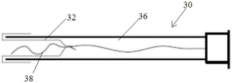
An improved design is shown in fig. 8A to 8E. An elongated balloon 32 having an expandable element 34 attached to the distal end of the balloon 32 is inverted into the lumen 36 of the catheter 30. When inverted, the expandable element 34 is disposed within the elongate balloon 32. In certain inventive embodiments, the expansible portion 34 is a filament of a multi-turn helix 38. The filaments forming the expandable element 34 may comprise a series of filament plastic materials, illustratively nylon or polypropylene, fluoropolymers or polylactic acid; metals such as stainless steel titanium or platinum; or a super-elastic metal such as nitinol. In some embodiments, a fiducial marker (not shown) is provided to facilitate subsequent retrieval to the appropriate cell sampling site. This is a pleasing alternative configuration of the expansion portion. For example, as shown in FIG. 17, the expandable section 34 may include a plurality of outwardly directed plastic or metal bristles 40; or the expandable section 34 may be formed by an elongated linear material that is crimped 38, stretched or fanned out 42, rolled 44 to pre-treat it for release from being pressed into the catheter (as shown in figures 10A-10B or figures 13A-13B); or it may be a plastic foam that expands when released into a wet environment (as shown in fig. 11A-11B). When the catheter is pressurized proximate the distal opening, the balloon 32 is inverted so as to advance the everted portion outwardly into the extended position and into contact with the cells of the fallopian tube inner wall. In certain inventive embodiments, the balloon is fully everted over the dilating portion 34 and delivered to the distal opening of the fallopian tube into the abdominal cavity. The flared portion 34 in some embodiments has an outer diameter of about 15-20 mm.
One advantage of having the flared portion 34 with several bristles is that it has a large surface area to facilitate cell collection, including areas that are not exposed to trimming forces when the device is pulled out. This method maximizes cell collection and minimizes cell removal as the device is pulled out of the fallopian tube or into the sheath, as shown in figures 17-19. In these embodiments where the expandable section has a greater surface area, the cell trapping typically increases each linear unit of binding in the fallopian tube under compression as compared to an uncontoured expandable section.
In some other inventive catheter embodiments, the expandable section, when deployed, is detailed as: a plurality of filaments 42 attached to the distal end of the balloon 32 that deploy when the balloon is inverted to form a fan-shaped brush 42 (shown in fig. 10A-10B); a plastic foam structure 46 is compressed into the bladder 32 and expands and is exposed to a liquid environment when the bladder 32 is inverted (as shown in fig. 11A-11B); an elastic or inelastic balloon 48 at the distal end of the inelastic sleeve balloon 32 (as shown in figures 12A-12B), a superelastic coil everting balloon (as shown in figures 13A-13B), a helical everting balloon 50 (as shown in figures 14A-14B), an everting end curve balloon 52 (as shown in figures 15A-15B); or a long length of flexible plastic or metal filament, is gathered into a three-dimensional structure, such as a lumen 54 (as shown in fig. 16A-16B), during balloon eversion, and the expandable section 34 has a plurality of outwardly directed bristles 40 (as shown in fig. 17). It is appreciated that the inventive catheter dilation section of any of these embodiments is readily adaptable to a fiducial marker for passage back into the fallopian tube when desired. For example, markers known in the art illustratively include: radiopacity markers, isotope markers, and high frequency markers. In other embodiments, the biodegradable dilating portion or a permanent dilating portion is separate from the catheter. In other embodiments, the dilating portion is transferred to a therapeutic agent in the fallopian tube tissue, such as a chemotherapeutic agent, an antibiotic, an anti-inflammatory agent, or a combination thereof.
When the catheter is pushed into the working channel of the hysteroscope, cells are scraped from the entire length of the inner surface of the fallopian tube. In some embodiments, the expandable section is inverted by reducing the gas pressure of the balloon so that the pores in the area of the catheter tip of the protector collect cells (see fig. 18).
Without being bound to a particular theory, the dilating portion creates sufficient friction between the outer surface of the dilating portion and the inner lining of the fallopian tube to collect cells and adhere them to the dilating portion, even in certain instances to a non-contoured dilating portion. The expansion helix contacts the distal end of the salpingo umbrella at the distal end of the balloon, and upon withdrawal a sample of cells is collected. Because the fallopian tube increases in internal diameter from the proximal side to the distal opening, the flared portion ensures collection of the cell sample at the distal end of the tube (the umbrella of the fallopian tube). The elongate balloon and the distal dilating portion are retracted into the working channel of the hysteroscope in some procedural embodiments to avoid cell loss when the hysteroscope is removed from the patient. A high-elasticity seal at the proximal end of the working channel seals the outer surface of the catheter. A marker on the catheter body indicates the length of withdrawal required to ensure that the elongate balloon and distal coil are lodged in the hysteroscope working channel. The hysteroscope is removed from the patient and in some embodiments, a syringe containing saline for flushing the cells collected by the elongate balloon and the dilatation helix into the test tube is attached to the luer fitting at the proximal end of the working channel. It will be appreciated that the cells of the expanded portion are readily collected for testing by conventional techniques and prepared for cytological, molecular or genetic testing.
An alternative embodiment is shown in fig. 16A-16B in which a tail is attached to the end of the everting balloon, with the addition of a lumen formed of a typical polyethylene terephthalate (PET) material. The turning process is described in the previous embodiment. Alternative embodiments also include an inflation port and a proximal seal to ensure that a port is maintained through the lumen into the fluid portion between the hysteroscope and the patient's body tissue when the balloon is inverted. Once inverted, the lumen provides a passage for a separate expandable section or a surgical instrument package to pass through. An example of such a collecting device is a spiral as shown in fig. 18 and 19. It is desirable that the cells be collected from a specific area of the fallopian tube, such as the umbrella of the fallopian tube, and then pulled back into the lumen to avoid scraping of the terminal cells off the internal surface of the fallopian tube proximal to the body when the potential device is removed.
Any patents or publications mentioned in this specification are herein incorporated by reference to the same extent as if each individual publication was specifically and individually indicated to be incorporated by reference. The foregoing descriptions of specific embodiments of the present invention are presented for purposes of illustration and not of limitation in the practice thereof.
Claims (14)
1. A catheter, comprising:
a tube having a distal tip and positionable relative to a fallopian tube;
a balloon having a distal end and being secured to the distal end of the tube at a proximal end of the balloon, the balloon having a length, the balloon being movable between a first inverted position and a second inverted position such that the balloon is expandable into a fallopian tube at the second inverted position; and
a dilating portion disposed at a distal tip of the balloon and movable between a retracted position and a dilated position when the balloon is inverted from a first inverted position to a second inverted position;
wherein the dilating portion is configured to dilate into a fallopian tube for cell collection when the balloon is dilated into the fallopian tube at the second everted position.
2. The catheter of claim 1, further comprising a source of pressurized fluid in selective communication with the balloon.
3. The catheter of claims 1-2, further comprising a hysteroscope.
4. The catheter of claims 1-2, wherein the dilating portion is a smooth-surfaced filament.
5. The catheter of claims 1-2, wherein the dilating portion is a textured filament.
6. The catheter of claims 1-2, wherein the dilating portion defines a brush.
7. The catheter of claims 1-2, wherein the dilating portion is a sponge.
8. The catheter of claims 1-2 wherein said dilating portion comprises an inflatable balloon.
9. The catheter of claim 8, wherein the inflatable balloon is a lumen having an aperture, and the dilating portion passes through the tube and the aperture.
10. The catheter of claims 1-2, wherein the dilating portion is a helical filament.
11. The catheter of claims 1-2, wherein the dilating portion is a spherical filament.
12. The catheter of claims 1-2, wherein the dilating portion is biodegradable.
13. The catheter of claims 1-2, further comprising a fiducial marker.
14. The catheter of claims 1-2, further comprising a therapeutic agent.
Applications Claiming Priority (5)
| Application Number | Priority Date | Filing Date | Title |
|---|---|---|---|
| US201361759783P | 2013-02-01 | 2013-02-01 | |
| US61/759,783 | 2013-02-01 | ||
| US201361873753P | 2013-09-04 | 2013-09-04 | |
| US61/873,753 | 2013-09-04 | ||
| CN201480018717.7A CN105246538B (en) | 2013-02-01 | 2014-02-03 | Fallopian tubal diagnostic device and method |
Related Parent Applications (1)
| Application Number | Title | Priority Date | Filing Date |
|---|---|---|---|
| CN201480018717.7A Division CN105246538B (en) | 2013-02-01 | 2014-02-03 | Fallopian tubal diagnostic device and method |
Publications (1)
| Publication Number | Publication Date |
|---|---|
| CN110946544A true CN110946544A (en) | 2020-04-03 |
Family
ID=51263030
Family Applications (2)
| Application Number | Title | Priority Date | Filing Date |
|---|---|---|---|
| CN201480018717.7A Active CN105246538B (en) | 2013-02-01 | 2014-02-03 | Fallopian tubal diagnostic device and method |
| CN201911112005.2A Pending CN110946544A (en) | 2013-02-01 | 2014-02-03 | Fallopian tube diagnostic device and method |
Family Applications Before (1)
| Application Number | Title | Priority Date | Filing Date |
|---|---|---|---|
| CN201480018717.7A Active CN105246538B (en) | 2013-02-01 | 2014-02-03 | Fallopian tubal diagnostic device and method |
Country Status (13)
| Country | Link |
|---|---|
| US (3) | US10646209B2 (en) |
| EP (2) | EP3603730B1 (en) |
| JP (2) | JP2016511023A (en) |
| KR (1) | KR20160007484A (en) |
| CN (2) | CN105246538B (en) |
| AU (5) | AU2015218548B2 (en) |
| BR (1) | BR112015018524A2 (en) |
| CA (1) | CA2899881A1 (en) |
| ES (1) | ES2752143T3 (en) |
| IL (1) | IL240305A0 (en) |
| MX (1) | MX2015009956A (en) |
| RU (1) | RU2688333C2 (en) |
| WO (1) | WO2014121207A1 (en) |
Families Citing this family (29)
| Publication number | Priority date | Publication date | Assignee | Title |
|---|---|---|---|---|
| US11179143B2 (en) | 2013-02-01 | 2021-11-23 | Boston Scientific Scimed, Inc. | Systems, methods, and devices for fallopian tube diagnostics |
| US10639016B2 (en) | 2013-02-01 | 2020-05-05 | Boston Scientific Scimed, Inc. | Methods and devices for Fallopian tube diagnostics |
| US11291434B2 (en) | 2013-02-01 | 2022-04-05 | Nvision Medical Corporation | Systems, methods, and devices for fallopian tube diagnostics |
| MX2015009956A (en) | 2013-02-01 | 2016-03-11 | Nvision Medical Corp | Methods and devices for fallopian tube diagnostics. |
| US9844383B2 (en) | 2013-05-08 | 2017-12-19 | Embolx, Inc. | Devices and methods for low pressure tumor embolization |
| WO2014182959A2 (en) | 2013-05-08 | 2014-11-13 | Embolx, Inc. | Device and methods for transvascular tumor embolization with integrated flow regulation |
| AU2014361829B2 (en) * | 2013-12-12 | 2019-07-25 | Case Western Reserve University | Device for collecting a biological sample |
| US10350382B1 (en) | 2018-06-08 | 2019-07-16 | Embolx, Inc. | High torque catheter and methods of manufacture |
| US11464948B2 (en) | 2016-02-16 | 2022-10-11 | Embolx, Inc. | Balloon catheters and methods of manufacture and use |
| US9550046B1 (en) | 2016-02-16 | 2017-01-24 | Embolx, Inc. | Balloon catheter and methods of fabrication and use |
| WO2017147586A1 (en) * | 2016-02-25 | 2017-08-31 | Nvision Medical Corporation | Methods and devices for fallopian tube diagnostics |
| KR20190008927A (en) * | 2016-05-24 | 2019-01-25 | 엔비전 메디컬 코포레이션 | Reversible cervical sterilization |
| JP6139759B1 (en) * | 2016-07-09 | 2017-05-31 | アイハート・メディカル株式会社 | Cell collection device |
| CN110582243A (en) | 2017-05-03 | 2019-12-17 | 美敦力瓦斯科尔勒公司 | Tissue removal catheter with guidewire isolation bushing |
| US11690645B2 (en) | 2017-05-03 | 2023-07-04 | Medtronic Vascular, Inc. | Tissue-removing catheter |
| CN114391887B (en) * | 2017-08-17 | 2024-03-26 | N视野医学有限公司 | systems, methods, and devices for fallopian tube diagnostics |
| US11141308B2 (en) | 2017-08-31 | 2021-10-12 | Crossbay Medical, Inc. | Apparatus and method for everting catheter for IUD delivery and placement in the uterine cavity |
| CN111343928B (en) * | 2017-10-27 | 2023-12-08 | 波士顿科学医学有限公司 | Cell collection and preparation device and method |
| US10576248B2 (en) * | 2018-07-23 | 2020-03-03 | Crossbay Medical, Inc. | Apparatus and method for everting catheter for uterine access for biopsy and cytology |
| WO2020102729A1 (en) | 2018-11-16 | 2020-05-22 | Medtronic Vascular, Inc. | Tissue-removing catheter |
| KR102260999B1 (en) * | 2019-03-27 | 2021-06-04 | (주)엠케어코리아 | Implant for ovarian cancer examination, examination kit including the same, and ovarian cancer screening method using the same |
| US11819236B2 (en) | 2019-05-17 | 2023-11-21 | Medtronic Vascular, Inc. | Tissue-removing catheter |
| US20220273339A1 (en) * | 2019-08-12 | 2022-09-01 | Covidien Lp | Cannula for preventing tumor seeding |
| US20220330792A1 (en) * | 2019-09-09 | 2022-10-20 | Arizona Board Of Regents On Behalf Of The University Of Arizona | Cell-collecting falloposcope and method for ovarian cancer detection |
| US11602269B2 (en) | 2020-05-26 | 2023-03-14 | FemDx Medsystems, Inc. | Oscillating endoscopic catheter for fallopian tube navigation |
| CA3179661A1 (en) * | 2020-05-26 | 2021-12-02 | Albert K. Chin | Oscillating endoscopic catheter for fallopian tube navigation |
| CA3186131A1 (en) * | 2020-06-16 | 2021-12-23 | Case Western Reserve University | Device for biological cell collection and method of use |
| CN111921022A (en) * | 2020-08-14 | 2020-11-13 | 中南大学湘雅医院 | Intestinal tract flushing drainage device |
| CN113288375A (en) * | 2021-06-17 | 2021-08-24 | 蔡迪先 | Fallopian tube dredging device and using method thereof |
Citations (2)
| Publication number | Priority date | Publication date | Assignee | Title |
|---|---|---|---|---|
| US20080097469A1 (en) * | 2006-10-18 | 2008-04-24 | Gruber William H | Intrauterine access and procedure system with laterally deflectable sheath |
| CN101869491A (en) * | 2004-07-07 | 2010-10-27 | 科隆尼克斯有限公司 | Colorectal cell sampling device |
Family Cites Families (89)
| Publication number | Priority date | Publication date | Assignee | Title |
|---|---|---|---|---|
| US2701559A (en) | 1951-08-02 | 1955-02-08 | William A Cooper | Apparatus for exfoliating and collecting diagnostic material from inner walls of hollow viscera |
| US3168092A (en) | 1961-06-15 | 1965-02-02 | Silverman Daniel | Medical probing instrument having flexible, extrudable tubing adapted to be extraverted under pressure into a body cavity |
| US3500819A (en) | 1965-10-24 | 1970-03-17 | Daniel Silverman | Medical probe adapted to be everted under pressure and method |
| US3664328A (en) * | 1971-04-28 | 1972-05-23 | Henry Dinwoodey Moyle Jr | Cancer test specimen gathering device |
| GB1408140A (en) * | 1971-12-09 | 1975-10-01 | Levene M M | Sampling device |
| US3877464A (en) * | 1972-06-07 | 1975-04-15 | Andrew R Vermes | Intra-uterine biopsy apparatus |
| US4157709A (en) | 1977-05-09 | 1979-06-12 | Ovutime, Inc. | Probe for obtaining cervical mucus and process thereof |
| US4227537A (en) * | 1978-03-08 | 1980-10-14 | Tucson Medical Instruments, Inc. | Endometrial brush with slidable protective sleeve |
| DE2812709C2 (en) * | 1978-03-23 | 1982-08-19 | Battelle-Institut E.V., 6000 Frankfurt | Device for obtaining cell materials from body cavities |
| US4324262A (en) * | 1979-01-02 | 1982-04-13 | University Of Virginia Alumni Patents Foundation | Aspirating culture catheter and method of use |
| CA1174882A (en) * | 1981-02-23 | 1984-09-25 | Charles J. Kramer | Plane grating polarizing beamsplitter |
| DE3331813A1 (en) | 1983-09-02 | 1985-03-21 | Mohamed Naim Dr.med. 3000 Hannover Saymé | Catheter for insertion into the fallopian tubes |
| US4863440A (en) | 1985-12-23 | 1989-09-05 | Thomas J. Fogarty | Pressurized manual advancement dilatation catheter |
| JP2627285B2 (en) | 1987-11-17 | 1997-07-02 | 有限会社はやしべるぐ | Hysteroscope for flexible tissue sampling |
| US4946440A (en) | 1988-10-05 | 1990-08-07 | Hall John E | Evertible membrane catheter and method of use |
| JPH03277374A (en) | 1990-03-28 | 1991-12-09 | Create Medic Kk | Improved balloon catheter for medical treatment |
| US5163927A (en) | 1991-10-17 | 1992-11-17 | Imagyn Medical, Inc. | Linear eversion catheter system with position indicating indicia |
| US5171305A (en) | 1991-10-17 | 1992-12-15 | Imagyn Medical, Inc. | Linear eversion catheter with reinforced inner body extension |
| AU661240B2 (en) | 1991-10-18 | 1995-07-13 | Imagyn Medical, Inc. | Apparatus and method for independent movement of an instrument within a linear eversion catheter |
| JPH0647448Y2 (en) | 1992-07-07 | 1994-12-07 | 稔 高田 | Endometrial cell collection tool |
| US5389089A (en) | 1992-10-13 | 1995-02-14 | Imagyn Medical, Inc. | Catheter with angled ball tip for fallopian tube access and method |
| US5445164A (en) * | 1993-05-11 | 1995-08-29 | Gynetech, Inc. | Cervical tissue sampling device |
| US5535756A (en) * | 1994-01-06 | 1996-07-16 | Parasher; Vinod K. | Catheter with simultaneous brush cytology and scrape biopsy capability |
| JP3726292B2 (en) * | 1994-09-05 | 2005-12-14 | 日本ゼオン株式会社 | Sliding catheter |
| ES2134492T3 (en) * | 1994-09-22 | 1999-10-01 | Bernard Chaffringeon | SINGLE USE DEVICE FOR THE DETECTION OR ANALYSIS OF A BODY LIQUID. |
| WO1996011372A1 (en) | 1994-10-05 | 1996-04-18 | Amerigon, Inc. | Improved heat transfer system for thermoelectric modules |
| US5630797A (en) * | 1995-01-17 | 1997-05-20 | Imagyn Medical, Inc. | Everting catheter system and method of utilizing the same |
| EP0723786A1 (en) | 1995-01-30 | 1996-07-31 | Cardiovascular Concepts, Inc. | Lesion measurement catheter and method |
| US5601581A (en) * | 1995-05-19 | 1997-02-11 | General Surgical Innovations, Inc. | Methods and devices for blood vessel harvesting |
| US5713369A (en) | 1995-09-13 | 1998-02-03 | Vance Products Inc. | Uterine endometrial tissue sample brush |
| US5762069A (en) * | 1995-12-29 | 1998-06-09 | Akos Biomedical, Inc. | Multiple sample biopsy forceps |
| US6074874A (en) | 1997-08-29 | 2000-06-13 | University Of Pittsburgh | Epithelial cell cultures for in vitro testing |
| JP2000135197A (en) | 1998-10-30 | 2000-05-16 | Toshiba Corp | Endoscope |
| US6371904B1 (en) * | 1998-12-24 | 2002-04-16 | Vivant Medical, Inc. | Subcutaneous cavity marking device and method |
| US7214229B2 (en) * | 1999-03-18 | 2007-05-08 | Fossa Medical, Inc. | Radially expanding stents |
| WO2001001867A1 (en) | 1999-06-25 | 2001-01-11 | Molecular Diagnostics, Inc. | Personal cervical cell collector |
| AU2051301A (en) | 1999-11-29 | 2001-06-04 | Molecular Diagnostics, Inc. | Cervical screening system |
| AUPQ641400A0 (en) * | 2000-03-23 | 2000-04-15 | Kleiner, Daniel E. | A device incorporating a hollow member for being positioned along a body cavity of a patient and method of positioning same |
| AU2001257212B9 (en) | 2000-04-25 | 2007-03-29 | Impres Medical, Inc. | Method and apparatus for creating intrauterine adhesions |
| US6478807B1 (en) | 2000-06-08 | 2002-11-12 | Advanced Cardiovascular Systems, Inc. | Pre-formed expandable member having grooves |
| AU2002321889A1 (en) * | 2001-08-03 | 2003-02-24 | Stemsource Llc | Devices and method for extraction of bone marrow |
| EP2305342A3 (en) | 2001-08-22 | 2013-04-03 | Gore Enterprise Holdings, Inc. | Apparatus and methods for treating strokes and controlling cerebral flow characteristics |
| WO2003020333A2 (en) | 2001-08-29 | 2003-03-13 | Artemis Medical, Inc. | Undamaged tissue collection assembly and method |
| AR031756A1 (en) | 2001-11-16 | 2003-10-01 | Tiberio Osvaldo Antonio | DEVICE FOR THE EXTRACTION OF UTERINE NECK CELLS FOR THEIR STUDY THROUGH THE PAPANICOLAOU TECHNIQUE |
| JP2006511271A (en) | 2002-12-18 | 2006-04-06 | ボストン・サイエンティフィック・サイメド・インコーポレイテッド | Detection using a catheter for endoluminal therapy |
| US7232681B2 (en) * | 2003-04-24 | 2007-06-19 | O'connell David | Personal cell sampling kit |
| US20060079924A1 (en) | 2003-07-24 | 2006-04-13 | Femspec Llc | Apparatus for accessing a body cavity and methods of making same |
| US20050021069A1 (en) | 2003-07-24 | 2005-01-27 | Gerald Feuer | Inflatable apparatus for accessing body cavity and methods of making |
| US20050245876A1 (en) * | 2003-12-24 | 2005-11-03 | Accessclosure, Inc. | Apparatus and methods for facilitating access through a puncture including sealing compound therein |
| US20050277847A1 (en) * | 2004-06-09 | 2005-12-15 | The Cleveland Clinic Foundation | Sampling device |
| TWI253342B (en) | 2004-08-06 | 2006-04-21 | Li-Cheng Lu | Cervical smear sampling device |
| US7255687B2 (en) | 2004-11-19 | 2007-08-14 | Percutaneous Systems, Inc. | Systems and methods for luminal access |
| ES2352568T3 (en) | 2005-07-05 | 2011-02-21 | Angioslide Ltd. | CATHETER WITH BALLOON. |
| WO2007146061A2 (en) | 2006-06-09 | 2007-12-21 | University Hospitals Of Cleveland | Cell collection and disease screening |
| US8333000B2 (en) | 2006-06-19 | 2012-12-18 | Advanced Cardiovascular Systems, Inc. | Methods for improving stent retention on a balloon catheter |
| US8292872B2 (en) * | 2007-06-29 | 2012-10-23 | Cook Medical Technologies Llc | Distal wire stop having adjustable handle |
| US8617114B2 (en) | 2007-07-13 | 2013-12-31 | Abbott Cardiovascular Systems Inc. | Drug coated balloon catheter |
| US8690823B2 (en) | 2007-07-13 | 2014-04-08 | Abbott Cardiovascular Systems Inc. | Drug coated balloon catheter |
| US8795197B2 (en) | 2007-07-17 | 2014-08-05 | Histologics, LLC | Frictional trans-epithelial tissue disruption collection apparatus and method of inducing an immune response |
| EP2166965B1 (en) | 2007-07-17 | 2017-05-17 | Neal Marc Lonky | Frictional trans-epithelial tissue disruption and collection apparatus |
| CA2724504A1 (en) * | 2008-05-16 | 2009-11-19 | Rwip, Llc | Delivery device with invertible diaphragm |
| SI22782A (en) | 2008-05-21 | 2009-12-31 | Univerza V Ljubljani | Device for atraumatic introduction of mean into a pipe-like organ of a living being |
| WO2010033467A1 (en) * | 2008-09-16 | 2010-03-25 | Intersect Partners, Llc | Device and methods for sampling prostate fluid |
| US8470043B2 (en) | 2008-12-23 | 2013-06-25 | Benvenue Medical, Inc. | Tissue removal tools and methods of use |
| US9161773B2 (en) | 2008-12-23 | 2015-10-20 | Benvenue Medical, Inc. | Tissue removal tools and methods of use |
| WO2010081000A1 (en) * | 2009-01-09 | 2010-07-15 | The United States Of America, As Represented By The Secretary, Department Of Health And Human Services | Collection device for biological specimens |
| EP2629676B1 (en) * | 2010-10-19 | 2016-07-13 | United States Endoscopy Group, Inc. | Cytology brush apparatus with improvements |
| WO2012106293A1 (en) * | 2011-01-31 | 2012-08-09 | Boston Scientific Scimed, Inc. | Distal tip configurations for biopsy with eus fna |
| US20130338533A1 (en) | 2011-03-14 | 2013-12-19 | Shared Medical Resources, Llc | Apparatus and method for obtaining transepithelial specimen |
| US20120259401A1 (en) | 2011-04-08 | 2012-10-11 | Gerrans Lawrence J | Balloon catheter for launching drug delivery device |
| ES2665307T3 (en) * | 2011-05-26 | 2018-04-25 | Adn International, Llc | Expandable device for tissue collection of an aerodigestive body light |
| US20120315662A1 (en) | 2011-06-07 | 2012-12-13 | Linnemeier Georgiann C | Method for the early detection of high-grade pelvic serous cancer |
| US8801628B2 (en) * | 2011-12-29 | 2014-08-12 | Express Scripts, Inc. | Methods and systems for medical home testing |
| GB2498349B (en) | 2012-01-10 | 2013-12-11 | Cook Medical Technologies Llc | Object capture device |
| CA2863144A1 (en) | 2012-01-31 | 2013-08-08 | Paul SPEISER | Non-invasive cancer diagnosis |
| US20130267870A1 (en) | 2012-04-06 | 2013-10-10 | Histologics Llc | Cell and tissue collection method and device |
| US20140128732A1 (en) | 2012-11-06 | 2014-05-08 | Cross Bay Medical, Inc. | Biopsy and sonography method and apparatus for assessing bodily cavities |
| US8747352B1 (en) | 2013-01-23 | 2014-06-10 | Medtronic Cryocath Lp | Balloon deflection |
| MX2015009956A (en) | 2013-02-01 | 2016-03-11 | Nvision Medical Corp | Methods and devices for fallopian tube diagnostics. |
| US20140257098A1 (en) | 2013-03-05 | 2014-09-11 | Giuseppe Del Priore | Systems and methods for detection of cancer in women |
| US9320502B2 (en) | 2013-03-12 | 2016-04-26 | Cook Medical Technologies Llc | Cytology balloon |
| WO2014149941A1 (en) * | 2013-03-15 | 2014-09-25 | Cook Medical Technologies Llc | Cell collector having an expandable mesh |
| US9750483B2 (en) * | 2013-06-26 | 2017-09-05 | Boston Scientific Scimed, Inc. | Cytology brush devices and methods of use |
| US20150057565A1 (en) | 2013-08-21 | 2015-02-26 | MKT Enterprises, LLC | Apparatus and method for ovarian cancer screening |
| WO2015070095A1 (en) | 2013-11-11 | 2015-05-14 | Cross Bay Medical, Inc. | Apparatus and methods for accessing and sealing bodily vessels and cavities |
| US9028401B1 (en) | 2013-11-11 | 2015-05-12 | Cross Bay Medical, Inc. | Apparatus and methods for accessing and sealing bodily vessels and cavities |
| AU2014361829B2 (en) | 2013-12-12 | 2019-07-25 | Case Western Reserve University | Device for collecting a biological sample |
| JP6251088B2 (en) | 2014-03-13 | 2017-12-20 | Hoya株式会社 | Deposit collecting tool and deposit collecting device |
| WO2016126879A1 (en) | 2015-02-03 | 2016-08-11 | The Arizona Board Of Regents On Behalf Of The University Of Arizona | Falloposcope and method for ovarian cancer detection |
-
2014
- 2014-02-03 MX MX2015009956A patent/MX2015009956A/en unknown
- 2014-02-03 EP EP19188620.9A patent/EP3603730B1/en active Active
- 2014-02-03 EP EP14745577.8A patent/EP2950873B1/en active Active
- 2014-02-03 BR BR112015018524A patent/BR112015018524A2/en not_active IP Right Cessation
- 2014-02-03 JP JP2015556206A patent/JP2016511023A/en active Pending
- 2014-02-03 WO PCT/US2014/014472 patent/WO2014121207A1/en active Application Filing
- 2014-02-03 CA CA2899881A patent/CA2899881A1/en not_active Abandoned
- 2014-02-03 CN CN201480018717.7A patent/CN105246538B/en active Active
- 2014-02-03 US US14/764,710 patent/US10646209B2/en active Active
- 2014-02-03 RU RU2015136529A patent/RU2688333C2/en not_active IP Right Cessation
- 2014-02-03 KR KR1020157023862A patent/KR20160007484A/en not_active Application Discontinuation
- 2014-02-03 CN CN201911112005.2A patent/CN110946544A/en active Pending
- 2014-02-03 ES ES14745577T patent/ES2752143T3/en active Active
-
2015
- 2015-08-02 IL IL240305A patent/IL240305A0/en unknown
- 2015-08-28 AU AU2015218548A patent/AU2015218548B2/en active Active
-
2017
- 2017-10-20 AU AU2017248549A patent/AU2017248549B2/en active Active
-
2018
- 2018-10-17 JP JP2018195989A patent/JP6758356B2/en active Active
-
2019
- 2019-10-30 AU AU2019257432A patent/AU2019257432B2/en active Active
- 2019-10-30 AU AU2019257431A patent/AU2019257431B2/en active Active
-
2020
- 2020-03-30 US US16/834,507 patent/US11571190B2/en active Active
-
2021
- 2021-06-17 AU AU2021204053A patent/AU2021204053B2/en active Active
-
2023
- 2023-01-10 US US18/095,254 patent/US20230157677A1/en active Pending
Patent Citations (2)
| Publication number | Priority date | Publication date | Assignee | Title |
|---|---|---|---|---|
| CN101869491A (en) * | 2004-07-07 | 2010-10-27 | 科隆尼克斯有限公司 | Colorectal cell sampling device |
| US20080097469A1 (en) * | 2006-10-18 | 2008-04-24 | Gruber William H | Intrauterine access and procedure system with laterally deflectable sheath |
Also Published As
Similar Documents
| Publication | Publication Date | Title |
|---|---|---|
| AU2021204053B2 (en) | Collection of cells from a lumen in patient | |
| US11517295B2 (en) | Methods and devices for fallopian tube diagnostics | |
| JP2023166627A (en) | Device for fallopian tube diagnostics |
Legal Events
| Date | Code | Title | Description |
|---|---|---|---|
| PB01 | Publication | ||
| PB01 | Publication | ||
| SE01 | Entry into force of request for substantive examination | ||
| SE01 | Entry into force of request for substantive examination |