BR112015032573B1 - Apparatus configured for guidance in capturing ultrasound images of an individual to obtain a target view, computer-readable media incorporating a program for guidance in capturing ultrasound images of an object to obtain a target view, and method for providing guidance in acquiring ultrasound images of an individual to obtain a target view - Google Patents
Apparatus configured for guidance in capturing ultrasound images of an individual to obtain a target view, computer-readable media incorporating a program for guidance in capturing ultrasound images of an object to obtain a target view, and method for providing guidance in acquiring ultrasound images of an individual to obtain a target view Download PDFInfo
- Publication number
- BR112015032573B1 BR112015032573B1 BR112015032573-4A BR112015032573A BR112015032573B1 BR 112015032573 B1 BR112015032573 B1 BR 112015032573B1 BR 112015032573 A BR112015032573 A BR 112015032573A BR 112015032573 B1 BR112015032573 B1 BR 112015032573B1
- Authority
- BR
- Brazil
- Prior art keywords
- image
- view
- images
- state space
- target view
- Prior art date
Links
- 238000002604 ultrasonography Methods 0.000 title claims abstract description 45
- 238000000034 method Methods 0.000 title claims description 9
- 230000004044 response Effects 0.000 claims abstract description 11
- 239000000523 sample Substances 0.000 claims description 55
- 238000003384 imaging method Methods 0.000 claims description 30
- 210000000056 organ Anatomy 0.000 claims description 17
- 238000013507 mapping Methods 0.000 claims description 16
- 210000003484 anatomy Anatomy 0.000 claims description 12
- 238000001514 detection method Methods 0.000 claims description 4
- 230000015572 biosynthetic process Effects 0.000 claims description 3
- 238000004590 computer program Methods 0.000 claims description 2
- 238000010276 construction Methods 0.000 claims 1
- 238000002591 computed tomography Methods 0.000 abstract description 8
- 238000002595 magnetic resonance imaging Methods 0.000 abstract description 6
- 238000012285 ultrasound imaging Methods 0.000 abstract description 2
- PWPJGUXAGUPAHP-UHFFFAOYSA-N lufenuron Chemical compound C1=C(Cl)C(OC(F)(F)C(C(F)(F)F)F)=CC(Cl)=C1NC(=O)NC(=O)C1=C(F)C=CC=C1F PWPJGUXAGUPAHP-UHFFFAOYSA-N 0.000 abstract 1
- 238000012545 processing Methods 0.000 description 6
- 230000000875 corresponding effect Effects 0.000 description 5
- 230000002452 interceptive effect Effects 0.000 description 5
- 230000015654 memory Effects 0.000 description 5
- 238000002360 preparation method Methods 0.000 description 4
- 230000008569 process Effects 0.000 description 4
- 210000001519 tissue Anatomy 0.000 description 4
- 210000001367 artery Anatomy 0.000 description 3
- 210000003109 clavicle Anatomy 0.000 description 3
- 238000003780 insertion Methods 0.000 description 3
- 230000037431 insertion Effects 0.000 description 3
- 230000009466 transformation Effects 0.000 description 3
- 230000000007 visual effect Effects 0.000 description 3
- 210000001168 carotid artery common Anatomy 0.000 description 2
- 230000002596 correlated effect Effects 0.000 description 2
- 238000010586 diagram Methods 0.000 description 2
- 230000006870 function Effects 0.000 description 2
- 210000002216 heart Anatomy 0.000 description 2
- 210000004373 mandible Anatomy 0.000 description 2
- 230000001052 transient effect Effects 0.000 description 2
- 235000016838 Pomo dAdamo Nutrition 0.000 description 1
- 244000003138 Pomo dAdamo Species 0.000 description 1
- 238000004458 analytical method Methods 0.000 description 1
- 238000013459 approach Methods 0.000 description 1
- 230000008901 benefit Effects 0.000 description 1
- 210000005242 cardiac chamber Anatomy 0.000 description 1
- 230000000747 cardiac effect Effects 0.000 description 1
- 230000015556 catabolic process Effects 0.000 description 1
- 230000008859 change Effects 0.000 description 1
- 230000000295 complement effect Effects 0.000 description 1
- 230000001010 compromised effect Effects 0.000 description 1
- 230000008878 coupling Effects 0.000 description 1
- 238000010168 coupling process Methods 0.000 description 1
- 238000005859 coupling reaction Methods 0.000 description 1
- 230000002950 deficient Effects 0.000 description 1
- 238000006731 degradation reaction Methods 0.000 description 1
- 230000001419 dependent effect Effects 0.000 description 1
- 238000002059 diagnostic imaging Methods 0.000 description 1
- 238000002592 echocardiography Methods 0.000 description 1
- 230000000694 effects Effects 0.000 description 1
- 238000005516 engineering process Methods 0.000 description 1
- 238000010348 incorporation Methods 0.000 description 1
- 210000001847 jaw Anatomy 0.000 description 1
- 238000002372 labelling Methods 0.000 description 1
- 210000003739 neck Anatomy 0.000 description 1
- 230000003287 optical effect Effects 0.000 description 1
- 230000008520 organization Effects 0.000 description 1
- 238000003909 pattern recognition Methods 0.000 description 1
- 210000001321 subclavian vein Anatomy 0.000 description 1
- 238000012360 testing method Methods 0.000 description 1
- 210000001685 thyroid gland Anatomy 0.000 description 1
- 238000012549 training Methods 0.000 description 1
- 238000013519 translation Methods 0.000 description 1
- 210000003462 vein Anatomy 0.000 description 1
- 210000002385 vertebral artery Anatomy 0.000 description 1
- 238000012800 visualization Methods 0.000 description 1
Images


Classifications
-
- A—HUMAN NECESSITIES
- A61—MEDICAL OR VETERINARY SCIENCE; HYGIENE
- A61B—DIAGNOSIS; SURGERY; IDENTIFICATION
- A61B8/00—Diagnosis using ultrasonic, sonic or infrasonic waves
- A61B8/52—Devices using data or image processing specially adapted for diagnosis using ultrasonic, sonic or infrasonic waves
- A61B8/5269—Devices using data or image processing specially adapted for diagnosis using ultrasonic, sonic or infrasonic waves involving detection or reduction of artifacts
-
- A—HUMAN NECESSITIES
- A61—MEDICAL OR VETERINARY SCIENCE; HYGIENE
- A61B—DIAGNOSIS; SURGERY; IDENTIFICATION
- A61B8/00—Diagnosis using ultrasonic, sonic or infrasonic waves
- A61B8/08—Detecting organic movements or changes, e.g. tumours, cysts, swellings
-
- A—HUMAN NECESSITIES
- A61—MEDICAL OR VETERINARY SCIENCE; HYGIENE
- A61B—DIAGNOSIS; SURGERY; IDENTIFICATION
- A61B8/00—Diagnosis using ultrasonic, sonic or infrasonic waves
- A61B8/46—Ultrasonic, sonic or infrasonic diagnostic devices with special arrangements for interfacing with the operator or the patient
- A61B8/461—Displaying means of special interest
- A61B8/466—Displaying means of special interest adapted to display 3D data
-
- A—HUMAN NECESSITIES
- A61—MEDICAL OR VETERINARY SCIENCE; HYGIENE
- A61B—DIAGNOSIS; SURGERY; IDENTIFICATION
- A61B8/00—Diagnosis using ultrasonic, sonic or infrasonic waves
- A61B8/48—Diagnostic techniques
- A61B8/483—Diagnostic techniques involving the acquisition of a 3D volume of data
-
- A—HUMAN NECESSITIES
- A61—MEDICAL OR VETERINARY SCIENCE; HYGIENE
- A61B—DIAGNOSIS; SURGERY; IDENTIFICATION
- A61B8/00—Diagnosis using ultrasonic, sonic or infrasonic waves
- A61B8/48—Diagnostic techniques
- A61B8/488—Diagnostic techniques involving Doppler signals
-
- A—HUMAN NECESSITIES
- A61—MEDICAL OR VETERINARY SCIENCE; HYGIENE
- A61B—DIAGNOSIS; SURGERY; IDENTIFICATION
- A61B8/00—Diagnosis using ultrasonic, sonic or infrasonic waves
- A61B8/52—Devices using data or image processing specially adapted for diagnosis using ultrasonic, sonic or infrasonic waves
- A61B8/5292—Devices using data or image processing specially adapted for diagnosis using ultrasonic, sonic or infrasonic waves using additional data, e.g. patient information, image labeling, acquisition parameters
-
- A—HUMAN NECESSITIES
- A61—MEDICAL OR VETERINARY SCIENCE; HYGIENE
- A61B—DIAGNOSIS; SURGERY; IDENTIFICATION
- A61B8/00—Diagnosis using ultrasonic, sonic or infrasonic waves
- A61B8/54—Control of the diagnostic device
-
- G—PHYSICS
- G06—COMPUTING; CALCULATING OR COUNTING
- G06F—ELECTRIC DIGITAL DATA PROCESSING
- G06F16/00—Information retrieval; Database structures therefor; File system structures therefor
- G06F16/50—Information retrieval; Database structures therefor; File system structures therefor of still image data
- G06F16/58—Retrieval characterised by using metadata, e.g. metadata not derived from the content or metadata generated manually
- G06F16/583—Retrieval characterised by using metadata, e.g. metadata not derived from the content or metadata generated manually using metadata automatically derived from the content
-
- G—PHYSICS
- G06—COMPUTING; CALCULATING OR COUNTING
- G06T—IMAGE DATA PROCESSING OR GENERATION, IN GENERAL
- G06T7/00—Image analysis
- G06T7/70—Determining position or orientation of objects or cameras
- G06T7/73—Determining position or orientation of objects or cameras using feature-based methods
- G06T7/74—Determining position or orientation of objects or cameras using feature-based methods involving reference images or patches
-
- A—HUMAN NECESSITIES
- A61—MEDICAL OR VETERINARY SCIENCE; HYGIENE
- A61B—DIAGNOSIS; SURGERY; IDENTIFICATION
- A61B8/00—Diagnosis using ultrasonic, sonic or infrasonic waves
- A61B8/08—Detecting organic movements or changes, e.g. tumours, cysts, swellings
- A61B8/0891—Detecting organic movements or changes, e.g. tumours, cysts, swellings for diagnosis of blood vessels
-
- A—HUMAN NECESSITIES
- A61—MEDICAL OR VETERINARY SCIENCE; HYGIENE
- A61B—DIAGNOSIS; SURGERY; IDENTIFICATION
- A61B8/00—Diagnosis using ultrasonic, sonic or infrasonic waves
- A61B8/44—Constructional features of the ultrasonic, sonic or infrasonic diagnostic device
- A61B8/4416—Constructional features of the ultrasonic, sonic or infrasonic diagnostic device related to combined acquisition of different diagnostic modalities, e.g. combination of ultrasound and X-ray acquisitions
-
- A—HUMAN NECESSITIES
- A61—MEDICAL OR VETERINARY SCIENCE; HYGIENE
- A61B—DIAGNOSIS; SURGERY; IDENTIFICATION
- A61B8/00—Diagnosis using ultrasonic, sonic or infrasonic waves
- A61B8/46—Ultrasonic, sonic or infrasonic diagnostic devices with special arrangements for interfacing with the operator or the patient
- A61B8/461—Displaying means of special interest
- A61B8/463—Displaying means of special interest characterised by displaying multiple images or images and diagnostic data on one display
-
- A—HUMAN NECESSITIES
- A61—MEDICAL OR VETERINARY SCIENCE; HYGIENE
- A61B—DIAGNOSIS; SURGERY; IDENTIFICATION
- A61B8/00—Diagnosis using ultrasonic, sonic or infrasonic waves
- A61B8/52—Devices using data or image processing specially adapted for diagnosis using ultrasonic, sonic or infrasonic waves
- A61B8/5207—Devices using data or image processing specially adapted for diagnosis using ultrasonic, sonic or infrasonic waves involving processing of raw data to produce diagnostic data, e.g. for generating an image
-
- A—HUMAN NECESSITIES
- A61—MEDICAL OR VETERINARY SCIENCE; HYGIENE
- A61B—DIAGNOSIS; SURGERY; IDENTIFICATION
- A61B8/00—Diagnosis using ultrasonic, sonic or infrasonic waves
- A61B8/52—Devices using data or image processing specially adapted for diagnosis using ultrasonic, sonic or infrasonic waves
- A61B8/5215—Devices using data or image processing specially adapted for diagnosis using ultrasonic, sonic or infrasonic waves involving processing of medical diagnostic data
-
- G—PHYSICS
- G06—COMPUTING; CALCULATING OR COUNTING
- G06T—IMAGE DATA PROCESSING OR GENERATION, IN GENERAL
- G06T2207/00—Indexing scheme for image analysis or image enhancement
- G06T2207/10—Image acquisition modality
- G06T2207/10132—Ultrasound image
-
- G—PHYSICS
- G06—COMPUTING; CALCULATING OR COUNTING
- G06T—IMAGE DATA PROCESSING OR GENERATION, IN GENERAL
- G06T2207/00—Indexing scheme for image analysis or image enhancement
- G06T2207/30—Subject of image; Context of image processing
- G06T2207/30004—Biomedical image processing
-
- G—PHYSICS
- G06—COMPUTING; CALCULATING OR COUNTING
- G06T—IMAGE DATA PROCESSING OR GENERATION, IN GENERAL
- G06T2207/00—Indexing scheme for image analysis or image enhancement
- G06T2207/30—Subject of image; Context of image processing
- G06T2207/30004—Biomedical image processing
- G06T2207/30101—Blood vessel; Artery; Vein; Vascular
-
- G—PHYSICS
- G06—COMPUTING; CALCULATING OR COUNTING
- G06T—IMAGE DATA PROCESSING OR GENERATION, IN GENERAL
- G06T2207/00—Indexing scheme for image analysis or image enhancement
- G06T2207/30—Subject of image; Context of image processing
- G06T2207/30244—Camera pose
Abstract
APARELHO CONFIGURADO PARA ORIENTAÇÃO NA CAPTURA DE IMAGEAMENTO DE ULTRASSOM DE UM INDIVÍDUO PARA OBTER UMA VISTA ALVO, E, MÍDIA LEGÍVEL POR COMPUTADOR QUE INCORPORA UM PROGRAMA PARA ORIENTAÇÃO NA CAPTURA DE IMAGENS DE ULTRASSOM DE UM OBJETO PARA OBTER UMA VISTA ALVO Trata- se de orientação na captura de imageamento de ultrassom de um indivíduo para se obter uma vista alvo, como uma vista padrão, que envolve a aplicação de ultrassom ao indivíduo e o recebimento, em resposta, de uma vista de ultrassom atual (502); o correlacionamento da imagem recebida com uma imagem preexistente, como uma imagem de referência tridimensional (503), e, para assistência ao usuário, a geração (baseada no correlacionamento) de um feedback (514-528) para a orientação. A imagem de referência pode ser um atlas estatístico ou pode ser derivada de varreduras de TC ou de RM específicas de um paciente. A imagem preexistente pode ser também um banco de dados correspondente a um estado de um espaço de estado. O feedback pode ser uma imagem derivada da imagem de referência; uma indicação gráfica (508) de um plano da vista alvo; a vista recebida unida (512) com uma imagem derivada da imagem de referência; ou a imagem derivada que aparece simultaneamente e é (...).APPARATUS CONFIGURED FOR GUIDANCE IN CAPTURING ULTRASOUND IMAGE OF AN INDIVIDUAL TO OBTAIN A TARGET VIEW, AND, COMPUTER-READable MEDIA THAT INCORPORATES A PROGRAM FOR GUIDANCE IN CAPTURING ULTRASOUND IMAGE OF AN OBJECT TO OBTAIN A TARGET VIEW This is guidance capturing ultrasound imaging of an individual to obtain a target view, such as a standard view, which involves applying ultrasound to the individual and receiving, in response, a current ultrasound view (502); correlating the received image with a pre-existing image, such as a three-dimensional reference image (503), and, for user assistance, generating (based on correlation) a feedback (514-528) for guidance. The reference image may be a statistical atlas or may be derived from patient-specific CT or MRI scans. The preexisting image can also be a database corresponding to a state in a state space. Feedback can be an image derived from the reference image; a graphical indication (508) of a target view plane; the received view joined (512) with an image derived from the reference image; or the derived image that appears simultaneously and is (...).
Description
[001] A presente invenção refere-se ao correlacionamento de imagem de ultrassom para orientação ao usuário e, mais particularmente, a tal correlacionamento a uma imagem preexistente para se obter uma vista alvo.[001] The present invention relates to ultrasound image mapping for user guidance and more particularly to such mapping to a preexisting image to obtain a target view.
[002] Um ultrassom bem-sucedido depende muito do treinamento e da experiência do usuário. Para evitar artefatos, o usuário precisa colocar a sonda na posição correta, ou seja, encontrar uma boa janela acústica para o imageamento. Convencionalmente, isso é feito unicamente com base nas imagens de ultrassom em tempo real na tela. Ainda que usuários experientes sejam geralmente capazes de reconhecer a degradação da imagem e consequentemente melhorar a qualidade da imagem, ao deslocarem a sonda para uma posição melhor, usuários menos experientes podem capturar imagens comprometidas devido a uma coordenação inferior entre mão e olho e menos conhecimento dos artefatos. A captura de um conjunto de vistas de um órgão do corpo de interesse é uma tarefa desafiadora para profissionais da área de cuidados com a saúde que têm pouco ou nenhum conhecimento em radiologia.[002] A successful ultrasound relies heavily on training and user experience. To avoid artifacts, the user needs to place the probe in the correct position, that is, find a good acoustic window for imaging. Conventionally, this is done solely on the basis of the real-time ultrasound images on the screen. While experienced users are generally able to recognize image degradation and consequently improve image quality, by moving the probe to a better position, less experienced users may capture compromised images due to poor hand-eye coordination and less knowledge of the probes. artifacts. Capturing a set of views of a body organ of interest is a challenging task for healthcare professionals who have little or no knowledge of radiology.
[003] A publicação intitulada “Real-Time Scan Assistant for Echocardiography”, Snare, S. R. et al., IEEE Transactions in Ultrasonics, Ferroelectrics, and Frequency Control (2012) (doravante como “a publicação Snare”) descreve uma abordagem de processamento de imagem aplicada a imagens bidimensionais (2D) das quatro câmaras cardíacas para produzir uma métrica da qualidade da vista obtida.[003] The publication entitled “Real-Time Scan Assistant for Echocardiography”, Snare, S. R. et al., IEEE Transactions in Ultrasonics, Ferroelectrics, and Frequency Control (2012) (hereinafter “the Snare publication”) describes a processing approach image applied to two-dimensional (2D) images of the four cardiac chambers to produce a metric of the quality of the view obtained.
[004] A publicação de patente No. 2012/0065510 concedida a Snare, S. R. et al. revela uma comparação de uma imagem ao vivo, de uma sonda de imageamento por ultrassom a uma imagem alvo através de correlacionamento de imagem. A Snare revela ainda um processador que calcula mudanças necessárias da posição atual da sonda a fim de realocar a sonda em uma nova posição, o que resultaria na captura de dados de ultrassom adicionais que podem ser usados para gerar uma imagem que correlacione de forma mais aproximada à imagem alvo. Instruções de como realocar a sonda são transmitidas ao usuário.[004] Patent Publication No. 2012/0065510 issued to Snare, S.R. et al. reveals a comparison of a live image from an ultrasound imaging probe to a target image via image correlation. Snare further unveils a processor that calculates necessary changes from the probe's current position in order to relocate the probe to a new position, which would result in the capture of additional ultrasound data that can be used to generate an image that more closely correlates. to the target image. Instructions on how to relocate the probe are transmitted to the user.
[005] O que se propõe mais adiante neste documento é direcionado a resolver um ou mais dos problemas acima.[005] What is proposed later in this document is aimed at solving one or more of the above problems.
[006] De acordo com um aspecto da presente invenção, a orientação na captura de imagens de ultrassom de um indivíduo para obter uma vista alvo inclui emitir, por meio de uma sonda de imageamento, ultrassom para o indivíduo e, em resposta, receber uma vista de ultrassom atual e correlacionar a imagem recebida com uma imagem preexistente. O correlacionamento estima um local da vista recebida em um espaço de estado que tem dimensões que são atributos da imagem. Um módulo de usuário é configurado para, em resposta ao correlacionamento, acessar, em relação ao local, um banco de dados organizado como o espaço de estado. O acesso é para recuperar o feedback do usuário para produzir a orientação.[006] In accordance with one aspect of the present invention, guidance in capturing ultrasound images of a subject to obtain a target view includes emitting, via an imaging probe, ultrasound to the subject and, in response, receiving a current ultrasound view and correlate the received image with a preexisting image. Correlation estimates a location of the received view in a state space that has dimensions that are image attributes. A user module is configured to, in response to mapping, access, with respect to location, a database organized as state space. Access is for retrieving user feedback to produce guidance.
[007] Em um subaspecto, a vista alvo é uma vista de um órgão ou vaso do corpo do indivíduo.[007] In a sub-aspect, the target view is a view of an organ or vessel in the individual's body.
[008] Em outro subaspecto, a geração de imagem é desempenhada dinamica ou continuamente.[008] In another sub-aspect, image generation is performed dynamically or continuously.
[009] Em um subaspecto diferente, a sonda tem uma posição atual, e ao menos um dentre mostrar e instruir como mover a sonda de sua posição atual ocorre para, dessa forma, se consiga fazer a captura da vista alvo.[009] In a different sub-aspect, the probe has a current position, and at least one of showing and instructing how to move the probe from its current position occurs in order to capture the target view.
[010] Em um subaspecto, é detectado um correlacionamento entre a vista alvo e a vista detectada.[010] In a sub-aspect, a correlation between the target view and the detected view is detected.
[011] Como um subaspecto adicional, um aparelho que, sem a necessidade de intervenção do usuário, desempenha automaticamente qualquer uma dentre ou ambas as ações de: a) tornar a notificação ao usuário responsiva à detecção do dito correlacionamento; e b) capturar dados de imagem, por meio da dita sonda, adequados à detecção responsiva do dito correlacionamento.[011] As an additional sub-aspect, a device that, without the need for user intervention, automatically performs either or both of the actions of: a) making the notification to the user responsive to the detection of said correlation; and b) capturing image data, by means of said probe, suitable for responsive detection of said correlation.
[012] Em um outro subaspecto, a vista alvo é uma vista anatômica padrão que, antes de um tempo de orientação, já foi estabelecida por uma entidade médica autorizada.[012] In another sub-aspect, the target view is a standard anatomical view that, before an orientation time, has already been established by an authorized medical entity.
[013] Em ainda outro subaspecto, é apresentado o feedback.[013] In yet another sub-aspect, feedback is presented.
[014] Como um subaspecto adicional, a apresentação é desempenhada dinamica ou continuamente.[014] As an additional sub-aspect, the presentation is played dynamically or continuously.
[015] Em mais um outro subaspecto, a vista recebida é registrada em uma imagem de referência tridimensional.[015] In yet another sub-aspect, the received view is registered in a three-dimensional reference image.
[016] Em um subaspecto, o registro de imagem é desempenhado dinamica ou continuamente.[016] In a sub-aspect, image registration is performed dynamically or continuously.
[017] Em um subaspecto diferente, a imagem de referência inclui um atlas, uma imagem capturada do dito indivíduo por meio de imageamento médico, ou ambos, o atlas e a imagem.[017] In a different sub-aspect, the reference image includes an atlas, an image captured of said individual through medical imaging, or both the atlas and the image.
[018] Em um subaspecto adicional, o atlas inclui um atlas estatístico.[018] In an additional sub-aspect, the atlas includes a statistical atlas.
[019] Como um subaspecto adicional com relação ao registro de vista, uma imagem derivada da imagem de referência e uma indicação gráfica de um plano da vista alvo são visualizados simultaneamente.[019] As an additional sub-aspect with respect to the view record, an image derived from the reference image and a graphical indication of a plane of the target view are visualized simultaneously.
[020] Em um subaspecto adicional ou complementar do registro de vista, existe uma visualização simultânea de um ou ambas das seguintes: a vista recebida unida com uma imagem derivada da imagem de referência; e a vista recebida e uma imagem derivada da imagem de referência, sendo que a imagem derivada aparece simultaneamente e aprimorada para indicar espacialmente onde a vista recebida é registrada na imagem de referência.[020] In an additional or complementary sub-aspect of the view record, there is a simultaneous visualization of one or both of the following: the received view joined with an image derived from the reference image; and the received view is an image derived from the reference image, with the derived image appearing simultaneously and enhanced to spatially indicate where the received view is recorded in the reference image.
[021] Em outra variação como um subaspecto do registro da vista, são fornecidas instruções sobre como mover a sonda para obter a vista alvo. Um alto-falante emite instruções de linguagem audíveis, as instruções são emitidas em uma tela, ou ambos o alto-falante e a tela são fornecidos para esses propósitos.[021] In another variation as a sub-aspect of the view record, instructions are given on how to move the probe to get the target view. A speaker emits audible language instructions, the instructions are output to a screen, or both the speaker and screen are provided for these purposes.
[022] Em uma versão específica do aspecto acima mencionado, estima-se um local da vista recebida no espaço de estado.[022] In a specific version of the aforementioned aspect, a location of the received view in state space is estimated.
[023] Em uma sub-versão específica, as configurações do Doppler são, automaticamente e por padrão, inicializadas de acordo com as que foram preconfiguradas para a visão alvo na construção do banco de dados.[023] In a specific sub-version, the Doppler settings are automatically and by default initialized according to the ones that were pre-configured for the target view when building the database.
[024] Como uma sub-versão desta, é feita uma seleção, com base na vista atual de ultrassom, de uma respectiva trajetória no espaço de estado em relação à vista alvo.[024] As a sub-version of this, a selection is made, based on the current ultrasound view, of a respective trajectory in state space in relation to the target view.
[025] Como uma sub-versão adicional, o feedback tem por base a seleção.[025] As an additional sub-version, feedback is based on selection.
[026] Em uma sub-versão adicional, a seleção é feita de uma trajetória ideal no espaço de estado em direção à vista alvo.[026] In a further subversion, the selection is made from an ideal trajectory in state space towards the target view.
[027] Em uma sub-versão específica, um scanner configurado para formar o espaço de estado o faz pelas etapas que incluem: capturar, por meio do scanner e a partir do imageamento de múltiplos indivíduos, imagens especializadas de um vaso ou um órgão específico do corpo, de modo que o órgão ou vaso, e/ou o tecido circundante sejam mostrados em todas as imagens da pluralidade; e identificar as imagens com os respectivos atributos.[027] In a specific sub-version, a scanner configured to form the state space does so by the steps that include: capturing, through the scanner and from the imaging of multiple individuals, specialized images of a specific vessel or organ of the body, so that the organ or vessel, and/or the surrounding tissue is shown in all images of the plurality; and identify the images with the respective attributes.
[028] Em uma sub-versão adicional, a formação do espaço de estado inclui vincular às imagens específicas como aquelas que não são alvo, ou seja, imagens padronizadas, respectivas instruções de como navegar a sonda de uma imagem específica a uma outra das imagens.[028] In a further subversion, state space formation includes linking to specific images as non-targeted ones, i.e. standardized images, respective instructions on how to navigate the probe from a specific image to another one of the images. .
[029] Em outro aspecto, um aparelho é configurado para orientar na captura de imageamento de ultrassom de um indivíduo para obter uma vista alvo. O aparelho inclui: uma sonda de imageamento (144) para emissão de ultrassom para um indivíduo e para, em resposta, receber uma vista de ultrassom atual; um módulo de correlacionamento de imagem (108) configurado para correlacionar a vista recebida com uma imagem preexistente; e um módulo de assistência ao usuário configurado para, com base no correlacionamento, gerar feedback para a orientação. O módulo de correlacionamento de imagem é configurado para, por meio do correlacionamento de padrões com base em imagem, registrar a vista recebida com uma imagem de referência (S240) que compreender um atlas estatístico que, por voxel, inclui uma distribuição de intensidades de imagem que reflete membros individuais de uma população. As informações da vizinhança são incluídas para cada voxel.[029] In another aspect, an apparatus is configured to guide in capturing an ultrasound image of an individual to obtain a target view. The apparatus includes: an imaging probe (144) for emitting ultrasound to a subject and for, in response, receiving a current ultrasound view; an image mapping module (108) configured to correlate the received view with a pre-existing image; and a user assistance module configured to, based on the correlation, generate feedback for guidance. The image mapping module is configured to, through image-based pattern mapping, record the received view with a reference image (S240) that comprises a statistical atlas that, per voxel, includes a distribution of image intensities that reflects individual members of a population. Neighborhood information is included for each voxel.
[030] Detalhes da nova tecnologia de orientação visual interativo em tempo real são apresentados mais abaixo, com o auxílio dos seguintes desenhos que não estão em escala.[030] Details of the new real-time interactive visual guidance technology are presented further below, with the aid of the following drawings which are not to scale.
[031] A Figura 1 é um diagrama esquemático de um aparelho médico interativo para orientação de ultrassom de acordo com a presente invenção; a Figura 2 é um fluxograma da operação total de uma implementação do aparelho da Figura 1, de acordo com a presente invenção; a Figura 3 é um fluxograma de um exemplo de uma preparação de correlacionamento de imagem, de acordo com a presente invenção; a Figura 4 é um fluxograma da preparação de espaço de estado, de acordo com a presente invenção; a Figura 5 é um diagrama conceitual e de tela de exibição de exemplos de feedback do usuário, de acordo com a presente invenção; e a Figura 6 é uma ilustração conceitual de exemplos de feedback de usuário e de geração de feedback, de acordo com a presente invenção.[031] Figure 1 is a schematic diagram of an interactive medical device for ultrasound guidance in accordance with the present invention; Figure 2 is a flowchart of the total operation of an implementation of the apparatus of Figure 1, in accordance with the present invention; Figure 3 is a flowchart of an example of an image mapping preparation in accordance with the present invention; Figure 4 is a flowchart of state space preparation in accordance with the present invention; Figure 5 is a conceptual and screen diagram showing examples of user feedback in accordance with the present invention; and Figure 6 is a conceptual illustration of examples of user feedback and feedback generation in accordance with the present invention.
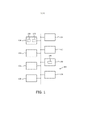
[032] A Figura 1 representa, a título de exemplo ilustrativo e não limitador, um aparelho médico interativo para orientação de ultrassom 100. O aparelho 100 inclui, entre outros componentes, um controlador 104, um módulo de correlacionamento de imagem 108, um módulo de assistência ao usuário 112, uma memória 116, um scanner 120, uma tela 124, um alto-falante 128 e controles de usuário 132. O módulo de correlacionamento de imagem 108 inclui um módulo de registro de imagem 136 e/ou um módulo de processamento de espaço de estado 140. O scanner inclui, entre outros componentes, uma sonda de imageamento 144.[032] Figure 1 represents, by way of illustrative and non-limiting example, an interactive medical device for
[033] Operacionalmente, um procedimento geral 200 para orientação de ultrassom médico interativo é mostrado a seguir, conforme a Figura 2. Como etapa preliminar, uma entidade médica autorizada, como um médico, uma junta médica, uma organização de padrões médicos ou um hospital, estabelece vistas de ultrassom padronizadas para um órgão ou vaso do corpo de interesse (etapa S204). O conjunto de vistas padronizadas é especificado para uso no aparelho 100 (etapa S208). Uma ou mais referências de correlacionamento de imagem são preparadas (etapa S212), o que é explicado abaixo, em mais detalhes, com referência à Figura 3. O médico seleciona um tipo de varredura, que pode ser de um órgão do corpo específico como o coração, ou um vaso como uma artéria específica (etapa S216). O aparelho 100 levanta a referência de correlacionamento de imagem correspondente (etapa S218). O aparelho 100 determina qual vista alvo dentre as vistas padronizadas deve ser capturada a seguir. O aparelho 100 também carrega as configurações de Doppler que foram preselecionadas para a vista alvo, conforme discutido abaixo em relação à Figura 4. Em resumo, as configurações de Doppler são, automaticamente por padrão, inicializadas de acordo com as que foram preconfiguradas para a vista alvo na criação de um banco de dados organizado como o espaço de estado (etapa S220). O aparelho 100 indica ao usuário, com base em orientações de livro-texto, por exemplo, como posicionar a sonda de imageamento 144 sobre a anatomia superficial do indivíduo do imageamento, como um paciente humano ou animal (etapa S224). O usuário, ou seja, o médico, posiciona a sonda 144 (etapa S228). Se o aparelho com a função de orientação de usuário 100 opera com base em um espaço de estado (etapa S232), o módulo de processamento do espaço de estado 140 é implementado e faz uma estimativa do local, no espaço de estado, da vista atual ou “ao vivo”, capturada através da sonda 144 (S236). Se, por outro lado, o módulo de processamento do espaço de estado 140 não é implementado (etapa S232), mas o módulo de registro de imagem 136 é implementado, a vista atual é registrada em uma posição correspondente, e orientação, em uma imagem de referência (etapa S240) tridimensional (3D). Se agora for determinado que a vista atual não se correlaciona com, ou representa suficientemente, a vista alvo (etapa S244), o aparelho 100 dá um feedback ao usuário que instrui, ou mostra, como proceder para atingir o objetivo de obter a vista alvo (etapa S248) e processar as ramificações de volta à etapa de posicionamento do usuário S228. É fornecida abaixo uma descrição mais detalhada do feedback na discussão que acompanha as Figuras 5 e 6. Se, em vez disso, um correlacionamento foi obtido (etapa S244) e a captura automática precisar ser executada (etapa S252), a vista atual é registrada para análise adicional, por exemplo, por um médico (etapa S256). Se, por outro lado, um correlacionamento foi obtido (etapa S244) e uma captura pelo usuário precisar ser executada (etapa S252), uma luz verde é acesa na sonda 144 ou em outra parte no scanner, como no console que abriga os controles do usuário 132 (etapa S260).[033] Operationally, a
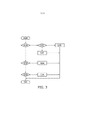
[034] A etapa de preparação da referência de correlacionamento de imagem (S212) é descrita mais detalhadamente no fluxograma da Figura 3. Com referência à Figura 3, se o correlacionamento de imagem precisar se basear em um atlas anatômico tridimensional (3D) como referência de correlacionamento de imagem (etapa S310), e o atlas precisar ser um atlas estatístico (etapa S320), um atlas estatístico é preparado (etapa S330). O atlas estatístico é construído com base nas varreduras da tomografia computadorizada (TC) e/ou do imageamento por ressonância magnética (RM) de uma ampla variedade de indivíduos para cobrir a variação anatômica. O mesmo pode ser armazenado em uma unidade de disco rígido que faz parte da memória 116. O atlas inclui uma distribuição de intensidades de imagem reflexivas de membros individuais da população por voxel. As informações da vizinhança também são incluídas para cada voxel. É realizado um correlacionamento de imagem para se obter registro mais rapidamente, devido à natureza estatística do atlas estatístico. Se, por outro lado, o correlacionamento da imagem precisar se basear em um atlas anatômico que não seja um atlas estatístico (etapas S310 e S320), o atlas anatômico é preparado como referência de correlacionamento de imagem 3D, em geral através das varreduras de TC e/ou de RM a partir de uma variedade de indivíduos (etapas S340). Se, em vez do atlas (etapa S310), as varreduras de TC e/ou de RM do mesmo paciente precisarem ser usadas para criar a referência de correlacionamento de imagem 3D (etapa S350), a referência do “mesmo paciente” é preparada (etapa S360). Se, por outro lado, as varreduras de TC/RM não estiverem disponíveis ou, caso contrário, não precisarem ser usadas (etapa S350), e um espaço de estado precisar ser usado (etapa S370), um espaço de estado é preparado (etapa S380). A preparação do espaço de estado é descrita mais detalhadamente, logo abaixo, em relação à Figura 4.[034] The step of preparing the image correlation reference (S212) is described in more detail in the flowchart of Figure 3. With reference to Figure 3, if the image correlation needs to be based on a three-dimensional (3D) anatomical atlas as a reference correlation process (step S310), and the atlas needs to be a statistical atlas (step S320), a statistical atlas is prepared (step S330). The statistical atlas is constructed from computed tomography (CT) and/or magnetic resonance imaging (MRI) scans of a wide variety of individuals to cover anatomical variation. The same can be stored on a hard disk drive that is part of
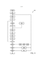
[035] Um processo de preparação do espaço de estado 400 envolve criar um banco de dados estatísticos de um conjunto de centenas de imagens do, e ao redor do, órgão e do vaso de interesse a partir do imageamento de múltiplos indivíduos. O banco de dados é organizado como um espaço de estado. Imagens a serem incorporadas no espaço de estado são etiquetadas com atributos, como anatomia de imagem vista, qualidade de imagem e posicionamento de sonda e orientação correspondente sobre a anatomia superficial. Um subconjunto desse conjunto de imagens é o conjunto de estados-alvo, que correspondem a vistas de ultrassom padrão com boa qualidade de imagem. As imagens do banco de dados podem ser descritas como pontos dentro de um espaço de estado cujas dimensões são os atributos da imagem. Dentro do espaço de estado, é possível definir um relacionamento espacial entre as imagens de ultrassom e, em particular, uma trajetória entre qualquer imagem de ultrassom e qualquer uma das imagens-alvo.[035] A 400 state space preparation process involves creating a statistical database of a set of hundreds of images of and around the organ and vessel of interest from imaging multiple individuals. The database is organized as a state space. Images to be embedded in the state space are tagged with attributes such as view image anatomy, image quality and probe placement, and corresponding orientation over the surface anatomy. A subset of this set of images is the set of target states, which correspond to standard ultrasound views with good image quality. Database images can be described as points within a state space whose dimensions are the attributes of the image. Within the state space, it is possible to define a spatial relationship between the ultrasound images and, in particular, a trajectory between any ultrasound image and any of the target images.
[036] As imagens a serem capturadas para incorporação no espaço de estado são especializadas para um órgão ou vaso do corpo específico, de modo que o órgão ou vaso, e o tecido circundante, sejam representados em cada imagem. O processo 400 é inicializado para um primeiro indivíduo de imageamento, uma primeira vista alvo, uma primeira trajetória e uma primeira imagem (etapa S404). Dessa forma, os respectivos indicadores e contadores são zerados. Uma imagem atual é obtida através da sonda de imageamento 144 (etapa S408). Atributos da imagem são registrados e a imagem atual é etiquetada com seus atributos (etapa S412). O registro pode ser feito em parte automaticamente e em parte através da entrada pela pessoa que constrói o banco de dados. A imagem atual pode ser etiquetada de acordo com: a anatomia vista (por exemplo, carótida (esquerda, direita, comum, interna, externa, bulbo, bifurcação, próxima, medial, distal, longitudinal, transversal, oblíqua etc.) assim como veia jugular, glândula tireoide, estruturas vertebrais, artéria vertebral, veia vertebral, artéria subclávia etc.; o posicionamento e a orientação da sonda 144 que dizem respeito à anatomia superficial para obter essas imagens (por exemplo, anterior, anterior, posterior, craniana, caudal, lateral, medial, pescoço, clavícula, mandíbula, pomo de Adão, horizontal, vertical, oblíqua); opcionalmente, o modo da imagem atual e configurações (por exemplo, para modo B, energia, profundidade focal, harmonia, composição espacial; para fluxo de cor, ganho, velocidade máxima, orientação da caixa de cor, tamanho do volume da amostra e ângulo do Doppler) e opcionalmente a presença de artefatos e uma medida de qualidade de imagem (por exemplo, artefatos de contato, bom contato de ultrassom e contraste de imagem médio). A etiquetagem do modo e configurações de imageamento atual é feita automaticamente. Como a navegação em direção à vista alvo normalmente continuaria do modo de imagem B para o modo de imagem B, modos como o Doppler podem ser normalmente, com efeito, retirados do processo de navegação. Por exemplo, essas configurações podem ser fornecidas como padrão automaticamente no início, como na etapa S220 acima. Os valores de configuração foram criados de acordo com os ajustes de controle feitos pelos construtores do banco de dados especificamente para a vista alvo durante a criação do banco de dados. Se, no decorrer da navegação de um usuário, o usuário, inadvertidamente ou por qualquer razão, mudar as configurações, a distância do espaço de estado resultante em algum ponto ou pontos no tempo, durante a navegação, resultará automaticamente em feedback, oferecendo instruções ao usuário para efetivamente restaurar essas configurações. Alternativamente, as configurações de Doppler da vista alvo não precisam ser fornecidas como padrão no início; em vez disso, o feedback do usuário, devido à distância do espaço de estado resultante, no decorrer da navegação do usuário ou no início, vai instruir os ajustes adequados às configurações.[036] The images to be captured for incorporation into the state space are specialized for a specific body organ or vessel, so that the organ or vessel, and the surrounding tissue, are represented in each image.
[037] A imagem atual capturada pode ter sido capturada através do contato do ultrassom que não é tão bom. Isso seria feito intencionalmente, de modo que o correlacionamento com essa imagem, uma vez que esteja no banco de dados, permite que o contato deficiente seja detectado. Se o contato é deficiente (etapa S416), a pessoa que constrói o banco de dados, aplica ou reaplica a mídia de acoplamento acústico, como gel, recoloca a sonda 144 na mesma posição e orientação com relação ao indivíduo do imageamento para uma imagem melhorada (etapa S420). Por outro lado, se o contato não foi deficiente (etapa S416), o construtor do banco de dados, através do movimento da sonda ou ajuste de configurações de imageamento, prepara para uma próxima captura de imagem (etapa S422). O movimento de ajuste é feito de modo a navegar em direção à imagem alvo.[037] The actual image captured may have been captured through ultrasound contact which is not so good. This would be done intentionally so that mapping to this image, once it is in the database, allows poor contact to be detected. If contact is poor (step S416), the person who builds the database, applies or reapplies acoustic coupling media such as gel, replaces
[038] Em qualquer das situações, ou seja, se o contato foi suficiente ou não, o processamento agora aponta para a próxima imagem (etapa S424). A imagem é capturada (etapa S428). Os atributos são registrados em parte manualmente e em parte automaticamente (etapa S432). O movimento da sonda, ajuste de contato ou ajuste de configurações de imageamento mais recentes, normalmente para o modo B, feitos nas etapas correspondentes acima S420 e S422, são inseridos ou selecionados pelos construtores do banco de dados, ou automaticamente, e vinculados à imagem anterior, ou seja, à imagem capturada antes da etapa S428 (etapa S436). A inserção pode ser, no que diz respeito à posição da sonda, “esquerda”, “direita”, “para cima”, “para baixo”. Aqui, “para cima” significa em geral da cabeça em direção ao dedo do pé. A inserção pode ser em vez disso ou além disso, no que diz respeito à orientação, ou seja, inclinação para a “esquerda”, “direita”, “para cima” ou “para baixo”. A inserção pode adicionalmente ou em vez disso ser, no que diz respeito à rotação da sonda 14, “em sentido horário” ou “em sentido anti-horário”. Em qualquer uma das opções, a distância ou magnitude não precisam ser registradas, porque a atualização do circuito de realimentação nas etapas S228 a S248 ocorre em tempo real. Em particular, a imagem do banco de dados que tem a localização mais próxima, de acordo com a distância euclideana, por exemplo, à estimativa feita na etapa S236, mantém o usuário dinamicamente em uma trajetória em direção à vista alvo. Mesmo se, durante o funcionamento, o usuário vagar para outra trajetória, essa outra trajetória vai, de modo similar, navegar em direção à vista alvo. Com relação ao contato da sonda, a entrada ou seleção pelo construtor do banco de dados pode ser “reaplicar gel à sonda e retornar à mesma posição e orientação”. Para mudanças nas configurações de imageamento, a seleção automática pode ser, por exemplo, “aumentar a profundidade do imageamento”.[038] In any of the situations, that is, whether the contact was sufficient or not, the processing now points to the next image (step S424). The image is captured (step S428). Attributes are registered partly manually and partly automatically (step S432). Probe movement, contact adjustment or latest imaging settings adjustment, typically to B mode, done in the corresponding steps above S420 and S422, are entered or selected by the database builders, or automatically, and linked to the image that is, the image captured before step S428 (step S436). The insertion can be, with respect to the position of the probe, “left”, “right”, “up”, “down”. Here, “up” generally means from the head towards the toe. Insertion can be instead of this or in addition to this, with regard to orientation, i.e. tilt to “left”, “right”, “up” or “down”. The insertion may additionally or instead be, with respect to the rotation of the probe 14, "clockwise" or "counterclockwise". In either option, the distance or magnitude does not need to be recorded, because the feedback circuit update in steps S228 to S248 takes place in real time. In particular, the database image that has the closest location, according to Euclidean distance, for example, to the estimate made in step S236, dynamically keeps the user on a trajectory towards the target view. Even if, during operation, the user wanders to another trajectory, that other trajectory will similarly navigate towards the target view. With respect to probe contact, entry or selection by the database builder can be “reapply gel to probe and return to same position and orientation”. For changes to imaging settings, the automatic selection can be, for example, “increase imaging depth”.
[039] Se a vista atual não for a vista alvo (etapa S440), o processamento retorna à etapa S416. Por outro lado, se a vista atual for a vista alvo, como evidenciado pela atuação do controle de usuário 132 adequado pelo construtor do banco de dados (etapa S440), e uma outra trajetória precisar ser registrada para a vista alvo atual do indivíduo do imageamento atual (etapa S444), o construtor do banco de dados é informado, por meio de uma mensagem na tela, para inserir as configurações de modo Doppler (etapa S446). Interativamente, de acordo com uma série de solicitações de tela e comandos de resposta dados pelo construtor do banco de dados, as configurações do Doppler são armazenadas como atributos da vista alvo (etapa S448). O indicador de trajetória é incrementado (etapa S450) e o retorno é feito para a etapa S408. Se, por outro lado, não houver mais trajetória a ser registrada (etapa S444), mas outra vista alvo para o indivíduo de imageamento atual for usada na construção do banco de dados (etapa S452), o indicador da vista é incrementado (etapa S456) e o retorno é feito, da mesma forma, para a etapa S408. Se, entretanto, a vista alvo do indivíduo de imageamento atual permanecer em termos de criação de banco de dados (etapa S452), mas um próximo indivíduo de imageamento for usado na criação do banco de dados (etapa S460), o indicador da vista é incrementado (etapa S464) e o retorno é feito, da mesma forma, para a etapa S408.[039] If the current view is not the target view (step S440), processing returns to step S416. On the other hand, if the current view is the target view, as evidenced by actuation of the
[040] A Figura 5 fornece exemplos do feedback do usuário da etapa S248, que pode ter a forma de ilustrações ou mensagens na tela, ou linguagem audível.[040] Figure 5 provides examples of user feedback from step S248, which may take the form of illustrations or on-screen messages, or audible language.
[041] Uma imagem de ultrassom representativa de uma vista atual 502, como uma imagem de modo B, pode ser exibida ao longo de uma imagem em seção transversal 504 derivada de uma imagem de referência 3D 503 armazenada em um disco rígido, ou seja, vinda de um atlas ou de uma imagem 3D criada a partir de varreduras de TC e/ou RM de pacientes específicos. A imagem em seção transversal 504, aqui de um órgão do corpo 505, por exemplo, o coração, foi segmentada e melhorada para indicar espacialmente onde a vista recebida (ou “ao vivo”) é registrada na imagem de referência. Dessa maneira, uma região melhorada 506, que é colorida, por exemplo, corresponde espacialmente a onde a imagem atual entra no atlas. Para mostrar a um médico como proceder na navegação em direção a uma vista alvo atual, uma indicação gráfica 508 do plano da vista alvo atual 510 pode ser adicionada à apresentação na tela. Além disso, em vez de mostrar a vista atual 502 como uma imagem separada, a imagem de ultrassom pode ser unida 512 a uma imagem em seção transversal 504 como por uma substituição de pixel por pixel. Aqui também, a indicação gráfica 508 pode ser adicionada.[041] An ultrasound image representative of a
[042] Alternativamente ou além disso, mensagens na tela ou instruções de linguagem audíveis podem guiar o médico. Dessa forma, para a posição/inclinação 514 da sonda 144, quatro possíveis indicações 516-522 são “direita”, “esquerda”, “para cima” e “para baixo”, assim como na modalidade com base no espaço de estado. Da mesma maneira, como na modalidade com base no espaço de estado, a rotação no lugar 524 pode ser “no sentido horário” 526 ou “no sentido anti-horário” 528.[042] Alternatively or in addition, on-screen messages or audible language instructions may guide the clinician. Thus, for position/
[043] O registro na etapa S240 envolve um correlacionamento do padrão com base em imagem da vista atual 502 à imagem de referência 3D e uma transformação coordenada na vista atual para fazer o registro com a imagem 3D de acordo com o correlacionamento. As instruções de feedback, com base na transformação, podem ser representativas de um tipo único, ou de mais de um tipo, de movimento da sonda sugerido 514 e 524.[043] Registration at step S240 involves a mapping of the image-based pattern of the
[044] Para a modalidade com base em espaço de estado, a estimativa na etapa S236 é feita como resultado do reconhecimento de padrão das comparações entre a vista atual 502 e as imagens de banco de dados capturadas nas etapas de captura S408 e S428. O um ou mais tipos de instruções de feedback (ou seja, movimento da sonda, contato da sonda e configurações de imageamento) vinculados à imagem do banco de dados atual são apresentados.[044] For the state space-based modality, the estimation in step S236 is done as a result of pattern recognition from comparisons between the
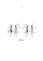
[045] A Figura 6 é um exemplo específico de um feedback de usuário e de geração de feedback. Esse exemplo refere-se à artéria carótida comum medial 601 e à captura de uma vista padronizada da artéria. Um gráfico de face de uma matriz de transdutores 604 é mostrado em uma posição oblíqua 608, representando um posicionamento atual 610 da sonda 144 e uma posição não oblíqua 612. A matriz de transdutores pode ter uma disposição linear ou uma disposição matricial. Gráficos mandibulares 616 e gráficos claviculares 620 também são mostrados na Figura 6. Um gráfico atual 624 corresponde conceitualmente à vista atual 502, e um gráfico alvo 628 corresponde conceitualmente à vista alvo 510. Além disso, ambos os gráficos 624 e 628 podem ser exibidos na tela em adição, a ou no lugar de, qualquer outro gráfico ou imagem de ultrassom representativa da vista atual 502.[045] Figure 6 is a specific example of user feedback and feedback generation. This example pertains to the 601 medial common carotid artery and capturing a standardized view of the artery. A face graphic of an array of
[046] Na modalidade do espaço de estado, a etiqueta na anatomia vista para uma imagem do banco de dados correlacionado é “artéria carótida comum medial esquerda, vista oblíqua”. A etiqueta da posição da sonda é “a meio caminho entre a clavícula e a mandíbula”. Os gráficos da clavícula e mandíbula 616 e 620 representam o tecido circundante. A etiqueta da orientação da sonda é “oblíqua”. A etiqueta do modo de imageamento é “modo B”. Uma etiqueta da configuração de imageamento é “composição espacial”. Uma etiqueta do artefato é “isento de artefato”. Uma etiqueta da qualidade de imagem é, por exemplo, “bom contraste de imagem”, com base na média de intensidade de pixel. Todas as etiquetas podem ser exibidas, na tela 124, na correlação com a imagem do banco de dados 604.[046] In state space mode, the tag in the anatomy seen for an image from the correlated database is “left medial common carotid artery, oblique view”. The probe position label is “midway between the clavicle and the mandible”. The clavicle and
[047] Se um atributo de qualidade de imagem da imagem do banco de dados correlacionado indicou falta de bom contato com a sonda, em vez da indicação atual aqui de “bom” contato da sonda em virtude da boa qualidade de imagem, essa condição de imageamento atual pode interferir no correlacionamento adicional com as imagens do banco de dados na navegação de uma trajetória em direção de uma vista padronizada. Consequentemente, o aprimoramento do contato predomina sobre outras considerações de navegação. Isso, então, constituiria um exemplo de seleção de uma trajetória ideal no espaço de estado, e é evidenciado pela emissão de uma mensagem de feedback do usuário do tipo “reaplicar gel e retornar sonda ao mesmo local e orientação”, que foi vinculada à imagem do banco de dados atual na etapa de vinculação S436.[047] If an image quality attribute of the image from the correlated database indicated lack of good probe contact, rather than the current indication here of “good” probe contact by virtue of good image quality, this condition of Current imaging can interfere with additional mapping to database images in navigating a trajectory towards a standardized view. Consequently, contact enhancement takes precedence over other navigation considerations. This would then constitute an example of selecting an optimal trajectory in state space, and is evidenced by the issuance of a user feedback message such as “reapply gel and return probe to same location and orientation”, which was linked to the image. of the current database in link step S436.
[048] Entretanto, como a etiqueta de qualidade da imagem indica que o contato é bom, uma instrução diferente, de que foi armazenado durante a criação do banco de dados na etapa S436, é enviada para ser visualizada na tela 124. A instrução aqui seria a instrução 526 para “girar no sentido horário”. Isso é indicado pelas setas ilustrativas 632. O movimento resultante da sonda 144 por um médico, como mencionado anteriormente neste documento, é monitorado em tempo real através do circuito de realimentação nas etapas S228 e S248. A instrução é reenviada para a tela repetidamente, mas vai mudar, caso a vista atual 502 se correlacione com uma nova imagem do banco de dados, como aquela que corresponde à vista alvo 510.[048] However, as the image quality label indicates that the contact is good, a different instruction than the one stored during database creation in step S436 is sent to be viewed on
[049] No caso da modalidade de imagem 3D, a instrução 526 “girar no sentido horário” é derivada quase que por definição, já que a única transformação envolvida no registro da vista atual 502 para a imagem de referência 3D é, de fato, a rotação no sentido horário. No caso menos definido, em que obter a vista alvo 510 envolve, por exemplo, a rotação e translação da sonda no lugar, o aparelho 100 decide se a rotação ou tradução vai predominar. O critério pode envolver limites selecionados com base em experiência empírica, embora, por exemplo, a localização ordinariamente dominará sobre a inclinação até que a localização da sonda esteja próxima do que é necessário para uma vista alvo 510.[049] In the case of the 3D image modality, the
[050] A orientação na captura de imageamento de ultrassom de um indivíduo para se obter uma vista alvo, como uma vista padrão, envolve a aplicação de ultrassom ao indivíduo e o recebimento, em resposta, de uma vista de ultrassom atual; o correlacionamento da imagem recebida com uma imagem preexistente, como uma imagem de referência tridimensional, e, para assistência ao usuário, a geração, baseada no correlacionamento, de um feedback para a orientação. A imagem de referência pode ser um atlas estatístico ou pode ser derivada de varreduras de TC ou de RM específicas de um paciente. A imagem preexistente pode ser também um banco de dados correspondente a um estado de um espaço de estado. O feedback pode ser uma imagem derivada da imagem de referência; uma indicação gráfica de um plano da vista alvo; a vista recebida unida com uma imagem derivada da imagem de referência; ou a vista recebida e uma imagem derivada da dita imagem de referência, sendo que a imagem derivada aparece simultaneamente e aprimorada para indicar espacialmente onde a vista recebida é registrada na imagem de referência. A vista alvo pode ser uma vista de um órgão ou um vaso do corpo do indivíduo. Tanto o atlas como o banco de dados podem ser especializados para imageamento de um órgão, ou um vaso, e o tecido circundante, selecionados de um usuário.[050] Guidance in capturing an ultrasound image of an individual to obtain a target view, such as a standard view, involves applying ultrasound to the individual and receiving, in response, a current ultrasound view; correlating the received image with a pre-existing image, such as a three-dimensional reference image, and, for user assistance, generating, based on the correlation, feedback for guidance. The reference image may be a statistical atlas or may be derived from patient-specific CT or MRI scans. The preexisting image can also be a database corresponding to a state in a state space. Feedback can be an image derived from the reference image; a graphical indication of a target view plane; the received view joined with an image derived from the reference image; or the received view and an image derived from said reference image, wherein the derived image appears simultaneously and enhanced to indicate spatially where the received view is recorded in the reference image. The target view may be a view of an organ or a vessel in the subject's body. Both the atlas and the database can be specialized for imaging an organ, or a vessel, and the surrounding tissue, selected from a user.
[051] Além de tornar o exame cardíaco diagnóstico executável por enfermeiros ou outros médicos que podem não ser treinados especificamente em sonografia, o aparelho de orientação visual interativo 100 pode orientar ultrassonografistas novatos. Alternativamente, o feedback visual inovador do aparelho 100 pode acelerar o fluxo de trabalho de ultrassonografistas treinados ou experientes.[051] In addition to making diagnostic cardiac examination workable by nurses or other physicians who may not be specifically trained in sonography, the interactive
[052] Embora a invenção tenha sido ilustrada e descrita em detalhes nos desenhos e na descrição supracitada, tal ilustração e descrição devem ser consideradas meramente ilustrativas ou exemplificadoras, e não restritivas; a invenção não se limita às modalidades reveladas.[052] Although the invention has been illustrated and described in detail in the drawings and in the aforementioned description, such illustration and description should be considered merely illustrative or exemplary, and not restrictive; the invention is not limited to the disclosed embodiments.
[053] Por exemplo, a sonda 144 pode alternativa ou adicionalmente usar feedback tátil no movimento adequado da sonda em relação à vista padronizada.[053] For example, probe 144 may alternatively or additionally use haptic feedback on proper probe movement relative to the standard view.
[054] Outras variações para as modalidades reveladas podem ser entendidas e efetuadas pelos versados na técnica na prática da invenção reivindicada, a partir do estudo dos desenhos, da revelação e das reivindicações anexas. Nas reivindicações, a expressão “que compreende” não exclui outros elementos ou outras etapas, e o artigo indefinido “um” ou “uma” não exclui uma pluralidade. Qualquer sinal de referência nas reivindicações não deve ser interpretado como limitador do escopo da invenção.[054] Other variations to the disclosed embodiments can be understood and made by those skilled in the art in the practice of the claimed invention, from the study of the drawings, the disclosure and the appended claims. In the claims, the expression "which comprises" does not exclude other elements or other steps, and the indefinite article "a" or "an" does not exclude a plurality. Any reference sign in the claims should not be interpreted as limiting the scope of the invention.
[055] Um programa de computador pode ser armazenado momentaneamente, temporariamente ou por um período de tempo maior em uma mídia legível por computador adequada, como uma mídia de armazenamento óptico ou uma mídia de estado sólido. Tal mídia é não transitória apenas no sentido de não ser um sinal de propagação transitório, mas incluir outras formas de mídias legíveis por computador como memória de registro, cache de processador, memória RAM e outras memórias voláteis.[055] A computer program may be stored momentarily, temporarily, or for a longer period of time on suitable computer-readable media, such as optical storage media or solid-state media. Such media is non-transient only in the sense that it is not a transient propagation signal, but includes other forms of computer-readable media such as log memory, processor cache, RAM memory, and other volatile memories.
[056] Um único processador ou outra unidade podem exercer as funções de vários itens mencionados nas reivindicações. O simples fato de certas medidas serem mencionadas em diferentes reivindicações mutuamente dependentes não indica que uma combinação dessas medidas não possa ser usada com vantagem.[056] A single processor or other unit may perform the functions of multiple items mentioned in the claims. The mere fact that certain measures are mentioned in different mutually dependent claims does not indicate that a combination of these measures cannot be used to advantage.
Claims (11)
Applications Claiming Priority (3)
| Application Number | Priority Date | Filing Date | Title |
|---|---|---|---|
| US201361840727P | 2013-06-28 | 2013-06-28 | |
| US61/840,727 | 2013-06-28 | ||
| PCT/IB2014/062523 WO2014207642A1 (en) | 2013-06-28 | 2014-06-23 | Ultrasound acquisition feedback guidance to a target view |
Publications (2)
| Publication Number | Publication Date |
|---|---|
| BR112015032573A2 BR112015032573A2 (en) | 2017-07-25 |
| BR112015032573B1 true BR112015032573B1 (en) | 2022-04-19 |
Family
ID=51205526
Family Applications (1)
| Application Number | Title | Priority Date | Filing Date |
|---|---|---|---|
| BR112015032573-4A BR112015032573B1 (en) | 2013-06-28 | 2014-06-23 | Apparatus configured for guidance in capturing ultrasound images of an individual to obtain a target view, computer-readable media incorporating a program for guidance in capturing ultrasound images of an object to obtain a target view, and method for providing guidance in acquiring ultrasound images of an individual to obtain a target view |
Country Status (7)
| Country | Link |
|---|---|
| US (1) | US10702248B2 (en) |
| EP (1) | EP3013242B1 (en) |
| JP (1) | JP6527860B2 (en) |
| CN (1) | CN105451663B (en) |
| BR (1) | BR112015032573B1 (en) |
| RU (1) | RU2683720C2 (en) |
| WO (1) | WO2014207642A1 (en) |
Families Citing this family (24)
| Publication number | Priority date | Publication date | Assignee | Title |
|---|---|---|---|---|
| US10134125B2 (en) * | 2013-02-28 | 2018-11-20 | Rivanna Medical Llc | Systems and methods for ultrasound imaging |
| WO2014136016A1 (en) * | 2013-03-05 | 2014-09-12 | Koninklijke Philips N.V. | Consistent sequential ultrasound acquisitions for intra-cranial monitoring |
| WO2016141449A1 (en) * | 2015-03-09 | 2016-09-15 | Her Majesty The Queen In Right Of Canada, As Represented By The Minister Of The Department Of National Defence | Computer-assisted focused assessment with sonography in trauma |
| GB201509164D0 (en) * | 2015-05-28 | 2015-07-15 | Intelligent Ultrasound Ltd | Imaging feedback system and method |
| US10682122B2 (en) * | 2015-12-03 | 2020-06-16 | Siemens Medical Solutions Usa, Inc. | Image-based user interface for controlling medical imaging |
| KR102425141B1 (en) | 2016-03-09 | 2022-07-26 | 에코너스 인코퍼레이티드 | Ultrasound image recognition system and method using artificial intelligence network |
| TWI765895B (en) | 2016-06-20 | 2022-06-01 | 美商蝴蝶網路公司 | Systems and methods of automated image acquisition for assisting a user to operate an ultrasound device |
| US10905402B2 (en) | 2016-07-27 | 2021-02-02 | Canon Medical Systems Corporation | Diagnostic guidance systems and methods |
| FR3060966B1 (en) * | 2016-12-23 | 2019-05-31 | Azoth Systems | DEVICE FOR MEASURING BLOOD FLOW |
| CA3070582A1 (en) | 2017-08-31 | 2019-03-07 | Butterfly Network, Inc. | Methods and apparatus for collection of ultrasound data |
| EP3681405A1 (en) * | 2017-09-15 | 2020-07-22 | Elesta S.P.A. | Device and method for needle sonographic guidance in minimally invasive procedures |
| US11464490B2 (en) | 2017-11-14 | 2022-10-11 | Verathon Inc. | Real-time feedback and semantic-rich guidance on quality ultrasound image acquisition |
| EP3549529A1 (en) | 2018-04-05 | 2019-10-09 | Koninklijke Philips N.V. | Ultrasound imaging system and method |
| US20190307428A1 (en) * | 2018-04-09 | 2019-10-10 | Butterfly Network, Inc. | Methods and apparatus for configuring an ultrasound system with imaging parameter values |
| WO2019222317A1 (en) | 2018-05-15 | 2019-11-21 | New York University | System and method for orientating capture of ultrasound images |
| US20220225963A1 (en) * | 2019-05-31 | 2022-07-21 | Koninklijke Philips N.V. | Methods and systems for guiding the acquisition of cranial ultrasound data |
| US11798677B2 (en) * | 2019-12-31 | 2023-10-24 | GE Precision Healthcare LLC | Method and system for providing a guided workflow through a series of ultrasound image acquisitions with reference images updated based on a determined anatomical position |
| CN111938699B (en) * | 2020-08-21 | 2022-04-01 | 电子科技大学 | System and method for guiding use of ultrasonic equipment |
| CN111938700B (en) * | 2020-08-21 | 2021-11-09 | 电子科技大学 | Ultrasonic probe guiding system and method based on real-time matching of human anatomy structure |
| US20220338836A1 (en) * | 2021-04-21 | 2022-10-27 | Ultrasight Ltd | System and method for guiding positioning and orienting of an ultrasound probe |
| CN113476098B (en) * | 2021-07-26 | 2022-06-24 | 首都医科大学附属北京儿童医院 | Monitoring system of shutoff pipe |
| US11903760B2 (en) | 2021-09-08 | 2024-02-20 | GE Precision Healthcare LLC | Systems and methods for scan plane prediction in ultrasound images |
| US20230148998A1 (en) * | 2021-11-15 | 2023-05-18 | GE Precision Healthcare LLC | Method and system for dynamically adjusting imaging parameters during an ultrasound scan |
| CN117017355B (en) * | 2023-10-08 | 2024-01-12 | 合肥合滨智能机器人有限公司 | Thyroid autonomous scanning system based on multi-modal generation type dialogue |
Family Cites Families (16)
| Publication number | Priority date | Publication date | Assignee | Title |
|---|---|---|---|---|
| US5906578A (en) * | 1997-06-18 | 1999-05-25 | Rajan; Govinda N. | Method and system for probe positioning in transesophageal echocardiography |
| JP4088104B2 (en) * | 2002-06-12 | 2008-05-21 | 株式会社東芝 | Ultrasonic diagnostic equipment |
| EP2460474B1 (en) * | 2003-05-08 | 2015-12-16 | Hitachi Medical Corporation | Reference image display method for ultrasonography and ultrasonic diagnosis apparatus |
| US7221972B2 (en) | 2003-08-29 | 2007-05-22 | Siemens Medical Solutions Usa, Inc. | Ultrasound system with protocol-driven user interface |
| US20050187472A1 (en) | 2004-01-30 | 2005-08-25 | Peter Lysyansky | Protocol-driven ultrasound examination |
| EP1751712A2 (en) * | 2004-05-14 | 2007-02-14 | Philips Intellectual Property & Standards GmbH | Information enhanced image guided interventions |
| JP4699062B2 (en) * | 2005-03-29 | 2011-06-08 | 株式会社日立メディコ | Ultrasonic device |
| US20070081706A1 (en) * | 2005-09-28 | 2007-04-12 | Xiang Zhou | Systems and methods for computer aided diagnosis and decision support in whole-body imaging |
| JP2009089736A (en) | 2007-10-03 | 2009-04-30 | Toshiba Corp | Ultrasonograph |
| WO2009081339A1 (en) * | 2007-12-21 | 2009-07-02 | Koninklijke Philips Electronics, N.V. | Systems and methods for tracking and guiding high intensity focused ultrasound beams |
| US8172753B2 (en) * | 2008-07-11 | 2012-05-08 | General Electric Company | Systems and methods for visualization of an ultrasound probe relative to an object |
| JP5481108B2 (en) | 2009-06-26 | 2014-04-23 | 株式会社東芝 | Ultrasonic diagnostic apparatus and automatic diagnosis support apparatus |
| JP2011110102A (en) * | 2009-11-24 | 2011-06-09 | Toshiba Corp | Ultrasonic diagnostic apparatus |
| US8352494B1 (en) * | 2009-12-07 | 2013-01-08 | Google Inc. | Distributed image search |
| US20120065510A1 (en) * | 2010-09-09 | 2012-03-15 | General Electric Company | Ultrasound system and method for calculating quality-of-fit |
| MX2013015358A (en) * | 2011-07-01 | 2014-02-11 | Koninkl Philips Nv | Intra-operative image correction for image-guided interventions. |
-
2014
- 2014-06-23 CN CN201480042594.0A patent/CN105451663B/en active Active
- 2014-06-23 EP EP14739560.2A patent/EP3013242B1/en active Active
- 2014-06-23 US US14/901,104 patent/US10702248B2/en active Active
- 2014-06-23 RU RU2016102416A patent/RU2683720C2/en active
- 2014-06-23 JP JP2016522921A patent/JP6527860B2/en not_active Expired - Fee Related
- 2014-06-23 WO PCT/IB2014/062523 patent/WO2014207642A1/en active Application Filing
- 2014-06-23 BR BR112015032573-4A patent/BR112015032573B1/en not_active IP Right Cessation
Also Published As
| Publication number | Publication date |
|---|---|
| CN105451663B (en) | 2019-03-19 |
| US10702248B2 (en) | 2020-07-07 |
| BR112015032573A2 (en) | 2017-07-25 |
| US20160143627A1 (en) | 2016-05-26 |
| EP3013242A1 (en) | 2016-05-04 |
| CN105451663A (en) | 2016-03-30 |
| EP3013242B1 (en) | 2018-11-07 |
| JP2016522074A (en) | 2016-07-28 |
| RU2016102416A (en) | 2017-08-01 |
| RU2683720C2 (en) | 2019-04-01 |
| JP6527860B2 (en) | 2019-06-05 |
| WO2014207642A1 (en) | 2014-12-31 |
Similar Documents
| Publication | Publication Date | Title |
|---|---|---|
| BR112015032573B1 (en) | Apparatus configured for guidance in capturing ultrasound images of an individual to obtain a target view, computer-readable media incorporating a program for guidance in capturing ultrasound images of an object to obtain a target view, and method for providing guidance in acquiring ultrasound images of an individual to obtain a target view | |
| Lang et al. | 3-Dimensional echocardiography: latest developments and future directions | |
| Simpson et al. | Three-dimensional echocardiography in congenital heart disease: an expert consensus document from the European Association of Cardiovascular Imaging and the American Society of Echocardiography | |
| Niemann et al. | Anatomically oriented right ventricular volume measurements with dynamic three-dimensional echocardiography validated by 3-Tesla magnetic resonance imaging | |
| Kang et al. | Stereoscopic augmented reality for laparoscopic surgery | |
| Mor-Avi et al. | Real-time 3-dimensional echocardiography: an integral component of the routine echocardiographic examination in adult patients? | |
| Mahmood et al. | Perioperative transoesophageal echocardiography: current status and future directions | |
| JP7133474B2 (en) | Image-based fusion of endoscopic and ultrasound images | |
| US20170116748A1 (en) | Landmark Detection with Spatial and Temporal Constraints in Medical Imaging | |
| BR112015025074B1 (en) | Ultrasound imaging system and method for generating and evaluating standard two-dimensional views from three-dimensional ultrasonic volume data | |
| US11793484B2 (en) | Method, device and system for intracavity probe procedure planning | |
| JP2015500083A (en) | Automatic imaging plane selection for echocardiography | |
| JP5689591B2 (en) | Ultrasonic diagnostic apparatus and ultrasonic image processing program | |
| Vettukattil | Three dimensional echocardiography in congenital heart disease | |
| Mahmood et al. | A practical approach to an intraoperative three-dimensional transesophageal echocardiography examination | |
| JP2017153818A (en) | Ultrasound diagnostic apparatus, ultrasound diagnostic apparatus control program, medical image processing apparatus, and medical image processing program | |
| Linte et al. | Virtual reality-enhanced ultrasound guidance: A novel technique for intracardiac interventions | |
| KR101495083B1 (en) | Method for proving bodymarker and ultrasound diagnostic apparatus thereof | |
| Bharucha et al. | Recent advances in pediatric echocardiography | |
| JP2012071138A (en) | Slice image display ultrasonic diagnostic apparatus of target object and method thereof | |
| Kapoor et al. | An update on transesophageal echocardiography views 2016: 2D versus 3D tee views | |
| Knackstedt et al. | Semi-automated 3-dimensional intracardiac echocardiography: development and initial clinical experience of a new system to guide ablation procedures | |
| Maxwell et al. | Emerging concepts in transesophageal echocardiography | |
| JP2021504046A (en) | Alignment of static preoperative planning data with respect to dynamic intraoperative segmentation data | |
| Salgo | 3D echocardiographic visualization for intracardiac beating heart surgery and intervention |
Legal Events
| Date | Code | Title | Description |
|---|---|---|---|
| B06F | Objections, documents and/or translations needed after an examination request according [chapter 6.6 patent gazette] | ||
| B06U | Preliminary requirement: requests with searches performed by other patent offices: procedure suspended [chapter 6.21 patent gazette] | ||
| B350 | Update of information on the portal [chapter 15.35 patent gazette] | ||
| B09A | Decision: intention to grant [chapter 9.1 patent gazette] | ||
| B16A | Patent or certificate of addition of invention granted [chapter 16.1 patent gazette] |
Free format text: PRAZO DE VALIDADE: 20 (VINTE) ANOS CONTADOS A PARTIR DE 23/06/2014, OBSERVADAS AS CONDICOES LEGAIS. |
|
| B21F | Lapse acc. art. 78, item iv - on non-payment of the annual fees in time |
Free format text: REFERENTE A 10A ANUIDADE. |