RU2699289C2 - Nucleic acids encoding human antibodies to sialyl-lewisa - Google Patents
Nucleic acids encoding human antibodies to sialyl-lewisa Download PDFInfo
- Publication number
- RU2699289C2 RU2699289C2 RU2016111015A RU2016111015A RU2699289C2 RU 2699289 C2 RU2699289 C2 RU 2699289C2 RU 2016111015 A RU2016111015 A RU 2016111015A RU 2016111015 A RU2016111015 A RU 2016111015A RU 2699289 C2 RU2699289 C2 RU 2699289C2
- Authority
- RU
- Russia
- Prior art keywords
- antibody
- seq
- residues
- functional fragment
- sle
- Prior art date
Links
- 241000282414 Homo sapiens Species 0.000 title claims description 59
- 150000007523 nucleic acids Chemical class 0.000 title claims description 28
- 102000039446 nucleic acids Human genes 0.000 title description 12
- 108020004707 nucleic acids Proteins 0.000 title description 12
- 239000012634 fragment Substances 0.000 claims abstract description 216
- 206010028980 Neoplasm Diseases 0.000 claims abstract description 126
- 238000000034 method Methods 0.000 claims abstract description 116
- 108091033319 polynucleotide Proteins 0.000 claims abstract description 62
- 102000040430 polynucleotide Human genes 0.000 claims abstract description 62
- 239000002157 polynucleotide Substances 0.000 claims abstract description 62
- 239000000427 antigen Substances 0.000 claims abstract description 58
- 102000036639 antigens Human genes 0.000 claims abstract description 58
- 108091007433 antigens Proteins 0.000 claims abstract description 58
- 208000037265 diseases, disorders, signs and symptoms Diseases 0.000 claims abstract description 45
- 201000010099 disease Diseases 0.000 claims abstract description 44
- 239000008194 pharmaceutical composition Substances 0.000 claims abstract description 37
- 239000003795 chemical substances by application Substances 0.000 claims abstract description 30
- 239000000126 substance Substances 0.000 claims abstract description 17
- 230000003211 malignant effect Effects 0.000 claims abstract description 6
- 125000003275 alpha amino acid group Chemical group 0.000 claims description 65
- 201000011510 cancer Diseases 0.000 claims description 49
- 239000003814 drug Substances 0.000 claims description 48
- 229940124597 therapeutic agent Drugs 0.000 claims description 45
- 206010061902 Pancreatic neoplasm Diseases 0.000 claims description 24
- 201000002528 pancreatic cancer Diseases 0.000 claims description 22
- 108091028043 Nucleic acid sequence Proteins 0.000 claims description 20
- 208000008443 pancreatic carcinoma Diseases 0.000 claims description 20
- 239000000032 diagnostic agent Substances 0.000 claims description 17
- 229940039227 diagnostic agent Drugs 0.000 claims description 17
- 208000015486 malignant pancreatic neoplasm Diseases 0.000 claims description 17
- 206010009944 Colon cancer Diseases 0.000 claims description 16
- FWMNVWWHGCHHJJ-SKKKGAJSSA-N 4-amino-1-[(2r)-6-amino-2-[[(2r)-2-[[(2r)-2-[[(2r)-2-amino-3-phenylpropanoyl]amino]-3-phenylpropanoyl]amino]-4-methylpentanoyl]amino]hexanoyl]piperidine-4-carboxylic acid Chemical compound C([C@H](C(=O)N[C@H](CC(C)C)C(=O)N[C@H](CCCCN)C(=O)N1CCC(N)(CC1)C(O)=O)NC(=O)[C@H](N)CC=1C=CC=CC=1)C1=CC=CC=C1 FWMNVWWHGCHHJJ-SKKKGAJSSA-N 0.000 claims description 14
- 208000029742 colonic neoplasm Diseases 0.000 claims description 13
- 230000002285 radioactive effect Effects 0.000 claims description 13
- 208000000587 small cell lung carcinoma Diseases 0.000 claims description 13
- 206010041067 Small cell lung cancer Diseases 0.000 claims description 12
- 208000009956 adenocarcinoma Diseases 0.000 claims description 12
- 210000000496 pancreas Anatomy 0.000 claims description 10
- 206010033128 Ovarian cancer Diseases 0.000 claims description 9
- 206010061535 Ovarian neoplasm Diseases 0.000 claims description 9
- 229940127089 cytotoxic agent Drugs 0.000 claims description 8
- 229910052751 metal Inorganic materials 0.000 claims description 8
- 239000002184 metal Substances 0.000 claims description 8
- 230000001225 therapeutic effect Effects 0.000 claims description 8
- 230000005740 tumor formation Effects 0.000 claims description 8
- 208000001333 Colorectal Neoplasms Diseases 0.000 claims description 7
- 239000003937 drug carrier Substances 0.000 claims description 7
- 206010052747 Adenocarcinoma pancreas Diseases 0.000 claims description 6
- 102000008394 Immunoglobulin Fragments Human genes 0.000 claims description 6
- 108010021625 Immunoglobulin Fragments Proteins 0.000 claims description 6
- GNBHRKFJIUUOQI-UHFFFAOYSA-N fluorescein Chemical compound O1C(=O)C2=CC=CC=C2C21C1=CC=C(O)C=C1OC1=CC(O)=CC=C21 GNBHRKFJIUUOQI-UHFFFAOYSA-N 0.000 claims description 6
- 201000002094 pancreatic adenocarcinoma Diseases 0.000 claims description 6
- 206010006187 Breast cancer Diseases 0.000 claims description 5
- 210000001035 gastrointestinal tract Anatomy 0.000 claims description 5
- MFRNYXJJRJQHNW-DEMKXPNLSA-N (2s)-2-[[(2r,3r)-3-methoxy-3-[(2s)-1-[(3r,4s,5s)-3-methoxy-5-methyl-4-[methyl-[(2s)-3-methyl-2-[[(2s)-3-methyl-2-(methylamino)butanoyl]amino]butanoyl]amino]heptanoyl]pyrrolidin-2-yl]-2-methylpropanoyl]amino]-3-phenylpropanoic acid Chemical compound CN[C@@H](C(C)C)C(=O)N[C@@H](C(C)C)C(=O)N(C)[C@@H]([C@@H](C)CC)[C@H](OC)CC(=O)N1CCC[C@H]1[C@H](OC)[C@@H](C)C(=O)N[C@H](C(O)=O)CC1=CC=CC=C1 MFRNYXJJRJQHNW-DEMKXPNLSA-N 0.000 claims description 4
- 239000002246 antineoplastic agent Substances 0.000 claims description 4
- MHMNJMPURVTYEJ-UHFFFAOYSA-N fluorescein-5-isothiocyanate Chemical compound O1C(=O)C2=CC(N=C=S)=CC=C2C21C1=CC=C(O)C=C1OC1=CC(O)=CC=C21 MHMNJMPURVTYEJ-UHFFFAOYSA-N 0.000 claims description 4
- 108010059074 monomethylauristatin F Proteins 0.000 claims description 4
- 206010055114 Colon cancer metastatic Diseases 0.000 claims description 3
- 206010052360 Colorectal adenocarcinoma Diseases 0.000 claims description 3
- 108010044540 auristatin Proteins 0.000 claims description 3
- 201000006587 bladder adenocarcinoma Diseases 0.000 claims description 3
- 239000000941 radioactive substance Substances 0.000 claims description 3
- PYWVYCXTNDRMGF-UHFFFAOYSA-N rhodamine B Chemical compound [Cl-].C=12C=CC(=[N+](CC)CC)C=C2OC2=CC(N(CC)CC)=CC=C2C=1C1=CC=CC=C1C(O)=O PYWVYCXTNDRMGF-UHFFFAOYSA-N 0.000 claims description 3
- CJIJXIFQYOPWTF-UHFFFAOYSA-N 7-hydroxycoumarin Natural products O1C(=O)C=CC2=CC(O)=CC=C21 CJIJXIFQYOPWTF-UHFFFAOYSA-N 0.000 claims description 2
- XPDXVDYUQZHFPV-UHFFFAOYSA-N Dansyl Chloride Chemical compound C1=CC=C2C(N(C)C)=CC=CC2=C1S(Cl)(=O)=O XPDXVDYUQZHFPV-UHFFFAOYSA-N 0.000 claims description 2
- 108010004729 Phycoerythrin Proteins 0.000 claims description 2
- 108010008739 auristatin PHE Proteins 0.000 claims description 2
- CFDVGUXRLQWLJX-QGTNPELVSA-N beta-D-Galp-(1->3)-[alpha-L-Fucp-(1->4)]-D-GlcpNAc Chemical compound O[C@H]1[C@H](O)[C@H](O)[C@H](C)O[C@H]1O[C@H]1[C@H](O[C@H]2[C@@H]([C@@H](O)[C@@H](O)[C@@H](CO)O2)O)[C@@H](NC(C)=O)C(O)O[C@@H]1CO CFDVGUXRLQWLJX-QGTNPELVSA-N 0.000 claims description 2
- MJQUEDHRCUIRLF-TVIXENOKSA-N bryostatin 1 Chemical compound C([C@@H]1CC(/[C@@H]([C@@](C(C)(C)/C=C/2)(O)O1)OC(=O)/C=C/C=C/CCC)=C\C(=O)OC)[C@H]([C@@H](C)O)OC(=O)C[C@H](O)C[C@@H](O1)C[C@H](OC(C)=O)C(C)(C)[C@]1(O)C[C@@H]1C\C(=C\C(=O)OC)C[C@H]\2O1 MJQUEDHRCUIRLF-TVIXENOKSA-N 0.000 claims description 2
- 229960005539 bryostatin 1 Drugs 0.000 claims description 2
- 239000002955 immunomodulating agent Substances 0.000 claims description 2
- 208000037819 metastatic cancer Diseases 0.000 claims description 2
- 208000011575 metastatic malignant neoplasm Diseases 0.000 claims description 2
- ZTLGJPIZUOVDMT-UHFFFAOYSA-N n,n-dichlorotriazin-4-amine Chemical compound ClN(Cl)C1=CC=NN=N1 ZTLGJPIZUOVDMT-UHFFFAOYSA-N 0.000 claims description 2
- ORHBXUUXSCNDEV-UHFFFAOYSA-N umbelliferone Chemical compound C1=CC(=O)OC2=CC(O)=CC=C21 ORHBXUUXSCNDEV-UHFFFAOYSA-N 0.000 claims description 2
- HFTAFOQKODTIJY-UHFFFAOYSA-N umbelliferone Natural products Cc1cc2C=CC(=O)Oc2cc1OCC=CC(C)(C)O HFTAFOQKODTIJY-UHFFFAOYSA-N 0.000 claims description 2
- IEDXPSOJFSVCKU-HOKPPMCLSA-N [4-[[(2S)-5-(carbamoylamino)-2-[[(2S)-2-[6-(2,5-dioxopyrrolidin-1-yl)hexanoylamino]-3-methylbutanoyl]amino]pentanoyl]amino]phenyl]methyl N-[(2S)-1-[[(2S)-1-[[(3R,4S,5S)-1-[(2S)-2-[(1R,2R)-3-[[(1S,2R)-1-hydroxy-1-phenylpropan-2-yl]amino]-1-methoxy-2-methyl-3-oxopropyl]pyrrolidin-1-yl]-3-methoxy-5-methyl-1-oxoheptan-4-yl]-methylamino]-3-methyl-1-oxobutan-2-yl]amino]-3-methyl-1-oxobutan-2-yl]-N-methylcarbamate Chemical compound CC[C@H](C)[C@@H]([C@@H](CC(=O)N1CCC[C@H]1[C@H](OC)[C@@H](C)C(=O)N[C@H](C)[C@@H](O)c1ccccc1)OC)N(C)C(=O)[C@@H](NC(=O)[C@H](C(C)C)N(C)C(=O)OCc1ccc(NC(=O)[C@H](CCCNC(N)=O)NC(=O)[C@@H](NC(=O)CCCCCN2C(=O)CCC2=O)C(C)C)cc1)C(C)C IEDXPSOJFSVCKU-HOKPPMCLSA-N 0.000 claims 2
- 108010093470 monomethyl auristatin E Proteins 0.000 claims 2
- 208000036764 Adenocarcinoma of the esophagus Diseases 0.000 claims 1
- 201000008274 breast adenocarcinoma Diseases 0.000 claims 1
- 208000028653 esophageal adenocarcinoma Diseases 0.000 claims 1
- 201000006585 gastric adenocarcinoma Diseases 0.000 claims 1
- 210000001635 urinary tract Anatomy 0.000 claims 1
- 230000027455 binding Effects 0.000 abstract description 67
- 230000000694 effects Effects 0.000 abstract description 25
- 238000001514 detection method Methods 0.000 abstract description 16
- 230000015572 biosynthetic process Effects 0.000 abstract description 11
- 210000004027 cell Anatomy 0.000 description 164
- 108090000623 proteins and genes Proteins 0.000 description 55
- 125000003729 nucleotide group Chemical group 0.000 description 43
- 239000002773 nucleotide Substances 0.000 description 40
- 102100035360 Cerebellar degeneration-related antigen 1 Human genes 0.000 description 38
- 108090000765 processed proteins & peptides Proteins 0.000 description 35
- 102000004196 processed proteins & peptides Human genes 0.000 description 33
- 229920001184 polypeptide Polymers 0.000 description 32
- 235000018102 proteins Nutrition 0.000 description 32
- 102000004169 proteins and genes Human genes 0.000 description 32
- 241000699670 Mus sp. Species 0.000 description 31
- 235000001014 amino acid Nutrition 0.000 description 31
- 239000013598 vector Substances 0.000 description 30
- 230000014509 gene expression Effects 0.000 description 28
- 108010047041 Complementarity Determining Regions Proteins 0.000 description 27
- 101000737793 Homo sapiens Cerebellar degeneration-related antigen 1 Proteins 0.000 description 26
- 241001465754 Metazoa Species 0.000 description 26
- 150000001720 carbohydrates Chemical class 0.000 description 26
- 210000001519 tissue Anatomy 0.000 description 25
- 239000002953 phosphate buffered saline Substances 0.000 description 24
- 238000011282 treatment Methods 0.000 description 24
- 210000004881 tumor cell Anatomy 0.000 description 23
- 125000000539 amino acid group Chemical group 0.000 description 22
- 238000004458 analytical method Methods 0.000 description 21
- RCINICONZNJXQF-MZXODVADSA-N taxol Chemical compound O([C@@H]1[C@@]2(C[C@@H](C(C)=C(C2(C)C)[C@H](C([C@]2(C)[C@@H](O)C[C@H]3OC[C@]3([C@H]21)OC(C)=O)=O)OC(=O)C)OC(=O)[C@H](O)[C@@H](NC(=O)C=1C=CC=CC=1)C=1C=CC=CC=1)O)C(=O)C1=CC=CC=C1 RCINICONZNJXQF-MZXODVADSA-N 0.000 description 21
- 108020004414 DNA Proteins 0.000 description 20
- 229930012538 Paclitaxel Natural products 0.000 description 20
- 235000014633 carbohydrates Nutrition 0.000 description 20
- 229960001592 paclitaxel Drugs 0.000 description 20
- 229940024606 amino acid Drugs 0.000 description 19
- 102000037865 fusion proteins Human genes 0.000 description 19
- 108020001507 fusion proteins Proteins 0.000 description 19
- 238000002600 positron emission tomography Methods 0.000 description 19
- 150000001413 amino acids Chemical class 0.000 description 18
- 239000013604 expression vector Substances 0.000 description 18
- YBJHBAHKTGYVGT-ZKWXMUAHSA-N (+)-Biotin Chemical compound N1C(=O)N[C@@H]2[C@H](CCCCC(=O)O)SC[C@@H]21 YBJHBAHKTGYVGT-ZKWXMUAHSA-N 0.000 description 17
- 238000002965 ELISA Methods 0.000 description 16
- 239000002552 dosage form Substances 0.000 description 16
- 239000000523 sample Substances 0.000 description 16
- 108091006905 Human Serum Albumin Proteins 0.000 description 15
- 102000008100 Human Serum Albumin Human genes 0.000 description 15
- 108060003951 Immunoglobulin Proteins 0.000 description 15
- 230000010056 antibody-dependent cellular cytotoxicity Effects 0.000 description 15
- 229960002685 biotin Drugs 0.000 description 15
- 239000011616 biotin Substances 0.000 description 15
- 102000018358 immunoglobulin Human genes 0.000 description 15
- 238000002474 experimental method Methods 0.000 description 14
- 239000000203 mixture Substances 0.000 description 14
- 210000004408 hybridoma Anatomy 0.000 description 13
- 239000007924 injection Substances 0.000 description 13
- 238000002347 injection Methods 0.000 description 13
- 238000006467 substitution reaction Methods 0.000 description 13
- 238000011579 SCID mouse model Methods 0.000 description 12
- 230000000295 complement effect Effects 0.000 description 12
- 230000004540 complement-dependent cytotoxicity Effects 0.000 description 12
- 238000001727 in vivo Methods 0.000 description 12
- 230000003993 interaction Effects 0.000 description 12
- 210000003819 peripheral blood mononuclear cell Anatomy 0.000 description 12
- 238000003259 recombinant expression Methods 0.000 description 12
- 108091003079 Bovine Serum Albumin Proteins 0.000 description 11
- 206010027476 Metastases Diseases 0.000 description 11
- FAPWRFPIFSIZLT-UHFFFAOYSA-M Sodium chloride Chemical compound [Na+].[Cl-] FAPWRFPIFSIZLT-UHFFFAOYSA-M 0.000 description 11
- 238000003745 diagnosis Methods 0.000 description 11
- 230000004048 modification Effects 0.000 description 11
- 238000012986 modification Methods 0.000 description 11
- -1 sLe a Chemical class 0.000 description 11
- 238000012360 testing method Methods 0.000 description 11
- 210000004369 blood Anatomy 0.000 description 10
- 239000008280 blood Substances 0.000 description 10
- 230000003013 cytotoxicity Effects 0.000 description 10
- 231100000135 cytotoxicity Toxicity 0.000 description 10
- 238000011161 development Methods 0.000 description 10
- 230000018109 developmental process Effects 0.000 description 10
- 230000001965 increasing effect Effects 0.000 description 10
- 208000024891 symptom Diseases 0.000 description 10
- 230000004614 tumor growth Effects 0.000 description 10
- 241000699666 Mus <mouse, genus> Species 0.000 description 9
- 235000020958 biotin Nutrition 0.000 description 9
- 230000021615 conjugation Effects 0.000 description 9
- 239000012091 fetal bovine serum Substances 0.000 description 9
- 206010061289 metastatic neoplasm Diseases 0.000 description 9
- 239000000047 product Substances 0.000 description 9
- 238000000746 purification Methods 0.000 description 9
- 108091026890 Coding region Proteins 0.000 description 8
- ANHVRCNNGJMJNG-BZSNNMDCSA-N Tyr-Tyr-Cys Chemical compound C1=CC(=CC=C1C[C@@H](C(=O)N[C@@H](CC2=CC=C(C=C2)O)C(=O)N[C@@H](CS)C(=O)O)N)O ANHVRCNNGJMJNG-BZSNNMDCSA-N 0.000 description 8
- 108010086434 alanyl-seryl-glycine Proteins 0.000 description 8
- 150000001875 compounds Chemical class 0.000 description 8
- 238000000338 in vitro Methods 0.000 description 8
- 125000005647 linker group Chemical group 0.000 description 8
- 238000004519 manufacturing process Methods 0.000 description 8
- 239000011159 matrix material Substances 0.000 description 8
- 238000010186 staining Methods 0.000 description 8
- 239000006228 supernatant Substances 0.000 description 8
- 150000004044 tetrasaccharides Chemical class 0.000 description 8
- 108020004705 Codon Proteins 0.000 description 7
- UIGMAMGZOJVTDN-WHFBIAKZSA-N Ser-Gly-Ser Chemical compound OC[C@H](N)C(=O)NCC(=O)N[C@@H](CO)C(O)=O UIGMAMGZOJVTDN-WHFBIAKZSA-N 0.000 description 7
- 238000002591 computed tomography Methods 0.000 description 7
- 238000005516 engineering process Methods 0.000 description 7
- XKUKSGPZAADMRA-UHFFFAOYSA-N glycyl-glycyl-glycine Natural products NCC(=O)NCC(=O)NCC(O)=O XKUKSGPZAADMRA-UHFFFAOYSA-N 0.000 description 7
- 238000003384 imaging method Methods 0.000 description 7
- 210000004962 mammalian cell Anatomy 0.000 description 7
- 230000001404 mediated effect Effects 0.000 description 7
- 229920002401 polyacrylamide Polymers 0.000 description 7
- 210000002966 serum Anatomy 0.000 description 7
- 239000000243 solution Substances 0.000 description 7
- 238000007920 subcutaneous administration Methods 0.000 description 7
- VNYMOTCMNHJGTG-JBDRJPRFSA-N Ala-Ile-Ser Chemical compound [H]N[C@@H](C)C(=O)N[C@@H]([C@@H](C)CC)C(=O)N[C@@H](CO)C(O)=O VNYMOTCMNHJGTG-JBDRJPRFSA-N 0.000 description 6
- MKJBPDLENBUHQU-CIUDSAMLSA-N Asn-Ser-Leu Chemical compound [H]N[C@@H](CC(N)=O)C(=O)N[C@@H](CO)C(=O)N[C@@H](CC(C)C)C(O)=O MKJBPDLENBUHQU-CIUDSAMLSA-N 0.000 description 6
- MNQMTYSEKZHIDF-GCJQMDKQSA-N Asp-Thr-Ala Chemical compound [H]N[C@@H](CC(O)=O)C(=O)N[C@@H]([C@@H](C)O)C(=O)N[C@@H](C)C(O)=O MNQMTYSEKZHIDF-GCJQMDKQSA-N 0.000 description 6
- WHUUTDBJXJRKMK-UHFFFAOYSA-N Glutamic acid Natural products OC(=O)C(N)CCC(O)=O WHUUTDBJXJRKMK-UHFFFAOYSA-N 0.000 description 6
- IEWBEPKLKUXQBU-VOAKCMCISA-N Leu-Leu-Thr Chemical compound [H]N[C@@H](CC(C)C)C(=O)N[C@@H](CC(C)C)C(=O)N[C@@H]([C@@H](C)O)C(O)=O IEWBEPKLKUXQBU-VOAKCMCISA-N 0.000 description 6
- 108091034117 Oligonucleotide Proteins 0.000 description 6
- YMTLKLXDFCSCNX-BYPYZUCNSA-N Ser-Gly-Gly Chemical compound OC[C@H](N)C(=O)NCC(=O)NCC(O)=O YMTLKLXDFCSCNX-BYPYZUCNSA-N 0.000 description 6
- JLCPHMBAVCMARE-UHFFFAOYSA-N [3-[[3-[[3-[[3-[[3-[[3-[[3-[[3-[[3-[[3-[[3-[[5-(2-amino-6-oxo-1H-purin-9-yl)-3-[[3-[[3-[[3-[[3-[[3-[[5-(2-amino-6-oxo-1H-purin-9-yl)-3-[[5-(2-amino-6-oxo-1H-purin-9-yl)-3-hydroxyoxolan-2-yl]methoxy-hydroxyphosphoryl]oxyoxolan-2-yl]methoxy-hydroxyphosphoryl]oxy-5-(5-methyl-2,4-dioxopyrimidin-1-yl)oxolan-2-yl]methoxy-hydroxyphosphoryl]oxy-5-(6-aminopurin-9-yl)oxolan-2-yl]methoxy-hydroxyphosphoryl]oxy-5-(6-aminopurin-9-yl)oxolan-2-yl]methoxy-hydroxyphosphoryl]oxy-5-(6-aminopurin-9-yl)oxolan-2-yl]methoxy-hydroxyphosphoryl]oxy-5-(6-aminopurin-9-yl)oxolan-2-yl]methoxy-hydroxyphosphoryl]oxyoxolan-2-yl]methoxy-hydroxyphosphoryl]oxy-5-(5-methyl-2,4-dioxopyrimidin-1-yl)oxolan-2-yl]methoxy-hydroxyphosphoryl]oxy-5-(4-amino-2-oxopyrimidin-1-yl)oxolan-2-yl]methoxy-hydroxyphosphoryl]oxy-5-(5-methyl-2,4-dioxopyrimidin-1-yl)oxolan-2-yl]methoxy-hydroxyphosphoryl]oxy-5-(5-methyl-2,4-dioxopyrimidin-1-yl)oxolan-2-yl]methoxy-hydroxyphosphoryl]oxy-5-(6-aminopurin-9-yl)oxolan-2-yl]methoxy-hydroxyphosphoryl]oxy-5-(6-aminopurin-9-yl)oxolan-2-yl]methoxy-hydroxyphosphoryl]oxy-5-(4-amino-2-oxopyrimidin-1-yl)oxolan-2-yl]methoxy-hydroxyphosphoryl]oxy-5-(4-amino-2-oxopyrimidin-1-yl)oxolan-2-yl]methoxy-hydroxyphosphoryl]oxy-5-(4-amino-2-oxopyrimidin-1-yl)oxolan-2-yl]methoxy-hydroxyphosphoryl]oxy-5-(6-aminopurin-9-yl)oxolan-2-yl]methoxy-hydroxyphosphoryl]oxy-5-(4-amino-2-oxopyrimidin-1-yl)oxolan-2-yl]methyl [5-(6-aminopurin-9-yl)-2-(hydroxymethyl)oxolan-3-yl] hydrogen phosphate Polymers Cc1cn(C2CC(OP(O)(=O)OCC3OC(CC3OP(O)(=O)OCC3OC(CC3O)n3cnc4c3nc(N)[nH]c4=O)n3cnc4c3nc(N)[nH]c4=O)C(COP(O)(=O)OC3CC(OC3COP(O)(=O)OC3CC(OC3COP(O)(=O)OC3CC(OC3COP(O)(=O)OC3CC(OC3COP(O)(=O)OC3CC(OC3COP(O)(=O)OC3CC(OC3COP(O)(=O)OC3CC(OC3COP(O)(=O)OC3CC(OC3COP(O)(=O)OC3CC(OC3COP(O)(=O)OC3CC(OC3COP(O)(=O)OC3CC(OC3COP(O)(=O)OC3CC(OC3COP(O)(=O)OC3CC(OC3COP(O)(=O)OC3CC(OC3COP(O)(=O)OC3CC(OC3COP(O)(=O)OC3CC(OC3COP(O)(=O)OC3CC(OC3CO)n3cnc4c(N)ncnc34)n3ccc(N)nc3=O)n3cnc4c(N)ncnc34)n3ccc(N)nc3=O)n3ccc(N)nc3=O)n3ccc(N)nc3=O)n3cnc4c(N)ncnc34)n3cnc4c(N)ncnc34)n3cc(C)c(=O)[nH]c3=O)n3cc(C)c(=O)[nH]c3=O)n3ccc(N)nc3=O)n3cc(C)c(=O)[nH]c3=O)n3cnc4c3nc(N)[nH]c4=O)n3cnc4c(N)ncnc34)n3cnc4c(N)ncnc34)n3cnc4c(N)ncnc34)n3cnc4c(N)ncnc34)O2)c(=O)[nH]c1=O JLCPHMBAVCMARE-UHFFFAOYSA-N 0.000 description 6
- 238000013459 approach Methods 0.000 description 6
- 238000003556 assay Methods 0.000 description 6
- 238000011260 co-administration Methods 0.000 description 6
- 231100000599 cytotoxic agent Toxicity 0.000 description 6
- 239000012636 effector Substances 0.000 description 6
- 238000001943 fluorescence-activated cell sorting Methods 0.000 description 6
- 210000004602 germ cell Anatomy 0.000 description 6
- 108010067216 glycyl-glycyl-glycine Proteins 0.000 description 6
- 239000002609 medium Substances 0.000 description 6
- 230000001394 metastastic effect Effects 0.000 description 6
- 239000013612 plasmid Substances 0.000 description 6
- 238000002198 surface plasmon resonance spectroscopy Methods 0.000 description 6
- 230000004083 survival effect Effects 0.000 description 6
- 238000003786 synthesis reaction Methods 0.000 description 6
- 229960005486 vaccine Drugs 0.000 description 6
- JSHWXQIZOCVWIA-ZKWXMUAHSA-N Asp-Ser-Val Chemical compound [H]N[C@@H](CC(O)=O)C(=O)N[C@@H](CO)C(=O)N[C@@H](C(C)C)C(O)=O JSHWXQIZOCVWIA-ZKWXMUAHSA-N 0.000 description 5
- 241000588724 Escherichia coli Species 0.000 description 5
- OTQSTOXRUBVWAP-NRPADANISA-N Gln-Ser-Val Chemical compound [H]N[C@@H](CCC(N)=O)C(=O)N[C@@H](CO)C(=O)N[C@@H](C(C)C)C(O)=O OTQSTOXRUBVWAP-NRPADANISA-N 0.000 description 5
- PDUHNKAFQXQNLH-ZETCQYMHSA-N Gly-Lys-Gly Chemical compound NCCCC[C@H](NC(=O)CN)C(=O)NCC(O)=O PDUHNKAFQXQNLH-ZETCQYMHSA-N 0.000 description 5
- WNZOCXUOGVYYBJ-CDMKHQONSA-N Gly-Phe-Thr Chemical compound C[C@H]([C@@H](C(=O)O)NC(=O)[C@H](CC1=CC=CC=C1)NC(=O)CN)O WNZOCXUOGVYYBJ-CDMKHQONSA-N 0.000 description 5
- ZZWUYQXMIFTIIY-WEDXCCLWSA-N Gly-Thr-Leu Chemical compound [H]NCC(=O)N[C@@H]([C@@H](C)O)C(=O)N[C@@H](CC(C)C)C(O)=O ZZWUYQXMIFTIIY-WEDXCCLWSA-N 0.000 description 5
- 241000282412 Homo Species 0.000 description 5
- 108010054477 Immunoglobulin Fab Fragments Proteins 0.000 description 5
- 102000001706 Immunoglobulin Fab Fragments Human genes 0.000 description 5
- PMGDADKJMCOXHX-UHFFFAOYSA-N L-Arginyl-L-glutamin-acetat Natural products NC(=N)NCCCC(N)C(=O)NC(CCC(N)=O)C(O)=O PMGDADKJMCOXHX-UHFFFAOYSA-N 0.000 description 5
- AXZGZMGRBDQTEY-SRVKXCTJSA-N Leu-Gln-Met Chemical compound [H]N[C@@H](CC(C)C)C(=O)N[C@@H](CCC(N)=O)C(=O)N[C@@H](CCSC)C(O)=O AXZGZMGRBDQTEY-SRVKXCTJSA-N 0.000 description 5
- KIZIOFNVSOSKJI-CIUDSAMLSA-N Leu-Ser-Cys Chemical compound CC(C)C[C@@H](C(=O)N[C@@H](CO)C(=O)N[C@@H](CS)C(=O)O)N KIZIOFNVSOSKJI-CIUDSAMLSA-N 0.000 description 5
- LCNASHSOFMRYFO-WDCWCFNPSA-N Leu-Thr-Gln Chemical compound CC(C)C[C@H](N)C(=O)N[C@@H]([C@@H](C)O)C(=O)N[C@H](C(O)=O)CCC(N)=O LCNASHSOFMRYFO-WDCWCFNPSA-N 0.000 description 5
- GQZMPWBZQALKJO-UWVGGRQHSA-N Lys-Gly-Arg Chemical compound [H]N[C@@H](CCCCN)C(=O)NCC(=O)N[C@@H](CCCNC(N)=N)C(O)=O GQZMPWBZQALKJO-UWVGGRQHSA-N 0.000 description 5
- QEDMOZUJTGEIBF-FXQIFTODSA-N Ser-Arg-Asp Chemical compound [H]N[C@@H](CO)C(=O)N[C@@H](CCCNC(N)=N)C(=O)N[C@@H](CC(O)=O)C(O)=O QEDMOZUJTGEIBF-FXQIFTODSA-N 0.000 description 5
- QYSFWUIXDFJUDW-DCAQKATOSA-N Ser-Leu-Arg Chemical compound [H]N[C@@H](CO)C(=O)N[C@@H](CC(C)C)C(=O)N[C@@H](CCCNC(N)=N)C(O)=O QYSFWUIXDFJUDW-DCAQKATOSA-N 0.000 description 5
- WLJPJRGQRNCIQS-ZLUOBGJFSA-N Ser-Ser-Asn Chemical compound [H]N[C@@H](CO)C(=O)N[C@@H](CO)C(=O)N[C@@H](CC(N)=O)C(O)=O WLJPJRGQRNCIQS-ZLUOBGJFSA-N 0.000 description 5
- SVGAWGVHFIYAEE-JSGCOSHPSA-N Trp-Gly-Gln Chemical compound C1=CC=C2C(C[C@H](N)C(=O)NCC(=O)N[C@@H](CCC(N)=O)C(O)=O)=CNC2=C1 SVGAWGVHFIYAEE-JSGCOSHPSA-N 0.000 description 5
- MBLJBGZWLHTJBH-SZMVWBNQSA-N Trp-Val-Arg Chemical compound C1=CC=C2C(C[C@H](N)C(=O)N[C@@H](C(C)C)C(=O)N[C@@H](CCCN=C(N)N)C(O)=O)=CNC2=C1 MBLJBGZWLHTJBH-SZMVWBNQSA-N 0.000 description 5
- 241000700605 Viruses Species 0.000 description 5
- 230000035508 accumulation Effects 0.000 description 5
- 238000009825 accumulation Methods 0.000 description 5
- 108010081404 acein-2 Proteins 0.000 description 5
- 108010008355 arginyl-glutamine Proteins 0.000 description 5
- 239000000872 buffer Substances 0.000 description 5
- 238000010367 cloning Methods 0.000 description 5
- 239000002299 complementary DNA Substances 0.000 description 5
- 231100000673 dose–response relationship Toxicity 0.000 description 5
- 238000000684 flow cytometry Methods 0.000 description 5
- 108010078144 glutaminyl-glycine Proteins 0.000 description 5
- 150000004676 glycans Chemical class 0.000 description 5
- 108010089804 glycyl-threonine Proteins 0.000 description 5
- 238000011534 incubation Methods 0.000 description 5
- 230000035772 mutation Effects 0.000 description 5
- 239000000546 pharmaceutical excipient Substances 0.000 description 5
- 238000002360 preparation method Methods 0.000 description 5
- 239000011780 sodium chloride Substances 0.000 description 5
- 239000000725 suspension Substances 0.000 description 5
- 238000013519 translation Methods 0.000 description 5
- 210000003462 vein Anatomy 0.000 description 5
- NFGXHKASABOEEW-UHFFFAOYSA-N 1-methylethyl 11-methoxy-3,7,11-trimethyl-2,4-dodecadienoate Chemical compound COC(C)(C)CCCC(C)CC=CC(C)=CC(=O)OC(C)C NFGXHKASABOEEW-UHFFFAOYSA-N 0.000 description 4
- YYSWCHMLFJLLBJ-ZLUOBGJFSA-N Ala-Ala-Ser Chemical compound C[C@H](N)C(=O)N[C@@H](C)C(=O)N[C@@H](CO)C(O)=O YYSWCHMLFJLLBJ-ZLUOBGJFSA-N 0.000 description 4
- KQFRUSHJPKXBMB-BHDSKKPTSA-N Ala-Ala-Trp Chemical compound C1=CC=C2C(C[C@H](NC(=O)[C@H](C)NC(=O)[C@@H](N)C)C(O)=O)=CNC2=C1 KQFRUSHJPKXBMB-BHDSKKPTSA-N 0.000 description 4
- RTZCUEHYUQZIDE-WHFBIAKZSA-N Ala-Ser-Gly Chemical compound C[C@H](N)C(=O)N[C@@H](CO)C(=O)NCC(O)=O RTZCUEHYUQZIDE-WHFBIAKZSA-N 0.000 description 4
- VKKYFICVTYKFIO-CIUDSAMLSA-N Arg-Ala-Glu Chemical compound OC(=O)CC[C@@H](C(O)=O)NC(=O)[C@H](C)NC(=O)[C@@H](N)CCCN=C(N)N VKKYFICVTYKFIO-CIUDSAMLSA-N 0.000 description 4
- AOHKLEBWKMKITA-IHRRRGAJSA-N Arg-Phe-Ser Chemical compound C1=CC=C(C=C1)C[C@@H](C(=O)N[C@@H](CO)C(=O)O)NC(=O)[C@H](CCCN=C(N)N)N AOHKLEBWKMKITA-IHRRRGAJSA-N 0.000 description 4
- ATABBWFGOHKROJ-GUBZILKMSA-N Arg-Pro-Ser Chemical compound [H]N[C@@H](CCCNC(N)=N)C(=O)N1CCC[C@H]1C(=O)N[C@@H](CO)C(O)=O ATABBWFGOHKROJ-GUBZILKMSA-N 0.000 description 4
- QLSRIZIDQXDQHK-RCWTZXSCSA-N Arg-Val-Thr Chemical compound [H]N[C@@H](CCCNC(N)=N)C(=O)N[C@@H](C(C)C)C(=O)N[C@@H]([C@@H](C)O)C(O)=O QLSRIZIDQXDQHK-RCWTZXSCSA-N 0.000 description 4
- RCENDENBBJFJHZ-ACZMJKKPSA-N Asn-Asn-Gln Chemical compound [H]N[C@@H](CC(N)=O)C(=O)N[C@@H](CC(N)=O)C(=O)N[C@@H](CCC(N)=O)C(O)=O RCENDENBBJFJHZ-ACZMJKKPSA-N 0.000 description 4
- 208000026310 Breast neoplasm Diseases 0.000 description 4
- YDNKGFDKKRUKPY-JHOUSYSJSA-N C16 ceramide Natural products CCCCCCCCCCCCCCCC(=O)N[C@@H](CO)[C@H](O)C=CCCCCCCCCCCCCC YDNKGFDKKRUKPY-JHOUSYSJSA-N 0.000 description 4
- 241000283707 Capra Species 0.000 description 4
- 102000004190 Enzymes Human genes 0.000 description 4
- 108090000790 Enzymes Proteins 0.000 description 4
- OYTPNWYZORARHL-XHNCKOQMSA-N Gln-Ala-Pro Chemical compound C[C@@H](C(=O)N1CCC[C@@H]1C(=O)O)NC(=O)[C@H](CCC(=O)N)N OYTPNWYZORARHL-XHNCKOQMSA-N 0.000 description 4
- ZFBBMCKQSNJZSN-AUTRQRHGSA-N Gln-Val-Gln Chemical compound [H]N[C@@H](CCC(N)=O)C(=O)N[C@@H](C(C)C)C(=O)N[C@@H](CCC(N)=O)C(O)=O ZFBBMCKQSNJZSN-AUTRQRHGSA-N 0.000 description 4
- WZZSKAJIHTUUSG-ACZMJKKPSA-N Glu-Ala-Asp Chemical compound OC(=O)C[C@@H](C(O)=O)NC(=O)[C@H](C)NC(=O)[C@@H](N)CCC(O)=O WZZSKAJIHTUUSG-ACZMJKKPSA-N 0.000 description 4
- MIWJDJAMMKHUAR-ZVZYQTTQSA-N Glu-Trp-Val Chemical compound CC(C)[C@@H](C(=O)O)NC(=O)[C@H](CC1=CNC2=CC=CC=C21)NC(=O)[C@H](CCC(=O)O)N MIWJDJAMMKHUAR-ZVZYQTTQSA-N 0.000 description 4
- NSTUFLGQJCOCDL-UWVGGRQHSA-N Gly-Leu-Arg Chemical compound NCC(=O)N[C@@H](CC(C)C)C(=O)N[C@H](C(O)=O)CCCN=C(N)N NSTUFLGQJCOCDL-UWVGGRQHSA-N 0.000 description 4
- NNCSJUBVFBDDLC-YUMQZZPRSA-N Gly-Leu-Ser Chemical compound NCC(=O)N[C@@H](CC(C)C)C(=O)N[C@@H](CO)C(O)=O NNCSJUBVFBDDLC-YUMQZZPRSA-N 0.000 description 4
- FFJQHWKSGAWSTJ-BFHQHQDPSA-N Gly-Thr-Ala Chemical compound [H]NCC(=O)N[C@@H]([C@@H](C)O)C(=O)N[C@@H](C)C(O)=O FFJQHWKSGAWSTJ-BFHQHQDPSA-N 0.000 description 4
- YGHSQRJSHKYUJY-SCZZXKLOSA-N Gly-Val-Pro Chemical compound CC(C)[C@@H](C(=O)N1CCC[C@@H]1C(=O)O)NC(=O)CN YGHSQRJSHKYUJY-SCZZXKLOSA-N 0.000 description 4
- DHMQDGOQFOQNFH-UHFFFAOYSA-N Glycine Chemical compound NCC(O)=O DHMQDGOQFOQNFH-UHFFFAOYSA-N 0.000 description 4
- JBCLFWXMTIKCCB-UHFFFAOYSA-N H-Gly-Phe-OH Natural products NCC(=O)NC(C(O)=O)CC1=CC=CC=C1 JBCLFWXMTIKCCB-UHFFFAOYSA-N 0.000 description 4
- 108010001336 Horseradish Peroxidase Proteins 0.000 description 4
- FBGXMKUWQFPHFB-JBDRJPRFSA-N Ile-Ser-Cys Chemical compound CC[C@H](C)[C@@H](C(=O)N[C@@H](CO)C(=O)N[C@@H](CS)C(=O)O)N FBGXMKUWQFPHFB-JBDRJPRFSA-N 0.000 description 4
- HQLSBZFLOUHQJK-STECZYCISA-N Ile-Tyr-Arg Chemical compound CC[C@H](C)[C@@H](C(=O)N[C@@H](CC1=CC=C(C=C1)O)C(=O)N[C@@H](CCCN=C(N)N)C(=O)O)N HQLSBZFLOUHQJK-STECZYCISA-N 0.000 description 4
- ZDXPYRJPNDTMRX-VKHMYHEASA-N L-glutamine Chemical compound OC(=O)[C@@H](N)CCC(N)=O ZDXPYRJPNDTMRX-VKHMYHEASA-N 0.000 description 4
- LVTJJOJKDCVZGP-QWRGUYRKSA-N Leu-Lys-Gly Chemical compound [H]N[C@@H](CC(C)C)C(=O)N[C@@H](CCCCN)C(=O)NCC(O)=O LVTJJOJKDCVZGP-QWRGUYRKSA-N 0.000 description 4
- RNAGAJXCSPDPRK-KKUMJFAQSA-N Met-Glu-Phe Chemical compound CSCC[C@H](N)C(=O)N[C@@H](CCC(O)=O)C(=O)N[C@H](C(O)=O)CC1=CC=CC=C1 RNAGAJXCSPDPRK-KKUMJFAQSA-N 0.000 description 4
- CRJGESKKUOMBCT-VQTJNVASSA-N N-acetylsphinganine Chemical compound CCCCCCCCCCCCCCC[C@@H](O)[C@H](CO)NC(C)=O CRJGESKKUOMBCT-VQTJNVASSA-N 0.000 description 4
- FGWUALWGCZJQDJ-URLPEUOOSA-N Phe-Thr-Ile Chemical compound [H]N[C@@H](CC1=CC=CC=C1)C(=O)N[C@@H]([C@@H](C)O)C(=O)N[C@@H]([C@@H](C)CC)C(O)=O FGWUALWGCZJQDJ-URLPEUOOSA-N 0.000 description 4
- DMKWYMWNEKIPFC-IUCAKERBSA-N Pro-Gly-Arg Chemical compound [H]N1CCC[C@H]1C(=O)NCC(=O)N[C@@H](CCCNC(N)=N)C(O)=O DMKWYMWNEKIPFC-IUCAKERBSA-N 0.000 description 4
- FDMKYQQYJKYCLV-GUBZILKMSA-N Pro-Pro-Ser Chemical compound OC[C@@H](C(O)=O)NC(=O)[C@@H]1CCCN1C(=O)[C@H]1NCCC1 FDMKYQQYJKYCLV-GUBZILKMSA-N 0.000 description 4
- 241000508269 Psidium Species 0.000 description 4
- 239000012980 RPMI-1640 medium Substances 0.000 description 4
- 108020004511 Recombinant DNA Proteins 0.000 description 4
- 108010084592 Saporins Proteins 0.000 description 4
- YQHZVYJAGWMHES-ZLUOBGJFSA-N Ser-Ala-Ser Chemical compound OC[C@H](N)C(=O)N[C@@H](C)C(=O)N[C@@H](CO)C(O)=O YQHZVYJAGWMHES-ZLUOBGJFSA-N 0.000 description 4
- HJEBZBMOTCQYDN-ACZMJKKPSA-N Ser-Glu-Asp Chemical compound [H]N[C@@H](CO)C(=O)N[C@@H](CCC(O)=O)C(=O)N[C@@H](CC(O)=O)C(O)=O HJEBZBMOTCQYDN-ACZMJKKPSA-N 0.000 description 4
- SFTZWNJFZYOLBD-ZDLURKLDSA-N Ser-Gly-Thr Chemical compound C[C@@H](O)[C@@H](C(O)=O)NC(=O)CNC(=O)[C@@H](N)CO SFTZWNJFZYOLBD-ZDLURKLDSA-N 0.000 description 4
- WLQRIHCMPFHGKP-PMVMPFDFSA-N Trp-Leu-Phe Chemical compound C([C@H](NC(=O)[C@@H](NC(=O)[C@@H](N)CC=1C2=CC=CC=C2NC=1)CC(C)C)C(O)=O)C1=CC=CC=C1 WLQRIHCMPFHGKP-PMVMPFDFSA-N 0.000 description 4
- PKZIWSHDJYIPRH-JBACZVJFSA-N Trp-Tyr-Gln Chemical compound [H]N[C@@H](CC1=CNC2=C1C=CC=C2)C(=O)N[C@@H](CC1=CC=C(O)C=C1)C(=O)N[C@@H](CCC(N)=O)C(O)=O PKZIWSHDJYIPRH-JBACZVJFSA-N 0.000 description 4
- LTFLDDDGWOVIHY-NAKRPEOUSA-N Val-Ala-Ile Chemical compound CC[C@H](C)[C@@H](C(=O)O)NC(=O)[C@H](C)NC(=O)[C@H](C(C)C)N LTFLDDDGWOVIHY-NAKRPEOUSA-N 0.000 description 4
- LMSBRIVOCYOKMU-NRPADANISA-N Val-Gln-Cys Chemical compound CC(C)[C@@H](C(=O)N[C@@H](CCC(=O)N)C(=O)N[C@@H](CS)C(=O)O)N LMSBRIVOCYOKMU-NRPADANISA-N 0.000 description 4
- HTONZBWRYUKUKC-RCWTZXSCSA-N Val-Thr-Val Chemical compound CC(C)[C@H](N)C(=O)N[C@@H]([C@@H](C)O)C(=O)N[C@@H](C(C)C)C(O)=O HTONZBWRYUKUKC-RCWTZXSCSA-N 0.000 description 4
- 108010008685 alanyl-glutamyl-aspartic acid Proteins 0.000 description 4
- 108010069020 alanyl-prolyl-glycine Proteins 0.000 description 4
- 108010087924 alanylproline Proteins 0.000 description 4
- 108010068265 aspartyltyrosine Proteins 0.000 description 4
- 230000008901 benefit Effects 0.000 description 4
- SFZBBUSDVJSDGR-XWFYHZIMSA-N beta-D-Galp-(1->4)-[alpha-L-Fucp-(1->3)]-beta-D-GlcpNAc-(1->3)-beta-D-Galp Chemical compound O[C@H]1[C@H](O)[C@H](O)[C@H](C)O[C@H]1O[C@H]1[C@H](O[C@H]2[C@@H]([C@@H](O)[C@@H](O)[C@@H](CO)O2)O)[C@@H](CO)O[C@@H](O[C@H]2[C@H]([C@@H](CO)O[C@@H](O)[C@@H]2O)O)[C@@H]1NC(C)=O SFZBBUSDVJSDGR-XWFYHZIMSA-N 0.000 description 4
- 239000000969 carrier Substances 0.000 description 4
- 229940106189 ceramide Drugs 0.000 description 4
- ZVEQCJWYRWKARO-UHFFFAOYSA-N ceramide Natural products CCCCCCCCCCCCCCC(O)C(=O)NC(CO)C(O)C=CCCC=C(C)CCCCCCCCC ZVEQCJWYRWKARO-UHFFFAOYSA-N 0.000 description 4
- 239000002254 cytotoxic agent Substances 0.000 description 4
- 238000010494 dissociation reaction Methods 0.000 description 4
- 230000005593 dissociations Effects 0.000 description 4
- 229940088598 enzyme Drugs 0.000 description 4
- 230000033581 fucosylation Effects 0.000 description 4
- 239000001963 growth medium Substances 0.000 description 4
- 230000001900 immune effect Effects 0.000 description 4
- 210000004185 liver Anatomy 0.000 description 4
- 230000004807 localization Effects 0.000 description 4
- 210000004072 lung Anatomy 0.000 description 4
- 239000003550 marker Substances 0.000 description 4
- 210000000822 natural killer cell Anatomy 0.000 description 4
- VVGIYYKRAMHVLU-UHFFFAOYSA-N newbouldiamide Natural products CCCCCCCCCCCCCCCCCCCC(O)C(O)C(O)C(CO)NC(=O)CCCCCCCCCCCCCCCCC VVGIYYKRAMHVLU-UHFFFAOYSA-N 0.000 description 4
- 238000011275 oncology therapy Methods 0.000 description 4
- 201000008129 pancreatic ductal adenocarcinoma Diseases 0.000 description 4
- 229920002704 polyhistidine Polymers 0.000 description 4
- 238000012545 processing Methods 0.000 description 4
- 150000003839 salts Chemical class 0.000 description 4
- 238000012216 screening Methods 0.000 description 4
- UCSJYZPVAKXKNQ-HZYVHMACSA-N streptomycin Chemical compound CN[C@H]1[C@H](O)[C@@H](O)[C@H](CO)O[C@H]1O[C@@H]1[C@](C=O)(O)[C@H](C)O[C@H]1O[C@@H]1[C@@H](NC(N)=N)[C@H](O)[C@@H](NC(N)=N)[C@H](O)[C@H]1O UCSJYZPVAKXKNQ-HZYVHMACSA-N 0.000 description 4
- RWQNBRDOKXIBIV-UHFFFAOYSA-N thymine Chemical compound CC1=CNC(=O)NC1=O RWQNBRDOKXIBIV-UHFFFAOYSA-N 0.000 description 4
- 238000013518 transcription Methods 0.000 description 4
- 230000035897 transcription Effects 0.000 description 4
- 241000701161 unidentified adenovirus Species 0.000 description 4
- 108010073969 valyllysine Proteins 0.000 description 4
- 238000005406 washing Methods 0.000 description 4
- 108091032973 (ribonucleotides)n+m Proteins 0.000 description 3
- AZKSAVLVSZKNRD-UHFFFAOYSA-M 3-(4,5-dimethylthiazol-2-yl)-2,5-diphenyltetrazolium bromide Chemical compound [Br-].S1C(C)=C(C)N=C1[N+]1=NC(C=2C=CC=CC=2)=NN1C1=CC=CC=C1 AZKSAVLVSZKNRD-UHFFFAOYSA-M 0.000 description 3
- SDZRIBWEVVRDQI-CIUDSAMLSA-N Ala-Lys-Asp Chemical compound [H]N[C@@H](C)C(=O)N[C@@H](CCCCN)C(=O)N[C@@H](CC(O)=O)C(O)=O SDZRIBWEVVRDQI-CIUDSAMLSA-N 0.000 description 3
- 241000201370 Autographa californica nucleopolyhedrovirus Species 0.000 description 3
- 108090001008 Avidin Proteins 0.000 description 3
- WVDDGKGOMKODPV-UHFFFAOYSA-N Benzyl alcohol Chemical compound OCC1=CC=CC=C1 WVDDGKGOMKODPV-UHFFFAOYSA-N 0.000 description 3
- IAZDPXIOMUYVGZ-UHFFFAOYSA-N Dimethylsulphoxide Chemical compound CS(C)=O IAZDPXIOMUYVGZ-UHFFFAOYSA-N 0.000 description 3
- KCXVZYZYPLLWCC-UHFFFAOYSA-N EDTA Chemical compound OC(=O)CN(CC(O)=O)CCN(CC(O)=O)CC(O)=O KCXVZYZYPLLWCC-UHFFFAOYSA-N 0.000 description 3
- LFQSCWFLJHTTHZ-UHFFFAOYSA-N Ethanol Chemical compound CCO LFQSCWFLJHTTHZ-UHFFFAOYSA-N 0.000 description 3
- ZBKUIQNCRIYVGH-SDDRHHMPSA-N Gln-Leu-Pro Chemical compound CC(C)C[C@@H](C(=O)N1CCC[C@@H]1C(=O)O)NC(=O)[C@H](CCC(=O)N)N ZBKUIQNCRIYVGH-SDDRHHMPSA-N 0.000 description 3
- 102000005720 Glutathione transferase Human genes 0.000 description 3
- 108010070675 Glutathione transferase Proteins 0.000 description 3
- IALQAMYQJBZNSK-WHFBIAKZSA-N Gly-Ser-Asn Chemical compound [H]NCC(=O)N[C@@H](CO)C(=O)N[C@@H](CC(N)=O)C(O)=O IALQAMYQJBZNSK-WHFBIAKZSA-N 0.000 description 3
- JQFILXICXLDTRR-FBCQKBJTSA-N Gly-Thr-Gly Chemical compound NCC(=O)N[C@@H]([C@H](O)C)C(=O)NCC(O)=O JQFILXICXLDTRR-FBCQKBJTSA-N 0.000 description 3
- 241000238631 Hexapoda Species 0.000 description 3
- QNILDNVBIARMRK-XVYDVKMFSA-N His-Cys-Ala Chemical compound C[C@@H](C(=O)O)NC(=O)[C@H](CS)NC(=O)[C@H](CC1=CN=CN1)N QNILDNVBIARMRK-XVYDVKMFSA-N 0.000 description 3
- HNDVDQJCIGZPNO-YFKPBYRVSA-N L-histidine Chemical compound OC(=O)[C@@H](N)CC1=CN=CN1 HNDVDQJCIGZPNO-YFKPBYRVSA-N 0.000 description 3
- SXTAYKAGBXMACB-DPVSGNNYSA-N L-methionine sulfoximine Chemical compound CS(=N)(=O)CC[C@H](N)C(O)=O SXTAYKAGBXMACB-DPVSGNNYSA-N 0.000 description 3
- 241000880493 Leptailurus serval Species 0.000 description 3
- QNBVTHNJGCOVFA-AVGNSLFASA-N Leu-Leu-Glu Chemical compound CC(C)C[C@H](N)C(=O)N[C@@H](CC(C)C)C(=O)N[C@H](C(O)=O)CCC(O)=O QNBVTHNJGCOVFA-AVGNSLFASA-N 0.000 description 3
- AIMGJYMCTAABEN-GVXVVHGQSA-N Leu-Val-Glu Chemical compound [H]N[C@@H](CC(C)C)C(=O)N[C@@H](C(C)C)C(=O)N[C@@H](CCC(O)=O)C(O)=O AIMGJYMCTAABEN-GVXVVHGQSA-N 0.000 description 3
- AIRZWUMAHCDDHR-KKUMJFAQSA-N Lys-Leu-Leu Chemical compound [H]N[C@@H](CCCCN)C(=O)N[C@@H](CC(C)C)C(=O)N[C@@H](CC(C)C)C(O)=O AIRZWUMAHCDDHR-KKUMJFAQSA-N 0.000 description 3
- GAELMDJMQDUDLJ-BQBZGAKWSA-N Met-Ala-Gly Chemical compound CSCC[C@H](N)C(=O)N[C@@H](C)C(=O)NCC(O)=O GAELMDJMQDUDLJ-BQBZGAKWSA-N 0.000 description 3
- NWIBSHFKIJFRCO-WUDYKRTCSA-N Mytomycin Chemical compound C1N2C(C(C(C)=C(N)C3=O)=O)=C3[C@@H](COC(N)=O)[C@@]2(OC)[C@@H]2[C@H]1N2 NWIBSHFKIJFRCO-WUDYKRTCSA-N 0.000 description 3
- ZMXDDKWLCZADIW-UHFFFAOYSA-N N,N-Dimethylformamide Chemical compound CN(C)C=O ZMXDDKWLCZADIW-UHFFFAOYSA-N 0.000 description 3
- KDLHZDBZIXYQEI-UHFFFAOYSA-N Palladium Chemical compound [Pd] KDLHZDBZIXYQEI-UHFFFAOYSA-N 0.000 description 3
- WWPAHTZOWURIMR-ULQDDVLXSA-N Phe-Pro-Leu Chemical compound CC(C)C[C@@H](C(O)=O)NC(=O)[C@@H]1CCCN1C(=O)[C@@H](N)CC1=CC=CC=C1 WWPAHTZOWURIMR-ULQDDVLXSA-N 0.000 description 3
- JMVQDLDPDBXAAX-YUMQZZPRSA-N Pro-Gly-Gln Chemical compound NC(=O)CC[C@@H](C(O)=O)NC(=O)CNC(=O)[C@@H]1CCCN1 JMVQDLDPDBXAAX-YUMQZZPRSA-N 0.000 description 3
- 240000004808 Saccharomyces cerevisiae Species 0.000 description 3
- JWOBLHJRDADHLN-KKUMJFAQSA-N Ser-Leu-Tyr Chemical compound [H]N[C@@H](CO)C(=O)N[C@@H](CC(C)C)C(=O)N[C@@H](CC1=CC=C(O)C=C1)C(O)=O JWOBLHJRDADHLN-KKUMJFAQSA-N 0.000 description 3
- STIAINRLUUKYKM-WFBYXXMGSA-N Ser-Trp-Ala Chemical compound C1=CC=C2C(C[C@@H](C(=O)N[C@@H](C)C(O)=O)NC(=O)[C@@H](N)CO)=CNC2=C1 STIAINRLUUKYKM-WFBYXXMGSA-N 0.000 description 3
- YOOAQCZYZHGUAZ-KATARQTJSA-N Thr-Leu-Ser Chemical compound [H]N[C@@H]([C@@H](C)O)C(=O)N[C@@H](CC(C)C)C(=O)N[C@@H](CO)C(O)=O YOOAQCZYZHGUAZ-KATARQTJSA-N 0.000 description 3
- DXPURPNJDFCKKO-RHYQMDGZSA-N Thr-Lys-Val Chemical compound CC(C)[C@H](NC(=O)[C@H](CCCCN)NC(=O)[C@@H](N)[C@@H](C)O)C(O)=O DXPURPNJDFCKKO-RHYQMDGZSA-N 0.000 description 3
- NVJCMGGZHOJNBU-UFYCRDLUSA-N Tyr-Val-Phe Chemical compound CC(C)[C@@H](C(=O)N[C@@H](CC1=CC=CC=C1)C(=O)O)NC(=O)[C@H](CC2=CC=C(C=C2)O)N NVJCMGGZHOJNBU-UFYCRDLUSA-N 0.000 description 3
- PZTZYZUTCPZWJH-FXQIFTODSA-N Val-Ser-Ser Chemical compound CC(C)[C@@H](C(=O)N[C@@H](CO)C(=O)N[C@@H](CO)C(=O)O)N PZTZYZUTCPZWJH-FXQIFTODSA-N 0.000 description 3
- 108010044940 alanylglutamine Proteins 0.000 description 3
- 108010047495 alanylglycine Proteins 0.000 description 3
- MXKCYTKUIDTFLY-ZNNSSXPHSA-N alpha-L-Fucp-(1->2)-beta-D-Galp-(1->4)-[alpha-L-Fucp-(1->3)]-beta-D-GlcpNAc-(1->3)-D-Galp Chemical compound O[C@H]1[C@H](O)[C@H](O)[C@H](C)O[C@H]1O[C@H]1[C@H](O[C@H]2[C@@H]([C@@H](NC(C)=O)[C@H](O[C@H]3[C@H]([C@@H](CO)OC(O)[C@@H]3O)O)O[C@@H]2CO)O[C@H]2[C@H]([C@H](O)[C@H](O)[C@H](C)O2)O)O[C@H](CO)[C@H](O)[C@@H]1O MXKCYTKUIDTFLY-ZNNSSXPHSA-N 0.000 description 3
- 238000010171 animal model Methods 0.000 description 3
- 230000000259 anti-tumor effect Effects 0.000 description 3
- 230000000890 antigenic effect Effects 0.000 description 3
- 238000005119 centrifugation Methods 0.000 description 3
- 238000012512 characterization method Methods 0.000 description 3
- 239000002738 chelating agent Substances 0.000 description 3
- 238000007796 conventional method Methods 0.000 description 3
- 230000001472 cytotoxic effect Effects 0.000 description 3
- 238000011033 desalting Methods 0.000 description 3
- 238000002405 diagnostic procedure Methods 0.000 description 3
- LOKCTEFSRHRXRJ-UHFFFAOYSA-I dipotassium trisodium dihydrogen phosphate hydrogen phosphate dichloride Chemical compound P(=O)(O)(O)[O-].[K+].P(=O)(O)([O-])[O-].[Na+].[Na+].[Cl-].[K+].[Cl-].[Na+] LOKCTEFSRHRXRJ-UHFFFAOYSA-I 0.000 description 3
- 239000000975 dye Substances 0.000 description 3
- 208000021045 exocrine pancreatic carcinoma Diseases 0.000 description 3
- 238000001914 filtration Methods 0.000 description 3
- ZDXPYRJPNDTMRX-UHFFFAOYSA-N glutamine Natural products OC(=O)C(N)CCC(N)=O ZDXPYRJPNDTMRX-UHFFFAOYSA-N 0.000 description 3
- 102000005396 glutamine synthetase Human genes 0.000 description 3
- 108020002326 glutamine synthetase Proteins 0.000 description 3
- 230000036541 health Effects 0.000 description 3
- HNDVDQJCIGZPNO-UHFFFAOYSA-N histidine Natural products OC(=O)C(N)CC1=CN=CN1 HNDVDQJCIGZPNO-UHFFFAOYSA-N 0.000 description 3
- 230000000977 initiatory effect Effects 0.000 description 3
- 239000007928 intraperitoneal injection Substances 0.000 description 3
- 238000001990 intravenous administration Methods 0.000 description 3
- 230000003902 lesion Effects 0.000 description 3
- 238000001325 log-rank test Methods 0.000 description 3
- 208000020816 lung neoplasm Diseases 0.000 description 3
- 108020004999 messenger RNA Proteins 0.000 description 3
- 229910021645 metal ion Inorganic materials 0.000 description 3
- 150000002739 metals Chemical class 0.000 description 3
- 230000009401 metastasis Effects 0.000 description 3
- 208000011645 metastatic carcinoma Diseases 0.000 description 3
- 238000002156 mixing Methods 0.000 description 3
- 238000010369 molecular cloning Methods 0.000 description 3
- 238000002703 mutagenesis Methods 0.000 description 3
- 231100000350 mutagenesis Toxicity 0.000 description 3
- 230000007935 neutral effect Effects 0.000 description 3
- 230000009871 nonspecific binding Effects 0.000 description 3
- 238000009206 nuclear medicine Methods 0.000 description 3
- 210000000056 organ Anatomy 0.000 description 3
- 238000002823 phage display Methods 0.000 description 3
- 229920000642 polymer Polymers 0.000 description 3
- 230000002265 prevention Effects 0.000 description 3
- 230000009257 reactivity Effects 0.000 description 3
- 230000009450 sialylation Effects 0.000 description 3
- 230000002269 spontaneous effect Effects 0.000 description 3
- 239000003381 stabilizer Substances 0.000 description 3
- 230000003612 virological effect Effects 0.000 description 3
- 238000012800 visualization Methods 0.000 description 3
- XLYOFNOQVPJJNP-UHFFFAOYSA-N water Substances O XLYOFNOQVPJJNP-UHFFFAOYSA-N 0.000 description 3
- MTCFGRXMJLQNBG-REOHCLBHSA-N (2S)-2-Amino-3-hydroxypropansäure Chemical compound OC[C@H](N)C(O)=O MTCFGRXMJLQNBG-REOHCLBHSA-N 0.000 description 2
- QMOQBVOBWVNSNO-UHFFFAOYSA-N 2-[[2-[[2-[(2-azaniumylacetyl)amino]acetyl]amino]acetyl]amino]acetate Chemical compound NCC(=O)NCC(=O)NCC(=O)NCC(O)=O QMOQBVOBWVNSNO-UHFFFAOYSA-N 0.000 description 2
- STQGQHZAVUOBTE-UHFFFAOYSA-N 7-Cyan-hept-2t-en-4,6-diinsaeure Natural products C1=2C(O)=C3C(=O)C=4C(OC)=CC=CC=4C(=O)C3=C(O)C=2CC(O)(C(C)=O)CC1OC1CC(N)C(O)C(C)O1 STQGQHZAVUOBTE-UHFFFAOYSA-N 0.000 description 2
- YXHLJMWYDTXDHS-IRFLANFNSA-N 7-aminoactinomycin D Chemical compound C[C@H]1OC(=O)[C@H](C(C)C)N(C)C(=O)CN(C)C(=O)[C@@H]2CCCN2C(=O)[C@@H](C(C)C)NC(=O)[C@H]1NC(=O)C1=C(N)C(=O)C(C)=C2OC(C(C)=C(N)C=C3C(=O)N[C@@H]4C(=O)N[C@@H](C(N5CCC[C@H]5C(=O)N(C)CC(=O)N(C)[C@@H](C(C)C)C(=O)O[C@@H]4C)=O)C(C)C)=C3N=C21 YXHLJMWYDTXDHS-IRFLANFNSA-N 0.000 description 2
- 108700012813 7-aminoactinomycin D Proteins 0.000 description 2
- ZCYVEMRRCGMTRW-UHFFFAOYSA-N 7553-56-2 Chemical compound [I] ZCYVEMRRCGMTRW-UHFFFAOYSA-N 0.000 description 2
- LDLSENBXQNDTPB-DCAQKATOSA-N Ala-Lys-Arg Chemical compound NCCCC[C@H](NC(=O)[C@@H](N)C)C(=O)N[C@H](C(O)=O)CCCN=C(N)N LDLSENBXQNDTPB-DCAQKATOSA-N 0.000 description 2
- MAEQBGQTDWDSJQ-LSJOCFKGSA-N Ala-Met-His Chemical compound C[C@@H](C(=O)N[C@@H](CCSC)C(=O)N[C@@H](CC1=CN=CN1)C(=O)O)N MAEQBGQTDWDSJQ-LSJOCFKGSA-N 0.000 description 2
- MAZZQZWCCYJQGZ-GUBZILKMSA-N Ala-Pro-Arg Chemical compound [H]N[C@@H](C)C(=O)N1CCC[C@H]1C(=O)N[C@@H](CCCNC(N)=N)C(O)=O MAZZQZWCCYJQGZ-GUBZILKMSA-N 0.000 description 2
- QGZKDVFQNNGYKY-UHFFFAOYSA-N Ammonia Chemical compound N QGZKDVFQNNGYKY-UHFFFAOYSA-N 0.000 description 2
- OTZMRMHZCMZOJZ-SRVKXCTJSA-N Arg-Leu-Glu Chemical compound [H]N[C@@H](CCCNC(N)=N)C(=O)N[C@@H](CC(C)C)C(=O)N[C@@H](CCC(O)=O)C(O)=O OTZMRMHZCMZOJZ-SRVKXCTJSA-N 0.000 description 2
- 239000004475 Arginine Substances 0.000 description 2
- CIWBSHSKHKDKBQ-JLAZNSOCSA-N Ascorbic acid Chemical compound OC[C@H](O)[C@H]1OC(=O)C(O)=C1O CIWBSHSKHKDKBQ-JLAZNSOCSA-N 0.000 description 2
- SLKLLQWZQHXYSV-CIUDSAMLSA-N Asn-Ala-Lys Chemical compound NC(=O)C[C@H](N)C(=O)N[C@@H](C)C(=O)N[C@@H](CCCCN)C(O)=O SLKLLQWZQHXYSV-CIUDSAMLSA-N 0.000 description 2
- UGXYFDQFLVCDFC-CIUDSAMLSA-N Asn-Ser-Lys Chemical compound NCCCC[C@@H](C(O)=O)NC(=O)[C@H](CO)NC(=O)[C@@H](N)CC(N)=O UGXYFDQFLVCDFC-CIUDSAMLSA-N 0.000 description 2
- QSFHZPQUAAQHAQ-CIUDSAMLSA-N Asp-Ser-Leu Chemical compound [H]N[C@@H](CC(O)=O)C(=O)N[C@@H](CO)C(=O)N[C@@H](CC(C)C)C(O)=O QSFHZPQUAAQHAQ-CIUDSAMLSA-N 0.000 description 2
- DCXYFEDJOCDNAF-UHFFFAOYSA-N Asparagine Natural products OC(=O)C(N)CC(N)=O DCXYFEDJOCDNAF-UHFFFAOYSA-N 0.000 description 2
- 241000894006 Bacteria Species 0.000 description 2
- 206010004992 Bladder adenocarcinoma stage unspecified Diseases 0.000 description 2
- OKTJSMMVPCPJKN-UHFFFAOYSA-N Carbon Chemical compound [C] OKTJSMMVPCPJKN-UHFFFAOYSA-N 0.000 description 2
- 201000009030 Carcinoma Diseases 0.000 description 2
- DLGOEMSEDOSKAD-UHFFFAOYSA-N Carmustine Chemical compound ClCCNC(=O)N(N=O)CCCl DLGOEMSEDOSKAD-UHFFFAOYSA-N 0.000 description 2
- 206010010099 Combined immunodeficiency Diseases 0.000 description 2
- 241000699802 Cricetulus griseus Species 0.000 description 2
- SZQCDCKIGWQAQN-FXQIFTODSA-N Cys-Arg-Ala Chemical compound [H]N[C@@H](CS)C(=O)N[C@@H](CCCNC(N)=N)C(=O)N[C@@H](C)C(O)=O SZQCDCKIGWQAQN-FXQIFTODSA-N 0.000 description 2
- 101710112752 Cytotoxin Proteins 0.000 description 2
- WQZGKKKJIJFFOK-QTVWNMPRSA-N D-mannopyranose Chemical compound OC[C@H]1OC(O)[C@@H](O)[C@@H](O)[C@@H]1O WQZGKKKJIJFFOK-QTVWNMPRSA-N 0.000 description 2
- RTZKZFJDLAIYFH-UHFFFAOYSA-N Diethyl ether Chemical compound CCOCC RTZKZFJDLAIYFH-UHFFFAOYSA-N 0.000 description 2
- AOJJSUZBOXZQNB-TZSSRYMLSA-N Doxorubicin Chemical compound O([C@H]1C[C@@](O)(CC=2C(O)=C3C(=O)C=4C=CC=C(C=4C(=O)C3=C(O)C=21)OC)C(=O)CO)[C@H]1C[C@H](N)[C@H](O)[C@H](C)O1 AOJJSUZBOXZQNB-TZSSRYMLSA-N 0.000 description 2
- 241000196324 Embryophyta Species 0.000 description 2
- 108010080865 Factor XII Proteins 0.000 description 2
- 102000000429 Factor XII Human genes 0.000 description 2
- WSFSSNUMVMOOMR-UHFFFAOYSA-N Formaldehyde Chemical compound O=C WSFSSNUMVMOOMR-UHFFFAOYSA-N 0.000 description 2
- JXFLPKSDLDEOQK-JHEQGTHGSA-N Gln-Gly-Thr Chemical compound C[C@@H](O)[C@@H](C(O)=O)NC(=O)CNC(=O)[C@@H](N)CCC(N)=O JXFLPKSDLDEOQK-JHEQGTHGSA-N 0.000 description 2
- FQCILXROGNOZON-YUMQZZPRSA-N Gln-Pro-Gly Chemical compound NC(=O)CC[C@H](N)C(=O)N1CCC[C@H]1C(=O)NCC(O)=O FQCILXROGNOZON-YUMQZZPRSA-N 0.000 description 2
- AVZHGSCDKIQZPQ-CIUDSAMLSA-N Glu-Arg-Ala Chemical compound C[C@H](NC(=O)[C@H](CCCNC(N)=N)NC(=O)[C@@H](N)CCC(O)=O)C(O)=O AVZHGSCDKIQZPQ-CIUDSAMLSA-N 0.000 description 2
- YQPFCZVKMUVZIN-AUTRQRHGSA-N Glu-Val-Gln Chemical compound [H]N[C@@H](CCC(O)=O)C(=O)N[C@@H](C(C)C)C(=O)N[C@@H](CCC(N)=O)C(O)=O YQPFCZVKMUVZIN-AUTRQRHGSA-N 0.000 description 2
- WQZGKKKJIJFFOK-GASJEMHNSA-N Glucose Natural products OC[C@H]1OC(O)[C@H](O)[C@@H](O)[C@@H]1O WQZGKKKJIJFFOK-GASJEMHNSA-N 0.000 description 2
- CCQOOWAONKGYKQ-BYPYZUCNSA-N Gly-Gly-Ala Chemical compound OC(=O)[C@H](C)NC(=O)CNC(=O)CN CCQOOWAONKGYKQ-BYPYZUCNSA-N 0.000 description 2
- PDAWDNVHMUKWJR-ZETCQYMHSA-N Gly-Gly-His Chemical compound NCC(=O)NCC(=O)N[C@H](C(O)=O)CC1=CNC=N1 PDAWDNVHMUKWJR-ZETCQYMHSA-N 0.000 description 2
- MIIVFRCYJABHTQ-ONGXEEELSA-N Gly-Leu-Val Chemical compound [H]NCC(=O)N[C@@H](CC(C)C)C(=O)N[C@@H](C(C)C)C(O)=O MIIVFRCYJABHTQ-ONGXEEELSA-N 0.000 description 2
- RVGMVLVBDRQVKB-UWVGGRQHSA-N Gly-Met-His Chemical compound CSCC[C@@H](C(=O)N[C@@H](CC1=CN=CN1)C(=O)O)NC(=O)CN RVGMVLVBDRQVKB-UWVGGRQHSA-N 0.000 description 2
- YOBGUCWZPXJHTN-BQBZGAKWSA-N Gly-Ser-Arg Chemical compound NCC(=O)N[C@@H](CO)C(=O)N[C@H](C(O)=O)CCCN=C(N)N YOBGUCWZPXJHTN-BQBZGAKWSA-N 0.000 description 2
- POJJAZJHBGXEGM-YUMQZZPRSA-N Gly-Ser-Lys Chemical compound C(CCN)C[C@@H](C(=O)O)NC(=O)[C@H](CO)NC(=O)CN POJJAZJHBGXEGM-YUMQZZPRSA-N 0.000 description 2
- ZLCLYFGMKFCDCN-XPUUQOCRSA-N Gly-Ser-Val Chemical compound CC(C)[C@H](NC(=O)[C@H](CO)NC(=O)CN)C(O)=O ZLCLYFGMKFCDCN-XPUUQOCRSA-N 0.000 description 2
- KSOBNUBCYHGUKH-UWVGGRQHSA-N Gly-Val-Val Chemical compound CC(C)[C@@H](C(O)=O)NC(=O)[C@H](C(C)C)NC(=O)CN KSOBNUBCYHGUKH-UWVGGRQHSA-N 0.000 description 2
- PEDCQBHIVMGVHV-UHFFFAOYSA-N Glycerine Chemical compound OCC(O)CO PEDCQBHIVMGVHV-UHFFFAOYSA-N 0.000 description 2
- 239000004471 Glycine Substances 0.000 description 2
- 229930186217 Glycolipid Natural products 0.000 description 2
- HVLSXIKZNLPZJJ-TXZCQADKSA-N HA peptide Chemical compound C([C@@H](C(=O)N[C@@H](CC(O)=O)C(=O)N[C@@H](C(C)C)C(=O)N1[C@@H](CCC1)C(=O)N[C@@H](CC(O)=O)C(=O)N[C@@H](CC=1C=CC(O)=CC=1)C(=O)N[C@@H](C)C(O)=O)NC(=O)[C@H]1N(CCC1)C(=O)[C@@H](N)CC=1C=CC(O)=CC=1)C1=CC=C(O)C=C1 HVLSXIKZNLPZJJ-TXZCQADKSA-N 0.000 description 2
- 108010000487 High-Molecular-Weight Kininogen Proteins 0.000 description 2
- 108010093488 His-His-His-His-His-His Proteins 0.000 description 2
- AQTWDZDISVGCAC-CFMVVWHZSA-N Ile-Asp-Tyr Chemical compound CC[C@H](C)[C@@H](C(=O)N[C@@H](CC(=O)O)C(=O)N[C@@H](CC1=CC=C(C=C1)O)C(=O)O)N AQTWDZDISVGCAC-CFMVVWHZSA-N 0.000 description 2
- UAQSZXGJGLHMNV-XEGUGMAKSA-N Ile-Gly-Tyr Chemical compound CC[C@H](C)[C@@H](C(=O)NCC(=O)N[C@@H](CC1=CC=C(C=C1)O)C(=O)O)N UAQSZXGJGLHMNV-XEGUGMAKSA-N 0.000 description 2
- PXKACEXYLPBMAD-JBDRJPRFSA-N Ile-Ser-Ser Chemical compound CC[C@H](C)[C@@H](C(=O)N[C@@H](CO)C(=O)N[C@@H](CO)C(=O)O)N PXKACEXYLPBMAD-JBDRJPRFSA-N 0.000 description 2
- 102000018071 Immunoglobulin Fc Fragments Human genes 0.000 description 2
- 108010091135 Immunoglobulin Fc Fragments Proteins 0.000 description 2
- 108010067060 Immunoglobulin Variable Region Proteins 0.000 description 2
- 102000017727 Immunoglobulin Variable Region Human genes 0.000 description 2
- 102000008070 Interferon-gamma Human genes 0.000 description 2
- 108010074328 Interferon-gamma Proteins 0.000 description 2
- 102100035792 Kininogen-1 Human genes 0.000 description 2
- ODKSFYDXXFIFQN-BYPYZUCNSA-P L-argininium(2+) Chemical compound NC(=[NH2+])NCCC[C@H]([NH3+])C(O)=O ODKSFYDXXFIFQN-BYPYZUCNSA-P 0.000 description 2
- DCXYFEDJOCDNAF-REOHCLBHSA-N L-asparagine Chemical compound OC(=O)[C@@H](N)CC(N)=O DCXYFEDJOCDNAF-REOHCLBHSA-N 0.000 description 2
- WHUUTDBJXJRKMK-VKHMYHEASA-N L-glutamic acid Chemical compound OC(=O)[C@@H](N)CCC(O)=O WHUUTDBJXJRKMK-VKHMYHEASA-N 0.000 description 2
- AGPKZVBTJJNPAG-WHFBIAKZSA-N L-isoleucine Chemical compound CC[C@H](C)[C@H](N)C(O)=O AGPKZVBTJJNPAG-WHFBIAKZSA-N 0.000 description 2
- ROHFNLRQFUQHCH-YFKPBYRVSA-N L-leucine Chemical compound CC(C)C[C@H](N)C(O)=O ROHFNLRQFUQHCH-YFKPBYRVSA-N 0.000 description 2
- KDXKERNSBIXSRK-YFKPBYRVSA-N L-lysine Chemical compound NCCCC[C@H](N)C(O)=O KDXKERNSBIXSRK-YFKPBYRVSA-N 0.000 description 2
- FFEARJCKVFRZRR-BYPYZUCNSA-N L-methionine Chemical compound CSCC[C@H](N)C(O)=O FFEARJCKVFRZRR-BYPYZUCNSA-N 0.000 description 2
- FBOZXECLQNJBKD-ZDUSSCGKSA-N L-methotrexate Chemical compound C=1N=C2N=C(N)N=C(N)C2=NC=1CN(C)C1=CC=C(C(=O)N[C@@H](CCC(O)=O)C(O)=O)C=C1 FBOZXECLQNJBKD-ZDUSSCGKSA-N 0.000 description 2
- AYFVYJQAPQTCCC-GBXIJSLDSA-N L-threonine Chemical compound C[C@@H](O)[C@H](N)C(O)=O AYFVYJQAPQTCCC-GBXIJSLDSA-N 0.000 description 2
- QIVBCDIJIAJPQS-VIFPVBQESA-N L-tryptophane Chemical compound C1=CC=C2C(C[C@H](N)C(O)=O)=CNC2=C1 QIVBCDIJIAJPQS-VIFPVBQESA-N 0.000 description 2
- OUYCCCASQSFEME-QMMMGPOBSA-N L-tyrosine Chemical compound OC(=O)[C@@H](N)CC1=CC=C(O)C=C1 OUYCCCASQSFEME-QMMMGPOBSA-N 0.000 description 2
- KZSNJWFQEVHDMF-BYPYZUCNSA-N L-valine Chemical compound CC(C)[C@H](N)C(O)=O KZSNJWFQEVHDMF-BYPYZUCNSA-N 0.000 description 2
- HXWALXSAVBLTPK-NUTKFTJISA-N Leu-Ala-Trp Chemical compound C[C@@H](C(=O)N[C@@H](CC1=CNC2=CC=CC=C21)C(=O)O)NC(=O)[C@H](CC(C)C)N HXWALXSAVBLTPK-NUTKFTJISA-N 0.000 description 2
- GPICTNQYKHHHTH-GUBZILKMSA-N Leu-Gln-Ser Chemical compound CC(C)C[C@H](N)C(=O)N[C@@H](CCC(N)=O)C(=O)N[C@@H](CO)C(O)=O GPICTNQYKHHHTH-GUBZILKMSA-N 0.000 description 2
- FEHQLKKBVJHSEC-SZMVWBNQSA-N Leu-Glu-Trp Chemical compound C1=CC=C2C(C[C@H](NC(=O)[C@H](CCC(O)=O)NC(=O)[C@@H](N)CC(C)C)C(O)=O)=CNC2=C1 FEHQLKKBVJHSEC-SZMVWBNQSA-N 0.000 description 2
- FAELBUXXFQLUAX-AJNGGQMLSA-N Leu-Leu-Ile Chemical compound CC[C@H](C)[C@@H](C(O)=O)NC(=O)[C@H](CC(C)C)NC(=O)[C@@H](N)CC(C)C FAELBUXXFQLUAX-AJNGGQMLSA-N 0.000 description 2
- GQYIWUVLTXOXAJ-UHFFFAOYSA-N Lomustine Chemical compound ClCCN(N=O)C(=O)NC1CCCCC1 GQYIWUVLTXOXAJ-UHFFFAOYSA-N 0.000 description 2
- 206010058467 Lung neoplasm malignant Diseases 0.000 description 2
- KDXKERNSBIXSRK-UHFFFAOYSA-N Lysine Natural products NCCCCC(N)C(O)=O KDXKERNSBIXSRK-UHFFFAOYSA-N 0.000 description 2
- 239000004472 Lysine Substances 0.000 description 2
- WYBVBIHNJWOLCJ-UHFFFAOYSA-N N-L-arginyl-L-leucine Natural products CC(C)CC(C(O)=O)NC(=O)C(N)CCCN=C(N)N WYBVBIHNJWOLCJ-UHFFFAOYSA-N 0.000 description 2
- 108010079364 N-glycylalanine Proteins 0.000 description 2
- MUBZPKHOEPUJKR-UHFFFAOYSA-N Oxalic acid Chemical compound OC(=O)C(O)=O MUBZPKHOEPUJKR-UHFFFAOYSA-N 0.000 description 2
- 238000012879 PET imaging Methods 0.000 description 2
- 229910019142 PO4 Inorganic materials 0.000 description 2
- 229930182555 Penicillin Natural products 0.000 description 2
- JGSARLDLIJGVTE-MBNYWOFBSA-N Penicillin G Chemical compound N([C@H]1[C@H]2SC([C@@H](N2C1=O)C(O)=O)(C)C)C(=O)CC1=CC=CC=C1 JGSARLDLIJGVTE-MBNYWOFBSA-N 0.000 description 2
- BPCLGWHVPVTTFM-QWRGUYRKSA-N Phe-Ser-Gly Chemical compound [H]N[C@@H](CC1=CC=CC=C1)C(=O)N[C@@H](CO)C(=O)NCC(O)=O BPCLGWHVPVTTFM-QWRGUYRKSA-N 0.000 description 2
- CVAUVSOFHJKCHN-BZSNNMDCSA-N Phe-Tyr-Cys Chemical compound C([C@H](N)C(=O)N[C@@H](CC=1C=CC(O)=CC=1)C(=O)N[C@@H](CS)C(O)=O)C1=CC=CC=C1 CVAUVSOFHJKCHN-BZSNNMDCSA-N 0.000 description 2
- GAMLAXHLYGLQBJ-UFYCRDLUSA-N Phe-Val-Tyr Chemical compound N[C@H](C(=O)N[C@H](C(=O)N[C@H](C(=O)O)CC1=CC=C(C=C1)O)C(C)C)CC1=CC=CC=C1 GAMLAXHLYGLQBJ-UFYCRDLUSA-N 0.000 description 2
- ISWSIDIOOBJBQZ-UHFFFAOYSA-N Phenol Natural products OC1=CC=CC=C1 ISWSIDIOOBJBQZ-UHFFFAOYSA-N 0.000 description 2
- 206010035226 Plasma cell myeloma Diseases 0.000 description 2
- 239000002202 Polyethylene glycol Substances 0.000 description 2
- 101710182846 Polyhedrin Proteins 0.000 description 2
- VXCHGLYSIOOZIS-GUBZILKMSA-N Pro-Ala-Arg Chemical compound NC(N)=NCCC[C@@H](C(O)=O)NC(=O)[C@H](C)NC(=O)[C@@H]1CCCN1 VXCHGLYSIOOZIS-GUBZILKMSA-N 0.000 description 2
- INXAPZFIOVGHSV-CIUDSAMLSA-N Pro-Asn-Gln Chemical compound NC(=O)CC[C@@H](C(O)=O)NC(=O)[C@H](CC(N)=O)NC(=O)[C@@H]1CCCN1 INXAPZFIOVGHSV-CIUDSAMLSA-N 0.000 description 2
- 108010094028 Prothrombin Proteins 0.000 description 2
- VGNYHOBZJKWRGI-CIUDSAMLSA-N Ser-Asn-Lys Chemical compound NCCCC[C@@H](C(O)=O)NC(=O)[C@H](CC(N)=O)NC(=O)[C@@H](N)CO VGNYHOBZJKWRGI-CIUDSAMLSA-N 0.000 description 2
- BNFVPSRLHHPQKS-WHFBIAKZSA-N Ser-Asp-Gly Chemical compound [H]N[C@@H](CO)C(=O)N[C@@H](CC(O)=O)C(=O)NCC(O)=O BNFVPSRLHHPQKS-WHFBIAKZSA-N 0.000 description 2
- ADJDNJCSPNFFPI-FXQIFTODSA-N Ser-Pro-Ala Chemical compound OC(=O)[C@H](C)NC(=O)[C@@H]1CCCN1C(=O)[C@@H](N)CO ADJDNJCSPNFFPI-FXQIFTODSA-N 0.000 description 2
- JCLAFVNDBJMLBC-JBDRJPRFSA-N Ser-Ser-Ile Chemical compound [H]N[C@@H](CO)C(=O)N[C@@H](CO)C(=O)N[C@@H]([C@@H](C)CC)C(O)=O JCLAFVNDBJMLBC-JBDRJPRFSA-N 0.000 description 2
- AXKJPUBALUNJEO-UBHSHLNASA-N Ser-Trp-Asn Chemical compound [H]N[C@@H](CO)C(=O)N[C@@H](CC1=CNC2=C1C=CC=C2)C(=O)N[C@@H](CC(N)=O)C(O)=O AXKJPUBALUNJEO-UBHSHLNASA-N 0.000 description 2
- 108010003723 Single-Domain Antibodies Proteins 0.000 description 2
- PXIPVTKHYLBLMZ-UHFFFAOYSA-N Sodium azide Chemical compound [Na+].[N-]=[N+]=[N-] PXIPVTKHYLBLMZ-UHFFFAOYSA-N 0.000 description 2
- 108010090804 Streptavidin Proteins 0.000 description 2
- NINIDFKCEFEMDL-UHFFFAOYSA-N Sulfur Chemical compound [S] NINIDFKCEFEMDL-UHFFFAOYSA-N 0.000 description 2
- AYFVYJQAPQTCCC-UHFFFAOYSA-N Threonine Natural products CC(O)C(N)C(O)=O AYFVYJQAPQTCCC-UHFFFAOYSA-N 0.000 description 2
- 239000004473 Threonine Substances 0.000 description 2
- 241000723873 Tobacco mosaic virus Species 0.000 description 2
- YZCKVEUIGOORGS-NJFSPNSNSA-N Tritium Chemical compound [3H] YZCKVEUIGOORGS-NJFSPNSNSA-N 0.000 description 2
- QUIXRGCMQOXUSV-SZMVWBNQSA-N Trp-Pro-Pro Chemical compound O=C([C@@H]1CCCN1C(=O)[C@H](CC=1C2=CC=CC=C2NC=1)N)N1CCC[C@H]1C(O)=O QUIXRGCMQOXUSV-SZMVWBNQSA-N 0.000 description 2
- QIVBCDIJIAJPQS-UHFFFAOYSA-N Tryptophan Natural products C1=CC=C2C(CC(N)C(O)=O)=CNC2=C1 QIVBCDIJIAJPQS-UHFFFAOYSA-N 0.000 description 2
- PZXUIGWOEWWFQM-SRVKXCTJSA-N Tyr-Asn-Asn Chemical compound [H]N[C@@H](CC1=CC=C(O)C=C1)C(=O)N[C@@H](CC(N)=O)C(=O)N[C@@H](CC(N)=O)C(O)=O PZXUIGWOEWWFQM-SRVKXCTJSA-N 0.000 description 2
- JWHOIHCOHMZSAR-QWRGUYRKSA-N Tyr-Asp-Gly Chemical compound OC(=O)CNC(=O)[C@H](CC(O)=O)NC(=O)[C@@H](N)CC1=CC=C(O)C=C1 JWHOIHCOHMZSAR-QWRGUYRKSA-N 0.000 description 2
- QUILOGWWLXMSAT-IHRRRGAJSA-N Tyr-Gln-Gln Chemical compound [H]N[C@@H](CC1=CC=C(O)C=C1)C(=O)N[C@@H](CCC(N)=O)C(=O)N[C@@H](CCC(N)=O)C(O)=O QUILOGWWLXMSAT-IHRRRGAJSA-N 0.000 description 2
- MWUYSCVVPVITMW-IGNZVWTISA-N Tyr-Tyr-Ala Chemical compound C([C@@H](C(=O)N[C@@H](C)C(O)=O)NC(=O)[C@@H](N)CC=1C=CC(O)=CC=1)C1=CC=C(O)C=C1 MWUYSCVVPVITMW-IGNZVWTISA-N 0.000 description 2
- YKBUNNNRNZZUID-UFYCRDLUSA-N Tyr-Val-Tyr Chemical compound [H]N[C@@H](CC1=CC=C(O)C=C1)C(=O)N[C@@H](C(C)C)C(=O)N[C@@H](CC1=CC=C(O)C=C1)C(O)=O YKBUNNNRNZZUID-UFYCRDLUSA-N 0.000 description 2
- ISAKRJDGNUQOIC-UHFFFAOYSA-N Uracil Chemical compound O=C1C=CNC(=O)N1 ISAKRJDGNUQOIC-UHFFFAOYSA-N 0.000 description 2
- XCTHZFGSVQBHBW-IUCAKERBSA-N Val-Leu Chemical compound CC(C)C[C@@H](C([O-])=O)NC(=O)[C@@H]([NH3+])C(C)C XCTHZFGSVQBHBW-IUCAKERBSA-N 0.000 description 2
- GBIUHAYJGWVNLN-UHFFFAOYSA-N Val-Ser-Pro Natural products CC(C)C(N)C(=O)NC(CO)C(=O)N1CCCC1C(O)=O GBIUHAYJGWVNLN-UHFFFAOYSA-N 0.000 description 2
- OWFGFHQMSBTKLX-UFYCRDLUSA-N Val-Tyr-Tyr Chemical compound CC(C)[C@@H](C(=O)N[C@@H](CC1=CC=C(C=C1)O)C(=O)N[C@@H](CC2=CC=C(C=C2)O)C(=O)O)N OWFGFHQMSBTKLX-UFYCRDLUSA-N 0.000 description 2
- KZSNJWFQEVHDMF-UHFFFAOYSA-N Valine Natural products CC(C)C(N)C(O)=O KZSNJWFQEVHDMF-UHFFFAOYSA-N 0.000 description 2
- QCWXUUIWCKQGHC-UHFFFAOYSA-N Zirconium Chemical compound [Zr] QCWXUUIWCKQGHC-UHFFFAOYSA-N 0.000 description 2
- RJURFGZVJUQBHK-UHFFFAOYSA-N actinomycin D Natural products CC1OC(=O)C(C(C)C)N(C)C(=O)CN(C)C(=O)C2CCCN2C(=O)C(C(C)C)NC(=O)C1NC(=O)C1=C(N)C(=O)C(C)=C2OC(C(C)=CC=C3C(=O)NC4C(=O)NC(C(N5CCCC5C(=O)N(C)CC(=O)N(C)C(C(C)C)C(=O)OC4C)=O)C(C)C)=C3N=C21 RJURFGZVJUQBHK-UHFFFAOYSA-N 0.000 description 2
- 108010045350 alanyl-tyrosyl-alanine Proteins 0.000 description 2
- 238000000540 analysis of variance Methods 0.000 description 2
- 230000000340 anti-metabolite Effects 0.000 description 2
- 230000005875 antibody response Effects 0.000 description 2
- 229940100197 antimetabolite Drugs 0.000 description 2
- 239000002256 antimetabolite Substances 0.000 description 2
- ODKSFYDXXFIFQN-UHFFFAOYSA-N arginine Natural products OC(=O)C(N)CCCNC(N)=N ODKSFYDXXFIFQN-UHFFFAOYSA-N 0.000 description 2
- 108010060035 arginylproline Proteins 0.000 description 2
- 235000009582 asparagine Nutrition 0.000 description 2
- 229960001230 asparagine Drugs 0.000 description 2
- 210000003719 b-lymphocyte Anatomy 0.000 description 2
- 230000001580 bacterial effect Effects 0.000 description 2
- MDKCFLQDBWCQCV-UHFFFAOYSA-N benzyl isothiocyanate Chemical compound S=C=NCC1=CC=CC=C1 MDKCFLQDBWCQCV-UHFFFAOYSA-N 0.000 description 2
- WQZGKKKJIJFFOK-VFUOTHLCSA-N beta-D-glucose Chemical compound OC[C@H]1O[C@@H](O)[C@H](O)[C@@H](O)[C@@H]1O WQZGKKKJIJFFOK-VFUOTHLCSA-N 0.000 description 2
- 102000005936 beta-Galactosidase Human genes 0.000 description 2
- 108010005774 beta-Galactosidase Proteins 0.000 description 2
- 230000004071 biological effect Effects 0.000 description 2
- 230000008512 biological response Effects 0.000 description 2
- 230000000903 blocking effect Effects 0.000 description 2
- 210000001772 blood platelet Anatomy 0.000 description 2
- 229940098773 bovine serum albumin Drugs 0.000 description 2
- 210000000481 breast Anatomy 0.000 description 2
- 230000036952 cancer formation Effects 0.000 description 2
- 229910052799 carbon Inorganic materials 0.000 description 2
- 229910002091 carbon monoxide Inorganic materials 0.000 description 2
- 231100000504 carcinogenesis Toxicity 0.000 description 2
- 210000000170 cell membrane Anatomy 0.000 description 2
- 230000007541 cellular toxicity Effects 0.000 description 2
- 210000004978 chinese hamster ovary cell Anatomy 0.000 description 2
- 238000004587 chromatography analysis Methods 0.000 description 2
- 238000003776 cleavage reaction Methods 0.000 description 2
- 238000005345 coagulation Methods 0.000 description 2
- 230000015271 coagulation Effects 0.000 description 2
- 210000001072 colon Anatomy 0.000 description 2
- 230000002860 competitive effect Effects 0.000 description 2
- 230000006957 competitive inhibition Effects 0.000 description 2
- 239000012141 concentrate Substances 0.000 description 2
- 230000001268 conjugating effect Effects 0.000 description 2
- OPTASPLRGRRNAP-UHFFFAOYSA-N cytosine Chemical compound NC=1C=CNC(=O)N=1 OPTASPLRGRRNAP-UHFFFAOYSA-N 0.000 description 2
- 239000002619 cytotoxin Substances 0.000 description 2
- STQGQHZAVUOBTE-VGBVRHCVSA-N daunorubicin Chemical compound O([C@H]1C[C@@](O)(CC=2C(O)=C3C(=O)C=4C=CC=C(C=4C(=O)C3=C(O)C=21)OC)C(C)=O)[C@H]1C[C@H](N)[C@H](O)[C@H](C)O1 STQGQHZAVUOBTE-VGBVRHCVSA-N 0.000 description 2
- 230000034994 death Effects 0.000 description 2
- 230000003111 delayed effect Effects 0.000 description 2
- 230000001419 dependent effect Effects 0.000 description 2
- 238000010790 dilution Methods 0.000 description 2
- 239000012895 dilution Substances 0.000 description 2
- 229940079593 drug Drugs 0.000 description 2
- 238000012377 drug delivery Methods 0.000 description 2
- 239000000839 emulsion Substances 0.000 description 2
- VJJPUSNTGOMMGY-MRVIYFEKSA-N etoposide Chemical compound COC1=C(O)C(OC)=CC([C@@H]2C3=CC=4OCOC=4C=C3[C@@H](O[C@H]3[C@@H]([C@@H](O)[C@@H]4O[C@H](C)OC[C@H]4O3)O)[C@@H]3[C@@H]2C(OC3)=O)=C1 VJJPUSNTGOMMGY-MRVIYFEKSA-N 0.000 description 2
- 229960005420 etoposide Drugs 0.000 description 2
- 238000011156 evaluation Methods 0.000 description 2
- 238000013213 extrapolation Methods 0.000 description 2
- 230000006870 function Effects 0.000 description 2
- 230000005714 functional activity Effects 0.000 description 2
- 108010063718 gamma-glutamylaspartic acid Proteins 0.000 description 2
- 229940044627 gamma-interferon Drugs 0.000 description 2
- 150000002270 gangliosides Chemical class 0.000 description 2
- 238000007429 general method Methods 0.000 description 2
- 239000003862 glucocorticoid Substances 0.000 description 2
- RWSXRVCMGQZWBV-WDSKDSINSA-N glutathione Chemical compound OC(=O)[C@@H](N)CCC(=O)N[C@@H](CS)C(=O)NCC(O)=O RWSXRVCMGQZWBV-WDSKDSINSA-N 0.000 description 2
- 230000013595 glycosylation Effects 0.000 description 2
- 238000006206 glycosylation reaction Methods 0.000 description 2
- VPZXBVLAVMBEQI-UHFFFAOYSA-N glycyl-DL-alpha-alanine Natural products OC(=O)C(C)NC(=O)CN VPZXBVLAVMBEQI-UHFFFAOYSA-N 0.000 description 2
- 108010001064 glycyl-glycyl-glycyl-glycine Proteins 0.000 description 2
- 108010048994 glycyl-tyrosyl-alanine Proteins 0.000 description 2
- 108010010147 glycylglutamine Proteins 0.000 description 2
- 230000012010 growth Effects 0.000 description 2
- 239000003102 growth factor Substances 0.000 description 2
- UYTPUPDQBNUYGX-UHFFFAOYSA-N guanine Chemical compound O=C1NC(N)=NC2=C1N=CN2 UYTPUPDQBNUYGX-UHFFFAOYSA-N 0.000 description 2
- 229910052588 hydroxylapatite Inorganic materials 0.000 description 2
- YLMAHDNUQAMNNX-UHFFFAOYSA-N imatinib methanesulfonate Chemical compound CS(O)(=O)=O.C1CN(C)CCN1CC1=CC=C(C(=O)NC=2C=C(NC=3N=C(C=CN=3)C=3C=NC=CC=3)C(C)=CC=2)C=C1 YLMAHDNUQAMNNX-UHFFFAOYSA-N 0.000 description 2
- 230000016784 immunoglobulin production Effects 0.000 description 2
- 229940072221 immunoglobulins Drugs 0.000 description 2
- 230000002779 inactivation Effects 0.000 description 2
- 229910052738 indium Inorganic materials 0.000 description 2
- APFVFJFRJDLVQX-UHFFFAOYSA-N indium atom Chemical compound [In] APFVFJFRJDLVQX-UHFFFAOYSA-N 0.000 description 2
- 230000001939 inductive effect Effects 0.000 description 2
- 239000003112 inhibitor Substances 0.000 description 2
- 230000005764 inhibitory process Effects 0.000 description 2
- NOESYZHRGYRDHS-UHFFFAOYSA-N insulin Chemical compound N1C(=O)C(NC(=O)C(CCC(N)=O)NC(=O)C(CCC(O)=O)NC(=O)C(C(C)C)NC(=O)C(NC(=O)CN)C(C)CC)CSSCC(C(NC(CO)C(=O)NC(CC(C)C)C(=O)NC(CC=2C=CC(O)=CC=2)C(=O)NC(CCC(N)=O)C(=O)NC(CC(C)C)C(=O)NC(CCC(O)=O)C(=O)NC(CC(N)=O)C(=O)NC(CC=2C=CC(O)=CC=2)C(=O)NC(CSSCC(NC(=O)C(C(C)C)NC(=O)C(CC(C)C)NC(=O)C(CC=2C=CC(O)=CC=2)NC(=O)C(CC(C)C)NC(=O)C(C)NC(=O)C(CCC(O)=O)NC(=O)C(C(C)C)NC(=O)C(CC(C)C)NC(=O)C(CC=2NC=NC=2)NC(=O)C(CO)NC(=O)CNC2=O)C(=O)NCC(=O)NC(CCC(O)=O)C(=O)NC(CCCNC(N)=N)C(=O)NCC(=O)NC(CC=3C=CC=CC=3)C(=O)NC(CC=3C=CC=CC=3)C(=O)NC(CC=3C=CC(O)=CC=3)C(=O)NC(C(C)O)C(=O)N3C(CCC3)C(=O)NC(CCCCN)C(=O)NC(C)C(O)=O)C(=O)NC(CC(N)=O)C(O)=O)=O)NC(=O)C(C(C)CC)NC(=O)C(CO)NC(=O)C(C(C)O)NC(=O)C1CSSCC2NC(=O)C(CC(C)C)NC(=O)C(NC(=O)C(CCC(N)=O)NC(=O)C(CC(N)=O)NC(=O)C(NC(=O)C(N)CC=1C=CC=CC=1)C(C)C)CC1=CN=CN1 NOESYZHRGYRDHS-UHFFFAOYSA-N 0.000 description 2
- 238000007918 intramuscular administration Methods 0.000 description 2
- 238000010253 intravenous injection Methods 0.000 description 2
- 239000011630 iodine Substances 0.000 description 2
- 229910052740 iodine Inorganic materials 0.000 description 2
- 150000002500 ions Chemical class 0.000 description 2
- AGPKZVBTJJNPAG-UHFFFAOYSA-N isoleucine Natural products CCC(C)C(N)C(O)=O AGPKZVBTJJNPAG-UHFFFAOYSA-N 0.000 description 2
- 229960000310 isoleucine Drugs 0.000 description 2
- 238000002372 labelling Methods 0.000 description 2
- 210000000265 leukocyte Anatomy 0.000 description 2
- 239000007788 liquid Substances 0.000 description 2
- HWYHZTIRURJOHG-UHFFFAOYSA-N luminol Chemical compound O=C1NNC(=O)C2=C1C(N)=CC=C2 HWYHZTIRURJOHG-UHFFFAOYSA-N 0.000 description 2
- 201000005202 lung cancer Diseases 0.000 description 2
- 210000001165 lymph node Anatomy 0.000 description 2
- 210000004698 lymphocyte Anatomy 0.000 description 2
- RLSSMJSEOOYNOY-UHFFFAOYSA-N m-cresol Chemical compound CC1=CC=CC(O)=C1 RLSSMJSEOOYNOY-UHFFFAOYSA-N 0.000 description 2
- 238000002595 magnetic resonance imaging Methods 0.000 description 2
- 238000007726 management method Methods 0.000 description 2
- GLVAUDGFNGKCSF-UHFFFAOYSA-N mercaptopurine Chemical compound S=C1NC=NC2=C1NC=N2 GLVAUDGFNGKCSF-UHFFFAOYSA-N 0.000 description 2
- 229930182817 methionine Natural products 0.000 description 2
- 229960004452 methionine Drugs 0.000 description 2
- 229960000485 methotrexate Drugs 0.000 description 2
- 238000002493 microarray Methods 0.000 description 2
- 229960004857 mitomycin Drugs 0.000 description 2
- 201000000050 myeloid neoplasm Diseases 0.000 description 2
- 208000002154 non-small cell lung carcinoma Diseases 0.000 description 2
- 231100000252 nontoxic Toxicity 0.000 description 2
- 230000003000 nontoxic effect Effects 0.000 description 2
- 235000015097 nutrients Nutrition 0.000 description 2
- 230000003287 optical effect Effects 0.000 description 2
- 238000005457 optimization Methods 0.000 description 2
- 210000001672 ovary Anatomy 0.000 description 2
- 230000005298 paramagnetic effect Effects 0.000 description 2
- 230000037361 pathway Effects 0.000 description 2
- 229940049954 penicillin Drugs 0.000 description 2
- XYJRXVWERLGGKC-UHFFFAOYSA-D pentacalcium;hydroxide;triphosphate Chemical compound [OH-].[Ca+2].[Ca+2].[Ca+2].[Ca+2].[Ca+2].[O-]P([O-])([O-])=O.[O-]P([O-])([O-])=O.[O-]P([O-])([O-])=O XYJRXVWERLGGKC-UHFFFAOYSA-D 0.000 description 2
- AQIXEPGDORPWBJ-UHFFFAOYSA-N pentan-3-ol Chemical compound CCC(O)CC AQIXEPGDORPWBJ-UHFFFAOYSA-N 0.000 description 2
- NBIIXXVUZAFLBC-UHFFFAOYSA-K phosphate Chemical compound [O-]P([O-])([O-])=O NBIIXXVUZAFLBC-UHFFFAOYSA-K 0.000 description 2
- 239000010452 phosphate Substances 0.000 description 2
- 239000002504 physiological saline solution Substances 0.000 description 2
- 229920001223 polyethylene glycol Polymers 0.000 description 2
- 239000013641 positive control Substances 0.000 description 2
- 238000012636 positron electron tomography Methods 0.000 description 2
- 239000003755 preservative agent Substances 0.000 description 2
- 125000002924 primary amino group Chemical group [H]N([H])* 0.000 description 2
- 230000008569 process Effects 0.000 description 2
- 108010087846 prolyl-prolyl-glycine Proteins 0.000 description 2
- 108010079317 prolyl-tyrosine Proteins 0.000 description 2
- 230000000069 prophylactic effect Effects 0.000 description 2
- 238000011321 prophylaxis Methods 0.000 description 2
- AQHHHDLHHXJYJD-UHFFFAOYSA-N propranolol Chemical compound C1=CC=C2C(OCC(O)CNC(C)C)=CC=CC2=C1 AQHHHDLHHXJYJD-UHFFFAOYSA-N 0.000 description 2
- QELSKZZBTMNZEB-UHFFFAOYSA-N propylparaben Chemical compound CCCOC(=O)C1=CC=C(O)C=C1 QELSKZZBTMNZEB-UHFFFAOYSA-N 0.000 description 2
- RXWNCPJZOCPEPQ-NVWDDTSBSA-N puromycin Chemical compound C1=CC(OC)=CC=C1C[C@H](N)C(=O)N[C@H]1[C@@H](O)[C@H](N2C3=NC=NC(=C3N=C2)N(C)C)O[C@@H]1CO RXWNCPJZOCPEPQ-NVWDDTSBSA-N 0.000 description 2
- 230000005855 radiation Effects 0.000 description 2
- 238000003127 radioimmunoassay Methods 0.000 description 2
- 239000011541 reaction mixture Substances 0.000 description 2
- 238000010188 recombinant method Methods 0.000 description 2
- 230000006798 recombination Effects 0.000 description 2
- GHMLBKRAJCXXBS-UHFFFAOYSA-N resorcinol Chemical compound OC1=CC=CC(O)=C1 GHMLBKRAJCXXBS-UHFFFAOYSA-N 0.000 description 2
- 230000000717 retained effect Effects 0.000 description 2
- 238000003757 reverse transcription PCR Methods 0.000 description 2
- 238000012552 review Methods 0.000 description 2
- 230000007017 scission Effects 0.000 description 2
- 239000006152 selective media Substances 0.000 description 2
- 208000002491 severe combined immunodeficiency Diseases 0.000 description 2
- 239000011734 sodium Substances 0.000 description 2
- DAEPDZWVDSPTHF-UHFFFAOYSA-M sodium pyruvate Chemical compound [Na+].CC(=O)C([O-])=O DAEPDZWVDSPTHF-UHFFFAOYSA-M 0.000 description 2
- 230000009870 specific binding Effects 0.000 description 2
- 238000010561 standard procedure Methods 0.000 description 2
- 238000007619 statistical method Methods 0.000 description 2
- 210000002784 stomach Anatomy 0.000 description 2
- 229960005322 streptomycin Drugs 0.000 description 2
- 229910052717 sulfur Inorganic materials 0.000 description 2
- 239000011593 sulfur Substances 0.000 description 2
- 229940037128 systemic glucocorticoids Drugs 0.000 description 2
- 229910052713 technetium Inorganic materials 0.000 description 2
- GKLVYJBZJHMRIY-UHFFFAOYSA-N technetium atom Chemical compound [Tc] GKLVYJBZJHMRIY-UHFFFAOYSA-N 0.000 description 2
- 238000002560 therapeutic procedure Methods 0.000 description 2
- 108010061238 threonyl-glycine Proteins 0.000 description 2
- 229940113082 thymine Drugs 0.000 description 2
- WYWHKKSPHMUBEB-UHFFFAOYSA-N tioguanine Chemical compound N1C(N)=NC(=S)C2=C1N=CN2 WYWHKKSPHMUBEB-UHFFFAOYSA-N 0.000 description 2
- 229960000303 topotecan Drugs 0.000 description 2
- UCFGDBYHRUNTLO-QHCPKHFHSA-N topotecan Chemical compound C1=C(O)C(CN(C)C)=C2C=C(CN3C4=CC5=C(C3=O)COC(=O)[C@]5(O)CC)C4=NC2=C1 UCFGDBYHRUNTLO-QHCPKHFHSA-N 0.000 description 2
- 230000009466 transformation Effects 0.000 description 2
- 229910052722 tritium Inorganic materials 0.000 description 2
- 108010044292 tryptophyltyrosine Proteins 0.000 description 2
- 230000036326 tumor accumulation Effects 0.000 description 2
- 230000005748 tumor development Effects 0.000 description 2
- 208000029729 tumor suppressor gene on chromosome 11 Diseases 0.000 description 2
- OUYCCCASQSFEME-UHFFFAOYSA-N tyrosine Natural products OC(=O)C(N)CC1=CC=C(O)C=C1 OUYCCCASQSFEME-UHFFFAOYSA-N 0.000 description 2
- YNAKLZFMOFNLRE-UHFFFAOYSA-N uce-6 Chemical compound O=C1C2=CC(O)=C(C)C(O)=C2C(=O)C2=C1C=C1C=C(O)C=C(CC(=O)CC(O)C)C1=C2O YNAKLZFMOFNLRE-UHFFFAOYSA-N 0.000 description 2
- 239000004474 valine Substances 0.000 description 2
- 108010036320 valylleucine Proteins 0.000 description 2
- 230000003442 weekly effect Effects 0.000 description 2
- 239000000080 wetting agent Substances 0.000 description 2
- 229910052726 zirconium Inorganic materials 0.000 description 2
- HDTRYLNUVZCQOY-UHFFFAOYSA-N α-D-glucopyranosyl-α-D-glucopyranoside Natural products OC1C(O)C(O)C(CO)OC1OC1C(O)C(O)C(O)C(CO)O1 HDTRYLNUVZCQOY-UHFFFAOYSA-N 0.000 description 1
- PFTAWBLQPZVEMU-DZGCQCFKSA-N (+)-catechin Chemical compound C1([C@H]2OC3=CC(O)=CC(O)=C3C[C@@H]2O)=CC=C(O)C(O)=C1 PFTAWBLQPZVEMU-DZGCQCFKSA-N 0.000 description 1
- DIGQNXIGRZPYDK-WKSCXVIASA-N (2R)-6-amino-2-[[2-[[(2S)-2-[[2-[[(2R)-2-[[(2S)-2-[[(2R,3S)-2-[[2-[[(2S)-2-[[2-[[(2S)-2-[[(2S)-2-[[(2R)-2-[[(2S,3S)-2-[[(2R)-2-[[(2S)-2-[[(2S)-2-[[(2S)-2-[[2-[[(2S)-2-[[(2R)-2-[[2-[[2-[[2-[(2-amino-1-hydroxyethylidene)amino]-3-carboxy-1-hydroxypropylidene]amino]-1-hydroxy-3-sulfanylpropylidene]amino]-1-hydroxyethylidene]amino]-1-hydroxy-3-sulfanylpropylidene]amino]-1,3-dihydroxypropylidene]amino]-1-hydroxyethylidene]amino]-1-hydroxypropylidene]amino]-1,3-dihydroxypropylidene]amino]-1,3-dihydroxypropylidene]amino]-1-hydroxy-3-sulfanylpropylidene]amino]-1,3-dihydroxybutylidene]amino]-1-hydroxy-3-sulfanylpropylidene]amino]-1-hydroxypropylidene]amino]-1,3-dihydroxypropylidene]amino]-1-hydroxyethylidene]amino]-1,5-dihydroxy-5-iminopentylidene]amino]-1-hydroxy-3-sulfanylpropylidene]amino]-1,3-dihydroxybutylidene]amino]-1-hydroxy-3-sulfanylpropylidene]amino]-1,3-dihydroxypropylidene]amino]-1-hydroxyethylidene]amino]-1-hydroxy-3-sulfanylpropylidene]amino]-1-hydroxyethylidene]amino]hexanoic acid Chemical compound C[C@@H]([C@@H](C(=N[C@@H](CS)C(=N[C@@H](C)C(=N[C@@H](CO)C(=NCC(=N[C@@H](CCC(=N)O)C(=NC(CS)C(=N[C@H]([C@H](C)O)C(=N[C@H](CS)C(=N[C@H](CO)C(=NCC(=N[C@H](CS)C(=NCC(=N[C@H](CCCCN)C(=O)O)O)O)O)O)O)O)O)O)O)O)O)O)O)N=C([C@H](CS)N=C([C@H](CO)N=C([C@H](CO)N=C([C@H](C)N=C(CN=C([C@H](CO)N=C([C@H](CS)N=C(CN=C(C(CS)N=C(C(CC(=O)O)N=C(CN)O)O)O)O)O)O)O)O)O)O)O)O DIGQNXIGRZPYDK-WKSCXVIASA-N 0.000 description 1
- FDKXTQMXEQVLRF-ZHACJKMWSA-N (E)-dacarbazine Chemical compound CN(C)\N=N\c1[nH]cnc1C(N)=O FDKXTQMXEQVLRF-ZHACJKMWSA-N 0.000 description 1
- ICLYJLBTOGPLMC-KVVVOXFISA-N (z)-octadec-9-enoate;tris(2-hydroxyethyl)azanium Chemical compound OCCN(CCO)CCO.CCCCCCCC\C=C/CCCCCCCC(O)=O ICLYJLBTOGPLMC-KVVVOXFISA-N 0.000 description 1
- NKIJBSVPDYIEAT-UHFFFAOYSA-N 1,4,7,10-tetrazacyclododec-10-ene Chemical compound C1CNCCN=CCNCCN1 NKIJBSVPDYIEAT-UHFFFAOYSA-N 0.000 description 1
- FPIPGXGPPPQFEQ-UHFFFAOYSA-N 13-cis retinol Natural products OCC=C(C)C=CC=C(C)C=CC1=C(C)CCCC1(C)C FPIPGXGPPPQFEQ-UHFFFAOYSA-N 0.000 description 1
- JKMHFZQWWAIEOD-UHFFFAOYSA-N 2-[4-(2-hydroxyethyl)piperazin-1-yl]ethanesulfonic acid Chemical compound OCC[NH+]1CCN(CCS([O-])(=O)=O)CC1 JKMHFZQWWAIEOD-UHFFFAOYSA-N 0.000 description 1
- GOJUJUVQIVIZAV-UHFFFAOYSA-N 2-amino-4,6-dichloropyrimidine-5-carbaldehyde Chemical group NC1=NC(Cl)=C(C=O)C(Cl)=N1 GOJUJUVQIVIZAV-UHFFFAOYSA-N 0.000 description 1
- AOYNUTHNTBLRMT-SLPGGIOYSA-N 2-deoxy-2-fluoro-aldehydo-D-glucose Chemical compound OC[C@@H](O)[C@@H](O)[C@H](O)[C@@H](F)C=O AOYNUTHNTBLRMT-SLPGGIOYSA-N 0.000 description 1
- BZTDTCNHAFUJOG-UHFFFAOYSA-N 6-carboxyfluorescein Chemical compound C12=CC=C(O)C=C2OC2=CC(O)=CC=C2C11OC(=O)C2=CC=C(C(=O)O)C=C21 BZTDTCNHAFUJOG-UHFFFAOYSA-N 0.000 description 1
- XZIIFPSPUDAGJM-UHFFFAOYSA-N 6-chloro-2-n,2-n-diethylpyrimidine-2,4-diamine Chemical compound CCN(CC)C1=NC(N)=CC(Cl)=N1 XZIIFPSPUDAGJM-UHFFFAOYSA-N 0.000 description 1
- FJHBVJOVLFPMQE-QFIPXVFZSA-N 7-Ethyl-10-Hydroxy-Camptothecin Chemical compound C1=C(O)C=C2C(CC)=C(CN3C(C4=C([C@@](C(=O)OC4)(O)CC)C=C33)=O)C3=NC2=C1 FJHBVJOVLFPMQE-QFIPXVFZSA-N 0.000 description 1
- FUXVKZWTXQUGMW-FQEVSTJZSA-N 9-Aminocamptothecin Chemical compound C1=CC(N)=C2C=C(CN3C4=CC5=C(C3=O)COC(=O)[C@]5(O)CC)C4=NC2=C1 FUXVKZWTXQUGMW-FQEVSTJZSA-N 0.000 description 1
- 101150094949 APRT gene Proteins 0.000 description 1
- 108010066676 Abrin Proteins 0.000 description 1
- QTBSBXVTEAMEQO-UHFFFAOYSA-M Acetate Chemical compound CC([O-])=O QTBSBXVTEAMEQO-UHFFFAOYSA-M 0.000 description 1
- 102000012440 Acetylcholinesterase Human genes 0.000 description 1
- 108010022752 Acetylcholinesterase Proteins 0.000 description 1
- 229930024421 Adenine Natural products 0.000 description 1
- GFFGJBXGBJISGV-UHFFFAOYSA-N Adenine Chemical compound NC1=NC=NC2=C1N=CN2 GFFGJBXGBJISGV-UHFFFAOYSA-N 0.000 description 1
- 108010024223 Adenine phosphoribosyltransferase Proteins 0.000 description 1
- 102100029457 Adenine phosphoribosyltransferase Human genes 0.000 description 1
- 208000036832 Adenocarcinoma of ovary Diseases 0.000 description 1
- 229920000936 Agarose Polymers 0.000 description 1
- ZGCSNRKSJLVANE-UHFFFAOYSA-N Aglycone-Rebeccamycin Natural products N1C2=C3NC4=C(Cl)C=CC=C4C3=C(C(=O)NC3=O)C3=C2C2=C1C(Cl)=CC=C2 ZGCSNRKSJLVANE-UHFFFAOYSA-N 0.000 description 1
- WRDANSJTFOHBPI-FXQIFTODSA-N Ala-Arg-Cys Chemical compound C[C@@H](C(=O)N[C@@H](CCCN=C(N)N)C(=O)N[C@@H](CS)C(=O)O)N WRDANSJTFOHBPI-FXQIFTODSA-N 0.000 description 1
- HOVPGJUNRLMIOZ-CIUDSAMLSA-N Ala-Ser-Leu Chemical compound CC(C)C[C@@H](C(O)=O)NC(=O)[C@H](CO)NC(=O)[C@H](C)N HOVPGJUNRLMIOZ-CIUDSAMLSA-N 0.000 description 1
- WNHNMKOFKCHKKD-BFHQHQDPSA-N Ala-Thr-Gly Chemical compound [H]N[C@@H](C)C(=O)N[C@@H]([C@@H](C)O)C(=O)NCC(O)=O WNHNMKOFKCHKKD-BFHQHQDPSA-N 0.000 description 1
- ZDILXFDENZVOTL-BPNCWPANSA-N Ala-Val-Tyr Chemical compound [H]N[C@@H](C)C(=O)N[C@@H](C(C)C)C(=O)N[C@@H](CC1=CC=C(O)C=C1)C(O)=O ZDILXFDENZVOTL-BPNCWPANSA-N 0.000 description 1
- 102000002260 Alkaline Phosphatase Human genes 0.000 description 1
- 108020004774 Alkaline Phosphatase Proteins 0.000 description 1
- 206010002091 Anaesthesia Diseases 0.000 description 1
- 102000012936 Angiostatins Human genes 0.000 description 1
- 108010079709 Angiostatins Proteins 0.000 description 1
- 101001084702 Arabidopsis thaliana Histone H2B.10 Proteins 0.000 description 1
- QQJSJIBESHAJPM-IHRRRGAJSA-N Arg-Cys-Tyr Chemical compound NC(N)=NCCC[C@H](N)C(=O)N[C@@H](CS)C(=O)N[C@H](C(O)=O)CC1=CC=C(O)C=C1 QQJSJIBESHAJPM-IHRRRGAJSA-N 0.000 description 1
- ZEAYJGRKRUBDOB-GARJFASQSA-N Arg-Gln-Pro Chemical compound C1C[C@@H](N(C1)C(=O)[C@H](CCC(=O)N)NC(=O)[C@H](CCCN=C(N)N)N)C(=O)O ZEAYJGRKRUBDOB-GARJFASQSA-N 0.000 description 1
- UBCPNBUIQNMDNH-NAKRPEOUSA-N Arg-Ile-Ala Chemical compound [H]N[C@@H](CCCNC(N)=N)C(=O)N[C@@H]([C@@H](C)CC)C(=O)N[C@@H](C)C(O)=O UBCPNBUIQNMDNH-NAKRPEOUSA-N 0.000 description 1
- NVUIWHJLPSZZQC-CYDGBPFRSA-N Arg-Ile-Arg Chemical compound NC(N)=NCCC[C@H](N)C(=O)N[C@@H]([C@@H](C)CC)C(=O)N[C@@H](CCCN=C(N)N)C(O)=O NVUIWHJLPSZZQC-CYDGBPFRSA-N 0.000 description 1
- COXMUHNBYCVVRG-DCAQKATOSA-N Arg-Leu-Ser Chemical compound [H]N[C@@H](CCCNC(N)=N)C(=O)N[C@@H](CC(C)C)C(=O)N[C@@H](CO)C(O)=O COXMUHNBYCVVRG-DCAQKATOSA-N 0.000 description 1
- KMFPQTITXUKJOV-DCAQKATOSA-N Arg-Ser-Leu Chemical compound [H]N[C@@H](CCCNC(N)=N)C(=O)N[C@@H](CO)C(=O)N[C@@H](CC(C)C)C(O)=O KMFPQTITXUKJOV-DCAQKATOSA-N 0.000 description 1
- PFOYSEIHFVKHNF-FXQIFTODSA-N Asn-Ala-Arg Chemical compound [H]N[C@@H](CC(N)=O)C(=O)N[C@@H](C)C(=O)N[C@@H](CCCNC(N)=N)C(O)=O PFOYSEIHFVKHNF-FXQIFTODSA-N 0.000 description 1
- HPBNLFLSSQDFQW-WHFBIAKZSA-N Asn-Ser-Gly Chemical compound NC(=O)C[C@H](N)C(=O)N[C@@H](CO)C(=O)NCC(O)=O HPBNLFLSSQDFQW-WHFBIAKZSA-N 0.000 description 1
- JBDLMLZNDRLDIX-HJGDQZAQSA-N Asn-Thr-Leu Chemical compound [H]N[C@@H](CC(N)=O)C(=O)N[C@@H]([C@@H](C)O)C(=O)N[C@@H](CC(C)C)C(O)=O JBDLMLZNDRLDIX-HJGDQZAQSA-N 0.000 description 1
- QTKYFZCMSQLYHI-UBHSHLNASA-N Asn-Trp-Asn Chemical compound [H]N[C@@H](CC(N)=O)C(=O)N[C@@H](CC1=CNC2=C1C=CC=C2)C(=O)N[C@@H](CC(N)=O)C(O)=O QTKYFZCMSQLYHI-UBHSHLNASA-N 0.000 description 1
- MRQQMVZUHXUPEV-IHRRRGAJSA-N Asp-Arg-Phe Chemical compound [H]N[C@@H](CC(O)=O)C(=O)N[C@@H](CCCNC(N)=N)C(=O)N[C@@H](CC1=CC=CC=C1)C(O)=O MRQQMVZUHXUPEV-IHRRRGAJSA-N 0.000 description 1
- QXHVOUSPVAWEMX-ZLUOBGJFSA-N Asp-Asp-Ser Chemical compound OC(=O)C[C@H](N)C(=O)N[C@@H](CC(O)=O)C(=O)N[C@@H](CO)C(O)=O QXHVOUSPVAWEMX-ZLUOBGJFSA-N 0.000 description 1
- VZNOVQKGJQJOCS-SRVKXCTJSA-N Asp-Asp-Tyr Chemical compound [H]N[C@@H](CC(O)=O)C(=O)N[C@@H](CC(O)=O)C(=O)N[C@@H](CC1=CC=C(O)C=C1)C(O)=O VZNOVQKGJQJOCS-SRVKXCTJSA-N 0.000 description 1
- RRKCPMGSRIDLNC-AVGNSLFASA-N Asp-Glu-Tyr Chemical compound [H]N[C@@H](CC(O)=O)C(=O)N[C@@H](CCC(O)=O)C(=O)N[C@@H](CC1=CC=C(O)C=C1)C(O)=O RRKCPMGSRIDLNC-AVGNSLFASA-N 0.000 description 1
- GBSUGIXJAAKZOW-GMOBBJLQSA-N Asp-Ile-Arg Chemical compound OC(=O)C[C@H](N)C(=O)N[C@@H]([C@@H](C)CC)C(=O)N[C@@H](CCCN=C(N)N)C(O)=O GBSUGIXJAAKZOW-GMOBBJLQSA-N 0.000 description 1
- WMLFFCRUSPNENW-ZLUOBGJFSA-N Asp-Ser-Ala Chemical compound [H]N[C@@H](CC(O)=O)C(=O)N[C@@H](CO)C(=O)N[C@@H](C)C(O)=O WMLFFCRUSPNENW-ZLUOBGJFSA-N 0.000 description 1
- KGHLGJAXYSVNJP-WHFBIAKZSA-N Asp-Ser-Gly Chemical compound OC(=O)C[C@H](N)C(=O)N[C@@H](CO)C(=O)NCC(O)=O KGHLGJAXYSVNJP-WHFBIAKZSA-N 0.000 description 1
- ZUNMTUPRQMWMHX-LSJOCFKGSA-N Asp-Val-Val Chemical compound [H]N[C@@H](CC(O)=O)C(=O)N[C@@H](C(C)C)C(=O)N[C@@H](C(C)C)C(O)=O ZUNMTUPRQMWMHX-LSJOCFKGSA-N 0.000 description 1
- 206010003497 Asphyxia Diseases 0.000 description 1
- OLCWFLWEHWLBTO-HSZRJFAPSA-N BMS-214662 Chemical compound C=1C=CSC=1S(=O)(=O)N([C@@H](C1)CC=2C=CC=CC=2)CC2=CC(C#N)=CC=C2N1CC1=CN=CN1 OLCWFLWEHWLBTO-HSZRJFAPSA-N 0.000 description 1
- 235000014469 Bacillus subtilis Nutrition 0.000 description 1
- 108010006654 Bleomycin Proteins 0.000 description 1
- COVZYZSDYWQREU-UHFFFAOYSA-N Busulfan Chemical compound CS(=O)(=O)OCCCCOS(C)(=O)=O COVZYZSDYWQREU-UHFFFAOYSA-N 0.000 description 1
- 101150013553 CD40 gene Proteins 0.000 description 1
- OYPRJOBELJOOCE-UHFFFAOYSA-N Calcium Chemical compound [Ca] OYPRJOBELJOOCE-UHFFFAOYSA-N 0.000 description 1
- KLWPJMFMVPTNCC-UHFFFAOYSA-N Camptothecin Natural products CCC1(O)C(=O)OCC2=C1C=C3C4Nc5ccccc5C=C4CN3C2=O KLWPJMFMVPTNCC-UHFFFAOYSA-N 0.000 description 1
- 102000053642 Catalytic RNA Human genes 0.000 description 1
- 108090000994 Catalytic RNA Proteins 0.000 description 1
- 241000701489 Cauliflower mosaic virus Species 0.000 description 1
- 102000009016 Cholera Toxin Human genes 0.000 description 1
- 108010049048 Cholera Toxin Proteins 0.000 description 1
- KRKNYBCHXYNGOX-UHFFFAOYSA-K Citrate Chemical compound [O-]C(=O)CC(O)(CC([O-])=O)C([O-])=O KRKNYBCHXYNGOX-UHFFFAOYSA-K 0.000 description 1
- PTOAARAWEBMLNO-KVQBGUIXSA-N Cladribine Chemical compound C1=NC=2C(N)=NC(Cl)=NC=2N1[C@H]1C[C@H](O)[C@@H](CO)O1 PTOAARAWEBMLNO-KVQBGUIXSA-N 0.000 description 1
- 102100031162 Collagen alpha-1(XVIII) chain Human genes 0.000 description 1
- 108020004635 Complementary DNA Proteins 0.000 description 1
- 239000004971 Cross linker Substances 0.000 description 1
- CMSMOCZEIVJLDB-UHFFFAOYSA-N Cyclophosphamide Chemical compound ClCCN(CCCl)P1(=O)NCCCO1 CMSMOCZEIVJLDB-UHFFFAOYSA-N 0.000 description 1
- DCJNIJAWIRPPBB-CIUDSAMLSA-N Cys-Ala-Lys Chemical compound C[C@@H](C(=O)N[C@@H](CCCCN)C(=O)O)NC(=O)[C@H](CS)N DCJNIJAWIRPPBB-CIUDSAMLSA-N 0.000 description 1
- BPHKULHWEIUDOB-FXQIFTODSA-N Cys-Gln-Gln Chemical compound SC[C@H](N)C(=O)N[C@@H](CCC(N)=O)C(=O)N[C@@H](CCC(N)=O)C(O)=O BPHKULHWEIUDOB-FXQIFTODSA-N 0.000 description 1
- YUZPQIQWXLRFBW-ACZMJKKPSA-N Cys-Glu-Ala Chemical compound [H]N[C@@H](CS)C(=O)N[C@@H](CCC(O)=O)C(=O)N[C@@H](C)C(O)=O YUZPQIQWXLRFBW-ACZMJKKPSA-N 0.000 description 1
- VRJZMZGGAKVSIQ-SRVKXCTJSA-N Cys-Tyr-Ser Chemical compound [H]N[C@@H](CS)C(=O)N[C@@H](CC1=CC=C(O)C=C1)C(=O)N[C@@H](CO)C(O)=O VRJZMZGGAKVSIQ-SRVKXCTJSA-N 0.000 description 1
- UHDGCWIWMRVCDJ-CCXZUQQUSA-N Cytarabine Chemical compound O=C1N=C(N)C=CN1[C@H]1[C@@H](O)[C@H](O)[C@@H](CO)O1 UHDGCWIWMRVCDJ-CCXZUQQUSA-N 0.000 description 1
- 102000004127 Cytokines Human genes 0.000 description 1
- 108090000695 Cytokines Proteins 0.000 description 1
- 241000701022 Cytomegalovirus Species 0.000 description 1
- FBPFZTCFMRRESA-FSIIMWSLSA-N D-Glucitol Natural products OC[C@H](O)[C@H](O)[C@@H](O)[C@H](O)CO FBPFZTCFMRRESA-FSIIMWSLSA-N 0.000 description 1
- IGXWBGJHJZYPQS-SSDOTTSWSA-N D-Luciferin Chemical compound OC(=O)[C@H]1CSC(C=2SC3=CC=C(O)C=C3N=2)=N1 IGXWBGJHJZYPQS-SSDOTTSWSA-N 0.000 description 1
- FBPFZTCFMRRESA-KVTDHHQDSA-N D-Mannitol Chemical compound OC[C@@H](O)[C@@H](O)[C@H](O)[C@H](O)CO FBPFZTCFMRRESA-KVTDHHQDSA-N 0.000 description 1
- FBPFZTCFMRRESA-JGWLITMVSA-N D-glucitol Chemical compound OC[C@H](O)[C@@H](O)[C@H](O)[C@H](O)CO FBPFZTCFMRRESA-JGWLITMVSA-N 0.000 description 1
- 101150074155 DHFR gene Proteins 0.000 description 1
- 230000004544 DNA amplification Effects 0.000 description 1
- 229940123323 DNA repair enzyme inhibitor Drugs 0.000 description 1
- 108010092160 Dactinomycin Proteins 0.000 description 1
- WEAHRLBPCANXCN-UHFFFAOYSA-N Daunomycin Natural products CCC1(O)CC(OC2CC(N)C(O)C(C)O2)c3cc4C(=O)c5c(OC)cccc5C(=O)c4c(O)c3C1 WEAHRLBPCANXCN-UHFFFAOYSA-N 0.000 description 1
- CYCGRDQQIOGCKX-UHFFFAOYSA-N Dehydro-luciferin Natural products OC(=O)C1=CSC(C=2SC3=CC(O)=CC=C3N=2)=N1 CYCGRDQQIOGCKX-UHFFFAOYSA-N 0.000 description 1
- 239000004375 Dextrin Substances 0.000 description 1
- 229920001353 Dextrin Polymers 0.000 description 1
- 208000002699 Digestive System Neoplasms Diseases 0.000 description 1
- 102000016607 Diphtheria Toxin Human genes 0.000 description 1
- 108010053187 Diphtheria Toxin Proteins 0.000 description 1
- 239000006144 Dulbecco’s modified Eagle's medium Substances 0.000 description 1
- 238000001061 Dunnett's test Methods 0.000 description 1
- 108010024212 E-Selectin Proteins 0.000 description 1
- 102100023471 E-selectin Human genes 0.000 description 1
- 238000012286 ELISA Assay Methods 0.000 description 1
- 108010079505 Endostatins Proteins 0.000 description 1
- YQYJSBFKSSDGFO-UHFFFAOYSA-N Epihygromycin Natural products OC1C(O)C(C(=O)C)OC1OC(C(=C1)O)=CC=C1C=C(C)C(=O)NC1C(O)C(O)C2OCOC2C1O YQYJSBFKSSDGFO-UHFFFAOYSA-N 0.000 description 1
- 108700024394 Exon Proteins 0.000 description 1
- 102100029095 Exportin-1 Human genes 0.000 description 1
- 102100029091 Exportin-2 Human genes 0.000 description 1
- 101710147878 Exportin-2 Proteins 0.000 description 1
- 108010014172 Factor V Proteins 0.000 description 1
- 108010074860 Factor Xa Proteins 0.000 description 1
- 229940124226 Farnesyltransferase inhibitor Drugs 0.000 description 1
- 102000009109 Fc receptors Human genes 0.000 description 1
- 108010087819 Fc receptors Proteins 0.000 description 1
- BWGVNKXGVNDBDI-UHFFFAOYSA-N Fibrin monomer Chemical group CNC(=O)CNC(=O)CN BWGVNKXGVNDBDI-UHFFFAOYSA-N 0.000 description 1
- 241000724791 Filamentous phage Species 0.000 description 1
- BJGNCJDXODQBOB-UHFFFAOYSA-N Fivefly Luciferin Natural products OC(=O)C1CSC(C=2SC3=CC(O)=CC=C3N=2)=N1 BJGNCJDXODQBOB-UHFFFAOYSA-N 0.000 description 1
- PXGOKWXKJXAPGV-UHFFFAOYSA-N Fluorine Chemical compound FF PXGOKWXKJXAPGV-UHFFFAOYSA-N 0.000 description 1
- GHASVSINZRGABV-UHFFFAOYSA-N Fluorouracil Chemical compound FC1=CNC(=O)NC1=O GHASVSINZRGABV-UHFFFAOYSA-N 0.000 description 1
- GYHNNYVSQQEPJS-UHFFFAOYSA-N Gallium Chemical compound [Ga] GYHNNYVSQQEPJS-UHFFFAOYSA-N 0.000 description 1
- 206010017993 Gastrointestinal neoplasms Diseases 0.000 description 1
- 108010010803 Gelatin Proteins 0.000 description 1
- WLRYGVYQFXRJDA-DCAQKATOSA-N Gln-Pro-Pro Chemical compound NC(=O)CC[C@H](N)C(=O)N1CCC[C@H]1C(=O)N1[C@H](C(O)=O)CCC1 WLRYGVYQFXRJDA-DCAQKATOSA-N 0.000 description 1
- VYOILACOFPPNQH-UMNHJUIQSA-N Gln-Val-Pro Chemical compound CC(C)[C@@H](C(=O)N1CCC[C@@H]1C(=O)O)NC(=O)[C@H](CCC(=O)N)N VYOILACOFPPNQH-UMNHJUIQSA-N 0.000 description 1
- MXOODARRORARSU-ACZMJKKPSA-N Glu-Ala-Ser Chemical compound C[C@@H](C(=O)N[C@@H](CO)C(=O)O)NC(=O)[C@H](CCC(=O)O)N MXOODARRORARSU-ACZMJKKPSA-N 0.000 description 1
- KBKGRMNVKPSQIF-XDTLVQLUSA-N Glu-Ala-Tyr Chemical compound [H]N[C@@H](CCC(O)=O)C(=O)N[C@@H](C)C(=O)N[C@@H](CC1=CC=C(O)C=C1)C(O)=O KBKGRMNVKPSQIF-XDTLVQLUSA-N 0.000 description 1
- INGJLBQKTRJLFO-UKJIMTQDSA-N Glu-Ile-Val Chemical compound CC(C)[C@@H](C(O)=O)NC(=O)[C@H]([C@@H](C)CC)NC(=O)[C@@H](N)CCC(O)=O INGJLBQKTRJLFO-UKJIMTQDSA-N 0.000 description 1
- JDUKCSSHWNIQQZ-IHRRRGAJSA-N Glu-Phe-Glu Chemical compound [H]N[C@@H](CCC(O)=O)C(=O)N[C@@H](CC1=CC=CC=C1)C(=O)N[C@@H](CCC(O)=O)C(O)=O JDUKCSSHWNIQQZ-IHRRRGAJSA-N 0.000 description 1
- 239000004366 Glucose oxidase Substances 0.000 description 1
- 108010015776 Glucose oxidase Proteins 0.000 description 1
- 108010024636 Glutathione Proteins 0.000 description 1
- LJPIRKICOISLKN-WHFBIAKZSA-N Gly-Ala-Ser Chemical compound NCC(=O)N[C@@H](C)C(=O)N[C@@H](CO)C(O)=O LJPIRKICOISLKN-WHFBIAKZSA-N 0.000 description 1
- QGZSAHIZRQHCEQ-QWRGUYRKSA-N Gly-Asp-Tyr Chemical compound NCC(=O)N[C@@H](CC(O)=O)C(=O)N[C@H](C(O)=O)CC1=CC=C(O)C=C1 QGZSAHIZRQHCEQ-QWRGUYRKSA-N 0.000 description 1
- BYYNJRSNDARRBX-YFKPBYRVSA-N Gly-Gln-Gly Chemical compound NCC(=O)N[C@@H](CCC(N)=O)C(=O)NCC(O)=O BYYNJRSNDARRBX-YFKPBYRVSA-N 0.000 description 1
- IDOGEHIWMJMAHT-BYPYZUCNSA-N Gly-Gly-Cys Chemical compound NCC(=O)NCC(=O)N[C@@H](CS)C(O)=O IDOGEHIWMJMAHT-BYPYZUCNSA-N 0.000 description 1
- UQJNXZSSGQIPIQ-FBCQKBJTSA-N Gly-Gly-Thr Chemical compound C[C@@H](O)[C@@H](C(O)=O)NC(=O)CNC(=O)CN UQJNXZSSGQIPIQ-FBCQKBJTSA-N 0.000 description 1
- SOEGEPHNZOISMT-BYPYZUCNSA-N Gly-Ser-Gly Chemical compound NCC(=O)N[C@@H](CO)C(=O)NCC(O)=O SOEGEPHNZOISMT-BYPYZUCNSA-N 0.000 description 1
- NVTPVQLIZCOJFK-FOHZUACHSA-N Gly-Thr-Asp Chemical compound [H]NCC(=O)N[C@@H]([C@@H](C)O)C(=O)N[C@@H](CC(O)=O)C(O)=O NVTPVQLIZCOJFK-FOHZUACHSA-N 0.000 description 1
- 108090000288 Glycoproteins Proteins 0.000 description 1
- 102000003886 Glycoproteins Human genes 0.000 description 1
- 108010026389 Gramicidin Proteins 0.000 description 1
- 102000004269 Granulocyte Colony-Stimulating Factor Human genes 0.000 description 1
- 108010017080 Granulocyte Colony-Stimulating Factor Proteins 0.000 description 1
- 108010017213 Granulocyte-Macrophage Colony-Stimulating Factor Proteins 0.000 description 1
- 102100039620 Granulocyte-macrophage colony-stimulating factor Human genes 0.000 description 1
- 108010051696 Growth Hormone Proteins 0.000 description 1
- 239000007995 HEPES buffer Substances 0.000 description 1
- JIUYRPFQJJRSJB-QWRGUYRKSA-N His-His-Gly Chemical compound C([C@H](N)C(=O)N[C@@H](CC=1NC=NC=1)C(=O)NCC(O)=O)C1=CN=CN1 JIUYRPFQJJRSJB-QWRGUYRKSA-N 0.000 description 1
- ZNTSGDNUITWTRA-WDSOQIARSA-N His-Trp-Val Chemical compound [H]N[C@@H](CC1=CNC=N1)C(=O)N[C@@H](CC1=CNC2=C1C=CC=C2)C(=O)N[C@@H](C(C)C)C(O)=O ZNTSGDNUITWTRA-WDSOQIARSA-N 0.000 description 1
- 101100321817 Human parvovirus B19 (strain HV) 7.5K gene Proteins 0.000 description 1
- 108010091358 Hypoxanthine Phosphoribosyltransferase Proteins 0.000 description 1
- 102100029098 Hypoxanthine-guanine phosphoribosyltransferase Human genes 0.000 description 1
- HERITAGIPLEJMT-GVARAGBVSA-N Ile-Ala-Tyr Chemical compound CC[C@H](C)[C@H](N)C(=O)N[C@@H](C)C(=O)N[C@H](C(O)=O)CC1=CC=C(O)C=C1 HERITAGIPLEJMT-GVARAGBVSA-N 0.000 description 1
- HLYBGMZJVDHJEO-CYDGBPFRSA-N Ile-Arg-Arg Chemical compound CC[C@H](C)[C@@H](C(=O)N[C@@H](CCCN=C(N)N)C(=O)N[C@@H](CCCN=C(N)N)C(=O)O)N HLYBGMZJVDHJEO-CYDGBPFRSA-N 0.000 description 1
- QTUSJASXLGLJSR-OSUNSFLBSA-N Ile-Arg-Thr Chemical compound CC[C@H](C)[C@@H](C(=O)N[C@@H](CCCN=C(N)N)C(=O)N[C@@H]([C@@H](C)O)C(=O)O)N QTUSJASXLGLJSR-OSUNSFLBSA-N 0.000 description 1
- QYOGJYIRKACXEP-SLBDDTMCSA-N Ile-Asn-Trp Chemical compound CC[C@H](C)[C@@H](C(=O)N[C@@H](CC(=O)N)C(=O)N[C@@H](CC1=CNC2=CC=CC=C21)C(=O)O)N QYOGJYIRKACXEP-SLBDDTMCSA-N 0.000 description 1
- LBRCLQMZAHRTLV-ZKWXMUAHSA-N Ile-Gly-Ser Chemical compound CC[C@H](C)[C@H](N)C(=O)NCC(=O)N[C@@H](CO)C(O)=O LBRCLQMZAHRTLV-ZKWXMUAHSA-N 0.000 description 1
- UWBDLNOCIDGPQE-GUBZILKMSA-N Ile-Lys Chemical compound CC[C@H](C)[C@H](N)C(=O)N[C@H](C(O)=O)CCCCN UWBDLNOCIDGPQE-GUBZILKMSA-N 0.000 description 1
- AKOYRLRUFBZOSP-BJDJZHNGSA-N Ile-Lys-Ser Chemical compound CC[C@H](C)[C@@H](C(=O)N[C@@H](CCCCN)C(=O)N[C@@H](CO)C(=O)O)N AKOYRLRUFBZOSP-BJDJZHNGSA-N 0.000 description 1
- DGAQECJNVWCQMB-PUAWFVPOSA-M Ilexoside XXIX Chemical compound C[C@@H]1CC[C@@]2(CC[C@@]3(C(=CC[C@H]4[C@]3(CC[C@@H]5[C@@]4(CC[C@@H](C5(C)C)OS(=O)(=O)[O-])C)C)[C@@H]2[C@]1(C)O)C)C(=O)O[C@H]6[C@@H]([C@H]([C@@H]([C@H](O6)CO)O)O)O.[Na+] DGAQECJNVWCQMB-PUAWFVPOSA-M 0.000 description 1
- 102000012745 Immunoglobulin Subunits Human genes 0.000 description 1
- 108010079585 Immunoglobulin Subunits Proteins 0.000 description 1
- 238000012404 In vitro experiment Methods 0.000 description 1
- 108020005350 Initiator Codon Proteins 0.000 description 1
- 108090001061 Insulin Proteins 0.000 description 1
- 102000004877 Insulin Human genes 0.000 description 1
- 102000006992 Interferon-alpha Human genes 0.000 description 1
- 108010047761 Interferon-alpha Proteins 0.000 description 1
- 102000000589 Interleukin-1 Human genes 0.000 description 1
- 108010002352 Interleukin-1 Proteins 0.000 description 1
- 102000003814 Interleukin-10 Human genes 0.000 description 1
- 108090000174 Interleukin-10 Proteins 0.000 description 1
- 108010065805 Interleukin-12 Proteins 0.000 description 1
- 102000013462 Interleukin-12 Human genes 0.000 description 1
- 102000003812 Interleukin-15 Human genes 0.000 description 1
- 108090000172 Interleukin-15 Proteins 0.000 description 1
- 108010002350 Interleukin-2 Proteins 0.000 description 1
- 102000000588 Interleukin-2 Human genes 0.000 description 1
- 108010065637 Interleukin-23 Proteins 0.000 description 1
- 102000013264 Interleukin-23 Human genes 0.000 description 1
- 102100039897 Interleukin-5 Human genes 0.000 description 1
- 108010002616 Interleukin-5 Proteins 0.000 description 1
- 102000004889 Interleukin-6 Human genes 0.000 description 1
- 108090001005 Interleukin-6 Proteins 0.000 description 1
- 102100021592 Interleukin-7 Human genes 0.000 description 1
- 108010002586 Interleukin-7 Proteins 0.000 description 1
- 108010002335 Interleukin-9 Proteins 0.000 description 1
- 102000000585 Interleukin-9 Human genes 0.000 description 1
- 108091092195 Intron Proteins 0.000 description 1
- PIWKPBJCKXDKJR-UHFFFAOYSA-N Isoflurane Chemical compound FC(F)OC(Cl)C(F)(F)F PIWKPBJCKXDKJR-UHFFFAOYSA-N 0.000 description 1
- XUJNEKJLAYXESH-REOHCLBHSA-N L-Cysteine Chemical compound SC[C@H](N)C(O)=O XUJNEKJLAYXESH-REOHCLBHSA-N 0.000 description 1
- ONIBWKKTOPOVIA-BYPYZUCNSA-N L-Proline Chemical compound OC(=O)[C@@H]1CCCN1 ONIBWKKTOPOVIA-BYPYZUCNSA-N 0.000 description 1
- QNAYBMKLOCPYGJ-REOHCLBHSA-N L-alanine Chemical compound C[C@H](N)C(O)=O QNAYBMKLOCPYGJ-REOHCLBHSA-N 0.000 description 1
- CKLJMWTZIZZHCS-REOHCLBHSA-N L-aspartic acid Chemical compound OC(=O)[C@@H](N)CC(O)=O CKLJMWTZIZZHCS-REOHCLBHSA-N 0.000 description 1
- SHZGCJCMOBCMKK-DHVFOXMCSA-N L-fucopyranose Chemical group C[C@@H]1OC(O)[C@@H](O)[C@H](O)[C@@H]1O SHZGCJCMOBCMKK-DHVFOXMCSA-N 0.000 description 1
- 229930182816 L-glutamine Natural products 0.000 description 1
- LHSGPCFBGJHPCY-UHFFFAOYSA-N L-leucine-L-tyrosine Natural products CC(C)CC(N)C(=O)NC(C(O)=O)CC1=CC=C(O)C=C1 LHSGPCFBGJHPCY-UHFFFAOYSA-N 0.000 description 1
- COLNVLDHVKWLRT-QMMMGPOBSA-N L-phenylalanine Chemical compound OC(=O)[C@@H](N)CC1=CC=CC=C1 COLNVLDHVKWLRT-QMMMGPOBSA-N 0.000 description 1
- 239000005517 L01XE01 - Imatinib Substances 0.000 description 1
- KSZCCRIGNVSHFH-UWVGGRQHSA-N Leu-Arg-Gly Chemical compound [H]N[C@@H](CC(C)C)C(=O)N[C@@H](CCCNC(N)=N)C(=O)NCC(O)=O KSZCCRIGNVSHFH-UWVGGRQHSA-N 0.000 description 1
- SVBJIZVVYJYGLA-DCAQKATOSA-N Leu-Ser-Val Chemical compound [H]N[C@@H](CC(C)C)C(=O)N[C@@H](CO)C(=O)N[C@@H](C(C)C)C(O)=O SVBJIZVVYJYGLA-DCAQKATOSA-N 0.000 description 1
- QESXLSQLQHHTIX-RHYQMDGZSA-N Leu-Val-Thr Chemical compound CC(C)C[C@H](N)C(=O)N[C@@H](C(C)C)C(=O)N[C@@H]([C@@H](C)O)C(O)=O QESXLSQLQHHTIX-RHYQMDGZSA-N 0.000 description 1
- ROHFNLRQFUQHCH-UHFFFAOYSA-N Leucine Natural products CC(C)CC(N)C(O)=O ROHFNLRQFUQHCH-UHFFFAOYSA-N 0.000 description 1
- NNJVILVZKWQKPM-UHFFFAOYSA-N Lidocaine Chemical compound CCN(CC)CC(=O)NC1=C(C)C=CC=C1C NNJVILVZKWQKPM-UHFFFAOYSA-N 0.000 description 1
- 239000012097 Lipofectamine 2000 Substances 0.000 description 1
- 108060001084 Luciferase Proteins 0.000 description 1
- 239000005089 Luciferase Substances 0.000 description 1
- DDWFXDSYGUXRAY-UHFFFAOYSA-N Luciferin Natural products CCc1c(C)c(CC2NC(=O)C(=C2C=C)C)[nH]c1Cc3[nH]c4C(=C5/NC(CC(=O)O)C(C)C5CC(=O)O)CC(=O)c4c3C DDWFXDSYGUXRAY-UHFFFAOYSA-N 0.000 description 1
- 206010025323 Lymphomas Diseases 0.000 description 1
- OIQSIMFSVLLWBX-VOAKCMCISA-N Lys-Leu-Thr Chemical compound [H]N[C@@H](CCCCN)C(=O)N[C@@H](CC(C)C)C(=O)N[C@@H]([C@@H](C)O)C(O)=O OIQSIMFSVLLWBX-VOAKCMCISA-N 0.000 description 1
- SVSQSPICRKBMSZ-SRVKXCTJSA-N Lys-Pro-Gln Chemical compound [H]N[C@@H](CCCCN)C(=O)N1CCC[C@H]1C(=O)N[C@@H](CCC(N)=O)C(O)=O SVSQSPICRKBMSZ-SRVKXCTJSA-N 0.000 description 1
- PDIDTSZKKFEDMB-UWVGGRQHSA-N Lys-Pro-Gly Chemical compound [H]N[C@@H](CCCCN)C(=O)N1CCC[C@H]1C(=O)NCC(O)=O PDIDTSZKKFEDMB-UWVGGRQHSA-N 0.000 description 1
- QLFAPXUXEBAWEK-NHCYSSNCSA-N Lys-Val-Asp Chemical compound [H]N[C@@H](CCCCN)C(=O)N[C@@H](C(C)C)C(=O)N[C@@H](CC(O)=O)C(O)=O QLFAPXUXEBAWEK-NHCYSSNCSA-N 0.000 description 1
- 241000124008 Mammalia Species 0.000 description 1
- 229930195725 Mannitol Natural products 0.000 description 1
- 229930126263 Maytansine Natural products 0.000 description 1
- CAODKDAPYGUMLK-FXQIFTODSA-N Met-Asn-Ser Chemical compound CSCC[C@H](N)C(=O)N[C@@H](CC(N)=O)C(=O)N[C@@H](CO)C(O)=O CAODKDAPYGUMLK-FXQIFTODSA-N 0.000 description 1
- SPSSJSICDYYTQN-HJGDQZAQSA-N Met-Thr-Gln Chemical compound CSCC[C@H](N)C(=O)N[C@@H]([C@@H](C)O)C(=O)N[C@H](C(O)=O)CCC(N)=O SPSSJSICDYYTQN-HJGDQZAQSA-N 0.000 description 1
- 102000003792 Metallothionein Human genes 0.000 description 1
- 108090000157 Metallothionein Proteins 0.000 description 1
- 206010027457 Metastases to liver Diseases 0.000 description 1
- VFKZTMPDYBFSTM-KVTDHHQDSA-N Mitobronitol Chemical compound BrC[C@@H](O)[C@@H](O)[C@H](O)[C@H](O)CBr VFKZTMPDYBFSTM-KVTDHHQDSA-N 0.000 description 1
- 229930192392 Mitomycin Natural products 0.000 description 1
- ZOKXTWBITQBERF-AKLPVKDBSA-N Molybdenum Mo-99 Chemical compound [99Mo] ZOKXTWBITQBERF-AKLPVKDBSA-N 0.000 description 1
- QPCDCPDFJACHGM-UHFFFAOYSA-N N,N-bis{2-[bis(carboxymethyl)amino]ethyl}glycine Chemical compound OC(=O)CN(CC(O)=O)CCN(CC(=O)O)CCN(CC(O)=O)CC(O)=O QPCDCPDFJACHGM-UHFFFAOYSA-N 0.000 description 1
- 230000012478 N-glycan fucosylation Effects 0.000 description 1
- 230000004988 N-glycosylation Effects 0.000 description 1
- 108010002311 N-glycylglutamic acid Proteins 0.000 description 1
- QAADZYUXQLUXFX-UHFFFAOYSA-N N-phenylmethylthioformamide Natural products S=CNCC1=CC=CC=C1 QAADZYUXQLUXFX-UHFFFAOYSA-N 0.000 description 1
- 108091007491 NSP3 Papain-like protease domains Proteins 0.000 description 1
- 238000011794 NU/NU nude mouse Methods 0.000 description 1
- 108010025020 Nerve Growth Factor Proteins 0.000 description 1
- 102000015336 Nerve Growth Factor Human genes 0.000 description 1
- 108020004711 Nucleic Acid Probes Proteins 0.000 description 1
- 108020005187 Oligonucleotide Probes Proteins 0.000 description 1
- 206010061328 Ovarian epithelial cancer Diseases 0.000 description 1
- 238000012408 PCR amplification Methods 0.000 description 1
- 208000008900 Pancreatic Ductal Carcinoma Diseases 0.000 description 1
- 206010033645 Pancreatitis Diseases 0.000 description 1
- 108090000526 Papain Proteins 0.000 description 1
- 102000057297 Pepsin A Human genes 0.000 description 1
- 108090000284 Pepsin A Proteins 0.000 description 1
- UHRNIXJAGGLKHP-DLOVCJGASA-N Phe-Ala-Ser Chemical compound [H]N[C@@H](CC1=CC=CC=C1)C(=O)N[C@@H](C)C(=O)N[C@@H](CO)C(O)=O UHRNIXJAGGLKHP-DLOVCJGASA-N 0.000 description 1
- MGBRZXXGQBAULP-DRZSPHRISA-N Phe-Glu-Ala Chemical compound OC(=O)[C@H](C)NC(=O)[C@H](CCC(O)=O)NC(=O)[C@@H](N)CC1=CC=CC=C1 MGBRZXXGQBAULP-DRZSPHRISA-N 0.000 description 1
- UAMFZRNCIFFMLE-FHWLQOOXSA-N Phe-Glu-Tyr Chemical compound C1=CC=C(C=C1)C[C@@H](C(=O)N[C@@H](CCC(=O)O)C(=O)N[C@@H](CC2=CC=C(C=C2)O)C(=O)O)N UAMFZRNCIFFMLE-FHWLQOOXSA-N 0.000 description 1
- GXDPQJUBLBZKDY-IAVJCBSLSA-N Phe-Ile-Ile Chemical compound [H]N[C@@H](CC1=CC=CC=C1)C(=O)N[C@@H]([C@@H](C)CC)C(=O)N[C@@H]([C@@H](C)CC)C(O)=O GXDPQJUBLBZKDY-IAVJCBSLSA-N 0.000 description 1
- GLJZDMZJHFXJQG-BZSNNMDCSA-N Phe-Ser-Phe Chemical compound [H]N[C@@H](CC1=CC=CC=C1)C(=O)N[C@@H](CO)C(=O)N[C@@H](CC1=CC=CC=C1)C(O)=O GLJZDMZJHFXJQG-BZSNNMDCSA-N 0.000 description 1
- IAOZOFPONWDXNT-IXOXFDKPSA-N Phe-Ser-Thr Chemical compound [H]N[C@@H](CC1=CC=CC=C1)C(=O)N[C@@H](CO)C(=O)N[C@@H]([C@@H](C)O)C(O)=O IAOZOFPONWDXNT-IXOXFDKPSA-N 0.000 description 1
- KLYYKKGCPOGDPE-OEAJRASXSA-N Phe-Thr-Leu Chemical compound [H]N[C@@H](CC1=CC=CC=C1)C(=O)N[C@@H]([C@@H](C)O)C(=O)N[C@@H](CC(C)C)C(O)=O KLYYKKGCPOGDPE-OEAJRASXSA-N 0.000 description 1
- KIQUCMUULDXTAZ-HJOGWXRNSA-N Phe-Tyr-Tyr Chemical compound N[C@@H](Cc1ccccc1)C(=O)N[C@@H](Cc1ccc(O)cc1)C(=O)N[C@@H](Cc1ccc(O)cc1)C(O)=O KIQUCMUULDXTAZ-HJOGWXRNSA-N 0.000 description 1
- 241000235648 Pichia Species 0.000 description 1
- 241000276498 Pollachius virens Species 0.000 description 1
- UIMCLYYSUCIUJM-UWVGGRQHSA-N Pro-Gly-Lys Chemical compound NCCCC[C@@H](C(O)=O)NC(=O)CNC(=O)[C@@H]1CCCN1 UIMCLYYSUCIUJM-UWVGGRQHSA-N 0.000 description 1
- RMODQFBNDDENCP-IHRRRGAJSA-N Pro-Lys-Leu Chemical compound [H]N1CCC[C@H]1C(=O)N[C@@H](CCCCN)C(=O)N[C@@H](CC(C)C)C(O)=O RMODQFBNDDENCP-IHRRRGAJSA-N 0.000 description 1
- ONIBWKKTOPOVIA-UHFFFAOYSA-N Proline Natural products OC(=O)C1CCCN1 ONIBWKKTOPOVIA-UHFFFAOYSA-N 0.000 description 1
- 206010060862 Prostate cancer Diseases 0.000 description 1
- 208000000236 Prostatic Neoplasms Diseases 0.000 description 1
- 239000004365 Protease Substances 0.000 description 1
- 102100027378 Prothrombin Human genes 0.000 description 1
- 101000762949 Pseudomonas aeruginosa (strain ATCC 15692 / DSM 22644 / CIP 104116 / JCM 14847 / LMG 12228 / 1C / PRS 101 / PAO1) Exotoxin A Proteins 0.000 description 1
- QEHOIJJIZXRMAN-UHFFFAOYSA-N Rebeccamycin Natural products OC1C(O)C(OC)C(CO)OC1N1C2=C3NC4=C(Cl)C=CC=C4C3=C3C(=O)NC(=O)C3=C2C2=CC=CC(Cl)=C21 QEHOIJJIZXRMAN-UHFFFAOYSA-N 0.000 description 1
- 102000007056 Recombinant Fusion Proteins Human genes 0.000 description 1
- 108010008281 Recombinant Fusion Proteins Proteins 0.000 description 1
- 108091028664 Ribonucleotide Proteins 0.000 description 1
- 108010039491 Ricin Proteins 0.000 description 1
- 241000235070 Saccharomyces Species 0.000 description 1
- 239000002262 Schiff base Substances 0.000 description 1
- 150000004753 Schiff bases Chemical class 0.000 description 1
- 238000012300 Sequence Analysis Methods 0.000 description 1
- FMDHKPRACUXATF-ACZMJKKPSA-N Ser-Gln-Ser Chemical compound OC[C@H](N)C(=O)N[C@@H](CCC(N)=O)C(=O)N[C@@H](CO)C(O)=O FMDHKPRACUXATF-ACZMJKKPSA-N 0.000 description 1
- AEGUWTFAQQWVLC-BQBZGAKWSA-N Ser-Gly-Arg Chemical compound [H]N[C@@H](CO)C(=O)NCC(=O)N[C@@H](CCCNC(N)=N)C(O)=O AEGUWTFAQQWVLC-BQBZGAKWSA-N 0.000 description 1
- IXCHOHLPHNGFTJ-YUMQZZPRSA-N Ser-Gly-His Chemical compound C1=C(NC=N1)C[C@@H](C(=O)O)NC(=O)CNC(=O)[C@H](CO)N IXCHOHLPHNGFTJ-YUMQZZPRSA-N 0.000 description 1
- JFWDJFULOLKQFY-QWRGUYRKSA-N Ser-Gly-Phe Chemical compound [H]N[C@@H](CO)C(=O)NCC(=O)N[C@@H](CC1=CC=CC=C1)C(O)=O JFWDJFULOLKQFY-QWRGUYRKSA-N 0.000 description 1
- XXXAXOWMBOKTRN-XPUUQOCRSA-N Ser-Gly-Val Chemical compound [H]N[C@@H](CO)C(=O)NCC(=O)N[C@@H](C(C)C)C(O)=O XXXAXOWMBOKTRN-XPUUQOCRSA-N 0.000 description 1
- IOVBCLGAJJXOHK-SRVKXCTJSA-N Ser-His-His Chemical compound C([C@H](NC(=O)[C@H](CO)N)C(=O)N[C@@H](CC=1NC=NC=1)C(O)=O)C1=CN=CN1 IOVBCLGAJJXOHK-SRVKXCTJSA-N 0.000 description 1
- ZKBKUWQVDWWSRI-BZSNNMDCSA-N Ser-Phe-Tyr Chemical compound [H]N[C@@H](CO)C(=O)N[C@@H](CC1=CC=CC=C1)C(=O)N[C@@H](CC1=CC=C(O)C=C1)C(O)=O ZKBKUWQVDWWSRI-BZSNNMDCSA-N 0.000 description 1
- RHAPJNVNWDBFQI-BQBZGAKWSA-N Ser-Pro-Gly Chemical compound OC[C@H](N)C(=O)N1CCC[C@H]1C(=O)NCC(O)=O RHAPJNVNWDBFQI-BQBZGAKWSA-N 0.000 description 1
- SRSPTFBENMJHMR-WHFBIAKZSA-N Ser-Ser-Gly Chemical compound OC[C@H](N)C(=O)N[C@@H](CO)C(=O)NCC(O)=O SRSPTFBENMJHMR-WHFBIAKZSA-N 0.000 description 1
- VGQVAVQWKJLIRM-FXQIFTODSA-N Ser-Ser-Val Chemical compound [H]N[C@@H](CO)C(=O)N[C@@H](CO)C(=O)N[C@@H](C(C)C)C(O)=O VGQVAVQWKJLIRM-FXQIFTODSA-N 0.000 description 1
- RXUOAOOZIWABBW-XGEHTFHBSA-N Ser-Thr-Arg Chemical compound OC[C@H](N)C(=O)N[C@@H]([C@H](O)C)C(=O)N[C@H](C(O)=O)CCCN=C(N)N RXUOAOOZIWABBW-XGEHTFHBSA-N 0.000 description 1
- MTCFGRXMJLQNBG-UHFFFAOYSA-N Serine Natural products OCC(N)C(O)=O MTCFGRXMJLQNBG-UHFFFAOYSA-N 0.000 description 1
- 102000007562 Serum Albumin Human genes 0.000 description 1
- 108010071390 Serum Albumin Proteins 0.000 description 1
- 241000700584 Simplexvirus Species 0.000 description 1
- 102220497176 Small vasohibin-binding protein_T47D_mutation Human genes 0.000 description 1
- 102100038803 Somatotropin Human genes 0.000 description 1
- 241000256251 Spodoptera frugiperda Species 0.000 description 1
- 108091081024 Start codon Proteins 0.000 description 1
- ZSJLQEPLLKMAKR-UHFFFAOYSA-N Streptozotocin Natural products O=NN(C)C(=O)NC1C(O)OC(CO)C(O)C1O ZSJLQEPLLKMAKR-UHFFFAOYSA-N 0.000 description 1
- 229930006000 Sucrose Natural products 0.000 description 1
- CZMRCDWAGMRECN-UGDNZRGBSA-N Sucrose Chemical compound O[C@H]1[C@H](O)[C@@H](CO)O[C@@]1(CO)O[C@@H]1[C@H](O)[C@@H](O)[C@H](O)[C@@H](CO)O1 CZMRCDWAGMRECN-UGDNZRGBSA-N 0.000 description 1
- 210000001744 T-lymphocyte Anatomy 0.000 description 1
- 229940123237 Taxane Drugs 0.000 description 1
- WDLRUFUQRNWCPK-UHFFFAOYSA-N Tetraxetan Chemical compound OC(=O)CN1CCN(CC(O)=O)CCN(CC(O)=O)CCN(CC(O)=O)CC1 WDLRUFUQRNWCPK-UHFFFAOYSA-N 0.000 description 1
- CAGTXGDOIFXLPC-KZVJFYERSA-N Thr-Arg-Ala Chemical compound C[C@@H](O)[C@H](N)C(=O)N[C@H](C(=O)N[C@@H](C)C(O)=O)CCCN=C(N)N CAGTXGDOIFXLPC-KZVJFYERSA-N 0.000 description 1
- KRPKYGOFYUNIGM-XVSYOHENSA-N Thr-Asp-Phe Chemical compound C[C@H]([C@@H](C(=O)N[C@@H](CC(=O)O)C(=O)N[C@@H](CC1=CC=CC=C1)C(=O)O)N)O KRPKYGOFYUNIGM-XVSYOHENSA-N 0.000 description 1
- XPNSAQMEAVSQRD-FBCQKBJTSA-N Thr-Gly-Gly Chemical compound C[C@@H](O)[C@H](N)C(=O)NCC(=O)NCC(O)=O XPNSAQMEAVSQRD-FBCQKBJTSA-N 0.000 description 1
- IMULJHHGAUZZFE-MBLNEYKQSA-N Thr-Gly-Ile Chemical compound [H]N[C@@H]([C@@H](C)O)C(=O)NCC(=O)N[C@@H]([C@@H](C)CC)C(O)=O IMULJHHGAUZZFE-MBLNEYKQSA-N 0.000 description 1
- VRUFCJZQDACGLH-UVOCVTCTSA-N Thr-Leu-Thr Chemical compound [H]N[C@@H]([C@@H](C)O)C(=O)N[C@@H](CC(C)C)C(=O)N[C@@H]([C@@H](C)O)C(O)=O VRUFCJZQDACGLH-UVOCVTCTSA-N 0.000 description 1
- IJVNLNRVDUTWDD-MEYUZBJRSA-N Thr-Leu-Tyr Chemical compound [H]N[C@@H]([C@@H](C)O)C(=O)N[C@@H](CC(C)C)C(=O)N[C@@H](CC1=CC=C(O)C=C1)C(O)=O IJVNLNRVDUTWDD-MEYUZBJRSA-N 0.000 description 1
- SPVHQURZJCUDQC-VOAKCMCISA-N Thr-Lys-Leu Chemical compound [H]N[C@@H]([C@@H](C)O)C(=O)N[C@@H](CCCCN)C(=O)N[C@@H](CC(C)C)C(O)=O SPVHQURZJCUDQC-VOAKCMCISA-N 0.000 description 1
- BIBYEFRASCNLAA-CDMKHQONSA-N Thr-Phe-Gly Chemical compound C[C@@H](O)[C@H](N)C(=O)N[C@H](C(=O)NCC(O)=O)CC1=CC=CC=C1 BIBYEFRASCNLAA-CDMKHQONSA-N 0.000 description 1
- MXDOAJQRJBMGMO-FJXKBIBVSA-N Thr-Pro-Gly Chemical compound C[C@@H](O)[C@H](N)C(=O)N1CCC[C@H]1C(=O)NCC(O)=O MXDOAJQRJBMGMO-FJXKBIBVSA-N 0.000 description 1
- BKVICMPZWRNWOC-RHYQMDGZSA-N Thr-Val-Leu Chemical compound CC(C)C[C@@H](C(O)=O)NC(=O)[C@H](C(C)C)NC(=O)[C@@H](N)[C@@H](C)O BKVICMPZWRNWOC-RHYQMDGZSA-N 0.000 description 1
- 108090000190 Thrombin Proteins 0.000 description 1
- 108010000499 Thromboplastin Proteins 0.000 description 1
- 102000002262 Thromboplastin Human genes 0.000 description 1
- 108020004440 Thymidine kinase Proteins 0.000 description 1
- 102000003978 Tissue Plasminogen Activator Human genes 0.000 description 1
- 108090000373 Tissue Plasminogen Activator Proteins 0.000 description 1
- HDTRYLNUVZCQOY-WSWWMNSNSA-N Trehalose Natural products O[C@@H]1[C@@H](O)[C@@H](O)[C@@H](CO)O[C@@H]1O[C@@H]1[C@H](O)[C@@H](O)[C@@H](O)[C@@H](CO)O1 HDTRYLNUVZCQOY-WSWWMNSNSA-N 0.000 description 1
- 108060008682 Tumor Necrosis Factor Proteins 0.000 description 1
- 102000000852 Tumor Necrosis Factor-alpha Human genes 0.000 description 1
- 102100031988 Tumor necrosis factor ligand superfamily member 6 Human genes 0.000 description 1
- 108050002568 Tumor necrosis factor ligand superfamily member 6 Proteins 0.000 description 1
- 102100040245 Tumor necrosis factor receptor superfamily member 5 Human genes 0.000 description 1
- CDRYEAWHKJSGAF-BPNCWPANSA-N Tyr-Ala-Met Chemical compound [H]N[C@@H](CC1=CC=C(O)C=C1)C(=O)N[C@@H](C)C(=O)N[C@@H](CCSC)C(O)=O CDRYEAWHKJSGAF-BPNCWPANSA-N 0.000 description 1
- QOIKZODVIPOPDD-AVGNSLFASA-N Tyr-Cys-Gln Chemical compound [H]N[C@@H](CC1=CC=C(O)C=C1)C(=O)N[C@@H](CS)C(=O)N[C@@H](CCC(N)=O)C(O)=O QOIKZODVIPOPDD-AVGNSLFASA-N 0.000 description 1
- CNLKDWSAORJEMW-KWQFWETISA-N Tyr-Gly-Ala Chemical compound [H]N[C@@H](CC1=CC=C(O)C=C1)C(=O)NCC(=O)N[C@@H](C)C(O)=O CNLKDWSAORJEMW-KWQFWETISA-N 0.000 description 1
- NKUGCYDFQKFVOJ-JYJNAYRXSA-N Tyr-Leu-Gln Chemical compound NC(=O)CC[C@@H](C(O)=O)NC(=O)[C@H](CC(C)C)NC(=O)[C@@H](N)CC1=CC=C(O)C=C1 NKUGCYDFQKFVOJ-JYJNAYRXSA-N 0.000 description 1
- LUMQYLVYUIRHHU-YJRXYDGGSA-N Tyr-Ser-Thr Chemical compound [H]N[C@@H](CC1=CC=C(O)C=C1)C(=O)N[C@@H](CO)C(=O)N[C@@H]([C@@H](C)O)C(O)=O LUMQYLVYUIRHHU-YJRXYDGGSA-N 0.000 description 1
- LDKDSFQSEUOCOO-RPTUDFQQSA-N Tyr-Thr-Phe Chemical compound [H]N[C@@H](CC1=CC=C(O)C=C1)C(=O)N[C@@H]([C@@H](C)O)C(=O)N[C@@H](CC1=CC=CC=C1)C(O)=O LDKDSFQSEUOCOO-RPTUDFQQSA-N 0.000 description 1
- NUQZCPSZHGIYTA-HKUYNNGSSA-N Tyr-Trp-Gly Chemical compound C1=CC=C2C(=C1)C(=CN2)C[C@@H](C(=O)NCC(=O)O)NC(=O)[C@H](CC3=CC=C(C=C3)O)N NUQZCPSZHGIYTA-HKUYNNGSSA-N 0.000 description 1
- 241000700618 Vaccinia virus Species 0.000 description 1
- YODDULVCGFQRFZ-ZKWXMUAHSA-N Val-Asp-Ser Chemical compound CC(C)[C@H](N)C(=O)N[C@@H](CC(O)=O)C(=O)N[C@@H](CO)C(O)=O YODDULVCGFQRFZ-ZKWXMUAHSA-N 0.000 description 1
- SBJCTAZFSZXWSR-AVGNSLFASA-N Val-Met-His Chemical compound CC(C)[C@@H](C(=O)N[C@@H](CCSC)C(=O)N[C@@H](CC1=CN=CN1)C(=O)O)N SBJCTAZFSZXWSR-AVGNSLFASA-N 0.000 description 1
- CKTMJBPRVQWPHU-JSGCOSHPSA-N Val-Phe-Gly Chemical compound CC(C)[C@@H](C(=O)N[C@@H](CC1=CC=CC=C1)C(=O)NCC(=O)O)N CKTMJBPRVQWPHU-JSGCOSHPSA-N 0.000 description 1
- GBIUHAYJGWVNLN-AEJSXWLSSA-N Val-Ser-Pro Chemical compound CC(C)[C@@H](C(=O)N[C@@H](CO)C(=O)N1CCC[C@@H]1C(=O)O)N GBIUHAYJGWVNLN-AEJSXWLSSA-N 0.000 description 1
- CEKSLIVSNNGOKH-KZVJFYERSA-N Val-Thr-Ala Chemical compound C[C@H]([C@@H](C(=O)N[C@@H](C)C(=O)O)NC(=O)[C@H](C(C)C)N)O CEKSLIVSNNGOKH-KZVJFYERSA-N 0.000 description 1
- 108010019530 Vascular Endothelial Growth Factors Proteins 0.000 description 1
- 102000005789 Vascular Endothelial Growth Factors Human genes 0.000 description 1
- 241000251539 Vertebrata <Metazoa> Species 0.000 description 1
- JXLYSJRDGCGARV-WWYNWVTFSA-N Vinblastine Natural products O=C(O[C@H]1[C@](O)(C(=O)OC)[C@@H]2N(C)c3c(cc(c(OC)c3)[C@]3(C(=O)OC)c4[nH]c5c(c4CCN4C[C@](O)(CC)C[C@H](C3)C4)cccc5)[C@@]32[C@H]2[C@@]1(CC)C=CCN2CC3)C JXLYSJRDGCGARV-WWYNWVTFSA-N 0.000 description 1
- FPIPGXGPPPQFEQ-BOOMUCAASA-N Vitamin A Natural products OC/C=C(/C)\C=C\C=C(\C)/C=C/C1=C(C)CCCC1(C)C FPIPGXGPPPQFEQ-BOOMUCAASA-N 0.000 description 1
- 229930003448 Vitamin K Natural products 0.000 description 1
- FHNFHKCVQCLJFQ-NJFSPNSNSA-N Xenon-133 Chemical compound [133Xe] FHNFHKCVQCLJFQ-NJFSPNSNSA-N 0.000 description 1
- 229940022698 acetylcholinesterase Drugs 0.000 description 1
- 239000002253 acid Substances 0.000 description 1
- 230000002378 acidificating effect Effects 0.000 description 1
- RJURFGZVJUQBHK-IIXSONLDSA-N actinomycin D Chemical compound C[C@H]1OC(=O)[C@H](C(C)C)N(C)C(=O)CN(C)C(=O)[C@@H]2CCCN2C(=O)[C@@H](C(C)C)NC(=O)[C@H]1NC(=O)C1=C(N)C(=O)C(C)=C2OC(C(C)=CC=C3C(=O)N[C@@H]4C(=O)N[C@@H](C(N5CCC[C@H]5C(=O)N(C)CC(=O)N(C)[C@@H](C(C)C)C(=O)O[C@@H]4C)=O)C(C)C)=C3N=C21 RJURFGZVJUQBHK-IIXSONLDSA-N 0.000 description 1
- 230000002730 additional effect Effects 0.000 description 1
- 229960000643 adenine Drugs 0.000 description 1
- 239000002487 adenosine deaminase inhibitor Substances 0.000 description 1
- 239000002671 adjuvant Substances 0.000 description 1
- 230000002411 adverse Effects 0.000 description 1
- 238000011256 aggressive treatment Methods 0.000 description 1
- 235000004279 alanine Nutrition 0.000 description 1
- 125000000217 alkyl group Chemical group 0.000 description 1
- 229940100198 alkylating agent Drugs 0.000 description 1
- 239000002168 alkylating agent Substances 0.000 description 1
- FPIPGXGPPPQFEQ-OVSJKPMPSA-N all-trans-retinol Chemical compound OC\C=C(/C)\C=C\C=C(/C)\C=C\C1=C(C)CCCC1(C)C FPIPGXGPPPQFEQ-OVSJKPMPSA-N 0.000 description 1
- HDTRYLNUVZCQOY-LIZSDCNHSA-N alpha,alpha-trehalose Chemical compound O[C@@H]1[C@@H](O)[C@H](O)[C@@H](CO)O[C@@H]1O[C@@H]1[C@H](O)[C@@H](O)[C@H](O)[C@@H](CO)O1 HDTRYLNUVZCQOY-LIZSDCNHSA-N 0.000 description 1
- DUKURNFHYQXCJG-JEOLMMCMSA-N alpha-L-Fucp-(1->4)-[beta-D-Galp-(1->3)]-beta-D-GlcpNAc-(1->3)-beta-D-Galp-(1->4)-D-Glcp Chemical compound O[C@H]1[C@H](O)[C@H](O)[C@H](C)O[C@H]1O[C@H]1[C@H](O[C@H]2[C@@H]([C@@H](O)[C@@H](O)[C@@H](CO)O2)O)[C@@H](NC(C)=O)[C@H](O[C@@H]2[C@H]([C@H](O[C@@H]3[C@H](OC(O)[C@H](O)[C@H]3O)CO)O[C@H](CO)[C@@H]2O)O)O[C@@H]1CO DUKURNFHYQXCJG-JEOLMMCMSA-N 0.000 description 1
- XBSNXOHQOTUENA-KRAHZTDDSA-N alpha-Neu5Ac-(2->3)-beta-D-Gal-(1->3)-[alpha-L-Fuc-(1->4)]-D-GlcNAc Chemical compound O[C@H]1[C@H](O)[C@H](O)[C@H](C)O[C@H]1O[C@H]1[C@H](O[C@H]2[C@@H]([C@@H](O[C@]3(O[C@H]([C@H](NC(C)=O)[C@@H](O)C3)[C@H](O)[C@H](O)CO)C(O)=O)[C@@H](O)[C@@H](CO)O2)O)[C@@H](NC(C)=O)C(O)O[C@@H]1CO XBSNXOHQOTUENA-KRAHZTDDSA-N 0.000 description 1
- 125000003368 amide group Chemical group 0.000 description 1
- 125000003277 amino group Chemical group 0.000 description 1
- 229940126575 aminoglycoside Drugs 0.000 description 1
- 229910021529 ammonia Inorganic materials 0.000 description 1
- 230000003321 amplification Effects 0.000 description 1
- 230000037005 anaesthesia Effects 0.000 description 1
- 210000003484 anatomy Anatomy 0.000 description 1
- 229940094957 androgens and estrogen Drugs 0.000 description 1
- 239000004037 angiogenesis inhibitor Substances 0.000 description 1
- 238000000137 annealing Methods 0.000 description 1
- 229940045799 anthracyclines and related substance Drugs 0.000 description 1
- 230000000692 anti-sense effect Effects 0.000 description 1
- 239000003963 antioxidant agent Substances 0.000 description 1
- 235000006708 antioxidants Nutrition 0.000 description 1
- 230000001640 apoptogenic effect Effects 0.000 description 1
- 239000007864 aqueous solution Substances 0.000 description 1
- 108010089975 arginyl-glycyl-aspartyl-serine Proteins 0.000 description 1
- 125000003118 aryl group Chemical group 0.000 description 1
- 235000010323 ascorbic acid Nutrition 0.000 description 1
- 229960005070 ascorbic acid Drugs 0.000 description 1
- 239000011668 ascorbic acid Substances 0.000 description 1
- 235000003704 aspartic acid Nutrition 0.000 description 1
- 108010069205 aspartyl-phenylalanine Proteins 0.000 description 1
- FZCSTZYAHCUGEM-UHFFFAOYSA-N aspergillomarasmine B Natural products OC(=O)CNC(C(O)=O)CNC(C(O)=O)CC(O)=O FZCSTZYAHCUGEM-UHFFFAOYSA-N 0.000 description 1
- 239000012298 atmosphere Substances 0.000 description 1
- QVGXLLKOCUKJST-UHFFFAOYSA-N atomic oxygen Chemical compound [O] QVGXLLKOCUKJST-UHFFFAOYSA-N 0.000 description 1
- 239000002585 base Substances 0.000 description 1
- 239000011324 bead Substances 0.000 description 1
- 230000009286 beneficial effect Effects 0.000 description 1
- 229960000686 benzalkonium chloride Drugs 0.000 description 1
- 229960001950 benzethonium chloride Drugs 0.000 description 1
- UREZNYTWGJKWBI-UHFFFAOYSA-M benzethonium chloride Chemical compound [Cl-].C1=CC(C(C)(C)CC(C)(C)C)=CC=C1OCCOCC[N+](C)(C)CC1=CC=CC=C1 UREZNYTWGJKWBI-UHFFFAOYSA-M 0.000 description 1
- 235000019445 benzyl alcohol Nutrition 0.000 description 1
- CADWTSSKOVRVJC-UHFFFAOYSA-N benzyl(dimethyl)azanium;chloride Chemical compound [Cl-].C[NH+](C)CC1=CC=CC=C1 CADWTSSKOVRVJC-UHFFFAOYSA-N 0.000 description 1
- OQFSQFPPLPISGP-UHFFFAOYSA-N beta-carboxyaspartic acid Natural products OC(=O)C(N)C(C(O)=O)C(O)=O OQFSQFPPLPISGP-UHFFFAOYSA-N 0.000 description 1
- 229960000397 bevacizumab Drugs 0.000 description 1
- 230000001588 bifunctional effect Effects 0.000 description 1
- 239000011230 binding agent Substances 0.000 description 1
- 230000003115 biocidal effect Effects 0.000 description 1
- 239000012472 biological sample Substances 0.000 description 1
- 238000001574 biopsy Methods 0.000 description 1
- 238000001815 biotherapy Methods 0.000 description 1
- 150000001615 biotins Chemical class 0.000 description 1
- HUTDDBSSHVOYJR-UHFFFAOYSA-H bis[(2-oxo-1,3,2$l^{5},4$l^{2}-dioxaphosphaplumbetan-2-yl)oxy]lead Chemical compound [Pb+2].[Pb+2].[Pb+2].[O-]P([O-])([O-])=O.[O-]P([O-])([O-])=O HUTDDBSSHVOYJR-UHFFFAOYSA-H 0.000 description 1
- 229960001561 bleomycin Drugs 0.000 description 1
- OYVAGSVQBOHSSS-UAPAGMARSA-O bleomycin A2 Chemical compound N([C@H](C(=O)N[C@H](C)[C@@H](O)[C@H](C)C(=O)N[C@@H]([C@H](O)C)C(=O)NCCC=1SC=C(N=1)C=1SC=C(N=1)C(=O)NCCC[S+](C)C)[C@@H](O[C@H]1[C@H]([C@@H](O)[C@H](O)[C@H](CO)O1)O[C@@H]1[C@H]([C@@H](OC(N)=O)[C@H](O)[C@@H](CO)O1)O)C=1N=CNC=1)C(=O)C1=NC([C@H](CC(N)=O)NC[C@H](N)C(N)=O)=NC(N)=C1C OYVAGSVQBOHSSS-UAPAGMARSA-O 0.000 description 1
- 230000037396 body weight Effects 0.000 description 1
- RSIHSRDYCUFFLA-DYKIIFRCSA-N boldenone Chemical compound O=C1C=C[C@]2(C)[C@H]3CC[C@](C)([C@H](CC4)O)[C@@H]4[C@@H]3CCC2=C1 RSIHSRDYCUFFLA-DYKIIFRCSA-N 0.000 description 1
- 210000004556 brain Anatomy 0.000 description 1
- 229960002092 busulfan Drugs 0.000 description 1
- LRHPLDYGYMQRHN-UHFFFAOYSA-N butyl alcohol Substances CCCCO LRHPLDYGYMQRHN-UHFFFAOYSA-N 0.000 description 1
- 125000000484 butyl group Chemical group [H]C([*])([H])C([H])([H])C([H])([H])C([H])([H])[H] 0.000 description 1
- BQRGNLJZBFXNCZ-UHFFFAOYSA-N calcein am Chemical compound O1C(=O)C2=CC=CC=C2C21C1=CC(CN(CC(=O)OCOC(C)=O)CC(=O)OCOC(C)=O)=C(OC(C)=O)C=C1OC1=C2C=C(CN(CC(=O)OCOC(C)=O)CC(=O)OCOC(=O)C)C(OC(C)=O)=C1 BQRGNLJZBFXNCZ-UHFFFAOYSA-N 0.000 description 1
- 239000011575 calcium Substances 0.000 description 1
- 229910052791 calcium Inorganic materials 0.000 description 1
- 229940127093 camptothecin Drugs 0.000 description 1
- VSJKWCGYPAHWDS-FQEVSTJZSA-N camptothecin Chemical compound C1=CC=C2C=C(CN3C4=CC5=C(C3=O)COC(=O)[C@]5(O)CC)C4=NC2=C1 VSJKWCGYPAHWDS-FQEVSTJZSA-N 0.000 description 1
- 229960005243 carmustine Drugs 0.000 description 1
- ADRVNXBAWSRFAJ-UHFFFAOYSA-N catechin Natural products OC1Cc2cc(O)cc(O)c2OC1c3ccc(O)c(O)c3 ADRVNXBAWSRFAJ-UHFFFAOYSA-N 0.000 description 1
- 235000005487 catechin Nutrition 0.000 description 1
- 239000006285 cell suspension Substances 0.000 description 1
- 230000003833 cell viability Effects 0.000 description 1
- 230000007248 cellular mechanism Effects 0.000 description 1
- 229960005395 cetuximab Drugs 0.000 description 1
- 239000013522 chelant Substances 0.000 description 1
- IOMXCGDXEUDZAK-UHFFFAOYSA-N chembl1511179 Chemical compound OC1=CC=C2C=CC=CC2=C1N=NC1=NC=CS1 IOMXCGDXEUDZAK-UHFFFAOYSA-N 0.000 description 1
- NDAYQJDHGXTBJL-MWWSRJDJSA-N chembl557217 Chemical compound C1=CC=C2C(C[C@H](NC(=O)[C@@H](CC(C)C)NC(=O)[C@H](CC=3C4=CC=CC=C4NC=3)NC(=O)[C@@H](CC(C)C)NC(=O)[C@H](CC=3C4=CC=CC=C4NC=3)NC(=O)[C@@H](CC(C)C)NC(=O)[C@H](CC=3C4=CC=CC=C4NC=3)NC(=O)[C@@H](C(C)C)NC(=O)[C@H](C(C)C)NC(=O)[C@@H](C(C)C)NC(=O)[C@H](C)NC(=O)[C@H](NC(=O)CNC(=O)[C@@H](NC=O)C(C)C)CC(C)C)C(=O)NCCO)=CNC2=C1 NDAYQJDHGXTBJL-MWWSRJDJSA-N 0.000 description 1
- 238000006243 chemical reaction Methods 0.000 description 1
- 210000000349 chromosome Anatomy 0.000 description 1
- 229950001002 cianidanol Drugs 0.000 description 1
- DQLATGHUWYMOKM-UHFFFAOYSA-L cisplatin Chemical compound N[Pt](N)(Cl)Cl DQLATGHUWYMOKM-UHFFFAOYSA-L 0.000 description 1
- 229960004316 cisplatin Drugs 0.000 description 1
- 239000000701 coagulant Substances 0.000 description 1
- 238000004040 coloring Methods 0.000 description 1
- 238000004440 column chromatography Methods 0.000 description 1
- 230000003750 conditioning effect Effects 0.000 description 1
- 238000010276 construction Methods 0.000 description 1
- 239000000599 controlled substance Substances 0.000 description 1
- 238000012937 correction Methods 0.000 description 1
- 238000004132 cross linking Methods 0.000 description 1
- 230000009260 cross reactivity Effects 0.000 description 1
- 210000000448 cultured tumor cell Anatomy 0.000 description 1
- 238000012258 culturing Methods 0.000 description 1
- HPXRVTGHNJAIIH-UHFFFAOYSA-N cyclohexanol Chemical compound OC1CCCCC1 HPXRVTGHNJAIIH-UHFFFAOYSA-N 0.000 description 1
- 229960004397 cyclophosphamide Drugs 0.000 description 1
- 235000018417 cysteine Nutrition 0.000 description 1
- XUJNEKJLAYXESH-UHFFFAOYSA-N cysteine Natural products SCC(N)C(O)=O XUJNEKJLAYXESH-UHFFFAOYSA-N 0.000 description 1
- 229960000684 cytarabine Drugs 0.000 description 1
- 230000009089 cytolysis Effects 0.000 description 1
- 210000000805 cytoplasm Anatomy 0.000 description 1
- 229940104302 cytosine Drugs 0.000 description 1
- 239000000824 cytostatic agent Substances 0.000 description 1
- 230000001085 cytostatic effect Effects 0.000 description 1
- 238000002784 cytotoxicity assay Methods 0.000 description 1
- 231100000263 cytotoxicity test Toxicity 0.000 description 1
- 229960000640 dactinomycin Drugs 0.000 description 1
- 230000006378 damage Effects 0.000 description 1
- 229960000975 daunorubicin Drugs 0.000 description 1
- RSIHSRDYCUFFLA-UHFFFAOYSA-N dehydrotestosterone Natural products O=C1C=CC2(C)C3CCC(C)(C(CC4)O)C4C3CCC2=C1 RSIHSRDYCUFFLA-UHFFFAOYSA-N 0.000 description 1
- 238000007324 demetalation reaction Methods 0.000 description 1
- 238000000432 density-gradient centrifugation Methods 0.000 description 1
- CFCUWKMKBJTWLW-UHFFFAOYSA-N deoliosyl-3C-alpha-L-digitoxosyl-MTM Natural products CC=1C(O)=C2C(O)=C3C(=O)C(OC4OC(C)C(O)C(OC5OC(C)C(O)C(OC6OC(C)C(O)C(C)(O)C6)C5)C4)C(C(OC)C(=O)C(O)C(C)O)CC3=CC2=CC=1OC(OC(C)C1O)CC1OC1CC(O)C(O)C(C)O1 CFCUWKMKBJTWLW-UHFFFAOYSA-N 0.000 description 1
- 239000005547 deoxyribonucleotide Substances 0.000 description 1
- 125000002637 deoxyribonucleotide group Chemical group 0.000 description 1
- 238000010612 desalination reaction Methods 0.000 description 1
- 235000019425 dextrin Nutrition 0.000 description 1
- 239000008121 dextrose Substances 0.000 description 1
- 206010012601 diabetes mellitus Diseases 0.000 description 1
- 238000003748 differential diagnosis Methods 0.000 description 1
- 208000024558 digestive system cancer Diseases 0.000 description 1
- 108010009297 diglycyl-histidine Proteins 0.000 description 1
- FSXRLASFHBWESK-UHFFFAOYSA-N dipeptide phenylalanyl-tyrosine Natural products C=1C=C(O)C=CC=1CC(C(O)=O)NC(=O)C(N)CC1=CC=CC=C1 FSXRLASFHBWESK-UHFFFAOYSA-N 0.000 description 1
- 150000002016 disaccharides Chemical class 0.000 description 1
- 230000006806 disease prevention Effects 0.000 description 1
- 208000035475 disorder Diseases 0.000 description 1
- 238000009826 distribution Methods 0.000 description 1
- VSJKWCGYPAHWDS-UHFFFAOYSA-N dl-camptothecin Natural products C1=CC=C2C=C(CN3C4=CC5=C(C3=O)COC(=O)C5(O)CC)C4=NC2=C1 VSJKWCGYPAHWDS-UHFFFAOYSA-N 0.000 description 1
- 239000003534 dna topoisomerase inhibitor Substances 0.000 description 1
- 229960004679 doxorubicin Drugs 0.000 description 1
- 229940000406 drug candidate Drugs 0.000 description 1
- 230000003670 easy-to-clean Effects 0.000 description 1
- 230000000235 effect on cancer Effects 0.000 description 1
- 238000004520 electroporation Methods 0.000 description 1
- 238000010828 elution Methods 0.000 description 1
- 239000003995 emulsifying agent Substances 0.000 description 1
- 230000003511 endothelial effect Effects 0.000 description 1
- 239000003623 enhancer Substances 0.000 description 1
- 230000002708 enhancing effect Effects 0.000 description 1
- 230000007613 environmental effect Effects 0.000 description 1
- 230000002255 enzymatic effect Effects 0.000 description 1
- 230000009144 enzymatic modification Effects 0.000 description 1
- 210000003743 erythrocyte Anatomy 0.000 description 1
- 210000003238 esophagus Anatomy 0.000 description 1
- 239000003797 essential amino acid Substances 0.000 description 1
- 235000020776 essential amino acid Nutrition 0.000 description 1
- 229960005542 ethidium bromide Drugs 0.000 description 1
- ZMMJGEGLRURXTF-UHFFFAOYSA-N ethidium bromide Chemical compound [Br-].C12=CC(N)=CC=C2C2=CC=C(N)C=C2[N+](CC)=C1C1=CC=CC=C1 ZMMJGEGLRURXTF-UHFFFAOYSA-N 0.000 description 1
- 210000003527 eukaryotic cell Anatomy 0.000 description 1
- ZVYVPGLRVWUPMP-FYSMJZIKSA-N exatecan Chemical compound C1C[C@H](N)C2=C(CN3C4=CC5=C(C3=O)COC(=O)[C@]5(O)CC)C4=NC3=CC(F)=C(C)C1=C32 ZVYVPGLRVWUPMP-FYSMJZIKSA-N 0.000 description 1
- 229950009429 exatecan Drugs 0.000 description 1
- 230000005284 excitation Effects 0.000 description 1
- 230000001747 exhibiting effect Effects 0.000 description 1
- 108700002148 exportin 1 Proteins 0.000 description 1
- 239000004744 fabric Substances 0.000 description 1
- 230000002349 favourable effect Effects 0.000 description 1
- 108010073651 fibrinmonomer Proteins 0.000 description 1
- GIUYCYHIANZCFB-FJFJXFQQSA-N fludarabine phosphate Chemical compound C1=NC=2C(N)=NC(F)=NC=2N1[C@@H]1O[C@H](COP(O)(O)=O)[C@@H](O)[C@@H]1O GIUYCYHIANZCFB-FJFJXFQQSA-N 0.000 description 1
- 229960005304 fludarabine phosphate Drugs 0.000 description 1
- 238000000799 fluorescence microscopy Methods 0.000 description 1
- 239000007850 fluorescent dye Substances 0.000 description 1
- 239000011737 fluorine Substances 0.000 description 1
- 229910052731 fluorine Inorganic materials 0.000 description 1
- 229960002949 fluorouracil Drugs 0.000 description 1
- 238000009472 formulation Methods 0.000 description 1
- 230000005021 gait Effects 0.000 description 1
- 229910052733 gallium Inorganic materials 0.000 description 1
- 201000011243 gastrointestinal stromal tumor Diseases 0.000 description 1
- 201000010231 gastrointestinal system cancer Diseases 0.000 description 1
- 229920000159 gelatin Polymers 0.000 description 1
- 239000008273 gelatin Substances 0.000 description 1
- 235000019322 gelatine Nutrition 0.000 description 1
- 235000011852 gelatine desserts Nutrition 0.000 description 1
- 229960005277 gemcitabine Drugs 0.000 description 1
- SDUQYLNIPVEERB-QPPQHZFASA-N gemcitabine Chemical compound O=C1N=C(N)C=CN1[C@H]1C(F)(F)[C@H](O)[C@@H](CO)O1 SDUQYLNIPVEERB-QPPQHZFASA-N 0.000 description 1
- 238000012252 genetic analysis Methods 0.000 description 1
- 239000008103 glucose Substances 0.000 description 1
- 229940116332 glucose oxidase Drugs 0.000 description 1
- 235000019420 glucose oxidase Nutrition 0.000 description 1
- 229930195712 glutamate Natural products 0.000 description 1
- 235000013922 glutamic acid Nutrition 0.000 description 1
- 239000004220 glutamic acid Substances 0.000 description 1
- 229960003180 glutathione Drugs 0.000 description 1
- 235000011187 glycerol Nutrition 0.000 description 1
- 150000002334 glycols Chemical class 0.000 description 1
- 229930182470 glycoside Natural products 0.000 description 1
- 108010084264 glycyl-glycyl-cysteine Proteins 0.000 description 1
- 108010081551 glycylphenylalanine Proteins 0.000 description 1
- 210000004013 groin Anatomy 0.000 description 1
- 239000000122 growth hormone Substances 0.000 description 1
- 229940093915 gynecological organic acid Drugs 0.000 description 1
- 238000010438 heat treatment Methods 0.000 description 1
- 108060003552 hemocyanin Proteins 0.000 description 1
- 229940022353 herceptin Drugs 0.000 description 1
- 210000000548 hind-foot Anatomy 0.000 description 1
- 239000005556 hormone Substances 0.000 description 1
- 229940088597 hormone Drugs 0.000 description 1
- 229910052739 hydrogen Inorganic materials 0.000 description 1
- 239000001257 hydrogen Substances 0.000 description 1
- 229920001477 hydrophilic polymer Polymers 0.000 description 1
- 230000002209 hydrophobic effect Effects 0.000 description 1
- 229960003685 imatinib mesylate Drugs 0.000 description 1
- 150000002466 imines Chemical group 0.000 description 1
- 230000008004 immune attack Effects 0.000 description 1
- 230000003053 immunization Effects 0.000 description 1
- 238000003018 immunoassay Methods 0.000 description 1
- 230000000984 immunochemical effect Effects 0.000 description 1
- 238000010166 immunofluorescence Methods 0.000 description 1
- 238000011532 immunohistochemical staining Methods 0.000 description 1
- 238000003364 immunohistochemistry Methods 0.000 description 1
- 238000012744 immunostaining Methods 0.000 description 1
- 238000009169 immunotherapy Methods 0.000 description 1
- 238000002513 implantation Methods 0.000 description 1
- 230000001976 improved effect Effects 0.000 description 1
- 238000011503 in vivo imaging Methods 0.000 description 1
- 238000011065 in-situ storage Methods 0.000 description 1
- 239000004615 ingredient Substances 0.000 description 1
- 230000002401 inhibitory effect Effects 0.000 description 1
- 238000011081 inoculation Methods 0.000 description 1
- 238000003780 insertion Methods 0.000 description 1
- 230000037431 insertion Effects 0.000 description 1
- 229940125396 insulin Drugs 0.000 description 1
- 229940076144 interleukin-10 Drugs 0.000 description 1
- 229940117681 interleukin-12 Drugs 0.000 description 1
- 229940124829 interleukin-23 Drugs 0.000 description 1
- 229940100602 interleukin-5 Drugs 0.000 description 1
- 229940100601 interleukin-6 Drugs 0.000 description 1
- 229940100994 interleukin-7 Drugs 0.000 description 1
- 229940118526 interleukin-9 Drugs 0.000 description 1
- 238000007912 intraperitoneal administration Methods 0.000 description 1
- 230000009545 invasion Effects 0.000 description 1
- 201000010985 invasive ductal carcinoma Diseases 0.000 description 1
- 238000005342 ion exchange Methods 0.000 description 1
- 229960004768 irinotecan Drugs 0.000 description 1
- UWKQSNNFCGGAFS-XIFFEERXSA-N irinotecan Chemical compound C1=C2C(CC)=C3CN(C(C4=C([C@@](C(=O)OC4)(O)CC)C=4)=O)C=4C3=NC2=CC=C1OC(=O)N(CC1)CCC1N1CCCCC1 UWKQSNNFCGGAFS-XIFFEERXSA-N 0.000 description 1
- 229960002725 isoflurane Drugs 0.000 description 1
- QRWUHWVLCGLLOK-UHFFFAOYSA-N ist 622 Chemical compound C=1C2=C(C=3C4=5)OC(=O)C4=C(C)C=CC=5OC(=O)C=3C(OC(=O)CCOCC)=C2C=CC=1OC1OC(C)C2OC(C=3C=CC=CC=3)OC2C1OC1OC(C)C(O)C(OC)C1O QRWUHWVLCGLLOK-UHFFFAOYSA-N 0.000 description 1
- 230000002147 killing effect Effects 0.000 description 1
- 229940043355 kinase inhibitor Drugs 0.000 description 1
- 210000000867 larynx Anatomy 0.000 description 1
- 239000010985 leather Substances 0.000 description 1
- 108010044311 leucyl-glycyl-glycine Proteins 0.000 description 1
- 108010012058 leucyltyrosine Proteins 0.000 description 1
- 229960004194 lidocaine Drugs 0.000 description 1
- 239000003446 ligand Substances 0.000 description 1
- 150000002632 lipids Chemical class 0.000 description 1
- 239000008297 liquid dosage form Substances 0.000 description 1
- 230000007762 localization of cell Effects 0.000 description 1
- 229960002247 lomustine Drugs 0.000 description 1
- 230000007774 longterm Effects 0.000 description 1
- 210000003141 lower extremity Anatomy 0.000 description 1
- 208000037841 lung tumor Diseases 0.000 description 1
- RVFGKBWWUQOIOU-NDEPHWFRSA-N lurtotecan Chemical compound O=C([C@]1(O)CC)OCC(C(N2CC3=4)=O)=C1C=C2C3=NC1=CC=2OCCOC=2C=C1C=4CN1CCN(C)CC1 RVFGKBWWUQOIOU-NDEPHWFRSA-N 0.000 description 1
- 108010003700 lysyl aspartic acid Proteins 0.000 description 1
- 108010064235 lysylglycine Proteins 0.000 description 1
- 230000014759 maintenance of location Effects 0.000 description 1
- 210000005075 mammary gland Anatomy 0.000 description 1
- 239000000594 mannitol Substances 0.000 description 1
- 235000010355 mannitol Nutrition 0.000 description 1
- 239000000463 material Substances 0.000 description 1
- 108010082117 matrigel Proteins 0.000 description 1
- 230000035800 maturation Effects 0.000 description 1
- WKPWGQKGSOKKOO-RSFHAFMBSA-N maytansine Chemical compound CO[C@@H]([C@@]1(O)C[C@](OC(=O)N1)([C@H]([C@@H]1O[C@@]1(C)[C@@H](OC(=O)[C@H](C)N(C)C(C)=O)CC(=O)N1C)C)[H])\C=C\C=C(C)\CC2=CC(OC)=C(Cl)C1=C2 WKPWGQKGSOKKOO-RSFHAFMBSA-N 0.000 description 1
- 238000005259 measurement Methods 0.000 description 1
- 230000007246 mechanism Effects 0.000 description 1
- 229960004961 mechlorethamine Drugs 0.000 description 1
- HAWPXGHAZFHHAD-UHFFFAOYSA-N mechlorethamine Chemical compound ClCCN(C)CCCl HAWPXGHAZFHHAD-UHFFFAOYSA-N 0.000 description 1
- 201000001441 melanoma Diseases 0.000 description 1
- 229960001428 mercaptopurine Drugs 0.000 description 1
- 230000004060 metabolic process Effects 0.000 description 1
- TWXDDNPPQUTEOV-FVGYRXGTSA-N methamphetamine hydrochloride Chemical compound Cl.CN[C@@H](C)CC1=CC=CC=C1 TWXDDNPPQUTEOV-FVGYRXGTSA-N 0.000 description 1
- 125000002496 methyl group Chemical group [H]C([H])([H])* 0.000 description 1
- 235000010270 methyl p-hydroxybenzoate Nutrition 0.000 description 1
- 239000004292 methyl p-hydroxybenzoate Substances 0.000 description 1
- 229960002216 methylparaben Drugs 0.000 description 1
- HPNSFSBZBAHARI-UHFFFAOYSA-N micophenolic acid Natural products OC1=C(CC=C(C)CCC(O)=O)C(OC)=C(C)C2=C1C(=O)OC2 HPNSFSBZBAHARI-UHFFFAOYSA-N 0.000 description 1
- 244000005700 microbiome Species 0.000 description 1
- CFCUWKMKBJTWLW-BKHRDMLASA-N mithramycin Chemical compound O([C@@H]1C[C@@H](O[C@H](C)[C@H]1O)OC=1C=C2C=C3C[C@H]([C@@H](C(=O)C3=C(O)C2=C(O)C=1C)O[C@@H]1O[C@H](C)[C@@H](O)[C@H](O[C@@H]2O[C@H](C)[C@H](O)[C@H](O[C@@H]3O[C@H](C)[C@@H](O)[C@@](C)(O)C3)C2)C1)[C@H](OC)C(=O)[C@@H](O)[C@@H](C)O)[C@H]1C[C@@H](O)[C@H](O)[C@@H](C)O1 CFCUWKMKBJTWLW-BKHRDMLASA-N 0.000 description 1
- 229960005485 mitobronitol Drugs 0.000 description 1
- KKZJGLLVHKMTCM-UHFFFAOYSA-N mitoxantrone Chemical compound O=C1C2=C(O)C=CC(O)=C2C(=O)C2=C1C(NCCNCCO)=CC=C2NCCNCCO KKZJGLLVHKMTCM-UHFFFAOYSA-N 0.000 description 1
- 229960001156 mitoxantrone Drugs 0.000 description 1
- 239000003607 modifier Substances 0.000 description 1
- 239000003068 molecular probe Substances 0.000 description 1
- 238000009126 molecular therapy Methods 0.000 description 1
- ZDZOTLJHXYCWBA-BSEPLHNVSA-N molport-006-823-826 Chemical compound O([C@H]1[C@H]2[C@@](C([C@H](O)C3=C(C)[C@@H](OC(=O)[C@H](O)[C@@H](NC(=O)OC(C)(C)C)C=4C=CC=CC=4)C[C@@]1(O)C3(C)C)=O)(C)[C@@H](O)C[C@H]1OC[C@]12OC(=O)C)C(=O)C1=CC=CC=C1 ZDZOTLJHXYCWBA-BSEPLHNVSA-N 0.000 description 1
- 238000012544 monitoring process Methods 0.000 description 1
- 150000002772 monosaccharides Chemical class 0.000 description 1
- 229940126619 mouse monoclonal antibody Drugs 0.000 description 1
- 210000003205 muscle Anatomy 0.000 description 1
- 229960000951 mycophenolic acid Drugs 0.000 description 1
- HPNSFSBZBAHARI-RUDMXATFSA-N mycophenolic acid Chemical compound OC1=C(C\C=C(/C)CCC(O)=O)C(OC)=C(C)C2=C1C(=O)OC2 HPNSFSBZBAHARI-RUDMXATFSA-N 0.000 description 1
- MPXAYYWSDIKNTP-UHFFFAOYSA-N n-(2-aminophenyl)acetamide Chemical compound CC(=O)NC1=CC=CC=C1N MPXAYYWSDIKNTP-UHFFFAOYSA-N 0.000 description 1
- CUJNYTNSAIPXSV-UHFFFAOYSA-N n-(2-methyl-7h-purin-6-yl)quinolin-6-amine Chemical compound N1=CC=CC2=CC(NC=3N=C(N=C4NC=NC4=3)C)=CC=C21 CUJNYTNSAIPXSV-UHFFFAOYSA-N 0.000 description 1
- HBAYEVATSBINBX-UHFFFAOYSA-N n-[5-[acetyl(hydroxy)amino]pentyl]-n'-hydroxy-n'-[5-[[4-[hydroxy-[5-[(4-isothiocyanatophenyl)carbamothioylamino]pentyl]amino]-4-oxobutanoyl]amino]pentyl]butanediamide Chemical compound CC(=O)N(O)CCCCCNC(=O)CCC(=O)N(O)CCCCCNC(=O)CCC(=O)N(O)CCCCCNC(=S)NC1=CC=C(N=C=S)C=C1 HBAYEVATSBINBX-UHFFFAOYSA-N 0.000 description 1
- 230000031942 natural killer cell mediated cytotoxicity Effects 0.000 description 1
- 239000013642 negative control Substances 0.000 description 1
- 229940053128 nerve growth factor Drugs 0.000 description 1
- 108010087904 neutravidin Proteins 0.000 description 1
- 229910052757 nitrogen Inorganic materials 0.000 description 1
- 239000002736 nonionic surfactant Substances 0.000 description 1
- 238000003199 nucleic acid amplification method Methods 0.000 description 1
- 238000007899 nucleic acid hybridization Methods 0.000 description 1
- 239000002853 nucleic acid probe Substances 0.000 description 1
- 239000002777 nucleoside Substances 0.000 description 1
- 150000003833 nucleoside derivatives Chemical class 0.000 description 1
- 238000011580 nude mouse model Methods 0.000 description 1
- 239000002751 oligonucleotide probe Substances 0.000 description 1
- 238000002515 oligonucleotide synthesis Methods 0.000 description 1
- 238000001543 one-way ANOVA Methods 0.000 description 1
- 210000003463 organelle Anatomy 0.000 description 1
- 150000007524 organic acids Chemical class 0.000 description 1
- 235000005985 organic acids Nutrition 0.000 description 1
- 208000013371 ovarian adenocarcinoma Diseases 0.000 description 1
- 201000006588 ovary adenocarcinoma Diseases 0.000 description 1
- 230000002018 overexpression Effects 0.000 description 1
- 229910052760 oxygen Inorganic materials 0.000 description 1
- 239000001301 oxygen Substances 0.000 description 1
- LXCFILQKKLGQFO-UHFFFAOYSA-N p-hydroxybenzoic acid methyl ester Natural products COC(=O)C1=CC=C(O)C=C1 LXCFILQKKLGQFO-UHFFFAOYSA-N 0.000 description 1
- 239000006179 pH buffering agent Substances 0.000 description 1
- 229910052763 palladium Inorganic materials 0.000 description 1
- 229940055729 papain Drugs 0.000 description 1
- 235000019834 papain Nutrition 0.000 description 1
- 239000012188 paraffin wax Substances 0.000 description 1
- 238000007911 parenteral administration Methods 0.000 description 1
- 239000003182 parenteral nutrition solution Substances 0.000 description 1
- 239000002245 particle Substances 0.000 description 1
- 229940111202 pepsin Drugs 0.000 description 1
- 239000008024 pharmaceutical diluent Substances 0.000 description 1
- 239000000825 pharmaceutical preparation Substances 0.000 description 1
- COLNVLDHVKWLRT-UHFFFAOYSA-N phenylalanine Natural products OC(=O)C(N)CC1=CC=CC=C1 COLNVLDHVKWLRT-UHFFFAOYSA-N 0.000 description 1
- 108010070409 phenylalanyl-glycyl-glycine Proteins 0.000 description 1
- 108010051242 phenylalanylserine Proteins 0.000 description 1
- 150000003904 phospholipids Chemical class 0.000 description 1
- 230000026731 phosphorylation Effects 0.000 description 1
- 238000006366 phosphorylation reaction Methods 0.000 description 1
- 239000003757 phosphotransferase inhibitor Substances 0.000 description 1
- SHUZOJHMOBOZST-UHFFFAOYSA-N phylloquinone Natural products CC(C)CCCCC(C)CCC(C)CCCC(=CCC1=C(C)C(=O)c2ccccc2C1=O)C SHUZOJHMOBOZST-UHFFFAOYSA-N 0.000 description 1
- INAAIJLSXJJHOZ-UHFFFAOYSA-N pibenzimol Chemical compound C1CN(C)CCN1C1=CC=C(N=C(N2)C=3C=C4NC(=NC4=CC=3)C=3C=CC(O)=CC=3)C2=C1 INAAIJLSXJJHOZ-UHFFFAOYSA-N 0.000 description 1
- 210000002826 placenta Anatomy 0.000 description 1
- 210000002381 plasma Anatomy 0.000 description 1
- HRGDZIGMBDGFTC-UHFFFAOYSA-N platinum(2+) Chemical compound [Pt+2] HRGDZIGMBDGFTC-UHFFFAOYSA-N 0.000 description 1
- 229960003171 plicamycin Drugs 0.000 description 1
- 229920001983 poloxamer Polymers 0.000 description 1
- 230000008488 polyadenylation Effects 0.000 description 1
- 238000006116 polymerization reaction Methods 0.000 description 1
- 229920000136 polysorbate Polymers 0.000 description 1
- 239000001267 polyvinylpyrrolidone Substances 0.000 description 1
- 229920000036 polyvinylpyrrolidone Polymers 0.000 description 1
- 235000013855 polyvinylpyrrolidone Nutrition 0.000 description 1
- 238000010837 poor prognosis Methods 0.000 description 1
- 230000001323 posttranslational effect Effects 0.000 description 1
- 238000004321 preservation Methods 0.000 description 1
- 229960004919 procaine Drugs 0.000 description 1
- MFDFERRIHVXMIY-UHFFFAOYSA-N procaine Chemical compound CCN(CC)CCOC(=O)C1=CC=C(N)C=C1 MFDFERRIHVXMIY-UHFFFAOYSA-N 0.000 description 1
- 229940002612 prodrug Drugs 0.000 description 1
- 239000000651 prodrug Substances 0.000 description 1
- 239000000583 progesterone congener Substances 0.000 description 1
- 108010029020 prolylglycine Proteins 0.000 description 1
- 229960003712 propranolol Drugs 0.000 description 1
- 235000010232 propyl p-hydroxybenzoate Nutrition 0.000 description 1
- 239000004405 propyl p-hydroxybenzoate Substances 0.000 description 1
- 229960003415 propylparaben Drugs 0.000 description 1
- 238000000159 protein binding assay Methods 0.000 description 1
- 239000003528 protein farnesyltransferase inhibitor Substances 0.000 description 1
- 230000009145 protein modification Effects 0.000 description 1
- 238000001742 protein purification Methods 0.000 description 1
- 230000006337 proteolytic cleavage Effects 0.000 description 1
- 229940039716 prothrombin Drugs 0.000 description 1
- 229950010131 puromycin Drugs 0.000 description 1
- 238000011002 quantification Methods 0.000 description 1
- 229960005567 rebeccamycin Drugs 0.000 description 1
- INSACQSBHKIWNS-QZQSLCQPSA-N rebeccamycin Chemical compound O[C@@H]1[C@@H](O)[C@H](OC)[C@@H](CO)O[C@H]1N1C2=C3N=C4[C](Cl)C=CC=C4C3=C3C(=O)NC(=O)C3=C2C2=CC=CC(Cl)=C21 INSACQSBHKIWNS-QZQSLCQPSA-N 0.000 description 1
- 108020003175 receptors Proteins 0.000 description 1
- 102000005962 receptors Human genes 0.000 description 1
- 238000005215 recombination Methods 0.000 description 1
- 230000000306 recurrent effect Effects 0.000 description 1
- 230000009467 reduction Effects 0.000 description 1
- 230000002829 reductive effect Effects 0.000 description 1
- 230000001105 regulatory effect Effects 0.000 description 1
- BOLDJAUMGUJJKM-LSDHHAIUSA-N renifolin D Natural products CC(=C)[C@@H]1Cc2c(O)c(O)ccc2[C@H]1CC(=O)c3ccc(O)cc3O BOLDJAUMGUJJKM-LSDHHAIUSA-N 0.000 description 1
- 239000002336 ribonucleotide Substances 0.000 description 1
- 125000002652 ribonucleotide group Chemical group 0.000 description 1
- 108020004418 ribosomal RNA Proteins 0.000 description 1
- 108091092562 ribozyme Proteins 0.000 description 1
- 229960004641 rituximab Drugs 0.000 description 1
- VHXNKPBCCMUMSW-FQEVSTJZSA-N rubitecan Chemical compound C1=CC([N+]([O-])=O)=C2C=C(CN3C4=CC5=C(C3=O)COC(=O)[C@]5(O)CC)C4=NC2=C1 VHXNKPBCCMUMSW-FQEVSTJZSA-N 0.000 description 1
- 229950009213 rubitecan Drugs 0.000 description 1
- 235000002020 sage Nutrition 0.000 description 1
- 238000009738 saturating Methods 0.000 description 1
- HFHDHCJBZVLPGP-UHFFFAOYSA-N schardinger α-dextrin Chemical class O1C(C(C2O)O)C(CO)OC2OC(C(C2O)O)C(CO)OC2OC(C(C2O)O)C(CO)OC2OC(C(O)C2O)C(CO)OC2OC(C(C2O)O)C(CO)OC2OC2C(O)C(O)C1OC2CO HFHDHCJBZVLPGP-UHFFFAOYSA-N 0.000 description 1
- 150000003335 secondary amines Chemical class 0.000 description 1
- 230000035945 sensitivity Effects 0.000 description 1
- 238000000926 separation method Methods 0.000 description 1
- 239000004017 serum-free culture medium Substances 0.000 description 1
- 125000005630 sialyl group Chemical group 0.000 description 1
- 210000001599 sigmoid colon Anatomy 0.000 description 1
- 238000002741 site-directed mutagenesis Methods 0.000 description 1
- 101150101156 slc51a gene Proteins 0.000 description 1
- 229910052708 sodium Inorganic materials 0.000 description 1
- 239000001509 sodium citrate Substances 0.000 description 1
- NLJMYIDDQXHKNR-UHFFFAOYSA-K sodium citrate Chemical compound O.O.[Na+].[Na+].[Na+].[O-]C(=O)CC(O)(CC([O-])=O)C([O-])=O NLJMYIDDQXHKNR-UHFFFAOYSA-K 0.000 description 1
- 238000002415 sodium dodecyl sulfate polyacrylamide gel electrophoresis Methods 0.000 description 1
- 229940054269 sodium pyruvate Drugs 0.000 description 1
- JJGWLCLUQNFDIS-GTSONSFRSA-M sodium;1-[6-[5-[(3as,4s,6ar)-2-oxo-1,3,3a,4,6,6a-hexahydrothieno[3,4-d]imidazol-4-yl]pentanoylamino]hexanoyloxy]-2,5-dioxopyrrolidine-3-sulfonate Chemical compound [Na+].O=C1C(S(=O)(=O)[O-])CC(=O)N1OC(=O)CCCCCNC(=O)CCCC[C@H]1[C@H]2NC(=O)N[C@H]2CS1 JJGWLCLUQNFDIS-GTSONSFRSA-M 0.000 description 1
- DCQXTYAFFMSNNH-UHFFFAOYSA-M sodium;2-[bis(2-hydroxyethyl)amino]ethanol;acetate Chemical compound [Na+].CC([O-])=O.OCCN(CCO)CCO DCQXTYAFFMSNNH-UHFFFAOYSA-M 0.000 description 1
- 239000007787 solid Substances 0.000 description 1
- 238000005063 solubilization Methods 0.000 description 1
- 230000007928 solubilization Effects 0.000 description 1
- 239000012453 solvate Substances 0.000 description 1
- 239000002904 solvent Substances 0.000 description 1
- 230000000392 somatic effect Effects 0.000 description 1
- 229940035044 sorbitan monolaurate Drugs 0.000 description 1
- 239000000600 sorbitol Substances 0.000 description 1
- 238000001179 sorption measurement Methods 0.000 description 1
- 210000000952 spleen Anatomy 0.000 description 1
- 230000010473 stable expression Effects 0.000 description 1
- 238000007447 staining method Methods 0.000 description 1
- SFVFIFLLYFPGHH-UHFFFAOYSA-M stearalkonium chloride Chemical compound [Cl-].CCCCCCCCCCCCCCCCCC[N+](C)(C)CC1=CC=CC=C1 SFVFIFLLYFPGHH-UHFFFAOYSA-M 0.000 description 1
- 239000008174 sterile solution Substances 0.000 description 1
- 230000001954 sterilising effect Effects 0.000 description 1
- 238000004659 sterilization and disinfection Methods 0.000 description 1
- 230000000638 stimulation Effects 0.000 description 1
- 239000011550 stock solution Substances 0.000 description 1
- 238000003860 storage Methods 0.000 description 1
- 229960001052 streptozocin Drugs 0.000 description 1
- ZSJLQEPLLKMAKR-GKHCUFPYSA-N streptozocin Chemical compound O=NN(C)C(=O)N[C@H]1[C@@H](O)O[C@H](CO)[C@@H](O)[C@@H]1O ZSJLQEPLLKMAKR-GKHCUFPYSA-N 0.000 description 1
- 239000005720 sucrose Substances 0.000 description 1
- 235000000346 sugar Nutrition 0.000 description 1
- 150000008163 sugars Chemical class 0.000 description 1
- 239000013589 supplement Substances 0.000 description 1
- 239000004094 surface-active agent Substances 0.000 description 1
- 230000002195 synergetic effect Effects 0.000 description 1
- 238000002626 targeted therapy Methods 0.000 description 1
- 230000008685 targeting Effects 0.000 description 1
- DKPFODGZWDEEBT-QFIAKTPHSA-N taxane Chemical class C([C@]1(C)CCC[C@@H](C)[C@H]1C1)C[C@H]2[C@H](C)CC[C@@H]1C2(C)C DKPFODGZWDEEBT-QFIAKTPHSA-N 0.000 description 1
- NRUKOCRGYNPUPR-QBPJDGROSA-N teniposide Chemical compound COC1=C(O)C(OC)=CC([C@@H]2C3=CC=4OCOC=4C=C3[C@@H](O[C@H]3[C@@H]([C@@H](O)[C@@H]4O[C@@H](OC[C@H]4O3)C=3SC=CC=3)O)[C@@H]3[C@@H]2C(OC3)=O)=C1 NRUKOCRGYNPUPR-QBPJDGROSA-N 0.000 description 1
- 229960001278 teniposide Drugs 0.000 description 1
- 238000012956 testing procedure Methods 0.000 description 1
- 229960002372 tetracaine Drugs 0.000 description 1
- GKCBAIGFKIBETG-UHFFFAOYSA-N tetracaine Chemical compound CCCCNC1=CC=C(C(=O)OCCN(C)C)C=C1 GKCBAIGFKIBETG-UHFFFAOYSA-N 0.000 description 1
- 229910052716 thallium Inorganic materials 0.000 description 1
- BKVIYDNLLOSFOA-UHFFFAOYSA-N thallium Chemical compound [Tl] BKVIYDNLLOSFOA-UHFFFAOYSA-N 0.000 description 1
- 229960004072 thrombin Drugs 0.000 description 1
- 229960003087 tioguanine Drugs 0.000 description 1
- 229960000187 tissue plasminogen activator Drugs 0.000 description 1
- 229940044693 topoisomerase inhibitor Drugs 0.000 description 1
- 230000001988 toxicity Effects 0.000 description 1
- 231100000419 toxicity Toxicity 0.000 description 1
- 239000003053 toxin Substances 0.000 description 1
- 231100000765 toxin Toxicity 0.000 description 1
- 230000005030 transcription termination Effects 0.000 description 1
- 238000012546 transfer Methods 0.000 description 1
- 230000001052 transient effect Effects 0.000 description 1
- 230000007704 transition Effects 0.000 description 1
- 238000002054 transplantation Methods 0.000 description 1
- 229940117013 triethanolamine oleate Drugs 0.000 description 1
- 239000000439 tumor marker Substances 0.000 description 1
- 230000005760 tumorsuppression Effects 0.000 description 1
- 241000701447 unidentified baculovirus Species 0.000 description 1
- 241001515965 unidentified phage Species 0.000 description 1
- 229940035893 uracil Drugs 0.000 description 1
- 238000002255 vaccination Methods 0.000 description 1
- 230000008728 vascular permeability Effects 0.000 description 1
- 239000003981 vehicle Substances 0.000 description 1
- 229960003048 vinblastine Drugs 0.000 description 1
- JXLYSJRDGCGARV-XQKSVPLYSA-N vincaleukoblastine Chemical compound C([C@@H](C[C@]1(C(=O)OC)C=2C(=CC3=C([C@]45[C@H]([C@@]([C@H](OC(C)=O)[C@]6(CC)C=CCN([C@H]56)CC4)(O)C(=O)OC)N3C)C=2)OC)C[C@@](C2)(O)CC)N2CCC2=C1NC1=CC=CC=C21 JXLYSJRDGCGARV-XQKSVPLYSA-N 0.000 description 1
- 229960004528 vincristine Drugs 0.000 description 1
- OGWKCGZFUXNPDA-XQKSVPLYSA-N vincristine Chemical compound C([N@]1C[C@@H](C[C@]2(C(=O)OC)C=3C(=CC4=C([C@]56[C@H]([C@@]([C@H](OC(C)=O)[C@]7(CC)C=CCN([C@H]67)CC5)(O)C(=O)OC)N4C=O)C=3)OC)C[C@@](C1)(O)CC)CC1=C2NC2=CC=CC=C12 OGWKCGZFUXNPDA-XQKSVPLYSA-N 0.000 description 1
- OGWKCGZFUXNPDA-UHFFFAOYSA-N vincristine Natural products C1C(CC)(O)CC(CC2(C(=O)OC)C=3C(=CC4=C(C56C(C(C(OC(C)=O)C7(CC)C=CCN(C67)CC5)(O)C(=O)OC)N4C=O)C=3)OC)CN1CCC1=C2NC2=CC=CC=C12 OGWKCGZFUXNPDA-UHFFFAOYSA-N 0.000 description 1
- 230000029812 viral genome replication Effects 0.000 description 1
- 235000019155 vitamin A Nutrition 0.000 description 1
- 239000011719 vitamin A Substances 0.000 description 1
- 235000019168 vitamin K Nutrition 0.000 description 1
- 239000011712 vitamin K Substances 0.000 description 1
- 150000003721 vitamin K derivatives Chemical class 0.000 description 1
- 229940045997 vitamin a Drugs 0.000 description 1
- 229940046010 vitamin k Drugs 0.000 description 1
- 239000011534 wash buffer Substances 0.000 description 1
- 230000003313 weakening effect Effects 0.000 description 1
- 108010027345 wheylin-1 peptide Proteins 0.000 description 1
- VWQVUPCCIRVNHF-UHFFFAOYSA-N yttrium atom Chemical compound [Y] VWQVUPCCIRVNHF-UHFFFAOYSA-N 0.000 description 1
Images
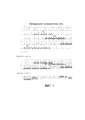
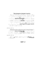
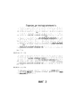
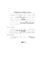
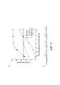
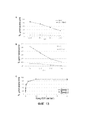
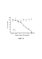
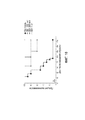
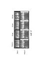
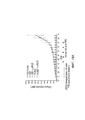
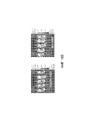
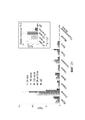
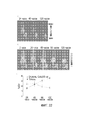
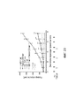
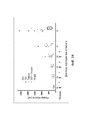
Classifications
-
- A—HUMAN NECESSITIES
- A61—MEDICAL OR VETERINARY SCIENCE; HYGIENE
- A61K—PREPARATIONS FOR MEDICAL, DENTAL OR TOILETRY PURPOSES
- A61K31/00—Medicinal preparations containing organic active ingredients
- A61K31/33—Heterocyclic compounds
- A61K31/335—Heterocyclic compounds having oxygen as the only ring hetero atom, e.g. fungichromin
- A61K31/337—Heterocyclic compounds having oxygen as the only ring hetero atom, e.g. fungichromin having four-membered rings, e.g. taxol
-
- A—HUMAN NECESSITIES
- A61—MEDICAL OR VETERINARY SCIENCE; HYGIENE
- A61K—PREPARATIONS FOR MEDICAL, DENTAL OR TOILETRY PURPOSES
- A61K39/00—Medicinal preparations containing antigens or antibodies
- A61K39/0005—Vertebrate antigens
- A61K39/0011—Cancer antigens
-
- A—HUMAN NECESSITIES
- A61—MEDICAL OR VETERINARY SCIENCE; HYGIENE
- A61K—PREPARATIONS FOR MEDICAL, DENTAL OR TOILETRY PURPOSES
- A61K39/00—Medicinal preparations containing antigens or antibodies
- A61K39/395—Antibodies; Immunoglobulins; Immune serum, e.g. antilymphocytic serum
- A61K39/39533—Antibodies; Immunoglobulins; Immune serum, e.g. antilymphocytic serum against materials from animals
- A61K39/39558—Antibodies; Immunoglobulins; Immune serum, e.g. antilymphocytic serum against materials from animals against tumor tissues, cells, antigens
-
- A—HUMAN NECESSITIES
- A61—MEDICAL OR VETERINARY SCIENCE; HYGIENE
- A61K—PREPARATIONS FOR MEDICAL, DENTAL OR TOILETRY PURPOSES
- A61K45/00—Medicinal preparations containing active ingredients not provided for in groups A61K31/00 - A61K41/00
- A61K45/06—Mixtures of active ingredients without chemical characterisation, e.g. antiphlogistics and cardiaca
-
- A—HUMAN NECESSITIES
- A61—MEDICAL OR VETERINARY SCIENCE; HYGIENE
- A61K—PREPARATIONS FOR MEDICAL, DENTAL OR TOILETRY PURPOSES
- A61K51/00—Preparations containing radioactive substances for use in therapy or testing in vivo
- A61K51/02—Preparations containing radioactive substances for use in therapy or testing in vivo characterised by the carrier, i.e. characterised by the agent or material covalently linked or complexing the radioactive nucleus
- A61K51/04—Organic compounds
- A61K51/08—Peptides, e.g. proteins, carriers being peptides, polyamino acids, proteins
- A61K51/10—Antibodies or immunoglobulins; Fragments thereof, the carrier being an antibody, an immunoglobulin or a fragment thereof, e.g. a camelised human single domain antibody or the Fc fragment of an antibody
- A61K51/1027—Antibodies or immunoglobulins; Fragments thereof, the carrier being an antibody, an immunoglobulin or a fragment thereof, e.g. a camelised human single domain antibody or the Fc fragment of an antibody against receptors, cell-surface antigens or cell-surface determinants
-
- A—HUMAN NECESSITIES
- A61—MEDICAL OR VETERINARY SCIENCE; HYGIENE
- A61K—PREPARATIONS FOR MEDICAL, DENTAL OR TOILETRY PURPOSES
- A61K51/00—Preparations containing radioactive substances for use in therapy or testing in vivo
- A61K51/02—Preparations containing radioactive substances for use in therapy or testing in vivo characterised by the carrier, i.e. characterised by the agent or material covalently linked or complexing the radioactive nucleus
- A61K51/04—Organic compounds
- A61K51/08—Peptides, e.g. proteins, carriers being peptides, polyamino acids, proteins
- A61K51/10—Antibodies or immunoglobulins; Fragments thereof, the carrier being an antibody, an immunoglobulin or a fragment thereof, e.g. a camelised human single domain antibody or the Fc fragment of an antibody
- A61K51/1045—Antibodies or immunoglobulins; Fragments thereof, the carrier being an antibody, an immunoglobulin or a fragment thereof, e.g. a camelised human single domain antibody or the Fc fragment of an antibody against animal or human tumor cells or tumor cell determinants
- A61K51/1057—Antibodies or immunoglobulins; Fragments thereof, the carrier being an antibody, an immunoglobulin or a fragment thereof, e.g. a camelised human single domain antibody or the Fc fragment of an antibody against animal or human tumor cells or tumor cell determinants the tumor cell being from liver or pancreas
-
- A—HUMAN NECESSITIES
- A61—MEDICAL OR VETERINARY SCIENCE; HYGIENE
- A61K—PREPARATIONS FOR MEDICAL, DENTAL OR TOILETRY PURPOSES
- A61K51/00—Preparations containing radioactive substances for use in therapy or testing in vivo
- A61K51/02—Preparations containing radioactive substances for use in therapy or testing in vivo characterised by the carrier, i.e. characterised by the agent or material covalently linked or complexing the radioactive nucleus
- A61K51/04—Organic compounds
- A61K51/08—Peptides, e.g. proteins, carriers being peptides, polyamino acids, proteins
- A61K51/10—Antibodies or immunoglobulins; Fragments thereof, the carrier being an antibody, an immunoglobulin or a fragment thereof, e.g. a camelised human single domain antibody or the Fc fragment of an antibody
- A61K51/1093—Antibodies or immunoglobulins; Fragments thereof, the carrier being an antibody, an immunoglobulin or a fragment thereof, e.g. a camelised human single domain antibody or the Fc fragment of an antibody conjugates with carriers being antibodies
-
- A—HUMAN NECESSITIES
- A61—MEDICAL OR VETERINARY SCIENCE; HYGIENE
- A61P—SPECIFIC THERAPEUTIC ACTIVITY OF CHEMICAL COMPOUNDS OR MEDICINAL PREPARATIONS
- A61P35/00—Antineoplastic agents
-
- A—HUMAN NECESSITIES
- A61—MEDICAL OR VETERINARY SCIENCE; HYGIENE
- A61P—SPECIFIC THERAPEUTIC ACTIVITY OF CHEMICAL COMPOUNDS OR MEDICINAL PREPARATIONS
- A61P37/00—Drugs for immunological or allergic disorders
- A61P37/02—Immunomodulators
- A61P37/04—Immunostimulants
-
- A—HUMAN NECESSITIES
- A61—MEDICAL OR VETERINARY SCIENCE; HYGIENE
- A61P—SPECIFIC THERAPEUTIC ACTIVITY OF CHEMICAL COMPOUNDS OR MEDICINAL PREPARATIONS
- A61P43/00—Drugs for specific purposes, not provided for in groups A61P1/00-A61P41/00
-
- C—CHEMISTRY; METALLURGY
- C07—ORGANIC CHEMISTRY
- C07K—PEPTIDES
- C07K16/00—Immunoglobulins [IGs], e.g. monoclonal or polyclonal antibodies
- C07K16/18—Immunoglobulins [IGs], e.g. monoclonal or polyclonal antibodies against material from animals or humans
- C07K16/28—Immunoglobulins [IGs], e.g. monoclonal or polyclonal antibodies against material from animals or humans against receptors, cell surface antigens or cell surface determinants
- C07K16/2896—Immunoglobulins [IGs], e.g. monoclonal or polyclonal antibodies against material from animals or humans against receptors, cell surface antigens or cell surface determinants against molecules with a "CD"-designation, not provided for elsewhere
-
- C—CHEMISTRY; METALLURGY
- C07—ORGANIC CHEMISTRY
- C07K—PEPTIDES
- C07K16/00—Immunoglobulins [IGs], e.g. monoclonal or polyclonal antibodies
- C07K16/18—Immunoglobulins [IGs], e.g. monoclonal or polyclonal antibodies against material from animals or humans
- C07K16/28—Immunoglobulins [IGs], e.g. monoclonal or polyclonal antibodies against material from animals or humans against receptors, cell surface antigens or cell surface determinants
- C07K16/30—Immunoglobulins [IGs], e.g. monoclonal or polyclonal antibodies against material from animals or humans against receptors, cell surface antigens or cell surface determinants from tumour cells
- C07K16/3076—Immunoglobulins [IGs], e.g. monoclonal or polyclonal antibodies against material from animals or humans against receptors, cell surface antigens or cell surface determinants from tumour cells against structure-related tumour-associated moieties
-
- G—PHYSICS
- G01—MEASURING; TESTING
- G01N—INVESTIGATING OR ANALYSING MATERIALS BY DETERMINING THEIR CHEMICAL OR PHYSICAL PROPERTIES
- G01N33/00—Investigating or analysing materials by specific methods not covered by groups G01N1/00 - G01N31/00
- G01N33/48—Biological material, e.g. blood, urine; Haemocytometers
- G01N33/50—Chemical analysis of biological material, e.g. blood, urine; Testing involving biospecific ligand binding methods; Immunological testing
- G01N33/53—Immunoassay; Biospecific binding assay; Materials therefor
- G01N33/574—Immunoassay; Biospecific binding assay; Materials therefor for cancer
- G01N33/57407—Specifically defined cancers
- G01N33/57438—Specifically defined cancers of liver, pancreas or kidney
-
- G—PHYSICS
- G01—MEASURING; TESTING
- G01N—INVESTIGATING OR ANALYSING MATERIALS BY DETERMINING THEIR CHEMICAL OR PHYSICAL PROPERTIES
- G01N33/00—Investigating or analysing materials by specific methods not covered by groups G01N1/00 - G01N31/00
- G01N33/48—Biological material, e.g. blood, urine; Haemocytometers
- G01N33/50—Chemical analysis of biological material, e.g. blood, urine; Testing involving biospecific ligand binding methods; Immunological testing
- G01N33/53—Immunoassay; Biospecific binding assay; Materials therefor
- G01N33/577—Immunoassay; Biospecific binding assay; Materials therefor involving monoclonal antibodies binding reaction mechanisms characterised by the use of monoclonal antibodies; monoclonal antibodies per se are classified with their corresponding antigens
-
- A—HUMAN NECESSITIES
- A61—MEDICAL OR VETERINARY SCIENCE; HYGIENE
- A61K—PREPARATIONS FOR MEDICAL, DENTAL OR TOILETRY PURPOSES
- A61K39/00—Medicinal preparations containing antigens or antibodies
- A61K2039/505—Medicinal preparations containing antigens or antibodies comprising antibodies
-
- A—HUMAN NECESSITIES
- A61—MEDICAL OR VETERINARY SCIENCE; HYGIENE
- A61K—PREPARATIONS FOR MEDICAL, DENTAL OR TOILETRY PURPOSES
- A61K39/00—Medicinal preparations containing antigens or antibodies
- A61K2039/60—Medicinal preparations containing antigens or antibodies characteristics by the carrier linked to the antigen
- A61K2039/6031—Proteins
- A61K2039/6081—Albumin; Keyhole limpet haemocyanin [KLH]
-
- C—CHEMISTRY; METALLURGY
- C07—ORGANIC CHEMISTRY
- C07K—PEPTIDES
- C07K2317/00—Immunoglobulins specific features
- C07K2317/20—Immunoglobulins specific features characterized by taxonomic origin
- C07K2317/21—Immunoglobulins specific features characterized by taxonomic origin from primates, e.g. man
-
- C—CHEMISTRY; METALLURGY
- C07—ORGANIC CHEMISTRY
- C07K—PEPTIDES
- C07K2317/00—Immunoglobulins specific features
- C07K2317/30—Immunoglobulins specific features characterized by aspects of specificity or valency
- C07K2317/33—Crossreactivity, e.g. for species or epitope, or lack of said crossreactivity
-
- C—CHEMISTRY; METALLURGY
- C07—ORGANIC CHEMISTRY
- C07K—PEPTIDES
- C07K2317/00—Immunoglobulins specific features
- C07K2317/50—Immunoglobulins specific features characterized by immunoglobulin fragments
- C07K2317/52—Constant or Fc region; Isotype
-
- C—CHEMISTRY; METALLURGY
- C07—ORGANIC CHEMISTRY
- C07K—PEPTIDES
- C07K2317/00—Immunoglobulins specific features
- C07K2317/50—Immunoglobulins specific features characterized by immunoglobulin fragments
- C07K2317/56—Immunoglobulins specific features characterized by immunoglobulin fragments variable (Fv) region, i.e. VH and/or VL
-
- C—CHEMISTRY; METALLURGY
- C07—ORGANIC CHEMISTRY
- C07K—PEPTIDES
- C07K2317/00—Immunoglobulins specific features
- C07K2317/60—Immunoglobulins specific features characterized by non-natural combinations of immunoglobulin fragments
- C07K2317/62—Immunoglobulins specific features characterized by non-natural combinations of immunoglobulin fragments comprising only variable region components
- C07K2317/626—Diabody or triabody
-
- C—CHEMISTRY; METALLURGY
- C07—ORGANIC CHEMISTRY
- C07K—PEPTIDES
- C07K2317/00—Immunoglobulins specific features
- C07K2317/70—Immunoglobulins specific features characterized by effect upon binding to a cell or to an antigen
- C07K2317/73—Inducing cell death, e.g. apoptosis, necrosis or inhibition of cell proliferation
- C07K2317/732—Antibody-dependent cellular cytotoxicity [ADCC]
-
- C—CHEMISTRY; METALLURGY
- C07—ORGANIC CHEMISTRY
- C07K—PEPTIDES
- C07K2317/00—Immunoglobulins specific features
- C07K2317/70—Immunoglobulins specific features characterized by effect upon binding to a cell or to an antigen
- C07K2317/73—Inducing cell death, e.g. apoptosis, necrosis or inhibition of cell proliferation
- C07K2317/734—Complement-dependent cytotoxicity [CDC]
-
- C—CHEMISTRY; METALLURGY
- C07—ORGANIC CHEMISTRY
- C07K—PEPTIDES
- C07K2317/00—Immunoglobulins specific features
- C07K2317/70—Immunoglobulins specific features characterized by effect upon binding to a cell or to an antigen
- C07K2317/77—Internalization into the cell
-
- C—CHEMISTRY; METALLURGY
- C07—ORGANIC CHEMISTRY
- C07K—PEPTIDES
- C07K2317/00—Immunoglobulins specific features
- C07K2317/90—Immunoglobulins specific features characterized by (pharmaco)kinetic aspects or by stability of the immunoglobulin
- C07K2317/92—Affinity (KD), association rate (Ka), dissociation rate (Kd) or EC50 value
Abstract
Description
По настоящей заявке испрашивается преимущество приоритета предварительной заявки США с серийным номером 61/870137, поданной 26 августа 2013 г., полное содержание которой включено в данное описание в качестве ссылки.This application claims priority advantage of the provisional application of the United States with
Это изобретение сделано при поддержке правительства с номером гранта CA-128362, присужденного Национальным институтом рака, NIH. Правительство имеет определенные права на данное изобретение.This invention was made with the support of the government with grant number CA-128362 awarded by the National Cancer Institute, NIH. The government has certain rights to this invention.
УРОВЕНЬ ТЕХНИКИ, К КОТОРОМУ ОТНОСИТСЯ ИЗОБРЕТЕНИЕBACKGROUND OF THE INVENTION
Настоящее изобретение в целом относится к антителам, направленным против сиалированного антигена Льюисаа (sLea), и более конкретно к полинуклеотидам, кодирующим антитела против sLea, и соответствующим кодируемым антителам или их фрагментам.The present invention generally relates to antibodies directed against the sialylated Lewis antigen a (sLe a ), and more particularly to polynucleotides encoding anti-sLe a antibodies and correspondingly encoded antibodies or fragments thereof.
Пассивное введение антител, направленных против опухолевых антигенов, может уничтожать опухолевые клетки и ранние метастазы в процессе развития рака. Это лечение может также иметь значительное влияние на рецидивы рака. Антитела, направленные против специфических опухолевых углеводов, могут быть полезными кандидатами при таком лечении рака. Например, многие моноклональные антитела, направленность которых ограничена опухолью, вырабатываемые в результате иммунизации мышей раковыми клетками человека, как показано, направлены против углеводных антигенов, экспрессируемых на клеточной поверхности в виде гликолипидов или гликопротеинов. Углевод sLea, как показано, экспрессируется в опухолях желудочно-кишечного тракта. Экспрессия sLea, как также обнаружено, вносит вклад в метастатический потенциал и коррелирует с повышенным метастатическим потенциалом при развитии рака толстой кишки человека и аденокарциномы поджелудочной железы. Тем не менее, химия углеводов является довольно сложной, и клиническая разработка антител, которые распознают такие специфические опухолевые углеводы, развивается медленно.Passive administration of antibodies directed against tumor antigens can destroy tumor cells and early metastases during the development of cancer. This treatment can also have a significant effect on cancer recurrence. Antibodies directed against specific tumor carbohydrates may be useful candidates for such cancer treatments. For example, many monoclonal antibodies whose orientation is limited to tumors produced by immunizing mice with human cancer cells have been shown to be directed against carbohydrate antigens expressed on the cell surface as glycolipids or glycoproteins. Carbohydrate sLe a , as shown, is expressed in tumors of the gastrointestinal tract. The expression of sLe a has also been found to contribute to the metastatic potential and correlates with increased metastatic potential in the development of human colon cancer and pancreatic adenocarcinoma. However, the chemistry of carbohydrates is quite complex, and the clinical development of antibodies that recognize such specific tumor carbohydrates is developing slowly.
Карцинома поджелудочной железы является одной из самых агрессивных аденокарцином и часто связана с плохим прогнозом. Карцинома поджелудочной железы рассматривается как четвертая ведущая причина смертности от рака. Несмотря на успехи в скрининге различных карцином, надежность обнаружения злокачественных поражений, возникающих в поджелудочной железе, остается на низком уровне. Позитронно-эмиссионная томография с использованием фтордезоксиглюкозы (FDG-PET) требуется для выявления и определения стадии рака поджелудочной железы. Тем не менее, FDG-PET не чувствительна к дифференциальной диагностике панкреатита и злокачественных новообразований, и проблематичным остается определение стадии при небольших первичных поражениях (<7 мм) и печеночных метастазах (<1 см). Один диагностический метод скрининга, используемый для мониторинга статуса протоковой аденокарциномы поджелудочной железы (PDAC) у больных, включает обнаружение повышенных уровней циркулирующего антигена sLea в сыворотке крови. Циркулирующий антиген sLea>37 Ед/мл у больных указывает на рецидив рака. Однако развитие альтернативных диагностических подходов, которые используют такие специфические опухолевые углеводы, происходит медленно.Pancreatic carcinoma is one of the most aggressive adenocarcinomas and is often associated with poor prognosis. Pancreatic carcinoma is considered the fourth leading cause of cancer mortality. Despite successes in screening various carcinomas, the reliability of detection of malignant lesions that occur in the pancreas remains low. Fluorodeoxyglucose positron emission tomography (FDG-PET) is required to identify and determine the stage of pancreatic cancer. However, FDG-PET is not sensitive to the differential diagnosis of pancreatitis and malignant neoplasms, and staging for small primary lesions (<7 mm) and hepatic metastases (<1 cm) remains problematic. One diagnostic screening method used to monitor pancreatic ductal adenocarcinoma (PDAC) status of patients, comprises detecting elevated levels of circulating antigen sLe a serum. The circulating antigen sLe a > 37 U / ml in patients indicates a relapse of the cancer. However, the development of alternative diagnostic approaches that use such specific tumor carbohydrates is slow.
Таким образом, существует необходимость в идентификации и выработке антител, которые специфически распознают специфические опухолевые углеводы, такие как sLea, для лечения рецидивирующих форм рака и для обнаружения злокачественных поражений и метастазов. Настоящее изобретение удовлетворяет эту потребность и обеспечивает связанные с этим преимущества.Thus, there is a need for the identification and production of antibodies that specifically recognize specific tumor carbohydrates, such as sLe a , for the treatment of recurrent forms of cancer and for the detection of malignant lesions and metastases. The present invention satisfies this need and provides related benefits.
СУЩНОСТЬ ИЗОБРЕТЕНИЯSUMMARY OF THE INVENTION
В соответствии с настоящим изобретением в настоящем документе предлагаются композиции для получения антител или их функциональных фрагментов, которые связываются с sLea. Композиции включают выделенный полинуклеотид, кодирующий антитело или его функциональный фрагмент, где антитело включает вариабельный домен тяжелой цепи (VH), который имеет аминокислотную последовательность, представленную в настоящем документе. Выделенный полинуклеотид по изобретению может также включать последовательность нуклеиновой кислоты, представленную в данном описании, где последовательность нуклеиновой кислоты кодирует домен VH антитела или его функционального фрагмента.In accordance with the present invention, there are provided compositions for producing antibodies or functional fragments thereof that bind to sLe a . Compositions include an isolated polynucleotide encoding an antibody or functional fragment thereof, wherein the antibody comprises a heavy chain variable domain (VH) that has the amino acid sequence provided herein. An isolated polynucleotide of the invention may also include the nucleic acid sequence described herein, wherein the nucleic acid sequence encodes the VH domain of an antibody or a functional fragment thereof.
В другом варианте осуществления настоящего изобретения выделенный полинуклеотид может кодировать антитело или его функциональный фрагмент, где антитело включает вариабельный домен легкой цепи (VL), который имеет аминокислотную последовательность, представленную в настоящем документе. Выделенный полинуклеотид по изобретению может также включать последовательность нуклеиновой кислоты, представленную в данном описании, где последовательность нуклеиновой кислоты кодирует домен VL антитела или его функционального фрагмента.In another embodiment of the present invention, the isolated polynucleotide may encode an antibody or a functional fragment thereof, wherein the antibody comprises a light chain variable domain (VL) that has the amino acid sequence described herein. An isolated polynucleotide of the invention may also include the nucleic acid sequence described herein, wherein the nucleic acid sequence encodes the VL domain of an antibody or a functional fragment thereof.
Композиции по настоящему изобретению также включают выделенное антитело или его функциональный фрагмент, где антитело связывается с sLea. В некоторых вариантах осуществления настоящее изобретение относится к выделенному антителу или его функциональному фрагменту, который связывается с sLea, где антитело или его функциональный фрагмент включает домен VH, имеющий аминокислотную последовательность, представленную в настоящем документе.The compositions of the present invention also include an isolated antibody or a functional fragment thereof, wherein the antibody binds to sLe a . In some embodiments, the present invention relates to an isolated antibody or functional fragment thereof that binds to sLe a , wherein the antibody or functional fragment thereof includes a VH domain having the amino acid sequence described herein.
В некоторых вариантах осуществления настоящее изобретение относится к выделенному антителу или его функциональному фрагменту, который связывается с sLea, где антитело или его функциональный фрагмент включает домен VL, имеющий аминокислотную последовательность, представленную в настоящем документе.In some embodiments, the present invention relates to an isolated antibody or functional fragment thereof that binds to sLe a , where the antibody or functional fragment thereof comprises a VL domain having the amino acid sequence described herein.
В некоторых вариантах осуществления настоящее изобретение относится к выделенному антителу или его функциональному фрагменту, который связывается с sLea, где антитело или его функциональный фрагмент включает как домен VH, так и домен VL, где домен VH и домен VL соответственно включают аминокислотную последовательность соответствующих доменов VH и VL изолятов клонов, представленных в настоящем описании.In some embodiments, the present invention relates to an isolated antibody or functional fragment thereof that binds to sLe a , where the antibody or functional fragment thereof includes both a VH domain and a VL domain, where the VH domain and VL domain respectively comprise the amino acid sequence of the corresponding VH domains and VL isolates of the clones described herein.
В некоторых вариантах осуществления в настоящем изобретении предлагается конъюгат, включающий антитело или его функциональный фрагмент, предлагаемые в данном документе, конъюгированные или соединенные рекомбинантным способом с диагностическим агентом, детектируемым агентом или терапевтическим агентом. В некоторых аспектах настоящего изобретения целью является конъюгат по настоящему изобретению, включающий детектируемый агент, который может быть использован в способе обнаружения и/или диагностики образования опухоли. Такие способы могут включать введение эффективного количества конъюгата нуждающемуся в этом индивидууму.In some embodiments, the present invention provides a conjugate comprising an antibody or a functional fragment thereof as provided herein, conjugated or recombinantly coupled to a diagnostic agent, a detectable agent, or a therapeutic agent. In some aspects of the present invention, the objective is the conjugate of the present invention, comprising a detectable agent that can be used in a method for detecting and / or diagnosing tumor formation. Such methods may include administering an effective amount of a conjugate to a person in need thereof.
В некоторых вариантах осуществления настоящее изобретение относится к фармацевтическим композициям, включающим одно или более антитело или его функциональный фрагмент по изобретению и фармацевтически приемлемый носитель. В некоторых аспектах настоящее изобретение относится также к способу лечения или профилактики заболевания у нуждающегося в этом индивидуума путем введения терапевтически эффективного количества фармацевтической композиции по настоящему изобретению. В еще одном аспекте настоящее изобретение относится к введению второго терапевтического агента одновременно или последовательно с антителом или его функциональным фрагментом по изобретению.In some embodiments, the present invention relates to pharmaceutical compositions comprising one or more antibodies or a functional fragment thereof of the invention and a pharmaceutically acceptable carrier. In some aspects, the present invention also relates to a method for treating or preventing a disease in a person in need thereof by administering a therapeutically effective amount of a pharmaceutical composition of the present invention. In another aspect, the present invention relates to the administration of a second therapeutic agent simultaneously or sequentially with the antibody or its functional fragment according to the invention.
КРАТКОЕ ОПИСАНИЕ ЧЕРТЕЖЕЙBRIEF DESCRIPTION OF THE DRAWINGS
На фиг. 1 показана нуклеотидная последовательность и кодируемая аминокислотная последовательность вариабельного домена тяжелой (VH) цепи клона 5B1 и лидирующая последовательность, которая может быть использована для рекомбинантной экспрессии. В верхней части фигуры показано выравнивание между нуклеотидной последовательностью SEQ ID NO: 1 и аминокислотной последовательностью SEQ ID NO: 2. Идентифицированы также три области, определяющие комплементарность (CDR1, CDR2 и CDR3).In FIG. 1 shows the nucleotide sequence and the encoded amino acid sequence of the variable domain of the heavy (VH) chain of clone 5B1 and the leading sequence that can be used for recombinant expression. The top of the figure shows the alignment between the nucleotide sequence of SEQ ID NO: 1 and the amino acid sequence of SEQ ID NO: 2. Three complementarity determining regions were also identified (CDR1, CDR2 and CDR3).
На фиг. 2 показана нуклеотидная последовательность и кодируемая аминокислотная последовательность вариабельного домена легкой (VL) цепи клона 5B1 и лидирующая последовательность, которая может быть использована для рекомбинантной экспрессии. В верхней части фигуры показано выравнивание между нуклеотидной последовательностью SEQ ID NO: 3 и аминокислотной последовательностью SEQ ID NO: 4. Идентифицированы также три области, определяющие комплементарность (CDR1, CDR2 и CDR3).In FIG. 2 shows the nucleotide sequence and the encoded amino acid sequence of the variable domain of the light domain (VL) of the clone 5B1 chain and the leading sequence that can be used for recombinant expression. The top of the figure shows the alignment between the nucleotide sequence of SEQ ID NO: 3 and the amino acid sequence of SEQ ID NO: 4. Three complementarity determining regions were also identified (CDR1, CDR2 and CDR3).
На фиг. 3 показана нуклеотидная последовательность и кодируемая аминокислотная последовательность вариабельного домена тяжелой (VH) цепи клона 9H3 и лидирующая последовательность, которая может быть использована для рекомбинантной экспрессии. В верхней части фигуры показано выравнивание между нуклеотидной последовательностью SEQ ID NO: 5 и аминокислотной последовательностью SEQ ID NO: 6. Идентифицированы также три области, определяющие комплементарность (CDR1, CDR2 и CDR3).In FIG. 3 shows the nucleotide sequence and the encoded amino acid sequence of the variable domain of the heavy (VH) chain of clone 9H3 and the leader sequence that can be used for recombinant expression. The top of the figure shows the alignment between the nucleotide sequence of SEQ ID NO: 5 and the amino acid sequence of SEQ ID NO: 6. Three complementarity determining regions were also identified (CDR1, CDR2 and CDR3).
На фиг. 4 показана нуклеотидная последовательность и кодируемая аминокислотная последовательность вариабельного домена легкой (VL) цепи клона 9H3 и лидирующая последовательность, которая может быть использована для рекомбинантной экспрессии. В верхней части фигуры показано выравнивание между нуклеотидной последовательностью SEQ ID NO: 7 и аминокислотной последовательностью SEQ ID NO: 8. Идентифицированы также три области, определяющие комплементарность (CDR1, CDR2 и CDR3).In FIG. 4 shows the nucleotide sequence and the encoded amino acid sequence of the variable domain of the light (VL) chain of clone 9H3 and the leading sequence that can be used for recombinant expression. The top of the figure shows the alignment between the nucleotide sequence of SEQ ID NO: 7 and the amino acid sequence of SEQ ID NO: 8. Three regions of complementarity were also identified (CDR1, CDR2 and CDR3).
На фиг. 5 показана нуклеотидная последовательность и кодируемая аминокислотная последовательность вариабельного домена тяжелой (VH) цепи клона 5H11 и лидирующая последовательность, которая может быть использована для рекомбинантной экспрессии. В верхней части фигуры показано выравнивание между нуклеотидной последовательностью SEQ ID NO: 9 и аминокислотной последовательностью SEQ ID NO: 10. Идентифицированы также три области, определяющие комплементарность (CDR1, CDR2 и CDR3).In FIG. 5 shows the nucleotide sequence and the encoded amino acid sequence of the variable domain of the heavy (VH) chain of clone 5H11 and the leading sequence that can be used for recombinant expression. The top of the figure shows the alignment between the nucleotide sequence of SEQ ID NO: 9 and the amino acid sequence of SEQ ID NO: 10. Three complementarity determining regions were also identified (CDR1, CDR2 and CDR3).
На фиг. 6 показана нуклеотидная последовательность и кодируемая аминокислотная последовательность вариабельного домена легкой (VL) цепи клона 5H11 и лидирующая последовательность, которая может быть использована для рекомбинантной экспрессии. В верхней части фигуры показано выравнивание между нуклеотидной последовательностью SEQ ID NO: 11 и аминокислотной последовательностью SEQ ID NO: 12. Идентифицированы также три области, определяющие комплементарность (CDR1, CDR2 и CDR3).In FIG. Figure 6 shows the nucleotide sequence and the encoded amino acid sequence of the variable domain of the light (VL) chain of clone 5H11 and the leading sequence that can be used for recombinant expression. The top of the figure shows the alignment between the nucleotide sequence of SEQ ID NO: 11 and the amino acid sequence of SEQ ID NO: 12. Three complementarity determining regions were also identified (CDR1, CDR2 and CDR3).
На фиг. 7 показана нуклеотидная последовательность и кодируемая аминокислотная последовательность вариабельного домена тяжелой (VH) цепи клона 7E3 и лидирующая последовательность, которая может быть использована для рекомбинантной экспрессии. В верхней части фигуры показано выравнивание между нуклеотидной последовательностью SEQ ID NO: 13 и аминокислотной последовательностью SEQ ID NO: 14. Идентифицированы также три области, определяющие комплементарность (CDR1, CDR2 и CDR3).In FIG. 7 shows the nucleotide sequence and the encoded amino acid sequence of the variable domain of the heavy (VH) chain of clone 7E3 and the leading sequence that can be used for recombinant expression. The top of the figure shows the alignment between the nucleotide sequence of SEQ ID NO: 13 and the amino acid sequence of SEQ ID NO: 14. Three complementarity determining regions were also identified (CDR1, CDR2 and CDR3).
На фиг. 8 показана нуклеотидная последовательность и кодируемая аминокислотная последовательность вариабельного домена легкой (VL) цепи клона 7E3 и лидирующая последовательность, которая может быть использована для рекомбинантной экспрессии. В верхней части фигуры показано выравнивание между нуклеотидной последовательностью SEQ ID NO: 15 и аминокислотной последовательностью SEQ ID NO: 16. Идентифицированы также три области, определяющие комплементарность (CDR1, CDR2 и CDR3).In FIG. Figure 8 shows the nucleotide sequence and the encoded amino acid sequence of the variable domain of the light domain (VL) of clone 7E3 and the leader sequence that can be used for recombinant expression. The top of the figure shows the alignment between the nucleotide sequence of SEQ ID NO: 15 and the amino acid sequence of SEQ ID NO: 16. Three complementarity determining regions were also identified (CDR1, CDR2 and CDR3).
На фиг. 9 показана нуклеотидная последовательность и кодируемая аминокислотная последовательность диатела, обозначаемого как 5B1CysDb, имеющего CDR1, CDR2 и CDR2 как вариабельного домена тяжелой (VH) цепи, так и вариабельного домена легкой (VL) цепи клона 5B1. В верхней части фигуры показано выравнивание между нуклеотидной последовательностью SEQ ID NO: 17 и аминокислотной последовательностью SEQ ID NO: 18. Идентифицированы три области, определяющие комплементарность (CDR1, CDR2 и CDR3), обоих доменов VH и VL, обозначенные жирным шрифтом и подчеркиванием текста. Последовательность линкера и полигистидиновой метки (Poly His-Tag) с добавленными аминокислотами также обозначена курсивом и подчеркиванием текста.In FIG. 9 shows the nucleotide sequence and the encoded amino acid sequence of a diabody designated as 5B1CysDb having CDR1, CDR2 and CDR2 of both the variable domain of the heavy (VH) chain and the variable domain of the light (VL) chain of clone 5B1. The top of the figure shows the alignment between the nucleotide sequence of SEQ ID NO: 17 and the amino acid sequence of SEQ ID NO: 18. Three regions of complementarity were identified (CDR1, CDR2 and CDR3), both domains VH and VL, indicated in bold and underlined text. The sequence of the linker and the polyhistidine tag (Poly His-Tag) with added amino acids is also indicated in italics and underlining the text.
На фиг. 10 показана нуклеотидная последовательность и кодируемая аминокислотная последовательность диатела, обозначаемого как 7E3CysDb, имеющего CDR1, CDR2 и CDR2 как вариабельного домена тяжелой (VH) цепи, так и вариабельного домена легкой (VL) цепи клона 7E3. В верхней части фигуры показано выравнивание между нуклеотидной последовательностью SEQ ID NO: 19 и аминокислотной последовательностью SEQ ID NO: 20. Идентифицированы три области, определяющие комплементарность (CDR1, CDR2 и CDR3), обоих доменов VH и VL, обозначенные жирным шрифтом и подчеркиванием текста. Последовательность линкера и полигистидиновой метки (Poly His-Tag) с добавленными аминокислотами также обозначена курсивом и подчеркиванием текста.In FIG. 10 shows the nucleotide sequence and the encoded amino acid sequence of a diabody designated as 7E3CysDb having CDR1, CDR2 and CDR2 of both the variable domain of the heavy (VH) chain and the variable domain of the light (VL) chain of clone 7E3. The top of the figure shows the alignment between the nucleotide sequence of SEQ ID NO: 19 and the amino acid sequence of SEQ ID NO: 20. Three regions of complementarity (CDR1, CDR2 and CDR3) were identified, both VH and VL domains, indicated in bold and underlined text. The sequence of the linker and the polyhistidine tag (Poly His-Tag) with added amino acids is also indicated in italics and underlining the text.
На фиг. 11, панели A-E, показано связывание антител человека против sLea с опухолевыми клетками, проанализированное с помощью проточной цитометрии. На панели A показаны клетки DMS-79, окрашенные рекомбинантными (r) антителами 5B1, 9H3, 5H11 и 7Е3. На панелях B-F, соответственно, показаны клетки HT29, BXPC3, SW626, SK-MEL28 и Colo205-Luc, окрашенные 1-2 мкг/мл r5Bl или r7E3 плюс IgG- или IgM-специфичными вторичными антителами, как описано в примере I.In FIG. 11, panel AE, shows the binding of human antibodies against sLe a to tumor cells, analyzed by flow cytometry. Panel A shows DMS-79 cells stained with recombinant (r) antibodies 5B1, 9H3, 5H11 and 7E3. BF panels respectively show HT29, BXPC3, SW626, SK-MEL28 and Colo205-Luc cells stained with 1-2 μg / ml r5Bl or r7E3 plus IgG or IgM-specific secondary antibodies, as described in Example I.
На фиг. 12, панели А и В, показана активность CDC антител r5Bl и r7E3 по сравнению с мышиными 121SLE (IgM) в присутствии комплемента человека (Hu C') при измерении относительно клеток DMS-79. Контрольные антитела изотипа человека, Hu IgG ![]()
![]()
![]()
![]()
![]()
![]()
![]()
![]()
![]()
![]()
На фиг. 13, панели A-C, показана зависимая от антител опосредуемая клетками цитотоксичность (ADCC) антител r5Bl. На панели А показана опосредуемая r5Bl ADCC РВМС человека против клеток DMS-79. РВМС тестировали в отношениях E:T от 100:1 до 12,5:1 с опухолевыми клетками DMS-79 в присутствии или в отсутствие 2 мкг/мл r5Bl. На панели B показана опосредуемая r5Bl ADCC первичных NK-клеток человека против клеток DMS-79. NK-клетки тестировали при более низких отношениях E:T от 5:1 до 0,6:1 с опухолевыми клетками DMS-79 в присутствии или в отсутствие 2 мкг/мл r5Bl. На панели С показана ADCC антитела r5Bl при различных концентрациях PBMCs от 2-доноров при отношении E:T=1:100 против опухолевых клеток DMS-79 в присутствии указанных концентраций r5Bl.In FIG. 13, Panel A-C, shows antibody dependent cell mediated cytotoxicity (ADCC) of r5Bl antibodies. Panel A shows r5Bl-mediated ADCC human PBMC against DMS-79 cells. PBMCs were tested in E: T ratios of 100: 1 to 12.5: 1 with DMS-79 tumor cells in the presence or absence of 2 μg / ml r5Bl. Panel B shows r5Bl-mediated ADCC of primary human NK cells versus DMS-79 cells. NK cells were tested at lower E: T ratios of 5: 1 to 0.6: 1 with DMS-79 tumor cells in the presence or absence of 2 μg / ml r5Bl. Panel C shows ADCC r5Bl antibodies at various concentrations of PBMCs from 2 donors at an E: T = 1: 100 ratio against DMS-79 tumor cells in the presence of the indicated r5Bl concentrations.
На фиг. 14 показана интернализация sLea в клетки BxPC3. Опухолевые клетки BxPC3 поджелудочной железы выращивали в присутствии антител r5Bl (против sLea) или rlB7 (против GD2), связанных с Hum-ZAP, конъюгированным с сапорином IgG против антигенов человека. Через 3 дня измеряли жизнеспособность клеток методом с использованием бромида 3-(4,5-диметилтиазол-2-ил)-2,5-дифенилтетразолия (МТТ), и значения образцов нормализовывали на значения необработанных культур.In FIG. 14 shows the internalization of sLe a in BxPC3 cells. Pancreatic BxPC3 tumor cells were grown in the presence of r5Bl (anti-sLe a ) or rlB7 (anti-GD2) antibodies bound to Hum-ZAP conjugated with saporin IgG against human antigens. After 3 days, cell viability was measured using 3- (4,5-dimethylthiazol-2-yl) -2,5-diphenyltetrazolium bromide (MTT), and sample values were normalized to untreated cultures.
На фиг. 15 показана активность антитела r5Bl в модели ксенотрансплантата с использованием клеток Colo205-luc. Мыши с тяжелым сочетанным иммунодефицитом (SCID) (5 на группу) получали 0,5 миллиона клеток Colo205-luc путем инъекции в хвостовую вену на день 0. Мыши получали 100 мкг r5Bl путем внутрибрюшинной инъекции в дни 1, 7, 14 и 21 (эксперимент 1, Exp1) или в дни 1, 4, 7, 10, 14 и 21 (эксперимент 2, Exp2) в суммарной дозе 600 мкг. Контрольные (Ctrl) животные получали ложные инъекции PBS.In FIG. Figure 15 shows the activity of the r5Bl antibody in a xenograft model using Colo205-luc cells. Severe Combined Immunodeficiency Mice (SCID) mice (5 per group) received 0.5 million Colo205-luc cells by injection into the tail vein on
На фиг. 16 показано влияние r5Bl на опухоли Colo205-luc у мышей SCID. Мыши получали 100 мкг ![]()
![]()
![]()
![]()
![]()
![]()
![]()
![]()
На фиг. 17 показана флуоресцентная визуализация пяти мышей на группу для мышей, получавших r5Bl, имеющих опухоли Colo205-luc, в день 0 и через 5 недель. Схема получения препаратов мышами представлена на фиг. 16 и описана в примере I.In FIG. Figure 17 shows the fluorescence imaging of five mice per group for mice treated with r5Bl having Colo205-luc tumors at
На фиг. 18, панели А и В, показана противоопухолевая активность в терапевтической модели подкожного ксенотрансплантата с использованием клеток DMS-79. На панели А показано подавление или регрессия опухоли у мышей, получавших 5B1 (только 5B1 ![]()
![]()
![]()
![]()
![]()
![]()
![]()
![]()
![]()
![]()
На фиг. 19, панели A-F, показано связывание 5B1 с различными типами опухолей. Панель А представляет собой поджелудочную железу, протоковую аденокарциному, стадию III опухоли. Панель B представляет собой сигмовидную кишку, карциному стадии IIIB опухоли. Панель С представляет собой легкое, аденокарциному, стадию IB опухоли. Панель D представляет собой мочевой пузырь, мукозную аденокарциному, стадию IV опухоли. Панель Е представляет собой яичник, метастатическую карциному опухоли толстой кишки. Панель F представляет собой лимфатический узел, метастатическую карциному, стадию IIIA опухоли.In FIG. 19, panel A-F, shows the binding of 5B1 to various types of tumors. Panel A is a pancreas, ductal adenocarcinoma, stage III tumor. Panel B is the sigmoid colon, stage IIIB carcinoma of the tumor. Panel C is a lung, adenocarcinoma, stage IB tumor. Panel D is a bladder, mucosal adenocarcinoma, stage IV tumor. Panel E is an ovary, metastatic carcinoma of a colon tumor. Panel F is a lymph node, metastatic carcinoma, stage IIIA tumor.
На фиг. 20 показаны серийные изображения проекции максимальной интенсивности (MIP) PET, полученные через 2-120 час после внутривенного введения антитела 5Bl, радиоактивно меченного 89Zr (89Zr-5Bl), самкам мышей SCID с подкожно имплантированными опухолями поджелудочной железы BxPC3. Изображение PET-MIP демонстрирует высокий захват опухолью с клиренсом неспецифически связанной метки уже через 24 часа после инъекции (h p.i.)In FIG. Figure 20 shows serial images of the maximum intensity projection (MIP) PET obtained 2-120 hours after intravenous administration of 5Bl antibody radiolabeled 89 Zr ( 89 Zr-5Bl) to female SCID mice with subcutaneously implanted pancreatic tumors BxPC3. The PET-MIP image shows high tumor uptake with clearance of a non-specifically linked label already 24 hours after injection (h pi)
На фиг. 21 приведены результаты биологического распределения, которые согласуются с данными PET фиг. 20, с наблюдаемым захватом опухолью 84,73±2,28% ID/г. Из-за малой массы опухоли гистограмма захвата опухолью, выраженная в % ID, в зависимости от времени, изображена на гистограмме во вставке. % ID опухоли показывает значительный захват опухолью 89Zr-5Bl во всех временных точках, и, по меньшей мере, семикратное превышение неспецифического захвата 89Zr-IgG. Конкурентное ингибирование немеченым 5B1 (200 мкг) показывает уменьшение аккумуляции в опухоли.In FIG. 21 shows biological distribution results that are consistent with the PET data of FIG. 20, with an observed tumor uptake of 84.73 ± 2.28% ID / g. Due to the small mass of the tumor, the histogram of tumor uptake, expressed as% ID, versus time, is shown in the histogram in the box. The% tumor ID shows significant 89 Zr-5Bl tumor uptake at all time points, and at least a seven-fold increase in non-specific 89 Zr-IgG uptake. Competitive inhibition of unlabeled 5B1 (200 μg) shows a decrease in tumor accumulation.
На фиг. 22, панели A-C, показаны изображения PET-MIP мышей, несущих DMS79 (панель А) и ксенотрансплантатов Colo205-luc (панель B). Указана визуализация PET-MIP очертаний опухоли (T), сердца (H) и печени (L) с помощью 89Zr-5Bl. В модели ксенотрансплантатов Colo205-luc колоректального рака продемонстрирована аккумуляция 89Zr-5Bl, достигающая максимума через 24 час, с последующим снижением, в то время как выявлен рост неспецифического связывания в печени (панель С).In FIG. 22, panel AC, images of PET-MIP mice bearing DMS79 (panel A) and Colo205-luc xenografts (panel B) are shown. Indicated PET-MIP visualization of the outlines of the tumor (T), heart (H) and liver (L) using 89 Zr-5Bl. In the Colo205-luc xenograft model of colorectal cancer, an accumulation of 89 Zr-5Bl was demonstrated, reaching a maximum after 24 hours, followed by a decrease, while an increase in nonspecific binding in the liver was revealed (panel C).
На фиг. 23 показано зависимое от дозы ингибирование и регрессия роста опухоли в модели ксенотрансплантата мелкоклеточной легочной карциномы DMS-79, при лечении последовательным совместным введением антитела 5B1 и таксола (паклитаксела). Большие стрелки на оси Х указывают на лечение 5B1. Совместное введение антитела 5B1 и таксола существенно ограничивало рост опухоли и приводило к регрессии опухоли по сравнению с контрольным IgG человека (HuIgG) или антителом 5B1 и таксолом, вводимыми по отдельности. Показаны значимые отличия от контроля с помощью 2-факторного дисперсионного анализа ANOVA с р<0,01 (**) и р<0,001 (***). N=5.In FIG. Figure 23 shows dose-dependent inhibition and regression of tumor growth in a xenograft model of small cell lung carcinoma DMS-79, when treated with sequential co-administration of antibody 5B1 and taxol (paclitaxel). Large arrows on the X axis indicate 5B1 treatment. Co-administration of antibody 5B1 and taxol significantly limited tumor growth and led to regression of the tumor compared to control human IgG (HuIgG) or antibody 5B1 and taxol, administered individually. Significant differences from control were shown using the 2-factor ANOVA analysis of variance with p <0.01 (**) and p <0.001 (***). N = 5.
На фиг. 24 показано ингибирование роста опухоли в модели ксенотрансплантата карциномы поджелудочной железы BxPC3 при лечении последовательным совместным введением антитела 5B1 и таксола (паклитаксела). Большие стрелки на оси Х указывают на лечение таксолом плюс 5B1, в то время как маленькие стрелки указывают на лечение только 5B1. Совместное введение антитела 5B1 и таксола существенно ограничивало рост опухоли по сравнению с контролями (PBS - Ctrl; IgG человека - HuIgG) или антителом 5B1 и таксолом, вводимыми по отдельности.In FIG. 24 shows tumor growth inhibition in a BxPC3 pancreatic carcinoma xenograft model when treated with co-administration of 5B1 antibody and taxol (paclitaxel). Large arrows on the X axis indicate treatment with Taxol plus 5B1, while small arrows indicate treatment with only 5B1. Co-administration of antibody 5B1 and taxol significantly limited tumor growth compared to controls (PBS - Ctrl; human IgG - HuIgG) or antibody 5B1 and taxol administered individually.
На фиг. 25, панели А и В, показаны репрезентативные изображения мышей, которым ортотопически трансплантировали ксенотрансплантаты опухолей поджелудочной железы BxPC3-luc. Панель A: совместная регистрация с помощью FDG-PET и компьютерной томографии (КТ) (слева) планарных секций FDG-PET с изображением только (справа) минимально обнаруживаемой метки в опухоли с высоким захватом в тканях с высоким метаболизмом (т.е. в сердце, Н, и мочевом пузыре, В). Панель B: PET изображение той же мыши, выявляемое с помощью радиоактивно меченного 89Zr антитела 5Bl (89Zr-5Bl), регистрируемое совместно с КТ, показало детекцию исключительно опухоли в случае ксенотрансплантатов опухоли BxPC3-luc.In FIG. 25, panels A and B, are representative images of mice orthotopically transplanted with BxPC3-luc pancreatic tumor xenografts. Panel A: co-registration with FDG-PET and computed tomography (CT) (left) of planar sections of FDG-PET with an image of only (right) the minimum detectable label in a tumor with high uptake in tissues with high metabolism (i.e. in the heart) , N, and the bladder, B). Panel B: PET image of the same mouse, detected by radioactively labeled 89 Zr 5Bl antibody ( 89 Zr-5Bl), recorded with CT, showed exclusively tumor detection in the case of xenografts of the BxPC3-luc tumor.
ПОДРОБНОЕ ОПИСАНИЕ ИЗОБРЕТЕНИЯDETAILED DESCRIPTION OF THE INVENTION
Углеводы, экспрессируемые на поверхности опухолевых клеток, могут представлять собой мишень для пассивной иммунотерапии. Композиции, представленные в настоящем описании, основаны, по меньшей мере, частично, на идентификации и характеристике антител человека, которые вырабатываются лимфоцитами крови индивидуумов, иммунизированных вакциной, представляющей собой конъюгат сиалированного антигена Льюисаа и гемоцианина фиссуреллии (sLea-KLH). Идентифицированы, по меньшей мере, четыре антитела с высоким сродством к sLea (5B1, 9H3, 5H11 и 7E3). Два из этих антител экспрессировали в виде рекомбинантных антител (r5Bl и r7E3) и дополнительно характеризовали в моделях in vitro и in vivo. Оба антитела при анализе характеризовались сильной, зависимой от комплемента цитотоксичностью (CDC), и антитело 5B1 также было высокоактивным в зависимой от антител цитотоксичности. Эффективность антител in vivo тестировали в двух моделях ксенотрансплантатов с использованием либо опухолевых клеток Colo205, либо опухолевых клеток DMS-79, пересаживаемых мышам с тяжелым сочетанным иммунодефицитом (SCID). Междисциплинарная актуальность осуществления изобретения, предлагаемого в настоящем документе, рассматривается в 2 аспектах: во-первых, подход, предлагаемый в настоящем описании, показывает, что ответ в виде антител, вызванный вакциной sLea-KLH, пригоден в качестве вакцины как таковой. Во-вторых, самые мощные антитела, которые вырабатываются при клиническом испытании, могут быть сохранены и, в конечном счете, использованы в качестве терапевтических агентов или для создания терапевтических агентов для таргетной терапии рака у населения. Высокое сродство антител, представленных в настоящем описании, и их высокие эффекторные функции поддерживают этот междисциплинарный потенциал.Carbohydrates expressed on the surface of tumor cells can be a target for passive immunotherapy. The compositions provided herein are based, at least in part, on the identification and characterization of human antibodies that are produced by blood lymphocytes of individuals immunized with the vaccine, which is a sialylated Lewis antigen conjugate and a keyhole hemocyanin (sLe a -KLH). At least four antibodies with high affinity for sLe a have been identified (5B1, 9H3, 5H11 and 7E3). Two of these antibodies were expressed as recombinant antibodies (r5Bl and r7E3) and were further characterized in in vitro and in vivo models. Both antibodies were characterized by strong complement dependent cytotoxicity (CDC) in the analysis, and 5B1 was also highly active in antibody dependent cytotoxicity. In vivo antibody efficacy was tested in two xenograft models using either Colo205 tumor cells or DMS-79 tumor cells transplanted into mice with severe combined immunodeficiency (SCID). The interdisciplinary relevance of the implementation of the invention proposed in this document is considered in 2 aspects: firstly, the approach proposed in the present description shows that the antibody response caused by the sLe a- KLH vaccine is suitable as a vaccine as such. Secondly, the most powerful antibodies that are produced in a clinical trial can be saved and, ultimately, used as therapeutic agents or to create therapeutic agents for targeted therapy of cancer in the population. The high affinity of the antibodies described herein and their high effector functions support this interdisciplinary potential.
Используемый в данном описании термин «антитело» предназначен для обозначения полипептидного продукта В-клеток в пределах иммуноглобулинового класса полипептидов, который способен связываться с конкретной антигенной молекулой, и состоит из двух идентичных пар полипептидных цепей, где каждая пара имеет одну тяжелую цепь (приблизительно 50-70 кДа) и одну легкую цепь (приблизительно 25 кДа), и каждый аминоконцевой участок каждой цепи включает вариабельную область от приблизительно 100 до приблизительно 130 или более аминокислот и каждый карбоксиконцевой участок каждой цепи включает константную область (смотри Borrebaeck (ed.) (1995) Antibody Engineering, Second Edition, Oxford University Press.; Kuby (1997) Immunology, Third Edition, W.H. Freeman and Company, New York). В контексте настоящего изобретения конкретная антигенная молекула, которая может связаться с антителом по настоящему изобретению, включает таргетный углевод sLea.As used herein, the term “antibody” is intended to refer to a B cell polypeptide product within an immunoglobulin class of polypeptides that is capable of binding to a particular antigenic molecule and consists of two identical pairs of polypeptide chains, where each pair has one heavy chain (approximately 50- 70 kDa) and one light chain (approximately 25 kDa), and each amino terminal portion of each chain comprises a variable region of from about 100 to about 130 or more amino acids and each carbo sikontsevoy portion of each chain includes a constant region (see Borrebaeck (ed.) (1995) Antibody Engineering, Second Edition, Oxford University Press .; Kuby (1997) Immunology, Third Edition, WH Freeman and Company, New York). In the context of the present invention, a particular antigenic molecule that can bind to an antibody of the present invention includes the targeted sLe a carbohydrate.
Термин «от человека» при использовании в отношении антитела или его функционального фрагмента обозначает антитело или его функциональный фрагмент, которые имеют вариабельную область от человека и/или константную область от человека или их часть, соответствующую последовательностям иммуноглобулинов зародышевой линии человека. Такие последовательности иммуноглобулинов зародышевой линии человека описаны Kabat et al. (1991) Sequences of Proteins of Immunological Interest, Fifth Edition, U.S. Department of Health and Human Services, NIH Publication No. 91-3242. Антитело человека в контексте настоящего изобретения может включать антитело, которое связывается с sLea и кодируется последовательностью нуклеиновой кислоты, которая представляет собой существующий в природе соматический вариант последовательности нуклеиновой кислоты иммуноглобулина зародышевой линии человека. Иллюстративные способы получения антител человека приведены в примере I, но может быть использован любой способ, хорошо известный специалистам в данной области техники.The term "from a person" when used in relation to an antibody or its functional fragment means an antibody or its functional fragment, which have a variable region from a person and / or a constant region from a person or part thereof, corresponding to sequences of human germline immunoglobulins. Such human germline immunoglobulin sequences are described by Kabat et al. (1991) Sequences of Proteins of Immunological Interest, Fifth Edition, US Department of Health and Human Services, NIH Publication No. 91-3242. A human antibody in the context of the present invention may include an antibody that binds to sLe a and is encoded by a nucleic acid sequence, which is a naturally occurring somatic variant of a human germline immunoglobulin nucleic acid sequence. Illustrative methods for producing human antibodies are shown in Example I, but any method well known to those skilled in the art can be used.
Термин «моноклональное антитело» обозначает антитело, которое является продуктом одного клона клеток или гибридомы или популяции клеток, происходящих от одной клетки. Моноклональные антитела также предназначены для обозначения антитела, продуцируемого рекомбинантными методами из генов тяжелых и легких цепей, кодирующих иммуноглобулин, с получением одного варианта молекулы иммуноглобулина. Аминокислотные последовательности антител в пределах препарата моноклонального антитела по существу однородны, и связывающая активность антител в таком препарате демонстрирует, по существу, одну и ту же антигенсвязывающую активность. В противоположность этому поликлональные антитела получают из различных В-клеток в популяции, они представляют собой сочетание молекул иммуноглобулинов, которые связывают специфический антиген. Каждый иммуноглобулин поликлональных антител может связываться с отличным эпитопом того же самого антигена. Способы получения как моноклональных антител, так и поликлональных антител хорошо известны в данной области техники (Harlow and Lane., Antibodies: A Laboratory Manual, Cold Spring Harbor Laboratory Press (1989) and Borrebaeck (ed.), Antibody Engineering: A Practical Guide, W.H. Freeman and Co., Publishers, New York, pp. 103-120 (1991)).The term "monoclonal antibody" means an antibody that is the product of a single clone of cells or a hybridoma or population of cells derived from one cell. Monoclonal antibodies are also designed to indicate antibodies produced by recombinant methods from the genes of the heavy and light chains encoding immunoglobulin, to obtain one variant of the immunoglobulin molecule. The amino acid sequences of antibodies within a monoclonal antibody preparation are substantially uniform, and the binding activity of antibodies in such a preparation exhibits substantially the same antigen binding activity. In contrast, polyclonal antibodies are obtained from various B cells in a population; they are a combination of immunoglobulin molecules that bind a specific antigen. Each polyclonal antibody immunoglobulin can bind to a different epitope of the same antigen. Methods for producing both monoclonal antibodies and polyclonal antibodies are well known in the art (Harlow and Lane., Antibodies: A Laboratory Manual, Cold Spring Harbor Laboratory Press (1989) and Borrebaeck (ed.), Antibody Engineering: A Practical Guide, WH Freeman and Co., Publishers, New York, pp. 103-120 (1991)).
Используемый в данном описании термин «функциональный фрагмент» при применении в отношении антитела предназначен для обозначения части антитела, включая полипептиды тяжелой или легкой цепи, которые сохраняют некоторую или всю связывающую активность антитела, из которого этот фрагмент произошел. Такие функциональные фрагменты могут включать, например, Fd, Fv, Fab, F(аb'), F(аb)2, F(аb')2, одноцепочечный Fv (scFv), диатело, триатело, тетратело и минитело. Другие функциональные фрагменты могут включать, например, полипептиды тяжелой или легкой цепи, полипептиды вариабельной области или полипептиды CDR, или их части, до тех пор, пока такие функциональные фрагменты сохраняют связывающую активность. Такие связывающие фрагменты антител могут быть найдены как описанные, например, в Harlow and Lane, Antibodies: A Laboratory Manual, Cold Spring Harbor Laboratory, New York (1989); Myers (ed.), Molec. Biology and Biotechnology: A Comprehensive Desk Reference, New York: VCH Publisher, Inc.; Huston et al., Cell Biophysics, 22:189-224 (1993); Plückthun and Skerra, Meth. Enzymol., 178:497-515 (1989) и в Day, E.D., Advanced Immunochemistry, Second Ed., Wiley-Liss, Inc., New York, NY (1990).As used herein, the term “functional fragment” when used in relation to an antibody is intended to mean a portion of an antibody, including heavy or light chain polypeptides, that retain some or all of the binding activity of the antibody from which this fragment originated. Such functional fragments may include, for example, Fd, Fv, Fab, F (ab '), F (ab) 2 , F (ab') 2 , single chain Fv (scFv), diatel, triatel, tetra and minitel. Other functional fragments may include, for example, heavy or light chain polypeptides, variable region polypeptides or CDR polypeptides, or parts thereof, as long as such functional fragments retain binding activity. Such antibody binding fragments can be found as described, for example, in Harlow and Lane, Antibodies: A Laboratory Manual, Cold Spring Harbor Laboratory, New York (1989); Myers (ed.), Molec. Biology and Biotechnology: A Comprehensive Desk Reference, New York: VCH Publisher, Inc .; Huston et al., Cell Biophysics, 22: 189-224 (1993); Plückthun and Skerra, Meth. Enzymol., 178: 497-515 (1989) and in Day, ED, Advanced Immunochemistry, Second Ed., Wiley-Liss, Inc., New York, NY (1990).
Термин «тяжелая цепь» при использовании в отношении антитела относится к полипептидной цепи приблизительно 50-70 кДа, в которой аминоконцевой участок включает вариабельную область от приблизительно 120 до 130 или более аминокислот и карбоксиконцевой участок, который включает константную область. Константная область может представлять собой один из пяти различных типов, обозначаемых как альфа (α), дельта (δ), эпсилон (ε), гамма (γ) и мю (μ), на основании аминокислотной последовательности константной области тяжелой цепи. Различные тяжелые цепи отличаются по размеру: α, δ и γ содержат приблизительно 450 аминокислот, а μ и ε содержат приблизительно 550 аминокислот. В сочетании с легкой цепью эти различные типы тяжелых цепей приводят к пяти хорошо известным классам антител, IgA, IgD, IgE, IgG и IgM, соответственно, включая четыре подкласса IgG, а именно: IgG1, IgG2, IgG3 и IgG4. Тяжелая цепь может представлять собой тяжелую цепь от человека.The term "heavy chain" when used in relation to an antibody refers to a polypeptide chain of approximately 50-70 kDa, in which the amino-terminal region includes a variable region of from about 120 to 130 or more amino acids and a carboxy-terminal region, which includes a constant region. The constant region can be one of five different types, designated as alpha (α), delta (δ), epsilon (ε), gamma (γ) and mu (μ), based on the amino acid sequence of the constant region of the heavy chain. Different heavy chains differ in size: α, δ and γ contain approximately 450 amino acids, and μ and ε contain approximately 550 amino acids. In combination with the light chain, these different types of heavy chains lead to five well-known classes of antibodies, IgA, IgD, IgE, IgG and IgM, respectively, including the four subclasses of IgG, namely IgG1, IgG2, IgG3 and IgG4. The heavy chain may be a heavy chain from a person.
Термин «легкая цепь» при использовании в отношении антитела относится к полипептидной цепи приблизительно 25 кДа, в которой аминоконцевой участок включает вариабельную область от приблизительно 100 до приблизительно 110 или более аминокислот и карбоксиконцевой участок включает константную область. Приблизительная длина легкой цепи составляет от 211 до 217 аминокислот. Существует два различных типа, называемых каппа (κ) или лямбда (λ), на основе аминокислотной последовательности константных доменов. Аминокислотные последовательности легкой цепи хорошо известны в данной области техники. Легкая цепь может представлять собой легкую цепь от человека.The term "light chain" when used in relation to an antibody refers to a polypeptide chain of approximately 25 kDa, in which the amino-terminal region includes a variable region of from about 100 to about 110 or more amino acids and the carboxy-terminal region includes a constant region. The approximate length of the light chain is from 211 to 217 amino acids. There are two different types, called kappa (κ) or lambda (λ), based on the amino acid sequence of constant domains. Amino acid sequences of the light chain are well known in the art. The light chain may be a light chain from a person.
Термин «вариабельный домен» или «вариабельная область» относится к части легких или тяжелых цепей антитела, которая, как правило, расположена на аминоконце легкой или тяжелой цепи и имеет длину от приблизительно 120 до 130 аминокислот в тяжелой цепи и приблизительно от 100 до 110 аминокислот в легкой цепи, и используется для связывания и специфичности каждого конкретного антитела в отношении его конкретного антигена. Вариабельные домены в значительной степени различаются по последовательности между различными антителами. Вариабельность последовательности сконцентрирована в CDR, в то время как менее вариабельные участки в вариабельной области называются каркасными областями (FR). CDRs легкой и тяжелой цепей ответственны в первую очередь за взаимодействие антитела с антигеном. Нумерация положений аминокислот, используемая в данном документе, находится в соответствии с индексом EU, как у Kabat et al. (1991) Sequences of proteins of immunological interest. (U.S. Department of Health and Human Services, Washington, D.C.) 5th ed. Вариабельная область может представлять собой вариабельную область от человека.The term "variable domain" or "variable region" refers to a portion of the light or heavy chains of an antibody, which is typically located on the amino terminus of a light or heavy chain and has a length of from about 120 to 130 amino acids in the heavy chain and from about 100 to 110 amino acids in the light chain, and is used for binding and specificity of each specific antibody in relation to its specific antigen. Variable domains vary significantly in sequence between different antibodies. Sequence variability is concentrated in the CDR, while less variable regions in the variable region are called framework regions (FR). Light and heavy chain CDRs are primarily responsible for the interaction of an antibody with an antigen. The amino acid position numbering used herein is in accordance with the EU index, as in Kabat et al. (1991) Sequences of proteins of immunological interest. (US Department of Health and Human Services, Washington, DC) 5 th ed. The variable region may be a variable region from a human.
CDR относится к одной из трех гипервариабельных областей (H1, H2 или H3) в пределах некаркасной области β-складок VH каркаса иммуноглобулина (Ig или антитела) или к одной из трех гипервариабельных областей (L1, L2 или L3) в пределах некаркасной области β-складок VL каркаса антитела. Соответственно, CDRs представляют собой последовательности вариабельных областей, рассеянные в пределах последовательностей каркасных областей. Области CDR хорошо известны специалистам в данной области техники и были определены, например, Kabat как области наибольшей гипервариабельности в пределах вариабельных (V) доменов антител (Kabat et al., J. Biol. Chem. 252:6609-6616 (1977); Kabat, Adv. Prot. Chem. 32:1-75 (1978)). Последовательности области CDR также были идентифицированы структурно по Chothia как те остатки, которые не являются частью консервативного каркаса β-складок, и, таким образом, способны адаптироваться к различным конформациям (Chothia and Lesk, J. Mol. Biol. 196:901-917 (1987)). Оба варианта терминологии хорошо известны специалистам в данной области техники. Положения CDRs в пределах канонического вариабельного домена антитела определены путем сравнения многочисленных структур (Al-Lazikani et al., J. Mol. Biol. 273:927-948 (1997); Morea et al., Methods 20:267-279 (2000)). Поскольку количество остатков в пределах гипервариабельной области варьируется в различных антителах, дополнительные остатки по отношению к каноническим положениям обычно нумеруются как a, b, c и так далее после номера остатка в канонической схеме нумерации вариабельных доменов (Al-Lazikani et al., (1997) выше). Такая номенклатура также хорошо известна специалистам в данной области техники.CDR refers to one of three hypervariable regions (H1, H2 or H3) within the non-frame region of the β-folds of the VH immunoglobulin framework (Ig or antibody) or to one of three hypervariable regions (L1, L2 or L3) within the non-frame region of β- folds of the VL antibody framework. Accordingly, CDRs are variable region sequences scattered within framework region sequences. The CDR regions are well known to those skilled in the art and have been identified, for example, by Kabat as the regions of greatest hypervariability within the variable (V) antibody domains (Kabat et al., J. Biol. Chem. 252: 6609-6616 (1977); Kabat Adv. Prot. Chem. 32: 1-75 (1978)). The CDR region sequences have also been structurally identified by Chothia as those residues that are not part of the conserved β-fold framework, and are thus able to adapt to different conformations (Chothia and Lesk, J. Mol. Biol. 196: 901-917 ( 1987)). Both terminology options are well known to those skilled in the art. The positions of CDRs within the canonical variable domain of an antibody are determined by comparing multiple structures (Al-Lazikani et al., J. Mol. Biol. 273: 927-948 (1997); Morea et al., Methods 20: 267-279 (2000) ) Since the number of residues within the hypervariable region varies in different antibodies, additional residues with respect to the canonical positions are usually numbered a, b, c, and so on after the residue number in the canonical variable domain numbering scheme (Al-Lazikani et al., (1997) above). Such a nomenclature is also well known to those skilled in the art.
Например, обозначения CDRs, определяемые в соответствии либо с Kabat (гипервариабельные), либо с Chothia (структурные), приведены в представленной ниже таблице 1.For example, CDRs designations, defined in accordance with either Kabat (hypervariable) or Chothia (structural), are shown in Table 1 below.
Обозначения CDRCDR Designations
2Нумерация остатков по номенклатуре Chothia et al., выше 1 Numbering of residues according to the nomenclature of Kabat et al., Above
2 Numbering of residues according to the nomenclature of Chothia et al., Above
Одна или более CDR также может быть включена в молекулу либо ковалентно, либо нековалентно, чтобы сделать ее иммуноадгезином. Иммуноадгезин может включать CDR(s) в качестве части более крупной полипептидной цепи, может ковалентно связывать CDR(s) с другой полипептидной цепью или может включать CDR(s) нековалентно. CDRs позволяют иммуноадгезину связываться с определенным антигеном, представляющим интерес.One or more CDRs can also be incorporated into a molecule, either covalently or non-covalently, to make it immunoadhesin. Immunoadhesin may include CDR (s) as part of a larger polypeptide chain, may covalently bind CDR (s) to another polypeptide chain, or may include CDR (s) non-covalently. CDRs allow the immunoadhesin to bind to a specific antigen of interest.
Используемый в данном описании термин «выделенный» при применении по отношению к антителу, функциональному фрагменту антитела или полинуклеотиду предназначен для обозначения того, что референсная молекула свободна, по меньшей мере, от одного компонента, встречаемого с ней в природе. Термин включает антитело, функциональный фрагмент антитела или полинуклеотид, которые отделяются от некоторых или всех других компонентов, встречаемых с ними в естественном окружении. Компоненты природной среды для антитела включают, например, эритроциты, лейкоциты, тромбоциты, плазму, белки, нуклеиновые кислоты, соли и питательные вещества. Компоненты природной среды для функционального фрагмента антитела или полинуклеотида включают, например, липидные мембраны, клеточные органеллы, белки, нуклеиновые кислоты, соли и питательные вещества. Антитело, функциональный фрагмент антитела или полинуклеотид по изобретению также могут быть свободны или практически по существу свободны от всех этих компонентов или любого другого компонента клеток, из которых они выделены или получены рекомбинантным способом.As used herein, the term “isolated” when used with respect to an antibody, a functional fragment of an antibody, or a polynucleotide is intended to mean that the reference molecule is free of at least one component found in nature. The term includes an antibody, a functional fragment of an antibody, or a polynucleotide that is detached from some or all of the other components found in their natural environment. Components of the natural environment for antibodies include, for example, red blood cells, white blood cells, platelets, plasma, proteins, nucleic acids, salts and nutrients. Environmental components for a functional fragment of an antibody or polynucleotide include, for example, lipid membranes, cell organelles, proteins, nucleic acids, salts and nutrients. An antibody, a functional fragment of an antibody, or a polynucleotide of the invention can also be free or substantially substantially free of all these components or any other component of the cells from which they are isolated or obtained by recombinant methods.
Используемый в данном описании термин «изотип» относится к классу антител, который кодируется генами константных областей тяжелых цепей. Тяжелые цепи данного антитела или функционального фрагмента определяют класс этого антитела или функционального фрагмента: IgM, IgG, IgA, IgD или IgE. Каждый класс может иметь либо κ, либо λ легкие цепи. Термин «подкласс» относится к минорным различиям в аминокислотных последовательностях тяжелых цепей, которые определяют деление на подклассы. У человека существует два подкласса IgA (подклассы IgA1 и IgA2) и четыре подкласса IgG (подклассы IgG1, IgG2, IgG3 и IgG4). Такие классы и подклассы хорошо известны специалистам в данной области техники.Used in this description, the term "isotype" refers to a class of antibodies that is encoded by the genes of the constant regions of the heavy chains. The heavy chains of a given antibody or functional fragment determine the class of this antibody or functional fragment: IgM, IgG, IgA, IgD or IgE. Each class can have either κ or λ light chains. The term "subclass" refers to minor differences in the amino acid sequences of the heavy chains that determine the division into subclasses. In humans, there are two subclasses of IgA (subclasses of IgA1 and IgA2) and four subclasses of IgG (subclasses of IgG1, IgG2, IgG3 and IgG4). Such classes and subclasses are well known to those skilled in the art.
Термины «связывается» или «связывание», используемые в данном описании, относятся к взаимодействию между молекулами с образованием комплекса. Взаимодействие может быть, например, нековалентным взаимодействием, включающим водородные связи, ионные связи, гидрофобные взаимодействия и/или взаимодействия Ван-дер-Ваальса. Комплекс может также включать связывание двух или более молекул, удерживаемых вместе с помощью ковалентных или нековалентных связей, взаимодействий или сил. Связывание антитела или его функционального фрагмента может быть определено с помощью, например, твердофазного иммуноферментного анализа, метода, представленного в примере I, или любого другого из ряда методов, которые хорошо известны специалистам в данной области техники.The terms “binding” or “binding”, as used herein, refer to the interaction between molecules to form a complex. The interaction may be, for example, non-covalent interaction, including hydrogen bonds, ionic bonds, hydrophobic interactions and / or Van der Waals interactions. The complex may also include the binding of two or more molecules held together by covalent or non-covalent bonds, interactions or forces. The binding of an antibody or its functional fragment can be determined using, for example, enzyme-linked immunosorbent assay, the method described in example I, or any other of a number of methods that are well known to specialists in this field of technology.
Сила суммарных нековалентных взаимодействий между одним антигенсвязывающим сайтом на антителе или его функциональном фрагменте и одним эпитопом молекулы-мишени, такой как sLea, представляет собой сродство антитела или его функционального фрагмента к этому эпитопу. Отношение ассоциации антитела или его функционального фрагмента c одновалентным антигеном (k1) к их диссоциации (k-1) (k1/k-1) представляет собой константу ассоциации К, которая является мерой сродства. Величина К варьируется для различных комплексов антитела или его функционального фрагмента с антигеном и зависит как от k1, так и от и k-1. Константа ассоциации К для антитела или его функционального фрагмента по изобретению может быть определена с использованием любого из методов, предлагаемых в данном документе, или с помощью любого другого метода, хорошо известного специалистам в этой области техники.The strength of the total non-covalent interactions between one antigen-binding site on the antibody or its functional fragment and one epitope of the target molecule, such as sLe a , is the affinity of the antibody or its functional fragment to this epitope. The ratio of the association of an antibody or its functional fragment with a monovalent antigen (k 1 ) to their dissociation (k -1 ) (k 1 / k -1 ) is the association constant K, which is a measure of affinity. The value of K varies for different complexes of the antibody or its functional fragment with the antigen and depends on both k 1 and k -1 . The association constant K for the antibody or its functional fragment according to the invention can be determined using any of the methods proposed in this document, or using any other method well known to specialists in this field of technology.
Сродство к одному сайту связывания не всегда отражает истинную силу взаимодействия между антителом или его функциональным фрагментом и антигеном. Когда сложные антигены, содержащие множественные, повторяющиеся антигенные детерминанты, такие как поливалентный sLea, вступают в контакт с антителами, содержащими множественные сайты связывания, взаимодействие антитела или его функционального фрагмента с антигеном на одном сайте увеличивает вероятность взаимодействия на втором сайте. Прочность таких множественных взаимодействий между поливалентным антителом и антигеном называется авидностью. Авидность антитела или его функционального фрагмента может быть лучшим показателем его связывающей способности, чем сродство его отдельных участков связывания. Например, высокая авидность может компенсировать низкую аффинность, как иногда обнаруживается для пентамерных антител IgM, которые могут иметь более низкое сродство, чем IgG, но высокая авидность IgM, происходящая в результате их мультивалентности, позволяет им эффективно связывать антиген.The affinity for one binding site does not always reflect the true strength of the interaction between the antibody or its functional fragment and antigen. When complex antigens containing multiple, repeating antigenic determinants, such as polyvalent sLe a , come into contact with antibodies containing multiple binding sites, the interaction of an antibody or its functional fragment with an antigen at one site increases the likelihood of interaction at the second site. The strength of such multiple interactions between a polyvalent antibody and an antigen is called avidity. The avidity of an antibody or its functional fragment may be a better indicator of its binding ability than the affinity of its individual binding sites. For example, high avidity can compensate for the low affinity, as is sometimes found for pentameric IgM antibodies, which may have a lower affinity than IgG, but the high avidity of IgM resulting from their multivalency allows them to efficiently bind antigen.
Специфичность антитела или его функционального фрагмента относится к способности отдельного антитела или его функционального фрагмента к взаимодействию только с одним антигеном. Антитело или его функциональный фрагмент можно рассматривать как специфические, когда они могут отличать различия в первичной, вторичной или третичной структуре антигена или изомерных форм антигена.The specificity of an antibody or its functional fragment refers to the ability of a single antibody or its functional fragment to interact with only one antigen. An antibody or its functional fragment can be considered as specific when they can distinguish differences in the primary, secondary or tertiary structure of the antigen or isomeric forms of the antigen.
Термин «полинуклеотид» относится к полимерной форме нуклеотидов любой длины, или дезоксирибонуклеотидов, или рибонуклеотидов, или их аналогов. Последовательность полинуклеотида состоит из четырех нуклеотидных оснований: аденина (А); цитозина (С); гуанина (G); тимина (Т); и урацила (U) вместо тимина, когда полинуклеотид представляет собой РНК. Таким образом, термины «нуклеотидная последовательность» или «последовательность нуклеиновой кислоты» представляют собой буквенное представление полинуклеотида. Полинуклеотид может включать ген или фрагмент гена (например, зонд, праймер, EST или тег SAGE), экзоны, интроны, матричную РНК (мРНК), транспортную РНК, рибосомную РНК, рибозимы, кДНК, рекомбинантные полинуклеотиды, разветвленные полинуклеотиды, плазмиды, векторы, выделенную ДНК любой последовательности, выделенную РНК любой последовательности, зонды нуклеиновых кислот и праймеры. Полинуклеотид также относится как к двухцепочечным, так и к одноцепочечным молекулам. Если не указано или не требуется иное, любой вариант осуществления настоящего изобретения, в котором предлагается полинуклеотид, охватывает как двухцепочечную форму, так и каждую из двух комплементарных одноцепочечных форм, известных или предсказанных как составляющие двухцепочечную форму. Понятно, что выделенные полинуклеотиды и нуклеиновые кислоты, описанные в настоящем документе, направлены на не встречающиеся в природе полинуклеотиды и нуклеиновые кислоты. Не встречающиеся в природе полинуклеотиды и нуклеиновые кислоты могут включать, но не ограничиваются этим, кДНК и химически синтезированные молекулы.The term "polynucleotide" refers to the polymeric form of nucleotides of any length, or deoxyribonucleotides, or ribonucleotides, or their analogues. The polynucleotide sequence consists of four nucleotide bases: adenine (A); cytosine (C); guanine (G); thymine (T); and uracil (U) instead of thymine when the polynucleotide is RNA. Thus, the terms “nucleotide sequence” or “nucleic acid sequence” are an alphabetic representation of a polynucleotide. A polynucleotide may include a gene or gene fragment (e.g., probe, primer, EST or SAGE tag), exons, introns, messenger RNA (mRNA), transport RNA, ribosomal RNA, ribozymes, cDNA, recombinant polynucleotides, branched polynucleotides, plasmids, vectors, isolated DNA of any sequence, isolated RNA of any sequence, nucleic acid probes and primers. A polynucleotide also refers to both double-stranded and single-stranded molecules. Unless otherwise indicated or required, any embodiment of the present invention that provides a polynucleotide encompasses both a double-stranded form and each of two complementary single-stranded forms known or predicted as constituting a double-stranded form. It is understood that the isolated polynucleotides and nucleic acids described herein are directed to non-naturally occurring polynucleotides and nucleic acids. Non-naturally occurring polynucleotides and nucleic acids may include, but are not limited to, cDNA and chemically synthesized molecules.
Термин «кодировать» или его грамматические эквиваленты при использовании в отношении полинуклеотидов относится к полинуклеотиду в его нативном состоянии или к полинуклеотиду, который может быть транскрибирован с получением мРНК, которая затем транслируется в полипептид и/или его фрагмент, при использовании методов, хорошо известных специалистам в этой области техники. Антисмысловая цепь комплементарна такому полинуклеотиду, и кодирующая последовательность может быть выведена из нее.The term “encode” or its grammatical equivalents when used with respect to polynucleotides refers to a polynucleotide in its native state or to a polynucleotide that can be transcribed to obtain mRNA, which is then translated into a polypeptide and / or its fragment, using methods well known to specialists in this technical field. The antisense strand is complementary to such a polynucleotide, and a coding sequence can be deduced from it.
Выражение «терапевтический агент» относится к любому агенту, который может использоваться для лечения, управления или облегчения заболевания, связанного с экспрессией sLea, и/или симптома, связанного с ним. В некоторых вариантах осуществления терапевтический агент относится к антителу или его функциональному фрагменту по изобретению. В других вариантах осуществления терапевтический агент относится к агенту, отличному от антитела или его функционального фрагмента по изобретению. Терапевтический агент может представлять собой агент, который хорошо известен как пригодный для лечения, управления или облегчения заболевания, связанного с экспрессией sLea, и/или симптома, связанного с ним, или применялся, или в настоящее время применяется для этих целей.The term "therapeutic agent" refers to any agent that can be used to treat, manage or alleviate a disease associated with the expression of sLe a and / or a symptom associated with it. In some embodiments, a therapeutic agent relates to an antibody or functional fragment thereof of the invention. In other embodiments, a therapeutic agent refers to an agent other than the antibody or functional fragment thereof of the invention. A therapeutic agent may be one that is well-known for the treatment, management or alleviation of a disease associated with the expression of sLe a and / or a symptom associated with it, or has been used, or is currently being used for these purposes.
Выражение «диагностический агент» относится к веществу, вводимому индивидууму с целью диагностики заболевания. Такие вещества могут быть использованы для выявления, точного определения и/или определения локализации процесса, вызывающего заболевание. В некоторых вариантах осуществления диагностический агент включает вещество, конъюгируемое с антителом или с его функциональным фрагментом согласно изобретению, которое при введении индивидууму или при контакте с образцом от индивидуума помогает в диагностике рака или образования опухоли.The term “diagnostic agent” refers to a substance administered to an individual to diagnose a disease. Such substances can be used to identify, accurately determine and / or determine the localization of the process causing the disease. In some embodiments, the diagnostic agent comprises a substance conjugated to an antibody or its functional fragment according to the invention, which, when administered to an individual or in contact with a sample from the individual, helps in the diagnosis of cancer or tumor formation.
Выражение «детектируемый агент» относится к веществу, которое может быть использовано для установления наличия или присутствия желаемой молекулы, такой как антитело или его функциональный фрагмент согласно изобретению, в образце или у индивидуума. Детектируемый агент может представлять собой вещество, которое можно визуализировать, или вещество, которое можно определить и/или измерить другим способом (например, путем количественной оценки).The term “detectable agent” refers to a substance that can be used to determine the presence or presence of a desired molecule, such as an antibody or its functional fragment according to the invention, in a sample or in an individual. The detected agent may be a substance that can be visualized, or a substance that can be determined and / or measured in another way (for example, by quantification).
«Эффективное количество» обозначает количество, достаточное для индукции благоприятных или желаемых результатов. Эффективное количество можно вводить в один или несколько приемов, употреблений или дозировок. Такая доставка зависит от числа переменных, включая период времени, в течение которого должна использоваться индивидуальная единица лекарственной формы, биодоступность агента, путь введения и т.п."Effective amount" means an amount sufficient to induce favorable or desired results. An effective amount may be administered in one or more doses, uses or dosages. Such delivery depends on the number of variables, including the period of time during which the individual unit of the dosage form should be used, the bioavailability of the agent, route of administration, etc.
Выражение «терапевтически эффективное количество», используемое в данном описании, относится к количеству терапевтического агента (например, антитела или его функционального фрагмента, предлагаемого в настоящем описании, или любого другого терапевтического агента, предлагаемого в настоящем описании), которое является достаточным для уменьшения и/или ослабления тяжести и/или продолжительности данного заболевания и/или симптома, связанного с ним. Терапевтически эффективное количество терапевтического агента может быть количеством, необходимым для уменьшения или облегчения распространения или прогрессирования данного заболевания, снижения или уменьшения рецидива, развития или возникновения данного заболевания, и/или для улучшения или усиления профилактического или терапевтического действия другого лечения (например, лечения, отличного от введения антитела или его функционального фрагмента, представленного в настоящем описании).The expression “therapeutically effective amount” as used herein refers to the amount of a therapeutic agent (eg, an antibody or a functional fragment thereof provided herein or any other therapeutic agent provided herein) that is sufficient to reduce and / or lessening the severity and / or duration of the disease and / or the symptom associated with it. A therapeutically effective amount of a therapeutic agent may be the amount necessary to reduce or facilitate the spread or progression of a given disease, reduce or reduce the relapse, development or occurrence of a given disease, and / or to improve or enhance the prophylactic or therapeutic effect of another treatment (eg, treatment that is excellent from the introduction of the antibody or its functional fragment presented in the present description).
Соединение «сиалированный антиген Льюисаа» (sLea), также известное как сиалил-sLea, сиалил-Льюиса A, сиалированный антиген Льюиса a и СА 19.9, представляет собой тетрасахарид с молекулярной формулой C31H52N2O23 и молярной массой 820,74 г/моль. Структура sLea может включать Neu5Aca2-3Galβ1-3(Fucαl-4)GlcNAcβ и Neu5Gcα2-3Galβ1-3(Fucαl-4) GlcNAcβ. sLea широко экспрессируется на опухолях желудочно-кишечного тракта и используется в качестве опухолевого маркера при раке поджелудочной железы и толстой кишки. sLea также представляет собой известный лиганд для E-селектина, также известного как молекула эндотелиальной адгезии лейкоцитов (ELAM).The compound “sialylated Lewis antigen a ” (sLe a ), also known as sialyl-sLe a , sialyl-Lewis A, sialylated Lewis a antigen and CA 19.9, is a tetrasaccharide with a molecular formula of C 31 H 52 N 2 O 23 and a molar mass 820.74 g / mol. The sLe a structure may include Neu5Aca2-3Galβ1-3 (Fucαl-4) GlcNAcβ and Neu5Gcα2-3Galβ1-3 (Fucαl-4) GlcNAcβ. sLe a is widely expressed in tumors of the gastrointestinal tract and is used as a tumor marker in cancer of the pancreas and colon. sLe a is also a known ligand for E-selectin, also known as a leukocyte endothelial adhesion molecule (ELAM).
В некоторых вариантах осуществления в настоящем изобретении предлагается выделенный полинуклеотид, кодирующий тяжелую или легкую цепь антитела, или его функциональный фрагмент, где антитело или его функциональный фрагмент получены с использованием тяжелой или легкой цепи антитела, связывающейся с sLea. Соответственно, в некоторых вариантах осуществления настоящее изобретение относится к выделенному полинуклеотиду, кодирующему антитело или его функциональный фрагмент, где антитело включает домен VH, который имеет аминокислотную последовательность, выбранную из группы, состоящей из остатков 20-142 SEQ ID NO: 2, остатков 20-142 SEQ ID NO: 6, остатков 20-142 SEQ ID NO: 10, и остатков 20-145 SEQ ID NO: 14. Выделенный полинуклеотид по изобретению также может включать последовательность нуклеиновой кислоты из остатков 58-426 последовательности SEQ ID NO: 1, остатков 58-426 SEQ ID NO: 5, остатков 58-426 SEQ ID NO: 9 или остатков 58-435 из SEQ ID NO: 13, где последовательность нуклеиновой кислоты кодирует домен VH антитела или его функционального фрагмента.In some embodiments, the present invention provides an isolated polynucleotide encoding an antibody heavy or light chain, or a functional fragment thereof, wherein the antibody or its functional fragment is prepared using an antibody heavy or light chain that binds to sLe a . Accordingly, in some embodiments, the present invention relates to an isolated polynucleotide encoding an antibody or functional fragment thereof, wherein the antibody comprises a VH domain that has an amino acid sequence selected from the group consisting of residues 20-142 SEQ ID NO: 2, residues 20- 142 SEQ ID NO: 6, residues 20-142 SEQ ID NO: 10, and residues 20-145 SEQ ID NO: 14. An isolated polynucleotide of the invention may also include a nucleic acid sequence from residues 58-426 of the sequence SEQ ID NO: 1, residues 58-426 of SEQ ID NO: 5, osta webs 58-426 of SEQ ID NO: 9 or residues 58-435 of SEQ ID NO: 13, where the nucleic acid sequence encodes the VH domain of the antibody or its functional fragment.
В другом варианте осуществления настоящего изобретения выделенный полинуклеотид может кодировать антитело или его функциональный фрагмент, где антитело включает домен VL, который имеет аминокислотную последовательность, выбранную из группы, состоящей из остатков 20-130 SEQ ID NO: 4, остатков 20-129 SEQ ID NO: 8, остатков 20-130 SEQ ID NO: 12 и остатков 23-130 SEQ ID NO: 16. Выделенный полинуклеотид по изобретению может также включать последовательность нуклеиновой кислоты из остатков 58-390 SEQ ID NO: 3, остатков 58-387 SEQ ID NO: 7, остатков 58-390 SEQ ID NO: 11 или остатков 67-390 SEQ ID NO: 15, где последовательность нуклеиновой кислоты кодирует домен VL антитела или его функционального фрагмента.In another embodiment of the present invention, the isolated polynucleotide can encode an antibody or a functional fragment thereof, where the antibody comprises a VL domain that has an amino acid sequence selected from the group consisting of residues 20-130 SEQ ID NO: 4, residues 20-129 SEQ ID NO : 8, residues 20-130 SEQ ID NO: 12 and residues 23-130 SEQ ID NO: 16. An isolated polynucleotide of the invention may also include a nucleic acid sequence from residues 58-390 SEQ ID NO: 3, residues 58-387 SEQ ID NO: 7, residues 58-390 SEQ ID NO: 11 or residues 67-390 SEQ ID NO: 15, where the last The nucleic acid sequence encodes an antibody VL domain, or a functional fragment thereof.
В другом варианте осуществления в настоящем изобретении предлагается выделенный полинуклеотид, кодирующий тяжелую или легкую цепь антитела или его функциональный фрагмент, где тяжелая или легкая цепь антитела или его функциональный фрагмент, кодируемые полинуклеотидом по изобретению, имеют одну или более областей, определяющих комплементарность (CDRs), изображенных на фиг. 1-8 или приведенных в таблице 2. Антитело или его функциональный фрагмент, которые включают одну или более CDRs, могут специфически связываться с sLea, как описано в настоящем документе. Специфическое связывание с sLea может включать специфичность, аффинность и/или авидность, как представлено в примере I, для любого из антител, представленных в настоящем описании. В другом аспекте антитело или его функциональный фрагмент, кодируемые полинуклеотидами по изобретению, могут включать активность, связанную с зависимой от комплемента цитотоксичностью (CDC), и/или активность, связанную с зависимой от антител клеточно-опосредуемой цитотоксичностью (ADCC) любого из изолятов клонов 5B1, 9H3, 5H11 или 7Е3, описанных в настоящем документе. Методы оценки специфичности, аффинности и/или авидности антитела или его функционального фрагмента хорошо известны в данной области техники, и иллюстративные методы приведены в данном описании.In another embodiment, the present invention provides an isolated polynucleotide encoding an antibody heavy or light chain or a functional fragment thereof, wherein an antibody heavy or light chain or a functional fragment thereof encoded by a polynucleotide of the invention have one or more complementarity determining regions (CDRs), depicted in FIG. 1-8 or are shown in table 2. The antibody or its functional fragment, which include one or more CDRs, can specifically bind to sLe a , as described herein. Specific binding to sLe a may include specificity, affinity, and / or avidity, as described in Example I, for any of the antibodies provided herein. In another aspect, an antibody or functional fragment thereof encoded by the polynucleotides of the invention may include complement-dependent cytotoxicity (CDC) activity and / or antibody-dependent cell-mediated cytotoxicity (ADCC) activity of any of the 5B1 clone isolates 9H3, 5H11 or 7E3 described herein. Methods for assessing the specificity, affinity, and / or avidity of an antibody or its functional fragment are well known in the art, and illustrative methods are provided herein.
CDRs изолятов клоновClone Isolate CDRs
(SEQ ID NO:)Nucleic acid residues
(SEQ ID NO :)
(SEQ ID NO:)Amino Acid Residues
(SEQ ID NO :)
(NO: 1)133-156
(NO: 1)
(NO: 1)208-231
(NO: 1)
(NO: 1)346-393
(NO: 1)
(NO: 2)55-62
(NO: 2)
(NO: 2)70-77
(NO: 2)
(NO: 3)133-156
(NO: 3)
(NO: 3)208-216
(NO: 3)
(NO: 3)325-360
(NO: 3)
(NO: 4)45-52
(NO: 4)
(NO: 4)70-72
(NO: 4)
(NO: 5)133-156
(NO: 5)
(NO: 5)208-231
(NO: 5)
(NO: 5)346-393
(NO: 5)
(NO: 6)45-52
(NO: 6)
(NO: 6)70-77
(NO: 6)
(NO: 7)133-156
(NO: 7)
(NO: 7)208-216
(NO: 7)
(NO: 7)325-357
(NO: 7)
(NO: 8)45-52
(NO: 8)
(NO: 8)70-72
(NO: 8)
(NO: 9)133-156
(NO: 9)
(NO: 9)208-231
(NO: 9)
(NO: 9)346-393
(NO: 9)
(NO: 10)45-52
(NO: 10)
(NO: 10)70-77
(NO: 10)
(NO: 11)134-156
(NO: 11)
(NO: 11)208-216
(NO: 11)
(NO: 11)325-360
(NO: 11)
(NO: 12)45-52
(NO: 12)
(NO: 12)70-72
(NO: 12)
(NO: 13)133-156
(NO: 13)
(NO: 13)208-231
(NO: 13)
(NO: 13)346-402
(NO: 13)
(NO: 13)45-52
(NO: 13)
(NO: 13)70-77
(NO: 13)
(NO: 15)145-162
(NO: 15)
(NO: 15)214-222
(NO: 15)
(NO: 15)331-360
(NO: 15)
(NO: 16)49-53
(NO: 16)
(NO: 16)72-74
(NO: 16)
В некоторых вариантах осуществления антитело или его функциональный фрагмент по изобретению включает менее шести CDRs. В некоторых вариантах осуществления антитело или его функциональный фрагмент включает одну, две, три, четыре или пять CDRs, выбранных из группы, состоящей из VH CDR1, VH CDR2, VH CDR3, VL CDR1, VL CDR2 и/или VL CDR3. В конкретных вариантах осуществления антитело или его функциональный фрагмент включает одну, две, три, четыре или пять CDRs, выбранных из группы, состоящей из VH CDR1, VH CDR2, VH CDR3, VL CDR1, VL CDR2 и/или VL CDR3 изолятов клонов 5B1, 9H3, 5H11 или 7Е3, описанных в настоящем документе.In some embodiments, the antibody or functional fragment of the invention comprises less than six CDRs. In some embodiments, the antibody or functional fragment thereof comprises one, two, three, four or five CDRs selected from the group consisting of VH CDR1, VH CDR2, VH CDR3, VL CDR1, VL CDR2 and / or VL CDR3. In specific embodiments, the antibody or functional fragment thereof comprises one, two, three, four or five CDRs selected from the group consisting of VH CDR1, VH CDR2, VH CDR3, VL CDR1, VL CDR2 and / or VL CDR3 of isolates of clones 5B1, 9H3, 5H11 or 7E3 described herein.
В некоторых вариантах осуществления в настоящем изобретении предлагается выделенный полинуклеотид, который кодирует антитело или его функциональный фрагмент, где антитело или его функциональный фрагмент включает вариабельный домен тяжелой (VH) цепи, имеющий аминокислотные последовательности CDR1, CDR2 и CDR3, изолятов клонов 5B1, 9H3, 5H11 или 7Е3. Такие домены VH могут включать аминокислотные остатки 55-62, 70-77 и 116-131 SEQ ID NO: 2 или, альтернативно, аминокислотные остатки 45-52, 70-77 и 116-131 SEQ ID NO: 6, или, альтернативно, аминокислотные остатки 45-52, 70-77 и 116-131 SEQ ID NO: 10, или, альтернативно, аминокислотные остатки 45-52, 70-77 и 116-134 SEQ ID NO: 14. В другом аспекте нуклеотидная последовательность, кодирующая CDR1, CDR2 и CDR3 домена VH, может соответственно включать нуклеотидную последовательность остатков 133-156, 208-231 и 346-393 SEQ ID NO: 1, или, альтернативно, нуклеотидную последовательность остатков 133-156, 208-231 и 346-393 SEQ ID NO: 5, или, альтернативно, нуклеотидную последовательность остатков 133-156, 208-231 и 346-393 SEQ ID NO: 9, или, альтернативно, нуклеотидную последовательность остатков 133-156, 208-231, 346-402 SEQ ID NO: 13.In some embodiments, the present invention provides an isolated polynucleotide that encodes an antibody or functional fragment thereof, wherein the antibody or functional fragment thereof comprises a heavy chain (VH) variable domain having the amino acid sequences CDR1, CDR2 and CDR3, isolates of clones 5B1, 9H3, 5H11 or 7E3. Such VH domains may include amino acid residues 55-62, 70-77 and 116-131 of SEQ ID NO: 2 or, alternatively, amino acid residues 45-52, 70-77 and 116-131 of SEQ ID NO: 6, or, alternatively, amino acid residues 45-52, 70-77 and 116-131 of SEQ ID NO: 10, or, alternatively, amino acid residues 45-52, 70-77 and 116-134 of SEQ ID NO: 14. In another aspect, the nucleotide sequence encoding CDR1 , CDR2 and CDR3 of the VH domain, respectively, may include the nucleotide sequence of residues 133-156, 208-231 and 346-393 of SEQ ID NO: 1, or, alternatively, the nucleotide sequence of residues 133-156, 208-231 and 346-393 of SEQ ID NO: 5, or, alternative alternatively, the nucleotide sequence of residues 133-156, 208-231 and 346-393 of SEQ ID NO: 9, or, alternatively, the nucleotide sequence of residues 133-156, 208-231, 346-402 of SEQ ID NO: 13.
В другом варианте осуществления в настоящем изобретении предлагается выделенный полинуклеотид, кодирующий антитело или его функциональный фрагмент, где антитело включает вариабельный домен легкой (VL) цепи, имеющий аминокислотную последовательность CDR1, CDR2 и CDR3 изолятов клонов 5B1, 9H3, 5H11 или 7Е3. Такой домен VL может включать аминокислотные остатки 45-52, 70-72 и 109-120 SEQ ID NO: 4, или, альтернативно, аминокислотные остатки 45-52, 70-72 и 109-119 SEQ ID NO: 8, или, альтернативно, аминокислотные остатки 45-52, 70-72 и 109-120 SEQ ID NO: 12, или, альтернативно, аминокислотные остатки 49-53, 72-74 и 111-120 SEQ ID NO: 16. В другом аспекте нуклеотидная последовательность, кодирующая CDR1, CDR2 и CDR3 домена VH, может соответственно включать нуклеотидную последовательность остатков 133-156, 208-216 и 325-360 SEQ ID NO: 3, или, альтернативно, нуклеотидную последовательность остатков 133-156, 208-216 и 325-357 SEQ ID NO: 7, или, альтернативно, нуклеотидную последовательность остатков 134-156, 208-216 и 325-360 SEQ ID NO: 11, или, альтернативно, нуклеотидную последовательность остатков 145-162, 214-222 и 331-360 SEQ ID NO: 15.In another embodiment, the present invention provides an isolated polynucleotide encoding an antibody or functional fragment thereof, wherein the antibody comprises a light chain (VL) variable domain having the amino acid sequence of CDR1, CDR2, and CDR3 of isolates of clones 5B1, 9H3, 5H11, or 7E3. Such a VL domain may include amino acid residues 45-52, 70-72 and 109-120 of SEQ ID NO: 4, or, alternatively, amino acid residues 45-52, 70-72 and 109-119 of SEQ ID NO: 8, or, alternatively amino acid residues 45-52, 70-72 and 109-120 of SEQ ID NO: 12, or, alternatively, amino acid residues 49-53, 72-74 and 111-120 of SEQ ID NO: 16. In another aspect, the nucleotide sequence encoding CDR1, CDR2, and CDR3 of the VH domain may respectively include the nucleotide sequence of residues 133-156, 208-216 and 325-360 of SEQ ID NO: 3, or, alternatively, the nucleotide sequence of residues 133-156, 208-216 and 325-357 of SEQ ID NO: 7, or, alternative tively, the nucleotide sequence of residues 134-156, 208-216 and 325-360 SEQ ID NO: 11, or alternatively, a nucleotide sequence of residues 145-162, 214-222 and 331-360 SEQ ID NO: 15.
В другом варианте осуществления в настоящем изобретении предлагается вариант полинуклеотидов, представленных в настоящем описании. Вариант при использовании в отношении полинуклеотида включает полинуклеотид, имеющий один или более модифицированных нуклеотидов, таких как, но не ограничиваясь этим, метилированный нуклеотид или нуклеотидный аналог. Кроме того, вариант полинуклеотида может включать полинуклеотид, который прерывается ненуклеотидными компонентами. Модификации полинуклеотида могут быть сделаны до или после сборки полинуклеотида с использованием способов, хорошо известных специалистам в данной области техники. Например, полинуклеотид может быть модифицирован после полимеризации путем конъюгации с меченым компонентом с использованием либо ферментативных, либо химических методов (например, как описано Gottfried and Weinhold, 2011, Biochem. Soc. Trans., 39(2):523-628; Paredes et al., 2011, Methods, 54(2):251-259).In another embodiment, the present invention provides a variant of the polynucleotides described herein. An embodiment when used with respect to a polynucleotide includes a polynucleotide having one or more modified nucleotides, such as, but not limited to, a methylated nucleotide or nucleotide analogue. In addition, a polynucleotide variant may include a polynucleotide that is interrupted by non-nucleotide components. Modifications to the polynucleotide can be made before or after assembly of the polynucleotide using methods well known to those skilled in the art. For example, a polynucleotide can be modified after polymerization by conjugation with a labeled component using either enzymatic or chemical methods (for example, as described by Gottfried and Weinhold, 2011, Biochem. Soc. Trans., 39 (2): 523-628; Paredes et al., 2011, Methods, 54 (2): 251-259).
Полинуклеотиды могут быть получены, и нуклеотидная последовательность полинуклеотидов определена любым способом, хорошо известным в данной области техники. Так как аминокислотные последовательности вариабельных доменов тяжелой и легкой цепи 5B1, 9H3, 5H11 и 7Е3 известны (смотри, например, SEQ ID NO: 2, 4, 6, 8, 10, 12, 14 и 16), нуклеотидные последовательности, кодирующие антитела и модифицированные варианты этих антител, могут быть определены с использованием способов, хорошо известных в данной области техники, т.е. кодоны нуклеотидов, известные как кодирующие конкретные аминокислоты, составляют таким образом, чтобы создать нуклеиновую кислоту, которая кодирует антитело. Такой полинуклеотид, кодирующий антитело, может быть составлен из химически синтезированных олигонуклеотидов (например, как описано Kutmeier et al., 1994, BioTechniques 17:242), что вкратце включает синтез перекрывающихся олигонуклеотидов, содержащих части последовательности, кодирующей антитело, его фрагменты или варианты, отжиг и лигирование этих олигонуклеотидов, а затем амплификацию лигированных олигонуклеотидов с помощью ПЦР.Polynucleotides can be obtained, and the nucleotide sequence of the polynucleotides is determined by any method well known in the art. Since the amino acid sequences of the variable domains of the heavy and light chains 5B1, 9H3, 5H11 and 7E3 are known (see, for example, SEQ ID NO: 2, 4, 6, 8, 10, 12, 14 and 16), the nucleotide sequences encoding the antibodies and modified variants of these antibodies can be determined using methods well known in the art, i.e. nucleotide codons, known as encoding specific amino acids, are designed to create a nucleic acid that encodes an antibody. Such a polynucleotide encoding an antibody can be composed of chemically synthesized oligonucleotides (e.g., as described by Kutmeier et al., 1994, BioTechniques 17: 242), which briefly includes the synthesis of overlapping oligonucleotides containing portions of the sequence encoding the antibody, fragments or variants thereof, annealing and ligation of these oligonucleotides, and then amplification of the ligated oligonucleotides by PCR.
Полинуклеотид, кодирующий антитело или его функциональный фрагмент по изобретению, может быть получен с использованием последовательности нуклеиновой кислоты вариабельных доменов тяжелой и/или легкой цепи изолятов 5B1, 9H3, 5Н11 или 7Е3 (например, SEQ ID NO: 1, 3, 5, 7, 9, 11, 13 и 15). Нуклеиновая кислота, кодирующая антитело или его функциональный фрагмент, может быть химически синтезирована или получена из подходящего источника (например, кДНК, выделенной из клеток, экспрессирующих антитело или его функциональный фрагмент, таких как клетки гибридомы, выбранные как экспрессирующие антитело или его функциональный фрагмент) с помощью ПЦР-амплификации с использованием синтетических праймеров, гибридизуемых с 3'- и 5'-концами последовательности, или с помощью клонирования с использованием олигонуклеотидного зонда, специфичного для конкретной последовательности нуклеиновой кислоты. Амплифицированные нуклеиновые кислоты, полученные с помощью ПЦР, могут быть затем клонированы в реплицируемые клонирующие векторы, с использованием любого способа, хорошо известного в данной области техники.The polynucleotide encoding the antibody or a functional fragment of the invention can be obtained using the nucleic acid sequence of the variable domains of the heavy and / or light chain of isolates 5B1, 9H3, 5H11 or 7E3 (for example, SEQ ID NO: 1, 3, 5, 7, 9, 11, 13 and 15). A nucleic acid encoding an antibody or its functional fragment can be chemically synthesized or obtained from a suitable source (for example, cDNA isolated from cells expressing the antibody or its functional fragment, such as hybridoma cells selected as expressing the antibody or its functional fragment) with using PCR amplification using synthetic primers hybridized to the 3'- and 5'-ends of the sequence, or using cloning using an oligonucleotide probe, special specific for a specific nucleic acid sequence. The amplified nucleic acids obtained by PCR can then be cloned into replicable cloning vectors using any method well known in the art.
В некоторых вариантах осуществления в настоящем изобретении предлагается выделенное антитело или его функциональный фрагмент, где антитело связывается с sLea. Соответственно, в некоторых аспектах настоящее изобретение относится к выделенному антителу или его функциональному фрагменту, которые связываются с sLea, где антитело или его функциональный фрагмент включает домен VH, имеющий аминокислотную последовательность, выбранную из группы, состоящей из остатков 20-142 SEQ ID NO: 2, остатков 20-142 SEQ ID NO: 6, остатков 20-142 SEQ ID NO: 10 и остатков 20-145 SEQ ID NO: 14.In some embodiments, the present invention provides an isolated antibody or functional fragment thereof, wherein the antibody binds to sLe a . Accordingly, in some aspects, the present invention relates to an isolated antibody or functional fragment thereof that binds to sLe a , where the antibody or functional fragment thereof comprises a VH domain having an amino acid sequence selected from the group consisting of residues 20-142 of SEQ ID NO: 2, residues 20-142 of SEQ ID NO: 6, residues 20-142 of SEQ ID NO: 10 and residues 20-145 of SEQ ID NO: 14.
В некоторых вариантах осуществления настоящее изобретение относится к выделенному антителу или его функциональному фрагменту, которые связываются с sLea, где антитело или его функциональный фрагмент включает домен VL, имеющий аминокислотную последовательность, выбранную из группы, состоящей из остатков 20-130 SEQ ID NO: 4, остатков 20-129 SEQ ID NO: 8, остатков 20-130 SEQ ID NO: 12 и остатков 23-130 SEQ ID NO: 16.In some embodiments, the present invention relates to an isolated antibody or functional fragment thereof that binds to sLe a , wherein the antibody or functional fragment thereof comprises a VL domain having an amino acid sequence selected from the group consisting of residues 20-130 of SEQ ID NO: 4 residues 20-129 of SEQ ID NO: 8, residues 20-130 of SEQ ID NO: 12 and residues 23-130 of SEQ ID NO: 16.
В некоторых вариантах осуществления настоящее изобретение относится к выделенному антителу или его функциональному фрагменту, которые связываются с sLea, где антитело или его функциональный фрагмент включает как домен VH, так и домен VL, где домен VH и домен VL соответственно включают аминокислотную последовательность, выбранную из группы, состоящей из остатков 20-142 SEQ ID NO: 2 и остатков 20-130 SEQ ID NO: 4; остатков 20-142 SEQ ID NO: 6 и остатков 20-129 SEQ ID NO: 8; остатков 20-142 SEQ ID NO: 10 и остатков 20-130 SEQ ID NO: 12; и остатков 20-145 SEQ ID NO: 14 и остатков 23-130 SEQ ID NO: 16.In some embodiments, the present invention relates to an isolated antibody or functional fragment thereof that binds to sLe a , where the antibody or functional fragment thereof includes both a VH domain and a VL domain, where the VH domain and VL domain respectively comprise an amino acid sequence selected from the group consisting of residues 20-142 of SEQ ID NO: 2 and residues 20-130 of SEQ ID NO: 4; residues 20-142 of SEQ ID NO: 6 and residues 20-129 of SEQ ID NO: 8; residues 20-142 of SEQ ID NO: 10 and residues 20-130 of SEQ ID NO: 12; and residues 20-145 of SEQ ID NO: 14 and residues 23-130 of SEQ ID NO: 16.
В некоторых вариантах осуществления для того, чтобы связать sLea, антитело или его функциональный фрагмент по изобретению имеет одну или более CDRs, изображенных на фиг. 1-8 или приведенных в таблице 2. Антитело или его функциональный фрагмент, которые включают одну или более CDRs, в частности CDR3, могут специфически связываться с sLea, как описано в настоящем документе. Специфическое связывание с sLea может включать специфичность и сродство, как это представлено в примере I, для любого из антител, предлагаемых в настоящем описании. В некоторых аспектах антитело или его функциональный фрагмент по настоящему изобретению может включать активность CDC и/или активностью ADCC любого из изолятов клонов 5B1, 9H3, 5H11 или 7Е3, описанных в настоящем документе.In some embodiments, in order to bind sLe a , the antibody or functional fragment of the invention has one or more CDRs depicted in FIG. 1-8 or are shown in table 2. The antibody or its functional fragment, which include one or more CDRs, in particular CDR3, can specifically bind to sLe a , as described herein. Specific binding to sLe a may include specificity and affinity, as described in Example I, for any of the antibodies provided herein. In some aspects, the antibody or functional fragment of the present invention may include CDC activity and / or ADCC activity of any of the isolates of clones 5B1, 9H3, 5H11 or 7E3 described herein.
В некоторых вариантах осуществления настоящее изобретение относится к выделенному антителу или его функциональному фрагменту, где антитело включает домен цепи VH, имеющий аминокислотную последовательность CDR1, CDR2 и CDR3 изолята клонов 5B1, 9H3, 5H11 или 7E3. Такие домены VH могут включать аминокислотные остатки 55-62, 70-77 и 116-131 SEQ ID NO: 2 или, альтернативно, аминокислотные остатки 45-52, 70-77 и 116-131 SEQ ID NO: 6, или, альтернативно, аминокислотные остатки 45-52, 70-77 и 116-131 SEQ ID NO: 10, или, альтернативно, аминокислотные остатки 45-52, 70-77 и 116-134 SEQ ID NO: 14.In some embodiments, the present invention relates to an isolated antibody or functional fragment thereof, wherein the antibody comprises a VH chain domain having the amino acid sequence of CDR1, CDR2 and CDR3 of isolate of clones 5B1, 9H3, 5H11 or 7E3. Such VH domains may include amino acid residues 55-62, 70-77 and 116-131 of SEQ ID NO: 2 or, alternatively, amino acid residues 45-52, 70-77 and 116-131 of SEQ ID NO: 6, or, alternatively, amino acid residues 45-52, 70-77 and 116-131 of SEQ ID NO: 10, or, alternatively, amino acid residues 45-52, 70-77 and 116-134 of SEQ ID NO: 14.
В некоторых вариантах осуществления настоящее изобретение относится к выделенному антителу или его функциональному фрагменту, где антитело включает домен цепи VL, имеющий аминокислотную последовательность CDR1, CDR2 и CDR3 изолята клонов 5B1, 9H3, 5H11 или 7E3. Такой домен VL может включать аминокислотные остатки 45-52, 70-72 и 109-120 SEQ ID NO: 4, или, альтернативно, аминокислотные остатки 45-52, 70-72 и 109-119 SEQ ID NO: 8, или, альтернативно, аминокислотные остатки 45-52, 70-72 и 109-120 SEQ ID NO: 12, или, альтернативно, аминокислотные остатки 49-53, 72-74 и 111-120 SEQ ID NO: 16.In some embodiments, the present invention relates to an isolated antibody or functional fragment thereof, wherein the antibody comprises a VL chain domain having the amino acid sequence of CDR1, CDR2 and CDR3 of isolate clones 5B1, 9H3, 5H11 or 7E3. Such a VL domain may include amino acid residues 45-52, 70-72 and 109-120 of SEQ ID NO: 4, or, alternatively, amino acid residues 45-52, 70-72 and 109-119 of SEQ ID NO: 8, or, alternatively amino acid residues 45-52, 70-72 and 109-120 of SEQ ID NO: 12, or, alternatively, amino acid residues 49-53, 72-74 and 111-120 of SEQ ID NO: 16.
В некоторых аспектах изобретения выделенное антитело или его функциональный фрагмент представляет собой моноклональное антитело. В некоторых аспектах изобретения выделенное антитело или его функциональный фрагмент, предлагаемые в настоящем описании, представляют собой изотип IgG или IgM. В еще одном аспекте настоящего изобретения антитело или его функциональный фрагмент представляет собой антитело подкласса IgG1.In some aspects of the invention, the isolated antibody or functional fragment thereof is a monoclonal antibody. In some aspects of the invention, the isolated antibody or functional fragment thereof provided herein is an IgG or IgM isotype. In yet another aspect of the present invention, the antibody or functional fragment thereof is an IgG1 subclass antibody.
В некоторых вариантах осуществления функциональный фрагмент антитела согласно изобретению может представлять собой, но не ограничивается этим, Fab, Fab', F(аb')2, Fabc, scFv, диатело, триатело, минитело или однодоменное антитело (sdAB). В некоторых аспектах в настоящем изобретении предлагается диатело, которое включает аминокислотную последовательность SEQ ID NO: 18 или 20. Такие диатела по настоящему изобретению в некоторых аспектах могут кодироваться полинуклеотидом, имеющим последовательность нуклеиновой кислоты SEQ ID NO: 17 или 19. Что касается антител и их функциональных фрагментов, различных форм, изменений и модификаций, они хорошо известны в данной области техники. Специфичные для sLea фрагменты антитела по настоящему изобретению могут включать любую из таких различных форм, изменений и модификаций антитела. Примеры таких различных форм и терминов, в том виде как они известны в данной области техники, приведены ниже.In some embodiments, a functional fragment of an antibody of the invention may be, but is not limited to, Fab, Fab ', F (ab') 2 , Fabc, scFv, diabody, triatelo, minitel, or single domain antibody (sdAB). In some aspects, the present invention provides a diabody that includes the amino acid sequence of SEQ ID NO: 18 or 20. Such diabodies of the present invention in some aspects can be encoded by a polynucleotide having the nucleic acid sequence of SEQ ID NO: 17 or 19. With regard to antibodies and their functional fragments, various forms, changes and modifications, they are well known in the art. The sLe a- specific antibody fragments of the present invention may include any of these various forms, changes, and modifications of the antibody. Examples of such various forms and terms, as they are known in the art, are given below.
В некоторых вариантах осуществления настоящее изобретение относится к способу получения антитела или его функционального фрагмента по изобретению. Способ по настоящему изобретению может включать введение полинуклеотида по изобретению в клетку-хозяина, культивирование клетки-хозяина в условиях и в течение периода времени, достаточного для продукции кодируемой тяжелой и/или легкой цепи антитела или его функционального фрагмента по изобретению, и очистку тяжелой и/или легкой цепи антитела или его функционального фрагмента.In some embodiments, the present invention relates to a method for producing an antibody or functional fragment thereof according to the invention. The method of the present invention may include introducing a polynucleotide of the invention into a host cell, culturing the host cell under conditions and for a period of time sufficient to produce the encoded heavy and / or light chain of an antibody or its functional fragment of the invention, and purification of the heavy and / or light chain of an antibody or functional fragment thereof.
Рекомбинантная экспрессия антитела или его функционального фрагмента по изобретению, которые связываются с антигеном sLea, может включать конструирование экспрессионного вектора, содержащего полинуклеотид, который кодирует тяжелую и/или легкую цепь антитела или его функционального фрагмента по изобретению. После того, как полинуклеотид, кодирующий антитело или его функциональный фрагмент (предпочтительно, но не обязательно, содержащий вариабельный домен тяжелой и/или легкой цепи) по настоящему изобретению, получен, может быть получен вектор для продукции антитела или его функционального фрагмента методом рекомбинантной ДНК с использованием методов, хорошо известных в данной области техники. Способы получения белка путем экспрессии полинуклеотида, содержащего нуклеотидную последовательность, кодирующую антитело или его функциональный фрагмент, описаны в данном документе.Recombinant expression of an antibody or functional fragment of the invention that binds to the sLe a antigen may include the construction of an expression vector containing a polynucleotide that encodes the heavy and / or light chain of the antibody or its functional fragment of the invention. Once the polynucleotide encoding the antibody or its functional fragment (preferably, but not necessarily, containing the variable domain of the heavy and / or light chain) of the present invention, is obtained, a vector can be obtained for the production of the antibody or its functional fragment by recombinant DNA method with using methods well known in the art. Methods for producing a protein by expressing a polynucleotide containing a nucleotide sequence encoding an antibody or a functional fragment thereof are described herein.
Методы, которые хорошо известны специалистам в данной области техники, могут быть использованы для конструирования экспрессионных векторов, содержащих кодирующие последовательности антитела или его функциональных фрагментов и подходящие сигналы, контролирующие транскрипцию и трансляцию. Эти методы включают, например, методы рекомбинантных ДНК in vitro, методы синтеза и генетической рекомбинации in vivo. В изобретении, таким образом, предлагаются реплицируемые векторы, включающие нуклеотидную последовательность, кодирующую антитело или его функциональный фрагмент по изобретению, функционально связанную с промотором. Такие векторы могут включать нуклеотидную последовательность, кодирующую константную область молекулы антитела (смотри, например, международные публикации Nos. WO 86/05807 и WO 89/01036; и патент США No. 5122464), и вариабельный домен антитела может быть клонирован в такой вектор для экспрессии всей тяжелой, всей легкой цепи, или как всей тяжелой, так и всей легкой цепей.Methods that are well known to those skilled in the art can be used to construct expression vectors containing the coding sequences of an antibody or its functional fragments and suitable signals that control transcription and translation. These methods include, for example, in vitro recombinant DNA methods, in vivo synthesis and genetic recombination methods. The invention thus provides replicable vectors comprising a nucleotide sequence encoding an antibody or a functional fragment thereof of the invention operably linked to a promoter. Such vectors may include a nucleotide sequence encoding the constant region of an antibody molecule (see, for example, international publications Nos. WO 86/05807 and WO 89/01036; and US patent No. 5122464), and the variable domain of an antibody can be cloned into such a vector for expression of the entire heavy, entire light chain, or both the entire heavy and the entire light chain.
Экспрессионный вектор может быть введен в клетку-хозяина с помощью обычных методов, и трансфецированные клетки затем культивируют обычными методами для получения антитела или его функционального фрагмента по настоящему изобретению. Таким образом, изобретение включает клетки-хозяева, содержащие полинуклеотид, кодирующий антитело или его функциональный фрагмент по изобретению, функционально связанный с гетерологичным промотором. В некоторых вариантах осуществления для экспрессии двухцепочечных антител, векторы, кодирующие как тяжелую, так и легкую цепи, могут совместно экспрессироваться в клетке-хозяине для экспрессии всей молекулы иммуноглобулина, как подробно описано ниже.The expression vector can be introduced into the host cell using conventional methods, and transfected cells are then cultured by conventional methods to obtain the antibody or its functional fragment of the present invention. Thus, the invention includes host cells containing a polynucleotide encoding an antibody or a functional fragment of the invention operably linked to a heterologous promoter. In some embodiments, for the expression of double-stranded antibodies, vectors encoding both heavy and light chains can be co-expressed in the host cell to express the entire immunoglobulin molecule, as described in detail below.
Разнообразные экспрессирующиеся в хозяине векторные системы могут быть использованы для экспрессии антитела или его функциональных фрагментов по изобретению (смотри, например, патент США No. 5807715). Такие экспрессирующиеся в хозяине системы представляют собой носители, с помощью которых кодирующие последовательности, представляющие интерес, могут быть получены и затем очищены, но также представляют собой клетки, которые могут при трансформации или трансфекции подходящими кодирующими нуклеотидными последовательностями экспрессировать молекулу антитела согласно изобретению in situ. Они включают, но не ограничиваются этим, микроорганизмы, такие как бактерии (например, E. coli и B. subtilis), трансформированные рекомбинантной ДНК бактериофага, экспрессионные векторы с плазмидной ДНК или космидной ДНК, содержащие кодирующие последовательности антитела; дрожжи (например, Saccharomyces Pichia), трансформированные рекомбинантными экспрессионными векторами дрожжей, содержащими кодирующие последовательности антитела; системы клеток насекомых, инфицированных рекомбинантными вирусными (например, бакуловирусными) экспрессионными векторами, содержащими кодирующие последовательности антитела; системы растительных клеток, инфицированных рекомбинантными вирусными (например, вирусом мозаики цветной капусты, CaMV; вирусом табачной мозаики, ВТМ) экспрессионными векторами или трансформированные рекомбинантными плазмидными экспрессионными векторами (например, плазмидой Ti), содержащие кодирующие последовательности антитела; или системы клеток млекопитающих (например, клеток COS, CHO, BHK, 293, NS0 и 3Т3), несущих рекомбинантные экспрессионные конструкции, содержащие промоторы, происходящие из генома клеток млекопитающих (например, промотор металлотионеина) или из вирусов млекопитающих (например, поздний промотор аденовируса, промотор вируса коровьей оспы 7.5K). В некоторых аспектах для экспрессии рекомбинантного антитела или его функционального фрагмента, особенно для экспрессии полного рекомбинантного антитела, используются бактериальные клетки, такие как Escherichia coli, или эукариотные клетки. Например, клетки млекопитающих, такие как клетки яичника китайского хомячка (СНО), в сочетании с вектором, таким как главный промоторный элемент промежуточного раннего гена цитомегаловируса человека, являются эффективной системой экспрессии антител (Foecking et al., 1986, Gene 45:101; и Cockett et al., 1990, Bio/Technology 8:2). В некоторых вариантах осуществления антитела или их фрагменты по изобретению получают в клетках СНО. В одном варианте осуществления экспрессия нуклеотидных последовательностей, кодирующих антитела или их функциональные фрагменты по изобретению, которые связываются с sLea, регулируется конститутивным промотором, индуцируемым промотором или тканеспецифическим промотором.A variety of host-expressed vector systems can be used to express the antibody or its functional fragments of the invention (see, for example, US Pat. No. 5,807,715). Such host-expressed systems are carriers by which the coding sequences of interest can be obtained and then purified, but also are cells that can, when transformed or transfected with suitable coding nucleotide sequences, express the antibody molecule of the invention in situ. They include, but are not limited to, microorganisms, such as bacteria (eg, E. coli and B. subtilis), transformed with recombinant bacteriophage DNA, expression vectors with plasmid DNA or cosmid DNA containing antibody coding sequences; yeast (e.g., Saccharomyces Pichia) transformed with recombinant yeast expression vectors containing antibody coding sequences; systems of insect cells infected with recombinant viral (eg, baculovirus) expression vectors containing antibody coding sequences; systems of plant cells infected with recombinant viral (e.g. cauliflower mosaic virus, CaMV; tobacco mosaic virus, TMV) expression vectors or transformed with recombinant plasmid expression vectors (e.g. Ti plasmid) containing antibody coding sequences; or mammalian cell systems (e.g., COS, CHO, BHK, 293, NS0, and 3T3 cells) carrying recombinant expression constructs containing promoters originating from the mammalian cell genome (e.g., metallothionein promoter) or from mammalian viruses (e.g., late adenovirus promoter , vaccinia virus promoter 7.5K). In some aspects, bacterial cells such as Escherichia coli or eukaryotic cells are used to express a recombinant antibody or functional fragment thereof, especially to express a complete recombinant antibody. For example, mammalian cells, such as Chinese hamster ovary (CHO) cells, in combination with a vector such as the main promoter element of the human early cytomegalovirus intermediate early gene, are an effective antibody expression system (Foecking et al., 1986, Gene 45: 101; and Cockett et al., 1990, Bio / Technology 8: 2). In some embodiments, the antibodies or fragments thereof of the invention are prepared in CHO cells. In one embodiment, the expression of nucleotide sequences encoding antibodies or functional fragments of the invention that bind to sLe a is regulated by a constitutive promoter, an inducible promoter, or a tissue-specific promoter.
В бактериальных системах ряд экспрессионных векторов может быть преимущественно выбран в зависимости от предполагаемого использования молекулы антитела, предназначенной для экспрессии. Например, когда планируется продукция большого количества такого антитела для создания фармацевтических композиций молекулы антитела, могут быть желательны векторы, которые управляют высокими уровнями экспрессии гибридных белковых продуктов, которые легко очищать. Такие векторы включают, но не ограничиваются этим, экспрессионный вектор pUR278 E. coli (Ruther et al., 1983, EMBO 12:1791), в котором кодирующая последовательность антитела может быть лигирована индивидуально в вектор в рамке с кодирующей областью lac Z таким образом, что образуется гибридный белок; векторы pIN (Inouye & Inouye, 1985, Nucleic Acids Res. 13:3101-3109; Van Heeke & Schuster, 1989, J. Biol. Chem. 24:5503-5509); и тому подобное. Векторы pGEX также могут быть использованы для экспрессии чужеродных полипептидов в виде гибридных белков с глутатион-S-трансферазой (GST). В целом такие гибридные белки являются растворимыми и могут быть легко очищены от лизированных клеток путем адсорбции и связывания с матриксом глутатион-агарозных шариков с последующим элюированием в присутствии свободного глутатиона. Векторы pGEX созданы для включения тромбина или сайтов расщепления протеазой фактора Xa таким образом, чтобы клонированный продукт гена-мишени мог высвобождаться от части GST.In bacterial systems, a number of expression vectors can be advantageously selected depending on the intended use of the antibody molecule intended for expression. For example, when it is planned to produce large quantities of such an antibody to create pharmaceutical compositions of an antibody molecule, vectors that drive high levels of expression of fusion protein products that are easy to clean may be desirable. Such vectors include, but are not limited to, the E. coli pUR278 expression vector (Ruther et al., 1983, EMBO 12: 1791), in which the coding sequence of an antibody can be individually ligated into a vector in a frame with the lac Z coding region in this way that a hybrid protein is formed; pIN vectors (Inouye & Inouye, 1985, Nucleic Acids Res. 13: 3101-3109; Van Heeke & Schuster, 1989, J. Biol. Chem. 24: 5503-5509); etc. PGEX vectors can also be used to express foreign polypeptides as fusion proteins with glutathione S-transferase (GST). In general, such fusion proteins are soluble and can be easily purified from lysed cells by adsorption and binding to a matrix of glutathione-agarose beads, followed by elution in the presence of free glutathione. The pGEX vectors are designed to incorporate thrombin or factor Xa protease cleavage sites so that a cloned target gene product can be released from a portion of the GST.
В системе насекомых вирус ядерного полиэдроза Autographa californica (AcNPV) используют в качестве вектора для экспрессии чужеродных генов. Вирус растет в клетках Spodoptera frugiperda. Кодирующая последовательность антитела или его функционального фрагмента может быть клонирована индивидуально в несущественных областях (например, гена полиэдрина) вируса и помещена под контроль промотора AcNPV (например, промотора полиэдрина).In the insect system, Autographa californica nuclear polyhedrosis virus (AcNPV) is used as a vector for the expression of foreign genes. The virus grows in Spodoptera frugiperda cells. The coding sequence of an antibody or its functional fragment can be cloned individually in non-essential regions (for example, the polyhedrin gene) of the virus and placed under the control of the AcNPV promoter (for example, the polyhedrin promoter).
В клетках-хозяевах млекопитающих может быть использован ряд экспрессионных систем на основе вирусов. В тех случаях, когда в качестве экспрессионного вектора используют аденовирус, кодирующую последовательность представляющего интерес антитела можно лигировать с аденовирусным комплексом, контролирующим транскрипцию/трансляцию, например, поздним промотором и трехраздельной лидирующей последовательностью. Этот гибридный ген затем можно вставить в геном аденовируса путем рекомбинации in vitro или in vivo. Вставка в несущественную область вирусного генома (например, в область E1 или Е3) должна привести к получению рекомбинантного вируса, который является жизнеспособным и способен экспрессировать молекулу антитела в инфицированных хозяевах (например, смотри Logan & Shenk, 1984, Proc. Natl. Acad. Sci. USA 8 1:355-359). Специфические сигналы инициации также могут быть использованы для эффективной трансляции вставленных кодирующих последовательностей антител. Эти сигналы включают инициирующий кодон ATG и смежные последовательности. Кроме того, инициирующий кодон должен совпадать по фазе с рамкой считывания желаемой кодирующей последовательности для обеспечения трансляции всей вставки. Эти экзогенные сигналы контроля трансляции и кодоны инициации могут иметь различное происхождение, как природное, так и синтетическое. Эффективность экспрессии может быть повышена за счет включения соответствующих элементов повышения транскрипции, терминации транскрипции и т.п. (смотри, например, Bittner et al., 1987, Methods in Enzymol. 153:51-544).In mammalian host cells, a number of virus-based expression systems can be used. In cases where an adenovirus is used as the expression vector, the coding sequence of the antibody of interest can be ligated with an adenovirus complex that controls transcription / translation, for example, a late promoter and a tripartite leader sequence. This hybrid gene can then be inserted into the adenovirus genome by in vitro or in vivo recombination. Insertion into an non-essential region of the viral genome (e.g., in the E1 or E3 region) should result in a recombinant virus that is viable and capable of expressing the antibody molecule in infected hosts (e.g. see Logan & Shenk, 1984, Proc. Natl.
Кроме того, может быть выбран штамм клеток-хозяев, который модулирует экспрессию вставленных последовательностей или модифицирует и осуществляет процессинг генного продукта конкретным желаемым способом. Такие модификации (например, гликозилирование) и процессинг (например, расщепление) белковых продуктов могут быть важны для функционирования антитела или его функционального фрагмента. Различные клетки-хозяева имеют характерные и специфические механизмы посттрансляционного процессинга и модификации белков и генных продуктов. Подходящие клеточные линии или системы хозяев могут быть выбраны для обеспечения корректной модификации и процессинга экспрессируемого чужеродного белка. С этой целью могут быть использованы эукариотные клетки-хозяева, которые обладают клеточным механизмом для правильного процессинга первичного транскрипта, гликозилирования и фосфорилирования генного продукта. Такие клетки-хозяева млекопитающих включают, но не ограничиваются этим, клетки CHO, VERY, ВНК, HeLa, COS, MDCK, 293, 3Т3, W138, BT483, HS578T, HTB2, BT2O и T47D, NS0 (клеточная линия миеломы мыши, которая эндогенно не продуцирует какие-либо цепи иммуноглобулинов), CRL7030 и клетки HsS78Bst.In addition, a host cell strain can be selected that modulates the expression of the inserted sequences or modifies and processes the gene product in a particular desired manner. Such modifications (e.g., glycosylation) and processing (e.g., cleavage) of protein products may be important for the functioning of the antibody or its functional fragment. Various host cells have characteristic and specific mechanisms of post-translational processing and modification of proteins and gene products. Suitable cell lines or host systems can be selected to ensure the correct modification and processing of the expressed foreign protein. For this purpose, eukaryotic host cells that have a cellular mechanism for the proper processing of the primary transcript, glycosylation and phosphorylation of the gene product can be used. Such mammalian host cells include, but are not limited to, CHO, VERY, BHK, HeLa, COS, MDCK, 293, 3T3, W138, BT483, HS578T, HTB2, BT2O and T47D, NS0 (mouse myeloma cell line endogenously does not produce any immunoglobulin chains), CRL7030 and HsS78Bst cells.
Для длительной продукции с высоким выходом рекомбинантных белков предпочтительной является стабильная экспрессия. Например, могут быть сконструированы клеточные линии, которые стабильно экспрессируют антитело или его функциональный фрагмент по изобретению. Вместо использования экспрессионных векторов, которые содержат вирусные сайты инициации репликации, клетки-хозяева могут быть трансформированы ДНК под контролем соответствующих контролирующих экспрессию элементов (например, промотора, энхансера, последовательностей, терминаторов транскрипции, сайтов полиаденилирования и т.д.), а также маркера селекции. После введения чужеродной ДНК сконструированным клеткам можно давать расти в течение 1-2 дней в обогащенной среде, а затем пересевать на селективную среду. Маркер селекции в рекомбинантной плазмиде придает устойчивость к отбору и позволяет клеткам стабильно интегрировать плазмиду в свои хромосомы и расти с образованием фокусов, которые, в свою очередь, могут быть клонированы и размножены в клеточные линии. Этот метод с успехом может быть использован для конструирования клеточных линий, которые экспрессируют молекулу антитела.For long-term production with high yield of recombinant proteins, stable expression is preferred. For example, cell lines can be constructed that stably express the antibody or its functional fragment of the invention. Instead of using expression vectors that contain viral replication initiation sites, host cells can be transformed with DNA under the control of appropriate expression control elements (e.g., promoter, enhancer, sequences, transcription terminators, polyadenylation sites, etc.), as well as a selection marker . After the introduction of foreign DNA, the constructed cells can be allowed to grow for 1–2 days in an enriched medium, and then transferred to a selective medium. The selection marker in the recombinant plasmid confers resistance to selection and allows cells to stably integrate the plasmid into their chromosomes and grow with the formation of foci, which, in turn, can be cloned and expanded into cell lines. This method can be successfully used to construct cell lines that express an antibody molecule.
Может быть использован ряд систем селекции, включая, но, не ограничиваясь этим, гены тимидинкиназы вируса простого герпеса (Wigler et al., 1977, Cell 11:223), гипоксантингуанинфосфорибозилтрансферазы (Szybalska & Szybalski, 1992, Proc. Natl. Acad. Sci. USA 48:202) и аденинфосфорибозилтрансферазы (Lowy et al., 1980, Cell 22:8-17), которые могут быть использованы в tk-, hgprt- или Aprt-клетках, соответственно. Кроме того, может быть использована устойчивость к антиметаболитам в качестве основы селекции по следующим генам: dhfr, который придает устойчивость к метотрексату (Wigler et al., 1980, Proc. Natl. Acad. Sci. U S A. 77(6):3567-70; O'Hare et al., 1981, Proc. Natl. Acad. Sci. USA 78:1527); глутаминсинтетазе (GS), которая представляет собой фермент, ответственный за биосинтез глутамина с использованием глутамата и аммиака (Bebbington et al., 1992, Biotechnology 10:169); gpt, который придает устойчивость к микофеноловой кислоте (Mulligan & Berg, 1981, Proc. Natl. Acad. Sci. USA 78:2072); neo, который придает устойчивость к аминогликозиду G-418 (Wu and Wu, 1991, Biotherapy 3:87-95; Tolstoshev, 1993, Ann. Rev. Pharmacol. Toxicol. 32:573-596; Mulligan, 1993, Science 260:926-932; и Morgan and Anderson, 1993, Ann. Rev. Biochem. 62:191-217; May, 1993, TIB TECH 11(5):155-215); и hygro, который придает устойчивость к гигромицину (Santerre et al., 1984, Gene 30:147). Методы, хорошо известные в области технологии рекомбинантной ДНК, могут обычно применяться для выбора желаемого рекомбинантного клона, и такие методы описаны, например, в Ausubel et al. (eds.), Current Protocols in Molecular Biology, John Wiley & Sons, NY (1993); Kriegler, Gene Transfer and Expression, A Laboratory Manual, Stockton Press, NY (1990); и в главах 12 и 13, Dracopoli et al. (eds.), Current Protocols in Human Genetics, John Wiley & Sons, NY (1994); Colberre-Garapin et al., 1981, J. Mol. Biol. 150:1, которые включены в настоящее описание в качестве ссылки в полном объеме.A number of selection systems can be used, including, but not limited to, herpes simplex virus thymidine kinase genes (Wigler et al., 1977, Cell 11: 223), hypoxanthine guanine phosphoribosyl transferase (Szybalska & Szybalski, 1992, Proc. Natl. Acad. Sci. USA 48: 202) and adenine phosphoribosyltransferases (Lowy et al., 1980, Cell 22: 8-17), which can be used in tk, hgprt, or Aprt cells, respectively. In addition, resistance to antimetabolites can be used as a basis for selection for the following genes: dhfr, which confers resistance to methotrexate (Wigler et al., 1980, Proc. Natl. Acad. Sci. US A. 77 (6): 3567- 70; O'Hare et al., 1981, Proc. Natl. Acad. Sci. USA 78: 1527); glutamine synthetase (GS), which is an enzyme responsible for the biosynthesis of glutamine using glutamate and ammonia (Bebbington et al., 1992, Biotechnology 10: 169); gpt, which confers resistance to mycophenolic acid (Mulligan & Berg, 1981, Proc. Natl. Acad. Sci. USA 78: 2072); neo, which confers resistance to aminoglycoside G-418 (Wu and Wu, 1991, Biotherapy 3: 87-95; Tolstoshev, 1993, Ann. Rev. Pharmacol. Toxicol. 32: 573-596; Mulligan, 1993, Science 260: 926 -932; and Morgan and Anderson, 1993, Ann. Rev. Biochem. 62: 191-217; May, 1993, TIB TECH 11 (5): 155-215); and hygro, which confers resistance to hygromycin (Santerre et al., 1984, Gene 30: 147). Methods well known in the art of recombinant DNA technology can usually be used to select the desired recombinant clone, and such methods are described, for example, in Ausubel et al. (eds.), Current Protocols in Molecular Biology, John Wiley & Sons, NY (1993); Kriegler, Gene Transfer and Expression, A Laboratory Manual, Stockton Press, NY (1990); and in
Уровни экспрессии молекулы антитела могут быть увеличены с помощью амплифицирующего вектора (для обзора смотри Bebbington and Hentschel, The use of vectors based on gene amplification for the expression of cloned genes in mammalian cells in DNA cloning, Vol. 3 (Academic Press, New York, 1987)). Когда маркер в векторной системе экспрессии антитела или его функционального фрагмента является амплифицируемым, увеличение уровня ингибитора, присутствующего в культуре клетки-хозяина, будет увеличивать количество копий маркерного гена. Поскольку амплифицируемая область связана с геном антитела, продукция антитела также будет увеличиваться (Crouse et al., 1983, Mol. Cell. Biol. 3:257).The expression levels of an antibody molecule can be increased using an amplifying vector (for a review see Bebbington and Hentschel, The use of vectors based on gene amplification for the expression of cloned genes in mammalian cells in DNA cloning, Vol. 3 (Academic Press, New York, 1987)). When a marker in an antibody or functional fragment vector expression system is amplifiable, increasing the level of inhibitor present in the culture of the host cell will increase the number of copies of the marker gene. As the amplified region is linked to the antibody gene, antibody production will also increase (Crouse et al., 1983, Mol. Cell. Biol. 3: 257).
Клетка-хозяин может быть котрансфецирована двумя экспрессионными векторами по изобретению, первым вектором, кодирующим полипептид, происходящий от тяжелой цепи, и вторым вектором, кодирующим полипептид, происходящий от легкой цепи. Эти два вектора могут содержать идентичные маркеры селекции, которые создают возможность равной экспрессии полипептидов тяжелой и легкой цепей. Альтернативно, может быть использован один вектор, который кодирует и способен экспрессировать полипептиды как тяжелой, так и легкой цепей. В таких ситуациях легкая цепь может быть помещена перед тяжелой цепью, чтобы избежать избыточной токсичности свободной тяжелой цепи (Proudfoot, 1986, Nature 322:52; и Kohler, 1980, Proc. Natl. Acad. Sci. USA 77:2197-2199). Кодирующие последовательности тяжелой и легкой цепей могут включать кДНК или геномную ДНК.A host cell can be cotransfected with two expression vectors of the invention, a first vector encoding a heavy chain derived polypeptide and a second vector encoding a light chain derived polypeptide. These two vectors may contain identical selection markers, which enable equal expression of the heavy and light chain polypeptides. Alternatively, a single vector can be used that encodes and is capable of expressing both the heavy and light chain polypeptides. In such situations, a light chain may be placed in front of the heavy chain to avoid excessive toxicity of the free heavy chain (Proudfoot, 1986, Nature 322: 52; and Kohler, 1980, Proc. Natl. Acad. Sci. USA 77: 2197-2199). The coding sequences of the heavy and light chains may include cDNA or genomic DNA.
Кроме того, полинуклеотиды, кодирующие тяжелую и/или легкую цепь антитела или его функционального фрагмента по изобретению, могут быть подвергнуты оптимизации кодонов с использованием методов, хорошо известных в данной области техники, для достижения оптимизированной экспрессии антитела или его функционального фрагмента по изобретению в желаемой клетке-хозяине. Например, в одном из способов оптимизации кодонов нативный кодон заменяют наиболее часто встречающимся кодоном из эталонного набора генов, где создается высокая скорость трансляции кодонов для каждой аминокислоты. Дополнительные иллюстративные способы создания кодон-оптимизированных полинуклеотидов для экспрессии желаемого белка, которые могут быть применены к тяжелой и/или легкой цепи антитела или его функционального фрагмента по изобретению, описаны в статьях Kanaya et al., Gene, 238:143-155 (1999), Wang et al., Mol. Biol. Evol., 18(5):792-800 (2001), патенте США 5795737, публикации США 2008/0076161 и WO 2008/000632.In addition, polynucleotides encoding the heavy and / or light chain of an antibody or its functional fragment according to the invention can be subjected to codon optimization using methods well known in the art to achieve optimized expression of the antibody or its functional fragment according to the invention in the desired cell the owner. For example, in one of the codon optimization methods, the native codon is replaced with the most common codon from the reference set of genes, where a high codon translation rate for each amino acid is created. Additional illustrative methods for creating codon-optimized polynucleotides for expression of a desired protein that can be applied to the heavy and / or light chain of an antibody or a functional fragment of the invention are described in Kanaya et al., Gene, 238: 143-155 (1999) , Wang et al., Mol. Biol. Evol., 18 (5): 792-800 (2001), U.S. Patent 5,795,737, U.S. Publications 2008/0076161 and WO 2008/000632.
После того, как молекула антитела по настоящему изобретению получена путем рекомбинантной экспрессии, она может быть очищена любым способом, известным в данной области техники для очистки молекулы иммуноглобулина, например, с помощью хроматографии (например, ионообменной, аффинной, особенно с аффинностью для специфического антигена после протеина А, и с помощью колоночной хроматографии с разделением по размеру), с помощью центрифугирования, дифференциальной растворимости или с помощью любого другого стандартного метода очистки белков. Кроме того, для облегчения очистки антитела или их функциональные фрагменты по настоящему изобретению могут быть соединены с гетерологичными полипептидными последовательностями, представленными в настоящем описании, или очищены иным способом, известным в данной области техники. Например, антитело или его функциональный фрагмент по изобретению может быть очищен с помощью рекомбинантного добавления среди прочего полигистидиновой метки (His-тега), FLAG-тега, тега гемагглютинина (HA-тега) или Myc-тега, которые являются коммерчески доступными, и использования методов очистки, хорошо известных специалистам в данной области техники.After the antibody molecule of the present invention is obtained by recombinant expression, it can be purified by any method known in the art for purification of an immunoglobulin molecule, for example, by chromatography (for example, ion exchange, affinity, especially with affinity for a specific antigen after protein A, and by column chromatography with size separation), by centrifugation, differential solubility, or using any other standard protein purification method . In addition, to facilitate purification, the antibodies or their functional fragments of the present invention can be combined with heterologous polypeptide sequences described herein or purified by another method known in the art. For example, an antibody or a functional fragment of the invention can be purified by recombinantly adding, among other things, a polyhistidine tag (His tag), a FLAG tag, a hemagglutinin tag (HA tag) or a Myc tag that are commercially available, and using methods purification, well known to specialists in this field of technology.
Фрагмент Fab относится к одновалентному фрагменту, состоящему из доменов VL, VH, CL и CH1; F(аb')2-фрагмент представляет собой двухвалентный фрагмент, включающий два Fab-фрагмента, связанных дисульфидным мостиком в шарнирной области; фрагмент Fd состоит из доменов VH и CH1; фрагмент Fv состоит из доменов VL и VH одного плеча антитела; и фрагмент dAb (Ward et al., Nature 341:544-546, (1989)), состоит из домена VH.The Fab fragment refers to a monovalent fragment consisting of the VL, VH, CL and CH1 domains; F (ab ') 2 fragment is a divalent fragment comprising two Fab fragments connected by a disulfide bridge in the hinge region; the Fd fragment consists of the VH and CH1 domains; the Fv fragment consists of the VL and VH domains of one arm of the antibody; and the dAb fragment (Ward et al., Nature 341: 544-546, (1989)) consists of the VH domain.
Антитело может иметь один или несколько сайтов связывания. Если есть более чем один сайт связывания, связывающие сайты могут быть идентичны друг другу или могут отличаться. Например, природный иммуноглобулин имеет два идентичных сайта связывания, одноцепочечное антитело или Fab-фрагмент имеет один сайт связывания, в то время как «биспецифическое» или «бифункциональное» антитело имеет два различных сайта связывания.An antibody may have one or more binding sites. If there is more than one binding site, the binding sites may be identical to each other or may differ. For example, a natural immunoglobulin has two identical binding sites, a single chain antibody or Fab fragment has one binding site, while a “bispecific” or “bifunctional” antibody has two different binding sites.
Одноцепочечное антитело (scFv), относится к антителу, в котором области VL и VH соединены через линкер (например, синтетическую последовательность аминокислотных остатков) с образованием непрерывной полипептидной цепи, где линкер является достаточно длинным для возможности обратной укладки белковой цепи относительно себя с образованием одновалентного сайта связывания антигена (смотри, например, Bird et al., Science 242:423-26 (1988) и Huston et al., Proc. Natl. Acad. Sci. USA 85:5879-83 (1988)). Диатела относятся к двухвалентным антителам, включающим две полипептидные цепи, где каждая полипептидная цепь включает домены VH и VL, соединенные с помощью линкера, который является слишком коротким для того, чтобы позволить образовывать пары между двумя доменами на одной и той же цепи, что позволяет каждому домену образовывать пару с комплементарным доменом на другой полипептидной цепи (см, например, Holliger et al., Proc. Natl. Acad. Sci. USA 90:6444-48 (1993), и Poljak et al., Structure 2:1121-23 (1994)). Если две полипептидные цепи диатела идентичны, то диатело в результате образования их пар будет иметь два идентичных антигенсвязывающих участка. Полипептидные цепи, имеющие различные последовательности, могут быть использованы для получения диатела с двумя различными антигенсвязывающими сайтами. Сходно триатела и тетратела представляют собой антитела, включающие три и четыре полипептидные цепи, соответственно, и образующие три и четыре антигенсвязывающих сайта, соответственно, которые могут быть одинаковыми или разными.A single chain antibody (scFv) refers to an antibody in which the VL and VH regions are linked via a linker (e.g., a synthetic sequence of amino acid residues) to form a continuous polypeptide chain, where the linker is long enough to allow folding of the protein chain relative to itself to form a monovalent site antigen binding (see, for example, Bird et al., Science 242: 423-26 (1988) and Huston et al., Proc. Natl. Acad. Sci. USA 85: 5879-83 (1988)). Diatels refer to divalent antibodies comprising two polypeptide chains, where each polypeptide chain includes VH and VL domains joined by a linker that is too short to allow pairs to form between two domains on the same chain, which allows each domain pair with a complementary domain on another polypeptide chain (see, for example, Holliger et al., Proc. Natl. Acad. Sci. USA 90: 6444-48 (1993), and Poljak et al., Structure 2: 1121-23 (1994)). If the two polypeptide chains of the diatel are identical, then the diatel as a result of the formation of their pairs will have two identical antigen-binding sites. Polypeptide chains having different sequences can be used to produce a diatel with two different antigen-binding sites. Similarly, triates and tetra are antibodies comprising three and four polypeptide chains, respectively, and forming three and four antigen binding sites, respectively, which may be the same or different.
Настоящее изобретение также относится к антителу или его функциональному фрагменту, происходящим от 5B1, 9H3, 5H11 и/или 7E3, где антитело или его функциональный фрагмент связывается с sLea. Стандартные методы, хорошо известные специалистам в этой области техники, могут быть использованы для введения мутаций в нуклеотидную последовательность, кодирующую антитело или его функциональный фрагмент по изобретению, включая, например, сайт-направленный мутагенез и ПЦР-опосредованный мутагенез, что приводит к аминокислотным заменам. В некоторых аспектах производное включает менее 25 аминокислотных замен, менее 20 аминокислотных замен, менее 15 аминокислотных замен, менее 10 аминокислотных замен, менее 5 аминокислотных замен, менее 4 аминокислотных замен, менее 3 аминокислотных замен или менее 2 аминокислотных замен относительно исходной молекулы.The present invention also relates to an antibody or a functional fragment thereof derived from 5B1, 9H3, 5H11 and / or 7E3, wherein the antibody or its functional fragment binds to sLe a . Standard methods well known to those skilled in the art can be used to introduce mutations into the nucleotide sequence encoding the antibody or its functional fragment according to the invention, including, for example, site-directed mutagenesis and PCR-mediated mutagenesis, which leads to amino acid substitutions. In some aspects, the derivative includes less than 25 amino acid substitutions, less than 20 amino acid substitutions, less than 15 amino acid substitutions, less than 10 amino acid substitutions, less than 5 amino acid substitutions, less than 4 amino acid substitutions, less than 3 amino acid substitutions, or less than 2 amino acid substitutions relative to the parent molecule.
В некоторых вариантах осуществления настоящее изобретение относится к антителу или его функциональному фрагменту, имеющим модифицированные формы природных аминокислот, консервативные замены, не встречающиеся в природе аминокислоты, аминокислотные аналоги и миметики, пока это антитело или его функциональный фрагмент сохраняет функциональную активность как определено в настоящем описании. В одном варианте осуществления производное имеет консервативные аминокислотные замены, которые сделаны в одном или более предсказанных аминокислотных остатках, не являющихся незаменимыми. Консервативная аминокислотная замена представляет собой замену, в которой аминокислотный остаток заменен на аминокислотный остаток, имеющий боковую цепь со сходным зарядом. Семейства аминокислотных остатков, имеющих боковые цепи со сходными зарядами, были определены в данной области техники. Эти семейства включают аминокислоты с основными боковыми цепями (например, лизин, аргинин, гистидин), кислыми боковыми цепями (например, аспарагиновая кислота, глутаминовая кислота), незаряженными полярными боковыми цепями (например, глицин, аспарагин, глутамин, серин, треонин, тирозин, цистеин), неполярными боковыми цепями (например, аланин, валин, лейцин, изолейцин, пролин, фенилаланин, метионин, триптофан), бета-разветвленными боковыми цепями (например, треонин, валин, изолейцин) и ароматическими боковыми цепями (например, тирозин, фенилаланин, триптофан, гистидин). Альтернативно, мутации могут быть введены случайным образом вдоль всей или части кодирующей последовательности, например, путем насыщающего мутагенеза, и полученные мутанты могут быть подвергнуты скринингу на биологическую активность с целью идентификации мутантов, которые сохраняют активность. После мутагенеза кодируемое антитело или его функциональный фрагмент может быть экспрессирован, и может быть определена активность антитела или его функционального фрагмента.In some embodiments, the present invention relates to an antibody or functional fragment thereof having modified forms of natural amino acids, conservative substitutions, non-naturally occurring amino acids, amino acid analogs and mimetics, as long as the antibody or functional fragment thereof retains functional activity as defined herein. In one embodiment, the derivative has conservative amino acid substitutions that are made at one or more predicted amino acid residues that are not essential. A conservative amino acid substitution is a substitution in which the amino acid residue is replaced by an amino acid residue having a side chain with a similar charge. Families of amino acid residues having side chains with similar charges have been identified in the art. These families include amino acids with major side chains (e.g., lysine, arginine, histidine), acidic side chains (e.g., aspartic acid, glutamic acid), uncharged polar side chains (e.g., glycine, asparagine, glutamine, serine, threonine, tyrosine, cysteine), non-polar side chains (e.g. alanine, valine, leucine, isoleucine, proline, phenylalanine, methionine, tryptophan), beta-branched side chains (e.g. threonine, valine, isoleucine) and aromatic side chains (e.g. tyrosine, phenylal Nin, tryptophan, histidine). Alternatively, mutations can be introduced randomly along all or part of the coding sequence, for example, by saturating mutagenesis, and the resulting mutants can be screened for biological activity to identify mutants that retain activity. After mutagenesis, the encoded antibody or its functional fragment can be expressed, and the activity of the antibody or its functional fragment can be determined.
В некоторых вариантах осуществления настоящее изобретение относится к антителу или его функциональному фрагменту, имеющим модифицированный фукозилированием, галактозилированием и/или сиалированием фрагмент Fc, содержащийся в антителе или его функциональном фрагменте по изобретению. Такие модификации фрагмента Fc могут влиять на активность, опосредуемую рецептором Fc, как описано Peipp et al., Blood, 112(6):2390-2399 (2008). Например, терапевтические антитела со сконструированным гликозилированием, лишенные коровых остатков фукозы в N-гликанах Fc, обнаруживают сильную ADCC при более низких концентрациях с гораздо более высокой эффективностью по сравнению с фукозилированными аналогами. Shields et al., J. Biol. Chem., 277(30):26733-40 (2002); Okazaki et al., J Mol Biol., 336:1239-1249 (2004); Natsume et al., J. Immunol. Methods., 306:93-103 (2005). Способы модификации фукозилирования, галактозилирования и/или сиалирования антитела или его функционального фрагмента хорошо известны в данной области техники. Например, подходы к дефукозилированию могут быть сгруппированы в три метода (1) переход от пути N-гликозилирования клеток немлекопитающих к «гуманизированному» пути отсутствия фукозилирования; (2) инактивация пути фукозилирования N-гликанов в клетках млекопитающих и (3) химический синтез in vitro нефукозилированного N-гликопротеида или ферментативная модификация N-гликанов до нефукозилированных форм, как это описано Yamane-Ohnuki et al., MAbs., 1(3):230-236 (2009). Понятно, что любой из этих способов или любой другой способ, который хорошо известен в данной области техники, может быть использован для получения антитела или его функционального фрагмента с модифицированным фукозилированием, галактозилированием и/или сиалированием.In some embodiments, the present invention relates to an antibody or functional fragment thereof having a Fc fragment modified in fucosylation, galactosylation and / or sialylation contained in the antibody or functional fragment of the invention. Such modifications of the Fc fragment can affect the activity mediated by the Fc receptor, as described by Peipp et al., Blood, 112 (6): 2390-2399 (2008). For example, therapeutic glycosylated antibodies lacking fucose core residues in Fc N-glycans exhibit strong ADCC at lower concentrations with much higher efficacy compared to fucosylated analogues. Shields et al., J. Biol. Chem., 277 (30): 26733-40 (2002); Okazaki et al., J Mol Biol., 336: 1239-1249 (2004); Natsume et al., J. Immunol. Methods., 306: 93-103 (2005). Methods of modifying fucosylation, galactosylation and / or sialylation of an antibody or its functional fragment are well known in the art. For example, approaches to defucosylation can be grouped into three methods: (1) transition from the path of N-glycosylation of non-mammalian cells to the “humanized” path of lack of fucosylation; (2) inactivation of the N-glycan fucosylation pathway in mammalian cells; and (3) in vitro chemical synthesis of the non-fucosylated N-glycoprotein or enzymatic modification of N-glycans to non-fucosylated forms, as described by Yamane-Ohnuki et al., MAbs., 1 (3 ): 230-236 (2009). It is understood that any of these methods or any other method that is well known in the art can be used to produce an antibody or its functional fragment with modified fucosylation, galactosylation and / or sialylation.
Антитела или их функциональные фрагменты по изобретению, которые связываются с sLea, могут быть получены любым способом, известным в данной области техники для синтеза антител, в частности, путем химического синтеза или с помощью методов рекомбинантной экспрессии. В практике настоящего изобретения используются, если не указано иное, обычные методы молекулярной биологии, микробиологии, генетического анализа, рекомбинантной ДНК, органической химии, биохимии, ПЦР, синтеза и модификации олигонуклеотидов, гибридизации нуклеиновых кислот и подобные методы в пределах данной области техники. Эти методы описаны в ссылках, приведенных в данном документе, и полностью описаны в литературе. Смотри, например, руководства Maniatis et al. (1982) Molecular Cloning: A Laboratory Manual, Cold Spring Harbor Laboratory Press; Sambrook et al. (1989), Molecular Cloning: A Laboratory Manual, Second Edition, Cold Spring Harbor Laboratory Press; Sambrook et al. (2001) Molecular Cloning: A Laboratory Manual, Cold Spring Harbor Laboratory Press, Cold Spring Harbor, NY; Ausubel et al., Current Protocols in Molecular Biology, John Wiley & Sons (1987 и ежегодные обновленные издания); Current Protocols in Immunology, John Wiley & Sons (1987 и ежегодные обновленные издания) Gait (ed.) (1984) Oligonucleotide Synthesis: A Practical Approach, IRL Press; Eckstein (ed.) (1991) Oligonucleotides and Analogues: A Practical Approach, IRL Press; Birren et al. (eds.) (1999) Genome Analysis: A Laboratory Manual, Cold Spring Harbor Laboratory Press; Borrebaeck (ed.) (1995) Antibody Engineering, Second Edition, Oxford University Press; Lo (ed.) (2006) Antibody Engineering: Methods and Protocols (Methods in Molecular Biology); Vol. 248, Humana Press, Inc; каждое из которых включено в настоящее описание в качестве ссылки в полном объеме.Antibodies or their functional fragments of the invention that bind to sLe a can be obtained by any method known in the art for the synthesis of antibodies, in particular by chemical synthesis or by recombinant expression methods. In the practice of the present invention, unless otherwise indicated, conventional methods of molecular biology, microbiology, genetic analysis, recombinant DNA, organic chemistry, biochemistry, PCR, synthesis and modification of oligonucleotides, nucleic acid hybridization and similar methods are used within the art. These methods are described in the references cited in this document and are fully described in the literature. See, for example, the manuals of Maniatis et al. (1982) Molecular Cloning: A Laboratory Manual, Cold Spring Harbor Laboratory Press; Sambrook et al. (1989), Molecular Cloning: A Laboratory Manual, Second Edition, Cold Spring Harbor Laboratory Press; Sambrook et al. (2001) Molecular Cloning: A Laboratory Manual, Cold Spring Harbor Laboratory Press, Cold Spring Harbor, NY; Ausubel et al., Current Protocols in Molecular Biology, John Wiley & Sons (1987 and annual updated editions); Current Protocols in Immunology, John Wiley & Sons (1987 and annual updated editions) Gait (ed.) (1984) Oligonucleotide Synthesis: A Practical Approach, IRL Press; Eckstein (ed.) (1991) Oligonucleotides and Analogues: A Practical Approach, IRL Press; Birren et al. (eds.) (1999) Genome Analysis: A Laboratory Manual, Cold Spring Harbor Laboratory Press; Borrebaeck (ed.) (1995) Antibody Engineering, Second Edition, Oxford University Press; Lo (ed.) (2006) Antibody Engineering: Methods and Protocols (Methods in Molecular Biology); Vol. 248, Humana Press, Inc; each of which is incorporated into this description by reference in full.
Моноклональные антитела могут быть получены с использованием широкого спектра методов, известных в данной области техники, включая использование гибридомы и рекомбинантных технологий, или их сочетания. Например, моноклональные антитела могут быть получены с использованием гибридомных методов, включая методы, известные в данной области техники и описанные, например, в руководствах Harlow et al., Antibodies: A Laboratory Manual, (Cold Spring Harbor Laboratory Press, 2nd ed. 1988); Hammerling et al., in: Monoclonal Antibodies and T-Cell Hybridomas 563 681 (Elsevier, N.Y., 1981), каждое из которых включено в настоящее описание в качестве ссылки в полном объеме. Моноклональное антитело не ограничивается антителами, полученными с помощью гибридомного метода. Другие иллюстративные способы получения моноклональных антител известны в данной области техники. Дополнительные иллюстративные способы получения моноклональных антител приведены в настоящем документе в примере I.Monoclonal antibodies can be obtained using a wide range of methods known in the art, including the use of hybridomas and recombinant technologies, or combinations thereof. For example, monoclonal antibodies can be obtained using hybridoma methods, including methods known in the art and described, for example, in the manuals of Harlow et al., Antibodies: A Laboratory Manual, (Cold Spring Harbor Laboratory Press, 2nd ed. 1988) ; Hammerling et al., In: Monoclonal Antibodies and T-Cell Hybridomas 563 681 (Elsevier, N.Y., 1981), each of which is incorporated herein by reference in its entirety. Monoclonal antibody is not limited to antibodies obtained using the hybridoma method. Other illustrative methods for producing monoclonal antibodies are known in the art. Additional illustrative methods for producing monoclonal antibodies are described herein in Example I.
Функциональные фрагменты антител, которые связываются с sLea, могут быть получены любым способом, хорошо известным специалистам в данной области техники. Например, Fab и F(аb')2-фрагменты по изобретению могут быть получены путем протеолитического расщепления молекул иммуноглобулина с использованием ферментов, таких как папаин (для получения Fab-фрагментов) или пепсин (для получения F(аb')2-фрагментов). F(аb')2-фрагменты содержат вариабельную область, константную область легкой цепи и домен CH1 тяжелой цепи.Functional antibody fragments that bind to sLe a can be obtained by any method well known to those skilled in the art. For example, Fab and F (ab ') 2 fragments of the invention can be obtained by proteolytic cleavage of immunoglobulin molecules using enzymes such as papain (to produce Fab fragments) or pepsin (to produce F (ab') 2 fragments) . F (ab ') 2 fragments contain the variable region, the constant region of the light chain and the CH1 domain of the heavy chain.
Функциональные фрагменты антител по изобретению также могут быть получены с использованием различных способов фагового дисплея, известных в данной области техники. Например, в методах фагового дисплея функциональные домены антител, такие как вариабельные области тяжелой и/или легкой цепи, имеющие одну, две, три, четыре, пять или шесть CDRs, предлагаемые в настоящем описании, экспонируются на поверхности фаговых частиц, которые несут кодирующие их полинуклеотидные последовательности. ДНК, кодирующую домены VH и VL, подвергают рекомбинации вместе с линкером scFv с помощью ПЦР и клонируют в фагемидный вектор. Вектор электропорируют в E. coli, и E. coli инфицируют хелперным фагом. Фаги, используемые в этих методах, обычно представляют собой нитчатые фаги, включая fd и M13, и домены VH и VL обычно рекомбинантно соединяют либо с геном III, либо с геном VIII фага. Фаг, экспрессирующий антигенсвязывающий домен, который связывается с конкретным антигеном, таким как sLea, может быть выбран или идентифицирован с помощью антигена, например, с использованием меченого антигена или антигена, связанного или захваченного твердой поверхностью или шариком. Примеры способов фагового дисплея, которые могут быть использованы для получения функциональных фрагментов антитела по настоящему изобретению, включают способы, которые раскрыты в Brinkman et al., 1995, J. Immunol. Methods 182:41-50; Ames et al., 1995, J. Immunol. Methods 184:177-186; Kettleborough et al., 1994, Eur. J. Immunol. 24:952-958; Persic et al., 1997, Gene 187:9-18; Burton et al., 1994, Advances in Immunology 57:191-280; заявке РСТ PCT/GB91/01134; международных публикациях Nos. WO 90/02809, WO 91/10737, WO 92/01047, WO 92/18619, WO 93/1 1236, WO 95/15982, WO 95/20401 и W097/13844; и патентах США Nos. 5698426, 5223409, 5403484, 5580717, 5427908, 5750753, 5821047, 5571698, 5427908, 5516637, 5780225, 5658727, 5733743 и 5969108, каждый из которых включен в настоящее описание в качестве ссылки в полном объеме.Functional antibody fragments of the invention can also be obtained using various phage display methods known in the art. For example, in phage display methods, functional antibody domains, such as heavy and / or light chain variable regions having one, two, three, four, five or six CDRs, as provided herein, are exposed on the surface of phage particles that carry their encoding polynucleotide sequences. DNA encoding the VH and VL domains is recombined with the scFv linker by PCR and cloned into a phagemid vector. The vector is electroporated in E. coli, and E. coli is infected with a helper phage. The phages used in these methods are typically filamentous phages, including fd and M13, and the VH and VL domains are usually recombinantly linked to either gene III or gene VIII of the phage. A phage expressing an antigen binding domain that binds to a particular antigen, such as sLe a , can be selected or identified using an antigen, for example, using labeled antigen or antigen bound or captured by a solid surface or ball. Examples of phage display methods that can be used to obtain functional fragments of the antibodies of the present invention include methods that are disclosed in Brinkman et al., 1995, J. Immunol. Methods 182: 41-50; Ames et al., 1995, J. Immunol. Methods 184: 177-186; Kettleborough et al., 1994, Eur. J. Immunol. 24: 952-958; Persic et al., 1997, Gene 187: 9-18; Burton et al., 1994, Advances in Immunology 57: 191-280; PCT Application PCT / GB91 / 01134; international publications nos. WO 90/02809, WO 91/10737, WO 92/01047, WO 92/18619, WO 93/1 1236, WO 95/15982, WO 95/20401 and W097 / 13844; and US Pat. Nos. 5698426, 5223409, 5403484, 5580717, 5427908, 5750753, 5821047, 5571698, 5427908, 5516637, 5780225, 5658727, 5733743 and 5969108, each of which is incorporated herein by reference in full.
Как описано в приведенных выше ссылках, после фаговой селекции кодирующие области антитела из фага могут быть выделены и использованы для создания полных антител, включая антитела человека, или любого другого желаемого антигенсвязывающего фрагмента, и экспрессированы в любом желаемом хозяине, включая клетки млекопитающих, клетки насекомых, клетки растений, дрожжи и бактерии, например, как описано в настоящем описании.As described in the above links, after phage selection, the antibody coding regions from the phage can be isolated and used to create complete antibodies, including human antibodies, or any other desired antigen binding fragment, and expressed in any desired host, including mammalian cells, insect cells, plant cells, yeast, and bacteria, for example, as described herein.
Могут также применяться методы рекомбинантного получения Fab, Fab' и F(аb')2-фрагментов с использованием способов, известных в данной области техники, таких как раскрытые в публикации РСТ WO 92/22324; Mullinax et al., 1992, BioTechniques 12(6):864-869; Sawai et al., 1995, AJRI 34:26-34; and Better et al., 1988, Science 240:1041-1043, каждая из которых включена в качестве ссылки в полном объеме.Methods for recombinant production of Fab, Fab 'and F (ab') 2 fragments may also be used using methods known in the art, such as those disclosed in PCT publication WO 92/22324; Mullinax et al., 1992, BioTechniques 12 (6): 864-869; Sawai et al., 1995, AJRI 34: 26-34; and Better et al., 1988, Science 240: 1041-1043, each of which is incorporated by reference in its entirety.
Для создания полных антител ПЦР-праймеры, включающие нуклеотидные последовательности VH или VL, сайт рестрикции, а также фланкирующие последовательности для защиты сайта рестрикции, могут быть использованы для амплификации последовательностей VH или VL в клонах scFv. При использовании методов клонирования, хорошо известных специалистам в данной области техники, ПЦР-амплифицированные домены VH могут быть клонированы в векторы, экспрессирующие константную область VH, например, константную область гамма 1 человека, и ПЦР-амплифицированные домены VL могут быть клонированы в векторы, экспрессирующие константную область VL, например, константные области каппа или лямбда человека. Домены VH и VL могут быть также клонированы в один вектор, экспрессирующий необходимые константные области. Векторы трансформации тяжелой цепи и векторы трансформации легкой цепи затем котрансфецируют в клеточные линии, чтобы создать стабильные или транзиторные клеточные линии, которые экспрессируют полноразмерные антитела, например, IgG, с использованием методов, хорошо известных специалистам в данной области техники.To create complete antibodies, PCR primers, including VH or VL nucleotide sequences, a restriction site, as well as flanking sequences to protect the restriction site, can be used to amplify VH or VL sequences in scFv clones. Using cloning methods well known to those skilled in the art, PCR amplified VH domains can be cloned into vectors expressing the VH constant region, for example,
В некоторых вариантах осуществления антитело или его функциональный фрагмент по изобретению конъюгируют (с помощью ковалентной или нековалентной конъюгации) или рекомбинантным способом соединяют с одним или более диагностическим агентом, детектируемым агентом или терапевтическим агентом или любой другой желаемой молекулой. Конъюгированное или соединенное рекомбинантно антитело или его функциональный фрагмент могут быть полезны для мониторинга или диагностики начала, развития, прогрессии и/или тяжести заболевания, связанного с экспрессией sLea, такого как развитие рака или опухоли, в качестве части клинической процедуры тестирования, такой как определение эффективности конкретного вида терапии.In some embodiments, the antibody or functional fragment of the invention is conjugated (via covalent or non-covalent conjugation) or recombinantly coupled to one or more diagnostic agents, a detectable agent or therapeutic agent, or any other desired molecule. A conjugated or recombinantly coupled antibody or functional fragment thereof may be useful for monitoring or diagnosing the onset, development, progression and / or severity of a disease associated with the expression of sLe a , such as the development of a cancer or tumor, as part of a clinical testing procedure, such as determining the effectiveness of a particular type of therapy.
Обнаружение и диагностика может быть выполнена, например, путем связывания антитела или его функционального фрагмента по изобретению с детектируемыми веществами, включая, но, не ограничиваясь этим, радиоактивные вещества, такие как, но, не ограничиваясь этим, цирконий (89Zr), йод (131I, 125I, 124I, 123I и 121I), углерод (14C, 11C), сера (35S), тритий (3H), индий (115In, 113In, 112In и 111In), технеций (99Tc), таллий (201Ti), галлий (68Ga, 67Ga), палладий (103Pd), молибден (99Mo), ксенон (133Xe), фтор (18F), 15O, 13N, 64Cu, 94mТс, 153Sm, 177Lu, 159Gd, 149Pm, 140La, 175Yb, 166Хо, 86Y, 90Y, 47Sc, 186Re, 188Re, 142Pr, 105Rh, 97Ru, 68Ge, 57Co, 65Zn, 85Sr, 32P, 153Gd, 169Yb, 51Cr, 54Mn, 75Se, 113Sn и 117Sn; и излучающие позитроны металлы при использовании различных вариантов позитронно-эмиссионной томографии, различные ферменты, такие как, но, не ограничиваясь этим, пероксидаза хрена, щелочная фосфатаза, бета-галактозидаза или ацетилхолинэстераза; простетические группы, такие как, но, не ограничиваясь этим, стрептавидин/биотин и авидин/биотин; флуоресцентные вещества, такие как, но, не ограничиваясь этим, умбеллиферон, флуоресцеин, флуоресцеина изотиоцианат, родамин, флуоресцеина дихлортриазиниламин, дансилхлорид или фикоэритрин; люминесцентные вещества, такие как, но, не ограничиваясь этим, люминол; биолюминесцентные вещества, такие как, но, не ограничиваясь этим, люцифераза, люциферин и экворин, и нерадиоактивные ионы парамагнитных металлов.Detection and diagnosis can be performed, for example, by binding the antibody or its functional fragment according to the invention with detectable substances, including, but not limited to, radioactive substances, such as, but not limited to, zirconium ( 89 Zr), iodine ( 131 I, 125 I, 124 I, 123 I and 121 I), carbon ( 14 C, 11 C), sulfur ( 35 S), tritium ( 3 H), indium ( 115 In, 113 In, 112 In and 111 In ), technetium ( 99 Tc), thallium ( 201 Ti), gallium ( 68 Ga, 67 Ga), palladium ( 103 Pd), molybdenum ( 99 Mo), xenon ( 133 Xe), fluorine ( 18 F), 15 O, 13 N, 64 Cu, 94m Tc, 153 Sm, 177 Lu, 159 Gd, 149 Pm, 140 La, 175 Yb, 166 Ho, 86 Y, 90 Y, 47 Sc, 186 Re, 188 Re, 142 Pr, 105 Rh , 97 Ru, 68 Ge, 5 7 Co, 65 Zn, 85 Sr, 32 P, 153 Gd, 169 Yb, 51 Cr, 54 Mn, 75 Se, 113 Sn and 117 Sn; and positron emitting metals using various options of positron emission tomography, various enzymes, such as, but not limited to, horseradish peroxidase, alkaline phosphatase, beta-galactosidase or acetylcholinesterase; prosthetic groups such as, but not limited to, streptavidin / biotin and avidin / biotin; fluorescent substances such as, but not limited to, umbelliferone, fluorescein, fluorescein isothiocyanate, rhodamine, fluorescein dichlorotriazinylamine, dansyl chloride or phycoerythrin; luminescent substances such as, but not limited to, luminol; bioluminescent substances such as, but not limited to, luciferase, luciferin and equorin, and non-radioactive ions of paramagnetic metals.
Настоящее изобретение дополнительно охватывает терапевтическое применение антитела или его функционального фрагмента по изобретению, конъюгированного (с помощью ковалентного или нековалентного конъюгирования) или соединенного рекомбинантным способом, с одним или более терапевтическим агентом. В этом контексте, например, антитело может быть конъюгировано или рекомбинантным способом соединено с терапевтическим агентом, таким как цитотоксин, например, цитостатический или цитотоксический агент, или с ионом радиоактивного металла, например, с альфа-излучателем. Цитотоксин или цитотоксический агент включает любой агент, который является вредным для клеток. Терапевтический агент может представлять собой химиотерапевтический агент, такой как, но не ограничиваясь этим, антрациклин (например, доксорубицин и даунорубицин (ранее дауномицин)); таксан (например, паклитаксел (таксол) и доцетаксел (таксотер), антиметаболит (например, метотрексат, 6-меркаптопурин, 6-тиогуанин, цитарабин, 5-фторурацил и декарбазин), или алкилирующий агент (например, мехлорэтамин, тиоэпа-хлорамбуцил, мелфалан, кармустин (BCNU), ломустин (CCNU), циклофосфамид, бусульфан, дибромоманнит, стрептозотоцин, митомицин С, цисдихлордиамин платины (II), (DDP) и цисплатин); антибиотик (например, актиномицин D, блеомицин, митрамицин, и антрамицин (AMC)); молекулу ауристатин (например, ауристатин РНЕ, бриостатин 1, соластатин 10, монометилауристатин E (ММАЕ) и монометилауристатин F (MMAF)); гормон (например, глюкокортикоиды, прогестины, андрогены и эстрогены), нуклеозидный аналог (например, гемцитабин), ингибитор фермента репарации ДНК (например, этопозид и топотекан), ингибитор киназы (например, соединение ST1571, также известное как гливек или иматиниба мезилат); цитотоксический агент (например, майтансин, паклитаксел, цитохалазин B, грамицидин D, бромид этидия, эметин, митомицин, этопозид, тенопозид, винкристин, винбластин, колхицин, доксорубицин, даунорубицин, дигидроксиантрациндион, митоксантрон, митрамицин, 1-дегидротестостерон, глюкокортикоиды, прокаин, тетракаин, лидокаин, пропранолол, пуромицин и их аналоги или гомологи, и те соединения, которые описаны в патентах США Nos. 6245759, 6399633, 6383790, 6335156, 6271242, 6242196, 6218410, 6218372, 6057300, 6034053, 5985877, 5958769, 5925376, 5922844, 5911995, 5872223, 5863904, 5840745, 5728868, 5648239, 5587459); ингибитор фарнезилтрансферазы (например, R115777, BMS-214662 и ингибиторы, которые раскрыты, например, в патентах США Nos: 6458935, 6451812, 6440974, 6436960, 6432959, 6420387, 6414145, 6410541, 6410539, 6403581, 6399615, 6387905, 6372747, 6369034, 6362188, 6342765, 6342487, 6300501, 6268363, 6265422, 6248756, 6239140, 6232338, 6228865, 6228856, 6225322, 6218406, 6211193, 6187786, 6169096, 6159984, 6143766, 6133303, 6127366, 6124465, 6124295, 6103723, 6093737, 6090948, 6080870, 6077853, 6071935, 6066738, 6063930, 6054466, 6051582, 6051574 и 6040305); ингибитор топоизомеразы (например, камптотецин, иринотекан, SN-38, топотекан, 9-аминокамптотецин, GG-211 (GI 147211), DX-8951f, IST-622, рубитекан, пиразолакридин, XR-5000, саинтопин, UCE6, UCE1022, TAN-1518A, 1518B TAN, KT6006, KT6528, ED-110, NB-506, ED-110, NB-506, фагаронин, коралин, бета-лапакон и ребеккамицин); агент, связывающийся с малой бороздкой ДНК (например, краситель Hoescht 33342 и краситель Hoechst 33258); ингибиторы аденозиндезаминазы (например, флударабина фосфат и 2-хлордезоксиаденозин); или их фармацевтически приемлемые соли, сольваты, клатраты, или их пролекарства. Терапевтический агент может представлять собой иммунотерапевтический агент, такой как, но, не ограничиваясь этим, цетуксимаб, бевацизумаб, герцептин, ритуксимаб).The present invention further encompasses the therapeutic use of an antibody or a functional fragment thereof of the invention conjugated (by covalent or non-covalent conjugation) or recombinantly coupled to one or more therapeutic agents. In this context, for example, the antibody can be conjugated or recombinantly coupled to a therapeutic agent, such as a cytotoxin, for example, a cytostatic or cytotoxic agent, or to a radioactive metal ion, for example, an alpha emitter. A cytotoxin or cytotoxic agent includes any agent that is harmful to cells. The therapeutic agent may be a chemotherapeutic agent, such as, but not limited to, anthracycline (eg, doxorubicin and daunorubicin (formerly daunomycin)); taxane (e.g. paclitaxel (taxol) and docetaxel (taxotere), antimetabolite (e.g. methotrexate, 6-mercaptopurine, 6-thioguanine, cytarabine, 5-fluorouracil and decarbazine), or an alkylating agent (e.g. mechlorethamine, thioeploram-chloram, , carmustine (BCNU), lomustine (CCNU), cyclophosphamide, busulfan, dibromomannitol, streptozotocin, mitomycin C, platinum (II) cis dichlorodiamine, (DDP) and cisplatin); antibiotic (e.g. actinomycin D, bleomycin and mitramine, )); auristatin molecule (e.g., auristatin PHE, bryostatin 1, solastatin 10, monomet ilauristatin E (MMAE) and monomethylauristatin F (MMAF)); a hormone (e.g. glucocorticoids, progestins, androgens and estrogens), a nucleoside analog (e.g. gemcitabine), a DNA repair enzyme inhibitor (e.g. etoposide and topotecan), a kinase inhibitor (e.g. , compound ST1571, also known as glivec or imatinib mesylate); a cytotoxic agent (e.g. maytansine, paclitaxel, cytochalazine B, gramicidin D, ethidium bromide, emethine, mitomycin, etoposide, tenoposide, vincristine, vinblastine, colchicidicin docyccin dicincycidrocidicin dicrucidicin ion, mitoxantrone, mithramycin, 1-dehydrotestosterone, glucocorticoids, procaine, tetracaine, lidocaine, propranolol, puromycin and analogs or homologs thereof, and those compounds described in U.S. Patent Nos. 6245759, 6399633, 6383790, 6335156, 6271242, 6242196, 6218410, 6218372, 6057300, 6034053, 5985877, 5958769, 5925376, 5922844, 5911995, 5872223, 5863904, 5840745, 5728868, 58288686858 farnesyl transferase inhibitor (e.g., R115777, BMS-214662 and the inhibitors that are disclosed, for example, in US Pat. Nos: 6458935, 6451812, 6440974, 6436960, 6432959, 6420387, 6414145, 6410541, 6410539, 6403581, 6399636, 639, 369, 639, 634, 639, 636, 639, 636, 639, 634, 639, 636, 639, 639, 639, 639, 639, 639, 639, 639, 639, 639, 639, 639, 639, 639, 639, 639, 639, 639, 639, 639, 639, 639, 639, 634, 639, 639, 639, 639, 639, 639, 639, 634, 639) . , 6080870, 6077853, 6071935, 6066738, 6063930, 6054466, 6051582, 6051574 and 6040305); topoisomerase inhibitor (e.g. camptothecin, irinotecan, SN-38, topotecan, 9-aminocamptothecin, GG-211 (GI 147211), DX-8951f, IST-622, rubitecan, pyrazolacridine, XR-5000, sintopin, UCE6, UCE22, UCE6, UCE10, UCE22 -1518A, 1518B TAN, KT6006, KT6528, ED-110, NB-506, ED-110, NB-506, fagaronin, coraline, beta-lapacon and rebeccamycin); a small groove binding agent for DNA (for example, Hoescht 33342 dye and Hoechst 33258 dye); adenosine deaminase inhibitors (e.g., fludarabine phosphate and 2-chlorodeoxyadenosine); or their pharmaceutically acceptable salts, solvates, clathrates, or prodrugs thereof. The therapeutic agent may be an immunotherapeutic agent, such as, but not limited to, cetuximab, bevacizumab, herceptin, rituximab).
Кроме того, антитело или его функциональный фрагмент согласно изобретению могут быть конъюгированы с терапевтическим агентом, таким как ион радиоактивного металла, такой как альфа-излучатель, такой как 213Bi или макроциклические хелаторы, пригодные для конъюгации ионов радиоактивного металла, включая, но не ограничиваясь этим, 131В, 131LU, 131Y, 131Ho, 131Sm; или макроциклический хелатор, такой как 1,4,7,10- тетраазациклододекан-N,N',N',N'''-тетрауксусная кислота (DOTA), которая может быть присоединена к антителу или его функциональному фрагменту через линкерную молекулу. Такие линкерные молекулы, как правило, известны в данной области техники и описаны в Denardo et al., 1998, Clin Cancer Res. 4(10):2483-90; Peterson et al., 1999, Bioconjug. Chem. 10(4):553-7; и Zimmerman et al., 1999, Nucl. Med. Biol. 26(8):943-50.In addition, the antibody or its functional fragment according to the invention can be conjugated to a therapeutic agent, such as a radioactive metal ion, such as an alpha emitter, such as 213 Bi or macrocyclic chelators suitable for conjugation of radioactive metal ions, including, but not limited to , 131 V, 131 LU, 131 Y, 131 Ho, 131 Sm; or a macrocyclic chelator such as 1,4,7,10-tetraazacyclododecane-N, N ′, N ′, N ″ ″ - tetraacetic acid (DOTA), which can be coupled to the antibody or its functional fragment via a linker molecule. Such linker molecules are generally known in the art and are described in Denardo et al., 1998, Clin Cancer Res. 4 (10): 2483-90; Peterson et al., 1999, Bioconjug. Chem. 10 (4): 553-7; and Zimmerman et al., 1999, Nucl. Med. Biol. 26 (8): 943-50.
Кроме того, антитело или его функциональный фрагмент по изобретению могут быть конъюгированы (с помощью ковалентного или нековалентного конъюгирования) или соединены рекомбинантным способом с терапевтическим агентом, который модифицирует данный биологический ответ. Таким образом, терапевтические агенты не следует рассматривать как ограничивающиеся классическими химическими терапевтическими агентами. Например, терапевтический агент может представлять собой белок, пептид или полипептид, обладающий желаемой биологической активностью. Такие белки могут включать, например, токсин (например, абрин, рицин A, экзотоксин Pseudomonas, холерный токсин и дифтерийный токсин); такой белок, как фактор некроза опухоли, γ-интерферон, α-интерферон, фактор роста нервов, тромбоцитарный фактор роста, тканевый активатор плазминогена, апоптотический агент (например, TNF-γ, AIM I, AIM II, Fas-лиганд и VEGF), антиангиогенный агент (например, ангиостатин, эндостатин и компонент пути свертывания, такой как тканевый фактор); модификатор биологической реакции (например, цитокин, такой как гамма-интерферон, интерлейкин-1, интерлейкин-2, интерлейкин-5, интерлейкин-6, интерлейкин-7, интерлейкин-9, интерлейкин-10, интерлейкин-12, интерлейкин-15, интерлейкин-23, гранулоцит-макрофаг-колониестимулирующий фактор и гранулоцит-колониестимулирующий фактор); фактор роста (например, гормон роста) или коагуляторный агент (например, кальций, витамин К, тканевые факторы, такие как, но не ограничиваясь этим, фактор Хагемана (фактор XII), высокомолекулярный кининоген (HMWK), прекалликреин (PK), фактор II коагуляции белков (протромбин), фактор V, XIIa, VIII, XIIIa, XI, XIa, IX, IXa, X, фосфолипид и мономер фибрина).In addition, the antibody or its functional fragment according to the invention can be conjugated (using covalent or non-covalent conjugation) or recombinantly coupled to a therapeutic agent that modifies this biological response. Thus, therapeutic agents should not be construed as being limited to classical chemical therapeutic agents. For example, the therapeutic agent may be a protein, peptide, or polypeptide having the desired biological activity. Such proteins may include, for example, a toxin (eg, abrin, ricin A, Pseudomonas exotoxin, cholera toxin and diphtheria toxin); a protein such as tumor necrosis factor, γ-interferon, α-interferon, nerve growth factor, platelet growth factor, tissue plasminogen activator, apoptotic agent (e.g. TNF-γ, AIM I, AIM II, Fas ligand and VEGF), an antiangiogenic agent (e.g., angiostatin, endostatin and a coagulation pathway component such as tissue factor); a biological response modifier (e.g., a cytokine such as gamma interferon, interleukin-1, interleukin-2, interleukin-5, interleukin-6, interleukin-7, interleukin-9, interleukin-10, interleukin-12, interleukin-15, interleukin-23, granulocyte macrophage colony stimulating factor and granulocyte colony stimulating factor); growth factor (e.g., growth hormone) or coagulant agent (e.g., calcium, vitamin K, tissue factors such as, but not limited to, Hageman factor (factor XII), high molecular weight kininogen (HMWK), precallicrein (PK), factor II protein coagulation (prothrombin), factor V, XIIa, VIII, XIIIa, XI, XIa, IX, IXa, X, phospholipid and fibrin monomer).
Настоящее изобретение охватывает антитела или их функциональные фрагменты по изобретению, рекомбинантно соединенные или химически конъюгированные (с помощью ковалентной или нековалентной конъюгации) с гетерологичным белком или полипептидом, для создания гибридных белков. В некоторых аспектах такой полипептид может составлять приблизительно 10, приблизительно 20, приблизительно 30, приблизительно 40, приблизительно 50, приблизительно 60, приблизительно 70, приблизительно 80, приблизительно 90 или приблизительно 100 аминокислот в длину. В некоторых аспектах в настоящем изобретении предлагаются гибридные белки, имеющие функциональный фрагмент антитела по изобретению (например, Fab-фрагмент, Fd-фрагмент, Fv-фрагмент, F(аb)2-фрагмент, домен VH, CDR VH, домен VL или CDR VL) и гетерологичный белок или полипептид. В одном варианте осуществления гетерологичный белок или полипептид, с которым соединено антитело или его функциональный фрагмент, служит для направленной доставки антитела или функционального фрагмента к клеткам конкретного типа, таким как клетки, которые экспрессируют sLea.The present invention encompasses antibodies or their functional fragments of the invention, recombinantly coupled or chemically conjugated (via covalent or non-covalent conjugation) to a heterologous protein or polypeptide, to create fusion proteins. In some aspects, such a polypeptide may be approximately 10, approximately 20, approximately 30, approximately 40, approximately 50, approximately 60, approximately 70, approximately 80, approximately 90, or approximately 100 amino acids in length. In some aspects, the present invention provides fusion proteins having a functional fragment of an antibody of the invention (e.g., Fab fragment, Fd fragment, Fv fragment, F (ab) 2 fragment, VH domain, VH CDR, VL domain or CDL VL ) and a heterologous protein or polypeptide. In one embodiment, the heterologous protein or polypeptide to which the antibody or its functional fragment is coupled serves to target the antibody or functional fragment to cells of a particular type, such as cells that express sLe a .
Конъюгированный или гибридный белок по настоящему изобретению включает любое антитело или его функциональный фрагмент по изобретению, предлагаемый в настоящем описании, конъюгированный (с помощью ковалентной или нековалентной конъюгации) или соединенный рекомбинантным способом с диагностическим агентом, детектируемым агентом или терапевтическим агентом. В одном из вариантов осуществления конъюгированный или гибридный белок по настоящему изобретению включает антитело 5B1, 9H3, 5H11 или 7Е3 и диагностический агент, детектируемый агент или терапевтический агент. В другом варианте осуществления конъюгированный или гибридный белок по настоящему изобретению включает функциональный фрагмент антител 5В1, 9H3, 5H11 или 7Е3 и диагностический агент, детектируемый агент или терапевтический агент. В другом варианте осуществления конъюгированный или гибридный белок по настоящему изобретению включает домен VH, имеющий аминокислотную последовательность любого из доменов VH, представленную остатками 20-142 SEQ ID NO: 2, остатками 20-142 SEQ ID NO: 6, остатками 20-142 SEQ ID NO: 10 или остатками 20-145 SEQ ID NO: 14, и/или домен VL, имеющий аминокислотную последовательность любого из доменов VL, представленную остатками 20-130 SEQ ID NO: 4, остатками 20-129 SEQ ID NO: 8, остатками 20-130 SEQ ID NO: 12 или остатками 23-130 SEQ ID NO: 16, и диагностический агент, детектируемый агент или терапевтический агент. В другом варианте осуществления конъюгированный или гибридный белок по настоящему изобретению включает одну или более CDRs VH, имеющих аминокислотную последовательность любой из CDRs VH, представленных в SEQ ID NO: 2, 6, 10 или 14, и диагностический агент, детектируемый агент или терапевтический агент. В другом варианте осуществления конъюгированный или гибридный белок включает одну или более CDRs VL, имеющих аминокислотную последовательность любой из CDRs VL, представленных в SEQ ID NO: 4, 8, 12 или 16, а также диагностический агент, детектируемый агент или терапевтический агент. В другом варианте осуществления конъюгированный или гибридный белок по настоящему изобретению включает, по меньшей мере, один домен VH и, по меньшей мере, один домен VL, представленные остатками 20-142 SEQ ID NO: 2 и остатками 20-130 SEQ ID NO: 4; остатками 20-142 SEQ ID NO: 6 и остатками 20-129 SEQ ID NO: 8; остатками 20-142 SEQ ID NO: 10 и остатками 20-130 SEQ ID NO: 12; или остатками 20-145 SEQ ID NO: 14 и остатками 23-130 SEQ ID NO: 16, соответственно, и диагностический агент, детектируемый агент или терапевтический агент.The conjugated or fusion protein of the present invention includes any antibody or functional fragment of the invention provided herein, conjugated (via covalent or non-covalent conjugation), or recombinantly coupled to a diagnostic agent, a detectable agent, or a therapeutic agent. In one embodiment, the conjugated or fusion protein of the present invention comprises an antibody 5B1, 9H3, 5H11 or 7E3 and a diagnostic agent, a detectable agent, or a therapeutic agent. In another embodiment, the conjugated or fusion protein of the present invention includes a functional fragment of antibodies 5B1, 9H3, 5H11 or 7E3 and a diagnostic agent, a detectable agent, or a therapeutic agent. In another embodiment, the conjugated or fusion protein of the present invention includes a VH domain having the amino acid sequence of any of the VH domains represented by residues 20-142 SEQ ID NO: 2, residues 20-142 SEQ ID NO: 6, residues 20-142 SEQ ID NO: 10 or residues 20-145 of SEQ ID NO: 14, and / or a VL domain having the amino acid sequence of any of the VL domains represented by residues 20-130 of SEQ ID NO: 4, residues 20-129 of SEQ ID NO: 8, residues 20-130 SEQ ID NO: 12 or residues 23-130 SEQ ID NO: 16, and a diagnostic agent, detectable agent or therapeutic agent. In another embodiment, the conjugated or fusion protein of the present invention includes one or more VH CDRs having the amino acid sequence of any of the VH CDRs shown in SEQ ID NO: 2, 6, 10, or 14, and a diagnostic agent, a detectable agent, or a therapeutic agent. In another embodiment, the conjugated or fusion protein includes one or more VL CDRs having the amino acid sequence of any of the VL CDRs shown in SEQ ID NOs: 4, 8, 12, or 16, as well as a diagnostic agent, detectable agent, or therapeutic agent. In another embodiment, the conjugated or fusion protein of the present invention includes at least one VH domain and at least one VL domain represented by residues 20-142 of SEQ ID NO: 2 and residues 20-130 of SEQ ID NO: 4 ; residues 20-142 of SEQ ID NO: 6 and residues 20-129 of SEQ ID NO: 8; residues 20-142 of SEQ ID NO: 10 and residues 20-130 of SEQ ID NO: 12; or residues 20-145 of SEQ ID NO: 14 and residues 23-130 of SEQ ID NO: 16, respectively, and a diagnostic agent, a detectable agent, or a therapeutic agent.
Способы соединения или конъюгации диагностических агентов, детектируемых агентов или терапевтических агентов (включая полипептиды) с антителами хорошо известны, смотри, например Arnon et al., «Monoclonal Antibodies For Immunotargeting Of Drugs In Cancer Therapy», in Monoclonal Antibodies And Cancer Therapy, Reisfeld et al. (eds.), pp. 243-56 (Alan R. Liss, Inc. 1985); Hellstrom et al., «Antibodies For Drug Delivery», in Controlled Drug Delivery (2nd Ed.), Robinson et al. (eds.), pp. 623-53 (Marcel Dekker, Inc. 1987); Thorpe, «Antibody Carriers Of Cytotoxic Agents In Cancer Therapy: A Review», in Monoclonal Antibodies 84: Biological And Clinical Applications, Pinchera et al. (eds.), pp. 475-506 (1985); «Analysis, Results, And Future Prospective Of The Therapeutic Use Of Radiolabeled Antibody In Cancer Therapy», in Monoclonal Antibodies For Cancer Detection And Therapy, Baldwin et al. (eds.), pp. 303-16 (Academic Press 1985), Thorpe et al., 1982, Immunol. Rev. 62:119-58; патенты США Nos. 5336603, 5622929, 5359046, 5349053, 5447851, 5723125, 5783181, 5908626, 5844095, 5112946, 7981695, 8039273, 8142784; публикации США 2009/0202536, 2010/0034837, 2011/0137017, 2011/0280891, 2012/0003247; EP 307434; EP 367166; EP 394827; публикации РСТ WO 91/06570, WO 96/04388, WO 96/22024, WO 97/34631 и WO 99/04813; Ashkenazi et al., Proc. Natl. Acad. Sci. USA, 88: 10535-10539, 1991; Traunecker et al., Nature, 331:84-86, 1988; Zheng et al., J. Immunol., 154:5590-5600, 1995; Vil et al., Proc. Natl. Acad. Sci. USA, 89:11337-11341, 1992; и Senter, Current Opinion in Chemical Biology, 13:235-244 (2009), которые включены в данное описание в качестве ссылки в полном объеме.Methods for combining or conjugating diagnostic agents, detectable agents, or therapeutic agents (including polypeptides) with antibodies are well known, see, for example, Arnon et al., "Monoclonal Antibodies For Immunotargeting Of Drugs In Cancer Therapy", in Monoclonal Antibodies And Cancer Therapy, Reisfeld et al. (eds.), pp. 243-56 (Alan R. Liss, Inc. 1985); Hellstrom et al., "Antibodies For Drug Delivery", in Controlled Drug Delivery (2nd Ed.), Robinson et al. (eds.), pp. 623-53 (Marcel Dekker, Inc. 1987); Thorpe, "Antibody Carriers Of Cytotoxic Agents In Cancer Therapy: A Review", in Monoclonal Antibodies 84: Biological And Clinical Applications, Pinchera et al. (eds.), pp. 475-506 (1985); "Analysis, Results, And Future Prospective Of The Therapeutic Use Of Radiolabeled Antibody In Cancer Therapy," in Monoclonal Antibodies For Cancer Detection And Therapy, Baldwin et al. (eds.), pp. 303-16 (Academic Press 1985), Thorpe et al., 1982, Immunol. Rev. 62: 119-58; US Pat. Nos. 5336603, 5622929, 5359046, 5349053, 5447851, 5723125, 5783181, 5908626, 5844095, 5112946, 7981695, 8039273, 8142784; U.S. Publications 2009/0202536, 2010/0034837, 2011/0137017, 2011/0280891, 2012/0003247; EP 307434; EP 367166; EP 394827; PCT Publications WO 91/06570, WO 96/04388, WO 96/22024, WO 97/34631 and WO 99/04813; Ashkenazi et al., Proc. Natl. Acad. Sci. USA 88: 10535-10539, 1991; Traunecker et al., Nature, 331: 84-86, 1988; Zheng et al., J. Immunol., 154: 5590-5600, 1995; Vil et al., Proc. Natl. Acad. Sci. USA 89: 11337-11341, 1992; and Senter, Current Opinion in Chemical Biology, 13: 235-244 (2009), which are incorporated herein by reference in their entirety.
В другом аспекте диагностический агент, детектируемый агент или терапевтический агент может быть присоединен к шарнирной области восстановленного компонента антитела путем образования дисульфидных связей. Альтернативно, такие агенты могут быть присоединены к компоненту антител с использованием гетеробифункционального кросс-линкера, такого как N-сукцинил-3-(2-пиридилдитио)пропионат (SPDP). Yu et al., Int. J. Cancer 56: 244 (1994). Общие методы такой конъюгации хорошо известны в данной области техники. Смотри, например, Wong, CHEMISTRY OF PROTEIN CONJUGATION AND CROSS-LINKING (CRC Press 1991); Upeslacis et al., «Modification of Antibodies by Chemical Methods,» in MONOCLONAL ANTIBODIES: PRINCIPLES AND APPLICATIONS, Birch et al. (eds.), pages 187-230 (Wiley-Liss, Inc. 1995); Price, «Production and Characterization of Synthetic Peptide-Derived Antibodies,» in MONOCLONAL ANTIBODIES: PRODUCTION, ENGINEERING AND CLINICAL APPLICATION, Ritter et al. (eds.), pages 60-84 (Cambridge University Press 1995).In another aspect, a diagnostic agent, a detectable agent, or a therapeutic agent can be attached to the hinge region of the reduced antibody component by forming disulfide bonds. Alternatively, such agents can be attached to the antibody component using a heterobifunctional cross-linker, such as N-succinyl-3- (2-pyridyldithio) propionate (SPDP). Yu et al., Int. J. Cancer 56: 244 (1994). General methods for such conjugation are well known in the art. See, for example, Wong, CHEMISTRY OF PROTEIN CONJUGATION AND CROSS-LINKING (CRC Press 1991); Upeslacis et al., “Modification of Antibodies by Chemical Methods,” in MONOCLONAL ANTIBODIES: PRINCIPLES AND APPLICATIONS, Birch et al. (eds.), pages 187-230 (Wiley-Liss, Inc. 1995); Price, “Production and Characterization of Synthetic Peptide-Derived Antibodies,” in MONOCLONAL ANTIBODIES: PRODUCTION, ENGINEERING AND CLINICAL APPLICATION, Ritter et al. (eds.), pages 60-84 (Cambridge University Press 1995).
Альтернативно диагностический агент, детектируемый агент или терапевтический агент может быть конъюгирован через углеводную часть в Fc-области антитела. Способы конъюгации пептидов с компонентами антител через углеводную часть антитела хорошо известны специалистам в данной области техники. Смотри, например, Shih et al., Int. J. Cancer. 41:832-839 (1988); Shih et al., Int. J. Cancer. 46:1101-1106 (1990); и Shih et al., патент США No. 5057313, все они включены в качестве ссылки в полном объеме. Общий способ включает взаимодействие компонента антитела, имеющего окисленную часть углевода, с полимером-носителем, который имеет, по меньшей мере, одну свободную аминогруппу и который нагружен множеством молекул пептида. Эта реакция приводит к первоначальному образованию связи основания Шиффа (имина), которая может быть стабилизирована путем восстановления до вторичного амина с образованием конечного конъюгата.Alternatively, the diagnostic agent, detectable agent or therapeutic agent may be conjugated via the carbohydrate moiety in the Fc region of the antibody. Methods for conjugating peptides to antibody components via the carbohydrate portion of an antibody are well known to those skilled in the art. See, for example, Shih et al., Int. J. Cancer. 41: 832-839 (1988); Shih et al., Int. J. Cancer. 46: 1101-1106 (1990); and Shih et al., U.S. Pat. 5057313, all of which are incorporated by reference in full. A general method involves reacting an antibody component having an oxidized portion of a carbohydrate with a carrier polymer that has at least one free amino group and which is loaded with a plurality of peptide molecules. This reaction leads to the initial formation of a Schiff base (imine) bond, which can be stabilized by reduction to a secondary amine to form the final conjugate.
Однако, если Fc-область отсутствует, например, если желателен функциональный фрагмент антитела, как представлено в настоящем описании, все еще возможно присоединить диагностический агент, детектируемый агент или терапевтический агент. Углеводная часть может быть введена в вариабельную область легкой цепи полноразмерного антитела или фрагмента антитела. Смотри, например, Leung et al., J. Immunol., 154: 5919 (1995); патенты США Nos. 5443953 и 6254868, все они включены в качестве ссылки в полном объеме. Сконструированная углеводная часть используется для присоединения диагностического агента, детектируемого агента или терапевтического агента.However, if the Fc region is absent, for example, if a functional fragment of the antibody is desired, as described herein, it is still possible to attach a diagnostic agent, a detectable agent, or a therapeutic agent. The carbohydrate moiety may be introduced into the variable region of the light chain of a full length antibody or antibody fragment. See, for example, Leung et al., J. Immunol., 154: 5919 (1995); US Pat. Nos. 5443953 and 6254868, all of which are incorporated by reference in full. The engineered carbohydrate moiety is used to attach a diagnostic agent, a detectable agent, or a therapeutic agent.
Терапевтический агент, конъюгированный или соединенный рекомбинантным способом с функциональным фрагментом антитела согласно изобретению, который связывается с sLea, может быть выбран для достижения желаемого профилактического или терапевтического эффекта(ов). Понятно, что при решении того, какой терапевтический агент конъюгировать или соединять рекомбинантным способом с антителом или его функциональным фрагментом согласно изобретению, в пределах уровня квалификации врача или другого медицинского персонала находится рассмотрение следующего: характера заболевания, тяжести заболевания и состояния индивидуума.A therapeutic agent conjugated or recombinantly coupled to a functional fragment of an antibody of the invention that binds to sLe a can be selected to achieve the desired prophylactic or therapeutic effect (s). It is clear that when deciding which therapeutic agent to conjugate or combine recombinantly with the antibody or its functional fragment according to the invention, the following are considered within the skill level of the doctor or other medical personnel: the nature of the disease, the severity of the disease and the condition of the individual.
Конъюгат или гибридное антитело или его функциональный фрагмент по изобретению, которые помечены детектируемой меткой, как это предлагается в настоящем документе, и которые связываются с sLea, могут быть использованы в диагностических целях для обнаружения, диагностики или мониторинга заболевания, где клетки, которые вызывают заболевание или связаны с ним, экспрессируют sLea. Например, как представлено в настоящем документе, экспрессируют sLea, как показано, такие раковые клетки и опухоли, как, но, не ограничиваясь этим, опухоли желудочно-кишечного тракта, рак молочной железы, рак яичников, рак толстой кишки, колоректальная аденокарцинома, рак поджелудочной железы, аденокарцинома поджелудочной железы, немелкоклеточный рак легкого, аденокарцинома мочевого пузыря, метастатический рак толстой кишки, колоректальный рак, перстневидноклеточный рак яичников и метастатический рак. Соответственно, настоящее изобретение относится к способам обнаружения злокачественной опухоли или образования опухоли у индивидуума, путем введения эффективного количества конъюгата или гибридного антитела или его функционального фрагмента по изобретению нуждающемуся в этом индивидууму. В некоторых аспектах способ обнаружения может дополнительно включать определение экспрессии sLea на клетках или в образце ткани индивидуума с использованием одного или более конъюгатов или гибридных антител или их функциональных фрагментов согласно изобретению, которые связываются с sLea; и сравнение уровня sLea с контрольным уровнем, например, уровнями в образцах нормальной ткани (например, от индивидуума, не страдающего заболеванием, или от того же индивидуума до начала заболевания), в результате чего повышение определяемого уровня sLea по сравнению с контрольным уровнем sLea свидетельствует о наличии заболевания. Такие диагностические методы могут позволить работникам здравоохранения использовать профилактические меры или агрессивное лечение раньше, чем возможно в противном случае, тем самым предотвращая развитие или дальнейшее прогрессирование заболевания.The conjugate or hybrid antibody or functional fragment of the invention that are labeled with a detectable label as provided herein and that bind to sLe a can be used for diagnostic purposes to detect, diagnose or monitor a disease, where the cells that cause the disease or associated with it, express sLe a . For example, as presented herein, cancer cells and tumors are expressed as sLe a , such as, but not limited to, gastrointestinal tumors, breast cancer, ovarian cancer, colon cancer, colorectal adenocarcinoma, cancer pancreas, pancreatic adenocarcinoma, non-small cell lung cancer, bladder adenocarcinoma, metastatic colon cancer, colorectal cancer, cricoid ovarian cancer and metastatic cancer. Accordingly, the present invention relates to methods for detecting a malignant tumor or tumor formation in an individual by administering an effective amount of a conjugate or hybrid antibody or a functional fragment thereof of the invention to an individual in need thereof. In some aspects, the detection method may further include determining the expression of sLe a on cells or in a tissue sample of an individual using one or more conjugates or hybrid antibodies or functional fragments thereof of the invention that bind to sLe a ; and comparing the level of sLe a with a control level, for example, levels in samples of normal tissue (for example, from an individual not suffering from a disease, or from the same individual before the onset of the disease), resulting in an increase in the determined level of sLe a compared to a control level of sLe a indicates the presence of a disease. Such diagnostic methods may allow health workers to use preventative measures or aggressive treatment earlier than otherwise possible, thereby preventing the development or further progression of the disease.
Антитело или его функциональный фрагмент по данному изобретению также могут быть использованы для анализа уровней антигена sLea в биологическом образце с использованием классических иммуногистологических методов, как предлагается в настоящем документе, или как хорошо известно специалистам в данной области техники (смотри, например, Jalkanen et al., 1985, J. Cell. Biol. 101:976-985; and Jalkanen et al., 1987, J. Cell. Biol. 105:3087-3096). Другие методы на основе антител, используемые для определения sLea, включают иммунологические методы, такие как твердофазный иммуноферментный анализ (ELISA) и радиоиммуноанализ (RIA). Подходящие метки для анализа антител известны в данной области техники и включают ферментные метки, такие как глюкозооксидаза; радиоизотопы, такие как йод (125I, 121I), углерод (14С), сера (35S), тритий (3Н), индий (121В) и технеций (99Тс); люминесцентные метки, такие как люминол; и флуоресцентные метки, такие как флуоресцеин и родамин, и биотин.The antibody or a functional fragment of this invention can also be used to analyze sLe a antigen levels in a biological sample using classical immunohistological methods, as proposed herein, or as is well known to those skilled in the art (see, for example, Jalkanen et al ., 1985, J. Cell. Biol. 101: 976-985; and Jalkanen et al., 1987, J. Cell. Biol. 105: 3087-3096). Other antibody-based methods used to determine sLe a include immunological methods such as enzyme-linked immunosorbent assay (ELISA) and radioimmunoassay (RIA). Suitable labels for antibody analysis are known in the art and include enzyme labels such as glucose oxidase; radioisotopes such as iodine ( 125 I, 121 I), carbon ( 14 C), sulfur ( 35 S), tritium ( 3 N), indium ( 121 V) and technetium ( 99 Tc); luminescent labels such as luminol; and fluorescent labels, such as fluorescein and rhodamine, and biotin.
В одном аспекте настоящее изобретение относится к обнаружению и диагностике заболеваний у человека. В одном варианте осуществления диагностика включает: a) введение (например, парентерально, подкожно или внутрибрюшинно) индивидууму эффективного количества конъюгата или гибридного белка по настоящему изобретению, которые связываются с sLea; b) ожидание в течение временного интервала, следующего за введением, для предоставления возможности конъюгату или гибридному белку предпочтительно сконцентрироваться в тех местах индивидуума, где экспрессируется sLea (и, в некоторых аспектах, для того, чтобы несвязанный конъюгат или гибридный белок вымывались до уровня фона); с) определение уровня фона; и d) определение конъюгата или гибридного белка у индивидуума таким образом, что определение конъюгата или гибридного белка выше уровня фона указывает на то, что индивидуум страдает заболеванием. Уровень фона может быть определен различными методами, включая сравнение определяемого количества конъюгата или гибридного белка со стандартным значением, определенным ранее для конкретной системы.In one aspect, the present invention relates to the detection and diagnosis of diseases in humans. In one embodiment, the diagnosis includes: a) administering (eg, parenterally, subcutaneously or intraperitoneally) to an individual an effective amount of the conjugate or fusion protein of the present invention that bind to sLe a ; b) waiting for the time interval following administration to allow the conjugate or fusion protein to preferably concentrate in those places of the individual where sLe a is expressed (and, in some aspects, so that the unbound conjugate or fusion protein is washed to a background level ); c) determination of the background level; and d) determining the conjugate or fusion protein in an individual so that a determination of the conjugate or fusion protein above the background level indicates that the individual is suffering from a disease. The background level can be determined by various methods, including comparing the determined amount of conjugate or fusion protein with the standard value previously determined for a particular system.
Понятно, что размер индивидуума и используемая система визуализации будут определять величину фрагмента изображения, необходимого для получения диагностических изображений, и это может быть легко определено специалистом в данной области техники. Например, в случае радиоактивного изотопа, конъюгированного с антителом или его функциональным фрагментом по изобретению, для индивидуума, являющегося человеком, количество вводимой радиоактивности обычно находится в пределах от приблизительно 18,5×10¹⁰ до 74×1010 мБк 99Тс. Конъюгат должен затем аккумулироваться в месте локализации клеток, которые экспрессируют sLea. Визуализация опухолей in vivo описана в S.W. Burchiel et al., «Immunopharmacokinetics of Radiolabeled Antibodies and Their Fragments.» (Chapter 13 in Tumor Imaging: The Radiochemical Detection of Cancer, S.W. Burchiel and B.A. Rhodes, eds., Masson Publishing Inc. (1982).It is clear that the size of the individual and the imaging system used will determine the size of the image fragment needed to obtain diagnostic images, and this can be easily determined by a person skilled in the art. For example, in the case of a radioactive isotope conjugated to an antibody or functional fragment of the invention for a human individual, the amount of radioactivity administered is usually in the range of about 18.5 × 10 10 to 74 × 10 10 mBq 99 Tc. The conjugate should then accumulate at the site of localization of cells that express sLe a . In vivo tumor imaging is described in SW Burchiel et al., “Immunopharmacokinetics of Radiolabeled Antibodies and Their Fragments.” (
В зависимости от нескольких переменных, включая используемый тип детектируемого агента и способ введения, интервал времени после введения для представления возможности конъюгату преимущественно концентрироваться в сайтах индивидуума и для представления возможности несвязанному конъюгату вымыться до фонового уровня составляет от 6 до 48 часов или от 6 до 24 часов или от 6 до 12 часов. В другом варианте осуществления интервал времени после введения составляет от 5 до 20 дней или от 5 до 10 дней. В одном варианте осуществления мониторинг заболевания осуществляется путем повторения метода диагностики, как представлено в данном документе, например, через один месяц после первоначального диагноза, через шесть месяцев после первоначального диагноза, через один год после первоначального диагноза или дольше.Depending on several variables, including the type of detectable agent used and the method of administration, the time interval after administration to allow the conjugate to concentrate predominantly at the sites of the individual and to allow the unbound conjugate to wash to the background level is from 6 to 48 hours or from 6 to 24 hours or from 6 to 12 hours. In another embodiment, the time interval after administration is from 5 to 20 days, or from 5 to 10 days. In one embodiment, the disease is monitored by repeating the diagnostic method as presented herein, for example, one month after the initial diagnosis, six months after the initial diagnosis, one year after the initial diagnosis or longer.
Присутствие конъюгата или гибридного белка может быть обнаружено у индивидуума с использованием способов, известных в данной области техники для сканирования in vivo. Эти способы зависят от типа используемого детектируемого агента. Опытный специалист сможет определить соответствующий способ для обнаружения конкретного детектируемого агента. Способы и устройства, которые могут быть использованы в диагностических способах по изобретению, включают, но не ограничиваются этим, компьютерную томографию (КТ), сканирование всего тела, например, с помощью позитронно-эмиссионной томографии (PET), магнитно-резонансную томографию (МРТ) и эхографию. В одном варианте осуществления антитело или его функциональный фрагмент согласно изобретению конъюгируют с радиоактивным изотопом и детектируют у индивидуума с использованием чувствительного к излучению хирургического инструмента. В другом варианте осуществления антитело или его функциональный фрагмент согласно изобретению конъюгируют с флуоресцентным соединением и детектируют у индивидуума с помощью реагирующего на флуоресценцию сканирующего прибора. В другом варианте осуществления антитело или его функциональный фрагмент согласно изобретению конъюгируют с позитронно-активным металлом, таким как цирконий (99Zr), или с любым другим позитронно-активным металлом, представленным в настоящем описании или хорошо известным в данной области техники как детектируемый с помощью позитронно-эмиссионной томографии, и детектируют у индивидуума с использованием позитронно-эмиссионной томографии. В еще одном варианте осуществления антитело или его функциональный фрагмент согласно изобретению конъюгируют с парамагнитной меткой и детектируют у индивидуума с использованием магнитно-резонансной томографии (МРТ).The presence of a conjugate or fusion protein can be detected in an individual using methods known in the art for in vivo scanning. These methods depend on the type of detectable agent used. An experienced person will be able to determine the appropriate method for detecting a specific detectable agent. Methods and devices that can be used in the diagnostic methods of the invention include, but are not limited to, computed tomography (CT), whole body scans, for example using positron emission tomography (PET), magnetic resonance imaging (MRI) and sonography. In one embodiment, the antibody or functional fragment of the invention is conjugated to a radioactive isotope and detected in an individual using a radiation-sensitive surgical instrument. In another embodiment, an antibody or a functional fragment of the invention is conjugated to a fluorescent compound and detected in an individual using a fluorescence-responsive scanning device. In another embodiment, the antibody or functional fragment of the invention is conjugated to a positron active metal, such as zirconium ( 99 Zr), or to any other positron active metal described herein or well known to be detectable by positron emission tomography, and detected in an individual using positron emission tomography. In yet another embodiment, the antibody or functional fragment thereof according to the invention is conjugated to a paramagnetic tag and detected in an individual using magnetic resonance imaging (MRI).
В одном варианте осуществления настоящее изобретение относится к фармацевтической композиции, включающей антитело или его функциональный фрагмент по изобретению и фармацевтически приемлемый носитель. Фармацевтически приемлемый носитель, который может быть использован в фармацевтических композициях по настоящему изобретению, включает любой из стандартных фармацевтических носителей, известных в данной области техники, такой как забуференный фосфатом физиологический раствор, вода и эмульсии, такие как эмульсия масла и воды, а также различные типы смачивающих агентов. Эти фармацевтические композиции могут быть получены в жидких единицах лекарственной формы или в любых других единицах лекарственной формы, которые достаточны для доставки антитела или его функционального фрагмента по изобретению к целевой области нуждающегося в лечении индивидуума. Например, фармацевтические композиции могут быть получены любым способом, подходящим для выбранного пути введения, например, внутрисосудистого, внутримышечного, подкожного, внутрибрюшинного и т.д. Другие необязательные компоненты, например, стабилизаторы фармацевтической степени чистоты, буферы, консерванты, наполнители и тому подобное могут быть легко выбраны специалистом в данной области техники. Получение фармацевтической композиции, имеющей должные рН, изотоничность, стабильность и тому подобное, находится в пределах уровня компетентности в данной области техники.In one embodiment, the present invention relates to a pharmaceutical composition comprising an antibody or a functional fragment of the invention and a pharmaceutically acceptable carrier. A pharmaceutically acceptable carrier that can be used in the pharmaceutical compositions of the present invention includes any of the standard pharmaceutical carriers known in the art, such as phosphate buffered saline, water and emulsions, such as an oil and water emulsion, as well as various types wetting agents. These pharmaceutical compositions can be prepared in liquid dosage form units or in any other dosage unit units that are sufficient to deliver the antibody or its functional fragment of the invention to the target area of the individual in need of treatment. For example, pharmaceutical compositions may be prepared by any method suitable for the chosen route of administration, for example, intravascular, intramuscular, subcutaneous, intraperitoneal, etc. Other optional components, for example, pharmaceutical grade stabilizers, buffers, preservatives, excipients and the like, can be readily selected by one of skill in the art. The preparation of a pharmaceutical composition having the proper pH, isotonicity, stability, and the like, is within the level of competence in the art.
Фармацевтические композиции, содержащие одно или более антител или их функциональных фрагментов по изобретению, предлагаемые в настоящем описании, могут быть получены для хранения путем смешивания антитела, имеющего требуемую степень чистоты, с необязательными физиологически приемлемыми носителями, наполнителями или стабилизаторами (Remington's Pharmaceutical Sciences (1990) Mack Publishing Co., Easton, PA) в виде лиофилизированных композиций или водных растворов. Приемлемые носители, наполнители или стабилизаторы являются нетоксичными для реципиентов в используемых дозах и концентрациях и включают буферы, такие как фосфатный, цитратный, и другие органические кислоты; антиоксиданты, включая аскорбиновую кислоту и метионин; консерванты (такие как хлорид октадецилдиметилбензиламмония; хлорид гексаметония; хлорид бензалкония, хлорид бензетония; фенол, бутиловый или бензиловый спирт; алкилпарабены, такие как метил- или пропилпарабен; катехин; резорцин; циклогексанол; 3-пентанол и м-крезол); полипептиды низкой молекулярной массы (приблизительно менее 10 остатков); белки, такие как сывороточный альбумин, желатин или иммуноглобулины; гидрофильные полимеры, такие как поливинилпирролидон; аминокислоты, такие как глицин, глутамин, аспарагин, гистидин, аргинин или лизин; моносахариды, дисахариды и другие углеводы, включая глюкозу, маннозу или декстрины; хелатирующие агенты, такие как ЭДТА; сахара, такие как сахароза, маннит, трегалоза или сорбит; солеобразующие противоионы, такие как натрий; комплексы с металлами (например, Zn-белковые комплексы); и/или неионные поверхностно-активные вещества, такие как твин™, Pluronics™ или полиэтиленгликоль (ПЭГ).Pharmaceutical compositions containing one or more antibodies or their functional fragments of the invention, as provided herein, can be prepared for storage by mixing an antibody of the desired purity with optional physiologically acceptable carriers, excipients or stabilizers (Remington's Pharmaceutical Sciences (1990) Mack Publishing Co., Easton, PA) in the form of lyophilized compositions or aqueous solutions. Acceptable carriers, excipients or stabilizers are non-toxic to recipients at the dosages and concentrations used and include buffers such as phosphate, citrate, and other organic acids; antioxidants, including ascorbic acid and methionine; preservatives (such as octadecyldimethylbenzylammonium chloride; hexamethonium chloride; benzalkonium chloride, benzethonium chloride; phenol, butyl or benzyl alcohol; alkyl parabens such as methyl or propyl paraben; catechin; resorcinol; cyclohexanol; 3-pentanol and m-cresol); low molecular weight polypeptides (approximately less than 10 residues); proteins, such as serum albumin, gelatin or immunoglobulins; hydrophilic polymers such as polyvinylpyrrolidone; amino acids such as glycine, glutamine, asparagine, histidine, arginine or lysine; monosaccharides, disaccharides and other carbohydrates, including glucose, mannose or dextrins; chelating agents such as EDTA; sugars such as sucrose, mannitol, trehalose or sorbitol; salt forming counterions, such as sodium; complexes with metals (for example, Zn-protein complexes); and / or nonionic surfactants such as Tween ™, Pluronics ™ or polyethylene glycol (PEG).
Таким образом, в некоторых вариантах осуществления настоящее изобретение относится к способу лечения или профилактики заболевания у нуждающегося в этом индивидуума. Способы по настоящему изобретению могут включать введение терапевтически эффективного количества фармацевтической композиции, представленной в данном описании, индивидууму. Например, фармацевтическая композиция может включать одно или более антитело или его функциональный фрагмент, представленные в данном документе. Заболевания, которые можно лечить или предотвращать с использованием способов по изобретению, включают рак, образование опухолей и/или метастазирование. В частности, способы по настоящему изобретению могут быть использованы для лечения рака или образования опухолей, где раковые клетки или опухоли экспрессируют углевод sLea. Неограничивающие примеры видов рака или опухолей, которые можно лечить или предотвращать с использованием способов по изобретению, включают опухоли желудочно-кишечного тракта, например, рак толстой кишки, колоректальную аденокарциному, метастатический рак толстой кишки, колоректальный рак, рак поджелудочной железы или аденокарциному поджелудочной железы; мелкоклеточный рак легкого; аденокарциному мочевого пузыря; перстневидноклеточный рак яичников; рак яичников, метастатическую карциному; и аденокарциному желудка, пищевода, гортани, мочеполового тракта или молочной железы.Thus, in some embodiments, the present invention relates to a method for treating or preventing a disease in an individual in need thereof. The methods of the present invention may include administering a therapeutically effective amount of the pharmaceutical composition provided herein to an individual. For example, a pharmaceutical composition may include one or more antibodies, or a functional fragment thereof, provided herein. Diseases that can be treated or prevented using the methods of the invention include cancer, tumor formation and / or metastasis. In particular, the methods of the present invention can be used to treat cancer or the formation of tumors, where the cancer cells or tumors express sLe a carbohydrate. Non-limiting examples of cancers or tumors that can be treated or prevented using the methods of the invention include tumors of the gastrointestinal tract, for example, colon cancer, colorectal adenocarcinoma, metastatic colon cancer, colorectal cancer, pancreatic cancer or pancreatic adenocarcinoma; small cell lung cancer; bladder adenocarcinoma; cricoid ovarian cancer; ovarian cancer, metastatic carcinoma; and adenocarcinomas of the stomach, esophagus, larynx, genitourinary tract, or mammary gland.
Соответственно, в некоторых аспектах настоящее изобретение относится к способу лечения рака или профилактики метастазов опухоли у нуждающегося в этом индивидуума путем введения терапевтически эффективного количества фармацевтической композиции, включающей антитело или его функциональный фрагмент, где антитело или его функциональный фрагмент связывается с sLea и включает домен VH, имеющий аминокислотную последовательность, выбранную из группы, состоящей из остатков 20-142 SEQ ID NO: 2, остатков 20-142 SEQ ID NO: 6, остатков 20-142 SEQ ID NO: 10 и остатков 20-145 SEQ ID NO: 14. В другом аспекте настоящее изобретение относится к способу лечения рака или профилактики метастазов опухоли у нуждающегося в этом индивидуума путем введения терапевтически эффективного количества фармацевтической композиции, включающей антитело или его функциональный фрагмент, где антитело или его функциональный фрагмент связывается с sLea и включает домен VL, имеющий аминокислотную последовательность, выбранную из группы, состоящей из остатков 20-130 SEQ ID NO: 4, остатков 20-129 SEQ ID NO: 8, остатков 20-130 SEQ ID NO: 12 и остатков 23-130 SEQ ID NO: 16. В еще одном аспекте настоящее изобретение относится к способу лечения рака или профилактики метастазов опухоли у нуждающегося в этом индивидуума путем введения терапевтически эффективного количества фармацевтической композиции, включающей антитело или его функциональный фрагмент, где антитело или его функциональный фрагмент связывается с sLea и включает как домен VH, так и домен VL, где домен VH и домен VL соответственно включают аминокислотную последовательность, выбранную из группы, состоящей из остатков 20-142 SEQ ID NO: 2 и остатков 20-130 SEQ ID NO: 4; остатков 20-142 SEQ ID NO: 6 и остатков 20-129 SEQ ID NO: 8; остатков 20-142 SEQ ID NO: 10 и остатков 20-130 SEQ ID NO: 12; и остатков 20-145 SEQ ID NO: 14 и остатков 23-130 SEQ ID NO: 16.Accordingly, in some aspects, the present invention relates to a method for treating cancer or preventing tumor metastases in a subject in need thereof by administering a therapeutically effective amount of a pharmaceutical composition comprising an antibody or a functional fragment thereof, wherein the antibody or its functional fragment binds to sLe a and includes the VH domain having an amino acid sequence selected from the group consisting of residues 20-142 SEQ ID NO: 2, residues 20-142 SEQ ID NO: 6, residues 20-142 SEQ ID NO: 10 and residues 20-145 SEQ ID NO: 14. In another aspect, the present invention relates to a method for treating cancer or preventing tumor metastases in a subject in need thereof by administering a therapeutically effective amount of a pharmaceutical composition comprising an antibody or a functional fragment thereof, wherein the antibody or its functional fragment binds to sLe a and includes a VL domain having an amino acid sequence selected from the group consisting of residues 20-130 SEQ ID NO: 4, residues 20-129 SEQ ID NO: 8, residues 20-130 SEQ ID NO: 12, and residues 23-130 SEQ ID NO: 16. In yet another An aspect of the present invention relates to a method for treating cancer or preventing tumor metastases in a subject in need thereof by administering a therapeutically effective amount of a pharmaceutical composition comprising an antibody or a functional fragment thereof, wherein the antibody or its functional fragment binds to sLe a and includes both the VH domain and and the VL domain, where the VH domain and the VL domain respectively include an amino acid sequence selected from the group consisting of residues 20-142 of SEQ ID NO: 2 and residues 20-130 of SEQ ID NO: 4; residues 20-142 of SEQ ID NO: 6 and residues 20-129 of SEQ ID NO: 8; residues 20-142 of SEQ ID NO: 10 and residues 20-130 of SEQ ID NO: 12; and residues 20-145 of SEQ ID NO: 14 and residues 23-130 of SEQ ID NO: 16.
Составы, такие как описанные в настоящем документе, могут также содержать более одного активного соединения, если необходимо для конкретного заболевания, подлежащего лечению. В некоторых вариантах осуществления составы включают антитело или его функциональный фрагмент по изобретению и один или более активных соединений с дополнительной активностью, которые не оказывают отрицательного влияния друг на друга. Такие молекулы могут подходящим образом присутствовать в сочетании и в количествах, которые эффективны для предназначенной цели. Например, антитело или его функциональный фрагмент по изобретению могут быть объединены с одним или более другими терапевтическими агентами. Такие сочетанные терапевтические агенты можно вводить индивидууму одновременно или последовательно.Formulations, such as those described herein, may also contain more than one active compound, if necessary for the particular disease to be treated. In some embodiments, the compositions include an antibody or a functional fragment thereof of the invention and one or more active compounds with additional activity that do not adversely affect each other. Such molecules may suitably be present in combination and in amounts that are effective for the intended purpose. For example, an antibody or a functional fragment thereof of the invention may be combined with one or more other therapeutic agents. Such combined therapeutic agents can be administered to an individual simultaneously or sequentially.
Таким образом, в некоторых аспектах изобретение относится к способу лечения или профилактики заболевания путем введения терапевтически эффективного количества фармацевтической композиции, представленной в данном документе, нуждающемуся в этом индивидууму, где фармацевтическая композиция включает антитело или его функциональный фрагмент по изобретению и второй терапевтический агент. Подходящий второй терапевтический агент может быть легко определен обычным специалистом в данной области техники, как описано в настоящем документе. Как указано в настоящем описании в примере IV, в некоторых аспектах настоящего изобретения второй терапевтический агент может представлять собой таксол.Thus, in some aspects, the invention relates to a method for treating or preventing a disease by administering a therapeutically effective amount of a pharmaceutical composition provided herein to a subject in need thereof, wherein the pharmaceutical composition comprises an antibody or a functional fragment thereof of the invention and a second therapeutic agent. A suitable second therapeutic agent may be readily determined by one of ordinary skill in the art as described herein. As indicated in the present description in example IV, in some aspects of the present invention, the second therapeutic agent may be taxol.
Фармацевтические композиции, предлагаемые в данном документе, содержат терапевтически эффективные количества одного или более антител по изобретению, предлагаемых в настоящем описании, и необязательно один или более дополнительных терапевтических агентов в фармацевтически приемлемом носителе. Такие фармацевтические композиции пригодны для профилактики, лечения, управления или облегчения заболевания, такого как рак или образование опухоли, или одного или более из его симптомов.The pharmaceutical compositions provided herein comprise therapeutically effective amounts of one or more antibodies of the invention provided herein, and optionally one or more additional therapeutic agents in a pharmaceutically acceptable carrier. Such pharmaceutical compositions are suitable for the prophylaxis, treatment, management or alleviation of a disease, such as cancer or the formation of a tumor, or one or more of its symptoms.
Фармацевтические композиции могут содержать одно или более антител или его функциональных фрагментов по данному изобретению. В одном варианте осуществления антитела или их функциональные фрагменты составляют в виде подходящих фармацевтических препаратов, таких как стерильные растворы или суспензии для парентерального введения. В одном варианте осуществления антитела или их функциональные фрагменты, представленные в настоящем описании, составляют в виде фармацевтических композиций с использованием методов и процедур, хорошо известных в данной области техники (смотри, например, Ansel (1985) Introduction to Pharmaceutical Dosage Forms, 4th Ed., p. 126).The pharmaceutical compositions may contain one or more antibodies or its functional fragments according to this invention. In one embodiment, the antibodies or their functional fragments are formulated as suitable pharmaceutical preparations, such as sterile solutions or suspensions for parenteral administration. In one embodiment, the antibodies or functional fragments thereof provided herein are formulated into pharmaceutical compositions using methods and procedures well known in the art (see, for example, Ansel (1985) Introduction to Pharmaceutical Dosage Forms , 4 th Ed ., p. 126).
Антитело или его функциональный фрагмент по изобретению могут быть включены в фармацевтическую композицию в терапевтически эффективном количестве, достаточном для проявления терапевтически полезного эффекта в отсутствии нежелательных побочных эффектов у подвергаемого лечению индивидуума. Терапевтически эффективную концентрацию можно определить эмпирически путем тестирования соединений в системах in vitro и in vivo с использованием стандартных способов и последующей экстраполяции на основе этого доз для человека. Концентрация антитела или его функционального фрагмента в фармацевтической композиции будет зависеть, например, от физико-химических характеристик антитела или его функционального фрагмента, схемы дозирования и количества вводимого препарата, а также от других факторов, хорошо известных специалистам в данной области техники.An antibody or a functional fragment thereof of the invention can be included in a pharmaceutical composition in a therapeutically effective amount sufficient to exhibit a therapeutically beneficial effect in the absence of undesirable side effects in the individual being treated. A therapeutically effective concentration can be determined empirically by testing the compounds in vitro and in vivo using standard methods and subsequent extrapolation based on this dose for humans. The concentration of the antibody or its functional fragment in the pharmaceutical composition will depend, for example, on the physicochemical characteristics of the antibody or its functional fragment, the dosage schedule and the amount of drug administered, as well as on other factors well known to those skilled in the art.
В одном варианте осуществления терапевтически эффективная доза обеспечивает концентрацию в сыворотке антитела или его функционального фрагмента от приблизительно 0,1 нг/мл до приблизительно 50-100 мкг/мл. Фармацевтические композиции в другом варианте осуществления обеспечивают дозу от приблизительно 0,001 мг до приблизительно 500 мг антитела на кг массы тела в день. Фармацевтические единицы лекарственных форм могут быть получены для обеспечения от приблизительно 0,01 мг, 0,1 мг или 1 мг до приблизительно 30 мг, 100 мг или 500 мг, и в одном варианте осуществления от приблизительно 10 мг до приблизительно 500 мг антитела или его функционального фрагмента и/или сочетания с другими дополнительными существенными ингредиентами в виде стандартной единицы лекарственной формы.In one embodiment, the therapeutically effective dose provides a concentration in the serum of the antibody or its functional fragment from about 0.1 ng / ml to about 50-100 μg / ml. The pharmaceutical compositions in another embodiment provide a dose of from about 0.001 mg to about 500 mg of antibody per kg of body weight per day. Pharmaceutical unit dosage forms can be prepared to provide from about 0.01 mg, 0.1 mg or 1 mg to about 30 mg, 100 mg or 500 mg, and in one embodiment, from about 10 mg to about 500 mg of the antibody, or functional fragment and / or combination with other additional essential ingredients in the form of a standard unit dosage form.
Антитело или его функциональный фрагмент по изобретению можно вводить однократно, или доза может быть разделена на несколько меньших доз, подлежащих введению через интервалы времени. Понятно, что точная дозировка и продолжительность лечения зависит от заболевания, подлежащего лечению, и может быть определена эмпирически с использованием известных протоколов тестирования или с помощью экстраполяции из данных испытаний in vivo или in vitro. Следует отметить, что концентрации и величины доз также могут варьироваться в зависимости от тяжести состояния, подлежащего облегчению. Следует также понимать, что для любого конкретного индивидуума конкретные режимы дозирования могут быть скорректированы с течением времени в соответствии с индивидуальной потребностью и профессиональной оценкой лица, назначающего или наблюдающего за введением композиций, и что диапазоны концентраций изложены в настоящем описании только в качестве примера и не предназначены для ограничения объема или применения заявленных композиций.The antibody or its functional fragment according to the invention can be administered once, or the dose can be divided into several smaller doses to be administered at time intervals. It is understood that the exact dosage and duration of treatment depends on the disease to be treated and can be determined empirically using known test protocols or by extrapolation from in vivo or in vitro test data. It should be noted that concentrations and dose rates may also vary depending on the severity of the condition to be alleviated. It should also be understood that for any particular individual, specific dosage regimens can be adjusted over time in accordance with the individual need and professional judgment of the person who prescribes or oversees the administration of the compositions, and that the concentration ranges are set forth in the present description by way of example only and are not intended to limit the scope or use of the claimed compositions.
При смешивании или добавлении антитела или его функционального фрагмента по изобретению полученная в результате смесь может представлять собой раствор, суспензию или тому подобное. Форма полученной смеси зависит от ряда факторов, включая предполагаемый способ введения и растворимость соединения в выбранном носителе или наполнителе. Эффективная концентрация представляет собой концентрацию, достаточную для ослабления симптомов заболевания, расстройства или состояния, подлежащего лечению, и может быть определена эмпирически.When mixing or adding the antibody or its functional fragment according to the invention, the resulting mixture may be a solution, suspension or the like. The form of the resulting mixture depends on a number of factors, including the intended route of administration and the solubility of the compound in the selected carrier or vehicle. An effective concentration is a concentration sufficient to alleviate the symptoms of a disease, disorder or condition to be treated and can be determined empirically.
Фармацевтические композиции предлагаются для введения человеку и животным в единицах дозированных лекарственных форм, таких как стерильные парентеральные растворы или суспензии, содержащие подходящие количества соединений или их фармацевтически приемлемых производных. Антитело или его функциональный фрагмент в одном варианте осуществления могут быть составлены и введены в виде единицы лекарственной формы или множественных единиц лекарственной формы. Единица лекарственной формы относится к физически дискретным единицам, подходящим для людей или животных и упакованным по отдельности, как известно в данной области техники. Каждая единица лекарственной формы содержит предварительно определенное количество антитела или его функционального фрагмента по изобретению, достаточное для получения желаемого терапевтического эффекта, в сочетании с требуемым фармацевтическим носителем, наполнителем или разбавителем. Примеры единиц лекарственных форм включают ампулы и шприцы. Единицы лекарственной формы можно вводить в виде долей или их кратных величин. Множественные единицы лекарственной формы представляют собой множество идентичных единиц лекарственной формы, упакованных в один контейнер для введения в виде отдельных единиц лекарственной формы. Примеры множественных единиц лекарственной формы включают флаконы или бутыли пинт или галлонов. Следовательно, множественная единица лекарственной формы представляет собой множество единиц лекарственной формы, которые не отделены в упаковке.The pharmaceutical compositions are provided for administration to humans and animals in unit dosage forms, such as sterile parenteral solutions or suspensions containing suitable amounts of the compounds or their pharmaceutically acceptable derivatives. An antibody or functional fragment thereof, in one embodiment, can be formulated and administered as a unit dosage form or as multiple unit dosage forms. Unit dosage form refers to physically discrete units suitable for humans or animals and individually packaged, as is known in the art. Each unit of the dosage form contains a predetermined amount of the antibody or its functional fragment according to the invention, sufficient to obtain the desired therapeutic effect, in combination with the desired pharmaceutical carrier, excipient or diluent. Examples of dosage unit forms include ampoules and syringes. Units of the dosage form can be entered in the form of fractions or their multiple values. Multiple unit dosage forms are many identical units of a dosage form packaged in a single container for administration as separate units of a dosage form. Examples of multiple dosage unit units include pint or gallon vials or bottles. Therefore, a multiple unit dosage form is a plurality of units of a dosage form that are not separated in a package.
В одном варианте осуществления одно или более антител или их функциональных фрагментов по изобретению находятся в жидкой фармацевтической композиции. Вводимые жидкие фармацевтические композиции, например, могут быть получены путем растворения, диспергирования или смешивания иным способом антитела или его функционального фрагмента, предлагаемых в настоящем описании, и необязательных фармацевтических адъювантов в носителе, таком как, например, вода, физиологический раствор, водный раствор декстрозы, глицерин, гликоли, этанол и тому подобное, чтобы в результате получился раствор. При желании фармацевтическая композиция, предназначенная для введения, может также содержать незначительные количества нетоксичных вспомогательных веществ, таких как смачивающие агенты, эмульгаторы, солюбилизирующие агенты, буферные рН агенты и тому подобное, например, ацетат, цитрат натрия, производные циклодекстрина, сорбитанмонолаурат, триэтаноламин ацетат натрия, олеат триэтаноламина и другие подобные агенты. Современные способы получения таких лекарственных форм известны или будут очевидны для специалистов в данной области техники; например, смотри Remington's Pharmaceutical Sciences (1990) Mack Publishing Co., Easton, PA.In one embodiment, one or more antibodies or their functional fragments of the invention are in a liquid pharmaceutical composition. Injectable liquid pharmaceutical compositions, for example, can be prepared by dissolving, dispersing or otherwise mixing an antibody or a functional fragment thereof as provided herein and optional pharmaceutical adjuvants in a carrier such as, for example, water, saline, aqueous dextrose, glycerin, glycols, ethanol and the like, so that a solution is obtained. If desired, the pharmaceutical composition intended for administration may also contain minor amounts of non-toxic adjuvants, such as wetting agents, emulsifiers, solubilizing agents, pH buffering agents and the like, for example, acetate, sodium citrate, cyclodextrin derivatives, sorbitan monolaurate, sodium triethanolamine acetate , triethanolamine oleate and other similar agents. Modern methods for producing such dosage forms are known or will be apparent to those skilled in the art; for example, see Remington's Pharmaceutical Sciences (1990) Mack Publishing Co., Easton, PA.
Способы введения фармацевтической композиции по настоящему изобретению хорошо известны в данной области техники. Понятно, что подходящий способ введения фармацевтической композиции может быть легко определен квалифицированным врачом. Типичные пути введения включают внутривенное введение, внутримышечное введение, внутрикожное введение или подкожное введение. Кроме того, следует понимать, что состав фармацевтической композиции можно легко скорректировать для адаптации к пути введения. Изобретение также предусматривает, что после введения фармацевтической композиции по настоящему изобретению, отставленные, последовательные и/или повторные дозы одной или более фармацевтических композиций могут быть введены индивидууму, как представлено в настоящем описании.Methods for administering the pharmaceutical composition of the present invention are well known in the art. It is understood that a suitable route of administration of the pharmaceutical composition can be readily determined by a qualified physician. Typical routes of administration include intravenous administration, intramuscular administration, intradermal administration, or subcutaneous administration. In addition, it should be understood that the composition of the pharmaceutical composition can be easily adjusted to adapt to the route of administration. The invention also provides that after administration of the pharmaceutical composition of the present invention, delayed, sequential and / or repeated doses of one or more pharmaceutical compositions may be administered to an individual, as described herein.
Способы по настоящему изобретению для лечения заболевания предназначены для включения (1) предотвращения заболевания, т.е. индукции предотвращения развития клинических симптомов заболевания у индивидуума, который может быть предрасположен к заболеванию, но еще не испытывает или не проявляет симптомов заболевания; (2) ингибирования заболевания, т.е. прекращения или ослабления развития заболевания или его клинических симптомов; или (3) ослабления заболевания, т.е. стимуляции регрессии заболевания или его клинических симптомов. Способы профилактики заболевания по настоящему изобретению предназначены для включения предупреждения клинической симптоматики, указывающей на рак или образование опухоли. Такое предупреждение включает, например, поддержание нормальных физиологических показателей у индивидуума. Таким образом, предотвращение может включать профилактическое лечение индивидуума для защиты его от появления метастазов опухоли.The methods of the present invention for treating a disease are intended to include (1) preventing the disease, i.e. inducing the prevention of the development of clinical symptoms of the disease in an individual who may be predisposed to the disease but not yet experiencing or exhibiting symptoms of the disease; (2) inhibiting the disease, i.e. cessation or weakening of the development of the disease or its clinical symptoms; or (3) amelioration of the disease, i.e. stimulation of regression of the disease or its clinical symptoms. The disease prevention methods of the present invention are intended to include prevention of clinical symptoms indicative of cancer or tumor formation. Such a warning includes, for example, maintaining normal physiological parameters in an individual. Thus, prevention may include prophylactic treatment of an individual to protect him from the appearance of tumor metastases.
Терапевтически эффективное количество фармацевтической композиции, используемое в способах по настоящему изобретению, будет изменяться в зависимости от используемой фармацевтической композиции, заболевания и его тяжести и возраста, веса и т.д. индивидуума, подлежащего лечению, все это находится в пределах компетенции лечащего врача. Индивидуум, которого можно лечить с помощью способов по изобретению, включает позвоночное животное, предпочтительно млекопитающее, более предпочтительно человека.The therapeutically effective amount of the pharmaceutical composition used in the methods of the present invention will vary depending on the pharmaceutical composition used, the disease and its severity, and age, weight, etc. individual subject to treatment, all this is within the competence of the attending physician. An individual that can be treated using the methods of the invention includes a vertebrate, preferably a mammal, more preferably a human.
Понятно, что модификации, которые не оказывают существенного влияния на действие различных вариантов осуществления настоящего изобретения, также предлагаются в рамках определения настоящего изобретения, представленного в настоящем описании. Соответственно, следующие примеры предназначены для иллюстрации, но не ограничивают настоящее изобретение.It is understood that modifications that do not significantly affect the effect of various embodiments of the present invention are also proposed as part of the definition of the present invention provided herein. Accordingly, the following examples are intended to illustrate but not limit the present invention.
ПРИМЕР IEXAMPLE I
Моноклональные антитела человека против sLeHuman monoclonal antibodies against sLe aa SLE обладают сильной противоопухолевой активностью SLE have strong antitumor activity
Углеводный антиген sLea широко экспрессируется на эпителиальных опухолях желудочно-кишечного тракта, молочной железы, поджелудочной железы и на клетках мелкоклеточного рака легких. Так как гиперэкспрессия sLea, как представляется, является ключевым событием в инвазии и метастазировании многих опухолей и приводит к восприимчивости к опосредованному антителами лизису, sLea представляет собой привлекательную мишень для молекулярной терапии опухолей. Соответственно, как описано в настоящем документе, были получены и охарактеризованы полностью человеческие моноклональные антитела (mAb) из лимфоцитов крови индивидуумов, иммунизированных вакциной sLea-KLH. Несколько mAbs было отобрано на основе ELISA и FACS, включая два моноклональных антитела с высоким сродством к sLea (5B1 и 7E3, связывающая аффинность 0,14 и 0,04 нмоль/л, соответственно) и дополнительно охарактеризовано. Оба антитела были специфичными для Neu5Acα2-3Galβ1-3(Fucα1-4) GlcNAcβ и Neu5Gcα2-3Galβ1-3(Fucα1-4)GlcNAcβ при определении матричным анализом гликанов. Зависимая от комплемента цитотоксичность против клеток DMS-79 была выше (ЕС50 0,1 мкг/мл по сравнению с 1,7 мкг/мл) у r7E3 (IgM), чем у r5Bl (IgG1). Кроме того, антитела r5Bl показали высокий уровень активности в отношении зависимой от антител опосредуемой клетками цитотоксичности на клетках DMS-79 с NK-клетками человека или мононуклеарными клетками периферической крови. Для оценки эффективности in vivo антитела были протестированы в модели ксенотрансплантата опухолевых клеток Colo205 или опухолевых клеток DMS-79, трансплантированных мышам с тяжелым комбинированным иммунодефицитом (SCID). В модели ксенотрансплантата Colo205 лечение в течение первых 21 дней четырьмя дозами r5Bl (100 мкг на дозу) удваивало медиану времени выживаемости до 207 дней, и три из пяти животных выживали в течение получения шести доз. В модели ксенотрансплантата DSM-79 рост выявленных опухолей DSM-79 подавлялся или регрессировал у животных, которых лечили антителом r5Bl. На основе потенциала sLea в качестве мишени для иммунной атаки и сродства, специфичности и эффекторных функций 5Bl и 7Е3 они имеют клиническое применение при лечении рака.The sLe a carbohydrate antigen is widely expressed on epithelial tumors of the gastrointestinal tract, breast, pancreas, and small cell lung cancer cells. Since overexpression of sLe a appears to be a key event in the invasion and metastasis of many tumors and leads to susceptibility to antibody-mediated lysis, sLe a is an attractive target for molecular therapy of tumors. Accordingly, as described herein, fully human monoclonal antibodies (mAb) were obtained and characterized from the blood lymphocytes of individuals immunized with sLe a -KLH vaccine. Several mAbs were selected based on ELISA and FACS, including two monoclonal antibodies with high affinity for sLe a (5B1 and 7E3, binding affinity 0.14 and 0.04 nmol / L, respectively) and were further characterized. Both antibodies were specific for Neu5Acα2-3Galβ1-3 (Fucα1-4) GlcNAcβ and Neu5Gcα2-3Galβ1-3 (Fucα1-4) GlcNAcβ as determined by matrix glycan analysis. Complement dependent cytotoxicity against DMS-79 cells was higher (EC50 0.1 μg / ml compared to 1.7 μg / ml) in r7E3 (IgM) than in r5Bl (IgG1). In addition, r5Bl antibodies showed a high level of activity against antibody-dependent cell-mediated cytotoxicity on DMS-79 cells with human NK cells or peripheral blood mononuclear cells. To evaluate in vivo efficacy, antibodies were tested in a xenograft model of Colo205 tumor cells or DMS-79 tumor cells transplanted into mice with severe combined immunodeficiency (SCID). In the Colo205 xenograft model, treatment for the first 21 days with four doses of r5Bl (100 μg per dose) doubled the median survival time to 207 days, and three out of five animals survived for six doses. In the DSM-79 xenograft model, the growth of detected DSM-79 tumors was inhibited or regressed in animals treated with r5Bl antibody. Based on the potential of sLe a as a target for immune attack and affinity, the specificity and effector functions of 5Bl and 7E3, they have clinical applications in the treatment of cancer.
Материалы, клетки и антителаMaterials, cells and antibodies
Клеточные линии DMS-79 (Pettengill et al., Cancer, 45:906-18 (1980)), SW626, EL4, HT29, BxPC3, SK-MEL28 и P3× 63Ag8.653 приобретали у американской коллекции типовых культур (АТСС), клетки Colo205-Luc (Bioware ultra) получали от Caliper Life Sciences. Контрольные мышиные mAb 121SLE (IgM) приобретали у GeneTex. Тетрасахарид sLea (Cat # S2279) приобретали у Sigma-Aldrich. Конъюгат sLea-HSA (сывороточный альбумин человека) (Cat # 07-011), одновалентный биотинилированный sLea (sLea-sp-биотин; Cat # 02-044), поливалентный биотинилированный sLea-PAA (Cat # 01-044), меченный биотином Lea-PAA (Cat # 01-035), и sLex-PAA-биотин (Cat # 01-045) приобретали у GlycoTech. В поливалентной презентации тетрасахарид включается в матрицу полиакриламида (PAA), таким образом создавая 30-кДа поливалентный полимер с приблизительно каждой пятой амидной группой полимерной цепи N-замещенного биотина в соотношении 4:1 и приблизительно 20% содержанием углеводов. Другие гликоконъюгаты HSA или BSA, используемые в данном исследовании, получали собственноручно с использованием sLea-пентенилгликозида, как описано. Ragupathi et al., Cancer Immunol Immunother, 58:1397-405 (2009). GD3, фукозил-GM1, GM2 и GM3 приобретали у Matreya и GD2 приобретали у Advanced ImmunoChemical.Cell lines DMS-79 (Pettengill et al., Cancer, 45: 906-18 (1980)), SW626, EL4, HT29, BxPC3, SK-MEL28 and P3 × 63Ag8.653 were purchased from the American type culture collection (ATCC), Colo205-Luc cells (Bioware ultra) were obtained from Caliper Life Sciences. Control mouse mAbs 121SLE (IgM) were purchased from GeneTex. Tetrasaccharide sLe a (Cat # S2279) was purchased from Sigma-Aldrich. Conjugate sLe a- HSA (human serum albumin) (Cat # 07-011), monovalent biotinylated sLe a (sLe a -sp-biotin; Cat # 02-044), multivalent biotinylated sLe a- PAA (Cat # 01-044) labeled with biotin Le a- PAA (Cat # 01-035), and sLe x -PAA-biotin (Cat # 01-045) were purchased from GlycoTech. In a polyvalent presentation, the tetrasaccharide is incorporated into a polyacrylamide (PAA) matrix, thereby creating a 30-kDa polyvalent polymer with approximately every fifth amide group of the N-substituted biotin polymer chain in a 4: 1 ratio and approximately 20% carbohydrate content. The other HSA or BSA glycoconjugates used in this study were prepared using sLe a- pentenyl glycoside as described. Ragupathi et al., Cancer Immunol Immunother, 58: 1397-405 (2009). GD3, fucosyl-GM1, GM2 and GM3 were purchased from Matreya and GD2 was purchased from Advanced ImmunoChemical.
Создание гибридом, продуцирующих mAb против sLeCreation of hybridomas producing anti-sLe mAbs aa
Образцы крови получали от 3 больных в текущем испытании вакцины конъюгированного sLea-KLH у больных раком молочной железы, инициированном в MSKCC по утвержденному MSKCC и FDA IRB протоколу и IND. Образцы крови отбирали от 2 больных после 3 или 4 вакцинации, которые показали титры антител против sLea 1/160 и 1/320, соответственно. Эти сыворотки (и мышиное mAb 19,9) хорошо реагируют с sLea-позитивными клеточными линиями в анализах FACS и опосредуют сильную CDC. Ragupathi et al., Cancer Immunol Immunother, 58:1397-405 (2009). Мононуклеарные клетки периферической крови (РВМС) выделяли из приблизительно от 80 до 90 мл крови путем градиентного центрифугирования на Histopaque-1077 (Sigma-Aldrich).Blood samples were obtained from 3 patients in the current trial of the conjugated sLe a -KLH vaccine in breast cancer patients initiated in the MSKCC according to the MSKCC and FDA-approved IRB protocol and IND. Blood samples were taken from 2 patients after 3 or 4 vaccinations, which showed antibody titers against
PBMCs культивировали в среде RPMI-1640, дополненной L-глутамином, заменимыми аминокислотами, пируватом натрия, витамином, пенициллин/стрептомицином, 10% FBS (Omega Scientific), 10 г/мл IL-21 (Biosource) и 1 мкг/мл mAb против CD40 (супернатанта гибридомы G28-5; АТСС). Клетки сливали с помощью электропорации с клетками миеломы P3×63Ag8.653.PBMCs were cultured in RPMI-1640 medium supplemented with L-glutamine, essential amino acids, sodium pyruvate, vitamin A, penicillin / streptomycin, 10% FBS (Omega Scientific), 10 g / ml IL-21 (Biosource) and 1 μg / ml anti-mAb CD40 (supernatant of hybridoma G28-5; ATCC). Cells were fused by electroporation with myeloma cells P3 × 63Ag8.653.
ELISA sLeELISA sLe aa
Для ELISA sLea планшеты покрывали либо конъюгатом 1 мкг/мл sLea-HSA, одновалентным биотинилированным sLea, либо поливалентным биотинилированным sLea-PAA, захватываемыми на планшетах с покрытием Neutr-Avidin. Непокрытые лунки (PBS) и лунки, покрытые HSA, использовали в качестве контролей. Связанные антитела первоначально детектировали козьими антителами против IgA+G+M человека, меченными пероксидазой хрена (HRP) (Jackson ImmunoResearch), и положительные лунки затем зондировали с помощью IgG-Fc- или IgM-специфических вторичных антител для определения изотипов.For ELISA sLe a, the plates were coated with either 1 μg / ml sLe a- HSA conjugate, monovalent biotinylated sLe a , or polyvalent biotinylated sLe a- PAA, captured on Neutr-Avidin coated tablets. Uncoated wells (PBS) and HSA coated wells were used as controls. Bound antibodies were initially detected by goat anti-human IgA + G + M antibodies labeled with horseradish peroxidase (HRP) (Jackson ImmunoResearch), and the positive wells were then probed with IgG-Fc or IgM-specific secondary antibodies to determine isotypes.
Анализ специфичности углеводовCarbohydrate Specificity Analysis
Перекрестную реактивность против близкородственных антигенов Lea и sLex оценивали с помощью поверхностного плазмонного резонанса (SPR) и подтверждали с помощью ELISA с использованием меченного биотином Lea-PAA и биотин-sLex-РАА. Связывание с ганглиозидами GD2, GD3, фукозил-GM1, GM2 и GM3 тестировали с помощью ELISA. Конкурентный ELISA использовали для оценки специфичности mAbs против нескольких других родственных углеводных частей. Вкратце, планшеты покрывали 2 мкг/мл конъюгата sLea-HSA с последующим блокированием 3% BSA в PBS. Затем 30 мкл различных углеводных остатков (40 мкг/мл в PBS, полученных из 1 мг/мл маточного раствора), либо неконъюгированных, либо конъюгированных с HSA или BSA, смешивали отдельно с 30 мкл тестируемого антитела и инкубировали при комнатной температуре в планшете с образцами. Через 30 минут 50 мкл смеси переносили в аналитический планшет с покрытием и инкубировали в течение 1 часа с последующей инкубацией с козьими антителами против IgA+G+М человека, меченными HRP, промывкой и колориметрическим определением связанного антитела с использованием спектрофлуориметра VersaMax (все стадии осуществляли при комнатной температуре). Тестируемые углеводные фрагменты включали globo H, антиген Льюиса Y, антиген Льюиса X, сиалил-Thomson-Nouveaux (STN), кластерный sTn, Thomson Friedenreich (TF), муцин Tighe Leb /Ley, муцин подчелюстной железы свиньи (PSM) и тетрасахарид sLea и конъюгат sLea-HSA. Для определения высокой специфичности антител осуществляли матричный анализ гликанов группой Consortium for Functional Glycomics Core H. Антитела 5B1 и 7E3 тестировали в концентрации 10 мкг/мл с использованием версии 4.1 печатной матрицы, состоящей из 465 гликанов в 6 повторах.Cross-reactivity against closely related Le a and sLe x antigens was evaluated by surface plasmon resonance (SPR) and confirmed by ELISA using biotin-labeled Le a- PAA and biotin-sLe x- PAA. Binding to gangliosides GD2, GD3, fucosyl-GM1, GM2 and GM3 was tested by ELISA. Competitive ELISA was used to evaluate the specificity of mAbs against several other related carbohydrate moieties. Briefly, tablets were coated with 2 μg / ml sLe a -HSA conjugate, followed by blocking of 3% BSA in PBS. Then, 30 μl of various carbohydrate residues (40 μg / ml in PBS obtained from 1 mg / ml stock solution), either unconjugated or conjugated to HSA or BSA, were mixed separately with 30 μl of the test antibody and incubated at room temperature in a sample plate . After 30 minutes, 50 μl of the mixture was transferred to a coated assay plate and incubated for 1 hour, followed by incubation with goat anti-human IgA + G + M antibodies labeled with HRP, washing and colorimetric determination of the bound antibody using a VersaMax spectrofluorimeter (all steps were performed with room temperature). Carbohydrate fragments tested included globo H, Lewis Y antigen, Lewis X antigen, sialyl Thomson-Nouveaux (STN), cluster sTn, Thomson Friedenreich (TF), Tighe Le b / Le y mucin, porcine submandibular mucin (PSM), and tetrasaccharide sLe a and conjugate sLe a -HSA. To determine the high specificity of antibodies, a matrix analysis of glycans was performed by the Consortium for Functional Glycomics Core H. The 5B1 and 7E3 antibodies were tested at a concentration of 10 μg / ml using version 4.1 of a printed matrix consisting of 465 glycans in 6 replicates.
Клонирование кДНК иммуноглобулинов и экспрессия рекомбинантных антителCloning of immunoglobulin cDNA and expression of recombinant antibodies
кДНК вариабельной области тяжелой и легкой цепи mAb человека получали путем ОТ-ПЦР из отдельной гибридомной клеточной линии и субклонировали в экспрессионный вектор тяжелой цепи IgG1 или IgM или легкой цепи IgK или IgL, как описано ранее. Sawada-Hirai et al., J. Immune Based Ther. Vaccines, 2:5 (2004). Экспрессионный вектор тяжелой цепи или легкой цепи Ig дважды расщепляли Not I и Sal I, и затем оба фрагмента лигировали с образованием экспрессионного вектора двойного гена. Клетки СНО в 6-луночных планшетах трансфецировали экспрессионным вектором двойного гена с использованием липофектамина 2000 (Invitrogen). Через 24 часа трансфецированные клетки переносили в 10-см чашку с селективной средой [DMEM, дополненной 10% диализованной FBS (Invitrogen), 50 ммоль/л L-метионинсульфоксимином (MSX), добавкой GS (Sigma-Aldrich) и пенициллин/стрептомицином (Omega Scientific)]. Через две недели устойчивых к MSX трансфектантов выделяли и наращивали. Клоны с высокой продукцией антител против sLea отбирали путем измерения уровней антител в супернатантах с помощью теста ELISA, специфичного для sLea, и их наращивали для крупномасштабной продукции mAb.Human mAb heavy and light chain variable region cDNAs were obtained by RT-PCR from a separate hybridoma cell line and subcloned into an IgG1 or IgM heavy chain expression vector or an IgK or IgL light chain, as described previously. Sawada-Hirai et al., J. Immune Based Ther. Vaccines, 2: 5 (2004). The expression vector of the Ig heavy chain or light chain was digested with Not I and Sal I twice, and then both fragments were ligated to form a double gene expression vector. CHO cells in 6-well plates were transfected with a double gene expression vector using Lipofectamine 2000 (Invitrogen). After 24 hours, the transfected cells were transferred to a 10 cm dish with selective medium [DMEM supplemented with 10% dialyzed FBS (Invitrogen), 50 mmol / L L-methionine sulfoximine (MSX), GS supplement (Sigma-Aldrich) and penicillin / streptomycin (Omega Scientific)]. After two weeks, MSX-resistant transfectants were isolated and expanded. Clones with high anti-sLe a antibody production were selected by measuring antibody levels in supernatants using an sLe a specific ELISA test and expanded for large-scale mAb production.
Очистка mAb человекаHuman mAb purification
Антитела очищали с использованием пропускания через систему Akta Explorer (GE Healthcare) с программным обеспечением Unicorn 5.0. Вкратце стабильные клоны 5B1 или 7E3 выращивали в бессывороточной культуральной среде в биореакторе Wave, и собранный супернатант осветляли путем центрифугирования и фильтрации и хранили в замороженном виде до использования. Антитела IgG человека очищали на колонках с протеином А соответствующих размеров с использованием 10 ммоль/л PBS и 150 ммоль/л NaCl подвижного буфера. Антитела IgM человека очищали на колонке гидроксиапатита, и IgM элюировали с градиентом от 500 ммоль/л фосфата. Концентрации антител определяли при ОП280 с использованием E1% 1,4 и 1,18 для IgG и IgM, соответственно, для расчета концентрации. Чистоту каждого препарата оценивали с помощью анализа SDS-PAGE (1-5 мкг на полосу) в восстанавливающих условиях, и чистота составляла более 90% в пересчете на сумму тяжелых и легких цепей.Antibodies were purified using pass through Akta Explorer (GE Healthcare) with Unicorn 5.0 software. Briefly, stable 5B1 or 7E3 clones were grown in a serum-free culture medium in a Wave bioreactor, and the collected supernatant was clarified by centrifugation and filtration and stored frozen until use. Human IgG antibodies were purified on appropriate Protein A columns using 10 mmol / L PBS and 150 mmol / L NaCl mobile buffer. Human IgM antibodies were purified on a hydroxyapatite column, and IgM was eluted with a gradient of 500 mmol / L phosphate. Antibody concentrations were determined at OD 280 using E 1% 1.4 and 1.18 for IgG and IgM, respectively, to calculate the concentration. The purity of each preparation was evaluated using SDS-PAGE analysis (1-5 μg per lane) under reducing conditions, and the purity was more than 90%, calculated on the sum of the heavy and light chains.
Проточная цитометрияFlow cytometry
sLea-положительные или sLea-отрицательные линии опухолевых клеток (0,5×10⁶ клеток на режим) промывали в PBS/2% FBS (PBSF). Затем добавляли тестируемые или контрольные mAb человека (1-2 мкг/мл в полной среде), и инкубировали на льду в течение 30 минут. Gilewski et al., Clin Cancer Res, 6:1693-701 (2000); Gilewski et al. Proc. Natl. Acad. Sci. U.S.A., 98:3270-5 (2001). После промывки в PBSF клетки инкубировали с Alexa-488 против IgM-Fcγ человека или против IgMμ человека (Invitrogen) в течение 30 минут на льду. Клетки дважды промывали в PBSF и анализировали с помощью проточной цитометрии с использованием Guava Personal Cell Analysis-96 (PCA-96) System (Millipore). Клетки Colo205-Luc инкубировали с 2 мкг/мл первичного антитела с последующим окрашиванием вторичными антителами от SouthernBiotech, и анализировали на приборе Becton Dickinson FACS Advantage IV с использованием программного обеспечения FlowJo 7.2.4.sLe a -positive or sLe a -negative tumor cell lines (0.5 × 10 6 cells per mode) were washed in PBS / 2% FBS (PBSF). Then, test or control human mAbs were added (1-2 μg / ml in complete medium), and incubated on ice for 30 minutes. Gilewski et al., Clin Cancer Res, 6: 1693-701 (2000); Gilewski et al. Proc. Natl. Acad. Sci. USA 98: 3270-5 (2001). After washing in PBSF, cells were incubated with Alexa-488 against human IgM-Fcγ or against human IgMμ (Invitrogen) for 30 minutes on ice. Cells were washed twice in PBSF and analyzed by flow cytometry using a Guava Personal Cell Analysis-96 (PCA-96) System (Millipore). Colo205-Luc cells were incubated with 2 μg / ml primary antibody, followed by staining with SouthernBiotech secondary antibodies, and analyzed on a Becton Dickinson FACS Advantage IV instrument using FlowJo 7.2.4 software.
Определение аффинностиAffinity determination
Константы сродства определяли с использованием принципа SPR с Biacore 3000 (GE Healthcare). Меченный биотином одновалентный sLea (Cat # 02-044) или поливалентный sLea-PAA-биотин (Cat # 01-044) соединяли с отдельными проточными ячейками чипа SPA биосенсора в соответствии с инструкциями изготовителя. Проточную ячейку блокировали HSA, и использовали культуральную среду, содержащую свободный биотин, в качестве эталонной ячейки. Кинетические параметры связывания определяли для нескольких известных концентраций антитела, разведенного в HBS-EP буфере (10 ммоль/л HEPES, рН 7,4, 150 ммоль/л NaCl, 3,4 ммоль/л ЭДТА, 0,005% поверхностно-активного вещества Р20), с использованием проточной ячейки, покрытой sLea-PAA-биотином. Программное обеспечение для подгонки кривой, предоставленное Biacore instrument, использовали для получения оценок скоростей ассоциации и диссоциации, из которых рассчитывали сродство.Affinity constants were determined using the SPR principle with Biacore 3000 (GE Healthcare). Biotin-labeled monovalent sLe a (Cat # 02-044) or polyvalent sLe a -PAA-biotin (Cat # 01-044) were connected to individual flow cells of the biosensor SPA chip in accordance with the manufacturer's instructions. The flow cell was blocked by HSA, and a culture medium containing free biotin was used as a reference cell. Kinetic binding parameters were determined for several known concentrations of antibody diluted in HBS-EP buffer (10 mmol / L HEPES, pH 7.4, 150 mmol / L NaCl, 3.4 mmol / L EDTA, 0.005% P20 surfactant) using a flow cell coated with sLe a -PAA-biotin. The curve fitting software provided by the Biacore instrument was used to obtain estimates of the association and dissociation rates from which the affinity was calculated.
Анализ CDCCDC analysis
sLea-положительные или sLea-отрицательные линии опухолевых клеток использовали для 90-минутного теста на цитотоксичность (Guava PCA-96 Cell-Toxicity kit; Millipore; Cat # 4500-0200) с использованием комплемента человека (Quidel; Cat # Al13) и очищенных mAbs человека в различных разведениях (0,1-25 мкг/мл) или mAbs положительного контроля, как описано ранее (Ragupathi et al. Clin Cancer Res 2003, 9:5214; Ragupathi et al. Int J Cancer 2000, 85:659; Dickler et al. Cancer Res 1999, 5:2773). Вкратце 2,5×10⁶ клеток-мишеней окрашивали сукцинимиловым эфиром карбоксифлуоресцеиндиацетата (CSFE) с получением зеленых/желтых флуоресцентных клеток-мишеней. Окрашенные клетки (1×10⁵/50 мкл образца) инкубировали в течение 40 минут с 100 мкл антител на льду. Затем 50 мкл комплемента человека разводили 1:2 в полной среде (RPMI-1640, 10% FCS) или добавляли только среду к трем параллельным образцам и инкубировали в течение 90 минут при 37°С. Таким образом, конечное разведение комплемента в тесте составляло 1:8. Клетки, которые погибали во время этого периода инкубации, метили путем добавления не проницаемого для мембраны красителя 7-амино-актиномицина D (7-AAD), и образцы анализировали с помощью двойной цветовой иммунофлуоресценции, используя программный модуль Guava CellToxicity. Контрольные образцы, которые инкубировали с NP40, использовали для определения максимальной гибели, и образцы только с комплементом служили в качестве показателя фонового уровня. Процент погибших клеток определяли с помощью соответствующего гейтинга и вычисляли по следующей формуле: % погибших=[(%выборки - %комплемента в покое)/(%NP40-%комплемента в покое)]×100.sLe a positive or sLe a negative tumor cell lines were used for a 90 minute cytotoxicity test (Guava PCA-96 Cell-Toxicity kit; Millipore; Cat # 4500-0200) using human complement (Quidel; Cat # Al13) and purified human mAbs in various dilutions (0.1-25 μg / ml) or positive control mAbs as previously described (Ragupathi et al. Clin Cancer Res 2003, 9: 5214; Ragupathi et al.
Анализ зависимой от антител клеточно-опосредованной цитотоксичностиAntibody-dependent cell-mediated cytotoxicity assay
Эффекторные клетки РВМС выделяли с помощью центрифугирования в градиенте плотности Ficoll-Hypaque из образцов крови, полученных по протоколу, одобренному MSKCC IRB. Клетки-мишени 5×10⁶ клеток/мл инкубировали в полной среде роста с 15 мкл раствора 0,1% кальцеин-АМ (Sigma-Aldrich) в течение 30 минут при температуре 37°С в присутствии 5% СO2. Клетки дважды промывали 15 мл PBS-0,02% ЭДТА и ресуспендировали в 1 мл полной среды роста. Пятьдесят микролитров (10000 клеток) меченых клеток-мишеней высевали в 96-луночный планшет в присутствии или в отсутствие антител в концентрациях, описанных на фиг. 13, и инкубировали с 50 мкл свежевыделенных мононуклеарных клеток периферической крови (эффекторные клетки, при отношении R/T 100:1) соответственно. После 2 часовой инкубации планшет центрифугировали при 300×g в течение 10 минут, и 75 мкл супернатанта переносили в новый 96-луночный планшет с плоским дном. Флуоресценцию в надосадочной жидкости измеряли при 485 нм возбуждения и 535 нм излучения в Fluoroskan Ascent (Thermo Scientific). Спонтанное высвобождение из клеток-мишеней определяли в среде RPMI-1640 с 30% FBS без эффекторных клеток, и максимальное высвобождение из клеток-мишеней определяли в среде RPMI-1640 с 30% FBS и 6% тритоном Х-100 без эффекторных клеток. Процент цитотоксичности рассчитывали как [(уровень флуоресценции в образце - спонтанное высвобождение)/(максимальная флуоресценция - спонтанное высвобождение)] х 100.PBMC effector cells were isolated by Ficoll-Hypaque density gradient centrifugation from blood samples obtained according to a protocol approved by the MSKCC IRB.
Анализ интернализации mAbMAb Internalization Analysis
Интернализацию антитела 5B1 оценивали путем измерения цитотоксической активности r5Bl и комплекса вторичного конъюгата Hum-Zap (Advanced Targeting Systems) против клеток BxPC3, экспрессирующих sLea, которые высевали в 96-луночный планшет (2000 клеток/90 мкл/лунка) и инкубировали в течение ночи в двух параллельных пробах. Различные концентрации антитела 5B1 инкубировали с вторичными конъюгатами Hum-Zap при RT в соответствии с инструкцией изготовителя. Затем к клеткам добавляли 10 мкл/лунка r5Bl и комплекса Hum-Zap и инкубировали в течение 3-х дней. Двадцать пять микролитров раствора тиазолила синего тетразолия бромида (Sigma-Aldrich) (5 мг/мл в PBS) добавляли в каждую лунку и инкубировали при 37°С. После инкубации в течение 2 часов добавляли 100 мкл/лунка раствора для солюбилизации (20% SDS/50% N,N-диметилформамид) в каждую лунку и инкубировали в течение еще 16 часов при 37°C. ОП измеряли при 570/690 нм, и полученные величины с одной средой использовали для вычитания фона планшета. Восемь параллельных культур без антитела использовали для нормализации величин образцов (образец/среднее необработанных × 100).The internalization of 5B1 antibody was evaluated by measuring the cytotoxic activity of r5Bl and a complex of secondary Hum-Zap conjugate (Advanced Targeting Systems) against BxPC3 cells expressing sLe a , which were seeded in a 96-well plate (2000 cells / 90 μl / well) and incubated overnight in two parallel samples. Different concentrations of 5B1 antibody were incubated with secondary Hum-Zap conjugates at RT in accordance with the manufacturer's instructions. Then, 10 μl / well of r5Bl and the Hum-Zap complex were added to the cells and incubated for 3 days. Twenty-five microliters of a solution of thiazolyl blue tetrazolium bromide (Sigma-Aldrich) (5 mg / ml in PBS) was added to each well and incubated at 37 ° C. After incubation for 2 hours, 100 μl / well of solubilization solution (20% SDS / 50% N, N-dimethylformamide) was added to each well and incubated for another 16 hours at 37 ° C. OD was measured at 570/690 nm, and the obtained values with one medium were used to subtract the background of the tablet. Eight parallel cultures without antibody were used to normalize sample sizes (sample / mean untreated × 100).
Модель трансплантации ксенотрансплантатаXenograft Transplant Model
Самок мышей СВ17 SCID (5-8 недель) приобретали у Taconic. Для модели ксенотрансплантата Colo205 клетки Colo205-luc (0,5×10⁶) в 0,1 мл полной среды роста вводили через хвостовую вену на день 0 с помощью инсулинового шприца BD с иглой 28G (Becton Dickinson & Co). Для первого исследования сто микрограммов mAb 5B1 вводили внутрибрюшинно в дни 1, 7, 14 и 21 (эксперимент 1) или в дни 1, 4, 7, 10, 14 и 21 (эксперимент 2). Для второго исследования 100 мкг, 300 мкг или 1 мг mAb 5B1 вводили внутрибрюшинно на 4-й день после инъекции опухолевых клеток, а затем два раза в неделю в течение первых двух недель и один раз в неделю в течение следующих 7 недель. У мышей отслеживали развитие опухоли. Для модели ксенотрансплантата DMS-79, клетки DMS-79 (1×10⁶) вводили подкожно самкам мышей СВ17 SCID, и начинали лечение мышей на 19 день после того, как длина опухоли достигала 5 мм (~20 мм2). Животные затем получали IgG человека или антитела 5B1, вводимые путем внутрибрюшинной инъекции 200 мкг на дозу, плюс cRGD путем внутривенной инъекции для увеличения проницаемости сосудов первоначально 80 мкг, затем 5 дней в неделю 40 мкг на дозу до 37 дня.Female CB17 SCID mice (5-8 weeks) were purchased from Taconic. For the Colo205 xenograft model, Colo205-luc cells (0.5 × 10 6 ) in 0.1 ml of complete growth medium was injected through the tail vein on
Все процедуры осуществляли в соответствии с протоколом, утвержденным онкологическим центром института по уходу за животными и комитетом по их использованию Memorial Sloan Kettering. Кривые выживаемости Каплана-Мейера получали с использованием GraphPad Prism 5.1 (GraphPad Software) и анализировали с помощью лог-рангового критерия Mantel-Haenszel.All procedures were carried out in accordance with the protocol approved by the Oncology Center of the Institute for Animal Care and the Committee for their Use Memorial Sloan Kettering. Kaplan-Meyer survival curves were obtained using GraphPad Prism 5.1 (GraphPad Software) and analyzed using the Mantel-Haenszel log-rank test.
Результатыresults
Идентификация моноклональных антител человека с помощью ELISA и создание рекомбинантных антителIdentification of human monoclonal antibodies using ELISA and the creation of recombinant antibodies
Образцы крови от 3 вакцинированных больных использовали для достижения создания гибридом, и выявили много положительных лунок в тестах ELISA, специфичных для антигена (таблица 3). Обширный скрининг использовали для устранения антител, которые проявляли незначительное или неспецифическое связывание. Восемь гибридомных клеток, экспрессирующих антитело человека (1 IgM и 7 IgG), с сильной реактивностью против sLea первоначально выбрали, нарастили и субклонировали для дальнейшей характеристики. Два антитела (9H1 и 9H3) показали сильное связывание с конъюгатами sLea-HSA, но не с планшетами, покрытыми sLea-PAA. Три антитела (5B1, 5H11 и 7Е3) показали сильное связывание с одновалентным и поливалентным sLea и конъюгатами sLea-HSA при измерении с помощью анализов ELISA (таблица 4).Blood samples from 3 vaccinated patients were used to achieve the creation of hybridomas, and revealed many positive wells in ELISA tests specific for the antigen (table 3). Extensive screening was used to eliminate antibodies that showed slight or non-specific binding. Eight hybridoma cells expressing a human antibody (1 IgM and 7 IgG) with strong reactivity against sLe a were initially selected, grown and subcloned for further characterization. Two antibodies (9H1 and 9H3) showed strong binding to sLe a -HSA conjugates, but not to sLe a -PAA-coated plates. Three antibodies (5B1, 5H11 and 7E3) showed strong binding to monovalent and polyvalent sLe a and sLe a- HSA conjugates as measured by ELISA assays (Table 4).
Связывание кандидатных супернатантов гибридом, содержащих моноклональные антитела IgG или IgM, с конъюгатом sLeThe binding of candidate supernatants of hybridomas containing monoclonal antibodies IgG or IgM, with conjugate sLe
aa
-ацетилфенилендиамин(APD)-сывороточный альбумин человека (HSA) (sLeAcetylphenylenediamine (APD) Human Serum Albumin (HSA) (sLe
aa
-HuSA).-HuSA).
Связывание выбранных антител против sLeBinding of Selected Anti-sLe Antibodies
aa
, представленных в виде одновалентных (моно-)sLerepresented as monovalent (mono) sLe
aa
, поливалентных (поли)sLepolyvalent (poly) sLe
aa
или в форме sLe or in sLe form
aa
-HSA.-HSA.
HuSa представляет собой контроль сывороточного альбумина человека. PBS представляет собой контроль физиологического раствора, забуференного фосфатом. NAV представляет собой контроль нейтрального авидина.HuSa is a control of human serum albumin. PBS is a control of saline phosphate buffered saline. NAV is a neutral avidin control.
Вариабельные области тяжелой и легкой цепи от 4 выбранных антител выделяли с помощью ОТ-ПЦР и клонировали в экспрессионные векторы изобретателей полноразмерных IgG1 или IgM. Молекулярный анализ последовательности с использованием IMGT/V-Quest (Brochet et al., Nucleic Acids Res., 36:W503-8 (2008)) показал, что 3 выбранных антитела IgG 5B1 (IgG/λ), 9H3 (IgG/λ), и 5H11 (IgG/λ) происходят от одного и того же семейства VH и все используют легкие цепи лямбда. Эти антитела IgG1 показали различные последовательности CDR с 16, 5 или 3 мутациями, отклоняющимися от зародышевой линии, соответственно (фиг 1-6; таблица 5). Антитело IgM (7Е3) использует легкую цепь каппа и имеет 6 мутаций тяжелой цепи (фиг 7-8; таблица 5). Повышенные мутации в 5B1 свидетельствуют о созревании аффинности. Рекомбинантные антитела продуцировали в клеточных линиях CHO в системе волнового биореактора и очищали с использованием протеина A или хроматографии на гидроксиапатите для IgG и IgM, соответственно. Очищенные рекомбинантные антитела сохраняли свойства исходных антител, происходящих из гибридомы, в отношении связывания в ELISA и специфичности.The heavy and light chain variable regions from 4 selected antibodies were isolated by RT-PCR and cloned into expression vectors of the inventors of full-length IgG1 or IgM. Molecular sequence analysis using IMGT / V-Quest (Brochet et al., Nucleic Acids Res., 36: W503-8 (2008)) showed that 3 selected IgG antibodies 5B1 (IgG / λ), 9H3 (IgG / λ) , and 5H11 (IgG / λ) come from the same VH family and all use lambda light chains. These IgG1 antibodies showed different CDR sequences with 16, 5 or 3 mutations deviating from the germ line, respectively (FIGS. 1-6; Table 5). The IgM antibody (7E3) uses a kappa light chain and has 6 heavy chain mutations (FIGS. 7-8; Table 5). Increased mutations in 5B1 indicate maturation of affinity. Recombinant antibodies were produced in CHO cell lines in a wave bioreactor system and purified using protein A or hydroxyapatite chromatography for IgG and IgM, respectively. Purified recombinant antibodies retained the properties of the parent antibodies originating from the hybridoma with respect to ELISA binding and specificity.
Классификация кДНК выбранных антител человека против sLeCDNA classification of selected human antibodies against sLe
aa
, полученных от вакцинированных доноров кровиobtained from vaccinated blood donors
Анализ связывания опухолевых клетокTumor Cell Binding Assay
Связывание на клеточной поверхности имеет решающее значение для цитотоксической активности и, следовательно, тестировалось в последующем. Проточная цитометрия показала сильное связывание рекомбинантных антител 5В1, 9H3, 5H11 и 7E3 с клетками DMS-79, с суспензией клеточной линии мелкоклеточного рака легких (фиг. 11А). Связывание r5Bl и r7E3 также было подтверждено на клетках НТ29 рака толстой кишки (фиг. 11B), клетках BxPC3 рака поджелудочной железы (фиг. 11C), клетках SW626 рака яичников (фиг. 11D) и клетках Colo205-luc рака толстой кишки (фиг. 11F). Эти антитела не смогли связаться с sLea-негативными (SLE121-негативными) клетками СК-MEL28 меланомы (фиг. 11E) или клетками EL4 лимфомы мыши (данные не показаны).Binding on the cell surface is crucial for cytotoxic activity and, therefore, was subsequently tested. Flow cytometry showed strong binding of 5B1, 9H3, 5H11 and 7E3 recombinant antibodies to DMS-79 cells, with a suspension of small cell lung cancer cell line (Fig. 11A). The binding of r5Bl and r7E3 was also confirmed on colon cancer cells HT29 (FIG. 11B), pancreatic cancer cells BxPC3 (FIG. 11C), ovarian cancer cells SW626 (FIG. 11D) and colon cancer cells Colo205-luc (FIG. 11F). These antibodies could not bind to sLe a -negative (SLE121-negative) melanoma SK-MEL28 cells (Fig. 11E) or mouse lymphoma EL4 cells (data not shown).
Измерения аффинностиAffinity Measurements
Относительное сродство/авидность связывания с sLea зондировали с помощью SPR с использованием биосенсорного чипа, покрытого стрептавидином, для захвата биотинилированного sLea-PPA. Как показано в таблице 6, r5Bl и r7E3 быстро связываются с sLea-PPA и показывают значительно более медленную скорость диссоциации по сравнению с 121SLE, коммерчески доступным мышиным антителом IgM против sLea, которое использовали для сравнения. Сродство 5B1 измеряли при 0,14 нмоль/л, и кажущаяся аффинность/авидность 7E3 была приблизительно в 4 раза выше (таблица 6). Определение сродства 9H3 было затруднено, так как антитела 9H3 (нативные и рекомбинантные) не способны связываться с биосенсорным чипом, покрытым sLea-PAA.Relative affinity / avidity of binding to sLe a was probed using SPR using a streptavidin coated biosensor chip to capture biotinylated sLe a -PPA. As shown in Table 6, r5Bl and r7E3 bind rapidly to sLe a -PPA and show a significantly slower dissociation rate compared to 121SLE, a commercially available mouse anti-sLe a IgM antibody, which was used for comparison. The affinity of 5B1 was measured at 0.14 nmol / L, and the apparent affinity / avidity of 7E3 was approximately 4 times higher (Table 6). The determination of 9H3 affinity was difficult because 9H3 antibodies (native and recombinant) were not able to bind to the biosensor chip coated with sLe a -PAA.
Определение кинетических параметров связывания антител против sLeDetermination of kinetic parameters of anti-sLe antibody binding
aa
с помощью SPR using SPR
моль/лK d
mol / l
1/моль/лK a
1 / mol / l
kd, 1/секDissociation
kd, 1 / sec
Анализ специфичностиSpecificity analysis
Предварительные анализы для исследования углеводной специфичности показали, что 5B1, 9H3 и 7E3 не связывались с близко родственными антигенами sLeX, Lea или LeY, или ганглиозидами GD2, GD3, фукозил-GM1, GM2 и GM3 при измерении с помощью ELISA или SPR. Дополнительный анализ связывания 7E3, 5B1 и 121SLE с sLea-PAA-биотином или с sLea-sp-биотином, захваченным авидиновым чипом Biacore, показал, что все три антитела связываются с поливалентной формой sLea, в то время как 7E3 и 5B1, как показано, связываются с одновалентной формой. Связывание 5B1 с sLea-PAA ингибировалось также тетрасахаридом sLea дозозависимым образом при анализе Biacore с сериями концентраций (данные не показаны). Эти результаты согласуются с предыдущими наблюдениями, что сыворотки с высокими тирами антител против sLea, как обнаружено с помощью ELISA, являются специфичными для sLea, то есть не взаимодействуют с ганглиозидами GM2, GD2, GD3, фукозил-GM1 или с нейтральными гликолипидами Globo H и Ley. Ragupathi et al., Cancer Immunol Immunother 58:1397-405 (2009). В конкурентном анализе с 9 различными родственными углеводными компонентами в различных представлениях (например, в виде церамида или конъюгатов с BSA или HSA), только тетрасахарид sLea и конъюгат sLea-HSA были способны ингибировать связывание с конъюгатом sLea-HSA (таблица 7).Preliminary analyzes to study carbohydrate specificity showed that 5B1, 9H3, and 7E3 did not bind to closely related sLe X , Le a, or Le Y antigens, or GD2, GD3, fucosyl-GM1, GM2, and GM3 gangliosides when measured using ELISA or SPR. Additional analysis of the binding of 7E3, 5B1 and 121SLE to sLe a -PAA-biotin or to sLe a -sp-biotin captured by the Biacore avidin chip showed that all three antibodies bind to the polyvalent form of sLe a , while 7E3 and 5B1, as shown, bind to the monovalent form. The binding of 5B1 to sLe a -PAA was also inhibited by sLe a tetrasaccharide in a dose-dependent manner in a Biacore assay with a series of concentrations (data not shown). These results are consistent with previous observations that sera with high anti-sLe a antibody tyres, as detected by ELISA, are specific for sLe a , i.e. do not interact with GM2, GD2, GD3, fucosyl-GM1 gangliosides or with Globo H neutral glycolipids and Le y . Ragupathi et al., Cancer Immunol Immunother 58: 1397-405 (2009). In a competitive analysis with 9 different related carbohydrate components in different ways (for example, as ceramide or conjugates with BSA or HSA), only sLe a tetrasaccharide and sLe a -HSA conjugate were able to inhibit binding to sLe a -HSA conjugate (table 7) .
Связывание со sLeA-PAA-HSA в присутствии различных родственных гликоконъюгатов.Binding to sLeA-PAA-HSA in the presence of various related glycoconjugates.
Для изучения углеводной специфичности более подробно антитела 5B1 и 7E3 также тестировали с помощью матричного анализа гликанов, проведенного группой Consortium for Functional Glycomics Core H. Оба антитела тестировали в концентрации 10 мкг/мл на печатных матрицах, состоящих из 465 гликанов в 6 повторах. Результаты подтвердили высокую специфичность обоих антител с селективным узнаванием тетрасахарида sLea, Neu5Acα2-3Galβ1-3(Fucαl-4)GlcNAcβ и Neu5Gcα2-3Galβ1-3(Fucα1-4)GlcNAcβ и фактическое отсутствие связывания близкородственных антигенов, которые присутствовали в матрице, включая sLex, Lea, Lex и Ley. Результаты суммированы в таблице 8, в которой показано 5 лучших структур 465 гликанов, которые узнавались соответствующими антителами.To study carbohydrate specificity, antibodies 5B1 and 7E3 were also tested in more detail using the matrix glycan analysis performed by the Consortium for Functional Glycomics Core H. Both antibodies were tested at a concentration of 10 μg / ml on printed matrices consisting of 465 glycans in 6 replicates. The results confirmed the high specificity of both antibodies with selective recognition of sLe a , Neu5Acα2-3Galβ1-3 (Fucαl-4) GlcNAcβ and Neu5Gcα2-3Galβ1-3 (Fucα1-4) GlcNAcβ tetrasaccharides and the actual absence of binding of closely related antigens that were present in the matrix, including s x , Le a , Le x and Le y . The results are summarized in table 8, which shows the 5 best structures of 465 glycans that are recognized by the corresponding antibodies.
Анализ специфичности углеводов путем скрининга матрицы гликанов.Analysis of the specificity of carbohydrates by screening a glycan matrix.
A. 5B1A. 5B1
Активность CDCCDC Activity
Для оценки функциональной активности 5B1 и 7E3 изобретатели тестировали цитотоксическую активность на клетках DMS-79 в присутствии сыворотки крови человека в качестве источника комплемента. Оба антитела показали в некоторых анализах близкую к 100% киллерную активность при 10 мкг/мл, в то время как контрольное антитело с отличной специфичностью (1B7, mAb против IgG1 GD2) не оказывало влияния при тех же концентрациях (данные не показаны). Активность CDC зависит от концентрации, и 7Е3 было значительно более активно по сравнению с 5B1 в этом анализе (фиг. 12), что ожидалось, так как антитела IgM, как известно, более эффективны в опосредуемой комплементом цитотоксичности. ЕС50 (50% цитотоксичность) составляла 1,7 мкг/мл для 5B1 и 0,1 мкг/мл для 7E3, что переводится в молях в приблизительно в 85 раз более высокую эффективность для 7E3 (фиг. 12).To evaluate the functional activity of 5B1 and 7E3, the inventors tested cytotoxic activity on DMS-79 cells in the presence of human serum as a source of complement. Both antibodies showed close to 100% killer activity at 10 μg / ml in some assays, while the control antibody with excellent specificity (1B7, anti-IgG1 GD2 mAb) did not affect at the same concentrations (data not shown). CDC activity is concentration dependent, and 7E3 was significantly more active than 5B1 in this assay (FIG. 12), as expected, since IgM antibodies are known to be more effective in complement-mediated cytotoxicity. The EC50 (50% cytotoxicity) was 1.7 μg / ml for 5B1 and 0.1 μg / ml for 7E3, which translates into moles approximately 85 times higher efficiency for 7E3 (Fig. 12).
Активность ADCCADCC Activity
В то время как 7Е3 является значительно более эффективным в анализе CDC, IgG антитела, как известно, обладают активностью в отношении зависимой от антител клеточно-опосредованной цитотоксичности (ADCC), которая считается важной для уничтожения опухолей in vivo. Высокие уровни цитотоксичности были измерены при использовании антитела 5B1 в отношении РВМС человека и клетками-мишенями DMS-79 при различных соотношениях Е:Т (фиг. 13А). Сходные уровни цитотоксичности наблюдались при более низких соотношениях Е:Т в отношении первичных NK-клеток (фиг. 13В). Эксперимент по дозозависимости с РВМС от 2 доноров при измерении при соотношении Е/Т 100:1 показал аналогичную эффективность, и цитотоксичность более 85% была достигнута при концентрациях 5B1 0,5 мкг/мл или более (фиг 13C). Цитотоксичность, опосредуемая 5B1, требует рецепторов FcyRIII, так как она может быть заблокирована антителами 3G8 против CD16. Высокие уровни цитотоксичности также были измерены с использованием антитела 5B1 с РВМС человека против клеток Colo205-luc при соотношении E:Т 100:1. ADCC-активность, достигаемая при 1 мкг/мл антител 5B1, превосходила активность, наблюдавшуюся с антителами против GM2, фукозил-GM1, Globo H или полисиаловой кислоты. Как и следовало ожидать, 7Е3 и мышиный 121SLE (оба IgM) были неактивны в этом анализе.While 7E3 is significantly more effective in CDC analysis, IgG antibodies are known to have activity against antibody dependent cell-mediated cytotoxicity (ADCC), which is considered important for killing tumors in vivo. High levels of cytotoxicity were measured using 5B1 antibody against human PBMC and DMS-79 target cells at various E: T ratios (Fig. 13A). Similar cytotoxicity levels were observed at lower E: T ratios for primary NK cells (FIG. 13B). A dose-dependent experiment with PBMC from 2 donors when measured at an E / T ratio of 100: 1 showed similar efficacy, and cytotoxicity of more than 85% was achieved at 5B1 concentrations of 0.5 μg / ml or more (Fig. 13C). 5B1-mediated cytotoxicity requires FcyRIII receptors, as it may be blocked by anti-CD16 3G8 antibodies. High cytotoxicity levels were also measured using 5B1 antibody with human PBMC against Colo205-luc cells at an E: T ratio of 100: 1. The ADCC activity achieved at 1 μg / ml of 5B1 antibodies was superior to that observed with antibodies against GM2, fucosyl-GM1, Globo H or polysialic acid. As expected, 7E3 and mouse 121SLE (both IgM) were inactive in this assay.
Анализ интернализации 5B15B1 Internalization Analysis
Конъюгаты антитела, направленного на антиген «близкородственный» антигену Льюиса Y, как показано ранее, быстро интернализуются и очень эффективны в моделях на животных. Hellstrom et al., Cancer Res 50:2183-90 (1990); Trail et al., Science 261:212-5 (1993). Для проверки интернализации sLea изобретатели инкубировали клеточную линию BxPC3 поджелудочной железы с 5B1 и затем добавляли Hum-Zap против IgG человека, конъюгированный с сапорином, белком инактивации рибосом. Kohls et al., Biotechniques 28:162-5 (2000). Клетки, которые интернализуют содержащий сапорин комплекс, гибнут, в то время как неинтернализованный сапорин покидает клетки без повреждения. Как показано на фиг. 14, клетки BxPC3 эффективно уничтожались в присутствии возрастающих доз 5B1, в то время как присутствие сходного по изотипу антитела IgG1, направленного против GD2, который не экспрессируется на этих клетках, не уничтожает клетки.Conjugates of an antibody directed to an antigen “closely related” to the Lewis Y antigen, as shown earlier, are rapidly internalized and very effective in animal models. Hellstrom et al., Cancer Res 50: 2183-90 (1990); Trail et al., Science 261: 212-5 (1993). To test sLe a internalization, the inventors incubated the pancreatic BxPC3 cell line with 5B1 and then added Hum-Zap against human IgG conjugated to saporin, a ribosome inactivation protein. Kohls et al., Biotechniques 28: 162-5 (2000). Cells that internalize the saporin-containing complex die, while non-internalized saporin leaves the cells without damage. As shown in FIG. 14, BxPC3 cells were efficiently destroyed in the presence of increasing doses of 5B1, while the presence of an isotype-like IgG1 antibody directed against GD2, which is not expressed on these cells, did not destroy the cells.
Активность в животной модели ксенотрансплантата для метастазированияActivity in an animal model of xenograft for metastasis
Для того чтобы оценить активность 5B1 in vivo антитела тестировали в двух моделях ксенотрансплантата с использованием либо опухолевых клеток Colo205-luc, либо опухолевых клеток DMS-79 у мышей SCID. Для модели ксенотрансплантата с использованием опухолевых клеток Colo205-luc пяти мышам на группу вводили 0,5×10⁶ клеток в хвостовую вену на день 0, и успешность введения клеток подтверждали путем получения изображения животных с помощью системы визуализации in vivo IVIS 200 (Caliper Life Sciences). Через день животные получали антитела 5B1, вводимые внутрибрюшинно, или пустую инъекцию PBS. В эксперименте 1 100 мкг 5B1 давали на 1, 7, 14 и 21 день (400 мкг общая доза), и в эксперименте 2 животные получали 100 мкг 5B1 на 1, 4, 7, 10, 14 и 21 день (600 мкг общая доза). Средняя медиана выживаемости животных без лечения составила 102 дня в 2-х экспериментах, и все животные без лечения умерли в течение 155 дней (фиг. 15). Лечение животных значительно повышало выживаемость: медиана выживаемости удваивалась к 207 дню в группе, которая получала 4 дозы 5B1, и 2 из 5 животных дожили до окончания эксперимента после 301 дня (лог-ранговый критерий, P=0,0499; HR=3,46). Доля выживших дополнительно увеличивалась до 3-х из 5 мышей при введении 6 доз (лог-ранговый критерий, P=0,0064; HR =6,375). Второй эксперимент был прекращен через 308 дней, и у выживших животных не возможно было выявить опухоли Colo205-luc при самой высокой чувствительности системы визуализации (данные не показаны).In order to evaluate in vivo 5B1 activity, antibodies were tested in two xenograft models using either Colo205-luc tumor cells or DMS-79 tumor cells in SCID mice. For a Xenograft model using Colo205-luc tumor cells, five mice were injected with 0.5 × 10 6 cells per tail vein per day on
Во втором исследовании мыши, которым аналогичным образом вводили опухолевые клетки Colo205-luc, как описано выше, получали увеличивающиеся дозы антител 5B1 или 7Е3 (100 мкг, 300 мкг или 1 мг). Все животные получали первоначально внутрибрюшинно антитела 5B1 или 7E3 или пустую инъекцию PBS (контроль) на 4-й день после инъекции опухолевых клеток, а затем два раза в неделю в течение первых двух недель, и один раз в неделю в течение последующих 7 недель. Отсроченное лечение различными дозами 5B1 показало зависимую от дозы защиту до полного излечения у мышей SCID с трансплантированными опухолевыми клетками Colo205-luc (фиг. 16 и 17). Лечение антителами 7Е3 не показало более высокую степень защиты, несмотря на повышенное кажущееся сродство (данные не показаны).In a second study, mice that were similarly injected with Colo205-luc tumor cells as described above received increasing doses of 5B1 or 7E3 antibodies (100 μg, 300 μg or 1 mg). All animals received, initially intraperitoneally, antibodies 5B1 or 7E3 or an empty PBS injection (control) on the 4th day after the injection of tumor cells, and then twice a week for the first two weeks, and once a week for the next 7 weeks. Delayed treatment with various doses of 5B1 showed dose-dependent protection until completely cured in SCID mice with transplanted Colo205-luc tumor cells (FIGS. 16 and 17). Treatment with 7E3 antibodies did not show a higher degree of protection, despite increased apparent affinity (data not shown).
В модели ксенотрансплантата с использованием клеток DMS-79 пяти мышам на группу вводили подкожно 1×10⁶ клеток на день 0, и начинали лечение на 19-й день после того, как длина опухоли достигала 5 мм (~20 мм2). Животные затем получали IgG человека или антитела 5B1 путем внутрибрюшинной инъекции 200 мкг на дозу, плюс cRGD путем внутривенной инъекции первоначально 80 мкг, затем 5 дней в неделю 40 мкг на дозу до 37 дня. Рост выявленных опухолей DMS-79 подавлялся или регрессировал у животных, получавших 5B1 или сочетание 5B1 плюс cRGD (фиг. 18А и 18В). Лечение животных 5B1 в день трансплантации клеток DMS-79 в модели подкожной трансплантации полностью предотвращало рост опухоли (данные не показаны).In a xenograft model using DMS-79 cells, five mice per group were injected subcutaneously with 1 × 10 6 cells on
Приведенные выше данные демонстрируют значительную способность вызывать подавление или регрессию выявленных опухолей и обеспечивать преимущество в выживаемости при использовании лечения антителом 5B1.The above data demonstrate a significant ability to suppress or regress detected tumors and provide an advantage in survival when using treatment with antibody 5B1.
ПРИМЕР IIEXAMPLE II
Иммуно-PET обнаружение и диагностика рака поджелудочной железы и других sLeImmuno-PET detection and diagnosis of pancreatic cancer and other sLe aa -позитивных аденокарцином при использовании радиоактивно меченного моноклонального антитела 5B1-positive adenocarcinomas when using radioactively labeled monoclonal antibody 5B1
Аденокарциномы являются ведущей причиной смерти от рака. Обнаружение рака поджелудочной железы остается особенно трудным с диагнозом, часто сделанным на поздней стадии. Подходы к более раннему выявлению первичного и метастатического рака поджелудочной железы могут иметь значительный клинический выход. В клинической практике повышенные уровни антигена sLea контролируются для выявления предполагаемого неидентифицированного злокачественного новообразования у больных раком поджелудочной железы. Как описано в настоящем документе, исследовался потенциал нового зонда immunoPET визуализации, направленного на sLea, в доклинических моделях рака поджелудочной железы и других sLea-позитивных аденокарциномах. Положительное окрашивание моноклональным антителом 5B1 человека против sLea выявлено в аденокарциномах человека, известных как sLea-позитивные, но не в sLea-негативных злокачественных опухолях или большинстве нормальных тканей. 89Zr-радиоактивно меченное 5B1 (89Zr-5Bl) продемонстрировало высокую степень метки (>80%) и выходы очистки (>95%). Визуализация с помощью 89Zr-5Bl была исследована при подкожных, ортотопических и метастатических ксенотрансплантатах рака поджелудочной железы самкам мышей SCID. Полученные изображения PET и исследования биораспределения продемонстрировали исключительную специфичность и локализацию 89Zr-5Bl в sLea-гиперэкспрессирующих ксенотрансплантатах BxPC3 с минимальным неспецифическим связыванием со здоровыми тканями. Дальнейший анализ в моделях подкожных ксенотрансплантатов рака толстой кишки и немелкоклеточного рака легкого привел также к отличному очерчиванию опухоли 89Zr-5Bl. Соответственно, эти результаты показывают, что 89Zr-5Bl может быть использован в качестве молекулярного зонда для раннего обнаружения экспрессирующих sLea злокачественных новообразований в клинике.Adenocarcinomas are the leading cause of cancer death. The detection of pancreatic cancer remains particularly difficult with a diagnosis often made at a late stage. Approaches to earlier detection of primary and metastatic pancreatic cancer can have a significant clinical outcome. In clinical practice, elevated sLe a antigen levels are monitored to detect a suspected unidentified malignant neoplasm in patients with pancreatic cancer. As described herein, the potential of a new immunoPET imaging probe directed to sLe a has been investigated in preclinical models of pancreatic cancer and other sLe a- positive adenocarcinomas. Positive staining with human monoclonal antibody 5B1 against sLe a was detected in human adenocarcinomas known as sLe a- positive, but not in sLe a- negative malignant tumors or most normal tissues. 89 Zr-radiolabeled 5B1 ( 89 Zr-5Bl) showed a high degree of labeling (> 80%) and purification yields (> 95%). Visualization with 89 Zr-5Bl was investigated with subcutaneous, orthotopic and metastatic pancreatic cancer xenografts in female SCID mice. The PET images obtained and biodistribution studies demonstrated the exceptional specificity and localization of 89 Zr-5Bl in sLe a -BxPC3 x-expressing xenografts with minimal non-specific binding to healthy tissues. Further analysis in subcutaneous xenograft models of colon cancer and non-small cell lung cancer also led to an excellent outline of the 89 Zr-5Bl tumor. Accordingly, these results show that the Zr-5Bl 89 can be used as molecular probes for the early detection of expressing sLe a malignant tumors in the clinic.
Клеточные линии и культура тканиCell lines and tissue culture
Все манипуляции с культурами ткани выполняли, следуя приемам стерилизации. Клетки DMS79 мелкоклеточного рака легких и клетки BxPC3 рака поджелудочной железы получали от американской коллекции типовых культур (АТСС, Manassas, VA). Клетки Colo205-luc колоректального рака (Bioware Ultra) приобретали у Caliper Life Sciences (CLS, Hopkinton, MA). Все клетки выращивали в соответствии с рекомендациями АТСС и CLS при 37°С с 5% СO2 в увлажненной атмосфере.All manipulations with tissue cultures were performed following sterilization techniques. Small cell lung cancer DMS79 and pancreatic cancer BxPC3 cells were obtained from the American Type Culture Collection (ATCC, Manassas, VA). Colo205-luc colorectal cancer cells (Bioware Ultra) were purchased from Caliper Life Sciences (CLS, Hopkinton, MA). All cells were grown in accordance with the recommendations of ATCC and CLS at 37 ° C with 5% CO 2 in a humidified atmosphere.
Оценка уровней экспрессии sLeAssessment of sLe expression levels aa с помощью FACS using FACS
Проточную цитометрию указанных культивируемых линий раковых клеток проводили, как описано в настоящем документе в примере I. Вкратце, клонированные клеточные суспензии 1×10⁶ культивируемых опухолевых клеток на пробирку отмывали в PBS с 3% фетальной бычьей сывороткой (FBS). Затем добавляли моноклональные антитела человека r5Bl (IgG против sLea) 20 мкг/мл на пробирку, и инкубировали на льду в течение 30 мин. После промывки в PBS с 3% FBS добавляли 20 мкл разбавленного 1:25 козьего антитела против IgG человека, меченного изотиоцианатом флуоресцеина (FITC, Southern Biotechnology, Birmingham, AL), и смесь инкубировали в течение еще 30 минут на льду. После окончательной промывки позитивную популяцию и медиану интенсивности флуоресценции окрашенных клеток дифференцировали с помощью сканирования FACS (Becton & Dickinson, San Jose, CA). Клетки, окрашенные только козьим антителом против IgG, меченным изотиоцианатом флуоресцеина, использовали для настройки FACScan, приводившей в результате к 1% как к фону для сравнения процента позитивных клеток, окрашенных первичным mAb.Flow cytometry of these cultured cancer cell lines was performed as described in Example I herein. Briefly, cloned cell suspensions of 1 × 10 6 cultured tumor cells per tube were washed in PBS with 3% fetal bovine serum (FBS). Human monoclonal antibodies r5Bl (anti-sLe a IgG) 20 μg / ml were then added per tube and incubated on ice for 30 minutes. After washing in PBS with 3% FBS, 20 μl of a diluted 1:25 goat anti-human IgG antibody labeled with fluorescein labeled (FITC, Southern Biotechnology, Birmingham, AL) was added, and the mixture was incubated for another 30 minutes on ice. After the final wash, the positive population and median fluorescence intensities of stained cells were differentiated using a FACS scan (Becton & Dickinson, San Jose, CA). Cells stained only with goat anti-IgG antibody labeled with fluorescein isothiocyanate were used to set up FACScan, resulting in 1% as a background for comparing the percentage of positive cells stained with primary mAb.
Получение антител, меченных Obtaining antibodies labeled 8989 ZrZr
Рекомбинантные антитела 5B1 получали и очищали, как описано в настоящем документе. Антитела 5B1 и неспецифические IgG человека функционализировали п-изотиоцианатобензил-дезферриоксамином (DFO-Bz-NCS, Macrocyclics, Inc., Dallas, TX) в отношении 1:4 mAb:DFO-BZ-NCS. Например, к 300 мкл 5B1 (1,23 мг в PBS, рН~9), добавляли объем 7,2 мкл DFO-BZ-NCS (4,25 мМ в ДМСО). Реакционную смесь инкубировали при 37°С в течение 1-1,5 час. Функционализированные антитела очищали либо с помощью обессоливающей колонки PD10 (GE Healthcare), либо с помощью 10 кДа центробежного фильтра (Amicon).5B1 recombinant antibodies were prepared and purified as described herein. Antibodies 5B1 and non-specific human IgG were functionalized with p-isothiocyanatobenzyl-desferrioxamine (DFO-Bz-NCS, Macrocyclics, Inc., Dallas, TX) in a 1: 4 mAb: DFO-BZ-NCS ratio. For example, to 300 μl of 5B1 (1.23 mg in PBS, pH ~ 9), a volume of 7.2 μl of DFO-BZ-NCS (4.25 mM in DMSO) was added. The reaction mixture was incubated at 37 ° C for 1-1.5 hours. Functionalized antibodies were purified either with a PD10 desalting column (GE Healthcare) or with a 10 kDa centrifugal filter (Amicon).
Zr-89 получали с помощью бомбардировки протонным пучком фольги иттрия и выделяли с высокой степенью чистоты в виде оксалата Zr-89 в MSKCC в соответствии с ранее разработанным методом. Holland et al., Nuclear Medicine and Biology 36:729-39 (2009). Мечение антител происходило с помощью методов, описанных Holland et al., Journal of Nuclear Medicine official publication, Society of Nuclear Medicine 51:1293-300 (2010). В общем, оксалат Zr-89 нейтрализовали до рН 7,0-7,2 с помощью 1 М Na2CO3. Затем добавляли DFO-антитела. Реакционную смесь инкубировали при комнатной температуре в течение 1-2 часов. Последующую очистку проводили с использованием любой обессоливающей колонки PD10 с 0,9% физиологическим раствором.Zr-89 was obtained by proton beam bombardment of yttrium foil and was isolated with a high degree of purity as Zr-89 oxalate in MSKCC in accordance with a previously developed method. Holland et al., Nuclear Medicine and Biology 36: 729-39 (2009). Labeling of antibodies was carried out using the methods described by Holland et al., Journal of Nuclear Medicine official publication, Society of Nuclear Medicine 51: 1293-300 (2010). In general, Zr-89 oxalate was neutralized to pH 7.0-7.2 with 1 M Na 2 CO 3 . Then added DFO antibodies. The reaction mixture was incubated at room temperature for 1-2 hours. Subsequent purification was performed using any PD10 desalting column with 0.9% saline.
Эксперименты in vitroIn vitro experiments
89Zr-5Bl исследовали на стабильность in vitro в 0,9% физиологическом растворе и в 1% бычьем сывороточном альбумине в течение 5 дней при температуре 37°C. Изменения радиохимической чистоты контролировали при t=0-5 дней с помощью излучающей iTLC с 50 мМ DTPA в качестве подвижной фазы. Тесты на иммунореактивность in vitro проводили в соответствии с протоколом, описанным Lindmo et al., Journal of Immunological Methods 72:77-89 (1984), для демонстрации целостности антител, меченных радиоактивным Zr-89. 89 Zr-5Bl was tested for in vitro stability in 0.9% saline and 1% bovine serum albumin for 5 days at 37 ° C. Changes in radiochemical purity were monitored at t = 0-5 days using radiating iTLC with 50 mM DTPA as the mobile phase. In vitro immunoreactivity tests were performed according to the protocol described by Lindmo et al., Journal of Immunological Methods 72: 77-89 (1984), to demonstrate the integrity of antibodies labeled with radioactive Zr-89.
Животные моделиAnimal models
Все исследования на животных были проведены в соответствии с руководящими принципами, установленными комитетом по уходу и использованию животных института. У самок мышей CB17SC-F SCID (Jackson Laboratories, 6-8 недель, 20-22 г) или голых бестимусных (nu/nu) мышей индуцировали развитие опухолей на задних лапах. Все клеточные линии инокулировали подкожно в 200 мкл 1:1 среда:раствор матригеля (BD Biosciences), и выращивали до максимального объема опухоли 250 мм3 перед использованием.All animal studies were conducted in accordance with guidelines established by the Institute's Animal Care and Use Committee. Female CB17SC-F SCID mice (Jackson Laboratories, 6-8 weeks, 20-22 g) or nude / nu / nu nude mice induced tumor development on their hind legs. All cell lines were inoculated subcutaneously in 200 μl 1: 1 medium: Matrigel solution (BD Biosciences), and grown to a maximum tumor volume of 250 mm 3 before use.
Исследования биораспределенияBiodistribution Studies
Исследования биораспределения проводили на нескольких когортах мышей-носителей отдельных ксенотрансплантатов Colo205-luc колоректального рака, BxPC3 рака поджелудочной железы и DMS79 мелкоклеточного рака легких (n=3-5). Zr-89 mAbs (370-740×109 мкБк, 1-2 мкг) в 100 мкл 0,9% физиологического раствора вводили внутривенно в латеральную вену. Дополнительное немеченое mAb (10-50 мкг) вводили совместно с меченым. Исследование блокирования с помощью 250 мкг избытка немеченого mAb проводили с целью изучения специфичности антитела против sLea в когорте мышей. После каждой временной точки (t= 24, 48, 120 час после введения) мышей усыпляли асфиксией СO2. Кровь собирали немедленно посредством пункции сердца, наряду с этим собирали опухоли вместе с выбранными органами. Измеряли сырую массу каждой ткани, и радиоактивность, связанную с каждым органом, подсчитывали с помощью гамма-счетчика Wizard2 2480 (Perkin Elmer). Процент захвата метки, выраженный как % вводимой дозы на грамм (%ID/г), рассчитывали как активность, связанную с тканью, на массу органа, на фактически вводимую дозу с коррекцией на распад к времени подсчета.Biodistribution studies were performed on several cohorts of mice carrying individual Colo205-luc colorectal cancer xenografts, BxPC3 pancreatic cancer, and small cell lung cancer DMS79 (n = 3-5). Zr-89 mAbs (370-740 × 10 9 μBq, 1-2 μg) in 100 μl of 0.9% saline was injected intravenously into the lateral vein. An additional unlabeled mAb (10-50 μg) was administered together with labeled. A blocking study using 250 μg of excess unlabeled mAb was performed to study the specificity of the anti-sLe a antibody in a mouse cohort. After each time point (t = 24, 48, 120 hours after administration), the mice were euthanized with CO 2 asphyxia. Blood was collected immediately by heart puncture, along with this, tumors were collected along with selected organs. The wet weight of each tissue was measured, and the radioactivity associated with each organ was counted using a Wizard 2 2480 gamma counter (Perkin Elmer). The percentage of label capture, expressed as% of the administered dose per gram (% ID / g), was calculated as the activity associated with the tissue, per organ mass, per actual dose administered, with correction for decay by the time of counting.
Иммуно-PET для мелких животныхImmuno-PET for small animals
Эксперименты по визуализации выполняли с помощью microPET Focus 120 или сканера R4 (Concorde Microsystems). Мышам (n=3-5) вводили антитела, меченные Zr-89 (7400-11100 мкБк, 15-25 мкг), в 100-200 мкл составов 0,9% физиологического раствора путем инъекций в латеральную хвостовую вену. Полученные PET-изображения всего тела мышей записывали через 24-96 час после инъекций при анестезии 1,5-2,0% изофлураном (Baxter Healthcare) в кислороде. Изображения анализировали с помощью программного обеспечения ASIPro VM™ (Concorde) Microsystems. Получали изображения областей, представляющих интерес (ROI), и представляли в зависимости от времени.Visualization experiments were performed using a
ИммуногистохимияImmunohistochemistry
Биотинилированные 5B1 получали путем инкубации с 20× молярным избытком сульфо-NHS-LC-биотина (Thermo Scientific/Pierce Cat # 21327) в течение 30 минут при комнатной температуре. Свободный биотин удаляли с помощью обессоливающих спин-колонок Zebra™ (Thermo Scientific/Pierce, Cat # 89889) в соответствии с инструкциями изготовителя. Проводили замену буфера антител на PBS, содержащий 0,01% азид натрия в концентрации 1,1 мг/мл. Связывание на клетках DMS79 подтверждали FACS, и оно было сравнимо с родительским антителом 5B1.Biotinylated 5B1 was obtained by incubation with a 20 × molar excess of sulfo-NHS-LC-biotin (Thermo Scientific / Pierce Cat # 21327) for 30 minutes at room temperature. Free biotin was removed using Zebra ™ desalination spin columns (Thermo Scientific / Pierce, Cat # 89889) according to the manufacturer's instructions. The antibody buffer was replaced with PBS containing 0.01% sodium azide at a concentration of 1.1 mg / ml. Binding to DMS79 cells was confirmed by FACS and was comparable to the parent antibody 5B1.
Предварительные условия иммуногистохимического окрашивания определяли с использованием клеток Colo205 в качестве положительного контроля и клеток SK-MEL28 в качестве отрицательного контроля. Получали клеточную массу, фиксировали формалином и заключали в парафин. Срезы инкубировали с биотинилированными 5B1, разведенными в 10% (об/об) нормальной сыворотке человека в PBS (Jackson ImmunoResearch Labs; cat# 009-000-121). Окрашивание проводили с помощью автоматизированной системы Ventana (Discovery XT platform-Ventana Medical Systems, Inc, Tucson, AZ) стандартным стрептавидин-биотин-иммунопероксидазным методом и с помощью DAB-системы детектирования в качестве метода окрашивания. Открытие антигена проводили с использованием нагревания и кондиционирующего раствора Ventana's CC1. Мышиное моноклональное антитело CA 19,9 (клон 116-NS-19-9) от Signet (Covance) давало сопоставимые результаты в пилотном исследовании. Клетки Colo205 имели интенсивную положительную окраску биотинилированными 5B1 при использовании в концентрации 10 мкг/мл, в то время как клетки SKMEL28 были негативными по окраске. Тканевые микроматрицы Histo-Array™ приобретали у Imgenex (San Diego, CA). Использовали следующие срезы, содержащие центральную массу биопсии опухолей, а также центральную массу некоторых нормальных тканей: IMH-327 (наиболее распространенные виды рака, 59 образцов), IMH-359 (колоректальная: рак-метастаз-нормальная ткань; 59 образцов) и IMH-324 (метастатический рак яичника). Центральные массы опухолевой ткани поджелудочной железы присутствовали в IMH-327.Immunohistochemical staining preconditions were determined using Colo205 cells as a positive control and SK-MEL28 cells as a negative control. Cell mass was obtained, fixed with formalin and embedded in paraffin. Sections were incubated with biotinylated 5B1 diluted in 10% (v / v) normal human serum in PBS (Jackson ImmunoResearch Labs; cat # 009-000-121). Staining was performed using a Ventana automated system (Discovery XT platform-Ventana Medical Systems, Inc., Tucson, AZ) using the standard streptavidin-biotin-immunoperoxidase method and the DAB detection system as a staining method. Discovery of the antigen was carried out using Ventana's CC1 heating and conditioning solution. Mouse monoclonal antibody CA 19.9 (clone 116-NS-19-9) from Signet (Covance) yielded comparable results in a pilot study. Colo205 cells had an intense positive stain with biotinylated 5B1 when used at a concentration of 10 μg / ml, while SKMEL28 cells were negative in color. Histo-Array ™ tissue microarrays were purchased from Imgenex (San Diego, CA). The following sections were used, containing the central mass of the tumor biopsy, as well as the central mass of some normal tissues: IMH-327 (the most common types of cancer, 59 samples), IMH-359 (colorectal: cancer-metastasis-normal tissue; 59 samples) and IMH- 324 (metastatic ovarian cancer). The central masses of the pancreatic tumor tissue were present in IMH-327.
Концентрации sLeSLe concentrations aa в сыворотке in vivo in vivo serum
У мышей, несущих ксенотрансплантаты Colo205, BxPC3 и DMS79, брали кровь для анализа антигена sLea. Группа мышей, не имеющих опухолей, служила в качестве контроля. Уровни sLea в сыворотке мышей измеряли с использованием набора ST AIA-PACK CA19.9 (Cat # 025271, TOSOH Bioscience Inc, South San Francisco, CA). Принцип анализа основан на двухслойном количественном иммуноферментном анализе. Анализ проводили, как описано в инструкции изготовителя. Оптическую плотность иммуноаналитических планшетов измеряли на автоматическом иммуноаналитическом анализаторе TOSOH AIA2000 (Tosoh Bioscience, Inc., San Francisco, CA).Mice carrying Colo205, BxPC3, and DMS79 xenografts were bled for analysis of sLe a antigen. A group of mice without tumors served as a control. Serum sLe a levels in mice were measured using the ST AIA-PACK CA19.9 kit (Cat # 025271, TOSOH Bioscience Inc, South San Francisco, CA). The principle of analysis is based on a two-layer quantitative enzyme-linked immunosorbent assay. The analysis was performed as described in the manufacturer's instructions. The optical density of the immunoassay plates was measured on a TOSOH AIA2000 automated immunoanalytic analyzer (Tosoh Bioscience, Inc., San Francisco, CA).
Статистический анализStatistical analysis
Значения данных выражали как среднее ± стандартное отклонение, если не указано иное. Статистический анализ проводили с использованием программного обеспечения GraphPad Prism версии 5.03 с использованием однофакторного ANOVA с последующим критерием Dunnett. Значения р<0,05 рассматривались как статистически значимые.Data values were expressed as mean ± standard deviation, unless otherwise indicated. Statistical analysis was performed using GraphPad Prism software version 5.03 using one-way ANOVA followed by the Dunnett test. P values <0.05 were considered statistically significant.
Результатыresults
Специфичность связывания 5B1 проверяли путем окрашивания выбранных микрочипов злокачественных и нормальных тканей. Реактивность 5B1 была ограничена злокачественными опухолями и случайными нормальными тканями, ранее известными как гиперэкспрессирующие sLea (Фиг. 19; таблица 9). В большинстве нормальные ткани были полностью негативными (таблица 9). В противоположность этому, сильное положительное окрашивание было обнаружено в 21/34 аденокарциномах толстой кишки (62%), 33/57 метастазах аденокарциномы в яичники (58%) и 7/9 протокового рака поджелудочной железы (66%) на различных стадиях (таблица 10). Как показано на фиг. 19, типичная реактивность представляла собой диффузное окрашивание цитоплазмы с некоторыми опухолевыми клетками, четко демонстрирующими окрашивание клеточной мембраны. Кроме того, некоторые варианты перстневидноклеточного рака яичников и некоторые виды рака легких и молочной железы также оказались сильно положительными. В противоположность этому, только 4/43 образцов рака простаты и 0/51 случаев GIST были позитивными (данные не показаны).The specificity of 5B1 binding was checked by staining selected microarrays of malignant and normal tissues. Reactivity 5B1 was limited to malignant tumors and random normal tissues, formerly known as overexpressing sLe a (Fig. 19; table 9). In most normal tissues were completely negative (table 9). In contrast, strong positive staining was found in 21/34 colon adenocarcinomas (62%), 33/57 ovarian adenocarcinoma metastases (58%), and 7/9 pancreatic duct cancer (66%) at different stages (table 10 ) As shown in FIG. 19, typical reactivity was diffuse staining of the cytoplasm with some tumor cells clearly demonstrating staining of the cell membrane. In addition, some variants of cricoid ovarian cancer and some types of lung and breast cancer also proved to be very positive. In contrast, only 4/43 samples of prostate cancer and 0/51 cases of GIST were positive (data not shown).
Обзор связывания 5B1 с нормальными тканямиOverview of 5B1 Binding to Normal Tissues
Окрашивание 5B1 протоковых аденокарцином поджелудочной железыPancreatic ductal adenocarcinoma staining 5B1
Высокая специфичность иммуноокрашивания 5B1 раковых тканей, экспрессирующих sLea, послужила основанием для использования этого mAb в качестве PET-зонда. Создавали модификацию 5B1 бензилизотиоцианатным аналогом дезфероксамина (DFO-BZ-NCS) в соотношении 4:1 (хелат:mAb) с последующей очисткой с помощью центробежной фильтрации с использованием физиологического раствора в качестве промывочного буфера. Легко достижимое введение радиоактивной метки Zr-89 проходило при комнатной температуре после доведения рН до 7,0-7,2. Более узкий диапазон рН, близкий к нейтральному, необходим для достижения оптимального >80% выхода при введении радиоактивной метки. Свободный, несвязанный Zr-89 удаляли с помощью обессоливающей колонки PD10. Концентрирование продукта осуществляли с использованием центробежной фильтрации (MWCO: 10 кДа). Установлена относительно высокая удельная активность 447,7×10⁹±40,7×10⁹ мБк/мг. Перед использованием обеспечивали радиохимическую чистоту более чем на 95%. Анализы иммунореактивности показали сохранение активности sLea (72,4±1,1%, n=3). Стабильность в бычьем сывороточном альбумине при 37°С поддерживалась на уровне >95% в течение 5 дней (данные не показаны). В физиологическом растворе деметаллирование наблюдалось уже через 24 час (>85% в целом) с приблизительно >75% связанного радиоактивного металла через 120 час при 37°С.The high specificity of the 5B1 immunostaining of cancer tissues expressing sLe a served as the basis for using this mAb as a PET probe. A 5B1 modification was created with a 4: 1 benzylisothiocyanate desferoxamine analogue (DFO-BZ-NCS) (chelate: mAb), followed by purification using centrifugal filtration using physiological saline as wash buffer. The easily achievable introduction of the Zr-89 radioactive label took place at room temperature after adjusting the pH to 7.0-7.2. A narrower pH range, close to neutral, is necessary to achieve an optimal> 80% yield with the introduction of a radioactive label. Free, unbound Zr-89 was removed using a PD10 desalting column. Product concentration was carried out using centrifugal filtration (MWCO: 10 kDa). A relatively high specific activity of 447.7 × 10 9 ± 40.7 × 10 9 mBq / mg was established. Before use, they provided radiochemical purity of more than 95%. Immunoreactivity analyzes showed the preservation of sLe a activity (72.4 ± 1.1%, n = 3). Stability in bovine serum albumin at 37 ° C was maintained at> 95% for 5 days (data not shown). In physiological saline, demetallation was observed after 24 hours (> 85% in total) with approximately> 75% of the bound radioactive metal after 120 hours at 37 ° C.
Исследования PET-визуализации мелких животных и биораспределения проводили с использованием самок мышей SCID с подкожно имплантированными ксенотрансплантатами BxPC3 рака поджелудочной железы на левой задней ноге. Полученные PET-изображения подтвердили с помощью 89Zr-5Bl существенное ограничение sLea, связанное с опухолью. Из проекций максимальной интенсивности (MIP) на фиг. 20 ксенотрансплантаты BxPC3 (n=3) показали чрезвычайную аккумуляцию радиоактивной метки, вводимой внутривенно. Области, представляющие интерес (ROI), обрисовывающие опухоль в изображениях PET, показали захват 5,0±0,4% ID/г (2 час), 16,2±2,5% ID/г (24 час), 23,8±4,7% ID/г (48 час), 36,8±6,1% ID/г (96 час) и 49,5±7,7% ID/г (120 час). Связывающая активность пула крови и нормальных тканей очевидно исчезала через 24 час после введения. Результаты экспериментов по биораспределению согласуются с данными PET. Наблюдался высокий уровень локализации в опухоли 89Zr-5Bl через 24 час (84,7±12,3% ID/г, n=4); повышенный захват наблюдался далее через 120 час после введения (114,1±23,1% ID/г, n=4) (фиг. 21). Захват опухолью превышает 100% из-за малой массы (62,4±0,03 мг). % ID через 24 час после инъекции, как установлено, был в десять раз выше, чем у неспецифически связывающего IgG в сходных временных точках (фиг. 21, вставка). Конкурентное ингибирование с помощью 250 мкг немеченого 5B1 через 24 час после инъекции блокировало аккумуляцию метки, указывая на специфичность захвата. Наблюдалось минимальное связывание 89Zr-5Bl с нормальной поджелудочной железой и остальными собранными нормальными тканями, что обеспечивает высокий контраст между опухолью и тканями во всех временных точках.PET imaging and biodistribution studies were performed using female SCID mice with subcutaneously implanted BxPC3 pancreatic cancer xenografts on the left hind leg. The obtained PET images confirmed with 89 Zr-5Bl a significant limitation of sLe a associated with the tumor. From the projections of maximum intensity (MIP) in FIG. 20 BxPC3 xenografts (n = 3) showed extraordinary accumulation of a radioactive label administered intravenously. The regions of interest (ROI) depicting the tumor in PET images showed a capture of 5.0 ± 0.4% ID / g (2 hours), 16.2 ± 2.5% ID / g (24 hours), 23, 8 ± 4.7% ID / g (48 hours), 36.8 ± 6.1% ID / g (96 hours) and 49.5 ± 7.7% ID / g (120 hours). The binding activity of the blood pool and normal tissues apparently disappeared 24 hours after administration. The results of biodistribution experiments are consistent with PET data. A high level of localization in the 89 Zr-5Bl tumor was observed after 24 hours (84.7 ± 12.3% ID / g, n = 4); increased uptake was further observed 120 hours after administration (114.1 ± 23.1% ID / g, n = 4) (FIG. 21). Tumor capture exceeds 100% due to low weight (62.4 ± 0.03 mg). The% ID 24 hours after injection was found to be ten times higher than that of non-specifically binding IgG at similar time points (Fig. 21, inset). Competitive inhibition with 250 μg of unlabeled 5B1 24 hours after injection blocked label accumulation, indicating capture specificity. A minimal binding of 89 Zr-5Bl to the normal pancreas and the rest of the collected normal tissues was observed, which provides a high contrast between the tumor and the tissues at all time points.
Следуя приведенным выше результатам, 89Zr-5Bl анализировали в ортотопической модели опухоли поджелудочной железы BxPC3. Ортотопические модели являются клинически значимыми и предоставляют клинически приемлемое тестирование эффективности зонда PET. После инокуляции в поджелудочную железу рост опухоли контролировали еженедельно с помощью биолюминесцентного оптического изображения. Эксперименты по PET-визуализации проводили после того, как опухоли становились пальпируемыми. Сравнение свойств очерчивающего опухоли зонда было сделано между FDG-PET и 89Zr-5Bl (фиг. 25). Компьютерная томография (КТ) в сочетании с PET обеспечивала улучшенную визуализацию анатомической области, представляющей интерес.Following the above results, 89 Zr-5Bl was analyzed in an orthotopic pancreatic tumor model BxPC3. Orthotopic models are clinically relevant and provide clinically acceptable testing of the effectiveness of the PET probe. After inoculation into the pancreas, tumor growth was monitored weekly using a bioluminescent optical image. PET imaging experiments were performed after the tumors became palpable. A comparison of the properties of the outlining tumor probe was made between FDG-PET and 89 Zr-5Bl (Fig. 25). Computed tomography (CT) combined with PET provided improved imaging of the anatomical region of interest.
Для оценки 89Zr-5Bl в качестве зонда PET для других аденокарцином, экспрессирующих sLea, 89Zr-5Bl анализировали в моделях рака легких и толстой кишки. Эксперименты на мелких животных проводили с использованием клеток DMS79 мелкоклеточного рака легких и клеток Colo205-luc рака толстой кишки, вводимых подкожно в правую заднюю лапу самок мышей SCID. PET-изображения MIP получали через 24-120 час после внутривенного введения 7400-11100 мкБк (16-25 мкг). Гетерогенный захват опухолью DMS79 продемонстрирован при 38,15±2,12% ID/г уже через 24 час после введения с прекрасным сигналом относительно фона (фиг. 22A). Повышение аккумуляции метки в опухоли происходило через 48 час после введения (44,60±6,47% ID/г) с сохранением через 120 час после введения (41,97±12,23% ID/г). Неспецифически связанное 89Zr-5Bl быстро вымывалось из нормальных тканей с от минимального захвата до отсутствия фонового захвата через 48 час после введения. Кроме того, очерчивание опухоли наблюдалось у ксенотрансплантатов Colo205-luc, как показано на фиг. 22B через 24-120 час после введения. ROIs продемонстрировали накопление в опухоли с 10,5±0,76, 23,5±2,7, 24,8±4,0, 18,4±4,7, 16,5±2,3% ID/г через 2, 24, 48, 96 и 120 час, соответственно. Наблюдаемое повышение аккумуляции в печени происходило с течением времени с последующим снижением захвата опухолью, как показано для области, представляющей интерес, из изображений PET (фиг. 22C). Данные, полученные из исследований по биораспределению, хорошо коррелируют с наблюдаемыми результатами PET (данные не показаны). Уровень sLea в сыворотке мышей количественно оценивали по мере прогрессии опухолей. Осуществляли получение крови у мышей SCID, несущих ксенотрансплантаты Colo205, DMS79 и BxPC3, в качестве контроля служила группа, не несущая опухоли. Величины sLea были на высоком уровне у мышей, нагруженных Colo205, по сравнению с мышами с имплантацией BxPC3 поджелудочной железы и DSM79 (таблица 11).To evaluate 89 Zr-5Bl as a PET probe for other adenocarcinomas expressing sLe a , 89 Zr-5Bl was analyzed in lung and colon cancer models. Small animal experiments were performed using small cell lung cancer DMS79 cells and Colo205-luc colon cancer cells introduced subcutaneously into the right hind paw of female SCID mice. MIP PET images were obtained 24-120 hours after intravenous administration of 7400-11100 μBq (16-25 μg). Heterogeneous tumor uptake of DMS79 was demonstrated at 38.15 ± 2.12% ID / g already 24 hours after administration with an excellent signal against the background (Fig. 22A). An increase in label accumulation in the tumor occurred 48 hours after administration (44.60 ± 6.47% ID / g) with
Величины sLeSLe values
aa
у мышей, несущих ксенотрансплантаты опухолей прямой кишки (Colo205), поджелудочной железы (BxPC3) и мелкоклеточного рака легкого (DMS79), по сравнению с контролем. in mice bearing xenografts of colorectal tumors (Colo205), pancreas (BxPC3), and small cell lung cancer (DMS79), compared to the control.
Эти результаты показывают, что антитело против Slea (89Zr-5B1) является специфическим для обнаружения и диагностики аденокарциномы поджелудочной железы и других sLea-позитивных аденокарцином. 89Zr-5Bl получали с прекрасным выходом и чистотой наряду с высокой удельной активностью и удерживаемой иммунореактивностью. Оценка 89Zr-5Bl в подкожных, ортотопических и метастатических моделях опухолей поджелудочной железы давала отличное разграничение опухоли и диагностику. Доклиническая оценка этой радиоактивной метки у мелких животных-опухоленосителей, несущих опухоль толстой кишки и мелкоклеточную опухоль легкого, продемонстрировала универсальную пригодность этой метки для злокачественных опухолей, экспрессирующих sLea.These results show that the anti-Sle a antibody ( 89 Zr-5B1) is specific for the detection and diagnosis of pancreatic adenocarcinoma and other sLe a- positive adenocarcinomas. 89 Zr-5Bl was obtained in excellent yield and purity along with high specific activity and retained immunoreactivity. Evaluation of 89 Zr-5Bl in subcutaneous, orthotopic and metastatic models of pancreatic tumors gave excellent tumor distinction and diagnosis. The preclinical evaluation of this radioactive label in small tumor-bearing animals carrying a colon tumor and a small cell lung tumor demonstrated the universal suitability of this label for malignant tumors expressing sLe a .
ПРИМЕР IIIEXAMPLE III
Диатела против sLeDiatel vs sLe aa , связывающиеся с различными клеточными линиями ракаbinding to various cancer cell lines
Получено два диатела с использованием доменов VH и VL изолятов клонов 5B1 и 7E3, описанных в настоящем документе, обозначенных 5BlCysDb и 7E3CysDb, соответственно (фиг. 9 и 10). Оба диатела содержали пять областей аминокислотных линкеров между доменами VL и VH. Полигистидиновую метку на C-конце, которую использовали для очистки и детекции, также включали в оба диатела.Two diabodies were obtained using the VH and VL domains of isolates of clones 5B1 and 7E3 described herein, designated 5BlCysDb and 7E3CysDb, respectively (FIGS. 9 and 10). Both diabodies contained five regions of amino acid linkers between the VL and VH domains. The polyhistidine tag at the C-terminus, which was used for purification and detection, was also included in both diabodies.
Связывание 5BlCysDb и 7E3CysDb с тремя линиями раковых клеток: (1) клетками DMS-79, суспензионной клеточной линии мелкоклеточного рака легкого; (2) клетками Capan-2, клетками аденокарциномы поджелудочной железы; и (3) клетками BxPC3, клетками рака поджелудочной железы, анализировали путем инкубации 0,25 миллиона клеток в 0,2 мл с 10 мкг/мл 5BlCysDb или 7E3CysDb, соответственно. Сочетания клеток и диатела инкубировали в течение 40 минут на льду в PBS/2% FBS.Binding of 5BlCysDb and 7E3CysDb to three cancer cell lines: (1) DMS-79 cells, a suspension cell line of small cell lung cancer; (2) Capan-2 cells, pancreatic adenocarcinoma cells; and (3) BxPC3 cells, pancreatic cancer cells, were analyzed by incubating 0.25 million cells in 0.2 ml with 10 μg / ml 5BlCysDb or 7E3CysDb, respectively. Cell and diabetic combinations were incubated for 40 minutes on ice in PBS / 2% FBS.
После промывки клетки инкубировали в течение 40 мин с 0,2 мл ALEXA-488-меченным антителом против His, разведенным 1:1000 (Life Technology, Cat # A21215). После второй промывки клетки анализировали на проточном цитометре Guava. Как 5BlCysDb, так и 7E3CysDb продемонстрировали значительное связывание с DMS-79, Capan-2 и клетками BxPC3 (таблица 12).After washing, cells were incubated for 40 min with 0.2 ml of ALEXA-488-labeled anti-His antibody diluted 1: 1000 (Life Technology, Cat # A21215). After a second wash, the cells were analyzed on a Guava flow cytometer. Both 5BlCysDb and 7E3CysDb showed significant binding to DMS-79, Capan-2, and BxPC3 cells (Table 12).
Связывание 5BlCysDb и 7E3CysDb с клеточными линиямиBinding of 5BlCysDb and 7E3CysDb to Cell Lines
ПРИМЕР IVEXAMPLE IV
Введение 5B1 и таксола ингибирует рост опухолиAdministration of 5B1 and Taxol Inhibits Tumor Growth
Противоопухолевую активность вводимых совместно антитела против sLea (5B1) и химиотерапевтического агента таксола (паклитаксела) оценивали в моделях ксенотрансплантатов рака поджелудочной железы и мелкоклеточного рака легких. Как описано в настоящем документе выше, 1 миллион клеток BxPC3 (клеток опухоли поджелудочной железы) или 5 миллионов клеток DMS-79 (клеток мелкоклеточного рака легких) вводили в задний пах 6-недельным самкам мышей СВ17 SCID (день 0, N=5), опухолям DMS79 давали расти в течение 21 дня, до тех пор, пока средний размер опухоли составил 193±64 мм3. IgG человека или 5B1 (0,5 или 1 мг) вводили в./б. дважды в неделю (начиная на 21-й день), и таксол (0,2 мг/доза) вводили в./в. на 23, 30, 37 и 44 дни. В модели ксенотрансплантата DMS-79 совместное введение антитела 5B1 и таксола значительно ограничивало рост опухоли и приводило к регрессии опухоли по сравнению с контрольным IgG человека или с индивидуальным введением антитела 5B1 и таксола (фиг. 23).The antitumor activity of co-administered anti-sLe a (5B1) antibodies and the chemotherapeutic agent taxol (paclitaxel) was evaluated in xenograft models of pancreatic cancer and small cell lung cancer. As described herein above, 1 million BxPC3 cells (pancreatic tumor cells) or 5 million DMS-79 cells (small cell lung cancer cells) were injected into the back groin in 6-week-old female CB17 SCID mice (
В модели ксенотрансплантата BxPC3 опухоли росли в течение 14 дней, после чего они достигали в среднем 126±30 мм3. Таксол вводили внутривенно на 14, 21, 28 и 34 дни (еженедельно) и 5B1 давали дважды в неделю, начиная с 14-го дня. Совместное введение антитела 5B1 и таксола значительно ограничивало рост опухоли по сравнению с контролями или с индивидуальным введением антитела 5B1 и таксола (фиг. 24). Эти результаты демонстрируют синергичный эффект антитела против sLea и химиотерапевтического агента в предотвращении роста опухоли и/или уменьшении размера опухолей рака поджелудочной железы и мелкоклеточного рака легкого.In the BxPC3 xenograft model, tumors grew for 14 days, after which they reached an average of 126 ± 30 mm 3 . Taxol was administered intravenously on
В данной заявке даны ссылки на различные публикации. Раскрытия этих публикаций тем самым включены в данную заявку в полном объеме в качестве ссылки для того, чтобы более полно описать состояние техники, к которой относится данное изобретение. Несмотря на то, что изобретение описано со ссылками на примеры, представленные выше, следует понимать, что различные модификации могут быть сделаны без отхода от сущности настоящего изобретения.This application provides links to various publications. The disclosures of these publications are hereby incorporated by reference in their entirety in order to more fully describe the state of the art to which this invention relates. Although the invention is described with reference to the examples presented above, it should be understood that various modifications can be made without departing from the essence of the present invention.
СПИСОК ПОСЛЕДОВАТЕЛЬНОСТЕЙLIST OF SEQUENCES
<110> MabVax Therapeutics, Inc.<110> MabVax Therapeutics, Inc.
<120> Нуклеиновые кислоты, кодирующие антитела против сиалированного антигена <120> Nucleic acids encoding antibodies against sialylated antigen
Льюисаа человекаLewis a man
<130> 12967-033-228<130> 12967-033-228
<140> TBA<140> TBA
<141> 2014-08-26<141> 2014-08-26
<150> US 61/870,137<150>
<151> 2013-08-26<151> 2013-08-26
<160> 20 <160> 20
<170> Патент версии 3.5<170> Patent version 3.5
<210> 1<210> 1
<211> 426<211> 426
<212> ДНК<212> DNA
<213> Искусственная последовательность<213> Artificial sequence
<220><220>
<223> Домен цепи VH клона 5B1<223> VH chain domain of clone 5B1
<400> 1<400> 1
atggagtttg ggctgagctg gctttttctt gtggctattt taaaaggcgt acagtgccag 60atggagtttg ggctgagctg gctttttctt gtggctattt taaaaggcgt acagtgccag 60
gtgcagctgg tggagtctgg gggaggctcg gtgcagcctg gcaggtccct gagactctcc 120gtgcagctgg tggagtctgg gggaggctcg gtgcagcctg gcaggtccct gagactctcc 120
tgtgaagcct ctggattcac ctttgaggcc tatgccatgc actgggtccg gcaacctcca 180tgtgaagcct ctggattcac ctttgaggcc tatgccatgc actgggtccg gcaacctcca 180
gggaagggcc tggagtgggt ctcaagtatt aattggaata gtggtcgcat agcctatgcg 240gggaagggcc tggagtgggt ctcaagtatt aattggaata gtggtcgcat agcctatgcg 240
gactctgtga agggccgatt caccatctcc agagacaacg ccaggaattc cctgtatctg 300gactctgtga agggccgatt caccatctcc agagacaacg ccaggaattc cctgtatctg 300
caaatgaaca gtctgagact tgaggacacg gccttctatt actgtgcaaa agatatacgg 360caaatgaaca gtctgagact tgaggacacg gccttctatt actgtgcaaa agatatacgg 360
aggtttagta ccgggggggc ggagtttgag tactggggcc agggaaccct ggtcaccgtc 420aggtttagta ccgggggggc ggagtttgag tactggggcc agggaaccct ggtcaccgtc 420
tcctca 426
<210> 2<210> 2
<211> 142<211> 142
<212> БЕЛОК<212> PROTEIN
<213> Искусственная последовательность<213> Artificial sequence
<220><220>
<223> Домен цепи VH клона 5B1<223> VH chain domain of clone 5B1
<400> 2<400> 2
Met Glu Phe Gly Leu Ser Trp Leu Phe Leu Val Ala Ile Leu Lys Gly Met Glu Phe Gly Leu Ser Trp Leu Phe Leu Val Ala Ile Leu Lys Gly
1 5 10 15 1 5 10 15
Val Gln Cys Gln Val Gln Leu Val Glu Ser Gly Gly Gly Ser Val Gln Val Gln Cys Gln Val Gln Leu Val Glu Ser Gly Gly Gly Ser Val Gln
20 25 30 20 25 30
Pro Gly Arg Ser Leu Arg Leu Ser Cys Glu Ala Ser Gly Phe Thr Phe Pro Gly Arg Ser Leu Arg Leu Ser Cys Glu Ala Ser Gly Phe Thr Phe
35 40 45 35 40 45
Glu Ala Tyr Ala Met His Trp Val Arg Gln Pro Pro Gly Lys Gly Leu Glu Ala Tyr Ala Met His Trp Val Arg Gln Pro Pro Gly Lys Gly Leu
50 55 60 50 55 60
Glu Trp Val Ser Ser Ile Asn Trp Asn Ser Gly Arg Ile Ala Tyr Ala Glu Trp Val Ser Ser Ile Asn Trp Asn Ser Gly Arg Ile Ala Tyr Ala
65 70 75 80 65 70 75 80
Asp Ser Val Lys Gly Arg Phe Thr Ile Ser Arg Asp Asn Ala Arg Asn Asp Ser Val Lys Gly Arg Phe Thr Ile Ser Arg Asp Asn Ala Arg Asn
85 90 95 85 90 95
Ser Leu Tyr Leu Gln Met Asn Ser Leu Arg Leu Glu Asp Thr Ala Phe Ser Leu Tyr Leu Gln Met Asn Ser Leu Arg Leu Glu Asp Thr Ala Phe
100 105 110 100 105 110
Tyr Tyr Cys Ala Lys Asp Ile Arg Arg Phe Ser Thr Gly Gly Ala Glu Tyr Tyr Cys Ala Lys Asp Ile Arg Arg Phe Ser Thr Gly Gly Ala Glu
115 120 125 115 120 125
Phe Glu Tyr Trp Gly Gln Gly Thr Leu Val Thr Val Ser Ser Phe Glu Tyr Trp Gly Gln Gly Thr Leu Val Thr Val Ser Ser
130 135 140 130 135 140
<210> 3<210> 3
<211> 390<211> 390
<212> ДНК<212> DNA
<213> Искусственная последовательность<213> Artificial sequence
<220><220>
<223> Домен цепи VL клона 5B1<223> VL chain domain of clone 5B1
<400> 3<400> 3
atggccggct tccctctcct cctcaccctc ctcactcact gtgcagggtc ttgggcccag 60atggccggct tccctctcct cctcaccctc ctcactcact gtgcagggtc ttgggcccag 60
tctgtgctga ctcagccgcc ctcagcgtct gggacccccg ggcagagggt caccatctct 120tctgtgctga ctcagccgcc ctcagcgtct gggacccccg ggcagagggt caccatctct 120
tgttctggaa gcagctccaa catcggaagt aattttgtat actggtacca gcagctccca 180tgttctggaa gcagctccaa catcggaagt aattttgtat actggtacca gcagctccca 180
ggaacggccc ccaaactcct catatatagg aataatcagc ggccctcagg ggtccctgac 240ggaacggccc ccaaactcct catatatagg aataatcagc ggccctcagg ggtccctgac 240
cgattctctg gctccaggtc tggcacctca gcctccctgg ccatcagtgg actccggtcc 300cgattctctg gctccaggtc tggcacctca gcctccctgg ccatcagtgg actccggtcc 300
gaggatgagg ctgattatta ctgtgcagca tgggatgaca gcctgggagg ccattatgtc 360gaggatgagg ctgattatta ctgtgcagca tgggatgaca gcctgggagg ccattatgtc 360
ttcggaactg ggaccaaggt caccgtcctt 390ttcggaactg ggaccaaggt caccgtcctt 390
<210> 4<210> 4
<211> 130<211> 130
<212> БЕЛОК<212> PROTEIN
<213> Искусственная последовательность<213> Artificial sequence
<220><220>
<223> Домен цепи VL клона 5B1<223> VL chain domain of clone 5B1
<400> 4<400> 4
Met Ala Gly Phe Pro Leu Leu Leu Thr Leu Leu Thr His Cys Ala Gly Met Ala Gly Phe Pro Leu Leu Leu Thr Leu Leu Thr His Cys Ala Gly
1 5 10 15 1 5 10 15
Ser Trp Ala Gln Ser Val Leu Thr Gln Pro Pro Ser Ala Ser Gly Thr Ser Trp Ala Gln Ser Val Leu Thr Gln Pro Pro Ser Ala Ser Gly Thr
20 25 30 20 25 30
Pro Gly Gln Arg Val Thr Ile Ser Cys Ser Gly Ser Ser Ser Asn Ile Pro Gly Gln Arg Val Thr Ile Ser Cys Ser Gly Ser Ser Ser Asn Ile
35 40 45 35 40 45
Gly Ser Asn Phe Val Tyr Trp Tyr Gln Gln Leu Pro Gly Thr Ala Pro Gly Ser Asn Phe Val Tyr Trp Tyr Gln Gln Leu Pro Gly Thr Ala Pro
50 55 60 50 55 60
Lys Leu Leu Ile Tyr Arg Asn Asn Gln Arg Pro Ser Gly Val Pro Asp Lys Leu Leu Ile Tyr Arg Asn Asn Gln Arg Pro Ser Gly Val Pro Asp
65 70 75 80 65 70 75 80
Arg Phe Ser Gly Ser Arg Ser Gly Thr Ser Ala Ser Leu Ala Ile Ser Arg Phe Ser Gly Ser Arg Ser Gly Thr Ser Ala Ser Leu Ala Ile Ser
85 90 95 85 90 95
Gly Leu Arg Ser Glu Asp Glu Ala Asp Tyr Tyr Cys Ala Ala Trp Asp Gly Leu Arg Ser Glu Asp Glu Ala Asp Tyr Tyr Cys Ala Ala Trp Asp
100 105 110 100 105 110
Asp Ser Leu Gly Gly His Tyr Val Phe Gly Thr Gly Thr Lys Val Thr Asp Ser Leu Gly Gly His Tyr Val Phe Gly Thr Gly Thr Lys Val Thr
115 120 125 115 120 125
Val Leu Val leu
130 130
<210> 5<210> 5
<211> 426<211> 426
<212> ДНК<212> DNA
<213> Искусственная последовательность<213> Artificial sequence
<220><220>
<223> Домен цепи VH клона 9H3<223> VH chain domain of clone 9H3
<400> 5<400> 5
atggagtttg ggctgagctg gctttttctt gtggctattt taaaaggcgt acagtgcgaa 60atggagtttg ggctgagctg gctttttctt gtggctattt taaaaggcgt acagtgcgaa 60
gtgcagctgt tggagtctgg gggaggcttg gtacagcctg gcaggtccct gagactctcc 120gtgcagctgt tggagtctgg gggaggcttg gtacagcctg gcaggtccct gagactctcc 120
tgtgcggcct ctggatttac ctttgatgat tatgtcatgc actgggtccg gcaagctcca 180tgtgcggcct ctggatttac ctttgatgat tatgtcatgc actgggtccg gcaagctcca 180
gggaagggcc tggagtgggt ctcaagtatt agttggaata gtggtagcat aggctatgcg 240gggaagggcc tggagtgggt ctcaagtatt agttggaata gtggtagcat aggctatgcg 240
gactctgtga agggccgatt catcatctcc agagacaacg ccaagaactc cctgtatctg 300gactctgtga agggccgatt catcatctcc agagacaacg ccaagaactc cctgtatctg 300
caaatgaaca gtctgagagc tgaggacacg gccttgtatt actgtgcaaa agatcgtcgt 360caaatgaaca gtctgagagc tgaggacacg gccttgtatt actgtgcaaa agatcgtcgt 360
attaggggtg actcggggtt cgagggtgac tactggggcc agggaaccct ggtcaccgtc 420attaggggtg actcggggtt cgagggtgac tactggggcc agggaaccct ggtcaccgtc 420
tcctca 426
<210> 6<210> 6
<211> 142<211> 142
<212> БЕЛОК<212> PROTEIN
<213> Искусственная последовательность<213> Artificial sequence
<220><220>
<223> Домен цепи VH клона 9H3<223> VH chain domain of clone 9H3
<400> 6<400> 6
Met Glu Phe Gly Leu Ser Trp Leu Phe Leu Val Ala Ile Leu Lys Gly Met Glu Phe Gly Leu Ser Trp Leu Phe Leu Val Ala Ile Leu Lys Gly
1 5 10 15 1 5 10 15
Val Gln Cys Glu Val Gln Leu Leu Glu Ser Gly Gly Gly Leu Val Gln Val Gln Cys Glu Val Gln Leu Leu Glu Ser Gly Gly Gly Leu Val Gln
20 25 30 20 25 30
Pro Gly Arg Ser Leu Arg Leu Ser Cys Ala Ala Ser Gly Phe Thr Phe Pro Gly Arg Ser Leu Arg Leu Ser Cys Ala Ala Ser Gly Phe Thr Phe
35 40 45 35 40 45
Asp Asp Tyr Val Met His Trp Val Arg Gln Ala Pro Gly Lys Gly Leu Asp Asp Tyr Val Met His Trp Val Arg Gln Ala Pro Gly Lys Gly Leu
50 55 60 50 55 60
Glu Trp Val Ser Ser Ile Ser Trp Asn Ser Gly Ser Ile Gly Tyr Ala Glu Trp Val Ser Ser Ile Ser Trp Asn Ser Gly Ser Ile Gly Tyr Ala
65 70 75 80 65 70 75 80
Asp Ser Val Lys Gly Arg Phe Ile Ile Ser Arg Asp Asn Ala Lys Asn Asp Ser Val Lys Gly Arg Phe Ile Ile Ser Arg Asp Asn Ala Lys Asn
85 90 95 85 90 95
Ser Leu Tyr Leu Gln Met Asn Ser Leu Arg Ala Glu Asp Thr Ala Leu Ser Leu Tyr Leu Gln Met Asn Ser Leu Arg Ala Glu Asp Thr Ala Leu
100 105 110 100 105 110
Tyr Tyr Cys Ala Lys Asp Arg Arg Ile Arg Gly Asp Ser Gly Phe Glu Tyr Tyr Cys Ala Lys Asp Arg Arg Ile Arg Gly Asp Ser Gly Phe Glu
115 120 125 115 120 125
Gly Asp Tyr Trp Gly Gln Gly Thr Leu Val Thr Val Ser Ser Gly Asp Tyr Trp Gly Gln Gly Thr Leu Val Thr Val Ser Ser
130 135 140 130 135 140
<210> 7<210> 7
<211> 387<211> 387
<212> ДНК<212> DNA
<213> Искусственная последовательность<213> Artificial sequence
<220><220>
<223> Домен цепи VL клона 9H3<223> VL chain domain of clone 9H3
<400> 7<400> 7
atggccggct tccctctcct cctcaccctc ctcactcact gtgcagggtc ttgggcccag 60atggccggct tccctctcct cctcaccctc ctcactcact gtgcagggtc ttgggcccag 60
tctgtgttga cgcagccgcc ctcagcgtct gggacccccg ggcagagggt caccatctct 120tctgtgttga cgcagccgcc ctcagcgtct gggacccccg ggcagagggt caccatctct 120
tgttctggaa gcagctccaa catcggaagt aattatgtat actggtacca gcagctccca 180tgttctggaa gcagctccaa catcggaagt aattatgtat actggtacca gcagctccca 180
ggaacggccc ccaaactcct catctatagg aataatcagc ggccctcagg ggtccctgac 240ggaacggccc ccaaactcct catctatagg aataatcagc ggccctcagg ggtccctgac 240
cgattctctg gctccaagtc tggcacctca gcctccctgg ccatcagtgg gctccggtcc 300cgattctctg gctccaagtc tggcacctca gcctccctgg ccatcagtgg gctccggtcc 300
gaggatgagg ctgattatta ctgtgcagca tgggatgcca gcctgagtgg tgtggtattc 360gaggatgagg ctgattatta ctgtgcagca tgggatgcca gcctgagtgg tgtggtattc 360
ggcggaggga ccaagctgac cgtccta 387ggcggaggga ccaagctgac cgtccta 387
<210> 8<210> 8
<211> 129<211> 129
<212> БЕЛОК<212> PROTEIN
<213> Искусственная последовательность<213> Artificial sequence
<220><220>
<223> Домен цепи VL клона 9H3<223> VL chain domain of clone 9H3
<400> 8<400> 8
Met Ala Gly Phe Pro Leu Leu Leu Thr Leu Leu Thr His Cys Ala Gly Met Ala Gly Phe Pro Leu Leu Leu Thr Leu Leu Thr His Cys Ala Gly
1 5 10 15 1 5 10 15
Ser Trp Ala Gln Ser Val Leu Thr Gln Pro Pro Ser Ala Ser Gly Thr Ser Trp Ala Gln Ser Val Leu Thr Gln Pro Pro Ser Ala Ser Gly Thr
20 25 30 20 25 30
Pro Gly Gln Arg Val Thr Ile Ser Cys Ser Gly Ser Ser Ser Asn Ile Pro Gly Gln Arg Val Thr Ile Ser Cys Ser Gly Ser Ser Ser Asn Ile
35 40 45 35 40 45
Gly Ser Asn Tyr Val Tyr Trp Tyr Gln Gln Leu Pro Gly Thr Ala Pro Gly Ser Asn Tyr Val Tyr Trp Tyr Gln Gln Leu Pro Gly Thr Ala Pro
50 55 60 50 55 60
Lys Leu Leu Ile Tyr Arg Asn Asn Gln Arg Pro Ser Gly Val Pro Asp Lys Leu Leu Ile Tyr Arg Asn Asn Gln Arg Pro Ser Gly Val Pro Asp
65 70 75 80 65 70 75 80
Arg Phe Ser Gly Ser Lys Ser Gly Thr Ser Ala Ser Leu Ala Ile Ser Arg Phe Ser Gly Ser Lys Ser Gly Thr Ser Ala Ser Leu Ala Ile Ser
85 90 95 85 90 95
Gly Leu Arg Ser Glu Asp Glu Ala Asp Tyr Tyr Cys Ala Ala Trp Asp Gly Leu Arg Ser Glu Asp Glu Ala Asp Tyr Tyr Cys Ala Ala Trp Asp
100 105 110 100 105 110
Ala Ser Leu Ser Gly Val Val Phe Gly Gly Gly Thr Lys Leu Thr Val Ala Ser Leu Ser Gly Val Val Phe Gly Gly Gly Thr Lys Leu Thr Val
115 120 125 115 120 125
Leu Leu
<210> 9<210> 9
<211> 426<211> 426
<212> ДНК<212> DNA
<213> Искусственная последовательность<213> Artificial sequence
<220><220>
<223> Домен цепи VH клона 5H11<223> VH chain domain of clone 5H11
<400> 9<400> 9
atggagtttg ggctgagctg gctttttctt gtggctattt taaaaggcgt acagtgccag 60atggagtttg ggctgagctg gctttttctt gtggctattt taaaaggcgt acagtgccag 60
gtgcagctgt tggagtctgg gggaggcttg gtacagcctg gcaggtccct gagactctcc 120gtgcagctgt tggagtctgg gggaggcttg gtacagcctg gcaggtccct gagactctcc 120
tgtgcagcct ctggattcac ctttgatgaa tatgccatgc actgggtccg gcaagctcca 180tgtgcagcct ctggattcac ctttgatgaa tatgccatgc actgggtccg gcaagctcca 180
gggaagggcc tggagtgggt ctcaagtgtt agttggaata gtggtagcat aggctatgcg 240gggaagggcc tggagtgggt ctcaagtgtt agttggaata gtggtagcat aggctatgcg 240
gactctgtga agggccgatt caccatctcc agagacaacg ccaagaactc cctgtatcta 300gactctgtga agggccgatt caccatctcc agagacaacg ccaagaactc cctgtatcta 300
caaatgaaca gtctgagagc tgaggacacg gccttgtatt actgtgcaaa agatatacgg 360caaatgaaca gtctgagagc tgaggacacg gccttgtatt actgtgcaaa agatatacgg 360
acctatagca ccgggggggc ggagtttgcc tcctggggcc agggaaccct ggtcaccgcc 420acctatagca ccgggggggc ggagtttgcc tcctggggcc agggaaccct ggtcaccgcc 420
tcctca 426
<210> 10<210> 10
<211> 142<211> 142
<212> БЕЛОК<212> PROTEIN
<213> Искусственная последовательность<213> Artificial sequence
<220><220>
<223> Домен цепи VH клона 5H11<223> VH chain domain of clone 5H11
<400> 10<400> 10
Met Glu Phe Gly Leu Ser Trp Leu Phe Leu Val Ala Ile Leu Lys Gly Met Glu Phe Gly Leu Ser Trp Leu Phe Leu Val Ala Ile Leu Lys Gly
1 5 10 15 1 5 10 15
Val Gln Cys Gln Val Gln Leu Leu Glu Ser Gly Gly Gly Leu Val Gln Val Gln Cys Gln Val Gln Leu Leu Glu Ser Gly Gly Gly Leu Val Gln
20 25 30 20 25 30
Pro Gly Arg Ser Leu Arg Leu Ser Cys Ala Ala Ser Gly Phe Thr Phe Pro Gly Arg Ser Leu Arg Leu Ser Cys Ala Ala Ser Gly Phe Thr Phe
35 40 45 35 40 45
Asp Glu Tyr Ala Met His Trp Val Arg Gln Ala Pro Gly Lys Gly Leu Asp Glu Tyr Ala Met His Trp Val Arg Gln Ala Pro Gly Lys Gly Leu
50 55 60 50 55 60
Glu Trp Val Ser Ser Val Ser Trp Asn Ser Gly Ser Ile Gly Tyr Ala Glu Trp Val Ser Ser Val Ser Trp Asn Ser Gly Ser Ile Gly Tyr Ala
65 70 75 80 65 70 75 80
Asp Ser Val Lys Gly Arg Phe Thr Ile Ser Arg Asp Asn Ala Lys Asn Asp Ser Val Lys Gly Arg Phe Thr Ile Ser Arg Asp Asn Ala Lys Asn
85 90 95 85 90 95
Ser Leu Tyr Leu Gln Met Asn Ser Leu Arg Ala Glu Asp Thr Ala Leu Ser Leu Tyr Leu Gln Met Asn Ser Leu Arg Ala Glu Asp Thr Ala Leu
100 105 110 100 105 110
Tyr Tyr Cys Ala Lys Asp Ile Arg Thr Tyr Ser Thr Gly Gly Ala Glu Tyr Tyr Cys Ala Lys Asp Ile Arg Thr Tyr Ser Thr Gly Gly Ala Glu
115 120 125 115 120 125
Phe Ala Ser Trp Gly Gln Gly Thr Leu Val Thr Ala Ser Ser Phe Ala Ser Trp Gly Gln Gly Thr Leu Val Thr Ala Ser Ser
130 135 140 130 135 140
<210> 11<210> 11
<211> 390<211> 390
<212> ДНК<212> DNA
<213> Искусственная последовательность<213> Artificial sequence
<220><220>
<223> Домен цепи VL клона 5H11<223> VL chain domain of clone 5H11
<400> 11<400> 11
atggccggct tccctctcct cctcaccctc ctcactcact gtgcagggtc ttgggcccag 60atggccggct tccctctcct cctcaccctc ctcactcact gtgcagggtc ttgggcccag 60
tctgtgttga cgcagccgcc ctcagcgtct gggacccccg ggcagagggt caccatctct 120tctgtgttga cgcagccgcc ctcagcgtct gggacccccg ggcagagggt caccatctct 120
tgttctggaa gcagctccaa catcggaagt aattatgtat actggtacca gcaggtccca 180tgttctggaa gcagctccaa catcggaagt aattatgtat actggtacca gcaggtccca 180
ggaacggccc ccaaactcct catctatagg aataatcagc ggccctcagg ggtccctgac 240ggaacggccc ccaaactcct catctatagg aataatcagc ggccctcagg ggtccctgac 240
cgattctctg gctccaagtc tggcacctca gcctccctgg ccatcagtgg gctccggtcc 300cgattctctg gctccaagtc tggcacctca gcctccctgg ccatcagtgg gctccggtcc 300
gaggatgagg ctgattatta ctgtgcagca tgggatgaca gcctgagtgg ccattatgtc 360gaggatgagg ctgattatta ctgtgcagca tgggatgaca gcctgagtgg ccattatgtc 360
ttcggaactg ggaccaaggt caccgtccta 390ttcggaactg ggaccaaggt caccgtccta 390
<210> 12<210> 12
<211> 130<211> 130
<212> БЕЛОК<212> PROTEIN
<213> Искусственная последовательность<213> Artificial sequence
<220><220>
<223> Домен цепи VL клона 5H11<223> VL chain domain of clone 5H11
<400> 12<400> 12
Met Ala Gly Phe Pro Leu Leu Leu Thr Leu Leu Thr His Cys Ala Gly Met Ala Gly Phe Pro Leu Leu Leu Thr Leu Leu Thr His Cys Ala Gly
1 5 10 15 1 5 10 15
Ser Trp Ala Gln Ser Val Leu Thr Gln Pro Pro Ser Ala Ser Gly Thr Ser Trp Ala Gln Ser Val Leu Thr Gln Pro Pro Ser Ala Ser Gly Thr
20 25 30 20 25 30
Pro Gly Gln Arg Val Thr Ile Ser Cys Ser Gly Ser Ser Ser Asn Ile Pro Gly Gln Arg Val Thr Ile Ser Cys Ser Gly Ser Ser Ser Asn Ile
35 40 45 35 40 45
Gly Ser Asn Tyr Val Tyr Trp Tyr Gln Gln Val Pro Gly Thr Ala Pro Gly Ser Asn Tyr Val Tyr Trp Tyr Gln Gln Val Pro Gly Thr Ala Pro
50 55 60 50 55 60
Lys Leu Leu Ile Tyr Arg Asn Asn Gln Arg Pro Ser Gly Val Pro Asp Lys Leu Leu Ile Tyr Arg Asn Asn Gln Arg Pro Ser Gly Val Pro Asp
65 70 75 80 65 70 75 80
Arg Phe Ser Gly Ser Lys Ser Gly Thr Ser Ala Ser Leu Ala Ile Ser Arg Phe Ser Gly Ser Lys Ser Gly Thr Ser Ala Ser Leu Ala Ile Ser
85 90 95 85 90 95
Gly Leu Arg Ser Glu Asp Glu Ala Asp Tyr Tyr Cys Ala Ala Trp Asp Gly Leu Arg Ser Glu Asp Glu Ala Asp Tyr Tyr Cys Ala Ala Trp Asp
100 105 110 100 105 110
Asp Ser Leu Ser Gly His Tyr Val Phe Gly Thr Gly Thr Lys Val Thr Asp Ser Leu Ser Gly His Tyr Val Phe Gly Thr Gly Thr Lys Val Thr
115 120 125 115 120 125
Val Leu Val leu
130 130
<210> 13<210> 13
<211> 435<211> 435
<212> ДНК<212> DNA
<213> Искусственная последовательность<213> Artificial sequence
<220><220>
<223> Домен цепи VH клона 7E3<223> VH chain domain of clone 7E3
<400> 13<400> 13
atggagtttg ggctgagctg gctttttctt gtggctattt taaaaggcgt acagtgccaa 60atggagtttg ggctgagctg gctttttctt gtggctattt taaaaggcgt acagtgccaa 60
gtgcagctgt tggagtctgg gggaggcgtg gtccagcctg ggaggtccct gagactctcc 120gtgcagctgt tggagtctgg gggaggcgtg gtccagcctg ggaggtccct gagactctcc 120
tgtgcagcct ctggattcac cttcagtttc tatggcatgc actgggtccg ccaggctcca 180tgtgcagcct ctggattcac cttcagtttc tatggcatgc actgggtccg ccaggctcca 180
ggcaaggggc tggagtgggt ggcagctata tcatatgatg gaagtaataa atactatgca 240ggcaaggggc tggagtgggt ggcagctata tcatatgatg gaagtaataa atactatgca 240
gactccgtga agggccgatt caccatctcc agagacaatt ccaagaacac gctgtatctg 300gactccgtga agggccgatt caccatctcc agagacaatt ccaagaacac gctgtatctg 300
caaatgaaca gcctgagagc tgaggacacg gctgtgtatt actgtgcgaa aaggcccaac 360caaatgaaca gcctgagagc tgaggacacg gctgtgtatt actgtgcgaa aaggcccaac 360
caattttatt gtagtgatgg tagatgctac tccattgact actggggcca gggaaccctg 420caattttatt gtagtgatgg tagatgctac tccattgact actggggcca gggaaccctg 420
gtcaccgtct cctca 435
<210> 14<210> 14
<211> 145<211> 145
<212> БЕЛОК<212> PROTEIN
<213> Искусственная последовательность<213> Artificial sequence
<220><220>
<223> Домен цепи VH клона 7E3<223> VH chain domain of clone 7E3
<400> 14<400> 14
Met Glu Phe Gly Leu Ser Trp Leu Phe Leu Val Ala Ile Leu Lys Gly Met Glu Phe Gly Leu Ser Trp Leu Phe Leu Val Ala Ile Leu Lys Gly
1 5 10 15 1 5 10 15
Val Gln Cys Gln Val Gln Leu Leu Glu Ser Gly Gly Gly Val Val Gln Val Gln Cys Gln Val Gln Leu Leu Glu Ser Gly Gly Gly Val Val Gln
20 25 30 20 25 30
Pro Gly Arg Ser Leu Arg Leu Ser Cys Ala Ala Ser Gly Phe Thr Phe Pro Gly Arg Ser Leu Arg Leu Ser Cys Ala Ala Ser Gly Phe Thr Phe
35 40 45 35 40 45
Ser Phe Tyr Gly Met His Trp Val Arg Gln Ala Pro Gly Lys Gly Leu Ser Phe Tyr Gly Met His Trp Val Arg Gln Ala Pro Gly Lys Gly Leu
50 55 60 50 55 60
Glu Trp Val Ala Ala Ile Ser Tyr Asp Gly Ser Asn Lys Tyr Tyr Ala Glu Trp Val Ala Ala Ile Ser Tyr Asp Gly Ser Asn Lys Tyr Tyr Ala
65 70 75 80 65 70 75 80
Asp Ser Val Lys Gly Arg Phe Thr Ile Ser Arg Asp Asn Ser Lys Asn Asp Ser Val Lys Gly Arg Phe Thr Ile Ser Arg Asp Asn Ser Lys Asn
85 90 95 85 90 95
Thr Leu Tyr Leu Gln Met Asn Ser Leu Arg Ala Glu Asp Thr Ala Val Thr Leu Tyr Leu Gln Met Asn Ser Leu Arg Ala Glu Asp Thr Ala Val
100 105 110 100 105 110
Tyr Tyr Cys Ala Lys Arg Pro Asn Gln Phe Tyr Cys Ser Asp Gly Arg Tyr Tyr Cys Ala Lys Arg Pro Asn Gln Phe Tyr Cys Ser Asp Gly Arg
115 120 125 115 120 125
Cys Tyr Ser Ile Asp Tyr Trp Gly Gln Gly Thr Leu Val Thr Val Ser Cys Tyr Ser Ile Asp Tyr Trp Gly Gln Gly Thr Leu Val Thr Val Ser
130 135 140 130 135 140
Ser Ser
145 145
<210> 15<210> 15
<211> 390<211> 390
<212> ДНК<212> DNA
<213> Искусственная последовательность<213> Artificial sequence
<220><220>
<223> Домен цепи VL клона 7E3<223> VL chain domain of clone 7E3
<400> 15<400> 15
atggacatga gggtccccgc tcagctcctg gggctcctgc tactctggct ccgaggtgcc 60atggacatga gggtccccgc tcagctcctg gggctcctgc tactctggct ccgaggtgcc 60
cggtgtgaaa ttgtaatgac gcagtctcca gccaccctgt ctgtgtctcc aggggagaga 120cggtgtgaaa ttgtaatgac gcagtctcca gccaccctgt ctgtgtctcc aggggagaga 120
gccaccctct cctgcagggc cagtcagagt gttagcagca acttagcctg gtaccagcag 180gccaccctct cctgcagggc cagtcagagt gttagcagca acttagcctg gtaccagcag 180
aaacctggcc aggctcccag gctcctcatc tatggtgcat ccaccagggc cactggtatc 240aaacctggcc aggctcccag gctcctcatc tatggtgcat ccaccagggc cactggtatc 240
ccagccaggt tcagtggcag tgggtctggg acagacttca ctctcaccat cagcagcctg 300ccagccaggt tcagtggcag tgggtctggg acagacttca ctctcaccat cagcagcctg 300
cagtctgtag attctgcagt ttattactgt cagcagtata ataactggcc tccgtacact 360cagtctgtag attctgcagt ttattactgt cagcagtata ataactggcc tccgtacact 360
tttggccagg ggaccaagct ggagatcaaa 390tttggccagg ggaccaagct ggagatcaaa 390
<210> 16<210> 16
<211> 130<211> 130
<212> БЕЛОК<212> PROTEIN
<213> Искусственная последовательность<213> Artificial sequence
<220><220>
<223> Домен цепи VL клона 7E3<223> VL chain domain of clone 7E3
<400> 16<400> 16
Met Asp Met Arg Val Pro Ala Gln Leu Leu Gly Leu Leu Leu Leu Trp Met Asp Met Arg Val Pro Ala Gln Leu Leu Gly Leu Leu Leu Leu Trp
1 5 10 15 1 5 10 15
Leu Arg Gly Ala Arg Cys Glu Ile Val Met Thr Gln Ser Pro Ala Thr Leu Arg Gly Ala Arg Cys Glu Ile Val Met Thr Gln Ser Pro Ala Thr
20 25 30 20 25 30
Leu Ser Val Ser Pro Gly Glu Arg Ala Thr Leu Ser Cys Arg Ala Ser Leu Ser Val Ser Pro Gly Glu Arg Ala Thr Leu Ser Cys Arg Ala Ser
35 40 45 35 40 45
Gln Ser Val Ser Ser Asn Leu Ala Trp Tyr Gln Gln Lys Pro Gly Gln Gln Ser Val Ser Ser Asn Leu Ala Trp Tyr Gln Gln Lys Pro Gly Gln
50 55 60 50 55 60
Ala Pro Arg Leu Leu Ile Tyr Gly Ala Ser Thr Arg Ala Thr Gly Ile Ala Pro Arg Leu Leu Ile Tyr Gly Ala Ser Thr Arg Ala Thr Gly Ile
65 70 75 80 65 70 75 80
Pro Ala Arg Phe Ser Gly Ser Gly Ser Gly Thr Asp Phe Thr Leu Thr Pro Ala Arg Phe Ser Gly Ser Gly Ser Gly Thr Asp Phe Thr Leu Thr
85 90 95 85 90 95
Ile Ser Ser Leu Gln Ser Val Asp Ser Ala Val Tyr Tyr Cys Gln Gln Ile Ser Ser Leu Gln Ser Val Asp Ser Ala Val Tyr Tyr Cys Gln Gln
100 105 110 100 105 110
Tyr Asn Asn Trp Pro Pro Tyr Thr Phe Gly Gln Gly Thr Lys Leu Glu Tyr Asn Asn Trp Pro Pro Tyr Thr Phe Gly Gln Gly Thr Lys Leu Glu
115 120 125 115 120 125
Ile Lys Ile lys
130 130
<210> 17<210> 17
<211> 750<211> 750
<212> ДНК<212> DNA
<213> Искусственная последовательность<213> Artificial sequence
<220><220>
<223> Последовательность диатела 5B1CysDb <223> 5B1CysDb diabody sequence
<400> 17<400> 17
cagtctgtgc tgacgcagcc gccctcagcg tctgggaccc ccgggcagag ggtcaccatc 60cagtctgtgc tgacgcagcc gccctcagcg tctgggaccc ccgggcagag ggtcaccatc 60
tcttgttctg gaagcagctc caacatcgga agtaattttg tatactggta ccagcagctc 120tcttgttctg gaagcagctc caacatcgga agtaattttg tatactggta ccagcagctc 120
ccaggaacgg cccccaaact cctcatatat aggaataatc agcggccctc aggggtccct 180ccaggaacgg cccccaaact cctcatatat aggaataatc agcggccctc aggggtccct 180
gaccgattct ctggctccag gtctggcacc tcagcctccc tggccatcag tggactccgg 240gaccgattct ctggctccag gtctggcacc tcagcctccc tggccatcag tggactccgg 240
tccgaggatg aggctgatta ttactgtgca gcatgggatg acagcctggg aggccattat 300tccgaggatg aggctgatta ttactgtgca gcatgggatg acagcctggg aggccattat 300
gtcttcggaa ctgggaccaa ggtcaccgtc ctttctggtg gtggtggtca ggtgcagctg 360gtcttcggaa ctgggaccaa ggtcaccgtc ctttctggtg gtggtggtca ggtgcagctg 360
gtggagtctg ggggaggctc ggtgcagcct ggcaggtccc tgagactctc ctgtgaagcc 420gtggagtctg ggggaggctc ggtgcagcct ggcaggtccc tgagactctc ctgtgaagcc 420
tctggattca cctttgaggc ctatgccatg cactgggtcc ggcaacctcc agggaagggc 480tctggattca cctttgaggc ctatgccatg cactgggtcc ggcaacctcc agggaagggc 480
ctggagtggg tctcaagtat taattggaat agtggtcgca tagcctatgc ggactctgtg 540ctggagtggg tctcaagtat taattggaat agtggtcgca tagcctatgc ggactctgtg 540
aagggccgat tcaccatctc cagagacaac gccaggaatt ccctgtatct gcaaatgaac 600aagggccgat tcaccatctc cagagacaac gccaggaatt ccctgtatct gcaaatgaac 600
agtctgagac ttgaggacac ggccttctat tactgtgcaa aagatatacg gaggtttagt 660agtctgagac ttgaggacac ggccttctat tactgtgcaa aagatatacg gaggtttagt 660
accggggggg cggagtttga gtactggggc cagggaaccc tggtcaccgt ctcctcaggt 720accggggggg cggagtttga gtactggggc cagggaaccc tggtcaccgt ctcctcaggt 720
tctcaccatc accatcacca tggcggttgc 750tctcaccatc accatcacca tggcggttgc 750
<210> 18<210> 18
<211> 250<211> 250
<212> БЕЛОК<212> PROTEIN
<213> Искусственная последовательность<213> Artificial sequence
<220><220>
<223> Последовательность диатела 5B1CysDb <223> 5B1CysDb diabody sequence
<400> 18<400> 18
Gln Ser Val Leu Thr Gln Pro Pro Ser Ala Ser Gly Thr Pro Gly Gln Gln Ser Val Leu Thr Gln Pro Pro Ser Ala Ser Gly Thr Pro Gly Gln
1 5 10 15 1 5 10 15
Arg Val Thr Ile Ser Cys Ser Gly Ser Ser Ser Asn Ile Gly Ser Asn Arg Val Thr Ile Ser Cys Ser Gly Ser Ser Ser Asn Ile Gly Ser Asn
20 25 30 20 25 30
Phe Val Tyr Trp Tyr Gln Gln Leu Pro Gly Thr Ala Pro Lys Leu Leu Phe Val Tyr Trp Tyr Gln Gln Leu Pro Gly Thr Ala Pro Lys Leu Leu
35 40 45 35 40 45
Ile Tyr Arg Asn Asn Gln Arg Pro Ser Gly Val Pro Asp Arg Phe Ser Ile Tyr Arg Asn Asn Gln Arg Pro Ser Gly Val Pro Asp Arg Phe Ser
50 55 60 50 55 60
Gly Ser Arg Ser Gly Thr Ser Ala Ser Leu Ala Ile Ser Gly Leu Arg Gly Ser Arg Ser Gly Thr Ser Ala Ser Leu Ala Ile Ser Gly Leu Arg
65 70 75 80 65 70 75 80
Ser Glu Asp Glu Ala Asp Tyr Tyr Cys Ala Ala Trp Asp Asp Ser Leu Ser Glu Asp Glu Ala Asp Tyr Tyr Cys Ala Ala Trp Asp Asp Ser Leu
85 90 95 85 90 95
Gly Gly His Tyr Val Phe Gly Thr Gly Thr Lys Val Thr Val Leu Ser Gly Gly His Tyr Val Phe Gly Thr Gly Thr Lys Val Thr Val Leu Ser
100 105 110 100 105 110
Gly Gly Gly Gly Gln Val Gln Leu Val Glu Ser Gly Gly Gly Ser Val Gly Gly Gly Gly Gln Val Gln Leu Val Glu Ser Gly Gly Gly Ser Val
115 120 125 115 120 125
Gln Pro Gly Arg Ser Leu Arg Leu Ser Cys Glu Ala Ser Gly Phe Thr Gln Pro Gly Arg Ser Leu Arg Leu Ser Cys Glu Ala Ser Gly Phe Thr
130 135 140 130 135 140
Phe Glu Ala Tyr Ala Met His Trp Val Arg Gln Pro Pro Gly Lys Gly Phe Glu Ala Tyr Ala Met His Trp Val Arg Gln Pro Pro Gly Lys Gly
145 150 155 160 145 150 155 160
Leu Glu Trp Val Ser Ser Ile Asn Trp Asn Ser Gly Arg Ile Ala Tyr Leu Glu Trp Val Ser Ser Ile Asn Trp Asn Ser Gly Arg Ile Ala Tyr
165 170 175 165 170 175
Ala Asp Ser Val Lys Gly Arg Phe Thr Ile Ser Arg Asp Asn Ala Arg Ala Asp Ser Val Lys Gly Arg Phe Thr Ile Ser Arg Asp Asn Ala Arg
180 185 190 180 185 190
Asn Ser Leu Tyr Leu Gln Met Asn Ser Leu Arg Leu Glu Asp Thr Ala Asn Ser Leu Tyr Leu Gln Met Asn Ser Leu Arg Leu Glu Asp Thr Ala
195 200 205 195 200 205
Phe Tyr Tyr Cys Ala Lys Asp Ile Arg Arg Phe Ser Thr Gly Gly Ala Phe Tyr Tyr Cys Ala Lys Asp Ile Arg Arg Phe Ser Thr Gly Gly Ala
210 215 220 210 215 220
Glu Phe Glu Tyr Trp Gly Gln Gly Thr Leu Val Thr Val Ser Ser Gly Glu Phe Glu Tyr Trp Gly Gln Gly Thr Leu Val Thr Val Ser Ser Gly
225 230 235 240 225 230 235 240
Ser His His His His His His Gly Gly Cys Ser His His His His His His His Gly Gly Cys
245 250 245,250
<210> 19<210> 19
<211> 750<211> 750
<212> ДНК<212> DNA
<213> Искусственная последовательность<213> Artificial sequence
<220><220>
<223> Последовательность диатела 7E3CysDb <223> Diatel sequence 7E3CysDb
<400> 19<400> 19
gatgttgtgc tgacgcagtc tccagccacc ctgtctgtgt ctccagggga gagagccacc 60gatgttgtgc tgacgcagtc tccagccacc ctgtctgtgt ctccagggga gagagccacc 60
ctctcctgca gggccagtca gagtgttagc agcaacttag cctggtacca gcagaaacct 120cctctcctgca gggccagtca gagtgttagc agcaacttag cctggtacca gcagaaacct 120
ggccaggctc ccaggctcct catctatggt gcatccacca gggccactgg tatcccagcc 180ggccaggctc ccaggctcct catctatggt gcatccacca gggccactgg tatcccagcc 180
aggttcagtg gcagtgggtc tgggacagac ttcactctca ccatcagcag cctgcagtct 240aggttcagtg gcagtgggtc tgggacagac ttcactctca ccatcagcag cctgcagtct 240
gaagattctg cagtttatta ctgtcagcag tataataact ggcctccgta cacttttggc 300gaagattctg cagtttatta ctgtcagcag tataataact ggcctccgta cacttttggc 300
caggggacca aggtggatat caaatctggt ggtggtggtg aagtgcagct ggtggagtct 360caggggacca aggtggatat caaatctggt ggtggtggtg aagtgcagct ggtggagtct 360
gggggaggcg tggtccagcc tgggaggtcc ctgagactct cctgtgcagc ctctggattc 420gggggaggcg tggtccagcc tgggaggtcc ctgagactct cctgtgcagc ctctggattc 420
accttcagtt tctatggcat gcactgggtc cgccaggctc caggcaaggg gctggagtgg 480accttcagtt tctatggcat gcactgggtc cgccaggctc caggcaaggg gctggagtgg 480
gtggcagcta tatcatatga tggaagtaat aaatactatg cagactccgt gaagggccga 540gtggcagcta tatcatatga tggaagtaat aaatactatg cagactccgt gaagggccga 540
ttcaccatct ccagagacaa ttccaagaac acgctgtatc tgcaaatgaa cagcctgaga 600ttcaccatct ccagagacaa ttccaagaac acgctgtatc tgcaaatgaa cagcctgaga 600
gctgaggaca cggctgtgta ttactgtgcg aaaaggccca accaatttta ttgtagtgat 660gctgaggaca cggctgtgta ttactgtgcg aaaaggccca accaatttta ttgtagtgat 660
ggtagatgct actccattga ctactggggc cagggaaccc tggtcaccgt ctcctcaggt 720ggtagatgct actccattga ctactggggc cagggaaccc tggtcaccgt ctcctcaggt 720
tctcaccatc accatcacca tggcggttgc 750tctcaccatc accatcacca tggcggttgc 750
<210> 20<210> 20
<211> 249<211> 249
<212> БЕЛОК<212> PROTEIN
<213> Искусственная последовательность<213> Artificial sequence
<220><220>
<223> Последовательность диатела 7E3CysDb <223> Diatel sequence 7E3CysDb
<400> 20<400> 20
Asp Val Val Leu Thr Gln Ser Pro Ala Thr Leu Ser Val Ser Pro Gly Asp Val Val Leu Thr Gln Ser Pro Ala Thr Leu Ser Val Ser Pro Gly
1 5 10 15 1 5 10 15
Glu Arg Ala Thr Leu Ser Cys Arg Ala Ser Gln Ser Val Ser Ser Asn Glu Arg Ala Thr Leu Ser Cys Arg Ala Ser Gln Ser Val Ser Ser Asn
20 25 30 20 25 30
Leu Ala Trp Tyr Gln Gln Lys Pro Gln Ala Pro Arg Leu Leu Ile Tyr Leu Ala Trp Tyr Gln Gln Lys Pro Gln Ala Pro Arg Leu Leu Ile Tyr
35 40 45 35 40 45
Gly Ala Ser Thr Arg Ala Thr Gly Ile Pro Ala Arg Phe Ser Gly Ser Gly Ala Ser Thr Arg Ala Thr Gly Ile Pro Ala Arg Phe Ser Gly Ser
50 55 60 50 55 60
Gly Ser Gly Thr Asp Phe Thr Leu Thr Ile Ser Ser Leu Gln Ser Glu Gly Ser Gly Thr Asp Phe Thr Leu Thr Ile Ser Ser Leu Gln Ser Glu
65 70 75 80 65 70 75 80
Asp Ser Ala Val Tyr Tyr Cys Gln Gln Tyr Asn Asn Trp Pro Pro Tyr Asp Ser Ala Val Tyr Tyr Cys Gln Gln Tyr Asn Asn Trp Pro Pro Tyr
85 90 95 85 90 95
Thr Phe Gly Gln Gly Thr Lys Val Asp Ile Lys Ser Gly Gly Gly Gly Thr Phe Gly Gln Gly Thr Lys Val Asp Ile Lys Ser Gly Gly Gly Gly
100 105 110 100 105 110
Glu Val Gln Leu Val Glu Ser Gly Gly Gly Val Val Gln Pro Gly Arg Glu Val Gln Leu Val Glu Ser Gly Gly Gly Val Val Gln Pro Gly Arg
115 120 125 115 120 125
Ser Leu Arg Leu Ser Cys Ala Ala Ser Gly Phe Thr Phe Ser Phe Tyr Ser Leu Arg Leu Ser Cys Ala Ala Ser Gly Phe Thr Phe Ser Phe Tyr
130 135 140 130 135 140
Gly Met His Trp Val Arg Gln Ala Pro Gly Lys Gly Leu Glu Trp Val Gly Met His Trp Val Arg Gln Ala Pro Gly Lys Gly Leu Glu Trp Val
145 150 155 160 145 150 155 160
Ala Ala Ile Ser Tyr Asp Gly Ser Asn Lys Tyr Tyr Ala Asp Ser Val Ala Ala Ile Ser Tyr Asp Gly Ser Asn Lys Tyr Tyr Ala Asp Ser Val
165 170 175 165 170 175
Lys Gly Arg Phe Thr Ile Ser Arg Asp Asn Ser Lys Asn Thr Leu Tyr Lys Gly Arg Phe Thr Ile Ser Arg Asp Asn Ser Lys Asn Thr Leu Tyr
180 185 190 180 185 190
Leu Gln Met Asn Ser Leu Arg Ala Glu Asp Thr Ala Val Tyr Tyr Cys Leu Gln Met Asn Ser Leu Arg Ala Glu Asp Thr Ala Val Tyr Tyr Cys
195 200 205 195 200 205
Ala Lys Arg Pro Asn Gln Phe Tyr Cys Ser Asp Gly Arg Cys Tyr Ser Ala Lys Arg Pro Asn Gln Phe Tyr Cys Ser Asp Gly Arg Cys Tyr Ser
210 215 220 210 215 220
Ile Asp Tyr Trp Gly Gln Gly Thr Leu Val Thr Val Ser Ser Gly Ser Ile Asp Tyr Trp Gly Gln Gly Thr Leu Val Thr Val Ser Ser Gly Ser
225 230 235 240 225 230 235 240
His His His His His His Gly Gly Cys His His His His His His His Gly Gly Cys
245 245
Claims (29)
Applications Claiming Priority (3)
| Application Number | Priority Date | Filing Date | Title |
|---|---|---|---|
| US201361870137P | 2013-08-26 | 2013-08-26 | |
| US61/870,137 | 2013-08-26 | ||
| PCT/US2014/052631 WO2015053871A2 (en) | 2013-08-26 | 2014-08-26 | NUCLEIC ACIDS ENCODING HUMAN ANTIBODIES TO SIALYL-LEWISa |
Related Child Applications (1)
| Application Number | Title | Priority Date | Filing Date |
|---|---|---|---|
| RU2019125161A Division RU2791967C2 (en) | 2013-08-26 | 2014-08-26 | NUCLEIC ACIDS ENCODING ANTIBODIES AGAINST SIALYLATED HUMAN LEWISa ANTIGEN |
Publications (3)
| Publication Number | Publication Date |
|---|---|
| RU2016111015A RU2016111015A (en) | 2017-10-03 |
| RU2016111015A3 RU2016111015A3 (en) | 2018-03-30 |
| RU2699289C2 true RU2699289C2 (en) | 2019-09-04 |
Family
ID=52480556
Family Applications (1)
| Application Number | Title | Priority Date | Filing Date |
|---|---|---|---|
| RU2016111015A RU2699289C2 (en) | 2013-08-26 | 2014-08-26 | Nucleic acids encoding human antibodies to sialyl-lewisa |
Country Status (22)
| Country | Link |
|---|---|
| US (4) | US9475874B2 (en) |
| EP (2) | EP3906945A3 (en) |
| JP (6) | JP6626445B2 (en) |
| KR (4) | KR102313341B1 (en) |
| CN (2) | CN105764526B (en) |
| AU (3) | AU2014332442B2 (en) |
| CA (2) | CA3090644C (en) |
| CY (1) | CY1124435T1 (en) |
| DK (1) | DK3041507T3 (en) |
| ES (1) | ES2880709T3 (en) |
| HK (1) | HK1226295A1 (en) |
| HR (1) | HRP20211129T1 (en) |
| HU (1) | HUE056325T2 (en) |
| IL (2) | IL244297B (en) |
| LT (1) | LT3041507T (en) |
| PL (1) | PL3041507T3 (en) |
| PT (1) | PT3041507T (en) |
| RS (1) | RS62189B1 (en) |
| RU (1) | RU2699289C2 (en) |
| SI (1) | SI3041507T1 (en) |
| WO (1) | WO2015053871A2 (en) |
| ZA (1) | ZA201601354B (en) |
Families Citing this family (16)
| Publication number | Priority date | Publication date | Assignee | Title |
|---|---|---|---|---|
| GB201515094D0 (en) * | 2015-08-25 | 2015-10-07 | Univ Nottingham | Sialyl-di-lewis a as expressed on glycoproteins but not glycolipids as a functional cancer target and antibodies thereto |
| BR112019000098A2 (en) * | 2016-07-14 | 2019-04-09 | Bioarctic Ab | brain supply protein, method of treatment and / or prophylaxis of brain disorders in mammals having or are at risk of developing said disorder and method of diagnosing and / or detecting brain disorders in mammals suspected of having or are at risk of developing the aforementioned disorder |
| UA126388C2 (en) | 2016-11-18 | 2022-09-28 | Астеллас Фарма Інк. | NOVEL ANTI-HUMAN MUC1 ANTIBODY Fab FRAGMENT |
| CA3045466A1 (en) | 2016-12-01 | 2018-06-07 | Regeneron Pharmaceuticals, Inc. | Radiolabeled anti-pd-l1 antibodies for immuno-pet imaging |
| CN110650974B (en) | 2017-02-10 | 2024-04-19 | 瑞泽恩制药公司 | Radiolabeled anti-LAG 3 antibodies for immune-PET imaging |
| JP7264831B2 (en) * | 2017-06-15 | 2023-04-25 | ディベロップメント センター フォー バイオテクノロジー | Antibody-drug conjugates containing anti-GLOBO H antibodies and uses thereof |
| TWI795415B (en) | 2017-07-07 | 2023-03-11 | 日商安斯泰來製藥股份有限公司 | A novel anti-human CEACAM5 antibody Fab fragment |
| US10730944B2 (en) | 2017-07-24 | 2020-08-04 | Regeneron Pharmaceuticals, Inc. | Anti-CD8 antibodies and uses thereof |
| PT110526B (en) | 2018-01-26 | 2021-02-04 | Univ Nova De Lisboa | ANTIBODY, FUNCTIONAL FRAGMENT OR PROBE OF THE SAME AGAINST TUMORAL ANTIGENS |
| KR102115236B1 (en) * | 2018-01-29 | 2020-05-27 | (주)에스엠티바이오 | Chimera antigen receptors for treating pancreatic cancer or biliary tract cancer |
| CN113056286A (en) | 2018-09-10 | 2021-06-29 | 冷泉港实验室 | Methods for treating pancreatitis |
| JP2022513372A (en) * | 2018-10-19 | 2022-02-07 | メモリアル スローン ケタリング キャンサー センター | Chimeric antigen receptor targeting Sialyl Lewis A and its use |
| US20230174664A1 (en) * | 2019-11-26 | 2023-06-08 | Ramot At Tel-Aviv University Ltd. | Antibodies to carbohydrate antigens |
| CN114605550B (en) * | 2020-12-08 | 2023-09-22 | 东莞市朋志生物科技有限公司 | Antibodies against CA19-9, uses thereof and kit for detecting CA19-9 |
| EP4277932A1 (en) * | 2021-01-12 | 2023-11-22 | Yeda Research and Development Co. Ltd | Antibodies to cancer glycosylation and uses thereof |
| AU2022244125A1 (en) | 2021-03-26 | 2023-10-19 | BioNTech SE | Combination therapy with an anti-ca19-9 antibody and folfirinox in the treatment of cancer |
Citations (2)
| Publication number | Priority date | Publication date | Assignee | Title |
|---|---|---|---|---|
| US20110311517A1 (en) * | 2009-02-10 | 2011-12-22 | Shenogen Pharma Group, Ltd. | Antibodies and methods for treating estrogen receptor-associated diseases |
| US20130209481A1 (en) * | 2010-07-22 | 2013-08-15 | The Regents Of The University Of California | Anti-Tumor Antigen Antibodies and Methods of Use |
Family Cites Families (117)
| Publication number | Priority date | Publication date | Assignee | Title |
|---|---|---|---|---|
| CA128362A (en) | 1910-07-25 | 1910-09-27 | Victor Herbert Gregory | Furnace for drawing glass |
| US5807715A (en) | 1984-08-27 | 1998-09-15 | The Board Of Trustees Of The Leland Stanford Junior University | Methods and transformed mammalian lymphocyte cells for producing functional antigen-binding protein including chimeric immunoglobulin |
| CA1319120C (en) | 1985-04-01 | 1993-06-15 | John Henry Kenten | Transformed myeloma cell-line and a process for the expression of a gene coding for a eukaryotic polypeptide employing same |
| GB8601597D0 (en) | 1986-01-23 | 1986-02-26 | Wilson R H | Nucleotide sequences |
| US5057313A (en) | 1986-02-25 | 1991-10-15 | The Center For Molecular Medicine And Immunology | Diagnostic and therapeutic antibody conjugates |
| WO1988007089A1 (en) | 1987-03-18 | 1988-09-22 | Medical Research Council | Altered antibodies |
| GB8717430D0 (en) | 1987-07-23 | 1987-08-26 | Celltech Ltd | Recombinant dna product |
| US5336603A (en) | 1987-10-02 | 1994-08-09 | Genentech, Inc. | CD4 adheson variants |
| US5223409A (en) | 1988-09-02 | 1993-06-29 | Protein Engineering Corp. | Directed evolution of novel binding proteins |
| JP3771253B2 (en) | 1988-09-02 | 2006-04-26 | ダイアックス コープ. | Generation and selection of novel binding proteins |
| KR900005995A (en) | 1988-10-31 | 1990-05-07 | 우메모또 요시마사 | Modified Interleukin-2 and Method of Making the Same |
| EP0394827A1 (en) | 1989-04-26 | 1990-10-31 | F. Hoffmann-La Roche Ag | Chimaeric CD4-immunoglobulin polypeptides |
| US5112946A (en) | 1989-07-06 | 1992-05-12 | Repligen Corporation | Modified pf4 compositions and methods of use |
| WO1991006570A1 (en) | 1989-10-25 | 1991-05-16 | The University Of Melbourne | HYBRID Fc RECEPTOR MOLECULES |
| WO1991010737A1 (en) | 1990-01-11 | 1991-07-25 | Molecular Affinities Corporation | Production of antibodies using gene libraries |
| US5780225A (en) | 1990-01-12 | 1998-07-14 | Stratagene | Method for generating libaries of antibody genes comprising amplification of diverse antibody DNAs and methods for using these libraries for the production of diverse antigen combining molecules |
| US5427908A (en) | 1990-05-01 | 1995-06-27 | Affymax Technologies N.V. | Recombinant library screening methods |
| US5349053A (en) | 1990-06-01 | 1994-09-20 | Protein Design Labs, Inc. | Chimeric ligand/immunoglobulin molecules and their uses |
| GB9015198D0 (en) | 1990-07-10 | 1990-08-29 | Brien Caroline J O | Binding substance |
| US5698426A (en) | 1990-09-28 | 1997-12-16 | Ixsys, Incorporated | Surface expression libraries of heteromeric receptors |
| EP0564531B1 (en) | 1990-12-03 | 1998-03-25 | Genentech, Inc. | Enrichment method for variant proteins with altered binding properties |
| DE69123241T2 (en) | 1990-12-14 | 1997-04-17 | Cell Genesys Inc | CHIMERIC CHAINS FOR TRANSDUCTING RECEPTOR-RELATED SIGNAL PATHS |
| EP0580737B1 (en) | 1991-04-10 | 2004-06-16 | The Scripps Research Institute | Heterodimeric receptor libraries using phagemids |
| CA2110799A1 (en) | 1991-06-14 | 1992-12-23 | Arnold H. Horwitz | Microbially-produced antibody fragments and their conjugates |
| US5844095A (en) | 1991-06-27 | 1998-12-01 | Bristol-Myers Squibb Company | CTLA4 Ig fusion proteins |
| ES2227512T3 (en) | 1991-12-02 | 2005-04-01 | Medical Research Council | PRODUCTION OF ANTIBODIES AGAINST SELF-ANTIGENS FROM REPERTORIES OF ANTIBODY SEGMENTS FIXED IN A PHOTO. |
| US5622929A (en) | 1992-01-23 | 1997-04-22 | Bristol-Myers Squibb Company | Thioether conjugates |
| US6271242B1 (en) | 1992-02-10 | 2001-08-07 | Bristol-Myers Squibb Co. | Method for treating cancer using a tyrosine protein kinase inhibitor |
| US5733743A (en) | 1992-03-24 | 1998-03-31 | Cambridge Antibody Technology Limited | Methods for producing members of specific binding pairs |
| US5447851B1 (en) | 1992-04-02 | 1999-07-06 | Univ Texas System Board Of | Dna encoding a chimeric polypeptide comprising the extracellular domain of tnf receptor fused to igg vectors and host cells |
| JPH0630786A (en) * | 1992-07-14 | 1994-02-08 | Kyowa Hakko Kogyo Co Ltd | Bispecific antobidy |
| HUT74451A (en) | 1993-07-15 | 1996-12-30 | Cancer Res Campaign Tech | Prodrugs of protein tyrosine kinase inhibitors, systems contg. them and process for preparing them |
| WO1995015982A2 (en) | 1993-12-08 | 1995-06-15 | Genzyme Corporation | Process for generating specific antibodies |
| US5443953A (en) | 1993-12-08 | 1995-08-22 | Immunomedics, Inc. | Preparation and use of immunoconjugates |
| US5925376C1 (en) | 1994-01-10 | 2001-03-20 | Madalene C Y Heng | Method for treating psoriasis using selected phosphorylase kinase inhibitor and additional compounds |
| PT1231268E (en) | 1994-01-31 | 2005-11-30 | Univ Boston | BANKS OF POLYCLONE ANTIBODIES |
| US5516637A (en) | 1994-06-10 | 1996-05-14 | Dade International Inc. | Method involving display of protein binding pairs on the surface of bacterial pili and bacteriophage |
| GB9415379D0 (en) | 1994-07-29 | 1994-09-21 | Smithkline Beecham Plc | Novel compounds |
| CA2196200A1 (en) | 1994-07-29 | 1996-02-15 | Michael Joseph Browne | Novel compounds |
| US5911995A (en) | 1994-08-19 | 1999-06-15 | Regents Of The University Of Minnesota | EGF-genistein conjugates for the treatment of cancer |
| US5587459A (en) | 1994-08-19 | 1996-12-24 | Regents Of The University Of Minnesota | Immunoconjugates comprising tyrosine kinase inhibitors |
| US5795737A (en) | 1994-09-19 | 1998-08-18 | The General Hospital Corporation | High level expression of proteins |
| US6030613A (en) | 1995-01-17 | 2000-02-29 | The Brigham And Women's Hospital, Inc. | Receptor specific transepithelial transport of therapeutics |
| ES2194973T3 (en) | 1995-01-17 | 2003-12-01 | Brigham & Womens Hospital | TRANSPORTATION OF SPECIFIC TRANSEPITELIAL IMMUNOGENS OF THE RECEIVER. |
| GB9501567D0 (en) | 1995-01-26 | 1995-03-15 | Pharmacia Spa | Hydrosoluble 3-arylidene-2-oxindole derivatives as tyrosine kinase inhibitors |
| GB9515975D0 (en) | 1995-08-04 | 1995-10-04 | Zeneca Ltd | Chemical compounds |
| US5863904A (en) | 1995-09-26 | 1999-01-26 | The University Of Michigan | Methods for treating cancers and restenosis with P21 |
| GB9601081D0 (en) | 1995-10-06 | 1996-03-20 | Cambridge Antibody Tech | Specific binding members for human transforming growth factor beta;materials and methods |
| US6127366A (en) | 1995-11-22 | 2000-10-03 | Merck & Co., Inc. | Inhibitors of farnesyl-protein transferase |
| SI1162201T1 (en) | 1995-12-08 | 2006-08-31 | Janssen Pharmaceutica Nv | Farnesyl protein transferase inhibiting (imidazol-5-yl)methyl-2-quinolinone derivatives |
| US5723125A (en) | 1995-12-28 | 1998-03-03 | Tanox Biosystems, Inc. | Hybrid with interferon-alpha and an immunoglobulin Fc linked through a non-immunogenic peptide |
| US5958769A (en) | 1996-01-18 | 1999-09-28 | Fred Hutchinson Cancer Research Center | Compositions and methods for mediating cell cycle progression |
| JP2978435B2 (en) | 1996-01-24 | 1999-11-15 | チッソ株式会社 | Method for producing acryloxypropyl silane |
| US6066738A (en) | 1996-01-30 | 2000-05-23 | Merck & Co., Inc. | Inhibitors of farnesyl-protein transferase |
| AU704087B2 (en) | 1996-01-30 | 1999-04-15 | Merck & Co., Inc. | Inhibitors of farnesyl-protein transferase |
| WO1997034631A1 (en) | 1996-03-18 | 1997-09-25 | Board Of Regents, The University Of Texas System | Immunoglobin-like domains with increased half lives |
| CA2249320C (en) | 1996-03-20 | 2008-12-23 | Immunomedics, Inc. | Glycosylated humanized b-cell specific antibodies |
| US6063930A (en) | 1996-04-03 | 2000-05-16 | Merck & Co., Inc. | Substituted imidazole compounds useful as farnesyl-protein transferase inhibitors |
| US6080870A (en) | 1996-04-03 | 2000-06-27 | Merck & Co., Inc. | Biaryl substituted imidazole compounds useful as farnesyl-protein transferase inhibitors |
| US5891889A (en) | 1996-04-03 | 1999-04-06 | Merck & Co., Inc. | Inhibitors of farnesyl-protein transferase |
| US5883105A (en) | 1996-04-03 | 1999-03-16 | Merck & Co., Inc. | Inhibitors of farnesyl-protein transferase |
| US6300501B1 (en) | 1996-05-22 | 2001-10-09 | Warner-Lambert Company | Histidine-(N-benzyl glycinamide) inhibitors of protein farnesyl transferase |
| US5648239A (en) | 1996-06-21 | 1997-07-15 | Incyte Pharmaceuticals, Inc. | Human camp-dependent protein kinase inhibitor homolog |
| JPH11512750A (en) | 1996-06-27 | 1999-11-02 | ファイザー インク. | Derivatives of 2- (2-oxo-ethylidene) -imidazolidin-4-one and their use as farnesyl protein transferase inhibitors |
| NZ334613A (en) | 1996-08-12 | 2002-02-01 | Welfide Corp | Pharmaceutical agents comprising Rho kinase inhibitor |
| US6040305A (en) | 1996-09-13 | 2000-03-21 | Schering Corporation | Compounds useful for inhibition of farnesyl protein transferase |
| US5945429A (en) | 1996-09-13 | 1999-08-31 | Schering Corporation | Compounds useful for inhibition of farnesyl protein transferase |
| US6030982A (en) | 1996-09-13 | 2000-02-29 | Schering Corporationm | Compounds useful for inhibition of farnesyl protein transferase |
| US5939439A (en) | 1996-12-30 | 1999-08-17 | Merck & Co., Inc. | Inhibitors of farnesyl-protein transferase |
| US6013662A (en) | 1996-12-30 | 2000-01-11 | Rhone-Poulenc Rorer S.A. | Farnesyl transferase inhibitors, their preparation, the pharmaceutical compositions which contain them and their use in the preparation of medicaments |
| US6093737A (en) | 1996-12-30 | 2000-07-25 | Merck & Co., Inc. | Inhibitors of farnesyl-protein transferase |
| US6414145B1 (en) | 1997-01-29 | 2002-07-02 | Zeneca Limited | Imidazolyl compounds as inhibitors of farnesyl-protein tranferase |
| ZA981080B (en) | 1997-02-11 | 1998-08-12 | Warner Lambert Co | Bicyclic inhibitors of protein farnesyl transferase |
| US6180415B1 (en) | 1997-02-20 | 2001-01-30 | The Regents Of The University Of California | Plasmon resonant particles, methods and apparatus |
| AU6542898A (en) * | 1997-03-05 | 1998-09-22 | John Wayne Cancer Institute | Sialyl lewis antigens as targets for immunotherapy |
| TW591030B (en) | 1997-03-10 | 2004-06-11 | Janssen Pharmaceutica Nv | Farnesyl transferase inhibiting 1,8-annelated quinolinone derivatives substituted with N- or C-linked imidazoles |
| US6228865B1 (en) | 1997-06-17 | 2001-05-08 | Schering Corporation | Compounds useful for inhibition of farnesyl protein transferase |
| US6211193B1 (en) | 1997-06-17 | 2001-04-03 | Schering Corporation | Compounds useful for inhibition of farnesyl protein transferase |
| US6159984A (en) | 1997-06-17 | 2000-12-12 | Schering Corporation | Farnesyl protein transferase inhibitors |
| US6239140B1 (en) | 1997-06-17 | 2001-05-29 | Schering Corporation | Compounds useful for inhibition of farnesyl protein transferase |
| US6225322B1 (en) | 1997-06-17 | 2001-05-01 | Schering Corporation | Compounds useful for inhibition of farnesyl protein transferase |
| US6051582A (en) | 1997-06-17 | 2000-04-18 | Schering Corporation | Compounds useful for inhibition of farnesyl protein transferase |
| CN1147317C (en) | 1997-08-15 | 2004-04-28 | 赛福伦公司 | Combination of tyrosine kinase inhibitor and chemical castration to treat prostate cancer |
| US6103723A (en) | 1997-10-17 | 2000-08-15 | Merck & Co., Inc. | Inhibitors of farnesyl-protein transferase |
| US6410539B1 (en) | 1997-10-22 | 2002-06-25 | Astrazenca Uk Limited | Imidazole derivatives and their use as farnesyl protein transferase inhibitors |
| WO1999020611A1 (en) | 1997-10-22 | 1999-04-29 | Zeneca Limited | Imidazole derivatives and their use as farnesyl protein transferase inhibitors |
| US6124465A (en) | 1997-11-25 | 2000-09-26 | Rhone-Poulenc S.A. | Farnesyl transferase inhibitors, their preparation, the pharmaceutical compositions which contain them and their use in the preparation of medicaments |
| CN1188407C (en) | 1997-11-28 | 2005-02-09 | Lg化学株式会社 | Imidazole derivatives with inhabiting activity to transterasa and preparing method thereof |
| US6054466A (en) | 1997-12-04 | 2000-04-25 | Merck & Co., Inc. | Inhibitors of farnesyl-protein transferase |
| US6242196B1 (en) | 1997-12-11 | 2001-06-05 | Dana-Farber Cancer Institute | Methods and pharmaceutical compositions for inhibiting tumor cell growth |
| US6335156B1 (en) | 1997-12-18 | 2002-01-01 | The Johns Hopkins University School Of Medicine | 14-3-3σ arrests the cell cycle |
| ATE229017T1 (en) | 1998-02-02 | 2002-12-15 | Lg Chemical Ltd | FARNESYLTRANSFERASE INHIBITORS WITH PIPERIDE INSTRUCTIONS AND METHOD FOR THE PRODUCTION THEREOF |
| DE69914893T2 (en) | 1998-04-27 | 2004-07-22 | Warner-Lambert Co. Llc | GLYCINAMIDE DERIVATIVES WITH FUNCTIONALIZED ALKYL AND ALKENYL SIDE CHAIN AS INHIBITORS OF FARNESYL TRANSFERASE |
| BR9911869A (en) | 1998-07-06 | 2001-03-27 | Janssen Pharmaceutica Nv | Farnesyl protein transferase inhibitors for the treatment of arthropathies |
| US6034053A (en) | 1998-07-13 | 2000-03-07 | Wayne Hughes Institute | EGF-isoflavone conjugates for the prevention of restenosis |
| US6787638B1 (en) * | 1998-12-02 | 2004-09-07 | Applied Molecular Evolution, Inc. | Tumor specific human monoclonal antibodies and methods of use |
| US6362188B1 (en) | 1998-12-18 | 2002-03-26 | Schering Corporation | Farnesyl protein transferase inhibitors |
| US6372747B1 (en) | 1998-12-18 | 2002-04-16 | Schering Corporation | Farnesyl protein transferase inhibitors |
| FR2787327B1 (en) | 1998-12-21 | 2003-01-17 | Aventis Pharma Sa | COMPOSITIONS CONTAINING FARNESYL TRANSFERASE INHIBITORS |
| US6432959B1 (en) | 1998-12-23 | 2002-08-13 | Schering Corporation | Inhibitors of farnesyl-protein transferase |
| JP4876239B2 (en) | 1999-01-11 | 2012-02-15 | プリンストン ユニバーシティー | High affinity inhibitors for target confirmation and use thereof |
| US6399633B1 (en) | 1999-02-01 | 2002-06-04 | Aventis Pharmaceuticals Inc. | Use of 4-H-1-benzopryan-4-one derivatives as inhibitors of smooth muscle cell proliferation |
| US6245759B1 (en) | 1999-03-11 | 2001-06-12 | Merck & Co., Inc. | Tyrosine kinase inhibitors |
| US6143766A (en) | 1999-04-16 | 2000-11-07 | Warner-Lambert Company | Benzopyranone and quinolone inhibitors of ras farnesyl transferase |
| US6458935B1 (en) | 1999-06-23 | 2002-10-01 | Merck & Co., Inc. | Radiolabeled farnesyl-protein transferase inhibitors |
| US6403581B1 (en) | 2000-01-19 | 2002-06-11 | American Cyanamid Company | Method of inhibition of farnesyl-protein transferase using substituted benz (cd) indol-2-imine and-amine derivatives |
| CA2480504A1 (en) | 2002-04-01 | 2003-10-16 | Evelina Angov | Method of designing synthetic nucleic acid sequences for optimal protein expression in a host cell |
| US20050276812A1 (en) | 2004-06-01 | 2005-12-15 | Genentech, Inc. | Antibody-drug conjugates and methods |
| BR122018071968B8 (en) | 2003-11-06 | 2021-07-27 | Seattle Genetics Inc | antibody-drug conjugate, pharmaceutical composition, article of manufacture and use of an antibody-drug conjugate |
| NZ580115A (en) | 2004-09-23 | 2010-10-29 | Genentech Inc | Cysteine engineered antibody light chains and conjugates |
| EP4026840A1 (en) | 2005-07-18 | 2022-07-13 | Seagen Inc. | Beta-glucuronide-linker drug conjugates |
| BRPI0713795B1 (en) | 2006-06-29 | 2018-03-20 | Dsm Ip Assets B.V. | Method of optimizing a coding nucleotide sequence encoding a predetermined amino acid sequence |
| EP2975057A1 (en) * | 2006-07-10 | 2016-01-20 | Fujita Health University | Novel anti-cd73 antibody |
| EP2185188B1 (en) | 2007-08-22 | 2014-08-06 | Medarex, L.L.C. | Site-specific attachment of drugs or other agents to engineered antibodies with c-terminal extensions |
| CA2698374C (en) * | 2007-09-07 | 2018-04-03 | Children's Hospital Medical Center | Use of secretor, lewis and sialyl antigen levels as predictors for disease |
| JP6067222B2 (en) | 2008-07-15 | 2017-01-25 | ジェネンテック, インコーポレイテッド | Anthracycline derivative conjugates, methods for their preparation and their use as antitumor compounds |
| CA2735900A1 (en) * | 2008-09-19 | 2010-03-25 | Medimmune, Llc | Antibodies directed to dll4 and uses thereof |
-
2014
- 2014-08-26 RU RU2016111015A patent/RU2699289C2/en active
- 2014-08-26 WO PCT/US2014/052631 patent/WO2015053871A2/en active Application Filing
- 2014-08-26 CN CN201480057851.8A patent/CN105764526B/en active Active
- 2014-08-26 SI SI201431854T patent/SI3041507T1/en unknown
- 2014-08-26 LT LTEPPCT/US2014/052631T patent/LT3041507T/en unknown
- 2014-08-26 CA CA3090644A patent/CA3090644C/en active Active
- 2014-08-26 KR KR1020207019821A patent/KR102313341B1/en active IP Right Grant
- 2014-08-26 ES ES14852746T patent/ES2880709T3/en active Active
- 2014-08-26 HU HUE14852746A patent/HUE056325T2/en unknown
- 2014-08-26 US US14/468,827 patent/US9475874B2/en active Active
- 2014-08-26 CN CN202011316903.2A patent/CN112695039A/en active Pending
- 2014-08-26 CA CA2922478A patent/CA2922478C/en active Active
- 2014-08-26 RS RS20210929A patent/RS62189B1/en unknown
- 2014-08-26 AU AU2014332442A patent/AU2014332442B2/en active Active
- 2014-08-26 DK DK14852746.8T patent/DK3041507T3/en active
- 2014-08-26 JP JP2016539019A patent/JP6626445B2/en active Active
- 2014-08-26 PT PT148527468T patent/PT3041507T/en unknown
- 2014-08-26 PL PL14852746T patent/PL3041507T3/en unknown
- 2014-08-26 KR KR1020217019368A patent/KR102362630B1/en active IP Right Grant
- 2014-08-26 EP EP21181759.8A patent/EP3906945A3/en active Pending
- 2014-08-26 EP EP14852746.8A patent/EP3041507B1/en active Active
- 2014-08-26 KR KR1020167007644A patent/KR20160054501A/en active Application Filing
- 2014-08-26 KR KR1020227004406A patent/KR102553717B1/en active IP Right Grant
-
2016
- 2016-02-25 IL IL244297A patent/IL244297B/en active IP Right Grant
- 2016-02-26 ZA ZA2016/01354A patent/ZA201601354B/en unknown
- 2016-09-23 US US15/275,174 patent/US20170015756A1/en not_active Abandoned
- 2016-12-16 HK HK16114384A patent/HK1226295A1/en unknown
-
2018
- 2018-12-27 JP JP2018244315A patent/JP7161397B2/en active Active
-
2020
- 2020-02-11 US US16/788,251 patent/US20200247900A1/en not_active Abandoned
- 2020-02-20 AU AU2020201225A patent/AU2020201225B2/en active Active
- 2020-08-19 IL IL276819A patent/IL276819A/en unknown
-
2021
- 2021-06-22 JP JP2021103311A patent/JP2021151263A/en active Pending
- 2021-07-14 HR HRP20211129TT patent/HRP20211129T1/en unknown
- 2021-07-21 CY CY20211100653T patent/CY1124435T1/en unknown
-
2022
- 2022-07-26 AU AU2022209226A patent/AU2022209226A1/en active Pending
- 2022-12-28 JP JP2022211698A patent/JP7299405B2/en active Active
-
2023
- 2023-04-14 US US18/134,844 patent/US20230365707A1/en active Pending
- 2023-09-25 JP JP2023160194A patent/JP2023165831A/en active Pending
-
2024
- 2024-02-20 JP JP2024023605A patent/JP2024050966A/en active Pending
Patent Citations (2)
| Publication number | Priority date | Publication date | Assignee | Title |
|---|---|---|---|---|
| US20110311517A1 (en) * | 2009-02-10 | 2011-12-22 | Shenogen Pharma Group, Ltd. | Antibodies and methods for treating estrogen receptor-associated diseases |
| US20130209481A1 (en) * | 2010-07-22 | 2013-08-15 | The Regents Of The University Of California | Anti-Tumor Antigen Antibodies and Methods of Use |
Non-Patent Citations (1)
| Title |
|---|
| РЫБАЛЬСКИЙ Н.Г., СЕРОВА М. А., ИГНАТЬЕВА Г.А., СТАРЧЕУС А.П. "Моноклональные антитела и гибридомы", Москва: ВАСХНИЛ, 1989, стр.23-44. * |
Also Published As
Similar Documents
| Publication | Publication Date | Title |
|---|---|---|
| AU2020201225B2 (en) | Nucleic acids encoding human antibodies to Sialyl-Lewisa | |
| AU2014332442A1 (en) | Nucleic acids encoding human antibodies to Sialyl-Lewis | |
| US11760809B2 (en) | Human monoclonal antibodies to ganglioside GD2 | |
| RU2791967C2 (en) | NUCLEIC ACIDS ENCODING ANTIBODIES AGAINST SIALYLATED HUMAN LEWISa ANTIGEN | |
| NZ717428B2 (en) | NUCLEIC ACIDS ENCODING HUMAN ANTIBODIES TO SIALYL-LEWISa | |
| BR122020012421B1 (en) | ISOLATED POLYNUCLEOTIDES, ISOLATED ANTIBODY OR FUNCTIONAL FRAGMENT THEREOF THAT BINDS TO SIALYL-LEWISA, USES THEREOF, CONJUGATION AND COMPOSITIONS | |
| BR112016004133B1 (en) | ISOLATED POLYNUCLEOTIDES, ISOLATED ANTIBODY OR FUNCTIONAL FRAGMENT THEREOF THAT BINDS TO SIALILLEWISA, USES THEREOF, CONJUGATE AND PHARMACEUTICAL COMPOSITION |
Legal Events
| Date | Code | Title | Description |
|---|---|---|---|
| HZ9A | Changing address for correspondence with an applicant |