CN114053429A - Antibody-drug conjugates and methods - Google Patents
Antibody-drug conjugates and methods Download PDFInfo
- Publication number
- CN114053429A CN114053429A CN202111204919.9A CN202111204919A CN114053429A CN 114053429 A CN114053429 A CN 114053429A CN 202111204919 A CN202111204919 A CN 202111204919A CN 114053429 A CN114053429 A CN 114053429A
- Authority
- CN
- China
- Prior art keywords
- antibody
- genbank accession
- receptor
- cell
- cancer
- Prior art date
- Legal status (The legal status is an assumption and is not a legal conclusion. Google has not performed a legal analysis and makes no representation as to the accuracy of the status listed.)
- Pending
Links
- 239000000611 antibody drug conjugate Substances 0.000 title claims abstract description 239
- 229940049595 antibody-drug conjugate Drugs 0.000 title claims abstract description 237
- 238000000034 method Methods 0.000 title claims abstract description 104
- 206010028980 Neoplasm Diseases 0.000 claims abstract description 186
- 239000003814 drug Substances 0.000 claims abstract description 135
- 229940079593 drug Drugs 0.000 claims abstract description 133
- 150000001875 compounds Chemical class 0.000 claims abstract description 130
- 239000000427 antigen Substances 0.000 claims abstract description 102
- 108091007433 antigens Proteins 0.000 claims abstract description 100
- 102000036639 antigens Human genes 0.000 claims abstract description 100
- 201000011510 cancer Diseases 0.000 claims abstract description 95
- 102000001301 EGF receptor Human genes 0.000 claims abstract description 92
- 108060006698 EGF receptor Proteins 0.000 claims abstract description 87
- 108010001857 Cell Surface Receptors Proteins 0.000 claims abstract description 19
- 210000004027 cell Anatomy 0.000 claims description 218
- 102100030086 Receptor tyrosine-protein kinase erbB-2 Human genes 0.000 claims description 77
- 108090000623 proteins and genes Proteins 0.000 claims description 71
- 101001012157 Homo sapiens Receptor tyrosine-protein kinase erbB-2 Proteins 0.000 claims description 67
- -1 CD79 β Proteins 0.000 claims description 60
- 241000282414 Homo sapiens Species 0.000 claims description 53
- 108090000765 processed proteins & peptides Proteins 0.000 claims description 53
- 102000005962 receptors Human genes 0.000 claims description 52
- 108020003175 receptors Proteins 0.000 claims description 52
- 102100026711 Metalloreductase STEAP2 Human genes 0.000 claims description 48
- 102000004169 proteins and genes Human genes 0.000 claims description 46
- 208000037265 diseases, disorders, signs and symptoms Diseases 0.000 claims description 43
- 101000628535 Homo sapiens Metalloreductase STEAP2 Proteins 0.000 claims description 42
- 102000004196 processed proteins & peptides Human genes 0.000 claims description 42
- 239000003153 chemical reaction reagent Substances 0.000 claims description 37
- 108060003951 Immunoglobulin Proteins 0.000 claims description 36
- 102000018358 immunoglobulin Human genes 0.000 claims description 36
- 210000003719 b-lymphocyte Anatomy 0.000 claims description 33
- 102100027203 B-cell antigen receptor complex-associated protein beta chain Human genes 0.000 claims description 30
- 210000004881 tumor cell Anatomy 0.000 claims description 29
- 102100032780 Semaphorin-5B Human genes 0.000 claims description 28
- 238000004519 manufacturing process Methods 0.000 claims description 27
- 230000002401 inhibitory effect Effects 0.000 claims description 26
- 239000000203 mixture Substances 0.000 claims description 26
- 102100029690 Tumor necrosis factor receptor superfamily member 13C Human genes 0.000 claims description 25
- 125000000217 alkyl group Chemical group 0.000 claims description 25
- 101000914491 Homo sapiens B-cell antigen receptor complex-associated protein beta chain Proteins 0.000 claims description 24
- 101000654679 Homo sapiens Semaphorin-5B Proteins 0.000 claims description 24
- 229940127089 cytotoxic agent Drugs 0.000 claims description 23
- 230000012010 growth Effects 0.000 claims description 23
- 108010023729 Complement 3d Receptors Proteins 0.000 claims description 22
- 102000011412 Complement 3d Receptors Human genes 0.000 claims description 22
- 230000002018 overexpression Effects 0.000 claims description 21
- 101000835745 Homo sapiens Teratocarcinoma-derived growth factor 1 Proteins 0.000 claims description 20
- 102100026404 Teratocarcinoma-derived growth factor 1 Human genes 0.000 claims description 20
- 239000002246 antineoplastic agent Substances 0.000 claims description 20
- 201000010099 disease Diseases 0.000 claims description 19
- 102100025218 B-cell differentiation antigen CD72 Human genes 0.000 claims description 18
- 102100024220 CD180 antigen Human genes 0.000 claims description 18
- 102000000844 Cell Surface Receptors Human genes 0.000 claims description 18
- 102100031511 Fc receptor-like protein 2 Human genes 0.000 claims description 18
- 101000980829 Homo sapiens CD180 antigen Proteins 0.000 claims description 18
- 150000003839 salts Chemical class 0.000 claims description 18
- 210000002307 prostate Anatomy 0.000 claims description 15
- 108010001445 CD79 Antigens Proteins 0.000 claims description 12
- 102000000796 CD79 Antigens Human genes 0.000 claims description 12
- 102100032768 Complement receptor type 2 Human genes 0.000 claims description 12
- 102100031517 Fc receptor-like protein 1 Human genes 0.000 claims description 12
- 101710120224 Fc receptor-like protein 1 Proteins 0.000 claims description 12
- 101000846911 Homo sapiens Fc receptor-like protein 2 Proteins 0.000 claims description 12
- 101000623901 Homo sapiens Mucin-16 Proteins 0.000 claims description 12
- 101000844504 Homo sapiens Transient receptor potential cation channel subfamily M member 4 Proteins 0.000 claims description 12
- 108010006444 Leucine-Rich Repeat Proteins Proteins 0.000 claims description 12
- 102100023123 Mucin-16 Human genes 0.000 claims description 12
- 102100036735 Prostate stem cell antigen Human genes 0.000 claims description 12
- 108091006232 SLC7A5 Proteins 0.000 claims description 12
- 101710178300 Tumor necrosis factor receptor superfamily member 13C Proteins 0.000 claims description 12
- 230000006870 function Effects 0.000 claims description 12
- 239000003102 growth factor Substances 0.000 claims description 12
- 210000004901 leucine-rich repeat Anatomy 0.000 claims description 12
- 230000005945 translocation Effects 0.000 claims description 12
- 101710100968 Receptor tyrosine-protein kinase erbB-2 Proteins 0.000 claims description 11
- 102100031228 Transient receptor potential cation channel subfamily M member 4 Human genes 0.000 claims description 11
- 239000003795 chemical substances by application Substances 0.000 claims description 11
- 239000002299 complementary DNA Substances 0.000 claims description 11
- 101710129514 B-cell differentiation antigen CD72 Proteins 0.000 claims description 10
- 102100031658 C-X-C chemokine receptor type 5 Human genes 0.000 claims description 10
- 206010060862 Prostate cancer Diseases 0.000 claims description 10
- 208000000236 Prostatic Neoplasms Diseases 0.000 claims description 10
- 230000024245 cell differentiation Effects 0.000 claims description 10
- 101000834948 Homo sapiens Tomoregulin-2 Proteins 0.000 claims description 9
- 108010029485 Protein Isoforms Proteins 0.000 claims description 9
- 102000001708 Protein Isoforms Human genes 0.000 claims description 9
- 102100026160 Tomoregulin-2 Human genes 0.000 claims description 9
- 230000004663 cell proliferation Effects 0.000 claims description 9
- 230000001419 dependent effect Effects 0.000 claims description 9
- 230000036210 malignancy Effects 0.000 claims description 9
- 239000012453 solvate Substances 0.000 claims description 9
- 102100038080 B-cell receptor CD22 Human genes 0.000 claims description 8
- 102100032312 Brevican core protein Human genes 0.000 claims description 8
- 101000934359 Homo sapiens B-cell differentiation antigen CD72 Proteins 0.000 claims description 8
- 206010025323 Lymphomas Diseases 0.000 claims description 8
- 206010035226 Plasma cell myeloma Diseases 0.000 claims description 8
- 230000004069 differentiation Effects 0.000 claims description 8
- 201000000050 myeloid neoplasm Diseases 0.000 claims description 8
- 108091008875 B cell receptors Proteins 0.000 claims description 7
- 108010046304 B-Cell Activation Factor Receptor Proteins 0.000 claims description 7
- 102100025473 Carcinoembryonic antigen-related cell adhesion molecule 6 Human genes 0.000 claims description 7
- 108010014612 Follistatin Proteins 0.000 claims description 7
- 102000016970 Follistatin Human genes 0.000 claims description 7
- 102000009465 Growth Factor Receptors Human genes 0.000 claims description 7
- 108010009202 Growth Factor Receptors Proteins 0.000 claims description 7
- 206010020853 Hypertonic bladder Diseases 0.000 claims description 7
- DGAQECJNVWCQMB-PUAWFVPOSA-M Ilexoside XXIX Chemical compound C[C@@H]1CC[C@@]2(CC[C@@]3(C(=CC[C@H]4[C@]3(CC[C@@H]5[C@@]4(CC[C@@H](C5(C)C)OS(=O)(=O)[O-])C)C)[C@@H]2[C@]1(C)O)C)C(=O)O[C@H]6[C@@H]([C@H]([C@@H]([C@H](O6)CO)O)O)O.[Na+] DGAQECJNVWCQMB-PUAWFVPOSA-M 0.000 claims description 7
- 108010052285 Membrane Proteins Proteins 0.000 claims description 7
- 102000003735 Mesothelin Human genes 0.000 claims description 7
- 108090000015 Mesothelin Proteins 0.000 claims description 7
- 108010092528 Phosphate Transport Proteins Proteins 0.000 claims description 7
- 102000016462 Phosphate Transport Proteins Human genes 0.000 claims description 7
- 208000032839 leukemia Diseases 0.000 claims description 7
- 210000004962 mammalian cell Anatomy 0.000 claims description 7
- 229910052708 sodium Inorganic materials 0.000 claims description 7
- 239000011734 sodium Substances 0.000 claims description 7
- 208000030507 AIDS Diseases 0.000 claims description 6
- 230000024704 B cell apoptotic process Effects 0.000 claims description 6
- 102100027205 B-cell antigen receptor complex-associated protein alpha chain Human genes 0.000 claims description 6
- 101710166261 B-cell antigen receptor complex-associated protein beta chain Proteins 0.000 claims description 6
- 230000003844 B-cell-activation Effects 0.000 claims description 6
- 108010085074 Brevican Proteins 0.000 claims description 6
- 101100004180 Chironomus tentans BR3 gene Proteins 0.000 claims description 6
- 102100026245 E3 ubiquitin-protein ligase RNF43 Human genes 0.000 claims description 6
- 108050009340 Endothelin Proteins 0.000 claims description 6
- 102000002045 Endothelin Human genes 0.000 claims description 6
- 102100031507 Fc receptor-like protein 5 Human genes 0.000 claims description 6
- 102000003688 G-Protein-Coupled Receptors Human genes 0.000 claims description 6
- 108090000045 G-Protein-Coupled Receptors Proteins 0.000 claims description 6
- 208000031886 HIV Infections Diseases 0.000 claims description 6
- 102100031546 HLA class II histocompatibility antigen, DO beta chain Human genes 0.000 claims description 6
- 101000914489 Homo sapiens B-cell antigen receptor complex-associated protein alpha chain Proteins 0.000 claims description 6
- 101000884305 Homo sapiens B-cell receptor CD22 Proteins 0.000 claims description 6
- 101000922405 Homo sapiens C-X-C chemokine receptor type 5 Proteins 0.000 claims description 6
- 101000941929 Homo sapiens Complement receptor type 2 Proteins 0.000 claims description 6
- 101000692702 Homo sapiens E3 ubiquitin-protein ligase RNF43 Proteins 0.000 claims description 6
- 101100119857 Homo sapiens FCRL2 gene Proteins 0.000 claims description 6
- 101000846908 Homo sapiens Fc receptor-like protein 5 Proteins 0.000 claims description 6
- 101000866281 Homo sapiens HLA class II histocompatibility antigen, DO beta chain Proteins 0.000 claims description 6
- 101000576802 Homo sapiens Mesothelin Proteins 0.000 claims description 6
- 101001024605 Homo sapiens Next to BRCA1 gene 1 protein Proteins 0.000 claims description 6
- 101000883798 Homo sapiens Probable ATP-dependent RNA helicase DDX53 Proteins 0.000 claims description 6
- 101100425948 Homo sapiens TNFRSF13C gene Proteins 0.000 claims description 6
- 102000004310 Ion Channels Human genes 0.000 claims description 6
- 108090000862 Ion Channels Proteins 0.000 claims description 6
- 102000004086 Ligand-Gated Ion Channels Human genes 0.000 claims description 6
- 108090000543 Ligand-Gated Ion Channels Proteins 0.000 claims description 6
- 102100025096 Mesothelin Human genes 0.000 claims description 6
- 101710147242 Metalloreductase STEAP2 Proteins 0.000 claims description 6
- 101000623899 Mus musculus Mucin-13 Proteins 0.000 claims description 6
- 102100037603 P2X purinoceptor 5 Human genes 0.000 claims description 6
- 101710189969 P2X purinoceptor 5 Proteins 0.000 claims description 6
- 102000004160 Phosphoric Monoester Hydrolases Human genes 0.000 claims description 6
- 108090000608 Phosphoric Monoester Hydrolases Proteins 0.000 claims description 6
- 102100038236 Probable ATP-dependent RNA helicase DDX53 Human genes 0.000 claims description 6
- 108010067787 Proteoglycans Proteins 0.000 claims description 6
- 102000016611 Proteoglycans Human genes 0.000 claims description 6
- 102000000033 Purinergic Receptors Human genes 0.000 claims description 6
- 108010080192 Purinergic Receptors Proteins 0.000 claims description 6
- 102100038437 Sodium-dependent phosphate transport protein 2B Human genes 0.000 claims description 6
- 210000001744 T-lymphocyte Anatomy 0.000 claims description 6
- 230000007547 defect Effects 0.000 claims description 6
- 230000007123 defense Effects 0.000 claims description 6
- 238000011161 development Methods 0.000 claims description 6
- 230000018109 developmental process Effects 0.000 claims description 6
- 230000009266 disease activity Effects 0.000 claims description 6
- 230000008482 dysregulation Effects 0.000 claims description 6
- ZUBDGKVDJUIMQQ-UBFCDGJISA-N endothelin-1 Chemical compound C([C@@H](C(=O)N[C@@H](CC(C)C)C(=O)N[C@@H](CC(O)=O)C(=O)N[C@@H]([C@@H](C)CC)C(=O)N[C@@H]([C@@H](C)CC)C(=O)N[C@@H](CC=1C2=CC=CC=C2NC=1)C(O)=O)NC(=O)[C@H]1NC(=O)[C@H](CC=2C=CC=CC=2)NC(=O)[C@@H](CC=2C=CC(O)=CC=2)NC(=O)[C@H](C(C)C)NC(=O)[C@H]2CSSC[C@@H](C(N[C@H](CO)C(=O)N[C@@H](CO)C(=O)N[C@H](CC(C)C)C(=O)N[C@@H](CCSC)C(=O)N[C@H](CC(O)=O)C(=O)N[C@@H](CCCCN)C(=O)N[C@@H](CCC(O)=O)C(=O)N2)=O)NC(=O)[C@@H](CO)NC(=O)[C@H](N)CSSC1)C1=CNC=N1 ZUBDGKVDJUIMQQ-UBFCDGJISA-N 0.000 claims description 6
- 238000009472 formulation Methods 0.000 claims description 6
- 210000004698 lymphocyte Anatomy 0.000 claims description 6
- 230000005012 migration Effects 0.000 claims description 6
- 238000013508 migration Methods 0.000 claims description 6
- 230000004963 pathophysiological condition Effects 0.000 claims description 6
- 239000008194 pharmaceutical composition Substances 0.000 claims description 6
- 239000000546 pharmaceutical excipient Substances 0.000 claims description 6
- 230000001105 regulatory effect Effects 0.000 claims description 6
- 230000005062 synaptic transmission Effects 0.000 claims description 6
- 201000000596 systemic lupus erythematosus Diseases 0.000 claims description 6
- 102000007350 Bone Morphogenetic Proteins Human genes 0.000 claims description 5
- 108010007726 Bone Morphogenetic Proteins Proteins 0.000 claims description 5
- 108010008955 Chemokine CXCL13 Proteins 0.000 claims description 5
- 102000006574 Chemokine CXCL13 Human genes 0.000 claims description 5
- 102100030886 Complement receptor type 1 Human genes 0.000 claims description 5
- 102000018713 Histocompatibility Antigens Class II Human genes 0.000 claims description 5
- 108010027412 Histocompatibility Antigens Class II Proteins 0.000 claims description 5
- 101000727061 Homo sapiens Complement receptor type 1 Proteins 0.000 claims description 5
- 101000604039 Homo sapiens Sodium-dependent phosphate transport protein 2B Proteins 0.000 claims description 5
- 101710098080 Teratocarcinoma-derived growth factor Proteins 0.000 claims description 5
- 229940112869 bone morphogenetic protein Drugs 0.000 claims description 5
- 230000011712 cell development Effects 0.000 claims description 5
- 239000003085 diluting agent Substances 0.000 claims description 5
- 210000003593 megakaryocyte Anatomy 0.000 claims description 5
- 238000010998 test method Methods 0.000 claims description 5
- 230000002463 transducing effect Effects 0.000 claims description 5
- 108010061298 CXCR5 Receptors Proteins 0.000 claims description 4
- 101710199399 Semaphorin-5B Proteins 0.000 claims description 4
- DRLFMBDRBRZALE-UHFFFAOYSA-N melatonin Chemical compound COC1=CC=C2NC=C(CCNC(C)=O)C2=C1 DRLFMBDRBRZALE-UHFFFAOYSA-N 0.000 claims description 4
- 230000004766 neurogenesis Effects 0.000 claims description 4
- 239000006143 cell culture medium Substances 0.000 claims description 3
- 210000005036 nerve Anatomy 0.000 claims description 2
- 230000008929 regeneration Effects 0.000 claims description 2
- 238000011069 regeneration method Methods 0.000 claims description 2
- 101001136592 Homo sapiens Prostate stem cell antigen Proteins 0.000 claims 4
- 102000052922 Large Neutral Amino Acid-Transporter 1 Human genes 0.000 claims 4
- 102100027052 Bone morphogenetic protein receptor type-1B Human genes 0.000 claims 2
- 101000984546 Homo sapiens Bone morphogenetic protein receptor type-1B Proteins 0.000 claims 2
- 238000011282 treatment Methods 0.000 abstract description 42
- 238000003745 diagnosis Methods 0.000 abstract description 4
- 208000037765 diseases and disorders Diseases 0.000 abstract 1
- 102000006240 membrane receptors Human genes 0.000 abstract 1
- 230000027455 binding Effects 0.000 description 76
- 125000005647 linker group Chemical group 0.000 description 74
- 235000018102 proteins Nutrition 0.000 description 42
- 239000003446 ligand Substances 0.000 description 35
- 206010006187 Breast cancer Diseases 0.000 description 33
- 239000000562 conjugate Substances 0.000 description 33
- 229920001184 polypeptide Polymers 0.000 description 33
- 208000026310 Breast neoplasm Diseases 0.000 description 31
- 125000003275 alpha amino acid group Chemical group 0.000 description 28
- 229960000575 trastuzumab Drugs 0.000 description 26
- 102400000058 Neuregulin-1 Human genes 0.000 description 25
- 238000012360 testing method Methods 0.000 description 25
- 208000035475 disorder Diseases 0.000 description 24
- 239000012634 fragment Substances 0.000 description 24
- 238000000338 in vitro Methods 0.000 description 24
- 150000007523 nucleic acids Chemical class 0.000 description 24
- 102000008394 Immunoglobulin Fragments Human genes 0.000 description 23
- 108010021625 Immunoglobulin Fragments Proteins 0.000 description 23
- 108090000556 Neuregulin-1 Proteins 0.000 description 22
- 125000004432 carbon atom Chemical group C* 0.000 description 22
- ANZJBCHSOXCCRQ-FKUXLPTCSA-N mertansine Chemical compound CO[C@@H]([C@@]1(O)C[C@H](OC(=O)N1)[C@@H](C)[C@@H]1O[C@@]1(C)[C@@H](OC(=O)[C@H](C)N(C)C(=O)CCS)CC(=O)N1C)\C=C\C=C(C)\CC2=CC(OC)=C(Cl)C1=C2 ANZJBCHSOXCCRQ-FKUXLPTCSA-N 0.000 description 22
- 102000039446 nucleic acids Human genes 0.000 description 22
- 108020004707 nucleic acids Proteins 0.000 description 22
- 241001529936 Murinae Species 0.000 description 20
- 230000004913 activation Effects 0.000 description 19
- 241001465754 Metazoa Species 0.000 description 17
- 238000003556 assay Methods 0.000 description 17
- 230000000694 effects Effects 0.000 description 17
- 210000002966 serum Anatomy 0.000 description 17
- 230000009261 transgenic effect Effects 0.000 description 17
- 229960005558 mertansine Drugs 0.000 description 16
- 229940002612 prodrug Drugs 0.000 description 16
- 239000000651 prodrug Substances 0.000 description 16
- 241000699660 Mus musculus Species 0.000 description 15
- 238000006243 chemical reaction Methods 0.000 description 15
- 231100000433 cytotoxic Toxicity 0.000 description 15
- 230000001472 cytotoxic effect Effects 0.000 description 15
- AOJJSUZBOXZQNB-TZSSRYMLSA-N Doxorubicin Chemical compound O([C@H]1C[C@@](O)(CC=2C(O)=C3C(=O)C=4C=CC=C(C=4C(=O)C3=C(O)C=21)OC)C(=O)CO)[C@H]1C[C@H](N)[C@H](O)[C@H](C)O1 AOJJSUZBOXZQNB-TZSSRYMLSA-N 0.000 description 14
- 230000010056 antibody-dependent cellular cytotoxicity Effects 0.000 description 14
- 238000001727 in vivo Methods 0.000 description 14
- 238000011068 loading method Methods 0.000 description 14
- 230000000269 nucleophilic effect Effects 0.000 description 14
- 238000012216 screening Methods 0.000 description 13
- 241000124008 Mammalia Species 0.000 description 12
- 239000002609 medium Substances 0.000 description 12
- QWPXBEHQFHACTK-KZVYIGENSA-N (10e,12e)-86-chloro-12,14,4-trihydroxy-85,14-dimethoxy-33,2,7,10-tetramethyl-15,16-dihydro-14h-7-aza-1(6,4)-oxazina-3(2,3)-oxirana-8(1,3)-benzenacyclotetradecaphane-10,12-dien-6-one Chemical compound CN1C(=O)CC(O)C2(C)OC2C(C)C(OC(=O)N2)CC2(O)C(OC)\C=C\C=C(C)\CC2=CC(OC)=C(Cl)C1=C2 QWPXBEHQFHACTK-KZVYIGENSA-N 0.000 description 11
- 230000015572 biosynthetic process Effects 0.000 description 11
- 230000014509 gene expression Effects 0.000 description 11
- 229940088597 hormone Drugs 0.000 description 11
- 239000005556 hormone Substances 0.000 description 11
- 230000026731 phosphorylation Effects 0.000 description 11
- 238000006366 phosphorylation reaction Methods 0.000 description 11
- 230000001225 therapeutic effect Effects 0.000 description 11
- 102100033620 Calponin-1 Human genes 0.000 description 10
- 101000945318 Homo sapiens Calponin-1 Proteins 0.000 description 10
- 101000652736 Homo sapiens Transgelin Proteins 0.000 description 10
- OUYCCCASQSFEME-QMMMGPOBSA-N L-tyrosine Chemical compound OC(=O)[C@@H](N)CC1=CC=C(O)C=C1 OUYCCCASQSFEME-QMMMGPOBSA-N 0.000 description 10
- 241000700159 Rattus Species 0.000 description 10
- 239000002253 acid Substances 0.000 description 10
- 230000004071 biological effect Effects 0.000 description 10
- 230000030833 cell death Effects 0.000 description 10
- 238000001516 cell proliferation assay Methods 0.000 description 10
- 230000013595 glycosylation Effects 0.000 description 10
- 238000006206 glycosylation reaction Methods 0.000 description 10
- 238000011580 nude mouse model Methods 0.000 description 10
- 238000011160 research Methods 0.000 description 10
- 239000000126 substance Substances 0.000 description 10
- 238000006467 substitution reaction Methods 0.000 description 10
- VBEQCZHXXJYVRD-GACYYNSASA-N uroanthelone Chemical compound C([C@@H](C(=O)N[C@H](C(=O)N[C@@H](CS)C(=O)N[C@@H](CC(N)=O)C(=O)N[C@@H](CS)C(=O)N[C@H](C(=O)N[C@@H]([C@@H](C)CC)C(=O)NCC(=O)N[C@@H](CC=1C=CC(O)=CC=1)C(=O)N[C@@H](CO)C(=O)NCC(=O)N[C@@H](CC(O)=O)C(=O)N[C@@H](CCCNC(N)=N)C(=O)N[C@@H](CS)C(=O)N[C@@H](CCC(N)=O)C(=O)N[C@@H]([C@@H](C)O)C(=O)N[C@@H](CCCNC(N)=N)C(=O)N[C@@H](CC(O)=O)C(=O)N[C@@H](CC(C)C)C(=O)N[C@@H](CCCNC(N)=N)C(=O)N[C@@H](CC=1C2=CC=CC=C2NC=1)C(=O)N[C@@H](CC=1C2=CC=CC=C2NC=1)C(=O)N[C@@H](CCC(O)=O)C(=O)N[C@@H](CC(C)C)C(=O)N[C@@H](CCCNC(N)=N)C(O)=O)C(C)C)[C@@H](C)O)NC(=O)[C@H](CO)NC(=O)[C@H](CC(O)=O)NC(=O)[C@H](CC(C)C)NC(=O)[C@H](CO)NC(=O)[C@H](CCC(O)=O)NC(=O)[C@@H](NC(=O)[C@H](CC=1NC=NC=1)NC(=O)[C@H](CCSC)NC(=O)[C@H](CS)NC(=O)[C@@H](NC(=O)CNC(=O)CNC(=O)[C@H](CC(N)=O)NC(=O)[C@H](CC(C)C)NC(=O)[C@H](CS)NC(=O)[C@H](CC=1C=CC(O)=CC=1)NC(=O)CNC(=O)[C@H](CC(O)=O)NC(=O)[C@H](CC=1C=CC(O)=CC=1)NC(=O)[C@H](CO)NC(=O)[C@H](CO)NC(=O)[C@H]1N(CCC1)C(=O)[C@H](CS)NC(=O)CNC(=O)[C@H]1N(CCC1)C(=O)[C@H](CC=1C=CC(O)=CC=1)NC(=O)[C@H](CO)NC(=O)[C@@H](N)CC(N)=O)C(C)C)[C@@H](C)CC)C1=CC=C(O)C=C1 VBEQCZHXXJYVRD-GACYYNSASA-N 0.000 description 10
- DJQYYYCQOZMCRC-UHFFFAOYSA-N 2-aminopropane-1,3-dithiol Chemical compound SCC(N)CS DJQYYYCQOZMCRC-UHFFFAOYSA-N 0.000 description 9
- 101800003838 Epidermal growth factor Proteins 0.000 description 9
- 241000699670 Mus sp. Species 0.000 description 9
- 108700020796 Oncogene Proteins 0.000 description 9
- 102100033237 Pro-epidermal growth factor Human genes 0.000 description 9
- 238000004458 analytical method Methods 0.000 description 9
- 125000003118 aryl group Chemical group 0.000 description 9
- 229910052799 carbon Inorganic materials 0.000 description 9
- 229940116977 epidermal growth factor Drugs 0.000 description 9
- 239000000543 intermediate Substances 0.000 description 9
- 230000004614 tumor growth Effects 0.000 description 9
- OUYCCCASQSFEME-UHFFFAOYSA-N tyrosine Natural products OC(=O)C(N)CC1=CC=C(O)C=C1 OUYCCCASQSFEME-UHFFFAOYSA-N 0.000 description 9
- 102100038204 Large neutral amino acids transporter small subunit 1 Human genes 0.000 description 8
- QWPXBEHQFHACTK-UHFFFAOYSA-N Maytansinol Natural products CN1C(=O)CC(O)C2(C)OC2C(C)C(OC(=O)N2)CC2(O)C(OC)C=CC=C(C)CC2=CC(OC)=C(Cl)C1=C2 QWPXBEHQFHACTK-UHFFFAOYSA-N 0.000 description 8
- 241000699666 Mus <mouse, genus> Species 0.000 description 8
- 230000003834 intracellular effect Effects 0.000 description 8
- WKPWGQKGSOKKOO-RSFHAFMBSA-N maytansine Chemical compound CO[C@@H]([C@@]1(O)C[C@](OC(=O)N1)([C@H]([C@@H]1O[C@@]1(C)[C@@H](OC(=O)[C@H](C)N(C)C(C)=O)CC(=O)N1C)C)[H])\C=C\C=C(C)\CC2=CC(OC)=C(Cl)C1=C2 WKPWGQKGSOKKOO-RSFHAFMBSA-N 0.000 description 8
- 230000004048 modification Effects 0.000 description 8
- 238000012986 modification Methods 0.000 description 8
- 239000000047 product Substances 0.000 description 8
- XJMOSONTPMZWPB-UHFFFAOYSA-M propidium iodide Chemical compound [I-].[I-].C12=CC(N)=CC=C2C2=CC=C(N)C=C2[N+](CCC[N+](C)(CC)CC)=C1C1=CC=CC=C1 XJMOSONTPMZWPB-UHFFFAOYSA-M 0.000 description 8
- 229910052717 sulfur Inorganic materials 0.000 description 8
- 210000001519 tissue Anatomy 0.000 description 8
- 239000003053 toxin Substances 0.000 description 8
- 231100000765 toxin Toxicity 0.000 description 8
- 108700012359 toxins Proteins 0.000 description 8
- 239000003981 vehicle Substances 0.000 description 8
- BWGNESOTFCXPMA-UHFFFAOYSA-N Dihydrogen disulfide Chemical compound SS BWGNESOTFCXPMA-UHFFFAOYSA-N 0.000 description 7
- 229930126263 Maytansine Natural products 0.000 description 7
- 101710100969 Receptor tyrosine-protein kinase erbB-3 Proteins 0.000 description 7
- 150000001299 aldehydes Chemical class 0.000 description 7
- 125000002947 alkylene group Chemical group 0.000 description 7
- 125000000539 amino acid group Chemical group 0.000 description 7
- 230000006907 apoptotic process Effects 0.000 description 7
- 210000000481 breast Anatomy 0.000 description 7
- 210000000349 chromosome Anatomy 0.000 description 7
- 229960004679 doxorubicin Drugs 0.000 description 7
- 150000002148 esters Chemical class 0.000 description 7
- 125000001072 heteroaryl group Chemical group 0.000 description 7
- 125000005842 heteroatom Chemical group 0.000 description 7
- 210000004408 hybridoma Anatomy 0.000 description 7
- 125000004435 hydrogen atom Chemical group [H]* 0.000 description 7
- 125000002887 hydroxy group Chemical group [H]O* 0.000 description 7
- YLMAHDNUQAMNNX-UHFFFAOYSA-N imatinib methanesulfonate Chemical compound CS(O)(=O)=O.C1CN(C)CCN1CC1=CC=C(C(=O)NC=2C=C(NC=3N=C(C=CN=3)C=3C=NC=CC=3)C(C)=CC=2)C=C1 YLMAHDNUQAMNNX-UHFFFAOYSA-N 0.000 description 7
- 230000001965 increasing effect Effects 0.000 description 7
- 210000003734 kidney Anatomy 0.000 description 7
- 229960003881 letrozole Drugs 0.000 description 7
- HPJKCIUCZWXJDR-UHFFFAOYSA-N letrozole Chemical compound C1=CC(C#N)=CC=C1C(N1N=CN=C1)C1=CC=C(C#N)C=C1 HPJKCIUCZWXJDR-UHFFFAOYSA-N 0.000 description 7
- 230000035755 proliferation Effects 0.000 description 7
- 150000003254 radicals Chemical class 0.000 description 7
- 241000894007 species Species 0.000 description 7
- JJAHTWIKCUJRDK-UHFFFAOYSA-N succinimidyl 4-(N-maleimidomethyl)cyclohexane-1-carboxylate Chemical compound C1CC(CN2C(C=CC2=O)=O)CCC1C(=O)ON1C(=O)CCC1=O JJAHTWIKCUJRDK-UHFFFAOYSA-N 0.000 description 7
- 238000003786 synthesis reaction Methods 0.000 description 7
- CIWBSHSKHKDKBQ-JLAZNSOCSA-N Ascorbic acid Chemical compound OC[C@H](O)[C@H]1OC(=O)C(O)=C1O CIWBSHSKHKDKBQ-JLAZNSOCSA-N 0.000 description 6
- UHOVQNZJYSORNB-UHFFFAOYSA-N Benzene Chemical compound C1=CC=CC=C1 UHOVQNZJYSORNB-UHFFFAOYSA-N 0.000 description 6
- 108010055167 CD59 Antigens Proteins 0.000 description 6
- 102100022002 CD59 glycoprotein Human genes 0.000 description 6
- 101100314454 Caenorhabditis elegans tra-1 gene Proteins 0.000 description 6
- 108010047041 Complementarity Determining Regions Proteins 0.000 description 6
- 102000004127 Cytokines Human genes 0.000 description 6
- 108090000695 Cytokines Proteins 0.000 description 6
- 108020004414 DNA Proteins 0.000 description 6
- 108010087819 Fc receptors Proteins 0.000 description 6
- 102000009109 Fc receptors Human genes 0.000 description 6
- GHASVSINZRGABV-UHFFFAOYSA-N Fluorouracil Chemical compound FC1=CNC(=O)NC1=O GHASVSINZRGABV-UHFFFAOYSA-N 0.000 description 6
- 102000003886 Glycoproteins Human genes 0.000 description 6
- 108090000288 Glycoproteins Proteins 0.000 description 6
- KDXKERNSBIXSRK-UHFFFAOYSA-N Lysine Natural products NCCCCC(N)C(O)=O KDXKERNSBIXSRK-UHFFFAOYSA-N 0.000 description 6
- 102000004022 Protein-Tyrosine Kinases Human genes 0.000 description 6
- 108090000412 Protein-Tyrosine Kinases Proteins 0.000 description 6
- 102100029986 Receptor tyrosine-protein kinase erbB-3 Human genes 0.000 description 6
- 125000003342 alkenyl group Chemical group 0.000 description 6
- 239000004037 angiogenesis inhibitor Substances 0.000 description 6
- 229960000397 bevacizumab Drugs 0.000 description 6
- 230000022534 cell killing Effects 0.000 description 6
- KRKNYBCHXYNGOX-UHFFFAOYSA-N citric acid Chemical compound OC(=O)CC(O)(C(O)=O)CC(O)=O KRKNYBCHXYNGOX-UHFFFAOYSA-N 0.000 description 6
- 230000004540 complement-dependent cytotoxicity Effects 0.000 description 6
- 238000005859 coupling reaction Methods 0.000 description 6
- 235000018417 cysteine Nutrition 0.000 description 6
- 239000002254 cytotoxic agent Substances 0.000 description 6
- 239000000539 dimer Substances 0.000 description 6
- VHJLVAABSRFDPM-QWWZWVQMSA-N dithiothreitol Chemical compound SC[C@@H](O)[C@H](O)CS VHJLVAABSRFDPM-QWWZWVQMSA-N 0.000 description 6
- 230000002357 endometrial effect Effects 0.000 description 6
- 229960002949 fluorouracil Drugs 0.000 description 6
- 150000002430 hydrocarbons Chemical group 0.000 description 6
- 229960003685 imatinib mesylate Drugs 0.000 description 6
- 238000003780 insertion Methods 0.000 description 6
- 230000037431 insertion Effects 0.000 description 6
- 210000004072 lung Anatomy 0.000 description 6
- 238000005259 measurement Methods 0.000 description 6
- 125000004123 n-propyl group Chemical group [H]C([H])([H])C([H])([H])C([H])([H])* 0.000 description 6
- 150000002482 oligosaccharides Polymers 0.000 description 6
- 229910052760 oxygen Inorganic materials 0.000 description 6
- 239000002953 phosphate buffered saline Substances 0.000 description 6
- 238000002360 preparation method Methods 0.000 description 6
- HXCHCVDVKSCDHU-PJKCJEBCSA-N s-[(2r,3s,4s,6s)-6-[[(2r,3s,4s,5r,6r)-5-[(2s,4s,5s)-5-(ethylamino)-4-methoxyoxan-2-yl]oxy-4-hydroxy-6-[[(2s,5z,9r,13e)-9-hydroxy-12-(methoxycarbonylamino)-13-[2-(methyltrisulfanyl)ethylidene]-11-oxo-2-bicyclo[7.3.1]trideca-1(12),5-dien-3,7-diynyl]oxy]-2-m Chemical compound C1[C@H](OC)[C@@H](NCC)CO[C@H]1O[C@H]1[C@H](O[C@@H]2C\3=C(NC(=O)OC)C(=O)C[C@@](C/3=C/CSSSC)(O)C#C\C=C/C#C2)O[C@H](C)[C@@H](NO[C@@H]2O[C@H](C)[C@@H](SC(=O)C=3C(=C(OC)C(O[C@H]4[C@@H]([C@H](OC)[C@@H](O)[C@H](C)O4)O)=C(I)C=3C)OC)[C@@H](O)C2)[C@@H]1O HXCHCVDVKSCDHU-PJKCJEBCSA-N 0.000 description 6
- 239000000523 sample Substances 0.000 description 6
- 125000002914 sec-butyl group Chemical group [H]C([H])([H])C([H])([H])C([H])(*)C([H])([H])[H] 0.000 description 6
- JQWHASGSAFIOCM-UHFFFAOYSA-M sodium periodate Chemical compound [Na+].[O-]I(=O)(=O)=O JQWHASGSAFIOCM-UHFFFAOYSA-M 0.000 description 6
- 125000003396 thiol group Chemical group [H]S* 0.000 description 6
- 150000003573 thiols Chemical class 0.000 description 6
- WYWHKKSPHMUBEB-UHFFFAOYSA-N tioguanine Chemical compound N1C(N)=NC(=S)C2=C1N=CN2 WYWHKKSPHMUBEB-UHFFFAOYSA-N 0.000 description 6
- VVIAGPKUTFNRDU-UHFFFAOYSA-N 6S-folinic acid Natural products C1NC=2NC(N)=NC(=O)C=2N(C=O)C1CNC1=CC=C(C(=O)NC(CCC(O)=O)C(O)=O)C=C1 VVIAGPKUTFNRDU-UHFFFAOYSA-N 0.000 description 5
- STQGQHZAVUOBTE-UHFFFAOYSA-N 7-Cyan-hept-2t-en-4,6-diinsaeure Natural products C1=2C(O)=C3C(=O)C=4C(OC)=CC=CC=4C(=O)C3=C(O)C=2CC(O)(C(C)=O)CC1OC1CC(N)C(O)C(C)O1 STQGQHZAVUOBTE-UHFFFAOYSA-N 0.000 description 5
- QTBSBXVTEAMEQO-UHFFFAOYSA-N Acetic acid Chemical compound CC(O)=O QTBSBXVTEAMEQO-UHFFFAOYSA-N 0.000 description 5
- 108010088751 Albumins Proteins 0.000 description 5
- 102000009027 Albumins Human genes 0.000 description 5
- IJGRMHOSHXDMSA-UHFFFAOYSA-N Atomic nitrogen Chemical compound N#N IJGRMHOSHXDMSA-UHFFFAOYSA-N 0.000 description 5
- 102000001893 Bone Morphogenetic Protein Receptors Human genes 0.000 description 5
- 108010040422 Bone Morphogenetic Protein Receptors Proteins 0.000 description 5
- 206010009944 Colon cancer Diseases 0.000 description 5
- 102100020743 Dipeptidase 1 Human genes 0.000 description 5
- 238000002965 ELISA Methods 0.000 description 5
- 206010014759 Endometrial neoplasm Diseases 0.000 description 5
- 101000914326 Homo sapiens Carcinoembryonic antigen-related cell adhesion molecule 6 Proteins 0.000 description 5
- 101000871708 Homo sapiens Proheparin-binding EGF-like growth factor Proteins 0.000 description 5
- 108010073807 IgG Receptors Proteins 0.000 description 5
- 102000009490 IgG Receptors Human genes 0.000 description 5
- 108010054477 Immunoglobulin Fab Fragments Proteins 0.000 description 5
- 102000001706 Immunoglobulin Fab Fragments Human genes 0.000 description 5
- FBOZXECLQNJBKD-ZDUSSCGKSA-N L-methotrexate Chemical compound C=1N=C2N=C(N)N=C(N)C2=NC=1CN(C)C1=CC=C(C(=O)N[C@@H](CCC(O)=O)C(O)=O)C=C1 FBOZXECLQNJBKD-ZDUSSCGKSA-N 0.000 description 5
- 108091007491 NSP3 Papain-like protease domains Proteins 0.000 description 5
- 206010061535 Ovarian neoplasm Diseases 0.000 description 5
- 239000002202 Polyethylene glycol Substances 0.000 description 5
- 102100033762 Proheparin-binding EGF-like growth factor Human genes 0.000 description 5
- 102000014105 Semaphorin Human genes 0.000 description 5
- 108050003978 Semaphorin Proteins 0.000 description 5
- 229940024606 amino acid Drugs 0.000 description 5
- 235000001014 amino acid Nutrition 0.000 description 5
- 150000001413 amino acids Chemical class 0.000 description 5
- 125000003277 amino group Chemical group 0.000 description 5
- 230000001093 anti-cancer Effects 0.000 description 5
- 125000003710 aryl alkyl group Chemical group 0.000 description 5
- 125000004429 atom Chemical group 0.000 description 5
- 229930195731 calicheamicin Natural products 0.000 description 5
- 210000001072 colon Anatomy 0.000 description 5
- 230000000295 complement effect Effects 0.000 description 5
- 230000021615 conjugation Effects 0.000 description 5
- 125000000151 cysteine group Chemical class N[C@@H](CS)C(=O)* 0.000 description 5
- 230000001085 cytostatic effect Effects 0.000 description 5
- 231100000599 cytotoxic agent Toxicity 0.000 description 5
- STQGQHZAVUOBTE-VGBVRHCVSA-N daunorubicin Chemical compound O([C@H]1C[C@@](O)(CC=2C(O)=C3C(=O)C=4C=CC=C(C=4C(=O)C3=C(O)C=21)OC)C(C)=O)[C@H]1C[C@H](N)[C@H](O)[C@H](C)O1 STQGQHZAVUOBTE-VGBVRHCVSA-N 0.000 description 5
- 238000012217 deletion Methods 0.000 description 5
- 230000037430 deletion Effects 0.000 description 5
- 238000005516 engineering process Methods 0.000 description 5
- VVIAGPKUTFNRDU-ABLWVSNPSA-N folinic acid Chemical compound C1NC=2NC(N)=NC(=O)C=2N(C=O)C1CNC1=CC=C(C(=O)N[C@@H](CCC(O)=O)C(O)=O)C=C1 VVIAGPKUTFNRDU-ABLWVSNPSA-N 0.000 description 5
- 235000008191 folinic acid Nutrition 0.000 description 5
- 239000011672 folinic acid Substances 0.000 description 5
- 125000000524 functional group Chemical group 0.000 description 5
- 230000002496 gastric effect Effects 0.000 description 5
- XGALLCVXEZPNRQ-UHFFFAOYSA-N gefitinib Chemical compound C=12C=C(OCCCN3CCOCC3)C(OC)=CC2=NC=NC=1NC1=CC=C(F)C(Cl)=C1 XGALLCVXEZPNRQ-UHFFFAOYSA-N 0.000 description 5
- 125000000623 heterocyclic group Chemical group 0.000 description 5
- 238000003364 immunohistochemistry Methods 0.000 description 5
- 230000001939 inductive effect Effects 0.000 description 5
- 239000003112 inhibitor Substances 0.000 description 5
- 229960001691 leucovorin Drugs 0.000 description 5
- 239000012528 membrane Substances 0.000 description 5
- 229960000485 methotrexate Drugs 0.000 description 5
- 125000002496 methyl group Chemical group [H]C([H])([H])* 0.000 description 5
- KKZJGLLVHKMTCM-UHFFFAOYSA-N mitoxantrone Chemical compound O=C1C2=C(O)C=CC(O)=C2C(=O)C2=C1C(NCCNCCO)=CC=C2NCCNCCO KKZJGLLVHKMTCM-UHFFFAOYSA-N 0.000 description 5
- 229920001542 oligosaccharide Polymers 0.000 description 5
- 230000002611 ovarian Effects 0.000 description 5
- 229910052698 phosphorus Inorganic materials 0.000 description 5
- 238000000746 purification Methods 0.000 description 5
- ZAHRKKWIAAJSAO-UHFFFAOYSA-N rapamycin Natural products COCC(O)C(=C/C(C)C(=O)CC(OC(=O)C1CCCCN1C(=O)C(=O)C2(O)OC(CC(OC)C(=CC=CC=CC(C)CC(C)C(=O)C)C)CCC2C)C(C)CC3CCC(O)C(C3)OC)C ZAHRKKWIAAJSAO-UHFFFAOYSA-N 0.000 description 5
- 230000009467 reduction Effects 0.000 description 5
- 210000003079 salivary gland Anatomy 0.000 description 5
- 229960002930 sirolimus Drugs 0.000 description 5
- QFJCIRLUMZQUOT-HPLJOQBZSA-N sirolimus Chemical compound C1C[C@@H](O)[C@H](OC)C[C@@H]1C[C@@H](C)[C@H]1OC(=O)[C@@H]2CCCCN2C(=O)C(=O)[C@](O)(O2)[C@H](C)CC[C@H]2C[C@H](OC)/C(C)=C/C=C/C=C/[C@@H](C)C[C@@H](C)C(=O)[C@H](OC)[C@H](O)/C(C)=C/[C@@H](C)C(=O)C1 QFJCIRLUMZQUOT-HPLJOQBZSA-N 0.000 description 5
- 235000015424 sodium Nutrition 0.000 description 5
- 230000004083 survival effect Effects 0.000 description 5
- 210000001685 thyroid gland Anatomy 0.000 description 5
- 238000011830 transgenic mouse model Methods 0.000 description 5
- 229960005486 vaccine Drugs 0.000 description 5
- LLDWLPRYLVPDTG-UHFFFAOYSA-N vatalanib succinate Chemical compound OC(=O)CCC(O)=O.C1=CC(Cl)=CC=C1NC(C1=CC=CC=C11)=NN=C1CC1=CC=NC=C1 LLDWLPRYLVPDTG-UHFFFAOYSA-N 0.000 description 5
- QGZKDVFQNNGYKY-UHFFFAOYSA-N Ammonia Chemical compound N QGZKDVFQNNGYKY-UHFFFAOYSA-N 0.000 description 4
- MLDQJTXFUGDVEO-UHFFFAOYSA-N BAY-43-9006 Chemical compound C1=NC(C(=O)NC)=CC(OC=2C=CC(NC(=O)NC=3C=C(C(Cl)=CC=3)C(F)(F)F)=CC=2)=C1 MLDQJTXFUGDVEO-UHFFFAOYSA-N 0.000 description 4
- 101800001382 Betacellulin Proteins 0.000 description 4
- UHDGCWIWMRVCDJ-CCXZUQQUSA-N Cytarabine Chemical compound O=C1N=C(N)C=CN1[C@H]1[C@@H](O)[C@H](O)[C@@H](CO)O1 UHDGCWIWMRVCDJ-CCXZUQQUSA-N 0.000 description 4
- 108010092160 Dactinomycin Proteins 0.000 description 4
- 206010014733 Endometrial cancer Diseases 0.000 description 4
- 101800000155 Epiregulin Proteins 0.000 description 4
- VWUXBMIQPBEWFH-WCCTWKNTSA-N Fulvestrant Chemical compound OC1=CC=C2[C@H]3CC[C@](C)([C@H](CC4)O)[C@@H]4[C@@H]3[C@H](CCCCCCCCCS(=O)CCCC(F)(F)C(F)(F)F)CC2=C1 VWUXBMIQPBEWFH-WCCTWKNTSA-N 0.000 description 4
- 108010000521 Human Growth Hormone Proteins 0.000 description 4
- 102000002265 Human Growth Hormone Human genes 0.000 description 4
- 239000000854 Human Growth Hormone Substances 0.000 description 4
- OAKJQQAXSVQMHS-UHFFFAOYSA-N Hydrazine Chemical compound NN OAKJQQAXSVQMHS-UHFFFAOYSA-N 0.000 description 4
- 239000005551 L01XE03 - Erlotinib Substances 0.000 description 4
- 239000004472 Lysine Substances 0.000 description 4
- NWIBSHFKIJFRCO-WUDYKRTCSA-N Mytomycin Chemical compound C1N2C(C(C(C)=C(N)C3=O)=O)=C3[C@@H](COC(N)=O)[C@@]2(OC)[C@@H]2[C@H]1N2 NWIBSHFKIJFRCO-WUDYKRTCSA-N 0.000 description 4
- 101800002648 Neuregulin-1 Proteins 0.000 description 4
- 206010033128 Ovarian cancer Diseases 0.000 description 4
- 206010061902 Pancreatic neoplasm Diseases 0.000 description 4
- 108091000080 Phosphotransferase Proteins 0.000 description 4
- 229920003171 Poly (ethylene oxide) Polymers 0.000 description 4
- 102100029837 Probetacellulin Human genes 0.000 description 4
- 102100025498 Proepiregulin Human genes 0.000 description 4
- 101710100963 Receptor tyrosine-protein kinase erbB-4 Proteins 0.000 description 4
- 241000187747 Streptomyces Species 0.000 description 4
- NKANXQFJJICGDU-QPLCGJKRSA-N Tamoxifen Chemical compound C=1C=CC=CC=1C(/CC)=C(C=1C=CC(OCCN(C)C)=CC=1)/C1=CC=CC=C1 NKANXQFJJICGDU-QPLCGJKRSA-N 0.000 description 4
- 208000024770 Thyroid neoplasm Diseases 0.000 description 4
- 208000007097 Urinary Bladder Neoplasms Diseases 0.000 description 4
- 108010073929 Vascular Endothelial Growth Factor A Proteins 0.000 description 4
- 102000005789 Vascular Endothelial Growth Factors Human genes 0.000 description 4
- 108010019530 Vascular Endothelial Growth Factors Proteins 0.000 description 4
- RJURFGZVJUQBHK-UHFFFAOYSA-N actinomycin D Natural products CC1OC(=O)C(C(C)C)N(C)C(=O)CN(C)C(=O)C2CCCN2C(=O)C(C(C)C)NC(=O)C1NC(=O)C1=C(N)C(=O)C(C)=C2OC(C(C)=CC=C3C(=O)NC4C(=O)NC(C(N5CCCC5C(=O)N(C)CC(=O)N(C)C(C(C)C)C(=O)OC4C)=O)C(C)C)=C3N=C21 RJURFGZVJUQBHK-UHFFFAOYSA-N 0.000 description 4
- 125000000304 alkynyl group Chemical group 0.000 description 4
- 125000004419 alkynylene group Chemical group 0.000 description 4
- 230000004075 alteration Effects 0.000 description 4
- ROBVIMPUHSLWNV-UHFFFAOYSA-N aminoglutethimide Chemical compound C=1C=C(N)C=CC=1C1(CC)CCC(=O)NC1=O ROBVIMPUHSLWNV-UHFFFAOYSA-N 0.000 description 4
- 229960003437 aminoglutethimide Drugs 0.000 description 4
- 230000033115 angiogenesis Effects 0.000 description 4
- 150000001721 carbon Chemical group 0.000 description 4
- 238000004113 cell culture Methods 0.000 description 4
- 208000029742 colonic neoplasm Diseases 0.000 description 4
- 230000008878 coupling Effects 0.000 description 4
- 238000010168 coupling process Methods 0.000 description 4
- XUJNEKJLAYXESH-UHFFFAOYSA-N cysteine Natural products SCC(N)C(O)=O XUJNEKJLAYXESH-UHFFFAOYSA-N 0.000 description 4
- 230000001086 cytosolic effect Effects 0.000 description 4
- 125000002228 disulfide group Chemical group 0.000 description 4
- 239000012636 effector Substances 0.000 description 4
- 230000002255 enzymatic effect Effects 0.000 description 4
- 125000001495 ethyl group Chemical group [H]C([H])([H])C([H])([H])* 0.000 description 4
- 238000002474 experimental method Methods 0.000 description 4
- 229960002258 fulvestrant Drugs 0.000 description 4
- 229960002584 gefitinib Drugs 0.000 description 4
- 206010073071 hepatocellular carcinoma Diseases 0.000 description 4
- 229940022353 herceptin Drugs 0.000 description 4
- 229960001101 ifosfamide Drugs 0.000 description 4
- HOMGKSMUEGBAAB-UHFFFAOYSA-N ifosfamide Chemical compound ClCCNP1(=O)OCCCN1CCCl HOMGKSMUEGBAAB-UHFFFAOYSA-N 0.000 description 4
- 230000001900 immune effect Effects 0.000 description 4
- 229940072221 immunoglobulins Drugs 0.000 description 4
- 238000011534 incubation Methods 0.000 description 4
- 230000005764 inhibitory process Effects 0.000 description 4
- 238000002347 injection Methods 0.000 description 4
- 239000007924 injection Substances 0.000 description 4
- 150000002500 ions Chemical class 0.000 description 4
- 150000002576 ketones Chemical class 0.000 description 4
- BCFGMOOMADDAQU-UHFFFAOYSA-N lapatinib Chemical compound O1C(CNCCS(=O)(=O)C)=CC=C1C1=CC=C(N=CN=C2NC=3C=C(Cl)C(OCC=4C=C(F)C=CC=4)=CC=3)C2=C1 BCFGMOOMADDAQU-UHFFFAOYSA-N 0.000 description 4
- 208000014018 liver neoplasm Diseases 0.000 description 4
- 208000020816 lung neoplasm Diseases 0.000 description 4
- 239000000463 material Substances 0.000 description 4
- 230000007246 mechanism Effects 0.000 description 4
- 230000001404 mediated effect Effects 0.000 description 4
- 210000004379 membrane Anatomy 0.000 description 4
- 239000002207 metabolite Substances 0.000 description 4
- OKKJLVBELUTLKV-UHFFFAOYSA-N methyl alcohol Substances OC OKKJLVBELUTLKV-UHFFFAOYSA-N 0.000 description 4
- 125000000325 methylidene group Chemical group [H]C([H])=* 0.000 description 4
- 229960001156 mitoxantrone Drugs 0.000 description 4
- 238000002703 mutagenesis Methods 0.000 description 4
- 231100000350 mutagenesis Toxicity 0.000 description 4
- 125000004108 n-butyl group Chemical group [H]C([H])([H])C([H])([H])C([H])([H])C([H])([H])* 0.000 description 4
- 238000011275 oncology therapy Methods 0.000 description 4
- 102000020233 phosphotransferase Human genes 0.000 description 4
- 238000003752 polymerase chain reaction Methods 0.000 description 4
- RXWNCPJZOCPEPQ-NVWDDTSBSA-N puromycin Chemical compound C1=CC(OC)=CC=C1C[C@H](N)C(=O)N[C@H]1[C@@H](O)[C@H](N2C3=NC=NC(=C3N=C2)N(C)C)O[C@@H]1CO RXWNCPJZOCPEPQ-NVWDDTSBSA-N 0.000 description 4
- 230000002829 reductive effect Effects 0.000 description 4
- 150000003384 small molecules Chemical class 0.000 description 4
- 229960003787 sorafenib Drugs 0.000 description 4
- 210000002784 stomach Anatomy 0.000 description 4
- UCSJYZPVAKXKNQ-HZYVHMACSA-N streptomycin Chemical compound CN[C@H]1[C@H](O)[C@@H](O)[C@H](CO)O[C@H]1O[C@@H]1[C@](C=O)(O)[C@H](C)O[C@H]1O[C@@H]1[C@@H](NC(N)=N)[C@H](O)[C@@H](NC(N)=N)[C@H](O)[C@H]1O UCSJYZPVAKXKNQ-HZYVHMACSA-N 0.000 description 4
- 230000001988 toxicity Effects 0.000 description 4
- 231100000419 toxicity Toxicity 0.000 description 4
- 230000005740 tumor formation Effects 0.000 description 4
- 125000001493 tyrosinyl group Chemical group [H]OC1=C([H])C([H])=C(C([H])=C1[H])C([H])([H])C([H])(N([H])[H])C(*)=O 0.000 description 4
- XQUPVDVFXZDTLT-UHFFFAOYSA-N 1-[4-[[4-(2,5-dioxopyrrol-1-yl)phenyl]methyl]phenyl]pyrrole-2,5-dione Chemical compound O=C1C=CC(=O)N1C(C=C1)=CC=C1CC1=CC=C(N2C(C=CC2=O)=O)C=C1 XQUPVDVFXZDTLT-UHFFFAOYSA-N 0.000 description 3
- NFGXHKASABOEEW-UHFFFAOYSA-N 1-methylethyl 11-methoxy-3,7,11-trimethyl-2,4-dodecadienoate Chemical compound COC(C)(C)CCCC(C)CC=CC(C)=CC(=O)OC(C)C NFGXHKASABOEEW-UHFFFAOYSA-N 0.000 description 3
- 125000004398 2-methyl-2-butyl group Chemical group CC(C)(CC)* 0.000 description 3
- NDMPLJNOPCLANR-UHFFFAOYSA-N 3,4-dihydroxy-15-(4-hydroxy-18-methoxycarbonyl-5,18-seco-ibogamin-18-yl)-16-methoxy-1-methyl-6,7-didehydro-aspidospermidine-3-carboxylic acid methyl ester Natural products C1C(CC)(O)CC(CC2(C(=O)OC)C=3C(=CC4=C(C56C(C(C(O)C7(CC)C=CCN(C67)CC5)(O)C(=O)OC)N4C)C=3)OC)CN1CCC1=C2NC2=CC=CC=C12 NDMPLJNOPCLANR-UHFFFAOYSA-N 0.000 description 3
- 102100038778 Amphiregulin Human genes 0.000 description 3
- 108010033760 Amphiregulin Proteins 0.000 description 3
- 108050008874 Annexin Proteins 0.000 description 3
- 102000000412 Annexin Human genes 0.000 description 3
- 101100067974 Arabidopsis thaliana POP2 gene Proteins 0.000 description 3
- 102000014654 Aromatase Human genes 0.000 description 3
- 108010078554 Aromatase Proteins 0.000 description 3
- 206010005003 Bladder cancer Diseases 0.000 description 3
- 241000219198 Brassica Species 0.000 description 3
- 235000003351 Brassica cretica Nutrition 0.000 description 3
- 235000003343 Brassica rupestris Nutrition 0.000 description 3
- 241000699802 Cricetulus griseus Species 0.000 description 3
- IAZDPXIOMUYVGZ-UHFFFAOYSA-N Dimethylsulphoxide Chemical compound CS(C)=O IAZDPXIOMUYVGZ-UHFFFAOYSA-N 0.000 description 3
- 241000588724 Escherichia coli Species 0.000 description 3
- LFQSCWFLJHTTHZ-UHFFFAOYSA-N Ethanol Chemical compound CCO LFQSCWFLJHTTHZ-UHFFFAOYSA-N 0.000 description 3
- XEKOWRVHYACXOJ-UHFFFAOYSA-N Ethyl acetate Chemical compound CCOC(C)=O XEKOWRVHYACXOJ-UHFFFAOYSA-N 0.000 description 3
- WHUUTDBJXJRKMK-UHFFFAOYSA-N Glutamic acid Natural products OC(=O)C(N)CCC(O)=O WHUUTDBJXJRKMK-UHFFFAOYSA-N 0.000 description 3
- 244000041633 Grewia tenax Species 0.000 description 3
- 235000005612 Grewia tenax Nutrition 0.000 description 3
- 101100118549 Homo sapiens EGFR gene Proteins 0.000 description 3
- 102000003839 Human Proteins Human genes 0.000 description 3
- 108090000144 Human Proteins Proteins 0.000 description 3
- 108010067060 Immunoglobulin Variable Region Proteins 0.000 description 3
- 102000017727 Immunoglobulin Variable Region Human genes 0.000 description 3
- 208000008839 Kidney Neoplasms Diseases 0.000 description 3
- XUJNEKJLAYXESH-REOHCLBHSA-N L-Cysteine Chemical compound SC[C@H](N)C(O)=O XUJNEKJLAYXESH-REOHCLBHSA-N 0.000 description 3
- WHUUTDBJXJRKMK-VKHMYHEASA-N L-glutamic acid Chemical compound OC(=O)[C@@H](N)CCC(O)=O WHUUTDBJXJRKMK-VKHMYHEASA-N 0.000 description 3
- ZDXPYRJPNDTMRX-VKHMYHEASA-N L-glutamine Chemical compound OC(=O)[C@@H](N)CCC(N)=O ZDXPYRJPNDTMRX-VKHMYHEASA-N 0.000 description 3
- 239000005411 L01XE02 - Gefitinib Substances 0.000 description 3
- 239000002147 L01XE04 - Sunitinib Substances 0.000 description 3
- 239000005511 L01XE05 - Sorafenib Substances 0.000 description 3
- 239000002136 L01XE07 - Lapatinib Substances 0.000 description 3
- 108060001084 Luciferase Proteins 0.000 description 3
- 239000005089 Luciferase Substances 0.000 description 3
- 206010058467 Lung neoplasm malignant Diseases 0.000 description 3
- 229930192392 Mitomycin Natural products 0.000 description 3
- 101800000675 Neuregulin-2 Proteins 0.000 description 3
- 101800000673 Neuregulin-3 Proteins 0.000 description 3
- 101800002641 Neuregulin-4 Proteins 0.000 description 3
- MUBZPKHOEPUJKR-UHFFFAOYSA-N Oxalic acid Chemical compound OC(=O)C(O)=O MUBZPKHOEPUJKR-UHFFFAOYSA-N 0.000 description 3
- 229920001213 Polysorbate 20 Polymers 0.000 description 3
- 102100022668 Pro-neuregulin-2, membrane-bound isoform Human genes 0.000 description 3
- 102100022659 Pro-neuregulin-3, membrane-bound isoform Human genes 0.000 description 3
- 102100022658 Pro-neuregulin-4, membrane-bound isoform Human genes 0.000 description 3
- 102100029981 Receptor tyrosine-protein kinase erbB-4 Human genes 0.000 description 3
- 108020004511 Recombinant DNA Proteins 0.000 description 3
- 101100123851 Saccharomyces cerevisiae (strain ATCC 204508 / S288c) HER1 gene Proteins 0.000 description 3
- 208000004337 Salivary Gland Neoplasms Diseases 0.000 description 3
- 206010061934 Salivary gland cancer Diseases 0.000 description 3
- 208000005718 Stomach Neoplasms Diseases 0.000 description 3
- 229930006000 Sucrose Natural products 0.000 description 3
- CZMRCDWAGMRECN-UGDNZRGBSA-N Sucrose Chemical compound O[C@H]1[C@H](O)[C@@H](CO)O[C@@]1(CO)O[C@@H]1[C@H](O)[C@@H](O)[C@H](O)[C@@H](CO)O1 CZMRCDWAGMRECN-UGDNZRGBSA-N 0.000 description 3
- FOCVUCIESVLUNU-UHFFFAOYSA-N Thiotepa Chemical compound C1CN1P(N1CC1)(=S)N1CC1 FOCVUCIESVLUNU-UHFFFAOYSA-N 0.000 description 3
- 102400001320 Transforming growth factor alpha Human genes 0.000 description 3
- 101800004564 Transforming growth factor alpha Proteins 0.000 description 3
- 108060008682 Tumor Necrosis Factor Proteins 0.000 description 3
- 230000035508 accumulation Effects 0.000 description 3
- 238000009825 accumulation Methods 0.000 description 3
- JKOQGQFVAUAYPM-UHFFFAOYSA-N amifostine Chemical compound NCCCNCCSP(O)(O)=O JKOQGQFVAUAYPM-UHFFFAOYSA-N 0.000 description 3
- 229960001097 amifostine Drugs 0.000 description 3
- 150000001412 amines Chemical class 0.000 description 3
- 238000009175 antibody therapy Methods 0.000 description 3
- 238000003491 array Methods 0.000 description 3
- 230000008901 benefit Effects 0.000 description 3
- WPYMKLBDIGXBTP-UHFFFAOYSA-N benzoic acid Chemical compound OC(=O)C1=CC=CC=C1 WPYMKLBDIGXBTP-UHFFFAOYSA-N 0.000 description 3
- 125000002619 bicyclic group Chemical group 0.000 description 3
- QKSKPIVNLNLAAV-UHFFFAOYSA-N bis(2-chloroethyl) sulfide Chemical compound ClCCSCCCl QKSKPIVNLNLAAV-UHFFFAOYSA-N 0.000 description 3
- 230000000903 blocking effect Effects 0.000 description 3
- 210000004369 blood Anatomy 0.000 description 3
- 239000008280 blood Substances 0.000 description 3
- GXJABQQUPOEUTA-RDJZCZTQSA-N bortezomib Chemical compound C([C@@H](C(=O)N[C@@H](CC(C)C)B(O)O)NC(=O)C=1N=CC=NC=1)C1=CC=CC=C1 GXJABQQUPOEUTA-RDJZCZTQSA-N 0.000 description 3
- 229960001467 bortezomib Drugs 0.000 description 3
- 150000001720 carbohydrates Chemical group 0.000 description 3
- 239000012659 cardioprotective agent Substances 0.000 description 3
- 229940045200 cardioprotective agent Drugs 0.000 description 3
- 230000010261 cell growth Effects 0.000 description 3
- 230000001413 cellular effect Effects 0.000 description 3
- 230000008859 change Effects 0.000 description 3
- 238000003776 cleavage reaction Methods 0.000 description 3
- 125000000753 cycloalkyl group Chemical group 0.000 description 3
- 229960000684 cytarabine Drugs 0.000 description 3
- 230000006378 damage Effects 0.000 description 3
- 229960000975 daunorubicin Drugs 0.000 description 3
- 238000010520 demethylation reaction Methods 0.000 description 3
- 238000009826 distribution Methods 0.000 description 3
- 231100000673 dose–response relationship Toxicity 0.000 description 3
- FSIRXIHZBIXHKT-MHTVFEQDSA-N edatrexate Chemical compound C=1N=C2N=C(N)N=C(N)C2=NC=1CC(CC)C1=CC=C(C(=O)N[C@@H](CCC(O)=O)C(O)=O)C=C1 FSIRXIHZBIXHKT-MHTVFEQDSA-N 0.000 description 3
- 229950006700 edatrexate Drugs 0.000 description 3
- 238000001962 electrophoresis Methods 0.000 description 3
- AAKJLRGGTJKAMG-UHFFFAOYSA-N erlotinib Chemical compound C=12C=C(OCCOC)C(OCCOC)=CC2=NC=NC=1NC1=CC=CC(C#C)=C1 AAKJLRGGTJKAMG-UHFFFAOYSA-N 0.000 description 3
- 229960001433 erlotinib Drugs 0.000 description 3
- FRPJXPJMRWBBIH-RBRWEJTLSA-N estramustine Chemical compound ClCCN(CCCl)C(=O)OC1=CC=C2[C@H]3CC[C@](C)([C@H](CC4)O)[C@@H]4[C@@H]3CCC2=C1 FRPJXPJMRWBBIH-RBRWEJTLSA-N 0.000 description 3
- 238000013467 fragmentation Methods 0.000 description 3
- 238000006062 fragmentation reaction Methods 0.000 description 3
- 230000004927 fusion Effects 0.000 description 3
- 150000004676 glycans Chemical class 0.000 description 3
- 125000006588 heterocycloalkylene group Chemical group 0.000 description 3
- 238000004128 high performance liquid chromatography Methods 0.000 description 3
- 238000013537 high throughput screening Methods 0.000 description 3
- 239000012642 immune effector Substances 0.000 description 3
- 229940121354 immunomodulator Drugs 0.000 description 3
- 230000003993 interaction Effects 0.000 description 3
- 125000000959 isobutyl group Chemical group [H]C([H])([H])C([H])(C([H])([H])[H])C([H])([H])* 0.000 description 3
- 125000001449 isopropyl group Chemical group [H]C([H])([H])C([H])(*)C([H])([H])[H] 0.000 description 3
- 229960004891 lapatinib Drugs 0.000 description 3
- 201000007270 liver cancer Diseases 0.000 description 3
- DHMTURDWPRKSOA-RUZDIDTESA-N lonafarnib Chemical compound C1CN(C(=O)N)CCC1CC(=O)N1CCC([C@@H]2C3=C(Br)C=C(Cl)C=C3CCC3=CC(Br)=CN=C32)CC1 DHMTURDWPRKSOA-RUZDIDTESA-N 0.000 description 3
- 201000005202 lung cancer Diseases 0.000 description 3
- 125000003588 lysine group Chemical group [H]N([H])C([H])([H])C([H])([H])C([H])([H])C([H])([H])C([H])(N([H])[H])C(*)=O 0.000 description 3
- 208000015486 malignant pancreatic neoplasm Diseases 0.000 description 3
- 238000004949 mass spectrometry Methods 0.000 description 3
- GLVAUDGFNGKCSF-UHFFFAOYSA-N mercaptopurine Chemical compound S=C1NC=NC2=C1NC=N2 GLVAUDGFNGKCSF-UHFFFAOYSA-N 0.000 description 3
- 229960004857 mitomycin Drugs 0.000 description 3
- 235000010460 mustard Nutrition 0.000 description 3
- 230000035772 mutation Effects 0.000 description 3
- LBWFXVZLPYTWQI-IPOVEDGCSA-N n-[2-(diethylamino)ethyl]-5-[(z)-(5-fluoro-2-oxo-1h-indol-3-ylidene)methyl]-2,4-dimethyl-1h-pyrrole-3-carboxamide;(2s)-2-hydroxybutanedioic acid Chemical compound OC(=O)[C@@H](O)CC(O)=O.CCN(CC)CCNC(=O)C1=C(C)NC(\C=C/2C3=CC(F)=CC=C3NC\2=O)=C1C LBWFXVZLPYTWQI-IPOVEDGCSA-N 0.000 description 3
- JZZFDCXSFTVOJY-UHFFFAOYSA-N n-[4-(3-chloro-4-fluoroanilino)-7-(3-morpholin-4-ylpropoxy)quinazolin-6-yl]prop-2-enamide;hydron;dichloride Chemical compound Cl.Cl.C1=C(Cl)C(F)=CC=C1NC1=NC=NC2=CC(OCCCN3CCOCC3)=C(NC(=O)C=C)C=C12 JZZFDCXSFTVOJY-UHFFFAOYSA-N 0.000 description 3
- 125000000740 n-pentyl group Chemical group [H]C([H])([H])C([H])([H])C([H])([H])C([H])([H])C([H])([H])* 0.000 description 3
- QZGIWPZCWHMVQL-UIYAJPBUSA-N neocarzinostatin chromophore Chemical compound O1[C@H](C)[C@H](O)[C@H](O)[C@@H](NC)[C@H]1O[C@@H]1C/2=C/C#C[C@H]3O[C@@]3([C@@H]3OC(=O)OC3)C#CC\2=C[C@H]1OC(=O)C1=C(O)C=CC2=C(C)C=C(OC)C=C12 QZGIWPZCWHMVQL-UIYAJPBUSA-N 0.000 description 3
- 229910052757 nitrogen Inorganic materials 0.000 description 3
- 150000002894 organic compounds Chemical class 0.000 description 3
- 210000001672 ovary Anatomy 0.000 description 3
- DWAFYCQODLXJNR-BNTLRKBRSA-L oxaliplatin Chemical compound O1C(=O)C(=O)O[Pt]11N[C@@H]2CCCC[C@H]2N1 DWAFYCQODLXJNR-BNTLRKBRSA-L 0.000 description 3
- 229960001756 oxaliplatin Drugs 0.000 description 3
- 201000002528 pancreatic cancer Diseases 0.000 description 3
- 208000008443 pancreatic carcinoma Diseases 0.000 description 3
- 238000010647 peptide synthesis reaction Methods 0.000 description 3
- 230000036470 plasma concentration Effects 0.000 description 3
- 239000000256 polyoxyethylene sorbitan monolaurate Substances 0.000 description 3
- 235000010486 polyoxyethylene sorbitan monolaurate Nutrition 0.000 description 3
- 229920001282 polysaccharide Polymers 0.000 description 3
- 239000005017 polysaccharide Substances 0.000 description 3
- 238000012545 processing Methods 0.000 description 3
- 230000000069 prophylactic effect Effects 0.000 description 3
- 150000008518 pyridopyrimidines Chemical class 0.000 description 3
- 238000003156 radioimmunoprecipitation Methods 0.000 description 3
- 229960004622 raloxifene Drugs 0.000 description 3
- GZUITABIAKMVPG-UHFFFAOYSA-N raloxifene Chemical compound C1=CC(O)=CC=C1C1=C(C(=O)C=2C=CC(OCCN3CCCCC3)=CC=2)C2=CC=C(O)C=C2S1 GZUITABIAKMVPG-UHFFFAOYSA-N 0.000 description 3
- 238000003259 recombinant expression Methods 0.000 description 3
- 238000012552 review Methods 0.000 description 3
- 230000007017 scission Effects 0.000 description 3
- 238000000926 separation method Methods 0.000 description 3
- 229940074404 sodium succinate Drugs 0.000 description 3
- ZDQYSKICYIVCPN-UHFFFAOYSA-L sodium succinate (anhydrous) Chemical compound [Na+].[Na+].[O-]C(=O)CCC([O-])=O ZDQYSKICYIVCPN-UHFFFAOYSA-L 0.000 description 3
- 206010041823 squamous cell carcinoma Diseases 0.000 description 3
- 125000001424 substituent group Chemical group 0.000 description 3
- 239000000758 substrate Substances 0.000 description 3
- 239000005720 sucrose Substances 0.000 description 3
- 125000004434 sulfur atom Chemical group 0.000 description 3
- 229940034785 sutent Drugs 0.000 description 3
- 230000008685 targeting Effects 0.000 description 3
- 125000000999 tert-butyl group Chemical group [H]C([H])([H])C(*)(C([H])([H])[H])C([H])([H])[H] 0.000 description 3
- 201000002510 thyroid cancer Diseases 0.000 description 3
- 229960003087 tioguanine Drugs 0.000 description 3
- 238000013519 translation Methods 0.000 description 3
- 201000005112 urinary bladder cancer Diseases 0.000 description 3
- UGGWPQSBPIFKDZ-KOTLKJBCSA-N vindesine Chemical compound C([C@@H](C[C@]1(C(=O)OC)C=2C(=CC3=C([C@]45[C@H]([C@@]([C@H](O)[C@]6(CC)C=CCN([C@H]56)CC4)(O)C(N)=O)N3C)C=2)OC)C[C@@](C2)(O)CC)N2CCC2=C1N=C1[C]2C=CC=C1 UGGWPQSBPIFKDZ-KOTLKJBCSA-N 0.000 description 3
- 229960004355 vindesine Drugs 0.000 description 3
- XLYOFNOQVPJJNP-UHFFFAOYSA-N water Substances O XLYOFNOQVPJJNP-UHFFFAOYSA-N 0.000 description 3
- 229950009268 zinostatin Drugs 0.000 description 3
- BMKDZUISNHGIBY-ZETCQYMHSA-N (+)-dexrazoxane Chemical compound C([C@H](C)N1CC(=O)NC(=O)C1)N1CC(=O)NC(=O)C1 BMKDZUISNHGIBY-ZETCQYMHSA-N 0.000 description 2
- BQWBEDSJTMWJAE-UHFFFAOYSA-N (2,5-dioxopyrrolidin-1-yl) 4-[(2-iodoacetyl)amino]benzoate Chemical compound C1=CC(NC(=O)CI)=CC=C1C(=O)ON1C(=O)CCC1=O BQWBEDSJTMWJAE-UHFFFAOYSA-N 0.000 description 2
- WCGUUGGRBIKTOS-GPOJBZKASA-N (3beta)-3-hydroxyurs-12-en-28-oic acid Chemical compound C1C[C@H](O)C(C)(C)[C@@H]2CC[C@@]3(C)[C@]4(C)CC[C@@]5(C(O)=O)CC[C@@H](C)[C@H](C)[C@H]5C4=CC[C@@H]3[C@]21C WCGUUGGRBIKTOS-GPOJBZKASA-N 0.000 description 2
- LTDQGCFMTVHZKP-UHFFFAOYSA-N (4-bromophenyl)-(4,6-dimethoxy-3-methyl-1-benzofuran-2-yl)methanone Chemical compound O1C2=CC(OC)=CC(OC)=C2C(C)=C1C(=O)C1=CC=C(Br)C=C1 LTDQGCFMTVHZKP-UHFFFAOYSA-N 0.000 description 2
- XRBSKUSTLXISAB-XVVDYKMHSA-N (5r,6r,7r,8r)-8-hydroxy-7-(hydroxymethyl)-5-(3,4,5-trimethoxyphenyl)-5,6,7,8-tetrahydrobenzo[f][1,3]benzodioxole-6-carboxylic acid Chemical compound COC1=C(OC)C(OC)=CC([C@@H]2C3=CC=4OCOC=4C=C3[C@H](O)[C@@H](CO)[C@@H]2C(O)=O)=C1 XRBSKUSTLXISAB-XVVDYKMHSA-N 0.000 description 2
- OMJKFYKNWZZKTK-POHAHGRESA-N (5z)-5-(dimethylaminohydrazinylidene)imidazole-4-carboxamide Chemical compound CN(C)N\N=C1/N=CN=C1C(N)=O OMJKFYKNWZZKTK-POHAHGRESA-N 0.000 description 2
- XRBSKUSTLXISAB-UHFFFAOYSA-N (7R,7'R,8R,8'R)-form-Podophyllic acid Natural products COC1=C(OC)C(OC)=CC(C2C3=CC=4OCOC=4C=C3C(O)C(CO)C2C(O)=O)=C1 XRBSKUSTLXISAB-UHFFFAOYSA-N 0.000 description 2
- FPVKHBSQESCIEP-UHFFFAOYSA-N (8S)-3-(2-deoxy-beta-D-erythro-pentofuranosyl)-3,6,7,8-tetrahydroimidazo[4,5-d][1,3]diazepin-8-ol Natural products C1C(O)C(CO)OC1N1C(NC=NCC2O)=C2N=C1 FPVKHBSQESCIEP-UHFFFAOYSA-N 0.000 description 2
- IEXUMDBQLIVNHZ-YOUGDJEHSA-N (8s,11r,13r,14s,17s)-11-[4-(dimethylamino)phenyl]-17-hydroxy-17-(3-hydroxypropyl)-13-methyl-1,2,6,7,8,11,12,14,15,16-decahydrocyclopenta[a]phenanthren-3-one Chemical compound C1=CC(N(C)C)=CC=C1[C@@H]1C2=C3CCC(=O)C=C3CC[C@H]2[C@H](CC[C@]2(O)CCCO)[C@@]2(C)C1 IEXUMDBQLIVNHZ-YOUGDJEHSA-N 0.000 description 2
- GHOKWGTUZJEAQD-ZETCQYMHSA-N (D)-(+)-Pantothenic acid Chemical compound OCC(C)(C)[C@@H](O)C(=O)NCCC(O)=O GHOKWGTUZJEAQD-ZETCQYMHSA-N 0.000 description 2
- IAKHMKGGTNLKSZ-INIZCTEOSA-N (S)-colchicine Chemical compound C1([C@@H](NC(C)=O)CC2)=CC(=O)C(OC)=CC=C1C1=C2C=C(OC)C(OC)=C1OC IAKHMKGGTNLKSZ-INIZCTEOSA-N 0.000 description 2
- AGNGYMCLFWQVGX-AGFFZDDWSA-N (e)-1-[(2s)-2-amino-2-carboxyethoxy]-2-diazonioethenolate Chemical compound OC(=O)[C@@H](N)CO\C([O-])=C\[N+]#N AGNGYMCLFWQVGX-AGFFZDDWSA-N 0.000 description 2
- GVJHHUAWPYXKBD-UHFFFAOYSA-N (±)-α-Tocopherol Chemical compound OC1=C(C)C(C)=C2OC(CCCC(C)CCCC(C)CCCC(C)C)(C)CCC2=C1C GVJHHUAWPYXKBD-UHFFFAOYSA-N 0.000 description 2
- AZQWKYJCGOJGHM-UHFFFAOYSA-N 1,4-benzoquinone Chemical compound O=C1C=CC(=O)C=C1 AZQWKYJCGOJGHM-UHFFFAOYSA-N 0.000 description 2
- SGVWDRVQIYUSRA-UHFFFAOYSA-N 1-[2-[2-(2,5-dioxopyrrol-1-yl)ethyldisulfanyl]ethyl]pyrrole-2,5-dione Chemical compound O=C1C=CC(=O)N1CCSSCCN1C(=O)C=CC1=O SGVWDRVQIYUSRA-UHFFFAOYSA-N 0.000 description 2
- APXRHPDHORGIEB-UHFFFAOYSA-N 1H-pyrazolo[4,3-d]pyrimidine Chemical class N1=CN=C2C=NNC2=C1 APXRHPDHORGIEB-UHFFFAOYSA-N 0.000 description 2
- HZAXFHJVJLSVMW-UHFFFAOYSA-N 2-Aminoethan-1-ol Chemical compound NCCO HZAXFHJVJLSVMW-UHFFFAOYSA-N 0.000 description 2
- QCXJFISCRQIYID-IAEPZHFASA-N 2-amino-1-n-[(3s,6s,7r,10s,16s)-3-[(2s)-butan-2-yl]-7,11,14-trimethyl-2,5,9,12,15-pentaoxo-10-propan-2-yl-8-oxa-1,4,11,14-tetrazabicyclo[14.3.0]nonadecan-6-yl]-4,6-dimethyl-3-oxo-9-n-[(3s,6s,7r,10s,16s)-7,11,14-trimethyl-2,5,9,12,15-pentaoxo-3,10-di(propa Chemical compound C[C@H]1OC(=O)[C@H](C(C)C)N(C)C(=O)CN(C)C(=O)[C@@H]2CCCN2C(=O)[C@H](C(C)C)NC(=O)[C@H]1NC(=O)C1=C(N=C2C(C(=O)N[C@@H]3C(=O)N[C@H](C(N4CCC[C@H]4C(=O)N(C)CC(=O)N(C)[C@@H](C(C)C)C(=O)O[C@@H]3C)=O)[C@@H](C)CC)=C(N)C(=O)C(C)=C2O2)C2=C(C)C=C1 QCXJFISCRQIYID-IAEPZHFASA-N 0.000 description 2
- 125000004922 2-methyl-3-pentyl group Chemical group CC(C)C(CC)* 0.000 description 2
- LSBDFXRDZJMBSC-UHFFFAOYSA-N 2-phenylacetamide Chemical class NC(=O)CC1=CC=CC=C1 LSBDFXRDZJMBSC-UHFFFAOYSA-N 0.000 description 2
- 125000001494 2-propynyl group Chemical group [H]C#CC([H])([H])* 0.000 description 2
- 125000004917 3-methyl-2-butyl group Chemical group CC(C(C)*)C 0.000 description 2
- 125000004921 3-methyl-3-pentyl group Chemical group CC(CC)(CC)* 0.000 description 2
- 125000005917 3-methylpentyl group Chemical group 0.000 description 2
- MIJYXULNPSFWEK-GTOFXWBISA-N 3beta-hydroxyolean-12-en-28-oic acid Chemical compound C1C[C@H](O)C(C)(C)[C@@H]2CC[C@@]3(C)[C@]4(C)CC[C@@]5(C(O)=O)CCC(C)(C)C[C@H]5C4=CC[C@@H]3[C@]21C MIJYXULNPSFWEK-GTOFXWBISA-N 0.000 description 2
- AOJJSUZBOXZQNB-VTZDEGQISA-N 4'-epidoxorubicin Chemical compound O([C@H]1C[C@@](O)(CC=2C(O)=C3C(=O)C=4C=CC=C(C=4C(=O)C3=C(O)C=21)OC)C(=O)CO)[C@H]1C[C@H](N)[C@@H](O)[C@H](C)O1 AOJJSUZBOXZQNB-VTZDEGQISA-N 0.000 description 2
- CLPFFLWZZBQMAO-UHFFFAOYSA-N 4-(5,6,7,8-tetrahydroimidazo[1,5-a]pyridin-5-yl)benzonitrile Chemical compound C1=CC(C#N)=CC=C1C1N2C=NC=C2CCC1 CLPFFLWZZBQMAO-UHFFFAOYSA-N 0.000 description 2
- IDPUKCWIGUEADI-UHFFFAOYSA-N 5-[bis(2-chloroethyl)amino]uracil Chemical compound ClCCN(CCCl)C1=CNC(=O)NC1=O IDPUKCWIGUEADI-UHFFFAOYSA-N 0.000 description 2
- NMUSYJAQQFHJEW-KVTDHHQDSA-N 5-azacytidine Chemical compound O=C1N=C(N)N=CN1[C@H]1[C@H](O)[C@H](O)[C@@H](CO)O1 NMUSYJAQQFHJEW-KVTDHHQDSA-N 0.000 description 2
- FHIDNBAQOFJWCA-UAKXSSHOSA-N 5-fluorouridine Chemical compound O[C@@H]1[C@H](O)[C@@H](CO)O[C@H]1N1C(=O)NC(=O)C(F)=C1 FHIDNBAQOFJWCA-UAKXSSHOSA-N 0.000 description 2
- HBAQYPYDRFILMT-UHFFFAOYSA-N 8-[3-(1-cyclopropylpyrazol-4-yl)-1H-pyrazolo[4,3-d]pyrimidin-5-yl]-3-methyl-3,8-diazabicyclo[3.2.1]octan-2-one Chemical class C1(CC1)N1N=CC(=C1)C1=NNC2=C1N=C(N=C2)N1C2C(N(CC1CC2)C)=O HBAQYPYDRFILMT-UHFFFAOYSA-N 0.000 description 2
- QTBSBXVTEAMEQO-UHFFFAOYSA-M Acetate Chemical compound CC([O-])=O QTBSBXVTEAMEQO-UHFFFAOYSA-M 0.000 description 2
- 108010059616 Activins Proteins 0.000 description 2
- 108020000948 Antisense Oligonucleotides Proteins 0.000 description 2
- BFYIZQONLCFLEV-DAELLWKTSA-N Aromasine Chemical compound O=C1C=C[C@]2(C)[C@H]3CC[C@](C)(C(CC4)=O)[C@@H]4[C@@H]3CC(=C)C2=C1 BFYIZQONLCFLEV-DAELLWKTSA-N 0.000 description 2
- 241000193830 Bacillus <bacterium> Species 0.000 description 2
- VGGGPCQERPFHOB-MCIONIFRSA-N Bestatin Chemical compound CC(C)C[C@H](C(O)=O)NC(=O)[C@@H](O)[C@H](N)CC1=CC=CC=C1 VGGGPCQERPFHOB-MCIONIFRSA-N 0.000 description 2
- 108010006654 Bleomycin Proteins 0.000 description 2
- MBABCNBNDNGODA-LTGLSHGVSA-N Bullatacin Natural products O=C1C(C[C@H](O)CCCCCCCCCC[C@@H](O)[C@@H]2O[C@@H]([C@@H]3O[C@H]([C@@H](O)CCCCCCCCCC)CC3)CC2)=C[C@H](C)O1 MBABCNBNDNGODA-LTGLSHGVSA-N 0.000 description 2
- 208000011691 Burkitt lymphomas Diseases 0.000 description 2
- KLWPJMFMVPTNCC-UHFFFAOYSA-N Camptothecin Natural products CCC1(O)C(=O)OCC2=C1C=C3C4Nc5ccccc5C=C4CN3C2=O KLWPJMFMVPTNCC-UHFFFAOYSA-N 0.000 description 2
- GAGWJHPBXLXJQN-UORFTKCHSA-N Capecitabine Chemical compound C1=C(F)C(NC(=O)OCCCCC)=NC(=O)N1[C@H]1[C@H](O)[C@H](O)[C@@H](C)O1 GAGWJHPBXLXJQN-UORFTKCHSA-N 0.000 description 2
- GAGWJHPBXLXJQN-UHFFFAOYSA-N Capecitabine Natural products C1=C(F)C(NC(=O)OCCCCC)=NC(=O)N1C1C(O)C(O)C(C)O1 GAGWJHPBXLXJQN-UHFFFAOYSA-N 0.000 description 2
- SHHKQEUPHAENFK-UHFFFAOYSA-N Carboquone Chemical compound O=C1C(C)=C(N2CC2)C(=O)C(C(COC(N)=O)OC)=C1N1CC1 SHHKQEUPHAENFK-UHFFFAOYSA-N 0.000 description 2
- 208000005623 Carcinogenesis Diseases 0.000 description 2
- 201000009030 Carcinoma Diseases 0.000 description 2
- AOCCBINRVIKJHY-UHFFFAOYSA-N Carmofur Chemical compound CCCCCCNC(=O)N1C=C(F)C(=O)NC1=O AOCCBINRVIKJHY-UHFFFAOYSA-N 0.000 description 2
- DLGOEMSEDOSKAD-UHFFFAOYSA-N Carmustine Chemical compound ClCCNC(=O)N(N=O)CCCl DLGOEMSEDOSKAD-UHFFFAOYSA-N 0.000 description 2
- 102000053642 Catalytic RNA Human genes 0.000 description 2
- 108090000994 Catalytic RNA Proteins 0.000 description 2
- 206010008342 Cervix carcinoma Diseases 0.000 description 2
- 108091026890 Coding region Proteins 0.000 description 2
- 241000254173 Coleoptera Species 0.000 description 2
- 102000007644 Colony-Stimulating Factors Human genes 0.000 description 2
- 108010071942 Colony-Stimulating Factors Proteins 0.000 description 2
- 208000001333 Colorectal Neoplasms Diseases 0.000 description 2
- XZMCDFZZKTWFGF-UHFFFAOYSA-N Cyanamide Chemical compound NC#N XZMCDFZZKTWFGF-UHFFFAOYSA-N 0.000 description 2
- CMSMOCZEIVJLDB-UHFFFAOYSA-N Cyclophosphamide Chemical compound ClCCN(CCCl)P1(=O)NCCCO1 CMSMOCZEIVJLDB-UHFFFAOYSA-N 0.000 description 2
- RGHNJXZEOKUKBD-SQOUGZDYSA-N D-gluconic acid Chemical compound OC[C@@H](O)[C@@H](O)[C@H](O)[C@@H](O)C(O)=O RGHNJXZEOKUKBD-SQOUGZDYSA-N 0.000 description 2
- XUIIKFGFIJCVMT-GFCCVEGCSA-N D-thyroxine Chemical compound IC1=CC(C[C@@H](N)C(O)=O)=CC(I)=C1OC1=CC(I)=C(O)C(I)=C1 XUIIKFGFIJCVMT-GFCCVEGCSA-N 0.000 description 2
- WEAHRLBPCANXCN-UHFFFAOYSA-N Daunomycin Natural products CCC1(O)CC(OC2CC(N)C(O)C(C)O2)c3cc4C(=O)c5c(OC)cccc5C(=O)c4c(O)c3C1 WEAHRLBPCANXCN-UHFFFAOYSA-N 0.000 description 2
- 206010061818 Disease progression Diseases 0.000 description 2
- ZQZFYGIXNQKOAV-OCEACIFDSA-N Droloxifene Chemical compound C=1C=CC=CC=1C(/CC)=C(C=1C=C(O)C=CC=1)\C1=CC=C(OCCN(C)C)C=C1 ZQZFYGIXNQKOAV-OCEACIFDSA-N 0.000 description 2
- 239000006144 Dulbecco’s modified Eagle's medium Substances 0.000 description 2
- SAMRUMKYXPVKPA-VFKOLLTISA-N Enocitabine Chemical compound O=C1N=C(NC(=O)CCCCCCCCCCCCCCCCCCCCC)C=CN1[C@H]1[C@@H](O)[C@H](O)[C@@H](CO)O1 SAMRUMKYXPVKPA-VFKOLLTISA-N 0.000 description 2
- 102000004190 Enzymes Human genes 0.000 description 2
- 108090000790 Enzymes Proteins 0.000 description 2
- JKLISIRFYWXLQG-UHFFFAOYSA-N Epioleonolsaeure Natural products C1CC(O)C(C)(C)C2CCC3(C)C4(C)CCC5(C(O)=O)CCC(C)(C)CC5C4CCC3C21C JKLISIRFYWXLQG-UHFFFAOYSA-N 0.000 description 2
- HTIJFSOGRVMCQR-UHFFFAOYSA-N Epirubicin Natural products COc1cccc2C(=O)c3c(O)c4CC(O)(CC(OC5CC(N)C(=O)C(C)O5)c4c(O)c3C(=O)c12)C(=O)CO HTIJFSOGRVMCQR-UHFFFAOYSA-N 0.000 description 2
- 102000003951 Erythropoietin Human genes 0.000 description 2
- 108090000394 Erythropoietin Proteins 0.000 description 2
- JOYRKODLDBILNP-UHFFFAOYSA-N Ethyl urethane Chemical compound CCOC(N)=O JOYRKODLDBILNP-UHFFFAOYSA-N 0.000 description 2
- 108010021468 Fc gamma receptor IIA Proteins 0.000 description 2
- 108010021472 Fc gamma receptor IIB Proteins 0.000 description 2
- VZCYOOQTPOCHFL-OWOJBTEDSA-N Fumaric acid Chemical compound OC(=O)\C=C\C(O)=O VZCYOOQTPOCHFL-OWOJBTEDSA-N 0.000 description 2
- 102000006771 Gonadotropins Human genes 0.000 description 2
- 108010086677 Gonadotropins Proteins 0.000 description 2
- BLCLNMBMMGCOAS-URPVMXJPSA-N Goserelin Chemical compound C([C@@H](C(=O)N[C@H](COC(C)(C)C)C(=O)N[C@@H](CC(C)C)C(=O)N[C@@H](CCCN=C(N)N)C(=O)N1[C@@H](CCC1)C(=O)NNC(N)=O)NC(=O)[C@H](CO)NC(=O)[C@H](CC=1C2=CC=CC=C2NC=1)NC(=O)[C@H](CC=1NC=NC=1)NC(=O)[C@H]1NC(=O)CC1)C1=CC=C(O)C=C1 BLCLNMBMMGCOAS-URPVMXJPSA-N 0.000 description 2
- 108010069236 Goserelin Proteins 0.000 description 2
- 101000851181 Homo sapiens Epidermal growth factor receptor Proteins 0.000 description 2
- MPBVHIBUJCELCL-UHFFFAOYSA-N Ibandronate Chemical compound CCCCCN(C)CCC(O)(P(O)(O)=O)P(O)(O)=O MPBVHIBUJCELCL-UHFFFAOYSA-N 0.000 description 2
- XDXDZDZNSLXDNA-TZNDIEGXSA-N Idarubicin Chemical compound C1[C@H](N)[C@H](O)[C@H](C)O[C@H]1O[C@@H]1C2=C(O)C(C(=O)C3=CC=CC=C3C3=O)=C3C(O)=C2C[C@@](O)(C(C)=O)C1 XDXDZDZNSLXDNA-TZNDIEGXSA-N 0.000 description 2
- XDXDZDZNSLXDNA-UHFFFAOYSA-N Idarubicin Natural products C1C(N)C(O)C(C)OC1OC1C2=C(O)C(C(=O)C3=CC=CC=C3C3=O)=C3C(O)=C2CC(O)(C(C)=O)C1 XDXDZDZNSLXDNA-UHFFFAOYSA-N 0.000 description 2
- 102000009786 Immunoglobulin Constant Regions Human genes 0.000 description 2
- 108010009817 Immunoglobulin Constant Regions Proteins 0.000 description 2
- 102100026818 Inhibin beta E chain Human genes 0.000 description 2
- 102000015696 Interleukins Human genes 0.000 description 2
- 108010063738 Interleukins Proteins 0.000 description 2
- KFZMGEQAYNKOFK-UHFFFAOYSA-N Isopropanol Chemical compound CC(C)O KFZMGEQAYNKOFK-UHFFFAOYSA-N 0.000 description 2
- 102100020880 Kit ligand Human genes 0.000 description 2
- PWKSKIMOESPYIA-BYPYZUCNSA-N L-N-acetyl-Cysteine Chemical compound CC(=O)N[C@@H](CS)C(O)=O PWKSKIMOESPYIA-BYPYZUCNSA-N 0.000 description 2
- CKLJMWTZIZZHCS-REOHCLBHSA-N L-aspartic acid Chemical compound OC(=O)[C@@H](N)CC(O)=O CKLJMWTZIZZHCS-REOHCLBHSA-N 0.000 description 2
- ROHFNLRQFUQHCH-YFKPBYRVSA-N L-leucine Chemical compound CC(C)C[C@H](N)C(O)=O ROHFNLRQFUQHCH-YFKPBYRVSA-N 0.000 description 2
- 108010000817 Leuprolide Proteins 0.000 description 2
- 206010024612 Lipoma Diseases 0.000 description 2
- GQYIWUVLTXOXAJ-UHFFFAOYSA-N Lomustine Chemical compound ClCCN(N=O)C(=O)NC1CCCCC1 GQYIWUVLTXOXAJ-UHFFFAOYSA-N 0.000 description 2
- 102100029204 Low affinity immunoglobulin gamma Fc region receptor II-a Human genes 0.000 description 2
- 102100029205 Low affinity immunoglobulin gamma Fc region receptor II-b Human genes 0.000 description 2
- 102000009151 Luteinizing Hormone Human genes 0.000 description 2
- 108010073521 Luteinizing Hormone Proteins 0.000 description 2
- 102000008072 Lymphokines Human genes 0.000 description 2
- 108010074338 Lymphokines Proteins 0.000 description 2
- 102000004083 Lymphotoxin-alpha Human genes 0.000 description 2
- 108090000542 Lymphotoxin-alpha Proteins 0.000 description 2
- 241001441512 Maytenus serrata Species 0.000 description 2
- 102000018697 Membrane Proteins Human genes 0.000 description 2
- AFVFQIVMOAPDHO-UHFFFAOYSA-N Methanesulfonic acid Chemical compound CS(O)(=O)=O AFVFQIVMOAPDHO-UHFFFAOYSA-N 0.000 description 2
- VFKZTMPDYBFSTM-KVTDHHQDSA-N Mitobronitol Chemical compound BrC[C@@H](O)[C@@H](O)[C@H](O)[C@H](O)CBr VFKZTMPDYBFSTM-KVTDHHQDSA-N 0.000 description 2
- YNAVUWVOSKDBBP-UHFFFAOYSA-N Morpholine Chemical compound C1COCCN1 YNAVUWVOSKDBBP-UHFFFAOYSA-N 0.000 description 2
- 101100327295 Mus musculus Cd22 gene Proteins 0.000 description 2
- 101100369076 Mus musculus Tdgf1 gene Proteins 0.000 description 2
- UFWIBTONFRDIAS-UHFFFAOYSA-N Naphthalene Chemical compound C1=CC=CC2=CC=CC=C21 UFWIBTONFRDIAS-UHFFFAOYSA-N 0.000 description 2
- 101710204212 Neocarzinostatin Proteins 0.000 description 2
- 241000187654 Nocardia Species 0.000 description 2
- 108091028043 Nucleic acid sequence Proteins 0.000 description 2
- 108091005461 Nucleic proteins Chemical group 0.000 description 2
- YBRJHZPWOMJYKQ-UHFFFAOYSA-N Oleanolic acid Natural products CC1(C)CC2C3=CCC4C5(C)CCC(O)C(C)(C)C5CCC4(C)C3(C)CCC2(C1)C(=O)O YBRJHZPWOMJYKQ-UHFFFAOYSA-N 0.000 description 2
- MIJYXULNPSFWEK-UHFFFAOYSA-N Oleanolinsaeure Natural products C1CC(O)C(C)(C)C2CCC3(C)C4(C)CCC5(C(O)=O)CCC(C)(C)CC5C4=CCC3C21C MIJYXULNPSFWEK-UHFFFAOYSA-N 0.000 description 2
- 229930187135 Olivomycin Natural products 0.000 description 2
- 241000283973 Oryctolagus cuniculus Species 0.000 description 2
- 101710160107 Outer membrane protein A Proteins 0.000 description 2
- ZJOKWAWPAPMNIM-UHFFFAOYSA-N PD-153035 hydrochloride Chemical compound Cl.C=12C=C(OC)C(OC)=CC2=NC=NC=1NC1=CC=CC(Br)=C1 ZJOKWAWPAPMNIM-UHFFFAOYSA-N 0.000 description 2
- 229910019142 PO4 Inorganic materials 0.000 description 2
- 229930012538 Paclitaxel Natural products 0.000 description 2
- NBIIXXVUZAFLBC-UHFFFAOYSA-N Phosphoric acid Chemical compound OP(O)(O)=O NBIIXXVUZAFLBC-UHFFFAOYSA-N 0.000 description 2
- KMSKQZKKOZQFFG-HSUXVGOQSA-N Pirarubicin Chemical compound O([C@H]1[C@@H](N)C[C@@H](O[C@H]1C)O[C@H]1C[C@@](O)(CC=2C(O)=C3C(=O)C=4C=CC=C(C=4C(=O)C3=C(O)C=21)OC)C(=O)CO)[C@H]1CCCCO1 KMSKQZKKOZQFFG-HSUXVGOQSA-N 0.000 description 2
- WOLQREOUPKZMEX-UHFFFAOYSA-N Pteroyltriglutamic acid Natural products C=1N=C2NC(N)=NC(=O)C2=NC=1CNC1=CC=C(C(=O)NC(CCC(=O)NC(CCC(=O)NC(CCC(O)=O)C(O)=O)C(O)=O)C(O)=O)C=C1 WOLQREOUPKZMEX-UHFFFAOYSA-N 0.000 description 2
- 238000011529 RT qPCR Methods 0.000 description 2
- 108090000103 Relaxin Proteins 0.000 description 2
- 102000003743 Relaxin Human genes 0.000 description 2
- 206010038389 Renal cancer Diseases 0.000 description 2
- 241000283984 Rodentia Species 0.000 description 2
- 230000018199 S phase Effects 0.000 description 2
- MTCFGRXMJLQNBG-UHFFFAOYSA-N Serine Natural products OCC(N)C(O)=O MTCFGRXMJLQNBG-UHFFFAOYSA-N 0.000 description 2
- 108010071390 Serum Albumin Proteins 0.000 description 2
- 102000007562 Serum Albumin Human genes 0.000 description 2
- FAPWRFPIFSIZLT-UHFFFAOYSA-M Sodium chloride Chemical compound [Na+].[Cl-] FAPWRFPIFSIZLT-UHFFFAOYSA-M 0.000 description 2
- 238000002105 Southern blotting Methods 0.000 description 2
- 108010039445 Stem Cell Factor Proteins 0.000 description 2
- QAOWNCQODCNURD-UHFFFAOYSA-L Sulfate Chemical compound [O-]S([O-])(=O)=O QAOWNCQODCNURD-UHFFFAOYSA-L 0.000 description 2
- NINIDFKCEFEMDL-UHFFFAOYSA-N Sulfur Chemical group [S] NINIDFKCEFEMDL-UHFFFAOYSA-N 0.000 description 2
- QAOWNCQODCNURD-UHFFFAOYSA-N Sulfuric acid Chemical compound OS(O)(=O)=O QAOWNCQODCNURD-UHFFFAOYSA-N 0.000 description 2
- PZBFGYYEXUXCOF-UHFFFAOYSA-N TCEP Chemical compound OC(=O)CCP(CCC(O)=O)CCC(O)=O PZBFGYYEXUXCOF-UHFFFAOYSA-N 0.000 description 2
- 102000036693 Thrombopoietin Human genes 0.000 description 2
- 108010041111 Thrombopoietin Proteins 0.000 description 2
- 101710183280 Topoisomerase Proteins 0.000 description 2
- 108010009583 Transforming Growth Factors Proteins 0.000 description 2
- 102000009618 Transforming Growth Factors Human genes 0.000 description 2
- 108010037150 Transient Receptor Potential Channels Proteins 0.000 description 2
- 102000011753 Transient Receptor Potential Channels Human genes 0.000 description 2
- GLNADSQYFUSGOU-GPTZEZBUSA-J Trypan blue Chemical compound [Na+].[Na+].[Na+].[Na+].C1=C(S([O-])(=O)=O)C=C2C=C(S([O-])(=O)=O)C(/N=N/C3=CC=C(C=C3C)C=3C=C(C(=CC=3)\N=N\C=3C(=CC4=CC(=CC(N)=C4C=3O)S([O-])(=O)=O)S([O-])(=O)=O)C)=C(O)C2=C1N GLNADSQYFUSGOU-GPTZEZBUSA-J 0.000 description 2
- 102000004243 Tubulin Human genes 0.000 description 2
- 108090000704 Tubulin Proteins 0.000 description 2
- 102000000852 Tumor Necrosis Factor-alpha Human genes 0.000 description 2
- 208000006105 Uterine Cervical Neoplasms Diseases 0.000 description 2
- JXLYSJRDGCGARV-WWYNWVTFSA-N Vinblastine Natural products O=C(O[C@H]1[C@](O)(C(=O)OC)[C@@H]2N(C)c3c(cc(c(OC)c3)[C@]3(C(=O)OC)c4[nH]c5c(c4CCN4C[C@](O)(CC)C[C@H](C3)C4)cccc5)[C@@]32[C@H]2[C@@]1(CC)C=CCN2CC3)C JXLYSJRDGCGARV-WWYNWVTFSA-N 0.000 description 2
- 230000001594 aberrant effect Effects 0.000 description 2
- 229960004308 acetylcysteine Drugs 0.000 description 2
- 230000002378 acidificating effect Effects 0.000 description 2
- 229930183665 actinomycin Natural products 0.000 description 2
- RJURFGZVJUQBHK-IIXSONLDSA-N actinomycin D Chemical compound C[C@H]1OC(=O)[C@H](C(C)C)N(C)C(=O)CN(C)C(=O)[C@@H]2CCCN2C(=O)[C@@H](C(C)C)NC(=O)[C@H]1NC(=O)C1=C(N)C(=O)C(C)=C2OC(C(C)=CC=C3C(=O)N[C@@H]4C(=O)N[C@@H](C(N5CCC[C@H]5C(=O)N(C)CC(=O)N(C)[C@@H](C(C)C)C(=O)O[C@@H]4C)=O)C(C)C)=C3N=C21 RJURFGZVJUQBHK-IIXSONLDSA-N 0.000 description 2
- 230000003213 activating effect Effects 0.000 description 2
- 239000013543 active substance Substances 0.000 description 2
- 239000000488 activin Substances 0.000 description 2
- 125000002015 acyclic group Chemical group 0.000 description 2
- 125000004450 alkenylene group Chemical group 0.000 description 2
- HSFWRNGVRCDJHI-UHFFFAOYSA-N alpha-acetylene Natural products C#C HSFWRNGVRCDJHI-UHFFFAOYSA-N 0.000 description 2
- 150000001408 amides Chemical class 0.000 description 2
- 229910021529 ammonia Inorganic materials 0.000 description 2
- 229960001220 amsacrine Drugs 0.000 description 2
- XCPGHVQEEXUHNC-UHFFFAOYSA-N amsacrine Chemical compound COC1=CC(NS(C)(=O)=O)=CC=C1NC1=C(C=CC=C2)C2=NC2=CC=CC=C12 XCPGHVQEEXUHNC-UHFFFAOYSA-N 0.000 description 2
- BBDAGFIXKZCXAH-CCXZUQQUSA-N ancitabine Chemical compound N=C1C=CN2[C@@H]3O[C@H](CO)[C@@H](O)[C@@H]3OC2=N1 BBDAGFIXKZCXAH-CCXZUQQUSA-N 0.000 description 2
- 229950000242 ancitabine Drugs 0.000 description 2
- 239000003098 androgen Substances 0.000 description 2
- 230000002491 angiogenic effect Effects 0.000 description 2
- 238000010171 animal model Methods 0.000 description 2
- MWPLVEDNUUSJAV-UHFFFAOYSA-N anthracene Chemical compound C1=CC=CC2=CC3=CC=CC=C3C=C21 MWPLVEDNUUSJAV-UHFFFAOYSA-N 0.000 description 2
- 229940045799 anthracyclines and related substance Drugs 0.000 description 2
- 239000003242 anti bacterial agent Substances 0.000 description 2
- 230000000259 anti-tumor effect Effects 0.000 description 2
- 229940088710 antibiotic agent Drugs 0.000 description 2
- 230000000890 antigenic effect Effects 0.000 description 2
- 229940041181 antineoplastic drug Drugs 0.000 description 2
- 229940045988 antineoplastic drug protein kinase inhibitors Drugs 0.000 description 2
- 239000000074 antisense oligonucleotide Substances 0.000 description 2
- 238000012230 antisense oligonucleotides Methods 0.000 description 2
- 125000002029 aromatic hydrocarbon group Chemical group 0.000 description 2
- 235000010323 ascorbic acid Nutrition 0.000 description 2
- 239000011668 ascorbic acid Substances 0.000 description 2
- 229960005070 ascorbic acid Drugs 0.000 description 2
- 229960002756 azacitidine Drugs 0.000 description 2
- 229950011321 azaserine Drugs 0.000 description 2
- 238000001574 biopsy Methods 0.000 description 2
- 235000010290 biphenyl Nutrition 0.000 description 2
- 210000003443 bladder cell Anatomy 0.000 description 2
- 201000000053 blastoma Diseases 0.000 description 2
- 229960001561 bleomycin Drugs 0.000 description 2
- OYVAGSVQBOHSSS-UAPAGMARSA-O bleomycin A2 Chemical compound N([C@H](C(=O)N[C@H](C)[C@@H](O)[C@H](C)C(=O)N[C@@H]([C@H](O)C)C(=O)NCCC=1SC=C(N=1)C=1SC=C(N=1)C(=O)NCCC[S+](C)C)[C@@H](O[C@H]1[C@H]([C@@H](O)[C@H](O)[C@H](CO)O1)O[C@@H]1[C@H]([C@@H](OC(N)=O)[C@H](O)[C@@H](CO)O1)O)C=1N=CNC=1)C(=O)C1=NC([C@H](CC(N)=O)NC[C@H](N)C(N)=O)=NC(N)=C1C OYVAGSVQBOHSSS-UAPAGMARSA-O 0.000 description 2
- MBABCNBNDNGODA-LUVUIASKSA-N bullatacin Chemical compound O1[C@@H]([C@@H](O)CCCCCCCCCC)CC[C@@H]1[C@@H]1O[C@@H]([C@H](O)CCCCCCCCCC[C@@H](O)CC=2C(O[C@@H](C)C=2)=O)CC1 MBABCNBNDNGODA-LUVUIASKSA-N 0.000 description 2
- 210000004899 c-terminal region Anatomy 0.000 description 2
- 108700002839 cactinomycin Proteins 0.000 description 2
- VSJKWCGYPAHWDS-FQEVSTJZSA-N camptothecin Chemical compound C1=CC=C2C=C(CN3C4=CC5=C(C3=O)COC(=O)[C@]5(O)CC)C4=NC2=C1 VSJKWCGYPAHWDS-FQEVSTJZSA-N 0.000 description 2
- 229940127093 camptothecin Drugs 0.000 description 2
- 230000036952 cancer formation Effects 0.000 description 2
- 230000005907 cancer growth Effects 0.000 description 2
- 229950002826 canertinib Drugs 0.000 description 2
- 229960004117 capecitabine Drugs 0.000 description 2
- 125000002915 carbonyl group Chemical group [*:2]C([*:1])=O 0.000 description 2
- 229960002115 carboquone Drugs 0.000 description 2
- 125000003178 carboxy group Chemical group [H]OC(*)=O 0.000 description 2
- 231100000504 carcinogenesis Toxicity 0.000 description 2
- XREUEWVEMYWFFA-CSKJXFQVSA-N carminomycin Chemical compound C1[C@H](N)[C@H](O)[C@H](C)O[C@H]1O[C@@H]1C2=C(O)C(C(=O)C3=C(O)C=CC=C3C3=O)=C3C(O)=C2C[C@@](O)(C(C)=O)C1 XREUEWVEMYWFFA-CSKJXFQVSA-N 0.000 description 2
- 229930188550 carminomycin Natural products 0.000 description 2
- XREUEWVEMYWFFA-UHFFFAOYSA-N carminomycin I Natural products C1C(N)C(O)C(C)OC1OC1C2=C(O)C(C(=O)C3=C(O)C=CC=C3C3=O)=C3C(O)=C2CC(O)(C(C)=O)C1 XREUEWVEMYWFFA-UHFFFAOYSA-N 0.000 description 2
- 229960003261 carmofur Drugs 0.000 description 2
- 229960005243 carmustine Drugs 0.000 description 2
- 229950001725 carubicin Drugs 0.000 description 2
- 230000003915 cell function Effects 0.000 description 2
- 239000013553 cell monolayer Substances 0.000 description 2
- 201000010881 cervical cancer Diseases 0.000 description 2
- 239000003638 chemical reducing agent Substances 0.000 description 2
- 229960004630 chlorambucil Drugs 0.000 description 2
- JCKYGMPEJWAADB-UHFFFAOYSA-N chlorambucil Chemical compound OC(=O)CCCC1=CC=C(N(CCCl)CCCl)C=C1 JCKYGMPEJWAADB-UHFFFAOYSA-N 0.000 description 2
- HVYWMOMLDIMFJA-DPAQBDIFSA-N cholesterol Chemical compound C1C=C2C[C@@H](O)CC[C@]2(C)[C@@H]2[C@@H]1[C@@H]1CC[C@H]([C@H](C)CCCC(C)C)[C@@]1(C)CC2 HVYWMOMLDIMFJA-DPAQBDIFSA-N 0.000 description 2
- 238000004587 chromatography analysis Methods 0.000 description 2
- ACSIXWWBWUQEHA-UHFFFAOYSA-N clodronic acid Chemical compound OP(O)(=O)C(Cl)(Cl)P(O)(O)=O ACSIXWWBWUQEHA-UHFFFAOYSA-N 0.000 description 2
- 229960002286 clodronic acid Drugs 0.000 description 2
- 229940047120 colony stimulating factors Drugs 0.000 description 2
- 230000024203 complement activation Effects 0.000 description 2
- 238000004590 computer program Methods 0.000 description 2
- 238000007796 conventional method Methods 0.000 description 2
- VFLDPWHFBUODDF-FCXRPNKRSA-N curcumin Chemical compound C1=C(O)C(OC)=CC(\C=C\C(=O)CC(=O)\C=C\C=2C=C(OC)C(O)=CC=2)=C1 VFLDPWHFBUODDF-FCXRPNKRSA-N 0.000 description 2
- 235000012754 curcumin Nutrition 0.000 description 2
- 229960004397 cyclophosphamide Drugs 0.000 description 2
- OPTASPLRGRRNAP-UHFFFAOYSA-N cytosine Chemical compound NC=1C=CNC(=O)N=1 OPTASPLRGRRNAP-UHFFFAOYSA-N 0.000 description 2
- 239000000824 cytostatic agent Substances 0.000 description 2
- 229960003901 dacarbazine Drugs 0.000 description 2
- 229960000640 dactinomycin Drugs 0.000 description 2
- 230000017858 demethylation Effects 0.000 description 2
- 238000000326 densiometry Methods 0.000 description 2
- 238000013461 design Methods 0.000 description 2
- 238000001514 detection method Methods 0.000 description 2
- 238000002405 diagnostic procedure Methods 0.000 description 2
- VFLDPWHFBUODDF-UHFFFAOYSA-N diferuloylmethane Natural products C1=C(O)C(OC)=CC(C=CC(=O)CC(=O)C=CC=2C=C(OC)C(O)=CC=2)=C1 VFLDPWHFBUODDF-UHFFFAOYSA-N 0.000 description 2
- 230000029087 digestion Effects 0.000 description 2
- ZUOUZKKEUPVFJK-UHFFFAOYSA-N diphenyl Chemical compound C1=CC=CC=C1C1=CC=CC=C1 ZUOUZKKEUPVFJK-UHFFFAOYSA-N 0.000 description 2
- 230000005750 disease progression Effects 0.000 description 2
- VSJKWCGYPAHWDS-UHFFFAOYSA-N dl-camptothecin Natural products C1=CC=C2C=C(CN3C4=CC5=C(C3=O)COC(=O)C5(O)CC)C4=NC2=C1 VSJKWCGYPAHWDS-UHFFFAOYSA-N 0.000 description 2
- AMRJKAQTDDKMCE-UHFFFAOYSA-N dolastatin Chemical compound CC(C)C(N(C)C)C(=O)NC(C(C)C)C(=O)N(C)C(C(C)C)C(OC)CC(=O)N1CCCC1C(OC)C(C)C(=O)NC(C=1SC=CN=1)CC1=CC=CC=C1 AMRJKAQTDDKMCE-UHFFFAOYSA-N 0.000 description 2
- 229930188854 dolastatin Natural products 0.000 description 2
- 239000002552 dosage form Substances 0.000 description 2
- 229950004203 droloxifene Drugs 0.000 description 2
- 238000012377 drug delivery Methods 0.000 description 2
- 201000008184 embryoma Diseases 0.000 description 2
- 229950011487 enocitabine Drugs 0.000 description 2
- 229940088598 enzyme Drugs 0.000 description 2
- 229960001904 epirubicin Drugs 0.000 description 2
- 229930013356 epothilone Natural products 0.000 description 2
- GTTBEUCJPZQMDZ-UHFFFAOYSA-N erlotinib hydrochloride Chemical compound [H+].[Cl-].C=12C=C(OCCOC)C(OCCOC)=CC2=NC=NC=1NC1=CC=CC(C#C)=C1 GTTBEUCJPZQMDZ-UHFFFAOYSA-N 0.000 description 2
- 229940105423 erythropoietin Drugs 0.000 description 2
- ITSGNOIFAJAQHJ-BMFNZSJVSA-N esorubicin Chemical compound O([C@H]1C[C@@](O)(CC=2C(O)=C3C(=O)C=4C=CC=C(C=4C(=O)C3=C(O)C=21)OC)C(=O)CO)[C@H]1C[C@H](N)C[C@H](C)O1 ITSGNOIFAJAQHJ-BMFNZSJVSA-N 0.000 description 2
- 229950002017 esorubicin Drugs 0.000 description 2
- 229960001842 estramustine Drugs 0.000 description 2
- 229940011871 estrogen Drugs 0.000 description 2
- 239000000262 estrogen Substances 0.000 description 2
- 125000005677 ethinylene group Chemical group [*:2]C#C[*:1] 0.000 description 2
- 229960000255 exemestane Drugs 0.000 description 2
- 239000013604 expression vector Substances 0.000 description 2
- 229950011548 fadrozole Drugs 0.000 description 2
- 238000000855 fermentation Methods 0.000 description 2
- 230000004151 fermentation Effects 0.000 description 2
- 239000012091 fetal bovine serum Substances 0.000 description 2
- ODKNJVUHOIMIIZ-RRKCRQDMSA-N floxuridine Chemical compound C1[C@H](O)[C@@H](CO)O[C@H]1N1C(=O)NC(=O)C(F)=C1 ODKNJVUHOIMIIZ-RRKCRQDMSA-N 0.000 description 2
- 229960000390 fludarabine Drugs 0.000 description 2
- GIUYCYHIANZCFB-FJFJXFQQSA-N fludarabine phosphate Chemical compound C1=NC=2C(N)=NC(F)=NC=2N1[C@@H]1O[C@H](COP(O)(O)=O)[C@@H](O)[C@@H]1O GIUYCYHIANZCFB-FJFJXFQQSA-N 0.000 description 2
- 238000001943 fluorescence-activated cell sorting Methods 0.000 description 2
- OVBPIULPVIDEAO-LBPRGKRZSA-N folic acid Chemical compound C=1N=C2NC(N)=NC(=O)C2=NC=1CNC1=CC=C(C(=O)N[C@@H](CCC(O)=O)C(O)=O)C=C1 OVBPIULPVIDEAO-LBPRGKRZSA-N 0.000 description 2
- 229960004783 fotemustine Drugs 0.000 description 2
- YAKWPXVTIGTRJH-UHFFFAOYSA-N fotemustine Chemical compound CCOP(=O)(OCC)C(C)NC(=O)N(CCCl)N=O YAKWPXVTIGTRJH-UHFFFAOYSA-N 0.000 description 2
- 239000012737 fresh medium Substances 0.000 description 2
- CHPZKNULDCNCBW-UHFFFAOYSA-N gallium nitrate Chemical compound [Ga+3].[O-][N+]([O-])=O.[O-][N+]([O-])=O.[O-][N+]([O-])=O CHPZKNULDCNCBW-UHFFFAOYSA-N 0.000 description 2
- 206010017758 gastric cancer Diseases 0.000 description 2
- 229960005277 gemcitabine Drugs 0.000 description 2
- SDUQYLNIPVEERB-QPPQHZFASA-N gemcitabine Chemical compound O=C1N=C(N)C=CN1[C@H]1C(F)(F)[C@H](O)[C@@H](CO)O1 SDUQYLNIPVEERB-QPPQHZFASA-N 0.000 description 2
- 229960003297 gemtuzumab ozogamicin Drugs 0.000 description 2
- 238000010353 genetic engineering Methods 0.000 description 2
- 230000002518 glial effect Effects 0.000 description 2
- 239000002622 gonadotropin Substances 0.000 description 2
- 229960002913 goserelin Drugs 0.000 description 2
- 230000009422 growth inhibiting effect Effects 0.000 description 2
- 150000004820 halides Chemical class 0.000 description 2
- 230000036541 health Effects 0.000 description 2
- 108010084091 heregulin beta1 Proteins 0.000 description 2
- 229940042795 hydrazides for tuberculosis treatment Drugs 0.000 description 2
- 230000002209 hydrophobic effect Effects 0.000 description 2
- NPZTUJOABDZTLV-UHFFFAOYSA-N hydroxybenzotriazole Chemical class O=C1C=CC=C2NNN=C12 NPZTUJOABDZTLV-UHFFFAOYSA-N 0.000 description 2
- 229940015872 ibandronate Drugs 0.000 description 2
- 229960000908 idarubicin Drugs 0.000 description 2
- 208000026278 immune system disease Diseases 0.000 description 2
- 229940127121 immunoconjugate Drugs 0.000 description 2
- 230000016784 immunoglobulin production Effects 0.000 description 2
- 230000002055 immunohistochemical effect Effects 0.000 description 2
- 238000001114 immunoprecipitation Methods 0.000 description 2
- 238000011065 in-situ storage Methods 0.000 description 2
- 239000000893 inhibin Substances 0.000 description 2
- ZPNFWUPYTFPOJU-LPYSRVMUSA-N iniprol Chemical compound C([C@H]1C(=O)NCC(=O)NCC(=O)N[C@H]2CSSC[C@H]3C(=O)N[C@@H](CCCCN)C(=O)N[C@@H](C)C(=O)N[C@@H](CCCNC(N)=N)C(=O)N[C@H](C(N[C@H](C(=O)N[C@@H](CCCNC(N)=N)C(=O)N[C@@H](CC=4C=CC(O)=CC=4)C(=O)N[C@@H](CC=4C=CC=CC=4)C(=O)N[C@@H](CC=4C=CC(O)=CC=4)C(=O)N[C@@H](CC(N)=O)C(=O)N[C@@H](C)C(=O)N[C@@H](CCCCN)C(=O)N[C@@H](C)C(=O)NCC(=O)N[C@@H](CC(C)C)C(=O)N[C@@H](CSSC[C@H](NC(=O)[C@H](CC(O)=O)NC(=O)[C@H](CCC(O)=O)NC(=O)[C@H](C)NC(=O)[C@H](CO)NC(=O)[C@H](CCCCN)NC(=O)[C@H](CC=4C=CC=CC=4)NC(=O)[C@H](CC(N)=O)NC(=O)[C@H](CC(N)=O)NC(=O)[C@H](CCCNC(N)=N)NC(=O)[C@H](CCCCN)NC(=O)[C@H](C)NC(=O)[C@H](CCCNC(N)=N)NC2=O)C(=O)N[C@@H](CCSC)C(=O)N[C@@H](CCCNC(N)=N)C(=O)N[C@@H]([C@@H](C)O)C(=O)N[C@@H](CSSC[C@H](NC(=O)[C@H](CC=2C=CC=CC=2)NC(=O)[C@H](CC(O)=O)NC(=O)[C@H]2N(CCC2)C(=O)[C@@H](N)CCCNC(N)=N)C(=O)N[C@@H](CC(C)C)C(=O)N[C@@H](CCC(O)=O)C(=O)N2[C@@H](CCC2)C(=O)N2[C@@H](CCC2)C(=O)N[C@@H](CC=2C=CC(O)=CC=2)C(=O)N[C@@H]([C@@H](C)O)C(=O)NCC(=O)N2[C@@H](CCC2)C(=O)N3)C(=O)NCC(=O)NCC(=O)N[C@@H](C)C(O)=O)C(=O)N[C@@H](CCC(N)=O)C(=O)N[C@H](C(=O)N[C@@H](CC=2C=CC=CC=2)C(=O)N[C@H](C(=O)N1)C(C)C)[C@@H](C)O)[C@@H](C)CC)=O)[C@@H](C)CC)C1=CC=C(O)C=C1 ZPNFWUPYTFPOJU-LPYSRVMUSA-N 0.000 description 2
- NOESYZHRGYRDHS-UHFFFAOYSA-N insulin Chemical compound N1C(=O)C(NC(=O)C(CCC(N)=O)NC(=O)C(CCC(O)=O)NC(=O)C(C(C)C)NC(=O)C(NC(=O)CN)C(C)CC)CSSCC(C(NC(CO)C(=O)NC(CC(C)C)C(=O)NC(CC=2C=CC(O)=CC=2)C(=O)NC(CCC(N)=O)C(=O)NC(CC(C)C)C(=O)NC(CCC(O)=O)C(=O)NC(CC(N)=O)C(=O)NC(CC=2C=CC(O)=CC=2)C(=O)NC(CSSCC(NC(=O)C(C(C)C)NC(=O)C(CC(C)C)NC(=O)C(CC=2C=CC(O)=CC=2)NC(=O)C(CC(C)C)NC(=O)C(C)NC(=O)C(CCC(O)=O)NC(=O)C(C(C)C)NC(=O)C(CC(C)C)NC(=O)C(CC=2NC=NC=2)NC(=O)C(CO)NC(=O)CNC2=O)C(=O)NCC(=O)NC(CCC(O)=O)C(=O)NC(CCCNC(N)=N)C(=O)NCC(=O)NC(CC=3C=CC=CC=3)C(=O)NC(CC=3C=CC=CC=3)C(=O)NC(CC=3C=CC(O)=CC=3)C(=O)NC(C(C)O)C(=O)N3C(CCC3)C(=O)NC(CCCCN)C(=O)NC(C)C(O)=O)C(=O)NC(CC(N)=O)C(O)=O)=O)NC(=O)C(C(C)CC)NC(=O)C(CO)NC(=O)C(C(C)O)NC(=O)C1CSSCC2NC(=O)C(CC(C)C)NC(=O)C(NC(=O)C(CCC(N)=O)NC(=O)C(CC(N)=O)NC(=O)C(NC(=O)C(N)CC=1C=CC=CC=1)C(C)C)CC1=CN=CN1 NOESYZHRGYRDHS-UHFFFAOYSA-N 0.000 description 2
- 229940047122 interleukins Drugs 0.000 description 2
- 238000002955 isolation Methods 0.000 description 2
- TWBYWOBDOCUKOW-UHFFFAOYSA-N isonicotinic acid Chemical compound OC(=O)C1=CC=NC=C1 TWBYWOBDOCUKOW-UHFFFAOYSA-N 0.000 description 2
- 201000010982 kidney cancer Diseases 0.000 description 2
- JVTAAEKCZFNVCJ-UHFFFAOYSA-N lactic acid Chemical compound CC(O)C(O)=O JVTAAEKCZFNVCJ-UHFFFAOYSA-N 0.000 description 2
- GFIJNRVAKGFPGQ-LIJARHBVSA-N leuprolide Chemical compound CCNC(=O)[C@@H]1CCCN1C(=O)[C@H](CCCNC(N)=N)NC(=O)[C@H](CC(C)C)NC(=O)[C@@H](CC(C)C)NC(=O)[C@@H](NC(=O)[C@H](CO)NC(=O)[C@H](CC=1C2=CC=CC=C2NC=1)NC(=O)[C@H](CC=1N=CNC=1)NC(=O)[C@H]1NC(=O)CC1)CC1=CC=C(O)C=C1 GFIJNRVAKGFPGQ-LIJARHBVSA-N 0.000 description 2
- 229960004338 leuprorelin Drugs 0.000 description 2
- 108020001756 ligand binding domains Proteins 0.000 description 2
- 230000000670 limiting effect Effects 0.000 description 2
- 150000002632 lipids Chemical class 0.000 description 2
- 239000002502 liposome Substances 0.000 description 2
- 229960002247 lomustine Drugs 0.000 description 2
- 229950001750 lonafarnib Drugs 0.000 description 2
- YROQEQPFUCPDCP-UHFFFAOYSA-N losoxantrone Chemical compound OCCNCCN1N=C2C3=CC=CC(O)=C3C(=O)C3=C2C1=CC=C3NCCNCCO YROQEQPFUCPDCP-UHFFFAOYSA-N 0.000 description 2
- 229950008745 losoxantrone Drugs 0.000 description 2
- 229940040129 luteinizing hormone Drugs 0.000 description 2
- 239000006166 lysate Substances 0.000 description 2
- 210000002540 macrophage Anatomy 0.000 description 2
- 230000014759 maintenance of location Effects 0.000 description 2
- 125000005439 maleimidyl group Chemical group C1(C=CC(N1*)=O)=O 0.000 description 2
- MQXVYODZCMMZEM-ZYUZMQFOSA-N mannomustine Chemical compound ClCCNC[C@@H](O)[C@@H](O)[C@H](O)[C@H](O)CNCCCl MQXVYODZCMMZEM-ZYUZMQFOSA-N 0.000 description 2
- 229950008612 mannomustine Drugs 0.000 description 2
- 231100000682 maximum tolerated dose Toxicity 0.000 description 2
- 229960004961 mechlorethamine Drugs 0.000 description 2
- HAWPXGHAZFHHAD-UHFFFAOYSA-N mechlorethamine Chemical compound ClCCN(C)CCCl HAWPXGHAZFHHAD-UHFFFAOYSA-N 0.000 description 2
- RQZAXGRLVPAYTJ-GQFGMJRRSA-N megestrol acetate Chemical compound C1=C(C)C2=CC(=O)CC[C@]2(C)[C@@H]2[C@@H]1[C@@H]1CC[C@@](C(C)=O)(OC(=O)C)[C@@]1(C)CC2 RQZAXGRLVPAYTJ-GQFGMJRRSA-N 0.000 description 2
- 229960004296 megestrol acetate Drugs 0.000 description 2
- 229960001924 melphalan Drugs 0.000 description 2
- SGDBTWWWUNNDEQ-LBPRGKRZSA-N melphalan Chemical compound OC(=O)[C@@H](N)CC1=CC=C(N(CCCl)CCCl)C=C1 SGDBTWWWUNNDEQ-LBPRGKRZSA-N 0.000 description 2
- 229960001428 mercaptopurine Drugs 0.000 description 2
- BDAGIHXWWSANSR-UHFFFAOYSA-N methanoic acid Natural products OC=O BDAGIHXWWSANSR-UHFFFAOYSA-N 0.000 description 2
- 125000001360 methionine group Chemical group N[C@@H](CCSC)C(=O)* 0.000 description 2
- HPNSFSBZBAHARI-UHFFFAOYSA-N micophenolic acid Natural products OC1=C(CC=C(C)CCC(O)=O)C(OC)=C(C)C2=C1C(=O)OC2 HPNSFSBZBAHARI-UHFFFAOYSA-N 0.000 description 2
- 229960005485 mitobronitol Drugs 0.000 description 2
- VFKZTMPDYBFSTM-GUCUJZIJSA-N mitolactol Chemical compound BrC[C@H](O)[C@@H](O)[C@@H](O)[C@H](O)CBr VFKZTMPDYBFSTM-GUCUJZIJSA-N 0.000 description 2
- 229950010913 mitolactol Drugs 0.000 description 2
- 238000002625 monoclonal antibody therapy Methods 0.000 description 2
- 125000002950 monocyclic group Chemical group 0.000 description 2
- 229960000951 mycophenolic acid Drugs 0.000 description 2
- HPNSFSBZBAHARI-RUDMXATFSA-N mycophenolic acid Chemical compound OC1=C(C\C=C(/C)CCC(O)=O)C(OC)=C(C)C2=C1C(=O)OC2 HPNSFSBZBAHARI-RUDMXATFSA-N 0.000 description 2
- 125000001280 n-hexyl group Chemical group C(CCCCC)* 0.000 description 2
- 230000007935 neutral effect Effects 0.000 description 2
- 229960001420 nimustine Drugs 0.000 description 2
- VFEDRRNHLBGPNN-UHFFFAOYSA-N nimustine Chemical compound CC1=NC=C(CNC(=O)N(CCCl)N=O)C(N)=N1 VFEDRRNHLBGPNN-UHFFFAOYSA-N 0.000 description 2
- 208000002154 non-small cell lung carcinoma Diseases 0.000 description 2
- 239000012038 nucleophile Substances 0.000 description 2
- 229940100243 oleanolic acid Drugs 0.000 description 2
- CZDBNBLGZNWKMC-MWQNXGTOSA-N olivomycin Chemical compound O([C@@H]1C[C@@H](O[C@H](C)[C@@H]1O)OC=1C=C2C=C3C[C@H]([C@@H](C(=O)C3=C(O)C2=C(O)C=1)O[C@H]1O[C@@H](C)[C@H](O)[C@@H](OC2O[C@@H](C)[C@H](O)[C@@H](O)C2)C1)[C@H](OC)C(=O)[C@@H](O)[C@@H](C)O)[C@H]1C[C@H](O)[C@H](OC)[C@H](C)O1 CZDBNBLGZNWKMC-MWQNXGTOSA-N 0.000 description 2
- 229950005848 olivomycin Drugs 0.000 description 2
- 229950011093 onapristone Drugs 0.000 description 2
- 230000003647 oxidation Effects 0.000 description 2
- 238000007254 oxidation reaction Methods 0.000 description 2
- 150000002923 oximes Chemical class 0.000 description 2
- 229960001592 paclitaxel Drugs 0.000 description 2
- 230000036961 partial effect Effects 0.000 description 2
- 230000001575 pathological effect Effects 0.000 description 2
- 229960002340 pentostatin Drugs 0.000 description 2
- FPVKHBSQESCIEP-JQCXWYLXSA-N pentostatin Chemical compound C1[C@H](O)[C@@H](CO)O[C@H]1N1C(N=CNC[C@H]2O)=C2N=C1 FPVKHBSQESCIEP-JQCXWYLXSA-N 0.000 description 2
- 210000003819 peripheral blood mononuclear cell Anatomy 0.000 description 2
- 238000002823 phage display Methods 0.000 description 2
- 230000003285 pharmacodynamic effect Effects 0.000 description 2
- 230000000144 pharmacologic effect Effects 0.000 description 2
- 239000012071 phase Substances 0.000 description 2
- NBIIXXVUZAFLBC-UHFFFAOYSA-K phosphate Chemical compound [O-]P([O-])([O-])=O NBIIXXVUZAFLBC-UHFFFAOYSA-K 0.000 description 2
- 239000010452 phosphate Substances 0.000 description 2
- 229960001221 pirarubicin Drugs 0.000 description 2
- 239000013612 plasmid Substances 0.000 description 2
- BASFCYQUMIYNBI-UHFFFAOYSA-N platinum Chemical compound [Pt] BASFCYQUMIYNBI-UHFFFAOYSA-N 0.000 description 2
- OXCMYAYHXIHQOA-UHFFFAOYSA-N potassium;[2-butyl-5-chloro-3-[[4-[2-(1,2,4-triaza-3-azanidacyclopenta-1,4-dien-5-yl)phenyl]phenyl]methyl]imidazol-4-yl]methanol Chemical compound [K+].CCCCC1=NC(Cl)=C(CO)N1CC1=CC=C(C=2C(=CC=CC=2)C2=N[N-]N=N2)C=C1 OXCMYAYHXIHQOA-UHFFFAOYSA-N 0.000 description 2
- 239000002243 precursor Substances 0.000 description 2
- 125000002924 primary amino group Chemical group [H]N([H])* 0.000 description 2
- FYPMFJGVHOHGLL-UHFFFAOYSA-N probucol Chemical compound C=1C(C(C)(C)C)=C(O)C(C(C)(C)C)=CC=1SC(C)(C)SC1=CC(C(C)(C)C)=C(O)C(C(C)(C)C)=C1 FYPMFJGVHOHGLL-UHFFFAOYSA-N 0.000 description 2
- 229960003912 probucol Drugs 0.000 description 2
- CPTBDICYNRMXFX-UHFFFAOYSA-N procarbazine Chemical compound CNNCC1=CC=C(C(=O)NC(C)C)C=C1 CPTBDICYNRMXFX-UHFFFAOYSA-N 0.000 description 2
- 229960000624 procarbazine Drugs 0.000 description 2
- 108010087851 prorelaxin Proteins 0.000 description 2
- HZLWUYJLOIAQFC-UHFFFAOYSA-N prosapogenin PS-A Natural products C12CC(C)(C)CCC2(C(O)=O)CCC(C2(CCC3C4(C)C)C)(C)C1=CCC2C3(C)CCC4OC1OCC(O)C(O)C1O HZLWUYJLOIAQFC-UHFFFAOYSA-N 0.000 description 2
- 238000000159 protein binding assay Methods 0.000 description 2
- 239000003909 protein kinase inhibitor Substances 0.000 description 2
- 230000005180 public health Effects 0.000 description 2
- 229950010131 puromycin Drugs 0.000 description 2
- 150000003230 pyrimidines Chemical class 0.000 description 2
- 150000004944 pyrrolopyrimidines Chemical class 0.000 description 2
- XSCHRSMBECNVNS-UHFFFAOYSA-N quinoxaline Chemical compound N1=CC=NC2=CC=CC=C21 XSCHRSMBECNVNS-UHFFFAOYSA-N 0.000 description 2
- 230000002285 radioactive effect Effects 0.000 description 2
- 238000011552 rat model Methods 0.000 description 2
- 230000009257 reactivity Effects 0.000 description 2
- 230000022983 regulation of cell cycle Effects 0.000 description 2
- 102000037983 regulatory factors Human genes 0.000 description 2
- 108091008025 regulatory factors Proteins 0.000 description 2
- 230000004044 response Effects 0.000 description 2
- 230000002441 reversible effect Effects 0.000 description 2
- 108091092562 ribozyme Proteins 0.000 description 2
- MBABCNBNDNGODA-WPZDJQSSSA-N rolliniastatin 1 Natural products O1[C@@H]([C@@H](O)CCCCCCCCCC)CC[C@H]1[C@H]1O[C@@H]([C@H](O)CCCCCCCCCC[C@@H](O)CC=2C(O[C@@H](C)C=2)=O)CC1 MBABCNBNDNGODA-WPZDJQSSSA-N 0.000 description 2
- YGSDEFSMJLZEOE-UHFFFAOYSA-N salicylic acid Chemical compound OC(=O)C1=CC=CC=C1O YGSDEFSMJLZEOE-UHFFFAOYSA-N 0.000 description 2
- 229940095743 selective estrogen receptor modulator Drugs 0.000 description 2
- 239000000333 selective estrogen receptor modulator Substances 0.000 description 2
- 230000035945 sensitivity Effects 0.000 description 2
- 230000019491 signal transduction Effects 0.000 description 2
- 238000002415 sodium dodecyl sulfate polyacrylamide gel electrophoresis Methods 0.000 description 2
- 239000001488 sodium phosphate Substances 0.000 description 2
- 229910000162 sodium phosphate Inorganic materials 0.000 description 2
- 239000007787 solid Substances 0.000 description 2
- 239000007790 solid phase Substances 0.000 description 2
- 239000002904 solvent Substances 0.000 description 2
- 208000017572 squamous cell neoplasm Diseases 0.000 description 2
- 230000000707 stereoselective effect Effects 0.000 description 2
- 201000011549 stomach cancer Diseases 0.000 description 2
- 229960005322 streptomycin Drugs 0.000 description 2
- PVYJZLYGTZKPJE-UHFFFAOYSA-N streptonigrin Chemical compound C=1C=C2C(=O)C(OC)=C(N)C(=O)C2=NC=1C(C=1N)=NC(C(O)=O)=C(C)C=1C1=CC=C(OC)C(OC)=C1O PVYJZLYGTZKPJE-UHFFFAOYSA-N 0.000 description 2
- KDYFGRWQOYBRFD-UHFFFAOYSA-L succinate(2-) Chemical compound [O-]C(=O)CCC([O-])=O KDYFGRWQOYBRFD-UHFFFAOYSA-L 0.000 description 2
- 235000000346 sugar Nutrition 0.000 description 2
- 150000003457 sulfones Chemical class 0.000 description 2
- 208000024891 symptom Diseases 0.000 description 2
- 229960001603 tamoxifen Drugs 0.000 description 2
- RCINICONZNJXQF-MZXODVADSA-N taxol Chemical compound O([C@@H]1[C@@]2(C[C@@H](C(C)=C(C2(C)C)[C@H](C([C@]2(C)[C@@H](O)C[C@H]3OC[C@]3([C@H]21)OC(C)=O)=O)OC(=O)C)OC(=O)[C@H](O)[C@@H](NC(=O)C=1C=CC=CC=1)C=1C=CC=CC=1)O)C(=O)C1=CC=CC=C1 RCINICONZNJXQF-MZXODVADSA-N 0.000 description 2
- NRUKOCRGYNPUPR-QBPJDGROSA-N teniposide Chemical compound COC1=C(O)C(OC)=CC([C@@H]2C3=CC=4OCOC=4C=C3[C@@H](O[C@H]3[C@@H]([C@@H](O)[C@@H]4O[C@@H](OC[C@H]4O3)C=3SC=CC=3)O)[C@@H]3[C@@H]2C(OC3)=O)=C1 NRUKOCRGYNPUPR-QBPJDGROSA-N 0.000 description 2
- 229960001278 teniposide Drugs 0.000 description 2
- 229960005353 testolactone Drugs 0.000 description 2
- BPEWUONYVDABNZ-DZBHQSCQSA-N testolactone Chemical compound O=C1C=C[C@]2(C)[C@H]3CC[C@](C)(OC(=O)CC4)[C@@H]4[C@@H]3CCC2=C1 BPEWUONYVDABNZ-DZBHQSCQSA-N 0.000 description 2
- 229960001196 thiotepa Drugs 0.000 description 2
- 229940034208 thyroxine Drugs 0.000 description 2
- XUIIKFGFIJCVMT-UHFFFAOYSA-N thyroxine-binding globulin Natural products IC1=CC(CC([NH3+])C([O-])=O)=CC(I)=C1OC1=CC(I)=C(O)C(I)=C1 XUIIKFGFIJCVMT-UHFFFAOYSA-N 0.000 description 2
- YFTWHEBLORWGNI-UHFFFAOYSA-N tiamiprine Chemical compound CN1C=NC([N+]([O-])=O)=C1SC1=NC(N)=NC2=C1NC=N2 YFTWHEBLORWGNI-UHFFFAOYSA-N 0.000 description 2
- 229950011457 tiamiprine Drugs 0.000 description 2
- JOXIMZWYDAKGHI-UHFFFAOYSA-N toluene-4-sulfonic acid Chemical compound CC1=CC=C(S(O)(=O)=O)C=C1 JOXIMZWYDAKGHI-UHFFFAOYSA-N 0.000 description 2
- VZCYOOQTPOCHFL-UHFFFAOYSA-N trans-butenedioic acid Natural products OC(=O)C=CC(O)=O VZCYOOQTPOCHFL-UHFFFAOYSA-N 0.000 description 2
- 230000032258 transport Effects 0.000 description 2
- 229930013292 trichothecene Natural products 0.000 description 2
- RYFMWSXOAZQYPI-UHFFFAOYSA-K trisodium phosphate Chemical compound [Na+].[Na+].[Na+].[O-]P([O-])([O-])=O RYFMWSXOAZQYPI-UHFFFAOYSA-K 0.000 description 2
- 229960000875 trofosfamide Drugs 0.000 description 2
- UMKFEPPTGMDVMI-UHFFFAOYSA-N trofosfamide Chemical compound ClCCN(CCCl)P1(=O)OCCCN1CCCl UMKFEPPTGMDVMI-UHFFFAOYSA-N 0.000 description 2
- 229940121358 tyrosine kinase inhibitor Drugs 0.000 description 2
- 239000005483 tyrosine kinase inhibitor Substances 0.000 description 2
- GFNNBHLJANVSQV-UHFFFAOYSA-N tyrphostin AG 1478 Chemical compound C=12C=C(OC)C(OC)=CC2=NC=NC=1NC1=CC=CC(Cl)=C1 GFNNBHLJANVSQV-UHFFFAOYSA-N 0.000 description 2
- 229950009811 ubenimex Drugs 0.000 description 2
- 238000011144 upstream manufacturing Methods 0.000 description 2
- 229960001055 uracil mustard Drugs 0.000 description 2
- PLSAJKYPRJGMHO-UHFFFAOYSA-N ursolic acid Natural products CC1CCC2(CCC3(C)C(C=CC4C5(C)CCC(O)C(C)(C)C5CCC34C)C2C1C)C(=O)O PLSAJKYPRJGMHO-UHFFFAOYSA-N 0.000 description 2
- 229940096998 ursolic acid Drugs 0.000 description 2
- 229960000241 vandetanib Drugs 0.000 description 2
- UHTHHESEBZOYNR-UHFFFAOYSA-N vandetanib Chemical compound COC1=CC(C(/N=CN2)=N/C=3C(=CC(Br)=CC=3)F)=C2C=C1OCC1CCN(C)CC1 UHTHHESEBZOYNR-UHFFFAOYSA-N 0.000 description 2
- 229950000578 vatalanib Drugs 0.000 description 2
- 239000013598 vector Substances 0.000 description 2
- 230000035899 viability Effects 0.000 description 2
- HOFQVRTUGATRFI-XQKSVPLYSA-N vinblastine Chemical compound C([C@@H](C[C@]1(C(=O)OC)C=2C(=CC3=C([C@]45[C@H]([C@@]([C@H](OC(C)=O)[C@]6(CC)C=CCN([C@H]56)CC4)(O)C(=O)OC)N3C)C=2)OC)C[C@@](C2)(O)CC)N2CCC2=C1N=C1[C]2C=CC=C1 HOFQVRTUGATRFI-XQKSVPLYSA-N 0.000 description 2
- 229960003048 vinblastine Drugs 0.000 description 2
- OGWKCGZFUXNPDA-XQKSVPLYSA-N vincristine Chemical compound C([N@]1C[C@@H](C[C@]2(C(=O)OC)C=3C(=CC4=C([C@]56[C@H]([C@@]([C@H](OC(C)=O)[C@]7(CC)C=CCN([C@H]67)CC5)(O)C(=O)OC)N4C=O)C=3)OC)C[C@@](C1)(O)CC)CC1=C2NC2=CC=CC=C12 OGWKCGZFUXNPDA-XQKSVPLYSA-N 0.000 description 2
- 229960004528 vincristine Drugs 0.000 description 2
- OGWKCGZFUXNPDA-UHFFFAOYSA-N vincristine Natural products C1C(CC)(O)CC(CC2(C(=O)OC)C=3C(=CC4=C(C56C(C(C(OC(C)=O)C7(CC)C=CCN(C67)CC5)(O)C(=O)OC)N4C=O)C=3)OC)CN1CCC1=C2NC2=CC=CC=C12 OGWKCGZFUXNPDA-UHFFFAOYSA-N 0.000 description 2
- FKBHRUQOROFRGD-IELIFDKJSA-N vinorelbine Chemical compound C1N(CC=2[C]3C=CC=CC3=NC=22)CC(CC)=C[C@H]1C[C@]2(C(=O)OC)C1=CC([C@]23[C@H]([C@@]([C@H](OC(C)=O)[C@]4(CC)C=CCN([C@H]34)CC2)(O)C(=O)OC)N2C)=C2C=C1OC FKBHRUQOROFRGD-IELIFDKJSA-N 0.000 description 2
- 229960002066 vinorelbine Drugs 0.000 description 2
- 229960001771 vorozole Drugs 0.000 description 2
- XLMPPFTZALNBFS-INIZCTEOSA-N vorozole Chemical compound C1([C@@H](C2=CC=C3N=NN(C3=C2)C)N2N=CN=C2)=CC=C(Cl)C=C1 XLMPPFTZALNBFS-INIZCTEOSA-N 0.000 description 2
- FBTUMDXHSRTGRV-ALTNURHMSA-N zorubicin Chemical compound O([C@H]1C[C@@](O)(CC=2C(O)=C3C(=O)C=4C=CC=C(C=4C(=O)C3=C(O)C=21)OC)C(\C)=N\NC(=O)C=1C=CC=CC=1)[C@H]1C[C@H](N)[C@H](O)[C@H](C)O1 FBTUMDXHSRTGRV-ALTNURHMSA-N 0.000 description 2
- 229960000641 zorubicin Drugs 0.000 description 2
- NNJPGOLRFBJNIW-HNNXBMFYSA-N (-)-demecolcine Chemical compound C1=C(OC)C(=O)C=C2[C@@H](NC)CCC3=CC(OC)=C(OC)C(OC)=C3C2=C1 NNJPGOLRFBJNIW-HNNXBMFYSA-N 0.000 description 1
- ZIUSSTSXXLLKKK-KOBPDPAPSA-N (1e,4z,6e)-5-hydroxy-1,7-bis(4-hydroxy-3-methoxyphenyl)hepta-1,4,6-trien-3-one Chemical compound C1=C(O)C(OC)=CC(\C=C\C(\O)=C\C(=O)\C=C\C=2C=C(OC)C(O)=CC=2)=C1 ZIUSSTSXXLLKKK-KOBPDPAPSA-N 0.000 description 1
- JKHVDAUOODACDU-UHFFFAOYSA-N (2,5-dioxopyrrolidin-1-yl) 3-(2,5-dioxopyrrol-1-yl)propanoate Chemical compound O=C1CCC(=O)N1OC(=O)CCN1C(=O)C=CC1=O JKHVDAUOODACDU-UHFFFAOYSA-N 0.000 description 1
- JWDFQMWEFLOOED-UHFFFAOYSA-N (2,5-dioxopyrrolidin-1-yl) 3-(pyridin-2-yldisulfanyl)propanoate Chemical compound O=C1CCC(=O)N1OC(=O)CCSSC1=CC=CC=N1 JWDFQMWEFLOOED-UHFFFAOYSA-N 0.000 description 1
- PVGATNRYUYNBHO-UHFFFAOYSA-N (2,5-dioxopyrrolidin-1-yl) 4-(2,5-dioxopyrrol-1-yl)butanoate Chemical compound O=C1CCC(=O)N1OC(=O)CCCN1C(=O)C=CC1=O PVGATNRYUYNBHO-UHFFFAOYSA-N 0.000 description 1
- PMJWDPGOWBRILU-UHFFFAOYSA-N (2,5-dioxopyrrolidin-1-yl) 4-[4-(2,5-dioxopyrrol-1-yl)phenyl]butanoate Chemical compound O=C1CCC(=O)N1OC(=O)CCCC(C=C1)=CC=C1N1C(=O)C=CC1=O PMJWDPGOWBRILU-UHFFFAOYSA-N 0.000 description 1
- VLARLSIGSPVYHX-UHFFFAOYSA-N (2,5-dioxopyrrolidin-1-yl) 6-(2,5-dioxopyrrol-1-yl)hexanoate Chemical compound O=C1CCC(=O)N1OC(=O)CCCCCN1C(=O)C=CC1=O VLARLSIGSPVYHX-UHFFFAOYSA-N 0.000 description 1
- WCMOHMXWOOBVMZ-UHFFFAOYSA-N (2,5-dioxopyrrolidin-1-yl) 6-[3-(2,5-dioxopyrrol-1-yl)propanoylamino]hexanoate Chemical compound O=C1CCC(=O)N1OC(=O)CCCCCNC(=O)CCN1C(=O)C=CC1=O WCMOHMXWOOBVMZ-UHFFFAOYSA-N 0.000 description 1
- IHVODYOQUSEYJJ-UHFFFAOYSA-N (2,5-dioxopyrrolidin-1-yl) 6-[[4-[(2,5-dioxopyrrol-1-yl)methyl]cyclohexanecarbonyl]amino]hexanoate Chemical compound O=C1CCC(=O)N1OC(=O)CCCCCNC(=O)C(CC1)CCC1CN1C(=O)C=CC1=O IHVODYOQUSEYJJ-UHFFFAOYSA-N 0.000 description 1
- WDQLRUYAYXDIFW-RWKIJVEZSA-N (2r,3r,4s,5r,6r)-4-[(2s,3r,4s,5r,6r)-3,5-dihydroxy-4-[(2r,3r,4s,5s,6r)-3,4,5-trihydroxy-6-(hydroxymethyl)oxan-2-yl]oxy-6-[[(2r,3r,4s,5s,6r)-3,4,5-trihydroxy-6-(hydroxymethyl)oxan-2-yl]oxymethyl]oxan-2-yl]oxy-6-(hydroxymethyl)oxane-2,3,5-triol Chemical compound O[C@@H]1[C@@H](CO)O[C@@H](O)[C@H](O)[C@H]1O[C@H]1[C@H](O)[C@@H](O[C@H]2[C@@H]([C@@H](O)[C@H](O)[C@@H](CO)O2)O)[C@H](O)[C@@H](CO[C@H]2[C@@H]([C@@H](O)[C@H](O)[C@@H](CO)O2)O)O1 WDQLRUYAYXDIFW-RWKIJVEZSA-N 0.000 description 1
- WOLQREOUPKZMEX-BZSNNMDCSA-N (2s)-2-[[(4s)-4-[[(4s)-4-[[4-[(2-amino-4-oxo-1h-pteridin-6-yl)methylamino]benzoyl]amino]-4-carboxybutanoyl]amino]-4-carboxybutanoyl]amino]pentanedioic acid Chemical compound C=1N=C2NC(N)=NC(=O)C2=NC=1CNC1=CC=C(C(=O)N[C@@H](CCC(=O)N[C@@H](CCC(=O)N[C@@H](CCC(O)=O)C(O)=O)C(O)=O)C(O)=O)C=C1 WOLQREOUPKZMEX-BZSNNMDCSA-N 0.000 description 1
- FLWWDYNPWOSLEO-HQVZTVAUSA-N (2s)-2-[[4-[1-(2-amino-4-oxo-1h-pteridin-6-yl)ethyl-methylamino]benzoyl]amino]pentanedioic acid Chemical compound C=1N=C2NC(N)=NC(=O)C2=NC=1C(C)N(C)C1=CC=C(C(=O)N[C@@H](CCC(O)=O)C(O)=O)C=C1 FLWWDYNPWOSLEO-HQVZTVAUSA-N 0.000 description 1
- LMGGOGHEVZMZCU-FGJMKEJPSA-N (2s,4s)-4-[(2r,4s,5s,6s)-4-amino-5-hydroxy-6-methyloxan-2-yl]oxy-2,5,7,12-tetrahydroxy-6,11-dioxo-3,4-dihydro-1h-tetracene-2-carboxylic acid Chemical compound C1[C@H](N)[C@H](O)[C@H](C)O[C@H]1O[C@@H]1C2=C(O)C(C(=O)C3=C(O)C=CC=C3C3=O)=C3C(O)=C2C[C@@](O)(C(O)=O)C1 LMGGOGHEVZMZCU-FGJMKEJPSA-N 0.000 description 1
- OOIBFPKQHULHSQ-UHFFFAOYSA-N (3-hydroxy-1-adamantyl) 2-methylprop-2-enoate Chemical compound C1C(C2)CC3CC2(O)CC1(OC(=O)C(=C)C)C3 OOIBFPKQHULHSQ-UHFFFAOYSA-N 0.000 description 1
- CGMTUJFWROPELF-YPAAEMCBSA-N (3E,5S)-5-[(2S)-butan-2-yl]-3-(1-hydroxyethylidene)pyrrolidine-2,4-dione Chemical compound CC[C@H](C)[C@@H]1NC(=O)\C(=C(/C)O)C1=O CGMTUJFWROPELF-YPAAEMCBSA-N 0.000 description 1
- MZOFCQQQCNRIBI-VMXHOPILSA-N (3s)-4-[[(2s)-1-[[(2s)-1-[[(1s)-1-carboxy-2-hydroxyethyl]amino]-4-methyl-1-oxopentan-2-yl]amino]-5-(diaminomethylideneamino)-1-oxopentan-2-yl]amino]-3-[[2-[[(2s)-2,6-diaminohexanoyl]amino]acetyl]amino]-4-oxobutanoic acid Chemical compound OC[C@@H](C(O)=O)NC(=O)[C@H](CC(C)C)NC(=O)[C@H](CCCN=C(N)N)NC(=O)[C@H](CC(O)=O)NC(=O)CNC(=O)[C@@H](N)CCCCN MZOFCQQQCNRIBI-VMXHOPILSA-N 0.000 description 1
- TVIRNGFXQVMMGB-OFWIHYRESA-N (3s,6r,10r,13e,16s)-16-[(2r,3r,4s)-4-chloro-3-hydroxy-4-phenylbutan-2-yl]-10-[(3-chloro-4-methoxyphenyl)methyl]-6-methyl-3-(2-methylpropyl)-1,4-dioxa-8,11-diazacyclohexadec-13-ene-2,5,9,12-tetrone Chemical compound C1=C(Cl)C(OC)=CC=C1C[C@@H]1C(=O)NC[C@@H](C)C(=O)O[C@@H](CC(C)C)C(=O)O[C@H]([C@H](C)[C@@H](O)[C@@H](Cl)C=2C=CC=CC=2)C/C=C/C(=O)N1 TVIRNGFXQVMMGB-OFWIHYRESA-N 0.000 description 1
- NMWKYTGJWUAZPZ-WWHBDHEGSA-N (4S)-4-[[(4R,7S,10S,16S,19S,25S,28S,31R)-31-[[(2S)-2-[[(1R,6R,9S,12S,18S,21S,24S,27S,30S,33S,36S,39S,42R,47R,53S,56S,59S,62S,65S,68S,71S,76S,79S,85S)-47-[[(2S)-2-[[(2S)-4-amino-2-[[(2S)-2-[[(2S)-2-[[(2S)-2-[[(2S)-2-[[(2S)-2-amino-3-methylbutanoyl]amino]-3-methylbutanoyl]amino]-3-hydroxypropanoyl]amino]-3-(1H-imidazol-4-yl)propanoyl]amino]-3-phenylpropanoyl]amino]-4-oxobutanoyl]amino]-3-carboxypropanoyl]amino]-18-(4-aminobutyl)-27,68-bis(3-amino-3-oxopropyl)-36,71,76-tribenzyl-39-(3-carbamimidamidopropyl)-24-(2-carboxyethyl)-21,56-bis(carboxymethyl)-65,85-bis[(1R)-1-hydroxyethyl]-59-(hydroxymethyl)-62,79-bis(1H-imidazol-4-ylmethyl)-9-methyl-33-(2-methylpropyl)-8,11,17,20,23,26,29,32,35,38,41,48,54,57,60,63,66,69,72,74,77,80,83,86-tetracosaoxo-30-propan-2-yl-3,4,44,45-tetrathia-7,10,16,19,22,25,28,31,34,37,40,49,55,58,61,64,67,70,73,75,78,81,84,87-tetracosazatetracyclo[40.31.14.012,16.049,53]heptaoctacontane-6-carbonyl]amino]-3-methylbutanoyl]amino]-7-(3-carbamimidamidopropyl)-25-(hydroxymethyl)-19-[(4-hydroxyphenyl)methyl]-28-(1H-imidazol-4-ylmethyl)-10-methyl-6,9,12,15,18,21,24,27,30-nonaoxo-16-propan-2-yl-1,2-dithia-5,8,11,14,17,20,23,26,29-nonazacyclodotriacontane-4-carbonyl]amino]-5-[[(2S)-1-[[(2S)-1-[[(2S)-3-carboxy-1-[[(2S)-1-[[(2S)-1-[[(1S)-1-carboxyethyl]amino]-4-methyl-1-oxopentan-2-yl]amino]-4-methyl-1-oxopentan-2-yl]amino]-1-oxopropan-2-yl]amino]-1-oxopropan-2-yl]amino]-3-(1H-imidazol-4-yl)-1-oxopropan-2-yl]amino]-5-oxopentanoic acid Chemical compound CC(C)C[C@H](NC(=O)[C@H](CC(C)C)NC(=O)[C@H](CC(O)=O)NC(=O)[C@H](C)NC(=O)[C@H](Cc1c[nH]cn1)NC(=O)[C@H](CCC(O)=O)NC(=O)[C@@H]1CSSC[C@H](NC(=O)[C@@H](NC(=O)[C@@H]2CSSC[C@@H]3NC(=O)[C@H](Cc4ccccc4)NC(=O)[C@H](CCC(N)=O)NC(=O)[C@@H](NC(=O)[C@H](Cc4c[nH]cn4)NC(=O)[C@H](CO)NC(=O)[C@H](CC(O)=O)NC(=O)[C@@H]4CCCN4C(=O)[C@H](CSSC[C@H](NC(=O)[C@@H](NC(=O)CNC(=O)[C@H](Cc4c[nH]cn4)NC(=O)[C@H](Cc4ccccc4)NC3=O)[C@@H](C)O)C(=O)N[C@@H](CCCNC(N)=N)C(=O)N[C@@H](Cc3ccccc3)C(=O)N[C@@H](CC(C)C)C(=O)N[C@@H](C(C)C)C(=O)N[C@@H](CCC(N)=O)C(=O)N[C@@H](CCC(O)=O)C(=O)N[C@@H](CC(O)=O)C(=O)N[C@@H](CCCCN)C(=O)N3CCC[C@H]3C(=O)N[C@@H](C)C(=O)N2)NC(=O)[C@H](CC(O)=O)NC(=O)[C@H](CC(N)=O)NC(=O)[C@H](Cc2ccccc2)NC(=O)[C@H](Cc2c[nH]cn2)NC(=O)[C@H](CO)NC(=O)[C@@H](NC(=O)[C@@H](N)C(C)C)C(C)C)[C@@H](C)O)C(C)C)C(=O)N[C@@H](Cc2c[nH]cn2)C(=O)N[C@@H](CO)C(=O)NCC(=O)N[C@@H](Cc2ccc(O)cc2)C(=O)N[C@@H](C(C)C)C(=O)NCC(=O)N[C@@H](C)C(=O)N[C@@H](CCCNC(N)=N)C(=O)N1)C(=O)N[C@@H](C)C(O)=O NMWKYTGJWUAZPZ-WWHBDHEGSA-N 0.000 description 1
- AESVUZLWRXEGEX-DKCAWCKPSA-N (7S,9R)-7-[(2S,4R,5R,6R)-4-amino-5-hydroxy-6-methyloxan-2-yl]oxy-6,9,11-trihydroxy-9-(2-hydroxyacetyl)-4-methoxy-8,10-dihydro-7H-tetracene-5,12-dione iron(3+) Chemical compound [Fe+3].COc1cccc2C(=O)c3c(O)c4C[C@@](O)(C[C@H](O[C@@H]5C[C@@H](N)[C@@H](O)[C@@H](C)O5)c4c(O)c3C(=O)c12)C(=O)CO AESVUZLWRXEGEX-DKCAWCKPSA-N 0.000 description 1
- JXVAMODRWBNUSF-KZQKBALLSA-N (7s,9r,10r)-7-[(2r,4s,5s,6s)-5-[[(2s,4as,5as,7s,9s,9ar,10ar)-2,9-dimethyl-3-oxo-4,4a,5a,6,7,9,9a,10a-octahydrodipyrano[4,2-a:4',3'-e][1,4]dioxin-7-yl]oxy]-4-(dimethylamino)-6-methyloxan-2-yl]oxy-10-[(2s,4s,5s,6s)-4-(dimethylamino)-5-hydroxy-6-methyloxan-2 Chemical compound O([C@@H]1C2=C(O)C=3C(=O)C4=CC=CC(O)=C4C(=O)C=3C(O)=C2[C@@H](O[C@@H]2O[C@@H](C)[C@@H](O[C@@H]3O[C@@H](C)[C@H]4O[C@@H]5O[C@@H](C)C(=O)C[C@@H]5O[C@H]4C3)[C@H](C2)N(C)C)C[C@]1(O)CC)[C@H]1C[C@H](N(C)C)[C@H](O)[C@H](C)O1 JXVAMODRWBNUSF-KZQKBALLSA-N 0.000 description 1
- RCFNNLSZHVHCEK-IMHLAKCZSA-N (7s,9s)-7-(4-amino-6-methyloxan-2-yl)oxy-6,9,11-trihydroxy-9-(2-hydroxyacetyl)-4-methoxy-8,10-dihydro-7h-tetracene-5,12-dione;hydrochloride Chemical compound [Cl-].O([C@H]1C[C@@](O)(CC=2C(O)=C3C(=O)C=4C=CC=C(C=4C(=O)C3=C(O)C=21)OC)C(=O)CO)C1CC([NH3+])CC(C)O1 RCFNNLSZHVHCEK-IMHLAKCZSA-N 0.000 description 1
- WRIDQFICGBMAFQ-UHFFFAOYSA-N (E)-8-Octadecenoic acid Natural products CCCCCCCCCC=CCCCCCCC(O)=O WRIDQFICGBMAFQ-UHFFFAOYSA-N 0.000 description 1
- 125000003088 (fluoren-9-ylmethoxy)carbonyl group Chemical group 0.000 description 1
- NAOLWIGVYRIGTP-UHFFFAOYSA-N 1,3,5-trihydroxyanthracene-9,10-dione Chemical compound C1=CC(O)=C2C(=O)C3=CC(O)=CC(O)=C3C(=O)C2=C1 NAOLWIGVYRIGTP-UHFFFAOYSA-N 0.000 description 1
- FONKWHRXTPJODV-DNQXCXABSA-N 1,3-bis[2-[(8s)-8-(chloromethyl)-4-hydroxy-1-methyl-7,8-dihydro-3h-pyrrolo[3,2-e]indole-6-carbonyl]-1h-indol-5-yl]urea Chemical compound C1([C@H](CCl)CN2C(=O)C=3NC4=CC=C(C=C4C=3)NC(=O)NC=3C=C4C=C(NC4=CC=3)C(=O)N3C4=CC(O)=C5NC=C(C5=C4[C@H](CCl)C3)C)=C2C=C(O)C2=C1C(C)=CN2 FONKWHRXTPJODV-DNQXCXABSA-N 0.000 description 1
- WNXJIVFYUVYPPR-UHFFFAOYSA-N 1,3-dioxolane Chemical compound C1COCO1 WNXJIVFYUVYPPR-UHFFFAOYSA-N 0.000 description 1
- MYBLAOJMRYYKMS-RTRLPJTCSA-N 1-(2-chloroethyl)-1-nitroso-3-[(3r,4r,5s,6r)-2,4,5-trihydroxy-6-(hydroxymethyl)oxan-3-yl]urea Chemical compound OC[C@H]1OC(O)[C@H](NC(=O)N(CCCl)N=O)[C@@H](O)[C@@H]1O MYBLAOJMRYYKMS-RTRLPJTCSA-N 0.000 description 1
- TUSDEZXZIZRFGC-UHFFFAOYSA-N 1-O-galloyl-3,6-(R)-HHDP-beta-D-glucose Natural products OC1C(O2)COC(=O)C3=CC(O)=C(O)C(O)=C3C3=C(O)C(O)=C(O)C=C3C(=O)OC1C(O)C2OC(=O)C1=CC(O)=C(O)C(O)=C1 TUSDEZXZIZRFGC-UHFFFAOYSA-N 0.000 description 1
- DIYPCWKHSODVAP-UHFFFAOYSA-N 1-[3-(2,5-dioxopyrrol-1-yl)benzoyl]oxy-2,5-dioxopyrrolidine-3-sulfonic acid Chemical compound O=C1C(S(=O)(=O)O)CC(=O)N1OC(=O)C1=CC=CC(N2C(C=CC2=O)=O)=C1 DIYPCWKHSODVAP-UHFFFAOYSA-N 0.000 description 1
- CULQNACJHGHAER-UHFFFAOYSA-N 1-[4-[(2-iodoacetyl)amino]benzoyl]oxy-2,5-dioxopyrrolidine-3-sulfonic acid Chemical compound O=C1C(S(=O)(=O)O)CC(=O)N1OC(=O)C1=CC=C(NC(=O)CI)C=C1 CULQNACJHGHAER-UHFFFAOYSA-N 0.000 description 1
- ABEXEQSGABRUHS-UHFFFAOYSA-N 16-methylheptadecyl 16-methylheptadecanoate Chemical compound CC(C)CCCCCCCCCCCCCCCOC(=O)CCCCCCCCCCCCCCC(C)C ABEXEQSGABRUHS-UHFFFAOYSA-N 0.000 description 1
- BTOTXLJHDSNXMW-POYBYMJQSA-N 2,3-dideoxyuridine Chemical compound O1[C@H](CO)CC[C@@H]1N1C(=O)NC(=O)C=C1 BTOTXLJHDSNXMW-POYBYMJQSA-N 0.000 description 1
- WXTMDXOMEHJXQO-UHFFFAOYSA-N 2,5-dihydroxybenzoic acid Chemical compound OC(=O)C1=CC(O)=CC=C1O WXTMDXOMEHJXQO-UHFFFAOYSA-N 0.000 description 1
- OZDGMOYKSFPLSE-UHFFFAOYSA-N 2-Methylaziridine Chemical compound CC1CN1 OZDGMOYKSFPLSE-UHFFFAOYSA-N 0.000 description 1
- JKMHFZQWWAIEOD-UHFFFAOYSA-N 2-[4-(2-hydroxyethyl)piperazin-1-yl]ethanesulfonic acid Chemical compound OCC[NH+]1CCN(CCS([O-])(=O)=O)CC1 JKMHFZQWWAIEOD-UHFFFAOYSA-N 0.000 description 1
- RTQWWZBSTRGEAV-PKHIMPSTSA-N 2-[[(2s)-2-[bis(carboxymethyl)amino]-3-[4-(methylcarbamoylamino)phenyl]propyl]-[2-[bis(carboxymethyl)amino]propyl]amino]acetic acid Chemical compound CNC(=O)NC1=CC=C(C[C@@H](CN(CC(C)N(CC(O)=O)CC(O)=O)CC(O)=O)N(CC(O)=O)CC(O)=O)C=C1 RTQWWZBSTRGEAV-PKHIMPSTSA-N 0.000 description 1
- QKNYBSVHEMOAJP-UHFFFAOYSA-N 2-amino-2-(hydroxymethyl)propane-1,3-diol;hydron;chloride Chemical compound Cl.OCC(N)(CO)CO QKNYBSVHEMOAJP-UHFFFAOYSA-N 0.000 description 1
- JUIKUQOUMZUFQT-UHFFFAOYSA-N 2-bromoacetamide Chemical compound NC(=O)CBr JUIKUQOUMZUFQT-UHFFFAOYSA-N 0.000 description 1
- VNBAOSVONFJBKP-UHFFFAOYSA-N 2-chloro-n,n-bis(2-chloroethyl)propan-1-amine;hydrochloride Chemical compound Cl.CC(Cl)CN(CCCl)CCCl VNBAOSVONFJBKP-UHFFFAOYSA-N 0.000 description 1
- 125000004918 2-methyl-2-pentyl group Chemical group CC(C)(CCC)* 0.000 description 1
- 125000004493 2-methylbut-1-yl group Chemical group CC(C*)CC 0.000 description 1
- CTRPRMNBTVRDFH-UHFFFAOYSA-N 2-n-methyl-1,3,5-triazine-2,4,6-triamine Chemical compound CNC1=NC(N)=NC(N)=N1 CTRPRMNBTVRDFH-UHFFFAOYSA-N 0.000 description 1
- IBKITMNSFUTJGA-UHFFFAOYSA-N 2-phenoxyacetamide Chemical class O(C1=CC=CC=C1)CC(=O)N.O(C1=CC=CC=C1)CC(=O)N IBKITMNSFUTJGA-UHFFFAOYSA-N 0.000 description 1
- KRTGJZMJJVEKRX-UHFFFAOYSA-N 2-phenylethan-1-yl Chemical group [CH2]CC1=CC=CC=C1 KRTGJZMJJVEKRX-UHFFFAOYSA-N 0.000 description 1
- 125000003903 2-propenyl group Chemical group [H]C([*])([H])C([H])=C([H])[H] 0.000 description 1
- RSEBUVRVKCANEP-UHFFFAOYSA-N 2-pyrroline Chemical compound C1CC=CN1 RSEBUVRVKCANEP-UHFFFAOYSA-N 0.000 description 1
- KGIGUEBEKRSTEW-UHFFFAOYSA-N 2-vinylpyridine Chemical compound C=CC1=CC=CC=N1 KGIGUEBEKRSTEW-UHFFFAOYSA-N 0.000 description 1
- LQJBNNIYVWPHFW-UHFFFAOYSA-N 20:1omega9c fatty acid Natural products CCCCCCCCCCC=CCCCCCCCC(O)=O LQJBNNIYVWPHFW-UHFFFAOYSA-N 0.000 description 1
- PWMYMKOUNYTVQN-UHFFFAOYSA-N 3-(8,8-diethyl-2-aza-8-germaspiro[4.5]decan-2-yl)-n,n-dimethylpropan-1-amine Chemical compound C1C[Ge](CC)(CC)CCC11CN(CCCN(C)C)CC1 PWMYMKOUNYTVQN-UHFFFAOYSA-N 0.000 description 1
- ALKYHXVLJMQRLQ-UHFFFAOYSA-N 3-Hydroxy-2-naphthoate Chemical class C1=CC=C2C=C(O)C(C(=O)O)=CC2=C1 ALKYHXVLJMQRLQ-UHFFFAOYSA-N 0.000 description 1
- PBVAJRFEEOIAGW-UHFFFAOYSA-N 3-[bis(2-carboxyethyl)phosphanyl]propanoic acid;hydrochloride Chemical compound Cl.OC(=O)CCP(CCC(O)=O)CCC(O)=O PBVAJRFEEOIAGW-UHFFFAOYSA-N 0.000 description 1
- 125000004919 3-methyl-2-pentyl group Chemical group CC(C(C)*)CC 0.000 description 1
- BBQWIRWKYMBILV-UHFFFAOYSA-N 3h-thiophen-2-imine Chemical compound N=C1CC=CS1 BBQWIRWKYMBILV-UHFFFAOYSA-N 0.000 description 1
- OSWFIVFLDKOXQC-UHFFFAOYSA-N 4-(3-methoxyphenyl)aniline Chemical compound COC1=CC=CC(C=2C=CC(N)=CC=2)=C1 OSWFIVFLDKOXQC-UHFFFAOYSA-N 0.000 description 1
- QFVHZQCOUORWEI-UHFFFAOYSA-N 4-[(4-anilino-5-sulfonaphthalen-1-yl)diazenyl]-5-hydroxynaphthalene-2,7-disulfonic acid Chemical compound C=12C(O)=CC(S(O)(=O)=O)=CC2=CC(S(O)(=O)=O)=CC=1N=NC(C1=CC=CC(=C11)S(O)(=O)=O)=CC=C1NC1=CC=CC=C1 QFVHZQCOUORWEI-UHFFFAOYSA-N 0.000 description 1
- ZMRMMAOBSFSXLN-UHFFFAOYSA-N 4-[4-(2,5-dioxopyrrol-1-yl)phenyl]butanehydrazide Chemical compound C1=CC(CCCC(=O)NN)=CC=C1N1C(=O)C=CC1=O ZMRMMAOBSFSXLN-UHFFFAOYSA-N 0.000 description 1
- XRYJULCDUUATMC-CYBMUJFWSA-N 4-[4-[[(1r)-1-phenylethyl]amino]-7h-pyrrolo[2,3-d]pyrimidin-6-yl]phenol Chemical compound N([C@H](C)C=1C=CC=CC=1)C(C=1C=2)=NC=NC=1NC=2C1=CC=C(O)C=C1 XRYJULCDUUATMC-CYBMUJFWSA-N 0.000 description 1
- NCQDNRMSPJIZKR-UHFFFAOYSA-N 4-[[1-(2,5-dioxopyrrolidin-1-yl)-2h-pyridin-2-yl]sulfanyl]pentanoic acid Chemical compound OC(=O)CCC(C)SC1C=CC=CN1N1C(=O)CCC1=O NCQDNRMSPJIZKR-UHFFFAOYSA-N 0.000 description 1
- TVZGACDUOSZQKY-LBPRGKRZSA-N 4-aminofolic acid Chemical compound C1=NC2=NC(N)=NC(N)=C2N=C1CNC1=CC=C(C(=O)N[C@@H](CCC(O)=O)C(O)=O)C=C1 TVZGACDUOSZQKY-LBPRGKRZSA-N 0.000 description 1
- RONQPWQYDRPRGG-UHFFFAOYSA-N 5,6-bis(4-fluoroanilino)isoindole-1,3-dione Chemical compound C1=CC(F)=CC=C1NC(C(=C1)NC=2C=CC(F)=CC=2)=CC2=C1C(=O)NC2=O RONQPWQYDRPRGG-UHFFFAOYSA-N 0.000 description 1
- 125000006043 5-hexenyl group Chemical group 0.000 description 1
- GOZMBJCYMQQACI-UHFFFAOYSA-N 6,7-dimethyl-3-[[methyl-[2-[methyl-[[1-[3-(trifluoromethyl)phenyl]indol-3-yl]methyl]amino]ethyl]amino]methyl]chromen-4-one;dihydrochloride Chemical compound Cl.Cl.C=1OC2=CC(C)=C(C)C=C2C(=O)C=1CN(C)CCN(C)CC(C1=CC=CC=C11)=CN1C1=CC=CC(C(F)(F)F)=C1 GOZMBJCYMQQACI-UHFFFAOYSA-N 0.000 description 1
- WYXSYVWAUAUWLD-SHUUEZRQSA-N 6-azauridine Chemical compound O[C@@H]1[C@H](O)[C@@H](CO)O[C@H]1N1C(=O)NC(=O)C=N1 WYXSYVWAUAUWLD-SHUUEZRQSA-N 0.000 description 1
- 229960005538 6-diazo-5-oxo-L-norleucine Drugs 0.000 description 1
- YCWQAMGASJSUIP-YFKPBYRVSA-N 6-diazo-5-oxo-L-norleucine Chemical compound OC(=O)[C@@H](N)CCC(=O)C=[N+]=[N-] YCWQAMGASJSUIP-YFKPBYRVSA-N 0.000 description 1
- ZGXJTSGNIOSYLO-UHFFFAOYSA-N 88755TAZ87 Chemical compound NCC(=O)CCC(O)=O ZGXJTSGNIOSYLO-UHFFFAOYSA-N 0.000 description 1
- QSBYPNXLFMSGKH-UHFFFAOYSA-N 9-Heptadecensaeure Natural products CCCCCCCC=CCCCCCCCC(O)=O QSBYPNXLFMSGKH-UHFFFAOYSA-N 0.000 description 1
- HDZZVAMISRMYHH-UHFFFAOYSA-N 9beta-Ribofuranosyl-7-deazaadenin Natural products C1=CC=2C(N)=NC=NC=2N1C1OC(CO)C(O)C1O HDZZVAMISRMYHH-UHFFFAOYSA-N 0.000 description 1
- 241000186361 Actinobacteria <class> Species 0.000 description 1
- 241000186046 Actinomyces Species 0.000 description 1
- 208000031261 Acute myeloid leukaemia Diseases 0.000 description 1
- 229920001817 Agar Polymers 0.000 description 1
- 235000001674 Agaricus brunnescens Nutrition 0.000 description 1
- CEIZFXOZIQNICU-UHFFFAOYSA-N Alternaria alternata Crofton-weed toxin Natural products CCC(C)C1NC(=O)C(C(C)=O)=C1O CEIZFXOZIQNICU-UHFFFAOYSA-N 0.000 description 1
- 102100039160 Amiloride-sensitive amine oxidase [copper-containing] Human genes 0.000 description 1
- 244000144730 Amygdalus persica Species 0.000 description 1
- 206010061424 Anal cancer Diseases 0.000 description 1
- 108090000672 Annexin A5 Proteins 0.000 description 1
- 102000004121 Annexin A5 Human genes 0.000 description 1
- DGBBXVWXOHSLTG-UHFFFAOYSA-N Ansamitocin P2 Natural products CC1C2OC2(C)C(OC(=O)CC)CC(=O)N(C)C(C(=C(OC)C=2)Cl)=CC=2CC(C)=CC=CC(OC)C2(O)NC(=O)OC1C2 DGBBXVWXOHSLTG-UHFFFAOYSA-N 0.000 description 1
- 108010005853 Anti-Mullerian Hormone Proteins 0.000 description 1
- 102000006306 Antigen Receptors Human genes 0.000 description 1
- 108010083359 Antigen Receptors Proteins 0.000 description 1
- 208000007860 Anus Neoplasms Diseases 0.000 description 1
- 208000002109 Argyria Diseases 0.000 description 1
- 229940122815 Aromatase inhibitor Drugs 0.000 description 1
- 208000023275 Autoimmune disease Diseases 0.000 description 1
- NOWKCMXCCJGMRR-UHFFFAOYSA-N Aziridine Chemical compound C1CN1 NOWKCMXCCJGMRR-UHFFFAOYSA-N 0.000 description 1
- 101710187595 B-cell receptor CD22 Proteins 0.000 description 1
- 102100022005 B-lymphocyte antigen CD20 Human genes 0.000 description 1
- 241000894006 Bacteria Species 0.000 description 1
- 231100000699 Bacterial toxin Toxicity 0.000 description 1
- 239000005711 Benzoic acid Substances 0.000 description 1
- 102000004506 Blood Proteins Human genes 0.000 description 1
- 108010017384 Blood Proteins Proteins 0.000 description 1
- 208000031648 Body Weight Changes Diseases 0.000 description 1
- 108091003079 Bovine Serum Albumin Proteins 0.000 description 1
- 206010055113 Breast cancer metastatic Diseases 0.000 description 1
- 101710140080 Brevican core protein Proteins 0.000 description 1
- CPELXLSAUQHCOX-UHFFFAOYSA-M Bromide Chemical compound [Br-] CPELXLSAUQHCOX-UHFFFAOYSA-M 0.000 description 1
- COVZYZSDYWQREU-UHFFFAOYSA-N Busulfan Chemical compound CS(=O)(=O)OCCCCOS(C)(=O)=O COVZYZSDYWQREU-UHFFFAOYSA-N 0.000 description 1
- FERIUCNNQQJTOY-UHFFFAOYSA-N Butyric acid Natural products CCCC(O)=O FERIUCNNQQJTOY-UHFFFAOYSA-N 0.000 description 1
- 102100025277 C-X-C motif chemokine 13 Human genes 0.000 description 1
- GSBZFSGYSKCMAP-APFJIOMQSA-N CCC(=O)O[C@@H]1CC[C@]2([C@H](C1)CC[C@@H]3[C@@H]2CC[C@]4([C@H]3C(CC4=O)O)C)C Chemical compound CCC(=O)O[C@@H]1CC[C@]2([C@H](C1)CC[C@@H]3[C@@H]2CC[C@]4([C@H]3C(CC4=O)O)C)C GSBZFSGYSKCMAP-APFJIOMQSA-N 0.000 description 1
- 101100123850 Caenorhabditis elegans her-1 gene Proteins 0.000 description 1
- VTYYLEPIZMXCLO-UHFFFAOYSA-L Calcium carbonate Chemical compound [Ca+2].[O-]C([O-])=O VTYYLEPIZMXCLO-UHFFFAOYSA-L 0.000 description 1
- OKTJSMMVPCPJKN-UHFFFAOYSA-N Carbon Chemical group [C] OKTJSMMVPCPJKN-UHFFFAOYSA-N 0.000 description 1
- 239000004215 Carbon black (E152) Substances 0.000 description 1
- 206010007559 Cardiac failure congestive Diseases 0.000 description 1
- 208000031229 Cardiomyopathies Diseases 0.000 description 1
- 108010076667 Caspases Proteins 0.000 description 1
- 102000011727 Caspases Human genes 0.000 description 1
- 241000282693 Cercopithecidae Species 0.000 description 1
- GHOKWGTUZJEAQD-UHFFFAOYSA-N Chick antidermatitis factor Natural products OCC(C)(C)C(O)C(=O)NCCC(O)=O GHOKWGTUZJEAQD-UHFFFAOYSA-N 0.000 description 1
- JWBOIMRXGHLCPP-UHFFFAOYSA-N Chloditan Chemical compound C=1C=CC=C(Cl)C=1C(C(Cl)Cl)C1=CC=C(Cl)C=C1 JWBOIMRXGHLCPP-UHFFFAOYSA-N 0.000 description 1
- VEXZGXHMUGYJMC-UHFFFAOYSA-M Chloride anion Chemical compound [Cl-] VEXZGXHMUGYJMC-UHFFFAOYSA-M 0.000 description 1
- 102100021809 Chorionic somatomammotropin hormone 1 Human genes 0.000 description 1
- 108020004705 Codon Proteins 0.000 description 1
- 241000699800 Cricetinae Species 0.000 description 1
- 229930188224 Cryptophycin Natural products 0.000 description 1
- IGXWBGJHJZYPQS-SSDOTTSWSA-N D-Luciferin Chemical compound OC(=O)[C@H]1CSC(C=2SC3=CC=C(O)C=C3N=2)=N1 IGXWBGJHJZYPQS-SSDOTTSWSA-N 0.000 description 1
- 150000008574 D-amino acids Chemical class 0.000 description 1
- ZZZCUOFIHGPKAK-UHFFFAOYSA-N D-erythro-ascorbic acid Natural products OCC1OC(=O)C(O)=C1O ZZZCUOFIHGPKAK-UHFFFAOYSA-N 0.000 description 1
- RGHNJXZEOKUKBD-UHFFFAOYSA-N D-gluconic acid Natural products OCC(O)C(O)C(O)C(O)C(O)=O RGHNJXZEOKUKBD-UHFFFAOYSA-N 0.000 description 1
- AEMOLEFTQBMNLQ-AQKNRBDQSA-N D-glucopyranuronic acid Chemical compound OC1O[C@H](C(O)=O)[C@@H](O)[C@H](O)[C@H]1O AEMOLEFTQBMNLQ-AQKNRBDQSA-N 0.000 description 1
- WQZGKKKJIJFFOK-QTVWNMPRSA-N D-mannopyranose Chemical compound OC[C@H]1OC(O)[C@@H](O)[C@@H](O)[C@@H]1O WQZGKKKJIJFFOK-QTVWNMPRSA-N 0.000 description 1
- 230000004568 DNA-binding Effects 0.000 description 1
- CYCGRDQQIOGCKX-UHFFFAOYSA-N Dehydro-luciferin Natural products OC(=O)C1=CSC(C=2SC3=CC(O)=CC=C3N=2)=N1 CYCGRDQQIOGCKX-UHFFFAOYSA-N 0.000 description 1
- NNJPGOLRFBJNIW-UHFFFAOYSA-N Demecolcine Natural products C1=C(OC)C(=O)C=C2C(NC)CCC3=CC(OC)=C(OC)C(OC)=C3C2=C1 NNJPGOLRFBJNIW-UHFFFAOYSA-N 0.000 description 1
- 108010002156 Depsipeptides Proteins 0.000 description 1
- FEWJPZIEWOKRBE-JCYAYHJZSA-N Dextrotartaric acid Chemical compound OC(=O)[C@H](O)[C@@H](O)C(O)=O FEWJPZIEWOKRBE-JCYAYHJZSA-N 0.000 description 1
- AUGQEEXBDZWUJY-ZLJUKNTDSA-N Diacetoxyscirpenol Chemical compound C([C@]12[C@]3(C)[C@H](OC(C)=O)[C@@H](O)[C@H]1O[C@@H]1C=C(C)CC[C@@]13COC(=O)C)O2 AUGQEEXBDZWUJY-ZLJUKNTDSA-N 0.000 description 1
- AUGQEEXBDZWUJY-UHFFFAOYSA-N Diacetoxyscirpenol Natural products CC(=O)OCC12CCC(C)=CC1OC1C(O)C(OC(C)=O)C2(C)C11CO1 AUGQEEXBDZWUJY-UHFFFAOYSA-N 0.000 description 1
- LTMHDMANZUZIPE-AMTYYWEZSA-N Digoxin Natural products O([C@H]1[C@H](C)O[C@H](O[C@@H]2C[C@@H]3[C@@](C)([C@@H]4[C@H]([C@]5(O)[C@](C)([C@H](O)C4)[C@H](C4=CC(=O)OC4)CC5)CC3)CC2)C[C@@H]1O)[C@H]1O[C@H](C)[C@@H](O[C@H]2O[C@@H](C)[C@H](O)[C@@H](O)C2)[C@@H](O)C1 LTMHDMANZUZIPE-AMTYYWEZSA-N 0.000 description 1
- 102000016607 Diphtheria Toxin Human genes 0.000 description 1
- 108010053187 Diphtheria Toxin Proteins 0.000 description 1
- 102000012545 EGF-like domains Human genes 0.000 description 1
- 108050002150 EGF-like domains Proteins 0.000 description 1
- 238000012286 ELISA Assay Methods 0.000 description 1
- 241000196324 Embryophyta Species 0.000 description 1
- 101710181478 Envelope glycoprotein GP350 Proteins 0.000 description 1
- OBMLHUPNRURLOK-XGRAFVIBSA-N Epitiostanol Chemical compound C1[C@@H]2S[C@@H]2C[C@]2(C)[C@H]3CC[C@](C)([C@H](CC4)O)[C@@H]4[C@@H]3CC[C@H]21 OBMLHUPNRURLOK-XGRAFVIBSA-N 0.000 description 1
- 239000004593 Epoxy Substances 0.000 description 1
- 102000056372 ErbB-3 Receptor Human genes 0.000 description 1
- 102000044591 ErbB-4 Receptor Human genes 0.000 description 1
- 229930189413 Esperamicin Natural products 0.000 description 1
- VGGSQFUCUMXWEO-UHFFFAOYSA-N Ethene Chemical compound C=C VGGSQFUCUMXWEO-UHFFFAOYSA-N 0.000 description 1
- 239000005977 Ethylene Substances 0.000 description 1
- 108700024394 Exon Proteins 0.000 description 1
- 239000001263 FEMA 3042 Substances 0.000 description 1
- 108010040476 FITC-annexin A5 Proteins 0.000 description 1
- 238000011771 FVB mouse Methods 0.000 description 1
- 102000018233 Fibroblast Growth Factor Human genes 0.000 description 1
- 108050007372 Fibroblast Growth Factor Proteins 0.000 description 1
- 108090000331 Firefly luciferases Proteins 0.000 description 1
- BJGNCJDXODQBOB-UHFFFAOYSA-N Fivefly Luciferin Natural products OC(=O)C1CSC(C=2SC3=CC(O)=CC=C3N=2)=N1 BJGNCJDXODQBOB-UHFFFAOYSA-N 0.000 description 1
- MPJKWIXIYCLVCU-UHFFFAOYSA-N Folinic acid Natural products NC1=NC2=C(N(C=O)C(CNc3ccc(cc3)C(=O)NC(CCC(=O)O)CC(=O)O)CN2)C(=O)N1 MPJKWIXIYCLVCU-UHFFFAOYSA-N 0.000 description 1
- 102000012673 Follicle Stimulating Hormone Human genes 0.000 description 1
- 108010079345 Follicle Stimulating Hormone Proteins 0.000 description 1
- 229940123457 Free radical scavenger Drugs 0.000 description 1
- 230000037057 G1 phase arrest Effects 0.000 description 1
- 108010015133 Galactose oxidase Proteins 0.000 description 1
- IAJILQKETJEXLJ-UHFFFAOYSA-N Galacturonsaeure Natural products O=CC(O)C(O)C(O)C(O)C(O)=O IAJILQKETJEXLJ-UHFFFAOYSA-N 0.000 description 1
- 206010017993 Gastrointestinal neoplasms Diseases 0.000 description 1
- JRZJKWGQFNTSRN-UHFFFAOYSA-N Geldanamycin Natural products C1C(C)CC(OC)C(O)C(C)C=C(C)C(OC(N)=O)C(OC)CCC=C(C)C(=O)NC2=CC(=O)C(OC)=C1C2=O JRZJKWGQFNTSRN-UHFFFAOYSA-N 0.000 description 1
- 102100041003 Glutamate carboxypeptidase 2 Human genes 0.000 description 1
- DHMQDGOQFOQNFH-UHFFFAOYSA-N Glycine Chemical compound NCC(O)=O DHMQDGOQFOQNFH-UHFFFAOYSA-N 0.000 description 1
- 206010019280 Heart failures Diseases 0.000 description 1
- 102100028999 High mobility group protein HMGI-C Human genes 0.000 description 1
- 101000897405 Homo sapiens B-lymphocyte antigen CD20 Proteins 0.000 description 1
- 101000731086 Homo sapiens Brevican core protein Proteins 0.000 description 1
- 101000858064 Homo sapiens C-X-C motif chemokine 13 Proteins 0.000 description 1
- 101000932213 Homo sapiens Dipeptidase 1 Proteins 0.000 description 1
- 101000892862 Homo sapiens Glutamate carboxypeptidase 2 Proteins 0.000 description 1
- 101000986379 Homo sapiens High mobility group protein HMGI-C Proteins 0.000 description 1
- 101001044893 Homo sapiens Interleukin-20 receptor subunit alpha Proteins 0.000 description 1
- 101000917826 Homo sapiens Low affinity immunoglobulin gamma Fc region receptor II-a Proteins 0.000 description 1
- 101000917824 Homo sapiens Low affinity immunoglobulin gamma Fc region receptor II-b Proteins 0.000 description 1
- 101000917858 Homo sapiens Low affinity immunoglobulin gamma Fc region receptor III-A Proteins 0.000 description 1
- 101000917839 Homo sapiens Low affinity immunoglobulin gamma Fc region receptor III-B Proteins 0.000 description 1
- 101000934338 Homo sapiens Myeloid cell surface antigen CD33 Proteins 0.000 description 1
- 101000708573 Homo sapiens Y+L amino acid transporter 2 Proteins 0.000 description 1
- 241000701044 Human gammaherpesvirus 4 Species 0.000 description 1
- VSNHCAURESNICA-UHFFFAOYSA-N Hydroxyurea Chemical compound NC(=O)NO VSNHCAURESNICA-UHFFFAOYSA-N 0.000 description 1
- 102100026120 IgG receptor FcRn large subunit p51 Human genes 0.000 description 1
- 101710177940 IgG receptor FcRn large subunit p51 Proteins 0.000 description 1
- 102100022420 Inactive rhomboid protein 1 Human genes 0.000 description 1
- 101710136320 Inactive rhomboid protein 1 Proteins 0.000 description 1
- 102000004877 Insulin Human genes 0.000 description 1
- 108090001061 Insulin Proteins 0.000 description 1
- 102000006992 Interferon-alpha Human genes 0.000 description 1
- 108010047761 Interferon-alpha Proteins 0.000 description 1
- 102000003996 Interferon-beta Human genes 0.000 description 1
- 108090000467 Interferon-beta Proteins 0.000 description 1
- 102000008070 Interferon-gamma Human genes 0.000 description 1
- 108010074328 Interferon-gamma Proteins 0.000 description 1
- 102000014150 Interferons Human genes 0.000 description 1
- 108010050904 Interferons Proteins 0.000 description 1
- 102000000589 Interleukin-1 Human genes 0.000 description 1
- 108010002352 Interleukin-1 Proteins 0.000 description 1
- 102100020881 Interleukin-1 alpha Human genes 0.000 description 1
- 102000003814 Interleukin-10 Human genes 0.000 description 1
- 108090000174 Interleukin-10 Proteins 0.000 description 1
- 108090000177 Interleukin-11 Proteins 0.000 description 1
- 102000003815 Interleukin-11 Human genes 0.000 description 1
- 102000013462 Interleukin-12 Human genes 0.000 description 1
- 108010065805 Interleukin-12 Proteins 0.000 description 1
- 108010082786 Interleukin-1alpha Proteins 0.000 description 1
- 102000000588 Interleukin-2 Human genes 0.000 description 1
- 108010002350 Interleukin-2 Proteins 0.000 description 1
- 102100022706 Interleukin-20 receptor subunit alpha Human genes 0.000 description 1
- 102100039064 Interleukin-3 Human genes 0.000 description 1
- 108010002386 Interleukin-3 Proteins 0.000 description 1
- 102000004388 Interleukin-4 Human genes 0.000 description 1
- 108090000978 Interleukin-4 Proteins 0.000 description 1
- 102100039897 Interleukin-5 Human genes 0.000 description 1
- 108010002616 Interleukin-5 Proteins 0.000 description 1
- 102000004889 Interleukin-6 Human genes 0.000 description 1
- 108090001005 Interleukin-6 Proteins 0.000 description 1
- 102100021592 Interleukin-7 Human genes 0.000 description 1
- 108010002586 Interleukin-7 Proteins 0.000 description 1
- 108090001007 Interleukin-8 Proteins 0.000 description 1
- 102000004890 Interleukin-8 Human genes 0.000 description 1
- 102100026871 Interleukin-9 Human genes 0.000 description 1
- 108010002335 Interleukin-9 Proteins 0.000 description 1
- 241000764238 Isis Species 0.000 description 1
- QNAYBMKLOCPYGJ-REOHCLBHSA-N L-alanine Chemical compound C[C@H](N)C(O)=O QNAYBMKLOCPYGJ-REOHCLBHSA-N 0.000 description 1
- 125000000998 L-alanino group Chemical group [H]N([*])[C@](C([H])([H])[H])([H])C(=O)O[H] 0.000 description 1
- DCXYFEDJOCDNAF-REOHCLBHSA-N L-asparagine Chemical compound OC(=O)[C@@H](N)CC(N)=O DCXYFEDJOCDNAF-REOHCLBHSA-N 0.000 description 1
- 229930182816 L-glutamine Natural products 0.000 description 1
- AGPKZVBTJJNPAG-WHFBIAKZSA-N L-isoleucine Chemical compound CC[C@H](C)[C@H](N)C(O)=O AGPKZVBTJJNPAG-WHFBIAKZSA-N 0.000 description 1
- FFEARJCKVFRZRR-BYPYZUCNSA-N L-methionine Chemical compound CSCC[C@H](N)C(O)=O FFEARJCKVFRZRR-BYPYZUCNSA-N 0.000 description 1
- LRQKBLKVPFOOQJ-YFKPBYRVSA-N L-norleucine Chemical compound CCCC[C@H]([NH3+])C([O-])=O LRQKBLKVPFOOQJ-YFKPBYRVSA-N 0.000 description 1
- 108090001090 Lectins Proteins 0.000 description 1
- 102000004856 Lectins Human genes 0.000 description 1
- 229920001491 Lentinan Polymers 0.000 description 1
- ROHFNLRQFUQHCH-UHFFFAOYSA-N Leucine Natural products CC(C)CC(N)C(O)=O ROHFNLRQFUQHCH-UHFFFAOYSA-N 0.000 description 1
- 229940123917 Lipid kinase inhibitor Drugs 0.000 description 1
- 102100029185 Low affinity immunoglobulin gamma Fc region receptor III-B Human genes 0.000 description 1
- DDWFXDSYGUXRAY-UHFFFAOYSA-N Luciferin Natural products CCc1c(C)c(CC2NC(=O)C(=C2C=C)C)[nH]c1Cc3[nH]c4C(=C5/NC(CC(=O)O)C(C)C5CC(=O)O)CC(=O)c4c3C DDWFXDSYGUXRAY-UHFFFAOYSA-N 0.000 description 1
- 230000027311 M phase Effects 0.000 description 1
- 102000043136 MAP kinase family Human genes 0.000 description 1
- 108091054455 MAP kinase family Proteins 0.000 description 1
- 102000043131 MHC class II family Human genes 0.000 description 1
- 108091054438 MHC class II family Proteins 0.000 description 1
- PEEHTFAAVSWFBL-UHFFFAOYSA-N Maleimide Chemical compound O=C1NC(=O)C=C1 PEEHTFAAVSWFBL-UHFFFAOYSA-N 0.000 description 1
- 208000032271 Malignant tumor of penis Diseases 0.000 description 1
- 240000001427 Mallotus nudiflorus Species 0.000 description 1
- OFOBLEOULBTSOW-UHFFFAOYSA-L Malonate Chemical compound [O-]C(=O)CC([O-])=O OFOBLEOULBTSOW-UHFFFAOYSA-L 0.000 description 1
- 108010090665 Mannosyl-Glycoprotein Endo-beta-N-Acetylglucosaminidase Proteins 0.000 description 1
- 206010027476 Metastases Diseases 0.000 description 1
- 102000029749 Microtubule Human genes 0.000 description 1
- 108091022875 Microtubule Proteins 0.000 description 1
- 102000013967 Monokines Human genes 0.000 description 1
- 108010050619 Monokines Proteins 0.000 description 1
- 208000033776 Myeloid Acute Leukemia Diseases 0.000 description 1
- 102100025243 Myeloid cell surface antigen CD33 Human genes 0.000 description 1
- OVBPIULPVIDEAO-UHFFFAOYSA-N N-Pteroyl-L-glutaminsaeure Natural products C=1N=C2NC(N)=NC(=O)C2=NC=1CNC1=CC=C(C(=O)NC(CCC(O)=O)C(O)=O)C=C1 OVBPIULPVIDEAO-UHFFFAOYSA-N 0.000 description 1
- 238000005481 NMR spectroscopy Methods 0.000 description 1
- 108010025020 Nerve Growth Factor Proteins 0.000 description 1
- 102000007072 Nerve Growth Factors Human genes 0.000 description 1
- GRYLNZFGIOXLOG-UHFFFAOYSA-N Nitric acid Chemical compound O[N+]([O-])=O GRYLNZFGIOXLOG-UHFFFAOYSA-N 0.000 description 1
- SYNHCENRCUAUNM-UHFFFAOYSA-N Nitrogen mustard N-oxide hydrochloride Chemical compound Cl.ClCC[N+]([O-])(C)CCCl SYNHCENRCUAUNM-UHFFFAOYSA-N 0.000 description 1
- KGTDRFCXGRULNK-UHFFFAOYSA-N Nogalamycin Natural products COC1C(OC)(C)C(OC)C(C)OC1OC1C2=C(O)C(C(=O)C3=C(O)C=C4C5(C)OC(C(C(C5O)N(C)C)O)OC4=C3C3=O)=C3C=C2C(C(=O)OC)C(C)(O)C1 KGTDRFCXGRULNK-UHFFFAOYSA-N 0.000 description 1
- 241000238413 Octopus Species 0.000 description 1
- 239000005642 Oleic acid Substances 0.000 description 1
- ZQPPMHVWECSIRJ-UHFFFAOYSA-N Oleic acid Natural products CCCCCCCCC=CCCCCCCCC(O)=O ZQPPMHVWECSIRJ-UHFFFAOYSA-N 0.000 description 1
- 108091034117 Oligonucleotide Proteins 0.000 description 1
- 108020005187 Oligonucleotide Probes Proteins 0.000 description 1
- 102000043276 Oncogene Human genes 0.000 description 1
- LSPANGZZENHZNJ-UHFFFAOYSA-N PD-153035 Chemical compound C=12C=C(OC)C(OC)=CC2=NC=NC=1NC1=CC=CC(Br)=C1 LSPANGZZENHZNJ-UHFFFAOYSA-N 0.000 description 1
- 229910018828 PO3H2 Inorganic materials 0.000 description 1
- 239000002033 PVDF binder Substances 0.000 description 1
- VREZDOWOLGNDPW-ALTGWBOUSA-N Pancratistatin Chemical compound C1=C2[C@H]3[C@@H](O)[C@H](O)[C@@H](O)[C@@H](O)[C@@H]3NC(=O)C2=C(O)C2=C1OCO2 VREZDOWOLGNDPW-ALTGWBOUSA-N 0.000 description 1
- VREZDOWOLGNDPW-MYVCAWNPSA-N Pancratistatin Natural products O=C1N[C@H]2[C@H](O)[C@H](O)[C@H](O)[C@H](O)[C@@H]2c2c1c(O)c1OCOc1c2 VREZDOWOLGNDPW-MYVCAWNPSA-N 0.000 description 1
- 108090000526 Papain Proteins 0.000 description 1
- 102000003982 Parathyroid hormone Human genes 0.000 description 1
- 108090000445 Parathyroid hormone Proteins 0.000 description 1
- 229930182555 Penicillin Natural products 0.000 description 1
- JGSARLDLIJGVTE-MBNYWOFBSA-N Penicillin G Chemical compound N([C@H]1[C@H]2SC([C@@H](N2C1=O)C(O)=O)(C)C)C(=O)CC1=CC=CC=C1 JGSARLDLIJGVTE-MBNYWOFBSA-N 0.000 description 1
- 208000002471 Penile Neoplasms Diseases 0.000 description 1
- 206010034299 Penile cancer Diseases 0.000 description 1
- LRBQNJMCXXYXIU-PPKXGCFTSA-N Penta-digallate-beta-D-glucose Natural products OC1=C(O)C(O)=CC(C(=O)OC=2C(=C(O)C=C(C=2)C(=O)OC[C@@H]2[C@H]([C@H](OC(=O)C=3C=C(OC(=O)C=4C=C(O)C(O)=C(O)C=4)C(O)=C(O)C=3)[C@@H](OC(=O)C=3C=C(OC(=O)C=4C=C(O)C(O)=C(O)C=4)C(O)=C(O)C=3)[C@H](OC(=O)C=3C=C(OC(=O)C=4C=C(O)C(O)=C(O)C=4)C(O)=C(O)C=3)O2)OC(=O)C=2C=C(OC(=O)C=3C=C(O)C(O)=C(O)C=3)C(O)=C(O)C=2)O)=C1 LRBQNJMCXXYXIU-PPKXGCFTSA-N 0.000 description 1
- 102000057297 Pepsin A Human genes 0.000 description 1
- 108090000284 Pepsin A Proteins 0.000 description 1
- 102000015731 Peptide Hormones Human genes 0.000 description 1
- 108010038988 Peptide Hormones Proteins 0.000 description 1
- 206010057249 Phagocytosis Diseases 0.000 description 1
- 108010003044 Placental Lactogen Proteins 0.000 description 1
- 239000000381 Placental Lactogen Substances 0.000 description 1
- 231100000742 Plant toxin Toxicity 0.000 description 1
- 241000276498 Pollachius virens Species 0.000 description 1
- HFVNWDWLWUCIHC-GUPDPFMOSA-N Prednimustine Chemical compound O=C([C@@]1(O)CC[C@H]2[C@H]3[C@@H]([C@]4(C=CC(=O)C=C4CC3)C)[C@@H](O)C[C@@]21C)COC(=O)CCCC1=CC=C(N(CCCl)CCCl)C=C1 HFVNWDWLWUCIHC-GUPDPFMOSA-N 0.000 description 1
- 108010076181 Proinsulin Proteins 0.000 description 1
- 102000003946 Prolactin Human genes 0.000 description 1
- 108010057464 Prolactin Proteins 0.000 description 1
- OFOBLEOULBTSOW-UHFFFAOYSA-N Propanedioic acid Natural products OC(=O)CC(O)=O OFOBLEOULBTSOW-UHFFFAOYSA-N 0.000 description 1
- 101710120463 Prostate stem cell antigen Proteins 0.000 description 1
- 239000004365 Protease Substances 0.000 description 1
- 102000052575 Proto-Oncogene Human genes 0.000 description 1
- 108700020978 Proto-Oncogene Proteins 0.000 description 1
- 235000006040 Prunus persica var persica Nutrition 0.000 description 1
- CZPWVGJYEJSRLH-UHFFFAOYSA-N Pyrimidine Chemical compound C1=CN=CN=C1 CZPWVGJYEJSRLH-UHFFFAOYSA-N 0.000 description 1
- AHHFEZNOXOZZQA-ZEBDFXRSSA-N Ranimustine Chemical compound CO[C@H]1O[C@H](CNC(=O)N(CCCl)N=O)[C@@H](O)[C@H](O)[C@H]1O AHHFEZNOXOZZQA-ZEBDFXRSSA-N 0.000 description 1
- 101100501691 Rattus norvegicus Erbb2 gene Proteins 0.000 description 1
- 101100501698 Rattus norvegicus Erbb4 gene Proteins 0.000 description 1
- 102000004278 Receptor Protein-Tyrosine Kinases Human genes 0.000 description 1
- 108090000873 Receptor Protein-Tyrosine Kinases Proteins 0.000 description 1
- 208000015634 Rectal Neoplasms Diseases 0.000 description 1
- OWPCHSCAPHNHAV-UHFFFAOYSA-N Rhizoxin Natural products C1C(O)C2(C)OC2C=CC(C)C(OC(=O)C2)CC2CC2OC2C(=O)OC1C(C)C(OC)C(C)=CC=CC(C)=CC1=COC(C)=N1 OWPCHSCAPHNHAV-UHFFFAOYSA-N 0.000 description 1
- 108010039491 Ricin Proteins 0.000 description 1
- 238000011579 SCID mouse model Methods 0.000 description 1
- 239000012722 SDS sample buffer Substances 0.000 description 1
- 108050003452 SH2 domains Proteins 0.000 description 1
- 102000014400 SH2 domains Human genes 0.000 description 1
- 108091006207 SLC-Transporter Proteins 0.000 description 1
- 102000037054 SLC-Transporter Human genes 0.000 description 1
- 108091006576 SLC34A2 Proteins 0.000 description 1
- 229910006069 SO3H Inorganic materials 0.000 description 1
- 240000004808 Saccharomyces cerevisiae Species 0.000 description 1
- 206010039491 Sarcoma Diseases 0.000 description 1
- 102000009203 Sema domains Human genes 0.000 description 1
- 108050000099 Sema domains Proteins 0.000 description 1
- 108010089417 Sex Hormone-Binding Globulin Proteins 0.000 description 1
- 108010029157 Sialic Acid Binding Ig-like Lectin 2 Proteins 0.000 description 1
- 229920000519 Sizofiran Polymers 0.000 description 1
- 206010041067 Small cell lung cancer Diseases 0.000 description 1
- 101100523267 Staphylococcus aureus qacC gene Proteins 0.000 description 1
- 108091081024 Start codon Proteins 0.000 description 1
- 241000862969 Stella Species 0.000 description 1
- 101000895926 Streptomyces plicatus Endo-beta-N-acetylglucosaminidase H Proteins 0.000 description 1
- KDYFGRWQOYBRFD-UHFFFAOYSA-N Succinic acid Natural products OC(=O)CCC(O)=O KDYFGRWQOYBRFD-UHFFFAOYSA-N 0.000 description 1
- 108091008874 T cell receptors Proteins 0.000 description 1
- BXFOFFBJRFZBQZ-QYWOHJEZSA-N T-2 toxin Chemical compound C([C@@]12[C@]3(C)[C@H](OC(C)=O)[C@@H](O)[C@H]1O[C@H]1[C@]3(COC(C)=O)C[C@@H](C(=C1)C)OC(=O)CC(C)C)O2 BXFOFFBJRFZBQZ-QYWOHJEZSA-N 0.000 description 1
- 102000016266 T-Cell Antigen Receptors Human genes 0.000 description 1
- 102000003618 TRPM4 Human genes 0.000 description 1
- FEWJPZIEWOKRBE-UHFFFAOYSA-N Tartaric acid Natural products [H+].[H+].[O-]C(=O)C(O)C(O)C([O-])=O FEWJPZIEWOKRBE-UHFFFAOYSA-N 0.000 description 1
- 229940123237 Taxane Drugs 0.000 description 1
- CGMTUJFWROPELF-UHFFFAOYSA-N Tenuazonic acid Natural products CCC(C)C1NC(=O)C(=C(C)/O)C1=O CGMTUJFWROPELF-UHFFFAOYSA-N 0.000 description 1
- 102000002938 Thrombospondin Human genes 0.000 description 1
- 108060008245 Thrombospondin Proteins 0.000 description 1
- 102000011923 Thyrotropin Human genes 0.000 description 1
- 108010061174 Thyrotropin Proteins 0.000 description 1
- 102000004887 Transforming Growth Factor beta Human genes 0.000 description 1
- 108090001012 Transforming Growth Factor beta Proteins 0.000 description 1
- UMILHIMHKXVDGH-UHFFFAOYSA-N Triethylene glycol diglycidyl ether Chemical compound C1OC1COCCOCCOCCOCC1CO1 UMILHIMHKXVDGH-UHFFFAOYSA-N 0.000 description 1
- FYAMXEPQQLNQDM-UHFFFAOYSA-N Tris(1-aziridinyl)phosphine oxide Chemical compound C1CN1P(N1CC1)(=O)N1CC1 FYAMXEPQQLNQDM-UHFFFAOYSA-N 0.000 description 1
- 206010053613 Type IV hypersensitivity reaction Diseases 0.000 description 1
- XSQUKJJJFZCRTK-UHFFFAOYSA-N Urea Natural products NC(N)=O XSQUKJJJFZCRTK-UHFFFAOYSA-N 0.000 description 1
- KZSNJWFQEVHDMF-UHFFFAOYSA-N Valine Chemical compound CC(C)C(N)C(O)=O KZSNJWFQEVHDMF-UHFFFAOYSA-N 0.000 description 1
- 241000251539 Vertebrata <Metazoa> Species 0.000 description 1
- 229930003268 Vitamin C Natural products 0.000 description 1
- 229930003427 Vitamin E Natural products 0.000 description 1
- 241000282485 Vulpes vulpes Species 0.000 description 1
- 102100032803 Y+L amino acid transporter 2 Human genes 0.000 description 1
- IHGLINDYFMDHJG-UHFFFAOYSA-N [2-(4-methoxyphenyl)-3,4-dihydronaphthalen-1-yl]-[4-(2-pyrrolidin-1-ylethoxy)phenyl]methanone Chemical compound C1=CC(OC)=CC=C1C(CCC1=CC=CC=C11)=C1C(=O)C(C=C1)=CC=C1OCCN1CCCC1 IHGLINDYFMDHJG-UHFFFAOYSA-N 0.000 description 1
- XZSRRNFBEIOBDA-CFNBKWCHSA-N [2-[(2s,4s)-4-[(2r,4s,5s,6s)-4-amino-5-hydroxy-6-methyloxan-2-yl]oxy-2,5,12-trihydroxy-7-methoxy-6,11-dioxo-3,4-dihydro-1h-tetracen-2-yl]-2-oxoethyl] 2,2-diethoxyacetate Chemical compound O([C@H]1C[C@](CC2=C(O)C=3C(=O)C4=CC=CC(OC)=C4C(=O)C=3C(O)=C21)(O)C(=O)COC(=O)C(OCC)OCC)[C@H]1C[C@H](N)[C@H](O)[C@H](C)O1 XZSRRNFBEIOBDA-CFNBKWCHSA-N 0.000 description 1
- XXHNKCVJZVIFGI-UHFFFAOYSA-N [N+](=O)([O-])C=1SC=CC1.[N+](=O)([O-])C=1SC=CC1 Chemical group [N+](=O)([O-])C=1SC=CC1.[N+](=O)([O-])C=1SC=CC1 XXHNKCVJZVIFGI-UHFFFAOYSA-N 0.000 description 1
- 230000002159 abnormal effect Effects 0.000 description 1
- 230000009102 absorption Effects 0.000 description 1
- 238000010521 absorption reaction Methods 0.000 description 1
- 150000007513 acids Chemical class 0.000 description 1
- 229930188522 aclacinomycin Natural products 0.000 description 1
- USZYSDMBJDPRIF-SVEJIMAYSA-N aclacinomycin A Chemical compound O([C@H]1[C@@H](O)C[C@@H](O[C@H]1C)O[C@H]1[C@H](C[C@@H](O[C@H]1C)O[C@H]1C[C@]([C@@H](C2=CC=3C(=O)C4=CC=CC(O)=C4C(=O)C=3C(O)=C21)C(=O)OC)(O)CC)N(C)C)[C@H]1CCC(=O)[C@H](C)O1 USZYSDMBJDPRIF-SVEJIMAYSA-N 0.000 description 1
- 229960004176 aclarubicin Drugs 0.000 description 1
- 230000009471 action Effects 0.000 description 1
- 239000008186 active pharmaceutical agent Substances 0.000 description 1
- 230000001154 acute effect Effects 0.000 description 1
- 150000001263 acyl chlorides Chemical class 0.000 description 1
- 230000010933 acylation Effects 0.000 description 1
- 238000005917 acylation reaction Methods 0.000 description 1
- 125000004423 acyloxy group Chemical group 0.000 description 1
- 125000005042 acyloxymethyl group Chemical group 0.000 description 1
- 239000002671 adjuvant Substances 0.000 description 1
- 229950004955 adozelesin Drugs 0.000 description 1
- BYRVKDUQDLJUBX-JJCDCTGGSA-N adozelesin Chemical compound C1=CC=C2OC(C(=O)NC=3C=C4C=C(NC4=CC=3)C(=O)N3C[C@H]4C[C@]44C5=C(C(C=C43)=O)NC=C5C)=CC2=C1 BYRVKDUQDLJUBX-JJCDCTGGSA-N 0.000 description 1
- 210000004100 adrenal gland Anatomy 0.000 description 1
- JZMHCANOTJFLQJ-IEQBYLOXSA-A affinitac Chemical compound [Na+].[Na+].[Na+].[Na+].[Na+].[Na+].[Na+].[Na+].[Na+].[Na+].[Na+].[Na+].[Na+].[Na+].[Na+].[Na+].[Na+].[Na+].[Na+].O=C1NC(=O)C(C)=CN1[C@@H]1O[C@H](COP([O-])(=S)O[C@@H]2[C@H](O[C@H](C2)N2C(N=C(N)C=C2)=O)COP([O-])(=S)O[C@@H]2[C@H](O[C@H](C2)N2C3=C(C(NC(N)=N3)=O)N=C2)COP([O-])(=S)O[C@@H]2[C@H](O[C@H](C2)N2C(N=C(N)C=C2)=O)COP([O-])(=S)O[C@@H]2[C@H](O[C@H](C2)N2C(NC(=O)C(C)=C2)=O)COP([O-])(=S)O[C@@H]2[C@H](O[C@H](C2)N2C(N=C(N)C=C2)=O)COP([O-])(=S)O[C@@H]2[C@H](O[C@H](C2)N2C(NC(=O)C(C)=C2)=O)COP([O-])(=S)O[C@@H]2[C@H](O[C@H](C2)N2C(NC(=O)C(C)=C2)=O)COP([O-])(=S)O[C@@H]2[C@H](O[C@H](C2)N2C3=C(C(NC(N)=N3)=O)N=C2)CO)[C@@H](OP([S-])(=O)OC[C@@H]2[C@H](C[C@@H](O2)N2C3=C(C(NC(N)=N3)=O)N=C2)OP([O-])(=S)OC[C@@H]2[C@H](C[C@@H](O2)N2C3=C(C(NC(N)=N3)=O)N=C2)OP([O-])(=S)OC[C@@H]2[C@H](C[C@@H](O2)N2C(NC(=O)C(C)=C2)=O)OP([O-])(=S)OC[C@@H]2[C@H](C[C@@H](O2)N2C3=C(C(NC(N)=N3)=O)N=C2)OP([O-])(=S)OC[C@@H]2[C@H](C[C@@H](O2)N2C3=NC=NC(N)=C3N=C2)OP([O-])(=S)OC[C@@H]2[C@H](C[C@@H](O2)N2C3=C(C(NC(N)=N3)=O)N=C2)OP([O-])(=S)OC[C@@H]2[C@H](C[C@@H](O2)N2C(NC(=O)C(C)=C2)=O)OP([O-])(=S)OC[C@@H]2[C@H](C[C@@H](O2)N2C(NC(=O)C(C)=C2)=O)OP([O-])(=S)OC[C@@H]2[C@H](C[C@@H](O2)N2C(NC(=O)C(C)=C2)=O)OP([O-])(=S)OC[C@@H]2[C@H](C[C@@H](O2)N2C(N=C(N)C=C2)=O)OP([O-])(=S)OC[C@@H]2[C@H](C[C@@H](O2)N2C3=NC=NC(N)=C3N=C2)O)C1 JZMHCANOTJFLQJ-IEQBYLOXSA-A 0.000 description 1
- 239000008272 agar Substances 0.000 description 1
- 230000002776 aggregation Effects 0.000 description 1
- 238000004220 aggregation Methods 0.000 description 1
- 101150115889 al gene Proteins 0.000 description 1
- 235000004279 alanine Nutrition 0.000 description 1
- 238000012867 alanine scanning Methods 0.000 description 1
- 108010035879 albumin-bilirubin complex Proteins 0.000 description 1
- HAXFWIACAGNFHA-UHFFFAOYSA-N aldrithiol Chemical compound C=1C=CC=NC=1SSC1=CC=CC=N1 HAXFWIACAGNFHA-UHFFFAOYSA-N 0.000 description 1
- 150000001335 aliphatic alkanes Chemical class 0.000 description 1
- 150000001336 alkenes Chemical class 0.000 description 1
- 150000001345 alkine derivatives Chemical class 0.000 description 1
- 125000006193 alkinyl group Chemical group 0.000 description 1
- 125000003282 alkyl amino group Chemical group 0.000 description 1
- 229940045714 alkyl sulfonate alkylating agent Drugs 0.000 description 1
- 150000008052 alkyl sulfonates Chemical class 0.000 description 1
- 229940100198 alkylating agent Drugs 0.000 description 1
- 239000002168 alkylating agent Substances 0.000 description 1
- SHGAZHPCJJPHSC-YCNIQYBTSA-N all-trans-retinoic acid Chemical compound OC(=O)\C=C(/C)\C=C\C=C(/C)\C=C\C1=C(C)CCCC1(C)C SHGAZHPCJJPHSC-YCNIQYBTSA-N 0.000 description 1
- WYTGDNHDOZPMIW-RCBQFDQVSA-N alstonine Natural products C1=CC2=C3C=CC=CC3=NC2=C2N1C[C@H]1[C@H](C)OC=C(C(=O)OC)[C@H]1C2 WYTGDNHDOZPMIW-RCBQFDQVSA-N 0.000 description 1
- 229960000473 altretamine Drugs 0.000 description 1
- 229910000147 aluminium phosphate Inorganic materials 0.000 description 1
- 125000003368 amide group Chemical group 0.000 description 1
- 229960002749 aminolevulinic acid Drugs 0.000 description 1
- 229960003896 aminopterin Drugs 0.000 description 1
- UMGDCJDMYOKAJW-UHFFFAOYSA-N aminothiocarboxamide Natural products NC(N)=S UMGDCJDMYOKAJW-UHFFFAOYSA-N 0.000 description 1
- 229960002932 anastrozole Drugs 0.000 description 1
- YBBLVLTVTVSKRW-UHFFFAOYSA-N anastrozole Chemical compound N#CC(C)(C)C1=CC(C(C)(C#N)C)=CC(CN2N=CN=C2)=C1 YBBLVLTVTVSKRW-UHFFFAOYSA-N 0.000 description 1
- 238000004873 anchoring Methods 0.000 description 1
- 229940030486 androgens Drugs 0.000 description 1
- 239000002870 angiogenesis inducing agent Substances 0.000 description 1
- 125000002490 anilino group Chemical group [H]N(*)C1=C([H])C([H])=C([H])C([H])=C1[H] 0.000 description 1
- 238000005571 anion exchange chromatography Methods 0.000 description 1
- DGBBXVWXOHSLTG-UMDRASRXSA-N ansamitocin p 2 Chemical compound C([C@@H]([C@@]1(O[C@H]1[C@@H]1C)C)OC(=O)CC)C(=O)N(C)C(C(=C(OC)C=2)Cl)=CC=2C\C(C)=C\C=C\[C@@H](OC)[C@]2(O)NC(=O)O[C@H]1C2 DGBBXVWXOHSLTG-UMDRASRXSA-N 0.000 description 1
- 150000001454 anthracenes Chemical class 0.000 description 1
- 230000002280 anti-androgenic effect Effects 0.000 description 1
- 230000001772 anti-angiogenic effect Effects 0.000 description 1
- 229940046836 anti-estrogen Drugs 0.000 description 1
- 230000001833 anti-estrogenic effect Effects 0.000 description 1
- 230000000340 anti-metabolite Effects 0.000 description 1
- 239000000868 anti-mullerian hormone Substances 0.000 description 1
- 230000001028 anti-proliverative effect Effects 0.000 description 1
- 230000000692 anti-sense effect Effects 0.000 description 1
- 239000000051 antiandrogen Substances 0.000 description 1
- 229940030495 antiandrogen sex hormone and modulator of the genital system Drugs 0.000 description 1
- 238000011230 antibody-based therapy Methods 0.000 description 1
- 238000011319 anticancer therapy Methods 0.000 description 1
- 238000011394 anticancer treatment Methods 0.000 description 1
- 238000010913 antigen-directed enzyme pro-drug therapy Methods 0.000 description 1
- 239000013059 antihormonal agent Substances 0.000 description 1
- 238000009166 antihormone therapy Methods 0.000 description 1
- 239000003524 antilipemic agent Substances 0.000 description 1
- 229940100197 antimetabolite Drugs 0.000 description 1
- 239000002256 antimetabolite Substances 0.000 description 1
- 229940045687 antimetabolites folic acid analogs Drugs 0.000 description 1
- 239000003963 antioxidant agent Substances 0.000 description 1
- 235000006708 antioxidants Nutrition 0.000 description 1
- 201000011165 anus cancer Diseases 0.000 description 1
- 238000003782 apoptosis assay Methods 0.000 description 1
- 238000013459 approach Methods 0.000 description 1
- NMYKBZSMOUFOJV-FJSWQEPZSA-N aprinocarsen Chemical compound O=C1NC(=O)C(C)=CN1[C@@H]1O[C@H](COP(O)(=S)O[C@@H]2[C@H](O[C@H](C2)N2C3=C(C(NC(N)=N3)=O)N=C2)COP(O)(=S)O[C@@H]2[C@H](O[C@H](C2)N2C3=C(C(NC(N)=N3)=O)N=C2)COP(O)(=S)O[C@@H]2[C@H](O[C@H](C2)N2C(NC(=O)C(C)=C2)=O)COP(O)(=S)O[C@@H]2[C@H](O[C@H](C2)N2C(N=C(N)C=C2)=O)COP(O)(=S)O[C@@H]2[C@H](O[C@H](C2)N2C3=C(C(NC(N)=N3)=O)N=C2)COP(O)(=S)O[C@@H]2[C@H](O[C@H](C2)N2C(N=C(N)C=C2)=O)COP(O)(=S)O[C@@H]2[C@H](O[C@H](C2)N2C(NC(=O)C(C)=C2)=O)COP(O)(=S)O[C@@H]2[C@H](O[C@H](C2)N2C(N=C(N)C=C2)=O)COP(O)(=S)O[C@@H]2[C@H](O[C@H](C2)N2C(NC(=O)C(C)=C2)=O)COP(O)(=S)O[C@@H]2[C@H](O[C@H](C2)N2C(NC(=O)C(C)=C2)=O)COP(O)(=S)O[C@@H]2[C@H](O[C@H](C2)N2C3=C(C(NC(N)=N3)=O)N=C2)CO)[C@@H](OP(O)(=S)OC[C@@H]2[C@H](C[C@@H](O2)N2C3=C(C(NC(N)=N3)=O)N=C2)OP(O)(=S)OC[C@@H]2[C@H](C[C@@H](O2)N2C3=NC=NC(N)=C3N=C2)OP(O)(=S)OC[C@@H]2[C@H](C[C@@H](O2)N2C3=C(C(NC(N)=N3)=O)N=C2)OP(O)(=S)OC[C@@H]2[C@H](C[C@@H](O2)N2C(NC(=O)C(C)=C2)=O)OP(O)(=S)OC[C@@H]2[C@H](C[C@@H](O2)N2C(NC(=O)C(C)=C2)=O)OP(O)(=S)OC[C@@H]2[C@H](C[C@@H](O2)N2C(NC(=O)C(C)=C2)=O)OP(O)(=S)OC[C@@H]2[C@H](C[C@@H](O2)N2C(N=C(N)C=C2)=O)OP(O)(=S)OC[C@@H]2[C@H](C[C@@H](O2)N2C3=NC=NC(N)=C3N=C2)O)C1 NMYKBZSMOUFOJV-FJSWQEPZSA-N 0.000 description 1
- 239000003886 aromatase inhibitor Substances 0.000 description 1
- 125000000732 arylene group Chemical group 0.000 description 1
- 230000003140 astrocytic effect Effects 0.000 description 1
- QVGXLLKOCUKJST-UHFFFAOYSA-N atomic oxygen Chemical group [O] QVGXLLKOCUKJST-UHFFFAOYSA-N 0.000 description 1
- 230000003305 autocrine Effects 0.000 description 1
- 230000001363 autoimmune Effects 0.000 description 1
- 150000001541 aziridines Chemical class 0.000 description 1
- 230000001580 bacterial effect Effects 0.000 description 1
- 239000000688 bacterial toxin Substances 0.000 description 1
- 230000009286 beneficial effect Effects 0.000 description 1
- 150000001555 benzenes Chemical class 0.000 description 1
- SRSXLGNVWSONIS-UHFFFAOYSA-N benzenesulfonic acid Chemical compound OS(=O)(=O)C1=CC=CC=C1 SRSXLGNVWSONIS-UHFFFAOYSA-N 0.000 description 1
- 229940092714 benzenesulfonic acid Drugs 0.000 description 1
- 235000010233 benzoic acid Nutrition 0.000 description 1
- 229960004365 benzoic acid Drugs 0.000 description 1
- 125000001797 benzyl group Chemical group [H]C1=C([H])C([H])=C(C([H])=C1[H])C([H])([H])* 0.000 description 1
- 238000007068 beta-elimination reaction Methods 0.000 description 1
- 230000001588 bifunctional effect Effects 0.000 description 1
- 239000012148 binding buffer Substances 0.000 description 1
- 238000010256 biochemical assay Methods 0.000 description 1
- 238000002306 biochemical method Methods 0.000 description 1
- 230000003115 biocidal effect Effects 0.000 description 1
- 239000013060 biological fluid Substances 0.000 description 1
- 229960000074 biopharmaceutical Drugs 0.000 description 1
- 238000005460 biophysical method Methods 0.000 description 1
- 239000004305 biphenyl Substances 0.000 description 1
- 150000004074 biphenyls Chemical class 0.000 description 1
- 108010080308 biregulin Proteins 0.000 description 1
- 229950008548 bisantrene Drugs 0.000 description 1
- 125000005340 bisphosphate group Chemical group 0.000 description 1
- 229950006844 bizelesin Drugs 0.000 description 1
- 210000002459 blastocyst Anatomy 0.000 description 1
- 230000036765 blood level Effects 0.000 description 1
- 210000004204 blood vessel Anatomy 0.000 description 1
- 230000037396 body weight Effects 0.000 description 1
- 230000004579 body weight change Effects 0.000 description 1
- 238000009835 boiling Methods 0.000 description 1
- 108010006025 bovine growth hormone Proteins 0.000 description 1
- 229960005520 bryostatin Drugs 0.000 description 1
- MJQUEDHRCUIRLF-TVIXENOKSA-N bryostatin 1 Chemical compound C([C@@H]1CC(/[C@@H]([C@@](C(C)(C)/C=C/2)(O)O1)OC(=O)/C=C/C=C/CCC)=C\C(=O)OC)[C@H]([C@@H](C)O)OC(=O)C[C@H](O)C[C@@H](O1)C[C@H](OC(C)=O)C(C)(C)[C@]1(O)C[C@@H]1C\C(=C\C(=O)OC)C[C@H]\2O1 MJQUEDHRCUIRLF-TVIXENOKSA-N 0.000 description 1
- MUIWQCKLQMOUAT-AKUNNTHJSA-N bryostatin 20 Natural products COC(=O)C=C1C[C@@]2(C)C[C@]3(O)O[C@](C)(C[C@@H](O)CC(=O)O[C@](C)(C[C@@]4(C)O[C@](O)(CC5=CC(=O)O[C@]45C)C(C)(C)C=C[C@@](C)(C1)O2)[C@@H](C)O)C[C@H](OC(=O)C(C)(C)C)C3(C)C MUIWQCKLQMOUAT-AKUNNTHJSA-N 0.000 description 1
- 239000000872 buffer Substances 0.000 description 1
- 244000309464 bull Species 0.000 description 1
- 229960002092 busulfan Drugs 0.000 description 1
- SNCZNSNPXMPCGN-UHFFFAOYSA-N butanediamide Chemical compound NC(=O)CCC(N)=O SNCZNSNPXMPCGN-UHFFFAOYSA-N 0.000 description 1
- KDYFGRWQOYBRFD-NUQCWPJISA-N butanedioic acid Chemical compound O[14C](=O)CC[14C](O)=O KDYFGRWQOYBRFD-NUQCWPJISA-N 0.000 description 1
- 229950009908 cactinomycin Drugs 0.000 description 1
- IVFYLRMMHVYGJH-PVPPCFLZSA-N calusterone Chemical compound C1C[C@]2(C)[C@](O)(C)CC[C@H]2[C@@H]2[C@@H](C)CC3=CC(=O)CC[C@]3(C)[C@H]21 IVFYLRMMHVYGJH-PVPPCFLZSA-N 0.000 description 1
- 229950009823 calusterone Drugs 0.000 description 1
- 229950007296 cantuzumab mertansine Drugs 0.000 description 1
- OWIUPIRUAQMTTK-UHFFFAOYSA-N carbazic acid Chemical class NNC(O)=O OWIUPIRUAQMTTK-UHFFFAOYSA-N 0.000 description 1
- 125000001589 carboacyl group Chemical group 0.000 description 1
- 238000012511 carbohydrate analysis Methods 0.000 description 1
- CREMABGTGYGIQB-UHFFFAOYSA-N carbon carbon Chemical compound C.C CREMABGTGYGIQB-UHFFFAOYSA-N 0.000 description 1
- 239000011203 carbon fibre reinforced carbon Substances 0.000 description 1
- YAYRGNWWLMLWJE-UHFFFAOYSA-L carboplatin Chemical compound O=C1O[Pt](N)(N)OC(=O)C11CCC1 YAYRGNWWLMLWJE-UHFFFAOYSA-L 0.000 description 1
- 229960004562 carboplatin Drugs 0.000 description 1
- 230000001451 cardiotoxic effect Effects 0.000 description 1
- 108010047060 carzinophilin Proteins 0.000 description 1
- 238000012219 cassette mutagenesis Methods 0.000 description 1
- 238000006555 catalytic reaction Methods 0.000 description 1
- 239000003518 caustics Substances 0.000 description 1
- 238000000423 cell based assay Methods 0.000 description 1
- 230000006369 cell cycle progression Effects 0.000 description 1
- 238000010822 cell death assay Methods 0.000 description 1
- 230000006037 cell lysis Effects 0.000 description 1
- 230000003833 cell viability Effects 0.000 description 1
- 230000002032 cellular defenses Effects 0.000 description 1
- 230000036755 cellular response Effects 0.000 description 1
- 210000003169 central nervous system Anatomy 0.000 description 1
- 229960005395 cetuximab Drugs 0.000 description 1
- 239000002738 chelating agent Substances 0.000 description 1
- OEUUFNIKLCFNLN-LLVKDONJSA-N chembl432481 Chemical compound OC(=O)[C@@]1(C)CSC(C=2C(=CC(O)=CC=2)O)=N1 OEUUFNIKLCFNLN-LLVKDONJSA-N 0.000 description 1
- 238000012412 chemical coupling Methods 0.000 description 1
- 238000001311 chemical methods and process Methods 0.000 description 1
- 238000007385 chemical modification Methods 0.000 description 1
- 230000000973 chemotherapeutic effect Effects 0.000 description 1
- 229960001480 chlorozotocin Drugs 0.000 description 1
- 235000012000 cholesterol Nutrition 0.000 description 1
- ZYVSOIYQKUDENJ-WKSBCEQHSA-N chromomycin A3 Chemical compound O([C@@H]1C[C@@H](O[C@H](C)[C@@H]1OC(C)=O)OC=1C=C2C=C3C[C@H]([C@@H](C(=O)C3=C(O)C2=C(O)C=1C)O[C@@H]1O[C@H](C)[C@@H](O)[C@H](O[C@@H]2O[C@H](C)[C@@H](O)[C@H](O[C@@H]3O[C@@H](C)[C@H](OC(C)=O)[C@@](C)(O)C3)C2)C1)[C@H](OC)C(=O)[C@@H](O)[C@@H](C)O)[C@@H]1C[C@@H](O)[C@@H](OC)[C@@H](C)O1 ZYVSOIYQKUDENJ-WKSBCEQHSA-N 0.000 description 1
- 230000002759 chromosomal effect Effects 0.000 description 1
- 230000001684 chronic effect Effects 0.000 description 1
- 229960004316 cisplatin Drugs 0.000 description 1
- DQLATGHUWYMOKM-UHFFFAOYSA-L cisplatin Chemical compound N[Pt](N)(Cl)Cl DQLATGHUWYMOKM-UHFFFAOYSA-L 0.000 description 1
- 229960004106 citric acid Drugs 0.000 description 1
- 235000015165 citric acid Nutrition 0.000 description 1
- 229960001338 colchicine Drugs 0.000 description 1
- 230000004154 complement system Effects 0.000 description 1
- 230000009918 complex formation Effects 0.000 description 1
- 230000001010 compromised effect Effects 0.000 description 1
- 239000000039 congener Substances 0.000 description 1
- 239000000356 contaminant Substances 0.000 description 1
- 238000011109 contamination Methods 0.000 description 1
- 230000001276 controlling effect Effects 0.000 description 1
- 239000003431 cross linking reagent Substances 0.000 description 1
- 108010089438 cryptophycin 1 Proteins 0.000 description 1
- PSNOPSMXOBPNNV-VVCTWANISA-N cryptophycin 1 Chemical compound C1=C(Cl)C(OC)=CC=C1C[C@@H]1C(=O)NC[C@@H](C)C(=O)O[C@@H](CC(C)C)C(=O)O[C@H]([C@H](C)[C@@H]2[C@H](O2)C=2C=CC=CC=2)C/C=C/C(=O)N1 PSNOPSMXOBPNNV-VVCTWANISA-N 0.000 description 1
- 108010090203 cryptophycin 8 Proteins 0.000 description 1
- PSNOPSMXOBPNNV-UHFFFAOYSA-N cryptophycin-327 Natural products C1=C(Cl)C(OC)=CC=C1CC1C(=O)NCC(C)C(=O)OC(CC(C)C)C(=O)OC(C(C)C2C(O2)C=2C=CC=CC=2)CC=CC(=O)N1 PSNOPSMXOBPNNV-UHFFFAOYSA-N 0.000 description 1
- LGLFFNDHMLKUMI-UHFFFAOYSA-N crystal violet cation Chemical compound C1=CC(N(C)C)=CC=C1C(C=1C=CC(=CC=1)N(C)C)=C1C=CC(=[N+](C)C)C=C1 LGLFFNDHMLKUMI-UHFFFAOYSA-N 0.000 description 1
- 238000012258 culturing Methods 0.000 description 1
- 239000004148 curcumin Substances 0.000 description 1
- 229940109262 curcumin Drugs 0.000 description 1
- 125000004122 cyclic group Chemical group 0.000 description 1
- 125000002433 cyclopentenyl group Chemical group C1(=CCCC1)* 0.000 description 1
- 102000003675 cytokine receptors Human genes 0.000 description 1
- 108010057085 cytokine receptors Proteins 0.000 description 1
- 230000009089 cytolysis Effects 0.000 description 1
- 229940104302 cytosine Drugs 0.000 description 1
- 230000003013 cytotoxicity Effects 0.000 description 1
- 231100000135 cytotoxicity Toxicity 0.000 description 1
- 238000007405 data analysis Methods 0.000 description 1
- 238000006298 dechlorination reaction Methods 0.000 description 1
- 230000002950 deficient Effects 0.000 description 1
- 229960005052 demecolcine Drugs 0.000 description 1
- 230000030609 dephosphorylation Effects 0.000 description 1
- 238000006209 dephosphorylation reaction Methods 0.000 description 1
- 229950003913 detorubicin Drugs 0.000 description 1
- 229960000605 dexrazoxane Drugs 0.000 description 1
- MTHSVFCYNBDYFN-UHFFFAOYSA-N diethylene glycol Chemical compound OCCOCCO MTHSVFCYNBDYFN-UHFFFAOYSA-N 0.000 description 1
- 238000009792 diffusion process Methods 0.000 description 1
- LTMHDMANZUZIPE-PUGKRICDSA-N digoxin Chemical compound C1[C@H](O)[C@H](O)[C@@H](C)O[C@H]1O[C@@H]1[C@@H](C)O[C@@H](O[C@@H]2[C@H](O[C@@H](O[C@@H]3C[C@@H]4[C@]([C@@H]5[C@H]([C@]6(CC[C@@H]([C@@]6(C)[C@H](O)C5)C=5COC(=O)C=5)O)CC4)(C)CC3)C[C@@H]2O)C)C[C@@H]1O LTMHDMANZUZIPE-PUGKRICDSA-N 0.000 description 1
- 229960005156 digoxin Drugs 0.000 description 1
- LTMHDMANZUZIPE-UHFFFAOYSA-N digoxine Natural products C1C(O)C(O)C(C)OC1OC1C(C)OC(OC2C(OC(OC3CC4C(C5C(C6(CCC(C6(C)C(O)C5)C=5COC(=O)C=5)O)CC4)(C)CC3)CC2O)C)CC1O LTMHDMANZUZIPE-UHFFFAOYSA-N 0.000 description 1
- XQRLCLUYWUNEEH-UHFFFAOYSA-L diphosphonate(2-) Chemical class [O-]P(=O)OP([O-])=O XQRLCLUYWUNEEH-UHFFFAOYSA-L 0.000 description 1
- 229940042396 direct acting antivirals thiosemicarbazones Drugs 0.000 description 1
- 238000006073 displacement reaction Methods 0.000 description 1
- 239000003534 dna topoisomerase inhibitor Substances 0.000 description 1
- 230000003828 downregulation Effects 0.000 description 1
- ZWAOHEXOSAUJHY-ZIYNGMLESA-N doxifluridine Chemical compound O[C@@H]1[C@H](O)[C@@H](C)O[C@H]1N1C(=O)NC(=O)C(F)=C1 ZWAOHEXOSAUJHY-ZIYNGMLESA-N 0.000 description 1
- 229950005454 doxifluridine Drugs 0.000 description 1
- NOTIQUSPUUHHEH-UXOVVSIBSA-N dromostanolone propionate Chemical compound C([C@@H]1CC2)C(=O)[C@H](C)C[C@]1(C)[C@@H]1[C@@H]2[C@@H]2CC[C@H](OC(=O)CC)[C@@]2(C)CC1 NOTIQUSPUUHHEH-UXOVVSIBSA-N 0.000 description 1
- 229950004683 drostanolone propionate Drugs 0.000 description 1
- 229960005501 duocarmycin Drugs 0.000 description 1
- VQNATVDKACXKTF-XELLLNAOSA-N duocarmycin Chemical compound COC1=C(OC)C(OC)=C2NC(C(=O)N3C4=CC(=O)C5=C([C@@]64C[C@@H]6C3)C=C(N5)C(=O)OC)=CC2=C1 VQNATVDKACXKTF-XELLLNAOSA-N 0.000 description 1
- 229930184221 duocarmycin Natural products 0.000 description 1
- 239000000975 dye Substances 0.000 description 1
- 230000004064 dysfunction Effects 0.000 description 1
- VLCYCQAOQCDTCN-UHFFFAOYSA-N eflornithine Chemical compound NCCCC(N)(C(F)F)C(O)=O VLCYCQAOQCDTCN-UHFFFAOYSA-N 0.000 description 1
- 125000006575 electron-withdrawing group Chemical group 0.000 description 1
- XOPYFXBZMVTEJF-PDACKIITSA-N eleutherobin Chemical compound C(/[C@H]1[C@H](C(=CC[C@@H]1C(C)C)C)C[C@@H]([C@@]1(C)O[C@@]2(C=C1)OC)OC(=O)\C=C\C=1N=CN(C)C=1)=C2\CO[C@@H]1OC[C@@H](O)[C@@H](O)[C@@H]1OC(C)=O XOPYFXBZMVTEJF-PDACKIITSA-N 0.000 description 1
- XOPYFXBZMVTEJF-UHFFFAOYSA-N eleutherobin Natural products C1=CC2(OC)OC1(C)C(OC(=O)C=CC=1N=CN(C)C=1)CC(C(=CCC1C(C)C)C)C1C=C2COC1OCC(O)C(O)C1OC(C)=O XOPYFXBZMVTEJF-UHFFFAOYSA-N 0.000 description 1
- 230000008030 elimination Effects 0.000 description 1
- 238000003379 elimination reaction Methods 0.000 description 1
- 201000003914 endometrial carcinoma Diseases 0.000 description 1
- 210000002472 endoplasmic reticulum Anatomy 0.000 description 1
- 230000002708 enhancing effect Effects 0.000 description 1
- JOZGNYDSEBIJDH-UHFFFAOYSA-N eniluracil Chemical compound O=C1NC=C(C#C)C(=O)N1 JOZGNYDSEBIJDH-UHFFFAOYSA-N 0.000 description 1
- 229950010213 eniluracil Drugs 0.000 description 1
- 230000006862 enzymatic digestion Effects 0.000 description 1
- 238000006911 enzymatic reaction Methods 0.000 description 1
- 229950002973 epitiostanol Drugs 0.000 description 1
- HESCAJZNRMSMJG-KKQRBIROSA-N epothilone A Chemical class C/C([C@@H]1C[C@@H]2O[C@@H]2CCC[C@@H]([C@@H]([C@@H](C)C(=O)C(C)(C)[C@@H](O)CC(=O)O1)O)C)=C\C1=CSC(C)=N1 HESCAJZNRMSMJG-KKQRBIROSA-N 0.000 description 1
- 150000003883 epothilone derivatives Chemical class 0.000 description 1
- 239000000328 estrogen antagonist Substances 0.000 description 1
- CCIVGXIOQKPBKL-UHFFFAOYSA-M ethanesulfonate Chemical compound CCS([O-])(=O)=O CCIVGXIOQKPBKL-UHFFFAOYSA-M 0.000 description 1
- 125000000816 ethylene group Chemical group [H]C([H])([*:1])C([H])([H])[*:2] 0.000 description 1
- 229960005237 etoglucid Drugs 0.000 description 1
- VJJPUSNTGOMMGY-MRVIYFEKSA-N etoposide Chemical compound COC1=C(O)C(OC)=CC([C@@H]2C3=CC=4OCOC=4C=C3[C@@H](O[C@H]3[C@@H]([C@@H](O)[C@@H]4O[C@H](C)OC[C@H]4O3)O)[C@@H]3[C@@H]2C(OC3)=O)=C1 VJJPUSNTGOMMGY-MRVIYFEKSA-N 0.000 description 1
- 210000003754 fetus Anatomy 0.000 description 1
- 229940126864 fibroblast growth factor Drugs 0.000 description 1
- 229960000961 floxuridine Drugs 0.000 description 1
- XRECTZIEBJDKEO-UHFFFAOYSA-N flucytosine Chemical compound NC1=NC(=O)NC=C1F XRECTZIEBJDKEO-UHFFFAOYSA-N 0.000 description 1
- 229960004413 flucytosine Drugs 0.000 description 1
- 238000002509 fluorescent in situ hybridization Methods 0.000 description 1
- 229960002074 flutamide Drugs 0.000 description 1
- MKXKFYHWDHIYRV-UHFFFAOYSA-N flutamide Chemical compound CC(C)C(=O)NC1=CC=C([N+]([O-])=O)C(C(F)(F)F)=C1 MKXKFYHWDHIYRV-UHFFFAOYSA-N 0.000 description 1
- 235000019152 folic acid Nutrition 0.000 description 1
- 239000011724 folic acid Substances 0.000 description 1
- 229960000304 folic acid Drugs 0.000 description 1
- 150000002224 folic acids Chemical class 0.000 description 1
- 229960004421 formestane Drugs 0.000 description 1
- OSVMTWJCGUFAOD-KZQROQTASA-N formestane Chemical compound O=C1CC[C@]2(C)[C@H]3CC[C@](C)(C(CC4)=O)[C@@H]4[C@@H]3CCC2=C1O OSVMTWJCGUFAOD-KZQROQTASA-N 0.000 description 1
- 235000019253 formic acid Nutrition 0.000 description 1
- 229940013688 formic acid Drugs 0.000 description 1
- 239000001530 fumaric acid Substances 0.000 description 1
- 230000002538 fungal effect Effects 0.000 description 1
- 229940044658 gallium nitrate Drugs 0.000 description 1
- LRBQNJMCXXYXIU-QWKBTXIPSA-N gallotannic acid Chemical compound OC1=C(O)C(O)=CC(C(=O)OC=2C(=C(O)C=C(C=2)C(=O)OC[C@H]2[C@@H]([C@@H](OC(=O)C=3C=C(OC(=O)C=4C=C(O)C(O)=C(O)C=4)C(O)=C(O)C=3)[C@H](OC(=O)C=3C=C(OC(=O)C=4C=C(O)C(O)=C(O)C=4)C(O)=C(O)C=3)[C@@H](OC(=O)C=3C=C(OC(=O)C=4C=C(O)C(O)=C(O)C=4)C(O)=C(O)C=3)O2)OC(=O)C=2C=C(OC(=O)C=3C=C(O)C(O)=C(O)C=3)C(O)=C(O)C=2)O)=C1 LRBQNJMCXXYXIU-QWKBTXIPSA-N 0.000 description 1
- 108010089491 gamma-heregulin Proteins 0.000 description 1
- WIGCFUFOHFEKBI-UHFFFAOYSA-N gamma-tocopherol Natural products CC(C)CCCC(C)CCCC(C)CCCC1CCC2C(C)C(O)C(C)C(C)C2O1 WIGCFUFOHFEKBI-UHFFFAOYSA-N 0.000 description 1
- 201000011243 gastrointestinal stromal tumor Diseases 0.000 description 1
- 210000005095 gastrointestinal system Anatomy 0.000 description 1
- 238000005227 gel permeation chromatography Methods 0.000 description 1
- QTQAWLPCGQOSGP-GBTDJJJQSA-N geldanamycin Chemical compound N1C(=O)\C(C)=C/C=C\[C@@H](OC)[C@H](OC(N)=O)\C(C)=C/[C@@H](C)[C@@H](O)[C@H](OC)C[C@@H](C)CC2=C(OC)C(=O)C=C1C2=O QTQAWLPCGQOSGP-GBTDJJJQSA-N 0.000 description 1
- 238000001415 gene therapy Methods 0.000 description 1
- 102000034356 gene-regulatory proteins Human genes 0.000 description 1
- 108091006104 gene-regulatory proteins Proteins 0.000 description 1
- 230000002068 genetic effect Effects 0.000 description 1
- 229960001235 gentian violet Drugs 0.000 description 1
- 229910052732 germanium Inorganic materials 0.000 description 1
- GNPVGFCGXDBREM-UHFFFAOYSA-N germanium atom Chemical compound [Ge] GNPVGFCGXDBREM-UHFFFAOYSA-N 0.000 description 1
- 230000000762 glandular Effects 0.000 description 1
- 208000005017 glioblastoma Diseases 0.000 description 1
- 239000000174 gluconic acid Substances 0.000 description 1
- 235000012208 gluconic acid Nutrition 0.000 description 1
- 229940097043 glucuronic acid Drugs 0.000 description 1
- 229960002989 glutamic acid Drugs 0.000 description 1
- 235000013922 glutamic acid Nutrition 0.000 description 1
- 239000004220 glutamic acid Substances 0.000 description 1
- ZDXPYRJPNDTMRX-UHFFFAOYSA-N glutamine Natural products OC(=O)C(N)CCC(N)=O ZDXPYRJPNDTMRX-UHFFFAOYSA-N 0.000 description 1
- 229930182470 glycoside Natural products 0.000 description 1
- 239000011544 gradient gel Substances 0.000 description 1
- 239000000122 growth hormone Substances 0.000 description 1
- 230000009036 growth inhibition Effects 0.000 description 1
- 239000003966 growth inhibitor Substances 0.000 description 1
- 239000001963 growth medium Substances 0.000 description 1
- 230000007773 growth pattern Effects 0.000 description 1
- 229910052736 halogen Inorganic materials 0.000 description 1
- 150000002367 halogens Chemical group 0.000 description 1
- 201000010536 head and neck cancer Diseases 0.000 description 1
- 208000014829 head and neck neoplasm Diseases 0.000 description 1
- 208000019622 heart disease Diseases 0.000 description 1
- 210000003958 hematopoietic stem cell Anatomy 0.000 description 1
- 108010034429 heregulin alpha Proteins 0.000 description 1
- 125000004475 heteroaralkyl group Chemical group 0.000 description 1
- 150000002390 heteroarenes Chemical class 0.000 description 1
- 150000002391 heterocyclic compounds Chemical class 0.000 description 1
- 125000000592 heterocycloalkyl group Chemical group 0.000 description 1
- 239000008241 heterogeneous mixture Substances 0.000 description 1
- UUVWYPNAQBNQJQ-UHFFFAOYSA-N hexamethylmelamine Chemical compound CN(C)C1=NC(N(C)C)=NC(N(C)C)=N1 UUVWYPNAQBNQJQ-UHFFFAOYSA-N 0.000 description 1
- ALBYIUDWACNRRB-UHFFFAOYSA-N hexanamide Chemical compound CCCCCC(N)=O ALBYIUDWACNRRB-UHFFFAOYSA-N 0.000 description 1
- 102000051957 human ERBB2 Human genes 0.000 description 1
- 150000002429 hydrazines Chemical class 0.000 description 1
- 229910052739 hydrogen Inorganic materials 0.000 description 1
- XMBWDFGMSWQBCA-UHFFFAOYSA-N hydrogen iodide Chemical compound I XMBWDFGMSWQBCA-UHFFFAOYSA-N 0.000 description 1
- 230000007062 hydrolysis Effects 0.000 description 1
- 238000006460 hydrolysis reaction Methods 0.000 description 1
- 229960001330 hydroxycarbamide Drugs 0.000 description 1
- CBOIHMRHGLHBPB-UHFFFAOYSA-N hydroxymethyl Chemical compound O[CH2] CBOIHMRHGLHBPB-UHFFFAOYSA-N 0.000 description 1
- 125000004029 hydroxymethyl group Chemical group [H]OC([H])([H])* 0.000 description 1
- 230000002267 hypothalamic effect Effects 0.000 description 1
- 229960001001 ibritumomab tiuxetan Drugs 0.000 description 1
- 238000005417 image-selected in vivo spectroscopy Methods 0.000 description 1
- 238000003384 imaging method Methods 0.000 description 1
- RAXXELZNTBOGNW-UHFFFAOYSA-N imidazole Substances C1=CNC=N1 RAXXELZNTBOGNW-UHFFFAOYSA-N 0.000 description 1
- 210000000987 immune system Anatomy 0.000 description 1
- 238000003018 immunoassay Methods 0.000 description 1
- 238000003119 immunoblot Methods 0.000 description 1
- 230000002163 immunogen Effects 0.000 description 1
- 230000002998 immunogenetic effect Effects 0.000 description 1
- 239000002596 immunotoxin Substances 0.000 description 1
- DBIGHPPNXATHOF-UHFFFAOYSA-N improsulfan Chemical compound CS(=O)(=O)OCCCNCCCOS(C)(=O)=O DBIGHPPNXATHOF-UHFFFAOYSA-N 0.000 description 1
- 229950008097 improsulfan Drugs 0.000 description 1
- 238000007901 in situ hybridization Methods 0.000 description 1
- 238000000099 in vitro assay Methods 0.000 description 1
- 229910052738 indium Inorganic materials 0.000 description 1
- 230000006698 induction Effects 0.000 description 1
- 230000008595 infiltration Effects 0.000 description 1
- 238000001764 infiltration Methods 0.000 description 1
- 230000002757 inflammatory effect Effects 0.000 description 1
- 238000001802 infusion Methods 0.000 description 1
- 108091008042 inhibitory receptors Proteins 0.000 description 1
- 230000000977 initiatory effect Effects 0.000 description 1
- 229910017053 inorganic salt Inorganic materials 0.000 description 1
- 229940125396 insulin Drugs 0.000 description 1
- 230000002608 insulinlike Effects 0.000 description 1
- 238000012739 integrated shape imaging system Methods 0.000 description 1
- 102000006495 integrins Human genes 0.000 description 1
- 108010044426 integrins Proteins 0.000 description 1
- 230000002452 interceptive effect Effects 0.000 description 1
- 229940047124 interferons Drugs 0.000 description 1
- 230000008863 intramolecular interaction Effects 0.000 description 1
- 238000007912 intraperitoneal administration Methods 0.000 description 1
- PGLTVOMIXTUURA-UHFFFAOYSA-N iodoacetamide Chemical compound NC(=O)CI PGLTVOMIXTUURA-UHFFFAOYSA-N 0.000 description 1
- UWKQSNNFCGGAFS-XIFFEERXSA-N irinotecan Chemical compound C1=C2C(CC)=C3CN(C(C4=C([C@@](C(=O)OC4)(O)CC)C=4)=O)C=4C3=NC2=CC=C1OC(=O)N(CC1)CCC1N1CCCCC1 UWKQSNNFCGGAFS-XIFFEERXSA-N 0.000 description 1
- 239000012948 isocyanate Substances 0.000 description 1
- 150000002513 isocyanates Chemical class 0.000 description 1
- QXJSBBXBKPUZAA-UHFFFAOYSA-N isooleic acid Natural products CCCCCCCC=CCCCCCCCCC(O)=O QXJSBBXBKPUZAA-UHFFFAOYSA-N 0.000 description 1
- 125000001972 isopentyl group Chemical group [H]C([H])([H])C([H])(C([H])([H])[H])C([H])([H])C([H])([H])* 0.000 description 1
- 150000002540 isothiocyanates Chemical class 0.000 description 1
- 108010011519 keratan-sulfate endo-1,4-beta-galactosidase Proteins 0.000 description 1
- 210000002510 keratinocyte Anatomy 0.000 description 1
- 125000000468 ketone group Chemical group 0.000 description 1
- 230000002147 killing effect Effects 0.000 description 1
- 239000004310 lactic acid Substances 0.000 description 1
- 235000014655 lactic acid Nutrition 0.000 description 1
- 239000002523 lectin Substances 0.000 description 1
- 229940115286 lentinan Drugs 0.000 description 1
- 210000000265 leukocyte Anatomy 0.000 description 1
- 238000004811 liquid chromatography Methods 0.000 description 1
- 238000004895 liquid chromatography mass spectrometry Methods 0.000 description 1
- 239000007791 liquid phase Substances 0.000 description 1
- 239000012280 lithium aluminium hydride Substances 0.000 description 1
- 238000007449 liver function test Methods 0.000 description 1
- 229960003538 lonidamine Drugs 0.000 description 1
- WDRYRZXSPDWGEB-UHFFFAOYSA-N lonidamine Chemical compound C12=CC=CC=C2C(C(=O)O)=NN1CC1=CC=C(Cl)C=C1Cl WDRYRZXSPDWGEB-UHFFFAOYSA-N 0.000 description 1
- 201000005249 lung adenocarcinoma Diseases 0.000 description 1
- VZCYOOQTPOCHFL-UPHRSURJSA-N maleic acid Chemical compound OC(=O)\C=C/C(O)=O VZCYOOQTPOCHFL-UPHRSURJSA-N 0.000 description 1
- 239000011976 maleic acid Substances 0.000 description 1
- 210000005075 mammary gland Anatomy 0.000 description 1
- 210000004216 mammary stem cell Anatomy 0.000 description 1
- 230000008774 maternal effect Effects 0.000 description 1
- 230000010534 mechanism of action Effects 0.000 description 1
- 229960002868 mechlorethamine hydrochloride Drugs 0.000 description 1
- QZIQJVCYUQZDIR-UHFFFAOYSA-N mechlorethamine hydrochloride Chemical compound Cl.ClCCN(C)CCCl QZIQJVCYUQZDIR-UHFFFAOYSA-N 0.000 description 1
- 238000002844 melting Methods 0.000 description 1
- 230000008018 melting Effects 0.000 description 1
- 230000009401 metastasis Effects 0.000 description 1
- MYWUZJCMWCOHBA-VIFPVBQESA-N methamphetamine Chemical compound CN[C@@H](C)CC1=CC=CC=C1 MYWUZJCMWCOHBA-VIFPVBQESA-N 0.000 description 1
- 229940098779 methanesulfonic acid Drugs 0.000 description 1
- 229930182817 methionine Natural products 0.000 description 1
- 230000000813 microbial effect Effects 0.000 description 1
- 244000005700 microbiome Species 0.000 description 1
- 210000004688 microtubule Anatomy 0.000 description 1
- 230000011278 mitosis Effects 0.000 description 1
- 229960000350 mitotane Drugs 0.000 description 1
- 238000002156 mixing Methods 0.000 description 1
- 239000003607 modifier Substances 0.000 description 1
- 210000001616 monocyte Anatomy 0.000 description 1
- 238000004264 monolayer culture Methods 0.000 description 1
- 150000002772 monosaccharides Chemical class 0.000 description 1
- BOQOXLAQTDFJKU-UHFFFAOYSA-N morpholine-4-carbonitrile Chemical compound N#CN1CCOCC1 BOQOXLAQTDFJKU-UHFFFAOYSA-N 0.000 description 1
- 238000010172 mouse model Methods 0.000 description 1
- 230000002107 myocardial effect Effects 0.000 description 1
- JYIYPKOJFKMEKS-UHFFFAOYSA-N n-(2,2-dimethyl-1-phenylpropylidene)hydroxylamine Chemical compound CC(C)(C)C(=NO)C1=CC=CC=C1 JYIYPKOJFKMEKS-UHFFFAOYSA-N 0.000 description 1
- NJSMWLQOCQIOPE-OCHFTUDZSA-N n-[(e)-[10-[(e)-(4,5-dihydro-1h-imidazol-2-ylhydrazinylidene)methyl]anthracen-9-yl]methylideneamino]-4,5-dihydro-1h-imidazol-2-amine Chemical compound N1CCN=C1N\N=C\C(C1=CC=CC=C11)=C(C=CC=C2)C2=C1\C=N\NC1=NCCN1 NJSMWLQOCQIOPE-OCHFTUDZSA-N 0.000 description 1
- BRZOTEHEMOQUOY-UHFFFAOYSA-N n-[bis(aziridin-1-yl)phosphoryl]benzamide Chemical compound C=1C=CC=CC=1C(=O)NP(=O)(N1CC1)N1CC1 BRZOTEHEMOQUOY-UHFFFAOYSA-N 0.000 description 1
- OWIUPIRUAQMTTK-UHFFFAOYSA-M n-aminocarbamate Chemical compound NNC([O-])=O OWIUPIRUAQMTTK-UHFFFAOYSA-M 0.000 description 1
- 239000002105 nanoparticle Substances 0.000 description 1
- 150000002790 naphthalenes Chemical class 0.000 description 1
- 125000004923 naphthylmethyl group Chemical group C1(=CC=CC2=CC=CC=C12)C* 0.000 description 1
- 210000000822 natural killer cell Anatomy 0.000 description 1
- 229930014626 natural product Natural products 0.000 description 1
- 230000001537 neural effect Effects 0.000 description 1
- 210000000440 neutrophil Anatomy 0.000 description 1
- XWXYUMMDTVBTOU-UHFFFAOYSA-N nilutamide Chemical compound O=C1C(C)(C)NC(=O)N1C1=CC=C([N+]([O-])=O)C(C(F)(F)F)=C1 XWXYUMMDTVBTOU-UHFFFAOYSA-N 0.000 description 1
- 229960002653 nilutamide Drugs 0.000 description 1
- 229910017604 nitric acid Inorganic materials 0.000 description 1
- QJGQUHMNIGDVPM-UHFFFAOYSA-N nitrogen group Chemical group [N] QJGQUHMNIGDVPM-UHFFFAOYSA-N 0.000 description 1
- KGTDRFCXGRULNK-JYOBTZKQSA-N nogalamycin Chemical compound CO[C@@H]1[C@@](OC)(C)[C@@H](OC)[C@H](C)O[C@H]1O[C@@H]1C2=C(O)C(C(=O)C3=C(O)C=C4[C@@]5(C)O[C@H]([C@H]([C@@H]([C@H]5O)N(C)C)O)OC4=C3C3=O)=C3C=C2[C@@H](C(=O)OC)[C@@](C)(O)C1 KGTDRFCXGRULNK-JYOBTZKQSA-N 0.000 description 1
- 229950009266 nogalamycin Drugs 0.000 description 1
- 239000002777 nucleoside Substances 0.000 description 1
- 239000002773 nucleotide Substances 0.000 description 1
- 125000003729 nucleotide group Chemical group 0.000 description 1
- ZQPPMHVWECSIRJ-KTKRTIGZSA-N oleic acid Chemical compound CCCCCCCC\C=C/CCCCCCCC(O)=O ZQPPMHVWECSIRJ-KTKRTIGZSA-N 0.000 description 1
- 229960002969 oleic acid Drugs 0.000 description 1
- 235000021313 oleic acid Nutrition 0.000 description 1
- 239000002751 oligonucleotide probe Substances 0.000 description 1
- 230000003287 optical effect Effects 0.000 description 1
- 210000000056 organ Anatomy 0.000 description 1
- 230000002188 osteogenic effect Effects 0.000 description 1
- 230000002138 osteoinductive effect Effects 0.000 description 1
- 235000006408 oxalic acid Nutrition 0.000 description 1
- 239000007800 oxidant agent Substances 0.000 description 1
- 230000001590 oxidative effect Effects 0.000 description 1
- 230000036542 oxidative stress Effects 0.000 description 1
- 239000001301 oxygen Chemical group 0.000 description 1
- WLJNZVDCPSBLRP-UHFFFAOYSA-N pamoic acid Chemical compound C1=CC=C2C(CC=3C4=CC=CC=C4C=C(C=3O)C(=O)O)=C(O)C(C(O)=O)=CC2=C1 WLJNZVDCPSBLRP-UHFFFAOYSA-N 0.000 description 1
- VREZDOWOLGNDPW-UHFFFAOYSA-N pancratistatine Natural products C1=C2C3C(O)C(O)C(O)C(O)C3NC(=O)C2=C(O)C2=C1OCO2 VREZDOWOLGNDPW-UHFFFAOYSA-N 0.000 description 1
- 229960001972 panitumumab Drugs 0.000 description 1
- 229940055726 pantothenic acid Drugs 0.000 description 1
- 239000011713 pantothenic acid Substances 0.000 description 1
- 235000019161 pantothenic acid Nutrition 0.000 description 1
- FJKROLUGYXJWQN-UHFFFAOYSA-N papa-hydroxy-benzoic acid Natural products OC(=O)C1=CC=C(O)C=C1 FJKROLUGYXJWQN-UHFFFAOYSA-N 0.000 description 1
- 229940055729 papain Drugs 0.000 description 1
- 235000019834 papain Nutrition 0.000 description 1
- 239000000199 parathyroid hormone Substances 0.000 description 1
- 229960001319 parathyroid hormone Drugs 0.000 description 1
- 230000008506 pathogenesis Effects 0.000 description 1
- 230000001991 pathophysiological effect Effects 0.000 description 1
- 230000037361 pathway Effects 0.000 description 1
- WVUNYSQLFKLYNI-AATRIKPKSA-N pelitinib Chemical compound C=12C=C(NC(=O)\C=C\CN(C)C)C(OCC)=CC2=NC=C(C#N)C=1NC1=CC=C(F)C(Cl)=C1 WVUNYSQLFKLYNI-AATRIKPKSA-N 0.000 description 1
- 239000008188 pellet Substances 0.000 description 1
- 230000035515 penetration Effects 0.000 description 1
- 229940049954 penicillin Drugs 0.000 description 1
- 125000003538 pentan-3-yl group Chemical group [H]C([H])([H])C([H])([H])C([H])(*)C([H])([H])C([H])([H])[H] 0.000 description 1
- 229940111202 pepsin Drugs 0.000 description 1
- 239000000813 peptide hormone Substances 0.000 description 1
- KHIWWQKSHDUIBK-UHFFFAOYSA-N periodic acid Chemical compound OI(=O)(=O)=O KHIWWQKSHDUIBK-UHFFFAOYSA-N 0.000 description 1
- 230000002093 peripheral effect Effects 0.000 description 1
- 201000002628 peritoneum cancer Diseases 0.000 description 1
- 230000035699 permeability Effects 0.000 description 1
- 230000008782 phagocytosis Effects 0.000 description 1
- 229940124531 pharmaceutical excipient Drugs 0.000 description 1
- 238000001050 pharmacotherapy Methods 0.000 description 1
- 150000003904 phospholipids Chemical class 0.000 description 1
- 238000003566 phosphorylation assay Methods 0.000 description 1
- DCWXELXMIBXGTH-UHFFFAOYSA-N phosphotyrosine Chemical compound OC(=O)C(N)CC1=CC=C(OP(O)(O)=O)C=C1 DCWXELXMIBXGTH-UHFFFAOYSA-N 0.000 description 1
- XKJCHHZQLQNZHY-UHFFFAOYSA-N phthalimide Chemical compound C1=CC=C2C(=O)NC(=O)C2=C1 XKJCHHZQLQNZHY-UHFFFAOYSA-N 0.000 description 1
- 230000000704 physical effect Effects 0.000 description 1
- 230000004962 physiological condition Effects 0.000 description 1
- 230000006461 physiological response Effects 0.000 description 1
- 229960000952 pipobroman Drugs 0.000 description 1
- NJBFOOCLYDNZJN-UHFFFAOYSA-N pipobroman Chemical compound BrCCC(=O)N1CCN(C(=O)CCBr)CC1 NJBFOOCLYDNZJN-UHFFFAOYSA-N 0.000 description 1
- NUKCGLDCWQXYOQ-UHFFFAOYSA-N piposulfan Chemical compound CS(=O)(=O)OCCC(=O)N1CCN(C(=O)CCOS(C)(=O)=O)CC1 NUKCGLDCWQXYOQ-UHFFFAOYSA-N 0.000 description 1
- 229950001100 piposulfan Drugs 0.000 description 1
- 239000003123 plant toxin Substances 0.000 description 1
- 229910052697 platinum Inorganic materials 0.000 description 1
- 150000003057 platinum Chemical class 0.000 description 1
- 229920001481 poly(stearyl methacrylate) Polymers 0.000 description 1
- 230000008488 polyadenylation Effects 0.000 description 1
- 108010054442 polyalanine Proteins 0.000 description 1
- 239000008389 polyethoxylated castor oil Substances 0.000 description 1
- 229920001223 polyethylene glycol Polymers 0.000 description 1
- 229920000642 polymer Polymers 0.000 description 1
- 229920002981 polyvinylidene fluoride Polymers 0.000 description 1
- 239000013641 positive control Substances 0.000 description 1
- 230000001323 posttranslational effect Effects 0.000 description 1
- 230000003389 potentiating effect Effects 0.000 description 1
- 229960004694 prednimustine Drugs 0.000 description 1
- 230000008569 process Effects 0.000 description 1
- 230000005522 programmed cell death Effects 0.000 description 1
- 229940097325 prolactin Drugs 0.000 description 1
- 230000001737 promoting effect Effects 0.000 description 1
- 230000000644 propagated effect Effects 0.000 description 1
- 125000001436 propyl group Chemical group [H]C([*])([H])C([H])([H])C([H])([H])[H] 0.000 description 1
- 208000023958 prostate neoplasm Diseases 0.000 description 1
- 125000006239 protecting group Chemical group 0.000 description 1
- 230000002797 proteolythic effect Effects 0.000 description 1
- 230000006337 proteolytic cleavage Effects 0.000 description 1
- 150000003212 purines Chemical class 0.000 description 1
- 238000011002 quantification Methods 0.000 description 1
- 150000003246 quinazolines Chemical class 0.000 description 1
- UOWVMDUEMSNCAV-WYENRQIDSA-N rachelmycin Chemical compound C1([C@]23C[C@@H]2CN1C(=O)C=1NC=2C(OC)=C(O)C4=C(C=2C=1)CCN4C(=O)C1=CC=2C=4CCN(C=4C(O)=C(C=2N1)OC)C(N)=O)=CC(=O)C1=C3C(C)=CN1 UOWVMDUEMSNCAV-WYENRQIDSA-N 0.000 description 1
- 239000002516 radical scavenger Substances 0.000 description 1
- 238000003127 radioimmunoassay Methods 0.000 description 1
- 229960002185 ranimustine Drugs 0.000 description 1
- BMKDZUISNHGIBY-UHFFFAOYSA-N razoxane Chemical compound C1C(=O)NC(=O)CN1C(C)CN1CC(=O)NC(=O)C1 BMKDZUISNHGIBY-UHFFFAOYSA-N 0.000 description 1
- 229960000460 razoxane Drugs 0.000 description 1
- 230000035484 reaction time Effects 0.000 description 1
- 206010038038 rectal cancer Diseases 0.000 description 1
- 201000001275 rectum cancer Diseases 0.000 description 1
- 230000026267 regulation of growth Effects 0.000 description 1
- 125000006853 reporter group Chemical group 0.000 description 1
- 230000000979 retarding effect Effects 0.000 description 1
- 229930002330 retinoic acid Natural products 0.000 description 1
- 238000004007 reversed phase HPLC Methods 0.000 description 1
- OWPCHSCAPHNHAV-LMONGJCWSA-N rhizoxin Chemical compound C/C([C@H](OC)[C@@H](C)[C@@H]1C[C@H](O)[C@]2(C)O[C@@H]2/C=C/[C@@H](C)[C@]2([H])OC(=O)C[C@@](C2)(C[C@@H]2O[C@H]2C(=O)O1)[H])=C\C=C\C(\C)=C\C1=COC(C)=N1 OWPCHSCAPHNHAV-LMONGJCWSA-N 0.000 description 1
- 125000006413 ring segment Chemical group 0.000 description 1
- 229950004892 rodorubicin Drugs 0.000 description 1
- VHXNKPBCCMUMSW-FQEVSTJZSA-N rubitecan Chemical compound C1=CC([N+]([O-])=O)=C2C=C(CN3C4=CC5=C(C3=O)COC(=O)[C@]5(O)CC)C4=NC2=C1 VHXNKPBCCMUMSW-FQEVSTJZSA-N 0.000 description 1
- 229960004889 salicylic acid Drugs 0.000 description 1
- 201000003804 salivary gland carcinoma Diseases 0.000 description 1
- 229920006395 saturated elastomer Polymers 0.000 description 1
- 238000013391 scatchard analysis Methods 0.000 description 1
- 229950003647 semaxanib Drugs 0.000 description 1
- WUWDLXZGHZSWQZ-WQLSENKSSA-N semaxanib Chemical compound N1C(C)=CC(C)=C1\C=C/1C2=CC=CC=C2NC\1=O WUWDLXZGHZSWQZ-WQLSENKSSA-N 0.000 description 1
- 238000012163 sequencing technique Methods 0.000 description 1
- 125000003607 serino group Chemical group [H]N([H])[C@]([H])(C(=O)[*])C(O[H])([H])[H] 0.000 description 1
- 229950001403 sizofiran Drugs 0.000 description 1
- 208000000587 small cell lung carcinoma Diseases 0.000 description 1
- 239000011780 sodium chloride Substances 0.000 description 1
- MKNJJMHQBYVHRS-UHFFFAOYSA-M sodium;1-[11-(2,5-dioxopyrrol-1-yl)undecanoyloxy]-2,5-dioxopyrrolidine-3-sulfonate Chemical compound [Na+].O=C1C(S(=O)(=O)[O-])CC(=O)N1OC(=O)CCCCCCCCCCN1C(=O)C=CC1=O MKNJJMHQBYVHRS-UHFFFAOYSA-M 0.000 description 1
- VUFNRPJNRFOTGK-UHFFFAOYSA-M sodium;1-[4-[(2,5-dioxopyrrol-1-yl)methyl]cyclohexanecarbonyl]oxy-2,5-dioxopyrrolidine-3-sulfonate Chemical compound [Na+].O=C1C(S(=O)(=O)[O-])CC(=O)N1OC(=O)C1CCC(CN2C(C=CC2=O)=O)CC1 VUFNRPJNRFOTGK-UHFFFAOYSA-M 0.000 description 1
- 230000009870 specific binding Effects 0.000 description 1
- 230000003595 spectral effect Effects 0.000 description 1
- 238000013319 spin trapping Methods 0.000 description 1
- 238000009987 spinning Methods 0.000 description 1
- 229950006315 spirogermanium Drugs 0.000 description 1
- ICXJVZHDZFXYQC-UHFFFAOYSA-N spongistatin 1 Natural products OC1C(O2)(O)CC(O)C(C)C2CCCC=CC(O2)CC(O)CC2(O2)CC(OC)CC2CC(=O)C(C)C(OC(C)=O)C(C)C(=C)CC(O2)CC(C)(O)CC2(O2)CC(OC(C)=O)CC2CC(=O)OC2C(O)C(CC(=C)CC(O)C=CC(Cl)=C)OC1C2C ICXJVZHDZFXYQC-UHFFFAOYSA-N 0.000 description 1
- 230000004936 stimulating effect Effects 0.000 description 1
- 230000000638 stimulation Effects 0.000 description 1
- 238000007920 subcutaneous administration Methods 0.000 description 1
- 125000001273 sulfonato group Chemical group [O-]S(*)(=O)=O 0.000 description 1
- 125000003375 sulfoxide group Chemical group 0.000 description 1
- 150000003462 sulfoxides Chemical class 0.000 description 1
- 239000011593 sulfur Chemical group 0.000 description 1
- WINHZLLDWRZWRT-ATVHPVEESA-N sunitinib Chemical compound CCN(CC)CCNC(=O)C1=C(C)NC(\C=C/2C3=CC(F)=CC=C3NC\2=O)=C1C WINHZLLDWRZWRT-ATVHPVEESA-N 0.000 description 1
- 239000013589 supplement Substances 0.000 description 1
- 239000004094 surface-active agent Substances 0.000 description 1
- 238000004114 suspension culture Methods 0.000 description 1
- 230000008961 swelling Effects 0.000 description 1
- 238000007910 systemic administration Methods 0.000 description 1
- 230000001839 systemic circulation Effects 0.000 description 1
- 230000009885 systemic effect Effects 0.000 description 1
- 229940033123 tannic acid Drugs 0.000 description 1
- 235000015523 tannic acid Nutrition 0.000 description 1
- 229920002258 tannic acid Polymers 0.000 description 1
- 238000002626 targeted therapy Methods 0.000 description 1
- 239000011975 tartaric acid Substances 0.000 description 1
- 235000002906 tartaric acid Nutrition 0.000 description 1
- 229960001367 tartaric acid Drugs 0.000 description 1
- ZRKFYGHZFMAOKI-QMGMOQQFSA-N tgfbeta Chemical compound C([C@H](NC(=O)[C@H](C(C)C)NC(=O)CNC(=O)[C@H](CCC(O)=O)NC(=O)[C@H](CCCNC(N)=N)NC(=O)[C@H](CC(N)=O)NC(=O)[C@H](CC(C)C)NC(=O)[C@H]([C@@H](C)O)NC(=O)[C@H](CCC(O)=O)NC(=O)[C@H]([C@@H](C)O)NC(=O)[C@H](CC(C)C)NC(=O)CNC(=O)[C@H](C)NC(=O)[C@H](CO)NC(=O)[C@H](CCC(N)=O)NC(=O)[C@@H](NC(=O)[C@H](C)NC(=O)[C@H](C)NC(=O)[C@@H](NC(=O)[C@H](CC(C)C)NC(=O)[C@@H](N)CCSC)C(C)C)[C@@H](C)CC)C(=O)N[C@@H]([C@@H](C)O)C(=O)N[C@@H](C(C)C)C(=O)N[C@@H](CC=1C=CC=CC=1)C(=O)N[C@@H](C)C(=O)N1[C@@H](CCC1)C(=O)N[C@@H]([C@@H](C)O)C(=O)N[C@@H](CC(N)=O)C(=O)N[C@@H](CCC(O)=O)C(=O)N[C@@H](C)C(=O)N[C@@H](CC=1C=CC=CC=1)C(=O)N[C@@H](CCCNC(N)=N)C(=O)N[C@@H](C)C(=O)N[C@@H](CC(C)C)C(=O)N1[C@@H](CCC1)C(=O)N1[C@@H](CCC1)C(=O)N[C@@H](CCCNC(N)=N)C(=O)N[C@@H](CCC(O)=O)C(=O)N[C@@H](CCCNC(N)=N)C(=O)N[C@@H](CO)C(=O)N[C@@H](CCCNC(N)=N)C(=O)N[C@@H](CC(C)C)C(=O)N[C@@H](CC(C)C)C(O)=O)C1=CC=C(O)C=C1 ZRKFYGHZFMAOKI-QMGMOQQFSA-N 0.000 description 1
- 238000002560 therapeutic procedure Methods 0.000 description 1
- SRVJKTDHMYAMHA-WUXMJOGZSA-N thioacetazone Chemical compound CC(=O)NC1=CC=C(\C=N\NC(N)=S)C=C1 SRVJKTDHMYAMHA-WUXMJOGZSA-N 0.000 description 1
- ATGUDZODTABURZ-UHFFFAOYSA-N thiolan-2-ylideneazanium;chloride Chemical compound Cl.N=C1CCCS1 ATGUDZODTABURZ-UHFFFAOYSA-N 0.000 description 1
- RYYWUUFWQRZTIU-UHFFFAOYSA-K thiophosphate Chemical compound [O-]P([O-])([O-])=S RYYWUUFWQRZTIU-UHFFFAOYSA-K 0.000 description 1
- 150000003584 thiosemicarbazones Chemical class 0.000 description 1
- 125000000341 threoninyl group Chemical group [H]OC([H])(C([H])([H])[H])C([H])(N([H])[H])C(*)=O 0.000 description 1
- 229960000874 thyrotropin Drugs 0.000 description 1
- 230000001748 thyrotropin Effects 0.000 description 1
- 230000025366 tissue development Effects 0.000 description 1
- 239000010936 titanium Substances 0.000 description 1
- YONPGGFAJWQGJC-UHFFFAOYSA-K titanium(iii) chloride Chemical compound Cl[Ti](Cl)Cl YONPGGFAJWQGJC-UHFFFAOYSA-K 0.000 description 1
- 229940044693 topoisomerase inhibitor Drugs 0.000 description 1
- UCFGDBYHRUNTLO-QHCPKHFHSA-N topotecan Chemical compound C1=C(O)C(CN(C)C)=C2C=C(CN3C4=CC5=C(C3=O)COC(=O)[C@]5(O)CC)C4=NC2=C1 UCFGDBYHRUNTLO-QHCPKHFHSA-N 0.000 description 1
- 229960000303 topotecan Drugs 0.000 description 1
- 229960005026 toremifene Drugs 0.000 description 1
- XFCLJVABOIYOMF-QPLCGJKRSA-N toremifene Chemical compound C1=CC(OCCN(C)C)=CC=C1C(\C=1C=CC=CC=1)=C(\CCCl)C1=CC=CC=C1 XFCLJVABOIYOMF-QPLCGJKRSA-N 0.000 description 1
- 238000013518 transcription Methods 0.000 description 1
- 230000035897 transcription Effects 0.000 description 1
- 230000002103 transcriptional effect Effects 0.000 description 1
- 238000012546 transfer Methods 0.000 description 1
- 230000009466 transformation Effects 0.000 description 1
- 102000027257 transmembrane receptors Human genes 0.000 description 1
- 108091008578 transmembrane receptors Proteins 0.000 description 1
- 238000002054 transplantation Methods 0.000 description 1
- 229950001353 tretamine Drugs 0.000 description 1
- IUCJMVBFZDHPDX-UHFFFAOYSA-N tretamine Chemical compound C1CN1C1=NC(N2CC2)=NC(N2CC2)=N1 IUCJMVBFZDHPDX-UHFFFAOYSA-N 0.000 description 1
- 229960001727 tretinoin Drugs 0.000 description 1
- PXSOHRWMIRDKMP-UHFFFAOYSA-N triaziquone Chemical compound O=C1C(N2CC2)=C(N2CC2)C(=O)C=C1N1CC1 PXSOHRWMIRDKMP-UHFFFAOYSA-N 0.000 description 1
- 229960004560 triaziquone Drugs 0.000 description 1
- LZAJKCZTKKKZNT-PMNGPLLRSA-N trichothecene Chemical compound C12([C@@]3(CC[C@H]2OC2C=C(CCC23C)C)C)CO1 LZAJKCZTKKKZNT-PMNGPLLRSA-N 0.000 description 1
- 150000003327 trichothecene derivatives Chemical class 0.000 description 1
- KVJXBPDAXMEYOA-CXANFOAXSA-N trilostane Chemical compound OC1=C(C#N)C[C@]2(C)[C@H]3CC[C@](C)([C@H](CC4)O)[C@@H]4[C@@H]3CC[C@@]32O[C@@H]31 KVJXBPDAXMEYOA-CXANFOAXSA-N 0.000 description 1
- 229960001670 trilostane Drugs 0.000 description 1
- NOYPYLRCIDNJJB-UHFFFAOYSA-N trimetrexate Chemical compound COC1=C(OC)C(OC)=CC(NCC=2C(=C3C(N)=NC(N)=NC3=CC=2)C)=C1 NOYPYLRCIDNJJB-UHFFFAOYSA-N 0.000 description 1
- 229960001099 trimetrexate Drugs 0.000 description 1
- 229950000212 trioxifene Drugs 0.000 description 1
- 229950010147 troxacitabine Drugs 0.000 description 1
- RXRGZNYSEHTMHC-BQBZGAKWSA-N troxacitabine Chemical compound O=C1N=C(N)C=CN1[C@H]1O[C@@H](CO)OC1 RXRGZNYSEHTMHC-BQBZGAKWSA-N 0.000 description 1
- HDZZVAMISRMYHH-LITAXDCLSA-N tubercidin Chemical compound C1=CC=2C(N)=NC=NC=2N1[C@@H]1O[C@@H](CO)[C@H](O)[C@H]1O HDZZVAMISRMYHH-LITAXDCLSA-N 0.000 description 1
- 230000005748 tumor development Effects 0.000 description 1
- 102000003390 tumor necrosis factor Human genes 0.000 description 1
- 208000029729 tumor suppressor gene on chromosome 11 Diseases 0.000 description 1
- 238000013414 tumor xenograft model Methods 0.000 description 1
- SPDZFJLQFWSJGA-UHFFFAOYSA-N uredepa Chemical compound C1CN1P(=O)(NC(=O)OCC)N1CC1 SPDZFJLQFWSJGA-UHFFFAOYSA-N 0.000 description 1
- 229950006929 uredepa Drugs 0.000 description 1
- 206010046766 uterine cancer Diseases 0.000 description 1
- 208000012991 uterine carcinoma Diseases 0.000 description 1
- 206010046885 vaginal cancer Diseases 0.000 description 1
- 208000013139 vaginal neoplasm Diseases 0.000 description 1
- 230000007332 vesicle formation Effects 0.000 description 1
- 125000000391 vinyl group Chemical group [H]C([*])=C([H])[H] 0.000 description 1
- 229920002554 vinyl polymer Polymers 0.000 description 1
- 230000003612 virological effect Effects 0.000 description 1
- 230000000007 visual effect Effects 0.000 description 1
- 235000019154 vitamin C Nutrition 0.000 description 1
- 239000011718 vitamin C Substances 0.000 description 1
- 235000019165 vitamin E Nutrition 0.000 description 1
- 229940046009 vitamin E Drugs 0.000 description 1
- 239000011709 vitamin E Substances 0.000 description 1
- 238000005406 washing Methods 0.000 description 1
- 150000003952 β-lactams Chemical class 0.000 description 1
Images


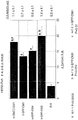

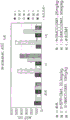


Classifications
-
- A—HUMAN NECESSITIES
- A61—MEDICAL OR VETERINARY SCIENCE; HYGIENE
- A61K—PREPARATIONS FOR MEDICAL, DENTAL OR TOILETRY PURPOSES
- A61K47/00—Medicinal preparations characterised by the non-active ingredients used, e.g. carriers or inert additives; Targeting or modifying agents chemically bound to the active ingredient
- A61K47/50—Medicinal preparations characterised by the non-active ingredients used, e.g. carriers or inert additives; Targeting or modifying agents chemically bound to the active ingredient the non-active ingredient being chemically bound to the active ingredient, e.g. polymer-drug conjugates
- A61K47/51—Medicinal preparations characterised by the non-active ingredients used, e.g. carriers or inert additives; Targeting or modifying agents chemically bound to the active ingredient the non-active ingredient being chemically bound to the active ingredient, e.g. polymer-drug conjugates the non-active ingredient being a modifying agent
- A61K47/68—Medicinal preparations characterised by the non-active ingredients used, e.g. carriers or inert additives; Targeting or modifying agents chemically bound to the active ingredient the non-active ingredient being chemically bound to the active ingredient, e.g. polymer-drug conjugates the non-active ingredient being a modifying agent the modifying agent being an antibody, an immunoglobulin or a fragment thereof, e.g. an Fc-fragment
- A61K47/6801—Drug-antibody or immunoglobulin conjugates defined by the pharmacologically or therapeutically active agent
- A61K47/6803—Drugs conjugated to an antibody or immunoglobulin, e.g. cisplatin-antibody conjugates
-
- A—HUMAN NECESSITIES
- A61—MEDICAL OR VETERINARY SCIENCE; HYGIENE
- A61K—PREPARATIONS FOR MEDICAL, DENTAL OR TOILETRY PURPOSES
- A61K39/00—Medicinal preparations containing antigens or antibodies
- A61K39/395—Antibodies; Immunoglobulins; Immune serum, e.g. antilymphocytic serum
-
- A—HUMAN NECESSITIES
- A61—MEDICAL OR VETERINARY SCIENCE; HYGIENE
- A61P—SPECIFIC THERAPEUTIC ACTIVITY OF CHEMICAL COMPOUNDS OR MEDICINAL PREPARATIONS
- A61P1/00—Drugs for disorders of the alimentary tract or the digestive system
- A61P1/04—Drugs for disorders of the alimentary tract or the digestive system for ulcers, gastritis or reflux esophagitis, e.g. antacids, inhibitors of acid secretion, mucosal protectants
-
- A—HUMAN NECESSITIES
- A61—MEDICAL OR VETERINARY SCIENCE; HYGIENE
- A61P—SPECIFIC THERAPEUTIC ACTIVITY OF CHEMICAL COMPOUNDS OR MEDICINAL PREPARATIONS
- A61P11/00—Drugs for disorders of the respiratory system
-
- A—HUMAN NECESSITIES
- A61—MEDICAL OR VETERINARY SCIENCE; HYGIENE
- A61P—SPECIFIC THERAPEUTIC ACTIVITY OF CHEMICAL COMPOUNDS OR MEDICINAL PREPARATIONS
- A61P13/00—Drugs for disorders of the urinary system
- A61P13/08—Drugs for disorders of the urinary system of the prostate
-
- A—HUMAN NECESSITIES
- A61—MEDICAL OR VETERINARY SCIENCE; HYGIENE
- A61P—SPECIFIC THERAPEUTIC ACTIVITY OF CHEMICAL COMPOUNDS OR MEDICINAL PREPARATIONS
- A61P13/00—Drugs for disorders of the urinary system
- A61P13/10—Drugs for disorders of the urinary system of the bladder
-
- A—HUMAN NECESSITIES
- A61—MEDICAL OR VETERINARY SCIENCE; HYGIENE
- A61P—SPECIFIC THERAPEUTIC ACTIVITY OF CHEMICAL COMPOUNDS OR MEDICINAL PREPARATIONS
- A61P13/00—Drugs for disorders of the urinary system
- A61P13/12—Drugs for disorders of the urinary system of the kidneys
-
- A—HUMAN NECESSITIES
- A61—MEDICAL OR VETERINARY SCIENCE; HYGIENE
- A61P—SPECIFIC THERAPEUTIC ACTIVITY OF CHEMICAL COMPOUNDS OR MEDICINAL PREPARATIONS
- A61P15/00—Drugs for genital or sexual disorders; Contraceptives
-
- A—HUMAN NECESSITIES
- A61—MEDICAL OR VETERINARY SCIENCE; HYGIENE
- A61P—SPECIFIC THERAPEUTIC ACTIVITY OF CHEMICAL COMPOUNDS OR MEDICINAL PREPARATIONS
- A61P35/00—Antineoplastic agents
-
- A—HUMAN NECESSITIES
- A61—MEDICAL OR VETERINARY SCIENCE; HYGIENE
- A61P—SPECIFIC THERAPEUTIC ACTIVITY OF CHEMICAL COMPOUNDS OR MEDICINAL PREPARATIONS
- A61P43/00—Drugs for specific purposes, not provided for in groups A61P1/00-A61P41/00
-
- A—HUMAN NECESSITIES
- A61—MEDICAL OR VETERINARY SCIENCE; HYGIENE
- A61P—SPECIFIC THERAPEUTIC ACTIVITY OF CHEMICAL COMPOUNDS OR MEDICINAL PREPARATIONS
- A61P5/00—Drugs for disorders of the endocrine system
- A61P5/14—Drugs for disorders of the endocrine system of the thyroid hormones, e.g. T3, T4
-
- C—CHEMISTRY; METALLURGY
- C07—ORGANIC CHEMISTRY
- C07D—HETEROCYCLIC COMPOUNDS
- C07D498/00—Heterocyclic compounds containing in the condensed system at least one hetero ring having nitrogen and oxygen atoms as the only ring hetero atoms
- C07D498/12—Heterocyclic compounds containing in the condensed system at least one hetero ring having nitrogen and oxygen atoms as the only ring hetero atoms in which the condensed system contains three hetero rings
- C07D498/18—Bridged systems
-
- C—CHEMISTRY; METALLURGY
- C07—ORGANIC CHEMISTRY
- C07K—PEPTIDES
- C07K16/00—Immunoglobulins [IGs], e.g. monoclonal or polyclonal antibodies
- C07K16/18—Immunoglobulins [IGs], e.g. monoclonal or polyclonal antibodies against material from animals or humans
- C07K16/28—Immunoglobulins [IGs], e.g. monoclonal or polyclonal antibodies against material from animals or humans against receptors, cell surface antigens or cell surface determinants
- C07K16/2803—Immunoglobulins [IGs], e.g. monoclonal or polyclonal antibodies against material from animals or humans against receptors, cell surface antigens or cell surface determinants against the immunoglobulin superfamily
-
- C—CHEMISTRY; METALLURGY
- C07—ORGANIC CHEMISTRY
- C07K—PEPTIDES
- C07K16/00—Immunoglobulins [IGs], e.g. monoclonal or polyclonal antibodies
- C07K16/18—Immunoglobulins [IGs], e.g. monoclonal or polyclonal antibodies against material from animals or humans
- C07K16/28—Immunoglobulins [IGs], e.g. monoclonal or polyclonal antibodies against material from animals or humans against receptors, cell surface antigens or cell surface determinants
- C07K16/2866—Immunoglobulins [IGs], e.g. monoclonal or polyclonal antibodies against material from animals or humans against receptors, cell surface antigens or cell surface determinants against receptors for cytokines, lymphokines, interferons
-
- C—CHEMISTRY; METALLURGY
- C07—ORGANIC CHEMISTRY
- C07K—PEPTIDES
- C07K16/00—Immunoglobulins [IGs], e.g. monoclonal or polyclonal antibodies
- C07K16/18—Immunoglobulins [IGs], e.g. monoclonal or polyclonal antibodies against material from animals or humans
- C07K16/28—Immunoglobulins [IGs], e.g. monoclonal or polyclonal antibodies against material from animals or humans against receptors, cell surface antigens or cell surface determinants
- C07K16/30—Immunoglobulins [IGs], e.g. monoclonal or polyclonal antibodies against material from animals or humans against receptors, cell surface antigens or cell surface determinants from tumour cells
- C07K16/3046—Stomach, Intestines
-
- C—CHEMISTRY; METALLURGY
- C07—ORGANIC CHEMISTRY
- C07K—PEPTIDES
- C07K16/00—Immunoglobulins [IGs], e.g. monoclonal or polyclonal antibodies
- C07K16/18—Immunoglobulins [IGs], e.g. monoclonal or polyclonal antibodies against material from animals or humans
- C07K16/28—Immunoglobulins [IGs], e.g. monoclonal or polyclonal antibodies against material from animals or humans against receptors, cell surface antigens or cell surface determinants
- C07K16/30—Immunoglobulins [IGs], e.g. monoclonal or polyclonal antibodies against material from animals or humans against receptors, cell surface antigens or cell surface determinants from tumour cells
- C07K16/3069—Reproductive system, e.g. ovaria, uterus, testes, prostate
-
- C—CHEMISTRY; METALLURGY
- C07—ORGANIC CHEMISTRY
- C07K—PEPTIDES
- C07K16/00—Immunoglobulins [IGs], e.g. monoclonal or polyclonal antibodies
- C07K16/18—Immunoglobulins [IGs], e.g. monoclonal or polyclonal antibodies against material from animals or humans
- C07K16/32—Immunoglobulins [IGs], e.g. monoclonal or polyclonal antibodies against material from animals or humans against translation products of oncogenes
-
- A—HUMAN NECESSITIES
- A61—MEDICAL OR VETERINARY SCIENCE; HYGIENE
- A61K—PREPARATIONS FOR MEDICAL, DENTAL OR TOILETRY PURPOSES
- A61K39/00—Medicinal preparations containing antigens or antibodies
- A61K2039/505—Medicinal preparations containing antigens or antibodies comprising antibodies
-
- A—HUMAN NECESSITIES
- A61—MEDICAL OR VETERINARY SCIENCE; HYGIENE
- A61K—PREPARATIONS FOR MEDICAL, DENTAL OR TOILETRY PURPOSES
- A61K47/00—Medicinal preparations characterised by the non-active ingredients used, e.g. carriers or inert additives; Targeting or modifying agents chemically bound to the active ingredient
- A61K47/50—Medicinal preparations characterised by the non-active ingredients used, e.g. carriers or inert additives; Targeting or modifying agents chemically bound to the active ingredient the non-active ingredient being chemically bound to the active ingredient, e.g. polymer-drug conjugates
- A61K47/51—Medicinal preparations characterised by the non-active ingredients used, e.g. carriers or inert additives; Targeting or modifying agents chemically bound to the active ingredient the non-active ingredient being chemically bound to the active ingredient, e.g. polymer-drug conjugates the non-active ingredient being a modifying agent
- A61K47/68—Medicinal preparations characterised by the non-active ingredients used, e.g. carriers or inert additives; Targeting or modifying agents chemically bound to the active ingredient the non-active ingredient being chemically bound to the active ingredient, e.g. polymer-drug conjugates the non-active ingredient being a modifying agent the modifying agent being an antibody, an immunoglobulin or a fragment thereof, e.g. an Fc-fragment
- A61K47/6835—Medicinal preparations characterised by the non-active ingredients used, e.g. carriers or inert additives; Targeting or modifying agents chemically bound to the active ingredient the non-active ingredient being chemically bound to the active ingredient, e.g. polymer-drug conjugates the non-active ingredient being a modifying agent the modifying agent being an antibody, an immunoglobulin or a fragment thereof, e.g. an Fc-fragment the modifying agent being an antibody or an immunoglobulin bearing at least one antigen-binding site
- A61K47/6875—Medicinal preparations characterised by the non-active ingredients used, e.g. carriers or inert additives; Targeting or modifying agents chemically bound to the active ingredient the non-active ingredient being chemically bound to the active ingredient, e.g. polymer-drug conjugates the non-active ingredient being a modifying agent the modifying agent being an antibody, an immunoglobulin or a fragment thereof, e.g. an Fc-fragment the modifying agent being an antibody or an immunoglobulin bearing at least one antigen-binding site the antibody being a hybrid immunoglobulin
-
- C—CHEMISTRY; METALLURGY
- C07—ORGANIC CHEMISTRY
- C07K—PEPTIDES
- C07K16/00—Immunoglobulins [IGs], e.g. monoclonal or polyclonal antibodies
- C07K16/08—Immunoglobulins [IGs], e.g. monoclonal or polyclonal antibodies against material from viruses
- C07K16/10—Immunoglobulins [IGs], e.g. monoclonal or polyclonal antibodies against material from viruses from RNA viruses
- C07K16/1036—Retroviridae, e.g. leukemia viruses
- C07K16/1045—Lentiviridae, e.g. HIV, FIV, SIV
- C07K16/1063—Lentiviridae, e.g. HIV, FIV, SIV env, e.g. gp41, gp110/120, gp160, V3, PND, CD4 binding site
-
- C—CHEMISTRY; METALLURGY
- C07—ORGANIC CHEMISTRY
- C07K—PEPTIDES
- C07K16/00—Immunoglobulins [IGs], e.g. monoclonal or polyclonal antibodies
- C07K16/18—Immunoglobulins [IGs], e.g. monoclonal or polyclonal antibodies against material from animals or humans
- C07K16/28—Immunoglobulins [IGs], e.g. monoclonal or polyclonal antibodies against material from animals or humans against receptors, cell surface antigens or cell surface determinants
- C07K16/2803—Immunoglobulins [IGs], e.g. monoclonal or polyclonal antibodies against material from animals or humans against receptors, cell surface antigens or cell surface determinants against the immunoglobulin superfamily
- C07K16/283—Immunoglobulins [IGs], e.g. monoclonal or polyclonal antibodies against material from animals or humans against receptors, cell surface antigens or cell surface determinants against the immunoglobulin superfamily against Fc-receptors, e.g. CD16, CD32, CD64
-
- C—CHEMISTRY; METALLURGY
- C07—ORGANIC CHEMISTRY
- C07K—PEPTIDES
- C07K16/00—Immunoglobulins [IGs], e.g. monoclonal or polyclonal antibodies
- C07K16/18—Immunoglobulins [IGs], e.g. monoclonal or polyclonal antibodies against material from animals or humans
- C07K16/28—Immunoglobulins [IGs], e.g. monoclonal or polyclonal antibodies against material from animals or humans against receptors, cell surface antigens or cell surface determinants
- C07K16/2896—Immunoglobulins [IGs], e.g. monoclonal or polyclonal antibodies against material from animals or humans against receptors, cell surface antigens or cell surface determinants against molecules with a "CD"-designation, not provided for elsewhere
-
- C—CHEMISTRY; METALLURGY
- C07—ORGANIC CHEMISTRY
- C07K—PEPTIDES
- C07K2317/00—Immunoglobulins specific features
- C07K2317/20—Immunoglobulins specific features characterized by taxonomic origin
- C07K2317/24—Immunoglobulins specific features characterized by taxonomic origin containing regions, domains or residues from different species, e.g. chimeric, humanized or veneered
Landscapes
- Health & Medical Sciences (AREA)
- Chemical & Material Sciences (AREA)
- Organic Chemistry (AREA)
- Immunology (AREA)
- Life Sciences & Earth Sciences (AREA)
- Medicinal Chemistry (AREA)
- General Health & Medical Sciences (AREA)
- Engineering & Computer Science (AREA)
- Bioinformatics & Cheminformatics (AREA)
- Genetics & Genomics (AREA)
- Biochemistry (AREA)
- Proteomics, Peptides & Aminoacids (AREA)
- Molecular Biology (AREA)
- Biophysics (AREA)
- Animal Behavior & Ethology (AREA)
- Public Health (AREA)
- Veterinary Medicine (AREA)
- Pharmacology & Pharmacy (AREA)
- Chemical Kinetics & Catalysis (AREA)
- Nuclear Medicine, Radiotherapy & Molecular Imaging (AREA)
- General Chemical & Material Sciences (AREA)
- Epidemiology (AREA)
- Virology (AREA)
- Oncology (AREA)
- Cell Biology (AREA)
- Reproductive Health (AREA)
- Urology & Nephrology (AREA)
- Gynecology & Obstetrics (AREA)
- AIDS & HIV (AREA)
- Hematology (AREA)
- Endocrinology (AREA)
- Pregnancy & Childbirth (AREA)
- Microbiology (AREA)
- Pulmonology (AREA)
- Diabetes (AREA)
- Mycology (AREA)
- Medicines Containing Antibodies Or Antigens For Use As Internal Diagnostic Agents (AREA)
- Peptides Or Proteins (AREA)
- Pharmaceuticals Containing Other Organic And Inorganic Compounds (AREA)
- Medicinal Preparation (AREA)
Abstract
The present invention relates to antibody-drug conjugates and methods. The present invention relates to antibody-drug conjugate compounds Ab- (L-D) of formula IpI one or more ofThe maytansinoid drug moiety (D) is covalently linked via L to an antibody (Ab) that binds to an ErbB receptor, or to one or more tumor associated antigens or cell surface receptors. These compounds are useful in methods of diagnosis or treatment of cancer, as well as other diseases and disorders.
Description
The application is a reissue application (original application number 200580026066.7) of a divisional application of the invention with the application date of 2005-31/5, Chinese application number 201810863230.9 and the name of antibody-drug conjugate and method.
This non-provisional application filed pursuant to 37 CFR § 1.53(b), and claims the benefit of US provisional application 60/576,517 filed 6/1/2004 and US provisional application 60/616,098 filed 10/5/2004, pursuant to 35 USC § 119(e), all of which are incorporated herein by reference.
Field of the invention
The present invention relates generally to compounds having anti-cancer activity, and more particularly to antibodies conjugated to chemotherapeutic maytansinoid drugs or toxins. The invention also relates to methods of using the antibody-drug conjugate compounds for in vitro, in situ, and in vivo diagnosis or treatment of mammalian cells, or associated pathological conditions.
Background
Monoclonal antibody therapies for the targeted treatment of cancer, immunological and angiogenic disorders in patients have been established. Local delivery Of Cytotoxic or cytostatic Agents, i.e.agents that kill or inhibit tumor cells In Cancer Therapy, using Antibody-drug conjugates (ADC), i.e.immunoconjugates, (Payne, G. (2003) Cancer Cell 3: 207-212; Trail et al (2003) Cancer immunological. immunothers.52: 328-337; Syrigos And Epeneros (1999) Anticancer Research 19: 605-614; Niculescu-Duvaz And Springer (1997) adv. drug Del. Rev.26: 151-172; US 4975278) theoretically allow targeted delivery Of drug moieties to tumors And accumulation within the cells Of tumors, where systemic administration Of these non-conjugated Agents may result In unacceptable levels Of toxicity to normal cells And tumor cells to be removed (Baldwin et al, (1986) Lance pp. 15, Mar. 6; Monoclonal Antibodies 603-84: "Cancer Cell In",603: 14 In.),84, a. Pinchera et al (eds.), pp.475-506). It is therefore sought to maximize efficacy while minimizing toxicity. Efforts to design and improve ADCs have focused on the selectivity of monoclonal antibodies (mAbs) as well as drug-linking and drug-release properties. Both polyclonal and monoclonal antibodies have been reported for use in these strategies (Rowland et al (1986) Cancer Immunol. Immunother.21: 183-87). Drugs for use in these methods include daunorubicin, doxorubicin, methotrexate, mitomycin, neocarzinostain (Takahashi et al (1988) Cancer 61:881-888) and vindesine (Rowland et al (1986) supra). Toxins for use in antibody-toxin conjugates include bacterial toxins such as diphtheria toxin, plant toxins such as ricin (US 4753894; US 5629197; US 4958009; US 4956453), small molecule toxins such as geldanamycin (Mandler et al (2000) J.of the Nat. Cancer Inst.92(19): 1573-1581; Mandler et al (2000) Bioorganic & Med. chem. letters 10: 1025-1028; Mandler et al (2002) Bioconjugate chem.13:786-791), maytansinoids (maytansinoids) (EP 1391213; Liuman et al (1996) Proc. Natl. Acad. Sci. USA 93:8618-8623), and calicheamicin (calicheamicin) (Locer et al (1998) Res.58: Cancer 3328; Cancer 3353-1993). The toxin may affect its cytotoxic and cytostatic effects by including tubulin binding, DNA binding, or topoisomerase inhibitory mechanisms. Some cytotoxic drugs tend to be inactivated or have reduced activity when coupled to large antibodies or protein receptor ligands.
Antibody-radioisotope conjugates have been approved (ibritumomab tiuxetan, Biogen/Idec) conjugated with a murine IgG1 kappa monoclonal antibody against CD20 antigen and with a thiourea linker (like)r) -chelator binding111In or90Y radioisotope composition (Wiseman et al (2000) Eur. J. Nucl. Med.27(7): 766-77; Wiseman et al (2002) Blood 99(12): 4336-42; Witzig et al (2002) J. Clin. Oncol.20(10): 2453-63; Witzig et al (2002) J. Clin. Oncol.20(15): 3262-69). In 2000, an antibody-drug conjugate MYLOTARG consisting of hu CD33 antibody linked to calicheamicin was approvedTM(gemtuzumab ozogamicin, Wyeth Pharmaceuticals) for the treatment of acute myeloid leukemia by injection (Drugs of the Future (2000)25(7): 686; US 4970198; US 5079233; US 5585089; US 5606040; US 5693762; US 5739116; US 5767285; US 5773001). The antibody-drug conjugate Cantuzumab mertansine (Immunogen, Inc.) (Xie et al (2004) J.of Pharm. and exp. Ther.308(3): 1073-. MLN-2704(Millennium pharm., BZL Biologics, immunolgen Inc.) is an antibody-drug conjugate consisting of an anti-Prostate Specific Membrane Antigen (PSMA) monoclonal antibody linked to maytansinoid drug moiety DM1, being developed for the effective treatment of prostate tumors. The same maytansinoid drug moiety DM1 was linked to the murine monoclonal antibody TA.1 of the mouse by a non-disulfide linker (non-disulfide linker) SMCC (Chari et al (1992) Cancer Research 52: 127-131). This conjugate is reported to be 200 times less potent than the corresponding disulfide linker conjugate. The SMCC linker is considered to be "non-cleavable" in the conjugate (see also: US 4981979). Have been reported to connect to DM1 through SMCC
  (trastuzumab) (WO 2005/037992).
In an effort to find effective cellular targets for cancer diagnosis and treatment, researchers have sought to identify transmembrane or tumor-associated polypeptides that are specifically expressed on the surface of one or more specific types of cancer cells as compared to one or more normal, non-cancerous cells. Typically, these tumor-associated polypeptides are more expressed on the surface of cancer cells than on the surface of non-cancer cells. The identification of these tumor-associated cell surface antigen polypeptides, i.e., tumor-associated antigens (TAAs), has led to the ability to specifically target cancer cells for destruction through antibody-based therapy.
Monoclonal antibody therapies for the targeted treatment of patients with cancer, immune disorders and angiogenesis disorders have been established. An example of a successful antibody therapy is a humanized monoclonal antibody derived from recombinant DNA (trastuzumab) which binds selectively with high affinity (Kd 5nM) to the extracellular region of the human epidermal
 growth factor receptor 2 protein HER2 (ErbB2) in a cell-based assay (US 5821337; US 6054297; US 6407213; US 6639055; Coissens L et al (1985) Science 230: 1132-9; Slamon DJ et al (1989) Science 244: 707-12). Trastuzumab is an IgG1 kappa antibody containing human framework regions with murine antibody complementarity determining regions (4D5) that bind HER 2. Trastuzumab binds to the HER2 antigen, thus inhibiting the growth of cancer cells. Because trastuzumab is a humanized antibody, it minimizes any HAMA response in the patient. The humanized antibody against HER2 was produced from a suspension culture of mammalian cells (Chinese Hamster Ovary (CHO)). The HER2 (or c-erbB2) proto-oncogene encodes a 185kDa transmembrane receptor protein that is structurally related to the epidermal growth factor receptor. HER2 protein overexpression is observed in 25% -30% of primary breast cancers, and HER2 protein overexpression can be determined using immunohistochemistry-based fixed tumor blockade assessment (Press MF, et al (1993) Cancer Res 53: 4960-70). Trastuzumab has been shown to inhibit the proliferation of HER 2-overexpressing human tumor cells in vitro tests and animals (Hudziak RM, et al (1989) Mol Cell Biol 9: 1165-72; Lewis GD, et al (1993) Cancer Immunol Immunother; 37: 255-63; Baselga J, et al (1998) Cancer Res.58: 2825-2831). Trastuzumab is a mediator of antibody-dependent cellular cytotoxicity, ADCC (Hotaling TE, et al (1996) [ Abstract ].Proc.Annual Meeting Am Assoc Canchor Res; 471 parts by weight; peg ram MD, et al (1997) [ Abstract]Proc Am Assoc Cancer Res; 38: 602; sliwkowski et al (1999) sensiars in Oncology 26(4), Suppl 12: 60-70; yarden Y. and Sliwkowski, M. (2001) Nature Reviews: Molecular Cell Biology, Macmillan Magazines, Ltd., Vol.2: 127-. In clinic, the medicine is prepared by the steps of, is active in ErbB 2-overexpressing metastatic breast cancer patients who have previously received a number of anti-cancer treatments (Baselga et al (1996) J.Clin.Oncol.14: 737-744). Although HERCEPTIN is a breakthrough in treating ErbB 2-overexpressing breast cancer patients who have previously received extensive anti-cancer therapy, most patients in this population do not respond or respond poorly to HERCEPTIN therapy. Thus, there is a clear clinical need to develop other HER 2-directed cancer therapies for the treatment of patients with HER 2-overexpressing tumors or other diseases associated with HER2 expression, but who do not respond or respond poorly to HERCEPTIN treatment. In addition to HER2, targeted therapy is also possible with other tumor-associated antigens.
SUMMARY
The present invention provides novel compounds having biological activity against cancer cells. The compounds can inhibit tumor growth in mammals and are useful for treating human cancer patients.
The present invention relates to the delivery, transport, accumulation or retention of therapeutic antibody-drug conjugate (ADC) compounds within cells. The invention more specifically relates to obtaining high concentrations of active metabolite molecules in cancer cells. Intracellular targeting can be achieved by methods and compounds that allow for the accumulation or retention of biologically active agents within cells. Such effective targeting is applicable to a variety of therapeutic formulations and methods.
It has been surprisingly found that antibody-drug conjugates with stable non-disulfide linker groups linking maytansinoid drug moieties to antibodies lead to enhanced in vitro potency and in vivo efficacy. Furthermore, the antibody-drug conjugates show unexpectedly safer results in vivo relative to certain disulfide linker conjugates.
An antibody-drug conjugate (ADC) compound comprises an antibody covalently linked by a linker to one or more maytansinoid drug moieties. ADC can be represented by formula I
Ab-(L-D)p I
Wherein one or more maytansinoid drug moieties (D) are covalently linked to an antibody (Ab) through L. Ab is an antibody that binds to an ErbB receptor, or to one or more tumor associated antigens or cell surface receptors. Linker L may be stable extracellularly, i.e. extracellularly. The linker L, the maytansinoid drug moiety D, or both the linker and the maytansinoid drug moiety (L-D) do not comprise a disulfide group (disulfide group).
In one embodiment, a substantial amount (substantial amount) of the drug moiety is not cleaved by the antibody until the antibody-drug conjugate enters a cell having a cell surface receptor specific for the antibody of the antibody-drug conjugate, and the drug moiety is cleaved by the antibody when the antibody-drug conjugate does enter the cell.
In another embodiment, the ADC specifically binds to a receptor encoded by an ErbB gene, such as EGFR, HER2, HER3, and HER 4. The ADC can specifically bind to the extracellular domain of the HER2 receptor. The ADC may inhibit the growth of tumor cells that overexpress the HER2 receptor.
In another embodiment, the antibody (Ab) of formula I is a humanized antibody such as huMAb4D5-1, huMAb4D5-2, huMAb4D5-3, huMAb4D5-4, huMAb4D5-5, huMAb4D5-6, huMAb4D5-7, or huMAb4D5-8 (trastuzumab).
Another aspect of the invention is a pharmaceutical composition comprising a compound of formula I, or a pharmaceutically acceptable salt or solvate thereof, and a pharmaceutically acceptable diluent, carrier, or excipient.
In another aspect, pharmaceutical combinations are provided comprising a compound of formula I and a second compound having anti-cancer properties or other therapeutic effects.
Another aspect includes diagnostic and therapeutic uses of the compounds and compositions disclosed herein.
Another aspect is a method of killing or inhibiting proliferation of a tumor cell or a cancer cell comprising treating the cell with an amount of an antibody-drug conjugate, or a pharmaceutically acceptable salt or solvate thereof, effective to kill or inhibit proliferation of the tumor cell or the cancer cell.
Another aspect is a method of treating cancer comprising administering to a patient a formulation of a compound of formula I. A method for treating cancer in a mammal, wherein said cancer is characterized by overexpression of an ErbB receptor. The mammal optionally is non-responsive, or weakly responsive, to treatment with unconjugated anti-ErbB antibodies. The method comprises administering to the mammal a therapeutically effective amount of an antibody-drug conjugate compound.
Another aspect is a method of inhibiting the growth of a tumor cell that overexpresses a growth factor receptor selected from the group consisting of the HER2 receptor and the EGF receptor, comprising administering to a patient an antibody-drug conjugate compound that specifically binds to the growth factor receptor and a chemotherapeutic agent, wherein the antibody-drug conjugate and the chemotherapeutic agent are each administered in an amount effective to inhibit the growth of a tumor cell in the patient.
Another aspect is a method of treating a human patient susceptible to or diagnosed with a disorder characterized by overexpression of the ErbB2 receptor, comprising administering a combination of an antibody-drug conjugate compound of formula I and a chemotherapeutic agent.
Another aspect is an assay for detecting cancer cells comprising contacting the cells with an antibody-drug conjugate compound and determining the extent of binding of the antibody-drug conjugate compound to the cells.
Another aspect relates to a method of screening for ADC candidates for use in the treatment of a disease or disorder, wherein the disease or disorder is characterized by overexpression of HER 2.
Another aspect includes an article of manufacture, i.e., a kit, comprising an antibody-drug conjugate, a container, and a package insert or label directing treatment.
Another aspect includes a method of treating a disease or disorder characterized by overexpression of HER2 in a patient with an antibody-drug conjugate compound.
Further aspects include methods of manufacture, methods of synthesis, methods of coupling, and methods of purification of antibody-drug conjugate compounds and intermediates useful in the manufacture, synthesis, and coupling of the antibody-drug conjugate compounds.
The present invention relates to the following items.
1. An antibody-drug conjugate compound comprising an antibody covalently linked through a linker to one or more maytansinoid drug moieties, the compound having the formula I
Ab-(L-D)p I
Or a pharmaceutically acceptable salt or solvate thereof, wherein
Ab is an antibody that binds to an ErbB receptor, or to one or more tumor-associated antigens or cell-surface receptors selected from the group consisting of (1) - (36)
(1) BMPR1B (bone morphogenetic protein receptor-type IB, Genbank accession No. NM _ 001203);
(2) e16(LAT1, SLC7A5, Genbank accession NM-003486);
(3) STEAP1 (six transmembrane epithelial antigen of prostate, Genbank accession No. NM _ 012449);
(4)0772P (CA125, MUC16, Genbank accession AF 361486);
(5) MPF (MPF, MSLN, SMR, megakaryocyte potentiator, mesothelin, Genbank accession No. NM-005823);
(6) napi3B (Napi-3B, NPTIIb, SLC34a2, solute carrier family 34 (sodium phosphate), member 2, type II sodium dependent phosphate transporter 3B, Genbank accession No. NM — 006424);
(7) sema5B (FLJ10372, KIAA1445, mm.42015, Sema5B, SEMAG, semaphorin 5B Hlog, Sema domain, seven thrombospondin repeats (type 1 and type 1-like), transmembrane domain (TM) and short cytoplasmic domain, (semaphorin) 5B, Genbank accession No. AB 040878);
(8) PSCA hlg (2700050C12Rik, C530008O16Rik, RIKEN cDNA 2700050C12, RIKEN cDNA 2700050C12 gene, Genbank accession No. AY 358628);
(9) ETBR (endothelin type B receptor, Genbank accession No. AY 275463);
(10) MSG783(RNF124, hypothetical protein FLJ20315, Genbank accession No. NM — 017763);
(11) STEAP2(HGNC _8639, IPCA-1, PCANAP1, STAMP1, STEAP2, STMP, prostate cancer-associated gene 1, prostate cancer-associated protein 1, six transmembrane epithelial antigen of prostate 2, six transmembrane prostate protein, Genbank accession No. AF 455138);
(12) TrpM4(BR22450, FLJ20041, TrpM4, TrpM4B, transient receptor potential cation channel, subfamily M, member 4, Genbank accession No. NM _ 017636);
(13) CRIPTO (CR, CR1, CRGF, CRIPTO, TDGF1, teratocarcinoma-derived growth factor, Genbank accession No. NP _003203 or NM _ 003212);
(14) CD21(CR2 (complement receptor 2) or C3DR (C3 d/Epstein-Barr virus receptor) or Hs.73792Genbank accession number M26004);
(15) CD79B (CD79B, CD79 β, IGb (immunoglobulin associated beta), B29, Genbank accession No. NM — 000626);
(16) FcRH2(IFGP4, IRTA4, spa 1A (SH 2 domain containing phosphatase dockerin 1a), spa 1B, spa 1C, Genbank accession No. NM — 030764);
(17) HER2(Genbank accession number M11730);
(18) NCA (Genbank accession number M18728);
(19) MDP (Genbank accession number BC 017023);
(20) IL20R α (Genbank accession No. AF 184971);
(21) brevican (Genbank accession No. AF 229053);
(22) EphB2R (Genbank accession No. NM _ 004442);
(23) ASLG659(Genbank accession number AX 092328);
(24) PSCA (Genbank accession No. AJ 297436);
(25) GEDA (Genbank accession No. AY 260763;
(26) BAFF-R (B cell activating factor receptor, BlyS receptor 3, BR3, NP-443177.1);
(27) CD22(B cell receptor CD22-B isoform, NP-001762.1);
(28) CD79a (CD79A, CD79 α, immunoglobulin-related alpha, B cell-specific protein that interacts covalently with Ig beta (CD79B) and forms complexes with Ig M molecules on the surface, transducing signals involved in B cell differentiation, Genbank accession No. NP _ 001774.1);
(29) CXCR5 (burkitt lymphoma receptor 1, a G protein-coupled receptor activated by the CXCL13 chemokine, plays a role in lymphocyte migration and humoral defense, plays a role in HIV-2 infection and possibly in the development of AIDS, lymphoma, myeloma, and leukemia, Genbank accession No. NP _ 001707.1);
(30) HLA-DOB (Beta subunit of MHC class II molecule (Ia antigen), which binds to and presents peptides to CD4+ T lymphocytes, Genbank accession No. NP _ 002111.1);
(31) P2X5 (purinoceptor P2X ligand-gated ion channel 5, an ion channel gated by extracellular ATP, may be involved in synaptic transmission and neurogenesis, defects of which may lead to pathophysiological conditions of idiopathic detrusor instability, Genbank accession No. NP _ 002552.2);
(32) CD72(B cell differentiation antigen CD72, Lyb-2, Genbank accession NP-001773.1);
(33) LY64 (lymphocyte antigen 64(RP105), leucine-rich repeat (LRR) family of type I membrane proteins, regulating B cell activation and apoptosis, loss of function associated with enhanced disease activity in patients with systemic lupus erythematosus, Genbank accession No. NP _ 005573.1);
(34) FcRH1(Fc receptor-like protein 1, putative receptor for immunoglobulin Fc domain, comprising C2-type Ig-like and ITAM domains, likely to play a role in B lymphocyte differentiation, Genbank accession No. NP _ 443170.1);
(35) IRTA2 (translocation-associated immunoglobulin superfamily receptor 2, a putative immunoreceptor, likely to play a role in B cell development and lymphomatosis; gene dysregulation by translocation occurs in some B cell malignancies, Genbank accession number NP-112571.1); and
(36) TENB2 (putative transmembrane proteoglycan, related to EGF of growth factor/regulin family of growth factors and follistatin, Genbank accession No. AF 179274;
Provided that the antibody is not ta.1;
l is a non-disulfide linker;
d is a maytansinoid drug moiety; and is
p is 1 to 8.
2. The antibody-drug conjugate compound of item 1 wherein Ab is an antibody that binds to one or more tumor associated antigens or cell surface receptors selected from (1) - (16) and (18) - (36).
3. The antibody-drug conjugate compound according to item 1 or item 2, wherein L is a linker selected from the group consisting of

Wherein the wavy line indicates covalent attachment to Ab and D;
x is:


y is:

r is independently H or C1-C6An alkyl group; and n is 1 to 12.
4. The antibody-drug conjugate compound of any one of the preceding claims wherein D is selected from the structures:

wherein the wavy line indicates covalent attachment to L,
r is independently H or C1-C6Alkyl radical, and
m is 1, 2, or 3.
5. The antibody-drug conjugate compound of item 4 wherein m is 2 and R is H.
6. The antibody-drug conjugate compound of any one of the preceding claims wherein the maytansinoid drug moiety is DM1 having the structure

7. The antibody-drug conjugate compound of item 3 having the structure

8. The antibody-drug conjugate compound of item 7 having the structure

9. The antibody-drug conjugate compound of item 3 having the structure

10. The antibody-drug conjugate compound of item 1 having the structure

11. The antibody-drug conjugate of item 10, wherein Ab is trastuzumab and p is 1, 2, 3, or 4.
12. The antibody-drug conjugate compound of item 1 having the structure

Wherein Tr is trastuzumab and p is 1, 2, 3, or 4.
13. The antibody-drug conjugate compound of item 1 having the structure

Wherein n is 0, 1, or 2; and p is 1, 2, 3, or 4.
14. The antibody-drug conjugate compound of item 13 wherein Ab is trastuzumab.
15. The antibody-drug conjugate compound of item 1 wherein p is 1, 2, 3, or 4.
16. The antibody-drug conjugate compound of item 1 wherein the antibody binds to a receptor encoded by an ErbB gene.
17. The antibody-drug conjugate compound of claim 16 wherein the receptor is selected from the group consisting of EGFR, HER2, HER3, and HER 4.
18. The antibody-drug conjugate compound of item 17 wherein the antibody specifically binds to HER2 receptor.
19. The antibody-drug conjugate compound of item 1 that specifically binds to the extracellular domain of the HER2 receptor and inhibits growth of tumor cells that overexpress the HER2 receptor.
20. The antibody-drug conjugate compound of any one of the preceding claims wherein the antibody is selected from the group consisting of a monoclonal antibody, an antibody fragment, a chimeric antibody and a humanized antibody.
21. The antibody-drug conjugate compound of item 1, wherein the antibody is a humanized antibody selected from the group consisting of huMAb4D5-1, huMAb4D5-2, huMAb4D5-3, huMAb4D5-4, huMAb4D5-5, huMAb4D5-6, huMAb4D5-7, and huMAb4D5-8 (trastuzumab).
22. The antibody-drug conjugate compound of item 21 wherein the antibody is huMAb4D5-8 (trastuzumab).
23. The antibody-drug conjugate of item 20, wherein the antibody is a Fab fragment.
24. The antibody-drug conjugate compound of item 1 wherein the antibody is attached to the linker through the cysteine thiol of the antibody.
25. The antibody-drug conjugate compound of item 24 wherein p is 1, 2, 3, or 4.
26. A pharmaceutical composition comprising the antibody-drug conjugate compound of item 1 or item 2, or a pharmaceutically acceptable salt thereof, and a pharmaceutically acceptable diluent, carrier, or excipient.
27. The pharmaceutical composition of item 26, further comprising a therapeutically effective amount of a chemotherapeutic agent selected from the group consisting of: erlotinib, Bortezomib, fulvestrant, Sutent, letrozole, imatinib mesylate, PTK787/ZK 222584, Oxaliplatin, 5-FU, leucovorin, rapamycin, Lapatinib, Lonafarnib, Sorafenib and Gefitinib.
28. The pharmaceutical composition of item 26, further comprising a therapeutically effective amount of an anti-angiogenic agent.
29. The pharmaceutical composition of item 26, further comprising a therapeutically effective amount of bevacizumab.
30. A method of inhibiting cell proliferation, comprising treating mammalian cells in a cell culture medium with the antibody-drug conjugate compound of item 1 or item 2, thereby inhibiting proliferation of the cells.
31. The method of clause 30, wherein the mammalian cell has a HER2 receptor protein directed to an antibody-drug conjugate compound.
32. The method of item 31, wherein said mammalian cell is a breast tumor cell.
33. The method of clause 30, wherein the antibody-drug conjugate compound is more cytotoxic than a maytansinoid compound comprising a maytansinoid moiety of the antibody-drug conjugate compound.
34. The method of clause 30, wherein the antibody-drug conjugate induces apoptosis.
35. A method of treating cancer comprising administering to a patient a formulation of an antibody-drug conjugate compound of item 1 or item 2 and a pharmaceutically acceptable diluent, carrier or excipient.
36. The method of item 35, wherein the cancer is selected from the group consisting of breast, ovarian, stomach, endometrial, salivary gland, lung, kidney, colon, colorectal, thyroid, pancreatic, prostate and bladder cancer.
37. The method of item 36, wherein the cancer is a breast cancer that overexpresses ErbB2 at a level of 2+ or more.
38. The method of clause 35, wherein the amount of antibody-drug conjugate compound administered to the patient ranges from about 0.1 to about 10mg/kg of patient body weight per dose.
39. The method of clause 35, wherein the antibody-drug conjugate is administered at intervals of about three weeks.
40. The method of clause 35, wherein the antibody-drug conjugate is administered by infusion.
41. The method of clause 35, wherein the antibody-drug conjugate is formulated with a pharmaceutically acceptable parenteral carrier.
42. The method of clause 41, wherein the antibody-drug conjugate is formulated as an injectable unit dosage form.
43. The method of clause 42, wherein the antibody-drug conjugate is administered intravenously.
44. The method of item 35, wherein the growth inhibitory antibody is administered to the patient in combination with an antibody-drug conjugate compound.
45. The method of item 35, wherein the patient is administered a second antibody that binds ErbB2 and blocks ligand activation of an ErbB receptor in combination with an antibody-drug conjugate compound.
46. The method of clause 45, wherein the second antibody comprises monoclonal antibody 2C4 or humanized 2C 4.
47. The method of clause 46, wherein the second antibody is conjugated to a cytotoxic agent.
48. The method of item 35, wherein the chemotherapeutic agent is administered to the patient in combination with the antibody-drug conjugate compound, wherein the chemotherapeutic agent is selected from the group consisting of: erlotinib, Bortezomib, fulvestrant, Sutent, letrozole, imatinib mesylate, PTK787/ZK 222584, Oxaliplatin, 5-FU, leucovorin, rapamycin, Lapatinib, Lonafarnib, Sorafenib, and Gefitinib.
49. The method of clause 35, wherein the anti-angiogenic agent is administered to the patient in combination with an antibody-drug conjugate compound, wherein the anti-angiogenic agent is selected from bevacizumab.
50. A method of inhibiting growth of a tumor cell that overexpresses a growth factor receptor comprising administering to a patient the antibody-drug conjugate of item 1 that specifically binds to a growth factor receptor selected from HER2 or EGF and a chemotherapeutic agent, wherein the antibody-drug conjugate and the chemotherapeutic agent are each administered in an amount effective to inhibit growth of a tumor cell in the patient.
51. A method of treating a human patient susceptible to or diagnosed with a disease characterized by overexpression of the ErbB2 receptor, comprising administering the antibody-drug conjugate of item 1 in combination with a chemotherapeutic agent or growth inhibitory agent.
52. The method of clause 51, wherein the antibody of the antibody-drug conjugate is an anti-ErbB 2 antibody.
53. The method of item 52, wherein the anti-ErbB 2 antibody has the biological characteristics of a 4D5 monoclonal antibody.
54. The method of item 52, wherein the anti-ErbB 2 antibody binds to substantially the same epitope as the 4D5 monoclonal antibody.
55. The method of item 52, wherein the anti-ErbB 2 antibody is selected from the group consisting of a humanized antibody: huMAb4D5-1, huMAb4D5-2, huMAb4D5-3, huMAb4D5-4, huMAb4D5-5, huMAb4D5-6, huMAb4D5-7 and huMAb4D5-8 (trastuzumab).
56. A test method for detecting cancer cells, comprising
(a) Contacting a cell with the antibody-drug conjugate compound of item 1 or item 2; and
(b) determining the extent to which the antibody-drug conjugate compound binds to the cell.
57. The test method of item 56, wherein the cell is a breast tumor cell.
58. The test of item 56, wherein the degree of binding is detected by determining the level of an ErbB-encoding nucleic acid using Fluorescence In Situ Hybridization (FISH).
59. The test method of item 56, wherein said degree of binding is determined by Immunohistochemistry (IHC).
60. An article of manufacture, comprising:
the antibody-drug conjugate compound of item 1 or item 2;
a container; and
a package insert or label indicating that the compound is useful for treating a cancer characterized by overexpression of an ErbB receptor.
61. The article of manufacture of item 60, wherein the package insert or label indicates that the compound is useful for treating a cancer characterized by overexpression of an ErbB2 receptor.
62. The article of manufacture of item 61, wherein the cancer is breast cancer.
63. The article of manufacture of item 62, wherein the cancer is characterized by overexpression of the ErbB2 receptor 2+ or greater.
64. A method of making an antibody-drug conjugate compound comprising covalently linking an antibody to one or more maytansinoid drug moieties via a linker, the compound having the formula I
Ab-(L-D)p I
Or a pharmaceutically acceptable salt or solvate thereof, wherein
Ab is an antibody that binds to an ErbB receptor, or to one or more tumor-associated antigens or cell-surface receptors selected from the group consisting of (1) - (36)
(1) BMPR1B (bone morphogenetic protein receptor-type IB, Genbank accession No. NM _ 001203);
(2) e16(LAT1, SLC7A5, Genbank accession NM-003486);
(3) STEAP1 (six transmembrane epithelial antigen of prostate, Genbank accession No. NM _ 012449);
(4)0772P (CA125, MUC16, Genbank accession AF 361486);
(5) MPF (MPF, MSLN, SMR, megakaryocyte potentiator, mesothelin, Genbank accession No. NM-005823);
(6) napi3B (Napi-3B, NPTIIb, SLC34a2, solute carrier family 34 (sodium phosphate), member 2, type II sodium dependent phosphate transporter 3B, Genbank accession No. NM — 006424);
(7) sema5B (FLJ10372, KIAA1445, mm.42015, Sema5B, SEMAG, semaphorin 5B Hlog, Sema domain, seven thrombospondin repeats (type 1 and type 1-like), transmembrane domain (TM) and short cytoplasmic domain, (semaphorin) 5B, Genbank accession No. AB 040878);
(8) PSCA hlg (2700050C12Rik, C530008O16Rik, RIKEN cDNA 2700050C12, RIKEN cDNA 2700050C12 gene, Genbank accession No. AY 358628);
(9) ETBR (endothelin type B receptor, Genbank accession No. AY 275463);
(10) MSG783(RNF124, hypothetical protein FLJ20315, Genbank accession No. NM _ 017763);
(11) STEAP2(HGNC _8639, IPCA-1, PCANAP1, STAMP1, STEAP2, STMP, prostate cancer-associated gene 1, prostate cancer-associated protein 1, six transmembrane epithelial antigen of prostate 2, six transmembrane prostate protein, Genbank accession No. AF 455138);
(12) TrpM4(BR22450, FLJ20041, TrpM4, TrpM4B, transient receptor potential cation channel, subfamily M, member 4, Genbank accession No. NM _ 017636);
(13) CRIPTO (CR, CR1, CRGF, CRIPTO, TDGF1, teratocarcinoma-derived growth factor, Genbank accession No. NP _003203 or NM _ 003212);
(14) CD21(CR2 (complement receptor 2) or C3DR (C3 d/Epstein-Barr virus receptor) or Hs.73792Genbank accession number M26004);
(15) CD79B (CD79B, CD79 β, IGb (immunoglobulin associated beta), B29, Genbank accession No. NM — 000626);
(16) FcRH2(IFGP4, IRTA4, spa 1A (SH 2 domain containing phosphatase dockerin 1a), spa 1B, spa 1C, Genbank accession No. NM — 030764);
(17) HER2(Genbank accession number M11730);
(18) NCA (Genbank accession number M18728);
(19) MDP (Genbank accession number BC 017023);
(20) IL20R α (Genbank accession No. AF 184971);
(21) brevican (Genbank accession No. AF 229053);
(22) EphB2R (Genbank accession No. NM _ 004442);
(23) ASLG659(Genbank accession number AX 092328);
(24) PSCA (Genbank accession No. AJ 297436);
(25) GEDA (Genbank accession No. AY 260763;
(26) BAFF-R (B cell activating factor receptor, BlyS receptor 3, BR3, NP-443177.1);
(27) CD22(B cell receptor CD22-B isoform, NP-001762.1);
(28) CD79a (CD79A, CD79 α, immunoglobulin-related alpha, B cell-specific protein that covalently interacts with Ig beta (CD79B) and forms complexes with Ig M molecules on the surface, transducing signals involved in B cell differentiation, Genbank accession No. NP _ 001774.1);
(29) CXCR5 (burkitt lymphoma receptor 1, a G protein-coupled receptor activated by CXCL13 chemokine, plays a role in lymphocyte migration and humoral defense, plays a role in HIV-2 infection and possibly AIDS, lymphoma, myeloma, and leukemia, Genbank accession No. NP _ 001707.1);
(30) HLA-DOB (Beta subunit of MHC class II molecule (Ia antigen), which binds to and presents peptides to CD4+ T lymphocytes, Genbank accession No. NP _ 002111.1);
(31) P2X5 (purinoceptor P2X ligand-gated ion channel 5, an ion channel gated by extracellular ATP, may be involved in synaptic transmission and nerve regeneration, defects of which may lead to pathophysiological conditions of idiopathic detrusor instability, Genbank accession No. NP _ 002552.2);
(32) CD72(B cell differentiation antigen CD72, Lyb-2, Genbank accession NP-001773.1);
(33) LY64 (lymphocyte antigen 64(RP105), leucine-rich repeat (LRR) family of type I membrane proteins, regulating B cell activation and apoptosis, loss of function associated with enhanced disease activity in patients with systemic lupus erythematosus, Genbank accession No. NP _ 005573.1);
(34) FcRH1(Fc receptor-like protein 1, putative immunoglobulin Fc domain, comprising C2-type Ig-like and ITAM domains, likely to play a role in B lymphocyte differentiation, Genbank accession No. NP _ 443170.1);
(35) IRTA2 (translocation-associated immunoglobulin superfamily receptor 2, a putative immunoreceptor, likely to play a role in B cell development and lymphomatosis; gene dysregulation by translocation occurs in some B cell malignancies, Genbank accession number NP-112571.1); and
(36) TENB2 (putative transmembrane proteoglycan, related to EGF/regulin family of growth factors and follistatin, Genbank accession No. AF 179274;
provided that the antibody is not ta.1;
l is a linker selected from the following structures:

wherein the wavy line indicates covalent attachment to Ab and D;
x is:


y is:

wherein R is independently H or C1-C6An alkyl group; and n is 1 to 12;
D is a maytansinoid drug moiety selected from the following structures:

wherein the wavy line indicates covalent attachment to LConnecting; r is independently H or C1-C6An alkyl group; m is 1, 2, or 3; and p is 1 to 8;
wherein the method comprises
Reacting the Ab with a linker reagent to form an antibody-linker intermediate Ab-L, and then reacting Ab-L with a drug moiety D to form an antibody-drug conjugate; or
The drug moiety D is reacted with a linker reagent to form a drug-linker intermediate D-L, and then D-L is reacted with Ab to form an antibody-drug conjugate.
65. The method of item 64, wherein Ab is an antibody that binds to one or more tumor associated antigens or cell surface receptors selected from the group consisting of (1) - (16) and (18) - (36).
66. The method of item 64 or item 65, wherein the linker agent is SMCC.
67. The method of item 64 or item 65, wherein the linker reagent is selected from the group consisting of DTME, BMB, BMDB, BMH, BMOE, BM (PEO)3And BM (PEO)4The bis-maleimide reagent of (a).
Brief Description of Drawings
FIG. 1 shows in vitro cell proliferation assay with SK-BR-3 cells treated with antibody-drug conjugates trastuzumab-SPP-
DM 1, - Δ -trastuzumab-SPDP-DM 1, and-o-trastuzumab-SMCC-DM 1.
FIG. 2 shows in vitro cell proliferation assay with antibody-drug conjugate treated BT-474 cells  trastuzumab-SPP-
DM 1, - Δ -trastuzumab-SPDP-DM 1, and-o-trastuzumab-SMCC-DM 1.
FIG. 3 shows in vitro cell proliferation assay with MCF7 cells treated with antibody-drug conjugates trastuzumab-SPP-
DM 1, - Δ -trastuzumab-SPDP-DM 1, and-o-trastuzumab-SMCC-DM 1.
FIG. 4 shows in vitro cell proliferation assay with antibody-drug conjugate treated MDA-MB-468 cells trastuzumab-SPP-
DM 1, - Δ -trastuzumab-SPDP-DM 1, and-o-trastuzumab-SMCC-DM 1.
Figure 5 shows serum clearance of trastuzumab-SMCC-DM 1 versus trastuzumab-SPP-DM 1 in tumor-free beige nude mice, and conjugate and total antibody serum concentrations were determined at six time points (5 minutes, 1 hour, 6 hours, 24 hours, 72 hours, 168 hours post-dose) over 7 days.
Figure 6 shows the stability over time of trastuzumab-SPDP-DM 1, trastuzumab-SPP-DM 1, trastuzumab-SPP-DM 3, trastuzumab-SPP-DM 4, and trastuzumab-SMCC-DM 1 for the following conjugates in tumor-free nude mice, and serum concentrations were determined at six time points (5 minutes, 1 hour, 6 hours, 24 hours, 72, 168 hours post-dose) over 7 days.
Figure 7 shows serum concentration measurements of total trastuzumab/trastuzumab-SMCC-DM 1 and total trastuzumab/trastuzumab-SPP-DM 1 in mice with and without tumors after 7 days of treatment.
FIG. 8 shows the plasma concentration clearance study after administration of 10mg/kg trastuzumab-SPP-DM 1 to 4 rats tested. The total antibody and trastuzumab-SPP-DM 1 concentrations were determined. (tr ═ trastuzumab)
FIG. 9 shows the plasma concentration clearance study after administration of 10mg/kg trastuzumab-SMCC-DM 1 to 4 rats tested. The total antibody and trastuzumab-SMCC-DM 1 concentrations were determined.
Figure 10 shows the mean tumor volume change over time in mice dosed with: vehicle (PBS pH 6.5), trastuzumab-SPP-DM 1(370 μ g DM 1/m)2) And trastuzumab-SMCC-DM 1 (330. mu.g DM 1/m)2) Wherein dosage refers to the dosage of DM1 administered.
Fig. 11 shows the mean tumor volume change over time in nude mice athymic implanted with Fo5 tumor allografts dosed on day 0 with the following drugs: vehicle (PBS pH 6.5), 10mg/kg trastuzumab-SIAB-DM 1(3.4DM 1/Ab; 168. mu.g DM1/kg), and 10mg/kg trastuzumab-SMCC-DM 1(3.2DM 1/Ab; 158. mu.g DM1/kg), where the dose refers to the dose of antibody-drug conjugate administered.
Figure 12 shows the mean tumor volume changes over time for single injections of vehicle (PBS pH 6.5), 10mg/kg trastuzumab-SPP-DM 1, 10mg/kg trastuzumab-SPP-DM 4, 10mg/kg trastuzumab-SPP-DM 3, and 10mg/kg trastuzumab-SMCC-DM 1 in MMTV-Her2 Fo5 beige nude mice (7 in each group, all with tumors, Ti ═ 7).
Figure 13 shows the time to tumor volume doubling and log cell killing analysis of vehicle (PBS pH 6.5), trastuzumab-SPP-DM 1, trastuzumab-SPP-DM 4, trastuzumab-SPP-DM 3, and trastuzumab-SMCC-DM 1 in HER2-Fo5 tumors.
Figure 14 shows the body weight change over time for rats dosed with the following drugs: vehicle (10mM sodium succinate, 100mg/mL sucrose, 0.1% Tween 20, pH 5.0), trastuzumab-SPP-DM 1 (1860. mu.g DM 1/m)2) trastuzumab-SMCC-DM 1(1860 μ g DM 1/m)2) trastuzumab-SMCC-DM 1(3260 mu g DM 1/m)2) And no DM1 (650. mu.g/m)2)。
Figure 15 shows liver function test values per liter AST units measured in a rat model dosed with the following drugs over time: vehicle (10mM sodium succinate, 100mg/mL sucrose, 0.1% Tween 20, pH 5.0), trastuzumab-SPP-DM 1(22.3mg/kg), trastuzumab-SMCC-DM 1(10mg/kg), trastuzumab-SMCC-DM 1(25mg/kg), trastuzumab-SMCC-DM 1(50mg/kg), and no DM 1.
FIG. 16 shows the safety profile of PLT units per liter of cells measured over time in a rat model dosed with: vehicle (10mM sodium succinate, 100mg/mL sucrose, 0.1% Tween 20, pH 5.0), trastuzumab-SPP-DM 1(22.3mg/kg), trastuzumab-SMCC-DM 1(10mg/kg), trastuzumab-SMCC-DM 1(25mg/kg), trastuzumab-SMCC-DM 1(50mg/kg), and no DM 1.
FIG. 17 shows in vitro cell proliferation assays with HT1080EphB2(C8) cells treated with antibody-drug conjugates: -. tangle-solidup-anti-EphB 2R2H 9-SPP-DM1, and a t-anti-EphB 2R2H9-SMCC-DM 1.
Detailed description of exemplary embodiments
Reference will now be made in detail to specific embodiments of the present invention, examples of which are illustrated in the accompanying structural and general formulas. While the invention will be described in conjunction with the enumerated embodiments, it will be understood that they are not intended to limit the invention to those embodiments. On the contrary, the invention is intended to cover all alternatives, modifications and equivalents, which may be included within the scope of the invention as defined by the appended claims.
Those skilled in the art will recognize that many methods and materials similar or equivalent to those described herein can be used in the practice of the present invention. The present invention is in no way limited to the methods and materials described.
Unless defined otherwise, technical and scientific terms used herein have the same meaning as commonly understood by one of ordinary skill in the art to which this invention belongs and are as defined in Singleton et al, (1994) Dictionary of Microbiology and Molecular Biology,2nd Ed., J.Wiley & Sons, New York, NY; and Janeway, C., Travers, P., Walport, M., Shlomchik (2001) Immuno Biology,5th Ed., Garland Publishing, New York.
Definition of
As used herein, the following terms and phrases shall have the following meanings, unless otherwise indicated
When used herein, applicants intend to include the brand name product formulation, the drug congeners of the brand name product, and the active pharmaceutical ingredient independently.
The term "antibody" is used herein in the broadest sense and specifically covers monoclonal antibodies, polyclonal antibodies, dimers, multimers, multispecific antibodies (e.g., bispecific antibodies), and antibody fragments so long as they exhibit the desired biological activity (Miller et al (2003) journal. of Immunology170: 4854-4861). The antibody may be murine, human, humanized, chimeric, or derived from other species. Antibodies are proteins produced by the immune system that are capable of recognizing and binding to specific antigens. (Janeway, C., Travers, P., Walport, M., Shlomchik (2001) Immuno Biology,5th Ed., Garland Publishing, New York.) the antigen of interest typically has many binding sites, also known as epitopes, recognized by the CDRs of various antibodies. Each antibody that specifically binds a different epitope has a different structure. Thus, one antigen may have more than one corresponding antibody. Antibodies include full-length immunoglobulin molecules or immunologically active portions of full-length immunoglobulin molecules, i.e., molecules that comprise an antigen binding site or portion thereof that immunospecifically binds to an antigen of a target of interest, including, but not limited to, cancer cells or cells that produce autoimmune antibodies associated with autoimmune diseases. The immunoglobulins disclosed herein can be any type (e.g., IgG, IgE, IgM, IgD, and IgA), class (e.g., IgG1, IgG2, IgG3, IgG4, IgA1, and IgA2) or subclass of immunoglobulin molecule. The immunoglobulin may be derived from any species. In yet another aspect, the immunoglobulin is of human, murine, or rabbit origin.
An "antibody fragment" comprises a portion of a full-length antibody, typically the antigen-binding or variable region thereof. Examples of antibody fragments include Fab, Fab ', F (ab')2And Fv fragments; a diabody; a linear antibody; fragments produced by a Fab expression library, anti-idiotypic (anti-Id) antibodies, CDRs (complementarity determining regions), and epitope-binding fragments, single chain antibody molecules, of any of the above that immunospecifically bind to a cancer cell antigen, a viral antigen, or a microbial antigen; and multispecific antibodies formed from antibody fragments.
The term monoclonal antibody as used herein refers to an antibody obtained from a substantially homogeneous population of antibodies, i.e., the individual antibodies comprising the population are identical except for naturally occurring mutations that may be present in minor amounts. Monoclonal antibodies are highly specific, being directed against a single antigenic site. In addition, in contrast to polyclonal antibody preparations that include different antibodies directed against different determinants (epitopes), each monoclonal antibody is directed against a single determinant on the antigen. In addition to their specificity, monoclonal antibodies are advantageous in that they can be synthesized without contamination by other antibodies. The modifier "monoclonal" used herein refers to the character of the antibody as obtained from a substantially homogeneous population of antibodies, and is not to be construed as requiring production of the antibody by any particular method. For example, monoclonal antibodies for use according to the invention may be prepared by the hybridoma method first described by Kohler et al (1975) Nature 256:495, or may be prepared by recombinant DNA methods (see, US 4816567). For example, monoclonal antibodies can also be identified using Clackson et al (1991) Nature,352: 624-; marks et al (1991) J.mol.biol.,222: 581-597.
Monoclonal antibodies herein specifically include "chimeric" antibodies, as well as fragments of such antibodies, wherein a portion of the heavy and/or light chain is identical or homologous to corresponding sequences in antibodies derived from a particular species or belonging to a particular antibody class or subclass, and the remainder of the chain is identical or homologous to corresponding sequences in antibodies derived from other species or belonging to other antibody classes or subclasses, so long as they exhibit the desired biological activity (US 4816567; and Morrison et al (1984) Proc. Natl. Acad. Sci. USA,81: 6851-6855). Chimeric antibodies of interest herein include "primatized" antibodies comprising variable region antigen-binding sequences derived from a non-human primate (e.g., old world Monkey or Ape) and human constant region sequences.
An "intact antibody" herein is an antibody comprising VL and VH domains, as well as a light chain constant region (CL) and heavy chain constant regions CH1, CH2, and CH 3. The constant region can be a native sequence constant region (e.g., a human native sequence constant region) or an amino acid sequence variant thereof. An intact antibody may have one or more "effector functions," i.e., those biological activities attributable to the Fc region of the antibody (either the native sequence Fc region or the amino acid sequence variant Fc region). Examples of antibody effector functions include C1q binding; complement-dependent cytotoxicity; fc receptor binding; antibody-dependent cell-mediated cytotoxicity (ADCC); phagocytosis; and down-regulation of cell surface receptors such as B cell receptors and BCR.
Intact antibodies can be assigned to different classes depending on the amino acid sequence of their heavy chain constant region. There are five major complete antibody classes, IgA, IgD, IgE, IgG, and IgM, several of which can be further divided into subclasses (isotypes), such as IgG1, IgG2, IgG3, IgG4, IgA, and IgA 2. The heavy chain constant regions corresponding to different antibody classes are referred to as α, δ, ε, γ, and μ, respectively. The subunit structures and three-dimensional conformations of different classes of immunoglobulins are well known.
An "ErbB receptor" is a receptor protein tyrosine kinase that belongs to the ErbB receptor family, which is an important mediator of cell growth, differentiation and survival. The ErbB receptor family includes four distinct members, including epidermal growth factor receptor (EGFR, ErbB1, HER1), HER2(ErbB2 or p 185)neu) HER3(ErbB3) and HER4(ErbB4 or tyro 2). A panel of anti-ErbB 2 antibodies has been identified with the human breast tumor cell line SKBR3 (Hudziak et al (1989) mol. cell. biol.9(3): 1165. su-b. 1172. the maximum inhibition was obtained with the antibody named 4D 5. the percentage of 4D5 inhibiting cell proliferation was 56% In this test, it was further found that the antibody 4D5 sensitizes the ErbB2 over-expressing breast tumor cell line to the cytotoxic effects of TNF- α (US 5677171). Fendly et al (1990) Cancer Research 50: 1550-; Kotts. 1990) In Vitro 26(3): 59A; Sarump et al (1991) Growth Regulation 1: 72-82; Shepard et J. (1991) In # Clont et al.255: 19811. docket No. 11. cell 989; cell et 9: 1989; Legend et al. 1989; cell 989. 19811. cell. 1989; cell et 11. biont et al.35. 1989). (1994) Cancer Research54: 5301-5309; sliwkowski et al (1994) J.biol.chem.269(20): 14661-14665; scott et al (1991) J.biol.chem.266: 14300-5; d' souza et al Proc. Natl. Acad. Sci. (1994)91: 7202-; lewis et al (1996) Cancer Research 56: 1457-; the anti-ErbB 2 antibodies discussed in Hudziak et al were further identified in and Schaefer et al, (1997) Oncogene 15: 1385-1394.
Franklin et al (2004) Cancer Cell 5: 317-; tagliabue et al (1991) int.J. cancer 47: 933-937; McKenzie et al (1989) Oncogene 4: 543-548; maier et al (1991) Cancer Res.51: 5361-5369; bacillus et al (1990) Molecular Carcinogenesis 3: 350-; stancovski et al (1991) PNAS (USA)88:8691 and 8695; bacillus et al (1992) Cancer Research 52: 2580-; xu et al (1993) int.J.cancer53: 401-408; WO 94/00136; kasprzyk et al (1992) Cancer Research 52: 2771-2776; hancock et al (1991) Cancer Res.51: 4575-4580; shawver et al (1994) Cancer Res.54: 1367-; aretag et al (1994) Cancer Res.54: 3758-3765; harwerth et al (1992) J.biol.chem.267: 15160-; US 5783186; other anti-ErbB 2 antibodies with different properties are described in Klaper et al (1997) Oncogene 14: 2099-2109.
Sequence identity screening has identified two other ErbB receptor family members; ErbB3(US5,183,884; US5,480,968; Kraus et al (1989) PNAS (USA)86: 9193-. Both of these receptors show enhanced expression on at least some breast cancer cell lines.
ErbB receptors will typically include an extracellular domain that can bind ErbB ligands; a lipophilic transmembrane domain; a conserved intracellular tyrosine kinase domain; and a carboxy-terminal signal domain containing several tyrosine residues that can be phosphorylated. The ErbB receptor may be a "native sequence" ErbB receptor or an "amino acid sequence variant" thereof. The ErbB receptor may be a native sequence human ErbB receptor. Thus, an "ErbB receptor family member" is EGFR (ErbB1), ErbB2, ErbB3, ErbB4, or any other ErbB receptor currently known or to be identified in the future.
The terms "ErbB 1", "epidermal growth factor receptor", "EGFR", and "HER 1" are used interchangeably herein and refer to EGFR as disclosed in, for example, Carpenter et al (1987) Ann. Rev.biochem.56: 881-19-914, including naturally occurring mutant forms thereof (e.g., deletion mutant EGFR in Humphrey et al, (1990) PNAS (USA),87: 4207-4211). The term erbB1 refers to a gene that encodes the EGFR protein product. For example, antibodies against HER1 are described by Murthy et al (1987) Arch.biochem.Biophys.,252:549-560 and WO 95/25167.
The terms "ERRP", "EGF-receptor related protein", "EGFR related protein" and "epidermal growth factor receptor related protein" are used interchangeably herein and refer to ERRPs as disclosed, for example, in US 6399743 and US 2003/0096373.
The expressions "ErbB 2" and "HER 2" are used interchangeably herein and refer to, for example, the human HER2 proteins described in Semba et al (1985) PNAS (USA),82: 6497-. The term "erbB 2" refers to the gene encoding human erbB2 and "neu" refers to the gene encoding rat p185 neu.
"ErbB 3" and "HER 3" refer to, for example, US 5183884; US 5480968; the receptor polypeptides disclosed in Kraus et al (1989) PNAS (USA)86: 9193-. Antibodies directed against ErbB3 are known in the art (US 5183884; US 5480968; WO 97/35885).
The terms "ErbB 4" and "HER 4" herein refer to, for example, EP patent application 599,274; plowman et al, proc.Natl.Acad.Sci.USA,90: 1746-; and Plowman et al, Nature,366:473-475(1993), including isoforms thereof, e.g.as disclosed in WO 99/19488. Antibodies against HER4 are described, for example, in WO 02/18444.
Antibodies to ErbB receptors are commercially available from a number of sources, including, for example, Santa Cruz Biotechnology, inc.
By "ErbB ligand" is meant a polypeptide that binds to and/or activates an ErbB receptor. The ErbB ligand may be a native sequence human ErbB ligand such as Epidermal Growth Factor (EGF) (Savage et al (1972) J.biol.chem.,247: 7612-; transforming growth factor alpha (TGF-. alpha.) (Marquardt et al (1984) Science 223: 1079-1082); amphiregulin (ampheregulin), also known as schwanoma (schwanoma) or keratinocyte autocrine growth factor (Shoyab et al (1989) Science 243: 1074-; betacellulin (betacellulin) (Shing et al (1993) Science 259: 1604-1607; Sasada et al (1993) biochem. Biophys. Res. Commun.190: 1173); heparin-binding epidermal growth factor (HB-EGF) (Higashiyama et al (1991) Science 251: 936-939); epiregulin (Toyoda et al (1995) J.biol.chem.270: 7495-7500; Komurasaki et al (1997) Oncogene 15: 2841-2848); heregulin (see below); neuregulin-2 (NRG-2) (Carraway et al, Nature,387: 512-; neuregulin-3 (NRG-3) (Zhang et al (1997) Proc. Natl. Acad. Sci.,94: 9562-; neuregulin-4 (NRG-4) (Harari et al (1999) Oncogene,18:2681-89) or cripto (CR-1) (Kannan et al (1997) J.biol.chem.,272(6): 3330-. ErbB ligands that bind EGFR include EGF, TGF- α, amphiregulin, betacellulin, HB-EGF and epiregulin. ErbB ligands that bind ErbB3 include heregulin. ErbB ligands capable of binding ErbB4 include betacellulin, epiregulin, HB-EGF, NRG-2, NRG-3, NRG-4 and heregulin. The ErbB ligand may also be a synthetic ErbB ligand. The synthetic ligand may be specific for a particular ErbB receptor or may recognize a particular ErbB receptor complex. An example of a synthetic ligand is the synthetic heregulin/EGF chimera biregulin (see, e.g., Jones et al (1999) FEBS Letters,447: 227-.
"Heregulin" (HRG) refers to a polypeptide encoded by the Heregulin gene product disclosed in US 5641869 or Marchioni et al (1993) Nature 362: 312-318. Examples of heregulins include heregulin-alpha, heregulin-beta 1, heregulin-beta 2 and heregulin-beta 3(Holmes et al (1992) Science 256: 1205-1210; and US 5641869); neu Differentiation Factor (NDF) (Peles et al (1992) Cell 69: 205-216); acetylcholine Receptor Inducing Activity (ARIA) (Falls et al (1993) Cell 72: 801-; glial Growth Factors (GGFs) (Marchionni et al (1993) Nature,362: 312-; sensory and motor neuron derived factor (SMDF) (Ho et al (1995) J.biol.chem.270: 14523-14532); gamma-heregulin (Schaefer et al (1997) Oncogene,15: 1385-1394). The term includes biologically active fragments and/or amino acid sequence variants of native sequence HRG polypeptides, such as EGF-like domain fragments thereof (e.g., HRG β 1177-244).
An "ErbB hetero-oligomer" is an oligomer that comprises a non-covalent association of at least two different ErbB receptors. An "ErbB dimer" is a non-covalently bound oligomer comprising two different ErbB receptors. Cells expressing two or more ErbB receptors may form such complexes when exposed to ErbB ligands. For example, ErbB oligomers, such as ErbB dimers, can be isolated by immunoprecipitation and SDS-PAGE analysis as described in Sliwkowski et al (1994) J.biol.chem.,269(20): 14661-14665. Examples of such ErbB heterooligomers include EGFR-ErbB2 (also known as HER1/HER2), ErbB2-ErbB3(HER2/HER3), and ErbB3-ErbB4(HER3/HER4) complexes. In addition, ErbB heteromultimers may include two or more ErbB2 receptors in combination with different ErbB receptors such as ErbB3, ErbB4, or EGFR (ErbB 1). Other proteins such as cytokine receptor subunits (e.g., gp130) can be included in the heterooligomers.
By "ligand activation of an ErbB receptor" is meant signal transduction mediated by binding of an ErbB ligand to an ErbB heteromultimer comprising an ErbB receptor of interest (e.g., caused by an intracellular kinase domain of an ErbB receptor phosphorylation tyrosine residue in an ErbB receptor or substrate polypeptide). This will typically involve binding of an ErbB ligand to an ErbB heteromultimer, which activates the kinase domain of one or more ErbB receptors in the heteromultimer and thereby results in phosphorylation of tyrosine residues in one or more ErbB receptors and/or phosphorylation of tyrosine residues in other substrate polypeptides. ErbB receptor activation can be quantified using a variety of tyrosine phosphorylation assays.
A "native sequence" polypeptide is a polypeptide having an amino acid sequence that is identical to a polypeptide derived from nature (e.g., an ErbB receptor or ErbB ligand). Such native sequence polypeptides may be isolated from nature, or may be produced by recombinant or synthetic means. Thus, a native sequence polypeptide can have the amino acid sequence of a naturally occurring human polypeptide, a murine polypeptide, or a polypeptide from any other mammalian species.
The term "amino acid sequence variant" refers to a polypeptide having an amino acid sequence that differs to some extent from a native sequence polypeptide. Typically, amino acid sequence variants will have at least about 70% sequence identity to at least one receptor-binding domain of a native ErbB ligand or to at least one ligand-binding domain of a native ErbB receptor, or will be at least about 80%, or at least about 90% homologous to such a receptor or ligand-binding domain. The amino acid sequence variants have substitutions, deletions, and/or insertions at some positions within the amino acid sequence of the native amino acid sequence.
"sequence identity" is defined as the percentage of identical residues in a variant amino acid sequence after aligning the sequences and introducing gaps, if necessary, to achieve the maximum percent sequence identity. Methods and computer programs for such alignments are well known in the art. One such computer program is "Align 2," designed by Genentech, inc, which was filed in 1991, 12/10 along with user documentation to the U.S. copyright office, washington, DC 20559.
"antibody-dependent cell-mediated cytotoxicity" and "ADCC" refer to a cell-mediated reaction in which nonspecific cytotoxic cells that express Fc receptors (FcRs) (e.g., Natural Killer (NK) cells, neutrophils, and macrophages) recognize bound antibody on a target cell, followed by lysis of the target cell. The major cells mediating ADCC, NK cells, express Fc γ RIII only, whereas monocytes express Fc γ RI, Fc γ RII and Fc γ RIII. FcR expression on hematopoietic cells is summarized in ravatch and Kinet, annu, rev, immunol,9:457-92(1991) page 464. To assess ADCC activity of a molecule of interest, an in vitro ADCC assay may be performed (US 5500362 l; US 5821337). Effector cells useful in this assay include Peripheral Blood Mononuclear Cells (PBMC) and Natural Killer (NK) cells. Alternatively, or in addition, the ADCC activity of the molecule of interest may be assessed in vivo, for example in an animal model as disclosed in Clynes et al (1998) PNAS (USA),95: 652-.
By "maytansinoid drug moiety" is meant the substructure of an antibody-drug conjugate having the structure of a maytansinoid compound. Maytansine was first isolated from the east African shrub Maytenus serrata (Maytenus serrata) (US 3896111). It was later discovered that certain microorganisms also produce maytansinoids such as maytansinol (maytansinol) and C-3 maytansinol esters (US 4151042). Synthetic maytansinol and maytansinol analogues have been reported. See U.S. patent 4137230; 4248870, respectively; 4256746, respectively; 4260608, respectively; 4265814, respectively; 4294757, respectively; 4307016, respectively; 4308268, respectively; 4308269, respectively; 4309428, respectively; 4313946, respectively; 4315929, respectively; 4317821, respectively; 4322348, respectively; 4331598, respectively; 4361650, respectively; 4364866, respectively; 4424219, respectively; 4450254, respectively; 4362663, respectively; and 4371533, and Kawai et al (1984) chem.pharm.Bull.3441-3451), all of which are expressly incorporated by reference.
The term "Fc receptor" or "FcR" is used to describe a receptor that binds the Fc region of an antibody, such as a native sequence human FcR. FcR can bind IgG antibodies (gamma receptors) including the receptors fcyri, fcyrii, and fcyriii subclasses, including allelic variants of these receptors and differently spliced forms of these receptors. Fc γ RII receptors include Fc γRIIA ("activating receptor") and Fc γ RIIB ("inhibiting receptor") have similar amino acid sequences, differing primarily in their cytoplasmic domains. The activating receptor Fc γ RIIA contains an immunoreceptor tyrosine-based activation motif (ITAM) in its cytoplasmic domain. The inhibitory receptor Fc γ RIIB contains an immunoreceptor tyrosine-based inhibitory motif (ITIM) in its cytoplasmic domain. (see review M.in  Annu.Rev.Immunol.15: 203-234 (1997)). Ravech and Kinet, annu.rev.immunol.9: 457-92 (1991); capel et al (1994) immunoassays, 4: 25-34; FcRs are reviewed in and de Haas et al (1995) J.Lab.Clin.Med.,126: 330-41. The term "FcR" herein includes other fcrs as well as those to be identified in the future. The term also includes the neonatal receptor FcRn, which is responsible for the transfer of maternal IgGs to the fetus (Guyer et al (1976) J.Immunol.117:587, and Kim et al (1994) J.Immunol.24: 249).
"complement-dependent cytotoxicity" or "CDC" refers to the ability of a molecule to lyse a target in the presence of complement. The complement activation pathway is initiated by the binding of the first component of the complement system (C1q) to a molecule (e.g., an antibody) that complexes with a relevant antigen. To assess complement activation, CDC tests can be performed, for example as described in Gazzano-Santoro et al (1996) J.Immunol.methods,202: 163.
"native antibodies" are typically heterotetrameric glycoproteins of about 150,000 daltons, composed of two identical light (L) chains and two identical heavy (H) chains. Each light chain is linked to a heavy chain by one covalent disulfide bond, however the number of disulfide bonds varies among heavy chains of different immunoglobulin isotypes. Each heavy and light chain also has regularly spaced intrachain disulfide bonds. Each heavy chain has a variable region (VH) at one end, followed by a number of constant regions. Each light chain has a variable region (VL) at one end and a constant region at its other end. The constant region of the light chain is juxtaposed to the first constant region of the heavy chain, and the variable region of the light chain is juxtaposed to the variable region of the heavy chain. It is believed that the specific amino acid residues form the interface between the light and heavy chain variable regions.
The term "variable" refers to antibodies in which certain portions of the variable regions differ greatly in sequence for the binding and specificity of each particular antibody for its particular antigen. However, the variability is not evenly distributed across the variable regions of all antibodies. In both the light and heavy chain variable regions, attention is focused on three fragments called hypervariable regions. The more conserved portions of the variable regions are called Framework Regions (FRs). The variable domains of native heavy and light chains each comprise four FRs, largely adopting a β -sheet conformation, connected by three hypervariable regions, which form loops connecting, and sometimes forming part of, the β -sheet structure. The hypervariable regions in each chain are held together very tightly by FRs and together with the hypervariable regions from the other chain contribute to the formation of the antigen-binding site of the antibody (see Kabat et al (1991) Sequences of Proteins of Immunological Interest,5th Ed. public Health Service, National Institutes of Health, Bethesda, Md.). The constant regions are not directly involved in binding of antibodies to antigens, but exhibit various effector functions, such as participation of antibodies in antibody-dependent cellular cytotoxicity (ADCC).
The term "hypervariable region" when used herein refers to the amino acid residues of an antibody which are responsible for antigen binding. The hypervariable regions typically comprise amino acid residues from the "complementarity determining regions" or "CDRs" (e.g., residues 24-34(L1), 50-56(L2) and 89-97(L3) in the light chain variable region and 31-35(H1), 50-65(H2) and 95-102 (H3); Kabat et al supra) and/or those from the "hypervariable loops" (e.g., residues 26-32(L1), 50-52(L2) and 91-96(L3) in the light chain variable region and 26-32(H1), 53-55(H2) and 96-101 (H3); Chothia and Lesk (1987) J.mol.biol.,196: 901: 917). "framework region" or "FR" residues are those variable region residues other than the hypervariable region residues defined herein.
Papain digestion of antibodies produces two identical antigen-binding fragments, called "Fab" fragments, each with a single antigen-binding site, and a residual "Fc" fragment, the name of which reflects its ability to crystallize readily. Pepsin treatment produced F (ab')2 fragments that had two antigen binding sites and were still able to couple to antigen.
"Fv" is the smallest antibody fragment that contains the complete antigen recognition and antigen binding site. This region consists of a dimer of one heavy and one light chain variable region in tight, non-covalent association. It is in this conformation of interaction of the three hypervariable regions of each variable region that the antigen-binding sites on the surface of the VH-VL dimer are defined. The six hypervariable regions together confer antigen-binding specificity to the antibody. However, even a single variable domain (or half of an Fv comprising only three hypervariable regions specific for an antigen) has the ability to recognize and bind antigen, albeit with less avidity than the entire binding site.
The Fab fragment also contains the constant region of the light chain and the first constant region of the heavy chain (CH 1). Fab' fragments differ from Fab fragments in that several residues have been added at the carboxy terminus of the heavy chain CH1 region, including one or more cysteines from the hinge region of the antibody. Fab 'with cysteine residues of the constant region having at least one free thiol group is referred to herein as Fab' -SH. F (ab ')2 antibody fragments are initially produced as pairs of Fab ' fragments having hinge cysteines between the Fab ' fragments. Other chemical conjugates of antibody fragments are also known.
The "light chain" of an antibody from any vertebrate species can be assigned to one of two clearly defined types, called kappa (κ) and lambda (λ), based on the amino acid sequence of its constant region.
By "single chain Fv" or "scFv" is meant a single chain variable antibody fragment comprising the VH and VL domains of an antibody, wherein these domains are present in a single polypeptide chain. The Fv polypeptide may further comprise a polypeptide linker between The VH and VL domains which enables The scFv to form The structure required for antigen binding (Pl ü ckthun in The pharmacological of Monoclonal Antibodies, vol.113, Rosenburg and Moore eds., Springer-Verlag, New York, pp.269-315(1994) WO 93/16185; US 5571894; US 5587458 describes anti-ErbB 2 antibody scFv fragments.
The term "diabodies" refers to small antibody fragments having two antigen-binding sites, which fragments comprise a heavy chain variable region (VH) linked to a light chain variable region (VL) in the same polypeptide chain (VH-VL). By using a linker sequence that is too short to allow pairing of the two domains on the same chain, the domains are forced to pair with the complementary domains of the other chain, creating two antigen binding sites. For example EP 404,097; WO 93/11161; and Hollinger et al (1993) Proc.Natl.Acad.Sci.USA,90: 6444-.
A "humanized" form of a non-human (e.g., rodent) antibody is a chimeric antibody comprising minimal sequences derived from a non-human immunoglobulin. For the most part, humanized antibodies are human immunoglobulins (recipient antibody) in which residues from a hypervariable region of the recipient are replaced by residues from a hypervariable region of a non-human species (donor antibody), such as mouse, rat, rabbit or non-human primate having the desired specificity, affinity, and capacity. In some cases, Framework Region (FR) residues of the human immunoglobulin are replaced by corresponding non-human residues. In addition, humanized antibodies may comprise residues not found in the recipient antibody or in the donor antibody. These modifications were constructed to further improve antibody performance. In general, a humanized antibody will comprise substantially all or at least one, and typically two, variable regions in which all or substantially all of the hypervariable loops correspond to those of a non-human immunoglobulin and all or substantially all of the FRs are those of human immunoglobulin sequences. The humanized antibody optionally will also comprise at least a portion of an immunoglobulin constant region (Fc), typically a human immunoglobulin constant region. For more details see Jones et al (1986) Nature,321: 522-525; riechmann et al (1988) Nature 332: 323-329; and Presta, (1992) curr. Op. struct.biol.,2: 593-.
Humanized anti-ErbB 2 antibodies include huMAb4D5-1, huMAb4D5-2, huMAb4D5-3, huMAb4D5-4, huMAb4D5-5, huMAb4D5-6, huMAb4D5-7 and huMAb4D5-8 described in Table 3 of US 5821337 (specifically incorporated herein by reference) ((R)) Trastuzumab); humanized 520C9(WO 93/21319) and humanized 2C4 antibodies.
An "isolated" antibody refers to an antibody that has been identified and isolated and/or recovered from a component of its natural environment. Contaminant components of their natural environment refer to materials that interfere with diagnostic or therapeutic uses for antibodies, and may include enzymes, hormones, and other proteinaceous or non-proteinaceous solutes. The antibody may be purified (1) to greater than 95% by weight of the antibody as determined by the Lowry method, (2) to an extent sufficient to obtain at least 15 residues of the N-terminal or internal amino acid sequence as determined using a rotor (spinning cup) protein sequencer, or (3) to homogeneity by coomassie blue or silver staining under reducing or non-reducing conditions by SDS-PAGE. An isolated antibody includes an antibody in situ within a recombinant cell, since at least one component of the antibody's natural environment will not be present. Typically, however, the isolated antibody will be prepared by at least one purification step.
An antibody that "binds" an antigen of interest, e.g., the ErbB2 antigen, refers to an antibody that is capable of binding that antigen with sufficient avidity such that the antibody can be used to target cells expressing the antigen. Wherein said antibody is an antibody that binds ErbB2, which will generally preferentially bind ErbB2 as opposed to other ErbB receptors, as well as an antibody that does not significantly cross-react with other proteins such as EGFR, ErbB3, or ErbB 4. In such embodiments, the degree to which the antibody binds to these non-ErbB 2 proteins (e.g., cell surface proteins that bind endogenous receptors) will be less than 10% as determined by Fluorescence Activated Cell Sorting (FACS) analysis or Radioimmunoprecipitation (RIA). Occasionally, anti-ErbB 2 antibodies do not significantly cross-react with rat neu protein, as described, for example, in Schecter et al (1984) Nature 312:513, and Drebin et al (1984) Nature,312: 545-548.
An antibody that "blocks" ligand activation of an ErbB receptor reduces or prevents such activation, wherein the antibody is capable of substantially blocking ligand activation of an ErbB receptor more effectively than monoclonal antibody 4D5, e.g., about as effective as monoclonal antibody 7F3 or 2C4 or a Fab fragment thereof. For example, an antibody that blocks ligand activation of an ErbB receptor may be an antibody that is about 50-100% more effective at blocking ErbB hetero-oligomer formation than 4D 5. Blocking ligand activation of an ErbB receptor can occur in any manner, for example, by interfering with ligand binding to an ErbB receptor, ErbB complex formation, tyrosine kinase activity of an ErbB receptor in an ErbB complex, and/or phosphorylation of tyrosine kinase residues in or by an ErbB receptor.
An antibody having the "biological characteristics" of a given antibody, such as monoclonal antibody 2C4(Omnitarg, Genentech, Inc.), refers to an antibody having one or more of the biological properties of that antibody that distinguish it from other antibodies that bind the same antigen (e.g., ErbB 2). For example, an antibody with the biological characteristics of 2C4 can block HRG activation of ErbB heterooligomers including ErbB2 and ErbB3, ErbB1, or ErbB 4; block EGF, TGF-alpha, HB-EGF, epiregulin and/or amphiregulin activation of ErbB receptors including EGFR and ErbB 2; block EGF, TGF- α and/or HRG mediated activation of MAPK; and/or binds to the same epitope in the extracellular domain of ErbB2 as does binding of 2C4 (e.g., an antibody that blocks monoclonal antibody 2C4 from binding ErbB 2).
Unless otherwise indicated, the phrase "monoclonal antibody 2C 4" refers to an antibody having the antigen binding residues of the murine 2C4 antibody, or derived from the murine 2C4 antibody, of the examples described below. For example, monoclonal antibody 2C4 may be murine monoclonal antibody 2C4 or a variant thereof, such as humanized antibody 2C4, which has the antigen binding amino acid residues of murine monoclonal antibody 2C4 (WO 01/00245). Unless otherwise indicated, the phrase "rhuMAb 2C4," when used herein, refers to an antibody comprising the light chain variable region (VL) and heavy chain variable region (VH) sequences of SEQ ID nos.3 and 4, respectively, fused to human light and heavy chain IgG1 (non-a allotype) constant region sequences optionally expressed by Chinese Hamster Ovary (CHO) cells (WO 01/00245).
Unless otherwise indicated, the term "monoclonal antibody 4D 5" refers to an antibody having the antigen binding residues of murine 4D5 antibody (ATCC CRL 10463) or derived from murine 4D5 antibody. For example, monoclonal antibody 4D5 may be murine monoclonal antibody 4D5 or a variant thereof, such as humanized 4D5, which has the antigen binding residues of murine monoclonal antibody 4D 5. Exemplary humanized 4D5 antibodies include huMAb4D5-1, huMAb4D5-2, huMAb4D5-3, humA from US 5821337b4D5-4, huMAb4D5-5, huMAb4D5-6, huMAb4D5-7 and huMAb4D5-8
"growth inhibitory agent" refers to a compound or composition that inhibits the growth of a cell, such as a cancer cell expressing ErbB, in vitro or in vivo. Thus, the growth inhibitory agent may be one that significantly reduces the percentage of ErbB expressing cells in S phase. Examples of growth inhibitors include agents that block Cell cycle progression (by causing it to be in a non-S phase location), such as agents that induce G1 arrest and M-phase arrest (The Molecular Basis of Cancer, Mendelsohn and Israel, eds., Chapter 1, entitled "Cell cycle regulation, oncogenes, and anticancer drugs" by Murakami et al. (WB Saunders: Philadelphia,1995), especial p.13). Examples of "growth inhibitory" antibodies are those that bind ErbB2 and inhibit the growth of cancer cells that overexpress ErbB 2. At antibody concentrations of about 0.5 to 30 μ g/ml, the growth inhibitory anti-ErbB 2 antibody can inhibit growth of Sk-Br-3 breast tumor cells in cell culture by more than 20%, or more than 50% (e.g., from about 50% to about 100%), wherein growth inhibition is determined six days after exposure of the Sk-Br-3 cells to the antibody (US 5677171).
An antibody that "induces cell death" directs an antibody that causes a living cell to become a non-living cell. The cell is typically a cell expressing the ErbB2 receptor, particularly a cell overexpressing the ErbB2 receptor. The cell may be a cancer cell, for example, a breast, ovarian, stomach, endometrial, salivary gland, lung, kidney, colon, thyroid, pancreatic or bladder cell. In vitro, the cell may be a Sk-Br-3, BT474, Calu 3, MDA-MB-453, MDA-MB-361 or SKOV3 cell. Cell death can be measured in vitro in the absence of complement and immune effector cells to distinguish between cell death induced by antibody-dependent cell-mediated cytotoxicity (ADCC) or complement-dependent cytotoxicity (CDC). Thus, cell death assays can be performed with heat inactivated serum (i.e., in the absence of complement) and in the absence of immune effector cells. To determine whether the antibody is capable of inducing cell death, loss of membrane integrity as assessed by Propidium Iodide (PI), trypan blue (see Moore et al (1995) Cytotechnology,17:1-11), or 7AAD uptake relative to untreated cells can be assessed. Antibodies that induce cell death refer to those antibodies that induce PI uptake in the PI uptake assay of BT474 cells (see below).
An antibody that "induces apoptosis" refers to an antibody that induces programmed cell death as measured by annexin V binding, DNA fragmentation, cell shrinkage, endoplasmic reticulum swelling, cell disruption, and/or membrane vesicle formation (referred to as an apoplast). The cell is typically a cell that overexpresses the ErbB2 receptor, including a tumor cell, e.g., a breast, ovarian, gastric, endometrial, salivary gland, lung, kidney, colon, thyroid, pancreatic or bladder cell. In vitro, the cell may be a Sk-Br-3, BT474, Calu 3 cell, MDA-MB-453, MDA-MB-361 or SKOV3 cell.
The terms "treatment" or "treatment" both refer to both therapeutic treatment and prophylactic or preventative measures, wherein the object is to prevent or slow down (lessen) an undesired physiological change or disorder, such as the development or spread of cancer. For purposes of the present invention, beneficial or desired clinical results include, but are not limited to, alleviation of symptoms, diminishment of extent of disease, stabilized (i.e., not worsening) state of disease, delay or slowing of disease progression, amelioration or palliation of the disease state, and remission (whether partial or total), whether detectable or undetectable. "Treatment" may also refer to extended survival compared to that expected in the absence of Treatment. Patients in need of treatment include those already with the condition or disorder, as well as those susceptible to the condition or disorder, or those for whom the condition or disorder is to be prevented.
"disorder" refers to any condition that would benefit from treatment according to the present invention. This includes chronic and acute disorders or diseases, including those pathological conditions that predispose a mammal to the disorder. Non-limiting examples of disorders to be treated herein include benign and malignant tumors; leukemias and lymphoid malignancies, in particular breast, ovarian, gastric, endometrial, salivary gland, lung, kidney, colon, thyroid, pancreatic, prostate or bladder cancer; neuronal, glial, astrocytic, hypothalamic and other glandular, macrophage (macrophagal), epithelial, stromal and blastocyst (blastocoelic) disorders; and inflammatory, angiogenic and immunological disorders. Exemplary disorders according to the present invention to be treated are solid malignancies.
The term "therapeutically effective amount" refers to an amount of a drug effective to treat a disease or disorder in a mammal. In the case of cancer, the therapeutically effective amount of the drug may: (i) reduce the number of cancer cells; (ii) reducing tumor size; (iii) inhibiting, retarding, slowing to some extent and preferably stopping the infiltration of cancer cells into peripheral organs; (iv) inhibit (i.e., slow to some extent and preferably stop) tumor metastasis; (v) inhibiting tumor growth; and/or (vi) alleviating to some extent one or more symptoms associated with cancer. To the extent that the drug is able to prevent growth and/or kill existing cancer cells, it may be cytostatic and/or cytotoxic. In animal models, the efficacy can be assessed by physically measuring the tumor during the course of disease after administration of the ADC and by determining partial and complete regression of the tumor. For cancer treatment, efficacy can be determined, for example, by assessing time to disease progression (TTP) and/or determining Response Rate (RR).
The term "bioavailability" refers to the system availability (i.e., blood/plasma levels) of a given amount of drug administered to a patient. Bioavailability is an absolute term that refers to a measure of the time (rate) and total amount (degree) of drug arrival in the systemic circulation from an administered dosage pattern.
The terms "cancer" and "cancerous" refer to or describe the physiological condition of a mammal that is typically characterized by unrestricted cell growth. A "tumor" includes one or more cancer cells. Examples of cancer include, but are not limited to, carcinoma (carcinoma), lymphoma, blastoma (blastoma), sarcoma, and leukemia or lymphoid malignancies. More specific examples of such cancers include squamous cell cancer (e.g., epithelial squamous cell cancer), lung cancer, including small-cell lung cancer, non-small cell lung cancer ("NSCLC"), adenocarcinoma of the lung and squamous carcinoma of the lung (squamous carcinosoma), cancer of the peritoneum, hepatocellular cancer, gastric (gastric) or stomach (stomach) cancer, including gastrointestinal cancer, gastrointestinal stromal tumor (GIST), pancreatic cancer, glioblastoma, cervical cancer (cervical cancer), ovarian cancer, liver cancer (liver cancer), bladder cancer, hepatoma (hepatoma), breast cancer, colon cancer, rectal cancer, colorectal cancer, endometrial or uterine carcinoma, salivary gland carcinoma, kidney or renal (kidney) cancer, prostate cancer, vaginal cancer, thyroid cancer, liver cancer (hepatoma), anal cancer, penile cancer, and head and neck cancer.
"ErbB expressing cancer" refers to a cancer comprising cells that have an ErbB protein present on their cell surface. "cancer expressing ErbB 2" refers to a cancer that produces sufficient levels of ErbB2 on its cell surface so that an anti-ErbB 2 antibody can bind thereto and have a therapeutic effect against the cancer.
A cancer that "overexpresses" a receptor, such as an ErbB receptor, refers to a cancer that has significantly higher levels of the receptor at its cell surface as compared to a non-cancerous cell of the same tissue type. Such overexpression may result from gene proliferation or enhanced transcription or translation. ErbB receptor overexpression can be determined in a diagnostic or prognostic test by assessing elevated levels of ErbB protein on the cell surface (e.g., by immunohistochemical assay; IHC). Alternatively, or in addition, the level of nucleic acid encoding ErbB in a cell can be determined by, for example, fluorescent in situ hybridization (FISH; see WO 98/45479), southern blotting, or Polymerase Chain Reaction (PCR) techniques such as real-time quantitative PCR (RT-PCR). Overexpression of an ErbB ligand can be determined diagnostically by assessing the level of the ligand (or nucleic acid encoding it) in a patient, e.g., in a tumor biopsy or by various diagnostic tests described above such as IHC, FISH, southern blot, PCR, or in vivo test. ErbB receptor overexpression can also be studied by assaying for shed antigen (e.g., ErbB extracellular domain) in biological fluids such as serum (see, e.g., US 4933294; WO 91/05264; US 5401638; and Sias et al (1990) J.Immunol.Methods132: 73-80). In addition to the tests described above, the skilled artisan can also employ a variety of other in vivo tests. For example, cells in a patient may be contacted with an antibody, optionally labeled with a detectable label, e.g., a radioisotope, and binding of the antibody to the patient's cells may be assessed, e.g., by radioactive external scanning, or by analyzing a biopsy taken from the patient who had previously been contacted with the antibody.
Tumors that overexpress HER2 are ranked by immunohistochemical scoring of the copy number of HER2 molecules corresponding to each cell expression, and biochemical assays can be performed with 0 ═ 0-10,000 copies per cell, 1+ ═ at least about 200,000 copies per cell, 2+ ═ at least about 500,000 copies per cell, 3+ ═ about 1-2x10⁶Copies/cell. Overexpression of HER2 at the 3+ level results in ligand-independent activation of tyrosine kinases (Hudziak et al, (1987) Proc. Natl. Acad. Sci. USA 84:7159-7163), with approximately 30% of breast cancers, and with reduced recurrence-free survival (and overall survival) in these patients (Slamon et al (1989) Science244: 707-712; Slamon et al (1987) Science 235: 177-182). In contrast, a cancer that is "not characterized by overexpression of the ErbB2 receptor" is one that does not express higher than normal levels of the ErbB2 receptor in a diagnostic test as compared to non-cancerous cells of the same tissue type. The murine monoclonal anti-HER 2 antibody inhibited the growth of breast cancer cell lines at 2+ and 3+ (1-2X 10 per cell)6HER2 receptor) levels overexpress HER2 but are not active on cells expressing lower levels of HER2 (Lewis et al (1993) Cancer immunol. immunother.37: 255-263). Based on this observation, antibody 4D5 was humanized (huMAb4D5-8, rhuMAb HER2, US 5821337; Carter et al (1992) Proc. Natl. Acad. Sci. USA 89: 4285-. In this trial, most patient tumors expressed HER2 at 3+ levels, with a fraction of 2 +.
A "hormone-independent" cancer is a cancer whose proliferation is independent of the presence of a hormone, wherein the hormone binds to a receptor expressed by the cancer cell. Such cancers do not undergo clinical regression (regression) when pharmacological or surgical strategies are implemented to attenuate the hormone concentration within or near the tumor. Examples of hormone-independent cancers include androgen-independent prostate cancer, estrogen-independent breast cancer, endometrial cancer, and ovarian cancer. Such cancers may begin with hormone-dependent tumors and progress from hormone-sensitive stages to hormone-resistant tumors following anti-hormone therapy.
The term "cytotoxic agent" as used herein refers to a substance that inhibits or prevents cellular function and/or causes cellular destruction. The term is intended to include radioisotopes (e.g., such as211At、131I、125I、90Y、186Re、188Re、153Sm、212Bi、32P、60C. And radioactive isotopes of Lu), chemotherapeutic agents, and toxins such as small molecule toxins or enzymatically active toxins of bacterial, fungal, plant or animal origin, including synthetic analogs and derivatives thereof.
A "chemotherapeutic agent" is a compound used in the treatment of cancer. Examples of chemotherapeutic agents include Erlotinib (R) ((R)) Genentech/OSI Pharm.)、Bortezomib(
 Millenium Pharm., Fulvestrant (Fulvestrant) ((r))
  Astrazeneca), Sutent (SU11248, Pfizer), letrozole (letrozole) ((Letrazole)
 Novartis), Imatinib mesylate (Imatinib mesylate), (I) and (II) a pharmaceutically acceptable salt thereof
 Novartis)、PTK787/ZK222584(Novartis)、Oxaliplatin(
 Sanofi), 5-FU (5-fluorouracil), leucovorin (leucovorin), Rapamycin (Rapamycin) (Sirolimus,
 wyeth), Lapatinib (GSK572016, GlaxoSmithKline), Lonafarnib (SCH 66336), Sorafenib (BAY43-9006, Bayer Labs.), and Gefitinib (Wyeth)
 Astrazeneca), AG1478, AG1571(SU 5271; sugen), alkylating agents, such as thiotepa and thiotepa
 Cyclophosphamide (cyclophosphamide); alkyl sulfonates such as busulfan, improsulfan, and piposulfan; aziridines such as benzotepa (benzodopa), carboquone (carboquone), metotepipa (meturedpa), and uredepa (uredpa); ethyleneimine and methylmelamine including altretamine, triethylenemelamine, triethylenephosphoramide, triethylenethiophosphoramide, and trimethlamelamine; annonaceous acetogenins (especially bullatacin and bullatacin); camptothecin (camptothecin) (including the synthetic analogues topotecan; bryostatin; callystatin; CC-1065 (including adozelesin, cartezesin and bizelesin) synthetic analogues thereof), cryptophycins (especially cryptophycin 1 and cryptophycin 8), dolastatin (dolastatin), duocarmycin (including the synthetic analogues KW-2189 and CB1-TM1), elseloptin (eleutherobin), pancratistatin; sarcodictyosin; spongistatin; nitrogen mustards, such as chlorambucil (chloreucil), naphazein (chloazelesine), cholorphamide (chlophosphamide), estramustine (estramustin), nitrogen mustards (e, chlophytin) Mustard (estramustine), ifosfamide (ifosfamide), mechlorethamine (mechlorethamine), mechlorethamine hydrochloride (mechlorethamine oxide hydrochloride), melphalan (melphalan), neomustard (novembichin), benzene mustard cholesterol (phenylenetereine), prednimustine, trofosfamide (trofosfamide), uracil mustard (uracilmustard); nitroureas such as carmustine (carmustine), chlorouretocin (chlorozotocin), fotemustine (fotemustine), lomustine (lomustine), nimustine (nimustine), and ranimustine (ranirnustine); antibiotics, such as enediynes (enediyne) antibiotics (e.g., calicheamicin, especially calicheamicin γ 1I and calicheamicin ω I1 (see, e.g., Agnew, chem. Intl. Ed. Engl. 33: 183-; anthracyclines (kinemicin), including kinemicin a; diphosphonate compounds (bisphosphates), such as clodronate (clodronate); epothilones (esperamicins); and neocarzinostatin (neocarzinostatin) chromophores and related chromoprotein enediyne antibiotic chromophores), aclacinomycin (acrinomycin), actinomycin (actinomycin), anthranilic (aurramycin), azaserine (azaserine), bleomycin (bleomycin), actinomycin C (cactinomycin), carabicin, carminomycin (carminomycin), carcinomycin (carzinophilin), tryptomycin (chromomycin), dactinomycin (dactinomycin), daunorubicin (daunorubicin), ditorexin (detorubicin), 6-diazo-5-oxo-L-norleucine,
  Doxorubicin (doxorubicin) (including morpholine doxorubicin, cyanomorpholine doxorubicin, 2-pyrroline doxorubicin and deoxydoxorubicin), epirubicin (epirubicin), esorubicin (esorubicin), idarubicin (idarubicin), marijumycin (marcelomycin), mitomycins (mitomycin) such as mitomycin C, mycophenolic acid (mycophenolic acid), nogomycin (nogalamycin), olivomycin (olivomycin), pelomycin (polyplomycin), potfiromycin, puromycin (puromycin), triiron doxorubicin (quelamycin), rodobicin (rodorubicin), streptomycin (streptonigrin), streptozomycin (streptozoc)in), tubercidin (tubicidin), ubenimex (ubenimex), azinostatin (zinostatin), zorubicin (zorubicin); antimetabolites such as methotrexate (methotrexate) and 5-fluorouracil (5-FU); folic acid analogs such as denopterin, methotrexate, pteroyltriglutamic acid (pteropterin), trimetrexate; purine analogs such as fludarabine (fludarabine), 6-mercaptopurine, thiamiprine (thiamiprine), thioguanine (thioguanine); pyrimidine analogs such as ancitabine (ancitabine), azacytidine (azacitidine), 6-azauridine, carmofur (carmofur), cytarabine (cytarabine), dideoxyuridine, deoxyfluorouridine (doxifluridine), enocitabine (enocitabine), fluorouridine (floxuridine); androgens such as carotinone (calusterone), hydroxyandrosterone propionate (dromostanolone propionate), epitioandrostanol (epitiostanol), mepiquitane (mepiquitazone), testolactone (testolactone); anti-adrenal agents such as aminoglutethimide (aminoglutethimide), mantotan (mitotane), trostan (trilostane); folic acid supplements such as folinic acid (frilic acid); acegulonone (acegultone); (ii) an aldophosphamide glycoside; aminolevulinic acid; anthracyclidine (eniluracil); amsacrine (amsacrine); bestrabuucil; bisantrene; edatrexate (edatraxate); defofamine; colchicine (demecolcine); diazaquinone (diaziqutone); elfornitine; hydroxy bleachol acetate (etipitate); an epothilone; epoxy glycolether (etoglucid); gallium nitrate; a hydroxyurea; mushroom polysaccharides (lentinan); lonidamine (lonidainine); maytansinoids (maytansinoids), such as maytansine (maytansine) and maytansinoids (ansamitocins); imipramone (mitoguzone); mitoxantrone (mitoxantrone); mopidantro (mopidanmol); nitrerine; pentostatin (pentostatin); methionine mustard (phenamett); pirarubicin (pirarubicin); losoxantrone (losoxantrone); podophyllinic acid (podophyllinic acid); 2-ethyl hydrazide; procarbazine (procarbazine);
  Polysaccharide complex (JHS Natural Products, Euge)ne, OR); propyleneimine (razoxane); rhizomycin (rhizoxin); zealand (sizofiran); helical germanium (spirogermanium); blepharic acid (tenuazonic acid); triimine quinone (triaziquone); 2,2' -trichlorotriethylamine; trichothecenes (trichothecene), especially the T-2 toxin, veracurin a, bacillocin a and serpentine (anguidine); urethane (urethan); vindesine (vindesine); dacarbazine (dacarbazine); mannitol mustard (mannomustine); dibromomannitol (mitobronitol); dibromodulcitol (mitolactol); pipobromane (pipobroman); a polycytidysine; cytarabine ("Ara-C"); cyclophosphamide; thiotepa; taxanes (taxoids), e.g.
 Paclitaxel (paclitaxel) (Bristol-Myers Squibb Oncology, Princeton, N.J.), ABRAXANETMPaclitaxel free Cremophor albumin engineered nanoparticle dosage forms (American Pharmaceutical Partners, Schaumberg, Illinois), and
 docetaxel (doxetaxel) ((doxetaxel))
 -Poulenc ror, antonyy, France); chlorambucil (chlorenbucil);
 gemcitabine (gemcitabine); 6-thioguanine; mercaptopurine; methotrexate; platinum analogs such as cisplatin and carboplatin; vinblastine (vinblastine); platinum; etoposide (VP-16); ifosfamide (ifosfamide); mitoxantrone (mitoxantrone); vincristine (vincristine);
  Vinorelbine (vinorelbine); nuantro (novantrone); teniposide (teniposide); edatrexate (edatrexate); daunomycin (daunomycin); aminopterin; (xiloda); ibandronate(ibandronate); CPT-11; topoisomerase inhibitor RFS 2000; difluoromethyl ornithine (DMFO); retinoids (retinoids), such as retinoic acid; capecitabine (capecitabine); and pharmaceutically acceptable salts, acids or derivatives of any of the foregoing.
This definition of "chemotherapeutic agent" also includes (i) anti-hormonal agents that act to modulate or inhibit the action of hormones on tumors, such as anti-estrogens and Selective Estrogen Receptor Modulators (SERMs), including, for example, tamoxifen (tamoxifen) (including Tamoxifen), raloxifene (raloxifene), droloxifene (droloxifene), 4-hydroxytamoxifene, trooxifene (trioxifene), naloxifene (keoxifene), LY117018, onapristone (onapristone), and
 toremifene (toremifene); (ii) aromatase (aromatase) inhibitors which inhibit the aromatase enzyme which regulates estrogen production in the adrenal gland, such as, for example, 4(5) -imidazole, aminoglutethimide (aminoglutethimide),
 Megestrol acetate (megestrol acetate),
  Exemestane (exemestane), formestane (formestanie), fadrozole (fadrozole),
 Vorozole (vorozole),
 Letrozole (letrozole), and
 anastrozole (anastrozole); (iii) antiandrogens, such as flutamide, nilutamide, bicalutamidetamide), leuprolide (leuprolide), and goserelin (goserelin); and troxacitabine (a 1, 3-dioxolane nucleoside cytosine analogue); (iv) an aromatase inhibitor; (v) protein kinase inhibitors; (vi) a lipid kinase inhibitor; (vii) antisense oligonucleotides, particularly antisense oligonucleotides that inhibit gene expression in signaling pathways involved in abnormal cell proliferation, such as, for example, PKC- α, Raf, and H-Ras; (viii) ribozymes, such as VEGF expression inhibitors (e.g.
 Ribozymes) and inhibitors of HER2 expression; (ix) vaccines, such as gene therapy vaccines, e.g.
 A vaccine,
 A vaccine, and
 a vaccine;
 a topoisomerase 1 inhibitor;
 rmRH; (x) Anti-angiogenic agents such as bevacizumab (bevacizumab) ((r))
 Genentech); and (xi) a pharmaceutically acceptable salt, acid or derivative of any of the above.
Protein kinase inhibitors include tyrosine kinase inhibitors which to some extent inhibit tyrosine kinase activity of tyrosine kinases such as ErbB receptors. Examples of tyrosine kinase inhibitors include EGFR-targeting drugs such as: (i) antibodies that bind EGFR, including MAb 579(ATCC CRL) HB 8506), MAb 455(ATCC CRL HB8507), MAb 225(ATCC CRL 8508), MAb 528(ATCC CRL 8509) (see, US 4943533, Mendelsohn et al), and variants thereof, such as chimeric 225(C225 or Cetuximab; imclone) and reshaped (reshaped) human 225(H225) (WO 96/40210, Imclone Systems Inc.); an antibody that binds to type II mutant EGFR (US 5212290); humanized and chimeric antibodies that bind EGFR (US 5891996); and human antibodies that bind EGFR, such as ABX-EGF (WO 98/50433); (ii) anti-EGFR antibodies conjugated with cytotoxic agents (EP 659439a 2); and small molecules that bind EGFR include ZD1839 or Gefitinib (IRESSA)TM;Astra Zeneca)、Erlotinib HCl(CP-358774,TARCEVATM(ii) a Genentech/OSI) and AG1478, AG1571(SU 5271; sugen), quinazolines such as PD153035, 4- (3-chloroaniline) quinazoline (4- (3-chloroanilino) quinazoline), pyridopyrimidines (pyridopyrimidines), pyridopyrimidines, pyrrolopyrimidines (pyrrolopyrimidines), such as CGP 59326, CGP 60261 and CGP 62706, and pyrazolopyrimidines (pyrazolopyrimidines), 4- (phenylamino) -7H-pyrrolo [2,3-d ]]Pyrimidine (4- (phenylaminono) -7H-pyrrolo [2, 3-d)]pyrimidines), curcumin (diferuloylmethane), 4,5-bis (4-fluoroanilino) phthalimide (4,5-bis (4-fluoroanilo) phthalimide), tyrphostines containing nitrothiophene (nitrothiophene) moieties; PD-0183805 (Warner-Lambert); antisense molecules (e.g., those that bind to ErbB-encoding nucleic acids); quinoxaline (US 5804396); trypostins (US 5804396); ZD6474(Astra Zeneca); PTK-787(Novartis/Schering AG); pan-ErbB inhibitors such as CI-1033 (Pfizer); affinitac (ISIS 3521; ISIS/Lilly); imatinib mesylate (Imatinib mesylate) (Gleevac; Novartis); PKI 166 (Novartis); GW2016(Glaxo SmithKline); CI-1033 (Pfizer); EKB-569 (Wyeth); semaxanib (sugen); ZD6474 (AstraZeneca); PTK-787(Novartis/Schering AG); INC-1C11 (Imclone); or as in US 5804396; WO 99/09016(American Cyanamid); WO98/43960(American Cyanamid); WO 97/38983(Warner Lambert); WO99/06378(Warner Lambert); WO 99/06396(Warner Lambert); WO96/30347(Pfizer, I) nc); WO 96/33978 (Zeneca); WO 96/3397 (Zeneca); and WO96/33980 (Zeneca).
An "anti-angiogenic agent" refers to a compound that blocks, or interferes with, the development of blood vessels to some extent. The anti-angiogenic factor can be, for example, a small molecule or an antibody that binds to a growth factor or growth factor receptor involved in promoting angiogenesis. An exemplary anti-angiogenic agent is an antibody that binds Vascular Endothelial Growth Factor (VEGF) such as bevacizumab (bevacizumab) ((r)) Genentech)。
The term "cytokine" is a generic term for proteins released by one cell population that act on another cell as intercellular mediators. Examples of such cytokines are lymphokines, monokines, and traditional polypeptide hormones. Included in the cytokines are growth hormones such as human growth hormone, N-methionyl human growth hormone (N-methionyl human growth hormone), and bovine growth hormone; parathyroid hormone; thyroxine (thyroxine); insulin; proinsulin; relaxin (relaxin); prorelaxin (prorelaxin); glycoprotein hormones such as Follitropin (FSH), Thyrotropin (TSH), and Luteinizing Hormone (LH); a liver growth factor; fibroblast growth factor; prolactin; placental lactogen; tumor necrosis factor-alpha and-beta; mullerian-inhibiting substances (mullerian-inhibiting substance); mouse gonadotropin-associated peptide (mouse gonadotropin-associated peptide); inhibin (inhibin); activin (activin); vascular endothelial growth factor; an integrin; thrombopoietin (TPO); nerve growth factors such as NGF-beta; platelet growth factor; transforming Growth Factors (TGFs) such as TGF-alpha and TGF-beta; insulin-like growth factors-I and-II; erythropoietin (EPO); osteogenic inducing factors (osteoinductive factors); interferons such as interferon- α, - β, and- γ; colony Stimulating Factors (CSFs) such as macrophage-CSF (M-CSF); granulocyte-macrophage-CSF (GM-CSF); and granulocyte-CSF (G-CSF); interleukins (ILs) such as IL-1, IL-1 α, IL-2, IL-3, IL-4, IL-5, IL-6, IL-7, IL-8, IL-9, IL-10, IL-11, IL-12; tumor necrosis factors such as TNF-alpha or TNF-beta; and other polypeptide factors including LIF and Kit Ligand (KL). The term cytokine as used herein includes proteins and biologically active equivalents of native sequence cytokines that are naturally derived or derived from recombinant cell culture.
The term "prodrug" as used herein refers to a precursor or derivative form of a pharmaceutically active substance which is less cytotoxic to tumor cells than the parent drug and which can be activated by enzymatic action or converted to the more active parent form. See, for example, Wilman, "Prodrugs in Cancer chemistry" Biochemical Society Transactions,14, pp.375-382,615th Meeting Belfast (1986) and Stella et al, "Prodrugs: A Chemical Approach to Targeted Drug Delivery," Directed Drug Delivery, Borchardt et al, (ed.), pp.247-267, Humana Press (1985). Prodrugs of the present invention include, but are not limited to, phosphate-containing prodrugs, thiophosphate-containing prodrugs, sulfate-containing prodrugs, peptide-containing prodrugs, D-amino acid modified prodrugs, glycosylated prodrugs, β -lactam-containing prodrugs, optionally substituted phenoxyacetamide (phenoxyacetamide) -containing prodrugs or optionally substituted phenylacetamide-containing prodrugs, 5-fluorocytosine and other 5-fluorouridine prodrugs capable of conversion to more active cytotoxic free drugs. Examples of cytotoxic drugs that may be derivatized into prodrug forms for use in the present invention include, but are not limited to, those chemotherapeutic agents described above.
A "liposome" is a vesicle composed of various types of lipids, phospholipids and/or surfactants that can be used to deliver drugs (such as the anti-ErbB 2 antibodies disclosed herein and optionally chemotherapeutic agents) to a mammal. The components of liposomes are typically arranged in a bilayer structure, similar to the lipid arrangement of biological membranes.
The term "package insert" is used to refer to an insert customarily included in commercial packages of therapeutic products, that contains information about the indications, usage, dosage, method of administration, contraindications and/or warnings concerning the use of such therapeutic products.
"cardioprotective agent" refers to a compound or composition that prevents or attenuates myocardial dysfunction (cardiomyopathy and/or congestive heart failure) associated with administration of a drug, such as an anthracycline and/or an anti-ErbB 2 antibody, to a patient. The cardioprotective agent may, for example, block or attenuate free radical mediated cardiotoxic effects and/or prevent or attenuate oxidative stress damage. Examples of cardioprotective agents encompassed by this definition include The iron chelator dexrazoxane (ICRF-187) (Seifert et al, The Annals of Pharmacotherapy,28:1063-1072 (1994)); lipid lowering agents and/or antioxidants such as probucol (probucol) (Singal et al, J.mol.cell Cardiol.,27: 1055-; amifostine (aminothiol2- [ (3-aminopropyl) amino ] ethanethiol-dihydrogenphosphate ester (amifostine (aminothiol2- [ (3-aminoprophenol) amino ] ethanethiol-dihydrogenphosphate ester), also known as WR-2721 and its cell-absorbed form of dephosphorylation known as WR-1065) and S-3- (3-methylaminopropylamino) propylthiophosphate (S-3- (3-methylaminopropylamino) propylthiophosphate acid), (WR-151327), see Green et al, (1994) Cancer Research,54: El 738 741; digoxin (britlow, M.R.ed. (1980) Drug-Induced Heart disease New York: sevier 215; septenal 191-methyl alcohol J.1995; septoriol J.31: 9, 9: J.31; and J.31: 9, Ser-31: 9, Ser-D.S.S.S.S. 3- (3-methylaminopropylamino) propylthiophosphate acid (WR-D.S-3-methyl) and S-3- (3-methyl-aminopropylamino) propylthiophosphate ester, 129: 197-9); a vitamin E; ascorbic acid (vitamin C); free radical scavengers such as oleanolic acid (oleanolic acid), ursolic acid (ursolic acid), and N-acetylcysteine (NAC); spin trapping compounds such as alpha-phenyl-tert-butyl nitrone (PBN) (Parachini et al (1993) Anticancer Res.,13: 1607-1612); selenizing organic compounds (selenoorganic compounds) such as P251 (Elbesen); and so on.
An "isolated" nucleic acid molecule refers to a nucleic acid molecule that is identified and separated from at least one contaminating nucleic acid molecule with which it is ordinarily associated in antibody nucleic acid of natural origin. An isolated nucleic acid molecule refers to a nucleic acid molecule in a form or composition not found in nature. An isolated nucleic acid molecule thus refers to a nucleic acid molecule that is different from that found in a natural cell. However, an isolated nucleic acid molecule includes a nucleic acid molecule contained in a cell that normally expresses an antibody, e.g., in which the nucleic acid molecule is in a different chromosomal location than the native cell.
"alkyl" means C1-C18Hydrocarbons containing n-, secondary-, tertiary or ring carbon atoms. Examples of alkyl groups include C1-C8 hydrocarbon moieties such as, but not limited to, methyl (Me, -CH3), ethyl (Et, -CH2CH3), 1-propyl (n-Pr, n-propyl, -CH2CH2CH3), 2-propyl (i-Pr, i-propyl, -CH (CH3)2), 1-butyl (n-Bu, n-butyl, -CH2CH2CH2CH3), 2-methyl-1-propyl (i-Bu, i-butyl, -CH2CH (CH3)2), 2-butyl (s-Bu, s-butyl, -CH (CH3) CH2CH3), 2-methyl-2-propyl (s-Bu, s-butyl), ((CH 3) CH2CH3)t-Bu,t-butyl, -C (CH3)3), 1-pentyl (C (H-H)) (C (H-H) (C-H3) 3)n-pentyl, -CH2CH2CH2CH2CH3) 2-pentyl (-CH (CH3) CH2CH2CH3), 3-pentyl (-CH (CH2CH3)2), 2-methyl-2-butyl (-C (CH3)2CH2CH3), 3-methyl-2-butyl (-CH (CH3) CH (CH3)2), 3-methyl-1-butyl (-CH2CH2CH (CH3)2), 2-methyl-1-butyl (-CH2CH (CH3) CH2CH3), 1-hexyl (-CH2CH2CH2CH2CH2CH3), 2-hexyl (-CH (CH3) CH2CH2CH3), 3-hexyl (-CH (CH2CH3) (CH2CH2CH 599)), 2-methyl-2-pentyl (-C (CH3)2CH2CH3), 3-methyl-2-pentyl (-CH 53) CH2CH2CH 8686 3), 3-methyl-pentyl (-CH2CH 86847) 2CH 868686847), 3-methyl-pentyl (-CH2CH 868458), 3-methyl-3-pentyl (-C (CH3) (CH2CH3)2), 2-methyl-3-pentyl (-CH (CH2CH3) CH (CH3)2),2, 3-dimethyl-2-butyl (-C (CH3)2CH (CH3)2),3, 3-dimethyl-2-butyl (-CH (CH3) C (CH3) 3).
"alkenyl" means C2-C18Hydrocarbons containing n-, secondary-, tertiary or ring carbon atoms, having at least one site of unsaturation, i.e. carbon-to-carbon sp2A double bond. Examples of alkenyl groups include C2-C8 hydrocarbon moieties such as, but not limited to: ethylene or vinyl (-CH ═ CH)2) Allyl (-CH)2CH=CH2) Cyclopentenyl (-C)5H7) And 5-hexenyl (-CH)2CH2CH2CH2CH=CH2)。
"alkynyl" means C2-C18Hydrocarbons, containing n-, secondary-, tertiary or ring carbon atoms, have at least one site of unsaturation, i.e., a carbon-carbon sp triple bond. Exemplary alkynyl groups includeIncluding C2-C8 hydrocarbon moieties such as, but not limited to: acetylene (-C.ident.CH) and propargyl (-CH)2C≡CH)。
"Alkylene (Alkylene)" refers to a saturated, branched, or straight chain or cyclic hydrocarbon group of 1 to 18 carbon atoms and has two monovalent radical centers derived by removing two hydrogen atoms from the same or two different carbon atoms of a parent alkane. Typical alkylene groups include C1-C8 hydrocarbon moieties such as, but not limited to: methylene (-CH)2-)1, 2-Ethyl (-CH)2CH2-), 1, 3-propyl (-CH)2CH2CH2-), 1, 4-butyl (-CH)2CH2CH2CH2-) and the like.
"alkenylene" refers to an unsaturated, branched, or straight chain or cyclic hydrocarbon group of 2 to 18 carbon atoms and having two monovalent radical centers derived by removing two hydrogen atoms from the same or two different carbon atoms of the parent alkene. Typical alkenylene groups include C 2-C8The hydrocarbon moiety is for example, but not limited to, 1, 2-ethylene (-CH (CH-).
"Alkynylene (Alkynylene)" refers to an unsaturated, branched or straight chain or cyclic hydrocarbon group of 2 to 18 carbon atoms and having two monovalent radical centers derived by removing two hydrogen atoms from the same or two different carbon atoms of a parent alkyne. Typical alkynylene groups include C2-C8 hydrocarbon moieties such as, but not limited to: acetylene (-C.ident.C-), propargyl (-CH)2C.ident.C-), and 4-pentynyl (-CH)2CH2CH2C≡C-)。
"aryl", alone or in combination, refers to a monovalent aromatic hydrocarbon radical of 6 to 20 carbon atoms derived by the removal of one hydrogen atom from a single carbon atom of a parent aromatic ring system. The aryl group may comprise one, two or three rings, wherein such rings may be attached together in a pendant manner, such as biphenyl, or may be fused, such as naphthalene or anthracene. Some aromatic hydrocarbon groups are represented by the exemplary structure of "Ar". Typical aromatic hydrocarbon groups include C6-C12Hydrocarbon moieties such as, but not limited to, groups derived from benzene, substituted benzenes, naphthalenes, anthracenes, biphenyls, and the like.
"arylalkyl" refers to an acyclic alkyl group, typically a terminal or sp group, containing carbon atoms 3One hydrogen atom bonded to a carbon atom is replaced with an aryl group. Typical aralkyl groups include, but are not limited to, benzyl, 2-phenylethan-1-yl, 2-styren-1-yl, naphthylmethyl, 2-naphthylethan-1-yl, 2-naphthylethynyl-1-yl, naphthylphenyl (naphthylphenylenyl), 2-naphthylphenylethan-1-yl (2-naphthylphenylethan-1-yl), and the like. Aralkyl groups contain 6 to 20 carbon atoms, for example the alkyl portion of an aralkyl group includes an alkanoyl (alkinyl), alkenyl or alkynyl group of 1 to 6 carbon atoms and the aryl portion is 5 to 14 carbon atoms.
"Heterocycloalkyl" refers to acyclic alkyl groups wherein the alkyl group is bonded to a carbon atom, typically to a terminal or sp group3One hydrogen atom bonded to a carbon atom is replaced with a heteroaromatic ring. Typical heteroaromatic alkyl groups include, but are not limited to, 2-benzimidazolemethylene (2-benzazolylethyl), 2-furanethyl, and the like. The heterocycloalkylene group contains 6 to 20 carbon atoms, for example the alkyl portion of the heterocycloalkylene group includes an alkanyl, alkenyl, or alkynyl group of 1 to 6 carbon atoms, the heterocycloalkylene group is 5 to 14 carbon atoms, and 1 to 3 heteroatoms are selected from N, O, P, and S. The heteroaromatic moiety of the heteroaromatic alkyl group may be a monocyclic ring having 3 to 7 ring members (2 to 6 carbon atoms) or a bicyclic ring having 7 to 10 ring members (4 to 9 carbon atoms, and 1 to 3 heteroatoms selected from N, O, P, and S), for example: bicyclic [4,5 ] ring ]、[5,5]、[5,6]Or [6,6]]Provided is a system.
The alkyl, alkenyl, aryl, aralkyl, and heteroaralkyl groups may be substituted wherein one or more hydrogen atoms are each independently replaced by a substituent. Typical substituents include, but are not limited to, -X, -R, -O-、-OR、-SR、-S-、-NR2、-NR3、=NR、-CX3、-CN、-OCN、-SCN、-N=C=O、-NCS、-NO、-NO2、=N2、-N3、-NC(=O)R、-C(=O)R、-C(=O)NR2、-SO3 -、-SO3H、-S(=O)2R、-OS(=O)2OR、-S(=O)2NR、-S(=O)R、-OP(=O)(OR)2、-P(=O)(OR)2、-PO- 3、-PO3H2、-C(=O)R、-C(=O)X、-C(=S)R、-CO2R、-CO2 -、-C(=S)OR、-C(=O)SR、-C(=S)SR、-C(=O)NR2、-C(=S)NR2、-C(=NR)NR2Wherein each X is independently a halogen: F. cl, Br, or I; each R is independently H, C1-C18Alkyl radical, C6-C20Aryl radical, C3-C14A heterocyclic protecting group or a prodrug moiety. The alkenyl, alkylene, and alkynylene groups described above may also be substituted as well.
"aromatic heterocycle" also known as a heterocycle or heterocyclyl (heterocyclic) refers to a cyclic system group in which one or more ring atoms are heteroatoms such as nitrogen, oxygen, and sulfur. The heteroaromatic group contains 5 to 14 carbon atoms and 1 to 3 heteroatoms selected from N, O, P, and S. The heteroaromatic moiety of the heteroaromatic alkyl group may be a monocyclic ring having 3 to 7 ring members (2 to 6 carbon atoms and 1 to 3 heteroatoms selected from N, O, P and S) or a bicyclic ring having 7 to 10 ring members (4 to 9 carbon atoms and 1 to 3 heteroatoms selected from N, O, P, and S), for example, a bicyclo [4,5], [5,6], or [6,6] system. Heteroaromatic compounds are described in Paquette, Leo a; "Principles of Modern Heterocyclic Chemistry" (W.A. Benjamin, New York,1968), in particular chapters 1, 3, 4, 6, 7, and 9; "The Chemistry of Heterocyclic Compounds, materials of monograms" (John Wiley & Sons, New York,1950 to date), especially volumes 13, 14, 16, 19, and 28; and j.am.chem.soc. (1960)82: 5566.
"linker" or "link" refers to a chemical moiety comprising a covalent bond or a chain of atoms that covalently links an antibody to a drug moiety. In many embodiments, the linker is represented by L. Linkers include divalent groups such as alkylene, arylene, heterocycloarylene, such as: - (CR)2)nO(CR2)nAlkoxy (e.g. polyethyleneoxy, PEG, polymethyleneoxy) and alkylamino (e.g. polyvinylamine, Jeffamine)TM) The repeating unit of (a); and diacid esters and amides, including succinate, succinamide, diethanolate, malonate, and caproamide.
The term "chiral" molecule refers to a molecule having the property that spiegelmers cannot overlap (supermosaicism), while the term "achiral" molecule refers to a molecule in which spiegelmers can overlap (supermosaicism).
The term "stereoisomers" refers to compounds having the same chemical composition, but differing in the arrangement of atoms or groups in space.
"diastereomer" refers to a stereoisomer that has two or more chiral centers and whose molecules are not mirror images of each other. The non-corresponding isomers have different physical properties, such as melting point, boiling point, spectral characteristics, and reactivity. Mixtures of non-corresponding isomers can be separated under high resolution analytical methods such as electrophoresis and chromatography.
"enantiomer" refers to two stereoisomers of a compound that are non-overlapping mirror images of each other.
Stereochemical definitions and conventions used herein are generally in accordance with s.p. parker, ed., McGraw-Hill Dictionary of Chemical terminologies (1984) McGraw-Hill Book Company, New York; and Eliel, E.and Wilen, S., Stereochemistry of Organic Compounds (1994) John Wiley & Sons, Inc., New York. Many organic compounds exist in an optically active form, i.e., they have the ability to rotate the plane of plane polarized light. In describing optically active compounds, the prefixes D and L, or R and S, are used to designate the absolute configuration of the molecule with respect to its chiral center. The prefixes d and l or (+) and (-) are used to designate the direction of rotation of plane polarized light caused by the compound, with (-) or l meaning that the compound is left-handed. Compounds prefixed with (+) or d are dextrorotatory. For a given chemical structure, these stereoisomers are identical except that they are mirror images of each other. A particular stereoisomer may also be referred to as an enantiomer, and a mixture of such isomers is commonly referred to as a mixture of enantiomers. A 50:50 enantiomeric mixture is referred to as a racemic mixture or racemate, which can occur in a chemical reaction or process that has not been stereoselective or stereospecific. The terms "racemic mixture" and "racemate" refer to an equimolar mixture of two types of enantiomers, which lacks optical activity.
The phrase "pharmaceutically acceptable salt," as used herein, refers to a pharmaceutically acceptable organic or inorganic salt of ADC. Exemplary salts include, but are not limited to, sulfuric acid, citric acid, acetic acid, oxalic acid, chloride, bromide, iodide, nitric acid, acid sulfate, phosphoric acid, acid phosphate, isonicotinic acid, lactic acid, salicylic acid, acidic citric acid, tartaric acid, oleic acid, tannic acid, pantothenic acid, aragonite, ascorbic acid, succinic acid, maleic acid, gentisinate, fumaric acid, gluconic acid, glucuronic acid, saccharic acid, formic acid, benzoic acid, glutamic acid, methanesulfonic acid, ethanesulfonic acid, benzenesulfonic acid, p-toluenesulfonic acid, and pamoic acid (i.e., 1' -methylene-bis- (2-hydroxy-3-naphthoic acid)) salts. Pharmaceutically acceptable salts may involve inclusion of another molecule such as acetate, succinate or other counter ion. The counter ion can be any organic or inorganic moiety that stabilizes the charge on the parent compound. In addition, pharmaceutically acceptable salts may have more than one charged atom in their structure. Examples in which the multiply charged atom is part of a pharmaceutically acceptable salt may have multiple counter ions. Thus, a pharmaceutically acceptable salt may have one or more charged atoms and/or one or more counter ions.
"pharmaceutically acceptable solvate" refers to the association of one or more solvent molecules with an ADC. Examples of solvents that form pharmaceutically acceptable solvates include, but are not limited to, water, isopropanol, ethanol, methanol, DMSO, ethyl acetate, acetic acid, and ethanolamine.
Antibody-drug conjugates
The compounds of the present invention include those having utility as anticancer activity. In particular, the compounds include an antibody conjugated, i.e., covalently linked via a linker, to a drug moiety, wherein the drug has a cytotoxic or cytostatic effect when not conjugated to the antibody. In this way, the biological activity of the drug moiety is modulated by conjugation to the antibody. The antibody-drug conjugates (ADCs) of the present invention can selectively deliver an effective amount of cytotoxic agent to tumor tissue, thereby allowing greater selectivity, i.e., lower doses, for desired efficacy.
In one embodiment, the bioavailability of the ADC or intracellular metabolite of the ADC in the mammal is increased compared to the bioavailability of the corresponding maytansinoid compound alone. At the same time, the bioavailability of the ADC or intracellular metabolites of the ADC in the mammal is increased compared to the corresponding antibody alone (antibody to ADC, no drug moiety or linker).
In one embodiment, the maytansinoid drug moiety of the ADC is not cleaved from the antibody until the antibody-drug conjugate binds to a cell surface receptor or enters a cell that has a cell surface receptor specific for the antibody of the antibody-drug conjugate. After the antibody-drug conjugate enters the cell, the drug moiety may be cleaved from the antibody. In mammals, the maytansinoid drug moiety may be cleaved intracellularly from antibodies to the compound or intracellular metabolites of the compound by enzymatic action, hydrolysis, oxidation, or other mechanisms. For example, while in no way meant to limit the present invention to a particular mechanism of action, the sulfur atom of the maytansinoid drug moiety of an ADC may be oxidized to a sulfone or sulfoxide group. Inside the cell, protons bound to the carbons of the sulfones and sulfoxides can be removed under general or enzymatic catalysis and cause beta-elimination fragmentation that cleaves and separates the drug moiety from the antibody of the ADC. Alternatively, other electron withdrawing groups in the linker, such as amides, antibodies or drug moieties, can cause similar fragmentation/cleavage mechanisms within the cell.
The antibody-drug conjugate (ADC) can be represented by formula I
Ab-(L-D)p I
Or a pharmaceutically acceptable salt or solvate thereof, wherein
Ab is an antibody that binds to an ErbB receptor, or to one or more tumor-associated antigens or cell surface receptors selected from the group consisting of (1) - (36)
(1) BMPR1B (bone morphogenetic protein receptor) -type IB, Genbank accession No. NM _ 001203);
(2) e16(LAT1, SLC7A5, Genbank accession NM-003486);
(3) STEAP1 (six transmembrane epithelial antigen of prostate (six transmembrane epithelial antigen), Genbank accession No. NM — 012449);
(4)0772P (CA125, MUC16, Genbank accession AF 361486);
(5) MPF (MPF, MSLN, SMR, megakaryocyte potentiating factor), mesothelin, Genbank accession No. NM-005823);
(6) napi3B (Napi-3B, NPTIIb, SLC34a2, solute carrier family (sodium phosphate) 34, member 2, type II sodium-dependent phosphate transporter (sodium-dependent phosphate transporter)3B, Genbank accession No. NM — 006424);
(7) sema5B (FLJ10372, KIAA1445, mm.42015, Sema5B, SEMAG, Semaphorin (Semaphorin)5B Hlog, Sema domain, seven thrombospondin repeats (type 1 and type 1-like), transmembrane domain (TM) and short cytoplasmic domain, (Semaphorin)5B, Genbank accession No. AB 040878);
(8) PSCA hlg (2700050C12Rik, C530008O16Rik, RIKEN cDNA2700050C12, RIKEN cDNA2700050C12 gene, Genbank accession No. AY 358628);
(9) ETBR (Endothelin) type B receptor, Genbank accession No. AY 275463);
(10) MSG783(RNF124, hypothetical protein FLJ20315, Genbank accession No. NM _ 017763);
(11) STEAP2(HGNC _8639, IPCA-1, PCANAP1, STAMP1, STEAP2, STMP, prostate cancer-associated gene 1, prostate cancer-associated protein 1, six transmembrane epithelial antigen of prostate 2, six transmembrane prostate protein (Genbank accession No. AF 455138);
(12) TrpM4(BR22450, FLJ20041, TrpM4, TrpM4B, transient receptor potential cation channel (transient receptor potential channel), subfamily m (subfamily m), member 4(member 4), Genbank accession No. NM — 017636);
(13) CRIPTO (CR, CR1, CRGF, CRIPTO, TDGF1, teratocarcinoma-derived growth factor, Genbank accession No. NP _003203 or NM _ 003212);
(14) CD21(CR2 (complement receptor 2) or C3DR (C3 d/Epstein Barr virus) receptor) or Hs.73792Genbank accession number M26004);
(15) CD79B (CD79B, CD79 β, IGb (immunoglobulin-associated beta), B29, Genbank accession No. NM — 000626);
(16) FcRH2(IFGP4, IRTA4, spa 1A (phosphatase anchor protein 1a containing the SH2 domain), spa 1B, spa 1C, Genbank accession No. NM — 030764);
(17) HER2(Genbank accession number M11730);
(18) NCA (Genbank accession number M18728);
(19) MDP (Genbank accession number BC 017023);
(20) IL20R α (Genbank accession No. AF 184971);
(21) brevican (Genbank accession No. AF 229053);
(22) EphB2R (Genbank accession No. NM _ 004442);
(23) ASLG659(Genbank accession number AX 092328);
(24) PSCA (Genbank accession No. AJ 297436);
(25) GEDA (Genbank accession No. AY 260763;
(26) BAFF-R (B cell activating factor receptor, BlyS receptor 3, BR3, NP-443177.1);
(27) CD22(B cell receptor CD22-B isoform (isoform), NP-001762.1);
(28) CD79a (CD79A, CD79 α, immunoglobulin-associated alpha), a B cell-specific protein that covalently interacts with Ig beta (CD79B) and forms a complex with Ig M molecules on the surface, transducing signals involved in B cell differentiation, Genbank accession No. NP _ 001774.1);
(29) CXCR5 (Burkitt's lymphoma receptor 1, G protein-coupled receptor activated by CXCL13 chemokine, plays a role in lymphocyte migration and humoral defense, plays a role in HIV-2 infection and possibly AIDS, lymphoma, myeloma, and leukemia, Genbank accession No. NP _ 001707.1);
(30) HLA-DOB (Beta subunit of MHC class II molecule (Ia antigen), which binds to and presents peptides to CD4+ T lymphocytes, Genbank accession No. NP _ 002111.1);
(31) P2X5 (purine receptor) P2X ligand-gated ion channel 5, an ion channel gated by extracellular ATP, may be involved in synaptic transmission and neurogenesis, defects of which may lead to pathophysiological conditions of idiopathic detrusor instability (pathophysiological of idiophatic detrusor instability, Genbank accession No. NP-002552.2);
(32) CD72(B cell differentiation antigens CD72, Lyb-2, Genbank accession NP-001773.1);
(33) LY64 (lymphocyte antigen 64(RP105), leucine-rich repeat type I membrane protein (LRR) family, regulates B cell activation and apoptosis, loss of function is associated with increased disease activity in patients with systemic lupus erythematosus, Genbank accession No. NP _ 005573.1);
(34) FcRH1(Fc receptor-like protein 1, putative immunoglobulin Fc domain receptor, comprising C2-type Ig-like and ITAM domains, likely to play a role in B lymphocyte differentiation, Genbank accession No. NP _ 443170.1);
(35) IRTA2 (translocation-associated immunoglobulin superfamily receptor 2, a putative immunoreceptor, may play a role in B cell development and lymphoma production; genetic dysregulation resulting from translocation occurs in some B cell malignancies, Genbank accession number NP-112571.1); and
(36) TENB2 (putative transmembrane proteoglycan, related to EGF/regulatory protein (heregulin) family of growth factors and follistatin (follistatin), Genbank accession No. AF 179274;
provided that the antibody is not ta.1.
L is a non-disulfide linker. L includes but is not limited to the structure

Wherein the wavy line indicates covalent attachment to Ab and D;
x is:


y is:

r is independently H or C1-C6An alkyl group; and n is 1 to 12;
d is a maytansinoid drug moiety. Maytansinoids include, but are not limited to, the structure

Wherein the wavy line indicates covalent attachment to L;
r is independently H or C1-C6An alkyl group; and is
M is 1, 2, or 3.
The drug to antibody ratio or drug loading is expressed as p for the compound of formula I. The drug loading value p is 1 to 8. The compounds of formula I include all mixtures of differently loaded and linked antibody-drug conjugates in which 1, 2, 3, 4, 5, 6, 7, and 8 drug moieties are covalently linked to the antibody.
In another embodiment, Ab is an antibody that binds to one or more tumor associated antigens or cell surface receptors selected from (1) - (16) and (18) - (36), i.e., does not bind ErbB receptors, including HER 2.
Antibodies
Within its scope, an antibody unit (Ab-) of formula I includes any antibody unit that is associated with or reactively associated with or complexed with a receptor, antigen or other receptive moiety that is associated with a given target cell population. An antibody can be any protein or protein-like molecule that binds to, complexes with, or reacts with a portion of a cell population to be treated or biologically modified. In one aspect, the antibody unit functions to deliver a maytansinoid drug moiety to a specific population of cells of interest to which the antibody unit reacts. Such antibodies include, but are not limited to, large molecular weight proteins such as full length antibodies and antibody fragments.
Antibodies comprising the antibody-drug conjugates of the invention preferably retain the antigen binding ability of their native, wild-type counterparts. As such, the antibodies of the invention are capable of binding, preferably specifically binding, to an antigen. Such antigens include, for example, Tumor Associated Antigens (TAAs), cell surface receptor proteins and other cell surface molecules, cell survival regulatory factors, cell proliferation regulatory factors, molecules associated with (e.g., known or suspected to functionally contribute to) tissue development or differentiation, lymphokines, cytokines, molecules involved in cell cycle regulation, molecules involved in angiogenesis, and molecules associated with (e.g., known or suspected to functionally contribute to angiogenesis). The tumor associated antigen may be a cluster differentiation factor (i.e., a CD protein). The antigen to which an antibody of the invention is capable of binding may be a member of a subset of one of the above categories, wherein other subsets of the categories comprise other molecules/antigens having a distinguishing characteristic (with respect to the antigen of interest).
In one embodiment, an antibody of an antibody-drug conjugate (ADC) specifically binds to a receptor encoded by an ErbB gene. The antibody may specifically bind an ErbB receptor selected from EGFR, HER2, HER3, and HER 4. The ADC can specifically bind to the extracellular domain (ECD) of HER2 and inhibit the growth of tumor cells that overexpress the HER2 receptor. The antibody of the ADC may be a monoclonal antibody, such as a murine monoclonal antibody, a chimeric antibody, or a humanized antibody. The humanized antibody may be huMAb4D5-1, huMAb4D5-2, huMAb4D5-3, huMAb4D5-4, huMAb4D5-5, huMAb4D5-6, huMAb4D5-7 or huMAb4D5-8 (trastuzumab). The antibody may be an antibody fragment, such as a Fab fragment.
Antibodies in the antibody-drug conjugates (ADCs) of formula I and that can be used in cancer therapy include, but are not limited to, antibodies against cell surface receptors and Tumor Associated Antigens (TAAs). Such tumor-associated antigens are known in the art and can be prepared using methods and information well known in the art for the production of antibodies. In an effort to find effective cellular targets for cancer diagnosis and treatment, researchers have sought to identify transmembrane or tumor-associated polypeptides that are specifically expressed on the surface of one or more specific types of cancer cells as compared to one or more normal, non-cancerous cells. Typically, these tumor-associated polypeptides are more expressed on the surface of cancer cells than on the surface of non-cancer cells. The identification of these tumor-associated cell surface antigen polypeptides has led to the ability to destroy cancer cells by specifically targeting them based on antibody therapy.
Examples of TAAs include, but are not limited to, the tumor associated antigens (1) - (36) listed below. For convenience, all information known in the art about these antigens is listed below, including names, alternative names, Genbank accession numbers, and original references, following nucleic acid and protein sequence identification conventions of the National Center for Biotechnology Information (NCBI). Nucleic acid and protein sequences corresponding to TAAs (1) - (36) are available in public databases such as GenBank. Antibody-targeted tumor associated antigens include all amino acid sequence variants and isoforms having at least about 70%, 80%, 85%, 90%, or 95% sequence identity, or that exhibit substantially the same biological properties or characteristics as a TAA having a sequence found in the cited reference, relative to sequences identified in the cited reference. For example, a TAA having a variant sequence that is generally capable of specifically binding to an antibody that specifically binds to a TAA having the corresponding sequence listed. The sequences and contents of the references specifically cited herein are specifically incorporated by reference.
Tumor-associated antigens (1) - (36)
(1) BMPR1B (bone morphogenetic protein receptor-IB type, Genbank accession No. NM-001203)
ten Dijke, P., et al Science 264(5155): 101-; WO2004063362 (claim 2); WO2003042661 (claim 12); US2003134790-A1 (pages 38-39); WO2002102235 (claim 13; page 296); WO2003055443 (pages 91-92); WO200299122 (example 2; page 528 and 530); WO2003029421 (claim 6); WO2003024392 (claim 2; FIG. 112); WO200298358 (claim 1; page 183); WO200254940 (page 100-101); WO200259377 (page 349 and 350); WO200230268 (claim 27; page 376); WO200148204 (examples; FIG. 4)
NP-001194 bone morphogenetic protein receptor, IB/pid type-NP-001194.1-
Cross-referencing: 603248 parts of MIM; NP-001194.1; AY065994
(2) E16(LAT1, SLC7A5, Genbank accession NM-003486)
Biochem Biophys Res Commun.255(2), 283-; WO2004048938 (example 2); WO2004032842 (example IV); WO2003042661 (claim 12); WO2003016475 (claim 1); WO200278524 (example 2); WO200299074 (claim 19; page 127 and 129); WO200286443 (claim 27; page 222, 393); WO2003003906 (claim 10; page 293); WO200264798 (claim 33; pages 93-95); WO200014228 (claim 5; page 133 and 136); US2003224454 (fig. 3); WO2003025138 (claim 12; page 150); US 20050107595; US 20050106644;
NP _003477 lysate carrier family 7 (cationic amino acid transporter, y + system), member 5/pid ═ NP _ 003477.3-human (Homo Sapiens)
Cross-referencing: 600182 parts of MIM; NP-003477.3; NM-015923; NM _003486_1
(3) STEAP1 (six transmembrane epithelial antigen of prostate, Genbank accession No. NM _012449)
Cancer Res.61(15), 5857-; WO2004065577 (claim 6); WO2004027049 (fig. 1L); EP1394274 (example 11); WO2004016225 (claim 2); WO2003042661 (claim 12); US2003157089 (example 5); US2003185830 (example 5); US2003064397 (fig. 2); WO200289747 (example 5; page 618 and 619); WO2003022995 (example 9; FIG. 13A, example 53; page 173, example 2; FIG. 2A);
six transmembrane epithelial antigen of NP 036581 prostate
Cross-referencing: 604415 parts of MIM; NP-036581.1; NM _012449_1
(4)0772P (CA125, MUC16, Genbank accession AF361486)
J.biol.chem.276(29):27371-27375 (2001)); WO2004045553 (claim 14); WO200292836 (claim 6; FIG. 12); WO200283866 (claim 15; page 116-121); US2003124140 (example 16); US2003091580 (claim 6); WO200206317 (claim 6; page 400-408);
Cross-referencing: 34501467 parts of GI; AAK 74120.3; AF361486_1
(5) MPF (MPF, MSLN, SMR, megakaryocyte potentiator, mesothelin, Genbank accession No. NM-005823)
Yamaguchi, N.A., et al biol. chem.269(2),805-808(1994), Proc Natl. Acad. Sci.U.S.A.96(20):11531-11536(1999), Proc Natl. Acad. Sci.U.S.A.93(1):136-140(1996), J.biol. chem.270(37):21984-21990 (1995)); WO2003101283 (claim 14); (WO2002102235 (claim 13; pp 287-288), WO2002101075 (claim 4; pp 308-309), WO200271928 (pp 320-321), WO9410312 (pp 52-57);
cross-referencing: 601051 parts of MIM; NP-005814.2; NM _005823_1
(6) Napi3B (NAPI-3B, NPTIIb, SLC34A2, lysate vector family 34 (sodium phosphate), member 2, type II sodium-dependent phosphate transporter 3B, genbank accession No. NM-006424)
J.biol.chem.277(22): 19665-; WO2004022778 (claim 2); EP1394274 (example 11); WO2002102235 (claim 13; page 326); EP875569 (claim 1; pages 17 to 19); WO200157188 (claim 20; page 329); WO2004032842 (example IV); WO200175177 (claim 24; page 139-140);
Cross-referencing: 604217 parts of MIM; NP-006415.1; NM _006424_1
(7) Sema5B (FLJ10372, KIAA1445, Mm.42015, SEMA5B, SEMAG, semaphorin (semaphorin)5B Hlog, Sema domain, seven thrombospondin repeats (type 1 and type 1-like), transmembrane domain (TM) and short cytoplasmic domain (semaphorin)5B, Genbank accession number AB040878)
Nagase T., et al (2000) DNA Res.7(2): 143-; WO2004000997 (claim 1); WO2003003984 (claim 1); WO200206339 (claim 1; page 50); WO200188133 (claim 1; pages 41-43, 48-58); WO2003054152 (claim 20); WO2003101400 (claim 11);
accession number Q9P 283; EMBL; AB 040878; baa95969.1. genew; 10737 parts of HGNC;
(8) PSCA hlg (2700050C12Rik, C530008O16Rik, RIKEN cDNA 2700050C12, RIKEN cDNA 2700050C12 gene, Genbank accession No. AY 358628); ross et al (2002) Cancer Res.62: 2546-; US2003129192 (claim 2); US2004044180 (claim 12); US2004044179 (claim 11); US2003096961 (claim 11); US2003232056 (example 5); WO2003105758 (claim 12); US2003206918 (example 5); EP1347046 (claim 1); WO2003025148 (claim 20);
Cross-referencing: 37182378 parts of GI; AAQ 88991.1; AY358628_1
(9) ETBR (endothelin type B receptor, Genbank accession No. AY 275463);
nakamuta m., et al biochem.biophysis.res.commun.177, 34-39,1991; ogawa y, et al biochem. biophysis. res. commun.178,248-255,1991; arai h., et al jpn.circ.j.56,1303-1307,1992; arai h., et al j.biol.chem.268,3463-3470,1993; sakamoto a., Yanagisawa m., et al biochem. biophysis. res. commu.178, 656-663,1991; elshourbagy n.a., et al j.biol.chem.268,3873-3879,1993; haendler B, et al J. Cardiovasc. Pharmacol.20, S1-S4,1992; tsutsumi M., et al Gene 228,43-49,1999; straussberg r.l., et al proc.natl.acad.sci.u.s.a.99,16899-16903,2002; bourgeois c, et al j.clin.endocrinol.meta.82, 3116-3123,1997; okamoto y, et al biol. chem.272,21589-21596,1997; verheij j j.b., et al am.j.med.genet.108,223-225,2002; hofstra r.m.w., et al eur.j.hum.genet.5,180-185,1997; puffenberger E.G., et al Cell 79,1257-1266, 1994; attie t., et al, hum.mol.genet.4,2407-2409,1995; auricchio A., et al hum. mol. Genet.5: 351-; amiel J., et al hum.mol.Genet.5,355-357,1996; hofstra r.m.w., et al nat. genet.12,445-447,1996; svensson P.J., et al hum. Genet.103,145-148,1998; fuchs s, et al mol. med.7,115-124,2001; pingoult V, et al (2002) hum. Genet.111, 198-206; WO2004045516 (claim 1); WO2004048938 (example 2); WO2004040000 (claim 151); WO2003087768 (claim 1); WO2003016475 (claim 1); WO2003016475 (claim 1); WO200261087 (fig. 1); WO2003016494 (fig. 6); WO2003025138 (claim 12; page 144); WO200198351 (claim 1; page 124-125); EP522868 (claim 8; FIG. 2); WO200177172 (claim 1; page 297-299); US 2003109676; US6518404 (fig. 3); US5773223 (claim 1 a; columns 31-34); WO 2004001004;
(10) MSG783(RNF124, hypothetical protein FLJ20315, Genbank accession No. NM _ 017763);
WO2003104275 (claim 1); WO2004046342 (example 2); WO2003042661 (claim 12); WO2003083074 (claim 14; page 61); WO2003018621 (claim 1); WO2003024392 (claim 2; FIG. 93); WO200166689 (example 6);
cross-referencing: LocusID 54894; NP-060233.2; NM _017763_1
(11) STEAP2(HGNC _8639, IPCA-1, PCANAP1, STAMP1, STEAP2, STMP, prostate cancer-associated gene 1, prostate cancer-associated protein 1, six transmembrane epithelial antigen of prostate 2, six transmembrane prostate protein, Genbank accession No. AF455138)
Lab.Invest.82(11):1573-1582 (2002)); WO 2003087306; US2003064397 (claim 1; FIG. 1); WO200272596 (claim 13; pages 54 to 55); WO200172962 (claim 1; FIG. 4B); WO2003104270 (claim 11); WO2003104270 (claim 16); US2004005598 (claim 22); WO2003042661 (claim 12); US2003060612 (claim 12; fig. 10); WO200226822 (claim 23; FIG. 2); WO200216429 (claim 12; figure 10);
cross-referencing: 22655488 parts of GI; AAN 04080.1; AF455138_1
(12) TrpM4(BR22450, FLJ20041, TRPM4, TRPM4B, transient receptor potential cation channel, subfamily M, member 4, Genbank accession No. NM-017636)
Xu, X.Z., et al Proc.Natl.Acad.Sci.U.S.A.98(19):10692-10697(2001), Cell 109(3):397-407(2002), J.biol.chem.278(33):30813-30820 (2003)); US2003143557 (claim 4); WO200040614 (claim 14; page 100-103); WO200210382 (claim 1; FIG. 9A); WO2003042661 (claim 12); WO200230268 (claim 27; page 391); US2003219806 (claim 4); WO200162794 (claim 14; FIGS. 1A-D);
cross-referencing: 606936 parts of MIM; NP-060106.2; NM _017636_1
(13) CRIPTO (CR, CR1, CRGF, CRIPTO, TDGF1, teratocarcinoma-derived growth factor, Genbank accession number NP-003203 or NM-003212)
Ciccodiola, A., et al EMBO J.8(7): 1987-; US2003224411 (claim 1); WO2003083041 (example 1); WO2003034984 (claim 12); WO200288170 (claim 2; pages 52-53); WO2003024392 (claim 2; FIG. 58); WO200216413 (claim 1; pages 94-95,105); WO200222808 (claim 2; FIG. 1); US5854399 (example 2; columns 17-18); US5792616 (fig. 2);
Cross-referencing: 187395 parts of MIM; NP-003203.1; NM _003212_ 1.
(14) CD21(CR2 (complement receptor 2) or C3DR (C3 d/Epstein-Barr virus receptor) or Hs.73792Genbank accession number M26004)
Fujisaku et al (1989) J.biol.chem.264(4): 2118-2125); weis j.j., et al j.exp.med.167,1047-1066,1988; moore m., et al proc.natl.acad.sci.u.s.a.84,9194-9198,1987; barel M., et al mol. Immunol.35,1025-1031,1998; weis j.j., et al proc.natl.acad.sci.u.s.a.83,5639-5643,1986; sinha S.K., et al (1993) J.Immunol.150, 5311-5320; WO2004045520 (example 4); US2004005538 (example 1); WO2003062401 (claim 9); WO2004045520 (example 4); WO9102536 (fig. 9.1-9.9); WO2004020595 (claim 1);
accession number: p20023; q13866; q14212; EMBL; m26004; AAA 35786.1.
(15) CD79B (CD79B, CD79 beta, IGb (immunoglobulin-related beta), B29, Genbank accession No. NM-000626 or 11038674)
Natl.Acad.Sci.U.S.A. (2003)100(7): 4126-; WO2004016225 (claim 2, fig. 140); WO2003087768, US2004101874 (claim 1, page 102); WO2003062401 (claim 9); WO200278524 (example 2); US2002150573 (claim 5, page 15); US 5644033; WO2003048202 (claim 1, pages 306 and 309); WO 99/558658, US6534482 (claim 13, fig. 17A/B); WO200055351 (claim 11, page 1145-1146);
Cross-referencing: 147245 parts of MIM; NP-000617.1; NM _000626_1
(16) FcRH2(IFGP4, IRTA4, SPAP1A (SH 2 domain containing phosphatase anchoring protein 1a), SPAP1B, SPAP1C, Genbank accession No. NM-030764, AY358130)
Genome Res.13(10): 2265-; WO2004016225 (claim 2); WO 2003077836; WO200138490 (claim 5; FIGS. 18D-1-18D-2); WO2003097803 (claim 12); WO2003089624 (claim 25);
cross-referencing: 606509 parts of MIM; NP-110391.2; NM _030764_1
(17) HER2(ErbB2, Genbank accession M11730)
Coissens L., et al Science (1985)230(4730): 1132-1139); yamamoto T, et al Nature 319, 230-; semba k, et al proc.natl.acad.sci.u.s.a.82,6497-6501,1985; swiercz J.M., et al J.cell biol.165,869-880,2004; kuhns J.J., et al J.biol.chem.274,36422-36427,1999; cho H. -S, et al Nature 421,756-760, 2003; ehsani A., et al (1993) Genomics 15, 426-429; WO2004048938 (example 2); WO2004027049 (fig. 1I); WO 2004009622; WO 2003081210; WO2003089904 (claim 9); WO2003016475 (claim 1); US 2003118592; WO2003008537 (claim 1); WO2003055439 (claim 29; FIGS. 1A-B); WO2003025228 (claim 37; FIG. 5C); WO200222636 (example 13; pages 95 to 107); WO200212341 (claim 68; FIG. 7); WO200213847 (pages 71-74); WO200214503 (page 114-; WO200153463 (claim 2; pages 41-46); WO200141787 (page 15); WO200044899 (claim 52; FIG. 7); WO200020579 (claim 3; FIG. 2); US5869445 (claim 3; col. 1-38); WO9630514 (claim 2; pages 56 to 61); EP1439393 (claim 7); WO2004043361 (claim 7); WO 2004022709; WO200100244 (example 3; FIG. 4);
Accession number: p04626; EMBL; m11767; aaa35808.1. embl; m11761; AAA35808.1.
(18) NCA (CEACAM6, Genbank accession number M18728);
barnett T, et al Genomics 3,59-66,1988; tawaragi Y., et al biochem. Biophys. Res. Commun.150,89-96,1988; strausberg R.L., et al Proc.Natl.Acad.Sci.U.S.A.99: 16899-169903, 2002; WO 2004063709; EP1439393 (claim 7); WO2004044178 (example 4); WO 2004031238; WO2003042661 (claim 12); WO200278524 (example 2); WO200286443 (claim 27; page 427); WO200260317 (claim 2);
accession number: p40199; q14920; EMBL; m29541; aaa59915.1. embl; m18728;
(19) MDP (DPEP1, Genbank accession BC017023)
Proc.Natl.Acad.Sci.U.S.A.99(26): 16899-169903 (2002)); WO2003016475 (claim 1); WO200264798 (claim 33; page 85-87); JP05003790 (fig. 6-8); WO9946284 (fig. 9);
cross-referencing: 179780 parts of MIM; AAH 17023.1; BC017023_1
(20) IL20R α (IL20Ra, ZCYTOR7, Genbank accession No. AF 184971);
clark H.F., et al Genome Res.13,2265-2270,2003; mungall A.J., et al Nature 425,805-811, 2003; blumberg H., et al Cell 104,9-19,2001; dumoutier l., et al j. immunol.167,3545-3549,2001; Parrish-Novak J., et al J.biol.chem.277,47517-47523,2002; pletnev s, et al (2003) Biochemistry42: 12617-; sheikh f., et al (2004) j.immunol.172, 2006-2010; EP1394274 (example 11); US2004005320 (example 5); WO2003029262 (pages 74 to 75); WO2003002717 (claim 2; page 63); WO200222153 (pages 45-47); US2002042366 (pages 20-21); WO200146261 (pages 57-59); WO200146232 (pages 63-65); WO9837193 (claim 1; pages 55 to 59);
Accession number: q9UHF 4; q6UWA 9; q96SH 8; EMBL; AF 184971; AAF01320.1.
(21) Brevican (BCAN, BEHAB, Genbank accession AF229053)
Gary S.C., et al Gene 256,139-147, 2000; clark H.F., et al Genome Res.13,2265-2270,2003; straussberg r.l., et al proc.natl.acad.sci.u.s.a.99,16899-16903,2002; US2003186372 (claim 11); US2003186373 (claim 11); US2003119131 (claim 1; FIG. 52); US2003119122 (claim 1; FIG. 52); US2003119126 (claim 1); US2003119121 (claim 1; FIG. 52); US2003119129 (claim 1); US2003119130 (claim 1); US2003119128 (claim 1; FIG. 52); US2003119125 (claim 1); WO2003016475 (claim 1); WO200202634 (claim 1);
(22) EphB2R (DRT, ERK, Hek5, EPHT3, Tyro5, Genbank accession NM-004442)
Chan, J.and Watt, V.M., Oncogene 6(6), 1057-; WO2003042661 (claim 12); WO200053216 (claim 1; page 41); WO2004065576 (claim 1); WO2004020583 (claim 9); WO2003004529 (page 128-132); WO200053216 (claim 1; page 42);
Cross-referencing: 600997 parts of MIM; NP-004433.2; NM _004442_1
(23) ASLG659(B7h, Genbank accession number AX092328)
US20040101899 (claim 2); WO2003104399 (claim 11); WO2004000221 (fig. 3); US2003165504 (claim 1); US2003124140 (example 2); US2003065143 (fig. 60); WO2002102235 (claim 13; page 299); US2003091580 (example 2); WO200210187 (claim 6; FIG. 10); WO200194641 (claim 12; FIG. 7 b); WO200202624 (claim 13; FIGS. 1A-1B); US2002034749 (claim 54; pages 45 to 46); WO200206317 (example 2; page 320-321, claim 34; page 321-322); WO200271928 (page 468 and 469); WO200202587 (example 1; FIG. 1); WO200140269 (example 3; Pages 190-; WO200036107 (example 2; page 205-; WO2004053079 (claim 12); WO2003004989 (claim 1); WO200271928 (pages 233, 452, 453); WO 0116318;
(24) PSCA (prostate stem cell antigen precursor, Genbank accession number AJ297436)
Reiter r.e., et al proc.natl.acad.sci.u.s.a.95,1735-1740,1998; gu Z, et al Oncogene 19,1288-1296, 2000; biochem, biophysis, res, commun, (2000)275(3) 783-788; WO 2004022709; EP1394274 (example 11); US2004018553 (claim 17); WO2003008537 (claim 1); WO200281646 (claim 1; page 164); WO2003003906 (claim 10; page 288); WO200140309 (example 1; FIG. 17); US2001055751 (example 1; FIG. 1 b); WO200032752 (claim 18; FIG. 1); WO9851805 (claim 17; page 97); WO9851824 (claim 10; page 94); WO9840403 (claim 2; FIG. 1B);
Accession number: o43653; EMBL; AF 043498; AAC39607.1.
(25) GEDA (Genbank accession number AY 260763);
AAP14954 lipoma (lipoma) HMGIC fusion partner-like protein/pid ═ AAP 14954.1-human
Species: intelligent man (human)
WO2003054152 (claim 20); WO2003000842 (claim 1);
WO2003023013 (example 3, claim 20); US2003194704 (claim 45);
cross-referencing: 30102449 parts of GI; AAP 14954.1; AY260763_1
(26) BAFF-R (B cell activating factor receptor, BlyS receptor 3, BR3, Genbank accession # AF 116456); BAFF receptor/pid NP 443177.1-homo sapiens
Thompson,J.S.,et al Science 293(5537),2108-2111(2001);WO2004058309;
WO 2004011611; WO2003045422 (examples; pages 32-33); WO2003014294 (claim 35; FIG. 6B); WO2003035846 (claim 70; page 615 and 616); WO200294852 (column 136 and 137); WO200238766 (claim 3; page 133); WO200224909 (example 3; FIG. 3);
cross-referencing: 606269 parts of MIM; NP-443177.1; NM _052945_ 1; AF132600
(27) CD22(B cell receptor CD22-B isoform, BL-CAM, Lyb-8, Lyb8, SIGLEC-2, FLJ22814, Genbank accession No. AK 026467);
wilson et al (1991) J.Exp.Med.173: 137-146; WO2003072036 (claim 1; FIG. 1);
cross-referencing: 107266 parts of MIM; NP-001762.1; NM _001771_1
(28) CD79a (CD79A, CD79 α, immunoglobulin-related alpha, covalently interacts with Ig beta (CD79B), forms complexes with Ig M molecules on the surface, transduces B cell-specific PROTEINs involved in signals of B cell differentiation) PROTECTIN SEQUENCE Full mpggpg.v.. dvqlekp (1.. 226; 226aa), pI:4.84, MW:25028TM:2[ P ] Gene Chromosome:19q13.2, Genbank accession number: NP _001774.10)
WO2003088808, US 20030228319; WO2003062401 (claim 9); US2002150573 (claim 4, pages 13-14); WO9958658 (claim 13, fig. 16); WO9207574 (fig. 1); US 5644033; ha et al (1992) J.Immunol.148(5): 1526-; mueller et al (1992) Eur.J.biochem.22: 1621-; hashimoto et al (1994) Immunogenetics 40(4): 287-295; preud' homme et al (1992) Clin. exp. Immunol.90(1): 141-; yu et al (1992) J.Immunol.148(2) 633-637; sakaguchi et al (1988) EMBO J.7(11): 3457-;
(29) CXCR5 (Burkitt's lymphoma receptor 1, G PROTEIN-coupled receptor activated by CXCL13 chemokines, plays a role in lymphocyte migration and humoral defense, plays a role in HIV-2 infection and perhaps in AIDS, lymphoma, myeloma, and leukemic development) PROTECTIN SEQUENCE Full typing. NP _001707.1)
WO 2004040000; WO 2004015426; US2003105292 (example 2); US6555339 (example 2); WO200261087 (fig. 1); WO200157188 (claim 20, page 269); WO200172830 (pages 12-13); WO200022129 (example 1, page 152-153, example 2, page 254-256); WO9928468 (claim 1, page 38); US5440021 (example 2, columns 49-52); WO9428931 (pages 56 to 58); WO9217497 (claim 7, fig. 5); dobner et al (1992) Eur.J.Immunol.22: 2795-; barella et al (1995) biochem.J.309: 773-779;
(30) HLA-DOB (the Beta subunit of MHC class II molecules (Ia antigen) that binds to and presents a peptide to CD4+ T lymphocytes) PROTECTIN SEQUENCE Full mgsgwvp.vllpqsc (1.. 273; 273aa, pI:6.56MW:30820TM:1[ P ] Gene Chromosome:6P21.3, Genbank accession: NP-002111.1)
Tonnelle et al (1985) EMBO J.4(11): 2839-2847; jonsson et al (1989) immunology 29(6) 411-413; beck et al (1992) j.mol.biol.228: 433-441; strausberg et al (2002) Proc.Natl.Acad.Sci USA 99: 16899-; sertenius et al (1987) J.biol.chem.262: 8759-8766; beck et al (1996) j.mol.biol.255: 1-13; naruse et al (2002) Tissue antibodies 59: 512-519; WO9958658 (claim 13, fig. 15); US6153408 (columns 35-38); US5976551 (column 168-170); US6011146 (column 145-146); kasahara et al (1989) immunology 30(1) 66-68; larhammar et al (1985) J.biol.chem.260(26): 14111-14119;
(31) P2X5 (purinergic receptor P2X ligand gated ion channel 5, ion channel gated by extracellular ATP, possibly involved in synaptic transmission and neurogenesis, defects of which may lead to idiopathic detrusor instability pathophysiological conditions) PROTECTION SEQUENCE Full mgqack.. lepirst (1.. 422; 422aa), pI:7.63, MW:47206TM:1[ P ] Gene Chromosome:17P13.3, Genbank accession number: NP _002552.2)
Le et al (1997) FEBS Lett.418(1-2): 195-199; WO 2004047749; WO2003072035 (claim 10); touchman et al (2000) Genome Res.10: 165-173; WO200222660 (claim 20); WO2003093444 (claim 1); WO2003087768 (claim 1); WO2003029277 (page 82);
(32) CD72(B cell differentiation antigens CD72, Lyb-2) PROTEIN SEQUENCE flow energy.. tafrfpd (1.. 359; 359aa), pI:8.66, MW:40225TM:1[ P ] Gene Chromosome:9P13.3, Genbank accession number: NP _001773.1)
WO2004042346 (claim 65); WO2003026493 (pages 51-52, 57-58); WO200075655 (page 105-106); von Hoegen et al (1990) J.Immunol.144(12): 4870-4877; strausberg et al (2002) Proc.Natl.Acad.Sci USA 99: 16899-;
(33) LY64 (lymphocyte antigen 64(RP105), leucine-rich repeat (LRR) type I membrane PROTEIN family, regulating B cell activation and apoptosis, loss of function associated with enhanced disease activity in patients with systemic lupus erythematosus) PROTECTIN SEQUENCE Full mafdvsc.. rwkyqhi (1.. 661; 661aa), pI:6.20, MW:74147TM:1[ P ] Gene Chromosome:5q12, Genbank accession number: NP _005573.1)
US 2002193567; WO9707198 (claim 11, pages 39-42); miura et al (1996) Genomics 38(3) 299-304; miura et al (1998) Blood 92: 2815-2822; WO 2003083047; WO9744452 (claim 8, pages 57-61); WO200012130 (pages 24 to 26);
(34) FcRH1(Fc receptor-like PROTEIN 1, Fc domain putative immunoglobulin receptor comprising Ig-like and ITAM domains of type C2, which may have a role in B lymphocyte differentiation) PROTEIN SEQUENCE Full mlprlll.. vdydedam (1.. 429; 429aa), pI:5.28, MW:46925TM:1[ P ] Gene Chromosome:1q21-1q22, Genbank accession number: NP _443170.1)
WO 2003077836; WO200138490 (claim 6, fig. 18E-1-18-E-2); davis et al (2001) Proc. Natl. Acad. Sci USA 98(17) 9772-9777; WO2003089624 (claim 8); EP1347046 (claim 1); WO2003089624 (claim 7);
(35) IRTA2 (translocation-associated immunoglobulin superfamily receptor 2, a putative immunoreceptor that may have a role in B-cell development and lymphomata; associated with translocation-induced Gene dysregulation occurring in some B-cell malignancies) PROTECTIN SEQUENCE Full mllwvil.. assaphr (1.. 977; 977aa), pI:6.88MW:106468TM:1[ P ] Gene Chromosome:1q21, Genbank accession number: AF343662, AF343663, AF343664, AF343665, AF369794, AF 39453, AK090423, AK090475, AL834187 and AY 358085; mouse, AK089756, AY158090, AY 506558; NP-112571.1
WO2003024392 (claim 2, fig. 97); nakayama et al (2000) biochem. Biophys. Res. Commun.277(1): 124-127; WO 2003077836; WO200138490 (claim 3, fig. 18B-1-18B-2);
(36) TENB2(TMEFF2, tomorgulin, TPEF, HPP1, TR, putative transmembrane proteoglycans, EGF/heregulin family related to growth factors and follistatin) PROTECTIN SEQUENCE Full mvlweisp.rastrli (1.. 374; 374aa, NCBI accession No.: AAD55776, AAF91397, AAG49451, NCBI RefSeq: NP-057276; NCBI Gene: 23671; OMIM: 605734; SwissProt Q9UIK 5; Genbank accession No.: AF179274, AY358907, CAF85723, CQ 2436
WO2004074320(SEQ ID NO 810); JP2004113151( SEQ ID NOS 2,4, 8); WO2003042661(SEQ ID NO 580); WO2003009814(SEQ ID NO 411); EP1295944 (pages 69-70); WO200230268 (page 329); WO200190304(SEQ ID NO 2706); US 2004249130; US 2004022727; WO 2004063355; US 2004197325; US 2003232350; US 2004005563; US 2003124579; US 6410506; US 6642006 l; horie et al (2000) Genomics 67: 146-; uchida et al (1999) biochem. Biophys. Res. Commun.266: 593-602; liang et al (2000) Cancer Res.60: 4907-12; Glynne-Jones et al (2001) Int J cancer. Oct 15; 94(2):178-84.
Production of antibodies
A number of methods have been used to produce monoclonal antibodies (MAbs). Hybridoma technology, which refers to the production of a clonal cell line of a single antibody type, uses cells from a number of species including mouse (murine), hamster, rat, and human. Other methods of making MAbs, including chimeric and humanized antibodies, use genetic engineering, i.e., recombinant DNA techniques.
Polyclonal antibodies can be prepared in animals by multiple subcutaneous (sc) or intraperitoneal (ip) injections of the relevant antigen and adjuvant. Monoclonal antibodies are obtained from a substantially homogeneous population of antibodies, i.e., the individual antibodies comprising the population are identical except for naturally occurring mutations that may be present in minor amounts.
Human myeloma and mouse-human hybridoma cell lines have also been described for the Production of human monoclonal antibodies (Kozbor, J.Immunol.,133:3001 (1984); and Brodeur et al monoclonal Antibody Production Techniques and Applications, pp.51-63(Marcel Dekker, Inc., New York, 1987)). The medium in which the hybridoma cells are cultured is tested for the production of monoclonal antibodies against the antigen. The binding specificity of monoclonal antibodies produced by hybridoma cells can be determined by immunoprecipitation or by an in vitro binding assay such as Radioimmunoassay (RIA) or enzyme-linked immunosorbent assay (ELISA). The binding affinity of a monoclonal antibody can be determined, for example, by the Scatchard analysis of Munson et al (1980) anal. biochem.,107: 220.
DNA encoding the monoclonal antibody can be readily isolated and sequenced using conventional procedures (e.g., by using oligonucleotide probes that are capable of specifically binding to the heavy and light chains encoding the murine antibody). The hybridoma cells serve as a source of such DNA. Once the DNA has been isolated, it may be placed in an expression vector and then transfected into host cells such as E.coli cells, simian COS cells, Chinese Hamster Ovary (CHO) cells, or myeloma cells that do not otherwise produce antibody proteins to obtain synthesis of monoclonal antibodies in the recombinant host cells (US 2005/0048572; US 2004/0229310). Reviews on recombinant expression of DNA encoding antibodies in bacteria include Skerra et al (1993) Curr. opinion in Immunol.,5: 256-188 and Pl ü ckthun (1992) Immunol.Revs.,130: 151-188.
In another embodiment, monoclonal antibodies or antibody fragments can be isolated from antibody phage libraries using McCafferty et al (1990) Nature 348: 552-554; clackson et al (1991) Nature 352: 624-; and Marks et al (1991) J.mol.biol.,222:581-597, which describe the isolation of murine and human antibodies, respectively, using phage libraries. Later publications described the production of high affinity (nM range) human antibodies by strand displacement (Marks et al (1992) Bio/Technology,10: 779-. Thus, these techniques are viable alternatives to traditional monoclonal antibody hybridoma techniques for isolating monoclonal antibodies.
The DNA may also be modified, for example, by substituting human heavy and light chain constant regions for homologous murine sequences (US 4816567; and Morrison et al (1984) Proc. Natl Acad. Sci. USA,81:6851), or by covalently linking all or part of the coding sequence for a non-immunoglobulin polypeptide to the immunoglobulin coding sequence.
Typically, such non-immunoglobulin polypeptides are substituted for the constant regions of an antibody, or for the variable regions of one antigen-binding site of an antibody, to form a chimeric bivalent antibody comprising one antigen-binding site with antigen specificity and another antigen-binding site with a different antigen specificity.
The description follows of an exemplary production technique for the antibody (Ab) used in the antibody-drug conjugate (ADC) of the present invention. The production of antibodies will be described with reference to anti-ErbB 2 antibodies, but it will be apparent to those skilled in the art that antibodies to other members of the ErbB receptor family as well as any other receptors or tumor-associated antigens or targets can be produced and modified in a similar manner.
The ErbB2 antigen used to produce the antibody may be, for example, a soluble form of the extracellular domain of ErbB2 or a portion thereof that comprises the desired epitope. Alternatively, a cell expressing ErbB2 on its cell surface, such as a NIH-3T3 cell transformed to overexpress ErbB 2; or cancer cell lines such as Sk-Br-3 cells (Stancovski et al (1991) PNAS (USA)88: 8691-. Other forms of ErbB2 useful for the production of antibodies will be apparent to those skilled in the art.
Example 1 describes the production of exemplary humanized anti-ErbB 2 antibodies. The humanized antibody may, for example, comprise non-human hypervariable region residues incorporated into a human variable region heavy chain domain and may further comprise Framework Region (FR) substitutions at positions selected from the group consisting of: 69H, 71H and 73H using the variable region numbering system provided in Kabat et al, Sequences of Proteins of Immunological Interest,5th Ed. public Health Service, National Institutes of Health, Bethesda, MD (1991). In one embodiment, the humanized antibody comprises FR substitutions at two or all of positions 69H, 71H and 73H.
As an alternative to humanization, human antibodies may be produced. For example, it is now possible to produce transgenic animals (e.g., mice) that, when immunized, are capable of producing a full repertoire of human antibodies in the absence of endogenous immunoglobulin production. (Jakobovits et al (1993) Proc. Natl. Acad. Sci. USA,90: 2551; Jakobovits et al (1993) Nature 362: 255-258; Bruggermann et al (1993) Yeast in Immuno.7: 33; and US 5591669; US 5589369; US 5545807).
Alternatively, phage display technology (McCafferty et al (1990) Nature 348:552-553) can be used to produce human antibodies and antibody fragments in vitro from a complete set of genes for immunoglobulin variable (V) regions from non-immune donors. (Johnson, Kevin S.and Chiswell, David J. (1993) Current Opinion in Structural Biology 3: 564-571). A full set of V genes from non-immunized human donors can be constructed and antibodies to a variety of antigens, including self-antigens, can be substantially isolated (Marks et al (1991) J.mol.biol.222: 581-597; Griffith et al (1993) EMBO J.12: 725-734; US 5565332; US 5573905). Human antibodies can also be produced by activated B cells in vitro (US 5567610; US 5229275). Human anti-ErbB 2 antibodies (US 5772997 and WO 97/00271) are described.
Various techniques for producing antibody fragments have been developed. Traditionally, these fragments have been derived by proteolytic digestion of intact antibodies (see, e.g., Morimoto et al (1992) Journal of Biochemical and Biophysical Methods 24: 107-. Antibody fragments can also be produced directly from recombinant host cells and antibody phage libraries as discussed above. Or, canRecovery of Fab '-SH fragments directly from E.coli and chemical coupling to form F (ab')2Fragment (Carter et al (1992) Bio/Technology 10: 163-. According to another method, F (ab')2And (3) fragment. Other techniques for producing antibody fragments will be apparent to the skilled artisan. In other embodiments, the selected antibody is a single chain Fv fragment (scFv). See WO 93/16185; US 5571894; and US 5587458. For example, the antibody fragment may also be a "linear antibody", such as described in US 5641870. Such linear antibody fragments may be monospecific or bispecific.
Bispecific antibodies with binding specificity for at least two different epitopes (Millstein et al (1983), Nature 305:537-539) can bind to two different epitopes of the ErbB2 protein. Other such antibodies may combine the ErbB2 binding site with the binding sites of EGFR, ErbB3, and/or ErbB 4. Alternatively, the arms of anti-ErbB 2 may be combined with arms that bind to a stimulatory molecule on leukocytes, such as a T cell receptor molecule (e.g., CD2 or CD3), or an Fc receptor of IgG (fcyr), such as fcyri (CD64), fcyrii (CD32), and fcyriii (CD16), to focus the cellular defense mechanisms on cells expressing ErbB 2. Bispecific antibodies may also be used to localize cytotoxic agents to cells expressing ErbB2 (WO 96/16673; US 5837234; WO 98/02463; US 5821337). Methods for the purification of bispecific antibodies have been disclosed (WO 93/08829; Travecker et al (1991) EMBO J.10: 3655-3659; WO 94/04690; Suresh et al (1986) Methods in Enzymology 121: 210; US 5731168). Bispecific antibodies can be produced using leucine zippers (Kostelny et al (1992) J.Immunol.148(5): 1547) -1553), and single chain fv (sFv) dimers (Gruber et al (1994) J.Immunol.152: 5368).
Techniques for the production of bispecific antibodies from antibody fragments have also been described, e.g.by proteolytic cleavage of intact antibodies at their position to F (ab')2Chemical bonds of the fragment (Brennan et al (1985) Science 229: 81). Fab' -SH fragments can be recovered from E.coli and chemically coupled to form bispecific antibodies (Shalaby et al (1992) J.exp.Med.175:217-Alternative methods for the generation of heterologous antibody fragments (Hollinger et al (1993) Proc. Natl. Acad. Sci. USA 90: 6444-.
Antibodies can be made that are more than bivalent. Multivalent "octopus" -like antibodies having three or more antigen binding sites and two or more variable regions can be readily produced by recombinant expression of nucleic acids encoding the polypeptide chains of the antibody (US 2002/0004586; WO 01/77342). For example, a trispecific antibody can be prepared (Tutt et al (1991) J.Immunol.147: 60).
Amino acid sequence modifications of the antibody can be expected. For example, antibody mutants and many isotypes that bind tumor-associated antigens are expected to improve the binding affinity and/or other biological properties of antibodies. Amino acid sequence variants of an antibody are prepared by introducing appropriate nucleotide changes into the nucleic acid encoding the antibody or by peptide synthesis. Such modifications include, for example, deletion of residues from within the amino acid sequence of the antibody, and/or insertion of residues into and/or substitution of residues within the amino acid sequence of the antibody. Any combination of deletions, insertions, and substitutions are made to arrive at the final construct, such that the final construct possesses the desired characteristics. Amino acid changes may also alter post-translational processing of the antibody, for example, changing the number or position of glycosylation sites.
A useful method for identifying specific residues or regions of an antibody is referred to as "alanine scanning mutagenesis", wherein the specific residues or regions are preferred sites for mutagenesis (Cunningham and Wells (1989) Science,244: 1081-1085), wherein an amino acid residue or a group of target residues (e.g., charged residues such as arg, asp, his, lys, and glu) is identified and substituted with neutral or negatively charged amino acids, such as alanine or polyalanine, to optimize the interaction of the amino acids with the antigen.an amino acid sequence insertion comprises fusion from one residue to the amino and/or carboxy terminus of a polypeptide comprising one hundred or more residues, and an intra-sequence insertion of a single or multiple amino acid residues.examples of a terminal insertion include anti-ErbB 2 antibodies with an N-terminal methionyl residue or antibodies fused to a cytotoxic polypeptide.other insertional variants of anti-ErbB 2 antibody molecules include enzymes (e.g., for ADEPT: Tietze et al (2003) Current. Design 9: 2155) Or a fusion of a polypeptide that increases the serum half-life of an anti-ErbB 2 antibody, e.g., an albumin-binding peptide, to the N-or C-terminus of the antibody.
Plasma-protein binding may be an effective means to improve the pharmacokinetic properties of short-lived molecules. Albumin is the most abundant protein in plasma. Serum Albumin Binding Peptides (ABPs) are capable of altering the pharmacodynamics of fused active domain proteins, including alterations in tissue absorption, penetration, and diffusion. These pharmacodynamic parameters can be modulated by the specific selection of appropriate serum albumin binding peptide sequences (US 20040001827). A series Of Albumin Binding peptides were identified by phage display screening (Dennis et al (2002) "Albumin Binding As agar Linear Stratagene For Improving The pharmaceutical Of Proteins" J Biol chem.277: 35035-35043; WO 01/45746). The compounds of the present invention include (i) Dennis et al (2002) J Biol chem.277:35035-35043, Table III and IV, page 35038; (ii) SEQ ID NOS:9-22 [0076] in US 20040001827; and (iii) the ABP sequence as taught in WO 01/45746, pages 12-13, SEQ ID NOS: z1-z14, which are incorporated herein by reference.
The amino acid sequence is typically altered by altering the basic nucleic acid sequence. Nucleic acid molecules encoding antibody amino acid sequence variants are prepared by a variety of methods known in the art. These methods include, but are not limited to, isolation from a natural source (in the case of naturally occurring amino acid sequence variants) or by oligonucleotide-mediated (or site-directed) mutagenesis, PCR mutagenesis, and cassette mutagenesis of a previously prepared variant or non-variant version of an antibody. The sites of substitution mutagenesis of most interest include the hypervariable regions, but FR alterations are also contemplated.
Substantial modification of antibody biological properties is accomplished by selective substitutions that differ significantly in their effect on maintaining (a) the structure of the polypeptide backbone in the region of the substitution, e.g., the sheet or helical conformation, (b) the charge or hydrophobicity of the target site of the molecule, or (c) the size of the side chain. The naturally occurring residues are divided into the following groups based on common side chain properties
(1) Hydrophobic: norleucine, met, ala, val, leu, ile;
(2) neutral hydrophilic: cys, ser, thr;
(3) acidic: asp, glu;
(4) basic: asn, gln, his, lys, arg;
(5) chain orientation affecting residues: gly, pro; and
(6) Aromatic: trp, tyr, phe.
Non-conservative substitutions will necessarily entail exchanging one of these classes for another.
Any cysteine residue not involved in maintaining the correct conformation of the antibody may also be substituted, usually with serine, to improve the oxidative stability of the molecule and prevent aberrant coupling. Conversely, cysteine bonds may be added to the antibody to improve its stability (particularly where the antibody is an antibody fragment such as an Fv fragment).
To increase the serum half-life of the antibody, a salvage receptor binding epitope may be incorporated into the antibody (particularly the antibody fragment), for example as described in US 5739277. The term "salvage receptor binding epitope" as used herein refers to an epitope of the Fc region of an IgG molecule (e.g., IgG1, IgG2, IgG3, or IgG4) that is responsible for enhancing the in vivo serum half-life of the IgG molecule (US 2003/0190311, US 6821505; US 6165745; US 5624821; US 5648260; US 6165745; US 5834597).
A glycosylation variant of an antibody refers to a variant in which the glycosylation pattern of the antibody is altered. Alteration refers to deletion of one or more carbohydrate moieties found in the antibody, addition of one or more carbohydrate moieties to the antibody, alteration of the composition of glycosylation (glycosylation pattern), or degree of glycosylation.
The antibody may be glycosylated (N-linked or O-linked) at conserved sites in its constant regions (Hse et al (1997) J.biol.chem.272: 9062-. The oligosaccharide side chains of immunoglobulins influence the function of the protein (Boyd et al (1996) mol. Immunol.32: 1311-1318; Wittwe and Howard, (1990) biochem.29:4175-4180), and intramolecular interactions between glycoprotein moieties that are capable of influencing the conformation and providing a three-dimensional surface of the glycoprotein (Hefferis and Lund, supra; Wys and Wagner (1996) Current Opin. Biotech.7: 409-416). Oligosaccharides can also be used to direct a given glycoprotein to a particular molecule based on a specific recognition structure (Malhotra et al (1995) Nature Med.1: 237-. Removal of oligosaccharides optimizes antigen binding and other properties of the antibody (Boyd et al (1996) mol. Immunol.32: 1311-1318).
Factors influencing glycosylation during recombinant production of antibodies include growth pattern, medium formulation, culture density, oxidation, pH, purification protocols, and the like (US 5047335; US 5510261; US 5278299). Glycosylation or specific types of glycosylation can be removed from glycoproteins by enzymatic methods, for example using endoglycosidase h (endo h). In addition, recombinant host cells can be genetically engineered, for example, to be deficient in processing a particular type of polysaccharide. These and similar techniques are well known in the art.
The glycosylation structure of an antibody can be readily analyzed by conventional techniques for carbohydrate analysis including lectin chromatography, NMR, mass spectrometry, HPLC, GPC, monosaccharide composition analysis, sequential enzymatic digestion, and HPAEC-PAD, wherein HPAEC-PAD separates oligosaccharides on a charge basis using high pH anion exchange chromatography. Methods for releasing oligosaccharides for analytical purposes are also known, including but not limited to enzymatic treatment (typically performed using peptide-N-glycosidase F/endo- β -galactosidase), elimination using a caustic environment to release primarily O-linked structures, and release of N-and O-linked oligosaccharides using anhydrous hydrazine chemistry.
Maytansinoid drug moieties
Maytansinoids inhibit cell proliferation by inhibiting the formation of polymeric microtubules of tubulin during mitosis (Remillard et al (1975) Science 189: 1002-1005; US 5208020). Maytansine and maytansinoids are highly cytotoxic, but their clinical use in cancer therapy has been greatly limited by their severe systemic side effects, primarily due to their poor tumor selectivity. Clinical trials for maytansine have been discontinued due to severe side effects on the central nervous system and the gastrointestinal system (Issel et al (1978) can. treatment. Rev.5: 199-207).
Maytansinoid drug moieties are attractive drug moieties in antibody-drug conjugates because they: (i) are relatively easy to prepare by fermentation or chemical modification, derivatize from fermentation products, (ii) are easily derivatized with functional groups suitable for conjugation to antibodies via non-disulfide linkers, (iii) are stable in plasma, and (iv) are effective against a variety of tumor cell lines.
Maytansinoid compounds suitable for use as the drug moiety of maytansinoids are well known in the art and may be isolated from natural sources according to known methods, produced by genetic engineering techniques (see Yu et al (2002) PNAS 99:7968-7973), or synthetically prepared as maytansinol and maytansinol analogs according to known methods.
Exemplary maytansinoid drug moieties include those having a modified aromatic ring, such as C-19-dechlorinated (dechoro) (US 4256746) (prepared by the lithium aluminum hydride reduction of ansamitocin P2); c-20-hydroxy (or C-20-demethyl) +/-C-19-dechlorinated (U.S. Pat. Nos. 4361650 and 4307016) (prepared by demethylation with Streptomyces (Streptomyces) or Actinomycetes (Actinomyces) or by dechlorination with LAH); and C-20-demethoxy, C-20-acyloxy (-OCOR), +/-dechlorinated (US4294757) (prepared by acylation with acyl chloride), as well as those with modifications at other sites.
Exemplary maytansinoid drug moieties also include those having the modification C-9-SH by the reaction of maytansinol with H2S or P2S5(US 4424219) reaction preparation; c-14-alkoxymethyl (demethoxy/CH 2 OR) (U.S. Pat. No. 3, 4331598); c-14-hydroxymethyl or acyloxymethyl (CH2OH or CH2OAc) prepared from Nocardia (Nocardia) (US 4450254); c-15-hydroxy/acyloxy, prepared by transformation of maytansinol with Streptomyces (US 4364866); c-15-methoxy, isolated from the peach tree (Trewia nudiflora) (US 4313946 and US 4315929); C-18-N-demethylation, prepared by demethylation of maytansinol with Streptomyces (US 4362663 and US 4322348); and 4, 5-deoxy, prepared by titanium trichloride/LAH reduction of maytansinol (US 4371533).
It is known that many sites on maytansine compounds can be used as attachment sites, depending on the type of attachment. For example, for the formation of ester bonds, a C-3 site having a hydroxyl group, a C-14 site modified with a hydroxymethyl group, a C-15 site modified with a hydroxyl group, and a C-20 site having a hydroxyl group are all suitable.
Maytansinoid drug moieties (D) include those having the structure

Wherein the wavy line indicates the covalent attachment of the sulfur atom of D to the antibody-drug conjugate (ADC) linker (L). R independently can be H or C 1-C6An alkyl group. The alkylene (alkylene) chain linking the amide group to the sulphur atom may be methyl (methyl), ethyl (ethyl), or propyl, i.e. m is 1, 2, or 3(US633410,5208020, Chari et al (1992) Cancer Res.52: 127-.
All stereoisomers of the maytansinoid drug moiety are contemplated compounds of the invention, i.e., any combination of R and S conformations at the chiral carbon positions of D. In one embodiment, the maytansinoid drug moiety (D) will have the following stereochemistry: (I)

The specific scheme of D includes
DM1(N2’-deacetyl-N2’- (3-mercapto-1-oxopropyl) -maytansine) wherein (CR)2)m=CH2CH2;

DM3(N2’-deacetylation-N-2’(4-mercapto-1-oxopentyl)Maytansine) wherein (CR)2)m=CH2CH2CH(CH3);

DM4(N2’-deacetyl-N2’-(4-methyl-4-mercapto-1-oxopentyl) -maytansine) wherein (CR2)m=CH2CH2C(CH3)2:

The steric hindrance provided by alkyl groups such as methyl groups to carbons near the sulfur atoms of DM3 and DM4 may affect the intracellular cleavage rate of ADCs (US 2004/0235840 a 1). Thus variable alkyl units (CR)2)mMay affect in vitro and in vivo efficacy (petency), efficacy (efficacy), and safety/toxicity.
Joint
Linker L links the antibody to the drug moiety via a covalent bond, excluding a disulfide group. Linkers are bifunctional or multifunctional moieties that can be used to link one or more drug moieties (D) and antibody units (Ab) to form antibody-drug conjugates (ADCs) of formula I. Antibody-drug conjugates (ADCs) can be conveniently prepared using linkers with reactive functionality for binding drugs and binding antibodies. The cysteine thiol, or amine, e.g., N-terminus or amino acid side chain such as lysine, of the antibody (Ab) is capable of forming a bond with a functional group of a linker reagent, drug moiety, or drug-linker reagent.
The linker is preferably stable extracellularly. The antibody-drug conjugate (ADC) is preferably stable and intact, i.e. the antibody remains attached to the drug moiety, prior to transport or delivery into the cell. The linker is stable outside the target cell and can be cleaved inside the cell at a certain rate of efficacy (efficious rate). (ii) an effective linker will: (i) retain the specific binding properties of the antibody; (ii) allowing intracellular delivery of the conjugate or drug moiety; (iii) remain stable and intact, i.e., not cleaved until the conjugate has been delivered or transported to its target site; and (iv) maintaining the cytotoxic, cell killing effect or cytostatic effect of the maytansinoid drug moiety. The stability of the ADC can be determined by standard analytical techniques such as mass spectroscopy, HPLC, and separation/analysis techniques LC/MS.
Covalent attachment of the antibody to the drug moiety requires that the linker have two reactive functional groups, i.e., be divalent in a reactive sense. Bivalent linker reagents such as peptides, nucleic acids, drugs, toxins, antibodies, haptens, and reporter groups useful for linking two or more functional or biologically active moieties are known, and the methods and conjugates derived therefrom have been described (Hermanson, G.T. (1996) Bioconjugate Techniques; Academic Press: New York, p 234-242).
The joint may have a structure selected from the group consisting of

Wherein the wavy line indicates covalent attachment to Ab and D in any direction; x may have the following structure in any direction


Wherein R is independently H or C1-C6An alkyl group; and n is 1 to 12; y may have the following structure in any direction

Wherein R is independentlyIs H or C1-C6An alkyl group; and n is 1 to 12.
For example, the linker may have the structure called SMCC

In another embodiment, the linker (L) has the structure

Wherein the wavy line indicates covalent attachment to Ab and D in any direction.
For example, the linker may have the following structure called SIAB

In another embodiment, the linker (L) has the structure

In another embodiment, the linker may be substituted with groups that modulate solubility or reactivity. For example, sulfonate substituents may enhance the water solubility of the reagent and facilitate coupling reactions of the linker reagent to the antibody or drug moiety, or Ab-L to D, or D-L to Ab, depending on the synthetic route used to make the ADC.
In another embodiment, the linker has a reactive functional group that contains a nucleophilic group, i.e., is reactive to an electrophilic group present on the antibody. Useful electrophilic groups on antibodies include, but are not limited to, the carbonyl groups of aldehydes and ketones. The heteroatom of the linker nucleophilic group is capable of reacting with an electrophilic group on an antibody to form a covalent bond with an antibody unit. Nucleophilic groups useful on the linker include, but are not limited to, hydrazides, oximes, amino groups, hydrazines, thiosemicarbazones, hydrazine carboxylates, and aryl hydrazides (arylhydrazides). Electrophilic groups on the antibody provide convenient sites for attachment to the linker.
The linker may be a linker for a peptide comprising one or more amino acid units. Peptide linker reagents can be prepared (E) by solid phase or liquid phase synthesis on automated synthesizers such as the Rainin Symphony Peptide Synthesizer (Protein Technologies, Inc., Tucson, AZ), or Model 433(Applied Biosystems, Foster City, Calif.). and K.L u bke, The Peptides,
 volume 1, pp 76-136(1965) Academic Press, which is well known in The art of peptide chemistry including t-BOC chemistry (Geiser et al "Automation of Solid-phase peptide Synthesis" in Macromolecular Sequencing and Synthesis, Alan R.Liss, Inc.,1988, pp.199-218) and Fmoc/HBTU chemistry (Fields, G.and Noble, R. (1990) "Solid peptide Synthesis 9-fluorogenic Synthesis amino acids", int.J. peptide Protein Res.35: 161-214).
The compounds are meant in terms but not limited to ADCs prepared with the following crosslinking agents: BMPEO, BMPS, EMCS, GMBS, HBVS, LC-SMCC, MBS, MPBH, SBAP, SIA, SIAB, SMCC, SMPB, SMPH, sulfo (sulfo) -EMCS, sulfo (sulfo) -GMBS, sulfo-KMUS, sulfo-MBS, sulfo-SIAB, sulfo-SMCC, and sulfo-SMPB, and SVSB (succinimidyl- (4-vinylsulfone) benzoate), and includes a bis-maleimide reagent: DTME, BMB, BMDB, BMH, BMOE, BM (PEO) 3And BM (PEO)4Commercially available from Pierce Biotechnology, Inc., Customer Service Department, P.O.Box 117, Rockford, IL.61105U.S.A, U.S.A.1-800-. See page 467 & 498, 2003 & 2004Applications Handbook and Catalog. The bis-maleimide reagent allows the free thiol group of the cysteine residue of the antibody to be attached to a thiol-containing drug moiety, label, or linker intermediate in a sequential or simultaneous fashion. Other than maleimide, other functions reactive with the thiol group of an antibody, maytansinoid drug moiety, or linker intermediateGroups include iodoacetamide, bromoacetamide, vinylpyridine, disulfide, pyridyl disulfide, isocyanate, and isothiocyanate.

An exemplary antibody-drug conjugate in which DM1 was linked to a thiol group of trastuzumab via a BMPEO linker had the following structure

Wherein Tr is trastuzumab; n is 0, 1, or 2; and p is 1, 2, 3, or 4.
Useful linker reagents are also available from other commercial sources, such as Molecular Biosciences Inc. (Boulder, CO), or according to Toki et al (2002) J.org.chem.67: 1866-1872; US6214345to Firestone et al; WO 02/088172; US 2003130189; US 2003096743; WO 03/026577; WO 03/043583; and the method described in WO 04/032828.
The linker may be a dendritic type linker in which more than one drug moiety is covalently linked to the antibody via a branched, multifunctional linker moiety (Sun et al (2002) Bioorganic & Medicinal Chemistry Letters 12: 2213-. The dendritic linker can increase the molar ratio of drug to antibody, i.e., the ratio of loading, which is related to the potency of the ADC. Thus, on antibodies possessing only one reactive cysteine thiol group, a number of drug moieties may be attached via a dendritic linker.
The following exemplary specific form of the dendritic linker reagent allows up to nine nucleophilic drug moiety reagents to be coupled by reaction with a chloroethanemustine functional group


Drug loading
In the molecule of formula I, the drug load is denoted by p, representing the average number of maytansinoid drugs loaded per antibody. Drug loading may be 1 to 8 drugs (D) per antibody (Ab), i.e. where 1, 2, 3, 4, 5, 6, 7, and 8 drug moieties are covalently linked to the antibody. The composition of the ADC of formula I comprises a collection of antibodies conjugated to 1 to 8 drugs. The average number of drugs per antibody in the ADC preparation from the conjugation reaction can be identified by conventional methods such as mass spectrometry, ELISA tests, electrophoresis, and HPLC. The quantitative distribution of the ADC in terms of p can also be determined. The mean value of p in a particular ADC preparation can be determined by ELISA (Hamblett et al (2004) Clinical Cancer Res.10: 7063-. However, the p (drug) value distribution cannot be distinguished by antibody-antigen binding and detection limitations of ELISA. Also, ELISA assays for detecting antibody-drug conjugates do not determine where the drug moiety is attached to the antibody, such as heavy or light chain fragments, or specific amino acid residues. In some cases, the separation, purification, and identification of homogeneous ADCs, where p is a particular value from ADC and other drug loadings, may be achieved by means such as reverse phase HPLC or electrophoresis.
For some antibody-drug conjugates, p may be limited by the number of attachment sites on the antibody. For example, the site of attachment is a cysteine thiol, as in the exemplary embodiments described above, the antibody may have only one or a few cysteine thiol groups, or may have only one or a few sufficiently reactive thiol groups through which a linker may be attached. Higher drug loadings, e.g., p >5, may result in aggregation, insolubility, toxicity, or loss of cell permeability of certain antibody-drug conjugates.
Typically, the portion of the drug conjugated to the antibody is less than the theoretical maximum during the conjugation reaction. The antibody may comprise, for example, a number of lysine residues that are not reactive with the drug-linker intermediate (D-L) or linker reagent. Only the most reactive lysine groups are able to react with the amine-reactive linker reagent. Likewise, only the most reactive cysteine thiol group can react with the thiol-reactive linker reagent. Typically, antibodies do not contain many free reactive cysteine thiol groups, if any, that can be attached to a drug moiety. Most cysteine thiol residues in antibody compounds exist as disulfide bonds and must be reduced with a reducing agent such as Dithiothreitol (DTT) or TCEP under incomplete or complete reduction conditions. In addition, the antibody must be subjected to denaturing conditions to expose reactive nucleophilic groups such as lysine or cysteine. The loading of the ADC (drug/antibody ratio) can be controlled in several different ways, including: (i) limiting the number of moles of drug-linker intermediate (D-L) or linker reagent in excess relative to the antibody, (ii) limiting the coupling reaction time or temperature, and (iii) incomplete or limited reduction conditions for cysteine thiol modifications.
More than one nucleophilic or electrophilic group in an antibody reacts with a drug-linker intermediate or linker reagent followed by a drug moiety reagent, and the resulting product is then a mixture of ADC compounds having a distribution of drug moieties attached to the antibody, e.g., 1,2, 3, etc. Liquid chromatography methods such as polymer reverse phase (PLRP) and Hydrophobic Interaction (HIC) are capable of separating compounds in mixtures by drug loading value. ADC preparations having a single Drug loading value (p) ("effective of Drug loading on the pharmaceutical, pharmaceutical excipients, and sensitivity of an anti-CD30Antibody-Drug conjugate", Hamblett, K.J., et al, Abstract No.624, American Association for Cancer Research; 2004 anti Meeting, March 27-31,2004, Proceedings of the AACR, Volume 45, March 2004; "Controlling the Location of Drug Attachment in Antibody Conjugates", Alley, S.C., 2004, Abstract No.627, American solubility for Cancer Research; March 27-Drug conjugate, Mass of the Drug loading of the sample, Mass of the CR, Volume of the Drug loading of the sample 31,2004) can be isolated. However, these single loading values of ADCs may still be a heterogeneous mixture, as the drug moiety may be attached to different sites on the antibody via a linker.
Preparation of antibody-drug conjugates
The ADCs of formula I may be prepared by several routes using organic chemical reactions, conditions, and reagents known to those skilled in the art, including: (1) reacting an antibody nucleophilic group or electrophilic group with a divalent linker reagent to form an antibody-linker intermediate Ab-L linked by a covalent bond, followed by reaction with an activated drug moiety D; and (2) reacting the nucleophilic or electrophilic group of the drug moiety with a linker reagent to form a drug-linker intermediate D-L linked by a covalent bond, followed by reaction with the nucleophilic or electrophilic group of the antibody. Conjugation methods (1) and (2) can be applied using a variety of antibodies, drug moieties, and linkers to prepare antibody-drug conjugates of formula I.
Nucleophilic groups on the antibody include, but are not limited to, (i) an N-terminal amino group, (ii) a side chain amino group, such as lysine, (iii) a side chain thiol group, such as cysteine, and (iv) a sugar hydroxyl or amino group, wherein the antibody is glycosylated. Ammonia, thiols, and hydroxyl groups are nucleophilic and capable of reacting with linker moieties and electrophilic groups on linker reagents to form covalent bonds, including: (i) active esters such as NHS esters, HOBt esters, haloformates (haloformates), and acid halides; (ii) alkyl and benzyl halides such as haloacetamides; (iii) aldehydes, ketones, carboxyl groups, and maleimide groups. Some antibodies have reducible interchain disulfide bonds, i.e., cysteine bridges. Antibodies can be made reactive for coupling to a linker reagent by treatment with a reducing agent such as DTT (Cleland's reagent, dithiothreitol) or TCEP (tris (2-carboxyethyl) phosphine hydrochloride); Getz et al (1999) anal. biochem. Vol 273: 73-80; Soltec Vents, Beverly, Mass.). Thus, each cysteine disulfide bridge will theoretically form two reactive thiol nucleophiles. Additional nucleophilic groups can be introduced into the antibody by reaction of lysine with 2-iminothiophene (Traut's reagent), which converts amines to thiols.
Antibody-drug conjugates can also be produced by modifying the antibody to introduce electrophilic moieties that are capable of reacting with nucleophilic substituents on the linker reagent or drug. For example, the sugar of a glycosylated antibody can be oxidized with a periodic acid oxidizing agent to form an aldehyde or ketone group, which can react with an amine group of a linker reagent or drug moiety (drug moieties). The resulting imine schiff base groups may form stable linkages, or may form stable amine linkages, for example, by reduction with a borohydride reagent. In one embodiment, the reaction of the carbohydrate moiety of a glycosylated antibody with galactose oxidase or sodium periodate (sodium-periodate) may produce carbonyl groups (aldehydes and ketones) in the protein that can react with appropriate groups on the drug (Hermanson, G.T. (1996) Bioconjugate Techniques; Academic Press: New York, p 234-242). In another embodiment, a protein comprising an N-terminal serine or threonine residue can be reacted with sodium periodate resulting in an aldehyde substitution for the first amino acid (Geoghegan & Stroh, (1992) Bioconjugate chem.3: 138-146; US 5362852). Such aldehydes are capable of reacting with a drug moiety or linker nucleophile.
Similarly, nucleophilic groups on the drug moiety include, but are not limited to, ammonia, thiol, hydroxyl, hydrazide, oxime, hydrazine, thiosemicarbazone, hydrazine carboxylate, and arylhydrazide (arylhydrazide) groups capable of reacting to form covalent bonds with electrophilic groups on the linker moiety and linker reagent, including: (i) active esters such as NHS esters, HOBt esters, haloformates, and acid halides; (ii) alkyl and benzyl halides such as haloacetamides; (iii) aldehydes, ketones, carboxyl groups, and maleimide groups.
Maytansine can be converted, for example, to May-SSCH3, which can be reduced to the free thiol May-SH and reacted with a modified antibody (Chari et al (1992) Cancer Research 52: 127-. Antibody-maytansinoid conjugates formed with disulfide linkers have been reported (WO 04/016801; US 6884874; US 2004/039176A 1; WO 03/068144; US 2004/001838A 1; US 6441163; US 5208020; US 5416064; WO 01/024763). Disulfide linker SPPs were constructed using the linker reagent N-succinimidyl 4- (2-pyridylthio) pentanoic acid. The antibody-SPP-DM 1 conjugate is represented by the structure
Disulfide (S-S) linker antibody-drug conjugates were tested for comparison to the non-disulfide linker ADCs of the present invention. Trastuzumab-SPP-DM 1(Ranson, M.and Sliwkowski M. (2002) Oncology 63(suppl 1):17-24) was prepared as in example 3. Disulfide antibody-drug conjugates trastuzumab-SPDP-DM 1, trastuzumab-SPP-DM 3, and trastuzumab-SPP-DM 4, having the structure

The ADC of the present invention includes an SMCC linker and a DM1 maytansinoid drug moiety, expressed as Ab-SMCC-DM1

A particular form of Ab-SMCC-DM1 is trastuzumab-SMCC-DM 1, wherein p is 1, 2, 3, or 4(Ab ═ trastuzumab, Tr, WO 2005/037992). Another specific form of ADC is trastuzumab-SIAB-DM 1 (trastuzumab ═ Tr), which has the following structure

Screening of antibody-drug conjugates (ADC) against tumor-associated antigens and cell surface receptors
Transgenic animals and cell lines are particularly useful for screening antibody-drug conjugates (ADCs) having the potential for prophylactic or therapeutic treatment of diseases or disorders involving the overexpression of tumor associated antigens and cell surface receptors such as HER2 (US 6632979). Screening for available ADCs may involve administering a range of doses of candidate ADCs to the transgenic animal and testing the effect of the ADCs on the disease or disorder being assessed at different time points. Alternatively, or in addition, the drug may be administered prior to or concurrently with exposure to the disease-inducing agent, if applicable. Candidate ADCs may be screened sequentially and separately, or simultaneously, under media or high throughput screening arrays. The rate of screening ADCs for prophylactic or therapeutic treatment of a disease or disorder is limited only by the rate of synthesis or the rate of screening methodologies including detection/assay/data analysis.
Specific embodiments of the screening method include (a) transplanting cells from a stable breast cancer cell line into a non-human animal, (b) administering an ADC candidate drug to the non-human animal and (c) determining the ability of the candidate drug to inhibit tumor formation from the transplanted cell line. The invention also relates to a method of screening for a candidate ADC for use in the treatment of a disease or disorder characterized by overexpression of a receptor protein, comprising (a) contacting cells from a stable breast cancer cell line with a candidate drug, and (b) assessing the ability of the candidate ADC to inhibit the growth of the stable cell line.
One embodiment of the screening method comprises (a) contacting cells from a stable breast cancer cell line with an ADC candidate drug, and (b) evaluating the ability of the candidate ADC to block ligand activation of HER 2. Another embodiment assesses the ability of a candidate ADC to block heregulin binding. In another embodiment, the ability of a candidate ADC to block ligand-stimulated tyrosine phosphorylation is assessed.
Another embodiment of the screening method comprises (a) contacting cells from a stable breast cancer cell line with an ADC candidate drug, and (b) assessing the ability of the candidate ADC to induce cell death. In one embodiment, the ability of a candidate ADC to induce apoptosis is assessed.
Another embodiment of the screening method comprises (a) administering an ADC candidate drug to a transgenic non-human mammal, e.g., one that overexpresses a native human protein, e.g., HER2, or a fragment thereof, in its mammary cells, wherein such transgenic mammal has stably integrated into its genome a nucleic acid sequence encoding a native human protein, or a fragment thereof having the biological activity of a native human protein, operably linked to a transcriptional regulatory sequence that directs its expression, and develops a tumor, e.g., a mammary tumor, that is non-responsive or poorly responsive to antibody therapy, e.g., an anti-HER 2 antibody, or to a non-human mammal that possesses a tumor transplanted from the transgenic non-human mammal; and (b) assessing the effect of the candidate ADC on the target disease or disorder. Without limitation, the disease or disorder may be HER 2-overexpressing cancers such as breast, ovarian, gastric, endometrial, salivary gland, lung, kidney, colon, thyroid, pancreatic and bladder cancers. The cancer may be a breast cancer that expresses HER2 in at least about 500,000 copies per cell, or at least about 2,000,000 copies per cell. For example, the ability of an ADC candidate to induce cell death and/or apoptosis can be assessed using assays well known in the art and described below.
In one embodiment, candidate ADCs are screened by administering a dose range of candidate ADCs to a transgenic animal and assessing the animal's physiological response to the compound over time. Administration may be oral, or by suitable injection, depending on the chemical nature of the compound to be assessed. Sometimes, it may be appropriate to administer the compound with a co-factor (co-factor) that enhances the efficacy of the compound. If cell lines derived from the transgenic animal being tested are used to screen for compounds useful in the treatment of various disorders associated with the overexpression of certain tumor associated antigenic proteins or cell surface receptors, such as HER 2-overexpression, the test compound is added to the cell culture medium at the appropriate time and the cellular response to the compound is assessed over time using appropriate biochemical and/or histological tests. Sometimes, it may be appropriate to apply the compound of interest to the medium together with a co-factor that enhances the efficacy of the compound.
Accordingly, the present invention provides assays for identifying ADCs that specifically target and bind to the overexpressed HER2 protein, the presence of HER2 protein being associated with aberrant cellular function and with the pathogenesis of breast cell proliferation and/or differentiation associated with the etiology of breast tumorigenesis.
To identify ADCs that block ligand activation of an ErbB (e.g., ErbB2) receptor, the ability of a compound to block binding of an ErbB ligand to a cell expressing an ErbB (ErbB2) receptor (e.g., in a conjugate with another ErbB receptor wherein said ErbB receptor of interest forms an ErbB heteromer with said another ErbB receptor) can be determined. For example, cells isolated from transgenic animals overexpressing HER2 and transfected to express another ErbB receptor (with which HER2 forms a heterooligomer) can be incubated, i.e., cultured, with the ADC and then contacted with a labeled ErbB ligand. The ability of the compounds to block ligand binding to ErbB receptors in ErbB heteromultimers can then be assessed.
For example, a 24-well plate can be used for monolayer culture on ice to evaluate binding of a candidate ADC-inhibitory Heregulin (HRG) to a breast tumor cell line that overexpresses HER2 and is established by the transgenic non-human mammals herein (e.g., mice). anti-ErbB 2 monoclonal antibody can be added to each well and incubated for 30 minutes. Then can add125I-labelled rHRG beta 1177-224(25,000cpm), incubation may be continued for 4 to 16 hours. Dose response curves can be prepared and the IC of the compound of interest can be calculated 50The value is obtained.
Alternatively, or in addition, the ADC can be assessed to block ErbB ligand-stimulated tyrosine phosphorylation of ErbB receptors present in ErbB heteromultimers. For example, cell lines established herein from transgenic animals can be incubated with the subject ADCs and then tested for ErbB ligand-dependent tyrosine phosphorylation activity using an anti-phosphotyrosine monoclonal antibody (which is optionally conjugated to a detectable label). The kinase receptor activation assay described in US 5766863 can also be used to determine ErbB receptor activation and blockade of activity by compounds.
In one embodiment, ADCs that inhibit HRG stimulation of pl80 tyrosine phosphorylation in MCF7 cells can be screened essentially as described below. For example, a cell line established from HER2 transgenic animals can be added to a 24-well plate, a compound can be added to each well and incubated at room temperature for 30 minutes; can convert rHRG beta1177-244Add to each well to a final concentration of 0.2nM and incubation may continue for about 8 minutes. The medium can be aspirated from each well by adding 100. mu.l SDS sample buffer (5% SDS, 2)5mM DTT, and 25mM Tris-HCl, pH 6.8). Each sample (25. mu.l) can be electrophoresed on a 4-12% gradient gel (Novex) and then electrophoresed to a polyvinylidene fluoride membrane. The immunoblots against phosphotyrosine (at 1. mu.g/ml) can be visualized and M can be quantified by reflection densitometry r-180,000 of the intensity of the main reaction band. An alternative method of assessing inhibition of receptor phosphorylation is the KIRA (kinase receptor activation) test (Sadick et al (1998) journal of pharm.and biomed.anal.1-9). In this test, several putative anti-HER 2 monoclonal antibodies, known to inhibit HRG-stimulated p180 tyrosine phosphorylation, may be used as positive controls. A dose-response curve for inhibition of HRG-stimulated p180 tyrosine phosphorylation, as measured by reflection densitometry, and the IC of the compound of interest can also be calculated50。
The growth inhibitory effect of the tested ADCs on cell lines derived from HER2 transgenic animals was also assessed (Schaefer et al (1997) Oncogene 15: 1385-1394). According to this test, cells can be treated with different concentrations of test compound for 4 days and stained with gentian violet or the redox dye Alamar Blue. Incubation with the compound showed a growth inhibitory effect on this cell line similar to that shown by monoclonal antibody 2C4 on MDA-MB-175 cells (Schaefer et al, supra). In another embodiment, exogenous HRG does not significantly reverse this inhibition.
To identify growth inhibitory ADC compounds that specifically target HER2, ADCs that inhibit the growth of HER 2-overexpressing cancer cells derived from transgenic animals can be screened (US 5677171). According to this test, HER2 overexpressing cells were cultured in a 1:1 mixture of F12 and DMEM medium supplemented with 10% fetal bovine serum, glutamine and penicillin streptomycin. Cells were plated at 20,000 cells in 35mm cell culture dishes (2mls/35mm dish) and test compounds were added at different concentrations. Six days later, the electronic COULTER is used TMThe cell counter counts the number of cells compared to untreated cells. Those ADCs that inhibit cell growth by about 20-100% or about 50-100% may be selected as growth inhibitory compounds.
For selection to induce cell deathApoptotic ADCs, loss of membrane integrity as indicated by, for example, PI, trypan blue or 7AAD uptake, can be assessed relative to controls. PI uptake assays use cells isolated from transgenic animal breast tumor tissue. According to this test, cells were cultured in Dulbecco's Modified Eagle Medium (-MEM): Ham's F-12(50:50) supplemented with 10% heat-inactivated FBS (Hyclone) and 2mM L-glutamine. Thus, the test is performed in the absence of complement and immune effector cells. Cells were cultured at 3X10⁶The density per dish was seeded in 100x20mm dishes and allowed to attach overnight. The medium is then removed and replaced with fresh medium only or with medium containing different concentrations of the compound. The cells were incubated for a period of 3 days. After each treatment, the cell monolayer was washed with PBS and detached by trypsinization. The cells were then centrifuged at 1200rpm for 5 minutes at 4 ℃ and the pellet resuspended in 3ml ice-cold Ca2+Binding buffer (10mM Hepes, pH 7.4, 140mM NaCl, 2.5mM CaCl 2) In (b), aliquoted into 35mm, capped strainers 12x75mm test tubes (1 ml per tube, 3 tubes per treatment group) to remove cell clumps. PI (10. mu.g/ml) was then added to the tube. The sample can be FACSCANTMFlow cytometer and FACSCVERTTMCellQuest software (Becton Dickinson) analysis. Those compounds that induce statistically significant levels of cell death as measured by PI uptake may be selected as cell death inducing compounds.
To select compounds that induce apoptosis, annexin binding assays were performed with cells established from transgenic animal breast tumor tissue. Cells were cultured and seeded in culture dishes as discussed in the previous paragraph. The medium was then removed and replaced with either fresh medium alone or with medium containing 10 μ g/ml antibody-drug conjugate (ADC). After a three-day incubation period, the cell monolayer was washed with PBS and detached by trypsinization. The cells were then centrifuged and resuspended in Ca2+Combined in buffer and aliquoted into the tubes described above for cell death testing. The tube is then filled with labeled annexin (e.g., annexin V-FITC) (1. mu.g/ml). The sample can be FACSCANTMFlow cytometer and FACSCVERTTMCellQuest software (Becton Dickinson) analysis. Relative to each other For the control, those compounds that induced statistically significant annexin binding levels were selected as apoptosis inducing compounds.
In vitro cell proliferation assay
Typically, the cytotoxic or cytostatic activity of an antibody-drug conjugate (ADC) is determined by contacting, in cell culture, a mammalian cell having a tumor-associated antigen or receptor protein with an antibody to the ADC; culturing the cells for a period of about 6 hours to about 5 days; cell viability (viability) was determined. Cell-based in vitro assays for determining viability, i.e., proliferation (IC) of ADC50) Cytotoxicity (EC)50) And apoptosis induction (caspase activation).
The in vitro potency of the antibody-drug conjugates was determined by cell proliferation assay (fig. 1-4). Luminescent Cell visual Assay is a commercially available (Promega corp., Madison, WI) homogeneous Assay (homogeneous Assay) method based on recombinant expression of coleopteran (Coleoptera) luciferase (US 5583024; US 5674713; US 5700670). This cell proliferation assay measures the number of viable cells in culture based on the quantification of ATP present, an indicator of metabolically active cells (Crouch et al (1993) J.Immunol.meth.160: 81-88; US 6602677).
  The assay was performed in 96-well plates to facilitate automated High Throughput Screening (HTS) (Cree (1995) AntiCancer Drugs 6: 398-404). The in-phase test method involves directly using a single reagent (C)
 Reagent) was added to the cells cultured in serum-supplemented medium. No cell washing, medium removal and multiple pipetting steps are required. 10 minutes after adding the reagents and mixing, the system probes 15 cells/well in a 384-well array.
Homogeneous "Add-mix-assay" formatThe formula results in cell lysis and the production of a luminescent signal proportional to the amount of ATP present. The amount of ATP is directly proportional to the number of cells present in the culture. The test produces a "glow-type" luminescent signal, which is generated by a luciferase reaction, typically with a half-life of greater than five hours, depending on the cell type and culture medium used. Viable cells are reflected in Relative Light Units (RLU). The substrate, Beetle luciferin, is oxidatively decarboxylated by recombinant firefly luciferase, with concomitant conversion of ATP to AMP and photon production. The extended half-life eliminates the need for using reagent syringes and provides flexibility for processing in a multi-plate continuous or batch mode. The cell proliferation assay can be used in a variety of multi-well arrays, such as 96 or 384 well arrays. Data may be recorded with a photometer or CCD camera imaging device. The fluorescence output is provided in Relative Light Units (RLU), which is measured over time.

The anti-proliferative effect of the three antibody-drug conjugates was determined by the cell proliferation, in vitro cell killing assay described above for four different breast tumor cell lines (fig. 1-4). FIG. 1 shows potency measurements at increasing concentrations of trastuzumab-SPP-DM 1, trastuzumab-SPDP-DM 1 and trastuzumab-SMCC-DM 1 after 3 days of treatment of SK-BR-3(HER 23 +) breast tumor cells. Figure 2 shows potency measurements at increasing concentrations of trastuzumab-SPP-DM 1, trastuzumab-SPDP-DM 1, and trastuzumab-SMCC-DM 1 after 3 days of treatment of BT-474(HER 23 +) breast tumor cells. FIG. 3 shows potency measurements at progressively increasing concentrations of trastuzumab-SPP-DM 1, trastuzumab-SPDP-DM and trastuzumab-SMCC-DM 1 3 days after treatment of MCF7(HER2 Low) breast tumor cells. FIG. 4 shows potency measurements at progressively increasing concentrations of trastuzumab-SPP-DM 1, trastuzumab-SPDP-DM 1, and trastuzumab-SMCC-DM 1 after 3 days of treatment of MDA-MB-468(HER2 negative) breast tumor cells.
The IC of SK-BR-3 and BT-474, which are known to overexpress the HER2 receptor protein, were determined50The value is obtained. For 6 experiments against SK-BR-3 cells, a number of conjugates, trastuzumab-SPP-DM 1 (2.8 DM1 (drug/Ab) per trastuzumab) gave a mean IC of 14.4. mu.g/ml 50Ranging from 9.1 to 22.3. mu.g/ml, giving an average IC of 51.7. mu.g/ml for 4 experiments with BT-474 cells50In the range of 28.7 to 63.1. mu.g/ml. For 4 experiments on SK-BR-3 cells, a number of conjugates trastuzumab-SMCC-DM 1 (2.7 DM1 (drug/Ab) per trastuzumab) gave a mean IC of 15.2. mu.g/ml50Ranging from 12.6 to 18.8. mu.g/ml, giving a mean IC of 94.9. mu.g/ml for 2 experiments with BT-474 cells50In the range of 75.2 to 114.6. mu.g/ml. The conjugate was inactive against MCF7 and MDA-MB-468, cells that did not overexpress HER 2.
Strong in vitro cell killing (IC) against CD19-SMCC-DM1 was shown with Raji cells50=<0.25 μ g/ml), wherein the naked antibody and the control ADC, trastuzumab-SMCC-DM 1 showed no effect in Raji cells. Strong in vitro cell killing (IC) against CD79a-SMCC-DM1 and against CD79b-SMCC-DM1 was shown with Ramos cells50=<0.25 μ g/ml), wherein the naked antibody and the control ADC, trastuzumab-SMCC-DM 1 showed no effect in Raji cells.
FIG. 17 shows an in vitro cell proliferation assay (example 5) performed with HT1080EphB2(C8) cells using anti-EphB 2R 2H9 antibody-drug conjugate: 2H9-SPP-DM1(IC 8)5080ng/ml), and 2H9-SMCC-DM1 (IC) 5050 ng/ml).
Serum clearance and stability in mice
Serum clearance and stability of ADC was studied in nude naive (nude, naive) (tumor not received by exogenous transplantation) mice. Figure 5 shows serum clearance in mitratumab-SMCC-DM 1 tumor versus mitratumab-SPP-DM 1 tumor free, beige nude mice, conjugate and total antibody serum concentrations were determined at six time points over a 7 day period. The difference in the amount of total antibody and ADC indicates the cleavage of the linker and the separation of the antibody from its DM1 fraction. As indicated by higher serum concentrations of conjugated antibody at day seven, SMCC-linked ADCs remained intact in vivo for longer than SPP-linked conjugates.
Figure 6 shows the stability over time of trastuzumab-SPDP-DM 1, trastuzumab-SPP-DM 1, trastuzumab-SPP-DM 3, trastuzumab-SPP-DM 4, and trastuzumab-SMCC-DM 1 in tumor-free nude mice, with serum concentrations determined at six time points over 7 days. ADCs with SMCC linkers are more stable in vivo than conjugates linked by SPP or SPDP, although trastuzumab-SMCC-DM 1 has approximately the same stability as the most hindered disulfide conjugate (most compromised refractory conjugate) trastuzumab-SPP-DM 4.
The assays shown in figures 5 and 6 were performed in tumor-free nude mice. However, inHas the advantages ofIn tumor nude mice, SMCC-linked conjugates showed the same enhanced stability compared to SPP-linked conjugates. As shown in figure 7, approximately 72% of the initial trastuzumab-SMCC-DM 1 remained as conjugate after 7 days of treatment, while only approximately 10% of the initial trastuzumab-SPP-DM 1 remained as conjugate after 7 days of treatment, demonstrating the enhanced stability of the non-enzymatically cleavable trastuzumab-SMCC-DM 1 conjugate in tumor bearing nude mice.
Serum clearance and stability in rats
Figures 8 and 9 show the relative stability and clearance profiles of disulfide-linked ADC (trastuzumab-SPP-DM 1) and non-disulfide-linked ADC (trastuzumab-SMCC-DM 1) in rats. The parameters of the study include
| Parameter(s) | trastuzumab-SPP- |
trastuzumab-SMCC- |
| Vd(ml/kg | 41 | 41 |
| Clearance (ml/day/kg) | 52 | 15 |
| T1/2alpha | 0.09 | 0.15 |
| T1/2beta | 0.7 | 0.85 |
| T1/2gamma (Tian) | 2.6 | 5.5 |
The non-disulfide linker ADC trastuzumab-SMCC-DM 1 (fig. 9) showed better stability in rat serum compared to disulfide linker (disulfide linker) ADC trastuzumab-SPP-DM 1 (fig. 8).
In vivo efficacy
The efficacy of the antibody-drug conjugates of the invention can be determined by implanting an allograft or xenograft of cancer cells in a rodent and treating the tumor with ADC. Depending on the cell line, the antibody binding specificity of the ADC to the receptor present on the cancer cell, the dosing regimen, and other factors, the expected results are different. The in vivo efficacy of anti-HER 2 ADC was determined with a mouse model of a highly expressing HER2 transgenic explant (explant). Allografts were propagated from Fo5 mmtv transgenic mice that responded no or very weakly to HERCEPTIN treatment. Subjects were treated once with ADC and monitored for 3-6 weeks to determine tumor doubling time, log cell killing, and time to tumor shrinkage. Dose-response was followed and multiple dose trials were performed.
Tumors readily develop in transgenic mice expressing mutation-activated neu, a rat homolog of HER2, but HER2, overexpressed in breast cancer, is not mutated, and the intensity of tumor formation is much less in transgenic mice overexpressing non-mutated HER2 (Webster et al (1994) sec. cancer biol.5: 69-76).
To enhance tumor formation with non-mutated HER2, transgenic mice were produced with HER2 cDNA plasmid in which the upstream ATG was deleted in the HER2 cDNA plasmid to prevent translation from being initiated at this upstream ATG codon, which would otherwise reduce the frequency of initiation of translation from the downstream authentic HER2 initiation codon (see, e.g., Child et al (1999) j.biol.chem.274: 24335-. In addition to this, addition of a chimeric intron to the 5' end will also enhance expression levels as previously reported (Neuberger and Williams (1988) Nucleic Acids Res.16: 6713; Buchman and Berg (1988) mol.cell.biol.8: 4395; Brinster et al (1988) Proc.Natl.Acad.Sci.USA 85: 836). The chimeric intron is derived from the Promega vector, pCI-neo mammalian expression vector (bp 890-1022). 3' end of cDNA is flanked by human growth hormone exons 4 and 5, and a polyadenylation sequence. In addition, FVB mice were used because this strain was more sensitive to tumor development. The promoter from MMTV-LTR was used to ensure that tissue specific HER2 was expressed in the mammary gland. Animals were fed AIN 76A feed to enhance sensitivity to tumor formation (Rao et al (1997) Breast Cancer Res. and Treatment45: 149-158).
Figures 10-13 show that ADC has strong anti-tumor activity in the allograft of HER2 positive tumor (Fo5), which initially occurred in MMTV-HER2 transgenic mice, HER2 positive tumor (Fo 5). In this model, antibodies alone (e.g., trastuzumab) do not have significant anti-tumor activity (Erickson et al US 6632979). As shown in fig. 10 and 11, treatment with ADC blocked tumor growth compared to control (vehicle) growth levels. Tumor growth was slowed most by treatment with trastuzumab-SMCC-DM 1 and trastuzumab-SIAB-DM 1 conjugates. As shown in figures 10, 12 and 13, the trastuzumab-SMCC-DM 1 conjugate slowed tumor growth more than the conjugate with the SPP linker, i.e., was more effective, whether to determine the time to doubling of the tumor in nude mice or to determine Log Cell Kill (Log Cell Kill) corresponding to the doubling time measurements shown (figure 13).
The in vivo efficacy of anti-CD 22 ADC was determined using a mouse tumor xenograft model. On day 1 (except where indicated), the anti-CD 22 antibody-drug conjugate or naked (naked) antibody was dosed once to test groups of mice, eight SCID mice per test group, each mouse having 2 million Bjab-luc (luciferase-expressing Bjab cells) xenograft tumor cells (example 8).

The time to tumor size doubling (MTD, mean tumor doubling time) was determined. The three naked anti-CD 22 antibodies showed essentially no efficacy relative to non-specifically bound ADC (trastuzumab-SMCC-DM 1). The corresponding conjugates all showed significant tumor growth arresting effects. The effect of multiple doses was established with RFB4-SMCC-DM1, where the MTD was 18 days for single dose mice and 55 days for three doses on days 1, 7, and 14. Complete tumor regression occurred in all 8 mice in the three dose group. In SCID mice with 2 million Bjab-luc xenograft tumor cells per mouse, other anti-CD 22-SMCC-DM1 conjugates in which the antibodies were 12F7, 9a8, 8C9, 8G10, 3F11, 10D2, 6C9, 14D1, and 11H10 showed a reduction in initial tumor volume or a delay in tumor growth 7 days after administration of a single dose (400 μ G DM1/m2) relative to the control (trastuzumab-SMCC-DM 1). anti-CD 22 conjugate RFB4-SMCC-DM1, 5E8-SMCC-DM1, and 7a2-SMCC-DM1 were also effective in blocking tumor growth relative to control (trastuzumab-SMCC-DM 1) 11 days after administration of a single dose (200 μ g DM1/m2) in SCID mice with 5 million Ramos RA1 xenograft tumor cells per mouse.
The conjugate RFB4-SMCC-DM1 was studied in three different drug loads on a test group of ten SCID mice with BJAB-luc xenografts per group (example 8). The low (1.95) and medium (3.7) drug-loaded conjugates, respectively, showed significant tumor growth arresting effects with an MTD of about 15 days. The high load (6.75) conjugate did not show a significantly different effect than the control conjugate GP120-SMCC-DM1, or the naked antibody RFB 4.

The anti-CD 19-SMCC-DM1 and anti-CD 22-SMCC-DM1 conjugates showed no in vivo activity in the Raji cell mouse tumor xenograft model. Other anti-CD 19 and anti-CD 22 conjugates may have in vivo activity against other cancer cell tumor models.
The in vivo efficacy of anti-CD 79a (alpha) and anti-CD 79b (beta) ADCs was determined using a mouse tumor xenograft model. The samples in the table below and following example 8 were administered on day 1 to a test group of mice with 8 SCID mice per group, each mouse having 2 million Bjab-luc xenograft tumor cells.

After 7 days, the conjugates SN8 anti-CD 79b-SMCC-DM1, 17A7 anti-CD 79b-SMCC-DM1, and 8H9 anti-CD 79a-SMCC-DM1 all showed a decrease in initial tumor volume (average 160mm 3). In a test group of 8 mice, the conjugate SN8 anti-CD 79b-SMCC-DM1 achieved Partial Regression (PR) in 4 animals and Complete Regression (CR) in 2 animals. Conjugate 17A7 anti-CD 79b-SMCC-DM1 gave CR in 1 animal. Conjugate 8H9 anti-CD 79a-SMCC-DM1 gave PR in 2 animals and CR in 1 animal. Other anti-CD 79b-SMCC-DM1 conjugates, in which the antibodies were 2F2, 5C3, 7H7, 8D11, 15E4, and 16C11, showed a reduction in initial tumor volume or a block in tumor growth relative to the control (trastuzumab-SMCC-DM 1) 8 days after administration of a single dose (192 μ g DM1/m2) in CB17 ICR SCID mice with 2 million Bjab-luc xenograft tumor cells per mouse.
The dose response effect of mice administered anti-CD 79b-SMCC-DM1 was determined. The samples in the table below (example 8) were used to administer on day 1 to a test group of mice each having 8 SCID mice per group, each mouse having 2 million Bjab-luc xenograft tumor cells. anti-CD 79b-SMCC-DM1 was administered at levels of 0.5, 2.0, and 3.64mg Ab/kg mice.

The in vivo efficacy of anti-TENB 2 ADC was determined using a mouse tumor xenograft model. TENB2 is a tumor antigen, and TENB2 has been shown to be expressed almost exclusively in human prostate and to be overexpressed in human prostate tumors (Glynne-Jones et al (2001) Int J cancer. Oct 15; 94(2): 178-84). PC3-TVA-919cv1:5 is a human prostate cancer cell line expressing high levels of TENB 2.
5 million PC3-TVA-919 high-expression or medium-expression cells were injected subcutaneously in a volume of 0.2ml per mouse as athymic nude mice. Cells were suspended in HBSS. When the average tumor size reaches 100-3At the same time, the mice were randomly divided into 8 groups, each groupThere were 8-10 mice, and a single IV treatment of the following samples was given (example 8).

The murine anti-TENB 2-DM1 conjugate showed anti-tumor efficacy against PC3-TENB2 tumors relative to negative and vehicle controls. The murine 10H1 anti-NaPi 3b-SMCC-DM1 conjugate showed no anti-tumor efficacy against PC3-NaPi3b tumor relative to the negative control and the vehicle control. Other antibody variants of the anti-NaPi 3b conjugate may have in vivo activity against PC3-NaPi3b tumor or other cancer cell lines.
Toxicity in rodents
Antibody-drug conjugates and a control "vehicle" without ADC were evaluated in an acute toxicity rat model. Toxicity of ADC was studied by treating female Sprague-Dawley rats with ADC, followed by observation and analysis of effects on various organs. Based on gross observations (body weight), clinical pathology parameters (serum chemistry and hematology), and histopathology, the toxicity of ADCs can be observed, identified, and determined. trastuzumab-SMCC-DM 1 was found to be associated with less acute toxicity compared to trastuzumab-SPP-DM 1 at equivalent dose levels.
Single injection by trastuzumab-SMCC-DM 1 (two doses: 1860 and 3260. mu.g DM 1/m)2) With disulfide ADC, trastuzumab-SPP-DM 1 (two doses: 1860 and 3260. mu. g DM 1/m)2) A 5-day acute toxicity study was performed on adolescent female rats (100-125gms) in comparison to a single injection of free DM1 maytansine (thiol) and control vehicle (day 0). Body weight was measured daily. Clinical chemistry, serum enzymes and hematology analyses were performed on days 3 and 5; the conclusion was drawn together with complete autopsy assessed by histopathology. Toxicity signals included clinical observations of weight loss.
Following dosing of ADC, weight loss, or weight change in animals relative to animals given vehicle alone, is a general and general indicator of systemic or local toxicity. Fig. 14 shows the change in body weight (grams) over a 5 day period. Rats receiving the disulfide ADC, trastuzumab-SPP-DM 1 showed significant, dose-dependent toxicity, manifested by lethality at higher doses and weight loss at lower doses. In contrast, rats receiving trastuzumab-SMCC-DM 1 gained weight and rats given lower doses did not show a decrease in the rate of weight gain relative to rats given placebo vehicle. Rats given higher levels of trastuzumab-SMCC-DM 1 also gained weight compared to free DM1 cytotoxin.
Hepatotoxicity was measured by elevation of liver enzymes, an increase in the number of mitotic and apoptotic signs, and hepatocellular necrosis. Toxicity to the blood lymph (hematolyphosid) system was observed by leukocyte, primary granulocyte (neutrophil), and/or platelet depletion, as well as lymphoid organ intervention, i.e., atrophy or apoptotic activity. Toxicity is also manifested by increased numbers of gastrointestinal lesions such as mitotic and apoptotic signs and as degenerative enteritis (degenerative enterocolitis).
The enzyme index of liver injury studied includes
AST (aspartate aminotransferase)
-localisation: cytoplasm; liver, heart, skeletal muscle, kidney
Plasma ratio of liver: 7000:1
-T1/2: 17 h
ALT (alanine aminotransferase)
-localisation: cytoplasm; liver, kidney, heart, skeletal muscle
Liver 3000:1 plasma ratio (plasma ratio)
-T1/2: 42 hours; day and night variation
GGT (g-glutamyltransferase)
Localization of the plasma membrane of cells with high secretory or absorptive capacity; liver, kidney and intestine
-a weak predictor of liver damage; is usually elevated in biliary tract diseases
None of the three enzymes tested above were liver specific. The ADCs of the present invention were found to cause transient, slight elevation, and transient reticulocyte reduction of the liver enzymes ALT and AST (fig. 15). No effect on peripheral blood granulocytes or platelets was observed (fig. 16).
Figures 15 and 16 show that rats exposed to 22.3mg/kg trastuzumab-SPP-DM 1 (group 2) showed the most severe clinical toxicity in this five day acute toxicity study. These animals showed the most severe weight loss, elevated liver function test values, leukopenia and thrombocytopenia, and signs of morphological toxicity against hematopoietic lymphoid tissues. The degree of toxicity is similar to that obtained in previous studies using a 25mg/kg dose. In contrast, group 3 and group 4 animals given trastuzumab-SMCC-DM 1 at doses of 10 and 25mg/kg, respectively, were indistinguishable from vehicle-treated animals based on clinical pathology and weight data. Morphologically, the number of mitotic images in the livers of these animals showed a slight increase, however, peripheral lymphoid and hematopoietic tissues were within the normal range.
Animals exposed to the free maytansinoid DM1 (group 6) showed the same toxicity pattern as animals treated with the trastuzumab conjugate. This dose of free DM1 corresponds to the drug dose of the 10mg/kg trastuzumab-SPP-DM 1 dose administered in the previous study. The severity of toxicity in group 6 animals was less than that seen in group 2 animals, but greater than that seen previously in animals treated with 10mg/kg trastuzumab-SPP-DM 1. It appeared to be fairly rapid recovery in animals treated with free maytansine, spleen sections showed an increase in the number of immature hematopoietic components; also, by day 5, in group 6 animals, a trend was shown in which LFTs and clinical hematological parameters clearly tended to normalize.
Kiwi toxicity/safety
The toxicity and safety of ADCs administered to Cynomolgus monkeys (Cynomolgus monkey) can be assessed. Toxicity/safety studies of the antibody-drug conjugate trastuzumab-SMCC-DM 1 were performed with cynomolgus monkeys. Three groups of monkeys were studied to evaluate the toxicity of intravenously administered trastuzumab-SMCC-DM 1 relative to a control (vehicle), with escalating doses of trastuzumab-SMCC-DM 1 injected. Group 1 (4 subjects) received vehicle only (PBS, pH 6.5, i.e. ADC-removed preparation) on days 1 and 22, after which necropsy was performed on day 36. Group 2 (4 subjects) received trastuzumab-SMCC-DM 14900. mu.g/m 2 on days 1 and 22. Group 3 (4 subjects) received trastuzumab-SMCC-DM 17200 μ g/m2 on day 22.
Hepatotoxicity was inferred from elevated liver enzyme measurements in rodent toxicity studies. Macaque vector (group 1) and trastuzumab-SMCC-DM 1 (group 2: 4900. mu.g/m) were dosed2(ii) a Group 3: 7200. mu.g/m2). Liver enzyme AST, platelet count, white blood cells, absolute neutrophils, red blood cells and reticulocytes were measured against trastuzumab-SMCC-DM 1 in macaques and compared to 2 IV dosing regimens (example 10).
Administration of antibody-drug conjugate drug formulations
The therapeutic antibody-drug conjugate (ADC) may be administered by any route appropriate to the condition to be treated. Typically, ADC is administered parenterally, i.e., infusion, subcutaneously, intramuscularly, intravenously, intradermally, intrathecally, bolus (bolus), intratumorally, or epidurally (fire et al (2004) J.Pharm.sciences 93(6): 1390-. Pharmaceutical formulations of therapeutic antibody-drug conjugates (ADCs) are typically prepared for parenteral administration along with a pharmaceutically acceptable parenteral carrier and are in injectable unit dosage forms. Antibody-drug conjugates (ADCs) of the desired purity, optionally mixed with pharmaceutically acceptable diluents, carriers, excipients or stabilizers, may be in lyophilized formulations or in aqueous solution (Remington's Pharmaceutical Sciences (1980)16th edition, Osol, a.ed.).
Acceptable parenteral vehicles, diluents, carriers, excipients, and stabilizersAmounts and concentrations are non-toxic to the subject, and include buffers such as phosphate, citrate, and other organic acids; antioxidants, including ascorbic acid and methionine; preservatives (e.g. octadecyldimethyl benzyl ammonium chloride; hexahydrocarbonic quaternary ammonium chloride; benzalkonium chloride, benzylmethyloxyammonium chloride; phenol, butyl or benzyl alcohol; alkyl parabens such as methyl or propyl paraben; catechol; resorcinol; cyclohexanol; 3-pentanol; and m-cresol); low molecular weight (less than about 10 residues) polypeptides; proteins, such as serum albumin, gelatin, or immunoglobulins; hydrophilic polymers such as polyvinylpyrrolidone; amino acids such as glycine, glutamine, asparagine, histidine, arginine, or lysine; monosaccharides, disaccharides, and other carbohydrates including glucose, mannose, or dextrins; chelating agents such as EDTA; sugars such as sucrose, mannitol, trehalose or sorbitol; salt-forming counterions such as sodium; metal complexes (e.g., zinc-protein complexes); and/or nonionic surfactants such as TWEEN TM、PLURONICSTMOr polyethylene glycol (PEG). Lyophilized anti-ErbB 2 antibody formulations are described, for example, in WO 97/04801, which is specifically incorporated herein by reference. An exemplary formulation of ADC, such as trastuzumab-SMCC-DM 1, contains about 100mg/ml of trehalose (2- (hydroxymethyl) -6- [3,4, 5-trihydroxy-6- (hydroxymethyl) tetrahydropyran-2-yl]Oxy-tetrahydropyran-3, 4, 5-triol; c12H22O11(ii) a CAS Nos. 99-20-7) and about 0.1% TWEENTM20 (Polysorbate 20; lauric acid 2- [2- [3, 4-bis (2-hydroxyethoxy) tetrahydrofuran-2-yl]-2- (2-hydroxyethoxy) ethoxy]Ethyl ester; c26H50O10(ii) a CAS No. 9005-64-5), pH was about 6.
Pharmaceutical formulations of therapeutic antibody-drug conjugates (ADCs) may contain an amount of unreactive drug moiety (D), antibody-linker intermediate (Ab-L), and/or drug-linker intermediate (D-L) that is incompletely purified and separated from excess reagents, impurities, and by-products during ADC manufacture; or as a result of hydrolysis or degradation affected by time/temperature during bulk ADC storage or during storage of formulated ADC compositions. For example, the ADC formulation trastuzumab-SMCC-DM 1 may comprise a detectable amountDM 1. Alternatively, or in addition, it may comprise a detectable amount of the drug-linker intermediate DM 1-SMCC. Alternatively, or in addition, it may comprise a detectable amount of the antibody trastuzumab. An exemplary formulation of trastuzumab-SMCC-DM 1 may comprise up to 10% molar equivalents of DM 1-SMCC. Unexpectedly, free drug DM1 (IC) was used in the in vitro cell proliferation assay (example 5) for SK-BR-3 and BT-474 breast cancer cells 500.0045. mu.M) compared with DM1-SMCC (IC)500.05 μ M) was approximately 20 times less effective in cell killing.
For example, the active pharmaceutical ingredient may also be packaged in microcapsules prepared by coacervation techniques or interfacial polymerization, such as hydroxymethylcellulose or gelatin-microcapsules and poly (methylmethacylate) microcapsules, respectively, in colloidal drug delivery systems (e.g., liposomes, albumin microspheres, microemulsions, nanoparticles and nanocapsules) or in macroemulsions (macroemulsions). These techniques are disclosed in Remington's Pharmaceutical Sciences 16th edition, Osol, A.Ed. (1980).
Sustained release preparations can be prepared. Examples of suitable sustained release preparations include semipermeable matrices of solid hydrophobic polymers containing the ADC, which matrices are in the form of shaped particle products, e.g., films, or microcapsules. Examples of sustained release matrices include polyesters, hydrogels (e.g., poly (2-hydroxyethyl-methyl acrylate), or polyvinyl alcohol, polylactide (US 3773919), copolymers of L-glutamic acid and gamma-ethyl-L-glutamic acid, non-degradable ethylene-vinyl acetate, degradable lactic acid-glycolic acid copolymers such as LUPRON DEPOTTM(injectable microspheres composed of lactic acid-glycolic acid copolymer and leuprolide acetate), and poly-D- (-) -3-hydroxybutyric acid.
Formulations for in vivo administration must be sterile, which is readily accomplished by filtration through sterile filtration membranes.
Such formulations include those suitable for the aforementioned routes of administration. The formulations may conveniently be presented in unit dosage form and may be prepared by any of the methods well known in the art of pharmacy. Techniques and formulations are commonly found in Pharmaceutical Sciences of Remington (Mack Publishing co., Easton, PA). Such methods include the step of bringing into association the active ingredient with the carrier which constitutes one or more accessory ingredients. In general, the formulations are prepared by uniformly and intimately bringing into association the active ingredient with liquid carriers or finely divided solid carriers or both, and then, if necessary, shaping the product.
Aqueous suspensions contain the active material (ADC) in admixture with excipients suitable for the manufacture of aqueous suspensions. Such excipients include suspending agents such as sodium carboxymethylcellulose, croscarmellose, povidone, methylcellulose, hydroxypropylmethylcellulose, sodium alginate, polyvinylpyrrolidone, gum tragacanth and gum acacia, and dispersing or wetting agents such as naturally occurring phosphatides (e.g., lecithin), condensation products of alkylene oxides with fatty acids (e.g., polyoxyethylene stearate), condensation products of ethylene oxide with long chain aliphatic alcohols (e.g., heptadecenoxyethylene glycol), condensation products of ethylene oxide with partial esters derived from fatty acids and hexitol anhydrides (e.g., polyoxyethylene sorbitol monooleate). The aqueous suspension may also contain one or more preservatives, for example ethyl or n-propyl p-hydroxy-benzoate, one or more colouring agents, one or more flavouring agents and one or more sweetening agents, such as sucrose or saccharin.
The pharmaceutical compositions of the ADCs may be in the form of sterile injectable preparations, such as sterile injectable aqueous or oleaginous suspensions. The suspensions may be formulated according to the known art using those suitable dispersing or wetting agents and suspending agents which have been mentioned above. The sterile injectable preparation may also be a sterile injectable solution or suspension in a non-toxic parenterally-acceptable diluent or solvent, for example as a solution in 1, 3-butane-diol or as a lyophilized powder. Acceptable carriers and solvents that may be used are water, Ringer's solution and isotonic sodium chloride solution. In addition, sterile fixed oils may conventionally be employed as a solvent or suspending medium. For this purpose, any bland fixed oil may be employed including synthetic mono-or diglycerides. In addition, fatty acids such as oleic acid find use in the preparation of injectables.
The amount of active ingredient that can be combined with the carrier materials to produce a single dosage form will vary depending upon the host treated and the particular mode of administration. For example, an aqueous solution for intravenous infusion may contain from about 3 to 500 μ g of active ingredient per mL of solution so as to be capable of producing an infusion at a rate of about 30mL/hr in a suitable volume. Subcutaneous (bolus) administration may be accomplished in a total volume of about 1.5ml or less and at a concentration of about 100mg ADC per ml. For ADCs that require frequent and long-term administration, the subcutaneous route may be used, such as by pre-filled syringe or auto-injection device technology.
As a general proposition, the pharmaceutically initial effective amount of ADC administered per dose will be in the range of about 0.01-100mg/kg, i.e., about 0.1 to 20mg/kg of patient body weight per day, with a typical initial range of about 0.3 to 15 mg/kg/day for the compound used. For example, a human patient may initially be dosed with about 1.5mg ADC per kg of patient body weight. The dose may be escalated to a Maximum Tolerated Dose (MTD). The dosing regimen may be about every 3 weeks, but may be more or less frequent depending on the condition or response being diagnosed. The dose may further be adjusted to be at or below the MTD during the course of treatment, which may be safely administered for multiple cycles, e.g., about 4 or more times.
Formulations suitable for parenteral administration include water and non-aqueous sterile injection solutions which may contain anti-oxidants, buffers, bacteriostats and solutes which render the formulation isotonic with the blood of the intended subject; and aqueous and non-aqueous sterile suspensions which may include suspending agents and thickening agents.
Although oral administration of protein therapeutics is generally disfavored because of limited intestinal (gut) absorption, hydrolysis or denaturation resulting in low bioavailability, formulations of ADC suitable for oral administration can be prepared as discrete units such as capsules, cachets or tablets each containing a predetermined amount of ADC.
The formulations may be packaged in unit-dose or multi-dose containers, for example sealed ampoules and vials, and may be stored in a freeze-dried (lyophilised) condition requiring only the addition of the sterile liquid carrier, for example water, for injections, immediately prior to use. Extemporaneous injection solutions and suspensions are prepared from sterile powders, granules and tablets of the type previously described. Exemplary unit dose formulations comprise a daily dose or unit daily sub-dose (unit dose) of the active ingredient, or an appropriate fraction thereof.
The invention further provides a veterinary composition comprising at least one active ingredient as defined above together with a veterinary carrier. A veterinary carrier is a material used for the purpose of applying the composition, which may be a solid, liquid or gaseous material, and which in addition should be inert or acceptable in the veterinary field and compatible with the active ingredient. These veterinary compositions may be administered parenterally, orally or by any other desired route.
Antibody-drug conjugate therapy
The antibody-drug conjugates of the invention will be useful in the treatment of a variety of diseases or disorders, such as cancer and autoimmune conditions. Exemplary conditions or disorders include benign or malignant tumors; leukemia and lymphoid malignancies; other diseases such as neuronal, glial, astrocytic, hypothalamic, glandular, macrophage (macrophagal), epithelial, stromal, blastocyst (blastocoelic), inflammatory, angiogenic and immunological diseases. Cancers that are sensitive to ADC treatment include those characterized by the overexpression of a particular tumor-associated antigen or cell surface receptor, such as HER 2.
ADC compounds identified in animal models and cell-based assays can be further tested in higher primate and human clinical trials with tumors. The human clinical trial may be designed to be similar to the clinical trial testing the efficacy of the anti-HER 2 monoclonal antibody HERCEPTIN in patients with metastatic breast cancer overexpressing HER2, who had previously received a number of anti-cancer treatments as reported by Baselga et al (1996) J.Clin.Oncol.14: 737-one 744. Clinical trials can be designed to assess the efficacy of ADC in combination with known therapeutic regimens, such as radiation and/or chemotherapy involving known chemotherapeutic and/or cytotoxic agents (Pegram et al (1999) Oncogene 18: 2241-2251).
Typically, the disease or disorder to be treated is cancer. Examples of cancers to be treated herein include, but are not limited to, carcinoma (carcinoma), lymphoma, blastoma (blastoma), sarcoma (sarcoma), and leukemia or lymphoid malignancies. More specific examples of such cancers include squamous cell cancer (e.g., squamous cell cancer), lung cancer, including small-cell lung cancer, non-small cell lung cancer ("NSCLC"), adenocarcinoma of the lung and squamous carcinoma of the lung, cancer of the peritoneum, hepatocellular cancer, gastric or stomach cancer, including gastrointestinal cancer, gastrointestinal stromal tumor (GIST), pancreatic cancer, glioblastoma, cervical cancer, ovarian cancer, liver cancer, bladder cancer, hepatoma (hepatoma), breast cancer, colon cancer, rectal cancer, colorectal cancer, endometrial or uterine carcinoma, salivary gland carcinoma, kidney or renal cancer, prostate cancer, vaginal cancer, thyroid cancer, liver cancer (hepatoma), anal cancer, penile cancer, and head and neck cancer.
The cancer to be treated herein may be one identified by overactivation of an ErbB receptor such as HER 2. This overactivation may be due to overexpression or increased production of ErbB receptors or ErbB ligands. In one embodiment, a diagnostic or prognostic test will be conducted to determine whether the patient's cancer is characterized by overactivation of ErbB receptors. For example, ErbB gene amplification and/or overexpression of an ErbB receptor can be determined in cancer. There are a variety of assays available in the art for determining such amplification/overexpression, including the IHC, FISH, and shed antigen assays described above. Alternatively, or in addition, the level of ErbB ligands, such as TGF-alpha, in or associated with the tumor may be determined according to known methods. Such tests may detect the protein and/or the nucleic acid encoding it in the sample to be tested. In one embodiment, ErbB ligand levels in a tumor can be determined using Immunohistochemistry (IHC); see, e.g., Scher et al (1995) clean. cancer Research 1: 545-. Alternatively, or in addition, the level of ErbB ligand-encoding nucleic acid in the sample to be tested can be assessed; for example by FISH, southern blot, or PCR techniques. In one embodiment, ErbB2 overexpression may be analyzed by IHC, for example using HERCEPTEST(Dako). The IHC test can be performed on paraffin-embedded tissue sections from tumor biopsies according to the ErbB2 protein staining intensity standard as follows: score 0, no staining was observed or membrane staining was observed in less than 10% of the tumor cells; a fraction of 1+, with faint/thinly distinguishable membrane staining detected in more than 10% of the tumor cells, with only a fraction of the cells membrane staining; score 2+, weak to moderate complete membrane staining was observed in more than 10% of tumor cells; score 3+, moderate to strong complete membrane staining was observed in more than 10% of tumor cells. ErbB2 overexpression assessment scores of 0 or 1+ for those tumors could be identified as not overexpressing ErbB2, while scores of 2+ or 3+ for those tumors could be identified as overexpressing ErbB 2.
Alternatively, or in addition, FISH testing such as INFORM may be performed on formalin-fixed, paraffin-embedded tumor tissueTM(Ventana Co., Ariz.) or PATHVISIONTM(Vysis, Ill.) to determine the extent of ErbB2 overexpression, if any, in tumors.
Alternatively, ErbB receptor or ErbB ligand overexpression or amplification can be assessed using in vivo diagnostic tests, for example by administering a molecule (e.g., an antibody) that binds to the molecule to be detected and carries a detectable label (e.g., a radioisotope), and scanning the patient externally to locate the label.
For the prevention or treatment of disease, the appropriate dosage of ADC will depend on the type of disease to be treated, as defined above, the severity and course of the disease, whether the molecule is administered for prophylactic or therapeutic purposes, previous therapy, the patient's history and response to the antibody, and the judgment of the attending physician. The molecule is suitable for administration to a patient at one time or over a series of treatments. Depending on the type and severity of the disease, whether, for example, one or more separate administrations, or a continuous infusion, the initial candidate dose administered to the patient is about 1 μ g/kg to 15mg/kg (e.g., 0.1-20mg/kg) of the molecule. Typical daily dosages may range from about 1. mu.g/kg to 100mg/kg or more, depending on the factors mentioned above. An exemplary dose of ADC to be administered to a patient is in the range of about 0.1 to about 10mg/kg of patient body weight.
For repeated administrations over several days or longer, depending on the condition, the treatment will continue until the desired disease symptoms are inhibited as desired. An exemplary dosing regimen comprises administration of an initial loading dose of about 4mg/kg followed by a weekly maintenance dose of about 2mg/kg of anti-ErbB 2 antibody. Other dosing regimens may also be used. The progress of this treatment can be readily monitored by conventional techniques and testing methods.
Combination therapy
The antibody-drug conjugate (ADC) may be combined with a second compound having anti-cancer properties in a pharmaceutical combination formulation, or in a dosing regimen for combination therapy. The second compound of the pharmaceutical combination formulation or dosing regimen preferably has complementary activities to the ADC's in the combination so as not to adversely affect each other.
The second compound may be a chemotherapeutic agent, cytotoxic agent, cytokine, growth inhibitory agent, anti-hormonal agent, aromatase inhibitor, protein kinase inhibitor, lipid kinase inhibitor, anti-androgen, antisense oligonucleotide, ribozyme, gene therapy vaccine, anti-angiogenic agent, and/or cardioprotective agent. Such molecules are suitably provided in the combination drug in an amount effective for the intended target. The pharmaceutical compositions comprising ADCs may also have a therapeutically effective amount of a chemotherapeutic agent such as a tubulin formation inhibitor, a topoisomerase inhibitor, or a DNA conjugate.
Alternatively, or in addition, the second compound can be an antibody that binds ErbB2 and blocks ligand activation of an ErbB receptor. The second antibody may be monoclonal antibody 2C4 or humanized 2C4 "Omnitarg" (WO 01/00245). The second antibody may be conjugated to a cytotoxic or chemotherapeutic agent, such as a maytansinoid, an auristatin, calicheamicin, or a 1, 8-bis-naphthalimide moiety. For example, it may be desirable to further provide antibodies that bind EGFR, ErbB2, ErbB3, ErbB4, or vascular endothelial factor (VEGF) in one formulation or dosing regimen.
Other treatment regimens may be combined with administration of an anti-cancer agent identified according to the invention. The combination therapy may be administered as a simultaneous or sequential regimen. When administered sequentially, the combination may be administered in two or more administrations. Co-administration includes simultaneous administration, which utilizes separate formulations or a single pharmaceutical formulation, and also includes sequential administration in any order, wherein a period is preferred in which both (or all) active agents exert their biological activities simultaneously.
In one embodiment, treatment with an ADC of the invention involves the combined administration of an anti-cancer agent identified herein, and one or more chemotherapeutic agents or growth inhibitory agents, including co-administration of cocktail-like mixtures of different chemotherapeutic agents, optionally treated with an anti-ErbB 2 antibody such as trastuzumab. The chemotherapeutic agent comprises Erlotinib hydrochloride (CP-358774, TARCEVA) TM(ii) a Genentech/OSI), taxanes (such as paclitaxel and docetaxel), and/or anthracyclines. The preparation and dosing regimen of such chemotherapeutic agents can be used according to the manufacturer's instructions or as determined empirically by the skilled artisan. Chemistry Service ed, m.c. perry, Williams&The preparation and dosing regimen of such chemotherapeutic agents are also described in Wilkins, Baltimore, Md. (1992).
The anti-cancer agent may be combined with an anti-hormonal compound; for example, antiestrogenic compounds such as tamoxifen; antiprogestins such as onapristone (EP 616812); or antiandrogens such as flutamide, in known doses of these molecules. Where the cancer to be treated is a hormone-independent cancer, the patient may have previously received anti-hormone therapy, and, after the cancer becomes hormone-independent, the patient may be administered an anti-ErbB 2 antibody (and optionally other agents described herein). It may be beneficial to co-administer a cardioprotective agent (to prevent or reduce myocardial dysfunction associated with treatment) or one or more cytokines to a patient. In addition to the above-described treatment methods, the patient may be subjected to surgery to remove cancer cells, and/or to radiation therapy.
Suitable dosages for any of the above co-administered agents are those presently used, and may be reduced due to the combined effect (synergy) of the newly identified agent and the other chemotherapeutic agent or treatment.
The combination therapy may provide "synergy" and demonstrate "synergy", i.e. the effect obtained when the active ingredients are used together is greater than the sum of the effects obtained with the compounds separately. When the active ingredients are co-formulated and administered or delivered simultaneously in a combined unit dosage formulation (1); (2) delivered as different formulations alternately or in parallel; or (3) when applied by some other protocol, a synergistic effect may be obtained. When delivered in alternating therapy, a synergistic effect may be obtained by sequential administration or delivery of the compounds, e.g. by separate injections in different syringes. In general, during alternation therapy, effective doses of the various active ingredients are administered sequentially, i.e., consecutively, whereas in combination therapy, effective doses of two or more active ingredients are administered together.
Metabolites of antibody-drug conjugates
In vivo metabolites of the ADC compounds described herein also fall within the scope of the invention, including to the extent such products are novel and unobvious over the prior art. Such products may, for example, result from oxidation, reduction, hydrolysis, amidation, esterification, enzymatic cleavage, and the like of the administered compound. Accordingly, the present invention includes novel and nonobvious compounds produced by the described methods comprising contacting a compound of the present invention with a mammal for a time sufficient to produce a metabolite thereof.
Can be labelled by preparation of radioisotopes (e.g. using14C or3H) ADCs are identified by administering them parenterally to an animal such as a rat, mouse, guinea pig, monkey, or to a human at detectable doses (e.g., greater than about 0.5mg/kg), providing sufficient time for metabolism to occur (typically about 30 seconds to 30 hours) and isolating their conversion products from urine, blood or other biological samples. These products are easy to isolate because they are labelled (others are isolated by using antibodies capable of binding epitopes retained in the metabolite). The metabolite structure is determined in a conventional manner, for example by MS, LC/MS or NMR analysis. In general, the analysis of metabolites is done in the same manner as conventional drug metabolism studies well known to those skilled in the art. The conversion products, so long as they are not found in vivo, are useful in diagnostic tests for therapeutic administration of the ADC compounds.
Metabolites include the products of in vivo cleavage of the ADC, where cleavage occurs at any bond linking the drug moiety to the antibody. Metabolic cleavage can therefore produce naked antibodies (or antibody fragments). The antibody metabolite may be attached to a portion of the linker, or the entire linker. Metabolic cleavage may also result in the production of a drug moiety (drug entity) or a portion of the drug moiety. The metabolite of the drug moiety may be attached to a portion of the linker, or the entire linker.
Article of manufacture
In another embodiment, an article of manufacture, or "kit," is provided that includes an ADC and a material for treating the above-described disorders. The article comprises a container and a label or package insert on or associated with the container. Suitable containers include, for example, bottles, vials, syringes, or blister packs. The container may be constructed of a variety of materials such as glass or plastic. The container contains an antibody-drug conjugate (ADC) composition effective for treating a selected condition, and may have a sterile access port (e.g., the container may be an intravenous solution bag or a vial having a stopper pierceable by a hypodermic injection needle). At least one active agent in the composition is ADC. The label or package insert indicates that the composition is for use in treating a selected condition, such as cancer. In one embodiment, the label or package insert indicates that a composition comprising an antibody that binds ErbB2 can be used to treat cancer that expresses an ErbB receptor selected from the group consisting of Epidermal Growth Factor Receptor (EGFR), ErbB2, ErbB3, and ErbB 4. In addition, the label or package insert may indicate that the patient to be treated has cancer characterized by hyperactivation of an ErbB receptor selected from EGFR, ErbB2, ErbB3, or ErbB 4. For example, the cancer may be one that overexpresses one of these receptors and/or overexpresses an ErbB ligand (e.g., TGF- α). The label or package insert may also indicate that the composition is useful for treating cancer, wherein the cancer is not characterized by overexpression of an ErbB2 receptor. In other embodiments, the package insert may indicate that the ADC composition may also be used to treat hormone-independent cancer, prostate cancer, colon cancer, or colorectal cancer.
The article of manufacture can comprise (a) a first container having a compound therein, wherein the compound comprises an ADC of the invention, wherein the antibody to the ADC is a first antibody that binds ErbB2 and inhibits growth of cancer cells that overexpress ErbB 2; and (b) a second container having a compound therein, wherein the compound comprises a second antibody that binds ErbB2 and blocks ligand activation of an ErbB receptor, or a conjugate of the second antibody and a maytansinoid. The article of manufacture of this embodiment may further comprise a package insert indicating that the first and second compounds are useful for treating cancer. Alternatively, or in addition, the article of manufacture may further comprise a second (or third) container comprising a pharmaceutically acceptable buffer, such as bacteriostatic water for injection (BWFI), phosphate buffered saline, Ringer's solution, and dextrose solution. It may further include other materials as desired from a commercial and user standpoint, including other buffers, diluents, filters, needles, and syringes.
Examples
EXAMPLE 1 production, identification and humanization of anti-ErbB 2 monoclonal antibody 4D5
Murine monoclonal antibody 4D5 which specifically binds the extracellular domain of ErbB2 was produced as described in Fendly et al (1990) Cancer Research 50: 1550-. Briefly, NIH 3T3/HER2-3 produced as described in Hudziak et al (1987) Proc. Natl. Acad. Sci. (USA)84:7158- 400Cells (expression about 1X 10)5ErbB2 molecules/cell) and used to immunize BALB/c mice. Mice were i.p. injected with 10 in 0.5ml PBS at weeks 0, 2, 5 and 77And (4) cells. I.p. injections of wheat germ agglutinin-sepharose (WGA) purified ErbB2 membrane extracts at weeks 9 and 13 for mice with immunoprecipitation32Antiserum to P-labeled ErbB 2. Thereafter splenocytes were fused i.v. with the mouse myeloma line X63-ag8.653 by injection of 0.1ml of ErbB2 preparation. Hybridoma supernatants were screened for ErbB 2-binding using ELISA and radioimmunoprecipitation.
Murine monoclonal antibody 4D5 was humanized using the "gene conversion mutagenesis" strategy as described in US 5821337, the entire contents of which are specifically incorporated herein by reference. The humanized monoclonal antibody 4D5 used in the following experiments was designated huMAb4D 5-8. The antibody is of the IgG1 isotype.
Example 2 purification of trastuzumab
One contains 440mg (huMAb4D5-8, rhuMAb HER2, US 5821337) antibody) vials were dissolved in 50mL MES buffer (25mM MES, 50mM NaCl, pH 5.6) and loaded onto a cation exchange column (Sepharose S, 15 cm. times.1.7 cm) that had been equilibrated in the same buffer. The column was then washed with the same buffer (5 column volumes). Trastuzumab was eluted by increasing the NaCl concentration of the buffer to 200 mM. The fractions containing the antibody were collected, diluted to 10mg/ml, and dialyzed into a buffer containing 50mM potassium phosphate, 50mM NaCl, 2mM EDTA, pH 6.5.
Example 3 preparation of antibody-drug conjugate trastuzumab-SPP-DM 1

Purified trastuzumab was derivatized with N-succinimide-4- (2-pyridylthio) valerate to introduce dithiopyridine (dithiopyridine) group. Trastuzumab was treated with SPP (5.3 molar equivalents in 2.3mL ethanol) and trastuzumab (376.0mg, 8mg/mL) was dissolved in 44.7mL of 50mM potassium phosphate buffer (pH 6.5) containing NaCl (50mM) and EDTA (1 mM). After incubation for 90 minutes at ambient temperature under argon, the reaction mixture was gel filtered through Sephadex G25 column equilibrated with 35mM sodium citrate, 154mM NaCl, 2mM EDTA. Antibody-containing fractions were collected and tested. The degree of modification of the antibody was determined as described above. The recovery of the modified antibody (trastuzumab-SPP-Py) was 337mg (89.7%), with 4.5 releasable 2-thiopyridyl groups attached per antibody (P').
trastuzumab-SPP-Py (337.0mg, 9.5. mu. mol) was treated with the above 35mM sodium citrate buffer at pH 6.5s releasable 2-thiopyridine group) to a final concentration of 2.5 mg/mL. DM1 (N) in 3.0mM dimethylacetamide (DMA, 3% v/v in the final reaction mixture) was then added to the antibody solution2’-deacetyl-N2’- (3-mercapto-1-oxopropionyl) -maytansine) (1.7 equiv., 16.1. mu. mols), the structure of which is shown in FIG. 1. The reaction was carried out at ambient temperature under argon for 20 hours.
The reaction was loaded onto a Sephacryl S300 gel filtration column (5.0 cm. times.90.0 cm, 1.77L) equilibrated with 35mM sodium citrate, 154mM NaCl, pH 6.5. The flow rate was 5.0mL/min, and 65 fractions (20.0 mL each) were collected. The main peak was centered on fraction 47 (fig. 3). The main peak comprises the monomer trastuzumab-SPP-DM 1. Fractions 44-51 were collected and tested. The number of DM1 drug molecules attached per antibody molecule (p') was determined by measuring the absorbance at 252nm and 280nm, and found to be 3.7 drug molecules per antibody molecule.
Example 4 preparation of antibody-drug conjugate trastuzumab-SMCC-DM 1

Purified trastuzumab was derivatized with (succinimide 4- (N-maleimidomethyl) cyclohexane-1-carboxylate, SMCC, Pierce Biotechnology, Inc) to introduce SMCC linkers. As described in example 2 from Trastuzumab was purified and treated with 7.5 to 10 molar equivalents of SMCC (20 mM in DMSO or DMA (dimethylacetamide), 6.7mg/mL) in 50mM potassium phosphate/50 mM sodium chloride/2 mM EDTA, pH 6.5 with 20mg/mL buffer exchange. After stirring at ambient temperature for 2 to 4 hours under argon, the reaction mixture was filtered through a Sephadex G25 column equilibrated with 50mM potassium phosphate/50 mM sodium chloride/2 mM EDTA, pH 6.5. Alternatively, the reaction mixture was gel filtered with 30mM citrate and 150mM sodium chloride at
 pH 6. Antibody-containing fractions were collected and tested. The recovery rate of trastuzumab-SMCC was 88%.
The drug-linker intermediate trastuzumab-SMCC obtained above was diluted with 50mM potassium phosphate/50 mM sodium chloride/2 mM EDTA, pH 6.5 to a final concentration of 10mg/mL, and reacted with a solution of 10mM DM1 (1.7 equivalent with 5 SMCC/trastuzumab, 7.37mg/mL) in dimethylacetamide. The reaction solution was stirred at ambient temperature under argon for 4 to about 16 hours. The coupling reaction mixture was filtered through a Sephadex G25 gel filtration column (1.5X4.9cm) with 1 XPBS at pH 6.5. Alternatively, the reaction mixture was gel filtered with 10mM succinate and 150mM sodium chloride at pH 5. The DM 1/trastuzumab ratio (p) was 3.1, as measured by absorbance at 252nm and 280 nm. The drug to antibody ratio (p) can also be determined by mass spectrometry. Coupling can also be monitored by SDS polyacrylamide gel electrophoresis. Aggregation can be assessed by laser light scattering analysis.
antibody-SMCC-DM 1 conjugate
Following this protocol, additional antibody-drug conjugates were prepared with SMCC linker and DM1 drug moiety, including


antibody-BMPEO-DM 1 conjugate
For cysteine conjugation, the antibody may be reduced with a reducing agent such as Dithiothreitol (DTT) or TCEP in the presence of EDTA. After incubation at 37 ℃ for about 1 hour, the pH was adjusted to about 7 with 100mM potassium phosphate. The reduced antibody was modified with the bis-maleimide reagent BM (PEO)4(Pierce Chemical) to leave unreacted maleimide groups on the antibody surface. This can be achieved by dissolving BM (PEO)4 in a 50% ethanol/water mixture to a concentration of 10mM and adding a ten fold molar excess to an antibody-containing solution in phosphate buffered saline at a concentration of approximately 1.6mg/ml (10. mu.M) and allowing to react for 1 hour to form Ab-BMPEO. Excess BM (PEO)4 was removed by gel filtration (HiTrap column, Pharmacia) in 30mM citrate, pH 6, and also 150mM NaCl. An approximately 10-fold molar excess of DM1 was dissolved in Dimethylacetamide (DMA) and added to the Ab-BMPEO intermediate. Dimethylformamide (DMF) may also be used to dissolve a portion of the agent. The reaction mixture was allowed to react overnight before gel filtration or dialysis into PBS to remove unreacted DM 1. Gel filtration on an S200 column in PBS to remove high molecular weight aggregates and provide purified Ab-BMPEO-DM 1.
Following this protocol, additional antibody-drug conjugates were prepared using BMPEO linker and DM1 drug moieties, including

Example 5 in vitro cell proliferation assay
The efficacy of ADC was determined by a cell proliferation assay using the following protocol (Promega Corp. technical Bulletin TB 288; Mendoza et al (2002) Cancer Res.62:5485-
1. The culture medium is mixed with about 10. mu.l of 100. mu.l4Aliquots of cell cultures of individual cells (SKBR-3, BT474, MCF7 or MDA-MB-468) were placed in each well of a 96-well opaque paneled plate.
2. Control wells containing media without cells were prepared.
ADC was added to the test wells and incubated for 3-5 days.
4. The plates were equilibrated to room temperature for about 30 minutes.
5. The same volume of CellTiter-Glo reagent as the cell culture medium present in each well was added.
6. The contents were mixed on an orbital shaker for 2 minutes to induce cell lysis.
7. The plate was incubated at room temperature for 10 minutes to stabilize the fluorescence signal.
8. Luminescence was recorded and reflected in the chart in relative luminescence units RLU.
Example 6 serum clearance and stability in mice
Six groups of beige mutant, nude mice (no tumor) were studied, with four animals per group. On day 0, each mouse received a single dose of 2mg/kg ADC in 200 μ l of aqueous vehicle, except for the vehicle group which received vehicle only. Blood was collected under anesthesia by cardiac puncture at each time point (5 minutes, 1 hour, 6 hours, 24 hours, 72 hours, and 168 hours) after dosing. Serum was separated and antibodies and ADCs were determined.
Vehicle (PBS, pH 6.5, no ADC) group 1
Example 7 serum stability in rats
Six dose groups of 6 Sprague-Dawley rats (100 and 125gms each) per dose group were studied. On day 0, animals were administered a single IV dose of vehicle, 10mg/kg trastuzumab-SPP-DM 1, or 10mg/kg trastuzumab-SMCC-DM 1 via one lateral tail vein at a volumetric dose of 10 ml/kg. At 0 (pre-dose), 10, and 30 minutes post-dose; 1. 2, 4, 8, 24 and 36 hours; and approximately 300. mu.l of whole blood was collected at each time point of 2, 3, 4, 7, 14, 21, 28 days.
Example 8 tumor volume in vivo efficacy
High expression HER2 transgenic explant mouse
Animals suitable for transgenic testing can be obtained from standard commercial sources such as Taconic (germanown, n.y.). Many strains are suitable, but FVB female mice are preferred due to higher sensitivity to tumor formation. FVB males are used for mating, and CD.1 mice with vasectomized vas deferens are used to stimulate pseudopregnancy. Vasectomized mice can be obtained from any commercial supplier. Mice were created (Founders) and bred with FVB mice or with 129/BL6 x FVB p53 heterozygous mice. Mice heterozygous for the p53 allele were used to potentially increase tumor formation. However, this has proven to be unnecessary. Thus, some F1 generation tumors were mixed lines. The only tumors created in mice were FVB. Six established mice were obtained with some growing tumor and no pups.
Animals treated with tumors (allograft expanded from Fo5 mmtv transgenic mice) were treated by a single trastuzumab-DM 1 maytansinoid conjugate (10mg/kg dose) injection and tumor volume was assessed for 20 days post-injection.
BJAB-luc xenograft SCID mouse
Eight to ten SCID mice per group, a test group with 2 million Bjab-luc xenograft tumor cells per mouse, were administered the antibody-drug conjugate, or naked antibody, on day 1. Groups of eight mice were tested per group with anti-CD 22 ADC, naked anti-CD 22 antibody, and controls. Control ADC (trastuzumab-SMCC-DM 1; load: DM 1/trastuzumab ═ 3.2) was not a specific binder and was administered at 200 μ g DM1/m2, 4.2 trastuzumab/kg mice, with the result that the mean time to tumor doubling was about 3 days. anti-CD 22 conjugate 7a2-SMCC-DM1 (load: DM1/Ab ═ 3.6), 5E8-SMCC-DM1 (load: DM1/Ab ═ 3.6), and RFB4-SMCC-DM1 (load: DM1/Ab ═ 4.3). Naked antibodies 7a2, 5E8, and RFB4 were administered at 4mg/kg mice.
Example 9 toxicity to rats
Acute toxicity profile of trastuzumab-SPP-DM 1 (disulfide linker) compared to free DM1 and trastuzumab-SMCC-DM 1 (non-disulfide linker) was evaluated. Animals were injected on day 1, full chemical and hematological profiles at baseline position were obtained on day 3 and 5, and full necropsy was performed on day 5. For each group, three animals were used randomly for routine histological analysis of the following tissues sternum, liver, kidney, thymus, spleen, large and small intestine. The test groups are as follows
Free DM1, 160. mu.g/kg of group 6
Example 10 toxicity/safety to macaque
Three groups of four (2 males, 2 females) young Cynomolgus monkeys (Macaca fascicularis) per group were studied.
Group 1 (4 animals) received vehicle alone (PBS, pH 6.5, i.e., ADC-free formulation) on days 1 and 22, after which necropsy was performed on day 36.
Group 2 (4 animals) received trastuzumab-SMCC-DM 14900. mu.g/m 2 on days 1 and 22.
Group 3 (4 animals) received trastuzumab-SMCC-DM 17200 μ g/m2 on day 22.
Dosing is expressed in terms of the surface area of the animal, so as to correspond to the other species, i.e. whatever the species, in terms of the dose μ g/m2, and can therefore be compared between species. The trastuzumab-SMCC-DM 1 formulation used in both study groups 2 and 3 contained PBS, 5.4mM sodium phosphate, 4.2mM potassium phosphate, 140mM sodium chloride, pH 6.5.
Blood was collected for hematological analysis before the first dose (acclimation period) and on days 3, 7, 11, and 14 after the first dose (groups 1 and 2) and on days 3, 7, 11, 14, and 21 after the second dose ( groups 1, 2, and 3). Red Blood Cell (RBC) and Platelet (PLT) counts were determined by light scattering. White Blood Cell (WBC) counts were determined by the peroxidase/basophil method. Reticulocyte counts were determined by light scattering using cationic dyes. Cell counts were determined on an Advia 120 instrument. ALT (alanine aminotransferase) and AST (aspartate aminotransferase) in U/L, by UV/NADH; IFCC methodology, determined on an Olympus AU400 instrument using the Total Ab ELISA-ECD/Gxhufc-HRP. Conj. Ab ELISA-xDM1/ECD-Bio/SA-HRP test.
All patents, patent applications, and references cited throughout this specification are expressly incorporated by reference.
Claims (9)
1. An antibody-drug conjugate compound comprising an antibody covalently linked through a linker to one or more maytansinoid drug moieties, the compound having the formula I
Ab-(L-D)p I
Or a pharmaceutically acceptable salt or solvate thereof, wherein
Ab is an antibody that binds to an ErbB receptor, or to one or more tumor-associated antigens or cell-surface receptors selected from the group consisting of (1) - (36)
(1) BMPR1B (bone morphogenetic protein receptor-type IB, Genbank accession No. NM _ 001203);
(2) e16(LAT1, SLC7A5, Genbank accession NM-003486);
(3) STEAP1 (six transmembrane epithelial antigen of prostate, Genbank accession No. NM _ 012449);
(4)0772P (CA125, MUC16, Genbank accession AF 361486);
(5) MPF (MPF, MSLN, SMR, megakaryocyte potentiator, mesothelin, Genbank accession No. NM-005823);
(6) napi3B (Napi-3B, NPTIIb, SLC34a2, solute carrier family 34 (sodium phosphate), member 2, type II sodium dependent phosphate transporter 3B, Genbank accession No. NM — 006424);
(7) sema5B (FLJ10372, KIAA1445, mm.42015, Sema5B, SEMAG, semaphorin 5B Hlog, Sema domain, seven thrombospondin repeats (type 1 and type 1-like), transmembrane domain (TM) and short cytoplasmic domain, (semaphorin) 5B, Genbank accession No. AB 040878);
(8) PSCA hlg (2700050C12Rik, C530008O16Rik, RIKEN cDNA 2700050C12, RIKEN cDNA 2700050C12 gene, Genbank accession No. AY 358628);
(9) ETBR (endothelin type B receptor, Genbank accession No. AY 275463);
(10) MSG783(RNF124, hypothetical protein FLJ20315, Genbank accession No. NM — 017763);
(11) STEAP2(HGNC _8639, IPCA-1, PCANAP1, STAMP1, STEAP2, STMP, prostate cancer-associated gene 1, prostate cancer-associated protein 1, six transmembrane epithelial antigen of prostate 2, six transmembrane prostate protein, Genbank accession No. AF 455138);
(12) TrpM4(BR22450, FLJ20041, TrpM4, TrpM4B, transient receptor potential cation channel, subfamily M, member 4, Genbank accession No. NM _ 017636);
(13) CRIPTO (CR, CR1, CRGF, CRIPTO, TDGF1, teratocarcinoma-derived growth factor, Genbank accession No. NP _003203 or NM _ 003212);
(14) CD21(CR2 (complement receptor 2) or C3DR (C3 d/Epstein-Barr virus receptor) or Hs.73792Genbank accession number M26004);
(15) CD79B (CD79B, CD79 β, IGb (immunoglobulin associated beta), B29, Genbank accession No. NM — 000626);
(16) FcRH2(IFGP4, IRTA4, spa 1A (SH 2 domain containing phosphatase dockerin 1a), spa 1B, spa 1C, Genbank accession No. NM — 030764);
(17) HER2(Genbank accession number M11730);
(18) NCA (Genbank accession number M18728);
(19) MDP (Genbank accession number BC 017023);
(20) IL20R α (Genbank accession No. AF 184971);
(21) brevican (Genbank accession No. AF 229053);
(22) EphB2R (Genbank accession No. NM _ 004442);
(23) ASLG659(Genbank accession number AX 092328);
(24) PSCA (Genbank accession No. AJ 297436);
(25) GEDA (Genbank accession No. AY 260763;
(26) BAFF-R (B cell activating factor receptor, BlyS receptor 3, BR3, NP-443177.1);
(27) CD22(B cell receptor CD22-B isoform, NP-001762.1);
(28) CD79a (CD79A, CD79 α, immunoglobulin-related alpha, B cell-specific protein that interacts covalently with Ig beta (CD79B) and forms complexes with Ig M molecules on the surface, transducing signals involved in B cell differentiation, Genbank accession No. NP _ 001774.1);
(29) CXCR5 (burkitt lymphoma receptor 1, a G protein-coupled receptor activated by the CXCL13 chemokine, plays a role in lymphocyte migration and humoral defense, plays a role in HIV-2 infection and possibly in the development of AIDS, lymphoma, myeloma, and leukemia, Genbank accession No. NP _ 001707.1);
(30) HLA-DOB (Beta subunit of MHC class II molecule (Ia antigen), which binds to and presents peptides to CD4+ T lymphocytes, Genbank accession No. NP _ 002111.1);
(31) P2X5 (purinoceptor P2X ligand-gated ion channel 5, an ion channel gated by extracellular ATP, may be involved in synaptic transmission and neurogenesis, defects of which may lead to pathophysiological conditions of idiopathic detrusor instability, Genbank accession No. NP _ 002552.2);
(32) CD72(B cell differentiation antigen CD72, Lyb-2, Genbank accession NP-001773.1);
(33) LY64 (lymphocyte antigen 64(RP105), leucine-rich repeat (LRR) family of type I membrane proteins, regulating B cell activation and apoptosis, loss of function associated with enhanced disease activity in patients with systemic lupus erythematosus, Genbank accession No. NP _ 005573.1);
(34) FcRH1(Fc receptor-like protein 1, putative receptor for immunoglobulin Fc domain, comprising C2-type Ig-like and ITAM domains, likely to play a role in B lymphocyte differentiation, Genbank accession No. NP _ 443170.1);
(35) IRTA2 (translocation-associated immunoglobulin superfamily receptor 2, a putative immunoreceptor, likely to play a role in B cell development and lymphomatosis; gene dysregulation by translocation occurs in some B cell malignancies, Genbank accession number NP-112571.1); and
(36) TENB2 (putative transmembrane proteoglycan, related to EGF of growth factor/regulin family of growth factors and follistatin, Genbank accession No. AF 179274;
provided that the antibody is not ta.1;
l is a non-disulfide linker;
d is a maytansinoid drug moiety; and is
p is 1 to 8.
2. A pharmaceutical composition comprising the antibody-drug conjugate compound of claim 1, or a pharmaceutically acceptable salt thereof, and a pharmaceutically acceptable diluent, carrier or excipient.
3. A method of inhibiting cell proliferation comprising treating mammalian cells in a cell culture medium with an antibody-drug conjugate compound of claim 1, thereby inhibiting cell proliferation.
4. A method of treating cancer comprising administering to a patient a formulation of an antibody-drug conjugate compound of claim 1 and a pharmaceutically acceptable diluent, carrier or excipient.
5. A method of inhibiting growth of a tumor cell that overexpresses a growth factor receptor comprising administering to a patient an antibody-drug conjugate of claim 1 which specifically binds to a growth factor receptor selected from HER2 or EGF and a chemotherapeutic agent, wherein the antibody-drug conjugate and the chemotherapeutic agent are each administered in an amount effective to inhibit growth of a tumor cell in the patient.
6. A method of treating a human patient susceptible to or diagnosed with a disease characterized by overexpression of the ErbB2 receptor, comprising administering the antibody-drug conjugate of claim 1 in combination with a chemotherapeutic agent or growth inhibitory agent.
7. A test method for detecting cancer cells, comprising
(a) Contacting a cell with the antibody-drug conjugate compound of claim 1; and
(b) determining the extent to which the antibody-drug conjugate compound binds to the cell.
8. An article of manufacture, comprising:
the antibody-drug conjugate compound of claim 1;
a container; and
a package insert or label indicating that the compound is useful for treating a cancer characterized by overexpression of an ErbB receptor.
9. A method of making an antibody-drug conjugate compound comprising covalently linking an antibody to one or more maytansinoid drug moieties via a linker, the compound having the formula I
Ab-(L-D)p I
Or a pharmaceutically acceptable salt or solvate thereof, wherein
Ab is an antibody that binds to an ErbB receptor, or to one or more tumor-associated antigens or cell-surface receptors selected from the group consisting of (1) - (36)
(1) BMPR1B (bone morphogenetic protein receptor-type IB, Genbank accession No. NM _ 001203);
(2) e16(LAT1, SLC7A5, Genbank accession NM-003486);
(3) STEAP1 (six transmembrane epithelial antigen of prostate, Genbank accession No. NM _ 012449);
(4)0772P (CA125, MUC16, Genbank accession AF 361486);
(5) MPF (MPF, MSLN, SMR, megakaryocyte potentiator, mesothelin, Genbank accession No. NM-005823);
(6) napi3B (Napi-3B, NPTIIb, SLC34a2, solute carrier family 34 (sodium phosphate), member 2, type II sodium dependent phosphate transporter 3B, Genbank accession No. NM — 006424);
(7) Sema5B (FLJ10372, KIAA1445, mm.42015, Sema5B, SEMAG, semaphorin 5B Hlog, Sema domain, seven thrombospondin repeats (type 1 and type 1-like), transmembrane domain (TM) and short cytoplasmic domain, (semaphorin) 5B, Genbank accession No. AB 040878);
(8) PSCA hlg (2700050C12Rik, C530008O16Rik, RIKEN cDNA 2700050C12, RIKEN cDNA 2700050C12 gene, Genbank accession No. AY 358628);
(9) ETBR (endothelin type B receptor, Genbank accession No. AY 275463);
(10) MSG783(RNF124, hypothetical protein FLJ20315, Genbank accession No. NM _ 017763);
(11) STEAP2(HGNC _8639, IPCA-1, PCANAP1, STAMP1, STEAP2, STMP, prostate cancer-associated gene 1, prostate cancer-associated protein 1, six transmembrane epithelial antigen of prostate 2, six transmembrane prostate protein, Genbank accession No. AF 455138);
(12) TrpM4(BR22450, FLJ20041, TrpM4, TrpM4B, transient receptor potential cation channel, subfamily M, member 4, Genbank accession No. NM _ 017636);
(13) CRIPTO (CR, CR1, CRGF, CRIPTO, TDGF1, teratocarcinoma-derived growth factor, Genbank accession No. NP _003203 or NM _ 003212);
(14) CD21(CR2 (complement receptor 2) or C3DR (C3 d/Epstein-Barr virus receptor) or Hs.73792Genbank accession number M26004);
(15) CD79B (CD79B, CD79 β, IGb (immunoglobulin associated beta), B29, Genbank accession No. NM — 000626);
(16) FcRH2(IFGP4, IRTA4, spa 1A (SH 2 domain containing phosphatase dockerin 1a), spa 1B, spa 1C, Genbank accession No. NM — 030764);
(17) HER2(Genbank accession number M11730);
(18) NCA (Genbank accession number M18728);
(19) MDP (Genbank accession number BC 017023);
(20) IL20R α (Genbank accession No. AF 184971);
(21) brevican (Genbank accession No. AF 229053);
(22) EphB2R (Genbank accession No. NM _ 004442);
(23) ASLG659(Genbank accession number AX 092328);
(24) PSCA (Genbank accession No. AJ 297436);
(25) GEDA (Genbank accession No. AY 260763;
(26) BAFF-R (B cell activating factor receptor, BlyS receptor 3, BR3, NP-443177.1);
(27) CD22(B cell receptor CD22-B isoform, NP-001762.1);
(28) CD79a (CD79A, CD79 α, immunoglobulin-related alpha, B cell-specific protein that covalently interacts with Ig beta (CD79B) and forms complexes with Ig M molecules on the surface, transducing signals involved in B cell differentiation, Genbank accession No. NP _ 001774.1);
(29) CXCR5 (burkitt lymphoma receptor 1, a G protein-coupled receptor activated by CXCL13 chemokine, plays a role in lymphocyte migration and humoral defense, plays a role in HIV-2 infection and possibly AIDS, lymphoma, myeloma, and leukemia, Genbank accession No. NP _ 001707.1);
(30) HLA-DOB (Beta subunit of MHC class II molecule (Ia antigen), which binds to and presents peptides to CD4+ T lymphocytes, Genbank accession No. NP _ 002111.1);
(31) P2X5 (purinoceptor P2X ligand-gated ion channel 5, an ion channel gated by extracellular ATP, may be involved in synaptic transmission and nerve regeneration, defects of which may lead to pathophysiological conditions of idiopathic detrusor instability, Genbank accession No. NP _ 002552.2);
(32) CD72(B cell differentiation antigen CD72, Lyb-2, Genbank accession NP-001773.1);
(33) LY64 (lymphocyte antigen 64(RP105), leucine-rich repeat (LRR) family of type I membrane proteins, regulating B cell activation and apoptosis, loss of function associated with enhanced disease activity in patients with systemic lupus erythematosus, Genbank accession No. NP _ 005573.1);
(34) FcRH1(Fc receptor-like protein 1, putative immunoglobulin Fc domain, comprising C2-type Ig-like and ITAM domains, likely to play a role in B lymphocyte differentiation, Genbank accession No. NP _ 443170.1);
(35) IRTA2 (translocation-associated immunoglobulin superfamily receptor 2, a putative immunoreceptor, likely to play a role in B cell development and lymphomatosis; gene dysregulation by translocation occurs in some B cell malignancies, Genbank accession number NP-112571.1); and
(36) TENB2 (putative transmembrane proteoglycan, related to EGF/regulin family of growth factors and follistatin, Genbank accession No. AF 179274;
provided that the antibody is not ta.1;
l is a linker selected from the following structures:

wherein the wavy line indicates covalent attachment to Ab and D;
x is:

y is:

wherein R is independently H or C1-C6An alkyl group; and n is 1 to 12;
d is a maytansinoid drug component selected from the group consisting of

Wherein the wavy line indicates covalent attachment to L; r is independently H or C1-C6An alkyl group; m is 1, 2, or 3; and p is 1 to 8;
wherein the method comprises
Reacting the Ab with a linker reagent to form an antibody-linker intermediate Ab-L, and then reacting Ab-L with a drug moiety D to form an antibody-drug conjugate; or
The drug moiety D is reacted with a linker reagent to form a drug-linker intermediate D-L, and then D-L is reacted with Ab to form an antibody-drug conjugate.
Applications Claiming Priority (5)
| Application Number | Priority Date | Filing Date | Title |
|---|---|---|---|
| US57651704P | 2004-06-01 | 2004-06-01 | |
| US60/576517 | 2004-06-01 | ||
| US61609804P | 2004-10-05 | 2004-10-05 | |
| US60/616098 | 2004-10-05 | ||
| CNA2005800260667A CN1993146A (en) | 2004-06-01 | 2005-05-31 | Antibody-drug conjugates and methods |
Related Parent Applications (1)
| Application Number | Title | Priority Date | Filing Date |
|---|---|---|---|
| CNA2005800260667A Division CN1993146A (en) | 2004-06-01 | 2005-05-31 | Antibody-drug conjugates and methods |
Publications (1)
| Publication Number | Publication Date |
|---|---|
| CN114053429A true CN114053429A (en) | 2022-02-18 |
Family
ID=35262141
Family Applications (2)
| Application Number | Title | Priority Date | Filing Date |
|---|---|---|---|
| CN2012104458306A Pending CN102973947A (en) | 2004-06-01 | 2005-05-31 | Antibody-drug conjugates and methods |
| CN202111204919.9A Pending CN114053429A (en) | 2004-06-01 | 2005-05-31 | Antibody-drug conjugates and methods |
Family Applications Before (1)
| Application Number | Title | Priority Date | Filing Date |
|---|---|---|---|
| CN2012104458306A Pending CN102973947A (en) | 2004-06-01 | 2005-05-31 | Antibody-drug conjugates and methods |
Country Status (14)
| Country | Link |
|---|---|
| US (4) | US20050276812A1 (en) |
| EP (2) | EP1753463A2 (en) |
| JP (2) | JP5234734B2 (en) |
| KR (2) | KR101200133B1 (en) |
| CN (2) | CN102973947A (en) |
| AU (1) | AU2005249490B2 (en) |
| BR (1) | BRPI0510883B8 (en) |
| CA (1) | CA2567520A1 (en) |
| IL (1) | IL179140A (en) |
| MX (1) | MXPA06014065A (en) |
| NO (1) | NO20066075L (en) |
| NZ (2) | NZ551180A (en) |
| RU (1) | RU2404810C9 (en) |
| WO (1) | WO2005117986A2 (en) |
Families Citing this family (347)
| Publication number | Priority date | Publication date | Assignee | Title |
|---|---|---|---|---|
| US6136311A (en) | 1996-05-06 | 2000-10-24 | Cornell Research Foundation, Inc. | Treatment and diagnosis of cancer |
| EP1229934B1 (en) | 1999-10-01 | 2014-03-05 | Immunogen, Inc. | Compositions and methods for treating cancer using immunoconjugates and chemotherapeutic agents |
| US7097840B2 (en) * | 2000-03-16 | 2006-08-29 | Genentech, Inc. | Methods of treatment using anti-ErbB antibody-maytansinoid conjugates |
| DK2163256T3 (en) | 2001-05-11 | 2015-12-07 | Ludwig Inst For Cancer Res Ltd | Specific binding proteins and use thereof |
| US20100056762A1 (en) | 2001-05-11 | 2010-03-04 | Old Lloyd J | Specific binding proteins and uses thereof |
| US20050272120A1 (en) | 2001-06-20 | 2005-12-08 | Genentech, Inc. | Compositions and methods for the diagnosis and treatment of tumor |
| US7803915B2 (en) * | 2001-06-20 | 2010-09-28 | Genentech, Inc. | Antibody compositions for the diagnosis and treatment of tumor |
| KR100607612B1 (en) | 2001-06-20 | 2006-08-02 | 제넨테크, 인크. | Compositions and Methods for the Diagnosis and Treatment of Tumor |
| US20110045005A1 (en) | 2001-10-19 | 2011-02-24 | Craig Crowley | Compositions and methods for the treatment of tumor of hematopoietic origin |
| US20090068178A1 (en) * | 2002-05-08 | 2009-03-12 | Genentech, Inc. | Compositions and Methods for the Treatment of Tumor of Hematopoietic Origin |
| US8088387B2 (en) | 2003-10-10 | 2012-01-03 | Immunogen Inc. | Method of targeting specific cell populations using cell-binding agent maytansinoid conjugates linked via a non-cleavable linker, said conjugates, and methods of making said conjugates |
| TW200526684A (en) * | 2003-11-21 | 2005-08-16 | Schering Corp | Anti-IGFR1 antibody therapeutic combinations |
| AU2005216251B2 (en) * | 2004-02-23 | 2011-03-10 | Genentech, Inc. | Heterocyclic self-immolative linkers and conjugates |
| SV2006002143A (en) * | 2004-06-16 | 2006-01-26 | Genentech Inc | USE OF AN ANTIBODY FOR THE TREATMENT OF CANCER RESISTANT TO PLATINUM |
| JO3000B1 (en) | 2004-10-20 | 2016-09-05 | Genentech Inc | Antibody Formulations. |
| US20070134243A1 (en) * | 2004-12-01 | 2007-06-14 | Gazzard Lewis J | Antibody drug conjugates and methods |
| US7947839B2 (en) | 2004-12-01 | 2011-05-24 | Genentech, Inc. | Heterocyclic-substituted bis-1,8 naphthalimide compounds, antibody drug conjugates, and methods of use |
| CA2592177A1 (en) * | 2005-01-21 | 2006-07-27 | Genentech, Inc. | Fixed dosing of her antibodies |
| ES2503719T3 (en) * | 2005-02-11 | 2014-10-07 | Immunogen, Inc. | Procedure for preparing antibody and maitansinoid conjugates |
| US8735394B2 (en) | 2005-02-18 | 2014-05-27 | Abraxis Bioscience, Llc | Combinations and modes of administration of therapeutic agents and combination therapy |
| US20070166388A1 (en) | 2005-02-18 | 2007-07-19 | Desai Neil P | Combinations and modes of administration of therapeutic agents and combination therapy |
| KR20140016402A (en) | 2005-02-18 | 2014-02-07 | 아브락시스 바이오사이언스, 엘엘씨 | Combinations and modes of administration of therapeutic agents and combination therapy |
| MX2007009889A (en) * | 2005-02-23 | 2007-09-07 | Genentech Inc | Extending time to disease progression or survival in cancer patients using a her dimerization inhibitor. |
| JP2006316040A (en) | 2005-05-13 | 2006-11-24 | Genentech Inc | Herceptin(r) adjuvant treatment |
| TWI320788B (en) * | 2005-05-25 | 2010-02-21 | Hoffmann La Roche | Method for the purification of antibodies |
| USRE47223E1 (en) | 2005-06-20 | 2019-02-05 | Genentech, Inc. | Compositions and methods for the diagnosis and treatment of tumor |
| PE20070207A1 (en) | 2005-07-22 | 2007-03-09 | Genentech Inc | COMBINED TREATMENT OF TUMORS THAT EXPRESS HER |
| EA013327B1 (en) | 2005-08-24 | 2010-04-30 | Иммуноджен, Инк. | Process for preparing purified drug conjugate |
| EP1806365A1 (en) | 2006-01-05 | 2007-07-11 | Boehringer Ingelheim International GmbH | Antibody molecules specific for fibroblast activation protein and immunoconjugates containing them |
| DK1982178T3 (en) * | 2006-02-07 | 2013-09-08 | Phenoquest Ag | Methods for treating affective disorders |
| WO2007100634A2 (en) | 2006-02-22 | 2007-09-07 | 3M Innovative Properties Company | Immune response modifier conjugates |
| AR059900A1 (en) | 2006-03-17 | 2008-05-07 | Genentech Inc | ANTI-TAT226 ANTIBODIES AND IMMUNOCATE PLAYERS |
| CA2646329C (en) | 2006-03-20 | 2018-07-03 | The Regents Of The University Of California | Engineered anti-prostate stem cell antigen (psca) antibodies for cancer targeting |
| KR101328756B1 (en) | 2006-05-30 | 2013-11-18 | 제넨테크, 인크. | Antibodies and immunoconjugates and uses therefor |
| EP1864682A1 (en) * | 2006-06-09 | 2007-12-12 | Sanofi-Aventis | Leptomycin derivatives |
| ZA200900545B (en) * | 2006-07-18 | 2010-03-31 | Sanofi Aventis | Antagonist antibody against EPHA2 for the treatment of cancer |
| US7825267B2 (en) * | 2006-09-08 | 2010-11-02 | University Of Pittsburgh-Of The Commonwealth System Of Higher Education | Synthesis of FR901464 and analogs with antitumor activity |
| CN101605535A (en) | 2006-12-14 | 2009-12-16 | 阿布拉科斯生物科学有限公司 | Treat breast carcinoma with the nano-particle that comprises taxane based on hormone receptor status |
| CN104043123B (en) | 2007-01-25 | 2019-08-13 | 达娜-法勃肿瘤研究所公司 | Purposes of the anti-egfr antibodies in the mutant mediated disease for the treatment of EGFR |
| AU2008216495A1 (en) | 2007-02-09 | 2008-08-21 | Genentech, Inc. | Anti-Robo4 antibodies and uses therefor |
| EP2132573B1 (en) | 2007-03-02 | 2014-04-23 | Genentech, Inc. | Predicting response to a her dimerisation inhbitor based on low her3 expression |
| JP5618549B2 (en) | 2007-03-15 | 2014-11-05 | ルードヴィッヒ インスティテュート フォー キャンサーリサーチ リミテッド | Method of treatment using EGFR antibody and SRC inhibitor and related preparation |
| JP5290276B2 (en) | 2007-05-08 | 2013-09-18 | ジェネンテック, インコーポレイテッド | Cysteine-modified anti-MUC16 antibody and antibody-drug conjugate |
| US8633034B2 (en) * | 2007-06-25 | 2014-01-21 | Becton, Dickinson And Company | Methods for evaluating the aggregation of a protein in a suspension including organopolysiloxane and medical articles coated with organopolysiloxane containing a protein solution |
| SG183023A1 (en) * | 2007-07-16 | 2012-08-30 | Genentech Inc | Anti-cd79b antibodies and immunoconjugates and methods of use |
| RU2557319C2 (en) | 2007-07-16 | 2015-07-20 | Дженентек, Инк. | HUMANISED ANTIBODIES AGAINST CD79b AND IMMUNOCONJUGATES AND METHODS OF APPLICATION |
| EP2188311B1 (en) | 2007-08-14 | 2016-10-05 | Ludwig Institute for Cancer Research Ltd. | Monoclonal antibody 175 targeting the egf receptor and derivatives and uses thereof |
| AU2013203262B2 (en) * | 2007-08-29 | 2016-07-07 | Sanofi | Humanized anti-CXCR5 antibodies, derivatives thereof and their uses |
| MX2010001975A (en) * | 2007-08-29 | 2010-03-10 | Sanofi Aventis | Humanized anti-cxcr5 antibodies, derivatives thereof and their uses. |
| CA2698343C (en) | 2007-09-04 | 2018-06-12 | The Regents Of The University Of California | High affinity anti-prostate stem cell antigen (psca) antibodies for cancer targeting and detection |
| SI2195017T1 (en) | 2007-10-01 | 2015-01-30 | Bristol-Myers Squibb Company | Human antibodies that bind mesothelin, and uses thereof |
| MX2010003718A (en) | 2007-10-19 | 2010-04-21 | Genentech Inc | Cysteine engineered anti-tenb2 antibodies and antibody drug conjugates. |
| WO2009054001A1 (en) * | 2007-10-22 | 2009-04-30 | Biocon Limited | A pharmaceutical composition and a process thereof |
| WO2009052573A1 (en) * | 2007-10-23 | 2009-04-30 | Clinical Genomics Pty. Ltd. | A method of diagnosing neoplasms |
| AU2008339913B2 (en) * | 2007-12-26 | 2014-03-20 | Biotest Ag | Methods and agents for improving targeting of CD138 expressing tumor cells |
| AR069979A1 (en) * | 2007-12-26 | 2010-03-03 | Biotest Ag | METHOD FOR DECREASING SECONDARY CYTOTOXIC EFFECTS AND IMPROVING THE EFFECTIVENESS OF IMMUNOCATE PLAYERS |
| AU2008339910B2 (en) * | 2007-12-26 | 2014-01-09 | Biotest Ag | Agents targeting CD138 and uses thereof |
| KR101654822B1 (en) * | 2007-12-26 | 2016-09-06 | 바이오테스트 아게 | Immunoconjugates targeting cd138 and uses thereof |
| EP2250201B1 (en) | 2008-01-29 | 2014-04-30 | Ludwig Institute for Cancer Research Ltd. | Membrane transporter napi2b (slc34a2) epitope for antibody therapy, antibodies directed thereto, and target for cancer therapy |
| TWI472339B (en) | 2008-01-30 | 2015-02-11 | Genentech Inc | Composition comprising antibody that binds to domain ii of her2 and acidic variants thereof |
| CN101981055B (en) | 2008-01-31 | 2016-03-09 | 健泰科生物技术公司 | Anti-CD79B antibody and immune conjugate and using method |
| MX2010009940A (en) | 2008-03-14 | 2010-09-28 | Genentech Inc | Genetic variations associated with drug resistance. |
| AU2014240234B2 (en) * | 2008-03-18 | 2016-10-06 | Genentech, Inc. | Combinations of an anti-HER2 antibody-drug conjugate and chemotherapeutic agents, and methods of use |
| PL2644204T3 (en) * | 2008-03-18 | 2017-09-29 | Genentech, Inc. | Combinations of an Anti-HER2 antibody-drug conjugate and pertuzumab |
| CN102046660A (en) * | 2008-03-26 | 2011-05-04 | 塞勒兰特治疗公司 | Immunoglobulin and/or toll-like receptor proteins associated with myelogenous haematological proliferative disorders and uses thereof |
| KR101614494B1 (en) | 2008-04-02 | 2016-04-22 | 마크로제닉스, 인크. | Bcr-complex-specific antibodies and methods of using same |
| WO2009134870A1 (en) * | 2008-04-30 | 2009-11-05 | Immunogen, Inc. | Potent cell-binding agent drug conjugates |
| MX2010011807A (en) | 2008-04-30 | 2011-03-04 | Immunogen Inc | Cross-linkers and their uses. |
| US20100092495A1 (en) * | 2008-04-30 | 2010-04-15 | Immunogen Inc. | Potent cell-binding agent drug conjugates |
| UA108598C2 (en) * | 2008-04-30 | 2015-05-25 | HIGH-EFFICIENT CONJUGATES AND HYDROPHILIC STAPLING AGENTS (LINERS) | |
| US8852630B2 (en) | 2008-05-13 | 2014-10-07 | Yale University | Chimeric small molecules for the recruitment of antibodies to cancer cells |
| PT2291659E (en) * | 2008-05-13 | 2016-01-22 | Univ Yale | Chimeric small molecules for the recruitment of antibodies to cancer cells |
| BRPI0812682A2 (en) | 2008-06-16 | 2010-06-22 | Genentech Inc | metastatic breast cancer treatment |
| WO2010009124A2 (en) | 2008-07-15 | 2010-01-21 | Genentech, Inc. | Anthracycline derivative conjugates, process for their preparation and their use as antitumor compounds |
| EP2320729B1 (en) | 2008-08-07 | 2015-04-22 | Centrose, Llc | Glycoside compounds and pharmaceutical compositions thereof |
| JP5593488B2 (en) | 2008-11-17 | 2014-09-24 | 独立行政法人国立がん研究センター | A novel cancer targeting treatment with a complex of a substance capable of binding specifically to a constituent of cancer stroma and an antitumor compound |
| EP2385955B1 (en) | 2009-01-12 | 2020-08-12 | CytomX Therapeutics, Inc. | Modified antibody compositions, methods of making and using thereof |
| WO2010091150A1 (en) | 2009-02-05 | 2010-08-12 | Immunogen, Inc. | Novel benzodiazepine derivatives |
| US9238878B2 (en) | 2009-02-17 | 2016-01-19 | Redwood Bioscience, Inc. | Aldehyde-tagged protein-based drug carriers and methods of use |
| CA2752510C (en) | 2009-02-17 | 2024-01-23 | Neil Bander | Methods and kits for diagnosis of cancer and prediction of therapeutic value |
| CN102369218B (en) * | 2009-04-01 | 2014-07-16 | 霍夫曼-拉罗奇有限公司 | Anti-fcrh5 antibodies and immunoconjugates and methods of use |
| MX2011010264A (en) | 2009-04-01 | 2011-10-11 | Genentech Inc | Anti-fcrh5 antibodies and immunoconjugates and methods of use. |
| AR076284A1 (en) * | 2009-04-29 | 2011-06-01 | Bayer Schering Pharma Ag | IMMUNOCONJUGADOS OF ANTIMESOTELINA AND USES OF THE SAME |
| EP3480202A1 (en) | 2009-06-03 | 2019-05-08 | ImmunoGen, Inc. | Conjugation methods |
| US9345661B2 (en) | 2009-07-31 | 2016-05-24 | Genentech, Inc. | Subcutaneous anti-HER2 antibody formulations and uses thereof |
| CA2809819A1 (en) | 2009-09-09 | 2011-03-17 | Centrose, Llc | Extracellular targeted drug conjugates |
| AR078161A1 (en) | 2009-09-11 | 2011-10-19 | Hoffmann La Roche | VERY CONCENTRATED PHARMACEUTICAL FORMULATIONS OF AN ANTIBODY ANTI CD20. USE OF THE FORMULATION. TREATMENT METHOD |
| AR078470A1 (en) | 2009-10-02 | 2011-11-09 | Sanofi Aventis | ANTIBODIES THAT SPECIFICALLY JOIN THE EPHA2 RECEIVER |
| CN102596922A (en) | 2009-10-06 | 2012-07-18 | 免疫基因公司 | Potent conjugates and hydrophilic linkers |
| US8772459B2 (en) | 2009-12-02 | 2014-07-08 | Imaginab, Inc. | J591 minibodies and Cys-diabodies for targeting human prostate specific membrane antigen (PSMA) and methods for their use |
| US20110165155A1 (en) * | 2009-12-04 | 2011-07-07 | Genentech, Inc. | Methods of treating metastatic breast cancer with trastuzumab-mcc-dm1 |
| DK2510012T3 (en) * | 2009-12-09 | 2017-07-31 | Bayer Pharma AG | ANTIBODIES AGAINST C4.4A AND APPLICATIONS THEREOF |
| WO2011100403A1 (en) | 2010-02-10 | 2011-08-18 | Immunogen, Inc | Cd20 antibodies and uses thereof |
| JP5988436B2 (en) | 2010-02-23 | 2016-09-07 | ジェネンテック, インコーポレイテッド | Compositions and methods for tumor diagnosis and treatment |
| WO2011109308A1 (en) | 2010-03-02 | 2011-09-09 | Seattle Genetics, Inc. | Methods for screening antibodies |
| JP6242213B2 (en) | 2010-03-29 | 2017-12-06 | アブラクシス バイオサイエンス, エルエルシー | Methods for improving the effectiveness of drug delivery and therapeutic agents |
| PL2552415T3 (en) | 2010-03-29 | 2017-03-31 | Abraxis Bioscience, Llc | Methods of treating cancer |
| US20130108547A1 (en) * | 2010-05-13 | 2013-05-02 | Fox Chase Cancer Center | Recombinantly Produced Antibodies Targeting ErbB Signaling Molecules and Methods of Use Thereof for the Diagnosis and Treatment of Disease |
| NZ701208A (en) | 2010-06-03 | 2016-05-27 | Genentech Inc | Immuno-pet imaging of antibodies and immunoconjugates and uses thereof |
| MY162903A (en) | 2010-06-04 | 2017-07-31 | Abraxis Bioscience Llc | Methods of treatment of pancreatic cancer |
| AU2011265054B2 (en) * | 2010-06-08 | 2016-09-15 | Genentech, Inc. | Cysteine engineered antibodies and conjugates |
| PT2582728T (en) | 2010-06-15 | 2017-11-23 | Genmab As | Human antibody drug conjugates against tissue factor |
| TW201302793A (en) | 2010-09-03 | 2013-01-16 | Glaxo Group Ltd | Novel antigen binding proteins |
| EP2632947A4 (en) | 2010-10-29 | 2015-03-18 | Immunogen Inc | Non-antagonistic egfr-binding molecules and immunoconjugates thereof |
| AU2011320314B2 (en) | 2010-10-29 | 2015-08-06 | Immunogen, Inc. | Novel EGFR-binding molecules and immunoconjugates thereof |
| MX2013005046A (en) * | 2010-11-03 | 2013-12-12 | Immunogen Inc | Cytotoxic agents comprising new ansamitocin derivatives. |
| EP2640727B1 (en) * | 2010-11-17 | 2015-05-13 | Genentech, Inc. | Alaninyl maytansinol antibody conjugates |
| MX2013006392A (en) | 2010-12-09 | 2013-12-06 | Genentech Inc | Treatment of her2-positive cancer with paclitaxel and trastuzumab-mcc-dm1. |
| KR20140014116A (en) | 2010-12-20 | 2014-02-05 | 제넨테크, 인크. | Anti-mesothelin antibodies and immunoconjugates |
| AU2012204955B2 (en) | 2011-01-06 | 2016-10-06 | Bionor Immuno As | Monomeric and multimeric immunogenic peptides |
| KR20140016262A (en) | 2011-01-14 | 2014-02-07 | 레드우드 바이오사이언스 인코포레이티드 | Aldehyde-tagged immunoglobulin polypeptides and method of use thereof |
| WO2012106281A2 (en) | 2011-01-31 | 2012-08-09 | The General Hospital Corporation | Multimodal trail molecules and uses in cellular therapies |
| EP3053600B1 (en) | 2011-02-15 | 2020-01-29 | ImmunoGen, Inc. | Cytotoxic benzodiazepine derivatives |
| RS58367B1 (en) | 2011-03-29 | 2019-03-29 | Immunogen Inc | Preparation of maytansinoid antibody conjugates by a one-step process |
| WO2012135517A2 (en) | 2011-03-29 | 2012-10-04 | Immunogen, Inc. | Preparation of maytansinoid antibody conjugates by a one-step process |
| US9156854B2 (en) | 2011-04-18 | 2015-10-13 | Immunogen, Inc. | Maytansinoid derivatives with sulfoxide linker |
| MX2013013104A (en) | 2011-05-09 | 2014-10-15 | Univ Virginia Patent Found | Compositions and methods for treating cancer. |
| KR20140037208A (en) | 2011-06-21 | 2014-03-26 | 이뮤노젠 아이엔씨 | Novel maytansinoid derivatives with peptide linker and conjugates thereof |
| JP6297490B2 (en) | 2011-08-31 | 2018-03-20 | ジェネンテック, インコーポレイテッド | Diagnostic marker |
| EA201790757A1 (en) | 2011-09-22 | 2017-07-31 | Эмджен Инк. | BONDING ANTIGEN CD27L PROTEINS |
| SG10202110077QA (en) | 2011-10-14 | 2021-10-28 | Genentech Inc | Uses for and article of manufacture including her2 dimerization inhibitor pertuzumab |
| TW201325613A (en) | 2011-10-28 | 2013-07-01 | Genentech Inc | Therapeutic combinations and methods of treating melanoma |
| EP2589609A1 (en) | 2011-11-03 | 2013-05-08 | Pierre Fabre Medicament | Antigen binding protein and its use as addressing product for the treatment of cancer |
| CN104066481A (en) | 2011-11-21 | 2014-09-24 | 伊缪诺金公司 | Method of treatment of tumors that are resistant to EGFR therapies by EGFR antibody cytotoxic agent conjugate |
| KR20140100571A (en) | 2011-12-08 | 2014-08-14 | 바이오테스트 아게 | Uses of immunoconjugates targeting cd138 |
| CN104220458B (en) * | 2012-02-24 | 2018-08-14 | 阿特根公司 | It is combined with the modification antibody of the motif containing cysteine residues, the modification antibody-drug conjugates containing the modification antibody and its manufacturing method |
| SG11201405553YA (en) | 2012-03-08 | 2014-11-27 | Halozyme Inc | Conditionally active anti-epidermal growth factor receptor antibodies and methods of use thereof |
| US9592289B2 (en) | 2012-03-26 | 2017-03-14 | Sanofi | Stable IgG4 based binding agent formulations |
| AR090549A1 (en) | 2012-03-30 | 2014-11-19 | Genentech Inc | ANTI-LGR5 AND IMMUNOCATE PLAYERS |
| US9056910B2 (en) | 2012-05-01 | 2015-06-16 | Genentech, Inc. | Anti-PMEL17 antibodies and immunoconjugates |
| US9504756B2 (en) | 2012-05-15 | 2016-11-29 | Seattle Genetics, Inc. | Self-stabilizing linker conjugates |
| NZ741211A (en) | 2012-05-15 | 2019-09-27 | Seattle Genetics Inc | Self-stabilizing linker conjugates |
| JO3623B1 (en) | 2012-05-18 | 2020-08-27 | Amgen Inc | St2 antigen binding proteins |
| SG11201407561UA (en) | 2012-05-21 | 2014-12-30 | Genentech Inc | ANTI-Ly6E ANTIBODIES AND IMMUNOCONJUGATES AND METHODS OF USE |
| NZ702146A (en) | 2012-06-06 | 2016-11-25 | Bionor Immuno As | Peptides derived from viral proteins for use as immunogens and dosage reactants |
| CN109939236A (en) | 2012-06-08 | 2019-06-28 | 豪夫迈·罗氏有限公司 | Catastrophic selection and the combination of Phosphoinositide-3 kinase inhibitor compound and chemotherapeutics for treating cancer |
| EP3473708B1 (en) | 2012-07-24 | 2021-01-27 | The General Hospital Corporation | Oncolytic virus therapy for resistant tumors |
| WO2014018625A1 (en) | 2012-07-25 | 2014-01-30 | Kolltan Pharmaceuticals, Inc. | Anti-kit antibodies and uses thereof |
| KR20150032886A (en) | 2012-08-02 | 2015-03-30 | 제넨테크, 인크. | Anti-etbr antibodies and immunoconjugates |
| RU2015106673A (en) | 2012-08-02 | 2016-09-20 | Дженентек, Инк. | ANTIBODIES TO TYPE B ENDOTHELIN RECEPTOR (ETBR) AND IMMUNOCONJUGATES |
| EP2887965A1 (en) | 2012-08-22 | 2015-07-01 | ImmunoGen, Inc. | Cytotoxic benzodiazepine derivatives |
| US9605074B2 (en) * | 2012-08-30 | 2017-03-28 | The General Hospital Corporation | Multifunctional nanobodies for treating cancer |
| IL270110B (en) | 2012-09-13 | 2022-08-01 | Bristol Myers Squibb Co | Fibronectin based scaffold domain proteins that bind to myostatin |
| MX359599B (en) | 2012-10-04 | 2018-09-12 | Immunogen Inc | Use of a pvdf membrane to purify cell-binding agent cytotoxic agent conjugates. |
| SG10201804788XA (en) | 2012-10-11 | 2018-07-30 | Daiichi Sankyo Co Ltd | Antibody-drug conjugate |
| US9872924B2 (en) | 2012-10-19 | 2018-01-23 | Daiichi Sankyo Company, Limited | Antibody-drug conjugate produced by binding through linker having hydrophilic structure |
| TW201425336A (en) | 2012-12-07 | 2014-07-01 | Amgen Inc | BCMA antigen binding proteins |
| CN103333245B (en) * | 2012-12-21 | 2015-03-18 | 百奥泰生物科技(广州)有限公司 | Anti-cell-acceptor and anti-tumor-growth drug molecule, preparation method thereof and applications thereof |
| CN104530235A (en) * | 2012-12-21 | 2015-04-22 | 百奥泰生物科技(广州)有限公司 | Antibody drug derivative for inhibiting tumor growth as well as preparation method and use of antibody drug derivative |
| US9771377B2 (en) | 2012-12-21 | 2017-09-26 | University of Pittsburgh—of the Commonwealth System of Higher Education | Synthesis of FR901464 and analogs with antitumor activity |
| CN103933575B (en) * | 2013-01-23 | 2017-09-29 | 上海新理念生物医药科技有限公司 | A kind of three flute profile connexons and its application |
| EP2948478B1 (en) | 2013-01-25 | 2019-04-03 | Amgen Inc. | Antibodies targeting cdh19 for melanoma |
| JP6423804B2 (en) | 2013-02-28 | 2018-11-14 | イミュノジェン・インコーポレーテッド | Complex containing cell binding agent and cytotoxic agent |
| JP6494533B2 (en) | 2013-02-28 | 2019-04-03 | イミュノジェン・インコーポレーテッド | Complexes comprising maytansinoids as cell binding agents and cytotoxic agents |
| PE20151672A1 (en) | 2013-03-14 | 2015-11-27 | Genentech Inc | ANTI-B7-H4 ANTIBODIES AND IMMUNOCONJUGATES |
| US9562099B2 (en) | 2013-03-14 | 2017-02-07 | Genentech, Inc. | Anti-B7-H4 antibodies and immunoconjugates |
| MX2015011348A (en) | 2013-03-15 | 2016-01-15 | Regeneron Pharma | Biologically active molecules, conjugates thereof, and therapeutic uses. |
| SG10201911353WA (en) | 2013-04-16 | 2020-02-27 | Genentech Inc | Pertuzumab variants and evaluation thereof |
| GB2513405A (en) | 2013-04-26 | 2014-10-29 | Adc Biotechnology Ltd | Method of synthesising ADCs using affinity resins |
| AU2014262566A1 (en) | 2013-05-08 | 2015-11-12 | Zymeworks Inc. | Bispecific HER2 and HER3 antigen binding constructs |
| NZ713636A (en) | 2013-05-30 | 2022-07-01 | Kiniksa Pharmaceuticals Ltd | Oncostatin m receptor antigen binding proteins |
| WO2014194030A2 (en) | 2013-05-31 | 2014-12-04 | Immunogen, Inc. | Conjugates comprising cell-binding agents and cytotoxic agents |
| CN110251683A (en) * | 2013-06-24 | 2019-09-20 | Abl生物公司 | Antibody-drug conjugates and application thereof with improved stability |
| AR096687A1 (en) | 2013-06-24 | 2016-01-27 | Genentech Inc | ANTI-FCRH5 ANTIBODIES |
| BR112016004023A2 (en) | 2013-08-26 | 2022-11-16 | Regeneron Pharma | COMPOSITION, METHODS FOR PREPARING A COMPOSITION AND FOR TREATMENT OF A DISEASE, AND COMPOUND |
| RS62189B1 (en) | 2013-08-26 | 2021-08-31 | Biontech Research And Development Inc | Nucleic acids encoding human antibodies to sialyl-lewis a |
| GB201315486D0 (en) * | 2013-08-30 | 2013-10-16 | Ucb Pharma Sa | Antibodies |
| US9657059B2 (en) | 2013-09-09 | 2017-05-23 | Canimguide Therapeutics Ab | Immune system modulators |
| AU2014318545A1 (en) | 2013-09-12 | 2016-03-24 | Halozyme, Inc. | Modified anti-epidermal growth factor receptor antibodies and methods of use thereof |
| BR112016004450A2 (en) | 2013-09-17 | 2017-10-17 | Genentech Inc | methods for treating a disease, for increasing the effectiveness of a treatment, for postponing and / or preventing the development of recurrence and / or resistance of a disease, for increasing sensitivity to an inhibitor, for extending the sensitivity period for an inhibitor and for extend response time to an inhibitor |
| CN103483357B (en) * | 2013-10-12 | 2015-11-18 | 齐鲁制药有限公司 | Intermediate new crystal of a kind of antibody-maytenin conjugate and preparation method thereof |
| EP3060253B1 (en) | 2013-10-21 | 2018-12-19 | Genentech, Inc. | Anti-ly6e antibodies and methods of use |
| US10273303B2 (en) | 2013-11-13 | 2019-04-30 | Zymeworks Inc. | Monovalent antigen binding constructs targeting EGFR and/or HER2 and uses thereof |
| US10087260B2 (en) * | 2013-11-19 | 2018-10-02 | Remegen, Ltd. | Anti-HER2 antibody and conjugate thereof |
| AU2014357292B2 (en) | 2013-11-27 | 2020-06-25 | Zymeworks Bc Inc. | Bispecific antigen-binding constructs targeting HER2 |
| MX2016007576A (en) | 2013-12-13 | 2016-10-03 | Genentech Inc | Anti-cd33 antibodies and immunoconjugates. |
| PE20161394A1 (en) | 2013-12-16 | 2017-01-06 | Genentech Inc | PEPTIDOMIMETIC COMPOUNDS AND THEIR ANTIBODY-DRUG CONJUGATES |
| PE20161217A1 (en) | 2013-12-17 | 2016-11-16 | Genentech Inc | ANTI-CD3 ANTIBODIES AND METHODS OF USE |
| ES2703903T3 (en) | 2013-12-25 | 2019-03-13 | Daiichi Sankyo Co Ltd | Drug-anti-trop2 antibody conjugate |
| JP2015124162A (en) * | 2013-12-26 | 2015-07-06 | 日本化薬株式会社 | Active targeting type polymer derivative and application thereof of hsp90 inhibitor with phenolic hydroxyl group |
| MX2016008444A (en) * | 2013-12-27 | 2017-01-11 | Zymeworks Inc | Sulfonamide-containing linkage systems for drug conjugates. |
| WO2015112909A1 (en) | 2014-01-24 | 2015-07-30 | Genentech, Inc. | Methods of using anti-steap1 antibodies and immunoconjugates |
| EP3100731A4 (en) * | 2014-01-29 | 2017-12-20 | Shanghai Hengrui Pharmaceutical Co., Ltd. | Ligand-cytotoxicity drug conjugate, preparation method therefor, and uses thereof |
| SG11201603960XA (en) | 2014-01-31 | 2016-07-28 | Daiichi Sankyo Co Ltd | Anti-her2 antibody-drug conjugate |
| SG10201808837RA (en) | 2014-03-21 | 2018-11-29 | Abbvie Inc | Anti-egfr antibodies and antibody drug conjugates |
| WO2015155998A1 (en) * | 2014-04-10 | 2015-10-15 | Daiichi Sankyo Company, Limited | Anti-her3 antibody-drug conjugate |
| JP6612738B2 (en) | 2014-04-10 | 2019-11-27 | 第一三共株式会社 | Anti-HER2 antibody-drug conjugate |
| AU2015249633B2 (en) | 2014-04-25 | 2020-10-15 | Genentech, Inc. | Methods of treating early breast cancer with Trastuzumab-MCC-DM1 and Pertuzumab |
| SI3134124T1 (en) * | 2014-04-25 | 2019-06-28 | Pierre Fabre Medicament | Igf-1r antibody-drug-conjugate and its use for the treatment of cancer |
| US11040084B2 (en) | 2014-04-29 | 2021-06-22 | Genequantum Healthcare (Suzhou) Co., Ltd. | Stable antibody-drug conjugate, preparation method therefor, and use thereof |
| WO2015168019A2 (en) * | 2014-04-30 | 2015-11-05 | Pfizer Inc. | Anti-ptk7 antibody-drug conjugates |
| MX2016015162A (en) | 2014-05-22 | 2017-03-03 | Genentech Inc | Anti-gpc3 antibodies and immunoconjugates. |
| JP6800021B2 (en) | 2014-06-02 | 2020-12-16 | レゲネロン ファーマシューティカルス,インコーポレーテッド | Antibody-drug conjugates, their manufacture, and their therapeutic uses |
| WO2015187811A2 (en) | 2014-06-04 | 2015-12-10 | MabVax Therapeutics, Inc. | Human monoclonal antibodies to ganglioside gd2 |
| CA2947484C (en) * | 2014-06-06 | 2023-05-09 | Redwood Bioscience, Inc. | Anti-her2 antibody-maytansine conjugates and methods of use thereof |
| KR20170010785A (en) | 2014-06-11 | 2017-02-01 | 제넨테크, 인크. | Anti-lgr5 antibodies and uses thereof |
| EP3164420A4 (en) * | 2014-06-30 | 2018-05-23 | Tarveda Therapeutics, Inc. | Targeted conjugates and particles and formulations thereof |
| WO2016008112A1 (en) | 2014-07-16 | 2016-01-21 | Medshine Discovery Inc. | Linkers and application towards adc thereof |
| KR102463267B1 (en) * | 2014-07-24 | 2022-11-03 | 제넨테크, 인크. | Methods of conjugating an agent to a thiol moiety in a protein that contains at least one trisulfide bond |
| US10786578B2 (en) * | 2014-08-05 | 2020-09-29 | Novartis Ag | CKIT antibody drug conjugates |
| US20170260261A1 (en) | 2014-08-28 | 2017-09-14 | Bioatla, Llc | Conditionally Active Chimeric Antigen Receptors for Modified T-Cells |
| CN109147874A (en) | 2014-09-02 | 2019-01-04 | 伊缪诺金公司 | Method for preparing antibody drug conjugate composition |
| SG10201901825YA (en) | 2014-09-03 | 2019-03-28 | Immunogen Inc | Conjugates comprising cell-binding agents and cytotoxic agents |
| MX2017002758A (en) | 2014-09-03 | 2017-10-20 | Immunogen Inc | Cytotoxic benzodiazepine derivatives. |
| WO2016036804A1 (en) | 2014-09-03 | 2016-03-10 | Immunogen, Inc. | Cytotoxic benzodiazepine derivatives |
| AR101848A1 (en) | 2014-09-12 | 2017-01-18 | Genentech Inc | ANTI-B7-H4 AND IMMUNOCATE PLAYERS |
| CA2957238C (en) | 2014-09-12 | 2024-02-20 | Genentech, Inc. | Anti-her2 antibodies and immunoconjugates |
| US9751946B2 (en) | 2014-09-12 | 2017-09-05 | Genentech, Inc. | Anti-CLL-1 antibodies and immunoconjugates |
| SG11201701128YA (en) | 2014-09-12 | 2017-03-30 | Genentech Inc | Cysteine engineered antibodies and conjugates |
| CN107124870A (en) | 2014-09-17 | 2017-09-01 | 基因泰克公司 | Immunoconjugates comprising Anti-HER 2 and Pyrrolobenzodiazepines * |
| WO2016049214A1 (en) | 2014-09-23 | 2016-03-31 | Genentech, Inc. | METHOD OF USING ANTI-CD79b IMMUNOCONJUGATES |
| GB201419185D0 (en) * | 2014-10-28 | 2014-12-10 | Adc Biotechnology Ltd | Method of synthesising ADCs using affinity resin |
| AU2015349985A1 (en) | 2014-11-19 | 2017-05-25 | Immunogen, Inc. | Process for preparing cell-binding agent-cytotoxic agent conjugates |
| CA2968258A1 (en) | 2014-11-27 | 2016-06-02 | Zymeworks Inc. | Methods of using bispecific antigen-binding constructs targeting her2 |
| CR20170230A (en) | 2014-12-05 | 2017-11-07 | Genentech Inc | ANTI-CD79B ANTIBODIES AND METHODS OF USE |
| EP3230322B1 (en) * | 2014-12-11 | 2020-10-07 | University of Utah Research Foundation | Bi-functional allosteric protein-drug molecules for targeted therapy |
| WO2016109802A1 (en) * | 2014-12-31 | 2016-07-07 | Development Center For Biotechnology | Site-specific conjugation through glycoproteins linkage and method thereof |
| WO2016115345A1 (en) | 2015-01-14 | 2016-07-21 | The Brigham And Women's Hospital, | Treatment of cancer with anti-lap monoclonal antibodies |
| ES2853582T3 (en) | 2015-03-06 | 2021-09-16 | Canimguide Therapeutics Ab | Immune system modulators and compositions |
| WO2016145099A1 (en) * | 2015-03-09 | 2016-09-15 | Agensys, Inc. | Antibody drug conjugates (adc) that bind to flt3 proteins |
| EP3273998B1 (en) | 2015-03-27 | 2019-09-04 | Regeneron Pharmaceuticals, Inc. | Maytansinoid derivatives, conjugates thereof, and methods of use |
| GB201506870D0 (en) | 2015-04-22 | 2015-06-03 | Ucb Biopharma Sprl | Method |
| CN107614015A (en) | 2015-05-30 | 2018-01-19 | 豪夫迈·罗氏有限公司 | Treat HER2 positives Locally Advanced or the previously method of untreated metastatic breast cancer |
| WO2016197946A1 (en) * | 2015-06-09 | 2016-12-15 | 凯惠科技发展(上海)有限公司 | Antibody drug conjugate, intermediate, preparation method, pharmaceutical composition and uses thereof |
| TWI825431B (en) | 2015-06-16 | 2023-12-11 | 美商建南德克公司 | HUMANIZED AND AFFINITY MATURED ANTIBODIES TO FcRH5 AND METHODS OF USE |
| KR20180021723A (en) | 2015-06-29 | 2018-03-05 | 다이이찌 산쿄 가부시키가이샤 | Method for selectively manufacturing antibody-drug conjugate |
| ES2832527T3 (en) | 2015-06-29 | 2021-06-10 | Immunogen Inc | Cysteine Modified Antibody Conjugates |
| PT3319995T (en) | 2015-07-07 | 2019-06-14 | H Hoffnabb La Roche Ag | Combination therapy with an anti-her2 antibody-drug conjugate and a bcl-2 inhibitor |
| WO2017023760A1 (en) * | 2015-07-31 | 2017-02-09 | Prudent James R | Extracellular drug conjugates targeting cd20 |
| WO2017027325A1 (en) | 2015-08-07 | 2017-02-16 | Imaginab, Inc. | Antigen binding constructs to target molecules |
| MA43345A (en) | 2015-10-02 | 2018-08-08 | Hoffmann La Roche | PYRROLOBENZODIAZEPINE ANTIBODY-DRUG CONJUGATES AND METHODS OF USE |
| MA43354A (en) | 2015-10-16 | 2018-08-22 | Genentech Inc | CONJUGATE DRUG CONJUGATES WITH CLOUDY DISULPHIDE |
| US11160871B2 (en) | 2015-10-28 | 2021-11-02 | Tarveda Therapeutics, Inc. | SSTR-targeted conjugates and particles and formulations thereof |
| JP6412906B2 (en) | 2015-11-03 | 2018-10-24 | 財團法人工業技術研究院Industrial Technology Research Institute | Compound, linker-drug and ligand-drug complex |
| WO2017087280A1 (en) | 2015-11-16 | 2017-05-26 | Genentech, Inc. | Methods of treating her2-positive cancer |
| CN106729743B (en) * | 2015-11-23 | 2021-09-21 | 四川科伦博泰生物医药股份有限公司 | anti-ErbB 2 antibody-drug conjugate, and composition, preparation method and application thereof |
| TWI726942B (en) | 2015-11-30 | 2021-05-11 | 美商輝瑞股份有限公司 | Site specific her2 antibody drug conjugates |
| WO2017120589A1 (en) * | 2016-01-08 | 2017-07-13 | Washington University | Compositions comprising chemerin and methods of use thereof |
| KR20180099892A (en) | 2016-01-25 | 2018-09-05 | 리제너론 파아마슈티컬스, 인크. | Maytansinoid derivatives, conjugates thereof, and methods of use |
| CN108601848A (en) | 2016-02-05 | 2018-09-28 | 伊缪诺金公司 | It is used to prepare the effective ways of cell binding agent-cytotoxic agent conjugate |
| WO2017137457A1 (en) * | 2016-02-08 | 2017-08-17 | Synaffix B.V. | Antibody-conjugates with improved therapeutic index for targeting cd30 tumours and method for improving therapeutic index of antibody-conjugates |
| US10874746B2 (en) | 2016-02-08 | 2020-12-29 | Synaffix B.V. | Sulfamide linkers for use in bioconjugates |
| US11951175B2 (en) | 2016-02-08 | 2024-04-09 | Synaffix B.V. | Bioconjugates containing sulfamide linkers for use in treatment |
| US11590239B2 (en) | 2016-02-08 | 2023-02-28 | Synaffix B.V. | Antibody-conjugates with improved therapeutic index for targeting CD30 tumours and method for improving therapeutic index of antibody-conjugates |
| WO2017137570A1 (en) | 2016-02-10 | 2017-08-17 | Becton Dickinson France | Method to evaluate the stability of a protein-based formulation |
| WO2017147240A1 (en) | 2016-02-23 | 2017-08-31 | Tarveda Therapeutics, Inc. | Hsp90 targeted conjugates and particles and formulations thereof |
| WO2017147542A2 (en) | 2016-02-26 | 2017-08-31 | Regeneron Pharmaceuticals, Inc. | Optimized transglutaminase site-specific antibody conjugation |
| WO2017161206A1 (en) | 2016-03-16 | 2017-09-21 | Halozyme, Inc. | Conjugates containing conditionally active antibodies or antigen-binding fragments thereof, and methods of use |
| EP3441088B1 (en) | 2016-04-06 | 2022-10-05 | Alteogen, Inc. | Antibody-drug conjugate comprising modified antibody |
| SG11201808994YA (en) | 2016-04-15 | 2018-11-29 | Bioatla Llc | Anti-axl antibodies, antibody fragments and their immunoconjugates and uses thereof |
| WO2017189432A1 (en) | 2016-04-26 | 2017-11-02 | R.P. Scherer Technologies, Llc | Antibody conjugates and methods of making and using the same |
| TW201805307A (en) | 2016-05-13 | 2018-02-16 | 美商拜奧亞特拉公司 | Anti-ROR2 antibodies, antibody fragments, their immunoconjugates and uses thereof |
| WO2017205741A1 (en) | 2016-05-27 | 2017-11-30 | Genentech, Inc. | Bioanalytical method for the characterization of site-specific antibody-drug conjugates |
| CN109641962A (en) | 2016-06-08 | 2019-04-16 | 艾伯维公司 | Anti- B7-H3 antibody and antibody drug conjugates |
| RU2764651C2 (en) | 2016-06-08 | 2022-01-19 | Эббви Инк. | Antibodies to b7-h3 and conjugates of antibody and drug |
| CA3027046A1 (en) | 2016-06-08 | 2017-12-14 | Abbvie Inc. | Anti-cd98 antibodies and antibody drug conjugates |
| MX2018015277A (en) | 2016-06-08 | 2019-09-06 | Abbvie Inc | Anti-b7-h3 antibodies and antibody drug conjugates. |
| CN109562169A (en) | 2016-06-08 | 2019-04-02 | 艾伯维公司 | Anti-CD 98 antibody and antibody drug conjugates |
| EP3478324A1 (en) | 2016-07-01 | 2019-05-08 | GlaxoSmithKline Intellectual Property (No.2) Limited | Antibody-drug conjugates and therapeutic methods using the same |
| TWI823836B (en) | 2016-07-08 | 2023-12-01 | 美商建南德克公司 | Use of human epididymis protein 4 (he4) for assessing responsiveness of cancer treatment |
| SG11201900699QA (en) | 2016-08-09 | 2019-02-27 | Seattle Genetics Inc | Drug conjugates with self-stabilizing linkers having improved physiochemical properties |
| WO2018085513A1 (en) | 2016-11-04 | 2018-05-11 | Genentech, Inc. | Treatment of her2-positive breast cancer |
| JP7330101B2 (en) | 2016-11-08 | 2023-08-21 | レゲネロン ファーマシューティカルス,インコーポレーテッド | Steroids and their protein conjugates |
| CN110023334B (en) | 2016-11-21 | 2023-11-14 | 科雅博有限责任公司 | anti-GP 73 antibodies and immunoconjugates |
| CA3044391A1 (en) | 2016-11-23 | 2018-05-31 | Immunogen, Inc. | Selective sulfonation of benzodiazepine derivatives |
| MX2019006448A (en) | 2016-12-01 | 2020-02-05 | Regeneron Pharma | Radiolabeled anti-pd-l1 antibodies for immuno-pet imaging. |
| TW201828993A (en) | 2016-12-12 | 2018-08-16 | 日商第一三共股份有限公司 | Combination of antibody-drug conjugate and immune checkpoint inhibitor |
| US10864279B2 (en) | 2016-12-16 | 2020-12-15 | Industrial Technology Research Institute | Linker-drug and antibody-drug conjugate (ADC) employing the same |
| CN110099926A (en) | 2016-12-28 | 2019-08-06 | 豪夫迈·罗氏有限公司 | The treatment of advanced stage HER2 expressivity cancer |
| EP3868404A1 (en) | 2017-01-17 | 2021-08-25 | F. Hoffmann-La Roche AG | Subcutaneous her2 antibody formulations |
| WO2018147960A1 (en) | 2017-02-08 | 2018-08-16 | Imaginab, Inc. | Extension sequences for diabodies |
| KR20190122766A (en) | 2017-02-28 | 2019-10-30 | 이뮤노젠 아이엔씨 | Maytansinoid Derivatives Having Self-Sacrificing Peptide Linkers and Their Conjugates |
| MX2019010296A (en) | 2017-03-02 | 2019-11-21 | Genentech Inc | Adjuvant treatment of her2-positive breast cancer. |
| US11332528B2 (en) * | 2017-03-24 | 2022-05-17 | Mogam Institute For Biomedical Research | Anti-CEACAM1 antibody and use thereof |
| WO2018180914A1 (en) | 2017-03-30 | 2018-10-04 | 日油株式会社 | Hydrophilic polymer derivative having self-immolative acetal linker and composite using same |
| EP3604385B1 (en) | 2017-03-30 | 2024-05-01 | NOF Corporation | Heterobifunctional monodispersed polyethylene glycol and conjugate using the same |
| EP3612567A4 (en) | 2017-04-19 | 2020-11-11 | Bluefin Biomedicine, Inc. | Anti-vtcn1 antibodies and antibody drug conjugates |
| WO2018195243A1 (en) | 2017-04-20 | 2018-10-25 | Immunogen, Inc. | Cytotoxic benzodiazepine derivatives and conjugates thereof |
| AU2018258263A1 (en) | 2017-04-24 | 2019-10-24 | Genentech, Inc. | ErbB2/Her2 mutations in the transmembrane or juxtamembrane domain |
| KR20200004325A (en) * | 2017-05-09 | 2020-01-13 | 사이아노 바이오테크 게엠베하 | Modified Microcystine and Noduline |
| TWI794230B (en) | 2017-05-15 | 2023-03-01 | 日商第一三共股份有限公司 | Anti cdh6 antibodies and anti cdh6 antibody drug conjugates, as well as manufacturing method thereof |
| AU2018270784B2 (en) | 2017-05-18 | 2024-05-16 | Regeneron Pharmaceuticals, Inc. | Cyclodextrin protein drug conjugates |
| SG11202000997YA (en) | 2017-08-31 | 2020-03-30 | Daiichi Sankyo Co Ltd | Improved method for producing antibody-drug conjugate |
| JP7248578B2 (en) | 2017-08-31 | 2023-03-29 | 第一三共株式会社 | Novel methods for producing antibody-drug conjugates |
| CA3075798A1 (en) * | 2017-09-20 | 2019-03-28 | Mersana Therapeutics, Inc. | Compositions and methods for predicting response to napi2b-targeted therapy |
| WO2019075090A1 (en) | 2017-10-10 | 2019-04-18 | Tilos Therapeutics, Inc. | Anti-lap antibodies and uses thereof |
| EP3694889A1 (en) | 2017-10-13 | 2020-08-19 | Boehringer Ingelheim International GmbH | Human antibodies to thomsen-nouvelle (tn) antigen |
| MX2020004691A (en) | 2017-11-07 | 2020-08-20 | Regeneron Pharma | Hydrophilic linkers for antibody drug conjugates. |
| MX2020006010A (en) * | 2017-12-11 | 2020-10-16 | Triphase Res And Development Iii Corp | Anti-cd22 antibody-maytansine conjugates, combinations, and methods of use thereof. |
| JP2021506260A (en) | 2017-12-15 | 2021-02-22 | ジュノー セラピューティクス インコーポレイテッド | Anti-CCT5 binding molecule and how to use it |
| TWI827575B (en) | 2017-12-28 | 2024-01-01 | 美商伊繆諾金公司 | Benzodiazepine derivatives |
| CA3093645A1 (en) | 2018-03-13 | 2019-09-19 | Nof Corporation | Heterobifunctional compound having monodispersed polyethylene glycol in main chain and side chain |
| WO2019175125A1 (en) | 2018-03-13 | 2019-09-19 | F. Hoffmann-La Roche Ag | Combination therapy with targeted 4-1bb (cd137) agonists |
| EP3560945A1 (en) | 2018-04-27 | 2019-10-30 | F. Hoffmann-La Roche AG | Methods for purification of polypeptides using polysorbates |
| KR20210008008A (en) | 2018-05-09 | 2021-01-20 | 리제너론 파마슈티칼스 인코포레이티드 | Anti-MSR1 antibodies and methods of use thereof |
| DK3794042T3 (en) | 2018-05-18 | 2024-04-15 | Daiichi Sankyo Co Ltd | ANTI-MUC1-EXATECET-ANTISTO-DRUG CONJUGATE |
| CN110507824A (en) * | 2018-05-21 | 2019-11-29 | 荣昌生物制药(烟台)有限公司 | A kind of Anti-mesothelin antibodies and its antibody drug conjugate |
| US20210123075A1 (en) | 2018-06-08 | 2021-04-29 | Novartis Ag | Compositions and methods for immunooncology |
| EP3808376A4 (en) | 2018-06-17 | 2021-09-01 | L&L Biopharma Co., Ltd. | Antibody targeting cldn18.2, bispecific antibody, adc, and car, and applications thereof |
| AR116109A1 (en) | 2018-07-10 | 2021-03-31 | Novartis Ag | DERIVATIVES OF 3- (5-AMINO-1-OXOISOINDOLIN-2-IL) PIPERIDINE-2,6-DIONA AND USES OF THE SAME |
| US11130802B2 (en) | 2018-10-10 | 2021-09-28 | Tilos Therapeutics, Inc. | Anti-lap antibody variants |
| EP3865151A4 (en) * | 2018-10-10 | 2022-11-16 | Takeda Pharmaceutical Company Limited | Method for manufacturing antibody-drug conjugate |
| RS63865B1 (en) | 2018-10-15 | 2023-01-31 | Hoffmann La Roche | Methods of treating residual breast cancer with trastuzumab emtansine |
| WO2020117257A1 (en) | 2018-12-06 | 2020-06-11 | Genentech, Inc. | Combination therapy of diffuse large b-cell lymphoma comprising an anti-cd79b immunoconjugates, an alkylating agent and an anti-cd20 antibody |
| JP2022513198A (en) | 2018-12-10 | 2022-02-07 | ジェネンテック, インコーポレイテッド | Photocrosslinkable peptide for site-specific conjugation to Fc-containing proteins |
| JP2022514315A (en) | 2018-12-20 | 2022-02-10 | ノバルティス アーゲー | Dosage regimens and drug combinations containing 3- (1-oxoisoindoline-2-yl) piperidine-2,6-dione derivatives |
| CN118027137A (en) | 2018-12-21 | 2024-05-14 | 里珍纳龙药品有限公司 | Tubulysin and protein-tubulysin conjugates |
| SG11202107212QA (en) | 2019-01-08 | 2021-07-29 | Regeneron Pharma | Traceless linkers and protein-conjugates thereof |
| WO2020165833A1 (en) | 2019-02-15 | 2020-08-20 | Novartis Ag | 3-(1-oxo-5-(piperidin-4-yl)isoindolin-2-yl)piperidine-2,6-dione derivatives and uses thereof |
| JP7488826B2 (en) | 2019-02-15 | 2024-05-22 | ノバルティス アーゲー | Substituted 3-(1-oxoisoindolin-2-yl)piperidine-2,6-dione derivatives and uses thereof |
| JP2022528020A (en) | 2019-03-19 | 2022-06-07 | フンダシオ プリバダ インスティトゥト ディンベスティガシオ オンコロジカ デ バル エブロン | Combination therapy for the treatment of cancer |
| KR20220007136A (en) | 2019-05-14 | 2022-01-18 | 제넨테크, 인크. | Methods of Use of Anti-CD79b Immunoconjugates to Treat Follicular Lymphoma |
| EP4036149A4 (en) | 2019-09-26 | 2023-10-25 | NOF Corporation | Heterobifunctional monodispersed polyethylene glycol having peptide linker |
| WO2021076196A1 (en) | 2019-10-18 | 2021-04-22 | Genentech, Inc. | Methods of using anti-cd79b immunoconjugates to treat diffuse large b-cell lymphoma |
| MX2022005903A (en) | 2019-11-15 | 2022-12-13 | Seagen Inc | Methods of treating her2 positive breast cancer with tucatinib in combination with an anti-her2 antibody-drug conjugate. |
| WO2021127621A2 (en) * | 2019-12-19 | 2021-06-24 | Overa, Inc. | Compositions and methods for oocyte-specific contraception |
| US20230056470A1 (en) | 2019-12-20 | 2023-02-23 | Novartis Ag | Uses of anti-tgf-beta antibodies and checkpoint inhibitors for the treatment of proliferative diseases |
| CA3168882A1 (en) | 2020-02-25 | 2021-09-02 | Mediboston, Inc. | Camptothecin derivatives and conjugates thereof |
| CN115968304A (en) | 2020-04-16 | 2023-04-14 | 瑞泽恩制药公司 | Diels-Alder conjugation methods |
| KR20230005903A (en) | 2020-04-24 | 2023-01-10 | 제넨테크, 인크. | Methods of Using Anti-CD79b Immunoconjugates |
| CN115916199A (en) | 2020-06-23 | 2023-04-04 | 诺华股份有限公司 | Dosing regimens comprising 3- (1-oxoisoindolin-2-yl) piperidine-2, 6-dione derivatives |
| US20230069722A1 (en) | 2020-06-24 | 2023-03-02 | Regeneron Pharmaceuticals, Inc. | Tubulysins and protein-tubulysin conjugates |
| KR20230038738A (en) | 2020-07-13 | 2023-03-21 | 리제너론 파마슈티칼스 인코포레이티드 | Camptothecin analogues conjugated to glutamine residues in proteins and uses thereof |
| CN116134027A (en) | 2020-08-03 | 2023-05-16 | 诺华股份有限公司 | Heteroaryl-substituted 3- (1-oxo-isoindolin-2-yl) piperidine-2, 6-dione derivatives and uses thereof |
| AU2021332460A1 (en) | 2020-08-27 | 2023-04-06 | Enosi Therapeutics Corporation | Methods and compositions to treat autoimmune diseases and cancer |
| KR102320536B1 (en) * | 2020-09-04 | 2021-11-03 | 주식회사 베르티스 | Composition for detecting or measuring analytes |
| CA3198294A1 (en) | 2020-11-10 | 2022-05-19 | Thomas Nittoli | Selenium antibody conjugates |
| CA3207893A1 (en) | 2021-01-15 | 2022-07-21 | Seagen Inc. | Immunomodulatory antibody-drug conjugates |
| UY39610A (en) | 2021-01-20 | 2022-08-31 | Abbvie Inc | ANTI-EGFR ANTIBODY-DRUG CONJUGATES |
| EP4288109A1 (en) | 2021-02-03 | 2023-12-13 | Seagen Inc. | Immunostimulatory compounds and conjugates |
| AU2022232548A1 (en) | 2021-03-08 | 2023-10-12 | Genequantum Healthcare (Suzhou) Co., Ltd. | Antibody-immune agonist conjugate and applications thereof |
| TW202304979A (en) | 2021-04-07 | 2023-02-01 | 瑞士商諾華公司 | USES OF ANTI-TGFβ ANTIBODIES AND OTHER THERAPEUTIC AGENTS FOR THE TREATMENT OF PROLIFERATIVE DISEASES |
| KR20230170769A (en) | 2021-04-14 | 2023-12-19 | 진콴텀 헬스케어 (쑤저우) 씨오., 엘티디. | Linkers, conjugates and their applications |
| AR125468A1 (en) | 2021-04-27 | 2023-07-19 | Novartis Ag | VIRAL VECTOR PRODUCTION SYSTEM |
| KR20240007184A (en) | 2021-05-12 | 2024-01-16 | 제넨테크, 인크. | Methods for using anti-CD79B immunoconjugates to treat diffuse large B-cell lymphoma |
| US20220409751A1 (en) * | 2021-05-21 | 2022-12-29 | NorthStar Medical Technologies, LLC | Urokinase Plasminogen Activator Receptor-Targeted Radiopharmaceutical |
| EP4346906A1 (en) | 2021-05-28 | 2024-04-10 | Seagen Inc. | Anthracycline antibody conjugates |
| WO2023019092A1 (en) | 2021-08-07 | 2023-02-16 | Genentech, Inc. | Methods of using anti-cd79b immunoconjugates to treat diffuse large b-cell lymphoma |
| KR102604934B1 (en) * | 2021-10-27 | 2023-11-23 | 주식회사 베르티스 | Composition for detecting or measuring analytes |
| US20230414775A1 (en) | 2021-12-29 | 2023-12-28 | Regeneron Pharmaceuticals, Inc. | Tubulysins and protein-tubulysin conjugates |
| US20230287138A1 (en) | 2022-01-12 | 2023-09-14 | Regneron Pharmaceuticals, Inc. | Protein-drug conjugates comprising camptothecin analogs and methods of use thereof |
| WO2023214325A1 (en) | 2022-05-05 | 2023-11-09 | Novartis Ag | Pyrazolopyrimidine derivatives and uses thereof as tet2 inhibitors |
| WO2023215740A1 (en) | 2022-05-06 | 2023-11-09 | Seagen Inc. | Immunomodulatory antibody-drug conjugates |
| US20240108744A1 (en) | 2022-07-27 | 2024-04-04 | Mediboston Limited | Auristatin derivatives and conjugates thereof |
| WO2024030577A1 (en) | 2022-08-03 | 2024-02-08 | Seagen Inc. | Immunostimulatory anti-pd-l1-drug conjugates |
| WO2024089013A1 (en) | 2022-10-25 | 2024-05-02 | Peptomyc, S.L. | Combination therapy for the treatment of cancer |
| WO2024089639A1 (en) | 2022-10-26 | 2024-05-02 | Novartis Ag | Lentiviral formulations |
Citations (1)
| Publication number | Priority date | Publication date | Assignee | Title |
|---|---|---|---|---|
| CN101087611A (en) * | 2003-10-10 | 2007-12-12 | 伊缪诺金公司 | Method of targeting specific cell populations using cell-binding agent maytansinoid conjugates linked via a non-cleavable linker, said conjugates, and methods of making said conjugates |
Family Cites Families (387)
| Publication number | Priority date | Publication date | Assignee | Title |
|---|---|---|---|---|
| US3773919A (en) | 1969-10-23 | 1973-11-20 | Du Pont | Polylactide-drug mixtures |
| CU22545A1 (en) | 1994-11-18 | 1999-03-31 | Centro Inmunologia Molecular | OBTAINING A CHEMICAL AND HUMANIZED ANTIBODY AGAINST THE RECEPTOR OF THE EPIDERMAL GROWTH FACTOR FOR DIAGNOSTIC AND THERAPEUTIC USE |
| US3896111A (en) | 1973-02-20 | 1975-07-22 | Research Corp | Ansa macrolides |
| US4151042A (en) | 1977-03-31 | 1979-04-24 | Takeda Chemical Industries, Ltd. | Method for producing maytansinol and its derivatives |
| US4137230A (en) * | 1977-11-14 | 1979-01-30 | Takeda Chemical Industries, Ltd. | Method for the production of maytansinoids |
| US4265814A (en) | 1978-03-24 | 1981-05-05 | Takeda Chemical Industries | Matansinol 3-n-hexadecanoate |
| US4307016A (en) | 1978-03-24 | 1981-12-22 | Takeda Chemical Industries, Ltd. | Demethyl maytansinoids |
| IL55477A (en) * | 1978-09-01 | 1982-04-30 | Yeda Res & Dev | System for utilizing solar energy |
| JPS5562090A (en) | 1978-10-27 | 1980-05-10 | Takeda Chem Ind Ltd | Novel maytansinoid compound and its preparation |
| US5206018A (en) * | 1978-11-03 | 1993-04-27 | Ayerst, Mckenna & Harrison, Inc. | Use of rapamycin in treatment of tumors |
| JPS55164687A (en) | 1979-06-11 | 1980-12-22 | Takeda Chem Ind Ltd | Novel maytansinoid compound and its preparation |
| US4256746A (en) | 1978-11-14 | 1981-03-17 | Takeda Chemical Industries | Dechloromaytansinoids, their pharmaceutical compositions and method of use |
| JPS5566585A (en) * | 1978-11-14 | 1980-05-20 | Takeda Chem Ind Ltd | Novel maytansinoid compound and its preparation |
| JPS55102583A (en) * | 1979-01-31 | 1980-08-05 | Takeda Chem Ind Ltd | 20-acyloxy-20-demethylmaytansinoid compound |
| JPS55162791A (en) * | 1979-06-05 | 1980-12-18 | Takeda Chem Ind Ltd | Antibiotic c-15003pnd and its preparation |
| JPS55164685A (en) | 1979-06-08 | 1980-12-22 | Takeda Chem Ind Ltd | Novel maytansinoid compound and its preparation |
| JPS55164686A (en) * | 1979-06-11 | 1980-12-22 | Takeda Chem Ind Ltd | Novel maytansinoid compound and its preparation |
| US4309428A (en) | 1979-07-30 | 1982-01-05 | Takeda Chemical Industries, Ltd. | Maytansinoids |
| JPS5645483A (en) * | 1979-09-19 | 1981-04-25 | Takeda Chem Ind Ltd | C-15003phm and its preparation |
| EP0028683A1 (en) * | 1979-09-21 | 1981-05-20 | Takeda Chemical Industries, Ltd. | Antibiotic C-15003 PHO and production thereof |
| JPS5645485A (en) * | 1979-09-21 | 1981-04-25 | Takeda Chem Ind Ltd | Production of c-15003pnd |
| US4361658A (en) * | 1980-04-03 | 1982-11-30 | Exxon Research And Engineering Co. | Process for polymeric gelation |
| WO1982001188A1 (en) * | 1980-10-08 | 1982-04-15 | Takeda Chemical Industries Ltd | 4,5-deoxymaytansinoide compounds and process for preparing same |
| US4450254A (en) | 1980-11-03 | 1984-05-22 | Standard Oil Company | Impact improvement of high nitrile resins |
| US4364533A (en) * | 1980-11-13 | 1982-12-21 | The Boeing Company | Sidewall panel window assembly and method of installing |
| US4313946A (en) * | 1981-01-27 | 1982-02-02 | The United States Of America As Represented By The Secretary Of Agriculture | Chemotherapeutically active maytansinoids from Trewia nudiflora |
| US4315929A (en) | 1981-01-27 | 1982-02-16 | The United States Of America As Represented By The Secretary Of Agriculture | Method of controlling the European corn borer with trewiasine |
| JPS57192389A (en) | 1981-05-20 | 1982-11-26 | Takeda Chem Ind Ltd | Novel maytansinoid |
| US4816567A (en) | 1983-04-08 | 1989-03-28 | Genentech, Inc. | Recombinant immunoglobin preparations |
| JPS6098584A (en) | 1983-11-02 | 1985-06-01 | Canon Inc | United vtr provided with power saving mechanism |
| US4933294A (en) | 1984-01-30 | 1990-06-12 | Icrf Patents Limited | Method of detecting truncated epidermal growth factor receptors |
| US5169774A (en) * | 1984-02-08 | 1992-12-08 | Cetus Oncology Corporation | Monoclonal anti-human breast cancer antibodies |
| US4753894A (en) * | 1984-02-08 | 1988-06-28 | Cetus Corporation | Monoclonal anti-human breast cancer antibodies |
| US4943533A (en) | 1984-03-01 | 1990-07-24 | The Regents Of The University Of California | Hybrid cell lines that produce monoclonal antibodies to epidermal growth factor receptor |
| US4970198A (en) | 1985-10-17 | 1990-11-13 | American Cyanamid Company | Antitumor antibiotics (LL-E33288 complex) |
| US5583024A (en) | 1985-12-02 | 1996-12-10 | The Regents Of The University Of California | Recombinant expression of Coleoptera luciferase |
| CA1289880C (en) | 1985-12-06 | 1991-10-01 | Jeffrey L. Winkelhake | Anti-human ovarian cancer immunotoxins and methods of use thereof |
| US4956453A (en) * | 1985-12-06 | 1990-09-11 | Cetus Corporation | Anti-human ovarian cancer immunotoxins and methods of use thereof |
| US5401638A (en) | 1986-06-04 | 1995-03-28 | Oncogene Science, Inc. | Detection and quantification of neu related proteins in the biological fluids of humans |
| US5567610A (en) | 1986-09-04 | 1996-10-22 | Bioinvent International Ab | Method of producing human monoclonal antibodies and kit therefor |
| IL85035A0 (en) | 1987-01-08 | 1988-06-30 | Int Genetic Eng | Polynucleotide molecule,a chimeric antibody with specificity for human b cell surface antigen,a process for the preparation and methods utilizing the same |
| US5079233A (en) | 1987-01-30 | 1992-01-07 | American Cyanamid Company | N-acyl derivatives of the LL-E33288 antitumor antibiotics, composition and methods for using the same |
| JP3101690B2 (en) | 1987-03-18 | 2000-10-23 | エス・ビィ・2・インコーポレイテッド | Modifications of or for denatured antibodies |
| US4975278A (en) | 1988-02-26 | 1990-12-04 | Bristol-Myers Company | Antibody-enzyme conjugates in combination with prodrugs for the delivery of cytotoxic agents to tumor cells |
| US4981979A (en) * | 1987-09-10 | 1991-01-01 | Neorx Corporation | Immunoconjugates joined by thioether bonds having reduced toxicity and improved selectivity |
| US5606040A (en) | 1987-10-30 | 1997-02-25 | American Cyanamid Company | Antitumor and antibacterial substituted disulfide derivatives prepared from compounds possessing a methyl-trithio group |
| US5720937A (en) * | 1988-01-12 | 1998-02-24 | Genentech, Inc. | In vivo tumor detection assay |
| JP3040121B2 (en) | 1988-01-12 | 2000-05-08 | ジェネンテク,インコーポレイテッド | Methods of treating tumor cells by inhibiting growth factor receptor function |
| GB8823869D0 (en) | 1988-10-12 | 1988-11-16 | Medical Res Council | Production of antibodies |
| US5175384A (en) | 1988-12-05 | 1992-12-29 | Genpharm International | Transgenic mice depleted in mature t-cells and methods for making transgenic mice |
| US5047335A (en) | 1988-12-21 | 1991-09-10 | The Regents Of The University Of Calif. | Process for controlling intracellular glycosylation of proteins |
| US5530101A (en) | 1988-12-28 | 1996-06-25 | Protein Design Labs, Inc. | Humanized immunoglobulins |
| DE3920358A1 (en) | 1989-06-22 | 1991-01-17 | Behringwerke Ag | BISPECIFIC AND OLIGO-SPECIFIC, MONO- AND OLIGOVALENT ANTI-BODY CONSTRUCTS, THEIR PRODUCTION AND USE |
| AU6355090A (en) | 1989-08-23 | 1991-04-03 | Scripps Clinic And Research Foundation | Compositions and methods for detection and treatment of epstein-barr virus infection and immune disorders |
| JP2975679B2 (en) | 1989-09-08 | 1999-11-10 | ザ・ジョーンズ・ホプキンス・ユニバーシティ | Structural change of EGF receptor gene in human glioma |
| JP3208427B2 (en) | 1989-09-29 | 2001-09-10 | オー・エス・アイ・ファーマシューテイカルズ・インコーポレイテッド | Detection and quantification of neu-related proteins in human biological fluids |
| CA2026147C (en) * | 1989-10-25 | 2006-02-07 | Ravi J. Chari | Cytotoxic agents comprising maytansinoids and their therapeutic use |
| US5208020A (en) | 1989-10-25 | 1993-05-04 | Immunogen Inc. | Cytotoxic agents comprising maytansinoids and their therapeutic use |
| US5183884A (en) | 1989-12-01 | 1993-02-02 | United States Of America | Dna segment encoding a gene for a receptor related to the epidermal growth factor receptor |
| US5229275A (en) | 1990-04-26 | 1993-07-20 | Akzo N.V. | In-vitro method for producing antigen-specific human monoclonal antibodies |
| DE4014540A1 (en) | 1990-05-07 | 1991-11-14 | Klaus Dr Tschaikowsky | Immune conjugates for prophylaxis and treatment of organ damage - caused by inflammation, esp. sepsis, contain monoclonal antibody, crosslinker and enzyme inhibitor of protein kinase C or calcium antagonist |
| US5256643A (en) | 1990-05-29 | 1993-10-26 | The Government Of The United States | Human cripto protein |
| AU9016591A (en) | 1990-10-25 | 1992-05-26 | Tanox Biosystems, Inc. | Glycoproteins associated with membrane-bound immunoglobulins as antibody targets on B cells |
| US5571894A (en) | 1991-02-05 | 1996-11-05 | Ciba-Geigy Corporation | Recombinant antibodies specific for a growth factor receptor |
| US5278299A (en) | 1991-03-18 | 1994-01-11 | Scripps Clinic And Research Foundation | Method and composition for synthesizing sialylated glycosyl compounds |
| US5440021A (en) | 1991-03-29 | 1995-08-08 | Chuntharapai; Anan | Antibodies to human IL-8 type B receptor |
| DE69231223T3 (en) | 1991-03-29 | 2008-01-17 | Genentech, Inc., South San Francisco | HUMAN PF4A RECEPTORS AND THEIR USE |
| US5543503A (en) | 1991-03-29 | 1996-08-06 | Genentech Inc. | Antibodies to human IL-8 type A receptor |
| IL101943A0 (en) | 1991-05-24 | 1992-12-30 | Genentech Inc | Structure,production and use of heregulin |
| DE69233254T2 (en) | 1991-06-14 | 2004-09-16 | Genentech, Inc., South San Francisco | Humanized Heregulin antibody |
| WO1994004679A1 (en) | 1991-06-14 | 1994-03-03 | Genentech, Inc. | Method for making humanized antibodies |
| JP3050424B2 (en) | 1991-07-12 | 2000-06-12 | 塩野義製薬株式会社 | Human endothelin receptor |
| US5264557A (en) | 1991-08-23 | 1993-11-23 | The United States Of America As Represented By The Department Of Health And Human Services | Polypeptide of a human cripto-related gene, CR-3 |
| ES2136092T3 (en) | 1991-09-23 | 1999-11-16 | Medical Res Council | PROCEDURES FOR THE PRODUCTION OF HUMANIZED ANTIBODIES. |
| US5362852A (en) | 1991-09-27 | 1994-11-08 | Pfizer Inc. | Modified peptide derivatives conjugated at 2-hydroxyethylamine moieties |
| US5587458A (en) | 1991-10-07 | 1996-12-24 | Aronex Pharmaceuticals, Inc. | Anti-erbB-2 antibodies, combinations thereof, and therapeutic and diagnostic uses thereof |
| WO1993008829A1 (en) | 1991-11-04 | 1993-05-13 | The Regents Of The University Of California | Compositions that mediate killing of hiv-infected cells |
| US6011146A (en) | 1991-11-15 | 2000-01-04 | Institut Pasteur | Altered major histocompatibility complex (MHC) determinant and methods of using the determinant |
| US6153408A (en) | 1991-11-15 | 2000-11-28 | Institut Pasteur And Institut National De La Sante Et De La Recherche Medicale | Altered major histocompatibility complex (MHC) determinant and methods of using the determinant |
| WO1993010260A1 (en) | 1991-11-21 | 1993-05-27 | The Board Of Trustees Of The Leland Stanford Junior University | Controlling degradation of glycoprotein oligosaccharides by extracellular glycosisases |
| CA2122732C (en) | 1991-11-25 | 2008-04-08 | Marc D. Whitlow | Multivalent antigen-binding proteins |
| DE69333807T2 (en) | 1992-02-06 | 2006-02-02 | Chiron Corp., Emeryville | MARKERS FOR CANCER AND BIOSYNTHETIC BINDEPROTEIN THEREFOR |
| JP3571337B2 (en) | 1992-02-11 | 2004-09-29 | セル ジェネシス,インコーポレーテッド | Homozygous gene conjugation by gene targeting |
| US5573905A (en) | 1992-03-30 | 1996-11-12 | The Scripps Research Institute | Encoded combinatorial chemical libraries |
| WO1993021319A1 (en) | 1992-04-08 | 1993-10-28 | Cetus Oncology Corporation | HUMANIZED C-erbB-2 SPECIFIC ANTIBODIES |
| CA2118508A1 (en) | 1992-04-24 | 1993-11-11 | Elizabeth S. Ward | Recombinant production of immunoglobulin-like domains in prokaryotic cells |
| CA2120745A1 (en) | 1992-06-30 | 1994-01-06 | Philip G. Kasprzyk | A combination of anti-erbb-2 monoclonal antibodies and method of using |
| CA2140280A1 (en) | 1992-08-17 | 1994-03-03 | Avi J. Ashkenazi | Bispecific immunoadhesins |
| IL107366A (en) | 1992-10-23 | 2003-03-12 | Chugai Pharmaceutical Co Ltd | Genes coding for megakaryocyte potentiator |
| CA2103323A1 (en) | 1992-11-24 | 1994-05-25 | Gregory D. Plowman | Her4 human receptor tyrosine kinase |
| US5644033A (en) | 1992-12-22 | 1997-07-01 | Health Research, Inc. | Monoclonal antibodies that define a unique antigen of human B cell antigen receptor complex and methods of using same for diagnosis and treatment |
| US5869445A (en) | 1993-03-17 | 1999-02-09 | University Of Washington | Methods for eliciting or enhancing reactivity to HER-2/neu protein |
| US5801005A (en) | 1993-03-17 | 1998-09-01 | University Of Washington | Immune reactivity to HER-2/neu protein for diagnosis of malignancies in which the HER-2/neu oncogene is associated |
| PT616812E (en) | 1993-03-24 | 2000-04-28 | Berlex Biosciences | COMBINATION OF ANTI-HORMONAL COMPOUNDS AND LIGACATION MOLECULES IN CANCER TREATMENT |
| US6214345B1 (en) | 1993-05-14 | 2001-04-10 | Bristol-Myers Squibb Co. | Lysosomal enzyme-cleavable antitumor drug conjugates |
| US5773223A (en) | 1993-09-02 | 1998-06-30 | Chiron Corporation | Endothelin B1, (ETB1) receptor polypeptide and its encoding nucleic acid methods, and uses thereof |
| AU697142B2 (en) | 1993-11-23 | 1998-10-01 | Genentech Inc. | Protein tyrosine kinases named Rse |
| ES2166368T3 (en) | 1993-12-24 | 2002-04-16 | Merck Patent Gmbh | IMMUNOCONJUGADOS. |
| AU2071695A (en) | 1994-03-17 | 1995-10-03 | Merck Patent Gmbh | Anti-EGFR single-chain FVS and anti-EGFR antibodies |
| US5773001A (en) | 1994-06-03 | 1998-06-30 | American Cyanamid Company | Conjugates of methyltrithio antitumor agents and intermediates for their synthesis |
| WO1996003397A1 (en) | 1994-07-21 | 1996-02-08 | Akzo Nobel N.V. | Cyclic ketone peroxide formulations |
| US5804396A (en) | 1994-10-12 | 1998-09-08 | Sugen, Inc. | Assay for agents active in proliferative disorders |
| US5750370A (en) | 1995-06-06 | 1998-05-12 | Human Genome Sciences, Inc. | Nucleic acid encoding human endothlein-bombesin receptor and method of producing the receptor |
| CA2207869A1 (en) | 1994-12-02 | 1996-06-06 | Chiron Corporation | Method of promoting an immune response with a bispecific antibody |
| US5731168A (en) | 1995-03-01 | 1998-03-24 | Genentech, Inc. | Method for making heteromultimeric polypeptides |
| JP3088018B2 (en) | 1995-03-30 | 2000-09-18 | ファイザー・インコーポレーテッド | Quinazoline derivatives |
| US5641870A (en) | 1995-04-20 | 1997-06-24 | Genentech, Inc. | Low pH hydrophobic interaction chromatography for antibody purification |
| JPH08336393A (en) | 1995-04-13 | 1996-12-24 | Mitsubishi Chem Corp | Production of optically active gamma-substituted-beta-hydroxybutyric ester |
| US5739277A (en) | 1995-04-14 | 1998-04-14 | Genentech Inc. | Altered polypeptides with increased half-life |
| GB9508538D0 (en) | 1995-04-27 | 1995-06-14 | Zeneca Ltd | Quinazoline derivatives |
| GB9508565D0 (en) | 1995-04-27 | 1995-06-14 | Zeneca Ltd | Quiazoline derivative |
| US6410506B1 (en) | 1995-05-19 | 2002-06-25 | Human Genome Sciences, Inc. | Transforming growth factor α HII |
| EP0831880A4 (en) | 1995-06-07 | 2004-12-01 | Imclone Systems Inc | Antibody and antibody fragments for inhibiting the growth of tumors |
| US5837234A (en) | 1995-06-07 | 1998-11-17 | Cytotherapeutics, Inc. | Bioartificial organ containing cells encapsulated in a permselective polyether suflfone membrane |
| CA2245835A1 (en) | 1995-06-14 | 1997-01-03 | The Regents Of The University Of California | Novel high affinity human antibodies to tumor antigens |
| US5707829A (en) | 1995-08-11 | 1998-01-13 | Genetics Institute, Inc. | DNA sequences and secreted proteins encoded thereby |
| DK2275119T3 (en) | 1995-07-27 | 2013-11-11 | Genentech Inc | Stable, isotonic lyophilized protein formulation |
| US20020193567A1 (en) | 1995-08-11 | 2002-12-19 | Genetics Institute, Inc. | Secreted proteins and polynucleotides encoding them |
| US5783186A (en) | 1995-12-05 | 1998-07-21 | Amgen Inc. | Antibody-induced apoptosis |
| JP3646191B2 (en) | 1996-03-19 | 2005-05-11 | 大塚製薬株式会社 | Human gene |
| ATE342283T1 (en) | 1996-03-27 | 2006-11-15 | Genentech Inc | ERBB3 ANTIBODIES |
| CN100503580C (en) | 1996-04-12 | 2009-06-24 | 沃尼尔·朗伯公司 | Irreversible inhibitor of tyrosine kinase |
| CA2254843A1 (en) | 1996-05-17 | 1997-11-27 | Schering Corporation | Human b-cell antigens, related reagents |
| US5834597A (en) | 1996-05-20 | 1998-11-10 | Protein Design Labs, Inc. | Mutated nonactivating IgG2 domains and anti CD3 antibodies incorporating the same |
| US5922845A (en) | 1996-07-11 | 1999-07-13 | Medarex, Inc. | Therapeutic multispecific compounds comprised of anti-Fcα receptor antibodies |
| US5945511A (en) | 1997-02-20 | 1999-08-31 | Zymogenetics, Inc. | Class II cytokine receptor |
| US20030185830A1 (en) | 1997-02-25 | 2003-10-02 | Corixa Corporation | Compositions and methods for the therapy and diagnosis of prostate cancer |
| US7033827B2 (en) | 1997-02-25 | 2006-04-25 | Corixa Corporation | Prostate-specific polynucleotide compositions |
| US6277375B1 (en) | 1997-03-03 | 2001-08-21 | Board Of Regents, The University Of Texas System | Immunoglobulin-like domains with increased half-lives |
| US6541212B2 (en) | 1997-03-10 | 2003-04-01 | The Regents Of The University Of California | Methods for detecting prostate stem cell antigen protein |
| DE69824287T2 (en) | 1997-03-10 | 2005-06-16 | The Regents Of The University Of California, Oakland | PROSTATE STEM CELL ANTIGEN (PSCA) |
| US6261791B1 (en) | 1997-03-10 | 2001-07-17 | The Regents Of The University Of California | Method for diagnosing cancer using specific PSCA antibodies |
| UA73073C2 (en) | 1997-04-03 | 2005-06-15 | Уайт Холдінгз Корпорейшн | Substituted 3-cyan chinolines |
| US5994071A (en) | 1997-04-04 | 1999-11-30 | Albany Medical College | Assessment of prostate cancer |
| US6555339B1 (en) | 1997-04-14 | 2003-04-29 | Arena Pharmaceuticals, Inc. | Non-endogenous, constitutively activated human protein-coupled receptors |
| US6319688B1 (en) | 1997-04-28 | 2001-11-20 | Smithkline Beecham Corporation | Polynucleotide encoding human sodium dependent phosphate transporter (IPT-1) |
| US6235883B1 (en) | 1997-05-05 | 2001-05-22 | Abgenix, Inc. | Human monoclonal antibodies to epidermal growth factor receptor |
| WO1998051824A1 (en) | 1997-05-15 | 1998-11-19 | Abbott Laboratories | Reagents and methods useful for detecting disease of the urinary tract |
| US6890749B2 (en) | 1997-05-15 | 2005-05-10 | Abbott Laboratories | Reagents and methods useful for detecting diseases of the prostate |
| ZA986732B (en) | 1997-07-29 | 1999-02-02 | Warner Lambert Co | Irreversible inhibitiors of tyrosine kinases |
| ZA986729B (en) | 1997-07-29 | 1999-02-02 | Warner Lambert Co | Irreversible inhibitors of tyrosine kinases |
| TW436485B (en) | 1997-08-01 | 2001-05-28 | American Cyanamid Co | Substituted quinazoline derivatives |
| US6602677B1 (en) | 1997-09-19 | 2003-08-05 | Promega Corporation | Thermostable luciferases and methods of production |
| AU9805398A (en) | 1997-10-15 | 1999-05-03 | Children's Medical Center Corporation | Novel human egf receptors and use thereof |
| US20030060612A1 (en) | 1997-10-28 | 2003-03-27 | Genentech, Inc. | Compositions and methods for the diagnosis and treatment of tumor |
| US20020034749A1 (en) | 1997-11-18 | 2002-03-21 | Billing-Medel Patricia A. | Reagents and methods useful for detecting diseases of the breast |
| US6110695A (en) | 1997-12-02 | 2000-08-29 | The Regents Of The University Of California | Modulating the interaction of the chemokine, B Lymphocyte Hemoattractant, and its Receptor, BLR1 |
| JP4603157B2 (en) | 1998-03-13 | 2010-12-22 | ザ バーナム インスティチュート | Molecules that home to various selected organs or tissues |
| CA2685270C (en) | 1998-05-13 | 2014-07-29 | Pharmexa Inc. | Expression vectors for stimulating an immune response and methods of using the same |
| US20030064397A1 (en) | 1998-05-22 | 2003-04-03 | Incyte Genomics, Inc. | Transmembrane protein differentially expressed in prostate and lung tumors |
| US20020187472A1 (en) | 2001-03-09 | 2002-12-12 | Preeti Lal | Steap-related protein |
| WO2000012130A1 (en) | 1998-08-27 | 2000-03-09 | Smithkline Beecham Corporation | Rp105 agonists and antagonists |
| JP4689781B2 (en) | 1998-09-03 | 2011-05-25 | 独立行政法人科学技術振興機構 | Amino acid transport protein and its gene |
| WO2000020579A1 (en) | 1998-10-02 | 2000-04-13 | Mcmaster University | Spliced form of erbb-2/neu oncogene |
| US6858710B2 (en) | 1998-12-17 | 2005-02-22 | Corixa Corporation | Compositions and methods for the therapy and diagnosis of ovarian cancer |
| US6468546B1 (en) | 1998-12-17 | 2002-10-22 | Corixa Corporation | Compositions and methods for therapy and diagnosis of ovarian cancer |
| US20020119158A1 (en) | 1998-12-17 | 2002-08-29 | Corixa Corporation | Compositions and methods for the therapy and diagnosis of ovarian cancer |
| US6962980B2 (en) | 1999-09-24 | 2005-11-08 | Corixa Corporation | Compositions and methods for the therapy and diagnosis of ovarian cancer |
| US20030091580A1 (en) | 2001-06-18 | 2003-05-15 | Mitcham Jennifer L. | Compositions and methods for the therapy and diagnosis of ovarian cancer |
| EP1141017B1 (en) | 1998-12-30 | 2008-09-10 | Beth Israel Deaconess Medical Center, Inc. | Characterization of the soc/crac calcium channel protein family |
| HUP0105303A2 (en) | 1999-01-29 | 2002-05-29 | Corixa Corp | Her-2/neu fusion proteins |
| GB9905124D0 (en) | 1999-03-05 | 1999-04-28 | Smithkline Beecham Biolog | Novel compounds |
| US7465785B2 (en) | 1999-03-08 | 2008-12-16 | Genentech, Inc. | Polypeptide encoded by a nucleic acid over-expressed in melanoma |
| AU3395900A (en) | 1999-03-12 | 2000-10-04 | Human Genome Sciences, Inc. | Human lung cancer associated gene sequences and polypeptides |
| US7074403B1 (en) * | 1999-06-09 | 2006-07-11 | Immunomedics, Inc. | Immunotherapy of autoimmune disorders using antibodies which target B-cells |
| US7312303B2 (en) | 1999-05-11 | 2007-12-25 | Genentech, Inc. | Anti-PRO4980 antibodies |
| WO2000069909A1 (en) | 1999-05-14 | 2000-11-23 | The United States Of America Through The Department Of Veterans Affairs | Isolation and characterization of epidermal growth factor related protein |
| US7049410B2 (en) | 1999-05-14 | 2006-05-23 | Majumdar Adhip P N | Antibodies to a novel EGF-receptor related protein (ERRP) |
| US20060073150A1 (en) * | 2001-09-06 | 2006-04-06 | Mary Faris | Nucleic acid and corresponding protein entitled STEAP-1 useful in treatment and detection of cancer |
| AU4952600A (en) | 1999-06-03 | 2000-12-28 | Takeda Chemical Industries Ltd. | Screening method with the use of cd100 |
| ES2662581T3 (en) * | 1999-06-25 | 2018-04-09 | Immunogen, Inc. | Treatment methods using anti-ErbB-maitansinoid antibody conjugates |
| CA2376596C (en) | 1999-06-25 | 2009-10-06 | Genentech, Inc. | Humanized anti-erbb2 antibodies and treatment with anti-erbb2 antibodies |
| US20030119113A1 (en) | 1999-07-20 | 2003-06-26 | Genentech, Inc. | Secreted and transmembrane polypeptides and nucleic acids encoding the same |
| US7297770B2 (en) | 1999-08-10 | 2007-11-20 | Genentech, Inc. | PRO6496 polypeptides |
| US7294696B2 (en) | 1999-08-17 | 2007-11-13 | Genentech Inc. | PRO7168 polypeptides |
| EP1208202A2 (en) | 1999-09-01 | 2002-05-29 | Genentech, Inc. | Secreted and transmembrane polypeptides and nucleic acids encoding the same |
| US20030129192A1 (en) | 1999-09-10 | 2003-07-10 | Corixa Corporation | Compositions and methods for the therapy and diagnosis of ovarian cancer |
| US20030206918A1 (en) | 1999-09-10 | 2003-11-06 | Corixa Corporation | Compositions and methods for the therapy and diagnosis of ovarian cancer |
| US20030232056A1 (en) | 1999-09-10 | 2003-12-18 | Corixa Corporation | Compositions and methods for the therapy and diagnosis of ovarian cancer |
| AU5241100A (en) | 1999-09-28 | 2001-04-30 | Universitat Zurich | Factors having prion-binding activity in serum and plasma and agents to detect transmissible spongiform encephalopathitis |
| EP1229934B1 (en) * | 1999-10-01 | 2014-03-05 | Immunogen, Inc. | Compositions and methods for treating cancer using immunoconjugates and chemotherapeutic agents |
| DE60039448D1 (en) | 1999-10-29 | 2008-08-21 | Genentech Inc | AGAINST PROSTATE-STATE-TEMPORARY (PSCA) ANTIBODIES AND THEIR USE |
| EP1235847B1 (en) * | 1999-11-29 | 2016-01-20 | The Trustees of Columbia University in the City of New York | ISOLATION OF FIVE NOVEL GENES CODING FOR NEW Fc RECEPTORS-TYPE MELANOMA INVOLVED IN THE PATHOGENESIS OF LYMPHOMA/MELANOMA |
| CA2392510A1 (en) | 1999-11-30 | 2001-06-07 | Corixa Corporation | Compositions and methods for therapy and diagnosis of breast cancer |
| JP2003530083A (en) | 1999-12-10 | 2003-10-14 | エピミューン インコーポレイテッド | Induction of a Cellular Immune Response to HER2 / neu Using Peptide and Nucleic Acid Compositions |
| EP1246846B1 (en) | 1999-12-23 | 2008-09-10 | ZymoGenetics, Inc. | Soluble interleukin-20 receptor |
| JP2003535037A (en) | 1999-12-23 | 2003-11-25 | ザイモジェネティクス,インコーポレイティド | How to treat inflammation |
| US6610286B2 (en) | 1999-12-23 | 2003-08-26 | Zymogenetics, Inc. | Method for treating inflammation using soluble receptors to interleukin-20 |
| NZ502058A (en) | 1999-12-23 | 2003-11-28 | Ovita Ltd | Isolated mutated nucleic acid molecule for regulation of ovulation rate |
| US20040001827A1 (en) | 2002-06-28 | 2004-01-01 | Dennis Mark S. | Serum albumin binding peptides for tumor targeting |
| DE60030323T2 (en) | 1999-12-24 | 2007-10-11 | Genentech, Inc., South San Francisco | METHOD AND CONNECTIONS FOR EXTENDING THE HALF-TIME PERIODS FOR THE DISPOSAL OF BIOWIRESAMIC COMPOUNDS |
| US7294695B2 (en) | 2000-01-20 | 2007-11-13 | Genentech, Inc. | PRO10268 polypeptides |
| US20020039573A1 (en) | 2000-01-21 | 2002-04-04 | Cheever Martin A. | Compounds and methods for prevention and treatment of HER-2/neu associated malignancies |
| US20030224379A1 (en) | 2000-01-21 | 2003-12-04 | Tang Y. Tom | Novel nucleic acids and polypeptides |
| AU2001243142A1 (en) | 2000-02-03 | 2001-08-14 | Hyseq, Inc. | Novel nucleic acids and polypeptides |
| US20020142377A1 (en) | 2000-02-22 | 2002-10-03 | Glucksmann Maria Alexandra | 18607, a novel human calcium channel |
| US20030219806A1 (en) | 2000-02-22 | 2003-11-27 | Millennium Pharmaceuticals, Inc. | Novel 18607, 15603, 69318, 12303, 48000, 52920, 5433, 38554, 57301, 58324, 55063, 52991, 59914, 59921 and 33751 molecules and uses therefor |
| US20040005561A1 (en) | 2000-03-01 | 2004-01-08 | Corixa Corporation | Compositions and methods for the detection, diagnosis and therapy of hematological malignancies |
| US20040002068A1 (en) | 2000-03-01 | 2004-01-01 | Corixa Corporation | Compositions and methods for the detection, diagnosis and therapy of hematological malignancies |
| AU2001245280A1 (en) | 2000-03-07 | 2001-09-17 | Hyseq, Inc. | Novel nucleic acids and polypeptides |
| US6632979B2 (en) | 2000-03-16 | 2003-10-14 | Genentech, Inc. | Rodent HER2 tumor model |
| US7097840B2 (en) * | 2000-03-16 | 2006-08-29 | Genentech, Inc. | Methods of treatment using anti-ErbB antibody-maytansinoid conjugates |
| EP1268526A4 (en) | 2000-03-24 | 2004-09-08 | Fahri Saatcioglu | Novel prostate-specific or testis-specific nucleic acid molecules, polypeptides, and diagnostic and therapeutic methods |
| EP1268554A2 (en) | 2000-03-31 | 2003-01-02 | IPF Pharmaceuticals GmbH | Diagnostic and medicament for analysing the cell surface proteome of tumour and inflammatory cells and for treating tumorous and inflammatory diseases, preferably using a specific chemokine receptor analysis and the chemokine receptor-ligand interaction |
| AU2001253140A1 (en) | 2000-04-03 | 2001-10-15 | The Government Of The United States Of America, As Represented By The Secretary Of The Department Of Health And Human Services | Tumor markers in ovarian cancer |
| MXPA02009902A (en) | 2000-04-07 | 2003-06-17 | Arena Pharm Inc | Methods for screening and identifying host pathogen defense genes. |
| NZ521540A (en) | 2000-04-11 | 2004-09-24 | Genentech Inc | Multivalent antibodies and uses therefor |
| US20030119115A1 (en) | 2000-05-17 | 2003-06-26 | Genentech, Inc. | Secreted and transmembrane polypeptides and nucleic acids encoding the same |
| WO2001088133A2 (en) | 2000-05-18 | 2001-11-22 | Lexicon Genetics Incorporated | Human semaphorin homologs and polynucleotides encoding the same |
| AU2001274888A1 (en) | 2000-05-19 | 2001-12-03 | Human Genome Sciences, Inc. | Nucleic acids, proteins, and antibodies |
| US20020051990A1 (en) | 2000-06-09 | 2002-05-02 | Eric Ople | Novel gene targets and ligands that bind thereto for treatment and diagnosis of ovarian carcinomas |
| WO2001098351A2 (en) | 2000-06-16 | 2001-12-27 | Incyte Genomics, Inc. | G-protein coupled receptors |
| EP1383892A2 (en) | 2000-06-30 | 2004-01-28 | Incyte Genomics, Inc. | Human extracellular matrix and cell adhesion polypeptides |
| MXPA02012749A (en) | 2000-06-30 | 2003-10-06 | Amgen Inc | B7-like molecules and uses thereof. |
| CA2406649A1 (en) | 2000-06-30 | 2002-01-10 | Human Genome Sciences, Inc. | B7-like polynucleotides, polypeptides, and antibodies |
| WO2002006339A2 (en) | 2000-07-03 | 2002-01-24 | Curagen Corporation | Proteins and nucleic acids encoding same |
| US20040044179A1 (en) | 2000-07-25 | 2004-03-04 | Genentech, Inc. | Secreted and transmembrane polypeptides and nucleic acids encoding the same |
| AU2001283507A1 (en) | 2000-07-27 | 2002-02-13 | Mayo Foundation For Medical Education And Research | B7-h3 and b7-h4, novel immunoregulatory molecules |
| WO2002010382A2 (en) | 2000-07-28 | 2002-02-07 | Ulrich Wissenbach | Trp8, trp9 and trp10, markers for cancer |
| US7229623B1 (en) | 2000-08-03 | 2007-06-12 | Corixa Corporation | Her-2/neu fusion proteins |
| AU2001283360A1 (en) | 2000-08-14 | 2002-02-25 | Corixa Corporation | Methods for diagnosis and therapy of hematological and virus-associated malignancies |
| CA2419533A1 (en) | 2000-08-14 | 2002-02-21 | Corixa Corporation | Compositions and methods for the therapy and diagnosis of her-2/neu-associated malignancies |
| US6333410B1 (en) | 2000-08-18 | 2001-12-25 | Immunogen, Inc. | Process for the preparation and purification of thiol-containing maytansinoids |
| GB0020953D0 (en) | 2000-08-24 | 2000-10-11 | Smithkline Beecham Biolog | Vaccine |
| EP1311674A2 (en) | 2000-08-24 | 2003-05-21 | Genentech, Inc. | Compositions and methods for the diagnosis and treatment of tumor |
| US20020119148A1 (en) | 2000-09-01 | 2002-08-29 | Gerritsen Mary E. | ErbB4 antagonists |
| CA2421949A1 (en) | 2000-09-11 | 2002-03-21 | Hyseq, Inc. | Novel nucleic acids and polypeptides |
| AR030612A1 (en) * | 2000-09-12 | 2003-08-27 | Smithkline Beecham Corp | PROCEDURE AND INTERMEDIATES |
| US6613567B1 (en) | 2000-09-15 | 2003-09-02 | Isis Pharmaceuticals, Inc. | Antisense inhibition of Her-2 expression |
| WO2002022153A2 (en) | 2000-09-15 | 2002-03-21 | Zymogenetics, Inc. | Use of a polypeptide comprising the extracellular domains of il-20rb for the treatment of inflammation |
| US20060073551A1 (en) | 2000-09-15 | 2006-04-06 | Genentech, Inc. | Pro4487 polypeptides |
| UA83458C2 (en) | 2000-09-18 | 2008-07-25 | Байоджен Айдек Ма Інк. | The isolated polypeptide baff-r (the receptor of the factor of activation of b-cells of the family tnf) |
| AU2001292724B2 (en) | 2000-09-18 | 2005-05-26 | Biogen Idec Ma Inc. | CRIPTO mutant and uses thereof |
| JP2005506033A (en) | 2000-10-13 | 2005-03-03 | イーオーエス バイオテクノロジー インコーポレイテッド | Prostate cancer diagnostic method, prostate cancer modulator screening composition and method |
| CA2428242A1 (en) | 2000-11-07 | 2002-05-16 | Zymogenetics, Inc. | Human tumor necrosis factor receptor |
| US20020150573A1 (en) | 2000-11-10 | 2002-10-17 | The Rockefeller University | Anti-Igalpha-Igbeta antibody for lymphoma therapy |
| EP2357187A1 (en) | 2000-12-12 | 2011-08-17 | MedImmune, LLC | Molecules with extended half-lives, compositions and uses thereof |
| WO2002061087A2 (en) | 2000-12-19 | 2002-08-08 | Lifespan Biosciences, Inc. | Antigenic peptides, such as for g protein-coupled receptors (gpcrs), antibodies thereto, and systems for identifying such antigenic peptides |
| EP1357828A2 (en) | 2001-01-12 | 2003-11-05 | University of Medicine and Dentistry of New Jersey | Bone morphogenetic protein-2 in the treatment and diagnosis of cancer |
| US20030119133A1 (en) | 2001-01-16 | 2003-06-26 | Genentech, Inc. | Secreted and transmembrane polypeptides and nucleic acids encoding the same |
| US20030119125A1 (en) | 2001-01-16 | 2003-06-26 | Genentech, Inc. | Secreted and transmembrane polypeptides and nucleic acids encoding the same |
| US7754208B2 (en) | 2001-01-17 | 2010-07-13 | Trubion Pharmaceuticals, Inc. | Binding domain-immunoglobulin fusion proteins |
| EP1425302A2 (en) | 2001-01-24 | 2004-06-09 | Protein Design Labs | Methods of diagnosis of breast cancer, compositions and methods of screening for modulators of breast cancer |
| US20030073144A1 (en) | 2001-01-30 | 2003-04-17 | Corixa Corporation | Compositions and methods for the therapy and diagnosis of pancreatic cancer |
| WO2002064780A1 (en) | 2001-02-12 | 2002-08-22 | Bionomics Limited | Dna sequences for human tumour suppressor genes |
| AU2002258518A1 (en) | 2001-03-14 | 2002-09-24 | Millennium Pharmaceuticals, Inc. | Nucleic acid molecules and proteins for the identification, assessment, prevention, and therapy of ovarian cancer |
| US20040236091A1 (en) | 2001-03-28 | 2004-11-25 | Chicz Roman M. | Translational profiling |
| WO2003008537A2 (en) | 2001-04-06 | 2003-01-30 | Mannkind Corporation | Epitope sequences |
| US6820011B2 (en) | 2001-04-11 | 2004-11-16 | The Regents Of The University Of Colorado | Three-dimensional structure of complement receptor type 2 and uses thereof |
| AU2002254615A1 (en) | 2001-04-17 | 2002-10-28 | The Board Of Trustees Of The University Of Arkansas | Repeat sequences of the ca125 gene and their use for diagnostic and therapeutic interventions |
| CA2444691A1 (en) | 2001-04-18 | 2002-10-31 | Protein Design Labs, Inc. | Methods of diagnosis of lung cancer, compositions and methods of screening for modulators of lung cancer |
| GEP20074091B (en) | 2001-04-26 | 2007-04-25 | Biogen Idec Inc | Cripto blocking antibodies and uses thereof |
| US6884869B2 (en) | 2001-04-30 | 2005-04-26 | Seattle Genetics, Inc. | Pentapeptide compounds and uses related thereto |
| EP1515982A4 (en) | 2001-05-09 | 2005-10-26 | Corixa Corp | Compositions and methods for the therapy and diagnosis of prostate cancer |
| AU2002344326A1 (en) | 2001-05-11 | 2002-11-25 | Sloan-Kettering Institute For Cancer Research | Nucleic acid sequence encoding ovarian antigen, ca125, and uses thereof |
| US20030103985A1 (en) * | 2001-05-18 | 2003-06-05 | Boehringer Ingelheim International Gmbh | Cytotoxic CD44 antibody immunoconjugates |
| EA007275B1 (en) | 2001-05-24 | 2006-08-25 | Займодженетикс, Инк. | Taci-immunoglobulin fusion proteins |
| US6441163B1 (en) * | 2001-05-31 | 2002-08-27 | Immunogen, Inc. | Methods for preparation of cytotoxic conjugates of maytansinoids and cell binding agents |
| US7157558B2 (en) | 2001-06-01 | 2007-01-02 | Genentech, Inc. | Polypeptide encoded by a polynucleotide overexpresses in tumors |
| WO2003000842A2 (en) | 2001-06-04 | 2003-01-03 | Curagen Corporation | Novel proteins and nucleic acids encoding same |
| AU2002314901A1 (en) | 2001-06-04 | 2002-12-16 | Eos Biotechnology, Inc. | Methods of diagnosis and treatment of androgen-dependent prostate cancer, prostate cancer undergoing androgen-withdrawal, and androgen-independent prostate cancer |
| JP2005505257A (en) | 2001-06-05 | 2005-02-24 | エクセリクシス・インコーポレイテッド | IGs as p53 pathway modifiers and methods of use |
| ATE483976T1 (en) | 2001-06-05 | 2010-10-15 | Exelixis Inc | GFATS AS P53 PATHWAY MODIFIERS AND METHODS OF USE |
| US7235358B2 (en) | 2001-06-08 | 2007-06-26 | Expression Diagnostics, Inc. | Methods and compositions for diagnosing and monitoring transplant rejection |
| WO2002101075A2 (en) | 2001-06-13 | 2002-12-19 | Millennium Pharmaceuticals, Inc. | Novel genes, compositions, kits, and methods for identification, assessment, prevention, and therapy of cervical cancer |
| US7189507B2 (en) | 2001-06-18 | 2007-03-13 | Pdl Biopharma, Inc. | Methods of diagnosis of ovarian cancer, compositions and methods of screening for modulators of ovarian cancer |
| WO2002102235A2 (en) | 2001-06-18 | 2002-12-27 | Eos Biotechnology Inc. | Methods of diagnosis of ovarian cancer, compositions and methods of screening for modulators of ovarian cancer |
| KR100607612B1 (en) * | 2001-06-20 | 2006-08-02 | 제넨테크, 인크. | Compositions and Methods for the Diagnosis and Treatment of Tumor |
| US7803915B2 (en) | 2001-06-20 | 2010-09-28 | Genentech, Inc. | Antibody compositions for the diagnosis and treatment of tumor |
| US20050107595A1 (en) | 2001-06-20 | 2005-05-19 | Genentech, Inc. | Compositions and methods for the diagnosis and treatment of tumor |
| US7705120B2 (en) | 2001-06-21 | 2010-04-27 | Millennium Pharmaceuticals, Inc. | Compositions, kits, and methods for identification, assessment, prevention, and therapy of breast cancer |
| US20030108958A1 (en) | 2001-06-28 | 2003-06-12 | Rene De Waal Malefyt | Biological activity of AK155 |
| WO2003004529A2 (en) | 2001-07-02 | 2003-01-16 | Licentia Ltd. | Ephrin-tie receptor materials and methods |
| US20040076955A1 (en) | 2001-07-03 | 2004-04-22 | Eos Biotechnology, Inc. | Methods of diagnosis of bladder cancer, compositions and methods of screening for modulators of bladder cancer |
| WO2003003984A2 (en) | 2001-07-05 | 2003-01-16 | Curagen Corporation | Novel proteins and nucleic acids encoding same |
| WO2003055439A2 (en) | 2001-07-18 | 2003-07-10 | The Regents Of The University Of California | Her2/neu target antigen and use of same to stimulate an immune response |
| US20030108963A1 (en) | 2001-07-25 | 2003-06-12 | Millennium Pharmaceuticals, Inc. | Novel genes, compositions, kit, and methods for identification, assessment, prevention and therapy of prostate cancer |
| BR0211614A (en) | 2001-08-03 | 2006-10-31 | Genentech Inc | tacis and br3 polypeptide and their uses |
| AU2002324700A1 (en) | 2001-08-14 | 2003-03-03 | Bayer Ag | Nucleic acid and amino acid sequences involved in pain |
| US20030092013A1 (en) | 2001-08-16 | 2003-05-15 | Vitivity, Inc. | Diagnosis and treatment of vascular disease |
| AU2002313559A1 (en) | 2001-08-23 | 2003-03-10 | Oxford Biomedica (Uk) Limited | Genes |
| WO2003029262A2 (en) | 2001-08-29 | 2003-04-10 | Vanderbilt University | The human mob-5 (il-24) receptors and uses thereof |
| US20040235068A1 (en) * | 2001-09-05 | 2004-11-25 | Levinson Arthur D. | Methods for the identification of polypeptide antigens associated with disorders involving aberrant cell proliferation and compositions useful for the treatment of such disorders |
| US20030124579A1 (en) | 2001-09-05 | 2003-07-03 | Eos Biotechnology, Inc. | Methods of diagnosis of ovarian cancer, compositions and methods of screening for modulators of ovarian cancer |
| CA2459219A1 (en) | 2001-09-17 | 2003-03-27 | Protein Design Labs, Inc. | Methods of diagnosis of cancer compositions and methods of screening for modulators of cancer |
| MXPA04002593A (en) * | 2001-09-18 | 2004-05-31 | Genentech Inc | Compositions and methods for the diagnosis and treatment of tumor. |
| US20050004017A1 (en) | 2001-09-18 | 2005-01-06 | Yuval Reiss | Methods and compositions for treating hcap associated diseases |
| WO2003025148A2 (en) | 2001-09-19 | 2003-03-27 | Nuvelo, Inc. | Novel nucleic acids and polypeptides |
| US7091186B2 (en) | 2001-09-24 | 2006-08-15 | Seattle Genetics, Inc. | p-Amidobenzylethers in drug delivery agents |
| WO2003026577A2 (en) | 2001-09-24 | 2003-04-03 | Seattle Genetics, Inc. | P-amidobenzylethers in drug delivery agents |
| US7355012B2 (en) * | 2001-09-26 | 2008-04-08 | United States Of America As Represented By The Secretary Of The Department Of Health And Human Services | Mutated anti-CD22 antibodies with increased affinity to CD22-expressing leukemia cells |
| AU2002327792A1 (en) | 2001-09-28 | 2003-04-07 | Bing Yang | Diagnosis and treatment of diseases caused by mutations in cd72 |
| AU2002362436A1 (en) | 2001-10-03 | 2003-04-14 | Rigel Pharmaceuticals, Inc. | Modulators of lymphocyte activation and migration |
| AU2002362454A1 (en) | 2001-10-03 | 2003-04-14 | Origene Technologies, Inc. | Regulated breast cancer genes |
| CA2461665A1 (en) | 2001-10-19 | 2003-05-01 | Genentech, Inc. | Compositions and methods for the diagnosis and treatment of inflammatory bowel disorders |
| US20050123925A1 (en) | 2002-11-15 | 2005-06-09 | Genentech, Inc. | Compositions and methods for the diagnosis and treatment of tumor |
| US6942972B2 (en) * | 2001-10-24 | 2005-09-13 | Beckman Coulter, Inc. | Efficient synthesis of protein-oligonucleotide conjugates |
| CA2465268A1 (en) | 2001-10-24 | 2003-05-01 | National Jewish Medical And Research Center | Three-dimensional structures of tall-1 and its cognate receptors and modified proteins and methods related thereto |
| EP1440159B1 (en) | 2001-10-31 | 2006-12-27 | Alcon, Inc. | Bone morphogenic proteins (bmp), bmp receptors and bmp binding proteins and their use in the diagnosis and treatment of glaucoma |
| US20030232350A1 (en) | 2001-11-13 | 2003-12-18 | Eos Biotechnology, Inc. | Methods of diagnosis of cancer, compositions and methods of screening for modulators of cancer |
| WO2003042661A2 (en) | 2001-11-13 | 2003-05-22 | Protein Design Labs, Inc. | Methods of diagnosis of cancer, compositions and methods of screening for modulators of cancer |
| AU2002363939A1 (en) | 2001-11-20 | 2003-06-10 | Seattle Genetics, Inc. | Treatment of immunological disorders using anti-cd30 antibodies |
| US7344843B2 (en) | 2001-11-29 | 2008-03-18 | Serono Genetics Institute S.A. | Agonists and antagonists of prolixin for the treatment of metabolic disorders |
| AU2002349784A1 (en) | 2001-12-03 | 2003-06-17 | Asahi Kasei Pharma Corporation | Nf-kappab activating genes |
| CA2469941A1 (en) | 2001-12-10 | 2003-07-03 | Nuvelo, Inc. | Novel nucleic acids and polypeptides |
| US6716821B2 (en) | 2001-12-21 | 2004-04-06 | Immunogen Inc. | Cytotoxic agents bearing a reactive polyethylene glycol moiety, cytotoxic conjugates comprising polyethylene glycol linking groups, and methods of making and using the same |
| US20030134790A1 (en) | 2002-01-11 | 2003-07-17 | University Of Medicine And Dentistry Of New Jersey | Bone Morphogenetic Protein-2 And Bone Morphogenetic Protein-4 In The Treatment And Diagnosis Of Cancer |
| US7452675B2 (en) | 2002-01-25 | 2008-11-18 | The Queen's Medical Center | Methods of screening for TRPM4b modulators |
| WO2003072036A2 (en) | 2002-02-21 | 2003-09-04 | Duke University | Treatment methods using anti-cd22 antibodies |
| CA2476518A1 (en) | 2002-02-22 | 2003-09-04 | Genentech, Inc. | Compositions and methods for the treatment of immune related diseases |
| WO2003073823A2 (en) | 2002-03-01 | 2003-09-12 | Exelixis, Inc. | PDPK1s AS MODIFIERS OF THE p53 PATHWAY AND METHODS OF USE |
| WO2003101400A2 (en) | 2002-06-04 | 2003-12-11 | Avalon Pharmaceuticals, Inc. | Cancer-linked gene as target for chemotherapy |
| ES2305481T3 (en) * | 2002-03-08 | 2008-11-01 | Pdl Biopharma, Inc. | ANTIBODIES AGAINST THE ANTIGEN CANCERIGENO TMEFF2 AND USES OF THE SAME. |
| EP2258712A3 (en) | 2002-03-15 | 2011-05-04 | Multicell Immunotherapeutics, Inc. | Compositions and Methods to Initiate or Enhance Antibody and Major-histocompatibility Class I or Class II-restricted T Cell Responses by Using Immunomodulatory, Non-coding RNA Motifs |
| WO2004000997A2 (en) | 2002-03-19 | 2003-12-31 | Curagen Corporation | Therapeutic polypeptides, nucleic acids encoding same, and methods of use |
| US7202033B2 (en) | 2002-03-21 | 2007-04-10 | Sunesis Pharmaceuticals, Inc. | Identification of kinase inhibitors |
| JP2005520566A (en) | 2002-03-22 | 2005-07-14 | バイオジェン・アイデック・エムエイ・インコーポレイテッド | Cripto specific antibodies |
| US7193069B2 (en) | 2002-03-22 | 2007-03-20 | Research Association For Biotechnology | Full-length cDNA |
| JP2005521429A (en) | 2002-03-25 | 2005-07-21 | ユーエービー リサーチ ファウンデーション | Fc receptor homologues, reagents and uses thereof |
| WO2003083074A2 (en) | 2002-03-28 | 2003-10-09 | Idec Pharmaceuticals Corporation | Novel gene targets and ligands that bind thereto for treatment and diagnosis of colon carcinomas |
| US20030194704A1 (en) | 2002-04-03 | 2003-10-16 | Penn Sharron Gaynor | Human genome-derived single exon nucleic acid probes useful for gene expression analysis two |
| BR0308953A (en) | 2002-04-05 | 2006-03-14 | Agensys Inc | compositions, protein, polynucleotide, method of generating an immune response, detection method, pharmaceutical composition, antibody or fragment thereof, transgenic animal, hybridoma, method of providing a cytotoxic agent or diagnostic agent and method of inhibiting cell growth cancerous |
| WO2003087768A2 (en) | 2002-04-12 | 2003-10-23 | Mitokor | Targets for therapeutic intervention identified in the mitochondrial proteome |
| JP2005536190A (en) | 2002-04-16 | 2005-12-02 | ジェネンテック・インコーポレーテッド | Compositions and methods for tumor diagnosis and treatment |
| WO2003089904A2 (en) | 2002-04-17 | 2003-10-30 | Baylor College Of Medicine | Aib1 as a prognostic marker and predictor of resistance to encocrine therapy |
| WO2003093444A2 (en) | 2002-05-03 | 2003-11-13 | Incyte Corporation | Transporters and ion channels |
| CA2485983A1 (en) | 2002-05-15 | 2003-11-27 | Avalon Pharmaceuticals | Cancer-linked gene as target for chemotherapy |
| AU2003232453A1 (en) | 2002-05-30 | 2003-12-19 | David K. Bol | Human solute carrier family 7 member 11 (hslc7a11) |
| AU2003240495A1 (en) | 2002-06-04 | 2003-12-19 | Incyte Corporation | Diagnostics markers for lung cancer |
| EP1513934B1 (en) | 2002-06-06 | 2011-03-02 | Oncotherapy Science, Inc. | Genes and polypeptides relating to human colon cancers |
| AU2003242633A1 (en) | 2002-06-06 | 2003-12-22 | Molecular Engines Laboratories | Dudulin genes, non-human animal model: uses in human hematological disease |
| CA2488629A1 (en) | 2002-06-07 | 2003-12-18 | Avalon Pharmaceuticals, Inc | Cancer-linked gene as target for chemotherapy |
| WO2003105758A2 (en) | 2002-06-12 | 2003-12-24 | Avalon Pharmaceuticals, Inc. | Cancer-linked gene as target for chemotherapy |
| US7538195B2 (en) * | 2002-06-14 | 2009-05-26 | Immunogen Inc. | Anti-IGF-I receptor antibody |
| US20040249130A1 (en) | 2002-06-18 | 2004-12-09 | Martin Stanton | Aptamer-toxin molecules and methods for using same |
| JP2005533794A (en) | 2002-06-18 | 2005-11-10 | アーケミックス コーポレイション | Aptamer-toxin molecules and methods of using the same |
| CA2489803A1 (en) | 2002-06-20 | 2003-12-31 | The Regents Of The University Of California | Compositions and methods for modulating lymphocyte activity |
| WO2004001004A2 (en) | 2002-06-21 | 2003-12-31 | Johns Hopkins University School Of Medicine | Membrane associated tumor endothelium markers |
| AU2003247762B2 (en) * | 2002-07-03 | 2008-05-01 | Immunogen, Inc. | Antibodies to non-shed MUC1 and MUC16, and uses thereof |
| AU2003281515A1 (en) | 2002-07-19 | 2004-02-09 | Cellzome Ag | Protein complexes of cellular networks underlying the development of cancer and other diseases |
| KR100471357B1 (en) | 2002-07-24 | 2005-03-10 | 미래산업 주식회사 | Carrier Module for Semiconductor Test Handler |
| CN1692127A (en) | 2002-07-25 | 2005-11-02 | 健泰科生物技术公司 | TACI antibodies and uses thereof |
| AU2003294210A1 (en) | 2002-07-31 | 2004-05-04 | Seattle Genetics, Inc | Anti-cd20 antibody-drug conjugates for the treatment of cancer and immune disorders |
| WO2004015426A1 (en) | 2002-08-06 | 2004-02-19 | Bayer Healthcare Ag | Diagnostics and therapeutics for diseases associated with human cxc chemokine receptor 5(cxcr5) |
| JP2004121218A (en) | 2002-08-06 | 2004-04-22 | Jenokkusu Soyaku Kenkyusho:Kk | Method for testing bronchial asthma or chronic obstructive pulmonary disease |
| JP2006500364A (en) * | 2002-08-16 | 2006-01-05 | イムノージェン インコーポレーテッド | Crosslinkers with high reactivity and solubility and their use in the preparation of conjugates for targeted delivery of small molecule drugs |
| CA2495389A1 (en) * | 2002-08-19 | 2004-02-26 | Genentech, Inc. | Compositions and methods for the diagnosis and treatment of tumor |
| US20040126379A1 (en) * | 2002-08-21 | 2004-07-01 | Boehringer Ingelheim International Gmbh | Compositions and methods for treating cancer using cytotoxic CD44 antibody immunoconjugates and chemotherapeutic agents |
| EP1391213A1 (en) | 2002-08-21 | 2004-02-25 | Boehringer Ingelheim International GmbH | Compositions and methods for treating cancer using maytansinoid CD44 antibody immunoconjugates and chemotherapeutic agents |
| EP1572957A4 (en) | 2002-08-27 | 2007-10-10 | Bristol Myers Squibb Pharma Co | Identification of polynucleotides for predicting activity of compounds that interact with and/or modulate protein tyrosine kinases and/or protein tyrosine kinase pathways in breast cells |
| WO2004020595A2 (en) | 2002-08-29 | 2004-03-11 | Five Prime Therapeutics, Inc. | Novel human polypeptides encoded by polynucleotides |
| AU2002951346A0 (en) | 2002-09-05 | 2002-09-26 | Garvan Institute Of Medical Research | Diagnosis of ovarian cancer |
| JP2005537800A (en) | 2002-09-06 | 2005-12-15 | マンカインド コーポレイション | Epitope sequence |
| CA2498264A1 (en) | 2002-09-09 | 2004-05-13 | Nura, Inc. | G protein coupled receptors and uses thereof |
| JP2004113151A (en) | 2002-09-27 | 2004-04-15 | Sankyo Co Ltd | Oncogene and its application |
| EP1554309A2 (en) | 2002-10-03 | 2005-07-20 | McGILL UNIVERSITY | Antibodies and cyclic peptides which bind cea (carcinoembryonic antigen) and their use as cancer therapeutics |
| EP1570078A4 (en) | 2002-10-04 | 2006-09-13 | Van Andel Res Inst | Molecular sub-classification of kidney tumors and the discovery of new diagnostic markers |
| CA2499300A1 (en) | 2002-10-31 | 2004-05-21 | Genentech, Inc. | Methods and compositions for increasing antibody production |
| US20040120949A1 (en) * | 2002-11-08 | 2004-06-24 | Boehringer Ingelheim International Gmbh | Compositions and methods for treating cancer using cytotoxic CD44 antibody immunoconjugates and radiotherapy |
| CA2503748A1 (en) | 2002-11-08 | 2004-05-27 | Genentech, Inc. | Compositions and methods for the treatment of natural killer cell related diseases |
| US20040146907A1 (en) | 2002-11-13 | 2004-07-29 | Genentech, Inc. | Methods and compositions for detecting dysplasia |
| AU2003298650B2 (en) | 2002-11-15 | 2010-03-11 | Musc Foundation For Research Development | Complement receptor 2 targeted complement modulators |
| CA2505919A1 (en) | 2002-11-15 | 2004-06-03 | The Board Of Trustees Of The University Of Arkansas | Ca125 gene and its use for diagnostic and therapeutic interventions |
| WO2004046342A2 (en) | 2002-11-20 | 2004-06-03 | Biogen Idec Inc. | Novel gene targets and ligands that bind thereto for treatment and diagnosis of carcinomas |
| WO2004047749A2 (en) | 2002-11-21 | 2004-06-10 | University Of Utah Research Foundation | Purinergic modulation of smell |
| AU2003298786A1 (en) | 2002-11-26 | 2004-06-18 | Protein Design Labs, Inc. | Methods of detecting soft tissue sarcoma, compositions and methods of screening for soft tissue sarcoma modulators |
| AU2003302774A1 (en) | 2002-12-06 | 2004-06-30 | Diadexus, Inc. | Compositions, splice variants and methods relating to ovarian specific genes and proteins |
| JP2004198419A (en) | 2002-12-13 | 2004-07-15 | Bayer Healthcare Llc | Detection method using timp1 |
| JP4898120B2 (en) | 2002-12-20 | 2012-03-14 | アボット バイオセラピューティクス コーポレイション | Antibodies against GPR64 and methods of use thereof |
| US20050249671A9 (en) | 2002-12-23 | 2005-11-10 | David Parmelee | Neutrokine-alpha conjugate, neutrokine-alpha complex, and uses thereof |
| CA2512536A1 (en) | 2003-01-08 | 2004-07-29 | Bristol-Myers Squibb Company | Biomarkers and methods for determining sensitivity to epidermal growth factor receptor modulators |
| WO2004063355A2 (en) | 2003-01-10 | 2004-07-29 | Protein Design Labs, Inc. | Novel methods of diagnosis of metastatic cancer, compositions and methods of screening for modulators of matastatic cancer |
| US20050227301A1 (en) | 2003-01-10 | 2005-10-13 | Polgen | Cell cycle progression proteins |
| EP1583820A4 (en) | 2003-01-14 | 2007-07-18 | Bristol Myers Squibb Co | Polynucleotides and polypeptides associated with the nf-kb pathway |
| EP1583821A4 (en) | 2003-01-15 | 2007-07-18 | Millennium Pharm Inc | Methods and compositions for treating urological disorders using 44390, 54181, 211, 5687, 884, 1405, 636, 4421, 5410, 30905, 2045, 16405, 18560, 2047, 33751, 52872, 14063, 20739, 32544, 43239, 44373, 51164, 53010, 16852, 1587, 2207, 22245, 2387, 52908, 69112, 14990, 18547, 115, 579, 15985, 15625, 76 |
| AU2004205684A1 (en) | 2003-01-23 | 2004-08-05 | Genentech, Inc. | Methods for producing humanized antibodies and improving yield of antibodies or antigen binding fragments in cell culture |
| CA2516128A1 (en) | 2003-02-14 | 2004-09-02 | Sagres Discovery, Inc. | Therapeutic targets in cancer |
| US20030224411A1 (en) | 2003-03-13 | 2003-12-04 | Stanton Lawrence W. | Genes that are up- or down-regulated during differentiation of human embryonic stem cells |
| US7755007B2 (en) * | 2003-04-17 | 2010-07-13 | K&H Manufacturing, Inc | Heated pet mat |
| JP4491576B2 (en) * | 2003-04-22 | 2010-06-30 | 株式会社 神崎高級工機製作所 | Hydraulic supply device |
| CN1816356A (en) * | 2003-05-14 | 2006-08-09 | 免疫原公司 | Drug conjugate composition |
| US8088387B2 (en) * | 2003-10-10 | 2012-01-03 | Immunogen Inc. | Method of targeting specific cell populations using cell-binding agent maytansinoid conjugates linked via a non-cleavable linker, said conjugates, and methods of making said conjugates |
| US7276497B2 (en) * | 2003-05-20 | 2007-10-02 | Immunogen Inc. | Cytotoxic agents comprising new maytansinoids |
| SI2295073T1 (en) * | 2003-11-17 | 2014-07-31 | Genentech, Inc. | Antibody against CD22 for the treatment of tumour of hematopoietic origin |
| AU2005216251B2 (en) * | 2004-02-23 | 2011-03-10 | Genentech, Inc. | Heterocyclic self-immolative linkers and conjugates |
| WO2005101017A1 (en) * | 2004-04-07 | 2005-10-27 | Genentech, Inc. | Mass spectrometry of antibody conjugates |
| PL2644204T3 (en) * | 2008-03-18 | 2017-09-29 | Genentech, Inc. | Combinations of an Anti-HER2 antibody-drug conjugate and pertuzumab |
-
2005
- 2005-05-31 CN CN2012104458306A patent/CN102973947A/en active Pending
- 2005-05-31 US US11/141,344 patent/US20050276812A1/en not_active Abandoned
- 2005-05-31 CN CN202111204919.9A patent/CN114053429A/en active Pending
- 2005-05-31 AU AU2005249490A patent/AU2005249490B2/en active Active
- 2005-05-31 RU RU2006147264/15A patent/RU2404810C9/en active
- 2005-05-31 MX MXPA06014065A patent/MXPA06014065A/en active IP Right Grant
- 2005-05-31 KR KR1020067025240A patent/KR101200133B1/en active Protection Beyond IP Right Term
- 2005-05-31 KR KR1020127010962A patent/KR20120064120A/en not_active Application Discontinuation
- 2005-05-31 EP EP05804852A patent/EP1753463A2/en not_active Ceased
- 2005-05-31 NZ NZ551180A patent/NZ551180A/en unknown
- 2005-05-31 JP JP2007515417A patent/JP5234734B2/en active Active
- 2005-05-31 CA CA002567520A patent/CA2567520A1/en not_active Abandoned
- 2005-05-31 BR BRPI0510883A patent/BRPI0510883B8/en active IP Right Grant
- 2005-05-31 NZ NZ579482A patent/NZ579482A/en unknown
- 2005-05-31 WO PCT/US2005/018829 patent/WO2005117986A2/en active Application Filing
- 2005-05-31 EP EP10191429A patent/EP2286844A3/en not_active Withdrawn
-
2006
- 2006-11-09 IL IL179140A patent/IL179140A/en active IP Right Grant
- 2006-12-29 NO NO20066075A patent/NO20066075L/en not_active Application Discontinuation
-
2008
- 2008-03-21 US US12/052,938 patent/US8652479B2/en active Active
- 2008-12-02 US US12/326,721 patent/US8142784B2/en active Active
-
2011
- 2011-09-22 JP JP2011207976A patent/JP2012036202A/en active Pending
-
2014
- 2014-01-08 US US14/149,979 patent/US20140128580A1/en not_active Abandoned
Patent Citations (1)
| Publication number | Priority date | Publication date | Assignee | Title |
|---|---|---|---|---|
| CN101087611A (en) * | 2003-10-10 | 2007-12-12 | 伊缪诺金公司 | Method of targeting specific cell populations using cell-binding agent maytansinoid conjugates linked via a non-cleavable linker, said conjugates, and methods of making said conjugates |
Non-Patent Citations (2)
| Title |
|---|
| RAVI V. J. CHARI等: "Immunoconjugates Containing Novel Maytansinoids: Promising Anticancer Drugs,", 《CANCER RESEARCH》, vol. 52, no. 1, pages 127 - 131, XP002287936 * |
| THE LANCET ONCOLOGY: ""Trastuzumab: hopes and realities"", 《THE LANCET ONCOLOGY》, vol. 3, no. 1, pages 137 - 144, XP004812074, DOI: 10.1016/S1470-2045(02)00676-9 * |
Also Published As
Similar Documents
| Publication | Publication Date | Title |
|---|---|---|
| KR101200133B1 (en) | Antibody drug conjugates and methods | |
| KR101270829B1 (en) | Cystein engineered antibodies and conjugates | |
| JP2008501029A5 (en) | ||
| ZA200610159B (en) | Antibody-drug conjugates and methods | |
| US20090226465A1 (en) | Macrocyclic depsipeptide antibody-drug conjugates and methods |
Legal Events
| Date | Code | Title | Description |
|---|---|---|---|
| PB01 | Publication | ||
| PB01 | Publication | ||
| SE01 | Entry into force of request for substantive examination | ||
| SE01 | Entry into force of request for substantive examination | ||
| REG | Reference to a national code |
Ref country code: HK Ref legal event code: DE Ref document number: 40065268 Country of ref document: HK |