WO2025057787A1 - 細胞外小胞の製造方法 - Google Patents
細胞外小胞の製造方法 Download PDFInfo
- Publication number
- WO2025057787A1 WO2025057787A1 PCT/JP2024/031255 JP2024031255W WO2025057787A1 WO 2025057787 A1 WO2025057787 A1 WO 2025057787A1 JP 2024031255 W JP2024031255 W JP 2024031255W WO 2025057787 A1 WO2025057787 A1 WO 2025057787A1
- Authority
- WO
- WIPO (PCT)
- Prior art keywords
- extracellular vesicles
- membrane
- carrier
- biological sample
- cells
- Prior art date
- Legal status (The legal status is an assumption and is not a legal conclusion. Google has not performed a legal analysis and makes no representation as to the accuracy of the status listed.)
- Pending
Links
Images


Classifications
-
- C—CHEMISTRY; METALLURGY
- C12—BIOCHEMISTRY; BEER; SPIRITS; WINE; VINEGAR; MICROBIOLOGY; ENZYMOLOGY; MUTATION OR GENETIC ENGINEERING
- C12N—MICROORGANISMS OR ENZYMES; COMPOSITIONS THEREOF; PROPAGATING, PRESERVING, OR MAINTAINING MICROORGANISMS; MUTATION OR GENETIC ENGINEERING; CULTURE MEDIA
- C12N5/00—Undifferentiated human, animal or plant cells, e.g. cell lines; Tissues; Cultivation or maintenance thereof; Culture media therefor
- C12N5/06—Animal cells or tissues; Human cells or tissues
Definitions
- the present invention relates to a method for producing extracellular vesicles by purifying them with a high recovery rate.
- Extracellular vesicles are a general term for vesicles with a heterogeneous lipid bilayer structure that are secreted from almost all living cells, and are broadly classified into exosomes, microvesicles, and apoptotic bodies based on differences in their production mechanisms.
- Exosomes are formed by the inward budding of late endosomal membranes, and then fuse with the cell membrane to become complete particles, which are then secreted outside the cell by exocytosis. Exosomes are extracellular vesicles with a diameter of approximately 30-150 nm that are released by eukaryotic cells such as animals, plants, and fungi, and are known to play an important role in intercellular signaling, such as encapsulating and propagating proteins, mRNA, miRNA, etc. in body fluids.
- exosomes are involved in the promotion of cancer metastasis and the aggravation of cancer
- diagnostic markers focusing on exosomes have been developed, such as biomarkers that predict cancer progression, confirm metastasis, and confirm prognosis, using miRNA expressed from cancer-derived exosomes as a tumor marker.
- exosomes from living organisms, it is expected that they will be used in drug delivery systems (DDS) as biological transporters that encapsulate intracellular nucleic acids, proteins, etc. Therefore, there is a demand for technology to isolate and purify exosomes with high purity.
- DDS drug delivery systems
- Microvesicles are produced when cell membranes bud outward and separate. Microvesicles come in a wide range of sizes, from 100 to 1,000 nm in diameter. Apoptotic bodies are produced when cells undergo apoptosis, and come in a wide range of sizes, from 50 to 5,000 nm in diameter. Microvesicles and apoptotic bodies are expected to find applications similar to those of exosomes.
- Methods for separating and purifying extracellular vesicles include centrifugal fractionation, ultracentrifugation such as density gradient centrifugation, precipitation using polymers such as polyethylene glycol, and chromatography such as size exclusion chromatography and affinity chromatography.
- a common method is ultracentrifugation, and highly pure extracellular vesicles can be obtained by density gradient centrifugation.
- Patent Document 1 discloses a method for producing exosomes in which a biological sample is ultrafiltered and then subjected to anion exchange column chromatography.
- Ultracentrifugation has problems such as structural changes to extracellular vesicles during the centrifugation process, loss of quality due to destruction, and low recovery rates.
- methods using anion exchange column chromatography have issues such as an insufficient number of extracellular vesicles adsorbed per unit volume.
- the present invention aims to provide a purification method that can separate and purify extracellular vesicles with a high recovery rate and low cost, and a method for producing extracellular vesicles using said purification method.
- step (a) comprises isolating cells from an animal or thawing frozen cells of a cell stock, and then culturing the cells.
- step (a) comprises isolating cells from an animal or thawing frozen cells of a cell stock, and then culturing the cells.
- step (c) comprises isolating cells from an animal or thawing frozen cells of a cell stock, and then culturing the cells.
- step (b) further comprising a step of removing at least a portion of contaminants from the biological sample between the steps (a) and (b).
- step of removing at least a portion of the impurities is one or more selected from the group consisting of centrifugation, microfiltration, ultrafiltration, and dialysis.
- the carrier is a separation membrane having anion exchange capacity.
- the amount of the target extracellular vesicles adsorbed to the carrier can be maximized by applying a step of adjusting the electrical conductivity of the biological sample before contacting the biological sample with a positively charged carrier.
- a particle group in which the extracellular vesicle marker per particle is strongly expressed can be obtained.
- FIG. 1 is a schematic diagram showing an example of a culture apparatus for preparing a biological sample containing extracellular vesicles.
- 1 is a graph (calibration curve) showing the relationship between salt concentration and electrical conductivity. Photographs showing the results of Western blot.
- the present invention relates to a new method for purifying and producing extracellular vesicles, including exosomes, from a biological sample.
- the present invention is described below, but is not limited to the following specific examples.
- the numerical range of "x to y" includes x and y.
- Biological sample preparation step In this step, a biological sample containing the target extracellular vesicles is prepared.
- the biological sample is not particularly limited as long as it contains the extracellular vesicles to be purified and produced, but is preferably a liquid. Specifically, it can be a cell culture medium, a culture supernatant separated from a cell culture medium, interstitial fluid, blood, serum or plasma separated from blood, urine, cerebrospinal fluid, lymph, ascites, milk, etc. If the biological sample is not a liquid, it is preferable to dissolve or disperse it in a liquid such as a buffer solution.
- the method for purifying extracellular vesicles from a biological sample preferably includes the steps of obtaining a biological sample containing extracellular vesicles, adjusting the electrical conductivity of the biological sample, contacting the biological sample with a positively charged carrier, contacting the carrier with a buffer solution and washing the carrier, and contacting the carrier with a buffer solution containing a metal salt to recover the extracellular vesicles.
- Extracellular vesicles are a general term for vesicles with a heterogeneous lipid bilayer structure secreted from almost all living cells.
- Extracellular vesicles are broadly divided into three types: exosomes, microvesicles, and apoptotic bodies, based on differences in the intracellular production mechanisms.
- Exosomes are 30-150 nm in diameter.
- Exosomes are formed by inward budding of the late endosomal membrane, which then fuses with the cell membrane to form complete particles that are secreted outside the cell by exocytosis.
- microvesicles are produced by outward budding and separation of the cell membrane. They are 100-1,000 nm in diameter.
- Apoptotic bodies are produced when cells undergo organized cell death (apoptosis). They are 50-5,000 nm in diameter.
- extracellular vesicles may be extracellular vesicles contained in a biological sample or extracellular vesicles produced by the target cultured cells, and there are no particular limitations on the size or shape of the extracellular vesicles themselves. In addition, they may be natural or non-natural (artificial) extracellular vesicles.
- Extracellular vesicles can be prepared, for example, from cultured cells.
- cultured cells refers to cells generally cultured in vitro (outside the living body), and is not limited to a specific commercially established autologous line (cell line).
- the cells contained in the culture e.g., primary culture
- Cells constituting various commercially established cell lines such as cultured tumor cells are typical examples of "cultured cells” as used herein.
- tumor cells such as “cancer cells” are cells that form various tumors, typically cells that have reached the point of abnormally proliferating independently of surrounding normal tissues, and include so-called cancerous cells.
- cancerous cells derived from cells constituting various cell types, lymphomas, melanomas, etc. are suitable examples of "cultured cells” as used herein. Examples of cultured tumor cells include cultured neuroblasts and retinoblasts.
- Cell cultures include the cultured cells of interest and materials that constitute the culture medium for the cultured cells.
- Cultured cells include commonly available, typically commercially available cell lines, such as cultured tumor cell lines derived from humans or non-human mammals. For example, cultured tumor cells from uterine, ovarian, and breast cancers can be used, and HEK293 cells derived from human fetal kidney can be used.
- the materials constituting the culture medium for cultured cells may be in accordance with the conventionally recommended culture medium composition depending on the type of cultured cells used, and there are no particular limitations in implementing the present invention.
- a medium prepared by adding serum such as fetal bovine serum (FBS) and, if necessary, components such as antibiotics to commercially available media such as ⁇ -MEM, DMEM, or MEM can be used.
- FBS fetal bovine serum
- ⁇ -MEM fetal bovine serum
- DMEM fetal bovine serum
- MEM MEM
- a serum-containing medium that is not contaminated with extracellular vesicles derived from serum-derived animals.
- the cells to be cultured when isolating extracellular vesicles from the culture supernatant, can be obtained by thawing frozen cells from a cell stock. In another embodiment, the cells can be obtained by isolation from animal tissues, including human tissues. Examples of cells that can be used include cells contained in blood and stem cells. The medium used during culture can be either serum-free or serum-containing medium. Examples of cells contained in blood include T cells. Examples of stem cells include ES cells, iPS cells, and mesenchymal stem cells.
- Figure 1 shows one example of the configuration of a hollow fiber membrane type cell culture device.
- 1 and 2 are gas permeable liquid culture medium storage containers (culture bags, etc.).
- a flow path is connected from liquid culture medium storage container 1 to cell culture container 17 via aseptic connection connector 13 and valve 4, allowing the culture medium contained in liquid culture medium storage container 1 to be transferred to the hollow part of the hollow fiber membrane.
- a flow path is connected from liquid culture medium storage container 2 to cell culture container 17 via aseptic connection connector 14 and valve 5, allowing the culture medium contained in liquid culture medium storage container 2 to be transferred to the outside of the hollow fiber membrane.
- a flow path is connected from the outer cavity side of the cell culture vessel 17 to the waste liquid collection vessel 18 via the valve 6, the valve 9, the liquid delivery pump 19, the valve 10, and the sterile connection connector 16, so that the culture medium that has passed through the cell culture vessel 17 can be discarded.
- the liquid delivery pump 19 provided in the flow path can control the flow rate of the liquid (culture medium, etc.) supplied from the liquid culture medium storage vessel 2 to the cell culture vessel 17, the discharge of the liquid from the cell culture vessel, and the disposal of the liquid.
- the cell collection container 3 is connected to the hollow part of the cell culture container 17 via valves 7, 8 and a sterile connection connector 15. If necessary, a pump or the like may be further provided in this flow path to collect cells. Note that cell collection may be performed by removing the cell culture container 17 from the cell culture device, in which case the structure from the branch of the waste system flow path on the inner cavity side to the cell collection container 3 may not be necessary.
- the CO2 incubator 31 covers most of the above-mentioned components, but as long as at least the gas-permeable liquid medium storage containers 1, 2 and the cell culture container 17 are covered, the other parts may be installed outside the CO2 incubator 31.
- an operation controller 21 (operation panel) may be installed to perform the above operations and processes, as well as for monitoring.
- the step of obtaining a biological sample containing extracellular vesicles may include a step of removing contaminants such as unnecessary large cell debris.
- the step of removing contaminants include centrifugation, microfiltration, ultrafiltration, dialysis, etc. Centrifugation is performed, for example, at 4°C, 10,000 x g, for 15 to 30 minutes. The supernatant from which the cell debris and the like have been removed by centrifugation is collected. Additional centrifugation may be performed to further remove contaminants. When additional centrifugation is performed, the centrifugal acceleration (g) may be increased to remove cell debris of medium size or smaller. When the subject of purification is exosomes or microvesicles, apoptotic vesicles may be removed.
- microfiltration is a process that uses a membrane filter with a pore size of 0.2 to 0.5 ⁇ m, and by collecting the filtrate, medium-sized cell debris can be removed in the same way as with centrifugation.
- the supernatant obtained by centrifugation and/or the filtrate obtained by microfiltration may be treated by ultrafiltration and/or dialysis using a membrane filter with a pore size of 0.1 ⁇ m or less, more preferably 0.05 ⁇ m or less.
- the process of obtaining a biological sample containing extracellular vesicles according to the present invention may include a process using ultracentrifugation.
- ultracentrifugation process extracellular vesicles are precipitated by applying a centrifugal force of 100,000 x g or more, thereby removing contaminants smaller than extracellular vesicles and concentrating the extracellular vesicles.
- the electrical conductivity of the biological sample prepared in the previous step 1 is adjusted to 4 mS/cm or more and 15 mS/cm or less.
- the electrical conductivity is preferably adjusted by adjusting the salt concentration.
- biological samples have an osmotic pressure close to that of physiological saline in which cells can easily survive, and the concentration and osmotic pressure of physiological saline are 0.154 mmol/L and 308 mOsm/L.
- the osmotic pressure of a biological sample is mostly derived from ionic components in the liquid, and therefore a positive correlation is observed between the electrical conductivity of the biological sample and the salt concentration, or between the electrical conductivity and osmotic pressure.
- the electrical conductivity is preferably 9 mS/cm or more and 14 mS/cm or less.
- the electrical conductivity is 15 mS/cm or less
- the concentration of negatively charged ions such as chloride ions in the salt-containing buffer component in the biological sample is reduced, and the proportion of ion exchange groups in the carrier that are ionically bonded to negatively charged ions such as chloride ions is reduced, and the number of ion exchange groups effective for adsorbing negatively charged extracellular vesicles is increased.
- the density of effective ion exchange groups present on the carrier surface is increased, and a zeta potential is generated on the carrier surface that can attract extracellular vesicles, which have a low diffusion coefficient compared to low molecular ions, to the carrier surface.
- the electrical conductivity is 4 mS/cm or more, the osmotic pressure of the aqueous solution components in the sample is not excessively reduced, and damage to the extracellular vesicles due to the osmotic pressure difference generated inside and outside the lipid bilayer membrane of the extracellular vesicles can be suppressed. Therefore, it is important to adjust the electrical conductivity to the above appropriate range.
- the electrical conductivity is 9 mS/cm or more and 14 mS/cm or less
- the NaCl concentration of an aqueous NaCl solution (containing 10 mM Tris-HCl) is converted to 79 mmol/L or more and 130 mmol/L or less.
- the step of reducing the electrical conductivity or salt concentration of a biological sample containing extracellular vesicles is not particularly limited, but can be carried out by diluting the biological sample with a buffer solution having a lower electrical conductivity or salt concentration than the biological sample.
- examples of the buffer solution used include approximately 10 mM Tris-HCl (Tris-HCl), phosphate buffered saline (PBS), and Tris buffered saline (TBS).
- the electrical conductivity or salt concentration of a biological sample containing extracellular vesicles can be adjusted by replacing the buffer solution by ultrafiltration or desalting by dialysis.
- the biological sample whose electrical conductivity has been adjusted in the previous step 2 is brought into contact with a carrier having a positive charge.
- the carrier is not particularly limited, but cellulose-based resins such as cellulose acetate and cellulose triacetate, polysulfone-based resins, etc. are preferred.
- the carrier may be a sheet-like separation membrane such as a flat membrane or a hollow fiber-like separation membrane (hollow fiber membrane), or a particulate porous or non-porous body.
- a positively charged carrier a cellulose-based ion exchange membrane in which at least a portion of the hydroxyl or acetyl groups at any of the 2-, 3-, and 6-positions of the porous substrate membrane containing a cellulose-based polymer are substituted with a compound having a positive charge.
- Porous substrate membranes containing cellulose-based polymers can be manufactured by a wet method.
- a cellulose-based polymer is mixed with a solvent and a non-solvent to produce a membrane-forming solution, which is then extruded into a hollow space and introduced into a coagulation bath to cause phase separation.
- Dry methods, thermally induced phase separation methods, stretching methods, etc. may also be used.
- flat membranes can be produced by dissolving a cellulose-based polymer in a solvent, uniformly applying the solution to a substrate such as glass, immersing the substrate in a coagulation liquid to cause coagulation, and then washing and drying as necessary to produce a porous substrate membrane.
- the solvent used to prepare the membrane-forming solution is preferably a polar solvent such as ⁇ -butyrolactone, N-methylpyrrolidone, or dimethylacetamide.
- a polar solvent such as ⁇ -butyrolactone, N-methylpyrrolidone, or dimethylacetamide.
- the above-mentioned solvents may be used alone or in combination.
- a non-solvent such as water, glycerin, ethylene glycol, triethylene glycol, polyethylene glycol 200, or polyethylene glycol 400 may be added.
- the mixing ratio of the solvent and non-solvent in the membrane-forming solution is preferably 90/10 to 10/90.
- the coagulation liquid is preferably a mixture of a solvent and a non-solvent such as water.
- the solvent/non-solvent ratio in the coagulation liquid is preferably the same as the solvent/non-solvent ratio in the membrane-forming solution.
- the compositional fluctuation of the coagulation liquid can be suppressed even during continuous membrane formation.
- the weight ratio of N-methylpyrrolidone:water in the mixed solution of N-methylpyrrolidone and water is preferably in the range of 30:70 to 50:50.
- the pore size of the membrane surface on the side that first comes into contact with the coagulation bath becomes larger than the pore size of the membrane surface on the side that contacts the substrate, and an asymmetric structure is formed in the thickness direction.
- concentration of N-methylpyrrolidone in the coagulation bath is 25% by mass or more and less than 30% by mass, the pore size of the membrane surface on the side that first comes into contact with the coagulation bath becomes equal to the pore size of the membrane surface on the side that contacts the substrate, and the asymmetric structure of the membrane tends to be alleviated.
- the concentration of N-methylpyrrolidone in the coagulation bath is less than 25% by mass, a dense layer without clear pores is likely to form on the membrane surface that first comes into contact with the coagulation bath.
- the temperature of the coagulation liquid is preferably 5 to 60°C.
- the resulting porous substrate membrane is washed with warm water to remove excess solvent, etc. After washing, the porous substrate membrane may be stored in water, or the pores may be filled with a pore-retaining agent such as an aqueous glycerin solution and then dried.
- a pore-retaining agent such as an aqueous glycerin solution
- the resulting porous substrate membrane is then treated to introduce ion exchange groups. Specifically, a deacetylation treatment is performed, followed by a treatment to impart a charge to the hydroxyl groups.
- a method for producing a cationic cellulose membrane, which is an ion exchange membrane, by substituting positively charged groups for the hydroxyl groups of a cellulose-based porous substrate membrane can be performed by immersing the cellulose-based porous substrate membrane in a non-dissolving solvent in the presence of an alkali, dripping a solution of a positively charged compound, reacting under heating conditions, and quenching with alcohol.
- non-dissolving solvent there are no particular limitations on the non-dissolving solvent, so long as it is a solvent in which the cellulose-based porous substrate membrane does not dissolve and in which a compound having a positive charge dissolves.
- water, tetrahydrofuran (THF), dimethyl sulfoxide (DMSO), dimethylformamide (DMF), etc. are good examples, and these may be used alone or in combination depending on the solubility of the raw material.
- the amount of charged groups imparted to the cellulose membrane can be controlled by the amount of the positively charged compound added.
- the concentration of the tertiary amine compound in the non-soluble solvent is preferably in the range of 0.01 mol/L to 3.0 mol/L.
- the ion exchange capacity corresponds to 0.05 meq/g to 0.9 meq/g per cellulose membrane weight.
- the concentration of the quaternary amine compound in the non-soluble solvent is preferably in the range of 0.01 mol/L to 1.0 mol/L.
- the ion exchange capacity corresponds to 0.05 meq/g to 0.4 meq/g per cellulose membrane weight. If the amount of positively charged groups imparted is too large, the content of positively charged groups per polymer molecule will be high, which may increase water solubility and weaken the membrane strength in water. Also, if the amount of positively charged groups imparted is too small, the efficiency of capturing extracellular vesicles may be low.
- a positively charged carrier such as an ion exchange membrane is preferably in the form of a device housed in a container equipped with an inlet for introducing the liquid to be treated and an outlet for discharging the biological sample, which is the liquid to be treated that has been ion-exchanged.
- This device has a first chamber and a second chamber separated by an ion exchange membrane, and the biological sample, which is the liquid to be treated, introduced into the first chamber permeates the ion exchange membrane and moves to the second chamber.
- extracellular vesicles in the biological sample, which is the liquid to be treated are adsorbed to the surface and pore surfaces of the ion exchange membrane.
- the ion exchange membrane which is a carrier having a positive charge
- the ion exchange membrane may be a cellulose-based ion exchange membrane in which at least a portion of the hydroxyl or acetyl groups at any of the 2-, 3-, and 6-positions of the cellulose-based polymer is substituted with a compound having a positive charge.
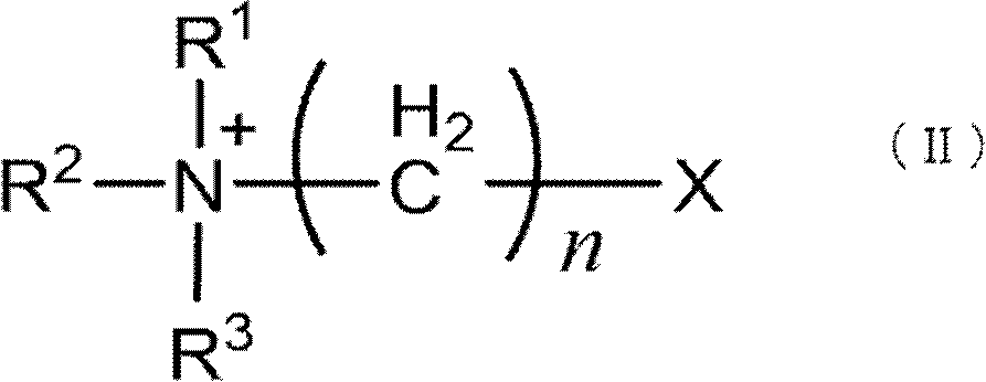
- R 1 to R 3 are the same or different hydrogen atoms, or linear or branched alkyl groups having 1 to 10 carbon atoms, and n in the methylene group is an integer of 1 to 5.
- the cellulose-based ion exchange membrane may be a so-called homogeneous membrane in which the pore size is almost constant from one surface to the other surface, or a so-called asymmetric membrane in which the pore size changes continuously or discontinuously from one surface to the other surface.
- the minimum pore size of the ion exchange membrane is preferably 50 nm or more and 1000 nm or less. In an asymmetric membrane, the minimum pore size layer is preferably located near one of the surfaces. If the minimum pore size is large, the probability of contact of extracellular vesicles such as exosomes with the membrane surface and/or pore surface decreases, and the adsorption rate of extracellular vesicles in the treated liquid (biological sample) may decrease.
- the minimum pore size of the ion exchange membrane is the polystyrene particle size at which the rejection rate is 80% or more when measured using a polystyrene particle dispersion.
- the average pore size of the surface of the ion exchange membrane is preferably 100 nm or more and 5000 nm or less. If the average pore size of the surface is 100 nm or more, biological samples can be processed at a sufficiently fast speed. If the average pore size of the surface is 5000 nm or less, the adsorption of extracellular vesicles to the carrier can be sufficiently promoted, and the recovery rate can be maintained. If the average pore size of the surface is within the above range, the effect of depth filtration can be more reliably exerted.
- the average pore size of the surface can be calculated using image processing software Image J based on photographs of both sides of the membrane taken with a scanning electron microscope (SEM) at a magnification of 1,000 to 20,000 times.
- the biological sample which is the liquid to be treated
- the biological sample may be treated by cross-flow or by dead-end treatment.
- the biological sample which is the liquid to be treated
- the biological sample may be introduced to the large pore side or the small pore side.
- a low solid content concentration and a high extracellular vesicle concentration such as culture supernatant
- the large pore side as the primary side and the small pore side as the secondary side
- clogging of the membrane pores by the adsorbed extracellular vesicles can be prevented even when the biological sample is loaded onto the membrane and the extracellular vesicles are charged and adsorbed onto the membrane.
- the ion exchange capacity of the positively charged carrier is preferably 0.05 meq/g or more and 1.5 meq/g or less. If the ion exchange capacity is 0.05 meq/g or more, the zeta potential of the membrane is stable, and the target substance to be purified, such as extracellular vesicles, which has a small diffusion coefficient, can be sufficiently attracted to the membrane, and the amount of adsorption is sufficiently large, so that a large-capacity device is not required. On the other hand, if the ion exchange capacity is 1.5 meq/g or less, the strength of the membrane is sufficient, and the amount of adsorption per unit volume of the membrane becomes appropriate, so that clogging of the membrane can be suppressed.
- the ion exchange capacity is preferably 0.03 meq/g or more and 0.9 meq/g or less per carrier mass.
- the ion exchange capacity is preferably 0.03 meq/g or more and 0.4 meq/g or less per carrier mass.
- the carrier may be in the form of a flat membrane or a hollow fiber membrane, but the thickness of the membrane is preferably 10 ⁇ m to 1000 ⁇ m. If the thickness of the membrane is 10 ⁇ m or more, the strength of the membrane is sufficiently high, and the handling during membrane production and device fabrication is highly convenient. On the other hand, if the thickness of the membrane is 1000 ⁇ m or less, the recovery time of extracellular vesicles can be reduced and the recovery rate can be maintained.
- the inner diameter is preferably 50 ⁇ m to 1000 ⁇ m.
- the inner diameter is 50 ⁇ m or more, the shear stress of the liquid flowing through the hollow portion is sufficiently small, and damage to extracellular vesicles can be sufficiently suppressed. On the other hand, if the inner diameter is 1000 ⁇ m or less, the membrane area per device can be sufficiently large.
- the specific surface area of the carrier is preferably 3 m 2 /g or more, more preferably 8 m 2 /g or more, as the sum of the specific surface areas of pores with a pore diameter of 30 nm or more. If the specific surface area is 3 m 2 /g or more, a sufficient amount of extracellular vesicles can be adsorbed.
- the specific surface area is generally measured by gas adsorption method or mercury intrusion method.
- the porosity of the carrier is preferably 40% or more and 90% or less. If the porosity is 40% or more, a sufficient area for adsorption of extracellular vesicles can be ensured. If the porosity is 90% or less, the strength of the carrier can be ensured sufficiently. A porosity of 45% or more and 90% or less is more preferable.
- the ion exchange membrane may have a certain degree of micropores.
- Biological samples and cell culture supernatants contain many various small molecular weight substances, and many of these substances have an electric charge.
- the specific surface area of pores having a pore diameter of less than 30 nm is preferably 30 m 2 /g or more and 80 m 2 /g or less.
- the ion exchange capacity of the positively charged carrier is preferably 0.08 meq/g or more and 0.3 meq/g or less. If the ion exchange capacity is 0.3 meq/g or less, the content of positively charged groups per polymer molecule constituting the carrier is not too high, and the water solubility of the carrier is not excessively high, so that the carrier strength in water can be sufficiently ensured. Furthermore, if the ion exchange capacity is 0.08 meq/g or more, the capture efficiency of extracellular vesicles can be more reliably maintained at a high level.
- the positively charged group is preferably a tertiary amine group or a quaternary ammonium group. Diethylamine can be used as the tertiary amine, and trimethylammonium can be used as the quaternary ammonium.
- contact refers to immersing the carrier in the biological sample.
- the carrier when the carrier is a porous membrane structure, it also includes a treatment in which a pressure difference is provided between the primary side and the secondary side of the membrane so that the biological sample enters the inside of the porous membrane, thereby passing the biological sample from the primary side to the secondary side of the membrane through the inside of the porous membrane.
- the carrier when the carrier is a particulate resin, it also includes a treatment in which the biological sample is passed from the inlet to the outlet of a column filled with the resin by a pressure difference.
- the amount of extracellular vesicle particles that can be processed by the carrier depends on the amount of residual medium components in the culture supernatant, but when the anion exchange capacity of the carrier is 0.08 to 0.3 meq/g, it is approximately 10 to 10 particles/mL, and it is necessary to adjust the amount of the biological sample to not exceed this amount relative to the amount of the carrier.
- washing step it is preferable to bring the biological sample into contact with a positively charged carrier to adsorb extracellular vesicles and the like onto the carrier, and then to wash the carrier by bringing a buffer into contact with the carrier.
- the buffer is not particularly limited, but is preferably Tris-HCl. Other buffers include TBS, PBS, and the like.
- the salt concentration of the buffer is preferably adjusted to be equal to or lower than the salt concentration of the biological sample in order to prevent the target extracellular vesicles from detaching from the carrier and being eluted together with other impurities.
- the buffer is 50 mmol/L or more and 150 mmol/L or less, and 6.08 mS/cm or more and 16.01 mS/cm or less.
- the pH is not particularly limited, but is preferably in the range of 7 to 8.
- the amount of the buffer used for washing is preferably in the range of 5 to 100 times the volume of the column or porous body.
- Recovery step In this step, the carrier to which the extracellular vesicles have been adsorbed in the previous steps 3 and 4 is contacted with a buffer solution containing a metal salt to dissociate the extracellular vesicles from the carrier and recover them.
- a buffer solution containing a monovalent or divalent metal salt may be contacted.
- the salt concentration of the buffer solution is preferably 300 mmol/L or more and 800 mmol/L or less, and 30.91 mS/cm or more and 80.58 mS/cm or less.
- the carrier may be preferable to contact the carrier while increasing the salt concentration, such as a stepwise elution method or a linear gradient elution method.
- the monovalent or divalent metal salt is not particularly limited, but NaCl is preferable. In other embodiments, the metal salt may be MgCl 2 , Na 2 SO 4 , or MgSO 4 .
- the buffer solution Tris-HCl, PBS and TBS are preferred, and Tris-HCl is more preferred.
- the salt concentration and electrical conductivity of the sample containing extracellular vesicles may be adjusted in order to enhance the storage stability of the recovered extracellular vesicles.
- the salt concentration and electrical conductivity may be in a range close to the electrolyte concentration in the living body, and are preferably 140 mmol/L or more and 160 mmol/L or less, and more than 15 mS/cm and 17.01 mS/cm or less.
- the electrical conductivity is more preferably 15.01 mS/cm or more.
- a method for measuring the salt concentration a method of measuring the electrical conductivity of the recovered fraction and calculating the NaCl concentration can be mentioned (Figure 2). Based on the obtained salt concentration, 10 mmol/L Tris-HCl (pH 7.5) may be added to dilute the sample. Other methods include buffer replacement by ultrafiltration or desalting by dialysis.
- concentration refers to reducing the volume of a solution containing the target extracellular vesicles and increasing the particle concentration of the target extracellular vesicles.
- Test Example 1 Measurement of ion exchange capacity of carrier The ion exchange capacity of the carrier was measured as follows. A 0.1% by mass bromophenol blue (BPB) aqueous solution was prepared, and the positively charged carrier was immersed in it at room temperature for 1 hour to stain it. After washing the dyed carrier with water, it was immersed in an 8% by mass sodium chloride aqueous solution to elute the BPB adsorbed on the carrier. The absorbance at 590 nm of the sodium chloride aqueous solution was measured, and the number of moles of BPB adsorbed per mass of the carrier was calculated based on a calibration curve in an aqueous BPB solution containing 8% by mass sodium chloride, and this was taken as the ion exchange capacity.
- BPB bromophenol blue
- Test Example 2 Evaluation of particle rejection The rejection of the porous substrate membrane and the ion exchange membrane was evaluated using a dispersion of polystyrene particles. A Tween 20 aqueous solution containing 0.01% by mass of polystyrene particles of various sizes was passed through the membrane at an operating pressure of 10 kPa to obtain a permeated liquid. The polystyrene particle concentrations in the original solution and the permeated liquid were measured by absorbance at 250 nm, and the rejection was calculated by the following formula.
- Rejection rate (%) ⁇ 1-(Ca-Cb)/Ca ⁇ 100
- Ca polystyrene particle concentration in the original solution
- Cb polystyrene particle concentration in the permeated liquid Rejection rates were measured using polystyrene with particle sizes of 79, 132, 208, 262, 313, 420, 460 and 616 nm, and the polystyrene particle size giving a rejection rate of 80% was determined as the minimum pore size.
- Test Example 3 Observation of Film Structure The film to be evaluated was placed on a sample stage for SEM, dried at room temperature, platinum was evaporated onto it, and the film was observed under a scanning electron microscope (SU-1500, Hitachi High-Technologies Corporation).
- Test Example 4 Measurement of the average pore size on the large pore side of the membrane The average pore size on the large pore side of the surface SEM image of the membrane observed at 10,000 times was measured by the following procedure. The image was opened on the image processing software Image J, a line was drawn in the range of the scale bar using a straight line tool from the toolbar, Analyze>Set Scale was selected, and the length of the scale bar was entered in "Known Distance" and the unit in "Unit of length”. In Image>Adjust>Auto Threshold, the method mode was set to Default, and OK was selected for binarization. In Analyze>Set Measurements, it was confirmed that "Feret's diameter" was checked, and OK was pressed.
- Test Example 5 Measurement of zeta potential The zeta potential of the membrane was calculated by attaching a flat plate zeta potential measurement cell ("ZEN1020" manufactured by Malvern Panalytical Co.) to a Zetasizer Nano ZS and measuring the mobility of aluminum oxide particles in ion-exchanged water of pH 6.5 to 7.5.
- ZEN1020 flat plate zeta potential measurement cell
- Zetasizer Nano ZS Zetasizer Nano ZS
- Test Example 6 Measurement of specific surface area A pore distribution measuring device ("Autopore IV9520" manufactured by Shimadzu Corporation) was used to measure under the following conditions. The sample was cut into strips of about 12.5 mm x 25 mm, and 0.04 to 0.15 g was placed in a 5 mL powder cell (stem volume 0.4 mL) and measured under the condition of an initial pressure of about 3.7 kPa (corresponding to a pore diameter of about 340 ⁇ m). The mercury parameters were set to the default mercury contact angle of 130 degrees and mercury surface tension of 485 dynes/cm. The pore distribution was obtained by plotting the transition of the Log differential volume of mercury that had penetrated into the sample against the pore diameter to obtain a pore distribution curve.
- Autopore IV9520 manufactured by Shimadzu Corporation
- Test Example 7 Nanoparticle Tracking Analysis The average particle number and diameter of the recovered extracellular vesicles were measured by nanoparticle tracking analysis (NTA) using a nanoparticle analysis system ("NanoSight NS300" manufactured by Malvern Panalytical). The camera level was set to 14 and the detection threshold was set to 7. When the salt concentration of the culture supernatant was changed, the particle number was measured 24 hours or more after the change.
- NTA nanoparticle tracking analysis
- the culture medium obtained at the time of culture medium replacement was collected and pooled and used for the preparation of exosomes.
- the collected culture medium was centrifuged at 4°C, 300 x g for 10 minutes, then at 10,000 x g for 20 minutes, and the centrifugal supernatant was collected. Further, filtration was performed using a 0.45 ⁇ m filter (Corning) to prepare the culture supernatant.
- Comparative Example 1 An extracellular vesicle solution was obtained in the same manner as in Example 1, except that in the permeation step, the electrical conductivity of the culture supernatant was not adjusted and the culture supernatant with the same salt concentration was permeated through the membrane. At this time, the electrical conductivity of the culture supernatant permeated through the anion exchange membrane was 16.01 mS/cm.
- Comparative Example 2 In the permeation step, an equilibration buffer was added to the culture supernatant to prepare a culture supernatant with an electrical conductivity of 3.10 mS/cm, and the culture supernatant was permeated through the membrane. An extracellular vesicle solution was obtained in the same manner as in Example 1, except that the culture supernatant was permeated through the membrane.
- Comparative Example 1 in which the electrical conductivity of the culture supernatant was kept at 16.01 mS/cm without adjustment, the number of particles recovered from the carrier having ion exchange groups was reduced. This indicates that the high salt concentration of the culture supernatant caused the extracellular vesicles to not be sufficiently adsorbed to the carrier, and they leaked out into the permeate (flow-through) fraction.
- Comparative Example 2 in which the electrical conductivity of the culture supernatant was set to 3.10 mS/cm, the particle measurement results using NTA in the culture supernatant were significantly increased. This indicates that the extracellular vesicles were ruptured and damaged.
- CD63 is known as a marker for EVs, and it has been reported that it is expressed in exosomes, especially in EVs (Borges FT, et al., 2013, Extracellular vesicles: structure, function, and potential clinical uses in renal diseases, Braz J Med Biol Res, 46(10), 824-30).
- exosome-rich fractions have a weaker negative charge than other EVs such as MVs (Seo N, et al., 2022, Distinguishing functional exosomes and other extracellular vesicles as a nucleic acid cargo by the anion-exchange method, J Extracell Vesicles, Mar, 11(3)).
- MVs eo N, et al., 2022, Distinguishing functional exosomes and other extracellular vesicles as a nucleic acid cargo by the anion-exchange method, J Extracell Vesicles, Mar, 11(3).
- the proportion of CD63-positive particles per unit particle among the particles recovered from the purified fraction was higher in Example 1 than in Comparative Example 1. This suggests that by using the method disclosed herein, it is possible to adsorb a greater amount of exosomes, which are particularly weakly negatively charged, to the membrane among the particles in the culture supernatant, thereby reducing loss and enabling recovery.
- the present invention it is possible to easily purify and recover extracellular vesicles contained in biological samples with a high recovery rate.
- the extracellular vesicles obtained by the present invention can be suitably used in fields such as drug delivery, pharmaceuticals, in vitro diagnostics, food, and cosmetics.
Landscapes
- Engineering & Computer Science (AREA)
- Health & Medical Sciences (AREA)
- Biomedical Technology (AREA)
- Life Sciences & Earth Sciences (AREA)
- Chemical & Material Sciences (AREA)
- Biotechnology (AREA)
- Organic Chemistry (AREA)
- Bioinformatics & Cheminformatics (AREA)
- Genetics & Genomics (AREA)
- Wood Science & Technology (AREA)
- Zoology (AREA)
- Microbiology (AREA)
- Biochemistry (AREA)
- General Engineering & Computer Science (AREA)
- General Health & Medical Sciences (AREA)
- Cell Biology (AREA)
- Separation Using Semi-Permeable Membranes (AREA)
- Peptides Or Proteins (AREA)
- Other Investigation Or Analysis Of Materials By Electrical Means (AREA)
Abstract
本発明は、細胞外小胞を高回収率かつ低コストに分離精製できる精製方法を提供することを目的とする。本発明は、細胞外小胞を製造するための方法であって、工程(a)細胞外小胞を含む生体試料を得る工程、工程(b)前記生体試料の電気伝導度を4mS/cm以上15mS/cm以下に調整する工程、工程(c)前記生体試料を、正電荷を有する担体に接触させる工程、工程(d)前記担体に、金属塩を含有する緩衝液を接触させて、細胞外小胞を回収する工程を含む方法である。
Description
本発明は、細胞外小胞を高回収率で精製することにより製造するための方法に関する。
細胞外小胞は、ほぼすべての生細胞から分泌される不均一な脂質二重膜構造を有する小胞の総称であり、産生機構の違いから、エクソソーム、マイクロベシクル、アポトーシス小体に大別される。
エクソソームは、後期エンドソーム膜の内向きの出芽により形成され、さらに細胞膜と融合することで完全な粒子となり、エキソサイトーシスにより細胞外へ分泌される。エクソソームは、動物、植物、菌類等の真核細胞により放出される、直径約30~150nmの細胞外小胞であり、体液中にてタンパク質、mRNA、miRNA等を内包して伝播する等、細胞間の情報伝達に重要な機能を示すことが知られている。近年、癌の転位の亢進、癌の増悪にエクソソームが関与していることが示唆されており、癌由来のエクソソームから発現するmiRNAを腫瘍マーカーとする、癌の進行の予測、転位、予後を確認するバイオマーカー等、エクソソームに注目した診断用マーカーが開発されている。
また、エクソソームは生体から分離精製することで、細胞内の核酸、タンパク質等を内包する生物由来の輸送体として、ドラッグ・デリバリー・システム(DDS)への応用が期待されている。それ故、高純度にエクソソームを分離精製する技術が求められている。
マイクロベシクルは、細胞膜が外側へ出芽して分離することによって生成される。マイクロベシクルの大きさは、直径100~1,000nmとかなり広範囲にわたる。アポトーシス小体は、細胞がアポトーシスを起こすことで生じ、その大きさは直径50~5,000nmと広範囲にわたる。マイクロベシクルとアポトーシス小体にも、エクソソームと同様の応用が期待される。
細胞外小胞を分離精製する方法として、遠心分画法、密度勾配遠心法等の超遠心法、ポリエチレングリコール等のポリマーを用いた沈殿法、サイズ排除クロマトグラフィー、アフィニティクロマトグラフィー等のクロマトグラフィー法が挙げられる。一般的な方法としては、超遠心法が挙げられ、密度勾配遠心によって高純度の細胞外小胞が得られる場合がある。例えば特許文献1には、生体試料を限外濾過した後、陰イオン交換カラムクロマトグラフィーに付すエクソソームの製造方法が開示されている。
超遠心分離法は、遠心過程において細胞外小胞の構造変化、破壊による品質の低下、回収率が低い等の問題がある。また、陰イオン交換カラムクロマトグラフィーを用いる方法は、単位体積あたりに吸着する細胞外小胞の数が不十分であるといった課題がある。
本発明は、細胞外小胞を高回収率かつ低コストに分離精製できる精製方法、及び当該精製方法による細胞外小胞の製造方法を提供することを目的とする。
本発明者は、細胞外小胞の調製法について鋭意検討した結果、細胞の培養、夾雑物質の除去、細胞外小胞の分離、回収に至る一連の工程を最適化することにより、目的の細胞外小胞を高純度、かつ高回収率で回収できることを見出した。
すなわち、以下に示す手段により上記課題を解決できることを見出し、本発明に到達した。
[1] 細胞外小胞を製造するための方法であって、
工程(a)細胞外小胞を含む生体試料を得る工程
工程(b)前記生体試料の電気伝導度を4mS/cm以上15mS/cm以下に調整する工程
工程(c)前記生体試料を、正電荷を有する担体に接触させる工程
工程(d)前記担体に、金属塩を含有する緩衝液を接触させて、細胞外小胞を回収する工程
を含むことを特徴とする方法。
[2] 前記工程(a)が、動物からの細胞の単離またはセルストックの凍結細胞の融解、およびそれに続く培養を含むことを特徴とする前記[1]に記載の方法。
[3] 前記工程(a)と前記工程(b)との間に、前記生体試料より夾雑物質の少なくとも一部を除去する工程を有することを特徴とする前記[1]または[2]に記載の方法。
[4] 前記夾雑物質の少なくとも一部を除去する工程が、遠心分離、精密ろ過、限外ろ過、透析からなる群から選択される1以上であることを特徴とする前記[3]に記載の方法。
[5] 前記工程(c)において、前記担体は、アニオン交換能を有する分離膜である、ことを特徴とする前記[1]~[4]のいずれかに記載の方法。
[6] 前記工程(c)において、前記担体は、3級アミンおよび/または4級アンモニウム基を有することを特徴とする前記[1]~[5]のいずれかに記載の方法。
[7] 前記工程(c)と前記工程(d)との間に、前記担体に、緩衝液を接触させて洗浄する工程を含むことを特徴とする前記[1]~[6]のいずれかに記載の方法。
[8] 前記工程(d)において、前記金属塩を含有する緩衝液はトリス塩酸緩衝液、リン酸緩衝生理食塩水、トリス緩衝生理食塩水のいずれかであることを特徴とする前記[1]~[7]のいずれかに記載の方法。
[9] 前記工程(d)において、前記金属塩がNaClであることを特徴とする前記[1]~[8]のいずれかに記載の方法。
[10] 前記工程(d)において、回収された細胞外小胞を含む試料の電気伝導度を15mS/cm超に調整する工程を含むことを特徴とする前記[1]~[9]のいずれかに記載の方法。
[11] 前記細胞外小胞は、エクソソームおよび/またはマイクロベシクルであることを特徴とする前記[1]~[10]のいずれかに記載の方法。
[1] 細胞外小胞を製造するための方法であって、
工程(a)細胞外小胞を含む生体試料を得る工程
工程(b)前記生体試料の電気伝導度を4mS/cm以上15mS/cm以下に調整する工程
工程(c)前記生体試料を、正電荷を有する担体に接触させる工程
工程(d)前記担体に、金属塩を含有する緩衝液を接触させて、細胞外小胞を回収する工程
を含むことを特徴とする方法。
[2] 前記工程(a)が、動物からの細胞の単離またはセルストックの凍結細胞の融解、およびそれに続く培養を含むことを特徴とする前記[1]に記載の方法。
[3] 前記工程(a)と前記工程(b)との間に、前記生体試料より夾雑物質の少なくとも一部を除去する工程を有することを特徴とする前記[1]または[2]に記載の方法。
[4] 前記夾雑物質の少なくとも一部を除去する工程が、遠心分離、精密ろ過、限外ろ過、透析からなる群から選択される1以上であることを特徴とする前記[3]に記載の方法。
[5] 前記工程(c)において、前記担体は、アニオン交換能を有する分離膜である、ことを特徴とする前記[1]~[4]のいずれかに記載の方法。
[6] 前記工程(c)において、前記担体は、3級アミンおよび/または4級アンモニウム基を有することを特徴とする前記[1]~[5]のいずれかに記載の方法。
[7] 前記工程(c)と前記工程(d)との間に、前記担体に、緩衝液を接触させて洗浄する工程を含むことを特徴とする前記[1]~[6]のいずれかに記載の方法。
[8] 前記工程(d)において、前記金属塩を含有する緩衝液はトリス塩酸緩衝液、リン酸緩衝生理食塩水、トリス緩衝生理食塩水のいずれかであることを特徴とする前記[1]~[7]のいずれかに記載の方法。
[9] 前記工程(d)において、前記金属塩がNaClであることを特徴とする前記[1]~[8]のいずれかに記載の方法。
[10] 前記工程(d)において、回収された細胞外小胞を含む試料の電気伝導度を15mS/cm超に調整する工程を含むことを特徴とする前記[1]~[9]のいずれかに記載の方法。
[11] 前記細胞外小胞は、エクソソームおよび/またはマイクロベシクルであることを特徴とする前記[1]~[10]のいずれかに記載の方法。
本発明は、生体試料から細胞外小胞を精製する方法において、正電荷を有する担体に生体試料を接触させる前に生体試料の電気伝導度を調整する工程を適用することにより、目的とする細胞外小胞の担体への吸着量を最大化することができる。また、粒子あたりの細胞外小胞マーカーが強く発現する粒子群を取得することができる。
本発明は、生体試料からエクソソームを含む細胞外小胞を精製する及び製造するための新しい方法に関する。以下、本発明を説明するが、本発明は以下の具体例に限定されるものではない。なお、本開示において、「x~y」の数値範囲には、xおよびyも含まれるものとする。
1.生体試料の調製工程
本工程では、目的物である細胞外小胞を含む生体試料を調製する。
本工程では、目的物である細胞外小胞を含む生体試料を調製する。
生体試料は、精製対象および製造対象である細胞外小胞を含むものである限り特に限定されるものではないが、液体が好ましい。具体的には、細胞培養液、細胞培養液から分離される培養上清、間質液、血液、血液から分離される血清や血漿、尿、髄液、リンパ液、腹水、乳などとすることができる。生体試料が液体でない場合には、緩衝液などの液体に溶解または分散することが好ましい。
本発明において、生体試料から細胞外小胞を精製する方法として、細胞外小胞を含む生体試料を得る工程、前記生体試料の電気伝導度を調整する工程、前記生体試料を、正電荷を有する担体に接触させる工程、前記担体に、緩衝液を接触させて洗浄する工程、前記担体に金属塩を含有する緩衝液を接触させて、細胞外小胞を回収する工程、を経るのが好ましい。
細胞外小胞は、ほぼすべての生細胞から分泌される不均一な脂質二重膜構造を有する小胞の総称である。細胞外小胞は、細胞内の産生機構の違いから、エクソソーム、マイクロベシクル、アポトーシス小体の3種類に大別される。エクソソームの大きさは直径30~150nmで、エクソソームは後期エンドソーム膜の内向きの出芽により形成され、さらに細胞膜と融合することで完全な粒子を形成しエキソサイトーシスにより細胞外へ分泌される。一方、マイクロベシクルは、細胞膜が外側へ出芽して分離することによって生成される。大きさは直径100nm~1,000nmである。アポトーシス小体は、細胞が組織的な細胞死(アポトーシス)を起こすことで生じる。大きさは直径50nm~5,000nmである。
本発明において、「細胞外小胞」は、生体試料に含まれる細胞外小胞若しくは対象とする培養細胞が産生する細胞外小胞であればよく、細胞外小胞のサイズや形態自体に特に制限はない。また、天然、非天然(人工)の細胞外小胞であってもよい。
細胞外小胞は、例えば、培養細胞から調製することができる。本発明において、「培養細胞」とは、インビトロ(生体外)で培養される細胞一般をいい、特定の商業的に確立されたセルフライン(細胞株)に限定されない。ヒト若しくはヒト以外の生物種、特には哺乳動物の生体から取り出された細胞、組織、器官等をインビトロで培養した場合の当該培養物(例えば、初代培養物(プライマリーカルチャー))中に含まれる細胞は、「培養細胞」に包含される。商業的に確立された種々の培養腫瘍細胞等のセルラインを構成する細胞は、ここでいう「培養細胞」の典型例である。なお、「がん細胞」等の「腫瘍細胞」とは、種々の腫瘍を形成する細胞であって、典型的には周辺の正常組織とは無関係に異常に増殖を行うに至った細胞であり、いわゆるがん化した細胞等がある。各種の細胞種、リンパ種、メラノーマ等を構成する細胞由来の「培養腫瘍細胞」は、ここでいう「培養細胞」の好適例である。培養腫瘍細胞としては、培養された神経芽細胞や網膜芽細胞が挙げられる。
細胞培養物には、対象とする培養細胞および培養細胞の培地を構成する材料が含まれる。培養細胞としては、通常入手可能な、典型的には市販されているセルライン、例えば、ヒトまたはヒト以外の哺乳動物由来の培養腫瘍細胞株が挙げられる。例えば、子宮がん、卵巣がん、および乳がん等の培養腫瘍細胞を使用することができ、ヒト胎児の腎由来のHEK293細胞を使用し得る。
培養細胞の培地を構成する材料は、使用する培養細胞の種類に応じて従来から推奨される培地構成に準じればよく、本発明の実施にあたって特に制限はない。例えば、α-MEM、DMEM、MEM等の市販される培地に、胎児ウシ血清(fetal bovine serum:FBS)等の血清ならびに必要に応じて抗生物質等の成分を添加して調製された培地を使用することができる。ここで、高純度の細胞外小胞を回収するという観点から、血清由来動物の細胞外小胞が混入していない血清含有培地を使用することが好ましい。
本発明において、培養上清から細胞外小胞を単離する場合、培養する細胞は、セルストックの凍結細胞を融解して得ることができる。また、別の実施形態では、ヒトを含む動物組織からの単離により得ることができる。細胞としては、例えば、血液に含まれる細胞、幹細胞などが用いられる。培養時に用いる培地は、無血清または血清入り培地のどちらでもよい。血液に含まれる細胞としては、T細胞が挙げられる。幹細胞としては、ES細胞、iPS細胞、間葉系幹細胞が挙げられる。
培養方法は、特に限定されないが、培養用フラスコ、ディッシュ、プレート等を用いた平面培養、細胞を接着させたビーズ状のマイクロキャリアを培養液中に浮遊させて培養するマイクロキャリア培養、中空糸膜や中空ゲルを用いた培養等が挙げられる。
培養期間は、特に限定しないが、十分量の細胞外小胞を効率的に回収するという観点から、CO2リッチ雰囲気中、37℃前後の温度条件で2~5日程度の培養期間であることが好ましい。CO2濃度は、例えば5±2%程度に調整することができる。
図1は、中空糸膜型細胞培養装置の構成例の一つを示す。図1において、1および2はそれぞれ、ガス透過性を有する液体培地貯留容器(培養バッグなど)である。液体培地貯留容器1からは、無菌接続コネクター13およびバルブ4を経由して、細胞培養容器17に流路が接続され、液体培地貯留容器1に入っている培地が中空糸膜の中空部に移送できるようになっている。他方、液体培地貯留容器2からは、無菌接続コネクター14およびバルブ5を経由して、細胞培養容器17に流路が接続され、液体培地貯留容器2に入っている培地が中空糸膜の外側に移送できるようになっている。
細胞培養容器17の中空部からは、バルブ7、バルブ11、送液ポンプ20、バルブ12、無菌接続コネクター16を経由して、廃液回収容器18まで流路が接続され、細胞培養容器17を通過した培地を廃棄できるようになっている。なお、前記流路に設けられた送液ポンプ20により、液体培地貯留容器1から細胞培養容器17への液体(培地など)の供給、前記細胞培養容器からの前記液体の排出、および前記液体の廃棄などにおける流速等を制御することができる。他方、細胞培養容器17の外腔側からは、バルブ6、バルブ9、送液ポンプ19、バルブ10、無菌接続コネクター16を経由して、廃液回収容器18まで流路が接続され、細胞培養容器17を通過した培地が廃棄できるようになっている。なお、前記流路に設けられた送液ポンプ19により、液体培地貯留容器2から細胞培養容器17への液体(培地など)の供給、前記細胞培養容器からの前記液体の排出、および前記液体の廃棄などにおける流速等を制御することができる。
図1において、細胞回収容器3は、バルブ7、8および無菌接続コネクター15を介して細胞培養容器17の中空部と連通している。必要により、この流路にさらにポンプ等を設けて、細胞を回収してもよい。なお、細胞の回収は、細胞培養容器17を細胞培養装置から取り外して行ってもよく、その場合は、内腔側の廃棄系の流路の分岐から細胞回収容器3までの構成は無くてもよい。
図1において、CO2インキュベーター31は、前記の構成のほとんどをカバーするのが好ましいが、少なくともガス透過性を有する液体培地貯留容器1、2および細胞培養容器17がカバーされていれば、その他の部分についてはCO2インキュベーター31の外側に設置されていても構わない。
図1において、作業コントローラー21(作業パネル)は、上記の操作や工程を行うため、またモニタリングを行うために設置されていてもよい。
本発明において、細胞外小胞を含む生体試料を得る工程は、不要な大型の細胞残屑などの夾雑物を除去する工程を含んでいてもよい。夾雑物を除去する工程としては、例えば、遠心分離や精密ろ過、限外ろ過、透析等が挙げられる。遠心分離は、例えば4℃、10,000×g、15分~30分で行われる。遠心分離により細胞残屑などが除去された上清を回収する。夾雑物をさらに除去するために、追加で遠心分離を行ってもよい。追加の遠心分離を行う場合、遠心加速度(g)を大きくして、中型サイズ以下の細胞残屑を除去してもよい。精製対象がエクソソームやマイクロベシクルである場合には、アポトーシス小胞を除去してもよい。
また、精密ろ過は、孔径0.2~0.5μmのメンブレンフィルターを用いた処理であり、ろ液を回収することにより遠心分離と同様に中型サイズの細胞残屑などを除去することができる。
また、遠心分離により得られた上清および/または精密ろ過により得られたろ液を孔径0.1μm以下、より好ましくは0.05μm以下のメンブレンフィルターを用いた限外ろ過および/または透析により処理してもよい。限外ろ過および/または透析を行うことで、細胞外小胞より小さな夾雑物の除去や、細胞外小胞の濃縮を実施することができる。
本発明の、細胞外小胞を含む生体試料を得る工程において、超遠心による処理を含んでもよい。超遠心の工程では、100,000×g以上の遠心力をかけることで細胞外小胞を沈殿させることで、細胞外小胞より小さな夾雑物の除去や、細胞外小胞の濃縮を実施することができる。
2.電気伝導度の調整工程
本工程では、前工程1で調製した生体試料の電気伝導度を4mS/cm以上15mS/cm以下に調整する。電気伝導度は、塩濃度を調整することにより調整することが好ましい。一般に生体試料は細胞が生存しやすい生理食塩水に近い浸透圧を有しており、生理食塩水の濃度および浸透圧は0.154mmol/Lおよび308mOsm/Lである。生体試料の有する浸透圧は、大部分が液中のイオン性成分に由来しており、そのため生体試料の電気伝導度と塩濃度との間、または電気伝導度と浸透圧との間には正の相関が見られる。電気伝導度を15mS/cm以下に調整することにより、細胞外小胞は比較的微弱な負電荷を有することから、正電荷を有する担体に対する細胞外小胞の吸着効率を高めることが可能になる。一方、電気伝導度を4mS/cm以上に調整することにより、細胞外小胞の変質を抑制できる。電気伝導度としては、9mS/cm以上14mS/cm以下が好ましい。電気伝導度が15mS/cm以下であれば、生体試料中の塩を含む緩衝液成分中の塩化物イオンなどの負電荷イオンの濃度が低減することで、担体中のイオン交換基のうち、塩化物イオンなどの負電荷イオンとイオン結合しているものの割合が減り、負電荷を帯びる細胞外小胞を吸着するのに有効なイオン交換基が増加する。担体表面に存在する有効なイオン交換基の密度が増加することで、低分子のイオンと比較して拡散係数の低い細胞外小胞を担体表面まで引き寄せることが可能なゼータ電位が担体表面に生じる。これにより、特に細胞外小胞の中でも負電荷の程度が小さい粒子が、担体に吸着しやすくなる。一方、電気伝導度が4mS/cm以上であれば、試料中の水溶液成分が有する浸透圧が過剰に低下することはなく、細胞外小胞の脂質二重膜の内外で生じた浸透圧差による細胞外小胞の損傷を抑制できる。そのため、上記の適切な電気伝導度範囲に調整することが重要である。電気伝導度が9mS/cm以上14mS/cm以下の場合、NaCl水溶液(10mM Tris-HClを含む)のNaCl濃度に換算すると79mmol/L以上130mmol/L以下である。
本工程では、前工程1で調製した生体試料の電気伝導度を4mS/cm以上15mS/cm以下に調整する。電気伝導度は、塩濃度を調整することにより調整することが好ましい。一般に生体試料は細胞が生存しやすい生理食塩水に近い浸透圧を有しており、生理食塩水の濃度および浸透圧は0.154mmol/Lおよび308mOsm/Lである。生体試料の有する浸透圧は、大部分が液中のイオン性成分に由来しており、そのため生体試料の電気伝導度と塩濃度との間、または電気伝導度と浸透圧との間には正の相関が見られる。電気伝導度を15mS/cm以下に調整することにより、細胞外小胞は比較的微弱な負電荷を有することから、正電荷を有する担体に対する細胞外小胞の吸着効率を高めることが可能になる。一方、電気伝導度を4mS/cm以上に調整することにより、細胞外小胞の変質を抑制できる。電気伝導度としては、9mS/cm以上14mS/cm以下が好ましい。電気伝導度が15mS/cm以下であれば、生体試料中の塩を含む緩衝液成分中の塩化物イオンなどの負電荷イオンの濃度が低減することで、担体中のイオン交換基のうち、塩化物イオンなどの負電荷イオンとイオン結合しているものの割合が減り、負電荷を帯びる細胞外小胞を吸着するのに有効なイオン交換基が増加する。担体表面に存在する有効なイオン交換基の密度が増加することで、低分子のイオンと比較して拡散係数の低い細胞外小胞を担体表面まで引き寄せることが可能なゼータ電位が担体表面に生じる。これにより、特に細胞外小胞の中でも負電荷の程度が小さい粒子が、担体に吸着しやすくなる。一方、電気伝導度が4mS/cm以上であれば、試料中の水溶液成分が有する浸透圧が過剰に低下することはなく、細胞外小胞の脂質二重膜の内外で生じた浸透圧差による細胞外小胞の損傷を抑制できる。そのため、上記の適切な電気伝導度範囲に調整することが重要である。電気伝導度が9mS/cm以上14mS/cm以下の場合、NaCl水溶液(10mM Tris-HClを含む)のNaCl濃度に換算すると79mmol/L以上130mmol/L以下である。
細胞外小胞を含む生体試料の電気伝導度や塩濃度を下げる工程は、特に限定されないが、生体試料よりも電気伝導度や塩濃度の低い緩衝液を用いて生体試料を希釈することにより実施することができる。この場合、使用する緩衝液として、10mM程度のトリス塩酸緩衝液(Tris-HCl)、リン酸緩衝生理食塩水(PBS)、トリス緩衝生理食塩水(TBS)などが挙げられる。また、別の実施形態では、限外ろ過による緩衝液の置換や、透析による脱塩により、細胞外小胞を含む生体試料の電気伝導度や塩濃度を調整することができる。
3.担体への接触工程
本工程では、前工程2で電気伝導度を調整した生体試料を、正電荷を有する担体に接触させる。本工程において、担体は特に限定されないが、酢酸セルロースや三酢酸セルロース等のセルロース系樹脂、ポリスルホン系樹脂などが好ましい。また、前記担体は、平膜などシート状の分離膜または中空糸状の分離膜(中空糸膜)であってもよいし、粒子状の多孔質体または非孔質体であってもよい。
本工程では、前工程2で電気伝導度を調整した生体試料を、正電荷を有する担体に接触させる。本工程において、担体は特に限定されないが、酢酸セルロースや三酢酸セルロース等のセルロース系樹脂、ポリスルホン系樹脂などが好ましい。また、前記担体は、平膜などシート状の分離膜または中空糸状の分離膜(中空糸膜)であってもよいし、粒子状の多孔質体または非孔質体であってもよい。
本発明において、担体に正電荷を付与する方法は特に限定されないが、担体の成形後にカチオン化合物を付与するなどの方法がある。より具体的には、水酸基をもつ担体に、グリシジルトリメチルアンモニウムやグリシジルジエチルアミンを接触させて正電荷を付与する方法などがある。また、市販のジエチルアミノエチル(DEAE)セルロース樹脂をカラムに充填したものを用いてもよい。DEAEセルロースは、セルロースにジエチルアミノエチル基を導入した陰イオン交換体であり、シグマアルドリッチ(Sigma-Aldrich)社などから入手することができる。
本発明において、正電荷を有する担体として、セルロース系ポリマーを含む多孔質基材膜の2位、3位および6位のいずれかの水酸基またはアセチル基の少なくも一部を正電荷を有する化合物に置換したセルロース系イオン交換膜を用いるのが好ましい。
セルロース系ポリマーを含む多孔質基材膜は、湿式法により製造することができる。湿式法は、セルロース系ポリマーを溶媒および非溶媒と混合して製膜溶液を作製した後、中空状に吐出させ、凝固浴中に導き相分離させる方法である。また、乾式法、熱誘起型相分離法、延伸法等を用いてもよい。一方、平膜は、セルロース系ポリマーを溶媒に溶解し、溶液をガラス等の基材へ均一に塗布し、凝固液へ浸漬して凝固させた後、洗浄、必要により乾燥することにより多孔質基材膜を作製することができる。
製膜溶液の調製に用いる溶媒としては、γ-ブチロラクトン、N-メチルピロリドン、ジメチルアセトアミド等の極性溶媒が好ましい。前記溶媒は、単独で用いてもよいが、混合して用いてもよい。また、必要により、水、グリセリン、エチレングリコール、トリエチレングリコール、ポリエチレングリコール200、ポリエチレングリコール400等の非溶媒を添加してもよい。製膜溶液中の溶媒と非溶媒の混合比(溶媒/非溶媒比)は、90/10~10/90が好ましい。
凝固液は、溶媒、および水などの非溶媒からなる混合液を用いるのが好ましい。また、凝固液中の溶媒/非溶媒比は、製膜溶液中の溶媒/非溶媒比に合わせるのが好ましい。製膜溶液と凝固液の溶媒/非溶媒比を合わせることにより、連続製膜時にも凝固液の組成変動を抑えることができる。平膜の場合、N-メチルピロリドンと水の混合溶液におけるN-メチルピロリドン:水の重量比が30:70~50:50の範囲が好ましい。凝固浴をこのような組成とすることで、凝固浴に最初に接する側の膜表面の孔径が、基材との接触面側の膜表面の孔径よりも大きくなり、厚み方向に対して非対称構造が形成される。凝固浴中のN-メチルピロリドンの濃度が25質量%以上30質量%未満であると、凝固浴に最初に接する側の膜面の孔径が、基材との接触面側の膜表面の孔径と等しくなり、膜の非対称構造が緩和される傾向にある。さらに、凝固浴中のN-メチルピロリドンの濃度が25質量%未満となると、凝固浴に最初に接する側の膜面に、明確な孔のない緻密層が形成されやすくなる。凝固液の温度は、5~60℃が好ましい。
得られた多孔質基材膜は、温水で洗浄して過剰の溶媒等を除去する。洗浄後の多孔質基材膜は、水中で保存してもよいし、細孔内にグリセリン水溶液等の細孔保持剤を充填した後、乾燥してもよい。
前記得られた多孔質基材膜は、次にイオン交換基を導入する処理を行う。具体的には、脱アセチル化処理を行った後、水酸基への荷電付与処理を行う。一例として、セルロース系多孔質基材膜の水酸基に正電荷性基を置換してイオン交換膜であるカチオン性セルロース膜を作製する方法としては、前記セルロース系多孔質基材膜をアルカリ存在下で非溶解性溶媒中に浸漬し、正電荷を有する化合物の溶液を滴下して、加熱条件下で反応させ、アルコールでクエンチすることで作製することができる。
前記非溶解性溶媒としては、セルロース系多孔質基材膜が溶解しない溶媒であり、正電荷を有する化合物が溶解する溶媒であれば特に制限はない。例えば、水、テトラヒドロフラン(THF)、ジメチルスルホキシド(DMSO)、ジメチルホルムアミド(DMF)等が好例であり、原料の溶解性に応じて単独または混合して使用してもよい。
脱アセチル化に用いるアルカリとしては、水酸化リチウム、水酸化カリウム、水酸化ナトリウム、水酸化セシウム等の水酸化物、及び炭酸塩、有機アミン等を用いることができる。その中でも工業的に安価な水酸化ナトリウムを用いるのが好ましい。使用するアルカリの濃度としては、0.05質量%~5.0質量%の範囲が好ましい。アルカリの濃度が0.05質量%以上であれば、荷電基の導入量を増やすことができ、単位容積あたりの細胞外小胞吸着量をより確実に大きくすることができ得る。一方、アルカリの濃度が5.0質量%以下であれば、膜の膨潤度を抑制できるため、荷電基導入後のイオン交換膜において基材膜の構造(細孔径)や物性(膜強度)をより確実に保持でき得る。したがって、アルカリの濃度は0.1~2.5質量%がより好ましく、0.2~2.0質量%がさらに好ましい。
上記正電荷を有する化合物の添加量によって、セルロース膜に対する荷電基付与量を制御することができる。例えば、エポキシ基含有の3級アミン化合物を使用する場合、非溶解性溶媒中の上記3級アミン化合物の濃度は、0.01mоl/L~3.0mоl/Lの範囲が好ましい。この場合、イオン交換容量は、セルロース膜重量当たり0.05meq/g~0.9meq/gに相当する。エポキシ基含有の上記第4級アンモニウム化合物を使用する場合、非溶解性溶媒中の上記4級アミン化合物の濃度は、0.01mоl/L~1.0mоl/Lの範囲が好ましい。この場合、イオン交換容量は、セルロース膜重量当たり0.05meq/g~0.4meq/gに相当する。正電荷の荷電基付与量が多すぎると、ポリマー1分子あたりの正電荷基の含有量が高くなるため、水溶性が高くなり、水中での膜強度が弱くなる場合がある。また、正電荷の荷電基付与量が小さすぎると、細胞外小胞の捕捉効率が低くなる場合がある。
本発明において、イオン交換膜など正電荷を有する担体は、被処理液を導入するための導入口と、イオン交換処理された被処理液である生体試料を排出するための排出口と、を備えた容器内に収められたデバイスの形態とすることが好ましい。このデバイスは、イオン交換膜で仕切られた第1室と第2室とを有し、第1室に導入された被処理液である生体試料はイオン交換膜を透過して第2室に移動する。この際、被処理液である生体試料中の細胞外小胞は、イオン交換膜の表面および細孔表面に吸着される。
本発明において、正電荷を有する担体であるイオン交換膜は、セルロース系ポリマーの2位、3位および6位のいずれかの水酸基またはアセチル基の少なくとも一部が正電荷を有する化合物で置換された、セルロース系イオン交換膜であってもよい。
本発明において、セルロース系ポリマーとしては、セルロースアセテート、セルロースプロピオネート、セルロースブチレート、セルロースアセテートプロピオネート、セルロースアセテートブチレート、セルロースアセテートラウレート、セルロースアセテートオレート、及びセルロースアセテートステアレート等が挙げられるが、正電荷を有する化合物の導入のしやすさの面からセルロースアセテートを用いるのが好ましい。セルロースアセテートは、酢化度、分子量が異なるものが市販されており、酢化度が52~62程度のセルロースアセテートおよび/またはセルローストリアセテートを用いるのがより好ましい。
本発明において、正電荷を有する化合物としては、下記式(I)で表される第3級アミンまたは下記式(II)で表される第4級アンモニウム化合物を用いるのが好ましい。式中、R1~R3は同一または異なる水素原子、もしくは炭素数1~10の直鎖あるいは分岐状アルキル基を示し、メチレン基のnは1~5の整数を示す。式中、Xは、フッ素、塩素、臭素、ヨウ素等のハロゲン原子、トシラート、トリフラート、メシラート等のスルホン酸エステル等の脱離基、アルコキシシリル、シラノール等のシリル基、エポキシド基、イソシアネート基、カルボン酸基からなる群から選ばれる1種以上を示す。正電荷を有する化合物は、何れの置換基Xを用いてもよいが、原料の入手のしやすさの面からは、Xが塩素、シリル基、エポキシド基を有する化合物が好ましい。

本発明において、セルロース系イオン交換膜は、一方の表面から他方の表面に向かって細孔径がほぼ一定である、いわゆる均質膜でもよいし、一方の表面から他方の表面に向かって連続または不連続に細孔径が変化する、いわゆる非対称膜でもよい。また、前記イオン交換膜の最小孔径は50nm以上1000nm以下であるのが好ましい。非対称膜において、最小孔径層は、いずれかの表面近傍にあるのが好ましい。最小孔径が大きいと、膜面および/または細孔表面へのエクソソーム等の細胞外小胞の接触確率が低下し、被処理液(生体試料)中の細胞外小胞の吸着率が低くなることがある。また、細胞屑などを除去することができないことがある。一方、最小孔径が小さいと、膜内部に入り込んだ細胞外小胞の回収率が低下するとか、回収に時間を要することがある。なお、最小孔径は、ポリスチレン粒子分散液を用いて測定したときに、阻止率が80%以上となるポリスチレン粒子径をイオン交換膜の最小孔径とする。
また、イオン交換膜の表面、例えば最小孔径層を有しない側の表面の平均孔径は、100nm以上5000nm以下であるのが好ましい。表面の平均孔径が100nm以上であれば、十分に速い速度で生体試料を処理することができる。また、表面の平均孔径が5000nm以下であれば、細胞外小胞の担体への吸着を十分に促進でき、回収率を維持することが可能になる。また、表面の平均孔径が前記範囲に含まれれば、デプスろ過の効果をより確実に発揮できる。なお、表面の平均孔径は、膜の両面を走査型電子顕微鏡(SEM)を用いて倍率1,000~20,000倍で撮影した写真に基づいて、画像処理ソフトImage Jにより算出することができる。
本工程において、被処理液である生体試料は、クロスフローで処理してもよいし、デッドエンドで処理してもよい。また、非対称膜を用いる場合、大孔径側に被処理液である生体試料を導入してもよいし、小孔径側に導入してもよい。たとえば、培養上清など、固形成分濃度が低く、細胞外小胞濃度が高い生体試料では、孔径が大きい面を一次側、孔径が小さい面を二次側とすることで、生体試料を膜にロードして細胞外小胞を膜に荷電吸着させる際にも、吸着した細胞外小胞により膜の細孔が詰まることを防ぐことができる。一方、血清、血漿、尿や乳など、細胞外小胞濃度に対して固形成分濃度が比較的高い生体試料では、孔径が小さい面を一次側、孔径が大きい面を二次側とすることで、表層ろ過により夾雑物を除去することができ、細孔の完全閉塞による膜の詰まりを防止することができる。
本発明において、正電荷を有する担体のイオン交換容量は、0.05meq/g以上1.5meq/g以下であることが好ましい。イオン交換容量が0.05meq/g以上であれば、膜のゼータ電位が安定して、細胞外小胞のような拡散係数の小さい精製対象物質も十分に膜に引き寄せることができ、吸着量が十分に大きいため、大容量のデバイスは必要ない。一方、イオン交換容量が1.5meq/g以下であれば、膜の強度が十分であり、また、膜の単位体積当たりの吸着量が適度になり、膜詰まりを抑制できる。なお、3級アミン化合物を使用する場合は、イオン交換容量は、担体質量当たり0.03meq/g以上0.9meq/g以下であるのが好ましい。また、第4級アンモニウム化合物を使用する場合は、イオン交換容量は、担体質量当たり0.03meq/g以上0.4meq/g以下であるのが好ましい。
本発明において、担体の形態は、平膜であってもよいし、中空糸膜であってもよいが、膜の厚みは10μm~1000μmであるのが好ましい。膜の厚みが10μm以上であれば、膜の強度が十分に高く、製膜やデバイス作製における取扱い上の利便性が高い。一方、膜の厚みが1000μm以下であれば、細胞外小胞の回収時間を抑制でき、回収率を維持できる。また、中空糸膜の場合、内径は50μm~1000μmが好ましい。内径が50μm以上であれば、中空部を流れる液体の剪断応力が十分に小さく、細胞外小胞へのダメージを十分に抑制できる。一方、内径が1000μm以下であれば、デバイスあたりの膜面積を十分に大きくできる。
本発明において、担体のゼータ電位は、pH6.5~7.5の条件にて10mV~70mVの範囲が好ましい。ゼータ電位が10mV以上であれば、拡散係数の低い細胞外小胞に対する吸着力が十分に高く、吸着性を確保できる。ゼータ電位が70mV以下であれば、膜強度が十分に高く、また、細胞外小胞のような粒子を精製対象とする場合、膜単位体積あたりの吸着量が適度になり、膜詰まりを抑制できる。シート状の担体のゼータ電位は、平板ゼータ電位測定用セルを用いて測定することができる。
本発明において、担体の比表面積は、細孔径が30nm以上の細孔の比表面績の合計が3m2/g以上が好ましく、8m2/g以上がより好ましい。比表面積が3m2/g以上であれば、十分な細胞外小胞の吸着量を確保できる。比表面積は一般的に、ガス吸着法や水銀圧入法により測定される。ガス吸着法では、およそ2nm以下のマイクロポアおよび2nm~50nmのメソポアの比表面積を検出できるのに対し、水銀圧入法ではメソポアおよび50nm以上のマクロポアを検出することができる。比表面積は、細胞外小胞の吸着量を確保するために大きい方がよいが、細胞外小胞は、細孔径が30nm未満のポア内部には入り込めないので、膜の単位体積あたりの細胞外小胞の吸着量を大きくするためには、細孔径が30nm以上のメソポアおよびマクロポアから成る比表面積が十分大きい必要がある。なお、細孔の比表面積は大きすぎて問題となることはないが、150m2/g以下が好ましく、120m2/g以下がより好ましい。
また、担体の気孔率は40%以上90%以下であるのが好ましい。気孔率が40%以上であれば、細胞外小胞が吸着する領域を十分に確保できる。気孔率が90%以下であれば、担体の強度を十分に確保できる。気孔率としては、45%以上90%以下がより好ましい。
本発明の担体を細胞外小胞の分離精製に用いる際には、ゼータ電位、比表面積、および気孔率等を特定の範囲に調整するのが好ましい。細胞外小胞のうちエクソソームは、大きさが30nm~150nmであり、表面に負電荷を有する細胞外小胞である。例えば、細胞培養上清などの生体試料中のエクソソームを効率よく吸脱着させる場合に、細孔が小さすぎ、マイクロポアが多すぎると、細孔内にエクソソームが入り込めないため担体の比表面積を大きくしても吸着領域や吸着容量を稼ぐことができない。また、細孔が大きすぎ、マクロポアが多すぎると、膜表面とエクソソームとの距離が遠すぎて電気的な吸引力が働きにくくなるとか、直径が150nmを超える細胞外小胞が細孔内に入り込み、目的のエクソソームの吸着効率が低下することがある。一方、イオン交換膜は、ある程度マイクロポアを有する方がよい場合がある。生体試料や細胞培養上清中には、種々の小分子量物質が多く含まれており、これらの中には電荷を有する物質も数多くある。このような小分子量物質をマイクロポア内に吸着させることにより、メソポアやマクロポアの吸着部位をエクソソームの吸着に有効に利用することが可能となる。細孔径が30nm未満の細孔の比表面積は30m2/g以上80m2/g以下が好ましい。
正電荷を有する担体のイオン交換容量は、0.08meq/g以上0.3meq/g以下であることが好ましい。イオン交換容量が0.3meq/g以下であれば、担体を構成するポリマー1分子あたりの正電荷基の含有量が高過ぎず、担体の水溶性が過剰に高過ぎることはないため、水中での担体強度を十分に確保することができる。また、イオン交換容量が0.08meq/g以上であれば、細胞外小胞の捕捉効率をより確実に高く維持できる。正電荷基は、3級アミン基または4級アンモニウム基であることが好ましい。3級アミンとしては、ジエチルアミン、4級アンモニウムとしては、トリメチルアンモニウムなどが使用できる。
本発明において、「接触」させるとは、担体を生体試料に浸漬することを指す。また、担体が多孔膜構造の場合、多孔膜内部に生体試料が入り込むように、膜の一次側と二次側の間に圧力差を設け、それにより生体試料が多孔膜内部を通り膜の一次側から二次側へ通過するよう処理することも含む。また、担体が粒子状の樹脂の場合、樹脂を充填したカラムの入り口から出口へと生体試料を圧力差により通過させることも含む。
担体が処理可能な細胞外小胞の粒子量は、培養上清の培地成分の残渣量により左右されるが、担体の陰イオン交換容量が0.08~0.3meq/gのとき、およそ109~1012個/mLであり、担体量に対してそれを超えない量の生体試料を処理するように調整する必要がある。
担体が処理可能な細胞外小胞の粒子量は、培養上清の培地成分の残渣量により左右されるが、担体の陰イオン交換容量が0.08~0.3meq/gのとき、およそ109~1012個/mLであり、担体量に対してそれを超えない量の生体試料を処理するように調整する必要がある。
4.洗浄工程
本発明において、生体試料を正電荷を有する担体に接触させて細胞外小胞等を担体に吸着させた後、担体に緩衝液を接触させて洗浄するのが好ましい。緩衝液は、特に限定されないがTris-HClであることが好ましい。その他の緩衝液としては、TBS、PBSなどが挙げられる。緩衝液の塩濃度は、目的の細胞外小胞が担体から脱離してその他の夾雑物と一緒に溶出することを防止するために、生体試料の塩濃度以下に調整することが好ましい。具体的には、50mmol/L以上150mmol/L以下、及び6.08mS/cm以上16.01mS/cm以下であることが好ましい。pHは特に限定されないが、7~8の範囲が好ましい。洗浄に使用する緩衝液の液量は、カラムもしくは多孔質体の体積の5倍~100倍の範囲が好ましい。
本発明において、生体試料を正電荷を有する担体に接触させて細胞外小胞等を担体に吸着させた後、担体に緩衝液を接触させて洗浄するのが好ましい。緩衝液は、特に限定されないがTris-HClであることが好ましい。その他の緩衝液としては、TBS、PBSなどが挙げられる。緩衝液の塩濃度は、目的の細胞外小胞が担体から脱離してその他の夾雑物と一緒に溶出することを防止するために、生体試料の塩濃度以下に調整することが好ましい。具体的には、50mmol/L以上150mmol/L以下、及び6.08mS/cm以上16.01mS/cm以下であることが好ましい。pHは特に限定されないが、7~8の範囲が好ましい。洗浄に使用する緩衝液の液量は、カラムもしくは多孔質体の体積の5倍~100倍の範囲が好ましい。
5.回収工程
本工程では、前工程3および前工程4において細胞外小胞を吸着した担体に、金属塩を含有する緩衝液を接触させて、細胞外小胞を担体から解離させて回収する。本発明において、担体に吸着および結合したエクソソーム等の細胞外小胞を溶出して回収するためには、1価または2価の金属塩を含有する緩衝液を接触させればよい。緩衝液の塩濃度は300mmol/L以上800mmol/L以下、及び30.91mS/cm以上80.58mS/cm以下であることが好ましい。細胞外小胞の電荷の差異によって、細胞外小胞の種類ごとに分けて回収するなどの目的によっては、ステップワイズ溶出法や、リニアグラジエント溶出法といった、塩濃度を上げながら接触させることが好ましい場合がある。1価または2価の金属塩としては、特に限定されるものではないが、NaClが好ましい。他の実施形態としては、金属塩がMgCl2、Na2SO4、MgSO4であってもよい。緩衝液としては、Tris-HCl、PBSおよびTBSが好ましく、Tris-HClがより好ましい。
本工程では、前工程3および前工程4において細胞外小胞を吸着した担体に、金属塩を含有する緩衝液を接触させて、細胞外小胞を担体から解離させて回収する。本発明において、担体に吸着および結合したエクソソーム等の細胞外小胞を溶出して回収するためには、1価または2価の金属塩を含有する緩衝液を接触させればよい。緩衝液の塩濃度は300mmol/L以上800mmol/L以下、及び30.91mS/cm以上80.58mS/cm以下であることが好ましい。細胞外小胞の電荷の差異によって、細胞外小胞の種類ごとに分けて回収するなどの目的によっては、ステップワイズ溶出法や、リニアグラジエント溶出法といった、塩濃度を上げながら接触させることが好ましい場合がある。1価または2価の金属塩としては、特に限定されるものではないが、NaClが好ましい。他の実施形態としては、金属塩がMgCl2、Na2SO4、MgSO4であってもよい。緩衝液としては、Tris-HCl、PBSおよびTBSが好ましく、Tris-HClがより好ましい。
本発明において、回収された細胞外小胞の保存安定性を高めるために、細胞外小胞を含む試料の塩濃度および電気伝導度を調整してもよい。塩濃度および電気伝導度は、生体の電解質濃度に近い濃度範囲であればよく、140mmol/L以上160mmol/L以下、及び15mS/cm超17.01mS/cm以下であることが好ましい。当該電気伝導度としては、15.01mS/cm以上がより好ましい。塩濃度を測定する方法としては、回収した画分の電気伝導度を測定し、NaCl濃度を算出する方法が挙げられる(図2)。得られた塩濃度に基づいて10mmol/L Tris-HCl(pH7.5)を添加して希釈等すればよい。他の方法として、限外ろ過による緩衝液置換、または透析による脱塩などしてもよい。
本発明において、「濃縮」とは、目的の細胞外小胞を含む溶液の体積を減らし、目的の細胞外小胞の粒子濃度をあげることを指す。
本願は、2023年9月14日に出願された日本国特許出願第2023-149357号に基づく優先権の利益を主張するものである。2023年9月14日に出願された日本国特許出願第2023-149357号の明細書の全内容が、本願に参考のため援用される。
以下、実施例及び比較例を示して本発明をより具体的に説明するが、本発明は以下の実施例によって限定されるものではない。なお、以下の実施例における物性等の評価方法は以下の通りである。
試験例1: 担体のイオン交換容量の測定
担体のイオン交換容量は、下記の通り測定した。
0.1質量%のブロモフェノールブルー(BPB)水溶液を調製し、正電荷を有する担体を室温で1時間浸漬して染色した。染色後の担体を水洗した後、8質量%塩化ナトリウム水溶液に浸漬して担体に吸着していたBPBを溶出させた。前記塩化ナトリウム水溶液の590nm吸光度を測定し、8質量%塩化ナトリウムを含むBPB水溶液中による検量線をもとに、担体の質量あたり吸着していたBPBモル数を算出し、イオン交換容量とした。
担体のイオン交換容量は、下記の通り測定した。
0.1質量%のブロモフェノールブルー(BPB)水溶液を調製し、正電荷を有する担体を室温で1時間浸漬して染色した。染色後の担体を水洗した後、8質量%塩化ナトリウム水溶液に浸漬して担体に吸着していたBPBを溶出させた。前記塩化ナトリウム水溶液の590nm吸光度を測定し、8質量%塩化ナトリウムを含むBPB水溶液中による検量線をもとに、担体の質量あたり吸着していたBPBモル数を算出し、イオン交換容量とした。
試験例2: 粒子の阻止率評価
多孔質基材膜およびイオン交換膜の阻止率を、ポリスチレン粒子の分散液を用いて評価した。種々のサイズのポリスチレン粒子を0.01質量%添加して分散させたTween20水溶液を、操作圧10kPaで膜を透過させて、透過液を得た。もとの溶液および透過液中のポリスチレン粒子濃度を250nmの吸光度で測定して、下記式により阻止率を算出した。
阻止率(%)={1-(Ca-Cb)/Ca}×100
Ca:もとの溶液のポリスチレン粒子濃度
Cb:透過液のポリスチレン粒子濃度
粒子径が79、132、208、262、313、420、460、616nmのポリスチレンを用いて阻止率の測定を行い、阻止率が80%になるポリスチレン粒子径を最小孔径とした。
多孔質基材膜およびイオン交換膜の阻止率を、ポリスチレン粒子の分散液を用いて評価した。種々のサイズのポリスチレン粒子を0.01質量%添加して分散させたTween20水溶液を、操作圧10kPaで膜を透過させて、透過液を得た。もとの溶液および透過液中のポリスチレン粒子濃度を250nmの吸光度で測定して、下記式により阻止率を算出した。
阻止率(%)={1-(Ca-Cb)/Ca}×100
Ca:もとの溶液のポリスチレン粒子濃度
Cb:透過液のポリスチレン粒子濃度
粒子径が79、132、208、262、313、420、460、616nmのポリスチレンを用いて阻止率の測定を行い、阻止率が80%になるポリスチレン粒子径を最小孔径とした。
試験例3: 膜の構造観察
評価すべき膜をSEM用試料台に乗せて室温で乾燥させ、プラチナ蒸着し、走査型電子顕微鏡(「SU-1500」日立ハイテクノロジー社製)で観察した。
評価すべき膜をSEM用試料台に乗せて室温で乾燥させ、プラチナ蒸着し、走査型電子顕微鏡(「SU-1500」日立ハイテクノロジー社製)で観察した。
試験例4: 膜の大孔径側の平均孔径の測定
10,000倍で観察した膜の前記表面SEM画像について、大孔径側の平均孔径を以下の手順で測定した。画像処理ソフトImage J上で画像を開き、ツールバーから直線ツールを用いてスケールバーの範囲に線を引き、Analyze>Set Scaleを選択し、「Known Distance」にスケールバーの長さ、「Unit of length」に単位を入力した。Image>Adjust>Auto Thresholdにて、MethodのモードをDefaultに設定し、OKを選択して二値化した。Analyze>Set Measurementsにて、「Feret’s diameter」にチェックが入っていることを確認し、OKを押した。Analyze>Analyze Particlesにて、「Size(pixel^2)」を0-Imfinityとし、「Display results」、「Exclude on edges」、「Include holes」の各ボックスにチェックが入っていることを確認してOKを選択して、Results画面にてフェレ径の測定データを得た。15ピクセル以下のノイズを消去するため、15ピクセルに相当する長さをFeret径と同じ単位で計算し、それ以下の値は消去して、フェレ径の平均値を算出した。
10,000倍で観察した膜の前記表面SEM画像について、大孔径側の平均孔径を以下の手順で測定した。画像処理ソフトImage J上で画像を開き、ツールバーから直線ツールを用いてスケールバーの範囲に線を引き、Analyze>Set Scaleを選択し、「Known Distance」にスケールバーの長さ、「Unit of length」に単位を入力した。Image>Adjust>Auto Thresholdにて、MethodのモードをDefaultに設定し、OKを選択して二値化した。Analyze>Set Measurementsにて、「Feret’s diameter」にチェックが入っていることを確認し、OKを押した。Analyze>Analyze Particlesにて、「Size(pixel^2)」を0-Imfinityとし、「Display results」、「Exclude on edges」、「Include holes」の各ボックスにチェックが入っていることを確認してOKを選択して、Results画面にてフェレ径の測定データを得た。15ピクセル以下のノイズを消去するため、15ピクセルに相当する長さをFeret径と同じ単位で計算し、それ以下の値は消去して、フェレ径の平均値を算出した。
試験例5: ゼータ電位の測定
膜のゼータ電位は、ゼータサイザーナノZSに平板ゼータ電位測定用セル(「ZEN1020」Malvern Panalytical社製)を取り付け、pH6.5~7.5のイオン交換水中にて酸化アルミニウム粒子の移動度を測定し、算出した。
膜のゼータ電位は、ゼータサイザーナノZSに平板ゼータ電位測定用セル(「ZEN1020」Malvern Panalytical社製)を取り付け、pH6.5~7.5のイオン交換水中にて酸化アルミニウム粒子の移動度を測定し、算出した。
試験例6: 比表面積の測定
細孔分布測定装置(「オートポアIV9520」島津製作所社製)を用い、以下の条件で測定した。試料を約12.5mm×25mmの短冊片に切断して、0.04~0.15gを5mL粉体用セル(ステム容積0.4mL)にとり、初期圧約3.7kPa(細孔直径約340μm相当)の条件で測定した。水銀パラメータは、装置デフォルトの水銀接触角130degrees、水銀表面張力485dynes/cmに設定した。細孔分布は、サンプル内に浸透した水銀のLog微分体積の推移を細孔径に対してプロットし、細孔分布曲線を得た。比表面積は、各モード径Dに対する細孔容積Vを測定し、それぞれのモード径Dに対する比表面積Sを下記式から求め、0.03~500μm範囲の総和から全比表面積を算出した。
S=4V/D
細孔分布測定装置(「オートポアIV9520」島津製作所社製)を用い、以下の条件で測定した。試料を約12.5mm×25mmの短冊片に切断して、0.04~0.15gを5mL粉体用セル(ステム容積0.4mL)にとり、初期圧約3.7kPa(細孔直径約340μm相当)の条件で測定した。水銀パラメータは、装置デフォルトの水銀接触角130degrees、水銀表面張力485dynes/cmに設定した。細孔分布は、サンプル内に浸透した水銀のLog微分体積の推移を細孔径に対してプロットし、細孔分布曲線を得た。比表面積は、各モード径Dに対する細孔容積Vを測定し、それぞれのモード径Dに対する比表面積Sを下記式から求め、0.03~500μm範囲の総和から全比表面積を算出した。
S=4V/D
試験例7: ナノ粒子トラッキング分析
回収された細胞外小胞の平均粒子数及び直径は、ナノ粒子解析システム(「NanoSight NS300」Malvern Panalytical社製)を用い、ナノ粒子トラッキング分析(NTA)により測定した。なお、カメラレベルを14、検出閾値を7に設定し測定した。培養上清の塩濃度を変更した際は、変更後24時間以上経過したのちに粒子数を測定した。
回収された細胞外小胞の平均粒子数及び直径は、ナノ粒子解析システム(「NanoSight NS300」Malvern Panalytical社製)を用い、ナノ粒子トラッキング分析(NTA)により測定した。なお、カメラレベルを14、検出閾値を7に設定し測定した。培養上清の塩濃度を変更した際は、変更後24時間以上経過したのちに粒子数を測定した。
試験例8: ウェスタンブロッティング
回収された細胞外小胞の表面タンパク質を含む細胞外小胞タンパク質を検出するため、ウェスタンブロッティングを実施した。回収された細胞外小胞を含む試料を必要に応じて濃縮し、サンプル緩衝液(「NuPage LDS Sample Buffer」Thermo Fisher Scientific社製)に溶解し、95℃で5分間、煮沸した。なお、TSG101、β-actin及びGAPDHの検出には100mmol/Lジチオトレイトールをさらに含むサンプル緩衝液を使用し、CD63及びCD81の検出にはジチオトレイトールを含まないサンプル緩衝液を使用した。SDSランニングバッファ(Tris/Glycine/SDS Buffer)を用いた10%ポリアクリルアミドゲル電気泳動で、各試料及び分子量マーカー(「MagicMark XP Western Protein Standard」Thermo Fisher Scientific社製)を30mA(ゲル1枚)または40mA(ゲル2枚)で60分間分離した。生成したゲルを、ブロッティング緩衝液(「EzFastBlot」ATTO社製)を用いて、ポリビニリデンフルオライド膜に転写し、ブロッキング剤(「ブロッキングワン」ナカライテスク社製)を用いてブロッキングした後、それぞれ1次抗体を用いて1時間反応させた。洗浄した後の膜を、それぞれ二次抗体を用いて1時間反応させた。洗浄した後、HRP用検出試薬(「EzWestLumi plus」ATTO社製)を用いて、目的とするタンパク質由来のバンドを可視化した。
回収された細胞外小胞の表面タンパク質を含む細胞外小胞タンパク質を検出するため、ウェスタンブロッティングを実施した。回収された細胞外小胞を含む試料を必要に応じて濃縮し、サンプル緩衝液(「NuPage LDS Sample Buffer」Thermo Fisher Scientific社製)に溶解し、95℃で5分間、煮沸した。なお、TSG101、β-actin及びGAPDHの検出には100mmol/Lジチオトレイトールをさらに含むサンプル緩衝液を使用し、CD63及びCD81の検出にはジチオトレイトールを含まないサンプル緩衝液を使用した。SDSランニングバッファ(Tris/Glycine/SDS Buffer)を用いた10%ポリアクリルアミドゲル電気泳動で、各試料及び分子量マーカー(「MagicMark XP Western Protein Standard」Thermo Fisher Scientific社製)を30mA(ゲル1枚)または40mA(ゲル2枚)で60分間分離した。生成したゲルを、ブロッティング緩衝液(「EzFastBlot」ATTO社製)を用いて、ポリビニリデンフルオライド膜に転写し、ブロッキング剤(「ブロッキングワン」ナカライテスク社製)を用いてブロッキングした後、それぞれ1次抗体を用いて1時間反応させた。洗浄した後の膜を、それぞれ二次抗体を用いて1時間反応させた。洗浄した後、HRP用検出試薬(「EzWestLumi plus」ATTO社製)を用いて、目的とするタンパク質由来のバンドを可視化した。
実施例1
(1)生体試料の取得工程
培養細胞としてヒト胎児の腎由来細胞株(HEK293.2sus)を使用した。HEK293.2susは、American Type Culture Collection社より入手した。HEK293.2sus細胞の培養には、100mLの三角フラスコ(Corning社)の中で、293 SFM II培地(ThermoFisher Scientific社)を用い、100rpm及び8%CO2濃度及び5×10⁵細胞/mLの条件下で培養した。次いで、70cm2の培養フラスコ(Corning社)の中、293 SFM II培地を用いて5%CO2濃度の条件下で培養した。3日または4日毎に培養液を交換し、最終17日間培養した。培養液交換時に得られた培養液を回収しプールしてエクソソームの調製に用いた。回収した培養液を4℃、300×gで10分間、次いで10,000×gで20分間の遠心分離を行い、遠心上清を回収した。さらに、0.45μmのフィルター(Corning社)によるろ過を行い、培養上清を調製した。
(1)生体試料の取得工程
培養細胞としてヒト胎児の腎由来細胞株(HEK293.2sus)を使用した。HEK293.2susは、American Type Culture Collection社より入手した。HEK293.2sus細胞の培養には、100mLの三角フラスコ(Corning社)の中で、293 SFM II培地(ThermoFisher Scientific社)を用い、100rpm及び8%CO2濃度及び5×10⁵細胞/mLの条件下で培養した。次いで、70cm2の培養フラスコ(Corning社)の中、293 SFM II培地を用いて5%CO2濃度の条件下で培養した。3日または4日毎に培養液を交換し、最終17日間培養した。培養液交換時に得られた培養液を回収しプールしてエクソソームの調製に用いた。回収した培養液を4℃、300×gで10分間、次いで10,000×gで20分間の遠心分離を行い、遠心上清を回収した。さらに、0.45μmのフィルター(Corning社)によるろ過を行い、培養上清を調製した。
(2)陰イオン交換膜の調製工程
正電荷を有する担体として、0.2μmの公称孔径を有するセルロースアセテートメンブレンフィルター(ADVANTEC社)を0.1M水酸化ナトリウム水溶液に室温で4時間浸漬することにより脱アセチルした。得られた脱アセチル膜を、0.4Mグリシジルジエチルアミン水溶液に65℃で4時間浸漬してイオン交換膜を得た。得られたイオン交換膜の性能を表1に示す。
正電荷を有する担体として、0.2μmの公称孔径を有するセルロースアセテートメンブレンフィルター(ADVANTEC社)を0.1M水酸化ナトリウム水溶液に室温で4時間浸漬することにより脱アセチルした。得られた脱アセチル膜を、0.4Mグリシジルジエチルアミン水溶液に65℃で4時間浸漬してイオン交換膜を得た。得られたイオン交換膜の性能を表1に示す。

(3)平衡化工程
直径25mmの陰イオン交換膜に、100mmol/L(11.05mS/cm)NaClを含有する10mmol/L Tris-HCl(pH7.5)(平衡化バッファ)を5mL通過させ陰イオン交換膜を平衡化した。
直径25mmの陰イオン交換膜に、100mmol/L(11.05mS/cm)NaClを含有する10mmol/L Tris-HCl(pH7.5)(平衡化バッファ)を5mL通過させ陰イオン交換膜を平衡化した。
(4)透過工程
10mmol/LのTris-HCl緩衝液にNaClを溶解させ、NaCl濃度と電気伝導度との関係を示す検量線を作成した。電気伝導度を電気伝導度計(「LAQUAtwin EC-33」HORIBA製)を用いて測定した。その際、測定サンプルを用いて測定部を2回共洗いした後、測定を実施した。0.45μmのメンブレンフィルターでろ過した培養上清6mLに10mmol/LのTris-HCl緩衝液を3mL添加することにより、電気伝導度を11.05mS/cmとした培養上清を膜に通過させた。
10mmol/LのTris-HCl緩衝液にNaClを溶解させ、NaCl濃度と電気伝導度との関係を示す検量線を作成した。電気伝導度を電気伝導度計(「LAQUAtwin EC-33」HORIBA製)を用いて測定した。その際、測定サンプルを用いて測定部を2回共洗いした後、測定を実施した。0.45μmのメンブレンフィルターでろ過した培養上清6mLに10mmol/LのTris-HCl緩衝液を3mL添加することにより、電気伝導度を11.05mS/cmとした培養上清を膜に通過させた。
(5)溶出工程
10mmol/LのTris-HCl緩衝液10mLを膜に通過させて膜を洗浄した後に、300mmol/L(30.91mS/cm)のNaCl2mLを膜に通過させ、膜に結合した細胞外小胞を溶出させ、細胞外小胞の精製画分を得た。
10mmol/LのTris-HCl緩衝液10mLを膜に通過させて膜を洗浄した後に、300mmol/L(30.91mS/cm)のNaCl2mLを膜に通過させ、膜に結合した細胞外小胞を溶出させ、細胞外小胞の精製画分を得た。
(6)等張化工程
次に、細胞外小胞を溶出させた画分の電気伝導度を測定した。検量線(図2)を用いて電気伝導度の値からNaCl濃度を算出した後、直ちに10mmol/L Tris-HCl(pH7.5)を添加することで、0.15mol/L NaClを含有する10mmol/L Tris-HCl(pH7.5)に等張化し細胞外小胞溶液を得た。なお、平衡化~等張化の工程は、いずれも溶液を4~10℃に冷却した状態で実施した。
次に、細胞外小胞を溶出させた画分の電気伝導度を測定した。検量線(図2)を用いて電気伝導度の値からNaCl濃度を算出した後、直ちに10mmol/L Tris-HCl(pH7.5)を添加することで、0.15mol/L NaClを含有する10mmol/L Tris-HCl(pH7.5)に等張化し細胞外小胞溶液を得た。なお、平衡化~等張化の工程は、いずれも溶液を4~10℃に冷却した状態で実施した。
(7)キャラクタリゼーション
分画された細胞外小胞を含む試料を、NTA解析に供し、平均粒子数及び直径を測定し、また、CD63を検出するウェスタンブロッティングの試験により、細胞外小胞の存在を確認した。NTA解析では、ナノ粒子解析システム(「NanoSight NS300」Malvern Panalytical社製)を用いてカメラレベル14、検出閾値(DT)7で測定した。ウェスタンブロッティングでは、一次抗体に抗CD63マウス抗体(MEX002-3,MBL)、二次抗体にAnti-Mouse IgGHRP-Linked Whole Ab Sheep(Cytiva)を使用し、iBrightTM FL1500 Imaging System(ThermoFisher scientific社)を使用して検出した。
分画された細胞外小胞を含む試料を、NTA解析に供し、平均粒子数及び直径を測定し、また、CD63を検出するウェスタンブロッティングの試験により、細胞外小胞の存在を確認した。NTA解析では、ナノ粒子解析システム(「NanoSight NS300」Malvern Panalytical社製)を用いてカメラレベル14、検出閾値(DT)7で測定した。ウェスタンブロッティングでは、一次抗体に抗CD63マウス抗体(MEX002-3,MBL)、二次抗体にAnti-Mouse IgGHRP-Linked Whole Ab Sheep(Cytiva)を使用し、iBrightTM FL1500 Imaging System(ThermoFisher scientific社)を使用して検出した。
比較例1
透過工程において、培養上清の電気伝導度を調整せず、そのままの塩濃度の培養上清を膜に透過させた以外は、実施例1と同様にして細胞外小胞溶液を得た。このとき、陰イオン交換膜に透過した培養上清の電気伝導度は16.01mS/cmであった。
透過工程において、培養上清の電気伝導度を調整せず、そのままの塩濃度の培養上清を膜に透過させた以外は、実施例1と同様にして細胞外小胞溶液を得た。このとき、陰イオン交換膜に透過した培養上清の電気伝導度は16.01mS/cmであった。
比較例2
透過工程において、培養上清に平衡化バッファを加えることにより、電気伝導度を3.10mS/cmとした培養上清を調製し、膜に透過させた以外は実施例1と同様にして、細胞外小胞溶液を得た。
透過工程において、培養上清に平衡化バッファを加えることにより、電気伝導度を3.10mS/cmとした培養上清を調製し、膜に透過させた以外は実施例1と同様にして、細胞外小胞溶液を得た。
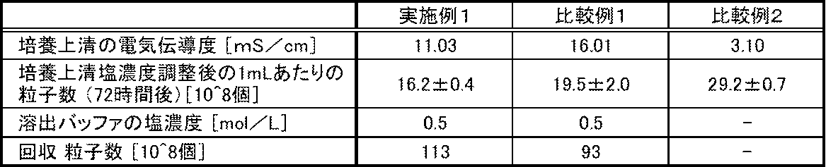
表2の結果より、培養上清の電気伝導度を16.01mS/cmのまま調整することなく精製した比較例1では、イオン交換基をもつ担体からの回収粒子数が低減している。これは、培養上清の塩濃度が高いために、担体に細胞外小胞が十分に吸着されず、透過(フロースルー)画分に流出してしまっていることを示している。
また、培養上清の電気伝導度を3.10mS/cmとした比較例2では、培養上清中のNTAによる粒子測定結果が有意に増加していた。これは、細胞外小胞が破裂し損傷をうけていることを示している。なお、比較例2では、細胞外小胞の有意な変質が確認されたため、透過試験を実施しなかった。
さらに、ウェスタンブロッティングテストの結果を図3に示す。CD63はEVsのマーカーとして知られており、EVsのなかでも特にエクソソームに発現しているという報告がある(Borges FT, et al., 2013, Extracellular vesicles: structure, function, and potential clinical uses in renal diseases, Braz J Med Biol Res, 46(10), 824-30)。また、エクソソームリッチな画分はMVなどの他のEVsと比較して負電荷が弱いという報告がある(Seo N, et al., 2022, Distinguishing functional exosomes and other extracellular vesicles as a nucleic acid cargo by the anion-exchange method, J Extracell Vesicles, Mar, 11(3))。精製画分の回収粒子のうち、単位粒子あたりのCD63陽性の粒子の割合は、図3から明らかなように、比較例1と比べて実施例1の方が高いことが示された。これは、本明細書が開示する方法を用いることで培養上清中の粒子の中でも、とくに微弱な負電荷をもつエクソソームを膜により多く吸着させることができ、ロスを低減して回収できていることを示唆している。
また、培養上清の電気伝導度を3.10mS/cmとした比較例2では、培養上清中のNTAによる粒子測定結果が有意に増加していた。これは、細胞外小胞が破裂し損傷をうけていることを示している。なお、比較例2では、細胞外小胞の有意な変質が確認されたため、透過試験を実施しなかった。
さらに、ウェスタンブロッティングテストの結果を図3に示す。CD63はEVsのマーカーとして知られており、EVsのなかでも特にエクソソームに発現しているという報告がある(Borges FT, et al., 2013, Extracellular vesicles: structure, function, and potential clinical uses in renal diseases, Braz J Med Biol Res, 46(10), 824-30)。また、エクソソームリッチな画分はMVなどの他のEVsと比較して負電荷が弱いという報告がある(Seo N, et al., 2022, Distinguishing functional exosomes and other extracellular vesicles as a nucleic acid cargo by the anion-exchange method, J Extracell Vesicles, Mar, 11(3))。精製画分の回収粒子のうち、単位粒子あたりのCD63陽性の粒子の割合は、図3から明らかなように、比較例1と比べて実施例1の方が高いことが示された。これは、本明細書が開示する方法を用いることで培養上清中の粒子の中でも、とくに微弱な負電荷をもつエクソソームを膜により多く吸着させることができ、ロスを低減して回収できていることを示唆している。
本発明によれば、生体試料中に含まれる細胞外小胞を、簡便に高回収率で精製回収することが可能である。本発明で得られる細胞外小胞は、薬物送達、医薬品、体外診断、食品および化粧品などの分野で好適に利用することができる。
1,2: 液体培地貯留容器
3: 細胞回収容器
4,5,6,7,8,9,10,11,12: バルブ
13,14,15,16: 無菌接続コネクター
17: 細胞培養容器
18: 廃液回収容器
19,20: 送液ポンプ
21: 作業コントローラー(作業パネル)
31: CO2インキュベーター
3: 細胞回収容器
4,5,6,7,8,9,10,11,12: バルブ
13,14,15,16: 無菌接続コネクター
17: 細胞培養容器
18: 廃液回収容器
19,20: 送液ポンプ
21: 作業コントローラー(作業パネル)
31: CO2インキュベーター
Claims (11)
- 細胞外小胞を製造するための方法であって、
工程(a)細胞外小胞を含む生体試料を得る工程
工程(b)前記生体試料の電気伝導度を4mS/cm以上15mS/cm以下に調整する工程
工程(c)前記生体試料を、正電荷を有する担体に接触させる工程
工程(d)前記担体に、金属塩を含有する緩衝液を接触させて、細胞外小胞を回収する工程を含むことを特徴とする方法。 - 前記工程(a)が、動物からの細胞の単離またはセルストックの凍結細胞の融解、およびそれに続く培養を含むことを特徴とする請求項1に記載の方法。
- 前記工程(a)と前記工程(b)との間に、前記生体試料より夾雑物質の少なくとも一部を除去する工程を有することを特徴とする請求項1に記載の方法。
- 前記夾雑物質の少なくとも一部を除去する工程が、遠心分離、精密ろ過、限外ろ過、透析からなる群から選択される1以上であることを特徴とする請求項3に記載の方法。
- 前記工程(c)において、前記担体は、アニオン交換能を有する分離膜であることを特徴とする請求項1に記載の方法。
- 前記工程(c)において、前記担体は、3級アミンおよび/または4級アンモニウム基を有することを特徴とする請求項5に記載の方法。
- 前記工程(c)と前記工程(d)との間に、前記担体に緩衝液を接触させて洗浄する工程を含むことを特徴とする請求項1に記載の方法。
- 前記工程(d)において、前記金属塩を含有する緩衝液はトリス塩酸緩衝液、リン酸緩衝生理食塩水、トリス緩衝生理食塩水のいずれかであることを特徴とする請求項1に記載の方法。
- 前記工程(d)において、前記金属塩がNaClであることを特徴とする請求項1に記載の方法。
- 前記工程(d)において、回収された細胞外小胞を含む試料の電気伝導度を15mS/cm超に調整する工程を含むことを特徴とする請求項1に記載の方法。
- 前記細胞外小胞は、エクソソームおよび/またはマイクロベシクルであることを特徴とする請求項1に記載の方法。
Applications Claiming Priority (2)
| Application Number | Priority Date | Filing Date | Title |
|---|---|---|---|
| JP2023-149357 | 2023-09-14 | ||
| JP2023149357 | 2023-09-14 |
Publications (1)
| Publication Number | Publication Date |
|---|---|
| WO2025057787A1 true WO2025057787A1 (ja) | 2025-03-20 |
Family
ID=95021233
Family Applications (1)
| Application Number | Title | Priority Date | Filing Date |
|---|---|---|---|
| PCT/JP2024/031255 Pending WO2025057787A1 (ja) | 2023-09-14 | 2024-08-30 | 細胞外小胞の製造方法 |
Country Status (2)
| Country | Link |
|---|---|
| TW (1) | TW202526004A (ja) |
| WO (1) | WO2025057787A1 (ja) |
Citations (4)
| Publication number | Priority date | Publication date | Assignee | Title |
|---|---|---|---|---|
| WO2012087241A1 (en) * | 2010-12-20 | 2012-06-28 | Agency For Science, Technology And Research | Method of purifying exosomes |
| JP2016502862A (ja) * | 2013-01-03 | 2016-02-01 | エクソサム ダイアグノスティクス,インコーポレイティド | 微細小胞の単離方法 |
| WO2020027185A1 (ja) | 2018-07-31 | 2020-02-06 | 国立大学法人三重大学 | エクソソームの製造方法 |
| JP2023149357A (ja) | 2022-03-31 | 2023-10-13 | ブラザー工業株式会社 | 容器および容器の製造方法 |
-
2024
- 2024-08-30 WO PCT/JP2024/031255 patent/WO2025057787A1/ja active Pending
- 2024-09-12 TW TW113134540A patent/TW202526004A/zh unknown
Patent Citations (4)
| Publication number | Priority date | Publication date | Assignee | Title |
|---|---|---|---|---|
| WO2012087241A1 (en) * | 2010-12-20 | 2012-06-28 | Agency For Science, Technology And Research | Method of purifying exosomes |
| JP2016502862A (ja) * | 2013-01-03 | 2016-02-01 | エクソサム ダイアグノスティクス,インコーポレイティド | 微細小胞の単離方法 |
| WO2020027185A1 (ja) | 2018-07-31 | 2020-02-06 | 国立大学法人三重大学 | エクソソームの製造方法 |
| JP2023149357A (ja) | 2022-03-31 | 2023-10-13 | ブラザー工業株式会社 | 容器および容器の製造方法 |
Non-Patent Citations (3)
| Title |
|---|
| BORGES FT ET AL.: "Extracellular vesicles: structure, function, and potential clinical uses in renal diseases", BRAZ J MED BIOL RES, vol. 46, no. 10, 2013, pages 824 - 30 |
| SEO N ET AL.: "Distinguishing functional exosomes and other extracellular vesicles as a nucleic acid cargo by the anion-exchange method", J EXTRACELL VESICLES, vol. 11, no. 3, March 2022 (2022-03-01) |
| TAKANO KIYOSHI: "Development of highly-efficient process for producing giant unilamellar vesicles (GUV) by controlling microscopic flow field", HTTPS://KAKEN.NII.AC.JP/, NATIONAL INSTITUTE OF INFORMATICS (NII)IN COOPERATION WITH MEXT AND JSPS, JP, 24 May 2013 (2013-05-24), JP, pages 1 - 5, XP093290706, Retrieved from the Internet <URL:https://kaken.nii.ac.jp/ja/file/KAKENHI-PROJECT-23656145/23656145seika.pdf> * |
Also Published As
| Publication number | Publication date |
|---|---|
| TW202526004A (zh) | 2025-07-01 |
Similar Documents
| Publication | Publication Date | Title |
|---|---|---|
| TWI412394B (zh) | Purification of proteins | |
| KR102206959B1 (ko) | 접선방향 유동 여과 모드에서 작동되는 나노섬유 한외여과막을 사용하여 샘플에서 목적하는 생물학적 물질을 정제하는 방법 | |
| CN113101737B (zh) | 一种亲和切向流过滤系统及其构建方法和外泌体提取方法及应用 | |
| JP2020530780A (ja) | 疎水性相互作用を利用した細胞外小胞体の分離方法 | |
| CN115427486B (zh) | 离子交换膜 | |
| Lorenzini et al. | Producing vesicle-free cell culture additive for human cells extracellular vesicles manufacturing | |
| CN116333987A (zh) | 一种外泌体提取方法获得的nk细胞外泌体在抗肿瘤中的应用 | |
| CN112680397B (zh) | 一种囊泡离心保护剂及其应用和离心提取囊泡的方法 | |
| TW202526004A (zh) | 細胞外囊泡之製造方法 | |
| KR20260070113A (ko) | 세포 외 소포의 제조 방법 | |
| Alia et al. | Preparation and characterization of a polysulfone ultrafiltration membrane for bovine serum albumin separation: Effect of polymer concentration | |
| US20250222373A1 (en) | Methods and compositions for purifying small extracellular vesicles | |
| US20240408513A1 (en) | Separation system and method for separating and purifying a target component | |
| JP7814718B2 (ja) | 生体微粒子分離精製用多孔性モノリス状ポリマー、生体微粒子分離精製用多孔性モノリス状ポリマーの製造方法、および生体微粒子分離精製用多孔性モノリス状ポリマーを用いるリン脂質で構成された生体微粒子の分離精製法 | |
| JP5931363B2 (ja) | タンパク質の精製方法 | |
| JP2012214408A (ja) | 澄明液中に分散した不純物凝集体を除去することによるタンパク質の精製方法 | |
| Ran et al. | A silica/polyvinyl alcohol membrane suitable for separating proteins | |
| JPS61274707A (ja) | 微生物分離用再生セルロ−ス多孔膜 | |
| CN120536342A (zh) | 分离与纯化细胞培养液外泌体的方法 | |
| CN119242566A (zh) | 一种制备外泌体的方法与应用 | |
| Wehland et al. | Combining Immuno-Affinity Chromatography and Filtration to Improve Specificity and Size Distribution of Exosome-Containing EV Populations | |
| EP4644535A1 (en) | Porous carrier, method for aquiring extracellular vesicle, method for producing extracellular vesicle, and kit | |
| Nechifor et al. | Composite PSF-PANI Membranes for Proteins Recovery | |
| Kunhi Mahin | A membrane process to extract Citrus Lemon derived extracellular vesicles |
Legal Events
| Date | Code | Title | Description |
|---|---|---|---|
| 121 | Ep: the epo has been informed by wipo that ep was designated in this application |
Ref document number: 24865289 Country of ref document: EP Kind code of ref document: A1 |
|
| ENP | Entry into the national phase |
Ref document number: 2025545612 Country of ref document: JP Kind code of ref document: A |
|
| WWE | Wipo information: entry into national phase |
Ref document number: 2024865289 Country of ref document: EP |