WO2025005266A1 - 眼薬 - Google Patents
眼薬 Download PDFInfo
- Publication number
- WO2025005266A1 WO2025005266A1 PCT/JP2024/023589 JP2024023589W WO2025005266A1 WO 2025005266 A1 WO2025005266 A1 WO 2025005266A1 JP 2024023589 W JP2024023589 W JP 2024023589W WO 2025005266 A1 WO2025005266 A1 WO 2025005266A1
- Authority
- WO
- WIPO (PCT)
- Prior art keywords
- emulsion
- eye
- oil
- aqueous phase
- phase
- Prior art date
- Legal status (The legal status is an assumption and is not a legal conclusion. Google has not performed a legal analysis and makes no representation as to the accuracy of the status listed.)
- Ceased
Links
Images












Classifications
-
- A—HUMAN NECESSITIES
- A61—MEDICAL OR VETERINARY SCIENCE; HYGIENE
- A61K—PREPARATIONS FOR MEDICAL, DENTAL OR TOILETRY PURPOSES
- A61K47/00—Medicinal preparations characterised by the non-active ingredients used, e.g. carriers or inert additives; Targeting or modifying agents chemically bound to the active ingredient
- A61K47/30—Macromolecular organic or inorganic compounds, e.g. inorganic polyphosphates
- A61K47/42—Proteins; Polypeptides; Degradation products thereof; Derivatives thereof, e.g. albumin, gelatin or zein
-
- A—HUMAN NECESSITIES
- A61—MEDICAL OR VETERINARY SCIENCE; HYGIENE
- A61K—PREPARATIONS FOR MEDICAL, DENTAL OR TOILETRY PURPOSES
- A61K47/00—Medicinal preparations characterised by the non-active ingredients used, e.g. carriers or inert additives; Targeting or modifying agents chemically bound to the active ingredient
- A61K47/44—Oils, fats or waxes according to two or more groups of A61K47/02-A61K47/42; Natural or modified natural oils, fats or waxes, e.g. castor oil, polyethoxylated castor oil, montan wax, lignite, shellac, rosin, beeswax or lanolin
-
- A—HUMAN NECESSITIES
- A61—MEDICAL OR VETERINARY SCIENCE; HYGIENE
- A61K—PREPARATIONS FOR MEDICAL, DENTAL OR TOILETRY PURPOSES
- A61K9/00—Medicinal preparations characterised by special physical form
- A61K9/10—Dispersions; Emulsions
- A61K9/107—Emulsions ; Emulsion preconcentrates; Micelles
- A61K9/113—Multiple emulsions, e.g. oil-in-water-in-oil
-
- A—HUMAN NECESSITIES
- A61—MEDICAL OR VETERINARY SCIENCE; HYGIENE
- A61P—SPECIFIC THERAPEUTIC ACTIVITY OF CHEMICAL COMPOUNDS OR MEDICINAL PREPARATIONS
- A61P27/00—Drugs for disorders of the senses
- A61P27/02—Ophthalmic agents
Definitions
- the present invention relates to eye medicines.
- Age-related macular degeneration is divided into atrophic and exudative types, with atrophic age-related macular degeneration leading to blindness.
- Atrophic age-related macular degeneration is common in Japan, and is the fourth leading cause of blindness in the country, with cases increasing year by year.
- exudative age-related macular degeneration new blood vessels form in the choroid, damaging photoreceptor cells.
- the main treatment for exudative age-related macular degeneration is intravitreal injection of anti-vascular endothelial growth factor (VEGF) antibody drugs.
- VEGF anti-vascular endothelial growth factor
- one of the objects of the present invention is to provide an eye drug that can deliver ocular disease treatment agents to the posterior segment of the eye.
- the eye medication according to this embodiment of the present invention includes a first aqueous phase, a gelled oil phase in which the first aqueous phase is dispersed, a second aqueous phase in which the oil phase is dispersed, and an eye disease treatment agent disposed within the first aqueous phase.
- the first aqueous phase may be gelled. In the above eye drops, the first aqueous phase may not be gelled.
- the eye disease treatment agent may be hydrophilic.
- the diameter of the first aqueous phase may be 25 nm or more and 150 nm or less.
- the diameter of the oil phase may be 100 nm or more and 350 nm or less.
- the first aqueous phase may contain gelatin.
- the gelatin may be crosslinked.
- the oil phase may contain beeswax.
- the above eye drops may be directed to the posterior segment of the eye.
- the above eye medication may be administered to the subject by eye drop administration.
- the above eye medications may be administered to the subject using an eye covering method.
- the delivery agent for an eye disease treatment agent includes a first aqueous phase, a gelled oil phase in which the first aqueous phase is dispersed, and a second aqueous phase in which the oil phase is dispersed.
- the eye disease treatment agent may be disposed in the first aqueous phase.
- the first aqueous phase may be gelled. In the delivery agent for the eye disease treatment agent described above, the first aqueous phase may not be gelled.
- the eye disease treatment agent may be hydrophilic.
- the diameter of the first aqueous phase may be 25 nm or more and 150 nm or less.
- the diameter of the oil phase may be 100 nm or more and 350 nm or less.
- the first aqueous phase may contain gelatin.
- the gelatin may be crosslinked.
- the oil phase may contain beeswax.
- the delivery agent for the above-mentioned eye disease treatment agent may be directed to the posterior segment of the eye.
- the method for producing an eye drug includes mixing a first aqueous solution containing an eye disease treatment agent with an oil containing a first surfactant to form a primary emulsion containing an oil phase in which a first aqueous phase containing the eye disease treatment agent therein is dispersed, adding a gelling agent to the oil phase of the primary emulsion to gel the oil phase, and mixing the primary emulsion in which the oil phase has been gelled with a second aqueous solution containing a second surfactant to form a secondary emulsion containing a second aqueous phase in which the gelled oil phase is dispersed.
- the first aqueous solution may further contain a gelling agent, and the first aqueous solution containing the eye disease treatment agent and the gelling agent may be mixed with an oil containing a first surfactant to form a primary emulsion containing an oil phase in which a gelled first aqueous phase containing the eye disease treatment agent therein is dispersed.
- the concentration of the first surfactant in the oil may be 125 mg/mL or more.
- ultrasound may be applied to the mixture of the first aqueous solution and the oil when forming the primary emulsion.
- the time for applying ultrasound may be 15 minutes or more.
- the concentration of the second surfactant in the second aqueous solution may be 25 mg/mL or more.
- ultrasonic waves when forming the secondary emulsion, may be applied to the mixture of the primary emulsion and the second aqueous solution.
- the time for applying ultrasound may be 10 minutes or more.
- the method for producing a delivery agent for an eye disease treatment includes mixing a first aqueous solution with an oil containing a first surfactant to form a primary emulsion containing an oil phase in which the first aqueous phase is dispersed, adding a gelling agent to the oil phase of the primary emulsion to gel the oil phase, and mixing the primary emulsion in which the oil phase has been gelled with a second aqueous solution containing a second surfactant to form a secondary emulsion containing a second aqueous phase in which the gelled oil phase is dispersed.
- the first aqueous solution may contain a gelling agent, and the first aqueous solution containing the gelling agent may be mixed with an oil containing a first surfactant to form a primary emulsion containing an oil phase in which the gelled first aqueous phase is dispersed.
- the concentration of the first surfactant in the oil may be 100 mg/mL or more.
- ultrasound may be applied to the mixture of the first aqueous solution and the oil when forming the primary emulsion.
- the time for applying ultrasound may be 15 minutes or more.
- the concentration of the second surfactant in the second aqueous solution may be 25 mg/mL or more.
- ultrasound may be applied to the mixture of the primary emulsion and the second aqueous solution when forming the secondary emulsion.
- the time for applying ultrasound may be 10 minutes or more.
- the present invention provides an eye drug capable of delivering an eye disease treatment to the posterior segment of the eye.
- FIG. 1 is a schematic diagram of an eye drop according to an embodiment.
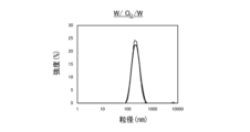
- Figure 2A is a graph showing the particle size distribution of a W/ O /W emulsion
- Figure 2B is a graph showing the particle size distribution of a W/O/W emulsion
- Figure 2C is a graph showing the particle size distribution of a W / O /W emulsion
- Figure 2D is a graph showing the particle size distribution of a W/ O /W emulsion.
- FIG. 1 is a schematic diagram of an eye drop according to an embodiment.
- Figure 2A is a graph showing the particle size distribution of a W/ O /W emulsion
- Figure 2B is a graph showing the particle size distribution of a W/O/W emulsion
- Figure 2C is a graph showing the particle size distribution of a W / O /W emulsion
- Figure 2D is a graph showing the particle size distribution of a W/ O /W emulsion.
- FIG. 3 is a graph showing particle size and polydispersity index (PDI) for a W/O/W emulsion, a W G /O/W emulsion, a W/O G /W emulsion, and a W G / O G /W emulsion.
- FIG. 4 is an electron microscope photograph of a W G /O/W emulsion.
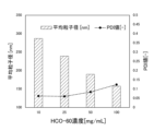
- FIG. 5 is a graph showing the relationship between the concentration of the surfactant when preparing the primary emulsion and the average particle size of the aqueous phase in the primary emulsion.
- FIG. 6 is a graph showing the relationship between the ultrasonic treatment time during preparation of the primary emulsion and the average particle size of the aqueous phase in the primary emulsion.
- FIG. 7 is a graph showing the relationship between the concentration of the surfactant when preparing the secondary emulsion and the average particle size of the oil phase in the secondary emulsion.
- FIG. 8 is a graph showing the relationship between the volume ratio of the aqueous solution to the primary emulsion when preparing the secondary emulsion and the average particle size of the oil phase in the secondary emulsion.
- FIG. 9 is a graph showing the relationship between the ultrasonic treatment time during preparation of a secondary emulsion and the average particle size of the oil phase in the secondary emulsion.
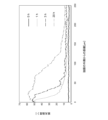
- FIG. 10 is a graph showing the change over time in the average particle size of the oil phase in a secondary emulsion.
- FIG. 11 is a graph showing the change in polydispersity index (PDI) of the oil phase in the secondary emulsion over time.
- FIG. 12 is a graph showing the initial release of albumin contained in the first aqueous phase of the secondary emulsion.
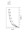
- FIG. 13 is a graph showing the change over time in the emission rate of a fluorescent material contained in the first aqueous phase of a secondary emulsion.
- FIG. 14 is a graph showing the initial change over time in the emission rate of a fluorescent material contained in the first aqueous phase of a secondary emulsion.
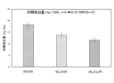
- FIG. 15 is a graph showing the initial release rate of a fluorescent material contained in the first aqueous phase of a secondary emulsion.
- FIG. 16 is a graph showing a calibration curve showing the relationship between BSA concentration and absorbance obtained by serially diluting 1% BSA that was not stirred with ultrasound.
- FIG. 17 is a graph showing the relationship between albumin concentration and absorbance for secondary emulsions containing albumin in the first aqueous phase.
- Figure 18A shows a fluorescence micrograph of cells cultured in medium without added fluorescein
- Figure 18B shows a fluorescence micrograph of cells cultured in medium with added secondary emulsion containing fluorescein in the first aqueous phase
- Figure 18C shows a fluorescence micrograph of cells cultured in medium with added liquid containing fluorescein solution.
- FIG. 18A shows a fluorescence micrograph of cells cultured in medium without added fluorescein
- Figure 18B shows a fluorescence micrograph of cells cultured in medium with added secondary emulsion containing fluorescein in the first aqueous phase
- Figure 18C shows a fluorescence micrograph
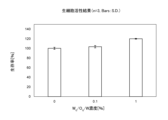
- FIG. 19 is a graph showing the relationship between the concentration of the secondary emulsion and the survival rate of cells.
- FIG. 20 shows micrographs of mouse eyeballs that received instillations of a fluorescein solution, a W/O/W emulsion containing fluorescein in the first aqueous phase, and a W G /O G /W emulsion containing fluorescein in the first aqueous phase.
- FIG. 20 shows micrographs of mouse eyeballs that received instillations of a fluorescein solution, a W/O/W emulsion containing fluorescein in the first aqueous phase, and a W G /O G /W emulsion containing fluorescein in the first aqueous phase.
- FIG. 21 is a graph showing the fluorescence intensity in the retinal layers of the eyes of mice that were instilled with a fluorescein solution, a W/O/W emulsion containing fluorescein in the first aqueous phase, and a W / O /W emulsion containing fluorescein in the first aqueous phase.
- FIG. 22 is a micrograph of a section of the anterior segment of the eyeball of a slaughtered pig to which the OG /W emulsion containing coumarin-6 according to Reference Example 1 was instilled, and a section of the anterior segment of the eyeball of a slaughtered pig to which the coumarin-6-added castor oil according to Reference Example 2 was instilled.
- FIG. 22 is a micrograph of a section of the anterior segment of the eyeball of a slaughtered pig to which the OG /W emulsion containing coumarin-6 according to Reference Example 1 was instilled, and a
- FIG. 23 is a micrograph of a section of the posterior segment of the eyeball of a slaughtered pig to which the OG /W emulsion containing coumarin-6 according to Reference Example 1 was instilled, and a section of the posterior segment of the eyeball of a slaughtered pig to which the coumarin-6-added castor oil according to Reference Example 2 was instilled.
- FIG. 24 is a micrograph of a section of the eyeball of a slaughtered pig that was given the O G /W emulsion containing coumarin-6 according to Reference Example 1 by the eyeball coating method.
- FIG. 25 is a graph showing the relationship between the distance from the sclera of the eyeball of a slaughtered pig that was given the O G /W emulsion encapsulating coumarin-6 according to Reference Example 1 by the eyeball covering method and the fluorescence intensity.
- FIG. 26 shows micrographs of sections of the anterior and posterior segments of a pig's eyeball to which the O G /W emulsion containing coumarin-6 according to Reference Example 1 was instilled while the animal was still alive.
- FIG. 27 shows micrographs of sections of the anterior and posterior segments of the eye of a pig that was given the O G /W emulsion containing coumarin-6 according to Reference Example 1 while alive by the eyeball coating method.
- FIG. 26 shows micrographs of sections of the anterior and posterior segments of the eye of a pig that was given the O G /W emulsion containing coumarin-6 according to Reference Example 1 while alive by the eyeball coating method.
- FIG. 28 is a graph showing the fluorescence intensity in sections of the posterior segment of the eyeball of a pig that was given the O G /W emulsion encapsulating coumarin-6 according to Reference Example 1 by eye drop method or eyeball coating method while alive.
- FIG. 29 is a micrograph of a section near the sclera of an eyeball immersed in the O G /W emulsion containing coumarin-6 according to Reference Example 1.
- FIG. 30 is a micrograph of a section near the retina of an eyeball immersed in the O G /W emulsion of Reference Example 1 encapsulating coumarin-6.
- FIG. 31 is a micrograph illustrating the distance from the sclera to the retina in the eyeball.
- FIG. 32 is a graph showing the relationship between the distance from the outside of the sclera and the fluorescence intensity in a section of an eyeball immersed in an O G /W emulsion containing coumarin-6 according to Reference Example 1.
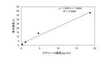
- FIG. 33 is a graph showing the relationship between the concentration of coumarin-6 and the fluorescence intensity of coumarin-6.
- FIG. 34 is a graph showing the relationship between the time that an eyeball was immersed in the O G /W emulsion containing coumarin-6 according to Reference Example 1 and the concentration of coumarin-6 in the retina of the eyeball.
- FIG. 33 is a graph showing the relationship between the concentration of coumarin-6 and the fluorescence intensity of coumarin-6.
- FIG. 34 is a graph showing the relationship between the time that an eyeball was immersed in the O G /W emulsion containing coumarin-6 according to Reference Example 1 and the concentration of coumarin-6 in the retina of the eyeball.
- FIG. 35 is a graph showing the relationship between the concentration of W G /O G /W emulsion and the survival rate of cells given W G /O G /W emulsion.
- FIG. 36 is a graph showing the relationship between the number of days the emulsion was stored and the average particle size.
- FIG. 37 is a graph showing the relationship between the number of days the emulsion was stored and the polydispersity index (PDI).
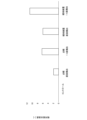
- FIG. 38 is a graph showing the emulsion stability index SI (%).
- the eye drug includes a first aqueous phase 10, a gelled oil phase 20 in which the first aqueous phase 10 is dispersed, a second aqueous phase 30 in which the oil phase 20 is dispersed, and an eye disease treatment agent disposed in the first aqueous phase 10.
- the first aqueous phase 10 may be gelled or may not be gelled.
- the gelled first aqueous phase 10 and the gelled oil phase 20 form a gelled water-in-gelled oil type ( WG / OG ) emulsion.
- the gelled oil phase 20 and the second aqueous phase 30 form a gelled oil-in-water type (OG/W) emulsion.
- the gelled first aqueous phase 10, the gelled oil phase 20, and the second aqueous phase 30 form a gelled water -in-gelled oil-in-water type ( WG / OG /W) composite emulsion.
- the first aqueous phase 10 includes an aqueous solution.
- the aqueous solution includes, for example, a buffer solution.
- the buffer solution include, but are not limited to, 2-morpholinoethanesulfonic acid monohydrate (MES) buffer solution, acetate buffer solution, phosphate buffer solution, Tris-HCl buffer solution, 4-(2-hydroxyethyl)-1-piperazineethanesulfonic acid (HEPES) buffer solution, piperazine-N,N'-bis(2-ethanesulfonic acid) (PIPES) buffer solution, and 3-morpholinopropanesulfonic acid (MOPS) buffer solution.
- the aqueous solution includes water.
- the water may be water for injection.
- the aqueous solution constituting the first aqueous phase 10 may be referred to as the first aqueous solution.
- the gelled first aqueous phase 10 contains, for example, a gelling agent for gelling an aqueous solution.
- the first aqueous phase 10 is gelled by the gelling agent.
- gelling agents for gelling an aqueous solution include, but are not limited to, gelatin, agar, collagen, polysaccharides, sodium alginate, calcium alginate, hydroxyethyl methacrylate (HEMA), poloxamer hydrogel, and poloxamer terminated with a thiol group.
- polysaccharides include, but are not limited to, cellulose, chitin, starch, glycogen, agarose, pectin, galactomannan, hyaluronic acid, low-substituted dopamine-hyaluronic acid complex, chitosan, chondroitin sulfate, dermatan sulfate, heparan sulfate, and keratan sulfate.
- the first aqueous phase 10 may contain multiple types of gelling agents.
- the gelling agent constituting the first aqueous phase 10 may be referred to as the first gelling agent.
- the gelled first aqueous phase 10 may also contain heparin.
- the gelling agent may be cross-linked by a cross-linking agent.
- a cross-linking agent is a combination of 1-ethyl-3-(3-dimethylaminopropyl)carbodiimide acetate (EDC) and N-hydroxysuccinimide (NHS), but is not limited thereto.
- EDC 1-ethyl-3-(3-dimethylaminopropyl)carbodiimide acetate
- NHS N-hydroxysuccinimide
- the first aqueous phase 10 is covered with a hydrophobic surfactant and forms particles dispersed in the oil phase 20.
- the hydrophobic surfactant is, for example, a surfactant having an HLB (Hydrophile Lipophile Balance) value range of less than 8.
- the hydrophobic surfactant is, for example, nonionic.
- examples of hydrophobic surfactants include, but are not limited to, sucrose fatty acid esters, glycerin fatty acid esters, organic acid monoglycerides, polyglycerin fatty acid esters, sorbitan fatty acid esters, propylene glycol fatty acid esters, lecithin, and enzyme-treated lecithin.
- the first aqueous phase 10 may be covered with multiple types of hydrophobic surfactants.
- the surfactant covering the first aqueous phase 10 may be referred to as the first surfactant.
- the diameter (particle size) of the first aqueous phase 10, which is covered with a hydrophobic surfactant and forms particles, is, for example, 25 nm to 150 nm, 35 nm to 90 nm, 40 nm to 80 nm, or 40 nm to 50 nm.
- the diameter of the first aqueous phase 10 is the average diameter measured by dynamic light scattering (DLS).
- the eye disease treatment agent is contained within the first aqueous phase 10 in the form of particles.
- An eye disease is any disease that can affect the eye. Examples of causes of eye diseases include, but are not limited to, proliferative disorders, infections, allergies, allergic reactions, degenerative disorders, inflammatory diseases, trauma, surgery, radiation, chemical irritation, physical irritation, and autoimmune diseases.
- An example of an eye disease is a retinal disease. Examples of retinal diseases include age-related macular degeneration, retinitis pigmentosa, retinal vein occlusion, central serous chorioretinopathy, diabetic retinopathy, retinal breaks, and retinal detachment.
- the eye disease treatment agent is, for example, hydrophilic.
- the eye disease treatment agent includes, for example, biomolecules such as proteins, peptides, and nucleic acids.
- proteins include, for example, antibodies and enzymes.
- antibodies include, but are not limited to, anti-vascular endothelial growth factor (VEGF) antibodies and anti-angiopoietin-2 antibodies.
- enzymes include, but are not limited to, ranpirnase and amphikinase, which have antiviral effects.
- the nucleic acid may be, for example, RNA that interferes with the mRNA of a protein that causes an eye disease.
- the nucleic acid may be, for example, an aptamer that binds to a protein that causes an eye disease.
- proteins that cause an eye disease include periostin, tenascin C, (pro)renin receptor, and VEGF, which are involved in angiogenesis.
- Multiple types of eye disease treatment agents may be included in the first a
- the gelled oil phase 20 contains an oil.
- oils include, but are not limited to, fatty acid ester oils, vegetable oils, animal oils, neutral lipids (mono-substituted, di-substituted or tri-substituted glycerides), synthetic oils and fats, liquid paraffin (mineral oil), heavy liquid isoparaffin, light liquid isoparaffin, sterol derivatives, lactate esters, polycarboxylic acid esters, 2-cetyl ethylhexanoate, silicones, and lipiodol.
- the oil phase 20 may contain multiple types of oil.
- fatty acid ester oils include, but are not limited to, isopropyl myristate, octyldodecyl myristate, cetyl myristate, ethyl oleate, ethyl linoleate, isopropyl linoleate, isopropyl palmitate, and butyl stearate.
- Examples of vegetable oils include, but are not limited to, castor oil, coconut oil, palm oil, soybean oil, cottonseed oil, rapeseed oil, sesame oil, corn oil, peanut oil, safflower oil, sunflower oil, olive oil, rapeseed oil, perilla oil, fennel oil, cacao oil, cinnamon oil, peppermint oil, and bergamot oil.
- Examples of animal oils include, but are not limited to, fish oil, and whale oil.
- neutral lipids include, but are not limited to, medium-chain triglycerides, triolein, trilinolein, tripalmitin, tristearin, trimyristin, and triarachidonin.
- synthetic oils include, but are not limited to, azone.
- sterol derivatives include, but are not limited to, cholesteryl oleate, cholesteryl linoleate, cholesteryl myristate, cholesteryl palmitate, and cholesteryl arachidate.
- lactate esters include, but are not limited to, ethyl lactate and cetyl lactate.
- polycarboxylic acid esters include, but are not limited to, triethyl citrate, diisopropyl adipate, diethyl sebacate, and diisopropyl sebacate.
- the gelled oil phase 20 contains, for example, a gelling agent for gelling oil.
- the oil phase 20 is gelled by the gelling agent.
- gelling agents for gelling oil include, but are not limited to, waxes, higher fatty acids, fatty acid esters, metal soaps, amino acid-based gelling agents, and hydrocarbons.
- waxes include, but are not limited to, beeswax, Japan wax, hydrogenated oil, candelilla wax, carnauba wax, rice wax, montan wax, ozokerite, ceresin, paraffin wax, microcrystalline wax, petrolatum, Fischer-Tropsch wax, polyethylene wax, and ethylene-propylene copolymer.
- higher fatty acids include, but are not limited to, lauric acid, myristic acid, palmitic acid, stearic acid, isostearic acid, behenic acid, undecylenic acid, 12-hydroxystearic acid (12-HSA), palmitoleic acid, oleic acid, linoleic acid, linolenic acid, erucic acid, docosahexaenoic acid, eicosapentaenoic acid, isohexadecanoic acid, anteisohenicosanoic acid, long-chain branched fatty acids, dimer acids, and hydrogenated dimer acids.
- lauric acid myristic acid, palmitic acid, stearic acid, isostearic acid, behenic acid, undecylenic acid, 12-hydroxystearic acid (12-HSA), palmitoleic acid, oleic acid, linoleic acid, linolenic acid, erucic acid,
- fatty acid esters examples include cholesterol fatty acid esters, sugar fatty acid esters, glycerin fatty acid esters, polyglycerin fatty acid esters, etc.
- specific examples of sugar fatty acid esters include, but are not limited to, dextrin palmitate, dextrin myristate, dextrin isostearate, fructooligosaccharide stearate, inulin stearate, and dibenzylidene sorbitol.
- metal soaps include, but are not limited to, calcium stearate, lithium stearate, aluminum stearate, calcium palmitate, lithium palmitate, aluminum palmitate, calcium 2-ethylhexanoate, lithium 2-ethylhexanoate, aluminum 2-ethylhexanoate, calcium 12-hydroxystearate, lithium 12-hydroxystearate, and aluminum 12-hydroxystearate.
- amino acid gelling agents include, but are not limited to, N-lauroyl glutamic acid di-n-butylamide.
- hydrocarbons include, but are not limited to, ⁇ -olefin oligomers, polyisobutene, hydrogenated polyisobutene, polybutene, squalane, olive-derived squalane, squalene, petrolatum, and solid paraffin.
- the oil phase 20 may contain multiple types of gelling agents.
- the gelling agent constituting the oil phase 20 may be referred to as the second gelling agent.
- the oil phase 20 is covered with a hydrophilic surfactant and forms particles that are dispersed in the second aqueous phase 30.
- a hydrophilic surfactant is, for example, a surfactant whose HLB (Hydrophile Lipophile Balance) value range includes 8 or more.
- a hydrophilic surfactant is, for example, nonionic.
- hydrophilic surfactants include, but are not limited to, polyoxyethylene hydrogenated castor oil (HCO-40, HCO-60, HCO-80, and HCO-100, etc.), alkyl sulfate salts, alkyl sulfonates, alkyl sulfosuccinates, polyoxyethylene alkyl ethers, polyoxyalkylene alkyl ethers, lauryl alcohol alkoxylates, glycerin fatty acid esters, saponin, sucrose fatty acid esters, glycerin fatty acid esters, sorbitan fatty acid esters, propylene glycol fatty acid esters, polysorbates, lecithin, and polyoxyethylene-polyoxypropylene block polymers.
- HCO-40, HCO-60, HCO-80, and HCO-100 etc.
- alkyl sulfate salts alkyl sulfonates
- alkyl sulfosuccinates alkyl sulf
- the oil phase 20 may be covered with multiple types of hydrophilic surfactants.
- the surfactant covering the oil phase 20 may be referred to as the second surfactant.
- the diameter (particle size) of the oil phase 20, which is covered with a hydrophilic surfactant and forms particles, is, for example, 100 nm to 350 nm, 120 nm to 350 nm, 130 nm to 300 nm, 140 nm to 250 nm, or 160 nm to 250 nm, and does not overlap with the diameter (particle size) of the first aqueous phase 10.
- the diameter of the oil phase 20 is the average diameter measured by dynamic light scattering (DLS).
- the second aqueous phase 30 includes an aqueous solution.
- Examples of the aqueous solution are the same as those contained in the first aqueous phase 10.
- the second aqueous phase 30 does not have to be gelled.
- the aqueous solution constituting the second aqueous phase 30 may be referred to as the second aqueous solution.
- a gelled water-in-gelled oil-in-water type ( WG / OG /W) composite emulsion containing a gelled first aqueous phase 10, a gelled oil phase 20, and a second aqueous phase 30, and a water-in-gelled oil-in-water type (W/ OG /W) composite emulsion containing a non-gelled first aqueous phase 10, a gelled oil phase 20, and a second aqueous phase 30 function as a delivery agent that delivers an ocular disease therapeutic agent to the posterior segment of the eye, such as the retina. Therefore, the eye drug according to the embodiment can be used as a posterior segment-directed eye drug.
- the eye medication according to the embodiment may be administered to a subject by an eye drop method.
- the eye drop method is, for example, a method of dropping the eye medication into the conjunctival sac of a subject.
- the subject is a human or a non-human animal.
- the subject may be a patient.
- the eye medication according to the embodiment may also be administered to a subject by an eye covering method.
- the eye covering method is a method of immersing the subject's eye in the eye medication or a solution containing the eye medication for a predetermined period of time.
- the eye may be immersed in the eye medication or the solution containing the eye medication by filling the cavity around the eye with the eye medication or the solution containing the eye medication.
- the eye may also be immersed in the eye medication or the solution containing the eye medication by covering the eye with a container filled with the eye medication or the solution containing the eye medication.
- the time for which the eye is immersed in the eye medication or a solution containing the eye medication is not particularly limited, and may be, for example, 10 seconds, 20 seconds, 30 seconds, 40 seconds, 50 seconds, 1 minute, 3 minutes, 5 minutes, 10 minutes, 15 minutes, 20 minutes, 30 minutes, 40 minutes, 50 minutes, 1 hour, 2 hours, 3 hours, 4 hours, 5 hours, 10 hours, 15 hours, or 20 hours.
- the method for producing an eye drug according to an embodiment includes mixing a first aqueous solution containing an eye disease treatment agent with an oil containing a first surfactant to form a primary emulsion containing an oil phase in which a first aqueous phase containing an eye disease treatment agent therein is dispersed, adding a second gelling agent to the oil phase of the primary emulsion to gel the oil phase, and mixing the primary emulsion in which the oil phase has been gelled with a second aqueous solution containing the second surfactant to form a secondary emulsion containing a second aqueous phase in which the gelled oil phase is dispersed.
- the first aqueous solution further contains a first gelling agent, a primary emulsion containing an oil phase in which a gelled first aqueous phase containing an eye disease treatment agent therein is dispersed is formed.
- the concentration of the first surfactant in the oil is, for example, 125 mg/mL or more, or 150 mg/mL or more.
- the concentration of the first surfactant in the oil 125 mg/mL or more, it is possible to make the particle size of the aqueous phase contained in the primary emulsion 150 nm or less, or 100 nm or less.
- ultrasound or pressure may be applied to the mixture of the first aqueous solution and the oil to agitate the mixture of the first aqueous solution and the oil.
- the time for applying ultrasound to the mixture of the first aqueous solution and the oil is, for example, 3 minutes or more, 10 minutes or more, 15 minutes or more, or 20 minutes or more.
- By applying ultrasound to the mixture of the first aqueous solution and the oil for 3 minutes or more it is possible to make the particle size of the aqueous phase contained in the primary emulsion 150 nm or less, or 100 nm or less.
- ultrasound to the mixture of the first aqueous solution and the oil for 15 minutes or more it is possible to make the particle size of the aqueous phase contained in the primary emulsion 50 nm or less.
- the concentration of the second surfactant in the second aqueous solution is, for example, 25 mg/mL or more, or 35 mg/mL or more.
- the concentration of the second surfactant in the second aqueous solution 25 mg/mL or more, it is possible to make the particle size of the oil phase contained in the secondary emulsion 350 nm or less, or 250 nm or less.
- ultrasonic waves or pressure may be applied to the mixture of the primary emulsion and the second aqueous solution to stir the mixture of the primary emulsion and the second aqueous solution.
- the time for applying ultrasonic waves to the mixture of the primary emulsion and the second aqueous solution is, for example, 10 minutes or more, or 15 minutes or more.
- Example 1 A crosslinking agent-containing aqueous solution was prepared by adding heparin sodium (Fuji Film Wako Pure Chemical Industries, Ltd.) at a post-addition concentration of 10 mg/mL, 1-ethyl-3-(3-dimethylaminopropyl)carbodiimide acetate (EDC, Peptide Institute, Ltd.) at a post-addition concentration of 10 mg/mL, and N-hydroxysuccinimide (NHS, Fuji Film Wako Pure Chemical Industries, Ltd.) at a post-addition concentration of 6 mg/mL to 50 mmol/L 2-morpholinoethanesulfonic acid monohydrate (MES) buffer solution (pH 5.6, Dojindo Laboratories, Ltd.), and the crosslinking agent-containing aqueous solution was allowed to stand in a refrigerator for 30 minutes.
- MES 2-morpholinoethanesulfonic acid monohydrate
- a surfactant-containing oil was prepared by dissolving a non-ionic hydrophobic surfactant (ER-290, Ryoto Sugar Ester, registered trademark, Mitsubishi Chemical Foods Corporation) in isopropyl myristate (IPM, Fujifilm Wako Pure Chemicals) at 50 mg/mL by shaking at room temperature.
- ER-290 Ryoto Sugar Ester, registered trademark, Mitsubishi Chemical Foods Corporation
- IPM Fujifilm Wako Pure Chemicals
- a gelatin-containing aqueous solution was prepared by dissolving gelatin powder (pig skin gelatin type A, Sigma-Aldrich) in phosphate buffered saline (PBS) in an incubator so that the final concentration when the crosslinking agent was added was 5 wt%.
- 900 ⁇ L of the gelatin-containing aqueous solution was mixed with 100 ⁇ L of the crosslinking agent-containing aqueous solution, which was then thoroughly pipetted.
- the mixed aqueous solution and surfactant-containing oil were mixed in a 1:2 ratio to obtain an oil-water mixture.
- the container containing the oil-water mixture was placed on ice and stirred with an ultrasonic homogenizer (Sonifier 250, Branson) at 1 pulse/0.7 sec and 70% output amplitude for 3 min.
- the ultrasonically treated oil-water mixture was left in a refrigerator overnight to prepare a primary emulsion ( WG /O emulsion) in which the gelled first aqueous phase was dispersed in the oil.
- Example 2 The 900 ⁇ L of the gelatin-containing aqueous solution in Example 1 was replaced with 900 ⁇ L of water to prepare a primary emulsion (W/O emulsion) in which a non-gelled first aqueous phase was dispersed in oil.
- W/O emulsion primary emulsion
- Example 3 A surfactant-containing aqueous solution was prepared by dissolving a nonionic hydrophilic surfactant (polyethylene hydrogenated castor oil-60, NIKKOL HCO-60, Nippon Surfactant Industries Co., Ltd.) at 50 mg/mL in PBS in a water bath at 75° C.
- the surfactant-containing aqueous solution and the primary emulsion (W G /O emulsion) prepared in Example 1 were mixed at a ratio of 5:1 to obtain an oil-water mixture.
- the container containing the oil-water mixture was placed on ice and stirred with an ultrasonic homogenizer (Sonifier 250, Branson) at 1 pulse/0.7 seconds for 3 minutes at 70% output amplitude.
- the ultrasonically treated oil-water mixture was left to stand in a refrigerator overnight to prepare a secondary emulsion ( WG /O/W emulsion) containing a non-gelled oil phase in which the gelled first aqueous phase is dispersed, and a second aqueous phase in which the non-gelled oil phase is dispersed.
- Example 4 The same surfactant-containing aqueous solution as in Example 3 and the primary emulsion (W/O emulsion) prepared in Example 2 were mixed in a ratio of 5:1 and stirred under the same conditions as in Example 3 to prepare a secondary emulsion (W/O/W emulsion) containing an ungelled oil phase in which an ungelled first aqueous phase was dispersed, and a second aqueous phase in which the ungelled oil phase was dispersed.
- W/O/W emulsion secondary emulsion
- Example 5 The same surfactant-containing aqueous solution as in Example 3 was prepared. Also, the same primary emulsion ( WG /O emulsion) as in Example 1 was prepared. The primary emulsion was centrifuged at 3500 rpm for 10 minutes using a centrifuge (KUBOTA5100, Kubota Manufacturing Co., Ltd.), and 1 mL of the supernatant was collected. Beeswax was added to the collected 1 mL of the supernatant so that the final concentration in the oil phase was 2.5 wt%, and the mixture was returned to the centrifuged primary emulsion.
- KUBOTA5100 Kubota Manufacturing Co., Ltd.
- the primary emulsion was stirred in a 75°C water bath for 15 seconds, and then stirred with an ultrasonic homogenizer (Sonifier 250, Branson) at 1 pulse/second for 10 seconds, and stirred at 70% output amplitude to disperse the gelled first aqueous phase in the oil again.
- an ultrasonic homogenizer Sonifier 250, Branson
- the surfactant-containing aqueous solution and the primary emulsion were mixed at a ratio of 5:1 to prepare an oil-water mixture, the container containing the oil-water mixture was placed on ice, and the mixture was stirred for 3 minutes with an ultrasonic homogenizer (Sonifier 250, Branson) at 1 pulse/0.7 seconds and 70% output amplitude.
- the ultrasonically treated oil-water mixture was left to stand overnight in a refrigerator to prepare a secondary emulsion ( WG / OG /W emulsion) containing a gelled oil phase in which the gelled first aqueous phase was dispersed, and a second aqueous phase in which the gelled oil phase was dispersed.
- Example 6 A secondary emulsion (W/ O /W emulsion) containing a gelled oil phase in which a non-gelled first aqueous phase is dispersed, and a second aqueous phase in which the gelled oil phase is dispersed, was prepared in the same manner as in Example 5, except that the same primary emulsion (W/O emulsion) as in Example 2 was used instead of the same primary emulsion (W/ O emulsion) as in Example 1.
- Example 7 The secondary emulsions prepared in each of Examples 3 to 6 were diluted 600 times with PBS.
- the average diameter and polydispersity index (PDI) of the particles contained in the diluted secondary emulsions were measured using a particle size measurement system (nanoSAQLA, Otsuka Electronics) using dynamic light scattering (DLS).
- PDI polydispersity index
- the particle diameter of each secondary emulsion was about 200 nm, and the particle size of the W G /O G /W emulsion was the smallest.
- the polydispersity index (PDI) of each secondary emulsion was 0.3 or less, and the polydispersity index (PDI) of the W G /O G /W emulsion was the smallest, indicating that the particle size distribution was narrow.
- the particles contained in the secondary emulsions have a multilayer structure, but the average diameter of the particles measured by dynamic light scattering (DLS) is the average diameter of the outermost shell of the particles.
- Example 8 The W G /O/W emulsion prepared in Example 3 was diluted 100 times, and 10 ⁇ L of the diluted emulsion was added to a carbon-coated copper grid (Glassy Film GLF-C10, STEM Cu100P grid, Oken Shoji Co., Ltd.). The emulsion was then dried in a vacuum for 30 minutes, and the particles were observed using a transmission electron microscope (JEOL, JEM-2100HC) with an acceleration voltage of 200 kV and uniaxial tilt. As a result, as shown in FIG. 4, it was confirmed that gelled aqueous phase particles were contained in oil phase particles of about 200 nm. Therefore, it was confirmed that the particles contained in the W G /O/W emulsion have a multilayer structure.
- JEOL transmission electron microscope
- Example 9 A crosslinking agent-containing aqueous solution was prepared by adding heparin sodium (Fuji Film Wako Pure Chemical Industries, Ltd.) at a post-addition concentration of 10 mg/mL, 1-ethyl-3-(3-dimethylaminopropyl)carbodiimide acetate (EDC, Peptide Institute, Ltd.) at a post-addition concentration of 10 mg/mL, and N-hydroxysuccinimide (NHS, Fuji Film Wako Pure Chemical Industries, Ltd.) at a post-addition concentration of 6 mg/mL to 50 mmol/L 2-morpholinoethanesulfonic acid monohydrate (MES) buffer solution (pH 5.6, Dojindo Laboratories, Ltd.), and the crosslinking agent-containing aqueous solution was allowed to stand in a refrigerator for 30 minutes.
- MES 2-morpholinoethanesulfonic acid monohydrate
- a surfactant-containing oil was prepared by dissolving a nonionic hydrophobic surfactant (ER-290, Ryoto Sugar Ester, registered trademark, Mitsubishi Chemical Foods Corporation) in isopropyl myristate (IPM, Fuji Film Wako Pure Chemical Industries, Ltd.) by shaking at room temperature to the concentration shown in Table 1.
- ER-290 Ryoto Sugar Ester, registered trademark, Mitsubishi Chemical Foods Corporation
- IPM Fuji Film Wako Pure Chemical Industries, Ltd.
- a gelatin-containing aqueous solution was prepared by dissolving gelatin powder (pig skin gelatin type A, Sigma-Aldrich) in phosphate buffered saline (PBS) in an incubator so that the final concentration when the crosslinking agent was added was 5 wt%.
- 900 ⁇ L of the gelatin-containing aqueous solution was mixed with 100 ⁇ L of the crosslinking agent-containing aqueous solution, which was then thoroughly pipetted.
- the mixed aqueous solution and surfactant-containing oil were mixed in a 1:2 ratio to obtain an oil-water mixture.
- the container containing the oil-water mixture was placed on ice and stirred with an ultrasonic homogenizer (Sonifire 250, Branson) at 1 pulse/0.7 sec and 70% output amplitude for the time listed in Table 2.
- the ultrasonically treated oil-water mixture was left to stand overnight in a refrigerator to form a primary emulsion ( WG /O emulsion) in which the gelled first aqueous phase was dispersed in the oil.
- the average diameter of the particles contained in the 600-fold diluted primary emulsion was measured using a particle size measuring system (nanoSAQLA, Otsuka Electronics) using dynamic light scattering (DLS), and the polydispersity index (PDI) was calculated.
- particle size measuring system nanoSAQLA, Otsuka Electronics
- DLS dynamic light scattering
- PDI polydispersity index
- Example 10 A nonionic hydrophilic surfactant (polyethylene hydrogenated castor oil-60, NIKKOL HCO-60, Nippon Surfactant Industry Co., Ltd.) was dissolved in PBS in a water bath at 75°C to the concentration shown in Table 3 to prepare an aqueous surfactant solution.
- a nonionic hydrophilic surfactant polyethylene hydrogenated castor oil-60, NIKKOL HCO-60, Nippon Surfactant Industry Co., Ltd.
- the same primary emulsion as in Example 1 was prepared under the conditions of a nonionic hydrophobic surfactant concentration of 50 mg/mL and an ultrasonic treatment time of 3 minutes.
- the primary emulsion was centrifuged at 3,500 rpm for 10 minutes using a centrifuge (KUBOTA5100, Kubota Manufacturing Co., Ltd.), and 1 mL of the supernatant was collected.
- Beeswax was added to the collected 1 mL of the supernatant so that the final concentration in the oil phase was 2.5 wt%, and the mixture was returned to the centrifuged primary emulsion.
- the primary emulsion was stirred in a 75°C water bath using an ultrasonic homogenizer (Sonifier 250, Branson) at 1 pulse/second and 70% output amplitude for 10 seconds, to re-disperse the first aqueous phase that had gelled in the oil.
- an ultrasonic homogenizer Sononifier 250, Branson
- the surfactant-containing aqueous solution and the primary emulsion were mixed in the ratio shown in Table 4 to obtain an oil-water mixture.
- the container containing the oil-water mixture was placed on ice and stirred using an ultrasonic homogenizer (Sonifire 250, Branson) at 1 pulse/0.7 sec and 70% output amplitude for the time shown in Table 5.
- the ultrasonically treated oil-water mixture was left to stand in a refrigerator overnight to form a secondary emulsion ( WG / OG /W emulsion) containing a gelled oil phase in which the gelled first aqueous phase is dispersed, and a second aqueous phase in which the gelled oil phase is dispersed.
- the secondary emulsion was diluted 600 times with PBS.
- the average diameter of the particles contained in the secondary emulsion was measured using a particle size measurement system (nanoSAQLA, Otsuka Electronics) that uses dynamic light scattering (DLS), and the polydispersity index (PDI) was calculated.
- the particle size tended to be smaller when the concentration of the hydrophilic surfactant was higher, at 25 mg/mL or more.
- the particle size tended to be smaller when the ratio of the surfactant-containing aqueous solution to the primary emulsion was higher.
- the particle size tended to be smaller when the ultrasonic treatment time was longer, at 10 minutes or more.
- Example 11 The secondary emulsions prepared in each of Examples 3 to 6 were stored at 4°C, and the average diameter and polydispersity index (PDI) of the particles contained in the emulsions were measured on days 0, 1, 3, and 7 after preparation in the same manner as in Example 7.
- the increase rate of the average particle diameter relative to day 0 is shown in Figure 10, and the polydispersity index (PDI) is shown in Figure 11.
- the W/ O / W emulsion and W/ O /W emulsion in which the oil phase was gelled showed a low increase rate of the average particle diameter over time and were stable.
- the W / O /W emulsion and W/ O /W emulsion in which the oil phase was gelled maintained a low polydispersity index (PDI) over time.
- the average particle diameter increased over time. This is thought to be because the particles aggregated over time and then fused together.
- Example 12 A W /O/W emulsion the same as that in Example 3 except that 200 ng/mL rat albumin (Sigma-Aldrich) was dissolved in the first aqueous phase, a W/O/W emulsion the same as that in Example 4 except that 200 ng/mL rat albumin (Sigma-Aldrich) was dissolved in the first aqueous phase, and a W / O /W emulsion the same as that in Example 5 except that 200 ng/mL rat albumin (Sigma-Aldrich) was dissolved in the first aqueous phase were prepared and left to stand at 4°C for 30 minutes.
- 200 ng/mL rat albumin Sigma-Aldrich
- the amount of rat albumin leaked into the second aqueous phase of the emulsion was measured using the ELISA method.
- the amount of albumin indicates the amount of albumin that could not be retained in the first aqueous phase 30 minutes after the emulsion was prepared.
- the W/O/W emulsion had less leakage than the W /O/W emulsion, and the W / O/W emulsion had less leakage than the W / O /W emulsion, as shown in Figure 12.
- Example 13 A W/O/W emulsion was prepared in the same manner as in Example 4 except that 0.28 mg/mL of fluorescein (Novartis Pharma) was dissolved in the first aqueous phase and the concentration of the nonionic hydrophobic surfactant was set to 150 mg/mL, and a W/ O /W emulsion was prepared in the same manner as in Example 5 except that 0.28 mg/mL of fluorescein (Novartis Pharma) was dissolved in the first aqueous phase and the concentration of the nonionic hydrophobic surfactant was set to 150 mg /mL.
- a fluorescein solution was prepared by dissolving fluorescein in phosphate buffer (PBS, pH 7.4) to a concentration of 0.28 mg/mL.
- the W/O/W emulsion, W G /O G /W emulsion, and fluorescein solution were each placed in a 3.5 kDa dialysis tube (Spectra/Por, registered trademark, Pre-wetted RC Tubing, REPLIGEN, Collinso Dominguez) with a surface area of the dialysis tube of 16 mm 2 .
- dialysis tubes were placed in a 100 mm dish containing 60 mL of phosphate buffer (PBS, pH 7.4) as receptor fluid.
- the dish was shaken at 37°C and 150 rpm, and 1 mL of receptor fluid was collected at 10, 20, 30, 45, 60, 120, 180, 240, 300, and 360 minutes after the start of shaking, and 1 mL of fresh phosphate buffer (PBS, pH 7.4) was placed in the dish.
- PBS phosphate buffer
- Figure 14 is a graph that enlarges the first 10 minutes of the graph in Figure 13.
- Figure 15 is a graph of the initial release rate of fluorescein calculated from the graph in Figure 14.
- the W / O /W emulsion had a lower fluorescein release rate over a long period of time than the W/O/W emulsion.
- the W/O/W emulsion had a 16.4% decrease in initial release rate compared to the fluorescein solution, and the W / O /W emulsion had a 35.8% decrease in initial release rate compared to the fluorescein solution.
- the W / O /W emulsion also had a 23.1% decrease in initial release rate compared to the W/O/W emulsion.
- Example 14 Similarly to Example 12, a W/O/W emulsion was prepared by dissolving 200 ng/mL rat albumin (Sigma-Aldrich) in the first aqueous phase, which was then subjected to ultrasonic treatment at 75° C. for 15 seconds and allowed to stand at 4° C. for 30 minutes. A 10% BSA solution (Sera Care) was also diluted to prepare a 1% BSA solution, which was stirred with ultrasonic waves under the same conditions as those used to prepare the W/O/W emulsion, and allowed to stand at 4° C. for 30 minutes. The rat albumin concentrations in the second aqueous phase of the W/O/W emulsion and the 1% BSA solution were then measured by ELISA.
- rat albumin concentrations in the second aqueous phase of the W/O/W emulsion and the 1% BSA solution were then measured by ELISA.
- Figure 16 shows the calibration curve obtained by serially diluting 1% BSA that was not stirred with ultrasonic waves.
- Figure 17 shows the calibration curve obtained by serially diluting 1% BSA that was stirred with ultrasonic waves.
- the calibration curve showed that the absorbance increased as the albumin concentration increased.
- the calibration curve showed that the increase in absorbance was suppressed even if the albumin concentration before stirring increased, indicating that albumin was denatured by stirring.
- sufficient absorbance was measured in the second aqueous phase of the W/O/W emulsion, indicating that albumin was not significantly denatured by stirring with ultrasonic waves.
- Hepatocytes were isolated from male Sprague-Dawley rats (Japan SLC) aged 6 to 8 weeks and weighing 160 to 230 g. A two-step collagenase perfusion method was used to isolate hepatocytes. The viability of hepatocytes, as examined by trypan blue exclusion, was approximately 90%.
- DMEM Dulbecco's modified Eagle's medium
- DMEM Serum-free Dulbecco's modified Eagle's medium
- epidermal growth factor Frazier-Aldrich
- bovine pancreatic insulin Sigma-Aldrich
- hydrocortisone Sigma-Aldrich
- 60 mg/L L-proline was prepared, and the hepatocytes were suspended in the medium.
- a W /O/W emulsion identical to that of Example 3 was prepared, except that 2.52 mg/mL of a hydrophilic fluorescent dye (fluorescein, Novartis Pharma) was dissolved in the first aqueous phase, and a fluorescein solution was prepared by dissolving fluorescein in phosphate buffer (PBS, pH 7.4).
- a hydrophilic fluorescent dye fluorescein, Novartis Pharma
- Hepatocytes were seeded on collagen-coated 35 mm dishes at a density of 2.5 x 10⁴ cells/ cm2 .
- the cells were cultured at 37°C and 5% CO2 , and the medium was replaced 4 hours after seeding.
- 2 mL of a WG /O/W emulsion containing fluorescein in the first aqueous phase diluted 100-fold, and a fluorescein solution diluted 100-fold were each placed in the dish.
- the cells were then cultured at 37°C for 1 hour, the medium was removed from the dish, and the cells were washed several times with PBS. Next, the cells were fixed with 10% neutral formalin buffer (Fujifilm Wako Pure Chemical Industries, Ltd.) and stained with Rhodamine B/Hoechst 33258 (Thermo Fisher Scientific) for 20 minutes, after which the cells were washed several times with PBS.
- 10% neutral formalin buffer Flujifilm Wako Pure Chemical Industries, Ltd.
- Rhodamine B/Hoechst 33258 Thermo Fisher Scientific
- the cells were observed using a confocal laser microscope (TCS SP8, Leica). As a result, as shown in Figure 18, it was confirmed that the cells cultured in the medium to which the W/ O /W emulsion containing fluorescein in the aqueous phase had taken up fluorescein.
- Example 16 The primary emulsion ( WG /O emulsion) prepared in Example 1 was centrifuged at 3500 rpm for 10 minutes using a centrifuge (KUBOTA 5100, Kubota Manufacturing Co., Ltd.). 1 mL of the supernatant isopropyl myristate was removed, and 1 mL of castor oil (Fujifilm Wako Pure Chemical Industries, Ltd.) was added to the primary emulsion. The primary emulsion was then stirred at 4°C for 10 seconds using an ultrasonic homogenizer (Sonifier 250, Branson) with 1 pulse/0.7 seconds and 70% output amplitude to redisperse the gelled first aqueous phase in the oil.
- KUBOTA 5100 Kubota Manufacturing Co., Ltd.
- a surfactant-containing aqueous solution was prepared by dissolving a nonionic hydrophilic surfactant (polyethylene hydrogenated castor oil-60, NIKKOL HCO-60, Nippon Surfactant Industries Co., Ltd.) at 50 mg/mL in PBS in a water bath at 75°C.
- the surfactant-containing aqueous solution and the primary emulsion (W G /O emulsion) were mixed at a ratio of 5:1 to obtain an oil-water mixture.
- the container containing the oil-water mixture was placed on ice and stirred for 3 minutes with an ultrasonic homogenizer (Sonifire 250, Branson) at 1 pulse/0.7 seconds and 70% output amplitude.
- the ultrasonically treated oil-water mixture was left in a refrigerator overnight to produce a secondary emulsion ( WG /O/W emulsion) containing a non-gelled oil phase in which the gelled first aqueous phase was dispersed, and a second aqueous phase in which the non-gelled oil phase was dispersed.
- Human umbilical vein endothelial cells (HUVEC, RIKEN BioResource Research Center) were seeded in a 96-well plate at a density of 3.4 ⁇ 10 4 cells/cm 2 and cultured in endothelial cell growth medium 2 (EGM-2, Lonza) at 37°C and 5% CO 2 .
- EMM-2 endothelial cell growth medium 2
- the medium was replaced 24 hours after the cells were seeded, and after another 24 hours, secondary emulsion ( WG /O/W emulsion) was added to the medium at a concentration of 0%, 0.1%, 1%, or 10%.
- the cell viability was measured using a cell counting kit (CCK-8, Dojindo Laboratories). As a result, as shown in FIG. 19, it was confirmed that the cells were alive.
- Example 17 A W/O/W emulsion similar to that of Example 5 was prepared except that 2.52 mg/mL of a hydrophilic fluorescent dye (fluorescein, Novartis Pharma) was dissolved in the first aqueous phase and the concentration of the nonionic hydrophobic surfactant was set to 150 mg /mL, and a W /O/W emulsion similar to that of Example 4 was prepared except that 2.52 mg/mL of a hydrophilic fluorescent dye was dissolved in the first aqueous phase.
- a 2.52 mg/mL fluorescein solution was prepared by dissolving fluorescein in phosphate buffer (PBS, pH 7.4).
- the eyeballs were washed with PBS and embedded in OCT compound (Sakura Finetech Japan).
- the embedment was frozen at -80°C and 20 ⁇ m sections of the embedment were prepared using a microtome (CM 1860 UV, Leica Microsystems). The sections were placed on slides and observed under a fluorescence microscope (TCS SP8, Leica Microsystems). As a result, as shown in Figure 20, strong fluorescence was observed in the retinal layer of the eyeball of the mouse to which the W G /O G /W emulsion was instilled.
- the fluorescence intensity in the retinal layer was analyzed using ImageJ software.
- the fluorescence intensity in the retinal layer of the control mouse that did not receive the eye drops was set to 1, and the relative fluorescence intensity in the retinal layer of the mouse that received the eye drops was calculated.
- the fluorescence intensity in the retinal layer of the mouse that received the W/O/W emulsion was significantly stronger than that of the mouse that received the W / O /W emulsion or the fluorescein solution.
- Hydrophobic fluorescent dye Coumarin-6 (Tokyo Chemical Industry Co., Ltd.) was added to castor oil (Fujifilm Wako Pure Chemical Industries, Ltd.) to a concentration of 3 mg/mL. Furthermore, beeswax was added to the castor oil to a concentration of 5 wt%, and dissolved at 75°C in a thermostatic bath (AS ONE, THERMAX TM-1A) to form an oil phase.
- aqueous phase and the oil phase 7:1 were stirred at 75°C for 3 minutes at 1 pulse/second and 70% output amplitude to form an O/W emulsion in which coumarin-6 was encapsulated in the oil phase.
- the O/W emulsion was allowed to stand at room temperature to gel the oil phase, forming an O /W emulsion in which coumarin-6 was encapsulated in the gelled oil phase.
- the eyeballs were immediately removed from the heads of the first and second pigs, embedded in OCT compound (Sakura Finetech Japan), and frozen at -80°C. 20 ⁇ m sections of the eyeballs were prepared using a microtome (CM 1860 UV, Leica Microsystems). The eyeball sections were placed on slides and observed under a fluorescence microscope (TCS SP8, Leica Microsystems).
- the concentration of coumarin-6 and the fluorescence intensity of coumarin-6 are proportional to each other.
- the concentration of coumarin-6 in the retina was calculated from the fluorescence image shown in Figure 30, and as shown in Figure 34, the concentration of coumarin-6 in the retina reached approximately 5 ⁇ g/mL 18 hours after the eyeball was immersed in the emulsion.
- the concentration of coumarin-6 in the retina reached approximately 5 ⁇ g/mL 18 hours after the eyeball was immersed in the emulsion.
- the coumarin-6 contained in the oil phase of the emulsion is replaced with an eye disease treatment agent, it is thought that the eye disease treatment agent will reach the retina sooner than coumarin-6.
- Aflibercept is one example of an eye disease treatment agent. If the weight of a pig's retina is 0.14 g and the volume of the pig's retina is 1.37 mL, the effective concentration of aflibercept is 4.25 ⁇ g/mL.
- coumarin-6 is hydrophobic
- aflibercept is hydrophilic. Hydrophilic substances are more likely to penetrate the sclera than hydrophobic substances. Therefore, the eye disease treatment agent reaches the retina in a faster time than coumarin-6, and it is thought that the eye disease treatment agent reaches an effective concentration in the retina in less than 18 hours.
- the emulsions used in Reference Examples 1 and 3 to 8 are OG /W emulsions containing two phases, but have the same outermost layer and diameter as WG / OG /W emulsions or W/ OG /W emulsions containing three phases. Therefore, the dynamics of WG / OG /W emulsions or W/ OG /W emulsions in the eyeball are considered to be similar to that of OG /W emulsions.
- a surfactant-containing oil was prepared by dissolving a nonionic hydrophobic surfactant (ER-290, Ryoto Sugar Ester, registered trademark, Mitsubishi Chemical Foods Corporation) in isopropyl myristate (IPM, Fuji Film Wako Pure Chemical Industries, Ltd.) at 150 mg/mL by shaking at room temperature.
- ER-290 Ryoto Sugar Ester, registered trademark, Mitsubishi Chemical Foods Corporation
- IPM Fuji Film Wako Pure Chemical Industries, Ltd.
- aqueous solution containing a crosslinker was prepared by adding 1-ethyl-3-(3-dimethylaminopropyl)carbodiimide hydrochloride (EDC, Peptide Institute) at a post-addition concentration of 10 mg/mL and N-hydroxysuccinimide (NHS, Fujifilm Wako Pure Chemical Industries) at a post-addition concentration of 6 mg/mL to 2-morpholinoethanesulfonic acid monohydrate (MES) buffer (pH 5.6, Dojindo Laboratories).
- EDC 1-ethyl-3-(3-dimethylaminopropyl)carbodiimide hydrochloride
- NHS N-hydroxysuccinimide
- MES 2-morpholinoethanesulfonic acid monohydrate
- a gelatin-containing aqueous solution was prepared by dissolving gelatin powder (pig skin gelatin type A, Sigma-Aldrich) in phosphate buffered saline (PBS) in an incubator so that the final concentration when the crosslinking agent was added was 5 wt%.
- the crosslinking agent-containing aqueous solution and heparin sodium (Fujifilm Wako Pure Chemical Industries) with a post-addition concentration of 10 mg/mL were added to the gelatin-containing aqueous solution to obtain a crosslinking agent- and gelatin-containing aqueous solution.
- a surfactant-containing oil was mixed with a crosslinker- and gelatin-containing aqueous solution in a ratio of 2:1 to obtain an oil-water mixture.
- the container containing the oil-water mixture was placed on ice and stirred for 15 minutes with an ultrasonic homogenizer (Sonifier 250, Branson) at 1 pulse/0.7 seconds and 70% output amplitude.
- the ultrasonic-treated oil-water mixture was left in a refrigerator overnight to produce a primary emulsion ( WG /O emulsion) in which the first aqueous phase was gelled and dispersed in the oil.
- a castor oil-containing aqueous solution was prepared by dissolving a non-ionic hydrophilic surfactant (polyethylene hydrogenated castor oil-60, NIKKOL HCO-60, Nippon Surfactant Industries Co., Ltd.) at 50 mg/mL in PBS in a 75°C water bath.
- a non-ionic hydrophilic surfactant polyethylene hydrogenated castor oil-60, NIKKOL HCO-60, Nippon Surfactant Industries Co., Ltd.
- the primary emulsion was centrifuged at 3,500 rpm for 10 minutes using a centrifuge (KUBOTA5100, Kubota Manufacturing Co., Ltd.) and the supernatant was collected. Beeswax was added to the collected supernatant so that the final concentration in the oil phase was 2.5 wt%, and the mixture was returned to the centrifuged primary emulsion.
- the primary emulsion was then stirred in a 75°C water bath with an ultrasonic homogenizer (Sonifier 250, Branson) at 1 pulse/second for 10 seconds and at 70% output amplitude to obtain a primary emulsion in which the gelled first aqueous phase was dispersed again in the oil.
- the castor oil-containing aqueous solution and the primary emulsion were mixed at a ratio of 5:1 to prepare an oil-water mixture, which was then stirred with an ultrasonic homogenizer (Sonifier 250, Branson) at 1 pulse/0.7 seconds for 3 minutes at 70% output amplitude.
- the ultrasonically treated oil-water mixture was left in a refrigerator overnight to prepare a secondary emulsion (WG/OG/W emulsion) containing a gelled oil phase in which the gelled first aqueous phase was dispersed, and a second aqueous phase in which the gelled oil phase was dispersed.
- Human retinal pigment epithelial cells (ARPE-19, American Type Culture Collection) were seeded in a 96-well plate at a density of 3.1 x 10⁵ cells/ cm2 and cultured in DMEM/Ham's F-12 medium (Nacalai Tesque) supplemented with 10% fetal bovine serum (FBS) and penicillin/streptomycin (Nacalai Tesque) at 37°C and 5% CO2. The medium was replaced 24 hours after the cells were seeded, and after another 24 hours, a secondary emulsion ( WG / OG /W emulsion) was added to the medium at a concentration of 0%, 0.1%, or 1%.
- DMEM/Ham's F-12 medium Nacalai Tesque
- FBS fetal bovine serum
- penicillin/streptomycin Nacalai Tesque
- the cell viability was measured using a cell counting kit (CCK-8, Dojindo Laboratories). As a result, as shown in Figure 35, it was confirmed that the cells were alive regardless of the emulsion concentration.
- Example 19 A secondary emulsion (WG/O/W emulsion) containing an ungelled oil phase in which a gelled first aqueous phase is dispersed, and a second aqueous phase in which the ungelled oil phase is dispersed, prepared in the same manner as in Example 3, and a secondary emulsion (W/ O /W emulsion) containing an ungelled oil phase in which a ungelled first aqueous phase is dispersed, and a second aqueous phase in which the ungelled oil phase is dispersed, prepared in the same manner as in Example 4, were prepared.
- the four types of secondary emulsions prepared were stored at 4°C, and on days 0, 1, 3, 7, 14, 30, 60, 90, and 270, samples were diluted 600-fold with PBS, and the average diameter and polydispersity index (PDI) of the particles contained in the diluted secondary emulsions were measured using a particle size measurement system (nanoSAQLA, Otsuka Electronics) that uses dynamic light scattering (DLS).
- PDI polydispersity index
- the particle size of the W/O/W emulsion and the W/ O /W emulsion increased over time, indicating that the particles aggregated and merged.
- the increase in particle size of the W / O /W emulsion and the W/ O /W emulsion was moderate, indicating that the particles were prevented from aggregating and merging.
- the polydispersity index (PDI) of the W/O/W emulsion and the W /O/W emulsion was smaller than that of the W / O /W emulsion and the W/ O /W emulsion, indicating that the particle size distribution was narrower.
- the stabilization index SI drops from 100%, it indicates that oil-water separation occurs due to a decrease in creaming, in which emulsion particles float to the surface.
- the SI value of the W / O /W emulsion was the highest, the SI value of the W/ O /W emulsion was the next highest, the SI value of the W /O/W emulsion was the next highest, and the SI value of the W/O/W emulsion was the lowest.
Landscapes
- Health & Medical Sciences (AREA)
- Chemical & Material Sciences (AREA)
- Life Sciences & Earth Sciences (AREA)
- Veterinary Medicine (AREA)
- Pharmacology & Pharmacy (AREA)
- Public Health (AREA)
- Medicinal Chemistry (AREA)
- General Health & Medical Sciences (AREA)
- Animal Behavior & Ethology (AREA)
- Epidemiology (AREA)
- Oil, Petroleum & Natural Gas (AREA)
- Engineering & Computer Science (AREA)
- Organic Chemistry (AREA)
- Nuclear Medicine, Radiotherapy & Molecular Imaging (AREA)
- General Chemical & Material Sciences (AREA)
- Ophthalmology & Optometry (AREA)
- Bioinformatics & Cheminformatics (AREA)
- Dispersion Chemistry (AREA)
- Chemical Kinetics & Catalysis (AREA)
- Proteomics, Peptides & Aminoacids (AREA)
- Inorganic Chemistry (AREA)
- Medicinal Preparation (AREA)
Abstract
第1の水相10と、第1の水相10が分散しているゲル化された油相20と、油相20が分散している第2の水相30と、第1の水相10内に配置された眼疾患治療剤と、を含む眼薬。
Description
本発明は、眼薬に関する。
加齢黄斑変性は萎縮型と滲出型に分けられ、萎縮型の加齢黄斑変性は失明につながる。日本では萎縮型の加齢黄斑変性が多く、日本において第4位の中途失明要因であり、年々増大している。滲出型の加齢黄斑変性では脈絡膜に新生血管が生じ、視細胞に障害を与える。滲出型の加齢黄斑変性の主要な治療方法は、抗血管内皮増殖因子(VEGF)抗体薬の硝子体注射である。しかし、硝子体注射は、侵襲性が高く、利便性も低い。
J. Fardous,et al., "Delivery of Hydrophobic Drugs to the Posterior Ocular Region by Gel-in-Water Nanoemulsion," TVST, May 2022, Vol. 11, No. 5, Article 16
加齢黄斑変に限らず、眼疾患治療において、低い侵襲性により後眼部に眼疾患治療剤を送達可能な技術が求められている。そこで、本発明は、後眼部に眼疾患治療剤を送達可能な眼薬を提供することを目的の一つとする。
本発明の態様に係る眼薬は、第1の水相と、第1の水相が分散しているゲル化された油相と、油相が分散している第2の水相と、第1の水相内に配置された眼疾患治療剤と、を含む。
上記の眼薬において、第1の水相がゲル化されていてもよい。上記の眼薬において、第1の水相がゲル化されていなくてもよい。
上記の眼薬において、眼疾患治療剤が親水性であってもよい。
上記の眼薬において、第1の水相の直径が25nm以上150nm以下であってもよい。
上記の眼薬において、油相の直径が100nm以上350nm以下であってもよい。
上記の眼薬において、第1の水相が、ゼラチンを含んでいてもよい。
上記の眼薬において、ゼラチンが架橋されていてもよい。
上記の眼薬において、油相が蜜蝋を含んでいてもよい。
上記の眼薬が、後眼部指向型であってもよい。
上記の眼薬が、点眼方法により対象に投与されてもよい。
上記の眼薬が、眼球被覆方法により対象に投与されてもよい。
本発明の態様に係る眼疾患治療剤の送達剤は、第1の水相と、第1の水相が分散しているゲル化された油相と、油相が分散している第2の水相と、を含む。
上記の眼疾患治療剤の送達剤において、第1の水相内に眼疾患治療剤が配置されてもよい。
上記の眼疾患治療剤の送達剤において、第1の水相がゲル化されていてもよい。上記の眼疾患治療剤の送達剤において、第1の水相がゲル化されていなくてもよい。
上記の眼疾患治療剤の送達剤において、眼疾患治療剤が親水性であってもよい。
上記の眼疾患治療剤の送達剤において、第1の水相の直径が25nm以上150nm以下であってもよい。
上記の眼疾患治療剤の送達剤において、油相の直径が100nm以上350nm以下であってもよい。
上記の眼疾患治療剤の送達剤において、第1の水相が、ゼラチンを含んでいてもよい。
上記の眼疾患治療剤の送達剤において、ゼラチンが架橋されていてもよい。
上記の眼疾患治療剤の送達剤において、油相が蜜蝋を含んでいてもよい。
上記の眼疾患治療剤の送達剤が、後眼部指向型であってもよい。
本発明の態様に係る眼薬の製造方法は、眼疾患治療剤を含む第1の水溶液と、第1の界面活性剤を含む油と、を混合し、眼疾患治療剤を内部に含む第1の水相が分散している油相を含む一次エマルションを形成することと、一次エマルションの油相にゲル化剤を加え、油相をゲル化することと、油相がゲル化された一次エマルションと、第2の界面活性剤を含む第2の水溶液と、を混合し、ゲル化された油相が分散している第2の水相を含む二次エマルションを形成することと、を含む。
上記の眼薬の製造方法において、第1の水溶液がゲル化剤をさらに含み、眼疾患治療剤及びゲル化剤を含む第1の水溶液と、第1の界面活性剤を含む油と、を混合し、眼疾患治療剤を内部に含むゲル化された第1の水相が分散している油相を含む一次エマルションを形成してもよい。
上記の眼薬の製造方法において、油における第1の界面活性剤の濃度が、125mg/mL以上であってもよい。
上記の眼薬の製造方法において、一次エマルションを形成する際に、第1の水溶液と油の混合液に超音波を加えてもよい。
上記の眼薬の製造方法において、超音波を加える時間が15分以上であってもよい。
上記の眼薬の製造方法において、第2の水溶液における第2の界面活性剤の濃度が、25mg/mL以上であってもよい。
上記の眼薬の製造方法において、二次エマルションを形成する際に、一次エマルションと第2の水溶液の混合液に超音波を加えてもよい。
上記の眼薬の製造方法において、超音波を加える時間が10分以上であってもよい。
本発明の態様に係る眼疾患治療剤の送達剤の製造方法は、第1の水溶液と、第1の界面活性剤を含む油と、を混合し、第1の水相が分散している油相を含む一次エマルションを形成することと、一次エマルションの油相にゲル化剤を加え、油相をゲル化することと、油相がゲル化された一次エマルションと、第2の界面活性剤を含む第2の水溶液と、を混合し、ゲル化された油相が分散している第2の水相を含む二次エマルションを形成することと、を含む。
上記の眼疾患治療剤の送達剤の製造方法において、第1の水溶液がゲル化剤を含み、ゲル化剤を含む第1の水溶液と、第1の界面活性剤を含む油と、を混合し、ゲル化された第1の水相が分散している油相を含む一次エマルションを形成してもよい。
上記の眼疾患治療剤の送達剤の製造方法において、油における第1の界面活性剤の濃度が、100mg/mL以上であってもよい。
上記の眼疾患治療剤の送達剤の製造方法において、一次エマルションを形成する際に、第1の水溶液と油の混合液に超音波を加えてもよい。
上記の眼疾患治療剤の送達剤の製造方法において、超音波を加える時間が15分以上であってもよい。
上記の眼疾患治療剤の送達剤の製造方法において、第2の水溶液における第2の界面活性剤の濃度が、25mg/mL以上であってもよい。
上記の眼疾患治療剤の送達剤の製造方法において、二次エマルションを形成する際に、一次エマルションと第2の水溶液の混合液に超音波を加えてもよい。
上記の眼疾患治療剤の送達剤の製造方法において、超音波を加える時間が10分以上であってもよい。
本発明によれば、後眼部に眼疾患治療剤を送達可能な眼薬を提供することができる。
以下に本発明の実施形態を説明する。ただし、以下の実施形態が本発明を限定するものであると理解するべきではない。本開示から当業者には様々な代替実施形態、実施例及び運用技術が明らかになるはずである。本発明はここでは記載していない様々な実施形態等を包含するということを理解すべきである。
図1に示すように、実施形態に係る眼薬は、第1の水相10と、第1の水相10が分散しているゲル化された油相20と、油相20が分散している第2の水相30と、第1の水相10内に配置された眼疾患治療剤と、を含む。第1の水相10は、ゲル化していてもよいし、ゲル化していなくてもよい。ゲル化された第1の水相10とゲル化された油相20は、ゲル化ウォーターインゲル化オイル型(WG/OG)のエマルションをなしている。ゲル化された油相20と第2の水相30は、ゲル化オイルインウォーター型(OG/W)のエマルションをなしている。ゲル化された第1の水相10、ゲル化された油相20、及び第2の水相30は、ゲル化ウォーターインゲル化オイルインウォーター型(WG/OG/W)の複合エマルションをなしている。
第1の水相10は、水溶液を含む。水溶液は、例えば、緩衝液を含む。緩衝液の例としては、2-モルホリノエタンスルホン酸-水和物(MES)緩衝液、酢酸緩衝液、リン酸緩衝液、トリス塩酸緩衝液、4-(2-ヒドロキシエチル)-1-ピペラジンエタンスルホン酸(HEPES)緩衝液、ピペラジン-N,N’-ビス(2-エタンスルホン酸)(PIPES)緩衝液、及び3-モルホリノプロパンスルホン酸(MOPS)緩衝液が挙げられるが、特に限定されない。また、本開示において、水溶液は、水を含む。水は注射用水であってもよい。以下、第1の水相10を構成する水溶液を、第1の水溶液という場合がある。
第1の水相10がゲル化している場合、ゲル化された第1の水相10は、例えば、水溶液をゲル化するためのゲル化剤を含む。ゲル化剤により、第1の水相10はゲル化されている。水溶液をゲル化するためのゲル化剤の例としては、ゼラチン、寒天、コラーゲン、多糖、アルギン酸ナトリウム、アルギン酸カルシウム、ヒドロキシエチルメタクリレート(HEMA)、ポロキサマーハイドロゲル、及びチオール基で末端化されたポロキサマーが挙げられるが、特に限定されない。多糖の例としては、セルロース、キチン、でんぷん、グリコーゲン、アガロース、ペクチン、ガラクトマンナン、ヒアルロン酸、低置換度ドーパミン-ヒアルロン酸複合体、キトサン、コンドロイチン硫酸、デルマタン硫酸、ヘパラン硫酸、及びケラタン硫酸が挙げられるが、特に限定されない。第1の水相10は、複数種類のゲル化剤を含んでいてもよい。以下、第1の水相10を構成するゲル化剤を、第1のゲル化剤という場合がある。また、ゲル化された第1の水相10は、ヘパリンを含んでいてもよい。
ゲル化剤は、架橋剤により架橋されていてもよい。架橋剤の例としては、1-エチル-3-(3-ジメチルアミノプロピル)カルボジイミド酢酸塩(EDC)とN-ヒドロキシスクシンイミド(NHS)の組み合わせが挙げられるが、特に限定されない。ゲル化剤は、複数種類の架橋剤により架橋されていてもよい。また、あるいは、ゲル化剤は、自己架橋していてもよい。
第1の水相10は、疎水性界面活性剤で覆われ、油相20中に分散する粒子をなしている。疎水性界面活性剤とは、例えば、HLB(Hydrophile Lipophile Balance)値の範囲が8未満を含む界面活性剤である。疎水性界面活性剤は、例えば、非イオン性である。疎水性界面活性剤の例としては、ショ糖脂肪酸エステル、グリセリン脂肪酸エステル、有機酸モノグリセリド、ポリグリセリン脂肪酸エステル、ソルビタン脂肪酸エステル、プロピレングリコール脂肪酸エステル、レシチン、及び酵素処理レシチンが挙げられるが、特に限定されない。第1の水相10は、複数種類の疎水性界面活性剤で覆われていてもよい。以下、第1の水相10を覆う界面活性剤と、第1の界面活性剤という場合がある。
疎水性界面活性剤で覆われ粒子をなしている第1の水相10の直径(粒径)は、例えば、25nm以上150nm以下、35nm以上90nm以下、40nm以上80nm以下、あるいは40nm以上50nm以下である。第1の水相10の直径は、動的光散乱法(DLS)で測定される平均直径である。
眼疾患治療剤は、粒子をなしている第1の水相10内に含まれる。眼疾患とは、眼に影響を及ぼし得る任意の疾患である。眼疾患の原因の例としては、増殖性障害、感染、アレルギー、アレルギー反応、変性障害、炎症性疾患、外傷、外科手術、放射線、化学的刺激、物理的刺激、及び自己免疫疾患が挙げられるが、特に限定されない。眼疾患は、例えば、網膜疾患である。網膜疾患の例としては、加齢黄斑変性、網膜色素変性症、網膜静脈閉塞症、中心性漿液性脈絡網膜症、糖尿病網膜症、網膜裂孔、及び網膜剥離が挙げられる。
眼疾患治療剤は、例えば、親水性である。眼疾患治療剤は、例えば、タンパク質、ペプチド、及び核酸等の生体分子を含む。タンパク質の例としては、例えば、抗体、及び酵素が挙げられる。抗体の例としては、抗血管内皮増殖因子(VEGF)抗体、及び抗アンジオポエチン-2抗体が挙げられるが、特に限定されない。酵素の例としては、抗ウイルス効果を有するランピルナーゼ、及びアンフィナーゼが挙げられるが、特に限定されない。核酸は、例えば、眼疾患の要因となるタンパク質のmRNAと干渉するRNAであってもよい。核酸は、例えば、眼疾患の要因となるタンパク質に結合するアプタマーであってもよい。眼疾患の要因となるタンパク質は、例えば、血管新生に関与するペリオスチン、テネイシンC、(プロ)レニン受容体、及びVEGFが挙げられる。複数種類の眼疾患治療剤が、第1の水相10内に含まれていてもよい。
ゲル化された油相20は、油を含む。油の例としては、脂肪酸エステル油、植物油、動物油、中性脂質(モノ置換、ジ置換またはトリ置換のグリセライド)、合成油脂、流動パラフィン(ミネラルオイル)、重質流動イソパラフィン、軽質流動イソパラフィン、ステロール誘導体、乳酸エステル、多価カルボン酸エステル、2-エチルヘキサン酸セチル、シリコーン、及びリピオドールが挙げられるが、特に限定されない。油相20は、複数種類の油を含んでいてもよい。
脂肪酸エステル油の例としては、ミリスチン酸イソプロピル、ミリスチン酸オクチルドデシル、ミリスチン酸セチル、オレイン酸エチル、リノール酸エチル、リノール酸イソプロピル、パルミチン酸イソプロピル、及びステアリン酸ブチルが挙げられるが、特に限定されない。
植物油の例としては、ひまし油、ヤシ油、バーム油、大豆油、綿実油、菜種油、ゴマ油、コーン油、落花生油、サフラワー油、サンフラワー油、オリーブ油、ナタネ油、シソ油、ウイキョウ油、カカオ油、ケイヒ油、ハッカ油、及びベルガモット油が挙げられるが、特に限定されない。動物油の例としては、魚油、及び鯨油が挙げられるが、特に限定されない。
中性脂質の例としては、中鎖脂肪酸トリグリセリド、トリオレイン、トリリノレイン、トリパルミチン、トリステアリン、トリミリスチン、及びトリアラキドニンが挙げられるが、特に限定されない。合成油脂の例としては、アゾンが挙げられるが、特に限定されない。
ステロール誘導体の例としては、コレステリルオレエート、コレステリルリノレート、コレステリルミリステート、コレステリルパルミデート、及びコレスレリルアラキデートが挙げられるが、特に限定されない。
乳酸エステルの例としては、乳酸エチル、及び乳酸セチルが挙げられるが、特に限定されない。多価カルボン酸エステルの例としては、クエン酸トリエチル、アジピン酸ジイソプロピル、セバシン酸ジエチル、及びセバシン酸ジイソプロピルが挙げられるが、特に限定されない。
ゲル化された油相20は、例えば、油をゲル化するためのゲル化剤を含む。ゲル化剤により、油相20はゲル化されている。油をゲル化するためのゲル化剤の例としては、ワックス類、高級脂肪酸、脂肪酸エステル類、金属セッケン、アミノ酸系ゲル化剤、及び炭化水素が挙げられるが、特に限定されない。
ワックス類の例としては、蜜蝋、木蝋、硬化油、キャンデリラワックス、カルナウバワックス、ライスワックス、モンタンワックス、オゾケライト、セレシン、パラフィンワックス、マイクロクリスタリンワックス、ペトロラタム、フィッシャートロプシュワックス、ポリエチレンワックス、及びエチレン・プロピレンコポリマーが挙げられるが、特に限定されない。
高級脂肪酸の例としては、ラウリン酸、ミリスチン酸、パルミチン酸、ステアリン酸、イソステアリン酸、ベヘン酸、ウンデシレン酸、12-ヒドロキシステアリン酸(12-HSA)、パルミトレイン酸、オレイン酸、リノール酸、リノレイン酸、エルカ酸、ドコサヘキサエン酸、エイコサペンタエン酸、イソヘキサデカン酸、アンテイソヘンイコサン酸、長鎖分岐脂肪酸、ダイマー酸、及び水素添加ダイマー酸が挙げられるが、特に限定されない。
脂肪酸エステル類の例としては、コレステロール脂肪酸エステル、糖脂肪酸エステル、グリセリン脂肪酸エステル、ポリグリセリン脂肪酸エステル等があり、糖脂肪酸エステルとしては、具体的にはパルミチン酸デキストリン、ミリスチン酸デキストリン、イソステアリン酸デキストリン、ステアリン酸フルクトオリゴ糖、ステアリン酸イヌリン、及びジベンジリデンソルビトールが挙げられるが、特に限定されない。
金属セッケンの例としては、ステアリン酸カルシウム、ステアリン酸リチウム、ステアリン酸アルミニウム、パルミチン酸カルシウム、パルミチン酸リチウム、パルミチン酸アルミニウム、2‐エチルヘキサン酸カルシウム、2‐エチルヘキサン酸リチウム、2‐エチルヘキサン酸アルミニウム、12-ヒドロキシステアリン酸カルシウム、12-ヒドロキシステアリン酸リチウム、及び12-ヒドロキシステアリン酸アルミニウムが挙げられるが、特に限定されない。
アミノ酸系ゲル化剤の例としては、N-ラウロイルグルタミン酸ジ-n-ブチルアミドが挙げられるが、特に限定されない。炭化水素の例としては、α-オレフィンオリゴマー、ポリイソブテン、水添ポリイソブテン、ポリブテン、スクワラン、オリーブ由来スクワラン、スクワレン、ワセリン、及び固形パラフィンが挙げられるが、特に限定されない。
油相20は、複数種類のゲル化剤を含んでいてもよい。以下、油相20を構成するゲル化剤を、第2のゲル化剤という場合がある。
油相20は、親水性界面活性剤で覆われ、第2の水相30中に分散する粒子をなしている。親水性界面活性剤とは、例えば、HLB(Hydrophile Lipophile Balance)値の範囲が8以上を含む界面活性剤である。親水性界面活性剤は、例えば、非イオン性である。
親水性界面活性剤の例としては、ポリオキシエチレン硬化ひまし油(HCO-40、HCO-60、HCO-80、及びHCO-100等)、アルキル硫酸エステル塩、アルキルスルホン酸塩、アルキルスルホコハク酸塩、ポリオキシエチレンアルキルエーテル、ポリオキシアルキレンアルキルエーテル、ラウリルアルコールアルコキシレート、グリセリン脂肪酸エステル、サポニン、ショ糖脂肪酸エステル、グリセリン脂肪酸エステル、ソルビタン脂肪酸エステル、プロピレングリコール脂肪酸エステル、ポリソルベート、レシチン、及びポリオキシエチレン―ポリオキシプロピレンブロックポリマー等が挙げられるが、特に限定されない。
油相20は、複数種類の親水性界面活性剤で覆われていてもよい。以下、油相20を覆う界面活性剤と、第2の界面活性剤という場合がある。
親水性界面活性剤で覆われ粒子をなしている油相20の直径(粒径)は、例えば、100nm以上350nm以下、120nm以上350nm以下、130nm以上300nm以下、140nm以上250nm以下、あるいは160nm以上250nm以下であり、かつ、第1の水相10の直径(粒径)と重複しない。油相20の直径は、動的光散乱法(DLS)で測定される平均直径である。
第2の水相30は、水溶液を含む。水溶液の例は、第1の水相10に含まれる水溶液と同様である。第2の水相30は、ゲル化されていなくともよい。以下、第2の水相30を構成する水溶液を、第2の水溶液という場合がある。
ゲル化された第1の水相10、ゲル化された油相20、及び第2の水相30を含むゲル化ウォーターインゲル化オイルインウォーター型(WG/OG/W)の複合エマルション、及びゲル化されていない第1の水相10、ゲル化された油相20、及び第2の水相30を含むウォーターインゲル化オイルインウォーター型(W/OG/W)の複合エマルションは、網膜等の後眼部に眼疾患治療剤を送達する送達剤として機能する。そのため、実施形態に係る眼薬は、後眼部指向型眼薬として使用可能である。
実施形態に係る眼薬は、点眼方法により対象に投与されてもよい。点眼方法とは、例えば、対象の結膜嚢に眼薬を滴下する方法である。対象とは、ヒト又は非ヒト動物である。対象は、患者であり得る。また、実施形態に係る眼薬は、眼球被覆方法により対象に投与されてもよい。眼球被覆方法とは、眼薬あるいは眼薬を含む溶液に対象の眼を所定の時間浸す方法である。眼の周囲のくぼみを眼薬あるいは眼薬を含む溶液で満たして、眼薬あるいは眼薬を含む溶液で眼を浸してもよい。また、眼薬あるいは眼薬を含む溶液で満たされた容器で眼を覆うことによって、眼薬あるいは眼薬を含む溶液で眼を浸してもよい。眼球被覆方法において眼薬あるいは眼薬を含む溶液で眼を浸す時間は、特に限定されず、例えば、10秒、20秒、30秒、40秒、50秒、1分、3分、5分、10分、15分、20分、30分、40分、50分、1時間、2時間、3時間、4時間、5時間、10時間、15時間、あるいは20時間である。
次に、実施形態に係る眼薬の製造方法を説明する。実施形態に係る眼薬の製造方法は、眼疾患治療剤を含む第1の水溶液と、第1の界面活性剤を含む油と、を混合し、眼疾患治療剤を内部に含む第1の水相が分散している油相を含む一次エマルションを形成することと、一次エマルションの油相に第2のゲル化剤を加え、油相をゲル化することと、油相がゲル化された一次エマルションと、第2の界面活性剤を含む第2の水溶液と、を混合し、ゲル化された油相が分散している第2の水相を含む二次エマルションを形成することと、を含む。なお、第1の水溶液が第1のゲル化剤をさらに含む場合、眼疾患治療剤を内部に含むゲル化された第1の水相が分散している油相を含む一次エマルションが形成される。
一次エマルションを形成する際、油における第1の界面活性剤の濃度は、例えば、125mg/mL以上、あるいは150mg/mL以上である。油における第1の界面活性剤の濃度を125mg/mL以上にすることにより、一次エマルションに含まれる水相の粒径を150nm以下、ないしは100nm以下にすることが可能である。
一次エマルションを形成する際に、第1の水溶液と油の混合液に超音波又は圧力を加えて、第1の水溶液と油の混合液を攪拌してもよい。第1の水溶液と油の混合液に超音波を加える時間は、例えば、3分以上、10分以上、15分以上、あるいは20分以上である。第1の水溶液と油の混合液に超音波を加える時間を3分以上にすることにより、一次エマルションに含まれる水相の粒径を150nm以下、ないしは100nm以下にすることが可能である。第1の水溶液と油の混合液に超音波を加える時間を15分以上にすることにより、一次エマルションに含まれる水相の粒径を50nm以下にすることが可能である。
二次エマルションを形成する際、第2の水溶液における第2の界面活性剤の濃度は、例えば、25mg/mL以上、あるいは35mg/mL以上である。第2の水溶液における第2の界面活性剤の濃度を25mg/mL以上にすることにより、二次エマルションに含まれる油相の粒径を350nm以下、ないしは250nm以下にすることが可能である。
二次エマルションを形成する際に、一次エマルションと第2の水溶液の混合液に超音波又は圧力を加えて、一次エマルションと第2の水溶液の混合液を攪拌してもよい。一次エマルションと第2の水溶液の混合液に超音波を加える時間は、例えば、10分以上、あるいは15分以上である。一次エマルションと第2の水溶液の混合液に超音波を加える時間を10分以上にすることにより、二次エマルションに含まれる油相の粒径を350nm以下、ないしは200nm以下にすることが可能である。
(実施例1)
50mmol/Lの2-モルホリノエタンスルホン酸-水和物(MES)緩衝液(pH5.6、同仁化学研究所)に添加後濃度10mg/mLのヘパリンナトリウム(富士フィルム和光純薬)、添加後濃度10mg/mLの1-エチル-3-(3-ジメチルアミノプロピル)カルボジイミド酢酸塩(EDC、ペプチド研究所)、及び添加後濃度6mg/mLのN-ヒドロキシスクシンイミド(NHS、富士フィルム和光純薬)を添加した架橋剤含有水溶液を用意し、架橋剤含有水溶液を30分間、冷蔵庫内に静置した。
50mmol/Lの2-モルホリノエタンスルホン酸-水和物(MES)緩衝液(pH5.6、同仁化学研究所)に添加後濃度10mg/mLのヘパリンナトリウム(富士フィルム和光純薬)、添加後濃度10mg/mLの1-エチル-3-(3-ジメチルアミノプロピル)カルボジイミド酢酸塩(EDC、ペプチド研究所)、及び添加後濃度6mg/mLのN-ヒドロキシスクシンイミド(NHS、富士フィルム和光純薬)を添加した架橋剤含有水溶液を用意し、架橋剤含有水溶液を30分間、冷蔵庫内に静置した。
ミリスチン酸イソプロピル(IPM、富士フィルム和光純薬)に、非イオン性疎水性界面活性剤(ER-290、リョートー・シュガーエステル、登録商標、三菱ケミカルフーズ株式会社)を50mg/mLになるよう室温で振盪して溶解させた界面活性剤含有油を用意した。
架橋剤の添加時に終濃度が5wt%となるようにインキュベータ内でリン酸緩衝生理食塩水(PBS)にゼラチン粉(豚皮膚由来ゼラチンタイプA、シグマアルドリッチ)を溶解させたゼラチン含有水溶液を用意した。900μLのゼラチン含有水溶液に100μLの架橋剤含有水溶液を加えた混合水溶液を充分にピペッティングし、混合水溶液と界面活性剤含有油を1:2で混合し、油水混合液を得た。
混合水溶液と界面活性剤含有油を混合した直後に、油水混合液を入れた容器を氷上におき、超音波ホモジナイザー(ソニファイアー250、ブランソン)で、1パルス/0.7秒、70%の出力振幅で3分攪拌した。超音波処理した油水混合液を一晩冷蔵庫に静置し、油中にゲル化された第1の水相が分散している一次エマルション(WG/Oエマルション)を作製した。
(実施例2)
実施例1における900μLのゼラチン含有水溶液を900μLの水に代えて、油中にゲル化されていない第1の水相が分散している一次エマルション(W/Oエマルション)を作製した。
実施例1における900μLのゼラチン含有水溶液を900μLの水に代えて、油中にゲル化されていない第1の水相が分散している一次エマルション(W/Oエマルション)を作製した。
(実施例3)
75℃で湯浴されているPBSに、非イオン性親水性界面活性剤(ポリエキシエチレン硬化ひまし油-60、NIKKOL HCO-60、日本サーファクタント工業株式会社)を50mg/mLになるよう溶解させた界面活性剤含有水溶液を用意した。界面活性剤含有水溶液と実施例1で用意した一次エマルション(WG/Oエマルション)を5:1で混合し、油水混合液を得た。
75℃で湯浴されているPBSに、非イオン性親水性界面活性剤(ポリエキシエチレン硬化ひまし油-60、NIKKOL HCO-60、日本サーファクタント工業株式会社)を50mg/mLになるよう溶解させた界面活性剤含有水溶液を用意した。界面活性剤含有水溶液と実施例1で用意した一次エマルション(WG/Oエマルション)を5:1で混合し、油水混合液を得た。
油水混合液を入れた容器を氷上におき、超音波ホモジナイザー(ソニファイアー250、ブランソン)で、1パルス/0.7秒で3分、70%の出力振幅で攪拌した。超音波処理した油水混合液を一晩冷蔵庫に静置し、ゲル化された第1の水相が分散しているゲル化されていない油相、及び当該ゲル化されていない油相が分散している第2の水相を含む二次エマルション(WG/O/Wエマルション)を作製した。
(実施例4)
実施例3と同じ界面活性剤含有水溶液と実施例2で用意した一次エマルション(W/Oエマルション)を5:1で混合し、実施例3と同じ条件で攪拌し、ゲル化されていない第1の水相が分散しているゲル化されていない油相、及び当該ゲル化されていない油相が分散している第2の水相を含む二次エマルション(W/O/Wエマルション)を作製した。
実施例3と同じ界面活性剤含有水溶液と実施例2で用意した一次エマルション(W/Oエマルション)を5:1で混合し、実施例3と同じ条件で攪拌し、ゲル化されていない第1の水相が分散しているゲル化されていない油相、及び当該ゲル化されていない油相が分散している第2の水相を含む二次エマルション(W/O/Wエマルション)を作製した。
(実施例5)
実施例3と同じ界面活性剤含有水溶液を用意した。また、実施例1と同じ一次エマルション(WG/Oエマルション)を用意した。遠心分離機(KUBOTA5100、久保田製作所)を用いて、3500rpm、10分で一次エマルションを遠心分離し、1mLの上澄みを回収した。回収した1mLの上澄みに蜜蝋を油相における終濃度が2.5wt%となるように加え、遠心分離された一次エマルションに戻した。その後、75℃湯浴中で15秒、一次エマルションを超音波ホモジナイザー(ソニファイアー250、ブランソン)で、1パルス/秒で10秒攪拌し、70%の出力振幅で攪拌して、油中にゲル化された第1の水相を再び分散させた。
実施例3と同じ界面活性剤含有水溶液を用意した。また、実施例1と同じ一次エマルション(WG/Oエマルション)を用意した。遠心分離機(KUBOTA5100、久保田製作所)を用いて、3500rpm、10分で一次エマルションを遠心分離し、1mLの上澄みを回収した。回収した1mLの上澄みに蜜蝋を油相における終濃度が2.5wt%となるように加え、遠心分離された一次エマルションに戻した。その後、75℃湯浴中で15秒、一次エマルションを超音波ホモジナイザー(ソニファイアー250、ブランソン)で、1パルス/秒で10秒攪拌し、70%の出力振幅で攪拌して、油中にゲル化された第1の水相を再び分散させた。
界面活性剤含有水溶液と一次エマルションを5:1で混合して油水混合液を作製し、油水混合液を入れた容器を氷上におき、超音波ホモジナイザー(ソニファイアー250、ブランソン)で、1パルス/0.7秒で3分攪拌し、70%の出力振幅で攪拌した。超音波処理した油水混合液を一晩冷蔵庫に静置し、ゲル化された第1の水相が分散しているゲル化された油相、及び当該ゲル化された油相が分散している第2の水相を含む二次エマルション(WG/OG/Wエマルション)を作製した。
(実施例6)
実施例1と同じ一次エマルション(WG/Oエマルション)の代わりに実施例2と同じ一次エマルション(W/Oエマルション)を用いた以外は、実施例5と同様の方法により、ゲル化されていない第1の水相が分散しているゲル化された油相、及び当該ゲル化された油相が分散している第2の水相を含む二次エマルション(W/OG/Wエマルション)を作製した。
実施例1と同じ一次エマルション(WG/Oエマルション)の代わりに実施例2と同じ一次エマルション(W/Oエマルション)を用いた以外は、実施例5と同様の方法により、ゲル化されていない第1の水相が分散しているゲル化された油相、及び当該ゲル化された油相が分散している第2の水相を含む二次エマルション(W/OG/Wエマルション)を作製した。
(実施例7)
実施例3から6のそれぞれで用意した二次エマルションをPBSで600倍希釈した。動的光散乱法(DLS)を用いる粒径測定システム(nanoSAQLA、大塚電子)を用いて、希釈した二次エマルションに含まれる粒子の平均直径と多分散指数(PDI)を測定した。その結果、図2及び図3に示すように、いずれの二次エマルションにおいても粒子の径は約200nmであり、WG/OG/Wエマルションの粒径が最も小さかった。また、いずれの二次エマルションにおいても多分散指数(PDI)は0.3以下であり、WG/OG/Wエマルションの多分散指数(PDI)が最も小さく、粒子径分布の幅が狭いことが示された。なお、二次エマルションに含まれる粒子は多層構造を有しているが、動的光散乱法(DLS)で測定される粒子の平均直径は、粒子の最外郭の平均直径である。
実施例3から6のそれぞれで用意した二次エマルションをPBSで600倍希釈した。動的光散乱法(DLS)を用いる粒径測定システム(nanoSAQLA、大塚電子)を用いて、希釈した二次エマルションに含まれる粒子の平均直径と多分散指数(PDI)を測定した。その結果、図2及び図3に示すように、いずれの二次エマルションにおいても粒子の径は約200nmであり、WG/OG/Wエマルションの粒径が最も小さかった。また、いずれの二次エマルションにおいても多分散指数(PDI)は0.3以下であり、WG/OG/Wエマルションの多分散指数(PDI)が最も小さく、粒子径分布の幅が狭いことが示された。なお、二次エマルションに含まれる粒子は多層構造を有しているが、動的光散乱法(DLS)で測定される粒子の平均直径は、粒子の最外郭の平均直径である。
(実施例8)
実施例3で作製したWG/O/Wエマルションを100倍希釈し、そのうち10μLをカーボンコーティングされた銅グリッド(グラッシイフィルム GLF-C10、STEM Cu100Pグリッド、応研商事株式会社)に添加した。その後、30分間、真空でエマルションを乾燥させ、透過電子顕微鏡(日本電子、JEM-2100HC)を用いて、加速電圧200kV、一軸傾斜により、粒子を観察した。その結果、図4に示すように、約200nmの油相の粒子中に、ゲル化された水相の粒子が含まれていることが確認された。よって、WG/O/Wエマルションに含まれる粒子は、多層構造を有することが確認された。
実施例3で作製したWG/O/Wエマルションを100倍希釈し、そのうち10μLをカーボンコーティングされた銅グリッド(グラッシイフィルム GLF-C10、STEM Cu100Pグリッド、応研商事株式会社)に添加した。その後、30分間、真空でエマルションを乾燥させ、透過電子顕微鏡(日本電子、JEM-2100HC)を用いて、加速電圧200kV、一軸傾斜により、粒子を観察した。その結果、図4に示すように、約200nmの油相の粒子中に、ゲル化された水相の粒子が含まれていることが確認された。よって、WG/O/Wエマルションに含まれる粒子は、多層構造を有することが確認された。
(実施例9)
50mmol/Lの2-モルホリノエタンスルホン酸-水和物(MES)緩衝液(pH5.6、同仁化学研究所)に添加後濃度10mg/mLのヘパリンナトリウム(富士フィルム和光純薬)、添加後濃度10mg/mLの1-エチル-3-(3-ジメチルアミノプロピル)カルボジイミド酢酸塩(EDC、ペプチド研究所)、及び添加後濃度6mg/mLのN-ヒドロキシスクシンイミド(NHS、富士フィルム和光純薬)を添加した架橋剤含有水溶液を用意し、架橋剤含有水溶液を30分間、冷蔵庫内に静置した。
50mmol/Lの2-モルホリノエタンスルホン酸-水和物(MES)緩衝液(pH5.6、同仁化学研究所)に添加後濃度10mg/mLのヘパリンナトリウム(富士フィルム和光純薬)、添加後濃度10mg/mLの1-エチル-3-(3-ジメチルアミノプロピル)カルボジイミド酢酸塩(EDC、ペプチド研究所)、及び添加後濃度6mg/mLのN-ヒドロキシスクシンイミド(NHS、富士フィルム和光純薬)を添加した架橋剤含有水溶液を用意し、架橋剤含有水溶液を30分間、冷蔵庫内に静置した。
ミリスチン酸イソプロピル(IPM、富士フィルム和光純薬)に、非イオン性疎水性界面活性剤(ER-290、リョートー・シュガーエステル、登録商標、三菱ケミカルフーズ株式会社)を表1に記載の濃度になるよう室温で振盪して溶解させた界面活性剤含有油を用意した。

架橋剤の添加時に終濃度が5wt%となるようにインキュベータ内でリン酸緩衝生理食塩水(PBS)にゼラチン粉(豚皮膚由来ゼラチンタイプA、シグマアルドリッチ)を溶解させたゼラチン含有水溶液を用意した。900μLのゼラチン含有水溶液に100μLの架橋剤含有水溶液を加えた混合水溶液を充分にピペッティングし、混合水溶液と界面活性剤含有油を1:2で混合し、油水混合液を得た。
混合水溶液と界面活性剤含有油を混合した直後に、油水混合液を入れた容器を氷上におき、超音波ホモジナイザー(ソニファイアー250、ブランソン)で、1パルス/0.7秒、70%の出力振幅で表2に記載の時間攪拌した。超音波処理した油水混合液を一晩冷蔵庫に静置し、油中にゲル化された第1の水相が分散している一次エマルション(WG/Oエマルション)を形成した。

25℃で、動的光散乱法(DLS)を用いる粒径測定システム(nanoSAQLA、大塚電子)を用いて、600倍希釈した一次エマルション(WG/Oエマルション)に含まれる粒子の平均直径を測定し、多分散指数(PDI)を算出した。その結果、図5に示すように、疎水性界面活性剤の濃度が100mg/mL以上で高い方が、粒子径が小さくなる傾向にあった。また、図6に示すように、超音波処理時間が15以上で長い方が、粒子径が小さくなる傾向にあった。
(実施例10)
75℃で湯浴されているPBSに、非イオン性親水性界面活性剤(ポリエキシエチレン硬化ひまし油-60、NIKKOL HCO-60、日本サーファクタント工業株式会社)を表3に記載の濃度になるよう溶解させた界面活性剤含有水溶液を用意した。

75℃で湯浴されているPBSに、非イオン性親水性界面活性剤(ポリエキシエチレン硬化ひまし油-60、NIKKOL HCO-60、日本サーファクタント工業株式会社)を表3に記載の濃度になるよう溶解させた界面活性剤含有水溶液を用意した。
非イオン性疎水性界面活性剤の濃度が50mg/mL、超音波処理時間が3分の条件で、実施例1と同じ一次エマルションを作製した。遠心分離機(KUBOTA5100、久保田製作所)を用いて、3500rpm、10分で一次エマルションを遠心分離し、1mLの上澄みを回収した。回収した1mLの上澄みに蜜蝋を油相における終濃度が2.5wt%となるように加え、遠心分離された一次エマルションに戻した。その後、75℃湯浴中で、一次エマルションを超音波ホモジナイザー(ソニファイアー250、ブランソン)を用いて一次エマルションを1パルス/秒、70%の出力振幅で10秒攪拌して、油中にゲル化された第1の水相を再び分散させた。
界面活性剤含有水溶液と一次エマルションを表4に記載の比で混合し、油水混合液を得た。

油水混合液を入れた容器を氷上におき、超音波ホモジナイザー(ソニファイアー250、ブランソン)を用いて1パルス/0.7秒、70%の出力振幅で表5に記載の時間攪拌した。超音波処理した油水混合液を一晩冷蔵庫に静置し、ゲル化された第1の水相が分散しているゲル化された油相、及び当該ゲル化された油相が分散している第2の水相を含む二次エマルション(WG/OG/Wエマルション)を形成した。

二次エマルションをPBSで600倍希釈した。動的光散乱法(DLS)を用いる粒径測定システム(nanoSAQLA、大塚電子)を用いて、二次エマルションに含まれる粒子の平均直径を測定し、多分散指数(PDI)を算出した。その結果、図7に示すように、親水性界面活性剤の濃度が25mg/mL以上で高い方が、粒子径が小さくなる傾向にあった。また、図8に示すように、一次エマルションに対する界面活性剤含有水溶液の比が高い方が、粒子径が小さくなる傾向にあった。また、図9に示すように、超音波処理時間が10以上で長い方が、粒子径が小さくなる傾向にあった。
(実施例11)
実施例3から6のそれぞれで用意した二次エマルションを4℃で保管し、作製してから0日目、1日目、3日目、及び7日目におけるエマルションに含まれる粒子の平均直径と多分散指数(PDI)を実施例7と同様に測定した。0日目に対する粒子の平均直径の増加率を図10に、多分散指数(PDI)を図11に示す。油相がゲル化されているWG/OG/Wエマルション及びW/OG/Wエマルションは、時間が経過しても粒子の平均直径の増加率が低く、安定していた。また、油相がゲル化されているWG/OG/Wエマルション及びW/OG/Wエマルションは、時間が経過しても多分散指数(PDI)が低く保たれた。
実施例3から6のそれぞれで用意した二次エマルションを4℃で保管し、作製してから0日目、1日目、3日目、及び7日目におけるエマルションに含まれる粒子の平均直径と多分散指数(PDI)を実施例7と同様に測定した。0日目に対する粒子の平均直径の増加率を図10に、多分散指数(PDI)を図11に示す。油相がゲル化されているWG/OG/Wエマルション及びW/OG/Wエマルションは、時間が経過しても粒子の平均直径の増加率が低く、安定していた。また、油相がゲル化されているWG/OG/Wエマルション及びW/OG/Wエマルションは、時間が経過しても多分散指数(PDI)が低く保たれた。
油相がゲル化されていないWG/O/Wエマルション及びW/O/Wエマルションは、時間が経過するにつれて、粒子の平均直径が増加した。これは、時間の経過とともに粒子が凝集し、凝集した粒子同士が融合して合一したものと考えられる。
(実施例12)
第1の水相に200ng/mLのラットアルブミン(シグマアルドリッチ)を溶解させた以外は実施例3と同じWG/O/Wエマルション、第1の水相に200ng/mLのラットアルブミン(シグマアルドリッチ)を溶解させた以外は実施例4と同じW/O/Wエマルション、及び第1の水相に200ng/mLのラットアルブミン(シグマアルドリッチ)を溶解させた以外は実施例5と同じWG/OG/Wエマルションを作製し、4℃で30分静置した。その後、ELISA法を用いて、エマルションの第2の水相に漏洩したラットアルブミンの量を測定した。当該アルブミンの量は、エマルションを作製して30分後に、第1の水相に保持できなかったアルブミンの量を示している。その結果、図12に示すように、W/O/WエマルションよりもWG/O/Wエマルションの方が漏洩が少なく、WG/O/WエマルションよりもWG/OG/Wエマルションの方が漏洩が少なかった。これらの結果は、第1の水相及び油相をゲル化することにより、エマルション作製後の初期の段階における、第2の水相への治療剤の漏洩を抑制可能であることを示している。
第1の水相に200ng/mLのラットアルブミン(シグマアルドリッチ)を溶解させた以外は実施例3と同じWG/O/Wエマルション、第1の水相に200ng/mLのラットアルブミン(シグマアルドリッチ)を溶解させた以外は実施例4と同じW/O/Wエマルション、及び第1の水相に200ng/mLのラットアルブミン(シグマアルドリッチ)を溶解させた以外は実施例5と同じWG/OG/Wエマルションを作製し、4℃で30分静置した。その後、ELISA法を用いて、エマルションの第2の水相に漏洩したラットアルブミンの量を測定した。当該アルブミンの量は、エマルションを作製して30分後に、第1の水相に保持できなかったアルブミンの量を示している。その結果、図12に示すように、W/O/WエマルションよりもWG/O/Wエマルションの方が漏洩が少なく、WG/O/WエマルションよりもWG/OG/Wエマルションの方が漏洩が少なかった。これらの結果は、第1の水相及び油相をゲル化することにより、エマルション作製後の初期の段階における、第2の水相への治療剤の漏洩を抑制可能であることを示している。
(実施例13)
第1の水相に0.28mg/mLのフルオレセイン(ノバルティスファーマ)を溶解させ、非イオン性疎水性界面活性剤の濃度を150mg/mLとした以外は実施例4と同じW/O/Wエマルション、及び第1の水相に0.28mg/mLのフルオレセイン(ノバルティスファーマ)を溶解させ、非イオン性疎水性界面活性剤の濃度を150mg/mLとした以外は実施例5と同じWG/OG/Wエマルションを作製した。また、フルオレセインを0.28mg/mLとなるようにリン酸緩衝液(PBS、pH7.4)に溶解させたフルオレセイン溶液を作製した。
第1の水相に0.28mg/mLのフルオレセイン(ノバルティスファーマ)を溶解させ、非イオン性疎水性界面活性剤の濃度を150mg/mLとした以外は実施例4と同じW/O/Wエマルション、及び第1の水相に0.28mg/mLのフルオレセイン(ノバルティスファーマ)を溶解させ、非イオン性疎水性界面活性剤の濃度を150mg/mLとした以外は実施例5と同じWG/OG/Wエマルションを作製した。また、フルオレセインを0.28mg/mLとなるようにリン酸緩衝液(PBS、pH7.4)に溶解させたフルオレセイン溶液を作製した。
W/O/Wエマルション、WG/OG/Wエマルション、及びフルオレセイン溶液をそれぞれ 3.5kDaの透析チューブ(Spectra/Por、登録商標、Pre-wetted RC Tubing、REPLIGEN、Rancho Dominguez)に入れ、透析チューブの表面積を16mm2にした。
レセプター液として60mLのリン酸緩衝液(PBS、pH7.4)を入れた100mmのディッシュにこれらの透析チューブを入れた。37℃、150rpmでディッシュを振盪し、振盪を開始してから10分、20分、30分、45分、60分、120分、180分、240分、300分、及び360分に1mLのレセプター液を回収し、1mLの新しいリン酸緩衝液(PBS、pH7.4)をディッシュに入れた。
分光蛍光光度計(マイクロプレートリーダー、Gemini EM、登録商標、モレキュラーデバイスジャパン)を用いて、回収したレセプター液に波長494nmの励起光を照射し、波長521nmの蛍光の強度を測定した。フルオレセイン溶液を段階希釈することにより作成した検量線を用いて、蛍光の強度をフルオレセインの濃度に換算し、図13に示す4パラメーターロジスティック曲線を作成した。図13のグラフの最初の10分を拡大したグラフを図14に示す。また、図14のグラフから算出したフルオレセインの初期放出速度のグラフを図15に示す。
WG/OG/Wエマルションは長期にわたって、W/O/Wエマルションよりもフルオレセインの放出率が低かった。W/O/Wエマルションはフルオレセイン溶液より初期放出速度が16.4%減少しており、WG/OG/Wエマルションはフルオレセイン溶液より初期放出速度が35.8%減少していた。また、WG/OG/WエマルションはW/O/Wエマルションより初期放出速度が23.1%減少していた。これらの結果は、WG/OG/Wエマルションが徐放性であることを示している。
(実施例14)
実施例12と同様に、第1の水相に200ng/mLのラットアルブミン(シグマアルドリッチ)を溶解させたW/O/Wエマルションを作製し、さらに75℃で15秒超音波処理をした後、4℃で30分静置した。また、10%BSA溶液(Sera Care)を希釈した1%BSA溶液を作製し、W/O/Wエマルションを作製するときと同じ条件の超音波で1%BSA溶液を攪拌し、4℃で30分静置した。その後、ELISA法でW/O/Wエマルションの第2の水相及び1%BSA溶液におけるラットアルブミンの濃度を測定した。
実施例12と同様に、第1の水相に200ng/mLのラットアルブミン(シグマアルドリッチ)を溶解させたW/O/Wエマルションを作製し、さらに75℃で15秒超音波処理をした後、4℃で30分静置した。また、10%BSA溶液(Sera Care)を希釈した1%BSA溶液を作製し、W/O/Wエマルションを作製するときと同じ条件の超音波で1%BSA溶液を攪拌し、4℃で30分静置した。その後、ELISA法でW/O/Wエマルションの第2の水相及び1%BSA溶液におけるラットアルブミンの濃度を測定した。
超音波で攪拌しなかった1%BSAを段階希釈して得られた検量線を図16に示す。また、超音波で攪拌した1%BSAを段階希釈して得られた検量線を図17に示す。超音波で攪拌しなかった場合、検量線は、アルブミン濃度が上昇するとともに、吸光度が上昇することを示していた。しかし、超音波で攪拌した場合、検量線は、攪拌前のアルブミン濃度が上昇しても、吸光度の上昇が抑制されていることを示しており、攪拌によりアルブミンが変性していることを示していた。一方、W/O/Wエマルションの第2の水相においては、十分な吸光度が測定されており、超音波による攪拌でアルブミンが顕著に変性しなかったことが示された。これらの結果は、タンパク質をエマルションの第1の水相に入れることにより、タンパク質の顕著な変性が抑制されたことを示している。
(実施例15)
6週齢から8週齢で、体重が160gから230gのオスのSprague-Dawleyラット(日本エスエルシー)から肝細胞を分離した。肝細胞の分離には2段階のコラーゲナーゼ灌流法を使用した。トリパンブルー色素排除法によって検査した肝細胞の生存率は、約90%であった。
6週齢から8週齢で、体重が160gから230gのオスのSprague-Dawleyラット(日本エスエルシー)から肝細胞を分離した。肝細胞の分離には2段階のコラーゲナーゼ灌流法を使用した。トリパンブルー色素排除法によって検査した肝細胞の生存率は、約90%であった。
0.05mg/Lの表皮増殖因子(フナコシ)、10mg/Lの牛の膵臓インスリン(シグマアルドリッチ)、7.5mg/Lのヒドロコルチゾン(シグマアルドリッチ)、及び60mg/LのL-プロリン(シグマアルドリッチ)を添加した血清フリーのダルベッコ改変イーグル培地(DMEM、フナコシ)を用意し、培地中に肝細胞を懸濁した。
第1の水相に2.52mg/mLの親水性蛍光色素(フルオレセイン、ノバルティスファーマ)を溶解させた以外は実施例3と同じWG/O/Wエマルションと、フルオレセインをリン酸緩衝液(PBS、pH7.4)に溶解させたフルオレセイン溶液を用意した。
肝細胞をコラーゲンコートした35mmのディッシュに2.5×10⁴細胞/cm2の密度で播種した。細胞を37℃、5%CO2で培養し、播種してから4時間後に培地を交換した。次に、100倍希釈した第1の水相にフルオレセインを含むWG/O/Wエマルション、及び100倍希釈したフルオレセイン溶液を、それぞれ、ディッシュに2mL入れた。
その後、細胞を37°Cで1時間培養し、培地をディッシュから除去して、細胞をPBSで数回洗浄した。次に、10%中性ホルマリン緩衝液(富士フイルム和光純薬株式会社)で細胞を固定し、ローダミンB/ヘキスト33258(Thermo Fisher Scientific)で20分細胞を染色し、その後、細胞をPBSで数回洗浄した。
共焦点レーザー顕微鏡(TCS SP8、ライカ)で細胞を観察した。その結果、図18に示すように、水相にフルオレセインを含むWG/O/Wエマルションを添加された培地で培養された細胞は、フルオレセインを取り込んでいることが確認された。
(実施例16)
実施例1で用意した一次エマルション(WG/Oエマルション)を、遠心分離器(KUBOTA 5100、久保田製作所)を用いて、3500rpmで10分遠心分離した。上澄みのミリスチン酸イソプロピルを1mL除去し、1mLのひまし油(富士フィルム和光純薬株式会社)を一次エマルションに添加した。その後、4℃下で、一次エマルションを超音波ホモジナイザー(ソニファイアー250、ブランソン)を用いて一次エマルションを1パルス/0.7秒、70%の出力振幅で10秒攪拌して、油中にゲル化された第1の水相を再び分散させた。
実施例1で用意した一次エマルション(WG/Oエマルション)を、遠心分離器(KUBOTA 5100、久保田製作所)を用いて、3500rpmで10分遠心分離した。上澄みのミリスチン酸イソプロピルを1mL除去し、1mLのひまし油(富士フィルム和光純薬株式会社)を一次エマルションに添加した。その後、4℃下で、一次エマルションを超音波ホモジナイザー(ソニファイアー250、ブランソン)を用いて一次エマルションを1パルス/0.7秒、70%の出力振幅で10秒攪拌して、油中にゲル化された第1の水相を再び分散させた。
75℃で湯浴されているPBSに、非イオン性親水性界面活性剤(ポリエキシエチレン硬化ひまし油-60、NIKKOL HCO-60、日本サーファクタント工業株式会社)を50mg/mLになるよう溶解させた界面活性剤含有水溶液を用意した。界面活性剤含有水溶液と一次エマルション(WG/Oエマルション)を5:1で混合し、油水混合液を得た。
油水混合液を入れた容器を氷上におき、超音波ホモジナイザー(ソニファイアー250、ブランソン)を用いて1パルス/0.7秒で3分秒攪拌し、70%の出力振幅で攪拌した。超音波処理した油水混合液を一晩冷蔵庫に静置し、ゲル化された第1の水相が分散しているゲル化されていない油相、及び当該ゲル化されていない油相が分散している第2の水相を含む二次エマルション(WG/O/Wエマルション)を作製した。
ヒト臍帯静脈内皮細胞(HUVEC、理化学研究所バイオリソース研究センター)を3.4×10⁴細胞/cm2の密度で96ウェルプレートに播種し、37°C、5%CO2下で、内皮細胞増殖培地2(EGM-2、ロンザ)中で培養した。細胞を播種してから24時間後に培地を交換し、さらに24時間後に、二次エマルション(WG/O/Wエマルション)を、0%、0.1%、1%、又は10%の濃度で培地に添加した。二次エマルションを添加してから24時間後、細胞の生存率を細胞数測定キット(CCK-8、同仁化学研究所)を用いて測定した。その結果、図19に示すように、細胞が生存していることが確認された。これらの結果は、二次エマルションが、細胞障害性を有しないことを示している。
(実施例17)
第1の水相に2.52mg/mLの親水性蛍光色素(フルオレセイン、ノバルティスファーマ)を溶解させ、非イオン性疎水性界面活性剤の濃度を150mg/mLとした以外は実施例5と同じWG/OG/Wエマルションと、第1の水相に2.52mg/mLの親水性蛍光色素を溶解させた以外は実施例4と同じW/O/Wエマルションを用意した。また、フルオレセインをリン酸緩衝液(PBS、pH7.4)に溶解させた2.52mg/mLのフルオレセイン溶液を用意した。
第1の水相に2.52mg/mLの親水性蛍光色素(フルオレセイン、ノバルティスファーマ)を溶解させ、非イオン性疎水性界面活性剤の濃度を150mg/mLとした以外は実施例5と同じWG/OG/Wエマルションと、第1の水相に2.52mg/mLの親水性蛍光色素を溶解させた以外は実施例4と同じW/O/Wエマルションを用意した。また、フルオレセインをリン酸緩衝液(PBS、pH7.4)に溶解させた2.52mg/mLのフルオレセイン溶液を用意した。
6週齢のICR雄マウス(Slc:ICR、日本エスエルシー)に対し、WG/OG/Wエマルション、W/O/Wエマルション、及びフルオレセイン溶液のそれぞれを5μLずつ注意深く目に点眼し、点眼後10回瞬きをさせた。この作業を3回繰り返した。その後、3日間連続で、毎日2回、マウスに点眼した。3日目にマウスをサクリファイスし、眼球を摘出した。
PBSで眼球を洗浄し、OCTコンパウンド(サクラファインテックジャパン)で眼球を包埋した。-80℃で包埋物を凍結し、ミクロトーム(CM 1860 UV、ライカマイクロシステムズ)を用いて20μmの包埋物の切片を作製した。切片をスライド上に配置し、蛍光顕微鏡(TCS SP8、ライカマイクロシステムズ)で切片を観察した。その結果、図20に示すように、WG/OG/Wエマルションを点眼されたマウスの眼球の網膜層において、強い蛍光が観察された。
網膜層における蛍光強度を、ImageJソフトウェアを使用して解析した。コントロールとして点眼をしなかったマウスの網膜層における蛍光輝度を1として、点眼されたマウスの網膜層における相対蛍光輝度を計算した。その結果、図21に示すように、WG/OG/Wエマルションを点眼されたマウスの網膜層の蛍光輝度は、W/O/Wエマルション又はフルオレセイン溶液を点眼されたマウスの網膜層より顕著に強かった。これらの結果は、WG/OG/Wエマルションの第1の水相に親水性の薬剤を含ませることにより、網膜等の後眼部に親水性の薬剤を効率的に送達できることを示している。
(参考例1)
リン酸緩衝液(PBS、pH7.4)に非イオン性親水性界面活性剤(ポリエキシエチレン硬化ひまし油-60、NIKKOL HCO-60、日本サーファクタント工業株式会社)を50mg/mLとなるように加え、恒温槽(AS ONE、THERMAX TM-1A)内で75℃で溶解し、水相を形成した。
リン酸緩衝液(PBS、pH7.4)に非イオン性親水性界面活性剤(ポリエキシエチレン硬化ひまし油-60、NIKKOL HCO-60、日本サーファクタント工業株式会社)を50mg/mLとなるように加え、恒温槽(AS ONE、THERMAX TM-1A)内で75℃で溶解し、水相を形成した。
ひまし油(富士フイルム和光純薬株式会社)に疎水性蛍光色素クマリン-6(東京化成工業株式会社)を3mg/mLとなるように加えた。さらに、ひまし油に蜜蝋を5wt%となるように加え、恒温槽(AS ONE、THERMAX TM-1A)内で75℃で溶解し、油相を形成した。
恒温槽(AS ONE、THERMAX TM-1A)内で超音波ホモジナイザー(ソニファイアー250、ブランソン)を使用し、75℃で水相と油相(水相:油相=7:1)を1パルス/秒、70%の出力振幅で3分間攪拌し、油相にクマリン-6を内包するO/Wエマルションを形成した。O/Wエマルションを室温で静置し、油相をゲル化させて、ゲル化した油相にクマリン-6を内包するOG/Wエマルションを形成した。
(参考例2)
ひまし油(富士フイルム和光純薬株式会社)に疎水性蛍光色素クマリン-6(東京化成工業株式会社)を濃度が3mg/mLとなるように加え、クマリン-6添加ひまし油を調製した。次に、参考例1のOG/Wエマルションにおけるクマリン-6の濃度と同じになるように、クマリン-6添加ひまし油をひまし油で8倍希釈した。
ひまし油(富士フイルム和光純薬株式会社)に疎水性蛍光色素クマリン-6(東京化成工業株式会社)を濃度が3mg/mLとなるように加え、クマリン-6添加ひまし油を調製した。次に、参考例1のOG/Wエマルションにおけるクマリン-6の濃度と同じになるように、クマリン-6添加ひまし油をひまし油で8倍希釈した。
(参考例3)
福岡食肉販売株式会社から屠殺済みのブタの頭を購入した。一般にブタの眼球のサイズはマウスの眼球のサイズの約7倍あり、人の眼球のサイズとほぼ同じである。第1のブタの頭の眼に50μLの参考例1に係るクマリン-6を内包するOG/Wエマルションを10分おきに計6回(計1時間)点眼した。また、第2のブタの頭の眼に50μLの参考例2に係るクマリン-6添加ひまし油を10分おきに計6回(計1時間)点眼した。
福岡食肉販売株式会社から屠殺済みのブタの頭を購入した。一般にブタの眼球のサイズはマウスの眼球のサイズの約7倍あり、人の眼球のサイズとほぼ同じである。第1のブタの頭の眼に50μLの参考例1に係るクマリン-6を内包するOG/Wエマルションを10分おきに計6回(計1時間)点眼した。また、第2のブタの頭の眼に50μLの参考例2に係るクマリン-6添加ひまし油を10分おきに計6回(計1時間)点眼した。
点眼終了後、即時に第1及び第2のブタの頭から眼球を摘出し、眼球をOCTコンパウンド(サクラファインテックジャパン)で包埋し、-80℃で凍結した。ミクロトーム(CM 1860 UV、ライカマイクロシステムズ)を使用して20μmの眼球の切片を作製した。眼球の切片をスライド上に置き、蛍光顕微鏡(TCS SP8、ライカマイクロシステムズ)で観察した。
図22に示すように、参考例1に係るクマリン-6を内包するOG/Wエマルションを点眼した眼球及び参考例2に係るクマリン-6添加ひまし油を点眼した眼球ともに、前眼部においては角膜上皮層へのクマリン-6の浸透が確認された。しかし、図23に示すように、参考例1に係るクマリン-6を内包するOG/Wエマルションを点眼した眼球及び参考例2に係るクマリン-6添加ひまし油を点眼した眼球ともに、後眼部においては角膜上皮層へのクマリン-6の浸透が確認されなかった。
(参考例4)
福岡食肉販売株式会社から屠殺済みのブタの頭を購入した。ブタの眼が開いたままになるよう、眼の周りの肉をテープで引っ張り、固定した。開いている4つの眼のそれぞれに2mLの参考例1に係るクマリン-6を内包するOG/Wエマルションを与え、開いている眼がエマルションに浸っている状態を維持した。エマルションを与えてから0時間後、1時間後、3時間後、及び20時間後にブタの頭から眼球を摘出し、参考例3と同様の方法で眼球の切片を観察した。
福岡食肉販売株式会社から屠殺済みのブタの頭を購入した。ブタの眼が開いたままになるよう、眼の周りの肉をテープで引っ張り、固定した。開いている4つの眼のそれぞれに2mLの参考例1に係るクマリン-6を内包するOG/Wエマルションを与え、開いている眼がエマルションに浸っている状態を維持した。エマルションを与えてから0時間後、1時間後、3時間後、及び20時間後にブタの頭から眼球を摘出し、参考例3と同様の方法で眼球の切片を観察した。
図24に示すように、時間が経過するにつれて、クマリン-6が後眼部に到達することが確認された。次に、画像ソフトウェア(ImageJ)を用いて蛍光画像における強膜から網膜にかけての蛍光強度を解析した。強膜から網膜までの距離は1500μmである。図25に示すように、時間が経過するにつれて、強膜から網膜に向けてクマリン-6が浸透していることが確認されたものの、クマリン-6は網膜には到達していなかった。
(参考例5)
学校法人酪農学園・酪農学園大学にて、生存ブタを麻酔し筋弛緩を行った。麻酔下においてブタを横向きにした。100μLの参考例1に係るクマリン-6を内包するOG/Wエマルションを10分おきに点眼し、点眼しない間は眼をテープで閉じる作業を計6回(合計60分間)行った。作業直後、ブタを安楽死させ、安楽死直後、及び安楽死後1時間静置後に眼球を摘出し、参考例3と同様の方法で眼球の切片を観察した。
学校法人酪農学園・酪農学園大学にて、生存ブタを麻酔し筋弛緩を行った。麻酔下においてブタを横向きにした。100μLの参考例1に係るクマリン-6を内包するOG/Wエマルションを10分おきに点眼し、点眼しない間は眼をテープで閉じる作業を計6回(合計60分間)行った。作業直後、ブタを安楽死させ、安楽死直後、及び安楽死後1時間静置後に眼球を摘出し、参考例3と同様の方法で眼球の切片を観察した。
図26に示すように、安楽死直後に摘出した眼球、及び安楽死後1時間静置後に摘出した眼球のいずれにおいても、後眼部の強膜の外側までクマリン-6が送達されていることが確認された。この結果は、生きている動物では、涙液の産生や房水の循環により、エマルションが点眼後に速やかに後眼部に移動することを示している。
(参考例6)
学校法人酪農学園・酪農学園大学にて、生存ブタを麻酔し筋弛緩を行った。麻酔下においてブタを横向きにした。ブタの眼が開いたままになるよう、眼の周りの肉をテープで引っ張り、固定した。600μLの参考例1に係るクマリン-6を内包するOG/Wエマルションを眼に与え、開いている眼がエマルションに浸っている状態を維持した。エマルションを与えてから1時間後、ブタを安楽死させ、安楽死直後、及び安楽死から1時間経過後、ブタの頭から眼球を摘出し、参考例3と同様の方法で眼球の切片を観察した。
学校法人酪農学園・酪農学園大学にて、生存ブタを麻酔し筋弛緩を行った。麻酔下においてブタを横向きにした。ブタの眼が開いたままになるよう、眼の周りの肉をテープで引っ張り、固定した。600μLの参考例1に係るクマリン-6を内包するOG/Wエマルションを眼に与え、開いている眼がエマルションに浸っている状態を維持した。エマルションを与えてから1時間後、ブタを安楽死させ、安楽死直後、及び安楽死から1時間経過後、ブタの頭から眼球を摘出し、参考例3と同様の方法で眼球の切片を観察した。
図27に示すように、安楽死直後に摘出した眼球、及び安楽死後1時間静置後に摘出した眼球のいずれにおいても、後眼部の強膜の外側までクマリン-6が送達されていることが確認された。この結果は、生きている動物では、エマルションで眼を浸すと、エマルションが速やかに後眼部に移動することを示している。
(参考例7)
参考例5、6で得た後眼部の画像の蛍光強度を画像ソフトウェア(ImageJ)で解析し、各サンプルの相対蛍光強度RIを下記式を用いて算出した。
RI=(SI-CI)/CI
上記式において、SIは各サンプルの測定された蛍光強度、CIはエマルションを与えられなかったコントロールの画像で測定された蛍光強度である。算出した相対蛍光強度のグラフを図28に示す。点眼方法と眼球被覆方法の間では、眼球被覆方法のほうが、エマルションが後眼部により浸透していた。また、点眼又は眼球被覆直後と比較して、点眼又は眼球被覆から1時間経過した後のほうが、エマルションが後眼部により浸透していた。
参考例5、6で得た後眼部の画像の蛍光強度を画像ソフトウェア(ImageJ)で解析し、各サンプルの相対蛍光強度RIを下記式を用いて算出した。
RI=(SI-CI)/CI
上記式において、SIは各サンプルの測定された蛍光強度、CIはエマルションを与えられなかったコントロールの画像で測定された蛍光強度である。算出した相対蛍光強度のグラフを図28に示す。点眼方法と眼球被覆方法の間では、眼球被覆方法のほうが、エマルションが後眼部により浸透していた。また、点眼又は眼球被覆直後と比較して、点眼又は眼球被覆から1時間経過した後のほうが、エマルションが後眼部により浸透していた。
(参考例8)
福岡食肉販売株式会社から屠殺済みのブタの頭を購入した。ブタ眼球を摘出し、50mLのスクリューキャップチューブ(SARSTEDT)に入れ、そこに6mLの参考例1に係るクマリン-6を内包するOG/Wエマルションを添加し、エマルション中に眼球を浸漬した。浸漬を開始してから0時間後、3時間後、6時間後、12時間後、18時間後、及び24時間後に眼球を取り出し、参考例3と同様の方法で眼球の切片を観察した。
福岡食肉販売株式会社から屠殺済みのブタの頭を購入した。ブタ眼球を摘出し、50mLのスクリューキャップチューブ(SARSTEDT)に入れ、そこに6mLの参考例1に係るクマリン-6を内包するOG/Wエマルションを添加し、エマルション中に眼球を浸漬した。浸漬を開始してから0時間後、3時間後、6時間後、12時間後、18時間後、及び24時間後に眼球を取り出し、参考例3と同様の方法で眼球の切片を観察した。
図29に示すように、時間が経過するにつれて、強膜近傍において、網膜に向けてクマリン-6が浸透していることが確認された。また、図30に示すように、時間が経過するにつれて、網膜近傍において、クマリン-6が浸透していることが確認された。画像ソフトウェア(ImageJ)を用いて、図31に示すように、蛍光画像における強膜から網膜にかけての蛍光強度を解析した。その結果、図32に示すように、時間が経過するにつれて、網膜側の蛍光強度が上昇していくことが確認された。
図33に示すように、クマリン―6の濃度と、クマリン―6の蛍光強度と、は、比例関係にある。図30に示した蛍光画像から網膜におけるクマリン―6の濃度を算出したところ、図34に示すように、眼球をエマルションに浸漬させてから18時間後に、網膜におけるクマリン―6の濃度が約5μg/mLに到達していた。ここで、以下の理由により、エマルションの油相に内包されるクマリン―6を眼疾患治療剤に置換すると、眼疾患治療剤はクマリン―6より早く網膜に到達すると考えられる。
眼疾患治療剤の一例としてアフリベルセプトがある。ブタの網膜の重さを0.14g、ブタの網膜の体積を1.37mLとすると、アフリベルセプトの有効濃度は4.25μg/mLである。ここで、クマリン―6は疎水性であるのに対し、アフリベルセプトは親水性である。強膜は、疎水性物質より親水性物質のほうが浸透しやすい。したがって、眼疾患治療剤は、クマリン―6よりも早く時間で網膜に到達し、網膜において眼疾患治療剤が有効濃度に達するのは18時間より早いと考えられる。
参考例1及び3から8で用いたエマルションは2相を含むOG/Wエマルションであるが、3相を含むWG/OG/WエマルションあるいはW/OG/Wエマルションとは最外層が同じであり、直径も同様である。そのため、眼球におけるWG/OG/WエマルションあるいはW/OG/Wエマルションの動態は、OG/Wエマルションと同様であると考えられる。
(実施例18)
ミリスチン酸イソプロピル(IPM、富士フィルム和光純薬)に、非イオン性疎水性界面活性剤(ER-290、リョートー・シュガーエステル、登録商標、三菱ケミカルフーズ株式会社)を150mg/mLになるよう室温で振盪して溶解させた界面活性剤含有油を用意した。
ミリスチン酸イソプロピル(IPM、富士フィルム和光純薬)に、非イオン性疎水性界面活性剤(ER-290、リョートー・シュガーエステル、登録商標、三菱ケミカルフーズ株式会社)を150mg/mLになるよう室温で振盪して溶解させた界面活性剤含有油を用意した。
2-モルホリノエタンスルホン酸-水和物(MES)緩衝液(pH5.6、同仁化学研究所)に添加後濃度10mg/mLの1-エチル-3-(3-ジメチルアミノプロピル)カルボジイミド塩酸塩(EDC、ペプチド研究所)、及び添加後濃度6mg/mLのN-ヒドロキシスクシンイミド(NHS、富士フィルム和光純薬)を添加した架橋剤含有水溶液を用意した。
架橋剤の添加時に終濃度が5wt%となるようにインキュベータ内でリン酸緩衝生理食塩水(PBS)にゼラチン粉(豚皮膚由来ゼラチンタイプA、シグマアルドリッチ)を溶解させたゼラチン含有水溶液を用意した。架橋剤含有水溶液と、添加後濃度10mg/mLのヘパリンナトリウム(富士フィルム和光純薬)と、を、ゼラチン含有水溶液に加え、架橋剤及びゼラチン含有水溶液を得た。
界面活性剤含有油と架橋剤及びゼラチン含有水溶液とを2:1で混合し、油水混合液を得た。油水混合液を入れた容器を氷上におき、超音波ホモジナイザー(ソニファイアー250、ブランソン)で、1パルス/0.7秒、70%の出力振幅で15分攪拌した。超音波処理した油水混合液を一晩冷蔵庫に静置し、油中にゲル化された第1の水相が分散している一次エマルション(WG/Oエマルション)を作製した。
75℃で湯浴されているPBSに、非イオン性親水性界面活性剤(ポリエキシエチレン硬化ひまし油-60、NIKKOL HCO-60、日本サーファクタント工業株式会社)を50mg/mLになるよう溶解させたひまし油含有水溶液を用意した。
遠心分離機(KUBOTA5100、久保田製作所)を用いて、3500rpm、10分で一次エマルションを遠心分離し、上澄みを回収した。回収した上澄みに蜜蝋を油相における終濃度が2.5wt%となるように加え、遠心分離された一次エマルションに戻した。その後、75℃湯浴中で、一次エマルションを超音波ホモジナイザー(ソニファイアー250、ブランソン)で、1パルス/秒で10秒攪拌し、70%の出力振幅で攪拌して、油中にゲル化された第1の水相を再び分散させた一次エマルションを得た。
ひまし油含有水溶液と一次エマルションを5:1で混合して油水混合液を作製し、油水混合液を超音波ホモジナイザー(ソニファイアー250、ブランソン)で、1パルス/0.7秒で3分攪拌し、70%の出力振幅で攪拌した。超音波処理した油水混合液を一晩冷蔵庫に静置し、ゲル化された第1の水相が分散しているゲル化された油相、及び当該ゲル化された油相が分散している第2の水相を含む二次エマルション(WG/OG/Wエマルション)を作製した。
ヒト網膜色素上皮細胞(ARPE-19、American Type Culture Collection)を3.1×10⁵細胞/cm2の密度で96ウェルプレートに播種し、37°C、5%CO2下で、10%ウシ胎児血清(FBS)及びペニシリン/ストレプトマイシン(ナカライテスク)を添加したDMEM/ハムF-12培地(ナカライテスク)中で培養した。細胞を播種してから24時間後に培地を交換し、さらに24時間後に、二次エマルション(WG/OG/Wエマルション)を、0%、0.1%、又は1%の濃度で培地に添加した。二次エマルションを添加してから24時間後、細胞数測定キット(CCK-8、同仁化学研究所)を用いて細胞の生存率を測定した。その結果、図35に示すように、エマルションの濃度にかからわず細胞が生存していることが確認された。これらの結果は、二次エマルション(WG/OG/Wエマルション)が、細胞障害性を有しないことを示している。なお、一般的には、対象に投与される二次エマルションの濃度は1%未満である。
(実施例19)
実施例3と同様の方法で作製したゲル化された第1の水相が分散しているゲル化されていない油相、及び当該ゲル化されていない油相が分散している第2の水相を含む二次エマルション(WG/O/Wエマルション)、及び実施例4と同様の方法で作製したゲル化されていない第1の水相が分散しているゲル化されていない油相、及び当該ゲル化されていない油相が分散している第2の水相を含む二次エマルション(W/O/Wエマルション)を用意した。
実施例3と同様の方法で作製したゲル化された第1の水相が分散しているゲル化されていない油相、及び当該ゲル化されていない油相が分散している第2の水相を含む二次エマルション(WG/O/Wエマルション)、及び実施例4と同様の方法で作製したゲル化されていない第1の水相が分散しているゲル化されていない油相、及び当該ゲル化されていない油相が分散している第2の水相を含む二次エマルション(W/O/Wエマルション)を用意した。
実施例5と同様の方法で作製したゲル化された第1の水相が分散しているゲル化された油相、及び当該ゲル化された油相が分散している第2の水相を含む二次エマルション(WG/OG/Wエマルション)、及び実施例6と同様の方法で作製したゲル化されていない第1の水相が分散しているゲル化された油相、及び当該ゲル化された油相が分散している第2の水相を含む二次エマルション(W/OG/Wエマルション)を用意した。
用意した4種類の二次エマルションを4℃で保管し、0日目、1日目、3日目、7日目、14日目、30日目、60日目、90日目、及び270日目にサンプルをPBSで600倍希釈し、動的光散乱法(DLS)を用いる粒径測定システム(nanoSAQLA、大塚電子)を用いて、希釈した二次エマルションに含まれる粒子の平均直径と多分散指数(PDI)を測定した。
その結果、図36に示すように、W/O/Wエマルション及びWG/O/Wエマルションは時間の経過とともに粒径が大きくなり、粒子同士が凝集し、合一されることが示された。一方、WG/OG/Wエマルション及びW/OG/Wエマルションは、粒径の増加が穏やかであり、粒子同士が凝集し、合一されることが抑制されていることが示された。また、図37に示すように、W/O/Wエマルション及びWG/O/Wエマルションと比較して、WG/OG/Wエマルション及びW/OG/Wエマルションの多分散指数(PDI)は小さく、粒子径分布の幅が狭いことが示された。
それぞれチューブに保管して270日目の4種類の二次エマルションを横から撮影し、画像解析ソフト(ImageJ)を用いて、チューブにおける溶液全体の深さ、及びチューブにおける乳化層の深さを特定した。さらに、溶液全体の深さから溶液全体の体積VEを算出し、乳化層の深さから乳化層の体積VCを算出した。次に、下記式を用いて、4種類の二次エマルションのそれぞれの安定化指数SI(%)を算出した。
SI=VC/VE×100
安定化指数SIが100%に近いほど、エマルションが均一に分散していることを示している。また、安定化指数SIが100%から低下すると、乳化粒子が浮上するクリーミング減少により油水分離が生じていることを示している。図38に示すように、WG/OG/WエマルションのSI値が最も高く、W/OG/WエマルションのSI値が次に高く、WG/O/WエマルションのSI値が次に高く、W/O/WエマルションのSI値が最も低かった。
SI=VC/VE×100
安定化指数SIが100%に近いほど、エマルションが均一に分散していることを示している。また、安定化指数SIが100%から低下すると、乳化粒子が浮上するクリーミング減少により油水分離が生じていることを示している。図38に示すように、WG/OG/WエマルションのSI値が最も高く、W/OG/WエマルションのSI値が次に高く、WG/O/WエマルションのSI値が次に高く、W/O/WエマルションのSI値が最も低かった。
10・・・第1の水相、20・・・油相、30・・・第2の水相
Claims (20)
- 第1の水相と、
前記第1の水相が分散しているゲル化された油相と、
前記油相が分散している第2の水相と、
前記第1の水相内に配置された眼疾患治療剤と、
を含む眼薬。 - 前記第1の水相がゲル化されている、又は前記第1の水相がゲル化されていない、請求項1に記載の眼薬。
- 前記眼疾患治療剤が親水性である、請求項1に記載の眼薬。
- 前記第1の水相の直径が25nm以上150nm以下である、請求項1に記載の眼薬。
- 前記油相の直径が100nm以上350nm以下である、請求項1に記載の眼薬。
- 前記第1の水相が、ゼラチンを含む、請求項1に記載の眼薬。
- 前記ゼラチンが架橋されている、請求項6に記載の眼薬。
- 前記油相が、蜜蝋を含む、請求項1に記載の眼薬。
- 後眼部指向型である、請求項1に記載の眼薬。
- 点眼方法により対象に投与される、請求項1に記載の眼薬。
- 眼球被覆方法により対象に投与される、請求項1に記載の眼薬。
- 第1の水相と、
前記第1の水相が分散しているゲル化された油相と、
前記油相が分散している第2の水相と、
を含む眼疾患治療剤の送達剤。 - 眼疾患治療剤を含む第1の水溶液と、第1の界面活性剤を含む油と、を混合し、前記眼疾患治療剤を内部に含む第1の水相が分散している油相を含む一次エマルションを形成することと、
前記一次エマルションの前記油相にゲル化剤を加え、前記油相をゲル化することと、
前記油相がゲル化された前記一次エマルションと、第2の界面活性剤を含む第2の水溶液と、を混合し、前記ゲル化された油相が分散している第2の水相を含む二次エマルションを形成することと、
を含む、眼薬の製造方法。 - 前記油における前記第1の界面活性剤の濃度が、125mg/mL以上である、請求項13に記載の眼薬の製造方法。
- 前記一次エマルションを形成する際に、前記第1の水溶液と前記油の混合液に超音波を加える、請求項13に記載の眼薬の製造方法。
- 前記超音波を加える時間が15分以上である、請求項15に記載の眼薬の製造方法。
- 前記第2の水溶液における前記第2の界面活性剤の濃度が、25mg/mL以上である、請求項13に記載の眼薬の製造方法。
- 前記二次エマルションを形成する際に、前記一次エマルションと前記第2の水溶液の混合液に超音波を加える、請求項13に記載の眼薬の製造方法。
- 前記超音波を加える時間が10分以上である、請求項18に記載の眼薬の製造方法。
- 第1の水溶液と、第1の界面活性剤を含む油と、を混合し、第1の水相が分散している油相を含む一次エマルションを形成することと、
前記一次エマルションの前記油相にゲル化剤を加え、前記油相をゲル化することと、
前記油相がゲル化された前記一次エマルションと、第2の界面活性剤を含む第2の水溶液と、を混合し、前記ゲル化された油相が分散している第2の水相を含む二次エマルションを形成することと、
を含む、眼疾患治療剤の送達剤の製造方法。
Priority Applications (1)
| Application Number | Priority Date | Filing Date | Title |
|---|---|---|---|
| JP2025530244A JPWO2025005266A1 (ja) | 2023-06-29 | 2024-06-28 |
Applications Claiming Priority (2)
| Application Number | Priority Date | Filing Date | Title |
|---|---|---|---|
| JP2023-107391 | 2023-06-29 | ||
| JP2023107391 | 2023-06-29 |
Publications (1)
| Publication Number | Publication Date |
|---|---|
| WO2025005266A1 true WO2025005266A1 (ja) | 2025-01-02 |
Family
ID=93939071
Family Applications (1)
| Application Number | Title | Priority Date | Filing Date |
|---|---|---|---|
| PCT/JP2024/023589 Ceased WO2025005266A1 (ja) | 2023-06-29 | 2024-06-28 | 眼薬 |
Country Status (2)
| Country | Link |
|---|---|
| JP (1) | JPWO2025005266A1 (ja) |
| WO (1) | WO2025005266A1 (ja) |
Citations (3)
| Publication number | Priority date | Publication date | Assignee | Title |
|---|---|---|---|---|
| WO2020004599A1 (ja) * | 2018-06-29 | 2020-01-02 | 株式会社日本触媒 | ゲルインオイル型エマルション及び経皮吸収剤 |
| JP2021014445A (ja) * | 2019-07-10 | 2021-02-12 | 株式会社日本触媒 | ゲルインウォーター型エマルション、その製造方法及び経皮吸収剤 |
| JP2021508331A (ja) * | 2017-12-19 | 2021-03-04 | ユニバーシティ オブ テネシー リサーチ ファウンデーション | 眼投与のためのw/o/wマイクロエマルジョン |
-
2024
- 2024-06-28 JP JP2025530244A patent/JPWO2025005266A1/ja active Pending
- 2024-06-28 WO PCT/JP2024/023589 patent/WO2025005266A1/ja not_active Ceased
Patent Citations (3)
| Publication number | Priority date | Publication date | Assignee | Title |
|---|---|---|---|---|
| JP2021508331A (ja) * | 2017-12-19 | 2021-03-04 | ユニバーシティ オブ テネシー リサーチ ファウンデーション | 眼投与のためのw/o/wマイクロエマルジョン |
| WO2020004599A1 (ja) * | 2018-06-29 | 2020-01-02 | 株式会社日本触媒 | ゲルインオイル型エマルション及び経皮吸収剤 |
| JP2021014445A (ja) * | 2019-07-10 | 2021-02-12 | 株式会社日本触媒 | ゲルインウォーター型エマルション、その製造方法及び経皮吸収剤 |
Non-Patent Citations (3)
| Title |
|---|
| FARDOUS JANNATUL, INOUE YUUTA, YOSHIDA KOZUE, ONO FUMIYASU, HIGUCHI AKIHIRO, IJIMA HIROYUKI: "Delivery of Hydrophobic Drugs to the Posterior Ocular Region by Gel-in-Water Nanoemulsion", TRANSLATIONAL VISION SCIENCE & TECHNOLOGY, ASSOCIATION FOR RESEARCH IN VISION AND OPHTHALMOLOGY, US, vol. 11, no. 5, 16 May 2022 (2022-05-16), US , pages 16, XP093253714, ISSN: 2164-2591, DOI: 10.1167/tvst.11.5.16 * |
| SZLISZKA EWELINA, CZUBA ZENON P., DOMINO MACIEJ, MAZUR BOGDAN, ZYDOWICZ GRZEGORZ, KROL WOJCIECH: "Ethanolic Extract of Propolis (EEP) Enhances the Apoptosis- Inducing Potential of TRAIL in Cancer Cells", MOLECULES, MDPI AG, CH, vol. 14, no. 2, CH , pages 738 - 754, XP093253651, ISSN: 1420-3049, DOI: 10.3390/molecules * |
| TAHA AHMED; AHMED EMAN; ISMAIEL AMR; ASHOKKUMAR MUTHUPANDIAN; XU XIAOYUN; PAN SIYI; HU HAO: "Ultrasonic emulsification: An overview on the preparation of different emulsifiers-stabilized emulsions", TRENDS IN FOOD SCIENCE & TECHNOLOGY, ELSEVIER SCIENCE PUBLISHERS, GB, vol. 105, 28 September 2020 (2020-09-28), GB , pages 363 - 377, XP086319924, ISSN: 0924-2244, DOI: 10.1016/j.tifs.2020.09.024 * |
Also Published As
| Publication number | Publication date |
|---|---|
| JPWO2025005266A1 (ja) | 2025-01-02 |
Similar Documents
| Publication | Publication Date | Title |
|---|---|---|
| Bao et al. | Exosome-loaded degradable polymeric microcapsules for the treatment of vitreoretinal diseases | |
| Üstündağ Okur et al. | Novel ocular drug delivery systems: An update on microemulsions | |
| Tahara et al. | Feasibility of drug delivery to the eye's posterior segment by topical instillation of PLGA nanoparticles | |
| Li et al. | Nanoparticulate drug delivery to the retina | |
| Mu et al. | Multivesicular liposomes for sustained release of bevacizumab in treating laser-induced choroidal neovascularization | |
| Souza et al. | Topical delivery of ocular therapeutics: carrier systems and physical methods | |
| JP5641483B2 (ja) | 持続性ドラッグデリバリーシステム | |
| Sauvage et al. | Photoablation of human vitreous opacities by light-induced vapor nanobubbles | |
| Thakur et al. | Ultrasound-responsive nanobubbles for enhanced intravitreal drug migration: an ex vivo evaluation | |
| JP7353292B2 (ja) | ネビボロールを含む医薬組成物 | |
| Agrahari et al. | How are we improving the delivery to back of the eye? Advances and challenges of novel therapeutic approaches | |
| Hegde et al. | Microemulsion: new insights into the ocular drug delivery | |
| CN110664757B (zh) | 纳米晶滴眼剂、其制备方法及其应用 | |
| US20120071865A1 (en) | Flowable hydrogels for control of cell in-migration | |
| Joshi et al. | Newer approaches to dry eye therapy: Nanotechnology, regenerative medicine, and tissue engineering | |
| Kim et al. | Particle‐stabilized emulsion droplets for gravity‐mediated targeting in the posterior segment of the eye | |
| Guo et al. | Antioxidant nanoemulsion loaded with latanoprost enables highly effective glaucoma treatment | |
| JP7079250B2 (ja) | リポソーム構築物を用いた黄斑細胞及び網膜細胞への尿素の送達 | |
| Booth et al. | Sustained-release ophthalmic drug delivery systems for treatment of macular disorders: present and future applications | |
| BR112012015386B1 (pt) | composição oftálmica tópica | |
| Oliveira et al. | Complications of silicone oil as vitreous tamponade in pars plana vitrectomy: a mini review | |
| Jadhav et al. | Development and in-vitro evaluation of dexamethasone enriched nanoemulsion for ophthalmic indication | |
| Ye et al. | Biomimetic nanocomplex based corneal neovascularization theranostics | |
| Du et al. | Diabetic retinopathy analysis—effects of nanoparticle-based triamcinolone | |
| CN109906075A (zh) | 用于在发炎病灶或区域中局部注射的脂质体皮质类固醇 |
Legal Events
| Date | Code | Title | Description |
|---|---|---|---|
| 121 | Ep: the epo has been informed by wipo that ep was designated in this application |
Ref document number: 24832123 Country of ref document: EP Kind code of ref document: A1 |
|
| ENP | Entry into the national phase |
Ref document number: 2025530244 Country of ref document: JP Kind code of ref document: A |
|
| WWE | Wipo information: entry into national phase |
Ref document number: 2025530244 Country of ref document: JP |
|
| NENP | Non-entry into the national phase |
Ref country code: DE |