WO2024202649A1 - 情報処理装置、疾患推定方法、制御プログラム - Google Patents
情報処理装置、疾患推定方法、制御プログラム Download PDFInfo
- Publication number
- WO2024202649A1 WO2024202649A1 PCT/JP2024/005222 JP2024005222W WO2024202649A1 WO 2024202649 A1 WO2024202649 A1 WO 2024202649A1 JP 2024005222 W JP2024005222 W JP 2024005222W WO 2024202649 A1 WO2024202649 A1 WO 2024202649A1
- Authority
- WO
- WIPO (PCT)
- Prior art keywords
- index
- radiopharmaceutical
- image
- target site
- disease
- Prior art date
- Legal status (The legal status is an assumption and is not a legal conclusion. Google has not performed a legal analysis and makes no representation as to the accuracy of the status listed.)
- Ceased
Links
Images






Classifications
-
- G—PHYSICS
- G01—MEASURING; TESTING
- G01T—MEASUREMENT OF NUCLEAR OR X-RADIATION
- G01T1/00—Measuring X-radiation, gamma radiation, corpuscular radiation, or cosmic radiation
- G01T1/16—Measuring radiation intensity
- G01T1/161—Applications in the field of nuclear medicine, e.g. in vivo counting
-
- G—PHYSICS
- G06—COMPUTING OR CALCULATING; COUNTING
- G06T—IMAGE DATA PROCESSING OR GENERATION, IN GENERAL
- G06T7/00—Image analysis
Definitions
- the present invention relates to an information processing device and a disease estimation method for estimating a disease in a target area of a subject.
- Non-Patent Document 1 discloses a technique for distinguishing the presence or absence of heart disease from the morphology of the heart using transthoracic cardiac ultrasound examination.
- One aspect of the present invention aims to realize an information processing device that can accurately estimate the presence or absence of a disease.
- An information processing device includes an image acquisition unit that acquires an image of a target site of a subject administered with a radiopharmaceutical, an index calculation unit that calculates a first variation index that indicates the degree of variation in the target site of the subject of the amount of radiation emitted from the radiopharmaceutical that has accumulated in the target site based on the image, and an estimation unit that estimates the presence or absence of a disease in the target site of the subject based on the calculated first variation index.
- a disease estimation method estimates the presence or absence of a disease in a target site of a subject based on a first variability index that indicates the degree of variability in the target site of a subject to which a radiopharmaceutical has been administered, the amount of radiation emitted from the radiopharmaceutical that has accumulated in the target site.
- the information processing device may be realized by a computer.
- the control program of the information processing device that causes the computer to operate as each unit (software element) of the information processing device to realize the information processing device, and the computer-readable recording medium on which it is recorded, also fall within the scope of the present invention.
- the presence or absence of disease can be estimated with high accuracy.
- FIG. 1 is a diagram illustrating an example of a configuration of an information processing system according to a first embodiment of the present invention.
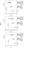
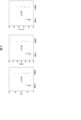
- 1 is a block diagram showing an example of a configuration example of an information processing device according to a first embodiment of the present invention; This figure plots the standard deviation (SD) of RI count values, the bandwidth (BW95) that includes 95% of the RI count values, and the entropy of the RI count values for healthy subjects, patients with idiopathic cardiomyopathy who were confirmed to have left ventricular reverse remodeling, and patients with idiopathic cardiomyopathy who were not confirmed to have left ventricular reverse remodeling.
- SD standard deviation
- BW95 bandwidth
- SD standard deviation
- BW95 bandwidth
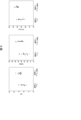
- entropy of the RI count values for healthy individuals and patients suffering from valvular disease This is a plot of the standard deviation (SD) of the RI count values, the bandwidth (BW95) that includes 95% of the RI count values, and the entropy of the RI count values for healthy individuals and patients suffering from valvular disease.
- This graph plots the standard deviation (SD) of RI count values, the bandwidth (BW95) that contains 95% of the RI count values, and the entropy of the RI count values for healthy subjects administered thallium-201 or technetium-99m methoxyisobutylisonitrile.
- SD standard deviation
- BW95 bandwidth
- 5 is a flowchart showing an example of a flow of processing performed by the information processing device.
- FIG. 11 is a block diagram showing an example of a configuration example of an information processing device according to a second embodiment of the present invention.
- 10 is a flowchart showing an example of a flow of a process performed by the information processing device when a first prediction model is used.
- 10 is a flowchart showing an example of a flow of a process performed by the information processing device when a second prediction model is used.
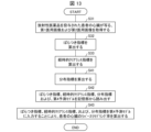
- 13 is a flowchart showing an example of a flow of a process performed by the information processing device when a third prediction model is used.
- 13 is a flowchart showing an example of a flow of a process performed by the information processing device when a fourth prediction model is used.
- Fig. 1 is a diagram showing an example of the configuration of the information processing system 100.
- the information processing system 100 according to an embodiment of the present disclosure includes an information processing device 1A.
- the information processing device 1A calculates a variation index indicating the degree of variation in the amount of radiation emitted from the radiopharmaceutical accumulated in the target part of the subject based on a medical image showing the target part of the subject to which a radiopharmaceutical has been administered.
- the information processing device 1A estimates the presence or absence of a disease in the target part of the subject based on the calculated variation index.
- the information processing device 1A may be communicatively connected to a LAN provided in each of a plurality of medical facilities 8 via a communication network 9.
- a medical image management device 2 may be communicatively connected to the LAN in each medical facility 8.
- the medical image management device 2 manages medical images 22 that are used by the information processing device 1A to estimate the presence or absence of a disease in a target area of a subject.
- the medical image management device 2 manages medical images 22 captured in each medical facility 8.
- the terminal device 3 may function as an output unit in the information processing system 100 and present information received from the information processing device 1A.
- the terminal device 3 may be, for example, a personal computer, a tablet terminal, a smartphone, etc.
- the terminal device 3 has a communication unit for transmitting and receiving data with other devices, an input unit such as a keyboard and a microphone, a display unit capable of displaying information transmitted from the information processing device 1A, an output unit such as a speaker, etc.
- the information processing device 1A When the information processing device 1A receives a request from any medical facility 8 to estimate the presence or absence of a disease in a target area of a subject, it acquires a medical image 22 from the medical image management device 2 of the medical facility 8, and estimates the presence or absence of a disease in the target area of the subject based on the medical image 22.
- the information processing system 100 may output the estimated result to the terminal device 3 of the medical facility 8.
- the information processing device 1A, the medical image management device 2, and the terminal device 3 may be provided in one medical facility 8.
- Fig. 2 is a block diagram showing an example of the configuration of the information processing device 1A.
- the information processing device 1A may be a computer.
- the information processing device 1A includes a control unit 10A, a memory unit 20, an input unit 30 that accepts input to the information processing device 1A, and an output unit 40 that outputs various information.
- the input unit 30 is, for example, a keyboard, a mouse, a microphone, etc.
- the output unit 40 is, for example, a display device, a printer, etc.
- the storage unit 20 stores various data used by the control unit 10A.
- the storage unit 20 also stores a control program 21, which is a program for performing various controls on the information processing device 1A.
- the control unit 10A controls each part of the information processing device 1A.
- the control unit 10A includes an image acquisition unit 11, an index calculation unit 50, and an estimation unit 12.
- the image acquisition unit 11 acquires the medical image 22 from the medical image management device 2.
- the image acquisition unit 11 stores the acquired medical image 22 in the storage unit 20.
- the medical image 22 will be described in detail below.
- the medical image 22 is an image of a target site of a subject to which a radiopharmaceutical has been administered.
- a radiopharmaceutical is a pharmaceutical containing a compound having a radioisotope (RI) as a structural element. When administered to a living body, a radiopharmaceutical has the property of selectively accumulating in a specific organ or tissue depending on the components.
- a radiopharmaceutical having the property of accumulating in a target site of a subject and imaging a minute amount of radiation (gamma rays) emitted from the radioisotope using a gamma camera, information on the distribution, accumulation amount, and change over time of the radiopharmaceutical in the target site can be obtained. Then, from the acquired information, the morphology, function, metabolic state, etc. of the target site can be evaluated.
- the information processing system 100 of this embodiment is not limited to the radiopharmaceuticals that can be used, and any radiopharmaceutical can be used.
- Each pixel of the medical image 22 captured by the gamma camera indicates the number of times that gamma rays emitted from the radioisotope present in that pixel have been detected (hereinafter, also referred to as the RI count value).
- the following description will be directed to a case where the information processing device 1A is used to estimate the presence or absence of a cardiac disease in a subject.
- the description will be directed to a case where the presence or absence of a disease that causes cardiac failure (cardiomyopathy, ischemic cardiac disease, hypertensive cardiac disease, valvular disease, etc.) is estimated.
- a disease that causes cardiac failure cardiac failure
- MIBI technetium 99m methoxyisobutylisonitrile
- BMIPP iodine-123- ⁇ -methyliodophenylpentadecanoate
- the information processing device 1A of the present invention is not limited to a target site being the heart, and other organs or tissues can be used as target sites. In this case, a radiopharmaceutical that has a tendency to accumulate in each organ or tissue can be used.
- the image acquisition unit 11 acquires a medical image 22 of the heart from the medical image management device 2 at a predetermined time after the administration of the radiopharmaceutical.
- the predetermined time may be any time that it takes the radiopharmaceutical administered into the body to accumulate in the heart, and may be, for example, 5 to 240 minutes.
- the index calculation unit 50 calculates an index used by the estimation unit 12 when estimating the presence or absence of a heart disease by analyzing the medical image 22.
- the index calculation unit 50 includes a variability index calculation unit 51.
- the variation index calculation unit 51 calculates the variation index 23 indicating the degree of variation in the amount of radiation emitted from the radiopharmaceutical accumulated in the subject's heart based on the medical image 22 stored in the storage unit 20.
- the variation index calculation unit 51 calculates the variation index 23 indicating the degree of variation in the amount of radiation emitted from the radiopharmaceutical accumulated in the heart (more specifically, the left ventricular myocardium of the heart) as the variation index 23.
- the variation index calculation unit 51 first performs a histogram analysis on the RI count value of each pixel included in the region corresponding to the left ventricular myocardium of the heart in the medical image 22.
- the variation index calculation unit 51 calculates, from the histogram analysis result, the standard deviation of the RI count value, the bandwidth including 95% of the RI count value, or the entropy, which is an index indicating the disorder of the RI count value and is distributed from 0 (complete order) to 1 (disorder), as the variation index 23.
- the variation index 23 is not limited to the above-mentioned index, and may be any index that indicates the degree of variation in the heart of the radiation amount (i.e., RI count value) emitted from the radiopharmaceutical accumulated in the subject's heart.
- the variation index calculation unit 51 calculates the variation index 23 based on the radiation amount emitted from the radiopharmaceutical accumulated in each of a plurality of unit areas (in this example, one pixel is used as a unit area) included in an area corresponding to the target part of the subject (in this example, the left ventricular myocardium of the heart) in the medical image 22.
- the size of the unit area may be, for example, 768 pixels.
- the variation index calculation unit 51 stores the calculated variation index 23 in the storage unit 20.
- the estimation unit 12 estimates the presence or absence of a cardiac disease of the subject based on the variability index 23 calculated by the variability index calculation unit 51.
- the inventors discovered that a person (living body) with a large variability index 23 is more likely to suffer from cardiac disease than a person (living body) with a small variability index 23, and completed the present invention. This will be explained with reference to Figures 3 to 7.
- Figure 3 is a plot of the standard deviation (SD) of the RI count values, the bandwidth (BW95) that includes 95% of the RI count values, and the entropy of the RI count values for a healthy person without cardiac disease, a patient with idiopathic cardiomyopathy who was confirmed to have left ventricular reverse remodeling, and a patient with idiopathic cardiomyopathy who was not confirmed to have left ventricular reverse remodeling.
- FIG. 4 is a plot of the standard deviation (SD) of the RI count values, the bandwidth (BW95) containing 95% of the RI count values, and the entropy of the RI count values for healthy subjects and patients suffering from ischemic cardiomyopathy.
- SD standard deviation
- BW95 bandwidth
- FIG. 5 is a plot of the standard deviation (SD) of the RI count values, the bandwidth (BW95) containing 95% of the RI count values, and the entropy of the RI count values for healthy subjects and patients suffering from valvular disease.
- SD standard deviation
- BW95 bandwidth
- MIBI technetium 99m methoxyisobutylisonitrile
- FIG. 6 is a plot of the standard deviation (SD) of the RI count value, the bandwidth (BW95) including 95% of the RI count value, and the entropy of the RI count value for healthy subjects administered with thallium-201 or technetium 99m methoxyisobutylisonitrile (MIBI).
- SD standard deviation
- BW95 bandwidth
- MIBI technetium 99m methoxyisobutylisonitrile
- the estimation unit 12 may estimate that the subject suffers from a heart disease or that the subject is highly likely to suffer from a heart disease when the variability index 23 calculated by the variability index calculation unit 51 is greater than a predetermined value.
- the estimation unit 12 may estimate that the subject suffers from a heart disease or that the subject is highly likely to suffer from a heart disease based on a comparison result between a reference index indicating the degree of variability in the heart of a comparison subject (i.e., a healthy subject) without a heart disease and the variability index 23 calculated by the variability index calculation unit 51.
- Fig. 8 is a flowchart showing an example of the flow of processing performed by the information processing device 1A.
- the image acquisition unit 11 acquires a medical image 22 showing the heart of a subject who has been administered a radiopharmaceutical from the medical image management device 2 (step S1).
- the image acquisition unit 11 stores the acquired medical image 22 in the storage unit 20.
- the variability index calculation unit 51 reads out the medical image 22 from the storage unit 20, and calculates a variability index 23 indicating the degree of variability in the heart of the radiation dose emitted from the radiopharmaceutical that has accumulated in the subject's heart based on the medical image 22 (step S2).
- the variability index calculation unit 51 stores the calculated variability index 23 in the storage unit 20.
- the estimation unit 12 reads out the variability index 23 calculated by the variability index calculation unit 51 from the storage unit 20, and estimates the presence or absence of a heart disease of the subject based on whether the variability index 23 is greater than a predetermined value (step S3).
- the information processing device 1A can estimate the presence or absence of a disease in the target area of the subject based on a variation index that indicates the degree of variation in the amount of radiation emitted from the radiopharmaceutical that has accumulated in the target area of the subject to which the radiopharmaceutical has been administered.
- the function of the image acquisition unit 11 may be provided in the terminal device 3, and the information processing device 1A may receive medical images 22 from the terminal device 3.
- the function of the estimation unit 12 may be provided in another computer or terminal device 3 different from the information processing device 1A.
- the information processing device 1B in this embodiment predicts "the possibility of improving at least one of the functional decline due to disease of the target part and the morphological change due to disease of the target part".
- the target part may be the heart or the like.
- the heart more specifically, the left ventricle of the heart
- the target part is used as an example of the target part.
- FIG. 9 is a block diagram showing an example of the configuration of an information processing device 1B in this embodiment.
- the information processing device 1B has a function of predicting the possibility of improvement of at least one of functional decline due to disease in a target part of a subject and morphological change due to disease in the target part (i.e., the possibility of reverse remodeling occurring in the target part).
- the heart more specifically, the left ventricle of the heart
- an example of predicting the possibility of reverse remodeling occurring in the left ventricle of a patient's heart in other words, calculating the left ventricular reverse remodeling rate of the patient's heart, will be described.
- a patient suffering from a disease in the left ventricle of the heart is the target of prediction.
- the information processing device 1B includes a control unit 10B instead of the control unit 10A in the first embodiment.
- the control unit 10B further includes a model generation unit 13 and a prediction unit 14.
- the image acquisition unit 11 acquires, as the medical image 22, a first medical image 22A (first image) of the target site (left ventricle of the heart) of the patient captured at a first time point when a first predetermined time has elapsed since the administration of the radiopharmaceutical from the medical image management device 2.
- the image acquisition unit 11 also acquires, as the medical image 22, a second medical image 22B (second image) of the left ventricle of the heart captured at a second time point when a second predetermined time has elapsed from the first time point from the medical image management device 2.
- the first predetermined time may be the time during which the radiopharmaceutical administered into the body accumulates in the left ventricle of the heart, and may be, for example, 5 to 60 minutes.
- the second predetermined time may be the time during which a portion of the radiopharmaceutical accumulated in the left ventricle of the heart is washed out of the left ventricle of the heart (in other words, the amount of accumulation of the radiopharmaceutical in the left ventricle of the heart attenuates).
- technetium 99m methoxyisobutylisonitrile (MIBI) or technetium 99m tetrofosmin can be used as the radiopharmaceutical.
- the image acquisition unit 11 stores the acquired first medical image 22A and second medical image 22B in the storage unit 20.
- the index calculation unit 50 in this embodiment calculates various indices used by the prediction unit 14 when making predictions by analyzing the medical image 22.
- the index calculation unit 50 includes a distribution index calculation unit 52 and a time-dependent clearance index calculation unit 53.
- the variability index calculation unit 51 calculates a variability index 23 that indicates the degree of variability in the left ventricle of the patient's heart in the amount of radiation emitted from the radiopharmaceutical that has accumulated in the left ventricle of the patient's heart, based on the first medical image 22A stored in the storage unit 20.
- the distribution index calculation unit 52 calculates a distribution index 24 indicating the proportion of the left ventricle of the heart that is occupied by a high-dose region consisting of one or more unit regions (e.g., one pixel) in which the radiation dose (i.e., RI count value) emitted from the radiopharmaceutical is at or above a predetermined level in the first medical image 22A.
- a distribution index 24 indicating the proportion of the left ventricle of the heart that is occupied by a high-dose region consisting of one or more unit regions (e.g., one pixel) in which the radiation dose (i.e., RI count value) emitted from the radiopharmaceutical is at or above a predetermined level in the first medical image 22A.
- the distribution index calculation unit 52 calculates the proportion of pixels whose RI count value is at or above a predetermined proportion as the distribution index 24 when the maximum RI count value is set to 100%.
- the above-mentioned predetermined proportion may be set to, for example, 30 to 50%.
- the distribution index 24 calculated as described above is an index indicating the survival proportion (functional proportion) of the left ventricular myocardium of the heart.
- the distribution index calculation unit 52 stores the calculated distribution index 24 in the storage unit 20.
- the temporal clearance index calculation unit 53 calculates an index (hereinafter referred to as the temporal clearance index 25) relating to the change in the amount of radiation emitted from the radiopharmaceutical in the left ventricle of the heart based on the first medical image 22A and the second medical image 22B.
- an index hereinafter referred to as the temporal clearance index 25
- An example of a method for calculating the temporal clearance index 25 is described below.
- the temporal clearance index calculation unit 53 calculates the sum of the RI count values emitted from each pixel in the area corresponding to the left ventricle of the heart for each of the first medical image 22A and the second medical image 22B.
- the temporal clearance index calculation unit 53 calculates 100 ⁇ (Q1 ⁇ Q2)/Q1 to calculate the temporal clearance index 25.
- the temporal clearance index 25 calculated as described above is called the washout rate, and is an index representing the quality of the left ventricle of the heart.
- the temporal clearance index calculation unit 53 stores the calculated temporal clearance index 25 in the storage unit 20.
- the storage unit 20 stores learning data 27 that is used when the model generation unit 13, which will be described later, generates a predictive model.
- the learning data 27 has a first index 27A that indicates the degree of variation in the amount of radiation emitted from a radiopharmaceutical that has accumulated in the left ventricle of the heart of a person (hereinafter referred to as person A) who has been administered a radiopharmaceutical, calculated based on a medical image showing the left ventricle of the heart of person A.
- the first index 27A is an index calculated by the same method as the method used by the variation index calculation unit 51 to calculate the variation index 23.
- the learning data 27 also includes a second index 27B, which is an index calculated based on the amount of radiation emitted from a radiopharmaceutical accumulated in each of a plurality of unit areas included in the region corresponding to the left ventricle of the heart of person A in the medical image of person A, and indicates the proportion of the left ventricle of person A's heart that is occupied by a high-dose region formed by one or more of the unit areas in which the amount of radiation emitted from the radiopharmaceutical is equal to or higher than a predetermined level.
- the second index 27B is an index calculated by the same method as the method used by the distribution index calculation unit 52 to calculate the distribution index 24.
- the learning data 27 also has information as to whether or not left ventricular reverse remodeling of person A's heart has been confirmed since the time the medical image was taken (e.g., 1 to 10 years after the medical image was taken), i.e., first improvement possibility information 27C indicating the possibility of left ventricular reverse remodeling of person A's heart occurring.
- the first improvement possibility information 27C is information on person A who has received medical treatment (medical intervention) since the time the medical image was taken.
- the learning data 27 may include a first index 27A, a second index 27B, and first improvement possibility information 27C for multiple people.
- the learning data 27 also includes a third index 27D that indicates the degree of variation in the amount of radiation emitted from the radiopharmaceutical that has accumulated in the left ventricle of the heart of a person (hereinafter referred to as person B) who has been administered a radiopharmaceutical, calculated based on a third medical image (third image) that is taken of the left ventricle of the heart of person B at a third time point when the first predetermined time has elapsed since the administration of the radiopharmaceutical.
- the third index 27D is an index calculated by the same method as the method used by the variation index calculation unit 51 to calculate the variation index 23.
- the third time point may be the same as the first time point.
- the learning data 27 also includes a fourth index 27E relating to the change in the amount of radiation emitted from the radiopharmaceutical in the left ventricle of person B's heart, which is calculated based on the third medical image and a fourth medical image (fourth image) of the left ventricle of person B's heart taken at a fourth time point that is the second predetermined time after the third time point.
- the fourth time point may be the same as the second time point.
- the fourth index 27E is an index calculated by the same method as the method used by the temporal clearance index calculation unit 53 to calculate the temporal clearance index 25.
- the learning data 27 also includes a fifth index 27F, which is an index calculated based on the amount of radiation emitted from a radiopharmaceutical accumulated in each of a plurality of unit areas included in the area corresponding to the left ventricle of the heart of person B in the third image, and indicates the proportion of the left ventricle of the heart of person B that is occupied by a high-dose area constituted by one or more unit areas in which the amount of radiation emitted from the radiopharmaceutical is equal to or higher than a predetermined level.
- the fifth index 27F is an index calculated by the same method as the method used by the distribution index calculation unit 52 to calculate the distribution index 24.
- the learning data 27 includes second improvement possibility information 27G that indicates the possibility of reverse remodeling of the left ventricle of person B's heart occurring after the fourth time point (e.g., 1 to 10 years after the fourth time point).
- the learning data 27 may include a third index 27D, a fourth index 27E, a fifth index 27F, and second improvement possibility information 27G for multiple people.
- the model generation unit 13 generates a learning model that is used by the prediction unit 14 when making predictions.
- the model generation unit 13 generates a first prediction model 26A, a second prediction model 26B, a third prediction model 26C, and a fourth prediction model 26D as prediction models.
- the model generation unit 13 in this embodiment generates each prediction model using logistic regression analysis as machine learning. However, the prediction model may be generated using machine learning other than logistic regression analysis. The method of generating each prediction model is described below.
- the first prediction model 26A is a prediction model generated by machine learning using learning data including a first explanatory variable and a first objective variable.
- the model generation unit 13 reads out the first index 27A and the first improvement possibility information 27C from the storage unit 20, and generates the first prediction model 26A using data including at least the first index 27A as the first explanatory variable and the first improvement possibility information 27C as the first objective variable.
- the model generation unit 13 stores the generated first prediction model 26A in the storage unit 20.
- the second prediction model 26B is a prediction model generated by machine learning using learning data including the second explanatory variable and the second objective variable.
- the model generation unit 13 reads out the first index 27A, the second index 27B, and the first improvement possibility information 27C from the storage unit 20.
- the model generation unit 13 generates the second prediction model 26B using data including at least the first index 27A and the second index 27B as the second explanatory variable and the first improvement possibility information 27C as the second objective variable.
- the model generation unit 13 stores the generated second prediction model 26B in the storage unit 20.
- the third prediction model 26C is a prediction model generated by machine learning using learning data including a third explanatory variable and a third objective variable.
- the model generation unit 13 reads out the third index 27D, the fourth index 27E, and the second improvement possibility information 27G from the storage unit 20.
- the model generation unit 13 generates the third prediction model 26C using data including at least the third index 27D and the fourth index 27E as the third explanatory variable and the second improvement possibility information 27G as the third objective variable.
- the model generation unit 13 stores the generated third prediction model 26C in the storage unit 20.
- the fourth prediction model 26D is a prediction model generated by machine learning using learning data including a fourth explanatory variable and a fourth objective variable.
- the model generation unit 13 reads out the third index 27D, the fourth index 27E, the fifth index 27F, and the second improvement possibility information 27G from the storage unit 20.
- the model generation unit 13 generates the fourth prediction model 26D by using data including at least the third index 27D, the fourth index 27E, and the fifth index 27F as the fourth explanatory variable and the second improvement possibility information 27G as the fourth objective variable.
- the model generation unit 13 stores the generated fourth prediction model 26D in the storage unit 20.
- the prediction unit 14 predicts the left ventricular remodeling rate of the patient using one of the first prediction model 26A, the second prediction model 26B, the third prediction model 26C, and the fourth prediction model 26D. The detailed process of prediction by the prediction unit 14 will be described later.
- Fig. 10 is a flowchart showing an example of the flow of processing performed by the information processing device 1B when the first prediction model 26A is used.
- the image acquisition unit 11 acquires the first medical image 22A from the medical image management device 2 (step S11).
- the image acquisition unit 11 stores the acquired first medical image 22A in the storage unit 20.
- the variability index calculation unit 51 reads out the first medical image 22A from the storage unit 20, and calculates a variability index 23 (first variability index) indicating the degree of variability in the patient's heart of the radiation dose emitted from the radiopharmaceutical that has accumulated in the patient's heart based on the first medical image 22A (step S12).
- the variability index calculation unit 51 stores the calculated variability index 23 in the storage unit 20.
- the prediction unit 14 reads out from the storage unit 20 the variability index 23 calculated by the variability index calculation unit 51 and the first prediction model 26A previously generated by the model generation unit 13 (step S13).
- the prediction unit 14 calculates the left ventricular reverse remodeling rate of the patient's heart by inputting the variability index 23 into the first prediction model 26A (step S14).
- the information processing device 1B of this embodiment has been realized based on this finding.
- the information processing device 1B is configured to predict the possibility of left ventricular reverse remodeling occurring in the patient's heart based on the variability index 23 indicating the degree of variability in the radiation dose in the left ventricle of the heart in a medical image of the left ventricle of the heart of a patient administered a radiopharmaceutical. This allows the information processing device 1B to calculate the left ventricular reverse remodeling rate with high accuracy.
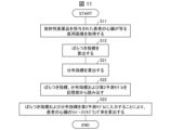
- Fig. 11 is a flowchart showing an example of the flow of processing performed by the information processing device 1B when the second prediction model 26B is used.
- steps S11 and S12 described above are performed as shown in FIG. 11.
- the distribution index calculation unit 52 calculates a distribution index 24 indicating the proportion of the left ventricular myocardium occupied by a high-dose region, which is composed of one or more unit regions in which the radiation dose emitted from the radiopharmaceutical is equal to or higher than a predetermined level, in the first medical image 22A (step S21).
- the distribution index calculation unit 52 stores the calculated distribution index 24 in the storage unit 20.
- the order of steps S12 and S21 may be reversed.
- the prediction unit 14 reads out from the storage unit 20 the variability index 23 calculated by the variability index calculation unit 51, the distribution index 24 calculated by the distribution index calculation unit 52, and the second prediction model 26B previously generated by the model generation unit 13 (step S22).
- the prediction unit 14 inputs the variability index 23 and the distribution index 24 into the second prediction model 26B to calculate the reverse remodeling rate of the patient's heart (left ventricular reverse remodeling rate) (step S23).
- the left ventricular reverse remodeling rate is calculated by inputting the variability index 23 and the distribution index 24 as input data into the second prediction model 26B.
- This allows the prediction unit 14 to make predictions that also take into account the survival rate (functional rate) of the left ventricle of the heart, thereby improving prediction accuracy compared to when only the variability index 23 is used as input data.
- Fig. 12 is a flowchart showing an example of the flow of processing performed by the information processing device 1B when the third prediction model 26C is used.
- the image acquisition unit 11 acquires from the medical image management device 2 a first medical image 22A that is an image of the patient's heart taken at a first time point when a first predetermined time has elapsed since the administration of the radiopharmaceutical. Furthermore, the image acquisition unit 11 acquires from the medical image management device 2 a second medical image 22B that is an image of the patient's heart taken at a second time point when a second predetermined time has elapsed from the first time point (step S31). The image acquisition unit 11 stores the acquired first medical image 22A and second medical image 22B in the storage unit 20.
- the variability index calculation unit 51 reads out the first medical image 22A from the storage unit 20, and calculates a variability index 23 (second variability index) indicating the degree of variability in the heart of the radiation dose emitted from the radiopharmaceutical accumulated in the patient's heart based on the first medical image 22A (step S32).
- the variability index calculation unit 51 stores the calculated variability index 23 in the storage unit 20.
- the variability index calculation unit 51 may calculate the variability index 23 based on the second medical image 22B.
- the temporal clearance index calculation unit 53 calculates the temporal clearance index 25, which is an index related to the change in the amount of radiation emitted from the radiopharmaceutical in the heart, based on the first medical image 22A and the second medical image 22B (step S33).
- the temporal clearance index calculation unit 53 stores the calculated temporal clearance index 25 in the storage unit 20.
- the order of steps S32 and S33 may be reversed.
- the prediction unit 14 reads out from the storage unit 20 the variation index 23 calculated by the variation index calculation unit 51, the clearance index over time 25 calculated by the clearance index over time calculation unit 53, and the third prediction model 26C previously generated by the model generation unit 13 (step S34).
- the prediction unit 14 inputs the variability index 23 and the temporal clearance index 25 into the third prediction model 26C to calculate the reverse remodeling rate of the patient's heart (left ventricular reverse remodeling rate) (step S35).
- the left ventricular reverse remodeling rate is calculated by inputting the variability index 23 and the temporal clearance index 25 as input data into the third prediction model 26C. This allows the prediction unit 14 to make predictions that also take into account the quality of the left ventricle of the heart, thereby improving prediction accuracy compared to when only the variability index 23 is used as input data.
- Fig. 13 is a flowchart showing an example of the flow of processing performed by the information processing device 1B when the fourth prediction model 26D is used.
- steps S31 to S33 described above are performed as shown in FIG. 12.
- the distribution index calculation unit 52 calculates a distribution index 24 indicating the proportion of the left ventricular myocardium occupied by a high-dose region, which is composed of one or more unit regions in which the radiation dose emitted from the radiopharmaceutical is equal to or higher than a predetermined level, in the first medical image 22A (step S41).
- the distribution index calculation unit 52 stores the calculated distribution index 24 in the storage unit 20.
- Steps S32, S33, and S41 are not limited to the above-mentioned order, and may be performed in any order.
- the prediction unit 14 reads out from the storage unit 20 the variability index 23 calculated by the variability index calculation unit 51, the temporal clearance index 25 calculated by the temporal clearance index calculation unit 53, the distribution index 24 calculated by the distribution index calculation unit 52, and the fourth prediction model 26D previously generated by the model generation unit 13 (step S42).
- the prediction unit 14 calculates the patient's left ventricular reverse remodeling rate by inputting the variability index 23, the temporal clearance index 25, and the distribution index 24 into the fourth prediction model 26D (step S43).
- the left ventricular reverse remodeling rate is calculated by inputting the variability index 23, the temporal clearance index 25, and the distribution index 24 as input data into the fourth prediction model 26D. This allows the prediction unit 14 to make predictions that take into account the survival rate (functional rate) and quality of the left ventricle of the heart, thereby further improving the prediction accuracy.
- the information processing device 1B in this embodiment is configured to be able to estimate the presence or absence of a cardiac disease of the subject by including the estimation unit 12, in addition to the function of calculating the reverse remodeling rate of the left ventricle of the subject, but may be configured not to include the estimation unit 12.
- the information processing device 1B may be configured to include an image acquisition unit that acquires a first image of a target site of a subject administered a radiopharmaceutical at a first time point, an index calculation unit that calculates a first variation index indicating the degree of variation in the target site of the subject of the amount of radiation emitted from the radiopharmaceutical accumulated in the target site based on the first image, and a prediction unit that inputs input data including at least the calculated first variation index into a first prediction model to predict the possibility of improvement of at least one of the functional decline due to the disease of the target site and the morphological change due to the disease of the target site of the subject.
- the information processing device 1B may also be configured to include an image acquisition unit that acquires an image of a target site of a subject administered with a radiopharmaceutical, an index calculation unit that calculates, based on the image, a first variation index that indicates the degree of variation in the target site of the subject of the amount of radiation emitted from the radiopharmaceutical accumulated in the target site, and a distribution index that indicates the proportion of the target site in the image that is a high-dose region composed of one or more unit regions in which the amount of radiation emitted from the radiopharmaceutical is equal to or higher than a predetermined level, and a prediction unit that inputs input data including at least the first variation index and the distribution index into a second prediction model to predict the possibility of improvement in at least one of functional decline due to a disease in the target site of the subject and morphological change due to a disease in the target site of the subject.
- an image acquisition unit that acquires an image of a target site of a subject administered with a radiopharmaceutical
- the information processing device 1B may also be configured to include an image acquisition unit that acquires a first image of a target site of a subject administered with a radiopharmaceutical at a first time point when a first predetermined time has elapsed since the administration of the radiopharmaceutical, and a second image of the target site at a second time point when a second predetermined time has further elapsed from the first time point; an index calculation unit that calculates (1) a second variation index indicating a degree of variation in the amount of radiation emitted from the radiopharmaceutical accumulated in the target site of the subject at the first time point, based on the first image, and (2) a temporal clearance index related to a change in the amount of radiation emitted from the radiopharmaceutical in the target site, based on the first image and the second image; and a prediction unit that inputs input data including at least the second variation index and the temporal clearance index into a third prediction model to predict the possibility of improvement in at least one of functional decline due to a disease in the
- the information processing device 1B further includes an image acquisition unit that acquires a first image of a target site of a subject administered with a radiopharmaceutical at a first time point when a first predetermined time has elapsed since the administration of the radiopharmaceutical, and a second image of the target site at a second time point when a second predetermined time has further elapsed from the first time point; and (1) a second variation index that indicates a degree of variation in the amount of radiation emitted from the radiopharmaceutical accumulated in the target site of the subject at the first time point based on the first image, and (2) a time-dependent clearance index that indicates a change in the amount of radiation emitted from the radiopharmaceutical in the target site based on the first image and the second image.
- the configuration may include an index calculation unit that calculates a distribution index, and (3) a distribution index that indicates the proportion of a high-dose region, which is a unit region included in a region corresponding to the target site of the subject in the first image and is composed of one or more unit regions in which the amount of radiation emitted from the radiopharmaceutical is equal to or higher than a predetermined level, in the target site, and a prediction unit that inputs input data including at least the second variability index, the distribution index, and the temporal clearance index into a fourth prediction model to predict the possibility of improvement in at least one of the functional decline due to a disease in the target site of the subject and the morphological change due to a disease in the target site.
- the function of the model generation unit 13 may be provided in the information processing device 1B by installing a prediction model that has undergone learning processing by another computer (e.g., terminal device 3) different from the information processing device 1B.
- the function of the prediction unit 14 may be provided in another computer or terminal device 3 different from the information processing device 1B.
- the functions of information processing device 1A or information processing device 1B can be realized by a program for causing a computer to function as the device, and a program for causing a computer to function as each control block of the device (particularly each part included in control unit 10A or control unit 10B).
- the device includes a computer having at least one control device (e.g., a processor) and at least one storage device (e.g., a memory) as hardware for executing the program.
- control device e.g., a processor
- storage device e.g., a memory
- the above program may be recorded on one or more computer-readable recording media, not on a temporary basis.
- the recording media may or may not be included in the device. In the latter case, the above program may be supplied to the device via any wired or wireless transmission medium.
- each of the above control blocks can be realized by a logic circuit.
- a logic circuit for example, an integrated circuit in which a logic circuit that functions as each of the above control blocks is formed is also included in the scope of the present invention.
- An information processing device includes an image acquisition unit that acquires an image of a target site of a subject to which a radiopharmaceutical has been administered, an index calculation unit that calculates, based on the image, a first variation index that indicates a degree of variation in the target site of the subject of the amount of radiation emitted from the radiopharmaceutical that has accumulated in the target site, and an estimation unit that estimates the presence or absence of a disease in the target site of the subject based on the calculated first variation index.
- the estimation unit may estimate the presence or absence of a disease in the target area of the subject based on a comparison result between a reference index indicating the degree of variation in the amount of radiation emitted from the radiopharmaceutical accumulated in the target area of the comparison subject who does not have the disease and the first variation index.
- the index calculation unit may calculate the first variability index based on the amount of radiation emitted from the radiopharmaceutical accumulated in each of a plurality of unit areas included in an area corresponding to the target part of the subject in the image.
- the information processing device may further include a prediction unit that inputs input data including at least the first variability index into a first prediction model to predict the possibility of improvement in at least one of the functional decline of the subject due to the disease of the target area and the morphological change of the subject due to the disease of the target area in the above-mentioned aspect 3.
- the first prediction model is generated by machine learning using learning data including a first explanatory variable and a first objective variable
- the first explanatory variable includes at least a first index calculated based on an image of the target site of a person administered the radiopharmaceutical and indicating the degree of variation in the amount of radiation emitted from the radiopharmaceutical accumulated in the target site of the person, the index being calculated based on the image of the target site
- the first objective variable may include first improvement possibility information indicating the possibility of improvement in at least one of a functional decline due to a disease in the target site of the person and a morphological change due to a disease in the target site of the person after the image is taken.
- the index calculation unit may further calculate a distribution index indicating the proportion of the target area that is occupied by a high-dose area constituted by one or more unit areas in which the amount of radiation emitted from the radioactive pharmaceutical is equal to or higher than a predetermined level, and may further include a prediction unit that inputs input data including at least the first variability index and the distribution index into a second prediction model to predict the possibility of improvement in at least one of the functional decline due to the disease of the target area and the morphological change due to the disease of the target area of the subject.
- the second prediction model is generated by machine learning using learning data including a second explanatory variable and a second objective variable
- the second explanatory variable includes at least: (1) a first index calculated based on an image showing the target part of the person administered the radiopharmaceutical, indicating the degree of variation in the amount of radiation emitted from the radiopharmaceutical accumulated in the target part of the person, and (2) a second index calculated based on the amount of radiation emitted from the radiopharmaceutical accumulated in each of a plurality of unit areas included in a region corresponding to the target part of the person in the image of the person, and indicating the proportion of a high-dose area constituted by one or more of the unit areas in which the amount of radiation emitted from the radiopharmaceutical is equal to or higher than a predetermined level, in the target part of the person; and the second objective variable may include first improvement possibility information indicating the possibility of improvement in at least one of a functional decline
- the image acquisition unit acquires a first image of the target site of the subject at a first time point when a first predetermined time has elapsed since administration of the radiopharmaceutical, and a second image of the target site at a second time point when a second predetermined time has further elapsed from the first time point
- the index calculation unit further calculates (1) a second variation index indicating the degree of variation in the amount of radiation emitted from the radiopharmaceutical accumulated in the target site of the subject at the first time point based on the first image, and (2) a time-dependent clearance index relating to the change in the amount of radiation emitted from the radiopharmaceutical in the target site based on the first image and the second image
- the third prediction model is generated by machine learning using learning data including a third explanatory variable and a third objective variable
- the third explanatory variable includes at least: (1) a third index indicating the degree of variation in the amount of radiation emitted from the radiopharmaceutical accumulated in the target part of the person administered the radiopharmaceutical, calculated based on a third image of the target part of the person administered the radiopharmaceutical at a third time point when the first predetermined time has elapsed since the administration of the radiopharmaceutical, and (2) a fourth index regarding the change in the amount of radiation emitted from the radiopharmaceutical in the target part of the person, calculated based on the third image and a fourth image of the target part of the person administered the radiopharmaceutical at a fourth time point when the second predetermined time has elapsed from the third time point; and the third objective variable may include second improvement possibility information indicating the possibility of improvement in at least one of the functional
- the image acquisition unit acquires a first image of the target site of the subject at a first time point when a first predetermined time has elapsed since administration of the radiopharmaceutical, and a second image of the target site at a second time point when a second predetermined time has further elapsed from the first time point
- the index calculation unit calculates (1) a second variation index based on the first image, the second variation index indicating the degree of variation in the amount of radiation emitted from the radiopharmaceutical accumulated in the target site of the subject at the first time point, and (2) a radiation dose emitted from the radiopharmaceutical at the target site based on the first image and the second image.
- the system may further include a prediction unit that calculates a time-dependent clearance index relating to the change in the amount of radiation in the target area of the subject, and (3) a distribution index indicating the proportion of a high-dose area, which is a unit area included in a region corresponding to the target area of the subject in the first image and is composed of one or more unit areas in which the amount of radiation emitted from the radiopharmaceutical is equal to or higher than a predetermined level, and inputs input data including at least the second variability index, the distribution index, and the time-dependent clearance index into a fourth prediction model to predict the possibility of improvement in at least one of the functional decline due to the disease in the target area of the subject and the morphological change due to the disease in the target area.
- a prediction unit that calculates a time-dependent clearance index relating to the change in the amount of radiation in the target area of the subject, and (3) a distribution index indicating the proportion of a high-dose area, which is a unit area included in a region corresponding to the target
- An information processing device is an information processing device according to aspect 10, wherein the fourth prediction model is generated by machine learning using learning data including a fourth explanatory variable and a fourth objective variable, and the fourth explanatory variable is: (1) a third index indicating the degree of variation in the amount of radiation emitted from the radiopharmaceutical accumulated in the target site of the person administered the radiopharmaceutical, calculated based on a third image of the target site of the person administered the radiopharmaceutical at a third time point when the first predetermined time has elapsed since the administration of the radiopharmaceutical; and (2) a third index indicating the degree of variation in the amount of radiation emitted from the radiopharmaceutical accumulated in the target site of the person, calculated based on the third image and a fourth image of the target site of the person administered the radiopharmaceutical at a fourth time point when the second predetermined time has further elapsed from the third time point.
- a fourth index relating to a change in the amount of radiation emitted from the radiopharmaceutical in the target site is an index calculated based on the amount of radiation emitted from the radiopharmaceutical accumulated in each of a plurality of unit areas included in the area corresponding to the target site of the person in the third image, and indicates the proportion of a high-dose area formed by one or more of the unit areas in which the amount of radiation emitted from the radiopharmaceutical is equal to or higher than a predetermined level in the target site of the person.
- the fourth objective variable may include second improvement possibility information indicating the possibility of improvement of at least one of the functional decline due to a disease in the target site of the person and the morphological change due to a disease in the target site of the person after the fourth time point.
- the third time point may be the same time point as the first time point.
- the fourth time point may also be the same time point as the second time point.
- the target area may be the heart.
- the radiopharmaceutical may be at least one of technetium 99m methoxyisobutylisonitrile (MIBI), technetium 99m tetrofosmin, thallium-201, and iodine-123- ⁇ -methyliodophenylpentadecanoic acid (BMIPP).
- MIBI technetium 99m methoxyisobutylisonitrile
- BMIPP iodine-123- ⁇ -methyliodophenylpentadecanoic acid
- the radiopharmaceutical may be at least one of technetium 99m methoxyisobutylisonitrile (MIBI) and technetium 99m tetrofosmin.
- MIBI technetium 99m methoxyisobutylisonitrile
- the disease estimation method estimates the presence or absence of a disease in a target site of a patient based on a first variability index that indicates the degree of variability in the target site of a patient to whom the radiopharmaceutical has been administered, the amount of radiation emitted from the radiopharmaceutical that has accumulated in the target site.
- the control program according to aspect 16 of the present disclosure is a control program for causing one or more computers to function as an information processing device according to any one of aspects 1 to 14 above, and is a control program for causing a computer to function as the image acquisition unit, the index calculation unit, the estimation unit, and the prediction unit.
- Image acquisition unit 12 Estimation unit 14
- Prediction unit 22 Medical image 22A First medical image 22B Second medical image 26A First prediction model 26B Second prediction model 26C Third prediction model 26D
- Fourth prediction model 27 Learning data 27A First index 27B Second index 27C
- First improvement possibility information 27D
- Third index 27E Fourth index 27F
- Fifth index 27G Second improvement possibility information 50 Index calculation unit
Landscapes
- Engineering & Computer Science (AREA)
- Physics & Mathematics (AREA)
- Health & Medical Sciences (AREA)
- General Physics & Mathematics (AREA)
- Medical Informatics (AREA)
- Theoretical Computer Science (AREA)
- Biomedical Technology (AREA)
- General Health & Medical Sciences (AREA)
- Computer Vision & Pattern Recognition (AREA)
- Nuclear Medicine, Radiotherapy & Molecular Imaging (AREA)
- Optics & Photonics (AREA)
- Life Sciences & Earth Sciences (AREA)
- High Energy & Nuclear Physics (AREA)
- Molecular Biology (AREA)
- Spectroscopy & Molecular Physics (AREA)
- Nuclear Medicine (AREA)
Priority Applications (1)
| Application Number | Priority Date | Filing Date | Title |
|---|---|---|---|
| JP2025509894A JPWO2024202649A1 (https=) | 2023-03-31 | 2024-02-15 |
Applications Claiming Priority (2)
| Application Number | Priority Date | Filing Date | Title |
|---|---|---|---|
| JP2023-059364 | 2023-03-31 | ||
| JP2023059364 | 2023-03-31 |
Publications (1)
| Publication Number | Publication Date |
|---|---|
| WO2024202649A1 true WO2024202649A1 (ja) | 2024-10-03 |
Family
ID=92904154
Family Applications (1)
| Application Number | Title | Priority Date | Filing Date |
|---|---|---|---|
| PCT/JP2024/005222 Ceased WO2024202649A1 (ja) | 2023-03-31 | 2024-02-15 | 情報処理装置、疾患推定方法、制御プログラム |
Country Status (2)
| Country | Link |
|---|---|
| JP (1) | JPWO2024202649A1 (https=) |
| WO (1) | WO2024202649A1 (https=) |
Citations (2)
| Publication number | Priority date | Publication date | Assignee | Title |
|---|---|---|---|---|
| WO2010004851A1 (ja) * | 2008-07-07 | 2010-01-14 | 浜松ホトニクス株式会社 | 脳疾患診断システム |
| JP2016513079A (ja) * | 2013-01-24 | 2016-05-12 | タイラートン インターナショナル ホールディングス インコーポレイテッドTylerton International Holdings Inc. | 神経細胞イメージング及び治療 |
-
2024
- 2024-02-15 WO PCT/JP2024/005222 patent/WO2024202649A1/ja not_active Ceased
- 2024-02-15 JP JP2025509894A patent/JPWO2024202649A1/ja active Pending
Patent Citations (2)
| Publication number | Priority date | Publication date | Assignee | Title |
|---|---|---|---|---|
| WO2010004851A1 (ja) * | 2008-07-07 | 2010-01-14 | 浜松ホトニクス株式会社 | 脳疾患診断システム |
| JP2016513079A (ja) * | 2013-01-24 | 2016-05-12 | タイラートン インターナショナル ホールディングス インコーポレイテッドTylerton International Holdings Inc. | 神経細胞イメージング及び治療 |
Non-Patent Citations (2)
| Title |
|---|
| JOINT STUDY GROUP: "Guidelines for Clinical Use of Cardiac Nuclear Medicine", GUIDELINES FOR CLINICAL USE OF CARDIAC NUCLEAR MEDICINE (JCS 2010); FY2009, JAPANESE SOCIETY OF NUCLEAR MEDICINE, JP, 1 January 2010 (2010-01-01), JP, pages 1 - 87, XP009560210 * |
| REN CHAO, REN JINGYUN, TIAN ZHUANG, DU YANRONG, HAO ZHIXIN, ZHANG ZONGYAO, FANG WEI, LI FANG, ZHANG SHUYANG, HSU BAILING, HUO LI: "Assessment of cardiac amyloidosis with 99mTc-pyrophosphate (PYP) quantitative SPECT", EJNMMI PHYSICS, vol. 8, no. 1, 1 December 2021 (2021-12-01), XP093218916, ISSN: 2197-7364, DOI: 10.1186/s40658-020-00342-7 * |
Also Published As
| Publication number | Publication date |
|---|---|
| JPWO2024202649A1 (https=) | 2024-10-03 |
Similar Documents
| Publication | Publication Date | Title |
|---|---|---|
| US20250025061A1 (en) | Systems and methods for processing images to analyze perfusion | |
| TWI535422B (zh) | 動態單光子放射電腦斷層攝影或是單光子放射電腦斷層攝影/電腦斷層攝影成像之心肌血流量化系統及方法 | |
| KR101716039B1 (ko) | 의료 영상 기반의 질환 진단 정보 산출 방법 및 장치 | |
| CN106062755A (zh) | 用于识别医学图像采集参数的系统和方法 | |
| CN111080584A (zh) | 医学图像的质控方法、计算机设备和可读存储介质 | |
| KR102330069B1 (ko) | 치과 영상으로부터 피검체의 성별 및 연령을 판정하는 방법 및 이를 이용한 장치 | |
| US11484221B2 (en) | Dynamic analysis apparatus, dynamic analysis system, expected rate calculation method, and recording medium | |
| KR20130111669A (ko) | 가시적 무손실 압축을 이용한 의료 영상 압축 시스템 및 방법 | |
| US20170020470A1 (en) | Imaging console and radiation imaging system | |
| Kortelainen et al. | Dependence of left ventricular functional parameters on image acquisition time in cardiac-gated myocardial perfusion SPECT | |
| Yoneyama et al. | Ability of artificial intelligence to diagnose coronary artery stenosis using hybrid images of coronary computed tomography angiography and myocardial perfusion SPECT | |
| CN116569271A (zh) | 通过在实时图像采集工作流程概览上叠加历史数据的可操作可视化 | |
| Gislason-Lee et al. | Impact of latest generation cardiac interventional X-ray equipment on patient image quality and radiation dose for trans-catheter aortic valve implantations | |
| JP2023121104A (ja) | 肺塞栓症診断支援装置、肺塞栓症診断支援方法及びプログラム | |
| JP6132691B2 (ja) | 医用画像処理装置及び医用画像処理システム | |
| WO2024202649A1 (ja) | 情報処理装置、疾患推定方法、制御プログラム | |
| Romero-Farina et al. | Acute myocardial infarction: estimation of at-risk and salvaged myocardium at myocardial perfusion SPECT 1 month after infarction | |
| Madamanchi et al. | What is multimodality cardiovascular imaging and how can it be delivered? | |
| Holst et al. | An independent evaluation of a new method for automated interpretation of lung scintigrams using artificial neural networks | |
| Wang et al. | Performance of automated software in the assessment of segmental left ventricular function in cardiac CT: comparison with cardiac magnetic resonance | |
| WO2019047545A1 (zh) | 低剂量成像方法及装置 | |
| CN118338847A (zh) | 基于动态图像数据的对象状态预测方法及执行该方法的计算装置 | |
| JP5422543B2 (ja) | 血流量推定装置、方法及びコンピュータプログラム | |
| JP2022034629A (ja) | 撮影制御装置、画像管理装置、画像管理システム及びプログラム | |
| JP6407520B2 (ja) | 医用装置及び積算線量表示システム |
Legal Events
| Date | Code | Title | Description |
|---|---|---|---|
| 121 | Ep: the epo has been informed by wipo that ep was designated in this application |
Ref document number: 24778794 Country of ref document: EP Kind code of ref document: A1 |
|
| ENP | Entry into the national phase |
Ref document number: 2025509894 Country of ref document: JP Kind code of ref document: A |
|
| WWE | Wipo information: entry into national phase |
Ref document number: 2025509894 Country of ref document: JP |
|
| NENP | Non-entry into the national phase |
Ref country code: DE |
|
| 122 | Ep: pct application non-entry in european phase |
Ref document number: 24778794 Country of ref document: EP Kind code of ref document: A1 |