WO2024176977A1 - 遺伝子変異の検査方法及び検査装置 - Google Patents
遺伝子変異の検査方法及び検査装置 Download PDFInfo
- Publication number
- WO2024176977A1 WO2024176977A1 PCT/JP2024/005576 JP2024005576W WO2024176977A1 WO 2024176977 A1 WO2024176977 A1 WO 2024176977A1 JP 2024005576 W JP2024005576 W JP 2024005576W WO 2024176977 A1 WO2024176977 A1 WO 2024176977A1
- Authority
- WO
- WIPO (PCT)
- Prior art keywords
- nucleic acid
- target nucleic
- target
- probe
- detecting
- Prior art date
- Legal status (The legal status is an assumption and is not a legal conclusion. Google has not performed a legal analysis and makes no representation as to the accuracy of the status listed.)
- Ceased
Links
Images



Classifications
-
- C—CHEMISTRY; METALLURGY
- C12—BIOCHEMISTRY; BEER; SPIRITS; WINE; VINEGAR; MICROBIOLOGY; ENZYMOLOGY; MUTATION OR GENETIC ENGINEERING
- C12Q—MEASURING OR TESTING PROCESSES INVOLVING ENZYMES, NUCLEIC ACIDS OR MICROORGANISMS; COMPOSITIONS OR TEST PAPERS THEREFOR; PROCESSES OF PREPARING SUCH COMPOSITIONS; CONDITION-RESPONSIVE CONTROL IN MICROBIOLOGICAL OR ENZYMOLOGICAL PROCESSES
- C12Q1/00—Measuring or testing processes involving enzymes, nucleic acids or microorganisms; Compositions therefor; Processes of preparing such compositions
- C12Q1/68—Measuring or testing processes involving enzymes, nucleic acids or microorganisms; Compositions therefor; Processes of preparing such compositions involving nucleic acids
- C12Q1/6813—Hybridisation assays
- C12Q1/6827—Hybridisation assays for detection of mutation or polymorphism
Definitions
- the present invention relates to a method and device for testing genetic mutations using nucleic acid probes used in DNA chips, etc.
- SNV single nucleotide variants
- SNP single nucleotide polymorphisms
- a DNA chip also called a DNA microarray
- a DNA chip has nucleic acid probes (a wild-type probe corresponding to the wild type and a mutant probe corresponding to the mutant type) that correspond to the genetic mutation to be tested fixed to a substrate.
- genomic DNA collected from the subject as a template
- a nucleic acid amplification reaction is carried out using a pair of primer sets that sandwich a region containing the genetic mutation to be tested, to obtain labeled nucleic acid fragments.
- a solution containing the obtained nucleic acid fragments is brought into contact with the DNA chip under specified reaction conditions. If the solution contains nucleic acid fragments with genetic mutations, a signal from the mutant probes can be detected.
- genetic mutations are not limited to SNVs and SNPs; similarly, DNA chips with nucleic acid probes corresponding to sequences are used in microsatellite, DNA methylation, and chimeric gene testing.
- a system using a DNA chip it is possible to test whether the nucleic acid fragments at the end of nucleic acid amplification (endpoint) contain any genetic mutations of the test subject.
- endpoint nucleic acid amplification
- a system using a DNA chip makes it possible to perform a qualitative evaluation of the genetic mutation of the test subject, rather than a quantitative measurement.
- Patent Document 1 As a system for quantifying nucleic acids using a DNA chip, as disclosed in Patent Document 1, a system is known in which the signal intensity based on hybridization between a target nucleic acid and a probe is measured, and compared with the signal intensity based on hybridization between a known amount of oligonucleotide and the probe, thereby correcting and quantifying the amount of target nucleic acid. Furthermore, Patent Document 2 discloses a method in which quantitative analysis or qualitative analysis by real-time PCR and qualitative analysis using a DNA chip are performed simultaneously in a single reaction system. However, these prior art techniques are techniques for quantifying the amount (copy number) of target nucleic acid present.
- the present invention aims to provide a genetic mutation testing method and testing device that can quantitatively analyze the proportion of genetic mutations present in the test subject using nucleic acid probes.
- the inventors conducted extensive research and discovered that when a nucleic acid fragment containing a genetic mutation to be tested is amplified by a nucleic acid amplification reaction, the genetic mutation can be quantitatively analyzed by detecting the nucleic acid fragment containing the genetic mutation using a nucleic acid probe during the exponential amplification phase of the nucleic acid amplification reaction, and thus completed the present invention.
- the present invention encompasses the following: (1) A method for testing for a genetic mutation, comprising: carrying out a nucleic acid amplification reaction to amplify a region including a genetic mutation to be tested; detecting a target nucleic acid including a target base to be detected in the genetic mutation and a non-target nucleic acid including a non-target base corresponding to the target base, which are contained in a reaction solution during the exponential amplification phase of the nucleic acid amplification reaction, using a nucleic acid probe for detecting a target nucleic acid having a sequence complementary to a region including the target base to be detected in the target nucleic acid and a nucleic acid probe for detecting a non-target nucleic acid having a sequence complementary to a region including the non-target base in the non-target nucleic acid, or a common probe having a sequence complementary to a common region common to the target nucleic acid and the non-target nucleic acid, the common region not overlapping with the region including the target
- the method for testing a gene mutation described in (1) characterized in that the detection step uses a DNA chip on which the nucleic acid probe for detecting the target nucleic acid and the nucleic acid probe for detecting a non-target nucleic acid or the common probe are immobilized on a carrier.
- the detection step uses a DNA chip on which the nucleic acid probe for detecting the target nucleic acid and the nucleic acid probe for detecting a non-target nucleic acid or the common probe are immobilized on a carrier.
- the abundance ratio of the target base in the genetic mutation to be tested is calculated based on the signal intensity from the nucleic acid probe for detecting the target nucleic acid and the signal intensity from the nucleic acid probe for detecting the non-target nucleic acid or the common probe.
- (6) The method for testing a genetic mutation described in (1), characterized in that in the quantitative analysis step, a calibration curve is prepared using a plurality of samples containing target nucleic acids and non-target nucleic acids in known ratios, and the proportion of the target base in the genetic mutation to be tested is quantified based on the signal intensity from the nucleic acid probe for detecting the target nucleic acid and the signal intensity from the nucleic acid probe for detecting the non-target nucleic acid or the common probe.
- a calibration curve is prepared using a plurality of samples containing target nucleic acids and non-target nucleic acids in known ratios, and when the signal intensity from the nucleic acid probe for detecting the non-target nucleic acids or the common probe exceeds a predetermined value determined based on the calibration curve, it is determined that the nucleic acid amplification reaction is in the exponential amplification phase.
- a genetic mutation testing device comprising: a nucleic acid amplification reaction unit that performs a nucleic acid amplification reaction to amplify a region including a genetic mutation to be tested; a detection unit that receives a reaction solution from the nucleic acid amplification reaction unit and detects at least a target nucleic acid using a nucleic acid probe for detecting a target nucleic acid having a sequence complementary to a region including a target base to be detected in a target nucleic acid including a base to be detected in the genetic mutation, and a nucleic acid probe for detecting a non-target nucleic acid having a sequence complementary to a region including a non-target base to be detected in a non-target nucleic acid including a non-target base corresponding to the target base to be detected, or a common probe having a sequence complementary to a common region common to the target nucleic acid and the non-target nucleic acid, the common region not overlapping with the target base to be detected or the region
- the testing device includes a measuring device that measures out an amount of reaction liquid to be supplied to the detection section for one cycle.
- the nucleic acid amplification reaction section is provided with a plurality of reaction chambers for performing a nucleic acid amplification reaction to amplify a region containing the genetic mutation to be tested, and the liquid delivery device includes a switching device that switches a flow path that supplies reaction liquid to the detection section between the plurality of reaction chambers, and the reaction liquid is separated by the switching device.
- the detection unit detects target nucleic acids and non-target nucleic acids using the common probe.
- the liquid delivery device includes a flow path connecting the nucleic acid amplification reaction section and the detection section, a valve disposed on the flow path, and a pump device connected to the flow path.
- the hybridization buffer contains a blocking nucleic acid that contains a base sequence complementary to the non-target nucleic acid that contains the non-detection target base.
- the genetic mutation testing method and testing device can quantitatively analyze a genetic mutation using a nucleic acid probe corresponding to the genetic mutation.
- a genetic mutation for example, the ratio of mutant to wild type.
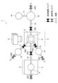
- FIG. 1 is a configuration diagram illustrating an example of a genetic mutation testing device according to the present invention.
- 3 is a flowchart showing the steps of testing for a gene mutation using the gene mutation testing device according to the present invention.
- FIG. 1 is a characteristic diagram showing a calibration curve and a logarithmically transformed calibration curve obtained using a reaction solution in the exponential amplification phase of PCR for the V617F mutation in the JAK2 gene.
- FIG. 1 is a characteristic diagram showing a calibration curve and a logarithmic transformed calibration curve obtained using a reaction solution in the exponential amplification phase of PCR for the W515L mutation in the MPL gene.
- FIG. 1 is a characteristic diagram showing a calibration curve prepared using a PCR end point reaction solution and a logarithmically transformed calibration curve.
- FIG. 11 is a characteristic diagram showing the relationship between the mutation rate quantified in Example 3 and the actually measured mutation rate.
- FIG. 2 is a configuration diagram illustrating another example of the genetic mutation testing device according to the present invention.
- FIG. 13 is a configuration diagram illustrating a further example of the genetic mutation testing device according to the present invention.
- the genetic mutation testing method and testing device of the present invention quantitatively detects a target nucleic acid containing a genetic mutation to be tested using a nucleic acid probe.
- the genetic mutation to be tested for is, for example, a single nucleotide variant (SNV) or a single nucleotide polymorphism (SNP), which refers to a difference in a base at a specific position in genomic DNA.
- SNV single nucleotide variant
- SNP single nucleotide polymorphism
- the target base to be detected means, for example, a specific nucleic acid residue at a specific position in genomic DNA, and is not particularly limited, but means a specific type of base in a base sequence such as a single nucleotide polymorphism (SNP).
- SNP single nucleotide polymorphism
- a specific single nucleotide polymorphism can take A (adenine) or C (cytosine)
- either one of the bases, i.e., A (adenine) in the single nucleotide polymorphism can be the target base to be detected.
- the target base to be detected may be either a major allele or a minor allele in a genetic polymorphism, and may or may not be a risk allele.
- the target nucleic acid refers to a nucleic acid molecule containing the base to be detected, i.e., a nucleic acid fragment.
- the target nucleic acid may be a nucleic acid molecule made of DNA, a nucleic acid molecule made of RNA, a nucleic acid molecule containing DNA and RNA (DNA-RNA complex), or a fragmented one such as cell-free DNA.
- the nucleic acid includes adenine, cytosine, guanine, thymine, and uracil, as well as artificial nucleic acids such as peptide nucleic acid (PNA) and locked nucleic acid (LNA).
- PNA peptide nucleic acid
- LNA locked nucleic acid
- the target nucleic acid containing the base to be detected can be prepared by amplifying a specific region on genomic DNA containing a gene mutation using a nucleic acid amplification method.
- the target nucleic acid may also be cDNA obtained by reverse transcription reaction from a transcription product collected from an individual organism, tissue, or cell.
- the base length of the target nucleic acid is not particularly limited, but can be, for example, 60 to 1000 bases, preferably 60 to 500 bases, and more preferably 60 to 200 bases.
- a nucleic acid molecule (nucleic acid fragment) containing a non-target base corresponding to the target base is referred to as a non-target nucleic acid.
- a nucleic acid fragment containing a non-target base corresponding to the target base
- bases other than the target base are non-target bases.
- a single nucleotide polymorphism at a specific position can be A (adenine) or C (cytosine)
- a (adenine) in the single nucleotide polymorphism is the target base
- C (cytosine) in the single nucleotide polymorphism is the non-target base.
- a nucleic acid fragment containing the mutated type can be used as a target nucleic acid
- a nucleic acid fragment containing a wild type forward or reverse strand of a major allele
- the non-target nucleic acid containing the non-target base is obtained at the same time as obtaining the target nucleic acid containing the target base as described above.
- the target nucleic acid is obtained by a nucleic acid amplification reaction such as a polymerase chain reaction, if one allele is a non-target base, the non-target nucleic acid will be amplified along with the target nucleic acid.
- a nucleic acid probe having a base sequence complementary to at least a region in the target nucleic acid that contains the target base is used to detect a target nucleic acid containing a target base.
- the nucleic acid probe is not particularly limited, but can be, for example, 10 to 30 bases long, and preferably 15 to 25 bases long.
- the base complementary to the target base is preferably located at the center of the character string. Note that the center of the character string includes the case where the nucleic acid probe is shifted by one base toward the 5' end or 3' end for a nucleic acid probe consisting of an even number of bases.
- the genetic mutation testing method and testing device of the present invention can use a common probe having a sequence complementary to a common region common to the target nucleic acid and non-target nucleic acid.
- the common region is a region of base sequence common to the target nucleic acid and non-target nucleic acid, and does not contain bases to be detected or bases to be non-detected.
- the common region can be a region that does not overlap with the region to which the nucleic acid probe that detects the target nucleic acid hybridizes and the region to which the nucleic acid probe that detects the non-target nucleic acid hybridizes.
- the genetic mutation testing method and testing device of the present invention can be applied to any system that includes hybridization, which means complementary binding between nucleic acid molecules in a target nucleic acid and a nucleic acid probe. That is, the genetic mutation testing method and testing device of the present invention can be based on Southern hybridization, Northern hybridization, or in situ hybridization.
- the genetic mutation testing method and testing device of the present invention are preferably used in a system in which a nucleic acid probe is fixed to a carrier (including a substrate, hollow fiber, or microparticle) and the target nucleic acid is detected (including qualitative and quantitative detection) using the fixed nucleic acid probe. More specifically, the genetic mutation testing method and testing device of the present invention are preferably used in a system that uses a DNA chip (DNA microarray) in which a nucleic acid probe is fixed to a substrate.
- DNA chip DNA microarray
- the nucleic acid probe for detecting the target nucleic acid (nucleic acid probe for detecting target nucleic acid), the nucleic acid probe for detecting non-target nucleic acids (nucleic acid probe for detecting non-target nucleic acids), and the common probe described above are more preferably single-stranded DNA, and can be obtained, for example, by chemical synthesis using a nucleic acid synthesizer.
- a device called a DNA synthesizer, fully automatic nucleic acid synthesizer, automatic nucleic acid synthesizer, etc. can be used.
- the nucleic acid probe for detecting the target nucleic acid is preferably used in the form of a microarray by immobilizing their 5'-end or 3'-end on a carrier.
- the material of the carrier can be any known material in the art, and is not particularly limited.
- conductive materials such as precious metals such as platinum, platinum black, gold, palladium, rhodium, silver, mercury, tungsten, and compounds thereof, and carbon such as graphite and carbon fiber; silicon materials such as single crystal silicon, amorphous silicon, silicon carbide, silicon oxide, silicon nitride, and composite materials of these silicon materials such as SOI (silicon on insulator); inorganic materials such as glass, quartz glass, alumina, sapphire, ceramics, forsterite, and photosensitive glass; polyethylene, polypropylene, cyclic Examples of organic materials include polyolefins, polyisobutylene, polyethylene terephthalate, unsaturated polyesters, fluorine-containing resins, polyvinyl chloride, polyvinylidene chloride, polyvinyl acetate, polyvinyl alcohol, polyvinyl acetal, acrylic resins, polyacrylonitrile, polystyrene, acetal resins, polycarbonates, polyamides, phenolic
- the carrier preferably has a carbon layer such as diamond-like carbon (DLC) on its surface, and chemically modified groups such as amino groups, carboxyl groups, epoxy groups, formyl groups, hydroxyl groups, and active ester groups.
- DLC diamond-like carbon
- Carriers having a carbon layer and chemically modified groups on their surface include those having a carbon layer and chemically modified groups on the surface of a substrate, and those having chemically modified groups on the surface of a substrate made of a carbon layer.
- Materials for the substrate may be any known material in the art, and are not particularly limited, and the same materials as those listed above as carrier materials may be used.
- the genetic mutation testing method and testing device of the present invention can quantitatively analyze the target nucleic acid in a subject using the DNA chip thus prepared.
- the method includes the steps of first extracting DNA from a sample derived from the subject, and amplifying a region containing the genetic mutation to be tested by a nucleic acid amplification reaction using the extracted DNA as a template, and detecting at least the target nucleic acid using the DNA chip during the exponential amplification phase of the nucleic acid amplification reaction.
- the subject is usually a human being, who is the subject of testing for genetic mutations, and may be a patient suffering from a disease associated with a specific genetic mutation.
- sample derived from the subject There are no particular limitations on the sample derived from the subject. Examples include blood-related samples (blood, serum, plasma, etc.), lymph, feces, cancer cells, tissue or organ fragments and extracts, etc.
- DNA is extracted from a sample taken from a subject.
- cells may be disrupted using a crusher called a blender, mixer, or homogenizer, and then purified using an organic solvent (phenol/chloroform), or cells may be disrupted using a surfactant, adsorbed onto a silica column, and centrifuged, or DNA may be adsorbed onto silica-coated magnetic beads, and then attracted with a magnet to separate the DNA.
- a crusher called a blender, mixer, or homogenizer
- an organic solvent phenol/chloroform
- DNA may be adsorbed onto silica column, and centrifuged
- DNA may be adsorbed onto silica-coated magnetic beads, and then attracted with a magnet to separate the DNA.
- an amplification reaction is carried out using the obtained DNA as a template to amplify the nucleic acid region containing the gene mutation to be tested, preferably the DNA.
- the amplification reaction the polymerase chain reaction (PCR), the loop-mediated isothermal amplification (LAMP), the isothermal and chimeric primer-initiated amplification of nucleic acids (ICAN), etc. can be applied.
- PCR polymerase chain reaction
- LAMP loop-mediated isothermal amplification
- ICAN isothermal and chimeric primer-initiated amplification of nucleic acids
- the method of labeling the amplified nucleic acid is not particularly limited, but for example, a method in which the primer used in the amplification reaction is labeled in advance may be used, or a method in which a labeled nucleotide is used as a substrate in the amplification reaction may be used.
- the labeling substance it is not particularly limited, but radioisotopes, fluorescent dyes, or organic compounds such as digoxigenin (DIG) and biotin can be used.
- This reaction system also contains a buffer necessary for nucleic acid amplification and labeling, a heat-resistant DNA polymerase, a pair of primers specific to the region to be amplified, labeled nucleotide triphosphate (specifically, a nucleotide triphosphate to which a fluorescent label or the like has been added), nucleotide triphosphate, magnesium chloride, etc.
- Nucleic acid amplification reactions consist of three steps: thermal denaturation, annealing, and extension. These three steps constitute one cycle, and by repeating multiple cycles, it is possible to synthesize a nucleic acid fragment containing the genetic mutation to be tested.
- the procedure for repeating these three steps is called a thermal cycle, and the number of times the three steps are repeated is called the number of thermal cycles.
- the nucleic acid amplification reaction may also be comprised of two steps, a thermal denaturation step and an annealing and extension step, by performing annealing and extension at the same temperature, known as two-step PCR.
- a thermal denaturation step by performing annealing and extension at the same temperature
- PCR annealing and extension at the same temperature
- a nucleic acid fragment containing the genetic mutation to be tested can be synthesized.
- the procedure for repeating these two steps is called a thermal cycle, and the number of times the two steps are repeated is called the number of thermal cycles.
- the thermal denaturation step is a step in which a reaction solution containing template DNA is heated to 90°C or higher to convert it into single-stranded DNA.
- the first thermal denaturation step in the thermal cycle is preferably carried out for a relatively long time (1-5 minutes) when genomic DNA is used as the template.
- the second and subsequent thermal denaturation steps are preferably carried out for a relatively short time (1-90 seconds) because the purpose is to convert the amplified product obtained as a double strand into a single strand.
- the annealing process is a process in which a reaction solution containing a single-stranded template DNA and a pair of primer sets is heated to a temperature typically in the range of 40-75°C, and these primers are specifically bound (annealed) to the template DNA.
- the temperature of the annealing process is set appropriately depending on the sequence and length of the primers.
- the temperature of the annealing process is usually set within a range of ⁇ 5°C of the Tm value of the primers used.
- the extension step is a step in which the temperature of the reaction solution is raised to about 72°C with the primer annealed to the template DNA, and a complementary strand is synthesized from the end of the primer using a heat-resistant polymerase (such as Taq DNA polymerase).
- a heat-resistant polymerase such as Taq DNA polymerase
- the complementary strand of the template DNA is synthesized from a labeled primer, or a complementary strand of the template DNA is synthesized while incorporating labeled free nucleotides.
- the target nucleic acid containing the base to be detected and the non-target nucleic acid containing the base not to be detected are labeled.
- the time for the extension step is appropriately set depending on the length of the nucleic acid fragment to be amplified and the reactivity of the enzyme used.
- the exponential amplification phase in a thermal cycle is the period during which the amplification efficiency in a thermal cycle is 70% or more, preferably 80% or more, more preferably 90% or more, even more preferably 95% or more, and most preferably 98% or more.
- the amplification efficiency is a value expressed as a percentage of the amount of nucleic acid fragment that increases with each cycle. In other words, an amplification efficiency of 100% means that the amplified fragment doubles with each cycle. Therefore, when defining the exponential amplification phase, there is no particular upper limit to the amplification efficiency, and it can be, for example, a value of 100 to 110%.
- the genetic mutation testing method and testing device of the present invention detect the target nucleic acid and non-target nucleic acid contained in the nucleic acid amplification reaction solution in the exponential amplification phase defined in this way, and quantitatively analyze the target nucleic acid based on this.
- a hybridization reaction between the target nucleic acid detection nucleic acid probe and the target nucleic acid is carried out, and the amount of nucleic acid hybridized to the target nucleic acid detection nucleic acid probe can be measured, for example, by detecting a label.
- the signal from the label can be detected as a fluorescent signal using a fluorescent scanner, and the signal intensity can be quantified by analyzing this using image analysis software.
- the hybridization reaction is preferably carried out under stringent conditions.
- Stringent conditions refer to conditions under which specific hybrids are formed and non-specific hybrids are not formed, such as conditions in which the hybridization reaction is carried out at 50°C for 16 hours, followed by washing at 2xSSC/0.2% SDS at 25°C for 10 minutes and 2xSSC at 25°C for 5 minutes.
- the hybridization buffer composition of the present invention may contain salts necessary for the hybridization reaction, such as SSC, and known blocking agents, such as SDS.
- FIG. 1 shows that a target nucleic acid can be quantitatively analyzed by using a template DNA with a ratio of 80% wild-type DNA and 20% mutant DNA, and the number of thermal cycles is plotted on the horizontal axis and the amount of DNA amplified on the vertical axis.
- the ratio of wild-type DNA to mutant DNA contained in the template DNA 80:20, is not maintained near the end of the thermal cycle. Therefore, it is understood that the target nucleic acid cannot be quantitatively analyzed using the reaction solution after the end of the thermal cycle.
- the ratio of wild-type DNA to mutant DNA contained in the template DNA, 80:20 is approximately maintained. Therefore, quantitative analysis of the target nucleic acid is possible by detecting the target nucleic acid in the exponential amplification phase of the thermal cycle.
- the ratio of the target nucleic acid to the non-target nucleic acid i.e., the mutation ratio
- the signal strengths of the nucleic acid probe for detecting a target nucleic acid and the nucleic acid probe for detecting a non-target nucleic acid are measured, respectively, and a judgment value for evaluating the signal strength from the nucleic acid probe for detecting a target nucleic acid is calculated.
- An example of calculating the judgment value is a method using the formula: [signal strength from the nucleic acid probe for detecting a target nucleic acid]/([signal strength from the nucleic acid probe for detecting a target nucleic acid]+[signal strength from the nucleic acid probe for detecting a non-target nucleic acid]).
- the genetic mutation testing method and testing device are not limited to the above-mentioned configuration, and a so-called blocking nucleic acid can be used to prevent non-target nucleic acids in the nucleic acid amplification reaction solution from non-specifically hybridizing to the nucleic acid probe for detecting the target nucleic acid.
- the blocking nucleic acid can be mixed with a reaction solution containing the target nucleic acid and non-target nucleic acid, and the resulting mixture can be brought into contact with a DNA chip to allow a hybridization reaction between the target nucleic acid and the nucleic acid probe for detecting the target nucleic acid to proceed.
- reaction solution containing the target nucleic acid and non-target nucleic acid and a solution containing the blocking nucleic acid can be mixed on the DNA chip to allow specific hybridization between the target nucleic acid and the nucleic acid probe to proceed simultaneously.
- the blocking nucleic acid hybridizes to the non-target nucleic acid, thereby preventing the non-target nucleic acid from hybridizing to the probe for detecting the target nucleic acid.
- the blocking nucleic acid has a base sequence complementary to a region of the non-target nucleic acid that contains a non-target base. Therefore, the blocking nucleic acid can hybridize with the non-target nucleic acid that contains the non-target base under conditions that allow hybridization of the target nucleic acid that contains the target base and the nucleic acid probe.
- the position of the base corresponding to the non-detection target base is not particularly limited.
- the blocking nucleic acid is not particularly limited, but it is preferable that the length is 60% or more of the base length of the nucleic acid probe. In addition, it is preferable that the blocking nucleic acid is 140% or less of the base length of the nucleic acid probe. For example, if the length of the nucleic acid probe is 25 bases, the base length of the blocking nucleic acid is preferably 15 to 35 bases.
- the blocking nucleic acid may contain mismatched bases (non-complementary bases) at positions corresponding to bases other than the non-detection target bases contained in the non-target nucleic acid.
- the number of mismatched bases may be 1 to 3, and preferably 1 to 2.
- the number of mismatched bases may be 1 to 5, and preferably 1 to 4.
- the concentration of the blocking nucleic acid is not particularly limited, but can be set appropriately depending on, for example, the concentration of the non-target nucleic acid and/or the concentration of the target nucleic acid, or depending on the primer concentration.
- the concentration of the blocking nucleic acid in the composition can be 0.01 to 2 ⁇ M, preferably 0.02 to 1.5 ⁇ M, and more preferably 0.05 to 1.5 ⁇ M.
- a blocking nucleic acid when used to prevent non-specific hybridization between a non-target nucleic acid and a nucleic acid probe for detecting a target nucleic acid, specific hybridization between the non-target nucleic acid and the nucleic acid probe for detecting a non-target nucleic acid is also inhibited. In this case, the signal strength from the nucleic acid probe for detecting a non-target nucleic acid is significantly reduced.
- the determination value according to the above formula: [signal strength from the nucleic acid probe for detecting a target nucleic acid]/([signal strength from the nucleic acid probe for detecting a target nucleic acid]+[signal strength from the nucleic acid probe for detecting a non-target nucleic acid]) may not be sufficient for quantitative analysis of the target nucleic acid. In this case, it is preferable to measure the total amount of the target nucleic acid and the non-target nucleic acid using a common probe.
- the common probe has a sequence complementary to a common region common to the target nucleic acid and the non-target nucleic acid. Therefore, the common probe can hybridize with the same non-target nucleic acid even if the blocking nucleic acid is hybridized with the same non-target nucleic acid.
- the formula for calculating the above-mentioned judgment value can be [signal intensity from the nucleic acid probe for detecting the target nucleic acid]/[signal intensity from the common probe]. This formula makes it possible to calculate the ratio of the target nucleic acid to the total of the target nucleic acid and non-target nucleic acid combined, eliminating the influence of the blocking nucleic acid.
- the calibration curve can be created by using various samples with known mutation ratios (samples containing target nucleic acid and non-target nucleic acid in known ratios) to detect the target nucleic acid and non-target nucleic acid contained in the nucleic acid amplification reaction solution in the exponential amplification phase, and from the judgment value calculated by the above formula.
- the calibration curve represents the relationship between the judgment value calculated as described above and the ratio of target nucleic acid and non-target nucleic acid contained in the nucleic acid amplification reaction solution, or the amount of target nucleic acid.
- the method for creating a calibration curve is not particularly limited, but may include a method in which a preset number of thermal cycles in a nucleic acid amplification reaction is set as the exponential amplification phase, target nucleic acid and non-target nucleic acid are detected at that number of thermal cycles, and a calibration curve is created from the judgment value calculated by the above formula.
- a method for creating a calibration curve may include a method in which the exponential amplification phase (a region with high linearity in the amplification curve) is examined based on an amplification curve obtained in a nucleic acid amplification reaction using a sample with a known mutation rate, the number of thermal cycles in the exponential amplification phase is determined, target nucleic acid and non-target nucleic acid are detected at that number of thermal cycles, and a calibration curve is created from the judgment value calculated by the above formula.
- a nucleic acid amplification reaction is carried out using a sample with a standard mutation rate (e.g., a sample with a mutation rate of 100%, hereafter referred to as the M100 sample), the reaction solution is aliquoted at predetermined intervals (e.g., every three cycles), and the target nucleic acid and non-target nucleic acid contained in the reaction solution are detected.
- the target nucleic acid is detected using a nucleic acid probe for detecting the target nucleic acid, and the target nucleic acid and non-target nucleic acid are detected using a common probe.
- the exponential amplification phase As a method for determining the exponential amplification phase, there is a method for determining the exponential amplification phase from the amplification curve of a nucleic acid probe for detecting a target nucleic acid. Specifically, a method is known in which the exponential amplification phase is calculated from the number of thermal cycles at which a predetermined threshold value is reached. However, this method may not be suitable when the dynamic range of detection changes. Therefore, for example, the exponential amplification phase is calculated by the following method.
- the reached fluorescence intensity value is set as the upper limit of the dynamic range of the nucleic acid probe for detecting the target nucleic acid, and several tentative fluorescence intensity thresholds (Tentative Threshold Mutant Probe Fluorescent Intensity, hereinafter referred to as TTh.MPFI) are set.
- TTh.MPFI Temporal Threshold Mutant Probe Fluorescent Intensity
- the judgment value at the thermal cycle number (Nearest Ct, hereinafter referred to as Near.Ct) closest to TTh.MPFI is calculated, and further, a tentative fluorescence intensity threshold of the common probe (Tentative Threshold Common Probe Fluorescent Intensity, hereinafter referred to as TTh.CPFI) is set, and the true fluorescence intensity threshold is determined from the linearity of the obtained calibration curve.
- TTh.MPFI Temporal Threshold Common Probe Fluorescent Intensity
- TTh.CPFI TTh.CPFI
- the exponential amplification phase may be determined by performing regression analysis or the like from the amplification curve of the nucleic acid fragment detected with the nucleic acid probe for detecting the target nucleic acid.
- the reciprocal of the judgment value obtained from the M100 sample can be used as a correction coefficient.
- This correction coefficient can be multiplied by the judgment value obtained from samples with known mutation rates including the M100 sample (for example, a sample with a mutation rate of 1%, 10%, 50%, M100, etc.) to correct the slope a of the calibration curve.
- a scatter plot is created with the corrected judgment value (judgment value x correction coefficient) on the horizontal axis and the known mutation rate on the vertical axis, and a linear approximation line can be obtained using the least squares method.
- the ratio of the rate of change on the horizontal axis to the rate of change on the vertical axis is constant, so the intercept b can be accurately corrected by logarithmicizing both axes.
- the slope a and intercept b can be corrected, and a calibration curve in which various errors have been normalized can be obtained. Specific examples are given below.
- TTh.MPFI Tthative Threshold Mutant probe Fluorescent Intensity
- TTh.MPFI 5000 is set here, the smallest ⁇ FI will be 400, and the number of thermal cycles at this time can be set as the nearest temperature cycle value (Nearest Ct, hereafter referred to as Near.Ct).
- Near.Ct 31.
- the CPFI at Near.Ct is set as the tentative fluorescence intensity threshold from the common probe (TTh.CPFI).
- TTh.CPFI is 7000.
- a judgment value is calculated in the same manner using the Near.Ct of each sample that is closest to the TTh.CPFI (7000 in the example of Table 1) calculated from the reference mutation ratio sample, and the CPFI and MPFI at that Near.Ct.
- the CPFI and MPFI are measured for various known mutation ratio samples in the same manner as in Table 1, and a judgment value is calculated using the MPFI at the Near.Ct where the measured CPFI approaches the above TTh.CPFI (7000 in the example of Table 1).
- the judgment value calculated for various known mutation rate samples can be multiplied by a correction coefficient to correct the slope a. More specifically, when the judgment value for a 100% mutation rate sample is calculated as [signal strength from the nucleic acid probe for detecting the target nucleic acid]/[signal strength from the common probe], the correction coefficient is calculated as [signal strength from the common probe]/[signal strength from the nucleic acid probe for detecting the target nucleic acid]. Then, by multiplying the judgment value calculated for various known mutation rate samples by the correction coefficient, a corrected judgment value can be calculated from the judgment values calculated for various known mutation rate samples.
- the corrected judgment value for a 100% mutation rate sample is 1, and the judgment values for the other mutation rate samples are distributed in the range of 0 to 1.
- a calibration curve can be created as a linear approximation curve by the least squares method.
- the method of calculating Near.Ct is not limited to the above-mentioned method, and may be, for example, a method of creating an approximation curve for the relationship between MPFI and temperature cycle value as shown in Table 1.
- the temperature cycle value (Calculated Ct, hereinafter Calc.Ct) that results in a preset MPFI (5000 in the above-mentioned example) is calculated on the approximation curve.
- the temperature cycle value closest to the calculated Calc.Ct can be set as Near.Ct.
- the approximation curve is not particularly limited as long as it is an approximation curve obtained by regression analysis, and may be, for example, a linear approximation curve, an exponential approximation curve, a logarithmic approximation curve, a polynomial approximation curve, a power approximation curve, a power approximation curve, a moving average curve, or a sigmoid curve.
- the genetic mutation testing method and testing device of the present invention may use the calibration curve created as described above, but it is preferable to set multiple TTh.MPFIs to create calibration curves in the same manner, and to select and use from the multiple calibration curves the calibration curve that has a more linear relationship between the corrected judgement value and the mutation rate.
- the multiple TTh.MPFIs can be appropriately set so that their values are equally spaced.
- the upper limit of the fluorescence intensity is 65535
- the multiple TTh.MPFIs can be set so that the equal interval width of the TTh.MPFIs is in the range of 1 to 65535, preferably in the range of 5 to 50000, and more preferably in the range of 10 to 10000.
- a calibration curve is created for the multiple TTh.MPFIs set with such a range of widths, and the calibration curve with the higher linearity is selected.
- the coefficient of determination R2 is calculated for the multiple calibration curves obtained, and the calibration curve with the coefficient of determination R2 closer to 1 can be selected as the calibration curve with the higher linearity.
- the equal interval width of the TTh.MPFIs is also extended, so the equal interval width of the TTh.MPFIs can also be set with the extended maximum value as the upper limit.
- the genetic mutation testing method and testing device of the present invention are not limited to the above-mentioned form of using a calibration curve showing the relationship between the corrected judgment value and the mutation rate, and it is preferable to also use a "log-transformed calibration curve" obtained by logarithmically transforming both sides of the equation showing the calibration curve.
- a calibration curve for a plurality of TTh.MPFIs as described above, and further create a logarithmically transformed calibration curve corresponding to the created calibration curve.
- a set with high linearity is selected for both calibration curves.
- a set of calibration curves with the highest coefficient of determination in the corresponding logarithmically transformed calibration curve is selected.
- the calibration curve included in the selected set can be used.
- the proportion of target nucleic acid (mutation proportion) in a subject can be analyzed.
- the CPFI corresponding to the TTh.MPFI used to create the calibration curve selected as described above is used as the TTh.CPFI to quantify the mutation proportion of an unknown specimen sample collected from a subject.
- the target nucleic acid and non-target nucleic acid contained in a nucleic acid amplification reaction solution using an unknown specimen sample collected from a subject are detected with a common probe, and the above-mentioned corrected judgment value is calculated using the fluorescence intensity (MPFI) observed with the nucleic acid probe for detecting target nucleic acid when the fluorescence intensity observed with the common probe is closest to the TTh.CPFI, and the mutation proportion can be analyzed based on the calibration curve.
- MPFI fluorescence intensity
- a portion of the reaction solution is taken out multiple times, and the target nucleic acid and non-target nucleic acid contained in the taken reaction solution are detected. Then, the MPFI observed in the reaction solution in which the fluorescence intensity observed with the common probe is closest to TTh.CPFI is used to calculate the above-mentioned corrected judgment value, and the mutation rate can be analyzed based on the calibration curve.
- the target nucleic acid and non-target nucleic acid contained in multiple reaction solutions in which a nucleic acid amplification reaction has been performed up to different thermal cycle numbers are detected. Then, the MPFI observed in the reaction solution in which the fluorescence intensity observed with the common probe is closest to TTh.CPFI is used to calculate the above-mentioned corrected judgment value, and the mutation rate can be analyzed based on the calibration curve.
- a large number of nucleic acid probes with different target bases to be detected can be arranged so that multiple genetic mutations can be tested with one DNA chip. Then, by carrying out the above-mentioned nucleic acid amplification reaction using multiple pairs of primer sets, multiple types of target nucleic acids can be amplified simultaneously.
- the target nucleic acids can be quantitatively analyzed for multiple genetic mutations using one DNA chip.
- the testing device 1 includes a nucleic acid amplification reaction section 2 that performs a nucleic acid amplification reaction to amplify a region containing a genetic mutation to be tested, and a detection section 3 that receives a reaction solution from the nucleic acid amplification reaction section 2 and detects the target nucleic acid contained in the reaction solution.
- the nucleic acid amplification reaction section 2 includes a reaction tank 4 that stores reagents and substances necessary for the nucleic acid amplification reaction, and a first temperature control section 5 that controls the reaction solution in the reaction tank 4 to a predetermined temperature.
- the detection section 3 includes a DNA chip mounting section 7 that mounts a DNA chip 6 having a nucleic acid probe for detecting a target nucleic acid, a nucleic acid probe for detecting a non-target nucleic acid, and a common probe, a second temperature control section 8 that controls the hybridization reaction solution supplied to the DNA chip 6 mounted on the DNA chip mounting section 7 to a predetermined temperature, and an imaging section 9 that is disposed at a position facing the surface of the DNA chip 6 to which various probes are fixed.
- the nucleic acid amplification reaction section 2 and the detection section 3 are connected via a flow path 10 through which a solution can pass.
- the first temperature control unit 5 and the second temperature control unit 8 are equipped with a metal block with high thermal conductivity and a temperature control device that heats/cools the metal block.
- the testing device 1 also includes a hybridization buffer tank 12 connected to the DNA chip mounting section 7 via a flow path 11, and a cleaning solution tank 14 connected to the DNA chip mounting section 7 via a flow path 13.
- the testing device 1 also includes a waste liquid tank 16 connected to the DNA chip mounting section 7 via a flow path 15.
- the testing device 1 also includes a pump device 18 connected to the waste liquid tank 16 via a flow path 17.
- liquid supply shutoff valves 19A, 19B, 19C, and 19D are provided in the flow paths 10, 11, 13, and 15.
- intake valves 20A, 20B, 20C, and 20D are attached to the reaction tank 4, hybridization buffer tank 12, washing solution tank 14, and waste tank 16, respectively.
- These liquid supply shutoff valves 19A, 19B, 19C, and 19D can open and close the flow paths, and direct the pressure of the pump device 18 to the flow path to which liquid is to be sent.
- the testing device 1 is equipped with a liquid supply device that includes the pump device 18, liquid supply shutoff valves 19A, 19B, 19C, and 19D, and intake valves 20A, 20B, 20C, and 20D.
- This liquid delivery device can deliver various solutions to the detection unit 3 via flow paths 10, 11, and 13, and can deliver waste liquid to the waste liquid tank 16 via flow path 15.
- the liquid delivery device can adjust the amount of reaction liquid delivered from the nucleic acid amplification reaction unit 2 to the detection unit 3 by timing control of the pump device 18, the liquid delivery shutoff valve 19A, and the intake valve 20A.
- the liquid delivery device in the testing device 1 may also be equipped with a metering device on the flow path 10 that measures the reaction liquid delivered from the nucleic acid amplification reaction unit 2 to the detection unit 3.
- the liquid delivery device can measure a predetermined amount of reaction liquid using the metering device and deliver the measured reaction liquid to the detection unit 3 via the flow path 10.
- the metering device is not particularly limited, but examples include a pipette and a space with a predetermined capacity.
- liquid supply cutoff valves 19A, 19B, 19C, and 19D can be moved to another location connected by a flow path.
- the liquid supply cutoff valve is not particularly limited, and can be made of any material such as one that has been treated to be hydrophobic or hydrophilic, or one that changes shape due to heat, but a shape that can be physically opened and closed by external stress is preferable, and as an example, it is preferable to use a diaphragm valve that is pressed down by an actuator, or a rotary valve that is opened and closed by rotation.
- the target nucleic acid and non-target nucleic acid contained in the nucleic acid amplification reaction solution in the exponential amplification phase can be detected, and the target nucleic acid can be quantitatively analyzed based on the detection.
- a flowchart for quantitatively analyzing the target nucleic acid is shown in FIG. 3.
- a nucleic acid amplification reaction of a region containing a genetic mutation to be tested is performed in the reaction vessel 4.
- the reaction vessel 4 is filled with a reaction solution consisting of reagents necessary for the nucleic acid amplification reaction.
- the reaction vessel 4 may be filled with the reaction solution by an operator, or the reaction vessel 4 may be filled from a different flow path not shown.
- the first temperature control unit 5 can control the temperature and processing time of the reaction solution in the reaction vessel 4 according to the set thermal cycle.
- the testing device 1 a portion of the reaction liquid in the exponential amplification phase is sent to the DNA chip mounting section 7 on which the DNA chip 6 is mounted. Specifically, the intake valve 20A and the liquid supply cutoff valves 19A and 19D are opened, and the other intake valves 20B, 20C, and 20D and the liquid supply cutoff valves 19B and 19C are closed. In this state, the pump device 18 is operated to send the reaction liquid in the reaction tank 4 to the DNA chip mounting section 7.
- the timing of the liquid supply can be determined by counting the number of thermal cycles from the start of the nucleic acid amplification reaction and determining the point at which a predetermined number of thermal cycles (the number of thermal cycles in the exponential amplification phase) is reached.
- the inspection device 1 can be set to temporarily suspend the thermal cycle performed by the first temperature control unit 5 and wait until the liquid transfer is completed in one temperature range of the thermal cycle (e.g., 95°C).
- the hybridization buffer is supplied from the hybridization buffer tank 12 to the DNA chip mounting section 7.
- the intake valve 20B and the supply cutoff valves 19B and 19D are opened, and the other intake valves 20A, 20C and 20D and the supply cutoff valves 19A and 19C are closed, and the pump device 18 is operated.
- the hybridization buffer may have a composition that includes the blocking nucleic acid described above.
- the second temperature control section 8 adjusts the temperature of the solution supplied to the DNA chip mounting section 7 to a preset hybridization temperature.
- the second temperature control section 8 can also preheat before the reaction if necessary.
- the reaction solution and hybridization buffer solution are sent separately through different flow paths, but it is also possible to mix the reaction solution and hybridization buffer solution and then send the mixed solution to the DNA chip mounting section 7.
- the detection unit 3 detects the target nucleic acid and non-target nucleic acid hybridized to the probe.
- a cleaning liquid is sent to the DNA chip mounting unit 7 to clean the DNA chip 6.
- the cleaning liquid can be sent from the cleaning liquid tank 14.
- the intake valve 20C and the liquid supply cutoff valves 19C and 19D are opened, and the other intake valves 20A, 20B, and 20D and the liquid supply cutoff valves 19A and 19B are closed, and the pump device 18 is operated.
- the second temperature control unit 8 can also lower the cleaning temperature (e.g., 25°C) lower than the hybridization reaction temperature.
- the mixture of the reaction solution and hybridization buffer solution may be discharged into the waste tank 16, and then the washing solution may be pumped as described above, or the mixture of the reaction solution and hybridization buffer solution may be discharged into the waste tank 16 by pumping the washing solution as described above.
- the surface of the DNA chip 6 on which the nucleic acid probes for detecting target nucleic acids, the nucleic acid probes for detecting non-target nucleic acids, and the common probe are fixed is imaged by the imaging unit 9, and the signal intensity of each probe is measured by image analysis software in a computer (not shown).
- the target nucleic acid can be quantitatively analyzed by the same computer based on the measured signal intensity.
- a portion of the nucleic acid amplification reaction liquid is delivered to detect target and non-target nucleic acids based on a hybridization reaction with each probe using a DNA chip 6.
- the reaction vessel 4 and the DNA chip mounting section 7 are connected via a flow path 10, and a portion of the nucleic acid amplification reaction liquid is delivered using a configuration including a pump device 18, delivery shutoff valves 19A, 19B, 19C, and 19D, and intake valves 20A, 20B, 20C, and 20D.
- the mechanism for delivering a portion of the nucleic acid amplification reaction liquid is not limited to this configuration, and a portion of the nucleic acid amplification reaction liquid can also be supplied using a pipette device disposed on a drive arm.
- the pump device 18 performs the above-mentioned liquid delivery operation by suction.
- the testing device 1 can also perform the above-mentioned liquid delivery operation by using a pressure pump connected upstream of the nucleic acid amplification reaction unit 2, the hybridization buffer tank 12, and the washing liquid tank 14.
- the hybridization buffer solution is sent directly to the DNA chip mounting section 7 from the hybridization buffer solution tank 12 connected to the DNA chip mounting section 7 via the flow path 11, but it may be sent to the DNA chip mounting section 7 after first merging with the flow path 10.
- testing device 1 shown in FIG. 2 is provided with a DNA chip mounting section 7 on which a DNA chip 6 can be mounted, and is configured so that the DNA chip 6 can be attached and detached.
- the testing device 1 is not limited to this configuration, and may be configured so that each probe is directly fixed to the detection section 3.
- the testing device 1 shown in FIG. 2 may also include a nucleic acid extraction section (not shown) upstream of the nucleic acid amplification reaction section 2, as necessary, for extracting template DNA from a sample derived from a subject.
- the nucleic acid extraction section can be composed of a nucleic acid extraction device including a crusher such as a blender, mixer, or homogenizer for disrupting cells, an organic solvent (phenol/chloroform), a silica membrane, an anion exchange resin column, magnetic beads, etc.
- the DNA extracted in the nucleic acid extraction section is mixed with the reagents necessary for the nucleic acid amplification reaction, and is sent to the nucleic acid amplification reaction section 2 connected via a flow path.
- the operation of mixing the extracted DNA with the reagents may be performed after the DNA and the reagents are each loaded into the reaction tank 4.
- the DNA and the reagents can be loaded separately into the reaction tank 4, and the loaded DNA and reagents can be mixed inside the reaction tank 4.
- the DNA and the reagents may be mixed in advance, and the resulting mixture may be loaded into the reaction tank 4.
- the testing device 1 shown in FIG. 2 a portion of the nucleic acid amplification reaction solution is sent from a nucleic acid amplification reaction unit 2 having one reaction chamber 4, but the testing device to which the present invention is applied is not limited to this configuration. That is, as shown diagrammatically in FIG. 8, the testing device 1 to which the present invention is applied may be configured to include a nucleic acid amplification reaction unit 2 having multiple reaction chambers 4 (seven in the example of FIG. 8). In this case, the multiple reaction chambers 4 may be collectively temperature-controlled by a single first temperature control unit 5, or the multiple first temperature control units 5 may each individually control the temperature.
- the inspection device 1 is equipped with a liquid delivery device having a flow path connected to each of the multiple reaction tanks 4 and a switching device arranged on the flow path to switch the flow path that supplies the reaction liquid to the detection unit 3 between the multiple reaction tanks 4.
- a liquid delivery shutoff valve 19A is arranged in the middle of the flow path 10.
- an intake valve 20A is attached to each reaction tank 4.
- the switching device can be equipped with a control device for controlling the timing of opening and closing of each of the liquid supply shutoff valve 19A and the intake valve 20A.
- the testing device 1 in FIG. 8 can perform nucleic acid amplification reactions in multiple reaction chambers 4 up to different numbers of thermal cycles, and detect the target nucleic acid and non-target nucleic acid contained in each reaction solution. This makes it possible to calculate the corrected judgment value and analyze the mutation rate based on the calibration curve, as described above.
- FIG. 9 is a schematic diagram showing another example of a genetic mutation testing device according to the present invention.
- the testing device 1 in FIG. 9 is equipped with a nucleic acid amplification reaction section 2 having multiple reaction chambers 4, similar to the example in FIG. 8.
- the reaction liquid derived from the subject is filled from one location, and the reaction liquid is distributed to each reaction chamber 4.
- the testing device to which the present invention is applied is not limited to this configuration, and may be configured so that each of the multiple reaction chambers 4 is filled with reaction liquid derived from a different subject.
- Example 1 [Creating a calibration curve] 1. Preparation of calibration curve sample
- the V617F mutation in the JAK2 gene and the W515L mutation in the MPL gene were used as the mutations to be tested.
- a mutant plasmid having a mutant type and a wild-type plasmid having a wild-type type were prepared through FASMAC's artificial gene synthesis service.
- As the wild-type plasmid for the JAK2 gene a plasmid into which the base sequence shown in SEQ ID NO: 1 was inserted was used.
- the wild-type plasmid for the MPL gene a plasmid into which the base sequence shown in SEQ ID NO: 2 was inserted was used.
- the mutant plasmid for each gene a plasmid into which the same region was inserted except for the above-mentioned mutation (V617F mutation in the JAK2 gene, W515L mutation in the MPL gene) was used.
- the copy numbers of the mutant and wild-type plasmids for each gene prepared were quantified in advance using Bio-Rad ddPCR, and the plasmids were mixed so that the mutation ratios for the JAK2 V617F mutation and the MPL W515L mutation were 1%, 5%, 20%, 50% or 100%, respectively.
- the mixed JAK2 gene and MPL gene were then adjusted with TE buffer so that the copy numbers of the mutant and wild-type plasmids were 4x10e3/ ⁇ L or 4x10e4/ ⁇ L, respectively, to prepare calibration curve samples.
- PCR Implementation Using the five types of prepared standard curve samples (mutation ratio: 1%, 5%, 20%, 50% or 100%), the designated regions of the JAK2 gene and the MPL gene were simultaneously amplified by adding the respective primer sets.
- the primer sets shown in Table 2 were designed for this PCR. Note that, among the primer sets shown in Table 2, the forward primer marked with "F” was fluorescently labeled (IC5).
- the primer sets designed as above were mixed to obtain the composition shown in Table 3 to prepare a primer mix.
- the PCR thermal cycle was performed at 95°C for 5 minutes, followed by 25, 28, 31, 34, and 37 cycles of 95°C for 30 seconds, 59°C for 30 seconds, and 72°C for 45 seconds, followed by 72°C for 10 minutes and a final temperature of 4°C.
- mutant probes nucleic acid probes for detecting target nucleic acids
- wild-type probes nucleic acid probes for detecting non-target nucleic acids
- a common probe common to the wild-type and mutant types were designed.
- the common probe specifically hybridizes to the amplified nucleic acid regardless of whether or not a gene mutation is present in the amplified nucleic acid.
- the base sequences of the designed probes are shown in Table 5.
- the reaction solution in the specified PCR tube was heated to 49.5°C using automatic operation, and the DNA chip needle was inserted and the hybridization reaction was carried out for 30 minutes. After the hybridization reaction, the DNA chip needle was quickly immersed in the cleaning solution tank and stirred 30 times so that the DNA chip went in and out of the cleaning solution surface. After washing, the DNA chip needle was immersed in the rinsing solution tank and stirred 80 times so that the DNA chip went in and out of the rinsing solution surface.
- the rinsed DNA chip needle was slowly immersed in the detection solution tank, and a 640 nm single-wavelength laser was irradiated to capture the excitation light on the CCD camera for 2 seconds.
- a scatter plot was created with the corrected judgement values of M1, M5, M20, M50, and M100 plotted on the X-axis and the known mutation rate on the Y-axis, and a linear approximation line I was created using the least squares method.
- a scatter plot was also created with the logarithmic transformation values (Log%) of the corrected judgement values of M1, M5, M20, M50, and M100 plotted on the X-axis and the logarithmic transformation values (Log%) of the known mutation rate on the Y-axis, and a linear approximation line II was created using the least squares method.
- ⁇ indicates that the coefficient of determination of the linear approximation line I is R2 ⁇ 0.99, meaning that there is linearity.
- ⁇ indicates that the coefficient of determination is R2 ⁇ 0.99, meaning that there is no linearity.
- ⁇ indicates that the coefficient of determination of the linear approximation line I is R2 ⁇ 0.99, and indicates the condition in which the coefficient of determination of the linear approximation line II is the highest. Under the conditions marked with " ⁇ " in Table 7, the mutation rate can be accurately calculated based on the judgment value.
- TTh.MPFI and TTh.CPFI that became “XX” were defined as the true Th.MPFI and Th.CPFI, and are shown in Table 8 (JAK2 gene) and Table 9 (MPL gene) together with the nearest temperature cycle value (Near.Ct(1)) and correction coefficient A.
- the linear approximation lines II determined in Tables 8 and 9 were set as the calibration curve.
- Example 2 [Creating a calibration curve] 1. Preparation of calibration curve samples
- calibration curve samples were prepared in the same manner as in Example 1, except that the copy numbers of the mutant plasmid and the wild-type plasmid for each of the JAK2 gene and the MPL gene were 2.4 x 10e3/ ⁇ L.
- PCR was carried out in the same manner as in Example 1, except that the temperature cycle of PCR was carried out for 22, 25, 28, 31, 34, 37 or 40 cycles.
- DNA Chip The conditions for the DNA chip were the same as those in Example 1.
- reaction solutions were prepared in the same manner as in Example 1, except that reaction solutions containing PCR amplification products amplified through thermal cycling of PCR at 22 cycles, 25 cycles, 28 cycles, 31 cycles, 34 cycles, 37 cycles, and 40 cycles were used.
- the fluorescence intensity was used by subtracting the value of the area where the probe was not spotted as the background value in advance.
- the coefficients a1 to a6 and b of the 6th-order polynomial approximation formula calculated for each of the V617F mutation in the JAK2 gene and the W515L mutation in the MPL gene are as shown in
- the Ct for which the difference ⁇ Ct between the obtained Calc.Ct and the actual temperature cycle value was the smallest was determined as Near.Ct(1).
- the judgment value, correction coefficient A, and TTh.CPFI in Near.Ct(1) were set.
- a scatter plot was created with the corrected judgement value plotted on the X-axis and the known mutation rate plotted on the Y-axis, and a linear approximation line I was created using the least squares method.
- a scatter plot was also created with the logarithmic transformation value of the corrected judgement value plotted on the X-axis and the logarithmic transformation value of the known mutation rate plotted on the Y-axis, and a linear approximation line II was created using the least squares method.
- a standard curve was created using the judgement value at the endpoint of 40 cycles.
- ⁇ indicates that the coefficient of determination of the linear approximation line I is R2 ⁇ 0.99, meaning that there is linearity.
- ⁇ indicates that the coefficient of determination is R2 ⁇ 0.99, meaning that there is no linearity.
- ⁇ indicates that the coefficient of determination of the linear approximation line I is R2 ⁇ 0.99, and indicates the condition in which the coefficient of determination of the linear approximation line II is the highest. Under the conditions marked with " ⁇ " in Table 11, the mutation rate can be accurately calculated based on the judgment value.
- TTh.MPFI and TTh.CPFI that were "XX” based on the linear approximation lines I and II were taken as the true Th.MPFI and Th.CPFI, and are shown in Table 12 together with the closest temperature cycle value (Near.Ct(1)) and correction coefficient A.
- the linear approximation line II determined in Table 12 was set as the calibration curve.
- Example 3 [Mutation rate quantitative test] 1. Calibration curve
- Example 2 Preparation of Samples for Quantification
- the plasmid DNAs used in Examples 1 and 2 were mixed so that the mutation rates of JAK2 and MPL were 0%, 0.25%, 0.5%, 1%, 2.5%, 5%, 10%, or 100%, respectively, and the concentrations were quantified using Qubit (Thermo Fisher Scientific).
- the mixed plasmid DNA was diluted with TE buffer to a concentration of 0.04 to 0.4 pg/ ⁇ L to prepare a sample for quantification.
- the PCR thermal cycle was performed at 95°C for 5 minutes, followed by 22, 25, 28, 31, 34, 37, and 40 cycles of 95°C for 30 seconds, 59°C for 30 seconds, and 72°C for 45 seconds, followed by 72°C for 10 minutes and a final temperature of 4°C.
- Example 2 the same DNA chip and hybridization reaction solution as in Example 2 were prepared, and the hybridization reaction was carried out under the same conditions as in Example 2.
Landscapes
- Chemical & Material Sciences (AREA)
- Organic Chemistry (AREA)
- Life Sciences & Earth Sciences (AREA)
- Zoology (AREA)
- Wood Science & Technology (AREA)
- Proteomics, Peptides & Aminoacids (AREA)
- Health & Medical Sciences (AREA)
- Engineering & Computer Science (AREA)
- Microbiology (AREA)
- Immunology (AREA)
- Physics & Mathematics (AREA)
- Molecular Biology (AREA)
- Biotechnology (AREA)
- Biophysics (AREA)
- Analytical Chemistry (AREA)
- Biochemistry (AREA)
- Bioinformatics & Cheminformatics (AREA)
- General Engineering & Computer Science (AREA)
- General Health & Medical Sciences (AREA)
- Genetics & Genomics (AREA)
- Measuring Or Testing Involving Enzymes Or Micro-Organisms (AREA)
Priority Applications (3)
| Application Number | Priority Date | Filing Date | Title |
|---|---|---|---|
| CN202480007319.9A CN120530206A (zh) | 2023-02-20 | 2024-02-16 | 基因突变的检查方法和检查装置 |
| JP2025502339A JPWO2024176977A1 (https=) | 2023-02-20 | 2024-02-16 | |
| EP24760276.6A EP4671383A1 (en) | 2023-02-20 | 2024-02-16 | GENETIC MUTATION TESTING METHOD AND GENETIC MUTATION TESTING DEVICE |
Applications Claiming Priority (2)
| Application Number | Priority Date | Filing Date | Title |
|---|---|---|---|
| JP2023-024439 | 2023-02-20 | ||
| JP2023024439 | 2023-02-20 |
Publications (1)
| Publication Number | Publication Date |
|---|---|
| WO2024176977A1 true WO2024176977A1 (ja) | 2024-08-29 |
Family
ID=92501184
Family Applications (1)
| Application Number | Title | Priority Date | Filing Date |
|---|---|---|---|
| PCT/JP2024/005576 Ceased WO2024176977A1 (ja) | 2023-02-20 | 2024-02-16 | 遺伝子変異の検査方法及び検査装置 |
Country Status (4)
| Country | Link |
|---|---|
| EP (1) | EP4671383A1 (https=) |
| JP (1) | JPWO2024176977A1 (https=) |
| CN (1) | CN120530206A (https=) |
| WO (1) | WO2024176977A1 (https=) |
Cited By (2)
| Publication number | Priority date | Publication date | Assignee | Title |
|---|---|---|---|---|
| WO2025089378A1 (ja) * | 2023-10-27 | 2025-05-01 | 東洋製罐グループホールディングス株式会社 | 遺伝子変異の検査方法及び検査装置 |
| WO2026014267A1 (ja) * | 2024-07-12 | 2026-01-15 | 東洋鋼鈑株式会社 | 遺伝子変異の検査方法及び検査装置 |
Citations (7)
| Publication number | Priority date | Publication date | Assignee | Title |
|---|---|---|---|---|
| JP2007097574A (ja) | 2005-09-06 | 2007-04-19 | Dna Chip Research Inc | 目的遺伝子のmRNAの定量方法 |
| JP2008306949A (ja) * | 2007-06-12 | 2008-12-25 | Olympus Corp | 塩基配列の増幅方法 |
| JP2012055271A (ja) * | 2010-09-10 | 2012-03-22 | National Agriculture & Food Research Organization | イチゴ葉縁退緑病の病原体CandidatusPhlomobacterfragariaeの検出方法 |
| JP5689446B2 (ja) | 2012-05-11 | 2015-03-25 | ケーマックK−Mac | リアルタイム重合酵素連鎖反応及びdnaチップが統合された検査システム、並びにこれを用いた統合分析方法 |
| WO2016167317A1 (ja) * | 2015-04-14 | 2016-10-20 | 凸版印刷株式会社 | 遺伝子変異の検出方法 |
| WO2019004335A1 (ja) * | 2017-06-28 | 2019-01-03 | 東洋鋼鈑株式会社 | 遺伝子変異評価方法、遺伝子変異評価用キット |
| JP2023024439A (ja) | 2014-11-10 | 2023-02-16 | フォーサイト・ビジョン フォー・インコーポレーテッド | 拡張可能な薬物送達デバイスおよび使用方法 |
-
2024
- 2024-02-16 EP EP24760276.6A patent/EP4671383A1/en active Pending
- 2024-02-16 JP JP2025502339A patent/JPWO2024176977A1/ja active Pending
- 2024-02-16 CN CN202480007319.9A patent/CN120530206A/zh active Pending
- 2024-02-16 WO PCT/JP2024/005576 patent/WO2024176977A1/ja not_active Ceased
Patent Citations (7)
| Publication number | Priority date | Publication date | Assignee | Title |
|---|---|---|---|---|
| JP2007097574A (ja) | 2005-09-06 | 2007-04-19 | Dna Chip Research Inc | 目的遺伝子のmRNAの定量方法 |
| JP2008306949A (ja) * | 2007-06-12 | 2008-12-25 | Olympus Corp | 塩基配列の増幅方法 |
| JP2012055271A (ja) * | 2010-09-10 | 2012-03-22 | National Agriculture & Food Research Organization | イチゴ葉縁退緑病の病原体CandidatusPhlomobacterfragariaeの検出方法 |
| JP5689446B2 (ja) | 2012-05-11 | 2015-03-25 | ケーマックK−Mac | リアルタイム重合酵素連鎖反応及びdnaチップが統合された検査システム、並びにこれを用いた統合分析方法 |
| JP2023024439A (ja) | 2014-11-10 | 2023-02-16 | フォーサイト・ビジョン フォー・インコーポレーテッド | 拡張可能な薬物送達デバイスおよび使用方法 |
| WO2016167317A1 (ja) * | 2015-04-14 | 2016-10-20 | 凸版印刷株式会社 | 遺伝子変異の検出方法 |
| WO2019004335A1 (ja) * | 2017-06-28 | 2019-01-03 | 東洋鋼鈑株式会社 | 遺伝子変異評価方法、遺伝子変異評価用キット |
Non-Patent Citations (1)
| Title |
|---|
| See also references of EP4671383A1 |
Cited By (2)
| Publication number | Priority date | Publication date | Assignee | Title |
|---|---|---|---|---|
| WO2025089378A1 (ja) * | 2023-10-27 | 2025-05-01 | 東洋製罐グループホールディングス株式会社 | 遺伝子変異の検査方法及び検査装置 |
| WO2026014267A1 (ja) * | 2024-07-12 | 2026-01-15 | 東洋鋼鈑株式会社 | 遺伝子変異の検査方法及び検査装置 |
Also Published As
| Publication number | Publication date |
|---|---|
| JPWO2024176977A1 (https=) | 2024-08-29 |
| EP4671383A1 (en) | 2025-12-31 |
| CN120530206A (zh) | 2025-08-22 |
Similar Documents
| Publication | Publication Date | Title |
|---|---|---|
| EP1591543B1 (en) | PCR amplification reaction apparatus and method for PCR amplification reaction using apparatus | |
| WO2024176977A1 (ja) | 遺伝子変異の検査方法及び検査装置 | |
| US20120107820A1 (en) | Multiplexed Quantitative PCR End Point Analysis of Nucleic Acid Targets | |
| WO2001048242A9 (en) | Methods for amplifying and detecting multiple polynucleotides on a solid phase support | |
| EP1375672A1 (en) | Method of detecting nucleic acid relating to disease | |
| WO2005085476A1 (en) | Detection of strp, such as fragile x syndrome | |
| EP2180066B1 (en) | Single nucleotide polymorphism genotyping detection via the real-time invader assay microarray platform | |
| JP7765002B2 (ja) | マイクロアレイキット、マイクロアレイを用いたターゲット検出方法 | |
| JP2009034052A (ja) | ハイブリダイゼーション方法および装置 | |
| JP6757942B2 (ja) | ハイブリダイゼーション用バッファー組成物及びハイブリダイゼーション方法 | |
| WO2011068518A1 (en) | Multiplexed quantitative pcr end point analysis of nucleic acid targets | |
| JP2016192939A (ja) | Cyp2c19*2検出用プローブ及びcyp2c19*3検出用プローブ | |
| JP5319148B2 (ja) | 標的核酸中の変異の検出方法及びアレイ | |
| EP1788096A1 (en) | Lid for PCR vessel comprising probes permitting PCR amplification and detection of the PCR product by hybridisation without opening the PCR vessel | |
| WO2025089378A1 (ja) | 遺伝子変異の検査方法及び検査装置 | |
| US11230732B2 (en) | Genotyping of mutations by combination of in-tube hybridization and universal tag-microarray | |
| US8932819B2 (en) | Assays for affinity profiling of nucleic acid binding proteins | |
| WO2026014267A1 (ja) | 遺伝子変異の検査方法及び検査装置 | |
| US6716579B1 (en) | Gene specific arrays, preparation and use | |
| JP2004313120A (ja) | β3アドレナリン受容体変異遺伝子の検出法ならびにそのための核酸プローブおよびキット | |
| CN114302967A (zh) | 用于评价骨髓增殖性肿瘤相关基因突变的试剂盒 | |
| Deharvengt et al. | Molecular assessment of human diseases in the clinical laboratory | |
| RU2738752C1 (ru) | Способ идентификации личности и установления родства с помощью InDel полиморфизмов и набор синтетических олигонуклеотидов для их генотипирования | |
| TWI332525B (en) | Method for genotyping and quantifying hepatitis b virus | |
| CN114085895B (zh) | 快速检测msi的检测引物及其试剂盒 |
Legal Events
| Date | Code | Title | Description |
|---|---|---|---|
| 121 | Ep: the epo has been informed by wipo that ep was designated in this application |
Ref document number: 24760276 Country of ref document: EP Kind code of ref document: A1 |
|
| ENP | Entry into the national phase |
Ref document number: 2025502339 Country of ref document: JP Kind code of ref document: A |
|
| WWE | Wipo information: entry into national phase |
Ref document number: 2025502339 Country of ref document: JP |
|
| WWE | Wipo information: entry into national phase |
Ref document number: 202480007319.9 Country of ref document: CN |
|
| WWP | Wipo information: published in national office |
Ref document number: 202480007319.9 Country of ref document: CN |
|
| WWE | Wipo information: entry into national phase |
Ref document number: 2024760276 Country of ref document: EP |
|
| NENP | Non-entry into the national phase |
Ref country code: DE |
|
| ENP | Entry into the national phase |
Ref document number: 2024760276 Country of ref document: EP Effective date: 20250922 |
|
| ENP | Entry into the national phase |
Ref document number: 2024760276 Country of ref document: EP Effective date: 20250922 |
|
| WWP | Wipo information: published in national office |
Ref document number: 2024760276 Country of ref document: EP |