WO2024166936A1 - 腫瘍組織内においてm2マクロファージに分化することができ、m2マクロファージに分化すると炎症性サイトカインを発現する細胞、および当該細胞を含む組成物 - Google Patents
腫瘍組織内においてm2マクロファージに分化することができ、m2マクロファージに分化すると炎症性サイトカインを発現する細胞、および当該細胞を含む組成物 Download PDFInfo
- Publication number
- WO2024166936A1 WO2024166936A1 PCT/JP2024/004080 JP2024004080W WO2024166936A1 WO 2024166936 A1 WO2024166936 A1 WO 2024166936A1 JP 2024004080 W JP2024004080 W JP 2024004080W WO 2024166936 A1 WO2024166936 A1 WO 2024166936A1
- Authority
- WO
- WIPO (PCT)
- Prior art keywords
- cells
- cell
- macrophages
- macrophage
- cancer
- Prior art date
- Legal status (The legal status is an assumption and is not a legal conclusion. Google has not performed a legal analysis and makes no representation as to the accuracy of the status listed.)
- Ceased
Links
Images










Classifications
-
- A—HUMAN NECESSITIES
- A61—MEDICAL OR VETERINARY SCIENCE; HYGIENE
- A61P—SPECIFIC THERAPEUTIC ACTIVITY OF CHEMICAL COMPOUNDS OR MEDICINAL PREPARATIONS
- A61P35/00—Antineoplastic agents
-
- C—CHEMISTRY; METALLURGY
- C12—BIOCHEMISTRY; BEER; SPIRITS; WINE; VINEGAR; MICROBIOLOGY; ENZYMOLOGY; MUTATION OR GENETIC ENGINEERING
- C12N—MICROORGANISMS OR ENZYMES; COMPOSITIONS THEREOF; PROPAGATING, PRESERVING, OR MAINTAINING MICROORGANISMS; MUTATION OR GENETIC ENGINEERING; CULTURE MEDIA
- C12N5/00—Undifferentiated human, animal or plant cells, e.g. cell lines; Tissues; Cultivation or maintenance thereof; Culture media therefor
- C12N5/06—Animal cells or tissues; Human cells or tissues
- C12N5/0602—Vertebrate cells
- C12N5/0634—Cells from the blood or the immune system
-
- A—HUMAN NECESSITIES
- A61—MEDICAL OR VETERINARY SCIENCE; HYGIENE
- A61K—PREPARATIONS FOR MEDICAL, DENTAL OR TOILETRY PURPOSES
- A61K35/00—Medicinal preparations containing materials or reaction products thereof with undetermined constitution
- A61K35/12—Materials from mammals; Compositions comprising non-specified tissues or cells; Compositions comprising non-embryonic stem cells; Genetically modified cells
- A61K35/14—Blood; Artificial blood
- A61K35/15—Cells of the myeloid line, e.g. granulocytes, basophils, eosinophils, neutrophils, leucocytes, monocytes, macrophages or mast cells; Myeloid precursor cells; Antigen-presenting cells, e.g. dendritic cells
-
- A—HUMAN NECESSITIES
- A61—MEDICAL OR VETERINARY SCIENCE; HYGIENE
- A61K—PREPARATIONS FOR MEDICAL, DENTAL OR TOILETRY PURPOSES
- A61K40/00—Cellular immunotherapy
- A61K40/10—Cellular immunotherapy characterised by the cell type used
- A61K40/17—Monocytes; Macrophages
-
- C—CHEMISTRY; METALLURGY
- C07—ORGANIC CHEMISTRY
- C07K—PEPTIDES
- C07K14/00—Peptides having more than 20 amino acids; Gastrins; Somatostatins; Melanotropins; Derivatives thereof
- C07K14/435—Peptides having more than 20 amino acids; Gastrins; Somatostatins; Melanotropins; Derivatives thereof from animals; from humans
- C07K14/52—Cytokines; Lymphokines; Interferons
-
- C—CHEMISTRY; METALLURGY
- C07—ORGANIC CHEMISTRY
- C07K—PEPTIDES
- C07K14/00—Peptides having more than 20 amino acids; Gastrins; Somatostatins; Melanotropins; Derivatives thereof
- C07K14/435—Peptides having more than 20 amino acids; Gastrins; Somatostatins; Melanotropins; Derivatives thereof from animals; from humans
- C07K14/52—Cytokines; Lymphokines; Interferons
- C07K14/525—Tumour necrosis factor [TNF]
-
- C—CHEMISTRY; METALLURGY
- C07—ORGANIC CHEMISTRY
- C07K—PEPTIDES
- C07K14/00—Peptides having more than 20 amino acids; Gastrins; Somatostatins; Melanotropins; Derivatives thereof
- C07K14/435—Peptides having more than 20 amino acids; Gastrins; Somatostatins; Melanotropins; Derivatives thereof from animals; from humans
- C07K14/52—Cytokines; Lymphokines; Interferons
- C07K14/54—Interleukins [IL]
-
- C—CHEMISTRY; METALLURGY
- C12—BIOCHEMISTRY; BEER; SPIRITS; WINE; VINEGAR; MICROBIOLOGY; ENZYMOLOGY; MUTATION OR GENETIC ENGINEERING
- C12N—MICROORGANISMS OR ENZYMES; COMPOSITIONS THEREOF; PROPAGATING, PRESERVING, OR MAINTAINING MICROORGANISMS; MUTATION OR GENETIC ENGINEERING; CULTURE MEDIA
- C12N5/00—Undifferentiated human, animal or plant cells, e.g. cell lines; Tissues; Cultivation or maintenance thereof; Culture media therefor
- C12N5/06—Animal cells or tissues; Human cells or tissues
- C12N5/0602—Vertebrate cells
- C12N5/0634—Cells from the blood or the immune system
- C12N5/0645—Macrophages, e.g. Kuepfer cells in the liver; Monocytes
-
- C—CHEMISTRY; METALLURGY
- C12—BIOCHEMISTRY; BEER; SPIRITS; WINE; VINEGAR; MICROBIOLOGY; ENZYMOLOGY; MUTATION OR GENETIC ENGINEERING
- C12N—MICROORGANISMS OR ENZYMES; COMPOSITIONS THEREOF; PROPAGATING, PRESERVING, OR MAINTAINING MICROORGANISMS; MUTATION OR GENETIC ENGINEERING; CULTURE MEDIA
- C12N5/00—Undifferentiated human, animal or plant cells, e.g. cell lines; Tissues; Cultivation or maintenance thereof; Culture media therefor
- C12N5/06—Animal cells or tissues; Human cells or tissues
- C12N5/0602—Vertebrate cells
- C12N5/0693—Tumour cells; Cancer cells
-
- C—CHEMISTRY; METALLURGY
- C12—BIOCHEMISTRY; BEER; SPIRITS; WINE; VINEGAR; MICROBIOLOGY; ENZYMOLOGY; MUTATION OR GENETIC ENGINEERING
- C12N—MICROORGANISMS OR ENZYMES; COMPOSITIONS THEREOF; PROPAGATING, PRESERVING, OR MAINTAINING MICROORGANISMS; MUTATION OR GENETIC ENGINEERING; CULTURE MEDIA
- C12N5/00—Undifferentiated human, animal or plant cells, e.g. cell lines; Tissues; Cultivation or maintenance thereof; Culture media therefor
- C12N5/10—Cells modified by introduction of foreign genetic material
-
- C—CHEMISTRY; METALLURGY
- C12—BIOCHEMISTRY; BEER; SPIRITS; WINE; VINEGAR; MICROBIOLOGY; ENZYMOLOGY; MUTATION OR GENETIC ENGINEERING
- C12N—MICROORGANISMS OR ENZYMES; COMPOSITIONS THEREOF; PROPAGATING, PRESERVING, OR MAINTAINING MICROORGANISMS; MUTATION OR GENETIC ENGINEERING; CULTURE MEDIA
- C12N2501/00—Active agents used in cell culture processes, e.g. differentation
- C12N2501/20—Cytokines; Chemokines
- C12N2501/23—Interleukins [IL]
-
- C—CHEMISTRY; METALLURGY
- C12—BIOCHEMISTRY; BEER; SPIRITS; WINE; VINEGAR; MICROBIOLOGY; ENZYMOLOGY; MUTATION OR GENETIC ENGINEERING
- C12N—MICROORGANISMS OR ENZYMES; COMPOSITIONS THEREOF; PROPAGATING, PRESERVING, OR MAINTAINING MICROORGANISMS; MUTATION OR GENETIC ENGINEERING; CULTURE MEDIA
- C12N2501/00—Active agents used in cell culture processes, e.g. differentation
- C12N2501/20—Cytokines; Chemokines
- C12N2501/23—Interleukins [IL]
- C12N2501/2304—Interleukin-4 (IL-4)
-
- C—CHEMISTRY; METALLURGY
- C12—BIOCHEMISTRY; BEER; SPIRITS; WINE; VINEGAR; MICROBIOLOGY; ENZYMOLOGY; MUTATION OR GENETIC ENGINEERING
- C12N—MICROORGANISMS OR ENZYMES; COMPOSITIONS THEREOF; PROPAGATING, PRESERVING, OR MAINTAINING MICROORGANISMS; MUTATION OR GENETIC ENGINEERING; CULTURE MEDIA
- C12N2501/00—Active agents used in cell culture processes, e.g. differentation
- C12N2501/20—Cytokines; Chemokines
- C12N2501/23—Interleukins [IL]
- C12N2501/2313—Interleukin-13 (IL-13)
-
- C—CHEMISTRY; METALLURGY
- C12—BIOCHEMISTRY; BEER; SPIRITS; WINE; VINEGAR; MICROBIOLOGY; ENZYMOLOGY; MUTATION OR GENETIC ENGINEERING
- C12N—MICROORGANISMS OR ENZYMES; COMPOSITIONS THEREOF; PROPAGATING, PRESERVING, OR MAINTAINING MICROORGANISMS; MUTATION OR GENETIC ENGINEERING; CULTURE MEDIA
- C12N2501/00—Active agents used in cell culture processes, e.g. differentation
- C12N2501/20—Cytokines; Chemokines
- C12N2501/24—Interferons [IFN]
-
- C—CHEMISTRY; METALLURGY
- C12—BIOCHEMISTRY; BEER; SPIRITS; WINE; VINEGAR; MICROBIOLOGY; ENZYMOLOGY; MUTATION OR GENETIC ENGINEERING
- C12N—MICROORGANISMS OR ENZYMES; COMPOSITIONS THEREOF; PROPAGATING, PRESERVING, OR MAINTAINING MICROORGANISMS; MUTATION OR GENETIC ENGINEERING; CULTURE MEDIA
- C12N2501/00—Active agents used in cell culture processes, e.g. differentation
- C12N2501/20—Cytokines; Chemokines
- C12N2501/25—Tumour necrosing factors [TNF]
-
- C—CHEMISTRY; METALLURGY
- C12—BIOCHEMISTRY; BEER; SPIRITS; WINE; VINEGAR; MICROBIOLOGY; ENZYMOLOGY; MUTATION OR GENETIC ENGINEERING
- C12N—MICROORGANISMS OR ENZYMES; COMPOSITIONS THEREOF; PROPAGATING, PRESERVING, OR MAINTAINING MICROORGANISMS; MUTATION OR GENETIC ENGINEERING; CULTURE MEDIA
- C12N2506/00—Differentiation of animal cells from one lineage to another; Differentiation of pluripotent cells
- C12N2506/11—Differentiation of animal cells from one lineage to another; Differentiation of pluripotent cells from blood or immune system cells
- C12N2506/115—Differentiation of animal cells from one lineage to another; Differentiation of pluripotent cells from blood or immune system cells from monocytes, from macrophages
-
- C—CHEMISTRY; METALLURGY
- C12—BIOCHEMISTRY; BEER; SPIRITS; WINE; VINEGAR; MICROBIOLOGY; ENZYMOLOGY; MUTATION OR GENETIC ENGINEERING
- C12N—MICROORGANISMS OR ENZYMES; COMPOSITIONS THEREOF; PROPAGATING, PRESERVING, OR MAINTAINING MICROORGANISMS; MUTATION OR GENETIC ENGINEERING; CULTURE MEDIA
- C12N2510/00—Genetically modified cells
Definitions
- the present disclosure relates to cells that can differentiate into M2 macrophages in tumor tissue and express inflammatory cytokines when differentiated into M2 macrophages, and to compositions containing the cells (e.g., pharmaceutical compositions used to treat cancer).
- tumor tissue is usually immunosuppressive, and tumors are not recognized as “foreign” by the immune system [8-13]. For this reason, cancer immunotherapy is not fully effective. This is mainly due to intratumoral stromal cells such as M2 macrophages (anti-inflammatory) and regulatory T cells that secrete anti-inflammatory cytokines such as interleukin (IL)-10 and transforming growth factor- ⁇ (TGF- ⁇ ) [8, 14].
- intratumoral stromal cells such as M2 macrophages (anti-inflammatory) and regulatory T cells that secrete anti-inflammatory cytokines such as interleukin (IL)-10 and transforming growth factor- ⁇ (TGF- ⁇ ) [8, 14].
- IL interleukin
- TGF- ⁇ transforming growth factor- ⁇
- Tumor necrosis factor alpha is one of the most important inflammatory cytokines and plays a major role in activating the immune system in surrounding tissues [15, 16].
- TNF- ⁇ binds to TNF receptors 1 and 2, it activates signaling cascades such as the nuclear factor kappa B (NF- ⁇ B) pathway, resulting in the expression of inflammatory cytokines such as IL-6 and IL-1 [16-19].
- NF- ⁇ B nuclear factor kappa B
- inflammatory cytokines such as IL-6 and IL-1 [16-19].
- M1 macrophages inflammatory
- activate other immune cells transforming tissues into an inflammatory environment [15].
- TNF- ⁇ The proinflammatory function of TNF- ⁇ is attractive for cancer therapy based on the body's own clearance of foreign substances, but the severe toxicity of TNF- ⁇ after systemic administration, even at doses far below the therapeutic range, has prevented its clinical application [20]. Furthermore, the half-life of TNF- ⁇ administered to the body is approximately 15 to 30 minutes, making it difficult to deliver sufficient TNF- ⁇ to tumor tissues [21, 22].
- TNF- ⁇ -antibody conjugates can efficiently deliver TNF- ⁇ to tumor tissues [23, 24].
- These technologies can deliver TNF- ⁇ specifically to antigens expressed in cancer cells, but the effectiveness is significantly reduced when the antigen is mutated or deleted, so there is still room for technical improvement [25, 26].
- Macrophages are one of the major immune cells and play an important role in inflammation and immune responses in tissues [27].
- Major characteristics of macrophages, especially in tumor tissues, are (1) that macrophages migrate to tumor tissues via C-C chemokine ligand 2 (CCL2) and granulocyte-macrophage colony-stimulating factor (GM-CSF) secreted by cancer cells and stromal cells [28, 29], and (2) that macrophages can polarize to an anti-inflammatory M2 phenotype in tumor tissues [18, 30, 31].
- Macrophages change to an M1 or M2 phenotype depending on the inflammatory state of the tissue environment, and control the inflammatory environment. M2 macrophages are involved in wound healing, tissue regeneration, and immunosuppression [30, 32]. In tumor tissues, in particular, macrophages polarize to an M2 macrophage and play an important role in establishing an immunosuppressive environment [30].
- mouse M2 macrophages are known to have very high arginase 1 (Arg1) activity [33].
- Arg1 catalyzes the hydrolysis of arginine to urea and ornithine, which reduces the amount of arginine and promotes immunosuppression [34]. Because Arg1 is not highly expressed in M0 or M1 macrophages, Arg1 activity is one of the important markers for identifying the mouse M2 phenotype [35].
- the present disclosure provides cells that can differentiate into M2 macrophages in tumor tissue and express inflammatory cytokines when differentiated into M2 macrophages, and compositions that contain the cells.
- a cell capable of differentiating/polarizing into an M2 macrophage comprising a nucleic acid encoding an inflammatory cytokine operably linked to a regulatory sequence, and capable of expressing the inflammatory cytokine after differentiation/polarization into an M2 macrophage.
- the cell according to (1) above which is any cell selected from the group consisting of M0 macrophages, monocytes, common monocyte progenitors, monocyte-dendritic cell progenitors, myeloid progenitors, and hematopoietic stem cells, as well as any cell in the differentiation process from hematopoietic stem cells to M0 macrophages.
- the cell according to (1) or (2) above which is an iNOS, CD206, and Arg-1 negative macrophage.
- the cell according to any one of (1) to (3) above which is a cell at any stage of differentiation from a monocyte to an M0 macrophage.
- the cell according to any one of (1) to (4) above, wherein the inflammatory cytokine is selected from the group consisting of TNF- ⁇ , IFN- ⁇ , IFN- ⁇ , IL-1, IL-2, IL-6, IL-8, IL-12, IL-17, and IL-23.
- the inflammatory cytokine is TNF- ⁇ .
- the regulatory sequence is CD163, CD200 R1, CD301, CXCR1, CXCR2, CD209, Dectin-1, or Fce.
- the regulatory sequence is a gene regulatory sequence consisting of the group consisting of RIa, IL-1RII, CD206, arginase 1, IFR4, PPAR ⁇ , STAT6, FIZZI, IL-1ra, IL-10, TGF- ⁇ , CCL1, CCL14, CCL18, CCL22, CCCL23, CCL24, CCL26, and YM1, or preferably a gene regulatory sequence selected from the group consisting of CCL-22, LOX, CISH, FCER15, ALOX15, F13A1, IDO1, MMP1, ALDH1A2, CD209, TGM2, VCAM1, MMP12, and SPINT2.
- a composition comprising the cells according to any one of (1) to (8) above.
- the composition according to (9) above for use in treating cancer.
- the composition according to (9) above for use in treating cancer in a subject having cancer.
- the composition according to (11) above, wherein the subject having cancer is a subject who has received cancer immunotherapy, a subject who is scheduled to receive cancer immunotherapy, or a subject who is currently receiving cancer immunotherapy.
- the cancer immunotherapy is any one or more selected from the group consisting of chimeric antigen receptor-expressing cell therapy, T cell adoptive immunotherapy, and immune checkpoint inhibitor therapy.
- a method of treating cancer in a subject having cancer comprising administering to the subject an effective amount of the cell according to any one of (1) to (8) and (18) above to render at least one immunosuppressive tumor in the subject more inflammatory (e.g., to increase the M1/M2 macrophage population ratio in the tumor); and administering to the subject (preferably the subject having the more inflammatory tumor) an effective amount of an immune checkpoint inhibitor.
- a composition comprising an effective amount of the cells according to any one of (1) to (8) and (18) above for use in the method according to (20) above.
- a kit for use in the method according to (20) above comprising an effective amount of the cell according to any one of (1) to (8) and (18) above and an effective amount of an immune checkpoint inhibitor.
- a pharmaceutical composition for use in the method according to (20) above comprising an effective amount of the cell according to any one of (1) to (8) and (18) above and an effective amount of an immune checkpoint inhibitor.
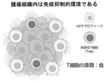
- FIG. 1 shows that tumor tissues are an immunosuppressive environment, with the presence of regulatory T cells and M2 macrophages and little infiltration of T cells.
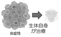
- FIG. 2 shows that when tumor tissue is placed in an inflammatory environment, the tumor tissue can shrink or disappear due to the effect of the individual's inherent immunity.
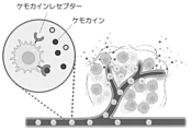
- FIG. 3 shows that macrophages migrate into tumor tissue and polarize into M2 macrophages within the tumor tissue.
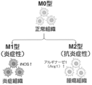
- FIG. 4 shows that macrophages, which are M0 in normal tissues, polarize to M2 type in tumor tissues.
- FIG. 5A shows a scheme for introducing a nucleic acid having a gene encoding a proinflammatory cytokine (e.g., TNF- ⁇ ) under the control of an M2-specific promoter into M0 macrophages, followed by induction of M2 macrophages and evaluation of the expression of a proinflammatory cytokine (e.g., TNF- ⁇ ).
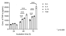
- Figure 5B shows that M0 macrophages treated with M2-inducing cytokines IL-4 or IL-13 differentiated into M2 macrophages and produced TNF- ⁇ in an M2-specific manner, and that M0 macrophages treated with cancer cell culture supernatant (TCM) differentiated into M2 macrophages and produced TNF- ⁇ in an M2-specific manner.
- TCM cancer cell culture supernatant
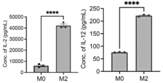
- FIG. 5C shows that M0 macrophages treated with the M2-inducing cytokines IL-4 or IL-13 differentiated into M2 macrophages and produced cytokines (IL-2 and IL-12) derived from exogenous genes operably linked to M2-specific promoters in an M2-specific manner.
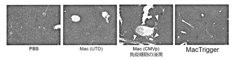
- FIG. 6A shows that M1 markers are enhanced in tumor tissues from the MacTrigger administration group.
- FIG. 6B shows an increase in the M1/M2 ratio in tumor tissues from the MacTrigger administration group.
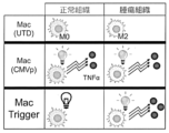
- FIG. 6C shows that MacTrigger polarizes M2 macrophages in tumor tissues, leading to the secretion of TNF- ⁇ in tumor tissues.
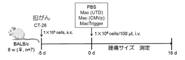
- FIG. 7A shows a scheme for administering MacTrigger to tumor-bearing mice and measuring tumor size.
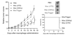
- FIG. 7B shows the change in tumor size over time and the survival curve in the MacTrigger administration group.
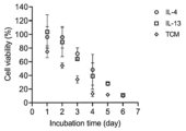
- FIG. 7C shows the results of an in vitro experiment showing the transition of cell viability of MacTrigger cultured in the presence of M2-inducing cytokines or TCM.
- FIG. 7D shows the expression of the cell proliferation marker Ki67 in tumor tissues from the MacTrigger administration group.
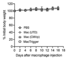
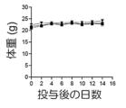
- FIG. 7E shows the time course of body weight change in the MacTrigger administration group.
- FIG. 8A is a scheme for determining the proportions of NK cells and cytotoxic T cells in tumor tissues in the MacTrigger administration group.
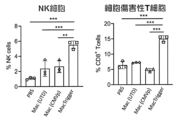
- FIG. 8B shows the percentages of NK cells and cytotoxic T cells in tumor tissues in the MacTrigger administration group.
- FIG. 8A is a scheme for determining the proportions of NK cells and cytotoxic T cells in tumor tissues in the MacTrigger administration group.
- FIG. 8B shows the percentages of NK cells and cytotoxic T cells in tumor tissues in the MacTrigger administration group.
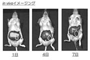
- FIG. 9A shows the dynamics of VT-680-labeled macrophages in the body of a mouse after administration of the VT-680-labeled macrophages.
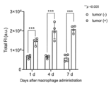
- FIG. 9B shows the abundance of VT-680-labeled macrophages in both non-tumor and tumor tissues of mice after administration of VT-680-labeled macrophages.
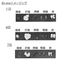
- FIG. 9C shows the abundance of VT-680-labeled macrophages in each mouse tissue after administration of VT-680-labeled macrophages.
- FIG. 10A shows an analysis scheme of cells in tumor tissues and livers of the macrophage-administered group.
- FIG. 10B shows AST activity in macrophage-injected mice.
- FIG. 10C shows the expression levels of CD206 (M2 marker) in tumor tissues and livers of the macrophage-administered group.
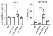
- FIG. 10D shows the presence or absence of liver hypertrophy and blood AST levels in the MacTrigger administration group.
- FIG. 10E shows the degree of immune cell infiltration in the liver of the MacTrigger administration group.
- 11A shows the effect of MacTrigger administration on tumors with larger sizes 10 days after tumor-bearing operation. The left panel shows the change in tumor volume over time, and the right panel shows the M1 macrophage/M2 macrophage ratio in the tumor.
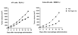
- Fig. 11B shows the effect of MacTrigger administration on tumor-bearing mice grafted with 4T1 cells or Colon-26 cells.
- FIG. 11B shows the time course of tumor volume after MacTrigger administration.
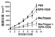
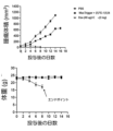
- Figure 12A shows the combined effect of MacTrigger and anti-PD-1 antibody in tumor-bearing mice grafted with tumor cells resistant to immune checkpoint inhibitors.
- Figure 12A shows the change in tumor volume over time after administration of MacTrigger.
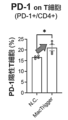
- FIG. 12B shows the change in the proportion of PD-1 positive cells in CD4 alone positive T cells after administration of MacTrigger.
- FIG. 12C shows the change in body weight after combined administration of MacTrigger and anti-PD-1 antibody in tumor-bearing mice grafted with tumor cells resistant to immune checkpoint inhibitors.
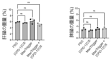
- Figure 12D shows the effect of combined use of MacTrigger and anti-PD-1 antibody on liver weight (left) and spleen weight (right) in tumor-bearing mice grafted with tumor cells resistant to immune checkpoint inhibitors.
- Figure 13 shows the effect of MacTrigger administration and combined administration of MacTrigger and anti-PD-1 antibody to triple-negative breast cancer cell-bearing mice.
- Figure 13 shows the change in tumor volume over time after administration.
- Figure 14A shows the ratio of gene expression levels in human macrophages (expression level in M2/expression level in M0). **** indicates p ⁇ 0.001.
- Figure 14B shows the ratio of gene expression levels in human macrophages (expression level in M2/expression level in M1). **** indicates p ⁇ 0.001.
- FIG. 14C shows the ratio of gene expression levels in human macrophages (expression level in M2/expression level in M0). **** indicates p ⁇ 0.001.
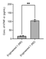
- FIG. 15 shows M2 macrophage-specific cytokine production by human MacTrigger.
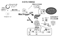
- FIG. 16 shows a schematic diagram of the cancer treatment strategy in this disclosure.
- a "subject” is a vertebrate, for example, a bird or a mammal, such as a mammal, for example, a mouse, rat, hamster, guinea pig, horse, cow, pig, goat, sheep, donkey, dog, and cat, as well as a primate, such as a monkey, chimpanzee, gorilla, orangutan, bonobo, or human, particularly a human.
- subject is used in the sense including humans as described above, and the term "non-human" is used to exclude humans.
- treatment can refer to prophylactic treatment and therapeutic treatment.
- Prophylactic treatment is used to include preventing the onset of a disease, delaying onset, and reducing the incidence of the disease.
- Therapeutic treatment is used to include reducing the rate of disease progression, delaying progression, preventing progression, reducing symptoms of a disease, curing a disease, and causing remission of a disease.
- Macrophages are a type of white blood cell that migrates in the body by ameboid motility. Macrophages have phagocytosis and can ingest foreign substances such as bacteria, viruses, and dead cells. Macrophages are one of the major immune cells and play an important role in inflammation and immune responses in tissues [27].
- macrophages especially in tumor tissues, are that (1) macrophages migrate to tumor tissues via C-C chemokine ligand 2 (CCL2) and granulocyte-macrophage colony-stimulating factor (GM-CSF) secreted by cancer cells and stromal cells [28,29], and (2) macrophages can be polarized to an anti-inflammatory M2 phenotype in tumor tissues [18,30,31]. Macrophages change to an M1 or M2 phenotype depending on the inflammatory state of the tissue environment, and control the inflammatory environment. M2 macrophages are involved in wound healing, tissue regeneration, and immunosuppression [30, 32].
- CCL2 C-C chemokine ligand 2
- GM-CSF granulocyte-macrophage colony-stimulating factor
- mouse M2 macrophages are known to have very high arginase 1 (Arg1) activity [33].
- Arg1 catalyzes the hydrolysis of arginine to urea and ornithine. This results in low amounts of arginine and promotes immunosuppression [34]. Because Arg1 is not highly expressed in M0 or M1 macrophages, Arg1 activity is one of the important markers for identifying the mouse M2 phenotype [35].
- M1 macrophages express one or more or all of the molecules selected from the group consisting of CD80, CD86, TLR-2, TLR-4, iNOS, and MHC-II, and preferably secrete one or more or all of the cytokines selected from the group consisting of interferon- ⁇ (TNF- ⁇ ), interleukin-1 ⁇ (IL-1 ⁇ ), IL-1 ⁇ , IL-6, IL-12, IL-23, CXCL9, CXCL16, and CCL5.
- TNF- ⁇ interferon- ⁇
- IL-1 ⁇ interleukin-1 ⁇
- IL-6 interleukin-1 ⁇
- IL-12 interleukin-12
- IL-23 CXCL9, CXCL16, and CCL5.
- M2 macrophages express one or more or all of the molecules selected from the group consisting of CD163, CD206, CD209, FIZZ1, and Ym1/2, and preferably secrete one or more or all of the cytokines selected from the group consisting of IL-10, TGF- ⁇ , CCL1, CCL17, CCL18, CCL22, CCL24, CXCL13, and VEGF.
- M1 macrophages are induced, for example, by LPS, granulocyte-macrophage colony-stimulating factor (GM-CSF), IFN- ⁇ , or TNF- ⁇ from monocytes, and M2 macrophages are induced, for example, by IL-4, IL-13, IL-10, or IL-21 from monocytes.
- M2a, M2b, M2c, and M2d are known for M2 macrophages.
- Human M2 macrophage markers include IDO, IL-10, TGF- ⁇ , CD115, CD204, CD163, CD206, CD209, FceR1, VSIG4, IRF4, and STAT6.
- human M2 macrophages can be identified by one or more, two or more, three or more, four or more, five or more, six or more, seven or more, eight or more, nine or more, ten or more, eleven or more, or twelve or more selected from the group consisting of IDO, IL-10, TGF- ⁇ , CD115, CD204, CD163, CD206, CD209, FceR1, VSIG4, IRF4, and STAT6.
- Naive macrophages (macrophages that have not been stimulated) are called M0 macrophages, and M0 macrophages reversibly polarize into M1 macrophages and M2 macrophages.
- M0 macrophages differentiate from monocytes.
- M0 macrophages can be obtained by stimulating monocytes with phorbol 12-myristate 13 acetate (PMA) or macrophage colony-stimulating factor (M-CSF).
- PMA phorbol 12-myristate 13 acetate
- M-CSF macrophage colony-stimulating factor
- M0 macrophages can be characterized by negativity for markers of M1 macrophages and markers of M2 macrophages.
- control sequence refers to a sequence that has the activity of driving a gene operably linked thereto and transcribing RNA from the gene.
- the control sequence is, for example, a promoter.
- promoters include class I promoters (which can be used to transcribe rRNA precursors), class II promoters (which are composed of a core promoter and an upstream promoter element and can be used to transcribe mRNA), and class III promoters (which are further broadly classified into types I, II, and III).
- the control sequence is also called a regulatory sequence, and may be any promoter that can transcribe mRNA in cells such as animal cells and plant cells. For example, various pol II promoters can be used as the first control sequence.
- pol II promoters include, but are not limited to, CMV promoter, EF1 promoter (EF1 ⁇ promoter), SV40 promoter, MSCV promoter, hTERT promoter, ⁇ -actin promoter, CAG promoter, and CBh promoter.
- the promoter may also include promoters that drive RNA polymerase derived from bacteriophage, such as the T7 promoter, T3 promoter, and SP6 promoter, and pol III promoters, such as the U6 promoter.
- the T7 promoter is preferably used for transcription from circular DNA
- the SP6 promoter is preferably used for transcription from linear DNA.
- the promoter may also be an inducible promoter.
- An inducible promoter is a promoter that can induce the expression of a polynucleotide operably linked to the promoter only in the presence of an inducer that drives the promoter. Some inducible promoters can induce the expression of a polynucleotide operably linked to the promoter only in the absence of an inhibitor that suppresses the activity of the promoter. Inducible promoters include, but are not limited to, promoters that induce gene expression by heating, such as heat shock promoters. Inducible promoters also include promoters that can be driven by drugs.
- Examples of such drug-inducible promoters include cumate operator sequences, lambda operator sequences (e.g., 12 ⁇ lambda Op), and tetracycline-inducible promoters.
- Tetracycline-inducible promoters include, for example, promoters that drive gene expression in the presence of tetracycline or its derivatives (e.g., doxycycline) or reverse tetracycline-controlled transactivator (rtTA).
- An example of a tetracycline-inducible promoter is the TRE3G promoter.
- a control sequence can cause a gene operably linked thereto to be expressed in at least M2 macrophages. By operably linked, it is meant that the gene is linked to a control sequence such that gene expression is driven by the control sequence.
- a "cell” refers to a vertebrate cell, preferably a mammalian cell, and more preferably a human cell.
- a cell capable of differentiating/polarizing into an M2 macrophage is a hematopoietic stem cell, a myeloid precursor cell, a monocyte-dendritic cell precursor cell, a common monocyte precursor cell, a monocyte, or an M0 macrophage, or any cell in the differentiation process.
- hematopoietic stem cell a myeloid precursor cell
- a monocyte-dendritic cell precursor cell a common monocyte precursor cell
- monocyte or an M0 macrophage, or any cell in the differentiation process.
- hematopoietic stem cells differentiate into monocytes in the bone marrow via myeloid precursor cells, monocyte-dendritic cell precursor cells, and a common monocyte precursor cell.
- the differentiated monocytes differentiate into macrophages in peripheral tissues.
- the macrophages migrate to tumor tissue and polarize into M2 macrophages.
- differentiation/polarization potential refers to differentiation potential or polarization potential, or differentiation potential and polarization potential.
- differentiation/polarization refers to differentiation or polarization, or differentiation and polarization.
- Polarization is used to refer to reversible changes between M0, M1, and M2. Differentiation is used to refer to changes in cell stages other than polarization.
- the cell capable of differentiating/polarizing into an M2 macrophage comprises a nucleic acid operably linked to a regulatory sequence, more preferably a nucleic acid encoding an inflammatory cytokine.
- a cell comprising a nucleic acid encoding an inflammatory cytokine operably linked to a regulatory sequence. In a preferred embodiment, these cells are capable of expressing said inflammatory cytokine after differentiation/polarization into an M2 macrophage.
- a cell capable of differentiating/polarizing into M2 macrophages which comprises a nucleic acid encoding an inflammatory cytokine operably linked to a control sequence and can express the inflammatory cytokine after differentiation/polarization into M2 macrophages.
- Such cells can differentiate/polarize into M2 macrophages in tumor tissue, thereby expressing an inflammatory cytokine in the tumor tissue, alleviating the anti-inflammatory state of the tumor tissue, and preferably changing the tumor tissue to an inflammatory environment (or an environment in which the ratio of M1 macrophage number/M2 macrophage number (M1 number/M2 number ratio)>1).
- NK cells or T cells By changing the tumor tissue to an inflammatory environment, NK cells or T cells can be recruited to the tumor tissue, and an anti-tumor effect on the tumor tissue can be exerted or enhanced.
- the anti-tumor effect on the tumor tissue in a subject receiving the cells of the present disclosure is preferably mainly due to activation of the subject's immune system other than the administered cells (specifically, recruitment and activation of immune cells such as NK cells and T cells possessed by the subject).
- the cell capable of differentiation/polarization into M2 macrophages is any cell selected from the group consisting of M0 macrophages, monocytes, common monocyte progenitors, monocyte-dendritic cell progenitors, myeloid progenitors, and hematopoietic stem cells, as well as any cell in the differentiation process from hematopoietic stem cells to M0 macrophages.
- the cell capable of differentiation/polarization into M2 macrophages may be a cell having a differentiation state between hematopoietic stem cells and myeloid progenitors.
- the cell capable of differentiation/polarization into M2 macrophages may be a cell having a differentiation state between myeloid progenitors and monocyte-dendritic cell progenitors. In a preferred embodiment, the cell capable of differentiation/polarization into M2 macrophages may be a cell having a differentiation state between monocyte-dendritic cell progenitors and common monocyte progenitors. In a preferred embodiment, the cell capable of differentiation/polarization into M2 macrophages may be a cell having a differentiation state between common monocyte progenitors and monocytes.
- the cells capable of differentiating/polarizing into M2 macrophages may be cells having a differentiation state between monocytes and M0 macrophages.
- the cells capable of differentiating/polarizing into M2 macrophages may be cells at any stage of differentiation from monocytes to M0 macrophages, and more preferably, M0 macrophages.
- the cells capable of differentiating/polarizing into M2 macrophages may be iNOS, CD206, and Arg-1 negative macrophages (i.e., non-M1 macrophages and non-M2 macrophages).
- the inflammatory cytokine can alleviate (reduce) the suppressive inflammation state of the tumor tissue.
- the inflammatory cytokine can change the tumor tissue from an immunosuppressive environment to an inflammatory environment.
- the inflammatory cytokine is an inflammatory cytokine selected from the group consisting of tumor necrosis factor- ⁇ (TNF- ⁇ ), IFN- ⁇ , IL-1 (e.g., IL-1 ⁇ ), IL-2, IL-6, IL-12, IL-17, and IL-23.
- the inflammatory cytokine is an inflammatory cytokine selected from the group consisting of tumor necrosis factor- ⁇ (TNF- ⁇ ), IFN- ⁇ , IFN- ⁇ , IL-1 (e.g., IL-1 ⁇ ), IL-2, IL-6, IL-8, IL-12, IL-17, and IL-23.
- TNF- ⁇ tumor necrosis factor- ⁇
- IFN- ⁇ IFN- ⁇
- IFN- ⁇ e.g., IL-1 ⁇
- IL-2 e.g., IL-1 ⁇
- IL-6 IL-6
- IL-8 IL-12
- IL-17 IL-17
- IL-23 tumor necrosis factor- ⁇
- the inflammatory cytokine can be TNF- ⁇ .
- Human TNF- ⁇ may have, for example, a sequence registered in GenBank: QCI55793.1, or a TNF- ⁇ sequence corresponding to the sequence.
- Human TNF- ⁇ includes, for example, a peptide having 90% or more sequence identity with a sequence registered in GenBank: QCI55793.1 and having the function of human TNF- ⁇ .
- the function of human TNF- ⁇ may be the function of inducing inflammation in a human subject.
- the inflammatory cytokine may be any cytokine capable of inducing inflammation in humans, with no particular limitations, but may preferably be a human cytokine.
- control sequence is capable of expressing a gene operably linked to the control sequence, such as an inflammatory cytokine, in M2 macrophages.
- control sequence is capable of expressing a gene operably linked to the control sequence, such as an inflammatory cytokine, selectively or specifically in M2 macrophages.
- selectively and specifically in M2 macrophages means driving stronger gene expression (2-fold or more, 3-fold or more, 4-fold or more, 5-fold or more, 6-fold or more, 7-fold or more, 8-fold or more, 9-fold or more, 10-fold or more, 20-fold or more, 30-fold or more, 40-fold or more, 50-fold or more, 100-fold or more, 500-fold or more, or 1000-fold or more) in M2 macrophages than in at least M0 macrophages and M1 macrophages.
- the regulatory sequence is a regulatory sequence of a gene consisting of the group consisting of CD163, CD200 R1, CD301, CXCR1, CXCR2, CD209, Dectin-1, Fce RIa, IL-1RII, CD206, Arginase 1, IFR4, PPAR ⁇ , STAT6, FIZZI, IL-1ra, IL-10, TGF- ⁇ , CCL1, CCL14, CCL18, CCL22, CCCL23, CCL24, CCL26, and YM1.
- the regulatory sequence may be a promoter of a gene selected from the group consisting of CCL-22, LOX, CISH, FCER15, ALOX15, F13A1, IDO1, MMP1, ALDH1A2, CD209, TGM2, VCAM1, MMP12, and SPINT2.
- the regulatory sequence may be a promoter of a gene selected from the group consisting of CCL-22, CISH, ALOX15, and TGM2.
- the regulatory sequence is preferably derived from the human genome.
- the regulatory sequence may be a promoter of a human gene preferably selected from the group consisting of CCL-22, CISH, ALOX15, and TGM2.
- the regulatory sequence may be CCL-22.
- the regulatory sequence may be CD209.
- the present disclosure provides a cell having any differentiation stage from monocyte to M0 macrophage, the cell comprising a nucleic acid encoding an inflammatory cytokine operably linked to an M2 macrophage selective or specific control sequence, and capable of expressing said inflammatory cytokine after differentiation/polarization to M2 macrophages.
- the present disclosure provides a cell having any differentiation stage from monocyte to M0 macrophage, the cell comprising a nucleic acid encoding TNF- ⁇ operably linked to an M2 macrophage selective or specific control sequence, and capable of expressing said TNF- ⁇ after differentiation/polarization to M2 macrophages.
- the cell is a monocyte or M0 macrophage.
- the cell is a macrophage that is non-M1 and non-M2.
- the cell is, for example, an iNOS, CD206, and Arg-1 negative macrophage.
- the present disclosure provides an M0 macrophage, the M0 macrophage comprising a nucleic acid encoding an inflammatory cytokine operably linked to an M2 macrophage-selective or specific control sequence, and capable of expressing said inflammatory cytokine after differentiation/polarization into an M2 macrophage.
- the present disclosure provides an M0 macrophage, the M0 macrophage comprising a nucleic acid encoding TNF- ⁇ operably linked to an M2 macrophage-selective or specific control sequence, and capable of expressing said TNF- ⁇ after differentiation/polarization into an M2 macrophage.
- the cells of the present disclosure release inflammatory cytokines in tumor tissue.
- the cells of the present disclosure cause the ratio of M1 macrophage numbers/M2 macrophage numbers (M1 number/M2 number ratio) in tumor tissue to 1 or more.
- the cells of the present disclosure change tumor tissue to an inflammatory environment.
- the cells of the present disclosure increase immune cells such as NK cells and/or T cells in tumor tissue.
- compositions comprising the cells of the present disclosure.
- the composition of the present disclosure may comprise, in addition to the cells, a pharma- ceutically acceptable solution.
- the composition of the present disclosure may comprise, in addition to the cells, a pharma- ceutically acceptable additive (e.g., pharmaceutical additives such as salts, buffers, thickeners, isotonicity agents, dispersants, etc.).
- the composition of the present disclosure may comprise, in addition to the cells, a pharma- ceutically acceptable solution and a pharma-ceutically acceptable additive (e.g., salts, buffers, thickeners, isotonicity agents, dispersants, etc.).
- a composition for use in releasing inflammatory cytokines in tumor tissue comprising the cells of the present disclosure.
- a composition for use in changing tumor tissue to an inflammatory environment comprising the cells of the present disclosure.
- a composition for use in increasing the number of M1 macrophages/the number of M2 macrophages in tumor tissue to 1 or more the composition comprising the cells of the present disclosure.
- a composition for use in increasing immune cells such as NK cells and/or T cells in tumor tissue, the composition comprising the cells of the present disclosure.
- the compositions of the present disclosure can be used to treat cancer. In some embodiments, the compositions of the present disclosure can be used to treat a cancer patient. In some embodiments, the compositions of the present disclosure can be used to alleviate an anti-inflammatory state in cancer tissue in a cancer patient. In some embodiments, the compositions of the present disclosure can be used to change an anti-inflammatory state in cancer tissue in a cancer patient to an inflammatory state. In some embodiments, the compositions of the present disclosure can be used to recruit immune cells to cancer tissue in a cancer patient. In some embodiments, the compositions of the present disclosure can be used to treat (e.g., therapeutically treat) cancer in a cancer patient.
- Cancers include, but are not limited to, blood cancers such as acute lymphoblastic leukemia (ALL), acute myeloid leukemia (AML), Hodgkin's lymphoma, non-Hodgkin's lymphoma, B-cell lymphoma, multiple myeloma, and T-cell lymphoma, melanoma, myelodysplastic syndrome, adenocarcinoma, squamous cell carcinoma, adenosquamous carcinoma, undifferentiated carcinoma, large cell carcinoma, non-small cell lung cancer, small cell lung cancer, mesothelioma, skin cancer, breast cancer (including triple-negative breast cancer), prostate cancer, bladder cancer, vaginal cancer, cervical cancer, head and neck cancer, and uterine cancer.
- ALL acute lymphoblastic leukemia
- AML acute myeloid leukemia
- Hodgkin's lymphoma non-Hodgkin's lymphoma
- B-cell lymphoma multiple mye
- solid cancers such as cervical cancer, liver cancer, gallbladder cancer, bile duct cancer, kidney cancer, pancreatic cancer, lung cancer, colon cancer, large intestine cancer, rectal cancer, small intestine cancer, stomach cancer, esophageal cancer, testicular cancer, ovarian cancer, bladder cancer, and brain tumors, as well as cancers of bone tissue, cartilage tissue, fat tissue, muscle tissue, vascular tissue, and hematopoietic tissue, as well as sarcomas such as chondrosarcoma, Ewing's sarcoma, malignant hemangioendothelioma, malignant schwannoma, osteosarcoma, and soft tissue sarcoma, and blastomas such as hepatoblastoma, medulloblastoma, nephroblastoma, neuroblastoma, pancreatic blastoma, pleuropulmonary blastoma, and retinoblastoma.
- solid cancers such
- composition or cells of the present disclosure may be administered parenterally to a subject.
- parenteral administration include, but are not limited to, intravenous administration, intraarterial administration, intramuscular administration, intradermal administration, subcutaneous administration, intraperitoneal administration, intrathoracic administration, intraarticular administration, intramyocardial administration, intraventricular administration, intramedullary administration, intrathecal administration, and intraspinal fluid administration.
- the parenteral administration may be intravenous administration.
- the present disclosure provides a method of treating cancer in a subject in need thereof, the method comprising administering to the subject a therapeutically effective amount of a cell of the present disclosure.
- the cancerous tissue of the subject transitions to an inflammatory environment.
- the present disclosure provides a method of treating a subject having cancer, the method comprising administering to the subject a therapeutically effective amount of a cell of the present disclosure.
- the cancer tissue of the subject transitions to an inflammatory environment.
- a method for producing inflammatory cytokines in tumor tissue of a subject having cancer comprising administering to the subject an effective amount of cells of the present disclosure.
- a method for alleviating an anti-inflammatory state in tumor tissue of a subject having cancer comprising administering to the subject an effective amount of cells of the present disclosure.
- a method for changing the tumor tissue of a subject having cancer e.g., from an anti-inflammatory state
- the method comprising administering to the subject an effective amount of cells of the present disclosure.
- a method for increasing the M1 macrophage count/M2 macrophage count to 1 or more in tumor tissue of a subject having cancer the method comprising administering to the subject an effective amount of cells of the present disclosure.
- the subject with cancer may be a subject before, during, or after immunotherapy.
- the subject with cancer may also be treated with a combination of administration of the cells of the present disclosure and cancer therapy.
- the subject with cancer is administered an effective amount of the cells of the present disclosure and receives cancer therapy.
- Administration of an effective amount of the cells of the present disclosure to the subject with cancer ensures that the tumor becomes an inflammatory tumor, to which the body's immune system can respond and launch an attack on the tumor.
- administration of an effective amount of the cells of the present disclosure to the subject with cancer may make any combination of cancer immunotherapy or antibody therapy more effective.
- the cancer therapy may be, for example, antibody therapy.
- an antibody that binds to a cancer antigen and has antibody-dependent cellular cytotoxicity (ADCC activity) or complement-dependent cytotoxicity (CDC activity) may be administered to the cancer patient.
- Administration of the cells of the present disclosure may convert a non-inflammatory tumor into an inflammatory tumor, thereby promoting the recruitment of NK cells to the tumor tissue, thereby allowing the antibody bound to the cancer cell to exert its ADCC activity in the tumor tissue.
- the antibody may bind to the cancer antigen with a binding dissociation constant (KD) of, for example, 10 ⁇ 7 M or less, 10 ⁇ 8 M or less, 10 ⁇ 9 M or less, 10 ⁇ 10 M or less, or 10 ⁇ 11 M or less, or 10 ⁇ 8 M to 10 ⁇ 12 M.
- KD binding dissociation constant
- the cancer immunotherapy may be, for example, T cell adoptive immunotherapy, chimeric antigen receptor (CAR)-expressing immune cell therapy, or immune checkpoint inhibitor therapy.
- the T cell adoptive immunotherapy comprises administering an effective amount of T cells that bind to the cancer antigen to a subject with cancer.
- the T cells may be those proliferated by cell culture, but may also be T cells rejuvenated through reprogramming to iPS cells.
- T cells that bind to cancer antigens may be obtained from a cancer patient, reprogrammed to obtain iPS cells, and the iPS cells may be differentiated to obtain T cells (see, for example, US9,206,3934B).
- T cells may be T cells with homogeneous T cell receptors (TCRs) by cloning iPS cells and/or suppressing or stopping the expression of RAG1 and 2 (see, for example, US9,206,3934B).
- Chimeric antigen receptor (CAR)-expressing immune cell therapy involves administering an effective amount of CAR immune cells (e.g., CAR-expressing T cells and CAR-expressing NK cells, etc.) that bind to cancer antigens to a subject.
- CAR immune cells e.g., CAR-expressing T cells and CAR-expressing NK cells, etc.
- Immune checkpoint inhibitor therapy involves administering an effective amount of an immune checkpoint inhibitor to a subject with cancer.
- Immune checkpoint inhibitors release the suppression of the immune system, i.e., release the brakes, but are considered to be less effective when the immune system is not activated in the first place.
- the administration of an effective amount of the cells of the present disclosure can increase the inflammation of tumors and activate the immune system against tumors, and is therefore expected to improve the efficacy of immune checkpoint inhibitors.
- a cell or cancer immunotherapy agent of the present disclosure for use in a method for treating cancer comprising administering an effective amount of a cell of the present disclosure and cancer immunotherapy.
- a cell or cancer immunotherapy agent of the present disclosure or both, in the manufacture of a medicament for use in a method for treating cancer comprising administering an effective amount of a cell of the present disclosure and cancer immunotherapy.
- the antibodies include, but are not limited to, rituximab, trastuzumab, cetuximab, alemtuzumab, ofatumumab, obinutuzumab, daratumumab, etotuzumab, and avelumab.
- humanized or human chimeric IgG1 antibodies that bind to cancer antigens may be administered in antibody therapy.
- CAR-expressing cells include CAR-expressing immune cells.
- Immune cells are not particularly limited, but include, for example, T cells, NK cells, neutrophils, macrophages, etc.
- "Chimeric antigen receptor" (CAR) is a chimeric molecule having an antigen-binding fragment of an antibody (particularly, scFv) that binds to a cancer antigen and an activation domain of an immune cell.
- CAR is generally a molecule that is formed by linking an scFv, an extracellular hinge domain, a transmembrane domain (e.g., CD8 ⁇ or CD28), and an activation signaling domain (e.g., CD3 ⁇ ).
- CAR can be introduced into cells and expressed on the cell surface.
- CAR can target specific antigens.
- CAR can be introduced into immune cells such as T cells or NK cells, and target immune cells such as T cells or NK cells to cancer.
- the scFv, the extracellular hinge domain, the transmembrane domain (e.g., CD8 ⁇ or CD28), and the activation signaling domain (e.g., CD3 ⁇ ) are linked, but the second generation CAR further includes a costimulatory molecule signaling domain for activating the immune cells into which the CAR is introduced.
- Costimulatory factors such as CD28, 4-1BB, OX40, CD27, and ICOS are used as the costimulatory molecule signaling domain.
- the antigen of the antibody or the antigen-binding fragment of the antibody may be an antigen (e.g., a cancer antigen) expressed on the surface of a cancer cell.
- cancer antigens include CD16, CD19, CD20, CD22, CD123, CD171, epidermal growth factor receptor (EGFR), particularly EGFRvIII, EGFR type 3, de2-7EGFR and HER2, carcinoembryonic antigen (CEA), prostate stem cell antigen (PSCA), B cell maturation antigen (BCMA), CS1, NKG2D, NKp30, B7H6, MUC-16 (CA125), receptor tyrosine kinase (KK) and tyrosine kinase (KKR).
- the antibody or antigen-binding fragment of an antibody may be, for example, a proteinaceous tag.
- the antibody or antigen-binding fragment of an antibody may be, for example, a fluorescent protein such as fluorescein isothiocyanate (FITC).
- CAR-T therapeutics approved by the U.S. Food and Drug Administration include: Abekuma (generic name: idecabutagen bikrucel), Breyandi (generic name: lysocabtagene maralucel), Carvykti (generic name: siltacabtagene autolucel), Kymriah (generic name: tisagenlukrucel), Tecartus (generic name: brexcabtagene autolucel), and Yescarta (generic name: axicabtagene silollucel). It is believed that other CAR-T therapeutics may also be advantageously combined with administration of the cells of the present disclosure.
- Immune checkpoint inhibitors can, for example, bind to immune checkpoint molecules or their ligands and inhibit the function of immune checkpoints.
- immune checkpoint molecules include programmed cell death-1 (PD-1), cytotoxic T-lymphocyte-associated protein 4 (CTLA-4), T-cell immunoglobulin domain and mucin domain-3 (TIM-3), and lymphocyte activation inhibitors (IL-1, IL-2, and IL-3).
- PD-1 programmed cell death-1
- CTL-4 cytotoxic T-lymphocyte-associated protein 4
- TIM-3 T-cell immunoglobulin domain and mucin domain-3
- IL-1, IL-2, and IL-3 lymphocyte activation inhibitors
- LAG-3 lymphocyte activation gene 3
- VISTA V-type immunoglobulin domain-containing suppressor of T-cell activation
- SIRP ⁇ dendritic cell-specific protein signal regulatory protein alpha
- the immune checkpoints that each of them is responsible for are called the PD-1 immune checkpoint, the CTLA-4 immune checkpoint, the TIM-3 immune checkpoint, the LAG-3 immune checkpoint, and the VISTA immune checkpoint, and the SIRP ⁇ immune checkpoint.
- the PD-1 immune checkpoint can be inhibited by inhibiting the binding of PD-1 to PD-L1 or PD-L2.
- the CTLA-4 immune checkpoint can be inhibited by inhibiting the binding between CTLA-4 and CD80 or CD86.
- the TIM-3 immune checkpoint can be inhibiting the binding between TIM-3 and galectin-9.
- the LAG-3 immune checkpoint can be inhibited by inhibiting the binding between LAG-3 and MHC class II molecules.
- the VISTA immune checkpoint can be inhibited by inhibiting the binding between VISTA and VSIG-3/IGSF11.
- the SIRP ⁇ immune checkpoint can be inhibited by inhibiting the binding between SIRP ⁇ and CD47.
- one or more immune checkpoints selected from the group consisting of the PD-1 immune checkpoint, the CTLA-4 immune checkpoint, the TIM-3 immune checkpoint, the Lag3 immune checkpoint, and the VISTA immune checkpoint, as well as the SIRP ⁇ immune checkpoint can be inhibited.
- An antibody that inhibits the binding of two proteins can bind to a receptor or a ligand.
- the antibody that inhibits the PD-1 immune checkpoint may be an antibody selected from the group consisting of anti-PD-1 antibodies, anti-PD-L1 antibodies, and anti-PD-L2 antibodies (e.g., nivolumab, pemprolizumab, avelumab, atezolizumab, and durvalumab).
- the antibody that inhibits the CTLA-4 immune checkpoint may be an antibody selected from the group consisting of anti-CDLA-4 antibodies, anti-CD80 antibodies, and anti-CD86 antibodies (e.g., ipilimumab and tremelimumab).
- the antibody that inhibits the TIM-3 immune checkpoint may be an antibody selected from the group consisting of anti-TIM-3 antibodies and anti-galectin-9 antibodies (e.g., MGB453).
- the antibody that inhibits the VISTA immune checkpoint may be an antibody selected from the group consisting of anti-VISTA antibodies and anti-VSIG-3/IGSF11 antibodies (e.g., JNJ-61610588).
- the antibody that inhibits the SIRP ⁇ immune checkpoint can be an antibody selected from the group consisting of an anti-SIRP ⁇ antibody and an anti-CD47 antibody (e.g., magrolimab, CC-90002).
- SIRP ⁇ immune checkpoint proteins that bind the CD47-binding domain of human SIRP ⁇ to the Fc region of human IgG1 and inhibit the binding between SIRP ⁇ and CD47 (e.g., ontropacept, maprilpacept, evolpacept).
- the subject preferably has cancer cells that express immune checkpoint molecules. Even if the subject's tumor is in an immunosuppressed state (i.e., a non-inflammatory tumor (cold tumor)), the tumor can be converted to an inflammatory tumor (hot tumor), and therefore any subject, including a subject with a non-inflammatory tumor or a subject with a tumor resistant to immune checkpoint inhibitors, may be suitable for immune checkpoint inhibitors.
- the subject is a subject that is resistant to immune checkpoint inhibitors.
- a subject that is resistant to immune checkpoint inhibitors may be appropriately identified by a medical professional such as a physician based on the course of treatment after administration.
- a method of treating cancer in a subject having cancer cells that do not express or barely express immune checkpoint molecules includes administering to the subject an effective amount of the cells of the present disclosure and administering to the subject an effective amount of an immune checkpoint inhibitor.
- the order of administration of the cells and the immune checkpoint inhibitor does not matter, but preferably, the cells are administered first, followed by the immune checkpoint inhibitor.
- the administration of the cells of the present disclosure is believed to activate the immune of the cancer cells of the subject receiving the cells and/or enhance the expression of immune checkpoint molecules in the cancer cells, thereby improving the effectiveness of the immune checkpoint inhibitor.
- the cells are administered to the subject, and an immune checkpoint inhibitor may be administered to the subject in which enhanced expression of immune checkpoint molecules in the cancer cells has been observed.
- Enhanced expression of immune checkpoint molecules in cancer cells can be confirmed by routine methods such as immunohistochemical staining, flow cytometry, and enzyme-linked immunosorbent assay (ELISA). If a subject does not express or barely expresses immune checkpoint molecules despite administration of the cells of the present disclosure (i.e., an immune checkpoint inhibitor is ineffective), it is not necessary to administer an immune checkpoint inhibitor to the subject.
- the present disclosure provides a cell of the present disclosure for use in the above method.
- the present disclosure provides a composition of the present disclosure for use in the above method.
- the present disclosure provides for the use of cells of the present disclosure in the manufacture of a medicament for use in the above method.
- the present disclosure provides for the use of cells of the present disclosure in the manufacture of a medicament for use in treating cancer.
- LightCycler FastStart DNA Master SYBR Green Kit and Liberase DH Research Grade were purchased from Roche Diagnostics (Basel, Switzerland).
- Vivo-Track 680 was purchased from PerkinElmer, Inc.
- PE Phycoerythrin
- brilliant violet (BV) 421-conjugated anti-mouse F4/80 antibody Alexa Fluor 700-conjugated anti-mouse CD86 antibody, Alexa Fluor 488-conjugated anti-mouse CD11b antibody, FITC (fluorescein isothiocyanate)-conjugated anti-mouse NKp46 antibody, PE-conjugated anti-mouse NK1.1 antibody, Alexa Fluor 700-conjugated anti-mouse CD3 antibody, Alexa Fluor 488-conjugated anti-mouse CD8 antibody, Enzyme Linked Immuno Sorbent Assay (ELISA) MAX TM Deluxe Set Mouse TNF- ⁇ , and Stop Solution for TMB Substrate were purchased from BioLegend, Inc.
- ELISA Enzyme Linked Immuno Sorbent Assay
- DAB 3,3-Diaminobenzidine
- HRP-conjugated rabbit antibodies were purchased from Dako, Inc. (Glostrup, Denmark).
- pGL3-mArg1 promoter/enhancer (-31/-3810) was a gift from Peter Murray (Addgene plasmid # 34571; http://n2t.net/addgene:34571; RRID: Addgene_34571) [36]. All other chemicals were of the highest grade commercially available.
- RAW264.7 mouse macrophages and CT-26 mouse colon carcinoma cells were purchased from the American Type Culture Collection (ATCC).
- ATCC American Type Culture Collection
- CT-26 cells were cultured in RPMI-1640. All media were supplemented with 10% heat-inactivated FBS and 1% antibiotic-antimicrobial stock solution.
- Cells were cultured at 37°C in an atmosphere containing 5% CO2 and 95% air. Culture medium was changed every 2 days, and cells that reached 90% confluence were detached with a cell scraper and recultured.
- THP-1 human monocytic cells were purchased from JCRB. THP-1 were cultured in RPMI 1640. The medium was supplemented with 10% heat-inactivated FBS and 1% antibiotic-antimicrobial stock solution. Cells were cultured at 37°C in an atmosphere containing 5% CO2 and 95% air. The culture medium was changed every 2 days, and cells that reached 90% confluence were harvested and recultured.
- RAW264.7 cells were plated at a density of 1 ⁇ 106 cells/well in 2.5 mL of culture medium in 6-well plates. After 24 h, the medium was replaced with tumor culture medium (TCM) prepared by culturing CT-26 (3 ⁇ 106 cells, 2.5 mL culture medium) for 24 h in 6-well plates, and the other wells were supplemented with 25 ng/mL IL-4 or IL-13, 200 ng/mL LPS, and 100 ng/mL TNF- ⁇ .
- TCM tumor culture medium
- RNA ribonucleic acid
- RNAiso Plus ribonucleic acid
- the LightCycler FastStart DNA Master SYBR Green kit was used for PCR amplification reactions. The following PCR conditions were used.
- Glyceraldehyde-3-phosphate dehydrogenase (GAPDH) was used as a housekeeping gene, and the relative expression levels of the target genes were determined by the comparative CT method [37].
- Primers for Arg1 and GAPDH were purchased from Fasmac. All primer sequences are shown in Table 1 [33,38].
- RNA sequence data from human THP-1-induced macrophages was selected and output from the NCBI data bank GEO Data sets (https://www.ncbi.nlm.nih.gov/). After converting it into a text file, heat map analysis was performed using iDEP.96 (http://bioinformatics.sdstate.edu/idep96/).
- THP-1 cells were differentiated into macrophages by adding 20 ng/mL phorbol 12-myristate 13-acetate (PMA) to the culture medium.
- PMA phorbol 12-myristate 13-acetate
- THP-1 cells were cultured in 2.5 mL of culture medium at a density of 5 ⁇ 106 cells/well in 6-well plates.
- macrophages were stimulated with IL-4 and IL-13 (25 ng/mL each) to polarize them into M2 macrophages.
- LPS 200 ng/mL
- IFN- ⁇ 100 ng/mL
- RNAiso Plus ribonucleic acid
- the LightCycler FastStart SNA Master SYBR Green kit was used. The following PCR conditions were used. Glyceraldehyde-3-phosphate dehydrogenase (GAPDH) was used as the housekeeping gene, and the relative expression levels of the target genes were determined by the comparative CT method.
- the primers are as follows:
- IVIS Spectrum IVIS Lumina II, Xenogen Co., Ltd.
- IVIS Lumina II Xenogen Co., Ltd.
- blood was collected using an animal lancet 4 and 7 days after macrophage administration.
- tumors, liver, spleen, kidneys, and lungs were extracted and macrophage accumulation was evaluated using IVIS Spectrum.
- Blood AST activity was analyzed using an AST assay kit according to the manufacturer's protocol.
- Livers were homogenized using a plunger on a 100 ⁇ m filter and suspended in cold DPBS. After centrifugation at 200 ⁇ g for 5 min, 4 mL of VersaLyse was added and incubated at room temperature for 5 min. After incubation, the solution was centrifuged at 200 ⁇ g for 5 min and resuspended in FACS buffer. After resuspension, fluorescently labeled antibodies (Alexa Fluor 488-labeled anti-mouse CD11b antibody, BV421-labeled anti-mouse F4/80 antibody, PE-labeled anti-mouse CD206 antibody) were added and incubated at 4 °C for 60 min.
- fluorescently labeled antibodies Alexa Fluor 488-labeled anti-mouse CD11b antibody, BV421-labeled anti-mouse F4/80 antibody, PE-labeled anti-mouse CD206 antibody
- CD206 M2 macrophage marker
- flow cytometry Beckman Coulter, CytoFLEX
- mouse TNF- ⁇ as an inflammatory cytokine was purchased from Integrated DNA Technology, Inc (Coralville, IA, USA) and cloned downstream of the M2 macrophage-specific Arg1 promoter to generate pcDNA3.1-mArg1p-TNF- ⁇ . Furthermore, the mouse TNF- ⁇ gene and enhanced green fluorescent protein (EGFP) coding sequence were inserted downstream of the CMV promoter to generate pcDNA3.1-CMVp-TNF- ⁇ and pcDNA3.1-CMVp-EGFP, respectively. Plasmid DNA was extracted using NucleoBond Xtra Maxi Plus EF.
- EGFP enhanced green fluorescent protein
- RAW264.7 cells expressed TNF- ⁇ in response to the Arg1 promoter (Arg1p-TNF ⁇ ), CMV promoter (CMVp-TNF ⁇ ), or EGFP in response to the CMV promoter (CMVp-EGFP).
- RAW264.7 cells were seeded at a density of 2.0 ⁇ 105 cells/well in 24-well plates. After 24 hours, Arg1p-TNF- ⁇ , CMVp-TNF- ⁇ , or CMVp-EGFP vectors were transfected into RAW264.7 cells using Lipofectamine 3000 reagent according to the manufacturer's protocol. Six hours after transfection, the medium was replaced with fresh medium. The cells were treated with 400 ⁇ g/mL G418 to obtain stable cell lines.
- Stable cell lines expressing IL-1 ⁇ (NCBI Reference Sequence: NP_032387.1), IL-2 (NCBI Reference Sequence: NP_032392.1), IL-6 (GenBank: ABG81953.1), IL-12 (IL-12 ⁇ subunit: NCBI Reference Sequence: NP_001152896.2, IL-12 ⁇ subunit: NCBI Reference Sequence: NP_001290173.1), and IFN- ⁇ (NCBI Reference Sequence: NP_032363.1) were generated in the same manner, except that TNF- ⁇ was replaced by IL-1 ⁇ .
- the untransfected macrophages, CMVp-TNF- ⁇ transfected macrophages, CMVp-EGFP transfected macrophages, and Arg1p-TNF- ⁇ transfected macrophages were named Mac(UTD), Mac(CMVp), Mac(EGFP), and MacTrigger, respectively.
- a human MacTrigger was also created. Specifically, the following procedure was followed: A linearized vector was prepared by amplifying the region of pcDNA3.1 other than the promoter-MCS (multi cloning cite) by PCR using Prime STAR Max DNA Polymerase. After vector amplification, the amplified product was treated with DpnI restriction enzyme. A synthetic gene encoding human TNF- ⁇ (sequence number 32) operably linked to the CCL-22 promoter (sequence number 31) (CCL22p-hTNF- ⁇ ) was purchased from IDT. The purchased synthetic DNA was amplified by PCR in the same manner as above.
- the amplified linearized vector and synthetic DNA were mixed with in fusion snap assembly master mix and incubated at 60°C for 15 minutes to prepare plasmid DNA by the in fusion method.
- the prepared plasmid DNA was added to JM109 competent cells and cultured overnight on LB agar medium containing ampicillin. A single colony was picked and cultured in 5 mL of LB medium for 6 hours.
- Plasmid DNA was extracted from the cultured E. coli using QIAGEN's plasmid extraction reagent. After confirming by sequence analysis that the desired sequence had been obtained, the bacteria was transformed again into JM109 and cultured. After cultivation, plasmid DNA was extracted in an endotoxin-free state using the NucleoBond(TM) Xtra Maxi Plus EF kit.
- THP-1 cells were seeded in a 24-well plate at a density of 5.0 ⁇ 10 6 cells/well. After 24 hours, the CCL-22-TNF- ⁇ vector was transfected into THP-1 cells using TransIT-Jurkat reagent according to the manufacturer's protocol. Six hours after transfection, the medium was replaced with fresh medium. The cells were treated with G418 to obtain a stable cell line. The human MacTrigger was obtained by stimulating the cell line with PMA (20 ng/mL) for 16 hours.
- MacTrigger cells were plated at a density of 1 ⁇ 10 5 cells/well in 24-well plates with 500 ⁇ L of medium. After 24 hours, the medium was supplemented with fresh culture medium, TCM, or medium containing 25 ng/mL IL-4 or IL-13. After 24, 48, and 72 hours, the supernatants were collected and the TNF- ⁇ concentration was measured using ELISA MAX TM Deluxe Set Mouse TNF- ⁇ according to the manufacturer's protocol. TNF- ⁇ production evaluation by human MacTrigger cells was also performed in the same manner.
- MacTrigger Mac(EGFP) cells were plated in 24-well plates at a density of 2 ⁇ 105 cells/well with 500 ⁇ L of medium. After 24 hours, the medium was supplemented with 25 ng/mL IL-13. After 24 hours, the cells were harvested and plated in 24-well plates at a density of 1 ⁇ 105 cells/well with 500 ⁇ L of medium containing IL-13. Next, Mac(UTD), Mac(CMVp), or MacTrigger cells were individually plated in 24-well plates with DMEM containing IL-13 at a density of 1 ⁇ 105 cells/well, 2 ⁇ 105 cells/well, or 4 ⁇ 105 cells/well (effector/target ratios of 1, 2, or 4, respectively).
- MacTrigger MacTrigger cells were seeded in a 96-well plate at a density of 5 ⁇ 10 3 cells/well, and 200 ⁇ L of medium was added. After 24 hours, fresh culture medium, TCM, or medium containing 25 ng/mL IL-4 or IL-13 was added, and the cells were cultured for 1 to 6 days. After culture, cell viability was evaluated using CCK-8 according to the manufacturer's protocol. Absorbance at 450 nm was measured using Infinite (trademark) 200 PRO M Plex (TECAN). Cell viability was calculated using the following formula:
- A1 is the absorbance including cytokines and TCM
- Actrl is the absorbance without cytokines or TCM
- A0 is the absorbance without cytokines or cells.
- the tumor volume on day 0 after administration was set as 1, and the relative tumor volume of each group was calculated.
- the experiment was terminated and all mice were sacrificed. After sacrifice, the tumor tissue and liver were excised and the liver weight was measured. Cells were extracted from the tumor tissue using the protocol described in 2.3.3.
- the cells were incubated with fluorescently labeled antibodies (Alexa Fluor 488-labeled anti-mouse CD11b antibody, BV421-labeled anti-mouse F4/80 antibody, PE-labeled anti-mouse CD206 antibody and Alexa Fluor 700-labeled anti-mouse CD86 antibody, or Alexa Fluor 700-labeled anti-mouse CD3 antibody and Alexa Fluor 488-labeled anti-mouse CD8 antibody, or FITC-labeled anti-mouse NKp46 antibody and PE-labeled anti-mouse NK1.1 antibody) and incubated at 4°C for 60 minutes. After incubation, cells were washed twice and resuspended in FACS buffer. Expression of CD206, CD86, CD3, CD8, NK1.1, and NKp46 was assessed by flow cytometry.
- fluorescently labeled antibodies Alexa Fluor 488-labeled anti-mouse CD11b antibody, BV421-
- HE staining and immunohistochemistry were performed according to a general protocol. Tumor and liver tissues were fixed in 4%-PFA PBS. For IHC, sections embedded in paraffin blocks were used for staining CD86, CD206, and Ki67. Tumor tissues were deparaffinized, hydrated, heated in citrate buffer (pH 6.0) at 95°C for 15 min, and blocked with 3% skim milk for 10 min. Slides were incubated overnight with anti-CD86 antibody (1:100), anti-CD206 antibody (1:200), or anti-Ki67 antibody (1:200).
- inflammatory cytokines TNF- ⁇ , IL-1 ⁇ (NCBI Reference Sequence: NP_032387.1), IL-2 (NCBI Reference Sequence: NP_032392.1), IL-6 (GenBank: ABG81953.1), IL-12 ( ⁇ subunit: NCBI Reference Sequence: NP_001152896.2, and ⁇ subunit: NCBI Reference Sequence: NP_001290173.1), and IFN- ⁇ (NCBI Reference Sequence: NP_032363.1) were used.
- MacTrigger was cultured in the presence of IL-4 or IL-13 as M2 macrophage-inducing cytokines, or in the presence of cancer cell culture supernatant (TCM).
- the culture medium was collected 24 hours, 48 hours, and 72 hours after the start of culture, and the amounts (concentrations) of TNF- ⁇ , IL-2, and IL-12 produced by the cells were determined based on 2.4.2.
- the results were as shown in Figure 5B.
- IL-4 or IL-13 which are M2 macrophage-inducing cytokines
- MacTrigger produced TNF- ⁇ .
- TCM macrophage-inducing cytokines
- the fact that differentiation into M2 macrophages was promoted in the presence of TCM suggests that tumor tissue has an environment that promotes polarization into M2 macrophages.
- MacTriggers incorporating IL-2 and IL-12 also produced these cytokines in an M2-specific manner, suggesting that these MacTriggers also release cytokines in a tumor tissue-specific manner.
- MacTrigger By driving cytokine production with an M2-specific promoter in this way, MacTrigger accumulates in tumors, polarizes to M2 macrophages in tumor tissue, and starts releasing cytokines. If the cytokine is an inflammatory cytokine, MacTrigger is expected to change the tumor into one with an inflammatory environment.
- Mac(UTD), Mac(CMVp), or MacTrigger were administered to tumor-bearing mice according to the administration method described in 2.4.4. Tumor tissues were collected 16 days after administration, and the expression of CD86 and CD206 was confirmed by immunohistochemical staining. The results are shown in Figure 6A. As shown in Figure 6A, the expression of CD86, an M1 marker, was significantly increased in the MacTrigger-administered group. In addition, the ratio of M1 macrophages to M2 macrophages (M1/M2) was determined based on the above staining results.
- MacTrigger does not release inflammatory cytokines in normal tissues, but differentiates into M2 macrophages only in tumor tissues, secretes inflammatory cytokines in the tumor tissues, putting the tumor tissues in an inflammatory state and inducing an increase in inflammatory cells such as M1 macrophages in the tumor tissues.
- Mac(UTD), Mac(CMVp), or MacTrigger was administered to tumor-bearing mice according to the scheme shown in Figure 7A and the method described in 2.4.4.
- the tumor sizes of the mice were measured up to 16 days after administration.
- the MacTrigger-administered group showed the strongest inhibition of tumor size increase.
- the survival rate of the tumor-bearing mice was confirmed, as shown in the right panel of Figure 7B, most mice died in the PBS-administered group, but no deaths were observed in the MacTrigger-administered group.
- the manipulation of changing tumor tissue to an inflammatory state by administering MacTrigger significantly inhibited the subsequent increase in tumor size and mortality rate.
- MacTrigger was cultured in the presence of IL-4 or IL-13, or in the presence of TCM, and the survival rate of MacTrigger was confirmed by WST-8 assay.
- the results are shown in Figure 7C.
- Figure 7C it was revealed that MacTrigger almost died after administration in about 4 days. The death of MacTrigger is thought to be due to the effect of TNF- ⁇ secreted by MacTrigger itself.
- the continuous inhibition of tumor size increase and maintenance of survival rate shown in Figure 7B continued even after the death of MacTrigger, suggesting that this is due to the continuation of the inflammatory environment in the tumor tissue induced by MacTrigger and the effect of this.
- M1 macrophages were predominant in the tumor tissue 16 days after MacTrigger administration (see Figures 6A and 6B).
- the number of immune cells in the tumor tissue of individuals administered MacTrigger was evaluated. Specifically, tumor tissue was collected from mice 16 days after administration of MacTrigger, and NK cells and cytotoxic T cells were gated by flow cytometry as shown in Figure 8A. The results are shown in Figure 8B. As shown in Figure 8B, the proportions of NK cells and cytotoxic T cells in the tumor tissue were significantly increased in the MacTrigger-administered group. This demonstrated that inflammation in tumor tissue induced by MacTrigger administration not only increases the number of M1 macrophages in the tumor tissue, but also increases the numbers of NK cells and cytotoxic T cells.
- VT-680-labeled macrophages were administered to both tumor-bearing and non-tumor-bearing mice using a method similar to that described in 2.3.2, and in vivo imaging was performed using IVIS spectrum 1, 4, and 7 days after administration. As a result, as shown in Figures 9A and 9B, macrophages accumulated in the tumor tissue.
- the administered macrophages were configured to constantly secrete TNF- ⁇ , and TNF- ⁇ was secreted in the liver, resulting in liver hypertrophy and increased blood AST values (see Figure 10D), and induced infiltration of immune cells into the liver (see Figure 10E).
- MacTrigger was administered 5 days after the tumor-bearing procedure, but to evaluate the effect of MacTrigger administration after the tumor had grown to a larger size, the same amount of MacTrigger was administered 10 days after the tumor-bearing procedure. The tumor volume after 10 days was approximately twice that after 5 days. The results are shown in Figure 11A. As shown in Figure 11A, MacTrigger administration had a significant anti-tumor effect. The M1/M2 ratio of tumor tissue exceeded 1 in the MacTrigger-administered group (see Figure 11A), suggesting that MacTrigger changes the intratumoral environment from an anti-inflammatory environment to an inflammatory environment, thereby exerting an anti-tumor effect.
- MacTrigger was administered to mice grafted with 4T1 cells, a triple-negative breast cancer cell line, or Colon-26 cells, a colon cancer cell line.
- the MacTrigger-administered group showed a significant antitumor effect compared to the negative control (PBS-administered group) (see Figure 11B).
- Anti-PD-1 antibody (InVivoMAb anti-mouse PD-1, BioXcell, BE0146) was used as the immune checkpoint inhibitor.
- MacTrigger not only transforms tumors into immunologically active tumors in a tumor-specific manner, but also recruits and increases PD-1-positive T cells in tumor tissue that is unresponsive to anti-PD-1 antibodies, thereby changing the intratumoral environment into one in which anti-PD-1 antibodies can exert their effects.
- Human MacTrigger was prepared. From single cell RNA sequence data, genes that showed specific expression in M2 macrophages were extracted from M0, M1, and M2 macrophages. 61 genes showed M2-specific expression. Volcano plot analysis was performed to extract genes with small p-values and large fold changes. The fold changes (log2(FC)) and p-values of the extracted gene expression are shown in Table 3.
- CCL-22 represents chemokine C-C motif ligand 22
- CISH represents cytokine-inducible SH2-containing protein
- ALOX15 represents arachidonate 15-lipoxygenase
- TGM2 represents transglutaminase 2.
- a gene encoding human TNF- ⁇ was linked to the human CCL-22 promoter and introduced into THP-1 cells. Macrophages were induced from the gene-transduced THP-1 cells by PMA treatment (20 ng/mL for 16 hours). M2 human TNF- ⁇ was induced in a medium containing 25 ng/mL IL-4 or IL-13. Expression levels of human TNF- ⁇ in M2 and M0 macrophages were confirmed by ELISA, and M2-specific expression of TNF- ⁇ was observed (see Figure 15).
- MacTrigger is a new type of cell drug that functions as a trigger to treat cancer by inducing the immune system to tumor tissue.
- MacTrigger manipulates macrophages to function as a trigger to induce an inflammatory environment only in tumor tissue. This enabled the drug to exert a strong antitumor effect based on its ability to remove foreign bodies.
- the strength of this study is that it utilizes two unique functions of macrophages, namely, (1) their ability to migrate to tumor tissue with an immunosuppressive environment, and (2) their polarization to an anti-inflammatory M2 phenotype in the presence of the tumor tissue.
- MacTrigger When polarized to the M2 phenotype, MacTrigger promoted the release of tumor necrosis factor- ⁇ (TNF- ⁇ ), an inflammatory cytokine.
- TNF- ⁇ tumor necrosis factor- ⁇
- this MacTrigger was administered to tumor-bearing mice, tumor growth was significantly suppressed compared to the non-administered group, the group without macrophages, and the artificial macrophage group that can randomly release TNF- ⁇ .
- the ratio of M1 phenotype to M2 phenotype in tumor tissue was >1 only in the MacTrigger group.
- the ratio of natural killer cells and CD8+ T cells in tumor tissue was increased in the MacTrigger-treated group compared to the other groups.
- MacTrigger did not polarize to the M2 phenotype in the liver and was unable to induce inflammation.
Landscapes
- Health & Medical Sciences (AREA)
- Life Sciences & Earth Sciences (AREA)
- Chemical & Material Sciences (AREA)
- Organic Chemistry (AREA)
- Engineering & Computer Science (AREA)
- Zoology (AREA)
- General Health & Medical Sciences (AREA)
- Genetics & Genomics (AREA)
- Biomedical Technology (AREA)
- Biotechnology (AREA)
- Medicinal Chemistry (AREA)
- Biochemistry (AREA)
- Immunology (AREA)
- Wood Science & Technology (AREA)
- Bioinformatics & Cheminformatics (AREA)
- Hematology (AREA)
- Cell Biology (AREA)
- Gastroenterology & Hepatology (AREA)
- Veterinary Medicine (AREA)
- Public Health (AREA)
- Animal Behavior & Ethology (AREA)
- Biophysics (AREA)
- Proteomics, Peptides & Aminoacids (AREA)
- Molecular Biology (AREA)
- Toxicology (AREA)
- Pharmacology & Pharmacy (AREA)
- Microbiology (AREA)
- General Engineering & Computer Science (AREA)
- Epidemiology (AREA)
- General Chemical & Material Sciences (AREA)
- Nuclear Medicine, Radiotherapy & Molecular Imaging (AREA)
- Chemical Kinetics & Catalysis (AREA)
- Developmental Biology & Embryology (AREA)
- Virology (AREA)
- Oncology (AREA)
- Medicines Containing Material From Animals Or Micro-Organisms (AREA)
Priority Applications (5)
| Application Number | Priority Date | Filing Date | Title |
|---|---|---|---|
| CN202480011560.9A CN120603934A (zh) | 2023-02-08 | 2024-02-07 | 能够在肿瘤组织内分化成m2巨噬细胞且分化成m2巨噬细胞时表达炎性细胞因子的细胞和含有该细胞的组合物 |
| EP24753379.7A EP4663750A1 (en) | 2023-02-08 | 2024-02-07 | Cell capable of being differentiated into m2 macrophage in tumor tissue and having property such that m2 macrophage differentiated from said cell express inflammatory cytokine, and composition containing said cell |
| JP2024576877A JPWO2024166936A1 (cg-RX-API-DMAC7.html) | 2023-02-08 | 2024-02-07 | |
| US19/154,945 US20260226412A1 (en) | 2023-02-08 | 2024-02-07 | Cell capable of being differentiated into m2 macrophage in tumor tissue and having property such that m2 macrophage differentiated from said cell express inflammatory cytokine, and composition containing said cell |
| KR1020257029106A KR20250143331A (ko) | 2023-02-08 | 2024-02-07 | 종양 조직 내에 있어서 m2 매크로파지로 분화될 수 있고, m2 매크로파지로 분화되면 염증성 사이토카인을 발현하는 세포, 및 당해 세포를 포함하는 조성물 |
Applications Claiming Priority (2)
| Application Number | Priority Date | Filing Date | Title |
|---|---|---|---|
| JP2023-017913 | 2023-02-08 | ||
| JP2023017913 | 2023-02-08 |
Publications (1)
| Publication Number | Publication Date |
|---|---|
| WO2024166936A1 true WO2024166936A1 (ja) | 2024-08-15 |
Family
ID=92262742
Family Applications (1)
| Application Number | Title | Priority Date | Filing Date |
|---|---|---|---|
| PCT/JP2024/004080 Ceased WO2024166936A1 (ja) | 2023-02-08 | 2024-02-07 | 腫瘍組織内においてm2マクロファージに分化することができ、m2マクロファージに分化すると炎症性サイトカインを発現する細胞、および当該細胞を含む組成物 |
Country Status (6)
| Country | Link |
|---|---|
| US (1) | US20260226412A1 (cg-RX-API-DMAC7.html) |
| EP (1) | EP4663750A1 (cg-RX-API-DMAC7.html) |
| JP (1) | JPWO2024166936A1 (cg-RX-API-DMAC7.html) |
| KR (1) | KR20250143331A (cg-RX-API-DMAC7.html) |
| CN (1) | CN120603934A (cg-RX-API-DMAC7.html) |
| WO (1) | WO2024166936A1 (cg-RX-API-DMAC7.html) |
Citations (5)
| Publication number | Priority date | Publication date | Assignee | Title |
|---|---|---|---|---|
| US9206393B2 (en) | 2003-06-23 | 2015-12-08 | Fraunhofer Gesellschaft Zur Forderung De Angewandten Forschung E.V. | Isolated adult pluripotent stem cells and methods for isolating and cultivating thereof |
| JP2020517261A (ja) * | 2017-04-21 | 2020-06-18 | オスペダーレ サン ラファエレ エス.アール.エル | 遺伝子療法 |
| JP2021514619A (ja) * | 2018-02-27 | 2021-06-17 | ザ ボード オブ トラスティーズ オブ ザ レランド スタンフォード ジュニア ユニバーシティー | 疾患の診断プローブとしての改変免疫細胞 |
| WO2021138302A1 (en) * | 2019-12-30 | 2021-07-08 | University Of Pittsburgh - Of The Commonwealth System Of Higher Education | Intrapancreatic m2 polarization of macrophages to treat type 1 diabetes |
| JP2021126113A (ja) * | 2020-02-10 | 2021-09-02 | 国立大学法人山口大学 | ヒトインターロイキン4受容体の変異ポリペプチド、及びm2マクロファージへの分化又は分極誘導剤 |
-
2024
- 2024-02-07 JP JP2024576877A patent/JPWO2024166936A1/ja active Pending
- 2024-02-07 EP EP24753379.7A patent/EP4663750A1/en active Pending
- 2024-02-07 US US19/154,945 patent/US20260226412A1/en active Pending
- 2024-02-07 KR KR1020257029106A patent/KR20250143331A/ko active Pending
- 2024-02-07 WO PCT/JP2024/004080 patent/WO2024166936A1/ja not_active Ceased
- 2024-02-07 CN CN202480011560.9A patent/CN120603934A/zh active Pending
Patent Citations (5)
| Publication number | Priority date | Publication date | Assignee | Title |
|---|---|---|---|---|
| US9206393B2 (en) | 2003-06-23 | 2015-12-08 | Fraunhofer Gesellschaft Zur Forderung De Angewandten Forschung E.V. | Isolated adult pluripotent stem cells and methods for isolating and cultivating thereof |
| JP2020517261A (ja) * | 2017-04-21 | 2020-06-18 | オスペダーレ サン ラファエレ エス.アール.エル | 遺伝子療法 |
| JP2021514619A (ja) * | 2018-02-27 | 2021-06-17 | ザ ボード オブ トラスティーズ オブ ザ レランド スタンフォード ジュニア ユニバーシティー | 疾患の診断プローブとしての改変免疫細胞 |
| WO2021138302A1 (en) * | 2019-12-30 | 2021-07-08 | University Of Pittsburgh - Of The Commonwealth System Of Higher Education | Intrapancreatic m2 polarization of macrophages to treat type 1 diabetes |
| JP2021126113A (ja) * | 2020-02-10 | 2021-09-02 | 国立大学法人山口大学 | ヒトインターロイキン4受容体の変異ポリペプチド、及びm2マクロファージへの分化又は分極誘導剤 |
Non-Patent Citations (54)
| Title |
|---|
| A. AALIPOUR, NAT BIOTECHNOL., vol. 37, 2019, pages 531 - 539 |
| A. FACCIABENE, CANCER RES., vol. 72, 2012, pages 2162 - 2171 |
| A. H. M. DE VRIES SCHULTINK, EUR J CLIN PHARMACOL, vol. 72, 2016, pages 645 - 653 |
| A. L. PAULEAU, J. IMMUNOL., vol. 172, 2004, pages 7565 - 7573 |
| A. SICA, SEMIN CANCER BIOL., vol. 18, 2008, pages 349 - 355 |
| A. SUZUKI, CYTOKINE., vol. 75, 2015, pages 79 - 88 |
| B. MANSOORI, ADV PHARM BULL, vol. 7, 2017, pages 339 - 348 |
| C. MONTAGU, NAT MED., vol. 18, 2012, pages 221 - 223 |
| C. MURDOCH, BLOOD, vol. 104, 2004, pages 2224 - 2234 |
| C. YUNNA, EUR J PHARMACOL., vol. 877, 2020, pages 173090 |
| D. H. MUNNV. BRONTE, CURR OPIN IMMUNOL., vol. 39, 2016, pages 1 - 6 |
| D. W. EDWARDSON, CURR DRUG METAB., vol. 16, 2015, pages 412 - 426 |
| F. LOGANZO, MOL CANCER THER., vol. 14, 2015, pages 952 - 963 |
| H. PATEL, CANCER BIOL THER., vol. 19, 2018, pages 3 - 12 |
| I. M. WANG, MOL IMMUNOL., vol. 41, 2004, pages 873 - 884 |
| J. FU, J CONTROL RELEASE, vol. 204, 2015, pages 11 - 19 |
| J. LEES. KIM, ARCH VIROL, vol. 161, 2016, pages 1151 - 1158 |
| J. MALONEY, MICROBES INFECT., vol. 17, 2015, pages 462 - 467 |
| J. SHI, INT J MOL SCI., vol. 22, 2021, pages 1 - 16 |
| L. YANG, ACS NANO, vol. 16, 2022, pages 9799 - 9809 |
| L.C. DAVIE, NAT IMMUNOL., vol. 14, 2013, pages 986 - 995 |
| M. A. LYU, BIOCHEM PHARMACOL., vol. 75, 2008, pages 836 - 846 |
| M. M. ALVAREZ, J CONTROL RELEASE., vol. 240, 2016, pages 349 - 363 |
| M. MARKMAN, J. CANCER RES. CLIN. ONCOL., vol. 121, 1995, pages 631 - 632 |
| M. MUNDER, BR J PHARMACOL., vol. 158, 2009, pages 638 - 651 |
| M. R. MCGILL, EXCLI J., vol. 15, 2016, pages 817 - 828 |
| M. SANTONI, BIOMED RES INT., 2014 |
| N. WANGH. LIANG, FRONT IMMUNOL., 2014, pages 5 |
| O. R. COLEGIO, NATURE, vol. 513, 2014, pages 559 - 563 |
| P. G. ROSE, ONCOLOGIST, vol. 10, 2005, pages 205 - 214 |
| P. J. MURRAY, IMMUNITY, vol. 41, 2014, pages 14 - 20 |
| R. A. FLAVELL, NAT REV IMMUNOL., vol. 10, 2010, pages 554 - 567 |
| R. GORDON, J NEUROINFLAMMATION, 2012, pages 9 |
| R. HORSSEN, ONCOLOGIST., vol. 11, 2006, pages 397 - 408 |
| R. SUGIMOTO, LIVER INTERNATIONAL., vol. 25, 2005, pages 420 - 428 |
| R. W. KNACKSTEDT, CANCER PREVENTION RESEARCH., vol. 6, 2013, pages 585 - 593 |
| S. DEVARAJI. JIALAL, ARTERIOSCLER THROMB VASC BIOL., vol. 31, 2011, pages 1397 - 1402 |
| S. F. JOSEPHS, J TRANSL MED., 2018, pages 16 |
| S. J. LIN, J. IMMUNOL, vol. 171, 2003, pages 4708 - 4716 |
| S. SETRERRAHMANEH. XU, MOL CANCER., 2017, pages 16 |
| S. WANG, SCIENCE TRANSLATIONAL MEDICINE, 2021, pages 13 |
| See also references of EP4663750A1 |
| T. D. SCHMITTGENK. J. LIVAK, NAT PROTOC., vol. 3, 2008, pages 1101 - 1108 |
| T. L. WHITESIDE, ONCOGENE, vol. 27, 2008, pages 5904 - 5912 |
| T. L. WHITESIDE, SEMIN CANCER BIOL, vol. 16, 2006, pages 3 - 15 |
| T. LIU, SIGNAL TRANSDUCT TARGET THER., vol. 2, 2017, pages 17023 |
| T. ROSZER, MEDIATORS INFLAMM., 2015 |
| T. VEREMEYKO, FRONT IMMUNOL., 2018, pages 9 |
| T. WUY. DAI, CANCER LETT., vol. 387, 2017, pages 61 - 68 |
| W. CAI, BIOCHEM INSIGHTS., vol. 1, 2008, pages 5 - 21 |
| W. ZOU, NAT REV CANCER, vol. 5, 2005, pages 263 - 274 |
| X. CHEN, CANCER LETT., vol. 456, 2019, pages 69 - 79 |
| Y MA, SCI REP., vol. 5, 2015, pages 13595 |
| Y Q. LUO, IUBMB LIFE, vol. 58, 2006, pages 647 - 653 |
Also Published As
| Publication number | Publication date |
|---|---|
| KR20250143331A (ko) | 2025-10-01 |
| CN120603934A (zh) | 2025-09-05 |
| US20260226412A1 (en) | 2026-08-06 |
| JPWO2024166936A1 (cg-RX-API-DMAC7.html) | 2024-08-15 |
| EP4663750A1 (en) | 2025-12-17 |
Similar Documents
| Publication | Publication Date | Title |
|---|---|---|
| Hadiloo et al. | The CAR macrophage cells, a novel generation of chimeric antigen-based approach against solid tumors | |
| Lanitis et al. | Optimized gene engineering of murine CAR-T cells reveals the beneficial effects of IL-15 coexpression | |
| AU2016221305B2 (en) | Combination immune therapy and cytokine control therapy for cancer treatment | |
| Li et al. | NK-92MI cells engineered with anti-claudin-6 chimeric antigen receptors in immunotherapy for ovarian cancer | |
| KR20210044736A (ko) | 키메라 항원 수용체(car) t세포 요법과 키나제 억제제의 조합요법 | |
| US20240109947A1 (en) | Immunostimulatory cytokine combination and therapeutic use thereof | |
| US20180344768A1 (en) | Nk cell-based therapy | |
| AU2019215031A1 (en) | Combination therapy using a chimeric antigen receptor | |
| US20230019381A1 (en) | Nk cell-based therapy | |
| US20220401480A1 (en) | Drug for Treating Cancer, Combination Drug, Drug Composition, Immune Responsive Cell, Nucleic Acid Delivery Vehicle, and Product | |
| WO2018052142A1 (ja) | 遺伝子改変細胞及びその作製方法 | |
| Niu et al. | Lentinan enhances CAR-T cell potency in solid tumors by optimizing T cell differentiation | |
| US20240408202A1 (en) | FOLATE RECEPTOR ALPHA -TARGETING BI-SPECIFIC T CELL ENGAGERS (BiTEs) AND USES THEREOF | |
| EP4663750A1 (en) | Cell capable of being differentiated into m2 macrophage in tumor tissue and having property such that m2 macrophage differentiated from said cell express inflammatory cytokine, and composition containing said cell | |
| WO2023145961A1 (ja) | IGF1Rを標的とするpre-pro前駆体型キメラ抗原受容体発現細胞 | |
| KR102739632B1 (ko) | 항-cd20 키메릭 항원 수용체를 유효성분으로 포함하는 암의 예방 또는 치료용 약학적 조성물 및 이의 제조 방법 | |
| US20250121060A1 (en) | Immune cells with enhanced efficacy | |
| RU2833301C1 (ru) | Лекарственное средство для лечения рака, комбинированное лекарственное средство, композиция лекарственного средства, иммунореактивная клетка, носитель для доставки нуклеиновых кислот и продукт | |
| Sun et al. | Enhancing CAR‐T Cell Efficacy in Solid Tumors by Inhibiting CCL5/VEGF‐Mediated Angiogenesis | |
| CN118048312A (zh) | 靶向cd19和/或cd20的car-nk细胞及其制备与应用 | |
| HK40074698A (en) | Drug for treating cancer, combination drug, drug composition, immune responsive cell, nucleic acid delivery vehicle, and product | |
| HK40015339B (en) | Improved nk-based cell therapy |
Legal Events
| Date | Code | Title | Description |
|---|---|---|---|
| 121 | Ep: the epo has been informed by wipo that ep was designated in this application |
Ref document number: 24753379 Country of ref document: EP Kind code of ref document: A1 |
|
| WWE | Wipo information: entry into national phase |
Ref document number: 202480011560.9 Country of ref document: CN |
|
| ENP | Entry into the national phase |
Ref document number: 2024576877 Country of ref document: JP Kind code of ref document: A |
|
| WWE | Wipo information: entry into national phase |
Ref document number: 2024576877 Country of ref document: JP |
|
| ENP | Entry into the national phase |
Ref document number: 1020257029106 Country of ref document: KR Free format text: ST27 STATUS EVENT CODE: A-0-1-A10-A15-NAP-PA0105 (AS PROVIDED BY THE NATIONAL OFFICE) |
|
| WWP | Wipo information: published in national office |
Ref document number: 202480011560.9 Country of ref document: CN |
|
| NENP | Non-entry into the national phase |
Ref country code: DE |
|
| WWP | Wipo information: published in national office |
Ref document number: 1020257029106 Country of ref document: KR |
|
| WWP | Wipo information: published in national office |
Ref document number: 2024753379 Country of ref document: EP |