WO2023136229A1 - 薬剤の、心筋細胞に対する作用を評価する方法 - Google Patents
薬剤の、心筋細胞に対する作用を評価する方法 Download PDFInfo
- Publication number
- WO2023136229A1 WO2023136229A1 PCT/JP2023/000252 JP2023000252W WO2023136229A1 WO 2023136229 A1 WO2023136229 A1 WO 2023136229A1 JP 2023000252 W JP2023000252 W JP 2023000252W WO 2023136229 A1 WO2023136229 A1 WO 2023136229A1
- Authority
- WO
- WIPO (PCT)
- Prior art keywords
- medium
- cardiomyocytes
- culture
- drug
- methyl
- Prior art date
- Legal status (The legal status is an assumption and is not a legal conclusion. Google has not performed a legal analysis and makes no representation as to the accuracy of the status listed.)
- Ceased
Links
Images































Classifications
-
- C—CHEMISTRY; METALLURGY
- C12—BIOCHEMISTRY; BEER; SPIRITS; WINE; VINEGAR; MICROBIOLOGY; ENZYMOLOGY; MUTATION OR GENETIC ENGINEERING
- C12M—APPARATUS FOR ENZYMOLOGY OR MICROBIOLOGY; APPARATUS FOR CULTURING MICROORGANISMS FOR PRODUCING BIOMASS, FOR GROWING CELLS OR FOR OBTAINING FERMENTATION OR METABOLIC PRODUCTS, i.e. BIOREACTORS OR FERMENTERS
- C12M3/00—Tissue, human, animal or plant cell, or virus culture apparatus
-
- C—CHEMISTRY; METALLURGY
- C12—BIOCHEMISTRY; BEER; SPIRITS; WINE; VINEGAR; MICROBIOLOGY; ENZYMOLOGY; MUTATION OR GENETIC ENGINEERING
- C12N—MICROORGANISMS OR ENZYMES; COMPOSITIONS THEREOF; PROPAGATING, PRESERVING, OR MAINTAINING MICROORGANISMS; MUTATION OR GENETIC ENGINEERING; CULTURE MEDIA
- C12N5/00—Undifferentiated human, animal or plant cells, e.g. cell lines; Tissues; Cultivation or maintenance thereof; Culture media therefor
- C12N5/06—Animal cells or tissues; Human cells or tissues
-
- C—CHEMISTRY; METALLURGY
- C12—BIOCHEMISTRY; BEER; SPIRITS; WINE; VINEGAR; MICROBIOLOGY; ENZYMOLOGY; MUTATION OR GENETIC ENGINEERING
- C12N—MICROORGANISMS OR ENZYMES; COMPOSITIONS THEREOF; PROPAGATING, PRESERVING, OR MAINTAINING MICROORGANISMS; MUTATION OR GENETIC ENGINEERING; CULTURE MEDIA
- C12N5/00—Undifferentiated human, animal or plant cells, e.g. cell lines; Tissues; Cultivation or maintenance thereof; Culture media therefor
- C12N5/10—Cells modified by introduction of foreign genetic material
-
- C—CHEMISTRY; METALLURGY
- C12—BIOCHEMISTRY; BEER; SPIRITS; WINE; VINEGAR; MICROBIOLOGY; ENZYMOLOGY; MUTATION OR GENETIC ENGINEERING
- C12Q—MEASURING OR TESTING PROCESSES INVOLVING ENZYMES, NUCLEIC ACIDS OR MICROORGANISMS; COMPOSITIONS OR TEST PAPERS THEREFOR; PROCESSES OF PREPARING SUCH COMPOSITIONS; CONDITION-RESPONSIVE CONTROL IN MICROBIOLOGICAL OR ENZYMOLOGICAL PROCESSES
- C12Q1/00—Measuring or testing processes involving enzymes, nucleic acids or microorganisms; Compositions therefor; Processes of preparing such compositions
- C12Q1/02—Measuring or testing processes involving enzymes, nucleic acids or microorganisms; Compositions therefor; Processes of preparing such compositions involving viable microorganisms
-
- C—CHEMISTRY; METALLURGY
- C12—BIOCHEMISTRY; BEER; SPIRITS; WINE; VINEGAR; MICROBIOLOGY; ENZYMOLOGY; MUTATION OR GENETIC ENGINEERING
- C12Q—MEASURING OR TESTING PROCESSES INVOLVING ENZYMES, NUCLEIC ACIDS OR MICROORGANISMS; COMPOSITIONS OR TEST PAPERS THEREFOR; PROCESSES OF PREPARING SUCH COMPOSITIONS; CONDITION-RESPONSIVE CONTROL IN MICROBIOLOGICAL OR ENZYMOLOGICAL PROCESSES
- C12Q1/00—Measuring or testing processes involving enzymes, nucleic acids or microorganisms; Compositions therefor; Processes of preparing such compositions
- C12Q1/68—Measuring or testing processes involving enzymes, nucleic acids or microorganisms; Compositions therefor; Processes of preparing such compositions involving nucleic acids
Definitions
- One aspect of the present invention relates to a method for evaluating the effect of a drug on cardiomyocytes.
- Candidate drugs for new drugs are evaluated for the presence or absence of the expected action (main action) in an in vitro test system, and only those with high efficacy proceed to the next development process. Therefore, it is important that an in vitro test system be able to detect the action of a drug at low concentrations and have few false positive detections.
- a drug that has advanced to clinical development may be discontinued as a new drug if serious side effects are found.
- Cardiotoxicity especially lethal arrhythmia
- drugs that have been put on the market not only drugs in the field of cardiovascular drugs, but also anti-allergic drugs, drugs for gastrointestinal treatment, antibacterial drugs, etc. are often withdrawn from the market due to the occurrence of cardiotoxicity. Therefore, in the early stages of drug discovery, it is necessary not only to detect the main effects of drugs, but also to assess cardiotoxicity.
- Non-Patent Document 1 cardiomyocytes differentiated from iPS cells are treated with a trypsin solution, seeded in a culture medium for cardiomyocytes, and cultured to create beating sheet-like cardiomyocytes. and exposing the resulting sheet-like cardiomyocytes to a drug within a certain concentration range, and measuring electrophysiological changes in the cardiomyocytes by extracellular potential recording. Further, Patent Literature 1 discloses a drug responsiveness test method for cardiomyocytes, in which the rate of oxygen supply to cardiomyocytes is increased by a configuration for increasing the amount of oxygen supplied to cardiomyocytes.
- in vitro test systems capable of accurately evaluating cardiotoxicity as well as detecting the main effects of drugs are needed. I thought construction was an important issue.
- the conventional in vitro test system depending on the drug, it was sometimes impossible to evaluate the effect on cardiomyocytes.
- the effects of certain drugs cannot be detected at low drug concentrations, and are difficult to detect in in vitro test systems, since cardiomyocyte pulsation is halted at high drug concentrations.
- the proarrhythmia effect of bepridil is an example of this.
- the sensitivity is not sufficiently high, for example, the effect on cardiomyocytes cannot be detected unless the concentration of the drug is increased.
- many false positives were detected, such as detection of non-specific effects.
- One aspect of the present invention provides a method capable of accurately evaluating the action of a drug on myocardial cells.
- a method for evaluating the effect of a drug on cardiomyocytes comprising: a step of seeding cardiomyocytes on the culture surface of the culture vessel (A); step (B) of culturing the cardiomyocytes obtained in step (A); A step (D) of exposing the cultured cardiomyocytes to the agent, and a step (E) of analyzing and evaluating the cell function index of the cardiomyocytes obtained in the step (D), A method, wherein at least a portion of the culture surface of the culture vessel is formed from a substrate comprising a 4-methyl-1-pentene polymer.
- the medium ( ⁇ ) further contains 1 to 100 ⁇ g/mL lysophosphatidylcholine, 1 to 100 ⁇ g/mL triacylglyceride, 1 to 100 ⁇ g/mL phosphatidylcholine, 1 to 100 ⁇ g/mL phosphatidic acid, and 0.1 to 10 ⁇ g cholesterol.
- the 4-methyl-1-pentene polymer is at least one selected from 4-methyl-1-pentene, ethylene and ⁇ -olefins having 3 to 20 carbon atoms (excluding 4-methyl-1-pentene)
- the action of a drug on cardiomyocytes can be evaluated with high accuracy. More specifically, according to one aspect of the present invention, the effects of drugs can be evaluated even at low concentrations. Further, according to one aspect of the present invention, false positives can be reduced. ADVANTAGE OF THE INVENTION According to one aspect of the present invention, it is possible to detect the action of a drug, which was considered difficult to detect in a conventional in vitro test system using iPS cell-derived cardiomyocytes or the like.
- FIG. 1 shows the results of comparing the expression levels of cTnT, MYL2, Kir2.1, and PGC1 ⁇ when using a C plate or a T plate.
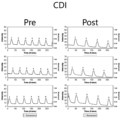
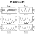
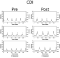
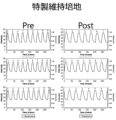
- FIG. 2 shows the calcium ion waveform of cardiomyocytes when 0.1% DMSO was added. The shooting time is 20 seconds and the frame rate is 14.23 frames/second.
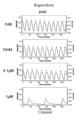
- FIG. 3 shows the calcium ion waveform of cardiomyocytes when 1 ⁇ M bepridil was added. The shooting time is 20 seconds and the frame rate is 14.23 frames/second. Arrows in the figure indicate EAD-like waveforms.
- FIG. 4 shows an enlarged EAD-like waveform in the calcium ion waveform of FIG. FIG.
- FIG. 5 shows the calcium ion waveform of cardiomyocytes when 2 ⁇ M bepridil was added. The shooting time is 20 seconds and the frame rate is 14.23 frames/second.
- FIG. 6 shows the calcium ion waveform of cardiomyocytes when 4 ⁇ M bepridil was added. The shooting time is 20 seconds and the frame rate is 14.23 frames/second.
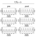
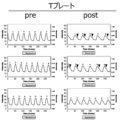
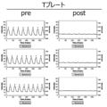
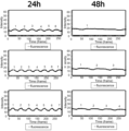
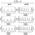
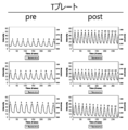
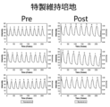
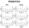
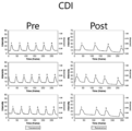
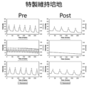
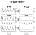
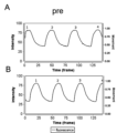
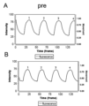
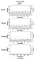
- FIG. 7A shows the calcium ion waveform of cardiomyocytes when 11.1 ⁇ M pentamidine was added to the T-plate. The shooting time is 20 seconds and the frame rate is 14.23 frames/second. Results before addition (pre), 10 minutes after addition (10 min), 24 hours after addition (24 h), and 48 hours after addition (48 h) are shown.
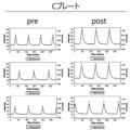
- FIG. 7B shows the calcium ion waveform of cardiomyocytes when 11.1 ⁇ M pentamidine was added to the C plate.
- the shooting time is 20 seconds and the frame rate is 14.23 frames/second.
- Results before addition (pre), 10 minutes after addition (10 min), 24 hours after addition (24 h), and 48 hours after addition (48 h) are shown.
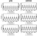
- FIG. 8A shows the calcium ion waveform of cardiomyocytes when 33.3 ⁇ M pentamidine was added to the T-plate.
- the shooting time is 20 seconds and the frame rate is 14.23 frames/second.
- Results before addition (pre), 10 minutes after addition (10 min), 24 hours after addition (24 h), and 48 hours after addition (48 h) are shown.
- FIG. 8B shows the calcium ion waveform of cardiomyocytes when 33.3 ⁇ M pentamidine was added to the C plate.
- the shooting time is 20 seconds and the frame rate is 14.23 frames/second.
- Results before addition (pre), 10 minutes after addition (10 min), 24 hours after addition (24 h), and 48 hours after addition (48 h) are shown.
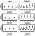
- FIG. 9A shows the calcium ion waveform of cardiomyocytes when 100 ⁇ M pentamidine was added to the T-plate.
- the shooting time is 20 seconds and the frame rate is 14.23 frames/second.
- Results before addition (pre), 10 minutes after addition (10 min), 24 hours after addition (24 h), and 48 hours after addition (48 h) are shown.
- FIG. 9B shows the calcium ion waveform of cardiomyocytes when 100 ⁇ M pentamidine was added to the C plate.
- the shooting time is 20 seconds and the frame rate is 14.23 frames/second. Results before addition (pre), 10 minutes after addition (10 min), 24 hours after addition (24 h), and 48 hours after addition (48 h) are shown.
- FIG. 10 shows the calcium ion waveform of cardiomyocytes when 20 nM isoproterenol was added. The shooting time is 20 seconds and the frame rate is 14.23 frames/second.
- FIG. 11 shows the calcium ion waveform of cardiomyocytes when 100 nM isoproterenol was added. The shooting time is 20 seconds and the frame rate is 14.23 frames/second.
- FIG. 12 shows the calcium ion waveform of cardiomyocytes when 500 nM isoproterenol was added.
- the shooting time is 20 seconds and the frame rate is 14.23 frames/second.
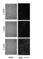
- FIG. 13 shows photographs of myocardial cells observed under a microscope using a G plate, a T plate, and a V plate. The upper row is a photograph of observation in a bright field, and the lower row is a photograph of fluorescence of GCaMP observed with a fluorescence microscope. The objective magnification is 4x for both the upper limit and the lower level.
- FIG. 14 shows the calcium ion waveform of myocardial cells when 0.1% DMSO was added using a T plate. The shooting time is 20 seconds and the frame rate is 14.23 frames/second.
- FIG. 15 shows the calcium ion waveform of myocardial cells when 0.1% DMSO was added using a G plate. The shooting time is 20 seconds and the frame rate is 14.23 frames/second.
- FIG. 16 shows the calcium ion waveform of cardiomyocytes when 0.06 ⁇ M bepridil was added using a T plate. The shooting time is 20 seconds and the frame rate is 14.23 frames/second.
- FIG. 17 shows the calcium ion waveform of cardiomyocytes when 0.06 ⁇ M bepridil was added using G plate. The shooting time is 20 seconds and the frame rate is 14.23 frames/second.
- FIG. 18 shows the calcium ion waveform of cardiomyocytes when 0.25 ⁇ M bepridil was added using a T plate.
- FIG. 19 shows the calcium ion waveform of cardiomyocytes when 0.25 ⁇ M bepridil was added using G plate.
- the shooting time is 20 seconds and the frame rate is 14.23 frames/second.
- FIG. 20 shows calcium ion waveforms of cardiomyocytes when 1 ⁇ M bepridil was added using a T plate.
- the shooting time is 20 seconds and the frame rate is 14.23 frames/second.
- FIG. 21 shows the calcium ion waveform of cardiomyocytes when 1 ⁇ M bepridil was added using G plate.
- the shooting time is 20 seconds and the frame rate is 14.23 frames/second.
- FIG. 22 shows the calcium ion waveform when 4 ⁇ M bepridil was added using the T-plate.
- FIG. 23 shows the calcium ion waveform of cardiomyocytes when 4 ⁇ M bepridil was added using G plate. The shooting time is 20 seconds and the frame rate is 14.23 frames/second.
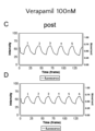
- FIG. 24 shows the calcium ion waveform of cardiomyocytes when verapamil was added at a final concentration of 10 nM. The shooting time is 20 seconds and the frame rate is 14.23 frames/second.
- FIG. 25 shows the calcium ion waveform of cardiomyocytes when verapamil was added at a final concentration of 100 nM. The shooting time is 20 seconds and the frame rate is 14.23 frames/second.
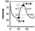
- FIG. 26 is a conceptual diagram explaining CAD30 and CAD80. FIG.
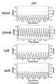
- FIG. 27 shows the calcium ion waveform of cardiomyocytes when verapamil was added at 60 nM to 4 ⁇ M.
- the shooting time is 20 seconds and the frame rate is 14.23 frames/second.
- FIG. 28A is a graph showing CAD30 with verapamil added at 60 nM to 4 ⁇ M and
- FIG. 28B shows CAD80 with verapamil added.
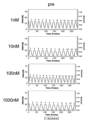
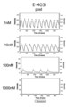
- FIG. 29 shows calcium ion waveforms of cardiomyocytes when E-4031 was added at 1 nM to 1000 nM.
- the shooting time is 20 seconds and the frame rate is 14.23 frames/second.
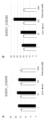
- FIG. 30A is a graph showing CAD30 when E-4031 was added at 1 nM to 1000 nM
- FIG. 30B is a graph showing CAD80 when E-4031 was added.
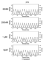
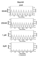
- FIG. 31 shows calcium ion waveforms of cardiomyocytes when bepridil was added at 60 nM to 4 ⁇ M.
- the shooting time is 20 seconds and the frame rate is 14.23 frames/second.
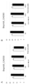
- FIG. 32A is a graph showing CAD30 when bepridil was added at 60 nM to 4 ⁇ M and
- FIG. 32B is a graph showing CAD80 when bepridil was added at 60 nM to 4 ⁇ M.
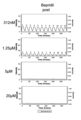
- FIG. 33 shows calcium ion waveforms of cardiomyocytes when bepridil was added at 312 nM to 20 ⁇ M. The shooting time is 20 seconds and the frame rate is 14.23 frames/second.
- FIG. 32A is a graph showing CAD30 when bepridil was added at 60 nM to 4 ⁇ M
- FIG. 32B is a graph showing CAD80 when bepridil was added at 60 nM to 4 ⁇ M.
- FIG. 33 shows calcium ion waveforms of cardiomyocytes when be
- FIG. 34A is a graph showing CAD30 when bepridil was added at 312 nM to 20 ⁇ M and FIG. 34B is a graph showing CAD80 when bepridil was added at 312 nM to 20 ⁇ M.
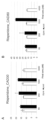
- FIG. 35 shows the calcium ion waveform of cardiomyocytes when risperidone was added at 1 nM to 1 ⁇ M. The shooting time is 20 seconds and the frame rate is 14.23 frames/second.
- FIG. 36A is a graph showing CAD30 when risperidone was added at 1 nM to 1 ⁇ M and FIG. 36B is a graph showing CAD80 when risperidone was added at 1 nM to 1 ⁇ M.
- FIG. 36A is a graph showing CAD30 when risperidone was added at 1 nM to 1 ⁇ M
- FIG. 36B is a graph showing CAD80 when risperidone was added at 1 nM to 1 ⁇ M.
- FIG. 37 shows the calcium ion waveform of cardiomyocytes when risperidone was added at 60 nM to 4 ⁇ M.
- the shooting time is 20 seconds and the frame rate is 14.23 frames/second.
- FIG. 38A is a graph showing CAD30 when risperidone was added at 60 nM to 4 ⁇ M and
- FIG. 38B is a graph showing CAD80 when risperidone was added at 60 nM to 4 ⁇ M.
- FIG. 39 shows calcium ion waveforms of cardiomyocytes when terfenadine was added from 0.25 nM to 16 nM. The shooting time is 20 seconds and the frame rate is 14.23 frames/second.
- FIG. 38A is a graph showing CAD30 when risperidone was added at 60 nM to 4 ⁇ M
- FIG. 38B is a graph showing CAD80 when risperidone was added at 60 nM to 4 ⁇ M.
- FIG. 39 shows calcium
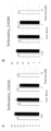
- FIG. 40A is a graph showing CAD30 with addition of terfenadine from 0.25 nM to 16 nM and FIG. 40B is a graph showing CAD80 with addition of terfenadine.
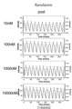
- FIG. 41 shows calcium ion waveforms of cardiomyocytes when ranolazine was added at 10 nM to 10000 nM. The shooting time is 20 seconds and the frame rate is 14.23 frames/second.
- Figure 42A is a graph showing CAD30 with addition of ranolazine from 10 nM to 10000 nM and Figure 42B is a graph showing CAD80 with addition of ranolazine.
- the vertical axis on the left is Intensity.
- the vertical axis on the right side is Movement
- the horizontal axis is Time (frame)
- the graph shows Fluorescence. 7 to 9
- the vertical axis represents Intensity
- the horizontal axis represents Time (frame)
- the graphs represent Fluorescence. 28, 30, 32, 34, 36, 38, 40, and 42
- the vertical axis of A indicates CAD30
- the vertical axis of B indicates CAD80.
- One aspect of the present invention is a method for evaluating the effect of a drug on cardiomyocytes, comprising the step (A) of seeding cardiomyocytes on the culture surface of a culture vessel, and the cardiomyocytes obtained in step (A)
- a culture vessel in which at least part of the culture surface is formed from a substrate containing a 4-methyl-1-pentene polymer is referred to as a culture vessel (X).
- the drug in one aspect of the present invention is not particularly limited, but may be a substance known to act on cardiomyocytes, such as isoproterenol, pentamidine, bepridil, verapamil, E-4031, terfenadine, astemizole, chromanol 293b, mexiletine, nifedipine, propranolol, milrinone, drugs described in Non-Patent Document 1, and the like.
- isoproterenol, pentamidine, bepridil, verapamil, and E-4031 are preferable because the method of one embodiment of the present invention can accurately evaluate the action on cardiomyocytes.
- Isoproterenol is a nonselective ⁇ -agonist with tachycardia and cardiotonic effects.
- Pentamidine which is one of antibacterial agents, has an arrhythmogenic effect.
- Bepridil is one of the calcium antagonists used for the treatment of angina pectoris, and is known to induce lethal ventricular arrhythmia called torsade de pointes (TdP) and QT prolongation as side effects.
- Milrinone is a phosphodiesterase III inhibitor and also has tachycardia and cardiotonic effects.
- Verapamil is an L-type calcium channel inhibitor.
- E-4031 is a hERG-type potassium channel inhibitor.
- Terfenadine is an antiallergic drug known to cause QT prolongation.
- Chromanol 293b is a voltage-gated potassium channel KCNQ1 inhibitor.
- Mexiletine is a voltage-gated sodium channel inhibitor.
- Nifedipine is an L-type calcium channel inhibitor.
- Propranolol is a beta-blocker and has a bradycardic effect.
- Agents in one aspect of the present invention include, for example, the 28 agents evaluated in Reference A: ibutilide, dl-sotalol, azimilide, dofetilide, quinidine, disopyramide, vandetanib, bepridil, domperidone, ondansetron, astemizole.
- cisapride pimozide, clarithromycin, risperidone, terfenadine, chlorpromazine, clozapine, ranolazine, metoprolol, mexiletine, loratadine, tamoxifen, nitrendipine, nifedipine, diltiazem, verapamil can also be used.
- drugs whose risk level is difficult to predict in conventional proarrhythmic models using cardiomyocytes differentiated from iPS cells are preferable, and those whose clinical TdP risk level is high or intermediate risk.
- the drug may be a substance whose effect on cardiomyocytes is unknown, such as a new drug candidate substance, a substance suspected of having cardiotoxicity, or a candidate substance expected to have a main effect on cardiomyocytes.
- the drug may be a low-molecular-weight compound included in low-molecular-weight drugs, or a high-molecular-weight compound such as proteins, antibodies, nucleic acids, and polysaccharides included in high-molecular-weight drugs.
- a drug may be a new substance or a known substance.
- the action of a drug includes main actions and side effects of the drug.
- the primary action of a drug on cardiomyocytes is the action that is originally expected among the pharmacological actions of the drug, such as bradycardia action (negative chronotropic action) and tachycardia action (positive chronotropic action). , cardiotonic (positive inotropy), and weak (negative inotropy).
- the primary effect of the drug on myocardial cells is preferably tachycardia or bradycardia, since the method of the present invention can detect the drug even at low concentrations.
- a side effect of a drug on cardiomyocytes is a pharmacological action that the drug has that is not related to or interferes with treatment.
- Side effects on myocardial cells include, for example, cardiotoxicity, and cardiotoxicity includes proarrhythmia, myocardial injury, and the like.
- Proarrhythmia effects are those that exacerbate existing arrhythmias or cause new arrhythmias, e.g. ), triggered activity arrhythmias, and reentry arrhythmias.
- Myocardial damaging effects may be irreversible or reversible myocardial damage.
- the side effect is preferably proarrhythmic, more preferably QT prolongation and early post-depolarization (EAD), since it can be detected with high accuracy by the method of one aspect of the present invention.
- the side effect is preferably myocardial injury, more preferably mitochondrial toxicity, since it can be detected with high accuracy by the method of one embodiment of the present invention.
- the method that is one aspect of the present invention can be used to evaluate the safety and efficacy of pharmaceuticals in the drug discovery process. That is, when a substance with an unknown effect on myocardial cells is used as a drug, the main effect or side effect of the substance can be evaluated even at a low concentration, and false positives are difficult to detect. In addition, substances that may be cardiotoxic in subsequent in vivo tests can be detected as predictive cardiotoxic substances in earlier in vitro test systems. In addition, when a substance with a known effect on myocardial cells is used as a drug, the known main effects or side effects of the substance can be confirmed even at low concentrations, or new main effects or side effects of the substance can be evaluated. and false positives are hard to detect. In particular, when a substance that is reported or suspected to have cardiotoxicity in vivo is used as a drug, cardiotoxicity can be detected more easily than in vivo tests, and the mechanism of cardiotoxicity can be analyzed.
- Cardiomyocytes may be cardiomyocytes differentiated from pluripotent stem cells, or primary cultured cardiomyocytes isolated from the heart of an organism.
- cardiomyocytes obtained by differentiating commercially available pluripotent stem cells for example, iCell Cardiomyocytes from FUJIFILM Cellular Dynamics, MiraCell Cardiomyocytes v2 from Takara Bio, Cor. 4U, Myoridge's CarmyA, REPROCELL's ReproCardio2, and the like may be used.
- Pluripotent stem cells are a general term for stem cells that have the ability to differentiate into cells of any tissue (pluripotency).
- Pluripotent stem cells include, for example, embryonic stem cells (ES cells), embryonic carcinoma cells (EC cells), trophoblast stem cells (TS cells), epiblast stem cells ( epiblast stem cells (EpiS cells), embryonic germ cells (EG cells), multipotent germline stem cells (mGS cells), induced pluripotent stem cells (iPS cells) , Muse cells (Multi-lineage differentiating Stress Enduring cells) and the like.
- Pluripotent stem cells are preferably ES cells or iPS cells.
- the cardiomyocyte is preferably a cardiomyocyte obtained by differentiating a pluripotent stem cell, more preferably a cardiomyocyte obtained by differentiating an induced pluripotent stem cell, and still more preferably a cardiomyocyte obtained by differentiating an induced pluripotent stem cell.
- These are mature cardiomyocytes.
- Cardiomyocytes obtained by differentiating induced pluripotent stem cells can be prepared by a known cardiomyocyte differentiation induction method, for example, a protein-free cardiomyocyte differentiation induction (PFCD) method can be used (WO 2015/182765). ).
- Cardiomyocytes are preferably cardiomyocytes obtained by differentiating induced pluripotent stem cells by a protein-free cardiomyocyte differentiation induction (PFCD) method.
- Mature cardiomyocytes differentiated from induced pluripotent stem cells is a term used in the art in contrast to immature cardiomyocytes from differentiated induced pluripotent stem cells. They have higher ion channel function than immature cardiomyocytes.
- Mature cardiomyocytes obtained by differentiating induced pluripotent stem cells are, for example, cells that have passed 14 days or more, preferably 20 days or more, more preferably 30 days or more after initiation of differentiation induction of induced pluripotent stem cells. .
- the day on which the induction of differentiation of induced pluripotent stem cells was started is the day on which the induced pluripotent stem cells maintained in an undifferentiated state were exposed to a treatment for transitioning to a differentiated state, and this day was set to 0. day.
- the upper limit of the number of days after initiation of differentiation induction is not particularly limited, and mature cardiomyocytes can be maintained in a differentiated state. It may be cultured or stored for a long period of time.
- mature cardiomyocytes may be cells that have passed 365 days or more after initiation of differentiation induction of induced pluripotent stem cells.
- Mature cardiomyocytes obtained by differentiating induced pluripotent stem cells preferably refer to cells that have passed 30 days or more after initiation of induction of differentiation of induced pluripotent stem cells by a protein-free myocardial differentiation induction (PFCD) method.
- PFCD protein-free myocardial differentiation induction
- Mature cardiomyocytes obtained by differentiating induced pluripotent stem cells are more preferable than immature cardiomyocytes obtained by differentiating induced pluripotent stem cells because their properties are closer to those of cardiomyocytes isolated from the heart of an organism.
- Cardiomyocytes are not particularly limited in origin, and may be derived from mammals, birds, amphibians, reptiles, fish, etc., preferably mammals, more preferably humans, monkeys, mice, rats, pigs, It is derived from dogs, sheep, cats, and goats, more preferably from humans.
- Cardiomyocytes may be normal cardiomyocytes, cardiomyocytes containing genetic mutations, or disease model cardiomyocytes. Cardiomyocytes may be introduced with various genes so as to transiently or permanently express sensor proteins and the like. Cardiomyocytes that transiently or constitutively express sensor proteins are preferred because indicators of cell function can be detected by fluorescence or chemiluminescence.
- sensor proteins include calcium sensors such as GCaMP, cameleon, pericam, G-GECO, B-GECO, R-GECO, GEX-GECO, GEM-GECO, CEPIA; mito-MaLion, MaLionB, MaLionG, MaLionR, ATeam Magnesium sensors such as MARIO; Glucose sensors such as Green Glifon and Red Glifon; Lactate sensors such as Green Lindoblum; Pyruvate sensors such as Green Pegassos;
- the sensor protein is preferably a calcium sensor or an ATP sensor, more preferably a calcium sensor, even more preferably GCaMP.
- Cardiomyocytes may be used in the form of single cells, in the form of cardiomyocyte clusters, or in the form of cardiomyocyte sheets.
- the cardiomyocytes can efficiently supply oxygen in the culture vessel (X), so they may be either adherent cardiomyocytes or floating cardiomyocytes, but are preferably adherent cardiomyocytes.
- the culture vessel means all vessels used for culturing cells.
- various known culture vessels can be used, and the shape and size are not particularly limited. Examples of the culture vessel include dishes, flasks, plates, bottles, bags, tubes and the like.
- the culture vessels are typically used in devices such as incubators, mass culture devices, or perfusion culture devices.
- the culture surface means a surface with which the medium and/or the cells are in contact, or a surface to be contacted with the medium and/or the cells when the cells are cultured.
- the culture vessel (X) "at least part of the culture surface of the culture vessel is formed from a base material containing a 4-methyl-1-pentene polymer” means that at least a part of the culture surface is It means that it is formed from a substrate containing a 4-methyl-1-pentene polymer, and the entire area of the culture surface may be formed from a substrate containing a 4-methyl-1-pentene polymer. .
- the culture vessel (X) is preferably a culture vessel whose bottom surface includes a culture surface in order to hold or store the culture medium.
- the culture vessel (X) is a dish, flask, or plate
- the bottom surface includes the culture surface. Therefore, of these bottom surfaces, side surfaces, and top surfaces, at least part or all of the bottom surface contains 4-methyl-1-pentene. It is formed from a substrate containing a polymer.
- oxygen is efficiently introduced into the medium via the 4-methyl-1-pentene polymer. can be supplied, facilitating efficient growth and differentiation of cells in the culture medium. In addition, it becomes easier to culture cells at a high density while maintaining cell functions.
- the entire culture surface of the culture vessel (X) is preferably formed from a base material containing a 4-methyl-1-pentene polymer. That is, when the culture vessel (X) is a dish, flask or plate, the inner side of the bottom is the culture surface. It is preferably formed from a base material containing
- the thickness of the substrate containing the 4-methyl-1-pentene polymer is not particularly limited, it is preferably 20 ⁇ m to 400 ⁇ m, more preferably 20 ⁇ m to 300 ⁇ m, still more preferably 20 ⁇ m to 200 ⁇ m.
- the thickness of the base material containing the 4-methyl-1-pentene polymer is appropriately selected according to the form of the culture vessel. By adjusting the thickness to the above range, the appropriate medium necessary for cell growth and differentiation can be obtained. It is easy to obtain the oxygen concentration inside, and it is easy to obtain sufficient strength as a culture vessel.
- the culture vessel (X) is preferably a culture vessel having at least one well, more preferably a plate having at least one well, 6 wells, 12 wells, 24 wells, 48 wells, 96 wells, A plate with wells such as 384 wells and 1536 wells is more preferable.
- a culture vessel having a concave bottom surface such as a well needs to have a thick bottom surface in order to stabilize the complex shape of the bottom surface, and it is difficult to sufficiently supply oxygen to the cells.
- wells such as 1 well, 6 wells, 12 wells, 24 wells, 48 wells, 96 wells, 384 wells, 1536 wells, etc., where the bottom surface is formed from a substrate comprising a 4-methyl-1-pentene polymer Even with a plate having a
- the shape of the bottom surface of the culture container (X) is not particularly limited, and includes flat bottom, round bottom (U bottom), flat bottom (F bottom), conical bottom (V bottom), flat bottom + curved edge, and the like.
- general injection molding or press molding may be performed at once, or film Alternatively, it is also possible to prepare a sheet and perform secondary processing such as vacuum forming or pressure forming.
- the shape of the bottom is selected according to the purpose of culture, but when cells are cultured two-dimensionally, a flat bottom is usually desirable, and when cells are cultured three-dimensionally, a round bottom (U bottom) or conical bottom is preferred. (V bottom) is usually desirable.
- the portion of the culture vessel (X) other than the culture surface may be composed of a material other than the base material containing the 4-methyl-1-pentene polymer.
- the material is not particularly limited, and known materials can be used. Such materials include, for example, polystyrene (PS), polydimethylsiloxane (PDMS), thermosetting resins, cyclic olefin polymers, cyclic olefin copolymers, glass, and the like.
- At least the culture surface of the culture vessel (X) may be coated with a coating agent containing a natural polymeric material, a synthetic polymeric material, or an inorganic material. Coating can be performed by a known method.
- the coated culture vessel (X) has superior cardiomyocyte adhesiveness and proliferation. This is probably because the component coated on the culture surface serves as a scaffold for the cells. Therefore, when the cardiomyocytes are adhered and cultured, the culture surface of the culture vessel (X) is preferably coated with a coating agent containing a natural polymer material, a synthetic polymer material, or an inorganic material.
- natural polymeric materials, synthetic polymeric materials, or inorganic materials are not particularly limited, but natural polymeric materials include collagen, gelatin, alginic acid, hyaluronic acid, glycosaminoglycans such as chondroitin sulfate, fibronectin, laminin, fibrinogen, Osteopontin, tenascin, vitronectin, thrombospondin, agarose, elastin, keratin, chitosan, fibrin, fibroin, sugars; synthetic polymer materials such as polyglycolic acid, polylactic acid, polyethylene glycol, polycaprolactone, synthetic peptides, synthetic proteins , polyhydroxyethyl methacrylate, polyethyleneimine; inorganic materials include ⁇ -tricalcium phosphate, calcium carbonate, and the like.
- the natural polymer material, synthetic polymer material, or inorganic material may be used after being processed such as vitrification.
- processing such as vitrification include vitrigel, which is obtained by rehydrating a conventional hydrogel such as an extracellular matrix component after vitrification, and collagen composed of a high-density collagen fiber network made from collagen. vitrigel and the like.
- the coating agent is preferably a protein such as collagen, gelatin, laminin, or polylysine, or a peptide, from the viewpoint of improving cardiomyocyte adhesion and cardiomyocyte proliferation and maintaining cardiomyocyte function for a longer period of time. More preferably laminin, collagen, polylysine, and more preferably laminin. That is, the culture surface of the culture vessel (X) is preferably coated with a coating agent containing laminin.
- the coating agent may contain the above components singly or in combination of two or more.
- the coating agent may be used for coating as it is, but it can be used for coating after being diluted with a solvent such as water, PBS, or a medium. From the viewpoint of cardiomyocyte adhesion, the coating agent is preferably diluted with a solvent such as water, PBS, or a medium before use for coating, more preferably after being diluted with a medium before use for coating.
- the medium used for diluting the coating agent is not particularly limited, and may be the same as or different from the medium used in step (A).
- the medium used for diluting the coating agent is preferably a medium containing no animal serum or alternative serum, more preferably DMEM, and particularly preferably a special basal medium described later in Examples.
- the coating agent may be removed from the culture vessel (X) or left in the culture vessel (X). preferably left in
- At least the culture surface of the culture vessel (X) may be processed.
- Surface processing includes, for example, uneven structure forming processing, surface modification processing such as hydrophilic processing, and hydrophobic processing.
- the method used for the surface modification treatment is not particularly limited, but for example, hydrophilic treatment such as corona treatment, plasma treatment, ozone treatment, ultraviolet treatment; hydrophobic treatment such as esterification, silylation, and fluorination; Vapor deposition, etching, or addition of specific functional groups such as hydroxyl group, amino group, sulfone group, thiol group, carboxyl group; treatment with specific functional groups such as silane coupling, titanium coupling, zirconium coupling; oxidizing agent surface roughening by means such as; physical treatment such as rubbing and sandblasting; These surface modification treatments may be performed singly or in combination of two or more. In addition, when surface modification treatment is performed, it is preferable to perform it at least on the culture surface.
- At least the culture surface of the culture vessel (X) is preferably hydrophilized, more preferably corona-treated or plasma-treated.
- hydrophilizing the surface of the culture vessel (X) the wettability of the surface of the culture vessel (X) is increased, the adhesion between the culture vessel (X) and the cardiomyocytes is improved, and the culture vessel (X) is improved. Cardiomyocytes can proliferate uniformly on the surface.
- by hydrophilizing the surface of the culture vessel (X) it becomes easier to coat the culture surface of the culture vessel (X) with the coating agent.
- the coating agent is uniformly loaded on the culture surface of the culture vessel (X), making it easier to adhere. It does not come off and can be used for cell culture while maintaining a stable initial state.
- nitrogen, hydrogen, helium, oxygen, argon, or the like is used as the accompanying gas, and at least one gas selected from nitrogen, helium, and argon is preferably selected.
- the culture vessel (X) may be disinfected or sterilized to prevent contamination.
- the method of disinfection or sterilization is not particularly limited, and physical disinfection methods such as the circulation steam method, boiling method, intermittent method, and ultraviolet method; chemical disinfection methods using gases such as ozone and disinfectants such as ethanol; Heat sterilization methods such as high pressure steam method and dry heat method; irradiation sterilization methods such as gamma ray method, electron beam method and high frequency method; gas sterilization methods such as ethylene oxide gas method and hydrogen peroxide gas plasma method.
- ethanol sterilization, autoclave sterilization, gamma ray sterilization, electron beam sterilization, or ethylene oxide gas sterilization is preferred because of its simple operation and sufficient sterilization.
- the culture surface of the culture vessel (X) preferably has a water contact angle of 50° or more, more preferably 55° or more, and still more preferably 60° or more.
- the culture surface of the culture vessel (X) preferably has a water contact angle of 100° or less, more preferably 90° or less, and even more preferably 84° or less.
- the culture surface of the culture vessel (X) is uniformly coated with a natural polymer material, a synthetic polymer material, or an inorganic material to facilitate adhesion.
- the natural polymer material, synthetic polymer material, or inorganic material does not peel off and can be used for cell culture while maintaining a stable initial state.
- the method for measuring the water contact angle is not particularly limited, and a known method can be used, preferably the sessile drop method.
- the shape of the water droplet can be regarded as spherical under constant temperature and humidity conditions of 25 ⁇ 5 ° C. and 50 ⁇ 10% according to Japanese Industrial Standard JIS-R3257 (test method for wettability of substrate glass surface).
- a water droplet with a volume of 4 ⁇ L or less is dropped on the surface of a measurement sample made using the same material as the culture vessel (X), and the sessile drop method is used to remove the water droplet within 1 minute immediately after contact with the measurement sample surface. It can be measured by a method of measuring the angle of the contact interface between the measurement sample and the water droplet.
- the manufacturing method of the culture vessel (X) is not particularly limited, nor is the equipment used for manufacturing.
- the entire culture vessel (X) is formed from a base material containing a 4-methyl-1-pentene polymer, for example, a film or sheet containing the 4-methyl-1-pentene polymer is formed, and the necessary Depending on the conditions, the film or sheet can be formed into a desired shape to produce a culture vessel (X).
- the culture vessel (X) can also be obtained by direct molding by methods such as extrusion molding, solution casting molding, injection molding, and blow molding.
- the culture vessel (X) can be obtained by appropriately bonding the film or sheet to another base material.
- the bonding method is not particularly limited, and the substrate containing the 4-methyl-1-pentene polymer and another substrate may be integrally formed, or they may be adhered to each other via an adhesive or pressure-sensitive adhesive. good.
- the extrusion temperature is preferably 100°C to 400°C, particularly preferably 200°C to 300°C.
- the roll temperature is preferably 45°C to 75°C, particularly preferably 55°C to 65°C.
- the film or sheet may be produced by a solution casting method in which a 4-methyl-1-pentene polymer is dissolved in a solvent, poured onto a resin or metal, slowly dried while leveling, and formed into a film (sheet). good.
- the solvent used is not particularly limited, but hydrocarbon solvents such as cyclohexane, hexane, decane and toluene may be used. Two or more solvents may be mixed in consideration of the solubility and drying efficiency of the 4-methyl-1-pentene polymer.
- a polymer solution is applied by a method such as table coating, spin coating, dip coating, die coating, spray coating, bar coating, roll coating, or curtain flow coating, dried, and peeled off to form a film or sheet.
- [4-methyl-1-pentene polymer] 4-methyl-1-pentene homopolymers and copolymers of 4-methyl-1-pentene and other monomers are collectively referred to as "4-methyl-1-pentene polymers ”.
- Copolymers of 4-methyl-1-pentene which is an example of 4-methyl-1-pentene polymers, with other monomers include random copolymers, alternating copolymers, block copolymers, and grafts. Any copolymer may be used.
- Copolymers of 4-methyl-1-pentene with other monomers include 4-methyl-1-pentene, ethylene and ⁇ -olefins having 3 to 20 carbon atoms (excluding 4-methyl-1-pentene A copolymer with at least one olefin selected from ) is preferable because it has high strength, is resistant to breakage and cracking even when used as a base material, and has little deflection.
- the 4-methyl-1-pentene polymer includes 4-methyl-1-pentene homopolymer, 4-methyl-1-pentene, ethylene and an ⁇ -olefin having 3 to 20 carbon atoms (4-methyl-1 - is preferably at least one polymer selected from copolymers with at least one olefin selected from (excluding pentene), 4-methyl-1-pentene, ethylene and 3 to 20 carbon atoms is more preferably a copolymer with at least one olefin selected from ⁇ -olefins (excluding 4-methyl-1-pentene).
- olefin examples include ethylene, propylene, 1-butene, 1-hexene, 1-heptene, 1-octene, 1-decene, 1-tetradecene, 1-hexadecene, 1-heptadecene, 1-octadecene, and 1-eicosene. is mentioned.
- the olefin can be appropriately selected depending on the physical properties required for the base material.
- the olefins are preferably ⁇ -olefins having 8 to 18 carbon atoms from the viewpoint of appropriate oxygen permeability and excellent rigidity, such as 1-octene, 1-decene, 1-dodecene, 1-tetradecene, At least one selected from 1-hexadecene, 1-heptadecene and 1-octadecene is more preferred.
- the number of carbon atoms in the olefin is within the above range, the processability of the polymer will be better, and the appearance of the base material due to cracks or cracks at the edges will tend to be less likely to occur. In addition, the defective product rate of the base material is reduced.
- the olefin can be used alone or in combination of two or more.
- the olefin preferably has 2 or more carbon atoms, more preferably 10 or more carbon atoms, from the viewpoint of strength of the material.
- the content of structural units derived from 4-methyl-1-pentene in the 4-methyl-1-pentene polymer is preferably 60 to 100 mol%, more preferably 80 to 99.5 mol%, still more preferably 85 to 98 mol %.
- the 4-methyl-1-pentene polymer is at least one selected from 4-methyl-1-pentene, ethylene and ⁇ -olefins having 3 to 20 carbon atoms (excluding 4-methyl-1-pentene)
- it is a copolymer with an olefin
- it is derived from at least one olefin selected from ethylene and ⁇ -olefins having 3 to 20 carbon atoms (excluding 4-methyl-1-pentene) in the copolymer
- the content of the structural units is preferably 0.1 to 40 mol%, more preferably 0.5 to 20 mol%, still more preferably 2 to 15 mol%.
- the content of these structural units is based on 100 mol % of all repeating structural units in the 4-methyl-1-pentene polymer.
- the content of the structural unit is within the above range, a uniform culture surface with excellent workability can be obtained, and the toughness and strength of the substrate are well balanced, so that the substrate is less flexed.
- the 4-methyl-1-pentene polymer is derived from the structural unit derived from 4-methyl-1-pentene and the ethylene and the ⁇ -olefin having 3 to 20 carbon atoms within a range that does not impair the effects of the present invention. It may have structural units other than structural units (hereinafter also referred to as "other structural units"). The content of other structural units is, for example, 0 to 10.0 mol %. When the 4-methyl-1-pentene polymer has other structural units, the other structural units may be one or two or more.
- Examples of monomers from which other structural units are derived include cyclic olefins, aromatic vinyl compounds, conjugated dienes, non-conjugated polyenes, functional vinyl compounds, hydroxyl group-containing olefins, and halogenated olefins.
- Cyclic olefins, aromatic vinyl compounds, conjugated dienes, non-conjugated polyenes, functional vinyl compounds, hydroxyl group-containing olefins and halogenated olefins include, for example, paragraphs [0035] to [0041] of JP-A-2013-169685. compounds can be used.
- the 4-methyl-1-pentene polymer may be used singly or in combination of two or more.
- a commercial product can also be used as the 4-methyl-1-pentene polymer.
- TPX MX001, MX002, MX004, MX0020, MX021, MX321, RT18, RT31 or DX845 (all registered trademarks) manufactured by Mitsui Chemicals, Inc. may be used.
- 4-methyl-1-pentene polymers manufactured by other manufacturers can be preferably used as long as they satisfy the above requirements. These commercial products may be used singly or in combination of two or more.
- the 4-methyl-1-pentene polymer usually has a melting point of 200°C to 240°C and high heat resistance. Further, since it does not undergo hydrolysis and has excellent water resistance, boiling water resistance and steam resistance, a substrate containing a 4-methyl-1-pentene polymer can be subjected to autoclave sterilization.
- the 4-methyl-1-pentene polymer also has a high visible light transmittance (usually 90% or more) and does not emit autofluorescence, so it is formed from a base material containing a 4-methyl-1-pentene polymer.
- a culture vessel that is designed to facilitate observation of cardiomyocytes.
- the 4-methyl-1-pentene polymer can be heat-sealed, and can be easily heat-sealed not only with itself but also with other materials.
- it since it can be thermoformed, it can be easily formed into a culture vessel of any shape, such as by imprinting or inserting.
- the weight average molecular weight (Mw) of the 4-methyl-1-pentene polymer is preferably 10,000 to 2,000,000, more preferably 20,000. ⁇ 1,000,000, more preferably 30,000 to 500,000.
- the sample concentration for GPC measurement can be, for example, 1.0 to 5.0 mg/ml.
- the molecular weight distribution (Mw/Mn) of the 4-methyl-1-pentene polymer is preferably 1.0-30, more preferably 1.1-25, still more preferably 1.1-20.
- the solvent used in GPC is preferably ortho-dichlorobenzene.
- the conditions shown in Examples to be described later can be cited, but the measurement conditions are not limited to these.
- the film produced by melt molding in the molding method of the 4-methyl-1-pentene polymer described later can easily suppress the occurrence of defects such as gel. It becomes easier to form a film with a uniform surface.
- the solubility in the solvent is improved, so that problems such as film gel can be easily suppressed, making it easier to form a film with a uniform surface.
- the weight average molecular weight (Mw) to the above lower limit or more, the strength of the culture vessel (X) tends to be sufficient. Furthermore, by setting the molecular weight distribution within the above range, it is easy to suppress the stickiness of the surface of the prepared culture vessel, the toughness of the culture vessel tends to be sufficient, and the occurrence of cracks during bending and cutting during molding is suppressed. easier to suppress.
- the weight-average molecular weight (Mw) and molecular weight distribution (Mw/Mn) of the 4-methyl-1-pentene polymer are different from each other when two or more types are used as the 4-methyl-1-pentene polymer. , Mw and Mw/Mn should be within the above ranges.
- the 4-methyl-1-pentene polymer preferably has an oxygen permeability coefficient of 100 to 2500 cm 3 ⁇ mm/(m 2 ⁇ 24h ⁇ atm), more preferably 1000 to 2500 cm 3 ⁇ mm/(m 2 ⁇ 24h ⁇ atm). ) is more preferable.
- the oxygen permeability coefficient is in the above range, the oxygen permeability is excellent, so that the cardiomyocytes maintain good morphology, are likely to proliferate efficiently according to the culture period, and are easy to detect the action of the drug.
- the oxygen permeability coefficient can be measured by the following method.
- a measurement sample was prepared from a substrate composed of a 4-methyl-1-pentene polymer, and the oxygen permeability coefficient [cm 3 ⁇ mm/( m 2 ⁇ 24h ⁇ atm)].
- the instrument used for the measurement is not particularly limited as long as it uses a differential pressure type gas permeability measurement method.
- the measurement sample was prepared by cutting a 90 ⁇ 90 mm test piece from a 50 ⁇ m-thick base material composed of a 4-methyl-1-pentene polymer, and the diameter of the measurement part was 70 mm (the transmission area was 38.46 cm 2 ). is preferable.
- the oxygen permeability is high, it is more preferable to apply an aluminum mask to the sample in advance so that the actual permeation area is 5.0 cm 2 .
- the sample to be measured may or may not have been subjected to microfabrication or surface modification treatment, but it is preferable that the sample has not been subjected to any treatment.
- a value obtained by dividing the oxygen permeability coefficient by the thickness ( ⁇ m) of the base material is defined as the oxygen permeability [cm 3 /(m 2 ⁇ 24h ⁇ atm)].
- a culture vessel having at least a culture surface formed of a base material containing the 4-methyl-1-pentene polymer is suitable for culture. It has good shape stability, light transmittance, molding processability, oxygen permeability, can be sterilized, cultures cardiomyocytes, and evaluates the effects of drugs. It is very excellent as a culture vessel used for
- the method for producing the 4-methyl-1-pentene polymer may be any method as long as it can polymerize 4-methyl-1-pentene, olefins, and other monomers.
- a chain transfer agent such as hydrogen may also be used to control the molecular weight or molecular weight distribution.
- the equipment used for manufacturing is also not limited.
- the polymerization method may be a known method such as a gas phase method, a slurry method, a solution method, or a bulk method. A slurry method and a solution method are preferred.
- the polymerization method may be a single-stage polymerization method or a multi-stage polymerization method such as a two-stage polymerization method, in which a plurality of polymers having different molecular weights are blended in a polymerization system.
- hydrogen When hydrogen is used as a chain transfer agent in either a single-stage polymerization method or a multi-stage polymerization method, it may be charged all at once or dividedly, for example, at the beginning, middle and end of the polymerization.
- the polymerization may be carried out at normal temperature, or may be heated if necessary, but from the viewpoint of polymerization efficiency, it is preferably carried out at 20°C to 80°C, and particularly preferably at 40°C to 60°C. .
- the catalyst used for production is also not limited, but from the viewpoint of polymerization efficiency, for example, the solid titanium catalyst component (I) described in WO 2006/054613 or the transition metal compound described in WO 2014/050817 It is preferable to use an olefin polymerization catalyst (metallocene catalyst) containing (A).
- the substrate containing the 4-methyl-1-pentene polymer is a composition containing the 4-methyl-1-pentene polymer
- 4-methyl-1- The pentene polymer is preferably 90% by mass or more and less than 100% by mass, more preferably 95% by mass or more and less than 100% by mass, and particularly preferably 99% by mass or more and less than 100% by mass.
- the 4-methyl-1-pentene polymer in the composition is 90% by mass or more, the oxygen permeability, transparency, strength, etc. of the substrate are further improved.
- components other than the 4-methyl-1-pentene polymer include a heat stabilizer, a light stabilizer, a processing aid, a plasticizer, an antioxidant, a lubricant, an antifoaming agent, an antiblocking agent, and a coloring agent. agents, modifiers, antibacterial agents, antifungal agents, antifogging agents, and other additives.
- Step (A) is a step of seeding cardiomyocytes on the culture surface of the culture vessel (X).
- the method of seeding the cardiomyocytes on the culture surface of the culture vessel (X) is not particularly limited. After rocking the container (X) to evenly disperse the myocardial cells in the culture container (X), it is allowed to stand still in an incubator.
- the cardiomyocyte seeding density is not particularly limited as long as the cardiomyocytes can be maintained or proliferated, but is preferably 0.1 ⁇ 10 5 cells/cm 2 to 10.0 ⁇ 10 5 cells/cm 2 , More preferably 0.3 ⁇ 10 5 cells/cm 2 to 5.0 ⁇ 10 5 cells/cm 2 , still more preferably 0.5 ⁇ 10 5 cells/cm 2 to 3.0 ⁇ 10 5 cells/cm 2 . It is preferable that the cardiomyocyte seeding density is within the above range because the cardiomyocytes easily adhere to the culture vessel (X) and proliferate more efficiently.
- the medium used in step (A) is not particularly limited as long as it allows myocardial cells to survive, and may be appropriately selected according to the cell type used.
- the medium used for seeding is, for example, any cell culture basal medium, differentiation medium, primary culture medium, etc.
- serum-free medium described later
- medium described later
- Eagle essential small medium EMEM
- Dulbecco's modified Eagle's medium DMEM
- ⁇ -MEM Glasgow MEM
- IMDM IMDM
- RPMI1640 Ham F-12
- MCDB medium Williams medium E
- special maintenance medium special basal medium, which will be described later in the examples
- Examples include iCell cardiomyocyte maintenance medium (manufactured by FUJIFILM Cellular Dynamics), iCell cardiomyocyte thawing medium (manufactured by FUJIFILM Cellular Dynamics), and mixed medium thereof.
- the medium used in step (A) is preferably iCell cardiomyocyte thawing medium, serum-free medium ( ⁇ ) described later in Examples, more preferably Serum-free medium ( ⁇ ).
- the amount of medium used in step (A) is not particularly limited, and can be appropriately determined according to the cell type and medium type. Cultivation conditions are not particularly limited, and a known method can be used, but usually the temperature is about 25 to 40° C. and the carbon dioxide concentration is about 5%.
- Serum-free medium ( ⁇ ) is a medium that does not contain animal serum, but contains alternative serum.
- a serum-free medium ( ⁇ ) can be prepared by adding serum replacement to the basal medium.
- the serum substitute means a serum substitute that replaces animal serum added as a nutrient necessary for cell survival during cell culture, and is referred to as Serum Protein Substitute (SPS), Serum Substitute Supplement (SSS), and the like. Sometimes it is done.
- the substitute serum is not particularly limited, and known ones can be used.
- Commercially available alternative serums include Knockout Serum Replacement (manufactured by ThermoFisher SCIENTIFIC, No.
- the content of the replacement serum in the serum-free medium ( ⁇ ) is not particularly limited, but is preferably 1-20% (v/v), more preferably 5-15% (v/v), still more preferably 8-12%. % (v/v).
- the basal medium is not particularly limited as long as it is a cell culture medium containing no animal serum.
- Examples of the basal medium include Grace medium, IPL-41 medium, Schneider's medium, Opti-PRO TM SFM medium, Opti-MEM TM I medium, VP-SFM medium, CD293 medium, 293SFMII medium, and CD-CHO medium.
- CHO-S-SFMII medium FreeStyle TM 293 medium, CD-CHO, AGT TM medium, RPMI medium, DMEM medium, MEM medium, GMEM medium, Eagle's MEM medium, BME medium, DME medium, ⁇ MEM medium, IMEM medium, ES medium, DM-160 medium, Fisher medium, F12 medium, WE medium, ASF103 medium, ASF104 medium, ASF301 medium, TC-100 medium, Sf-900II medium, Ex-cell405 medium, Express-Five medium, Drosophila medium, Ham 'sF-12K medium and mixed medium thereof.
- the basal medium is preferably DMEM medium, RPMI medium, or MEM medium, more preferably DMEM medium, since cardiomyocytes easily adhere to the culture vessel (X).
- the serum-free medium ( ⁇ ) is preferably DMEM medium, RPMI medium, or MEM medium containing Knockout Serum Replacement, more preferably DMEM medium containing Knockout Serum Replacement.
- serum-free medium ⁇
- various growth factors, differentiation inducers, antibiotics, hormones, amino acids, sugars, salts, minerals, metals, vitamins, etc. may be added to the serum-free medium ( ⁇ ).
- Step (B) is a step of culturing the cardiomyocytes seeded in step (A).
- a method for culturing cardiomyocytes is not particularly limited, and may be performed according to a known protocol.
- the details of the medium used for culture in step (B) are the same as those for the medium used in step (A).
- the medium used in step (B) may be the same as or different from the medium used in step (A), but is preferably the same because cardiomyocytes proliferate more easily.
- the amount of medium used in step (B) is not particularly limited, and can be appropriately determined according to the cell type and medium type.
- the frequency of medium replacement is not particularly limited, but it is preferable not to replace the medium during the culture period in step (B) except for the day after seeding.
- Cultivation conditions are not particularly limited, and a known method can be used, but usually the temperature is about 25 to 40° C. and the carbon dioxide concentration is about 5%.
- the culture period in step (B) is not particularly limited, but is preferably 6 hours to 3 days, more preferably 10 hours to 2 days, still more preferably 12 to 36 hours.
- the end of the culture period in step (B) can be determined, for example, by whether the myocardial cells have sufficiently adhered to the culture vessel (X). Whether the cardiomyocytes sufficiently adhere to the culture vessel (X) can be evaluated, for example, by microscopic observation.
- the culture in step (B) can be either adherent culture or suspension culture because oxygen can be supplied efficiently in the culture vessel (X), but adherent culture is preferred.
- Steps (A) and (B) are preferably performed in a serum-free medium ( ⁇ ).
- steps (A) and (B) are performed in a serum-free medium ( ⁇ )
- cardiomyocytes easily adhere to the culture vessel (X).
- Step (C) is a step of further culturing the cardiomyocytes obtained in step (B).
- the method of one aspect of the present invention preferably includes step (C) of further culturing the cardiomyocytes cultured in step (B). If step (C) is included, it is easy to accurately evaluate the action of the drug.
- a method for culturing cardiomyocytes is not particularly limited, and may be performed according to a known protocol.
- the medium used for culture in step (C) is not particularly limited as long as it is a medium in which cardiomyocytes can survive, and may be appropriately selected according to the cell type used.
- the medium used for culture is, for example, any cell culture basal medium, differentiation medium, primary culture medium, etc.
- serum-free medium ⁇
- medium ⁇
- EMEM Eagle's small essential medium
- DMEM Dulbecco modified Eagle medium
- GMEM Glasgow MEM
- IMDM IMDM
- RPMI1640 Ham F-12
- MCDB medium Williams medium E
- CardioGro manufactured by Funakoshi
- FUJIFILM iCell cardiomyocyte maintenance medium
- iCell cardiomyocyte thawing medium manufactured by FUJIFILM Cellular Dynamics
- the medium used in step (C) is preferably iCell cardiomyocyte maintenance medium or medium ( ⁇ ) described later, and more preferably, because cardiomyocytes grow more efficiently and the action of the drug can be detected with high accuracy. is the medium ( ⁇ ) described later.
- the medium used for culture in step (C) may be changed during the culture period in step (C), and two or more may be used in combination. It is preferable to use the medium ( ⁇ ) for at least part of the culture period in step (C), since cardiomyocytes grow more efficiently and the action of the drug can be detected with high accuracy.
- the order and duration of using medium ( ⁇ ) are not limited, but medium ( ⁇ ) is preferably used as the second and subsequent media.
- the culture period using the medium ( ⁇ ) is preferably 30% or more, more preferably 40% or more, even more preferably 50% or more, and particularly preferably 60% or more of the culture period in step (C).
- the amount of medium used in step (C) is not particularly limited, and can be appropriately determined according to the cell type and medium type.
- the frequency of medium replacement is not particularly limited, but it is preferably performed every day or every two or three days.
- Cultivation conditions are not particularly limited, and a known method can be used, but usually the temperature is about 25 to 40° C. and the carbon dioxide concentration is about 5%.
- the culture period in step (C) is not particularly limited, but is preferably 3 to 30 days, more preferably 7 to 20 days, still more preferably 10 to 16 days.
- the end of the culture period in step (C) can be determined, for example, by whether the myocardial cells are sufficiently synchronized, more specifically, by whether the myocardial cells exhibit a stable beating pattern. Whether cardiomyocytes exhibit a stable beating pattern can be evaluated, for example, by calcium ion waveforms.
- Medium ( ⁇ ) is a medium containing 1-100 ⁇ g/mL of free fatty acids. Other components of the medium ( ⁇ ) are not particularly limited as long as it contains 1 to 100 ⁇ g/mL free fatty acid.
- a free fatty acid means a non-esterified fatty acid, and the carbon number of the fatty acid is not limited.
- the type of free fatty acid is not limited, and examples include short-chain fatty acids with 2 to 6 carbon atoms such as acetic acid, butyric acid, and caproic acid; medium-chain fatty acids with 8 to 10 carbon atoms, such as caprylic acid and capric acid; and lauric acid. , myristic acid, myristoleic acid, palmitic acid, stearic acid, oleic acid, linoleic acid, linolenic acid, arachidonic acid, eicosapentaenoic acid, and docosahexaenoic acid.
- the fatty acids may be saturated fatty acids or unsaturated fatty acids.
- the medium ( ⁇ ) may contain the above free fatty acids singly or in combination of two or more.
- the free fatty acid content of the medium ( ⁇ ) is preferably 1 to 70 ⁇ g/mL, more preferably 2 to 50 ⁇ g/mL, still more preferably 10 to 50 ⁇ g/mL.
- the medium ( ⁇ ) contains free fatty acid in an amount within the above range, the action of the drug can be evaluated with higher accuracy.
- the method for measuring the content of free fatty acids in the medium ( ⁇ ) is not particularly limited, and the concentration of free fatty acids can be measured, for example, by a method using a coupled enzymatic reaction.
- Medium ( ⁇ ) preferably further contains lysophosphatidylcholine 1-100 ⁇ g/mL, triacylglyceride 1-100 ⁇ g/mL, phosphatidylcholine 1-100 ⁇ g/mL, phosphatidic acid 1-100 ⁇ g/mL, cholesterol 0.1-10 ⁇ g/mL. mL, and at least one selected from 0.1 to 10 ⁇ g/mL of sphingomyelin.
- the content of lysophosphatidylcholine is more preferably 1-50 ⁇ g/mL, still more preferably 1-20 ⁇ g/mL.
- the content of triacylglycerides is more preferably 1-50 ⁇ g/mL, more preferably 1-20 ⁇ g/mL.
- the content of phosphatidylcholine is more preferably 1-50 ⁇ g/mL, more preferably 1-25 ⁇ g/mL.
- the content of phosphatidic acid is more preferably 1-50 ⁇ g/mL, more preferably 1-10 ⁇ g/mL.
- the cholesterol content is more preferably 0.1 to 8 ⁇ g/mL, still more preferably 0.1 to 5 ⁇ g/mL.
- the content of sphingomyelin is more preferably 0.1-8 ⁇ g/mL, still more preferably 0.1-5 ⁇ g/mL.
- the method for measuring the lipid content of the medium ( ⁇ ) is not particularly limited, and can be measured by a known method.
- Step (D) is a step of exposing the cultured myocardial cells to the agent.
- the cultured cardiomyocytes are the cardiomyocytes cultured in step (B) when there is no step (C), and the cardiomyocytes cultured in step (C) when there is step (C). is.
- the method of exposing cardiomyocytes to the drug is not particularly limited.
- the drug may be directly added to the medium, or the drug may be dispersed or dissolved in an appropriate solvent and then added to the medium.
- the solvent include, but are not particularly limited to, water, ethanol, methanol, and DMSO.
- the concentration of the drug to be added may be determined according to the type of drug.
- the action of the drug can be easily detected even when the concentration of the drug is low. It can be a concentration lower than the concentration.
- the drug can be added to the medium to a final concentration ranging, for example, from 0.001 nM to 10 mM.
- the duration of exposing the cardiomyocytes to the drug is not particularly limited as long as the drug can exert its effect on the cardiomyocytes, and can be appropriately determined according to the type of cardiomyocytes or the drug.
- the period of exposure is preferably 1 second to 5 days, more preferably 1 minute to 3 days, still more preferably 5 minutes to 3 days.
- Cultivation conditions are not particularly limited, and a known method can be used, but usually the temperature is about 25 to 40° C. and the carbon dioxide concentration is about 5%.
- step (D) The details of the medium used in step (D) are the same as the medium used in step (B) when there is no step (C), and when there is step (C), it is used in step (C). Same as medium.
- the medium used in step (D) may be the same as or different from the medium used in step (C) or the medium used in step (B). is preferred.
- the method that is one aspect of the present invention preferably includes step (C), and steps (C) and (D) are performed in medium ( ⁇ ).
- steps (C) and (D) are carried out in medium ( ⁇ )
- drug effects, particularly cardiotoxicity can be evaluated more accurately, and proarrhythmia, particularly EAD-like waveforms can be easily detected.
- steps (A) and (B) are performed in serum-free medium ( ⁇ ) and steps (C) and (D) are performed in medium ( ⁇ ).
- the cardiomyocytes can easily adhere to the culture vessel (X), and more stably, the action of the drug, especially the cardiotoxicity, can be evaluated with higher accuracy. It is easy to detect similar waveforms.
- Step (E) is a step of analyzing and evaluating an index of cell function of cardiomyocytes exposed to the agent in step (D).
- the indicator of cardiomyocyte cell function is not particularly limited as long as it indicates cardiomyocyte function, and may be an indicator of general cell function or an indicator of cardiomyocyte-specific function. good.
- Indicators of cellular function of cardiomyocytes include, for example, action potential, gene expression, contractility, sarcomere length, intracellular concentrations of sodium ions, potassium ions, or calcium ions or changes thereof, pulsation, cell viability, and mitochondrial function. Indicators include concentrations of energy-related substances such as glucose, pyruvate, lactate, and ATP, or changes thereof.
- the index of cell function of cardiomyocytes is preferably the concentration of calcium ions or changes in calcium ion concentration, since the measurement is simple and the movement of ions that cause heart muscle cells to beat can be analyzed.
- a change in the intracellular concentration of calcium ions can be detected as a waveform representing changes in calcium ion concentration. That is, the indicator of cardiomyocyte cellular function is preferably the calcium ion waveform.
- the method of analyzing the index of cardiomyocyte cell function is not particularly limited.
- Action potentials can be analyzed by, for example, extracellular potential analysis using a multi-electrode system, MEA (Multi-electrodearray) analysis, intracellular potential analysis using whole-cell patch clamping, and the like.
- Gene expression can be analyzed, for example, by RT-PCR for cardiomyocyte-specific gene expression.
- Contractility can be analyzed, for example, by measuring the velocity of displacement from the movement of the cardiomyocyte margins with a motion analyzer.
- Sarcomere length can be analyzed, for example, by GFP-labeling ⁇ -actinin present in sarcomere and visualizing it.
- the intracellular concentration of calcium ions or changes thereof can be analyzed, for example, by calcium imaging methods. Beats can be analyzed, for example, by a motion analyzer. Cell viability can be analyzed, for example, by a cell survival assay using a tetrazolium compound or a mitochondrial membrane potential-dependent dye, measurement of ATP amount, and the like.
- the index of mitochondrial function can be analyzed by, for example, mitochondrial toxicity, for example, swelling assay as an index of mitochondrial permeability transition (MPT), activity measurement of electron transport chain enzyme complex, and the like.
- concentration of energy-related substances such as glucose, pyruvate, lactate, and ATP or their changes can be analyzed, for example, by imaging methods such as glucose, pyruvate, lactate, and ATP.
- step (E) By analyzing calcium ion waveforms, e.g., QT prolongation, bradycardia (negative chronotropic effect), tachycardia (positive chronotropic effect), cardiotonic (positive inotropic effect), weak heart (negative inotropic effect), early Later depolarizations (EAD), delayed after depolarizations (DAD), torsades de pointes (TdP), triggered activity arrhythmias, or reentry arrhythmias can be detected.
- early post-depolarization (EAD) is preferably detected by analyzing the calcium ion waveform.
- tachycardia or bradycardia is preferably detected by analyzing the calcium ion waveform.
- step (F) The method of one aspect of the present invention may include, after step (E), step (F) of comparing the analysis results obtained in step (E) with the analysis results in the absence of the drug.
- Step (F) is, in other words, a step of comparing the index of cell function obtained in step (E) in the presence and absence of the drug to determine whether the action of the drug is high or low. Criteria for determination utilize that the index of cell function in the presence of the drug is high, low, or has a change compared to the index of cell function in the absence of the drug.
- “Absence of drug” usually means before addition of the drug or when only a solvent for dispersing or dissolving the drug is added.
- ⁇ Apparatus Gel permeation chromatograph HLC-8321GPC/HT type (manufactured by Tosoh Corporation) ⁇ Data analysis software: Empower3 (manufactured by Waters) ⁇ Detector: Differential refractometer ⁇ Series-connected columns: TSKgelGMH6-HT (2) and TSKgelGMH6-HTL (2) ⁇ Column temperature: 140°C ⁇ Flow rate: 1.0 mL/min ⁇ Sample concentration: 1.5 mg/mL
- the oxygen permeability coefficient [cm 3 ⁇ mm/(m 2 ⁇ 24 h x atm)] was measured.
- the diameter of the measuring portion was 70 mm (transmission area was 38.46 cm 2 ). Since the oxygen permeability coefficient was expected to be large, the sample was masked with aluminum in advance to set the actual permeation area to 5.0 cm 2 .
- the oxygen permeability coefficient was 1912 cm 3 ⁇ mm/(m 2 ⁇ 24h ⁇ atm).
- the film 1 was plasma-treated using an atmospheric plasma surface treatment apparatus (manufactured by Sekisui Chemical Co., Ltd.) with the chamber filled with a nitrogen stream (treatment speed 2 m/min, output 4.5 kW, 2 reciprocations).
- the water contact angle was measured using the plasma-treated film 1 as a measurement sample.
- the water contact angle was measured according to Japanese Industrial Standards JIS-R3257 (testing method for wettability of substrate glass surface). Under constant temperature and humidity conditions of 25 ⁇ 5 ° C and 50 ⁇ 10%, a water droplet of a volume of 4 ⁇ L or less that can be regarded as a spherical water droplet is dropped on the surface of the measurement sample. The angle of the contact interface between the measurement sample and the water droplet was measured within 1 minute from immediately after the contact. The water contact angle of plasma-treated Film 1 was 60.3°.
- TCPS tissue Currure Polystyrene 96-well plate
- C plate Corning, product number 3300, also referred to as "C plate”
- PDMS Polydimethylsiloxane 96-well plate
- VECELL product number V96WGPB
- oxygen permeability coefficient 19121 [cm 3 ⁇ mm/(m 2 ⁇ 24 h ⁇ atm)] bottom thickness 350 ⁇ m
- TCPS 96-well plate manufactured by Greiner, product number 655090, “G plate” ) was used.
- CDI maintenance medium iCell cardiomyocyte maintenance medium, manufactured by FUJIFILM Cellular Dynamics, 557-33591
- CDI thawing/seeding medium iCell cardiomyocyte thawing medium, manufactured by FUJIFILM Cellular Dynamics, 550-33581
- ⁇ Special maintenance medium (CarmyA maintenance medium UG, manufactured by Myoridge, ME-01A00241, supplied separately into liquid medium and attached supplements, and used after mixing the liquid medium and attached supplements. In medium ( ⁇ ) applicable.) Contains 11.3 ⁇ g/mL lysophosphatidylcholine, 9.79 ⁇ g/mL triacylglycerides, 5.29 ⁇ g/mL phosphatidylcholine, 2.07 ⁇ g/mL phosphatidic acid, 0.88 ⁇ g/mL cholesterol, and 0.80 ⁇ g/mL sphingomyelin . It also contains 35.29 ⁇ g/mL of free fatty acids.
- CarmyA special seeding medium (CarmyA seeding medium kit UG, manufactured by Myoridge, ME-02A00211. Corresponds to serum-free medium ( ⁇ ).)
- ⁇ Special basal medium (CarmyA seeding medium kit UG, manufactured by Myoridge, serum-free medium attached to ME-02A00211)
- Cardiomyocyte-Specific Gene Expression levels were compared using a C plate (Reference Example 1) and a T plate (Reference Example 2) as test plates.
- ⁇ Culture schedule> Cell culture was performed according to the following schedule. The day after cell wakeup is defined as Day 0, and subsequent days are represented.
- Day-1 Reagent preparation and preparation Day0: Frozen myocardial cell sleep, precoating of plate, seeding on test 96-well plate, culture Day1-5: medium exchange with 150 ⁇ L of CDI maintenance medium every other day and culture Day7 ⁇ 13: Culturing with 200 ⁇ L of CDI maintenance medium every other day Day 14: Gene expression analysis
- ⁇ iMatrix-511silk coating method for test plate 70 ⁇ L of iMatrix-511silk was added to 9 mL of PBS( ⁇ ) for dilution to prepare a coating solution. The coating liquid was added to the test 96-well plate at 100 ⁇ L/well (concentration used: 1.21 ⁇ g/cm 2 ) and allowed to stand in a 37° C. incubator for 1 hour. Cells were removed by aspiration before seeding.
- Y-27632 was added to the CDI thawing/seeding medium to a final concentration of 10 ⁇ M and used as the thawing medium. It was used after warming to 37 degreeC at the time of use.
- Frozen myocardial cells were thawed in a 37° C. water bath and suspended in a thawing medium. Centrifuge at 300 ⁇ g for 5 minutes and remove the supernatant. Cells were suspended in a thawing medium, adjusted to 2 ⁇ 10 6 to 5 ⁇ 10 6 cells/mL, then counted and plated at 8 ⁇ 10 4 cells/well on an iMatrix-511silk-coated test 96-well plate. Cells were seeded and placed in a 37° C., 5% CO 2 incubator. Experiments were performed in triplicate.
- RNA extraction miRNeasyMiniKit (product number 217004, manufactured by Qiagen)
- cDNA synthesis ReverTraAce (R)
- qPCR RT MasterMix with gDNA Remover product number FSQ-301, manufactured by TOYOBO
- qPCR reaction PowerUp SYBRGreenMasterMix (product number A25776, ThermoFisher) QuantStudio6 FlexReal-time PCR system (manufactured by ThermoFisher) Nanophotometer spectrophotometer C40 (manufactured by Wakenby Tech Co., Ltd.)
- RNA Extraction After removing the culture supernatant of cardiomyocytes, QIAZOL (manufactured by Qiagen) was added and suspended to lyse the cells, and the lysate was collected in a tube. Thereafter, RNA was extracted according to the protocol attached to the miRNeasyMiniKit, and the RNA concentration was measured with a Nanophotometer spectrophotometer C40.
- FIG. 1 shows the results of gene expression level analysis.
- GPDH glyceraldehyde-3-phosphate dehydrogenase
- Examples 1 to 9 Changes in drug responsiveness due to differences in test plate material (Method) Using C plates (Comparative Examples 1 to 9) and T plates (Examples 1 to 9) as test plates, drugs were added and calcium transients were analyzed.
- ⁇ Culture schedule> Cell culture was performed according to the following schedule. The day after cell wakeup is defined as Day 0, and subsequent days are represented.
- Day-1 Reagent preparation and preparation Day0: Frozen cardiomyocyte sleep, plate precoating, and seeding on test 96-well plate (step (A)), culture (step (B))
- Day 1 Culture by exchanging the medium with CDI maintenance medium (step (C))
- Day 4 to 13 Culturing by exchanging the medium with a special maintenance medium every 2 days (step (C))
- Day 14 Using a special maintenance medium, drug addition and calcium transient analysis (step (D), step (E))
- ⁇ iMatrix-511silk coating method for test plate 19.2 ⁇ L of iMatrix-511silk was added to 3 mL of the special basal medium and diluted to prepare a coating solution. The coating solution was added to the test 96-well plate at 100 ⁇ L/well (concentration used: 1 ⁇ g/cm 2 ) and allowed to stand in a 37°C incubator for 1 hour. Myocardial cells were seeded without removing the coating solution.
- Y-27632 was added to CarmyA's special seeding medium to a final concentration of 10 ⁇ M and used as a special thawed seeding medium. It was used at room temperature before use. Frozen cardiomyocytes were thawed in a 37° C. water bath and suspended in a special thaw seeding medium. Centrifuge at 300 ⁇ g for 5 minutes and remove the supernatant. Cells were suspended in a special thawing and seeding medium, adjusted to 2 ⁇ 10 6 to 5 ⁇ 10 6 cells/mL, and then counted. Cells were seeded in wells (medium volume: 200 ⁇ L) and allowed to stand in a 37° C., 5% CO 2 incubator. Experiments were performed in triplicate.
- the drugs were DMSO at a final concentration of 0.1%, bepridil at a final concentration of 1 ⁇ M, 2 ⁇ M and 4 ⁇ M, pentamidine at a final concentration of 11.1 ⁇ M, 33.3 ⁇ M and 100 ⁇ M, and isoproterenol at a final concentration of 20 nM, 100 nM and 500 nM.
- Bepridil at a final concentration of 1 ⁇ M, 2 ⁇ M and 4 ⁇ M, pentamidine at a final concentration of 11.1 ⁇ M, 33.3 ⁇ M and 100 ⁇ M, and isoproterenol at a final concentration of 20 nM, 100 nM and 500 nM were added to the T plate, respectively.
- Bepridil at a final concentration of 1 ⁇ M, 2 ⁇ M, and 4 ⁇ M, pentamidine at a final concentration of 11.1 ⁇ M, 33.3 ⁇ M, and 100 ⁇ M, and isoproterenol at a final concentration of 20 nM, 100 nM, and 500 nM were added to the C plate for comparison.
- Figure 2 shows the results for 0.1% DMSO
- Figure 3 shows the results for 1 ⁇ M bepridil
- Figure 4 shows the enlarged EAD-like waveforms for the results for 1 ⁇ M bepridil
- Figure 5 shows the results for 2 ⁇ M bepridil
- Figure 6 shows the results for 4 ⁇ M bepridil.
- 7A, 7B for 11.1 ⁇ M pentamidine
- FIGS. 8A, 8B for 33.3 ⁇ M pentamidine
- FIGS. 9A, 9B for 100 ⁇ M pentamidine
- Results for telenol are shown in FIG.
- Bepridil is one of the calcium channel blockers used to treat angina pectoris, but it is known to induce lethal ventricular arrhythmia called torsadedepointes (TdP) as a side effect.
- TdP lethal ventricular arrhythmia
- its proarrhythmic action is considered difficult to detect in an in vitro test system using cardiomyocytes or the like differentiated from iPS cells.
- Example 1 it was revealed that an EAD-like waveform that triggers torsadedepointes (TdP) could be detected, and that the proarrhythmic action of bepridil could be detected.
- Pentamidine is one of the antibacterial drugs, but it is known to induce arrhythmia, and its mechanism of action is hERG (human ether-a-go-go related gene) protein membrane translocation (traffic) inhibition. It is assumed. Therefore, pentamidine is known to require long-term exposure in order to develop its proarrhythmic action. The fact that arrhythmia was detected 24 hours after the addition of pentamidine in Example 4 is reasonable considering the action mechanism of pentamidine. It is considered that the effect can be detected.
- the acute arrhythmia detected on the C plate was 10 minutes after the addition of pentamidine, so it is highly possible that it is a non-specific action different from the hERG protein membrane translocation (trafficking) inhibitory action. . No such non-specific effects were seen with the T-plate, suggesting that the use of the T-plate can reduce false positives when assessing drug effects.
- Isoproterenol is a ⁇ 1, ⁇ 2 receptor agonist and has a heart rate increasing effect (tachycardiac effect).
- the use of the T plate enabled stable detection of the heart rate-increasing effect (tachycardia effect) even at a lower concentration of isoproterenol. It is believed that the use of the T-plate allows the evaluation of drug effects with higher sensitivity than the C-plate.
- test plates with high oxygen permeability (method) Using a test plate made of PDMS with high oxygen permeability, the effect on cardiomyocyte culture was examined.
- G plate manufactured by TCPS, Reference Example 3
- T plate Reference Example 4
- V plate manufactured by PDMS, Reference Example 5
- ⁇ Culture schedule> Cell culture was performed according to the following schedule. The day after cell wakeup is defined as Day 0, and subsequent days are represented.
- Day 6 Microscopic observation
- iMatrix-511silk coating of test plates frozen cardiomyocyte initiation and seeding were performed as in Examples 1-9, except that a special thaw seeding medium was used.
- the cells were observed under a bright field using a microscope and photographed. Moreover, it observed also with the fluorescence microscope, and the moving image and the still image were image
- FIG. 13 shows a photograph of cells observed 6 days after seeding.
- the left is a photograph of Reference Example 3 using a G plate
- the center is a photograph of Reference Example 4 using a T plate
- the right is a photograph of Reference Example 5 using a V plate.
- the cardiomyocytes were sufficiently adhered to the test plate.
- a synchronized beat-like waveform was detected.
- Reference Example 5 using the V plate cardiomyocytes were not sufficiently adhered to the test plate, and cell clusters were observed. Further, when fluorescence observation was performed, no synchronized beat-like waveform was detected.
- test plate is made of PDMS with high oxygen permeability, it is impossible to culture cardiomyocytes, especially to detect beat-like waveforms.
- Example 10 to 17 Study of medium (method) A T plate or G plate was used as a test plate, and a special maintenance medium or CDI maintenance medium was added as a medium after the first day of seeding, and calcium transients were analyzed.
- Day-1 Reagent preparation and preparation Day0: Using a special basal medium, precoating the plate, using a special thawing seeding medium, frozen cardiomyocytes sleep, seeding into a 96-well plate for testing (step (A )), culture (step (B))
- Day 1 Culture by exchanging the medium with a special maintenance medium or CDI maintenance medium (step (C))
- step (C)) Day 3 to 13: Culturing by replacing the medium with a special maintenance medium or CDI maintenance medium every 2 days (step (C))
- Day 14 medium exchange, drug addition, calcium transient analysis (step (D), step (E))
- ⁇ Culture medium> The iMatrix-511 silk coating of the test plates was done with a special basal medium. Sleeping and seeding of frozen cardiomyocytes was performed using a special thawing seeding medium. A special maintenance medium or a CDI maintenance medium was used for medium exchange, drug addition, and calcium transient analysis after the first day of seeding.
- DMSO DMSO was added at a final concentration of 0.1%
- bepridil at a final concentration of 0.06 ⁇ M, 0.25 ⁇ M, 1 ⁇ M and 4 ⁇ M.
- a special maintenance medium was used as the medium after the first day of seeding, and bepridil was added to a final concentration of 0.06 ⁇ M, 0.25 ⁇ M, 1 ⁇ M, and 4 ⁇ M, respectively, in Examples 10 to 13,
- Examples 14 to 17 were obtained by using a CDI maintenance medium as the medium after the first day of seeding and adding bepridil to final concentrations of 0.06 ⁇ M, 0.25 ⁇ M, 1 ⁇ M and 4 ⁇ M.
- Comparative Examples 10 to 13 Comparative Examples 14 to 17 were obtained by using a CDI maintenance medium as a medium after the first day of seeding, and adding bepridil to final concentrations of 0.06 ⁇ M, 0.25 ⁇ M, 1 ⁇ M and 4 ⁇ M.
- a drug (verapamil) was added to the plates subjected to medium exchange A or B approximately 30 minutes after the medium exchange.
- Drugs were dissolved in DMSO to make 10 mM stock solutions and diluted with special maintenance medium to make 10 mM stock solutions 10 times the target final concentration to serve as supplemental drug solutions.
- the drug solution for addition was added to the measurement medium in an amount of 1/10 of the amount of the measurement medium. For example, when adding verapamil at a final concentration of 10 nM, a 10 mM stock solution was diluted with a special maintenance medium to prepare a 100 nM additive drug solution, and 15 ⁇ L of the additive drug solution was added to 150 ⁇ L of the measurement medium.
- DMSO was added to the control group at a final concentration of 0.1%.
- Examples 18 and 19 were obtained by performing medium exchange A or B and adding verapamil to a final concentration of 10 nM, respectively.
- Examples 20 and 21 were obtained by performing medium exchange A or B and adding verapamil to a final concentration of 100 nM, respectively.
- FIG. 24 shows the results of adding verapamil at a final concentration of 10 nM
- FIG. 25 shows the results of adding verapamil at a final concentration of 100 nM.
- Pre indicates the value before addition of the drug
- Post indicates the value after addition of the drug.
- 24A is the result of Pre of medium exchange A
- FIG. 24B is the result of Pre of medium exchange B
- FIG. 24C is the result of Post of medium exchange A
- FIG. 24D is the result of Post of medium exchange B
- 25A is the result of Pre of medium exchange A
- FIG. 25B is the result of Pre of medium exchange B
- FIG. 25C is the result of Post of medium exchange A
- FIG. 25D is the result of Post of medium exchange B.
- FIG. 25A is the result of Pre of medium exchange A
- FIG. 25B is the result of Pre of medium exchange B
- FIG. 25C is the result of Post of medium exchange A
- FIG. 25D is the result of Post of medium exchange B.
- Verapamil is a calcium ion channel blocker. Verapamil is known to have a weak heart effect (negative inotropic effect) and a QT shortening effect, which lowers the calcium ion concentration in myocardial cells and attenuates the contractility of myocardial cells. Verapamil is a drug that has a low clinical TdP risk level and is predicted to have a low TdP risk level even in a conventional arrhythmogenic model using cardiomyocytes differentiated from iPS cells. The method according to one aspect of the present invention made it possible to detect a weak heart effect (negative inotropic effect) of verapamil.
- the conventional arrhythmia-promoting model using cardiomyocytes differentiated from iPS cells predicts a low TdP risk level.
- Bepridil, risperidone, and terfenadine were used as representative examples of antiviral drugs.
- the clinical TdP risk level is low, conventional arrhythmia-promoting models using cardiomyocytes differentiated from iPS cells predict a high TdP risk level. Ranolazine was used.
- Drugs were added in the same manner as in Examples 18-21.
- Examples 22, 23, 24 and 25 were obtained by adding verapamil to final concentrations of 60 nM, 250 nM, 1000 nM and 4000 nM, respectively.
- Examples 26, 27, 28 and 29 were obtained by adding E-4031 to final concentrations of 1 nM, 10 nM, 100 nM and 1000 nM, respectively.
- Examples 30, 31, 32 and 33 were obtained by adding bepridil to final concentrations of 60 nM, 250 nM, 1 ⁇ M and 40 ⁇ M, respectively.
- Examples 34, 35, 36 and 37 were obtained by adding bepridil to final concentrations of 312 nM, 1.25 ⁇ M, 5 ⁇ M and 20 ⁇ M, respectively.
- Example 38, Example 39, Example 40 and Example 41 were prepared by adding risperidone to final concentrations of 1 nM, 10 nM, 0.1 ⁇ M and 1 ⁇ M, respectively.
- Example 42, Example 43, Example 44 and Example 45 were prepared by adding risperidone to final concentrations of 60 nM, 250 nM, 1 ⁇ M and 4 ⁇ M, respectively.
- Examples 46, 47, 48 and 49 were obtained by adding terfenadine to final concentrations of 0.25 nM, 1 nM, 4 nM and 16 nM, respectively.
- Examples 50, 51, 52 and 53 were obtained by adding ranolazine to final concentrations of 10 nM, 100 nM, 1000 nM and 10000 nM, respectively.
- the minimum value of the peak is 0 and the maximum value of the peak is 1
- the elapsed time from t0 to the time when the value (0.7), which is 30% less than the peak maximum value, was measured was reduced by CAD 30, 80%
- the elapsed time from t0 to the time when the value (0.2) was measured was taken as CAD80.
- a conceptual diagram is shown in FIG.
- CAD30 and CAD80 are values that serve as indexes of the QT time, as shown in the conceptual diagram of FIG.
- QT is prolonged
- CAD30 and CAD80 or either one is shortened
- QT is shortened.
- E-4031 is a potassium ion channel blocker and is known to induce EAD and TdP.
- E-4031 has a high clinical TdP risk level, and is a drug that is predicted to have a high TdP risk level even in a conventional arrhythmogenic model using cardiomyocytes differentiated from iPS cells. 29 and 30, QT prolongation was observed from 10 nM, and the QT prolongation was more pronounced at 100 nM, and the proarrhythmic action of E-4031 could be detected in a concentration-dependent manner.
- the method according to one aspect of the present invention allowed detection of the proarrhythmia effect of E-4031.
- Bepridil is a calcium channel blocker originally approved as an antianginal drug, but is also a Group IV antiarrhythmic drug (according to the Vaughan Williams classification). Bepridil has a so-called multi-ion channel blocking action that inhibits not only calcium ion channels but also sodium ion channels and potassium ion channels. Bepridil is known to induce QT prolongation and torsades de pointes (TdP) based on potassium ion channel inhibition. 31 and 32, QT prolongation was detected by addition of 60 nM to 4000 nM bepridil, but concentration dependence was not observed. At 1 ⁇ M, an EAD-like waveform was detected.
- Risperidone an atypical antipsychotic drug with dopamine 2 receptor and serotonin 2A receptor antagonism, is known to induce QT prolongation.
- concentration-dependent QT prolongation was detected by addition of 1 nM to 1 ⁇ M risperidone. Therefore, an experiment was conducted by changing the concentration range of risperidone. 37 and 39, addition of 60 nM, 250 nM, and 1000 nM of risperidone detected concentration-dependent QT prolongation, and particularly at 250 nM, an EAD-like waveform was also detected (arrow in FIG. 37).
- the method according to one aspect of the present invention made it possible to detect the proarrhythmia effect of risperidone.
- concentration-dependent QT prolongation was not detected by addition of 0.25 nM to 16 nM terfenadine. However, at 4 nM, an abnormal waveform was detected (arrow in FIG. 39).
- the method according to one aspect of the present invention made it possible to detect the proarrhythmia effect of terfenadine.
- Ranolazine is a sodium ion channel blocker. Ranolazine has a low risk of causing arrhythmia in vivo, but QT prolongation is detected in myocardial cells differentiated from iPS cells, and it is known to be determined to have proarrhythmic action. That is, ranolazine is known to be determined as a false positive in a conventional arrhythmogenic model using cardiomyocytes differentiated from iPS cells.
- ranolazine was not evaluated as having proarrhythmic effects. That is, according to the method of one aspect of the present invention, even a drug that is determined as a false positive in a conventional proarrhythmia model using cardiomyocytes differentiated from iPS cells can be correctly determined as negative. can reduce false positives.
Landscapes
- Life Sciences & Earth Sciences (AREA)
- Health & Medical Sciences (AREA)
- Chemical & Material Sciences (AREA)
- Engineering & Computer Science (AREA)
- Organic Chemistry (AREA)
- Wood Science & Technology (AREA)
- Zoology (AREA)
- Genetics & Genomics (AREA)
- Bioinformatics & Cheminformatics (AREA)
- Biomedical Technology (AREA)
- Biotechnology (AREA)
- Microbiology (AREA)
- Biochemistry (AREA)
- General Engineering & Computer Science (AREA)
- General Health & Medical Sciences (AREA)
- Proteomics, Peptides & Aminoacids (AREA)
- Cell Biology (AREA)
- Physics & Mathematics (AREA)
- Analytical Chemistry (AREA)
- Biophysics (AREA)
- Immunology (AREA)
- Molecular Biology (AREA)
- Sustainable Development (AREA)
- Virology (AREA)
- Measuring Or Testing Involving Enzymes Or Micro-Organisms (AREA)
- Apparatus Associated With Microorganisms And Enzymes (AREA)
Abstract
Description
臨床開発まで進んだ薬剤は、深刻な副作用が見つかると、新薬としての開発が中止となり得る。心毒性(特に、致死性の不整脈)は、肝毒性及び神経毒性と共に最も深刻な副作用である。また、上市された薬剤の中でも、循環器薬領域の薬剤ばかりではなく、抗アレルギー薬、消化器治療薬、抗菌薬などが、心毒性の発生により市場から撤退していることも少なくない。従って、創薬の初期段階において、薬剤の主作用を検出するだけでなく、心毒性も評価することが必要とされている。
従来のin vitro試験系では、薬剤によっては、心筋細胞に対する作用を評価できないことがあった。例えば、ある種の薬剤は、薬剤の濃度が低いとその作用が検出されず、また、薬剤の濃度が高いと心筋細胞の拍動が停止してしまい、in vitro試験系では検出が難しかった。ベプリジルの催不整脈作用がこの例である。また、薬剤の濃度を高くしないと、心筋細胞に対する作用を検出できない等、感度が充分に高くない場合もあった。さらに、非特異的な作用も検出してしまうなど、偽陽性を多く検出する場合もあった。
本発明の一態様は、薬剤の、心筋細胞に対する作用を精度良く評価することができる方法を提供する。
本発明の態様は、例えば以下の〔1〕~〔17〕に関する。
〔1〕 薬剤の、心筋細胞に対する作用を評価する方法であって、
培養容器の培養面に心筋細胞を播種する工程(A)、
前記工程(A)で得られた心筋細胞を培養する工程(B)、
前記培養された心筋細胞を前記薬剤に曝露させる工程(D)、及び
前記工程(D)で得られた心筋細胞の細胞機能の指標を分析して評価する工程(E)を含み、
前記培養容器の培養面の少なくとも一部が4-メチル-1-ペンテン重合体を含む基材から形成されている、方法。
〔2〕 前記工程(B)で得られた心筋細胞をさらに培養する工程(C)を含み、
前記工程(C)及び(D)が、遊離脂肪酸1~100μg/mLを含有する培地(β)中で行われる、〔1〕に記載の方法。
〔3〕 前記培地(β)が、更に、リゾホスファチジルコリン1~100μg/mL、トリアシルグリセリド1~100μg/mL、ホスファチジルコリン1~100μg/mL、ホスファチジン酸1~100μg/mL、コレステロール0.1~10μg/mL、及びスフィンゴミエリン0.1~10μg/mLから選ばれる少なくとも1種を含有する、〔2〕に記載の方法。
〔4〕 前記工程(A)及び(B)が、代替血清を含有する無血清培地(α)中で行われる、〔1〕~〔3〕のいずれかに記載の方法。
〔5〕 前記4-メチル-1-ペンテン重合体が、4-メチル-1-ペンテンとエチレン及び炭素数3~20のα-オレフィン(4-メチル-1-ペンテンを除く)から選ばれる少なくとも1種のオレフィンとの共重合体である、〔1〕~〔4〕のいずれかに記載の方法。
〔6〕 前記培養容器の培養面全体が、4-メチル-1-ペンテン重合体を含む基材から形成されている、〔1〕~〔5〕のいずれかに記載の方法。
〔7〕 前記培養面が、ラミニンを含むコーティング剤でコートされている、〔1〕~〔6〕のいずれかに記載の方法。
〔8〕 前記心筋細胞が、人工多能性幹細胞を分化させた心筋細胞である、〔1〕~〔7〕のいずれかに記載の方法。
〔9〕 前記心筋細胞が、人工多能性幹細胞をプロテインフリー心筋分化誘導(PFCD)法により分化させた心筋細胞である、〔1〕~〔8〕のいずれかに記載の方法。
〔10〕 前記作用が、心毒性である、〔1〕~〔9〕のいずれかに記載の方法。
〔11〕 前記心毒性が、催不整脈作用である、〔10〕に記載の方法。
〔12〕 前記心毒性が、心筋傷害作用である、〔10〕に記載の方法。
〔13〕 前記細胞機能の指標が、ミトコンドリア機能の指標である、〔12〕に記載の方法。
〔14〕 前記細胞機能の指標が、カルシウムイオン波形である、〔1〕~〔11〕のいずれかに記載の方法。
〔15〕 前記工程(E)において、早期後脱分極(EAD)を検出する、〔1〕~〔11〕、〔14〕のいずれかに記載の方法。
〔16〕 前記作用が、頻脈作用又は徐脈作用である、〔1〕~〔9〕のいずれかに記載の方法。
〔17〕 前記細胞機能の指標が、カルシウムイオン波形である、〔16〕に記載の方法。
本発明の一態様は、薬剤の、心筋細胞に対する作用を評価する方法であって、培養容器の培養面に心筋細胞を播種する工程(A)、前記工程(A)で得られた心筋細胞を培養する工程(B)、前記培養された心筋細胞を前記薬剤に曝露させる工程(D)、及び前記工程(D)で得られた心筋細胞の細胞機能の指標を分析して評価する工程(E)を含み、前記培養容器の培養面の少なくとも一部が4-メチル-1-ペンテン重合体を含む基材から形成されている。
培養面の少なくとも一部が4-メチル-1-ペンテン重合体を含む基材から形成されている培養容器を培養容器(X)と称する。
本発明の一態様における薬剤は、特に制限されないが、心筋細胞への作用が既知の物質であってもよく、例えば、イソプロテレノール、ペンタミジン、ベプリジル、ベラパミル、E-4031、テルフェナジン、アステミゾール、クロマノール293b、メキシレチン、ニフェジピン、プロプラノール、ミルリノン、非特許文献1に記載の薬剤などである。本発明の一態様である方法により、心筋細胞に対する作用を精度良く評価することができることから、これらの中でも、イソプロテレノール、ペンタミジン、ベプリジル、ベラパミル、E-4031が好ましい。
薬剤は、低分子医薬品に包含される低分子化合物であってもよいし、高分子医薬品に包含される、タンパク質、抗体、核酸、多糖などの高分子化合物であってもよい。薬剤は、新規な物質でも公知の物質でもよい。
薬剤の、心筋細胞に対する主作用とは、該薬剤が有する薬理作用の中で、本来期待される作用であり、例えば、徐脈作用(陰性変時作用)、頻脈作用(陽性変時作用)、強心作用(陽性変力作用)、弱心作用(陰性変力作用)である。薬剤の、心筋細胞に対する主作用は、本発明の方法により、薬剤が低濃度であっても検出できることから、好ましくは頻脈作用又は徐脈作用である。
本発明の一態様である方法により、精度良く検出できることから、前記副作用は、好ましくは心筋傷害作用であり、より好ましくはミトコンドリア毒性である。
心筋細胞は、多能性幹細胞を分化させた心筋細胞であってもよいし、生物の心臓から単離された初代培養心筋細胞であってもよい。また、市販されている多能性幹細胞を分化させた心筋細胞、例えば、FUJIFILM Cellular Dynamics社のiCell Cardiomyocytes、タカラバイオ社のMiraCell Cardiomyocytes v2、Axogenesis社のCor.4U、マイオリッジ社のCarmyA、REPROCELL社のReproCardio2などであってもよい。
心筋細胞は、好ましくは、人工多能性幹細胞をプロテインフリー心筋分化誘導(PFCD)法により分化させた心筋細胞である。
人工多能性幹細胞を分化させた成熟心筋細胞は、例えば、人工多能性幹細胞の分化誘導を開始してから14日以上、好ましくは20日以上、より好ましくは30日以上経過した細胞である。ここで、人工多能性幹細胞の分化誘導を開始した日は、未分化状態で維持された人工多能性幹細胞を、分化状態に移行させるための処理に晒した日であり、この日を0日目とする。また、成熟心筋細胞は、分化状態を維持したまま長期間培養することが可能であるため、分化誘導を開始してからの日数の上限は特に限定されず、成熟心筋細胞を、分化状態を維持したまま長期間培養ないし保管してもよい。例えば、成熟心筋細胞は、人工多能性幹細胞の分化誘導を開始してから365日以上経過した細胞であっても問題ない。人工多能性幹細胞を分化させた成熟心筋細胞は、好ましくは、プロテインフリー心筋分化誘導(PFCD)法により人工多能性幹細胞の分化誘導を開始してから30日以上経過した細胞をいう。
人工多能性幹細胞を分化させた成熟心筋細胞は、人工多能性幹細胞を分化させた未熟心筋細胞に比べて、生物の心臓から単離された心筋細胞に性質が近いため、好ましい。
心筋細胞は、センサータンパク質等を一過性に又は恒常的に発現するように、各種の遺伝子を導入していてもよい。心筋細胞は、センサータンパク質を一過性に又は恒常的に発現していると、細胞機能の指標を蛍光又は化学発光により検出できるため好ましい。
センサータンパク質としては、例えば、GCaMP、cameleon、pericam、G-GECO、B-GECO、R-GECO、GEX-GECO、GEM-GECO、CEPIA等のカルシウムセンサー;mito-MaLion、MaLionB、MaLionG、MaLionR、ATeam等のATPセンサー;MARIO等のマグネシウムセンサー;Green Glifon、Red Glifon等のグルコースセンサー、Green Lindoblum等の乳酸センサー;Green Pegassos等のピルビン酸センサー等が挙げられる。センサータンパク質は好ましくはカルシウムセンサー又はATPセンサーであり、より好ましくはカルシウムセンサーであり、さらに好ましくはGCaMPである。
培養容器(X)は、培養容器の培養面の少なくとも一部が4-メチル-1-ペンテン重合体を含む基材から形成されている。
ここで培養面とは、細胞を培養する際に、培地及び/又は細胞が接触している面、若しくは培地及び/又は細胞が接触する予定の面を意味する。
コートされた培養容器(X)は、心筋細胞の接着性、増殖性がより優れる。これは、培養面にコートされている成分が、細胞の足場となるためと考えられる。したがって、心筋細胞を付着させて培養する際には、培養容器(X)の培養面は、天然高分子材料、合成高分子材料、又は無機材料を含むコーティング剤でコートされていることが好ましい。
コーティング剤は、上記の成分を、1種単独でもよいし、2種以上を組み合わせて含んでもよい。
プラズマ処理を行う場合には、同伴させるガスとして、窒素、水素、ヘリウム、酸素、アルゴンなどが用いられ、好ましくは、窒素、ヘリウム、アルゴンから選択される少なくとも一種のガスが選ばれる。
本発明の一態様においては、4-メチル-1-ペンテン単独重合体、及び4-メチル-1-ペンテンと他のモノマーとの共重合体を総称して「4-メチル-1-ペンテン重合体」と称する。
また、4-メチル-1-ペンテン重合体が、4-メチル-1-ペンテンと、エチレン及び炭素数3~20のα-オレフィン(4-メチル-1-ペンテンを除く)から選ばれる少なくとも1種のオレフィンとの共重合体である場合は、その共重合体におけるエチレン及び炭素数3~20のα-オレフィン(4-メチル-1-ペンテンを除く)から選ばれる少なくとも1種のオレフィンに由来する構成単位の含有量は、好ましくは0.1~40モル%、より好ましくは0.5~20モル%、さらに好ましくは2~15モル%である。なお、これら構成単位の含有量は、4-メチル-1-ペンテン重合体中の全繰返し構成単位量を100モル%とする。構成単位の含有量が上記範囲内にあると、加工性に優れ均質な培養面が得られ、また基材の靭性と強度のバランスが良いため、撓みも少なくなる。
この場合、4-メチル-1-ペンテン重合体以外の成分としては、耐熱安定化剤、耐光安定化剤、加工助剤、可塑剤、酸化防止剤、滑剤、消泡剤、アンチブロック剤、着色剤、改質剤、抗菌剤、抗黴剤、防曇剤などの添加剤が挙げられる。
工程(A)は、培養容器(X)の培養面に心筋細胞を播種する工程である。培養容器(X)の培養面に心筋細胞を播種する方法は特に制限されず、例えば、培地に懸濁した心筋細胞をピペット等で培養容器(X)内に添加し、必要に応じて該培養容器(X)を揺動させて培養容器(X)内に心筋細胞を均等に散らした後、インキュベーター内で静置する。
心筋細胞の播種密度が上記の範囲であると、心筋細胞が培養容器(X)に接着しやすく、より効率よく増殖するため好ましい。
心筋細胞が培養容器(X)に接着しやすいことから、工程(A)で用いる培地は、好ましくはiCell心筋細胞解凍用培地、実施例において後述する無血清培地(α)であり、より好ましくは無血清培地(α)である。
培養条件は特に制限されず、公知の方法で行うことができるが、通常、温度は25~40℃程度、二酸化炭素濃度5%程度で培養する。
無血清培地(α)は、動物血清を含有しないが、代替血清を含有する培地である。無血清培地(α)は、基礎培地に代替血清を添加することにより作製することができる。
前記代替血清とは、細胞培養の際に、細胞の生存に必要な栄養素として添加させる動物血清に代わる、血清代用品を意味し、Serum Protein Substitute(SPS)、Serum Substitute Supplement(SSS)等と称されることもある。前記代替血清は特に限定されず、公知のものを使用することができる。市販の代替血清としては、KnockoutSerumReplacement(ThermoFisherSCIENTIFIC社製、NO.10828028)、StemSureTM 血清代替品(SSR、富士フイルム和光純薬株式会社製)、PL SOLUTION(PL Bioscience社製)、PL MATRIX(PL Bioscience社製)、Artificial Serum(株式会社細胞科学研究所社製)などが挙げられる。これらの中でも、KnockoutSerumReplacementが好ましい。
無血清培地(α)における代替血清の含有量は、特に制限されないが、好ましくは1~20%(v/v)、より好ましくは5~15%(v/v)、さらに好ましくは8~12%(v/v)である。
心筋細胞が培養容器(X)に接着しやすいことから、前記基礎培地は、好ましくはDMEM培地、RPMI培地、MEM培地であり、より好ましくはDMEM培地である。
工程(B)は、工程(A)で播種した心筋細胞を培養する工程である。心筋細胞を培養する方法は特に制限されず、公知のプロトコルに従って行えばよい。
培地交換の頻度は、特に制限されないが、播種翌日に培地交換を行う以外は工程(B)での培養期間中は行わないことが好ましい。
培養条件は特に制限されず、公知の方法で行うことができるが、通常、温度は25~40℃程度、二酸化炭素濃度5%程度で培養する。
工程(B)での培養期間の終期は、例えば、心筋細胞が培養容器(X)に充分に接着したかにより決めることができる。心筋細胞が培養容器(X)に充分に接着したかは、例えば、顕微鏡観察により評価することができる。
工程(C)は、工程(B)で得られた心筋細胞をさらに培養する工程である。
本発明の一態様である方法は、好ましくは、工程(B)で培養した心筋細胞をさらに培養する工程(C)を含む。工程(C)を含むと、薬剤の作用を精度良く評価しやすい。
心筋細胞を培養する方法は特に制限されず、公知のプロトコルに従って行えばよい。
心筋細胞がより効率よく増殖し、また、薬剤の作用を精度良く検出できることから、工程(C)で用いる培地は、好ましくはiCell心筋細胞維持用培地又は後述する培地(β)であり、より好ましくは後述する培地(β)である。
心筋細胞がより効率よく増殖し、また、薬剤の作用を精度良く検出できることから、工程(C)での培養期間の少なくとも一部で培地(β)を用いることが好ましい。工程(C)で培養に用いる2種以上の培地のうち、培地(β)を用いる順序及び期間は制限されないが、培地(β)は、2番目以降に用いる培地として用いることが好ましい。培地(β)を用いて培養する期間は、工程(C)での培養期間の好ましくは30%以上、より好ましくは40%以上、さらに好ましくは50%以上、特に好ましくは60%以上である。
培地交換の頻度は、特に制限されないが、毎日又は2、3日おきに行うことが好ましい。
培養条件は特に制限されず、公知の方法で行うことができるが、通常、温度は25~40℃程度、二酸化炭素濃度5%程度で培養する。
工程(C)での培養期間の終期は、例えば、心筋細胞が充分に同期したか、より具体的には、心筋細胞が安定した拍動パターンを示すかにより決めることができる。心筋細胞が安定した拍動パターンを示すかは、例えば、カルシウムイオン波形により評価することができる。
培地(β)は、遊離脂肪酸1~100μg/mLを含有する培地である。培地(β)は、遊離脂肪酸1~100μg/mLを含有する培地であれば、その他の成分については特に制限されない。
遊離脂肪酸の脂肪酸の種類は制限されず、例えば、酢酸、酪酸、カプロン酸等の炭素数2~6の短鎖脂肪酸;カプリル酸、カプリン酸等の炭素数8~10の中鎖脂肪酸;ラウリン酸、ミリスチン酸、ミリストレイン酸、パルミチン酸、ステアリン酸、オレイン酸、リノール酸、リノレン酸、アラキドン酸、エイコサペンタエン酸、ドコサヘキサエン酸等の炭素数12~22の長鎖脂肪酸である。前記脂肪酸は飽和脂肪酸であっても不飽和脂肪酸であってもよい。
培地(β)は、上記の遊離脂肪酸を、1種単独でもよいし、2種以上を組み合わせて含んでもよい。
培地(β)が、遊離脂肪酸を上記範囲内の量で含有すると、薬剤の作用をより精度良く評価することができる。
リゾホスファチジルコリンの含有量は、より好ましくは1~50μg/mL、さらに好ましくは1~20μg/mLである。トリアシルグリセリドの含有量は、より好ましくは1~50μg/mL、さらに好ましくは1~20μg/mLである。ホスファチジルコリンの含有量は、より好ましくは1~50μg/mL、さらに好ましくは1~25μg/mLである。ホスファチジン酸の含有量は、より好ましくは1~50μg/mL、さらに好ましくは1~10μg/mLである。コレステロールの含有量は、より好ましくは0.1~8μg/mL、さらに好ましくは0.1~5μg/mLである。スフィンゴミエリンの含有量は、より好ましくは0.1~8μg/mL、さらに好ましくは0.1~5μg/mLである。
培地(β)が、上記の脂質を上記範囲内で含有すると、薬剤の作用をより精度良く評価することができる。
工程(D)は、前記培養された心筋細胞を前記薬剤に曝露させる工程である。前記培養された心筋細胞とは、工程(C)がない場合には、工程(B)で培養した心筋細胞であり、工程(C)がある場合には、工程(C)で培養した心筋細胞である。
心筋細胞を薬剤に曝露させる方法は特に制限されず、例えば、薬剤を直接培地中に添加してもよいし、薬剤を適当な溶媒に分散又は溶解させてから培地中に添加してもよい。
前記溶媒としては、特に制限されないが、例えば、水、エタノール、メタノール、DMSOが挙げられる。
薬剤の添加濃度は、薬剤の種類に応じて決定すればよい。本発明の一態様である方法は、薬剤の濃度が低くてもその作用を検出しやすいので、薬剤の添加濃度は、例えば、非特許文献1等に記載された公知の濃度を参考にし、その濃度よりも低い濃度とすることができる。薬剤は、培地中に、例えば0.001nM~10mMの範囲の終濃度となるように添加することができる。
曝露させる期間は、好ましくは1秒~5日間であり、より好ましくは1分~3日間であり、さらに好ましくは5分~3日間である。
培養条件は特に制限されず、公知の方法で行うことができるが、通常、温度は25~40℃程度、二酸化炭素濃度5%程度で培養する。
工程(A)及び(B)は、無血清培地(α)中で行われ、かつ、工程(C)及び(D)は、培地(β)中で行われることがより好ましい。このような培地条件で行うと、心筋細胞が培養容器(X)に接着しやすく、さらに安定して、薬剤の作用、特に心毒性をより精度良く評価することができ、催不整脈性、特にEAD様波形を検出しやすい。
工程(E)は、工程(D)で薬剤に曝露させた心筋細胞の細胞機能の指標を分析して評価する工程である。
心筋細胞の細胞機能の指標は、心筋細胞の機能を示すものであれば特に制限されず、細胞の一般的な機能の指標であっても、心筋細胞に特異的な機能の指標であってもよい。心筋細胞の細胞機能の指標は、例えば、活動電位、遺伝子発現、収縮性、サルコメア長、ナトリウムイオン、カリウムイオン、又はカルシウムイオンの細胞内濃度又はその変化、拍動、細胞生存率、ミトコンドリア機能の指標、グルコース、ピルビン酸、乳酸、ATP等のエネルギー関連物質の濃度又はその変化が挙げられる。
測定が簡便であり、また、心筋細胞の拍動を引き起こすイオンの動きを解析できることから、心筋細胞の細胞機能の指標は、好ましくはカルシウムイオンの濃度又はその変化である。カルシウムイオンの細胞内濃度の変化は、カルシウムイオンの濃度変化を示す波形として検出することができる。すなわち、心筋細胞の細胞機能の指標は、好ましくはカルシウムイオン波形である。
工程(E)においては、好ましくは、カルシウムイオン波形を分析することにより、早期後脱分極(EAD)を検出する。また、工程(E)においては、好ましくは、カルシウムイオン波形を分析することにより、頻脈又は徐脈を検出する。
本発明の一態様である方法は、工程(E)の後に、工程(E)で得られた分析結果を、薬剤の非存在下における分析結果と比較する工程(F)を含んでもよい。工程(F)は、換言すると、工程(E)で得られた薬剤の存在下及び非存在下における細胞機能の指標を比較し、薬剤が有する作用の高低を判定する工程である。
判定の基準は、薬剤の存在下における細胞機能の指標が、薬剤の非存在下における細胞機能の指標と比較して高い又は低いこと、若しくは変化があることを利用する。
薬剤の非存在下とは、通常、薬剤の添加前、若しくは薬剤を分散又は溶解させる溶媒のみを添加する場合をいう。
実施例に用いた4-メチル-1-ペンテン重合体の重量平均分子量(Mw)、及び、分子量分布(Mw/Mn)をゲルパーミュエーションクロマトグラフィー(GPC)により測定した。
具体的には、下記の条件で、オルトジクロロベンゼンに溶解したポリマーの重量平均分子量(Mw)及び数平均分子量(Mn)を、標準ポリスチレンによって分子量を較正して測定した。
・装置:ゲル浸透クロマトグラフHLC-8321GPC/HT型(東ソー社製)
・データ解析ソフト:Empower3(Waters社製)
・検出器:示差屈折計
・直列連結カラム:TSKgelGMH6-HT(2本)、及び、TSKgelGMH6-HTL(2本)
・カラム温度:140℃
・流量:1.0mL/分
・試料濃度:1.5mg/mL
4-メチル-1-ペンテン重合体であるTPX(登録商標)(三井化学株式会社製:分子量(Mw)=428000、分子量分布(Mw/Mn)=4.1、4-メチル-1-ペンテンから導かれる構成単位の含有量が97.2モル%であり、炭素数16のα-オレフィンから導かれる構成単位の含有量と炭素数18のα-オレフィンから導かれる構成単位の含有量の合計が2.8モル%である)を使用し、基材層を押し出すフルフライト型のスクリューを備えたTダイ付き押出機へ投入し、押出し温度を270℃、ロール温度を60℃に設定し、ロール回転速度の条件を変えて押出し成形することで、厚さ50μmのフィルム1を得た。
前記プラズマ処理済のフィルム1を8cm×12cmサイズにカットし、ポリスチレン(PSとも称す)製96ウェル容器枠の底面に、医療用粘着剤(スリーエム製)を介して密着させて96ウェルの培養プレートを作製した。その後、耐ガンマ線袋に梱包して10kGyのガンマ線を照射し滅菌した。これをTプレートとした。1ウェルの培養面積は約0.32cm2であった。
液体窒素中で凍結保存したヒトiPS細胞由来心筋細胞GCaMP導入株(株式会社マイオリッジ製、製品番号G-011106)を用いた。この細胞は、ヒトiPS細胞をプロテインフリー心筋分化誘導(PFCD)法により分化誘導開始して37日目の心筋細胞であり、カルシウムセンサーとして、GCaMPを恒常的に発現するように遺伝子導入されている。
・CDI維持培地(iCell心筋細胞維持用培地、FUJIFILM CellularDynamics社製、557-33591)
・CDI解凍/播種用培地(iCell心筋細胞解凍用培地、FUJIFILM CellularDynamics社製、550-33581)
リゾホスファチジルコリン11.3μg/mL、トリアシルグリセリド9.79μg/mL、ホスファチジルコリン5.29μg/mL、ホスファチジン酸2.07μg/mL、コレステロール0.88μg/mL、及びスフィンゴミエリン0.80μg/mLを含有する。また、遊離脂肪酸35.29μg/mLを含有する。
・CarmyA特製播種用培地(CarmyA播種用培地キットUG、マイオリッジ社製、ME-02A00211。無血清培地(α)に該当する。)
・特製基礎培地(CarmyA播種用培地キットUG、マイオリッジ社製、ME-02A00211に付属する無血清培地)
・CultureSureTMY-27632(製品番号034-24024、富士フィルム和光純薬社製、ロット番号KCG7025)
・ベプリジル(Bepridil)(キッセイ薬品工業社製)
・ペンタミジン(Pentamidine)(キッセイ薬品工業社製)
・E-4031 キッセイ薬品工業社製
・ベラパミル(Verapamil)キッセイ薬品工業社製
・リスペリドン(Risperidone)富士フイルム和光純薬株式会社製
・テルフェナジン(Terfenadine)Sigma-Aldrich社製
・ラノラジン(Ranolazine)東京化成工業株式会社製
・DMSO(富士フイルム和光純薬社製)
蛍光顕微鏡システムIX83(オリンパス社製)を用いて、GCaMP蛍光強度の変化をカルシウムトランジェントとして測定し、解析にはCarmy-Analyzerソフトウェア(マイオリッジ社製)を使用した。
(方法)
試験用プレートとして、Cプレート(参考例1)、Tプレート(参考例2)を用いて、心筋細胞特異的な遺伝子発現量を比較した。
<培養スケジュール>
細胞培養は下記のスケジュールで行った。細胞起眠日をDay0として以降の日を表す。
Day-1:試薬調製及び準備
Day0:凍結心筋細胞起眠、プレートのプレコート、試験用96ウェルプレートへの播種、培養
Day1~5:1日おきに150μLのCDI維持培地で培地交換して培養
Day7~13:1日おきに200μLのCDI維持培地で培地交換して培養
Day14:遺伝子発現解析
PBS(-)9mLに70μLのiMatrix-511silkを添加して希釈し、コーティング液とした。コーティング液を100μL/well(使用濃度1.21μg/cm2)で試験用96ウェルプレートに添加し、37℃インキュベーター中で1時間静置した。細胞を播種する前に、アスピレーションで除去した。
CDI解凍/播種用培地にY-27632を終濃度10μMになるように添加し、解凍用培地として用いた。使用時に37℃に加温してから使用した。
凍結心筋細胞を37℃のウォーターバスで解凍し、解凍用培地に懸濁した。300×gで5分間遠心し、上清を除去した。解凍用培地で細胞を懸濁し、2×10⁶~5×10⁶cells/mLに調整後、細胞数をカウントし、iMatrix-511silkコーティングした試験用96ウェルプレートに8×104cells/wellで細胞を播種し、37℃、5%CO2インキュベーター内に静置した。実験はtriplicateで行った。
cTnT、MYL2、Kir2.1、PGC1α(PPARGC1A)及びGAPDHを解析対象とした。
RNA抽出:miRNeasyMiniKit(製品番号217004、キアゲン社製)
cDNA合成:ReverTraAce(R)qPCR RT MasterMix with gDNA Remover(製品番号FSQ-301、TOYOBO社製)
qPCR反応:PowerUp SYBRGreenMasterMix(製品番号A25776、ThermoFisher社製)
QuantStudio6 FlexReal-timePCRsystem(ThermoFisher社製)
Nanophotometer分光光度計C40(ワケンビーテック株式会社製)
心筋細胞の培養上清を除去した後、QIAZOL(キアゲン社製)を添加し、懸濁して細胞を溶解し、溶解物をチューブに回収した。以降はmiRNeasyMiniKitに添付のプロトコルに従ってRNAを抽出し、Nanophotometer分光光度計C40でRNA濃度を測定した。
上記で抽出したRNA253ngを用いて、PowerUpSYBRGreenMasterMixに添付のプロトコルに従って逆転写反応を行いcDNAの合成を行った。その後、6ngのcDNAを用いてスタンダード法にてqPCR反応を行った。検量線は10ng/μLの各cDNAサンプルを10μLずつ集め、そこから1/10希釈して5点作製した。
細胞から抽出したRNAを用い、QuantStudio6FlexReal-timePCRsystemにて遺伝子発現量の解析を行った。使用したプライマーの配列を表1に、PCRの条件を表2に示す。

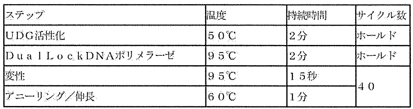
遺伝子発現量を解析した結果を図1に示す。遺伝子発現量は、グリセルアルデヒド-3-リン酸デヒドロゲナーゼ(GAPDH)の遺伝子発現量を1とした場合の相対値とし、±SD(n=3)で示した。
Tプレートを用いた場合(参考例2)は、cTnT、MYL2、Kir2.1、及びPGC1α(PPARGC1A)の発現量は、Cプレート(参考例1)よりも多かった。この結果から、Tプレートを用いて心筋細胞を培養すると、心筋細胞特異的な遺伝子の発現が上昇し、心筋細胞の機能を向上させ得ることが明らかになった。
試験用プレートとして、Cプレート(比較例1~9)、Tプレート(実施例1~9)を用いて、薬剤を添加し、カルシウムトランジェントを解析した。
<培養スケジュール>
細胞培養は下記のスケジュールで行った。細胞起眠日をDay0として以降の日を表す。
Day-1:試薬調製及び準備
Day0:凍結心筋細胞起眠、プレートのプレコート、及び試験用96ウェルプレートへの播種(工程(A))、培養(工程(B))
Day1:CDI維持培地で培地交換して培養(工程(C))
Day4~13:2日おきに特製維持培地で培地交換して培養(工程(C))
Day14:特製維持培地を用いて、薬剤添加、及びカルシウムトランジェント解析(工程(D)、工程(E))
特製基礎培地3mLに19.2μLのiMatrix-511silkを添加して希釈し、コーティング液とした。コーティング液を100μL/well(使用濃度1μg/cm2)で試験用96ウェルプレートに添加し、37℃インキュベーター中で1時間静置した。コーティング液は除去せずに、コーティング液を残したまま、心筋細胞を播種した。
CarmyA特製播種用培地にY-27632を終濃度10μMになるように添加し、特製解凍播種用培地として用いた。使用時に室温して使用した。
凍結心筋細胞を37℃のウォーターバスで解凍し、特製解凍播種用培地に懸濁した。300×gで5分間遠心し、上清を除去した。特製解凍播種用培地で細胞を懸濁し、2×10⁶~5×10⁶cells/mLに調整後、細胞数をカウントし、iMatrix-511silkコーティングした試験用96ウェルプレートに6×104cells/well(培地量200μL)で細胞を播種し、37℃、5%CO2インキュベーター内に静置した。実験はtriplicateで行った。
薬剤は、DMSOを終濃度0.1%、ベプリジルを終濃度1μM、2μM、4μM、ペンタミジンを終濃度11.1μM、33.3μM、100μM、イソプロテレノールを終濃度20nM、100nM、500nMとなるように添加した。
Tプレートにおいて、ベプリジルを終濃度1μM、2μM、4μM、ペンタミジンを終濃度11.1μM、33.3μM、100μM、イソプロテレノールを終濃度20nM、100nM、500nMとなるように添加したものをそれぞれ、実施例1~9とした。
Cプレートにおいて、ベプリジルを終濃度1μM、2μM、4μM、ペンタミジンを終濃度11.1μM、33.3μM、100μM、イソプロテレノールを終濃度20nM、100nM、500nMとなるように添加したものをそれぞれ、比較例1~9とした。
0.1%DMSOの結果を図2、1μMベプリジルの結果を図3、1μMベプリジルの結果のEAD様波形を拡大した結果を図4、2μMベプリジルの結果を図5、4μMベプリジルの結果を図6、11.1μMペンタミジンの結果を図7A、図7B、33.3μMペンタミジンの結果を図8A、図8B、100μMペンタミジンの結果を図9A、図9B、20nMイソプロテレノールの結果を図10、100nMイソプロテレノールの結果を図11、500nMイソプロテレノールの結果を図12に示す。Preは薬剤添加前、Postは薬剤添加後を示す。
DMSO、ベプリジル、又はペンタミジンを添加した場合は、添加10分後の結果を示す。イソプロテレノールを添加した場合は、添加10分後、24時間後、48時間後の結果を示す。
実施例4で不整脈が検出されたのがペンタミジン添加24時間後であることは、ペンタミジンの作用メカニズムを考慮すると合理的であるため、Tプレートにおいては、ペンタミジンのhERGタンパク質の膜移行(トラフィッキング)阻害作用を検出できていると考えられる。一方、Cプレート(比較例6)で検出された急性の不整脈は、ペンタミジン添加10分後であったから、hERGタンパク質の膜移行(トラフィッキング)阻害作用とは異なる非特異的な作用の可能性が高い。Tプレートでは、このような非特異的な作用が見られなかったことから、Tプレートを用いると、薬剤の作用を評価する際に偽陽性を減らすことができることが示唆された。
酸素透過性の高いPDMS製の試験用プレートを用いて、心筋細胞培養への影響を検討した。試験用プレートとしてGプレート(TCPS製、参考例3)、Tプレート(参考例4)、Vプレート(PDMS製、参考例5)を用いた。
細胞培養は下記のスケジュールで行った。細胞起眠日をDay0として以降の日を表す。
Day-1:試薬調製及び準備
Day0:特製解凍播種用培地を用いて、凍結心筋細胞起眠、プレートのプレコート、試験用96ウェルプレートへの播種、培養
Day1:特製維持培地で培地交換して培養
Day3~13:2日おきに特製維持培地で培地交換して培養
Day6:顕微鏡観察
播種6日後に顕微鏡を用いて明視野で細胞を観察し、写真を撮影した。また、蛍光顕微鏡でも観察して動画及び静止画を撮影した。
播種6日後において細胞を観察した写真を図13に示す。左は、Gプレートを用いた参考例3、中央はTプレートを用いた参考例4、右はVプレートを用いた参考例5の写真である。Gプレートを用いた参考例3、Tプレートを用いた参考例4では、心筋細胞は試験用プレートに充分に接着していた。また、蛍光観察したところ、同期した拍動様波形が検出された。一方、Vプレートを用いた参考例5では、心筋細胞は試験用プレートに充分に接着しておらず、細胞塊が見られた。また、蛍光観察したところ、同期した拍動様波形は検出されなかった。
(方法)
試験用プレートとしてTプレート又はGプレートを用い、播種1日目以降の培地として、特製維持培地又はCDI維持培地を用いて薬剤添加し、カルシウムトランジェントを解析した。
<培養スケジュール>
細胞培養は下記のスケジュールで行った。細胞起眠日をDay0として以降の日を表す。
Day-1:試薬調製及び準備
Day0:特製基礎培地を用いて、プレートのプレコートをおこない、特製解凍播種用培地を用いて、凍結心筋細胞起眠、試験用96ウェルプレートへの播種(工程(A))、培養(工程(B))
Day1:特製維持培地または、CDI維持培地で培地交換して培養(工程(C))
Day3~13:2日おきに特製維持培地又はCDI維持培地で培地交換して培養(工程(C))
Day14:培地交換、薬剤添加、カルシウムトランジェント解析(工程(D)、工程(E))
試験用プレートのiMatrix-511silkコーティングは、特製基礎培地でおこなった。凍結心筋細胞の起眠と播種は、特製解凍播種用培地を用いて行った。
播種1日目以降の培地交換、薬剤添加、及びカルシウムトランジェント解析に用いる培地を特製維持培地又はCDI維持培地とした。
薬剤は、DMSOを終濃度0.1%、ベプリジルを終濃度0.06μM、0.25μM、1μM、4μMとなるようにそれぞれ添加した。
Tプレートにおいて、播種1日目以降の培地として特製維持培地を用いて、ベプリジルを終濃度0.06μM、0.25μM、1μM、4μMとなるように添加したものをそれぞれ、実施例10~13、播種1日目以降の培地としてCDI維持培地を用いて、ベプリジルを終濃度0.06μM、0.25μM、1μM、4μMとなるように添加したものをそれぞれ、実施例14~17とした。
Gプレートにおいて、播種1日目以降の培地として特製維持培地を用いて、ベプリジルを終濃度0.06μM、0.25μM、1μM、4μMとなるように添加したものをそれぞれ、比較例10~13、播種1日目以降の培地としてCDI維持培地を用いて、ベプリジルを終濃度0.06μM、0.25μM、1μM、4μMとなるように添加したものをそれぞれ、比較例14~17とした。
結果を図14~図23に示す。「特製維持培地」とは、播種1日目以降の培地として特製維持培地を用いた場合、「CDI」とは播種1日目以降の培地としてCDI維持培地を用いた場合を示す。Preは薬剤添加前、Postは薬剤添加後を示す。
Tプレートの方が、Gプレートよりも拍動様波形が安定しており、シグナルが増強されていた。また、Tプレートにおいて播種1日目以降の培地として特製維持培地を用いた場合、CDI維持培地を用いた場合よりもさらに拍動様波形が安定して検出され、シグナルが増強されていた。
Gプレートにおいて、播種1日目以降の培地として特製維持培地を用いた場合、ベプリジルを1μM添加すると(比較例12)、ウェルによっても異なるが、カルシウムイオン波形が乱れ、拍動様波形は検出されなくなり、EAD様波形も、QT延長も検出されなかった。Gプレートにおいて、播種1日目以降の培地としてCDI維持培地を用いた場合、ベプリジルを1μM添加すると(比較例16)、カルシウムイオン波形は安定していたが、EAD様波形も、QT延長も検出されなかった(図21)。
試験用プレートとしてTプレートを用い、薬剤添加直前における培地交換の影響を検討した。また、試験用プレートとしてTプレートを用い、ベラパミルの、心筋細胞に対する作用を評価できるかを検討した。
(方法)
<試験スケジュール>
Day-1:試薬調製及び準備
Day0:特製基礎培地を用いて、Tプレートのプレコートをおこない、特製解凍播種用培地を用いて、凍結心筋細胞起眠、試験用96ウェルプレートへの播種(工程(A))、培養(工程(B))
Day1:特製維持培地で培地交換して培養(工程(C))
Day4~13:2日おきに特製維持培地で培地交換して培養(工程(C))
Day14:培地交換、薬剤添加、カルシウムトランジェント解析(工程(D)、工程(E))
<培地交換>
培地交換A:プレート内の特製維持培地を除去し、新しい特製維持培地を添加した。
培地交換B:プレート内の特製維持培地を除去し、新しい特製維持培地を添加して、すぐに除去した後、再度、新しい特製維持培地を添加した。
培地交換AまたはBを行ったプレートに、培地交換の約30分後に薬剤(ベラパミル)を添加した。薬剤は、DMSOに溶解して10mMストックソリューションを作製し、10mMストックソリューションを目標とする最終濃度の10倍濃度となるように特製維持培地で希釈して、添加用薬剤液とした。添加用薬剤液は、測定培地量の10分の1量を測定培地中に添加した。例えば、終濃度10nMでベラパミルを添加する場合、10mMストックソリューションを特製維持培地で希釈して100nMの添加用薬剤液を作製し、測定培地150μLに対して、添加用薬剤液を15μL添加した。対照群にはDMSOを終濃度0.1%で添加した。
培地交換AまたはBを行い、ベラパミルを終濃度100nMとなるように添加したものをそれぞれ、実施例20、21とした。
薬剤添加の10~30分後にカルシウムトランジェント解析を行った。
ベラパミルを終濃度10nMで添加した結果を図24、ベラパミルを終濃度100nMで添加した結果を図25に示す。Preは薬剤添加前、Postは薬剤添加後を示す。図24Aは培地交換AのPre、図24Bは培地交換BのPre、図24Cは培地交換AのPost、図24Dは培地交換BのPostの結果である。図25Aは培地交換AのPre、図25Bは培地交換BのPre、図25Cは培地交換AのPost、図25Dは培地交換BのPostの結果である。
本発明の一態様である方法により、ベラパミルの弱心作用(陰性変力作用)を検出することができた。
次に、試験用プレートとしてTプレートを用い、播種1日目以降の培地として特製維持培地を用いて、その他の薬剤の、心筋細胞に対する作用を評価できるかを検討した。
文献Aでは、臨床的なTdPのリスクレベル(高リスク、中リスク、低リスクまたは無リスク)が知られている28の薬物について、電気生理学的反応の濃度依存性と変動要因を評価し、ヒトiPS細胞を分化させた心筋細胞のin vitro催不整脈モデルとしての有用性を評価している。
臨床的なTdPのリスクレベルと、iPS細胞を分化させた心筋細胞を用いた催不整脈モデルで推測されるリスクレベルとは、相関する薬剤が多いが、例外的な薬剤、すなわち、iPS細胞を分化させた心筋細胞を用いた、従来の催不整脈モデルではリスクレベルの予測が困難である薬剤も存在している(文献A Figure6)。そこで、iPS細胞を分化させた心筋細胞を用いた、従来の催不整脈モデルではリスクレベルの予測が困難である薬剤の作用を、本発明の一態様である方法で評価した。
具体的には、臨床的なTdPのリスクレベルが高リスク又は中リスクであるのに、iPS細胞を分化させた心筋細胞を用いた、従来の催不整脈モデルではTdPのリスクレベルが低く予測されてしまう薬剤の代表例としてベプリジル、リスペリドン、テルフェナジンを用いた。また、臨床的なTdPのリスクレベルは低リスクであるのに、iPS細胞を分化させた心筋細胞を用いた、従来の催不整脈モデルではTdPのリスクレベルが高く予測されてしまう薬剤の代表例としてラノラジンを用いた。
<試験スケジュール>
Day-1:試薬調製及び準備
Day0:特製基礎培地を用いて、Tプレートのプレコートをおこない、特製解凍播種用培地を用いて、凍結心筋細胞起眠、試験用96ウェルプレートへの播種(工程(A))、培養(工程(B))
Day1:特製維持培地で培地交換して培養(工程(C))
Day4~13:2日おきに特製維持培地で培地交換して培養(工程(C))
Day14:培地交換、薬剤添加、カルシウムトランジェント解析(工程(D)、工程(E))
実施例18~21と同様の方法で薬剤を添加した。
ベラパミルを終濃度60nM、250nM、1000nM、4000nMとなるように添加したものをそれぞれ、実施例22、実施例23、実施例24、実施例25とした。
E-4031を終濃度1nM、10nM、100nM、1000nMとなるように添加したものをそれぞれ、実施例26、実施例27、実施例28、実施例29とした。
ベプリジルを終濃度60nM、250nM、1μM、40μMとなるように添加したものをそれぞれ、実施例30、実施例31、実施例32、実施例33とした。
ベプリジルを終濃度312nM、1.25μM、5μM、20μMとなるように添加したものをそれぞれ、実施例34、実施例35、実施例36、実施例37とした。
リスペリドンを終濃度1nM、10nM、0.1μM、1μMとなるように添加したものをそれぞれ、実施例38、実施例39、実施例40、実施例41とした。
リスペリドンを終濃度60nM、250nM、1μM、4μMとなるように添加したものをそれぞれ、実施例42、実施例43、実施例44、実施例45とした。
テルフェナジンを終濃度0.25nM、1nM、4nM、16nMとなるように添加したものをそれぞれ、実施例46、実施例47、実施例48、実施例49とした。
ラノラジンを終濃度10nM、100nM、1000nM、10000nMとなるように添加したものをそれぞれ、実施例50、実施例51、実施例52、実施例53とした。
実施例18~21と同様の方法でカルシウムトランジェント解析を行った。
カルシウムトランジェント解析で得られた波形から、CAD30(CaD30、calcium transient duration at 30%)及びCAD80(CaD80、calcium transient duration at 80%)を算出した。Intensityのピークの最小値が測定された時間をt0とした。ピークの最小値を0、ピーク最大値を1としたときの、ピーク最大値から30%減少した値(0.7)が測定された時間までのt0からの経過時間をCAD30、80%減少した値(0.2)が測定された時間までのt0からの経過時間をCAD80とした。概念図を図26に示す。
CAD30及びCAD80は、図26にその概念図を示す通り、QT時間の指標となる値である。対照に比べて、CAD30とCAD80の両方、またはどちらか一方が延長している場合はQT延長、CAD30とCAD80の両方、またはどちらか一方が短縮している場合は、QT短縮しているといえる。
結果を図27~図42に示す。Preは薬剤添加前、Postは薬剤添加後を示す。
(ベラパミルについて)
図27より、60nM、250nMベラパミルではIntensityがPreとPostでほとんど変化がなかった。データは示していないが、対照群(DMSOを添加)では、Preに比べてPostでIntensityが増加したことを考慮すると、60nM、250nMベラパミルではIntensityの増加が抑制されたといえる。つまり、ベラパミルはIntensityを小さく、言い換えると、心筋細胞の収縮性を減弱させており、ベラパミルの弱心作用(陰性変力作用)が検出された。1000nMでは、ベラパミルの弱心作用(陰性変力作用)がはっきりと検出された(図27)。図28より、1000nM、4000nM ベラパミルでは、Preに比べてPostでは、CAD30及びCAD80が減少した。すなわち、ベラパミルはQTを短縮した。また、これらの作用はベラパミルの濃度依存的であった。
本発明の一態様である方法により、ベラパミルの弱心作用(陰性変力作用)及びQT短縮作用を検出することができた。
E-4031はカリウムイオンチャネルブロッカーであり、EAD及びTdPを誘発することが知られている。E-4031は、臨床的なTdPのリスクレベルが高リスクであり、iPS細胞を分化させた心筋細胞を用いた、従来の催不整脈モデルでもTdPのリスクレベルが高く予測される薬剤である。
図29、図30より、10nMからQT延長が観察され、100nMにおいて、QT延長はより顕著であり、E-4031の催不整脈作用を濃度依存的に検出できた。
本発明の一態様である方法により、E-4031の催不整脈作用を検出することができた。
ベプリジルは、元々は抗狭心症薬として承認されたカルシウム拮抗薬であるが、IV群に分類される抗不整脈薬(Vaughan Williams分類による)でもある。ベプリジルは、カルシウムイオンチャネルだけでなく、ナトリウムイオンチャネル及びカリウムイオンチャネルも抑制する、いわゆるmulti-ionchannel blocking作用を持つ。ベプリジルは、カリウムイオンチャネル抑制作用に基づくQT延長及びtorsades de pointes(TdP)を誘発することが知られている。
図31、図32より、60nM~4000nMのベプリジル添加により、QT延長が検出されたが、濃度依存性は見られなかった。1μMにおいては、EAD様波形が検出された。次に、ベプリジルの濃度範囲をより狭くして実験を行った。図33、図34より、312nM、1250nMのベプリジル添加により、QT延長が検出された。
本発明の一態様である方法により、ベプリジルの催不整脈作用を検出することができた。
リスペリドンは、ドーパミン2受容体とセロトニン2A受容体の拮抗作用を有する非定型抗精神病薬であるが、QT延長を誘発することが知られている。
本発明の一態様である方法により、リスペリドンの催不整脈作用を検出することができた。
テルフェナジンは、ヒスタミンH1受容体拮抗薬であるが、QT延長、TdPを起こし、心停止を起こす報告が相次いだために市場から撤退した。現在では、テルフェナジンの活性代謝物であり、催不整脈作用のないフェキソフェナジンが後継の抗アレルギー薬として汎用されている。
本発明の一態様である方法により、テルフェナジンの催不整脈作用を検出することができた。
ラノラジンはナトリウムイオンチャネルブロッカーである。ラノラジンは、in vivoで不整脈を引き起こすリスクは低いが、iPS細胞を分化させた心筋細胞ではQT延長が検出され、催不整脈作用を有するものとして判定されてしまうことが知られている。すなわち、ラノラジンは、従来のiPS細胞を分化させた心筋細胞を用いた催不整脈モデルでは偽陽性として判定されることが知られている。
本発明の一態様である方法によれば、ラノラジンは催不整脈作用を有するものとは評価されなかった。すなわち、本発明の一態様である方法によれば、iPS細胞を分化させた心筋細胞を用いた従来の催不整脈モデルでは偽陽性として判定される薬剤であっても、正しく陰性として判定することができ、偽陽性を減らすことができる。
この出願は、2022年1月11日に日本国特許庁に出願された特願2022-002556号及び2022年6月29日に日本国特許庁に出願された特願2022-104613号を基礎とする優先権を主張し、その開示の全てをここに取り込む。
Claims (17)
- 薬剤の、心筋細胞に対する作用を評価する方法であって、
培養容器の培養面に心筋細胞を播種する工程(A)、
前記工程(A)で得られた心筋細胞を培養する工程(B)、
前記培養された心筋細胞を前記薬剤に曝露させる工程(D)、及び
前記工程(D)で得られた心筋細胞の細胞機能の指標を分析して評価する工程(E)を含み、
前記培養容器の培養面の少なくとも一部が4-メチル-1-ペンテン重合体を含む基材から形成されている、方法。 - 前記工程(B)で得られた心筋細胞をさらに培養する工程(C)を含み、
前記工程(C)及び(D)が、遊離脂肪酸1~100μg/mLを含有する培地(β)中で行われる、請求項1に記載の方法。 - 前記培地(β)が、更に、リゾホスファチジルコリン1~100μg/mL、トリアシルグリセリド1~100μg/mL、ホスファチジルコリン1~100μg/mL、ホスファチジン酸1~100μg/mL、コレステロール0.1~10μg/mL、及びスフィンゴミエリン0.1~10μg/mLから選ばれる少なくとも1種を含有する、請求項2に記載の方法。
- 前記工程(A)及び(B)が、代替血清を含有する無血清培地(α)中で行われる、請求項1又は2に記載の方法。
- 前記4-メチル-1-ペンテン重合体が、4-メチル-1-ペンテンとエチレン及び炭素数3~20のα-オレフィン(4-メチル-1-ペンテンを除く)から選ばれる少なくとも1種のオレフィンとの共重合体である、請求項1又は2に記載の方法。
- 前記培養容器の培養面全体が、4-メチル-1-ペンテン重合体を含む基材から形成されている、請求項1又は2に記載の方法。
- 前記培養面が、ラミニンを含むコーティング剤でコートされている、請求項1又は2に記載の方法。
- 前記心筋細胞が、人工多能性幹細胞を分化させた心筋細胞である、請求項1又は2に記載の方法。
- 前記心筋細胞が、人工多能性幹細胞をプロテインフリー心筋分化誘導(PFCD)法により分化させた心筋細胞である、請求項1又は2に記載の方法。
- 前記作用が、心毒性である、請求項1又は2に記載の方法。
- 前記心毒性が、催不整脈作用である、請求項10に記載の方法。
- 前記心毒性が、心筋傷害作用である、請求項10に記載の方法。
- 前記細胞機能の指標が、ミトコンドリア機能の指標である、請求項12に記載の方法。
- 前記細胞機能の指標が、カルシウムイオン波形である、請求項1又は2に記載の方法。
- 前記工程(E)において、早期後脱分極(EAD)を検出する、請求項1又は2に記載の方法。
- 前記作用が、頻脈作用又は徐脈作用である、請求項1又は2に記載の方法。
- 前記細胞機能の指標が、カルシウムイオン波形である、請求項16に記載の方法。
Priority Applications (1)
| Application Number | Priority Date | Filing Date | Title |
|---|---|---|---|
| JP2023574023A JP7798302B2 (ja) | 2022-01-11 | 2023-01-10 | 薬剤の、心筋細胞に対する作用を評価する方法 |
Applications Claiming Priority (4)
| Application Number | Priority Date | Filing Date | Title |
|---|---|---|---|
| JP2022002556 | 2022-01-11 | ||
| JP2022-002556 | 2022-01-11 | ||
| JP2022104613 | 2022-06-29 | ||
| JP2022-104613 | 2022-06-29 |
Publications (1)
| Publication Number | Publication Date |
|---|---|
| WO2023136229A1 true WO2023136229A1 (ja) | 2023-07-20 |
Family
ID=87279063
Family Applications (1)
| Application Number | Title | Priority Date | Filing Date |
|---|---|---|---|
| PCT/JP2023/000252 Ceased WO2023136229A1 (ja) | 2022-01-11 | 2023-01-10 | 薬剤の、心筋細胞に対する作用を評価する方法 |
Country Status (2)
| Country | Link |
|---|---|
| JP (1) | JP7798302B2 (ja) |
| WO (1) | WO2023136229A1 (ja) |
Citations (3)
| Publication number | Priority date | Publication date | Assignee | Title |
|---|---|---|---|---|
| JP2016521571A (ja) * | 2013-06-11 | 2016-07-25 | プルーリオミクス・ベー・フェー | 多能性哺乳動物幹細胞に由来する心筋細胞を成熟させるための培地組成物 |
| WO2019131806A1 (ja) * | 2017-12-26 | 2019-07-04 | 株式会社マイオリッジ | 心筋細胞の薬剤応答性試験方法 |
| WO2020256079A1 (ja) * | 2019-06-21 | 2020-12-24 | 三井化学株式会社 | 培養材およびその用途 |
-
2023
- 2023-01-10 WO PCT/JP2023/000252 patent/WO2023136229A1/ja not_active Ceased
- 2023-01-10 JP JP2023574023A patent/JP7798302B2/ja active Active
Patent Citations (3)
| Publication number | Priority date | Publication date | Assignee | Title |
|---|---|---|---|---|
| JP2016521571A (ja) * | 2013-06-11 | 2016-07-25 | プルーリオミクス・ベー・フェー | 多能性哺乳動物幹細胞に由来する心筋細胞を成熟させるための培地組成物 |
| WO2019131806A1 (ja) * | 2017-12-26 | 2019-07-04 | 株式会社マイオリッジ | 心筋細胞の薬剤応答性試験方法 |
| WO2020256079A1 (ja) * | 2019-06-21 | 2020-12-24 | 三井化学株式会社 | 培養材およびその用途 |
Also Published As
| Publication number | Publication date |
|---|---|
| JPWO2023136229A1 (ja) | 2023-07-20 |
| JP7798302B2 (ja) | 2026-01-14 |
Similar Documents
| Publication | Publication Date | Title |
|---|---|---|
| Choi et al. | 3D bioprinted vascularized lung cancer organoid models with underlying disease capable of more precise drug evaluation | |
| Wu et al. | Arp2/3 is critical for lamellipodia and response to extracellular matrix cues but is dispensable for chemotaxis | |
| KR102646444B1 (ko) | 배양재 및 그의 용도 | |
| Mabry et al. | Microarray analyses to quantify advantages of 2D and 3D hydrogel culture systems in maintaining the native valvular interstitial cell phenotype | |
| Tu et al. | Rapid prototyping of concave microwells for the formation of 3D multicellular cancer aggregates for drug screening | |
| Cheng et al. | Three-dimensional polymer scaffolds for high throughput cell-based assay systems | |
| CA3135597C (en) | Compositions and methods of using partial gel layers in a microfluidic device | |
| EP2710111B1 (en) | Coated fiber scaffold for three dimensional cell culture of neural cells | |
| WO2017083705A1 (en) | Cell culture system and method of use thereof | |
| JP2009535025A (ja) | 三次元での細胞培養のルーチン成長のための基材 | |
| CN105849254A (zh) | 用于纯化视网膜色素上皮细胞的方法 | |
| Lindner et al. | Flow-induced glycocalyx formation and cell alignment of HUVECs compared to iPSC-derived ECs for tissue engineering applications | |
| Chakraborty et al. | Droplet microarrays for cell culture: effect of surface properties and nanoliter culture volume on global transcriptomic landscape | |
| EP2902496B1 (en) | Method for evaluating effect of cytokine on metabolic activity of cytochrome p450, and drug screening method | |
| EP4263804A1 (en) | A dual cardiac-blood model system for disease modelling and drug screening | |
| US20120302467A1 (en) | Patterned substrates for cell applications | |
| EP3699265A1 (en) | Method for producing pluripotent stem cells to be differentiated into cardiomyocytes | |
| JP7798302B2 (ja) | 薬剤の、心筋細胞に対する作用を評価する方法 | |
| US20250002866A1 (en) | Cell culture platforms, methods and uses thereof | |
| AU2021377418A1 (en) | Organoid culture platform | |
| Pradhan et al. | In situ pH maintenance for mammalian cell cultures in shake flasks and tissue culture flasks | |
| D’Antonio-Chronowska et al. | Human iPSC gene signatures and X chromosome dosage impact response to WNT inhibition and cardiac differentiation fate | |
| US11713441B2 (en) | Cell culture substrates, methods and uses thereof | |
| CN121794367A (zh) | 核壳结构的三维结构物 | |
| JP2022154850A (ja) | 心筋細胞の培養方法 |
Legal Events
| Date | Code | Title | Description |
|---|---|---|---|
| 121 | Ep: the epo has been informed by wipo that ep was designated in this application |
Ref document number: 23740238 Country of ref document: EP Kind code of ref document: A1 |
|
| ENP | Entry into the national phase |
Ref document number: 2023574023 Country of ref document: JP Kind code of ref document: A |
|
| NENP | Non-entry into the national phase |
Ref country code: DE |
|
| 32PN | Ep: public notification in the ep bulletin as address of the adressee cannot be established |
Free format text: NOTING OF LOSS OF RIGHTS PURSUANT TO RULE 112(1) EPC (EPO FORM 1205A DATED 17/10/2024) |
|
| 122 | Ep: pct application non-entry in european phase |
Ref document number: 23740238 Country of ref document: EP Kind code of ref document: A1 |