WO2023120232A1 - 呼吸教示装置 - Google Patents
呼吸教示装置 Download PDFInfo
- Publication number
- WO2023120232A1 WO2023120232A1 PCT/JP2022/045407 JP2022045407W WO2023120232A1 WO 2023120232 A1 WO2023120232 A1 WO 2023120232A1 JP 2022045407 W JP2022045407 W JP 2022045407W WO 2023120232 A1 WO2023120232 A1 WO 2023120232A1
- Authority
- WO
- WIPO (PCT)
- Prior art keywords
- breathing
- abdomen
- user
- abdominal
- teaching device
- Prior art date
- Legal status (The legal status is an assumption and is not a legal conclusion. Google has not performed a legal analysis and makes no representation as to the accuracy of the status listed.)
- Ceased
Links
Images


Classifications
-
- A—HUMAN NECESSITIES
- A61—MEDICAL OR VETERINARY SCIENCE; HYGIENE
- A61B—DIAGNOSIS; SURGERY; IDENTIFICATION
- A61B5/00—Measuring for diagnostic purposes; Identification of persons
- A61B5/103—Measuring devices for testing the shape, pattern, colour, size or movement of the body or parts thereof, for diagnostic purposes
- A61B5/11—Measuring movement of the entire body or parts thereof, e.g. head or hand tremor or mobility of a limb
- A61B5/113—Measuring movement of the entire body or parts thereof, e.g. head or hand tremor or mobility of a limb occurring during breathing
-
- A—HUMAN NECESSITIES
- A61—MEDICAL OR VETERINARY SCIENCE; HYGIENE
- A61N—ELECTROTHERAPY; MAGNETOTHERAPY; RADIATION THERAPY; ULTRASOUND THERAPY
- A61N1/00—Electrotherapy; Circuits therefor
- A61N1/02—Details
- A61N1/04—Electrodes
-
- A—HUMAN NECESSITIES
- A61—MEDICAL OR VETERINARY SCIENCE; HYGIENE
- A61N—ELECTROTHERAPY; MAGNETOTHERAPY; RADIATION THERAPY; ULTRASOUND THERAPY
- A61N1/00—Electrotherapy; Circuits therefor
- A61N1/18—Applying electric currents by contact electrodes
- A61N1/32—Applying electric currents by contact electrodes alternating or intermittent currents
- A61N1/36—Applying electric currents by contact electrodes alternating or intermittent currents for stimulation
Definitions
- the present invention relates to a breathing teaching device that measures a user's electrocardiogram or myoelectricity and teaches breathing according to the measurement result by electrical stimulation.
- abdominal breathing has been used to calm the heightened sympathetic nerves and activate the parasympathetic nerves to regulate the autonomic nerves, as well as to take in a large amount of oxygen into the body, which activates immune cells such as white blood cells and lymphocytes. This is believed to enhance immune function.
- Patent Document 1 discloses a technique for detecting abdominal respiration by wearing a belly band and expanding the abdomen. disclosed.
- the abdominal band vibrates or emits an electronic sound in accordance with the detected respiration, thereby notifying that the respiration is abdominal respiration or actively encouraging abdominal respiration.
- the sensor for measuring the electrocardiogram is generally placed near the heart. It is disclosed that an electrocardiogram is measured from the abdomen by providing electrodes.
- the electrocardiogram can be measured in the abdomen, but the relationship between the electrocardiogram and respiration is not considered.
- a breathing teaching device worn by a user a measurement unit that contacts the user's abdomen and measures electrocardiogram or myoelectricity at the abdomen; an electrical stimulator that is in contact with the abdomen and applies low-frequency stimulation to the abdomen; a control unit that controls the electrical stimulation unit to prompt the user to perform abdominal breathing that should be recommended to the user according to the measurement result of the electrocardiogram or abdominal myoelectricity;
- a breathing teaching device a measurement unit that contacts the user's abdomen and measures electrocardiogram or myoelectricity at the abdomen; an electrical stimulator that is in contact with the abdomen and applies low-frequency stimulation to the abdomen; a control unit that controls the electrical stimulation unit to prompt the user to perform abdominal breathing that should be recommended to the user according to the measurement result of the electrocardiogram or abdominal myoelectricity;
- the breathing teaching device can teach appropriate breathing in consideration of the electrocardiogram without impairing the usability of the user.
- FIG. 4 is a diagram showing an example of a wearing part in the breathing teaching device of the first embodiment
- 1 is a functional block diagram of a breathing teaching device according to Configuration Example 1 of a first embodiment of the present invention
- FIG. FIG. 2 is a functional block diagram of a breathing teaching device according to Configuration Example 2 of the first embodiment of the present invention
- FIG. 4 is a diagram showing positions where electrodes are attached to muscles in the breathing teaching device of the first embodiment
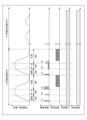
- FIG. 4 is a diagram showing an example of waveforms of timing and electric signals of the breathing teaching device in the first embodiment
- FIG. 4 is a diagram showing schematic exchanges in the breathing teaching device of the first embodiment
- FIG. 2 is a block diagram of a breathing teaching device according to a second embodiment of the present invention
- the present invention relates to a breathing teaching device, more specifically, to a breathing teaching device that measures a user's electrocardiogram or myoelectricity and teaches breathing according to the measurement result by electrical stimulation.
- “teaching breathing” means teaching abdominal breathing.
- Abdominal respiration is a breathing method (abdominal respiration) performed mainly by stretching the diaphragm, and is a breathing method in which the abdomen is expanded during inhalation and depressed during exhalation without moving the ribcage as much as possible.
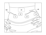
- FIG. 1 is a diagram showing the overall configuration of the breathing teaching device of the first embodiment.
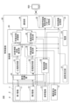
- FIG. 2 is a functional block diagram of the breathing teaching device according to the first embodiment of the present invention.
- the breathing teaching device of the present invention is a wearable device that is worn by a user.
- a breathing teaching function is imparted to a wearable part such as a belly band, belt, leggings, etc. that is in close contact with the abdomen of the user. ing.
- a wearable part such as a belly band, belt, leggings, etc. that is in close contact with the abdomen of the user. ing.
- the applied portion is a belly band.
- the breathing teaching device 100 includes a first electrode 11, a second electrode 12, a reference electrode 13, an abdominal displacement measuring section 14, and a control board for a bellyband 40. 20 are provided.
- the first electrode 11, the second electrode 12, and the reference electrode (third electrode) 13 have conductive properties and are made of metal, conductive fiber, conductive rubber, or the like.
- the first electrode 11 and the second electrode 12 are measuring units that contact the user's abdomen to measure the electrocardiogram in the abdomen, and are measuring units that contact the user's abdomen to measure the myoelectric potential in the abdomen. It also functions as an electrical stimulator that comes into contact with the abdomen and provides low-frequency stimulation to the abdomen.
- the electrocardiogram and the electromyogram are simultaneously measured by the first electrode 11 and the second electrode 12, and the overlapping waveforms of the two measured signals are demultiplexed by digital signal processing or an analog demultiplexing filter. .
- the measurement and the electrical stimulation are time-divided so that they do not occur at the same time.
- the first electrode 11 and the second electrode measure electrocardiogram and myoelectricity in a time-division manner. Then, the measurement and the electrical stimulation are time-divided so that they do not occur at the same time.
- the abdomen displacement measuring unit 14 comes into contact with the abdomen of the user and measures the displacement of the abdomen such as expansion and contraction of the abdomen to detect breathing.
- the control board 20 is provided with a control unit 30 and the like, and the control unit 30 prompts the user to perform recommended abdominal breathing according to the measurement result of the electrocardiogram or abdominal myoelectricity. control the electrodes. At this time, according to the timing of the user's measured breathing, low-frequency electrical stimulation is applied to the abdomen to encourage the implementation of recommended abdominal breathing according to the measurement results of electrocardiogram or abdominal myoelectricity. preferable.
- FIG. 2 is a functional block diagram of the breathing teaching device 100 according to Configuration Example 1 of the first embodiment of the present invention.
- the breath teaching device 100 of the present invention and an information processing device such as the smartphone 600 that displays evaluation results of breathing teaching and performs various settings for teaching breathing are collectively referred to as a breathing teaching system.
- the control board 20 is provided with a control section 30 , an attitude measurement section 21 , storage sections 22 and 23 , a communication section 24 and a power supply 25 .
- the posture measurement unit (first posture measurement unit) 21 measures the posture by contacting, for example, the plenum of the user's abdomen. As an example, posture measurement unit 21 always measures the posture of the user while the power is on.
- the recommended breath-by-breath electrical stimulation pattern storage unit 22 is a database of electrical stimulation patterns configured with NVRAM (Non-Volatile Random Access Memory).
- the characteristic information storage unit 23 is a rewritable non-volatile memory made up of NVRAM, and stores individual breathing characteristics and the like.
- the communication unit 24 is a communication unit capable of short-distance communication with an external device such as the smartphone 600.
- the communication method is, for example, Bluetooth (registered trademark) or Wifi (registered trademark).
- the power supply 25 is a battery or rechargeable battery and supplies power to the control unit 30 .
- the power supply 25 may be supplied with power from a commercial power supply connected by a cable or the like.
- the control unit 30 is composed of, for example, a CPU (Central Processing Unit), FPGA (Field Programmable Gate Array), ASIC (Application Specific Integrated Circuit), and the like.
- CPU Central Processing Unit
- FPGA Field Programmable Gate Array
- ASIC Application Specific Integrated Circuit
- the control unit 30 controls the electrical stimulation unit to prompt the user to perform recommended abdominal breathing according to the electrocardiographic and/or abdominal myoelectric measurement results.
- the control unit 30 includes an electrocardiogram acquisition unit 31, a myoelectricity acquisition unit 32, an electrical stimulation setting unit 33, a signal separation unit 34, an autonomic nervous state estimation unit 35, a respiratory state estimation unit 36, a respiratory state evaluation unit 37, and a recommended breathing unit. It has a setting unit 38 in an executable manner.
- the first electrode 11, the second electrode 12, the electrocardiogram acquisition unit 31, and the signal separation unit 34 function as an electrocardiogram measurement unit ⁇ .
- the first electrode 11, the second electrode 12, the reference electrode 13, the myoelectric potential acquiring unit 32, and the signal separating unit 34 function as an electromyographic measuring unit ⁇ .
- the first electrode 11, the second electrode 12, and the electrical stimulation setting section 33 function as an electrical stimulation section ⁇ .
- the signal separation unit 34 separates the overlapping waveforms of the electrocardiogram and myoelectric signals simultaneously measured by the electrodes 11, 12 or the electrodes 11, 12, 13 by digital signal processing or an analog demultiplexing filter. Wave and separate into ECG data and EMG data.
- the electrocardiographic acquisition unit 31 performs signal processing on the electrocardiographic data measured by the electrodes 11 and 12 and separated by the signal separation unit 34 to measure the heart rate, PR interval, QT interval, QRS width interval, amplitude, and the like.
- the autonomic nerve state estimating unit 35 estimates the user's autonomic nerve state and the like from various numerical values of the acquired electrocardiogram data. For example, by spectrally analyzing the periodic fluctuations (R-R) of the heartbeat and comparing the increase and decrease of frequency components in the range of intermediate frequency components (0.05-0.20Hz) and high frequency components (0.20-0.35Hz), the state of the autonomic nervous system can be estimated. do.
- R-R periodic fluctuations
- the myoelectricity acquisition unit 32 performs signal processing on the myoelectricity measurement data measured by the electrodes 11, 12, and 13 and separated by the signal separation unit 34, and extracts necessary waveforms.
- the respiratory state estimating unit 36 performs signal processing on the waveform extracted as myoelectric data (myoelectric measurement data) by the myoelectricity acquisition unit 32, and determines the respiratory state such as whether abdominal respiration is properly performed. .
- the respiratory state estimating unit 36 described above may also refer to the movement of the abdomen measured by the abdominal displacement measuring unit 14 and determine the respiratory state together with the electromyographic data of the abdomen.
- the respiratory state estimating unit 36 may also refer to the user's posture and motion state measured by the posture measuring unit 21 to determine the respiratory state by lying down with the electromyographic data of the abdomen.
- the respiratory state evaluation unit 37 evaluates whether abdominal respiration is performed appropriately, as necessary, based on the acquired and signal-processed electromyographic measurement data and electrocardiographic measurement data.
- the recommended breathing setting unit 38 sets recommended breathing based on the state of the autonomic nerves estimated by the autonomic nerve state estimating unit 35 and the respiratory state estimated by the respiratory state estimating unit 36 .
- the electrical stimulation setting unit 33 sets the electrical stimulation to be applied to the user at the inhalation/exhalation timing set in the recommended breathing setting unit 38 in accordance with the breathing method.
- the electrical stimulation set here is electrical stimulation by a weak current such as low-frequency EMS (Electrical Muscle Stimulation).
- EMS Electromuscle Stimulation
- the sensory nerves in the abdomen are acted upon by low-frequency electrical stimulation. Therefore, unlike electrical stimulation by vibration, which encourages electrical hyperactivity by passing an electric current through muscles (motor nerves), the stimulation is weak, so breathing can be taught without impairing the user's usability. .
- the electrical stimulation setting unit 33 selects the electrical stimulation to be applied from the recommended per-breath electrical stimulation pattern storage unit 22 .
- the characteristic information storage unit 23 may store the characteristics of each case, such as the actual state of breathing after the electrical stimulation is applied. In that case, when setting the electrical stimulation pattern in the electrical stimulation setting unit 33, the electrical stimulation pattern may be set by referring to the history of the user's past respiratory information stored in the characteristic information storage unit 23. .
- the communication unit 24 wirelessly communicates with an information processing device such as the smartphone 600 of the user.
- an information processing device such as the smartphone 600 of the user.
- the user can save each measurement data and make various settings such as the breathing method and its teaching method. Further, when evaluating breathing, the evaluation result evaluated by the respiratory state evaluation unit 37 is added to various measurement results, and the result is transmitted to the smartphone 600 .
- control unit 30 prompts the user to perform recommended abdominal breathing according to the measurement results of the electrocardiogram and/or the electromyography of the abdomen in accordance with the timing of the measured breathing of the user.
- the electrical stimulator ⁇ is controlled so as to apply low-frequency electrical stimulation to the abdomen of the person.
- control unit 30 (1) myoelectric data, (2) electrocardiographic data, (3) displacement of the abdomen, (4) posture, Among them, including (1) or / and (2), (3), (4) data may be added, and the recommended breathing can be set by referring to 1 to 4 of the above .
- control unit 30 can estimate the state of the autonomic nerves based on the electrocardiogram measurement results and the detected abdominal displacement, and set recommended abdominal breathing.
- control unit 30 can estimate the depth of respiration and the respiration cycle, and set recommended abdominal respiration. can.
- the timing of breathing based on the detected displacement of the abdomen and the measurement result of the electrocardiogram of the abdomen is based on the measurement result of the electrocardiogram. It is possible to control the electrical stimulator ⁇ so as to apply low-frequency electrical stimulation to the abdomen at the start timings of the intake period, breath-holding period, and exhalation period of recommended abdominal respiration.
- human breathing includes chest breathing in addition to abdominal breathing.
- Thoracic respiration is respiration (chest respiration) performed mainly by movement of the ribs by the intercostal muscles.
- thoracic respiration is dominant over abdominal respiration, and generally unconsciously thoracic respiration occurs at rest. Therefore, for example, in the case of a female, the control unit 30 determines the breathing state of the user based on the detected displacement of the abdomen and the measurement result of the electromyography of the abdomen, and if the breathing is not abdominal breathing, Electrical stimulation may be given.
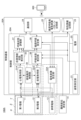
- FIG. 3 is a functional block diagram of a breathing teaching device 100A according to configuration example 2 of the first embodiment of the present invention. Only differences from FIG. 2 will be described.
- an exclusive operation control section 39 is provided instead of the signal separation section 34 in the control section 30A.
- the exclusive operation control unit 39 exclusively switches between electrocardiogram data and myoelectric data to be measured by the first electrode 11 and the second electrode 12 and acquires them.
- the electrocardiographic acquisition unit 31 performs signal processing on the electrocardiographic data exclusively measured by the electrodes 11 and 12 to measure the heart rate, PR interval, QT interval, QRS width interval, amplitude, and the like. Further, the myoelectric potential acquiring unit 32 performs signal processing on the myoelectric potential measurement data exclusively measured by the electrodes 11, 12, and 13 to extract a necessary waveform. Subsequent calculations are the same as in FIG.
- signals are acquired exclusively, so electrocardiogram data and myoelectric data can be acquired without separating the signals.
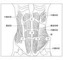
- FIG. 4 is a diagram showing positions where electrodes are attached to muscles in the breathing teaching device of the first embodiment.
- the electrodes are located at the positions shown in FIG. With the first electrode 11 and the second electrode 12 at such positions, in order to obtain myoelectric data, the rectus abdominis muscle and the external oblique muscle are measured as surface myoelectric data, and in order to obtain electrocardiographic data, Measure the movement of the transversus abdominis muscle. Note that the reference electrode 13 contacts the hipbone or the like without contacting the muscles.
- the electrodes 11 and 12 for realizing one function such as measuring electrocardiogram, measuring myoelectricity of the abdominal muscles, or performing electrical stimulation are provided symmetrically.
- the reference electrode 13 is provided at a location that is not affected by myoelectricity, for example, at a location that contacts the hipbone.
- the abdomen displacement measuring unit 14 is provided in the umbilical region around the navel. Note that the position of the abdomen displacement measuring unit 14 is not limited to this position as long as the circumference of the abdomen can be measured.
- a posture measurement unit (first posture measurement unit) 21 provided on the control board 20 measures the posture by contacting, for example, the plenum of the user's abdomen.
- FIG. 5 shows an example of waveforms of the timing of the breathing teaching device and the operation of the electric signal in the first embodiment.
- a period Ta is a period during which the abdominal breathing method is taught
- a period Tb is a period during which the abdominal breathing method is not taught.
- the electrocardiogram is constantly observed. Autonomic nerve balance and stress state are derived from electrocardiogram measurement, and the user is prompted to perform abdominal breathing.
- the posture is constantly observed.
- the user's posture is important for performing the abdominal breathing method, and when the user's posture is bad during the teaching of the abdominal breathing method, correction of the posture is notified.
- the abdominal breathing method is not performed, when the user's posture deteriorates (hunchback, warped waist, etc.), the user is notified and urged to correct it.
- the posture data is also used to interpolate the data obtained by the respiration measurement unit. Additionally, posture measurement data may be used to infer what the user is doing.
- the electrical stimulator is controlled to apply low-frequency electrical stimulation to the abdomen at the start timings of the intake period, breath-holding period, and exhalation period of recommended abdominal breathing. do.
- Myoelectricity is measured mainly during exhalation, and it is measured whether the user is exhaling using the abdominal muscles and whether the user is exhaling sufficiently, and the quality of the breathing method is determined.
- the user can know the effect of the taught breathing method.
- the electrical stimulation may adversely affect electrocardiographic measurement results as noise, so electrical stimulation and electrocardiographic measurement are controlled to operate exclusively. preferred.
- FIG. 6 is a diagram showing an example of a stepwise control sequence in the breathing teaching device of the first embodiment.
- FIG. 6 shows what timing and what kind of breathing method instruction the user can receive.
- the breathing teaching device 100 measures the electrocardiogram, respiration, and posture of the user's unconscious breathing during the period T1, and evaluates the user's mental and physical condition from evaluation 1 in which the measurement results are evaluated. and judge whether it is better to do abdominal breathing. Then, when it is determined that abdominal breathing should be performed, the user receives a warning (electrical stimulation) from the breathing teaching device 100 .
- the user who was warned at the beginning of period T2 voluntarily performs abdominal breathing in his/her own way, and continuously voluntarily performs abdominal breathing in T2 and T3.
- the breathing teaching device 100 measures the electrocardiogram, respiration, and posture during the period T3, and then obtains evaluation 2 again.
- the user will be instructed and evaluated by combining the instruction of electrical stimulation from this device and the respiration evaluation of myoelectric measurement.
- Receive instruction on abdominal breathing For example, in addition to teaching electrical stimulation, the user is visually instructed on the breathing state and instructions on the smartphone screen. Further, for example, the respiratory state may be measured by myoelectricity and abdominal displacement in the first respiration, and the depth and length of abdominal respiration by electrical stimulation in the second respiration may be taught based on the results.
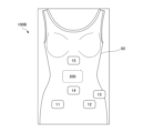
- FIG. 7 is a diagram showing the overall configuration of the breathing teaching device of the second embodiment.
- FIG. 8 is a functional block diagram of the breathing teaching device according to the first embodiment of the present invention.
- a breathing teaching function is imparted to a wearing part 50 that is worn as clothes in contact with the user's chest and abdomen, such as a vest, tank top, camisole, bra top, and the like. ing.
- a wearing part 50 that is worn as clothes in contact with the user's chest and abdomen, such as a vest, tank top, camisole, bra top, and the like.
- the breathing teaching device of this embodiment includes a second posture measuring unit 15 that contacts the user's chest and measures the posture.
- the second posture measurement unit 15 is arranged in the chest area, thereby making it possible to measure breathing movements of both the abdomen and the chest.
- the main effect of abdominal breathing was to calm the sympathetic nerves by making the parasympathetic nerves dominant, but the effect of thoracic breathing was to make the sympathetic nerves dominant and to get motivated and get excited. effective.
Landscapes
- Health & Medical Sciences (AREA)
- Life Sciences & Earth Sciences (AREA)
- Engineering & Computer Science (AREA)
- Biomedical Technology (AREA)
- Animal Behavior & Ethology (AREA)
- General Health & Medical Sciences (AREA)
- Public Health (AREA)
- Veterinary Medicine (AREA)
- Nuclear Medicine, Radiotherapy & Molecular Imaging (AREA)
- Radiology & Medical Imaging (AREA)
- Physiology (AREA)
- Dentistry (AREA)
- Oral & Maxillofacial Surgery (AREA)
- Physics & Mathematics (AREA)
- Biophysics (AREA)
- Pathology (AREA)
- Heart & Thoracic Surgery (AREA)
- Medical Informatics (AREA)
- Molecular Biology (AREA)
- Surgery (AREA)
- Measurement And Recording Of Electrical Phenomena And Electrical Characteristics Of The Living Body (AREA)
Abstract
使用者に装着される呼吸教示装置であって、使用者の腹部と接触し、腹部で心電又は筋電を測定する測定部と、腹部と接触し、腹部に刺激を与える電気刺激部と、心電又は腹部の筋電の測定結果に応じて、使用者に推奨すべき腹式呼吸の実施を促すように前記電気刺激部を制御する制御部と、を備える。
Description
本発明は、使用者の心電又は筋電を測定して、測定結果に応じた呼吸の教示を電気刺激により行う呼吸教示装置に関する。
従来から、腹式呼吸を行うことにより、高まった交感神経を鎮め副交感神経がはたらくようにして、自律神経を整えるとともに、酸素が体内に多く取り込まれ白血球やリンパ球などの免疫細胞が活発になることで、免疫機能が高まると考えられている。
そのため、意識的に腹式呼吸を促すように、例えば、特許文献1には、腹式呼吸を検知する方法として、腹巻を装着して腹部が膨らんだことで腹式呼吸を検知する技術とが開示されている。この技術では、腹巻が、検出した呼吸に合わせて振動する、あるいは電子音が鳴ることで、呼吸が腹式呼吸であることを通知したり、積極的に腹式呼吸を促したりする。
また、安静時には、呼吸と心拍は別の筋肉の働きによって実施されるが、心理的な緊張などストレスを感じた時や、トレーニング時には、心拍と呼吸に共通してイレギュラーな反応が現れることが知られている。
ここで、心電を測定するセンサーは心臓の近くに配置されることが一般的であるが、特許文献2では、腹部に装着するトレーニング用のベルトに、胸郭の最下点に位置するように電極を設けることで、その電極により腹部から心電を測定することが開示されている。
しかし、特許文献1のように腹式呼吸の促進のために電子音が鳴る場合、その装置を使用できる環境が限られてしまう。また、音声による通知では、力を入れている位置が直感的にわからないため、使用者が実際に適切な呼吸法をしているか判断しにくかった。一方、振動による刺激では、使用者及び近くにいる人が耳に音として聞こえることになり、その装置が使用できる環境が限られてしまうとともに、振動による刺激は強いため、使用者の使用感を損なってしまう。
また、特許文献2では、腹部で心電は測定できるが、心電と呼吸との関係は考慮されていなかった。
そこで、本発明は上記事情に鑑み、使用者の使用感を損なわずに、心電を考慮して、適切な呼吸を教示することができる、呼吸教示装置の提供を目的とする。
上記課題を解決するため、本発明の一態様では、
使用者に装着される呼吸教示装置であって、
使用者の腹部と接触し、腹部で心電又は筋電を測定する測定部と、
腹部と接触し、腹部に低周波の刺激を与える電気刺激部と、
心電又は腹部の筋電の測定結果に応じて、使用者に推奨すべき腹式呼吸の実施を促すように前記電気刺激部を制御する制御部と、を備える、
呼吸教示装置、を提供する。
使用者に装着される呼吸教示装置であって、
使用者の腹部と接触し、腹部で心電又は筋電を測定する測定部と、
腹部と接触し、腹部に低周波の刺激を与える電気刺激部と、
心電又は腹部の筋電の測定結果に応じて、使用者に推奨すべき腹式呼吸の実施を促すように前記電気刺激部を制御する制御部と、を備える、
呼吸教示装置、を提供する。
一態様によれば、呼吸教示装置は、使用者の使用感を損なわずに、心電を考慮して、適切な呼吸を教示することができる。
以下、図面を参照して本発明を実施するための形態について説明する。下記、各図面において、同一構成部分には同一符号を付し、重複した説明を省略する場合がある。
本発明は、呼吸教示装置、詳しくは、使用者の心電又は筋電を測定して、測定結果に応じた呼吸の教示を電気刺激により行う呼吸教示装置に関する。
本明細書における、「呼吸教示」とは、腹式呼吸を教示することを意味している。腹式呼吸とは、主として横隔膜の伸縮によって行う呼吸法(腹呼吸)であって、胸郭をなるべく動かさずに、吸入時に腹部を膨らませ、吐気時に腹部を凹ませる呼吸方法である。
<第1実施形態>
まず、図1、図2を用いて、本発明の第1実施形態に係る呼吸教示装置について説明する。図1は、第1実施形態の呼吸教示装置における全体構成を示す図である。図2は、本発明の第1実施形態に係る呼吸教示装置の機能ブロック図である。
まず、図1、図2を用いて、本発明の第1実施形態に係る呼吸教示装置について説明する。図1は、第1実施形態の呼吸教示装置における全体構成を示す図である。図2は、本発明の第1実施形態に係る呼吸教示装置の機能ブロック図である。
本発明の呼吸教示装置は、使用者に装着されるウェアラブル装置であって、第1実施形態では、腹巻、ベルト、レギンス等の、使用者の腹部に密着する装着部に呼吸教示機能が付与されている。下記、第1実施形態では、装着部が腹巻である例を説明する。
図1に示すように、本実施形態に係る呼吸教示装置100は、腹巻40に対して、第1電極11と、第2電極12と、参照電極13と、腹部変位測定部14と、制御基板20が設けられている。
第1電極11、第2電極12、及び参照電極(第3電極)13は、導電特性を持ち、金属、導電繊維、又は導電ゴム等で構成されている。
第1電極11と第2電極12は、使用者の腹部と接触して腹部で心電を測定する測定部であるとともに、使用者の腹部と接触して腹部で筋電を測定する測定部であり、さらに、腹部と接触して腹部に低周波の刺激を与える電気刺激部として機能する。
構成例1では、第1電極11と第2電極12では、心電と筋電は同時に測定し、測定した2つの信号が重複した波形をディジタル信号処理、又はアナログ分波フィルタにより、分波する。また、測定時と、電気刺激時は同時に発生させないように時分割する。
また、構成例2では、第1電極11と第2電極では心電と筋電を時分割して測定する。そして、測定時と、電気刺激時は同時に発生させないように時分割する。
腹部変位測定部14は、使用者の腹部と接触し、腹部の膨張、収縮という腹部の変位を測定して呼吸を検出する。
制御基板20には、制御部30等が設けられており、制御部30は、心電又は腹部の筋電の測定結果に応じて、使用者に推奨すべき腹式呼吸の実施を促すように電極を制御する。この際、測定した使用者の呼吸のタイミングに合わせて、心電又は腹部の筋電の測定結果に応じた推奨すべき腹式呼吸の実施を促すように腹部に低周波の電気刺激を与えると好ましい。
(構成例1の制御)
図2は、本発明の第1実施形態の構成例1に係る呼吸教示装置100の機能ブロック図である。本発明の呼吸教示装置100と、呼吸教示の評価結果を表示したり、教示する呼吸の各種設定を行うスマートフォン600等の情報処理装置とを合わせて、呼吸教示システムと呼ぶ。
図2は、本発明の第1実施形態の構成例1に係る呼吸教示装置100の機能ブロック図である。本発明の呼吸教示装置100と、呼吸教示の評価結果を表示したり、教示する呼吸の各種設定を行うスマートフォン600等の情報処理装置とを合わせて、呼吸教示システムと呼ぶ。
制御基板20には、制御部30と、姿勢測定部21と、記憶部22,23と、通信部24と、電源25と、が設けられている。
姿勢測定部(第1の姿勢測定部)21は、例えば、使用者の腹部のみぞおち部と接触して、姿勢を測定する。姿勢測定部21は、一例として、電源がオンの期間、常時使用者の姿勢を測定する。
推奨呼吸毎電気刺激パターン記憶部22は、NVRAM(Non-Volatile Random Access Memory)で構成されている、電気刺激パターンのデータベースである。特性情報記憶部23は、NVRAMで構成されている、書き換え可能な不揮発性メモリであり、個人の呼吸の特性などを記憶していく。
通信部24は、スマートフォン600等の外部機器と、近距離通信可能な通信部である。通信方式は、例えば、Bluetooth(登録商標)やWifi(登録商標)である。
電源25は電池、又は充電式電池であって、制御部30に電力を供給する。あるいは、電源25は、ケーブル等で接続された商用電源から電力が供給されてもよい。
制御部30は、例えば、CPU(Central Processing Unit)、FPGA(Field Programmable Gate Array)、ASIC(Application Specific Integrated Circuit)等で構成されている。
制御部30は、心電又は/及び腹部の筋電の測定結果に応じて、使用者に推奨すべき腹式呼吸の実施を促すように電気刺激部を制御する。
制御部30は、心電取得部31、筋電取得部32、電気刺激設定部33、信号分離部34、自律神経状態推定部35、呼吸状態推定部36、呼吸状態評価部37、及び推奨呼吸設定部38を、実行可能に有している。
第1電極11と、第2電極12と、心電取得部31と、信号分離部34は、心電測定部αとして機能する。第1電極11と、第2電極12と、参照電極13と、筋電取得部32と、信号分離部34は、筋電測定部βとして機能する。第1電極11と、第2電極12と、電気刺激設定部33は、電気刺激部γとして機能する。
信号分離部34は、電極11、12又は電極11、12、13によって、同時に測定された心電と筋電の2つの信号が重複した波形を、ディジタル信号処理、又はアナログ分波フィルタにより、分波して、心電データと筋電データに分離する。
心電取得部31は、電極11,12で測定して信号分離部34で分離した心電データを信号処理して、心拍数、PR間隔、QT間隔、QRS幅間隔および振幅等を計測する。
自律神経状態推定部35は、取得した心電データの各種数値から、使用者の自律神経状態等の推定をする。例えば、心拍の周期変動(R-R)をスペクトル解析し、中間周波数成分(0.05-0.20Hz)と高周波成分(0.20-0.35Hz)の範囲で周波数成分の増減を比較することで自律神経の状態を推定する。
筋電取得部32は、電極11,12,13で測定して信号分離部34で分離した、筋電測定データを信号処理して必要な波形を取り出す。
呼吸状態推定部36は、筋電取得部32で筋電データ(筋電測定データ)として取り出された波形を信号処理して、腹式呼吸が適切に実施されているか等の呼吸状態を判定する。
上記の呼吸状態推定部36は、腹部変位測定部14で測定した腹部の動きも参照して、腹部の筋電データと併せて呼吸状態を判定してもよい。
また、呼吸状態推定部36は、姿勢測定部21で測定した、使用者の姿勢と動作状態も参照して、腹部の筋電データと伏せて、呼吸状態を判定してもよい。
呼吸状態評価部37は、取得し、信号処理された、筋電測定データ及び心電測定データに基づいて、必要に応じて、腹式呼吸が適切に実施されているかを評価する。
推奨呼吸設定部38は、自律神経状態推定部35で推定された自律神経の状態と、呼吸状態推定部36で推定された呼吸状態を基に、推奨すべき呼吸を設定する。
電気刺激設定部33は、推奨呼吸設定部38で設定された、呼吸法に沿った吸気/吐気タイミングで、使用者に印加すべき電気刺激を設定する。ここで設定される電気刺激は、低周波のEMS(Electrical Muscle Stimulation)等の微弱な電流による電気刺激である。本発明では、低周波の電気刺激によって、腹部の知覚神経に働きかける。そのため、筋肉(運動神経)に電流を流すことで電気的な多動運動を促すような振動による電気刺激とは異なり、刺激は弱いため、使用者の使用感を損なわずに、呼吸を教示できる。
この際、電気刺激設定部33は、推奨呼吸毎電気刺激パターン記憶部22から印加すべき電気刺激を選択する。
なお、呼吸のくせは個々人で異なるため、電気刺激印加後の実際の呼吸の状態などの都度の特性を特性情報記憶部23に記憶してもよい。その場合、電気刺激設定部33で電気刺激パターンを設定する際に、特性情報記憶部23で記憶した、使用者の過去の呼吸情報の履歴を参照して、電気刺激パターンを設定してもよい。
通信部24は、使用者のスマートフォン600等の情報処理装置と無線で通信する。使用者はスマートフォンを操作することで、各測定データの保存や呼吸法及びその教示方法等の各種設定を行うことができる。また、呼吸を評価する場合は、各種測定結果に、呼吸状態評価部37によって評価された評価結果を追加して、スマートフォン600へ送信する。
このように、制御部30は、測定した使用者の呼吸のタイミングに合わせて、心電又は/及び腹部の筋電の測定結果に応じた推奨すべき腹式呼吸の実施を促すように、使用者の腹部に低周波の電気刺激を与えるように電気刺激部γを制御する。
ここで、制御部30は
(1)筋電データ
(2)心電データ
(3)腹部の変位
(4)姿勢、
のうち、(1)又は/及び(2)を含み、(3)、(4)のデータを加えてもよく、上記のうち1~4つを参照して、推奨呼吸を設定することができる。
(1)筋電データ
(2)心電データ
(3)腹部の変位
(4)姿勢、
のうち、(1)又は/及び(2)を含み、(3)、(4)のデータを加えてもよく、上記のうち1~4つを参照して、推奨呼吸を設定することができる。
例えば、制御部30は、心電の測定結果と、検出した腹部の変位を基に、自律神経の状態を推定して、推奨すべき腹式呼吸を設定することができる。
制御部30は、心電の測定結果と、検出した腹部の変位と、腹部の筋電を基に、呼吸の深さと呼吸の周期を推定して、推奨すべき腹式呼吸を設定することができる。
また、制御部30において、腹式呼吸を教示する際は、検出した腹部の変位と、腹部の筋電の測定結果を基にした呼吸のタイミングに対して、心電の測定結果を基にした、推奨すべき腹式呼吸の、吸気期間、呼吸停止期間、吐気期間の開始のタイミングで、腹部に低周波の電気刺激を与えるように電気刺激部γを制御することができる。
なお、人間の呼吸は腹式呼吸の他に胸式呼吸がある。胸式呼吸とは、主として肋間筋による肋骨の運動によって行う呼吸(胸呼吸)である。女性では腹式呼吸よりも胸式呼吸が優勢であり、また、安静時には一般的に無意識では胸式呼吸になる。そのため、例えば、女性の場合、制御部30は、検出した腹部の変位と、腹部の筋電の測定結果を基に使用者の呼吸の状態を判定して、呼吸が腹式呼吸でない場合に、電気刺激を与えてもよい。
(構成例2の制御)
図3は、本発明の第1実施形態の構成例2に係る呼吸教示装置100Aの機能ブロック図である。図2との相違点のみ説明する。
図3は、本発明の第1実施形態の構成例2に係る呼吸教示装置100Aの機能ブロック図である。図2との相違点のみ説明する。
本構成例では、制御部30Aにおいて、信号分離部34に代えて、排他動作制御部39が設けられている。排他動作制御部39は、第1電極11、第2電極12の測定対象となる心電データと筋電データとを、排他的に切り替えて、取得する。
そして、心電取得部31は、電極11,12で排他的に測定した心電データを信号処理して、心拍数、PR間隔、QT間隔、QRS幅間隔および振幅等を計測する。また、筋電取得部32は、電極11,12,13で排他的に測定した筋電測定データを信号処理して必要な波形を取り出す。その後の演算は、図2と同様である。
本構成例では排他的に信号を取得するため、信号を分離することなく、心電データ、筋電データを取得することができる。
図4は、第1実施形態の呼吸教示装置における電極の筋肉に対する貼り付け位置を示す図である。
腹巻40を使用者の腹部に装着することで、図4の示す位置に電極が位置する。このような位置にある第1電極11、第2電極12では、筋電データを得るため、表面筋電として、腹直筋と、外腹斜筋を測定し、心電データを得るために、腹横筋の動きを測定する。なお、参照電極13は、筋肉と接触せずに腰骨等と接触する。
また、心電を測定する、腹筋の筋電を測定する、電気刺激をする等、1つの機能を実現するための電極11,12は左右対称に設けられると好適である。
なお、参照電極13は、筋電の影響を受けない場所、例えば腰骨と接触する位置に設けられる。
また、腹部変位測定部14は、臍周辺の臍部に設けられる。なお、腹部変位測定部14は、腹部周囲長を測定出来れば位置はこの位置に限られない。
制御基板20に設けられる姿勢測定部(第1の姿勢測定部)21は、例えば、使用者の腹部のみぞおち部と接触して、姿勢を測定する。
(制御例)
図5に、第1実施形態における呼吸教示装置のタイミングと電気信号の動作の波形例を示す。図5において、期間Taは、腹式呼吸法を教示している期間、期間Tbは腹式呼吸法を教示していない期間である。
図5に、第1実施形態における呼吸教示装置のタイミングと電気信号の動作の波形例を示す。図5において、期間Taは、腹式呼吸法を教示している期間、期間Tbは腹式呼吸法を教示していない期間である。
図5に示すように、心電は常時観測を行う。心電測定から自律神経バランスやストレス状態を導出し、使用者に腹式呼吸法の実施タイミングを促す。
また、姿勢は常時観測を行う。腹式呼吸法を実施するには使用者の姿勢が重要とされていて、腹式呼吸法教示中に使用者の姿勢が悪い時には姿勢の矯正を知らせる。また腹式呼吸法を実施していないときにも使用者の姿勢が悪化した時に(猫背、反り腰等)を使用者に知らせて矯正を促す。一方で、使用者が腹式呼吸法を実施したとき、呼吸による腹部の動きは姿勢データにも表れる為、呼吸測定部で得られたデータの補間にも姿勢データは使用される。更に、姿勢測定データは、使用者が何をしている状態か推測することに使用されてもよい。
腹式呼吸法を教示する期間Taでは、推奨すべき腹式呼吸の、吸気期間、呼吸停止期間、吐気期間の開始のタイミングで、腹部に低周波の電気刺激を与えるように電気刺激部を制御する。
このように電気刺激を与えることで、呼吸法教示中、使用者は電気刺激により、予め導出された肉体的・精神的に効果を得られる呼吸法に基づいた吸気・吐気のタイミングを知ることが出来る。
なお、図5の(1)~(6)に示すように電気刺激のパターンは複数の種類があり、使用者にどの様な感覚を加えるかにより異なった電気刺激波形や強さを選択する。
筋電測定は主に吐気時に測定され、使用者が腹筋を用いて吐気しているか、十分な吐気を行っているかを測定し、呼吸法の良否判定を行う。
また呼吸法の実施前後の心電測定結果を比較することで、使用者は、教示された呼吸法による実施効果を、知ることができる。一方で、電気刺激が使用者に印加されたとき、その電気刺激はノイズとして心電測定結果に悪影響を及ぼす可能性がある為、電気刺激と心電測定は排他的に動作するように制御すると好適である。
このようにして、姿勢データも考慮することで、呼吸法教示中に得られた心電と筋電のデータの妥当性判断が可能になる。
図6は、第1実施形態の呼吸教示装置での段階的な制御シーケンスの例を示す図である。図6では、使用者がどの様なタイミングでどの様な呼吸法教示を受けることができるかを示す。
まず、期間T1で使用者の無意識の呼吸に対して、呼吸教示装置100は心電・呼吸・姿勢を測定して、その測定結果を評価した評価1から使用者の精神的・肉体的な状態を推測し、腹式呼吸法をした方が良いか判断する。そして、腹式呼吸法をした方がよいと判断した場合、使用者は本呼吸教示装置100からの警告(電気刺激)を受ける。
期間T2の最初に警告を受けた使用者は、自分のやりかたで自発的に腹式呼吸を行い、T2,T3で継続的に自発的に腹式呼吸を行う。自発的腹式呼吸に対して、呼吸教示装置100は期間T3で心電・呼吸・姿勢を測定し、その後改めて評価2を得る。
もしも評価2により、精神的・肉体的な状態の改善が得られない場合、T4において、本装置100から呼吸に合わせて電気刺激を行い、使用者は教示付きの腹式呼吸法を受ける。期間T5で実施した教示付き腹式呼吸法に対しても、呼吸教示装置100による心電・呼吸・姿勢の測定が行われ、再度、評価3を得る。
この刺激をおこなっても、精神的・肉体的な状態の改善が得られない場合、使用者は、本装置からの電気刺激の教示と筋電測定の呼吸評価を合わせた、教示・評価付きの腹式呼吸法の教示を受ける。例えば、電気刺激の教示と併せてスマートフォン画面での視覚的な呼吸状態や教示をユーザーに行う。また、例えば、1回目の呼吸では筋電と腹部変位量で呼吸状態を測定し、その結果から2回目の呼吸では電気刺激による腹式呼吸の深さや長さを教示してもよい。
このような刺激により、本実施形態では使用者が呼吸を集中すべき腹部への刺激による教示ができ、鈍感な腹部に対して、直感的な呼吸法の教示ができる。また、呼吸法の教示は低周波による電気刺激を用いることで、静粛性・小型・低消費電力を実現している。更に腹部の筋電を測定することで、使用者が実際に適正な呼吸法をしているか判らないという課題を解決し、適切な腹式呼吸法を使用者に呈示できる。また、このようにして腹式呼吸を促すことで、使用者の自律神経のバランスが取れ、ストレスを緩和する、という腹式呼吸の効果を奏する。
<第2実施形態>
次に、図7、図8を用いて、本発明の第2実施形態に係る呼吸教示装置について説明する。図7は、第2実施形態の呼吸教示装置における全体構成を示す図である。図8は、本発明の第1実施形態に係る呼吸教示装置の機能ブロック図である。
次に、図7、図8を用いて、本発明の第2実施形態に係る呼吸教示装置について説明する。図7は、第2実施形態の呼吸教示装置における全体構成を示す図である。図8は、本発明の第1実施形態に係る呼吸教示装置の機能ブロック図である。
本発明の第2実施形態に係る呼吸教示装置100Bは、ベスト、タンクトップ、キャミソール、ブラトップ等、使用者の胸部および腹部と接触して服として着られる装着部50に呼吸教示機能が付与されている。下記、第2実施形態では、装着部がベストである例を説明する。
図7に示すように、本実施形態の呼吸教示装置では、使用者の胸部と接触して、姿勢を測定する第2の姿勢測定部15を備える。
本実施形態では、第2の姿勢測定部15が、胸の部分に配置されることで、腹部と胸部両方の呼吸動作を測定可能にしている。
ここで、腹式呼吸の主な効果は、副交感神経を優勢にして交感神経を鎮めるものであったが、胸式呼吸の効果は、交感神経を優勢にしてやる気を出し興奮状態を得る、という効果がある。
本実施形態では、胸部の姿勢を測定することで腹式呼吸の状態に加えて、胸式呼吸の状態も把握することができる。
以上、本発明の好ましい実施形態について詳述したが、本発明は上記した特定の実施形態に限定されるものではなく、特許請求の範囲に記載された本発明の要旨の範囲内において、種々の変形・変更が可能なものである。
本国際出願は2021年12月24日に出願された日本国特許出願第2021-211258号に基づく優先権を主張するものであり、2021-211258号の全内容を本国際出願に援用する。
100,100A,100B 呼吸教示装置
11 第1電極(電極)
12 第2電極(電極)
13 参照電極(第3電極)
14 腹部変位測定部
30 制御部
40,50 装着部
11 第1電極(電極)
12 第2電極(電極)
13 参照電極(第3電極)
14 腹部変位測定部
30 制御部
40,50 装着部
Claims (14)
- 使用者に装着される呼吸教示装置であって、
使用者の腹部と接触し、腹部で心電又は筋電を測定する測定部と、
腹部と接触し、腹部に刺激を与える電気刺激部と、
心電又は腹部の筋電の測定結果に応じて、使用者に推奨すべき腹式呼吸の実施を促すように前記電気刺激部を制御する制御部と、を備える
呼吸教示装置。 - 使用者の腹部と接触し、腹部の膨張、収縮の腹部の変位を測定して呼吸を検出する腹部変位測定部を備え、
前記制御部は、測定した使用者の呼吸のタイミングに合わせて、心電又は腹部の筋電の測定結果に応じた推奨すべき腹式呼吸の実施を促すように前記腹部に低周波の電気刺激を与えるように前記電気刺激部を制御する
請求項1に記載の呼吸教示装置。 - 前記制御部は、検出した腹部の変位と、前記腹部の筋電を基に、使用者の呼吸の状態を判定して、推奨すべき腹式呼吸を設定する
請求項2に記載の呼吸教示装置。 - 前記制御部は、前記心電の測定結果と、検出した腹部の変位を基に、自律神経の状態を推定して、推奨すべき腹式呼吸を設定する
請求項3に記載の呼吸教示装置。 - 前記制御部は、前記心電の測定結果と、検出した腹部の変位と、前記腹部の筋電を基に、呼吸の深さと呼吸の周期を推定して、推奨すべき腹式呼吸を設定する
請求項3に記載の呼吸教示装置。 - 前記制御部は、検出した腹部の変位と、前記腹部の筋電の測定結果を基にした呼吸のタイミングに対して、心電の測定結果を基にした、推奨すべき腹式呼吸の、吸気期間、呼吸停止期間、吐気期間の開始のタイミングで、前記腹部に低周波の電気刺激を与えるように前記電気刺激部を制御する
請求項3に記載の呼吸教示装置。 - 前記制御部は、検出した腹部の変位と、前記腹部の筋電の測定結果を基に判定した使用者の呼吸の状態を判定して、呼吸が腹式呼吸でない場合に、電気刺激を与える
請求項3に記載の呼吸教示装置。 - 前記測定部は心電測定部と筋電測定部とを有し、
前記心電測定部は、常時、使用者の心電を測定し、
前記筋電測定部は、呼吸の吐気時に、使用者の腹筋の筋電を測定する
請求項3に記載の呼吸教示装置。 - 前記電気刺激部と、前記測定部とは、電極を共通して使用する
請求項3に記載の呼吸教示装置。 - 前記電極は、
腹直筋と接触する第1電極、
腹斜筋と接触する第2電極、及び
筋肉と接触せずに腰骨と接触する参照用の第3電極、を有する
請求項9に記載の呼吸教示装置。 - 使用者の腹部のみぞおち部と接触して、姿勢を測定する第1の姿勢測定部を備える
請求項1に記載の呼吸教示装置。 - 使用者の腹部に巻かれる腹巻型の装着部を備える
請求項1に記載の呼吸教示装置。 - 使用者の胸部と接触して、姿勢を測定する第2の姿勢測定部を備える
請求項1に記載の呼吸教示装置。 - 使用者の腹部と胸部に接触するベスト型の装着部を備える
請求項13に記載の呼吸教示装置。
Priority Applications (1)
| Application Number | Priority Date | Filing Date | Title |
|---|---|---|---|
| JP2023569307A JPWO2023120232A1 (ja) | 2021-12-24 | 2022-12-09 |
Applications Claiming Priority (2)
| Application Number | Priority Date | Filing Date | Title |
|---|---|---|---|
| JP2021211258 | 2021-12-24 | ||
| JP2021-211258 | 2021-12-24 |
Publications (1)
| Publication Number | Publication Date |
|---|---|
| WO2023120232A1 true WO2023120232A1 (ja) | 2023-06-29 |
Family
ID=86902359
Family Applications (1)
| Application Number | Title | Priority Date | Filing Date |
|---|---|---|---|
| PCT/JP2022/045407 Ceased WO2023120232A1 (ja) | 2021-12-24 | 2022-12-09 | 呼吸教示装置 |
Country Status (2)
| Country | Link |
|---|---|
| JP (1) | JPWO2023120232A1 (ja) |
| WO (1) | WO2023120232A1 (ja) |
Citations (2)
| Publication number | Priority date | Publication date | Assignee | Title |
|---|---|---|---|---|
| JP2014512197A (ja) * | 2011-01-25 | 2014-05-22 | アペリス・ホールディングス,エルエルシー | 呼吸を支援するための装置および方法 |
| JP2019208876A (ja) * | 2018-06-05 | 2019-12-12 | 株式会社東芝 | 呼吸センサ、呼吸検出装置、生体情報処理装置、生体情報処理方法、コンピュータプログラム及びマインドフルネス支援装置 |
Family Cites Families (4)
| Publication number | Priority date | Publication date | Assignee | Title |
|---|---|---|---|---|
| JP3552052B2 (ja) * | 2003-09-01 | 2004-08-11 | 株式会社高見電機 | 腹式呼吸の誘導装置 |
| US8396537B2 (en) * | 2009-04-20 | 2013-03-12 | Mindray Ds Usa, Inc. | Method and system to measure ECG and respiration |
| JP5622202B2 (ja) * | 2010-07-15 | 2014-11-12 | 株式会社タニタ | 呼吸訓練装置および呼吸訓練システム |
| KR102134959B1 (ko) * | 2018-07-03 | 2020-07-16 | 부산가톨릭대학교 산학협력단 | 호흡리듬을 적용한 복부근육 전기자극 장치 및 전기자극 방법 |
-
2022
- 2022-12-09 JP JP2023569307A patent/JPWO2023120232A1/ja active Pending
- 2022-12-09 WO PCT/JP2022/045407 patent/WO2023120232A1/ja not_active Ceased
Patent Citations (2)
| Publication number | Priority date | Publication date | Assignee | Title |
|---|---|---|---|---|
| JP2014512197A (ja) * | 2011-01-25 | 2014-05-22 | アペリス・ホールディングス,エルエルシー | 呼吸を支援するための装置および方法 |
| JP2019208876A (ja) * | 2018-06-05 | 2019-12-12 | 株式会社東芝 | 呼吸センサ、呼吸検出装置、生体情報処理装置、生体情報処理方法、コンピュータプログラム及びマインドフルネス支援装置 |
Also Published As
| Publication number | Publication date |
|---|---|
| JPWO2023120232A1 (ja) | 2023-06-29 |
Similar Documents
| Publication | Publication Date | Title |
|---|---|---|
| EP3934736B1 (en) | Wearable peripheral nerve stimulation for the treatment of diseases utilizing rhythmic biological processes | |
| US20180036531A1 (en) | Device, system and method for the transmission of stimuli | |
| JP6898309B2 (ja) | リラクセーションを補助するための装置 | |
| JP2018531056A6 (ja) | リラクセーションを補助するための装置 | |
| TWI378781B (en) | A belt integrated with stress sensing and output reaction | |
| CN109661251A (zh) | 神经刺激装置和方法 | |
| EP3769811A1 (en) | Apparatus and methods for assisting breathing | |
| CN111655327A (zh) | 肌肉电刺激装置 | |
| JP5622202B2 (ja) | 呼吸訓練装置および呼吸訓練システム | |
| CN118574570A (zh) | 用于监测用户的生理状态并向用户提供至少一个个性化的呼吸锻炼的系统和方法以及用于执行所述方法的虚拟监测程序 | |
| US20120016254A1 (en) | Respiration characteristic analysis apparatus and respiration characteristic analysis system | |
| US20250057461A1 (en) | Wearable device | |
| Bjerkefors et al. | Assessment of abdominal muscle function in individuals with motor-complete spinal cord injury above T6 in response to transcranial magnetic stimulation. | |
| EP3151748A1 (en) | Breath volume monitoring system and method | |
| CN113769342B (zh) | 腹式呼吸训练带及穿戴式治疗仪 | |
| KR20220030405A (ko) | 복강내압 모니터링장치 및 이를 이용한 단단한 복강유지방법 | |
| JP2013158593A (ja) | 呼吸センサ | |
| CN216061916U (zh) | 腹式呼吸训练带及穿戴式治疗仪 | |
| EP4014860A1 (en) | System for monitoring the thoracic excursion to detect restriction of respiratory function | |
| JP2018175795A (ja) | 使役犬による作業の効率を向上させるための方法およびシステム | |
| EP4041371B1 (en) | Devices for adjusting and tracking respiration-stimulating electrodes | |
| JP5633875B2 (ja) | 換気特性判定装置 | |
| KR20180099030A (ko) | 성범죄 분류를 위한 자극 제시에 따른 신체 변화 측정 시스템 및 방법 | |
| CN118044819A (zh) | 一种可穿戴设备 | |
| Lee et al. | Development of a Strap-Based Active Back-Support Exoskeleton for Static Postural Sway Correction |
Legal Events
| Date | Code | Title | Description |
|---|---|---|---|
| 121 | Ep: the epo has been informed by wipo that ep was designated in this application |
Ref document number: 22910943 Country of ref document: EP Kind code of ref document: A1 |
|
| ENP | Entry into the national phase |
Ref document number: 2023569307 Country of ref document: JP Kind code of ref document: A |
|
| NENP | Non-entry into the national phase |
Ref country code: DE |
|
| 122 | Ep: pct application non-entry in european phase |
Ref document number: 22910943 Country of ref document: EP Kind code of ref document: A1 |