WO2023105976A1 - 臨床試験支援装置、臨床試験支援装置の作動方法、および臨床試験支援装置の作動プログラム - Google Patents
臨床試験支援装置、臨床試験支援装置の作動方法、および臨床試験支援装置の作動プログラム Download PDFInfo
- Publication number
- WO2023105976A1 WO2023105976A1 PCT/JP2022/040265 JP2022040265W WO2023105976A1 WO 2023105976 A1 WO2023105976 A1 WO 2023105976A1 JP 2022040265 W JP2022040265 W JP 2022040265W WO 2023105976 A1 WO2023105976 A1 WO 2023105976A1
- Authority
- WO
- WIPO (PCT)
- Prior art keywords
- clinical trial
- subjects
- target disease
- group
- test drug
- Prior art date
- Legal status (The legal status is an assumption and is not a legal conclusion. Google has not performed a legal analysis and makes no representation as to the accuracy of the status listed.)
- Ceased
Links
Images






Classifications
-
- G—PHYSICS
- G16—INFORMATION AND COMMUNICATION TECHNOLOGY [ICT] SPECIALLY ADAPTED FOR SPECIFIC APPLICATION FIELDS
- G16H—HEALTHCARE INFORMATICS, i.e. INFORMATION AND COMMUNICATION TECHNOLOGY [ICT] SPECIALLY ADAPTED FOR THE HANDLING OR PROCESSING OF MEDICAL OR HEALTHCARE DATA
- G16H10/00—ICT specially adapted for the handling or processing of patient-related medical or healthcare data
- G16H10/20—ICT specially adapted for the handling or processing of patient-related medical or healthcare data for electronic clinical trials or questionnaires
-
- G—PHYSICS
- G16—INFORMATION AND COMMUNICATION TECHNOLOGY [ICT] SPECIALLY ADAPTED FOR SPECIFIC APPLICATION FIELDS
- G16H—HEALTHCARE INFORMATICS, i.e. INFORMATION AND COMMUNICATION TECHNOLOGY [ICT] SPECIALLY ADAPTED FOR THE HANDLING OR PROCESSING OF MEDICAL OR HEALTHCARE DATA
- G16H10/00—ICT specially adapted for the handling or processing of patient-related medical or healthcare data
- G16H10/40—ICT specially adapted for the handling or processing of patient-related medical or healthcare data for data related to laboratory analysis, e.g. patient specimen analysis
-
- G—PHYSICS
- G16—INFORMATION AND COMMUNICATION TECHNOLOGY [ICT] SPECIALLY ADAPTED FOR SPECIFIC APPLICATION FIELDS
- G16H—HEALTHCARE INFORMATICS, i.e. INFORMATION AND COMMUNICATION TECHNOLOGY [ICT] SPECIALLY ADAPTED FOR THE HANDLING OR PROCESSING OF MEDICAL OR HEALTHCARE DATA
- G16H30/00—ICT specially adapted for the handling or processing of medical images
- G16H30/40—ICT specially adapted for the handling or processing of medical images for processing medical images, e.g. editing
-
- G—PHYSICS
- G16—INFORMATION AND COMMUNICATION TECHNOLOGY [ICT] SPECIALLY ADAPTED FOR SPECIFIC APPLICATION FIELDS
- G16H—HEALTHCARE INFORMATICS, i.e. INFORMATION AND COMMUNICATION TECHNOLOGY [ICT] SPECIALLY ADAPTED FOR THE HANDLING OR PROCESSING OF MEDICAL OR HEALTHCARE DATA
- G16H50/00—ICT specially adapted for medical diagnosis, medical simulation or medical data mining; ICT specially adapted for detecting, monitoring or modelling epidemics or pandemics
- G16H50/20—ICT specially adapted for medical diagnosis, medical simulation or medical data mining; ICT specially adapted for detecting, monitoring or modelling epidemics or pandemics for computer-aided diagnosis, e.g. based on medical expert systems
-
- G—PHYSICS
- G16—INFORMATION AND COMMUNICATION TECHNOLOGY [ICT] SPECIALLY ADAPTED FOR SPECIFIC APPLICATION FIELDS
- G16H—HEALTHCARE INFORMATICS, i.e. INFORMATION AND COMMUNICATION TECHNOLOGY [ICT] SPECIALLY ADAPTED FOR THE HANDLING OR PROCESSING OF MEDICAL OR HEALTHCARE DATA
- G16H50/00—ICT specially adapted for medical diagnosis, medical simulation or medical data mining; ICT specially adapted for detecting, monitoring or modelling epidemics or pandemics
- G16H50/70—ICT specially adapted for medical diagnosis, medical simulation or medical data mining; ICT specially adapted for detecting, monitoring or modelling epidemics or pandemics for mining of medical data, e.g. analysing previous cases of other patients
Definitions
- the technology of the present disclosure relates to a clinical trial support device, a method of operating the clinical trial support device, and an operation program for the clinical trial support device.
- antidementia drugs medicines that prevent the onset of dementia, such as Alzheimer's disease, and delay the progression of dementia (hereinafter sometimes referred to as antidementia drugs) ) are being developed.
- Nootropics are evaluated for efficacy after a period of clinical trials.
- Subjects (also referred to as subjects) for clinical trials of antidementia drugs are preferably those whose dementia progresses relatively quickly. This is because, in the case of a person whose dementia progresses relatively slowly, it is not clear whether the progress is being suppressed by the efficacy of the nootropics, or whether the progress is delayed due to reasons specific to that person. be.
- Clinical trials consist of Phase 1 clinical trials (also called clinical pharmacology trials), which mainly examine the safety and pharmacokinetics of the test drug, in a small number of healthy adults, and relatively small-scale Phase 2 clinical trials. It is divided into three phases: a trial (also called an exploratory trial) and a relatively large Phase 3 clinical trial (also called a confirmatory trial).
- Phase 1 clinical trials also called clinical pharmacology trials
- Phase 2 clinical trials It is divided into three phases: a trial (also called an exploratory trial) and a relatively large Phase 3 clinical trial (also called a confirmatory trial).
- One embodiment of the technology of the present disclosure provides a clinical trial support device that can contribute to valid evaluation of efficacy of a test drug for a target disease, a method of operating the clinical trial support device, and operation of the clinical trial support device. Offer a program.
- the clinical trial support apparatus of the present disclosure includes a processor, the processor predicts the speed of progression of the target disease for subjects of the clinical trial, and selects a plurality of subjects according to the prediction result of the speed of progression of the target disease.
- the subjects are divided into groups, and the subjects are divided into a test drug group to which the test drug is administered and a placebo group to which the placebo is administered for each of a plurality of groups.
- the processor selects subjects whose target disease progresses relatively quickly, and only the selected subjects are grouped according to the prediction results and divided into the test drug group and the placebo group. It is preferable to
- the processor preferably divides subjects into the test drug group and the placebo group using a block randomization method.
- the processor predicts the speed of progression of the target disease using the trained model.
- the trained model extracts multiple anatomical region images, which are images of multiple anatomical regions of organs, from the medical image at the start of the clinical trial, and inputs the anatomical region images to the feature value derivation model prepared for each anatomical region. Then, it is preferable to output the feature quantity of the anatomical region from the feature quantity derivation model.
- the learning input data also includes target disease-related data about the target disease of the target disease patient at the first time point, and the processor also inputs target disease-related data about the target disease of the subject at the start of the clinical trial into the trained model. is preferred.
- the target disease is dementia
- the numerical value is preferably the amount of change in the score of the cognitive function test or the probability of becoming the target disease.
- the target disease is a cranial nerve disease. Moreover, it is preferable that the target disease is dementia.
- the operating method of the clinical trial support device of the present disclosure predicts the speed of progression of the target disease for subjects in clinical trials, and divides the subjects into a plurality of groups according to the prediction results of the speed of progression of the target disease. and dividing subjects into a test drug group to which a test drug is administered and a placebo group to which a placebo is administered for each of a plurality of groups.
- the operation program of the clinical trial support apparatus of the present disclosure predicts the speed of progression of the target disease for the subjects of the clinical trial, and divides the subjects into a plurality of groups according to the prediction results of the speed of progression of the target disease.
- a computer is caused to execute a process including dividing and sorting subjects into a test drug group to which a test drug is administered and a placebo group to which a placebo is administered for each of a plurality of groups.
- a clinical trial support device a method of operating the clinical trial support device, and an operation program for the clinical trial support device that can contribute to valid evaluation of efficacy of a test drug for a target disease are provided. be able to.
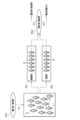
- FIG. 1 is a diagram showing the flow from a clinical phase 1 trial to a clinical phase 2 trial.
- FIG. 2 is a diagram showing the flow from a clinical phase 2 trial to a clinical phase 3 trial.
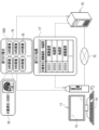
- FIG. 3 is a diagram showing various data transmitted and received between the clinical trial support server and operator terminals, and between the clinical trial support server and operator terminals;
- 2 is a block diagram showing a computer that constitutes a clinical trial support server;
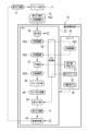
- FIG. 4 is a block diagram showing a processing unit of a CPU of the clinical trial support server;
- FIG. FIG. 4 is a diagram showing the processing of the predictor and the time points of MRI images and prediction results; It is a figure which shows the detail of the process of a progress speed prediction model and a prediction part.
- FIG. 1 is a diagram showing the flow from a clinical phase 1 trial to a clinical phase 2 trial.
- FIG. 2 is a diagram showing the flow from a clinical phase 2 trial to a clinical phase 3 trial.
- FIG. 3 is
- FIG. 2 is a diagram showing the configuration of an autoencoder and the formation of a feature value derivation model
- FIG. 4 is a diagram showing an outline of processing in the learning phase of an autoencoder
- FIG. 10 is a diagram showing an overview of processing in the learning phase of the variation derivation model
- FIG. 10 is a diagram showing learning MRI images and time points of correct CDR-SOB change amounts; It is a figure which shows the process of a grouping part. It is a figure which shows a distribution table. It is a figure which shows the process of a distribution part. It is a figure which shows the process of a distribution part.
- 4 is a flow chart showing a processing procedure of a clinical trial support server; FIG.
- FIG. 5 is a diagram showing details of a simulation for confirming the effects of the technique of the present disclosure
- FIG. 10 is a graph showing the probability of overestimating the efficacy of a study drug in a Phase 2 clinical trial.
- FIG. 10 is a graph showing the probability of underestimating the efficacy of an investigational drug in a Phase 2 clinical trial.
- FIG. It is a graph which shows a clinical trial success probability.
- It is a figure which shows the process part of 2nd Embodiment.
- FIG. 10 is a diagram showing a third embodiment in which dementia-related data are input to a progression speed prediction model in addition to MRI images. It is a figure which shows dementia related data.
- FIG. 13 is a diagram showing an outline of processing in a learning phase of a variation derivation model according to the third embodiment
- FIG. 4 is a diagram showing time points of MRI images for learning, dementia-related data for learning, and correct CDR-SOB variation.
- FIG. 10 is a diagram showing a mode of inputting a clinical trial period into a progression speed prediction model in addition to MRI images and dementia-related data. It is a figure which shows the aspect which outputs the probability of becoming dementia as a prediction result. It is a figure which shows the structure of a multitask CNN and the origin of a feature-value derivation model.
- Phase 1 clinical trial Ph1 is conducted to investigate the safety and pharmacokinetics of the test drug for antidementia.
- a relatively small-scale second phase clinical trial Ph2 is conducted with about several hundred subjects S.
- subjects S Prior to this Phase 2 clinical trial Ph2, subjects S are divided into a test drug group TG (Treatment Group) to which a test drug is administered and a placebo group PG (Placebo Group) to which a placebo is administered.
- Phase 2 clinical trial Ph3 A large scale Phase 3 clinical trial Ph3 will be conducted. As in the case of the clinical phase 2 study Ph2, in the clinical phase 3 study Ph3, the subjects S are divided into the test drug group TG and the placebo group PG. The duration of the clinical phase 2 trial Ph2 and the clinical phase 3 trial Ph3 are both, for example, 18 months.
- Dementia is an example of a "target disease” and a "brain nerve disease” according to the technology of the present disclosure.
- Dementia includes Alzheimer's dementia, Lewy body dementia, vascular dementia, and the like.
- the nootropic drug may be used for Alzheimer's disease other than Alzheimer's dementia.
- antidementia drugs are used to promote the progression of Alzheimer's disease from preclinical Alzheimer's disease (PAD) to mild cognitive impairment (MCI) due to Alzheimer's disease. It may be one that delays progression, or one that delays progression from MCI due to Alzheimer's disease to Alzheimer's disease.
- the target disease is preferably a cranial nerve disease such as dementia.
- Cognitive function test data includes clinical dementia evaluation method (hereinafter abbreviated as CDR-SOB (Clinical Dementia Rating-Sum of Boxes)) score, mini-mental state examination (hereinafter abbreviated as MMSE (Mini-Mental State Examination) ) and Alzheimer's disease assessment scale (hereinafter abbreviated as ADAS-Cog (Alzheimer's Disease Assessment Scale-cognitive subscale)).
- CDR-SOB Clinical Dementia Rating-Sum of Boxes
- MMSE Mini-Mental State Examination
- ADAS-Cog Alzheimer's disease assessment scale
- the morphological imaging test data includes a tomographic image (hereinafter referred to as an MRI image) 16 (hereinafter referred to as an MRI image) 16 (see FIG. 3) of the brain obtained by magnetic resonance imaging (MRI), or a brain image obtained by computed tomography (CT). tomographic images (hereinafter referred to as CT images).
- MRI image magnetic resonance imaging
- CT images computed tomography
- Brain functional imaging test data includes brain tomographic images (hereinafter referred to as PET images) by positron emission tomography (PET), brain tomography by single photon emission tomography (SPECT) images (hereinafter referred to as SPECT images) and the like.
- Blood and cerebrospinal fluid test data include the amount of p-tau (phosphorylated tau protein) 181 in cerebrospinal fluid (hereinafter abbreviated as CSF (Cerebrospinal Fluid)).
- CSF Cerebrospinal Fluid
- the genetic test data includes the genotype test results of the ApoE gene.
- the clinical trial support server 10 is a server that assists in the allocation of the subject S to the test drug group TG and placebo group PG in the clinical phase 2 trial Ph2 and the clinical phase 3 trial Ph3. It is connected to an operator terminal 11 via a network 12 .
- the clinical trial support server 10 is an example of a “clinical trial support device” according to the technology of the present disclosure.
- the operator terminal 11 is installed, for example, in a drug development facility and operated by an operator involved in clinical trials at the drug development facility.
- the operator terminal 11 has a display 13 and input devices 14 such as a keyboard and a mouse.
- the network 12 is, for example, a WAN (Wide Area Network) such as the Internet or a public communication network.
- the operator terminal 11 transmits a distribution request 15 to the clinical trial support server 10.
- the allocation request 15 is a request for causing the clinical trial support server 10 to sort the subject S into the test drug group TG and the placebo group PG in the clinical phase 2 trial Ph2 or the clinical phase 3 trial Ph3.
- the distribution request 15 includes an MRI image group 16G, which is a set of MRI images 16 showing the brain of each subject S.
- a subject ID Identity Data
- the MRI image 16 is an example of a "medical image” according to the technology of the present disclosure.
- the brain is an example of an “organ” according to the technology of the present disclosure.
- the distribution request 15 also includes a terminal ID or the like for uniquely identifying the operator terminal 11 that sent the distribution request 15 .
- the clinical trial support server 10 Upon receiving the allocation request 15, the clinical trial support server 10 derives the allocation result 17.
- the sorting result 17 is data in which the test drug group TG or the placebo group PG, which is the sorting result for each subject ID, is registered.
- the clinical trial support server 10 delivers the sorting result 17 to the operator terminal 11 that sent the sorting request 15 .
- the operator terminal 11 displays the sorting result 17 on the display 13 and provides the sorting result 17 for the operator to view.
- the computer that constitutes the clinical trial support server 10 includes a storage 20, a memory 21, a CPU (Central Processing Unit) 22, a communication section 23, a display 24, and an input device 25. These are interconnected via bus lines 26 .
- the storage 20 is a hard disk drive built into the computer that constitutes the clinical trial support server 10 or connected via a cable or network.
- the storage 20 is a disk array in which a plurality of hard disk drives are connected.
- the storage 20 stores a control program such as an operating system, various application programs, various data associated with these programs, and the like.
- a solid state drive may be used instead of the hard disk drive.
- the memory 21 is a work memory for the CPU 22 to execute processing.
- the CPU 22 loads the program stored in the storage 20 into the memory 21 and executes processing according to the program. Thereby, the CPU 22 comprehensively controls each part of the computer.
- the CPU 22 is an example of a "processor" according to the technology of the present disclosure. Note that the memory 21 may be built in the CPU 22 .
- the communication unit 23 controls transmission of various information with external devices such as the operator terminal 11.
- the display 24 displays various screens. Various screens are provided with operation functions by GUI (Graphical User Interface).
- GUI Graphic User Interface
- the computer that configures the clinical trial support server 10 accepts input of operation instructions from the input device 25 through various screens.
- the input device 25 is a keyboard, mouse, touch panel, microphone for voice input, and the like.
- the operating program 30 is stored in the storage 20 of the clinical trial support server 10 .
- the operating program 30 is an application program for causing the computer to function as the clinical trial support server 10.
- FIG. That is, the operating program 30 is an example of the "clinical trial support device operating program" according to the technology of the present disclosure.
- the storage 20 also stores an MRI image group 16G.
- the storage 20 also stores a progress speed prediction model 31 and a sorting table 32 .
- the progress speed prediction model 31 is an example of a “learned model” according to the technology of the present disclosure.
- the CPU 22 of the computer that constitutes the clinical trial support server 10 cooperates with the memory 21 and the like to operate the reception unit 35, the read/write (hereinafter abbreviated as RW (Read Write)) control unit 36 , a prediction unit 37 , a grouping unit 38 , a distribution unit 39 , and a distribution control unit 40 .
- RW Read Write
- the reception unit 35 receives the distribution request 15 from the operator terminal 11 .
- the distribution request 15 contains the MRI image group 16G. Therefore, by accepting the distribution request 15, the accepting unit 35 acquires the MRI image group 16G.
- the reception unit 35 outputs the MRI image group 16G to the RW control unit 36 .
- the receiving unit 35 also outputs the terminal ID of the operator terminal 11 (not shown) to the distribution control unit 40 .
- the RW control unit 36 controls storage of various data in the storage 20 and reading of various data in the storage 20 .
- the RW control unit 36 stores the MRI image group 16G from the reception unit 35 in the storage 20 .
- the RW control unit 36 also reads out the MRI image group 16G from the storage 20 and outputs the MRI image group 16G to the prediction unit 37 .
- the RW control unit 36 also reads the progress speed prediction model 31 from the storage 20 and outputs the progress speed prediction model 31 to the prediction unit 37 .
- the RW control unit 36 reads the distribution table 32 from the storage 20 and outputs the distribution table 32 to the distribution unit 39 .
- the prediction unit 37 uses the progression speed prediction model 31 to predict the speed of progression of dementia of each subject S from all the MRI images 16 that make up the MRI image group 16G.
- the prediction unit 37 outputs to the grouping unit 38 a prediction result group 45 ⁇ /b>G that is a set of prediction results 45 (see FIG. 6 ) of the rate of progression of dementia of each subject S.
- the grouping unit 38 divides the subject S into a plurality of groups according to the prediction result 45.
- the grouping section 38 outputs the grouping result 46 to the sorting section 39 .
- the sorting unit 39 sorts the subjects S into the test drug group TG and the placebo group PG for each of a plurality of groups.
- the distribution unit 39 outputs the distribution result 17 to the distribution control unit 40 .
- the distribution control unit 40 controls distribution of the distribution result 17 to the operator terminal 11 that sent the distribution request 15 .
- the distribution control unit 40 identifies the operator terminal 11 that is the transmission source of the distribution request 15 based on the terminal ID from the reception unit 35 .
- the prediction unit 37 inputs the MRI image 16 to the progressing speed prediction model 31 . Then, the prediction result 45 is output from the advancing speed prediction model 31 .
- the prediction result 45 is the CDR-SOB score change amount (hereinafter referred to as CDR-SOB change amount).
- the MRI image 16 input to the progression speed prediction model 31 is an image of the brain of the subject S at the start time TS of the clinical trial.
- the prediction result 45 output from the progression speed prediction model 31 predicts the CDR-SOB change amount of the subject S at the end point TE of the clinical trial after the clinical trial period PD from the start point TS. . If the CDR-SOB variation is 0 or more, it can be predicted that dementia has progressed in the subject S at the end of the clinical trial. In addition, it can be predicted that the greater the CDR-SOB change amount, the faster the progress of dementia of the subject S. Note that the prediction result 45 is given the same subject ID as the MRI image 16 .
- the amount of change in CDR-SOB is an example of the “numerical value indicating the degree of progression of the target disease” and the “amount of change in cognitive function test score” according to the technology of the present disclosure.
- the CDR-SOB change amount may be expressed as ⁇ .
- the start time TS may be strictly the start date of the clinical trial, but may be set with a margin such as one week before or after the start date of the clinical trial.
- the progress speed prediction model 31 is composed of a segmentation model 50, a plurality of feature value derivation models 51, and a variation derivation model 52.
- the segmentation model 50 is a trained model that performs so-called semantic segmentation by assigning a label representing each anatomical region of the brain, such as the hippocampus, the anterior temporal lobe, and the amygdala, to each pixel of the brain shown in the MRI image 16 .
- the prediction unit 37 extracts images 53 of a plurality of anatomical regions of the brain (hereinafter referred to as anatomical region images) from the MRI image 16 based on the labels given by the segmentation model 50 .
- Anterior temporal lobe means the front part of the temporal lobe.
- images of anatomical regions such as the frontal lobe, mammary body, corpus callosum, brain arch, and lateral ventricle may be extracted.
- the anatomical region images preferably also include images of other anatomical regions such as the parahippocampal gyrus, occipital lobe, thalamus, and hypothalamus.
- images of other anatomical regions such as the parahippocampal gyrus, occipital lobe, thalamus, and hypothalamus.
- anatomical region image of each of the left and right anatomical regions it is preferable to extract an anatomical region image of each of the left and right anatomical regions.
- the anatomical area image preferably includes at least an image of the hippocampus, and more preferably includes at least an image of the hippocampus and an image of the anterior temporal lobe.
- FIG. 7 illustrates a hippocampus feature amount derivation model 51_1, an anterior temporal lobe feature amount derivation model 51_2, and an amygdala feature amount derivation model 51_3.
- the prediction unit 37 inputs each anatomical region image 53 to the corresponding feature value derivation model 51 .
- the feature quantity 54 of each anatomical region is output from the feature quantity derivation model 51 .
- a feature amount 54_1 of the hippocampus, a feature amount 54_2 of the anterior temporal lobe, a feature amount 54_3 of the amygdala, and the like are illustrated.
- the prediction unit 37 inputs a feature quantity group 54G, which is a set of feature quantities 54 of each anatomical region, to the variation derivation model 52. Then, the prediction result 45 is output from the variation derivation model 52 .
- the variation derivation model 52 is constructed by any one of neural network, support vector machine, and boosting.
- the feature value derivation model 51 uses an encoder unit 61 of an autoencoder (hereinafter abbreviated as AE (Auto Encoder)) 60 .
- AE 60 has an encoder section 61 and a decoder section 62 .
- An anatomical region image 53 is input to the encoder unit 61 .
- the encoder unit 61 converts the anatomical region image 53 into a feature amount 54 by performing well-known convolution operation, pooling processing, and the like.
- the feature values 54 obtained in this manner represent the features of the shape and texture of each anatomical segment, such as the degree of hippocampal atrophy and the presence or absence of decreased blood flow metabolism in the anterior temporal lobe.
- the encoder section 61 transfers the feature amount 54 to the decoder section 62 .
- a decoder unit 62 generates a restored image 63 of the anatomical region image 53 from the feature amount 54 .
- the AE 60 learns by inputting the learning anatomical region image 53L in the learning phase before the encoder unit 61 is diverted to the feature value derivation model 51 .
- the anatomical region image for learning 53L is an image extracted from the MRI image for learning 16L using the segmentation model 50 .
- the learning MRI image 16L is an MRI image 16 of a dementia patient whose MRI image 16 etc. is registered in a database such as ADNI (Alzheimer's Disease Neuroimaging Initiative) and who satisfies the conditions for participating in a clinical trial. . Conditions for participation in clinical trials include, for example, a positive amyloid ⁇ in the CSF test and an MMSE score of 26 or less.
- the learning MRI image 16L is an example of “learning input data” according to the technology of the present disclosure.
- a dementia patient is an example of a “target disease patient” according to the technology of the present disclosure.
- the AE 60 outputs a learning restored image 63L for the learning anatomical region image 53L. Based on these learning anatomical region image 53L and learning restored image 63L, loss calculation of the AE 60 using a loss function is performed. Various coefficients of the AE 60 (such as filter coefficients for convolution calculation) are updated according to the result of the loss calculation, and the AE 60 is updated according to the update settings.
- the above-described series of processing of inputting the learning anatomical region image 53L to the AE 60, outputting the learning restored image 63L from the AE 60, loss calculation, update settings, and updating the AE 60 is performed by the learning anatomy. This is repeated while the area images 53L are exchanged. Repetition of the above series of processes is terminated when the restoration accuracy from the learning anatomical region image 53L to the learning restored image 63L reaches a predetermined set level.
- the encoder unit 61 of the AE 60 whose restoration accuracy reaches the set level in this way is used in the prediction unit 37 as part of the progress speed prediction model 31 . Note that regardless of the restoration accuracy from the learning anatomical region image 53L to the learning restored image 63L, learning may be terminated when the series of processes described above is repeated a set number of times.
- the variation derivation model 52 is learned by learning data 70.
- the learning data 70 is a set of the learning feature quantity group 54GL and the correct CDR-SOB variation 45CA.
- the learning feature quantity group 54GL is obtained by inputting the learning anatomical region image 53L of each anatomical region extracted from the learning MRI image 16L into each learned feature quantity derivation model 51 .
- the correct CDR-SOB change amount 45CA is the actual CDR-SOB change amount of the dementia patient in the learning MRI image 16L from which the learning feature amount group 54GL was obtained.
- the correct CDR-SOB variation 45CA is an example of "correct data" according to the technology of the present disclosure.
- the learning feature group 54GL is input to the variation derivation model 52 .
- the change amount derivation model 52 outputs a learning prediction result 45L for the learning feature amount group 54GL.
- the loss calculation of the variation derivation model 52 using a loss function is performed.
- Various coefficients of the variation derivation model 52 are updated according to the result of the loss calculation, and the variation derivation model 52 is updated according to the update settings.
- the input of the learning feature group 54GL to the variation derivation model 52, the output of the learning prediction result 45L from the variation derivation model 52, the loss calculation, the update setting, and the change The series of processes for updating the quantity derivation model 52 are repeated while the learning data 70 are exchanged.
- the repetition of the above series of processes ends when the prediction accuracy of the learning prediction result 45L with respect to the correct CDR-SOB change amount 45CA reaches a predetermined set level.
- the variation derivation model 52 whose prediction accuracy reaches the set level in this way is used in the prediction unit 37 as part of the progress speed prediction model 31 . Regardless of the prediction accuracy of the learning prediction result 45L with respect to the correct CDR-SOB change amount 45CA, learning may be terminated when the above series of processes are repeated a set number of times.
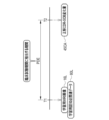
- the learning MRI image 16L is an image of the dementia patient's brain at the first time point T1.
- the correct CDR-SOB change amount 45CA is the actual CDR-SOB change amount of the dementia patient at the second time point T2 after the period PDE corresponding to the clinical trial period PD from the first time point T1.
- the dementia patient has a CDR-SOB score of 22 at the first time point T1 and a CDR-SOB score of 25 at the second time point T2
- the period PDE corresponding to the clinical trial period PD is a period including the clinical trial period PD and adding an appropriate margin to the clinical trial period PD. For example, if the clinical trial period PD is 18 months, the period PDE corresponding to the clinical trial period PD is 12 months to 24 months plus or minus 6 months in 18 months.
- the grouping unit 38 divides the subject S into one of groups 1 to 18 according to the CDR-SOB change amount of the prediction result 45.
- FIG. For example, the grouping unit 38 assigns subjects S whose CDR-SOB change amount is less than -3.0 ( ⁇ -3.0) to group 1, and assigns subjects S whose CDR-SOB change amount is -3.0 to group 4, for example. A subject S with a score of 2.0 or more and less than -1.5 (-2.0 ⁇ -1.5) was placed. Further, the grouping unit 38 assigns subjects S whose CDR-SOB variation is 3.5 or more and less than 4.0 (3.5 ⁇ 4.0) to group 15, for example. A subject S whose CDR-SOB variation is greater than 5.0 (5.0 ⁇ ) is assigned to . In this way, groups 1 to 18 are defined mainly by the range of CDR-SOB variation between -3.0 and 5.0 set in 0.5 increments.
- the grouping result 46 is data in which the target person ID of the target person S is registered for each group.
- the CDR-SOB change amount of the subject S whose subject ID is S0001 is 4.0, so the grouping unit 38 registers S0001 in the group 16 .
- the CDR-SOB change amount of the subject S whose subject ID is S0004 is ⁇ 1.7, so the grouping unit 38 registers S0004 in group 4.
- the sorting table 32 is a table in which sorting rules for each group are registered.
- the distribution rule is based on a block randomization technique for each two players.
- the group 1 sorting rule is that for pairs of two subjects S, 1st, 2nd and 4th, the first subject S is sorted into the test drug group TG, and the second subject S is placed in placebo. The content is to distribute to the group PG.
- the first subject S is assigned to the placebo group PG, and the second subject S is assigned to the test drug group TG. Content.
- the group 16 sorting rule is that for pairs of two subjects S, 1st to 4th and 6th, the first subject S is sorted into the test drug group TG, and the second subject S is placed in placebo. The content is to distribute to the group PG.
- the first subject S is assigned to the placebo group PG, and the second subject S is assigned to the test drug group TG.
- FIG. 14 shows an example of how the subjects S in Group 1 are sorted into the test drug group TG and the placebo group PG according to the sorting rules of the sorting table 32 .
- FIG. 14 exemplifies a case where eight subjects S whose subject IDs are S0008, S0022, S0034, S0044, S0051, S0068, S0102, and S0116 are sorted.
- the sorting unit 39 sorts the first subject S into the test drug group TG, and sorts the second subject S into the placebo group PG, according to the sorting rule. Of the two subjects S, S0008 is assigned to the test drug group TG, and S0022 is assigned to the placebo group PG.
- the S0034 subject S is assigned to the test drug group TG
- the S0044 subject S is assigned to the placebo group PG.
- the subject S0102 of the two subjects S of the fourth pair S0102 and S0116 is assigned to the test drug group TG
- the subject S0116 is assigned to the placebo group PG.
- the sorting unit 39 sorts the first subject S into the placebo group PG, and sorts the second subject S into the test drug group TG, according to the sorting rule.
- the subject S of S0051 is assigned to the placebo group PG
- the subject S of S0068 is assigned to the test drug group TG.
- FIG. 15 shows an example of how subjects S in group 16 are sorted into test drug group TG and placebo group PG according to the sorting rules of sorting table 32 .
- FIG. 15 illustrates a case where 12 subjects S whose subject IDs are S0001, S0024, S0029, S0056, S0072, S0081, S0105, S0124, S0133, S0148, S0172, and S0181 are sorted.
- the sorting unit 39 sorts the first subject S into the test drug group TG, and sorts the second subject S into the placebo group PG, according to the sorting rule. Of the two subjects S, S0001 is assigned to the test drug group TG, and S0024 is assigned to the placebo group PG.
- the S0029 subject S is assigned to the test drug group TG
- the S0056 subject S is assigned to the placebo group PG.
- the subject S of S0072 is assigned to the test drug group TG
- the subject S of S0081 is assigned to the placebo group PG.
- the subject S of S0105 is assigned to the test drug group TG
- the subject S of S0124 is assigned to the placebo group PG.
- the sorting unit 39 sorts the first subject S into the placebo group PG, and sorts the second subject S into the test drug group TG, according to the sorting rule.
- the subject S of S0133 is assigned to the placebo group PG, and the subject S of S0148 is assigned to the test drug group TG.
- the sorting unit 39 sorts the subjects S of each group 1 to 18 into the test drug group TG and the placebo group PG using the block randomization technique. Such a distribution method is called a stratified randomization method.
- the operator operates the input device 14 of the operator terminal 11 to transmit a sorting request 15 including an MRI image group 16G, which is a set of MRI images 16 of the test drug subject S with dementia, to the clinical trial support server 10. .
- the allocation request 15 from the operator terminal 11 is received by the receiving unit 35 (step ST100).
- the MRI image group 16G included in the distribution request 15 is output from the reception unit 35 to the RW control unit 36 and stored in the storage 20 under the control of the RW control unit 36 (step ST110).
- the MRI image group 16G is read from the storage 20 by the RW control unit 36 (step ST120).
- the MRI image group 16G is output from the RW control section 36 to the prediction section 37 .
- step ST130 the prediction result 45 is output from the progression speed prediction model 31 (step ST130).
- step ST130 The process of step ST130 is repeated until the prediction result 45 is output for all the subjects S (NO in step ST140).
- a prediction result group 45G which is a set of multiple prediction results 45, is output from the prediction unit 37 to the grouping unit 38. Then, as shown in FIG. 12, the grouping unit 38 divides the subject S into one of groups 1 to 18 according to the prediction result 45 (step ST150). The grouping result 46 is output from the grouping section 38 to the sorting section 39 .
- the subject S is sorted into the test drug group TG and the placebo group PG for each of the groups 1 to 18 using the block randomization method (step ST160).
- the distribution result 17 is output from the distribution unit 39 to the distribution control unit 40 .
- the distribution result 17 is distributed to the operator terminal 11 that sent the distribution request 15 under the control of the distribution control section 40 (step ST170).
- a sorting result 17 is displayed on the display 13 of the operator terminal 11 .
- the operator browses the sorting result 17, sorts the subjects S into the test drug group TG and the placebo group PG, and conducts the clinical phase 2 study Ph2 or the clinical phase 3 study Ph3.
- the CPU 22 of the clinical trial support server 10 includes the prediction section 37, the grouping section 38, and the sorting section 39.
- the prediction unit 37 predicts the CDR-SOB change amount for the test drug subject S for dementia using the progression speed prediction model 31 .
- the grouping unit 38 divides the subject S into any one of groups 1 to 18 according to the prediction result 45 of the CDR-SOB change amount.
- the sorting unit 39 sorts the subjects S into the test drug group TG and the placebo group PG for each of the groups 1-18.
- the sorting unit 39 sorts the subjects S into the test drug group TG and the placebo group PG using a block randomization technique. For this reason, the risk of bias between the test drug group TG and the placebo group PG between subjects with relatively fast progression of dementia and subjects with slow progression is further reduced, and as a result, the efficacy of the test drug for dementia is justified. It will be possible to further contribute to a more accurate evaluation.
- the progression speed prediction model 31 uses the learning MRI image 16L of the dementia patient at the first time point T1 as learning input data, and from the first time point T1 to the clinical trial period PD
- This model is trained using the correct CDR-SOB change amount 45CA, which is the CDR-SOB change amount of the dementia patient at the second time point T2 after the corresponding period of PDE, as the correct data.
- the prediction unit 37 inputs the MRI image 16 of the subject S at the start time TS of the clinical trial to the progression speed prediction model 31, and the CDR of the subject S at the end time TE of the clinical trial. - Output the SOB change amount from the advancing speed prediction model 31 as the prediction result 45 .
- MRI images 16 are medical images of the brain. Almost all dementia patients also take the CDR-SOB. For this reason, if the learning MRI image 16L of the dementia patient is used as learning input data and the correct CDR-SOB variation 45CA is used as correct data, the learning data 70 of the progress speed prediction model 31 will not be lacking, and the progress speed will be improved. Learning of the prediction model 31 progresses. Furthermore, the MRI image 16 can be taken in minutes if the equipment is idle. Therefore, the prediction result 45 can be output from the progress speed prediction model 31 relatively easily.
- CDR-SOB changes Changes in cognitive function test scores, especially CDR-SOB changes, have long been used to determine the success or failure of clinical trials. Therefore, if the amount of CDR-SOB change is used as in this example as a numerical value indicating the degree of progression of dementia in the subject S at the end point of the clinical trial TE, the success or failure of the clinical trial can be based on the same standard as before. can be determined.
- the progression speed prediction model 31 uses the segmentation model 50 to extract a plurality of anatomical region images 53, which are images of a plurality of brain anatomical regions, from the MRI image 16 at the start time TS of the clinical trial. Then, the anatomical region image 53 is input to the feature quantity derivation model 51 prepared for each anatomical region, and the feature quantity 54 of the anatomical region is output from the feature quantity derivation model 51 . Therefore, the speed of progression of dementia can be predicted based on the feature values obtained from a plurality of anatomical regions of the brain, and the prediction accuracy of the prediction result 45 can be improved.
- the actual CDR-SOB change amount (true value) ⁇ c registered in ADNI is 0 or less, that is, dementia progresses.
- the CDR-SOB change amount (true value) ⁇ c remains unchanged without correction.
- the CDR-SOB variation (true value) ⁇ c of the subjects S assigned to the placebo group PG remains unchanged without correction.
- the actual CDR-SOB change amount (true value) ⁇ c was greater than 0, that is, the subject S with advanced dementia had the CDR-SOB change amount
- the CDR-SOB variation (corrected value) ⁇ cX corrected by the efficacy Eff of the test drug is defined as the true value) ⁇ c.
- the CDR-SOB variation (correction value) ⁇ cX is obtained by multiplying the CDR-SOB variation (true value) ⁇ c by (1 ⁇ Eff).
- FIG. 18 shows a method of a comparative example, a method according to the technique of the present disclosure in which the subject S is stratified and randomized based on the prediction result 45, and stratified randomization based on the actual CDR-SOB variation (true value) ⁇ c.
- the efficacy Eff of the test drug was changed in increments of 0.05 between 0 and 0.4, P1 ⁇ 0.05 (cleared Phase 2 clinical trial Ph2) and P2 ⁇ 0 Indicates the probability of .01 (not clearing Phase 3 clinical trial Ph3).
- the method of the comparative example divides the subjects S into groups according to the prediction result 45, and sorts the subjects S into the test drug group TG and the placebo group PG for each group, like the method according to the technology of the present disclosure.
- all ungrouped subjects S are randomly assigned to the test drug group TG and the placebo group PG.
- the probability that P1 ⁇ 0.05 and P2 ⁇ 0.01 is, in other words, the probability that the efficacy of the test drug is overestimated in Phase 2 clinical trials Ph2.
- the probability of P1 ⁇ 0.05 and P2 ⁇ 0.01 in the method according to the technique of the present disclosure is higher than the probability of P1 ⁇ 0.05 and P2 ⁇ 0.01 in the method of the comparative example. is also found to be generally low. Therefore, it was confirmed that the technique of the present disclosure reduces the risk of the efficacy of the test drug being overestimated.
- FIG. 19 shows a method of a comparative example, a method according to the technology of the present disclosure in which the subject S is stratified and randomized based on the prediction result 45, and stratified randomization based on the actual CDR-SOB change amount (true value) ⁇ c.
- the efficacy Eff of the test drug was changed in increments of 0.05 between 0 and 0.4
- P1 ⁇ 0.05 without clearing the clinical phase 2 trial Ph2
- P2 The probability of ⁇ 0.01 (clearing Phase 3 clinical trial Ph3) is shown.
- the probability that P1 ⁇ 0.05 and P2 ⁇ 0.01 is, in other words, the probability that the efficacy of the test drug is underestimated in the Phase 2 clinical trial Ph2.
- the probability of P1 ⁇ 0.05 and P2 ⁇ 0.01 in the method according to the technique of the present disclosure is higher than the probability of P1 ⁇ 0.05 and P2 ⁇ 0.01 in the method of the comparative example. is also found to be generally low. Therefore, it was confirmed that the technique of the present disclosure reduces the possibility that the efficacy of the test drug will be underestimated.
- FIG. 20 shows a method of a comparative example, a method according to the technique of the present disclosure in which the subject S is stratified and randomized based on the prediction result 45, and stratified randomization based on the actual CDR-SOB change amount (true value) ⁇ c.
- the efficacy Eff of the test drug was changed in increments of 0.05 between 0 and 0.4, P1 ⁇ 0.05 (cleared Phase 2 clinical trial Ph2) and P2 ⁇ 0 0.01 (clears Phase 3 clinical trial Ph3).
- the probability that P1 ⁇ 0.05 and P2 ⁇ 0.01 is, in other words, the clinical trial success probability.
- the probability of P1 ⁇ 0.05 and P2 ⁇ 0.01 in the method according to the technique of the present disclosure is higher than the probability of P1 ⁇ 0.05 and P2 ⁇ 0.01 in the method of the comparative example. is also found to be generally high. Therefore, according to the technique of the present disclosure, it was confirmed that the efficacy of the test drug was properly evaluated, and the probability of success in clinical trials increased accordingly.
- the conventional method is more effective than the method according to the technology of the present disclosure. be done. Therefore, if the prediction accuracy of the progress speed prediction model 31 is improved, it is considered that the conventional method will be more effective than the method according to the technology of the present disclosure.
- the clinical phase 1 trial Ph1 when the clinical phase 1 trial Ph1 is cleared, the clinical phase 2 trial Ph2 is performed, and when the clinical phase 2 trial Ph2 is cleared, the clinical phase 3 trial Ph3 is performed.
- a clinical phase 1 trial Ph1 and a clinical phase 2 trial Ph2, or a clinical phase 1 trial Ph2 and a clinical phase 3 trial Ph3 may be conducted in parallel.
- the present invention is not limited to this.
- a part of the single-task convolutional neural network or a part of the multi-task convolutional neural network may be used as the feature value derivation model 51 (see FIG. 31).
- the unit of the block randomization method is not limited to the two people shown in the example. For example, it may be four people.
- half of the clinical trial subjects S constituting the group may be simply assigned to the test drug group TG, and the other half may be assigned to the placebo group PG.
- the CPU of the clinical trial support server of the second embodiment includes the processing units 35 to 40 of the first embodiment (in FIG. 21, the prediction unit 37, grouping unit 38, and 39 is shown, others are not shown), and functions as a selection unit 75 .
- the selection unit 75 is provided between the prediction unit 37 and the grouping unit 38 .
- the prediction result group 45 ⁇ /b>G from the prediction section 37 is input to the selection section 75 .
- the selection unit 75 selects the subject S whose dementia progresses relatively quickly according to the selection condition 76 .
- the selection condition 76 is that the CDR-SOB variation is greater than 0 ( ⁇ >0) in this example.
- the selection unit 75 selects only the prediction results 45 that satisfy the selection condition 76 from among the plurality of prediction results 45 forming the prediction result group 45G, thereby generating the selected prediction result group 45GS.
- the selection unit 75 outputs the selection prediction result group 45GS to the grouping unit 38 .
- FIG. 22 shows an example of the processing of the selection unit 75.
- the CDR-SOB variation amounts of the subjects S with subject IDs S0001 and S0003 are greater than 0, and the CDR-SOB variation amounts of the subjects S with subject IDs S0002 and S0004 are 0 or less. Therefore, the selection unit 75 selects the prediction results 45 with the subject IDs S0001 and S0003, excludes the prediction results 45 with the subject IDs S0002 and S0004, and generates a selection prediction result group 45GS.
- the grouping unit 38 groups the subjects S for whom the prediction result 45 is selected in the selection prediction result group 45GS. Based on the grouping result 46, the sorting unit 39 sorts the subjects S for whom the prediction result 45 was selected in the selection prediction result group 45GS into the test drug group TG and the placebo group PG.
- the selection unit 75 selects the subject S whose dementia progresses relatively quickly, such as the subject S whose CDR-SOB change amount is greater than 0, based on the prediction result 45. .
- the grouping unit 38 groups only the selected target person S according to the prediction result 45 .
- the sorting unit 39 sorts only the selected subjects S into the test drug group TG and the placebo group PG. Therefore, a person more suitable for clinical trials of a test drug for dementia can be selected as the subject S, which can further contribute to the proper evaluation of the efficacy of the test drug for dementia.
- the selection condition 76 is not limited to the content of the example.
- the content may be that the CDR-SOB variation is greater than 2 ( ⁇ >2).
- the content may be such that the CDR-SOB variation is greater than 0 and less than or equal to 3 (0 ⁇ 3).
- the prediction unit 37 inputs the dementia-related data 80 to the progression speed prediction model 81 in addition to the MRI images 16 .
- the dementia-related data 80 is data relating to dementia of the subject S at the start time TS of the clinical trial, and includes part of the data relating to the dementia diagnostic criteria described above. More specifically, as shown in FIG. 24 as an example, the dementia-related data 80 includes the subject S's age, sex, genetic test data, and CSF test data, which is blood/cerebrospinal fluid test data. Genetic test data is, for example, genotype test results of the ApoE gene.
- the genotype of the ApoE gene is a combination of two of the three ApoE genes ⁇ 2, ⁇ 3, and ⁇ 4 ( ⁇ 2 and ⁇ 3, ⁇ 3 and ⁇ 4, etc.). Cognition of Alzheimer's disease in persons with genotypes with one or two ⁇ 4 ( ⁇ 2 and ⁇ 4, ⁇ 4 and ⁇ 4, etc.) versus those with genotypes without ⁇ 4 ( ⁇ 2 and ⁇ 3, ⁇ 3 and ⁇ 3, etc.) The risk of developing the disease is estimated to be approximately 3 to 12 times higher.
- CSF test data is, for example, the amount of p-tau (phosphorylated tau protein) 181 in CSF. Gender is expressed numerically as 00 for male and 01 for female. Similarly, the genetic test data are expressed numerically for combinations of ApoE genes.
- the dementia-related data 80 is an example of "target disease-related data" according to the technology of the present disclosure.
- the dementia-related data 80 is input to the change amount derivation model 85 of the progression speed prediction model 81 together with the feature amount group 54G.
- the variation derivation model 85 outputs the prediction result 45 by referring to the dementia-related data 80 as well as the feature quantity group 54G.
- the variation derivation model 85 of the present embodiment includes a principal component analysis (hereinafter abbreviated as PCA (Principal Component Analysis)) layer 150, a quantile normalization unit 151, and a linear discriminant analysis unit. 152.
- a feature quantity group 54G is input to the PCA layer 150 .
- the PCA layer 150 performs PCA on each of the plurality of feature amounts 54 forming the feature amount group 54G, and sets the feature amount 54 as an aggregate feature amount 54A.
- the PCA layer 150 aggregates hundreds to thousands of feature quantities 54 into one aggregated feature quantity 54A.
- the PCA layer 150 outputs an aggregated feature quantity group 54AG, which is a set of aggregated feature quantities 54A of each feature quantity 54, to the quantile normalization unit 151.
- the aggregated feature quantity group 54AG and the dementia-related data 80 are input to the quantile normalization unit 151 .
- the quantile normalization unit 151 aligns the plurality of aggregated feature quantities 54A that make up the aggregated feature quantity group 54AG and the age, sex, genetic test data, and CSF test data of the subject S of the dementia-related data 80. To handle them, Quantile Normalization is performed to transform them into data following a normal distribution.
- the linear discriminant analysis unit 152 performs, for example, support vector regression ( Linear discriminant analysis using SVR (Support Vector Regression) or the like is performed, and a prediction result 45 is output as the result.
- the variation derivation model 85 is learned by learning data 90 .
- the learning data 90 is a set of a learning feature quantity group 54GL, a learning dementia-related data 80L, and a correct CDR-SOB variation 45CA.
- the origin of the learning feature quantity group 54GL and the correct CDR-SOB variation 45CA is the same as in the case of the learning data 70 of the first embodiment.
- the learning dementia-related data 80L is the age, sex, genetic test data, and CSF test data of the dementia patient of the learning MRI image 16L from which the learning feature quantity group 54GL was obtained.
- the learning dementia-related data 80L is an example of "learning input data" according to the technology of the present disclosure.
- the learning feature quantity group 54GL and the learning dementia-related data 80L are input to the variation derivation model 85.
- the variation derivation model 85 outputs learning prediction results 45L for the learning feature quantity group 54GL and the learning dementia-related data 80L.
- the loss calculation of the variation derivation model 85 using a loss function is performed.
- Various coefficients of the variation derivation model 85 are updated according to the result of the loss calculation, and the variation derivation model 85 is updated according to the update settings.
- the input of the learning feature group 54GL and the learning dementia-related data 80L to the variation derivation model 85 the output of the learning prediction result 45L from the variation derivation model 85,
- the series of processes of loss calculation, update setting, and update of the variation derivation model 85 are repeated while the learning data 90 are exchanged.
- the repetition of the above series of processes ends when the prediction accuracy of the learning prediction result 45L with respect to the correct CDR-SOB change amount 45CA reaches a predetermined set level.
- the change amount derivation model 85 whose prediction accuracy reaches the set level in this manner is used in the prediction unit 37 as part of the progress speed prediction model 81 . Regardless of the prediction accuracy of the learning prediction result 45L with respect to the correct CDR-SOB change amount 45CA, learning may be terminated when the above series of processes are repeated a set number of times.
- the learning dementia-related data 80L is data related to dementia of a dementia patient at the first time point T1, like the learning MRI image 16L.
- learning input data also includes learning dementia-related data 80L, which is dementia-related data related to dementia of a dementia patient at first time point T1.
- the prediction unit 37 also inputs dementia-related data 80 regarding dementia of the subject S at the start time TS of the clinical trial to the progression speed prediction model 81 . Since the dementia-related data 80, which is strong data useful for predicting the speed of dementia progression, is added, compared to the case of predicting the speed of dementia progression only with the feature group 54G, the progress of dementia Speed prediction accuracy can be dramatically improved.
- the dementia-related data 80 may include medical history, or whether or not there are relatives who have developed dementia.
- Dementia-related data 80 may include blood test results such as apolipoprotein measurements, complement protein measurements, and transthyretin measurements.
- Hasegawa dementia scale score, MMSE score, Rivermead Behavioral Memory Test (RBMT) score, activities of daily living (ADL: Activities of Daily Living), ADAS-Cog score, etc. related to dementia may be included in the data 80.
- the clinical trial period PD may be input to the progression speed prediction model 95.
- the period PDE corresponding to the clinical test period PD is used as learning input data for the progression speed prediction model. 95.
- the amount of change in CDR-SOB which is the amount of change in the score of the cognitive function test
- the probability of developing dementia may be output as the prediction result 101, like the progression speed prediction model 100 shown in FIG.
- Groups in this case are, for example, group 1 with a probability of becoming dementia of 0% or more and less than 25%, group 2 of 25% or more and less than 50%, group 3 of 50% or more and less than 75%, and 75% or more and 100% Group 4 or the like below may be used.
- a part of a multitask convolutional neural network (hereinafter referred to as a multitask CNN (Convolutional Neural Network)) 200 shown in FIG. 31 may be used as the feature value derivation model 51.
- a multitask CNN Convolutional Neural Network
- the multitasking CNN 200 includes an encoder unit 201, a self-attention (hereinafter referred to as SA (Self-Attention)) mechanism layer 202, a decoder unit 203, and a global average pooling (hereinafter referred to as GAP (Global Average Pooling)) It has a layer 204 , a first fully connected (FC (Fully Connected)) layer 205 , a second FC layer 206 , and a softmax function (SMF (SoftMax Function)) layer 207 .
- SA Self-Attention
- GAP Global Average Pooling
- the encoder unit 201 converts the input anatomical region image 53 into the pre-stage feature amount 54P, like the encoder unit 61 of the AE 60 of the first embodiment.
- the encoder unit 201 outputs the pre-stage feature amount 54 P to the SA mechanism layer 202 .
- the SA mechanism layer 202 performs a convolution operation on the pre-stage feature quantity 54P while changing the filter coefficients for the convolution operation according to the value of the element targeted for the convolution operation.
- the SA mechanism layer 202 outputs the pre-stage feature quantity 54P after the convolution operation to the decoder unit 203 and the GAP layer 204.
- the decoder unit 203 generates a restored image 208 of the anatomical region image 53 from the pre-stage feature quantity 54P after the convolution operation, like the decoder unit 62 of the AE 60 of the first embodiment.
- the GAP layer 204 performs overall average pooling processing on the pre-stage feature quantity 54P after the convolution operation.
- the overall average pooling process is a process of obtaining an average value for each of a plurality of channels of the pre-stage feature quantity 54P. For example, when the number of channels of the pre-stage feature amount 54P is 512, the average value of the 512 pre-stage feature amounts 54P is obtained by the overall average pooling process.
- the GAP layer 204 outputs the obtained average value of the preceding stage feature quantity 54P to the first FC layer 205 and the second FC layer 206 as the feature quantity 54 .
- the first FC layer 205 outputs a first prediction result 209 based on the feature quantity 54.
- the first prediction result 209 is the CDR-SOB change amount.
- the first FC layer 205 has an input layer having units corresponding to the number of features 54 and one output layer for outputting the first prediction result 209 . Each unit in the input layer and the output layer are fully connected to each other, and weights are set for each.
- a feature amount 54 is input to each unit of the input layer.
- the product sum of the feature amount 54 and the weight set between each unit is the output value of the output layer. This output value is the CDR-SOB change amount.
- the second FC layer 206 converts the feature quantity 54 into variables handled by the SMF of the SMF layer 207 .
- the second FC layer 206 has an input layer having units corresponding to the number of feature values 54 and an output layer having units corresponding to the number of variables handled by SMF. Each unit in the input layer and each unit in the output layer are fully connected to each other and weighted.
- a feature amount 54 is input to each unit of the input layer.
- the product sum of the feature amount 54 and the weight set between each unit is the output value of each unit in the output layer. This output value is a variable handled by SMF.
- the second FC layer 206 outputs variables handled by SMF to the SMF layer 207 .
- the SMF layer 207 outputs a second prediction result 210 by applying the variables to the SMF.
- the second prediction result 210 indicates that the CDR-SOB variation is greater than 0 or less than or equal to 0.
- the multitasking CNN 200 performs a total of three tasks: AE, continuous quantity (CDR-SOB variation) prediction, and class classification (whether the CDR-SOB variation is greater than 0 or less than or equal to 0).
- the encoder section 201, the SA mechanism layer 202, and the GAP layer 204 are used for the feature value derivation model 51.
- FIG. 1 A block diagram illustrating an exemplary computing environment in accordance with the present in the CDR-SOB variation.
- part of the multitasking CNN 200 may be used as the feature value derivation model 51.
- the multitasking CNN 200 performs more complicated processing than the AE 60 and the like in order to handle a plurality of tasks. Therefore, there is a high possibility that the feature quantity 54 output from a part of the multitasking CNN 200 more exhaustively represents the features of the anatomical region image 53 . Therefore, if a part of the multitasking CNN 200 is used as the feature value derivation model 51, the prediction result 45 based on the more reliable feature value 54 is output, so the accuracy of predicting the speed of progression of dementia can be improved. can be higher.
- the speed of progression of dementia in subject S may be predicted.
- the medical image is not limited to the MRI image 16 illustrated.
- a PET image or a SPECT image may be used.
- a device other than the test support server 10 may be used. Also, these learnings may be continued after each model is stored in the storage 20 of the clinical trial support server 10 .
- the clinical trial support server 10 may be installed in each drug development facility, or may be installed in a data center independent from the drug development facility.
- the operator terminal 11 may take part or all of the functions of each processing unit of the clinical trial support server 10 .
- the target disease may be, for example, cerebral infarction.
- a CT image or MRI image of the subject's brain is input to the trained model, and the amount of change in the score of the stroke rating scale (NIHSS: National Institutes of Health Stroke Scale) or the Japanese Stroke Rating Scale (JSS : Japan Stroke Scale) output the amount of change in the score from the trained model.
- Target diseases are preferably dementia and cerebral infarction as exemplified, or neurodegenerative diseases such as Parkinson's disease and cranial nerve diseases including cerebrovascular diseases.
- dementia has become a social problem with the advent of an aging society. Therefore, it can be said that this example in which dementia is the target disease is a form that matches the current social problem.
- Target diseases are not limited to cranial nerve diseases, and therefore organs are not limited to the brain.
- the speed of progression of the target disease of subject S may be predicted without using a trained model such as the progression speed prediction model 31 .
- a processing unit Processing As the hardware structure of the Unit, various processors shown below can be used.
- Various processors include, as described above, in addition to the CPU 22 which is a general-purpose processor that executes software (operation program 30) and functions as various processing units, FPGAs (Field Programmable Gate Arrays), etc.
- Programmable Logic Device PLD
- ASIC Application Specific Integrated Circuit
- One processing unit may be configured with one of these various processors, or a combination of two or more processors of the same or different type (for example, a combination of a plurality of FPGAs and/or a CPU and combination with FPGA). Also, a plurality of processing units may be configured by one processor.
- a single processor is configured by combining one or more CPUs and software.
- a processor functions as multiple processing units.
- SoC System On Chip
- a processor that realizes the functions of the entire system including multiple processing units with a single IC (Integrated Circuit) chip. be.
- various processing units are configured using one or more of the above various processors as a hardware structure.
- an electric circuit combining circuit elements such as semiconductor elements can be used.
- the processor Predict the speed of progression of the target disease for subjects in clinical trials, Divide the subjects into multiple groups according to the prediction results of the speed of progression of the target disease, Divide the subjects into a test drug group to which the test drug is administered and a placebo group to which the placebo is administered for each of the plurality of groups; Clinical trial support equipment.
- the clinical trial support device according to claim 1, wherein only the selected subjects are grouped according to the prediction results and sorted into a test drug group and a placebo group.
- the clinical trial support device which uses a block randomization technique to sort subjects into a test drug group and a placebo group.
- the processor Predicting the speed of progression of the target disease using a trained model,
- the trained model uses a medical image of an organ of a target disease patient at a first time point as input data for learning, and the target disease patient at a second time point after a period corresponding to the clinical trial period from the first time point.
- a trained model that has been trained using numerical values indicating the degree of disease progression as correct data
- the processor A medical image showing the subject's organs at the start of the clinical trial is input to the trained model, and a numerical value indicating the degree of progression of the subject's target disease at the end of the clinical trial is used as the prediction result.
- the clinical trial support device according to any one of additional items 1 to 3, which is output from a model.
- the trained model is extracting a plurality of anatomical region images, which are images of a plurality of anatomical regions of an organ, from the medical image at the start of the clinical trial; 5.
- the clinical trial support apparatus wherein the anatomical region image is input to a feature value derivation model prepared for each anatomical region, and the feature value of the anatomical region is output from the feature value derivation model.
- the learning input data also includes target disease-related data related to the target disease of the target disease patient at the first time point, The processor 6.
- the clinical trial support device according to claim 4 or claim 5, wherein target disease-related data relating to the subject's target disease at the start of the clinical trial is also input to the trained model.
- the target disease is dementia, 7.
- the clinical trial support device according to any one of additional items 4 to 6, wherein the numerical value is an amount of change in a cognitive function test score or a probability of developing dementia.
- the technology of the present disclosure can also appropriately combine various embodiments and/or various modifications described above. Moreover, it is needless to say that various configurations can be employed without departing from the scope of the present invention without being limited to the above embodiments. Furthermore, the technology of the present disclosure extends to storage media that non-temporarily store programs in addition to programs.
- a and/or B is synonymous with “at least one of A and B.” That is, “A and/or B” means that only A, only B, or a combination of A and B may be used.
- a and/or B means that only A, only B, or a combination of A and B may be used.
Landscapes
- Engineering & Computer Science (AREA)
- Health & Medical Sciences (AREA)
- Medical Informatics (AREA)
- Public Health (AREA)
- Epidemiology (AREA)
- General Health & Medical Sciences (AREA)
- Primary Health Care (AREA)
- Biomedical Technology (AREA)
- Data Mining & Analysis (AREA)
- Databases & Information Systems (AREA)
- Pathology (AREA)
- Radiology & Medical Imaging (AREA)
- Nuclear Medicine, Radiotherapy & Molecular Imaging (AREA)
- Magnetic Resonance Imaging Apparatus (AREA)
- Medical Treatment And Welfare Office Work (AREA)
Priority Applications (2)
| Application Number | Priority Date | Filing Date | Title |
|---|---|---|---|
| JP2023566151A JPWO2023105976A1 (https=) | 2021-12-08 | 2022-10-27 | |
| US18/673,319 US12614615B2 (en) | 2021-12-08 | 2024-05-24 | Clinical trial support apparatus, operation method of clinical trial support apparatus, and operation program of clinical trial support apparatus |
Applications Claiming Priority (4)
| Application Number | Priority Date | Filing Date | Title |
|---|---|---|---|
| JP2021-199612 | 2021-12-08 | ||
| JP2021199612 | 2021-12-08 | ||
| JP2022-118300 | 2022-07-25 | ||
| JP2022118300 | 2022-07-25 |
Related Child Applications (1)
| Application Number | Title | Priority Date | Filing Date |
|---|---|---|---|
| US18/673,319 Continuation US12614615B2 (en) | 2021-12-08 | 2024-05-24 | Clinical trial support apparatus, operation method of clinical trial support apparatus, and operation program of clinical trial support apparatus |
Publications (1)
| Publication Number | Publication Date |
|---|---|
| WO2023105976A1 true WO2023105976A1 (ja) | 2023-06-15 |
Family
ID=86730104
Family Applications (1)
| Application Number | Title | Priority Date | Filing Date |
|---|---|---|---|
| PCT/JP2022/040265 Ceased WO2023105976A1 (ja) | 2021-12-08 | 2022-10-27 | 臨床試験支援装置、臨床試験支援装置の作動方法、および臨床試験支援装置の作動プログラム |
Country Status (3)
| Country | Link |
|---|---|
| US (1) | US12614615B2 (https=) |
| JP (1) | JPWO2023105976A1 (https=) |
| WO (1) | WO2023105976A1 (https=) |
Citations (5)
| Publication number | Priority date | Publication date | Assignee | Title |
|---|---|---|---|---|
| JP2007524361A (ja) * | 2003-02-10 | 2007-08-30 | アジェンシス, インコーポレイテッド | 膀胱癌及びその他の癌の治療及び検出に有用な名称158p1d7の核酸及び対応タンパク質 |
| US20100280975A1 (en) * | 2007-11-05 | 2010-11-04 | Wista Laboratories Ltd. | Systems for clinical trials |
| JP2016516007A (ja) * | 2013-02-26 | 2016-06-02 | トリアクト セラピューティクス,インク. | 癌治療 |
| JP2018517781A (ja) * | 2015-06-19 | 2018-07-05 | モレキュラー ディフェンシズ コーポレイション | グルタチオン製剤およびその使用方法 |
| WO2021020198A1 (ja) * | 2019-07-26 | 2021-02-04 | 富士フイルム株式会社 | 情報処理装置、プログラム、学習済みモデル、診断支援装置、学習装置及び予測モデルの生成方法 |
Family Cites Families (3)
| Publication number | Priority date | Publication date | Assignee | Title |
|---|---|---|---|---|
| US7358353B2 (en) | 2000-08-22 | 2008-04-15 | Agensys, Inc. | Nucleic acid and corresponding protein named 158P1D7 useful in the treatment and detection of bladder and other cancers |
| US11139051B2 (en) * | 2018-10-02 | 2021-10-05 | Origent Data Sciences, Inc. | Systems and methods for designing clinical trials |
| US12593971B2 (en) * | 2021-11-18 | 2026-04-07 | Iterative Scopes, Inc. | Systems for tracking disease progression in a patient |
-
2022
- 2022-10-27 JP JP2023566151A patent/JPWO2023105976A1/ja active Pending
- 2022-10-27 WO PCT/JP2022/040265 patent/WO2023105976A1/ja not_active Ceased
-
2024
- 2024-05-24 US US18/673,319 patent/US12614615B2/en active Active
Patent Citations (5)
| Publication number | Priority date | Publication date | Assignee | Title |
|---|---|---|---|---|
| JP2007524361A (ja) * | 2003-02-10 | 2007-08-30 | アジェンシス, インコーポレイテッド | 膀胱癌及びその他の癌の治療及び検出に有用な名称158p1d7の核酸及び対応タンパク質 |
| US20100280975A1 (en) * | 2007-11-05 | 2010-11-04 | Wista Laboratories Ltd. | Systems for clinical trials |
| JP2016516007A (ja) * | 2013-02-26 | 2016-06-02 | トリアクト セラピューティクス,インク. | 癌治療 |
| JP2018517781A (ja) * | 2015-06-19 | 2018-07-05 | モレキュラー ディフェンシズ コーポレイション | グルタチオン製剤およびその使用方法 |
| WO2021020198A1 (ja) * | 2019-07-26 | 2021-02-04 | 富士フイルム株式会社 | 情報処理装置、プログラム、学習済みモデル、診断支援装置、学習装置及び予測モデルの生成方法 |
Also Published As
| Publication number | Publication date |
|---|---|
| US20240312576A1 (en) | 2024-09-19 |
| US12614615B2 (en) | 2026-04-28 |
| JPWO2023105976A1 (https=) | 2023-06-15 |
Similar Documents
| Publication | Publication Date | Title |
|---|---|---|
| Maggi et al. | Nusinersen safety and effects on motor function in adult spinal muscular atrophy type 2 and 3 | |
| Ossenkoppele et al. | Assessment of demographic, genetic, and imaging variables associated with brain resilience and cognitive resilience to pathological tau in patients with Alzheimer disease | |
| Hoenig et al. | Networks of tau distribution in Alzheimer’s disease | |
| Łusakowska et al. | Long-term nusinersen treatment across a wide spectrum of spinal muscular atrophy severity: a real-world experience | |
| Ramasubbu et al. | Long versus short pulse width subcallosal cingulate stimulation for treatment-resistant depression: a randomised, double-blind, crossover trial | |
| Langdahl et al. | Reduction in fracture rate and back pain and increased quality of life in postmenopausal women treated with teriparatide: 18-month data from the European Forsteo Observational Study (EFOS) | |
| Seeley et al. | Neurodegenerative diseases target large-scale human brain networks | |
| Wilson et al. | Conscientiousness, dementia related pathology, and trajectories of cognitive aging. | |
| Guetin et al. | Effect of music therapy on anxiety and depression in patients with Alzheimer’s type dementia: randomised, controlled study | |
| Han et al. | White matter structural connectivity underlying semantic processing: evidence from brain damaged patients | |
| Bak et al. | Clinical, imaging and pathological correlates of a hereditary deficit in verb and action processing | |
| Josephs et al. | Visual hallucinations in posterior cortical atrophy | |
| Bertsch et al. | The role of A β and Tau proteins in Alzheimer’s disease: A mathematical model on graphs | |
| Zhu et al. | Investigating brain aging trajectory deviations in different brain regions of individuals with schizophrenia using multimodal magnetic resonance imaging and brain-age prediction: a multicenter study | |
| Kirschner et al. | 2024 update: European consensus statement on gene therapy for spinal muscular atrophy | |
| Jobst et al. | Oxytocin plasma levels predict the outcome of psychotherapy: A pilot study in chronic depression | |
| Akbari et al. | Effect of illness perception correction-based educational program on quality of life and self-care in patients with heart failure: a randomized controlled trial | |
| Wiendl et al. | Gaps between aims and achievements in therapeutic modification of neuronal damage (“neuroprotection”) | |
| Roy et al. | Behavioural subphenotypes and their anatomic correlates in neurodegenerative disease | |
| Li et al. | Effects of virtual reality-based interventions on cognitive function, emotional state, and quality of life in patients with mild cognitive impairment: a meta-analysis | |
| Wang et al. | Anosognosia is associated with increased prevalence and faster development of neuropsychiatric symptoms in mild cognitive impairment | |
| Itzkovitch et al. | Cue-approach training for food behavior | |
| Chang et al. | Altered functional network affects amyloid and structural covariance in Alzheimer’s disease | |
| WO2023105976A1 (ja) | 臨床試験支援装置、臨床試験支援装置の作動方法、および臨床試験支援装置の作動プログラム | |
| Pucci et al. | Systematic review and meta-analysis of randomized controlled trials on the effects of exercise interventions on amyloid beta levels in humans |
Legal Events
| Date | Code | Title | Description |
|---|---|---|---|
| 121 | Ep: the epo has been informed by wipo that ep was designated in this application |
Ref document number: 22903908 Country of ref document: EP Kind code of ref document: A1 |
|
| WWE | Wipo information: entry into national phase |
Ref document number: 2023566151 Country of ref document: JP |
|
| NENP | Non-entry into the national phase |
Ref country code: DE |
|
| 122 | Ep: pct application non-entry in european phase |
Ref document number: 22903908 Country of ref document: EP Kind code of ref document: A1 |