WO2023085262A1 - Système d'endoscope permettant de mesurer une vitesse de flux sanguin dans un petit vaisseau sanguin gastro-intestinal superficiel - Google Patents
Système d'endoscope permettant de mesurer une vitesse de flux sanguin dans un petit vaisseau sanguin gastro-intestinal superficiel Download PDFInfo
- Publication number
- WO2023085262A1 WO2023085262A1 PCT/JP2022/041538 JP2022041538W WO2023085262A1 WO 2023085262 A1 WO2023085262 A1 WO 2023085262A1 JP 2022041538 W JP2022041538 W JP 2022041538W WO 2023085262 A1 WO2023085262 A1 WO 2023085262A1
- Authority
- WO
- WIPO (PCT)
- Prior art keywords
- blood flow
- frame
- moving image
- microvessels
- endoscope
- Prior art date
Links
- 230000017531 blood circulation Effects 0.000 title claims abstract description 106
- 230000002496 gastric effect Effects 0.000 title claims abstract description 21
- 210000004204 blood vessel Anatomy 0.000 title abstract 4
- 238000000034 method Methods 0.000 claims abstract description 39
- 238000012545 processing Methods 0.000 claims abstract description 28
- 210000004088 microvessel Anatomy 0.000 claims description 48
- 210000001035 gastrointestinal tract Anatomy 0.000 claims description 38
- 238000013519 translation Methods 0.000 claims description 20
- 210000003743 erythrocyte Anatomy 0.000 claims description 13
- 239000002344 surface layer Substances 0.000 claims description 11
- 238000000691 measurement method Methods 0.000 claims description 9
- 238000004364 calculation method Methods 0.000 claims description 7
- 230000005856 abnormality Effects 0.000 claims description 5
- 102000029749 Microtubule Human genes 0.000 claims description 3
- 108091022875 Microtubule Proteins 0.000 claims description 3
- 210000004688 microtubule Anatomy 0.000 claims description 3
- 208000018522 Gastrointestinal disease Diseases 0.000 description 5
- 201000010099 disease Diseases 0.000 description 5
- 208000037265 diseases, disorders, signs and symptoms Diseases 0.000 description 5
- 238000003745 diagnosis Methods 0.000 description 4
- 230000003902 lesion Effects 0.000 description 4
- 238000004458 analytical method Methods 0.000 description 3
- 238000001839 endoscopy Methods 0.000 description 3
- 231100001014 gastrointestinal tract lesion Toxicity 0.000 description 3
- 238000012327 Endoscopic diagnosis Methods 0.000 description 2
- 206010028980 Neoplasm Diseases 0.000 description 2
- 208000005718 Stomach Neoplasms Diseases 0.000 description 2
- 230000002159 abnormal effect Effects 0.000 description 2
- 230000000172 allergic effect Effects 0.000 description 2
- 208000010668 atopic eczema Diseases 0.000 description 2
- 201000011510 cancer Diseases 0.000 description 2
- 238000011161 development Methods 0.000 description 2
- 238000010586 diagram Methods 0.000 description 2
- 230000006870 function Effects 0.000 description 2
- 206010017758 gastric cancer Diseases 0.000 description 2
- 230000002757 inflammatory effect Effects 0.000 description 2
- 238000005259 measurement Methods 0.000 description 2
- 201000011591 microinvasive gastric cancer Diseases 0.000 description 2
- 210000004877 mucosa Anatomy 0.000 description 2
- 230000001613 neoplastic effect Effects 0.000 description 2
- 201000011549 stomach cancer Diseases 0.000 description 2
- 206010009944 Colon cancer Diseases 0.000 description 1
- 206010010774 Constipation Diseases 0.000 description 1
- 206010012735 Diarrhoea Diseases 0.000 description 1
- 206010061825 Duodenal neoplasm Diseases 0.000 description 1
- 208000000461 Esophageal Neoplasms Diseases 0.000 description 1
- 206010017817 Gastric polyps Diseases 0.000 description 1
- 208000007882 Gastritis Diseases 0.000 description 1
- 206010017993 Gastrointestinal neoplasms Diseases 0.000 description 1
- 208000022559 Inflammatory bowel disease Diseases 0.000 description 1
- 206010030155 Oesophageal carcinoma Diseases 0.000 description 1
- 206010030216 Oesophagitis Diseases 0.000 description 1
- 208000015634 Rectal Neoplasms Diseases 0.000 description 1
- 206010054184 Small intestine carcinoma Diseases 0.000 description 1
- 230000008827 biological function Effects 0.000 description 1
- 210000004369 blood Anatomy 0.000 description 1
- 239000008280 blood Substances 0.000 description 1
- 230000001684 chronic effect Effects 0.000 description 1
- 208000029742 colonic neoplasm Diseases 0.000 description 1
- 238000007796 conventional method Methods 0.000 description 1
- 210000001198 duodenum Anatomy 0.000 description 1
- 201000000312 duodenum cancer Diseases 0.000 description 1
- 201000006549 dyspepsia Diseases 0.000 description 1
- 238000005516 engineering process Methods 0.000 description 1
- 201000004101 esophageal cancer Diseases 0.000 description 1
- 208000006881 esophagitis Diseases 0.000 description 1
- 210000003238 esophagus Anatomy 0.000 description 1
- 238000011156 evaluation Methods 0.000 description 1
- 210000001156 gastric mucosa Anatomy 0.000 description 1
- 208000028774 intestinal disease Diseases 0.000 description 1
- 208000002551 irritable bowel syndrome Diseases 0.000 description 1
- 210000002429 large intestine Anatomy 0.000 description 1
- 239000010410 layer Substances 0.000 description 1
- 230000007170 pathology Effects 0.000 description 1
- 208000014081 polyp of colon Diseases 0.000 description 1
- 206010038038 rectal cancer Diseases 0.000 description 1
- 210000000664 rectum Anatomy 0.000 description 1
- 201000001275 rectum cancer Diseases 0.000 description 1
- 230000035945 sensitivity Effects 0.000 description 1
- 210000000813 small intestine Anatomy 0.000 description 1
- 230000003068 static effect Effects 0.000 description 1
- 210000002784 stomach Anatomy 0.000 description 1
Images
Classifications
-
- A—HUMAN NECESSITIES
- A61—MEDICAL OR VETERINARY SCIENCE; HYGIENE
- A61B—DIAGNOSIS; SURGERY; IDENTIFICATION
- A61B1/00—Instruments for performing medical examinations of the interior of cavities or tubes of the body by visual or photographical inspection, e.g. endoscopes; Illuminating arrangements therefor
- A61B1/04—Instruments for performing medical examinations of the interior of cavities or tubes of the body by visual or photographical inspection, e.g. endoscopes; Illuminating arrangements therefor combined with photographic or television appliances
- A61B1/045—Control thereof
Definitions
- the present invention relates to an endoscope system for measuring the blood flow velocity of microvessels on the surface of the gastrointestinal tract.
- the present inventors measured the blood flow velocity of the gastric surface microvessels using a magnifying endoscope, and found that the blood flow velocity of the gastric surface microvessels is useful for qualitative diagnosis in the magnifying endoscopic diagnosis of early gastric cancer. We have found and reported that (Non-Patent Documents 1 to 4).
- an object of the present invention is to provide an endoscope system that measures the blood flow velocity of microvessels on the surface of the gastrointestinal tract in real time.
- the present inventors studied the real-time measurement of the blood flow velocity of the surface microvessels of the gastrointestinal tract.
- the inventors have found that the blood flow velocity of microvessels on the surface of the gastrointestinal tract can be measured in real time by measuring the change in the red color component, which indicates the movement of red blood cells in the moving image, and completed the present invention.
- an endoscopic system for measuring the blood flow velocity of microtubules in the surface layer of the gastrointestinal tract comprising a magnifying endoscope and a blood flow moving image data processing section obtained by the magnifying endoscope, (A) the magnifying endoscope captures a blood flow moving image of microvessels on the surface of the gastrointestinal tract and sends it to the blood flow moving image data processing unit; (B) An endoscope for measuring the blood flow velocity of gastrointestinal superficial microvessels, wherein the blood flow moving image data processing unit that receives the blood flow moving image performs the following data processing (B1) to (B5). system.
- the endoscope system according to [2] further performs a process of comparing the obtained blood flow velocity of the gastrointestinal superficial microvessels with the normal blood flow velocity of the gastrointestinal superficial microvessels.
- the endoscope system according to any one of [1] to [4], wherein the process of calculating the difference using the red component detects movement of red blood cells.
- the endoscope system according to any one of [1] to [5], wherein the regions generated by the regionization are regions through which red blood cells have passed between frames 1 and 2.
- a method for measuring the blood flow velocity of surface microvessels of the gastrointestinal tract comprising: using a magnifying endoscope to capture a video of the blood flow in the surface microvessels of the gastrointestinal tract; decomposing the obtained blood flow movie into frames; comparing the image of frame 1 with the next image of frame 2 to remove translation components; calculating the difference with the red component of the image with the translation component removed; a step of regionalizing the portion for which the difference of the red component is calculated;
- a method for measuring blood flow velocity in gastrointestinal superficial microvessels comprising the step of calculating a region size of obtained region data.
- the measurement method according to [9], wherein the moving image of blood flow is a moving image of blood flow of microvessels at a site suspected of having an abnormality in the surface layer of the gastrointestinal tract obtained by a magnifying endoscope.
- the measuring method according to [10] further comprising the step of comparing the obtained blood flow velocity in the gastrointestinal tract superficial microvessels with normal blood flow velocity in the gastrointestinal superficial microvessels.
- the measurement method according to any one of [9] to [11], wherein the step of removing the translation component is a step of detecting differences between frames caused by blood flow.
- the measuring method according to any one of [9] to [12], wherein the step of calculating the difference with the red component detects movement of red blood cells.
- a magnifying endoscope can be used to measure the blood flow velocity of microvessels on the surface of the gastrointestinal tract in real time.
- inflammatory bowel disease inflammatory and functional diseases related to the gastrointestinal tract
- allergic gastrointestinal diseases inflammatory and functional diseases related to the gastrointestinal tract
- abnormal gastrointestinal blood flow due to lifestyle-related diseases other than gastrointestinal diseases.
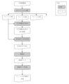
- FIG. 4 is a diagram showing a flow of obtaining blood flow velocities from frame 2 and frame 1 to frame 2 of the endoscope moving image by the endoscope system of the present invention;
- gray portions indicate processing means, and white portions indicate data.
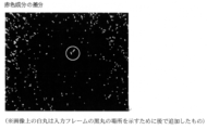
- FIG. 3 is a diagram in which translation components are removed from frames 1 and 2; It is a figure which shows the difference of a red component.
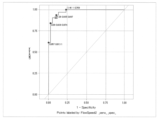
- ROC analysis results for blood flow velocities in early stage gastric cancer and mean blood flow velocities in normal gastric superficial microvessels are shown.
- One aspect of the present invention is an endoscopic system for measuring the blood flow velocity of gastrointestinal surface microtubules, comprising a magnifying endoscope and a blood flow moving image data processing unit obtained by the magnifying endoscope, (A) the magnifying endoscope captures a blood flow moving image of microvessels on the surface of the gastrointestinal tract and sends it to the blood flow moving image data processing unit; (B) An endoscope for measuring the blood flow velocity of gastrointestinal superficial microvessels, wherein the blood flow moving image data processing unit that receives the blood flow moving image performs the following data processing (B1) to (B5). System.
- Another aspect of the present invention is a method for measuring the blood flow velocity of microvessels in the surface layer of the gastrointestinal tract in real time from microvessel moving images of the surface layer of the gastrointestinal tract, comprising the following step (a): (f).
- the magnifying endoscope captures a blood flow moving image of microvessels on the surface of the gastrointestinal tract and sends it to the blood flow moving image data processing unit; All of the steps of capturing a moving image of blood flow in microvessels on the surface of the gastrointestinal tract using a magnifying endoscope are processes performed by the magnifying endoscope.
- This magnifying endoscope may be any endoscope that can measure blood flow in microvessels. Commercially available magnifying endoscopes have a moving image capturing function.
- FIG. 1 shows an example of a specific processing flow.
- Step (a) of the present invention is a step of capturing a moving image of blood flow in microvessels at a site suspected of having an abnormality in the surface layer of the gastrointestinal tract obtained by a magnifying endoscope.
- This magnifying endoscope may be any endoscope that can measure blood flow in microvessels.
- Commercially available magnifying endoscopes have a moving image capturing function.
- Steps (b) to (f) of the present invention can be performed in real time by a computer that receives blood flow moving images obtained by a magnifying endoscope.
- Known software can be used as the framework used for the image processing of the present invention. For example, OpenCV, dlib, etc. can be used. In addition, development languages such as c/c++, Python, and JavaScript can be used.
- Step (b) is a step of decomposing the obtained magnified endoscopic blood flow moving image into frames.
- This step is a step of designating a moving image file captured by a magnifying endoscope to be analyzed, and decomposing the moving image into frames.
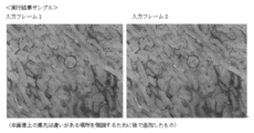
- the moving image is decomposed into an image of frame 1, an image of frame 2, an image of frame 3, and so on, as shown in FIG.
- Step (c) is a step of comparing the image of frame 1 with the image of next frame 2 to remove the translation component.
- an image from which the translation component between the image of frame 1 and the image of frame 2 is removed is obtained (see FIG. 1).
- This step detects the difference between the frame 1 image and the frame 2 image, that is, the difference between the frames caused by the blood flow (see FIG. 2).
- Step (d) is a step of calculating the difference with the red component of the image from which the translation component has been removed.
- This step is to calculate the difference between the images of frame 1 and frame 2, ie, the difference between the frames caused by the blood flow, in the red component. Since the movement of the red component is the movement of red blood cells in blood, the movement of red blood cells can be detected by calculating the difference with the red component.
- Step (e) is a step of regionalizing the portion where the difference of the red component is calculated. This step measures the area traversed by the red blood cells between frame 1 and frame 2.
- Step (f) is a step of calculating the region size of the obtained region data. This step measures the minor and major diameters of the region size.
- the blood flow velocity in each region of Frame 2 can be measured. Also, as shown in Table 2, the blood flow velocity in each region in the other frame images of the obtained moving image can also be measured.
- this blood flow rate with the normal blood flow rate of microvessels in the surface layer of the gastrointestinal tract, it is possible to diagnose whether or not a disease such as early cancer exists in the surface layer of the gastrointestinal tract.
- the comparison of these blood flow velocities can also be performed by a computer that performs the steps (b) to (f).
- the gastrointestinal tract includes the esophagus, stomach, duodenum, small intestine, large intestine and rectum.
- gastrointestinal diseases include neoplastic lesions such as esophageal cancer, gastric cancer, duodenal cancer, colon cancer and rectal cancer; non-neoplastic lesions such as gastric polyps and colon polyps; Inflammatory and functional diseases such as intestinal disease, esophagitis, gastritis, functional dyspepsia, irritable bowel syndrome, chronic constipation, diarrhea, and bowel movements, allergic gastrointestinal diseases, lifestyle-related diseases other than gastrointestinal diseases Abnormal gastrointestinal blood flow due to Here, for example, blood flow velocities in early gastric cancer were statistically significantly slower and narrower than those in normal gastric superficial microvessels, even when intra-individual variability was taken into account. . Furthermore, when ROC analysis was performed for each average, the cutoff value was 1.09 as shown in FIG. 4 (sensitivity 90.3%, specificity 89.7%
- Example 1 Using Python as a development language and OpenCV as an image processing framework, three arguments in Table 1 were specified, and a magnified endoscopic video of the gastric mucosa was processed according to the flow in FIG.
- the magnified endoscopic video was captured using a LASEREO 7000 series (FUJIFILM) endoscope system and an EG-L600ZW7 (FUJIFILM) scope.
- the parallel movement component-removed image shown in FIG. 2 was obtained, and the difference of the red component shown in FIG. 3 was detected.
- the blood flow velocity could be measured from the blood flow moving image of the microvessels on the surface of the gastrointestinal tract obtained by the magnifying endoscope.
- Table 2 shows the region size in each frame and the measurement results of the blood flow velocity in that region.
Landscapes
- Health & Medical Sciences (AREA)
- Life Sciences & Earth Sciences (AREA)
- Surgery (AREA)
- Nuclear Medicine, Radiotherapy & Molecular Imaging (AREA)
- Biomedical Technology (AREA)
- Optics & Photonics (AREA)
- Pathology (AREA)
- Radiology & Medical Imaging (AREA)
- Biophysics (AREA)
- Engineering & Computer Science (AREA)
- Physics & Mathematics (AREA)
- Heart & Thoracic Surgery (AREA)
- Medical Informatics (AREA)
- Molecular Biology (AREA)
- Animal Behavior & Ethology (AREA)
- General Health & Medical Sciences (AREA)
- Public Health (AREA)
- Veterinary Medicine (AREA)
- Measuring Pulse, Heart Rate, Blood Pressure Or Blood Flow (AREA)
Abstract
Le but de la présente invention est de fournir un moyen de mesure d'une vitesse de flux sanguin dans un petit vaisseau sanguin gastro-intestinal superficiel en temps réel. L'invention concerne un système d'endoscope de mesure d'une vitesse de flux sanguin dans un petit vaisseau sanguin gastro-intestinal superficiel, le système d'endoscope comportant un endoscope de grossissement et une unité de traitement de données d'image en mouvement de flux sanguin obtenues dans l'endoscope de grossissement, c'est-à-dire, une unité de traitement de données d'image en mouvement de flux sanguin, et le système d'endoscope étant caractérisé en ce que (A) l'endoscope de grossissement capture une image en mouvement de flux sanguin d'un petit vaisseau sanguin gastro-intestinal superficiel et envoie l'image en mouvement de flux sanguin à l'unité de traitement de données d'image en mouvement de flux sanguin, et (B) l'unité de traitement de données d'image en mouvement de flux sanguin qui a reçu l'image en mouvement de flux sanguin effectue les traitements de données (B1) à (B5) suivants. (B1) Un traitement de désassemblage de l'image en mouvement de flux sanguin obtenue en trames ; (B2) un traitement de comparaison d'une image d'une trame (1) à une image d'une trame (2) suivante afin de supprimer une composante de décalage parallèle ; (B3) un traitement de calcul d'une différence d'une composante rouge dans l'image dont la composante de décalage parallèle a été supprimée ; (B4) un traitement de régionalisation d'une région pour laquelle la différence de la composante rouge a été calculée ; et (B5) un traitement de calcul d'une taille de région des données de région obtenues.
Priority Applications (1)
| Application Number | Priority Date | Filing Date | Title |
|---|---|---|---|
| JP2023559636A JPWO2023085262A1 (fr) | 2021-11-09 | 2022-11-08 |
Applications Claiming Priority (2)
| Application Number | Priority Date | Filing Date | Title |
|---|---|---|---|
| JP2021182768 | 2021-11-09 | ||
| JP2021-182768 | 2021-11-09 |
Publications (1)
| Publication Number | Publication Date |
|---|---|
| WO2023085262A1 true WO2023085262A1 (fr) | 2023-05-19 |
Family
ID=86336130
Family Applications (1)
| Application Number | Title | Priority Date | Filing Date |
|---|---|---|---|
| PCT/JP2022/041538 WO2023085262A1 (fr) | 2021-11-09 | 2022-11-08 | Système d'endoscope permettant de mesurer une vitesse de flux sanguin dans un petit vaisseau sanguin gastro-intestinal superficiel |
Country Status (2)
| Country | Link |
|---|---|
| JP (1) | JPWO2023085262A1 (fr) |
| WO (1) | WO2023085262A1 (fr) |
Citations (5)
| Publication number | Priority date | Publication date | Assignee | Title |
|---|---|---|---|---|
| JPH0417076A (ja) * | 1990-05-10 | 1992-01-21 | Olympus Optical Co Ltd | 内視鏡用画像処理装置 |
| JP2014004329A (ja) * | 2012-06-01 | 2014-01-16 | Sony Corp | 歯用装置、医療用装置及び算出方法 |
| WO2016121811A1 (fr) * | 2015-01-29 | 2016-08-04 | 富士フイルム株式会社 | Dispositif de traitement d'image, procédé de traitement d'image, et système d'endoscope |
| WO2017061256A1 (fr) * | 2015-10-07 | 2017-04-13 | 富士フイルム株式会社 | Système d'endoscope et procédé de fonctionnement du système d'endoscope |
| JP2019520879A (ja) * | 2016-05-23 | 2019-07-25 | ライカ インストゥルメンツ (シンガポール) プライヴェット リミテッドLeica Instruments (Singapore) Pte. Ltd. | 顕微鏡または内視鏡等の医療用観察装置ならびに時間変調および/または空間変調を有する疑似カラーパターンを用いる方法 |
-
2022
- 2022-11-08 WO PCT/JP2022/041538 patent/WO2023085262A1/fr unknown
- 2022-11-08 JP JP2023559636A patent/JPWO2023085262A1/ja active Pending
Patent Citations (5)
| Publication number | Priority date | Publication date | Assignee | Title |
|---|---|---|---|---|
| JPH0417076A (ja) * | 1990-05-10 | 1992-01-21 | Olympus Optical Co Ltd | 内視鏡用画像処理装置 |
| JP2014004329A (ja) * | 2012-06-01 | 2014-01-16 | Sony Corp | 歯用装置、医療用装置及び算出方法 |
| WO2016121811A1 (fr) * | 2015-01-29 | 2016-08-04 | 富士フイルム株式会社 | Dispositif de traitement d'image, procédé de traitement d'image, et système d'endoscope |
| WO2017061256A1 (fr) * | 2015-10-07 | 2017-04-13 | 富士フイルム株式会社 | Système d'endoscope et procédé de fonctionnement du système d'endoscope |
| JP2019520879A (ja) * | 2016-05-23 | 2019-07-25 | ライカ インストゥルメンツ (シンガポール) プライヴェット リミテッドLeica Instruments (Singapore) Pte. Ltd. | 顕微鏡または内視鏡等の医療用観察装置ならびに時間変調および/または空間変調を有する疑似カラーパターンを用いる方法 |
Also Published As
| Publication number | Publication date |
|---|---|
| JPWO2023085262A1 (fr) | 2023-05-19 |
Similar Documents
| Publication | Publication Date | Title |
|---|---|---|
| Wu et al. | A deep neural network improves endoscopic detection of early gastric cancer without blind spots | |
| Struyvenberg et al. | A computer-assisted algorithm for narrow-band imaging-based tissue characterization in Barrett’s esophagus | |
| Rex et al. | High-definition colonoscopy versus Endocuff versus EndoRings versus full-spectrum endoscopy for adenoma detection at colonoscopy: a multicenter randomized trial | |
| Hoffman et al. | High-definition endoscopy with i-Scan and Lugol’s solution for more precise detection of mucosal breaks in patients with reflux symptoms | |
| Wu et al. | Effect of a deep learning-based system on the miss rate of gastric neoplasms during upper gastrointestinal endoscopy: a single-centre, tandem, randomised controlled trial | |
| Bhat et al. | High-definition and high-magnification endoscopes | |
| Cho et al. | Comparison of convolutional neural network models for determination of vocal fold normality in laryngoscopic images | |
| Kwon et al. | High-resolution and high-magnification endoscopes | |
| WO2020215810A1 (fr) | Procédé de détection d'image à bande étroite basé sur la reconnaissance d'image pour procédure de coloscopie | |
| JP2024045234A (ja) | 腸の病理学のための画像スコアリング | |
| WO2021054477A2 (fr) | Méthode d'aide au diagnostic de maladie à l'aide d'une image endoscopique de l'appareil digestif, système d'aide au diagnostic, programme d'aide au diagnostic et support d'enregistrement lisible par ordinateur sur lequel est mémorisé ledit programme d'aide au diagnostic | |
| Al-Rahayfeh et al. | Detection of bleeding in wireless capsule endoscopy images using range ratio color | |
| Waki et al. | Usefulness of an artificial intelligence system for the detection of esophageal squamous cell carcinoma evaluated with videos simulating overlooking situation | |
| JP2006218138A (ja) | ハイビジョンデジタル内視鏡画像のファイリング及びコンピューター支援診断装置 | |
| Qu et al. | Development and validation of an automatic image-recognition endoscopic report generation system: A multicenter study | |
| Brodersen et al. | Artificial Intelligence-assisted Analysis of Pan-enteric Capsule Endoscopy in Patients with Suspected Crohn’s Disease: A Study on Diagnostic Performance | |
| Matrakool et al. | Improved detection of Helicobacter pylori infection and premalignant gastric mucosa using conventional white light source gastroscopy | |
| WO2023085262A1 (fr) | Système d'endoscope permettant de mesurer une vitesse de flux sanguin dans un petit vaisseau sanguin gastro-intestinal superficiel | |
| Honzawa et al. | A novel endoscopic imaging system for quantitative evaluation of colonic mucosal inflammation in patients with quiescent ulcerative colitis | |
| Arif et al. | Artificial intelligence in endoscopy: Overview, applications, and future directions | |
| Carter et al. | PillCam colon capsule endoscopy (PCCE) in colonic diseases | |
| Tennyson et al. | Video capsule endoscopy in celiac disease | |
| Kunihara et al. | Predictive factors of portal hypertensive enteropathy exacerbation in patients with liver cirrhosis: a capsule endoscopy study | |
| Romańczyk et al. | The prospective validation of a scoring system to assess mucosal cleanliness during esophagogastroduodenoscopy | |
| Fiaidhi et al. | An Investigation into Crohn’s Disease Lesions Variability Sensing Using Video Colonoscopy and Machine Learning Techniques |
Legal Events
| Date | Code | Title | Description |
|---|---|---|---|
| 121 | Ep: the epo has been informed by wipo that ep was designated in this application |
Ref document number: 22892760 Country of ref document: EP Kind code of ref document: A1 |
|
| ENP | Entry into the national phase |
Ref document number: 2023559636 Country of ref document: JP Kind code of ref document: A |