WO2023074794A1 - Procédé ou agent avec régulateur de hdac, pour le traitement du diabète et de ses complications - Google Patents
Procédé ou agent avec régulateur de hdac, pour le traitement du diabète et de ses complications Download PDFInfo
- Publication number
- WO2023074794A1 WO2023074794A1 PCT/JP2022/040140 JP2022040140W WO2023074794A1 WO 2023074794 A1 WO2023074794 A1 WO 2023074794A1 JP 2022040140 W JP2022040140 W JP 2022040140W WO 2023074794 A1 WO2023074794 A1 WO 2023074794A1
- Authority
- WO
- WIPO (PCT)
- Prior art keywords
- diabetes
- cells
- insulin
- treatment
- hdac
- Prior art date
Links
Images


Classifications
-
- A—HUMAN NECESSITIES
- A61—MEDICAL OR VETERINARY SCIENCE; HYGIENE
- A61K—PREPARATIONS FOR MEDICAL, DENTAL OR TOILETRY PURPOSES
- A61K31/00—Medicinal preparations containing organic active ingredients
- A61K31/16—Amides, e.g. hydroxamic acids
- A61K31/165—Amides, e.g. hydroxamic acids having aromatic rings, e.g. colchicine, atenolol, progabide
- A61K31/167—Amides, e.g. hydroxamic acids having aromatic rings, e.g. colchicine, atenolol, progabide having the nitrogen of a carboxamide group directly attached to the aromatic ring, e.g. lidocaine, paracetamol
-
- A—HUMAN NECESSITIES
- A61—MEDICAL OR VETERINARY SCIENCE; HYGIENE
- A61K—PREPARATIONS FOR MEDICAL, DENTAL OR TOILETRY PURPOSES
- A61K31/00—Medicinal preparations containing organic active ingredients
- A61K31/16—Amides, e.g. hydroxamic acids
- A61K31/18—Sulfonamides
-
- A—HUMAN NECESSITIES
- A61—MEDICAL OR VETERINARY SCIENCE; HYGIENE
- A61K—PREPARATIONS FOR MEDICAL, DENTAL OR TOILETRY PURPOSES
- A61K31/00—Medicinal preparations containing organic active ingredients
- A61K31/33—Heterocyclic compounds
- A61K31/335—Heterocyclic compounds having oxygen as the only ring hetero atom, e.g. fungichromin
- A61K31/34—Heterocyclic compounds having oxygen as the only ring hetero atom, e.g. fungichromin having five-membered rings with one oxygen as the only ring hetero atom, e.g. isosorbide
- A61K31/343—Heterocyclic compounds having oxygen as the only ring hetero atom, e.g. fungichromin having five-membered rings with one oxygen as the only ring hetero atom, e.g. isosorbide condensed with a carbocyclic ring, e.g. coumaran, bufuralol, befunolol, clobenfurol, amiodarone
-
- A—HUMAN NECESSITIES
- A61—MEDICAL OR VETERINARY SCIENCE; HYGIENE
- A61K—PREPARATIONS FOR MEDICAL, DENTAL OR TOILETRY PURPOSES
- A61K31/00—Medicinal preparations containing organic active ingredients
- A61K31/33—Heterocyclic compounds
- A61K31/395—Heterocyclic compounds having nitrogen as a ring hetero atom, e.g. guanethidine or rifamycins
- A61K31/40—Heterocyclic compounds having nitrogen as a ring hetero atom, e.g. guanethidine or rifamycins having five-membered rings with one nitrogen as the only ring hetero atom, e.g. sulpiride, succinimide, tolmetin, buflomedil
-
- A—HUMAN NECESSITIES
- A61—MEDICAL OR VETERINARY SCIENCE; HYGIENE
- A61K—PREPARATIONS FOR MEDICAL, DENTAL OR TOILETRY PURPOSES
- A61K31/00—Medicinal preparations containing organic active ingredients
- A61K31/33—Heterocyclic compounds
- A61K31/395—Heterocyclic compounds having nitrogen as a ring hetero atom, e.g. guanethidine or rifamycins
- A61K31/495—Heterocyclic compounds having nitrogen as a ring hetero atom, e.g. guanethidine or rifamycins having six-membered rings with two or more nitrogen atoms as the only ring heteroatoms, e.g. piperazine or tetrazines
- A61K31/505—Pyrimidines; Hydrogenated pyrimidines, e.g. trimethoprim
- A61K31/506—Pyrimidines; Hydrogenated pyrimidines, e.g. trimethoprim not condensed and containing further heterocyclic rings
-
- A—HUMAN NECESSITIES
- A61—MEDICAL OR VETERINARY SCIENCE; HYGIENE
- A61K—PREPARATIONS FOR MEDICAL, DENTAL OR TOILETRY PURPOSES
- A61K45/00—Medicinal preparations containing active ingredients not provided for in groups A61K31/00 - A61K41/00
-
- A—HUMAN NECESSITIES
- A61—MEDICAL OR VETERINARY SCIENCE; HYGIENE
- A61P—SPECIFIC THERAPEUTIC ACTIVITY OF CHEMICAL COMPOUNDS OR MEDICINAL PREPARATIONS
- A61P1/00—Drugs for disorders of the alimentary tract or the digestive system
-
- A—HUMAN NECESSITIES
- A61—MEDICAL OR VETERINARY SCIENCE; HYGIENE
- A61P—SPECIFIC THERAPEUTIC ACTIVITY OF CHEMICAL COMPOUNDS OR MEDICINAL PREPARATIONS
- A61P13/00—Drugs for disorders of the urinary system
- A61P13/12—Drugs for disorders of the urinary system of the kidneys
-
- A—HUMAN NECESSITIES
- A61—MEDICAL OR VETERINARY SCIENCE; HYGIENE
- A61P—SPECIFIC THERAPEUTIC ACTIVITY OF CHEMICAL COMPOUNDS OR MEDICINAL PREPARATIONS
- A61P17/00—Drugs for dermatological disorders
-
- A—HUMAN NECESSITIES
- A61—MEDICAL OR VETERINARY SCIENCE; HYGIENE
- A61P—SPECIFIC THERAPEUTIC ACTIVITY OF CHEMICAL COMPOUNDS OR MEDICINAL PREPARATIONS
- A61P25/00—Drugs for disorders of the nervous system
-
- A—HUMAN NECESSITIES
- A61—MEDICAL OR VETERINARY SCIENCE; HYGIENE
- A61P—SPECIFIC THERAPEUTIC ACTIVITY OF CHEMICAL COMPOUNDS OR MEDICINAL PREPARATIONS
- A61P27/00—Drugs for disorders of the senses
- A61P27/02—Ophthalmic agents
-
- A—HUMAN NECESSITIES
- A61—MEDICAL OR VETERINARY SCIENCE; HYGIENE
- A61P—SPECIFIC THERAPEUTIC ACTIVITY OF CHEMICAL COMPOUNDS OR MEDICINAL PREPARATIONS
- A61P3/00—Drugs for disorders of the metabolism
- A61P3/08—Drugs for disorders of the metabolism for glucose homeostasis
- A61P3/10—Drugs for disorders of the metabolism for glucose homeostasis for hyperglycaemia, e.g. antidiabetics
Definitions
- the present disclosure relates to treatment and prevention of diabetes and/or its related diseases by combining histone deacetylase (HDAC) modulation and glycemic control. More particularly, the present disclosure relates to the treatment and prevention of diabetes and/or its related diseases by modulating HDACs in glycemically controlled subjects and/or by modulating HDACs in combination with glycemic control treatments.
- HDAC histone deacetylase
- Non-Patent Document 1 Diabetes is generally classified into type 1 and type 2 (Non-Patent Document 1). However, since the details of the onset of both are unknown, it must be said that the classification of type 1 and type 2 itself lacks validity as based on etiology.
- various drugs are currently being developed for the treatment of diabetes, but most of them are aimed at blood sugar control and may be effective in preventing the progression of diabetes, but are insufficient for curing diabetes. can be In addition, diabetic complications such as neuropathy, nephropathy, liver damage, retinopathy, fatty liver, gastrointestinal disorders, delayed healing of fractures, eating disorders, and skin disorders are common to diabetes classified as type 1 and type 2. However, once it develops, it can be difficult to cure.
- the inventors have found that therapeutic strategies for diabetes and/or its related diseases based on HDAC regulation combined with glycemic control are highly effective. Based on this new finding, the present disclosure provides tools for the treatment of diabetes and/or its related diseases.
- the present disclosure provides: (Item 1) A composition comprising a histone deacetylase (HDAC) modulator for treating and/or preventing diabetes and/or diabetes-related diseases, disorders and/or conditions in a subject, said subject comprising: The composition is administered under controlled conditions. (Item 2) A composition comprising a histone deacetylase (HDAC) modulator for treating and/or preventing diabetes and/or diabetes-related diseases, disorders and/or conditions in a subject, said composition comprising glycemic control treatment and A composition characterized in that it is administered in combination. (Item 3) The composition of any of the preceding items, wherein the HDAC modulator inhibits at least one of Class I, Class II and Class IV HDACs.
- HDAC histone deacetylase
- the HDAC modulator is fingolimod (FTY720), divinostat, vorinostat (SAHA, suberoylanilide hydroxamic acid), belinostat, panobinostat, resminostat, abexinostat, mosetinostat, trichostatin A (TSA), romidepsin, apicidin, thidamide , entinostat, tacedinaline, tucidinostat, pracinostat (SB939), xinostat, tefinostat, rosilinostat, fimepinostat, nanatinostat, domatinostat, trapoxin (TPX), cyclic hydroxamic acid-containing peptides 1 (CHAP1), valproic acid (VPA), sodium butyrate (NaBu), 4-phenylbutyric acid, OBP-801, ME-344, CG200745, MPT0E014, RGFP966 (MCE)
- the HDAC modulator is fingolimod (FTY720), divinostat, trichostatin A, vorinostat, belinostat, panobinostat, resminostat, abexinostat, xinostat, pracinostat, rosilinostat, nanatinostat, dacinostat, (N -hydroxy-7-2-naphthylthio)heptanomide, valproic acid, CG200745, scriptaid, KBH-A42, romidepsin, sodium butyrate, 4-phenylbutyric acid, SK-7041, WW437, entinostat, tacedinaline, mosetinostat, butypivalate at least selected from the group consisting of lyloxymethyl, trapoxin, ⁇ -ketoamides, heterocyclic ketones, diallyltrisulfide, licorinostat, tubacin, azelaroyl bis-hydroxamic
- the hypoglycemic agents include insulin, insulin analogues, biguanide drugs, insulin secretagogues (sulfonylurea drugs (SU drugs), etc.), fast-acting insulin secretagogues (glinide drugs, etc.), incretin-related drugs (DPP-4 inhibitors, etc.).
- the control of blood glucose includes controlling fasting blood glucose to less than 126 mg/dL, controlling blood glucose to less than 200 mg/dL at 2 hours of an oral glucose tolerance test, or random blood glucose to less than 200 mg/dL.
- a composition of any of the above items comprising controlling.
- the composition of any of the preceding items, wherein said disease, disorder and/or condition comprises diabetic complications.
- the disease, disorder and/or symptom is selected from the group consisting of neuropathy, nephropathy, liver disorder, retinopathy, fatty liver, gastrointestinal disorder, fracture healing delay, eating disorder, and skin disorder. any composition.
- HSC hematopoietic stem cells
- Item 13 The composition of any of the preceding items, wherein said abnormal hematopoietic stem cells are characterized by at least one of altered levels of any HDACs and enhanced levels of CD106.
- composition of any of the preceding items wherein the composition is a pharmaceutical composition.
- composition of any of the preceding items further comprising a pharmaceutically acceptable excipient.
- HDAC histone deacetylase
- HDAC histone deacetylase
- the HDAC modulator is fingolimod (FTY720), divinostat, vorinostat (SAHA, suberoylanilide hydroxamic acid), belinostat, panobinostat, resminostat, abexinostat, mosetinostat, trichostatin A (TSA), romidepsin, apicidin, thidamide , entinostat, tacedinaline, tucidinostat, pracinostat (SB939), xinostat, tefinostat, rosilinostat, fimepinostat, nanatinostat, domatinostat, trapoxin (TPX), cyclic hydroxamic acid-containing peptides 1 (CHAP1), valproic acid (VPA), sodium butyrate (NaBu), 4-phenylbutyric acid, OBP-801, ME-344, CG200745, MPT0E014, RGFP966 (MCE
- the HDAC modulator is fingolimod (FTY720), divinostat, trichostatin A, vorinostat, belinostat, panobinostat, resminostat, abexinostat, xinostat, pracinostat, rosilinostat, nanatinostat, dacinostat, (N -hydroxy-7-2-naphthylthio)heptanomide, valproic acid, CG200745, scriptaid, KBH-A42, romidepsin, sodium butyrate, 4-phenylbutyric acid, SK-7041, WW437, entinostat, tacedinaline, mosetinostat, butypivalate at least selected from the group consisting of lyloxymethyl, trapoxin, ⁇ -ketoamides, heterocyclic ketones, diallyltrisulfide, licorinostat, tubacin, azelaroyl bis-hydroxamic
- the hypoglycemic agents include insulin, insulin analogues, biguanide drugs, insulin secretagogues (sulfonylurea drugs (SU drugs), etc.), fast-acting insulin secretagogues (glinide drugs, etc.), incretin-related drugs (DPP-4 inhibitors, etc.).
- the control of blood glucose includes controlling fasting blood glucose to less than 126 mg/dL, controlling blood glucose to less than 200 mg/dL at 2 hours of an oral glucose tolerance test, or random blood glucose to less than 200 mg/dL.
- a method of any of the above items comprising controlling.
- the disease, disorder and/or symptom is selected from the group consisting of neuropathy, nephropathy, liver disorder, retinopathy, fatty liver, gastrointestinal disorder, fracture healing delay, eating disorder, and skin disorder. either way.
- the method of any of the preceding items comprising diagnosing said subject as having abnormal hematopoietic stem cells (HSC) or cells derived therefrom.
- HSC hematopoietic stem cells
- Item 28 The method of any of the preceding items, wherein the abnormal hematopoietic stem cells are characterized by at least one of altered levels of any HDACs and elevated levels of CD106.
- HDAC histone deacetylase
- the HDAC modulator is fingolimod (FTY720), divinostat, vorinostat (SAHA, suberoylanilide hydroxamic acid), belinostat, panobinostat, resminostat, abexinostat, mosetinostat, trichostatin A (TSA), romidepsin, apicidin, thidamide , entinostat, tacedinaline, tucidinostat, pracinostat (SB939), xinostat, tefinostat, rosilinostat, fimepinostat, nanatinostat, domatinostat, trapoxin (TPX), cyclic hydroxamic acid-containing peptides 1 (CHAP1), valproic acid (VPA), sodium butyrate (NaBu), 4-phenylbutyric acid, OBP-801, ME-344, CG200745, MPT0E014, RGFP966 (MCE
- the HDAC modulator is fingolimod (FTY720), divinostat, trichostatin A, vorinostat, belinostat, panobinostat, resminostat, abexinostat, xinostat, pracinostat, rosilinostat, nanatinostat, dacinostat, (N -hydroxy-7-2-naphthylthio)heptanomide, valproic acid, CG200745, scriptaid, KBH-A42, romidepsin, sodium butyrate, 4-phenylbutyric acid, SK-7041, WW437, entinostat, tacedinaline, mosetinostat, butypivalate at least selected from the group consisting of lyloxymethyl, trapoxin, ⁇ -ketoamides, heterocyclic ketones, diallyltrisulfide, licorinostat, tubacin, azelaroyl bis-hydroxamic
- hypoglycemic agents include insulin, insulin analogues, biguanide drugs, insulin secretagogues (sulfonylurea drugs (SU drugs), etc.), fast-acting insulin secretagogues (glinide drugs, etc.), incretin-related drugs (DPP-4 inhibitors, etc.).
- the control of blood glucose includes controlling fasting blood glucose to less than 126 mg/dL, controlling blood glucose to less than 200 mg/dL at 2 hours of an oral glucose tolerance test, or random blood glucose to less than 200 mg/dL. Use of any of the above items, including controlling.
- Item 37 Use of any of the above items for treating diabetes and/or diabetes-related diseases, disorders and/or conditions.
- (Item 38) Use of any of the items above, wherein said disease, disorder and/or condition comprises diabetic complications.
- the disease, disorder and/or symptom is selected from the group consisting of neuropathy, nephropathy, liver disorder, retinopathy, fatty liver, gastrointestinal disorder, fracture healing delay, eating disorder, and skin disorder. any use.
- (Item 40) Use of any of the above items, wherein said subject has been diagnosed with abnormal hematopoietic stem cells (HSC) or cells derived therefrom.
- HSC hematopoietic stem cells
- Item 41 Use of any of the preceding items, wherein said abnormal hematopoietic stem cells are characterized by at least one of altered levels of any HDAC and enhanced levels of CD106.
- HDAC histone deacetylase
- HDAC modulator for treating and/or preventing diabetes and/or diabetes-related diseases, disorders and/or conditions in a subject, wherein blood glucose is controlled in the subject
- An HDAC modulator characterized in that it is administered under conditions.
- HDAC histone deacetylase
- An HDAC modulator characterized by: (Item 44) The HDAC modulator of any of the preceding items, wherein said HDAC modulator inhibits at least one of Class I, Class II and Class IV HDACs.
- the HDAC modulator is fingolimod (FTY720), divinostat, vorinostat (SAHA, suberoylanilide hydroxamic acid), belinostat, panobinostat, resminostat, abexinostat, mosetinostat, trichostatin A (TSA), romidepsin, apicidin, thidamide , entinostat, tacedinaline, tucidinostat, pracinostat (SB939), xinostat, tefinostat, rosilinostat, fimepinostat, nanatinostat, domatinostat, trapoxin (TPX), cyclic hydroxamic acid-containing peptides 1 (CHAP1), valproic acid (VPA), sodium butyrate (NaBu), 4-phenylbutyric acid, OBP-801, ME-344, CG200745, MPT0E014, RGFP966 (MCE
- the HDAC modulator is fingolimod (FTY720), divinostat, trichostatin A, vorinostat, belinostat, panobinostat, resminostat, abexinostat, xinostat, pracinostat, rosilinostat, nanatinostat, dacinostat, (N -hydroxy-7-2-naphthylthio)heptanomide, valproic acid, CG200745, scriptaid, KBH-A42, romidepsin, sodium butyrate, 4-phenylbutyric acid, SK-7041, WW437, entinostat, tacedinaline, mosetinostat, butypivalate at least selected from the group consisting of lyloxymethyl, trapoxin, ⁇ -ketoamides, heterocyclic ketones, diallyltrisulfide, licorinostat, tubacin, azelaroyl bis-hydroxamic
- the HDAC regulating agent according to any one of the above items, wherein the control of blood sugar is performed by at least one selected from the group consisting of administration of hypoglycemic agents, dietary restriction and nutrient injection control.
- the hypoglycemic agents include insulin, insulin analogues, biguanide drugs, insulin secretagogues (sulfonylurea drugs (SU drugs), etc.), fast-acting insulin secretagogues (glinide drugs, etc.), incretin-related drugs (DPP-4 inhibitors, etc.).
- the control of blood glucose includes controlling fasting blood glucose to less than 126 mg/dL, controlling blood glucose to less than 200 mg/dL at 2 hours of an oral glucose tolerance test, or random blood glucose to less than 200 mg/dL. HDAC modulator of any of the above items, including controlling.
- the disease, disorder and/or symptom is selected from the group consisting of neuropathy, nephropathy, liver disorder, retinopathy, fatty liver, gastrointestinal disorder, fracture healing delay, eating disorder, and skin disorder. Any HDAC modulator.
- HSC abnormal hematopoietic stem cells
- pancreatic transplantation and pancreatic islet transplantation which are methods of restoring insulin secretion in diabetic patients, do not require donors, and can overcome the limitation of supply capacity with respect to the number of recipients. Also, patient burden such as surgery that the recipient has undergone can be avoided.
- the treatment of the present disclosure can be accomplished by drug administration and can therefore be much more convenient and less burdensome.
- radical treatment is possible, the prognosis is expected to be good.
- FIG. 2 is a schematic diagram showing the mechanism by which bone marrow-derived cells cause abnormalities in type 2 diabetes.
- FIG. 1 is a schematic diagram showing the mechanism by which bone marrow-derived cells cause abnormalities in type 1 diabetes.
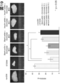
- FIG. 2 is a schematic diagram showing an overview of bone marrow transplantation therapy for STZ diabetic mice. Schematic of transplantation of non-diabetic or diabetic derived bone marrow into STZ diabetic mice under insulin pellet or blank pellet treatment. 1 shows a comparison of mRNA expression of each histone deacetylase gene group (HDACs) in KSL cells of non-DM and STZ-DM mice.
- HDACs histone deacetylase gene group
- the horizontal axis indicates the mRNA expression fold (Log2 change) in STZ-DM mice relative to non-DM mice, with increased expression on the right and decreased expression on the left.
- FIG. 5 shows changes in blood glucose levels when the treatments shown in FIG. 4 are performed.
- the vertical axis shows the average blood glucose level (mg/dL) at each time point with standard error.
- the horizontal axis shows the time course of treatment, with the time of bone marrow transplantation set as week 0.
- the shelf life of insulin pellets is also illustrated. * indicates P ⁇ 0.05, **P ⁇ 0.01.
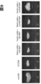
- FIG. 5 shows pancreas cryosection results when the treatment shown in FIG. 4 is performed. (Upper panel) Insulin and nuclear staining of frozen sections. The mouse condition under which each section was acquired is displayed above each image. Scale bar indicates 20 ⁇ m.
- Pancreatic islets were randomly selected in each mouse, and the ratio (%) of insulin-positive cells to the islet size was calculated using Image J software. From left, non-diabetic mice, STZ-diabetic mice, STZ-diabetic mice treated with blank pellets and diabetic bone marrow transplants, STZ-diabetic mice treated with blank pellets and non-diabetic bone marrow transplants, STZ-diabetic mice treated with insulin pellets and diabetic bone marrow transplants. , and STZ diabetic mice treated with insulin pellets and non-diabetic bone marrow transplantation. ** indicates P ⁇ 0.01.
- FIG. 5 shows pancreas cryosection results when the treatments shown in FIG.
- FIG. 4 shows a comparison of the sizes of representative excised thymuses when the treatments shown in FIG. 4 are performed.
- FIG. 2 is a schematic diagram showing an outline of mouse experiments of diabetes induction by bone marrow transplantation using bone marrow cells derived from transgenic mice transfected with green fluorescent protein (GFP mice), combined treatment with insulin pellets, and STZ.
- FIG. 2 is a schematic diagram showing an outline of mouse experiments of diabetes induction by bone marrow transplantation using bone marrow cells derived from transgenic mice transfected with green fluorescent protein (GFP mice), combined treatment with insulin pellets, and STZ.
- FIG. 2 shows laser fluorescence microscope images of pancreatic islets in mice transplanted with GFP mouse bone marrow. Vcam and nuclei were stained in tissue sections and fluorescence was observed along with GFP. The mouse condition under which each section was acquired is displayed above each image. Scale bar indicates 50 ⁇ m. Nuclei are shown in blue (DAPI), bone marrow-derived cells (GFP) in green, and Vcam1 (Tomato) in red. The white dashed line indicates the extra-islet border.
- FIG. 10 shows laser fluorescence microscope images of thymus in mice transplanted with bone marrow of GFP mice. Vcam and nuclei were stained in tissue sections and fluorescence was observed along with GFP. The mouse condition under which each section was acquired is displayed above each image.
- FIG. 10 outlines the experiment of Example 3 comparing streptozotocin-induced diabetic mice treated with or without divinostat under insulin pellet or blank pellet treatment. Mice without diabetic induction by streptozotocin (nonDM, control), blank pellets + no divinostat treatment (STZDM), insulin pellets + no divinostat treatment, blank pellets + divinostat treatment (orally or intravenously), and insulin pellets + divinostat treatment Studies were performed using each (oral or intravenous) streptozotocin-induced diabetic mice.
- FIG. 3 shows changes in blood glucose level of each mouse in the experiment of Example 3.
- FIG. The vertical axis shows the average blood glucose level (mg/dL) at each time point with standard error.
- the horizontal axis indicates the time course (weeks) of treatment, with the time point at which divinostat treatment was initiated as week 0. Each mark indicates the correspondence between the polygonal line and the type of mouse.
- 2 shows blood glucose levels after intraperitoneal glucose tolerance test (IPGTT) in streptozotocin-induced diabetic mice with blank pellet + divinostat treatment and insulin pellet + divinostat treatment in the experiment of Example 3.
- FIG. The vertical axis indicates the average blood glucose level (mg/dL) at each time point, and the horizontal axis indicates the time course after glucose injection.
- FIG. 3 shows blood insulin after intraperitoneal glucose tolerance test (IPGTT) of each streptozotocin-induced diabetic mice with blank pellet + divinostat treatment and insulin pellet + divinostat treatment in the experiment of Example 3.
- IPGTT intraperitoneal glucose tolerance test
- FIG. The vertical axis indicates the average blood insulin (ng/mL) at each time point, and the horizontal axis indicates the time course after glucose injection.
- 2 shows the observation results of insulin immunohistochemical staining of frozen pancreas sections treated in the experiment of Example 3.
- FIG. (Upper panel) Insulin and nuclear staining of frozen sections. Scale bar indicates 20 ⁇ m.
- Pancreatic islets were randomly selected in each mouse, and the ratio (%) of insulin-positive cells to the islet size was calculated using Image J software.
- mice without diabetic induction by streptozotocin control
- blank pellets + no divinostat treatment blank pellets + no divinostat treatment
- blank pellets + divinostat treatment oral
- insulin pellets + divinostat treatment intravenous
- Streptozotocin-induced diabetic mice with pellets + divinostat treatment (oral) and insulin pellets + divinostat treatment are shown. ** indicates P ⁇ 0.01.
- 2 shows the observation results of glucagon immunohistochemical staining of pancreatic cryosections treated in the experiment of Example 3.
- FIG. (Upper panel) Glucagon and nuclear staining of frozen sections. Scale bar indicates 20 ⁇ m.
- Pancreatic islets were randomly selected in each mouse, and the ratio (%) of glucagon-positive cells to the size of the pancreatic islets was calculated using Image J software. From left, mice without diabetic induction by streptozotocin (control), blank pellets + no divinostat treatment, blank pellets + no divinostat treatment, blank pellets + divinostat treatment (oral), insulin pellets + divinostat treatment (intravenous), insulin. Streptozotocin-induced diabetic mice with pellets + divinostat treatment (oral) and insulin pellets + divinostat treatment (intravenous) are shown. ** indicates P ⁇ 0.01.
- FIG. 2 shows the observation results of TNF- ⁇ immunohistochemical staining of bone tissue demineralized sections treated in the experiment of Example 3.
- FIG. (Upper panel) Stained images of TNF- ⁇ (Ni-DAB staining, brown) and nuclei (hematoxylin staining, purple) in bone tissue demineralized sections. Scale bar indicates 20 ⁇ m.
- mice without diabetes induction control
- blank pellets + no divinostat treatment blank pellets + divinostat treatment (oral)
- blank pellets + divinostat treatment intravenous
- insulin pellets + divinostat treatment oral
- Individual streptozotocin-induced diabetic mice with insulin pellets plus divinostat treatment are shown.
- 2 shows the observation results of TNF- ⁇ immunohistochemical staining of the thymus when treated in the experiment of Example 3.
- FIG. (Upper panel) Stained images of thymic TNF- ⁇ (Ni-DAB staining, brown, indicated by black arrows) and nuclei (hematoxylin staining, purple). Scale bar indicates 20 ⁇ m.
- TNF- ⁇ Ni-DAB staining, brown
- nuclei hematoxylin staining, purple
- Scale bar indicates 20 ⁇ m.
- Pancreatic islets in a section were randomly selected from each mouse, and the ratio (%) of TNF- ⁇ positive cells among cells with stained nuclei (purple) was calculated using Image J software.
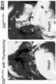
- FIG. 10 is a schematic diagram showing an overview of the treatment of thymectomized STZ diabetic mice in Example 8.
- FIG. 24 shows changes in blood glucose levels when the treatments shown in FIG. 23 are performed. The vertical axis shows the average blood glucose level (mg/dL) at each time point with standard error.
- FIG. 24 is a photograph of the thymic periphery of the thymectomized group (left) and control group (right) mice treated as shown in FIG. 23 at 8 weeks.
- diabetes is used in the usual sense used in the art, except for diabetes associated with pregnancy or due to specific genetic abnormalities, and usually insulin depletion due to destruction of pancreatic ⁇ cells.
- type 1 diabetes mellitus caused by obesity and type 2 diabetes mellitus caused by decreased insulin secretion from ⁇ -cells in the islets of Langerhans (pancreatic islets) in the pancreas, resulting in decreased glucose uptake into muscle and adipose tissue. are categorized. It should be noted that, according to the results of the present disclosure, both type 1 diabetes and type 2 diabetes are completely different from what has been thought to be the etiology until now.
- Diabetes is defined as, for example, two or more tests with fasting blood sugar ⁇ 126 mg/dL, HbA1c ⁇ 6.5%, oral glucose tolerance test (75 g OGTT) with a 2-hour value of 200 mg/dL or higher, or random blood sugar level of 200 mg/dL. It can be diagnosed by dL or higher and the like.
- Diabetes as used herein also includes juvenile-onset maturity diabetes and borderline diabetes.
- diabetes-related disease or “diabetes-related disease, disorder and/or condition” can include any disease, disorder and condition associated with diabetes, examples of which include diabetic neuropathy.
- diabetes can include diabetic neuropathy.
- Disorder diabetic retinopathy, diabetic nephropathy, secondary diabetes, diabetic coma, disturbance of consciousness, abdominal pain, cramps, neuropathy, hyperosmotic hyperglycemia syndrome, albuminuria, edema, renal failure, blindness, Alzheimer's cognition disease, myocardial infarction, arteriosclerosis obliterans, cerebral infarction, fatty liver, skin symptoms (diabetic lipoid necrosis, etc.), decreased wound healing ability, susceptibility to infection (septicemia, etc.), cancer (liver cancer, kidney cancer, pancreatic cancer, colon cancer, stomach cancer, liver cancer, ovarian cancer, colorectal cancer, colon cancer, etc.), diabetic ketoacidosis, heart disease, cerebrovascular disease, steroid diabetes, constip
- non-diabetic subject means a subject who does not have diabetes or its related diseases.
- blood sugar control refers to performing a treatment to lower blood sugar (blood glucose concentration: fasting blood sugar, glucose tolerance test blood sugar, random blood sugar, etc.), and It refers to both states in which blood glucose is maintained within the therapeutic ranges described herein.
- Treatments to lower blood sugar include administration of hypoglycemic agents, dietary restrictions, controlled nutrient infusions, and the like.
- blood sugar controlled state may be achieved by a treatment that actively keeps blood sugar within a certain range, and blood sugar is kept within the certain range without doing anything may be the case.
- the case where the blood glucose falls within the certain range also falls under the "state of blood glucose control".
- treatment refers to prevention, preferably maintenance, more preferably alleviation of aggravation of a disease or disorder when such a condition occurs. Preferably, it means to cure, and includes the ability to exert symptom ameliorating effect or preventive effect on the patient's disease or one or more symptoms associated with the disease.
- Alleviating refers to reducing the severity of a disease or disorder when such a condition occurs, and “cure” means no longer To be undiagnosed as having such a disease or disorder.
- Preliminary diagnosis and appropriate treatment are called “companion therapy”, and the diagnostic agent for that purpose is sometimes called “companion diagnostic agent”.
- prevention refers to preventing a disease or disorder from becoming such a condition before it becomes such a condition.
- the agents of the present disclosure can be used for diagnosis, and if necessary, the agents of the present disclosure can be used to prevent, for example, diabetes, or preventive measures can be taken.
- diagnosis refers to identifying various parameters related to a condition (eg, disease, disorder) in a subject and determining the current status or future of such condition.
- conditions within the body can be determined, and such information can be used to determine conditions in a subject, treatment or prophylaxis formulations or methods to be administered, etc. can be selected.
- diagnosis in a narrow sense means diagnosing the current situation, but in a broader sense it includes “early diagnosis”, “predictive diagnosis”, “pre-diagnosis” and the like.
- the diagnostic method of the present disclosure is industrially useful because, in principle, it can use what comes out of the body and can be performed without the hands of medical professionals such as doctors.
- "predictive diagnosis, preliminary diagnosis or diagnosis” is sometimes referred to as "assisting”.
- HDAC histone deacetylase
- SIRT 1 to 7 are known.
- the enzymatic activity of HDACs 1-11 is believed to be zinc ion dependent, with HDACs 1, 2, 3 and 8 in class I, HDACs 4, 5, 7 and 9 in class IIa, HDACs 6 and 10 in class IIb, and HDAC11. is classified as Class IV.
- the enzymatic activity of SIRT1-7 is believed to be NAD+ dependent and classified as class III HDACs.
- Each HDAC and its representative nucleic acid sequence and amino acid sequence are shown below.
- modulation of HDAC or the like means increasing, decreasing or eliminating the function of HDAC or the like by any means.
- Modulation includes “inhibition” and “activation.”
- inhibittion of HDAC or the like means reducing or eliminating the function of HDAC or the like by any means. For example, inhibition of HDACs can result in reduced or eliminated enzymatic activity of HDACs, reduced or eliminated transcription of HDAC genes, reduced or eliminated translation of HDAC proteins, enhanced degradation of HDAC proteins, and the like.
- activation of HDAC or the like means to increase the function of HDAC or the like by any means. , and suppression of HDAC protein degradation.
- HDAC modulating agent means an agent that modulates HDAC by any means, and includes “HDAC inhibitors” and “HDAC activators.”
- HDAC inhibitor means an agent that inhibits HDAC by any means
- HDAC activator means an agent that activates HDAC by any means.
- hematopoietic stem cells or “HSCs” refer to stem cells capable of differentiating into blood lineage cells. In the human adult, it is mainly present in the bone marrow and produces leukocytes (neutrophils, eosinophils, basophils, lymphocytes, monocytes, macrophages), erythrocytes, platelets, mast cells, and dendritic cells.
- Human hematopoietic stem cells can be characterized, for example, by being CD34 positive Thy-1 positive and also characterized by Lineage negative CD34 positive CD38 negative CD90 positive CD45RA negative (i.e. Lin ⁇ CD34 + CD38 ⁇ CD90 + CD45RA ⁇ ).
- Mouse hematopoietic stem cells can be characterized as c-kit positive Sca-1 positive lineage marker negative (KSL) cells. Hematopoietic stem cells were also characterized by the fact that bone marrow cells stained with Hoechest 33342 dye were both negative when excited with ultraviolet light (350 nm) and developed with two types of optical filters, Hoechst blue and Hoechst red. can be attached. Hematopoietic stem cells can be subdivided into long-term hematopoietic stem cells (LT-HSC), short-term hematopoietic stem cells (ST-HSC), and the like. For example, characterization of hematopoietic stem cells can be found in Cytometry Research 19(2):25-32, 2009.
- LT-HSC long-term hematopoietic stem cells
- LT-HSC are usually identified by surface antigen markers and are CD34-negative, CD150-positive, CD48-negative, Lin-negative, sca1-positive, c-kit-positive (or Lineage ⁇ c-Kit + Sca ⁇ 1 + CD34 ⁇ /low CD150 + cells).
- LT-HSCs can also be described as CD34-negative, CD38-negative (or CD34 ⁇ CD38 ⁇ cells) (eg, in humans) (eg, Nat Immunol. 2010 Jul;11(7):585-93; Blood 108, 2446-2454, 2006).
- ST-HSC short-term hematopoietic stem cells
- ST-HSC are usually identified by surface antigenic markers and can be described as CD34-positive, CD150-positive, CD48-negative, Lin-negative, sca-1-positive, c-kit-positive (eg in mice).
- ST-HSCs can also be described (eg, in humans) as CD34-positive, CD38-negative (or CD34 + CD38 ⁇ cells) (eg, Nat Immunol. 2010 Jul;11(7):585-93; Blood 108, 2446-2454, 2006).
- abnormal hematopoietic stem cells or “abnormal HSCs” refer to hematopoietic stem cells that express abnormal functions and/or lack at least some of their normal functions.
- HSCs refer to cells in which HDAC, TNF- ⁇ , CD106 or functional equivalent genes or proteins thereof are expressed and/or function at levels different from normal levels (reduced, elevated or absent).
- the term "not expressed at a normal level" of a gene or protein means that the gene or protein is expressed at an amount or level that is not the expression level seen in cells with normal function. say.
- it can be determined to be abnormal when CD106 is normally expressed (normal value seen in normal cells), preferably when CD106 is expressed more than normal can be determined to be abnormal.
- KSL cells were sorted from non-diabetic and diabetic mice by FACS, RNA was extracted, and comparative analysis of gene expression levels was performed using QT-PCR to determine unusual levels of gene expression (e.g., overexpression) can be tested.
- FACS analysis can be performed to detect aberrant hematopoietic stem cell surface antigens and to examine abnormal levels of protein expression by comparing diabetic and non-diabetic subjects.
- a gene or protein is not functioning at a normal level
- it may be determined to be abnormal if it deviates from the level at which CD106 normally functions (the normal level or value found in normal cells), preferably at a level above normal that CD106 functions. If observed, it can be determined as abnormal. For example, by performing FACS analysis to detect aberrant hematopoietic stem cell surface antigens and observing protein activation in comparison to diabetic and non-diabetic subjects, the protein functions at normal levels. You can test that it is not.
- CD106 is a type I membrane protein, a member of the Ig superfamily, which is a type of surface antigen and is also known as Vascular cell adhesion molecule-1 (VCAM-1) or INCAM-100. is a cell surface sialoglycoprotein expressed by cytokine-activated endothelium and is known to mediate leukocyte-endothelial cell adhesion and signal transduction.
- VCAM-1 Vascular cell adhesion molecule-1
- INCAM-100 INCAM-100
- VCAM-1 isoform a precursor (nucleic acid sequence: NM_001078.4, amino acid sequence: NP_001069.1)
- VCAM-1 isoform b precursor (nucleic acid sequence: NM_080682.2, amino acid sequence: NP_542413.1)
- VCAM -1 isoform c precursor (nucleic acid sequence: NM_001199834.1, amino acid sequence: NP_001186763.1) is representative.
- CD34 is a surface protein believed to be involved in the attachment of stem cells to bone marrow extracellular matrix or stromal cells, is highly glycosylated and phosphorylated by protein kinase C. is known.
- CD34 transcription variant 1 nucleic acid sequence: NM_001025109.2, amino acid sequence: NP_001020280.1
- CD34 transcription variant 2 nucleic acid sequence: NM_001773.3, amino acid sequence: NP_001764.1
- tumor necrosis factor alpha or its abbreviation “TNF- ⁇ ” refers to a multifunctional inflammatory factor belonging to the tumor necrosis factor (TNF) superfamily, also known as TNF, DIF, TNFA, TNFSF2, TNLG1F.
- TNF- ⁇ can function through the receptors TNFRSF1A/TNFR1 and TNFRSF1B/TNFBR and is involved in the regulation of a wide range of biological processes such as cell proliferation, differentiation, apoptosis, lipid metabolism and coagulation.
- nucleic acid sequence: NM_000594.4 and amino acid sequence: NP_000585.2 are representative.
- proinsulin refers to a precursor protein of insulin. Proinsulin is processed in the endoplasmic reticulum of pancreatic ⁇ -cells and the C-peptide region is removed in immature secretory granules to produce insulin composed of A and B chains.
- proinsulin transcript variant 1 (nucleic acid sequence: NM_000207.3, amino acid sequence: NP_000198.1)
- proinsulin transcript variant 2 (nucleic acid sequence: NM_001185097.2, amino acid sequence: NP_001172026.1)
- proinsulin transcript variant 3 (nucleic acid sequence: NM_001185098.1, amino acid sequence: NP_001172027.1)
- proinsulin transcriptional variant 4 nucleic acid sequence: NM_001291897.2, amino acid sequence: NP_001278826.
- Kit isoform 1 precursor (nucleic acid sequence: NM_000222.2, amino acid sequence: NP_000213.1)
- Kit isoform 2 precursor (nucleic acid sequence: NM_001093772.1, amino acid sequence: NP_001087241.1) are representative.
- CD20 is a member of the transmembrane 4A gene family, also known as MS4A1, B1, S7, Bp35, CD20, CVID5, MS4A2 or LEU-16, which is responsible for the development of B cells into plasma cells. and refers to a B-lymphocyte surface molecule that plays a role in differentiation.
- CD20 transcription variant 1 nucleic acid sequence: NM_152866.2, amino acid sequence: NP_690605.1
- CD20 transcription variant 3 are representative.
- each protein contemplates not only the protein having the amino acid sequence set forth in the specific accession number (or the nucleic acid encoding it), but also functional equivalents thereof. It is understood that
- “functional equivalents” of a molecule include mutants or variants (e.g., amino acid sequence variants, etc.) of that molecule that have the characteristics described herein (e.g., markers ), perform a function similar to (but not necessarily to the same extent) the biological function of the molecule, and are capable of altering the molecule itself at the time of action is understood to include
- a "functional variant" of a molecule includes a substance that has been altered while maintaining (or changing the degree of) the function of the molecule.
- functional variants of binding molecules such as antibodies include those conjugated to other moieties (labels, other functional molecules (such as proteins), etc.) such that they retain their binding function with the target molecule. , fragmented, etc.
- functional variants as used herein are those that maintain the underlying function prior to the alteration.
- one or more amino acid insertions, substitutions or deletions, or additions to one or both ends thereof can be used in the amino acid sequence.
- insertion, substitution or deletion of one or more amino acids in an amino acid sequence, or addition to one or both ends thereof refers to well-known techniques such as site-directed mutagenesis. It means that it has been modified by a method or by natural mutation, such as substitution of a plurality of amino acids to the extent that it can occur naturally.
- biological function refers to a specific function that the gene, nucleic acid molecule or polypeptide can have in vivo when referring to a gene or a nucleic acid molecule or polypeptide related thereto. Examples include, but are not limited to, generation of specific antibodies, enzymatic activity, imparting resistance, and the like. Such biological activities can be referred to documents, etc., cited in the accession numbers, Entrez numbers, etc., referred to in the table above, and such documents, etc., are also referenced herein.
- a biological function can be exerted by "biological activity.”
- biological activity refers to an activity that a certain factor (e.g., polynucleotide, protein, etc.) can have in vivo, and exhibits various functions (e.g., transcription promoting activity).
- activities in which interaction with one molecule activates or inactivates another molecule are also encompassed.
- two agents interact, their biological activity is determined by the binding between the two molecules and the resulting biological changes, e.g., when one molecule is precipitated with an antibody, the other Two molecules are considered bound when the molecules also co-precipitate. Therefore, observing such coprecipitation is one method of determination.
- an agent is an enzyme
- its biological activity includes its enzymatic activity.
- Another example includes binding to the receptor to which the ligand corresponds when the agent is a ligand.
- Such biological activity can be measured by techniques well known in the art.
- a “derivative” or “analog” or “variant” of a protein or nucleic acid as used herein is not intended to be limiting, but includes molecules containing regions of substantial homology to the protein or nucleic acid. , such molecules, in various embodiments, have at least 30% , 40%, 50%, 60%, 70%, 80%, 90%, 95%, 98% or 99% identical, or "derivatives" or “analogs” or “variants” of such nucleic acids ' can hybridize to the original nucleic acid under stringent, moderately stringent, or non-stringent conditions.
- a “derivative” or “analog” or “variant” of a protein is the product of modifications, such as amino acid substitutions, deletions and additions, to the naturally occurring protein, which still exist in the organism of the naturally occurring protein.
- a protein that exhibits a biological function although not necessarily to the same degree.
- the biological function of such proteins can be determined by suitable and available in vitro assays described herein or known in the art. While this disclosure will primarily discuss humans, it is understood that it applies to other species, such as other species within the primates or species of other genera of animals, and these mammals are also included in the present disclosure. It is understood to fall within the scope of disclosure.
- activity refers to the function of a molecule in the broadest sense. Activity generally includes, but is not limited to, any biological, biochemical, physical or chemical function of the molecule. Activities include, for example, activating, promoting, stabilizing, inhibiting, repressing, or destabilizing enzymatic activity, ability to interact with other molecules, and functions of other molecules. stability, ability to localize to specific subcellular locations. Where applicable, the term also relates to the function of protein complexes in the broadest sense.
- a “functionally active” protein, polypeptide, fragment or derivative possesses a protein's structural, regulatory, or biochemical function, such as biological activity.
- a “functionally active” protein, polypeptide, fragment or derivative need only have at least one of the intended biological activities, etc., so long as it is consistent with the purposes of this disclosure. understood.
- the term “gene” refers to factors that define genetic traits. They are usually arranged in a certain order on the chromosome. A gene that defines the primary structure of a protein is called a structural gene, and a gene that controls its expression is called a regulatory gene. As used herein, “gene” may refer to "polynucleotide,” “oligonucleotide,” and “nucleic acid,” including DNA, RNA, and the like. A “gene product” refers to a substance produced based on a gene, such as protein, mRNA, and the like. Therefore, mRNA can also be included in the concept of gene and can also be applied to gene products. In addition, the term “gene expression” often refers to the transcription level of mRNA and the like.
- protein protein
- polypeptide oligopeptide
- peptide refers to polymers of amino acids of any length.
- the polymer may be linear, branched, or cyclic.
- Amino acids may be naturally occurring or non-naturally occurring, and may be modified amino acids.
- the term can also include multiple polypeptide chains assembled into a complex.
- the term also includes natural or artificially modified amino acid polymers. Such modifications include, for example, disulfide bond formation, glycosylation, lipidation, acetylation, phosphorylation or any other manipulation or modification (eg, conjugation with a labeling component).
- This definition also includes, for example, polypeptides containing one or more analogs of amino acids (including, for example, unnatural amino acids), peptide-like compounds (eg, peptoids), and other modifications known in the art. be done.
- polynucleotide As used herein, “polynucleotide”, “oligonucleotide” and “nucleic acid” are used interchangeably herein and refer to polymers of nucleotides of any length. The term also includes oligonucleotide or polynucleotide derivatives. "Nucleic acid” is also used interchangeably herein with gene, cDNA, mRNA, oligonucleotide, and polynucleotide. As used herein, “nucleotides” may be natural or non-natural.
- the term "homology" of a gene refers to the degree of identity of two or more gene sequences to each other. Generally, having “homology” means having a high degree of identity or similarity. say. Therefore, the higher the homology between two genes, the higher the identity or similarity of their sequences. Whether or not two types of genes have homology can be determined by direct sequence comparison or, in the case of nucleic acids, hybridization under stringent conditions. When two gene sequences are directly compared, the DNA sequences are typically at least 50% identical, preferably at least 70% identical, more preferably at least 80%, 90% identical between the gene sequences. The genes are homologous if they are 95%, 96%, 97%, 98% or 99% identical.
- a “homologue” or “homologous gene product” as used herein therefore refers to a protein in another species, preferably a mammal, that exerts the same biological function as the protein component of the complex further described herein. means Such homologues may also be referred to as “orthologous gene products.”
- Amino acids may be referred to herein by either their commonly known three-letter symbols or by the one-letter symbols recommended by the IUPAC-IUB Biochemical Nomenclature Commission. Nucleotides may likewise be referred to by their commonly recognized one-letter code.
- comparisons of similarity, identity and homology of amino acid and nucleotide sequences are calculated using the sequence analysis tool BLAST with default parameters.
- the identity search can be performed using, for example, NCBI BLAST 2.2.9 (published May 12, 2004).
- the value of identity in the present specification generally refers to the value when aligned under the default conditions using the above BLAST. However, if a higher value is obtained by changing the parameters, the highest value is taken as the identity value. When identity is evaluated in multiple regions, the highest value among them is taken as the identity value. Similarity is a number that takes into account similar amino acids in addition to identity.
- a “purified” substance or biological agent refers to one from which at least part of the factors naturally associated with the biological agent have been removed. Therefore, the purity of the biological agent in a purified biological agent is generally higher (ie, more concentrated) than the state in which the biological agent is normally present.
- the term “purified” preferably refers to at least 75%, more preferably at least 85%, even more preferably at least 95%, and most preferably at least 98% by weight of It means that the same type of biological agent is present. Materials used in this disclosure are preferably "purified" materials.
- a "corresponding" amino acid or nucleic acid has or has the same effect in a given polypeptide or polynucleotide molecule as a given amino acid or nucleotide in a polypeptide or polynucleotide to which it is compared.
- Amino acids or nucleotides that are predicted to be similar, particularly in enzyme molecules, to amino acids that are in similar positions in the active site and contribute similarly to catalytic activity.
- an antisense molecule can be the same portion in an ortholog that corresponds to a particular portion of the antisense molecule.
- the corresponding amino acid can be, for example, cysteinylated, glutathionylated, S—S bond formation, oxidized (eg of the methionine side chain), formylated, acetylated, phosphorylated, glycosylated, myristylated, etc. can be an amino acid of Alternatively, the corresponding amino acid may be the amino acid responsible for dimerization.
- Such "corresponding" amino acids or nucleic acids may be regions or domains spanning a range. Accordingly, such cases are referred to herein as "corresponding" regions or domains.
- a "corresponding" gene e.g., polynucleotide sequence or molecule
- a gene e.g., a polynucleotide sequence or molecule
- a gene corresponding to a gene can be an ortholog of that gene.
- Such corresponding genes can be identified using techniques well known in the art. Thus, for example, a corresponding gene in an animal (e.g., mouse, rat) is searched in the animal's sequence database using the reference gene sequence of the corresponding gene (e.g., human) as a query sequence. can be found by
- fragment refers to a polypeptide or polynucleotide having a sequence length of 1 to n-1 relative to a full-length polypeptide or polynucleotide (length of n).
- the length of the fragment can be changed as appropriate according to its purpose.
- the lower limit of the length of the polypeptide is 15, 20, 25, 30, 40, 50 and more amino acids are included, and lengths represented by integers not specifically recited herein (eg, 11, etc.) are also suitable as lower limits. obtain.
- polynucleotides 5, 6, 7, 8, 9, 10, 15, 20, 25, 30, 40, 50, 75, 100 and more nucleotides are included, specifically recited herein.
- a length expressed as an integer that does not contain a number may also be suitable as a lower bound.
- such fragments are understood to fall within the scope of the present disclosure as long as the fragment itself also functions as a marker, eg, if the full-length version functions as a marker.
- a fragment of a molecule is a substance (typically a polypeptide) containing any region of this molecule, as long as it can be used for the purposes of the present disclosure (e.g., treatment, detection, diagnosis, etc.). , may not have the biological function of the naturally occurring molecule.
- genes, polynucleotides, polypeptides, etc. means that the gene, etc. undergoes a certain action in vivo and becomes a different form.
- genes, polynucleotides, etc. are transcribed and translated into a form of polypeptide, but transcription to produce mRNA can also be an aspect of expression. More preferably, such polypeptide forms may be post-translationally processed (derivatives as referred to herein).
- the expression level of a molecule can be determined by any method. Specifically, the expression level can be known by evaluating the amount of mRNA of this molecule, the amount of protein, or the biological activity of the protein.
- the term "expression level" refers to the amount of expression of a polypeptide, mRNA, or the like in a target cell, tissue, or the like. Such expression level is evaluated by any appropriate method including immunological measurement methods such as ELISA method, RIA method, fluorescent antibody method, Western blot method, and immunohistochemical staining method using the antibody of the present disclosure.
- the expression level of the polypeptide of the present disclosure at the protein level, or the polypeptide used in the present disclosure evaluated by any suitable method, including molecular biological measurement methods such as Northern blotting, dot blotting, and PCR Examples include the amount of peptide expression at the mRNA level.
- “Change in expression level” means expression at the protein level or mRNA level of the polypeptide used in the present disclosure evaluated by any appropriate method including the immunological measurement method or molecular biological measurement method Means to increase or decrease in quantity.
- Various marker-based detections or diagnoses can be performed by measuring the expression level of a certain marker.
- the term "marker (substance, protein or gene (nucleic acid))” refers to a state (e.g., cell type, normal cell state, disease state, disorder state, proliferative ability, level of differentiation state, etc.) A substance that serves as an indicator to track whether a substance is present or at risk of it.
- the detection, diagnosis, preliminary detection, prediction or pre-diagnosis of a condition includes agents, agents, factors or means specific for markers associated with that condition, or compositions containing them. , a kit or system, or the like.
- the term "means” refers to any tool that can achieve a certain purpose (e.g., detection, diagnosis, treatment, prevention). "Means for recognizing” refers to a means by which an object can be recognized (detected) differently from others.
- detection or “quantitation” of polynucleotide or polypeptide expression includes, for example, binding to or interaction with a marker detection agent, using appropriate methods including mRNA measurement and immunological measurement methods.
- molecular biological measurement methods include Northern blotting, dot blotting and PCR.
- immunological measurement methods include ELISA method using a microtiter plate, RIA method, fluorescent antibody method, luminescence immunoassay (LIA), immunoprecipitation method (IP), immunodiffusion method (SRID), immunological Examples include turbidimetric assay (TIA), Western blotting, and immunohistochemical staining.
- probe refers to a substance that serves as a means of searching for use in biological experiments such as in vitro and/or in vivo screening. Examples include, but are not limited to, peptides containing amino acid sequences, specific antibodies or fragments thereof, and the like. Probes are used herein as marker detection means.
- label refers to an entity (for example, substance, energy, electromagnetic waves, etc.) that distinguishes a target molecule or substance from others.
- labeling methods include RI (radioisotope) method, fluorescence method, biotin method, chemiluminescence method and the like.
- a "tag” is a substance for selecting molecules by a specific recognition mechanism such as a receptor-ligand, more specifically a binding for binding a specific substance. It refers to a substance that acts as a partner (for example, having a relationship such as biotin-avidin or biotin-streptavidin) and can be included in the category of “label”. Thus, for example, a specific substance to which a tag is bound can be selected by contacting a substrate to which a binding partner of the tag sequence is bound.
- tags or labels are well known in the art. Representative tag sequences include, but are not limited to, myc tags, His tags, HA, Avi tags, and the like. Such tags may be attached to markers or marker-detecting agents of the disclosure.
- drug As used herein, “drug”, “agent” or “factor” (both of which are equivalent to agents in English) are broadly used interchangeably and are used for any purpose as long as it can achieve the intended purpose. It may be matter or other elements (eg, energy such as light, radiation, heat, electricity, etc.). Such substances include, for example, cells (e.g., T cells), proteins, polypeptides, oligopeptides, peptides, polynucleotides, oligonucleotides, nucleotides, nucleic acids (e.g., DNA such as cDNA, genomic DNA, mRNA (including RNA such as , small molecule ligands, etc.), including but not limited to these complex molecules.
- cells e.g., T cells
- proteins polypeptides, oligopeptides, peptides, polynucleotides, oligonucleotides, nucleotides, nucleic acids (e.g., DNA such as c
- kits refers to a unit in which parts to be provided (e.g., test drugs, diagnostic drugs, therapeutic drugs, instructions, etc.) are provided, usually divided into two or more compartments.
- This kit form is preferred when the purpose is to provide a composition that should not be provided in a mixed form for reasons such as stability, and is preferably used in a mixed form immediately before use.
- kits preferably include the parts provided (e.g., instructions or instructions describing how to use the test, diagnostic, or therapeutic agent, or how the reagents should be handled).
- the kit when the kit is used as a reagent kit, the kit usually contains instructions describing how to use test agents, diagnostic agents, therapeutic agents, etc. be
- the term "instructions” describes instructions for a physician or other user on how to use the present disclosure.
- the instructions describe the detection method of the present disclosure, how to use the diagnostic agent, or instructions for administering medicine or the like.
- the instructions may include a statement instructing oral or esophageal administration (for example, by injection) as the site of administration.
- This instruction is prepared in accordance with the format prescribed by the regulatory authority of the country in which this disclosure is implemented (for example, the Ministry of Health, Labor and Welfare in Japan, the Food and Drug Administration (FDA) in the United States, etc.) and is approved by the regulatory authority It is stated that it has received Instructions are so-called package inserts, which are usually provided in paper form, but are not limited to that, for example, in forms such as electronic media (for example, homepages provided on the Internet, e-mail) can also be provided.
- the "amount" of an analyte, etc. in a sample generally refers to an absolute value that reflects the mass of the analyte that can be detected in the volume of the sample. However, an amount also contemplates a relative amount compared to another analyte amount. For example, the amount of analyte in the sample may be an amount greater than a control or normal value for the analyte normally present in the sample.
- the "level" of an analyte or the like in a sample generally reflects the value of the analyte's activity or the like when the analyte is a target that exhibits a function such as an enzyme. refers to the absolute value of However, a level also contemplates a relative level compared to another analyte level. For example, the level of an analyte in a sample may be a level greater than a control or normal value for the analyte normally present in the sample.
- HDAC modulation eg, HDAC inhibition or activation
- glycemic control provides superior therapeutic efficacy for diabetes. Based on this new diabetes treatment strategy, the present disclosure provides a new treatment strategy for diabetes and/or its related diseases.
- the present disclosure provides diabetes and/or diabetes-related diseases, disorders and/or diseases by combining histone deacetylase (HDAC) modulation (e.g., HDAC inhibition or activation) and glycemic control. or provide treatment and/or prevention of symptoms.
- HDAC histone deacetylase
- This treatment and/or prevention may be accomplished by any means, including practicing methods, providing compositions, combinations, kits, or systems for this purpose.
- administering an HDAC modulator to a subject in a glycemic controlled condition treats and/or prevents diabetes and/or diabetes-related diseases, disorders and/or conditions.
- administering treats and/or prevents diabetes and/or diabetes-related diseases, disorders and/or conditions.
- modulation of HDACs can be achieved by appropriately activating, inhibiting, or otherwise regulating individual HDACs based on the descriptions in the present disclosure.
- the state of glycemic control can be determined by fasting blood glucose, 2-hour blood glucose levels of an oral glucose tolerance test, and/or random blood glucose levels.
- fasting blood glucose is less than 150 mg/dL, less than 145 mg/dL, less than 140 mg/dL, less than 135 mg/dL, less than 130 mg/dL, less than 126 mg/dL, less than 120 mg/dL, less than 115 mg/dL, or A state of less than 110 mg/dL may be a state of glycemic control.
- the 2 hour blood glucose level of an oral glucose tolerance test (e.g., 75 g OGTT) is less than 250 mg/dL, less than 240 mg/dL, less than 230 mg/dL, less than 220 mg/dL, less than 210 mg/dL, less than 200 mg/dL
- a state of less than 190 mg/dL, less than 180 mg/dL, less than 170 mg/dL, less than 160 mg/dL or less than 150 mg/dL can be a controlled blood sugar state.
- random blood glucose levels are less than 250 mg/dL, less than 240 mg/dL, less than 230 mg/dL, less than 220 mg /dL, ⁇ 210 mg/dL, ⁇ 200 mg/dL, ⁇ 190 mg/dL, ⁇ 180 mg/dL, ⁇ 170 mg/dL, ⁇ 160 mg/dL or ⁇ 150 mg/dL is a glycemic controlled state.
- the blood glucose controlled state may be determined by a single measurement result or multiple measurement results, and may be determined over a period of time (eg, 1 day, 2 days, 3 days). day, 4 days, 5 days, 6 days, 7 days, 2 weeks, 3 weeks, 4 weeks, etc.).
- the blood glucose level of a subject may be measured.
- blood glucose measurements may be performed before, after, and/or during treatment with HDAC modulation and/or blood glucose control.
- blood glucose levels are measured once every 1 day, 2 days, 3 days, 4 days, 5 days, 6 days, 7 days, 2 weeks, 3 weeks, or 4 weeks.
- blood glucose levels are measured once every 1, 2, 3, 4, 5, 6, or 7 days, or 1-3 times daily.
- blood glucose measurements are performed weekly, and within the treatment period, blood glucose measurements are taken at the same frequency as treatment (e.g., once every three days). If an HDAC modulator is administered, every 3 days).
- HDAC modulation in treatment and/or prophylaxis of the present disclosure can be performed using HDAC modulating agents.
- the HDACs that are modulated can be any of Class I, Class IIa, Class IIb, Class III, Class IV, HDACs 1-11 and SIRT 1-7.
- HDAC modulation inhibits class I, class II and/or class IV HDACs.
- HDAC modulation inhibits one or more of HDAC1, HDAC2, HDAC3, HDAC4, HDAC5, HDAC6, HDAC7, HDAC8, HDAC9 and HDAC10.
- HDAC modulation activates one or more of SIRT1, SIRT2, SIRT3, SIRT4, SIRT5, SIRT6 and SIRT7.
- HDAC modulation activates SIRT1 and/or SIRT6.
- Modulated target HDACs may be modulated selectively or in conjunction with other HDACs.
- SIRT may preferably have increased activity in organs other than the bone marrow, and in the present disclosure it may be preferable not to inhibit and/or activate Class III HDACs.
- activation of SIRT1 and SIRT6 may reduce oxidative stress by reducing inflammation in diabetes.
- Agents that broadly inhibit class I, II and IV HDACs without inhibiting class III HDACs, such as divinostat may be particularly preferred.
- each HDAC is expected to have functions in diabetes as shown in the table below, and modulation of HDACs is chosen to control these functions. You may
- one or more may be different, and the pattern may show exactly the same variation as described in the table, one or more may be in a different manner may vary with Different patterns of abnormal hematopoietic stem cells are considered to be no change or decrease, no change or decrease, and no change or increase, and embodiments based on these are also possible. It is intended to be within the scope of this disclosure.
- the HDAC modulator can be any substance such as low-molecular-weight compounds, inhibitory nucleic acids (including those intended for gene therapy), and antibodies.
- Exemplary HDAC modulators include fingolimod (FTY720), divinostat, vorinostat (SAHA, suberoylanilide hydroxamic acid), belinostat, panobinostat, resminostat, abexinostat, mosetinostat, trichostatin A (TSA), romidepsin, apicidin.
- the HDAC modulator is fingolimod (FTY720), divinostat, trichostatin A, vorinostat, belinostat, panobinostat, resminostat, abexinostat, xinostat, placinostat, rosilinostat, nanatinostat, dacinostat, (N-hydroxy-7-2-naphthylthio)heptanomide, valproic acid, CG200745, scriptaid, KBH-A42, romidepsin, sodium butyrate, 4-phenylbutyric acid, SK-7041, WW437, entinostat, tacedinaline, mosetinostat , butyryloxymethyl pivalate, trapoxin, ⁇ -ketoamides, heterocyclic ketones, diallyl trisulfide, licorinostat, Tubacin, azelaroyl bis-hydroxamic acid, m-carboxycinn
- hematopoietic stem cell migration agent such as a CXCR4 inhibitor (e.g., plelixafor (AMD3100)) is required to release the abnormal stem cells present in the bone marrow from the niche.
- epidermal growth factor receptor (EGFR) inhibitors e.g., gefitinib, erlotinib, afatinib, osimertinib, etc.
- G-CSF granulocyte colony-stimulating factor
- MIP2 CXCR2 stimulants
- Controlling blood sugar in the treatment and/or prophylaxis of the present disclosure can refer to both conditions in which control of blood sugar is achieved and treatment to control blood sugar, but treatment to control blood sugar includes administration of hypoglycemic agents, Dietary restrictions and control of nutrient infusions are included.
- the hypoglycemic agent is insulin, insulin analogues, biguanide drugs, insulin secretagogues (such as sulfonylureas (SU drugs)), fast-acting insulin secretagogues (such as glinides), incretin-related drugs.
- DPP-4 inhibitors DPP-4 inhibitors, GLP-1 receptor agonists, etc.
- sugar absorption regulators ⁇ -glucosidase inhibitors ( ⁇ -GI)
- insulin sensitizers thiazolidinediones
- urinary glucose excretion agents SGLT2 inhibitors
- the hypoglycemic agent is insulin, degludec, glargine, lispro, detemir, aspart, metformin, phenformin, buformin, tolbutamide, glibenclamide, gliclazide, chlorpropamide, tolazamide, acetohexamide, glyclopyramide, glimepiride, glybizol, glipizide, saloglitazar, alleglitazar, muraglitazar, tesaglitazar, ragaglitazar, metaglidasen, naveglitazar, reglitazar, ferglitazar, periglitazar, soderglitazar, indeglitazar, nateglinide, repaglinide, mitiglinide, gliptin, sitagliptin, vir dagliptin, saxagliptin, linagliptin, gemigliptin, ana
- treatment to control blood sugar can be performed to achieve the blood sugar-controlled state described herein.
- Dietary restriction as a treatment to control blood sugar is commonly understood by those skilled in the art, and includes, for example, reducing total calorie intake, reducing carbohydrate intake, and adopting poorly absorbable carbohydrates as carbohydrate intake. can be implemented in combination with adjustment of meal timing as needed.
- Controlling nutrient infusions as a treatment to control blood sugar is commonly understood by those of skill in the art and can be, for example, lowering carbohydrates in an IV.
- HDAC modulation as described herein can ablate abnormal hematopoietic stem cells, immediately after ablation of abnormal hematopoietic stem cells, pancreatic ⁇ -cells (or even thymus, which prevents ⁇ -cell injury) have not yet recovered. As a result, glycemic control is poor and hyperglycemia can occur. As described herein, since hyperglycemia can promote the reappearance of abnormal hematopoietic stem cells, hyperglycemia is inhibited until pancreatic ⁇ -cells (or even the thymus, which prevents ⁇ -cell damage) are restored. can be advantageous.
- HDAC modulation and glycemic control may be performed over the same period of time, or may be performed such that some periods of HDAC modulation and glycemic control overlap, or HDAC modulation may be followed by glycemic control. or HDACs may be adjusted after glycemic control. Glycemic control may be maintained until pancreatic ⁇ -cells (or even the thymus, which prevents ⁇ -cell damage) are restored to the extent that blood glucose self-regulation is possible.
- the agents may be administered separately, may be administered together, and may be provided in one composition or individually in separate compositions. may be provided in
- the treatment and/or prophylaxis of the present disclosure is performed on subjects diagnosed with abnormal hematopoietic stem cells (HSCs).
- HSCs abnormal hematopoietic stem cells
- a subject is diagnosed with abnormal hematopoietic stem cells (HSC) during and/or after a therapeutic and/or prophylactic treatment period of the present disclosure.
- HSCs abnormal hematopoietic stem cells
- the treatment and/or prevention of the present disclosure can be treatment and/or prevention of diabetes.
- the treatment and/or prevention of the present disclosure is a complication of diabetes such as neuropathy, nephropathy, liver damage, retinopathy, fatty liver, gastrointestinal disorders, delayed healing of fractures, eating disorders or skin disorders. It can be treatment and/or prevention of disorders.
- the treatment and/or prevention of the present disclosure includes any known therapeutic and/or prophylactic treatment or means (e.g., treatment and/or prophylactic treatment or means of diabetes and/or diabetes-related diseases, disorders and/or symptoms). may be combined with any known therapeutic and/or prophylactic treatment or means (e.g., treatment and/or prophylactic treatment or means of diabetes and/or diabetes-related diseases, disorders and/or symptoms). may be combined with any known therapeutic and/or prophylactic treatment or means (e.g., treatment and/or prophylactic treatment or means of diabetes and/or diabetes-related diseases, disorders and/or symptoms). may be combined with any known therapeutic and/or prophylactic treatment or means (e.g., treatment and/or prophylactic treatment or means of diabetes and/or diabetes-related diseases, disorders and/or symptoms). may be combined with any known therapeutic and/or prophylactic treatment or means (e.g., treatment and/or prophylactic treatment or means of diabetes and/or diabetes-related diseases, disorders and/or symptoms). may be combined with any known therapeutic
- the present disclosure provides for curing diabetes and/or diabetes-related diseases, disorders and/or symptoms.
- the therapeutic methods described herein may not only slow diabetes but also cure it.
- the cure of diabetes and/or diabetes-related diseases, disorders and/or conditions is at least 1 month, 3 months, 6 months, 1 year, 1 year after treatment of the disease, disorder and/or condition has ended. Resulting in an undiagnosed condition for 2 or 5 years.
- the cure for diabetes and/or diabetes-related diseases, disorders and/or conditions is the response of blood glucose levels by the glucose tolerance test, which provides diagnostic criteria for diabetes, as well as insulin secretory capacity, glucagon load, which have ancillary diagnostic value. A determination may be made based on the test resulting in normalization in the C-peptide secretory response.
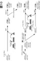
- FIG. 1 schematically shows the types of bone marrow cells identified by the inventors as being involved in the onset of diabetes, their representative characteristics, and their involvement in diabetes.
- TNF- ⁇ -producing cells are blood-derived cells, and it is unlikely that TNF- ⁇ is directly produced by the chief cells of an organ.
- HSC hematopoietic stem cells
- TNF- ⁇ is expressed in the most undifferentiated long term (LT)-HSC. It was first expressed in short term (ST)-HSCs, and expression was restricted to the CD106-positive fraction of ST-HSCs.
- the ST-HSCs expressing CD106 and TNF- ⁇ also had variable expression of HDACs (HDAC2, 3, 4, 8, 9, 11, SIRT1, 6, etc.). Modulation of HDACs can be advantageous because CD106 and TNF- ⁇ can also be expressed on normal cells with important functions, and CD106 can also be expressed on normal stem cells.
- Fig. 2 schematically shows the mechanism by which abnormal cells develop type 2 diabetes.
- the CD106/TNF- ⁇ -positive abnormal hematopoietic stem cells seen in STZ mice were also seen in high-fat diet mice, a different type 2 diabetes model. Impaired insulin secretion results from decreased insulin sensitivity in both STZ and high-fat-fed mice (1), which results in hyperglycemia and promotes the production of CD106/TNF- ⁇ positive abnormal stem cells in the bone marrow.
- Abnormal expression of histone deacetylases occurs in abnormal stem cells, which is thought to cause various gene expression abnormalities.
- Abnormal stem cells in the bone marrow are in a niche (environment where stem cells can maintain their properties in vivo) as a memory that maintains memories of type 2 diabetes. It is thought that they reside in cells) and create intractable pathologies (2). These cascades of abnormalities lead to (A) elimination of abnormal stem cells in the bone marrow and (B) restoration of insulin secretory function to the point that blood glucose levels can be normalized to prevent the emergence of new abnormal stem cells. It is considered possible to cure type 2 diabetes by administering insulin externally for a certain period of time to normalize the blood sugar level.
- Fig. 3 schematically shows the mechanism by which abnormal cells develop type 1 diabetes.
- type 1 diabetes mouse model NOD mouse
- CD106/TNF- ⁇ positive cells that appeared in type 2 also appear.
- cells that produce autoantibodies that damage ⁇ cells emerge from B cell lineage progenitor cells (1).
- Autoantibodies released from these cells lead to progressive depletion of ⁇ -cells (2).
- Some of these abnormal B cells also migrate to the thymus, where they also produce autoantibodies and attack and eliminate pancreatic islet autoantigen-presenting cells such as proinsulin and GAD (3). This renders the thymus incapable of negative selection of islet-attacking T cells, allowing them to migrate to the periphery (4).
- endothelial proliferation and catastrophic tissue destruction of ⁇ -cell progenitors (11).
- endothelial dysfunction occurs in other peripheral organs as well as type 2, and various complications specific to diabetes progress due to cell fusion (13) and the like.
- type 1 diabetes and type 2 diabetes is the complete destruction of cells expressing insulin or proinsulin protein in the thymus and pancreatic islets.
- HDAC regulation suppresses the formation of this stem cell within the bone marrow while also inducing apoptosis of the abnormal cells formed.
- HDAC modulation alone has no effect on increasing the number of insulin-secreting cells.
- HDAC regulation alone suppresses abnormal cells, such as by inducing apoptosis, and reduces inflammatory cytokines such as TNF- ⁇ released by abnormal cells. This may reduce insulin resistance and indirectly lower blood sugar levels.
- the blood glucose level decreased over time compared to untreated STZ diabetes. Backed up.
- type 1 diabetes in which insulin secretion is reduced
- type 2 diabetes in which insulin is no longer secreted, do not lower blood sugar levels that much.
- control of blood glucose in treating and/or preventing diabetes and/or diabetes-related diseases, disorders and/or conditions of the present disclosure can be due to HDAC modulation.
- Subjects that are expected to lower blood sugar (such as subjects with type 2 diabetes whose blood sugar level is not very high) can be suitably treated.
- HDAC regulation can also suppress various cells (vascular endothelial cells and cells with abnormal cell fusion ability) induced from abnormal stem cells.
- HDAC modulating agents that act not only on stem cells, but also on cells derived therefrom, and/or further using another agent to suppress the induced cells, particularly in the treatment of advanced diabetes, etc. can be advantageous.
- the abnormal stem cells can be hematopoietic stem cells.
- the abnormal stem cells do not express normal levels of and/or function at least one of CD106, histone deacetylases (HDACs), CD34, TNF- ⁇ , and proinsulin. and optionally other characteristics of the abnormal stem cells described herein (e.g., c-Kit positive, Sca-1 positive, hematopoietic stem cell lineage marker negative, short-term hematopoietic stem cell It can be further characterized by having a characteristic (such as CD38 negative).
- abnormal stem cells can be characterized by CD106 expression levels that are higher than CD106 expression levels in total bone marrow cells or hematopoietic stem cells (eg, CD34-positive Thy-1-positive cells) of a non-diabetic subject population.
- the abnormal stem cells are 10% or more, 20% or more, 30% or more, 40% or more, 50% or more, 60% more than hematopoietic stem cells (e.g., CD34 positive cells) from a non-diabetic subject population It may be characterized by 70% or more, 80% or more, 90% or more, 100% or more, 150% or more, or 200% or more higher CD106 mRNA expression level.
- abnormal stem cells can be characterized by the expression of histone deacetylase genes (HDACs).
- HDACs histone deacetylase genes
- the abnormal stem cells are 10% or more, 20% or more, 30% or more, 40% or more, 50% or more than hematopoietic stem cells (eg, c-Kit positive lineage marker negative cells) from a non-diabetic subject population.
- HDACs histone deacetylases
- abnormal stem cells can be characterized by the expression of TNF- ⁇ . In one embodiment, abnormal stem cells can be characterized by being positive for expression of TNF- ⁇ . In one embodiment, the abnormal stem cells are 10% or more, 20% or more, 30% or more, 40% or more, 50% or more, 60% more than hematopoietic stem cells (e.g., CD34 positive cells) from a non-diabetic subject population It can be characterized by 70% or more, 80% or more, 90% or more, 100% or more, 150% or more, or 200% or more higher TNF- ⁇ mRNA expression.
- hematopoietic stem cells e.g., CD34 positive cells
- abnormal stem cells can be characterized by abnormal expression of proinsulin. In one embodiment, the abnormal stem cells can be characterized by being positive for proinsulin expression. In one embodiment, the abnormal stem cells can be characterized by higher insulin (or proinsulin) mRNA expression than hematopoietic stem cells (eg, CD34 positive cells) from a non-diabetic subject population.
- the abnormal stem cells can be characterized by the expression of one or more of c-Kit, Sca-1, hematopoietic stem cell lineage markers. In one embodiment, abnormal stem cells can be characterized by being positive for expression of one or more of c-Kit and Sca-1. Lineage markers of hematopoietic stem cells include CD3 (T cells), CD19 (B cells), NK1.1 (NK cells), CD11c (dendritic cells), CD11b (monocytes), Fc ⁇ RI (mast cells), Gr-1 (granulocytes) and the like. In one embodiment, the abnormal stem cells can be characterized by being negative for the expression of one or more (eg, all) of the hematopoietic stem cell lineage markers. In one embodiment, the abnormal stem cells may be characterized by higher CD34 mRNA expression than hematopoietic stem cells from a non-diabetic subject population.
- abnormal stem cells can be characterized by being hematopoietic stem cells.
- being a human hematopoietic stem cell can also be characterized by being CD34 positive Thy-1 positive or Lineage negative CD34 positive CD38 negative CD90 positive CD45RA negative.
- abnormal stem cells can be characterized by the cellular stage of hematopoietic stem cells.
- the abnormal stem cells can be short-term hematopoietic stem cells.
- determining that a subject has abnormal stem cells can be performed by detecting any characteristic of abnormal stem cells described herein. In one embodiment, whether a subject has abnormal stem cells can be diagnosed based on a quantitative indication of at least one characteristic of abnormal stem cells described herein (e.g., cell surface protein expression level, mRNA expression level). . In one embodiment, the subject-derived sample (e.g., blood sample, bone marrow sample), or hematopoietic stem cells (e.g., CD34-positive cells) contained therein, is or is contained in a corresponding sample of a non-diabetic subject population.
- a quantitative indication of at least one characteristic of abnormal stem cells described herein e.g., cell surface protein expression level, mRNA expression level.
- the subject-derived sample e.g., blood sample, bone marrow sample
- hematopoietic stem cells e.g., CD34-positive cells
- hematopoietic stem cells e.g., CD34 positive cells
- hematopoietic stem cells e.g., CD34 positive cells
- the subject has diabetes and/or diabetes-related diseases, disorders and/or Or it may be judged suitable for the treatment and/or prevention of symptoms.
- the determination that a subject has abnormal stem cells can be based on the proportion of cells exhibiting at least one characteristic (including quantitative indicators) of abnormal stem cells described herein.
- the diagnosis of the present disclosure can be performed based on the proportion of hematopoietic stem cells of a subject that exhibit abnormal levels of abnormal stem cell marker (CD106, HDAC, TNF- ⁇ , etc.) expression. It can optionally be performed by comparing the percentage of hematopoietic stem cells of a non-diabetic or diabetic subject population that exhibit abnormal levels of CD106 expression.
- the proportion of cells exhibiting abnormal levels of expression of abnormal stem cell markers (CD106, HDAC, TNF- ⁇ , etc.) among the hematopoietic stem cells of the subject is non- 1.1-fold or more, 1.2-fold or more, 1.3-fold the proportion of hematopoietic stem cells in a diabetic subject population that express abnormal levels of abnormal stem cell markers (CD106, HDAC, TNF- ⁇ , etc.) 1.4 times or more, 1.5 times or more, 1.6 times or more, 1.7 times or more, 1.8 times or more, 1.9 times or more, 2 times or more, 2.1 times or more, 2.
- the subject is eligible for diabetes and/or diabetes-related disease, disorder and/or condition treatment and /or may be judged suitable for prophylaxis.
- the subject to whom the treatment and/or prevention of the present disclosure is applied has been diagnosed with diabetes.
- the subject to which the treatment and/or prevention of the present disclosure is applied is a diabetic complication such as neuropathy, nephropathy, liver injury, retinopathy, fatty liver, gastrointestinal disorder, fracture healing delay, Diagnosed with, or at risk for, an eating disorder or skin disorder.
- the subject to whom treatment and/or prophylaxis of the present disclosure is applied has a thymus (eg, has not undergone thymectomy).
- the subject to whom the treatment and/or prophylaxis of the present disclosure is applied does not have hypothymic function.
- treatment and/or prophylaxis of the present disclosure comprises administration of agents that improve thymic function.
- abnormal hematopoietic stem cells can contribute to diabetes and/or diabetes-related diseases, disorders, and/or conditions, and thus the detection of abnormal hematopoietic stem cells.
- Companion therapy to treat diabetes and/or diabetes-related diseases, disorders and/or conditions is one of the preferred embodiments contemplated in this disclosure.
- the present disclosure may be used with companion diagnostics, including agents that detect abnormal hematopoietic stem cells, to lead to the treatment or prevention of diabetes and/or diabetes-related diseases, disorders and/or conditions.
- any treatment or prevention of diabetes and/or diabetes-related diseases, disorders and/or symptoms can be appropriately adjusted according to the level of detection of abnormal hematopoietic stem cells, the site of detection, and other conditions of the subject (level of progression of diabetes, age, sex, etc.).
- detection of abnormal hematopoietic stem cells can be an important factor in guiding treatment or prevention strategies for diabetes and/or diabetes-related diseases, disorders and/or conditions.
- the treatment, prevention and/or diagnosis of the present disclosure can be performed on any subject.
- the subject is a mammal, such as a human, mouse, guinea pig, hamster, rat, mouse, rabbit, pig, sheep, goat, cow, horse, cat, dog, marmoset, monkey, or chimpanzee. is.
- the subject is human.
- HDAC modulators and hypoglycemic agents described herein can be provided as compositions or medicaments in various forms.
- the routes of administration of the HDAC modulators and hypoglycemic agents described herein should be effective in treating, preventing or detecting diabetes and/or diabetes-related diseases, disorders and/or symptoms.
- Preferred are, for example, intravenous, subcutaneous, intramuscular, intraperitoneal, or oral administration.
- the dosage form may be, for example, injections, capsules, tablets, granules and the like.
- Aqueous solutions for injection may be stored, for example, in vials or stainless steel containers.
- Aqueous solutions for injection may also contain physiological saline, sugar (eg, trehalose), NaCl, NaOH, or the like.
- the therapeutic agent may also contain, for example, buffers (eg, phosphate buffers), stabilizers, and the like.
- compositions, medicaments, inhibitors, chemotactic agents, detection agents, etc. of the present disclosure comprise a therapeutically effective amount of active ingredient or a detectable amount of detection means, and a pharmaceutically acceptable carrier or excipient.
- pharmaceutically acceptable means a compound that has been approved by a governmental regulatory agency or approved by a pharmacopoeia or other generally recognized pharmacopoeia for use in animals, and more particularly in humans. Means it is enumerated.
- carrier refers to a diluent, adjuvant, excipient, or vehicle with which the therapeutic or detectable agent is administered.
- Such carriers can be sterile liquids such as water and oils, including those of petroleum, animal, vegetable or synthetic origin, including but not limited to peanut oil, soybean oil, minerals. Oil, sesame oil, etc. are included. Water is a preferred carrier when the drug is administered orally. Saline and aqueous dextrose are preferred carriers when the pharmaceutical composition is administered intravenously. Saline solutions and aqueous dextrose and glycerol solutions are preferably employed as liquid carriers for injectable solutions.
- Suitable excipients include light silicic anhydride, microcrystalline cellulose, mannitol, starch, glucose, lactose, sucrose, gelatin, malt, rice, flour, chalk, silica gel, sodium stearate, glycerol monostearate, talc, chloride.
- composition if desired, can also contain minor amounts of wetting or emulsifying agents, or pH buffering agents. These compositions can take the form of solutions, suspensions, emulsions, tablets, pills, capsules, powders, sustained release formulations, and the like.
- compositions can also be formulated as a suppository, with traditional binders and carriers such as triglycerides.
- Oral formulations can also include standard carriers such as pharmaceutical grades of mannitol, lactose, starch, magnesium stearate, sodium saccharin, cellulose, magnesium carbonate, and the like. Examples of suitable carriers include E. W. Described in Martin, Remington's Pharmaceutical Sciences (Mark Publishing Company, Easton, U.S.A).
- Such compositions will contain a therapeutically effective amount of the therapeutic agent, preferably in purified form, together with a suitable amount of carrier so as to provide the form for proper administration to the patient.
- the formulation should suit the mode of administration.
- surfactants for example, surfactants, excipients, coloring agents, flavoring agents, preservatives, stabilizers, buffers, suspending agents, tonicity agents, binders, disintegrants, lubricants, fluidity Accelerators, flavoring agents and the like may also be included.
- a "salt” includes, for example, an anionic salt formed with any acidic (eg, carboxyl) group, or a cationic salt formed with any basic (eg, amino) group.
- Salts include inorganic salts or organic salts, for example, salts described in Berge et al., J. Pharm. Sci., 1977, 66, 1-19. Further examples include metal salts, ammonium salts, salts with organic bases, salts with inorganic acids, salts with organic acids and the like.
- a "solvate” in one embodiment of the disclosure is a compound formed by a solute and a solvent. For solvates, see, for example, J.
- the solvates formed when the solvent is water are hydrates.
- the solvent does not interfere with the biological activity of the solute. Examples of such preferred solvents include, but are not limited to, water or various buffers.
- various delivery systems can be used together, and such systems can be used to administer the inhibitors and/or migratory agents of the present disclosure to the appropriate site.
- Such systems include, for example, encapsulation in liposomes, microparticles, and microcapsules; use of receptor-mediated endocytosis; construction of therapeutic nucleic acids as part of retroviral vectors or other vectors; be.
- Methods of introduction include, but are not limited to, intradermal, intramuscular, intraperitoneal, intravenous, subcutaneous, intranasal, epidural, and oral routes.
- the medicament by any suitable route, such as by infusion, by bolus injection, by absorption through epithelial or mucocutaneous linings (such as oral, rectal and intestinal mucosa).
- An inhaler or nebulizer may be used, with an aerosolizing agent as appropriate, and may be co-administered with other biologically active agents. Administration can be systemic or local.
- the composition can be formulated as a pharmaceutical composition adapted for administration to humans according to known methods. Such compositions can be administered by injection. Typically, compositions for injectable administration are solutions in sterile isotonic aqueous buffer. If desired, the composition can also include a solubilizing agent and a local anesthetic such as lidocaine to relieve pain at the injection site. Generally, the ingredients are supplied separately or mixed together in a unit dosage form, for example a lyophilized powder or water-free concentrate, in a hermetically sealed container such as an ampoule or sachet indicating the amount of active agent. can be supplied as goods.
- a unit dosage form for example a lyophilized powder or water-free concentrate
- a hermetically sealed container such as an ampoule or sachet indicating the amount of active agent. can be supplied as goods.
- composition is to be administered by infusion
- it can be dispensed with an infusion bottle containing sterile pharmaceutical grade water or saline.
- an ampoule of sterile water for injection or saline can be provided so that the ingredients can be mixed prior to administration.
- the HDAC modulators and hypoglycemic agents of the present disclosure can also be formulated in neutral or salt form or other prodrugs (eg, esters, etc.).
- Pharmaceutically acceptable salts include those formed with free carboxyl groups derived from hydrochloric, phosphoric, acetic, oxalic, tartaric, etc., isopropylamine, triethylamine, 2-ethylaminoethanol, histidine, procaine and those derived from sodium, potassium, ammonium, calcium, ferric hydroxide, and the like.
- HDAC modulators and hypoglycemic agents of the present disclosure may vary depending on the nature of the disorder or condition, but can be determined by those skilled in the art by standard clinical techniques based on the description herein.
- in vitro assays may optionally be employed to help identify optimal dosage ranges.
- the precise dose to be employed in the formulations can also vary with route of administration, and the seriousness of the disease or disorder, and should be decided according to the judgment of the attending physician and each patient's circumstances.
- the dosage is not particularly limited, and may be, for example, 0.001, 1, 5, 10, 15, 100, or 1000 mg/kg body weight per dose, within the range of any two of these values. There may be.
- the administration interval is not particularly limited. good too.
- the dose, administration interval and administration method may be appropriately selected depending on the patient's age, body weight, symptoms, target organ and the like.
- the therapeutic agent preferably contains a therapeutically effective amount, or an amount effective to exert the desired effect, of the active ingredient. Effective doses can be extrapolated from dose-response curves derived from in vitro or animal model test systems.
- the pharmaceutical composition or therapeutic agent or prophylactic agent of the present disclosure can be provided as a kit.
- the disclosure provides pharmaceutical packs or kits comprising one or more containers filled with one or more ingredients of the compositions or medicaments of the disclosure.
- Example 1 Bone marrow transplantation treatment and HDAC expression in STZ-DM mice
- Diabetes was treated in STZ-DM mice (type 2 diabetes model) by bone marrow transplantation.
- TNF- ⁇ positive cells and proinsulin positive cells were present in c-kit positive Sca-1 positive lineage marker negative (KSL) cells, and CD106 expressing cells were increased.
- KSL type 1 diabetic mice
- TNF- ⁇ -positive cells and CD106-expressing cells have been found to be increased, and hematopoietic stem cells with these characteristics are associated with diabetes. This is thought to be the cause of the emergence of characteristics as a chronic disease.
- CD106-positive cells and TNF- ⁇ -positive cells which characterize hematopoietic stem cell abnormalities, are non-SP cells (short-term hematopoietic stem cells: ST-HSC), but not SP cells (long-term hematopoietic stem cells: LT-HSC) in KSL cells at an earlier differentiation stage, found in diabetes. It has been shown that abnormal stem cells that have been identified may reside in ST-HSCs. Expression of the histone deacetylase (HDAC) gene was examined in such abnormal stem cells that have been found to be involved in diabetes.
- HDAC histone deacetylase
- FIG. Diabetes was induced by tail vein injection of streptozotocin (150 mg/kg) to C57BL/6J mice (wild-type, Clea Japan, Osaka), and mice with a blood glucose level of 250 mg/dl or more one week after administration were treated. They were used as diabetic mice (STZ-DM mice).
- insulin pellets 0.1 U/day release of insulin, sustained for about 30 days, LINSHIN CANADA, INC., Cat No: Pr-1-B
- blank pellets not containing insulin
- a hematopoietic stem cell fraction KSL was collected, and microarray analysis was performed on expressed genes.
- Sections for histological analysis were prepared as follows. Mice were perfusion-fixed with a solution of 4% PFA in 0.1 M PB, and the collected pancreatic tissue was immersed overnight in a solution of 15% sucrose in 0.1 M PB, and then 8 ⁇ m cryosections were prepared using a cryostat. After washing the frozen sections with PBS(-) for 10 minutes three times, they were treated with 0.3% hydrogen peroxide solution (0.3% H 2 O 2 in PBS(-) to inactivate endogenous peroxidase in the sections). ) for 30 minutes at room temperature and washed with PBS(-) for 5 minutes three times.
- Sections were then reacted with 5% normal goat serum in PBS(-) supplemented with 0.3% Triton X and blocked for 1 hour at room temperature to prevent non-specific reactions. After blocking, it was reacted with a primary antibody (anti-insulin antibody: CST company or anti-glucagon antibody: CST company) at 4° C. overnight. After that, it was washed three times with PBS(-) for 5 minutes, reacted in ImmPRESS reagent (Vector laboratory) at room temperature for 30 minutes, and washed further. Thereafter, DAB color development was performed, and nuclear staining was performed with hematoxylin as counterstaining, followed by dehydration and encapsulation.
- a primary antibody anti-insulin antibody: CST company or anti-glucagon antibody: CST company
- mice of each experimental group were dissected after perfusion fixation, and the thymus was collected. Images of the thymus (either left lobe or right lobe) were acquired under a stereomicroscope.
- the microarray analysis was performed as follows. Mononuclear cells were isolated by FACS from whole bone marrow using Ficoll-Paque Plus (GE Healthcare Bio-Sciences AB, Uppsala, Sweden). Mononuclear cells were reacted with PE-Cy7-conjugated streptavidin antibody, APC-conjugated anti-c-kit antibody, APC-Cy7-conjugated anti-Ly6A/E antibody (Sca-1), and biotin mouse strain panel. After that, it was fixed with BD Cytofix/CytoPerm (BD Biosciences).
- RNA was isolated from LSK cells using the RNeasy Plus Micro kit (Qiagen, Tokyo, Japan). RNA samples were analyzed at TakaRa bio (Shiga, Japan) using Nanodrop and Agilent 2100 BioAnalyser. RNA samples were amplified with the Ovation Pico WTA system and used for microarray analysis. HDAC9 and the like have a large number of isozymes, and in the microarray, an array was used that targets sites that reflect the total amount.
- Hdac1-11 and Sirtuin1-7 histone deacetylases (Hdac1-11 and Sirtuin1-7) in hematopoietic stem cell fractions of diabetic and non-diabetic bone marrow are shown in FIG. Hdac3, Hdac4, Hdac8, Sirt1 and Sirt6 were elevated in diabetes. In addition, Hdac2, Hdac9 and Hdac11 were decreased. Considering the balance with the effect on diabetes as a whole, it is considered preferable to regulate HDACs so as to suppress abnormal hematopoietic stem cells. Strategies such as broad inhibition of IV HDAC are envisioned.
- Hdacs epigenome-associated genes
- stem cells In diabetic KSL cells, the expression of epigenome-associated genes (Hdacs), which are important for regulating gene expression through histone modifications, was increased, suggesting that hyperglycemia conferred abnormal properties on stem cells at the genetic level. It was thought that It has been reported that cells exposed to transient hyperglycemia maintain their abnormalities caused by hyperglycemia even when returned to a normoglycemic environment (El-Osta et al. J Exp Med. 2008 Sep 29;205(10):2409-17), this also suggests that increased expression of the histone deacetylase gene in KSL cells may be involved in the emergence of abnormal stem cells and diabetes. Conceivable.
- Fig. 6 shows changes in blood glucose levels over time. Mice without insulin treatment remained hyperglycemic even after normalization of diabetic bone marrow by bone marrow transplantation. From this, it is presumed that abnormal hematopoietic stem cells are newly produced due to hyperglycemia, even if the bone marrow is normalized in a state where blood sugar is not normalized. On the other hand, insulin pellets that normalized bone marrow stem cells at the same time as blood glucose control for 4 weeks corrected hyperglycemia. However, in mice transplanted with bone marrow from diabetic mice, blood glucose levels rose rapidly when insulin action ceased. This indicates the presence of abnormal, diseased stem cells in the bone marrow of diabetic subjects.
- pancreatic glucagon DAB staining FIG. 8
- the ratio of glucagon-positive cells was decreased in the insulin pellet-administered non-diabetic bone marrow transplant group compared to the STZ diabetic group, and glucagon production was significantly decreased.
- the percentage of glucagon-positive cells in STZ diabetes was the same, and no reduction in glucagon production was observed. I didn't. Comparing the size of the harvested thymus (Fig. 9), the diabetic mice had significantly atrophied thymus compared to non-diabetic mice. On the other hand, administration of insulin pellets and transplantation of bone marrow from non-diabetic mice restored the size of the thymus to the same level as that of non-diabetic mice, and no improvement was observed in the other mice.
- Example 2 Tissue analysis using GFP transgenic (GFP-TG) mice
- Diabetes was histologically analyzed using bone marrow cells from GFP-TG mice as shown in FIG.
- total bone marrow cells (4 ⁇ 10 6 ) obtained from non-diabetic GFP-TG mice were transplanted into normal mice, and the course was observed for 4 weeks. After the transplanted bone marrow was established, STZ was administered. and developed diabetes. Buffer was administered to the non-diabetic group. After 12 weeks of stable diabetes, both groups were euthanized and histological analysis was performed.
- Histological analysis of therapeutic studies using bone marrow transplants obtained from GFP-TG mice was performed as follows. STZ was administered to normal mice to develop diabetes, followed by follow-up for 4 weeks. Mice were then divided into two groups, one implanted with an insulin pellet and the other with a blank pellet. On the next day, lethal radiation (9 Gy) was irradiated, and total bone marrow cells (4 ⁇ 10 6 ) obtained from GFP-TG normal mice were transplanted to each of the two groups, and the progress was observed for 8 weeks. During that time, blood glucose levels were measured weekly to observe treatment effects. After the end of the treatment period, the animals were euthanized, and histological analysis of pancreatic islets and thymus was performed. For immunohistological analysis, TNF- ⁇ and Vcam specific antibodies were used and observed under a fluorescence microscope.
- FIG. 11 shows the analysis results for pancreatic islets.
- a few bone marrow-derived cells are found in pancreatic islets, but Vcam1-positive cells are found only in a part of the outer membrane of the pancreatic islets, and are hardly found in pancreatic islets.
- the number of bone marrow-derived cells is increased, and at the same time, a large number of Vcam1-positive cells are observed.
- Vcam1-positive cells (red) and bone marrow-derived cells (green) overlapped, and yellow cells were observed. This suggests that Vcam1-expressing cells are present in pancreatic islets among bone marrow-derived cells.
- pancreatic islets showed similar findings to non-diabetic mice, but did not normalize blood glucose. The case showed the same findings as untreated diabetic mice. This suggests that even if diabetic mice are transplanted with bone marrow from non-diabetic mice to replace the bone marrow while hyperglycemia remains, hyperglycemia causes Vcam1-positive abnormal cells to be produced again, and these cells enter pancreatic islets. presumably invaded.
- Fig. 12 shows the findings of the thymus.
- mice transplanted with bone marrow obtained from non-diabetic GFP mice (GFP BMT nonDM) after irradiation with 9 Gy show a marked appearance of green cells in the thymic tissue. This is thought to be caused by regeneration of thymocytes damaged by irradiation with bone marrow-derived cells.
- STZ was administered to these mice to induce diabetes, most of the bone marrow-derived thymocytes were damaged by hyperglycemia and disappeared, exhibiting marked atrophy again (GFP BMT STZDM).
- Example 3 Drug treatment of STZ-DM mice
- HDAC modulators and glycemic control were used to treat diabetes in STZ-DM mice.
- Fig. 13 shows an overview of the treatment. Streptozotocin (150 mg/kg) was administered to C57BL/6J mice (wild-type, Clea Japan, Osaka) (nonDM) through the tail vein to induce diabetes.
- the STZ-DM mice were administered insulin pellets to normalize blood glucose levels, and a control group was administered blank pellets.
- 2.5 mg/kg divinostat (Cayman chemical, USA) was administered to both the treatment group and the control group through the tail vein or orally. Insulin pellets or blank pellets were administered up to 3 days before the start of divinostat treatment, and divinostat administration was started on the day when it was confirmed that the blood glucose level of the treatment group was within the normal range as day 0.
- Divinostat was administered once every 3 days until the 8th week.
- a non-diabetic group in which STZ was not administered at the same time as the treatment group and only buffer was administered
- STZ-DM non-treatment diabetic group
- FIG. 15 shows changes in blood sugar
- FIG. 16 shows changes in insulin concentration.
- mice Twelve weeks after the start of treatment, some of the mice were perfusion-fixed with a solution of 4% PFA in 0.1 M PB, and the collected pancreatic tissue was immersed in a solution of 15% sucrose in 0.1 M PB overnight. 8 um cryosections were prepared at . After washing the frozen sections with PBS(-) for 10 minutes three times, they were treated with 0.3% hydrogen peroxide solution (0.3% H 2 O 2 in PBS(-) to inactivate endogenous peroxidase in the sections). ) for 30 minutes at room temperature and washed with PBS(-) for 5 minutes three times.
- Sections were then reacted with 5% normal goat serum in PBS(-) supplemented with 0.3% Triton X and blocked for 1 hour at room temperature to prevent non-specific reactions. After blocking, it was reacted with a primary antibody (anti-insulin antibody: CST company or anti-glucagon antibody: CST company) at 4° C. overnight. After that, it was washed three times with PBS(-) for 5 minutes, reacted in ImmPRESS reagent (Vector laboratory) at room temperature for 30 minutes, and washed further. Thereafter, DAB color development was performed, and nuclear staining was performed with hematoxylin as counterstaining, followed by dehydration and encapsulation. The prepared sections were observed under a microscope, about 5 to 8 pancreatic islets were randomly selected, and the ratio of insulin- or glucagon-positive cells to the islet size was calculated using Image J software.
- mice in each experimental group were dissected after perfusion fixation, and the femur, thymus, and pancreas were collected.
- the femur was decalcified using a neutral decalcifying solution (10% EDTA solution) to prepare a paraffin block.
- Paraffin sections with a thickness of 3 ⁇ m were prepared from the paraffin blocks using a microtome and stained with DAB.
- DAB staining paraffin sections were deparaffinized with xylene, hydrophilized with ethanol, and antigens were activated using HistoVTone (Nacalai Tesque). Then, DAB staining was performed using TNF- ⁇ (abcam).
- FIG. 14 shows changes in blood glucose level in each group. Hyperglycemia persisted in STZ-DM compared to nonDM. On the other hand, the blood glucose level immediately decreased in the diabetic group in which blood glucose was controlled with insulin pellets for approximately 4 weeks, and in the treatment group in which divinostat (orally or intravenously for 8 weeks) was administered simultaneously with insulin pellets. However, in the group that was not treated with insulin pellets, the blood glucose level did not decrease even after administration of divinostat for 8 weeks, and the increase in blood glucose level was not suppressed in any period up to 12 weeks after discontinuation of divinostat.
- mice treated with insulin pellets + divinostat were examined between mice treated with insulin pellets + divinostat and mice treated with blank pellets + divinostat ( Figures 15 and 16).
- IPGTT intraperitoneal glucose tolerance test
- glucagon-positive cells were present in a circle just below the outer membrane of the islet, and approximately 10% of the entire islet was positive.
- STZ-DM glucagon-positive cells increased remarkably throughout the pancreatic islet, occupying 75% of the area of the entire pancreatic islet.
- the area of glucagon-positive cells decreased from the central part to about 40% of the entire pancreatic islet.
- the glucagon-positive area remained high in the group showing hyperglycemia.
- the thymus weight was 37 mg in the non-diabetic group, but 13 mg in the diabetic group, which was about 1/3 atrophy.
- both oral and intravenous administration recovered to about 2/3 of the normal level.
- TNF- ⁇ immunostaining of bone marrow and thymus (FIGS. 20 and 21) showed that TNF- ⁇ positive cells increased in the diabetic group, but in the group treated with divinostat at the same time as blood sugar control, TNF - ⁇ -positive cells decreased to the same extent as in the non-diabetic group.
- TNF- ⁇ staining was observed throughout the cells in almost 100% of the cells in the diabetes untreated group and the group in which diabetes was not improved.
- TNF- ⁇ -positive cells are present throughout the body, the killer T cells that attack them are eliminated by negative selection as autoantigen-reactive T cells, resulting in abnormal cells expressing TNF- ⁇ and proinsulin.
- TNF- ⁇ immunostaining DAB staining of pancreatic islets (FIG. 22) showed that non-diabetic mice, insulin pellets plus divinostat treatment (oral), and streptozotocin-induced diabetic mice with insulin pellets plus divinostat treatment (intravenous). Almost no TNF- ⁇ -positive cells were observed in the successfully treated group. On the other hand, in blank pellet + divinostat treatment (oral) and blank pellet + divinostat treatment (intravenous), many TNF- ⁇ positive cells were observed to migrate to the central part of pancreatic islets.
- abnormal stem cells that are TNF- ⁇ positive/proinsulin positive within the Vcam1-positive short-term hematopoietic stem cells in the bone marrow.
- Normal short-term hematopoietic stem cells include capillary progenitor cells, vascular niche cells, and organ stem cells from various organs. In the thymus, these cells may be the originating cells of the thymic organ itself, suggesting that these cells cannot migrate into the thymus under hyperglycemia in diabetes. Therefore, it is considered that the thymus cannot produce killer T cells for attacking TNF- ⁇ -positive/proinsulin-positive abnormal stem cells in the bone marrow that appear in diabetes. In diabetes, the cells continue to survive as "diabetic stem cells", and it is thought that blood sugar control alone cannot cure diabetes.
- Example 4 Combining HDAC modulators and glycemic control to treat diabetes in NOD mice.
- Insulin glargine is administered subcutaneously daily in escalating doses of 10 IU until day 3 after initiation of treatment, 5 IU for 3 days thereafter, 2.5 IU for 6 days thereafter, and 0.75 IU for 12 days thereafter.
- Two hours after administration of insulin glargine, divinostat is orally administered daily at a dose of 2.5 mg/kg to mice whose blood glucose levels have been measured and hypoglycemia has been confirmed. If the pre-insulin glargine blood glucose level is less than 200 mg/dl more than 8 weeks after starting treatment, insulin glargine and divinostat for that day are discontinued.
- blood glucose levels are then measured weekly for up to 4 weeks to determine diabetic status. If blood glucose levels do not fall below 200 mg/dl after 8 weeks of treatment initiation, treatment is given daily for up to 12 weeks, at which point treatment is terminated.
- hypoglycemia occurs, a 25% glucose solution is administered intraperitoneally (300 ⁇ L) and the blood glucose level is measured 1 hour later. If hypoglycemia persists, this procedure is repeated. After confirming that the blood sugar level has risen sufficiently (150 mg/dl or more), no further glucose solution is administered. If hypoglycemia is confirmed, subsequent insulin doses are halved as needed.
- Diabetes in NOD mice is also cured.
- Diabetes is treated by administering a combination of HDAC modulators and glycemic control agents to diabetic db/db mice.
- Db/db mice are observed from the 6th week, and the onset of diabetes is defined as the time when the blood sugar level exceeds 300 mg/dl.
- insulin glargine was tapered to 10 IU by day 3, then 5 IU for 3 days, then 2.5 IU for 6 days, and then 0.75 IU for 12 days. , administered subcutaneously daily.
- Two hours after administration of insulin glargine, 2.5 mg/kg of divinostat is orally administered daily to mice whose blood glucose levels have been measured and hypoglycemia has been confirmed. If the pre-insulin glargine blood glucose level is less than 200 mg/dl more than 8 weeks after starting treatment, insulin glargine and divinostat for that day are discontinued.
- blood glucose levels are then measured weekly for up to 4 weeks to determine diabetic status. If blood glucose levels do not fall below 200 mg/dl after 8 weeks of treatment initiation, treatment is given daily for up to 12 weeks, at which point treatment is terminated.
- hypoglycemia occurs, a 25% glucose solution is administered intraperitoneally (300 ⁇ L) and the blood glucose level is measured 1 hour later. If hypoglycemia persists, this procedure is repeated. After confirming that the blood sugar level has risen sufficiently (150 mg/dl or more), no further glucose solution is administered. If hypoglycemia is confirmed, subsequent insulin doses are halved as needed.
- Diabetes in an obese mouse model is also cured.
- Diabetes is treated by administering a combination of HDAC modulators and different types of glycemic control agents to diabetic db/db mice.
- Db/db mice are observed from the 6th week, and the onset of diabetes is defined as the time when the blood sugar level exceeds 300 mg/dl.
- the SGLT inhibitor dapagliflozin 1.0 mg/kg and the HDAC modulator divinostat 2.5 mg/kg were orally administered daily for 8 weeks, followed by treatment discontinuation for 4 weeks. and end.
- Diabetes can be cured even when different types of blood sugar control agents are used.
- fingolimod instead of divinostat, trichostatin A, vorinostat, belinostat, panobinostat, resminostat, abexinostat, xinostat, prasinostat, rosilinostat, nanatinostat, dacinostat, (N-hydroxy-7 -2-naphthylthio)heptanomide, valproic acid, CG200745, scriptaid, KBH-A42, romidepsin, sodium butyrate, 4-phenylbutyric acid, SK-7041, WW437, entinostat, tacedinaline, mosetinostat, butyryloxymethyl pivalate, Trapoxin, ⁇ -ketoamides, heterocyclic ketones, diallyltrisulfide, licorinostat, Tubacin, azelaroyl bis-hydroxamic acid, m-carboxycinn
- Example 8 Effect of thymectomy
- Streptozotocin 150 mg/kg was administered to C57BL/6J mice (wild type, Clea Japan, Osaka) through the tail vein to induce diabetes. After one month of free feeding, the STZ-DM mice were subcutaneously administered insulin pellets to normalize blood glucose levels. The mice were divided into two groups, the thymus was removed from the suprasternal fossa in one group of mice (thymectomy group) and the other group of mice had an incision in the suprasternal fossa that was immediately closed (control group). Both the thymectomy group and the control group were orally administered 2.5 mg/kg divinostat (Cayman chemical, USA). Divinostat was administered once every 3 days.
- mice in both groups were perfusion-fixed to confirm the presence or absence of the thymus (Fig. 25).
- the present disclosure provides diabetes treatment that combines blood glucose control and HDAC regulation, which provides a new approach for diabetes treatment.
Abstract
La présente invention concerne un nouveau traitement du diabète. La présente invention concerne un traitement du diabète, dans lequel le contrôle de la glycémie et l'ajustement de l'histone-désacétylase (HDAC) sont combinés. Selon un mode de réalisation, la présente invention concerne un traitement du diabète et/ou des maladies, troubles et/ou symptômes liés au diabète, dans lequel le contrôle de la glycémie et l'ajustement de l'HDAC sont combinés. Selon un mode de réalisation, le contrôle de la glycémie et l'ajustement de l'HDAC sont combinés pour définir les cellules souches anormales comme cible, afin de traiter le diabète et/ou les maladies, troubles et/ou symptômes liés au diabète.
Applications Claiming Priority (2)
| Application Number | Priority Date | Filing Date | Title |
|---|---|---|---|
| JP2021177845 | 2021-10-29 | ||
| JP2021-177845 | 2021-10-29 |
Publications (1)
| Publication Number | Publication Date |
|---|---|
| WO2023074794A1 true WO2023074794A1 (fr) | 2023-05-04 |
Family
ID=86159956
Family Applications (1)
| Application Number | Title | Priority Date | Filing Date |
|---|---|---|---|
| PCT/JP2022/040140 WO2023074794A1 (fr) | 2021-10-29 | 2022-10-27 | Procédé ou agent avec régulateur de hdac, pour le traitement du diabète et de ses complications |
Country Status (1)
| Country | Link |
|---|---|
| WO (1) | WO2023074794A1 (fr) |
Citations (2)
| Publication number | Priority date | Publication date | Assignee | Title |
|---|---|---|---|---|
| WO2021075536A1 (fr) * | 2019-10-18 | 2021-04-22 | 国立大学法人滋賀医科大学 | Traitement contre le diabète utilisant un agent de migration des cellules souches |
| WO2022181797A1 (fr) * | 2021-02-26 | 2022-09-01 | 国立大学法人滋賀医科大学 | Nouvelle méthode et nouvel agent destinés au traitement, au diagnostic et à la détection du diabète et de complications |
-
2022
- 2022-10-27 WO PCT/JP2022/040140 patent/WO2023074794A1/fr unknown
Patent Citations (2)
| Publication number | Priority date | Publication date | Assignee | Title |
|---|---|---|---|---|
| WO2021075536A1 (fr) * | 2019-10-18 | 2021-04-22 | 国立大学法人滋賀医科大学 | Traitement contre le diabète utilisant un agent de migration des cellules souches |
| WO2022181797A1 (fr) * | 2021-02-26 | 2022-09-01 | 国立大学法人滋賀医科大学 | Nouvelle méthode et nouvel agent destinés au traitement, au diagnostic et à la détection du diabète et de complications |
Non-Patent Citations (8)
| Title |
|---|
| "Nutritional and Therapeutic Interventions for Diabetes and Metabolic Syndrome (Second Edition)", 8 June 2018, ISBN: 978-0-12-812019-4, article LUÍS F. SCHÜTZ, MIN HI PARK, MAHUA CHOUDHURY: "Chapter 35 - HDACs in Diabetes: A New Era of Epigenetic Drug", pages: 475 - 486, XP009545780, DOI: 10.1016/B978-0-12-812019-4.00035-0 * |
| ARGUELLES ANDRIX O.; MERUVU SUNITHA; BOWMAN JOHN D.; CHOUDHURY MAHUA: "Are epigenetic drugs for diabetes and obesity at our door step?", DRUG DISCOVERY TODAY, ELSEVIER, AMSTERDAM, NL, vol. 21, no. 3, 14 December 2015 (2015-12-14), AMSTERDAM, NL , pages 499 - 509, XP029464367, ISSN: 1359-6446, DOI: 10.1016/j.drudis.2015.12.001 * |
| BESANçON ALIX; GONCALVES TANIA; VALETTE FABRICE; DAHLLöF MATTIAS S.; MANDRUP-POULSEN THOMAS; CHATENOUD LUCIENNE; YOU SYL: "Oral histone deacetylase inhibitor synergises with T cell targeted immunotherapy to preserve beta cell metabolic function and induce stable remission of new-onset autoimmune diabetes in NOD mice", DIABETOLOGIA, SPRINGER BERLIN HEIDELBERG, BERLIN/HEIDELBERG, vol. 61, no. 2, 13 October 2017 (2017-10-13), Berlin/Heidelberg, pages 389 - 398, XP036387771, ISSN: 0012-186X, DOI: 10.1007/s00125-017-4459-0 * |
| ERIKA F. BRUTSAERT: "Drug therapy for diabetes", MSD MANUAL PROFESSIONAL EDITION, MSD, JP, JP, pages 1 - 5, XP009546679, Retrieved from the Internet <URL:https://www.msdmanuals.com/ja-jp/%E3%83%97%E3%83%AD%E3%83%95%E3%82%A7%E3%83%83%E3%82%B7%E3%83%A7%E3%83%8A%E3%83%AB/10-%E5%86%85%E5%88%86%E6%B3%8C%E7%96%BE%E6%82%A3%E3%81%A8%E4%BB%A3%E8%AC%9D%E6%80%A7%E7%96%BE%E6%82%A3/%E7%B3%96%E5%B0%BF%E7%97%85%E3%81%A8%E7%B3%96%E4%BB%A3%E8%AC%9D%E7%95%B0%E5%B8%B8%E7%97%87/%E7%B3%96%E5%B0%BF%E7%97%85%E3%81%AE%E8%96%AC%E7%89%A9%E7%99%82%E6%B3%95> * |
| M. LUNDH; D. P. CHRISTENSEN; M. DAMGAARD NIELSEN; S. J. RICHARDSON; M. S. DAHLLöF; T. SKOVGAARD; J. BERTHELSEN; C. A. DINARELLO; : "Histone deacetylases 1 and 3 but not 2 mediate cytokine-induced beta cell apoptosis in INS-1 cells and dispersed primary islets from rats and are differentially regulated in the islets of type 1 diabetic children", DIABETOLOGIA, SPRINGER, BERLIN, DE, vol. 55, no. 9, 7 July 2012 (2012-07-07), Berlin, DE , pages 2421 - 2431, XP035094596, ISSN: 1432-0428, DOI: 10.1007/s00125-012-2615-0 * |
| MIWAKO KASHI, ET AL: "O-11-4 Development of therapeutic method aiming at complete cure of diabetes by targeting bone marrow cells", REGENERATIVE MEDICINE, JAPANESE SOCIETY FOR REGENERATIVE MEDICINE, JP, vol. 20, no. Suppl., 26 February 2021 (2021-02-26) - 16 March 2021 (2021-03-16), JP , pages 220, XP009539553, ISSN: 1347-7919 * |
| S. M. CABRERA; S. C. COLVIN; S. A. TERSEY; B. MAIER; J. L. NADLER; R. G. MIRMIRA: "Effects of combination therapy with dipeptidyl peptidase‐IV and histone deacetylase inhibitors in the non‐obese diabetic mouse model of type 1 diabetes", CLINICAL AND EXPERIMENTAL IMMUNOLOGY, WILEY-BLACKWELL PUBLISHING LTD., GB, vol. 172, no. 3, 18 April 2013 (2013-04-18), GB , pages 375 - 382, XP071081715, ISSN: 0009-9104, DOI: 10.1111/cei.12068 * |
| SHARMA SORABH; TALIYAN RAJEEV: "Histone deacetylase inhibitors: Future therapeutics for insulin resistance and type 2 diabetes", PHARMACOLOGICAL RESEARCH, ELSEVIER, AMSTERDAM, NL, vol. 113, 1 January 1900 (1900-01-01), AMSTERDAM, NL, pages 320 - 326, XP029804511, ISSN: 1043-6618, DOI: 10.1016/j.phrs.2016.09.009 * |
Similar Documents
| Publication | Publication Date | Title |
|---|---|---|
| Mori et al. | Critical roles for the TSC-mTOR pathway in β-cell function | |
| US7803783B2 (en) | Use of WNT inhibitors to augment therapeutic index of chemotherapy | |
| US10426757B2 (en) | Compositions and methods for promoting intestinal stem cell and/or non-stem progenitor cell function | |
| WO2021075536A1 (fr) | Traitement contre le diabète utilisant un agent de migration des cellules souches | |
| US20130085169A1 (en) | Methods and compositions comprising ampk activator (metformin/troglitazone) for the treatment of myotonic dystrophy type 1 (dm1) | |
| EP2976094B1 (fr) | Méthodes de traitement de troubles métaboliques | |
| EP1900374B1 (fr) | Agent angiogénétique contenant de l adrénoméduline comme principe actif | |
| Whittam et al. | Small molecule inhibition of dipeptidyl peptidase-4 enhances bone marrow progenitor cell function and angiogenesis in diabetic wounds | |
| Hussien et al. | Combination of obestatin and bone marrow mesenchymal stem cells prevents aggravation of endocrine pancreatic damage in type II diabetic rats | |
| WO2022181797A1 (fr) | Nouvelle méthode et nouvel agent destinés au traitement, au diagnostic et à la détection du diabète et de complications | |
| US20140030234A1 (en) | Methods and compositions for modulating islet beta cell development | |
| WO2023074794A1 (fr) | Procédé ou agent avec régulateur de hdac, pour le traitement du diabète et de ses complications | |
| US20240139129A1 (en) | Novel method and agent for treating, diagnosing and detecting diabetes and complications | |
| WO2021075535A1 (fr) | Traitement contre le diabète ciblant des cellules souches anormales | |
| WO2016176493A1 (fr) | Traitement d'états médicaux | |
| US20160083733A1 (en) | Novel treatment of metabolic diseases | |
| US20150174202A1 (en) | Methods and Compositions for Modulating Beta Cell Proliferation | |
| CN111529689A (zh) | 转录因子klf16蛋白在制备防治脂质和糖代谢异常相关疾病药物中的应用 | |
| US20200405773A1 (en) | Promoting and protecting functional beta cell mass by syntaxin 4 enrichment | |
| KR20150139769A (ko) | 트레할로스를 유효성분으로 포함하는 지방간, 당뇨병 또는 인슐린 저항성 증후군의 예방 또는 치료용 조성물 | |
| Olaniru | The Role of GPR56 and Collagen III in Islet Functions | |
| WO2008066744A2 (fr) | Procédés et compositions de traitement et de prévention des lésions de la moelle épinière et d'autres maladies ou lésions cérébrales | |
| Usatyuk et al. | 45 REGULATION OF 4-HYDROXY-2-NONENAL-MEDIATED ENDOTHELIAL BARRIER FUNCTION BY FOCAL ADHESION AND ADHERENS JUNCTION PROTEINS. | |
| Keystone | 3rd Annual Symposium of the American Heart Association Council on Basic Cardiovascular Sciences Translation of Basic Insights Into Clinical Practice |
Legal Events
| Date | Code | Title | Description |
|---|---|---|---|
| 121 | Ep: the epo has been informed by wipo that ep was designated in this application |
Ref document number: 22887119 Country of ref document: EP Kind code of ref document: A1 |