WO2023054287A1 - Dispositif, procédé et programme de prédiction de maladie osseuse, dispositif d'apprentissage, procédé et programme, et réseau neuronal entraîné - Google Patents
Dispositif, procédé et programme de prédiction de maladie osseuse, dispositif d'apprentissage, procédé et programme, et réseau neuronal entraîné Download PDFInfo
- Publication number
- WO2023054287A1 WO2023054287A1 PCT/JP2022/035789 JP2022035789W WO2023054287A1 WO 2023054287 A1 WO2023054287 A1 WO 2023054287A1 JP 2022035789 W JP2022035789 W JP 2022035789W WO 2023054287 A1 WO2023054287 A1 WO 2023054287A1
- Authority
- WO
- WIPO (PCT)
- Prior art keywords
- bone
- image
- pixel
- target
- region
- Prior art date
Links
- 238000013528 artificial neural network Methods 0.000 title claims description 83
- 208000020084 Bone disease Diseases 0.000 title claims description 73
- 238000000034 method Methods 0.000 title claims description 38
- 210000000988 bone and bone Anatomy 0.000 claims abstract description 458
- 210000004872 soft tissue Anatomy 0.000 claims abstract description 136
- 230000005855 radiation Effects 0.000 claims abstract description 102
- 238000009826 distribution Methods 0.000 claims abstract description 22
- 208000010392 Bone Fractures Diseases 0.000 claims description 106
- 229910052500 inorganic mineral Inorganic materials 0.000 claims description 101
- 239000011707 mineral Substances 0.000 claims description 101
- 238000003384 imaging method Methods 0.000 claims description 35
- 210000000689 upper leg Anatomy 0.000 claims description 21
- 238000010801 machine learning Methods 0.000 claims description 14
- 230000006870 function Effects 0.000 claims description 13
- 230000037182 bone density Effects 0.000 abstract 2
- 206010017076 Fracture Diseases 0.000 description 84
- 238000009795 derivation Methods 0.000 description 36
- 238000003860 storage Methods 0.000 description 28
- 238000010586 diagram Methods 0.000 description 27
- 238000012545 processing Methods 0.000 description 27
- 206010028980 Neoplasm Diseases 0.000 description 15
- 201000011510 cancer Diseases 0.000 description 15
- 230000011218 segmentation Effects 0.000 description 13
- 210000001185 bone marrow Anatomy 0.000 description 11
- 206010027476 Metastases Diseases 0.000 description 10
- 230000009401 metastasis Effects 0.000 description 10
- 238000012937 correction Methods 0.000 description 9
- XLYOFNOQVPJJNP-UHFFFAOYSA-N water Substances O XLYOFNOQVPJJNP-UHFFFAOYSA-N 0.000 description 8
- 101100021996 Arabidopsis thaliana CYP97C1 gene Proteins 0.000 description 6
- 238000002595 magnetic resonance imaging Methods 0.000 description 6
- 210000004197 pelvis Anatomy 0.000 description 6
- 238000012549 training Methods 0.000 description 6
- OAICVXFJPJFONN-UHFFFAOYSA-N Phosphorus Chemical compound [P] OAICVXFJPJFONN-UHFFFAOYSA-N 0.000 description 4
- 238000013459 approach Methods 0.000 description 4
- 230000008859 change Effects 0.000 description 4
- 230000012447 hatching Effects 0.000 description 4
- 210000003958 hematopoietic stem cell Anatomy 0.000 description 4
- 210000003205 muscle Anatomy 0.000 description 4
- 238000011176 pooling Methods 0.000 description 4
- 206010061218 Inflammation Diseases 0.000 description 3
- 238000006243 chemical reaction Methods 0.000 description 3
- 201000010099 disease Diseases 0.000 description 3
- 208000037265 diseases, disorders, signs and symptoms Diseases 0.000 description 3
- 230000004054 inflammatory process Effects 0.000 description 3
- 239000000203 mixture Substances 0.000 description 3
- 230000008569 process Effects 0.000 description 3
- 208000001132 Osteoporosis Diseases 0.000 description 2
- 238000013527 convolutional neural network Methods 0.000 description 2
- 230000001054 cortical effect Effects 0.000 description 2
- 239000000284 extract Substances 0.000 description 2
- 230000004083 survival effect Effects 0.000 description 2
- 206010010214 Compression fracture Diseases 0.000 description 1
- RYGMFSIKBFXOCR-UHFFFAOYSA-N Copper Chemical compound [Cu] RYGMFSIKBFXOCR-UHFFFAOYSA-N 0.000 description 1
- 206010027677 Fractures and dislocations Diseases 0.000 description 1
- 206010027452 Metastases to bone Diseases 0.000 description 1
- 239000003082 abrasive agent Substances 0.000 description 1
- 230000005540 biological transmission Effects 0.000 description 1
- 210000004369 blood Anatomy 0.000 description 1
- 239000008280 blood Substances 0.000 description 1
- 238000004364 calculation method Methods 0.000 description 1
- 238000004891 communication Methods 0.000 description 1
- 229910052802 copper Inorganic materials 0.000 description 1
- 239000010949 copper Substances 0.000 description 1
- 230000008034 disappearance Effects 0.000 description 1
- 238000005516 engineering process Methods 0.000 description 1
- 238000000605 extraction Methods 0.000 description 1
- 230000001678 irradiating effect Effects 0.000 description 1
- 210000001503 joint Anatomy 0.000 description 1
- 210000000629 knee joint Anatomy 0.000 description 1
- 239000004973 liquid crystal related substance Substances 0.000 description 1
- 230000003137 locomotive effect Effects 0.000 description 1
- 238000004519 manufacturing process Methods 0.000 description 1
- 230000008722 morphological abnormality Effects 0.000 description 1
- 230000004660 morphological change Effects 0.000 description 1
- 230000003287 optical effect Effects 0.000 description 1
- 210000000056 organ Anatomy 0.000 description 1
- 230000000306 recurrent effect Effects 0.000 description 1
- 102220034833 rs1801145 Human genes 0.000 description 1
- 239000004065 semiconductor Substances 0.000 description 1
- 239000007787 solid Substances 0.000 description 1
- 230000002194 synthesizing effect Effects 0.000 description 1
- 210000001694 thigh bone Anatomy 0.000 description 1
- 239000010409 thin film Substances 0.000 description 1
- 210000002303 tibia Anatomy 0.000 description 1
- 210000001519 tissue Anatomy 0.000 description 1
Images


Classifications
-
- A—HUMAN NECESSITIES
- A61—MEDICAL OR VETERINARY SCIENCE; HYGIENE
- A61B—DIAGNOSIS; SURGERY; IDENTIFICATION
- A61B6/00—Apparatus for radiation diagnosis, e.g. combined with radiation therapy equipment
Definitions
- the present invention relates to bone disease prediction devices, methods and programs, learning devices, methods and programs, and trained neural networks.
- Japanese Patent Application Laid-Open No. 2019-202035 proposes a method of obtaining bone mineral information representing the bone mineral content of vertebrae from radiographic images and deriving fracture risk from the alignment of the spine and the bone mineral information. Further, in International Publication No. 2020/166561, bone mineral content and muscle mass are calculated for each pixel of a radiographic image, statistical values regarding the subject are calculated based on the bone mineral content and muscle mass, and the statistical values are A method has been proposed to assess fracture risk based on
- the present disclosure has been made in view of the above circumstances, and aims to enable identification of early-stage bone disease using simple radiographic images.
- a bone disease prediction apparatus comprises at least one processor, The processor obtains a first radiographic image and a second radiographic image obtained by imaging a subject including bone and soft tissue with radiation having different energy distributions, Deriving a bone image representing the bone tissue of the subject and a soft tissue image representing the soft tissue of the subject by weighting and subtracting the first radiographic image and the second radiographic image, Deriving the bone mineral content for each pixel in the target bone region of the subject from the bone image, obtaining a pixel value for each pixel in a corresponding region corresponding to the target bone region in the soft tissue image; A fracture probability of the target bone is derived from the bone mineral content of each pixel in the target bone region and the pixel value of each pixel in the corresponding region.
- the processor calculates the bone mineral content of each pixel of the target bone region derived from the bone image of the human body and the corresponding region corresponding to the target bone region derived from the soft tissue image of the human body. It may function as a learned neural network that has undergone machine learning using the pixel value of each pixel and the correct data representing the fracture probability of the target bone as teacher data.
- the bone disease prediction apparatus in addition to the bone mineral content of each pixel in the target bone region and the pixel value of each pixel in the corresponding region, from the first radiographic image or the second radiographic image, It may derive a fracture probability.
- the processor corresponds to the bone mineral content of each pixel of the target bone derived from the simple radiographic image of the human body, the bone part image of the human body, and the target bone derived from the soft tissue image of the human body. It may function as a trained neural network that has been machine-learned using the pixel value of each pixel in the corresponding region and the correct data representing the fracture probability as teacher data.
- the processor may display the fracture probability on the display.
- the target bone may be the femur.
- the target bone may be a vertebra.
- the processor may derive a bone image and a soft tissue image that minimize mutual correlation.
- the processor may derive the bone image and soft tissue image so that the correlation of specific frequency components in the bone image and soft tissue image is minimized.
- a learning device comprises at least one processor, The processor expresses the bone mineral content of each pixel of the target bone derived from the bone image of the human body, the pixel value of each pixel of the corresponding region corresponding to the target bone derived from the soft tissue image of the human body, and the fracture probability of the target bone.
- the processor expresses the bone mineral content of each pixel of the target bone derived from the bone image of the human body, the pixel value of each pixel of the corresponding region corresponding to the target bone derived from the soft tissue image of the human body, and the fracture probability of the target bone.
- the processor may machine-learn a neural network using a simple radiographic image of the human body as teacher data.
- a first trained neural network includes a bone mineral content for each pixel in a target bone region derived from a bone image of a target subject and a pixel in a corresponding region corresponding to the target bone region derived from a soft tissue image of the target subject.
- the fracture probability of the target bone of the target subject is derived.
- the second trained neural network includes a simple radiographic image of the target subject, a bone mineral content per pixel in the target bone region derived from the bone image of the target subject, and a target bone region derived from the soft tissue image of the target subject.
- the fracture probability of the target bone of the target subject is derived from the pixel value of each pixel in the corresponding region corresponding to .
- a bone disease prediction method acquires a first radiographic image and a second radiographic image obtained by imaging a subject including a bone part and a soft part with radiation having different energy distributions, Deriving a bone image representing the bone tissue of the subject and a soft tissue image representing the soft tissue of the subject by weighting and subtracting the first radiographic image and the second radiographic image, Deriving the bone mineral content for each pixel in the target bone region of the subject from the bone image, obtaining a pixel value for each pixel in a corresponding region corresponding to the target bone region in the soft tissue image; A fracture probability of the target bone is derived from the bone mineral content of each pixel in the target bone region and the pixel value of each pixel in the corresponding region.
- the learning method includes the bone mineral content of each pixel of the target bone derived from the bone image of the human body, the pixel value of each pixel of the corresponding region corresponding to the target bone derived from the soft tissue image of the human body, and the pixel value of the target bone.
- a neural network using the correct data representing the fracture probability as teacher data, the bone mineral content of each pixel in the target bone region derived from the bone image of the target subject and the soft tissue image of the target subject.
- a trained neural network is constructed that derives the fracture probability of the target bone of the target subject from the pixel value of each pixel in the corresponding region corresponding to the target bone region.
- bone disease prediction method and learning method may be provided as a program to be executed by a computer.
- early stage bone disease can be identified using plain radiographic images.
- Schematic block diagram showing the configuration of a radiographic imaging system to which a bone disease prediction device and a learning device according to an embodiment of the present disclosure are applied A diagram showing a schematic configuration of a bone disease prediction device and a learning device according to an embodiment of the present disclosure
- a diagram showing a functional configuration of a bone disease prediction device and a learning device according to an embodiment of the present disclosure Diagram showing the functional configuration of the information derivation unit Diagram showing bone image Diagram showing soft tissue images Diagram showing segmentation results
- Diagram showing an example of a lookup table Diagram for explaining the setting of the region around the femur in the soft tissue image A diagram showing a schematic configuration of a neural network used in this embodiment.
- Diagram showing training data Diagram for explaining neural network learning Diagram showing an example of a trained neural network Diagram showing the display screen Flowchart of learning processing performed in the present embodiment Flowchart of bone disease prediction processing performed in the present embodiment Diagram showing another example of training data Illustration showing a soft tissue image of a patient with bone metastasis of cancer to the vertebrae
- FIG. 1 is a schematic block diagram showing the configuration of a radiographic imaging system to which a bone disease prediction device and a learning device according to an embodiment of the present disclosure are applied.
- the radiographic imaging system according to this embodiment includes an imaging device 1, a bone disease prediction device and a learning device according to this embodiment (hereinafter sometimes represented by the bone disease prediction device). 10.
- the imaging apparatus 1 irradiates a first radiation detector 5 and a second radiation detector 6 with radiation such as X-rays emitted from a radiation source 3 and transmitted through a subject H with different energies.
- This is an imaging device for performing energy subtraction by the shot method.
- a first radiation detector 5, a radiation energy conversion filter 7 made of a copper plate or the like, and a second radiation detector 6 are arranged in this order from the radiation source 3 side. to drive the radiation source 3 .
- the first and second radiation detectors 5 and 6 and the radiation energy conversion filter 7 are in close contact.
- the first radiation detector 5 acquires a first radiographic image G1 of the subject H by low-energy radiation including so-called soft rays.
- the second radiation detector 6 acquires a second radiographic image G2 of the subject H by high-energy radiation from which soft rays have been removed.
- Both the first and second radiographic images G1 and G2 are two-dimensional images, which are transmission images of a subject obtained by simple imaging in which the subject H is irradiated with radiation once. Therefore, both the first and second radiographic images G1 and G2 are simple radiographic images.
- the first and second radiographic images are input to bone disease prediction apparatus 10 .
- the first and second radiation detectors 5 and 6 are capable of repeatedly recording and reading radiographic images, and are so-called direct radiation detectors that directly receive radiation to generate charges. may be used, or a so-called indirect type radiation detector that converts radiation to visible light and then converts the visible light to charge signals may be used.
- a so-called TFT readout method in which the radiation image signal is read out by turning on/off a TFT (thin film transistor) switch, or a radiation image signal is read out by irradiating the readout light.
- TFT thin film transistor
- the bone disease prediction device 10 is connected to the image storage system 9 via a network (not shown).
- the image storage system 9 is a system that stores image data of radiation images captured by the imaging device 1 .
- the image storage system 9 extracts an image requested by the bone disease prediction apparatus 10 from the stored radiographic images and transmits the image to the requesting apparatus.
- a specific example of the image storage system 9 is PACS (Picture Archiving and Communication Systems).
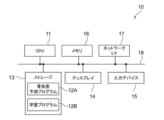
- the bone disease prediction apparatus 10 is a computer such as a workstation, server computer, or personal computer, and includes a CPU (Central Processing Unit) 11, a nonvolatile storage 13, and a memory 16 as a temporary storage area.
- the bone disease prediction apparatus 10 includes a display 14 such as a liquid crystal display, an input device 15 such as a keyboard and a mouse, and a network I/F (InterFace) connected to a network (not shown). ) 17.
- CPU 11, storage 13, display 14, input device 15, Memory 16 and network I/F 17 are connected to bus 18 .
- the CPU 11 is an example of a processor in the present disclosure.
- the storage 13 is realized by HDD (Hard Disk Drive), SSD (Solid State Drive), flash memory, and the like.
- a bone disease prediction program 12A and a learning program 12B installed in the bone disease prediction device 10 are stored in the storage 13 as a storage medium.
- the CPU 11 reads the bone disease prediction program 12A and the learning program 12B from the storage 13, develops them in the memory 16, and executes the developed bone disease prediction program 12A and the learning program 12B.
- the bone disease prediction program 12A and the learning program 12B are stored in a storage device of a server computer connected to a network or in a network storage in an externally accessible state, and configure the bone disease prediction device 10 upon request. downloaded and installed on your computer. Alternatively, it is recorded on a recording medium such as a DVD (Digital Versatile Disc), a CD-ROM (Compact Disc Read Only Memory) or the like, distributed, and installed in a computer constituting the bone disease prediction apparatus 10 from the recording medium.
- a recording medium such as a DVD (Digital Versatile Disc), a CD-ROM (Compact Disc Read Only Memory) or the like, distributed, and installed in a computer constituting the bone disease prediction apparatus 10 from the recording medium.
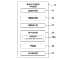
- FIG. 3 is a diagram showing the functional configuration of the bone disease prediction device and learning device according to this embodiment.
- the bone disease prediction apparatus 10 includes an image acquisition section 21 , an information acquisition section 22 , an information derivation section 23 , a probability derivation section 24 , a learning section 25 and a display control section 26 .
- the CPU 11 functions as an image acquisition unit 21, an information acquisition unit 22, an information derivation unit 23, a probability derivation unit 24, and a display control unit 26. It functions as an advanced neural network 24A.
- the CPU 11 also functions as a learning unit 25 by executing the learning program 12B.
- the image acquiring unit 21 causes the imaging device 1 to image the subject H, thereby obtaining a first radiographic image, which is a front image of, for example, the vicinity of the crotch area of the subject H from the first and second radiation detectors 5 and 6. Acquire G1 and a second radiographic image G2.
- SID Source Image receptor distance
- SOD Source Object Distance
- the SOD and SID are used to calculate the body thickness distribution as described later.
- SOD is preferably acquired by, for example, a TOF (Time Of Flight) camera.
- the SID is preferably obtained by, for example, a potentiometer, an ultrasonic rangefinder, a laser rangefinder, or the like.
- the imaging conditions may be set by input from the input device 15 by the operator.
- the set shooting conditions are saved in the storage 13 .
- the first and second radiographic images G1 and G2 may be acquired by a program separate from the bone disease prediction program 12A and stored in the storage 13.
- the image acquiring unit 21 acquires the first and second radiation images G1 and G2 stored in the storage 13 by reading them from the storage 13 for processing.
- the information acquisition unit 22 acquires teacher data for learning a neural network, which will be described later, from the image storage system 9 via the network I/F 17.
- the information derivation unit 23 derives a bone image and a soft tissue image of the subject H from the first and second radiation images G1 and G2. Then, the information derivation unit 23 derives the bone mineral content of each pixel in the target bone region of the subject H from the bone image, and acquires the pixel value of each pixel of the corresponding region corresponding to the target bone region in the soft tissue image.
- the target bone is the femur.
- FIG. 4 is a diagram showing the functional configuration of the information derivation unit 23.
- the information derivation unit 23 includes a scattered radiation removal unit 31 , an image derivation unit 32 , a segmentation unit 33 , a bone mineral content derivation unit 34 and a soft tissue pixel value acquisition unit 35 .
- the CPU 11 functions as a scattered radiation removal unit 31, an image derivation unit 32, a segmentation unit 33, a bone mineral content derivation unit 34, and a soft tissue pixel value acquisition unit 35 by executing the bone disease prediction program 12A.
- each of the first radiographic image G1 and the second radiographic image G2 includes, in addition to the primary ray component of the radiation transmitted through the subject H, the scattered ray component based on the radiation scattered within the subject H. . Therefore, the scattered radiation removal unit 31 removes scattered radiation components from the first radiographic image G1 and the second radiographic image G2.
- the scattered radiation removal unit 31 may apply the method described in JP-A-2015-043959 to remove scattered radiation components from the first radiographic image G1 and the second radiographic image G2.
- the derivation of the body thickness distribution of the subject H and the derivation of the scattered radiation component for removing the scattered radiation component are performed at the same time.
- the scattered radiation removing unit 31 acquires a virtual model K of the subject H having the initial body thickness distribution T0(x, y).
- the virtual model K is data that virtually represents the subject H, in which the body thickness according to the initial body thickness distribution T0(x, y) is associated with the coordinate position of each pixel of the first radiographic image G1.
- the virtual model K of the subject H having the initial body thickness distribution T0(x, y) may be stored in the storage 13 in advance.
- the body thickness distribution T(x, y) of the subject H may be calculated based on the SID and SOD included in the imaging conditions. In this case, the body thickness distribution can be obtained by subtracting SOD from SID.
- the scattered radiation removal unit 31 estimates an estimated primary radiation image obtained by estimating the primary radiation image obtained by imaging the virtual model K and a scattered radiation image obtained by imaging the virtual model K.
- An image obtained by synthesizing the obtained estimated scattered radiation image is generated as an estimated image obtained by estimating the first radiographic image G1 obtained by imaging the subject H.
- the scattered radiation removal unit 31 corrects the initial body thickness distribution T0(x, y) of the virtual model K so that the difference between the estimated image and the first radiographic image G1 is reduced.
- the scattered radiation removing unit 31 repeats the generation of the estimated image and the correction of the body thickness distribution until the difference between the estimated image and the first radiographic image G1 satisfies a predetermined termination condition.
- the scattered radiation removing unit 31 derives the body thickness distribution when the termination condition is satisfied as the body thickness distribution T(x, y) of the subject H.
- the scattered radiation removing unit 31 removes the scattered radiation component included in the first radiographic image G1 by subtracting the scattered radiation component when the end condition is satisfied from the first radiographic image G1.
- the image deriving unit 32 performs energy subtraction processing to obtain a bone image Gb in which the bone of the subject H is extracted and a soft tissue image Gs in which the soft tissue is extracted from the first and second radiation images G1 and G2. derive Scattered radiation components have been removed from the first and second radiographic images G1 and G2 in subsequent processing.
- the image derivation unit 32 calculates the distance between the corresponding pixels for the first and second radiographic images G1 and G2 as shown in the following formula (1).
- a bone portion image Gb in which the bone portion of the subject H included in each of the radiographic images G1 and G2 is extracted, is derived as shown in FIG.
- Equation (1) ⁇ 1 is a weighting factor.
- the pixel value of each pixel in the bone region in the bone image Gb is the bone pixel value.
- Gb(x,y) G1(x,y)- ⁇ 1 ⁇ G2(x,y) (1)
- the image derivation unit 32 performs calculation, for example, weighted subtraction between corresponding pixels on the first and second radiographic images G1 and G2, as shown in the following equation (2).
- a soft tissue image Gs in which only the soft tissue of the subject H included in each of the radiographic images G1 and G2 is extracted is derived.
- ⁇ 2 is a weighting factor.
- Gs(x, y) G1(x, y)- ⁇ 2 ⁇ G2(x, y) (2)
- the soft-tissue image Gs represents the soft-tissue area of the subject H's soft tissue.
- the “soft tissue” of the subject H refers to things other than bone tissue, and specifically includes muscle tissue, fat tissue, blood, and water.
- the segmentation unit 33 segments the bone image Gb into a femur region, a pelvis region, and a vertebra region.
- the segmentation may be performed by using an extraction model that has undergone machine learning so as to extract the femur, pelvis, and vertebrae from the bone image Gb.
- templates representing each of the femur, pelvis, and vertebrae may be stored in the storage 13, and segmentation may be performed by performing template matching between these templates and the bone image Gb.
- FIG. 7 is a diagram showing the result of segmentation by the segmentation unit 33.
- the bone region in the bone image Gb is segmented into a femur region A1, a pelvis region A2, and a vertebra region A3.
- the segmentation results are shown by giving different hatching to the femur region A1, the pelvis region A2, and the vertebra region A3.
- the bone image Gb includes only the sacral vertebrae and lumbar vertebrae.
- the lumbar vertebrae are anatomically labeled L5, L4, L3, L2, and L1 from the pelvic side toward the neck. Therefore, the segmentation unit 33 preferably segments the sacral vertebrae and the five lumbar vertebrae into different regions.
- the segmentation unit 33 may segment only the target bone in the bone image Gb. For example, since the femur is the target bone in this embodiment, only the region A1 of the femur may be segmented.
- the bone mineral content deriving unit 34 derives the bone mineral content for each pixel of the target bone region, which is the region of the target bone in the bone image Gb.
- the bone mineral content deriving unit 34 converts each pixel value in the target bone region of the bone image Gb into a pixel value of the bone image acquired under the reference imaging conditions, thereby obtaining a pixel value of the target bone region.
- the bone mineral content derivation unit 34 derives the bone mineral content by correcting each pixel value of the target bone region using a correction coefficient obtained from a lookup table, which will be described later.
- the subject H absorbs the low-energy component of the radiation, causing beam hardening in which the energy of the radiation increases. The higher the energy of the radiation due to the beam hardening, the greater the body thickness of the subject H.
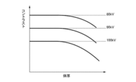
- FIG. 8 is a diagram showing the relationship between the body thickness of the subject H and the contrast between the bone part and the soft part.
- FIG. 8 shows the relationship between the body thickness of the subject H and the contrast between the bone and soft parts at three tube voltages of 80 kV, 90 kV and 100 kV.
- the higher the tube voltage the lower the contrast.
- the greater the body thickness the lower the contrast.
- the larger the pixel value of the bone region in the bone image Gb the greater the contrast between the bone and the soft tissue. Therefore, the relationship shown in FIG. 8 shifts to the high contrast side as the pixel value of the bone region in the bone image Gb increases.
- a lookup table for obtaining a correction coefficient for correcting a difference in contrast according to the tube voltage at the time of imaging and a decrease in contrast due to the influence of beam hardening in the bone image Gb is stored in the storage 13. stored in A correction coefficient is a coefficient for correcting each pixel value of the bone image Gb.
- FIG. 9 is a diagram showing an example of a lookup table stored in the storage 13.
- FIG. 9 illustrates a lookup table LUT1 in which the reference imaging condition is set to a tube voltage of 90 kV.
- the lookup table LUT1 in which the reference imaging condition is set to a tube voltage of 90 kV.
- a larger correction coefficient is set as the tube voltage increases and the body thickness of the subject H increases.
- the reference imaging condition is a tube voltage of 90 kV
- the correction coefficient is 1 when the tube voltage is 90 kV and the body thickness is 0.
- the lookup table LUT1 is shown two-dimensionally in FIG. 9, the correction coefficients differ according to the pixel values of the bone region. Therefore, the lookup table LUT1 is actually a three-dimensional table to which an axis representing pixel values of the bone region is added.
- the bone mineral content derivation unit 34 calculates a correction coefficient C0(x, y) for each pixel according to the imaging conditions including the body thickness distribution T(x, y) of the subject H and the setting value of the tube voltage stored in the storage 13. is extracted from the lookup table LUT1. Then, the bone mineral content derivation unit 34 multiplies each pixel (x, y) of the target bone region in the bone image Gb by the correction coefficient C0(x, y), as shown in the following equation (3). By doing so, the bone mineral content B(x, y) (g/cm 2 ) for each pixel in the target bone region is derived.
- the bone mineral content B(x, y) thus derived is obtained by imaging the subject H with a tube voltage of 90 kV, which is the standard imaging condition, and is a radiographic image from which the influence of beam hardening has been removed. represents the pixel value of the bone region included in .
- B(x, y) C0(x, y) ⁇ Gb(x, y) (3)
- the soft tissue pixel value acquisition unit 35 acquires the pixel value Gs (x, y) for each pixel of the corresponding region corresponding to the target bone region in the soft tissue image Gs.
- the target bone is the femur. Therefore, as shown in FIG. 10, the soft tissue pixel value acquisition unit 35 acquires the pixel value Gs(x, y) for each pixel in the corresponding region A11 corresponding to the region of the femur in the soft tissue image Gs.
- bone includes bone marrow.
- Bone marrow contains hematopoietic cells, and hematopoietic cells increase in fat content with age after the age of 20.
- Bone marrow with increased fat content is called fatty marrow.
- the soft tissue pixel value acquisition unit 35 obtains a pixel value for each pixel in the corresponding region corresponding to the target bone region in the soft tissue image Gs in order to detect early signs of fracture based on changes in the density of the bone marrow region. Get the value Gs(x,y).
- hatching indicates that the left thighbone of the patient includes a region D0 with a higher concentration than other regions.
- the probability derivation unit 24 derives the fracture probability of the target bone from the bone mineral content B(x, y) of each pixel of the target bone region and the pixel value Gs(x, y) of each pixel of the corresponding region. For this reason, when the probability deriving unit 24 receives the bone mineral content B(x, y) for each pixel of the target bone region and the pixel value Gs(x, y) for each pixel of the corresponding region, the probability derivation unit 24 The fracture probability of the target bone is derived using the trained neural network 24A that outputs the fracture probability.
- the learning unit 25 acquires the bone mineral content of each pixel of the target bone region derived from the bone image of the human body, the pixel value of each pixel of the corresponding region corresponding to the target bone region derived from the soft tissue image of the human body, and the pixel value of the target bone.
- a learned neural network 24A is constructed by performing machine learning on the neural network using correct data representing the fracture probability as teacher data.
- Neural networks include simple perceptrons, multilayer perceptrons, deep neural networks, convolutional neural networks, deep belief networks, recurrent neural networks, and probabilistic neural networks.
- a convolutional neural network is used as the neural network.
- FIG. 11 is a diagram showing a neural network used in this embodiment.
- neural network 60 comprises input layer 61 , intermediate layer 62 and output layer 63 .
- Intermediate layers 62 comprise, for example, multiple convolutional layers 65 , multiple pooling layers 66 and fully connected layers 67 .
- a fully connected layer 67 exists in front of the output layer 63 .
- convolutional layers 65 and pooling layers 66 are alternately arranged between the input layer 61 and the fully connected layer 67 .
- neural network 60 may comprise one convolutional layer 65 and one pooling layer 66 between input layer 61 and fully connected layer 67 .
- FIG. 12 is a diagram showing an example of teacher data used for neural network learning.
- teacher data 40 consists of learning data 41 and correct answer data 42 .
- the learning data 41 consists of a bone mineral content 43 for each pixel in the target bone region in the bone image and a pixel value 44 for each pixel in the corresponding region corresponding to the target bone region in the soft tissue image.
- the bone mineral content 43 is derived from the bone image Gb of a patient with an early fracture.
- Pixel values 44 are obtained from a soft tissue image Gs of a patient suffering from an early fracture. In this case, the presence or absence of the patient's initial fracture can be confirmed by obtaining an MRI image of the patient and interpreting the MRI image.
- the bone image Gb for deriving the bone mineral content 43 and the soft tissue image Gs for obtaining the pixel value 44 were obtained by performing energy subtraction imaging on a patient in whom an initial fracture was found as a result of reading the MRI image. derived from radiographic images.
- the bone mineral content 43 indicates that it is the bone mineral content by hatching the target bone region (femur) in the bone image.
- the pixel value 44 indicates that it is a pixel value by giving hatching to the corresponding region in the soft tissue image.
- the correct answer data 42 consist of fracture probabilities.
- the teacher data 40 is derived by recording the statistics of bone mineral content for each pixel in the target bone region and the pixel value for each pixel in the corresponding region with respect to a plurality of patients when a fracture occurs, and stored as an image. Stored in system 9.
- the bone fracture probability which is the correct data 42 in the teacher data 40, is obtained for a predetermined number of years (for example, one year, After 2 years or 5 years, etc.), the number of cases in which fracture occurred is obtained, and the obtained number of cases is divided by the number of patients.
- the bone mineral content 43 and the pixel value 44 which are the learning data 41, may be derived by processing the radiographic image of a healthy subject according to signs of early bone fracture. As a result, learning can be effectively advanced by increasing the number of teacher data.
- the correct data 42 may use the bone fracture probabilities for patients with cases similar to the processed radiographic image.
- the learning unit 25 uses a large amount of teacher data 40 to learn the neural network.
- FIG. 13 is a diagram for explaining learning of the neural network 60. As shown in FIG. When learning the neural network 60 , the learning unit 25 inputs learning data 41 to the input layer 61 of the neural network 60 . Then, the learning unit 25 outputs the fracture probability as the output data 70 from the output layer 63 of the neural network 60 . Then, the learning unit 25 derives the difference between the output data 70 and the fracture probability included in the correct data 42 as the loss L0.
- the learning unit 25 learns the neural network 60 based on the loss L0. Specifically, the learning unit 25 sets the coefficient of the kernel in the convolutional layer 65, the weight of the connection between each layer, the weight of the connection in the fully connected layer 67, etc. (hereinafter referred to as parameter 71) so as to reduce the loss L0. to adjust. As a method for adjusting the parameter 71, for example, the error backpropagation method can be used. The learning unit 25 repeats adjustment of the parameter 71 until the loss L0 becomes equal to or less than a predetermined threshold.
- the parameter 71 is adjusted so as to output a more accurate fracture probability, and the trained neural network 24A is constructed.
- the constructed trained neural network 24A is stored in the storage 13.
- the bone mineral content B(x, y) for each pixel of the target bone region derived from the bone image of the subject H, who is a patient, and the soft tissue image of the subject H When the acquired pixel value Gs(x, y) for each pixel of the corresponding region is input, as shown in FIG. It will output a reasonable fracture probability.
- the display control unit 26 displays the bone fracture probability derived by the probability deriving unit 24 on the display 14 .
- FIG. 15 is a diagram showing a fracture probability display screen. As shown in FIG. 15, the display screen 50 displays a bone image Gb, a soft tissue image Gs, and a bone fracture probability 51 of the subject H. FIG.
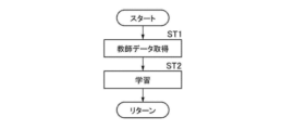
- FIG. 16 is a flow chart showing the learning process performed in this embodiment.
- the information acquisition unit 22 acquires teacher data from the image storage system 9 (step ST1), and the learning unit 25 inputs the learning data 41 included in the teacher data 40 to the neural network 60 to output the fracture probability.
- the loss L0 based on the difference from the correct data 42 is used to learn the neural network 60 (step ST2), and the process returns to step ST1.
- the learning unit 25 repeats the processing of steps ST1 and ST2 until the loss L0 reaches a predetermined threshold, and ends the learning.
- the learning unit 25 may end learning by repeating learning a predetermined number of times. Thereby, the learning unit 25 constructs a trained neural network 24A.
- FIG. 17 is a flow chart showing bone disease prediction processing in this embodiment. It is assumed that the first and second radiographic images G1 and G2 have been acquired by imaging and stored in the storage 13 .
- the image acquisition unit 21 acquires the first and second radiation images G1 and G2 from the storage 13 (radiation image acquisition; step ST11).
- the scattered radiation removal unit 31 of the information derivation unit 23 removes scattered radiation components from the first and second radiographic images G1 and G2 (step ST12).
- the image deriving unit 32 derives a bone part image Gb in which the bone part of the subject H is extracted and a soft tissue image Gs in which the soft part is extracted from the first and second radiation images G1 and G2 (step ST13).
- the segmentation unit 33 segments the region of the femur, which is the target bone, from the bone image Gb (step ST14).
- the bone mineral content derivation unit 34 derives the bone mineral content for each pixel of the target bone region in the bone image Gb (step ST15), and the soft tissue pixel value acquisition unit 35 extracts the pixels of the corresponding region from the soft tissue image Gs. A soft tissue pixel value is acquired for each time (step ST16).
- the probability derivation unit 24 uses the trained neural network 24A to derive the fracture probability of the target bone from the bone mineral content of each pixel of the target bone region and the pixel value of each pixel of the corresponding region (step ST17). . Then, the display control unit 26 displays the bone fracture probability derived by the probability deriving unit 24 on the display 14 (step ST18), and ends the processing.
- the bone marrow contains hematopoietic cells, but after the age of 20, the fat content of the hematopoietic cells increases with age. Also, when an early fracture occurs, the bone marrow becomes inflamed and thus watery. Since water absorbs radiation more than fat absorbs radiation, water appears denser (ie darker) than fat in a plain radiographic image. Since the bone marrow is a soft tissue, when inflammation occurs in the bone, the pixel values of the corresponding region corresponding to the bone region in the soft tissue image Gs become high (that is, dark) due to the influence of water.
- the target It was made to derive the fracture probability regarding the bone.
- the fracture probability is derived using the pixel value Gs (x, y) of the corresponding region corresponding to the target bone region in the soft tissue image Gs, the fracture probability reflecting the initial signs of fracture is derived. can do.
- plain radiographic imaging can be used to identify early stage bone disease by reference to fracture probability.
- the learning data 41 includes the bone mineral content 43 for each pixel of the target bone region in the bone image and the pixel value 44 for each pixel of the corresponding region corresponding to the target bone region in the soft tissue image.
- the trained neural network 24A is constructed using the teacher data 40, it is not limited to this. As shown in FIG. 18, a trained neural network 24A may be constructed using teacher data 40A including a simple radiation image G0 as learning data 41A.
- the trained neural network 24A constructed in this way, in addition to the bone mineral content of each pixel of the target bone region of the subject H and the pixel value of each pixel of the corresponding region, the first radiographic image G1 of the subject H Alternatively, the second radiographic image G2 is input to the trained neural network 24A to derive the fracture probability.
- the femur is used as the target bone, but the target bone is not limited to this.
- a vertebra may be the target bone.
- the amount of bone mineral in the vertebrae is particularly low due to the occurrence of osteoporosis, and when osteoporosis worsens, the vertebrae are compressed and deformed in the vertical direction of the human body, and even compression fractures occur. For this reason, when the target bone is a vertebra, the bone mineral content of the target vertebra and the pixel value of the corresponding region corresponding to the vertebra are used to more accurately capture the signs of early fracture and estimate the probability of fracture occurrence. Prediction becomes possible.
- any bones such as the femur and tibia around the knee joint can be used as target bones.
- the trained neural network 24A is constructed using the pixel value 44 for each pixel in the area as the learning data 41, but the invention is not limited to this. Bone mineral content for each pixel in the target bone region and pixel values for each pixel in the corresponding region corresponding to the target bone region obtained from radiographic images of a patient with bone metastasis of cancer are used as learning data 41 for learning.
- a preconfigured neural network 24A may be constructed.
- FIG. 19 shows a soft tissue image of a patient with bone metastasis of cancer in the vertebrae.
- the soft tissue image Gs cancer has metastasized to the fourth lumbar vertebrae, and an elliptical high-density region D1 is included.
- the high density region D1 is hatched for explanation.
- the bone fracture probability which is the correct data 42, is obtained in advance for a plurality of patients with cancer bone metastases who have similar bone mineral content for each pixel in the target bone region and pixel values for each pixel in the corresponding region. It can be calculated by finding the number of cases in which fractures occurred after a specified number of years (for example, 1 year, 2 years, or 5 years) and dividing the calculated number of cases by the number of patients.
- a trained neural network that derives bone metastasis of cancer by using pixel values of a corresponding region corresponding to a target bone (for example, the fourth lumbar vertebrae) included in a soft tissue image Gs as shown in FIG. 19 as learning data. can be built. Therefore, by applying such a trained neural network to the probability derivation unit 24, it is possible to derive the probability of fracture caused by bone metastasis of cancer. Also, bone metastasis of cancer can be detected at an early stage using a simple radiographic image by referring to the fracture probability.
- the teacher data (referred to as first teacher data) shown in FIG. 12 and the teacher data (referred to as second teacher data) including the pixel values of the corresponding regions in the soft tissue image Gs shown in FIG. 19 as learning data.
- the neural network When learning data of the first teacher data is input, the neural network is trained such that the probability of the first fracture approaches 1 and the probability of the second fracture approaches 0. On the other hand, when the learning data of the second teacher data is input, the neural network is trained such that the first fracture probability approaches zero and the second fracture probability approaches one. This makes it possible to derive both the fracture probability resulting from the initial fracture and the fracture probability resulting from bone metastasis of cancer using one trained neural network 24B.
- the first radiographic image G1 and the second radiographic image G2 themselves are used to derive the bone mineral content, but the present invention is not limited to this.
- the moving average of the surrounding pixels is calculated, and the moving average is the pixel value of each pixel.
- Bone mineral content may be derived using the image G2.
- each pixel should be separated from the surrounding pixels so as to maintain a resolution at which the cortical bone can be visually recognized, for example, a resolution of 2 mm or less in the actual size of the subject.
- a moving average should be calculated.
- pixels to be used for the moving average may be appropriately determined based on information on the mutual distances among the radiation source 3, subject H, and radiation detectors 5 and 6, information on pixel sizes of the radiation detectors 5 and 6, and the like. .
- the tube constituting the radiation source 3 deteriorates over time, the quality and dose of the radiation emitted from the radiation source 3 change over time. Pixel values change over time.
- the radiation detectors 5 and 6 also deteriorate over time, the pixel values of the first and second radiographic images G1 and G2 output from the radiation detectors 5 and 6 change over time.
- the pixel values of the first and second radiographic images G1 and G2 fluctuate, an error occurs in the bone image Gb and the soft tissue image Gs derived by the above equations (1) and (2). and soft tissue under- and over-abrasives. As described above, when the accuracy of the bone image Gb and the soft image Gs is lowered, it becomes impossible to accurately derive the bone mineral content and the fracture probability.
- processing for removing the scattered radiation component is performed, and the body thickness distribution is repeatedly derived so as to match the estimated image at that time.
- the pixel value I0 on the assumption that there is no subject H is used as the body thickness.
- the pixel value I0 is derived using the tube voltage kv, dose mAs and SID and based on pre-acquired calibration data. Therefore, if the quality and dose of radiation and the signal value output from the radiation detector change compared to when the calibration data was acquired, an error occurs in the calculated pixel value I0.
- the bone image Gb and the soft tissue image Gs have different compositions, their mutual correlation is small.
- the composition remains unerased or over-erased, the correlation between the bone image Gb and the soft tissue image Gs increases. Therefore, in the present embodiment, the bone image Gb and the soft tissue image Gs may be derived such that the correlation between the bone image Gb and the soft tissue image Gs is minimized. Derivation of the bone image Gb and the soft tissue image Gs that minimize the correlation will be described below.
- the information deriving unit 23 derives the provisional bone image Gb0 and the provisional soft tissue image Gs0 by the above-described equations (1) and (2).
- a radiographic image including both the bone part and the soft part is derived.
- a coefficient h0 is defined for adjusting the level of bone disappearance in the temporary bone image Gb0.
- the information derivation unit 23 multiplies the provisional bone image Gb0 by the coefficient h0 to derive a new provisional bone image Gb0.
- a new temporary soft tissue image Gs0 is derived from G10-Gb0.
- the information deriving unit 23 derives the correlation r between the new provisional bone image Gb0 and the new provisional soft tissue image Gs0 while changing the value of the coefficient h0 according to the following equation (4).
- the coefficient h0 is derived as a uniform value for all pixels.
- the coefficient h0 may be derived for each pixel in the image.
- the temporary bone image Gb0 and the temporary soft tissue image Gs0 are calculated using a uniform value within the local region centered on the pixel for which the coefficient h0 is calculated, and the temporary bone image Gb0 and the temporary soft tissue image Gs0 are calculated in the local region.
- the correlation r may be derived using the temporary soft tissue image Gs0.
- kbs is the covariance between the new provisional bone image Gb0 and the new provisional soft tissue image Gs0
- db is the standard deviation of the new provisional bone image Gb0
- ds is the new provisional soft tissue image.
- the standard deviation of the image Gs0, n is the number of pixels of the new temporary bone image Gb0 and the new temporary soft tissue image Gs0, bi and si are the numbers of the new temporary bone image Gb0 and the new temporary soft tissue image Gs0, respectively.
- the pixel values B and S of each pixel are the average values of all the pixels of the new temporary bone image Gb0 and the new temporary soft tissue image Gs0, respectively.
- the image is two-dimensional, it is expressed one-dimensionally by assigning a number to each pixel of the image in Equation (4).
- the information derivation unit 23 derives the correlation r while changing the value of the coefficient h0, and multiplies the temporary bone image Gb0 derived by the equation (1) by the coefficient h0 when the correlation r is minimized. , derive the bone image Gb. That is, the information derivation unit 23 derives the bone image Gb by the following equation (5). Further, the information deriving unit 23 derives the soft tissue image Gs by subtracting the bone image Gb derived by the equation (5) from the original radiographic image G10, as shown in the equation (6).
- the minimum correlation r is derived by deriving the correlation r while changing the coefficient h0 to derive a plurality of correlations. means r.
- Gb h0 ⁇ Gb0 (5)
- Gs G10-Gb (6)
- the provisional bone image Gb0 and the provisional soft tissue image Gs0 are frequency-decomposed into band components consisting of a plurality of frequency bands, and The band components may be used to derive the correlation r.
- a specific frequency band means one or more predetermined frequency bands among a plurality of frequency bands.
- the bone image Gb and the soft tissue image Gs that have the minimum mutual correlation it is possible to derive the highly accurate bone image Gb and the soft tissue image Gs that are free from unerased or over-erased compositions. can. Therefore, the bone mineral content and the pixel value in the corresponding region can be derived with high accuracy, and as a result, the fracture probability of the target bone can be derived with higher accuracy.
- the first and second radiographic images G1 and G2 are acquired by the one-shot method when performing the energy subtraction processing, but the method is not limited to this.
- the first and second radiographic images G1 and G2 may be acquired by a so-called two-shot method, in which imaging is performed twice using only one radiation detector.
- body motion of the subject H may shift the position of the subject H included in the first radiographic image G1 and the second radiographic image G2. Therefore, it is preferable to perform the processing of the present embodiment after aligning the subject in the first radiographic image G1 and the second radiographic image G2.
- the first and second radiographic images G1 and G2 of the subject H are captured using the first and second radiation detectors 5 and 6, and the radiographic images obtained by the system are used to detect the bones.
- disease prediction processing is performed, the technology of the present disclosure can also be applied when acquiring the first and second radiographic images G1 and G2 using stimulable phosphor sheets instead of radiation detectors.
- two stimulable phosphor sheets are superimposed and irradiated with radiation that has passed through the subject H, and the radiographic image information of the subject H is accumulated and recorded on each stimulable phosphor sheet.
- the first and second radiographic images G1 and G2 may be obtained by photoelectrically reading the radiographic image information.
- the two-shot method may also be used when the first and second radiographic images G1 and G2 are acquired using the stimulable phosphor sheet.
- the radiation in the above embodiment is not particularly limited, and ⁇ -rays, ⁇ -rays, or the like can be used in addition to X-rays.
- a processing unit that executes various processes such as the image acquisition unit 21, the information acquisition unit 22, the information derivation unit 23, the probability derivation unit 24, the learning unit 25, and the display control unit 26
- the above various processors include: In addition to the CPU, which is a general-purpose processor that executes software (programs) and functions as various processing units, there are also programmable logic devices (FPGA), which are processors whose circuit configuration can be changed after manufacturing, such as FPGA (Field Programmable Gate Array). Programmable Logic Device (PLD), ASIC (Application Specific Integrated Circuit), and a dedicated electric circuit that is a processor having a circuit configuration specially designed to execute specific processing.
- FPGA programmable logic devices
- One processing unit may be configured with one of these various processors, or a combination of two or more processors of the same or different type (for example, a combination of multiple FPGAs or a combination of a CPU and an FPGA). ). Also, a plurality of processing units may be configured by one processor.
- one processor is configured by combining one or more CPUs and software, There is a form in which this processor functions as a plurality of processing units.
- SoC System On Chip
- the various processing units are configured using one or more of the above various processors as a hardware structure.
- an electric circuit in which circuit elements such as semiconductor elements are combined can be used.
- (Appendix 1) comprising at least one processor;
- the processor Acquiring a first radiographic image and a second radiographic image obtained by imaging a subject including bone and soft tissue with radiation having different energy distributions, Deriving a bone image representing the bone tissue of the subject and a soft tissue image representing the soft tissue of the subject by weighting and subtracting the first radiographic image and the second radiographic image, deriving a bone mineral content for each pixel in the target bone region of the subject from the bone image; obtaining a pixel value for each pixel of a corresponding region corresponding to the target bone region in the soft tissue image;
- a bone disease prediction apparatus for deriving a fracture probability of the target bone from the bone mineral content of each pixel in the target bone region and the pixel value of each pixel in the corresponding region.
- the processor generates a bone mineral content for each pixel of a target bone region derived from a bone portion image of a human body, a pixel value for each pixel of a corresponding region corresponding to the target bone region derived from a soft tissue image of the human body, and a pixel value for each pixel of the target bone region.
- the bone disease prediction device which functions as a trained neural network that has undergone machine learning using correct data representing bone fracture probability as teacher data.
- the processor derives the fracture probability from the first radiographic image or the second radiographic image in addition to the bone mineral content of each pixel in the target bone region and the pixel value of each pixel in the corresponding region.
- Item 2 The bone disease prediction device according to item 1.
- the processor comprises a simple radiographic image of the human body, a bone mineral content of each pixel of the target bone derived from the bone part image of the human body, and a bone mineral content of each pixel of the corresponding region corresponding to the target bone derived from the soft part image of the human body. 4.
- the bone disease prediction device according to item 3 which functions as a trained neural network that has undergone machine learning using pixel values and correct data representing the fracture probability as teacher data.
- (Appendix 10) comprising at least one processor;
- the processor Bone mineral content of each pixel of the target bone derived from the bone image of the human body, pixel value of each pixel of the corresponding region corresponding to the target bone derived from the soft tissue image of the human body, and fracture probability of the target bone are calculated.
- a neural network using the correct data represented as teacher data, the bone mineral content of each pixel in the target bone region derived from the bone part image of the target subject and the target derived from the soft tissue image of the target subject
- a learning device for constructing a trained neural network for deriving the fracture probability of the target bone of the target subject from the pixel value of each pixel in the corresponding region corresponding to the bone region.
- the learning device wherein the processor machine-learns the neural network using simple radiographic images of the human body as teacher data.
- Appendix 12 When the bone mineral content of each pixel in the target bone region derived from the bone portion image of the target subject and the pixel value of each pixel in the corresponding region corresponding to the target bone region derived from the soft tissue image of the target subject are input, A trained neural network for deriving a fracture probability of a target bone of the target subject.
- (Appendix 14) Acquiring a first radiographic image and a second radiographic image obtained by imaging a subject including bone and soft tissue with radiation having different energy distributions, Deriving a bone image representing the bone tissue of the subject and a soft tissue image representing the soft tissue of the subject by weighting and subtracting the first radiographic image and the second radiographic image, deriving a bone mineral content for each pixel in the target bone region of the subject from the bone image; obtaining a pixel value for each pixel of a corresponding region corresponding to the target bone region in the soft tissue image; A bone disease prediction method for deriving a fracture probability of the target bone from a bone mineral content of each pixel in the target bone region and a pixel value of each pixel in the corresponding region.
- Bone mineral content of each pixel of the target bone derived from the bone image of the human body, pixel value of each pixel of the corresponding region corresponding to the target bone derived from the soft tissue image of the human body, and fracture probability of the target bone are calculated.
- a neural network using the correct data represented as teacher data, the bone mineral content of each pixel in the target bone region derived from the bone part image of the target subject and the target derived from the soft tissue image of the target subject
- a learning program for causing a computer to execute a procedure for constructing a trained neural network for deriving the fracture probability of the target bone of the target subject from the pixel value of each pixel in the corresponding region corresponding to the bone region.
- Imaging Device 3 Radiation Source 5, 6 Radiation Detector 7 Radiation Energy Conversion Filter 9 Image Storage System 10
- Bone Disease Prediction Device 11 CPU 12A bone disease prediction processing program 12B learning program 13 storage 14 display 15 input device 16 memory 17 network I/F 18 bus 21 image acquisition unit 22 information acquisition unit 23 information derivation unit 24 probability derivation unit 24A, 24B trained neural network 25 learning unit 26 display control unit 31 scattered radiation removal unit 32 image derivation unit 33 segmentation unit 34 bone mineral content derivation unit 35 Soft tissue pixel value acquisition unit 40 Teacher data 41 Learning data 42 Correct data 43 Bone mineral content in target bone region 44 Pixel value in corresponding region 50 Display screen 51 Occurrence probability of fracture 60 Neural network 61 Input layer 62 Intermediate layer 63 Output layer 65 Convolution layer 66 Pooling layer 67 Fully connected layer 70 Output data 71 Parameters A1 Femur region A2 Pelvis region A3 Vertebral region A10, A11 Corresponding region B(x, y) Bone mineral content per pixel D0, D1 Cor
Landscapes
- Health & Medical Sciences (AREA)
- Life Sciences & Earth Sciences (AREA)
- Medical Informatics (AREA)
- Engineering & Computer Science (AREA)
- Radiology & Medical Imaging (AREA)
- Molecular Biology (AREA)
- Biophysics (AREA)
- Nuclear Medicine, Radiotherapy & Molecular Imaging (AREA)
- Optics & Photonics (AREA)
- Pathology (AREA)
- Physics & Mathematics (AREA)
- Biomedical Technology (AREA)
- Heart & Thoracic Surgery (AREA)
- High Energy & Nuclear Physics (AREA)
- Surgery (AREA)
- Animal Behavior & Ethology (AREA)
- General Health & Medical Sciences (AREA)
- Public Health (AREA)
- Veterinary Medicine (AREA)
- Image Analysis (AREA)
- Apparatus For Radiation Diagnosis (AREA)
Abstract
La présente invention concerne un procédé selon lequel un processeur acquiert une première radiographie et une seconde radiographie acquises par capture d'une image d'un sujet comprenant une partie osseuse et une partie molle au moyen d'un rayonnement ayant différentes distributions d'énergie, obtient une image de partie osseuse représentant la structure osseuse du sujet et une image de partie molle représentant un tissu mou du sujet par soustraction pondérée de la première radiographie et de la seconde radiographie, obtient une densité osseuse pour chaque pixel dans une région osseuse objet du sujet à partir de l'image de partie osseuse, acquiert une valeur de pixel pour chaque pixel dans une région correspondante de l'image en partie molle correspondant à la région osseuse objet, et obtient une probabilité de fracture de l'os objet à partir de la densité osseuse pour chaque pixel et de la valeur de pixel pour chaque pixel dans la région osseuse objet.
Applications Claiming Priority (4)
| Application Number | Priority Date | Filing Date | Title |
|---|---|---|---|
| JP2021-162984 | 2021-10-01 | ||
| JP2021162984 | 2021-10-01 | ||
| JP2022-129051 | 2022-08-12 | ||
| JP2022129051 | 2022-08-12 |
Publications (1)
| Publication Number | Publication Date |
|---|---|
| WO2023054287A1 true WO2023054287A1 (fr) | 2023-04-06 |
Family
ID=85782680
Family Applications (1)
| Application Number | Title | Priority Date | Filing Date |
|---|---|---|---|
| PCT/JP2022/035789 WO2023054287A1 (fr) | 2021-10-01 | 2022-09-26 | Dispositif, procédé et programme de prédiction de maladie osseuse, dispositif d'apprentissage, procédé et programme, et réseau neuronal entraîné |
Country Status (1)
| Country | Link |
|---|---|
| WO (1) | WO2023054287A1 (fr) |
Citations (5)
| Publication number | Priority date | Publication date | Assignee | Title |
|---|---|---|---|---|
| JP2012179076A (ja) * | 2011-02-28 | 2012-09-20 | Hitachi Aloka Medical Ltd | 骨密度測定装置 |
| WO2016129682A1 (fr) * | 2015-02-13 | 2016-08-18 | 株式会社島津製作所 | Dispositif d'analyse osseuse |
| WO2016190327A1 (fr) * | 2015-05-28 | 2016-12-01 | 株式会社日立製作所 | Système médical de mesure à rayons x |
| WO2020166561A1 (fr) * | 2019-02-14 | 2020-08-20 | 富士フイルム株式会社 | Dispositif d'acquisition de valeur d'évaluation de risque de fracture osseuse, son procédé de fonctionnement, et programme d'acquisition de valeur d'évaluation de risque de fracture osseuse |
| JP2021002339A (ja) * | 2019-06-21 | 2021-01-07 | ストラックスコープ ピーティワイ リミテッドStraxcorp Pty Ltd | 画像内での構造又は物質セグメンテーションに基づいた機械学習分類のための方法及びシステム |
-
2022
- 2022-09-26 WO PCT/JP2022/035789 patent/WO2023054287A1/fr active Application Filing
Patent Citations (5)
| Publication number | Priority date | Publication date | Assignee | Title |
|---|---|---|---|---|
| JP2012179076A (ja) * | 2011-02-28 | 2012-09-20 | Hitachi Aloka Medical Ltd | 骨密度測定装置 |
| WO2016129682A1 (fr) * | 2015-02-13 | 2016-08-18 | 株式会社島津製作所 | Dispositif d'analyse osseuse |
| WO2016190327A1 (fr) * | 2015-05-28 | 2016-12-01 | 株式会社日立製作所 | Système médical de mesure à rayons x |
| WO2020166561A1 (fr) * | 2019-02-14 | 2020-08-20 | 富士フイルム株式会社 | Dispositif d'acquisition de valeur d'évaluation de risque de fracture osseuse, son procédé de fonctionnement, et programme d'acquisition de valeur d'évaluation de risque de fracture osseuse |
| JP2021002339A (ja) * | 2019-06-21 | 2021-01-07 | ストラックスコープ ピーティワイ リミテッドStraxcorp Pty Ltd | 画像内での構造又は物質セグメンテーションに基づいた機械学習分類のための方法及びシステム |
Similar Documents
| Publication | Publication Date | Title |
|---|---|---|
| JP6906479B2 (ja) | 骨塩情報取得装置、方法およびプログラム | |
| EP4056120B1 (fr) | Dispositif, procédé et programme d'estimation | |
| JP7016294B2 (ja) | 骨塩情報取得装置、方法およびプログラム | |
| JP7016293B2 (ja) | 骨塩情報取得装置、方法およびプログラム | |
| WO2023054287A1 (fr) | Dispositif, procédé et programme de prédiction de maladie osseuse, dispositif d'apprentissage, procédé et programme, et réseau neuronal entraîné | |
| US20220287663A1 (en) | Estimation device, estimation method, and estimation program | |
| US20220323032A1 (en) | Learning device, learning method, and learning program, radiation image processing device, radiation image processing method, and radiation image processing program | |
| CN115131277A (zh) | 推定装置、方法及计算机可读存储介质 | |
| US20220249013A1 (en) | Motor organ disease prediction device, motor organ disease prediction method, motor organ disease prediction program, learning device, learning method, learning program, and learned neural network | |
| CN118043908A (en) | Bone disease prediction device, bone disease prediction method, bone disease prediction program, learning device, bone disease prediction method, learning program, and learning completion neural network | |
| US20230017704A1 (en) | Estimation device, estimation method, and estimation program | |
| JP2024040945A (ja) | 画像処理装置、方法およびプログラム | |
| US20220335605A1 (en) | Estimation device, estimation method, and estimation program | |
| US20230172576A1 (en) | Radiation image processing device, radiation image processing method, and radiation image processing program | |
| JP7220643B2 (ja) | 画像処理装置、方法およびプログラム | |
| US20230096694A1 (en) | Image processing device, image processing method, and image processing program | |
| US20240104729A1 (en) | Radiation image processing device, radiation image processing method, and radiation image processing program | |
| JP7342140B2 (ja) | 情報処理装置、情報処理方法およびプログラム | |
| US20240090861A1 (en) | Radiation image processing apparatus, operation method of radiation image processing apparatus, and non-transitory computer readable medium | |
| US11963810B2 (en) | Estimation device, estimation method, and estimation program | |
| JP7289769B2 (ja) | 画像処理装置、方法およびプログラム | |
| JP2024000885A (ja) | 放射線画像処理装置、方法およびプログラム | |
| JP2023177980A (ja) | 放射線画像処理装置、方法およびプログラム | |
| JP2023047910A (ja) | 脂肪量導出装置、方法およびプログラム |
Legal Events
| Date | Code | Title | Description |
|---|---|---|---|
| 121 | Ep: the epo has been informed by wipo that ep was designated in this application |
Ref document number: 22876154 Country of ref document: EP Kind code of ref document: A1 |
|
| WWE | Wipo information: entry into national phase |
Ref document number: 2023551486 Country of ref document: JP |