WO2023037860A1 - Blood vessel cover - Google Patents
Blood vessel cover Download PDFInfo
- Publication number
- WO2023037860A1 WO2023037860A1 PCT/JP2022/031605 JP2022031605W WO2023037860A1 WO 2023037860 A1 WO2023037860 A1 WO 2023037860A1 JP 2022031605 W JP2022031605 W JP 2022031605W WO 2023037860 A1 WO2023037860 A1 WO 2023037860A1
- Authority
- WO
- WIPO (PCT)
- Prior art keywords
- cover
- vein
- blood vessel
- inner diameter
- vascular
- Prior art date
Links
- 210000004204 blood vessel Anatomy 0.000 title claims abstract description 77
- 210000003462 vein Anatomy 0.000 claims abstract description 122
- 210000001367 artery Anatomy 0.000 claims abstract description 38
- 239000002473 artificial blood Substances 0.000 claims abstract description 16
- 230000002093 peripheral effect Effects 0.000 claims abstract description 6
- 230000002792 vascular Effects 0.000 claims description 54
- 230000007704 transition Effects 0.000 claims description 21
- 239000004744 fabric Substances 0.000 claims description 12
- 239000002759 woven fabric Substances 0.000 claims description 9
- 239000004745 nonwoven fabric Substances 0.000 claims description 6
- 239000007853 buffer solution Substances 0.000 abstract description 30
- 238000007634 remodeling Methods 0.000 abstract description 15
- 206010072810 Vascular wall hypertrophy Diseases 0.000 abstract description 3
- 230000017531 blood circulation Effects 0.000 description 31
- 210000002460 smooth muscle Anatomy 0.000 description 23
- 210000004177 elastic tissue Anatomy 0.000 description 20
- 238000009940 knitting Methods 0.000 description 17
- 238000011144 upstream manufacturing Methods 0.000 description 15
- 230000003872 anastomosis Effects 0.000 description 13
- 239000008280 blood Substances 0.000 description 11
- 210000004369 blood Anatomy 0.000 description 11
- 238000000034 method Methods 0.000 description 11
- 239000000835 fiber Substances 0.000 description 10
- 206010020718 hyperplasia Diseases 0.000 description 10
- 239000000463 material Substances 0.000 description 10
- 230000000541 pulsatile effect Effects 0.000 description 10
- 230000004872 arterial blood pressure Effects 0.000 description 8
- 102000008186 Collagen Human genes 0.000 description 7
- 108010035532 Collagen Proteins 0.000 description 7
- 230000036772 blood pressure Effects 0.000 description 7
- 229920001436 collagen Polymers 0.000 description 7
- 230000003902 lesion Effects 0.000 description 7
- 229920005989 resin Polymers 0.000 description 7
- 239000011347 resin Substances 0.000 description 7
- 238000010276 construction Methods 0.000 description 5
- 239000000872 buffer Substances 0.000 description 4
- 238000010586 diagram Methods 0.000 description 4
- 238000000502 dialysis Methods 0.000 description 4
- 230000000694 effects Effects 0.000 description 4
- 230000010349 pulsation Effects 0.000 description 4
- 238000010186 staining Methods 0.000 description 4
- 230000008320 venous blood flow Effects 0.000 description 4
- 208000031481 Pathologic Constriction Diseases 0.000 description 3
- 210000003363 arteriovenous anastomosis Anatomy 0.000 description 3
- 230000003139 buffering effect Effects 0.000 description 3
- 230000008859 change Effects 0.000 description 3
- 238000006243 chemical reaction Methods 0.000 description 3
- 230000000877 morphologic effect Effects 0.000 description 3
- -1 polyethylene Polymers 0.000 description 3
- 229920001343 polytetrafluoroethylene Polymers 0.000 description 3
- 239000004810 polytetrafluoroethylene Substances 0.000 description 3
- 230000008569 process Effects 0.000 description 3
- 230000036262 stenosis Effects 0.000 description 3
- 208000037804 stenosis Diseases 0.000 description 3
- 230000001629 suppression Effects 0.000 description 3
- 230000009885 systemic effect Effects 0.000 description 3
- 206010020772 Hypertension Diseases 0.000 description 2
- 206010029113 Neovascularisation Diseases 0.000 description 2
- 230000015572 biosynthetic process Effects 0.000 description 2
- 238000012790 confirmation Methods 0.000 description 2
- 210000002808 connective tissue Anatomy 0.000 description 2
- 230000007423 decrease Effects 0.000 description 2
- 238000005259 measurement Methods 0.000 description 2
- 239000000203 mixture Substances 0.000 description 2
- 231100000915 pathological change Toxicity 0.000 description 2
- 230000036285 pathological change Effects 0.000 description 2
- 239000004033 plastic Substances 0.000 description 2
- 229920003023 plastic Polymers 0.000 description 2
- 229920000728 polyester Polymers 0.000 description 2
- 239000005020 polyethylene terephthalate Substances 0.000 description 2
- 206010002329 Aneurysm Diseases 0.000 description 1
- 206010003210 Arteriosclerosis Diseases 0.000 description 1
- 229920000742 Cotton Polymers 0.000 description 1
- 229920004934 Dacron® Polymers 0.000 description 1
- YCKRFDGAMUMZLT-UHFFFAOYSA-N Fluorine atom Chemical compound [F] YCKRFDGAMUMZLT-UHFFFAOYSA-N 0.000 description 1
- 206010019280 Heart failures Diseases 0.000 description 1
- 239000004677 Nylon Substances 0.000 description 1
- 206010061876 Obstruction Diseases 0.000 description 1
- 239000004698 Polyethylene Substances 0.000 description 1
- 229920000954 Polyglycolide Polymers 0.000 description 1
- 239000004721 Polyphenylene oxide Substances 0.000 description 1
- 239000004743 Polypropylene Substances 0.000 description 1
- 208000001647 Renal Insufficiency Diseases 0.000 description 1
- 208000017932 Steel syndrome Diseases 0.000 description 1
- 208000007536 Thrombosis Diseases 0.000 description 1
- 239000002253 acid Substances 0.000 description 1
- 230000009471 action Effects 0.000 description 1
- 230000003044 adaptive effect Effects 0.000 description 1
- 125000001931 aliphatic group Chemical group 0.000 description 1
- 229920003232 aliphatic polyester Polymers 0.000 description 1
- 230000010100 anticoagulation Effects 0.000 description 1
- 210000000709 aorta Anatomy 0.000 description 1
- 230000008321 arterial blood flow Effects 0.000 description 1
- 208000011775 arteriosclerosis disease Diseases 0.000 description 1
- 230000004323 axial length Effects 0.000 description 1
- 230000008901 benefit Effects 0.000 description 1
- 238000005520 cutting process Methods 0.000 description 1
- 230000007123 defense Effects 0.000 description 1
- 206010012601 diabetes mellitus Diseases 0.000 description 1
- 229940079593 drug Drugs 0.000 description 1
- 239000003814 drug Substances 0.000 description 1
- 230000005489 elastic deformation Effects 0.000 description 1
- 238000001523 electrospinning Methods 0.000 description 1
- 229920000840 ethylene tetrafluoroethylene copolymer Polymers 0.000 description 1
- 238000011156 evaluation Methods 0.000 description 1
- 229920000295 expanded polytetrafluoroethylene Polymers 0.000 description 1
- 230000002349 favourable effect Effects 0.000 description 1
- 239000012530 fluid Substances 0.000 description 1
- 239000011737 fluorine Substances 0.000 description 1
- 229910052731 fluorine Inorganic materials 0.000 description 1
- 238000001631 haemodialysis Methods 0.000 description 1
- 230000012447 hatching Effects 0.000 description 1
- 230000000322 hemodialysis Effects 0.000 description 1
- 238000003384 imaging method Methods 0.000 description 1
- 239000007943 implant Substances 0.000 description 1
- 230000005764 inhibitory process Effects 0.000 description 1
- 229910052500 inorganic mineral Inorganic materials 0.000 description 1
- 238000005304 joining Methods 0.000 description 1
- 208000017169 kidney disease Diseases 0.000 description 1
- 201000006370 kidney failure Diseases 0.000 description 1
- 230000005226 mechanical processes and functions Effects 0.000 description 1
- 239000012528 membrane Substances 0.000 description 1
- 239000011707 mineral Substances 0.000 description 1
- 210000003205 muscle Anatomy 0.000 description 1
- 229920001778 nylon Polymers 0.000 description 1
- 230000001575 pathological effect Effects 0.000 description 1
- 230000007170 pathology Effects 0.000 description 1
- 229920011301 perfluoro alkoxyl alkane Polymers 0.000 description 1
- 229920000747 poly(lactic acid) Polymers 0.000 description 1
- 229920006122 polyamide resin Polymers 0.000 description 1
- 229920000570 polyether Polymers 0.000 description 1
- 229920000573 polyethylene Polymers 0.000 description 1
- 229920000139 polyethylene terephthalate Polymers 0.000 description 1
- 239000004633 polyglycolic acid Substances 0.000 description 1
- 229920001721 polyimide Polymers 0.000 description 1
- 239000009719 polyimide resin Substances 0.000 description 1
- 239000004626 polylactic acid Substances 0.000 description 1
- 229920005672 polyolefin resin Polymers 0.000 description 1
- 229920001155 polypropylene Polymers 0.000 description 1
- 239000004800 polyvinyl chloride Substances 0.000 description 1
- 229920000915 polyvinyl chloride Polymers 0.000 description 1
- 238000003825 pressing Methods 0.000 description 1
- 230000001681 protective effect Effects 0.000 description 1
- 230000035485 pulse pressure Effects 0.000 description 1
- 238000004080 punching Methods 0.000 description 1
- 230000003014 reinforcing effect Effects 0.000 description 1
- 238000005096 rolling process Methods 0.000 description 1
- 210000000329 smooth muscle myocyte Anatomy 0.000 description 1
- 238000001356 surgical procedure Methods 0.000 description 1
- 229920003002 synthetic resin Polymers 0.000 description 1
- 239000000057 synthetic resin Substances 0.000 description 1
- 239000002699 waste material Substances 0.000 description 1
- XLYOFNOQVPJJNP-UHFFFAOYSA-N water Substances O XLYOFNOQVPJJNP-UHFFFAOYSA-N 0.000 description 1
Images


Classifications
-
- A—HUMAN NECESSITIES
- A61—MEDICAL OR VETERINARY SCIENCE; HYGIENE
- A61F—FILTERS IMPLANTABLE INTO BLOOD VESSELS; PROSTHESES; DEVICES PROVIDING PATENCY TO, OR PREVENTING COLLAPSING OF, TUBULAR STRUCTURES OF THE BODY, e.g. STENTS; ORTHOPAEDIC, NURSING OR CONTRACEPTIVE DEVICES; FOMENTATION; TREATMENT OR PROTECTION OF EYES OR EARS; BANDAGES, DRESSINGS OR ABSORBENT PADS; FIRST-AID KITS
- A61F2/00—Filters implantable into blood vessels; Prostheses, i.e. artificial substitutes or replacements for parts of the body; Appliances for connecting them with the body; Devices providing patency to, or preventing collapsing of, tubular structures of the body, e.g. stents
- A61F2/02—Prostheses implantable into the body
- A61F2/04—Hollow or tubular parts of organs, e.g. bladders, tracheae, bronchi or bile ducts
- A61F2/06—Blood vessels
-
- A—HUMAN NECESSITIES
- A61—MEDICAL OR VETERINARY SCIENCE; HYGIENE
- A61M—DEVICES FOR INTRODUCING MEDIA INTO, OR ONTO, THE BODY; DEVICES FOR TRANSDUCING BODY MEDIA OR FOR TAKING MEDIA FROM THE BODY; DEVICES FOR PRODUCING OR ENDING SLEEP OR STUPOR
- A61M1/00—Suction or pumping devices for medical purposes; Devices for carrying-off, for treatment of, or for carrying-over, body-liquids; Drainage systems
- A61M1/36—Other treatment of blood in a by-pass of the natural circulatory system, e.g. temperature adaptation, irradiation ; Extra-corporeal blood circuits
Definitions
- the present invention relates to a vascular cover used for an anastomosis in which blood vessels are anastomosed together.
- the present invention relates to a blood vessel cover that can be used by arranging it on the outer peripheral side of the.
- Dialysis treatment is given regularly.
- a special needle is inserted into a vein.
- the artery is anastomosed to the vein because the normal venous blood flow is not sufficient for dialysis.
- a blood vessel is called a shunt.
- an incision is made in the skin of the arm to expose the artery and vein, a small incision is made in the artery, the vein is anastomosed, and part of the blood flow from the artery is diverted to the vein.
- a shunt provided with an artificial blood vessel may be used.
- the conditions are favorable, appropriate remodeling due to changes in elasticity of the venous wall occurs as the body's defense reaction, avoiding stenosis and occlusion due to intimal thickening, and reducing the shunt blood flow state to a state that does not burden the body. It may be self-regulating. However, if the local conditions such as shunt blood flow or the shape of the anastomotic site, or systemic conditions (diabetes, hypertension, arteriosclerosis, blood conditions, etc.) are poor, pathological conditions beyond the range of appropriate protective adaptive reactions occur. It becomes a serious biological reaction and causes local and systemic pathology.
- Non-Patent Document 1 in order to suppress a rapid increase in blood flow immediately after surgery and in the initial stage, the vein wall is reinforced from the outside to prevent excessive blood pressure in the inner vein.
- vascular banding is performed to prevent hyperextension and blood turbulence caused by this.
- US Pat. No. 5,300,000 a woven net made by forming a knitted fabric that is seamless, tubular, substantially pileless, is a covering for reinforcing natural veins for use as surgical implants. is disclosed.
- vascular banding as described above could not sufficiently prevent lesions such as intimal hyperplasia.
- the reinforced vein wall is altered (arterialized) into an arterial wall-like structure only under certain conditions, but when blood flows from the reinforced site to the unreinforced vein, blood pressure and Since the pulsation is delivered downstream as it is without being buffered, the cause of intimal hyperplasia has not been fundamentally resolved. In order to resolve this, it is necessary to gradually lower the blood pressure and pulsatility downstream from the anastomotic site, and remodel the most downstream venous side to a state in which only low pressure is applied without pulsation.
- the present invention has been made in view of the above circumstances, and remodels the vein into a buffer system vessel that can deliver blood to the downstream vein while gradually reducing the blood pressure, pulse pressure, and blood flow rate of the blood flowing through the lumen.
- An object of the present invention is to provide a vascular cover capable of preventing intimal hyperplasia.
- a blood vessel cover according to an embodiment of the present invention which can solve the above problems, is as follows.
- a tubular vascular cover arranged on the outer peripheral side of a vein that is anastomosed with an artery or an artificial blood vessel and continuous over the entire circumference, wherein one end and the other end of the vascular cover are arranged in the axial direction of the vascular cover.
- the inner wall of the second portion having a central axis parallel to the central axis of the blood vessel cover, where the portion from the midpoint to the one end is defined as the first portion, and the portion from the midpoint to the other end is defined as the second portion.
- the diameter of the second imaginary cylinder inscribed in the vessel cover is larger than the diameter of the first imaginary cylinder inscribed in the inner wall of the first part, the central axis being parallel to the central axis of the vessel cover.
- the minimum inner diameter of the second portion is larger than the minimum inner diameter of the first portion.
- the vein may gradually grow outward during the process of remodeling into a buffer system vessel, but the vessel cover having the above configuration does not hinder this growth, and as a result, the lumen of the coated vessel is widened. It can be maintained to ensure sufficient blood flow.
- the vascular cover of the present invention enables shunt construction that secures a sufficient blood flow while suppressing lesions such as intimal hyperplasia by remodeling veins into buffer-system vessels.
- the vascular cover preferably has [2] to [7] below.
- the vascular cover includes one end portion having a first inner diameter, which is a section from one end of the vascular cover to the midpoint of the first portion in the axial direction of the vascular cover, and the other end portion. an intermediate portion located on the end side and having a second inner diameter of 1.2 times or more the first inner diameter; and the other end having a third inner diameter of 1.2 times or more the second inner diameter. , a first transition portion with a gradually increasing inner diameter between one end and an intermediate portion, and a second transition portion with a gradually increasing inner diameter between the intermediate portion and the other end. vascular cover.
- the covering becomes looser toward the downstream side of the vein. This facilitates remodeling and securing blood flow.
- the inner diameter of the other end of the blood vessel cover is larger than the inner diameter of the intermediate portion, the effect of abrupt release of the suppression by the covering at the other end of the blood vessel cover can be alleviated.
- the boundary between one end and the first transition portion, the boundary between the first transition portion and the intermediate portion, and the boundary between the intermediate portion and the second transition portion are curved lines [2]
- the vascular cover described in As a result, the vessel can be covered with a vascular cover having a smooth lumen wall, and remodeling into a buffer system vessel can be facilitated.
- the force required to expand the inner wall of the second part radially by 1.5 times from the natural state is the force required to expand the inner wall of the first part radially by 1.5 times from the natural state.
- vascular cover has at least one of knitted fabric, woven fabric, and non-woven fabric as a component that partially configures or as a component that configures the whole Vessel cover as described.
- the blood vessel cover has a bellows structure that periodically repeats peaks and valleys in the axial direction.
- the vessel cover according to any one of [1] to [6], wherein in the axial direction the distance between adjacent ridges at the second portion is greater than the distance between the ridges at one end.
- the wall structure of the vein is gradually changed downstream from the anastomosis, and the inside of the covered vein is subjected to gradient shear stress, It is possible to change the pressure perpendicular to the blood vessel wall, the blood flow, the blood flow velocity, and the width of change associated with pulsation, thereby suppressing the mismatch of blood vessel wall elasticity, blood turbulence, and excessive high blood flow, Intimal hyperplasia can be prevented.
- the reasons why the blood vessel cover of the present invention has such effects are considered as follows.
- Both arteries and veins consist of the intima, media, and adventitia.
- the media consists of a smooth muscle layer rich in smooth muscle cells and an elastic fiber layer containing collagen fibers.
- Arteries have thick smooth muscle layers and elastic fiber layers so that even when the pressure of the pulsating luminal blood flow is applied, there is little pulsation change in the vascular wall, and turbulence and frictional stress fluctuations do not occur.
- Veins on the other hand, have thin blood vessel walls and do not have thick smooth muscle layers and elastic fiber layers like arteries. When arterial blood flows directly into such veins through an arteriovenous shunt, lesions such as intimal hyperplasia occur due to the marked difference in elasticity between arteries and veins, as described above.
- the vascular cover of the present invention has the above configuration, so that the pulsatile blood flow with high arterial pressure that flows into the vein at the arteriovenous anastomosis of the shunt site or the artificial blood vessel-venous anastomosis is directed downstream.
- the vein at the shunt site can be remodeled into a buffered vessel that can be gradually buffered and eventually transitioned to venous blood flow. As a result, blood turbulence and pulsating changes in the vein wall are suppressed, and lesions such as intimal hyperplasia can be prevented.
- the vascular cover of the present invention having the above structure does not hinder the growth of veins, which may gradually grow outward in the process of remodeling into buffer-system vessels, thereby widening the vascular lumen. It can be maintained to ensure sufficient blood flow. As a result, it becomes possible to create a shunt that can secure a sufficient blood flow while suppressing lesions such as intimal hyperplasia.
- FIG. 11 shows a schematic diagram of another example of a shunt-forming portion
- 1 depicts a perspective view of a vessel cover according to an embodiment of the present invention
- FIG. 2 depicts a perspective view of a vessel cover according to another embodiment of the present invention
- FIG. 10 depicts a perspective view of a vessel cover according to yet another embodiment of the present invention
- FIG. 10 depicts a perspective view of a vessel cover according to yet another embodiment of the present invention
- FIG. 10 depicts a perspective view of a vessel cover according to yet another embodiment of the present invention
- FIG. 1 shows a schematic diagram of a case in which an autologous vein is anastomosed to a small incision of an artery in a shunt-creating part
- FIG. 10 is a schematic diagram when a vein is anastomosed to the other end of the .
- 3-5 depict perspective views of vessel covers according to different embodiments.
- 6 and 7 present perspective views of vascular covers according to yet other different embodiments.
- the shunt-constructed part 1 can be formed by performing an arteriovenous anastomosis as shown in FIG. 1 or an artificial blood vessel-venous anastomosis as shown in FIG.
- the shunt section 1 can be formed by anastomosing the vein 4 to the small incision of the artery 3 in the arm 2 and allowing the blood flow of the artery 3 to flow through the vein 4.
- blood flows from the artery 3 through the anastomosis 6 and the vein 4 in the direction indicated by the arrow B.
- the shunt-constructing part 1 can be formed by anastomosing one end of an artificial blood vessel 5 to a small incision portion of an artery 3 in an arm 2 and anastomosing a vein 4 to the other end of the artificial blood vessel 5.
- the blood flows from the artery 3 through the artificial blood vessel 5, the anastomosis 6, and the vein 4 in the direction indicated by the arrow B.
- the arteriovenous anastomosis shown in FIG. 1 or the artificial vessel-vein anastomosis shown in FIG. It can be arranged on the outer peripheral side, and the vein 4 can be remodeled into a buffer system blood vessel.
- the vessel cover 10 is preferably arranged from the most upstream part of the vein 4 on the anastomotic part 6 side. At this time, one end 10a of the vessel cover 10 is arranged at the most upstream portion of the vein 4 on the anastomosis portion 6 side, and the other end 10b of the vessel cover 10 is arranged at the downstream side of the vein 4 away from the anastomosis portion 6. is preferred.
- the blood vessel cover 10 is formed in a continuous cylindrical shape over the entire circumference, and has an axial direction x and a radial direction y.
- the axial direction x of the vessel cover 10 is the direction in which the central axis C of the vessel cover 10 extends. is the direction connecting points on the outer edge of
- the vascular cover 10 may be a knitted fabric, woven fabric, or net that is continuously constructed all around. Knitted fabrics, woven fabrics, nets, and the like have stitches and textures, but the gaps formed in the stitches and textures are not discontinuous portions of the vascular cover 10, but the above-mentioned "continuous can form a "cylindrical".
- the blood vessel cover 10 has flexibility, and it is preferable that the axial direction x of the blood vessel cover 10 can be curved to follow the extending direction of the veins 4 to be covered.
- the vessel cover 10 preferably has a lumen with a circular or elliptical shape in cross section in the radial direction y.
- the outer edge of the cross section in the radial direction y may have fine irregularities.
- the vessel cover 10 may be in a state in which the lumen is crushed by its own weight in the natural state. Even in such a case, it is possible to define the same cross-sectional shape in the axial direction x, radial direction y, and radial direction y by widening the lumen.
- a method for widening the lumen collapsed by its own weight for example, a tube whose lumen does not collapse under its own weight and has a central axis parallel to the central axis C of the vessel cover 10 and inscribed in the inner wall of the vessel cover 10 is used. into the lumen of the vessel cover 10, and the like.
- the vessel cover 10 is a tubular cover arranged on the outer peripheral side of the vein 4 anastomosed with the artery 3 or the artificial blood vessel 5.
- the portion from the midpoint 10c between the one end 10a and the other end 10b of the vessel cover 10 to the one end 10a is the first portion 11, and the portion from the midpoint 10c to the other end 10b is the second portion 12.
- the diameter d2 of the second virtual cylinder T2 which has a central axis parallel to the central axis C of the vessel cover 10 and is inscribed in the inner wall of the second part 12, has a central axis parallel to the central axis C of the vessel cover 10. It is larger than the diameter d1 of the first imaginary cylinder T1 that contacts the inner wall of the first portion 11 .
- the minimum inner diameter of the second portion 12 can be made larger than the minimum inner diameter of the first portion 11 of the vessel cover 10 .
- the vein 4 is more downstream than the upstream side. Can be loosely covered.
- the vein 4 is remodeled so that the pulsatile blood flow flowing into the vein 4 and having high arterial pressure is gradually buffered toward the downstream and finally becomes a buffer system blood vessel that can shift to the venous blood flow. can do.
- blood turbulence and pulsating changes in the vein wall are suppressed, and lesions such as intimal hyperplasia can be prevented.
- the wall of the vein 4 of the shunt-constructed portion 1 has a smooth muscle layer that is thicker than the smooth muscle layer of the normal vein and includes an elastic fiber layer, and an elastic layer that includes collagen fibers that are thicker than the smooth muscle layer on the outside of the smooth muscle layer.
- Blood vessels are composed of three layers: the intima, the media, and the adventitia. Among them, the intima greatly contributes to anticoagulation, but its mechanical contribution is extremely small.
- the mechanical elements of the arteries of the extremities which are normal arteries used for artificial dialysis, consist of the media, which contains a small amount of elastic fibers and abundant smooth muscle, and the outer membrane, which consists of elastic fibers and collagen fibers. account for the percentage. Thus, these arteries have a great abundance of smooth muscle and relatively few elastic fibers (“smooth muscle>elastic fiber” configuration).
- elastic fibers Due to their elasticity, elastic fibers have a cushioning function like a rubber tube that resists and cushions the pulsatile and high blood pressure of arteries.
- smooth muscle since smooth muscle is a muscle, it also has a more active mechanical function, resisting arterial blood pressure, while on the other hand delivering high pulsatile arterial blood pressure to the periphery without attenuation. It has an active function. Due to the pressure-transmitting function of the abundant smooth muscle of the arteries, the blood pressure of the aorta, which has an inner diameter on the order of a centimeter, and the small artery, which has an inner diameter of only a fraction of a millimeter, hardly changes.
- the venous wall changes like a normal artery, it is arterialization (remodeling into an artery) and not into a buffer system vessel. If the arterialization gradually weakens and naturally transitions downstream to a completely normal vein, it means that it gradually thins and transitions to the vein with its "smooth muscle > elastic fiber" configuration. , high pulsatile blood pressure acts on the venous wall due to its lack of buffering function, resulting in pathological changes in downstream veins. This point is a clear functional difference between the case where the buffer system blood vessel gradually thins and shifts to a normal vein, and the case where a normal artery-like blood vessel gradually thins and shifts to a normal vein.
- the vascular cover 10 has the above-described configuration, so that the downstream side of the vein 4 of the shunt-constructing portion 1 is more loosely covered, thereby changing the wall structure of the vein 4 as described above. can be remodeled into a buffer system of blood vessels.
- the vein 4 may gradually grow outward in the course of being remodeled into a buffer system vessel, but the vessel cover 10 according to the embodiment of the present invention can more loosely cover the downstream side of the vein 4. Therefore, inhibition of the growth of the vein 4 by the vascular cover 10 is reduced, and the lumen of the vein 4 can be kept wide to ensure a sufficient blood flow.
- the vascular cover 10 according to the embodiment of the present invention, it is possible to form the shunt-constructed portion 1 that can ensure a sufficient blood flow while suppressing lesions such as intimal hyperplasia.
- the blood vessel cover 10 covers not only the vein 4 but also a part of the artery 3 and the artificial blood vessel 5 on the anastomosis 6 side. may be arranged to cover the surface.
- the blood vessel cover 10 may have a tubular shape, and may have a joint portion, for example, formed by rolling a flat plate member into a tubular shape and joining them by suturing. In that case, it is preferable that the joint portion such as the suture portion is formed on the outer surface of the vessel cover 10 . This prevents the joint from affecting the vein 4 .
- a seamless cylindrical member having no joints may be formed by using a molded member or knitted fabric.
- the first virtual cylinder T1 is inscribed in the inner wall of the first portion 11 at one end 10a of the vessel cover 10, and is inscribed in the inner wall of the first portion 11 at portions other than the one end 10a. It doesn't have to be. That is, the first portion 11 of the vessel cover 10 has an inner diameter d1 at the one end 10a, and may have an inner diameter larger than d1 at portions other than the one end 10a.
- the second virtual cylinder T2 is inscribed in the inner wall of the second portion 12 at the midpoint 10c of the blood vessel cover 10, and is in contact with the inner wall of the second portion 12 at portions other than the midpoint 10c. It does not have to be inscribed.
- the second portion 12 of the vessel cover 10 has an inner diameter d2 at the midpoint 10c, and may have an inner diameter larger than d2 at portions other than the midpoint 10c.
- An example of such a shape of the vessel cover 10 is a tapered shape in which the inner diameter gradually increases from one end 10a to the other end 10b.
- the inner diameter of the first portion 11 is the minimum value of the inner diameter of the second portion 12 throughout the axial direction x of the first portion 11. or smaller than the minimum inner diameter of the second portion 12 .
- the first imaginary column T1 is inscribed in the inner wall of the first portion 11 at a portion from one end 10a of the vessel cover 10 to a predetermined position in the axial direction x, and includes the one end 10a. Parts other than this part may not be inscribed in the inner wall of the first part 11 . That is, the first portion 11 of the vascular cover 10 has an inner diameter d1 in a portion from the one end 10a of the vascular cover 10 to a predetermined position, and has an inner diameter larger than d1 in other portions. good too.
- the portion of the vascular cover 10 with the smallest inner diameter is the largest on the anastomosis portion 6 side of the vein 4.
- the upstream part can be covered, and since it can be covered with the vessel cover 10 whose inner diameter gradually increases as it goes downstream of the vein 4, it can be loosely covered as it goes downstream of the vein 4, and the buffer system of the vein 4 Remodeling into a blood vessel and securing the lumen diameter of the vein 4 are facilitated.
- the first imaginary cylinder T1 is inscribed in the inner wall of the first portion 11 at any position between the one end 10a and the midpoint 10c of the vessel cover 10 in the axial direction x.
- the portion other than the position may not be inscribed in the inner wall of the first portion 11 . That is, the first portion 11 of the vessel cover 10 has an inner diameter d1 at a position between the one end 10a and the midpoint 10c of the vessel cover 10, and has an inner diameter larger than d1 at other portions. You may have
- the inner diameter of the first portion 11 over the entire axial direction x of the first portion 11 is the same as the minimum inner diameter of the second portion 12, or is larger than the minimum inner diameter of the second portion 12. is preferably small.
- the first virtual cylinder T1 may be inscribed in the inner wall of the first portion 11 over the entire section from the one end 10a of the vessel cover 10 to the midpoint 10c in the axial direction x. That is, the first portion 11 of the vessel cover 10 may have the inner diameter d1 over the entire axial direction x.
- the second virtual cylinder T2 may be inscribed in the inner wall of the second part 12 at a portion other than the midpoint 10c of the blood vessel cover 10.
- the second portion 12 may have an inner diameter larger than d2 except for the portion where the second virtual cylinder T2 is inscribed.
- the inner diameter of the second portion 12 is preferably 2.5 times or less, more preferably 2 times or less, the diameter d2 of the second imaginary cylinder T2 throughout the axial direction x. 5 times or less is more preferable.
- the inner diameter of the second portion 12 at the other end 10b is preferably larger than the inner diameter of the second portion 12 at the midpoint 10c.
- the second virtual cylinder T2 may be inscribed in the inner wall of the second portion 12 over the entire section from the midpoint 10c of the vessel cover 10 to the other end 10b in the axial direction x. That is, the second portion 12 of the vessel cover 10 may have an inner diameter d2 along the entire axial direction x.
- the diameter d2 of the second virtual column T2 is larger than the diameter d1 of the first virtual column T1, so that the one end 10a of the vessel cover 10 is positioned upstream of the vein 4 of the shunt-constructing portion 1.
- the downstream side of the vein 4 can be more loosely covered, and the above effect can be obtained.
- FIG. 6 shows a perspective view of a blood vessel cover 10 according to another embodiment.
- the blood vessel cover 10 of this aspect has a one end portion 110 which is a section from the one end 10a of the blood vessel cover 10 to the midpoint of the first portion 11 in the axial direction x of the blood vessel cover 10 and has a first inner diameter d5.
- the other end 10b having a third inner diameter d7 that is twice or more, the first transition portion 110m having a gradually increasing inner diameter between the one end 110 and the intermediate portion 120, and the intermediate portion 120 and the other end 10b and a second transition portion 120m of gradually increasing inner diameter.
- the vein 4 moves downstream in the process of remodeling the vein 4 into a buffer system vessel. Since it can spread slowly outward in the radial direction y as it goes, remodeling of the vein 4 into a buffer system vessel is facilitated, and the lumen of the vein 4 is easily maintained to ensure the blood flow.
- the other end 10b of the vessel cover 10 is a portion where the suppression of the vein 4 by the vessel cover 10 is abruptly released. , the influence of abrupt release of the suppression by the covering of the vessel cover 10 on the vein 4 can be mitigated.
- the boundary between the one end portion 110 and the first transition portion 110m, the boundary between the first transition portion 110m and the intermediate portion 120, and the intermediate portion 120 and the second transition portion is preferably curved. Since the one end portion 110, the intermediate portion 120, and the other end 10b have different inner diameters by a factor of 1.2 or more, the inner wall of the vessel cover 10 has a step in the axial direction x. , the steps can be smoothed by forming the respective boundaries as curved lines as described above. As a result, the blood vessel can be covered with the vessel cover 10 having a smooth lumen wall, and remodeling into a buffer system vessel is facilitated.
- the force required to expand the inner wall of the second portion 12 by 1.5 times in the radial direction y from the natural state is required to expand the inner wall of the first portion 11 by 1.5 times in the radial direction y from the natural state. is preferably less than the force
- a resin tube is inserted into the lumen of the vessel cover 10 so as to be inscribed with the inner wall of the first part 11, A method of applying pressure by introducing a fluid into the lumen of the resin tube can be used.
- the inner wall of the second portion 12 can be expanded 1.5 times in the radial direction y from the natural state.
- a cylindrical sample is prepared by cutting out a portion of the first part 11 so as to have a predetermined length in the axial direction x, and two pins are attached to the inner diameter of the cylindrical sample in parallel with the axial direction of the cylindrical sample.
- the two pins may be inserted and pulled in opposite diametrical y directions so that the inner diameter of the cylindrical sample is 1.5 times larger.
- the inner wall of the second part 12 can be similarly expanded in the radial direction y by 1.5 times from the natural state, but the length of the cylindrical sample cut out from the second part 12 in the axial direction x is The length in the axial direction x of the cylindrical sample cut out from the first part 11 is set to be the same.
- the downstream side of the vein 4 is more loosely covered when the first portion 11 of the vascular cover 10 is arranged upstream of the vein 4 of the shunt-constructing portion 1 . This makes it easier to remodel the vein 4 into a buffer system vessel and effectively secure the lumen diameter of the vein 4 .
- the length of the blood vessel cover 10 in the axial direction x is preferably 5 mm or more.
- the length of the blood vessel cover 10 in the axial direction x is preferably 10 mm or longer, more preferably 20 mm or longer, particularly preferably 30 mm or longer, and may be 40 mm or longer.
- the length of the blood vessel cover 10 in the axial direction x is preferably 120 mm or less, more preferably 100 mm or less, and even more preferably 90 mm or less. If the length of the vascular cover 10 in the axial direction x is within the above range, the vein 4 of the shunt-constructed portion 1 can be covered with the vascular cover 10 having a length greater than or equal to the predetermined length, and the vein 4 can be reconnected to the buffer system blood vessel. Easier to model.
- the inner diameter of the vessel cover 10 is preferably 2 mm or more, more preferably 3 mm or more, even more preferably 4 mm or more, further preferably 5 mm at the portion having the minimum inner diameter in the first portion 11, that is, the portion inscribed with the first virtual cylinder T1.
- the above is particularly preferable, and 10 mm or less is preferable, 8 mm or less is more preferable, and 6 mm or less is even more preferable.
- the inner diameter of the vessel cover 10 is preferably 4 mm or more, more preferably 5 mm or more, and even more preferably 6 mm or more at the portion having the minimum inner diameter in the second portion 12, that is, the portion inscribed with the second virtual cylinder T2.
- vascular cover 10 7 mm or more is particularly preferable, 12 mm or less is preferable, 10 mm or less is more preferable, 9 mm or less is even more preferable, and 8 mm or less is particularly preferable.
- fine unevenness may occur on the inner wall of the vascular cover 10, making it difficult to determine the inner diameter.
- the diameter d1 of the inscribed first imaginary cylinder T1 and the diameter d2 of the second imaginary cylinder T2 inscribed in the second part 12 can be used as the inner diameter of the vessel cover .
- the vascular cover 10 preferably has at least one of a knitted fabric, a woven fabric, and a nonwoven fabric as a component that partially configures it or as a component that configures the whole. With these materials, it is easy to form the elastically deformable blood vessel cover 10 .
- the type of knitted fabric is not particularly limited, and may be warp knitted or weft knitted. Knitting structures of warp knitting include half knitting, back half knitting, queens coat knitting, and satin knitting. Weft knitting includes circular knitting and flat knitting, and knitting structures of weft knitting include plain knitting, rubber knitting, double-sided knitting, milanese rib knitting, and jacquard knitting.
- the knitted fabric is preferably composed of weft knitting from the viewpoint of excellent stretchability.
- the type of woven fabric is not particularly limited, and may be plain weave, twill weave, satin weave, or the like.
- the blood vessel cover 10 may be composed of a nonwoven fabric produced by any method such as meltblowing, needle punching, spunlacing, electrospinning, or the like.
- the blood vessel cover 10 may be composed of a combination of two or more different materials, for example, one part is composed of a knitted fabric and the other part is composed of another material such as a non-woven fabric.
- yarns forming knitted fabrics, woven fabrics, and non-woven fabrics are also made of resin materials with superior plastic deformation, such as polyolefin resins such as polyethylene and polypropylene; polyamide resins such as nylon; polyethylene terephthalate. polyimide resins; fluorine resins such as PTFE, PFA and ETFE; synthetic resins such as polyvinyl chloride resins.
- resin materials with superior plastic deformation such as polyolefin resins such as polyethylene and polypropylene; polyamide resins such as nylon; polyethylene terephthalate.
- polyimide resins fluorine resins such as PTFE, PFA and ETFE
- synthetic resins such as polyvinyl chloride resins.
- the yarn forming the knitted fabric or woven fabric can also be made of a resin material (e.g., polyester, PTFE) used for artificial blood vessels, and specifically, ePTFE obtained by stretching PTFE and polyester fiber from DuPont. Dacron (registered trademark) and the like can be mentioned.
- the vessel cover 10 may be made of a biodegradable material such as aliphatic polyester such as polylactic acid, polyglycolic acid, and polyhydroxyalkanoic acid; aliphatic polyether.
- the yarn forming the knitted or woven fabric may be composed of natural fibers such as silk or cotton, or may be composed of a combination of resin materials, biodegradable materials, and natural fibers.
- the blood vessel cover 10 has a bellows structure in which peaks and troughs are periodically repeated in the axial direction x.
- the portion is the one end portion 110
- the evaluation of whether or not the vein 4 is remodeled into the low-pressure buffer system blood vessel needs to be confirmed by both the morphological confirmation method described below and the confirmation method by measuring the buffering action.
- a morphological method it is confirmed whether a two-layer structure consisting of a smooth muscle layer containing morphological elastic fibers and elastic fibers containing collagen fibers thicker than the smooth muscle layer is formed. It can be carried out. Specifically, the vein 4 of the shunt-forming portion 1 is cut out, subjected to special staining such as hematoxylin-eosin (HE) staining and elastica-fungieson (EvG) staining, and the cross section of the vein wall is observed with a microscope.
- HE hematoxylin-eosin
- EvG elastica-fungieson
- smooth muscle is stained turbid yellow, elastic fibers are dark purple, and collagen fibers are stained dark red, so the smooth muscle layer containing elastic fibers and the elastic fiber layer containing collagen fibers are observed, This can be done by confirming "thickness of smooth muscle layer containing elastic fibers ⁇ thickness of elastic fiber layer containing collagen fibers".
- shunt-forming part 2 arm 3: artery 4: vein 5: artificial blood vessel 6: anastomotic part 10: vessel cover 10a: one end 10b of vessel cover: the other end 10c of vessel cover: middle point 11 of vessel cover: First part 12: Second part 110: One end 110m: First transition part 120: Middle part 120m: Second transition part C: Central axis d1 of vessel cover: Diameter d2 of first imaginary cylinder: Second imaginary cylinder Diameter d5: Inside diameter d6 of one end: Inside diameter d7 of intermediate part: Inside diameter of the other end L1: Distance between mountains at one end L2: Distance between mountains at second part T1: First virtual cylinder T2: Second virtual Cylinder x: axial direction y: radial direction
Abstract
To provide a blood vessel cover that can prevent intimal thickening by remodeling a vein into a buffer-system blood vessel. The present invention is a tubular blood vessel cover (10) which is disposed on the outer peripheral side of a vein (4) that is anastomosed to an artery (3) or to an artificial blood vessel, and which is continuous along the entire circumference of said cover. When a portion from the center (10c) to one end (10a) of the blood vessel cover (10) in the axial direction x of the blood vessel cover (10) is a first section (11) and a portion from the center (10c) to an opposite end (10b) is a second section (12), the diameter (d2) of a second virtual cylinder (T2) that has a central axis parallel to the central axis (C) of the blood vessel cover (10) and is inscribed in an inner wall of the second section (12) is greater than the diameter (d1) of a first virtual cylinder (T1) that has a central axis parallel to the central axis (C) of the blood vessel cover (10) and is inscribed in an inner wall of the first section (11).
Description
本発明は、血管同士が吻合された吻合部に用いる血管カバーに関し、例えばシャント造設部における動脈と静脈との吻合、又は動脈に吻合された人工血管と静脈との吻合において、吻合部の静脈の外周側に配置して用いることのできる血管カバーに関するものである。
TECHNICAL FIELD The present invention relates to a vascular cover used for an anastomosis in which blood vessels are anastomosed together. The present invention relates to a blood vessel cover that can be used by arranging it on the outer peripheral side of the.
腎不全を含む重篤な腎臓疾患等の患者に対して、患者の体内から血液を取り出し、透析器で老廃物や余分な水分、ミネラルなどを取り除いた後、再び患者の体内に血液を戻す血液透析治療が定期的に行われる。通常、血液透析を行う際には静脈に専用の針を穿刺する。このとき、普通の静脈の血流では透析を施行するのに十分な血流量が得られないため、動脈を静脈に吻合する。このような血管をシャントと呼び、通常腕の皮膚を切開して動脈と静脈を露出させ、動脈に小切開を加えてそこに静脈を吻合し、動脈の血流を一部静脈へ流すことでシャントを造設する。このとき、動脈に直接静脈を吻合してシャントとする場合、又は動脈の小切開部分に人工血管の一方端を吻合し、人工血管の他方端を静脈に吻合して動脈と静脈との間に人工血管が設けられたシャントとする場合がある。
For patients with serious kidney disease including renal failure, blood is removed from the patient's body, waste products, excess water, minerals, etc. are removed with a dialyzer, and then returned to the patient's body Dialysis treatment is given regularly. Usually, when performing hemodialysis, a special needle is inserted into a vein. At this time, the artery is anastomosed to the vein because the normal venous blood flow is not sufficient for dialysis. Such a blood vessel is called a shunt. Normally, an incision is made in the skin of the arm to expose the artery and vein, a small incision is made in the artery, the vein is anastomosed, and part of the blood flow from the artery is diverted to the vein. Build a shunt. At this time, when a vein is directly anastomosed to an artery to form a shunt, or one end of the artificial blood vessel is anastomosed to a small incision portion of the artery and the other end of the artificial blood vessel is anastomosed to the vein to create a space between the artery and the vein. A shunt provided with an artificial blood vessel may be used.
シャント造設部においては、動脈と静脈とでは弾性の差異が著しいため、動脈において拍動する高い血圧の血液が、低圧での著しい高伸展性と高圧での低弾力性を有する静脈に流入すると、血液乱流や静脈壁への応力変化が起こってしまう。その結果、吻合部及び流出路静脈に内膜肥厚が生じ、狭窄、閉塞や血栓形成などの病態変化が容易に生じることとなる。シャント血流状態を生体に負担の大きな状態のまま調節できないと、より広範な局所的(下流静脈の瘤形成や狭窄、過剰シャント血流によるスチール症候群など)或いは全身的(静脈環流の著しい増加による心不全など)病態を引き起こす。
At the site of shunt construction, there is a marked difference in elasticity between arteries and veins. Therefore, when pulsating, high-pressure blood in arteries flows into veins, which have remarkably high distensibility at low pressure and low elasticity at high pressure, , blood turbulence and stress changes on the venous wall occur. As a result, intimal thickening occurs at the anastomotic site and outflow tract veins, easily resulting in pathological changes such as stenosis, obstruction, and thrombus formation. If the state of shunt blood flow cannot be controlled in a state that places a large burden on the body, it can lead to more widespread local (e.g., aneurysm formation and stenosis of downstream veins, steel syndrome due to excessive shunt blood flow) or systemic (due to significant increase in venous return). heart failure, etc.).
条件がよければ、生体の防御反応として、静脈壁の弾性変化などによる適切なリモデリングが起こり、内膜肥厚による狭窄や閉塞等を免れる場合や、シャント血流状態を生体に負担のない状態に自己調節できる場合もある。しかし、シャント血流量や吻合部の形状等の局所的条件や全身的条件(糖尿病、高血圧、動脈硬化や血液状態等)が悪い場合には、適切な防御適応反応が生じる範囲を超えて病的な生体反応となり、局所的全身的病態を引き起こすこととなる。
If the conditions are favorable, appropriate remodeling due to changes in elasticity of the venous wall occurs as the body's defense reaction, avoiding stenosis and occlusion due to intimal thickening, and reducing the shunt blood flow state to a state that does not burden the body. It may be self-regulating. However, if the local conditions such as shunt blood flow or the shape of the anastomotic site, or systemic conditions (diabetes, hypertension, arteriosclerosis, blood conditions, etc.) are poor, pathological conditions beyond the range of appropriate protective adaptive reactions occur. It becomes a serious biological reaction and causes local and systemic pathology.
これらに対応するため、例えば非特許文献1では、術直後から初期の急激な血流量増加を抑制するために、静脈壁の外側から静脈壁を補強することにより、内側にある静脈に過剰な血圧、またそれによる過伸展や血液乱流などが起こることを防止する血管バンディングが行われている。また、特許文献1には、外科用インプラントとして使用する天然静脈を補強するための被覆物であって、シームレス、チューブ状、実質的にパイルレスであるニット生地を形成することによって作られる編織物ネットの被覆物が開示されている。さらに、特許文献2及び3には、生体内分解性ポリマーの拘束性繊維マトリクスによりラッピングされた動静脈グラフト(AVG)は、頸動脈と類似する拍動性の放射状偏位が見られたことが開示されている。
In order to cope with these problems, for example, in Non-Patent Document 1, in order to suppress a rapid increase in blood flow immediately after surgery and in the initial stage, the vein wall is reinforced from the outside to prevent excessive blood pressure in the inner vein. In addition, vascular banding is performed to prevent hyperextension and blood turbulence caused by this. Also, in US Pat. No. 5,300,000, a woven net made by forming a knitted fabric that is seamless, tubular, substantially pileless, is a covering for reinforcing natural veins for use as surgical implants. is disclosed. Furthermore, US Pat. disclosed.
しかし、上記のような血管バンディングでは、内膜肥厚等の病変を十分に防止することができなかった。従来の血管バンディングでは、補強された静脈壁は動脈壁のような構造に特定の条件下のみで改変(動脈化)されるが、補強部位から補強されていない静脈に血液が流れる際に血圧と脈動は緩衝されずにそのまま下流に送達されるため、内膜肥厚の要因の根本的な解消とはなっていなかった。これを解消するには、吻合部から下流にかけて徐々に血圧と拍動性を低下させ、最下流の静脈側は拍動性のない低圧しかかからない状態にリモデリングする必要がある。
However, vascular banding as described above could not sufficiently prevent lesions such as intimal hyperplasia. In conventional vascular banding, the reinforced vein wall is altered (arterialized) into an arterial wall-like structure only under certain conditions, but when blood flows from the reinforced site to the unreinforced vein, blood pressure and Since the pulsation is delivered downstream as it is without being buffered, the cause of intimal hyperplasia has not been fundamentally resolved. In order to resolve this, it is necessary to gradually lower the blood pressure and pulsatility downstream from the anastomotic site, and remodel the most downstream venous side to a state in which only low pressure is applied without pulsation.
本発明は上記事情に鑑みてなされたものであり、内腔を流れる血液の血圧や脈圧や血流量を徐々に低下させつつ血液を下流静脈に送達できるような緩衝系血管に静脈をリモデリングすることにより、内膜肥厚を防止できる血管カバーを提供することを目的とする。
The present invention has been made in view of the above circumstances, and remodels the vein into a buffer system vessel that can deliver blood to the downstream vein while gradually reducing the blood pressure, pulse pressure, and blood flow rate of the blood flowing through the lumen. An object of the present invention is to provide a vascular cover capable of preventing intimal hyperplasia.
上記課題を解決できた本発明の一実施態様に係る血管カバーは、以下の通りである。
[1] 動脈と、又は人工血管と吻合されている静脈の外周側に配置される全周にわたって連続した筒状の血管カバーであって、血管カバーの軸方向において、血管カバーの一方端と他方端との中点から一方端までの部分を第1部、中点から他方端までの部分を第2部としたとき、血管カバーの中心軸と平行な中心軸を有し第2部の内壁に内接する第2仮想円柱の直径は血管カバーの中心軸と平行な中心軸を有し第1部の内壁に内接する第1仮想円柱の直径よりも大きい。 A blood vessel cover according to an embodiment of the present invention, which can solve the above problems, is as follows.
[1] A tubular vascular cover arranged on the outer peripheral side of a vein that is anastomosed with an artery or an artificial blood vessel and continuous over the entire circumference, wherein one end and the other end of the vascular cover are arranged in the axial direction of the vascular cover. The inner wall of the second portion having a central axis parallel to the central axis of the blood vessel cover, where the portion from the midpoint to the one end is defined as the first portion, and the portion from the midpoint to the other end is defined as the second portion. The diameter of the second imaginary cylinder inscribed in the vessel cover is larger than the diameter of the first imaginary cylinder inscribed in the inner wall of the first part, the central axis being parallel to the central axis of the vessel cover.
上記構成を有する血管カバーは、第2部における内径の最小値の方が第1部における内径の最小値よりも大きい。このような構成を有する血管カバーの第1部をシャント造設部の静脈の上流側に配置することにより、静脈の下流側をより緩く被覆することができ、シャント造設部の静脈に流入した高い動脈圧を有する拍動性の血流が下流に向けて徐々に緩衝され最終的に静脈血流に移行できる緩衝系血管となるように、シャント造設部の静脈をリモデリングすることができる。また、静脈が緩衝系血管にリモデリングされる過程で徐々に外側に成長することがあるが、上記構成を有する血管カバーはこの成長を邪魔せず、その結果被覆された血管の内腔を広く維持して十分な血流量を確保することができる。このように、本発明の血管カバーは、静脈を緩衝系血管へリモデリングすることにより内膜肥厚等の病変を抑えつつ、十分な血流量を確保できるシャント造設を可能とする。
In the blood vessel cover having the above configuration, the minimum inner diameter of the second portion is larger than the minimum inner diameter of the first portion. By arranging the first part of the vascular cover having such a configuration on the upstream side of the vein of the shunt-created part, the downstream side of the vein can be more loosely covered, and the blood flows into the vein of the shunt-created part. The vein at the shunt site can be remodeled to provide a buffered vessel that allows pulsatile blood flow with high arterial pressure to gradually buffer downstream and eventually transition to venous blood flow. . In addition, the vein may gradually grow outward during the process of remodeling into a buffer system vessel, but the vessel cover having the above configuration does not hinder this growth, and as a result, the lumen of the coated vessel is widened. It can be maintained to ensure sufficient blood flow. As described above, the vascular cover of the present invention enables shunt construction that secures a sufficient blood flow while suppressing lesions such as intimal hyperplasia by remodeling veins into buffer-system vessels.
本発明の実施態様に係る血管カバーは以下の[2]~[7]であることが好ましい。
[2] 血管カバーは、血管カバーの軸方向において血管カバーの一方端から第1部の中点までの区間であって第1内径を有している一方端部と、一方端部よりも他方端側に位置しており第1内径の1.2倍以上の第2内径を有している中間部と、第2内径の1.2倍以上の第3内径を有している他方端と、一方端部と中間部との間に内径が漸増する第1移行部と、中間部と他方端との間に内径が漸増する第2移行部と、を有している[1]に記載の血管カバー。このような構成の血管カバーの一方端部をシャント造設部の静脈の上流側に配置することにより、静脈の下流側にいくに従いより緩く被覆することができるため、静脈の緩衝系血管へのリモデリングや血流量の確保が容易となる。また、血管カバーの他方端の内径が中間部の内径よりも大きいことにより、血管カバーの他方端において被覆による抑制の急激な解除の影響を緩和できる。 The vascular cover according to the embodiment of the present invention preferably has [2] to [7] below.
[2] The vascular cover includes one end portion having a first inner diameter, which is a section from one end of the vascular cover to the midpoint of the first portion in the axial direction of the vascular cover, and the other end portion. an intermediate portion located on the end side and having a second inner diameter of 1.2 times or more the first inner diameter; and the other end having a third inner diameter of 1.2 times or more the second inner diameter. , a first transition portion with a gradually increasing inner diameter between one end and an intermediate portion, and a second transition portion with a gradually increasing inner diameter between the intermediate portion and the other end. vascular cover. By arranging one end of the vascular cover having such a structure on the upstream side of the vein of the shunt construction portion, the covering becomes looser toward the downstream side of the vein. This facilitates remodeling and securing blood flow. In addition, since the inner diameter of the other end of the blood vessel cover is larger than the inner diameter of the intermediate portion, the effect of abrupt release of the suppression by the covering at the other end of the blood vessel cover can be alleviated.
[3] 軸方向の断面において、一方端部と第1移行部との境界、第1移行部と中間部との境界、及び中間部と第2移行部との境界は曲線である[2]に記載の血管カバー。これにより、滑らかな内腔壁を有する血管カバーで血管を被覆することができ、緩衝系血管へのリモデリングがより容易となる。
[3] In the axial cross section, the boundary between one end and the first transition portion, the boundary between the first transition portion and the intermediate portion, and the boundary between the intermediate portion and the second transition portion are curved lines [2] The vascular cover described in . As a result, the vessel can be covered with a vascular cover having a smooth lumen wall, and remodeling into a buffer system vessel can be facilitated.
[4] 第2部の内壁を自然状態から径方向に1.5倍拡張させるのに必要な力は、第1部の内壁を自然状態から径方向に1.5倍拡張させるのに必要な力よりも小さい[1]~[3]のいずれかに記載の血管カバー。これにより、血管カバーの第1部をシャント造設部の静脈の上流側に配置した場合に、静脈の下流側をより緩く被覆することができ静脈の緩衝系血管へのリモデリングが容易となる。
[4] The force required to expand the inner wall of the second part radially by 1.5 times from the natural state is the force required to expand the inner wall of the first part radially by 1.5 times from the natural state. The vessel cover according to any one of [1] to [3], which is smaller than force. As a result, when the first part of the vascular cover is placed on the upstream side of the vein in the shunt-created portion, the downstream side of the vein can be more loosely covered, facilitating remodeling of the vein into a buffer system vessel. .
[5] 血管カバーの軸方向の長さは5mm以上である[1]~[4]のいずれかに記載の血管カバー。所定以上の長さの血管カバーでシャント造設部の静脈を被覆することにより、静脈の緩衝系血管へのリモデリングが容易となる。
[5] The blood vessel cover according to any one of [1] to [4], wherein the axial length of the blood vessel cover is 5 mm or more. By covering the vein in the shunt-constructed portion with a vascular cover having a length greater than or equal to a predetermined length, remodeling of the vein into a buffer system vessel is facilitated.
[6] 血管カバーは、編物、織物、不織布のいずれか少なくとも1つを、部分的に構成する成分として、又は全体を構成する成分として有している[1]~[5]のいずれかに記載の血管カバー。
[6] Any of [1] to [5], wherein the vascular cover has at least one of knitted fabric, woven fabric, and non-woven fabric as a component that partially configures or as a component that configures the whole Vessel cover as described.
[7] 血管カバーは、軸方向に山と谷とを周期的に繰り返す蛇腹構造を有しており、血管カバーの一方端から第1部の中点までの部分を一方端部としたとき、軸方向において、第2部における隣り合う上記山間の距離は一方端部における上記山間の距離よりも大きい[1]~[6]のいずれかに記載の血管カバー。これにより、血管カバーの一方端部をシャント造設部の静脈の上流側に配置した場合に、静脈の下流側をより緩く被覆することができ静脈の緩衝系血管へのリモデリングが容易となる。
[7] The blood vessel cover has a bellows structure that periodically repeats peaks and valleys in the axial direction. The vessel cover according to any one of [1] to [6], wherein in the axial direction the distance between adjacent ridges at the second portion is greater than the distance between the ridges at one end. As a result, when one end of the vascular cover is arranged upstream of the vein in the shunt-created portion, the downstream side of the vein can be more loosely covered, and remodeling of the vein into a buffer system vessel can be facilitated. .
上記構成を有する本発明の血管カバーでシャント造設部の静脈を被覆することにより、吻合部から下流にかけて静脈の壁構造を徐々に変化させ、被覆された静脈の内部において傾斜的にせん断応力、血管壁に直行する圧、血流量、血液の流速、拍動に伴う変化幅を変化させることができ、これにより、血管壁弾性の不適合、血液乱流、過度の血液の高流量を抑制し、内膜肥厚を防止することができる。本発明の血管カバーがこのような効果を奏する理由としては、以下のことが考えられる。
By covering the vein of the shunt-constructed part with the vascular cover of the present invention having the above structure, the wall structure of the vein is gradually changed downstream from the anastomosis, and the inside of the covered vein is subjected to gradient shear stress, It is possible to change the pressure perpendicular to the blood vessel wall, the blood flow, the blood flow velocity, and the width of change associated with pulsation, thereby suppressing the mismatch of blood vessel wall elasticity, blood turbulence, and excessive high blood flow, Intimal hyperplasia can be prevented. The reasons why the blood vessel cover of the present invention has such effects are considered as follows.
動脈及び静脈は、ともに内膜、中膜、及び外膜とからなり、動脈では中膜は平滑筋細胞リッチな平滑筋層と、コラーゲン線維を含む弾性線維層とからなる。動脈は、拍動する内腔血流の圧がかかっても血管壁の脈動変化が少なく乱流発生や擦り応力の変動が起こらないように、厚い平滑筋層及び弾性線維層を有している。一方で静脈は、血管壁自体が薄い上に、動脈のような厚い平滑筋層及び弾性線維層を有していない。このような静脈に動静脈シャントにより動脈血が直接流入すると、上述のように動脈と静脈の著しい弾性の差異により内膜肥厚等の病変が生じる。これを防止するには、シャント造設部の静脈において、吻合部すなわち静脈の最上流部には100%の拍動性動脈圧がかかるが、静脈の下流に向かって徐々に血圧と拍動性と血流量及び最高流速が低下し、最下流の静脈においては拍動性のない低圧にできる血管、すなわち緩衝系血管にシャント造設部の静脈がリモデリングされる必要がある。
Both arteries and veins consist of the intima, media, and adventitia. In arteries, the media consists of a smooth muscle layer rich in smooth muscle cells and an elastic fiber layer containing collagen fibers. Arteries have thick smooth muscle layers and elastic fiber layers so that even when the pressure of the pulsating luminal blood flow is applied, there is little pulsation change in the vascular wall, and turbulence and frictional stress fluctuations do not occur. . Veins, on the other hand, have thin blood vessel walls and do not have thick smooth muscle layers and elastic fiber layers like arteries. When arterial blood flows directly into such veins through an arteriovenous shunt, lesions such as intimal hyperplasia occur due to the marked difference in elasticity between arteries and veins, as described above. In order to prevent this, 100% pulsatile arterial pressure is applied to the anastomosis, that is, the most upstream part of the vein in the shunt site, but the blood pressure and pulsatile arterial pressure gradually increase downstream of the vein. As a result, the blood flow rate and maximum flow velocity decrease, and it is necessary to remodel the vein at the site of the shunt construction into a blood vessel that is not pulsatile and can be made to have a low pressure in the most downstream vein, that is, a buffer system blood vessel.
本発明の血管カバーは、上記構成を有することにより、シャント造設部の動静脈吻合部又は人工血管静脈吻合部における静脈に流入した高い動脈圧を有する拍動性の血流が下流に向けて徐々に緩衝され最終的に静脈血流に移行できる緩衝系血管となるように、シャント造設部の静脈をリモデリングすることができる。
その結果、血液乱流や静脈壁の脈動変化が抑制され、内膜肥厚等の病変を防止することができる。 The vascular cover of the present invention has the above configuration, so that the pulsatile blood flow with high arterial pressure that flows into the vein at the arteriovenous anastomosis of the shunt site or the artificial blood vessel-venous anastomosis is directed downstream. The vein at the shunt site can be remodeled into a buffered vessel that can be gradually buffered and eventually transitioned to venous blood flow.
As a result, blood turbulence and pulsating changes in the vein wall are suppressed, and lesions such as intimal hyperplasia can be prevented.
さらに、上記構成を有する本発明の血管カバーは、静脈が緩衝系血管にリモデリングされる過程で徐々に外側に成長することがあるが、この成長を邪魔せず、その結果血管内腔を広く維持して十分な血流量を確保することができる。これにより、内膜肥厚等の病変を抑えつつ十分な血流量を確保できるシャント造設が可能となる。
Furthermore, the vascular cover of the present invention having the above structure does not hinder the growth of veins, which may gradually grow outward in the process of remodeling into buffer-system vessels, thereby widening the vascular lumen. It can be maintained to ensure sufficient blood flow. As a result, it becomes possible to create a shunt that can secure a sufficient blood flow while suppressing lesions such as intimal hyperplasia.
以下、実施の形態に基づき本発明を説明するが、本発明はもとより下記実施の形態によって制限を受けるものではなく、前・後記の趣旨に適合し得る範囲で適当に変更を加えて実施することも勿論可能であり、それらはいずれも本発明の技術的範囲に包含される。なお、各図面において、便宜上、ハッチングや部材符号等を省略する場合もあるが、かかる場合、明細書や他の図面を参照するものとする。また、図面における種々部材の寸法は、本発明の特徴の理解に資することを優先しているため、実際の寸法とは異なる場合がある。
Hereinafter, the present invention will be described based on the embodiments, but the present invention is not limited by the following embodiments, and can be implemented by making appropriate changes within the scope that can conform to the gist of the preceding and following descriptions. are also possible, and all of them are included in the technical scope of the present invention. In each drawing, for the sake of convenience, hatching, member numbers, etc. may be omitted. In such cases, the specification and other drawings shall be referred to. In addition, the dimensions of various members in the drawings may differ from the actual dimensions, since priority is given to helping to understand the features of the present invention.
本発明の実施形態に係る血管カバーについて、図面を参照して説明する。なお、本発明は、図面に示された態様に限定されるものではない。図1は、シャント造設部において、動脈の小切開部分に自己静脈が吻合された場合の模式図を表し、図2は、動脈の小切開部分に人工血管の一方端が吻合され、人工血管の他方端に静脈が吻合された場合の模式図を表す。図3~図5は、それぞれ異なる実施形態に係る血管カバーの斜視図を表す。図6及び図7は、それぞれ異なるさらに他の実施形態に係る血管カバーの斜視図を表す。
A blood vessel cover according to an embodiment of the present invention will be described with reference to the drawings. It should be noted that the present invention is not limited to the embodiments shown in the drawings. FIG. 1 shows a schematic diagram of a case in which an autologous vein is anastomosed to a small incision of an artery in a shunt-creating part, and FIG. FIG. 10 is a schematic diagram when a vein is anastomosed to the other end of the . 3-5 depict perspective views of vessel covers according to different embodiments. 6 and 7 present perspective views of vascular covers according to yet other different embodiments.
シャント造設部1は、図1に示すような動静脈吻合、又は図2に示すような人工血管静脈吻合を行うことにより形成することができる。
The shunt-constructed part 1 can be formed by performing an arteriovenous anastomosis as shown in FIG. 1 or an artificial blood vessel-venous anastomosis as shown in FIG.
図1に示すように、腕2の動脈3の小切開部分に静脈4を吻合して動脈3の血流を静脈4に流すことでシャント造設部1を形成できる。この場合、血液は動脈3から吻合部6を経て静脈4を矢印Bで示す方向に流れる。
As shown in FIG. 1, the shunt section 1 can be formed by anastomosing the vein 4 to the small incision of the artery 3 in the arm 2 and allowing the blood flow of the artery 3 to flow through the vein 4. In this case, blood flows from the artery 3 through the anastomosis 6 and the vein 4 in the direction indicated by the arrow B. FIG.
図2に示すように、或いはシャント造設部1は、腕2の動脈3の小切開部分に人工血管5の一方端が吻合され、人工血管5の他方端に静脈4を吻合して形成できる。この場合、血液は動脈3から人工血管5、さらに吻合部6を経て静脈4を矢印Bで示す方向に流れる。
As shown in FIG. 2, alternatively, the shunt-constructing part 1 can be formed by anastomosing one end of an artificial blood vessel 5 to a small incision portion of an artery 3 in an arm 2 and anastomosing a vein 4 to the other end of the artificial blood vessel 5. . In this case, the blood flows from the artery 3 through the artificial blood vessel 5, the anastomosis 6, and the vein 4 in the direction indicated by the arrow B. FIG.
本発明の実施形態に係る血管カバー10は、図1に示した動静脈吻合部、又は図2に示した人工血管静脈吻合部のいずれの場合においても、吻合部6から下流にかけての静脈4の外周側に配置でき、静脈4を緩衝系血管へリモデリングすることができる。
In either the arteriovenous anastomosis shown in FIG. 1 or the artificial vessel-vein anastomosis shown in FIG. It can be arranged on the outer peripheral side, and the vein 4 can be remodeled into a buffer system blood vessel.
上記いずれの場合も、血管カバー10は、静脈4の吻合部6側の最上流部から配置されることが好ましい。このとき、静脈4の吻合部6側の最上流部に血管カバー10の一方端10aが配置され、吻合部6から離れた静脈4の下流側に血管カバー10の他方端10bが配置されることが好ましい。
In any of the above cases, the vessel cover 10 is preferably arranged from the most upstream part of the vein 4 on the anastomotic part 6 side. At this time, one end 10a of the vessel cover 10 is arranged at the most upstream portion of the vein 4 on the anastomosis portion 6 side, and the other end 10b of the vessel cover 10 is arranged at the downstream side of the vein 4 away from the anastomosis portion 6. is preferred.
血管カバー10は、全周にわたって連続した筒状に形成されており、軸方向xと径方向yを有している。血管カバー10の軸方向xは血管カバー10の中心軸Cが延在する方向であり、血管カバー10の径方向yは軸方向xと垂直な断面において血管カバー10の中心軸Cと血管カバー10の外縁上の点とを結ぶ方向である。血管カバー10は、全周にわたって連続的に構成されている編布、織布、ネットであってもよい。編布、織布、ネットなどは編み目や織り目を有するが、それら編み目や織り目に形成されている隙間は血管カバー10の不連続部ではなく、編布、織布、ネットなどにより上記「連続した筒状」を形成することができる。
The blood vessel cover 10 is formed in a continuous cylindrical shape over the entire circumference, and has an axial direction x and a radial direction y. The axial direction x of the vessel cover 10 is the direction in which the central axis C of the vessel cover 10 extends. is the direction connecting points on the outer edge of The vascular cover 10 may be a knitted fabric, woven fabric, or net that is continuously constructed all around. Knitted fabrics, woven fabrics, nets, and the like have stitches and textures, but the gaps formed in the stitches and textures are not discontinuous portions of the vascular cover 10, but the above-mentioned "continuous can form a "cylindrical".
血管カバー10は柔軟性を有しており、血管カバー10の軸方向xは被覆する静脈4の延在方向に追従して湾曲できることが好ましい。
The blood vessel cover 10 has flexibility, and it is preferable that the axial direction x of the blood vessel cover 10 can be curved to follow the extending direction of the veins 4 to be covered.
図3に示すように、血管カバー10は、径方向yの断面における形状が円形又は楕円形の内腔を有していることが好ましい。血管カバー10を構成する材料や血管カバー10の構造によっては、径方向yの断面における形状の外縁が細かい凹凸を有していてもよい。
As shown in FIG. 3, the vessel cover 10 preferably has a lumen with a circular or elliptical shape in cross section in the radial direction y. Depending on the material forming the blood vessel cover 10 and the structure of the blood vessel cover 10, the outer edge of the cross section in the radial direction y may have fine irregularities.
或いは、図示していないが、血管カバー10は自然状態において自重で内腔が潰れた状態となっていてもよい。このような場合においても、内腔を広げることにより上記と同様の軸方向x、径方向y、及び径方向yの断面における形状を定義することができる。自重により潰れた内腔を広げる方法としては、例えば、内腔が自重で潰れないチューブであって、血管カバー10の中心軸Cと平行な中心軸を有し血管カバー10の内壁に内接するチューブを血管カバー10の内腔に挿入すること等が挙げられる。
Alternatively, although not shown, the vessel cover 10 may be in a state in which the lumen is crushed by its own weight in the natural state. Even in such a case, it is possible to define the same cross-sectional shape in the axial direction x, radial direction y, and radial direction y by widening the lumen. As a method for widening the lumen collapsed by its own weight, for example, a tube whose lumen does not collapse under its own weight and has a central axis parallel to the central axis C of the vessel cover 10 and inscribed in the inner wall of the vessel cover 10 is used. into the lumen of the vessel cover 10, and the like.
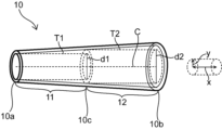
図1~図5に示すように、血管カバー10は、動脈3と、又は人工血管5と吻合されている静脈4の外周側に配置される筒状のカバーであって、血管カバー10の軸方向xにおいて、血管カバー10の一方端10aと他方端10bとの中点10cから一方端10aまでの部分を第1部11、中点10cから他方端10bまでの部分を第2部12としたとき、血管カバー10の中心軸Cと平行な中心軸を有し第2部12の内壁に内接する第2仮想円柱T2の直径d2は血管カバー10の中心軸Cと平行な中心軸を有し第1部11の内壁に当接する第1仮想円柱T1の直径d1よりも大きい。
As shown in FIGS. 1 to 5, the vessel cover 10 is a tubular cover arranged on the outer peripheral side of the vein 4 anastomosed with the artery 3 or the artificial blood vessel 5. In the direction x, the portion from the midpoint 10c between the one end 10a and the other end 10b of the vessel cover 10 to the one end 10a is the first portion 11, and the portion from the midpoint 10c to the other end 10b is the second portion 12. Then, the diameter d2 of the second virtual cylinder T2, which has a central axis parallel to the central axis C of the vessel cover 10 and is inscribed in the inner wall of the second part 12, has a central axis parallel to the central axis C of the vessel cover 10. It is larger than the diameter d1 of the first imaginary cylinder T1 that contacts the inner wall of the first portion 11 .
上記構成を有することにより、血管カバー10の第1部11の内径の最小値と比べて、第2部12の内径の最小値を大きくすることができる。このような血管カバー10の第1部11を吻合部6において吻合された静脈4の上流側に配置し第2部12を下流側に配置することにより、静脈4の上流側よりも下流側を緩く被覆することができる。これにより、静脈4に流入した高い動脈圧を有する拍動性の血流が下流に向けて徐々に緩衝され最終的に静脈血流に移行できる緩衝系血管となるように、静脈4をリモデリングすることができる。その結果、血液乱流や静脈壁の脈動変化が抑制され、内膜肥厚等の病変を防止できる。
With the above configuration, the minimum inner diameter of the second portion 12 can be made larger than the minimum inner diameter of the first portion 11 of the vessel cover 10 . By arranging the first part 11 of such a vascular cover 10 upstream of the anastomosed vein 4 at the anastomosis part 6 and arranging the second part 12 downstream, the vein 4 is more downstream than the upstream side. Can be loosely covered. As a result, the vein 4 is remodeled so that the pulsatile blood flow flowing into the vein 4 and having high arterial pressure is gradually buffered toward the downstream and finally becomes a buffer system blood vessel that can shift to the venous blood flow. can do. As a result, blood turbulence and pulsating changes in the vein wall are suppressed, and lesions such as intimal hyperplasia can be prevented.
より詳細には、シャント造設部1の静脈4の壁に通常静脈の平滑筋層よりも厚く弾性線維層を含む平滑筋層と、その外側に該平滑筋層よりも厚いコラーゲン線維を含む弾性線維層からなる2層構造を傾斜的に形成することにより、シャント造設部1の静脈4を緩衝系血管にリモデリングすることができる。
More specifically, the wall of the vein 4 of the shunt-constructed portion 1 has a smooth muscle layer that is thicker than the smooth muscle layer of the normal vein and includes an elastic fiber layer, and an elastic layer that includes collagen fibers that are thicker than the smooth muscle layer on the outside of the smooth muscle layer. By forming a two-layer structure composed of fibrous layers at an angle, the vein 4 of the shunt-constructed part 1 can be remodeled into a buffer system blood vessel.
上記緩衝系血管と通常の動脈との違いを説明する。血管は、内膜、中膜、外膜の三層から構成される。その中で、内膜は抗凝固性には大きく寄与するが力学的寄与は極めて小さい。人工透析に用いられる通常の動脈である四肢の動脈の力学的要素の構成は、若干の弾性線維と豊富な平滑筋を含む中膜、その外側の弾性線維とコラーゲン線維等からなる外膜が大きな割合を占めている。つまりこれら動脈は、非常に豊富な平滑筋と比較的少ない弾性線維を有している(「平滑筋>弾性線維」の構成)。弾性線維は、その弾力性により、動脈の拍動性で高い血圧に抵抗して緩和するゴム管のような緩衝機能を持つ。一方平滑筋は、筋肉であるため、より能動的な力学的機能をも持ち、動脈血圧に抵抗しつつ、他方では高い拍動性の動脈血圧を減衰させることなく末梢まで送達するという積極的・能動的な機能を有する。この動脈の豊富な平滑筋の圧送達機能のゆえに、内径がセンチメートル単位の大動脈でも、また、1ミリの数分の一の小動脈でも、その血圧はほとんど変わらない。すなわち、通常の動脈(「平滑筋>弾性線維」の構成)は、豊富な平滑筋の働きにより、拍動性で高い動脈圧を緩衝する機能は持たない。一方、シャント造設部の静脈が緩衝系血管にリモデリングされた状態では、弾性線維と平滑筋の割合は通常動脈とは逆で、弾性線維が豊富で平滑筋は比較的薄い(「弾性線維>平滑筋」の構成)。そのため緩衝系血管では、圧送達機能より圧緩衝機能が優位である。緩衝系血管では、上記の「弾性線維>平滑筋」の構成を保ちつつ、すなわち緩衝機能を保ちつつ、全体が徐々に薄くなり静脈へ移行、すなわち緩衝された圧の低下に従って徐々に通常静脈へ移行する。もし仮に、静脈壁が通常の動脈様に変化すれば、それは動脈化(動脈へのリモデリング)であって緩衝系血管へのリモデリングではない。その動脈化が徐々に弱くなり下流で全く通常の静脈へと自然に移行するならば、その「平滑筋>弾性線維」の構成のまま徐々に薄くなって静脈へ移行することを意味することから、緩衝機能を持たないため高い拍動性血圧が静脈壁に作用して下流側の静脈の病的な変化を引き起こす結果になるわけである。この点が、緩衝系血管が徐々に薄くなり通常静脈へ移行する場合と、通常の動脈様に変化した血管が徐々に薄くなり通常静脈へ移行する場合との明確な機能的な違いである。
Explain the difference between the above buffer system blood vessels and normal arteries. Blood vessels are composed of three layers: the intima, the media, and the adventitia. Among them, the intima greatly contributes to anticoagulation, but its mechanical contribution is extremely small. The mechanical elements of the arteries of the extremities, which are normal arteries used for artificial dialysis, consist of the media, which contains a small amount of elastic fibers and abundant smooth muscle, and the outer membrane, which consists of elastic fibers and collagen fibers. account for the percentage. Thus, these arteries have a great abundance of smooth muscle and relatively few elastic fibers (“smooth muscle>elastic fiber” configuration). Due to their elasticity, elastic fibers have a cushioning function like a rubber tube that resists and cushions the pulsatile and high blood pressure of arteries. On the other hand, since smooth muscle is a muscle, it also has a more active mechanical function, resisting arterial blood pressure, while on the other hand delivering high pulsatile arterial blood pressure to the periphery without attenuation. It has an active function. Due to the pressure-transmitting function of the abundant smooth muscle of the arteries, the blood pressure of the aorta, which has an inner diameter on the order of a centimeter, and the small artery, which has an inner diameter of only a fraction of a millimeter, hardly changes. That is, normal arteries (composition of "smooth muscle>elastic fiber") do not have the function of buffering pulsatile and high arterial pressure due to the action of abundant smooth muscle. On the other hand, when the vein at the site of the shunt is remodeled into a buffer system vessel, the ratio of elastic fibers to smooth muscle is opposite to that of normal arteries. >smooth muscle” composition). Therefore, in a buffer system blood vessel, the pressure buffer function is superior to the pressure delivery function. In the buffer system blood vessel, while maintaining the above-mentioned structure of "elastic fiber>smooth muscle", that is, while maintaining the buffer function, the whole gradually becomes thinner and moves to the vein, that is, gradually to the normal vein as the buffered pressure decreases Transition. If the venous wall changes like a normal artery, it is arterialization (remodeling into an artery) and not into a buffer system vessel. If the arterialization gradually weakens and naturally transitions downstream to a completely normal vein, it means that it gradually thins and transitions to the vein with its "smooth muscle > elastic fiber" configuration. , high pulsatile blood pressure acts on the venous wall due to its lack of buffering function, resulting in pathological changes in downstream veins. This point is a clear functional difference between the case where the buffer system blood vessel gradually thins and shifts to a normal vein, and the case where a normal artery-like blood vessel gradually thins and shifts to a normal vein.
本発明の実施形態に係る血管カバー10は、上記構成を有することにより、シャント造設部1の静脈4の下流側をより緩く被覆することにより、静脈4の壁構造を上記のように変化させて緩衝系血管へリモデリングすることができる。
The vascular cover 10 according to the embodiment of the present invention has the above-described configuration, so that the downstream side of the vein 4 of the shunt-constructing portion 1 is more loosely covered, thereby changing the wall structure of the vein 4 as described above. can be remodeled into a buffer system of blood vessels.
さらに、静脈4は緩衝系血管にリモデリングされる過程で徐々に外側に成長することがあるが、本発明の実施形態に係る血管カバー10は静脈4の下流側をより緩く被覆することができるため、血管カバー10による静脈4の成長の阻害が低減され、静脈4の内腔を広く維持して十分な血流量を確保することができる。
Furthermore, the vein 4 may gradually grow outward in the course of being remodeled into a buffer system vessel, but the vessel cover 10 according to the embodiment of the present invention can more loosely cover the downstream side of the vein 4. Therefore, inhibition of the growth of the vein 4 by the vascular cover 10 is reduced, and the lumen of the vein 4 can be kept wide to ensure a sufficient blood flow.
これにより、本発明の実施形態に係る血管カバー10によれば、内膜肥厚等の病変を抑えつつ十分な血流量を確保できるシャント造設部1を形成することが可能となる。
As a result, according to the vascular cover 10 according to the embodiment of the present invention, it is possible to form the shunt-constructed portion 1 that can ensure a sufficient blood flow while suppressing lesions such as intimal hyperplasia.
図示していないが、動脈3及び人工血管5のいずれに吻合される場合であっても、血管カバー10は、静脈4のみならず吻合部6側の動脈3及び人工血管5の一部に亘って被覆するように配置されてもよい。
Although not shown, regardless of whether the artery 3 or the artificial blood vessel 5 is to be anastomosed, the blood vessel cover 10 covers not only the vein 4 but also a part of the artery 3 and the artificial blood vessel 5 on the anastomosis 6 side. may be arranged to cover the surface.
血管カバー10は筒状であればよく、例えば平板状の部材を筒状に丸めて縫合により接合して形成する等、接合部を有していてもよい。その場合、縫合部等の接合部は血管カバー10の外側面に形成されることが好ましい。これにより、接合部が静脈4に影響することを防止できる。或いは、成形部材や編地を用いることにより接合部を有さないシームレスな筒状部材としてもよい。
The blood vessel cover 10 may have a tubular shape, and may have a joint portion, for example, formed by rolling a flat plate member into a tubular shape and joining them by suturing. In that case, it is preferable that the joint portion such as the suture portion is formed on the outer surface of the vessel cover 10 . This prevents the joint from affecting the vein 4 . Alternatively, a seamless cylindrical member having no joints may be formed by using a molded member or knitted fabric.
図3に示すように、第1仮想円柱T1は血管カバー10の一方端10aにおいて第1部11の内壁に内接しており、一方端10a以外の部分においては第1部11の内壁に内接していなくてもよい。すなわち、血管カバー10の第1部11は、一方端10aにおいて内径d1を有しており、一方端10a以外の部分においてはd1よりも大きい内径を有していてもよい。また、図3に示すように、第2仮想円柱T2は血管カバー10の中点10cにおいて第2部12の内壁に内接しており、中点10c以外の部分においては第2部12の内壁に内接していなくてもよい。すなわち、血管カバー10の第2部12は、中点10cにおいて内径d2を有しており、中点10c以外の部分においてはd2よりも大きい内径を有していてもよい。このような血管カバー10の形状の例としては、一方端10aから他方端10bにかけて内径が漸増するテーパー形状が挙げられる。
As shown in FIG. 3, the first virtual cylinder T1 is inscribed in the inner wall of the first portion 11 at one end 10a of the vessel cover 10, and is inscribed in the inner wall of the first portion 11 at portions other than the one end 10a. It doesn't have to be. That is, the first portion 11 of the vessel cover 10 has an inner diameter d1 at the one end 10a, and may have an inner diameter larger than d1 at portions other than the one end 10a. Further, as shown in FIG. 3, the second virtual cylinder T2 is inscribed in the inner wall of the second portion 12 at the midpoint 10c of the blood vessel cover 10, and is in contact with the inner wall of the second portion 12 at portions other than the midpoint 10c. It does not have to be inscribed. That is, the second portion 12 of the vessel cover 10 has an inner diameter d2 at the midpoint 10c, and may have an inner diameter larger than d2 at portions other than the midpoint 10c. An example of such a shape of the vessel cover 10 is a tapered shape in which the inner diameter gradually increases from one end 10a to the other end 10b.
第1部11の一方端10a以外の部分における内径がd1よりも大きい場合であっても、第1部11の軸方向x全体にわたって第1部11の内径は第2部12の内径の最小値と同じか、又は第2部12の内径の最小値よりも小さいことが好ましい。
Even if the inner diameter of the portion other than the one end 10a of the first portion 11 is larger than d1, the inner diameter of the first portion 11 is the minimum value of the inner diameter of the second portion 12 throughout the axial direction x of the first portion 11. or smaller than the minimum inner diameter of the second portion 12 .
或いは、図4に示すように、第1仮想円柱T1は軸方向xにおいて血管カバー10の一方端10aから所定位置までの部分において第1部11の内壁に内接しており、一方端10aを含む当該部分以外の部分においては第1部11の内壁に内接していなくてもよい。すなわち、血管カバー10の第1部11は、血管カバー10の一方端10aから所定位置までの部分において内径d1を有しており、それ以外の部分においてはd1よりも大きい内径を有していてもよい。
Alternatively, as shown in FIG. 4, the first imaginary column T1 is inscribed in the inner wall of the first portion 11 at a portion from one end 10a of the vessel cover 10 to a predetermined position in the axial direction x, and includes the one end 10a. Parts other than this part may not be inscribed in the inner wall of the first part 11 . That is, the first portion 11 of the vascular cover 10 has an inner diameter d1 in a portion from the one end 10a of the vascular cover 10 to a predetermined position, and has an inner diameter larger than d1 in other portions. good too.
これらの形状であれば、血管カバー10の一方端10aをシャント造設部1の静脈4の上流側に配置した際に血管カバー10の内径が最も小さい部分で静脈4の吻合部6側の最上流部を覆うことができ、静脈4の下流に行くに従って内径が徐々に大きくなる血管カバー10で覆うことができるため、静脈4の下流に行くに従って緩く被覆することができ、静脈4の緩衝系血管へのリモデリングや静脈4の内腔径の確保が容易となる。
With these shapes, when the one end 10a of the vascular cover 10 is arranged upstream of the vein 4 of the shunt-constructed portion 1, the portion of the vascular cover 10 with the smallest inner diameter is the largest on the anastomosis portion 6 side of the vein 4. The upstream part can be covered, and since it can be covered with the vessel cover 10 whose inner diameter gradually increases as it goes downstream of the vein 4, it can be loosely covered as it goes downstream of the vein 4, and the buffer system of the vein 4 Remodeling into a blood vessel and securing the lumen diameter of the vein 4 are facilitated.
或いは、図5に示すように、第1仮想円柱T1は軸方向xにおいて血管カバー10の一方端10aと中点10cとの間のいずれかの位置で第1部11の内壁に内接しており、当該位置以外の部分においては第1部11の内壁に内接していなくてもよい。すなわち、血管カバー10の第1部11は、血管カバー10の一方端10aと中点10cとの間の位置において内径d1を有しており、それ以外の部分においてはd1よりも大きい内径を有していてもよい。
Alternatively, as shown in FIG. 5, the first imaginary cylinder T1 is inscribed in the inner wall of the first portion 11 at any position between the one end 10a and the midpoint 10c of the vessel cover 10 in the axial direction x. , the portion other than the position may not be inscribed in the inner wall of the first portion 11 . That is, the first portion 11 of the vessel cover 10 has an inner diameter d1 at a position between the one end 10a and the midpoint 10c of the vessel cover 10, and has an inner diameter larger than d1 at other portions. You may have
上記いずれの場合であっても、第1部11の軸方向x全体にわたって第1部11の内径は第2部12の内径の最小値と同じか、又は第2部12の内径の最小値よりも小さいことが好ましい。
In any of the above cases, the inner diameter of the first portion 11 over the entire axial direction x of the first portion 11 is the same as the minimum inner diameter of the second portion 12, or is larger than the minimum inner diameter of the second portion 12. is preferably small.
或いは、図示していないが、第1仮想円柱T1は軸方向xにおいて血管カバー10の一方端10aから中点10cまでの区間全体にわたって第1部11の内壁に内接していてもよい。すなわち、血管カバー10の第1部11が軸方向x全体にわたって内径d1を有していてもよい。
Alternatively, although not shown, the first virtual cylinder T1 may be inscribed in the inner wall of the first portion 11 over the entire section from the one end 10a of the vessel cover 10 to the midpoint 10c in the axial direction x. That is, the first portion 11 of the vessel cover 10 may have the inner diameter d1 over the entire axial direction x.
第2仮想円柱T2は、血管カバー10の中点10c以外の部分で第2部12の内壁に内接していてもよい。第2仮想円柱T2が内接している部分以外においては、第2部12はd2よりも大きい内径を有していてもよい。このような場合であっても、第2部12の内径は軸方向x全体にわたって第2仮想円柱T2の直径d2の2.5倍以下であることが好ましく、2倍以下がより好ましく、1.5倍以下がさらに好ましい。また、他方端10bにおける第2部12の内径は中点10cにおける第2部12の内径よりも大きいことが好ましい。
The second virtual cylinder T2 may be inscribed in the inner wall of the second part 12 at a portion other than the midpoint 10c of the blood vessel cover 10. The second portion 12 may have an inner diameter larger than d2 except for the portion where the second virtual cylinder T2 is inscribed. Even in such a case, the inner diameter of the second portion 12 is preferably 2.5 times or less, more preferably 2 times or less, the diameter d2 of the second imaginary cylinder T2 throughout the axial direction x. 5 times or less is more preferable. Also, the inner diameter of the second portion 12 at the other end 10b is preferably larger than the inner diameter of the second portion 12 at the midpoint 10c.
或いは、図示していないが、第2仮想円柱T2は軸方向xにおいて血管カバー10の中点10cから他方端10bまでの区間全体にわたって第2部12の内壁に内接していてもよい。すなわち、血管カバー10の第2部12が軸方向x全体にわたって内径d2を有していてもよい。
Alternatively, although not shown, the second virtual cylinder T2 may be inscribed in the inner wall of the second portion 12 over the entire section from the midpoint 10c of the vessel cover 10 to the other end 10b in the axial direction x. That is, the second portion 12 of the vessel cover 10 may have an inner diameter d2 along the entire axial direction x.
上記のいずれの態様においても、第2仮想円柱T2の直径d2が第1仮想円柱T1の直径d1よりも大きいことにより、血管カバー10の一方端10aをシャント造設部1の静脈4の上流側に配置した際に静脈4の下流をより緩く被覆することができ、上記効果を奏することができる。
In any of the above modes, the diameter d2 of the second virtual column T2 is larger than the diameter d1 of the first virtual column T1, so that the one end 10a of the vessel cover 10 is positioned upstream of the vein 4 of the shunt-constructing portion 1. When it is placed in the inner wall of the vein 4, the downstream side of the vein 4 can be more loosely covered, and the above effect can be obtained.
図6に、他の実施形態に係る血管カバー10の斜視図を示す。本態様の血管カバー10は、血管カバー10の軸方向xにおいて血管カバー10の一方端10aから第1部11の中点までの区間であって第1内径d5を有している一方端部110と、一方端部110よりも他方端10b側に位置しており第1内径d5の1.2倍以上の第2内径d6を有している中間部120と、第2内径d6の1.2倍以上の第3内径d7を有している他方端10bと、一方端部110と中間部120との間に内径が漸増する第1移行部110mと、中間部120と他方端10bとの間に内径が漸増する第2移行部120mと、を有していることが好ましい。
FIG. 6 shows a perspective view of a blood vessel cover 10 according to another embodiment. The blood vessel cover 10 of this aspect has a one end portion 110 which is a section from the one end 10a of the blood vessel cover 10 to the midpoint of the first portion 11 in the axial direction x of the blood vessel cover 10 and has a first inner diameter d5. and an intermediate portion 120 located closer to the other end 10b than the one end 110 and having a second inner diameter d6 that is 1.2 times or more the first inner diameter d5, and an intermediate portion 120 that is 1.2 times the second inner diameter d6 The other end 10b having a third inner diameter d7 that is twice or more, the first transition portion 110m having a gradually increasing inner diameter between the one end 110 and the intermediate portion 120, and the intermediate portion 120 and the other end 10b and a second transition portion 120m of gradually increasing inner diameter.
このような構成の血管カバー10の一方端部110をシャント造設部1の静脈4の上流側に配置することにより、静脈4が緩衝系血管へリモデリングされる過程で静脈4が下流側に行くに従い径方向yの外側にゆっくり広がることができるため、静脈4の緩衝系血管へのリモデリングが容易となり、静脈4の内腔を維持して血流量を確保することが容易となる。また、血管カバー10の他方端10bは静脈4に対する血管カバー10による抑制が急激に解除される部分であるが、この部分の内径d7が中間部120に対して上記関係を有していることにより、静脈4に対する血管カバー10の被覆による抑制の急激な解除の影響を緩和することが可能となる。
By arranging the one end 110 of the vascular cover 10 having such a configuration upstream of the vein 4 of the shunt-constructed portion 1, the vein 4 moves downstream in the process of remodeling the vein 4 into a buffer system vessel. Since it can spread slowly outward in the radial direction y as it goes, remodeling of the vein 4 into a buffer system vessel is facilitated, and the lumen of the vein 4 is easily maintained to ensure the blood flow. The other end 10b of the vessel cover 10 is a portion where the suppression of the vein 4 by the vessel cover 10 is abruptly released. , the influence of abrupt release of the suppression by the covering of the vessel cover 10 on the vein 4 can be mitigated.
上記の構成を有する場合、軸方向xの断面において、一方端部110と第1移行部110mとの境界、第1移行部110mと中間部120との境界、及び中間部120と第2移行部120mとの境界は曲線であることが好ましい。一方端部110と中間部120と他方端10bはそれぞれ1.2倍以上ずつ異なる内径を有しているため、軸方向xにおいて血管カバー10の内壁は軸方向xに段差を有することとなるが、上記のようにそれぞれの境界が曲線であることにより、段差を滑らかとすることができる。これにより、滑らかな内腔壁を有する血管カバー10で血管を被覆することができ、緩衝系血管へのリモデリングがより容易となる。
When having the above configuration, in the cross section in the axial direction x, the boundary between the one end portion 110 and the first transition portion 110m, the boundary between the first transition portion 110m and the intermediate portion 120, and the intermediate portion 120 and the second transition portion The boundary with 120m is preferably curved. Since the one end portion 110, the intermediate portion 120, and the other end 10b have different inner diameters by a factor of 1.2 or more, the inner wall of the vessel cover 10 has a step in the axial direction x. , the steps can be smoothed by forming the respective boundaries as curved lines as described above. As a result, the blood vessel can be covered with the vessel cover 10 having a smooth lumen wall, and remodeling into a buffer system vessel is facilitated.
第2部12の内壁を自然状態から径方向yに1.5倍拡張させるのに必要な力は、第1部11の内壁を自然状態から径方向yに1.5倍拡張させるのに必要な力よりも小さいことが好ましい。
The force required to expand the inner wall of the second portion 12 by 1.5 times in the radial direction y from the natural state is required to expand the inner wall of the first portion 11 by 1.5 times in the radial direction y from the natural state. is preferably less than the force
第1部11の内壁を自然状態から径方向yに1.5倍拡張させる方法としては、例えば、血管カバー10の内腔に第1部11の内壁と内接するように樹脂チューブを挿入し、樹脂チューブの内腔に流体を導入して圧力をかける等の方法が挙げられる。同様にして、第2部12の内壁を自然状態から径方向yに1.5倍拡張させることができる。
As a method for expanding the inner wall of the first part 11 from the natural state by 1.5 times in the radial direction y, for example, a resin tube is inserted into the lumen of the vessel cover 10 so as to be inscribed with the inner wall of the first part 11, A method of applying pressure by introducing a fluid into the lumen of the resin tube can be used. Similarly, the inner wall of the second portion 12 can be expanded 1.5 times in the radial direction y from the natural state.
或いは、軸方向xに所定長さを有するように第1部11の一部分を切り出した筒状サンプルを用意し、当該筒状サンプルの内径に2本のピンを筒状サンプルの軸方向と平行に挿入し、筒状サンプルの内径が1.5倍となるように2本のピンを互いに径方向yの反対方向に牽引してもよい。このとき、同様にして第2部12の内壁を自然状態から径方向yに1.5倍拡張させることができるが、第2部12から切り出す筒状サンプルの軸方向xの長さは、第1部11から切り出す筒状サンプルの軸方向xの長さと同じとなるようにする。
Alternatively, a cylindrical sample is prepared by cutting out a portion of the first part 11 so as to have a predetermined length in the axial direction x, and two pins are attached to the inner diameter of the cylindrical sample in parallel with the axial direction of the cylindrical sample. The two pins may be inserted and pulled in opposite diametrical y directions so that the inner diameter of the cylindrical sample is 1.5 times larger. At this time, the inner wall of the second part 12 can be similarly expanded in the radial direction y by 1.5 times from the natural state, but the length of the cylindrical sample cut out from the second part 12 in the axial direction x is The length in the axial direction x of the cylindrical sample cut out from the first part 11 is set to be the same.
血管カバー10が上記の構成を有していれば、血管カバー10の第1部11をシャント造設部1の静脈4の上流側に配置した場合に、静脈4の下流側をより緩く被覆することがより容易となり、静脈4の緩衝系血管へのリモデリングや静脈4の内腔径の確保を効果的に行える。
If the vascular cover 10 has the above configuration, the downstream side of the vein 4 is more loosely covered when the first portion 11 of the vascular cover 10 is arranged upstream of the vein 4 of the shunt-constructing portion 1 . This makes it easier to remodel the vein 4 into a buffer system vessel and effectively secure the lumen diameter of the vein 4 .
血管カバー10の軸方向xの長さは5mm以上であることが好ましい。血管カバー10の軸方向xの長さは、10mm以上がより好ましく、20mm以上がさらに好ましく、30mm以上が特に好ましく、40mm以上であってもよい。また、血管カバー10の軸方向xの長さは、120mm以下が好ましく、100mm以下がより好ましく、90mm以下がさらに好ましい。血管カバー10の軸方向xの長さが上記範囲であれば、所定以上の長さの血管カバー10でシャント造設部1の静脈4を被覆することができ、静脈4を緩衝系血管にリモデリングすることが容易となる。
The length of the blood vessel cover 10 in the axial direction x is preferably 5 mm or more. The length of the blood vessel cover 10 in the axial direction x is preferably 10 mm or longer, more preferably 20 mm or longer, particularly preferably 30 mm or longer, and may be 40 mm or longer. The length of the blood vessel cover 10 in the axial direction x is preferably 120 mm or less, more preferably 100 mm or less, and even more preferably 90 mm or less. If the length of the vascular cover 10 in the axial direction x is within the above range, the vein 4 of the shunt-constructed portion 1 can be covered with the vascular cover 10 having a length greater than or equal to the predetermined length, and the vein 4 can be reconnected to the buffer system blood vessel. Easier to model.
血管カバー10の内径は、第1部11において内径の最小値を有する部分、すなわち第1仮想円柱T1が内接する部分において、2mm以上が好ましく、3mm以上がより好ましく、4mm以上がさらに好ましく、5mm以上が特に好ましく、また、10mm以下が好ましく、8mm以下がより好ましく、6mm以下がさらに好ましい。また、血管カバー10の内径は、第2部12において内径の最小値を有する部分、すなわち第2仮想円柱T2が内接する部分において、4mm以上が好ましく、5mm以上がより好ましく、6mm以上がさらに好ましく、7mm以上が特に好ましく、また、12mm以下が好ましく、10mm以下がより好ましく、9mm以下がさらに好ましく、8mm以下が特に好ましい。血管カバー10を構成する材料や血管カバー10の構造によっては、血管カバー10の内壁に細かな凹凸が生じて内径を決定するのが困難となる場合があるが、この場合は第1部11に内接する第1仮想円柱T1の直径d1や第2部12に内接する第2仮想円柱T2の直径d2を血管カバー10の内径とすることができる。
The inner diameter of the vessel cover 10 is preferably 2 mm or more, more preferably 3 mm or more, even more preferably 4 mm or more, further preferably 5 mm at the portion having the minimum inner diameter in the first portion 11, that is, the portion inscribed with the first virtual cylinder T1. The above is particularly preferable, and 10 mm or less is preferable, 8 mm or less is more preferable, and 6 mm or less is even more preferable. In addition, the inner diameter of the vessel cover 10 is preferably 4 mm or more, more preferably 5 mm or more, and even more preferably 6 mm or more at the portion having the minimum inner diameter in the second portion 12, that is, the portion inscribed with the second virtual cylinder T2. , 7 mm or more is particularly preferable, 12 mm or less is preferable, 10 mm or less is more preferable, 9 mm or less is even more preferable, and 8 mm or less is particularly preferable. Depending on the material forming the vascular cover 10 and the structure of the vascular cover 10, fine unevenness may occur on the inner wall of the vascular cover 10, making it difficult to determine the inner diameter. The diameter d1 of the inscribed first imaginary cylinder T1 and the diameter d2 of the second imaginary cylinder T2 inscribed in the second part 12 can be used as the inner diameter of the vessel cover .
血管カバー10は、編物、織物、不織布のいずれか少なくとも一つを、部分的に構成する成分として、又は全体を構成する成分として有していることが好ましい。これらの素材であれば、弾性変形する血管カバー10を形成することが容易である。
The vascular cover 10 preferably has at least one of a knitted fabric, a woven fabric, and a nonwoven fabric as a component that partially configures it or as a component that configures the whole. With these materials, it is easy to form the elastically deformable blood vessel cover 10 .
編物の種類は特に限定されず、経編であっても緯編であってもよい。経編の編組織としては、ハーフ編、バックハーフ編、クインズコート編、サテン編等が挙げられる。緯編には丸編や横編が含まれ、緯編の編組織としては、平編、ゴム編、両面編、ミラノリブ編、ジャガード編等が挙げられる。伸縮性に優れる点から、編物は緯編から構成されることが好ましい。織物の種類は特に限定されず、平織、綾織、朱子織等であってもよい。或いは、血管カバー10は、メルトブロー法、ニードルパンチ法、スパンレース法、エレクトロスピニング法等の任意の方法で作製された不織布で構成されていてもよい。
The type of knitted fabric is not particularly limited, and may be warp knitted or weft knitted. Knitting structures of warp knitting include half knitting, back half knitting, queens coat knitting, and satin knitting. Weft knitting includes circular knitting and flat knitting, and knitting structures of weft knitting include plain knitting, rubber knitting, double-sided knitting, milanese rib knitting, and jacquard knitting. The knitted fabric is preferably composed of weft knitting from the viewpoint of excellent stretchability. The type of woven fabric is not particularly limited, and may be plain weave, twill weave, satin weave, or the like. Alternatively, the blood vessel cover 10 may be composed of a nonwoven fabric produced by any method such as meltblowing, needle punching, spunlacing, electrospinning, or the like.
血管カバー10は、例えば一部が編物で構成されており、それ以外の部分が他の材料、例えば不織布で構成されているなど、2以上の異なる素材の組合せで構成されていてもよい。
The blood vessel cover 10 may be composed of a combination of two or more different materials, for example, one part is composed of a knitted fabric and the other part is composed of another material such as a non-woven fabric.
シャントカバーの壁の性質は塑性変形が優位な方が弾性変形が優位であるよりも緩衝機能に優れる。そのため、編物や織物、不織布を形成する糸も、塑性変形が優位な樹脂材料から構成されていることがより好ましく、例えば、ポリエチレン、ポリプロピレン等のポリオレフィン系樹脂;ナイロン等のポリアミド系樹脂;ポリエチレンテレフタレート等のポリエステル系樹脂;ポリイミド系樹脂;PTFE、PFA、ETFE等のフッ素系樹脂;ポリ塩化ビニル系樹脂等の合成樹脂等を素材とし、塑性変形が優位な糸が挙げられる。編物や織物を形成する糸は、人工血管に使用される樹脂材料(例えば、ポリエステル、PTFE)から構成することもでき、具体的には、PTFEを延伸加工したePTFE、デュポン社のポリエステル繊維であるダクロン(登録商標)等が挙げられる。また、血管カバー10は生分解性材料から構成されていてもよく、例えば、ポリ乳酸、ポリグリコール酸、ポリヒドロキシアルカン酸等の脂肪族ポリエステル;脂肪族ポリエーテル等が挙げられる。さらに、編物や織物を形成する糸は、シルクやコットンなどの天然繊維から構成されていてもよく、また、樹脂材料、生分解性材料、天然繊維の組合せから構成されていてもよい。
The properties of the wall of the shunt cover are better in cushioning function when plastic deformation is dominant than when elastic deformation is dominant. Therefore, it is more preferable that yarns forming knitted fabrics, woven fabrics, and non-woven fabrics are also made of resin materials with superior plastic deformation, such as polyolefin resins such as polyethylene and polypropylene; polyamide resins such as nylon; polyethylene terephthalate. polyimide resins; fluorine resins such as PTFE, PFA and ETFE; synthetic resins such as polyvinyl chloride resins. The yarn forming the knitted fabric or woven fabric can also be made of a resin material (e.g., polyester, PTFE) used for artificial blood vessels, and specifically, ePTFE obtained by stretching PTFE and polyester fiber from DuPont. Dacron (registered trademark) and the like can be mentioned. Also, the vessel cover 10 may be made of a biodegradable material such as aliphatic polyester such as polylactic acid, polyglycolic acid, and polyhydroxyalkanoic acid; aliphatic polyether. Furthermore, the yarn forming the knitted or woven fabric may be composed of natural fibers such as silk or cotton, or may be composed of a combination of resin materials, biodegradable materials, and natural fibers.
図7に示すように、血管カバー10は、軸方向xに山と谷とを周期的に繰り返す蛇腹構造を有しており、血管カバー10の一方端10aから第1部11の中点までの部分を一方端部110としたとき、軸方向xにおいて、第2部12における隣り合う山間の距離L2は一方端部110における山間の距離L1よりも大きいことが好ましい。蛇腹構造の山間の距離が一方端部110において相対的に小さく第2部12において相対的に大きいことで、一方端部110をシャント造設部1の静脈4の上流側に配置した際に、静脈4の下流をより被覆することが容易となり、静脈4の緩衝系血管へのリモデリングや静脈4の内腔径の確保がより容易となる。
As shown in FIG. 7, the blood vessel cover 10 has a bellows structure in which peaks and troughs are periodically repeated in the axial direction x. When the portion is the one end portion 110, it is preferable that the distance L2 between adjacent crests in the second portion 12 is greater than the distance L1 between crests in the one end portion 110 in the axial direction x. Since the distance between the peaks of the bellows structure is relatively small at the one end 110 and relatively large at the second portion 12, when the one end 110 is arranged upstream of the vein 4 of the shunt-forming portion 1, It becomes easier to cover the downstream side of the vein 4 , and it becomes easier to remodel the vein 4 into a buffer system vessel and secure the lumen diameter of the vein 4 .
静脈4が低圧緩衝系血管にリモデンリングされているかどうかの評価は、以下の述べる形態学的確認法と緩衝作用の測定による確認法との両方で確認されることが必要である。形態学的方法としては、形態学的弾性線維を含む平滑筋層と、その外側に該平滑筋層よりも厚いコラーゲン線維を含む弾性線維からなる2層構造が形成されているかを確認することで行うことができる。具体的には、シャント造設部1の静脈4を切り出し、ヘマトキシリン・エオジン(HE)染色及びエラスチカ・ファンギーソン(EvG)染色等の特殊染色を施して、静脈壁断面を顕微鏡で観察する。例えば、EvG染色では、平滑筋は濁黄に、弾性線維は濃紫に、コラーゲン線維は暗赤に染色されるため、弾性線維を含む平滑筋層及びコラーゲン線維を含む弾性線維層を観察し、「弾性線維を含む平滑筋層の厚さ<コラーゲン線維を含む弾性線維層の厚さ」を確認することで行うことができる。
The evaluation of whether or not the vein 4 is remodeled into the low-pressure buffer system blood vessel needs to be confirmed by both the morphological confirmation method described below and the confirmation method by measuring the buffering action. As a morphological method, it is confirmed whether a two-layer structure consisting of a smooth muscle layer containing morphological elastic fibers and elastic fibers containing collagen fibers thicker than the smooth muscle layer is formed. It can be carried out. Specifically, the vein 4 of the shunt-forming portion 1 is cut out, subjected to special staining such as hematoxylin-eosin (HE) staining and elastica-fungieson (EvG) staining, and the cross section of the vein wall is observed with a microscope. For example, in EvG staining, smooth muscle is stained turbid yellow, elastic fibers are dark purple, and collagen fibers are stained dark red, so the smooth muscle layer containing elastic fibers and the elastic fiber layer containing collagen fibers are observed, This can be done by confirming "thickness of smooth muscle layer containing elastic fibers<thickness of elastic fiber layer containing collagen fibers".
また、静脈4が緩衝系血管にリモデリングされているかどうかの緩衝作用の測定による確認法としては、ドップラー血流測定及びカラードップラー超音波画像診断装置による血流測定により確認することができる。
In addition, as a method for confirming whether or not the vein 4 is remodeled into a buffer system blood vessel by measuring the buffer effect, it can be confirmed by Doppler blood flow measurement and blood flow measurement using a color Doppler ultrasonic imaging apparatus.
本願は、2021年9月8日に出願された日本国特許出願第2021-146521号に基づく優先権の利益を主張するものである。2021年9月8日に出願された日本国特許出願第2021-146521号の明細書の全内容が、本願に参考のため援用される。
This application claims the benefit of priority based on Japanese Patent Application No. 2021-146521 filed on September 8, 2021. The entire contents of the specification of Japanese Patent Application No. 2021-146521 filed on September 8, 2021 are incorporated herein by reference.
1:シャント造設部
2:腕
3:動脈
4:静脈
5:人工血管
6:吻合部
10:血管カバー
10a:血管カバーの一方端
10b:血管カバーの他方端
10c:血管カバーの中点
11:第1部
12:第2部
110:一方端部
110m:第1移行部
120:中間部
120m:第2移行部
C:血管カバーの中心軸
d1:第1仮想円柱の直径
d2:第2仮想円柱の直径
d5:一方端部の内径
d6:中間部の内径
d7:他方端の内径
L1:一方端部における山間の距離
L2:第2部における山間の距離
T1:第1仮想円柱
T2:第2仮想円柱
x:軸方向
y:径方向 1: shunt-forming part 2: arm 3: artery 4: vein 5: artificial blood vessel 6: anastomotic part 10:vessel cover 10a: one end 10b of vessel cover: the other end 10c of vessel cover: middle point 11 of vessel cover: First part 12: Second part 110: One end 110m: First transition part 120: Middle part 120m: Second transition part C: Central axis d1 of vessel cover: Diameter d2 of first imaginary cylinder: Second imaginary cylinder Diameter d5: Inside diameter d6 of one end: Inside diameter d7 of intermediate part: Inside diameter of the other end L1: Distance between mountains at one end L2: Distance between mountains at second part T1: First virtual cylinder T2: Second virtual Cylinder x: axial direction y: radial direction
2:腕
3:動脈
4:静脈
5:人工血管
6:吻合部
10:血管カバー
10a:血管カバーの一方端
10b:血管カバーの他方端
10c:血管カバーの中点
11:第1部
12:第2部
110:一方端部
110m:第1移行部
120:中間部
120m:第2移行部
C:血管カバーの中心軸
d1:第1仮想円柱の直径
d2:第2仮想円柱の直径
d5:一方端部の内径
d6:中間部の内径
d7:他方端の内径
L1:一方端部における山間の距離
L2:第2部における山間の距離
T1:第1仮想円柱
T2:第2仮想円柱
x:軸方向
y:径方向 1: shunt-forming part 2: arm 3: artery 4: vein 5: artificial blood vessel 6: anastomotic part 10:
Claims (7)
- 動脈と、又は人工血管と吻合されている静脈の外周側に配置される全周にわたって連続した筒状の血管カバーであって、前記血管カバーの軸方向において、前記血管カバーの一方端と他方端との中点から前記一方端までの部分を第1部、前記中点から前記他方端までの部分を第2部としたとき、前記血管カバーの中心軸と平行な中心軸を有し前記第2部の内壁に内接する第2仮想円柱の直径は前記血管カバーの中心軸と平行な中心軸を有し前記第1部の内壁に内接する第1仮想円柱の直径よりも大きい血管カバー。 A tubular vascular cover arranged on the outer peripheral side of a vein anastomosed with an artery or an artificial blood vessel and continuous over the entire circumference, wherein one end and the other end of the vascular cover are arranged in the axial direction of the vascular cover. When a portion from the middle point to the one end is defined as a first portion, and a portion from the middle point to the other end is defined as a second portion, the center axis is parallel to the center axis of the blood vessel cover and the second portion The diameter of the second virtual cylinder inscribed in the inner wall of the two parts is larger than the diameter of the first virtual cylinder inscribed in the inner wall of the first part having a central axis parallel to the central axis of the vessel cover.
- 前記血管カバーの軸方向において前記血管カバーの一方端から前記第1部の中点までの区間であって第1内径を有している一方端部と、前記一方端部よりも他方端側に位置しており前記第1内径の1.2倍以上の第2内径を有している中間部と、前記第2内径の1.2倍以上の第3内径を有している他方端と、前記一方端部と前記中間部との間に内径が漸増する第1移行部と、前記中間部と前記他方端との間に内径が漸増する第2移行部と、を有している請求項1に記載の血管カバー。 one end having a first inner diameter in a section from one end of the vascular cover to the midpoint of the first portion in the axial direction of the vascular cover; an intermediate portion located and having a second inner diameter that is 1.2 times or more the first inner diameter; and the other end having a third inner diameter that is 1.2 times or more the second inner diameter; 3. A first transition portion having a gradually increasing inner diameter between said one end and said intermediate portion, and a second transition portion having a gradually increasing inner diameter between said intermediate portion and said other end. 2. The vascular cover according to 1.
- 前記軸方向の断面において、前記一方端部と前記第1移行部との境界、前記第1移行部と前記中間部との境界、及び前記中間部と前記第2移行部との境界は曲線である請求項2に記載の血管カバー。 In the axial section, a boundary between the one end portion and the first transition portion, a boundary between the first transition portion and the intermediate portion, and a boundary between the intermediate portion and the second transition portion are curved lines. A vessel cover according to claim 2.
- 前記第2部の内壁を自然状態から径方向に1.5倍拡張させるのに必要な力は、前記第1部の内壁を自然状態から径方向に1.5倍拡張させるのに必要な力よりも小さい請求項1又は2に記載の血管カバー。 The force required to radially expand the inner wall of the second portion 1.5 times from the natural state is the force required to radially expand the inner wall of the first portion 1.5 times from the natural state. 3. A vessel cover according to claim 1 or 2 which is smaller than.
- 前記軸方向の長さが5mm以上である請求項1又は2に記載の血管カバー。 The blood vessel cover according to claim 1 or 2, wherein the length in the axial direction is 5 mm or more.
- 編物、織物、不織布のいずれか少なくとも1つを、部分的に構成する成分として、又は全体を構成する成分として有している請求項1又は2に記載の血管カバー。 The vascular cover according to claim 1 or 2, comprising at least one of a knitted fabric, a woven fabric, and a nonwoven fabric as a component that partially configures or a component that configures the whole.
- 前記軸方向に山と谷とを周期的に繰り返す蛇腹構造を有しており、前記血管カバーの一方端から前記第1部の中点までの部分を一方端部としたとき、前記軸方向において、前記第2部における隣り合う前記山間の距離は前記一方端部における前記山間の距離よりも大きい請求項1又は2に記載の血管カバー。
It has a bellows structure in which peaks and troughs are periodically repeated in the axial direction. 3. The vessel cover according to claim 1 or 2, wherein the distance between adjacent ridges on said second portion is greater than the distance between said ridges on said one end portion.
Applications Claiming Priority (2)
| Application Number | Priority Date | Filing Date | Title |
|---|---|---|---|
| JP2021-146521 | 2021-09-08 | ||
| JP2021146521 | 2021-09-08 |
Publications (1)
| Publication Number | Publication Date |
|---|---|
| WO2023037860A1 true WO2023037860A1 (en) | 2023-03-16 |
Family
ID=85506579
Family Applications (1)
| Application Number | Title | Priority Date | Filing Date |
|---|---|---|---|
| PCT/JP2022/031605 WO2023037860A1 (en) | 2021-09-08 | 2022-08-22 | Blood vessel cover |
Country Status (1)
| Country | Link |
|---|---|
| WO (1) | WO2023037860A1 (en) |
Citations (2)
| Publication number | Priority date | Publication date | Assignee | Title |
|---|---|---|---|---|
| WO2021161884A1 (en) * | 2020-02-14 | 2021-08-19 | 明郎 萩原 | Protective cover for vascular anastomotic site |
| WO2021177273A1 (en) * | 2020-03-03 | 2021-09-10 | 明郎 萩原 | Vein cover |
-
2022
- 2022-08-22 WO PCT/JP2022/031605 patent/WO2023037860A1/en active Application Filing
Patent Citations (2)
| Publication number | Priority date | Publication date | Assignee | Title |
|---|---|---|---|---|
| WO2021161884A1 (en) * | 2020-02-14 | 2021-08-19 | 明郎 萩原 | Protective cover for vascular anastomotic site |
| WO2021177273A1 (en) * | 2020-03-03 | 2021-09-10 | 明郎 萩原 | Vein cover |
Similar Documents
| Publication | Publication Date | Title |
|---|---|---|
| US6371981B1 (en) | Vascular graft assemblies and methods for implanting same | |
| EP1386590B1 (en) | Abrasion resistant vascular graft | |
| JP2939337B2 (en) | Three-dimensional braided soft tissue prosthesis | |
| CN102202608B (en) | Multi-layer device with gap for treating a target site and associated method | |
| JP4209054B2 (en) | Improved stent recaptured and repositioned | |
| WO2021177273A1 (en) | Vein cover | |
| CA2714053C (en) | Stent graft for reinforcement of vascular abnormalities and associated method | |
| US8088156B2 (en) | Graft material attachment device and method | |
| US7905915B2 (en) | Z-stent with incorporated barbs | |
| US20010049554A1 (en) | Endovascular prosthesis and method of making | |
| JP5253499B2 (en) | Artificial blood vessel | |
| CN103200975A (en) | Anastomotic devices and methods | |
| EP3351209B1 (en) | Graft anchor device | |
| JP2008534108A (en) | Vascular graft | |
| WO2021161884A1 (en) | Protective cover for vascular anastomotic site | |
| WO2009086458A1 (en) | Vascular graft prosthesis having a reinforced margin for enhanced anastomosis | |
| US8551153B2 (en) | Prosthesis comprising a coiled stent and method of use thereof | |
| WO2023037860A1 (en) | Blood vessel cover | |
| WO2023037859A1 (en) | Blood vessel cover | |
| EP1645245B1 (en) | Intracardiac device with sealable fenestration for total cavopulmonary anastomosis by catheterisation | |
| WO2023037861A1 (en) | Blood vessel cover | |
| JP3686833B2 (en) | Vascular anastomosis ring and artificial blood vessel equipped with vascular anastomosis ring | |
| US20220409359A1 (en) | Vein cover | |
| JP2022523568A (en) | Branched stent graft and its manufacturing method | |
| US20230149149A1 (en) | Bifurcated vascualr stent and methods of manufacture |
Legal Events
| Date | Code | Title | Description |
|---|---|---|---|
| 121 | Ep: the epo has been informed by wipo that ep was designated in this application |
Ref document number: 22867182 Country of ref document: EP Kind code of ref document: A1 |
|
| WWE | Wipo information: entry into national phase |
Ref document number: 2023546866 Country of ref document: JP |