WO2022270153A1 - Ultrasonic diagnostic device and method for controlling ultrasonic diagnostic device - Google Patents
Ultrasonic diagnostic device and method for controlling ultrasonic diagnostic device Download PDFInfo
- Publication number
- WO2022270153A1 WO2022270153A1 PCT/JP2022/018997 JP2022018997W WO2022270153A1 WO 2022270153 A1 WO2022270153 A1 WO 2022270153A1 JP 2022018997 W JP2022018997 W JP 2022018997W WO 2022270153 A1 WO2022270153 A1 WO 2022270153A1
- Authority
- WO
- WIPO (PCT)
- Prior art keywords
- stool
- degree
- emphasis
- region
- ultrasonic
- Prior art date
Links
- 238000000034 method Methods 0.000 title claims abstract description 35
- 238000001514 detection method Methods 0.000 claims abstract description 138
- 238000012545 processing Methods 0.000 claims abstract description 66
- 230000033001 locomotion Effects 0.000 claims description 291
- 238000002604 ultrasonography Methods 0.000 claims description 165
- 239000000523 sample Substances 0.000 claims description 77
- 230000002550 fecal effect Effects 0.000 claims description 24
- 238000013136 deep learning model Methods 0.000 claims description 17
- 230000008859 change Effects 0.000 claims description 16
- 230000008569 process Effects 0.000 claims description 16
- 238000007689 inspection Methods 0.000 claims description 5
- 238000010801 machine learning Methods 0.000 claims description 5
- 210000003608 fece Anatomy 0.000 description 30
- 230000005540 biological transmission Effects 0.000 description 12
- 238000010586 diagram Methods 0.000 description 12
- 238000012937 correction Methods 0.000 description 9
- 206010010774 Constipation Diseases 0.000 description 8
- 206010012735 Diarrhoea Diseases 0.000 description 5
- 238000011156 evaluation Methods 0.000 description 5
- 238000002834 transmittance Methods 0.000 description 5
- 238000006243 chemical reaction Methods 0.000 description 4
- 230000006870 function Effects 0.000 description 4
- 230000007423 decrease Effects 0.000 description 3
- 230000012447 hatching Effects 0.000 description 3
- 230000004044 response Effects 0.000 description 3
- FYYHWMGAXLPEAU-UHFFFAOYSA-N Magnesium Chemical compound [Mg] FYYHWMGAXLPEAU-UHFFFAOYSA-N 0.000 description 2
- 210000003484 anatomy Anatomy 0.000 description 2
- 238000013459 approach Methods 0.000 description 2
- 238000003745 diagnosis Methods 0.000 description 2
- 238000005401 electroluminescence Methods 0.000 description 2
- HFGPZNIAWCZYJU-UHFFFAOYSA-N lead zirconate titanate Chemical compound [O-2].[O-2].[O-2].[O-2].[O-2].[Ti+4].[Zr+4].[Pb+2] HFGPZNIAWCZYJU-UHFFFAOYSA-N 0.000 description 2
- 229910052451 lead zirconate titanate Inorganic materials 0.000 description 2
- 229910052749 magnesium Inorganic materials 0.000 description 2
- 239000011777 magnesium Substances 0.000 description 2
- 238000012706 support-vector machine Methods 0.000 description 2
- 210000001519 tissue Anatomy 0.000 description 2
- BQCIDUSAKPWEOX-UHFFFAOYSA-N 1,1-Difluoroethene Chemical compound FC(F)=C BQCIDUSAKPWEOX-UHFFFAOYSA-N 0.000 description 1
- 230000003044 adaptive effect Effects 0.000 description 1
- 230000003321 amplification Effects 0.000 description 1
- 238000004458 analytical method Methods 0.000 description 1
- 230000004397 blinking Effects 0.000 description 1
- 238000004364 calculation method Methods 0.000 description 1
- 239000000919 ceramic Substances 0.000 description 1
- 239000002131 composite material Substances 0.000 description 1
- 239000013078 crystal Substances 0.000 description 1
- 230000003247 decreasing effect Effects 0.000 description 1
- 238000002405 diagnostic procedure Methods 0.000 description 1
- 238000002592 echocardiography Methods 0.000 description 1
- 230000010006 flight Effects 0.000 description 1
- 238000010191 image analysis Methods 0.000 description 1
- 239000004973 liquid crystal related substance Substances 0.000 description 1
- 238000004519 manufacturing process Methods 0.000 description 1
- 238000012986 modification Methods 0.000 description 1
- 230000004048 modification Effects 0.000 description 1
- 238000003199 nucleic acid amplification method Methods 0.000 description 1
- 230000003287 optical effect Effects 0.000 description 1
- 229920000131 polyvinylidene Polymers 0.000 description 1
- 230000001902 propagating effect Effects 0.000 description 1
- 210000002307 prostate Anatomy 0.000 description 1
- 238000013139 quantization Methods 0.000 description 1
- 210000000664 rectum Anatomy 0.000 description 1
- 239000004065 semiconductor Substances 0.000 description 1
- 239000007787 solid Substances 0.000 description 1
- 239000006104 solid solution Substances 0.000 description 1
Images


Classifications
-
- A—HUMAN NECESSITIES
- A61—MEDICAL OR VETERINARY SCIENCE; HYGIENE
- A61B—DIAGNOSIS; SURGERY; IDENTIFICATION
- A61B8/00—Diagnosis using ultrasonic, sonic or infrasonic waves
- A61B8/13—Tomography
- A61B8/14—Echo-tomography
-
- A—HUMAN NECESSITIES
- A61—MEDICAL OR VETERINARY SCIENCE; HYGIENE
- A61B—DIAGNOSIS; SURGERY; IDENTIFICATION
- A61B8/00—Diagnosis using ultrasonic, sonic or infrasonic waves
- A61B8/08—Detecting organic movements or changes, e.g. tumours, cysts, swellings
-
- A—HUMAN NECESSITIES
- A61—MEDICAL OR VETERINARY SCIENCE; HYGIENE
- A61B—DIAGNOSIS; SURGERY; IDENTIFICATION
- A61B8/00—Diagnosis using ultrasonic, sonic or infrasonic waves
- A61B8/08—Detecting organic movements or changes, e.g. tumours, cysts, swellings
- A61B8/0833—Detecting organic movements or changes, e.g. tumours, cysts, swellings involving detecting or locating foreign bodies or organic structures
-
- A—HUMAN NECESSITIES
- A61—MEDICAL OR VETERINARY SCIENCE; HYGIENE
- A61B—DIAGNOSIS; SURGERY; IDENTIFICATION
- A61B8/00—Diagnosis using ultrasonic, sonic or infrasonic waves
- A61B8/44—Constructional features of the ultrasonic, sonic or infrasonic diagnostic device
- A61B8/4444—Constructional features of the ultrasonic, sonic or infrasonic diagnostic device related to the probe
- A61B8/4461—Features of the scanning mechanism, e.g. for moving the transducer within the housing of the probe
-
- A—HUMAN NECESSITIES
- A61—MEDICAL OR VETERINARY SCIENCE; HYGIENE
- A61B—DIAGNOSIS; SURGERY; IDENTIFICATION
- A61B8/00—Diagnosis using ultrasonic, sonic or infrasonic waves
- A61B8/46—Ultrasonic, sonic or infrasonic diagnostic devices with special arrangements for interfacing with the operator or the patient
- A61B8/461—Displaying means of special interest
-
- A—HUMAN NECESSITIES
- A61—MEDICAL OR VETERINARY SCIENCE; HYGIENE
- A61B—DIAGNOSIS; SURGERY; IDENTIFICATION
- A61B8/00—Diagnosis using ultrasonic, sonic or infrasonic waves
- A61B8/46—Ultrasonic, sonic or infrasonic diagnostic devices with special arrangements for interfacing with the operator or the patient
- A61B8/467—Ultrasonic, sonic or infrasonic diagnostic devices with special arrangements for interfacing with the operator or the patient characterised by special input means
- A61B8/469—Ultrasonic, sonic or infrasonic diagnostic devices with special arrangements for interfacing with the operator or the patient characterised by special input means for selection of a region of interest
-
- A—HUMAN NECESSITIES
- A61—MEDICAL OR VETERINARY SCIENCE; HYGIENE
- A61B—DIAGNOSIS; SURGERY; IDENTIFICATION
- A61B8/00—Diagnosis using ultrasonic, sonic or infrasonic waves
- A61B8/52—Devices using data or image processing specially adapted for diagnosis using ultrasonic, sonic or infrasonic waves
- A61B8/5207—Devices using data or image processing specially adapted for diagnosis using ultrasonic, sonic or infrasonic waves involving processing of raw data to produce diagnostic data, e.g. for generating an image
-
- A—HUMAN NECESSITIES
- A61—MEDICAL OR VETERINARY SCIENCE; HYGIENE
- A61B—DIAGNOSIS; SURGERY; IDENTIFICATION
- A61B8/00—Diagnosis using ultrasonic, sonic or infrasonic waves
- A61B8/52—Devices using data or image processing specially adapted for diagnosis using ultrasonic, sonic or infrasonic waves
- A61B8/5215—Devices using data or image processing specially adapted for diagnosis using ultrasonic, sonic or infrasonic waves involving processing of medical diagnostic data
- A61B8/5223—Devices using data or image processing specially adapted for diagnosis using ultrasonic, sonic or infrasonic waves involving processing of medical diagnostic data for extracting a diagnostic or physiological parameter from medical diagnostic data
-
- A—HUMAN NECESSITIES
- A61—MEDICAL OR VETERINARY SCIENCE; HYGIENE
- A61B—DIAGNOSIS; SURGERY; IDENTIFICATION
- A61B8/00—Diagnosis using ultrasonic, sonic or infrasonic waves
- A61B8/52—Devices using data or image processing specially adapted for diagnosis using ultrasonic, sonic or infrasonic waves
- A61B8/5284—Devices using data or image processing specially adapted for diagnosis using ultrasonic, sonic or infrasonic waves involving retrospective matching to a physiological signal
Definitions
- the motion amount detection unit uses, for each frame of the ultrasound image, a correlation value between the ultrasound image of the current frame and the ultrasound image of the past frame one frame before the current frame as the amount of motion. It is preferable to ask
- the motion amount detection unit detects, for each frame of the ultrasound image, the feces area of the ultrasound image of the current frame and the feces area of the ultrasound image of the past frame one frame before the current frame. It is preferable to obtain the degree of superimposition as the amount of motion.
- the motion amount detection unit performs binary determination based on the motion amount to determine whether the motion of the ultrasonic probe is in motion or not
- the emphasis degree determination unit selects, from among two levels of emphasis degrees corresponding to binary movements of the ultrasonic probe, a first emphasis degree and a second emphasis degree smaller than the first emphasis degree, It is preferable to determine the first degree of emphasis when it is determined that there is movement, and to set the second degree of emphasis when it is determined that there is no movement.
- the motion amount detection unit performs multi-value determination for detecting the motion of the ultrasonic probe as multi-values of three or more values based on the amount of motion
- the emphasis degree determination unit determines the degree of emphasis to be the degree of emphasis corresponding to the determination result of the multi-valued determination from among the multiple levels of emphasis corresponding to the multi-valued motion of the ultrasonic probe.
- the enhancement degree determination unit determines the degree of enhancement in the ultrasound image of the frame immediately after the determination result of the multi-valued determination has changed. It is preferable to change the degree of emphasis to a level corresponding to the result.
- the enhancement level determination unit sets the enhancement level to the level before the change in the ultrasound images of a plurality of frames after the determination result of the multilevel determination has changed. It is preferable to change in steps from the level of emphasis corresponding to the determination result of the multilevel determination to the level of emphasis corresponding to the determination result of the multilevel determination after the change.
- the degree-of-enhancement determination unit performs the same determination to determine whether or not feces regions of the ultrasonic images of adjacent frames are the same feces region, and continuously displays the feces regions determined to be the same. It is preferable to determine the degree of emphasis based on.
- the flight information display unit creates a mask that fills the inside of the flight region with a predetermined display color, displays the mask superimposed on the flight region, and emphasizes the mask. If the degree is less than the threshold, it is preferable to detect the contour of the stool region to create a contour line, and display the contour line superimposed on the contour of the stool region.
- the flight information display unit thins out the frames to be highlighted according to the degree of enhancement to highlight the flight region.
- the stool information detection unit further performs detection processing for detecting stool properties in the stool region from the ultrasound image, It is preferable that the stool information display unit changes the display color of the highlighted display of the stool region according to the stool properties when the stool properties are detected.
- the stool information detection unit detects a statistic value of brightness in the stool region for each frame of the ultrasound image, and obtains a first comparison result by comparing the statistic value of brightness in the stool region with a threshold value. , detecting stool properties based on a first comparison result in one or more frames of ultrasound images; or The stool information detection unit detects, for each frame of the ultrasound image, the luminance ratio between the statistic value of the luminance in the stool region and the statistic value of the luminance in a predetermined region around the stool region, and calculates the luminance ratio and the threshold value. It is preferable to obtain a second comparison result by comparing with and to detect the stool properties based on the second comparison result in the ultrasonic image of one frame or a plurality of frames.
- the flight information detection unit preferably detects the flight region using a deep learning model.
- the image generating unit generates an ultrasonic image based on a received signal obtained by scanning an examination location of the subject with an ultrasonic beam using an ultrasonic probe; a step in which the display control unit displays an ultrasound image on a monitor; a step in which the stool information detection unit performs a detection process for detecting a stool region from an ultrasound image; a step of determining the degree of emphasis of the stool region based on a determination condition for determining the degree of emphasis of the stool region when the stool region is detected; a step in which the stool information display unit highlights the stool region in the ultrasound image displayed on the monitor according to the degree of emphasis determined by the step of determining the degree of emphasis of the stool region; A device control method is provided.
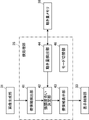
- FIG. 1 is a block diagram of an embodiment showing the configuration of an ultrasonic diagnostic apparatus of the present invention
- FIG. 1 is a block diagram of an embodiment showing the configuration of a transmission/reception circuit
- FIG. It is a block diagram of one embodiment showing the configuration of the image generator.
- It is a block diagram of one embodiment showing the configuration of the stool processing unit.
- 1 is a flowchart of one embodiment representing operation of an ultrasound diagnostic apparatus in a first mode of operation
- FIG. 4 is a flow chart of one embodiment representing the operation of the ultrasound diagnostic apparatus in the second mode of operation
- FIG. 11 is a flow chart of one embodiment representing operation of the ultrasound diagnostic apparatus in a third mode of operation
- FIG. 10 is a conceptual diagram of an embodiment showing a display screen of a monitor of an ultrasonic diagnostic apparatus when the transmittance of the mask is lowered according to the degree of enhancement;
- FIG. 11 is a conceptual diagram of an embodiment showing a display screen of a monitor of an ultrasonic diagnostic apparatus when the transmittance of the mask is increased according to the degree of enhancement; FIG.
- FIG. 1 is a block diagram of one embodiment showing the configuration of the ultrasonic diagnostic apparatus of the present invention.
- the ultrasonic diagnostic apparatus shown in FIG. 1 is a stationary ultrasonic diagnostic apparatus and includes an ultrasonic probe 1 and an apparatus body 3 connected to the ultrasonic probe 1 .
- the beamformer 54 adds each delay to each received data converted by the AD converter 53 according to the sound velocity or the distribution of the sound velocity set based on the reception delay pattern selected by the device controller 36. By doing so, so-called reception focus processing is performed. By this reception focusing process, each piece of reception data converted by the AD conversion unit 53 is phased and added, and an acoustic ray signal in which the focus of the ultrasonic echo is narrowed down is generated.
- the DSC 18 raster-converts the image information data generated by the signal processing unit 16 into an image signal that conforms to the normal television signal scanning method.
- the image processing unit 17 performs various corrections such as brightness correction, gradation correction, sharpness correction, image size correction, refresh rate correction, scanning frequency correction, and color correction on the image signal input from the DSC 18 according to the display format of the monitor 34 .
- an ultrasonic image (ultrasonic image signal) is generated, and the ultrasonic image subjected to the image processing is output to the image memory 32 , the fecal processing unit 35 and the display control unit 33 .
- the motion amount memory 38 is a memory that stores the amount of motion of the ultrasonic probe 1 for each frame of an ultrasonic image, for example, under the control of the fecal processing unit 35 .
- the method of detecting the stool region is not particularly limited. A stool area can be detected.
- the stool information detection unit 41 When detecting a stool region from an ultrasound image using template matching, the stool information detection unit 41 prepares a plurality of templates having different sizes, shapes, textures, etc. within the region of interest, and uses each of the plurality of templates. By raster-scanning the inside of the ultrasound image with the scanner, a region whose correlation value with the template is equal to or greater than a predetermined threshold value is detected as a fecal region.
- the stool information detection unit 41 detects a stool region from an ultrasound image using a deep learning model
- a large number of teacher images including anatomical structures and stool regions are prepared in advance, and a large number of teacher images are used.
- a deep learning model is created by learning the relationship between the teacher image and the stool region in the teacher image for a large number of teacher images, and the stool region is detected from the ultrasound image using this deep learning model.
- the stool information detection unit 41 detects the stool region as a rectangular region for each frame of the ultrasound image using, for example, a deep learning model. , find the location of the stool region and the probability that this stool region is a stool region. Then, the stool information detection unit 41 compares the statistic value of the probability of the ultrasound images of a plurality of frames, for example, the average value, the weighted average value, or the median value, with a threshold to determine whether the stool region is actually a flight. Detects whether or not is a certain area. The flight information detection unit 41 detects a flight region, for example, when the statistical value of the probability of being a flight region is equal to or greater than a threshold.
- the method of detecting the stool properties is not particularly limited, but the stool information detection unit 41 can detect the stool properties of the stool area based on the luminance value of the stool area or using a deep learning model, for example. .
- the stool information detection unit 41 detects a stool region from the ultrasonic image for each frame of the ultrasonic image, and calculates the statistic value of the luminance in the stool region and the luminance in a predetermined region around the stool region.
- the second comparison result is obtained by comparing the luminance ratio and the threshold value, and fecal properties are detected based on the second comparison result in the ultrasound image of one frame or a plurality of frames. You may Similarly, when the luminance ratio is the first threshold or more, hard stool, the second threshold smaller than the first threshold or more, and less than the first threshold, normal stool, and the second threshold when less than the second threshold Detect loose stools.
- the stool information detection unit 41 calculates the probability that the stool region is in each class of stool properties, such as the probability that the stool region is hard stool, soft stool, normal stool, or background. Alternatively, for each pixel in the ultrasound image, the probability that the pixel is in each class of stool properties may be detected.
- the motion amount detection unit 44 detects the amount of motion of the ultrasonic probe 1 during scanning.
- the conditions for determining the degree of emphasis are not particularly limited, but for example, the amount of movement of the ultrasonic probe 1, the continuous display time of the same stool region (the number of frames in which the same stool region is continuously displayed), and the stool region can be used.
- the amount of movement of the ultrasonic probe 1, the continuous display time of the same stool region, and the area of the stool region are each converted into the degree of enhancement of the stool region by, for example, a LUT (LookUp Table) or conversion in advance.
- a formula can be prepared and converted using this LUT or conversion formula.
- the degree-of-emphasis determining unit 42 determines the degree of emphasis of the stool region based on the amount of motion of the ultrasonic probe 1 .
- the enhancement degree determination unit 42 increases the enhancement degree of the feces region as the amount of movement of the ultrasonic probe 1 increases so that the user can easily grasp the feces region.
- the enhancement degree determination unit 42 lowers the enhancement degree of the stool region as the amount of movement of the ultrasonic probe 1 becomes smaller so as not to interfere with the user's interpretation of the ultrasonic image.
- the emphasis degree determination unit 42 performs the above-described identity determination of the flight region, and determines the continuous display time of the flight region determined to be the same. Based on this, the degree of enhancement of the stool region is determined.
- the fact that the continuous display time of the stool region is long means that the stool region of adjacent frames does not move and is determined to be the same for a long time (the number of frames is large), that is, the movement of the ultrasonic probe 1 It can be said that the amount is small and the time is long. If the continuous display time of the stool area is short, it is highly likely that the user has not yet fully grasped the stool area.
- the emphasis degree determination unit 42 increases the degree of emphasis of the stool region according to the continuous display time of the stool region, the shorter the continuous display time of the stool region, so that the user can easily grasp the stool region.
- the continuous display time of the stool area is long, it is considered that the user has already grasped the stool area.
- the enhancement degree determination unit 42 emphasizes the stool region according to the continuous display time of the stool region, the longer the continuous display time of the stool region, so as not to interfere with the user's interpretation of the ultrasound image. lower the degree.
- the degree-of-emphasis determination unit 42 determines the degree of emphasis of the stool region based on the area of the stool region. If the area of the stool area is small, the user may miss the stool area. Therefore, the emphasis degree determination unit 42 increases the degree of emphasis of the feces region according to the area of the feces region, the smaller the area of the feces region, so that the user can easily recognize the feces region. On the other hand, when the area of the feces area is large, the user can easily recognize the feces area. Therefore, the enhancement degree determination unit 42 lowers the enhancement degree of the stool region as the area of the stool region increases so as not to interfere with the user's interpretation of the ultrasound image.
- the flight information display unit 43 causes the monitor 34 to display various information related to flights.
- the stool information display unit 43 highlights, for example, the stool region in the ultrasound image displayed on the monitor 34 according to the degree of emphasis determined (changed) by the degree-of-emphasis determination unit 42 .
- the image generation unit 31, the flight processing unit 35, the display control unit 33 and the device control unit 36 are configured by the processor 39.
- the transmitting/receiving circuit 14 starts transmitting ultrasonic waves under the control of the device control unit 36, and an acoustic ray signal is generated. (step S1).
- ultrasonic beams are transmitted from the plurality of transducers of the transducer array 11 to the inspected portion of the subject according to the drive signal from the pulser 51 .
- An ultrasonic echo from an inspection location based on an ultrasonic beam transmitted from the pulsar 51 is received by each transducer of the transducer array 11, and each transducer of the transducer array 11 that has received the ultrasonic echo outputs an analog signal.
- a received signal is output.
- a received signal output from each transducer of the transducer array 11 is amplified by the amplifier 52 and AD-converted by the AD converter 53 to obtain received data.
- a sound ray signal is generated by subjecting the received data to reception focusing processing by the beamformer 54 .
- the sound ray signal generated by the beamformer 54 is subjected to various signal processings by the signal processing unit 16 to generate image information data representing tomographic image information regarding tissues in the subject.
- the image information data generated by the signal processing unit 16 is raster-converted by the DSC 18 and further subjected to various image processing by the image processing unit 17 to generate an ultrasonic image (ultrasonic image signal).
- the ultrasound image generated by the image processing unit 17 is stored in the image memory 32 .
- the display control unit 33 performs predetermined processing on the ultrasonic image generated by the image processing unit 17 or the ultrasonic image stored in the image memory 32. It is displayed on the monitor 34 (step S3).
- step S13 if no stool region is detected from the ultrasonic image (No at step S13), the process returns to step S11, and the detection processing of the stool region is repeatedly performed until the stool region is detected from the ultrasonic image.
- the fecal region is highlighted in the ultrasonic image displayed on the monitor 34, so that the user can easily identify the fecal region in the ultrasonic image. can be grasped.
- the emphasis degree determination unit 42 determines the degree of emphasis based on the conditions for determining the degree of emphasis (step S24). That is, in the case of the third operation mode, the emphasis degree determination unit 42 changes the emphasis degree of the stool region based on the emphasis degree determination condition when the emphasis degree determination condition is changed.
- the motion amount detection unit 44 superimposes, for each frame of the ultrasound image, the feces area of the ultrasound image of the current frame and the feces area of the ultrasound image of the past frame one frame before the current frame.
- the degree may be obtained as the amount of movement of the ultrasonic probe 1 .
- the movement amount detection unit 44 detects that the amount of movement of the ultrasonic probe 1 is smaller as the degree of superimposition of the stool region is larger, and that the amount of movement of the ultrasonic probe 1 is larger as the degree of superimposition of the stool region is smaller.
- the motion amount detection unit 44 obtains the amount of motion between the nth frame ultrasound image and the (n ⁇ 1)th frame ultrasound image, and Statistical values are obtained from a group of motion amounts of ultrasonic images for 10 frames from the eye to the (n ⁇ 9)th frame. If the degree of emphasis of the feces region, for example, the degree of transparency of the display color of the feces region is changed by obtaining the amount of motion of the ultrasound image between adjacent frames, the degree of emphasis of the feces region may change frequently, making it difficult to see. .
- the statistical value is not particularly limited, but for example, an average value obtained from the movement amount group, a weighted average value obtained from the movement amount group with a weight increasing as the past approaches the present, or a movement amount group A median value or the like can be exemplified.
- the stool information detection unit 41 performs detection processing of the stool region for each frame of the ultrasound image (step S32).
- the emphasis degree determination unit 42 determines the degree of emphasis based on the statistic obtained from the motion amount group (step S38).
- the stool information display unit 43 determines (changes) the stool region in the ultrasound image displayed on the monitor 34 under the control of the display control unit 33 based on the statistical values obtained from the movement amount group. It is highlighted in accordance with the degree of emphasis (step S39). After that, the process returns to step S31, and the above-described operations are repeated.
- a group of motion amounts of ultrasonic images of a plurality of frames when a fecal region is detected, a group of motion amounts of ultrasonic images of a plurality of frames is obtained, and the statistical value is obtained from this group of motion amounts. may be asked for.
- step S43 If the fecal region is not detected from the ultrasonic image of the current frame as a result of the fecal region detection processing (No in step S43), the flow returns to step S41, and the fecal region is detected from the ultrasonic image until the fecal region is detected. The process is repeated.
- the motion amount detection unit 44 obtains a motion amount group of ultrasound images of a plurality of frames.
- the motion amount detection unit 44 determines whether there is a motion amount of a past frame ultrasound image that is not stored in the motion amount memory 38 among the motion amount of the ultrasound image of the past frame.
- the motion amount detection unit 44 stores the second motion amount and the third motion amount in the motion amount memory 38 as the motion amount of the ultrasonic probe 1 (step S47). Then, the motion amount detector 44 obtains a statistical value from the motion amount group consisting of the first motion amount, the second motion amount, and the third motion amount (step S48).
- steps S49 and S50 are the same as the operations of steps S38 and S39 in the flow chart of FIG. After that, the process returns to step S41, and the above-described operations are repeated.
- the emphasis degree determination unit 42 determines the binary motion of the ultrasonic probe 1, that is, whether there is motion or not. When it is determined that there is movement, from among the first emphasis level and the second emphasis level, which are smaller than the first emphasis level, which are two levels of corresponding emphasis levels, the first emphasis level is selected. If it is determined that there is no movement, the second enhancement degree is determined. Further, when the determination result of the binary determination is changed, the emphasis degree determination unit 42 sets the degree of emphasis in the ultrasound image of the frame immediately after the determination result of the binary determination is changed, for example, to the post-change multi-value The degree of emphasis is changed to a level corresponding to the determination result of the determination.
- the enhancement degree determination unit 42 selects, for example, from multi-level enhancement degrees corresponding to the multi-valued motion of the ultrasonic probe 1. , the degree of emphasis is determined to be a degree of emphasis corresponding to the determination result of the multilevel determination. Further, when the determination result of the multi-valued determination is changed, the emphasis degree determination unit 42 sets the degree of enhancement in the ultrasound image of the frame immediately after the determination result of the multi-valued determination is changed, for example, to the post-change multi-valued determination result. The degree of emphasis is changed to a level corresponding to the determination result of the determination.
- the stool information display unit 43 creates, for example, a mask in which the stool region is colored in a predetermined display color and painted, and the transparency of the display color is determined by the emphasis degree of the stool region determined by the emphasis degree determination unit 42. , and the mask with the changed transmittance can be superimposed and displayed on the feces area of the ultrasound image displayed on the monitor 34 .
- the flight information display unit 43 reduces the transparency of the mask 47 as shown in FIG. 10A as the degree of emphasis increases, and decreases the transparency of the mask 47 as shown in FIG. Raise it to reveal the mask 47 .
- the difference in transparency is represented by the density of hatching. That is, the hatching in the case of low transmittance shown in FIG. 10A is dense, and the hatching in the case of high transmittance shown in FIG. 10B is coarse.
- the stool information display unit 43 detects the contour of the stool area to create a contour line, and changes the thickness of the contour line or the degree of transparency of the display color according to the degree of emphasis of the stool area.
- the contour line whose transparency has been changed can be superimposed on the contour of the stool region of the ultrasound image displayed on the monitor 34 and displayed.
- the flight information display unit 43 thickens the outline 48 as shown in FIG. Thin the thickness to display the contour line.
- the flight information display unit 43 increases the transparency of the outline 48 as the degree of emphasis is smaller, and decreases the transparency of the outline 48 as the degree of emphasis is greater. 48 is displayed.
- the stool information display unit 43 may change the display mode of highlighting from a mask to a contour line or from a contour line to a mask according to the degree of emphasis of the stool region.
- the degree of emphasis of the stool region is equal to or greater than the threshold value, that is, when increasing the degree of emphasis of the stool region
- the flight information display unit 43 creates the above-described mask 47 and displays the mask 47 as shown in FIG. 12A. Then, the mask 47 is superimposed on the stool area and displayed.
- the degree of emphasis of the stool region is less than the threshold value, that is, when the degree of emphasis of the stool region is decreased
- the flight information display unit 43 creates the aforementioned outline 48, and as shown in FIG. 12B, This outline 48 is superimposed on the outline of the stool area and displayed.
- the stool information display unit 43 may highlight the stool region by thinning out the frames for highlighting the stool region according to the degree of emphasis of the stool region determined by the emphasis degree determination unit 42 .
- the user sees the stool area highlight blinking.
- the ratio of the number of frames (time) in which the stool region is highlighted and the number of frames (time) in which the stool region is not highlighted, that is, the degree of flickering of the stool region highlighting is determined. It can be changed according to the degree.
- the stool information display unit 43 reduces the number of frames to be thinned out and lengthens the time for highlighting the stool region as the degree of emphasis of the stool region increases, and reduces the number of frames to be thinned out as the degree of emphasis of the stool region decreases. The higher the number, the shorter the time to highlight the stool region. For example, when increasing the degree of emphasis, the flight information display unit 43 may thin out the frames so as to shorten the flickering interval, or may thin out the frames so as to lengthen the time during which the flight region is highlighted. good.
- the stool information display unit 43 displays the results of the stool property detection processing.
- the display color of the stool region highlighting may be changed according to the stool properties such as hard stool, loose stool, and normal stool. As a result, the user can grasp the stool properties simply by looking at the display color of the highlighted display of the stool region.
- the stool information detection unit 41 may erroneously detect the stool properties of the stool region depending on the state in which the ultrasonic probe 1 is in contact with the examination site of the subject. there's a possibility that.
- the degree of highlighting of the display color of the stool region for example, when the transparency is lowered, the stool properties of the stool region change, and the display color of the stool region is changed accordingly. It is difficult for the user to understand the change in the display color of the highlighting of the stool area.
- the degree-of-emphasis determining unit 42 performs the same determination of the stool regions as described above, and if the detection results of the stool properties of the stool regions determined to be the same change, If the properties are erroneously determined, the enhancement degree of the stool region may be temporarily increased.
- the stool information display unit 43 highlights the stool area according to the degree of emphasis of the stool area temporarily raised. Then, after a predetermined period has elapsed since the detection result of the stool properties changed, the emphasis degree determination unit 42 restores the degree of emphasis of the stool region to that before the degree of emphasis was increased.
- the degree of enhancement of the stool area is determined based on the determination conditions. In this way, when the stool texture of the stool region changes, it is possible to alert the user to the possibility that the stool texture is erroneously detected by temporarily increasing the degree of enhancement of the stool region. can.
- the present invention is not limited to a stationary ultrasonic diagnostic apparatus, but also a portable ultrasonic diagnostic apparatus in which the device main body 3 is realized by a laptop terminal device, and a smartphone or a tablet PC (Personal The present invention can also be applied to a handheld ultrasonic diagnostic apparatus realized by a handheld terminal device such as a computer (personal computer).
- the ultrasonic probe 1 and the apparatus main body 3 may be connected by wire or wirelessly.
- all of the image generating section 31 or only the signal processing section 16 may be provided on the ultrasonic probe 1 side, or may be provided on the device main body 3 side.
- flash memory As the image memory 32 and the motion amount memory 38, flash memory, HDD (Hard Disk Drive), SSD (Solid State Drive), FD (Flexible Disc), MO disk (Magneto -Optical disc), MT (Magnetic Tape), RAM (Random Access Memory), CD (Compact Disc), DVD (Digital Versatile Disc), SD
- a recording medium such as a card (Secure Digital card), a USB memory (Universal Serial Bus memory), or an external server can be used.
- the circuit configuration can be changed after manufacturing such as CPU (Central Processing Unit), FPGA (Field Programmable Gate Array), etc., which are general-purpose processors that run software (programs) and function as various processing units.
- Programmable Logic Device PLD
- ASIC Application Specific Integrated Circuit
- One processing unit may be composed of one of these various processors, or a combination of two or more processors of the same or different type, such as a combination of multiple FPGAs, or a combination of FPGAs and CPUs. and so on. Also, the plurality of processing units may be configured by one of various processors, or two or more of the plurality of processing units may be combined into one processor.
- SoC System on Chip
- the hardware configuration of these various processors is, more specifically, an electric circuit that combines circuit elements such as semiconductor elements.
- the method of the present invention can be implemented, for example, by a program for causing a computer to execute each step. It is also possible to provide a computer-readable recording medium on which this program is recorded.
Landscapes
- Health & Medical Sciences (AREA)
- Life Sciences & Earth Sciences (AREA)
- Engineering & Computer Science (AREA)
- Medical Informatics (AREA)
- Surgery (AREA)
- Pathology (AREA)
- Radiology & Medical Imaging (AREA)
- Biophysics (AREA)
- Biomedical Technology (AREA)
- Heart & Thoracic Surgery (AREA)
- Physics & Mathematics (AREA)
- Molecular Biology (AREA)
- Nuclear Medicine, Radiotherapy & Molecular Imaging (AREA)
- Animal Behavior & Ethology (AREA)
- General Health & Medical Sciences (AREA)
- Public Health (AREA)
- Veterinary Medicine (AREA)
- Computer Vision & Pattern Recognition (AREA)
- Physiology (AREA)
- Ultra Sonic Daignosis Equipment (AREA)
Abstract
Description
また、便秘に関する内容ではないが、特許文献2には、現在の超音波画像に対応する基準画像データセットの画像スライスの前立腺等の関心対象の外縁輪郭を現在の超音波画像に重ね合わせて表示する介入治療システムが記載されている。 For example, in
In addition, although the content is not related to constipation, Patent Document 2 discloses that the outer edge contour of an object of interest, such as the prostate, of an image slice of a reference image data set corresponding to a current ultrasound image is superimposed on the current ultrasound image. An interventional treatment system is described.
モニタと、
超音波プローブを用いて超音波ビームにより被検体の検査箇所をスキャンして得られた受信信号に基づいて超音波画像を生成する画像生成部と、
超音波画像をモニタに表示させる表示制御部と、
超音波画像から便領域を検出するための検出処理を行う便情報検出部と、
便領域が検出された場合に、便領域の強調度合いを決定するための決定条件に基づいて、便領域の強調度合いを決定する強調度合い決定部と、
モニタに表示された超音波画像において、便領域を、強調度合い決定部によって決定された強調度合いに応じて強調表示させる便情報表示部と、を備える、超音波診断装置を提供する。 In order to achieve the above object, the present invention provides an ultrasonic probe,
a monitor;
an image generation unit that generates an ultrasound image based on a received signal obtained by scanning an inspection location of a subject with an ultrasound beam using an ultrasound probe;
a display control unit for displaying an ultrasound image on a monitor;
a stool information detection unit that performs detection processing for detecting a stool region from an ultrasound image;
an emphasis degree determination unit that determines the degree of emphasis of the stool region based on a determination condition for determining the degree of emphasis of the stool region when the stool region is detected;
An ultrasound diagnostic apparatus is provided, comprising: a feces information display unit that highlights a feces region in an ultrasound image displayed on a monitor according to the degree of emphasis determined by the degree of emphasis determination unit.
強調度合い決定部は、動き量に基づいて強調度合いを決定することが好ましい。 In addition, it has a motion amount detection unit that detects the amount of motion of the ultrasonic probe,
Preferably, the emphasis degree determination unit determines the degree of emphasis based on the amount of motion.
便情報検出部は、超音波画像のフレーム毎に便領域の検出処理を行い、
動き量検出部は、超音波画像のフレーム毎に動き量を求めて動き量メモリに保存しておき、便領域が検出された場合に、定められた数の過去のフレームの超音波画像の動き量を動き量メモリから読み出し、現在のフレームの超音波画像の動き量および動き量メモリから読み出された過去のフレームの超音波画像の動き量からなる動き量群から統計値を求めることが好ましい。 In addition, it has a motion amount memory that stores the amount of motion,
The stool information detection unit performs detection processing of the stool region for each frame of the ultrasound image,
The motion amount detection unit obtains the motion amount for each frame of the ultrasound image and stores it in a motion amount memory. Preferably, the amount is read from the motion amount memory, and the statistical value is obtained from the motion amount group consisting of the amount of motion of the ultrasonic image of the current frame and the amount of motion of the ultrasonic image of the past frame read from the motion amount memory. .
便情報検出部は、超音波画像のフレーム毎に便領域の検出処理を行い、
動き量検出部は、便領域が検出された場合に、定められた数の過去のフレームの超音波画像の動き量のうち、動き量メモリに保存されている過去のフレームの超音波画像の動き量がある場合に、動き量メモリに保存されている過去のフレームの超音波画像の動き量を第1動き量として動き量メモリから読み出し、動き量メモリに保存されていない過去のフレームの超音波画像の動き量がある場合に、動き量メモリに保存されていない過去のフレームの超音波画像の動き量を第2動き量として求め、さらに、現在のフレームの超音波画像の動き量を第3動き量として求め、第2動き量および第3動き量を動き量メモリに保存しておき、第1動き量、第2動き量および第3動き量からなる動き量群から統計値を求めることが好ましい。 In addition, it has a motion amount memory that stores the amount of motion,
The stool information detection unit performs detection processing of the stool region for each frame of the ultrasound image,
When the feces area is detected, the motion amount detection unit detects the motion amount of the ultrasound images of the past frames stored in the motion amount memory, out of the motion amounts of the ultrasound images of the past frames of a predetermined number. If there is an amount of motion, the motion amount of the ultrasound image of the past frame stored in the motion amount memory is read from the motion amount memory as the first motion amount, and the ultrasound of the past frame not stored in the motion amount memory is read. If there is an amount of motion of the image, the amount of motion of the ultrasound image of the past frame that is not stored in the motion amount memory is obtained as the second amount of motion, and further the amount of motion of the ultrasound image of the current frame is determined as the third amount of motion. Statistical values can be obtained from a motion amount group consisting of the first motion amount, the second motion amount, and the third motion amount, by obtaining the motion amount, storing the second motion amount and the third motion amount in a motion amount memory. preferable.
強調度合い決定部は、超音波プローブの2値の動きに対応する2段階の強調度合いである、第1の強調度合い、および、第1の強調度合いよりも小さい第2の強調度合いの中から、動きありと判定された場合に、第1の強調度合いに決定し、動きなしと判定された場合に、第2の強調度合いに決定することが好ましい。 Further, the motion amount detection unit performs binary determination based on the motion amount to determine whether the motion of the ultrasonic probe is in motion or not,
The emphasis degree determination unit selects, from among two levels of emphasis degrees corresponding to binary movements of the ultrasonic probe, a first emphasis degree and a second emphasis degree smaller than the first emphasis degree, It is preferable to determine the first degree of emphasis when it is determined that there is movement, and to set the second degree of emphasis when it is determined that there is no movement.
強調度合い決定部は、超音波プローブの多値の動きに対応する多段階の強調度合いの中から、強調度合いを多値判定の判定結果に対応する段階の強調度合いに決定することが好ましい。 Further, the motion amount detection unit performs multi-value determination for detecting the motion of the ultrasonic probe as multi-values of three or more values based on the amount of motion,
Preferably, the emphasis degree determination unit determines the degree of emphasis to be the degree of emphasis corresponding to the determination result of the multi-valued determination from among the multiple levels of emphasis corresponding to the multi-valued motion of the ultrasonic probe.
便情報表示部は、便性状が検出された場合に、便性状に応じて便領域の強調表示の表示色を変えることが好ましい。 The stool information detection unit further performs detection processing for detecting stool properties in the stool region from the ultrasound image,
It is preferable that the stool information display unit changes the display color of the highlighted display of the stool region according to the stool properties when the stool properties are detected.
便情報検出部は、超音波画像のフレーム毎に、便領域内の輝度の統計値と便領域の周囲の定められた領域内の輝度の統計値との輝度比を検出し、輝度比と閾値とを比較することにより第2比較結果を求め、1フレームまたは複数のフレームの超音波画像における第2比較結果に基づいて便性状を検出することが好ましい。 Further, the stool information detection unit detects a statistic value of brightness in the stool region for each frame of the ultrasound image, and obtains a first comparison result by comparing the statistic value of brightness in the stool region with a threshold value. , detecting stool properties based on a first comparison result in one or more frames of ultrasound images; or
The stool information detection unit detects, for each frame of the ultrasound image, the luminance ratio between the statistic value of the luminance in the stool region and the statistic value of the luminance in a predetermined region around the stool region, and calculates the luminance ratio and the threshold value. It is preferable to obtain a second comparison result by comparing with and to detect the stool properties based on the second comparison result in the ultrasonic image of one frame or a plurality of frames.
ユーザからの指示に応じて、少なくとも2つの動作モードのうちの1つの動作モードに切り替えるモード切替部を備えることが好ましい。 A first operation mode that does not highlight the stool region, a second operation mode that highlights the stool region with a predetermined degree of emphasis regardless of the determination condition, and a degree of emphasis that is determined based on the determination condition. at least two modes of operation of a third mode of operation for highlighting the stool region;
It is preferable to provide a mode switching unit that switches to one of at least two operation modes according to an instruction from a user.
表示制御部が、超音波画像をモニタに表示させるステップと、
便情報検出部が、超音波画像から便領域を検出するための検出処理を行うステップと、
強調度合い決定部が、便領域が検出された場合に、便領域の強調度合いを決定するための決定条件に基づいて、便領域の強調度合いを決定するステップと、
便情報表示部が、モニタに表示された超音波画像において、便領域を、便領域の強調度合いを決定するステップによって決定された強調度合いに応じて強調表示させるステップと、を含む、超音波診断装置の制御方法を提供する。 Further, according to the present invention, the image generating unit generates an ultrasonic image based on a received signal obtained by scanning an examination location of the subject with an ultrasonic beam using an ultrasonic probe;
a step in which the display control unit displays an ultrasound image on a monitor;
a step in which the stool information detection unit performs a detection process for detecting a stool region from an ultrasound image;
a step of determining the degree of emphasis of the stool region based on a determination condition for determining the degree of emphasis of the stool region when the stool region is detected;
a step in which the stool information display unit highlights the stool region in the ultrasound image displayed on the monitor according to the degree of emphasis determined by the step of determining the degree of emphasis of the stool region; A device control method is provided.
各振動子は、例えば、PZT(Lead Zirconate Titanate:チタン酸ジルコン酸鉛)に代表される圧電セラミック、PVDF(Poly Vinylidene Di Fluoride:ポリフッ化ビニリデン)に代表される高分子圧電素子およびPMN-PT(Lead Magnesium Niobate-Lead Titanate:マグネシウムニオブ酸鉛-チタン酸鉛固溶体)に代表される圧電単結晶等からなる圧電体の両端に電極を形成した素子を用いて構成される。 The
Each vibrator includes, for example, a piezoelectric ceramic typified by PZT (Lead Zirconate Titanate), a polymeric piezoelectric element typified by PVDF (Poly Vinylidene Di Fluoride), and PMN-PT ( Lead Magnesium Niobate-Lead Titanate: A piezoelectric single crystal represented by lead magnesium niobate-lead titanate solid solution).
便領域のみを検出する場合、便情報検出部41は、例えば、深層学習モデルを用いて、便領域を矩形領域として検出する。 When a stool region is detected from an ultrasound image using a deep learning model, the stool
When detecting only the stool area, the stool
超音波プローブ1の動き量、同一の便領域の連続表示時間、および、便領域の面積のそれぞれから便領域の強調度合いへの変換は、例えば、予めLUT(LookUp Table:ルックアップテーブル)または変換式を用意しておき、このLUTまたは変換式を用いて変換することができる。 The conditions for determining the degree of emphasis are not particularly limited, but for example, the amount of movement of the
The amount of movement of the
超音波プローブ1の動き量が大きい場合、ユーザは、便を探している状態であると考えられる。このため、強調度合い決定部42は、ユーザが便領域を把握しやすいように、超音波プローブ1の動き量が大きくなるほど、便領域の強調度合いを上げる。
一方、超音波プローブ1の動き量が小さい場合、ユーザは、便領域を把握していると考えられる。このため、強調度合い決定部42は、ユーザが超音波画像を読影する際の妨げとならないように、超音波プローブ1の動き量が小さくなるほど、便領域の強調度合いを下げる。 When the condition for determining the degree of emphasis is the amount of motion of the
When the amount of movement of the
On the other hand, when the amount of movement of the
便領域の連続表示時間が長いということは、隣接するフレームの便領域が動いておらず、同一であると判定されている時間が長い(フレーム数が多い)、すなわち、超音波プローブ1の動き量が小さい時間が長いとも言える。
便領域の連続表示時間が短い場合、ユーザは、便領域をまだ十分に把握できていない可能性が高いと考えられる。このため、強調度合い決定部42は、ユーザが便領域を把握しやすいように、便領域の連続表示時間に応じて、便領域の連続表示時間が短いほど、便領域の強調度合いを上げる。
一方、便領域の連続表示時間が長い場合、ユーザは、便領域を既に把握していると考えられる。このため、強調度合い決定部42は、ユーザが超音波画像を読影する際の妨げとならないように、便領域の連続表示時間に応じて、便領域の連続表示時間が長くなるほど、便領域の強調度合いを下げる。 If the condition for determining the degree of emphasis is the continuous display time of the same flight region, the emphasis
The fact that the continuous display time of the stool region is long means that the stool region of adjacent frames does not move and is determined to be the same for a long time (the number of frames is large), that is, the movement of the
If the continuous display time of the stool area is short, it is highly likely that the user has not yet fully grasped the stool area. Therefore, the emphasis
On the other hand, if the continuous display time of the stool area is long, it is considered that the user has already grasped the stool area. For this reason, the enhancement
便領域の面積が小さい場合、ユーザは、便領域を見落とす可能性がある。このため、強調度合い決定部42は、ユーザが便領域を認識しやすいように、便領域の面積に応じて、便領域の面積が小さいほど、便領域の強調度合いを上げる。
一方、便領域の面積が大きい場合、ユーザは、便領域を容易に認識することができる。このため、強調度合い決定部42は、ユーザが超音波画像を読影する際の妨げとならないように、便領域の面積に応じて、便領域の面積が大きいほど、便領域の強調度合いを下げる。 When the condition for determining the degree of emphasis is the area of the stool region, the degree-of-
If the area of the stool area is small, the user may miss the stool area. Therefore, the emphasis
On the other hand, when the area of the feces area is large, the user can easily recognize the feces area. Therefore, the enhancement
モード切替部46は、例えば、GUIまたは音声認識等を利用して入力されるユーザからの指示に応じて、前述の少なくとも2つの動作モードのうちの1つの動作モードに切り替える。 The ultrasonic diagnostic apparatus has a first operation mode in which the stool region is not highlighted, a second operation mode in which the stool region is highlighted with a predetermined degree of emphasis regardless of the determination conditions for the degree of emphasis, and a It has at least two operation modes of a third operation mode that determines (changes) the degree of emphasis based on the determination condition and highlights the stool region.
The
パルサ51から送信された超音波ビームに基づく検査箇所からの超音波エコーは、振動子アレイ11の各振動子により受信され、超音波エコーを受信した振動子アレイ11の各振動子からアナログ信号である受信信号が出力される。
振動子アレイ11の各振動子から出力される受信信号は、増幅部52により増幅され、AD変換部53によりAD変換されて受信データが取得される。
この受信データに対して、ビームフォーマ54により受信フォーカス処理が施されることにより、音線信号が生成される。 In other words, ultrasonic beams are transmitted from the plurality of transducers of the
An ultrasonic echo from an inspection location based on an ultrasonic beam transmitted from the
A received signal output from each transducer of the
A sound ray signal is generated by subjecting the received data to reception focusing processing by the
信号処理部16により生成された画像情報データは、DSC18によりラスター変換され、さらに画像処理部17により各種の画像処理が施され、超音波画像(超音波画像信号)が生成される。
画像処理部17により生成された超音波画像は、画像メモリ32に保存される。 That is, the sound ray signal generated by the
The image information data generated by the
The ultrasound image generated by the
その後、ステップS11へ戻り、超音波画像のフレーム毎に前述の動作が繰り返される。 Then, under the control of the
After that, the process returns to step S11, and the above operation is repeated for each frame of the ultrasonic image.
その後、ステップS21へ戻り、超音波画像のフレーム毎に前述の動作が繰り返される。 Then, under the control of the
After that, the process returns to step S21, and the above operation is repeated for each frame of the ultrasonic image.
この場合、動き量検出部44は、現在のフレームをnとすると、nフレーム目の超音波画像と、(n-1)フレーム目の超音波画像と、の相関値を求める。
相関値の算出方法は、特に限定されないが、例えば、正規化相互相関演算を行うことによって相関値を算出することができる。また、相関値として、隣接するフレーム間の超音波画像のオプティカルフローを算出してもよい。 For example, the motion
In this case, if the current frame is n, the motion
Although the method of calculating the correlation value is not particularly limited, the correlation value can be calculated by performing normalized cross-correlation calculation, for example. Also, as a correlation value, an optical flow of ultrasonic images between adjacent frames may be calculated.
この場合、動き量検出部44は、現在のフレームをnとすると、nフレーム目の超音波画像と、(n-5)フレーム目の超音波画像と、の相関値を求める。
フレームレートにもよるが、隣接するフレーム間の超音波画像の相関値は大きくなりすぎる可能性があるため、超音波プローブ1の動き量を正確に求めることが難しい場合がある。これに対し、定められたフレーム数だけ離れたフレーム間の超音波画像の相関値を求めることにより、より超音波プローブ1の動き量をより正確に求めることができる。 Alternatively, the motion
In this case, if the current frame is n, the motion
Although it depends on the frame rate, the correlation value of ultrasonic images between adjacent frames may become too large, so it may be difficult to accurately determine the amount of movement of the
この場合、動き量検出部44は、現在のフレームをnとすると、nフレーム目の超音波画像と、(n-1)フレーム目の超音波画像と、の間の動き量を求め、nフレーム目から(n-9)フレーム目までの10フレーム分の超音波画像の動き量群から統計値を求める。
隣接するフレーム間の超音波画像の動き量を求めて便領域の強調度合い、例えば、便領域の表示色の透過度を変える場合、頻繁に便領域の強調度合いが変わって見づらくなる可能性がある。これに対し、複数のフレームの超音波画像の動き量群から求められる統計値を超音波プローブ1の動き量として求めることにより、より正確に超音波プローブ1の動き量を算出することができ、便領域の強調度合いが頻繁に変わることを防止することができる。 Further, the motion
In this case, if the current frame is n, the motion
If the degree of emphasis of the feces region, for example, the degree of transparency of the display color of the feces region is changed by obtaining the amount of motion of the ultrasound image between adjacent frames, the degree of emphasis of the feces region may change frequently, making it difficult to see. . On the other hand, the amount of motion of the
その後、ステップS31へ戻り、前述の動作が繰り返される。 Then, the stool
After that, the process returns to step S31, and the above-described operations are repeated.
また、動き量検出部44により、定められた数の過去のフレームの超音波画像の動き量のうち、動き量メモリ38に保存されていない過去のフレームの超音波画像の動き量がある場合に、動き量メモリ38に保存されていない過去のフレームの超音波画像の動き量が第2動き量として求められ(ステップS45)、さらに、現在のフレームの超音波画像の動き量が第3動き量として求められる(ステップS46)。動き量検出部44は、第2動き量および第3動き量を超音波プローブ1の動き量として動き量メモリ38に記憶しておく(ステップS47)。そして、動き量検出部44により、第1動き量、第2動き量および第3動き量からなる動き量群から統計値が求められる(ステップS48)。 In response to this, the motion
In addition, if there is a motion amount of a past frame ultrasound image that is not stored in the
その後、ステップS41へ戻り、前述の動作が繰り返される。 The operations of steps S49 and S50 are the same as the operations of steps S38 and S39 in the flow chart of FIG.
After that, the process returns to step S41, and the above-described operations are repeated.
また、強調度合い決定部42は、2値判定の判定結果が変化した場合に、例えば、2値判定の判定結果が変化した直後のフレームの超音波画像において、強調度合いを、変化後の多値判定の判定結果に対応する段階の強調度合いに変える。 When the motion
Further, when the determination result of the binary determination is changed, the emphasis
また、強調度合い決定部42は、多値判定の判定結果が変化した場合に、例えば、多値判定の判定結果が変化した直後のフレームの超音波画像において、強調度合いを、変化後の多値判定の判定結果に対応する段階の強調度合いに変える。あるいは、強調度合い決定部42は、多値判定の判定結果が2値以上変化した場合に、多値判定の判定結果が変化してから複数のフレームの超音波画像において、強調度合いを、変化前の多値判定の判定結果に対応する段階の強調度合いから変化後の多値判定の判定結果に対応する段階の強調度合いまで段階的に変えてもよい。 When the motion
Further, when the determination result of the multi-valued determination is changed, the emphasis
この場合、便情報表示部43は、強調度合いが大きいほど、図10Aに示すように、マスク47の透過度を下げ、強調度合いが小さいほど、図10Bに示すように、マスク47の透過度を上げてマスク47を表示させる。なお、図10A,10Bにおいては、透過度の違いが、ハッチングの粗密によって表現されている。すなわち、図10Aに示す、透過度が小さい場合のハッチングを密にし、図10Bに示す、透過度が大きい場合のハッチングを粗にしている。 The stool
In this case, the flight
この場合、便情報表示部43は、強調度合いが大きいほど、図11Aに示すように、輪郭線48の太さを太くし、強調度合いが小さいほど、図11Bに示すように、輪郭線48の太さを細くして輪郭線を表示させる。また、便情報表示部43は、マスク47の場合と同じように、強調度合いが小さいほど、輪郭線48の透過度を上げ、強調度合いが大きいほど、輪郭線48の透過度を下げて輪郭線48を表示させる。 Alternatively, the stool
In this case, the flight
便情報表示部43は、便領域を、一時的に上げられた便領域の強調度合いに応じて強調表示させる。
そして、強調度合い決定部42は、便性状の検出結果が変化してから、定められた期間が経過した後に、強調度合いを上げる前の便領域の強調度合いに戻し、これ以後は、強調度合いの決定条件に基づいて便領域の強調度合いを決定する。
このように、便領域の便性状が変化した場合に、便領域の強調度合いを一時的に上げることにより、便性状が誤検出である可能性があることをユーザに対して注意喚起することができる。 In response to this, the degree-of-
The stool
Then, after a predetermined period has elapsed since the detection result of the stool properties changed, the emphasis
In this way, when the stool texture of the stool region changes, it is possible to alert the user to the possibility that the stool texture is erroneously detected by temporarily increasing the degree of enhancement of the stool region. can.
Claims (29)
- 超音波プローブと、
モニタと、
前記超音波プローブを用いて超音波ビームにより被検体の検査箇所をスキャンして得られた受信信号に基づいて超音波画像を生成する画像生成部と、
前記超音波画像を前記モニタに表示させる表示制御部と、
前記超音波画像から便領域を検出するための検出処理を行う便情報検出部と、
前記便領域が検出された場合に、前記便領域の強調度合いを決定するための決定条件に基づいて、前記便領域の強調度合いを決定する強調度合い決定部と、
前記モニタに表示された前記超音波画像において、前記便領域を、前記強調度合い決定部によって決定された前記強調度合いに応じて強調表示させる便情報表示部と、を備える、超音波診断装置。 an ultrasound probe;
a monitor;
an image generating unit that generates an ultrasonic image based on a received signal obtained by scanning an examination location of a subject with an ultrasonic beam using the ultrasonic probe;
a display control unit for displaying the ultrasonic image on the monitor;
a stool information detection unit that performs detection processing for detecting a stool region from the ultrasonic image;
an emphasis degree determination unit that determines the degree of emphasis of the stool region based on a determination condition for determining the degree of emphasis of the stool region when the stool region is detected;
and a stool information display unit that highlights and displays the stool region in the ultrasonic image displayed on the monitor according to the degree of emphasis determined by the degree-of-emphasis determination unit. - 前記便情報検出部は、テンプレートマッチング、画像特徴量を利用した機械学習、および、深層学習モデルの少なくとも1つを用いて、前記超音波画像から前記便領域を検出する、請求項1に記載の超音波診断装置。 2. The stool information detection unit according to claim 1, wherein the stool information detection unit detects the stool region from the ultrasound image using at least one of template matching, machine learning using image feature values, and a deep learning model. Ultrasound diagnostic equipment.
- 前記超音波プローブの動き量を検出する動き量検出部を備え、
前記強調度合い決定部は、前記動き量に基づいて前記強調度合いを決定する、請求項1または2に記載の超音波診断装置。 A motion amount detection unit that detects the amount of motion of the ultrasonic probe,
3. The ultrasonic diagnostic apparatus according to claim 1, wherein said emphasis degree determination unit determines said emphasis degree based on said motion amount. - 前記動き量検出部は、前記超音波画像のフレーム毎に、現在のフレームの超音波画像と、前記現在のフレームよりも1つ前の過去のフレームの超音波画像と、の相関値を前記動き量として求める、請求項3に記載の超音波診断装置。 The motion amount detection unit detects, for each frame of the ultrasound image, a correlation value between an ultrasound image of a current frame and an ultrasound image of a past frame that is one frame before the current frame. 4. The ultrasonic diagnostic apparatus according to claim 3, determined as a quantity.
- 前記動き量検出部は、前記超音波画像のフレーム毎に、現在のフレームの超音波画像と、前記現在のフレームの超音波画像よりも、定められたフレーム数だけ前の過去のフレームの超音波画像と、の相関値を前記動き量として求める、請求項3に記載の超音波診断装置。 The motion amount detection unit detects, for each frame of the ultrasonic image, an ultrasonic image of a current frame and an ultrasonic image of a past frame that is a predetermined number of frames before the ultrasonic image of the current frame. 4. The ultrasonic diagnostic apparatus according to claim 3, wherein a correlation value between said image and said motion amount is obtained.
- 前記動き量検出部は、前記超音波画像のフレーム毎に、現在のフレームの超音波画像の前記便領域と、前記現在のフレームよりも1つ前の過去のフレームの超音波画像の前記便領域と、の重畳度合いを前記動き量として求める、請求項3に記載の超音波診断装置。 The motion amount detection unit detects, for each frame of the ultrasonic image, the stool region of the ultrasonic image of the current frame and the stool region of the ultrasonic image of the past frame one frame before the current frame. 4. The ultrasonic diagnostic apparatus according to claim 3, wherein a degree of superimposition of , and is obtained as said motion amount.
- 前記動き量検出部は、前記超音波画像のフレーム毎に、現在のフレームの超音波画像と、前記現在のフレームよりも1つ前の過去のフレームの超音波画像と、の間の動き量を現在のフレームの超音波画像の動き量として求め、前記現在のフレームの超音波画像から、定められた数の過去のフレームの超音波画像までの動き量からなる動き量群から求められる統計値を前記動き量として求める、請求項3ないし6のいずれか一項に記載の超音波診断装置。 The motion amount detection unit detects, for each frame of the ultrasound image, an amount of motion between an ultrasound image of a current frame and an ultrasound image of a previous frame preceding the current frame. Statistical values obtained from a group of motion amounts obtained as the amount of motion of the ultrasound image of the current frame, and the amount of motion from the ultrasound image of the current frame to the ultrasound images of a predetermined number of past frames. 7. The ultrasonic diagnostic apparatus according to any one of claims 3 to 6, wherein the motion amount is obtained.
- 前記統計値は、前記動き量群から求められる平均値、過去から現在に近くなるほど重みを大きくして、前記動き量群から求められる重み付け平均値、または、前記動き量群から求められる中央値である、請求項7に記載の超音波診断装置。 The statistic value is an average value obtained from the movement amount group, a weighted average value obtained from the movement amount group with the weight increasing from the past to the present, or a median value obtained from the movement amount group. The ultrasonic diagnostic apparatus according to claim 7, wherein
- 前記動き量を保存する動き量メモリを備え、
前記便情報検出部は、前記超音波画像のフレーム毎に前記便領域の検出処理を行い、
前記動き量検出部は、前記超音波画像のフレーム毎に前記動き量を求めて前記動き量メモリに保存しておき、前記便領域が検出された場合に、前記定められた数の過去のフレームの超音波画像の動き量を前記動き量メモリから読み出し、前記現在のフレームの超音波画像の動き量および前記動き量メモリから読み出された前記過去のフレームの超音波画像の動き量からなる動き量群から前記統計値を求める、請求項7または8に記載の超音波診断装置。 A motion amount memory for storing the motion amount,
The stool information detection unit performs detection processing of the stool region for each frame of the ultrasound image,
The motion amount detection unit obtains the motion amount for each frame of the ultrasonic image and stores the motion amount memory in the motion amount memory. read out the amount of motion of the ultrasonic image from the motion amount memory, and the amount of motion of the ultrasonic image of the current frame and the amount of motion of the ultrasonic image of the past frame read from the motion amount memory 9. The ultrasonic diagnostic apparatus according to claim 7, wherein said statistical value is obtained from a group of quantities. - 前記動き量を保存する動き量メモリを備え、
前記便情報検出部は、前記超音波画像のフレーム毎に前記便領域の検出処理を行い、
前記動き量検出部は、前記便領域が検出された場合に、前記定められた数の過去のフレームの超音波画像の動き量のうち、前記動き量メモリに保存されている過去のフレームの超音波画像の動き量がある場合に、前記動き量メモリに保存されている過去のフレームの超音波画像の動き量を第1動き量として前記動き量メモリから読み出し、前記動き量メモリに保存されていない過去のフレームの超音波画像の動き量がある場合に、前記動き量メモリに保存されていない過去のフレームの超音波画像の動き量を第2動き量として求め、さらに、前記現在のフレームの超音波画像の動き量を第3動き量として求め、前記第2動き量および前記第3動き量を前記動き量メモリに保存しておき、前記第1動き量、前記第2動き量および前記第3動き量からなる動き量群から前記統計値を求める、請求項7または8に記載の超音波診断装置。 A motion amount memory for storing the motion amount,
The stool information detection unit performs detection processing of the stool region for each frame of the ultrasound image,
When the fecal region is detected, the motion amount detection unit detects the motion amount of the past frames stored in the motion amount memory, out of the motion amounts of the ultrasound images of the predetermined number of past frames. When there is a motion amount of the acoustic image, the motion amount of the ultrasound image of the past frame stored in the motion amount memory is read as a first motion amount from the motion amount memory, and is stored in the motion amount memory. If there is a motion amount of the ultrasound image of the past frame that is not stored in the motion amount memory, the motion amount of the ultrasound image of the past frame that is not stored in the motion amount memory is obtained as a second motion amount; A motion amount of the ultrasonic image is obtained as a third motion amount, the second motion amount and the third motion amount are stored in the motion amount memory, and the first motion amount, the second motion amount and the third motion amount are stored. 9. The ultrasonic diagnostic apparatus according to claim 7, wherein said statistical value is obtained from a motion amount group consisting of three motion amounts. - 前記動き量検出部は、前記超音波プローブに設けられたモーションセンサによる動きの検出結果に基づいて前記動き量を検出する、請求項3に記載の超音波診断装置。 The ultrasonic diagnostic apparatus according to claim 3, wherein the motion amount detection unit detects the motion amount based on a motion detection result of a motion sensor provided in the ultrasonic probe.
- 前記動き量検出部は、前記動き量に基づいて、前記超音波プローブの動きを、動きあり、または、動きなしの2値に判定する2値判定を行い、
前記強調度合い決定部は、前記超音波プローブの2値の動きに対応する2段階の強調度合いである、第1の強調度合い、および、前記第1の強調度合いよりも小さい第2の強調度合いの中から、前記動きありと判定された場合に、前記第1の強調度合いに決定し、前記動きなしと判定された場合に、前記第2の強調度合いに決定する、請求項4ないし11のいずれか一項に記載の超音波診断装置。 The motion amount detection unit performs binary determination based on the motion amount to determine whether the motion of the ultrasonic probe is in motion or not,
The emphasis degree determination unit has two stages of emphasis degrees corresponding to the binary motion of the ultrasonic probe, namely, a first emphasis degree and a second emphasis degree smaller than the first emphasis degree. 12. Any one of claims 4 to 11, wherein the first degree of emphasis is selected when it is determined that there is movement, and the second degree of emphasis is selected when it is determined that there is no movement. or the ultrasonic diagnostic apparatus according to claim 1. - 前記動き量検出部は、前記動き量に基づいて、前記超音波プローブの動きを3値以上の多値に検出する多値判定を行い、
前記強調度合い決定部は、前記超音波プローブの多値の動きに対応する多段階の強調度合いの中から、前記強調度合いを前記多値判定の判定結果に対応する段階の強調度合いに決定する、請求項4ないし11のいずれか一項に記載の超音波診断装置。 The motion amount detection unit performs multi-value determination for detecting the motion of the ultrasonic probe as multi-values of three or more values based on the amount of motion,
The emphasis degree determination unit determines the degree of emphasis to be the degree of emphasis corresponding to the judgment result of the multi-valued judgment from among the multi-stage emphasis degrees corresponding to the multi-valued movement of the ultrasonic probe. The ultrasonic diagnostic apparatus according to any one of claims 4 to 11. - 前記強調度合い決定部は、前記多値判定の判定結果が変化した場合に、前記多値判定の判定結果が変化した直後のフレームの超音波画像において、前記強調度合いを、変化後の前記多値判定の判定結果に対応する段階の強調度合いに変える、請求項13に記載の超音波診断装置。 When the determination result of the multi-valued determination changes, the emphasis degree determination unit sets the degree of emphasis in the ultrasound image of the frame immediately after the determination result of the multi-valued determination changes to the multi-valued determination result after the change. 14. The ultrasonic diagnostic apparatus according to claim 13, wherein the degree of emphasis is changed to a level corresponding to the judgment result of the judgment.
- 前記強調度合い決定部は、前記多値判定の判定結果が2値以上変化した場合に、前記多値判定の判定結果が変化してから複数のフレームの超音波画像において、前記強調度合いを、変化前の前記多値判定の判定結果に対応する段階の強調度合いから変化後の前記多値判定の判定結果に対応する段階の強調度合いまで段階的に変える、請求項13に記載の超音波診断装置。 The enhancement degree determination unit changes the enhancement degree in a plurality of frames of ultrasound images after the determination result of the multi-value determination changes when the determination result of the multi-value determination changes by two or more values. 14. The ultrasonic diagnostic apparatus according to claim 13, wherein the degree of emphasis is changed stepwise from a level of emphasis corresponding to the judgment result of the previous multi-valued judgment to a degree of emphasis corresponding to the judgment result of the multi-valued judgment after the change. .
- 前記強調度合い決定部は、隣接するフレームの超音波画像の前記便領域が同一の便領域であるか否かを判定する同一判定を行って、同一であると判定された便領域の連続表示時間に基づいて前記強調度合いを決定する、請求項1ないし15のいずれか一項に記載の超音波診断装置。 The degree-of-enhancement determination unit performs same determination to determine whether or not the stool regions of the ultrasonic images of adjacent frames are the same stool region, and continuously displays the stool regions determined to be the same. 16. The ultrasonic diagnostic apparatus according to any one of claims 1 to 15, wherein said degree of emphasis is determined based on.
- 前記強調度合い決定部は、前記便領域の面積に基づいて前記強調度合いを決定する、請求項1ないし16のいずれか一項に記載の超音波診断装置。 The ultrasonic diagnostic apparatus according to any one of claims 1 to 16, wherein said emphasis degree determining unit determines said degree of emphasis based on the area of said stool region.
- 前記便情報表示部は、前記便領域内を、定められた表示色に着色して塗りつぶすマスクを作成し、前記表示色の透過度を前記強調度合いに応じて変更し、前記透過度が変更されたマスクを前記便領域に重畳して表示させる、請求項1ないし17のいずれか一項に記載の超音波診断装置。 The flight information display unit creates a mask that fills the flight area with a predetermined display color, changes the transparency of the display color according to the degree of emphasis, and changes the transparency. 18. The ultrasonic diagnostic apparatus according to any one of claims 1 to 17, wherein a mask is superimposed on the stool region and displayed.
- 前記便情報表示部は、前記便領域の輪郭を検出することにより輪郭線を作成し、前記輪郭線の太さまたは表示色の透過度を前記強調度合いに応じて変更し、前記太さまたは前記透過度が変更された輪郭線を前記便領域の輪郭に重畳して表示させる、請求項1ないし17のいずれか一項に記載の超音波診断装置。 The flight information display unit detects the contour of the flight area to create a contour line, changes the thickness of the contour line or the degree of transparency of the display color according to the degree of emphasis, and 18. The ultrasonic diagnostic apparatus according to any one of claims 1 to 17, wherein a contour line whose transparency is changed is superimposed on the contour of the stool region and displayed.
- 前記便情報表示部は、前記強調度合いが閾値以上である場合に、前記便領域内を、定められた表示色に着色して塗りつぶすマスクを作成し、前記マスクを前記便領域に重畳して表示させ、前記強調度合いが前記閾値未満である場合に、前記便領域の輪郭を検出することにより輪郭線を作成し、前記輪郭線を前記便領域の輪郭に重畳して表示させる、請求項1ないし17のいずれか一項に記載の超音波診断装置。 When the degree of emphasis is equal to or greater than a threshold value, the flight information display unit creates a mask that fills the inside of the flight region with a predetermined display color, and displays the mask superimposed on the flight region. and if the degree of emphasis is less than the threshold value, a contour line is created by detecting the contour of the stool region, and the contour line is displayed superimposed on the contour of the stool region. 18. The ultrasonic diagnostic apparatus according to any one of 17.
- 前記便情報表示部は、前記強調表示させるフレームを前記強調度合いに応じて間引いて前記便領域を強調表示させる、請求項1ないし17のいずれか一項に記載の超音波診断装置。 The ultrasonic diagnostic apparatus according to any one of claims 1 to 17, wherein the stool information display unit thins out the frames to be highlighted according to the degree of emphasis to highlight the stool region.
- 前記便情報検出部は、さらに、前記超音波画像から前記便領域の便性状を検出するための検出処理を行い、
前記便情報表示部は、前記便性状が検出された場合に、前記便性状に応じて前記便領域の強調表示の表示色を変える、請求項1ないし21のいずれか一項に記載の超音波診断装置。 The stool information detection unit further performs detection processing for detecting stool properties of the stool region from the ultrasonic image,
22. The ultrasonic wave according to any one of claims 1 to 21, wherein the stool information display unit changes a display color for highlighting the stool region according to the stool properties when the stool properties are detected. diagnostic equipment. - 前記便情報検出部は、前記超音波画像のフレーム毎に、前記便領域内の輝度の統計値を検出し、前記便領域内の輝度の統計値と閾値とを比較することにより第1比較結果を求め、1フレームまたは複数のフレームの超音波画像における前記第1比較結果に基づいて前記便性状を検出する、または、
前記便情報検出部は、前記超音波画像のフレーム毎に、前記便領域内の輝度の統計値と前記便領域の周囲の定められた領域内の輝度の統計値との輝度比を検出し、前記輝度比と閾値とを比較することにより第2比較結果を求め、1フレームまたは複数のフレームの超音波画像における前記第2比較結果に基づいて前記便性状を検出する、請求項22に記載の超音波診断装置。 The stool information detection unit detects a statistic value of brightness in the stool region for each frame of the ultrasound image, and compares the statistic value of brightness in the stool region with a threshold to obtain a first comparison result. and detecting the stool properties based on the first comparison result in one or more frames of ultrasound images, or
The stool information detection unit detects, for each frame of the ultrasound image, a luminance ratio between a statistic value of luminance within the stool region and a statistic value of luminance within a predetermined region around the stool region, 23. The method according to claim 22, wherein a second comparison result is obtained by comparing the luminance ratio and a threshold value, and the fecal properties are detected based on the second comparison result in one frame or a plurality of frames of ultrasound images. Ultrasound diagnostic equipment. - 前記便情報検出部は、深層学習モデルを用いて前記便領域を検出する、請求項22に記載の超音波診断装置。 The ultrasonic diagnostic apparatus according to claim 22, wherein the stool information detection unit detects the stool region using a deep learning model.
- 前記強調度合い決定部は、隣接するフレームの超音波画像の前記便領域が同一の便領域であるか否かを判定する同一判定を行って、同一であると判定された便領域の便性状の検出結果が変化した場合に、前記強調度合いを一時的に上げる、請求項22ないし24のいずれか一項に記載の超音波診断装置。 The degree-of-enhancement determining unit performs a same determination to determine whether or not the stool regions of the ultrasonic images of adjacent frames are the same stool region, and determines the stool properties of the stool regions determined to be the same. 25. The ultrasonic diagnostic apparatus according to any one of claims 22 to 24, wherein said degree of enhancement is temporarily increased when a detection result changes.
- 前記便情報表示部は、前記便性状の検出結果をテキスト情報として前記モニタに表示させる、請求項22ないし25のいずれか一項に記載の超音波診断装置。 The ultrasonic diagnostic apparatus according to any one of claims 22 to 25, wherein the stool information display unit causes the monitor to display the detection result of the stool properties as text information.
- 前記強調度合い決定部は、ユーザからの指示に応じて前記強調度合いを変更する、請求項1ないし26のいずれか一項に記載の超音波診断装置。 The ultrasonic diagnostic apparatus according to any one of claims 1 to 26, wherein said emphasis degree determination unit changes said degree of emphasis according to an instruction from a user.
- 前記便領域を強調表示しない第1の動作モード、前記決定条件に係わらず、定められた強調度合いで前記便領域を強調表示する第2の動作モード、および、前記決定条件に基づいて前記強調度合いを決定して前記便領域を強調表示する第3の動作モードのうちの少なくとも2つの動作モードを有し、
ユーザからの指示に応じて、前記少なくとも2つの動作モードのうちの1つの動作モードに切り替えるモード切替部を備える、請求項1ないし27のいずれか一項に記載の超音波診断装置。 a first operation mode in which the stool area is not highlighted; a second operation mode in which the stool area is highlighted with a predetermined degree of emphasis regardless of the determination condition; and the degree of emphasis based on the determination condition. at least two of a third mode of operation for determining and highlighting the stool region;
28. The ultrasonic diagnostic apparatus according to any one of claims 1 to 27, further comprising a mode switching unit that switches to one of said at least two operation modes according to an instruction from a user. - 画像生成部が、超音波プローブを用いて超音波ビームにより被検体の検査箇所をスキャンして得られた受信信号に基づいて超音波画像を生成するステップと、
表示制御部が、前記超音波画像をモニタに表示させるステップと、
便情報検出部が、前記超音波画像から便領域を検出するための検出処理を行うステップと、
強調度合い決定部が、前記便領域が検出された場合に、前記便領域の強調度合いを決定するための決定条件に基づいて、前記便領域の強調度合いを決定するステップと、
便情報表示部が、前記モニタに表示された前記超音波画像において、前記便領域を、前記便領域の強調度合いを決定するステップによって決定された前記強調度合いに応じて強調表示させるステップと、を含む、超音波診断装置の制御方法。 an image generation unit generating an ultrasound image based on a received signal obtained by scanning an inspection location of a subject with an ultrasound beam using an ultrasound probe;
a display control unit displaying the ultrasound image on a monitor;
a step in which the stool information detection unit performs a detection process for detecting a stool region from the ultrasonic image;
a step of determining the degree of emphasis of the stool region based on a determination condition for determining the degree of emphasis of the stool region when the stool region is detected;
a step in which the stool information display unit highlights the stool region in the ultrasonic image displayed on the monitor according to the degree of emphasis determined by the step of determining the degree of emphasis of the stool region; A control method for an ultrasonic diagnostic apparatus, comprising:
Priority Applications (3)
| Application Number | Priority Date | Filing Date | Title |
|---|---|---|---|
| EP22828072.3A EP4360564A1 (en) | 2021-06-24 | 2022-04-27 | Ultrasonic diagnostic device and method for controlling ultrasonic diagnostic device |
| JP2023529664A JPWO2022270153A1 (en) | 2021-06-24 | 2022-04-27 | |
| US18/540,789 US20240108308A1 (en) | 2021-06-24 | 2023-12-14 | Ultrasound diagnostic apparatus and control method for ultrasound diagnostic apparatus |
Applications Claiming Priority (2)
| Application Number | Priority Date | Filing Date | Title |
|---|---|---|---|
| JP2021105100 | 2021-06-24 | ||
| JP2021-105100 | 2021-06-24 |
Related Child Applications (1)
| Application Number | Title | Priority Date | Filing Date |
|---|---|---|---|
| US18/540,789 Continuation US20240108308A1 (en) | 2021-06-24 | 2023-12-14 | Ultrasound diagnostic apparatus and control method for ultrasound diagnostic apparatus |
Publications (1)
| Publication Number | Publication Date |
|---|---|
| WO2022270153A1 true WO2022270153A1 (en) | 2022-12-29 |
Family
ID=84545585
Family Applications (1)
| Application Number | Title | Priority Date | Filing Date |
|---|---|---|---|
| PCT/JP2022/018997 WO2022270153A1 (en) | 2021-06-24 | 2022-04-27 | Ultrasonic diagnostic device and method for controlling ultrasonic diagnostic device |
Country Status (4)
| Country | Link |
|---|---|
| US (1) | US20240108308A1 (en) |
| EP (1) | EP4360564A1 (en) |
| JP (1) | JPWO2022270153A1 (en) |
| WO (1) | WO2022270153A1 (en) |
Citations (6)
| Publication number | Priority date | Publication date | Assignee | Title |
|---|---|---|---|---|
| JP2008161674A (en) * | 2006-12-04 | 2008-07-17 | Matsushita Electric Ind Co Ltd | Ultrasonic diagnostic apparatus |
| JP2012120747A (en) * | 2010-12-09 | 2012-06-28 | Fujifilm Corp | Ultrasound diagnostic apparatus and ultrasound image generation method |
| JP2016195748A (en) * | 2015-04-06 | 2016-11-24 | 国立大学法人 熊本大学 | Diagnostic device and diagnostic method |
| WO2020075449A1 (en) | 2018-10-12 | 2020-04-16 | 富士フイルム株式会社 | Ultrasound diagnosis device and ultrasound diagnosis device control method |
| JP6745796B2 (en) | 2014-10-17 | 2020-08-26 | コーニンクレッカ フィリップス エヌ ヴェKoninklijke Philips N.V. | System for real-time organ segmentation and instrument navigation during instrument insertion within interventional therapy and method of operation thereof |
| JP2021084000A (en) * | 2019-12-01 | 2021-06-03 | 株式会社カンテラス | Intracorporeal state learning system and evacuation condition learning system |
-
2022
- 2022-04-27 WO PCT/JP2022/018997 patent/WO2022270153A1/en active Application Filing
- 2022-04-27 EP EP22828072.3A patent/EP4360564A1/en active Pending
- 2022-04-27 JP JP2023529664A patent/JPWO2022270153A1/ja active Pending
-
2023
- 2023-12-14 US US18/540,789 patent/US20240108308A1/en active Pending
Patent Citations (6)
| Publication number | Priority date | Publication date | Assignee | Title |
|---|---|---|---|---|
| JP2008161674A (en) * | 2006-12-04 | 2008-07-17 | Matsushita Electric Ind Co Ltd | Ultrasonic diagnostic apparatus |
| JP2012120747A (en) * | 2010-12-09 | 2012-06-28 | Fujifilm Corp | Ultrasound diagnostic apparatus and ultrasound image generation method |
| JP6745796B2 (en) | 2014-10-17 | 2020-08-26 | コーニンクレッカ フィリップス エヌ ヴェKoninklijke Philips N.V. | System for real-time organ segmentation and instrument navigation during instrument insertion within interventional therapy and method of operation thereof |
| JP2016195748A (en) * | 2015-04-06 | 2016-11-24 | 国立大学法人 熊本大学 | Diagnostic device and diagnostic method |
| WO2020075449A1 (en) | 2018-10-12 | 2020-04-16 | 富士フイルム株式会社 | Ultrasound diagnosis device and ultrasound diagnosis device control method |
| JP2021084000A (en) * | 2019-12-01 | 2021-06-03 | 株式会社カンテラス | Intracorporeal state learning system and evacuation condition learning system |
Also Published As
| Publication number | Publication date |
|---|---|
| US20240108308A1 (en) | 2024-04-04 |
| JPWO2022270153A1 (en) | 2022-12-29 |
| EP4360564A1 (en) | 2024-05-01 |
Similar Documents
| Publication | Publication Date | Title |
|---|---|---|
| US11029400B2 (en) | Methods and system for maintaining polarization of an ultrasound transducer | |
| JP7163402B2 (en) | ULTRASOUND DIAGNOSTIC SYSTEM AND CONTROL METHOD OF ULTRASOUND DIAGNOSTIC SYSTEM | |
| JP6389963B2 (en) | Ultrasonic diagnostic apparatus and control method of ultrasonic diagnostic apparatus | |
| WO2017033502A1 (en) | Ultrasonic diagnostic device and method for controlling ultrasonic diagnostic device | |
| US20210219960A1 (en) | Ultrasound diagnostic apparatus and control method of ultrasound diagnostic apparatus | |
| JP4897460B2 (en) | Ultrasonic diagnostic apparatus, data analysis method, and data analysis program | |
| WO2022044654A1 (en) | Ultrasonic diagnostic device and method for controlling ultrasonic diagnostic device | |
| WO2022270153A1 (en) | Ultrasonic diagnostic device and method for controlling ultrasonic diagnostic device | |
| JP7434529B2 (en) | Ultrasonic diagnostic device, control method for ultrasonic diagnostic device, and processor for ultrasonic diagnostic device | |
| JP2023182996A (en) | Ultrasonic diagnostic apparatus and control method of ultrasonic diagnostic apparatus | |
| JPWO2018142954A1 (en) | Ultrasonic diagnostic apparatus, control method for ultrasonic diagnostic apparatus, and control program for ultrasonic diagnostic apparatus | |
| WO2020075575A1 (en) | Ultrasonic diagnostic device and control method for ultrasonic diagnostic device | |
| EP4360565A1 (en) | Ultrasonic diagnostic device and method for controlling ultrasonic diagnostic device | |
| WO2023228564A1 (en) | Image cut-out assistance device, ultrasonic diagnostic device, and image cut-out assistance method | |
| JP2020048968A (en) | Ultrasound diagnostic apparatus and method of controlling ultrasound diagnostic apparatus | |
| US20230270410A1 (en) | Ultrasound diagnostic apparatus and control method for ultrasound diagnostic apparatus | |
| WO2022230379A1 (en) | Ultrasound diagnostic device and method for controlling ultrasound diagnostic device | |
| JP2020048928A (en) | Ultrasound diagnostic apparatus and method of controlling ultrasound diagnostic apparatus | |
| US20230240654A1 (en) | Ultrasound diagnostic apparatus and display method of ultrasound diagnostic apparatus | |
| WO2022168521A1 (en) | Ultrasonic diagnostic device and method for controlling ultrasonic diagnostic device | |
| WO2024190217A1 (en) | Ultrasonic diagnostic device and method for controlling ultrasonic diagnostic device | |
| US20230414204A1 (en) | Ultrasound diagnostic apparatus and control method of ultrasound diagnostic apparatus | |
| US20230414194A1 (en) | Ultrasound diagnostic apparatus and control method of ultrasound diagnostic apparatus | |
| US20240130712A1 (en) | Ultrasound diagnostic apparatus and control method of ultrasound diagnostic apparatus | |
| JP2024039865A (en) | Ultrasonic diagnostic device, and control method of ultrasonic diagnostic device |
Legal Events
| Date | Code | Title | Description |
|---|---|---|---|
| 121 | Ep: the epo has been informed by wipo that ep was designated in this application |
Ref document number: 22828072 Country of ref document: EP Kind code of ref document: A1 |
|
| WWE | Wipo information: entry into national phase |
Ref document number: 2023529664 Country of ref document: JP |
|
| WWE | Wipo information: entry into national phase |
Ref document number: 2022828072 Country of ref document: EP |
|
| NENP | Non-entry into the national phase |
Ref country code: DE |
|
| ENP | Entry into the national phase |
Ref document number: 2022828072 Country of ref document: EP Effective date: 20240124 |