WO2022264608A1 - 医用画像診断システム、医用画像診断システム評価方法及びプログラム - Google Patents
医用画像診断システム、医用画像診断システム評価方法及びプログラム Download PDFInfo
- Publication number
- WO2022264608A1 WO2022264608A1 PCT/JP2022/014071 JP2022014071W WO2022264608A1 WO 2022264608 A1 WO2022264608 A1 WO 2022264608A1 JP 2022014071 W JP2022014071 W JP 2022014071W WO 2022264608 A1 WO2022264608 A1 WO 2022264608A1
- Authority
- WO
- WIPO (PCT)
- Prior art keywords
- discrimination
- result
- medical image
- integrated
- contribution
- Prior art date
- Legal status (The legal status is an assumption and is not a legal conclusion. Google has not performed a legal analysis and makes no representation as to the accuracy of the status listed.)
- Ceased
Links
Images

Classifications
-
- G—PHYSICS
- G16—INFORMATION AND COMMUNICATION TECHNOLOGY [ICT] SPECIALLY ADAPTED FOR SPECIFIC APPLICATION FIELDS
- G16H—HEALTHCARE INFORMATICS, i.e. INFORMATION AND COMMUNICATION TECHNOLOGY [ICT] SPECIALLY ADAPTED FOR THE HANDLING OR PROCESSING OF MEDICAL OR HEALTHCARE DATA
- G16H30/00—ICT specially adapted for the handling or processing of medical images
- G16H30/40—ICT specially adapted for the handling or processing of medical images for processing medical images, e.g. editing
-
- A—HUMAN NECESSITIES
- A61—MEDICAL OR VETERINARY SCIENCE; HYGIENE
- A61B—DIAGNOSIS; SURGERY; IDENTIFICATION
- A61B6/00—Apparatus or devices for radiation diagnosis; Apparatus or devices for radiation diagnosis combined with radiation therapy equipment
- A61B6/02—Arrangements for diagnosis sequentially in different planes; Stereoscopic radiation diagnosis
- A61B6/03—Computed tomography [CT]
-
- G—PHYSICS
- G06—COMPUTING OR CALCULATING; COUNTING
- G06T—IMAGE DATA PROCESSING OR GENERATION, IN GENERAL
- G06T7/00—Image analysis
- G06T7/0002—Inspection of images, e.g. flaw detection
- G06T7/0012—Biomedical image inspection
-
- G—PHYSICS
- G16—INFORMATION AND COMMUNICATION TECHNOLOGY [ICT] SPECIALLY ADAPTED FOR SPECIFIC APPLICATION FIELDS
- G16H—HEALTHCARE INFORMATICS, i.e. INFORMATION AND COMMUNICATION TECHNOLOGY [ICT] SPECIALLY ADAPTED FOR THE HANDLING OR PROCESSING OF MEDICAL OR HEALTHCARE DATA
- G16H30/00—ICT specially adapted for the handling or processing of medical images
- G16H30/20—ICT specially adapted for the handling or processing of medical images for handling medical images, e.g. DICOM, HL7 or PACS
-
- G—PHYSICS
- G16—INFORMATION AND COMMUNICATION TECHNOLOGY [ICT] SPECIALLY ADAPTED FOR SPECIFIC APPLICATION FIELDS
- G16H—HEALTHCARE INFORMATICS, i.e. INFORMATION AND COMMUNICATION TECHNOLOGY [ICT] SPECIALLY ADAPTED FOR THE HANDLING OR PROCESSING OF MEDICAL OR HEALTHCARE DATA
- G16H50/00—ICT specially adapted for medical diagnosis, medical simulation or medical data mining; ICT specially adapted for detecting, monitoring or modelling epidemics or pandemics
- G16H50/20—ICT specially adapted for medical diagnosis, medical simulation or medical data mining; ICT specially adapted for detecting, monitoring or modelling epidemics or pandemics for computer-aided diagnosis, e.g. based on medical expert systems
-
- G—PHYSICS
- G06—COMPUTING OR CALCULATING; COUNTING
- G06T—IMAGE DATA PROCESSING OR GENERATION, IN GENERAL
- G06T2207/00—Indexing scheme for image analysis or image enhancement
- G06T2207/10—Image acquisition modality
- G06T2207/10072—Tomographic images
-
- G—PHYSICS
- G06—COMPUTING OR CALCULATING; COUNTING
- G06T—IMAGE DATA PROCESSING OR GENERATION, IN GENERAL
- G06T2207/00—Indexing scheme for image analysis or image enhancement
- G06T2207/10—Image acquisition modality
- G06T2207/10116—X-ray image
-
- G—PHYSICS
- G06—COMPUTING OR CALCULATING; COUNTING
- G06T—IMAGE DATA PROCESSING OR GENERATION, IN GENERAL
- G06T2207/00—Indexing scheme for image analysis or image enhancement
- G06T2207/20—Special algorithmic details
- G06T2207/20081—Training; Learning
-
- G—PHYSICS
- G06—COMPUTING OR CALCULATING; COUNTING
- G06T—IMAGE DATA PROCESSING OR GENERATION, IN GENERAL
- G06T2207/00—Indexing scheme for image analysis or image enhancement
- G06T2207/20—Special algorithmic details
- G06T2207/20084—Artificial neural networks [ANN]
-
- G—PHYSICS
- G06—COMPUTING OR CALCULATING; COUNTING
- G06T—IMAGE DATA PROCESSING OR GENERATION, IN GENERAL
- G06T2207/00—Indexing scheme for image analysis or image enhancement
- G06T2207/30—Subject of image; Context of image processing
- G06T2207/30004—Biomedical image processing
- G06T2207/30048—Heart; Cardiac
-
- G—PHYSICS
- G06—COMPUTING OR CALCULATING; COUNTING
- G06T—IMAGE DATA PROCESSING OR GENERATION, IN GENERAL
- G06T2207/00—Indexing scheme for image analysis or image enhancement
- G06T2207/30—Subject of image; Context of image processing
- G06T2207/30004—Biomedical image processing
- G06T2207/30061—Lung
-
- G—PHYSICS
- G06—COMPUTING OR CALCULATING; COUNTING
- G06T—IMAGE DATA PROCESSING OR GENERATION, IN GENERAL
- G06T2207/00—Indexing scheme for image analysis or image enhancement
- G06T2207/30—Subject of image; Context of image processing
- G06T2207/30004—Biomedical image processing
- G06T2207/30096—Tumor; Lesion
Definitions
- the present invention relates to a medical image diagnostic system, a medical image diagnostic system evaluation method, and a program.
- a medical image diagnosis support system that applies AI (Artificial Intelligence) to discover and diagnose abnormal areas in medical images is known.
- a medical image diagnosis support system is called CAD using an abbreviation of English notation Computer Aided Diagnosis.
- An era is envisioned in which multiple CADs can be used for the same site and same lesion.
- discrimination processing using each of the multiple CADs is performed on one medical image to be processed.
- a user such as a doctor can refer to the determination results of each of the plurality of CADs.
- Patent Document 1 acquires diagnostic results output from each of a plurality of computer-aided diagnostic devices, applies prescribed criteria to determine the final results as a system, and presents the final results to an operator such as a doctor.
- a composite diagnostic imaging support system is described.
- the computer aided diagnosis device is called CAD (Computer Aided Diagnosis).
- Patent Document 1 aims to automatically build a new database by sorting the images from which the final results have been derived into the diseased image database and the disease-free image database.
- the system described in Patent Document 1 does not focus on the above-described problems, and the document does not disclose any component that solves the above-described problems.
- the present invention has been made in view of such circumstances, and relates to an integrated discrimination result obtained by integrating a plurality of discrimination results. intended to provide
- a medical image diagnostic system includes a processor and a memory in which one or more instructions to be executed by the processor are stored.
- a first discrimination result is obtained from the first discrimination device that performs the first discrimination, and a second discrimination is performed on the medical image for the same predetermined site and predetermined lesion as the first discrimination device.
- Obtaining the second discrimination result from the second discrimination device deriving the integrated discrimination result by integrating the first discrimination result and the second discrimination result, and based on the integrated discrimination result and the doctor's definitive diagnosis result, the second discrimination result for the integrated discrimination result
- the contribution of the first discrimination result to the integrated discrimination result and the contribution of the second discrimination result to the integrated discrimination result are derived based on the integrated discrimination result. This makes it possible to grasp the contribution of the first discriminating device and the second discriminating device in the integrated discrimination result in which a plurality of discrimination results are integrated.
- the processor uses an integrated discriminating device, which is a trained learning model in which a set of a medical image and a correct image in which a lesion is detected from the medical image is learned as learning data. , to derive the integrated discrimination result.
- an integrated discriminating device which is a trained learning model in which a set of a medical image and a correct image in which a lesion is detected from the medical image is learned as learning data.
- a correct image can use a lesion mask image that represents a lesion in a medical image.
- the integrated discriminating device inputs the first correct data output from the first discriminating device and the medical image to the second discriminating device when the medical image is input to the first discriminating device.
- a trained model is used that has been trained using a set of the second correct data and the correct image output from the second discriminating device when the second discriminating device is used as learning data.
- the processor derives a first score representing the degree of contribution of the first discriminating device to the integrated discrimination result, and calculates a second score representing the degree of contribution of the second discriminating device to the integrated discrimination result.
- the contribution to the integrated discrimination result of the first discriminating device and the second discriminating device can be evaluated based on the degree of contribution quantified as a score.
- the processor evaluates the contribution of the first discriminating device and the second discriminating device to the integrated discrimination result for each predetermined part.
- the characteristics of the first discriminating device and the characteristics of the second discriminating device can be evaluated for each part.
- the processor evaluates the contributions of the first discriminating device and the second discriminating device to the integrated discrimination result for each region obtained by subdividing the predetermined part.
- the processor causes the display device to display at least one of the first determination result and the second determination result.
- a user such as a doctor can grasp at least one of the first determination result and the second determination result.
- the processor acquires input information representing a doctor's definitive diagnosis input using an input device.
- a user such as a doctor can use the input device to input the doctor's definitive diagnosis result.
- a medical image diagnostic system includes an image storage device in which medical images are stored, and a processor acquires the medical image from the image storage device.
- medical images can be acquired from the image storage device.
- a medical image diagnostic system includes a first discrimination device and a second discrimination device.
- a computer acquires a first discrimination result obtained by performing a first discrimination targeting a predetermined site and a predetermined lesion on a medical image, , acquire the second discrimination result obtained by performing the second discrimination targeting the same predetermined site and predetermined lesion as the first discrimination, and integrate the first discrimination result and the second discrimination result
- a medical image diagnostic system evaluation method for deriving an integrated discrimination result, and deriving the contribution of the first discrimination to the integrated discrimination result and the contribution of the second discrimination to the integrated discrimination result based on the integrated discrimination result and the doctor's definitive diagnosis result is.
- the medical image diagnostic system evaluation method it is possible to obtain the same effects as the medical image diagnostic system according to the present disclosure.
- Components of a medical image diagnostic system according to another aspect can be applied to components of a medical image diagnostic system evaluation method according to another aspect.
- a program causes a computer to obtain a first determination result obtained by performing a first determination targeting a predetermined site and a predetermined lesion on a medical image, and performs a first determination result on the medical image.
- An integrated discrimination result obtained by obtaining a second discrimination result obtained by performing a second discrimination targeting a predetermined site and a predetermined lesion identical to the first discrimination, and integrating the first discrimination result and the second discrimination result and derive the degree of contribution of the first determination to the integrated determination result and the degree of contribution of the second determination to the integrated determination result based on the integrated determination result and the doctor's definitive diagnosis result.
- the degree of contribution of the first determination result to the integrated determination result and the degree of contribution of the second determination result to the integrated determination result are derived based on the integrated determination result. This makes it possible to grasp the contribution of the first discriminating device and the second discriminating device in the integrated discrimination result in which a plurality of discrimination results are integrated.
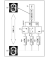
- FIG. 1 is a block diagram of a medical image diagnostic system according to an embodiment.
- FIG. 2 is a schematic diagram of learning applied to the result integration CAD processing server.
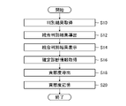
- FIG. 3 is a flow chart showing the procedure of the medical image diagnostic system evaluation method according to the embodiment.
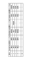
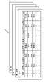
- FIG. 4 is a table showing an example of evaluation results of the CAD processing server.
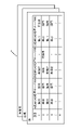
- FIG. 5 is a schematic diagram showing a configuration example of the evaluation result of the CAD processing server.
- FIG. 6 is a schematic diagram showing another configuration example of the evaluation result of the CAD processing server.
- FIG. 7 is a diagram showing an example of a discrimination result screen.
- FIG. 8 is a diagram showing another example of the determination result screen.
- FIG. 1 is a block diagram of a medical image diagnostic system according to an embodiment.
- a medical image diagnostic system 10 shown in the figure includes a modality 12 , an image storage server 14 , a plurality of CAD processing servers 16 , a result integration CAD processing server 18 and a PACS viewer 20 .
- CAD is an abbreviation for Computer-Aided Diagnosis.
- PACS is an abbreviation for Picture Archiving and Communication System.
- each of the modality 12, the image storage server 14, the plurality of CAD processing servers 16, the result integration CAD processing server 18, and the PACS viewer 20 can mutually transmit and receive data via a communication network such as the Internet. connected to
- system a collection of multiple devices is called a system, but the term system may include the concept of devices. That is, the terms system and device can be read interchangeably.
- the modality 12 is an imaging device that captures an image of the inspection target site of the subject and generates a medical image 22 .
- Examples of the modality 12 include an X-ray imaging device, a CT device, an MRI device, a PET device, an ultrasonic device, a CR device using a plane X-ray detector, and the like.
- medical image is synonymous with medical image.
- CT is an abbreviation for Computed Tomography.
- MRI is an abbreviation for Magnetic Resonance Imaging.
- PET is an abbreviation for Positron Emission Tomography.
- CR is an abbreviation for Computed Radiography.
- the image storage server 14 is a server that manages medical images 22 captured using the modality 12 .
- a computer having a large-capacity storage device is used as the image storage server 14 .
- the computer incorporates a program that provides the functionality of the data storage system.
- the image storage server 14 acquires medical images 22 captured using the modality 12 and stores them in a mass storage device.
- a program is synonymous with software.
- the image storage server 14 described in the embodiment is an example of an image storage device that stores medical images.
- the format of the medical image 22 can use the DICOM standard.
- the medical image 22 may be added with DICOM tag information defined in the DICOM standard.
- image may include image data, which is a signal representing an image, as well as the image itself, such as a photograph.
- DICOM is an abbreviation for Digital Imaging and Communications in Medicine.
- FIG. 1 a CAD processing server 16A manufactured by Company A, a CAD processing server 16B manufactured by Company B, and a CAD processing server 16C manufactured by Company C manufactured by three different companies are illustrated as the plurality of CAD processing servers 16.
- FIG. A plurality of CAD processing servers 16 is sufficient, and the number is not limited.
- a plurality of CAD processing servers 16 perform abnormality detection processing for each part on the medical image 22 acquired from the image storage server 14, and determine the presence or absence of abnormality in the medical image.
- Each of the plurality of CAD processing servers 16 can use the same medical image 22 to perform abnormality detection processing on a predetermined site and a predetermined lesion. That is, each of the plurality of CAD processing servers 16 can perform abnormality detection processing for the same site and the same lesion based on the same medical image 22 .
- sites include organs, bones, muscles, ligaments, nerves and blood vessels.
- abnormalities include disease, disease, lesions, and the like.
- the determination result 24 in each of the plurality of CAD processing servers 16 is associated with the medical image 22 to be processed and transmitted to the result integration CAD processing server 18 .
- discrimination can include the concept of "extraction.”
- discrimination may also include concepts such as identification, recognition, inference, estimation and detection.
- the determination result 24A is output from the CAD processing server 16A manufactured by company A
- the determination result 24B is output from the CAD processing server 16B manufactured by company B
- the determination result 24C is output from the CAD processing server 16C manufactured by company C.
- FIG. 1 illustrates a determination result 24A, a determination result 24B, and a determination result 24C using a binary image.
- Each of the plurality of CAD processing servers 16 shown in FIG. 1 includes one or more processors and one or more memories.
- a processor executes instructions contained in programs stored in memory to determine the medical image 22 and output determination results 24 .
- the CAD processing server 16A manufactured by Company A is equipped with a CAD engine 28A manufactured by Company A.
- the CAD engine 28A manufactured by Company A performs abnormality detection processing of the medical image 22 in the CAD processing server 16A manufactured by Company A, and outputs a determination result 24A.
- the CAD processing server 16B manufactured by Company B is equipped with a CAD engine 28B manufactured by Company B.
- the CAD engine 28B manufactured by Company B performs abnormality detection processing of the medical image 22 in the CAD processing server 16B manufactured by Company B, and outputs a determination result 24B.
- the CAD processing server 16C manufactured by Company C is equipped with a CAD engine 28C manufactured by Company C.
- the CAD engine 28C manufactured by Company C performs abnormality detection processing of the medical image 22 in the CAD processing server 16C manufactured by Company C, and outputs a determination result 24C.
- the CAD engine 28A manufactured by Company A, the CAD engine 28B manufactured by Company B, and the CAD engine 28C manufactured by Company C can use trained learning models such as a convolutional neural network called a CNN (Convolutional Neural Network).
- the result integration CAD processing server 18 acquires each determination result 24 for one medical image 22 transmitted from each of the plurality of CAD processing servers 16, and integrates determination based on each determination result 24 for one medical image 22. Result 26 is generated. The integrated determination result 26 is associated with the medical image 22 to be processed, transmitted to the image storage server 14 , and stored in the image storage server 14 .
- a trained learning model is used for the result integration CAD processing server 18 .
- An example of a learning model applied to the results integration CAD processing server 18 is a convolutional neural network.
- a convolutional neural network can adopt a configuration that includes a combination of convolutional layers and pooling layers as part of a plurality of intermediate layers.
- the number of intermediate layers that constitute the convolutional neural network, the processing content of each layer, and the arrangement order of each layer are not limited, and various combinations of structures can be adopted.
- the convolution layer performs a convolution operation using a filter on the nodes in the local region in the previous layer to obtain a feature map.
- the convolution layer is responsible for feature extraction, which extracts from the image the characteristic grayscale structure represented by the filter.
- the pooling layer performs a pooling process of aggregating the local regions of the feature map output from the convolutional layer using representative values.
- the pooling layer generates a new feature map by reducing the resolution of the feature map output from the convolution layer.
- the pooling layer provides robustness so that the target features extracted using the convolution layer are not affected by positional changes. In other words, the pooling layer reduces the sensitivity to positional variations of the target feature.
- the convolutional neural network may include at least one layer of a normalization layer and a fully connected layer in addition to the convolution layer and the pooling layer. Also, each layer of the intermediate layers may optionally include an activation function.
- the normalization layer performs processing to normalize the grayscale structure of the image. For example, the normalization layer performs local contrast normalization on at least one of the output of the convolutional layer and the output of the pooling layer.
- a fully connected layer is a layer that connects all the nodes between adjacent layers.
- a fully connected layer may be placed near the output layer.
- the fully connected layer combines feature maps, from which features are extracted through a convolution layer and a pooling layer, into one node, and outputs feature variables using an activation function.
- the fully connected layer performs class classification using a softmax function or the like based on the output from the fully connected layer.
- the result integrated CAD processing server 18 evaluates CAD processing performance such as CAD processing characteristics and features for each of the plurality of CAD processing servers 16 based on the integrated determination result 26 .
- the result integrated CAD processing server 18 stores the CAD processing performance evaluation results of each of the plurality of CAD processing servers 16 .
- the result integration CAD processing server 18 acquires definitive diagnosis information representing the doctor's definitive diagnosis result, and evaluates the contribution of the CAD processing to the integrated determination result 26 based on the integrated determination result 26 and the doctor's definitive diagnosis result.
- the definitive diagnosis information described in the embodiment is an example of input information representing a doctor's definitive diagnosis result.
- the result-integrated CAD processing server 18 stores the results of contribution evaluation of CAD processing to the integrated determination results 26 of each of the plurality of CAD processing servers 16 . Details of performance evaluation and contribution evaluation for the plurality of CAD processing servers 16 will be described later.
- a computer is used for the result integration CAD processing server 18 .
- the form of the computer may be a personal computer or a workstation.
- the results integration CAD processing server 18 comprises one or more processors 18A and one or more memories 18B.
- Processor 18A executes instructions stored in memory 18B.
- examples of the hardware structure of the processor 18A include a CPU (Central Processing Unit), a GPU (Graphics Processing Unit), a PLD (Programmable Logic Device), and an ASIC (Application Specific Integrated Circuit).
- a CPU is a general-purpose processor that executes programs and acts as various functional units.
- a GPU is a processor specialized for image processing.
- a PLD is a processor whose electrical circuit configuration can be changed after the device is manufactured. Examples of PLDs include FPGAs (Field Programmable Gate Arrays). An ASIC is a processor with dedicated electrical circuitry specifically designed to perform a particular process.
- One processing unit may be composed of one of these various processors, or may be composed of two or more processors of the same type or different types. Examples of combinations of various processors include combinations of one or more FPGAs and one or more CPUs, and combinations of one or more FPGAs and one or more GPUs. Another example of a combination of various processors is a combination of one or more CPUs and one or more GPUs.
- a single processor may be used to configure multiple functional units.
- a combination of one or more CPUs and software such as SoC (System On a Chip), typified by a computer such as a client or server, is applied.
- SoC System On a Chip
- a mode is given in which one processor is configured and this processor is operated as a plurality of functional units.
- IC is an abbreviation for Integrated Circuit.
- various functional units are configured using one or more of the various processors described above as a hardware structure.
- the hardware structure of the various processors described above is, more specifically, an electric circuit combining circuit elements such as semiconductor elements.
- the memory 18B stores instructions to be executed by the processor 18A.
- the memory 18B may include RAM (Random Access Memory) and ROM (Read Only Memory).
- the processor 18A uses the RAM as a work area, executes software using various programs and parameters including a medical image processing program stored in the ROM, and uses the parameters stored in the ROM or the like to integrate results. Various processes of the CAD processing server 18 are executed.
- the result integration CAD processing server 18 may have the functions of a plurality of CAD processing servers 16 .
- Hardware such as the processor 18A of the result integration CAD processing server 18 may be used to execute programs used by multiple CAD processing servers 16 .
- the hardware and software of a plurality of CAD processing servers 16 may be built into the result integration CAD processing server 18 .
- the PACS viewer 20 is a terminal device used by users such as doctors.
- a known image viewer for interpretation is used as the PACS viewer 20 .
- the PACS viewer 20 may be a personal computer, workstation, or tablet terminal.
- the PACS viewer 20 comprises an input device 20A and a display device 20B.
- Examples of the input device 20A include a pointing device such as a mouse and an input device such as a keyboard.
- a user may input instructions to the medical imaging system 10 using the input device 20A.
- the display device 20B functions as a GUI (Graphical User Interface) that displays screens necessary for operations using the input device 20A. Also, the display device 20B displays a medical image captured by the modality 12 . Further, an integrated determination result 26 is displayed as a CAD result 29 on the display device 20B.
- GUI Graphic User Interface
- the display device 20B receives the display signal representing the CAD result 29 and displays the CAD result 29.
- the CAD result 29 includes the integrated determination result 26 .
- the CAD result 29 may include the determination result 24 for each CAD processing server 16 .
- a touch panel display in which the input device 20A and the display device 20B are integrated may be used as the PACS viewer 20.
- the CAD processing server 16A manufactured by Company A, the CAD processing server 16B manufactured by Company B, and the CAD processing server 16C manufactured by Company C described in the embodiment are examples of a first determination device that performs the first determination, and the second determination It is an example of a second discrimination device that implements.
- each of the CAD processing server 16A manufactured by Company A, the CAD processing server 16B manufactured by Company B, and the CAD processing server 16C manufactured by Company C is either a first determination device or a second determination device.
- the CAD processing server 16A manufactured by company A is the first discrimination device
- the CAD processing server 16B manufactured by company B and the CAD processing server 16C manufactured by company C are second discrimination devices.
- the determination result 24A, the determination result 24B, and the determination result 24C described in the embodiment are examples of the first determination result and examples of the second determination result.
- each of the determination result 24A, the determination result 24B, and the determination result 24C is either the first determination result or the second determination result.
- the determination result 24A is the first determination result
- the determination result 24B and the determination result 24C are the second determination result.
- the result integrated CAD processing server 18 described in the embodiment is an example of an integrated determination device.
- FIG. 2 is a schematic diagram of learning applied to the result integration CAD processing server.
- a set of a learning medical image 30 and a correct mask image 32 is used as learning data.
- the medical images 30 for learning used as learning data for the result integration CAD processing server 18 are medical images captured using the same modality as the medical images used as learning data for the plurality of CAD processing servers 16. be done.
- the learning medical images 30 medical images of the same parts and same lesions as the medical images used as learning data for the plurality of CAD processing servers 16 are used.
- the learning medical images 30 medical images that are not used in the learning data of the plurality of CAD processing servers 16 are used.
- FIG. 2 illustrates a lung CT image as a learning medical image 30 and a lung tumor region mask image as a correct mask image 32 .
- a lung mass region mask image can be generated by masking a lung mass region extracted from a CT image of the lung.
- a learning medical image 30 serving as learning data is input to each of the CAD engine 28A manufactured by company A, the CAD engine 28B manufactured by company B, and the CAD engine 28C manufactured by company C.
- Each of the CAD engine 28A manufactured by company A, the CAD engine 28B manufactured by company B, and the CAD engine 28C manufactured by company C performs processing on the learning medical image 30 and acquires a lung tumor labeling image as a temporary correct image 34. Specifically, the CAD engine 28A manufactured by company A obtains a provisional correct image 34A, the CAD engine 28B manufactured by company B obtains a provisional correct image 34B, and the CAD engine 28C manufactured by company C obtains a provisional correct image 34C. get.
- FIG. 2 shows a provisional correct image 34A in which two lung masses 35 are detected, a provisional correct image 34B in which three lung masses 35 are detected, and one lung mass 35 is detected.
- a temporary correct image 34C is illustrated.
- a set of the provisional correct image 34A, the provisional correct image 34B, and the provisional correct image 34C is input to the result integration CNN 18C.
- the result integration CNN 18C performs learning for outputting the correct mask image 32 when a set of the provisional correct image 34A, the provisional correct image 34B, and the provisional correct image 34C is input.
- the temporary correct image 34A, the temporary correct image 34B, and the temporary correct image 34C described in the embodiment are examples of the first correct data and also examples of the second correct data.
- the provisional correct image 34A is the first correct data
- the provisional correct image 34B and the provisional correct image 34C are each the second correct data.
- the term data includes the concepts of signal and information.
- the medical image diagnostic system 10 evaluates the characteristics of each of the multiple CAD processing servers 16 based on the integrated determination result 26 . Further, the medical image diagnostic system 10 evaluates the contribution of each of the plurality of CAD processing servers 16 to the integrated determination result 26 based on the integrated determination result 26 and the doctor's definitive diagnosis result.
- Examples of the characteristics of the CAD processing server 16 include areas it specializes in, lesions it specializes in, and types of medical images it specializes in. A region into which a region is subdivided may be used as a region where the region is good.
- the type of medical image can be understood as the type of modality that generates the medical image. Types of medical images include, for example, MRI images and CT images.
- the medical image diagnostic system 10 can evaluate the characteristics of the CAD processing server 16 and re-learn the result integration CNN 18C based on the evaluation results. For example, when the CAD processing server 16A manufactured by Company A specializes in discrimination of the right lung, the result integration CAD processing server 18 positively uses the discrimination result 24A of the CAD processing server 16A manufactured by A in the discrimination processing of the right lung. Retraining of the result-integrated CNN 18C employed may be performed. In other words, when performing right lung discrimination processing, the result integration CAD processing server 18 weights the discrimination result of the CAD processing server 16 that specializes in right lung discrimination, and re-learns the result integration CNN 18C. conduct.
- the result integration CAD processing server 18 weights the discrimination result of the CAD processing server 16, which is good at discrimination of the part, according to the part to be subjected to discrimination processing, and re-learns the result integration CNN 18C. .
- the degree of contribution representing the contribution of the CAD processing server 16 to the integrated determination result 26 is necessary for each CAD processing server 16 .
- the contribution of the CAD processing server 16 can be derived based on the number of cases adopted as the integrated determination result 26 and the number of cases adopted as the doctor's definitive diagnosis result.
- the user of the medical image diagnostic system 10 can grasp the CAD processing server 16 that contributes relatively little to the improvement of the discrimination accuracy of the integrated discrimination result 26 based on the degree of contribution of each CAD processing server 16 .
- the user of the medical image diagnostic system 10 can stop charging the CAD processing server 16 whose contribution is relatively low and delete it from the lineup. Also, the company managing the CAD processing server 16 may be encouraged to improve the CAD processing server 16 .
- the medical image diagnostic system 10 evaluates the performance of all of the plurality of CAD processing servers 16, calculates the contribution to the integrated determination result 26, and stores it.
- the user of the medical image diagnostic system 10 can grasp the redundancy of the discrimination result 24 and the performance of the CAD processing server 16 each time the number of discrimination processing is repeated, and select one or two of the plurality of CAD processing servers 16. It becomes possible to limit the use to the CAD processing server 16 .
- FIG. 3 is a flow chart showing the procedure of the medical image diagnostic system evaluation method according to the embodiment.
- the result integration CAD processing server 18 shown in FIG. store the set of
- the result integrated CAD processing server 18 derives the integrated determination result 26 based on the set of the determination results 24 acquired in the determination result acquisition step S10, and stores the integrated determination result 26.
- a set of the discrimination result 24A, the discrimination result 24B, and the discrimination result 24C is input to the learned result integration CNN 18C, and the integrated discrimination result 26 is output from the result integration CNN 18C. be done.
- the result integrated CAD processing server 18 displays the integrated determination result 26 on the display device 20B.
- the result integration CAD processing server 18 acquires definitive diagnosis information including the doctor's definitive diagnosis input using the input device 20A.
- the result integration CAD processing server 18 associates the acquired definitive diagnosis information with the integrated determination result 26 and stores it.
- the contribution of each CAD processing server 16 is derived based on the integrated determination result 26 and the definite diagnosis information.
- the contribution of each CAD processing server 16 may use a score that represents the contribution of the determination result 24 to the integrated determination result 26 .
- the result integration CAD processing server 18 stores the contribution degree of each CAD processing server 16 derived in the contribution degree derivation step S18.
- the procedure shown in FIG. 3 may be performed when the integrated determination result 26 of the medical image 22 is newly acquired, or may be performed based on the user's input signal.
- the contribution of each CAD processing server 16 may be updated each time the procedure shown in FIG. 3 is performed.
- FIG. 4 is a table showing an example of evaluation results of the CAD processing server.
- the figure shows the evaluation results of the CAD processing server 16A manufactured by Company A, the CAD processing server 16B manufactured by Company B, and the CAD processing server 16C manufactured by Company C shown in FIG.
- the numerical value in the column labeled "Time" represents the number of determination processes.
- the number of discrimination processes shown in the table of FIG. 4 can be grasped as the identification number of the medical image to be processed.
- Detected in the table shown in FIG. 4 represents a case where an abnormality candidate such as a lesion candidate is detected from the medical image 22.
- Undetected represents a case where an abnormality candidate is not detected from the medical image 22 .
- the CAD processing server 16A manufactured by Company A and the CAD processing server 16C manufactured by Company C detect an abnormality candidate in all of the first to fourth detections.
- the CAD processing server 16B manufactured by Company B detects an abnormality candidate for the second time, but does not detect an abnormality candidate for the first, third, and fourth times.
- “Adopted” in the table shown in FIG. 4 represents the case where the discrimination result is adopted for the doctor's definitive diagnosis.
- “Not Adopted” represents a case where the determination result is not adopted for the doctor's definitive diagnosis.
- the determination result is adopted for the doctor's definitive diagnosis in all of the first to fourth times.
- the CAD processing server 16B manufactured by company B detected an abnormality candidate for the second time, but the determination result was not adopted for the definitive diagnosis by the doctor, and the determination result of the CAD processing server 16A manufactured by company A and the CAD processing server 16C manufactured by company C were not adopted.
- the determination result of is adopted for a definitive diagnosis by a doctor.
- the CAD processing server 16C manufactured by Company C detected an abnormality candidate for the first time, the determination result was not adopted for the doctor's definitive diagnosis, and the determination result of the CAD processing server 16A manufactured by Company A was used for the doctor's definitive diagnosis. be.
- the CAD processing server 16C manufactured by Company C adopts the determination result for the doctor's definitive diagnosis from the second to the fourth times.
- the user can understand that the CAD processing server 16A manufactured by Company A and the CAD processing server 16C manufactured by Company C have similar characteristics. Specifically, in the example shown in FIG. 4 , the CAD processing server 16A manufactured by Company A and the CAD processing server 16C manufactured by Company C detect an abnormality candidate for the same medical image 22 . That is, it can be understood that servers that similarly detect abnormality candidates for the same medical image 22 have similar characteristics. In addition, the CAD processing server 16A manufactured by Company A has a larger number of discrimination results adopted for definitive diagnosis by a doctor than the CAD processing server 16C manufactured by C Company.
- the user can consider stopping the use of the CAD processing server 16C manufactured by company C, which has a relatively low degree of contribution among the CAD processing servers 16A manufactured by company A and CAD processing servers 16C manufactured by company C, which have similar characteristics. Also, the user may consider stopping the use of the CAD processing server 16B manufactured by Company B, which has the lowest degree of contribution.
- the result-integrating CAD processing server 18 may calculate a score representing the degree of contribution of each CAD processing server 16 based on the evaluation results of the CAD processing server 16 shown in FIG. In other words, the result integration CAD processing server 18 may calculate a score representing the degree of contribution based on the detection characteristics (performance) of the abnormality candidate and the adoption result of the definitive diagnosis. Specifically, when an abnormality candidate is detected and adopted for definitive diagnosis, a high score is given; when an abnormality candidate is detected and not adopted for definitive diagnosis, a medium score is given; A low score may be given if not detected.
- the result integration CAD processing server 18 can calculate a comprehensive evaluation score for each CAD processing server 16 by summing the above scores.
- the result integration CAD processing server 18 may issue a warning when the ratio of the number of detections of anomaly candidates to the number of determination processes is less than a specified number, or warn when the number of adoptions in the number of determination processes is less than a specified number. may be notified.
- the score for each CAD processing server 16 described in the embodiment is an example of the first score and an example of the second score.
- the score of each CAD processing server 16A, 16B, 16C is the first score or the second score.
- FIG. 5 is a schematic diagram showing a configuration example of the evaluation result of the CAD processing server.
- FIG. 5 shows evaluation results of the CAD processing server 16 derived for each part. Specifically, FIG. 5 shows the evaluation result of the CAD processing server 16 derived for the lungs, the evaluation result of the CAD processing server 16 derived for the heart, and the evaluation result of the CAD processing server 16 derived for the bronchi.
- the result integration CAD processing server 18 evaluates the CAD processing server 16 for each part and derives the evaluation result of the CAD processing server 16 for each part.
- the medical image diagnostic system 10 can derive the degree of contribution for each CAD processing server 16 for each part. Further, the medical image diagnostic system 10 can evaluate the performance of the parts of the CAD processing server 16, such as a good part, based on the evaluation results of the CAD processing server 16 for each part.
- FIG. 6 is a schematic diagram showing another configuration example of the evaluation result of the CAD processing server.
- FIG. 6 shows the evaluation result of the CAD processing server 16 derived for each area obtained by subdividing the part.
- FIG. 6 illustrates right lung, left lung, lung segment S1 and lung segment S2 as examples of regions.
- the CAD processing server 16 is evaluated separately according to the nature and occurrence location of the pulmonary nodule, and whether or not to use the CAD processing server 16 is determined according to the nature and occurrence location of the pulmonary nodule. can be optimized. In other words, a plurality of CAD servers 16A, 16B, and 16C are evaluated for each region corresponding to the lesion type and occurrence location, and the CAD processing servers 16A, 16B, and 16C suitable for the lesion type and occurrence location are selected. may be made available.
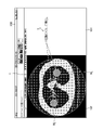
- FIG. 7 is a diagram showing an example of a discrimination result screen.
- a determination result screen 100 shown in FIG. 7 is one aspect of notifying the integrated determination result 26 shown in FIG. 1, and is displayed using the display device 20B shown in FIG.
- the CT image I1 is displayed in the first area 102 , and the marker M1 and the marker M2 surrounding the lesion area detected from the CT image I1 are superimposed on the CT image I1. be.
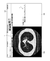
- FIG. 8 is a diagram showing another example of the determination result screen.
- a determination result screen 120 shown in FIG. 8 is displayed using the display device 20B shown in FIG.
- the determination result screen 120 is used when no lesion area is detected from the CT image I1.
- the CT image I1 is displayed in the first area 102.
- FIG. 7 On the other hand, on the determination result screen 120 , the markers M1, M2, etc. shown in FIG . 7 are not displayed.
- the explanation T2 for the CT image I1 is displayed in the second area 104.
- FIG. 8 character information indicating that no abnormality is detected using the CAD processing server 16B manufactured by Company B is displayed as an explanation T2 for the CT image I1.
- the explanatory text T2 shown in FIG . 8 may be displayed together with the explanatory text T1 shown in FIG.
- the result integrated CAD processing server 18 can notify the determination results 24 of the plurality of CAD processing servers 16 shown in FIG. 1 on the determination result display screen that displays the integrated determination results 26.
- the result integration CAD processing server 18 may selectively switch between display and non - display of the explanatory text T1 and the like for the CT image I1.
- display and non - display of the explanatory text T1 and the like may be selectively switched according to selection information input by the user using the input device 20A shown in FIG.
- the medical image diagnostic system, medical image diagnostic system evaluation method, and program according to the embodiments can obtain the following effects.
- a contribution representing the contribution of the determination result 24 to the integrated determination result 26 is derived.
- the CAD processing server 16 that contributes relatively little to improving the accuracy of the integrated determination result 26 can be recognized.
- the degree of contribution representing the contribution of the determination result 24 to the integrated determination result 26 is derived for each part and for each area obtained by subdividing the part. This makes it possible to grasp the strengths and weaknesses of each CAD processing server 16 with respect to parts and regions. In addition, it is possible to grasp whether or not the CAD processing server 16 is required for each part and area.
- a degree of contribution representing the contribution of the determination result 24 to the integrated determination result 26 is derived based on the adoption or non-adoption of the doctor's definitive diagnosis. This allows the CAD processing server 16 to be evaluated based on the doctor's definitive diagnosis.
- the components that make up the medical image diagnostic system shown in FIG. 1 can be integrated and separated as appropriate.
- the result integration CAD processing server 18 and the like may be configured using a plurality of computers.
- part or all of the image storage server 14 and part or all of the result integration CAD processing server 18 may be configured using one computer.
Landscapes
- Engineering & Computer Science (AREA)
- Health & Medical Sciences (AREA)
- Medical Informatics (AREA)
- General Health & Medical Sciences (AREA)
- Public Health (AREA)
- Nuclear Medicine, Radiotherapy & Molecular Imaging (AREA)
- Radiology & Medical Imaging (AREA)
- Primary Health Care (AREA)
- Epidemiology (AREA)
- Biomedical Technology (AREA)
- Physics & Mathematics (AREA)
- Pathology (AREA)
- Life Sciences & Earth Sciences (AREA)
- Computer Vision & Pattern Recognition (AREA)
- Databases & Information Systems (AREA)
- Data Mining & Analysis (AREA)
- Theoretical Computer Science (AREA)
- General Physics & Mathematics (AREA)
- Quality & Reliability (AREA)
- High Energy & Nuclear Physics (AREA)
- Biophysics (AREA)
- Optics & Photonics (AREA)
- Heart & Thoracic Surgery (AREA)
- Molecular Biology (AREA)
- Surgery (AREA)
- Animal Behavior & Ethology (AREA)
- Veterinary Medicine (AREA)
- Measuring And Recording Apparatus For Diagnosis (AREA)
Priority Applications (3)
| Application Number | Priority Date | Filing Date | Title |
|---|---|---|---|
| EP22824604.7A EP4356837A4 (en) | 2021-06-17 | 2022-03-24 | MEDICAL IMAGE DIAGNOSTIC SYSTEM, METHOD FOR EVALUATING A MEDICAL IMAGE DIAGNOSTIC SYSTEM AND PROGRAM |
| JP2023529592A JP7756717B2 (ja) | 2021-06-17 | 2022-03-24 | 医用画像診断システム、医用画像診断システム評価方法及びプログラム |
| US18/533,050 US20240105315A1 (en) | 2021-06-17 | 2023-12-07 | Medical image diagnostic system, medical image diagnostic system evaluation method, and program |
Applications Claiming Priority (2)
| Application Number | Priority Date | Filing Date | Title |
|---|---|---|---|
| JP2021100612 | 2021-06-17 | ||
| JP2021-100612 | 2021-06-17 |
Related Child Applications (1)
| Application Number | Title | Priority Date | Filing Date |
|---|---|---|---|
| US18/533,050 Continuation US20240105315A1 (en) | 2021-06-17 | 2023-12-07 | Medical image diagnostic system, medical image diagnostic system evaluation method, and program |
Publications (1)
| Publication Number | Publication Date |
|---|---|
| WO2022264608A1 true WO2022264608A1 (ja) | 2022-12-22 |
Family
ID=84527012
Family Applications (1)
| Application Number | Title | Priority Date | Filing Date |
|---|---|---|---|
| PCT/JP2022/014071 Ceased WO2022264608A1 (ja) | 2021-06-17 | 2022-03-24 | 医用画像診断システム、医用画像診断システム評価方法及びプログラム |
Country Status (4)
| Country | Link |
|---|---|
| US (1) | US20240105315A1 (cg-RX-API-DMAC7.html) |
| EP (1) | EP4356837A4 (cg-RX-API-DMAC7.html) |
| JP (1) | JP7756717B2 (cg-RX-API-DMAC7.html) |
| WO (1) | WO2022264608A1 (cg-RX-API-DMAC7.html) |
Citations (5)
| Publication number | Priority date | Publication date | Assignee | Title |
|---|---|---|---|---|
| JP2006043007A (ja) * | 2004-08-02 | 2006-02-16 | Fujitsu Ltd | 診断支援プログラムおよび診断支援装置 |
| JP2006167289A (ja) | 2004-12-17 | 2006-06-29 | Toshiba Corp | 複合画像診断支援システム、複合画像診断支援装置、及び医用画像データベース作成方法 |
| JP2013005841A (ja) * | 2011-06-22 | 2013-01-10 | Hitachi Ltd | 画像処理フロー評価方法及び当該方法を実行する画像処理装置 |
| US20140101080A1 (en) * | 2012-09-28 | 2014-04-10 | Samsung Electronics Co., Ltd. | Apparatus and method of diagnosis using diagnostic models |
| US20200202523A1 (en) * | 2018-12-21 | 2020-06-25 | Infinitt Healthcare Co., Ltd. | Medical image diagnosis assistant apparatus and method for generating evaluation scores for plurality of medical image diagnosis algorithms |
Family Cites Families (6)
| Publication number | Priority date | Publication date | Assignee | Title |
|---|---|---|---|---|
| US20040100476A1 (en) * | 2002-11-25 | 2004-05-27 | Morita Mark M. | Method and apparatus for viewing computer aided detection and diagnosis results |
| US10169863B2 (en) * | 2015-06-12 | 2019-01-01 | International Business Machines Corporation | Methods and systems for automatically determining a clinical image or portion thereof for display to a diagnosing physician |
| KR101818074B1 (ko) * | 2017-07-20 | 2018-01-12 | (주)제이엘케이인스펙션 | 인공지능 기반 의료용 자동 진단 보조 방법 및 그 시스템 |
| JP7033202B2 (ja) * | 2018-06-28 | 2022-03-09 | 富士フイルム株式会社 | 医用画像処理装置及び方法、機械学習システム、プログラム並びに記憶媒体 |
| JP7129870B2 (ja) * | 2018-10-01 | 2022-09-02 | 富士フイルム株式会社 | 疾患領域を判別する判別器の学習装置、方法及びプログラム、疾患領域を判別する判別器、並びに疾患領域判別装置及びプログラム |
| JP7349345B2 (ja) * | 2019-12-23 | 2023-09-22 | 富士フイルムヘルスケア株式会社 | 画像診断支援装置、画像診断支援プログラム、及び、それを備えた医用画像取得装置 |
-
2022
- 2022-03-24 JP JP2023529592A patent/JP7756717B2/ja active Active
- 2022-03-24 EP EP22824604.7A patent/EP4356837A4/en active Pending
- 2022-03-24 WO PCT/JP2022/014071 patent/WO2022264608A1/ja not_active Ceased
-
2023
- 2023-12-07 US US18/533,050 patent/US20240105315A1/en active Pending
Patent Citations (5)
| Publication number | Priority date | Publication date | Assignee | Title |
|---|---|---|---|---|
| JP2006043007A (ja) * | 2004-08-02 | 2006-02-16 | Fujitsu Ltd | 診断支援プログラムおよび診断支援装置 |
| JP2006167289A (ja) | 2004-12-17 | 2006-06-29 | Toshiba Corp | 複合画像診断支援システム、複合画像診断支援装置、及び医用画像データベース作成方法 |
| JP2013005841A (ja) * | 2011-06-22 | 2013-01-10 | Hitachi Ltd | 画像処理フロー評価方法及び当該方法を実行する画像処理装置 |
| US20140101080A1 (en) * | 2012-09-28 | 2014-04-10 | Samsung Electronics Co., Ltd. | Apparatus and method of diagnosis using diagnostic models |
| US20200202523A1 (en) * | 2018-12-21 | 2020-06-25 | Infinitt Healthcare Co., Ltd. | Medical image diagnosis assistant apparatus and method for generating evaluation scores for plurality of medical image diagnosis algorithms |
Non-Patent Citations (1)
| Title |
|---|
| See also references of EP4356837A4 |
Also Published As
| Publication number | Publication date |
|---|---|
| EP4356837A4 (en) | 2024-08-14 |
| JPWO2022264608A1 (cg-RX-API-DMAC7.html) | 2022-12-22 |
| US20240105315A1 (en) | 2024-03-28 |
| JP7756717B2 (ja) | 2025-10-20 |
| EP4356837A1 (en) | 2024-04-24 |
Similar Documents
| Publication | Publication Date | Title |
|---|---|---|
| US20260004918A1 (en) | System and Method for Interpretation of Multiple Medial Images using Deep Learning | |
| US10692602B1 (en) | Structuring free text medical reports with forced taxonomies | |
| AU2004252917A1 (en) | CAD (computer-aided decision) support for medical imaging using machine learning to adapt CAD process with knowledge collected during routine use of CAD system | |
| Yang et al. | Deep rib fracture instance segmentation and classification from ct on the ribfrac challenge | |
| JP7783889B2 (ja) | 医用画像診断システム、医用画像診断システムの作動方法及びプログラム | |
| US20090041322A1 (en) | Computer Assisted Detection of Polyps Within Lumen Using Enhancement of Concave Area | |
| EP4235566A1 (en) | Method and system for determining a change of an anatomical abnormality depicted in medical image data | |
| Rodríguez et al. | Computer aided detection and diagnosis in medical imaging: a review of clinical and educational applications | |
| Dong et al. | A novel end‐to‐end deep learning solution for coronary artery segmentation from CCTA | |
| US11923069B2 (en) | Medical document creation support apparatus, method and program, learned model, and learning apparatus, method and program | |
| EP4316378A1 (en) | Medical image processing device, method for operating medical image processing device, and operation program for medical image processing device | |
| US20220076796A1 (en) | Medical document creation apparatus, method and program, learning device, method and program, and trained model | |
| Li et al. | LT-YOLO: long-term temporal enhanced YOLO for stenosis detection on invasive coronary angiography | |
| Salama et al. | Machine learning and deep learning covid-19 diagnosis system: key achievements, lessons learned, and a transfer learning algorithm: GM Salama et al. | |
| JP2026020375A (ja) | 医用画像診断システム、医用画像診断システムの作動方法及びプログラム | |
| Keshavamurthy et al. | Weakly supervised pneumonia localization in chest X‐rays using generative adversarial networks | |
| CN111462203A (zh) | Dr病灶演化分析装置和方法 | |
| Arfat et al. | Role of artificial intelligence in medical radiology and imaging | |
| JP7756717B2 (ja) | 医用画像診断システム、医用画像診断システム評価方法及びプログラム | |
| CN117476185B (zh) | 医学影像展示方法、装置、电子设备和存储介质 | |
| Rajaraman et al. | Improved TB classification using bone-suppressed chest radiographs | |
| KR20250079567A (ko) | 골 연령 정보의 출력 방법 | |
| Mohan et al. | Slice-Level Detection of Cervical Vertebral Fractures from CT Images Using a Custom CNN | |
| US20240266026A1 (en) | Information processing apparatus, operation method of information processing apparatus, and program | |
| Tahir et al. | A Methodical Review on the Segmentation Types and Techniques of Medical Images |
Legal Events
| Date | Code | Title | Description |
|---|---|---|---|
| 121 | Ep: the epo has been informed by wipo that ep was designated in this application |
Ref document number: 22824604 Country of ref document: EP Kind code of ref document: A1 |
|
| WWE | Wipo information: entry into national phase |
Ref document number: 2023529592 Country of ref document: JP |
|
| WWE | Wipo information: entry into national phase |
Ref document number: 2022824604 Country of ref document: EP |
|
| NENP | Non-entry into the national phase |
Ref country code: DE |
|
| ENP | Entry into the national phase |
Ref document number: 2022824604 Country of ref document: EP Effective date: 20240117 |