WO2022239820A1 - Anti-human pd-1 agonist antibody for treating or preventing inflammatory diseases, and pharmaceutical composition containing same - Google Patents
Anti-human pd-1 agonist antibody for treating or preventing inflammatory diseases, and pharmaceutical composition containing same Download PDFInfo
- Publication number
- WO2022239820A1 WO2022239820A1 PCT/JP2022/020011 JP2022020011W WO2022239820A1 WO 2022239820 A1 WO2022239820 A1 WO 2022239820A1 JP 2022020011 W JP2022020011 W JP 2022020011W WO 2022239820 A1 WO2022239820 A1 WO 2022239820A1
- Authority
- WO
- WIPO (PCT)
- Prior art keywords
- antibody
- human
- seq
- functional fragment
- region
- Prior art date
Links
- 239000008194 pharmaceutical composition Substances 0.000 title claims description 11
- 239000000556 agonist Substances 0.000 title abstract description 54
- 208000027866 inflammatory disease Diseases 0.000 title abstract description 24
- 239000012634 fragment Substances 0.000 claims abstract description 108
- 101000611936 Homo sapiens Programmed cell death protein 1 Proteins 0.000 claims abstract description 98
- 102000048362 human PDCD1 Human genes 0.000 claims abstract description 95
- 108010021472 Fc gamma receptor IIB Proteins 0.000 claims description 76
- 102100029205 Low affinity immunoglobulin gamma Fc region receptor II-b Human genes 0.000 claims description 75
- 125000003275 alpha amino acid group Chemical group 0.000 claims description 74
- 230000027455 binding Effects 0.000 claims description 70
- 230000001270 agonistic effect Effects 0.000 claims description 69
- 108010087819 Fc receptors Proteins 0.000 claims description 39
- 102000009109 Fc receptors Human genes 0.000 claims description 39
- 108010047041 Complementarity Determining Regions Proteins 0.000 claims description 38
- 230000001976 improved effect Effects 0.000 claims description 32
- 238000012360 testing method Methods 0.000 claims description 25
- 238000000034 method Methods 0.000 claims description 23
- 101000917858 Homo sapiens Low affinity immunoglobulin gamma Fc region receptor III-A Proteins 0.000 claims description 22
- 238000005259 measurement Methods 0.000 claims description 22
- 150000001413 amino acids Chemical class 0.000 claims description 16
- 230000004048 modification Effects 0.000 claims description 13
- 238000012986 modification Methods 0.000 claims description 13
- 150000007523 nucleic acids Chemical class 0.000 claims description 12
- 238000002198 surface plasmon resonance spectroscopy Methods 0.000 claims description 11
- 230000006872 improvement Effects 0.000 claims description 10
- 238000005516 engineering process Methods 0.000 claims description 9
- 238000012258 culturing Methods 0.000 claims description 8
- 108020004707 nucleic acids Proteins 0.000 claims description 8
- 102000039446 nucleic acids Human genes 0.000 claims description 8
- 102000005962 receptors Human genes 0.000 claims description 7
- 108020003175 receptors Proteins 0.000 claims description 7
- 239000013598 vector Substances 0.000 claims description 7
- 239000003937 drug carrier Substances 0.000 claims description 4
- 101000917826 Homo sapiens Low affinity immunoglobulin gamma Fc region receptor II-a Proteins 0.000 claims description 3
- 230000004075 alteration Effects 0.000 claims 1
- 230000000694 effects Effects 0.000 abstract description 107
- 239000003814 drug Substances 0.000 abstract description 24
- 229940079593 drug Drugs 0.000 abstract description 18
- 210000004027 cell Anatomy 0.000 description 181
- 101100519207 Mus musculus Pdcd1 gene Proteins 0.000 description 178
- 210000001744 T-lymphocyte Anatomy 0.000 description 124
- 229940122544 PD-1 agonist Drugs 0.000 description 88
- 241000699670 Mus sp. Species 0.000 description 66
- 238000011156 evaluation Methods 0.000 description 39
- 230000001506 immunosuppresive effect Effects 0.000 description 38
- 239000000427 antigen Substances 0.000 description 31
- 102000036639 antigens Human genes 0.000 description 31
- 108091007433 antigens Proteins 0.000 description 31
- 208000009329 Graft vs Host Disease Diseases 0.000 description 28
- 208000024908 graft versus host disease Diseases 0.000 description 28
- 230000010056 antibody-dependent cellular cytotoxicity Effects 0.000 description 27
- 230000006698 induction Effects 0.000 description 26
- 230000003110 anti-inflammatory effect Effects 0.000 description 25
- 241000699666 Mus <mouse, genus> Species 0.000 description 24
- 210000004408 hybridoma Anatomy 0.000 description 23
- 238000011282 treatment Methods 0.000 description 23
- 230000002401 inhibitory effect Effects 0.000 description 21
- 230000035772 mutation Effects 0.000 description 19
- 210000003719 b-lymphocyte Anatomy 0.000 description 18
- 208000006673 asthma Diseases 0.000 description 17
- 208000037265 diseases, disorders, signs and symptoms Diseases 0.000 description 17
- 230000001419 dependent effect Effects 0.000 description 16
- 230000004044 response Effects 0.000 description 16
- 206010028980 Neoplasm Diseases 0.000 description 15
- 230000000903 blocking effect Effects 0.000 description 15
- 239000000872 buffer Substances 0.000 description 15
- 201000010099 disease Diseases 0.000 description 15
- 108090000623 proteins and genes Proteins 0.000 description 15
- 206010061218 Inflammation Diseases 0.000 description 14
- 102100037850 Interferon gamma Human genes 0.000 description 14
- 108010074328 Interferon-gamma Proteins 0.000 description 14
- 238000004458 analytical method Methods 0.000 description 14
- 201000011510 cancer Diseases 0.000 description 14
- 230000001684 chronic effect Effects 0.000 description 14
- 230000028993 immune response Effects 0.000 description 14
- 230000004054 inflammatory process Effects 0.000 description 14
- 206010025135 lupus erythematosus Diseases 0.000 description 14
- 210000003819 peripheral blood mononuclear cell Anatomy 0.000 description 14
- 101710099301 Low affinity immunoglobulin gamma Fc region receptor III-A Proteins 0.000 description 13
- 102100029193 Low affinity immunoglobulin gamma Fc region receptor III-A Human genes 0.000 description 13
- 229940024606 amino acid Drugs 0.000 description 13
- 235000001014 amino acid Nutrition 0.000 description 13
- 108010074708 B7-H1 Antigen Proteins 0.000 description 12
- 102000008096 B7-H1 Antigen Human genes 0.000 description 12
- 201000009961 allergic asthma Diseases 0.000 description 12
- 239000013604 expression vector Substances 0.000 description 12
- 230000007246 mechanism Effects 0.000 description 12
- 239000002953 phosphate buffered saline Substances 0.000 description 12
- 208000024891 symptom Diseases 0.000 description 12
- 238000012546 transfer Methods 0.000 description 12
- 238000002965 ELISA Methods 0.000 description 11
- 238000001994 activation Methods 0.000 description 11
- 230000001154 acute effect Effects 0.000 description 11
- 210000000612 antigen-presenting cell Anatomy 0.000 description 11
- 210000004369 blood Anatomy 0.000 description 11
- 239000008280 blood Substances 0.000 description 11
- LOKCTEFSRHRXRJ-UHFFFAOYSA-I dipotassium trisodium dihydrogen phosphate hydrogen phosphate dichloride Chemical compound P(=O)(O)(O)[O-].[K+].P(=O)(O)([O-])[O-].[Na+].[Na+].[Cl-].[K+].[Cl-].[Na+] LOKCTEFSRHRXRJ-UHFFFAOYSA-I 0.000 description 11
- 239000000243 solution Substances 0.000 description 11
- 210000000952 spleen Anatomy 0.000 description 11
- 230000001629 suppression Effects 0.000 description 11
- VDABVNMGKGUPEY-UHFFFAOYSA-N 6-carboxyfluorescein succinimidyl ester Chemical compound C=1C(O)=CC=C2C=1OC1=CC(O)=CC=C1C2(C1=C2)OC(=O)C1=CC=C2C(=O)ON1C(=O)CCC1=O VDABVNMGKGUPEY-UHFFFAOYSA-N 0.000 description 10
- 230000004913 activation Effects 0.000 description 10
- 239000012228 culture supernatant Substances 0.000 description 10
- 230000008595 infiltration Effects 0.000 description 10
- 238000001764 infiltration Methods 0.000 description 10
- 239000002609 medium Substances 0.000 description 10
- 108090000765 processed proteins & peptides Proteins 0.000 description 10
- 230000000638 stimulation Effects 0.000 description 10
- 238000006467 substitution reaction Methods 0.000 description 10
- 201000002491 encephalomyelitis Diseases 0.000 description 9
- 230000004073 interleukin-2 production Effects 0.000 description 9
- 230000000704 physical effect Effects 0.000 description 9
- 238000011740 C57BL/6 mouse Methods 0.000 description 8
- 206010033799 Paralysis Diseases 0.000 description 8
- 206010047115 Vasculitis Diseases 0.000 description 8
- 208000026935 allergic disease Diseases 0.000 description 8
- 206010009887 colitis Diseases 0.000 description 8
- 230000003247 decreasing effect Effects 0.000 description 8
- 230000006870 function Effects 0.000 description 8
- 230000003993 interaction Effects 0.000 description 8
- 238000004519 manufacturing process Methods 0.000 description 8
- 230000001404 mediated effect Effects 0.000 description 8
- 238000007799 mixed lymphocyte reaction assay Methods 0.000 description 8
- 102000004169 proteins and genes Human genes 0.000 description 8
- 230000002829 reductive effect Effects 0.000 description 8
- 210000001519 tissue Anatomy 0.000 description 8
- 102000004127 Cytokines Human genes 0.000 description 7
- 108090000695 Cytokines Proteins 0.000 description 7
- 230000006044 T cell activation Effects 0.000 description 7
- 230000007423 decrease Effects 0.000 description 7
- 210000003979 eosinophil Anatomy 0.000 description 7
- 210000004072 lung Anatomy 0.000 description 7
- 239000000203 mixture Substances 0.000 description 7
- 235000018102 proteins Nutrition 0.000 description 7
- 206010039073 rheumatoid arthritis Diseases 0.000 description 7
- 230000001225 therapeutic effect Effects 0.000 description 7
- 230000004580 weight loss Effects 0.000 description 7
- 208000023275 Autoimmune disease Diseases 0.000 description 6
- 102100024222 B-lymphocyte antigen CD19 Human genes 0.000 description 6
- 108010021468 Fc gamma receptor IIA Proteins 0.000 description 6
- 101000980825 Homo sapiens B-lymphocyte antigen CD19 Proteins 0.000 description 6
- 102100029204 Low affinity immunoglobulin gamma Fc region receptor II-a Human genes 0.000 description 6
- HEMHJVSKTPXQMS-UHFFFAOYSA-M Sodium hydroxide Chemical compound [OH-].[Na+] HEMHJVSKTPXQMS-UHFFFAOYSA-M 0.000 description 6
- 108091008874 T cell receptors Proteins 0.000 description 6
- 102000016266 T-Cell Antigen Receptors Human genes 0.000 description 6
- 239000002671 adjuvant Substances 0.000 description 6
- 238000010586 diagram Methods 0.000 description 6
- 239000012530 fluid Substances 0.000 description 6
- 102000037865 fusion proteins Human genes 0.000 description 6
- 108020001507 fusion proteins Proteins 0.000 description 6
- 210000005260 human cell Anatomy 0.000 description 6
- 229910052739 hydrogen Inorganic materials 0.000 description 6
- 108010074108 interleukin-21 Proteins 0.000 description 6
- 230000000069 prophylactic effect Effects 0.000 description 6
- 210000004989 spleen cell Anatomy 0.000 description 6
- 201000000596 systemic lupus erythematosus Diseases 0.000 description 6
- 102100036475 Alanine aminotransferase 1 Human genes 0.000 description 5
- 108010082126 Alanine transaminase Proteins 0.000 description 5
- 108010003415 Aspartate Aminotransferases Proteins 0.000 description 5
- 102000004625 Aspartate Aminotransferases Human genes 0.000 description 5
- 108020004414 DNA Proteins 0.000 description 5
- 241000282412 Homo Species 0.000 description 5
- 101100005713 Homo sapiens CD4 gene Proteins 0.000 description 5
- 108010002350 Interleukin-2 Proteins 0.000 description 5
- 102100023302 Myelin-oligodendrocyte glycoprotein Human genes 0.000 description 5
- 201000009594 Systemic Scleroderma Diseases 0.000 description 5
- 206010042953 Systemic sclerosis Diseases 0.000 description 5
- 206010067584 Type 1 diabetes mellitus Diseases 0.000 description 5
- 239000006285 cell suspension Substances 0.000 description 5
- 230000016396 cytokine production Effects 0.000 description 5
- 230000002950 deficient Effects 0.000 description 5
- 238000001514 detection method Methods 0.000 description 5
- 229910052731 fluorine Inorganic materials 0.000 description 5
- 210000001102 germinal center b cell Anatomy 0.000 description 5
- 210000002865 immune cell Anatomy 0.000 description 5
- 230000036039 immunity Effects 0.000 description 5
- 230000003053 immunization Effects 0.000 description 5
- 239000007924 injection Substances 0.000 description 5
- 238000002347 injection Methods 0.000 description 5
- 239000000178 monomer Substances 0.000 description 5
- 201000006417 multiple sclerosis Diseases 0.000 description 5
- 210000004180 plasmocyte Anatomy 0.000 description 5
- 238000000746 purification Methods 0.000 description 5
- 230000011664 signaling Effects 0.000 description 5
- 210000003462 vein Anatomy 0.000 description 5
- 238000005406 washing Methods 0.000 description 5
- SQTFKIKSQNCWGJ-KCDKBNATSA-N (2s,3r,4r,5s)-2-fluoro-3,4,5-trihydroxyhexanal Chemical compound C[C@H](O)[C@@H](O)[C@@H](O)[C@H](F)C=O SQTFKIKSQNCWGJ-KCDKBNATSA-N 0.000 description 4
- HZAXFHJVJLSVMW-UHFFFAOYSA-N 2-Aminoethan-1-ol Chemical compound NCCO HZAXFHJVJLSVMW-UHFFFAOYSA-N 0.000 description 4
- 208000003950 B-cell lymphoma Diseases 0.000 description 4
- 108091003079 Bovine Serum Albumin Proteins 0.000 description 4
- 102100031658 C-X-C chemokine receptor type 5 Human genes 0.000 description 4
- 101000609767 Dromaius novaehollandiae Ovalbumin Proteins 0.000 description 4
- 101000922405 Homo sapiens C-X-C chemokine receptor type 5 Proteins 0.000 description 4
- 101000599940 Homo sapiens Interferon gamma Proteins 0.000 description 4
- 101001117317 Homo sapiens Programmed cell death 1 ligand 1 Proteins 0.000 description 4
- 206010020751 Hypersensitivity Diseases 0.000 description 4
- 208000022559 Inflammatory bowel disease Diseases 0.000 description 4
- 108090000176 Interleukin-13 Proteins 0.000 description 4
- 108090000978 Interleukin-4 Proteins 0.000 description 4
- 108010002616 Interleukin-5 Proteins 0.000 description 4
- 101100066431 Mus musculus Fcgr2 gene Proteins 0.000 description 4
- 206010034277 Pemphigoid Diseases 0.000 description 4
- 201000004681 Psoriasis Diseases 0.000 description 4
- 108010055044 Tetanus Toxin Proteins 0.000 description 4
- 206010047642 Vitiligo Diseases 0.000 description 4
- 208000024340 acute graft versus host disease Diseases 0.000 description 4
- 230000000172 allergic effect Effects 0.000 description 4
- 150000001412 amines Chemical class 0.000 description 4
- 125000000539 amino acid group Chemical group 0.000 description 4
- 208000010668 atopic eczema Diseases 0.000 description 4
- 210000001072 colon Anatomy 0.000 description 4
- 238000010168 coupling process Methods 0.000 description 4
- 238000004132 cross linking Methods 0.000 description 4
- 238000010494 dissociation reaction Methods 0.000 description 4
- 230000005593 dissociations Effects 0.000 description 4
- 238000002474 experimental method Methods 0.000 description 4
- 238000000684 flow cytometry Methods 0.000 description 4
- 238000009472 formulation Methods 0.000 description 4
- 239000001963 growth medium Substances 0.000 description 4
- 102000048776 human CD274 Human genes 0.000 description 4
- 210000004969 inflammatory cell Anatomy 0.000 description 4
- 210000005087 mononuclear cell Anatomy 0.000 description 4
- 230000026731 phosphorylation Effects 0.000 description 4
- 238000006366 phosphorylation reaction Methods 0.000 description 4
- 239000000523 sample Substances 0.000 description 4
- 210000004988 splenocyte Anatomy 0.000 description 4
- 229940118376 tetanus toxin Drugs 0.000 description 4
- XLYOFNOQVPJJNP-UHFFFAOYSA-N water Substances O XLYOFNOQVPJJNP-UHFFFAOYSA-N 0.000 description 4
- 208000023328 Basedow disease Diseases 0.000 description 3
- 229920002307 Dextran Polymers 0.000 description 3
- 238000012413 Fluorescence activated cell sorting analysis Methods 0.000 description 3
- 208000015023 Graves' disease Diseases 0.000 description 3
- 206010019939 Herpes gestationis Diseases 0.000 description 3
- 101001057504 Homo sapiens Interferon-stimulated gene 20 kDa protein Proteins 0.000 description 3
- 101001055144 Homo sapiens Interleukin-2 receptor subunit alpha Proteins 0.000 description 3
- 101001061851 Homo sapiens V(D)J recombination-activating protein 2 Proteins 0.000 description 3
- 108010073807 IgG Receptors Proteins 0.000 description 3
- 102000009490 IgG Receptors Human genes 0.000 description 3
- 206010062016 Immunosuppression Diseases 0.000 description 3
- 102100027268 Interferon-stimulated gene 20 kDa protein Human genes 0.000 description 3
- 208000005777 Lupus Nephritis Diseases 0.000 description 3
- 108091054438 MHC class II family Proteins 0.000 description 3
- 102000043131 MHC class II family Human genes 0.000 description 3
- 208000008223 Pemphigoid Gestationis Diseases 0.000 description 3
- 230000006052 T cell proliferation Effects 0.000 description 3
- 102100029591 V(D)J recombination-activating protein 2 Human genes 0.000 description 3
- 241000700605 Viruses Species 0.000 description 3
- 230000009471 action Effects 0.000 description 3
- 230000007815 allergy Effects 0.000 description 3
- 230000005875 antibody response Effects 0.000 description 3
- 239000011324 bead Substances 0.000 description 3
- 230000008901 benefit Effects 0.000 description 3
- 230000037396 body weight Effects 0.000 description 3
- 238000003501 co-culture Methods 0.000 description 3
- 230000000875 corresponding effect Effects 0.000 description 3
- 210000004748 cultured cell Anatomy 0.000 description 3
- 239000000428 dust Substances 0.000 description 3
- 239000000839 emulsion Substances 0.000 description 3
- 230000001747 exhibiting effect Effects 0.000 description 3
- 238000001943 fluorescence-activated cell sorting Methods 0.000 description 3
- 238000002649 immunization Methods 0.000 description 3
- 238000000338 in vitro Methods 0.000 description 3
- 238000001727 in vivo Methods 0.000 description 3
- 230000001939 inductive effect Effects 0.000 description 3
- 238000011813 knockout mouse model Methods 0.000 description 3
- 210000004185 liver Anatomy 0.000 description 3
- 210000001165 lymph node Anatomy 0.000 description 3
- 210000004698 lymphocyte Anatomy 0.000 description 3
- 210000004400 mucous membrane Anatomy 0.000 description 3
- 239000013641 positive control Substances 0.000 description 3
- 230000003389 potentiating effect Effects 0.000 description 3
- 239000000843 powder Substances 0.000 description 3
- 102000004196 processed proteins & peptides Human genes 0.000 description 3
- 239000012488 sample solution Substances 0.000 description 3
- 238000003998 size exclusion chromatography high performance liquid chromatography Methods 0.000 description 3
- 238000010186 staining Methods 0.000 description 3
- 239000000126 substance Substances 0.000 description 3
- 239000000725 suspension Substances 0.000 description 3
- 230000008685 targeting Effects 0.000 description 3
- 229940124597 therapeutic agent Drugs 0.000 description 3
- NFGXHKASABOEEW-UHFFFAOYSA-N 1-methylethyl 11-methoxy-3,7,11-trimethyl-2,4-dodecadienoate Chemical compound COC(C)(C)CCCC(C)CC=CC(C)=CC(=O)OC(C)C NFGXHKASABOEEW-UHFFFAOYSA-N 0.000 description 2
- 208000026872 Addison Disease Diseases 0.000 description 2
- 206010001367 Adrenal insufficiency Diseases 0.000 description 2
- 102100021266 Alpha-(1,6)-fucosyltransferase Human genes 0.000 description 2
- 101710146120 Alpha-(1,6)-fucosyltransferase Proteins 0.000 description 2
- 208000002267 Anti-neutrophil cytoplasmic antibody-associated vasculitis Diseases 0.000 description 2
- 208000003343 Antiphospholipid Syndrome Diseases 0.000 description 2
- 206010069002 Autoimmune pancreatitis Diseases 0.000 description 2
- 241000894006 Bacteria Species 0.000 description 2
- 101150013553 CD40 gene Proteins 0.000 description 2
- 108091033409 CRISPR Proteins 0.000 description 2
- 108010021064 CTLA-4 Antigen Proteins 0.000 description 2
- 102000008203 CTLA-4 Antigen Human genes 0.000 description 2
- 229940045513 CTLA4 antagonist Drugs 0.000 description 2
- 241000283707 Capra Species 0.000 description 2
- 206010008609 Cholangitis sclerosing Diseases 0.000 description 2
- 108020004705 Codon Proteins 0.000 description 2
- 206010009900 Colitis ulcerative Diseases 0.000 description 2
- 208000035473 Communicable disease Diseases 0.000 description 2
- 206010056370 Congestive cardiomyopathy Diseases 0.000 description 2
- 201000004624 Dermatitis Diseases 0.000 description 2
- 206010012438 Dermatitis atopic Diseases 0.000 description 2
- 201000003066 Diffuse Scleroderma Diseases 0.000 description 2
- 201000010046 Dilated cardiomyopathy Diseases 0.000 description 2
- 208000019872 Drug Eruptions Diseases 0.000 description 2
- 208000032678 Fixed drug eruption Diseases 0.000 description 2
- 208000007465 Giant cell arteritis Diseases 0.000 description 2
- 208000001204 Hashimoto Disease Diseases 0.000 description 2
- 208000030836 Hashimoto thyroiditis Diseases 0.000 description 2
- 208000035186 Hemolytic Autoimmune Anemia Diseases 0.000 description 2
- 101000914514 Homo sapiens T-cell-specific surface glycoprotein CD28 Proteins 0.000 description 2
- 101000611023 Homo sapiens Tumor necrosis factor receptor superfamily member 6 Proteins 0.000 description 2
- 206010020850 Hyperthyroidism Diseases 0.000 description 2
- 206010021245 Idiopathic thrombocytopenic purpura Diseases 0.000 description 2
- 108060003951 Immunoglobulin Proteins 0.000 description 2
- 208000004187 Immunoglobulin G4-Related Disease Diseases 0.000 description 2
- 102100022297 Integrin alpha-X Human genes 0.000 description 2
- 102000013691 Interleukin-17 Human genes 0.000 description 2
- 206010059176 Juvenile idiopathic arthritis Diseases 0.000 description 2
- 108060001084 Luciferase Proteins 0.000 description 2
- 241001465754 Metazoa Species 0.000 description 2
- 241001529936 Murinae Species 0.000 description 2
- 101100407308 Mus musculus Pdcd1lg2 gene Proteins 0.000 description 2
- NWIBSHFKIJFRCO-WUDYKRTCSA-N Mytomycin Chemical compound C1N2C(C(C(C)=C(N)C3=O)=O)=C3[C@@H](COC(N)=O)[C@@]2(OC)[C@@H]2[C@H]1N2 NWIBSHFKIJFRCO-WUDYKRTCSA-N 0.000 description 2
- 108091028043 Nucleic acid sequence Proteins 0.000 description 2
- 240000007594 Oryza sativa Species 0.000 description 2
- 235000007164 Oryza sativa Nutrition 0.000 description 2
- 241000721454 Pemphigus Species 0.000 description 2
- 108010081690 Pertussis Toxin Proteins 0.000 description 2
- 108700030875 Programmed Cell Death 1 Ligand 2 Proteins 0.000 description 2
- 102100024213 Programmed cell death 1 ligand 2 Human genes 0.000 description 2
- 102100040678 Programmed cell death protein 1 Human genes 0.000 description 2
- 201000001263 Psoriatic Arthritis Diseases 0.000 description 2
- 208000036824 Psoriatic arthropathy Diseases 0.000 description 2
- 239000012980 RPMI-1640 medium Substances 0.000 description 2
- 206010039710 Scleroderma Diseases 0.000 description 2
- 208000021386 Sjogren Syndrome Diseases 0.000 description 2
- 206010042033 Stevens-Johnson syndrome Diseases 0.000 description 2
- 231100000168 Stevens-Johnson syndrome Toxicity 0.000 description 2
- 102100027213 T-cell-specific surface glycoprotein CD28 Human genes 0.000 description 2
- 208000001106 Takayasu Arteritis Diseases 0.000 description 2
- 208000031981 Thrombocytopenic Idiopathic Purpura Diseases 0.000 description 2
- 108060008683 Tumor Necrosis Factor Receptor Proteins 0.000 description 2
- 102000000160 Tumor Necrosis Factor Receptor-Associated Peptides and Proteins Human genes 0.000 description 2
- 108010080432 Tumor Necrosis Factor Receptor-Associated Peptides and Proteins Proteins 0.000 description 2
- 102100040245 Tumor necrosis factor receptor superfamily member 5 Human genes 0.000 description 2
- 102100040403 Tumor necrosis factor receptor superfamily member 6 Human genes 0.000 description 2
- 201000006704 Ulcerative Colitis Diseases 0.000 description 2
- 230000002159 abnormal effect Effects 0.000 description 2
- 230000003213 activating effect Effects 0.000 description 2
- 208000004631 alopecia areata Diseases 0.000 description 2
- 229940037003 alum Drugs 0.000 description 2
- 238000010171 animal model Methods 0.000 description 2
- 239000005557 antagonist Substances 0.000 description 2
- 230000001028 anti-proliverative effect Effects 0.000 description 2
- 230000000259 anti-tumor effect Effects 0.000 description 2
- 239000007864 aqueous solution Substances 0.000 description 2
- 201000008937 atopic dermatitis Diseases 0.000 description 2
- 230000002238 attenuated effect Effects 0.000 description 2
- 230000001363 autoimmune Effects 0.000 description 2
- 201000000448 autoimmune hemolytic anemia Diseases 0.000 description 2
- 201000003710 autoimmune thrombocytopenic purpura Diseases 0.000 description 2
- 230000033228 biological regulation Effects 0.000 description 2
- 229940098773 bovine serum albumin Drugs 0.000 description 2
- 208000000594 bullous pemphigoid Diseases 0.000 description 2
- 229910002091 carbon monoxide Inorganic materials 0.000 description 2
- 210000000170 cell membrane Anatomy 0.000 description 2
- 230000004663 cell proliferation Effects 0.000 description 2
- 238000005119 centrifugation Methods 0.000 description 2
- 238000006243 chemical reaction Methods 0.000 description 2
- 210000004978 chinese hamster ovary cell Anatomy 0.000 description 2
- 208000017760 chronic graft versus host disease Diseases 0.000 description 2
- 208000025302 chronic primary adrenal insufficiency Diseases 0.000 description 2
- 230000004540 complement-dependent cytotoxicity Effects 0.000 description 2
- 230000008878 coupling Effects 0.000 description 2
- 238000005859 coupling reaction Methods 0.000 description 2
- 208000004921 cutaneous lupus erythematosus Diseases 0.000 description 2
- 238000004925 denaturation Methods 0.000 description 2
- 230000036425 denaturation Effects 0.000 description 2
- 238000000432 density-gradient centrifugation Methods 0.000 description 2
- 201000001981 dermatomyositis Diseases 0.000 description 2
- 239000003085 diluting agent Substances 0.000 description 2
- 239000002612 dispersion medium Substances 0.000 description 2
- 230000007783 downstream signaling Effects 0.000 description 2
- 239000012636 effector Substances 0.000 description 2
- 206010014599 encephalitis Diseases 0.000 description 2
- 239000003623 enhancer Substances 0.000 description 2
- 230000002327 eosinophilic effect Effects 0.000 description 2
- 201000001155 extrinsic allergic alveolitis Diseases 0.000 description 2
- 239000012091 fetal bovine serum Substances 0.000 description 2
- 208000012587 fixed pigmented erythema Diseases 0.000 description 2
- 230000004927 fusion Effects 0.000 description 2
- PCHJSUWPFVWCPO-UHFFFAOYSA-N gold Chemical compound [Au] PCHJSUWPFVWCPO-UHFFFAOYSA-N 0.000 description 2
- 229910052737 gold Inorganic materials 0.000 description 2
- 239000010931 gold Substances 0.000 description 2
- 239000008187 granular material Substances 0.000 description 2
- 208000022098 hypersensitivity pneumonitis Diseases 0.000 description 2
- 230000003100 immobilizing effect Effects 0.000 description 2
- 230000008105 immune reaction Effects 0.000 description 2
- 210000000987 immune system Anatomy 0.000 description 2
- 230000002998 immunogenetic effect Effects 0.000 description 2
- 102000018358 immunoglobulin Human genes 0.000 description 2
- 230000016784 immunoglobulin production Effects 0.000 description 2
- 238000009169 immunotherapy Methods 0.000 description 2
- 208000015181 infectious disease Diseases 0.000 description 2
- 208000000509 infertility Diseases 0.000 description 2
- 231100000535 infertility Toxicity 0.000 description 2
- 230000036512 infertility Effects 0.000 description 2
- 230000028709 inflammatory response Effects 0.000 description 2
- 230000003834 intracellular effect Effects 0.000 description 2
- 230000004068 intracellular signaling Effects 0.000 description 2
- 238000010212 intracellular staining Methods 0.000 description 2
- 238000007912 intraperitoneal administration Methods 0.000 description 2
- 229910052740 iodine Inorganic materials 0.000 description 2
- 201000002215 juvenile rheumatoid arthritis Diseases 0.000 description 2
- 239000003446 ligand Substances 0.000 description 2
- 239000012669 liquid formulation Substances 0.000 description 2
- 230000010534 mechanism of action Effects 0.000 description 2
- 238000000520 microinjection Methods 0.000 description 2
- 206010063344 microscopic polyangiitis Diseases 0.000 description 2
- 238000002156 mixing Methods 0.000 description 2
- 208000008795 neuromyelitis optica Diseases 0.000 description 2
- 238000005457 optimization Methods 0.000 description 2
- 210000000056 organ Anatomy 0.000 description 2
- 238000007911 parenteral administration Methods 0.000 description 2
- 230000001575 pathological effect Effects 0.000 description 2
- 230000007170 pathology Effects 0.000 description 2
- 210000005259 peripheral blood Anatomy 0.000 description 2
- 239000011886 peripheral blood Substances 0.000 description 2
- 230000004962 physiological condition Effects 0.000 description 2
- 201000006292 polyarteritis nodosa Diseases 0.000 description 2
- 208000005987 polymyositis Diseases 0.000 description 2
- 230000002265 prevention Effects 0.000 description 2
- 201000000742 primary sclerosing cholangitis Diseases 0.000 description 2
- 238000012545 processing Methods 0.000 description 2
- 238000005086 pumping Methods 0.000 description 2
- 238000003259 recombinant expression Methods 0.000 description 2
- 230000008844 regulatory mechanism Effects 0.000 description 2
- 238000011160 research Methods 0.000 description 2
- 206010048628 rheumatoid vasculitis Diseases 0.000 description 2
- 235000009566 rice Nutrition 0.000 description 2
- 208000010157 sclerosing cholangitis Diseases 0.000 description 2
- 238000012216 screening Methods 0.000 description 2
- 238000000926 separation method Methods 0.000 description 2
- 238000002415 sodium dodecyl sulfate polyacrylamide gel electrophoresis Methods 0.000 description 2
- 238000007920 subcutaneous administration Methods 0.000 description 2
- 239000006228 supernatant Substances 0.000 description 2
- 239000000829 suppository Substances 0.000 description 2
- 206010043207 temporal arteritis Diseases 0.000 description 2
- 238000002560 therapeutic procedure Methods 0.000 description 2
- 238000001890 transfection Methods 0.000 description 2
- 102000003298 tumor necrosis factor receptor Human genes 0.000 description 2
- QKNYBSVHEMOAJP-UHFFFAOYSA-N 2-amino-2-(hydroxymethyl)propane-1,3-diol;hydron;chloride Chemical compound Cl.OCC(N)(CO)CO QKNYBSVHEMOAJP-UHFFFAOYSA-N 0.000 description 1
- FWMNVWWHGCHHJJ-SKKKGAJSSA-N 4-amino-1-[(2r)-6-amino-2-[[(2r)-2-[[(2r)-2-[[(2r)-2-amino-3-phenylpropanoyl]amino]-3-phenylpropanoyl]amino]-4-methylpentanoyl]amino]hexanoyl]piperidine-4-carboxylic acid Chemical compound C([C@H](C(=O)N[C@H](CC(C)C)C(=O)N[C@H](CCCCN)C(=O)N1CCC(N)(CC1)C(O)=O)NC(=O)[C@H](N)CC=1C=CC=CC=1)C1=CC=CC=C1 FWMNVWWHGCHHJJ-SKKKGAJSSA-N 0.000 description 1
- 206010000234 Abortion spontaneous Diseases 0.000 description 1
- 206010067484 Adverse reaction Diseases 0.000 description 1
- 201000004384 Alopecia Diseases 0.000 description 1
- 206010001889 Alveolitis Diseases 0.000 description 1
- 208000024827 Alzheimer disease Diseases 0.000 description 1
- 206010002065 Anaemia megaloblastic Diseases 0.000 description 1
- 206010002091 Anaesthesia Diseases 0.000 description 1
- 206010002199 Anaphylactic shock Diseases 0.000 description 1
- 206010002556 Ankylosing Spondylitis Diseases 0.000 description 1
- 235000002198 Annona diversifolia Nutrition 0.000 description 1
- 239000004475 Arginine Substances 0.000 description 1
- 201000001320 Atherosclerosis Diseases 0.000 description 1
- 206010003827 Autoimmune hepatitis Diseases 0.000 description 1
- 206010055128 Autoimmune neutropenia Diseases 0.000 description 1
- 108091008875 B cell receptors Proteins 0.000 description 1
- 238000011725 BALB/c mouse Methods 0.000 description 1
- 208000027496 Behcet disease Diseases 0.000 description 1
- 208000008439 Biliary Liver Cirrhosis Diseases 0.000 description 1
- 208000033222 Biliary cirrhosis primary Diseases 0.000 description 1
- 206010006474 Bronchopulmonary aspergillosis allergic Diseases 0.000 description 1
- 238000011814 C57BL/6N mouse Methods 0.000 description 1
- 108010029697 CD40 Ligand Proteins 0.000 description 1
- 102100032937 CD40 ligand Human genes 0.000 description 1
- 241000282832 Camelidae Species 0.000 description 1
- 241000700198 Cavia Species 0.000 description 1
- 241000251730 Chondrichthyes Species 0.000 description 1
- 208000006545 Chronic Obstructive Pulmonary Disease Diseases 0.000 description 1
- 208000030939 Chronic inflammatory demyelinating polyneuropathy Diseases 0.000 description 1
- 208000015943 Coeliac disease Diseases 0.000 description 1
- 208000010007 Cogan syndrome Diseases 0.000 description 1
- 102000014447 Complement C1q Human genes 0.000 description 1
- 108010078043 Complement C1q Proteins 0.000 description 1
- 206010010741 Conjunctivitis Diseases 0.000 description 1
- 241000699800 Cricetinae Species 0.000 description 1
- 208000011231 Crohn disease Diseases 0.000 description 1
- CMSMOCZEIVJLDB-UHFFFAOYSA-N Cyclophosphamide Chemical compound ClCCN(CCCl)P1(=O)NCCCO1 CMSMOCZEIVJLDB-UHFFFAOYSA-N 0.000 description 1
- PMATZTZNYRCHOR-CGLBZJNRSA-N Cyclosporin A Chemical compound CC[C@@H]1NC(=O)[C@H]([C@H](O)[C@H](C)C\C=C\C)N(C)C(=O)[C@H](C(C)C)N(C)C(=O)[C@H](CC(C)C)N(C)C(=O)[C@H](CC(C)C)N(C)C(=O)[C@@H](C)NC(=O)[C@H](C)NC(=O)[C@H](CC(C)C)N(C)C(=O)[C@H](C(C)C)NC(=O)[C@H](CC(C)C)N(C)C(=O)CN(C)C1=O PMATZTZNYRCHOR-CGLBZJNRSA-N 0.000 description 1
- 108010036949 Cyclosporine Proteins 0.000 description 1
- 206010050685 Cytokine storm Diseases 0.000 description 1
- 108010049207 Death Domain Receptors Proteins 0.000 description 1
- 102000009058 Death Domain Receptors Human genes 0.000 description 1
- 206010012442 Dermatitis contact Diseases 0.000 description 1
- 208000006926 Discoid Lupus Erythematosus Diseases 0.000 description 1
- 102000004190 Enzymes Human genes 0.000 description 1
- 108090000790 Enzymes Proteins 0.000 description 1
- 206010014954 Eosinophilic fasciitis Diseases 0.000 description 1
- 206010014989 Epidermolysis bullosa Diseases 0.000 description 1
- 241000283074 Equus asinus Species 0.000 description 1
- 206010016207 Familial Mediterranean fever Diseases 0.000 description 1
- 102000008946 Fibrinogen Human genes 0.000 description 1
- 108010049003 Fibrinogen Proteins 0.000 description 1
- 208000001640 Fibromyalgia Diseases 0.000 description 1
- 208000004262 Food Hypersensitivity Diseases 0.000 description 1
- 206010016946 Food allergy Diseases 0.000 description 1
- 241000233866 Fungi Species 0.000 description 1
- 241000287828 Gallus gallus Species 0.000 description 1
- 206010018364 Glomerulonephritis Diseases 0.000 description 1
- 208000024869 Goodpasture syndrome Diseases 0.000 description 1
- 108020005004 Guide RNA Proteins 0.000 description 1
- 208000035895 Guillain-Barré syndrome Diseases 0.000 description 1
- 208000008899 Habitual abortion Diseases 0.000 description 1
- 239000012981 Hank's balanced salt solution Substances 0.000 description 1
- 101100383038 Homo sapiens CD19 gene Proteins 0.000 description 1
- 101000883515 Homo sapiens Chitinase-3-like protein 1 Proteins 0.000 description 1
- 101001010621 Homo sapiens Interleukin-21 Proteins 0.000 description 1
- 101000818543 Homo sapiens Tyrosine-protein kinase ZAP-70 Proteins 0.000 description 1
- 206010058359 Hypogonadism Diseases 0.000 description 1
- 208000000038 Hypoparathyroidism Diseases 0.000 description 1
- 206010021143 Hypoxia Diseases 0.000 description 1
- 208000010159 IgA glomerulonephritis Diseases 0.000 description 1
- 206010021263 IgA nephropathy Diseases 0.000 description 1
- 102000037982 Immune checkpoint proteins Human genes 0.000 description 1
- 108091008036 Immune checkpoint proteins Proteins 0.000 description 1
- 108090000172 Interleukin-15 Proteins 0.000 description 1
- 108010002586 Interleukin-7 Proteins 0.000 description 1
- 208000003456 Juvenile Arthritis Diseases 0.000 description 1
- QNAYBMKLOCPYGJ-REOHCLBHSA-N L-alanine Chemical compound C[C@H](N)C(O)=O QNAYBMKLOCPYGJ-REOHCLBHSA-N 0.000 description 1
- OUYCCCASQSFEME-QMMMGPOBSA-N L-tyrosine Chemical compound OC(=O)[C@@H](N)CC1=CC=C(O)C=C1 OUYCCCASQSFEME-QMMMGPOBSA-N 0.000 description 1
- GUBGYTABKSRVRQ-QKKXKWKRSA-N Lactose Natural products OC[C@H]1O[C@@H](O[C@H]2[C@H](O)[C@@H](O)C(O)O[C@@H]2CO)[C@H](O)[C@@H](O)[C@H]1O GUBGYTABKSRVRQ-QKKXKWKRSA-N 0.000 description 1
- 241000282838 Lama Species 0.000 description 1
- 208000034624 Leukocytoclastic Cutaneous Vasculitis Diseases 0.000 description 1
- 208000032514 Leukocytoclastic vasculitis Diseases 0.000 description 1
- 208000001244 Linear IgA Bullous Dermatosis Diseases 0.000 description 1
- 208000012309 Linear IgA disease Diseases 0.000 description 1
- 208000000185 Localized scleroderma Diseases 0.000 description 1
- 239000005089 Luciferase Substances 0.000 description 1
- 208000016604 Lyme disease Diseases 0.000 description 1
- 108091054455 MAP kinase family Proteins 0.000 description 1
- 102000043136 MAP kinase family Human genes 0.000 description 1
- 101150018665 MAPK3 gene Proteins 0.000 description 1
- 208000000682 Megaloblastic Anemia Diseases 0.000 description 1
- 108010052285 Membrane Proteins Proteins 0.000 description 1
- 208000027530 Meniere disease Diseases 0.000 description 1
- 206010027480 Metastatic malignant melanoma Diseases 0.000 description 1
- 206010049567 Miller Fisher syndrome Diseases 0.000 description 1
- 208000003250 Mixed connective tissue disease Diseases 0.000 description 1
- 101001044384 Mus musculus Interferon gamma Proteins 0.000 description 1
- 101001043827 Mus musculus Interleukin-2 Proteins 0.000 description 1
- 101001117316 Mus musculus Programmed cell death 1 ligand 1 Proteins 0.000 description 1
- 101100477560 Mus musculus Siglec5 gene Proteins 0.000 description 1
- 206010028372 Muscular weakness Diseases 0.000 description 1
- 208000000112 Myalgia Diseases 0.000 description 1
- 108010083674 Myelin Proteins Proteins 0.000 description 1
- 102000006386 Myelin Proteins Human genes 0.000 description 1
- 108010019759 OVA 323-339 Proteins 0.000 description 1
- 206010061323 Optic neuropathy Diseases 0.000 description 1
- 241000283973 Oryctolagus cuniculus Species 0.000 description 1
- 208000001132 Osteoporosis Diseases 0.000 description 1
- 108010058846 Ovalbumin Proteins 0.000 description 1
- 229940124060 PD-1 antagonist Drugs 0.000 description 1
- 239000012270 PD-1 inhibitor Substances 0.000 description 1
- 239000012668 PD-1-inhibitor Substances 0.000 description 1
- 206010033645 Pancreatitis Diseases 0.000 description 1
- 229930040373 Paraformaldehyde Natural products 0.000 description 1
- 241001494479 Pecora Species 0.000 description 1
- 208000029082 Pelvic Inflammatory Disease Diseases 0.000 description 1
- 208000004362 Penile Induration Diseases 0.000 description 1
- 208000031845 Pernicious anaemia Diseases 0.000 description 1
- 208000020758 Peyronie disease Diseases 0.000 description 1
- 102000004160 Phosphoric Monoester Hydrolases Human genes 0.000 description 1
- 108090000608 Phosphoric Monoester Hydrolases Proteins 0.000 description 1
- 208000000528 Pilonidal Sinus Diseases 0.000 description 1
- 206010035043 Pilonidal cyst Diseases 0.000 description 1
- 206010035226 Plasma cell myeloma Diseases 0.000 description 1
- 208000012654 Primary biliary cholangitis Diseases 0.000 description 1
- 206010036697 Primary hypothyroidism Diseases 0.000 description 1
- 101710094000 Programmed cell death 1 ligand 1 Proteins 0.000 description 1
- 102100024216 Programmed cell death 1 ligand 1 Human genes 0.000 description 1
- 101710089372 Programmed cell death protein 1 Proteins 0.000 description 1
- 108010076504 Protein Sorting Signals Proteins 0.000 description 1
- 206010037660 Pyrexia Diseases 0.000 description 1
- 241000700159 Rattus Species 0.000 description 1
- 102000007056 Recombinant Fusion Proteins Human genes 0.000 description 1
- 108010008281 Recombinant Fusion Proteins Proteins 0.000 description 1
- 108091027981 Response element Proteins 0.000 description 1
- 208000025747 Rheumatic disease Diseases 0.000 description 1
- 206010039085 Rhinitis allergic Diseases 0.000 description 1
- 208000034189 Sclerosis Diseases 0.000 description 1
- 102000036646 Signalosomes Human genes 0.000 description 1
- 108091007411 Signalosomes Proteins 0.000 description 1
- FAPWRFPIFSIZLT-UHFFFAOYSA-M Sodium chloride Chemical compound [Na+].[Cl-] FAPWRFPIFSIZLT-UHFFFAOYSA-M 0.000 description 1
- 229920002472 Starch Polymers 0.000 description 1
- 208000006011 Stroke Diseases 0.000 description 1
- 241000271567 Struthioniformes Species 0.000 description 1
- 239000012505 Superdex™ Substances 0.000 description 1
- 201000008736 Systemic mastocytosis Diseases 0.000 description 1
- 230000005867 T cell response Effects 0.000 description 1
- 210000000662 T-lymphocyte subset Anatomy 0.000 description 1
- QJJXYPPXXYFBGM-LFZNUXCKSA-N Tacrolimus Chemical compound C1C[C@@H](O)[C@H](OC)C[C@@H]1\C=C(/C)[C@@H]1[C@H](C)[C@@H](O)CC(=O)[C@H](CC=C)/C=C(C)/C[C@H](C)C[C@H](OC)[C@H]([C@H](C[C@H]2C)OC)O[C@@]2(O)C(=O)C(=O)N2CCCC[C@H]2C(=O)O1 QJJXYPPXXYFBGM-LFZNUXCKSA-N 0.000 description 1
- 210000004241 Th2 cell Anatomy 0.000 description 1
- 208000031737 Tissue Adhesions Diseases 0.000 description 1
- 108090000340 Transaminases Proteins 0.000 description 1
- 102000003929 Transaminases Human genes 0.000 description 1
- 102000004887 Transforming Growth Factor beta Human genes 0.000 description 1
- 108090001012 Transforming Growth Factor beta Proteins 0.000 description 1
- 206010052779 Transplant rejections Diseases 0.000 description 1
- 229920004890 Triton X-100 Polymers 0.000 description 1
- 239000013504 Triton X-100 Substances 0.000 description 1
- 102100021125 Tyrosine-protein kinase ZAP-70 Human genes 0.000 description 1
- 102100033019 Tyrosine-protein phosphatase non-receptor type 11 Human genes 0.000 description 1
- 101710116241 Tyrosine-protein phosphatase non-receptor type 11 Proteins 0.000 description 1
- 208000024780 Urticaria Diseases 0.000 description 1
- 206010052568 Urticaria chronic Diseases 0.000 description 1
- 206010046851 Uveitis Diseases 0.000 description 1
- 208000025749 Vogt-Koyanagi-Harada disease Diseases 0.000 description 1
- 208000034705 Vogt-Koyanagi-Harada syndrome Diseases 0.000 description 1
- 206010052428 Wound Diseases 0.000 description 1
- 208000027418 Wounds and injury Diseases 0.000 description 1
- 239000012190 activator Substances 0.000 description 1
- 230000006978 adaptation Effects 0.000 description 1
- 208000017515 adrenocortical insufficiency Diseases 0.000 description 1
- 230000002411 adverse Effects 0.000 description 1
- 230000006838 adverse reaction Effects 0.000 description 1
- 230000009824 affinity maturation Effects 0.000 description 1
- 235000004279 alanine Nutrition 0.000 description 1
- 239000013566 allergen Substances 0.000 description 1
- 208000006778 allergic bronchopulmonary aspergillosis Diseases 0.000 description 1
- 230000009285 allergic inflammation Effects 0.000 description 1
- 201000010105 allergic rhinitis Diseases 0.000 description 1
- 230000000961 alloantigen Effects 0.000 description 1
- 231100000360 alopecia Toxicity 0.000 description 1
- 230000037005 anaesthesia Effects 0.000 description 1
- 208000003455 anaphylaxis Diseases 0.000 description 1
- 230000005809 anti-tumor immunity Effects 0.000 description 1
- 239000000043 antiallergic agent Substances 0.000 description 1
- 229940125644 antibody drug Drugs 0.000 description 1
- 239000000611 antibody drug conjugate Substances 0.000 description 1
- 229940049595 antibody-drug conjugate Drugs 0.000 description 1
- 239000000739 antihistaminic agent Substances 0.000 description 1
- 229940125715 antihistaminic agent Drugs 0.000 description 1
- 230000001640 apoptogenic effect Effects 0.000 description 1
- 238000013459 approach Methods 0.000 description 1
- 239000007900 aqueous suspension Substances 0.000 description 1
- 239000008135 aqueous vehicle Substances 0.000 description 1
- ODKSFYDXXFIFQN-UHFFFAOYSA-N arginine Natural products OC(=O)C(N)CCCNC(N)=N ODKSFYDXXFIFQN-UHFFFAOYSA-N 0.000 description 1
- 125000000637 arginyl group Chemical group N[C@@H](CCCNC(N)=N)C(=O)* 0.000 description 1
- 206010003246 arthritis Diseases 0.000 description 1
- 238000003149 assay kit Methods 0.000 description 1
- 230000006472 autoimmune response Effects 0.000 description 1
- 201000004982 autoimmune uveitis Diseases 0.000 description 1
- 230000005784 autoimmunity Effects 0.000 description 1
- 206010003883 azoospermia Diseases 0.000 description 1
- 239000011230 binding agent Substances 0.000 description 1
- 230000004071 biological effect Effects 0.000 description 1
- 229960000074 biopharmaceutical Drugs 0.000 description 1
- 230000036765 blood level Effects 0.000 description 1
- 239000001506 calcium phosphate Substances 0.000 description 1
- 229910000389 calcium phosphate Inorganic materials 0.000 description 1
- 235000011010 calcium phosphates Nutrition 0.000 description 1
- 238000002619 cancer immunotherapy Methods 0.000 description 1
- 239000002775 capsule Substances 0.000 description 1
- 230000000747 cardiac effect Effects 0.000 description 1
- 230000010261 cell growth Effects 0.000 description 1
- 230000008859 change Effects 0.000 description 1
- 239000003153 chemical reaction reagent Substances 0.000 description 1
- 239000003638 chemical reducing agent Substances 0.000 description 1
- 235000013330 chicken meat Nutrition 0.000 description 1
- 208000003167 cholangitis Diseases 0.000 description 1
- 208000016644 chronic atrophic gastritis Diseases 0.000 description 1
- 201000005795 chronic inflammatory demyelinating polyneuritis Diseases 0.000 description 1
- 208000024376 chronic urticaria Diseases 0.000 description 1
- 229960001265 ciclosporin Drugs 0.000 description 1
- 239000007979 citrate buffer Substances 0.000 description 1
- 230000015271 coagulation Effects 0.000 description 1
- 238000005345 coagulation Methods 0.000 description 1
- 229940110456 cocoa butter Drugs 0.000 description 1
- 235000019868 cocoa butter Nutrition 0.000 description 1
- 239000005515 coenzyme Substances 0.000 description 1
- 238000013264 cohort analysis Methods 0.000 description 1
- 230000000112 colonic effect Effects 0.000 description 1
- 150000001875 compounds Chemical class 0.000 description 1
- 208000010247 contact dermatitis Diseases 0.000 description 1
- 230000001276 controlling effect Effects 0.000 description 1
- 238000012937 correction Methods 0.000 description 1
- 230000002596 correlated effect Effects 0.000 description 1
- 230000004940 costimulation Effects 0.000 description 1
- 230000000139 costimulatory effect Effects 0.000 description 1
- 239000006071 cream Substances 0.000 description 1
- 229960004397 cyclophosphamide Drugs 0.000 description 1
- 229930182912 cyclosporin Natural products 0.000 description 1
- 206010052015 cytokine release syndrome Diseases 0.000 description 1
- 230000006735 deficit Effects 0.000 description 1
- 239000003405 delayed action preparation Substances 0.000 description 1
- 230000030609 dephosphorylation Effects 0.000 description 1
- 238000006209 dephosphorylation reaction Methods 0.000 description 1
- 238000013461 design Methods 0.000 description 1
- 238000011161 development Methods 0.000 description 1
- 206010012601 diabetes mellitus Diseases 0.000 description 1
- 238000010790 dilution Methods 0.000 description 1
- 239000012895 dilution Substances 0.000 description 1
- 238000003113 dilution method Methods 0.000 description 1
- 239000000539 dimer Substances 0.000 description 1
- ZPWVASYFFYYZEW-UHFFFAOYSA-L dipotassium hydrogen phosphate Chemical compound [K+].[K+].OP([O-])([O-])=O ZPWVASYFFYYZEW-UHFFFAOYSA-L 0.000 description 1
- BFMYDTVEBKDAKJ-UHFFFAOYSA-L disodium;(2',7'-dibromo-3',6'-dioxido-3-oxospiro[2-benzofuran-1,9'-xanthene]-4'-yl)mercury;hydrate Chemical compound O.[Na+].[Na+].O1C(=O)C2=CC=CC=C2C21C1=CC(Br)=C([O-])C([Hg])=C1OC1=C2C=C(Br)C([O-])=C1 BFMYDTVEBKDAKJ-UHFFFAOYSA-L 0.000 description 1
- 208000035475 disorder Diseases 0.000 description 1
- 239000002270 dispersing agent Substances 0.000 description 1
- 229950003468 dupilumab Drugs 0.000 description 1
- 235000013601 eggs Nutrition 0.000 description 1
- 238000004520 electroporation Methods 0.000 description 1
- 230000001804 emulsifying effect Effects 0.000 description 1
- 230000002255 enzymatic effect Effects 0.000 description 1
- 208000019097 eosinophilic gastrointestinal disease Diseases 0.000 description 1
- 230000002349 favourable effect Effects 0.000 description 1
- 230000008713 feedback mechanism Effects 0.000 description 1
- 229940012952 fibrinogen Drugs 0.000 description 1
- 238000001914 filtration Methods 0.000 description 1
- 108091006047 fluorescent proteins Proteins 0.000 description 1
- 102000034287 fluorescent proteins Human genes 0.000 description 1
- 235000020932 food allergy Nutrition 0.000 description 1
- 208000020694 gallbladder disease Diseases 0.000 description 1
- 238000001641 gel filtration chromatography Methods 0.000 description 1
- 238000003209 gene knockout Methods 0.000 description 1
- 238000010353 genetic engineering Methods 0.000 description 1
- 238000010362 genome editing Methods 0.000 description 1
- 210000004602 germ cell Anatomy 0.000 description 1
- 230000009422 growth inhibiting effect Effects 0.000 description 1
- 208000019622 heart disease Diseases 0.000 description 1
- 208000007475 hemolytic anemia Diseases 0.000 description 1
- 208000006454 hepatitis Diseases 0.000 description 1
- 231100000283 hepatitis Toxicity 0.000 description 1
- 102000054350 human CHI3L1 Human genes 0.000 description 1
- 230000009610 hypersensitivity Effects 0.000 description 1
- 201000006362 hypersensitivity vasculitis Diseases 0.000 description 1
- 208000003532 hypothyroidism Diseases 0.000 description 1
- 230000007954 hypoxia Effects 0.000 description 1
- 230000002519 immonomodulatory effect Effects 0.000 description 1
- 230000005931 immune cell recruitment Effects 0.000 description 1
- 230000036737 immune function Effects 0.000 description 1
- 230000004957 immunoregulator effect Effects 0.000 description 1
- 229960003444 immunosuppressant agent Drugs 0.000 description 1
- 239000003018 immunosuppressive agent Substances 0.000 description 1
- 201000008319 inclusion body myositis Diseases 0.000 description 1
- 230000002458 infectious effect Effects 0.000 description 1
- 230000002757 inflammatory effect Effects 0.000 description 1
- -1 inhalants Substances 0.000 description 1
- 230000005764 inhibitory process Effects 0.000 description 1
- 208000014674 injury Diseases 0.000 description 1
- 230000010354 integration Effects 0.000 description 1
- 230000005703 interleukin-21 production Effects 0.000 description 1
- 238000007918 intramuscular administration Methods 0.000 description 1
- 238000001990 intravenous administration Methods 0.000 description 1
- 208000023589 ischemic disease Diseases 0.000 description 1
- 239000008101 lactose Substances 0.000 description 1
- 230000000670 limiting effect Effects 0.000 description 1
- 238000001638 lipofection Methods 0.000 description 1
- 238000011068 loading method Methods 0.000 description 1
- 230000007774 longterm Effects 0.000 description 1
- 238000004020 luminiscence type Methods 0.000 description 1
- 239000003550 marker Substances 0.000 description 1
- 230000013011 mating Effects 0.000 description 1
- 231100001016 megaloblastic anemia Toxicity 0.000 description 1
- 201000011475 meningoencephalitis Diseases 0.000 description 1
- 208000021039 metastatic melanoma Diseases 0.000 description 1
- 229920012128 methyl methacrylate acrylonitrile butadiene styrene Polymers 0.000 description 1
- 230000003278 mimic effect Effects 0.000 description 1
- 229940062713 mite extract Drugs 0.000 description 1
- 229960004857 mitomycin Drugs 0.000 description 1
- 230000004879 molecular function Effects 0.000 description 1
- 230000009456 molecular mechanism Effects 0.000 description 1
- 229910000402 monopotassium phosphate Inorganic materials 0.000 description 1
- 235000019796 monopotassium phosphate Nutrition 0.000 description 1
- 238000010172 mouse model Methods 0.000 description 1
- 210000004877 mucosa Anatomy 0.000 description 1
- 206010065579 multifocal motor neuropathy Diseases 0.000 description 1
- 230000036473 myasthenia Effects 0.000 description 1
- 201000000050 myeloid neoplasm Diseases 0.000 description 1
- 208000010125 myocardial infarction Diseases 0.000 description 1
- 230000001016 myotrophic effect Effects 0.000 description 1
- 239000007923 nasal drop Substances 0.000 description 1
- 229940100662 nasal drops Drugs 0.000 description 1
- 239000013642 negative control Substances 0.000 description 1
- 230000003472 neutralizing effect Effects 0.000 description 1
- 229960003301 nivolumab Drugs 0.000 description 1
- 208000008338 non-alcoholic fatty liver disease Diseases 0.000 description 1
- 206010053219 non-alcoholic steatohepatitis Diseases 0.000 description 1
- 239000002674 ointment Substances 0.000 description 1
- 238000011275 oncology therapy Methods 0.000 description 1
- 208000020911 optic nerve disease Diseases 0.000 description 1
- 239000003960 organic solvent Substances 0.000 description 1
- 201000008482 osteoarthritis Diseases 0.000 description 1
- 229940092253 ovalbumin Drugs 0.000 description 1
- 210000003101 oviduct Anatomy 0.000 description 1
- 229920002866 paraformaldehyde Polymers 0.000 description 1
- 244000045947 parasite Species 0.000 description 1
- 244000052769 pathogen Species 0.000 description 1
- 230000001717 pathogenic effect Effects 0.000 description 1
- 230000037361 pathway Effects 0.000 description 1
- 229940121655 pd-1 inhibitor Drugs 0.000 description 1
- 239000008188 pellet Substances 0.000 description 1
- 201000001245 periodontitis Diseases 0.000 description 1
- 206010034674 peritonitis Diseases 0.000 description 1
- 239000000546 pharmaceutical excipient Substances 0.000 description 1
- PJNZPQUBCPKICU-UHFFFAOYSA-N phosphoric acid;potassium Chemical compound [K].OP(O)(O)=O PJNZPQUBCPKICU-UHFFFAOYSA-N 0.000 description 1
- DCWXELXMIBXGTH-UHFFFAOYSA-N phosphotyrosine Chemical compound OC(=O)C(N)CC1=CC=C(OP(O)(O)=O)C=C1 DCWXELXMIBXGTH-UHFFFAOYSA-N 0.000 description 1
- 239000002504 physiological saline solution Substances 0.000 description 1
- 239000000902 placebo Substances 0.000 description 1
- 229940068196 placebo Drugs 0.000 description 1
- 239000013612 plasmid Substances 0.000 description 1
- 239000013600 plasmid vector Substances 0.000 description 1
- 229920002401 polyacrylamide Polymers 0.000 description 1
- 230000008488 polyadenylation Effects 0.000 description 1
- 229920000642 polymer Polymers 0.000 description 1
- 229920001184 polypeptide Polymers 0.000 description 1
- 229910052700 potassium Inorganic materials 0.000 description 1
- 230000003449 preventive effect Effects 0.000 description 1
- 230000000750 progressive effect Effects 0.000 description 1
- 230000035755 proliferation Effects 0.000 description 1
- 238000011321 prophylaxis Methods 0.000 description 1
- 201000008158 rapidly progressive glomerulonephritis Diseases 0.000 description 1
- 230000009467 reduction Effects 0.000 description 1
- 230000010076 replication Effects 0.000 description 1
- 230000001177 retroviral effect Effects 0.000 description 1
- 230000000552 rheumatic effect Effects 0.000 description 1
- 239000010979 ruby Substances 0.000 description 1
- 229910001750 ruby Inorganic materials 0.000 description 1
- 239000012723 sample buffer Substances 0.000 description 1
- 201000000306 sarcoidosis Diseases 0.000 description 1
- 230000028327 secretion Effects 0.000 description 1
- 229960004540 secukinumab Drugs 0.000 description 1
- 238000012163 sequencing technique Methods 0.000 description 1
- 210000002966 serum Anatomy 0.000 description 1
- 206010040400 serum sickness Diseases 0.000 description 1
- 238000004904 shortening Methods 0.000 description 1
- 230000019491 signal transduction Effects 0.000 description 1
- 201000009890 sinusitis Diseases 0.000 description 1
- 239000011780 sodium chloride Substances 0.000 description 1
- 239000007787 solid Substances 0.000 description 1
- 239000002904 solvent Substances 0.000 description 1
- 241000894007 species Species 0.000 description 1
- 230000009870 specific binding Effects 0.000 description 1
- 201000005671 spondyloarthropathy Diseases 0.000 description 1
- 208000000995 spontaneous abortion Diseases 0.000 description 1
- 239000003381 stabilizer Substances 0.000 description 1
- 235000019698 starch Nutrition 0.000 description 1
- 239000008107 starch Substances 0.000 description 1
- 210000000130 stem cell Anatomy 0.000 description 1
- 150000003431 steroids Chemical class 0.000 description 1
- 239000000758 substrate Substances 0.000 description 1
- 239000002511 suppository base Substances 0.000 description 1
- 230000002459 sustained effect Effects 0.000 description 1
- 230000002195 synergetic effect Effects 0.000 description 1
- 230000002194 synthesizing effect Effects 0.000 description 1
- 230000009885 systemic effect Effects 0.000 description 1
- 239000003826 tablet Substances 0.000 description 1
- 229960001967 tacrolimus Drugs 0.000 description 1
- QJJXYPPXXYFBGM-SHYZHZOCSA-N tacrolimus Natural products CO[C@H]1C[C@H](CC[C@@H]1O)C=C(C)[C@H]2OC(=O)[C@H]3CCCCN3C(=O)C(=O)[C@@]4(O)O[C@@H]([C@H](C[C@H]4C)OC)[C@@H](C[C@H](C)CC(=C[C@@H](CC=C)C(=O)C[C@H](O)[C@H]2C)C)OC QJJXYPPXXYFBGM-SHYZHZOCSA-N 0.000 description 1
- 238000010257 thawing Methods 0.000 description 1
- 229940126585 therapeutic drug Drugs 0.000 description 1
- 238000011285 therapeutic regimen Methods 0.000 description 1
- 230000036962 time dependent Effects 0.000 description 1
- 238000010361 transduction Methods 0.000 description 1
- 230000026683 transduction Effects 0.000 description 1
- 238000002054 transplantation Methods 0.000 description 1
- 230000008733 trauma Effects 0.000 description 1
- QORWJWZARLRLPR-UHFFFAOYSA-H tricalcium bis(phosphate) Chemical compound [Ca+2].[Ca+2].[Ca+2].[O-]P([O-])([O-])=O.[O-]P([O-])([O-])=O QORWJWZARLRLPR-UHFFFAOYSA-H 0.000 description 1
- OUYCCCASQSFEME-UHFFFAOYSA-N tyrosine Natural products OC(=O)C(N)CC1=CC=C(O)C=C1 OUYCCCASQSFEME-UHFFFAOYSA-N 0.000 description 1
- 238000000108 ultra-filtration Methods 0.000 description 1
- 241001515965 unidentified phage Species 0.000 description 1
- 241001430294 unidentified retrovirus Species 0.000 description 1
- 230000003639 vasoconstrictive effect Effects 0.000 description 1
- 238000012795 verification Methods 0.000 description 1
- 239000002023 wood Substances 0.000 description 1
Images


Classifications
-
- A—HUMAN NECESSITIES
- A61—MEDICAL OR VETERINARY SCIENCE; HYGIENE
- A61K—PREPARATIONS FOR MEDICAL, DENTAL OR TOILETRY PURPOSES
- A61K39/00—Medicinal preparations containing antigens or antibodies
- A61K39/395—Antibodies; Immunoglobulins; Immune serum, e.g. antilymphocytic serum
-
- C—CHEMISTRY; METALLURGY
- C07—ORGANIC CHEMISTRY
- C07K—PEPTIDES
- C07K16/00—Immunoglobulins [IGs], e.g. monoclonal or polyclonal antibodies
- C07K16/18—Immunoglobulins [IGs], e.g. monoclonal or polyclonal antibodies against material from animals or humans
- C07K16/28—Immunoglobulins [IGs], e.g. monoclonal or polyclonal antibodies against material from animals or humans against receptors, cell surface antigens or cell surface determinants
- C07K16/2803—Immunoglobulins [IGs], e.g. monoclonal or polyclonal antibodies against material from animals or humans against receptors, cell surface antigens or cell surface determinants against the immunoglobulin superfamily
- C07K16/2818—Immunoglobulins [IGs], e.g. monoclonal or polyclonal antibodies against material from animals or humans against receptors, cell surface antigens or cell surface determinants against the immunoglobulin superfamily against CD28 or CD152
-
- A—HUMAN NECESSITIES
- A61—MEDICAL OR VETERINARY SCIENCE; HYGIENE
- A61P—SPECIFIC THERAPEUTIC ACTIVITY OF CHEMICAL COMPOUNDS OR MEDICINAL PREPARATIONS
- A61P29/00—Non-central analgesic, antipyretic or antiinflammatory agents, e.g. antirheumatic agents; Non-steroidal antiinflammatory drugs [NSAID]
-
- A—HUMAN NECESSITIES
- A61—MEDICAL OR VETERINARY SCIENCE; HYGIENE
- A61P—SPECIFIC THERAPEUTIC ACTIVITY OF CHEMICAL COMPOUNDS OR MEDICINAL PREPARATIONS
- A61P37/00—Drugs for immunological or allergic disorders
- A61P37/02—Immunomodulators
- A61P37/06—Immunosuppressants, e.g. drugs for graft rejection
-
- C—CHEMISTRY; METALLURGY
- C07—ORGANIC CHEMISTRY
- C07K—PEPTIDES
- C07K16/00—Immunoglobulins [IGs], e.g. monoclonal or polyclonal antibodies
-
- C—CHEMISTRY; METALLURGY
- C07—ORGANIC CHEMISTRY
- C07K—PEPTIDES
- C07K16/00—Immunoglobulins [IGs], e.g. monoclonal or polyclonal antibodies
- C07K16/18—Immunoglobulins [IGs], e.g. monoclonal or polyclonal antibodies against material from animals or humans
- C07K16/28—Immunoglobulins [IGs], e.g. monoclonal or polyclonal antibodies against material from animals or humans against receptors, cell surface antigens or cell surface determinants
-
- A—HUMAN NECESSITIES
- A61—MEDICAL OR VETERINARY SCIENCE; HYGIENE
- A61K—PREPARATIONS FOR MEDICAL, DENTAL OR TOILETRY PURPOSES
- A61K39/00—Medicinal preparations containing antigens or antibodies
- A61K2039/505—Medicinal preparations containing antigens or antibodies comprising antibodies
-
- C—CHEMISTRY; METALLURGY
- C07—ORGANIC CHEMISTRY
- C07K—PEPTIDES
- C07K2317/00—Immunoglobulins specific features
- C07K2317/20—Immunoglobulins specific features characterized by taxonomic origin
- C07K2317/21—Immunoglobulins specific features characterized by taxonomic origin from primates, e.g. man
-
- C—CHEMISTRY; METALLURGY
- C07—ORGANIC CHEMISTRY
- C07K—PEPTIDES
- C07K2317/00—Immunoglobulins specific features
- C07K2317/20—Immunoglobulins specific features characterized by taxonomic origin
- C07K2317/24—Immunoglobulins specific features characterized by taxonomic origin containing regions, domains or residues from different species, e.g. chimeric, humanized or veneered
-
- C—CHEMISTRY; METALLURGY
- C07—ORGANIC CHEMISTRY
- C07K—PEPTIDES
- C07K2317/00—Immunoglobulins specific features
- C07K2317/50—Immunoglobulins specific features characterized by immunoglobulin fragments
- C07K2317/56—Immunoglobulins specific features characterized by immunoglobulin fragments variable (Fv) region, i.e. VH and/or VL
- C07K2317/565—Complementarity determining region [CDR]
-
- C—CHEMISTRY; METALLURGY
- C07—ORGANIC CHEMISTRY
- C07K—PEPTIDES
- C07K2317/00—Immunoglobulins specific features
- C07K2317/60—Immunoglobulins specific features characterized by non-natural combinations of immunoglobulin fragments
- C07K2317/62—Immunoglobulins specific features characterized by non-natural combinations of immunoglobulin fragments comprising only variable region components
- C07K2317/622—Single chain antibody (scFv)
-
- C—CHEMISTRY; METALLURGY
- C07—ORGANIC CHEMISTRY
- C07K—PEPTIDES
- C07K2317/00—Immunoglobulins specific features
- C07K2317/70—Immunoglobulins specific features characterized by effect upon binding to a cell or to an antigen
- C07K2317/72—Increased effector function due to an Fc-modification
-
- C—CHEMISTRY; METALLURGY
- C07—ORGANIC CHEMISTRY
- C07K—PEPTIDES
- C07K2317/00—Immunoglobulins specific features
- C07K2317/70—Immunoglobulins specific features characterized by effect upon binding to a cell or to an antigen
- C07K2317/73—Inducing cell death, e.g. apoptosis, necrosis or inhibition of cell proliferation
- C07K2317/732—Antibody-dependent cellular cytotoxicity [ADCC]
-
- C—CHEMISTRY; METALLURGY
- C07—ORGANIC CHEMISTRY
- C07K—PEPTIDES
- C07K2317/00—Immunoglobulins specific features
- C07K2317/70—Immunoglobulins specific features characterized by effect upon binding to a cell or to an antigen
- C07K2317/75—Agonist effect on antigen
-
- C—CHEMISTRY; METALLURGY
- C07—ORGANIC CHEMISTRY
- C07K—PEPTIDES
- C07K2317/00—Immunoglobulins specific features
- C07K2317/90—Immunoglobulins specific features characterized by (pharmaco)kinetic aspects or by stability of the immunoglobulin
- C07K2317/92—Affinity (KD), association rate (Ka), dissociation rate (Kd) or EC50 value
-
- C—CHEMISTRY; METALLURGY
- C07—ORGANIC CHEMISTRY
- C07K—PEPTIDES
- C07K2317/00—Immunoglobulins specific features
- C07K2317/90—Immunoglobulins specific features characterized by (pharmaco)kinetic aspects or by stability of the immunoglobulin
- C07K2317/94—Stability, e.g. half-life, pH, temperature or enzyme-resistance
Definitions
- the present invention relates to PD-1 agonist-containing pharmaceutical compositions for treating or preventing inflammatory diseases.
- Immune reactions have aspects in which inappropriate control leads to disease. If the immune response against pathogens such as bacteria and viruses that invade the body is insufficient, infectious diseases will occur, and conversely, if harmful immune responses against self tissues are established, autoimmune diseases will develop. In order to prevent falling into such a pathological state and to operate the system effectively, the immune system has a mechanism that activates the function of immune cells and promotes immune response, and a mechanism that suppresses the function. both mechanisms to reduce the immune response. Impairments in these endogenous regulatory mechanisms are also thought to contribute to diseases that result from insufficient or excessive immune responses.
- CTLA-4 and PD-1 which are representative of such immunosuppressive mechanisms
- anti-tumor immunity which had been suppressed until then, was activated, and the fact that it actually showed therapeutic effects.
- these endogenous immune regulatory mechanisms have a great influence and are important targets for disease treatment by correcting imbalances in immune responses.
- Non-Patent Document 1 Okazaki et al. Nat. Immunol., 2013
- Non-Patent Document 2 Young et al. Cancer Immunol. Res., 2018
- drugs such as anti-PD-1 antibodies in cancer treatment suggest that it is possible to regulate the strength of the immune response by controlling the function of PD-1.
- PD-1 is expressed on immune cells such as activated T cells, and when ligand molecules such as PD-L1 and PD-L2 are expressed on the cell surface of the partner during antigen recognition, interaction with these Its known mechanism of action is to interfere with immune cell activation signals.
- ligand molecules such as PD-L1 and PD-L2 are expressed on the cell surface of the partner during antigen recognition, interaction with these Its known mechanism of action is to interfere with immune cell activation signals.
- Anti-PD-1 antibodies used in cancer therapy inhibit the binding of ligand molecules (as a result) to enhance immune function.
- Potential PD-1 agonists are those that can induce PD-1 function by binding to 1.
- the present invention explores the conditions necessary for antibodies against human PD-1 to have agonistic activity, establishes an agonistic antibody optimized based on the necessary conditions, and applies it as a therapeutic drug for human inflammatory diseases. With the goal.
- the present inventors have found many anti-human PD-1 antibodies that target PD-1, an immunoregulatory molecule, and induce the immunosuppressive activity of PD-1, and prevent or treat inflammatory diseases in humans by this agonist activity. found possible antibodies. We examined the binding regions of these antibodies to human PD-1, and found that the activity exhibited by the antibodies varied depending on the binding region. Furthermore, we have found that binding to Fc receptors is required for the exertion of agonistic activity of anti-PD-1 antibodies, and that higher Fc receptor affinity is required for immunosuppression in humans. In addition, they found that by imparting ADCC activity to an anti-PD-1 agonist antibody, activated immune cells expressing PD-1 could be eliminated, and more desirable immunosuppressive activity could be obtained.
- the present invention was completed based on these findings, and the gist thereof is as follows.
- the antibody or functional fragment thereof according to (1) comprising the following (A) to (D): (A) the antibody heavy chain variable region of the amino acid sequence represented by SEQ ID NO: 20 and the antibody light chain variable region of the amino acid sequence represented by SEQ ID NO: 37 (B) the antibody heavy chain of the amino acid sequence represented by SEQ ID NO: 21 Variable region and antibody light chain variable region (C) of amino acid sequence represented by SEQ ID NO: 37
- (11) The antibody or functional fragment thereof according to (1) to (9), Further, an antibody or functional fragment thereof characterized by having improved affinity for human Fc ⁇ RIIB.
- (12) The antibody or functional fragment thereof according to (1) to (9) or (11), Furthermore, an antibody or a functional fragment thereof characterized by having improved affinity for human Fc ⁇ RIIIA.
- the improvement in affinity to human Fc ⁇ RIIB is 1.5 times or more compared to a reference antibody having the Fc region of human IgG1-K322A in an Fc receptor binding affinity measurement test using surface plasmon resonance technology. , preferably 2.0 times or more, more preferably 2.5 times or more, the antibody or functional fragment thereof according to (11) or (12).
- the improvement in affinity to human Fc ⁇ RIIB is at least twice that of a reference antibody having the Fc region of human IgG1-K322A in a flow cytometer binding measurement test for human Fc receptor-expressing cell lines.
- the antibody or functional fragment thereof according to (11) to (13) which is preferably 5-fold or more, more preferably 20-fold or more.
- the improvement in affinity to human Fc ⁇ RIIIA is 1.5 times or more compared to a reference antibody having the Fc region of human IgG1-K322A in an Fc receptor binding affinity measurement test using surface plasmon resonance technology. , preferably 2.0 times or more, more preferably 2.5 times or more, and most preferably 4 times or more, the antibody or functional fragment thereof according to (12) or (13).
- the improvement in affinity to human Fc ⁇ RIIIA is 1.5 times or more compared to a reference antibody having the Fc region of human IgG1-K322A in an Fc receptor binding affinity measurement test using a flow cytometer, preferably The antibody or functional fragment thereof according to (12) to (15), which is 2.0 times or more, more preferably 4.0 times or more, and most preferably 5 times or more.
- the amino acid modification is G236D/H268D, S239D/H268D, S239D/H268D/L328Y/I332E, G236D/H268D/K322A, S239D/H268D/K322A, S239D/S267G/H268D/K322A, G236D/E296A /K322A ⁇ S239D/H268D/K322A/L328Y/I332E ⁇ S239D/H268D/E293A/K322A ⁇ S239D/S267G/H268D/K322A/L328Y ⁇ S239D/S267G/H268D/K322A/I332E ⁇ S239D/H268D/K322A/L328Y ⁇ S239D/K322A/I332E ⁇ S239D/H268D/K322A/L328
- the antibody or functional fragment thereof according to any one of (12) to (23), which is "defucosylated”.
- a vector comprising the nucleic acid of (25).
- a host cell comprising the nucleic acid of (25).
- a pharmaceutical composition comprising the antibody or functional fragment thereof according to any one of (1) to (24) and a pharmaceutically acceptable carrier.
- the present invention makes it possible to treat or prevent inflammatory diseases.
- This specification includes the contents described in the specifications and/or drawings of the Japanese patent application, Japanese Patent Application No. 2021-81913 and Japanese Patent Application No. 2021-86534, which are the basis of the priority of this application.
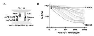
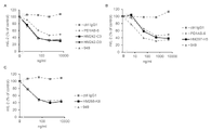
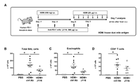
- a system for evaluating cytokine production suppression activity of T cells by PD-1 stimulation A system for evaluating cytokine production suppression activity of T cells by PD-1 stimulation.
- A Schematic diagram of the evaluation system using DO11.10 T-cell hybridomas expressing human PD-1 (hPD-1) and IIA1.6 B-cell lymphoma cells.
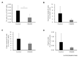
- DO11.10 T-cell hybridomas are activated in response to OVA 323-339 peptide presented by MHC class II molecules (IA d ) of IIA1.6 cells, but activation is suppressed upon stimulation to PD-1. be done.
- B Interaction with PD-L1 on IIA1.6 cells suppressed IL-2 production from DO11.10 T cell hybridomas expressing human PD-1. No suppression of IL-2 is observed when PD-1 or PD-L1 is not expressed.
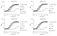
- Anti-human PD-1 antibodies were screened using their inhibitory activity against IL-2 production as an index.
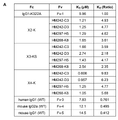
- (B) As a result of the evaluation, about 30 clones with immunosuppressive activity were obtained, ranging from those with high activity to those with low activity. The graph shows only clones with immunosuppressive activity among these anti-human PD-1 antibodies. Activity comparison of anti-human PD-1 agonist antibodies. Of the anti-human PD-1 antibodies found to have immunosuppressive activity, those with relatively high activity were selected and their IC50 values were determined.
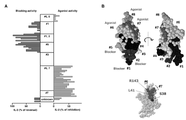
- (A) #6, 7 groups (antibodies that bind to #6 and #7 regions), (B) #7 groups (antibodies that bind only to #7 regions), (C) #6, 8 groups ( Representative antibodies among the group of antibodies that bind to the #6 and #8 regions) are shown.
- Commercially available antibodies (J116, MIH4) that have been found to have agonistic activity, as well as novel antibodies generated by immunizing mice with human PD-1, are all mouse IgG. Based on these antibody concentration-dependent suppression curves of IL-2 production, the concentration at which 50% suppression was achieved was determined, with no IL-2 production as 0%. Correlation of activity and binding site of anti-human PD-1 antibodies.
- a new class of anti-human PD-1 antibodies with different binding regions from known antibody classes including PD-1 blocking antibodies such as nivolumab was obtained.
- the antibody group is one in which substitution of the #7 region results in no binding, or substitution of either the #7 region or the #6 region results in no binding, each of which the #7 region is important for binding.
- the #7 only antibody group includes known antibodies such as 949 and J116, but the antibodies of the present invention are human PD-1 Arg143 point mutants such as R143A and R143V, mouse PD-1-human #7 region While retaining the ability to bind to the substitution (Mouse PD-1 (hu38-48)), the known antibody had significantly reduced binding to these point mutants and substitutions.
- substitution of the #1 region abolishes binding (HM292, HM297, HM330, HM624)
- substitution of the #1 region reduces binding (HM698, HM634, HM643), or those that maintained binding even when the #1 region was substituted (HM647, HM654).
- Human PD-1 knock-in mice were sensitized with house dust mite antigen (HDM) and allergic asthma was induced by intranasal administration of the same antigen one week later. Infiltration of inflammatory cells into the alveoli was examined by collecting bronchoalveolar lavage fluid 7 days after the start of daily intranasal administration of HDM.
- HDM house dust mite antigen
- BD Induction of allergic asthma increased the total number of infiltrated inflammatory cells (B), among which eosinophils (C) and CD4 + T cells (D) were the majority.
- prophylactic administration of an anti-human PD-1 agonist antibody to this model of inflammation suppressed the infiltration of eosinophils and CD4 + T cells into the alveoli.
- HM266 was administered only once (HM266 (x1)) 3 days after the induction of inflammation in the lung tissue to observe the therapeutic effect, the infiltration of inflammatory cells into the alveoli was suppressed.
- HM266 was administered only once (HM266 (x1)) 3 days after the induction of inflammation in the lung tissue to observe the therapeutic effect, the infiltration of inflammatory cells into the alveoli was suppressed.
- Admitted Anti-inflammatory effect of anti-human PD-1 agonist antibody against allergic asthma and suppression of Th2-type immune response.
- Allergic asthma was induced according to the same schedule as in FIG. 6, and bronchoalveolar lavage fluid and lung tissue were collected. Cytokine production from CD4 + T cells that infiltrated into the alveoli (A) or lung tissue (B) was examined by intracellular staining. , IL-5, IL-10 and IL-13 producing cells were observed. The blood mite antigen-specific IgE concentration (C) was also significantly decreased by HM266 administration. A similar trend was also observed when HM266 was administered only once (HM266 (x1)) 3 days after the start of inflammation induction in the lungs, suggesting the usefulness of anti-human PD-1 agonist antibody for the treatment of allergic diseases. was done.
- EAE experimental encephalomyelitis
- Acute GVHD was induced by transferring splenocytes from C57BL/6 human PD-1 knock-in mice (donor) into BDF1 mice (recipient).
- donor C57BL/6 human PD-1 knock-in mice
- BDF1 mice BDF1 mice
- HM266, HM242, HM297 and HM268 were administered prophylactically to this inflammation model, the donor cell ratio (CD) and the donor-derived CD8 + cell ratio (EF) in the spleen and liver two weeks after induction was also significantly suppressed. From the above, it was suggested that the anti-human PD-1 agonist antibody could be applied to the treatment of inflammatory diseases.
- a chimeric antibody (human Fc chimeric antibody) having a mouse-derived Fab region and a human IgG1-K322A Fc region was prepared by selecting an anti-human PD-1 agonist antibody having high activity from the obtained anti-human PD-1 agonist antibodies.
- A Since a human Fc region was used, agonistic activity was assessed using a human Fc ⁇ RIIB-expressing cell line.
- B Agonist activity and physical property evaluation of human Fc chimeric antibody.
- ⁇ indicates an antibody for which no abnormal band was detected
- x indicates an antibody for which an abnormal band such as a heavy chain dimer was detected.
- Differences were observed among the clones, and HM242, HM268 and HM297 in particular exhibited high agonist activity and good physical property profiles.
- Humanization of anti-human PD-1 agonist antibody and bioactivity/physical property profile test Agonist activities of humanized (A) HM242, (B) HM297, (C) HM268 are shown and the physical property profiles are shown in Tables 1-3.
- % Identical with human sequence means the homology (%) when comparing the antibody sequence excluding CDRs with the human germline-derived antibody sequence. do.
- Several antibodies HM242-C3, HM242-D3, HM297-H5 and HM268-K8 that maintain high agonist activity and exhibit favorable physical property profiles, while some antibodies have decreased agonist activity and stability due to humanization was gotten. Effect of a humanized anti-human PD-1 agonist antibody in an evaluation system using human primary cultured cells.
- a humanized anti-human PD-1 agonist antibody having a humanized Fab region and a human IgG1-K322A Fc region exhibits a T-cell suppressive effect in an evaluation system composed only of human primary cultured cells.
- MLR mixed lymphocyte reaction
- PBMC peripheral blood mononuclear cell
- CTLA-4-Ig was added as a positive control.
- T cell suppressive activity was evaluated using the amount of IFN- ⁇ production in the supernatant as an index.
- Antibodies did not show significant inhibition.
- the agonistic activity of anti-human PD-1 agonistic antibodies is dependent on Fc receptors, especially Fc ⁇ RIIB.
- the IL-2 production inhibitory activity of the humanized anti-human PD-1 agonist antibody was examined using the same evaluation system as in FIGS. 3 and 12 .
- the IIA1.6 cells used here express human Fc ⁇ RIA, Fc ⁇ RIIA (H131), Fc ⁇ RIIA (R131), Fc ⁇ RIIB, Fc ⁇ RIIIA (F158), and Fc ⁇ RIIIA (V158) at similar high levels.
- humanized anti-human PD-1 agonist antibody inhibited IL-2 production. It showed the highest IL-2 production inhibitory activity when cells were used. When Fc ⁇ RIA-expressing cells were used, antibody addition increased IL-2 production. These results suggested that the humanized anti-human PD-1 agonistic antibody exhibited the highest agonistic activity especially in the presence of Fc ⁇ RIIB. The exertion of agonistic activity of anti-human PD-1 antibodies depends on the expression level of Fc receptors. In the evaluation system shown in FIG.
- Affinity (K D value) of Fc region variants to human Fc ⁇ RIIB The affinity of human IgG1 Fc region variants such as X2-K, X3-KS, and X4-K for human Fc ⁇ RIIB was improved by 2.18- to 9.83-fold compared to IgG1-K322A. Improving affinity to human Fc ⁇ RIIB by modifying the Fc region.
- B Affinity of Fc region variants to human Fc ⁇ RIIB (GMFI).
- the affinity of human IgG1 Fc region variants such as X2-K, X3-KS, and X4-K for human Fc ⁇ RIIB was improved by 2.92 to 84.8 times compared to IgG1-K322A having the same Fv region.
- DF represents a defucosylation. Improving affinity to human Fc ⁇ RIIB by modifying the Fc region.
- C Correlation between the affinity (GMFI) of the Fc region variants for human Fc ⁇ RIIB and the agonist activity (IC50: ng/ml). Each plot represents an antibody described in Figure 17B. When the Fv region was immobilized, the higher the affinity of the Fc region for human Fc ⁇ RIIB, the higher the agonist activity tended to be.
- HM266 Fc region-engineered chimeric antibodies were prepared and their agonist activity was evaluated.
- PD1AB-6 and 949 which are known anti-human PD-1 agonist antibodies, showed only weak agonist activity,
- Each Fc region variant with improved affinity for human Fc ⁇ RIIB exhibited high agonist activity.
- a chimeric antibody having the HM266 Fab region and the Fc regions of various Fc region variants, and a chimeric antibody with IgG1-K322A, CTLA-4-Ig, were evaluated in the MLR system of CD4 + T cells and CD19 + B cells.
- the chimeric antibody with improved affinity for human Fc ⁇ RIIB by having the Fc region variant Fc region has greatly improved T cell suppression activity compared to the original IgG1-K322A, CTLA-4-Ig exhibited an anti-inflammatory effect superior to Anti-inflammatory action of a humanized anti-human PD-1 agonist antibody in an evaluation system using human cells.
- a humanized antibody combining the selected humanized Fab region see FIG.
- Antibodies with X2-K Fc regions have low ADCC activity
- X3-KS antibodies have the same ADCC activity as IgG1-K322A
- X4-K antibodies and defucosylated X3-KS have high ADCC activity. had activity.
- CD4 + T cells that were activated and proliferated upon stimulation of human PBMC with tetanus toxin were monitored by the decline of carboxyfluorescein succinimidyl ester (CFSE) fluorescence in CD4 + T cells.
- CFSE carboxyfluorescein succinimidyl ester
- HM266 suppressed the time-dependent increase in phosphorylated ERK positive rate in DO11.10. From the above, it was suggested that the anti-human PD-1 agonist antibody suppresses downstream signaling of the T cell receptor.
- BD In the PD-1 agonist activity in vitro evaluation system in Fig. 3, administration of J116, which showed weaker activity than HM266, suppressed the total number of inflammatory cells (B) infiltrating the tissue, especially eosinophils. Infiltration was significantly suppressed (C), but CD4 + T cell infiltration was not significantly suppressed (D).
- B total number of inflammatory cells
- C CD4 + T cell infiltration was not significantly suppressed
- Acute GVHD was induced by transferring splenocytes from C57BL/6 human PD-1 knock-in mice (donor) into BDF1 mice (recipient).
- A Diagram showing human PD-1 expression on CD4 + or CD8 + T cells in donor-derived cells (H-2K b+ H-2K d ⁇ ). In GVHD-induced mice, 61.7% of donor-derived CD4 + T cells and 49.5% of donor-derived CD8 + T cells were positive for human PD-1. Induction of acute GVHD in mice and anti-inflammatory effects of anti-human PD-1 agonistic antibodies.
- B-D AST (B), ALT (C), IFN- ⁇ (D) in mouse plasma 2 weeks after induction.
- Plasma ALT, AST and IFN- ⁇ were elevated by GVHD induction, but these elevations were markedly suppressed by prophylactic administration of HM266, HM242, HM297 and HM268.
- T cell-dependent antibody responses were induced in C57BL/6 human PD-1 knock-in mice by intraperitoneal administration of 4-hydroxy-3-nitrophenylacetylovalbumin (NP-OVA) antigen with alum adjuvant.
- NP-OVA 4-hydroxy-3-nitrophenylacetylovalbumin
- HM266 administration suppressed the proportion of Tfh cells (A), germinal center B cells (B), class-switched B cells (C), and plasma cells (D) in the mouse spleens 10 days after antigen administration.
- A germinal center B cells
- C class-switched B cells
- D plasma cells
- E Further analysis of plasma antibody titers 7 days after antigen administration revealed that low-affinity IgG antibody titers (anti-NP25 IgG) and high-affinity IgG/IgM antibody titers (anti-NP2 IgG or IgM) were suppressed.
- F low-affinity IgG antibody titers
- IgM high-affinity IgG/IgM antibody titers
- anti-human PD-1 agonist antibody administration may suppress antibody class switching and affinity maturation.
- Induction of chronic GVHD/lupus-like symptoms in mice and anti-inflammatory effects of anti-human PD-1 agonistic antibodies Spleen cells from C57BL/6 human PD-1 knock-in mice (donors) were depleted of CD8 + cells and then transferred into BDF1 mice (recipients) to induce chronic GVHD/lupus-like symptoms. After cell transfer, donor-derived Tfh cells (A), recipient-derived germinal center B cells (B), class-switched B cells (C), and plasma cells (D) were significantly increased in the recipient spleen.
- HM242 or CTLA-4-Ig Prophylactic administration of HM242 or CTLA-4-Ig to this inflammation model suppressed the increase in the number of these cells.
- "-" represents recipient mice not transfected with donor cells.
- Spleen cells from C57BL/6 human PD-1 knock-in mice (donors) were depleted of CD8 + cells and then transferred into BDF1 mice (recipients) to induce chronic GVHD/lupus-like symptoms. After cell transfer, significant increases in blood IL-21 concentration (E) and anti-dsDNA antibody titer (F) were confirmed.
- E blood IL-21 concentration
- F anti-dsDNA antibody titer
- HM242 or CTLA-4-Ig When HM242 or CTLA-4-Ig was administered prophylactically to this inflammation model, the increase in blood anti-dsDNA antibody was suppressed. From the above, it was suggested that the anti-human PD-1 agonist antibody could be applied to the treatment of inflammatory diseases in which autoantibody production is seen.
- a chimeric antibody between the HM266 Fab region and the Fc region of various Fc region variants, and a chimeric antibody with the Fc region of IgG1-K322A and IgG4-S228P were used for CD4 + T cells and THP-1, a human monocytic cell line.
- IFN- ⁇ production was used as an index for evaluation.
- THP-1 in which the human Fc ⁇ RIIB gene was artificially introduced and highly expressed, the chimeric antibody between the Fab region of HM266 and the Fc region of IgG1-K322A and IgG4-S228P exhibited immunity equivalent to or higher than that of known antibodies. It showed inhibitory activity (A).
- THP-1 which expresses endogenous Fc ⁇ RIIB without transfection
- chimeric antibodies of IgG1-K322A and IgG4-S228P and known antibodies did not show immunosuppressive activity, but had affinity for human Fc ⁇ RIIB.
- Each Fc region variant with improved potency exhibited high immunosuppressive activity (B).
- the chimeric antibody with improved affinity for human Fc ⁇ RIIB due to the modified Fc region had significantly improved T cell suppressive activity compared to the original IgG1-K322A.
- Antibody whose Fc region is X2 type has low ADCC activity, and antibody whose Fc region is X3 type or X4 type has high ADCC activity equivalent to or higher than that of IgG1-K322A type.
- the Fc region variant based on the Fc region of IgG4-S228P had improved ADCC activity compared to IgG4-S228P, but not as much as IgG1-K322A type, X3 and X4 types.
- Suppressive effect of humanized anti-human PD-1 agonist antibody on activation of antigen-specific T cells was evaluated using IFN- ⁇ produced by stimulation of human PBMC with tetanus toxin as an index.
- humanized anti-human PD-1 agonist antibody suppressed IL-21 production by Tfh-like cells.
- RA Rheumatoid arthritis
- SLE systemic lupus erythematosus
- C, D patient-derived PBMCs were cultured in the presence of autoantigens, and CD4 + T cell proliferation (A, C) and IFN- ⁇ Production (B, D) was used as an index for evaluation.
- the humanized anti-human PD-1 agonist antibody showed an inhibitory activity equal to or greater than that of CTLA-4-Ig against the activation of patient-derived cells against autoantigen stimulation. Improving affinity to human Fc ⁇ RIIB by modifying the Fc region.
- Affinity K D value
- the affinity of the human IgG1 Fc region variant to human Fc ⁇ RIIB was improved by 2.34- to 8.65-fold compared to IgG1-K322A.
- B Affinity of Fc region variants to human Fc ⁇ RIIB (GMFI). The affinity of the human IgG1 Fc region variant to human Fc ⁇ RIIB was 26.1 to 180 times higher than that of IgG1-K322A. Improving affinity to human Fc ⁇ RIIIA by modifying the Fc region.
- A Affinity (K D value) of Fc region variant to human Fc ⁇ RIIIA (V158).
- the affinity of human IgG1 Fc region variants such as X4-K, X3-KS-DF, X3-K, X3-KSI, X3-KI, and X3-KE to human Fc ⁇ RIIIA is determined by IgG1 with the same Fv region It was 1.92 to 6.98 times better than -K322A. Improving agonistic activity by modifying the Fc region.
- IIA1.6 cells with low expression of human Fc ⁇ RIIB, 3.7C6 and Antibody 1, which are known anti-human PD-1 agonist antibodies, showed only weak agonistic activity, but improved affinity to human Fc ⁇ RIIB
- Each of the Fc region variants that were modified showed high agonist activity.
- Anti-inflammatory action of a humanized anti-human PD-1 agonist antibody in an evaluation system using human cells A humanized antibody obtained by combining the humanized Fab region of HM268-K8 and the Fc region of a modified human Fc region was evaluated in the MLR system of CD4 + T cells and CD19 + B cells in the same manner as in FIG. As a result, the Fc region-engineered humanized antibodies with improved Fc ⁇ RIIB affinity exhibited a stronger T-cell inhibitory effect than CTLA-4-Ig, suggesting that these humanized antibodies inhibit human T-cell function. It was suggested that it could show a strong anti-inflammatory effect. ADCC activity of humanized anti-human PD-1 agonist antibodies.
- Antibodies with X3-KSL and X3-KL Fc regions had low ADCC activity, while antibodies with X3-KSI and X3-KI and X3-KS-DF had high ADCC activity.
- Suppressive effect of humanized anti-human PD-1 agonist antibody on activation of antigen-specific T cells Similar to FIG. 21, CD4 + T cells that were activated and proliferated when human PBMC were stimulated with tetanus toxin were monitored by the decrease in CFSE fluorescence in CD4 + T cells. Data show the percentage of CD4 + T cells whose CFSE fluorescence intensity is attenuated by cell proliferation.
- Antibodies that showed high ADCC activity in FIG. 39 exhibited particularly high antiproliferative effects.
- CTLA-4-Ig did not show any antiproliferative effect when donor-derived cells different from those used in FIG. 21 were used.
- the present invention provides an agonistic antibody against human PD-1 (anti-human PD-1 agonistic antibody) or a functional fragment thereof, which binds to the #7 region of human PD-1 represented by SEQ ID NO:9.
- a characterized antibody or functional fragment thereof is provided.
- the anti-human PD-1 agonistic antibody is an antibody that binds to human PD-1 and activates the same intracellular signaling system as human PD-L1 in the human body.
- the activity to suppress IL-2 produced from T cells in an antigen-stimulation-dependent manner is selected as an indicator. (see examples below).
- the antibody or functional fragment thereof of the present invention binds only to the #7 region of human PD-1 (the region from the 38th amino acid to the 48th amino acid in the amino acid sequence of SEQ ID NO: 1, SEQ ID NO: 9). Further, the #6 region of human PD-1 (the region from the 109th amino acid to the 120th amino acid in the amino acid sequence of SEQ ID NO: 1, SEQ ID NO: 8), #1 region (SEQ ID NO: 1 Although it may bind to the region from the 129th amino acid to the 139th amino acid of the amino acid sequence of (SEQ ID NO: 3), etc., it preferably binds only to the #7 region of human PD-1.
- An anti-human PD-1 antibody can be defined as binding to the #7 region of human PD-1, where binding to the #7 region varies from binding to other regions of human PD-1, etc. are independently determined. Binding to regions other than the #7 region of human PD-1 is similarly defined.
- the accession database and accession number for the amino acid sequence of human PD-1 is NCBI accession number: NP_005009.2, and the accession database and accession number for the amino acid sequence of mouse PD-1 is NCBI accession number: NP_032824.1.
- the antibody or functional fragment thereof of the present invention is also a mouse PD-1 substitute (Mouse PD-1 (hu38-48 )) and/or human PD-1 in which arginine at position 143 of human PD-1 is mutated to alanine (R143A point mutant).
- the binding ability (binding affinity) of the antibody of the present invention or a functional fragment thereof to human PD-1 has an equilibrium dissociation constant (K D ) of 10 ⁇ 7 M or less, preferably 10 ⁇ 8 M It is below.
- K D equilibrium dissociation constant
- the equilibrium dissociation constant can be measured by surface plasmon resonance (SPR) method. It can also be determined by flow cytometry.
- CDRs are described, for example, in Kabat (J Exp Med. 1970; 132: 211-50), Chothia (J Mol Biol. 1987; 196: 901-17), IMGT (Dev Comp Immunol. 2003; 27: 55-77) and Paratome ( PLoS Comput Biol. 2012; 8: e1002388), preferably by Kabat's method.
- HM242, HM266, HM268 and HM297 are mouse anti-human PD-1 antibodies exhibiting PD-1 agonistic activity generated by immunizing mice with human PD-1.
- the antibody of the present invention preferably has a heavy chain variable region containing heavy chain CDRs 1-3 and a light chain variable region containing light chain CDRs 1-3 as shown in Table I below.
- the heavy chain CDRs 1-3 and light chain CDRs 1-3 are at least 85%, 85-90%, 90-95%, 95-97% total CDRs relative to the sequences shown in Table I below. % or 97% or more identity. The identity of two sequences can be determined using BLAST.
- variable regions (Fv) of the antibodies of the present invention are antibodies derived from non-human animals (e.g., mice, rabbits, rats, hamsters, guinea pigs, goats, sheep, donkeys, llamas, camels, chickens, ostriches, sharks, etc.). or humanized antibody heavy and/or light chain Fv regions derived from non-human animals.
- Humanization can be performed, for example, by grafting the CDRs of the VH and VL of a non-human animal-derived antibody into the VH and VL frameworks of a human antibody (Nature, 332, 323-327, 1988). Humanization may be one in which the CDR sequence is maintained. Antigen-binding may be improved by identifying the amino acid residues involved in , and substituting them with non-humanized antibody amino acid residues (MABS, 8(7), 1302-1318 2016).
- VH region An example of the sequence of the heavy chain variable region (VH region, positions 1 to 117, according to EU numbering, hereinafter the same) of a mouse-derived antibody that exhibits PD-1 agonistic activity and an example of the humanized VH region sequence is shown in Table II below.
- Table II Sequences of heavy chain variable regions (VH regions) of HM242, HM266, HM268 and HM297 and sequences of humanized VH regions of HM242, HM266, HM268 and HM297
- VL region An example of the light chain variable region (VL region, positions 1-107) and humanized VL region sequences of murine-derived antibodies exhibiting PD-1 agonistic activity is shown in Table III below.
- the antibodies of the present invention may have improved affinity for human Fc receptors, and the Fc receptors are preferably Fc ⁇ receptors, more preferably Fc ⁇ RII, and more preferably Fc ⁇ RIIB. preferable.
- the affinity of the antibody of the present invention for human Fc receptors can be determined by a binding measurement test for human Fc receptor-expressing cell lines using a flow cytometer, and an Fc receptor binding affinity using surface plasmon resonance technology using Biacore 8K (Cytiva). It can be evaluated by a property measurement test.
- the improvement of the affinity of the antibody of the present invention for human Fc receptors may be evaluated by any of these methods, preferably by a flow cytometer, and evaluated by both of these methods.
- the affinity of the antibody of the present invention for human Fc ⁇ RIIB when measured by a human Fc ⁇ RIIB binding affinity measurement test using surface plasmon resonance technology, has an equilibrium dissociation constant for an antibody having the Fc region of human IgG1-K322A (reference antibody) (K D ) ratio, the antibodies of the invention have an affinity greater than or equal to 1.5-fold that of the reference antibody, preferably greater than or equal to 2-fold that of the reference antibody, more preferably It has an affinity greater than or equal to 2.5 times that of the reference antibody (see Examples below).
- Affinity of the antibody of the present invention to human Fc ⁇ RIIB when measured by a binding measurement test for human Fc ⁇ RIIB-expressing cell lines using a flow cytometer, GMFI against an antibody having the Fc region of human IgG1-K322A (reference antibody) It can be expressed as a ratio.
- the antibody of the present invention exhibits a GMFI that is at least twice that when a reference antibody having the same Fv region is added, which is preferable. shows a 5-fold or greater GMFI, more preferably a 20-fold or greater GMFI.
- the antibodies of the present invention may have improved affinity for human Fc ⁇ RIIIA.
- Affinity of the antibody of the present invention to human Fc ⁇ RIIIA, when measured by human Fc ⁇ RIIIA (V158) binding affinity measurement test using surface plasmon resonance technology, to an antibody having the Fc region of human IgG1-K322A (reference antibody) can be expressed as an equilibrium dissociation constant (K D ) ratio
- the antibodies of the invention have an affinity of 1.5 times or more that of the reference antibody, preferably 2 times or more of the affinity of the reference antibody, More preferably, it has an affinity that is 2.5 times or more that of the reference antibody, and even more preferably, it has an affinity that is 4 times or more (see Examples below).
- the affinity of the antibody of the present invention for human Fc ⁇ RIIIA is measured by a binding measurement test for human Fc ⁇ RIIIA (V158)-expressing cell lines using a flow cytometer. ) can be expressed as the GMFI ratio.
- the GMFI without antibody addition is about 10 and the GMFI with reference antibody addition is 6000 to 15000, the antibody of the present invention exhibits a GMFI that is 1.5 times or more that with reference antibody addition having the same Fv region. , preferably 2-fold or more GMFI, more preferably 4-fold or more GMFI, still more preferably 5-fold or more GMFI.
- the antibody of the present invention preferably has the Fc region of a human antibody (e.g., IgG1, IgG4, etc.), and the Fc region of the human antibody is a human Fc receptor (human Fc ⁇ RIIA, human Fc ⁇ RIIB, human Fc ⁇ RIIIA). Any one or more) is preferably an Fc region variant that has been modified to improve affinity. Affinity for human Fc receptors can be improved by modifying the Fc region (positions 216-447) of an antibody. In addition, defucose treatment of antibodies can also improve the affinity for human Fc receptors.
- a human antibody e.g., IgG1, IgG4, etc.
- the Fc region of the human antibody is a human Fc receptor (human Fc ⁇ RIIA, human Fc ⁇ RIIB, human Fc ⁇ RIIIA). Any one or more) is preferably an Fc region variant that has been modified to improve affinity.
- Affinity for human Fc receptors can be improved by modifying the
- SEQ ID NO: 47 An example of the amino acid sequence of the Fc region of human IgG1 is shown in SEQ ID NO: 47.
- SEQ ID NO: 64 shows an example of the amino acid sequence of the Fc region of human IgG4.
- positions 233, 234, 236 and 237 in the amino acid sequence of the Fc region of human IgG1 represented by SEQ ID NO: 47 , 238th, 239th, 267th, 268th, 271st, 296th, 323rd, 326th, 328th, 330th and 332nd preferably one or more mutations are introduced may be a combination of these.
- mutations and defucosylation may be combined.
- mutations e.g. 239/268/332, 239/267/268/328/332, 233/237/238/268/271/330, 267/328, preferably 239/268, 239/268/328 /332, 239/267/268, 239/267/268/332, 239/268/332, 233/237/238/268/271/330, 267/328, more preferably 239 /268/328/332, 233/237/238/268/271/330, or 267/328.
- G at position 236 may be mutated to, for example, D, E, N, Q, F, H, I, K, L, M, P, R, S, T, V, W, Y, A, etc. Mutation to D, E, N, or Q is preferable, and mutation to D is more preferable.
- H at position 268 may be mutated, for example, to D, E, N, Q, A, G, etc., preferably to D, E, N, Q, more preferably to D. .
- S at position 239 may be mutated to, for example, D, E, N, Q, F, T, H, Y, G, L, etc., preferably D, E, N, Q, and more Preferably, it should be mutated to D.
- L at position 328 may be mutated to, for example, Y, E, F, H, I, Q, W, D, T, S, M, A, V, N, H, etc., preferably Y, F, It is preferable to mutate to D, E, N, Q, and more preferably to Y.
- I at position 332 may be mutated to, for example, E, D, F, L, R, S, T, D, N, Q, H, Y, A, M, etc., preferably to E, T, M It is preferable to mutate, more preferably to E. E at position 233 should be mutated to D, for example.
- G at position 237 may be mutated to, for example, W, F, A, D, E, L, M, Y, etc., preferably D, E, and more preferably D. .
- P at position 238 may be mutated to D, for example.
- P at position 271 may be mutated to G, for example.
- a at position 330 may be mutated, for example, to K, R, M, etc., preferably to R.
- S at position 267 may be mutated to, for example, E, V, Q, A, L, etc., preferably to E or G, more preferably to G.
- K at position 326 may be mutated to, for example, L, Q, N, M, D, S, T, A, and the like.
- L at position 234 may be mutated to, for example, D, E, N, Q, T, H, I, V, F, W, Y, and the like.
- V at position 323 may be mutated to, for example, I, L, M, or the like.
- Y at position 296 may be mutated to D, for example.
- CDC complement-dependent cytotoxicity
- Combinations of mutations to be introduced into the Fc region of human IgG1 are G236D/H268D, S239D/H268D, S239D/H268D/L328Y/I332E, G236D/H268D/K322A, S239D/H268D/K322A, S239D/S267G/H268D/K322A, G236D /H268D/E293A/K322A ⁇ S239D/H268D/K322A/L328Y/I332E ⁇ S239D/H268D/E293A/K322A ⁇ S239D/S267G/H268D/K322A/L328Y ⁇ S239D/S267G/H268D/K322A/I332E ⁇ S239D/H268D/K322A /L328Y, S239D/H268D/K
- the combination of Fc region mutations When combining Fc region mutations of human antibodies with defucosylation, the combination of Fc region mutations has the same affinity for human Fc receptors, particularly human Fc ⁇ RIIIA, due to defucosylation. It may be improved by 1.5 times or more, preferably by 2 times or more, more preferably by 3 times or more, as compared to the antibody.
- Preferred combinations of mutations and defucosylation include, but are not limited to, combinations of S239D/S267G/H268D/K322A and defucosylation.
- any combination of S228P/G236D/Q268D, S228P/S239D/Q268D or S228P/S239D/Q268D/L328Y/I332E is preferable, more preferably S228P/S239D/Q268D or S228P/S239D/Q268D Any combination of /L328Y/I332E is good.
- Fc region sequences of human IgG1 (WT, K322A) and IgG4 (WT, S228P) and Fc region variants introduced with mutations are shown in Table IV. [Table IV] Fc regions of human IgG1 (WT, K322A) and IgG4 (WT, S228P) and Fc region variants
- the antibody of the present invention preferably has a high ratio of monomers having a four-chain structure consisting of two light chains and two heavy chains.
- the monomer ratio can be measured by SEC-HPLC (see Examples below).
- the monomer ratio is preferably 80% or more, more preferably 90% or more.
- the antibody of the present invention may be further subjected to functional modifications other than those described above, such as amino acid mutation, subclass substitution, and sugar chain modification.
- the antibody of the present invention preferably has ADCC activity equal to or higher than IgG1 or IgG1-K322A when the same Fv region is employed.
- An antibody with high ADCC activity can exhibit a higher T cell suppressive effect upon antigen stimulation than an antibody with low ADCC activity.
- ADCC activity is achieved, for example, using genetically modified Jurkat T cells expressing Fc receptors instead of effector cells, where signals from Fc receptors activate luciferase genes integrated downstream of the NFAT response element. The amount of luciferase produced by the reaction can be determined by measuring with a luminometer (see Examples below).
- the antibody of the present invention is a non-human animal (e.g., mouse)-derived antibody (which exhibits PD-1 agonistic activity) in which the variable regions of each of the heavy and light chains are humanized Fab regions, human IgG1 or IgG4 It is preferable to have the Fc region of the Fc region variant of An example of the heavy and light chain amino acid sequences of preferred antibodies of the invention and the nucleic acid sequences encoding the same are shown in Table V below. Antibodies of the present invention may have the heavy chain sequence and/or light chain amino acid sequence shown in Table V below.
- the heavy and light chains consist of sequences having at least 90%, 90-95%, 95-99%, or 99% or more identity to the amino acid sequences shown in Table V below. good.
- Nucleic acids encoding antibodies of the present invention may also be nucleic acids encoding these heavy and light chain amino acid sequences, and may be modified from the nucleic acid sequences shown in Table V below according to the desired amino acid sequence.
- the design of a nucleic acid sequence according to the desired amino acid sequence is known to those skilled in the art, and it is desirable that the nucleic acid sequence (codon) encoding each amino acid is optimized according to the desired amino acid sequence.
- Antibodies of the present invention include, for example, HM242-C3-X3-K, HM242-C3-X3-KE, HM242-C3-X3-KI, HM242-C3-X3-KSI, HM242-C3-X4-K, HM242-D3 -X3-K, HM242-D3-X3-KE, HM242-D3-X3-KI, HM242-D3-X3-KS, HM242-D3-X3-KSI, HM242-D3-X4-K, HM268-K8-X3 -K, HM268-K8-X3-KE, HM268-K8-X3-KI, HM268-K8-X3-KS, HM268-K8-X3-KSI and HM268-K8-X4-K, HM242-C3-X3 -KI, HM242-C3-X3-KSI,
- the functional fragment means, for example, a molecule having one or more of scFv, Fv, F(ab')2, Fab', Fab of the antibody of the present invention, and further binds to Fc receptors.
- a drug for example, an antibody-drug conjugate of the antibody of the present invention and a drug, a polypeptide comprising the scFv of the antibody of the present invention and the scFv of an anti-Fc receptor antibody, or a fusion comprising the scFv of the antibody of the present invention and the Fc region
- examples include, but are not limited to, proteins and the like.
- the functional fragment has an Fc region, such as a fusion protein between the scFv of the antibody of the present invention and the Fc region
- the Fc region is the Fc region variant of the Fc region described herein. is desirable.
- the above-described protein improvement and optimization techniques can also be applied to functional fragments.
- the antibodies and functional fragments thereof of the present invention can be produced as recombinant antibodies or functional fragments thereof by genetic engineering techniques. That is, DNA encoding the heavy chain and light chain genes of the antibody of the present invention or a functional fragment of the antibody of the present invention is synthesized, inserted into an expression vector (e.g., plasmid, bacteriophage, virus), and then By introducing into cells (eg, CHO cells, HEK cells, etc.) and culturing the host cells, recombinant antibodies or functional fragments thereof can be collected from the culture. In synthesizing the DNA encoding the antibody heavy and light chain genes or functional fragments, codon optimization is preferred.
- an expression vector e.g., plasmid, bacteriophage, virus
- the antibody heavy and light chain genes may be inserted into the same expression vector or separate expression vectors.
- Expression vectors may have promoters, enhancers, polyadenylation signals, replication origins, selectable marker genes, secretion signal sequences, and the like, as required.
- the ratio of the heavy chain gene to the light chain gene of the antibody inserted into the expression vector is preferably 1:1, but may be changed as appropriate to improve the expression level of the antibody.
- Known methods such as the calcium phosphate method, the DEAE-dextran method, the microinjection method, the lipofection method, the electroporation method, the transduction method, the scrape loading method and the shotgun method can be used to introduce the recombinant expression vector into the host cell. can be used.
- host cells introduced with the recombinant expression vector are cultured, recombinant antibodies or functional fragments thereof are produced in the culture. Appropriate culture conditions (medium, culture time, culture temperature, CO 2 concentration, etc.) can be appropriately set by those skilled in the art.
- the produced antibody or functional fragment thereof utilizes isoelectric point, size, solubility (in water, organic solvent, etc.), affinity for certain substances (in the case of enzymes, substrate, coenzyme, etc.), etc. It can be recovered using a known protein separation/purification method.
- 2-Deoxy-2-fluoro-L-fucose is added to the medium and cultured, or Fucosyltransferase 8 (FUT8)-deficient cells are used as host cells.
- FUT8 Fucosyltransferase 8
- the antibody or functional fragment thereof of the present invention exerts a stronger T cell suppressive effect than CTLA-4-Ig in mixed lymphocyte reaction (MLR) of human T cells and B cells, and suppresses functions of human T cells. (see Examples below).
- MLR mixed lymphocyte reaction
- inflammatory disease refers to a disease caused by excessive inflammation caused by wounds, chemicals, infection, or self-tissue recognition by immune cells. It includes autoimmune diseases and diseases classified as type I-IV allergies, such as Behçet's disease, systemic lupus erythematosus, lupus nephritis, cutaneous lupus erythematosus, chronic discoid lupus erythematosus, serum sickness, systemic sclerosis (systemic severe dermatitis, progressive systemic sclerosis), multiple sclerosis, scleroderma, polymyositis, dermatomyositis, periarteritis nodosa (polyarteritis nodosa), aortitis syndrome (Takayasu arteritis), malignant Rheumatoid arthritis, rheumato
- the antibody or functional fragment thereof of the present invention can be used for inflammatory diseases, particularly diseases involving T cells or autoantibodies, such as systemic lupus erythematosus, lupus nephritis, multiple sclerosis, systemic sclerosis (systemic scleroderma). disease, progressive systemic sclerosis), multiple sclerosis, scleroderma, polymyositis, dermatomyositis, rheumatoid arthritis, Sjögren's syndrome, allergic granulomatous vasculitis, large angiitis, ANCA-associated vasculitis (e.g.
- IgG4-related diseases e.g. primary sclerosing cholangitis, autoimmune pancreatitis, etc.
- IgG4-related diseases e.g. primary sclerosing cholangitis, autoimmune pancreatitis, etc.
- idiopathic thrombocytopenic purpura Graves' disease (Graves' disease (hyperthyroidism)), Hashimoto's disease, psoriasis, psoriatic arthritis, bullous pemphigoid, pemphigus, pemphigoid, alopecia areata , vitiligo, vitiligo vulgaris, Stevens-Johnson syndrome, fixed drug eruptions, neuromyelitis optica, inflammatory bowel disease (e.g., ulcerative colitis, Crohn's disease), asthma, acute and chronic graft-versus-host disease and/or Or it can be used for prophylaxis.
- IgG4-related diseases e.
- the present invention provides an agonistic antibody against human PD-1 (anti-human PD-1 agonistic antibody) or a functional fragment thereof, which binds to the #7 region of human PD-1 represented by SEQ ID NO:9.
- a pharmaceutical composition is provided that includes the characterized antibody or functional fragment thereof and a pharmaceutically acceptable carrier.
- the pharmaceutical composition of the present invention can be used as a medicament.
- composition of the present invention can be administered to a subject (human or non-human animal) systemically or topically, orally or parenterally.
- compositions of the present invention which may contain an effective amount of an anti-human PD-1 agonist antibody or functional fragment thereof, may be mixed, dissolved, emulsified, encapsulated, lyophilized, and pharmaceutically acceptable carriers. It is possible to formulate and manufacture by, for example.
- the pharmaceutical composition of the present invention may be administered orally or parenterally, but a suitable formulation for oral administration is an anti-human PD-1 agonist antibody or a functional fragment thereof added with water, physiological Liquid formulations in which an effective amount is dissolved in a diluent such as saline, capsules, granules, powders or tablets containing an effective amount as solids or granules, suspensions in which an effective amount is suspended in a suitable dispersion medium.
- a suitable formulation for oral administration is an anti-human PD-1 agonist antibody or a functional fragment thereof added with water, physiological Liquid formulations in which an effective amount is dissolved in a diluent such as saline, capsules, granules, powders or tablets containing an effective amount as solids or granules, suspensions in which an effective amount is suspended in a suitable dispersion medium.
- a suitable dispersion for oral administration is an anti-human PD-1 agonist antibody or a functional fragment thereof
- an anti-human PD-1 agonist antibody or a functional fragment thereof is combined with pharmaceutically acceptable solvents, excipients, binders, stabilizers, dispersants, etc., in a solution for injection (frozen (including dry formulations), suspensions, emulsions, creams, ointments, inhalants, suppositories, and the like.
- a solution for injection frozen (including dry formulations), suspensions, emulsions, creams, ointments, inhalants, suppositories, and the like.
- the anti-human PD-1 agonist antibody or functional fragment thereof is dissolved in an aqueous solution, preferably in a physiologically compatible buffer such as Hank's solution, Ringer's solution, or physiological saline buffer. can do.
- the medicaments of the present invention can take such forms as suspensions, solutions, or emulsions in oily or aqueous vehicles.
- an anti-human PD-1 agonist antibody or a functional fragment thereof may be produced in the form of powder, and an aqueous solution or suspension may be prepared using sterilized water or the like before use.
- the anti-human PD-1 agonist antibody or functional fragment thereof can be powdered and made into a powder mix with a suitable base such as lactose or starch.
- Suppository formulations can be prepared by mixing an anti-human PD-1 agonist antibody or functional fragment thereof with a conventional suppository base such as cocoa butter.
- the pharmaceutical compositions can be formulated as sustained release preparations, such as encapsulated in polymer matrices.
- Parenteral administration includes intravenous, intramuscular, subcutaneous, rectal, nasal, buccal and transdermal administration.
- the dosage should be about 0.1 to 100 mg/kg (body weight) for an adult human once, or multiple doses at intervals of about 1 day to 6 months.
- the pharmaceutical composition of the present invention may be used alone, antihistamines, antiallergic drugs, vasoconstrictive nasal drops, steroid drugs, low-molecular-weight immunosuppressants (cyclosporine, tacrolimus, etc.), antibody drugs (anti-IgE Antibody, anti-IL-4 antibody, anti-IL-5 antibody, anti-IL-6 antibody, anti-IL-13 antibody, anti-IL-4 receptor antibody, anti-IL-5 receptor antibody, anti-IL-22 antibody, anti-IL-25 antibodies, anti-IL-33 antibodies, anti-TNF ⁇ antibodies, anti-TSLP antibodies, anti-BAFF antibodies, etc.), recombinant soluble fusion proteins (CTLA-4-Ig, etc.), and other therapeutic agents.
- CTLA-4-Ig recombinant soluble fusion proteins
- Known antibodies include 949 (anti-human PD-1 antibody described in WO2011/110621, Fc region sequence is human IgG4-S228P), PD1AB-6 (PD1AB-6-IgG1-K322A described in WO2017/058859), PD1 -17 (anti-human PD-1 antibody described in WO2004/056875, Fc region sequence is IgG1), 3.7C6 (anti-human PD-1 antibody described in WO2020/247648, Fc region sequence is IgG1), Antibody 1 (anti-human PD-1 antibody described in WO2019/168745, Fc region sequence is IgG1) was produced by the following method.
- the Sigma adjuvant system (S6322-1VL; Sigma-Aldrich) was used. Three days after the final immunization, spleen cells of the immune host were fused with P3U1 mouse myeloma cells to prepare hybridomas. Screening for hybridomas producing anti-human PD-1 antibodies was carried out by adding the culture supernatant of each hybridoma to HEK293 cells in which human PD-1 was forcibly expressed, and using R-phycoerythrin-labeled goat anti-mouse IgG as a secondary antibody.
- Hybridomab' F(ab')2 fragment (115-116-146; Jackson ImmunoResearch) was used for staining, followed by analysis using a flow cytometer. Finally, the selected hybridomas were cloned by the limiting dilution method, and after high-density culture in a cell line bioreactor (Wheaton), the culture supernatant was treated with Ab-Capture ExTra (P-003-10; Protenova). Human PD-1 antibody was purified.
- Antibody Sequence Sequencing For identification of the variable region sequence of the anti-human PD-1 antibody, frozen hybridomas were sent to Visicom Japan, Inc., and the contracted analysis service of Fusion Antibodies was used.
- Humanization of the anti-human PD-1 antibody was performed by grafting the complementarity determining regions (CDRs) of the mouse antibody and the residues necessary to retain the CDR conformation of the mouse antibody into a human antibody framework. .
- CDRs complementarity determining regions
- a heavy chain expression vector was constructed using DNA encoding the heavy chain sequence and an expression vector (pcDNA3.4, Thermo Fisher Scientific).
- a light chain expression vector was constructed using DNA encoding the light chain sequence and an expression vector (pcDNA3.4, Thermo Fisher Scientific). Mix the above two expression vectors so that the ratio of heavy chain and light chain is 1:1, and introduce 0.8 ⁇ g of DNA per 1 ml of culture medium into CHO cells using ExpiFectamine CHO Reagent (Thermo Fisher Scientific). did. ExpiCHO Feed and ExpiFectamine CHO Enhancer were added on the next day after transfection, and cultured at 32°C for 10 to 12 days.
- a defucose derivative was obtained by adding 2-Deoxy-2-fluoro-L-fucose (2-DFF) to the culture medium to a final concentration of 10-1000 ⁇ M. Equivalent Fc ⁇ RIIIA affinities were observed at 2-DFF final concentrations ranging from 10 to 1000 ⁇ M. Purification of the defucosylated form was carried out in the same manner as above.
- the purified antibody was suspended in sample buffer (with or without reducing agent), heated at 98°C for 3 minutes, and then run on a 4-15% MiniProtean TGX polyacrylamide gel (Bio-Protean). Rad) and electrophoresed at 200 V constant voltage for 30 minutes. Staining was performed with SYPRO Ruby Protein Gel Stain (Thermo Fisher Scientific) and analyzed with LAS500 (Cytiva).
- the ratio of monomer contained in the sample was measured by SEC-HPLC.
- the column was TSKgel G3000SWXL, 5 ⁇ m, 7.8 mm ⁇ 300 mm (TOSOH), and the mobile phase was a solution of 84.4 g of potassium dihydrogen phosphate and 66.2 g of dipotassium hydrogen phosphate dissolved in water to make 1000 ml. was diluted 10 times and used.
- the monomer ratio was obtained by measuring the peak area of each sample solution by the automatic integration method and determining their ratio by the area percentage method.
- Thermal stability of anti-human PD-1 antibody is measured by measuring the denaturation midpoint temperature (T m ) and denaturation onset temperature (T onset ), which are used as predictive indices. did.
- a differential scanning calorimeter (Microcal PEAQ-DSC; Malvern Panalytical) was used for 400 ⁇ l of anti-human PD-1 antibody sample diluted to 0.1 mg/ml at a rate of 200°C/hr from 25°C to 100°C. The change in the amount of heat was obtained when the temperature was raised at .
- PBS was used as a reference during measurement. The obtained data were subjected to baseline correction using buffer measurement results and fitting analysis using a non-two-state model to calculate the T m and T onset of the Fab, CH2, and CH3 regions.
- PD-1 Binding Affinity Measurement Test Using Surface Plasmon Resonance Technique The binding affinity of each antibody to human PD-1 was measured using Biacore 8K (Cytiva).
- Immobilization was carried out at a flow rate of 10 ⁇ l/min with an activating solution containing equal amounts of NHS and EDC for 7 minutes, at 5 ⁇ l/min with a immobilizing solution for 7 minutes, and at 10 ⁇ l/min with 1 M ethanolamine hydrochloride- It was carried out by pumping NaOH pH 8.5 for 7 minutes.
- 1 ⁇ HBS-EP+buffer was used as the buffer for immobilization.
- Human PD-1/PDCD1 Protein (PD-1) adjusted to 1 ⁇ g/ml in 1 ⁇ HBS-EP+ Buffer was added to one of the two flow cells for each immobilized channel at a flow rate of 10 ⁇ l before sample injection. 20 ⁇ l was injected at /min to capture PD-1 on the sensor chip.
- 1 ⁇ HBS-EP+Buffer was similarly sent to another flow cell to serve as a control flow cell.
- Each antibody was adjusted to 16 nM by adding 1 ⁇ HBS-EP+Buffer, and each was diluted 2-fold 7 times using 1 ⁇ HBS-EP+Buffer.
- 30 ⁇ l of sample solution was injected at a flow rate of 10 ⁇ l/min to measure the response.
- the K D value was calculated by curve fitting using Biacore 8K Evaluation Software.
- Immobilization was carried out at a flow rate of 10 ⁇ l/min with an activating solution containing equal amounts of NHS and EDC for 7 minutes, at 5 ⁇ l/min with a immobilizing solution for 7 minutes, and at 10 ⁇ l/min with 1 M ethanolamine hydrochloride- It was carried out by pumping NaOH pH 8.5 for 7 minutes.
- 1 ⁇ HBS-EP+Buffer was used as the buffer for immobilization.
- 20 ⁇ l of Fc receptor adjusted to 1 ⁇ g/ml with 1 ⁇ HBS-EP+ Buffer was injected into one of the two flow cells of each immobilized channel at a flow rate of 10 ⁇ l/min before sample injection, Fc receptors were captured on a sensor chip.
- 1 ⁇ HBS-EP+Buffer was similarly sent to another flow cell to serve as a control flow cell.
- each antibody was diluted 2-fold 7 times using 1x HBS-EP Buffer (when measuring Fc ⁇ RIIIA (V158), each 4-fold 7-step dilution).
- 15 ⁇ l of sample solution was injected at a flow rate of 30 ⁇ l/min to measure the response.
- the K D value was calculated by equilibrium value analysis using Biacore 8K Evaluation Software.
- the activity of the anti-human PD-1 antibody was evaluated as the effect on cytokine production by interaction between T cells expressing human PD-1 and antigen-presenting cells.
- T cells mouse PD-1 was knocked out using Cas9 (Invitrogen) against DO11.10 T cell hybridoma strain (from Kyoto University graduate School of Medicine Department of Immunogenetic Medicine), and human PD-1 to this A strain in which was forced to express was prepared.
- an antigen-presenting cell IIA1.6 B cell line (from Kyoto University graduate School of Medicine, Department of Immunogenetic Medicine) mouse PD-L1 knocked out strain, and forced expression of human PD-L1 or mouse Fc ⁇ RIIB in this Each stock was prepared.
- Mouse or human Fc ⁇ RIIB-expressing IIA1.6 cells were used to assess human PD-1 agonist activity, and human PD-L1-expressing IIA1.6 cells were used to assess antagonist activity.
- various human Fc receptor-expressing IIA1.6 cells were used to evaluate each Fc receptor dependence.
- Human PD-1-expressing DO11.10 T cell hybridomas and IIA1.6 cells were suspended in medium (RPMI1640 medium containing 10% fetal bovine serum), and 5x10 4 DO11.10 T cell hybridomas and 1x10 IIA1.6 cells were added. 4 /well/50 ⁇ l were seeded in round-bottom 96-well plates.
- anti-human PD-1 antibody was added at 50 ⁇ l/well to final concentrations of 5, 0.5, 0.05 and 0.005 ⁇ g/ml.
- OVA 323-339 peptide Eurofins
- IL-2 concentrations in culture supernatants were measured using mouse IL-2 DuoSet ELISA (R&D Systems).
- human PD-1 agonist activity evaluation cytokine suppression obtained when using human PD-L1-expressing IIA1.6 cells was used as a positive control, and DO11.10 T cell hybridomas not expressing human PD-1 were used. The result of the case was used as a negative control.
- human PD-1 antagonist activity evaluation an anti-human PD-1 antibody (clone name: EH12.2H7) with known antagonist activity was used.
- BV421-labeled anti-mouse IgG antibody or BV421-labeled anti-human IgG antibody adjusted to 5 ⁇ g/ml was added and incubated at 4°C for 15 minutes.
- the cells were suspended in 200 ⁇ l of buffer, passed through a 70 ⁇ m filter, and fluorescence intensity was measured with a flow cytometer. The fluorescence intensity at this time can be rephrased as the ability of the anti-human PD-1 antibody to bind to human PD-1 (wild-type or substitute).
- the antibody binds to that region. defined as a region.
- the binding properties to the R143A point mutant and Mouse PD-1(hu38-48) substitution were also analyzed.
- mice Generation of human PD-1 knock-in mice using genome editing
- a gRNA targeting 5'-GCCAGGGCTCTGGGCATGT-3' and a donor vector containing the human PD-1 gene were transfected together with Cas9 protein (Invitrogen) from C57BL/6N mice. It was introduced into nuclear-stage fertilized eggs by microinjection and transplanted into oviducts of foster mother mice. Mice (F0) thus obtained were selected from indel mice and crossed with wild-type mice to obtain F1 mice. Homozygous mice obtained by mating the F1 mice in which the gene transfer was confirmed were used in experiments as human PD-1 knock-in mice.
- mice were blood-collected and euthanized, and the bronchoalveolar lavage fluid and lungs were collected and infiltrated into the alveoli from the bronchoalveolar lavage fluid.
- Mononuclear cells were isolated. Mononuclear cells were isolated from the lungs by enzymatic treatment and density gradient centrifugation, and the numbers of CD4 + T cells, eosinophils (CD11c - SiglecF + ), etc. were calculated by FACS analysis. Intracellular cytokines of bronchoalveolar lavage fluid-derived mononuclear cells and lung-derived mononuclear cells were also analyzed by FACS. Blood levels of HDM-specific IgE were measured by ELISA (mouse serum anti-HDM IgE antibody assay kit; Chondrex).
- PD-1 agonist by acute graft-versus-host disease (GVHD ) model It was administered intraperitoneally at mg/kg (day -1). One day later, spleen cells from a human PD-1 knock-in mouse (donor, H-2K b+ H-2K d ⁇ ) were collected, washed with PBS, and suspended in PBS to 5 ⁇ 10 7 cells/200 ⁇ l. 200 ⁇ l of this cell suspension was transferred to BDF1 mice by tail vein administration (day 0). The anti-human PD-1 agonist antibody was intraperitoneally administered at 500 ⁇ g/mouse once every 3 to 4 days from day 0. To assess GVHD symptoms, body weight was measured after 4, 7, 11, and 14 days, and weight loss from day 0 was calculated.
- Plasma aspartate aminotransferase (AST) and alanine aminotransferase (ALT) were measured using Transaminase CII-Test Wako (FULIFILM), and IFN- ⁇ concentration was measured using mouse IFN- ⁇ ELISA MAX Deluxe (BioLegend).
- CD8 + cells and Gr-1 + cells were removed by AutoMACS, and 1.05 ⁇ 10 7 remaining cells were plated in flat-bottom 12-well plates and incubated for 5 days in the presence of 20 ⁇ g/ml MOG peptide and 10 ng/ml IL-23. cultured for days. On the first and second days of culture, human PD-1 was overexpressed using retrovirus. After 5 days of culture, the cells were collected, the concentration was adjusted with PBS so that the number of activated CD4 + T cells in the lymphocytes was 1 ⁇ 10 6 in 200 ⁇ l, and 200 ⁇ l of each of the cells were transferred to wild-type mice through the tail vein ( day 0).
- the anti-human PD-1 agonist antibody was intraperitoneally administered at 500 ⁇ g/mouse once every 3 days from day 0 after cell transfer.
- weight measurement and scoring were performed at the timings shown in FIG. Scores were recorded as follows. 0: No symptoms, 0.5: Paralysis of tail tip, 1.0: Paralysis of tail, 1.5: Mild paralysis of one leg, 2.0: Mild paralysis of both legs, 2.5: Paralysis of one leg, 3.0: Paralysis of leg (both) paralysis, 3.5: limb paralysis, 4.0: dying, 5.0: dead
- PD-1 agonist by colitis model Cells were prepared from spleens and lymph nodes of human PD-1 knock-in mice, and CD25 - CD4 + cells were isolated by cell sorter. These cells were suspended in PBS at 5 ⁇ 10 5 cells/200 ⁇ l, and 200 ⁇ l were transferred to RAG2 knockout mice by tail vein injection (day 0). The anti-human PD-1 agonist antibody was intraperitoneally administered at 500 ⁇ g/mouse once every 3 to 4 days from day 0. Body weight was measured twice a week, and the weight loss rate from day 0 was calculated. Mice were euthanized at 8 weeks and colons were harvested and measured for length. In addition, CD4 + T cells that infiltrated into the colonic mucosa layer were collected and analyzed with a flow cytometer after intracellular cytokine staining.
- mice 200 ⁇ l of this cell suspension was transferred to BDF1 mice by tail vein administration (day 0).
- the anti-human PD-1 agonist antibody was intraperitoneally administered at 500 ⁇ g/mouse once every 3 to 4 days from day 0.
- blood was collected every week for 11 weeks, and IL-21 or anti-dsDNA antibody concentrations in plasma were measured by ELISA.
- mice were euthanized after 7 and 14 days, spleens were harvested, and cell suspensions were prepared.
- donor-derived Tfh cells CXCR5 + ICOS + H-2K d- CD4 +
- recipient-derived germinal center B cells CD95 + GL7 + H-2K d+ B220 +
- class-switched B Cells IgD - IgM - H-2K d+ B220 +
- plasma cells CD138 + H-2K d+ CD19 +
- Human CD4 + T cells LONZA and human CD19 + B cells isolated by negative selection from healthy human PBMCs (Precision for medicine)
- CFSE-stained human T cells (2 ⁇ 10 5 ) were mixed with B cells (1 ⁇ 10 5 ) and seeded in a round-bottom 96-well plate at 200 ⁇ l/well. After culturing for 6-7 days, cells were harvested. B cells were removed from these cells by negative selection using a CD19 antibody, and T cells were restimulated by newly adding B cells from the same donor.
- each drug was added at the same time to final concentrations of 5, 0.5, 0.05 and 0.005 ⁇ g/ml. After 12 hours, the culture supernatant was collected and the IFN- ⁇ concentration was measured using human IFN- ⁇ ELISA MAX Deluxe (BioLegend).
- Raji-hPD-1 Cells (InvivoGen) were suspended in a medium at 1.1 ⁇ 10 6 cells/ml and seeded in a round-bottom 96-well plate at 90 ⁇ l/well.
- anti-human PD-1 antibody was added at 20 ⁇ l/well to final concentrations of 1000, 300, 100, 30, 10, 3, 1, 0.3, 0.1, 0.03 and 0.01 ng/ml.
- Jurkat-Lucia NFAT-CD16 Cells (InvivoGen) as effector cells were suspended in a medium at 2.2.x10 6 cells/ml and added at 90 ⁇ l/well.
- 50 ⁇ l of QUANTI-Luc solution (InvivoGen) was added to 20 ⁇ l of culture supernatant, and luminescence intensity was measured with a microplate reader.
- each drug was added at 50 ⁇ l/well to final concentrations of 5, 0.5, 0.05 and 0.005 ⁇ g/ml.
- CD4 + T cell proliferation was evaluated using a flow cytometer using CFSE as an index.
- IFN- ⁇ concentration in the culture supernatant was measured using human IFN- ⁇ ELISA MAX Deluxe (BioLegend).
- OVA323-339 peptide (Eurofins) was added as an antigen at 50 ⁇ l/well to a final concentration of 2 ⁇ g/ml.
- Cells were harvested immediately after addition of OVA (0 hours) or after 2, 4, 6 hours at 37° C., 5% CO 2 .
- Cells were fixed with 4% paraformaldehyde for 15 minutes, washed, and loaded with 0.1% Triton X-100 for 15 minutes. After washing the cells, they were suspended in 50% methanol-PBS and allowed to stand overnight at -20°C.
- Human PD-1 knock-in mice were intraperitoneally injected with coprecipitates of NP-OVA (100 ⁇ g of total protein) and alum adjuvant, and anti-human PD-1 antibodies were administered at the same time. was administered intraperitoneally at 500 ⁇ g (day 0). The same amount of anti-human PD-1 antibody was intraperitoneally administered 3 and 7 days later. Blood was collected 7 days and 10 days after NP-OVA administration, and antigen-specific antibody titers and IL-21 levels in the blood were measured by ELISA.
- Antibodies that bind to 2 or 25 NP molecules per bovine serum albumin (BSA) molecule were measured by ELISA. defined as antigen-specific antibodies. Also, 10 days after antigen immunization, the animals were euthanized, their spleens were collected, and cell suspensions were prepared. By flow cytometry, these Tfh cells (CXCR5 + ICOS + CD4 + ), germinal center B cells (CD95 + GL7 + B220 + ), class-switched B cells (IgD - IgM - B220 + ), plasma cells (CD138 + B220 - ) was analyzed.
- Human CD4 + T cells (LONZA) isolated from healthy PBMCs by negative selection were adjusted to 1x10 6 cells/ml and plated with anti-CD3 antibody (3 ⁇ g/ml) at 1 ml/ml on a 24-well plate. Wells were seeded and stimulated for 3 days.
- Human Tfh-like cells were prepared as follows. A naive T cell (CD45RA + CXCR5 ⁇ CD11c ⁇ ) fraction was sorted from human CD4 + T cells using a cell sorter and prepared to 1 ⁇ 10 6 cells/ml.
- the prepared activated human T cells or human Tfh-like cells were seeded at 5 ⁇ 10 4 cells/well and THP-1 cells at 2.5 ⁇ 10 4 cells/well in a round-bottom 96-well plate. Furthermore, CytoStim (Miltenyi Biotec) 0.2 ⁇ l/well as a stimulant and various drugs were added to a final concentration of 5-5000 ng/ml, and cultured at 37° C., 5% CO 2 for 18 hours. After culturing, supernatants were collected and cytokine concentrations were measured using human IFN- ⁇ ELISA MAX Deluxe (BioLegend) and Human IL-21 Duoset ELISA (R&D).
- PBMC peripheral blood mononuclear cells
- citrullinated fibrinogen for RA patient PBMC and Sm antigen (AROTEC DIAGNOSTICS) for SLE patient PBMC were added at a final concentration of 10 ⁇ g/ml and 5 ⁇ g/ml, respectively, at 50 ⁇ l/well. was added.
- each drug was added at 50 ⁇ l/well to final concentrations of 5, 0.5, 0.05 and 0.005 ⁇ g/ml.
- CD4 + T cell proliferation was evaluated using a flow cytometer using CFSE as an indicator.
- the IFN- ⁇ concentration in the culture supernatant was measured using human IFN- ⁇ ELISA MAX Deluxe (BioLegend).
- An evaluation system that reproduces the interaction between T cells and antigen-presenting cells and antigen-specific T cell activation conditions is ideal for discovering anti-human PD-1 agonistic antibodies that work under physiological conditions. .
- a test system that allows easy processing of conditions is useful for analyzing the characteristics of its action.
- the combination of DO11.10 T cell hybridoma, an established cell line, and IIA1.6 cell line as an antigen-presenting cell has demonstrated antigen-specific T cell activation and an efficient and reproducible mechanism of action. It has become a useful platform that enables analysis (Fig. 1).
- DO11.10 T-cell hybridomas were generated from DO11.10 mouse CD4 + T cells expressing MHC class II (IA d )-restricted T-cell receptors that recognize ovalbumin-derived peptides (OVA 323-339 ). It is also a T-cell hybridoma cell line. DO11.10 T-cell hybridomas produce IL-2 in response to antigen peptides presented by MHC class II (IA d ) expressed in IIA1.6 cells, a B-cell lymphoma cell line (FIG. 1A). Since this combination of cells can induce antigen-specific T cell activation, this test system reproduces a physiological activation mechanism that cannot be obtained in an experimental system that promotes non-specific T cell activation. have an advantage in Furthermore, conditional processing such as gene knockout and forced expression is easy.
- PD-1 and PD-L1 expressed on the cell membrane are considered to have the original three-dimensional structure of membrane proteins, which is also an advantage over artificial experimental systems using solubilized fusion proteins.
- an anti-human PD-1 antibody (EH12.2H7), which has already been established as a blocking antibody, was added to this experimental system, it restored IL-2, which had been reduced by the action of PD-L1. It was shown that the blocking antibody could be reliably detected (Fig. 1C).
- this experimental system utilizing cell-to-cell interaction is a suitable combination for observing the T cell inhibitory action mediated by PD-1, and anti-human PD-1 antibody.
- the agonist activity of the obtained antibodies was evaluated. Detection of immunosuppressive activity by an anti-PD-1 agonist antibody must be performed under conditions where PD-L1 suppression does not occur.
- the agonist antibody screening system uses a combination of human PD-1-expressing DO11.10 T-cell hybridomas and PD-L1-deficient Fc ⁇ RIIB-expressing IIA1.6 cells. board. We used these two systems, one for blocking antibody detection and one for agonistic antibody detection, to determine the activity of a panel of anti-human PD-1 antibodies with different binding epitopes and available anti-human PD-1 monoclonal antibodies. evaluated. As a result, agonist antibodies and blocking antibodies with various activity strengths were obtained (Fig. 3-5).
- the biological activity of each of these antibodies correlated with the binding site, and it was found that the binding sites of the agonist antibody and the blocking antibody were clearly distinguished (Fig. 5).
- the agonist antibody binds to the #6 region (SEQ ID NO: 8), #7 region (SEQ ID NO: 9) or #8 region (SEQ ID NO: 10) on the surface of the human PD-1 molecule, whereas the blocking antibody bound to the #1 region (SEQ ID NO:3), #2 region (SEQ ID NO:4) or #5 region (SEQ ID NO:7).
- Agonist antibodies binding to the #7 region were highly active, including clones HM266, HM242, HM297 and HM268. Such a correlation between the binding region of the human PD-1 molecule and agonist activity has never been reported in defining a group of anti-human PD-1 agonist antibodies.
- the anti-human PD-1 agonist antibody of the present invention was selected in a test system in which T cells are activated by the interaction of T cells and antigen-presenting cells. It is suggested that immunosuppressive activity is likely to act under physiological conditions. Furthermore, the antibody of the present invention exhibits no activity against PD-1-deficient T cells, clearly demonstrating a PD-1-dependent immunosuppressive effect.
- anti-CD3 antibody and anti-PD-1 antibody are both immobilized and evaluated as in the previous experimental method, the amount of immobilized anti-CD3 antibody decreases due to the combined anti-PD-1 antibody. I have something to do.
- T cell activation is apparently suppressed, but this is due to decreased stimulation by anti-CD3 antibodies and is a non-specific phenomenon unrelated to the action of anti-PD-1 antibodies.
- the present inventors have also experienced such a case, but when using this type of experimental system for PD-1 research, it is necessary to pay close attention to the amount of immobilized anti-CD3 antibody. There is a need to confirm PD-1-specific immunosuppression using PD-1-deficient T cells.
- the anti-human PD-1 agonist antibody of the present invention exhibits in vitro immunosuppressive activity targeting activated T cells, it is expected to be effective against many inflammatory diseases.
- Activated T cells have been reported to be involved in various organ-specific disorders and type I allergic diseases (Ref. 2). is also known to reduce (Ref. 3).
- patients who received anti-PD-1 blocking antibodies for cancer treatment have side effects that mimic diseases strongly associated with T cells (Ref. 4).
- PD-1 induction was observed during T cell-dependent inflammation (Ref. 5), and PD-1 knockout and anti-PD-1 blocking antibody administration enhanced inflammation (Ref. 6)
- PD-1 is known to be very important for the control of activated T cells during disease.
- therapeutic intervention targeting PD-1 is considered to have sufficient potential to become a useful therapeutic method that controls activated T cells involved in pathological conditions and exhibits a wide range of effects on various inflammatory diseases. . Therefore, the present inventors next verified the anti-inflammatory action of an anti-human PD-1 agonist antibody in multiple mouse inflammation models.
- the concentration of mite antigen-specific IgE in the blood was also greatly reduced so as to correlate with the decrease in Th2 cells, indicating that Th2-type immunity was actually suppressed (Fig. 7).
- the therapeutic regimen also significantly suppressed the infiltration of eosinophils and CD4 + T cells that produce Th2-type cytokines (Figs. 6 and 7). These results indicate that suppression of T cells by PD-1 agonists is useful as a preventive drug and therapeutic strategy for allergic inflammation.
- Administration of J116 which has weaker PD-1 agonistic activity than HM266, also significantly inhibited eosinophil infiltration into the alveoli caused by tick antigen stimulation.
- J116 decreased the number of IL-5 and IL-13-producing cells, but did not significantly decrease CD4 + T cell infiltration and the number of IL-4-producing cells (Figs. 24 and 25). Since HM266 has higher agonist activity than J116 (Fig. 23), it is thought that the difference in anti-inflammatory action reflects its potential as an agonist. One of the best clones out there.
- anti-human PD-1 agonist antibodies have been investigated in other T cell-mediated disease models, including multiple sclerosis (EAE), colitis, acute GVHD and chronic GVHD/lupus-like models. further evaluated. These disease models were induced by T cell transfer, and treatment with anti-human PD-1 agonistic antibody was initiated at the same time as T cell transfer. As a result, the encephalomyelitis score of the EAE model was significantly reduced by administration of HM266 (Fig. 8). decreased (Figs. 9, 26). In acute GVHD models, anti-human PD-1 agonistic antibodies markedly suppressed weight loss, expansion of donor-derived cells in peripheral blood and organs, and elevation of AST, ALT and IFN- ⁇ in plasma.
- EAE multiple sclerosis
- colitis acute GVHD
- acute GVHD chronic GVHD/lupus-like models.
- anti-human PD-1 agonist antibodies such as HM266, HM242, HM268 and HM297 ( Figure 10, Figure 27).
- the anti-human PD-1 agonist antibody significantly suppressed the induction of autoantibodies and the increase in Tfh cells and antibody-producing B cells involved in the production of autoantibodies in chronic GVHD/lupus-like models (Figs. 8-11, 29). This result suggests that the anti-human PD-1 agonist antibody has an inhibitory effect on the T cell-dependent antibody production response.
- anti-human PD-1 agonistic antibodies in multiple T cell-mediated disease models suggested that the antibodies of the present invention are useful as therapeutic strategies against a wide range of inflammatory diseases.
- Neutralizing antibodies against T-cell-derived cytokines already in clinical use may require continued use for sustained efficacy.
- anti-human PD-1 agonistic antibodies can achieve long-term suppression of T cells and thus may cure these diseases.
- anti-human PD-1 agonist antibody administration significantly suppressed CD8 + T cells in an acute GVHD model, suggesting that anti-PD-1 agonist antibody is mainly responsible for GVHD and CD8 + T cell-related GVHD. It is considered to be effective as a therapeutic agent for alopecia areata.
- Anti-human PD-1 agonist antibodies also suppressed CD4 + T cells in asthma, colitis, and EAE models, suggesting that anti-PD-1 agonist antibodies may be useful in allergic diseases, such as colitis, in which CD4 + T cells play an important role. , multiple sclerosis, as well as psoriasis. Furthermore, administration of a PD-1 agonist to a lupus-like model suppressed autoantibodies, suggesting that PD-1 agonists can be applied as therapeutic agents for autoantibody-mediated diseases such as lupus nephritis and rheumatoid arthritis.
- the present inventors converted the anti-human PD-1 agonist antibody into a humanized antibody to enable adaptation to humans.
- the resulting chimeric antibodies were evaluated for agonist activity using human PD-1 overexpressed T cell lines and human Fc ⁇ RIIB-expressed antigen-presenting cell lines. showed activity (Fig. 12).
- three antibodies were selected that exhibited relatively superior stability in terms of physical properties. The Fv regions of these antibodies were then converted to human sequences.
- the Fv region was modified into 12 or 15 patterns of humanized sequences, each combined with the constant region of the human IgG1-K322A sequence. All of the 12 HM242-derived compounds maintained high PD-1 agonistic activity, and many of them had excellent physical properties. Despite the fact that 15 HM297-derived and 12 HM268-derived phenotypes were humanized in the same way as HM242, some of them unexpectedly exhibited inferior agonist activity and physical property profiles compared to those before humanization. rice field. Among them, we were able to obtain antibodies that maintained high levels of both activity and properties, and finally selected four humanized antibodies (Fig. 13, Tables 1-3).
- anti-human PD-1 agonistic antibodies require antibody cross-linking to exert agonist activity, and some Fc receptors play an important role as a means of antibody cross-linking.
- Fc receptor-dependent cross-linking no studies have discussed whether the activity of anti-human PD-1 agonistic antibodies requires Fc receptor-dependent cross-linking. According to the verification by the present inventors, anti-human PD-1 agonistic antibodies could not exhibit agonistic activity in the absence of Fc receptors. In contrast, in the presence of Fc receptors, anti-human PD-1 agonistic antibodies exert agonistic activity, and among several human Fc receptors, anti-human PD-1 agonistic antibodies exhibit the most activity.
- Fc ⁇ RIIB was highly effective (Figs. 15 and 16). Immunosuppressive activity can also be induced by Fc ⁇ RIIA (H131) and Fc ⁇ RIIA (R131), but the strength is inferior to that of Fc ⁇ RIIB, and the induction of agonist activity by Fc ⁇ RIIIA (F158) and Fc ⁇ RIIIA (V158) is even weaker. rice field. Unexpectedly, the use of Fc ⁇ RIA-expressing antigen-presenting cells to bind humanized anti-human PD-1 antibodies tended to conversely increase cytokine production.
- the agonistic activity of humanized anti-human PD-1 agonistic antibodies increases depending on the expression level of human Fc ⁇ RIIB expressed on the side of antigen-presenting cells (Fig. 16). That is, high immunosuppressive activity was observed when antigen-presenting cells expressing high levels of Fc ⁇ RIIB were used, whereas only a slight immunosuppressive effect was observed when Fc ⁇ RIIB was expressed at low levels. Therefore, in order for the agonistic antibody of the present invention to exhibit an immunosuppressive effect via low levels of human Fc ⁇ RIIB found in actual human lymphocytes, it is considered that the affinity of human Fc ⁇ RIIB should be optimized to improve it. was taken. This problem and solution to the problem could not be found in the test system (antibody immobilization) adopted by known anti-PD-1 agonist antibodies, and is a finding that secures the inventive step of the present invention. .
- the present inventors considered improving the affinity of agonist antibodies to human Fc ⁇ RIIB.
- the present inventors discovered a combination that selectively improves Fc ⁇ RIIB affinity by introducing amino acid mutations into the Fc region or the like.
- a chimeric antibody was prepared by adding these modified Fc regions to an anti-human PD-1 agonist antibody.
- the resulting chimeric antibody exhibited high agonist activity even under low Fc ⁇ RIIB expression conditions, and also exhibited good immunosuppressive activity in an evaluation system using primary human cells (Figs. 17, 18, 30, 35, 37). ).
- Figs. 17, 18, 30, 35, 37 a primary human cells
- Figs. 19, 18, 30, 35, 37 show that these humanized antibodies introduced with Fc modifications are more potent than CTLA-4-Ig in suppressing the activation of human T cells.
- This result indicates that the Fc-engineered anti-human PD-1 agonist antibody of the present invention can exert a potent immunosuppressive effect through the levels of PD-1 and Fc ⁇
- Fc region variants have improved PD-1 agonist activity due to improved Fc ⁇ RIIB affinity, as well as improved Fc ⁇ RIIIA affinity, and accompanying this, Fc ⁇ RIIIA-mediated ADCC activity was enhanced (Figs. 20, 31, 36 , 39).
- Such antibodies showed a higher T cell inhibitory effect in an evaluation system in which human PBMC are stimulated with an antigen compared to antibodies with low Fc ⁇ RIIIA affinity (FIGS. 21, 32, 40).
- the overall immunosuppressive activity of anti-PD-1 agonistic antibodies can be further improved by the addition of ADCC activity to eliminate PD-1 expressing cells.
- agonistic antibodies that target these cell surface molecules and exhibit Fc ⁇ RIIB-dependent agonistic activity are known (Ref. 7). These agonistic antibodies bind to Fc ⁇ RIIB and induce signal transduction via TRAFs (TNF receptor-associated factors) downstream of target molecules by cross-linking target molecules (Ref. 8).
- PD-1 is a molecule belonging to the immunoglobulin superfamily. Dephosphorylation results in immunosuppression (Ref. 9).
- agonist signaling mediated by the anti-human PD-1 antibody of the present invention is completely different from signaling by known agonistic antibodies in terms of its molecular species and intracellular signaling mode.
- the novel idea of the present inventors to improve the affinity of an anti-human PD-1 agonist antibody to Fc ⁇ RIIB is that the antibody of the present invention can generate sufficient PD-1 even under the condition that the physiological Fc ⁇ RIIB expression level in humans is low. 1 agonist activity, and may realize immunosuppressive treatment of inflammatory diseases. Furthermore, there has been no report to date that Fc modification to enhance ADCC activity further enhances the immunosuppressive effects of anti-human PD-1 agonist antibodies. Enhancement of PD-1 agonist activity or additionally ADCC activity by Fc modification exhibits a stronger immunosuppressive effect than existing anti-human PD-1 antibodies, demonstrating the inventive step of the antibody of the present invention. Conceivable.
- references Reference 1 ⁇ Patent No. 6174321 ⁇ WO 2017/058859 ⁇ WO 2018/053405 ⁇ WO 2018/183459 ⁇ Patent No. 4511943 ⁇ WO 2019/168745 ⁇ WO 2018/226580 ⁇ WO 2019/156199 ⁇ WO 2019/219709 ⁇ WO 2020/247648 Bennett F, Luxenberg D, Ling V, Wang IM, Marquette K, Lowe D, Khan N, Veldman G, Jacobs KA, Valge-Archer VE, Collins M, Carreno BM.
- PD-1 has a unique capacity to inhibit allergen-specific human CD4+ T cell responses. Sci Rep. 8:13543 (2016). ⁇ Bottlaender L, Amini-Adle M, Maucort-Boulch D, Robinson P, Thomas L, Dalle S. Cutaneous adverse events: a predictor of tumor response under anti-PD-1 therapy for metastatic melanoma, a cohort analysis of 189 patients. J Eur Acad Dermatol Venereol. 34:2096-2105 (2020). Reference 5 ⁇ Liang SC, Latchman YE, Buhlmann JE, Tomczak MF, Horwitz BH, Freeman GJ, Sharpe AH.
- Programmed cell death 1 forms negative costimulatory microclusters that directly inhibit T cell receptor signaling by recruiting phosphatase SHP2. J Exp Med. 209:1201 -1217 (2012). Reference 10 ⁇ Dahan R, Barnhart BC, Li F, Yamniuk AP, Korman AJ, Ravetch JV. Therapeutic activity of agonistic, human anti-CD40 monoclonal Abs requires selective Fc ⁇ R-engagement. Cancer Cell. 29(6): 820-831 (2016) . All publications, patents and patent applications cited herein are hereby incorporated by reference in their entirety.
- the present invention can be used for treatment and/or prevention of inflammatory diseases.
Abstract
Description
(1)ヒトPD-1に対するアゴニスト抗体又はその機能的断片であって、配列番号9で表される、ヒトPD-1の#7領域に結合することを特徴とする抗体又はその機能的断片。
(2)(1)に記載の抗体又はその機能的断片であって、以下の(A)~(D):
(A)配列番号17によって表されるアミノ酸配列中のCDR1、CDR2及びCDR3を含む抗体重鎖可変領域配列と、配列番号34によって表されるアミノ酸配列のCDR1、CDR2及びCDR3を含む抗体軽鎖可変領域配列
(B)配列番号22によって表されるアミノ酸配列中のCDR1、CDR2及びCDR3を含む抗体重鎖可変領域配列と、配列番号38によって表されるアミノ酸配列のCDR1、CDR2及びCDR3を含む抗体軽鎖可変領域配列
(C)配列番号28によって表されるアミノ酸配列中のCDR1、CDR2及びCDR3を含む抗体重鎖可変領域配列と、配列番号42によって表されるアミノ酸配列のCDR1、CDR2及びCDR3を含む抗体軽鎖可変領域配列
(D)配列番号33によって表されるアミノ酸配列中のCDR1、CDR2及びCDR3を含む抗体重鎖可変領域配列と、配列番号46によって表されるアミノ酸配列のCDR1、CDR2及びCDR3を含む抗体軽鎖可変領域配列
のいずれかを有する抗体又はその機能的断片。
(3)(1)に記載の抗体又はその機能的断片であって、以下の(A)~(D):
(A)配列番号:345の相補性決定領域(CDR)1、配列番号:346のCDR 2、及び配列番号:347のCDR 3を含む抗体重鎖可変領域、並びに配列番号:348のCDR1、配列番号:349のCDR 2、及び配列番号:350のCDR 3を含む、抗体軽鎖可変領域
(B)配列番号:351のCDR1、配列番号:352のCDR 2、及び配列番号:353のCDR 3を含む抗体重鎖可変領域、並びに配列番号:354のCDR1、配列番号:355のCDR 2、及び配列番号:356のCDR 3を含む、抗体軽鎖可変領域
(C)配列番号:357のCDR1、配列番号:358のCDR 2、及び配列番号:359のCDR 3を含む抗体重鎖可変領域、並びに配列番号:360のCDR1、配列番号:361のCDR 2、及び配列番号:362のCDR 3を含む、抗体軽鎖可変領域
(D)配列番号:363のCDR1、配列番号:364のCDR 2、及び配列番号:365のCDR 3を含む抗体重鎖可変領域、並びに配列番号:366のCDR1、配列番号:367のCDR 2、及び配列番号:368のCDR 3を含む、抗体軽鎖可変領域
のいずれかを有する抗体又はその機能的断片。
(4)(1)~(3)に記載の抗体又はその機能的断片であって、抗体可変領域が、ヒト抗体フレームワーク又はヒト化抗体フレームワークを含む、抗体又はその機能的断片。
(5)(1)に記載の抗体又はその機能的断片であって、以下の(A)~(D):
(A)配列番号20によって表されるアミノ酸配列の抗体重鎖可変領域及び配列番号37によって表されるアミノ酸配列の抗体軽鎖可変領域
(B)配列番号21によって表されるアミノ酸配列の抗体重鎖可変領域及び配列番号37によって表されるアミノ酸配列の抗体軽鎖可変領域
(C)配列番号26によって表されるアミノ酸配列の抗体重鎖可変領域及び配列番号40によって表されるアミノ酸配列の抗体軽鎖可変領域
(D)配列番号30によって表されるアミノ酸配列の抗体重鎖可変領域及び配列番号44によって表されるアミノ酸配列の抗体軽鎖可変領域
のいずれかを有する抗体又はその機能的断片。
(6)(1)~(5)に記載の抗体又はその機能的断片であって、
抗体重鎖定常領域及び抗体軽鎖定常領域がヒト抗体由来のアミノ酸配列を含む、抗体又はその機能的断片。
(7)(1)~(5)に記載の抗体又はその機能的断片であって、
抗体重鎖定常領域はヒトIgGの抗体重鎖定常領域である、抗体又はその機能的断片。
(8)(7)に記載の抗体又はその機能的断片であって、
前記ヒトIgGの抗体重鎖定常領域はヒトIgG1の抗体重鎖定常領域である、抗体又はその機能的断片。
(9)(1)~(8)に記載の抗体又はその機能的断片であって、
抗体軽鎖定常領域はヒトIgκの抗体軽鎖定常領域である、抗体又はその機能的断片。
(10)(1)~(9)に記載の抗体又はその機能的断片であって、
さらにヒトFcγRIIA、ヒトFcγRIIB、ヒトFcγRIIIAいずれか1以上の受容体に対する親和性が向上したことを特徴とする抗体又はその機能的断片。
(11)(1)~(9)に記載の抗体又はその機能的断片であって、
さらにヒトFcγRIIBに対する親和性が向上した
ことを特徴とする抗体又はその機能的断片。
(12)(1)~(9)又は(11)に記載の抗体又はその機能的断片であって、
さらにヒトFcγRIIIAに対する親和性が向上した
ことを特徴とする抗体又はその機能的断片。
(13)ヒトFcγRIIBへの親和性の向上が、表面プラズモン共鳴技術を使用したFc受容体結合親和性測定試験において、ヒトIgG1-K322AのFc領域を有する参照抗体と比べて、1.5倍以上であり、好ましくは2.0倍以上であり、さらに好ましくは2.5倍以上である、(11)又は(12)に記載の抗体又はその機能的断片。
(14)ヒトFcγRIIBへの親和性の向上が、フローサイトメーターによるヒトFc受容体発現細胞株に対する結合性測定試験において、ヒトIgG1-K322AのFc領域を有する参照抗体と比べて、2倍以上であり、好ましくは5倍以上であり、さらに好ましくは20倍以上である、(11)~(13)に記載の抗体又はその機能的断片。
(15)ヒトFcγRIIIAへの親和性の向上が、表面プラズモン共鳴技術を使用したFc受容体結合親和性測定試験において、ヒトIgG1-K322AのFc領域を有する参照抗体と比べて、1.5倍以上であり、好ましくは2.0倍以上であり、さらに好ましくは2.5倍以上であり最も好ましくは4倍以上である、(12)又は(13)に記載の抗体又はその機能的断片。
(16)ヒトFcγRIIIAへの親和性の向上が、フローサイトメーターによるFc受容体結合親和性測定試験において、ヒトIgG1-K322AのFc領域を有する参照抗体と比べて、1.5倍以上であり、好ましくは2.0倍以上であり、さらに好ましくは4.0倍以上であり最も好ましくは5倍以上である、(12)~(15)に記載の抗体又はその機能的断片。
(17)改変されたヒトIgGのFc領域を有し、ヒトIgGのFc領域の改変により、前記受容体に対する親和性が向上した(10)~(16)のいずれかに記載の抗体又はその機能的断片。
(18)改変されたヒトIgG1又はヒトIgG4のFc領域を有し、ヒトIgG1又はヒトIgG4のFc領域の改変により、前記受容体に対する親和性が向上した(10)~(16)のいずれかに記載の抗体又はその機能的断片。
(19)ヒトIgG1のFc領域のアミノ酸配列中において233位、234位、236位、237位、238位、239位、267位、268位、271位、296位、323位、326位、328位、330位及び332位(EUナンバリングによる。)のいずれかのアミノ酸が改変された(10)~(18)に記載の抗体又はその機能的断片。
(20)前記アミノ酸の改変が、G236D/H268D、S239D/H268D、S239D/H268D/L328Y/I332E、G236D/H268D/K322A、S239D/H268D/K322A、S239D/S267G/H268D/K322A、G236D/H268D/E293A/K322A、S239D/H268D/K322A/L328Y/I332E、S239D/H268D/E293A/K322A、S239D/S267G/H268D/K322A/L328Y、S239D/S267G/H268D/K322A/I332E、S239D/H268D/K322A/L328Y、S239D/H268D/K322A/I332E、S239D/S267G/H268D/K322A/L328Y/I332E、E233D/G237D/P238D/H268D/P271G/A330R、S267E/L328Fのいずれかの組み合わせである、(19)に記載の抗体又はその機能的断片。
(21)ヒトIgG4のFc領域のアミノ酸配列中において236位、239位、268位、328位、332位(EUナンバリングによる。)のいずれかのアミノ酸が改変された(10)~(18)に記載の抗体又はその機能的断片。
(22)前記アミノ酸の改変が、S228P/G236D/Q268D、S228P/S239D/Q268D又はS228P/S239D/Q268D/L328Y/I332Eのいずれかの組み合わせである、(21)に記載の抗体又はその機能的断片。
(23)(10)に記載の抗体又はその機能的断片であって、以下の(A)~(D):
(A)配列番号245によって表されるアミノ酸配列の抗体重鎖配列及び配列番号247によって表されるアミノ酸配列の抗体軽鎖配列
(B)配列番号277によって表されるアミノ酸配列の抗体重鎖配列及び配列番号279によって表されるアミノ酸配列の抗体軽鎖配列
(C)配列番号285によって表されるアミノ酸配列の抗体重鎖配列及び配列番号287によって表されるアミノ酸配列の抗体軽鎖配列
(D)配列番号289によって表されるアミノ酸配列の抗体重鎖配列及び配列番号291によって表されるアミノ酸配列の抗体軽鎖配列
のいずれかからなる抗体又はその機能的断片。
(24)「脱フコース体」である(12)~(23)のいずれかに記載の抗体又はその機能的断片。
(25)(1)~(24)のいずれか一項に記載の抗体又はその機能的断片をコードする、単離された核酸。
(26)(25)に記載の核酸を含むベクター。
(27)(25)に記載の核酸を含む、宿主細胞。
(28)(26)に記載のベクターを含む、宿主細胞。
(29)(27)又は(28)に記載の宿主細胞を培養する段階を含む、抗体又はその機能的断片を産生する方法。
(30)(1)~(24)のいずれかに記載の抗体又はその機能的断片と、薬学的に許容される担体とを含有する医薬組成物。 The present invention was completed based on these findings, and the gist thereof is as follows.
(1) An agonistic antibody against human PD-1 or a functional fragment thereof, which binds to the #7 region of human PD-1 represented by SEQ ID NO:9, or a functional fragment thereof.
(2) The antibody or functional fragment thereof according to (1), comprising the following (A) to (D):
(A) antibody heavy chain variable region sequence comprising CDR1, CDR2 and CDR3 in the amino acid sequence represented by SEQ ID NO: 17 and antibody light chain variable region sequence comprising CDR1, CDR2 and CDR3 of the amino acid sequence represented by SEQ ID NO: 34 Region sequence (B) antibody heavy chain variable region sequence comprising CDR1, CDR2 and CDR3 in the amino acid sequence represented by SEQ ID NO: 22 and antibody light chain comprising CDR1, CDR2 and CDR3 of the amino acid sequence represented by SEQ ID NO: 38 Chain variable region sequence (C) antibody heavy chain variable region sequence comprising CDR1, CDR2 and CDR3 in the amino acid sequence represented by SEQ ID NO:28 and CDR1, CDR2 and CDR3 of the amino acid sequence represented by SEQ ID NO:42 Antibody light chain variable region sequence (D) Antibody heavy chain variable region sequence comprising CDR1, CDR2 and CDR3 in the amino acid sequence represented by SEQ ID NO:33 and CDR1, CDR2 and CDR3 of the amino acid sequence represented by SEQ ID NO:46 An antibody or functional fragment thereof having any of the antibody light chain variable region sequences comprising
(3) The antibody or functional fragment thereof according to (1), wherein the following (A) to (D):
(A) Antibody heavy chain variable region comprising complementarity determining region (CDR) 1 of SEQ ID NO:345, CDR2 of SEQ ID NO:346, and CDR3 of SEQ ID NO:347, and CDR1 of SEQ ID NO:348, sequence Antibody light chain variable region (B) comprising
(4) The antibody or functional fragment thereof according to (1) to (3), wherein the antibody variable region comprises a human antibody framework or a humanized antibody framework.
(5) The antibody or functional fragment thereof according to (1), comprising the following (A) to (D):
(A) the antibody heavy chain variable region of the amino acid sequence represented by SEQ ID NO: 20 and the antibody light chain variable region of the amino acid sequence represented by SEQ ID NO: 37 (B) the antibody heavy chain of the amino acid sequence represented by SEQ ID NO: 21 Variable region and antibody light chain variable region (C) of amino acid sequence represented by SEQ ID NO: 37 Antibody heavy chain variable region of amino acid sequence represented by SEQ ID NO: 26 and antibody light chain of amino acid sequence represented by SEQ ID NO: 40 Variable region (D) An antibody having either the antibody heavy chain variable region of the amino acid sequence represented by SEQ ID NO:30 or the antibody light chain variable region of the amino acid sequence represented by SEQ ID NO:44, or a functional fragment thereof.
(6) The antibody or functional fragment thereof according to (1) to (5),
An antibody or functional fragment thereof, wherein the antibody heavy chain constant region and the antibody light chain constant region comprise amino acid sequences derived from a human antibody.
(7) The antibody or functional fragment thereof according to (1) to (5),
An antibody or functional fragment thereof, wherein the antibody heavy chain constant region is a human IgG antibody heavy chain constant region.
(8) The antibody or functional fragment thereof according to (7),
The antibody or functional fragment thereof, wherein the human IgG antibody heavy chain constant region is a human IgG1 antibody heavy chain constant region.
(9) The antibody or functional fragment thereof according to (1) to (8),
An antibody or functional fragment thereof, wherein the antibody light chain constant region is a human Igκ antibody light chain constant region.
(10) The antibody or functional fragment thereof according to (1) to (9),
Furthermore, an antibody or functional fragment thereof characterized by having improved affinity for one or more of human FcγRIIA, human FcγRIIB, and human FcγRIIIA receptors.
(11) The antibody or functional fragment thereof according to (1) to (9),
Further, an antibody or functional fragment thereof characterized by having improved affinity for human FcγRIIB.
(12) The antibody or functional fragment thereof according to (1) to (9) or (11),
Furthermore, an antibody or a functional fragment thereof characterized by having improved affinity for human FcγRIIIA.
(13) The improvement in affinity to human FcγRIIB is 1.5 times or more compared to a reference antibody having the Fc region of human IgG1-K322A in an Fc receptor binding affinity measurement test using surface plasmon resonance technology. , preferably 2.0 times or more, more preferably 2.5 times or more, the antibody or functional fragment thereof according to (11) or (12).
(14) The improvement in affinity to human FcγRIIB is at least twice that of a reference antibody having the Fc region of human IgG1-K322A in a flow cytometer binding measurement test for human Fc receptor-expressing cell lines. The antibody or functional fragment thereof according to (11) to (13), which is preferably 5-fold or more, more preferably 20-fold or more.
(15) The improvement in affinity to human FcγRIIIA is 1.5 times or more compared to a reference antibody having the Fc region of human IgG1-K322A in an Fc receptor binding affinity measurement test using surface plasmon resonance technology. , preferably 2.0 times or more, more preferably 2.5 times or more, and most preferably 4 times or more, the antibody or functional fragment thereof according to (12) or (13).
(16) The improvement in affinity to human FcγRIIIA is 1.5 times or more compared to a reference antibody having the Fc region of human IgG1-K322A in an Fc receptor binding affinity measurement test using a flow cytometer, preferably The antibody or functional fragment thereof according to (12) to (15), which is 2.0 times or more, more preferably 4.0 times or more, and most preferably 5 times or more.
(17) The antibody according to any one of (10) to (16), which has a modified human IgG Fc region and has improved affinity for the receptor due to modification of the human IgG Fc region, or a function thereof fragment.
(18) having a modified human IgG1 or human IgG4 Fc region, wherein the modification of the human IgG1 or human IgG4 Fc region improves the affinity for the receptor (10) to any one of (16) An antibody or functional fragment thereof as described.
(19) positions 233, 234, 236, 237, 238, 239, 267, 268, 271, 296, 323, 326, 328 in the amino acid sequence of the Fc region of human IgG1 The antibody or functional fragment thereof according to (10) to (18), wherein any of the amino acids at positions 330 and 332 (according to EU numbering) is modified.
(20) the amino acid modification is G236D/H268D, S239D/H268D, S239D/H268D/L328Y/I332E, G236D/H268D/K322A, S239D/H268D/K322A, S239D/S267G/H268D/K322A, G236D/E296A /K322A、S239D/H268D/K322A/L328Y/I332E、S239D/H268D/E293A/K322A、S239D/S267G/H268D/K322A/L328Y、S239D/S267G/H268D/K322A/I332E、S239D/H268D/K322A/L328Y、S239D /H268D/K322A/I332E, S239D/S267G/H268D/K322A/L328Y/I332E, E233D/G237D/P238D/H268D/P271G/A330R, S267E/L328F, the antibody according to (19), or Its functional fragment.
(21) Any amino acid at positions 236, 239, 268, 328, and 332 (according to EU numbering) in the amino acid sequence of the Fc region of human IgG4 is modified (10) to (18) An antibody or functional fragment thereof as described.
(22) The antibody or functional fragment thereof according to (21), wherein the amino acid modification is any combination of S228P/G236D/Q268D, S228P/S239D/Q268D, or S228P/S239D/Q268D/L328Y/I332E. .
(23) The antibody or functional fragment thereof according to (10), wherein the following (A) to (D):
(A) the antibody heavy chain sequence of the amino acid sequence represented by SEQ ID NO: 245 and the antibody light chain sequence of the amino acid sequence represented by SEQ ID NO: 247 (B) the antibody heavy chain sequence of the amino acid sequence represented by SEQ ID NO: 277 and The antibody light chain sequence (C) of the amino acid sequence represented by SEQ ID NO: 279, the antibody heavy chain sequence of the amino acid sequence represented by SEQ ID NO: 285 and the antibody light chain sequence (D) sequence of the amino acid sequence represented by SEQ ID NO: 287 An antibody or functional fragment thereof comprising either the antibody heavy chain sequence of the amino acid sequence represented by number 289 and the antibody light chain sequence of the amino acid sequence represented by SEQ ID NO:291.
(24) The antibody or functional fragment thereof according to any one of (12) to (23), which is "defucosylated".
(25) An isolated nucleic acid encoding the antibody or functional fragment thereof according to any one of (1) to (24).
(26) A vector comprising the nucleic acid of (25).
(27) A host cell comprising the nucleic acid of (25).
(28) A host cell containing the vector of (26).
(29) A method of producing an antibody or functional fragment thereof, comprising culturing the host cell of (27) or (28).
(30) A pharmaceutical composition comprising the antibody or functional fragment thereof according to any one of (1) to (24) and a pharmaceutically acceptable carrier.
本明細書は、本願の優先権の基礎である日本国特許出願、特願2021‐81913及び特願2021‐86534の明細書および/または図面に記載される内容を包含する。 The present invention makes it possible to treat or prevent inflammatory diseases.
This specification includes the contents described in the specifications and/or drawings of the Japanese patent application, Japanese Patent Application No. 2021-81913 and Japanese Patent Application No. 2021-86534, which are the basis of the priority of this application.
[表I] HM242、HM266、HM268及びHM297の重鎖CDR1~3及び軽鎖CDR1~3の配列

本発明の抗体の可変領域(Fv)は、ヒト以外の動物(例えば、マウス、ウサギ、ラット、ハムスター、モルモット、ヤギ、ヒツジ、ロバ、ラマ、ラクダ、ニワトリ、ダチョウ、サメなど)に由来する抗体のFvであってもよいし、ヒト以外の動物に由来する抗体の重鎖及び/又は軽鎖のFv領域をヒト化したものであってもよい。ヒト化は例えば、ヒト以外の動物由来抗体のVH及びVLのCDRをヒト抗体のVH及びVLのフレームワークに移植することにより実施しうる(Nature, 332, 323-327, 1988)。ヒト化はCDRの配列が維持されたものであればよいが、直接抗原との結合に関与しているアミノ酸残基、CDRと相互作用しているアミノ酸残基及びCDRの立体構造を維持するのに関与しているアミノ酸残基を同定し、非ヒト化抗体のアミノ酸残基に置換することなどにより抗原結合を向上させたものなどであってもよい(MABS, 8(7), 1302-1318, 2016)。 An example of the sequences of heavy chain CDRs 1-3 and light chain CDRs 1-3 of an antibody of the invention is shown in Table I below. CDRs are described, for example, in Kabat (J Exp Med. 1970; 132: 211-50), Chothia (J Mol Biol. 1987; 196: 901-17), IMGT (Dev Comp Immunol. 2003; 27: 55-77) and Paratome ( PLoS Comput Biol. 2012; 8: e1002388), preferably by Kabat's method. In Table I, HM242, HM266, HM268 and HM297 are mouse anti-human PD-1 antibodies exhibiting PD-1 agonistic activity generated by immunizing mice with human PD-1. The antibody of the present invention preferably has a heavy chain variable region containing heavy chain CDRs 1-3 and a light chain variable region containing light chain CDRs 1-3 as shown in Table I below. In the antibodies of the invention, the heavy chain CDRs 1-3 and light chain CDRs 1-3 are at least 85%, 85-90%, 90-95%, 95-97% total CDRs relative to the sequences shown in Table I below. % or 97% or more identity. The identity of two sequences can be determined using BLAST.
[Table I] Sequences of heavy chain CDRs 1-3 and light chain CDRs 1-3 of HM242, HM266, HM268 and HM297
The variable regions (Fv) of the antibodies of the present invention are antibodies derived from non-human animals (e.g., mice, rabbits, rats, hamsters, guinea pigs, goats, sheep, donkeys, llamas, camels, chickens, ostriches, sharks, etc.). or humanized antibody heavy and/or light chain Fv regions derived from non-human animals. Humanization can be performed, for example, by grafting the CDRs of the VH and VL of a non-human animal-derived antibody into the VH and VL frameworks of a human antibody (Nature, 332, 323-327, 1988). Humanization may be one in which the CDR sequence is maintained. Antigen-binding may be improved by identifying the amino acid residues involved in , and substituting them with non-humanized antibody amino acid residues (MABS, 8(7), 1302-1318 2016).
[表II] HM242、HM266、HM268及びHM297の重鎖可変領域(VH領域)の配列とヒト化したHM242、HM266、HM268及びHM297のVH領域の配列

PD-1アゴニスト活性を示すマウス由来の抗体の軽鎖可変領域(VL領域。1位~107位。)の配列及びヒト化したVL領域配列の一例を下記の表IIIに示す。
[表III] HM242、HM266、HM268及びHM297の軽鎖可変領域(VL領域)とヒト化したHM242、HM266、HM268及びHM297のVL領域の配列

本発明の抗体は、上記の表II及び/又はIIIに示す、重鎖可変領域及び/又は軽鎖可変領域を持つものであるとよい。本発明の抗体において、重鎖可変領域及び軽鎖可変領域は、それぞれ、上記の表II及びIIIに示す配列に対して、少なくとも90%、90~95%、95~99%又は99%以上の同一性を有する配列からなるとよい。 An example of the sequence of the heavy chain variable region (VH region, positions 1 to 117, according to EU numbering, hereinafter the same) of a mouse-derived antibody that exhibits PD-1 agonistic activity and an example of the humanized VH region sequence is shown in Table II below. shown in
[Table II] Sequences of heavy chain variable regions (VH regions) of HM242, HM266, HM268 and HM297 and sequences of humanized VH regions of HM242, HM266, HM268 and HM297
An example of the light chain variable region (VL region, positions 1-107) and humanized VL region sequences of murine-derived antibodies exhibiting PD-1 agonistic activity is shown in Table III below.
[Table III] Sequences of light chain variable regions (VL regions) of HM242, HM266, HM268 and HM297 and humanized VL regions of HM242, HM266, HM268 and HM297
Antibodies of the present invention preferably have heavy chain variable regions and/or light chain variable regions shown in Tables II and/or III above. In the antibody of the present invention, the heavy chain variable region and the light chain variable region are at least 90%, 90-95%, 95-99% or 99% or more relative to the sequences shown in Tables II and III above, respectively. It may consist of sequences having identity.
ヒト抗体のFc領域の変異と脱フコース化と組み合わせる場合、Fc領域の変異の組合せは、脱フコース化によってヒトFc受容体、特にヒトFcγRIIIAへの親和性が同じ変異を有する脱フコース化されていない抗体よりも1.5倍以上向上するものであればよく、好ましくは2倍以上向上するものであり、より好ましくは3倍以上向上するものであればよい。好ましい変異と脱フコース化の組合せとしては、S239D/S267G/H268D/K322Aと脱フコース化の組合せが挙げられるが、これに限定されない。 Combinations of mutations to be introduced into the Fc region of human IgG1 are G236D/H268D, S239D/H268D, S239D/H268D/L328Y/I332E, G236D/H268D/K322A, S239D/H268D/K322A, S239D/S267G/H268D/K322A, G236D /H268D/E293A/K322A、S239D/H268D/K322A/L328Y/I332E、S239D/H268D/E293A/K322A、S239D/S267G/H268D/K322A/L328Y、S239D/S267G/H268D/K322A/I332E、S239D/H268D/K322A /L328Y, S239D/H268D/K322A/I332E, S239D/S267G/H268D/K322A/L328Y/I332E, E233D/G237D/P238D/H268D/P271G/A330R, S267E/L328F, preferably S239D/H268D/K322A、S239D/S267G/H268D/K322A、G236D/H268D/E293A/K322A、S239D/H268D/K322A/L328Y/I332E、S239D/H268D/E293A/K322A、S239D/S267G/H268D/K322A/I332E、 Any combination of S239D/H268D/K322A/I332E, E233D/G237D/P238D/H268D/P271G/A330R, and S267E/L328F is preferable.
When combining Fc region mutations of human antibodies with defucosylation, the combination of Fc region mutations has the same affinity for human Fc receptors, particularly human FcγRIIIA, due to defucosylation. It may be improved by 1.5 times or more, preferably by 2 times or more, more preferably by 3 times or more, as compared to the antibody. Preferred combinations of mutations and defucosylation include, but are not limited to, combinations of S239D/S267G/H268D/K322A and defucosylation.
[表IV]ヒトIgG1(WT、K322A)及びIgG4(WT、S228P)のFc領域並びにFc領域改変体のFc領域

Examples of Fc region sequences of human IgG1 (WT, K322A) and IgG4 (WT, S228P) and Fc region variants introduced with mutations are shown in Table IV.
[Table IV] Fc regions of human IgG1 (WT, K322A) and IgG4 (WT, S228P) and Fc region variants
Antibodies of the present invention include, for example, HM242-C3-X3-K, HM242-C3-X3-KE, HM242-C3-X3-KI, HM242-C3-X3-KSI, HM242-C3-X4-K, HM242-D3 -X3-K, HM242-D3-X3-KE, HM242-D3-X3-KI, HM242-D3-X3-KS, HM242-D3-X3-KSI, HM242-D3-X4-K, HM268-K8-X3 -K, HM268-K8-X3-KE, HM268-K8-X3-KI, HM268-K8-X3-KS, HM268-K8-X3-KSI and HM268-K8-X4-K, HM242-C3-X3 -KI, HM242-C3-X3-KSI, HM242-D3-X3-KI, HM242-D3-X3-KS, HM242-D3-X3-KSI, HM268-K8-X3-KI, HM268-K8-X3-KS and HM268-K8-X3-KSI are preferred, HM242-D3-X3-KS, HM268-K8-X3-KI, HM268-K8-X3-KS and HM268-K8-X3-KSI are most preferred.

[Table V] (Table V-01)

[Table V] (Table V continued) (Table V-02)

[Table V] (Table V continued) (Table V-03)

[Table V] (Table V continued) (Table V-04)

[Table V] (Table V continued) (Table V-05)

[Table V] (Table V continued) (Table V-06)

[Table V] (Table V continued) (Table V-07)

[Table V] (Table V continued) (Table V-08)

[Table V] (Table V continued) (Table V-09)

[Table V] (Table V continued) (Table V-10)

[Table V] (Table V continued) (Table V-11)

[Table V] (Table V continued) (Table V-12)

[Table V] (Table V continued) (Table V-13)

[Table V] (Table V continued) (Table V-14)

[Table V] (Table V continued) (Table V-15)

[Table V] (Table V continued) (Table V-16)

[Table V] (Table V continued) (Table V-17)

[Table V] (Table V continued) (Table V-18)

[Table V] (Table V continued) (Table V-19)

[Table V] (Table V continued) (Table V-20)

[Table V] (Table V continued) (Table V-21)

[Table V] (Table V continued) (Table V-22)

[Table V] (Table V continued) (Table V-23)

[Table V] (Table V continued) (Table V-24)

[Table V] (Table V continued) (Table V-25)

[Table V] (Table V continued) (Table V-26)

[Table V] (Table V continued) (Table V-27)

[Table V] (Table V continued) (Table V-28)

[Table V] (Table V continued) (Table V-29)

[Table V] (Table V continued) (Table V-30)

[Table V] (Table V continued) (Table V-31)

[Table V] (Table V continued) (Table V-32)

[Table V] (Table V continued) (Table V-33)

[Table V] (Table V continued) (Table V-34)

[Table V] (Table V continued) (Table V-35)
Although the pharmaceutical composition of the present invention may be used alone, antihistamines, antiallergic drugs, vasoconstrictive nasal drops, steroid drugs, low-molecular-weight immunosuppressants (cyclosporine, tacrolimus, etc.), antibody drugs (anti-IgE Antibody, anti-IL-4 antibody, anti-IL-5 antibody, anti-IL-6 antibody, anti-IL-13 antibody, anti-IL-4 receptor antibody, anti-IL-5 receptor antibody, anti-IL-22 antibody, anti-IL-25 antibodies, anti-IL-33 antibodies, anti-TNFα antibodies, anti-TSLP antibodies, anti-BAFF antibodies, etc.), recombinant soluble fusion proteins (CTLA-4-Ig, etc.), and other therapeutic agents. As a result, a synergistic effect of drug efficacy can be expected.
Methods EXAMPLES The present invention will be described in more detail below with reference to examples.
Methods
市販抗体及び市販組換えタンパク質は、抗ヒトPD-1抗体(クローン名 : EH12.2H7、Biolegend)、抗ヒトPD-1抗体(クローン名 : J116、Invitrogen)、抗ヒトPD-1抗体(クローン名 : MIH4、Invitrogen)、コントロールマウスIgG1(クローン名 : MOPC-21、Biolegend)、コントロールヒトIgG1(クローン名 : QA16A12、Biolegend)、コントロールヒトIgG4(クローン名 : QA16A15、Biolegend)、リコンビナントヒトCTLA-4-Fc領域 キメラタンパク(CTLA-4-Ig、Cat# : 591908、Biolegend)を用いた。公知抗体は、949(WO2011/110621に記載の抗ヒトPD-1抗体、Fc領域の配列はヒトIgG4-S228P)、PD1AB-6(WO2017/058859に記載のPD1AB-6-IgG1-K322A)、PD1-17(WO2004/056875に記載の抗ヒトPD-1抗体、Fc領域の配列はIgG1)、3.7C6(WO2020/247648に記載の抗ヒトPD-1抗体、Fc領域の配列はIgG1)、Antibody 1(WO2019/168745に記載の抗ヒトPD-1抗体、Fc領域の配列はIgG1)を下記の方法で作製した。 Commercially available antibodies, commercially available fusion proteins and known antibodies Commercially available antibodies and commercially available recombinant proteins are anti-human PD-1 antibody (clone name: EH12.2H7, Biolegend), anti-human PD-1 antibody (clone name: J116, Invitrogen), Anti-human PD-1 antibody (clone name: MIH4, Invitrogen), control mouse IgG1 (clone name: MOPC-21, Biolegend), control human IgG1 (clone name: QA16A12, Biolegend), control human IgG4 (clone name: QA16A15, Biolegend), recombinant human CTLA-4-Fc region chimeric protein (CTLA-4-Ig, Cat#: 591908, Biolegend) was used. Known antibodies include 949 (anti-human PD-1 antibody described in WO2011/110621, Fc region sequence is human IgG4-S228P), PD1AB-6 (PD1AB-6-IgG1-K322A described in WO2017/058859), PD1 -17 (anti-human PD-1 antibody described in WO2004/056875, Fc region sequence is IgG1), 3.7C6 (anti-human PD-1 antibody described in WO2020/247648, Fc region sequence is IgG1), Antibody 1 (anti-human PD-1 antibody described in WO2019/168745, Fc region sequence is IgG1) was produced by the following method.
抗ヒトPD-1マウスモノクローナル抗体を産生するための技法は当該技術分野で既知であり、例えば、Goding、Monoclonal Antibodies: Principles and Practice、pp.59-103(Academic Press、1986)に記載されている方法を用いた。免疫ホストとしてA/J及びBALB/cマウスを用い、免疫原としては、ヒトPD-1タンパク質(NCBI accession number: NP_005009.2)の全長、又はその変異体を発現するプラスミドベクター(15-50 μg)、リコンビナントヒトPD-1細胞外ドメインとヒトIgG1-Fc領域の融合タンパク質(25 μg)、ヒトPD-1又はその変異体を一過性に発現させた293T細胞(5x10⁷ 個)を用いた。これらのいずれかを10-50日間隔で、筋注、皮内投与、腹腔内投与、又は静注した。アジュバントを用いた場合には、Sigma adjuvant system(S6322-1VL; Sigma-Aldrich)を用いた。最終免疫から3日後に免疫ホストの脾臓細胞とP3U1マウスミエローマ細胞との細胞融合を行い、ハイブリドーマを調製した。抗ヒトPD-1抗体を産生するハイブリドーマのスクリーニングは、ヒトPD-1を強制発現させたHEK293細胞に各ハイブリドーマの培養上清を添加し、二次抗体として R-フィコエリスリン標識ヤギ抗マウスIgG(H+L) F(ab')2フラグメント(115-116-146; Jackson ImmunoResearch)を用いて染色後、フローサイトメーターを用いて解析することで行った。最終的に選択したハイブリドーマを限界希釈法によりクローン化し、セルライン バイオリアクター(Wheaton)での高密度培養後、その培養上清から Ab-Capture ExTra(P-003-10;プロテノバ)を用いて抗ヒトPD-1抗体を精製した。 Techniques for obtaining and purifying novel antibodies to produce anti-human PD-1 murine monoclonal antibodies are known in the art, see, for example, Goding, Monoclonal Antibodies: Principles and Practice, pp.59-103 (Academic Press, 1986). ) was used. A/J and BALB/c mice were used as immune hosts, and plasmid vectors (15-50 μg ), recombinant human PD-1 extracellular domain and human IgG1-Fc region fusion protein (25 μg), human PD-1 or its mutants were transiently expressed in 293T cells (5×10 7 cells) . Either of these was administered intramuscularly, intradermally, intraperitoneally, or intravenously at 10-50 day intervals. When an adjuvant was used, the Sigma adjuvant system (S6322-1VL; Sigma-Aldrich) was used. Three days after the final immunization, spleen cells of the immune host were fused with P3U1 mouse myeloma cells to prepare hybridomas. Screening for hybridomas producing anti-human PD-1 antibodies was carried out by adding the culture supernatant of each hybridoma to HEK293 cells in which human PD-1 was forcibly expressed, and using R-phycoerythrin-labeled goat anti-mouse IgG as a secondary antibody. (H+L) F(ab')2 fragment (115-116-146; Jackson ImmunoResearch) was used for staining, followed by analysis using a flow cytometer. Finally, the selected hybridomas were cloned by the limiting dilution method, and after high-density culture in a cell line bioreactor (Wheaton), the culture supernatant was treated with Ab-Capture ExTra (P-003-10; Protenova). Human PD-1 antibody was purified.
抗ヒトPD-1抗体の可変領域配列の特定は、凍結ハイブリドーマを株式会社ビジコムジャパンに送付し、Fusion Antibodies社の委託解析サービスを利用した。 Antibody Sequence Sequencing For identification of the variable region sequence of the anti-human PD-1 antibody, frozen hybridomas were sent to Visicom Japan, Inc., and the contracted analysis service of Fusion Antibodies was used.
マウス抗体の相補性決定領域(CDR)及びマウス抗体のCDRコンフォメーションを保持するのに必要な残基をヒト抗体フレームワークに移植することによりヒト化を実施した。 Humanization of the anti-human PD-1 antibody was performed by grafting the complementarity determining regions (CDRs) of the mouse antibody and the residues necessary to retain the CDR conformation of the mouse antibody into a human antibody framework. .
重鎖配列をコードするDNAと発現ベクター(pcDNA3.4、Thermo Fisher Scientific)を用いて重鎖発現ベクターを構築した。同様に軽鎖配列をコードするDNAと発現ベクター(pcDNA3.4、Thermo Fisher Scientific)を用いて軽鎖発現ベクターを構築した。上記二種の発現ベクターを重鎖と軽鎖の割合を1:1となるよう混合し、培養液1 ml当たり0.8 μgのDNA量でCHO細胞にExpiFectamine CHO Reagent(Thermo Fisher Scientific)を用いて導入した。トランスフェクションした翌日にExpiCHO Feed及びExpiFectamine CHO Enhancerを添加し、32℃で10から12日間培養した。培養液より遠心分離及びフィルトレーションにより細胞を除去し培養上清を回収した。培養上清中の抗体量はCedex Bio(Roche Diagnostic)を用いて測定した。予めPhosphate-buffered saline(PBS)で平衡化したProtein Aカラムに培養上清をアプライ後、PBSを用いてカラムを洗浄した。抗体の溶出はクエン酸バッファー(pH3.4)を用いて行い、溶出液1 mlあたり160 μlの1 M Tris-HCL(pH9.0)を添加することにより中和した。限外ろ過による濃縮後にSuperdex 200 Increase 10/300 GL(Cytiva)を用いたゲルろ過クロマトグラフィーにより精製した。脱フコース体は培養液に終濃度10~1000 μMとなるよう2-Deoxy-2-fluoro-L-fucose(2-DFF)を添加することにより得た。2-DFF終濃度が10~1000 μMの範囲内において、同等のFcγRIIIA親和性が見られた。脱フコース体の精製は上記と同様の方法で実施した。 Expression and Purification of Recombinant Antibody A heavy chain expression vector was constructed using DNA encoding the heavy chain sequence and an expression vector (pcDNA3.4, Thermo Fisher Scientific). Similarly, a light chain expression vector was constructed using DNA encoding the light chain sequence and an expression vector (pcDNA3.4, Thermo Fisher Scientific). Mix the above two expression vectors so that the ratio of heavy chain and light chain is 1:1, and introduce 0.8 μg of DNA per 1 ml of culture medium into CHO cells using ExpiFectamine CHO Reagent (Thermo Fisher Scientific). did. ExpiCHO Feed and ExpiFectamine CHO Enhancer were added on the next day after transfection, and cultured at 32°C for 10 to 12 days. Cells were removed from the culture medium by centrifugation and filtration, and the culture supernatant was recovered. The amount of antibody in the culture supernatant was measured using Cedex Bio (Roche Diagnostic). After applying the culture supernatant to a Protein A column previously equilibrated with Phosphate-buffered saline (PBS), the column was washed with PBS. Antibodies were eluted with citrate buffer (pH 3.4) and neutralized by adding 160 μl of 1 M Tris-HCL (pH 9.0) per 1 ml of eluate. Concentration by ultrafiltration followed by purification by gel filtration
精製した抗体をサンプルバッファー(還元剤を含むもの及び含まないもの)に懸濁し、98℃で3分間加熱後、本サンプルを4-15% ミニプロティアンTGXポリアクリルアミドゲル(Bio-Rad)にアプライし、200 V定電圧で30分間泳動した。染色はSYPRO Ruby Protein Gel Stain(Thermo Fisher Scientific)で行い、LAS500(Cytiva)で解析した。 Analysis by SDS-PAGE The purified antibody was suspended in sample buffer (with or without reducing agent), heated at 98°C for 3 minutes, and then run on a 4-15% MiniProtean TGX polyacrylamide gel (Bio-Protean). Rad) and electrophoresed at 200 V constant voltage for 30 minutes. Staining was performed with SYPRO Ruby Protein Gel Stain (Thermo Fisher Scientific) and analyzed with LAS500 (Cytiva).
サンプル中に含まれる単量体比率をSEC-HPLCにより測定した。カラムはTSKgel G3000SWXL、5 μm、7.8 mm×300 mm(TOSOH社)を使用し、移動相はリン酸二水素カリウム84.4 g及びリン酸水素二カリウム66.2 gを水に溶解し、1000 mlとした溶液を10倍希釈したものを使用した。単量体比率は試料溶液の各々のピーク面積を自動積分法により測定し、面積百分率法によりそれらの割合を求めた。 Analysis by SEC-HPLC The ratio of monomer contained in the sample was measured by SEC-HPLC. The column was TSKgel G3000SWXL, 5 μm, 7.8 mm × 300 mm (TOSOH), and the mobile phase was a solution of 84.4 g of potassium dihydrogen phosphate and 66.2 g of dipotassium hydrogen phosphate dissolved in water to make 1000 ml. was diluted 10 times and used. The monomer ratio was obtained by measuring the peak area of each sample solution by the automatic integration method and determining their ratio by the area percentage method.
抗ヒトPD-1抗体の熱安定性は、予測指標の一つとして用いられる変性中点温度(Tm)及び変性開始温度(Tonset)を測定した。0.1 mg/mlに希釈した抗ヒトPD-1抗体試料400 μlにつき、示差走査熱量計(Microcal PEAQ-DSC; Malvern Panalytical)を用いて、25°Cから100°Cまで200°C/hrの速度で昇温させたときの熱量変化を求めた。なお測定時のリファレンスにはPBSを用いた。得られたデータについて、バッファー測定結果によるベースライン補正、及びNon-two-stateモデルによるフィッティング解析を実施し、Fab、CH2、CH3領域のTm、Tonsetを算出した。 Evaluation of thermal stability using a differential scanning calorimeter Thermal stability of anti-human PD-1 antibody is measured by measuring the denaturation midpoint temperature (T m ) and denaturation onset temperature (T onset ), which are used as predictive indices. did. A differential scanning calorimeter (Microcal PEAQ-DSC; Malvern Panalytical) was used for 400 μl of anti-human PD-1 antibody sample diluted to 0.1 mg/ml at a rate of 200°C/hr from 25°C to 100°C. The change in the amount of heat was obtained when the temperature was raised at . PBS was used as a reference during measurement. The obtained data were subjected to baseline correction using buffer measurement results and fitting analysis using a non-two-state model to calculate the T m and T onset of the Fab, CH2, and CH3 regions.
各抗体のヒトPD-1に対する結合親和性をBiacore 8K(Cytiva)を用いて測定した。カルボキシメチルデキストランが金膜に固定されているセンサーチップの8チャネル(16フローセル、1チャネルを2フローセルとする)にAmine Coupling Kit 及びHis Capture Kitを用いてアミンカップリング法により抗His Tag抗体を固定化した。固定化は、流速10 μl/minにてNHS及びEDCを等量混合した活性化溶液を7分間、5 μl/minにて固定化溶液を7分間、10 μl/min にて1 M Ethanolamine hydrochloride-NaOH pH 8.5を7分間送液することにより行った。固定化時の緩衝液は1×HBS-EP+バッファーを用いた。固定化したそれぞれのチャネルの2フローセルのうち1つのフローセルに試料注入前に1×HBS-EP+ Bufferにて1 μg/mlに調製したHuman PD-1 /PDCD1 Protein(PD-1)を流速10 μl/minにて20 μl注入し、センサーチップ上にPD-1を捕捉させた。また、もう一つのフローセルには、1×HBS-EP+ Bufferを同様に送液し、対照フローセルとした。各抗体を1×HBS-EP+ Bufferを加えて16 nMとなるよう調製後、それぞれ1×HBS-EP+ Bufferを用いて各2倍7段階希釈を行った。流速10 μl/minにて試料溶液を30 μl注入しレスポンスを測定した。リガンドを捕捉させたフローセルのレスポンスから、対照フローセルのレスポンスを差し引いたうえで、Biacore 8K Evaluation Softwareを用いてカーブフィッティングによりKD値を算出した。 PD-1 Binding Affinity Measurement Test Using Surface Plasmon Resonance Technique The binding affinity of each antibody to human PD-1 was measured using Biacore 8K (Cytiva). An anti-His Tag antibody is immobilized on 8 channels (16 flow cells, 1 channel = 2 flow cells) of a sensor chip on which carboxymethyl dextran is immobilized on a gold film by the amine coupling method using the Amine Coupling Kit and His Capture Kit. turned into Immobilization was carried out at a flow rate of 10 μl/min with an activating solution containing equal amounts of NHS and EDC for 7 minutes, at 5 μl/min with a immobilizing solution for 7 minutes, and at 10 μl/min with 1 M ethanolamine hydrochloride- It was carried out by pumping NaOH pH 8.5 for 7 minutes. 1×HBS-EP+buffer was used as the buffer for immobilization. Human PD-1/PDCD1 Protein (PD-1) adjusted to 1 μg/ml in 1×HBS-EP+ Buffer was added to one of the two flow cells for each immobilized channel at a flow rate of 10 μl before sample injection. 20 μl was injected at /min to capture PD-1 on the sensor chip. In addition, 1×HBS-EP+Buffer was similarly sent to another flow cell to serve as a control flow cell. Each antibody was adjusted to 16 nM by adding 1×HBS-EP+Buffer, and each was diluted 2-fold 7 times using 1×HBS-EP+Buffer. 30 μl of sample solution was injected at a flow rate of 10 μl/min to measure the response. After subtracting the response of the control flow cell from the response of the ligand-captured flow cell, the K D value was calculated by curve fitting using Biacore 8K Evaluation Software.
各抗体のFc受容体(FcγRIIB/C (CD32b/c)(FcγRIIB)(R&D Systems)及びFc gamma RIIIA/CD16a, CF(FcγRIIIA(V158))(R&D Systems))に対する結合親和性をBiacore 8Kを用いて測定した。カルボキシメチルデキストランが金膜に固定されているセンサーチップの8チャネル(16フローセル、1チャネルを2フローセルとする)にAmine Coupling Kit 及びHis Capture Kitを用いてアミンカップリング法により抗His Tag抗体を固定化した。固定化は、流速10 μl/minにてNHS及びEDCを等量混合した活性化溶液を7分間、5 μl/minにて固定化溶液を7分間、10 μl/min にて1 M Ethanolamine hydrochloride-NaOH pH 8.5を7分間送液することにより行った。固定化時の緩衝液は1×HBS-EP+ Bufferを用いた。固定化したそれぞれのチャネルの2フローセルのうち1つのフローセルに試料注入前に1×HBS-EP+ Bufferにて1 μg/mlに調製したFc受容体を流速10 μl/minにて20 μl注入し、センサーチップ上にFc受容体を捕捉させた。また、もう一つのフローセルには、1×HBS-EP+ Bufferを同様に送液し、対照フローセルとした。各抗体を1×HBS-EP+ Bufferを加えて1 mg/mlとなるよう調製後、それぞれ1×HBS-EP Bufferを用いて各2倍7段階希釈を行った(FcγRIIIA(V158)測定時は各4倍7段階希釈)。流速30 μl/minにて試料溶液を15 μl注入しレスポンスを測定した。リガンドを捕捉させたフローセルのレスポンスから、対照フローセルのレスポンスを差し引いたうえで、Biacore 8K Evaluation Softwareを用いて平衡値解析によりKD値を算出した。 Fc receptor binding affinity measurement test using surface plasmon resonance technology ) (R&D Systems)) was measured using a Biacore 8K. An anti-His Tag antibody is immobilized on 8 channels (16 flow cells, 1 channel = 2 flow cells) of a sensor chip on which carboxymethyl dextran is immobilized on a gold film by the amine coupling method using the Amine Coupling Kit and His Capture Kit. turned into Immobilization was carried out at a flow rate of 10 μl/min with an activating solution containing equal amounts of NHS and EDC for 7 minutes, at 5 μl/min with a immobilizing solution for 7 minutes, and at 10 μl/min with 1 M ethanolamine hydrochloride- It was carried out by pumping NaOH pH 8.5 for 7 minutes. 1×HBS-EP+Buffer was used as the buffer for immobilization. 20 μl of Fc receptor adjusted to 1 μg/ml with 1×HBS-EP+ Buffer was injected into one of the two flow cells of each immobilized channel at a flow rate of 10 μl/min before sample injection, Fc receptors were captured on a sensor chip. In addition, 1×HBS-EP+Buffer was similarly sent to another flow cell to serve as a control flow cell. After adjusting each antibody to 1 mg/ml by adding 1x HBS-EP+ Buffer, each was diluted 2-fold 7 times using 1x HBS-EP Buffer (when measuring FcγRIIIA (V158), each 4-fold 7-step dilution). 15 μl of sample solution was injected at a flow rate of 30 μl/min to measure the response. After subtracting the response of the control flow cell from the response of the ligand-captured flow cell, the K D value was calculated by equilibrium value analysis using Biacore 8K Evaluation Software.
抗ヒトPD-1抗体の活性は、ヒトPD-1を発現するT細胞と抗原提示細胞との相互作用によるサイトカイン産生に対する効果として評価した。T細胞として、DO11.10 T細胞ハイブリドーマ株(京都大学大学院医学研究科免疫ゲノム医学講座より)に対してCas9(Invitrogen)を用いてマウスPD-1をノックアウトした株、及びこれにヒトPD-1を強制発現させた株を用意した。抗原提示細胞として、IIA1.6 B細胞株(京都大学大学院医学研究科免疫ゲノム医学講座より)に対してマウスPD-L1をノックアウトした株、さらにこれにヒトPD-L1、又はマウスFcγRIIBを強制発現させた株をそれぞれ用意した。ヒトPD-1アゴニスト活性を評価するためマウス又はヒトFcγRIIB発現IIA1.6細胞を、アンタゴニスト活性を評価するためヒトPD-L1発現IIA1.6細胞を用いた。また、各Fc受容体依存性を評価するため各種ヒトFc受容体発現IIA1.6細胞を用いた。ヒトPD-1発現DO11.10 T細胞ハイブリドーマと各IIA1.6細胞を培地(10%牛胎児血清を含むRPMI1640培地)に懸濁し、DO11.10 T細胞ハイブリドーマを5x10⁴、IIA1.6細胞を1x10⁴個/ウェル/50 μlで丸底96ウェルプレートに播種した。これに抗ヒトPD-1抗体を最終濃度5、0.5、0.05、0.005 μg/mlになるよう50 μl/ウェルで添加した。その後、抗原としてOVA323-339ペプチド(Eurofins)を最終濃度2 μg/mlになるよう、50 μl/ウェルで添加した。18時間後、マウスIL-2 DuoSet ELISA(R&D Systems)を用いて培養上清中のIL-2濃度を測定した。 Activity evaluation using cell lines The activity of the anti-human PD-1 antibody was evaluated as the effect on cytokine production by interaction between T cells expressing human PD-1 and antigen-presenting cells. As T cells, mouse PD-1 was knocked out using Cas9 (Invitrogen) against DO11.10 T cell hybridoma strain (from Kyoto University Graduate School of Medicine Department of Immunogenetic Medicine), and human PD-1 to this A strain in which was forced to express was prepared. As an antigen-presenting cell, IIA1.6 B cell line (from Kyoto University Graduate School of Medicine, Department of Immunogenetic Medicine) mouse PD-L1 knocked out strain, and forced expression of human PD-L1 or mouse FcγRIIB in this Each stock was prepared. Mouse or human FcγRIIB-expressing IIA1.6 cells were used to assess human PD-1 agonist activity, and human PD-L1-expressing IIA1.6 cells were used to assess antagonist activity. In addition, various human Fc receptor-expressing IIA1.6 cells were used to evaluate each Fc receptor dependence. Human PD-1-expressing DO11.10 T cell hybridomas and IIA1.6 cells were suspended in medium (RPMI1640 medium containing 10% fetal bovine serum), and 5x10 4 DO11.10 T cell hybridomas and 1x10 IIA1.6 cells were added. 4 /well/50 μl were seeded in round-bottom 96-well plates. To this, anti-human PD-1 antibody was added at 50 μl/well to final concentrations of 5, 0.5, 0.05 and 0.005 μg/ml. After that, OVA 323-339 peptide (Eurofins) was added as an antigen at 50 μl/well to a final concentration of 2 μg/ml. Eighteen hours later, IL-2 concentrations in culture supernatants were measured using mouse IL-2 DuoSet ELISA (R&D Systems).
ヒトFcγRIIB又はヒトFcγRIIIAを強制発現させたIIA1.6細胞に各種抗体 5 μg/ml、50 μlを加え、4℃にて15分間インキュベートした。その後、細胞を洗浄し、APC標識抗ヒトIgG Fab抗体を用いて4℃にて15分間インキュベートすることで、細胞上に結合した抗体を染色した。再び細胞を洗浄した後、FACS解析により細胞上の結合抗体を検出し、GMFIをFcγRIIB又はFcγRIIIAへの親和性として算出した。 Affinity measurement test for human Fc receptor-expressing cell line by flow cytometer To IIA1.6 cells forced to express human FcγRIIB or human FcγRIIIA, 5 μg/ml of various antibodies and 50 μl were added, and the cells were incubated at 4°C. and incubated for 15 minutes. Cells were then washed and incubated with an APC-labeled anti-human IgG Fab antibody at 4°C for 15 minutes to stain antibodies bound on the cells. After washing the cells again, bound antibodies on the cells were detected by FACS analysis and GMFI was calculated as affinity to FcγRIIB or FcγRIIIA.
ヒトPD-1(野生型)あるいは図2Aの1-8の領域をマウス配列に置換したヒトPD-1置換体の遺伝子とGFPなど蛍光タンパク質の遺伝子とを、IRES配列でつないだベクターにより細胞株(DO11.10 T細胞ハイブリドーマやHEK293Tなど)に共発現させた。この細胞株上のPD-1発現量はGFPなどの蛍光タンパク質の発現量と相関している。PD-1/GFP発現細胞に抗ヒトPD-1抗体を結合させ、結合した抗体を別の蛍光色素で標識された二次抗体を用いて検出した。すなわち、PD-1を発現させた細胞を1×105個ずつV底96ウェルプレートに播種し、細胞ペレットに対し5 μg/mlに調整した抗ヒトPD-1抗体を50 μlずつ添加、混合し、4℃で15分間インキュベートした。洗浄後、5 μg/mlに調整したBV421標識抗マウスIgG抗体あるいはBV421標識抗ヒトIgG抗体を50 μlずつ添加し、4℃で15分間インキュベートした。洗浄後、200 μlのバッファーで懸濁し、70 μmフィルターに通し、フローサイトメーターで蛍光強度を測定した。この時の蛍光強度は、抗ヒトPD-1抗体のヒトPD-1(野生型あるいは置換体)への結合能と言い換えることができる。野生型ヒトPD-1への結合能に比べ、ある領域をマウスPD-1の対応する配列に置換したヒトPD-1置換体への結合能が低下した場合、当該領域をその抗体が結合する領域であると定義した。R143A点変異体や、Mouse PD-1(hu38-48)置換体に対する結合特性も同様に解析した。 Analysis of the binding characteristics of anti-human PD-1 antibodies . was co-expressed in cell lines (such as DO11.10 T cell hybridoma and HEK293T) by a vector linked by an IRES sequence. The expression level of PD-1 on this cell line correlates with the expression level of fluorescent proteins such as GFP. An anti-human PD-1 antibody was bound to the PD-1/GFP-expressing cells, and the bound antibody was detected using a secondary antibody labeled with another fluorochrome. Specifically, 1×10 5 cells expressing PD-1 were seeded in a V-bottom 96-well plate, and 50 μl each of anti-human PD-1 antibody adjusted to 5 μg/ml was added to the cell pellet and mixed. and incubated for 15 minutes at 4°C. After washing, 50 µl each of BV421-labeled anti-mouse IgG antibody or BV421-labeled anti-human IgG antibody adjusted to 5 µg/ml was added and incubated at 4°C for 15 minutes. After washing, the cells were suspended in 200 μl of buffer, passed through a 70 μm filter, and fluorescence intensity was measured with a flow cytometer. The fluorescence intensity at this time can be rephrased as the ability of the anti-human PD-1 antibody to bind to human PD-1 (wild-type or substitute). If the binding ability to human PD-1 substitutes in which a certain region is substituted with the corresponding sequence of mouse PD-1 is reduced compared to the binding ability to wild-type human PD-1, the antibody binds to that region. defined as a region. The binding properties to the R143A point mutant and Mouse PD-1(hu38-48) substitution were also analyzed.
5’-GCCAGGGGCTCTGGGCATGT-3’を標的とするgRNA、及びヒトPD-1遺伝子を含むドナーベクターを、Cas9タンパク質(Invitrogen)とともにC57BL/6Nマウス由来前核期受精卵にmicroinjectionにて導入し、仮親マウスの卵管に移植した。これより得られたマウス(F0)について、indelマウスを選別し、これを野生型マウスと掛け合わせることによりF1マウスを得た。遺伝子導入が確認されたF1マウスをさらに交配させて得たホモ接合型をヒトPD-1ノックインマウスとして実験に用いた。 Generation of human PD-1 knock-in mice using genome editing A gRNA targeting 5'-GCCAGGGCTCTGGGCATGT-3' and a donor vector containing the human PD-1 gene were transfected together with Cas9 protein (Invitrogen) from C57BL/6N mice. It was introduced into nuclear-stage fertilized eggs by microinjection and transplanted into oviducts of foster mother mice. Mice (F0) thus obtained were selected from indel mice and crossed with wild-type mice to obtain F1 mice. Homozygous mice obtained by mating the F1 mice in which the gene transfer was confirmed were used in experiments as human PD-1 knock-in mice.
予防的投与条件下での実験においては、ヒトPD-1ノックインマウスにHDM(D. Pteronyssinus; Greer)を総タンパク量400 ng(Derp1量換算で10 ng)を腹腔内投与し、同時に抗ヒトPD-1抗体を500 μg腹腔内投与した(0日目)。抗ヒトPD-1抗体はさらに3日後、7日後、10日後に同量腹腔内投与した。また、治療的投与条件下での実験においては、抗ヒトPD-1抗体の投与は10日後の一度だけ行った。HDM 腹腔内投与7日後より、麻酔下にてHDM総タンパク量25 μg/25 μlを経鼻投与した。この経鼻投与は8日間連続で行われ、最終経鼻投与の4時間後にマウスから採血した後に安楽死させ、気管支肺胞洗浄液、肺を回収し、気管支肺胞洗浄液より肺胞内に浸潤した単核球を単離した。肺は酵素処理及び密度勾配遠心により単核球を単離し、FACS 解析によりCD4+ T細胞、好酸球(CD11c-SiglecF+)の細胞数などを算出した。また気管支肺胞洗浄液由来単核球、及び肺由来単核球の細胞内サイトカインをFACSにより解析した。HDM特異的なIgEの血中濃度はELISA(mouse serum anti-HDM IgE antibody assay kit; Chondrex)によって測定した。 Evaluation of PD-1 agonists using a house dust mite extract (HDM)-induced allergy model . 400 ng of protein (10 ng in terms of Derp1) was intraperitoneally administered, and at the same time, 500 μg of anti-human PD-1 antibody was intraperitoneally administered (day 0). An anti-human PD-1 antibody was further intraperitoneally administered in the
BDF1マウス(レシピエント、H-2Kb+H-2Kd+)にPBSに溶解したシクロホスファミド(富士フイルム和光純薬)を100 mg/kgとなるよう腹腔内投与した(-1日目)。1日後、ヒトPD-1ノックインマウス(ドナー、H-2Kb+H-2Kd-)の脾臓細胞を回収し、PBSで洗浄後、5x107個/200 μlとなるようPBSに懸濁した。この細胞懸濁液をBDF1マウスに尾静脈投与により200 μl移入した(0日目)。抗ヒトPD-1アゴニスト抗体は0日目より3-4日に1回、500 μg/マウスとなるよう腹腔内投与した。GVHD症状を評価するため、4、7、11、14日後に体重測定を行い、0日目からの体重減少率を算出した。また、7及び14日後に採血を行い、遠心分離操作により血漿を採取した後、密度勾配遠心によりPBMCを単離した。14日後にマウスを安楽死させ、脾臓及び肝臓を回収し、細胞懸濁液を調製した。これらのフローサイトメーターによる解析、細胞数計測結果から、PBMCや脾臓、又は肝臓におけるドナー細胞(H-2Kb+H-2Kd-)率及び細胞数を算出した。血漿中のアスパラギン酸アミノトランスフェラーゼ(AST)、アラニンアミノトランスフェラーゼ(ALT)はトランスアミナーゼCII-テストワコー(FULIFILM)、IFN-γ濃度はマウスIFN-γ ELISA MAX Deluxe(BioLegend)を用いて測定した。 Evaluation of PD-1 agonist by acute graft-versus-host disease (GVHD ) model It was administered intraperitoneally at mg/kg (day -1). One day later, spleen cells from a human PD-1 knock-in mouse (donor, H-2K b+ H-2K d− ) were collected, washed with PBS, and suspended in PBS to 5×10 7 cells/200 μl. 200 μl of this cell suspension was transferred to BDF1 mice by tail vein administration (day 0). The anti-human PD-1 agonist antibody was intraperitoneally administered at 500 μg/mouse once every 3 to 4 days from
野生型マウスに完全フロイントアジュバントと混合してエマルジョンにしたMOG35-55ペプチド(Eurofins)100 μgずつを背部皮下2箇所に免疫し、百日咳毒素(富士フイルム和光純薬)400 ngを尾静脈より投与した。1日後に百日咳毒素 400 ngを再度投与し、13日後に脾臓と腋窩リンパ節から細胞を単離した。CD8+ 細胞及びGr-1+ 細胞をAutoMACSで取り除き、残った細胞を1.05×10⁷個ずつ平底12ウェルプレートに播種し、20 μg/ml MOGペプチド、10 ng/ml IL-23存在下で5日間培養した。培養初日と2日目に、レトロウイルスを用いてヒトPD-1を強制発現させた。培養5日後細胞を回収し、200 μlにリンパ球中の活性化CD4+ T細胞が1×10⁶個になるようPBSで濃度を調整し、200 μlずつ野生型マウスに尾静脈より移入した(0日目)。抗ヒトPD-1アゴニスト抗体は細胞移入後0日目より3日に1回、500 μg/マウスとなるよう腹腔内投与した。また、図8に示したタイミングで、体重測定とスコアリングを行った。スコアは以下のように記録した。0:無症状、0.5:尾先の麻痺、1.0:尾の麻痺、1.5:下肢(片方)の軽い麻痺、2.0:下肢(両方)の軽い麻痺、2.5:下肢(片方)の麻痺、3.0:下肢(両方)の麻痺、3.5:四肢の麻痺、4.0:瀕死、5.0:死亡 Evaluation of PD-1 agonists by EAE model Wild-type mice were immunized with 100 μg each of MOG 35-55 peptide (Eurofins) emulsified by mixing with complete Freund's adjuvant into two subcutaneous sites on the back, and pertussis toxin (Fujifilm Wako Jun drug) was administered at 400 ng through the tail vein. One day later, 400 ng of pertussis toxin was administered again, and 13 days later, cells were isolated from the spleen and axillary lymph nodes. CD8 + cells and Gr-1 + cells were removed by AutoMACS, and 1.05×10 7 remaining cells were plated in flat-bottom 12-well plates and incubated for 5 days in the presence of 20 μg/ml MOG peptide and 10 ng/ml IL-23. cultured for days. On the first and second days of culture, human PD-1 was overexpressed using retrovirus. After 5 days of culture, the cells were collected, the concentration was adjusted with PBS so that the number of activated CD4 + T cells in the lymphocytes was 1×10 6 in 200 μl, and 200 μl of each of the cells were transferred to wild-type mice through the tail vein ( day 0). The anti-human PD-1 agonist antibody was intraperitoneally administered at 500 μg/mouse once every 3 days from
ヒトPD-1ノックインマウスの脾臓及びリンパ節から細胞を調製し、セルソーターによりCD25-CD4+ 細胞を単離した。この細胞を5x10⁵個/200 μlとなるようPBS中に懸濁し、RAG2 ノックアウトマウスに尾静脈投与により200 μl移入した(0日目)。抗ヒトPD-1アゴニスト抗体は0日目より3-4日に1回、500 μg/マウスとなるよう腹腔内投与した。週2回体重測定を行い、0日目からの体重減少率を算出した。8週目にマウスを安楽死させ、大腸を回収し、長さを測定した。また、大腸粘膜層固有に浸潤したCD4+ T細胞を回収し、細胞内サイトカイン染色後、フローサイトメーターで解析した。 Evaluation of PD-1 agonist by colitis model Cells were prepared from spleens and lymph nodes of human PD-1 knock-in mice, and CD25 - CD4 + cells were isolated by cell sorter. These cells were suspended in PBS at 5×10 5 cells/200 μl, and 200 μl were transferred to RAG2 knockout mice by tail vein injection (day 0). The anti-human PD-1 agonist antibody was intraperitoneally administered at 500 μg/mouse once every 3 to 4 days from
BDF1マウス(レシピエント、H-2Kb+H-2Kd+)にPBSに溶解したシクロホスファミド(富士フイルム和光純薬)を100 mg/kgとなるよう腹腔内投与した(-1日目)。1日後、ヒトPD-1ノックインマウス(ドナー、H-2Kb+H-2Kd-)の脾臓細胞を回収し、AutoMACSを用いてCD8+ 細胞を除去した。その後PBSで洗浄し、2x10⁷個/200 μlとなるようPBSに懸濁した。この細胞懸濁液をBDF1マウスに尾静脈投与により200 μl移入した(0日目)。抗ヒトPD-1アゴニスト抗体は0日目より3-4日に1回、500 μg/マウスとなるよう腹腔内投与した。ループス様症状を評価するため、1週ごとに11週間採血を行い、血漿中のIL-21又は抗dsDNA抗体濃度をELISA法により測定した。また、7及び14日後にマウスを安楽死させ、脾臓を回収し、細胞懸濁液を調製した。フローサイトメーターにより、これらの、ドナー由来Tfh細胞(CXCR5+ICOS+H-2Kd-CD4+)、もしくはレシピエント由来胚中心B細胞(CD95+GL7+H-2Kd+B220+)、クラススイッチB細胞(IgD-IgM-H-2Kd+B220+)、形質細胞(CD138+H-2Kd+CD19+)について解析した。 Evaluation of PD-1 agonists in a chronic GVHD/lupus-like model BDF1 mice (recipient, H-2K b+ H-2K d+ ) were treated with 100 mg/kg of cyclophosphamide (Fujifilm Wako Pure Chemical Industries, Ltd.) dissolved in PBS. (day -1). One day later, splenocytes from human PD-1 knock-in mice (donor, H-2K b+ H-2K d− ) were harvested and CD8 + cells were depleted using AutoMACS. After that, the cells were washed with PBS and suspended in PBS at 2×10 7 cells/200 μl. 200 μl of this cell suspension was transferred to BDF1 mice by tail vein administration (day 0). The anti-human PD-1 agonist antibody was intraperitoneally administered at 500 μg/mouse once every 3 to 4 days from
健常人PBMCからネガティブセレクションにより単離されたヒトCD4+ T細胞(LONZA)及びヒトCD19+ B細胞(Precision for medicine)のMLRにおいて、T細胞のアロ抗原への反応に対する抗ヒトPD-1アゴニスト抗体の活性を評価した。具体的には、CFSEにより染色したヒトT細胞(2x10⁵個)をB細胞(1x10⁵個)と混合し、200 μl/ウェルになるよう丸底96ウェルプレートに播種した。6-7日間の培養後、細胞を回収した。この細胞からCD19抗体を用いたネガティブセレクションによりB細胞を除去し、再度同じドナーのB細胞を新たに添加することでT細胞を再刺激した。この再刺激の際、同時に各薬剤を最終濃度5、0.5、0.05、0.005 μg/mlになるよう添加した。12時間後に培養上清を回収し、ヒトIFN-γ ELISA MAX Deluxe(BioLegend)を用いてIFN-γ濃度を測定した。 Human CD4 + T cells (LONZA) and human CD19 + B cells isolated by negative selection from healthy human PBMCs (Precision for medicine) We evaluated the activity of anti-human PD-1 agonistic antibodies against T-cell alloantigen responses in the MLR of . Specifically, CFSE-stained human T cells (2×10 5 ) were mixed with B cells (1×10 5 ) and seeded in a round-bottom 96-well plate at 200 μl/well. After culturing for 6-7 days, cells were harvested. B cells were removed from these cells by negative selection using a CD19 antibody, and T cells were restimulated by newly adding B cells from the same donor. During this restimulation, each drug was added at the same time to final concentrations of 5, 0.5, 0.05 and 0.005 μg/ml. After 12 hours, the culture supernatant was collected and the IFN-γ concentration was measured using human IFN-γ ELISA MAX Deluxe (BioLegend).
標的細胞としてRaji-hPD-1 Cells(InvivoGen)を1.1x10⁶個/mlになるよう培地に懸濁し、90 μl/ウェルになるよう丸底96ウェルプレートに播種した。この時同時に、抗ヒトPD-1抗体を最終濃度1000、300、100、30、10、3、1、0.3、0.1、0.03、0.01 ng/mlになるよう20 μl/ウェルで添加した。1時間培養後、エフェクター細胞としてJurkat-Lucia NFAT-CD16 Cells(InvivoGen)を2.2.x106個/mlになるよう培地に懸濁し、90 μl/ウェルで添加した。6時間培養後、培養上清20 μlにQUANTI-Luc溶液(InvivoGen)50 μlを加え、マイクロプレートリーダーで発光強度を測定した。 Evaluation of ADCC Activity As target cells, Raji-hPD-1 Cells (InvivoGen) were suspended in a medium at 1.1×10 6 cells/ml and seeded in a round-bottom 96-well plate at 90 μl/well. At the same time, anti-human PD-1 antibody was added at 20 μl/well to final concentrations of 1000, 300, 100, 30, 10, 3, 1, 0.3, 0.1, 0.03 and 0.01 ng/ml. After culturing for 1 hour, Jurkat-Lucia NFAT-CD16 Cells (InvivoGen) as effector cells were suspended in a medium at 2.2.x10 6 cells/ml and added at 90 µl/well. After culturing for 6 hours, 50 μl of QUANTI-Luc solution (InvivoGen) was added to 20 μl of culture supernatant, and luminescence intensity was measured with a microplate reader.
凍結健常人PBMC(Precision for medicine)を融解後、1x10⁷個/2 mlになるよう培地に懸濁し、2 ml/ウェルになるよう12ウェルプレートに播種した。24時間の事前培養後、細胞を回収し、CFSEにより染色後、3x106個/mlになるよう培地に懸濁し、100 μl/ウェルになるよう丸底96ウェルプレートに播種した。この細胞に対する抗原として、破傷風毒素(Merck)を最終濃度1 μg/mlになるよう50 μl/ウェルで添加した。また、各薬剤を最終濃度5、0.5、0.05、0.005 μg/mlになるよう50 μl/ウェルで添加した。4-5日後、フローサイトメーターによりCFSEを指標としたCD4+ T細胞増殖を評価した。また、ヒトIFN-γ ELISA MAX Deluxe(BioLegend)を用いて培養上清中IFN-γ濃度を測定した。 Evaluation of PD-1 agonist in antigen response of human PBMC After thawing healthy PBMC (Precision for Medicine) from healthy subjects, suspend in medium to 1 x 10 7 cells/2 ml and transfer to 12-well plate to 2 ml/well. sown. After 24 hours of pre-cultivation, the cells were harvested, stained with CFSE, suspended in medium at 3×10 6 cells/ml, and seeded in a round-bottom 96-well plate at 100 μl/well. As an antigen for the cells, tetanus toxin (Merck) was added at 50 μl/well to a final concentration of 1 μg/ml. In addition, each drug was added at 50 μl/well to final concentrations of 5, 0.5, 0.05 and 0.005 μg/ml. Four to five days later, CD4 + T cell proliferation was evaluated using a flow cytometer using CFSE as an index. Also, the IFN-γ concentration in the culture supernatant was measured using human IFN-γ ELISA MAX Deluxe (BioLegend).
ヒトPD-1発現DO11.10 T細胞ハイブリドーマとマウスFcγRIIB発現IIA1.6細胞を培地(10%牛胎児血清を含むRPMI1640培地)に懸濁し、DO11.10 T細胞ハイブリドーマを5x10⁴、IIA1.6細胞を1x10⁴個/ウェル/50 μlで丸底96ウェルプレートに播種した。これにコントロールマウス抗体(MOPC-21)もしくは抗ヒトPD-1抗体HM266を最終濃度5 μg/mlになるよう50 μl/ウェルで添加した。その後、抗原としてOVA323-339ペプチド(Eurofins)を最終濃度2 μg/mlになるよう、50 μl/ウェルで添加した。OVA添加直後(0時間)、もしくは37℃、5% CO2で2、4、6時間後、細胞を回収した。細胞を4%パラホルムアルデヒドで15分間固定処理し、洗浄後、0.1% Triton X-100で15分間投下処理した。細胞を洗浄後、50%メタノール-PBSに懸濁し、-20℃で一晩静置した。細胞を洗浄後、Alexa488-anti-phospho-p44/42 MAPK (Erk1/2) (Thr202/Tyr204) mAb、PE-anti-DO11.10 TCR mAb、APC-anti-B220 mAbにて染色した。染色後の細胞をFACS解析し、DO11.10(DO11.10 TCR+B220-)におけるリン酸化ERK陽性率を算出した。 Evaluation of PD-1 agonists on ERK phosphorylation Human PD-1-expressing DO11.10 T-cell hybridomas and mouse FcγRIIB-expressing IIA1.6 cells were suspended in medium (RPMI1640 medium containing 10% fetal bovine serum), DO11.10 T cell hybridomas were seeded at 5×10 4 and IIA1.6 cells at 1×10 4 /well/50 μl in round-bottom 96-well plates. Control mouse antibody (MOPC-21) or anti-human PD-1 antibody HM266 was added to this at 50 μl/well to a final concentration of 5 μg/ml. After that, OVA323-339 peptide (Eurofins) was added as an antigen at 50 μl/well to a final concentration of 2 μg/ml. Cells were harvested immediately after addition of OVA (0 hours) or after 2, 4, 6 hours at 37° C., 5% CO 2 . Cells were fixed with 4% paraformaldehyde for 15 minutes, washed, and loaded with 0.1% Triton X-100 for 15 minutes. After washing the cells, they were suspended in 50% methanol-PBS and allowed to stand overnight at -20°C. After washing the cells, they were stained with Alexa488-anti-phospho-p44/42 MAPK (Erk1/2) (Thr202/Tyr204) mAb, PE-anti-DO11.10 TCR mAb and APC-anti-B220 mAb. FACS analysis was performed on the stained cells, and the phosphorylated ERK positive rate in DO11.10 (DO11.10 TCR + B220 − ) was calculated.
ヒトPD-1ノックインマウスにNP-OVA(総タンパク量100 μg)とアラムアジュバントの共沈降物を腹腔内投与し、同時に抗ヒトPD-1抗体を500 μg腹腔内投与した(0日目)。抗ヒトPD-1抗体はさらに3日後、7日後に同量腹腔内投与した。NP-OVA投与7日後、10日後に採血を行い、血中の抗原特異的抗体価やIL-21量をELISA法により測定した。ウシ血清アルブミン(BSA)1分子に対してNPが2分子あるいは25分子結合したものに結合する抗体(anti-NP2, anti-NP25)をELISA法により測定し、それぞれ高親和性、低親和性の抗原特異的抗体と定義した。また、抗原免疫10日後に安楽死させ、脾臓を回収し、細胞懸濁液を調製した。フローサイトメーターにより、これらのTfh細胞(CXCR5+ICOS+CD4+)、胚中心B細胞(CD95+GL7+B220+)、クラススイッチB細胞(IgD-IgM-B220+)、形質細胞(CD138+B220-)について解析した。 Evaluation of PD-1 agonists by T cell-dependent antibody responses Human PD-1 knock-in mice were intraperitoneally injected with coprecipitates of NP-OVA (100 μg of total protein) and alum adjuvant, and anti-human PD-1 antibodies were administered at the same time. was administered intraperitoneally at 500 μg (day 0). The same amount of anti-human PD-1 antibody was intraperitoneally administered 3 and 7 days later. Blood was collected 7 days and 10 days after NP-OVA administration, and antigen-specific antibody titers and IL-21 levels in the blood were measured by ELISA. Antibodies (anti-NP2, anti-NP25) that bind to 2 or 25 NP molecules per bovine serum albumin (BSA) molecule were measured by ELISA. defined as antigen-specific antibodies. Also, 10 days after antigen immunization, the animals were euthanized, their spleens were collected, and cell suspensions were prepared. By flow cytometry, these Tfh cells (CXCR5 + ICOS + CD4 + ), germinal center B cells (CD95 + GL7 + B220 + ), class-switched B cells (IgD - IgM - B220 + ), plasma cells (CD138 + B220 - ) was analyzed.
活性化ヒトT細胞は以下のように調製した。健常人PBMCからネガティブセレクションにより単離されたヒトCD4+ T細胞(LONZA)を1x10⁶ cells/mlに調製し、抗CD3抗体(3 μg/ml)を固相化した24ウェルプレートに1 ml/ウェルで播種し、3日間刺激した。ヒトTfh様細胞は以下のように調製した。セルソーターを用いて、ヒトCD4+ T細胞よりナイーブT細胞(CD45RA+CXCR5-CD11c-)画分をソーティングし、1x10⁶ 個/mlに調製した。この細胞1 mlあたり、洗浄済みDynabeads Human T-activator CD3/28(Thermo Fisher)25 μlを添加し、細胞・ビーズ混合液を24ウェルプレートに1 ml/ウェルで播種した。翌日、ビーズで刺激していた細胞を回収し、ビーズを除去後、5x105 個/mlに調製し、抗CD3抗体(5 μg/ml)を固相化した平底96ウェルプレートに100 μl/ウェルで播種した。その後、Human IL-23 25 ng/ml、Human TGFβ 5 ng/ml、Human IL-12 1 ng/ml、anti-human CD28 1 μg/mlとなるよう添加し、200 μl/wellとした後、37℃、5% CO2で48-72時間培養した。培養後の細胞をFACSで解析し、50%以上がTfh様細胞(CXCR5+ICOS+PD-1+)となったことを確認した。THP-1細胞株(親株、もしくはヒトFcγRIIB過剰発現株)を1x10⁷個/mlとなるよう、500 μg/mlのマイトマイシンC希釈液で懸濁し、2時間、37℃処理した後、培養培地で3回洗浄した。調製した活性化ヒトT細胞あるいはヒトTfh様細胞を5x104 個/ウェル、THP-1細胞を2.5x104個/ウェルとなるよう丸底96ウェルプレートに播種した。さらに刺激剤としてCytoStim(Miltenyi Biotec)0.2 μl/ウェル及び各種薬剤を終濃度5-5000 ng/mlとなるよう添加し、37℃、5% CO2で18時間培養した。培養後、上清を回収し、ヒトIFN-γ ELISA MAX Deluxe(BioLegend)やHuman IL-21 Duoset ELISA(R&D)を用いてサイトカイン濃度を測定した。 Evaluation of PD-1 agonists in a co-culture system of activated human T cells or human Tfh-like cells and THP-1 cells Activated human T cells were prepared as follows. Human CD4 + T cells (LONZA) isolated from healthy PBMCs by negative selection were adjusted to 1x10 6 cells/ml and plated with anti-CD3 antibody (3 μg/ml) at 1 ml/ml on a 24-well plate. Wells were seeded and stimulated for 3 days. Human Tfh-like cells were prepared as follows. A naive T cell (CD45RA + CXCR5 − CD11c − ) fraction was sorted from human CD4 + T cells using a cell sorter and prepared to 1×10 6 cells/ml. 25 μl of washed Dynabeads Human T-activator CD3/28 (Thermo Fisher) was added per 1 ml of the cells, and the cell-bead mixture was seeded in a 24-well plate at 1 ml/well. The next day, collect the cells stimulated with beads, remove the beads, adjust to 5 x 10 5 cells/ml, and add 100 µl/well to a flat-bottom 96-well plate immobilized with anti-CD3 antibody (5 µg/ml). sown with Then, Human IL-23 25 ng/ml,
RA、SLE患者PBMC(Precision for medicine, STEMCELL Technologies)を融解後、1x10⁷個/2 mlになるよう培地に懸濁し、2 ml/ウェルになるよう12ウェルプレートに播種した。16~24時間の事前培養後、細胞を回収し、CFSEにより染色後、3x106個/mlになるよう培地に懸濁し、100 μl/ウェルになるよう丸底96ウェルプレートに播種した。この細胞に対する抗原として、RA患者PBMCにはシトルリン化フィブリノーゲン(Cayman)を、SLE患者PBMCにはSm抗原(AROTEC DIAGNOSTICS)をそれぞれ最終濃度10 μg/ml、5 μg/mlになるよう50 μl/ウェルで添加した。また、各薬剤を最終濃度5、0.5、0.05、0.005 μg/mlになるよう50 μl/ウェルで添加した。6-7日後、フローサイトメーターによりCFSEを指標としたCD4+ T細胞増殖を評価した。また、ヒトIFN-γ ELISA MAX Deluxe(BioLegend)を用いて培養上清中IFN-γ濃度を測定した。 Evaluation of PD-1 agonists in autoantigen responses of peripheral blood mononuclear cells (PBMC) derived from autoimmune disease patients
RA and SLE patient PBMCs (Precision for Medicine, STEMCELL Technologies) were thawed, suspended in a medium at 1×10 7 cells/2 ml, and seeded in a 12-well plate at 2 ml/well. After pre-culture for 16-24 hours, cells were harvested, stained with CFSE, suspended in medium at 3×10 6 cells/ml, and seeded at 100 μl/well in round-bottom 96-well plates. As antigens for these cells, citrullinated fibrinogen (Cayman) for RA patient PBMC and Sm antigen (AROTEC DIAGNOSTICS) for SLE patient PBMC were added at a final concentration of 10 µg/ml and 5 µg/ml, respectively, at 50 µl/well. was added. In addition, each drug was added at 50 μl/well to final concentrations of 5, 0.5, 0.05 and 0.005 μg/ml. Six to seven days later, CD4 + T cell proliferation was evaluated using a flow cytometer using CFSE as an indicator. Also, the IFN-γ concentration in the culture supernatant was measured using human IFN-γ ELISA MAX Deluxe (BioLegend).
PD-1アゴニスト活性により、T細胞の活性を抑制すると主張されているPD-1抗体は存在する。(Ref. 1)しかし、そのPD-1抗体は本当にPD-1を刺激できるかどうかは証明されていない。先行研究は、T細胞を固相化した抗CD3ε抗体などで刺激した際に、同じく固相化した抗PD-1抗体が活性化を抑制したという、人工的な試験系の結果からアゴニスト活性を論じている。しかし、このような人工的な条件は生体内でのT細胞活性化を正確に再現したとは言えない。遊離状態の抗PD-1抗体を投与した際に、生体内のT細胞が抗原提示細胞との相互作用を介して活性化するプロセスを抑制できるかどうかとなると、これは別な問題となる。 Results and Discussion
There are PD-1 antibodies that have been claimed to suppress T cell activity through PD-1 agonist activity. (Ref. 1) However, it has not been proven that PD-1 antibodies can indeed stimulate PD-1. Prior research showed that when T cells were stimulated with immobilized anti-CD3ε antibody, the same immobilized anti-PD-1 antibody inhibited the activation of an artificial test system. discussing. However, such artificial conditions did not accurately reproduce in vivo T cell activation. Another question is whether administration of free anti-PD-1 antibodies can suppress the activation process of T cells in vivo through interaction with antigen-presenting cells.


Ref. 1
・特許第6174321号
・WO 2017/058859
・WO 2018/053405
・WO 2018/183459
・特許第4511943号
・WO 2019/168745
・WO 2018/226580
・WO 2019/156199
・WO 2019/219709
・WO 2020/247648
・Bennett F, Luxenberg D, Ling V, Wang IM, Marquette K, Lowe D, Khan N, Veldman G, Jacobs KA, Valge-Archer VE, Collins M, Carreno BM. Program Death-1 Engagement Upon TCR Activation Has Distinct Effects on Costimulation and Cytokine-Driven Proliferation: Attenuation of ICOS, IL-4, and IL-21, But Not CD28, IL-7, and IL-15 Responses. J Immunol. 170:711-718 (2003).
Ref. 2
・Hirahara K, Nakayama T. CD4+ T-cell subsets in inflammatory diseases: beyond the Th1/Th2 paradigm. Int Immunol. 28:163-171 (2016).
・Noda S, Krueger JG, Guttman-Yassky E. The translational revolution and use of biologics in patients with inflammatory skin diseases. J Allergy Clin Immunol. 135:324-336 (2015).
Ref. 3
・Simpson EL, Bieber T, Guttman-Yassky E, Beck LA, Blauvelt A, Cork MJ, Silverberg JI, Deleuran M, Kataoka Y, Lacour JP, Kingo K, Worm M, Poulin Y, Wollenberg A, Soo Y, Graham NM, Pirozzi G, Akinlade B, Staudinger H, Mastey V, Eckert L, Gadkari A, Stahl N, Yancopoulos GD, Ardeleanu M; SOLO 1 and SOLO 2 Investigators. Two Phase 3 Trials of Dupilumab versus Placebo in Atopic Dermatitis. N Engl J Med. 375:2335-2348 (2016).
・Langley RG, Elewski BE, Lebwohl M, Reich K, Griffiths CE, Papp K, Puig L, Nakagawa H, Spelman L, Sigurgeirsson B, Rivas E, Tsai TF, Wasel N, Tyring S, Salko T, Hampele I, Notter M, Karpov A, Helou S, Papavassilis C; ERASURE Study Group; FIXTURE Study Group. Secukinumab in plaque psoriasis--results of two phase 3 trials. N Engl J Med. 371:326-38 (2014).
Ref. 4
・Rosskopf S, Jahn-Schmid B, Schmetterer KG, Zlabinger GJ, Steinberger P. PD-1 has a unique capacity to inhibit allergen-specific human CD4+ T cell responses. Sci Rep. 8:13543 (2018).
・Bottlaender L, Amini-Adle M, Maucort-Boulch D, Robinson P, Thomas L, Dalle S. Cutaneous adverse events: a predictor of tumour response under anti-PD-1 therapy for metastatic melanoma, a cohort analysis of 189 patients. J Eur Acad Dermatol Venereol. 34:2096-2105 (2020).
Ref. 5
・Liang SC, Latchman YE, Buhlmann JE, Tomczak MF, Horwitz BH, Freeman GJ, Sharpe AH. Regulation of PD-1, PD-L1, and PD-L2 expression during normal and autoimmune responses. Eur J Immunol. 33:2706-16 (2003).
Ref. 6
・Nishimura H, Nose M, Hiai H, Minato N, Honjo T. Development of lupus-like autoimmune diseases by disruption of the PD-1 gene encoding an ITIM motif-carrying immunoreceptor. Immunity. 11:141-51 (1999).
・Wang J, Yoshida T, Nakaki F, Hiai H, Okazaki T, Honjo T. Establishment of NOD-Pdcd1/ mice as an efficient animal model of type I diabetes. Proc. Natl Acad. Sci. USA. 102:11823-8 (2005).
・Ansari MJ, Salama AD, Chitnis T, Smith RN, Yagita H, Akiba H, Yamazaki T, Azuma M, Iwai H, Khoury SJ, Auchincloss H Jr, Sayegh MH. The programmed death-1 (PD-1) pathway regulates autoimmune diabetes in nonobese diabetic (NOD) mice. J Exp Med. 198:63-9 (2003).
・Nishimura H, Okazaki T, Tanaka Y, Nakatani K, Hara M, Matsumori A, Sasayama S, Mizoguchi A, Hiai H, Minato N, Honjo T. Autoimmune dilated cardiomyopathy in PD-1 receptor-deficient mice. Science. 291:319-22 (2001).
・Okazaki T, Tanaka Y, Nishio R, Mitsuiye T, Mizoguchi A, Wang J, Ishida M, Hiai H, Matsumori A, Minato N, Honjo T. Autoantibodies against cardiac troponin I are responsible for dilated cardiomyopathy in PD-1-deficient mice. Nat Med. 9:1477-83 (2003).
・Okazaki T, Honjo T. Pathogenic roles of cardiac autoantibodies in dilated cardiomyopathy. Trends Mol Med. 11:322-6 (2005).
・Tsushima F, Iwai H, Otsuki N, Abe M, Hirose S, Yamazaki T, Akiba H, Yagita H, Takahashi Y, Omura K, Okumura K, Azuma M. Preferential contribution of B7-H1 to programmed death-1-mediated regulation of hapten-specific allergic inflammatory responses. Eur J Immunol. 33:2773-82 (2003).
・Tanaka R, Ichimura Y, Kubota N, Saito A, Nakamura Y, Ishitsuka Y, Watanabe R, Fujisawa Y, Kanzaki M, Mizuno S, Takahashi S, Fujimoto M, Okiyama N. Activation of CD8 T cells accelerates anti-PD-1 antibody-induced psoriasis-like dermatitis through IL-6. Commun Biol. 3:571 (2020).
Ref. 7
・Li F, Ravetch JV. Inhibitory Fcγ receptor engagement drives adjuvant and anti-tumor activities of agonistic CD40 antibodies. Science. 333:1030-4 (2011).
・White AL, Chan HT, Roghanian A, French RR, Mockridge CI, Tutt AL, Dixon SV, Ajona D, Verbeek JS, Al-Shamkhani A, Cragg MS, Beers SA, Glennie MJ. Interaction with FcγRIIB is critical for the agonistic activity of anti-CD40 monoclonal antibody. J Immunol. 187:1754-63 (2011).
・Wilson NS, Yang B, Yang A, Loeser S, Marsters S, Lawrence D, Li Y, Pitti R, Totpal K, Yee S, Ross S, Vernes JM, Lu Y, Adams C, Offringa R, Kelley B, Hymowitz S, Daniel D, Meng G, Ashkenazi A. An Fcγ receptor-dependent mechanism drives antibody-mediated target-receptor signaling in cancer cells. Cancer Cell. 19:101-13 (2011).
・Li F, Ravetch JV. Apoptotic and antitumor activity of death receptor antibodies require inhibitory Fcγ receptor engagement. Proc Natl Acad Sci U S A. 109:10966-71 (2012).
Ref. 8
・Elgueta R, Benson MJ, de Vries VC, Wasiuk A, Guo Y, Noelle RJ. Molecular mechanism and function of CD40/CD40L engagement in the immune system. Immunol Rev. 229:152-72 (2009).
Ref. 9
・Okazaki T, Maeda A, Nishimura H, Kurosaki T, Honjo T. PD-1 immunoreceptor inhibits B cell receptor-mediated signaling by recruiting src homology 2-domain-containing tyrosine phosphatase 2 to phosphotyrosine. Proc Natl Acad Sci U S A. 98:13866-13871(2001).
・Sheppard KA, Fitz LJ, Lee JM, Benander C, George JA, Wooters J, Qiu Y, Jussif JM, Carter LL, Wood CR, Chaudhary D. PD-1 inhibits T-cell receptor induced phosphorylation of the ZAP70/CD3zeta signalosome and downstream signaling to PKCtheta. FEBS Lett. 574:37-41 (2004).
・Yokosuka T, Takamatsu M, Kobayashi-Imanishi W, Hashimoto-Tane A, Azuma M, Saito T. Programmed cell death 1 forms negative costimulatory microclusters that directly inhibit T cell receptor signaling by recruiting phosphatase SHP2. J Exp Med. 209:1201-1217 (2012).
Ref. 10
・Dahan R, Barnhart BC, Li F, Yamniuk AP, Korman AJ, Ravetch JV. Therapeutic activity of agonistic, human anti-CD40 monoclonal Abs requires selective FcγR-engagement. Cancer Cell. 29(6): 820-831 (2016).
本明細書で引用した全ての刊行物、特許および特許出願をそのまま参考として本明細書にとり入れるものとする。
References
・Patent No. 6174321 ・WO 2017/058859
・WO 2018/053405
・WO 2018/183459
・Patent No. 4511943 ・WO 2019/168745
・WO 2018/226580
・WO 2019/156199
・WO 2019/219709
・WO 2020/247648
Bennett F, Luxenberg D, Ling V, Wang IM, Marquette K, Lowe D, Khan N, Veldman G, Jacobs KA, Valge-Archer VE, Collins M, Carreno BM. Program Death-1 Engagement Upon TCR Activation Has Distinct Effects on Costimulation and Cytokine-Driven Proliferation: Attenuation of ICOS, IL-4, and IL-21, But Not CD28, IL-7, and IL-15 Responses. J Immunol. 170:711-718 (2003).
・Hirahara K, Nakayama T. CD4+ T-cell subsets in inflammatory diseases: beyond the Th1/Th2 paradigm. Int Immunol. 28:163-171 (2016).
・Noda S, Krueger JG, Guttman-Yassky E. The translational revolution and use of biologics in patients with inflammatory skin diseases. J Allergy Clin Immunol. 135:324-336 (2015).
・Simpson EL, Bieber T, Guttman-Yassky E, Beck LA, Blauvelt A, Cork MJ, Silverberg JI, Deleuran M, Kataoka Y, Lacour JP, Kingo K, Worm M, Poulin Y, Wollenberg A, Soo Y, Graham NM , Pirozzi G, Akinlade B, Staudinger H, Mastey V, Eckert L, Gadkari A, Stahl N, Yancopoulos GD, Ardeleanu M;
・Langley RG, Elewski BE, Lebwohl M, Reich K, Griffiths CE, Papp K, Puig L, Nakagawa H, Spelman L, Sigurgeirsson B, Rivas E, Tsai TF, Wasel N, Tyring S, Salko T, Hampele I, Notter M, Karpov A, Helou S, Papavassilis C; ERASURE Study Group; FIXTURE Study Group. Secukinumab in plaque psoriasis--results of two
・Rosskopf S, Jahn-Schmid B, Schmetterer KG, Zlabinger GJ, Steinberger P. PD-1 has a unique capacity to inhibit allergen-specific human CD4+ T cell responses. Sci Rep. 8:13543 (2018).
・Bottlaender L, Amini-Adle M, Maucort-Boulch D, Robinson P, Thomas L, Dalle S. Cutaneous adverse events: a predictor of tumor response under anti-PD-1 therapy for metastatic melanoma, a cohort analysis of 189 patients. J Eur Acad Dermatol Venereol. 34:2096-2105 (2020).
・Liang SC, Latchman YE, Buhlmann JE, Tomczak MF, Horwitz BH, Freeman GJ, Sharpe AH. Regulation of PD-1, PD-L1, and PD-L2 expression during normal and autoimmune responses. Eur J Immunol. 33:2706 -16 (2003).
・Nishimura H, Nose M, Hiai H, Minato N, Honjo T. Development of lupus-like autoimmune diseases by disruption of the PD-1 gene encoding an ITIM motif-carrying immunoreceptor. Immunity. 11:141-51 (1999).
・Wang J, Yoshida T, Nakaki F, Hiai H, Okazaki T, Honjo T. Establishment of NOD-Pdcd1/ mice as an efficient animal model of type I diabetes. Proc. Natl Acad. (2005).
・Ansari MJ, Salama AD, Chitnis T, Smith RN, Yagita H, Akiba H, Yamazaki T, Azuma M, Iwai H, Khoury SJ, Auchincloss H Jr, Sayegh MH. The programmed death-1 (PD-1) pathway regulates autoimmune diabetes in nonobese diabetic (NOD) mice. J Exp Med. 198:63-9 (2003).
・Nishimura H, Okazaki T, Tanaka Y, Nakatani K, Hara M, Matsumori A, Sasayama S, Mizoguchi A, Hiai H, Minato N, Honjo T. Autoimmune dilated cardiomyopathy in PD-1 receptor-deficient mice. Science. 291: 319-22 (2001).
・Okazaki T, Tanaka Y, Nishio R, Mitsuiye T, Mizoguchi A, Wang J, Ishida M, Hiai H, Matsumori A, Minato N, Honjo T. Autoantibodies against cardiac troponin I are responsible for dilated cardiomyopathy in PD-1-deficient mice. Nat Med. 9:1477-83 (2003).
・Okazaki T, Honjo T. Pathogenic roles of cardiac autoantibodies in dilated cardiomyopathy. Trends Mol Med. 11:322-6 (2005).
・Tsushima F, Iwai H, Otsuki N, Abe M, Hirose S, Yamazaki T, Akiba H, Yagita H, Takahashi Y, Omura K, Okumura K, Azuma M. Preferential contribution of B7-H1 to programmed death-1-mediated regulation of hapten-specific allergic inflammatory responses. Eur J Immunol. 33:2773-82 (2003).
・Tanaka R, Ichimura Y, Kubota N, Saito A, Nakamura Y, Ishitsuka Y, Watanabe R, Fujisawa Y, Kanzaki M, Mizuno S, Takahashi S, Fujimoto M, Okiyama N. Activation of CD8 T cells accelerates anti-PD- 1 antibody-induced psoriasis-like dermatitis through IL-6. Commun Biol. 3:571 (2020).
・Li F, Ravetch JV. Inhibitory Fcγ receptor engagement drives adjuvant and anti-tumor activities of agonistic CD40 antibodies. Science. 333:1030-4 (2011).
・White AL, Chan HT, Roghanian A, French RR, Mockridge CI, Tutt AL, Dixon SV, Ajona D, Verbeek JS, Al-Shamkhani A, Cragg MS, Beers SA, Glennie MJ. Interaction with FcγRIIB is critical for the agonistic activity of anti-CD40 monoclonal antibody. J Immunol. 187:1754-63 (2011).
・Wilson NS, Yang B, Yang A, Loeser S, Marsters S, Lawrence D, Li Y, Pitti R, Totpal K, Yee S, Ross S, Vernes JM, Lu Y, Adams C, Offringa R, Kelley B, Hymowitz S, Daniel D, Meng G, Ashkenazi A. An Fcγ receptor-dependent mechanism drives antibody-mediated target-receptor signaling in cancer cells. Cancer Cell. 19:101-13 (2011).
・Li F, Ravetch JV. Apoptotic and antitumor activity of death receptor antibodies require inhibitory Fcγ receptor engagement. Proc Natl Acad Sci U S A. 109:10966-71 (2012).
・Elgueta R, Benson MJ, de Vries VC, Wasiuk A, Guo Y, Noelle RJ. Molecular mechanism and function of CD40/CD40L engagement in the immune system. Immunol Rev. 229:152-72 (2009).
・Okazaki T, Maeda A, Nishimura H, Kurosaki T, Honjo T. PD-1 immunoreceptor inhibits B cell receptor-mediated signaling by recruiting src homology 2-domain-containing
・Sheppard KA, Fitz LJ, Lee JM, Benander C, George JA, Wooters J, Qiu Y, Jussif JM, Carter LL, Wood CR, Chaudhary D. PD-1 inhibits T-cell receptor induced phosphorylation of the ZAP70/CD3zeta signalosome and downstream signaling to PKCtheta. FEBS Lett. 574:37-41 (2004).
・Yokosuka T, Takamatsu M, Kobayashi-Imanishi W, Hashimoto-Tane A, Azuma M, Saito T.
・Dahan R, Barnhart BC, Li F, Yamniuk AP, Korman AJ, Ravetch JV. Therapeutic activity of agonistic, human anti-CD40 monoclonal Abs requires selective FcγR-engagement. Cancer Cell. 29(6): 820-831 (2016) .
All publications, patents and patent applications cited herein are hereby incorporated by reference in their entirety.
INDUSTRIAL APPLICABILITY The present invention can be used for treatment and/or prevention of inflammatory diseases.


Claims (30)
- ヒトPD-1に対するアゴニスト抗体又はその機能的断片であって、配列番号9で表される、ヒトPD-1の#7領域に結合することを特徴とする抗体又はその機能的断片。 An agonistic antibody against human PD-1 or a functional fragment thereof, which binds to the #7 region of human PD-1 represented by SEQ ID NO:9, or a functional fragment thereof.
- 請求項1に記載の抗体又はその機能的断片であって、以下の(A)~(D):
(A)配列番号17によって表されるアミノ酸配列中のCDR1、CDR2及びCDR3を含む抗体重鎖可変領域配列と、配列番号34によって表されるアミノ酸配列のCDR1、CDR2及びCDR3を含む抗体軽鎖可変領域配列
(B)配列番号22によって表されるアミノ酸配列中のCDR1、CDR2及びCDR3を含む抗体重鎖可変領域配列と、配列番号38によって表されるアミノ酸配列のCDR1、CDR2及びCDR3を含む抗体軽鎖可変領域配列
(C)配列番号28によって表されるアミノ酸配列中のCDR1、CDR2及びCDR3を含む抗体重鎖可変領域配列と、配列番号42によって表されるアミノ酸配列のCDR1、CDR2及びCDR3を含む抗体軽鎖可変領域配列
(D)配列番号33によって表されるアミノ酸配列中のCDR1、CDR2及びCDR3を含む抗体重鎖可変領域配列と、配列番号46によって表されるアミノ酸配列のCDR1、CDR2及びCDR3を含む抗体軽鎖可変領域配列
のいずれかを有する抗体又はその機能的断片。 The antibody or functional fragment thereof according to claim 1, comprising the following (A) to (D):
(A) Antibody heavy chain variable region sequence comprising CDR1, CDR2 and CDR3 in the amino acid sequence represented by SEQ ID NO: 17 and antibody light chain variable region sequence comprising CDR1, CDR2 and CDR3 of the amino acid sequence represented by SEQ ID NO: 34 Region sequence (B) antibody heavy chain variable region sequence comprising CDR1, CDR2 and CDR3 in the amino acid sequence represented by SEQ ID NO: 22 and antibody light chain comprising CDR1, CDR2 and CDR3 of the amino acid sequence represented by SEQ ID NO: 38 Chain variable region sequence (C) antibody heavy chain variable region sequence comprising CDR1, CDR2 and CDR3 in the amino acid sequence represented by SEQ ID NO:28 and CDR1, CDR2 and CDR3 of the amino acid sequence represented by SEQ ID NO:42 Antibody light chain variable region sequence (D) Antibody heavy chain variable region sequence comprising CDR1, CDR2 and CDR3 in the amino acid sequence represented by SEQ ID NO:33 and CDR1, CDR2 and CDR3 of the amino acid sequence represented by SEQ ID NO:46 An antibody or functional fragment thereof having any of the antibody light chain variable region sequences comprising - 請求項1に記載の抗体又はその機能的断片であって、以下の(A)~(D):
(A)配列番号:345の相補性決定領域(CDR)1、配列番号:346のCDR 2、及び配列番号:347のCDR 3を含む抗体重鎖可変領域、並びに配列番号:348のCDR1、配列番号:349のCDR 2、及び配列番号:350のCDR 3を含む、抗体軽鎖可変領域
(B)配列番号:351のCDR1、配列番号:352のCDR 2、及び配列番号:353のCDR 3を含む抗体重鎖可変領域、並びに配列番号:354のCDR1、配列番号:355のCDR 2、及び配列番号:356のCDR 3を含む、抗体軽鎖可変領域
(C)配列番号:357のCDR1、配列番号:358のCDR 2、及び配列番号:359のCDR 3を含む抗体重鎖可変領域、並びに配列番号:360のCDR1、配列番号:361のCDR 2、及び配列番号:362のCDR 3を含む、抗体軽鎖可変領域
(D)配列番号:363のCDR1、配列番号:364のCDR 2、及び配列番号:365のCDR 3を含む抗体重鎖可変領域、並びに配列番号:366のCDR1、配列番号:367のCDR 2、及び配列番号:368のCDR 3を含む、抗体軽鎖可変領域
のいずれかを有する抗体又はその機能的断片。 The antibody or functional fragment thereof according to claim 1, comprising the following (A) to (D):
(A) Antibody heavy chain variable region comprising complementarity determining region (CDR) 1 of SEQ ID NO:345, CDR2 of SEQ ID NO:346, and CDR3 of SEQ ID NO:347, and CDR1 of SEQ ID NO:348, sequence Antibody light chain variable region (B) comprising CDR 2 of SEQ ID NO: 349 and CDR 3 of SEQ ID NO: 350 CDR 1 of SEQ ID NO: 351, CDR 2 of SEQ ID NO: 352 and CDR 3 of SEQ ID NO: 353 and an antibody light chain variable region (C) comprising CDR1 of SEQ ID NO:354, CDR2 of SEQ ID NO:355, and CDR3 of SEQ ID NO:356, CDR1 of SEQ ID NO:357, sequence an antibody heavy chain variable region comprising CDR 2 of SEQ ID NO: 358 and CDR 3 of SEQ ID NO: 359; antibody light chain variable region (D) an antibody heavy chain variable region comprising CDR1 of SEQ ID NO:363, CDR2 of SEQ ID NO:364, and CDR3 of SEQ ID NO:365, and CDR1 of SEQ ID NO:366; An antibody or functional fragment thereof having any of the antibody light chain variable regions comprising CDR 2 of 367 and CDR 3 of SEQ ID NO:368. - 請求項1~3に記載の抗体又はその機能的断片であって、抗体可変領域が、ヒト抗体フレームワーク又はヒト化抗体フレームワークを含む、抗体又はその機能的断片。 The antibody or functional fragment thereof according to claims 1-3, wherein the antibody variable region comprises a human antibody framework or a humanized antibody framework.
- 請求項1に記載の抗体又はその機能的断片であって、以下の(A)~(D):
(A)配列番号20によって表されるアミノ酸配列の抗体重鎖可変領域及び配列番号37によって表されるアミノ酸配列の抗体軽鎖可変領域
(B)配列番号21によって表されるアミノ酸配列の抗体重鎖可変領域及び配列番号37によって表されるアミノ酸配列の抗体軽鎖可変領域
(C)配列番号26によって表されるアミノ酸配列の抗体重鎖可変領域及び配列番号40によって表されるアミノ酸配列の抗体軽鎖可変領域
(D)配列番号30によって表されるアミノ酸配列の抗体重鎖可変領域及び配列番号44によって表されるアミノ酸配列の抗体軽鎖可変領域
のいずれかを有する抗体又はその機能的断片。 The antibody or functional fragment thereof according to claim 1, comprising the following (A) to (D):
(A) antibody heavy chain variable region of the amino acid sequence represented by SEQ ID NO: 20 and antibody light chain variable region of the amino acid sequence represented by SEQ ID NO: 37 (B) antibody heavy chain of the amino acid sequence represented by SEQ ID NO: 21 Variable region and antibody light chain variable region (C) of amino acid sequence represented by SEQ ID NO: 37 Antibody heavy chain variable region of amino acid sequence represented by SEQ ID NO: 26 and antibody light chain of amino acid sequence represented by SEQ ID NO: 40 Variable region (D) An antibody having either the antibody heavy chain variable region of the amino acid sequence represented by SEQ ID NO:30 or the antibody light chain variable region of the amino acid sequence represented by SEQ ID NO:44, or a functional fragment thereof. - 請求項1~5に記載の抗体又はその機能的断片であって、
抗体重鎖定常領域及び抗体軽鎖定常領域がヒト抗体由来のアミノ酸配列を含む、抗体又はその機能的断片。 The antibody or functional fragment thereof according to claims 1 to 5,
An antibody or functional fragment thereof, wherein the antibody heavy chain constant region and the antibody light chain constant region comprise amino acid sequences derived from a human antibody. - 請求項1~5に記載の抗体又はその機能的断片であって、
抗体重鎖定常領域はヒトIgGの抗体重鎖定常領域である、抗体又はその機能的断片。 The antibody or functional fragment thereof according to claims 1 to 5,
An antibody or functional fragment thereof, wherein the antibody heavy chain constant region is a human IgG antibody heavy chain constant region. - 請求項7に記載の抗体又はその機能的断片であって、
前記ヒトIgGの抗体重鎖定常領域はヒトIgG1の抗体重鎖定常領域である、抗体又はその機能的断片。 The antibody or functional fragment thereof according to claim 7,
The antibody or functional fragment thereof, wherein the human IgG antibody heavy chain constant region is a human IgG1 antibody heavy chain constant region. - 請求項1~8に記載の抗体又はその機能的断片であって、
抗体軽鎖定常領域はヒトIgκの抗体軽鎖定常領域である、抗体又はその機能的断片。 The antibody or functional fragment thereof according to claims 1 to 8,
An antibody or functional fragment thereof, wherein the antibody light chain constant region is a human Igκ antibody light chain constant region. - 請求項1~9に記載の抗体又はその機能的断片であって、
さらにヒトFcγRIIA、ヒトFcγRIIB、ヒトFcγRIIIAいずれか1以上の受容体に対する親和性が向上したことを特徴とする抗体又はその機能的断片。 The antibody or functional fragment thereof according to claims 1 to 9,
Furthermore, an antibody or functional fragment thereof characterized by having improved affinity for one or more of human FcγRIIA, human FcγRIIB, and human FcγRIIIA receptors. - 請求項1~9に記載の抗体又はその機能的断片であって、
さらにヒトFcγRIIBに対する親和性が向上した
ことを特徴とする抗体又はその機能的断片。 The antibody or functional fragment thereof according to claims 1 to 9,
Further, an antibody or functional fragment thereof characterized by having improved affinity for human FcγRIIB. - 請求項1~9又は11に記載の抗体又はその機能的断片であって、
さらにヒトFcγRIIIAに対する親和性が向上した
ことを特徴とする抗体又はその機能的断片。 The antibody or functional fragment thereof according to claims 1 to 9 or 11,
Furthermore, an antibody or a functional fragment thereof characterized by having improved affinity for human FcγRIIIA. - ヒトFcγRIIBへの親和性の向上が、表面プラズモン共鳴技術を使用したFc受容体結合親和性測定試験において、ヒトIgG1-K322AのFc領域を有する参照抗体と比べて、1.5倍以上であり、好ましくは2.0倍以上であり、さらに好ましくは2.5倍以上である、請求項11又は12に記載の抗体又はその機能的断片。 The improvement in affinity to human FcγRIIB is 1.5 times or more in an Fc receptor binding affinity measurement test using surface plasmon resonance technology compared to a reference antibody having the Fc region of human IgG1-K322A, preferably 13. The antibody or functional fragment thereof according to claim 11 or 12, which is 2.0 times or more, more preferably 2.5 times or more.
- ヒトFcγRIIBへの親和性の向上が、フローサイトメーターによるヒトFc受容体発現細胞株に対する結合性測定試験において、ヒトIgG1-K322AのFc領域を有する参照抗体と比べて、2倍以上であり、好ましくは5倍以上であり、さらに好ましくは20倍以上である、請求項11~13に記載の抗体又はその機能的断片。 The improvement in affinity to human FcγRIIB is at least twice that of a reference antibody having the Fc region of human IgG1-K322A in a flow cytometer binding measurement test for a human Fc receptor-expressing cell line, and is preferably is 5 times or more, more preferably 20 times or more, the antibody or functional fragment thereof according to claims 11 to 13.
- ヒトFcγRIIIAへの親和性の向上が、表面プラズモン共鳴技術を使用したFc受容体結合親和性測定試験において、ヒトIgG1-K322AのFc領域を有する参照抗体と比べて、1.5倍以上であり、好ましくは2.0倍以上であり、さらに好ましくは2.5倍以上であり最も好ましくは4倍以上である、請求項12又は13に記載の抗体又はその機能的断片。 The improvement in affinity to human FcγRIIIA is 1.5 times or more compared to a reference antibody having the Fc region of human IgG1-K322A in an Fc receptor binding affinity measurement test using surface plasmon resonance technology, preferably 14. The antibody or functional fragment thereof according to claim 12 or 13, which is 2.0 times or more, more preferably 2.5 times or more, and most preferably 4 times or more.
- ヒトFcγRIIIAへの親和性の向上が、フローサイトメーターによるFc受容体結合親和性測定試験において、ヒトIgG1-K322AのFc領域を有する参照抗体と比べて、1.5倍以上であり、好ましくは2.0倍以上であり、さらに好ましくは4.0倍以上であり最も好ましくは5倍以上である、請求項12~15に記載の抗体又はその機能的断片。 The improvement in affinity to human FcγRIIIA is 1.5 times or more, preferably 2.0 times or more, compared to a reference antibody having the Fc region of human IgG1-K322A in an Fc receptor binding affinity measurement test using a flow cytometer. , more preferably 4.0 times or more, and most preferably 5 times or more, the antibody or functional fragment thereof according to claims 12 to 15.
- 改変されたヒトIgGのFc領域を有し、ヒトIgGのFc領域の改変により、前記受容体に対する親和性が向上した請求項10~16のいずれかに記載の抗体又はその機能的断片。 17. The antibody or functional fragment thereof according to any one of claims 10 to 16, which has a modified human IgG Fc region, and has improved affinity for the receptor due to the modification of the human IgG Fc region.
- 改変されたヒトIgG1又はヒトIgG4のFc領域を有し、ヒトIgG1又はヒトIgG4のFc領域の改変により、前記受容体に対する親和性が向上した請求項10~16のいずれかに記載の抗体又はその機能的断片。 17. The antibody according to any one of claims 10 to 16, which has a modified human IgG1 or human IgG4 Fc region, and has improved affinity for the receptor due to modification of the human IgG1 or human IgG4 Fc region, or the antibody thereof functional fragment.
- ヒトIgG1のFc領域のアミノ酸配列中において233位、234位、236位、237位、238位、239位、267位、268位、271位、296位、323位、326位、328位、330位及び332位(EUナンバリングによる。)のいずれかのアミノ酸が改変された請求項10~18に記載の抗体又はその機能的断片。 233, 234, 236, 237, 238, 239, 267, 268, 271, 296, 323, 326, 328, 330 in the amino acid sequence of the Fc region of human IgG1 The antibody or functional fragment thereof according to claims 10 to 18, wherein the amino acid at any one of positions 332 and 332 (according to EU numbering) is modified.
- 前記アミノ酸の改変が、G236D/H268D、S239D/H268D、S239D/H268D/L328Y/I332E、G236D/H268D/K322A、S239D/H268D/K322A、S239D/S267G/H268D/K322A、G236D/H268D/E293A/K322A、S239D/H268D/K322A/L328Y/I332E、S239D/H268D/E293A/K322A、S239D/S267G/H268D/K322A/L328Y、S239D/S267G/H268D/K322A/I332E、S239D/H268D/K322A/L328Y、S239D/H268D/K322A/I332E、S239D/S267G/H268D/K322A/L328Y/I332E、E233D/G237D/P238D/H268D/P271G/A330R、S267E/L328Fのいずれかの組み合わせである、請求項19に記載の抗体又はその機能的断片。 The amino acid alterations are S239D/H268D/K322A/L328Y/I332E、S239D/H268D/E293A/K322A、S239D/S267G/H268D/K322A/L328Y、S239D/S267G/H268D/K322A/I332E、S239D/H268D/K322A/L328Y、S239D/H268D/ K322A/I332E, S239D/S267G/H268D/K322A/L328Y/I332E, E233D/G237D/P238D/H268D/P271G/A330R, S267E/L328F, the antibody or functional thereof according to claim 19 piece.
- ヒトIgG4のFc領域のアミノ酸配列中において236位、239位、268位、328位、332位(EUナンバリングによる。)のいずれかのアミノ酸が改変された請求項10~18に記載の抗体又はその機能的断片。 236, 239, 268, 328, or 332 (according to EU numbering) in the amino acid sequence of the Fc region of human IgG4, wherein any amino acid has been altered, or the antibody according to any one of claims 10 to 18, or its functional fragment.
- 前記アミノ酸の改変が、S228P/G236D/Q268D、S228P/S239D/Q268D又はS228P/S239D/Q268D/L328Y/I332Eのいずれかの組み合わせである、請求項21に記載の抗体又はその機能的断片。 22. The antibody or functional fragment thereof according to claim 21, wherein said amino acid modification is any combination of S228P/G236D/Q268D, S228P/S239D/Q268D or S228P/S239D/Q268D/L328Y/I332E.
- 請求項10に記載の抗体又はその機能的断片であって、以下の(A)~(D):
(A)配列番号245によって表されるアミノ酸配列の抗体重鎖配列及び配列番号247によって表されるアミノ酸配列の抗体軽鎖配列
(B)配列番号277によって表されるアミノ酸配列の抗体重鎖配列及び配列番号279によって表されるアミノ酸配列の抗体軽鎖配列
(C)配列番号285によって表されるアミノ酸配列の抗体重鎖配列及び配列番号287によって表されるアミノ酸配列の抗体軽鎖配列
(D)配列番号289によって表されるアミノ酸配列の抗体重鎖配列及び配列番号291によって表されるアミノ酸配列の抗体軽鎖配列
のいずれかからなる抗体又はその機能的断片。 The antibody or functional fragment thereof according to claim 10, comprising the following (A) to (D):
(A) the antibody heavy chain sequence of the amino acid sequence represented by SEQ ID NO: 245 and the antibody light chain sequence of the amino acid sequence represented by SEQ ID NO: 247 (B) the antibody heavy chain sequence of the amino acid sequence represented by SEQ ID NO: 277 and The antibody light chain sequence (C) of the amino acid sequence represented by SEQ ID NO: 279, the antibody heavy chain sequence of the amino acid sequence represented by SEQ ID NO: 285 and the antibody light chain sequence (D) sequence of the amino acid sequence represented by SEQ ID NO: 287 An antibody or functional fragment thereof comprising either the antibody heavy chain sequence of the amino acid sequence represented by number 289 and the antibody light chain sequence of the amino acid sequence represented by SEQ ID NO:291. - 「脱フコース体」である請求項12~23のいずれかに記載の抗体又はその機能的断片。 The antibody or functional fragment thereof according to any one of claims 12 to 23, which is "defucosylated".
- 請求項1~24のいずれか一項に記載の抗体又はその機能的断片をコードする、単離された核酸。 An isolated nucleic acid encoding the antibody or functional fragment thereof of any one of claims 1-24.
- 請求項25に記載の核酸を含むベクター。 A vector comprising the nucleic acid of claim 25.
- 請求項25に記載の核酸を含む、宿主細胞。 26. A host cell comprising the nucleic acid of claim 25.
- 請求項26に記載のベクターを含む、宿主細胞。 A host cell comprising the vector of claim 26.
- 請求項27又は28に記載の宿主細胞を培養する段階を含む、抗体又はその機能的断片を産生する方法。 29. A method of producing an antibody or functional fragment thereof comprising culturing the host cell of claim 27 or 28.
- 請求項1~24のいずれかに記載の抗体又はその機能的断片と、薬学的に許容される担体とを含有する医薬組成物。 A pharmaceutical composition comprising the antibody or functional fragment thereof according to any one of claims 1 to 24 and a pharmaceutically acceptable carrier.
Priority Applications (8)
| Application Number | Priority Date | Filing Date | Title |
|---|---|---|---|
| KR1020237042168A KR20240007203A (en) | 2021-05-13 | 2022-05-12 | Anti-human PD-1 agonist antibody and pharmaceutical composition containing the same for treating or preventing inflammatory diseases |
| IL307887A IL307887A (en) | 2021-05-13 | 2022-05-12 | Anti-human pd-1 agonist antibody and pharmaceutical composition comprising the antibody for treating or preventing inflammatory diseases |
| CA3217199A CA3217199A1 (en) | 2021-05-13 | 2022-05-12 | Anti-human pd-1 agonist antibody and pharmaceutical composition comprising the antibody for treating or preventing inflammatory diseases |
| EP22807519.8A EP4339289A1 (en) | 2021-05-13 | 2022-05-12 | Anti-human pd-1 agonist antibody for treating or preventing inflammatory diseases, and pharmaceutical composition containing same |
| CN202280030354.3A CN117203341A (en) | 2021-05-13 | 2022-05-12 | Anti-human PD-1 agonist antibodies and pharmaceutical compositions containing the same for the treatment or prevention of inflammatory diseases |
| BR112023023400A BR112023023400A2 (en) | 2021-05-13 | 2022-05-12 | HUMAN ANTI-PD-1 AGONIST ANTIBODY AND PHARMACEUTICAL COMPOSITION COMPRISING THE ANTIBODY FOR THE TREATMENT OR PREVENTION OF INFLAMMATORY DISEASES |
| AU2022272835A AU2022272835A1 (en) | 2021-05-13 | 2022-05-12 | Anti-human pd-1 agonist antibody and pharmaceutical composition comprising the antibody for treating or preventing inflammatory diseases |
| JP2023521233A JPWO2022239820A1 (en) | 2021-05-13 | 2022-05-12 |
Applications Claiming Priority (4)
| Application Number | Priority Date | Filing Date | Title |
|---|---|---|---|
| JP2021-081913 | 2021-05-13 | ||
| JP2021081913 | 2021-05-13 | ||
| JP2021086534 | 2021-05-21 | ||
| JP2021-086534 | 2021-05-21 |
Publications (1)
| Publication Number | Publication Date |
|---|---|
| WO2022239820A1 true WO2022239820A1 (en) | 2022-11-17 |
Family
ID=84029692
Family Applications (1)
| Application Number | Title | Priority Date | Filing Date |
|---|---|---|---|
| PCT/JP2022/020011 WO2022239820A1 (en) | 2021-05-13 | 2022-05-12 | Anti-human pd-1 agonist antibody for treating or preventing inflammatory diseases, and pharmaceutical composition containing same |
Country Status (11)
| Country | Link |
|---|---|
| EP (1) | EP4339289A1 (en) |
| JP (1) | JPWO2022239820A1 (en) |
| KR (1) | KR20240007203A (en) |
| AR (1) | AR125887A1 (en) |
| AU (1) | AU2022272835A1 (en) |
| BR (1) | BR112023023400A2 (en) |
| CA (1) | CA3217199A1 (en) |
| IL (1) | IL307887A (en) |
| TW (1) | TW202311290A (en) |
| UY (1) | UY39768A (en) |
| WO (1) | WO2022239820A1 (en) |
Cited By (1)
| Publication number | Priority date | Publication date | Assignee | Title |
|---|---|---|---|---|
| CN117024595A (en) * | 2023-10-08 | 2023-11-10 | 江西乐成欣生生物技术研究有限责任公司 | Monoclonal antibody against human ST2 and use thereof |
Citations (13)
| Publication number | Priority date | Publication date | Assignee | Title |
|---|---|---|---|---|
| WO2004056875A1 (en) | 2002-12-23 | 2004-07-08 | Wyeth | Antibodies against pd-1 and uses therefor |
| WO2011110621A1 (en) | 2010-03-11 | 2011-09-15 | Ucb Pharma, S.A. | Biological products: humanised agonistic anti-pd-1 antibodies |
| JP2012501670A (en) * | 2008-09-12 | 2012-01-26 | アイシス・イノベーション・リミテッド | PD-1-specific antibodies and uses thereof |
| WO2013022091A1 (en) * | 2011-08-11 | 2013-02-14 | 小野薬品工業株式会社 | Therapeutic agent for autoimmune diseases comprising pd-1 agonist |
| WO2017058859A1 (en) | 2015-09-29 | 2017-04-06 | Celgene Corporation | Pd-1 binding proteins and methods of use thereof |
| WO2018226580A2 (en) | 2017-06-05 | 2018-12-13 | Janssen Biotech, Inc. | Antibodies that specifically bind pd-1 and methods of use |
| WO2019156199A1 (en) | 2018-02-09 | 2019-08-15 | 小野薬品工業株式会社 | Bispecific antibody |
| WO2019168745A1 (en) | 2018-03-02 | 2019-09-06 | Eli Lilly And Company | Pd-1 agonist antibodies and uses thereof |
| WO2019219709A1 (en) | 2018-05-14 | 2019-11-21 | Immunocore Limited | Bifunctional binding polypeptides |
| WO2020247648A2 (en) | 2019-06-05 | 2020-12-10 | Anaptysbio, Inc. | Pd-1 agonist and method of using same |
| JP2021081913A (en) | 2019-11-18 | 2021-05-27 | 株式会社エクスモーション | Information processing device and information processing program |
| JP2021086534A (en) | 2019-11-29 | 2021-06-03 | 日本電気株式会社 | Counter, control method, and program |
| WO2021241523A1 (en) * | 2020-05-25 | 2021-12-02 | 公益財団法人神戸医療産業都市推進機構 | PD-1 AGONIST-CONTAINING PHARMACEUTICAL COMPOSITION FOR TREATING OR PREVENTING Th2-MEDIATED DISEASE |
-
2022
- 2022-05-12 WO PCT/JP2022/020011 patent/WO2022239820A1/en active Application Filing
- 2022-05-12 BR BR112023023400A patent/BR112023023400A2/en unknown
- 2022-05-12 CA CA3217199A patent/CA3217199A1/en active Pending
- 2022-05-12 JP JP2023521233A patent/JPWO2022239820A1/ja active Pending
- 2022-05-12 KR KR1020237042168A patent/KR20240007203A/en unknown
- 2022-05-12 AU AU2022272835A patent/AU2022272835A1/en active Pending
- 2022-05-12 EP EP22807519.8A patent/EP4339289A1/en active Pending
- 2022-05-12 IL IL307887A patent/IL307887A/en unknown
- 2022-05-13 UY UY0001039768A patent/UY39768A/en unknown
- 2022-05-13 AR ARP220101298A patent/AR125887A1/en unknown
- 2022-05-13 TW TW111118052A patent/TW202311290A/en unknown
Patent Citations (16)
| Publication number | Priority date | Publication date | Assignee | Title |
|---|---|---|---|---|
| JP4511943B2 (en) | 2002-12-23 | 2010-07-28 | ワイス エルエルシー | Antibody against PD-1 and use thereof |
| WO2004056875A1 (en) | 2002-12-23 | 2004-07-08 | Wyeth | Antibodies against pd-1 and uses therefor |
| JP2012501670A (en) * | 2008-09-12 | 2012-01-26 | アイシス・イノベーション・リミテッド | PD-1-specific antibodies and uses thereof |
| WO2011110621A1 (en) | 2010-03-11 | 2011-09-15 | Ucb Pharma, S.A. | Biological products: humanised agonistic anti-pd-1 antibodies |
| JP2013521769A (en) * | 2010-03-11 | 2013-06-13 | ユセベ ファルマ ソシエテ アノニム | Biologic: humanized agonist anti-PD-1 antibody |
| WO2013022091A1 (en) * | 2011-08-11 | 2013-02-14 | 小野薬品工業株式会社 | Therapeutic agent for autoimmune diseases comprising pd-1 agonist |
| WO2017058859A1 (en) | 2015-09-29 | 2017-04-06 | Celgene Corporation | Pd-1 binding proteins and methods of use thereof |
| JP2020522278A (en) * | 2017-06-05 | 2020-07-30 | ヤンセン バイオテツク,インコーポレーテツド | Antibody specifically binding to PD-1 and method of use |
| WO2018226580A2 (en) | 2017-06-05 | 2018-12-13 | Janssen Biotech, Inc. | Antibodies that specifically bind pd-1 and methods of use |
| WO2019156199A1 (en) | 2018-02-09 | 2019-08-15 | 小野薬品工業株式会社 | Bispecific antibody |
| WO2019168745A1 (en) | 2018-03-02 | 2019-09-06 | Eli Lilly And Company | Pd-1 agonist antibodies and uses thereof |
| WO2019219709A1 (en) | 2018-05-14 | 2019-11-21 | Immunocore Limited | Bifunctional binding polypeptides |
| WO2020247648A2 (en) | 2019-06-05 | 2020-12-10 | Anaptysbio, Inc. | Pd-1 agonist and method of using same |
| JP2021081913A (en) | 2019-11-18 | 2021-05-27 | 株式会社エクスモーション | Information processing device and information processing program |
| JP2021086534A (en) | 2019-11-29 | 2021-06-03 | 日本電気株式会社 | Counter, control method, and program |
| WO2021241523A1 (en) * | 2020-05-25 | 2021-12-02 | 公益財団法人神戸医療産業都市推進機構 | PD-1 AGONIST-CONTAINING PHARMACEUTICAL COMPOSITION FOR TREATING OR PREVENTING Th2-MEDIATED DISEASE |
Non-Patent Citations (36)
| Title |
|---|
| "NCBI", Database accession no. NP_032824.1 |
| ANSARI MJSALAMA ADCHITNIS TSMITH RNYAGITA HAKIBA HYAMAZAKI TAZUMA MIWAI HKHOURY SJ: "The programmed death-1 (PD-1) pathway regulates autoimmune diabetes in nonobese diabetic (NOD) mice", J EXP MED, vol. 198, 2003, pages 63 - 9, XP055188563, DOI: 10.1084/jem.20022125 |
| BENNETT F, LUXENBERG D, LING V, WANG IM, MARQUETTE K, LOWE D, KHAN N, VELDMAN G, JACOBS KA, VALGE-ARCHER VE, COLLINS M, CARRENO BM: "Program Death-1 Engagement Upon TCR Activation Has Distinct Effects on Costimulation and Cytokine-Driven Proliferation: Attenuation of ICOS, IL-4, and IL-21, But Not CD28, IL-7, and IL-15 Responses", J IMMUNOL., vol. 170, 2003, pages 711 - 718, XP055048008, DOI: 10.4049/jimmunol.170.2.711 |
| BOTTLAENDER LAMINI-ADLE MMAUCORT-BOULCH DROBINSON PTHOMAS LDALLE S: "Cutaneous adverse events: a predictor of tumour response under anti-PD-1 therapy for metastatic melanoma, a cohort analysis of 189 patients", J EUR ACAD DERMATOL VENEREOL, vol. 34, 2020, pages 2096 - 2105 |
| CHOTHIA, J MOL BIOL., vol. 196, 1987, pages 901 - 17 |
| DAHAN RBAMHART BCLI FYAMNIUK APKORMAN AJRAVETCH JV: "Therapeutic activity of agonistic, human anti-CD40 monoclonal Abs requires selective FcyR-engagement", CANCER CELL, vol. 29, no. 6, 2016, pages 820 - 831 |
| DEV COMP IMMUNOL, vol. 27, 2003, pages 55 - 77 |
| ELGUETA RBENSON MJDE VRIES VCWASIUK AGUO YNOELLE RJ: "Molecular mechanism and function of CD40/CD40L engagement in the immune system", IMMUNOL REV, vol. 229, 2009, pages 152 - 72, XP055425214, DOI: 10.1111/j.1600-065X.2009.00782.x |
| GODING: "Monoclonal Antibodies: Principles and Practice", 1986, ACADEMIC PRESS, pages: 59 - 103 |
| HIRAHARA KNAKAYAMA T: "CD4+ T-cell subsets in inflammatory diseases: beyond the Thl/Th2 paradigm", INT IMMUNOL, vol. 28, 2016, pages 163 - 171 |
| KABAT, J EXP MED., vol. 132, 1970, pages 211 - 50 |
| KEHRY MARILYN: "Discovery of a PD-1 Checkpoint Agonist Antibody for Autoimmune/Inflammatory Disease", ANAPTYSBIO, 1 January 2020 (2020-01-01), pages 1 - 16, XP093004746, Retrieved from the Internet <URL:https://www.anaptysbio.com/wp-content/uploads/ANB030.FestivalofBiologics.030220presentation.final2_.pdf> [retrieved on 20221205] * |
| LANGLEY RG, ELEWSKI BE, LEBWOHL M, REICH K, GRIFFITHS CE, PAPP K, PUIG L, NAKAGAWA H, SPELMAN L, SIGURGEIRSSON B, RIVAS E, TSAI TF: "Secukinumab in plaque psoriasis--results of two phase 3 trials", N ENGL J MED, vol. 371, 2014, pages 326 - 38, XP055414280, DOI: 10.1056/NEJMoa1314258 |
| LI FRAVETCH JV: "Apoptotic and antitumor activity of death receptor antibodies require inhibitory Fc receptor engagement", PROC NATL ACAD SCI USA, vol. 109, 2012, pages 10966 - 71, XP055946492, DOI: 10.1073/pnas.1208698109 |
| LI FRAVETCH JV: "Inhibitory Fc receptor engagement drives adjuvant and anti-tumor activities of agonistic CD40 antibodies", SCIENCE, vol. 333, 2011, pages 1030 - 4, XP002720021, DOI: 10.1126/science.1206954 |
| LIANG SCLATCHMAN YEBUHLMANN JETOMCZAK MFHORWITZ BHFREEMAN GJSHARPE AH: "Regulation of PD-1, PD-L1, and PD-L2 expression during normal and autoimmune responses", EUR J IMMUNOL, vol. 33, 2003, pages 2706 - 16, XP055466758, DOI: 10.1002/eji.200324228 |
| MABS, vol. 8, no. 7, 2016, pages 1302 - 1318 |
| NATURE, vol. 332, 1988, pages 323 - 327 |
| NISHIMURA HNOSE MHIAI HMINATO NHONJO T: "Development of lupus-like autoimmune diseases by disruption of the PD-1 gene encoding an ITIM motif-carrying immunoreceptor", IMMUNITY, vol. 11, 1999, pages 141 - 51, XP000971788, DOI: 10.1016/S1074-7613(00)80089-8 |
| NISHIMURA HOKAZAKI TTANAKA YNAKATANI KHARA MMATSUMORI ASASAYAMA SMIZOGUCHI AHIAI HMINATO N: "Autoimmune dilated cardiomyopathy in PD-1 receptor-deficient mice", SCIENCE, vol. 291, 2001, pages 319 - 22, XP002377071, DOI: 10.1126/science.291.5502.319 |
| NODA SKRUEGER JGGUTTMAN-YASSKY E: "The translational revolution and use of biologics in patients with inflammatory skin diseases", J ALLERGY CLIN IMMUNOL, vol. 135, 2015, pages 324 - 336 |
| OKAZAKI T, HONJO T: "Pathogenic roles of cardiac autoantibodies in dilated cardiomyopathy", TRENDS MOL MED, vol. 11, 2005, pages 322 - 6, XP004973105, DOI: 10.1016/j.molmed.2005.05.001 |
| OKAZAKI TCHIKUMA SIWAI YFAGARASAN SHONJO T: "A rheostat for immune responses: the unique properties of PD-1 and their advantages for clinical application", NAT. IMMUNOL., vol. 14, 2013, pages 1212 - 1218 |
| OKAZAKI TMAEDA ANISHIMURA HKUROSAKI THONJO T: "PD-1 immunoreceptor inhibits B cell receptor-mediated signaling by recruiting src homology 2-domain-containing tyrosine phosphatase 2 to phosphotyrosine", PROC NATL ACAD SCI USA, vol. 98, 2001, pages 13866 - 13871, XP002957352, DOI: 10.1073/pnas.231486598 |
| OKAZAKI TTANAKA YNISHIO RMITSUIYE TMIZOGUCHI AWANG JISHIDA MHIAI HMATSUMORI AMINATO N: "Autoantibodies against cardiac troponin I are responsible for dilated cardiomyopathy in PD-1-deficient mice", NAT MED, vol. 9, 2003, pages 1477 - 83, XP002376772, DOI: 10.1038/nm955 |
| PARATOME, PLOS COMPUT BIOL, vol. 8, 2012, pages e1002388 |
| ROSSKOPF SJAHN-SCHMID BSCHMETTERER KGZLABINGER GJSTEINBERGER P: "PD-1 has a unique capacity to inhibit allergen-specific human CD4+ T cell responses", SCI REP, vol. 8, 2018, pages 13543 |
| SHEPPARD KA, FITZ LJ, LEE JM, BENANDER C, GEORGE JA, WOOTERS J, QIU Y, JUSSIF JM, CARTER LL, WOOD CR, CHAUDHARY D: " PD-1 inhibits T-cell receptor induced phosphorylation of the ZAP70/CD3zeta signalosome and downstream signaling to PKCtheta", FEBS LETT, vol. 574, 2004, pages 37 - 41, XP004557234, DOI: 10.1016/j.febslet.2004.07.083 |
| SIMPSON ELBIEBER TGUTTMAN-YASSKY EBECK LABLAUVELT ACORK MJSILVERBERG JIDELEURAN MKATAOKA YLACOUR JP: "SOLO 1 and SOLO 2 Investigators. Two Phase 3 Trials of Dupilumab versus Placebo in Atopic Dermatitis", N ENGL J MED, vol. 375, 2016, pages 2335 - 2348 |
| TANAKA RICHIMURA YKUBOTA NSAITO ANAKAMURA YISHITSUKA YWATANABE RFUJISAWA YKANZAKI MMIZUNO S: "Activation of CD8 T cells accelerates anti-PD-1 antibody-induced psoriasis-like dermatitis through IL-6", COMMUN BIOL, vol. 3, 2020, pages 571 |
| TSUSHIMA FIWAI HOTSUKI NABE MHIROSE SYAMAZAKI TAKIBA HYAGITA HTAKAHASHI YOMURA K: "Preferential contribution of B7-H1 to programmed death-1-mediated regulation of hapten-specific allergic inflammatory responses", EUR J IMMUNOL, vol. 33, 2003, pages 2773 - 82, XP001190896, DOI: 10.1002/eji.200324084 |
| WANG JYOSHIDA TNAKAKI FHIAI HOKAZAKI THONJO T: "Establishment of NOD-Pdcdl/ mice as an efficient animal model of type I diabetes", PROC. NATL ACAD. SCI. USA., vol. 102, 2005, pages 11823 - 8, XP055916284, DOI: 10.1073/pnas.0505497102 |
| WHITE ALCHAN HTROGHANIAN AFRENCH RRMOCKRIDGE CITUTT ALDIXON SVAJONA DVERBEEK JSAL-SHAMKHANI A: "Interaction with FcyRIIB is critical for the agonistic activity of anti-CD40 monoclonal antibody", J IMMUNOL., vol. 187, 2011, pages 1754 - 63, XP002689066, DOI: 10.4049/jimmunol.1101135 |
| WILSON NSYANG BYANG ALOESER SMARSTERS SLAWRENCE DLI YPITTI RTOTPAL KYEE S: "An Fcy receptor-dependent mechanism drives antibody-mediated target-receptor signaling in cancer cells", CANCER CELL, vol. 19, 2011, pages 101 - 13, XP055101108, DOI: 10.1016/j.ccr.2010.11.012 |
| YOKOSUKA TTAKAMATSU MKOBAYASHI-IMANISHI WHASHIMOTO-TANE AAZUMA MSAITO T: "Programmed cell death 1 forms negative costimulatory microclusters that directly inhibit T cell receptor signaling by recruiting phosphatase SHP2", J EXP MED, vol. 209, 2012, pages 1201 - 1217, XP002753472, DOI: 10.1084/jem.20112741 |
| YOUNG AQUANDT ZBLUESTONE JA: "The Balancing Act between Cancer Immunity and Autoimmunity in Response to Immunotherapy", CANCER IMMUNOL. RES., vol. 6, 2018, pages 1445 - 1452 |
Cited By (2)
| Publication number | Priority date | Publication date | Assignee | Title |
|---|---|---|---|---|
| CN117024595A (en) * | 2023-10-08 | 2023-11-10 | 江西乐成欣生生物技术研究有限责任公司 | Monoclonal antibody against human ST2 and use thereof |
| CN117024595B (en) * | 2023-10-08 | 2024-01-26 | 江西乐成欣生生物技术研究有限责任公司 | Monoclonal antibody against human ST2 and use thereof |
Also Published As
| Publication number | Publication date |
|---|---|
| JPWO2022239820A1 (en) | 2022-11-17 |
| IL307887A (en) | 2023-12-01 |
| AU2022272835A9 (en) | 2023-11-16 |
| AU2022272835A1 (en) | 2023-11-02 |
| EP4339289A1 (en) | 2024-03-20 |
| AR125887A1 (en) | 2023-08-23 |
| CA3217199A1 (en) | 2022-11-17 |
| BR112023023400A2 (en) | 2024-01-23 |
| TW202311290A (en) | 2023-03-16 |
| KR20240007203A (en) | 2024-01-16 |
| UY39768A (en) | 2022-11-30 |
Similar Documents
| Publication | Publication Date | Title |
|---|---|---|
| JP7290013B2 (en) | Binding agents that bind to PD-L1 and CD137 and uses thereof | |
| JP7287963B2 (en) | Anti-TIGIT antibodies and their use as therapeutic and diagnostic agents | |
| US11008391B2 (en) | Anti-PD-1 antibodies | |
| CN104245732B (en) | Antibody polypeptides antagonizing CD40L | |
| CN112040979A (en) | Antibodies to signal-modulating protein alpha and methods of use | |
| WO2017159287A1 (en) | Cell injury inducing therapeutic drug for use in cancer therapy | |
| JP7209008B2 (en) | antibody | |
| JP6175590B1 (en) | Cytotoxicity-inducing therapeutic agent for use in cancer treatment | |
| US20200347131A1 (en) | Anti-lag-3 antibody and use thereof | |
| CN113454111A (en) | Antibody formulations | |
| CA3214587A1 (en) | Agonistic anti-il-2r antibodies and methods of use | |
| WO2022239820A1 (en) | Anti-human pd-1 agonist antibody for treating or preventing inflammatory diseases, and pharmaceutical composition containing same | |
| US11639393B2 (en) | Anti-CCR8 antibodies | |
| KR20230104222A (en) | Anti-CD19 agonist and B cell targeting agent combination therapy for the treatment of B cell malignancies | |
| CN117203341A (en) | Anti-human PD-1 agonist antibodies and pharmaceutical compositions containing the same for the treatment or prevention of inflammatory diseases | |
| US20230400467A1 (en) | Pvrl2 and/or pvrig as biomarkers for treatment | |
| US20230374148A1 (en) | Binding molecules that multimerise cd45 | |
| WO2024077044A1 (en) | Combination therapies comprising t-cell redirecting therapies and agonistic anti-il-2r antibodies or fragments thereof | |
| JP2023547329A (en) | Antibodies capable of binding to ROR2 and bispecific antibodies that bind to ROR2 and CD3 | |
| TW202334215A (en) | Cldn18.2-targeting antibody, bispecific antibody and use thereof |
Legal Events
| Date | Code | Title | Description |
|---|---|---|---|
| 121 | Ep: the epo has been informed by wipo that ep was designated in this application |
Ref document number: 22807519 Country of ref document: EP Kind code of ref document: A1 |
|
| WWE | Wipo information: entry into national phase |
Ref document number: 2023521233 Country of ref document: JP |
|
| WWE | Wipo information: entry into national phase |
Ref document number: 3217199 Country of ref document: CA |
|
| WWE | Wipo information: entry into national phase |
Ref document number: 307887 Country of ref document: IL Ref document number: 2022272835 Country of ref document: AU Ref document number: AU2022272835 Country of ref document: AU |
|
| ENP | Entry into the national phase |
Ref document number: 2022272835 Country of ref document: AU Date of ref document: 20220512 Kind code of ref document: A |
|
| WWE | Wipo information: entry into national phase |
Ref document number: MX/A/2023/013207 Country of ref document: MX |
|
| WWE | Wipo information: entry into national phase |
Ref document number: P6002943/2023 Country of ref document: AE |
|
| REG | Reference to national code |
Ref country code: BR Ref legal event code: B01A Ref document number: 112023023400 Country of ref document: BR |
|
| ENP | Entry into the national phase |
Ref document number: 20237042168 Country of ref document: KR Kind code of ref document: A |
|
| WWE | Wipo information: entry into national phase |
Ref document number: 1020237042168 Country of ref document: KR |
|
| WWE | Wipo information: entry into national phase |
Ref document number: 2023132582 Country of ref document: RU Ref document number: 2022807519 Country of ref document: EP |
|
| NENP | Non-entry into the national phase |
Ref country code: DE |
|
| ENP | Entry into the national phase |
Ref document number: 2022807519 Country of ref document: EP Effective date: 20231213 |
|
| WWE | Wipo information: entry into national phase |
Ref document number: 523451209 Country of ref document: SA |
|
| ENP | Entry into the national phase |
Ref document number: 112023023400 Country of ref document: BR Kind code of ref document: A2 Effective date: 20231108 |