WO2022220275A1 - ANTI-C1s ANTIBODY - Google Patents
ANTI-C1s ANTIBODY Download PDFInfo
- Publication number
- WO2022220275A1 WO2022220275A1 PCT/JP2022/017780 JP2022017780W WO2022220275A1 WO 2022220275 A1 WO2022220275 A1 WO 2022220275A1 JP 2022017780 W JP2022017780 W JP 2022017780W WO 2022220275 A1 WO2022220275 A1 WO 2022220275A1
- Authority
- WO
- WIPO (PCT)
- Prior art keywords
- antibody
- hvr
- amino acid
- seq
- antibodies
- Prior art date
Links
- 230000027455 binding Effects 0.000 claims abstract description 322
- 238000009739 binding Methods 0.000 claims abstract description 231
- 108091007433 antigens Proteins 0.000 claims abstract description 149
- 102000036639 antigens Human genes 0.000 claims abstract description 149
- 239000000427 antigen Substances 0.000 claims abstract description 148
- 241000282414 Homo sapiens Species 0.000 claims description 172
- 125000003275 alpha amino acid group Chemical group 0.000 claims description 143
- 230000007935 neutral effect Effects 0.000 claims description 104
- 230000002378 acidificating effect Effects 0.000 claims description 94
- 150000001413 amino acids Chemical class 0.000 claims description 79
- 238000010494 dissociation reaction Methods 0.000 claims description 48
- 230000005593 dissociations Effects 0.000 claims description 48
- 108010073807 IgG Receptors Proteins 0.000 claims description 45
- 102000009490 IgG Receptors Human genes 0.000 claims description 41
- 230000004048 modification Effects 0.000 claims description 36
- 238000012986 modification Methods 0.000 claims description 36
- 230000004075 alteration Effects 0.000 claims description 17
- 208000037265 diseases, disorders, signs and symptoms Diseases 0.000 abstract description 190
- 238000000034 method Methods 0.000 abstract description 163
- 230000000295 complement effect Effects 0.000 abstract description 106
- 201000010099 disease Diseases 0.000 abstract description 98
- 230000001404 mediated effect Effects 0.000 abstract description 94
- 208000035475 disorder Diseases 0.000 abstract description 92
- 239000008194 pharmaceutical composition Substances 0.000 abstract description 26
- 230000001419 dependent effect Effects 0.000 abstract description 24
- 210000004027 cell Anatomy 0.000 description 110
- 235000001014 amino acid Nutrition 0.000 description 86
- 229940024606 amino acid Drugs 0.000 description 78
- 230000002829 reductive effect Effects 0.000 description 62
- 230000006870 function Effects 0.000 description 54
- 241000282567 Macaca fascicularis Species 0.000 description 53
- 125000000539 amino acid group Chemical group 0.000 description 51
- 108090000623 proteins and genes Proteins 0.000 description 51
- 238000003556 assay Methods 0.000 description 49
- 229910001424 calcium ion Inorganic materials 0.000 description 48
- BHPQYMZQTOCNFJ-UHFFFAOYSA-N Calcium cation Chemical compound [Ca+2] BHPQYMZQTOCNFJ-UHFFFAOYSA-N 0.000 description 47
- 230000000694 effects Effects 0.000 description 45
- 239000011575 calcium Substances 0.000 description 43
- 230000024203 complement activation Effects 0.000 description 41
- 238000006467 substitution reaction Methods 0.000 description 41
- OYPRJOBELJOOCE-UHFFFAOYSA-N Calcium Chemical compound [Ca] OYPRJOBELJOOCE-UHFFFAOYSA-N 0.000 description 39
- 229910052791 calcium Inorganic materials 0.000 description 39
- 102000004169 proteins and genes Human genes 0.000 description 39
- FAPWRFPIFSIZLT-UHFFFAOYSA-M Sodium chloride Chemical compound [Na+].[Cl-] FAPWRFPIFSIZLT-UHFFFAOYSA-M 0.000 description 38
- 235000018102 proteins Nutrition 0.000 description 37
- 239000003814 drug Substances 0.000 description 35
- 210000003743 erythrocyte Anatomy 0.000 description 35
- 238000002347 injection Methods 0.000 description 34
- 239000007924 injection Substances 0.000 description 34
- 239000000203 mixture Substances 0.000 description 33
- 238000004519 manufacturing process Methods 0.000 description 30
- 230000014509 gene expression Effects 0.000 description 29
- 230000004044 response Effects 0.000 description 29
- 239000000523 sample Substances 0.000 description 28
- 238000002198 surface plasmon resonance spectroscopy Methods 0.000 description 28
- 150000007523 nucleic acids Chemical class 0.000 description 27
- 241000282693 Cercopithecidae Species 0.000 description 26
- 238000005259 measurement Methods 0.000 description 26
- 102000039446 nucleic acids Human genes 0.000 description 26
- 108020004707 nucleic acids Proteins 0.000 description 26
- 230000001737 promoting effect Effects 0.000 description 24
- 238000011282 treatment Methods 0.000 description 24
- 239000000872 buffer Substances 0.000 description 23
- 230000003993 interaction Effects 0.000 description 23
- 238000007792 addition Methods 0.000 description 22
- 230000002708 enhancing effect Effects 0.000 description 22
- 238000001727 in vivo Methods 0.000 description 22
- 230000002401 inhibitory effect Effects 0.000 description 22
- 238000012216 screening Methods 0.000 description 21
- WHUUTDBJXJRKMK-VKHMYHEASA-N L-glutamic acid Chemical compound OC(=O)[C@@H](N)CCC(O)=O WHUUTDBJXJRKMK-VKHMYHEASA-N 0.000 description 20
- 239000011324 bead Substances 0.000 description 20
- 108010021625 Immunoglobulin Fragments Proteins 0.000 description 19
- 102000008394 Immunoglobulin Fragments Human genes 0.000 description 19
- 230000009089 cytolysis Effects 0.000 description 19
- 238000011156 evaluation Methods 0.000 description 19
- 210000004408 hybridoma Anatomy 0.000 description 19
- 239000011780 sodium chloride Substances 0.000 description 19
- 210000001519 tissue Anatomy 0.000 description 19
- 108091007491 NSP3 Papain-like protease domains Proteins 0.000 description 18
- HEMHJVSKTPXQMS-UHFFFAOYSA-M Sodium hydroxide Chemical compound [OH-].[Na+] HEMHJVSKTPXQMS-UHFFFAOYSA-M 0.000 description 18
- 201000011240 Frontotemporal dementia Diseases 0.000 description 17
- 238000005516 engineering process Methods 0.000 description 17
- 150000002500 ions Chemical class 0.000 description 17
- 241000699666 Mus <mouse, genus> Species 0.000 description 16
- 229920000609 methyl cellulose Polymers 0.000 description 16
- 239000001923 methylcellulose Substances 0.000 description 16
- 239000013598 vector Substances 0.000 description 16
- WHUUTDBJXJRKMK-UHFFFAOYSA-N Glutamic acid Natural products OC(=O)C(N)CCC(O)=O WHUUTDBJXJRKMK-UHFFFAOYSA-N 0.000 description 15
- 241001465754 Metazoa Species 0.000 description 15
- 238000002648 combination therapy Methods 0.000 description 15
- 238000002101 electrospray ionisation tandem mass spectrometry Methods 0.000 description 15
- 239000012530 fluid Substances 0.000 description 15
- 238000001294 liquid chromatography-tandem mass spectrometry Methods 0.000 description 15
- 108060003951 Immunoglobulin Proteins 0.000 description 14
- 239000002253 acid Substances 0.000 description 14
- 230000000903 blocking effect Effects 0.000 description 14
- 239000003795 chemical substances by application Substances 0.000 description 14
- 230000008021 deposition Effects 0.000 description 14
- 239000012634 fragment Substances 0.000 description 14
- 102000018358 immunoglobulin Human genes 0.000 description 14
- 210000002966 serum Anatomy 0.000 description 14
- 239000000243 solution Substances 0.000 description 14
- 102100029185 Low affinity immunoglobulin gamma Fc region receptor III-B Human genes 0.000 description 13
- 210000004369 blood Anatomy 0.000 description 13
- 239000008280 blood Substances 0.000 description 13
- 238000002360 preparation method Methods 0.000 description 13
- 238000009097 single-agent therapy Methods 0.000 description 13
- 102100029204 Low affinity immunoglobulin gamma Fc region receptor II-a Human genes 0.000 description 12
- KDXKERNSBIXSRK-UHFFFAOYSA-N Lysine Natural products NCCCCC(N)C(O)=O KDXKERNSBIXSRK-UHFFFAOYSA-N 0.000 description 12
- 206010028980 Neoplasm Diseases 0.000 description 12
- 230000004071 biological effect Effects 0.000 description 12
- 230000003247 decreasing effect Effects 0.000 description 12
- 238000012217 deletion Methods 0.000 description 12
- 230000037430 deletion Effects 0.000 description 12
- 238000009472 formulation Methods 0.000 description 12
- 238000003780 insertion Methods 0.000 description 12
- 230000037431 insertion Effects 0.000 description 12
- 230000003834 intracellular effect Effects 0.000 description 12
- 239000000546 pharmaceutical excipient Substances 0.000 description 12
- 239000012146 running buffer Substances 0.000 description 12
- 238000012360 testing method Methods 0.000 description 12
- 239000003643 water by type Substances 0.000 description 12
- 239000004475 Arginine Substances 0.000 description 11
- 206010012289 Dementia Diseases 0.000 description 11
- 241000282412 Homo Species 0.000 description 11
- 101000917839 Homo sapiens Low affinity immunoglobulin gamma Fc region receptor III-B Proteins 0.000 description 11
- 239000012491 analyte Substances 0.000 description 11
- ODKSFYDXXFIFQN-UHFFFAOYSA-N arginine Natural products OC(=O)C(N)CCCNC(N)=N ODKSFYDXXFIFQN-UHFFFAOYSA-N 0.000 description 11
- 235000009697 arginine Nutrition 0.000 description 11
- 238000006243 chemical reaction Methods 0.000 description 11
- 230000035772 mutation Effects 0.000 description 11
- 230000037361 pathway Effects 0.000 description 11
- 230000009467 reduction Effects 0.000 description 11
- 229940124597 therapeutic agent Drugs 0.000 description 11
- 230000001225 therapeutic effect Effects 0.000 description 11
- 102100026120 IgG receptor FcRn large subunit p51 Human genes 0.000 description 10
- 101710177940 IgG receptor FcRn large subunit p51 Proteins 0.000 description 10
- XSQUKJJJFZCRTK-UHFFFAOYSA-N Urea Chemical compound NC(N)=O XSQUKJJJFZCRTK-UHFFFAOYSA-N 0.000 description 10
- 230000004913 activation Effects 0.000 description 10
- 230000010056 antibody-dependent cellular cytotoxicity Effects 0.000 description 10
- 201000011510 cancer Diseases 0.000 description 10
- 238000000533 capillary isoelectric focusing Methods 0.000 description 10
- 229940127089 cytotoxic agent Drugs 0.000 description 10
- 229940127121 immunoconjugate Drugs 0.000 description 10
- 108090000765 processed proteins & peptides Proteins 0.000 description 10
- 241000894007 species Species 0.000 description 10
- 208000024891 symptom Diseases 0.000 description 10
- MTCFGRXMJLQNBG-REOHCLBHSA-N (2S)-2-Amino-3-hydroxypropansäure Chemical compound OC[C@H](N)C(O)=O MTCFGRXMJLQNBG-REOHCLBHSA-N 0.000 description 9
- 108010021472 Fc gamma receptor IIB Proteins 0.000 description 9
- ZDXPYRJPNDTMRX-VKHMYHEASA-N L-glutamine Chemical compound OC(=O)[C@@H](N)CCC(N)=O ZDXPYRJPNDTMRX-VKHMYHEASA-N 0.000 description 9
- 102100029205 Low affinity immunoglobulin gamma Fc region receptor II-b Human genes 0.000 description 9
- 206010035226 Plasma cell myeloma Diseases 0.000 description 9
- 206010052779 Transplant rejections Diseases 0.000 description 9
- 125000003295 alanine group Chemical group N[C@@H](C)C(=O)* 0.000 description 9
- 230000008859 change Effects 0.000 description 9
- 239000000562 conjugate Substances 0.000 description 9
- 201000003278 cryoglobulinemia Diseases 0.000 description 9
- 229940079593 drug Drugs 0.000 description 9
- 210000001163 endosome Anatomy 0.000 description 9
- 239000013604 expression vector Substances 0.000 description 9
- 230000008595 infiltration Effects 0.000 description 9
- 238000001764 infiltration Methods 0.000 description 9
- 230000005764 inhibitory process Effects 0.000 description 9
- 201000000050 myeloid neoplasm Diseases 0.000 description 9
- 108020003175 receptors Proteins 0.000 description 9
- 239000007991 ACES buffer Substances 0.000 description 8
- ATRRKUHOCOJYRX-UHFFFAOYSA-N Ammonium bicarbonate Chemical compound [NH4+].OC([O-])=O ATRRKUHOCOJYRX-UHFFFAOYSA-N 0.000 description 8
- 229910000013 Ammonium bicarbonate Inorganic materials 0.000 description 8
- 108091003079 Bovine Serum Albumin Proteins 0.000 description 8
- 108010021468 Fc gamma receptor IIA Proteins 0.000 description 8
- 101000917858 Homo sapiens Low affinity immunoglobulin gamma Fc region receptor III-A Proteins 0.000 description 8
- CKLJMWTZIZZHCS-REOHCLBHSA-N L-aspartic acid Chemical compound OC(=O)[C@@H](N)CC(O)=O CKLJMWTZIZZHCS-REOHCLBHSA-N 0.000 description 8
- 102100029193 Low affinity immunoglobulin gamma Fc region receptor III-A Human genes 0.000 description 8
- 208000018737 Parkinson disease Diseases 0.000 description 8
- 241000700159 Rattus Species 0.000 description 8
- DTQVDTLACAAQTR-UHFFFAOYSA-N Trifluoroacetic acid Chemical compound OC(=O)C(F)(F)F DTQVDTLACAAQTR-UHFFFAOYSA-N 0.000 description 8
- 235000012538 ammonium bicarbonate Nutrition 0.000 description 8
- 239000001099 ammonium carbonate Substances 0.000 description 8
- 229940098773 bovine serum albumin Drugs 0.000 description 8
- HNDVDQJCIGZPNO-UHFFFAOYSA-N histidine Natural products OC(=O)C(N)CC1=CN=CN1 HNDVDQJCIGZPNO-UHFFFAOYSA-N 0.000 description 8
- 235000014304 histidine Nutrition 0.000 description 8
- NBZBKCUXIYYUSX-UHFFFAOYSA-N iminodiacetic acid Chemical compound OC(=O)CNCC(O)=O NBZBKCUXIYYUSX-UHFFFAOYSA-N 0.000 description 8
- 238000002703 mutagenesis Methods 0.000 description 8
- 231100000350 mutagenesis Toxicity 0.000 description 8
- 208000015122 neurodegenerative disease Diseases 0.000 description 8
- 102000040430 polynucleotide Human genes 0.000 description 8
- 108091033319 polynucleotide Proteins 0.000 description 8
- 239000002157 polynucleotide Substances 0.000 description 8
- 230000002285 radioactive effect Effects 0.000 description 8
- 102000005962 receptors Human genes 0.000 description 8
- 238000002560 therapeutic procedure Methods 0.000 description 8
- DHMQDGOQFOQNFH-UHFFFAOYSA-N Glycine Chemical compound NCC(O)=O DHMQDGOQFOQNFH-UHFFFAOYSA-N 0.000 description 7
- AYFVYJQAPQTCCC-GBXIJSLDSA-N L-threonine Chemical compound C[C@@H](O)[C@H](N)C(O)=O AYFVYJQAPQTCCC-GBXIJSLDSA-N 0.000 description 7
- 101710099301 Low affinity immunoglobulin gamma Fc region receptor III-A Proteins 0.000 description 7
- 241000124008 Mammalia Species 0.000 description 7
- KZSNJWFQEVHDMF-UHFFFAOYSA-N Valine Chemical group CC(C)C(N)C(O)=O KZSNJWFQEVHDMF-UHFFFAOYSA-N 0.000 description 7
- 238000004458 analytical method Methods 0.000 description 7
- 210000000349 chromosome Anatomy 0.000 description 7
- 230000004540 complement-dependent cytotoxicity Effects 0.000 description 7
- 239000002254 cytotoxic agent Substances 0.000 description 7
- 231100000599 cytotoxic agent Toxicity 0.000 description 7
- 239000003937 drug carrier Substances 0.000 description 7
- 239000012636 effector Substances 0.000 description 7
- 230000001965 increasing effect Effects 0.000 description 7
- 238000001990 intravenous administration Methods 0.000 description 7
- 230000004770 neurodegeneration Effects 0.000 description 7
- 230000003472 neutralizing effect Effects 0.000 description 7
- 150000002482 oligosaccharides Chemical class 0.000 description 7
- 238000003752 polymerase chain reaction Methods 0.000 description 7
- 239000000047 product Substances 0.000 description 7
- 238000000746 purification Methods 0.000 description 7
- -1 see Hudson et al. Proteins 0.000 description 7
- 239000000126 substance Substances 0.000 description 7
- NFGXHKASABOEEW-UHFFFAOYSA-N 1-methylethyl 11-methoxy-3,7,11-trimethyl-2,4-dodecadienoate Chemical compound COC(C)(C)CCCC(C)CC=CC(C)=CC(=O)OC(C)C NFGXHKASABOEEW-UHFFFAOYSA-N 0.000 description 6
- 208000024827 Alzheimer disease Diseases 0.000 description 6
- UXVMQQNJUSDDNG-UHFFFAOYSA-L Calcium chloride Chemical compound [Cl-].[Cl-].[Ca+2] UXVMQQNJUSDDNG-UHFFFAOYSA-L 0.000 description 6
- SHZGCJCMOBCMKK-UHFFFAOYSA-N D-mannomethylose Natural products CC1OC(O)C(O)C(O)C1O SHZGCJCMOBCMKK-UHFFFAOYSA-N 0.000 description 6
- 206010067889 Dementia with Lewy bodies Diseases 0.000 description 6
- 241000588724 Escherichia coli Species 0.000 description 6
- PNNNRSAQSRJVSB-SLPGGIOYSA-N Fucose Natural products C[C@H](O)[C@@H](O)[C@H](O)[C@H](O)C=O PNNNRSAQSRJVSB-SLPGGIOYSA-N 0.000 description 6
- 101000913074 Homo sapiens High affinity immunoglobulin gamma Fc receptor I Proteins 0.000 description 6
- 208000028622 Immune thrombocytopenia Diseases 0.000 description 6
- SHZGCJCMOBCMKK-DHVFOXMCSA-N L-fucopyranose Chemical compound C[C@@H]1OC(O)[C@@H](O)[C@H](O)[C@@H]1O SHZGCJCMOBCMKK-DHVFOXMCSA-N 0.000 description 6
- HNDVDQJCIGZPNO-YFKPBYRVSA-N L-histidine Chemical compound OC(=O)[C@@H](N)CC1=CN=CN1 HNDVDQJCIGZPNO-YFKPBYRVSA-N 0.000 description 6
- 201000002832 Lewy body dementia Diseases 0.000 description 6
- 206010034277 Pemphigoid Diseases 0.000 description 6
- 206010057249 Phagocytosis Diseases 0.000 description 6
- AYFVYJQAPQTCCC-UHFFFAOYSA-N Threonine Natural products CC(O)C(N)C(O)=O AYFVYJQAPQTCCC-UHFFFAOYSA-N 0.000 description 6
- 239000004473 Threonine Substances 0.000 description 6
- 208000031981 Thrombocytopenic Idiopathic Purpura Diseases 0.000 description 6
- 239000004480 active ingredient Substances 0.000 description 6
- 238000013459 approach Methods 0.000 description 6
- 230000015572 biosynthetic process Effects 0.000 description 6
- 208000000594 bullous pemphigoid Diseases 0.000 description 6
- 239000001110 calcium chloride Substances 0.000 description 6
- 235000011148 calcium chloride Nutrition 0.000 description 6
- 229910001628 calcium chloride Inorganic materials 0.000 description 6
- 208000010877 cognitive disease Diseases 0.000 description 6
- 235000013922 glutamic acid Nutrition 0.000 description 6
- 239000004220 glutamic acid Substances 0.000 description 6
- ZDXPYRJPNDTMRX-UHFFFAOYSA-N glutamine Natural products OC(=O)C(N)CCC(N)=O ZDXPYRJPNDTMRX-UHFFFAOYSA-N 0.000 description 6
- 235000004554 glutamine Nutrition 0.000 description 6
- 230000013595 glycosylation Effects 0.000 description 6
- 238000006206 glycosylation reaction Methods 0.000 description 6
- 238000000338 in vitro Methods 0.000 description 6
- 210000003292 kidney cell Anatomy 0.000 description 6
- 210000004698 lymphocyte Anatomy 0.000 description 6
- 210000002540 macrophage Anatomy 0.000 description 6
- 210000004962 mammalian cell Anatomy 0.000 description 6
- 229920001542 oligosaccharide Polymers 0.000 description 6
- 230000008782 phagocytosis Effects 0.000 description 6
- 238000000159 protein binding assay Methods 0.000 description 6
- 238000010408 sweeping Methods 0.000 description 6
- 235000008521 threonine Nutrition 0.000 description 6
- 108700012359 toxins Proteins 0.000 description 6
- 208000023275 Autoimmune disease Diseases 0.000 description 5
- 241000894006 Bacteria Species 0.000 description 5
- 208000005145 Cerebral amyloid angiopathy Diseases 0.000 description 5
- 102000016917 Complement C1 Human genes 0.000 description 5
- 108010028774 Complement C1 Proteins 0.000 description 5
- 208000011990 Corticobasal Degeneration Diseases 0.000 description 5
- 208000019707 Cryoglobulinemic vasculitis Diseases 0.000 description 5
- WQZGKKKJIJFFOK-QTVWNMPRSA-N D-mannopyranose Chemical compound OC[C@H]1OC(O)[C@@H](O)[C@@H](O)[C@@H]1O WQZGKKKJIJFFOK-QTVWNMPRSA-N 0.000 description 5
- 102000004190 Enzymes Human genes 0.000 description 5
- 108090000790 Enzymes Proteins 0.000 description 5
- 102100026122 High affinity immunoglobulin gamma Fc receptor I Human genes 0.000 description 5
- DCXYFEDJOCDNAF-REOHCLBHSA-N L-asparagine Chemical compound OC(=O)[C@@H](N)CC(N)=O DCXYFEDJOCDNAF-REOHCLBHSA-N 0.000 description 5
- AGPKZVBTJJNPAG-WHFBIAKZSA-N L-isoleucine Chemical group CC[C@H](C)[C@H](N)C(O)=O AGPKZVBTJJNPAG-WHFBIAKZSA-N 0.000 description 5
- 208000001089 Multiple system atrophy Diseases 0.000 description 5
- 102000016943 Muramidase Human genes 0.000 description 5
- 108010014251 Muramidase Proteins 0.000 description 5
- 241000699670 Mus sp. Species 0.000 description 5
- 108010062010 N-Acetylmuramoyl-L-alanine Amidase Proteins 0.000 description 5
- 208000000609 Pick Disease of the Brain Diseases 0.000 description 5
- 239000002202 Polyethylene glycol Substances 0.000 description 5
- MTCFGRXMJLQNBG-UHFFFAOYSA-N Serine Natural products OCC(N)C(O)=O MTCFGRXMJLQNBG-UHFFFAOYSA-N 0.000 description 5
- 208000003441 Transfusion reaction Diseases 0.000 description 5
- 150000001412 amines Chemical class 0.000 description 5
- 239000002246 antineoplastic agent Substances 0.000 description 5
- 239000004202 carbamide Substances 0.000 description 5
- 238000004113 cell culture Methods 0.000 description 5
- 238000010367 cloning Methods 0.000 description 5
- 230000008878 coupling Effects 0.000 description 5
- 238000010168 coupling process Methods 0.000 description 5
- 238000005859 coupling reaction Methods 0.000 description 5
- 239000012228 culture supernatant Substances 0.000 description 5
- 230000007423 decrease Effects 0.000 description 5
- 208000017004 dementia pugilistica Diseases 0.000 description 5
- 238000001514 detection method Methods 0.000 description 5
- 229940088598 enzyme Drugs 0.000 description 5
- 230000004927 fusion Effects 0.000 description 5
- 238000002523 gelfiltration Methods 0.000 description 5
- 239000001963 growth medium Substances 0.000 description 5
- 230000002163 immunogen Effects 0.000 description 5
- 230000005847 immunogenicity Effects 0.000 description 5
- 208000027866 inflammatory disease Diseases 0.000 description 5
- 208000028867 ischemia Diseases 0.000 description 5
- 238000012417 linear regression Methods 0.000 description 5
- 230000007774 longterm Effects 0.000 description 5
- 239000004325 lysozyme Substances 0.000 description 5
- 229960000274 lysozyme Drugs 0.000 description 5
- 235000010335 lysozyme Nutrition 0.000 description 5
- 239000002609 medium Substances 0.000 description 5
- 210000003819 peripheral blood mononuclear cell Anatomy 0.000 description 5
- 238000002823 phage display Methods 0.000 description 5
- 230000036470 plasma concentration Effects 0.000 description 5
- 229920001223 polyethylene glycol Polymers 0.000 description 5
- 229920001184 polypeptide Polymers 0.000 description 5
- 229920000136 polysorbate Polymers 0.000 description 5
- 102000004196 processed proteins & peptides Human genes 0.000 description 5
- 201000002212 progressive supranuclear palsy Diseases 0.000 description 5
- 238000012552 review Methods 0.000 description 5
- 235000004400 serine Nutrition 0.000 description 5
- 208000011580 syndromic disease Diseases 0.000 description 5
- 239000003053 toxin Substances 0.000 description 5
- 231100000765 toxin Toxicity 0.000 description 5
- 230000007704 transition Effects 0.000 description 5
- YBJHBAHKTGYVGT-ZKWXMUAHSA-N (+)-Biotin Chemical compound N1C(=O)N[C@@H]2[C@H](CCCCC(=O)O)SC[C@@H]21 YBJHBAHKTGYVGT-ZKWXMUAHSA-N 0.000 description 4
- QCVGEOXPDFCNHA-UHFFFAOYSA-N 5,5-dimethyl-2,4-dioxo-1,3-oxazolidine-3-carboxamide Chemical compound CC1(C)OC(=O)N(C(N)=O)C1=O QCVGEOXPDFCNHA-UHFFFAOYSA-N 0.000 description 4
- 230000010773 Antigen Neutralization Effects 0.000 description 4
- DCXYFEDJOCDNAF-UHFFFAOYSA-N Asparagine Natural products OC(=O)C(N)CC(N)=O DCXYFEDJOCDNAF-UHFFFAOYSA-N 0.000 description 4
- 102000005701 Calcium-Binding Proteins Human genes 0.000 description 4
- 108010045403 Calcium-Binding Proteins Proteins 0.000 description 4
- 208000035473 Communicable disease Diseases 0.000 description 4
- 108010078015 Complement C3b Proteins 0.000 description 4
- 229920002307 Dextran Polymers 0.000 description 4
- AOJJSUZBOXZQNB-TZSSRYMLSA-N Doxorubicin Chemical compound O([C@H]1C[C@@](O)(CC=2C(O)=C3C(=O)C=4C=CC=C(C=4C(=O)C3=C(O)C=21)OC)C(=O)CO)[C@H]1C[C@H](N)[C@H](O)[C@H](C)O1 AOJJSUZBOXZQNB-TZSSRYMLSA-N 0.000 description 4
- 238000002965 ELISA Methods 0.000 description 4
- 108010000912 Egg Proteins Proteins 0.000 description 4
- 102000002322 Egg Proteins Human genes 0.000 description 4
- 241000196324 Embryophyta Species 0.000 description 4
- 102000009109 Fc receptors Human genes 0.000 description 4
- 108010087819 Fc receptors Proteins 0.000 description 4
- 241000287828 Gallus gallus Species 0.000 description 4
- 206010071602 Genetic polymorphism Diseases 0.000 description 4
- 102000005720 Glutathione transferase Human genes 0.000 description 4
- 108010070675 Glutathione transferase Proteins 0.000 description 4
- 206010018910 Haemolysis Diseases 0.000 description 4
- 101000917826 Homo sapiens Low affinity immunoglobulin gamma Fc region receptor II-a Proteins 0.000 description 4
- 101000917824 Homo sapiens Low affinity immunoglobulin gamma Fc region receptor II-b Proteins 0.000 description 4
- 108010091358 Hypoxanthine Phosphoribosyltransferase Proteins 0.000 description 4
- VHJLVAABSRFDPM-IMJSIDKUSA-N L-1,4-dithiothreitol Chemical compound SC[C@H](O)[C@@H](O)CS VHJLVAABSRFDPM-IMJSIDKUSA-N 0.000 description 4
- OUYCCCASQSFEME-QMMMGPOBSA-N L-tyrosine Chemical compound OC(=O)[C@@H](N)CC1=CC=C(O)C=C1 OUYCCCASQSFEME-QMMMGPOBSA-N 0.000 description 4
- ROHFNLRQFUQHCH-UHFFFAOYSA-N Leucine Natural products CC(C)CC(N)C(O)=O ROHFNLRQFUQHCH-UHFFFAOYSA-N 0.000 description 4
- 239000004472 Lysine Substances 0.000 description 4
- TWRXJAOTZQYOKJ-UHFFFAOYSA-L Magnesium chloride Chemical compound [Mg+2].[Cl-].[Cl-] TWRXJAOTZQYOKJ-UHFFFAOYSA-L 0.000 description 4
- UEZVMMHDMIWARA-UHFFFAOYSA-N Metaphosphoric acid Chemical compound OP(=O)=O UEZVMMHDMIWARA-UHFFFAOYSA-N 0.000 description 4
- OVRNDRQMDRJTHS-UHFFFAOYSA-N N-acelyl-D-glucosamine Natural products CC(=O)NC1C(O)OC(CO)C(O)C1O OVRNDRQMDRJTHS-UHFFFAOYSA-N 0.000 description 4
- MBLBDJOUHNCFQT-LXGUWJNJSA-N N-acetylglucosamine Natural products CC(=O)N[C@@H](C=O)[C@@H](O)[C@H](O)[C@H](O)CO MBLBDJOUHNCFQT-LXGUWJNJSA-N 0.000 description 4
- 241000283973 Oryctolagus cuniculus Species 0.000 description 4
- 241001494479 Pecora Species 0.000 description 4
- 108010029485 Protein Isoforms Proteins 0.000 description 4
- 102000001708 Protein Isoforms Human genes 0.000 description 4
- 241000283984 Rodentia Species 0.000 description 4
- 229920002684 Sepharose Polymers 0.000 description 4
- PXIPVTKHYLBLMZ-UHFFFAOYSA-N Sodium azide Chemical compound [Na+].[N-]=[N+]=[N-] PXIPVTKHYLBLMZ-UHFFFAOYSA-N 0.000 description 4
- 208000006011 Stroke Diseases 0.000 description 4
- 102000004142 Trypsin Human genes 0.000 description 4
- 108090000631 Trypsin Proteins 0.000 description 4
- 206010047115 Vasculitis Diseases 0.000 description 4
- 241000251539 Vertebrata <Metazoa> Species 0.000 description 4
- 238000002835 absorbance Methods 0.000 description 4
- 230000009471 action Effects 0.000 description 4
- 235000004279 alanine Nutrition 0.000 description 4
- 238000005571 anion exchange chromatography Methods 0.000 description 4
- 235000009582 asparagine Nutrition 0.000 description 4
- 229960001230 asparagine Drugs 0.000 description 4
- 239000012472 biological sample Substances 0.000 description 4
- 238000011088 calibration curve Methods 0.000 description 4
- 150000001720 carbohydrates Chemical class 0.000 description 4
- 125000003178 carboxy group Chemical group [H]OC(*)=O 0.000 description 4
- 210000000991 chicken egg Anatomy 0.000 description 4
- 238000012258 culturing Methods 0.000 description 4
- 235000018417 cysteine Nutrition 0.000 description 4
- 125000000151 cysteine group Chemical group N[C@@H](CS)C(=O)* 0.000 description 4
- 239000000539 dimer Substances 0.000 description 4
- 235000014103 egg white Nutrition 0.000 description 4
- 238000002330 electrospray ionisation mass spectrometry Methods 0.000 description 4
- 230000007277 glial cell activation Effects 0.000 description 4
- 230000008588 hemolysis Effects 0.000 description 4
- 229940022353 herceptin Drugs 0.000 description 4
- FDGQSTZJBFJUBT-UHFFFAOYSA-N hypoxanthine Chemical compound O=C1NC=NC2=C1NC=N2 FDGQSTZJBFJUBT-UHFFFAOYSA-N 0.000 description 4
- 230000003100 immobilizing effect Effects 0.000 description 4
- 238000001802 infusion Methods 0.000 description 4
- PGLTVOMIXTUURA-UHFFFAOYSA-N iodoacetamide Chemical compound NC(=O)CI PGLTVOMIXTUURA-UHFFFAOYSA-N 0.000 description 4
- 239000003446 ligand Substances 0.000 description 4
- 235000018977 lysine Nutrition 0.000 description 4
- 229910052751 metal Inorganic materials 0.000 description 4
- 239000002184 metal Substances 0.000 description 4
- 239000003094 microcapsule Substances 0.000 description 4
- 208000001488 molybdenum cofactor deficiency Diseases 0.000 description 4
- 238000012544 monitoring process Methods 0.000 description 4
- 239000000178 monomer Substances 0.000 description 4
- 201000008383 nephritis Diseases 0.000 description 4
- 210000004498 neuroglial cell Anatomy 0.000 description 4
- 210000000715 neuromuscular junction Anatomy 0.000 description 4
- 210000002569 neuron Anatomy 0.000 description 4
- 229920000642 polymer Polymers 0.000 description 4
- 230000002265 prevention Effects 0.000 description 4
- 238000003127 radioimmunoassay Methods 0.000 description 4
- 238000012163 sequencing technique Methods 0.000 description 4
- 235000000346 sugar Nutrition 0.000 description 4
- 230000009261 transgenic effect Effects 0.000 description 4
- 239000012588 trypsin Substances 0.000 description 4
- 238000004704 ultra performance liquid chromatography Methods 0.000 description 4
- 238000005406 washing Methods 0.000 description 4
- 206010002198 Anaphylactic reaction Diseases 0.000 description 3
- 206010059245 Angiopathy Diseases 0.000 description 3
- 208000009137 Behcet syndrome Diseases 0.000 description 3
- WVDDGKGOMKODPV-UHFFFAOYSA-N Benzyl alcohol Chemical compound OCC1=CC=CC=C1 WVDDGKGOMKODPV-UHFFFAOYSA-N 0.000 description 3
- 102000000584 Calmodulin Human genes 0.000 description 3
- 108010041952 Calmodulin Proteins 0.000 description 3
- 208000020406 Creutzfeldt Jacob disease Diseases 0.000 description 3
- 208000003407 Creutzfeldt-Jakob Syndrome Diseases 0.000 description 3
- 208000010859 Creutzfeldt-Jakob disease Diseases 0.000 description 3
- 241000699800 Cricetinae Species 0.000 description 3
- 208000009093 Diffuse Neurofibrillary Tangles with Calcification Diseases 0.000 description 3
- MYMOFIZGZYHOMD-UHFFFAOYSA-N Dioxygen Chemical compound O=O MYMOFIZGZYHOMD-UHFFFAOYSA-N 0.000 description 3
- 201000010374 Down Syndrome Diseases 0.000 description 3
- KCXVZYZYPLLWCC-UHFFFAOYSA-N EDTA Chemical compound OC(=O)CN(CC(O)=O)CCN(CC(O)=O)CC(O)=O KCXVZYZYPLLWCC-UHFFFAOYSA-N 0.000 description 3
- LYCAIKOWRPUZTN-UHFFFAOYSA-N Ethylene glycol Chemical compound OCCO LYCAIKOWRPUZTN-UHFFFAOYSA-N 0.000 description 3
- 208000003736 Gerstmann-Straussler-Scheinker Disease Diseases 0.000 description 3
- 206010072075 Gerstmann-Straussler-Scheinker syndrome Diseases 0.000 description 3
- 206010018341 Gliosis Diseases 0.000 description 3
- PEDCQBHIVMGVHV-UHFFFAOYSA-N Glycerine Chemical compound OCC(O)CO PEDCQBHIVMGVHV-UHFFFAOYSA-N 0.000 description 3
- 239000004471 Glycine Substances 0.000 description 3
- 102000003886 Glycoproteins Human genes 0.000 description 3
- 108090000288 Glycoproteins Proteins 0.000 description 3
- 102000001554 Hemoglobins Human genes 0.000 description 3
- 108010054147 Hemoglobins Proteins 0.000 description 3
- 108010001336 Horseradish Peroxidase Proteins 0.000 description 3
- 241000725303 Human immunodeficiency virus Species 0.000 description 3
- 102100029098 Hypoxanthine-guanine phosphoribosyltransferase Human genes 0.000 description 3
- ROHFNLRQFUQHCH-YFKPBYRVSA-N L-leucine Chemical compound CC(C)C[C@H](N)C(O)=O ROHFNLRQFUQHCH-YFKPBYRVSA-N 0.000 description 3
- KDXKERNSBIXSRK-YFKPBYRVSA-N L-lysine Chemical compound NCCCC[C@H](N)C(O)=O KDXKERNSBIXSRK-YFKPBYRVSA-N 0.000 description 3
- COLNVLDHVKWLRT-QMMMGPOBSA-N L-phenylalanine Chemical compound OC(=O)[C@@H](N)CC1=CC=CC=C1 COLNVLDHVKWLRT-QMMMGPOBSA-N 0.000 description 3
- 208000026072 Motor neurone disease Diseases 0.000 description 3
- 208000005314 Multi-Infarct Dementia Diseases 0.000 description 3
- 206010068871 Myotonic dystrophy Diseases 0.000 description 3
- OVRNDRQMDRJTHS-RTRLPJTCSA-N N-acetyl-D-glucosamine Chemical compound CC(=O)N[C@H]1C(O)O[C@H](CO)[C@@H](O)[C@@H]1O OVRNDRQMDRJTHS-RTRLPJTCSA-N 0.000 description 3
- 208000027089 Parkinsonian disease Diseases 0.000 description 3
- 206010034010 Parkinsonism Diseases 0.000 description 3
- 108091005804 Peptidases Proteins 0.000 description 3
- 102000035195 Peptidases Human genes 0.000 description 3
- 241000288906 Primates Species 0.000 description 3
- 102000029797 Prion Human genes 0.000 description 3
- 108091000054 Prion Proteins 0.000 description 3
- 206010063837 Reperfusion injury Diseases 0.000 description 3
- 108010003723 Single-Domain Antibodies Proteins 0.000 description 3
- 208000037065 Subacute sclerosing leukoencephalitis Diseases 0.000 description 3
- 206010042297 Subacute sclerosing panencephalitis Diseases 0.000 description 3
- 239000012505 Superdex™ Substances 0.000 description 3
- 201000004810 Vascular dementia Diseases 0.000 description 3
- 239000002671 adjuvant Substances 0.000 description 3
- 238000001042 affinity chromatography Methods 0.000 description 3
- 230000009824 affinity maturation Effects 0.000 description 3
- 206010002026 amyotrophic lateral sclerosis Diseases 0.000 description 3
- 230000036783 anaphylactic response Effects 0.000 description 3
- 208000003455 anaphylaxis Diseases 0.000 description 3
- 230000009830 antibody antigen interaction Effects 0.000 description 3
- 229940049595 antibody-drug conjugate Drugs 0.000 description 3
- 206010003246 arthritis Diseases 0.000 description 3
- 235000003704 aspartic acid Nutrition 0.000 description 3
- 239000012131 assay buffer Substances 0.000 description 3
- OQFSQFPPLPISGP-UHFFFAOYSA-N beta-carboxyaspartic acid Natural products OC(=O)C(N)C(C(O)=O)C(O)=O OQFSQFPPLPISGP-UHFFFAOYSA-N 0.000 description 3
- 210000001124 body fluid Anatomy 0.000 description 3
- 235000014633 carbohydrates Nutrition 0.000 description 3
- 210000001175 cerebrospinal fluid Anatomy 0.000 description 3
- 210000004978 chinese hamster ovary cell Anatomy 0.000 description 3
- 230000006999 cognitive decline Effects 0.000 description 3
- 230000003920 cognitive function Effects 0.000 description 3
- 230000021615 conjugation Effects 0.000 description 3
- 238000010276 construction Methods 0.000 description 3
- 229920001577 copolymer Polymers 0.000 description 3
- XUJNEKJLAYXESH-UHFFFAOYSA-N cysteine Natural products SCC(N)C(O)=O XUJNEKJLAYXESH-UHFFFAOYSA-N 0.000 description 3
- 238000002784 cytotoxicity assay Methods 0.000 description 3
- 231100000263 cytotoxicity test Toxicity 0.000 description 3
- 230000002950 deficient Effects 0.000 description 3
- 238000002651 drug therapy Methods 0.000 description 3
- 210000003038 endothelium Anatomy 0.000 description 3
- 208000030533 eye disease Diseases 0.000 description 3
- 230000033581 fucosylation Effects 0.000 description 3
- 230000007387 gliosis Effects 0.000 description 3
- 230000036541 health Effects 0.000 description 3
- 208000014951 hematologic disease Diseases 0.000 description 3
- 125000000487 histidyl group Chemical group [H]N([H])C(C(=O)O*)C([H])([H])C1=C([H])N([H])C([H])=N1 0.000 description 3
- 238000003384 imaging method Methods 0.000 description 3
- 230000001900 immune effect Effects 0.000 description 3
- 230000016784 immunoglobulin production Effects 0.000 description 3
- 201000008319 inclusion body myositis Diseases 0.000 description 3
- 208000015181 infectious disease Diseases 0.000 description 3
- 208000017169 kidney disease Diseases 0.000 description 3
- 230000000670 limiting effect Effects 0.000 description 3
- 206010025135 lupus erythematosus Diseases 0.000 description 3
- 239000003550 marker Substances 0.000 description 3
- 201000008350 membranous glomerulonephritis Diseases 0.000 description 3
- 229910021645 metal ion Inorganic materials 0.000 description 3
- 208000005264 motor neuron disease Diseases 0.000 description 3
- 238000006386 neutralization reaction Methods 0.000 description 3
- 239000000256 polyoxyethylene sorbitan monolaurate Substances 0.000 description 3
- 208000000170 postencephalitic Parkinson disease Diseases 0.000 description 3
- 230000008569 process Effects 0.000 description 3
- 230000000750 progressive effect Effects 0.000 description 3
- 230000002062 proliferating effect Effects 0.000 description 3
- 230000000069 prophylactic effect Effects 0.000 description 3
- 230000006337 proteolytic cleavage Effects 0.000 description 3
- 230000009257 reactivity Effects 0.000 description 3
- 238000011146 sterile filtration Methods 0.000 description 3
- 230000002739 subcortical effect Effects 0.000 description 3
- 238000007920 subcutaneous administration Methods 0.000 description 3
- JJAHTWIKCUJRDK-UHFFFAOYSA-N succinimidyl 4-(N-maleimidomethyl)cyclohexane-1-carboxylate Chemical compound C1CC(CN2C(C=CC2=O)=O)CCC1C(=O)ON1C(=O)CCC1=O JJAHTWIKCUJRDK-UHFFFAOYSA-N 0.000 description 3
- 239000006228 supernatant Substances 0.000 description 3
- 230000004083 survival effect Effects 0.000 description 3
- 239000003656 tris buffered saline Substances 0.000 description 3
- OUYCCCASQSFEME-UHFFFAOYSA-N tyrosine Natural products OC(=O)C(N)CC1=CC=C(O)C=C1 OUYCCCASQSFEME-UHFFFAOYSA-N 0.000 description 3
- 208000019553 vascular disease Diseases 0.000 description 3
- 230000003156 vasculitic effect Effects 0.000 description 3
- XLYOFNOQVPJJNP-UHFFFAOYSA-N water Substances O XLYOFNOQVPJJNP-UHFFFAOYSA-N 0.000 description 3
- JWDFQMWEFLOOED-UHFFFAOYSA-N (2,5-dioxopyrrolidin-1-yl) 3-(pyridin-2-yldisulfanyl)propanoate Chemical compound O=C1CCC(=O)N1OC(=O)CCSSC1=CC=CC=N1 JWDFQMWEFLOOED-UHFFFAOYSA-N 0.000 description 2
- QKNYBSVHEMOAJP-UHFFFAOYSA-N 2-amino-2-(hydroxymethyl)propane-1,3-diol;hydron;chloride Chemical compound Cl.OCC(N)(CO)CO QKNYBSVHEMOAJP-UHFFFAOYSA-N 0.000 description 2
- IVLXQGJVBGMLRR-UHFFFAOYSA-N 2-aminoacetic acid;hydron;chloride Chemical compound Cl.NCC(O)=O IVLXQGJVBGMLRR-UHFFFAOYSA-N 0.000 description 2
- VPFUWHKTPYPNGT-UHFFFAOYSA-N 3-(3,4-dihydroxyphenyl)-1-(5-hydroxy-2,2-dimethylchromen-6-yl)propan-1-one Chemical compound OC1=C2C=CC(C)(C)OC2=CC=C1C(=O)CCC1=CC=C(O)C(O)=C1 VPFUWHKTPYPNGT-UHFFFAOYSA-N 0.000 description 2
- TVZGACDUOSZQKY-LBPRGKRZSA-N 4-aminofolic acid Chemical compound C1=NC2=NC(N)=NC(N)=C2N=C1CNC1=CC=C(C(=O)N[C@@H](CCC(O)=O)C(O)=O)C=C1 TVZGACDUOSZQKY-LBPRGKRZSA-N 0.000 description 2
- 102000000412 Annexin Human genes 0.000 description 2
- 108050008874 Annexin Proteins 0.000 description 2
- 208000002267 Anti-neutrophil cytoplasmic antibody-associated vasculitis Diseases 0.000 description 2
- 208000003343 Antiphospholipid Syndrome Diseases 0.000 description 2
- 208000002109 Argyria Diseases 0.000 description 2
- 241001554566 Argyria Species 0.000 description 2
- 206010003445 Ascites Diseases 0.000 description 2
- CIWBSHSKHKDKBQ-JLAZNSOCSA-N Ascorbic acid Chemical compound OC[C@H](O)[C@H]1OC(=O)C(O)=C1O CIWBSHSKHKDKBQ-JLAZNSOCSA-N 0.000 description 2
- 201000001320 Atherosclerosis Diseases 0.000 description 2
- 208000035913 Atypical hemolytic uremic syndrome Diseases 0.000 description 2
- 238000012492 Biacore method Methods 0.000 description 2
- 241000283690 Bos taurus Species 0.000 description 2
- 108090000342 C-Type Lectins Proteins 0.000 description 2
- 102000003930 C-Type Lectins Human genes 0.000 description 2
- 208000023635 C1q nephropathy Diseases 0.000 description 2
- 102000002110 C2 domains Human genes 0.000 description 2
- 108050009459 C2 domains Proteins 0.000 description 2
- 102000000905 Cadherin Human genes 0.000 description 2
- 108050007957 Cadherin Proteins 0.000 description 2
- 208000004051 Chronic Traumatic Encephalopathy Diseases 0.000 description 2
- 102100022641 Coagulation factor IX Human genes 0.000 description 2
- 108010069112 Complement System Proteins Proteins 0.000 description 2
- 102000000989 Complement System Proteins Human genes 0.000 description 2
- 206010010996 Corneal degeneration Diseases 0.000 description 2
- 241000699802 Cricetulus griseus Species 0.000 description 2
- 208000011231 Crohn disease Diseases 0.000 description 2
- 208000016192 Demyelinating disease Diseases 0.000 description 2
- 206010012305 Demyelination Diseases 0.000 description 2
- 208000006926 Discoid Lupus Erythematosus Diseases 0.000 description 2
- 206010014950 Eosinophilia Diseases 0.000 description 2
- 108010076282 Factor IX Proteins 0.000 description 2
- 208000002339 Frontotemporal Lobar Degeneration Diseases 0.000 description 2
- 241000233866 Fungi Species 0.000 description 2
- 206010018364 Glomerulonephritis Diseases 0.000 description 2
- SXRSQZLOMIGNAQ-UHFFFAOYSA-N Glutaraldehyde Chemical compound O=CCCCC=O SXRSQZLOMIGNAQ-UHFFFAOYSA-N 0.000 description 2
- 206010072579 Granulomatosis with polyangiitis Diseases 0.000 description 2
- 208000035895 Guillain-Barré syndrome Diseases 0.000 description 2
- 208000035186 Hemolytic Autoimmune Anemia Diseases 0.000 description 2
- 206010019860 Hereditary angioedema Diseases 0.000 description 2
- 241000238631 Hexapoda Species 0.000 description 2
- NTYJJOPFIAHURM-UHFFFAOYSA-N Histamine Chemical compound NCCC1=CN=CN1 NTYJJOPFIAHURM-UHFFFAOYSA-N 0.000 description 2
- 101000878605 Homo sapiens Low affinity immunoglobulin epsilon Fc receptor Proteins 0.000 description 2
- 108010003272 Hyaluronate lyase Proteins 0.000 description 2
- 102000001974 Hyaluronidases Human genes 0.000 description 2
- MHAJPDPJQMAIIY-UHFFFAOYSA-N Hydrogen peroxide Chemical compound OO MHAJPDPJQMAIIY-UHFFFAOYSA-N 0.000 description 2
- 206010049933 Hypophosphatasia Diseases 0.000 description 2
- UGQMRVRMYYASKQ-UHFFFAOYSA-N Hypoxanthine nucleoside Natural products OC1C(O)C(CO)OC1N1C(NC=NC2=O)=C2N=C1 UGQMRVRMYYASKQ-UHFFFAOYSA-N 0.000 description 2
- 206010063725 Idiopathic pneumonia syndrome Diseases 0.000 description 2
- DGAQECJNVWCQMB-PUAWFVPOSA-M Ilexoside XXIX Chemical class C[C@@H]1CC[C@@]2(CC[C@@]3(C(=CC[C@H]4[C@]3(CC[C@@H]5[C@@]4(CC[C@@H](C5(C)C)OS(=O)(=O)[O-])C)C)[C@@H]2[C@]1(C)O)C)C(=O)O[C@H]6[C@@H]([C@H]([C@@H]([C@H](O6)CO)O)O)O.[Na+] DGAQECJNVWCQMB-PUAWFVPOSA-M 0.000 description 2
- 208000024781 Immune Complex disease Diseases 0.000 description 2
- XEEYBQQBJWHFJM-UHFFFAOYSA-N Iron Chemical compound [Fe] XEEYBQQBJWHFJM-UHFFFAOYSA-N 0.000 description 2
- 208000011200 Kawasaki disease Diseases 0.000 description 2
- QNAYBMKLOCPYGJ-REOHCLBHSA-N L-alanine Chemical compound C[C@H](N)C(O)=O QNAYBMKLOCPYGJ-REOHCLBHSA-N 0.000 description 2
- ODKSFYDXXFIFQN-BYPYZUCNSA-P L-argininium(2+) Chemical compound NC(=[NH2+])NCCC[C@H]([NH3+])C(O)=O ODKSFYDXXFIFQN-BYPYZUCNSA-P 0.000 description 2
- 125000003290 L-leucino group Chemical group [H]OC(=O)[C@@]([H])(N([H])[*])C([H])([H])C(C([H])([H])[H])([H])C([H])([H])[H] 0.000 description 2
- FFEARJCKVFRZRR-BYPYZUCNSA-N L-methionine Chemical compound CSCC[C@H](N)C(O)=O FFEARJCKVFRZRR-BYPYZUCNSA-N 0.000 description 2
- FBOZXECLQNJBKD-ZDUSSCGKSA-N L-methotrexate Chemical compound C=1N=C2N=C(N)N=C(N)C2=NC=1CN(C)C1=CC=C(C(=O)N[C@@H](CCC(O)=O)C(O)=O)C=C1 FBOZXECLQNJBKD-ZDUSSCGKSA-N 0.000 description 2
- 125000002068 L-phenylalanino group Chemical group [H]OC(=O)[C@@]([H])(N([H])[*])C([H])([H])C1=C([H])C([H])=C([H])C([H])=C1[H] 0.000 description 2
- 108010001831 LDL receptors Proteins 0.000 description 2
- 208000032514 Leukocytoclastic vasculitis Diseases 0.000 description 2
- 102100038007 Low affinity immunoglobulin epsilon Fc receptor Human genes 0.000 description 2
- 102100024640 Low-density lipoprotein receptor Human genes 0.000 description 2
- 208000005777 Lupus Nephritis Diseases 0.000 description 2
- 108090000362 Lymphotoxin-beta Proteins 0.000 description 2
- 241000282560 Macaca mulatta Species 0.000 description 2
- 208000004451 Membranoproliferative Glomerulonephritis Diseases 0.000 description 2
- 206010049567 Miller Fisher syndrome Diseases 0.000 description 2
- 102000016349 Myosin Light Chains Human genes 0.000 description 2
- 108010067385 Myosin Light Chains Proteins 0.000 description 2
- NWIBSHFKIJFRCO-WUDYKRTCSA-N Mytomycin Chemical compound C1N2C(C(C(C)=C(N)C3=O)=O)=C3[C@@H](COC(N)=O)[C@@]2(OC)[C@@H]2[C@H]1N2 NWIBSHFKIJFRCO-WUDYKRTCSA-N 0.000 description 2
- 238000005481 NMR spectroscopy Methods 0.000 description 2
- 208000010577 Niemann-Pick disease type C Diseases 0.000 description 2
- 108090000854 Oxidoreductases Proteins 0.000 description 2
- 102000004316 Oxidoreductases Human genes 0.000 description 2
- 241000282579 Pan Species 0.000 description 2
- 241000282520 Papio Species 0.000 description 2
- 208000000733 Paroxysmal Hemoglobinuria Diseases 0.000 description 2
- 108060005874 Parvalbumin Proteins 0.000 description 2
- 102000001675 Parvalbumin Human genes 0.000 description 2
- 201000011152 Pemphigus Diseases 0.000 description 2
- ISWSIDIOOBJBQZ-UHFFFAOYSA-N Phenol Natural products OC1=CC=CC=C1 ISWSIDIOOBJBQZ-UHFFFAOYSA-N 0.000 description 2
- 102100036050 Phosphatidylinositol N-acetylglucosaminyltransferase subunit A Human genes 0.000 description 2
- 229920001213 Polysorbate 20 Polymers 0.000 description 2
- 239000004372 Polyvinyl alcohol Substances 0.000 description 2
- NBBJYMSMWIIQGU-UHFFFAOYSA-N Propionic aldehyde Chemical compound CCC=O NBBJYMSMWIIQGU-UHFFFAOYSA-N 0.000 description 2
- 239000004365 Protease Substances 0.000 description 2
- 102000003923 Protein Kinase C Human genes 0.000 description 2
- 108090000315 Protein Kinase C Proteins 0.000 description 2
- 201000004681 Psoriasis Diseases 0.000 description 2
- 108010039491 Ricin Proteins 0.000 description 2
- 240000004808 Saccharomyces cerevisiae Species 0.000 description 2
- 206010040047 Sepsis Diseases 0.000 description 2
- 108010079723 Shiga Toxin Proteins 0.000 description 2
- 206010043540 Thromboangiitis obliterans Diseases 0.000 description 2
- 208000007536 Thrombosis Diseases 0.000 description 2
- 108060008245 Thrombospondin Proteins 0.000 description 2
- 102000002938 Thrombospondin Human genes 0.000 description 2
- IQFYYKKMVGJFEH-XLPZGREQSA-N Thymidine Chemical compound O=C1NC(=O)C(C)=CN1[C@@H]1O[C@H](CO)[C@@H](O)C1 IQFYYKKMVGJFEH-XLPZGREQSA-N 0.000 description 2
- 208000030886 Traumatic Brain injury Diseases 0.000 description 2
- 102000013534 Troponin C Human genes 0.000 description 2
- 208000007930 Type C Niemann-Pick Disease Diseases 0.000 description 2
- 241000700605 Viruses Species 0.000 description 2
- 230000002159 abnormal effect Effects 0.000 description 2
- 208000015228 acquired partial lipodystrophy Diseases 0.000 description 2
- 230000003213 activating effect Effects 0.000 description 2
- 206010064930 age-related macular degeneration Diseases 0.000 description 2
- 238000012867 alanine scanning Methods 0.000 description 2
- WQZGKKKJIJFFOK-PHYPRBDBSA-N alpha-D-galactose Chemical compound OC[C@H]1O[C@H](O)[C@H](O)[C@@H](O)[C@H]1O WQZGKKKJIJFFOK-PHYPRBDBSA-N 0.000 description 2
- 238000003016 alphascreen Methods 0.000 description 2
- 229960003896 aminopterin Drugs 0.000 description 2
- 238000012440 amplified luminescent proximity homogeneous assay Methods 0.000 description 2
- 210000004102 animal cell Anatomy 0.000 description 2
- 239000000611 antibody drug conjugate Substances 0.000 description 2
- 125000000637 arginyl group Chemical group N[C@@H](CCCNC(N)=N)C(=O)* 0.000 description 2
- 208000006673 asthma Diseases 0.000 description 2
- 201000000448 autoimmune hemolytic anemia Diseases 0.000 description 2
- 210000003719 b-lymphocyte Anatomy 0.000 description 2
- 230000001580 bacterial effect Effects 0.000 description 2
- 230000008901 benefit Effects 0.000 description 2
- 230000001588 bifunctional effect Effects 0.000 description 2
- 230000008033 biological extinction Effects 0.000 description 2
- 229960002685 biotin Drugs 0.000 description 2
- 235000020958 biotin Nutrition 0.000 description 2
- 239000011616 biotin Substances 0.000 description 2
- 230000023555 blood coagulation Effects 0.000 description 2
- 239000010839 body fluid Substances 0.000 description 2
- 210000005013 brain tissue Anatomy 0.000 description 2
- 201000004781 bullous keratopathy Diseases 0.000 description 2
- 210000004899 c-terminal region Anatomy 0.000 description 2
- 230000015556 catabolic process Effects 0.000 description 2
- YCIMNLLNPGFGHC-UHFFFAOYSA-N catechol Chemical compound OC1=CC=CC=C1O YCIMNLLNPGFGHC-UHFFFAOYSA-N 0.000 description 2
- 230000002490 cerebral effect Effects 0.000 description 2
- 239000002738 chelating agent Substances 0.000 description 2
- 230000015271 coagulation Effects 0.000 description 2
- 238000005345 coagulation Methods 0.000 description 2
- 230000007370 cognitive improvement Effects 0.000 description 2
- 238000004440 column chromatography Methods 0.000 description 2
- 238000004590 computer program Methods 0.000 description 2
- 238000007796 conventional method Methods 0.000 description 2
- 239000003431 cross linking reagent Substances 0.000 description 2
- 208000004921 cutaneous lupus erythematosus Diseases 0.000 description 2
- 231100000433 cytotoxic Toxicity 0.000 description 2
- 230000001472 cytotoxic effect Effects 0.000 description 2
- 230000006378 damage Effects 0.000 description 2
- 230000034994 death Effects 0.000 description 2
- 238000006731 degradation reaction Methods 0.000 description 2
- 210000004443 dendritic cell Anatomy 0.000 description 2
- 208000022401 dense deposit disease Diseases 0.000 description 2
- 238000001212 derivatisation Methods 0.000 description 2
- 238000003745 diagnosis Methods 0.000 description 2
- 238000007865 diluting Methods 0.000 description 2
- 230000006806 disease prevention Effects 0.000 description 2
- 238000010828 elution Methods 0.000 description 2
- NBVXSUQYWXRMNV-UHFFFAOYSA-N fluoromethane Chemical compound FC NBVXSUQYWXRMNV-UHFFFAOYSA-N 0.000 description 2
- 201000005206 focal segmental glomerulosclerosis Diseases 0.000 description 2
- 231100000854 focal segmental glomerulosclerosis Toxicity 0.000 description 2
- 239000011521 glass Substances 0.000 description 2
- RWSXRVCMGQZWBV-WDSKDSINSA-N glutathione Chemical compound OC(=O)[C@@H](N)CCC(=O)N[C@@H](CS)C(=O)NCC(O)=O RWSXRVCMGQZWBV-WDSKDSINSA-N 0.000 description 2
- PCHJSUWPFVWCPO-UHFFFAOYSA-N gold Chemical compound [Au] PCHJSUWPFVWCPO-UHFFFAOYSA-N 0.000 description 2
- 229910052737 gold Inorganic materials 0.000 description 2
- 239000010931 gold Substances 0.000 description 2
- 239000008187 granular material Substances 0.000 description 2
- 230000002949 hemolytic effect Effects 0.000 description 2
- 238000004128 high performance liquid chromatography Methods 0.000 description 2
- 210000003630 histaminocyte Anatomy 0.000 description 2
- 229920001519 homopolymer Polymers 0.000 description 2
- 102000057266 human FCGR3A Human genes 0.000 description 2
- 102000053350 human FCGR3B Human genes 0.000 description 2
- 229960002773 hyaluronidase Drugs 0.000 description 2
- 208000024326 hypersensitivity reaction type III disease Diseases 0.000 description 2
- 208000007915 ichthyosis prematurity syndrome Diseases 0.000 description 2
- 229940072221 immunoglobulins Drugs 0.000 description 2
- 230000006872 improvement Effects 0.000 description 2
- 238000011534 incubation Methods 0.000 description 2
- 239000012678 infectious agent Substances 0.000 description 2
- 239000003112 inhibitor Substances 0.000 description 2
- 238000007912 intraperitoneal administration Methods 0.000 description 2
- 238000001155 isoelectric focusing Methods 0.000 description 2
- 229960000310 isoleucine Drugs 0.000 description 2
- AGPKZVBTJJNPAG-UHFFFAOYSA-N isoleucine Natural products CCC(C)C(N)C(O)=O AGPKZVBTJJNPAG-UHFFFAOYSA-N 0.000 description 2
- 210000003734 kidney Anatomy 0.000 description 2
- 230000003907 kidney function Effects 0.000 description 2
- 125000001909 leucine group Chemical group [H]N(*)C(C(*)=O)C([H])([H])C(C([H])([H])[H])C([H])([H])[H] 0.000 description 2
- 239000007788 liquid Substances 0.000 description 2
- 125000003588 lysine group Chemical group [H]N([H])C([H])([H])C([H])([H])C([H])([H])C([H])([H])C([H])(N([H])[H])C(*)=O 0.000 description 2
- RLSSMJSEOOYNOY-UHFFFAOYSA-N m-cresol Chemical compound CC1=CC=CC(O)=C1 RLSSMJSEOOYNOY-UHFFFAOYSA-N 0.000 description 2
- 108010026228 mRNA guanylyltransferase Proteins 0.000 description 2
- 208000002780 macular degeneration Diseases 0.000 description 2
- 229910001629 magnesium chloride Inorganic materials 0.000 description 2
- 238000002595 magnetic resonance imaging Methods 0.000 description 2
- 238000013507 mapping Methods 0.000 description 2
- 241001515942 marmosets Species 0.000 description 2
- 230000035800 maturation Effects 0.000 description 2
- 229930182817 methionine Natural products 0.000 description 2
- 125000001360 methionine group Chemical group N[C@@H](CCSC)C(=O)* 0.000 description 2
- 229960000485 methotrexate Drugs 0.000 description 2
- 125000002496 methyl group Chemical group [H]C([H])([H])* 0.000 description 2
- 208000027061 mild cognitive impairment Diseases 0.000 description 2
- 239000011259 mixed solution Substances 0.000 description 2
- 239000003607 modifier Substances 0.000 description 2
- DNIAPMSPPWPWGF-UHFFFAOYSA-N monopropylene glycol Natural products CC(O)CO DNIAPMSPPWPWGF-UHFFFAOYSA-N 0.000 description 2
- 208000001725 mucocutaneous lymph node syndrome Diseases 0.000 description 2
- 206010065579 multifocal motor neuropathy Diseases 0.000 description 2
- 201000006417 multiple sclerosis Diseases 0.000 description 2
- 206010028417 myasthenia gravis Diseases 0.000 description 2
- 208000010125 myocardial infarction Diseases 0.000 description 2
- 230000001537 neural effect Effects 0.000 description 2
- 210000002682 neurofibrillary tangle Anatomy 0.000 description 2
- 208000008795 neuromyelitis optica Diseases 0.000 description 2
- 210000000440 neutrophil Anatomy 0.000 description 2
- 230000009871 nonspecific binding Effects 0.000 description 2
- 231100000252 nontoxic Toxicity 0.000 description 2
- 230000003000 nontoxic effect Effects 0.000 description 2
- 210000004248 oligodendroglia Anatomy 0.000 description 2
- 210000001672 ovary Anatomy 0.000 description 2
- 201000003045 paroxysmal nocturnal hemoglobinuria Diseases 0.000 description 2
- 208000036274 partial acquired susceptibility to lipodystrophy Diseases 0.000 description 2
- 239000008188 pellet Substances 0.000 description 2
- 201000001976 pemphigus vulgaris Diseases 0.000 description 2
- AQIXEPGDORPWBJ-UHFFFAOYSA-N pentan-3-ol Chemical compound CCC(O)CC AQIXEPGDORPWBJ-UHFFFAOYSA-N 0.000 description 2
- 210000005105 peripheral blood lymphocyte Anatomy 0.000 description 2
- 239000008177 pharmaceutical agent Substances 0.000 description 2
- COLNVLDHVKWLRT-UHFFFAOYSA-N phenylalanine Natural products OC(=O)C(N)CC1=CC=CC=C1 COLNVLDHVKWLRT-UHFFFAOYSA-N 0.000 description 2
- 239000008363 phosphate buffer Substances 0.000 description 2
- 208000005987 polymyositis Diseases 0.000 description 2
- 235000010486 polyoxyethylene sorbitan monolaurate Nutrition 0.000 description 2
- 229920001451 polypropylene glycol Polymers 0.000 description 2
- 229920002451 polyvinyl alcohol Polymers 0.000 description 2
- 239000001267 polyvinylpyrrolidone Substances 0.000 description 2
- 229920000036 polyvinylpyrrolidone Polymers 0.000 description 2
- 235000013855 polyvinylpyrrolidone Nutrition 0.000 description 2
- 239000003755 preservative agent Substances 0.000 description 2
- 238000012545 processing Methods 0.000 description 2
- QELSKZZBTMNZEB-UHFFFAOYSA-N propylparaben Chemical compound CCCOC(=O)C1=CC=C(O)C=C1 QELSKZZBTMNZEB-UHFFFAOYSA-N 0.000 description 2
- 235000019419 proteases Nutrition 0.000 description 2
- 230000002797 proteolythic effect Effects 0.000 description 2
- 230000005180 public health Effects 0.000 description 2
- 238000010188 recombinant method Methods 0.000 description 2
- GHMLBKRAJCXXBS-UHFFFAOYSA-N resorcinol Chemical compound OC1=CC=CC(O)=C1 GHMLBKRAJCXXBS-UHFFFAOYSA-N 0.000 description 2
- 206010039073 rheumatoid arthritis Diseases 0.000 description 2
- 230000028327 secretion Effects 0.000 description 2
- 208000007056 sickle cell anemia Diseases 0.000 description 2
- 229910052708 sodium Inorganic materials 0.000 description 2
- 239000011734 sodium Substances 0.000 description 2
- 239000007787 solid Substances 0.000 description 2
- 230000009870 specific binding Effects 0.000 description 2
- 208000002320 spinal muscular atrophy Diseases 0.000 description 2
- 238000010254 subcutaneous injection Methods 0.000 description 2
- 239000007929 subcutaneous injection Substances 0.000 description 2
- 239000000758 substrate Substances 0.000 description 2
- 201000000596 systemic lupus erythematosus Diseases 0.000 description 2
- 239000010409 thin film Substances 0.000 description 2
- 125000003396 thiol group Chemical group [H]S* 0.000 description 2
- 125000000341 threoninyl group Chemical group [H]OC([H])(C([H])([H])[H])C([H])(N([H])[H])C(*)=O 0.000 description 2
- 206010043554 thrombocytopenia Diseases 0.000 description 2
- 230000009529 traumatic brain injury Effects 0.000 description 2
- 229930013292 trichothecene Natural products 0.000 description 2
- 150000003327 trichothecene derivatives Chemical class 0.000 description 2
- 208000025883 type III hypersensitivity disease Diseases 0.000 description 2
- 239000004474 valine Substances 0.000 description 2
- 229920003169 water-soluble polymer Polymers 0.000 description 2
- HDTRYLNUVZCQOY-UHFFFAOYSA-N α-D-glucopyranosyl-α-D-glucopyranoside Natural products OC1C(O)C(O)C(CO)OC1OC1C(O)C(O)C(O)C(CO)O1 HDTRYLNUVZCQOY-UHFFFAOYSA-N 0.000 description 1
- JKHVDAUOODACDU-UHFFFAOYSA-N (2,5-dioxopyrrolidin-1-yl) 3-(2,5-dioxopyrrol-1-yl)propanoate Chemical compound O=C1CCC(=O)N1OC(=O)CCN1C(=O)C=CC1=O JKHVDAUOODACDU-UHFFFAOYSA-N 0.000 description 1
- PVGATNRYUYNBHO-UHFFFAOYSA-N (2,5-dioxopyrrolidin-1-yl) 4-(2,5-dioxopyrrol-1-yl)butanoate Chemical compound O=C1CCC(=O)N1OC(=O)CCCN1C(=O)C=CC1=O PVGATNRYUYNBHO-UHFFFAOYSA-N 0.000 description 1
- BQWBEDSJTMWJAE-UHFFFAOYSA-N (2,5-dioxopyrrolidin-1-yl) 4-[(2-iodoacetyl)amino]benzoate Chemical compound C1=CC(NC(=O)CI)=CC=C1C(=O)ON1C(=O)CCC1=O BQWBEDSJTMWJAE-UHFFFAOYSA-N 0.000 description 1
- PMJWDPGOWBRILU-UHFFFAOYSA-N (2,5-dioxopyrrolidin-1-yl) 4-[4-(2,5-dioxopyrrol-1-yl)phenyl]butanoate Chemical compound O=C1CCC(=O)N1OC(=O)CCCC(C=C1)=CC=C1N1C(=O)C=CC1=O PMJWDPGOWBRILU-UHFFFAOYSA-N 0.000 description 1
- VLARLSIGSPVYHX-UHFFFAOYSA-N (2,5-dioxopyrrolidin-1-yl) 6-(2,5-dioxopyrrol-1-yl)hexanoate Chemical compound O=C1CCC(=O)N1OC(=O)CCCCCN1C(=O)C=CC1=O VLARLSIGSPVYHX-UHFFFAOYSA-N 0.000 description 1
- WCMOHMXWOOBVMZ-UHFFFAOYSA-N (2,5-dioxopyrrolidin-1-yl) 6-[3-(2,5-dioxopyrrol-1-yl)propanoylamino]hexanoate Chemical compound O=C1CCC(=O)N1OC(=O)CCCCCNC(=O)CCN1C(=O)C=CC1=O WCMOHMXWOOBVMZ-UHFFFAOYSA-N 0.000 description 1
- IHVODYOQUSEYJJ-UHFFFAOYSA-N (2,5-dioxopyrrolidin-1-yl) 6-[[4-[(2,5-dioxopyrrol-1-yl)methyl]cyclohexanecarbonyl]amino]hexanoate Chemical compound O=C1CCC(=O)N1OC(=O)CCCCCNC(=O)C(CC1)CCC1CN1C(=O)C=CC1=O IHVODYOQUSEYJJ-UHFFFAOYSA-N 0.000 description 1
- MFRNYXJJRJQHNW-DEMKXPNLSA-N (2s)-2-[[(2r,3r)-3-methoxy-3-[(2s)-1-[(3r,4s,5s)-3-methoxy-5-methyl-4-[methyl-[(2s)-3-methyl-2-[[(2s)-3-methyl-2-(methylamino)butanoyl]amino]butanoyl]amino]heptanoyl]pyrrolidin-2-yl]-2-methylpropanoyl]amino]-3-phenylpropanoic acid Chemical compound CN[C@@H](C(C)C)C(=O)N[C@@H](C(C)C)C(=O)N(C)[C@@H]([C@@H](C)CC)[C@H](OC)CC(=O)N1CCC[C@H]1[C@H](OC)[C@@H](C)C(=O)N[C@H](C(O)=O)CC1=CC=CC=C1 MFRNYXJJRJQHNW-DEMKXPNLSA-N 0.000 description 1
- IEUUDEWWMRQUDS-UHFFFAOYSA-N (6-azaniumylidene-1,6-dimethoxyhexylidene)azanium;dichloride Chemical compound Cl.Cl.COC(=N)CCCCC(=N)OC IEUUDEWWMRQUDS-UHFFFAOYSA-N 0.000 description 1
- BGJSXRVXTHVRSN-UHFFFAOYSA-N 1,3,5-trioxane Chemical compound C1OCOCO1 BGJSXRVXTHVRSN-UHFFFAOYSA-N 0.000 description 1
- WNXJIVFYUVYPPR-UHFFFAOYSA-N 1,3-dioxolane Chemical compound C1COCO1 WNXJIVFYUVYPPR-UHFFFAOYSA-N 0.000 description 1
- VILFTWLXLYIEMV-UHFFFAOYSA-N 1,5-difluoro-2,4-dinitrobenzene Chemical compound [O-][N+](=O)C1=CC([N+]([O-])=O)=C(F)C=C1F VILFTWLXLYIEMV-UHFFFAOYSA-N 0.000 description 1
- DIYPCWKHSODVAP-UHFFFAOYSA-N 1-[3-(2,5-dioxopyrrol-1-yl)benzoyl]oxy-2,5-dioxopyrrolidine-3-sulfonic acid Chemical compound O=C1C(S(=O)(=O)O)CC(=O)N1OC(=O)C1=CC=CC(N2C(C=CC2=O)=O)=C1 DIYPCWKHSODVAP-UHFFFAOYSA-N 0.000 description 1
- CULQNACJHGHAER-UHFFFAOYSA-N 1-[4-[(2-iodoacetyl)amino]benzoyl]oxy-2,5-dioxopyrrolidine-3-sulfonic acid Chemical compound O=C1C(S(=O)(=O)O)CC(=O)N1OC(=O)C1=CC=C(NC(=O)CI)C=C1 CULQNACJHGHAER-UHFFFAOYSA-N 0.000 description 1
- PNDPGZBMCMUPRI-HVTJNCQCSA-N 10043-66-0 Chemical compound [131I][131I] PNDPGZBMCMUPRI-HVTJNCQCSA-N 0.000 description 1
- YBBNVCVOACOHIG-UHFFFAOYSA-N 2,2-diamino-1,4-bis(4-azidophenyl)-3-butylbutane-1,4-dione Chemical compound C=1C=C(N=[N+]=[N-])C=CC=1C(=O)C(N)(N)C(CCCC)C(=O)C1=CC=C(N=[N+]=[N-])C=C1 YBBNVCVOACOHIG-UHFFFAOYSA-N 0.000 description 1
- KGLPWQKSKUVKMJ-UHFFFAOYSA-N 2,3-dihydrophthalazine-1,4-dione Chemical compound C1=CC=C2C(=O)NNC(=O)C2=C1 KGLPWQKSKUVKMJ-UHFFFAOYSA-N 0.000 description 1
- FALRKNHUBBKYCC-UHFFFAOYSA-N 2-(chloromethyl)pyridine-3-carbonitrile Chemical compound ClCC1=NC=CC=C1C#N FALRKNHUBBKYCC-UHFFFAOYSA-N 0.000 description 1
- FZDFGHZZPBUTGP-UHFFFAOYSA-N 2-[[2-[bis(carboxymethyl)amino]-3-(4-isothiocyanatophenyl)propyl]-[2-[bis(carboxymethyl)amino]propyl]amino]acetic acid Chemical compound OC(=O)CN(CC(O)=O)C(C)CN(CC(O)=O)CC(N(CC(O)=O)CC(O)=O)CC1=CC=C(N=C=S)C=C1 FZDFGHZZPBUTGP-UHFFFAOYSA-N 0.000 description 1
- FBUTXZSKZCQABC-UHFFFAOYSA-N 2-amino-1-methyl-7h-purine-6-thione Chemical compound S=C1N(C)C(N)=NC2=C1NC=N2 FBUTXZSKZCQABC-UHFFFAOYSA-N 0.000 description 1
- BKKWZCSSYWYNDS-JEDNCBNOSA-N 2-aminoacetic acid;(2s)-2,6-diaminohexanoic acid Chemical compound NCC(O)=O.NCCCC[C@H](N)C(O)=O BKKWZCSSYWYNDS-JEDNCBNOSA-N 0.000 description 1
- NDMPLJNOPCLANR-UHFFFAOYSA-N 3,4-dihydroxy-15-(4-hydroxy-18-methoxycarbonyl-5,18-seco-ibogamin-18-yl)-16-methoxy-1-methyl-6,7-didehydro-aspidospermidine-3-carboxylic acid methyl ester Natural products C1C(CC)(O)CC(CC2(C(=O)OC)C=3C(=CC4=C(C56C(C(C(O)C7(CC)C=CCN(C67)CC5)(O)C(=O)OC)N4C)C=3)OC)CN1CCC1=C2NC2=CC=CC=C12 NDMPLJNOPCLANR-UHFFFAOYSA-N 0.000 description 1
- ZMRMMAOBSFSXLN-UHFFFAOYSA-N 4-[4-(2,5-dioxopyrrol-1-yl)phenyl]butanehydrazide Chemical compound C1=CC(CCCC(=O)NN)=CC=C1N1C(=O)C=CC1=O ZMRMMAOBSFSXLN-UHFFFAOYSA-N 0.000 description 1
- 102100031126 6-phosphogluconolactonase Human genes 0.000 description 1
- 108010029731 6-phosphogluconolactonase Proteins 0.000 description 1
- STQGQHZAVUOBTE-UHFFFAOYSA-N 7-Cyan-hept-2t-en-4,6-diinsaeure Natural products C1=2C(O)=C3C(=O)C=4C(OC)=CC=CC=4C(=O)C3=C(O)C=2CC(O)(C(C)=O)CC1OC1CC(N)C(O)C(C)O1 STQGQHZAVUOBTE-UHFFFAOYSA-N 0.000 description 1
- CJIJXIFQYOPWTF-UHFFFAOYSA-N 7-hydroxycoumarin Natural products O1C(=O)C=CC2=CC(O)=CC=C21 CJIJXIFQYOPWTF-UHFFFAOYSA-N 0.000 description 1
- 108010066676 Abrin Proteins 0.000 description 1
- 206010069754 Acquired gene mutation Diseases 0.000 description 1
- 108010088751 Albumins Proteins 0.000 description 1
- 102000009027 Albumins Human genes 0.000 description 1
- 102000002260 Alkaline Phosphatase Human genes 0.000 description 1
- 108020004774 Alkaline Phosphatase Proteins 0.000 description 1
- QGZKDVFQNNGYKY-OUBTZVSYSA-N Ammonia-15N Chemical compound [15NH3] QGZKDVFQNNGYKY-OUBTZVSYSA-N 0.000 description 1
- 101150075175 Asgr1 gene Proteins 0.000 description 1
- 102000005427 Asialoglycoprotein Receptor Human genes 0.000 description 1
- 108010002913 Asialoglycoproteins Proteins 0.000 description 1
- 108090001008 Avidin Proteins 0.000 description 1
- 108091008875 B cell receptors Proteins 0.000 description 1
- 230000003844 B-cell-activation Effects 0.000 description 1
- 108090000363 Bacterial Luciferases Proteins 0.000 description 1
- DWRXFEITVBNRMK-UHFFFAOYSA-N Beta-D-1-Arabinofuranosylthymine Natural products O=C1NC(=O)C(C)=CN1C1C(O)C(O)C(CO)O1 DWRXFEITVBNRMK-UHFFFAOYSA-N 0.000 description 1
- 102100026189 Beta-galactosidase Human genes 0.000 description 1
- 208000013987 C1Q deficiency Diseases 0.000 description 1
- 241000282465 Canis Species 0.000 description 1
- 241000282472 Canis lupus familiaris Species 0.000 description 1
- 101710158575 Cap-specific mRNA (nucleoside-2'-O-)-methyltransferase Proteins 0.000 description 1
- OKTJSMMVPCPJKN-UHFFFAOYSA-N Carbon Chemical compound [C] OKTJSMMVPCPJKN-UHFFFAOYSA-N 0.000 description 1
- OKTJSMMVPCPJKN-OUBTZVSYSA-N Carbon-13 Chemical compound [13C] OKTJSMMVPCPJKN-OUBTZVSYSA-N 0.000 description 1
- OKTJSMMVPCPJKN-NJFSPNSNSA-N Carbon-14 Chemical compound [14C] OKTJSMMVPCPJKN-NJFSPNSNSA-N 0.000 description 1
- 229920002134 Carboxymethyl cellulose Polymers 0.000 description 1
- 102000000844 Cell Surface Receptors Human genes 0.000 description 1
- 108010001857 Cell Surface Receptors Proteins 0.000 description 1
- 241000282552 Chlorocebus aethiops Species 0.000 description 1
- 108010009685 Cholinergic Receptors Proteins 0.000 description 1
- 108010023736 Chondroitinases and Chondroitin Lyases Proteins 0.000 description 1
- 108020004705 Codon Proteins 0.000 description 1
- 208000028698 Cognitive impairment Diseases 0.000 description 1
- 208000011038 Cold agglutinin disease Diseases 0.000 description 1
- 206010009868 Cold type haemolytic anaemia Diseases 0.000 description 1
- 102000016574 Complement C3-C5 Convertases Human genes 0.000 description 1
- 108010067641 Complement C3-C5 Convertases Proteins 0.000 description 1
- 108010034753 Complement Membrane Attack Complex Proteins 0.000 description 1
- 108010047041 Complementarity Determining Regions Proteins 0.000 description 1
- 108091035707 Consensus sequence Proteins 0.000 description 1
- 108700032819 Croton tiglium crotin II Proteins 0.000 description 1
- FBPFZTCFMRRESA-FSIIMWSLSA-N D-Glucitol Natural products OC[C@H](O)[C@H](O)[C@@H](O)[C@H](O)CO FBPFZTCFMRRESA-FSIIMWSLSA-N 0.000 description 1
- IGXWBGJHJZYPQS-SSDOTTSWSA-N D-Luciferin Chemical compound OC(=O)[C@H]1CSC(C=2SC3=CC=C(O)C=C3N=2)=N1 IGXWBGJHJZYPQS-SSDOTTSWSA-N 0.000 description 1
- FBPFZTCFMRRESA-KVTDHHQDSA-N D-Mannitol Chemical compound OC[C@@H](O)[C@@H](O)[C@H](O)[C@H](O)CO FBPFZTCFMRRESA-KVTDHHQDSA-N 0.000 description 1
- FBPFZTCFMRRESA-JGWLITMVSA-N D-glucitol Chemical compound OC[C@H](O)[C@@H](O)[C@H](O)[C@H](O)CO FBPFZTCFMRRESA-JGWLITMVSA-N 0.000 description 1
- 206010011878 Deafness Diseases 0.000 description 1
- CYCGRDQQIOGCKX-UHFFFAOYSA-N Dehydro-luciferin Natural products OC(=O)C1=CSC(C=2SC3=CC(O)=CC=C3N=2)=N1 CYCGRDQQIOGCKX-UHFFFAOYSA-N 0.000 description 1
- 239000004375 Dextrin Substances 0.000 description 1
- 229920001353 Dextrin Polymers 0.000 description 1
- 102100024746 Dihydrofolate reductase Human genes 0.000 description 1
- BWGNESOTFCXPMA-UHFFFAOYSA-N Dihydrogen disulfide Chemical compound SS BWGNESOTFCXPMA-UHFFFAOYSA-N 0.000 description 1
- 102000016607 Diphtheria Toxin Human genes 0.000 description 1
- 108010053187 Diphtheria Toxin Proteins 0.000 description 1
- 101710140859 E3 ubiquitin ligase TRAF3IP2 Proteins 0.000 description 1
- 102100026620 E3 ubiquitin ligase TRAF3IP2 Human genes 0.000 description 1
- 102000012545 EGF-like domains Human genes 0.000 description 1
- 108050002150 EGF-like domains Proteins 0.000 description 1
- 238000012286 ELISA Assay Methods 0.000 description 1
- 108010062466 Enzyme Precursors Proteins 0.000 description 1
- 102000010911 Enzyme Precursors Human genes 0.000 description 1
- 206010014989 Epidermolysis bullosa Diseases 0.000 description 1
- 241000283086 Equidae Species 0.000 description 1
- 101710082714 Exotoxin A Proteins 0.000 description 1
- 206010073306 Exposure to radiation Diseases 0.000 description 1
- 108010021470 Fc gamma receptor IIC Proteins 0.000 description 1
- 241000282326 Felis catus Species 0.000 description 1
- 108090000331 Firefly luciferases Proteins 0.000 description 1
- BJGNCJDXODQBOB-UHFFFAOYSA-N Fivefly Luciferin Natural products OC(=O)C1CSC(C=2SC3=CC(O)=CC=C3N=2)=N1 BJGNCJDXODQBOB-UHFFFAOYSA-N 0.000 description 1
- PXGOKWXKJXAPGV-UHFFFAOYSA-N Fluorine Chemical compound FF PXGOKWXKJXAPGV-UHFFFAOYSA-N 0.000 description 1
- 229910052688 Gadolinium Inorganic materials 0.000 description 1
- 108010015133 Galactose oxidase Proteins 0.000 description 1
- 108010010803 Gelatin Proteins 0.000 description 1
- 108700004714 Gelonium multiflorum GEL Proteins 0.000 description 1
- 108010073178 Glucan 1,4-alpha-Glucosidase Proteins 0.000 description 1
- 102100022624 Glucoamylase Human genes 0.000 description 1
- WQZGKKKJIJFFOK-GASJEMHNSA-N Glucose Natural products OC[C@H]1OC(O)[C@H](O)[C@@H](O)[C@@H]1O WQZGKKKJIJFFOK-GASJEMHNSA-N 0.000 description 1
- 239000004366 Glucose oxidase Substances 0.000 description 1
- 108010015776 Glucose oxidase Proteins 0.000 description 1
- 108010018962 Glucosephosphate Dehydrogenase Proteins 0.000 description 1
- 108010024636 Glutathione Proteins 0.000 description 1
- 244000068988 Glycine max Species 0.000 description 1
- 235000010469 Glycine max Nutrition 0.000 description 1
- 206010018901 Haemoglobinaemia Diseases 0.000 description 1
- 239000012981 Hank's balanced salt solution Substances 0.000 description 1
- 208000032759 Hemolytic-Uremic Syndrome Diseases 0.000 description 1
- 101000819490 Homo sapiens Alpha-(1,6)-fucosyltransferase Proteins 0.000 description 1
- 101000935587 Homo sapiens Flavin reductase (NADPH) Proteins 0.000 description 1
- 101001041117 Homo sapiens Hyaluronidase PH-20 Proteins 0.000 description 1
- 101001057504 Homo sapiens Interferon-stimulated gene 20 kDa protein Proteins 0.000 description 1
- 101001055144 Homo sapiens Interleukin-2 receptor subunit alpha Proteins 0.000 description 1
- 108010054477 Immunoglobulin Fab Fragments Proteins 0.000 description 1
- 102000001706 Immunoglobulin Fab Fragments Human genes 0.000 description 1
- 102000006496 Immunoglobulin Heavy Chains Human genes 0.000 description 1
- 108010019476 Immunoglobulin Heavy Chains Proteins 0.000 description 1
- 238000012695 Interfacial polymerization Methods 0.000 description 1
- 108010002350 Interleukin-2 Proteins 0.000 description 1
- 102100026878 Interleukin-2 receptor subunit alpha Human genes 0.000 description 1
- ZCYVEMRRCGMTRW-AHCXROLUSA-N Iodine-123 Chemical compound [123I] ZCYVEMRRCGMTRW-AHCXROLUSA-N 0.000 description 1
- LRQKBLKVPFOOQJ-YFKPBYRVSA-N L-norleucine Chemical compound CCCC[C@H]([NH3+])C([O-])=O LRQKBLKVPFOOQJ-YFKPBYRVSA-N 0.000 description 1
- QIVBCDIJIAJPQS-VIFPVBQESA-N L-tryptophane Chemical compound C1=CC=C2C(C[C@H](N)C(O)=O)=CNC2=C1 QIVBCDIJIAJPQS-VIFPVBQESA-N 0.000 description 1
- KZSNJWFQEVHDMF-BYPYZUCNSA-N L-valine Chemical compound CC(C)[C@H](N)C(O)=O KZSNJWFQEVHDMF-BYPYZUCNSA-N 0.000 description 1
- 102100038609 Lactoperoxidase Human genes 0.000 description 1
- 108010023244 Lactoperoxidase Proteins 0.000 description 1
- 102100029206 Low affinity immunoglobulin gamma Fc region receptor II-c Human genes 0.000 description 1
- 108060001084 Luciferase Proteins 0.000 description 1
- DDWFXDSYGUXRAY-UHFFFAOYSA-N Luciferin Natural products CCc1c(C)c(CC2NC(=O)C(=C2C=C)C)[nH]c1Cc3[nH]c4C(=C5/NC(CC(=O)O)C(C)C5CC(=O)O)CC(=O)c4c3C DDWFXDSYGUXRAY-UHFFFAOYSA-N 0.000 description 1
- 229910052765 Lutetium Inorganic materials 0.000 description 1
- 206010062049 Lymphocytic infiltration Diseases 0.000 description 1
- 239000004907 Macro-emulsion Substances 0.000 description 1
- 229930195725 Mannitol Natural products 0.000 description 1
- 230000010637 Metal Chelating Activity Effects 0.000 description 1
- 206010027476 Metastases Diseases 0.000 description 1
- 208000034956 Mixed cryoglobulinemia type II Diseases 0.000 description 1
- 244000302512 Momordica charantia Species 0.000 description 1
- 235000009811 Momordica charantia Nutrition 0.000 description 1
- 241000699660 Mus musculus Species 0.000 description 1
- NQTADLQHYWFPDB-UHFFFAOYSA-N N-Hydroxysuccinimide Chemical compound ON1C(=O)CCC1=O NQTADLQHYWFPDB-UHFFFAOYSA-N 0.000 description 1
- OVRNDRQMDRJTHS-FMDGEEDCSA-N N-acetyl-beta-D-glucosamine Chemical compound CC(=O)N[C@H]1[C@H](O)O[C@H](CO)[C@@H](O)[C@@H]1O OVRNDRQMDRJTHS-FMDGEEDCSA-N 0.000 description 1
- 102100031688 N-acetylgalactosamine-6-sulfatase Human genes 0.000 description 1
- 206010060860 Neurological symptom Diseases 0.000 description 1
- 208000014060 Niemann-Pick disease Diseases 0.000 description 1
- 101710163270 Nuclease Proteins 0.000 description 1
- 241000238413 Octopus Species 0.000 description 1
- 206010030113 Oedema Diseases 0.000 description 1
- 108020005187 Oligonucleotide Probes Proteins 0.000 description 1
- 229910019142 PO4 Inorganic materials 0.000 description 1
- 206010056332 Panencephalitis Diseases 0.000 description 1
- 206010033799 Paralysis Diseases 0.000 description 1
- 235000009074 Phytolacca americana Nutrition 0.000 description 1
- 240000007643 Phytolacca americana Species 0.000 description 1
- 101100413173 Phytolacca americana PAP2 gene Proteins 0.000 description 1
- 206010036790 Productive cough Diseases 0.000 description 1
- ONIBWKKTOPOVIA-UHFFFAOYSA-N Proline Natural products OC(=O)C1CCCN1 ONIBWKKTOPOVIA-UHFFFAOYSA-N 0.000 description 1
- 208000003251 Pruritus Diseases 0.000 description 1
- 241000589517 Pseudomonas aeruginosa Species 0.000 description 1
- 239000012564 Q sepharose fast flow resin Substances 0.000 description 1
- 239000012980 RPMI-1640 medium Substances 0.000 description 1
- 108020004511 Recombinant DNA Proteins 0.000 description 1
- 241000219061 Rheum Species 0.000 description 1
- 240000003946 Saponaria officinalis Species 0.000 description 1
- 241000256251 Spodoptera frugiperda Species 0.000 description 1
- 108010090804 Streptavidin Proteins 0.000 description 1
- 229930006000 Sucrose Natural products 0.000 description 1
- CZMRCDWAGMRECN-UGDNZRGBSA-N Sucrose Chemical compound O[C@H]1[C@H](O)[C@@H](CO)O[C@@]1(CO)O[C@@H]1[C@H](O)[C@@H](O)[C@H](O)[C@@H](CO)O1 CZMRCDWAGMRECN-UGDNZRGBSA-N 0.000 description 1
- 208000032859 Synucleinopathies Diseases 0.000 description 1
- 210000001744 T-lymphocyte Anatomy 0.000 description 1
- XOAAWQZATWQOTB-UHFFFAOYSA-N Taurine Natural products NCCS(O)(=O)=O XOAAWQZATWQOTB-UHFFFAOYSA-N 0.000 description 1
- 108010034949 Thyroglobulin Proteins 0.000 description 1
- 102000009843 Thyroglobulin Human genes 0.000 description 1
- 101710120037 Toxin CcdB Proteins 0.000 description 1
- HDTRYLNUVZCQOY-WSWWMNSNSA-N Trehalose Natural products O[C@@H]1[C@@H](O)[C@@H](O)[C@@H](CO)O[C@@H]1O[C@@H]1[C@H](O)[C@@H](O)[C@@H](O)[C@@H](CO)O1 HDTRYLNUVZCQOY-WSWWMNSNSA-N 0.000 description 1
- 206010044688 Trisomy 21 Diseases 0.000 description 1
- 101710162629 Trypsin inhibitor Proteins 0.000 description 1
- 229940122618 Trypsin inhibitor Drugs 0.000 description 1
- QIVBCDIJIAJPQS-UHFFFAOYSA-N Tryptophan Natural products C1=CC=C2C(CC(N)C(O)=O)=CNC2=C1 QIVBCDIJIAJPQS-UHFFFAOYSA-N 0.000 description 1
- 108010092464 Urate Oxidase Proteins 0.000 description 1
- 101150117115 V gene Proteins 0.000 description 1
- 244000000188 Vaccinium ovalifolium Species 0.000 description 1
- 240000001866 Vernicia fordii Species 0.000 description 1
- JXLYSJRDGCGARV-WWYNWVTFSA-N Vinblastine Natural products O=C(O[C@H]1[C@](O)(C(=O)OC)[C@@H]2N(C)c3c(cc(c(OC)c3)[C@]3(C(=O)OC)c4[nH]c5c(c4CCN4C[C@](O)(CC)C[C@H](C3)C4)cccc5)[C@@]32[C@H]2[C@@]1(CC)C=CCN2CC3)C JXLYSJRDGCGARV-WWYNWVTFSA-N 0.000 description 1
- 229940122803 Vinca alkaloid Drugs 0.000 description 1
- 108010093894 Xanthine oxidase Proteins 0.000 description 1
- 102100033220 Xanthine oxidase Human genes 0.000 description 1
- 238000012452 Xenomouse strains Methods 0.000 description 1
- IEDXPSOJFSVCKU-HOKPPMCLSA-N [4-[[(2S)-5-(carbamoylamino)-2-[[(2S)-2-[6-(2,5-dioxopyrrolidin-1-yl)hexanoylamino]-3-methylbutanoyl]amino]pentanoyl]amino]phenyl]methyl N-[(2S)-1-[[(2S)-1-[[(3R,4S,5S)-1-[(2S)-2-[(1R,2R)-3-[[(1S,2R)-1-hydroxy-1-phenylpropan-2-yl]amino]-1-methoxy-2-methyl-3-oxopropyl]pyrrolidin-1-yl]-3-methoxy-5-methyl-1-oxoheptan-4-yl]-methylamino]-3-methyl-1-oxobutan-2-yl]amino]-3-methyl-1-oxobutan-2-yl]-N-methylcarbamate Chemical compound CC[C@H](C)[C@@H]([C@@H](CC(=O)N1CCC[C@H]1[C@H](OC)[C@@H](C)C(=O)N[C@H](C)[C@@H](O)c1ccccc1)OC)N(C)C(=O)[C@@H](NC(=O)[C@H](C(C)C)N(C)C(=O)OCc1ccc(NC(=O)[C@H](CCCNC(N)=O)NC(=O)[C@@H](NC(=O)CCCCCN2C(=O)CCC2=O)C(C)C)cc1)C(C)C IEDXPSOJFSVCKU-HOKPPMCLSA-N 0.000 description 1
- 239000008351 acetate buffer Substances 0.000 description 1
- 102000034337 acetylcholine receptors Human genes 0.000 description 1
- 230000001154 acute effect Effects 0.000 description 1
- 238000011374 additional therapy Methods 0.000 description 1
- 229940009456 adriamycin Drugs 0.000 description 1
- 230000002411 adverse Effects 0.000 description 1
- 230000004931 aggregating effect Effects 0.000 description 1
- 150000001299 aldehydes Chemical class 0.000 description 1
- 125000000217 alkyl group Chemical group 0.000 description 1
- 230000000735 allogeneic effect Effects 0.000 description 1
- HDTRYLNUVZCQOY-LIZSDCNHSA-N alpha,alpha-trehalose Chemical compound O[C@@H]1[C@@H](O)[C@H](O)[C@@H](CO)O[C@@H]1O[C@@H]1[C@H](O)[C@@H](O)[C@H](O)[C@@H](CO)O1 HDTRYLNUVZCQOY-LIZSDCNHSA-N 0.000 description 1
- 108010001818 alpha-sarcin Proteins 0.000 description 1
- 229940037003 alum Drugs 0.000 description 1
- 230000009435 amidation Effects 0.000 description 1
- 238000007112 amidation reaction Methods 0.000 description 1
- 210000004381 amniotic fluid Anatomy 0.000 description 1
- 208000007502 anemia Diseases 0.000 description 1
- 238000010171 animal model Methods 0.000 description 1
- 239000003242 anti bacterial agent Substances 0.000 description 1
- 230000002391 anti-complement effect Effects 0.000 description 1
- 230000000259 anti-tumor effect Effects 0.000 description 1
- 229940088710 antibiotic agent Drugs 0.000 description 1
- 230000009833 antibody interaction Effects 0.000 description 1
- 210000000628 antibody-producing cell Anatomy 0.000 description 1
- 108010008730 anticomplement Proteins 0.000 description 1
- 238000010913 antigen-directed enzyme pro-drug therapy Methods 0.000 description 1
- 230000000890 antigenic effect Effects 0.000 description 1
- 239000003963 antioxidant agent Substances 0.000 description 1
- 235000006708 antioxidants Nutrition 0.000 description 1
- 239000007864 aqueous solution Substances 0.000 description 1
- 150000001484 arginines Chemical class 0.000 description 1
- 210000003567 ascitic fluid Anatomy 0.000 description 1
- 235000010323 ascorbic acid Nutrition 0.000 description 1
- 229960005070 ascorbic acid Drugs 0.000 description 1
- 239000011668 ascorbic acid Substances 0.000 description 1
- 108010006523 asialoglycoprotein receptor Proteins 0.000 description 1
- 125000000613 asparagine group Chemical group N[C@@H](CC(N)=O)C(=O)* 0.000 description 1
- 229940009098 aspartate Drugs 0.000 description 1
- 210000001130 astrocyte Anatomy 0.000 description 1
- QVGXLLKOCUKJST-UHFFFAOYSA-N atomic oxygen Chemical compound [O] QVGXLLKOCUKJST-UHFFFAOYSA-N 0.000 description 1
- 230000003376 axonal effect Effects 0.000 description 1
- 229960000686 benzalkonium chloride Drugs 0.000 description 1
- 229960001950 benzethonium chloride Drugs 0.000 description 1
- UREZNYTWGJKWBI-UHFFFAOYSA-M benzethonium chloride Chemical compound [Cl-].C1=CC(C(C)(C)CC(C)(C)C)=CC=C1OCCOCC[N+](C)(C)CC1=CC=CC=C1 UREZNYTWGJKWBI-UHFFFAOYSA-M 0.000 description 1
- WPYMKLBDIGXBTP-UHFFFAOYSA-N benzoic acid Chemical compound OC(=O)C1=CC=CC=C1 WPYMKLBDIGXBTP-UHFFFAOYSA-N 0.000 description 1
- 235000019445 benzyl alcohol Nutrition 0.000 description 1
- CADWTSSKOVRVJC-UHFFFAOYSA-N benzyl(dimethyl)azanium;chloride Chemical compound [Cl-].C[NH+](C)CC1=CC=CC=C1 CADWTSSKOVRVJC-UHFFFAOYSA-N 0.000 description 1
- WQZGKKKJIJFFOK-VFUOTHLCSA-N beta-D-glucose Chemical compound OC[C@H]1O[C@@H](O)[C@H](O)[C@@H](O)[C@@H]1O WQZGKKKJIJFFOK-VFUOTHLCSA-N 0.000 description 1
- 108010005774 beta-Galactosidase Proteins 0.000 description 1
- IQFYYKKMVGJFEH-UHFFFAOYSA-N beta-L-thymidine Natural products O=C1NC(=O)C(C)=CN1C1OC(CO)C(O)C1 IQFYYKKMVGJFEH-UHFFFAOYSA-N 0.000 description 1
- SQVRNKJHWKZAKO-UHFFFAOYSA-N beta-N-Acetyl-D-neuraminic acid Natural products CC(=O)NC1C(O)CC(O)(C(O)=O)OC1C(O)C(O)CO SQVRNKJHWKZAKO-UHFFFAOYSA-N 0.000 description 1
- 238000013357 binding ELISA Methods 0.000 description 1
- 239000000090 biomarker Substances 0.000 description 1
- 238000001574 biopsy Methods 0.000 description 1
- HUTDDBSSHVOYJR-UHFFFAOYSA-H bis[(2-oxo-1,3,2$l^{5},4$l^{2}-dioxaphosphaplumbetan-2-yl)oxy]lead Chemical compound [Pb+2].[Pb+2].[Pb+2].[O-]P([O-])([O-])=O.[O-]P([O-])([O-])=O HUTDDBSSHVOYJR-UHFFFAOYSA-H 0.000 description 1
- 201000008275 breast carcinoma Diseases 0.000 description 1
- 239000007853 buffer solution Substances 0.000 description 1
- LRHPLDYGYMQRHN-UHFFFAOYSA-N butyl alcohol Substances CCCCO LRHPLDYGYMQRHN-UHFFFAOYSA-N 0.000 description 1
- 125000000484 butyl group Chemical group [H]C([*])([H])C([H])([H])C([H])([H])C([H])([H])[H] 0.000 description 1
- ZTQSAGDEMFDKMZ-UHFFFAOYSA-N butyric aldehyde Natural products CCCC=O ZTQSAGDEMFDKMZ-UHFFFAOYSA-N 0.000 description 1
- 238000004364 calculation method Methods 0.000 description 1
- HXCHCVDVKSCDHU-LULTVBGHSA-N calicheamicin Chemical compound C1[C@H](OC)[C@@H](NCC)CO[C@H]1O[C@H]1[C@H](O[C@@H]2C\3=C(NC(=O)OC)C(=O)C[C@](C/3=C/CSSSC)(O)C#C\C=C/C#C2)O[C@H](C)[C@@H](NO[C@@H]2O[C@H](C)[C@@H](SC(=O)C=3C(=C(OC)C(O[C@H]4[C@@H]([C@H](OC)[C@@H](O)[C@H](C)O4)O)=C(I)C=3C)OC)[C@@H](O)C2)[C@@H]1O HXCHCVDVKSCDHU-LULTVBGHSA-N 0.000 description 1
- 229930195731 calicheamicin Natural products 0.000 description 1
- 238000005251 capillar electrophoresis Methods 0.000 description 1
- 239000002041 carbon nanotube Substances 0.000 description 1
- 229910021393 carbon nanotube Inorganic materials 0.000 description 1
- 239000001768 carboxy methyl cellulose Substances 0.000 description 1
- 235000010948 carboxy methyl cellulose Nutrition 0.000 description 1
- 239000008112 carboxymethyl-cellulose Substances 0.000 description 1
- 150000003943 catecholamines Chemical class 0.000 description 1
- 238000005277 cation exchange chromatography Methods 0.000 description 1
- 230000020411 cell activation Effects 0.000 description 1
- 238000010370 cell cloning Methods 0.000 description 1
- 239000006143 cell culture medium Substances 0.000 description 1
- 230000006727 cell loss Effects 0.000 description 1
- 230000006037 cell lysis Effects 0.000 description 1
- 239000006285 cell suspension Substances 0.000 description 1
- 230000036755 cellular response Effects 0.000 description 1
- 230000004700 cellular uptake Effects 0.000 description 1
- 208000019065 cervical carcinoma Diseases 0.000 description 1
- JCKYGMPEJWAADB-UHFFFAOYSA-N chlorambucil Chemical compound OC(=O)CCCC1=CC=C(N(CCCl)CCCl)C=C1 JCKYGMPEJWAADB-UHFFFAOYSA-N 0.000 description 1
- 229960004630 chlorambucil Drugs 0.000 description 1
- 238000004587 chromatography analysis Methods 0.000 description 1
- 230000002759 chromosomal effect Effects 0.000 description 1
- 150000001860 citric acid derivatives Chemical class 0.000 description 1
- 238000003776 cleavage reaction Methods 0.000 description 1
- 230000004186 co-expression Effects 0.000 description 1
- 238000005354 coacervation Methods 0.000 description 1
- 235000021277 colostrum Nutrition 0.000 description 1
- 210000003022 colostrum Anatomy 0.000 description 1
- 230000004154 complement system Effects 0.000 description 1
- 239000002299 complementary DNA Substances 0.000 description 1
- 230000001010 compromised effect Effects 0.000 description 1
- 238000011970 concomitant therapy Methods 0.000 description 1
- 238000012790 confirmation Methods 0.000 description 1
- 239000013068 control sample Substances 0.000 description 1
- 238000012937 correction Methods 0.000 description 1
- 238000004132 cross linking Methods 0.000 description 1
- 239000013078 crystal Substances 0.000 description 1
- HPXRVTGHNJAIIH-UHFFFAOYSA-N cyclohexanol Chemical compound OC1CCCCC1 HPXRVTGHNJAIIH-UHFFFAOYSA-N 0.000 description 1
- 125000001295 dansyl group Chemical group [H]C1=C([H])C(N(C([H])([H])[H])C([H])([H])[H])=C2C([H])=C([H])C([H])=C(C2=C1[H])S(*)(=O)=O 0.000 description 1
- STQGQHZAVUOBTE-VGBVRHCVSA-N daunorubicin Chemical compound O([C@H]1C[C@@](O)(CC=2C(O)=C3C(=O)C=4C=CC=C(C=4C(=O)C3=C(O)C=21)OC)C(C)=O)[C@H]1C[C@H](N)[C@H](O)[C@H](C)O1 STQGQHZAVUOBTE-VGBVRHCVSA-N 0.000 description 1
- 229960000975 daunorubicin Drugs 0.000 description 1
- 230000007850 degeneration Effects 0.000 description 1
- 230000001934 delay Effects 0.000 description 1
- 239000003405 delayed action preparation Substances 0.000 description 1
- 230000002939 deleterious effect Effects 0.000 description 1
- 235000019425 dextrin Nutrition 0.000 description 1
- 239000000032 diagnostic agent Substances 0.000 description 1
- 229940039227 diagnostic agent Drugs 0.000 description 1
- 238000002405 diagnostic procedure Methods 0.000 description 1
- 238000000502 dialysis Methods 0.000 description 1
- 230000029087 digestion Effects 0.000 description 1
- 108020001096 dihydrofolate reductase Proteins 0.000 description 1
- 125000005442 diisocyanate group Chemical group 0.000 description 1
- 239000012470 diluted sample Substances 0.000 description 1
- 239000003085 diluting agent Substances 0.000 description 1
- 238000010790 dilution Methods 0.000 description 1
- 239000012895 dilution Substances 0.000 description 1
- 206010013023 diphtheria Diseases 0.000 description 1
- 150000002016 disaccharides Chemical class 0.000 description 1
- 230000008034 disappearance Effects 0.000 description 1
- 238000006073 displacement reaction Methods 0.000 description 1
- 238000009300 dissolved air flotation Methods 0.000 description 1
- ZWIBGKZDAWNIFC-UHFFFAOYSA-N disuccinimidyl suberate Chemical compound O=C1CCC(=O)N1OC(=O)CCCCCCC(=O)ON1C(=O)CCC1=O ZWIBGKZDAWNIFC-UHFFFAOYSA-N 0.000 description 1
- AMRJKAQTDDKMCE-UHFFFAOYSA-N dolastatin Chemical compound CC(C)C(N(C)C)C(=O)NC(C(C)C)C(=O)N(C)C(C(C)C)C(OC)CC(=O)N1CCCC1C(OC)C(C)C(=O)NC(C=1SC=CN=1)CC1=CC=CC=C1 AMRJKAQTDDKMCE-UHFFFAOYSA-N 0.000 description 1
- 229930188854 dolastatin Natural products 0.000 description 1
- 230000003828 downregulation Effects 0.000 description 1
- 229960004679 doxorubicin Drugs 0.000 description 1
- 238000012377 drug delivery Methods 0.000 description 1
- 230000009977 dual effect Effects 0.000 description 1
- 230000008482 dysregulation Effects 0.000 description 1
- 238000006911 enzymatic reaction Methods 0.000 description 1
- 210000002615 epidermis Anatomy 0.000 description 1
- 150000002148 esters Chemical class 0.000 description 1
- VJJPUSNTGOMMGY-MRVIYFEKSA-N etoposide Chemical compound COC1=C(O)C(OC)=CC([C@@H]2C3=CC=4OCOC=4C=C3[C@@H](O[C@H]3[C@@H]([C@@H](O)[C@@H]4O[C@H](C)OC[C@H]4O3)O)[C@@H]3[C@@H]2C(OC3)=O)=C1 VJJPUSNTGOMMGY-MRVIYFEKSA-N 0.000 description 1
- 229960005420 etoposide Drugs 0.000 description 1
- 210000003527 eukaryotic cell Anatomy 0.000 description 1
- 230000005281 excited state Effects 0.000 description 1
- 230000028023 exocytosis Effects 0.000 description 1
- 210000003191 femoral vein Anatomy 0.000 description 1
- 239000010408 film Substances 0.000 description 1
- 238000001914 filtration Methods 0.000 description 1
- 238000000684 flow cytometry Methods 0.000 description 1
- GNBHRKFJIUUOQI-UHFFFAOYSA-N fluorescein Chemical compound O1C(=O)C2=CC=CC=C2C21C1=CC=C(O)C=C1OC1=CC(O)=CC=C21 GNBHRKFJIUUOQI-UHFFFAOYSA-N 0.000 description 1
- 238000001943 fluorescence-activated cell sorting Methods 0.000 description 1
- 150000002222 fluorine compounds Chemical class 0.000 description 1
- 210000000245 forearm Anatomy 0.000 description 1
- 230000005714 functional activity Effects 0.000 description 1
- 230000002538 fungal effect Effects 0.000 description 1
- UIWYJDYFSGRHKR-UHFFFAOYSA-N gadolinium atom Chemical compound [Gd] UIWYJDYFSGRHKR-UHFFFAOYSA-N 0.000 description 1
- 229930182830 galactose Natural products 0.000 description 1
- 210000004051 gastric juice Anatomy 0.000 description 1
- 238000001502 gel electrophoresis Methods 0.000 description 1
- 239000008273 gelatin Substances 0.000 description 1
- 229920000159 gelatin Polymers 0.000 description 1
- 235000019322 gelatine Nutrition 0.000 description 1
- 235000011852 gelatine desserts Nutrition 0.000 description 1
- 230000030279 gene silencing Effects 0.000 description 1
- 210000004602 germ cell Anatomy 0.000 description 1
- 239000008103 glucose Substances 0.000 description 1
- 229940116332 glucose oxidase Drugs 0.000 description 1
- 235000019420 glucose oxidase Nutrition 0.000 description 1
- 229960003180 glutathione Drugs 0.000 description 1
- 239000003966 growth inhibitor Substances 0.000 description 1
- 229940093915 gynecological organic acid Drugs 0.000 description 1
- 210000003958 hematopoietic stem cell Anatomy 0.000 description 1
- 210000003494 hepatocyte Anatomy 0.000 description 1
- 125000000623 heterocyclic group Chemical group 0.000 description 1
- 229960001340 histamine Drugs 0.000 description 1
- 210000005260 human cell Anatomy 0.000 description 1
- 235000020256 human milk Nutrition 0.000 description 1
- 210000004251 human milk Anatomy 0.000 description 1
- GPRLSGONYQIRFK-UHFFFAOYSA-N hydron Chemical compound [H+] GPRLSGONYQIRFK-UHFFFAOYSA-N 0.000 description 1
- 229920001477 hydrophilic polymer Polymers 0.000 description 1
- 230000002209 hydrophobic effect Effects 0.000 description 1
- 229920001600 hydrophobic polymer Polymers 0.000 description 1
- 229910052588 hydroxylapatite Inorganic materials 0.000 description 1
- 229940031574 hydroxymethyl cellulose Drugs 0.000 description 1
- 229920003063 hydroxymethyl cellulose Polymers 0.000 description 1
- 229940044700 hylenex Drugs 0.000 description 1
- 150000002463 imidates Chemical class 0.000 description 1
- 230000028993 immune response Effects 0.000 description 1
- 208000026278 immune system disease Diseases 0.000 description 1
- 230000003053 immunization Effects 0.000 description 1
- 238000002649 immunization Methods 0.000 description 1
- 229940124452 immunizing agent Drugs 0.000 description 1
- 238000001114 immunoprecipitation Methods 0.000 description 1
- 230000002637 immunotoxin Effects 0.000 description 1
- 229940051026 immunotoxin Drugs 0.000 description 1
- 239000002596 immunotoxin Substances 0.000 description 1
- 231100000608 immunotoxin Toxicity 0.000 description 1
- 238000000099 in vitro assay Methods 0.000 description 1
- 229910052738 indium Inorganic materials 0.000 description 1
- APFVFJFRJDLVQX-AHCXROLUSA-N indium-111 Chemical compound [111In] APFVFJFRJDLVQX-AHCXROLUSA-N 0.000 description 1
- 229940055742 indium-111 Drugs 0.000 description 1
- 208000000509 infertility Diseases 0.000 description 1
- 230000036512 infertility Effects 0.000 description 1
- 208000021267 infertility disease Diseases 0.000 description 1
- 239000004615 ingredient Substances 0.000 description 1
- 108091008042 inhibitory receptors Proteins 0.000 description 1
- 239000003999 initiator Substances 0.000 description 1
- 230000009878 intermolecular interaction Effects 0.000 description 1
- 238000001361 intraarterial administration Methods 0.000 description 1
- 238000007918 intramuscular administration Methods 0.000 description 1
- 238000007913 intrathecal administration Methods 0.000 description 1
- 238000010253 intravenous injection Methods 0.000 description 1
- 238000004255 ion exchange chromatography Methods 0.000 description 1
- 239000002563 ionic surfactant Substances 0.000 description 1
- 229910052742 iron Inorganic materials 0.000 description 1
- 208000012947 ischemia reperfusion injury Diseases 0.000 description 1
- 125000000741 isoleucyl group Chemical group [H]N([H])C(C(C([H])([H])[H])C([H])([H])C([H])([H])[H])C(=O)O* 0.000 description 1
- 238000006317 isomerization reaction Methods 0.000 description 1
- 108010045069 keyhole-limpet hemocyanin Proteins 0.000 description 1
- 229940057428 lactoperoxidase Drugs 0.000 description 1
- 229910052745 lead Inorganic materials 0.000 description 1
- 239000002523 lectin Substances 0.000 description 1
- 210000000265 leukocyte Anatomy 0.000 description 1
- 239000002502 liposome Substances 0.000 description 1
- 238000011068 loading method Methods 0.000 description 1
- 210000002751 lymph Anatomy 0.000 description 1
- 210000001165 lymph node Anatomy 0.000 description 1
- 210000003712 lysosome Anatomy 0.000 description 1
- 230000001868 lysosomic effect Effects 0.000 description 1
- 230000002101 lytic effect Effects 0.000 description 1
- 229920002521 macromolecule Polymers 0.000 description 1
- 210000005075 mammary gland Anatomy 0.000 description 1
- 238000007726 management method Methods 0.000 description 1
- WPBNNNQJVZRUHP-UHFFFAOYSA-L manganese(2+);methyl n-[[2-(methoxycarbonylcarbamothioylamino)phenyl]carbamothioyl]carbamate;n-[2-(sulfidocarbothioylamino)ethyl]carbamodithioate Chemical compound [Mn+2].[S-]C(=S)NCCNC([S-])=S.COC(=O)NC(=S)NC1=CC=CC=C1NC(=S)NC(=O)OC WPBNNNQJVZRUHP-UHFFFAOYSA-L 0.000 description 1
- 239000000594 mannitol Substances 0.000 description 1
- 235000010355 mannitol Nutrition 0.000 description 1
- 239000000463 material Substances 0.000 description 1
- 238000001840 matrix-assisted laser desorption--ionisation time-of-flight mass spectrometry Methods 0.000 description 1
- 238000000691 measurement method Methods 0.000 description 1
- 230000007246 mechanism Effects 0.000 description 1
- SGDBTWWWUNNDEQ-LBPRGKRZSA-N melphalan Chemical compound OC(=O)[C@@H](N)CC1=CC=C(N(CCCl)CCCl)C=C1 SGDBTWWWUNNDEQ-LBPRGKRZSA-N 0.000 description 1
- 229960001924 melphalan Drugs 0.000 description 1
- 239000012528 membrane Substances 0.000 description 1
- 206010027175 memory impairment Diseases 0.000 description 1
- 230000009401 metastasis Effects 0.000 description 1
- 235000010270 methyl p-hydroxybenzoate Nutrition 0.000 description 1
- 239000004292 methyl p-hydroxybenzoate Substances 0.000 description 1
- 229960002216 methylparaben Drugs 0.000 description 1
- 239000004530 micro-emulsion Substances 0.000 description 1
- 210000000274 microglia Anatomy 0.000 description 1
- 108010029942 microperoxidase Proteins 0.000 description 1
- 239000004005 microsphere Substances 0.000 description 1
- 229960004857 mitomycin Drugs 0.000 description 1
- 208000019858 mixed cryoglobulinemia type III Diseases 0.000 description 1
- 108010010621 modeccin Proteins 0.000 description 1
- 238000010369 molecular cloning Methods 0.000 description 1
- 210000001616 monocyte Anatomy 0.000 description 1
- 150000002772 monosaccharides Chemical class 0.000 description 1
- 230000004899 motility Effects 0.000 description 1
- 230000007659 motor function Effects 0.000 description 1
- 210000003097 mucus Anatomy 0.000 description 1
- 230000004118 muscle contraction Effects 0.000 description 1
- 210000004165 myocardium Anatomy 0.000 description 1
- 229950006780 n-acetylglucosamine Drugs 0.000 description 1
- 239000002088 nanocapsule Substances 0.000 description 1
- 239000002105 nanoparticle Substances 0.000 description 1
- 210000000822 natural killer cell Anatomy 0.000 description 1
- 210000005036 nerve Anatomy 0.000 description 1
- 230000000626 neurodegenerative effect Effects 0.000 description 1
- 239000002773 nucleotide Substances 0.000 description 1
- 125000003729 nucleotide group Chemical group 0.000 description 1
- 231100000862 numbness Toxicity 0.000 description 1
- 239000002751 oligonucleotide probe Substances 0.000 description 1
- 238000002515 oligonucleotide synthesis Methods 0.000 description 1
- 150000007524 organic acids Chemical class 0.000 description 1
- 235000005985 organic acids Nutrition 0.000 description 1
- 230000010355 oscillation Effects 0.000 description 1
- 239000001301 oxygen Substances 0.000 description 1
- 229910052760 oxygen Inorganic materials 0.000 description 1
- QVGXLLKOCUKJST-OUBTZVSYSA-N oxygen-17 atom Chemical compound [17O] QVGXLLKOCUKJST-OUBTZVSYSA-N 0.000 description 1
- LXCFILQKKLGQFO-UHFFFAOYSA-N p-hydroxybenzoic acid methyl ester Natural products COC(=O)C1=CC=C(O)C=C1 LXCFILQKKLGQFO-UHFFFAOYSA-N 0.000 description 1
- 238000004806 packaging method and process Methods 0.000 description 1
- 244000045947 parasite Species 0.000 description 1
- 230000036961 partial effect Effects 0.000 description 1
- 244000052769 pathogen Species 0.000 description 1
- 230000001575 pathological effect Effects 0.000 description 1
- XYJRXVWERLGGKC-UHFFFAOYSA-D pentacalcium;hydroxide;triphosphate Chemical compound [OH-].[Ca+2].[Ca+2].[Ca+2].[Ca+2].[Ca+2].[O-]P([O-])([O-])=O.[O-]P([O-])([O-])=O.[O-]P([O-])([O-])=O XYJRXVWERLGGKC-UHFFFAOYSA-D 0.000 description 1
- 238000010647 peptide synthesis reaction Methods 0.000 description 1
- 230000003285 pharmacodynamic effect Effects 0.000 description 1
- 235000021317 phosphate Nutrition 0.000 description 1
- 150000003013 phosphoric acid derivatives Chemical class 0.000 description 1
- LFGREXWGYUGZLY-UHFFFAOYSA-N phosphoryl Chemical group [P]=O LFGREXWGYUGZLY-UHFFFAOYSA-N 0.000 description 1
- 239000003504 photosensitizing agent Substances 0.000 description 1
- 230000001766 physiological effect Effects 0.000 description 1
- 239000000049 pigment Substances 0.000 description 1
- 229920003023 plastic Polymers 0.000 description 1
- 239000004033 plastic Substances 0.000 description 1
- 229920000191 poly(N-vinyl pyrrolidone) Polymers 0.000 description 1
- 229920003229 poly(methyl methacrylate) Polymers 0.000 description 1
- 229920001583 poly(oxyethylated polyols) Polymers 0.000 description 1
- 108010054442 polyalanine Proteins 0.000 description 1
- 239000004926 polymethyl methacrylate Substances 0.000 description 1
- 239000011148 porous material Substances 0.000 description 1
- 230000004481 post-translational protein modification Effects 0.000 description 1
- 239000002243 precursor Substances 0.000 description 1
- 125000002924 primary amino group Chemical group [H]N([H])* 0.000 description 1
- 238000004393 prognosis Methods 0.000 description 1
- 210000001236 prokaryotic cell Anatomy 0.000 description 1
- 230000035755 proliferation Effects 0.000 description 1
- 201000008171 proliferative glomerulonephritis Diseases 0.000 description 1
- 230000002035 prolonged effect Effects 0.000 description 1
- 230000001902 propagating effect Effects 0.000 description 1
- 238000011321 prophylaxis Methods 0.000 description 1
- 235000010232 propyl p-hydroxybenzoate Nutrition 0.000 description 1
- 239000004405 propyl p-hydroxybenzoate Substances 0.000 description 1
- 229960003415 propylparaben Drugs 0.000 description 1
- 235000019833 protease Nutrition 0.000 description 1
- 230000012743 protein tagging Effects 0.000 description 1
- 231100000654 protein toxin Toxicity 0.000 description 1
- 230000005855 radiation Effects 0.000 description 1
- 150000003254 radicals Chemical class 0.000 description 1
- 238000007420 radioactive assay Methods 0.000 description 1
- 238000001959 radiotherapy Methods 0.000 description 1
- 229920005604 random copolymer Polymers 0.000 description 1
- 229910052761 rare earth metal Inorganic materials 0.000 description 1
- 150000002910 rare earth metals Chemical class 0.000 description 1
- 230000008707 rearrangement Effects 0.000 description 1
- 238000004064 recycling Methods 0.000 description 1
- 230000000717 retained effect Effects 0.000 description 1
- 238000004007 reversed phase HPLC Methods 0.000 description 1
- PYWVYCXTNDRMGF-UHFFFAOYSA-N rhodamine B Chemical compound [Cl-].C=12C=CC(=[N+](CC)CC)C=C2OC2=CC(N(CC)CC)=CC=C2C=1C1=CC=CC=C1C(O)=O PYWVYCXTNDRMGF-UHFFFAOYSA-N 0.000 description 1
- 210000003296 saliva Anatomy 0.000 description 1
- 150000003839 salts Chemical class 0.000 description 1
- 238000013391 scatchard analysis Methods 0.000 description 1
- 230000007017 scission Effects 0.000 description 1
- 230000003248 secreting effect Effects 0.000 description 1
- 230000035945 sensitivity Effects 0.000 description 1
- 238000000926 separation method Methods 0.000 description 1
- 210000000717 sertoli cell Anatomy 0.000 description 1
- SQVRNKJHWKZAKO-OQPLDHBCSA-N sialic acid Chemical compound CC(=O)N[C@@H]1[C@@H](O)C[C@@](O)(C(O)=O)OC1[C@H](O)[C@H](O)CO SQVRNKJHWKZAKO-OQPLDHBCSA-N 0.000 description 1
- 238000002741 site-directed mutagenesis Methods 0.000 description 1
- 238000001542 size-exclusion chromatography Methods 0.000 description 1
- 210000002027 skeletal muscle Anatomy 0.000 description 1
- 210000002460 smooth muscle Anatomy 0.000 description 1
- 238000002415 sodium dodecyl sulfate polyacrylamide gel electrophoresis Methods 0.000 description 1
- MKNJJMHQBYVHRS-UHFFFAOYSA-M sodium;1-[11-(2,5-dioxopyrrol-1-yl)undecanoyloxy]-2,5-dioxopyrrolidine-3-sulfonate Chemical compound [Na+].O=C1C(S(=O)(=O)[O-])CC(=O)N1OC(=O)CCCCCCCCCCN1C(=O)C=CC1=O MKNJJMHQBYVHRS-UHFFFAOYSA-M 0.000 description 1
- ULARYIUTHAWJMU-UHFFFAOYSA-M sodium;1-[4-(2,5-dioxopyrrol-1-yl)butanoyloxy]-2,5-dioxopyrrolidine-3-sulfonate Chemical compound [Na+].O=C1C(S(=O)(=O)[O-])CC(=O)N1OC(=O)CCCN1C(=O)C=CC1=O ULARYIUTHAWJMU-UHFFFAOYSA-M 0.000 description 1
- VUFNRPJNRFOTGK-UHFFFAOYSA-M sodium;1-[4-[(2,5-dioxopyrrol-1-yl)methyl]cyclohexanecarbonyl]oxy-2,5-dioxopyrrolidine-3-sulfonate Chemical compound [Na+].O=C1C(S(=O)(=O)[O-])CC(=O)N1OC(=O)C1CCC(CN2C(C=CC2=O)=O)CC1 VUFNRPJNRFOTGK-UHFFFAOYSA-M 0.000 description 1
- MIDXXTLMKGZDPV-UHFFFAOYSA-M sodium;1-[6-(2,5-dioxopyrrol-1-yl)hexanoyloxy]-2,5-dioxopyrrolidine-3-sulfonate Chemical compound [Na+].O=C1C(S(=O)(=O)[O-])CC(=O)N1OC(=O)CCCCCN1C(=O)C=CC1=O MIDXXTLMKGZDPV-UHFFFAOYSA-M 0.000 description 1
- 239000002904 solvent Substances 0.000 description 1
- 230000000392 somatic effect Effects 0.000 description 1
- 230000037439 somatic mutation Effects 0.000 description 1
- 239000000600 sorbitol Substances 0.000 description 1
- 210000004989 spleen cell Anatomy 0.000 description 1
- 210000003802 sputum Anatomy 0.000 description 1
- 208000024794 sputum Diseases 0.000 description 1
- 239000003381 stabilizer Substances 0.000 description 1
- 238000010561 standard procedure Methods 0.000 description 1
- SFVFIFLLYFPGHH-UHFFFAOYSA-M stearalkonium chloride Chemical compound [Cl-].CCCCCCCCCCCCCCCCCC[N+](C)(C)CC1=CC=CC=C1 SFVFIFLLYFPGHH-UHFFFAOYSA-M 0.000 description 1
- 210000000130 stem cell Anatomy 0.000 description 1
- 229940014800 succinic anhydride Drugs 0.000 description 1
- 239000005720 sucrose Substances 0.000 description 1
- 150000008163 sugars Chemical class 0.000 description 1
- 125000000472 sulfonyl group Chemical group *S(*)(=O)=O 0.000 description 1
- 230000001629 suppression Effects 0.000 description 1
- 239000000725 suspension Substances 0.000 description 1
- 230000002459 sustained effect Effects 0.000 description 1
- 238000013268 sustained release Methods 0.000 description 1
- 239000012730 sustained-release form Substances 0.000 description 1
- 210000004243 sweat Anatomy 0.000 description 1
- 210000001179 synovial fluid Anatomy 0.000 description 1
- 229960003080 taurine Drugs 0.000 description 1
- 210000001138 tear Anatomy 0.000 description 1
- 238000011287 therapeutic dose Methods 0.000 description 1
- CNHYKKNIIGEXAY-UHFFFAOYSA-N thiolan-2-imine Chemical compound N=C1CCCS1 CNHYKKNIIGEXAY-UHFFFAOYSA-N 0.000 description 1
- 229940104230 thymidine Drugs 0.000 description 1
- 229960002175 thyroglobulin Drugs 0.000 description 1
- 230000036964 tight binding Effects 0.000 description 1
- RUELTTOHQODFPA-UHFFFAOYSA-N toluene 2,6-diisocyanate Chemical compound CC1=C(N=C=O)C=CC=C1N=C=O RUELTTOHQODFPA-UHFFFAOYSA-N 0.000 description 1
- 231100000331 toxic Toxicity 0.000 description 1
- 230000002588 toxic effect Effects 0.000 description 1
- 238000012546 transfer Methods 0.000 description 1
- 230000009466 transformation Effects 0.000 description 1
- 238000011830 transgenic mouse model Methods 0.000 description 1
- 230000032258 transport Effects 0.000 description 1
- 239000002753 trypsin inhibitor Substances 0.000 description 1
- 208000019857 type II mixed cryoglobulinemia Diseases 0.000 description 1
- ORHBXUUXSCNDEV-UHFFFAOYSA-N umbelliferone Chemical compound C1=CC(=O)OC2=CC(O)=CC=C21 ORHBXUUXSCNDEV-UHFFFAOYSA-N 0.000 description 1
- HFTAFOQKODTIJY-UHFFFAOYSA-N umbelliferone Natural products Cc1cc2C=CC(=O)Oc2cc1OCC=CC(C)(C)O HFTAFOQKODTIJY-UHFFFAOYSA-N 0.000 description 1
- 241000701447 unidentified baculovirus Species 0.000 description 1
- 241001515965 unidentified phage Species 0.000 description 1
- 238000011144 upstream manufacturing Methods 0.000 description 1
- 210000002700 urine Anatomy 0.000 description 1
- 125000002987 valine group Chemical group [H]N([H])C([H])(C(*)=O)C([H])(C([H])([H])[H])C([H])([H])[H] 0.000 description 1
- 230000008728 vascular permeability Effects 0.000 description 1
- 210000003462 vein Anatomy 0.000 description 1
- 108700026220 vif Genes Proteins 0.000 description 1
- 229960003048 vinblastine Drugs 0.000 description 1
- JXLYSJRDGCGARV-XQKSVPLYSA-N vincaleukoblastine Chemical compound C([C@@H](C[C@]1(C(=O)OC)C=2C(=CC3=C([C@]45[C@H]([C@@]([C@H](OC(C)=O)[C@]6(CC)C=CCN([C@H]56)CC4)(O)C(=O)OC)N3C)C=2)OC)C[C@@](C2)(O)CC)N2CCC2=C1NC1=CC=CC=C21 JXLYSJRDGCGARV-XQKSVPLYSA-N 0.000 description 1
- 229960004528 vincristine Drugs 0.000 description 1
- OGWKCGZFUXNPDA-XQKSVPLYSA-N vincristine Chemical compound C([N@]1C[C@@H](C[C@]2(C(=O)OC)C=3C(=CC4=C([C@]56[C@H]([C@@]([C@H](OC(C)=O)[C@]7(CC)C=CCN([C@H]67)CC5)(O)C(=O)OC)N4C=O)C=3)OC)C[C@@](C1)(O)CC)CC1=C2NC2=CC=CC=C12 OGWKCGZFUXNPDA-XQKSVPLYSA-N 0.000 description 1
- OGWKCGZFUXNPDA-UHFFFAOYSA-N vincristine Natural products C1C(CC)(O)CC(CC2(C(=O)OC)C=3C(=CC4=C(C56C(C(C(OC(C)=O)C7(CC)C=CCN(C67)CC5)(O)C(=O)OC)N4C=O)C=3)OC)CN1CCC1=C2NC2=CC=CC=C12 OGWKCGZFUXNPDA-UHFFFAOYSA-N 0.000 description 1
- UGGWPQSBPIFKDZ-KOTLKJBCSA-N vindesine Chemical compound C([C@@H](C[C@]1(C(=O)OC)C=2C(=CC3=C([C@]45[C@H]([C@@]([C@H](O)[C@]6(CC)C=CCN([C@H]56)CC4)(O)C(N)=O)N3C)C=2)OC)C[C@@](C2)(O)CC)N2CCC2=C1N=C1[C]2C=CC=C1 UGGWPQSBPIFKDZ-KOTLKJBCSA-N 0.000 description 1
- 229960004355 vindesine Drugs 0.000 description 1
- 230000004304 visual acuity Effects 0.000 description 1
- 238000001262 western blot Methods 0.000 description 1
- 239000012224 working solution Substances 0.000 description 1
Images


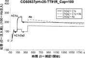


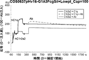


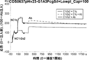
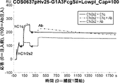


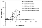
Classifications
-
- C—CHEMISTRY; METALLURGY
- C07—ORGANIC CHEMISTRY
- C07K—PEPTIDES
- C07K16/00—Immunoglobulins [IGs], e.g. monoclonal or polyclonal antibodies
- C07K16/18—Immunoglobulins [IGs], e.g. monoclonal or polyclonal antibodies against material from animals or humans
-
- A—HUMAN NECESSITIES
- A61—MEDICAL OR VETERINARY SCIENCE; HYGIENE
- A61K—PREPARATIONS FOR MEDICAL, DENTAL OR TOILETRY PURPOSES
- A61K39/00—Medicinal preparations containing antigens or antibodies
- A61K2039/505—Medicinal preparations containing antigens or antibodies comprising antibodies
-
- C—CHEMISTRY; METALLURGY
- C07—ORGANIC CHEMISTRY
- C07K—PEPTIDES
- C07K2317/00—Immunoglobulins specific features
- C07K2317/20—Immunoglobulins specific features characterized by taxonomic origin
- C07K2317/24—Immunoglobulins specific features characterized by taxonomic origin containing regions, domains or residues from different species, e.g. chimeric, humanized or veneered
-
- C—CHEMISTRY; METALLURGY
- C07—ORGANIC CHEMISTRY
- C07K—PEPTIDES
- C07K2317/00—Immunoglobulins specific features
- C07K2317/30—Immunoglobulins specific features characterized by aspects of specificity or valency
- C07K2317/32—Immunoglobulins specific features characterized by aspects of specificity or valency specific for a neo-epitope on a complex, e.g. antibody-antigen or ligand-receptor
-
- C—CHEMISTRY; METALLURGY
- C07—ORGANIC CHEMISTRY
- C07K—PEPTIDES
- C07K2317/00—Immunoglobulins specific features
- C07K2317/50—Immunoglobulins specific features characterized by immunoglobulin fragments
- C07K2317/56—Immunoglobulins specific features characterized by immunoglobulin fragments variable (Fv) region, i.e. VH and/or VL
-
- C—CHEMISTRY; METALLURGY
- C07—ORGANIC CHEMISTRY
- C07K—PEPTIDES
- C07K2317/00—Immunoglobulins specific features
- C07K2317/70—Immunoglobulins specific features characterized by effect upon binding to a cell or to an antigen
- C07K2317/71—Decreased effector function due to an Fc-modification
-
- C—CHEMISTRY; METALLURGY
- C07—ORGANIC CHEMISTRY
- C07K—PEPTIDES
- C07K2317/00—Immunoglobulins specific features
- C07K2317/70—Immunoglobulins specific features characterized by effect upon binding to a cell or to an antigen
- C07K2317/76—Antagonist effect on antigen, e.g. neutralization or inhibition of binding
-
- C—CHEMISTRY; METALLURGY
- C07—ORGANIC CHEMISTRY
- C07K—PEPTIDES
- C07K2317/00—Immunoglobulins specific features
- C07K2317/90—Immunoglobulins specific features characterized by (pharmaco)kinetic aspects or by stability of the immunoglobulin
- C07K2317/92—Affinity (KD), association rate (Ka), dissociation rate (Kd) or EC50 value
Definitions
- the present invention relates to antibodies such as anti-C1s antibodies and methods of using them.
- the C1 complex is a large protein complex that functions as a major initiator of the classical pathway cascade.
- the C1 complex is composed of three components, C1q, C1r, and C1s, in molar ratios of 1:2:2, respectively (Non-Patent Document 1).
- the classical pathway is initiated upon binding of the C1 complex to antibody-bound targets.
- C1q which has six globular heads, mediates binding of the C1 complex to antibodies through an avidity-type interaction with the Fc region.
- C1r within the C1 complex Upon tight binding to the target, C1r within the C1 complex autoactivates and becomes enzymatically active. The activated C1r then cleaves and activates the proenzyme C1s within the C1 complex (Non-Patent Document 2).
- C1s then cleaves its substrates, complement components C2 and C4, into C2a/C2b and C4a/C4b fragments, respectively. This causes the assembly of the C3 convertase, C4b2a, to appear on the surface of the target and cleave C3 to form C3b.
- C3b then cleaves C5 to initiate formation of the terminal membrane attack complex of C5b, C6, C7, C8, and C9, which lyses the target through pore formation.
- Both C1s and C1r proteins have the same domain configuration of CUB1-EGF-CUB2-CCP1-CCP2-serine protease (Non-Patent Document 3).
- the CUB1-EGF-CUB2 domain mediates the interaction between C1r and C1s to form the C1r2s2 tetramer (Non-Patent Document 4) and also between C1r2s2 and C1q (Non-Patent Document 4).
- the CCP1-CCP2-serine protease domains of C1r and C1s are responsible for proteolytic cleavage of their respective substrates (6, 7).
- the C1r2s2 tetramer interacts with the six stems of C1q through six binding sites within the CUB1-EGF-CUB2 domain of this tetramer (Non-Patent Document 5).
- autoimmune hemolytic anemia AIHA
- Behcet's disease bullous pemphigoid
- ITP immune thrombocytopenic purpura
- Non-Patent Document 8 an antibody that binds to the beta domain of C1s, can inhibit the interaction between C1r2s2 and C1q.
- this antibody was unable to completely neutralize the hemolytic activity of human serum, with 30% activity remaining even after 24 hours of incubation of serum with this antibody.
- Antibodies are highly attractive pharmaceuticals because they are stable in plasma, highly specific for their targets, and generally exhibit excellent pharmacokinetic profiles. However, due to their large molecular size, therapeutic antibody doses are usually high. If the target is present in high abundance, even higher therapeutic doses of antibody will be required. As a result, methods to improve the pharmacokinetics, pharmacodynamics, and antigen-binding properties of antibodies are attractive ways to reduce the dosage and high manufacturing costs associated with therapeutic antibodies.
- Antibodies that bind antigen in a pH-dependent manner are capable of neutralizing multiple antigen molecules by a single antibody molecule. It has been reported that it enables (Non-Patent Document 9, Patent Document 1).
- a pH-dependent antibody binds strongly to its antigen under neutral pH conditions in the plasma, but dissociates from the antigen under acidic pH conditions within the endosomes of cells. Upon dissociation from the antigen, the antibody is recycled into the plasma by the FcRn receptor and the dissociated antigen is degraded within the lysosomes of the cell. The recycled antibody is then free to rebind and neutralize the antigen molecule, and the process repeats as long as the antibody remains in the blood.
- the present invention provides anti-complement component antibodies such as anti-C1s antibodies, pharmaceutical compositions containing them, and methods of using them.
- the present invention comprises an antigen-binding region and an antibody constant region, has a displacement function that binds to the C1qrs complex and promotes dissociation of C1q from the C1qrs complex, and/or Antibodies are provided that have blocking functionality to bind C1r2s2 and inhibit binding of C1q to C1r2s2 and that bind C1s in a pH-dependent manner.
- the antibody of the invention comprises a mutated constant region comprising at least one amino acid modification that reduces binding activity to an Fc ⁇ R and/or at least one amino acid modification that reduces the isoelectric point (pI) of the Fc region.
- the invention relates to: [1] An isolated antibody comprising an antigen binding region and an antibody constant region, the antibody promotes dissociation of C1q from the C1qrs complex and/or inhibits binding of C1q to C1r2s2;
- An antibody wherein the antigen-binding region comprises a combination of HVR-H1, HVR-H2, HVR-H3, HVR-L1, HVR-L2, and HVR-L3 selected from the group consisting of 1) to 6) below : 1) HVR-H1, HVR-H2, HVR-H3, HVR-L1, HVR-L2, and HVR-L3, comprising amino acid sequences consisting of SEQ ID NOS: 25, 26, 27, 60, 61, and 62, respectively; 2) HVR-H1, HVR-H2, HVR-H3, HVR-L1, HVR-L2, and HVR-L3, comprising amino acid sequences consisting of SEQ ID NOs: 37, 38, 39, 56, 57, and 58, respectively; 3) HVR
- VH and VL comprising amino acid sequences consisting of SEQ ID NOs:32 and 55, respectively.
- [3] The antibody of [1] or [2], which has an acidic KD/neutral KD ratio of 107 or more, which is the ratio of the KD value in the acidic pH range to the KD value in the neutral pH range.
- [4] The antibody according to any one of [1] to [3], wherein the antigen-binding region can specifically bind to the CUB1-EGF-CUB2 domain of human C1s.
- [5] The antibody of any one of [1] to [4], wherein the antibody has a mutated constant region containing at least one amino acid modification that reduces Fc ⁇ receptor-binding activity.
- the antibody according to any one of [1] to [8], which has a pI of 7.8 or less [10] The antibody according to any one of [1] to [8], wherein the constant region comprises a heavy chain constant region comprising the amino acid sequence of SEQ ID NO:45 and a light chain constant region comprising the amino acid sequence of SEQ ID NO:23.
- HVR-H1, HVR-H2, HVR-H3, HVR-L1, HVR-L2, and HVR-L3 selected from the group consisting of 1) to 6) below antibody: 1) HVR-H1, HVR-H2, HVR-H3, HVR-L1, HVR-L2, and HVR-L3, comprising amino acid sequences consisting of SEQ ID NOS: 25, 26, 27, 60, 61, and 62, respectively; 2) HVR-H1, HVR-H2, HVR-H3, HVR-L1, HVR-L2, and HVR-L3, comprising amino acid sequences consisting of SEQ ID NOs: 37, 38, 39, 56, 57, and 58, respectively; 3) HVR-H1, HVR-H2, HVR-H3, HVR-L1, HVR-L2, and HVR-L3, comprising amino acid sequences consisting of SEQ ID NOS: 25, 26, 27, 56, 57, and 58, respectively; 4) HVR-H1, HVR-H2, HVR-H3, HVR-L1,
- VH and VL comprising amino acid sequences consisting of SEQ ID NOs:32 and 55, respectively.
- a pharmaceutical composition comprising the antibody of any one of [1] to [14] and at least one pharmaceutically acceptable carrier.
- a therapeutic or prophylactic agent for a complement-mediated disease or disorder comprising the antibody of any one of [1] to [14].
- An isolated antibody comprising an antigen binding region and an antibody constant region, The acidic KD/neutral KD ratio, which is the ratio of the KD value in the acidic pH range to the KD value in the neutral pH range, is 107 or more, An antibody wherein the antibody constant region comprises at least one amino acid modification that reduces Fc ⁇ receptor-binding activity and at least one amino acid modification that reduces the isoelectric point (pI).
- Figure 2-1 shows evaluation of the C1q dissociation promoting function of the anti-C1s antibody COS0637pHv8-TT91R.
- Solid line is sensorgram obtained when hC1q was injected into hC1r2s2 followed by buffer injection (C1r2s2+C1q): Sensorgram 1, dashed line is hC1q injected into hC1r2s2 followed by antibody injection. sensorgram (C1r2s2+C1q+Ab): sensorgram 2, and the dashed line shows sensorgram (C1r2s2+Ab): sensorgram 3 obtained when antibody alone was injected into hC1r2s2 without injecting hC1q. COS0637pHv8-TT91R was injected as an Ab.
- Figure 2-2 shows evaluation of the C1q dissociation promoting function of the anti-C1s antibody COS0637pHv15-TT91R.
- Solid line is sensorgram obtained when hC1q was injected into hC1r2s2 followed by buffer injection (C1r2s2+C1q): Sensorgram 1, dashed line is hC1q injected into hC1r2s2 followed by antibody injection.
- the dashed line shows sensorgram (C1r2s2+Ab): sensorgram 3 obtained when antibody alone was injected into hC1r2s2 without injecting hC1q.
- COS0637pHv15-TT91R was injected as an Ab.
- Figure 2-3 shows evaluation of the C1q dissociation promoting function of the anti-C1s antibody COS0637pHv16-TT91R.
- Solid line is sensorgram obtained when hC1q was injected into hC1r2s2 followed by buffer injection (C1r2s2+C1q): Sensorgram 1, dashed line is hC1q injected into hC1r2s2 followed by antibody injection.
- the dashed line shows sensorgram (C1r2s2+Ab): sensorgram 3 obtained when antibody alone was injected into hC1r2s2 without injecting hC1q.
- COS0637pHv16-TT91R was injected as an Ab.
- Figure 2-4 shows evaluation of the C1q dissociation promoting function of the anti-C1s antibody COS0637pHv17-TT91R.
- Solid line is sensorgram obtained when hC1q was injected into hC1r2s2 followed by buffer injection (C1r2s2+C1q): Sensorgram 1, dashed line is hC1q injected into hC1r2s2 followed by antibody injection. sensorgram (C1r2s2+C1q+Ab): sensorgram 2, and the dashed line shows sensorgram (C1r2s2+Ab): sensorgram 3 obtained when antibody alone was injected into hC1r2s2 without injecting hC1q. COS0637pHv17-TT91R was injected as an Ab.
- Figure 2-5 shows evaluation of the C1q dissociation promoting function of the anti-C1s antibody COS0637pHv21-TT91R.
- Solid line is sensorgram obtained when hC1q was injected into hC1r2s2 followed by buffer injection (C1r2s2+C1q): Sensorgram 1, dashed line is hC1q injected into hC1r2s2 followed by antibody injection.
- the dashed line shows sensorgram (C1r2s2+Ab): sensorgram 3 obtained when antibody alone was injected into hC1r2s2 without injecting hC1q.
- COS0637pHv21-TT91R was injected as an Ab.
- Figure 2-6 shows evaluation of the C1q dissociation promoting function of the anti-C1s antibody COS0637pHv23-TT91R.
- Solid line is sensorgram obtained when hC1q was injected into hC1r2s2 followed by buffer injection (C1r2s2+C1q): Sensorgram 1, dashed line is hC1q injected into hC1r2s2 followed by antibody injection.
- the dashed line shows sensorgram (C1r2s2+Ab): sensorgram 3 obtained when antibody alone was injected into hC1r2s2 without injecting hC1q.
- As an Ab COS0637pHv23-TT91R was injected.
- Figure 2-7 shows evaluation of the C1q dissociation promoting function of the anti-C1s antibody COS0637pHv25-TT91R.
- Solid line is sensorgram obtained when hC1q was injected into hC1r2s2 followed by buffer injection (C1r2s2+C1q): Sensorgram 1, dashed line is hC1q injected into hC1r2s2 followed by antibody injection. sensorgram (C1r2s2+C1q+Ab): sensorgram 2, and the dashed line shows sensorgram (C1r2s2+Ab): sensorgram 3 obtained when antibody alone was injected into hC1r2s2 without injecting hC1q. COS0637pHv25-TT91R was injected as an Ab.
- Figure 2-8 shows evaluation of the C1q dissociation promoting function of the anti-C1s antibody COS0637pHv8-G1A3FcgSil+LowpI.
- Solid line is sensorgram obtained when hC1q was injected into hC1r2s2 followed by buffer injection (C1r2s2+C1q): Sensorgram 1, dashed line is hC1q injected into hC1r2s2 followed by antibody injection.
- the dashed line shows sensorgram (C1r2s2+Ab): sensorgram 3 obtained when antibody alone was injected into hC1r2s2 without injecting hC1q.
- the dashed line shows sensorgram (C1r2s2+Ab): sensorgram 3 obtained when antibody alone was injected into hC1r2s2 without injecting hC1q.
- As Ab COS0637pHv15-G1A3FcgSil+LowpI was injected.
- Figure 2-10 shows evaluation of the C1q dissociation promoting function of the anti-C1s antibody COS0637pHv16-G1A3FcgSil+LowpI.
- Solid line is sensorgram obtained when hC1q was injected into hC1r2s2 followed by buffer injection (C1r2s2+C1q): Sensorgram 1, dashed line is hC1q injected into hC1r2s2 followed by antibody injection. sensorgram (C1r2s2+C1q+Ab): sensorgram 2, and the dashed line shows sensorgram (C1r2s2+Ab): sensorgram 3 obtained when antibody alone was injected into hC1r2s2 without injecting hC1q. As Ab, COS0637pHv16-G1A3FcgSil+LowpI was injected.
- Figure 2-11 shows evaluation of the C1q dissociation promoting function of the anti-C1s antibody COS0637pHv17-G1A3FcgSil+LowpI.
- Solid line is sensorgram obtained when hC1q was injected into hC1r2s2 followed by buffer injection (C1r2s2+C1q): Sensorgram 1, dashed line is hC1q injected into hC1r2s2 followed by antibody injection.
- the dashed line shows sensorgram (C1r2s2+Ab): sensorgram 3 obtained when antibody alone was injected into hC1r2s2 without injecting hC1q.
- FIG. 2-12 shows evaluation of the C1q dissociation promoting function of the anti-C1s antibody COS0637pHv21-G1A3FcgSil+LowpI.
- Solid line is sensorgram obtained when hC1q was injected into hC1r2s2 followed by buffer injection (C1r2s2+C1q): Sensorgram 1, dashed line is hC1q injected into hC1r2s2 followed by antibody injection.
- the dashed line shows sensorgram (C1r2s2+Ab): sensorgram 3 obtained when antibody alone was injected into hC1r2s2 without injecting hC1q.
- As Ab COS0637pHv21-G1A3FcgSil+LowpI was injected.
- Figure 2-13 shows evaluation of the C1q dissociation promoting function of the anti-C1s antibody COS0637pHv23-G1A3FcgSil+LowpI.
- Solid line is sensorgram obtained when hC1q was injected into hC1r2s2 followed by buffer injection (C1r2s2+C1q): Sensorgram 1, dashed line is hC1q injected into hC1r2s2 followed by antibody injection. sensorgram (C1r2s2+C1q+Ab): sensorgram 2, and the dashed line shows sensorgram (C1r2s2+Ab): sensorgram 3 obtained when antibody alone was injected into hC1r2s2 without injecting hC1q. As Ab, COS0637pHv23-G1A3FcgSil+LowpI was injected.
- Figure 2-14 shows evaluation of the C1q dissociation promoting function of the anti-C1s antibody COS0637pHv25-G1A3FcgSil+LowpI.
- Solid line is sensorgram obtained when hC1q was injected into hC1r2s2 followed by buffer injection (C1r2s2+C1q): Sensorgram 1, dashed line is hC1q injected into hC1r2s2 followed by antibody injection.
- the dashed line shows sensorgram (C1r2s2+Ab): sensorgram 3 obtained when antibody alone was injected into hC1r2s2 without injecting hC1q.
- FIG. 3 shows plasma antibody concentration changes (log-linear) after single intravenous administration of COS0637pHv16-G1A3FcgSil+LowpI and COS0637pHv21-G1A3FcgSil+LowpI antibodies in male cynomolgus monkeys. Data are shown as mean ⁇ standard deviation (COS0637pHv16-G
- Figure 4 shows evaluation of the complement neutralizing function of anti-C1s antibodies in monkeys.
- Figure 2 shows neutralizing activity of anti-C1s antibodies (COS0637pHv2-FcgSil, COS0637pHv2-SG1077R, COS0637pHv3-SG1077R, COS0637pHv8-SG1077R, COS0637pHv16-G1A3FcgSil+LowpI, COS0637pHv21-G1A3FcgSil+LowpI) in monkeys. All specimens inhibited erythrocyte lysis immediately after administration.
- an "acceptor human framework” for the purposes of this specification is a light chain variable domain (VL) framework or heavy chain variable domain (VH ) a framework containing the amino acid sequence of the framework.
- Acceptor human frameworks "derived from” human immunoglobulin frameworks or human consensus frameworks may contain those same amino acid sequences or may contain alterations in the amino acid sequences. In some embodiments, the number of amino acid changes is 10 or less, 9 or less, 8 or less, 7 or less, 6 or less, 5 or less, 4 or less, 3 or less, or 2 or less.
- the VL acceptor human framework is identical in sequence to a VL human immunoglobulin framework sequence or a human consensus framework sequence.
- Binding affinity refers to the total strength of non-covalent interactions between one binding site of a molecule (eg, antibody) and its binding partner (eg, antigen). Unless otherwise indicated, "binding affinity” as used herein refers to the intrinsic binding affinity that reflects a 1:1 interaction between members of a binding pair (eg, antibody and antigen). The affinity of molecule X for its partner Y can generally be expressed by the dissociation constant (Kd or KD). Affinity can be measured by routine methods known in the art, including those described herein. Individual specific illustrative and exemplary embodiments for measuring binding affinity are described below. "Affinity”, “binding affinity”, “binding capacity” and “binding activity” can be used interchangeably.
- the term "avidity” refers to the total strength of non-covalent interactions between a single or multiple binding sites of a molecule (e.g. antibody) and its binding partner (e.g. antigen).
- Binding activity is not strictly limited to activity that reflects a 1:1 interaction between members of a binding pair (eg, antibody and antigen). When members of a binding pair are capable of binding to each other in both monovalent and multivalent manners, avidity is the combined strength of these bindings.
- the binding activity of molecule X for its partner Y can usually be expressed by the dissociation constant (KD). Alternatively, association and dissociation rates (Kon and Koff) can be used to assess binding. Binding activity can be measured by common methods known in the art, including those described herein. Specific illustrative and exemplary embodiments for measuring binding affinity are described below.
- An “affinity matured” antibody has one or more modifications in one or more hypervariable regions (HVRs) that confer improved affinity for the antibody's antigen as compared to a parent antibody that does not possess the modifications.
- HVRs hypervariable regions
- anti-C1s antibody antibodies capable of binding C1s with sufficient affinity such that the antibody binds to C1s. Refers to antibodies that are useful as diagnostic and/or therapeutic agents when targeted.
- the extent of binding of the anti-C1s antibody to an unrelated non-C1s protein is less than about 10% of the binding of the antibody to C1s, eg, as measured by radioimmunoassay (RIA).
- the antibody that binds C1s is 1 micromolar ( ⁇ M) or less, 100 nM or less, 10 nM or less, 1 nM or less, 0.1 nM or less, 0.01 nM or less, or 0.001 nM or less (e.g., 10 ⁇ 8 M or less). , such as from 10 ⁇ 8 M to 10 ⁇ 13 M, such as from 10 ⁇ 9 M to 10 ⁇ 13 M).
- the anti-C1s antibody binds to an epitope of C1s that is conserved among C1s from different species.
- antibody is used in the broadest sense, and as long as it exhibits the desired antigen-binding activity, it is not limited to, but is not limited to, monoclonal antibodies, polyclonal antibodies, multispecific antibodies (e.g., It encompasses a variety of antibody structures, including bispecific antibodies) and antibody fragments.
- antibody fragment refers to a molecule other than an intact antibody that contains a portion of the intact antibody that binds to the antigen to which the intact antibody binds.
- antibody fragments include, but are not limited to, Fv, Fab, Fab', Fab'-SH, F(ab') 2 ; diabodies; linear antibodies; single chain antibody molecules (e.g., scFv ); and multispecific antibodies formed from antibody fragments.
- an "antibody that binds to the same epitope” as a reference antibody refers to an antibody that blocks the binding of the reference antibody to its own antigen by 50% or more in a competition assay, and conversely, the reference antibody The aforementioned antibodies block more than 50% of binding to their own antigen in the assay.
- An exemplary competition assay is provided herein.
- chimeric antibody means that a portion of the heavy and/or light chains are derived from a particular source or species, while the remainder of the heavy and/or light chains are derived from a different source or species. Refers to antibodies.
- the "class" of an antibody refers to the type of constant domain or constant region provided by the heavy chains of the antibody.
- the heavy-chain constant domains that correspond to the different classes of immunoglobulins are called ⁇ , ⁇ , ⁇ , ⁇ , and ⁇ , respectively.
- cytotoxic agent refers to a substance that inhibits or prevents the function of cells and/or causes the death or destruction of cells.
- Cytotoxic agents include, but are not limited to, radioactive isotopes such as 211 At, 131 I, 125 I, 90 Y, 186 Re, 188 Re, 153 Sm, 212 Bi, 32 P, 212 Pb chemotherapeutic agents or agents (e.g., methotrexate, adriamycin, vinca alkaloids (vincristine, vinblastine, etoposide), doxorubicin, melphalan, mitomycin C, chlorambucil, daunorubicin, or other calating agents); growth inhibitors; enzymes such as nucleases and fragments thereof; antibiotics; toxins, including variants); and various anti-tumor or anti-cancer agents disclosed below.
- radioactive isotopes such as 211 At, 131 I, 125 I, 90 Y, 186 Re,
- Antibody effector function refers to the biological activity that differs depending on the antibody isotype, which is attributed to the Fc region of an antibody.
- Examples of antibody effector functions include: C1q binding and complement dependent cytotoxicity (CDC); Fc receptor binding; antibody-dependent cell cytotoxicity; phagocytosis; downregulation of cell surface receptors (eg, B cell receptors); and B cell activation.
- an “effective amount” of an agent refers to an amount, at the necessary dose and over the necessary period of time, effective to achieve the desired therapeutic or prophylactic result.
- epitope includes any determinant capable of being bound by an antibody.
- An epitope is the region of an antigen that is bound by an antibody that targets it, and includes specific amino acids that directly contact the antibody.
- Epitopic determinants can include chemically active surface groupings of molecules such as amino acids, sugar side chains, phosphoryl or sulfonyl groups, and have specific three dimensional structural characteristics, and/or specific charge characteristics. can be done.
- antibodies specific for a particular target antigen preferentially recognize epitopes on that target antigen in a complex mixture of proteins and/or macromolecules.
- Fc region is used herein to define the C-terminal region of an immunoglobulin heavy chain that includes at least a portion of the constant region.
- the term includes native sequence Fc regions and variant Fc regions.
- the human IgG heavy chain Fc region extends from Cys226 or from Pro230 to the carboxyl terminus of the heavy chain.
- the C-terminal lysine (Lys447) or glycine-lysine (residues 446-447) of the Fc region may or may not be present.
- the numbering of amino acid residues in the Fc region or constant region is according to Kabat et al., Sequences of Proteins of Immunological Interest, 5th Ed. Public Health Service, National Institutes of Health, Bethesda, According to the EU numbering system (also known as the EU index), as described in MD 1991.
- FR Framework or "FR” refers to variable domain residues other than hypervariable region (HVR) residues.
- the FRs of variable domains usually consist of four FR domains: FR1, FR2, FR3 and FR4. Accordingly, FR and HVR sequences usually appear in VH (or VL) in the following order: FR1-H1(L1)-FR2-H2(L2)-FR3-H3(L3)-FR4.
- full length antibody “whole antibody” and “whole antibody” are used interchangeably herein and have a structure substantially similar to that of a naturally occurring antibody or as defined herein. It refers to an antibody having a heavy chain containing an Fc region that
- host cell refers to a cell (including progeny of such a cell) into which exogenous nucleic acid has been introduced.
- a host cell includes “transformants” and “transformed cells,” including the primary transformed cell and progeny derived therefrom at any passage number. Progeny may not be completely identical in nucleic acid content to the parent cell, but may contain mutations. Mutant progeny that have the same function or biological activity as with which the originally transformed cell was screened or selected are also included herein.
- human antibody is an antibody with an amino acid sequence that corresponds to that of an antibody produced by a human or human cells or derived from a human antibody repertoire or other non-human source using human antibody coding sequences. This definition of human antibody specifically excludes humanized antibodies that contain non-human antigen-binding residues.
- a "human consensus framework” is a framework that represents the most commonly occurring amino acid residues in a selected group of human immunoglobulin VL or VH framework sequences.
- the selection of human immunoglobulin VL or VH sequences is from a subgroup of variable domain sequences.
- the subgroup of sequences is the subgroup in Kabat et al., Sequences of Proteins of Immunological Interest, Fifth Edition, NIH Publication 91-3242, Bethesda MD (1991), vols. 1-3.
- the subgroup is subgroup ⁇ I according to Kabat et al., supra.
- the subgroup is subgroup III according to Kabat et al., supra.
- a “humanized” antibody refers to a chimeric antibody that contains amino acid residues from non-human HVRs and amino acid residues from human FRs.
- a humanized antibody comprises substantially all of at least one, typically two, variable domains in which all or substantially all HVRs (e.g., CDRs) are non- Corresponds to that of a human antibody, and all or substantially all FRs correspond to those of a human antibody.
- a humanized antibody optionally may comprise at least a portion of an antibody constant region derived from a human antibody.
- a "humanized form" of an antibody (eg, a non-human antibody) refers to an antibody that has undergone humanization.
- hypervariable region is hypervariable in sequence (“complementarity determining region” or “CDR”) and/or is structurally defined. Refers to the regions of the variable domain of an antibody that form loops (“hypervariable loops”) and/or contain antigen-contacting residues (“antigen-contacting”). Antibodies typically contain 6 HVRs, 3 in the VH (H1, H2, H3) and 3 in the VL (L1, L2, L3), for a total of 6 HVRs.
- Exemplary HVRs herein include: (a) at amino acid residues 26-32 (L1), 50-52 (L2), 91-96 (L3), 26-32 (H1), 53-55 (H2), and 96-101 (H3); resulting hypervariable loops (Chothia and Lesk, J. Mol. Biol. 196: 901-917 (1987)); (b) at amino acid residues 24-34 (L1), 50-56 (L2), 89-97 (L3), 31-35b (H1), 50-65 (H2), and 95-102 (H3); the resulting CDRs (Kabat et al., Sequences of Proteins of Immunological Interest, 5th Ed.
- HVR residues and other residues in the variable domain are numbered herein according to Kabat et al., supra.
- immunoconjugate is an antibody conjugated to one or more heterologous molecules (heterologous molecules include, but are not limited to, cytotoxic agents).
- “Individuals” or “subjects” are mammals. Mammals include, but are not limited to, domestic animals (eg, cows, sheep, cats, dogs, horses), primates (eg, humans and non-human primates such as monkeys), rabbits, and , including rodents (eg, mice and rats). In certain aspects, the individual or subject is human.
- an “isolated” antibody is one that has been separated from a component of its original environment.
- antibodies are analyzed electrophoretically (e.g., SDS-PAGE, isoelectric focusing (IEF), capillary electrophoresis) or chromatographically (e.g., ion-exchange or reverse-phase HPLC). Purified to greater than 95% or 99% purity as measured.
- electrophoretically e.g., SDS-PAGE, isoelectric focusing (IEF), capillary electrophoresis
- chromatographically e.g., ion-exchange or reverse-phase HPLC.
- isolated nucleic acid refers to a nucleic acid molecule that has been separated from components of its original environment.
- An isolated nucleic acid includes a nucleic acid molecule contained within cells that normally contain the nucleic acid molecule, but the nucleic acid molecule is present extrachromosomally or on a chromosome other than its native chromosomal location. exists in position.
- isolated nucleic acid encoding an anti-C1s antibody or “an isolated nucleic acid encoding an anti-C1r antibody” refers to one or more nucleic acids encoding the antibody heavy and light chains (or fragments thereof) Molecules, including nucleic acid molecules on a single vector or on separate vectors, and nucleic acid molecules present in one or more locations in a host cell.
- the term "monoclonal antibody” as used herein refers to an antibody obtained from a substantially homogeneous population of antibodies. That is, the individual antibodies that make up the population are mutated antibodies that may arise (e.g., mutated antibodies containing naturally occurring mutations, or mutated antibodies that arise during the manufacture of monoclonal antibody preparations. are identical and/or bind to the same epitope, except that they are present in different amounts). In contrast to polyclonal antibody preparations which typically include different antibodies directed against different determinants (epitopes), each monoclonal antibody of a monoclonal antibody preparation is directed against a single determinant on the antigen.
- monoclonal indicates the character of the antibody as being obtained from a population of substantially homogeneous antibodies and should not be construed as requiring production of the antibody by any particular method.
- monoclonal antibodies used in accordance with the present invention include, but are not limited to, hybridoma technology, recombinant DNA technology, phage display technology, transgenic animals containing all or part of the human immunoglobulin locus. Such and other exemplary methods for making monoclonal antibodies are described herein.
- naked antibody refers to an antibody that is not conjugated to a heterologous moiety (eg, a cytotoxic moiety) or a radiolabel.
- a naked antibody may be present in a pharmaceutical formulation.
- Native antibodies refer to immunoglobulin molecules with various structures occurring in nature.
- native IgG antibodies are heterotetrameric glycoproteins of approximately 150,000 daltons composed of two identical light chains and two identical heavy chains that are disulfide-bonded. From N-terminus to C-terminus, each heavy chain has a variable region (VH), also called a variable heavy domain or heavy chain variable domain, followed by three constant domains (CH1, CH2 and CH3). Similarly, from N-terminus to C-terminus, each light chain has a variable region (VL), also called a variable light domain or light chain variable domain, followed by a constant light (CL) domain.
- VH variable region
- VL variable region
- CL constant light
- package insert is commonly included in the commercial packaging of therapeutic products and includes information about indications, directions, doses, methods of administration, concomitant therapies, contraindications, and/or warnings regarding the use of such therapeutic products. Used to refer to instructions for use.
- a "percent (%) amino acid sequence identity" to a reference polypeptide sequence is obtained after aligning the sequences to obtain the maximum percent sequence identity and introducing gaps if necessary, and without any conservative substitutions. Defined as the percentage of amino acid residues in a candidate sequence that are identical to amino acid residues in the reference polypeptide sequence, not considered part of the identity. Alignments for purposes of determining percent amino acid sequence identity may be performed by a variety of methods within the skill in the art, such as BLAST, BLAST-2, ALIGN, Megalign (DNASTAR) software, or GENETYX® (Genetyx®) software. Co., Ltd.). Those skilled in the art can determine appropriate parameters for aligning sequences, including any algorithms needed to achieve maximal alignment over the entire length of the sequences being compared.
- the ALIGN-2 sequence comparison computer program is the work of Genentech, Inc. and its source code has been filed with the US Copyright Office, Washington DC, 20559 with user documentation, under US Copyright Registration No. TXU510087. Registered.
- the ALIGN-2 program is publicly available from Genentech, Inc., South San Francisco, Calif., or may be compiled from source code.
- ALIGN-2 programs are compiled for use on UNIX operating systems, including Digital UNIX V4.0D. All sequence comparison parameters were set by the ALIGN-2 program and do not vary.
- the % amino acid sequence identity of a given amino acid sequence A to, with, or to a given amino acid sequence B is calculated as follows: 100 times the fraction X/Y where X is the number of amino acid residues scored as identical matches in the alignment of A and B by the sequence alignment program ALIGN-2 and Y is in B is the total number of amino acid residues in It is understood that the % amino acid sequence identity of A to B is not equal to the % amino acid sequence identity of B to A if the length of amino acid sequence A is not equal to the length of amino acid sequence B. deaf. Unless otherwise specified, all % amino acid sequence identity values used herein are obtained using the ALIGN-2 computer program as described in the immediately preceding paragraph.
- pharmaceutical formulation means a preparation in a form such that the active ingredients contained therein can exert their biological activity and which is unacceptably toxic to the subject to whom the formulation is administered.
- “Pharmaceutically acceptable carrier” refers to an ingredient other than the active ingredient in a pharmaceutical formulation that is non-toxic to the subject.
- Pharmaceutically acceptable carriers include, but are not limited to, buffers, excipients, stabilizers, or preservatives.
- the phrase "specifically binds" means that an antibody is of interest at a level of binding that includes background binding (i.e., non-specific binding) but does not include significant binding (i.e., specific binding). Refers to the activity or characteristic of binding to an antigen that is not of interest. In other words, “specifically binds” means that the antibody is of interest at a level of binding that includes significant binding (i.e., specific binding) in addition to or instead of background binding (i.e., non-specific binding).
- the activity or characteristic of binding to an antigen of Specificity can be measured by any method mentioned herein or known in the art.
- the level of non-specific or background binding described above may be zero, or near zero rather than zero, or may be so small that it is technically ignored by those skilled in the art. For example, if one skilled in the art cannot detect or observe a significant (or relatively strong) signal for binding between an antibody and an antigen of interest not in a suitable binding assay, then the antibody is "antigen of interest". it does not bind specifically". Conversely, an antibody is "specific for an antigen of interest” if one skilled in the art can detect or observe a significant (or relatively strong) signal for binding between the antibody and the antigen of interest in an appropriate binding assay. can be said to bind to
- C1s can be from any vertebrate source, including mammals such as primates (e.g., humans) and rodents (e.g., mice and rats). It refers to the native C1s of The term encompasses "full-length" unprocessed C1s as well as any form of C1s that results from processing in the cell. The term also includes naturally occurring variants of C1s, such as splice variants and allelic variants.
- An exemplary human C1s amino acid sequence is shown in SEQ ID NO:1.
- An exemplary cynomolgus monkey C1s amino acid sequence is shown in SEQ ID NO:3.
- C1r can be from any vertebrate source, including mammals such as primates (e.g., humans) and rodents (e.g., mice and rats). It refers to the native C1r of The term encompasses "full length" unprocessed C1r as well as any form of C1r that results from processing in the cell. The term also includes naturally occurring variants of C1r, such as splice variants and allelic variants.
- An exemplary human C1r amino acid sequence is shown in SEQ ID NO:2.
- An exemplary cynomolgus C1r amino acid sequence is shown in SEQ ID NO:4.
- treatment refers to clinical treatments intended to alter the natural course of the individual being treated.
- intervention which can be performed both for prophylaxis and during the course of a clinical condition.
- Desirable effects of treatment include, but are not limited to, prevention of disease onset or recurrence, relief of symptoms, attenuation of any direct or indirect pathological effects of disease, prevention of metastasis, prevention of disease Includes reduced rate of progression, amelioration or amelioration of disease state, and remission or improved prognosis.
- the antibodies of the invention are used to delay the onset of disease or slow the progression of disease.
- variable region refers to the domains of the heavy or light chains of an antibody that are involved in binding the antibody to the antigen.
- the heavy and light chain variable domains (VH and VL, respectively) of native antibodies are usually similar, with each domain containing four conserved framework regions (FR) and three hypervariable regions (HVR). structure (see, for example, Kindt et al. Kuby Immunology, 6th ed., W.H. Freeman and Co., page 91 (2007)).
- a single VH or VL domain may be sufficient to confer antigen binding specificity.
- antibodies that bind a particular antigen may be isolated using the VH or VL domain from the antibody that binds that antigen to screen a complementary library of VL or VH domains, respectively. See, e.g., Portolano et al., J. Immunol. 150:880-887 (1993); Clarkson et al., Nature 352:624-628 (1991).
- vector refers to a nucleic acid molecule capable of propagating another nucleic acid to which it has been linked.
- the term includes vectors as self-replicating nucleic acid structures and vectors that integrate into the genome of a host cell into which they are introduced. Certain vectors are capable of directing the expression of nucleic acids to which they are operably linked. Such vectors are also referred to herein as "expression vectors”.
- the invention is based, in part, on antibodies that include an antigen binding region and an antibody constant region. In certain embodiments, antibodies are provided that bind to C1s. In certain embodiments, antibodies are provided that specifically bind to C1s. Antibodies of the invention are useful, for example, in diagnosing or treating complement-mediated diseases or disorders.
- the C1s species can be selected from one or more species.
- the species are humans and non-human animals.
- the species are humans, rats, and monkeys (eg, cynomolgus monkeys, rhesus monkeys, marmosets, chimpanzees, and baboons).
- the species are humans and monkeys (eg, cynomolgus monkeys, rhesus monkeys, marmosets, chimpanzees, and baboons).
- the species are humans and cynomolgus monkeys.
- antibodies encompass various types of antibodies, including antibody fragments, chimeric and humanized antibodies, human antibodies, library-derived antibodies, and multispecific antibodies.
- the antibody may be a full-length antibody, eg, a complete IgG1, IgG2, IgG3, or IgG4 antibody, or other antibody classes or isotypes as defined herein.
- the antibodies provided herein are antibody fragments.
- Antibody fragments include, but are not limited to, Fab, Fab', Fab'-SH, F(ab') 2 , Fv, and scFv fragments, as well as other fragments described below.
- Fab fragment antigen
- Fab' fragment antigen binding fragment
- Fv fragment antigen binding fragment
- scFv fragments fragment antigen binding fragments
- See US Pat. No. 5,869,046 for a discussion of Fab and F(ab′) 2 fragments containing salvage receptor binding epitope residues with increased in vivo half-lives.
- a diabody is an antibody fragment with two antigen-binding sites that may be bivalent or bispecific.
- WO1993/01161 Hudson et al., Nat. Med. 9:129-134 (2003); Hollinger et al., Proc. Natl. Acad. Sci. USA 90:6444-6448 (1993 ) reference.
- Triabodies and tetrabodies are also described in Hudson et al., Nat. Med. 9:129-134 (2003).
- a single domain antibody is an antibody fragment that contains all or part of the heavy chain variable domain or all or part of the light chain variable domain of an antibody.
- the single domain antibody is a human single domain antibody (Domantis, Inc., Waltham, MA; see, eg, US Pat. No. 6,248,516 B1).
- Antibody fragments are produced in a variety of ways, including, but not limited to, proteolytic digestion of intact antibodies, production by recombinant host cells (e.g., E. coli or phage), as described herein. can be made by the method of
- chimeric and humanized antibodies are chimeric antibodies.
- Certain chimeric antibodies are described, for example, in US Pat. No. 4,816,567; and Morrison et al., Proc. Natl. Acad. Sci. USA, 81:6851-6855 (1984).
- a chimeric antibody comprises a non-human variable region (eg, a variable region derived from a non-human primate such as mouse, rat, hamster, rabbit, or monkey) and a human constant region.
- a chimeric antibody is a "class-switched" antibody that has an altered class or subclass from that of the parent antibody. Chimeric antibodies also include antigen-binding fragments thereof.
- the chimeric antibody is a humanized antibody.
- non-human antibodies are humanized to reduce their immunogenicity in humans while retaining the specificity and affinity of the parent non-human antibody.
- a humanized antibody comprises one or more variable domains in which HVRs (e.g. CDRs (or portions thereof)) are derived from non-human antibodies and FRs (or portions thereof) are derived from human antibody sequences.
- HVRs e.g. CDRs (or portions thereof)
- FRs or portions thereof
- a humanized antibody optionally will also comprise at least a portion of a human constant region.
- a non-human antibody e.g., the antibody from which the HVR residues were derived
- Human framework regions that can be used for humanization include, but are not limited to: selected using the "best fit” method (see Sims et al., J. Immunol. 151:2296 (1993)); framework regions derived from the consensus sequences of human antibodies of a particular subgroup of light or heavy chain variable regions (Carter et al., Proc. Natl. Acad. Sci. USA, 89:4285 (1992 ) and Presta et al., J. Immunol., 151:2623 (1993)); human mature (somatic mutation) framework regions or human germline framework regions (e.g., Almagro and Fransson, Front. Biosci.
- the antibodies provided herein are human antibodies.
- Human antibodies can be produced by various techniques known in the art. Human antibodies are reviewed in van Dijk and van de Winkel, Curr. Opin. Pharmacol. 5:368-74 (2001) and Lonberg, Curr. Opin. Immunol. 20:450-459 (2008).
- Human antibodies may be prepared by administering an immunogen to transgenic animals that have been engineered to produce fully human antibodies or fully antibodies with human variable regions in response to an antigenic challenge.
- Such animals typically contain all or part of a human immunoglobulin locus, wherein all or part of the human immunoglobulin locus replaces the endogenous immunoglobulin locus, or is extrachromosomally or It exists in a state of being randomly incorporated into the chromosome of the animal.
- the endogenous immunoglobulin loci are usually inactivated.
- Human antibodies can also be made by hybridoma-based methods. Human myeloma and mouse-human heteromyeloma cell lines for the production of human monoclonal antibodies have been previously described (e.g. Kozbor, J. Immunol. 133: 3001 (1984); Brodeur et al., Monoclonal Antibody Production Techniques and Applications, pp. 51-63 (Marcel Dekker, Inc., New York, 1987); and Boerner et al., J. Immunol. 147: 86 (1991)). Human antibodies generated via human B-cell hybridoma technology are also described in Li et al., Proc. Natl. Acad. Sci. USA, 103:3557-3562 (2006).
- Human antibodies can also be generated by isolating selected Fv clone variable domain sequences from human-derived phage display libraries. Such variable domain sequences can then be combined with the desired human constant domain. Techniques for selecting human antibodies from antibody libraries are described below.
- Antibodies of the invention may be isolated by screening combinatorial libraries for antibodies with the desired activity or activities. For example, various methods are known in the art for generating phage display libraries and screening such libraries for antibodies with desired binding characteristics. Such methods are reviewed in Hoogenboom et al., Methods in Molecular Biology 178:1-37 (O'Brien et al., ed., Human Press, Totowa, NJ, 2001) and also see, for example, McCafferty et al., Nature 348:552-554; Clackson et al., Nature 352: 624-628 (1991); Marks et al., J. Mol. Biol.
- repertoires of VH and VL genes are separately cloned by the polymerase chain reaction (PCR) and randomly recombined into a phage library, which is called Winter et al., Ann. Rev. Immunol. 12: 433-455 (1994).
- Phage typically display antibody fragments, either as single-chain Fv (scFv) fragments or as Fab fragments.
- Libraries from immunized sources provide high affinity antibodies to the immunogen without the need to construct hybridomas.
- a naive repertoire can be cloned (e.g., from humans) to provide a wide range of non-self and It can also provide a single source of antibodies to self antigens.
- na ⁇ ve libraries were generated by cloning pre-rearranged V-gene segments from stem cells and hypervariable CDR3 as described in Hoogenboom and Winter, J. Mol. Biol. 227: 381-388 (1992). It can also be made synthetically by using PCR primers that encode the region and contain random sequences to accomplish the rearrangement in vitro.
- Patent literature describing human antibody phage libraries includes, for example: U.S. Patent No. 5,750,373, and U.S. Patent Application Publication Nos. 2005/0079574, 2005/0119455, 2005/0266000, 2007/0117126; Including 2007/0160598, 2007/0237764, 2007/0292936, and 2009/0002360.
- Antibodies or antibody fragments isolated from human antibody libraries are considered human antibodies or human antibody fragments herein.
- the antibodies provided herein are multispecific antibodies (eg, bispecific antibodies).
- Multispecific antibodies are monoclonal antibodies that have binding specificities for at least two different sites. In certain embodiments, one of the binding specificities is for C1s and the other is for any other antigen.
- bispecific antibodies may bind to two different epitopes of C1s. Bispecific antibodies may be used to localize cytotoxic agents to cells that express C1s. Bispecific antibodies can be prepared as full length antibodies or as antibody fragments.
- Multispecific antibodies include, but are not limited to, recombinant co-expression of two immunoglobulin heavy-light chain pairs with different specificities (Milstein and Cuello, Nature 305: 537 (1983), WO93/08829, and Traunecker et al., EMBO J. 10:3655 (1991)), and "knob-in-hole” technology (see, e.g., U.S. Patent No. 5,731,168).
- Multispecific antibodies are engineered by the electrostatic steering effect to create Fc heterodimeric molecules (WO2009/089004A1); bridging two or more antibodies or fragments (U.S.
- modified antibodies with three or more functional antigen-binding sites including "octopus antibodies” (see, eg, US Patent Application Publication No. 2006/0025576 A1).
- Antibodies or fragments herein also include “dual-acting Fabs” or “DAFs”, which contain one antigen-binding site that binds C1s and another, different antigen (e.g., US Patent Application Publication No. 2008/0069820 No.).
- the antibody is an isolated antibody.
- the isolated antibody comprises an antigen binding region and an antibody constant region.
- the isolated antibody may have a dissociation promoting function that specifically binds to C1s and promotes dissociation of C1q from the C1qrs complex.
- the isolated antibody may have a blocking function such that it specifically binds C1s and inhibits binding of C1q to C1r2s2.
- the isolated antibody may have either or both dissociation-promoting and blocking functions. Preferably the antibody has both of these functions.
- the isolated antibody specifically binds C1s in a pH-dependent manner.
- Dissociation constant (KD) values in the neutral pH range can be reliably calculated and KD values in the acidic pH range can be reliably calculated due to no or very low binding activity. cannot be calculated, or ii) the ratio of the KD value in the acidic pH range to the KD value in the neutral pH range (acid KD /neutral KD ratio) is greater than 10.
- Anti-C1s antibodies only remove C1r2s2 from the plasma (through binding to C1s) but not C1q, and are superior to antibodies that bind to and clear the C1qrs complex from the plasma. Expected to have a safety profile. As a result, side effects associated with C1q deficiency can be avoided. In addition, antibodies with the ability to promote fast dissociation of C1q are expected to exhibit faster neutralization of complement activity, which may lead to faster onset of therapeutic effect.
- isolated antibodies that inhibit the interaction between C1q and C1r2s2 complexes are isolated on a chip (e.g. BIACORE®) for surface plasmon resonance assays. It is an antibody that binds to the C1qrs complex on the (trademark) chip) and promotes the dissociation of C1q from the C1qrs complex.
- the function of binding to the C1qrs complex and promoting dissociation of C1q from the C1qrs complex is herein referred to as "dissociation-promoting function/activity" or "C1q dissociation-promoting function/activity.” be called.
- This function/activity may suitably be assessed qualitatively or quantitatively using a surface plasmon resonance assay, such as the BIACORE® assay described herein.
- the antibody exhibits a value of response units (RU) in the presence of the antibody determined by a surface plasmon resonance assay, e.g., a BIACORE® assay, after sufficient time has passed
- a surface plasmon resonance assay e.g., a BIACORE® assay
- An antibody can be determined to have dissociation-promoting function if it is lower than the value of response units (RU) below.
- the proximity of the curve in the presence of C1q and antibody to the curve in the presence of C1q and absence of antibody can be identified.
- a "crossover point” can be identified at which the curve in the presence of C1q and in the absence of antibody intersects the curve in the presence of C1q and antibody (see Examples for details). . Strictly speaking, multiple crossover points can be observed even in one sensorgram due to noise or oscillation of the latter curve when crossing the former curve. In such an example, any of the multiple crossover points may be selected as the "crossover point.”
- "Sufficient time lapse” means that the time point of measurement of the response unit (RU) value is well after the "crossover time point” for measurement purposes. In some embodiments, the response unit (RU) values are measured at least 60 seconds, 100 seconds, 150 seconds, 200 seconds, 500 seconds, 700 seconds, 1000 seconds after the start of antibody infusion.
- the measurement time points are at least 100, 200, 300, 400, 500, 600, 700, 800, 900, 1000 seconds after the crossover time. , after 3000 seconds, after 5000 seconds, after 7000 seconds, or after 10000 seconds.
- an isolated antibody that inhibits the interaction between C1q and the C1r2s2 complex is, for example, "captured amounts of the C1r2s2 complex and C1q are 200 resonance units (RU) and 200 resonance units (RU ) and injected at 10 microliters ( ⁇ L)/min of 500 nM antibody as analyte (e.g., in the BIACORE® assay ) If the crossover time is within 60 seconds, 100 seconds, 150 seconds, 200 seconds, 500 seconds, 700 seconds, 1000 seconds, 1500 seconds, or 2000 seconds after the start of antibody injection In addition, it can be determined that the antibody has dissociation-promoting function.
- an isolated antibody that inhibits the interaction between C1q and the C1r2s2 complex is, for example, "captured amounts of the C1r2s2 complex and C1q are 200 resonance units (RU) and 200 resonance units (RU ) and within 100 seconds, within 300 seconds, 500 Nearly all (or all) of C1q from the C1qrs complex within seconds, within 700 seconds, within 1000 seconds, within 1500 seconds, within 2000 seconds, within 3000 seconds, within 5000 seconds, within 7000 seconds, or within 10000 seconds When dissociated, it can be determined that the antibody has a dissociation promoting function.
- substantially all (of C1q) means 70%, 71%, 72%, 73%, 74%, 75%, 76%, 77%, 78%, 79%, 80%, 81% , 82%, 83%, 84%, 85%, 86%, 87%, 88%, 89%, 90%, 91%, 92%, 93%, 94%, 95%, 96%, 97%, 98 %, 99%, or higher; "all (of C1q)” represents a percentage of 100%.
- the present invention provides a method of screening for antibodies that promote the dissociation of C1q from the C1r2s2 complex, using the above method to measure the "dissociation promoting function/activity" of such antibodies. provide a way.
- the screening method comprises selecting antibodies that inhibit the interaction between C1q and C1r2s2 complexes, i.e., antibodies that bind to the C1qrs complex and promote dissociation of C1q from the C1qrs complex. is selected.
- Antibodies with dissociation-promoting function/activity can be suitably selected using a surface plasmon resonance assay, such as the BIACORE® assay described herein.
- the screening method comprises, after a sufficient amount of time, a surface plasmon resonance assay, e.g. ) determining the value of response units (RU) in the absence of antibody.
- the screening method may comprise comparing the value of (i) above with the value of (ii) above.
- the screening method may comprise the step of selecting antibodies where the value of (i) above is lower than the value of (ii) above.
- the screening method may include identifying a "crossover point" at which the curve in the presence of C1q and in the absence of antibody intersects the curve in the presence of C1q and antibody.
- the screening method is at least 60 seconds, 100 seconds, 150 seconds, 200 seconds, 500 seconds, 700 seconds, 1000 seconds, 1500 seconds after the start of antibody infusion, or It may include measuring the value of Response Units (RU) after 2000 seconds.
- the screening method is at least 100 seconds, 200 seconds, 300 seconds, 400 seconds, 500 seconds, 600 seconds, 700 seconds, 800 seconds, 900 seconds, 1000 seconds after the crossover time point. measuring the value in response units (RU) after, 3000 seconds, 5000 seconds, 7000 seconds, or 10000 seconds.
- the screening method comprises selecting antibodies that inhibit the interaction between the C1q and C1r2s2 complexes or that have dissociation-promoting functions, e.g. BIACORE® assay with the following conditions: volume is 200 resonance units (RU) and 200 resonance units (RU), respectively, and 500 nM antibody as analyte infused at 10 microliters ( ⁇ L)/min. within 60 seconds, within 100 seconds, within 150 seconds, within 200 seconds, within 500 seconds, within 700 seconds, within 1000 seconds, within 1500 seconds after the start of antibody infusion, as determined by , or selecting the antibody if it is within 2000 seconds.
- volume is 200 resonance units (RU) and 200 resonance units (RU)
- 500 nM antibody as analyte infused at 10 microliters ( ⁇ L)/min. within 60 seconds, within 100 seconds, within 150 seconds, within 200 seconds, within 500 seconds, within 700 seconds, within 1000 seconds, within 1500 seconds after the start of antibody infusion, as determined by , or selecting the antibody if it is within 2000 seconds.
- the screening method comprises selecting antibodies that inhibit the interaction between the C1q and C1r2s2 complexes or that have dissociation-promoting functions, e.g. Amounts are 200 resonance units (RU) and 200 resonance units (RU), respectively, with 500 nM antibody as analyte infused at 10 ⁇ L/min, as determined by the BIACORE® assay. , within 100 seconds, within 300 seconds, within 500 seconds, within 700 seconds, within 1000 seconds, within 1500 seconds, within 2000 seconds, within 3000 seconds, within 5000 seconds, within 7000 seconds, or 10000 seconds after the start of antibody injection within which the antibody may be selected if substantially all (or all) of C1q dissociates from the C1qrs complex.
- Amounts are 200 resonance units (RU) and 200 resonance units (RU), respectively, with 500 nM antibody as analyte infused at 10 ⁇ L/min, as determined by the BIACORE® assay.
- the invention provides an isolated antibody that inhibits the interaction between C1q and C1r2s2 complexes, wherein the antibody is It has a blocking function such as binding to C1r2s2 and inhibiting C1q binding to C1r2s2.
- the antibody has a blocking ratio of at least 60%, 65%, 70%, 75%, 80%, 85%, 90%, 95%, or higher. This blocking function/activity or blocking ratio can be determined by using the BIACORE® assay.
- C1r2s2 capture doses target 50, 100, 200, 400 resonance units (RU).
- the blocking ratio is calculated by the following formula: [1 ⁇ (human C1q binding response in presence of antibody variant/human C1q binding response in absence of antibody variant)] ⁇ 100%.
- the isolated antibody specifically binds C1s in a pH dependent manner.
- the binding activity of the antibody to C1s in the acidic pH range eg pH 6.0
- the neutral pH range eg pH 7.4
- the KD value when the binding activity of the antibody is measured by surface plasmon resonance and the dissociation constant (KD) value is calculated based on the data, the KD value is unreliable in the acidic pH region or It may have very low binding to C1s in the acidic pH range, such that no binding activity to C1s can be measured in that range.
- the KD value in the neutral pH range can be calculated reliably and the KD value in the acidic pH range should be calculated reliably due to no or very low binding activity or ii) the ratio of the KD value in the acidic pH range to the KD value in the neutral pH range (acid KD /neutral KD ratio) is greater than 10.
- the acidic KD/neutral KD ratio of ii) above is preferably 14 or more, 44 or more, 45 or more, 72 or more, 99 or more, 100 or more, 107 or more, 110 or more, 117 or more, 120 or more, 138 181 or more, 209 or more, 225 or more, 278 or more.
- the above ii) acidic KD/neutral KD ratio is more preferably 44 or higher, 45 or higher, 72 or higher, 99 or higher, 100 or higher, 107 or higher, 110 or higher, 117 or higher, 120 or higher, 138 or higher, 181 or higher, 209 or higher, 225 or higher, 278 or higher.
- KD values at pH 7.4 and pH 6.0 for each sample are determined using a BIACORE® T200 instrument (Cytiva) at 37°C.
- Purified mouse anti-human Ig ⁇ light chain (GE Healthcare) can be immobilized on all flow cells of the CM5 sensor chip using an amine coupling kit (GE Healthcare). 20 mM ACES, 150 mM NaCl, 1.2 mM CaCl2, 1 mg/mL bovine serum albumin (BSA) (IgG free), 1 mg/mL CMD (CM-dextran sodium salt), 0.05% Tween® 20, and 0.005% NaN 3 (pH 7.4 or pH 6.0) is used as running buffer.
- BSA bovine serum albumin
- CMD CM-dextran sodium salt
- Tween® 20 0.005% NaN 3
- Each antibody can be captured on the sensor surface via an anti-human Ig ⁇ light chain.
- the antibody capture dose is adjusted to 50 resonance units (RU).
- RU resonance units
- the human C1r2s2 complex was 0, 25, 40, 100, 200, 400 nM, 0, 12.5, 25, 40, 100, 200 nM, or 0, 6.3, 12.5, 25 , 50, 100 nM of the protein complex is prepared so that it can be injected at 30 ⁇ L/min.
- KD values at pH 6.0 human or cynomolgus monkey C1r2s2 complexes, e.g. , 200, 400, 800 nM of the protein complex are prepared so that they can be injected at 30 ⁇ L/min.
- the sensor surface is regenerated using, for example, glycine pH 2.0 (GE Healthcare).
- KD values are obtained using BIACORE® T200 evaluation software, version 2.0 (Cytiva). The KD value at pH 6.0 is compared to the KD value at pH 7.4 (acidic KD/neutral KD ratio). If the BIACORE® Software quality control results state that "kinetics constants cannot be uniquely determined" for an antibody, the KD value for that antibody is unreliable. It was considered that it could not be calculated with gender.
- binding activity was measured by surface plasmon resonance using a sensor chip with 50 resonance units of each antibody captured via human Ig ⁇ light chain and 20 mM ACES (N-(2-acetamide)- 2-aminoethanesulfonic acid), 150 mM NaCl, 1.2 mM CaCl2, 1 mg/mL bovine serum albumin (BSA), 1 mg/mL CM-dextran sodium salt (CMD), 0.05% polysorbate 20 , 0.005% NaN3. buffer and at 37°C.
- ACES N-(2-acetamide)- 2-aminoethanesulfonic acid
- 150 mM NaCl 150 mM NaCl
- BSA bovine serum albumin
- CMD CM-dextran sodium salt
- polysorbate 20 0.005% NaN3. buffer and at 37°C.
- the antibody does not include antibodies that have no binding activity or that have very low binding activity, for which the KD value in the neutral pH region cannot be calculated reliably.
- C1s forms a dimer at high calcium concentrations, but dissociates into monomers at low calcium concentrations.
- bivalent antibodies can form immune complexes by cross-linking multiple C1s molecules. This allows the antibody to bind to the C1s molecule within the complex through both affinity- and avidity-type interactions, thereby increasing the apparent affinity of the antibody.
- C1s when C1s is in its monomeric state, antibodies bind to C1s only through affinity-type interactions.
- pH-dependent C1s antibodies can form immune complexes with dimeric C1s in plasma, but C1s dissociates into monomers upon entry into acidic endosomes. This breaks up the immune complex, which enhances the pH-dependent dissociation of the antibody from the antigen.
- the KD value of its C1s binding activity at neutral pH versus its at acidic pH is greater than 10.
- the KD value for its C1s binding activity at neutral pH relative to the KD value for its C1s binding activity at neutral pH when measured under high calcium concentration at neutral pH and under low calcium concentration at acid pH.
- the ratio of the KD values of its C1s binding activity at (KD(acidic pH)/KD(neutral pH)) is greater than 10.
- the KD value of its C1s binding activity at neutral pH relative to its C1s binding activity at neutral pH, measured under low calcium concentrations at both neutral and acidic pH measured under low calcium concentrations at both neutral and acidic pH:
- the ratio of KD values of its C1s binding activity is greater than 10, where the anti-C1s antibody binds C1s in its dimeric state.
- the epitope structure of C1s bound by the antibody may be conformationally altered by the absence of calcium, thereby altering the affinity of the antibody, or 2) the If the interaction (affinity type or avidity type) can change depending on the state of C1s (monomer state or dimer state), the ratio of KD values (KD (acidic pH) / KD (neutral pH) ), measurements using specific conditions (high calcium concentration at neutral pH and low calcium concentration at acid pH) can be used.
- the antibody binds C1s with greater affinity at neutral pH than at acidic pH as described in (i) or (ii) below: (i) the ratio of the KD value of C1s binding activity at acidic pH to the KD value of C1s binding activity at neutral pH (KD(acidic pH)/KD (neutral pH)) is greater than 10, (ii) the ratio of the KD value of C1s binding activity at acidic pH to the KD value of C1s binding activity at neutral pH when measured under high calcium concentration at neutral pH and under low calcium concentration at acidic pH ( KD (acidic pH)/KD (neutral pH)) is greater than 10.
- the epitope structure of the particular antigen bound by the antibody may be conformationally altered by the absence of calcium, thereby reducing the affinity of the antibody. or 2) if the antibody interaction (affinity type or avidity type) can change depending on the state of the antigen (monomer state or dimer state), the ratio of KD values (KD (acidic pH )/KD(neutral pH)) using specific conditions (high calcium concentration at neutral pH and low calcium concentration at acid pH) can be used.
- Antibodies therefore bind antigen with higher affinity at neutral pH than at acidic pH as follows: neutral pH when measured under high calcium concentration at neutral pH and low calcium concentration at acid pH
- the ratio of the KD value of antigen-binding activity at acidic pH to the KD value of antigen-binding activity at pH is greater than 10.
- KD ratio ie, KD (acidic pH)/KD (neutral pH) is one or more for the parent antibody (i.e., the original antibody before modification of the present invention) and the original (parent) antibody.
- amino acid mutations eg, additions, insertions, deletions or substitutions
- the original (parent) antibody can be any known or freshly isolated antibody as long as it specifically binds to C1s.
- the ratio of the KD value of C1s binding activity at acidic pH to the KD value of C1s binding activity at neutral pH is the ratio of the KD value of C1s binding activity at acidic pH to the KD value of C1s binding activity at neutral pH of the original (parent) antibody (KD(acidic pH)/KD(neutral pH) ) at least 1.2x, 1.4x, 1.6x, 1.8x, 2x, 3x, 3.5x, 4x, 5x, 8x, 10x higher than
- the present invention provides that the parent (original) antibody has been introduced with one or more amino acid mutations (e.g., additions, insertions, deletions or substitutions) such that the following ratio of (i) to (ii) is at least providing an isolated anti-C1s antibody that is 1.2, 1.4, 1.6, 1.8, 2, 2.5, 3, 3.5, 4,
- antibodies have different antigen-binding activity between intracellular and extracellular conditions.
- Intracellular and extracellular conditions refer to different conditions between the interior and exterior of a cell. Categories of conditions include, for example, ion concentration, more specifically metal ion concentration, hydrogen ion concentration (pH) and calcium ion concentration.
- “Intracellular conditions” preferably refer to the environment characteristic of the intraendosomal environment, and “extracellular conditions” preferably refer to the environment characteristic of the plasma environment.
- Antibodies with the property of having antigen-binding activity that varies with ion concentration can be obtained by screening a large number of antibodies for domains with such properties.
- antibodies having the above properties can be obtained by producing a large number of antibodies whose sequences differ from each other by the hybridoma method or antibody library method and measuring their antigen-binding activity under different ion concentrations.
- B-cell cloning methods are one example of such antibody screening methods.
- at least one characteristic amino acid residue capable of imparting to the antibody the property of having antigen-binding activity that varies depending on the ion concentration is identified, and the characteristic amino acid residue is identified as a common structure.
- a library of a large number of antibodies having different sequences that are shared as is prepared. Such libraries can be screened to effectively isolate antibodies with the properties described above.
- the present invention provides antibodies that bind C1s with higher affinity at neutral pH than at acidic pH.
- the invention provides anti-C1s antibodies that exhibit pH-dependent binding to C1s.
- pH-dependent binding means "reduced binding at acidic pH compared to binding at neutral pH", and both expressions can be used interchangeably.
- an anti-C1s antibody that "has pH-dependent binding properties” includes antibodies that bind C1s with greater affinity at neutral pH than at acid pH.
- the ratio of the KD value of C1s binding activity at acidic pH to the KD value of C1s binding activity at neutral pH is greater than 10.
- the antibody is at least 11, 15, 20, 25, 30, 35, 40, 45, 50, 55, 60, 65, 70, 75, 80, 85 at neutral pH than at acid pH. , 90, 95, 100, 200, 400, 1000, 10000 fold or higher affinity to C1s.
- the KD value of C1s binding activity at acidic pH relative to the KD value of C1s binding activity at neutral pH when measured under high calcium concentration at neutral pH and under low calcium concentration at acidic pH.
- the ratio (KD(acidic pH)/KD(neutral pH)) is greater than 10.
- the antibodies of the invention are at least 11, 15, 20, 25, 30, 35, 40, 45, 50, 55, 60, 65, 70, 75, 11, 15, 20, 25, 30, 35, 40, 45, 50, 55, 60, 65, 70, 75, at neutral pH than at acid pH. Binds C1s with 80, 85, 90, 95, 100, 200, 400, 1000, 10000 fold or higher affinity.
- KD(acidic pH)/KD(neutral pH) is KD(pH 6.0)/KD(pH 7.4) .
- acidic and neutral pH are detailed later in this specification.
- KD (acidic pH)/KD (neutral pH) such as KD (pH 6.0)/KD (pH 7.4), can be 11-10,000.
- the antigen is a soluble protein
- binding of the antibody to the antigen may extend the plasma half-life of the antigen, as the antibody may have a longer half-life in the plasma than the antigen itself and may act as a carrier for the antigen. (ie, reduced clearance of antigens from the plasma). This is due to the recycling of antigen-antibody complexes by FcRn via the intracellular endosomal pathway (Roopenian and Akilesh (2007) Nat Rev Immunol 7(9): 715-725).
- antibodies with pH-dependent binding properties that bind antigen in a neutral extracellular environment but release the antigen into the acidic endosomal compartment after cell entry bind in a pH-independent manner.
- the present invention provides antibodies that bind C1s with higher affinity under conditions of high calcium concentration than under conditions of low calcium concentration.
- preferred metal ions include, for example, calcium ions.
- Calcium ions are involved in contraction of muscles such as skeletal muscle, smooth muscle and cardiac muscle; activation of leukocyte motility, phagocytosis, etc.; activation of platelet shape change, secretion, etc.; activation of lymphocytes; cell responses mediated by catecholamine alpha receptors or acetylcholine receptors; exocytosis; release of transmitters from neuron terminals; and axonal flow in neurons. Involve in coordination.
- Known intracellular calcium ion receptors include troponin C, calmodulin, parvalbumin and myosin light chain, which have several calcium ion binding sites and are derived from a common origin in terms of molecular evolution.
- Such well-known motifs include, for example, the cadherin domain, the EF-hand of calmodulin, the C2 domain of protein kinase C, the Gla domain of blood coagulation protein factor IX, the C-type lectins of asialoglycoprotein receptors and mannose-binding receptors. , the A domain of the LDL receptor, the annexin, the thrombospondin type 3 domain and the EGF-like domain.
- the antigen-binding activity under low calcium ion concentration conditions is preferably lower than that under high calcium ion concentration conditions.
- the intracellular calcium ion concentration is lower than the extracellular calcium ion concentration.
- the extracellular calcium ion concentration is higher than the intracellular calcium ion concentration.
- the low calcium ion concentration is preferably 0.1 ⁇ M to 30 ⁇ M, more preferably 0.5 ⁇ M to 10 ⁇ M, and particularly preferably 1 ⁇ M to 5 ⁇ M, which is close to the calcium ion concentration in early endosomes in vivo.
- the high calcium ion concentration is preferably 100 ⁇ M to 10 ⁇ M, more preferably 200 ⁇ M to 5 mM, and particularly preferably 0.5 mM to 2.5 mM, which is close to the plasma (blood) calcium ion concentration. is.
- the low calcium ion concentration is the intraendosomal calcium ion concentration and the high calcium ion concentration is the plasma calcium ion concentration.
- antibody binding is preferably stronger under high calcium ion concentration than under low calcium ion concentration.
- the antigen-binding activity of an antibody is preferably lower under low calcium ion concentrations than under high calcium ion concentrations.
- the level of binding activity is expressed as a dissociation constant (KD)
- KD dissociation constant
- the value of KD (low calcium ion concentration)/KD (high calcium ion concentration) is greater than 1, preferably 2 or more, and still more preferably 10 or more. Yes, and even more preferably 40, 45, 50, 55, 60, 65, 70, 75, 80, 85, 90, 95, 100, 200, 400, 1000, 10000 or more.
- the upper limit of the value of KD (low calcium ion concentration)/KD (high calcium ion concentration) is not particularly limited, and may be any value such as 100, 400, 1000, or 10000 as long as it can be produced by a person skilled in the art. It is also possible to use the dissociation rate constant (kd) instead of KD. If KD values are difficult to calculate, activity may be assessed based on the level of binding reactivity when analytes are run at the same concentration in BIACORE®. When an antigen is passed over an antibody-immobilized chip, the binding reactivity at low calcium concentration is preferably 1/2 or less, more preferably 1/3, of the binding reactivity at high calcium concentration.
- the extracellular (for example, plasma) calcium ion concentration in vivo is high and the intracellular (for example, endosome) calcium ion concentration is low. Therefore, in one aspect, it is preferred that the extracellular condition is a high calcium ion concentration and the intracellular condition is a low calcium ion concentration.
- the antigen bound to the antibody extracellularly dissociates from the antibody intracellularly. , thereby enhancing the uptake of antigens from extracellular to intracellular.
- the antibody When such an antibody is administered to a living body, the antibody is useful because it can reduce the plasma antigen concentration and reduce the physiological activity of the antigen in the living body.
- Methods for screening antigen-binding regions or antibodies with lower antigen-binding activity under conditions of low calcium ion concentration than under conditions of high calcium ion concentration include, for example, the methods described in WO2012/073992 (eg, paragraphs 0200-0213). be There is no particular limitation on the method for imparting the antigen-binding region with the property of weakly binding to an antigen under conditions of low calcium ion concentration compared to conditions of high calcium ion concentration, and any method may be used.
- the method includes, for example, a method of substituting at least one amino acid residue with a metal-chelating amino acid residue in the antigen-binding region, and/or a method of substituting at least one amino acid residue with a metal-chelating action.
- Methods of inserting groups into the antigen binding domain are included.
- An antibody in which at least one amino acid residue in the antigen-binding domain is replaced with an amino acid residue having a metal chelating action and/or at least one amino acid residue having a metal chelating action is inserted into the antigen-binding domain is one of the preferred embodiments of the antibody.
- Amino acid residues with metal chelating activity preferably include, for example, serine, threonine, asparagine, glutamine, aspartic acid and glutamic acid.
- amino acid residues that change the antigen-binding activity of the antigen-binding region in response to calcium ion concentration preferably include, for example, amino acid residues that form a calcium-binding motif.
- Calcium-binding motifs are well known to those skilled in the art and have been extensively reported (e.g., Springer et al., (Cell (2000) 102, 275-277); Kawasaki and Kretsinger (Protein Prof. (1995) 2, 305-490); Moncrief et al., (J. Mol. Evol.
- the C-type lectin of -SIGN the A domain of the LDL receptor; the annexin domain; the cadherin domain; the thrombospondin type 3 domain;
- the antigen-binding region may contain amino acid residues that change the antigen-binding activity depending on the calcium ion concentration, such as the amino acid residues that have a metal chelating action and the amino acid residues that form a calcium-binding motif.
- the position of such amino acid residues in the antigen-binding region is not particularly limited, and may be at any position as long as the antigen-binding activity changes depending on the calcium ion concentration.
- such amino acid residues may be contained singly or in combination of two or more as long as the antigen-binding activity is changed according to the calcium ion concentration. Suitable examples of such amino acid residues are serine, threonine, asparagine, glutamine, aspartic acid and glutamic acid.
- the heavy chain variable region and/or the light chain variable region may contain those amino acid residues.
- those amino acid residues may be included in CDR3 of the heavy chain variable region, more preferably positions 95, 96, 100a and/or Kabat numbering of CDR3 of the heavy chain variable region. Or it may be included in the 101st place.
- those amino acid residues may be included in the CDR1 of the light chain variable region, more preferably positions 30, 31 and/or Kabat numbering of the CDR1 of the light chain variable region. It may be included in the 32nd place.
- those amino acid residues may be contained in CDR2 of the light chain variable region, more preferably at position 50 represented by Kabat numbering of CDR2 of the light chain variable region. good too.
- those amino acid residues may be contained in the CDR3 of the light chain variable region, more preferably at position 92 represented by Kabat numbering of the CDR3 of the light chain variable region. good too.
- the amino acid residue may be contained in two or three CDRs selected from CDR1, CDR2 and CDR3 of the light chain variable region, more preferably It may be contained at any one or more of positions 30, 31, 32, 50 and/or 92 represented by Kabat numbering of the light chain variable region.
- a large number of antigen-binding regions having different sequences while having a common structure of amino acid residues that change the antigen-binding activity depending on the calcium ion concentration as described above are prepared as a library and screened from such a library. can efficiently obtain an antigen-binding region that has binding activity to a desired antigen and whose antigen-binding activity changes depending on the calcium ion concentration.
- the “affinity" of an antibody for C1s is expressed in the KD of that antibody for the purposes of this disclosure.
- the KD of an antibody refers to the equilibrium dissociation constant of the antibody-antigen interaction. The higher the KD value of an antibody's binding to its antigen, the weaker its binding affinity for that particular antigen.
- the phrase "higher affinity at neutral pH than at acid pH” means that the KD of an antibody at acidic pH is means greater than the KD of
- the KD of binding of an antibody to C1s at acidic pH is greater than 10-fold compared to the KD of binding of the antibody to C1s at neutral pH, then the antibody will is also considered to bind C1s with high affinity at neutral pH.
- the present invention provides a KD for binding of an antibody to C1s at neutral pH that is at least 11, 15, 20, 25, 30, 35, 40, 45, 50, 55, 60, 65, 70, 75, Antibodies that bind C1s at acidic pH with a KD that is 80, 85, 90, 95, 100, 200, 400, 1000, 10000 fold or greater.
- the KD value of said antibody at neutral pH is 10 ⁇ 7 M, 10 ⁇ 8 M, 10 ⁇ 9 M, 10 ⁇ 10 M, 10 ⁇ 11 M, 10 ⁇ 12 M, or less.
- the KD value for the antibody at acidic pH can be 10 ⁇ 9 M, 10 ⁇ 8 M, 10 ⁇ 7 M, 10 ⁇ 6 M, or higher.
- the binding properties for a particular antigen can also be expressed in the kd of the antibody.
- the kd of an antibody refers to the dissociation rate constant of an antibody for a particular antigen and is expressed in units of reciprocal seconds (ie, sec ⁇ 1 ).
- An increased kd value means that the antibody binds weaker to its antigen.
- the present invention includes antibodies that bind C1s with a higher kd value at acidic pH than at neutral pH.
- the present invention provides a kd of antibody binding to C1s at neutral pH that is at least 11, 15, 20, 25, 30, 35, 40, 45, 50, 55, 60, 65, 70, 75, 80, 85, Includes antibodies that bind C1s at acidic pH with a kd that is 90, 95, 100, 200, 400, 1000, 10000-fold or greater.
- the kd value of the antibody at neutral pH is 10 ⁇ 2 1/s, 10 ⁇ 3 1/s, 10 ⁇ 4 1/s, 10 ⁇ 5 1/s, 10 ⁇ 6 1/s , or less.
- the kd value of the antibody at acidic pH can be 10 ⁇ 3 1/s, 10 ⁇ 2 1/s, 10 ⁇ 1 1/s, or higher.
- "reduced binding at acidic pH relative to binding at neutral pH” is the ratio of the KD value of an antibody at acidic pH to the KD value of the antibody at neutral pH (or its inverse). For example, if an antibody exhibits an acidic/neutral KD ratio of 10 or greater, then for the purposes of the present invention, that antibody is defined as having a "reduced binding to C1s at acidic pH compared to binding to C1s at neutral pH.” can be considered to indicate "bonding”.
- the acidic/neutral KD ratio for the antibody is It can be 90, 95, 100, 200, 400, 1000, 10000 times or more.
- the KD value of the antibody at neutral pH is 10 ⁇ 7 M, 10 ⁇ 8 M, 10 ⁇ 9 M, 10 ⁇ 10 M, 10 ⁇ 11 M, 10 ⁇ 12 M, or less. could be.
- the KD value of the antibody at acidic pH can be 10 ⁇ 9 M, 10 ⁇ 8 M, 10 ⁇ 7 M, 10 ⁇ 6 M, or higher.
- "reduced binding at acidic pH relative to binding at neutral pH” is the ratio of the kd value of an antibody at acidic pH to the kd value of the antibody at neutral pH (or its inverse). For example, if an antibody exhibits an acidic/neutral kd ratio of 2 or greater, for purposes the antibody has "reduced binding to C1s at acidic pH compared to binding to C1s at neutral pH.” can be considered to indicate In certain exemplary embodiments, the acidic/neutral kd ratio for the antibody is It can be 90, 95, 100, 200, 400, 1000, 10000 times or more.
- the kd value of the antibody at neutral pH is 10 ⁇ 2 1/s, 10 ⁇ 3 1/s, 10 ⁇ 4 1/s, 10 ⁇ 5 1/s, 10 ⁇ 6 1/s , or less.
- the kd value of the antibody at acidic pH can be 10 ⁇ 3 1/s, 10 ⁇ 2 1/s, 10 ⁇ 1 1/s, or higher.
- acidic pH used herein means pH 4.0-6.5.
- the expression “acidic pH” includes pH values 4.0, 4.1, 4.2, 4.3, 4.4, 4.5, 4.6, 4.7, 4.8, 4.9, 5.0, 5.1, 5.2, 5.3, 5.4, 5.5, 5.6, 5.7, 5.8, 5.9, Includes 6.0, 6.1, 6.2, 6.3, 6.4, and 6.5.
- “acidic pH” is 6.0.
- neutral pH used herein means pH 6.7 to about 10.0.
- the expression “neutral pH” includes pH values 6.7, 6.8, 6.9, 7.0, 7.1, 7.2, 7.3, 7.4, 7.5, 7.6, 7.7, 7.8, 7.9, 8.0, 8.1, 8.2, 8.3, 8.4, 8.5, 8.6 , 8.7, 8.8, 8.9, 9.0, 9.1, 9.2, 9.3, 9.4, 9.5, 9.6, 9.7, 9.8, 9.9, and 10.0.
- neutral pH is 7.4.
- the expression “under high calcium concentration” or “under high calcium concentration” means 100 ⁇ M to 10 mM, more preferably 200 ⁇ M to 5 mM, particularly preferably plasma (blood) calcium ion concentration means 0.5 mM to 2.5 mM close to .
- the expression “under high calcium concentrations” or “at high calcium concentrations” includes: Calcium concentrations of 1.4 mM, 1.6 mM, 1.8 mM, 2.0 mM, 2.2 mM, 2.4 mM, 2.5 mM, 3 mM, 4 mM, 5 mM, 6 mM, 7 mM, 8 mM, 9 mM and 10 mM Ca 2+ Contains value.
- "under high calcium concentration” or “under high calcium concentration” refers to 1.2 mM Ca2 + .
- under low calcium concentration or "under low calcium concentration” as used herein refers to a calcium ion concentration of 0.1 ⁇ M to 30 ⁇ M, more preferably 0.5 ⁇ M to 10 ⁇ M, particularly preferably in early endosomes in vivo means 1 ⁇ M to 5 ⁇ M close to .
- under low calcium concentrations or "at low calcium concentrations” includes: Contains calcium concentration values of ⁇ M, 3.2 ⁇ M, 3.3 ⁇ M, 3.4 ⁇ M, 3.5 ⁇ M, 4.0 ⁇ M, 5.0 ⁇ M, 6.0 ⁇ M, 7.0 ⁇ M, 8.0 ⁇ M, 9.0 ⁇ M, 10 ⁇ M, 15 ⁇ M, 20 ⁇ M, 25 ⁇ M and 30 ⁇ M Ca 2+ .
- under low calcium concentration or “under low calcium concentration” refers to 3.0 ⁇ M Ca 2+ .
- K and k values expressed herein can be determined using surface plasmon resonance-based biosensors to characterize antibody-antigen interactions (see, e.g., Example 5 herein). see.). KD and kd values can be determined at 25 degrees Celsius (°C) or 37°C. This determination can be performed in the presence of 150 mM NaCl. In some embodiments, this determination can be performed using surface plasmon resonance technology, immobilizing the antibody, using the antigen as the analyte, and using the following conditions: 20 mM ACES at 37 degrees Celsius (°C) and 150 mM NaCl.
- the present invention provides a method of enhancing clearance of C1s from plasma in an individual.
- the method comprises administering to the individual an amount of an anti-C1s antibody effective to enhance clearance of C1s from the plasma.
- the invention also provides a method of enhancing the clearance of C1r and C1s complexes from plasma in an individual.
- the method comprises administering to the individual an amount of an anti-C1s antibody effective to enhance clearance of the complex of C1r and C1s from the plasma.
- the method comprises administering to the individual an amount of an anti-C1s antibody effective to enhance clearance of C1r2s2 from plasma.
- the method comprises administering to the individual an amount of an anti-C1s antibody effective to enhance clearance of C1r2s2 from the plasma without enhancing clearance of C1q from the plasma.
- the invention provides a method of removing C1s from plasma, comprising the steps of: (a) identifying an individual in need of removing C1s from their plasma; (b) an antibody that binds C1s. a step, wherein the antibody binds to C1s through the antigen binding (C1s binding) domain of the antibody and has a KD (pH 6.0)/KD (pH 7.4) value of 11 to 10,000, wherein KD (pH 6.0)/KD(pH 7.4) is defined as the ratio of the KD for C1s at pH 6.0 to the KD for C1s at pH 7.4, where KD is determined using a surface plasmon resonance technique; binding to C1s in plasma in vivo and dissociating from the bound C1s under conditions present in endosomes in vivo, wherein said antibody is human IgG or humanized IgG; and (c) said antibody to the individual.
- such surface plasmon resonance techniques can be used at 37° C. and 150 mM NaCl.
- such a surface plasmon resonance technique immobilizing antibody, using antigen as analyte, and using the conditions "20 mM ACES at 37° C., and 150 mM NaCl" can be used.
- the invention provides a method of removing C1s from plasma in a subject, comprising: (a) identifying a first antibody, wherein the first antibody is the antigen binding region of the first antibody (b) identifying a second antibody, wherein the second antibody (1) binds to C1s through the antigen binding (C1s binding) domain of the second antibody; (2) the first antibody, except that at least one amino acid in the variable region of the first antibody has been substituted with histidine and/or at least one histidine has been inserted into the variable region of the first antibody; (3) a KD (pH 6.0)/KD (pH 7.4) that is higher than the KD (pH 6.0)/KD (pH 7.4) value of the first antibody and is 11 to 10,000 ), where KD(pH 6.0)/KD(pH 7.4) is the ratio between the KD for C1s at pH 6.0 and the KD for C1s at pH 7.4 when KD was determined using the surface plasmon resonance technique.
- such surface plasmon resonance techniques can be used at 37° C. and 150 mM NaCl.
- such surface plasmon resonance techniques can be used at 37° C. and 150 mM NaCl.
- such surface plasmon resonance techniques can be used by immobilizing the antibody, using the antigen as the analyte, and using the following conditions: 20 mM ACES and 150 mM NaCl at 37°C.
- the invention provides a method of removing C1s from plasma in a subject, comprising the steps of (a) identifying a first antibody, wherein the first antibody comprises (1) the first antibody and (2) at least one variable region of the first antibody has at least one more histidine residue than the corresponding variable region of the second antibody;
- the amino acid sequence is identical to the second antibody that binds to C1s through the antigen-binding (C1s binding) domain of the second antibody, and (3) the KD (pH 6.0) / KD (pH 7.4) value of the second antibody is also high and has KD(pH 6.0)/KD(pH 7.4) values that are between 11 and 10,000, where KD(pH 6.0)/KD(pH 7.4) is determined using surface plasmon resonance techniques.
- a method comprising administering the first antibody to the subject at least once such that medium C1s levels are decreased.
- such surface plasmon resonance techniques can be used at 37° C. and 150 mM NaCl.
- such surface plasmon resonance techniques can be used at 37° C. and 150 mM NaCl.
- such surface plasmon resonance techniques can be used by immobilizing the antibody, using the antigen as the analyte, and using the conditions "20 mM ACES and 150 mM NaCl at 37°C.”
- the antibody inhibits a component of the classical complement pathway, and in some examples, the component of the classical complement pathway is C1s.
- the isoelectric point (pI) of the isolated antibody is lowered due to the constant region alterations.
- an isolated antibody with a reduced pI will contain at least one amino acid alteration (eg, addition, insertion, deletion or substitution of an amino acid) in the constant region relative to its parental constant region.
- each amino acid modification lowers the isoelectric point (pI) of the constant region compared to the parent constant region.
- the amino acid may be exposed on the surface of the region.
- the pI is the parent antibody (original antibody before modification of the present invention) and one or more amino acid mutations (e.g., additions, insertions) in the antibody constant region (parent constant region) of the original (parent) antibody. , deletions or substitutions) can be compared with the antibodies of the present invention after modification.
- the amino acid alteration lowers the isoelectric point (pI) of the mutant constant region compared to that of the parental constant region. That is, an antibody of the invention has a mutated constant region comprising at least one amino acid modification, wherein the amino acid modification lowers the isoelectric point (pI) of the mutated constant region compared to that of the parent constant region. is.
- the original (parent) antibody can be any known or freshly isolated antibody as long as it specifically binds to C1s.
- the pI of the modified anti-C1s antibody is at least 0.1, 0.2, 0.3, 0.4, 0.5, 0.6, 0.7, 0.8, 0.9, 1.0, 1.1, 1.2, 1.3, 1.4, 1.5, 1.6, 1.7, 1.8, 1.9, or 2.0 lower.
- the pI of the modified anti-C1s antibody is preferably 0.6, 0.7, 0.8, 0.9 or 1.0 lower, more preferably 1.1, 1.2, 1.3, 1.4, 1.5 lower than the pI of its original (parent) antibody, More preferably 1.6, 1.7, 1.8, 1.9 or 2.0 lower.
- the isolated antibody comprises a constant region and an antigen-binding domain.
- the antigen-binding activity of the antigen-binding domain varies with ion concentration conditions.
- the pi-reduced constant regions of the invention are 137, 268, 274, 285, 311, 312, 315, 318, 333, 335, 337, 341, 342, 343, 355, 384, according to EU numbering, It contains at least one amino acid modification at at least one position selected from the group consisting of 385, 388, 390, 399, 400, 401, 402, 413, 419, 420, 422, and 431.
- the pi-reduced mutant constant region comprises an amino acid alteration at at least one of positions 137, 268, 274, 355 and 419 according to EU numbering.
- the pi-reduced constant region has an amino acid substitution of arginine, glutamic acid, serine, or glutamine at each selected position.
- the pi-reduced constant region has amino acids 137, 268, 274, 355, 419 substituted with arginine, glutamic acid, serine, or glutamine (all numbers according to the EU numbering system) .
- the isoelectric point (pI) of the isolated antibody can also be lowered by altering the heavy chain variable region and/or the light chain variable region.
- an isolated antibody with a reduced pI contains at least one amino acid alteration in the heavy and/or light chain variable region as compared to its parental region. Lowering the pI by modifying the heavy chain variable region and/or the light chain variable region and/or lowering the pI by modifying the constant region can contribute to improving the PK of the isolated antibody.
- the pI of the antibody is 7.8 or less, 7.7 or less, 7.6 or less, or 7.5 or less.
- the pI is 7.7 or less, more preferably 7.6 or less, even more preferably 7.5 or less. If the pI is below either of these points, the serum half-life of the antibody will be prolonged. On the other hand, the lowest possible pI is usually 4.28 or higher.
- the pI of an antibody can be measured by capillary isoelectric focusing (cIEF).
- cIEF capillary isoelectric focusing
- the anolyte and catholyte are 0.08 M phosphoric acid in 0.1% m/v methylcellulose (MC) and 0.1 M sodium hydroxide in 0.1% m/v MC, respectively.
- All samples analyzed had working 0.2 mg/mL antibody, 0.35% m/v MC, 6 mM IDA (iminodiacetic acid), 10 mM arginine, 0.5 w/v %pI markers 5.85 and 0.5 w/v %pI Contains marker 9.99, and 2 vol% pharmacolyte 8-10.5 and 2 vol% pharmacolyte 5-8. All samples are vortexed and briefly centrifuged before being placed in the autosampler compartment. Samples are incubated in the autosampler for 2 hours before starting the measurement. Focused at 1.5 kV for 1 minute followed by 3.0 kV for 7 minutes each. The autosampler compartment is maintained at 10°C.
- the pI of an antibody can be measured by capillary isoelectric focusing (cIEF).
- cIEF is performed on the ProteinSimple iCE3 Whole Capillary Imaging System using fluorocarbon-coated capillary cartridges.
- the anolyte and catholyte are 0.08 M phosphoric acid in 0.1% m/v methylcellulose (MC) and 0.1 M sodium hydroxide in 0.1% m/v MC, respectively.
- the pI was measured using a solution containing 0.08 M phosphoric acid in 0.1% m/v methylcellulose (MC) as the anolyte and a solution containing 0.1 M sodium hydroxide in 0.1% m/v MC. was used as the catholyte, and a solution containing 0.5 mg/mL antibody, 0.3% m/v MC, 6.0 mM iminodiacetic acid (IDA), 10 mM arginine, 4 M urea, and pI markers (7.65 and 9.77) was used for antibody lysis. It is measured by capillary isoelectric focusing, which is used as the working solution for
- the antibody comprises an antigen binding region.
- the antigen binding region can specifically bind to an epitope within the CUB1-EGF-CUB2 domain of C1s.
- the antigen binding region can specifically bind to the CUB1-EGF-CUB2 domain of C1s.
- C1s includes, but is not limited to, human C1s.
- C1s is preferably human C1s.
- the antigen binding region may be an antibody variable region.
- the antigen-binding region does not impair the properties of the isolated antibody, such as dissociation-promoting and/or blocking functions, and the antibody has the following binding activities:
- dissociation constant (KD) values in the neutral pH range can be reliably calculated and KD values in the acidic pH range are unreliable due to no or very low binding activity.
- the antibody variable region may be all or part of an antibody variable region, as long as the ratio KD/neutral) is not compromised by greater than 10.
- the antibody variable regions are humanized.
- the antigen binding regions are preferably humanized antibody variable regions.
- side effects are expected to be avoided compared to non-humanized antibodies.
- the antigen-binding region of the isolated anti-C1s antibody is HVR-H1, HVR-H2, HVR-H3, HVR-L1, HVR-L2 selected from the group consisting of 1) to 6) below , and including HVR-L3 combinations: 1) HVR-H1, HVR-H2, HVR-H3, HVR-L1, HVR-L2, and HVR-L3, comprising amino acid sequences consisting of SEQ ID NOS: 25, 26, 27, 60, 61, and 62, respectively; 2) HVR-H1, HVR-H2, HVR-H3, HVR-L1, HVR-L2, and HVR-L3, comprising amino acid sequences consisting of SEQ ID NOs: 37, 38, 39, 56, 57, and 58, respectively; 3) HVR-H1, HVR-H2, HVR-H3, HVR-L1, HVR-L2, and HVR-L3, comprising amino acid sequences consisting of SEQ ID NOS: 25, 26, 27, 56, 57, and 58, respectively;
- the isolated anti-C1s antibody comprises a heavy chain variable region, a light chain variable region, and an antibody constant region.
- the isolated anti-C1s antibody comprises a heavy chain variable region (VH) and a light chain variable region (VL) selected from the group consisting of 1) to 6) below: 1) VH and VL, comprising amino acid sequences consisting of SEQ ID NOs: 24 and 59, respectively; 2) VH and VL, comprising amino acid sequences consisting of SEQ ID NOS: 36 and 55, respectively; 3) VH and VL, comprising amino acid sequences consisting of SEQ ID NOs: 24 and 55, respectively; 4) VH and VL, comprising amino acid sequences consisting of SEQ ID NOs: 24 and 47, respectively; 5) VH and VL, comprising amino acid sequences consisting of SEQ ID NOs:28 and 51, respectively; and 6) VH and VL, comprising amino acid sequences consisting of SEQ ID NOs:32 and 55,
- the antibody constant region of the isolated antibody includes, but is not limited to, that of a human antibody.
- a constant region of a human antibody may comprise heavy and light chains.
- Human antibodies include, but are not limited to, human IgG1.
- the human antibody is preferably human IgG1.
- the antibody constant region comprises at least one amino acid that reduces the ability of the isolated antibody to bind FcRn in an acidic pH region compared to an isolated antibody that does not contain the at least one amino acid. It contains at least one amino acid that can be enhanced.
- the constant region comprises: (a) Ala at position 434 by EU numbering; Glu, Arg, Ser or Lys at position 438; and Glu, Asp or Gln at position 440; (b) Ala at position 434 by EU numbering; Arg or Lys at position 438; and Glu or Asp at position 440; (c) Ile or Leu at position 428 by EU numbering; Ala at position 434; Ile, Leu, Val, Thr, or Phe at position 436; Glu, Arg, Ser, or Lys at position 438; Asp, or Gln; (d) Ile or Leu at position 428 by EU numbering; Ala at position 434; Ile, Leu, Val, Thr, or Phe at position 436; Arg or Lys at position 438; and Glu or Asp at position 440; (e) Leu at position 428; Ala at position 434; Val or Thr at position 436; Glu, Arg, Ser, or Lys at position
- WO2013/046704 describes Q438R/S440E, Q438R/S440D, according to EU numbering, which, when combined with amino acid substitutions capable of enhancing binding to FcRn under acidic conditions, result in a significant reduction in binding to rheumatoid factor. Double amino acid residue substitutions of Q438K/S440E and Q438K/S440D have been specifically reported.
- the constant region preferably comprises a combination of amino acid substitutions selected from the group consisting of: (I) by EU numbering: (a) N434A/Q438R/S440E; (b) N434A/Q438R/S440D; (c) N434A/Q438K/S440E; (d) N434A/Q438K/S440D; (e) N434A/Y436T/ (f) N434A/Y436T/Q438R/S440D; (g) N434A/Y436T/Q438K/S440E; (h) N434A/Y436T/Q438K/S440D; (i) N434A/Y436V/Q438R/S440E; (k) N434A/Y436V/Q438K/S440E; (l) N434A/Y436V/Q438K/S440D; (m) N434A/I/
- the constant region preferably comprises at least one amino acid selected from the group consisting of leucine at position 428, alanine at position 434, and threonine at position 436 (all numbers according to the EU numbering system).
- the constant region more preferably comprises leucine at position 428, alanine at position 434, and threonine at position 436 (all numbers according to the EU numbering system).
- one or more amino acid alterations may be introduced into the Fc regions of the antibodies provided herein, thereby generating Fc region variants.
- An Fc region variant may comprise a human Fc region sequence (eg, an Fc region of human IgG1, IgG2, IgG3, or IgG4) containing amino acid alterations (eg, substitutions) at one or more amino acid positions.
- the Fc region is the Fc region of human IgG1.
- the amino acid residues of the binding site for FcRn within the Fc region of IgG are modified to facilitate their cellular uptake. can be modified.
- a pH-dependent antibody is modified in this way, the variant binds FcRn more strongly and can efficiently transport the antigen into endosomes (where the pH is acidic) for degradation.
- Such a modified "sweeping" antibody binds more strongly to FcRn on the cell surface at neutral pH and reduces antigen uptake and degradation compared to the original (parental) antibody without the modification.
- the antibody comprises an Fc region that has at least one amino acid modification within the Fc region to facilitate lowering plasma antigen levels and/or improve pharmacokinetics of the antibody.
- an antibody of the invention has a mutated constant region that contains at least one amino acid alteration that reduces Fc ⁇ receptor binding activity.
- the Fc ⁇ receptor (herein sometimes referred to as Fc ⁇ receptor, Fc ⁇ R or FcgR) refers to a receptor capable of binding to the Fc region of IgG1, IgG2, IgG3, and IgG4, substantially Fc ⁇ It refers to any member of the family of proteins encoded by receptor genes.
- this family includes Fc ⁇ RI (CD64), which includes isoforms Fc ⁇ RIa, Fc ⁇ RIb and Fc ⁇ RIc; 2) and Fc ⁇ RIIc (CD32); and Fc ⁇ RIII (CD16), including isoforms Fc ⁇ RIIIa (including allotypes V158 and F158) and Fc ⁇ RIIIb (including allotypes Fc ⁇ RIIIb-NA1 and Fc ⁇ RIIIb-NA2), and any undiscovered Also included, but not limited to, are human Fc ⁇ Rs or Fc ⁇ R isoforms or allotypes.
- Fc ⁇ Rs include, but are not limited to, those derived from humans, mice, rats, rabbits and monkeys, and may be derived from any organism.
- Mouse Fc ⁇ Rs include, but are not limited to, Fc ⁇ RI (CD64), Fc ⁇ RII (CD32), Fc ⁇ RIII (CD16) and Fc ⁇ RIII-2 (CD16-2), and any undiscovered mouse Fc ⁇ Rs or Fc ⁇ R isoforms or allotypes. is not limited to Suitable examples of such Fc ⁇ receptors include human Fc ⁇ RI (CD64), Fc ⁇ RIIa (CD32), Fc ⁇ RIIb (CD32), Fc ⁇ RIIIa (CD16) and/or Fc ⁇ RIIIb (CD16).
- Fc ⁇ R has activating receptors with ITAM (Immunoreceptor tyrosine-based activation motif) and inhibitory receptors with ITIM (immunoreceptor tyrosine-based inhibitory motif).
- Fc ⁇ Rs are classified into activating Fc ⁇ Rs such as Fc ⁇ RI, Fc ⁇ RIIa R, Fc ⁇ RIIa H, Fc ⁇ RIIIa, and Fc ⁇ RIIIb, and inhibitory Fc ⁇ Rs such as Fc ⁇ RIIb.
- the polynucleotide sequence and amino acid sequence of Fc ⁇ RI are NM_000566.3 and NP_000557.1, respectively, the polynucleotide sequence and amino acid sequence of Fc ⁇ RIIa are BC020823.1 and AAH20823.1, respectively, and the polynucleotide sequence and amino acid sequence of Fc ⁇ RIIb are The polynucleotide and amino acid sequences of Fc ⁇ RIIIa are in BC033678.1 and AAH33678.1, respectively, and the polynucleotide and amino acid sequences of Fc ⁇ RIIIb are in BC128562.1 and AAI28563.1, respectively. listed (RefSeq accession number).
- Fc ⁇ RIIa has two types of genetic polymorphisms in which the 131st amino acid of Fc ⁇ RIIa is substituted with histidine (H type) or arginine (R type) (J. Exp. Med, 172, 19-25, 1990).
- Fc ⁇ RIIb has two types of genetic polymorphisms in which the 232nd amino acid of Fc ⁇ RIIb is substituted with isoleucine (I type) or threonine (T type) (Arthritis. Rheum. 46: 1242-1254 (2002) ).
- Fc ⁇ RIIIa has two types of genetic polymorphisms in which the 158th amino acid of Fc ⁇ RIIIa is substituted with valine (V type) or phenylalanine (F type) (J. Clin. Invest. 100(5): 1059 -1070 (1997)).
- Fc ⁇ RIIIb has two genetic polymorphisms, NA1 and NA2 (J. Clin. Invest. 85: 1287-1295 (1990)).
- the binding properties of each sample to Fc ⁇ R at pH 7.4 are determined using a BIACORE® T200 instrument (Cytiva) at 25°C.
- protein L BioVision
- Amine Coupling Kit type2 (Cytiva).
- 50 mM phosphate buffer (pH 7.4) containing 150 mM NaCl, 0.05% Tween® 20 as running buffer antibody prepared to have a binding response of 500 RU or 2000 RU was applied to the sensor surface. caught.
- Fc ⁇ R for example, human or monkey Fc ⁇ R
- running buffer is injected here, and the amount of binding to the antibody is measured.
- the sensor surface is regenerated using 10 mM glycine-HCl solution, pH 1.5.
- BIACORE registered trademark
- T200 evaluation software version 2.0 (Cytiva) was used to calculate the value obtained by dividing the binding amount of Fc ⁇ R by the binding amount of each captured antibody (Binding/capture). do. That is, since the amount of Fc ⁇ R binding depends on the amount of captured antibody, a correction value is calculated by dividing the amount of Fc ⁇ R binding by the amount captured by each antibody, and compared between the antibodies.
- the value obtained by dividing the binding amount of Fc ⁇ R by the binding amount of each captured antibody is 1.0, 0.9, 0.8, 0.7, 0.6, 0.5, 0.4, 0.3, 0.2, 0.1, 0.09, 0.08, 0.07, 0.06, 0.05, 0.04, 0.03, 0.02, or 0.01 or less, preferably 0.05, 0.04, 0.03, 0.02 or less, particularly preferably 0.01 or less, or no binding activity or very high binding activity is so low that it cannot be calculated reliably.
- the antibody of the present invention having a mutant constant region containing at least one amino acid modification that reduces the binding activity to the Fc ⁇ receptor is compared with an antibody having a (parent) constant region that does not contain the amino acid modification, Fc ⁇ receptor
- the relative binding activity to Fc ⁇ receptor is compared with the binding / capture value obtained with the antibody of the present invention having a mutated constant region containing at least one amino acid modification that reduces the binding activity to the Fc ⁇ receptor, the antibody without the amino acid modification ( It can be expressed as a relative value (relative binding ratio to Fc ⁇ Rs) divided by the binding / capture value obtained with Herceptin), and the relative value is 0.06 or less, 0.05 or less, 0.04 or less, 0.03 or less, 0.02 or less, 0.01 or less, or 0.00 or less, preferably 0.00 or less, or no binding activity or binding activity so low that it cannot be calculated reliably.
- the ALPHA screen is carried out based on the following principle by ALPHA technology that uses two beads, a donor and an acceptor.
- a molecule bound to the donor bead biologically interacts with a molecule bound to the acceptor bead, and a luminescent signal is detected only when the two beads are in close proximity.
- a photosensitizer in the donor bead, excited by a laser converts ambient oxygen to excited state singlet oxygen.
- the singlet oxygen diffuses around the donor bead and when it reaches the nearby acceptor bead, it triggers a chemiluminescent reaction within the bead, ultimately releasing light.
- the singlet oxygen produced by the donor bead does not reach the acceptor bead, and no chemiluminescent reaction occurs.
- the antibody contains the Fc region of an antibody as the FcRn binding domain
- an antibody with a wild-type Fc region and an antibody with a mutated Fc region with amino acid mutations to alter binding to Fc ⁇ receptors are provided.
- donor beads are conjugated with biotin-labeled antibodies
- acceptor beads are conjugated with glutathione S-transferase (GST)-tagged Fc ⁇ receptors.
- GST glutathione S-transferase
- antibodies with wild-type Fc regions interact with Fc ⁇ receptors to produce a signal at 520-620 nm.
- antibodies with mutated Fc regions are untagged, they compete with the interaction between antibodies with wild-type Fc regions and Fc ⁇ receptors.
- Relative binding affinities can be determined by quantifying the decrease in fluorescence that results from competition. It is known to biotinylate an antibody using Sulfo-NHS-biotin or the like.
- a method of tagging an Fc ⁇ receptor with GST a fusion gene in which a polynucleotide encoding an Fc ⁇ receptor and a polynucleotide encoding GST are fused in-frame is expressed in a cell or the like carrying a vector capable of expressing the fusion gene, A method of purification using a glutathione column or the like may be employed as appropriate.
- the signals obtained are suitably analyzed by fitting a one-site competition model using non-linear regression analysis using software such as GRAPHPAD PRISM (GraphPad Inc., San Diego).
- One of the substances to observe the interaction (ligand) is fixed on the gold thin film of the sensor chip, and when light is applied from the back side of the sensor chip so that it is totally reflected at the interface between the gold thin film and the glass, part of the reflected light A portion with reduced reflection intensity (SPR signal) is formed.
- SPR signal A portion with reduced reflection intensity
- the other substance (analyte) to observe the interaction is flowed onto the surface of the sensor chip and the ligand and analyte bind, the mass of the immobilized ligand molecule increases and the refractive index of the solvent on the sensor chip surface changes. do. This change in refractive index shifts the position of the SPR signal (conversely, when the bond dissociates, the signal position returns).
- the Biacore system plots the amount of shift, that is, the change in mass on the surface of the sensor chip on the vertical axis, and displays the change in mass over time as measurement data (sensorgram).
- Kinetics binding rate constant (ka) and dissociation rate constant (kd) are determined from the curve of the sensorgram, and affinity (KD) is determined from the ratio of these constants.
- Inhibition assays are also preferably used in the BIACORE method. An example of an inhibition assay is described in Proc.Natl.Acad.Sci.USA (2006) 103(11), 4005-4010.
- the binding activity to Fc ⁇ receptors is reduced or “the binding activity to Fc ⁇ receptors is reduced” refers to, for example, the antibody constant region as a control based on the above analysis method.
- Fc ⁇ receptor-binding activity of an antibody e.g., parent antibody, i.e., the original antibody before modification of the present invention
- the binding activity of the antibody of the invention is 50% or less, preferably 45% or less, 40% or less, 35% or less, 30% or less, 20% or less, 15% or less, 10% or less, 9% or less, 8% or less, It means showing a binding activity of 7% or less, 6% or less, particularly preferably 5% or less, 4% or less, 3% or less, 2% or less, 1% or less, or
- control antibody for example, an antibody before modification having a domain containing the Fc region of an IgG1, IgG2, IgG3 or IgG4 monoclonal antibody can be appropriately used.
- an antibody having a variant of the Fc region of a specific isotype antibody as a test substance
- an antibody having the Fc region of the antibody of the specific isotype as a control, the variant is The effect on the binding activity to the Fc ⁇ receptor by the mutation having is verified.
- Antibodies having Fc region mutants that have been verified to have reduced binding activity to Fc ⁇ receptors are appropriately produced as described above.
- variants include deletion of amino acids 231A-238S (WO 2009/011941), C226S, C229S, P238S, (C220S) (J. Rheumatol (2007) 34, 11), mutants such as C226S, C229S (Hum.Antibod.Hybridomas (1990) 1(1), 47-54), C226S, C229S, E233P, L234V, L235A (Blood (2007) 109, 1185-1192) It is publicly known.
- any of the following amino acids specified according to EU numbering positions 220, 226, 229, 231, 232, 233, and 234 , 235th, 236th, 237th, 238th, 239th, 240th, 264th, 265th, 266th, 267th, 269th, 270th, 295th, 296th, 297th, 298th, 299th
- Antibodies having an Fc region substituted at positions 300, 325, 327, 328, 329, 330, 331 and 332 are preferred. Particularly preferred are positions 235 and 236.
- the isotype of the antibody that is the origin of the Fc region is not particularly limited, and Fc regions originating from IgG1, IgG2, IgG3, or IgG4 monoclonal antibodies can be used as appropriate. It is preferably used.
- any of the following substitutions specified according to EU numbering (the position of the amino acid residue where the number is specified according to EU numbering, the single letter amino acid located in front of the number The symbol represents the amino acid residue before substitution, and the one-letter amino acid symbol after the number represents the amino acid residue after substitution): (a) L234F, L235E, P331S, (b) C226S, C229S, P238S, (c) C226S, C229S, (d) C226S, C229S, E233P, L234V, L235A Antibodies having an Fc region treated with , or an Fc region in which the amino acid sequence from positions 231 to 238 is deleted can also be used as appropriate.
- any of the following substitutions specified according to EU numbering (the position of the amino acid residue where the number is specified according to EU numbering, the single letter amino acid located in front of the number The symbol represents the amino acid residue before substitution, and the one-letter amino acid symbol after the number represents the amino acid residue after substitution): (e) H268Q, V309L, A330S, P331S (f) V234A (g) G237A (h) V234A, G237A (i) A235E, G237A (j) V234A, A235E, G237A Antibodies having an Fc region that has been treated with may also be used as appropriate.
- any of the following substitutions specified according to EU numbering (the position of the amino acid residue where the number is specified according to EU numbering, the single letter amino acid located in front of the number The symbol represents the amino acid residue before substitution, and the one-letter amino acid symbol after the number represents the amino acid residue after substitution): (k) F241A (l) D265A (m) V264A Antibodies having an Fc region that has been treated with may also be used as appropriate.
- any of the following substitutions specified according to EU numbering (the position of the amino acid residue where the number is specified according to EU numbering, the single letter amino acid located in front of the number The symbol represents the amino acid residue before substitution, and the one-letter amino acid symbol after the number represents the amino acid residue after substitution): (n) L235A, G237A, E318A (o) L235E (p) F234A, L235A Antibodies having an Fc region that has been treated with may also be used as appropriate.
- Other preferred examples include any of the following amino acids identified according to EU numbering among the amino acids that make up the Fc region of a native human IgG1 antibody: positions 233, 234, 235, 236, 237, and 327. Antibodies having an Fc region in which positions 330, 331 have been replaced in the corresponding IgG2 or IgG4 with the corresponding amino acids in the EU numbering.
- Antibodies having the Fc region described herein are preferably exemplified. Although the type of amino acid present after substitution is not particularly limited, antibodies having an Fc region in which one or two amino acids at positions 235 and 236 are substituted with arginine are particularly preferred.
- antibody variants that possess some, but not all, effector functions are also within consideration of the present invention, which effector functions reduce the activity of the antibody, although its in vivo half-life is important. , making them desirable candidates for applications where certain effector functions (such as complement and ADCC) are unnecessary or deleterious.
- In vitro and/or in vivo cytotoxicity assays can be performed to confirm reduced/deficient CDC and/or ADCC activity.
- Fc receptor (FcR) binding assays can be performed to confirm that the antibody lacks Fc ⁇ R binding (and thus likely lacks ADCC activity) while maintaining FcRn binding capacity.
- NK cells the primary cells that mediate ADCC, express only Fc ⁇ RIII, whereas monocytes express Fc ⁇ RI, Fc ⁇ RII, and Fc ⁇ RIII.
- FcR expression on hematopoietic cells is summarized in Table 3 on page 464 of Ravetch and Kinet, Annu. Rev. Immunol. 9:457-492 (1991).
- in vitro assays for assessing ADCC activity of a molecule of interest are described in US Pat. No. 5,500,362 (eg, Hellstrom I. et al., Proc. Nat'l Acad. Sci. USA 83:7059-7063 (1986)) and Hellstrom I. et al., Proc.
- non-radioactive assays may be used (e.g., ACT1® non-radioactive cytotoxicity assay for flow cytometry (CellTechnology, Inc. Mountain View, Calif.); and CytoTox 96® non-radioactive cytotoxicity assays (Promega, Madison, WI)).
- Useful effector cells for such assays include peripheral blood mononuclear cells (PBMC) and natural killer (NK) cells.
- the ADCC activity of a molecule of interest can be tested in vivo in an animal model, for example, as described in Clynes et al., Proc. Nat'l Acad. Sci. USA 95:652-656 (1998). may be evaluated with A C1q binding assay may also be performed to confirm that the antibody is incapable of binding C1q and thus lacks CDC activity. See, for example, C1q and C3c binding ELISAs in WO2006/029879 and WO2005/100402. CDC measurements may also be performed to assess complement activation (e.g., Gazzano-Santoro et al., J. Immunol. Methods 202:163 (1996); Cragg M.S.
- the antibody of the present application is an antibody whose antibody constant region comprises an H chain constant region comprising the amino acid sequence of SEQ ID NO:45 and an L chain constant region comprising the amino acid sequence of SEQ ID NO:23.
- the antibody of the present application is an antibody comprising a heavy chain (H chain) and a light chain (L chain) selected from the group consisting of 1) to 6) below: 1) H and L chains comprising amino acid sequences consisting of SEQ ID NOs: 66 and 67, respectively; 2) H and L chains comprising amino acid sequences consisting of SEQ ID NOs: 68 and 69, respectively; 3) H and L chains comprising amino acid sequences consisting of SEQ ID NOS: 70 and 71, respectively; 4) H and L chains comprising amino acid sequences consisting of SEQ ID NOS: 72 and 73, respectively; 5) H and L chains comprising amino acid sequences consisting of SEQ ID NOs: 74 and 75, respectively; and 6) H and L chains comprising amino acid sequences consisting of SEQ ID NOs: 76 and 77, respectively.
- H chain heavy chain
- L chain light chain
- amino acid sequence variants of the antibodies provided herein are also contemplated. For example, it may be desirable to improve the binding affinity and/or other biological properties of antibodies.
- Amino acid sequence variants of the antibody may be prepared by introducing appropriate modifications into the nucleotide sequence encoding the antibody, or by peptide synthesis. Such modifications include, for example, deletions from and/or insertions into and/or substitutions of residues in the antibody amino acid sequence. Any combination of deletion, insertion, and substitution may be made to arrive at the final construct, provided that the final construct possesses the desired characteristics (eg, antigen binding).
- antibody variants having one or more amino acid substitutions are provided.
- Target sites for substitutional mutagenesis include HVR and FR.
- Conservative substitutions are shown in Table A under the "preferred substitutions” heading. More substantial changes are provided in Table A under the heading “Exemplary Substitutions” and are detailed below with reference to classes of amino acid side chains.
- Amino acid substitutions may be introduced into the antibody of interest and the product screened for the desired activity, e.g., retained/improved antigen binding, decreased immunogenicity, or improved ADCC or CDC. good.
- Amino acids can be divided into groups according to common side chain properties: (1) Hydrophobic: Norleucine, Methionine (Met), Alanine (Ala), Valine (Val), Leucine (Leu), Isoleucine (Ile); (2) Neutral hydrophilicity: Cysteine (Cys), Serine (Ser), Threonine (Thr), Asparagine (Asn), Glutamine (Gln); (3) acidic: aspartic acid (Asp), glutamic acid (Glu); (4) basic: histidine (His), lysine (Lys), arginine (Arg); (5) residues that influence chain orientation: glycine (Gly), proline (Pro); (6) Aromaticity: Tryptophan (Trp), Tyrosine (Tyr), Phenylalanine (Phe).
- Non-conservative substitutions refer to exchanging a member of one of these classes for another class.
- substitutional variant involves substituting one or more hypervariable region residues of a parent antibody (eg, a humanized or human antibody).
- a parent antibody eg. a humanized or human antibody
- the resulting variants selected for further testing are modified (e.g. improved) in a particular biological property (e.g. increased affinity, decreased immunogenicity) compared to the parent antibody. ) and/or substantially retain certain biological properties of the parent antibody.
- exemplary substitutional variants are affinity-matured antibodies, which may suitably be generated using, for example, phage display-based affinity maturation techniques such as those described herein. Briefly, one or more HVR residues are mutated and the mutated antibodies are displayed on phage and screened for a particular biological activity (eg, binding affinity).
- HVRs Modifications (eg, substitutions) can be made in HVRs, for example, to improve antibody affinity. Such alterations are made at HVR "hotspots", i.e., residues encoded by codons that are frequently mutated during the somatic maturation process (e.g., Chowdhury, Methods Mol. Biol. 207:179-196 (2008)) and/or at antigen-contacting residues, and the resulting mutated VH or VL can be tested for binding affinity.
- HVR "hotspots” i.e., residues encoded by codons that are frequently mutated during the somatic maturation process (e.g., Chowdhury, Methods Mol. Biol. 207:179-196 (2008)) and/or at antigen-contacting residues, and the resulting mutated VH or VL can be tested for binding affinity.
- Affinity maturation by construction and reselection from secondary libraries is described, for example, in Hoogenboom et al., in Methods in Molecular Biology 178:1-37 (O'Brien et al., ed., Human Press, Totowa, NJ, (2001)).
- affinity maturation diversity is introduced into the variable gene selected for maturation by any of a variety of methods (eg, error-prone PCR, chain shuffling or oligonucleotide-directed mutagenesis).
- a secondary library is then created. This library is then screened to identify any antibody variants with the desired affinity.
- Another method of introducing diversity involves an HVR-directed approach that randomizes several HVR residues (eg, 4-6 residues at a time). HVR residues involved in antigen binding can be specifically identified using, for example, alanine scanning mutagenesis or modeling. In particular, CDR-H3 and CDR-L3 are often targeted.
- substitutions, insertions, or deletions may be made within one or more HVRs, so long as such modifications do not substantially reduce the ability of the antibody to bind antigen.
- conservative modifications eg, conservative substitutions as provided herein
- modifications can be, for example, outside the antigen-contacting residues of the HVR.
- each HVR is unmodified or contains no more than 1, 2, or 3 amino acid substitutions.
- a useful method for identifying residues or regions of an antibody that can be targeted for mutagenesis is "alanine scanning mutagenesis," as described by Cunningham and Wells (1989), Science 244:1081-1085. is called.
- a residue or group of target residues e.g., charged residues such as arginine, aspartate, histidine, lysine, and glutamic acid
- neutral or negatively charged amino acids e.g., alanine or polyalanine
- Further substitutions may be introduced at amino acid positions that have shown functional sensitivity to this initial substitution.
- a crystal structure of the antigen-antibody complex can be analyzed to identify contact points between the antibody and antigen. Such contact residues and neighboring residues may be targeted as replacement candidates or may be excluded from replacement candidates. Mutants can be screened to determine whether they contain the desired property.
- Amino acid sequence insertions range in length from one residue to more than 100 residues at the amino and/or carboxyl terminus of the polypeptide, as well as insertions of single or multiple amino acid residues within the sequence. Including fusion.
- Examples of terminal insertions include antibodies with a methionyl residue at the N-terminus.
- Other insertional variants of antibody molecules include fusions to the N- or C-terminus of the antibody with enzymes (eg, for ADEPT) or polypeptides that increase the plasma half-life of the antibody.
- the antibodies provided herein have been modified to increase or decrease the degree to which the antibodies are glycosylated.
- the addition or deletion of glycosylation sites to the antibody can be conveniently accomplished by altering the amino acid sequence to create or remove one or more glycosylation sites.
- the carbohydrate added to it may be modified.
- Native antibodies produced by mammalian cells typically contain branched, biantennary oligosaccharides, which are usually attached by N-linkage to Asn297 of the CH2 domain of the Fc region. See, for example, Wright et al., TIBTECH 15:26-32 (1997).
- Oligosaccharides include various carbohydrates such as mannose, N-acetylglucosamine (GlcNAc), galactose, and sialic acid, as well as fucose attached to the GlcNAc in the "stem" of the biantennary oligosaccharide structure.
- modifications of oligosaccharides in antibodies of the invention may be made to create antibody variants with certain improved properties.
- antibody variants are provided that have carbohydrate structures that lack fucose attached (directly or indirectly) to the Fc region.
- the amount of fucose in such antibodies can be 1%-80%, 1%-65%, 5%-65% or 20%-40%.
- the amount of fucose is the sum of all sugar structures (e.g. complex, hybrid and high mannose structures) attached to Asn297 measured by MALDI-TOF mass spectrometry as described in e.g. WO2008/077546. is determined by calculating the average amount of fucose within the sugar chain at Asn297 relative to Asn297 represents the asparagine residue located around position 297 of the Fc region (EU numbering of Fc region residues).
- Asn297 could be located ⁇ 3 amino acids upstream or downstream of position 297, ie between positions 294-300.
- Such fucosylation variants may have improved ADCC function. See, for example, U.S. Patent Application Publication Nos. 2003/0157108 (Presta, L.); 2004/0093621 (Kyowa Hakko Kogyo Co., Ltd).
- a cell line capable of producing defucosylated antibodies is Lec13 CHO cells, which lack protein fucosylation (Ripka et al., Arch. Biochem. Biophys. 249:533-545 (1986); US2003/0157108 A1 , Presta, L; and WO2004/056312 A1, Adams et al., particularly Example 11) and knockout cell lines, such as the alpha-1,6-fucosyltransferase gene FUT8 knockout CHO cells (e.g., Yamane-Ohnuki et al., Biotech. Bioeng. 87: 614 (2004); Kanda Y. et al., Biotechnol. Bioeng. 94(4):680-688 (2006);
- Antibody variants with bisected oligosaccharides are further provided, for example, where a biantennary oligosaccharide attached to the Fc region of the antibody is bisected by GlcNAc. Such antibody variants may have reduced fucosylation and/or improved ADCC function. Examples of such antibody variants are described, for example, in WO2003/011878 (Jean-Mairet et al.); US Pat. No. 6,602,684 (Umana et al.); and US2005/0123546 (Umana et al.). there is Antibody variants with at least one galactose residue in the oligosaccharide attached to the Fc region are also provided. Such antibody variants may have improved CDC function. Such antibody variants are described, for example, in WO1997/30087 (Patel et al.); WO1998/58964 (Raju, S.); and WO1999/22764 (Raju, S.).
- cysteine-engineered antibodies e.g., "thioMAbs”
- cysteine residues undergoing substitution occur at accessible sites of the antibody.
- reactive thiol groups are placed at accessible sites of the antibody, which reactive thiol groups connect the antibody to other moieties (drug moieties or linker-drug moieties). etc.) to create immunoconjugates as further detailed herein.
- any one or more of the following residues may be replaced with cysteine: V205 of the light chain (Kabat numbering); A118 of the heavy chain (EU numbering); and S400 of the heavy chain Fc region. (EU numbering).
- Cysteine engineered antibodies may be generated, for example, as described in US Pat. No. 7,521,541.
- the antibodies provided herein may be further modified to contain additional non-protein moieties that are known in the art and readily available. good. Suitable moieties for antibody derivatization include, but are not limited to, water-soluble polymers.
- Non-limiting examples of water-soluble polymers include, but are not limited to, polyethylene glycol (PEG), ethylene glycol/propylene glycol copolymers, carboxymethylcellulose, dextran, polyvinyl alcohol, polyvinylpyrrolidone, poly 1,3 Dioxolane, poly 1,3,6, trioxane, ethylene/maleic anhydride copolymer, polyamino acid (either homopolymer or random copolymer), and dextran or poly(n-vinylpyrrolidone) polyethylene glycol, polypropylene glycol homopolymer, Including polypropylene oxide/ethylene oxide copolymers, polyoxyethylated polyols (eg glycerol), polyvinyl alcohol, and mixtures thereof.
- PEG polyethylene glycol
- ethylene glycol/propylene glycol copolymers carboxymethylcellulose
- dextran polyvinyl alcohol
- polyvinylpyrrolidone poly 1,3 Dioxolane
- Polyethylene glycol propionaldehyde may have manufacturing advantages due to its stability in water.
- the polymer can be of any molecular weight and can be branched or unbranched.
- the number of polymers attached to the antibody can vary, and if two or more polymers are attached they can be the same or different molecules. Generally, the number and/or types of polymers used for derivatization include, but are not limited to, the specific property or function of the antibody to be improved, the ability of the antibody derivative to The decision can be made based on considerations such as whether to be used for therapy.
- conjugates of antibodies and non-protein moieties that can be selectively heated by exposure to radiation are provided.
- the non-protein portion is a carbon nanotube (Kam et al., Proc. Natl. Acad. Sci. USA 102: 11600-11605 (2005)).
- the radiation can be of any wavelength, including but not limited to, such that it heats the non-protein moiety to a temperature that kills cells in close proximity to the antibody-non-protein moiety while not harming normal cells. Including wavelength.
- Antibodies can be produced using recombinant methods and constructions, for example, as described in US Pat. No. 4,816,567.
- an isolated nucleic acid encoding an anti-C1s antibody described herein is provided.
- Such nucleic acids may encode an amino acid sequence comprising the VL and/or the amino acid sequence comprising the VH of the antibody (eg, the light and/or heavy chains of the antibody).
- one or more vectors eg, expression vectors
- host cells containing such nucleic acids are provided.
- the host cell comprises (1) a vector comprising a nucleic acid encoding an amino acid sequence comprising the VL of the antibody and an amino acid sequence comprising the VH of the antibody; or (2) an amino acid sequence comprising the VL of the antibody.
- a first vector containing nucleic acid encoding the sequence and a second vector containing nucleic acid encoding the amino acid sequence comprising the VH of the antibody are included (eg, transformed).
- the host cells are eukaryotic (eg, Chinese Hamster Ovary (CHO) cells) or lymphoid (eg, Y0, NS0, SP2/0 cells).
- a method of making an anti-C1s antibody comprising recovering from.
- the antibody-encoding nucleic acid (such as those described above) is isolated and placed into one or more vectors for further cloning and/or expression in a host cell. insert.
- nucleic acids may be readily isolated and sequenced using conventional procedures (e.g., oligonucleotide probes capable of specifically binding to the genes encoding the heavy and light chains of the antibody). by using).
- Suitable host cells for cloning or expression of antibody-encoding vectors include prokaryotic or eukaryotic cells described herein.
- antibodies may be produced in bacteria, particularly if glycosylation and Fc effector functions are not required. See, eg, US Pat. Nos. 5,648,237, 5,789,199, and 5,840,523 regarding expression of antibody fragments and polypeptides in bacteria. (In addition, see Charlton, Methods in Molecular Biology, Vol. 248 (B.K.C. Lo, ed., Humana Press, Totowa, NJ, 2003), pp. 245-254 describing expression of antibody fragments in E. coli. ) After expression, the antibody may be isolated from the bacterial cell paste in the soluble fraction and may be further purified.
- filamentous fungi or yeast including strains of fungi and yeast in which the glycosylation pathway has been "humanized", resulting in the production of antibodies with a partial or complete human glycosylation pattern.
- Nuclear microbes are preferred cloning or expression hosts for antibody-encoding vectors. See Gerngross, Nat. Biotech. 22:1409-1414 (2004) and Li et al., Nat. Biotech. 24:210-215 (2006).
- invertebrates and vertebrates are also suitable host cells for the expression of glycosylated antibodies.
- invertebrate cells include plant and insect cells.
- a number of baculovirus strains have been identified for use in conjugation with insect cells, particularly for transformation of Spodoptera frugiperda cells.
- Plant cell cultures can also be used as hosts. See, for example, U.S. Patent Nos. 5,959,177, 6,040,498, 6,420,548, 7,125,978, and 6,417,429 (describing PLANTIBODIES® technology for producing antibodies in transgenic plants).
- Vertebrate cells can also be used as hosts.
- mammalian cell lines adapted to grow in suspension would be useful.
- Other examples of useful mammalian host cell lines are the SV40 transformed monkey kidney CV1 strain (COS-7); a human embryonic kidney strain (Graham et al., J. Gen Virol. 36:59 (1977 293 or 293 cells described in ), etc.); baby hamster kidney cells (BHK); mouse Sertoli cells (TM4 cells described in Mather, Biol. Reprod.
- monkey kidney cells (CV1 ); African green monkey kidney cells (VERO-76); human cervical carcinoma cells (HELA); canine kidney cells (MDCK); Buffalo strain rat hepatocytes (BRL 3A); Hep G2); mouse mammary carcinoma (MMT 060562); TRI cells (described, eg, in Mather et al., Annals NY Acad. Sci. 383:44-68 (1982)); MRC5 cells;
- Other useful mammalian host cell lines are Chinese Hamster Ovary (CHO) cells, including DHFR - CHO cells (Urlaub et al., Proc. Natl. Acad. Sci.
- Antibodies with pH dependence can be obtained by using screening and/or mutagenesis methods, such as those described in WO 2009/125825.
- the screening method can include any process that identifies antibodies with pH-dependent binding properties from within a population of antibodies specific for a particular antigen.
- the screening method may comprise measuring one or more binding parameters (eg, KD or kd) of individual antibodies within the initial antibody population at both acidic and neutral pH.
- Antibody binding parameters can be measured, for example, using surface plasmon resonance, or any other analytical method that allows quantitative or qualitative assessment of the binding properties of an antibody to a particular antigen.
- the screening method can comprise identifying antibodies that bind to antigen with an acidic KD/neutral KD ratio of 2 or greater.
- the screening method can involve identifying antibodies that bind to the antigen with an acidic kd/neutral kd ratio of 2 or greater.
- the mutagenesis method may comprise incorporating amino acid deletions, substitutions or additions within the heavy and/or light chain of the antibody to enhance the pH-dependent binding of the antibody to the antigen.
- mutagenesis may be performed within one or more variable domains of the antibody, eg, within one or more HVRs (eg, CDRs).
- HVRs eg, CDRs
- mutagenesis can involve substituting an amino acid within one or more HVRs (eg, CDRs) of an antibody with another amino acid.
- mutagenesis may involve replacing one or more amino acids within at least one HVR (eg, CDR) of the antibody with histidine.
- "enhanced pH-dependent binding” means that the mutant antibody has a greater acidic KD/medium than the original "parent" antibody (i.e., the less pH-dependent antibody) prior to mutagenesis. It means that it exhibits a positive KD ratio or a large acidic kd/neutral kd ratio.
- the antibody variant has an acidic KD/neutral KD ratio of 2 or greater.
- the antibody variant has an acidic kd/neutral kd ratio of 2 or greater.
- Polyclonal antibodies are preferably raised in animals by multiple subcutaneous (sc) or intraperitoneal (ip) injections of the relevant antigen and an adjuvant.
- the animal (usually a non-human mammal) is injected, for example, with 100 ⁇ g or 5 ⁇ g of protein or conjugate (for rabbits or mice, respectively) combined with 3 volumes of Freund's complete adjuvant and the solution injected intradermally at multiple sites. is immunized against an antigen, immunogenic conjugate, or derivative by.
- the animals are boosted with 1/5 to 1/10 the original amount of peptide or conjugate in Freund's complete adjuvant by subcutaneous injection at multiple sites.
- the animals are bled and the serum is assayed for antibody titer. Animals are boosted until titers plateau.
- the animal is boosted with a conjugate of the same antigen but conjugated to another protein and/or via another cross-linking reagent.
- Conjugates also can be made in recombinant cell culture as protein fusions. Aggregating agents such as alum are also preferably used to enhance the immune response.
- Monoclonal antibodies are obtained from a population of substantially homogeneous antibodies, ie, the individual antibodies that make up the population may be present in small amounts with potential naturally occurring mutations and/or post-translational modifications (e.g., isomerization, amidation).
- the modifier "monoclonal” indicates the character of the antibody as not being a mixture of separate antibodies.
- monoclonal antibodies can be made using the hybridoma method first described in Kohler et al., Nature 256(5517):495-497 (1975).
- a mouse or other suitable host animal such as a hamster, is immunized as described herein above to produce or have the ability to produce antibodies that specifically bind to the protein used for immunization.
- induce certain lymphocytes Alternatively, lymphocytes can be immunized in vitro.
- Immunizing agents typically include antigen proteins or fusion variants thereof.
- PBL peripheral blood lymphocytes
- spleen cells or lymph node cells are used when non-human mammalian sources are desired. Lymphocytes are then fused with an immortalized cell line using a suitable fusing agent such as polyethylene glycol to form hybridoma cells (Goding, Monoclonal Antibodies: Principles and Practice, Academic Press (1986), pp. 59- 103).
- Immortalized cell lines are usually transformed mammalian cells, especially myeloma cells of rodent, bovine and human origin. Usually, rat or mouse myeloma cell lines are utilized.
- the hybridoma cells thus produced are seeded and grown in a suitable culture medium that preferably contains one or more substances that inhibit the growth or survival of the unfused, parental myeloma cells.
- the culture medium for the hybridomas typically contains hypoxanthine, aminopterin, hypoxanthine, aminopterin, a substance that prevents the growth of HGPRT-deficient cells. and thymidine (HAT medium).
- hypoxanthine guanine phosphoribosyltransferase HGPRT or HPRT
- the culture medium for the hybridomas typically contains hypoxanthine, aminopterin, hypoxanthine, aminopterin, a substance that prevents the growth of HGPRT-deficient cells. and thymidine (HAT medium).
- Preferred immortalized myeloma cells are cells that fuse efficiently, support stable high-level production of antibody by the selected antibody-producing cells, and are sensitive to a medium such as HAT medium. .
- mouse myeloma strains such as those derived from MOPC-21 and MPC-11 mouse tumors available from the Salk Institute Cell Distribution Center, San Diego, CA, USA, and from the American Type Culture Collection, Manassas, VA, USA. SP-2 cells (and derivatives thereof such as X63-Ag8-653) are preferred.
- Human myeloma cell lines and mouse-human heteromyeloma cell lines have also been described for the production of human monoclonal antibodies (Kozbor et al., J Immunol. 133(6):3001-3005 (1984); Brodeur et al. ., Monoclonal Antibody Production Techniques and Applications, Marcel Dekker, Inc., New York, pp. 51-63 (1987)).
- the culture medium in which the hybridoma cells are growing is assayed for the production of monoclonal antibodies against the antigen.
- the binding specificity of monoclonal antibodies produced by hybridoma cells is determined by immunoprecipitation or by an in vitro binding assay, such as a radioimmunoassay (RIA) or an enzyme-linked immunosorbent assay (ELISA). .
- RIA radioimmunoassay
- ELISA enzyme-linked immunosorbent assay
- the clones can be subcloned by limiting dilution and grown by standard methods (Goding, supra). .
- Suitable culture media for this purpose include, for example, D-MEM or RPMI-1640 medium.
- Hybridoma cells can also be grown in vivo as tumors in mammals.
- Monoclonal antibodies secreted by the subclones are purified from culture medium, ascites fluid, or serum by conventional immunoglobulin purification methods such as protein A-sepharose, hydroxyapatite chromatography, gel electrophoresis, dialysis, or affinity chromatography. , are properly separated.
- the anti-C1s antibodies provided herein may be identified, screened, or characterized for physical/chemical properties and/or biological activity by various assays known in the art. good.
- the antibodies of the invention are tested for their antigen binding activity by known methods such as ELISA, Western blot, and the like.
- a competition assay can be used to identify antibodies that bind to .
- a competing antibody when such a competing antibody is present in excess, it reduces binding of the reference antibody to C1s by at least 10%, 15%, 20%, 25%, 30%, 35%, 40%, 45% %, 50%, 55%, 60%, 65%, 70%, 75%, or more blocking (eg, reducing).
- such competing antibodies bind the same epitope (eg, a linear or conformational epitope) that is bound by any anti-C1s antibody described herein.
- mapping epitopes bound by antibodies are provided in Morris (1996), "Epitope Mapping Protocols," in Methods in Molecular Biology vol. 66 (Humana Press, Totowa, NJ).
- competition assays can be performed at neutral pH conditions.
- the competition assay is a tandem competition assay, eg, using the Octet® system.
- immobilized C1s is combined with a first labeled antibody (e.g., one of those described herein) that binds C1s and a first antibody for binding to C1s. are incubated in a solution containing a second, unlabeled antibody that is tested for its ability to compete with the antibody of A second antibody may be present in the hybridoma supernatant.
- a second labeled antibody e.g., one of those described herein
- immobilized C1s are incubated in a solution containing the first labeled antibody but no second unlabeled antibody. After incubation under conditions permissive for binding of the first antibody to C1s, excess unbound antibody is removed and the amount of label bound to immobilized C1s is measured.
- antibodies that bind to the same epitope as an anti-C1s antibody provided herein or that compete with an anti-C1s antibody provided herein for binding to C1s are identified using a sandwich assay.
- Sandwich assays involve the use of two antibodies, each capable of binding a different immunogenic portion or epitope of the protein desired to be detected.
- a test sample analyte binds to a first antibody immobilized on a solid support, followed by binding of a second antibody to the analyte, thereby forming an insoluble ternary complex. be. See David & Greene, U.S. Pat. No. 4,376,110.
- the second antibody may itself be labeled with a detectable moiety (direct sandwich assay) or measured using an anti-immunoglobulin antibody labeled with a detectable moiety (indirect sandwich assay).
- a detectable moiety for example, one sandwich assay is an ELISA assay, and in this example the detectable moiety is an enzyme.
- An antibody that binds to C1s at the same time as an anti-C1s antibody provided herein can be determined to be an antibody that binds to an epitope different from that of the anti-C1s antibody.
- an antibody that does not bind to C1s at the same time as the anti-C1s antibody provided herein is determined to be an antibody that binds to the same epitope as the anti-C1s antibody or competes with the anti-C1s antibody for binding to C1s.
- assays are provided for identifying anti-C1s antibodies that have biological activity.
- Biological activity can include blocking activation of the classical pathway and blocking generation of the cleavage products C2a, C2b, C3a, C3b, C4a, C4b, C5a, and C5b resulting from activation of the pathway.
- Antibodies with such biological activity in vivo and/or in vitro are also provided.
- antibodies of the invention are tested for such biological activity.
- the antibodies of the invention can be evaluated for their ability to inhibit complement-mediated hemolysis of sheep RBCs sensitized by antibodies against sheep red blood cell (RBC) antigen, i.e., using the RBC assay.
- the antibodies of the invention can be evaluated for their ability to inhibit complement-mediated hemolysis of cRBCs sensitized by antibodies against chicken red blood cell (cRBC) antigen.
- cRBC chicken red blood cell
- the RBC assay can be suitably performed using known methods, such as the method disclosed in J. Vis. Exp. 2010; (37): 1923.
- This article describes how to perform a 50% hemolytic complement (CH50) assay as an RBC lysis assay. Briefly, this assay measures activation of the classical complement pathway and detects the reduction, absence, or inactivity of any component of that pathway. It assesses the erythrocyte lytic activity of complement components in serum. When antibodies are incubated with test sera, this pathway is activated and causes hemolysis. CH50 values decrease when one or more components of the classical pathway are decreased.
- CH50 50% hemolytic complement
- the CH50 assay is not exactly the same as the assay used in the examples herein for measuring % inhibition of cell lysis by complement components, the concept and basic structure are substantially the same as the present invention.
- the RBC assay is performed as follows. Human serum is pre-incubated with the antibody of interest (eg, 37 degrees Celsius (°C) for 3 hours). This serum is then added to an equal volume of sensitized sheep red blood cells and incubated to lyse the red blood cells (eg, 37° C. for 1 hour). The reaction is then stopped. The mixture is centrifuged to pellet unlysed cells and the supernatant is removed and analyzed for hemoglobin release using absorbance (OD) at 415 nm.
- OD absorbance
- % inhibition of erythrocyte lysis 0% inhibition was set as no antibody added (buffer only) and 100% inhibition was set as addition of EDTA at a final concentration of 5 mM. (see, eg, Example 7).
- % inhibition of erythrocyte lysis 0% inhibition was set as no antibody added (buffer only) and 100% inhibition was set as addition of EDTA at a final concentration of 5 mM.
- the RBC assay can be used to assess the neutralizing activity of antibodies against human serum complement.
- the invention provides an isolated antibody that inhibits the interaction between C1q and the C1r2s2 complex that has a neutralizing activity against human serum complement of at least 70% in the RBC assay.
- Antibody immunogenicity is measured by the percentage of IL-2 secreting CD4 + T cells prior to active proliferation as described in WO2018/124005 (Kubo C. et al.). It is evaluated by using as Specifically, CD8 - CD25 low PBMCs (peripheral blood mononuclear cells) are prepared from human PBMCs, and the cells are cultured in the presence of antibodies for 67 hours.
- PBMCs peripheral blood mononuclear cells
- Immunoconjugates The present invention also includes one or more cytotoxic agents (e.g. chemotherapeutic agents or chemotherapeutic agents, growth inhibitory agents, toxins (e.g. protein toxins of bacterial, fungal, plant or animal origin, enzymatically Immunoconjugates are provided comprising an anti-C1s antibody herein conjugated to an active toxin (or fragment thereof) or radioisotope).
- cytotoxic agents e.g. chemotherapeutic agents or chemotherapeutic agents, growth inhibitory agents, toxins (e.g. protein toxins of bacterial, fungal, plant or animal origin, enzymatically Immunoconjugates are provided comprising an anti-C1s antibody herein conjugated to an active toxin (or fragment thereof) or radioisotope).
- the immunoconjugate is an antibody-drug conjugate (ADC), wherein the antibody is conjugated to one or more drugs, including but not limited to There are: maytansinoids (see U.S. Pat. Nos. 5,208,020, 5,416,064, and EP 0,425,235 B1); e.g. monomethylauristatin drug moieties DE and DF (MMAE and MMAF) (U.S. Pat. Nos. 5,635,483 and 5,780,588) dolastatin; calicheamicin or derivatives thereof (U.S. Pat. Nos.
- ADC antibody-drug conjugate
- the immunoconjugate comprises an antibody described herein conjugated to an enzymatically active toxin or fragment thereof, including but not limited to: Diphtheria A chain , non-binding active fragment of diphtheria toxin, exotoxin A chain (from Pseudomonas aeruginosa), ricin A chain, abrin A chain, modeccin A chain, alpha-sarcin, Aleurites fordii protein, diantin Proteins, Phytolacca americana protein (PAPI, PAPII and PAP-S), momordica charantia inhibitor, curcin, crotin, saponaria officinalis inhibitor, gelonin, mitogellin, restrictocins, phenomycins, enomycins, and trichothecenes.
- Diphtheria A chain non-binding active fragment of diphtheria toxin
- exotoxin A chain from Pseudomonas aeruginosa
- an immunoconjugate comprises an antibody described herein conjugated to a radioactive atom to form a radioactive conjugate.
- radioactive isotopes are available for the production of radioconjugates. Examples include radioactive isotopes of 211 At, 131 I, 125 I, 90 Y, 186 Re, 188 Re, 153 Sm, 212 Bi, 32 P, 212 Pb, and Lu.
- radioconjugates are radioactive atoms for scintigraphic studies (e.g. Tc-99m or 123 I), or for nuclear magnetic resonance (NMR) imaging (magnetic resonance imaging, as MRI). (also known as iodine-123, iodine-131, indium-111, fluorine-19, carbon-13, nitrogen-15, oxygen-17, gadolinium, manganese, or iron) for .
- Conjugates of antibodies and cytotoxic agents can be made using a variety of bifunctional protein linking agents. e.g. N-succinimidyl-3-(2-pyridyldithio)propionate (SPDP), succinimidyl-4-(N-maleimidomethyl)cyclohexane-1-carboxylate (SMCC), iminothiolane (IT), bifunctional derivatives of imidoesters (e.g. dimethyl adipimidate HCl), active esters (e.g. disuccinimidyl suberate), aldehydes (e.g. glutaraldehyde), bis-azido compounds (e.g.
- SPDP N-succinimidyl-3-(2-pyridyldithio)propionate
- SMCC succinimidyl-4-(N-maleimidomethyl)cyclohexane-1-carboxylate
- IT iminothiolane
- bis(p-azidobenzoyl)hexanediamine), bis- diazonium derivatives (e.g. bis-(p-diazoniumbenzoyl)-ethylenediamine), diisocyanates (e.g. toluene 2,6-diisocyanate), and bis-active fluorine compounds (e.g. 1,5-difluoro-2,4-dinitrobenzene) is.
- a ricin immunotoxin can be prepared as described in Vitetta et al., Science 238:1098 (1987).
- Carbon-14 labeled 1-isothiocyanatobenzyl-3-methyldiethylenetriaminepentaacetic acid is an exemplary chelating agent for conjugation of radionuclides to antibodies. See WO94/11026.
- the linker can be a "cleavable linker" that facilitates intracellular release of the cytotoxic agent.
- acid-labile linkers, peptidase-sensitive linkers, photolabile linkers, dimethyl linkers, or disulfide-containing linkers (Chari et al., Cancer Res. 52:127-131 (1992); U.S. Patent No. 5,208,020) have been used. obtain.
- Immunoconjugates or ADCs herein are commercially available (e.g. from Pierce Biotechnology, Inc., Rockford, IL., U.S.A.) BMPS, EMCS, GMBS, HBVS, LC-SMCC, MBS, MPBH, SBAP, SIA, SIAB, SMCC, SMPB, SMPH, sulfo-EMCS, sulfo-GMBS, sulfo-KMUS, sulfo-MBS, sulfo-SIAB, sulfo-SMCC, and sulfo-SMPB, and SVSB (succinimidyl-(4-vinylsulfone) Conjugates prepared using cross-linking reagents including, but not limited to, benzoate) are expressly contemplated.
- cross-linking reagents including, but not limited to, benzoate
- any of the anti-C1s antibodies provided herein are useful for detecting the presence of C1s in biological samples.
- the term "detection" as used herein includes quantitative or qualitative detection.
- the biological sample is a cell or tissue, e.g., serum, whole blood, plasma, biopsy sample, tissue sample, cell suspension, saliva, sputum, oral fluid, cerebrospinal fluid, amniotic fluid, ascitic fluid. , breast milk, colostrum, mammary gland secretions, lymph, urine, sweat, tears, gastric juice, synovial fluid, ascites, ocular lens fluid, or mucus.
- anti-C1s antibodies are provided for use in diagnostic or detection methods.
- methods are provided for detecting the presence of C1s in a biological sample.
- the method comprises contacting the biological sample with an anti-C1s antibody described herein under conditions permissive for binding of the anti-C1s antibody to C1s; including detecting whether a complex has formed between them.
- anti-C1s antibodies are used to select subjects amenable to treatment with anti-C1s antibodies, eg, where C1s is a biomarker for selecting patients.
- bacteria e.g. meningococci or streptococci
- viruses e.g. , diseases caused by human immunodeficiency virus (HIV) or other infectious agents
- inflammatory diseases ischemia/reperfusion injury, mild cognitive impairment, immune thrombocytopenic purpura (ITP), molybdenum cofactor deficiency (MoCD) type A
- MoCD molybdenum cofactor deficiency
- MPGN proliferative membranous glomerulonephritis
- MPGN proliferative membranous glomerulonephritis II (dense deposit disease), membranous nephritis, multi-infarct dementia, lupus (systemic lupus erythematosus) (SLE)), glomerulonephritis, Kawasaki disease, multifocal motor neuropathy, multiple sclerosis, multiple system atrophy, myasthenia gravis, myocardial infarction, myotonic
- labeled anti-C1s antibodies include labels or moieties that are detected directly (e.g., fluorescent labels, chromogenic labels, electron-dense labels, chemiluminescent labels, and radioactive labels) and indirectly detected through, e.g., enzymatic reactions or intermolecular interactions. including, but not limited to, moieties such as enzymes or ligands.
- exemplary labels include, but are not limited to: the radioisotopes 32 P, 14 C, 125 I, 3 H and 131 I, fluorophores such as rare earth chelates or fluorescein and its derivatives.
- luciferases such as dansyl, umbelliferone, firefly luciferase and bacterial luciferase (U.S. Pat. No. 4,737,456), luciferin, 2,3-dihydrophthalazinedione, horseradish peroxidase (HRP), alkaline Phosphatases, beta-galactosidase, glucoamylase, lysozyme, monosaccharide oxidases (e.g. glucose oxidase, galactose oxidase and glucose-6-phosphate dehydrogenase), enzymes that oxidize pigment precursors with hydrogen peroxide (e.g.
- HRP lactoperoxidase , or microperoxidase
- heterocyclic oxidases such as uricase and xanthine oxidase, biotin/avidin, spin labels, bacteriophage labels, stable free radicals, and the like.
- compositions of the anti-C1s antibodies described herein comprise antibodies of desired purity in one or more of any pharmaceutically acceptable carriers (Remington's Pharmaceutical Sciences 16th edition, Osol , A. Ed. (1980)) in the form of lyophilized formulations or aqueous solutions.
- Pharmaceutically acceptable carriers are generally nontoxic to recipients at the dosages and concentrations employed and include, but are not limited to: phosphates, citrates, buffers such as acid salts and other organic acids; antioxidants, including ascorbic acid and methionine; preservatives (octadecyldimethylbenzyl ammonium chloride; hexamethonium chloride; benzalkonium chloride; benzethonium chloride; phenol, butyl, or benzyl alcohol; alkylparabens such as methyl or propylparaben; catechol; resorcinol; cyclohexanol; 3-pentanol; and m-cresol); proteins such as immunoglobulins; hydrophilic polymers such as polyvinylpyrrolidone; amino acids such as glycine, glutamine, asparagine, histidine, arginine, or lysine; monosaccharides, disaccharides, and other carbohydrates,
- Exemplary pharmaceutically acceptable carriers herein further include soluble neutrally active hyaluronidase glycoprotein (sHASEGP) (e.g., human soluble hyaluronidase glycoprotein such as rHuPH20 (HYLENEX®, Baxter International, Inc.)). PH-20 hyaluronidase glycoprotein).
- sHASEGP soluble neutrally active hyaluronidase glycoprotein
- rHuPH20 HYLENEX®, Baxter International, Inc.
- PH-20 hyaluronidase glycoprotein e.g., human soluble hyaluronidase glycoprotein such as rHuPH20 (HYLENEX®, Baxter International, Inc.)
- PH-20 hyaluronidase glycoprotein e.g., PH-20 hyaluronidase glycoprotein.
- Certain exemplary sHASEGPs and methods of their use are described in US Patent Application Publication Nos. 2005/0260186 and
- Aqueous antibody formulations include those described in US Pat. No. 6,171,586 and WO2006/044908, the latter formulation containing a histidine-acetate buffer.
- the formulations herein may contain more than one active ingredient if required for the particular indication being treated. Those with complementary activities that do not adversely affect each other are preferred. For example, it may be desirable to further provide formulations for use in combination therapy. Such active ingredients are present in any suitable combination in amounts that are effective for the purpose intended.
- the active ingredient is incorporated into microcapsules prepared, for example, by droplet formation (coacervation) techniques or by interfacial polymerization (for example, hydroxymethylcellulose or gelatin microcapsules, and poly(methyl methacrylate) microcapsules, respectively).
- microcapsules prepared, for example, by droplet formation (coacervation) techniques or by interfacial polymerization (for example, hydroxymethylcellulose or gelatin microcapsules, and poly(methyl methacrylate) microcapsules, respectively).
- colloidal drug delivery systems eg, liposomes, albumin microspheres, microemulsions, nanoparticles, and nanocapsules
- sustained release formulation may be prepared.
- sustained-release preparations include semipermeable matrices of solid hydrophobic polymers containing the antibody, which matrices are in the form of shaped articles, eg films or microcapsules.
- the formulations used for in vivo administration are usually sterile. Sterility is readily accomplished, for example, by filtration through sterile filtration membranes.
- anti-C1s antibodies may be used in therapeutic methods.
- an anti-C1s antibody is provided for use as a medicament.
- anti-C1s antibodies are provided for use in treating a complement-mediated disease or disorder.
- anti-C1s antibodies are provided for use in therapeutic methods.
- the invention provides for use in a method of treating an individual with a complement-mediated disease or disorder comprising administering to the individual an effective amount of an anti-C1s antibody, Provide anti-C1s antibodies.
- the method further comprises administering to the individual an effective amount of at least one additional therapeutic agent.
- An "individual" according to any of the above aspects is preferably a human.
- the present invention provides anti-C1s antibodies for use in treating complement-mediated diseases or disorders.
- anti-C1s antibodies may be for use in enhancing clearance of C1s from plasma.
- anti-C1s antibodies may be for use in enhancing clearance of C1r2s2 from plasma.
- anti-C1s antibodies may be for use in enhancing clearance of C1r2s2 from plasma without enhancing clearance of C1q from plasma.
- the antibody inhibits a component of the classical complement pathway, and in some examples, the component of the classical complement pathway is C1s.
- the invention provides anti-C1s antibodies for use in methods of treating complement-mediated diseases or disorders.
- the invention provides anti-C1s antibodies for use in methods of enhancing clearance of C1s from plasma. In certain embodiments, the invention provides anti-C1s antibodies for use in methods of enhancing clearance of C1r2s2 from plasma. In certain embodiments, the invention provides anti-C1s antibodies for use in enhancing clearance of C1r2s2 from plasma without enhancing clearance of C1q from plasma. In certain embodiments, the invention provides anti-C1s antibodies for use in methods of inhibiting a component of the classical complement pathway, which in some instances is C1s. In any of the above embodiments, the "individual" is preferably human.
- the present disclosure provides methods of modulating complement activation.
- the method inhibits complement activation, eg, to decrease C4b2a production.
- the disclosure provides a method of modulating complement activation in an individual with a complement-mediated disease or disorder, comprising administering an anti-C1s antibody of the disclosure or a pharmaceutical composition of the disclosure.
- a method is provided comprising administering to an individual, wherein the pharmaceutical composition comprises an anti-C1s antibody of the present disclosure.
- such methods inhibit complement activation.
- the individual is a mammal.
- the individual is human.
- Administration can be by any route known to those of skill in the art, including those disclosed herein.
- administration is intravenous or subcutaneous.
- the administration is intrathecal.
- a complement-mediated disease or disorder is a disorder characterized by abnormal amounts of complement C1s or abnormal levels of complement C1s proteolytic activity in an individual's cells, tissues, or fluids.
- a complement-mediated disease or disorder is characterized by the presence of increased (greater than normal) amounts of C1s or elevated levels of complement C1s activity in cells, tissues, or fluids.
- the complement-mediated disease or disorder is characterized by the presence of increased amounts of C1s and/or increased activity of C1s in brain tissue and/or cerebrospinal fluid.
- a "greater than normal" amount of C1s in a cell, tissue, or fluid means that the amount of C1s in that cell, tissue, or fluid is greater than a normal control level, e.g., normal for an individual or population of individuals in the same age group.
- a "higher than normal" level of C1s activity in a cell, tissue, or body fluid means that the proteolytic cleavage effected by C1s in that cell, tissue, or body fluid is higher than normal control levels, e.g., individuals of the same age group. or above normal control levels for the population of individuals.
- an individual with a complement-mediated disease or disorder exhibits one or more additional symptoms of such disease or disorder.
- a complement-mediated disease or disorder is characterized by the presence of less than normal amounts of C1s or low levels of complement C1s activity in cells, tissues, or fluids.
- a complement-mediated disease or disorder is characterized by the presence of low amounts of C1s and/or low activity of C1s in brain tissue and/or cerebrospinal fluid.
- a "lower than normal" amount of C1s in a cell, tissue, or fluid means that the amount of C1s in that cell, tissue, or fluid is less than a normal control level, e.g., normal for an individual or population of individuals in the same age group.
- a "lower than normal" level of C1s activity in a cell, tissue, or fluid means that the proteolytic cleavage effected by C1s in that cell, tissue, or fluid is lower than normal control levels, e.g., individuals of the same age group. or below normal control levels for the population of individuals.
- an individual with a complement-mediated disease or disorder exhibits one or more additional symptoms of such disease or disorder.
- a complement-mediated disease or disorder is a disease or disorder in which the amount or activity of complement C1s is such that it causes the disease or disorder in an individual.
- the complement-mediated disease or disorder is autoimmune disease, cancer, hematologic disease, infectious disease, inflammatory disease, ischemia-reperfusion injury, neurodegenerative disease, neurodegenerative disorder, eye disease, selected from the group consisting of renal disease, graft rejection, vascular disease, and vasculitic disease.
- the complement-mediated disease or disorder is an autoimmune disease.
- the complement-mediated disease or disorder is cancer.
- the complement-mediated disease or disorder is an infectious disease.
- the complement-mediated disease or disorder is an inflammatory disease.
- the complement-mediated disease or disorder is a hematologic disease. In some embodiments, the complement-mediated disease or disorder is ischemia-reperfusion disease. In some embodiments, the complement-mediated disease or disorder is an eye disease. In some embodiments, the complement-mediated disease or disorder is kidney disease. In some embodiments, the complement-mediated disease or disorder is graft rejection. In some embodiments, the complement-mediated disease or disorder is antibody-mediated graft rejection. In some embodiments, the complement-mediated disease or disorder is vascular disease. In some embodiments, the complement-mediated disease or disorder is a vasculitic disease. In some embodiments, the complement-mediated disease or disorder is a neurodegenerative disease or disorder. In some embodiments, the complement-mediated disease is a neurodegenerative disease. In some embodiments, the complement-mediated disorder is a neurodegenerative disorder. In some embodiments, the complement-mediated disorder is tauropathy.
- complement-mediated diseases or disorders include age-related macular degeneration, Alzheimer's disease, amyotrophic lateral sclerosis, anaphylaxis, argyric granular dementia, arthritis (e.g., rheumatoid arthritis), asthma, Atherosclerosis, atypical hemolytic uremic syndrome, autoimmune disease, Barraquer-Simons syndrome, Behcet's disease, British amyloid angiopathy, bullous pemphigoid, Buerger's disease, C1q nephropathy , cancer, fulminant antiphospholipid antibody syndrome, cerebral amyloid angiopathy, cold agglutinin disease, cerebral corticobasal degeneration, Creutzfeldt-Jakob disease, Crohn's disease, cryoglobulinemia vasculitis, boxer dementia , dementia with Lewy bodies (DLB), diffuse neurofibrillary tangles with calcification, discoid lupus erythematosus, Down
- bacteria e.g. meningococci or streptococci
- viruses e.g. human immune disease caused by HIV or other infectious agents
- inflammatory disease ischemia/reperfusion injury, mild cognitive impairment, immune thrombocytopenic purpura (ITP), molybdenum cofactor deficiency (MoCD) Type A
- MPGN proliferative membranous glomerulonephritis
- MPGN membranous proliferative glomerulonephritis
- MPGN membranous proliferative glomerulonephritis II (dense deposit disease), membranous nephritis, multi-infarct dementia, lupus (systemic lupus erythematosus (SLE)) ), glomerulonephritis, Kawasaki disease, multifocal motor neuropathy, multiple sclerosis, multiple system atrophy, myasthenia gravis, myocardial infarction, myotonic dystrophy, neuro
- Alzheimer's disease and certain forms of frontotemporal dementia are the most common form of tauropathy. Accordingly, the present invention relates to any of the above methods wherein the tauropathy is Alzheimer's disease, Pick's disease, sporadic frontotemporal dementia, and frontotemporal dementia linked to chromosome 17 with parkinsonism.
- Other tauropathies include, but are not limited to, progressive supranuclear palsy (PSP), corticobasal degeneration (CBD), and subacute sclerosing panencephalitis.
- Neurodegenerative tauropathies include Alzheimer's disease, amyotrophic lateral sclerosis/parkinsonian dementia complex, argyria granule dementia, British amyloid angiopathy, cerebral amyloid angiopathy, corticobasal degeneration, Creutzfeldt- Jacob's disease, boxer dementia, diffuse neurofibrillary tangles with calcification, Down's syndrome, frontotemporal dementia, frontotemporal dementia linked to chromosome 17 and parkinsonism, frontotemporal lobe Degeneration, Gerstmann-Straussler-Scheinker syndrome, Hallerholden-Spatz syndrome, inclusion body myositis, multiple system atrophy, myotonic dystrophy, Niemann-Pick disease type C, non-Guam motor neuron disease presenting with neurofibrillary tangles , Pick's disease, postencephalitic Parkinson's disease, prion protein cerebral amyloid angiopathy, progressive subcortical gliosis
- the present disclosure also provides methods of treating synucleinopathies such as Parkinson's disease (PD); dementia with Lewy bodies (DLB); multiple system atrophy (MSA) and the like.
- PD Parkinson's disease
- DLB dementia with Lewy bodies
- MSA multiple system atrophy
- PD with dementia can be treated using the methods of the present disclosure.
- the complement-mediated disease or disorder comprises Alzheimer's disease. In some embodiments, the complement-mediated disease or disorder comprises Parkinson's disease. In some embodiments, the complement-mediated disease or disorder comprises graft rejection.In some embodiments, the complement-mediated disease or disorder is antibody-mediated graft rejection.
- an anti-C1s antibody of the present disclosure prevents or delays onset of at least one symptom of a complement-mediated disease or disorder in an individual. In some embodiments, an anti-C1s antibody of the disclosure reduces or eliminates at least one symptom of a complement-mediated disease or disorder in an individual. Examples of conditions include autoimmune diseases, cancer, hematologic diseases, infectious diseases, inflammatory diseases, ischemia-reperfusion diseases, neurodegenerative diseases, neurodegenerative disorders, kidney diseases, graft rejection, eye diseases, vascular diseases or Includes, but is not limited to, symptoms associated with vasculitic disease. Symptoms can be neurological symptoms, such as cognitive impairment, memory impairment, loss of motor function, and the like. The symptom can also be activity of C1s protein in an individual's cells, tissues, or bodily fluids. A symptom can also be the degree of complement activation in an individual's cells, tissues, or fluids.
- administration of an anti-C1s antibody of the present disclosure to an individual modulates complement activation in the individual's cells, tissues, or fluids. In some embodiments, administration of an anti-C1s antibody of the disclosure to an individual inhibits complement activation in the individual's cells, tissues, or fluids.
- an anti-C1s antibody of the present disclosure when administered at one or more doses as monotherapy or in combination therapy to an individual with a complement-mediated disease or disorder, anti-C1s antibody at least about 10%, at least about 15%, at least about 20%, at least about 25%, at least about 30%, compared to complement activation in the individual prior to treatment with Inhibit at least about 40%, at least about 50%, at least about 60%, at least about 70%, at least about 80%, at least about 90%, or more than 90%.
- the anti-C1s antibodies of the present disclosure reduce C3 deposition on red blood cells, e.g., in some embodiments the anti-C1s antibodies of the present disclosure reduce C3b, iC3b, etc. deposition on RBCs.
- the anti-C1s antibodies of this disclosure inhibit complement-mediated erythrocyte lysis.
- the anti-C1s antibodies of the present disclosure reduce deposition of C3 on platelets.
- anti-C1s antibodies of the present disclosure reduce deposition of C3b, iC3b, etc. on platelets.
- administering results in an outcome selected from the group consisting of: (a) reduced complement activation, (b) improved cognitive function, (c) neuronal (d) reduced phosphorylated Tau levels in neurons, (e) reduced glial cell activation, (f) reduced lymphocyte infiltration, (g) reduced macrophage infiltration, (h) reduced antibody deposition , (i) reduced glial cell loss, (j) reduced oligodendrocyte loss, (k) reduced dendritic cell infiltration, (l) reduced neutrophil infiltration, (m) reduced erythrocyte lysis, (n (o) reduced platelet phagocytosis; (p) reduced platelet lysis; (q) improved graft survival; (r) reduced macrophage-mediated phagocytosis; (t) improved motor control, (u) improved thrombus formation, (v) improved coagulation, (w) improved renal function, (x) reduced antibody-mediated complement activation, ( y) reduction
- C3 deposition on platelets e.g., decreased deposition of C3b, iC3b, etc. on platelets
- anaphylatoxin af) reduced autoantibody-mediated blistering; (ag) reduced autoantibody-induced pruritus; (ah) reduced autoantibody-induced lupus erythematosus; (ai) autoantibody-induced (aj) reduced erythrocyte destruction induced by transfusion reactions; (ak) reduced erythrocyte lysis induced by alloantibodies; (al) reduced hemolysis induced by transfusion reactions; (am) allogeneic Reduced antibody-mediated platelet lysis, (an) reduced platelet lysis resulting from transfusion reactions, (ao) reduced mast cell activation, (ap) reduced mast cell histamine release, (aq) vascular permeability (ar) reduced edema, (as) reduced complement deposition in the graft endothel
- an anti-C1s antibody of the present disclosure when administered at one or more doses as monotherapy or in combination therapy to an individual with a complement-mediated disease or disorder, has the following outcomes: (b) cognitive decline; (c) neuronal loss; (d) phosphorylated Tau levels in neurons; (e) glial cell activation; (f) lymphocytic infiltration; (h) antibody deposition; (i) loss of glial cells; (j) loss of oligodendrocytes; (k) dendritic cell infiltration; (l) neutrophil infiltration; (m) erythrocyte lysis; (o) platelet phagocytosis; (p) platelet lysis; (q) graft rejection; (r) macrophage-mediated phagocytosis; (u) autoantibody-mediated complement activation; (v) demyelination; (w) eosinophilia prior to treatment with an anti-C1s antibody.
- an anti-C1s antibody of the present disclosure when administered at one or more doses as monotherapy or in combination therapy to an individual with a complement-mediated disease or disorder, has the following outcomes: a b) graft survival; c) visual acuity; d) motor control; e) thrombus formation; f) coagulation; g) renal function; at least about 10%, at least about 15%, at least about 20%, at least about 25%, at least about 30%, at least about 40% compared to the level or extent of the outcome in the individual prior to treatment with an anti-C1s antibody %, at least about 50%, at least about 60%, at least about 70%, at least about 80%, at least about 90%, or more than 90%.
- an anti-C1s antibody of the present disclosure when administered to an individual with a complement-mediated disease or disorder at one or more doses as monotherapy or in combination therapy, will at least about 10%, at least about 15%, at least about 20%, at least about 25%, at least about 30% compared to complement activation in the individual prior to treatment with an anti-C1s antibody , at least about 40%, at least about 50%, at least about 60%, at least about 70%, at least about 80%, at least about 90%, or more than 90%.
- an anti-C1s antibody of the disclosure improves cognitive function in the individual.
- an anti-C1s antibody of the present disclosure when administered to an individual with a complement-mediated disease or disorder at one or more doses as monotherapy or in combination therapy, results in cognitive improvement in the individual. function at least about 10%, at least about 15%, at least about 20%, at least about 25%, at least about 30%, at least about 40% compared to cognitive function in the individual prior to treatment with an anti-C1s antibody %, at least about 50%, at least about 60%, at least about 70%, at least about 80%, at least about 90%, or more than 90%.
- administering reduces the rate of cognitive decline in the individual.
- an anti-C1s antibody of the present disclosure when administered to an individual with a complement-mediated disease or disorder at one or more doses as monotherapy or in combination therapy, results in cognitive improvement in the individual. at least about 10%, at least about 15%, at least about 20%, at least about 25%, at least about 25%, compared to the rate of cognitive decline in the individual before treatment with the anti-C1s antibody 30%, at least about 40%, at least about 50%, at least about 60%, at least about 70%, at least about 80%, at least about 90%, or more than 90%.
- administering reduces neuronal loss in the individual.
- an anti-C1s antibody of the present disclosure when administered to an individual with a complement-mediated disease or disorder at one or more doses as monotherapy or in combination therapy, reduces neuronal activity in the individual. loss of at least about 10%, at least about 15%, at least about 20%, at least about 25%, at least about 30%, at least about 40% compared to neuronal loss in the individual prior to treatment with an anti-C1s antibody; %, at least about 50%, at least about 60%, at least about 70%, at least about 80%, at least about 90%, or more than 90%.
- an anti-C1s antibody of the present disclosure when administered at one or more doses as monotherapy or in combination therapy to an individual with a complement-mediated disease or disorder, reduces oxidized Tau by at least about 10%, at least about 15%, at least about 20%, at least about 25%, at least about 30% compared to phosphorylated Tau levels in the individual prior to treatment with the anti-C1s antibody; Reduce by at least about 40%, at least about 50%, at least about 60%, at least about 70%, at least about 80%, at least about 90%, or more than 90%.
- an anti-C1s antibody of the present disclosure when administered at one or more doses as monotherapy or in combination therapy to an individual with a complement-mediated disease or disorder, reduces glia in the individual.
- activation by at least about 10%, at least about 15%, at least about 20%, at least about 25%, at least about 30% compared to glial cell activation in the individual prior to treatment with an anti-C1s antibody;
- Reduce by at least about 40%, at least about 50%, at least about 60%, at least about 70%, at least about 80%, at least about 90%, or more than 90%.
- the glial cells are astrocytes or microglia.
- an anti-C1s antibody of the present disclosure when administered at one or more doses as monotherapy or in combination therapy to an individual with a complement-mediated disease or disorder, will at least about 10%, at least about 15%, at least about 20%, at least about 25%, at least about 30%, at least Reduce by about 40%, at least about 50%, at least about 60%, at least about 70%, at least about 80%, at least about 90%, or more than 90%.
- an anti-C1s antibody of the present disclosure when administered to an individual with a complement-mediated disease or disorder at one or more doses as monotherapy or in combination therapy, reduces macrophages in the individual. at least about 10%, at least about 15%, at least about 20%, at least about 25%, at least about 30%, at least about 40% compared to macrophage infiltration in the individual prior to treatment with an anti-C1s antibody; %, at least about 50%, at least about 60%, at least about 70%, at least about 80%, at least about 90%, or more than 90%.
- administering reduces antibody deposition in the individual.
- an anti-C1s antibody of the present disclosure is administered to an individual with a complement-mediated disease or disorder at one or more doses as monotherapy or in combination therapy. at least about 10%, at least about 15%, at least about 20%, at least about 25%, at least about 30%, at least about 40%, compared to antibody deposition in the individual prior to treatment with an anti-C1s antibody; %, at least about 50%, at least about 60%, at least about 70%, at least about 80%, at least about 90%, or more than 90%.
- administering decreases anaphylatoxin (eg, C3a, C4a, C5a) production in the individual.
- an anti-C1s antibody of the present disclosure when administered at one or more doses as monotherapy or in combination therapy to an individual with a complement-mediated disease or disorder, causes anaphylaxis in the individual.
- toxin production by at least about 10%, at least about 15%, at least about 20%, at least about 25%, at least about 30% compared to anaphylatoxin production levels in the individual prior to treatment with an anti-C1s antibody; Reduce by at least about 40%, at least about 50%, at least about 60%, at least about 70%, at least about 80%, at least about 90%, or more than 90%.
- the disclosure provides an anti-C1s antibody of the disclosure or an anti-C1s antibody of the disclosure and a pharmaceutically acceptable excipient for treating an individual with a complement-mediated disease or disorder.
- a pharmaceutical composition comprising
- the disclosure provides use of the anti-C1s antibodies of the disclosure to treat an individual with a complement-mediated disease or disorder.
- the disclosure provides pharmaceutical compositions comprising an anti-C1s antibody of the disclosure and a pharmaceutically acceptable excipient for treating an individual with a complement-mediated disease or disorder. provide use.
- the disclosure provides use of an anti-C1s antibody of the disclosure in the manufacture of a medicament for treating an individual with a complement-mediated disease or disorder.
- the present disclosure provides an anti-C1s antibody of the disclosure or a pharmaceutical composition comprising an anti-C1s antibody of the disclosure and a pharmaceutically acceptable excipient for inhibiting complement activation.
- the disclosure provides an anti-C1s antibody of the disclosure or an anti-C1s antibody of the disclosure and a pharmaceutically acceptable anti-C1s antibody of the disclosure for inhibiting complement activation in an individual with a complement-mediated disease or disorder.
- a pharmaceutical composition comprising the excipients described herein.
- the disclosure provides use of anti-C1s antibodies of the disclosure to inhibit complement activation in an individual with a complement-mediated disease or disorder.
- the disclosure includes an anti-C1s antibody of the disclosure and a pharmaceutically acceptable excipient for inhibiting complement activation in an individual with a complement-mediated disease or disorder A use of the pharmaceutical composition is provided.
- the disclosure provides use of an anti-C1s antibody of the disclosure in the manufacture of a medicament for modulating complement activation.
- the pharmaceutical agent inhibits complement activation.
- the pharmaceutical agent inhibits complement activation in an individual with a complement-mediated disease or disorder.
- the disclosure provides an anti-C1s antibody of the disclosure or a pharmaceutical composition comprising an anti-C1s antibody of the disclosure and a pharmaceutically acceptable excipient for use in drug therapy. .
- the disclosure provides anti-C1s antibodies of the disclosure for use in drug therapy.
- the disclosure provides pharmaceutical compositions comprising an anti-C1s antibody of the disclosure and a pharmaceutically acceptable excipient for use in drug therapy.
- the disclosure provides an anti-C1s antibody of the disclosure or an anti-C1s antibody of the disclosure and a pharmaceutically acceptable excipient for treating an individual with a complement-mediated disease or disorder.
- a pharmaceutical composition comprising:
- the disclosure provides anti-C1s antibodies of the disclosure for treating an individual with a complement-mediated disease or disorder.
- the disclosure provides a pharmaceutical composition comprising an anti-C1s antibody of the disclosure and a pharmaceutically acceptable excipient for treating an individual with a complement-mediated disease or disorder. offer.
- the disclosure provides an anti-C1s antibody of the disclosure or a pharmaceutical composition comprising an anti-C1s antibody of the disclosure and a pharmaceutically acceptable excipient for modulating complement activation. offer.
- the disclosure provides anti-C1s antibodies of the disclosure for modulating complement activation.
- the disclosure provides pharmaceutical compositions comprising an anti-C1s antibody of the disclosure and a pharmaceutically acceptable excipient for modulating complement activation.
- the anti-C1s antibody inhibits complement activation.
- the present invention provides use of anti-C1s antibodies in the manufacture or preparation of medicaments.
- the medicament is for treatment of a complement-mediated disease or disorder.
- the medicament is for use in a method of treating a complement-mediated disease or disorder comprising administering to an individual having the complement-mediated disease or disorder an effective amount of the medicament.
- the method further comprises administering to the individual an effective amount of at least one additional therapeutic agent (eg, those described below).
- the medicament is for use in enhancing clearance of C1s from plasma (or removing C1s from plasma).
- the medicament is for use in enhancing clearance of C1r2s2 from plasma (or removing C1r2s2 from plasma). In a further embodiment, the medicament is for use in enhancing clearance of C1r2s2 from plasma (or removing C1r2s2 from plasma) without enhancing clearance of C1q from plasma (or removing C1q from plasma). is. In a further embodiment, the medicament is for use in inhibiting a component of the classical complement pathway, and in some instances the component of the classical complement pathway is C1s.
- the medicament is for use in a method of treating an individual with a complement-mediated disease or disorder comprising administering to the individual an effective amount of the medicament.
- the "individual" can be a human.
- the invention provides a method of treating a complement-mediated disease or disorder.
- the method comprises administering to an individual having such a complement-mediated disease or disorder an effective amount of an anti-C1s antibody.
- the method further comprises administering to the individual an effective amount of at least one additional therapeutic agent (described below).
- the "individual" can be a human.
- the present invention provides a method of enhancing clearance of C1s from plasma (or removing C1s from plasma) in an individual. In a further aspect, the invention provides a method of enhancing clearance of C1r2s2 from plasma (or removing C1r2s2 from plasma) in an individual. In a further aspect, the invention provides a method of enhancing clearance of C1r2s2 from plasma (or removing C1r2s2 from plasma) without enhancing clearance of C1q from plasma (or removing C1q from plasma) in an individual. . In some examples, the invention provides methods of inhibiting a component of the classical complement pathway in an individual, and in some examples, the component of the classical complement pathway is C1s. In one aspect, the "individual" is a human.
- the present invention provides pharmaceutical formulations comprising any of the anti-C1s antibodies provided herein, eg, for use in any of the therapeutic methods described above.
- a pharmaceutical formulation comprises any of the anti-C1s antibodies provided herein and a pharmaceutically acceptable carrier.
- a pharmaceutical formulation comprises any of the anti-C1s antibodies provided herein and at least one additional therapeutic agent (eg, those described below).
- the antibodies of the present invention can be used either alone or in combination with other agents in therapy.
- an antibody of the invention may be co-administered with at least one additional therapeutic agent.
- Combination therapy includes combined administration (where two or more therapeutic agents are contained in the same or separate formulations), and separate administration, where administration of an antibody of the invention is additional therapy. It can occur prior to, concurrently with, and/or subsequent to administration of the agent.
- the administration of the anti-C1s antibody and the administration of the additional therapeutic agent are within about 1 month, or within about 1, 2, or 3 weeks, or about 1, 2, 3, 4, 5, or 6 of each other. done within days.
- Antibodies of the invention can be used in combination with radiation therapy.
- Antibodies of the invention may be administered by any suitable means, including parenteral, intrapulmonary, and nasal administration, and intralesional administration if desired for local treatment.
- can be administered by Parenteral injection includes intramuscular, intravenous, intraarterial, intraperitoneal, or subcutaneous administration.
- Administration may be by any suitable route, eg, by injection, such as intravenous or subcutaneous injection, depending in part on whether the administration is short or long term.
- Various dosing schedules are contemplated herein, including, but not limited to, single or repeated doses over various time points, bolus doses, and pulse infusions.
- the antibodies of the invention are formulated, administered, and administered in a manner consistent with good medical practice. Factors to be considered in this regard are the particular disorder being treated, the particular mammal being treated, the clinical presentation of the individual patient, the cause of the disorder, the site of delivery of the agent, the method of administration, the schedule, and other factors known to health care practitioners.
- the antibody is optionally, but not necessarily, formulated with one or more agents currently used to prevent or treat the disorder in question. Effective amounts of such other agents will depend on the amount of antibody present in the formulation, the type of disorder or treatment, and other factors discussed above. These will generally be at the same doses and routes of administration as stated herein, or at about 1 to 99% of the doses stated herein, or any dose determined empirically/clinically appropriate. and any route, used.
- an antibody of the invention when used alone or with one or more other additional therapeutic agents, will depend on the type of disease to be treated, the amount of antibody It will depend on the type, severity and course of the disease, whether the antibody is administered prophylactically or therapeutically, medication history, the patient's clinical history and response to the antibody, and the discretion of the attending physician. .
- the antibody is suitably administered to the patient at one time or over a series of treatments. Depending on the type and severity of the disease, for example, about 1 ⁇ g/kg to 15 mg/kg (e.g., 0.1 mg/kg to 10 mg /kg) of antibody can be the first candidate dose for administration to a patient.
- One typical daily dosage might range from about 1 ⁇ g/kg to 100 mg/kg or more, depending on the factors mentioned above. For repeated administrations over several days or longer, depending on the circumstances, treatment is usually maintained until a desired suppression of disease symptoms occurs.
- One exemplary dose of antibody is within the range of about 0.05 mg/kg to about 10 mg/kg.
- one or more doses (or any combination thereof) of about 0.5 mg/kg, 2.0 mg/kg, 4.0 mg/kg, or 10 mg/kg may be administered to the patient.
- Such doses may be administered intermittently, eg, every week or every three weeks (eg, such that the patient receives from about 2 to about 20, or eg, about 6 doses of the antibody).
- a high loading dose may be followed by one or more lower doses.
- other dosing regimens may be useful. The progress of this therapy is easily monitored by conventional techniques and measurements.
- an article of manufacture contains a device useful for the treatment, prevention, and/or diagnosis of the disorders described above.
- the product includes the container and any label on or accompanying the container.
- Preferred containers include, for example, bottles, vials, syringes, IV solution bags, and the like.
- Containers may be formed from a variety of materials, such as glass or plastic.
- the container may hold the composition alone or in combination with another composition effective for treating, preventing, and/or diagnosing a condition, and may have a sterile access port.
- the container may be an intravenous solution bag or vial having a stopper pierceable by a hypodermic injection needle).
- At least one active ingredient in the composition is an antibody of the invention.
- the label or package insert indicates that the composition is used for treating the condition of choice.
- the article of manufacture further comprises (a) a first container with a composition comprising an antibody of the invention contained therein; and (b) a second container, wherein A second container may be included with a composition containing an additional cytotoxic or otherwise therapeutic agent contained therein.
- Articles of manufacture in this aspect of the invention may further include a package insert indicating that the composition may be used to treat a particular condition.
- the product further comprises a second (or a third) container. It may further include other commercially desirable or user-desirable equipment such as other buffers, diluents, filters, needles, and syringes.
- any of the above products may contain the immunoconjugate of the present invention instead of or in addition to the anti-C1s antibody.
- Example 1 Preparation of recombinant C1r2s2 1.1. Expression and Purification of Human C1r2s2 Tetramer (hC1r2s2)
- the sequences used for expression and purification were human C1s (NCBI Reference Sequence: NP_958850.1) (SEQ ID NO: 1) and human C1r (NCBI Reference Sequence: NP_001724. 3).
- the human C1r sequence has R463Q and S654A mutations (SEQ ID NO:2).
- HEK293 (Expi293®) cell line (Thermo Fisher, Carlsbad, CA, USA) or FreeStyle® 293-F cell line (Thermo Fisher, Carlsbad, CA) for expression of recombinant human C1r2s2 tetramer. , USA) was used to transiently co-express human C1s and human C1r.
- the culture supernatant expressing recombinant human C1r2s2 was diluted 3-fold with MilliQ (registered trademark) water, CaCl 2 (Wako) was added to a final concentration of 2 mM, and the pH was adjusted to 8 with 1N NaOH.
- Cynomolgus C1r2s2 Tetramer (cyC1r2s2)
- the sequences used for expression and purification were Cynomolgus C1s (SEQ ID NO:3) and Cynomolgus C1r.
- the cynomolgus C1r sequence has R463Q and S654A mutations (SEQ ID NO: 4).
- HEK293 (Expi293®) cell line (Thermo Fisher, Carlsbad, CA, USA) was used to transiently co-express cynomolgus C1s and cynomolgus C1r.
- the culture supernatant expressing the recombinant cynomolgus monkey C1r2s2 was diluted 3-fold with MilliQ (registered trademark) water, CaCl 2 (Wako) was added to a final concentration of 2 mM, and the pH was adjusted to 8 with 1N NaOH.
- a gene sequence encoding a His tag was then added to the 3' end of each gene.
- Each gene obtained was inserted into an expression vector designed for mammalian cell expression by a method known to those skilled in the art.
- the expression vector was introduced into human embryonic kidney cell-derived FreeStyle293 cells (Invitrogen) to express the target protein.
- the culture supernatant obtained was, in principle, filtered and purified through the following four steps.
- Cation exchange chromatography was performed using SP Sepharose FF as a first step.
- affinity chromatography HisTrap HP
- Gel filtration column chromatography Superdex200
- Sterile filtration was performed as a fourth step.
- the absorbance of the purified protein at 280 nm was measured using a spectrophotometer and the concentration of the purified protein was determined using the extinction coefficient calculated by the method of PACE (Protein Science 4:2411-2423 (1995)). was done.
- amino acid sequences of the extracellular domains of the Fc gamma receptors used for expression and purification are shown in the sequence listing as follows: (SEQ ID NO: 12 for human Fc gamma receptor Ia [hFc ⁇ RIa], human Fc gamma receptor SEQ ID NO: 13 for human Fc gamma receptor IIa_167H [hFc ⁇ IIa_167H], SEQ ID NO: 15 for human Fc gamma receptor IIb [hFc ⁇ RIIb], SEQ ID NO: 15 for human Fc gamma receptor IIIa_176F [hFc ⁇ RIIIa_176F] SEQ ID NO: 16, SEQ ID NO: 17 for human Fc gamma receptor IIIa_176V [hFc ⁇ RIIIa_176V], SEQ ID NO: 18 for human Fc gamma receptor IIIb_NA1 [hFc ⁇ RIIIb_NA1], SEQ ID NO: 19 for human Fc gamma receptor IIIb_NA2 [hFc ⁇
- a His tag was added to the C-terminus, and each resulting gene was inserted into an expression vector designed for mammalian cell expression by methods known to those skilled in the art.
- the expression vector was introduced into human embryonic kidney cell-derived FreeStyle293 cells (Invitrogen) to express the target protein.
- the resulting culture supernatant was filtered and purified in principle through the following four steps, or three steps excluding the first anion exchange chromatography step.
- Anion exchange chromatography using Q Sepharose Fast Flow (GE Healthcare) was performed as a first step.
- affinity chromatography against the His-tag was performed using HisTrap HP (GE Healthcare).
- gel filtration column chromatography was performed using HiLoad 26/600 Superdex 200 pg (GE Healthcare). Sterile filtration was performed as a fourth step.
- the absorbance of the purified protein at 280 nm was measured using a spectrophotometer and the concentration of the purified protein was determined using the extinction coefficient calculated by the method of PACE (Protein Science 4:2411-2423 (1995)). was done.
- Example 3 Preparation of anti-C1s antibody 3.1. Preparation of IPN009VH2VK3-SG1148 Anti-C1s antibody heavy and light chain variable regions, IPN009VH2 (SEQ ID NO: 20) and IPN009VK3 (SEQ ID NO: 21) polynucleotides were synthesized (as described in WO2019/098212). . The heavy and light chain variable regions were cloned into expression vectors containing heavy chain constant region SG1148 (SEQ ID NO:22) and light chain constant region SK1 (SEQ ID NO:23), respectively.
- the anti-C1s antibody IPN009VH2VK3-SG1148 was transiently expressed using Expi293® F cells (Life technologies) according to the manufacturer's instructions. Recombinant antibodies were purified using protein A (GE Healthcare) and eluted in D-PBS, Tris Buffered Saline (TBS) or His buffer (20 mM histidine, 150 mM NaCl, pH 6.0). let me If necessary, size exclusion chromatography was further performed to remove high and/or low molecular weight components.
- COS0637pHv2-SG1077R with the lowest PK and COS0637pHv3-SG1077R and COS0637pHv8-SG1077R with slightly better PK were compared. (8.76 (COS0637pHv3-SG1077R), 8.76 (COS0637pHv8-SG1077R), 9.27 (COS0637pHv2-SG1077R), respectively).
- the COS0637pHv8-TT91R Fab (Example 5), which was confirmed to be highly pH-dependent, was used as a template for amino acid modification on the Fab to improve pH-dependence. Amino acid modifications on Fc that reduce pI and reduce binding to Fc ⁇ Rs were sought.
- amino acid modifications were added to COS0637pHv8 to prepare antibodies with improved pH dependence shown in Table 2 (improved pH dependence is shown in Example 5). was done).
- these anti-C1s Fab sequences with further improved pH dependence are combined with Fc for sweeping antibodies with enhanced binding to Fc ⁇ R such as TT91R and SG1077R than antibodies that had lower pH dependence, It may exhibit higher sweeping capacity and longer term ability to neutralize complement activity.
- Each antibody sequence synthesizes a gene encoding a heavy chain variable region (VH), a modified human IgG1 heavy chain constant region (CH) (SG1, SEQ ID NO: 65), a modified human IgG1 CH, such as SG1077R (sequence 42), FcgSil (SEQ ID NO: 43), TT91R (SEQ ID NO: 44), G1A3FcgSil+LowpI (SEQ ID NO: 45), and SG1148 (SEQ ID NO: 22).
- a gene encoding a light chain variable region (VL) was synthesized and combined with a human light chain constant region (CL) (SK1, SEQ ID NO:23). These combined sequences were cloned into an expression vector by methods known to those skilled in the art.
- Antibodies were expressed in HEK293 cells co-transfected with a mixture of heavy and light chain expression vectors, and each antibody was purified from the collected culture supernatant using protein A or protein G by a method known to those skilled in the art. Further gel filtration was performed as needed.
- Example 4 In vivo test of anti-C1s antibody
- Plasma antibody and endogenous C1s concentrations after administration of anti-C1s antibody were evaluated in an in vivo test using 3- to 5-year-old cynomolgus monkeys from Cambodia (Shin Nihon Kagaku Co., Ltd.).
- the anti-C1s antibody was administered at a dose of 10 mg/kg into the forearm radial cutaneous vein using a disposable syringe and an indwelling needle. It was administered at a rate of 0.5 mL/kg/min or 2 mL/min.
- COS0637pHv2-SG1077R blood samples were collected at predose, 5 minutes, 2 hours, 8 hours, 1 day, 2 days, 4 days, 7 days, 14 days, 21 days, 28 days, 42 days and 56 days after administration. was taken.
- blood was also collected 10 days after administration.
- Blood was collected from the femoral vein using a sodium heparinized syringe. The blood was immediately ice-cooled and then centrifuged (4°C, 1700 xg, 5 or 10 minutes) to collect plasma. Plasma was stored frozen in an ultra-low temperature freezer (acceptable range: -70°C or below).
- the administered anti-C1s antibodies were four antibodies, COS0637pHv2-SG1077R, COS0637pHv3-SG1077R, COS0637pHv8-SG1077R and COS0637pHv2-FcgSil.
- a standard curve was prepared at 0.410, 1.02, 2.56, 6.40, 16.0, 40.0 and 100 ⁇ g/mL as cynomolgus monkey plasma concentrations.
- the diluted sample was added to the plate and stirred at room temperature for 1 hour.
- a biotinylated anti-human IgG antibody (Bethyl Laboratories, A80-319A) was added and stirred at room temperature for 1 hour.
- SULFO-TAG Streptavidin (Meso Scale Discovery) was added and stirred at room temperature for 1 hour.
- a standard curve and 2 ⁇ L of plasma sample were added to 50 ⁇ L of magnetic beads (Magnosphere MS300/Low Carboxyl, JSR) on which an antibody that specifically binds to COS0637pHv3-SG1077R or COS0637pHv8-SG1077R was immobilized, and shaken at 25°C for 1.5 hours.
- the magnetic beads in the sample were washed with 0.2 mL of PBS containing 0.05% Tween 20 three times and then with 0.2 mL of PBS.
- the magnetic beads were suspended in 24 ⁇ L of 50 mmol/L ammonium bicarbonate containing 7.5 mol/L urea, 8 mmol/L dithiothreitol and 0.1 ⁇ g/mL lysozyme (chicken egg white) and then incubated at 56°C for 45 minutes. Shake for 1 minute. Subsequently, 2 ⁇ L of 500 mmol/L iodoacetamide was added, and the mixture was shaken at 37°C for 30 minutes in the dark. Subsequently, 160 ⁇ L of 50 mmol/L ammonium bicarbonate containing 0.5 ⁇ g/mL sequencing grade modified trypsin (Promega) was added.
- Anti-C1s Antibody Concentration and pH Dependence on Complement Inhibitory Activity in Cynomolgus Monkeys, and Effect of Fc ⁇ R Silent Antibody Three Anti-C1s Antibodies (COS0637pHv2-, COS0637pHv3- and COS0637pHv8- SG1077R) decreased the plasma C1s concentration from 18.9% before administration to 26.4% by 2 days after administration (Fig. 1-2). However, the disappearance of these antibodies was faster than that of conventional therapeutic IgG antibodies (Fig. 1-1), and after 14 days of administration, the plasma antibody concentration decreased and the total C1s concentration re-increased.
- COS0637pHv3- and COS0637pHv8-SG1077R which improved pH dependence on antigen binding, showed improved antibody PK compared to COS0637pHv2-SG1077R.
- the complement inhibitory activity of COS0637pHv2- SG1077R administration is up to 7 days after administration, whereas the complement inhibitory activity of COS0637pHv3- and COS0637pHv8-SG1077R administration is sustained up to 14 and 21 days after administration, It showed long-term antigen neutralization as well as complement inhibition (Fig. 4).
- COS0637pHv2-FcgSil an anti-C1s antibody with an Fc modified to reduce binding to monkey Fc ⁇ Rs
- the complement inhibitory activity upon administration of COS0637pHv2-FcgSil persisted up to 14 days after administration (Fig. 4).
- Total C1s concentration during COS0637pHv2-FcgSil administration decreased only to a maximum of 75.5% before administration (Fig. 1-2), but the improvement in PK resulted in long-term antigen neutralization compared to COS0637pHv2- SG1077R. and exhibited complement inhibitory activity.
- Example 5 Binding evaluation under different pH conditions using Biacore® Binding properties of each sample at pH 7.4 and pH 6.0 were determined using a BIACORE® T200 instrument (Cytiva) at 37°C .
- the anti-human C1r2s2 antibody IPN009VH2Vk3-SG1148 (IPN009) was immobilized on all flow cells of the CM5 sensor chip using Amine Coupling Kit, type2 (Cytiva).
- the antibody solution was injected at 30 ⁇ L/min for 120 seconds, and then the running buffer was injected at 30 ⁇ L/min for 180 seconds to calculate the dissociation constant (K D ) of each antibody for hC1r2s2 and cyC1r2s2.
- K D dissociation constant
- antibody solutions diluted to 0, 0.8, 1.6, 3.1, 6.3, and 13 nM using a pH 7.4 running buffer were injected.
- K D values at 0.0 antibody solutions prepared at 0, 6.3, 12.5, 25, 50, 100 nM using pH 6.0 running buffer were injected.
- the sensor surface was regenerated using 3 M MgCl2 ( homemade).
- K D values were calculated using BIACORE® T200 evaluation software version 2.0 (Cytiva). The binding strength at each pH condition was also compared by dividing the K D value at pH 6.0 by the K D value at pH 7.4. (Table 4)
- Example 6 Evaluation of the C1q Dissociation Promoting Function of Anti-C1s Antibodies The C1q dissociation promoting function of the antibodies was demonstrated by the C1r2s2 capture method using a BIACORE® T200 instrument (Cytiva) at 37°C.
- the anti-human C1r2s2 antibody IPN009VH2Vk3-SG1148 was immobilized on a CM5 sensor chip using Amine Coupling Kit, type2 (Cytiva).
- Antibodies, recombinant human C1r2s2 tetramer (hC1r2s2), and native human C1q (CompTech, hC1q) were prepared in pH 7.4 running buffer (20 mM ACES, 150 mM NaCl, 1.2 mM CaCl 2 , 1 mg/mL BSA ( IgG-free), 1 mg/mL CMD, 0.05% Tween® 20, 0.005 w/v% NaN 3 , pH 7.4).
- hC1r2s2 was captured on the sensor surface by IPN009VH2Vk3-SG1148. The capture dose was targeted at 200 resonance units (RU).
- the dashed-dotted line is a sensorgram (C1r2s2+C1q+Ab) when hC1q was injected into hC1r2s2 and then antibody was injected, reflecting the effect of the antibody on the C1qrs complex on the sensor chip surface (hereinafter , described as sensorgram 2).
- the dashed line is a sensorgram (C1r2s2+Ab) acquired when only an antibody was injected without hC1q into hC1r2s2, and illustrates the binding of the antibody only to hC1r2s2 on the sensor chip surface (hereafter, described as sensorgram 3).
- Figures 2-1 to 2-14 show sensorgrams 1 to 3. For comparison of these sensorgrams, the binding response of C1r2s2 was normalized as 100 RU.
- Example 7 Evaluation of isoelectric point (pI) of anti-C1s antibody 4.1. Determination of isoelectric point (pI) of anti-C1s antibody using capillary isoelectric focusing (cIEF) cIEF was performed by Maurice (Protein simple) using a capillary cartridge. The anolyte and catholyte were 0.08 M phosphoric acid with 0.1 w/v % methylcellulose (MC) and 0.1 M sodium hydroxide with 0.1 w/v % MC, respectively.
- cIEF capillary isoelectric focusing
- Example 8 Binding Confirmation to Fc ⁇ Rs The binding properties of each sample to Fc ⁇ Rs at pH 7.4 were determined using a BIACORE® T200 instrument (Cytiva) at 25°C. First, protein L (BioVision) was immobilized on all flow cells of the CM4 sensor chip using Amine Coupling Kit, type2 (Cytiva). Using 50 mM phosphate buffer (pH 7.4) containing 150 mM NaCl, 0.05% Tween® 20 as running buffer, antibody prepared to have a binding response of 500 RU or 2000 RU was applied to the sensor surface. caught. Human and monkey Fc ⁇ Rs diluted with running buffer were injected here, and the amount of antibody binding was measured.
- hFc ⁇ RIa and cyFc ⁇ RIa were diluted to 8 nM, and others were diluted to 1000 nM.
- the sensor surface was regenerated using 10 mM glycine-HCl solution, pH 1.5 (homemade). From the obtained measurement results, BIACORE (registered trademark) T200 evaluation software, version 2.0 (Cytiva) was used to calculate the value obtained by dividing the binding amount of Fc ⁇ R by the binding amount of each captured antibody (Binding/capture). (Tables 7 and 9).
- Example 9 In vivo test using cynomolgus monkey The test was performed in the same manner as in Example 4.
- the administered anti-C1s antibodies were COS0637pHv16-G1A3FcgSil+LowpI and COS0637pHv21-G1A3FcgSil+LowpI.
- Plasma antibody concentration measurement by high-performance liquid chromatography-coupled electrospray ionization mass spectrometry Concentrations of COS0637pHv16-G1A3FcgSil+LowpI and Measured by MS.
- a calibration curve was prepared at 0.781, 1.56, 3.13, 6.25, 12.5, 25.0 and 50 ⁇ g/mL as cynomolgus monkey plasma concentrations.
- the magnetic beads were suspended in 24 ⁇ L of 50 mmol/L ammonium bicarbonate containing 7.5 mol/L urea, 8.3 mmol/L dithiothreitol and 0.083 ⁇ g/mL lysozyme (chicken egg white) and then incubated at 56°C for 45 minutes. Shake for 1 minute. Subsequently, 2 ⁇ L of 500 mmol/L iodoacetamide was added, and the mixture was shaken at 37°C for 30 minutes in the dark. Subsequently, 160 ⁇ L of 50 mmol/L ammonium bicarbonate containing 0.5 ⁇ g/mL sequencing grade modified trypsin (Promega) was added.
- Example 10 Evaluation of complement neutralizing function in monkeys (RBC lysis inhibitory effect) The complement inhibitory activity of the antibody was evaluated using sensitized chicken erythrocytes as follows. Plasma collected from monkeys was diluted to 10% in assay buffer (HBSS Ca 2+ Mg 2+ with 0.05% BSA). Chicken erythrocytes (Japan Bio Serum) were sensitized with an anti-chicken erythrocyte antibody (Rockland), counted after rinsing and adjusted to 1 ⁇ 10 8 cells/mL in assay buffer. The monkey-derived plasma was then added to an equal volume of sensitized chicken red blood cells and incubated at 37°C for 7 minutes to lyse the red blood cells. The final concentration of monkey plasma in this reaction was 5%.
- assay buffer HBSS Ca 2+ Mg 2+ with 0.05% BSA
- Chicken erythrocytes (Japan Bio Serum) were sensitized with an anti-chicken erythrocyte antibody (Rockland), counted after
- the antibody of the present invention with improved PK exhibits neutralization of blood C1s and complement inhibitory activity over a long period of time.
- Such antibodies of the invention can be used in the treatment or prevention of complement-mediated diseases or disorders.
Landscapes
- Chemical & Material Sciences (AREA)
- Organic Chemistry (AREA)
- Health & Medical Sciences (AREA)
- General Health & Medical Sciences (AREA)
- Biochemistry (AREA)
- Biophysics (AREA)
- Life Sciences & Earth Sciences (AREA)
- Genetics & Genomics (AREA)
- Medicinal Chemistry (AREA)
- Molecular Biology (AREA)
- Proteomics, Peptides & Aminoacids (AREA)
- Immunology (AREA)
- Medicines Containing Antibodies Or Antigens For Use As Internal Diagnostic Agents (AREA)
- Peptides Or Proteins (AREA)
Abstract
Description
[1]
抗原結合領域および抗体定常領域を含む単離された抗体であって、
該抗体はC1qrs複合体からのC1qの解離を促進させ、かつ/またはC1qのC1r2s2への結合を阻害し、
該抗原結合領域が、以下の1)~6)からなる群より選択されるHVR-H1、HVR-H2、HVR-H3、HVR-L1、HVR-L2、およびHVR-L3の組み合わせを含む、抗体:
1)それぞれ配列番号25、26、27、60、61、および62からなるアミノ酸配列を含む、HVR-H1、HVR-H2、HVR-H3、HVR-L1、HVR-L2、およびHVR-L3、
2)それぞれ配列番号37、38、39、56、57、および58からなるアミノ酸配列を含む、HVR-H1、HVR-H2、HVR-H3、HVR-L1、HVR-L2、およびHVR-L3、
3)それぞれ配列番号25、26、27、56、57、および58からなるアミノ酸配列を含む、HVR-H1、HVR-H2、HVR-H3、HVR-L1、HVR-L2、およびHVR-L3、
4)それぞれ配列番号25、26、27、48、49、および50からなるアミノ酸配列を含む、HVR-H1、HVR-H2、HVR-H3、HVR-L1、HVR-L2、およびHVR-L3、
5)それぞれ配列番号29、30、31、52、53、および54からなるアミノ酸配列を含む、HVR-H1、HVR-H2、HVR-H3、HVR-L1、HVR-L2、およびHVR-L3、ならびに
6)それぞれ配列番号33、34、35、56、57、および58からなるアミノ酸配列を含む、HVR-H1、HVR-H2、HVR-H3、HVR-L1、HVR-L2、およびHVR-L3。
[2]
以下の1)~6)からなる群より選択される重鎖可変領域(VH)および軽鎖可変領域(VL)を含む、[1]に記載の抗体:
1)それぞれ配列番号24および59からなるアミノ酸配列を含む、VHおよびVL、
2)それぞれ配列番号36および55からなるアミノ酸配列を含む、VHおよびVL、
3)それぞれ配列番号24および55からなるアミノ酸配列を含む、VHおよびVL、
4)それぞれ配列番号24および47からなるアミノ酸配列を含む、VHおよびVL、
5)それぞれ配列番号28および51からなるアミノ酸配列を含む、VHおよびVL、ならびに
6)それぞれ配列番号32および55からなるアミノ酸配列を含む、VHおよびVL。[3]
中性pH領域でのKD値に対する酸性pH領域でのKD値の比である酸性KD/中性KD比が107以上である、[1]または[2]に記載の抗体。
[4]
前記抗原結合領域が、ヒトC1sのCUB1-EGF-CUB2ドメインに特異的に結合し得る、[1]~[3]のいずれかに記載の抗体。
[5]
前記抗体が、Fcγ受容体に対する結合活性を低下させる少なくとも1つのアミノ酸改変を含む変異定常領域を有する、[1]~[4]のいずれかに記載の抗体。
[6]
前記変異定常領域は、EUナンバリングによる位置235、および236のうち少なくとも一つにアミノ酸改変を含む、[5]に記載の抗体。
[7]
前記抗体が少なくとも1つのアミノ酸改変を含む変異定常領域を有し、
該アミノ酸改変が、変異定常領域の等電点(pI)を親定常領域のそれと比較して低下させる、[1]~[6]のいずれかに記載の抗体。
[8]
前記変異定常領域は、EUナンバリングによる位置137、268、274、355、および419のうち少なくとも一つにアミノ酸改変を含む、[7]に記載の抗体。
[9]
pIが7.8以下である、[1]~[8]のいずれかに記載の抗体
[10]
前記定常領域が、配列番号45からなるアミノ酸配列を含む重鎖定常領域および配列番号23からなるアミノ酸配列を含む軽鎖定常領域を含む、[1]~[8]のいずれかに記載の抗体。
[11]
以下の1)~6)からなる群より選択される、HVR-H1、HVR-H2、HVR-H3、HVR-L1、HVR-L2、およびHVR-L3の組み合わせを含む、C1sに対する結合活性を有する抗体:
1)それぞれ配列番号25、26、27、60、61、および62からなるアミノ酸配列を含む、HVR-H1、HVR-H2、HVR-H3、HVR-L1、HVR-L2、およびHVR-L3、
2)それぞれ配列番号37、38、39、56、57、および58からなるアミノ酸配列を含む、HVR-H1、HVR-H2、HVR-H3、HVR-L1、HVR-L2、およびHVR-L3、
3)それぞれ配列番号25、26、27、56、57、および58からなるアミノ酸配列を含む、HVR-H1、HVR-H2、HVR-H3、HVR-L1、HVR-L2、およびHVR-L3、
4)それぞれ配列番号25、26、27、48、49、および50からなるアミノ酸配列を含む、HVR-H1、HVR-H2、HVR-H3、HVR-L1、HVR-L2、およびHVR-L3、
5)それぞれ配列番号29、30、31、52、53、および54からなるアミノ酸配列を含む、HVR-H1、HVR-H2、HVR-H3、HVR-L1、HVR-L2、およびHVR-L3、ならびに
6)それぞれ配列番号33、34、35、56、57、および58からなるアミノ酸配列を含む、HVR-H1、HVR-H2、HVR-H3、HVR-L1、HVR-L2、およびHVR-L3。
[12]
以下の1)~6)からなる群より選択される重鎖可変領域(VH)および軽鎖可変領域(VL)を含む、[11]に記載の抗体:
1)それぞれ配列番号24および59からなるアミノ酸配列を含む、VHおよびVL、
2)それぞれ配列番号36および55からなるアミノ酸配列を含む、VHおよびVL、
3)それぞれ配列番号24および55からなるアミノ酸配列を含む、VHおよびVL、
4)それぞれ配列番号24および47からなるアミノ酸配列を含む、VHおよびVL、
5)それぞれ配列番号28および51からなるアミノ酸配列を含む、VHおよびVL、ならびに
6)それぞれ配列番号32および55からなるアミノ酸配列を含む、VHおよびVL。[13]
前記抗体定常領域が、配列番号45からなるアミノ酸配列を含むH鎖定常領域および配列番号23からなるアミノ酸配列を含むL鎖定常領域を含む、[11]または[12]に記載の抗体。
[14]
以下の1)~6)からなる群より選択される重鎖(H鎖)および軽鎖(L鎖)を含む、抗体:
1)それぞれ配列番号66および67からなるアミノ酸配列を含む、H鎖およびL鎖、
2)それぞれ配列番号68および69からなるアミノ酸配列を含む、H鎖およびL鎖、
3)それぞれ配列番号70および71からなるアミノ酸配列を含む、H鎖およびL鎖、
4)それぞれ配列番号72および73からなるアミノ酸配列を含む、H鎖およびL鎖、
5)それぞれ配列番号74および75からなるアミノ酸配列を含む、H鎖およびL鎖、ならびに
6)それぞれ配列番号76および77からなるアミノ酸配列を含む、H鎖およびL鎖。
[15]
[1]~[14]のいずれかに記載の抗体および少なくとも一つの薬学的に許容される担体を含む、薬学的組成物。
[16]
補体介在性の疾患もしくは障害を有する個体の治療、または補体介在性の疾患もしく障害を有する可能性のある個体の予防において用いるための、[15]に記載の薬学的組成物。
[17]
補体介在性の疾患もしくは障害を有する個体を治療するか、または補体介在性の疾患もしく障害を有する可能性のある個体を予防する方法であって、[1]~[14]のいずれかに記載の抗体を該個体に有効量投与する工程を含む、方法。
[18]
補体介在性の疾患もしくは障害の治療または予防において用いるための、[1]~[14]のいずれかに記載の抗体。
[19]
[1]~[14]のいずれかに記載の抗体を含む、補体介在性の疾患もしくは障害の治療剤または予防剤。
[20]
補体介在性の疾患もしくは障害の治療剤または予防剤の製造における、[1]~[14]のいずれかに記載の抗体の使用。
[21]
抗原結合領域および抗体定常領域を含む単離された抗体であって、
中性pH領域でのKD値に対する酸性pH領域でのKD値の比である酸性KD/中性KD比が107以上で、
抗体定常領域は、Fcγ受容体に対する結合活性を低下させる少なくとも1つのアミノ酸改変と、等電点(pI)を低下させる少なくとも1つのアミノ酸改変を含む、抗体。 Specifically, the invention relates to:
[1]
An isolated antibody comprising an antigen binding region and an antibody constant region,
the antibody promotes dissociation of C1q from the C1qrs complex and/or inhibits binding of C1q to C1r2s2;
An antibody wherein the antigen-binding region comprises a combination of HVR-H1, HVR-H2, HVR-H3, HVR-L1, HVR-L2, and HVR-L3 selected from the group consisting of 1) to 6) below :
1) HVR-H1, HVR-H2, HVR-H3, HVR-L1, HVR-L2, and HVR-L3, comprising amino acid sequences consisting of SEQ ID NOS: 25, 26, 27, 60, 61, and 62, respectively;
2) HVR-H1, HVR-H2, HVR-H3, HVR-L1, HVR-L2, and HVR-L3, comprising amino acid sequences consisting of SEQ ID NOs: 37, 38, 39, 56, 57, and 58, respectively;
3) HVR-H1, HVR-H2, HVR-H3, HVR-L1, HVR-L2, and HVR-L3, comprising amino acid sequences consisting of SEQ ID NOS: 25, 26, 27, 56, 57, and 58, respectively;
4) HVR-H1, HVR-H2, HVR-H3, HVR-L1, HVR-L2, and HVR-L3, comprising amino acid sequences consisting of SEQ ID NOs: 25, 26, 27, 48, 49, and 50, respectively;
5) HVR-H1, HVR-H2, HVR-H3, HVR-L1, HVR-L2, and HVR-L3, comprising amino acid sequences consisting of SEQ ID NOS: 29, 30, 31, 52, 53, and 54, respectively; 6) HVR-H1, HVR-H2, HVR-H3, HVR-L1, HVR-L2, and HVR-L3, comprising amino acid sequences consisting of SEQ ID NOS: 33, 34, 35, 56, 57, and 58, respectively.
[2]
The antibody of [1], comprising a heavy chain variable region (VH) and a light chain variable region (VL) selected from the group consisting of 1) to 6) below:
1) VH and VL, comprising amino acid sequences consisting of SEQ ID NOs: 24 and 59, respectively;
2) VH and VL, comprising amino acid sequences consisting of SEQ ID NOS: 36 and 55, respectively;
3) VH and VL, comprising amino acid sequences consisting of SEQ ID NOs: 24 and 55, respectively;
4) VH and VL, comprising amino acid sequences consisting of SEQ ID NOs: 24 and 47, respectively;
5) VH and VL, comprising amino acid sequences consisting of SEQ ID NOs:28 and 51, respectively; and 6) VH and VL, comprising amino acid sequences consisting of SEQ ID NOs:32 and 55, respectively. [3]
The antibody of [1] or [2], which has an acidic KD/neutral KD ratio of 107 or more, which is the ratio of the KD value in the acidic pH range to the KD value in the neutral pH range.
[4]
The antibody according to any one of [1] to [3], wherein the antigen-binding region can specifically bind to the CUB1-EGF-CUB2 domain of human C1s.
[5]
The antibody of any one of [1] to [4], wherein the antibody has a mutated constant region containing at least one amino acid modification that reduces Fcγ receptor-binding activity.
[6]
The antibody of [5], wherein the mutated constant region comprises an amino acid alteration at at least one of positions 235 and 236 according to EU numbering.
[7]
said antibody has a mutated constant region comprising at least one amino acid alteration;
The antibody of any one of [1] to [6], wherein said amino acid alteration lowers the isoelectric point (pI) of the mutant constant region compared to that of the parent constant region.
[8]
The antibody of [7], wherein said mutated constant region comprises an amino acid alteration at at least one of positions 137, 268, 274, 355 and 419 according to EU numbering.
[9]
The antibody according to any one of [1] to [8], which has a pI of 7.8 or less
[10]
The antibody according to any one of [1] to [8], wherein the constant region comprises a heavy chain constant region comprising the amino acid sequence of SEQ ID NO:45 and a light chain constant region comprising the amino acid sequence of SEQ ID NO:23.
[11]
Having binding activity to C1s, including combinations of HVR-H1, HVR-H2, HVR-H3, HVR-L1, HVR-L2, and HVR-L3 selected from the group consisting of 1) to 6) below antibody:
1) HVR-H1, HVR-H2, HVR-H3, HVR-L1, HVR-L2, and HVR-L3, comprising amino acid sequences consisting of SEQ ID NOS: 25, 26, 27, 60, 61, and 62, respectively;
2) HVR-H1, HVR-H2, HVR-H3, HVR-L1, HVR-L2, and HVR-L3, comprising amino acid sequences consisting of SEQ ID NOs: 37, 38, 39, 56, 57, and 58, respectively;
3) HVR-H1, HVR-H2, HVR-H3, HVR-L1, HVR-L2, and HVR-L3, comprising amino acid sequences consisting of SEQ ID NOS: 25, 26, 27, 56, 57, and 58, respectively;
4) HVR-H1, HVR-H2, HVR-H3, HVR-L1, HVR-L2, and HVR-L3, comprising amino acid sequences consisting of SEQ ID NOs: 25, 26, 27, 48, 49, and 50, respectively;
5) HVR-H1, HVR-H2, HVR-H3, HVR-L1, HVR-L2, and HVR-L3, comprising amino acid sequences consisting of SEQ ID NOS: 29, 30, 31, 52, 53, and 54, respectively; 6) HVR-H1, HVR-H2, HVR-H3, HVR-L1, HVR-L2, and HVR-L3, comprising amino acid sequences consisting of SEQ ID NOS: 33, 34, 35, 56, 57, and 58, respectively.
[12]
The antibody of [11], comprising a heavy chain variable region (VH) and a light chain variable region (VL) selected from the group consisting of 1) to 6) below:
1) VH and VL, comprising amino acid sequences consisting of SEQ ID NOs: 24 and 59, respectively;
2) VH and VL, comprising amino acid sequences consisting of SEQ ID NOS: 36 and 55, respectively;
3) VH and VL, comprising amino acid sequences consisting of SEQ ID NOs: 24 and 55, respectively;
4) VH and VL, comprising amino acid sequences consisting of SEQ ID NOs: 24 and 47, respectively;
5) VH and VL, comprising amino acid sequences consisting of SEQ ID NOs:28 and 51, respectively; and 6) VH and VL, comprising amino acid sequences consisting of SEQ ID NOs:32 and 55, respectively. [13]
The antibody of [11] or [12], wherein the antibody constant region comprises an H chain constant region comprising the amino acid sequence of SEQ ID NO:45 and an L chain constant region comprising the amino acid sequence of SEQ ID NO:23.
[14]
An antibody comprising a heavy chain (H chain) and a light chain (L chain) selected from the group consisting of 1) to 6) below:
1) H and L chains comprising amino acid sequences consisting of SEQ ID NOs: 66 and 67, respectively;
2) H and L chains comprising amino acid sequences consisting of SEQ ID NOs: 68 and 69, respectively;
3) H and L chains comprising amino acid sequences consisting of SEQ ID NOS: 70 and 71, respectively;
4) H and L chains comprising amino acid sequences consisting of SEQ ID NOS: 72 and 73, respectively;
5) H and L chains comprising amino acid sequences consisting of SEQ ID NOs: 74 and 75, respectively; and 6) H and L chains comprising amino acid sequences consisting of SEQ ID NOs: 76 and 77, respectively.
[15]
A pharmaceutical composition comprising the antibody of any one of [1] to [14] and at least one pharmaceutically acceptable carrier.
[16]
A pharmaceutical composition according to [15] for use in treating an individual who has a complement-mediated disease or disorder, or preventing an individual who may have a complement-mediated disease or disorder.
[17]
A method of treating an individual having a complement-mediated disease or disorder or preventing an individual who may have a complement-mediated disease or disorder, comprising any of [1]-[14] administering to said individual an effective amount of the antibody of any one of the preceding claims.
[18]
The antibody of any one of [1]-[14] for use in treating or preventing a complement-mediated disease or disorder.
[19]
A therapeutic or prophylactic agent for a complement-mediated disease or disorder, comprising the antibody of any one of [1] to [14].
[20]
Use of the antibody of any one of [1] to [14] in the manufacture of a therapeutic or prophylactic agent for complement-mediated diseases or disorders.
[21]
An isolated antibody comprising an antigen binding region and an antibody constant region,
The acidic KD/neutral KD ratio, which is the ratio of the KD value in the acidic pH range to the KD value in the neutral pH range, is 107 or more,
An antibody wherein the antibody constant region comprises at least one amino acid modification that reduces Fcγ receptor-binding activity and at least one amino acid modification that reduces the isoelectric point (pI).
別途定義しない限り、本明細書で使用される技術用語および科学用語は、本発明が属する技術分野の当業者によって一般的に理解されるのと同じ意味を有する。Singleton et al., Dictionary of Microbiology and Molecular Biology 2nd ed., J. Wiley & Sons (New York, N.Y. 1994)、およびMarch, Advanced Organic Chemistry Reactions, Mechanisms and Structure 4th ed., John Wiley & Sons (New York, N.Y. 1992)は、本出願において使用される用語の多くに対する一般的指針を当業者に提供する。特許出願および刊行物を含む、本明細書に引用される全ての参考文献は、その全体が参照により本明細書に組み入れられる。 I. DEFINITIONS Unless defined otherwise, technical and scientific terms used herein have the same meaning as commonly understood by one of ordinary skill in the art to which this invention belongs. Singleton et al., Dictionary of Microbiology and Molecular Biology 2nd ed., J. Wiley & Sons (New York, NY 1994), and March, Advanced Organic Chemistry Reactions, Mechanisms and Structure 4th ed., John Wiley & Sons (New York , NY 1992) provides those skilled in the art with general guidance to many of the terms used in this application. All references cited herein, including patent applications and publications, are hereby incorporated by reference in their entirety.
(a) アミノ酸残基26-32 (L1)、50-52 (L2)、91-96 (L3)、26-32 (H1)、53-55 (H2)、および96-101 (H3)のところで生じる超可変ループ (Chothia and Lesk, J. Mol. Biol. 196: 901-917 (1987));
(b) アミノ酸残基24-34 (L1)、50-56 (L2)、89-97 (L3)、31-35b (H1)、50-65 (H2)、 および95-102 (H3)のところで生じるCDR (Kabat et al., Sequences of Proteins of Immunological Interest, 5th Ed. Public Health Service, National Institutes of Health, Bethesda, MD (1991));
(c) アミノ酸残基27c-36 (L1)、46-55 (L2)、89-96 (L3)、30-35b (H1)、47-58 (H2)、および93-101 (H3) のところで生じる抗原接触 (MacCallum et al. J. Mol. Biol. 262: 732-745 (1996));ならびに、
(d) HVRアミノ酸残基46-56 (L2)、47-56 (L2)、48-56 (L2)、49-56 (L2)、26-35 (H1)、26-35b (H1)、49-65 (H2)、93-102 (H3)、および94-102 (H3)を含む、(a)、(b)、および/または(c)の組合せ。
別段示さない限り、HVR残基および可変ドメイン中の他の残基(例えば、FR残基)は、本明細書では上記のKabatらにしたがって番号付けされる。 As used herein, the term "hypervariable region" or "HVR" is hypervariable in sequence ("complementarity determining region" or "CDR") and/or is structurally defined. Refers to the regions of the variable domain of an antibody that form loops (“hypervariable loops”) and/or contain antigen-contacting residues (“antigen-contacting”). Antibodies typically contain 6 HVRs, 3 in the VH (H1, H2, H3) and 3 in the VL (L1, L2, L3), for a total of 6 HVRs. Exemplary HVRs herein include:
(a) at amino acid residues 26-32 (L1), 50-52 (L2), 91-96 (L3), 26-32 (H1), 53-55 (H2), and 96-101 (H3); resulting hypervariable loops (Chothia and Lesk, J. Mol. Biol. 196: 901-917 (1987));
(b) at amino acid residues 24-34 (L1), 50-56 (L2), 89-97 (L3), 31-35b (H1), 50-65 (H2), and 95-102 (H3); the resulting CDRs (Kabat et al., Sequences of Proteins of Immunological Interest, 5th Ed. Public Health Service, National Institutes of Health, Bethesda, MD (1991));
(c) at amino acid residues 27c-36 (L1), 46-55 (L2), 89-96 (L3), 30-35b (H1), 47-58 (H2), and 93-101 (H3); antigen contact that occurs (MacCallum et al. J. Mol. Biol. 262: 732-745 (1996));
(d) HVR amino acid residues 46-56 (L2), 47-56 (L2), 48-56 (L2), 49-56 (L2), 26-35 (H1), 26-35b (H1), 49 Combinations of (a), (b), and/or (c), including -65 (H2), 93-102 (H3), and 94-102 (H3).
Unless otherwise indicated, HVR residues and other residues in the variable domain (eg, FR residues) are numbered herein according to Kabat et al., supra.
分率X/Yの100倍
ここで、Xは配列アラインメントプログラムALIGN-2によって、当該プログラムのAおよびBのアラインメントにおいて同一である一致としてスコアされたアミノ酸残基の数であり、YはB中のアミノ酸残基の全数である。アミノ酸配列Aの長さがアミノ酸配列Bの長さと等しくない場合、AのBへの%アミノ酸配列同一性は、BのAへの%アミノ酸配列同一性と等しくないことが、理解されるであろう。別段特に明示しない限り、本明細書で用いられるすべての%アミノ酸配列同一性値は、直前の段落で述べたとおりALIGN-2コンピュータプログラムを用いて得られるものである。 The ALIGN-2 sequence comparison computer program is the work of Genentech, Inc. and its source code has been filed with the US Copyright Office, Washington DC, 20559 with user documentation, under US Copyright Registration No. TXU510087. Registered. The ALIGN-2 program is publicly available from Genentech, Inc., South San Francisco, Calif., or may be compiled from source code. ALIGN-2 programs are compiled for use on UNIX operating systems, including Digital UNIX V4.0D. All sequence comparison parameters were set by the ALIGN-2 program and do not vary. In situations where ALIGN-2 is used for amino acid sequence comparison, the % amino acid sequence identity of a given amino acid sequence A to, with, or to a given amino acid sequence B (or A given amino acid sequence A, which has or contains a certain % amino acid sequence identity to, with, or to B) is calculated as follows:
100 times the fraction X/Y where X is the number of amino acid residues scored as identical matches in the alignment of A and B by the sequence alignment program ALIGN-2 and Y is in B is the total number of amino acid residues in It is understood that the % amino acid sequence identity of A to B is not equal to the % amino acid sequence identity of B to A if the length of amino acid sequence A is not equal to the length of amino acid sequence B. deaf. Unless otherwise specified, all % amino acid sequence identity values used herein are obtained using the ALIGN-2 computer program as described in the immediately preceding paragraph.
本明細書でいう用語「C1r」は、別段示さない限り、霊長類(例えば、ヒト)およびげっ歯類(例えば、マウスおよびラット)などの哺乳動物を含む、任意の脊椎動物供給源からの任意の天然型C1rのことをいう。この用語は、「全長」のプロセシングを受けていないC1rも、細胞中でのプロセシングの結果生じるいかなる形態のC1rも包含する。この用語はまた、自然に生じるC1rの変異体、例えば、スプライス変異体や対立遺伝子変異体も包含する。例示的なヒトC1rのアミノ酸配列を、配列番号:2に示す。例示的なカニクイザルC1rのアミノ酸配列を、配列番号:4に示す。 As used herein, the term "C1s", unless otherwise indicated, can be from any vertebrate source, including mammals such as primates (e.g., humans) and rodents (e.g., mice and rats). It refers to the native C1s of The term encompasses "full-length" unprocessed C1s as well as any form of C1s that results from processing in the cell. The term also includes naturally occurring variants of C1s, such as splice variants and allelic variants. An exemplary human C1s amino acid sequence is shown in SEQ ID NO:1. An exemplary cynomolgus monkey C1s amino acid sequence is shown in SEQ ID NO:3.
As used herein, the term "C1r", unless otherwise indicated, can be from any vertebrate source, including mammals such as primates (e.g., humans) and rodents (e.g., mice and rats). It refers to the native C1r of The term encompasses "full length" unprocessed C1r as well as any form of C1r that results from processing in the cell. The term also includes naturally occurring variants of C1r, such as splice variants and allelic variants. An exemplary human C1r amino acid sequence is shown in SEQ ID NO:2. An exemplary cynomolgus C1r amino acid sequence is shown in SEQ ID NO:4.
一局面において、本発明は一部には、抗原結合領域および抗体定常領域を含む抗体に基づく。特定の態様において、C1sに結合する抗体が提供される。特定の態様において、C1sに特異的に結合する抗体が提供される。本発明の抗体は、例えば補体介在性の疾患または障害の診断または治療に有用である。 II. Antibodies In one aspect, the invention is based, in part, on antibodies that include an antigen binding region and an antibody constant region. In certain embodiments, antibodies are provided that bind to C1s. In certain embodiments, antibodies are provided that specifically bind to C1s. Antibodies of the invention are useful, for example, in diagnosing or treating complement-mediated diseases or disorders.
特定の態様において、本明細書で提供される抗体は、抗体断片である。抗体断片は、これらに限定されるものではないが、Fab、Fab'、Fab'-SH、F(ab')2、Fv、および scFv断片、ならびに、後述する他の断片を含む。特定の抗体断片についての総説として、Hudson et al., Nat. Med. 9:129-134 (2003) を参照のこと。scFv断片の総説として、例えば、Pluckthun, in The Pharmacology of Monoclonal Antibodies, vol. 113, Rosenburg and Moore eds., (Springer-Verlag, New York), pp.269-315 (1994);加えて、WO93/16185;ならびに米国特許第5,571,894号および第5,587,458号を参照のこと。サルベージ受容体結合エピトープ残基を含みin vivoにおける半減期の長くなったFabおよびF(ab')2断片についての論説として、米国特許第5,869,046号を参照のこと。 (antibody fragment)
In certain aspects, the antibodies provided herein are antibody fragments. Antibody fragments include, but are not limited to, Fab, Fab', Fab'-SH, F(ab') 2 , Fv, and scFv fragments, as well as other fragments described below. For a review of particular antibody fragments, see Hudson et al., Nat. Med. 9:129-134 (2003). For reviews of scFv fragments, see, for example, Pluckthun, in The Pharmacology of Monoclonal Antibodies, vol. 113, Rosenburg and Moore eds., (Springer-Verlag, New York), pp.269-315 (1994); 16185; and U.S. Pat. Nos. 5,571,894 and 5,587,458. See US Pat. No. 5,869,046 for a discussion of Fab and F(ab′) 2 fragments containing salvage receptor binding epitope residues with increased in vivo half-lives.
特定の態様において、本明細書で提供される抗体は、キメラ抗体である。特定のキメラ抗体が、例えば、米国特許第4,816,567号;および、Morrison et al., Proc. Natl. Acad. Sci. USA, 81:6851-6855 (1984) に記載されている。一例では、キメラ抗体は、非ヒト可変領域(例えば、マウス、ラット、ハムスター、ウサギ、またはサルなどの非ヒト霊長類に由来する可変領域)およびヒト定常領域を含む。さらなる例において、キメラ抗体は、親抗体のものからクラスまたはサブクラスが変更された「クラススイッチ」抗体である。キメラ抗体は、その抗原結合断片も含む。 (chimeric and humanized antibodies)
In certain embodiments, the antibodies provided herein are chimeric antibodies. Certain chimeric antibodies are described, for example, in US Pat. No. 4,816,567; and Morrison et al., Proc. Natl. Acad. Sci. USA, 81:6851-6855 (1984). In one example, a chimeric antibody comprises a non-human variable region (eg, a variable region derived from a non-human primate such as mouse, rat, hamster, rabbit, or monkey) and a human constant region. In a further example, a chimeric antibody is a "class-switched" antibody that has an altered class or subclass from that of the parent antibody. Chimeric antibodies also include antigen-binding fragments thereof.
特定の態様において、本明細書で提供される抗体は、ヒト抗体である。ヒト抗体は、当該技術分野において知られる種々の手法によって製造され得る。ヒト抗体は、van Dijk and van de Winkel, Curr. Opin. Pharmacol. 5:368-74 (2001) および Lonberg, Curr. Opin. Immunol. 20:450-459 (2008) に、概説されている。 (human antibody)
In certain embodiments, the antibodies provided herein are human antibodies. Human antibodies can be produced by various techniques known in the art. Human antibodies are reviewed in van Dijk and van de Winkel, Curr. Opin. Pharmacol. 5:368-74 (2001) and Lonberg, Curr. Opin. Immunol. 20:450-459 (2008).
本発明の抗体は、所望の1つまたは複数の活性を伴う抗体についてコンビナトリアルライブラリーをスクリーニングすることによって単離してもよい。例えば、ファージディスプレイライブラリーの生成や、所望の結合特性を備える抗体についてそのようなライブラリーをスクリーニングするための、様々な方法が当該技術分野において知られている。そのような方法は、Hoogenboom et al., Methods in Molecular Biology 178:1-37 (O'Brien et al., ed., Human Press, Totowa, NJ, 2001) において総説されており、さらに例えば、McCafferty et al., Nature 348:552-554;Clackson et al., Nature 352: 624-628 (1991); Marks et al., J. Mol. Biol. 222: 581-597 (1992); Marks and Bradbury, Methods in Molecular Biology 248:161-175 (Lo, ed., Human Press, Totowa, NJ, 2003); Sidhu et al., J. Mol. Biol. 338(2):299-310 (2004); Lee et al., J. Mol. Biol. 340(5):1073-1093 (2004); Fellouse, Proc. Natl. Acad. Sci. USA 101(34):12467-12472 (2004); およびLee et al., J. Immunol. Methods 284(1-2):119-132 (2004) に記載されている。 (library-derived antibody)
Antibodies of the invention may be isolated by screening combinatorial libraries for antibodies with the desired activity or activities. For example, various methods are known in the art for generating phage display libraries and screening such libraries for antibodies with desired binding characteristics. Such methods are reviewed in Hoogenboom et al., Methods in Molecular Biology 178:1-37 (O'Brien et al., ed., Human Press, Totowa, NJ, 2001) and also see, for example, McCafferty et al., Nature 348:552-554; Clackson et al., Nature 352: 624-628 (1991); Marks et al., J. Mol. Biol. 222: 581-597 (1992); Methods in Molecular Biology 248:161-175 (Lo, ed., Human Press, Totowa, NJ, 2003); Sidhu et al., J. Mol. Biol. 338(2):299-310 (2004); Lee et al. al., J. Mol. Biol. 340(5):1073-1093 (2004); Fellouse, Proc. Natl. Acad. Sci. J. Immunol. Methods 284(1-2):119-132 (2004).
特定の態様において、本明細書で提供される抗体は、多重特異性抗体(例えば、二重特異性抗体)である。多重特異性抗体は、少なくとも2つの異なる部位に結合特異性を有する、モノクローナル抗体である。特定の態様において、結合特異性の1つは、C1sに対するものであり、もう1つは他の任意の抗原へのものである。特定の態様において、二重特異性抗体は、C1sの異なった2つのエピトープに結合してもよい。二重特異性抗体は、C1sを発現する細胞に細胞傷害剤を局在化するために使用されてもよい。二重特異性抗体は、全長抗体としてまたは抗体断片として調製され得る。 (multispecific antibody)
In certain aspects, the antibodies provided herein are multispecific antibodies (eg, bispecific antibodies). Multispecific antibodies are monoclonal antibodies that have binding specificities for at least two different sites. In certain embodiments, one of the binding specificities is for C1s and the other is for any other antigen. In certain embodiments, bispecific antibodies may bind to two different epitopes of C1s. Bispecific antibodies may be used to localize cytotoxic agents to cells that express C1s. Bispecific antibodies can be prepared as full length antibodies or as antibody fragments.
特定の態様において、抗体は単離された抗体である。本態様において、単離された抗体は、抗原結合領域および抗体定常領域を含む。 A. Isolated Antibodies In certain embodiments, the antibody is an isolated antibody. In this embodiment, the isolated antibody comprises an antigen binding region and an antibody constant region.
i)中性pH領域における解離定数(KD)値は信頼性をもって計算することができ、かつ酸性pH領域でのKD値は、結合活性が無いかもしくは結合活性が非常に低いために信頼性をもって計算することができない、または
ii)中性pH領域および酸性pH領域の両方でのKD値を信頼性をもって計算することができるという条件で、中性pH領域でのKD値に対する酸性pH領域でのKD値の比(酸性KD/中性KD比)は10より大きい。 In one embodiment, the isolated antibody specifically binds C1s in a pH-dependent manner. As a specific example of this embodiment, when measuring the binding activity of the antibody to human and/or cynomolgus monkey C1s by surface plasmon resonance,
i) Dissociation constant (KD) values in the neutral pH range can be reliably calculated and KD values in the acidic pH range can be reliably calculated due to no or very low binding activity. cannot be calculated, or
ii) the ratio of the KD value in the acidic pH range to the KD value in the neutral pH range (acid KD /neutral KD ratio) is greater than 10.
一態様において、C1qとC1r2s2複合体との間の相互作用を阻害する単離された抗体は、表面プラズモン共鳴アッセイ用のチップ(例えばBIACORE(登録商標)チップ)上のC1qrs複合体に結合してC1qrs複合体からのC1qの解離を促進させる抗体である。いくつかの態様において、上記のC1qrs複合体に結合してC1qrs複合体からのC1qの解離を促進させる機能は、本明細書において「解離促進機能/活性」または「C1q解離促進機能/活性」と呼ばれる。この機能/活性は、表面プラズモン共鳴アッセイ、例えば本明細書に記載されるBIACORE(登録商標)アッセイを用いて定性的にまたは定量的に適切に評価され得る。さらなる態様において、抗体は、十分な時間経過後に、表面プラズモン共鳴アッセイ、例えばBIACORE(登録商標)アッセイにより決定される、その抗体の存在下での応答単位(RU)の値がその抗体の非存在下での応答単位(RU)の値よりも低い場合に、解離促進機能を有する抗体であると判定され得る。そのようなアッセイから得られるセンサーグラムにおいて、C1qおよび抗体の存在下での曲線の、C1qの存在下かつ抗体の非存在下での曲線への近接が特定されうる。さらなる態様において、C1qの存在下かつ抗体の非存在下での曲線がC1qおよび抗体の存在下での曲線と交差する「クロスオーバー時点」が特定され得る(詳細については実施例を参照のこと)。厳密に言うと、ノイズ、または前者の曲線と交差するときの後者の曲線の振動が原因で、一つのセンサーグラムにおいてさえも複数のクロスオーバー時点が観察され得る。そのような例において、複数のクロスオーバー時点のうちのいずれかが「クロスオーバー時点」として選択され得る。「十分な時間経過」は、測定の目的上、応答単位(RU)の値の測定時点が「クロスオーバー時点」よりも十分に後であることを意味する。いくつかの態様において、応答単位(RU)の値の測定時点は、抗体注入開始時点の少なくとも60秒後、100秒後、150秒後、200秒後、500秒後、700秒後、1000秒後、1500秒後、または2000秒後である。あるいは、測定時点は、クロスオーバー時点の少なくとも100秒後、200秒後、300秒後、400秒後、500秒後、600秒後、700秒後、800秒後、900秒後、1000秒後、3000秒後、5000秒後、7000秒後、または10000秒後であり得る。 (a1) BIACORE®/Dissociation Enhancement Concept In one embodiment, isolated antibodies that inhibit the interaction between C1q and C1r2s2 complexes are isolated on a chip (e.g. BIACORE®) for surface plasmon resonance assays. It is an antibody that binds to the C1qrs complex on the (trademark) chip) and promotes the dissociation of C1q from the C1qrs complex. In some embodiments, the function of binding to the C1qrs complex and promoting dissociation of C1q from the C1qrs complex is herein referred to as "dissociation-promoting function/activity" or "C1q dissociation-promoting function/activity." be called. This function/activity may suitably be assessed qualitatively or quantitatively using a surface plasmon resonance assay, such as the BIACORE® assay described herein. In a further embodiment, the antibody exhibits a value of response units (RU) in the presence of the antibody determined by a surface plasmon resonance assay, e.g., a BIACORE® assay, after sufficient time has passed An antibody can be determined to have dissociation-promoting function if it is lower than the value of response units (RU) below. In the sensorgrams resulting from such assays, the proximity of the curve in the presence of C1q and antibody to the curve in the presence of C1q and absence of antibody can be identified. In a further embodiment, a "crossover point" can be identified at which the curve in the presence of C1q and in the absence of antibody intersects the curve in the presence of C1q and antibody (see Examples for details). . Strictly speaking, multiple crossover points can be observed even in one sensorgram due to noise or oscillation of the latter curve when crossing the former curve. In such an example, any of the multiple crossover points may be selected as the "crossover point.""Sufficient time lapse" means that the time point of measurement of the response unit (RU) value is well after the "crossover time point" for measurement purposes. In some embodiments, the response unit (RU) values are measured at least 60 seconds, 100 seconds, 150 seconds, 200 seconds, 500 seconds, 700 seconds, 1000 seconds after the start of antibody infusion. After, after 1500 seconds, or after 2000 seconds. Alternatively, the measurement time points are at least 100, 200, 300, 400, 500, 600, 700, 800, 900, 1000 seconds after the crossover time. , after 3000 seconds, after 5000 seconds, after 7000 seconds, or after 10000 seconds.
一態様において、本発明は、C1qとC1r2s2複合体との間の相互作用を阻害する単離された抗体を提供し、この抗体は、この抗体がC1r2s2に結合してC1r2s2に対するC1qの結合を阻害するようなブロッキング機能を有する。さらなる態様において、抗体は、少なくとも60%、65%、70%、75%、80%、85%、90%、95%、またはそれより高いブロッキング比率を有する。このブロッキング機能/活性またはブロッキング比率は、BIACORE(登録商標)アッセイを用いることによって決定され得る。C1qブロッキングのレベルを評価するために、以下の条件が使用され得る:C1r2s2の捕捉量は、50、100、200、400共鳴単位(RU)を目標とする。250、500、1000、2000 nMの抗体変異体を注入して抗体結合を飽和させ、続いて、250、500、1000、2000 nMの抗体変異体と共にまたはそれなしで50、100、200 nMのヒトC1qを注入する。ブロッキング比率は、次の式により算出する:[1-(抗体変異体の存在下でのヒトC1q結合応答/抗体変異体の非存在下でのヒトC1q結合応答)]×100%。 (a2) BIACORE®/Blocking Concept In one aspect, the invention provides an isolated antibody that inhibits the interaction between C1q and C1r2s2 complexes, wherein the antibody is It has a blocking function such as binding to C1r2s2 and inhibiting C1q binding to C1r2s2. In further embodiments, the antibody has a blocking ratio of at least 60%, 65%, 70%, 75%, 80%, 85%, 90%, 95%, or higher. This blocking function/activity or blocking ratio can be determined by using the BIACORE® assay. To assess the level of C1q blocking, the following conditions can be used: C1r2s2 capture doses target 50, 100, 200, 400 resonance units (RU). 250, 500, 1000, 2000 nM antibody variants were injected to saturate antibody binding, followed by 50, 100, 200 nM human with or without 250, 500, 1000, 2000 nM antibody variants. Inject C1q. The blocking ratio is calculated by the following formula: [1−(human C1q binding response in presence of antibody variant/human C1q binding response in absence of antibody variant)]×100%.
一態様において、単離された抗体は、pH依存的にC1sに特異的に結合する。好ましい態様において、酸性pH領域(例えばpH6.0)でのC1sに対する抗体の結合活性は、中性pH領域(例えばpH7.4)での結合活性よりも低い。 (a3) pH Dependence In one embodiment, the isolated antibody specifically binds C1s in a pH dependent manner. In preferred embodiments, the binding activity of the antibody to C1s in the acidic pH range (eg pH 6.0) is lower than in the neutral pH range (eg pH 7.4).
i)中性pH領域でのKD値は信頼性をもって計算することができ、かつ酸性pH領域でのKD値は、結合活性が無いかもしくは結合活性が非常に低いために信頼性をもって計算することができない、または
ii)中性pH領域および酸性pH領域の両方でのKD値が信頼性をもって計算することができるという条件で、中性pH領域でのKD値に対する酸性pH領域でのKD値の比(酸性KD/中性KD比)は10より大きい。 In this embodiment, when the binding activity of the antibody is measured by surface plasmon resonance and the dissociation constant (KD) value is calculated based on the data, the KD value is unreliable in the acidic pH region or It may have very low binding to C1s in the acidic pH range, such that no binding activity to C1s can be measured in that range. That is, when measuring the binding activity of an antibody to human and/or cynomolgus monkey C1s by surface plasmon resonance,
i) the KD value in the neutral pH range can be calculated reliably and the KD value in the acidic pH range should be calculated reliably due to no or very low binding activity or
ii) the ratio of the KD value in the acidic pH range to the KD value in the neutral pH range (acid KD /neutral KD ratio) is greater than 10.
(i)中性pHおよび酸性pHの両方において高カルシウム濃度下で測定された場合、中性pHでのC1s結合活性のKD値に対する酸性pHでのC1s結合活性のKD値の比(KD(酸性pH)/KD(中性pH))が10より大きい、
(ii)中性pHにおける高カルシウム濃度下および酸性pHにおける低カルシウム濃度下で測定された場合、中性pHでのC1s結合活性のKD値に対する酸性pHでのC1s結合活性のKD値の比(KD(酸性pH)/KD(中性pH))が10より大きい。 In other words, the antibody binds C1s with greater affinity at neutral pH than at acidic pH as described in (i) or (ii) below:
(i) the ratio of the KD value of C1s binding activity at acidic pH to the KD value of C1s binding activity at neutral pH (KD(acidic pH)/KD (neutral pH)) is greater than 10,
(ii) the ratio of the KD value of C1s binding activity at acidic pH to the KD value of C1s binding activity at neutral pH when measured under high calcium concentration at neutral pH and under low calcium concentration at acidic pH ( KD (acidic pH)/KD (neutral pH)) is greater than 10.
一態様において、単離された抗体の等電点(pI)は、定常領域の改変により低下する。本態様において、pIが低下した単離された抗体は、定常領域において、その親定常領域と比べて少なくとも一つのアミノ酸改変(例えばアミノ酸の付加、挿入、欠失または置換)を含む。さらなる態様において、各アミノ酸改変は、定常領域の等電点(pI)を親定常領域と比べて低下させる。さらなる態様において、当該アミノ酸は当該領域の表面に露出していてもよい。pIは、親抗体(本発明の改変前の元の抗体)と、当該元の(親)抗体の抗体定常領域(親定常領域)に対して1つまたは複数のアミノ酸変異(例えば、付加、挿入、欠失または置換)が導入された改変後の本発明の抗体との間で比較され得る。該アミノ酸改変が、変異定常領域の等電点(pI)を親定常領域のそれと比較して低下させる。すなわち、本発明の抗体は、少なくとも1つのアミノ酸改変を含む変異定常領域を有し、該アミノ酸改変が、変異定常領域の等電点(pI)を親定常領域のそれと比較して低下させる、抗体である。元の(親)抗体は、それがC1sに特異的に結合する限り、任意の公知の抗体または新たに単離された抗体であり得る。一局面において、改変された抗C1s抗体のpIは、その元の(親)抗体のpIよりも、少なくとも、0.1、0.2、0.3、0.4、0.5、0.6、0.7、0.8、0.9、1.0、1.1、1.2、1.3、1.4、1.5、1.6、1.7、1.8、1.9、または2.0低い。改変された抗C1s抗体のpIはその元の(親)抗体のpIよりも、好ましくは、0.6、0.7、0.8、0.9または1.0低く、より好ましくは、1.1、1.2、1.3、1.4、1.5低く、さらに好ましくは1.6、1.7、1.8、1.9、または2.0低い。さらなる態様において、単離された抗体は、定常領域および抗原結合ドメインを含む。さらなる態様において、抗原結合ドメインの抗原結合活性はイオン濃度条件によって変化する。 (a4) pI of isolated antibody
In one embodiment, the isoelectric point (pI) of the isolated antibody is lowered due to the constant region alterations. In this embodiment, an isolated antibody with a reduced pI will contain at least one amino acid alteration (eg, addition, insertion, deletion or substitution of an amino acid) in the constant region relative to its parental constant region. In a further embodiment, each amino acid modification lowers the isoelectric point (pI) of the constant region compared to the parent constant region. In further embodiments, the amino acid may be exposed on the surface of the region. The pI is the parent antibody (original antibody before modification of the present invention) and one or more amino acid mutations (e.g., additions, insertions) in the antibody constant region (parent constant region) of the original (parent) antibody. , deletions or substitutions) can be compared with the antibodies of the present invention after modification. The amino acid alteration lowers the isoelectric point (pI) of the mutant constant region compared to that of the parental constant region. That is, an antibody of the invention has a mutated constant region comprising at least one amino acid modification, wherein the amino acid modification lowers the isoelectric point (pI) of the mutated constant region compared to that of the parent constant region. is. The original (parent) antibody can be any known or freshly isolated antibody as long as it specifically binds to C1s. In one aspect, the pI of the modified anti-C1s antibody is at least 0.1, 0.2, 0.3, 0.4, 0.5, 0.6, 0.7, 0.8, 0.9, 1.0, 1.1, 1.2, 1.3, 1.4, 1.5, 1.6, 1.7, 1.8, 1.9, or 2.0 lower. the pI of the modified anti-C1s antibody is preferably 0.6, 0.7, 0.8, 0.9 or 1.0 lower, more preferably 1.1, 1.2, 1.3, 1.4, 1.5 lower than the pI of its original (parent) antibody, More preferably 1.6, 1.7, 1.8, 1.9 or 2.0 lower. In a further aspect, the isolated antibody comprises a constant region and an antigen-binding domain. In a further aspect, the antigen-binding activity of the antigen-binding domain varies with ion concentration conditions.
あるいは、一態様において、抗体のpIは、キャピラリー等電点電気泳動(cIEF)によって測定し得る。本態様の一例として、cIEFは、フッ化炭素でコーティングされたキャピラリーカートリッジを用いて、プロテインシンプルiCE3ホールキャピラリーイメージングシステム上で実施される。陽極液および陰極液は、それぞれ、0.1%m/vメチルセルロース(MC)中の0.08 Mリン酸、および0.1%m/v MC中の0.1 M水酸化ナトリウムである。分析されるすべてのサンプルは、作用0.35% m/v MC、4 mM IDA (イミノ二酢酸)、10 mMアルギニン、pIマーカー(3.21または4.22または4.65または5.12または5.85または6.14または6.61または7.05または7.65または8.40または8.79または9.46または9.77または10.1)、ならびに、以下のv/v混合物のうちの一つ:4% pharmalyte 3-10を含有する。すべてのサンプルは、オートサンプラーのコンパートメントに入れられる前にボルテックスされ、短く遠心される。1分間1.5 kVに続く各8分間3.0 kVの条件にて集束される。オートサンプラーのコンパートメントは、10℃に維持される。各サンプルについて測定を2回繰り返し、n=2の測定値の平均を計算することによって、各サンプルのpI値を得た。 In one embodiment, the pI of an antibody can be measured by capillary isoelectric focusing (cIEF). As an example of this embodiment, cIEF is performed on the ProteinSimple iCE3 Whole Capillary Imaging System using fluorocarbon-coated capillary cartridges. The anolyte and catholyte are 0.08 M phosphoric acid in 0.1% m/v methylcellulose (MC) and 0.1 M sodium hydroxide in 0.1% m/v MC, respectively. All samples analyzed had working 0.2 mg/mL antibody, 0.35% m/v MC, 6 mM IDA (iminodiacetic acid), 10 mM arginine, 0.5 w/v %pI markers 5.85 and 0.5 w/v %pI Contains marker 9.99, and 2 vol% pharmacolyte 8-10.5 and 2 vol% pharmacolyte 5-8. All samples are vortexed and briefly centrifuged before being placed in the autosampler compartment. Samples are incubated in the autosampler for 2 hours before starting the measurement. Focused at 1.5 kV for 1 minute followed by 3.0 kV for 7 minutes each. The autosampler compartment is maintained at 10°C. The pI value for each sample was obtained by repeating the measurement twice for each sample and calculating the mean of the n=2 measurements.
Alternatively, in one embodiment, the pI of an antibody can be measured by capillary isoelectric focusing (cIEF). As an example of this embodiment, cIEF is performed on the ProteinSimple iCE3 Whole Capillary Imaging System using fluorocarbon-coated capillary cartridges. The anolyte and catholyte are 0.08 M phosphoric acid in 0.1% m/v methylcellulose (MC) and 0.1 M sodium hydroxide in 0.1% m/v MC, respectively. All samples analyzed had a working 0.35% m/v MC, 4 mM IDA (iminodiacetic acid), 10 mM arginine, pI markers (3.21 or 4.22 or 4.65 or 5.12 or 5.85 or 6.14 or 6.61 or 7.05 or 7.65 or 8.40 or 8.79 or 9.46 or 9.77 or 10.1) and one of the following v/v mixtures: 4% pharmacolyte 3-10. All samples are vortexed and briefly centrifuged before being placed in the autosampler compartment. Focused at 1.5 kV for 1 minute followed by 3.0 kV for 8 minutes each. The autosampler compartment is maintained at 10°C. The pI value for each sample was obtained by repeating the measurement twice for each sample and calculating the mean of the n=2 measurements.
一態様において、抗体は抗原結合領域を含む。好ましい態様において、抗原結合領域は、C1sのCUB1-EGF-CUB2ドメイン内のエピトープに特異的に結合し得る。さらなる好ましい態様において、抗原結合領域は、C1sのCUB1-EGF-CUB2ドメインに特異的に結合し得る。これらの態様において、C1sはヒトC1sを含むが、これに限定されない。C1sは好ましくはヒトC1sである。 (a5) Antigen Binding Region In one embodiment, the antibody comprises an antigen binding region. In preferred embodiments, the antigen binding region can specifically bind to an epitope within the CUB1-EGF-CUB2 domain of C1s. In a further preferred embodiment, the antigen binding region can specifically bind to the CUB1-EGF-CUB2 domain of C1s. In these embodiments, C1s includes, but is not limited to, human C1s. C1s is preferably human C1s.
表面プラズモン共鳴により抗体のヒトおよび/またはカニクイザルC1sに対する結合活性を測定する場合において、
i)中性pH領域での解離定数(KD)値は信頼性をもって計算することができ、かつ酸性pH領域でのKD値は、結合活性が無いかもしくは結合活性が非常に低いために信頼性をもって計算することができない、または
ii)中性pH領域および酸性pH領域の両方でのKD値が信頼性をもって計算することができるという条件で、中性pH領域でのKD値に対する酸性pH領域でのKD値の比(酸性KD/中性KD比)が10より大きい
を損なわない限り、抗体可変領域は抗体可変領域の全部または一部であってもよい。 In one aspect, the antigen binding region may be an antibody variable region. The antigen-binding region does not impair the properties of the isolated antibody, such as dissociation-promoting and/or blocking functions, and the antibody has the following binding activities:
When measuring the binding activity of the antibody to human and / or cynomolgus monkey C1s by surface plasmon resonance,
i) Dissociation constant (KD) values in the neutral pH range can be reliably calculated and KD values in the acidic pH range are unreliable due to no or very low binding activity. cannot be calculated with or
ii) the ratio of the KD value in the acidic pH range to the KD value in the neutral pH range (acid KD The antibody variable region may be all or part of an antibody variable region, as long as the ratio KD/neutral) is not compromised by greater than 10.
1)それぞれ配列番号25、26、27、60、61、および62からなるアミノ酸配列を含む、HVR-H1、HVR-H2、HVR-H3、HVR-L1、HVR-L2、およびHVR-L3、
2)それぞれ配列番号37、38、39、56、57、および58からなるアミノ酸配列を含む、HVR-H1、HVR-H2、HVR-H3、HVR-L1、HVR-L2、およびHVR-L3、
3)それぞれ配列番号25、26、27、56、57、および58からなるアミノ酸配列を含む、HVR-H1、HVR-H2、HVR-H3、HVR-L1、HVR-L2、およびHVR-L3、
4)それぞれ配列番号25、26、27、48、49、および50からなるアミノ酸配列を含む、HVR-H1、HVR-H2、HVR-H3、HVR-L1、HVR-L2、およびHVR-L3、
5)それぞれ配列番号29、30、31、52、53、および54からなるアミノ酸配列を含む、HVR-H1、HVR-H2、HVR-H3、HVR-L1、HVR-L2、およびHVR-L3、ならびに
6)それぞれ配列番号33、34、35、56、57、および58からなるアミノ酸配列を含む、HVR-H1、HVR-H2、HVR-H3、HVR-L1、HVR-L2、およびHVR-L3。 In one embodiment, the antigen-binding region of the isolated anti-C1s antibody is HVR-H1, HVR-H2, HVR-H3, HVR-L1, HVR-L2 selected from the group consisting of 1) to 6) below , and including HVR-L3 combinations:
1) HVR-H1, HVR-H2, HVR-H3, HVR-L1, HVR-L2, and HVR-L3, comprising amino acid sequences consisting of SEQ ID NOS: 25, 26, 27, 60, 61, and 62, respectively;
2) HVR-H1, HVR-H2, HVR-H3, HVR-L1, HVR-L2, and HVR-L3, comprising amino acid sequences consisting of SEQ ID NOs: 37, 38, 39, 56, 57, and 58, respectively;
3) HVR-H1, HVR-H2, HVR-H3, HVR-L1, HVR-L2, and HVR-L3, comprising amino acid sequences consisting of SEQ ID NOS: 25, 26, 27, 56, 57, and 58, respectively;
4) HVR-H1, HVR-H2, HVR-H3, HVR-L1, HVR-L2, and HVR-L3, comprising amino acid sequences consisting of SEQ ID NOs: 25, 26, 27, 48, 49, and 50, respectively;
5) HVR-H1, HVR-H2, HVR-H3, HVR-L1, HVR-L2, and HVR-L3, comprising amino acid sequences consisting of SEQ ID NOS: 29, 30, 31, 52, 53, and 54, respectively; 6) HVR-H1, HVR-H2, HVR-H3, HVR-L1, HVR-L2, and HVR-L3, comprising amino acid sequences consisting of SEQ ID NOS: 33, 34, 35, 56, 57, and 58, respectively.
1)それぞれ配列番号24および59からなるアミノ酸配列を含む、VHおよびVL、
2)それぞれ配列番号36および55からなるアミノ酸配列を含む、VHおよびVL、
3)それぞれ配列番号24および55からなるアミノ酸配列を含む、VHおよびVL、
4)それぞれ配列番号24および47からなるアミノ酸配列を含む、VHおよびVL、
5)それぞれ配列番号28および51からなるアミノ酸配列を含む、VHおよびVL、ならびに
6)それぞれ配列番号32および55からなるアミノ酸配列を含む、VHおよびVL。 In another embodiment of the invention, the isolated anti-C1s antibody comprises a heavy chain variable region, a light chain variable region, and an antibody constant region. In this embodiment, the isolated anti-C1s antibody comprises a heavy chain variable region (VH) and a light chain variable region (VL) selected from the group consisting of 1) to 6) below:
1) VH and VL, comprising amino acid sequences consisting of SEQ ID NOs: 24 and 59, respectively;
2) VH and VL, comprising amino acid sequences consisting of SEQ ID NOS: 36 and 55, respectively;
3) VH and VL, comprising amino acid sequences consisting of SEQ ID NOs: 24 and 55, respectively;
4) VH and VL, comprising amino acid sequences consisting of SEQ ID NOs: 24 and 47, respectively;
5) VH and VL, comprising amino acid sequences consisting of SEQ ID NOs:28 and 51, respectively; and 6) VH and VL, comprising amino acid sequences consisting of SEQ ID NOs:32 and 55, respectively.
一態様において、単離された抗体の抗体定常領域は、ヒト抗体の定常領域を包含するがそれに限定されない。ヒト抗体の定常領域は重鎖および軽鎖を含んでもよい。ヒト抗体はヒトIgG1を包含するがそれに限定されない。ヒト抗体は、好ましくはヒトIgG1である。 (a6) Antibody Constant Region In one embodiment, the antibody constant region of the isolated antibody includes, but is not limited to, that of a human antibody. A constant region of a human antibody may comprise heavy and light chains. Human antibodies include, but are not limited to, human IgG1. The human antibody is preferably human IgG1.
(a)EUナンバリングによる434位のAla;438位のGlu、Arg、Ser、もしくはLys;および440位のGlu、Asp、もしくはGln;
(b)EUナンバリングによる434位のAla;438位のArgもしくはLys;および440位のGluもしくはAsp;
(c)EUナンバリングによる428位のIleもしくはLeu;434位のAla;436位のIle、Leu、Val、Thr、もしくはPhe;438位のGlu、Arg、Ser、もしくはLys;および440位のGlu、Asp、もしくはGln;
(d)EUナンバリングによる428位のIleもしくはLeu;434位のAla;436位のIle、Leu、Val、Thr、もしくはPhe;438位のArgもしくはLys;および440位のGluもしくはAsp;
(e)EUナンバリングによる428位のLeu;434位のAla;436位のValもしくはThr;438位のGlu、Arg、Ser、もしくはLys;および440位のGlu、Asp、もしくはGln;または
(f)EUナンバリングによる428位のLeu;434位のAla;436位のValもしくはThr;438位のArgもしくはLys;および440位のGluもしくはAsp。 In this embodiment the constant region comprises:
(a) Ala at position 434 by EU numbering; Glu, Arg, Ser or Lys at position 438; and Glu, Asp or Gln at position 440;
(b) Ala at position 434 by EU numbering; Arg or Lys at position 438; and Glu or Asp at position 440;
(c) Ile or Leu at position 428 by EU numbering; Ala at position 434; Ile, Leu, Val, Thr, or Phe at position 436; Glu, Arg, Ser, or Lys at position 438; Asp, or Gln;
(d) Ile or Leu at position 428 by EU numbering; Ala at position 434; Ile, Leu, Val, Thr, or Phe at position 436; Arg or Lys at position 438; and Glu or Asp at position 440;
(e) Leu at position 428; Ala at position 434; Val or Thr at position 436; Glu, Arg, Ser, or Lys at position 438; and Glu, Asp, or Gln at position 440; Ala at position 434; Val or Thr at position 436; Arg or Lys at position 438; and Glu or Asp at position 440 by EU numbering.
(I)EUナンバリングによる、(a)N434A/Q438R/S440E;(b)N434A/Q438R/S440D;(c)N434A/Q438K/S440E;(d)N434A/Q438K/S440D;(e)N434A/Y436T/Q438R/S440E;(f)N434A/Y436T/Q438R/S440D;(g)N434A/Y436T/Q438K/S440E;(h)N434A/Y436T/Q438K/S440D;(i)N434A/Y436V/Q438R/S440E;(j)N434A/Y436V/Q438R/S440D;(k)N434A/Y436V/Q438K/S440E;(l)N434A/Y436V/Q438K/S440D;(m)N434A/R435H/F436T/Q438R/S440E;(n)N434A/R435H/F436T/Q438R/S440D;(o)N434A/R435H/F436T/Q438K/S440E;(p)N434A/R435H/F436T/Q438K/S440D;(q)N434A/R435H/F436V/Q438R/S440E;(r)N434A/R435H/F436V/Q438R/S440D;(s)N434A/R435H/F436V/Q438K/S440E;(t)N434A/R435H/F436V/Q438K/S440D;(u) M428L/N434A/Q438R/S440E;(v)M428L/N434A/Q438R/S440D;(w)M428L/N434A/Q438K/S440E;(x)M428L/N434A/Q438K/S440D;(y)M428L/N434A/Y436T/Q438R/S440E;(z)M428L/N434A/Y436T/Q438R/S440D;(aa)M428L/N434A/Y436T/Q438K/S440E;(ab)M428L/N434A/Y436T/Q438K/S440D;(ac)M428L/N434A/Y436V/Q438R/S440E;(ad)M428L/N434A/Y436V/Q438R/S440D;(ae)M428L/N434A/Y436V/Q438K/S440E;(af)M428L/N434A/Y436V/Q438K/S440D;(ag) L235R/G236R/S239K/M428L/N434A/Y436T/Q438R/S440E;(ah)L235R/G236R/A327G/A330S/P331S/M428L/N434A/Y436T/Q438R/S440E;または
(II)EUナンバリングによる、(a)N434A/Q438R/S440E;(b)N434A/Y436T/Q438R/S440E;(c)N434A/Y436V/Q438R/S440E;(d)M428L/N434A/Q438R/S440E;(e)M428L/N434A/Y436T/Q438R/S440E;(f)M428L/N434A/Y436V/Q438R/S440E;(g)L235R/G236R/S239K/M428L/N434A/Y436T/Q438R/S440E;および(h)L235R/G236R/A327G/A330S/P331S/M428L/N434A/Y436T/Q438R/S440E。 In this embodiment, the constant region preferably comprises a combination of amino acid substitutions selected from the group consisting of:
(I) by EU numbering: (a) N434A/Q438R/S440E; (b) N434A/Q438R/S440D; (c) N434A/Q438K/S440E; (d) N434A/Q438K/S440D; (e) N434A/Y436T/ (f) N434A/Y436T/Q438R/S440D; (g) N434A/Y436T/Q438K/S440E; (h) N434A/Y436T/Q438K/S440D; (i) N434A/Y436V/Q438R/S440E; (k) N434A/Y436V/Q438K/S440E; (l) N434A/Y436V/Q438K/S440D; (m) N434A/R435H/F436T/Q438R/S440E; (n) N434A/R435H (o) N434A/R435H/F436T/Q438K/S440E; (p) N434A/R435H/F436T/Q438K/S440D; (q) N434A/R435H/F436V/Q438R/S440E; (r) N434A (s) N434A/R435H/F436V/Q438K/S440E; (t) N434A/R435H/F436V/Q438K/S440D; (u) M428L/N434A/Q438R/S440E; (v) M428L (w) M428L/N434A/Q438K/S440E; (x) M428L/N434A/Q438K/S440D; (y) M428L/N434A/Y436T/Q438R/S440E; (z) M428L/N434A/Y436T (aa) M428L/N434A/Y436T/Q438K/S440E; (ab) M428L/N434A/Y436T/Q438K/S440D; (ac) M428L/N434A/Y436V/Q438R/S440E; (ad) M428L/N434A /Y436V/Q438R/S440D; (ae) M428L/N434A/Y436V/Q438K/S440E; (af) M428L/N434A/Y436V/Q438K/S440D; (ag) L235R/G236R/S239K/M428L/N434A/Y436T/Q 438R/S440E; (ah) L235R/G236R/A327G/A330S/P331S/M428L/N434A/Y436T/Q438R/S440E; or (II) by EU numbering (a) N434A/Q438R/S440E; (b) N434A/Y436T (c) N434A/Y436V/Q438R/S440E; (d) M428L/N434A/Q438R/S440E; (e) M428L/N434A/Y436T/Q438R/S440E; (f) M428L/N434A/Y436V/Q438R /S440E; (g) L235R/G236R/S239K/M428L/N434A/Y436T/Q438R/S440E; and (h) L235R/G236R/A327G/A330S/P331S/M428L/N434A/Y436T/Q438R/S440E.
(スイーピング技術)
特定の態様において、本明細書で提供される抗体のFc領域に1つまたは複数のアミノ酸改変を導入して、それによりFc領域変異体を生成してもよい。Fc領域変異体は、1つまたは複数のアミノ酸ポジションのところでアミノ酸改変(例えば、置換)を含む、ヒトFc領域配列(例えば、ヒトIgG1、IgG2、IgG3、またはIgG4のFc領域)を含んでもよい。いくつかの態様において、Fc領域はヒトIgG1のFc領域である。 Fc region mutant (sweeping technology)
In certain embodiments, one or more amino acid alterations may be introduced into the Fc regions of the antibodies provided herein, thereby generating Fc region variants. An Fc region variant may comprise a human Fc region sequence (eg, an Fc region of human IgG1, IgG2, IgG3, or IgG4) containing amino acid alterations (eg, substitutions) at one or more amino acid positions. In some embodiments, the Fc region is the Fc region of human IgG1.
本発明の抗体において、FcγRの結合量を、捕捉した各抗体の結合量で割った値(Binding/capture)は、1.0、0.9、0.8、0.7、0.6、0.5、0.4、0.3、0.2、0.1、0.09、0.08、0.07、0.06、0.05、0.04、0.03、0.02、または0.01以下を示し、好ましくは0.05、0.04、0.03、0.02以下、特に好ましくは0.01以下、または結合活性が無いかもしくは結合活性が非常に低いために信頼性をもって計算することができない、である。また、Fcγ受容体に対する結合活性を低下させる少なくとも1つのアミノ酸改変を含む変異定常領域を有する本発明の抗体の、当該アミノ酸改変を含まない(親)定常領域を有する抗体と比較した、Fcγ受容体に対する相対的結合活性は、Fcγ受容体に対する結合活性を低下させる少なくとも1つのアミノ酸改変を含む変異定常領域を有する本発明の抗体で得られたBinding/capture値を、当該アミノ酸改変を含まない抗体(例えばHerceptin)で得られたBinding/capture値で割った相対値(FcγRsへの相対結合割合)で表すことができ、当該相対値は、0.06以下、0.05以下、0.04以下、0.03以下、0.02以下、0.01以下、または0.00以下であり、好ましくは0.00以下、または結合活性が無いかもしくは結合活性が非常に低いために信頼性をもって計算することができない、である。 For example, the binding properties of each sample to FcγR at pH 7.4 are determined using a BIACORE® T200 instrument (Cytiva) at 25°C. First, protein L (BioVision) is immobilized on all flow cells of the CM4 sensor chip using Amine Coupling Kit, type2 (Cytiva). Using 50 mM phosphate buffer (pH 7.4) containing 150 mM NaCl, 0.05
In the antibody of the present invention, the value obtained by dividing the binding amount of FcγR by the binding amount of each captured antibody (Binding/capture) is 1.0, 0.9, 0.8, 0.7, 0.6, 0.5, 0.4, 0.3, 0.2, 0.1, 0.09, 0.08, 0.07, 0.06, 0.05, 0.04, 0.03, 0.02, or 0.01 or less, preferably 0.05, 0.04, 0.03, 0.02 or less, particularly preferably 0.01 or less, or no binding activity or very high binding activity is so low that it cannot be calculated reliably. In addition, the antibody of the present invention having a mutant constant region containing at least one amino acid modification that reduces the binding activity to the Fcγ receptor is compared with an antibody having a (parent) constant region that does not contain the amino acid modification, Fcγ receptor The relative binding activity to Fcγ receptor is compared with the binding / capture value obtained with the antibody of the present invention having a mutated constant region containing at least one amino acid modification that reduces the binding activity to the Fcγ receptor, the antibody without the amino acid modification ( It can be expressed as a relative value (relative binding ratio to FcγRs) divided by the binding / capture value obtained with Herceptin), and the relative value is 0.06 or less, 0.05 or less, 0.04 or less, 0.03 or less, 0.02 or less, 0.01 or less, or 0.00 or less, preferably 0.00 or less, or no binding activity or binding activity so low that it cannot be calculated reliably.
(a)L234F、L235E、P331S、
(b)C226S、C229S、P238S、
(c)C226S、C229S、
(d)C226S、C229S、E233P、L234V、L235A
が施されているFc領域、又は、231位から238位のアミノ酸配列が欠失したFc領域を有する抗体も適宜使用され得る。 For example, among the amino acids that make up the Fc region of an IgG1 antibody, any of the following substitutions specified according to EU numbering (the position of the amino acid residue where the number is specified according to EU numbering, the single letter amino acid located in front of the number The symbol represents the amino acid residue before substitution, and the one-letter amino acid symbol after the number represents the amino acid residue after substitution):
(a) L234F, L235E, P331S,
(b) C226S, C229S, P238S,
(c) C226S, C229S,
(d) C226S, C229S, E233P, L234V, L235A
Antibodies having an Fc region treated with , or an Fc region in which the amino acid sequence from positions 231 to 238 is deleted can also be used as appropriate.
(e)H268Q、V309L、A330S、P331S
(f)V234A
(g)G237A
(h)V234A、G237A
(i)A235E、G237A
(j)V234A、A235E、G237A
が施されているFc領域を有する抗体も適宜使用され得る。 In addition, among the amino acids that make up the Fc region of the IgG2 antibody, any of the following substitutions specified according to EU numbering (the position of the amino acid residue where the number is specified according to EU numbering, the single letter amino acid located in front of the number The symbol represents the amino acid residue before substitution, and the one-letter amino acid symbol after the number represents the amino acid residue after substitution):
(e) H268Q, V309L, A330S, P331S
(f) V234A
(g) G237A
(h) V234A, G237A
(i) A235E, G237A
(j) V234A, A235E, G237A
Antibodies having an Fc region that has been treated with may also be used as appropriate.
(k)F241A
(l)D265A
(m)V264A
が施されているFc領域を有する抗体も適宜使用され得る。 In addition, among the amino acids that make up the Fc region of the IgG3 antibody, any of the following substitutions specified according to EU numbering (the position of the amino acid residue where the number is specified according to EU numbering, the single letter amino acid located in front of the number The symbol represents the amino acid residue before substitution, and the one-letter amino acid symbol after the number represents the amino acid residue after substitution):
(k) F241A
(l) D265A
(m) V264A
Antibodies having an Fc region that has been treated with may also be used as appropriate.
(n)L235A、G237A、E318A
(o)L235E
(p)F234A、L235A
が施されているFc領域を有する抗体も適宜使用され得る。 In addition, among the amino acids that constitute the Fc region of the IgG4 antibody, any of the following substitutions specified according to EU numbering (the position of the amino acid residue where the number is specified according to EU numbering, the single letter amino acid located in front of the number The symbol represents the amino acid residue before substitution, and the one-letter amino acid symbol after the number represents the amino acid residue after substitution):
(n) L235A, G237A, E318A
(o) L235E
(p) F234A, L235A
Antibodies having an Fc region that has been treated with may also be used as appropriate.
1)それぞれ配列番号66および67からなるアミノ酸配列を含む、H鎖およびL鎖、
2)それぞれ配列番号68および69からなるアミノ酸配列を含む、H鎖およびL鎖、
3)それぞれ配列番号70および71からなるアミノ酸配列を含む、H鎖およびL鎖、
4)それぞれ配列番号72および73からなるアミノ酸配列を含む、H鎖およびL鎖、
5)それぞれ配列番号74および75からなるアミノ酸配列を含む、H鎖およびL鎖、ならびに
6)それぞれ配列番号76および77からなるアミノ酸配列を含む、H鎖およびL鎖。 In one aspect, the antibody of the present application is an antibody comprising a heavy chain (H chain) and a light chain (L chain) selected from the group consisting of 1) to 6) below:
1) H and L chains comprising amino acid sequences consisting of SEQ ID NOs: 66 and 67, respectively;
2) H and L chains comprising amino acid sequences consisting of SEQ ID NOs: 68 and 69, respectively;
3) H and L chains comprising amino acid sequences consisting of SEQ ID NOS: 70 and 71, respectively;
4) H and L chains comprising amino acid sequences consisting of SEQ ID NOS: 72 and 73, respectively;
5) H and L chains comprising amino acid sequences consisting of SEQ ID NOs: 74 and 75, respectively; and 6) H and L chains comprising amino acid sequences consisting of SEQ ID NOs: 76 and 77, respectively.
(抗体変異体)
特定の態様において、本明細書で提供される抗体のアミノ酸配列変異体も、考慮の内である。例えば、抗体の結合アフィニティおよび/または他の生物学的特性を改善することが、望ましいこともある。抗体のアミノ酸配列変異体は、抗体をコードするヌクレオチド配列に適切な修飾を導入すること、または、ペプチド合成によって、調製されてもよい。そのような修飾は、例えば、抗体のアミノ酸配列からの欠失、および/または抗体のアミノ酸配列中への挿入、および/または抗体のアミノ酸配列中の残基の置換を含む。最終構築物が所望の特徴(例えば、抗原結合性)を備えることを前提に、欠失、挿入、および置換の任意の組合せが、最終構築物に至るために行われ得る。 (a7) Other embodiments (antibody variants)
In certain embodiments, amino acid sequence variants of the antibodies provided herein are also contemplated. For example, it may be desirable to improve the binding affinity and/or other biological properties of antibodies. Amino acid sequence variants of the antibody may be prepared by introducing appropriate modifications into the nucleotide sequence encoding the antibody, or by peptide synthesis. Such modifications include, for example, deletions from and/or insertions into and/or substitutions of residues in the antibody amino acid sequence. Any combination of deletion, insertion, and substitution may be made to arrive at the final construct, provided that the final construct possesses the desired characteristics (eg, antigen binding).
特定の態様において、1つまたは複数のアミノ酸置換を有する抗体変異体が提供される。置換的変異導入の目的部位は、HVRおよびFRを含む。保存的置換を、表Aの「好ましい置換」の見出しの下に示す。より実質的な変更を、表Aの「例示的な置換」の見出しの下に提供するとともに、アミノ酸側鎖のクラスに言及しつつ下で詳述する。アミノ酸置換は目的の抗体に導入されてもよく、産物は、例えば、保持/改善された抗原結合性、低下した免疫原性、または改善したADCCまたはCDCなどの、所望の活性についてスクリーニングされてもよい。 a7-1) Substitution, Insertion, and Deletion Mutants In certain embodiments, antibody variants having one or more amino acid substitutions are provided. Target sites for substitutional mutagenesis include HVR and FR. Conservative substitutions are shown in Table A under the "preferred substitutions" heading. More substantial changes are provided in Table A under the heading "Exemplary Substitutions" and are detailed below with reference to classes of amino acid side chains. Amino acid substitutions may be introduced into the antibody of interest and the product screened for the desired activity, e.g., retained/improved antigen binding, decreased immunogenicity, or improved ADCC or CDC. good.

(1) 疎水性:ノルロイシン、メチオニン (Met)、アラニン (Ala)、バリン (Val)、ロイシン (Leu)、イソロイシン (Ile);
(2) 中性の親水性:システイン (Cys)、セリン (Ser)、トレオニン (Thr)、アスパラギン (Asn)、グルタミン (Gln);
(3) 酸性:アスパラギン酸 (Asp)、グルタミン酸 (Glu);
(4) 塩基性:ヒスチジン (His)、リジン (Lys)、アルギニン (Arg);
(5) 鎖配向に影響する残基:グリシン (Gly)、プロリン (Pro);
(6) 芳香族性:トリプトファン (Trp)、チロシン (Tyr)、フェニルアラニン (Phe)。
非保存的置換は、これらのクラスの1つのメンバーを、別のクラスのものに交換することをいう。 Amino acids can be divided into groups according to common side chain properties:
(1) Hydrophobic: Norleucine, Methionine (Met), Alanine (Ala), Valine (Val), Leucine (Leu), Isoleucine (Ile);
(2) Neutral hydrophilicity: Cysteine (Cys), Serine (Ser), Threonine (Thr), Asparagine (Asn), Glutamine (Gln);
(3) acidic: aspartic acid (Asp), glutamic acid (Glu);
(4) basic: histidine (His), lysine (Lys), arginine (Arg);
(5) residues that influence chain orientation: glycine (Gly), proline (Pro);
(6) Aromaticity: Tryptophan (Trp), Tyrosine (Tyr), Phenylalanine (Phe).
Non-conservative substitutions refer to exchanging a member of one of these classes for another class.
特定の態様において、本明細書で提供される抗体は、抗体がグリコシル化される程度を増加させるまたは減少させるように改変されている。抗体へのグリコシル化部位の追加または削除は、1つまたは複数のグリコシル化部位を作り出すまたは取り除くようにアミノ酸配列を改変することにより、簡便に達成可能である。 a7-2) Glycosylation Variants In certain embodiments, the antibodies provided herein have been modified to increase or decrease the degree to which the antibodies are glycosylated. The addition or deletion of glycosylation sites to the antibody can be conveniently accomplished by altering the amino acid sequence to create or remove one or more glycosylation sites.
特定の態様において、抗体の1つまたは複数の残基がシステイン残基で置換された、システイン改変抗体(例えば、「thioMAb」)を作り出すことが望ましいだろう。特定の態様において、置換を受ける残基は、抗体の、アクセス可能な部位に生じる。それらの残基をシステインで置換することによって、反応性のチオール基が抗体のアクセス可能な部位に配置され、当該反応性のチオール基は、当該抗体を他の部分(薬剤部分またはリンカー‐薬剤部分など)にコンジュゲートして本明細書でさらに詳述するようにイムノコンジュゲートを作り出すのに使用されてもよい。特定の態様において、以下の残基の任意の1つまたは複数が、システインに置換されてよい:軽鎖のV205(Kabatナンバリング);重鎖のA118(EUナンバリング);および重鎖Fc領域のS400(EUナンバリング)。システイン改変抗体は、例えば、米国特許第7,521,541号に記載されるようにして生成されてもよい。 a7-3) Cysteine-engineered antibody variants In certain embodiments, it may be desirable to create cysteine-engineered antibodies (e.g., "thioMAbs") in which one or more residues of the antibody are replaced with cysteine residues. In certain embodiments, the residues undergoing substitution occur at accessible sites of the antibody. By replacing those residues with cysteines, reactive thiol groups are placed at accessible sites of the antibody, which reactive thiol groups connect the antibody to other moieties (drug moieties or linker-drug moieties). etc.) to create immunoconjugates as further detailed herein. In certain embodiments, any one or more of the following residues may be replaced with cysteine: V205 of the light chain (Kabat numbering); A118 of the heavy chain (EU numbering); and S400 of the heavy chain Fc region. (EU numbering). Cysteine engineered antibodies may be generated, for example, as described in US Pat. No. 7,521,541.
特定の態様において、本明細書で提供される抗体は、当該技術分野において知られておりかつ容易に入手可能な追加の非タンパク質部分を含むように、さらに修飾されてもよい。抗体の誘導体化に好適な部分は、これに限定されるものではないが、水溶性ポリマーを含む。水溶性ポリマーの非限定的な例は、これらに限定されるものではないが、ポリエチレングリコール (PEG)、エチレングリコール/プロピレングリコールのコポリマー、カルボキシメチルセルロース、デキストラン、ポリビニルアルコール、ポリビニルピロリドン、ポリ1,3ジオキソラン、ポリ1,3,6,トリオキサン、エチレン/無水マレイン酸コポリマー、ポリアミノ酸(ホモポリマーまたはランダムコポリマーのいずれでも)、および、デキストランまたはポリ(n-ビニルピロリドン)ポリエチレングリコール、ポリプロピレングリコールホモポリマー、ポリプロピレンオキシド/エチレンオキシドコポリマー、ポリオキシエチル化ポリオール類(例えばグリセロール)、ポリビニルアルコール、および、これらの混合物を含む。ポリエチレングリコールプロピオンアルデヒドは、その水に対する安定性のために、製造において有利であるだろう。ポリマーは、いかなる分子量でもよく、枝分かれしていてもしていなくてもよい。抗体に付加されるポリマーの数には幅があってよく、2つ以上のポリマーが付加されるならそれらは同じ分子であってもよいし、異なる分子であってもよい。一般的に、誘導体化に使用されるポリマーの数および/またはタイプは、これらに限定されるものではないが、改善されるべき抗体の特定の特性または機能、抗体誘導体が規定の条件下での療法に使用されるか否か、などへの考慮に基づいて、決定することができる。 a7-4) Antibody derivatives In certain embodiments, the antibodies provided herein may be further modified to contain additional non-protein moieties that are known in the art and readily available. good. Suitable moieties for antibody derivatization include, but are not limited to, water-soluble polymers. Non-limiting examples of water-soluble polymers include, but are not limited to, polyethylene glycol (PEG), ethylene glycol/propylene glycol copolymers, carboxymethylcellulose, dextran, polyvinyl alcohol, polyvinylpyrrolidone,
例えば、米国特許第4,816,567号に記載されるとおり、抗体は組み換えの方法や構成を用いて製造することができる。一態様において、本明細書に記載の抗C1s抗体をコードする、単離された核酸が提供される。そのような核酸は、抗体のVLを含むアミノ酸配列および/またはVHを含むアミノ酸配列(例えば、抗体の軽鎖および/または重鎖)をコードしてもよい。さらなる態様において、このような核酸を含む1つまたは複数のベクター(例えば、発現ベクター)が提供される。さらなる態様において、このような核酸を含む宿主細胞が提供される。このような態様の1つでは、宿主細胞は、(1)抗体のVLを含むアミノ酸配列および抗体のVHを含むアミノ酸配列をコードする核酸を含むベクター、または、(2)抗体のVLを含むアミノ酸配列をコードする核酸を含む第一のベクターと抗体のVHを含むアミノ酸配列をコードする核酸を含む第二のベクターを含む(例えば、形質転換されている)。一態様において、宿主細胞は、真核性である(例えば、チャイニーズハムスター卵巣 (CHO) 細胞)またはリンパ系の細胞(例えば、Y0、NS0、SP2/0細胞))。一態様において、抗C1s抗体の発現に好適な条件下で、上述のとおり当該抗体をコードする核酸を含む宿主細胞を培養すること、および任意で、当該抗体を宿主細胞(または宿主細胞培養培地)から回収することを含む、抗C1s抗体を作製する方法が提供される。 B. Recombinant Methods and Constructions Antibodies can be produced using recombinant methods and constructions, for example, as described in US Pat. No. 4,816,567. In one aspect, an isolated nucleic acid encoding an anti-C1s antibody described herein is provided. Such nucleic acids may encode an amino acid sequence comprising the VL and/or the amino acid sequence comprising the VH of the antibody (eg, the light and/or heavy chains of the antibody). In further embodiments, one or more vectors (eg, expression vectors) containing such nucleic acids are provided. In a further aspect, host cells containing such nucleic acids are provided. In one such embodiment, the host cell comprises (1) a vector comprising a nucleic acid encoding an amino acid sequence comprising the VL of the antibody and an amino acid sequence comprising the VH of the antibody; or (2) an amino acid sequence comprising the VL of the antibody. A first vector containing nucleic acid encoding the sequence and a second vector containing nucleic acid encoding the amino acid sequence comprising the VH of the antibody are included (eg, transformed). In one embodiment, the host cells are eukaryotic (eg, Chinese Hamster Ovary (CHO) cells) or lymphoid (eg, Y0, NS0, SP2/0 cells). In one embodiment, culturing a host cell containing a nucleic acid encoding the antibody as described above under conditions suitable for expression of the anti-C1s antibody, and optionally culturing the antibody in the host cell (or host cell culture medium). A method of making an anti-C1s antibody is provided, comprising recovering from.
本明細書で提供される抗C1s抗体は、当該技術分野において知られている種々の測定法によって、同定され、スクリーニングされ、または物理的/化学的特性および/または生物活性について明らかにされてもよい。 III. Measurement method (assay)
The anti-C1s antibodies provided herein may be identified, screened, or characterized for physical/chemical properties and/or biological activity by various assays known in the art. good.
一局面において、本発明の抗体は、例えばELISA、ウエスタンブロット等の公知の方法によって、その抗原結合活性に関して試験される。 A. Binding Assays and Other Assays In one aspect, the antibodies of the invention are tested for their antigen binding activity by known methods such as ELISA, Western blot, and the like.
一局面において、生物活性を有する抗C1s抗体を同定するためのアッセイが提供される。生物活性には、古典経路の活性化の阻止、および当該経路の活性化により生じる切断産物であるC2a、C2b、C3a、C3b、C4a、C4b、C5a、およびC5bの生成の阻止が含まれ得る。in vivoおよび/またはin vitroでそのような生物活性を有する抗体も提供される。 B. Activity Assays In one aspect, assays are provided for identifying anti-C1s antibodies that have biological activity. Biological activity can include blocking activation of the classical pathway and blocking generation of the cleavage products C2a, C2b, C3a, C3b, C4a, C4b, C5a, and C5b resulting from activation of the pathway. Antibodies with such biological activity in vivo and/or in vitro are also provided.
抗体の免疫原性は、WO2018/124005(Kubo C.ら)に記載されているように、活発な増殖を示す前のIL-2分泌CD4+ T細胞の割合を指標として使用することで評価される。具体的には、ヒトPBMCからCD8-CD25low PBMC(末梢血単核球細胞)を調整し、該細胞を抗体存在下で67時間培養する。 C. Assessment of Immunogenicity Antibody immunogenicity is measured by the percentage of IL-2 secreting CD4 + T cells prior to active proliferation as described in WO2018/124005 (Kubo C. et al.). It is evaluated by using as Specifically, CD8 - CD25 low PBMCs (peripheral blood mononuclear cells) are prepared from human PBMCs, and the cells are cultured in the presence of antibodies for 67 hours.
本発明はまた、1つまたは複数の細胞傷害剤(例えば化学療法剤または化学療法薬、増殖阻害剤、毒素(例えば細菌、真菌、植物もしくは動物起源のタンパク質毒素、酵素的に活性な毒素、もしくはそれらの断片)または放射性同位体)にコンジュゲートされた本明細書の抗C1s抗体を含むイムノコンジュゲートを提供する。 IV. Immunoconjugates The present invention also includes one or more cytotoxic agents (e.g. chemotherapeutic agents or chemotherapeutic agents, growth inhibitory agents, toxins (e.g. protein toxins of bacterial, fungal, plant or animal origin, enzymatically Immunoconjugates are provided comprising an anti-C1s antibody herein conjugated to an active toxin (or fragment thereof) or radioisotope).
特定の態様において、本明細書で提供される抗C1s抗体のいずれも、生物学的サンプルにおけるC1sの存在を検出するのに有用である。本明細書で用いられる用語「検出」は、定量的または定性的な検出を包含する。特定の態様において、生物学的サンプルは、細胞または組織、例えば、血清、全血、血漿、生検サンプル、組織サンプル、細胞懸濁物、唾液、痰、口腔液、脳脊髄液、羊水、腹水、母乳、初乳、乳腺分泌物、リンパ液、尿、汗、涙液、胃液、滑液、腹水、眼用レンズ液、または粘液を含む。 V. Methods and Compositions for Diagnosis and Detection In certain embodiments, any of the anti-C1s antibodies provided herein are useful for detecting the presence of C1s in biological samples. The term "detection" as used herein includes quantitative or qualitative detection. In certain embodiments, the biological sample is a cell or tissue, e.g., serum, whole blood, plasma, biopsy sample, tissue sample, cell suspension, saliva, sputum, oral fluid, cerebrospinal fluid, amniotic fluid, ascitic fluid. , breast milk, colostrum, mammary gland secretions, lymph, urine, sweat, tears, gastric juice, synovial fluid, ascites, ocular lens fluid, or mucus.
本明細書に記載の抗C1s抗体の薬学的製剤は、所望の純度を有する抗体を、1つまたは複数の任意の薬学的に許容される担体 (Remington's Pharmaceutical Sciences 16th edition, Osol, A. Ed. (1980)) と混合することによって、凍結乾燥製剤または水溶液の形態で、調製される。薬学的に許容される担体は、概して、用いられる際の用量および濃度ではレシピエントに対して非毒性であり、これらに限定されるものではないが、以下のものを含む:リン酸塩、クエン酸塩、および他の有機酸などの緩衝液;アスコルビン酸およびメチオニンを含む、抗酸化剤;保存料(オクタデシルジメチルベンジル塩化アンモニウム;塩化ヘキサメトニウム;塩化ベンザルコニウム;塩化ベンゼトニウム;フェノール、ブチル、またはベンジルアルコール;メチルまたはプロピルパラベンなどのアルキルパラベン;カテコール;レソルシノール;シクロヘキサノール;3-ペンタノール;およびm-クレゾールなど);低分子(約10残基未満)ポリペプチド;血清アルブミン、ゼラチン、または免疫グロブリンなどのタンパク質;ポリビニルピロリドンなどの親水性ポリマー;グリシン、グルタミン、アスパラギン、ヒスチジン、アルギニン、またはリジンなどのアミノ酸;グルコース、マンノース、またはデキストリンを含む、単糖、二糖、および他の炭水化物;EDTAなどのキレート剤;スクロース、マンニトール、トレハロース、ソルビトールなどの、砂糖類;ナトリウムなどの塩形成対イオン類;金属錯体(例えば、Zn-タンパク質錯体);および/またはポリエチレングリコール (PEG) などの非イオン系表面活性剤。本明細書の例示的な薬学的に許容される担体は、さらに、可溶性中性活性型ヒアルロニダーゼ糖タンパク質 (sHASEGP)(例えば、rHuPH20 (HYLENEX(登録商標)、Baxter International, Inc.) などのヒト可溶性PH-20ヒアルロニダーゼ糖タンパク質)などの間質性薬剤分散剤を含む。特定の例示的sHASEGPおよびその使用方法は(rHuPH20を含む)、米国特許出願公開第2005/0260186号および第2006/0104968号に記載されている。一局面において、sHASEGPは、コンドロイチナーゼなどの1つまたは複数の追加的なグリコサミノグリカナーゼと組み合わせられる。 VI. PHARMACEUTICAL FORMULATIONS Pharmaceutical formulations of the anti-C1s antibodies described herein comprise antibodies of desired purity in one or more of any pharmaceutically acceptable carriers (Remington's Pharmaceutical Sciences 16th edition, Osol , A. Ed. (1980)) in the form of lyophilized formulations or aqueous solutions. Pharmaceutically acceptable carriers are generally nontoxic to recipients at the dosages and concentrations employed and include, but are not limited to: phosphates, citrates, buffers such as acid salts and other organic acids; antioxidants, including ascorbic acid and methionine; preservatives (octadecyldimethylbenzyl ammonium chloride; hexamethonium chloride; benzalkonium chloride; benzethonium chloride; phenol, butyl, or benzyl alcohol; alkylparabens such as methyl or propylparaben; catechol; resorcinol; cyclohexanol; 3-pentanol; and m-cresol); proteins such as immunoglobulins; hydrophilic polymers such as polyvinylpyrrolidone; amino acids such as glycine, glutamine, asparagine, histidine, arginine, or lysine; monosaccharides, disaccharides, and other carbohydrates, including glucose, mannose, or dextrins; chelating agents such as EDTA; sugars such as sucrose, mannitol, trehalose, sorbitol; salt-forming counterions such as sodium; metal complexes (eg, Zn-protein complexes); Ionic surfactant. Exemplary pharmaceutically acceptable carriers herein further include soluble neutrally active hyaluronidase glycoprotein (sHASEGP) (e.g., human soluble hyaluronidase glycoprotein such as rHuPH20 (HYLENEX®, Baxter International, Inc.)). PH-20 hyaluronidase glycoprotein). Certain exemplary sHASEGPs and methods of their use (including rHuPH20) are described in US Patent Application Publication Nos. 2005/0260186 and 2006/0104968. In one aspect, sHASEGP is combined with one or more additional glycosaminoglycanase, such as chondroitinase.
本明細書で提供される抗C1s抗体のいずれも、治療的な方法において使用されてよい。
一局面において、医薬品としての使用のための、抗C1s抗体が提供される。さらなる局面において、補体介在性の疾患または障害の治療における使用のための、抗C1s抗体が提供される。特定の態様において、治療方法における使用のための、抗C1s抗体が提供される。特定の態様において、本発明は、補体介在性の疾患または障害を有する個体を治療する方法であって、当該個体に抗C1s抗体の有効量を投与する工程を含む方法における使用のための、抗C1s抗体を提供する。このような態様の1つにおいて、方法は、当該個体に少なくとも1つの追加治療剤の有効量を投与する工程を、さらに含む。上記態様の任意のものによる「個体」は、好適にはヒトである。 VII. THERAPEUTIC METHODS AND THERAPEUTIC COMPOSITIONS Any of the anti-C1s antibodies provided herein may be used in therapeutic methods.
In one aspect, an anti-C1s antibody is provided for use as a medicament. In a further aspect, anti-C1s antibodies are provided for use in treating a complement-mediated disease or disorder. In certain embodiments, anti-C1s antibodies are provided for use in therapeutic methods. In a particular embodiment, the invention provides for use in a method of treating an individual with a complement-mediated disease or disorder comprising administering to the individual an effective amount of an anti-C1s antibody, Provide anti-C1s antibodies. In one such embodiment, the method further comprises administering to the individual an effective amount of at least one additional therapeutic agent. An "individual" according to any of the above aspects is preferably a human.
本発明の別の局面において、上述の障害の治療、予防、および/または診断に有用な器材を含んだ製品が、提供される。製品は、容器、および当該容器上のラベルまたは当該容器に付属する添付文書を含む。好ましい容器としては、例えば、ボトル、バイアル、シリンジ、IV溶液バッグなどが含まれる。容器類は、ガラスやプラスチックなどの、様々な材料から形成されていてよい。容器は組成物を単体で保持してもよいし、症状の治療、予防、および/または診断のために有効な別の組成物と組み合わせて保持してもよく、また、無菌的なアクセスポートを有していてもよい(例えば、容器は、皮下注射針によって突き通すことのできるストッパーを有する静脈内投与用溶液バッグまたはバイアルであってよい)。組成物中の少なくとも1つの有効成分は、本発明の抗体である。ラベルまたは添付文書は、組成物が選ばれた症状を治療するために使用されるものであることを示す。さらに製品は、(a)第一の容器であって、その中に収められた本発明の抗体を含む組成物を伴う、第一の容器;および、(b)第二の容器であって、その中に収められたさらなる細胞傷害剤またはそれ以外で治療的な剤を含む組成物を伴う、第二の容器を含んでもよい。本発明のこの態様における製品は、さらに、組成物が特定の症状を治療するために使用され得ることを示す、添付文書を含んでもよい。あるいはまたは加えて、製品はさらに、注射用制菌水 (BWFI)、リン酸緩衝生理食塩水、リンガー溶液、およびデキストロース溶液などの、薬学的に許容される緩衝液を含む、第二の(または第三の)容器を含んでもよい。他の緩衝液、希釈剤、フィルター、針、およびシリンジなどの、他の商業的観点またはユーザの立場から望ましい器材をさらに含んでもよい。 VIII. Articles of Manufacture In another aspect of the invention, an article of manufacture is provided that contains a device useful for the treatment, prevention, and/or diagnosis of the disorders described above. The product includes the container and any label on or accompanying the container. Preferred containers include, for example, bottles, vials, syringes, IV solution bags, and the like. Containers may be formed from a variety of materials, such as glass or plastic. The container may hold the composition alone or in combination with another composition effective for treating, preventing, and/or diagnosing a condition, and may have a sterile access port. (For example, the container may be an intravenous solution bag or vial having a stopper pierceable by a hypodermic injection needle). At least one active ingredient in the composition is an antibody of the invention. The label or package insert indicates that the composition is used for treating the condition of choice. The article of manufacture further comprises (a) a first container with a composition comprising an antibody of the invention contained therein; and (b) a second container, wherein A second container may be included with a composition containing an additional cytotoxic or otherwise therapeutic agent contained therein. Articles of manufacture in this aspect of the invention may further include a package insert indicating that the composition may be used to treat a particular condition. Alternatively or additionally, the product further comprises a second (or a third) container. It may further include other commercially desirable or user-desirable equipment such as other buffers, diluents, filters, needles, and syringes.
組み換えC1r2s2の調製
1.1. ヒトC1r2s2四量体(hC1r2s2)の発現および精製
発現および精製に使用した配列は、ヒトC1s(NCBIリファレンス配列:NP_958850.1)(配列番号:1)およびヒトC1r(NCBIリファレンス配列:NP_001724.3)である。ヒトC1r配列は、R463Q及びS654A変異を有する(配列番号:2)。組み換えヒトC1r2s2四量体の発現のために、HEK293(Expi293(登録商標))細胞株(Thermo Fisher、Carlsbad、CA、USA)またはFreeStyle(登録商標)293-F細胞株(Thermo Fisher、Carlsbad、CA、USA)を用いてヒトC1sおよびヒトC1rを一過的に共発現させた。組み換えヒトC1r2s2を発現する培養上清をMilliQ(登録商標)水で3倍に希釈し、最終濃度2 mM となるようCaCl2(Wako)を加え、1N NaOHにてpHを8に調整し、50 mM Tris-HCl、2 mM CaCl2、pH8.0で平衡化されたQセファロースHP陰イオン交換クロマトグラフィーカラム(GE healthcare)に供し、NaCl勾配を用いて溶出させた。組み換えヒトC1r2s2四量体を含む溶出画分を収集し、濃縮し、続いて、1×TBS (Wako)、2 mM CaCl2緩衝液で平衡化されたSuperdex(登録商標)200ゲルろ過カラム(GE healthcare)に供した。次いで組み換えヒトC1r2s2四量体を含む画分をプールし、必要に応じて濃縮し、-80℃で保管した。 Example 1
Preparation of recombinant C1r2s2
1.1. Expression and Purification of Human C1r2s2 Tetramer (hC1r2s2) The sequences used for expression and purification were human C1s (NCBI Reference Sequence: NP_958850.1) (SEQ ID NO: 1) and human C1r (NCBI Reference Sequence: NP_001724. 3). The human C1r sequence has R463Q and S654A mutations (SEQ ID NO:2). HEK293 (Expi293®) cell line (Thermo Fisher, Carlsbad, CA, USA) or FreeStyle® 293-F cell line (Thermo Fisher, Carlsbad, CA) for expression of recombinant human C1r2s2 tetramer. , USA) was used to transiently co-express human C1s and human C1r. The culture supernatant expressing recombinant human C1r2s2 was diluted 3-fold with MilliQ (registered trademark) water, CaCl 2 (Wako) was added to a final concentration of 2 mM, and the pH was adjusted to 8 with 1N NaOH. It was applied to a Q Sepharose HP anion exchange chromatography column (GE healthcare) equilibrated with mM Tris-HCl, 2 mM CaCl2, pH 8.0 and eluted with a NaCl gradient. Elution fractions containing recombinant human C1r2s2 tetramer were collected, concentrated and subsequently applied to a
発現および精製に使用した配列は、カニクイザルC1s(配列番号:3)およびカニクイザルC1rである。カニクイザルC1r配列はR463Q及びS654A変異を有する(配列番号:4)。組み換えカニクイザルC1r2s2四量体の発現のために、HEK293(Expi293(登録商標))細胞株(Thermo Fisher、Carlsbad、CA、USA)を用いてカニクイザルC1sおよびカニクイザルC1rを一過的に共発現させた。組み換えカニクイザルC1r2s2を発現する培養上清をMilliQ(登録商標)水で3倍に希釈し、最終濃度2 mMとなるようCaCl2 (Wako)を加え、1N NaOHにてpHを8に調整し、50 mM Tris-HCl、2 mM CaCl2、pH8.0で平衡化されたQセファロースHP陰イオン交換クロマトグラフィーカラム(GE healthcare)に供し、NaCl勾配を用いて溶出させた。組み換えヒトC1r2s2四量体を含む溶出画分を収集し、濃縮し、続いて、1×TBS (Wako)、2mM CaCl2緩衝液で平衡化されたSuperdex(登録商標)200ゲルろ過カラム(GE healthcare)に供した。次いで組み換えカニクイザルC1r2s2四量体を含む画分をプールし、-80℃で保管した。 1.2. Expression and Purification of Cynomolgus C1r2s2 Tetramer (cyC1r2s2) The sequences used for expression and purification were Cynomolgus C1s (SEQ ID NO:3) and Cynomolgus C1r. The cynomolgus C1r sequence has R463Q and S654A mutations (SEQ ID NO: 4). For expression of recombinant cynomolgus C1r2s2 tetramer, HEK293 (Expi293®) cell line (Thermo Fisher, Carlsbad, CA, USA) was used to transiently co-express cynomolgus C1s and cynomolgus C1r. The culture supernatant expressing the recombinant cynomolgus monkey C1r2s2 was diluted 3-fold with MilliQ (registered trademark) water, CaCl 2 (Wako) was added to a final concentration of 2 mM, and the pH was adjusted to 8 with 1N NaOH. It was applied to a Q Sepharose HP anion exchange chromatography column (GE healthcare) equilibrated with mM Tris-HCl, 2 mM CaCl2, pH 8.0 and eluted with a NaCl gradient. The elution fraction containing the recombinant human C1r2s2 tetramer was collected and concentrated, followed by a
組み換えFcガンマ受容体の調製
2.1カニクイザルFcガンマ受容体(cyFcγRs)の調製
カニクイザルFcガンマ受容体の細胞外ドメインの遺伝子は、当業者に知られている方法を使用して、カニクイザルから各Fcガンマ受容体のcDNAをクローニングすることによって構築された。Fcガンマ受容体の細胞外ドメインのアミノ酸配列は配列表に次のように示された:(カニクイザルFcガンマ受容体Ia [cyFcγRIa]については配列番号5、カニクイザルFcガンマ受容体IIa1[cyFcγRIIa1]については配列番号6、カニクイザルFcガンマ受容体IIa2[cyFcγRIIa2]については配列番号7、カニクイザルFcガンマ受容体IIa3[cyFcγRIIa3]については配列番号8、カニクイザルFcガンマ受容体IIb[cyFcγRIIb]については 配列番号9、カニクイザルFcガンマ受容体IIIa(R)[cyFcγRIIIa(R)]については配列番号10、カニクイザルFcガンマ受容体IIIa(S)[cyFcγRIIIa(S)]については配列番号11)。次に、Hisタグをコードする遺伝子配列は各遺伝子の3′末端に付加された。得られた各遺伝子は哺乳動物細胞発現用に設計された発現ベクターに当業者公知の方法によって挿入された。発現ベクターはヒト胎児腎臓細胞由来のFreeStyle293細胞(Invitrogen)に導入されて、標的タンパク質は発現させられた。培養後、得られた培養上清は、原則として以下の4工程でろ過・精製された。最初のステップとしてSPセファロースFFを使用した陽イオン交換クロマトグラフィー実行された。2番目のステップとして、Hisタグに対するアフィニティークロマトグラフィー(HisTrap HP)が実行された。3番目のステップとして、ゲルろ過カラムクロマトグラフィー(Superdex200)が実行された。4番目のステップとして滅菌ろ過が実行された。精製タンパク質の280nmでの吸光度は分光光度計を使用して測定されて、精製タンパク質の濃度はPACEの方法(Protein Science 4:2411-2423(1995))によって計算された吸光係数を使用して決定された。 Example 2
Preparation of recombinant Fc gamma receptors
2.1 Preparation of Cynomolgus Monkey Fc Gamma Receptors (cyFcγRs) Cynomolgus monkey Fc gamma receptor extracellular domain genes can be obtained by cloning each Fc gamma receptor cDNA from cynomolgus monkey using methods known to those skilled in the art. Built by The amino acid sequences of the extracellular domains of the Fc gamma receptors are shown in the sequence listing as follows: (SEQ ID NO: 5 for cynomolgus monkey Fc gamma receptor Ia [cyFcγRIa]; SEQ ID NO: 6, SEQ ID NO: 7 for cynomolgus monkey Fc gamma receptor IIa2 [cyFcγRIIa2], SEQ ID NO: 8 for cynomolgus monkey Fc gamma receptor IIa3 [cyFcγRIIa3], SEQ ID NO: 9 for cynomolgus monkey Fc gamma receptor IIb [cyFcγRIIb], cynomolgus monkey SEQ ID NO: 10 for Fc gamma receptor IIIa (R) [cyFcγRIIIa(R)], SEQ ID NO: 11 for cynomolgus monkey Fc gamma receptor IIIa (S) [cyFcγRIIIa(S)]). A gene sequence encoding a His tag was then added to the 3' end of each gene. Each gene obtained was inserted into an expression vector designed for mammalian cell expression by a method known to those skilled in the art. The expression vector was introduced into human embryonic kidney cell-derived FreeStyle293 cells (Invitrogen) to express the target protein. After culturing, the culture supernatant obtained was, in principle, filtered and purified through the following four steps. Cation exchange chromatography was performed using SP Sepharose FF as a first step. As a second step, affinity chromatography (HisTrap HP) against His-tag was performed. As a third step, gel filtration column chromatography (Superdex200) was performed. Sterile filtration was performed as a fourth step. The absorbance of the purified protein at 280 nm was measured using a spectrophotometer and the concentration of the purified protein was determined using the extinction coefficient calculated by the method of PACE (Protein Science 4:2411-2423 (1995)). was done.
ヒトFcガンマ受容体の細胞外ドメインの遺伝子配列は、ヒトFcガンマ受容体Ia(NCBIリファレンス配列:NM_000566.3) ヒトFcガンマ受容体IIa(NCBIリファレンス配列:NM_001136219.1) ヒトFcガンマ受容体IIb(NCBIリファレンス配列:NM_004001.3)ヒトFcガンマ受容体IIIa(NCBIリファレンス配列:NM_001127593.1)ヒトFcガンマ受容体IIIb (NCBIリファレンス配列:NM_000570.3)から取得されて、多型部位に関しては次の文献を参考にして設計された(ヒトFcガンマ受容体IIaについては Warmerdam, P. A. M. et al., 1990, J. Exp. Med. 172:19-25、ヒトFcガンマ受容体IIIaについては Wu, J. et al., 1997, J. Clin. Invest. 100(5):1059-1070、 ヒトFcガンマ受容体IIIbについては Ory, P. A. et al., 1989, J. Clin. Invest. 84:1688-1691)。 2.2 Preparation of human Fc gamma receptor (hFcγRs) : NM_001136219.1) Human Fc gamma receptor IIb (NCBI reference sequence: NM_004001.3) Human Fc gamma receptor IIIa (NCBI reference sequence: NM_001127593.1) Human Fc gamma receptor IIIb (NCBI reference sequence: NM_000570.3) and designed with reference to the following literature for the polymorphic site (for human Fc gamma receptor IIa, Warmerdam, PAM et al., 1990, J. Exp. Med. 172: 19-25, For human Fc gamma receptor IIIa, Wu, J. et al., 1997, J. Clin. Invest. 100(5):1059-1070; For human Fc gamma receptor IIIb, Ory, P. A. et al., 1989, J. Clin. Invest. 84:1688-1691).
抗C1s抗体の調製
3.1. IPN009VH2VK3-SG1148の調製
抗C1s抗体の重鎖および軽鎖可変領域であるIPN009VH2(配列番号:20)およびIPN009VK3(配列番号:21)(WO2019/098212に記載の通り)のポリヌクレオチドを合成した。この重鎖および軽鎖可変領域を、それぞれ重鎖定常領域SG1148(配列番号:22)および軽鎖定常領域SK1(配列番号:23)を含有する発現ベクターにクローニングした。
製造元の指示にしたがってExpi293(登録商標)F細胞(Life technologies)を用いて、抗C1s抗体IPN009VH2VK3-SG1148を一過的に発現させた。組み換え抗体はプロテインA(GE Healthcare)を用いて精製し、D-PBS、Tris緩衝生理食塩水(Tris Buffered Saline;TBS)またはHis緩衝液(20 mMヒスチジン、150 mM NaCl、pH6.0)に溶出させた。必要に応じて、サイズ排除クロマトグラフィーをさらに行い、高分子量および/または低分子量成分を除去した。 Example 3
Preparation of anti-C1s antibody
3.1. Preparation of IPN009VH2VK3-SG1148 Anti-C1s antibody heavy and light chain variable regions, IPN009VH2 (SEQ ID NO: 20) and IPN009VK3 (SEQ ID NO: 21) polynucleotides were synthesized (as described in WO2019/098212). . The heavy and light chain variable regions were cloned into expression vectors containing heavy chain constant region SG1148 (SEQ ID NO:22) and light chain constant region SK1 (SEQ ID NO:23), respectively.
The anti-C1s antibody IPN009VH2VK3-SG1148 was transiently expressed using Expi293® F cells (Life technologies) according to the manufacturer's instructions. Recombinant antibodies were purified using protein A (GE Healthcare) and eluted in D-PBS, Tris Buffered Saline (TBS) or His buffer (20 mM histidine, 150 mM NaCl, pH 6.0). let me If necessary, size exclusion chromatography was further performed to remove high and/or low molecular weight components.
血漿からのC1sのクリアランスを増強する抗C1sスイーピング抗体を創製し、長期間の血中C1sの中和を達成するために、FcγRへの結合が増強された重鎖定常領域SG1077R (ヒト用TT91Rに対応したカニクイザルサロゲートFc)が3つのFab (COS0637pHv2、COS0637pHv3、COS0637pHv8 (特願2019-189148))にそれぞれ連結された3抗体が調製された(配列は表1参照。)。これらの抗体を用いてサル薬物動態(PK)試験が実施されたが、 全ての抗体のPKプロファイルは一般的な治療用抗体よりも劣り、補体活性も長期間抑制することができなかった (実施例4、10)。最もPKの劣ったCOS0637pHv2-SG1077RとわずかにPKの優れたCOS0637pHv3-SG1077R、COS0637pHv8-SG1077Rを比較したところ、COS0637pHv3-SG1077R、COS0637pHv8-SG1077RはCOS0637pHv2-SG1077Rに比べ理論等電点(pI)が低かった(それぞれ、8.76(COS0637pHv3-SG1077R)、8.76(COS0637pHv8-SG1077R)、9.27(COS0637pHv2-SG1077R))。またFabの結合性を比較したところ、COS0637pHv3、COS0637pHv8はCOS0637pHv2に比べてpH依存性が強かった(特願2019-189148: KD(pH5.8)/KD(pH7.4)=44(COS0637pHv2)、278(COS0637pHv3)、117(COS0637pHv8))。そこで抗C1s抗体のPKはpH依存性のさらなる増強とpIの低下によりさらに改善されうるという仮説を立てて検証を行った。また、FcγRへの結合とPKへの影響を考察するために、COS0637pHv2-SG1077RとCOS0637pHv2-SG1077RのカニクイザルFcγRへの結合を全てサイレントにしたCOS0637pHv2-FcgSil(表9、10)のサルPK試験が行われた (実施例4)。その結果、抗C1s抗体COS0637pHv2-SG1077RのPKはFcγRへの結合をサイレントにすることで改善されることが分かった。以上の結果に基づき、FabとFcの両面からの最適化による動態改善が実施された。サルPK試験に使用した抗体一覧及び配列番号は表1に示された。また結合試験のコントロールに用いられた抗体COS0637pHv8-TT91R、COS0637pHv3-TT91Rの配列も表1に示された 3.2. Creation of anti-C1s antibody with optimized pH dependence, isoelectric point, and Fc gamma receptor binding, and preparation of various anti-C1s antibodies Creation of anti-C1s sweeping antibody that enhances clearance of C1s from plasma However, to achieve long-term neutralization of blood C1s, the heavy chain constant region SG1077R (cynomolgus monkey surrogate Fc for human TT91R) with enhanced binding to FcγR was combined with three Fabs (COS0637pHv2, COS0637pHv3, Three antibodies each linked to COS0637pHv8 (Japanese Patent Application No. 2019-189148)) were prepared (see Table 1 for sequences). A monkey pharmacokinetic (PK) study was performed with these antibodies, but the PK profiles of all antibodies were inferior to those of common therapeutic antibodies, and they also failed to suppress complement activity for a long period of time ( Examples 4, 10). COS0637pHv2-SG1077R with the lowest PK and COS0637pHv3-SG1077R and COS0637pHv8-SG1077R with slightly better PK were compared. (8.76 (COS0637pHv3-SG1077R), 8.76 (COS0637pHv8-SG1077R), 9.27 (COS0637pHv2-SG1077R), respectively). When the binding properties of Fab were compared, COS0637pHv3 and COS0637pHv8 were more pH dependent than COS0637pHv2 (Japanese Patent Application 2019-189148: KD (pH5.8) / KD (pH7.4) = 44 (COS0637pHv2), 278 (COS0637pHv3), 117 (COS0637pHv8)). Therefore, we hypothesized that the PK of the anti-C1s antibody could be further improved by further enhancing the pH dependence and lowering the pI, and verified it. In addition, in order to examine the effect on binding to FcγR and PK, a monkey PK test was conducted with COS0637pHv2-FcgSil (Tables 9 and 10), in which all binding of COS0637pHv2-SG1077R and COS0637pHv2-SG1077R to cynomolgus monkey FcγR was silenced. (Example 4). As a result, it was found that the PK of the anti-C1s antibody COS0637pHv2-SG1077R was improved by silencing the binding to FcγR. Based on the above results, kinetics were improved by optimizing both Fab and Fc. A list of antibodies and SEQ ID NOs used in monkey PK studies is shown in Table 1. The sequences of the antibodies COS0637pHv8-TT91R and COS0637pHv3-TT91R used as controls for the binding tests are also shown in Table 1.

まず、pH依存性を向上させるために、COS0637pHv8にアミノ酸改変を加えることで、表2に記載されたpH依存性が改善された抗体が作成された (pH依存性の改善は実施例5に示された)。 In order to optimize the antibody for the purpose of improving pharmacokinetics and drug efficacy, the COS0637pHv8-TT91R Fab (Example 5), which was confirmed to be highly pH-dependent, was used as a template for amino acid modification on the Fab to improve pH-dependence. Amino acid modifications on Fc that reduce pI and reduce binding to FcγRs were sought.
First, in order to improve pH dependence, amino acid modifications were added to COS0637pHv8 to prepare antibodies with improved pH dependence shown in Table 2 (improved pH dependence is shown in Example 5). was done).

さらに、pIが低下されてFcγRへの結合が抑制された重鎖定常領域 (G1A3FcgSil+LowpI)が作成された(実施例7、8)。天然型IgG1配列に対して導入されたG137E、H268Q、K274Q、R355Q、Q419E(いずれの番号もEUナンバリングシステムによる) の改変はpIを低下させた。また、天然型IgG1配列に対して導入されたL235R、G236Rの改変はFcγRへの結合を抑制した。
これらのFab配列と重鎖定常領域配列を組み合わせることによってpH依存性が改善され、pIが低下され、FcγRへの結合性が抑制された分子が作成された(表3)。また、pH依存性がさらに改善されたこれらの抗C1s Fab配列は例えばTT91RやSG1077RなどのFcγRへの結合が増強されたスイーピング抗体用のFcと組み合わせることでpH依存性が低かった抗体よりも、より高いスイーピング能力及びより長期間の補体活性中和能を発揮しうる。 In addition, all of these antibodies have a "dissociation promoting function/activity" or a "C1q dissociation promoting function/activity" that binds to the C1q and C1r2s2 complex (C1qrs complex) and promotes the dissociation of C1q from the C1qrs complex. It was shown (Example 6).
Furthermore, a heavy chain constant region (G1A3FcgSil+LowpI) with reduced pI and suppressed binding to FcγR was constructed (Examples 7 and 8). Modifications of G137E, H268Q, K274Q, R355Q, Q419E (all numbers are according to the EU numbering system) introduced into the native IgG1 sequence lowered the pI. In addition, modifications of L235R and G236R introduced into the native IgG1 sequence suppressed binding to FcγR.
Combining these Fab sequences with heavy chain constant region sequences created molecules with improved pH dependence, reduced pI, and reduced binding to FcγRs (Table 3). In addition, these anti-C1s Fab sequences with further improved pH dependence are combined with Fc for sweeping antibodies with enhanced binding to FcγR such as TT91R and SG1077R than antibodies that had lower pH dependence, It may exhibit higher sweeping capacity and longer term ability to neutralize complement activity.

抗C1s抗体のin vivo試験
カニクイザルによるin vivo試験
抗C1s抗体投与後の血漿中の抗体ならびに内因性C1s濃度をカンボジア産の3才から5才のカニクイザルを用いたin vivo試験で評価した (株式会社新日本科学)。抗C1s抗体は、ディスポーザブル注射筒及び留置針を用いて前腕撓側皮静脈内に10 mg/kgの用量で投与した。0.5 mL/kg/minあるいは2 mL/minの速度で投与した。COS0637pHv2-SG1077Rについては、投与前、投与5分後、2時間後、8時間後、1日後、2日後、4日後、7日後、14日後、21日後、28日後、42日後および56日後に血液を採取した。その他の抗体については、さらに投与10日後にも採血を実施した。血液は大腿静脈からヘパリンナトリウム加注射筒を用いて採取した。血液は速やかに氷冷した後、遠心分離 (4℃、1700×g、5分あるいは10分間) し、血漿を分取した。血漿は超低温フリーザー (許容範囲 : -70℃以下) で凍結保管した。投与された抗C1s抗体は、COS0637pHv2-SG1077R、COS0637pHv3-SG1077R、COS0637pHv8-SG1077RおよびCOS0637pHv2-FcgSilの4抗体である。 Example 4
In vivo test of anti-C1s antibody
In vivo test using cynomolgus monkeys Plasma antibody and endogenous C1s concentrations after administration of anti-C1s antibody were evaluated in an in vivo test using 3- to 5-year-old cynomolgus monkeys from Cambodia (Shin Nihon Kagaku Co., Ltd.). The anti-C1s antibody was administered at a dose of 10 mg/kg into the forearm radial cutaneous vein using a disposable syringe and an indwelling needle. It was administered at a rate of 0.5 mL/kg/min or 2 mL/min. For COS0637pHv2-SG1077R, blood samples were collected at predose, 5 minutes, 2 hours, 8 hours, 1 day, 2 days, 4 days, 7 days, 14 days, 21 days, 28 days, 42 days and 56 days after administration. was taken. For other antibodies, blood was also collected 10 days after administration. Blood was collected from the femoral vein using a sodium heparinized syringe. The blood was immediately ice-cooled and then centrifuged (4°C, 1700 xg, 5 or 10 minutes) to collect plasma. Plasma was stored frozen in an ultra-low temperature freezer (acceptable range: -70°C or below). The administered anti-C1s antibodies were four antibodies, COS0637pHv2-SG1077R, COS0637pHv3-SG1077R, COS0637pHv8-SG1077R and COS0637pHv2-FcgSil.
カニクイザル血漿中のCOS0637pHv2-SG1077RおよびCOS0637pHv2-FcgSil濃度はECL法によって測定した。MULTI-ARRAY 96-wellプレート (Meso Scale Discovery) に抗ヒトIgG Fcマウス抗体 (SouthernBiotech、9040-01) を分注し、室温で1時間静置して固相化した。検量線およびカニクイザル血漿サンプルは100倍以上希釈した。検量線は、カニクイザル血漿中濃度として0.410、1.02、2.56、6.40、16.0、40.0および100μg/mLに調製した。抗ヒトIgG Fc抗体を固相化したプレートを洗浄した後、希釈したサンプルをプレートに添加し、室温で1時間撹拌した。プレートを洗浄後、ビオチン化した抗ヒトIgG抗体 (Bethyl Laboratories、A80-319A) を添加し、室温で1時間撹拌した。プレートを洗浄後、SULFO-TAG Streptavidin (Meso Scale Discovery) を添加し、室温で1時間撹拌した。プレートを洗浄後、Read Buffer T (x2) (Meso Scale Discovery) を直ちにプレートに添加し、SECTOR Imager 2400 (Meso Scale Discovery) でシグナルを検出した。各抗体濃度は、検量線のシグナルをもとに解析ソフトSOFTmax PRO (Molecular Devices) を用いて算出した。本測定によって得られた血漿中抗体濃度推移は図1-1に示す。 Plasma Antibody Concentration Determination by Electrochemiluminescence (ECL) Method Concentrations of COS0637pHv2-SG1077R and COS0637pHv2-FcgSil in cynomolgus monkey plasma were measured by the ECL method. An anti-human IgG Fc mouse antibody (SouthernBiotech, 9040-01) was dispensed into a MULTI-ARRAY 96-well plate (Meso Scale Discovery) and allowed to stand at room temperature for 1 hour for immobilization. Standard curves and cynomolgus monkey plasma samples were diluted 100-fold or more. A standard curve was prepared at 0.410, 1.02, 2.56, 6.40, 16.0, 40.0 and 100 μg/mL as cynomolgus monkey plasma concentrations. After washing the anti-human IgG Fc antibody-immobilized plate, the diluted sample was added to the plate and stirred at room temperature for 1 hour. After washing the plate, a biotinylated anti-human IgG antibody (Bethyl Laboratories, A80-319A) was added and stirred at room temperature for 1 hour. After washing the plate, SULFO-TAG Streptavidin (Meso Scale Discovery) was added and stirred at room temperature for 1 hour. After washing the plate, Read Buffer T (x2) (Meso Scale Discovery) was immediately added to the plate, and the signal was detected with SECTOR Imager 2400 (Meso Scale Discovery). Each antibody concentration was calculated using the analysis software SOFTmax PRO (Molecular Devices) based on the signal of the standard curve. Figure 1-1 shows the time course of plasma antibody concentration obtained by this measurement.
カニクイザル血漿中のCOS0637pHv3-SG1077RおよびCOS0637pHv8-SG1077R濃度はLC/ESI-MS/MSによって測定した。検量線は、カニクイザル血漿中濃度として0.781、1.56、3.13、6.25、12.5、25.0および50μg/mLに調製した。検量線および血漿サンプル2μLを50μLのCOS0637pHv3-SG1077RないしCOS0637pHv8-SG1077Rに特異的に結合する抗体を固相化した磁気ビーズ (Magnosphere MS300/ Low Carboxyl,JSR) に添加し、25℃で1.5時間振盪した。サンプル中の磁気ビーズを0.2 mLの0.05% Tween 20入りPBSで3度洗浄後、0.2 mLのPBSで洗浄した。磁気ビーズは24μLの7.5 mol/L尿素、8 mmol/Lジチオスレイトールおよび0.1μg/mLリゾチーム (chicken egg white) を含む50 mmol/L ammonium bicarbonate中で懸濁された後、56°Cで45分間振盪した。続いて、500 mmol/Lヨードアセトアミド2μLを添加し、遮光下で、37℃で30分間振盪した。続いて、0.5μg/mL sequencing grade modified trypsin (Promega) を含む50 mmol/L ammonium bicarbonateを160μL添加した。サンプルは37℃で16時間振盪し、 10%トリフルオロ酢酸を5μL添加して反応を停止した。酵素消化サンプル50μLをLC/ESI-MS/MSによる分析に使用した。LC/ESI-MS/MS分析にはI-class UPLC (Waters) を接続したXevo TQ-S triple quadrupole instrument (Waters) を使用した。抗C1s抗体特異的ペプチドNQVSLTC(Carbamidomethyl)LVKは選択的反応モニタリング (SRM) で検出した。SRMトランジションは、抗C1s抗体の親イオンが[M+2H]2+ (m/z 581.3)、娘イオンがy7 ion (m/z 820.4) である。内部較正検量線は、濃度とピーク面積を用いた直線回帰によって1/x2の重み付けで作成された。カニクイザル血漿中の抗体濃度は、解析ソフトMasslynx Ver.4.1 (Waters) を用いて算出された。本測定によって得られた血漿中抗体濃度推移は図1-1に示す。 Plasma Antibody Concentration Determination by High-Performance Liquid Chromatography-Connected Electrospray Ionization Mass Spectrometry (LC/ESI-MS/MS) Concentrations of COS0637pHv3-SG1077R and COS0637pHv8-SG1077R in cynomolgus monkey plasma were measured by LC/ESI-MS/MS . A calibration curve was prepared at 0.781, 1.56, 3.13, 6.25, 12.5, 25.0 and 50 μg/mL as cynomolgus monkey plasma concentrations. A standard curve and 2 μL of plasma sample were added to 50 μL of magnetic beads (Magnosphere MS300/Low Carboxyl, JSR) on which an antibody that specifically binds to COS0637pHv3-SG1077R or COS0637pHv8-SG1077R was immobilized, and shaken at 25°C for 1.5 hours. . The magnetic beads in the sample were washed with 0.2 mL of PBS containing 0.05
カニクイザル血漿中のC1s濃度はLC/ESI-MS/MSによって測定した。検量線は、カニクイザルC1r2s2をマウス血漿で希釈しカニクイザルC1s血漿中濃度として0.477, 0.954, 1.91, 3.82, 7.63, 15.3および30.5μg/mLに調製した。カニクイザル血漿サンプルはマウス血漿を用いて5倍に希釈した。検量線および血漿サンプル2μLは26μLの50 mmol/L ammonium bicarbonate 混合溶液(7.5 mol/L尿素/ 100 mmol/Lジチオスレイトール/ 10μg/mLリゾチーム (chicken egg white)/ 100μg/mL ヒトC1s = 20/ 2/ 2/ 2)と混合された後、56°Cで45分間振盪した。ヒトC1sは内部標準として使用した。続いて、500 mmol/Lヨードアセトアミド2μLを添加し、遮光下で、37℃で30分間振盪した。続いて、0.5μg/mL sequencing grade modified trypsin (Promega) を含む50 mmol/L ammonium bicarbonateを160μL添加した。サンプルは37℃で16時間振盪した後、 10%トリフルオロ酢酸を5μL添加して反応を停止した。酵素消化サンプル50μLをLC/ESI-MS/MSによる分析に使用した。LC/ESI-MS/MS分析にはI-class UPLC (Waters) を接続したXevo TQ-S triple quadrupole instrument (Waters) を使用した。カニクイザルC1s特異的ペプチドLLEVPEARは選択的反応モニタリング (SRM) で検出した。SRMトランジションは、抗C1s抗体の親イオンが[M+2H]2+ (m/z 463.8)、娘イオンがy6 ion (m/z 700.4) である。内部較正検量線は、濃度とピーク面積を用いた直線回帰によって1/x2の重み付けで作成された。カニクイザル血漿中のC1s濃度は、解析ソフトMasslynx Ver.4.1 (Waters) を用いて算出された。本測定によって得られた血漿中C1s濃度推移は図1-2に示す。 Measurement of Endogenous C1s Concentration in Plasma by High-Performance Liquid Chromatography-Connected Electrospray Ionization Mass Spectrometry (LC/ESI-MS/MS) Concentrations of C1s in plasma of cynomolgus monkeys were measured by LC/ESI-MS/MS. A calibration curve was prepared by diluting cynomolgus monkey C1r2s2 with mouse plasma to give cynomolgus monkey C1s plasma concentrations of 0.477, 0.954, 1.91, 3.82, 7.63, 15.3 and 30.5 μg/mL. Cynomolgus monkey plasma samples were diluted 5-fold with mouse plasma. Standard curve and 2 μL of plasma sample are 26 μL of 50 mmol/L ammonium bicarbonate mixed solution (7.5 mol/L urea / 100 mmol/L dithiothreitol / 10 μg/mL lysozyme (chicken egg white) / 100 μg/mL human C1s = 20/ 2/2/2) and then shaken at 56 °C for 45 min. Human C1s was used as an internal standard. Subsequently, 2 µL of 500 mmol/L iodoacetamide was added, and the mixture was shaken at 37°C for 30 minutes in the dark. Subsequently, 160 μL of 50 mmol/L ammonium bicarbonate containing 0.5 μg/mL sequencing grade modified trypsin (Promega) was added. The sample was shaken at 37°C for 16 hours, and then 5 µL of 10% trifluoroacetic acid was added to stop the reaction. 50 μL of enzyme-digested sample was used for analysis by LC/ESI-MS/MS. A Xevo TQ-S triple quadrupole instrument (Waters) connected to an I-class UPLC (Waters) was used for LC/ESI-MS/MS analysis. Cynomolgus monkey C1s-specific peptide LLEVPEAR was detected by selective response monitoring (SRM). In the SRM transition, the parent ion of the anti-C1s antibody is [M+2H] 2+ (m/z 463.8) and the daughter ion is y 6 ion (m/z 700.4). An internal calibration standard curve was generated by linear regression using concentrations and peak areas with a weighting of 1 /x2. C1s concentrations in cynomolgus monkey plasma were calculated using analysis software Masslynx Ver.4.1 (Waters). Figure 1-2 shows the time course of plasma C1s concentration obtained by this measurement.
サルFcγRIIa及びFcγRIIb に対する結合増強改変を加えたFcを有する3種の抗C1s抗体 (COS0637pHv2-、COS0637pHv3-およびCOS0637pHv8-SG1077R) は、投与2日後までに血漿中のC1s濃度を最大で投与前の18.9%から26.4%まで低下させた (図1-2)。しかし、これらの抗体の消失は通常の治療用IgG抗体に比べて早く(図1-1)、投与後14日以降に血漿中の抗体濃度の低下とともに総C1s濃度の再増加が見られた。ただし、抗原結合に対するpH依存性を改善したCOS0637pHv3-およびCOS0637pHv8-SG1077Rでは、COS0637pHv2- SG1077Rに比べて抗体PKの改善が見られた。これによって、COS0637pHv2- SG1077R投与時の補体阻害活性が投与後7日までであるのに対し、COS0637pHv3-およびCOS0637pHv8-SG1077R投与時の補体阻害活性は投与後14日および21日まで持続し、長期間の抗原中和ならびに補体阻害を示した(図4)。 Anti-C1s Antibody Concentration and pH Dependence on Complement Inhibitory Activity in Cynomolgus Monkeys, and Effect of FcγR Silent Antibody Three Anti-C1s Antibodies (COS0637pHv2-, COS0637pHv3- and COS0637pHv8- SG1077R) decreased the plasma C1s concentration from 18.9% before administration to 26.4% by 2 days after administration (Fig. 1-2). However, the disappearance of these antibodies was faster than that of conventional therapeutic IgG antibodies (Fig. 1-1), and after 14 days of administration, the plasma antibody concentration decreased and the total C1s concentration re-increased. However, COS0637pHv3- and COS0637pHv8-SG1077R, which improved pH dependence on antigen binding, showed improved antibody PK compared to COS0637pHv2-SG1077R. Thus, the complement inhibitory activity of COS0637pHv2- SG1077R administration is up to 7 days after administration, whereas the complement inhibitory activity of COS0637pHv3- and COS0637pHv8-SG1077R administration is sustained up to 14 and 21 days after administration, It showed long-term antigen neutralization as well as complement inhibition (Fig. 4).
Biacore(登録商標)を用いた異なるpH条件下での結合評価
pH7.4およびpH6.0における各サンプルの結合特性は、37℃でBIACORE(登録商標)T200機器(Cytiva)を用いて決定された。まず抗ヒトC1r2s2抗体IPN009VH2Vk3-SG1148(IPN009)が、Amine Coupling Kit, type2(Cytiva)を用いてCM5センサーチップのすべてのフローセル上に固定化された。20 mM ACES、150 mM NaCl、1.2 mM CaCl2、1 mg/mL BSA(IgG非含有)、1 mg/mL CMD、0.05% Tween(登録商標)20、0.005w/v% NaN3、pH7.4またはpH6.0緩衝液がランニングバッファーとして用いられた。次に、結合レスポンスが100 Resonance unit (RU) となるように調製された組み換えヒトC1r2s2四量体 (hC1r2s2)および組み換えカニクイザルC1r2s2四量体(cyC1r2s2)が、IPN009によってセンサー表面に捕捉された。ここに、30μL/minで抗体溶液が120秒注入された後、30μL/minでランニングバッファーが180秒注入されることで、hC1r2s2及びcyC1r2s2に対する各抗体の解離定数(KD)が算出された。尚、pH7.4でのKD値の算出には、pH7.4のランニングバッファーを用いて0, 0.8, 1.6, 3.1, 6.3, 13 nMとなるように希釈された抗体溶液が注入され、pH6.0でのKD値の算出には、pH6.0のランニングバッファーを用いて0、6.3、12.5、25、50、100 nMになるように調製された抗体溶液が注入された。サイクル毎に、3 M MgCl2(自家調製品)を用いてセンサー表面が再生された。KD値はBIACORE(登録商標)T200 evaluation software version 2.0(Cytiva)を用いて算出された。また、各pH条件での結合の強さは、pH6.0でのKD値をpH7.4でのKD値で割ることによって比較された。(表4) Example 5
Binding evaluation under different pH conditions using Biacore® Binding properties of each sample at pH 7.4 and pH 6.0 were determined using a BIACORE® T200 instrument (Cytiva) at 37°C . First, the anti-human C1r2s2 antibody IPN009VH2Vk3-SG1148 (IPN009) was immobilized on all flow cells of the CM5 sensor chip using Amine Coupling Kit, type2 (Cytiva). 20 mM ACES, 150 mM NaCl, 1.2 mM CaCl2, 1 mg/mL BSA (no IgG), 1 mg/mL CMD, 0.05


抗C1s抗体のC1q解離促進機能の評価
抗体のC1q解離促進機能は、37℃でBIACORE(登録商標)T200機器(Cytiva)を用いたC1r2s2捕捉法により実証された。抗ヒトC1r2s2抗体IPN009VH2Vk3-SG1148は、Amine Coupling Kit, type2(Cytiva)を用いてCM5センサーチップ上に固定化された。抗体、組み換えヒトC1r2s2四量体(hC1r2s2)、および天然型ヒトC1q(CompTech, hC1q)は、pH7.4のランニングバッファー(20 mM ACES、150 mM NaCl、1.2 mM CaCl2、1 mg/mL BSA(IgG非含有)、1 mg/mL CMD、0.05% Tween(登録商標)20、0.005w/v% NaN3、pH7.4)で調製された。最初に、hC1r2s2が、IPN009VH2Vk3-SG1148によってセンサー表面に捕捉された。捕捉量は、200共鳴単位(RU)を目標とした。次に、100 nMになるようにランニングバッファーで希釈されたhC1qが注入され、その後直ちにランニングバッファーで500 nMに希釈された抗体溶液が10μL/分で1500秒間注入された。センサー表面はサイクル毎に3 M MgCl2(自家調製品)を用いて再生された。結果を図2-1~2-14に示す。センサーグラムはBIACORE(登録商標)T200 evaluation software、version2.0(Cytiva)を用いて取得された。実線は、hC1r2s2にhC1qを注入したのちに緩衝液を注入した際に取得されたセンサーグラム(C1r2s2+C1q)であり,センサーチップ表面のC1qrs複合体形成を反映している(以降、センサーグラム1と記載)。一点鎖線は、hC1r2s2にhC1qを注入後,抗体の注入を実施した際のセンサーグラム(C1r2s2+C1q+Ab)であり,センサーチップ表面のC1qrs複合体に抗体が及ぼす影響を反映している(以降,センサーグラム2と記載)。破線は、hC1r2s2にhC1qは注入せずに抗体のみを注入した際に取得されたセンサーグラム(C1r2s2+Ab)であり,センサーチップ表面のhC1r2s2のみへの抗体の結合を図示している(以降,センサーグラム3と記載)。図2-1~2-14はセンサーグラム1~3を示す。これらのセンサーグラムの比較のため、C1r2s2の結合応答を100 RUとしてノーマライズした。 Example 6
Evaluation of the C1q Dissociation Promoting Function of Anti-C1s Antibodies The C1q dissociation promoting function of the antibodies was demonstrated by the C1r2s2 capture method using a BIACORE® T200 instrument (Cytiva) at 37°C. The anti-human C1r2s2 antibody IPN009VH2Vk3-SG1148 was immobilized on a CM5 sensor chip using Amine Coupling Kit, type2 (Cytiva). Antibodies, recombinant human C1r2s2 tetramer (hC1r2s2), and native human C1q (CompTech, hC1q) were prepared in pH 7.4 running buffer (20 mM ACES, 150 mM NaCl, 1.2 mM CaCl 2 , 1 mg/mL BSA ( IgG-free), 1 mg/mL CMD, 0.05
C1q解離促進機能を有する抗体については、C1q添加後のタイムポイントから、抗体を添加するとセンサーグラムの応答が上昇するが、抗体添加終了時の応答単位は、C1q添加後にBufferのみを添加した場合における応答単位よりも低いか、または抗体添加開始直後に対する添加終了時の低下程度が、Bufferのみを添加した場合の低下程度よりも大きい。
図2-1~2-14におけるセンサーグラム3から、各Abは、C1qの非存在下でC1r2s2に安定的に結合することができると考えられた。センサーグラム1では、Abの非存在下でC1qが安定的にC1r2s2に結合した。そのうえで、ほとんどの抗体において、センサーグラム2の抗体添加終了後の応答単位は、センサーグラム1 よりも低くなり、いくつかの抗体で、センサーグラム2での抗体添加直後に対する添加終了時の応答単位の低下程度が、センサーグラム1での低下程度よりも大きかった。この結果から、すべての改変抗体は、C1qrs複合体からC1qを解離させることができた。 Since the anti-C1s antibody binds to C1qrs, addition of the antibody increases the sensorgram response from the time point after the addition of C1q. Regarding the C1s antibody, which does not have a C1q dissociation promoting function, the response of the sensorgram increases when the antibody is added from the time point after the addition of C1q, and the response unit hardly changes at the end of antibody addition (Reference: WO2019198807, Fig. 2A COS0583).
For antibodies with C1q dissociation promoting function, the addition of antibody increases the response of the sensorgram from the time point after addition of C1q, but the response unit at the end of antibody addition is It is lower than the response unit, or the degree of reduction at the end of addition compared to immediately after the start of antibody addition is greater than the degree of reduction when only buffer is added.
From sensorgram 3 in Figures 2-1 to 2-14, each Ab was thought to be able to bind stably to C1r2s2 in the absence of C1q. In
抗C1s抗体の等電点(pI)の評価
4.1. キャピラリー等電点電気泳動(cIEF)を用いた抗C1s抗体の等電点(pI)の測定
cIEFは、キャピラリーカートリッジを用いてMaurice (Protein simple) にて行われた。陽極液および陰極液は、それぞれ0.1w/v%メチルセルロース(MC)を含む0.08 Mリン酸、および0.1w/v% MCを含む0.1 M水酸化ナトリウムであった。すべての解析サンプルは、作用0.2 mg/mL抗体、0.35w/v% MC、6.0 mM IDA(イミノ二酢酸)、10 mMアルギニン、0.5w/v%pIマーカー5.85および0.5w/v%pIマーカー9.99、ならびに2vol% pharmalyte 8-10.5及び2vol% pharmalyte 5-8を含んでいた。すべてのサンプルは、オートサンプラーのコンパートメントに入れる前にボルテックスされ、短く遠心された。サンプルは1分間1.5 kVに続く各7分間3.0 kVの条件にて集束された。オートサンプラーのコンパートメントは、10℃に維持された。各サンプルのpI値は、測定を2回繰り返し、n=2の測定値の平均を計算することによって得られた。算出されたpI値は表6に示す。 Example 7
Evaluation of isoelectric point (pI) of anti-C1s antibody
4.1. Determination of isoelectric point (pI) of anti-C1s antibody using capillary isoelectric focusing (cIEF) cIEF was performed by Maurice (Protein simple) using a capillary cartridge. The anolyte and catholyte were 0.08 M phosphoric acid with 0.1 w/v % methylcellulose (MC) and 0.1 M sodium hydroxide with 0.1 w/v % MC, respectively. All analyzed samples were 0.2 mg/mL working antibody, 0.35 w/v% MC, 6.0 mM IDA (iminodiacetic acid), 10 mM arginine, 0.5 w/v % pI marker 5.85 and 0.5 w/v % pI marker 9.99. , and 2 vol% pharmacolyte 8-10.5 and 2 vol% pharmacolyte 5-8. All samples were vortexed and briefly spun before entering the autosampler compartment. The sample was focused at 1.5 kV for 1 minute followed by 3.0 kV for 7 minutes each. The autosampler compartment was maintained at 10°C. The pI value for each sample was obtained by repeating the measurement twice and calculating the mean of n=2 measurements. The calculated pI values are shown in Table 6.

FcγRへの結合確認
pH7.4における各サンプルのFcγRsに対する結合特性は、25℃でBIACORE(登録商標)T200機器(Cytiva)を用いて決定された。まずプロテインL(BioVision)は、Amine Coupling Kit,type2(Cytiva)を用いてCM4センサーチップのすべてのフローセル上に固定化された。150mM NaCl, 0.05% Tween(登録商標)20を含む 50mMリン酸緩衝液(pH7.4)をランニングバッファーとして使用し、結合レスポンスが500 RUもしくは2000 RU となるように調製された抗体がセンサー表面に捕捉された。ここに、ランニングバッファーで希釈したヒト及びサルFcγRsを注入し、抗体への結合量が測定された。この時、hFcγRIa及びcyFcγRIaは8nMに希釈し、その他は1000nMに希釈した。サイクル毎に、10 mM グリシン塩酸溶液、pH1.5(自家調製品)を用いてセンサー表面が再生された。得られた測定結果から、BIACORE(登録商標)T200 evaluation software、version2.0(Cytiva)を用いて、FcγRの結合量を、捕捉した各抗体の結合量で割った値(Binding/capture)を算出した(表7及び表9)。さらにこの結果から、Herceptin(中外製薬株式会社)(ヒトIgG1/kappa)で得られたBinding/captureの値を1としたときの、その他の検体でのBinding/captureの値を表に示した(表8及び 表10)。 Example 8
Binding Confirmation to FcγRs The binding properties of each sample to FcγRs at pH 7.4 were determined using a BIACORE® T200 instrument (Cytiva) at 25°C. First, protein L (BioVision) was immobilized on all flow cells of the CM4 sensor chip using Amine Coupling Kit, type2 (Cytiva). Using 50 mM phosphate buffer (pH 7.4) containing 150 mM NaCl, 0.05


カニクイザルによるin vivo試験
実施例4と同様の方法にて試験を実施した。投与された抗C1s抗体は、COS0637pHv16-G1A3FcgSil+LowpIおよびCOS0637pHv21-G1A3FcgSil+LowpIの2抗体である。 Example 9
In vivo test using cynomolgus monkey The test was performed in the same manner as in Example 4. The administered anti-C1s antibodies were COS0637pHv16-G1A3FcgSil+LowpI and COS0637pHv21-G1A3FcgSil+LowpI.
カニクイザル血漿中のCOS0637pHv16-G1A3FcgSil+LowpIおよびCOS0637pHv21-G1A3FcgSil+LowpI濃度はLC/ESI-MS/MSによって測定した。検量線は、カニクイザル血漿中濃度として0.781、1.56、3.13、6.25、12.5、25.0および50μg/mLに調製した。検量線および血漿サンプル2μLを50μLのCOS0637pHv16-G1A3FcgSil+LowpIないしCOS0637pHv21-G1A3FcgSil+LowpIに特異的に結合する抗体を固相化した磁気ビーズ (Magnosphere MS300/ Low Carboxyl,JSR) に添加し、遠心後25℃で1.5時間振盪した。サンプル中の磁気ビーズを0.2 mLの 0.05% Tween 20入りPBSで3度洗浄後、0.2 mLのPBSで洗浄した。磁気ビーズは24μLの7.5 mol/L尿素、8.3 mmol/Lジチオスレイトールおよび0.083μg/mLリゾチーム (chicken egg white) を含む50 mmol/L ammonium bicarbonate中で懸濁された後、56°Cで45分間振盪した。続いて、500 mmol/Lヨードアセトアミド2μLを添加し、遮光下で、37℃で30分間振盪した。続いて、0.5μg/mL sequencing grade modified trypsin (Promega) を含む50 mmol/L ammonium bicarbonateを160μL添加した。サンプルは37℃で一晩振盪し、 10%トリフルオロ酢酸を5μL添加して反応を停止した。酵素消化サンプル50μLをLC/ESI-MS/MSによる分析に使用した。LC/ESI-MS/MS分析にはI-class UPLC (Waters) を接続したXevo TQ-S triple quadrupole instrument (Waters) を使用した。抗C1s抗体特異的ペプチドGPSVFPLAPSSRを選択的反応モニタリング (SRM) で検出した。SRMトランジションは,抗C1s抗体の親イオンが[M+2H]2+ (m/z 607.8),娘イオンがy7 ion (m/z 727.4) である。内部較正検量線は、濃度とピーク面積を用いた直線回帰によって1/x2の重み付けで作成された。カニクイザル血漿中の抗体濃度は、解析ソフトMasslynx Ver.4.2(Waters) を用いて算出された。本測定によって得られた血漿中抗体濃度推移は図3-1に示す。 Plasma antibody concentration measurement by high-performance liquid chromatography-coupled electrospray ionization mass spectrometry (LC/ESI-MS/MS) Concentrations of COS0637pHv16-G1A3FcgSil+LowpI and Measured by MS. A calibration curve was prepared at 0.781, 1.56, 3.13, 6.25, 12.5, 25.0 and 50 μg/mL as cynomolgus monkey plasma concentrations. 2 μL of standard curve and plasma sample were added to 50 μL of magnetic beads (Magnosphere MS300/Low Carboxyl, JSR) on which an antibody that specifically binds to COS0637pHv16-G1A3FcgSil+LowpI or COS0637pHv21-G1A3FcgSil+LowpI was immobilized, and centrifuged for 25 minutes. ℃ for 1.5 hours. The magnetic beads in the sample were washed with 0.2 mL of PBS containing 0.05
カニクイザル血漿中のC1s濃度はLC/ESI-MS/MSによって測定した。検量線は、カニクイザルC1r2s2をマウス血漿で希釈しカニクイザルC1s血漿中濃度として0.477, 0.954, 1.91, 3.82, 7.63, 15.3および30.5μg/mLに調製した。カニクイザル血漿サンプルはマウス血漿を用いて5倍に希釈した。検量線および血漿サンプル2μLは26μLの50 mmol/L ammonium bicarbonate 混合溶液(7.5 mol/L尿素/ 100 mmol/Lジチオスレイトール/ 10μg/mLリゾチーム (chicken egg white)/ 300μg/mL ヒトC1s2r2= 20/ 2/ 2/ 2)と混合された後、56°Cで45分間振盪した。ヒトC1sは内部標準として使用した。続いて、500 mmol/Lヨードアセトアミド2μLを添加し、遮光下で、37℃で30分間振盪した。続いて、0.5μg/mL sequencing grade modified trypsin (Promega) を含む50 mmol/L ammonium bicarbonateを160μL添加した。サンプルは37℃で16時間振盪した後、10%トリフルオロ酢酸を5μL添加して反応を停止した。酵素消化サンプル50μLをLC/ESI-MS/MSによる分析に使用した。LC/ESI-MS/MS分析にはI-class UPLC (Waters) を接続したXevo TQ-S triple quadrupole instrument (Waters) を使用した。カニクイザルC1s特異的ペプチドLLEVPEARは選択的反応モニタリング (SRM) で検出した。SRMトランジションは、抗C1s抗体の親イオンが[M+2H]2+ (m/z 463.8)、娘イオンがy6 ion (m/z 700.4) である。内部較正検量線は、濃度とピーク面積を用いた直線回帰によって1/x2の重み付けで作成された。カニクイザル血漿中のC1s濃度は、解析ソフトMasslynx Ver.4.2 (Waters) を用いて算出された。本測定によって得られた血漿中C1s濃度推移は図3-2に示す。 Measurement of Endogenous C1s Concentration in Plasma by High Performance Liquid Chromatography-coupled Electrospray Ionization Mass Spectrometry (LC/ESI-MS/MS) C1s concentration in cynomolgus monkey plasma was measured by LC/ESI-MS/MS. A calibration curve was prepared by diluting cynomolgus monkey C1r2s2 with mouse plasma to give cynomolgus monkey C1s plasma concentrations of 0.477, 0.954, 1.91, 3.82, 7.63, 15.3 and 30.5 μg/mL. Cynomolgus monkey plasma samples were diluted 5-fold with mouse plasma. Standard curve and 2 μL of plasma sample are 26 μL of 50 mmol/L ammonium bicarbonate mixed solution (7.5 mol/L urea / 100 mmol/L dithiothreitol / 10 μg/mL lysozyme (chicken egg white) / 300 μg/mL human C1s2r2 = 20/ 2/2/2) and then shaken at 56 °C for 45 min. Human C1s was used as an internal standard. Subsequently, 2 µL of 500 mmol/L iodoacetamide was added, and the mixture was shaken at 37°C for 30 minutes in the dark. Subsequently, 160 μL of 50 mmol/L ammonium bicarbonate containing 0.5 μg/mL sequencing grade modified trypsin (Promega) was added. The sample was shaken at 37°C for 16 hours, and then 5 µL of 10% trifluoroacetic acid was added to stop the reaction. 50 μL of enzyme-digested sample was used for analysis by LC/ESI-MS/MS. A Xevo TQ-S triple quadrupole instrument (Waters) connected to an I-class UPLC (Waters) was used for LC/ESI-MS/MS analysis. Cynomolgus monkey C1s-specific peptide LLEVPEAR was detected by selective response monitoring (SRM). In the SRM transition, the parent ion of the anti-C1s antibody is [M+2H] 2+ (m/z 463.8) and the daughter ion is y 6 ion (m/z 700.4). An internal calibration standard curve was generated by linear regression using concentrations and peak areas with a weighting of 1 /x2. C1s concentrations in cynomolgus monkey plasma were calculated using analysis software Masslynx Ver.4.2 (Waters). Figure 3-2 shows the changes in plasma C1s concentration obtained by this measurement.
サルFcγRsに対する結合を低下させる改変を加えたFcを有する抗C1s抗体について、さらに抗原結合に対するpH依存性を改善し、抗体のpIを低下させる改変を加えたCOS0637pHv16-G1A3FcgSil+LowpIならびにCOS0637pHv21-G1A3FcgSil+LowpIの抗体および抗原濃度推移を確認した。抗体投与後のC1s濃度推移はCOS0637pHv2-FcgSil投与時と大きな差は認められなかった一方で、投与後56日後の抗体濃度はCOS0637pHv2-FcgSilと比べて20倍以上高い濃度を示し、大幅なPKの改善が見られた。各抗体投与時の補体阻害活性は、少なくとも投与後56日まで持続した (図4)。COS0637pHv16-G1A3FcgSil+LowpIならびにCOS0637pHv21-G1A3FcgSil+LowpIでは、PKの大幅な改善によって長期間の抗原の中和ならびに補体阻害活性を示した。 Anti-C1s antibody concentration and pH dependence on complement inhibitory activity in cynomolgus monkeys and effects of pI Anti-C1s antibodies with Fc modified to reduce binding to monkey FcγRs, further improving pH dependence on antigen binding, antibody Antibody and antigen concentration transitions of COS0637pHv16-G1A3FcgSil+LowpI and COS0637pHv21-G1A3FcgSil+LowpI, which were modified to lower the pI of , were confirmed. While there was no significant difference in C1s concentration after administration of the antibody and COS0637pHv2-FcgSil administration, the antibody concentration 56 days after administration was more than 20 times higher than that of COS0637pHv2-FcgSil, indicating a significant PK reduction. Improvement was seen. The complement inhibitory activity upon administration of each antibody persisted at least until 56 days after administration (Fig. 4). COS0637pHv16-G1A3FcgSil+LowpI and COS0637pHv21-G1A3FcgSil+LowpI exhibited long-term antigen neutralization and complement inhibitory activity with greatly improved PK.
サルにおける補体中和機能の評価(RBC溶解阻害効果)
抗体の補体阻害活性は感作されたニワトリの赤血球を使用して、以下のようにして評価した。サルから採取した血漿を、アッセイ緩衝液(0.05%BSAを含むHBSS Ca2+ Mg2+)で10%に希釈した。抗ニワトリ赤血球抗体(Rockland)を用いてニワトリ赤血球(Japan Bio Serum)を感作し、リンス後に計数し、アッセイ緩衝液中1×10⁸細胞/mLに調整した。次いでサル由来血漿を、等量の感作ニワトリ赤血球に添加し、37℃で7分間インキュベートして赤血球を溶解させた。この反応物中でのサル血漿の終濃度は5%であった。反応は、EDTAを含む冷アッセイ緩衝液で停止させた。この混合物を遠心分離して未溶解細胞をペレット化させ、その上清を取り出し、415 nmでの吸光度(OD)を用いてヘモグロビンの放出を分析した。赤血球溶解率(%)を計算するため、抗体投与前の血漿による赤血球溶解を100%とし、血漿を加えない条件での溶解率を0%として各タイムポイントでの赤血球溶解率を算出した。サル3頭の血漿を用いて、1タイムポイントあたりそれぞれ1ウェルを評価した。図4に示されるデータは、抗抗体が陰性と判断されたデータの平均±SDを表している。 Example 10
Evaluation of complement neutralizing function in monkeys (RBC lysis inhibitory effect)
The complement inhibitory activity of the antibody was evaluated using sensitized chicken erythrocytes as follows. Plasma collected from monkeys was diluted to 10% in assay buffer (HBSS Ca 2+ Mg 2+ with 0.05% BSA). Chicken erythrocytes (Japan Bio Serum) were sensitized with an anti-chicken erythrocyte antibody (Rockland), counted after rinsing and adjusted to 1×10 8 cells/mL in assay buffer. The monkey-derived plasma was then added to an equal volume of sensitized chicken red blood cells and incubated at 37°C for 7 minutes to lyse the red blood cells. The final concentration of monkey plasma in this reaction was 5%. Reactions were stopped with cold assay buffer containing EDTA. The mixture was centrifuged to pellet unlysed cells and the supernatant was removed and analyzed for hemoglobin release using absorbance (OD) at 415 nm. In order to calculate the erythrocyte lysis rate (%), the erythrocyte lysis rate at each time point was calculated assuming that the erythrocyte lysis rate by plasma before antibody administration was 100%, and the lysis rate without addition of plasma was 0%. Plasma from 3 monkeys was used, with one well evaluated per time point. The data shown in FIG. 4 represent the mean±SD of the data determined as negative for anti-antibody.
Claims (14)
- 抗原結合領域および抗体定常領域を含む単離された抗体であって、
該抗体はC1qrs複合体からのC1qの解離を促進させ、かつ/またはC1qのC1r2s2への結合を阻害し、
該抗原結合領域が、以下の1)~6)からなる群より選択されるHVR-H1、HVR-H2、HVR-H3、HVR-L1、HVR-L2、およびHVR-L3の組み合わせを含む、抗体:
1)それぞれ配列番号25、26、27、60、61、および62からなるアミノ酸配列を含む、HVR-H1、HVR-H2、HVR-H3、HVR-L1、HVR-L2、およびHVR-L3、
2)それぞれ配列番号37、38、39、56、57、および58からなるアミノ酸配列を含む、HVR-H1、HVR-H2、HVR-H3、HVR-L1、HVR-L2、およびHVR-L3、
3)それぞれ配列番号25、26、27、56、57、および58からなるアミノ酸配列を含む、HVR-H1、HVR-H2、HVR-H3、HVR-L1、HVR-L2、およびHVR-L3、
4)それぞれ配列番号25、26、27、48、49、および50からなるアミノ酸配列を含む、HVR-H1、HVR-H2、HVR-H3、HVR-L1、HVR-L2、およびHVR-L3、
5)それぞれ配列番号29、30、31、52、53、および54からなるアミノ酸配列を含む、HVR-H1、HVR-H2、HVR-H3、HVR-L1、HVR-L2、およびHVR-L3、ならびに
6)それぞれ配列番号33、34、35、56、57、および58からなるアミノ酸配列を含む、HVR-H1、HVR-H2、HVR-H3、HVR-L1、HVR-L2、およびHVR-L3。 An isolated antibody comprising an antigen binding region and an antibody constant region,
the antibody promotes dissociation of C1q from the C1qrs complex and/or inhibits binding of C1q to C1r2s2;
An antibody wherein the antigen-binding region comprises a combination of HVR-H1, HVR-H2, HVR-H3, HVR-L1, HVR-L2, and HVR-L3 selected from the group consisting of 1) to 6) below :
1) HVR-H1, HVR-H2, HVR-H3, HVR-L1, HVR-L2, and HVR-L3, comprising amino acid sequences consisting of SEQ ID NOS: 25, 26, 27, 60, 61, and 62, respectively;
2) HVR-H1, HVR-H2, HVR-H3, HVR-L1, HVR-L2, and HVR-L3, comprising amino acid sequences consisting of SEQ ID NOS: 37, 38, 39, 56, 57, and 58, respectively;
3) HVR-H1, HVR-H2, HVR-H3, HVR-L1, HVR-L2, and HVR-L3, comprising amino acid sequences consisting of SEQ ID NOS: 25, 26, 27, 56, 57, and 58, respectively;
4) HVR-H1, HVR-H2, HVR-H3, HVR-L1, HVR-L2, and HVR-L3, comprising amino acid sequences consisting of SEQ ID NOs: 25, 26, 27, 48, 49, and 50, respectively;
5) HVR-H1, HVR-H2, HVR-H3, HVR-L1, HVR-L2, and HVR-L3, comprising amino acid sequences consisting of SEQ ID NOS: 29, 30, 31, 52, 53, and 54, respectively; 6) HVR-H1, HVR-H2, HVR-H3, HVR-L1, HVR-L2, and HVR-L3, comprising amino acid sequences consisting of SEQ ID NOs: 33, 34, 35, 56, 57, and 58, respectively. - 以下の1)~6)からなる群より選択される重鎖可変領域(VH)および軽鎖可変領域(VL)を含む、請求項1に記載の抗体:
1)それぞれ配列番号24および59からなるアミノ酸配列を含む、VHおよびVL、
2)それぞれ配列番号36および55からなるアミノ酸配列を含む、VHおよびVL、
3)それぞれ配列番号24および55からなるアミノ酸配列を含む、VHおよびVL、
4)それぞれ配列番号24および47からなるアミノ酸配列を含む、VHおよびVL、
5)それぞれ配列番号28および51からなるアミノ酸配列を含む、VHおよびVL、ならびに
6)それぞれ配列番号32および55からなるアミノ酸配列を含む、VHおよびVL。 The antibody of claim 1, comprising a heavy chain variable region (VH) and a light chain variable region (VL) selected from the group consisting of 1) to 6) below:
1) VH and VL, comprising amino acid sequences consisting of SEQ ID NOs: 24 and 59, respectively;
2) VH and VL, comprising amino acid sequences consisting of SEQ ID NOS: 36 and 55, respectively;
3) VH and VL, comprising amino acid sequences consisting of SEQ ID NOs: 24 and 55, respectively;
4) VH and VL, comprising amino acid sequences consisting of SEQ ID NOs: 24 and 47, respectively;
5) VH and VL, comprising amino acid sequences consisting of SEQ ID NOs:28 and 51, respectively; and 6) VH and VL, comprising amino acid sequences consisting of SEQ ID NOs:32 and 55, respectively. - 中性pH領域でのKD値に対する酸性pH領域でのKD値の比である酸性KD/中性KD比が107以上である、請求項1または2に記載の抗体。 3. The antibody according to claim 1 or 2, which has an acidic KD/neutral KD ratio of 107 or more, which is the ratio of the KD value in the acidic pH range to the KD value in the neutral pH range.
- 前記抗原結合領域が、ヒトC1sのCUB1-EGF-CUB2ドメインに特異的に結合し得る、請求項1~3のいずれか一項に記載の抗体。 The antibody according to any one of claims 1 to 3, wherein the antigen-binding region can specifically bind to the CUB1-EGF-CUB2 domain of human C1s.
- 前記抗体が、Fcγ受容体に対する結合活性を低下させる少なくとも1つのアミノ酸改変を含む変異定常領域を有する、請求項1~4のいずれか一項に記載の抗体。 The antibody according to any one of claims 1 to 4, wherein the antibody has a mutated constant region containing at least one amino acid modification that reduces Fcγ receptor-binding activity.
- 前記変異定常領域は、EUナンバリングによる位置235、および236のうち少なくとも一つにアミノ酸改変を含む、請求項5に記載の抗体。 6. The antibody of claim 5, wherein said mutant constant region comprises an amino acid alteration at at least one of positions 235 and 236 according to EU numbering.
- 前記抗体が少なくとも1つのアミノ酸改変を含む変異定常領域を有し、
該アミノ酸改変が、変異定常領域の等電点(pI)を親定常領域のそれと比較して低下させる、請求項1~6のいずれか一項に記載の抗体。 said antibody has a mutated constant region comprising at least one amino acid alteration;
The antibody of any one of claims 1-6, wherein said amino acid modification lowers the isoelectric point (pI) of the mutant constant region compared to that of the parent constant region. - 前記変異定常領域は、EUナンバリングによる位置137、268、274、355、および419のうち少なくとも一つにアミノ酸改変を含む、請求項7に記載の抗体。 8. The antibody of claim 7, wherein said mutant constant region comprises an amino acid alteration at at least one of positions 137, 268, 274, 355, and 419 according to EU numbering.
- pIが7.8以下である、請求項1~8のいずれか一項に記載の抗体 The antibody according to any one of claims 1 to 8, which has a pI of 7.8 or less
- 前記定常領域が、配列番号45からなるアミノ酸配列を含む重鎖定常領域および配列番号23からなるアミノ酸配列を含む軽鎖定常領域を含む、請求項1~8のいずれか一項に記載の抗体。 The antibody according to any one of claims 1 to 8, wherein the constant region comprises a heavy chain constant region comprising an amino acid sequence consisting of SEQ ID NO:45 and a light chain constant region comprising an amino acid sequence consisting of SEQ ID NO:23.
- 以下の1)~6)からなる群より選択される、HVR-H1、HVR-H2、HVR-H3、HVR-L1、HVR-L2、およびHVR-L3の組み合わせを含む、C1sに対する結合活性を有する抗体:
1)それぞれ配列番号25、26、27、60、61、および62からなるアミノ酸配列を含む、HVR-H1、HVR-H2、HVR-H3、HVR-L1、HVR-L2、およびHVR-L3、
2)それぞれ配列番号37、38、39、56、57、および58からなるアミノ酸配列を含む、HVR-H1、HVR-H2、HVR-H3、HVR-L1、HVR-L2、およびHVR-L3、
3)それぞれ配列番号25、26、27、56、57、および58からなるアミノ酸配列を含む、HVR-H1、HVR-H2、HVR-H3、HVR-L1、HVR-L2、およびHVR-L3、
4)それぞれ配列番号25、26、27、48、49、および50からなるアミノ酸配列を含む、HVR-H1、HVR-H2、HVR-H3、HVR-L1、HVR-L2、およびHVR-L3、
5)それぞれ配列番号29、30、31、52、53、および54からなるアミノ酸配列を含む、HVR-H1、HVR-H2、HVR-H3、HVR-L1、HVR-L2、およびHVR-L3、ならびに
6)それぞれ配列番号33、34、35、56、57、および58からなるアミノ酸配列を含む、HVR-H1、HVR-H2、HVR-H3、HVR-L1、HVR-L2、およびHVR-L3。 Having binding activity to C1s, including combinations of HVR-H1, HVR-H2, HVR-H3, HVR-L1, HVR-L2, and HVR-L3 selected from the group consisting of 1) to 6) below antibody:
1) HVR-H1, HVR-H2, HVR-H3, HVR-L1, HVR-L2, and HVR-L3, comprising amino acid sequences consisting of SEQ ID NOS: 25, 26, 27, 60, 61, and 62, respectively;
2) HVR-H1, HVR-H2, HVR-H3, HVR-L1, HVR-L2, and HVR-L3, comprising amino acid sequences consisting of SEQ ID NOS: 37, 38, 39, 56, 57, and 58, respectively;
3) HVR-H1, HVR-H2, HVR-H3, HVR-L1, HVR-L2, and HVR-L3, comprising amino acid sequences consisting of SEQ ID NOS: 25, 26, 27, 56, 57, and 58, respectively;
4) HVR-H1, HVR-H2, HVR-H3, HVR-L1, HVR-L2, and HVR-L3, comprising amino acid sequences consisting of SEQ ID NOs: 25, 26, 27, 48, 49, and 50, respectively;
5) HVR-H1, HVR-H2, HVR-H3, HVR-L1, HVR-L2, and HVR-L3, comprising amino acid sequences consisting of SEQ ID NOS: 29, 30, 31, 52, 53, and 54, respectively; 6) HVR-H1, HVR-H2, HVR-H3, HVR-L1, HVR-L2, and HVR-L3, comprising amino acid sequences consisting of SEQ ID NOs: 33, 34, 35, 56, 57, and 58, respectively. - 以下の1)~6)からなる群より選択される重鎖可変領域(VH)および軽鎖可変領域(VL)を含む、請求項11に記載の抗体:
1)それぞれ配列番号24および59からなるアミノ酸配列を含む、VHおよびVL、
2)それぞれ配列番号36および55からなるアミノ酸配列を含む、VHおよびVL、
3)それぞれ配列番号24および55からなるアミノ酸配列を含む、VHおよびVL、
4)それぞれ配列番号24および47からなるアミノ酸配列を含む、VHおよびVL、
5)それぞれ配列番号28および51からなるアミノ酸配列を含む、VHおよびVL、ならびに
6)それぞれ配列番号32および55からなるアミノ酸配列を含む、VHおよびVL。 The antibody according to claim 11, comprising a heavy chain variable region (VH) and a light chain variable region (VL) selected from the group consisting of 1) to 6) below:
1) VH and VL, comprising amino acid sequences consisting of SEQ ID NOs: 24 and 59, respectively;
2) VH and VL, comprising amino acid sequences consisting of SEQ ID NOS: 36 and 55, respectively;
3) VH and VL, comprising amino acid sequences consisting of SEQ ID NOs: 24 and 55, respectively;
4) VH and VL, comprising amino acid sequences consisting of SEQ ID NOs: 24 and 47, respectively;
5) VH and VL, comprising amino acid sequences consisting of SEQ ID NOs:28 and 51, respectively; and 6) VH and VL, comprising amino acid sequences consisting of SEQ ID NOs:32 and 55, respectively. - 前記抗体定常領域が、配列番号45からなるアミノ酸配列を含むH鎖定常領域および配列番号23からなるアミノ酸配列を含むL鎖定常領域を含む、請求項11または12に記載の抗体。 The antibody according to claim 11 or 12, wherein the antibody constant region comprises an H chain constant region comprising the amino acid sequence of SEQ ID NO:45 and an L chain constant region comprising the amino acid sequence of SEQ ID NO:23.
- 以下の1)~6)からなる群より選択される重鎖(H鎖)および軽鎖(L鎖)を含む、抗体:
1)それぞれ配列番号66および67からなるアミノ酸配列を含む、H鎖およびL鎖、
2)それぞれ配列番号68および69からなるアミノ酸配列を含む、H鎖およびL鎖、
3)それぞれ配列番号70および71からなるアミノ酸配列を含む、H鎖およびL鎖、
4)それぞれ配列番号72および73からなるアミノ酸配列を含む、H鎖およびL鎖、
5)それぞれ配列番号74および75からなるアミノ酸配列を含む、H鎖およびL鎖、ならびに
6)それぞれ配列番号76および77からなるアミノ酸配列を含む、H鎖およびL鎖。 An antibody comprising a heavy chain (H chain) and a light chain (L chain) selected from the group consisting of 1) to 6) below:
1) H and L chains comprising amino acid sequences consisting of SEQ ID NOS: 66 and 67, respectively;
2) H and L chains comprising amino acid sequences consisting of SEQ ID NOS: 68 and 69, respectively;
3) H and L chains comprising amino acid sequences consisting of SEQ ID NOS: 70 and 71, respectively;
4) H and L chains comprising amino acid sequences consisting of SEQ ID NOS: 72 and 73, respectively;
5) H and L chains comprising amino acid sequences consisting of SEQ ID NOs: 74 and 75, respectively; and 6) H and L chains comprising amino acid sequences consisting of SEQ ID NOs: 76 and 77, respectively.
Priority Applications (12)
| Application Number | Priority Date | Filing Date | Title |
|---|---|---|---|
| JP2023514676A JPWO2022220275A1 (en) | 2021-04-15 | 2022-04-14 | |
| CN202280027694.0A CN117120615A (en) | 2021-04-15 | 2022-04-14 | anti-C1 s antibodies |
| KR1020237038304A KR20230170017A (en) | 2021-04-15 | 2022-04-14 | anti-C1s antibody |
| EP22788198.4A EP4324925A1 (en) | 2021-04-15 | 2022-04-14 | Anti-c1s antibody |
| CR20230528A CR20230528A (en) | 2021-04-15 | 2022-04-14 | ANTI-C1s ANTIBODY |
| AU2022257471A AU2022257471A1 (en) | 2021-04-15 | 2022-04-14 | ANTI-C1s ANTIBODY |
| CA3213912A CA3213912A1 (en) | 2021-04-15 | 2022-04-14 | Anti-c1s antibody |
| US18/286,471 US20240287163A1 (en) | 2021-04-15 | 2022-04-14 | ANTI-C1s ANTIBODY |
| IL305041A IL305041A (en) | 2021-04-15 | 2022-04-14 | ANTI-C1s ANTIBODY |
| BR112023015422A BR112023015422A2 (en) | 2021-04-15 | 2022-04-14 | ANTI-C1S ANTIBODY |
| MX2023010764A MX2023010764A (en) | 2021-04-15 | 2022-04-14 | ANTI-C1s ANTIBODY. |
| CONC2023/0012831A CO2023012831A2 (en) | 2021-04-15 | 2023-09-28 | Anti-complement component 1s (c1s) antibody |
Applications Claiming Priority (2)
| Application Number | Priority Date | Filing Date | Title |
|---|---|---|---|
| JP2021-068964 | 2021-04-15 | ||
| JP2021068964 | 2021-04-15 |
Publications (1)
| Publication Number | Publication Date |
|---|---|
| WO2022220275A1 true WO2022220275A1 (en) | 2022-10-20 |
Family
ID=83640739
Family Applications (1)
| Application Number | Title | Priority Date | Filing Date |
|---|---|---|---|
| PCT/JP2022/017780 WO2022220275A1 (en) | 2021-04-15 | 2022-04-14 | ANTI-C1s ANTIBODY |
Country Status (16)
| Country | Link |
|---|---|
| US (1) | US20240287163A1 (en) |
| EP (1) | EP4324925A1 (en) |
| JP (1) | JPWO2022220275A1 (en) |
| KR (1) | KR20230170017A (en) |
| CN (1) | CN117120615A (en) |
| AR (1) | AR125344A1 (en) |
| AU (1) | AU2022257471A1 (en) |
| BR (1) | BR112023015422A2 (en) |
| CA (1) | CA3213912A1 (en) |
| CL (1) | CL2023003047A1 (en) |
| CO (1) | CO2023012831A2 (en) |
| CR (1) | CR20230528A (en) |
| IL (1) | IL305041A (en) |
| MX (1) | MX2023010764A (en) |
| TW (1) | TW202304977A (en) |
| WO (1) | WO2022220275A1 (en) |
Cited By (1)
| Publication number | Priority date | Publication date | Assignee | Title |
|---|---|---|---|---|
| US12084513B2 (en) | 2017-11-14 | 2024-09-10 | Chugai Seiyaku Kabushiki Kaisha | Anti-C1S antibodies and methods of use |
Citations (97)
| Publication number | Priority date | Publication date | Assignee | Title |
|---|---|---|---|---|
| US4376110A (en) | 1980-08-04 | 1983-03-08 | Hybritech, Incorporated | Immunometric assays using monoclonal antibodies |
| US4676980A (en) | 1985-09-23 | 1987-06-30 | The United States Of America As Represented By The Secretary Of The Department Of Health And Human Services | Target specific cross-linked heteroantibodies |
| US4737456A (en) | 1985-05-09 | 1988-04-12 | Syntex (U.S.A.) Inc. | Reducing interference in ligand-receptor binding assays |
| US4816567A (en) | 1983-04-08 | 1989-03-28 | Genentech, Inc. | Recombinant immunoglobin preparations |
| EP0404097A2 (en) | 1989-06-22 | 1990-12-27 | BEHRINGWERKE Aktiengesellschaft | Bispecific and oligospecific, mono- and oligovalent receptors, production and applications thereof |
| WO1993001161A1 (en) | 1991-07-11 | 1993-01-21 | Pfizer Limited | Process for preparing sertraline intermediates |
| US5208020A (en) | 1989-10-25 | 1993-05-04 | Immunogen Inc. | Cytotoxic agents comprising maytansinoids and their therapeutic use |
| WO1993008829A1 (en) | 1991-11-04 | 1993-05-13 | The Regents Of The University Of California | Compositions that mediate killing of hiv-infected cells |
| WO1993016185A2 (en) | 1992-02-06 | 1993-08-19 | Creative Biomolecules, Inc. | Biosynthetic binding protein for cancer marker |
| WO1994011026A2 (en) | 1992-11-13 | 1994-05-26 | Idec Pharmaceuticals Corporation | Therapeutic application of chimeric and radiolabeled antibodies to human b lymphocyte restricted differentiation antigen for treatment of b cell lymphoma |
| US5500362A (en) | 1987-01-08 | 1996-03-19 | Xoma Corporation | Chimeric antibody with specificity to human B cell surface antigen |
| EP0425235B1 (en) | 1989-10-25 | 1996-09-25 | Immunogen Inc | Cytotoxic agents comprising maytansinoids and their therapeutic use |
| US5571894A (en) | 1991-02-05 | 1996-11-05 | Ciba-Geigy Corporation | Recombinant antibodies specific for a growth factor receptor |
| US5587458A (en) | 1991-10-07 | 1996-12-24 | Aronex Pharmaceuticals, Inc. | Anti-erbB-2 antibodies, combinations thereof, and therapeutic and diagnostic uses thereof |
| US5635483A (en) | 1992-12-03 | 1997-06-03 | Arizona Board Of Regents Acting On Behalf Of Arizona State University | Tumor inhibiting tetrapeptide bearing modified phenethyl amides |
| US5648237A (en) | 1991-09-19 | 1997-07-15 | Genentech, Inc. | Expression of functional antibody fragments |
| WO1997030087A1 (en) | 1996-02-16 | 1997-08-21 | Glaxo Group Limited | Preparation of glycosylated antibodies |
| US5712374A (en) | 1995-06-07 | 1998-01-27 | American Cyanamid Company | Method for the preparation of substantiallly monomeric calicheamicin derivative/carrier conjugates |
| US5714586A (en) | 1995-06-07 | 1998-02-03 | American Cyanamid Company | Methods for the preparation of monomeric calicheamicin derivative/carrier conjugates |
| US5731168A (en) | 1995-03-01 | 1998-03-24 | Genentech, Inc. | Method for making heteromultimeric polypeptides |
| US5739116A (en) | 1994-06-03 | 1998-04-14 | American Cyanamid Company | Enediyne derivatives useful for the synthesis of conjugates of methyltrithio antitumor agents |
| US5750373A (en) | 1990-12-03 | 1998-05-12 | Genentech, Inc. | Enrichment method for variant proteins having altered binding properties, M13 phagemids, and growth hormone variants |
| US5770710A (en) | 1987-10-30 | 1998-06-23 | American Cyanamid Company | Antitumor and antibacterial substituted disulfide derivatives prepared from compounds possessing a methlytrithio group |
| US5770429A (en) | 1990-08-29 | 1998-06-23 | Genpharm International, Inc. | Transgenic non-human animals capable of producing heterologous antibodies |
| US5770701A (en) | 1987-10-30 | 1998-06-23 | American Cyanamid Company | Process for preparing targeted forms of methyltrithio antitumor agents |
| US5780588A (en) | 1993-01-26 | 1998-07-14 | Arizona Board Of Regents | Elucidation and synthesis of selected pentapeptides |
| US5789199A (en) | 1994-11-03 | 1998-08-04 | Genentech, Inc. | Process for bacterial production of polypeptides |
| US5821337A (en) | 1991-06-14 | 1998-10-13 | Genentech, Inc. | Immunoglobulin variants |
| US5840523A (en) | 1995-03-01 | 1998-11-24 | Genetech, Inc. | Methods and compositions for secretion of heterologous polypeptides |
| WO1998058964A1 (en) | 1997-06-24 | 1998-12-30 | Genentech, Inc. | Methods and compositions for galactosylated glycoproteins |
| US5869046A (en) | 1995-04-14 | 1999-02-09 | Genentech, Inc. | Altered polypeptides with increased half-life |
| WO1999022764A1 (en) | 1997-10-31 | 1999-05-14 | Genentech, Inc. | Methods and compositions comprising glycoprotein glycoforms |
| US5959177A (en) | 1989-10-27 | 1999-09-28 | The Scripps Research Institute | Transgenic plants expressing assembled secretory antibodies |
| US6040498A (en) | 1998-08-11 | 2000-03-21 | North Caroline State University | Genetically engineered duckweed |
| US6075181A (en) | 1990-01-12 | 2000-06-13 | Abgenix, Inc. | Human antibodies derived from immunized xenomice |
| WO2000061739A1 (en) | 1999-04-09 | 2000-10-19 | Kyowa Hakko Kogyo Co., Ltd. | Method for controlling the activity of immunologically functional molecule |
| US6150584A (en) | 1990-01-12 | 2000-11-21 | Abgenix, Inc. | Human antibodies derived from immunized xenomice |
| US6171586B1 (en) | 1997-06-13 | 2001-01-09 | Genentech, Inc. | Antibody formulation |
| WO2001029246A1 (en) | 1999-10-19 | 2001-04-26 | Kyowa Hakko Kogyo Co., Ltd. | Process for producing polypeptide |
| US6248516B1 (en) | 1988-11-11 | 2001-06-19 | Medical Research Council | Single domain ligands, receptors comprising said ligands methods for their production, and use of said ligands and receptors |
| US6267958B1 (en) | 1995-07-27 | 2001-07-31 | Genentech, Inc. | Protein formulation |
| WO2002031140A1 (en) | 2000-10-06 | 2002-04-18 | Kyowa Hakko Kogyo Co., Ltd. | Cells producing antibody compositions |
| US6420548B1 (en) | 1999-10-04 | 2002-07-16 | Medicago Inc. | Method for regulating transcription of foreign genes |
| US20020164328A1 (en) | 2000-10-06 | 2002-11-07 | Toyohide Shinkawa | Process for purifying antibody |
| WO2003011878A2 (en) | 2001-08-03 | 2003-02-13 | Glycart Biotechnology Ag | Antibody glycosylation variants having increased antibody-dependent cellular cytotoxicity |
| US20030115614A1 (en) | 2000-10-06 | 2003-06-19 | Yutaka Kanda | Antibody composition-producing cell |
| US6602684B1 (en) | 1998-04-20 | 2003-08-05 | Glycart Biotechnology Ag | Glycosylation engineering of antibodies for improving antibody-dependent cellular cytotoxicity |
| US20030157108A1 (en) | 2001-10-25 | 2003-08-21 | Genentech, Inc. | Glycoprotein compositions |
| US6630579B2 (en) | 1999-12-29 | 2003-10-07 | Immunogen Inc. | Cytotoxic agents comprising modified doxorubicins and daunorubicins and their therapeutic use |
| WO2003085119A1 (en) | 2002-04-09 | 2003-10-16 | Kyowa Hakko Kogyo Co., Ltd. | METHOD OF ENHANCING ACTIVITY OF ANTIBODY COMPOSITION OF BINDING TO FcϜ RECEPTOR IIIa |
| WO2003084570A1 (en) | 2002-04-09 | 2003-10-16 | Kyowa Hakko Kogyo Co., Ltd. | DRUG CONTAINING ANTIBODY COMPOSITION APPROPRIATE FOR PATIENT SUFFERING FROM FcϜRIIIa POLYMORPHISM |
| WO2003085107A1 (en) | 2002-04-09 | 2003-10-16 | Kyowa Hakko Kogyo Co., Ltd. | Cells with modified genome |
| US20040093621A1 (en) | 2001-12-25 | 2004-05-13 | Kyowa Hakko Kogyo Co., Ltd | Antibody composition which specifically binds to CD20 |
| US20040110282A1 (en) | 2002-04-09 | 2004-06-10 | Kyowa Hakko Kogyo Co., Ltd. | Cells in which activity of the protein involved in transportation of GDP-fucose is reduced or lost |
| US20040109865A1 (en) | 2002-04-09 | 2004-06-10 | Kyowa Hakko Kogyo Co., Ltd. | Antibody composition-containing medicament |
| WO2004056312A2 (en) | 2002-12-16 | 2004-07-08 | Genentech, Inc. | Immunoglobulin variants and uses thereof |
| US20040132140A1 (en) | 2002-04-09 | 2004-07-08 | Kyowa Hakko Kogyo Co., Ltd. | Production process for antibody composition |
| US20050079574A1 (en) | 2003-01-16 | 2005-04-14 | Genentech, Inc. | Synthetic antibody phage libraries |
| WO2005035778A1 (en) | 2003-10-09 | 2005-04-21 | Kyowa Hakko Kogyo Co., Ltd. | PROCESS FOR PRODUCING ANTIBODY COMPOSITION BY USING RNA INHIBITING THE FUNCTION OF α1,6-FUCOSYLTRANSFERASE |
| WO2005035586A1 (en) | 2003-10-08 | 2005-04-21 | Kyowa Hakko Kogyo Co., Ltd. | Fused protein composition |
| US20050119455A1 (en) | 2002-06-03 | 2005-06-02 | Genentech, Inc. | Synthetic antibody phage libraries |
| US20050123546A1 (en) | 2003-11-05 | 2005-06-09 | Glycart Biotechnology Ag | Antigen binding molecules with increased Fc receptor binding affinity and effector function |
| WO2005053742A1 (en) | 2003-12-04 | 2005-06-16 | Kyowa Hakko Kogyo Co., Ltd. | Medicine containing antibody composition |
| WO2005100402A1 (en) | 2004-04-13 | 2005-10-27 | F.Hoffmann-La Roche Ag | Anti-p-selectin antibodies |
| US20050260186A1 (en) | 2003-03-05 | 2005-11-24 | Halozyme, Inc. | Soluble glycosaminoglycanases and methods of preparing and using soluble glycosaminoglycanases |
| US20050266000A1 (en) | 2004-04-09 | 2005-12-01 | Genentech, Inc. | Variable domain library and uses |
| US6982321B2 (en) | 1986-03-27 | 2006-01-03 | Medical Research Council | Altered antibodies |
| US20060025576A1 (en) | 2000-04-11 | 2006-02-02 | Genentech, Inc. | Multivalent antibodies and uses therefor |
| WO2006029879A2 (en) | 2004-09-17 | 2006-03-23 | F.Hoffmann-La Roche Ag | Anti-ox40l antibodies |
| WO2006044908A2 (en) | 2004-10-20 | 2006-04-27 | Genentech, Inc. | Antibody formulation in histidine-acetate buffer |
| US7041870B2 (en) | 2000-11-30 | 2006-05-09 | Medarex, Inc. | Transgenic transchromosomal rodents for making human antibodies |
| US7087409B2 (en) | 1997-12-05 | 2006-08-08 | The Scripps Research Institute | Humanization of murine antibody |
| US7125978B1 (en) | 1999-10-04 | 2006-10-24 | Medicago Inc. | Promoter for regulating expression of foreign genes |
| US7189826B2 (en) | 1997-11-24 | 2007-03-13 | Institute For Human Genetics And Biochemistry | Monoclonal human natural antibodies |
| US20070061900A1 (en) | 2000-10-31 | 2007-03-15 | Murphy Andrew J | Methods of modifying eukaryotic cells |
| US20070117126A1 (en) | 1999-12-15 | 2007-05-24 | Genentech, Inc. | Shotgun scanning |
| US20070160598A1 (en) | 2005-11-07 | 2007-07-12 | Dennis Mark S | Binding polypeptides with diversified and consensus vh/vl hypervariable sequences |
| US20070237764A1 (en) | 2005-12-02 | 2007-10-11 | Genentech, Inc. | Binding polypeptides with restricted diversity sequences |
| US20070292936A1 (en) | 2006-05-09 | 2007-12-20 | Genentech, Inc. | Binding polypeptides with optimized scaffolds |
| US20080069820A1 (en) | 2006-08-30 | 2008-03-20 | Genentech, Inc. | Multispecific antibodies |
| WO2008077546A1 (en) | 2006-12-22 | 2008-07-03 | F. Hoffmann-La Roche Ag | Antibodies against insulin-like growth factor i receptor and uses thereof |
| US20090002360A1 (en) | 2007-05-25 | 2009-01-01 | Innolux Display Corp. | Liquid crystal display device and method for driving same |
| WO2009011941A2 (en) | 2007-04-04 | 2009-01-22 | The Government Of U.S.A., As Represented By The Secretary, Departmetnt Of Health & Human Services | Monoclonal antibodies against dengue and other viruses with deletion in fc region |
| US7498298B2 (en) | 2003-11-06 | 2009-03-03 | Seattle Genetics, Inc. | Monomethylvaline compounds capable of conjugation to ligands |
| US7521541B2 (en) | 2004-09-23 | 2009-04-21 | Genetech Inc. | Cysteine engineered antibodies and conjugates |
| US7527791B2 (en) | 2004-03-31 | 2009-05-05 | Genentech, Inc. | Humanized anti-TGF-beta antibodies |
| WO2009089004A1 (en) | 2008-01-07 | 2009-07-16 | Amgen Inc. | Method for making antibody fc-heterodimeric molecules using electrostatic steering effects |
| WO2009125825A1 (en) | 2008-04-11 | 2009-10-15 | 中外製薬株式会社 | Antigen-binding molecule capable of binding to two or more antigen molecules repeatedly |
| WO2012073992A1 (en) | 2010-11-30 | 2012-06-07 | 中外製薬株式会社 | Antigen-binding molecule capable of binding to plurality of antigen molecules repeatedly |
| WO2013046704A2 (en) | 2011-09-30 | 2013-04-04 | Chugai Seiyaku Kabushiki Kaisha | THERAPEUTIC ANTIGEN-BINDING MOLECULE WITH A FcRn-BINDING DOMAIN THAT PROMOTES ANTIGEN CLEARANCE |
| JP2013539361A (en) * | 2010-07-29 | 2013-10-24 | ゼンコア インコーポレイテッド | Antibodies with modified isoelectric points |
| WO2018124005A1 (en) | 2016-12-27 | 2018-07-05 | 中外製薬株式会社 | Method for evaluating immunogenicity of test substance |
| WO2019098212A1 (en) | 2017-11-14 | 2019-05-23 | Chugai Seiyaku Kabushiki Kaisha | Anti-c1s antibodies and methods of use |
| WO2019198807A1 (en) | 2018-04-13 | 2019-10-17 | Chugai Seiyaku Kabushiki Kaisha | Anti-complement component antibodies and methods of use |
| JP2019189148A (en) | 2018-04-27 | 2019-10-31 | 日立オートモティブシステムズ株式会社 | On-vehicle electronic device housing |
| JP2021508441A (en) * | 2017-12-05 | 2021-03-11 | 中外製薬株式会社 | Antigen-binding molecule containing a modified antibody variable region that binds to CD3 and CD137 |
| WO2021075479A1 (en) * | 2019-10-16 | 2021-04-22 | Chugai Seiyaku Kabushiki Kaisha | An antibody, a pharmaceutical composition, and a method |
-
2022
- 2022-04-13 AR ARP220100938A patent/AR125344A1/en unknown
- 2022-04-14 AU AU2022257471A patent/AU2022257471A1/en active Pending
- 2022-04-14 EP EP22788198.4A patent/EP4324925A1/en active Pending
- 2022-04-14 WO PCT/JP2022/017780 patent/WO2022220275A1/en active Application Filing
- 2022-04-14 TW TW111114212A patent/TW202304977A/en unknown
- 2022-04-14 CR CR20230528A patent/CR20230528A/en unknown
- 2022-04-14 US US18/286,471 patent/US20240287163A1/en active Pending
- 2022-04-14 MX MX2023010764A patent/MX2023010764A/en unknown
- 2022-04-14 IL IL305041A patent/IL305041A/en unknown
- 2022-04-14 JP JP2023514676A patent/JPWO2022220275A1/ja active Pending
- 2022-04-14 CA CA3213912A patent/CA3213912A1/en active Pending
- 2022-04-14 KR KR1020237038304A patent/KR20230170017A/en unknown
- 2022-04-14 CN CN202280027694.0A patent/CN117120615A/en active Pending
- 2022-04-14 BR BR112023015422A patent/BR112023015422A2/en unknown
-
2023
- 2023-09-28 CO CONC2023/0012831A patent/CO2023012831A2/en unknown
- 2023-10-12 CL CL2023003047A patent/CL2023003047A1/en unknown
Patent Citations (103)
| Publication number | Priority date | Publication date | Assignee | Title |
|---|---|---|---|---|
| US4376110A (en) | 1980-08-04 | 1983-03-08 | Hybritech, Incorporated | Immunometric assays using monoclonal antibodies |
| US4816567A (en) | 1983-04-08 | 1989-03-28 | Genentech, Inc. | Recombinant immunoglobin preparations |
| US4737456A (en) | 1985-05-09 | 1988-04-12 | Syntex (U.S.A.) Inc. | Reducing interference in ligand-receptor binding assays |
| US4676980A (en) | 1985-09-23 | 1987-06-30 | The United States Of America As Represented By The Secretary Of The Department Of Health And Human Services | Target specific cross-linked heteroantibodies |
| US6982321B2 (en) | 1986-03-27 | 2006-01-03 | Medical Research Council | Altered antibodies |
| US5500362A (en) | 1987-01-08 | 1996-03-19 | Xoma Corporation | Chimeric antibody with specificity to human B cell surface antigen |
| US5770710A (en) | 1987-10-30 | 1998-06-23 | American Cyanamid Company | Antitumor and antibacterial substituted disulfide derivatives prepared from compounds possessing a methlytrithio group |
| US5770701A (en) | 1987-10-30 | 1998-06-23 | American Cyanamid Company | Process for preparing targeted forms of methyltrithio antitumor agents |
| US6248516B1 (en) | 1988-11-11 | 2001-06-19 | Medical Research Council | Single domain ligands, receptors comprising said ligands methods for their production, and use of said ligands and receptors |
| EP0404097A2 (en) | 1989-06-22 | 1990-12-27 | BEHRINGWERKE Aktiengesellschaft | Bispecific and oligospecific, mono- and oligovalent receptors, production and applications thereof |
| US5416064A (en) | 1989-10-25 | 1995-05-16 | Immunogen, Inc. | Cytotoxic agents comprising maytansinoids and their therapeutic use |
| EP0425235B1 (en) | 1989-10-25 | 1996-09-25 | Immunogen Inc | Cytotoxic agents comprising maytansinoids and their therapeutic use |
| US5208020A (en) | 1989-10-25 | 1993-05-04 | Immunogen Inc. | Cytotoxic agents comprising maytansinoids and their therapeutic use |
| US5959177A (en) | 1989-10-27 | 1999-09-28 | The Scripps Research Institute | Transgenic plants expressing assembled secretory antibodies |
| US6417429B1 (en) | 1989-10-27 | 2002-07-09 | The Scripps Research Institute | Transgenic plants expressing assembled secretory antibodies |
| US6150584A (en) | 1990-01-12 | 2000-11-21 | Abgenix, Inc. | Human antibodies derived from immunized xenomice |
| US6075181A (en) | 1990-01-12 | 2000-06-13 | Abgenix, Inc. | Human antibodies derived from immunized xenomice |
| US5770429A (en) | 1990-08-29 | 1998-06-23 | Genpharm International, Inc. | Transgenic non-human animals capable of producing heterologous antibodies |
| US5750373A (en) | 1990-12-03 | 1998-05-12 | Genentech, Inc. | Enrichment method for variant proteins having altered binding properties, M13 phagemids, and growth hormone variants |
| US5571894A (en) | 1991-02-05 | 1996-11-05 | Ciba-Geigy Corporation | Recombinant antibodies specific for a growth factor receptor |
| US5821337A (en) | 1991-06-14 | 1998-10-13 | Genentech, Inc. | Immunoglobulin variants |
| WO1993001161A1 (en) | 1991-07-11 | 1993-01-21 | Pfizer Limited | Process for preparing sertraline intermediates |
| US5648237A (en) | 1991-09-19 | 1997-07-15 | Genentech, Inc. | Expression of functional antibody fragments |
| US5587458A (en) | 1991-10-07 | 1996-12-24 | Aronex Pharmaceuticals, Inc. | Anti-erbB-2 antibodies, combinations thereof, and therapeutic and diagnostic uses thereof |
| WO1993008829A1 (en) | 1991-11-04 | 1993-05-13 | The Regents Of The University Of California | Compositions that mediate killing of hiv-infected cells |
| WO1993016185A2 (en) | 1992-02-06 | 1993-08-19 | Creative Biomolecules, Inc. | Biosynthetic binding protein for cancer marker |
| WO1994011026A2 (en) | 1992-11-13 | 1994-05-26 | Idec Pharmaceuticals Corporation | Therapeutic application of chimeric and radiolabeled antibodies to human b lymphocyte restricted differentiation antigen for treatment of b cell lymphoma |
| US5635483A (en) | 1992-12-03 | 1997-06-03 | Arizona Board Of Regents Acting On Behalf Of Arizona State University | Tumor inhibiting tetrapeptide bearing modified phenethyl amides |
| US5780588A (en) | 1993-01-26 | 1998-07-14 | Arizona Board Of Regents | Elucidation and synthesis of selected pentapeptides |
| US5773001A (en) | 1994-06-03 | 1998-06-30 | American Cyanamid Company | Conjugates of methyltrithio antitumor agents and intermediates for their synthesis |
| US5739116A (en) | 1994-06-03 | 1998-04-14 | American Cyanamid Company | Enediyne derivatives useful for the synthesis of conjugates of methyltrithio antitumor agents |
| US5877296A (en) | 1994-06-03 | 1999-03-02 | American Cyanamid Company | Process for preparing conjugates of methyltrithio antitumor agents |
| US5767285A (en) | 1994-06-03 | 1998-06-16 | American Cyanamid Company | Linkers useful for the synthesis of conjugates of methyltrithio antitumor agents |
| US5789199A (en) | 1994-11-03 | 1998-08-04 | Genentech, Inc. | Process for bacterial production of polypeptides |
| US5840523A (en) | 1995-03-01 | 1998-11-24 | Genetech, Inc. | Methods and compositions for secretion of heterologous polypeptides |
| US5731168A (en) | 1995-03-01 | 1998-03-24 | Genentech, Inc. | Method for making heteromultimeric polypeptides |
| US5869046A (en) | 1995-04-14 | 1999-02-09 | Genentech, Inc. | Altered polypeptides with increased half-life |
| US5714586A (en) | 1995-06-07 | 1998-02-03 | American Cyanamid Company | Methods for the preparation of monomeric calicheamicin derivative/carrier conjugates |
| US5712374A (en) | 1995-06-07 | 1998-01-27 | American Cyanamid Company | Method for the preparation of substantiallly monomeric calicheamicin derivative/carrier conjugates |
| US6267958B1 (en) | 1995-07-27 | 2001-07-31 | Genentech, Inc. | Protein formulation |
| WO1997030087A1 (en) | 1996-02-16 | 1997-08-21 | Glaxo Group Limited | Preparation of glycosylated antibodies |
| US6171586B1 (en) | 1997-06-13 | 2001-01-09 | Genentech, Inc. | Antibody formulation |
| WO1998058964A1 (en) | 1997-06-24 | 1998-12-30 | Genentech, Inc. | Methods and compositions for galactosylated glycoproteins |
| WO1999022764A1 (en) | 1997-10-31 | 1999-05-14 | Genentech, Inc. | Methods and compositions comprising glycoprotein glycoforms |
| US7189826B2 (en) | 1997-11-24 | 2007-03-13 | Institute For Human Genetics And Biochemistry | Monoclonal human natural antibodies |
| US7087409B2 (en) | 1997-12-05 | 2006-08-08 | The Scripps Research Institute | Humanization of murine antibody |
| US6602684B1 (en) | 1998-04-20 | 2003-08-05 | Glycart Biotechnology Ag | Glycosylation engineering of antibodies for improving antibody-dependent cellular cytotoxicity |
| US6040498A (en) | 1998-08-11 | 2000-03-21 | North Caroline State University | Genetically engineered duckweed |
| WO2000061739A1 (en) | 1999-04-09 | 2000-10-19 | Kyowa Hakko Kogyo Co., Ltd. | Method for controlling the activity of immunologically functional molecule |
| US6420548B1 (en) | 1999-10-04 | 2002-07-16 | Medicago Inc. | Method for regulating transcription of foreign genes |
| US7125978B1 (en) | 1999-10-04 | 2006-10-24 | Medicago Inc. | Promoter for regulating expression of foreign genes |
| WO2001029246A1 (en) | 1999-10-19 | 2001-04-26 | Kyowa Hakko Kogyo Co., Ltd. | Process for producing polypeptide |
| US20070117126A1 (en) | 1999-12-15 | 2007-05-24 | Genentech, Inc. | Shotgun scanning |
| US6630579B2 (en) | 1999-12-29 | 2003-10-07 | Immunogen Inc. | Cytotoxic agents comprising modified doxorubicins and daunorubicins and their therapeutic use |
| US20060025576A1 (en) | 2000-04-11 | 2006-02-02 | Genentech, Inc. | Multivalent antibodies and uses therefor |
| US20030115614A1 (en) | 2000-10-06 | 2003-06-19 | Yutaka Kanda | Antibody composition-producing cell |
| WO2002031140A1 (en) | 2000-10-06 | 2002-04-18 | Kyowa Hakko Kogyo Co., Ltd. | Cells producing antibody compositions |
| US20020164328A1 (en) | 2000-10-06 | 2002-11-07 | Toyohide Shinkawa | Process for purifying antibody |
| US20070061900A1 (en) | 2000-10-31 | 2007-03-15 | Murphy Andrew J | Methods of modifying eukaryotic cells |
| US7041870B2 (en) | 2000-11-30 | 2006-05-09 | Medarex, Inc. | Transgenic transchromosomal rodents for making human antibodies |
| WO2003011878A2 (en) | 2001-08-03 | 2003-02-13 | Glycart Biotechnology Ag | Antibody glycosylation variants having increased antibody-dependent cellular cytotoxicity |
| US20030157108A1 (en) | 2001-10-25 | 2003-08-21 | Genentech, Inc. | Glycoprotein compositions |
| US20040093621A1 (en) | 2001-12-25 | 2004-05-13 | Kyowa Hakko Kogyo Co., Ltd | Antibody composition which specifically binds to CD20 |
| US20040132140A1 (en) | 2002-04-09 | 2004-07-08 | Kyowa Hakko Kogyo Co., Ltd. | Production process for antibody composition |
| WO2003085119A1 (en) | 2002-04-09 | 2003-10-16 | Kyowa Hakko Kogyo Co., Ltd. | METHOD OF ENHANCING ACTIVITY OF ANTIBODY COMPOSITION OF BINDING TO FcϜ RECEPTOR IIIa |
| WO2003084570A1 (en) | 2002-04-09 | 2003-10-16 | Kyowa Hakko Kogyo Co., Ltd. | DRUG CONTAINING ANTIBODY COMPOSITION APPROPRIATE FOR PATIENT SUFFERING FROM FcϜRIIIa POLYMORPHISM |
| US20040109865A1 (en) | 2002-04-09 | 2004-06-10 | Kyowa Hakko Kogyo Co., Ltd. | Antibody composition-containing medicament |
| WO2003085107A1 (en) | 2002-04-09 | 2003-10-16 | Kyowa Hakko Kogyo Co., Ltd. | Cells with modified genome |
| US20040110704A1 (en) | 2002-04-09 | 2004-06-10 | Kyowa Hakko Kogyo Co., Ltd. | Cells of which genome is modified |
| US20040110282A1 (en) | 2002-04-09 | 2004-06-10 | Kyowa Hakko Kogyo Co., Ltd. | Cells in which activity of the protein involved in transportation of GDP-fucose is reduced or lost |
| US20050119455A1 (en) | 2002-06-03 | 2005-06-02 | Genentech, Inc. | Synthetic antibody phage libraries |
| WO2004056312A2 (en) | 2002-12-16 | 2004-07-08 | Genentech, Inc. | Immunoglobulin variants and uses thereof |
| US20050079574A1 (en) | 2003-01-16 | 2005-04-14 | Genentech, Inc. | Synthetic antibody phage libraries |
| US20050260186A1 (en) | 2003-03-05 | 2005-11-24 | Halozyme, Inc. | Soluble glycosaminoglycanases and methods of preparing and using soluble glycosaminoglycanases |
| WO2005035586A1 (en) | 2003-10-08 | 2005-04-21 | Kyowa Hakko Kogyo Co., Ltd. | Fused protein composition |
| WO2005035778A1 (en) | 2003-10-09 | 2005-04-21 | Kyowa Hakko Kogyo Co., Ltd. | PROCESS FOR PRODUCING ANTIBODY COMPOSITION BY USING RNA INHIBITING THE FUNCTION OF α1,6-FUCOSYLTRANSFERASE |
| US20050123546A1 (en) | 2003-11-05 | 2005-06-09 | Glycart Biotechnology Ag | Antigen binding molecules with increased Fc receptor binding affinity and effector function |
| US7498298B2 (en) | 2003-11-06 | 2009-03-03 | Seattle Genetics, Inc. | Monomethylvaline compounds capable of conjugation to ligands |
| WO2005053742A1 (en) | 2003-12-04 | 2005-06-16 | Kyowa Hakko Kogyo Co., Ltd. | Medicine containing antibody composition |
| US7527791B2 (en) | 2004-03-31 | 2009-05-05 | Genentech, Inc. | Humanized anti-TGF-beta antibodies |
| US20050266000A1 (en) | 2004-04-09 | 2005-12-01 | Genentech, Inc. | Variable domain library and uses |
| WO2005100402A1 (en) | 2004-04-13 | 2005-10-27 | F.Hoffmann-La Roche Ag | Anti-p-selectin antibodies |
| WO2006029879A2 (en) | 2004-09-17 | 2006-03-23 | F.Hoffmann-La Roche Ag | Anti-ox40l antibodies |
| US7521541B2 (en) | 2004-09-23 | 2009-04-21 | Genetech Inc. | Cysteine engineered antibodies and conjugates |
| WO2006044908A2 (en) | 2004-10-20 | 2006-04-27 | Genentech, Inc. | Antibody formulation in histidine-acetate buffer |
| US20070160598A1 (en) | 2005-11-07 | 2007-07-12 | Dennis Mark S | Binding polypeptides with diversified and consensus vh/vl hypervariable sequences |
| US20070237764A1 (en) | 2005-12-02 | 2007-10-11 | Genentech, Inc. | Binding polypeptides with restricted diversity sequences |
| US20070292936A1 (en) | 2006-05-09 | 2007-12-20 | Genentech, Inc. | Binding polypeptides with optimized scaffolds |
| US20080069820A1 (en) | 2006-08-30 | 2008-03-20 | Genentech, Inc. | Multispecific antibodies |
| WO2008077546A1 (en) | 2006-12-22 | 2008-07-03 | F. Hoffmann-La Roche Ag | Antibodies against insulin-like growth factor i receptor and uses thereof |
| WO2009011941A2 (en) | 2007-04-04 | 2009-01-22 | The Government Of U.S.A., As Represented By The Secretary, Departmetnt Of Health & Human Services | Monoclonal antibodies against dengue and other viruses with deletion in fc region |
| US20090002360A1 (en) | 2007-05-25 | 2009-01-01 | Innolux Display Corp. | Liquid crystal display device and method for driving same |
| WO2009089004A1 (en) | 2008-01-07 | 2009-07-16 | Amgen Inc. | Method for making antibody fc-heterodimeric molecules using electrostatic steering effects |
| WO2009125825A1 (en) | 2008-04-11 | 2009-10-15 | 中外製薬株式会社 | Antigen-binding molecule capable of binding to two or more antigen molecules repeatedly |
| JP2013539361A (en) * | 2010-07-29 | 2013-10-24 | ゼンコア インコーポレイテッド | Antibodies with modified isoelectric points |
| WO2012073992A1 (en) | 2010-11-30 | 2012-06-07 | 中外製薬株式会社 | Antigen-binding molecule capable of binding to plurality of antigen molecules repeatedly |
| WO2013046704A2 (en) | 2011-09-30 | 2013-04-04 | Chugai Seiyaku Kabushiki Kaisha | THERAPEUTIC ANTIGEN-BINDING MOLECULE WITH A FcRn-BINDING DOMAIN THAT PROMOTES ANTIGEN CLEARANCE |
| WO2018124005A1 (en) | 2016-12-27 | 2018-07-05 | 中外製薬株式会社 | Method for evaluating immunogenicity of test substance |
| WO2019098212A1 (en) | 2017-11-14 | 2019-05-23 | Chugai Seiyaku Kabushiki Kaisha | Anti-c1s antibodies and methods of use |
| JP2021508441A (en) * | 2017-12-05 | 2021-03-11 | 中外製薬株式会社 | Antigen-binding molecule containing a modified antibody variable region that binds to CD3 and CD137 |
| WO2019198807A1 (en) | 2018-04-13 | 2019-10-17 | Chugai Seiyaku Kabushiki Kaisha | Anti-complement component antibodies and methods of use |
| JP2019189148A (en) | 2018-04-27 | 2019-10-31 | 日立オートモティブシステムズ株式会社 | On-vehicle electronic device housing |
| WO2021075479A1 (en) * | 2019-10-16 | 2021-04-22 | Chugai Seiyaku Kabushiki Kaisha | An antibody, a pharmaceutical composition, and a method |
Non-Patent Citations (110)
| Title |
|---|
| "Remington's Pharmaceutical Sciences", 1980 |
| "Using Antibodies: A Laboratory Manual", 1999, COLD SPRING HARBOR LABORATORY PRESS |
| ALMAGROFRANSSON, FRONT. BIOSCI., vol. 13, 2008, pages 1619 - 1633 |
| ALMITAIRI, PROC NATL ACAD SCI USA., vol. 115, no. 4, 23 January 2018 (2018-01-23), pages 768 - 773 |
| ARTHRITIS. RHEUM., vol. 46, 2002, pages 1242 - 1254 |
| BACA ET AL., J. BIOL. CHEM., vol. 272, 1997, pages 10678 - 10684 |
| BAIROCHCOX, FEBS LETT., vol. 269, 1990, pages 454 - 456 |
| BALLY, J BIOL CHEM., vol. 284, no. 29, 17 July 2009 (2009-07-17), pages 19340 - 8 |
| BLOOD, vol. 109, 2007, pages 1185 - 1192 |
| BOERNER ET AL., J. IMMUNOL., vol. 147, 1991, pages 60 |
| BRENNAN ET AL., SCIENCE, vol. 229, 1985, pages 81 |
| BRUGGEMANN, M. ET AL., J. EXP. MED., vol. 166, 1987, pages 1351 - 1361 |
| CARTER ET AL., PROC. NATL. ACAD. SCI. USA, vol. 89, 1992, pages 4285 |
| CHARI ET AL., CANCER RES., vol. 52, 1992, pages 127 - 131 |
| CHAUVAUX ET AL., BIOCHEM. J., vol. 265, 1990, pages 261 - 265 |
| CHOTHIALESK, J. MOL. BIOL., vol. 196, 1987, pages 901 - 917 |
| CHOWDHURY, METHODS MOL. BIOL., vol. 207, 2008, pages 179 - 196 |
| CLACKSON ET AL., NATURE, vol. 352, 1991, pages 624 - 3242 |
| CLYNES ET AL., PROC. NAT'L ACAD. SCI. USA, vol. 95, 1998, pages 652 - 656 |
| CRAGG, M.S. ET AL., BLOOD, vol. 101, 2003, pages 1045 - 1052 |
| CRAGG, M.S.M.J. GLENNIE, BLOOD, vol. 103, 2004, pages 2738 - 2743 |
| CUNNINGHAMWELLS, SCIENCE, vol. 244, 1989, pages 1081 - 1085 |
| DALL'ACQUA ET AL., METHODS, vol. 36, 2005, pages 61 - 68 |
| DAVIS, NEW BIOL., vol. 2, 1990, pages 410 - 419 |
| DEVANABOYINA ET AL., MABS, vol. 5, no. 6, 2013, pages 851 - 859 |
| DUBOWCHIK ET AL., BIOORG. & MED. CHEM. LETTERS, vol. 12, 2002, pages 1529 - 1532 |
| ECONOMOU ET AL., EMBO J., vol. 9, 1990, pages 349 - 354 |
| FELLOUSE, PROC. NATL. ACAD. SCI. USA, vol. 101, no. 34, 2004, pages 12467 - 12472 |
| FLATMAN ET AL., J. CHROMATOGR. B, vol. 848, 2007, pages 79 - 87 |
| GAL, MOL IMMUNOL., vol. 46, no. 14, September 2009 (2009-09-01), pages 2745 - 52 |
| GAZZANO-SANTORO ET AL., J. IMMUNOL. METHODS, vol. 202, 1996, pages 163 |
| GERNGROSS, NAT. BIOTECH., vol. 22, 2004, pages 1409 - 1414 |
| GODING: "Monoclonal Antibodies: Principles and Practice", 1986, ACADEMIC PRESS, pages: 59 - 103 |
| GRAHAM ET AL., J. GEN VIROL., vol. 36, 1977, pages 59 |
| GRIFFITHS ET AL., EMBO J, vol. 12, 1993, pages 725 - 734 |
| GRUBER ET AL., J. IMMUNOL., vol. 152, 1994, pages 5368 |
| HARLOWLANE: "Antibodies: A Laboratory Manual", 1988, COLD SPRING HARBOR LABORATORY |
| HELLSTROM, I ET AL., PROC. NAT'L ACAD. SCI. USA, vol. 82, 1985, pages 1499 - 1502 |
| HELLSTROM, I. ET AL., PROC. NAT'L ACAD. SCI. USA, vol. 83, 1986, pages 7059 - 7063 |
| HINMAN ET AL., CANCER RES., vol. 53, 1993, pages 3336 - 3342 |
| HOLLINGER ET AL., PROC. NATL. ACAD. SCI. USA, vol. 90, 1993, pages 6444 - 6448 |
| HOOGENBOOMWINTER, J. MOL. BIOL., vol. 227, 1992, pages 381 - 388 |
| HUDSON ET AL., NAT. MED., vol. 9, 2003, pages 129 - 134 |
| HUM. ANTIBOD. HYBRIDOMAS, vol. 1, no. 1, 1990, pages 47 - 54 |
| IGAWA ET AL., NATURE BIOTECHNOL, vol. 28, no. 11, 2010, pages 1203 - 1207 |
| IGAWA, NAT BIOTECHNOL., vol. 28, no. 11, November 2010 (2010-11-01), pages 1203 - 7 |
| J. CLIN. INVEST., vol. 85, 1990, pages 1287 - 1295 |
| J. EXP. MED, vol. 172, 1990, pages 19 - 25 |
| J. RHEUMATOL, vol. 34, 2007, pages 11 |
| J. VIS. EXP, no. 37, 2010, pages 1923 |
| JEFFREY ET AL., BIOORGANIC & MED. CHEM. LETTERS, vol. 16, 2006, pages 358 - 362 |
| KAM ET AL., PROC. NATL. ACAD. SCI. USA, vol. 102, 2005, pages 11600 - 11605 |
| KANDA, Y ET AL., BIOTECHNOL. BIOENG., vol. 94, no. 4, 2006, pages 680 - 688 |
| KAWASAKIKRETSINGER, PROTEIN PROF., vol. 2, 1995, pages 305 - 490 |
| KING ET AL., J. MED. CHEM., vol. 45, 2002, pages 4336 - 4343 |
| KLIMKA ET AL., BR. J. CANCER, vol. 83, 2000, pages 252 - 260 |
| KOHLER ET AL., NATURE, vol. 256, no. 5517, 1975, pages 495 - 497 |
| KOSTELNY ET AL., J. IMMUNOL., vol. 148, no. 5, 1992, pages 1547 - 1553 |
| KOZBOR ET AL., J. IMMUNOL., vol. 133, no. 6, 1984, pages 3001 - 3005 |
| KRATZ ET AL., CURRENT MED. CHEM., vol. 13, 2006, pages 477 - 523 |
| LACROIX, J BIOL CHEM., vol. 276, no. 39, 28 September 2001 (2001-09-28), pages 36233 - 40 |
| LEE ET AL., J. IMMUNOL. METHODS, vol. 284, no. 1-2, 2004, pages 119 - 132 |
| LI ET AL., NAT. BIOTECH., vol. 24, 2006, pages 210 - 215 |
| LI ET AL., PROC. NATL. ACAD. SCI. USA, vol. 103, no. 11, 2006, pages 4005 - 4010 |
| LODE ET AL., CANCER RES., vol. 58, 1998, pages 2925 - 2928 |
| LONBERG, CURR. OPIN. IMMUNOL., vol. 20, 2008, pages 450 - 459 |
| LONBERG, NAT. BIOTECH., vol. 23, 2005, pages 1117 - 1125 |
| MACCALLUM ET AL., J. MOL. BIOL., vol. 262, 1996, pages 732 - 745 |
| MATHER ET AL., ANNALS N.Y. ACAD. SCI., vol. 383, 1982, pages 44 - 68 |
| MATHER, BIOL. REPROD., vol. 23, 1980, pages 243 - 251 |
| MCCAFFERTY ET AL., NATURE, vol. 305, 1983, pages 537 - 554 |
| MONCRIEF ET AL., J. MOL. EVOL., vol. 30, 1990, pages 522 - 562 |
| MORRISON ET AL., PROC. NATL. ACAD. SCI. USA, vol. 81, 1984, pages 6851 - 6855 |
| MORTENSEN, PROC NATL ACAD SCI USA., vol. 114, no. 5, 31 January 2017 (2017-01-31), pages 986 - 991 |
| NAGY ET AL., PROC. NATL. ACAD. SCI. USA, vol. 97, 2000, pages 829 - 834 |
| NI, XIANDAI MIANYIXUE, vol. 26, no. 4, 2006, pages 265 - 268 |
| OKAZAKI ET AL., J. MOL. BIOL., vol. 336, no. 5, 2004, pages 1239 - 1249 |
| ORY, P. A. ET AL., J. CLIN. INVEST., vol. 84, 1989, pages 1688 - 1691 |
| PADLAN, MOL. IMMUNOL., vol. 28, 1991, pages 489 - 498 |
| PETKOVA, S.B ET AL., INT'L. IMMUNOL., vol. 18, no. 12, 2006, pages 1759 - 1769 |
| PORTOLANO ET AL., J. IMMUNOL., vol. 151, 1993, pages 2623 - 887 |
| PROC. NATL. ACAD. SCI USA, vol. 103, no. 11, 2006, pages 4005 - 4010 |
| PROTEIN SCIENCE, vol. 4, 1995, pages 2411 - 2423 |
| QUEEN ET AL., PROC. NAT'L ACAD. SCI. USA, vol. 86, 1989, pages 10029 - 10033 |
| RAVETCHKINET, ANNU. REV. IMMUNOL., vol. 9, 1991, pages 457 - 492 |
| RIECHMANN ET AL., NATURE, vol. 332, 1988, pages 323 - 329 |
| RIPKA ET AL., ARCH. BIOCHEM. BIOPHYS., vol. 249, 1986, pages 533 - 545 |
| ROOPENIANAKILESH, NAT REV IMMUNOL, vol. 7, no. 9, 2007, pages 715 - 725 |
| ROSOK ET AL., J. BIOL. CHEM., vol. 271, 1996, pages 22611 - 22618 |
| ROSSI, J BIOL CHEM., vol. 273, no. 2, 9 January 1998 (1998-01-09), pages 1232 - 9 |
| SCATCHARD ANALYSIS OF MUNSON, ANAL. BIOCHEM., vol. 107, no. 1, 1980, pages 220 - 239 |
| SCHAEFER ET AL., GENOMICS, vol. 25, 1995, pages 638 - 643 |
| SEMIN IMMUNOPATHOL., vol. 40, no. 1, 2018, pages 125 - 140 |
| SPRINGER ET AL., CELL, vol. 102, 2000, pages 275 - 277 |
| TORGOV ET AL., BIOCONJ. CHEM., vol. 16, 2005, pages 717 - 721 |
| TRAUNECKER ET AL., EMBO J., vol. 10, 1991, pages 3655 |
| TSENG, MOL IMMUNOL., vol. 34, no. 8-9, June 1997 (1997-06-01), pages 671 - 9 |
| URLAUB ET AL., PROC. NATL. ACAD. SCI. USA, vol. 77, 1980, pages 4216 |
| VAN DIJKVAN DE WINKEL, CURR. OPIN. PHARMACOL., vol. 5, 2001, pages 368 - 74 |
| VITETTA ET AL., SCIENCE, vol. 238, 1987, pages 1098 - 63 |
| VOLLMERSBRANDLEIN, HISTOLOGY AND HISTOPATHOLOGY, vol. 20, no. 3, 2005, pages 927 - 937 |
| VOLLMERSBRANDLEIN, METHODS AND FINDINGS IN EXPERIMENTAL AND CLINICAL PHARMACOLOGY, vol. 27, no. 3, 2005, pages 185 - 91 |
| WANG, MOL CELL, vol. 63, no. 1, 7 July 2016 (2016-07-07), pages 135 - 45 |
| WARMERDAM, P. A. M. ET AL., J. EXP. MED., vol. 172, pages 19 - 25 |
| WINTER ET AL., ANN. REV. IMMUNOL., vol. 113, 1994, pages 433 - 455 |
| WRIGHT ET AL., TIBTECH, vol. 15, 1997, pages 26 - 32 |
| WU, J. ET AL., J. CLIN. INVEST., vol. 100, no. 5, 1997, pages 1059 - 1070 |
| WURZBURG ET AL., STRUCTURE., vol. 14, no. 6, 2006, pages 1049 - 1058 |
| YAMANE-OHNUKI ET AL., BIOTECH. BIOENG., vol. 87, 2004, pages 614 |
| YAZAKIWU: "Methods in Molecular Biology", vol. 248, 1996, HUMANA PRESS, article "Epitope Mapping Protocols", pages: 255 - 268 |
Cited By (1)
| Publication number | Priority date | Publication date | Assignee | Title |
|---|---|---|---|---|
| US12084513B2 (en) | 2017-11-14 | 2024-09-10 | Chugai Seiyaku Kabushiki Kaisha | Anti-C1S antibodies and methods of use |
Also Published As
| Publication number | Publication date |
|---|---|
| AR125344A1 (en) | 2023-07-05 |
| MX2023010764A (en) | 2023-09-22 |
| KR20230170017A (en) | 2023-12-18 |
| US20240287163A1 (en) | 2024-08-29 |
| CO2023012831A2 (en) | 2023-10-19 |
| AU2022257471A9 (en) | 2024-07-18 |
| AU2022257471A1 (en) | 2023-08-24 |
| CA3213912A1 (en) | 2022-10-20 |
| EP4324925A1 (en) | 2024-02-21 |
| IL305041A (en) | 2023-10-01 |
| CL2023003047A1 (en) | 2024-03-15 |
| CR20230528A (en) | 2023-12-05 |
| TW202304977A (en) | 2023-02-01 |
| CN117120615A (en) | 2023-11-24 |
| JPWO2022220275A1 (en) | 2022-10-20 |
| BR112023015422A2 (en) | 2023-11-07 |
Similar Documents
| Publication | Publication Date | Title |
|---|---|---|
| JP6174782B2 (en) | Anti-C5 antibodies and methods of use | |
| JP7333789B2 (en) | ANTI-COMPLEMENT COMPONENT ANTIBODY AND METHODS OF USE | |
| JP6202774B1 (en) | Anti-C5 antibodies and methods of use | |
| US12084513B2 (en) | Anti-C1S antibodies and methods of use | |
| JP2023100992A (en) | Combination of two or more anti-c5 antibodies and methods of use | |
| JP2017112996A (en) | Anti-c5 antibodies and methods of use | |
| WO2021075479A1 (en) | An antibody, a pharmaceutical composition, and a method | |
| US20230002481A1 (en) | An antigen-binding molecule, a pharmaceutical composition, and a method | |
| WO2022220275A1 (en) | ANTI-C1s ANTIBODY | |
| JP2024059593A (en) | Use of anti-c1s antibody | |
| KR20240054896A (en) | USES OF ANTI-C1s ANTIBODY |
Legal Events
| Date | Code | Title | Description |
|---|---|---|---|
| 121 | Ep: the epo has been informed by wipo that ep was designated in this application |
Ref document number: 22788198 Country of ref document: EP Kind code of ref document: A1 |
|
| ENP | Entry into the national phase |
Ref document number: 2023514676 Country of ref document: JP Kind code of ref document: A |
|
| WWE | Wipo information: entry into national phase |
Ref document number: 305041 Country of ref document: IL |
|
| REG | Reference to national code |
Ref country code: BR Ref legal event code: B01A Ref document number: 112023015422 Country of ref document: BR |
|
| ENP | Entry into the national phase |
Ref document number: 2022257471 Country of ref document: AU Date of ref document: 20220414 Kind code of ref document: A |
|
| WWE | Wipo information: entry into national phase |
Ref document number: MX/A/2023/010764 Country of ref document: MX |
|
| ENP | Entry into the national phase |
Ref document number: 3213912 Country of ref document: CA |
|
| WWE | Wipo information: entry into national phase |
Ref document number: 11202306347R Country of ref document: SG Ref document number: 002755-2023 Country of ref document: PE |
|
| WWE | Wipo information: entry into national phase |
Ref document number: 140250140003004566 Country of ref document: IR |
|
| WWE | Wipo information: entry into national phase |
Ref document number: P6002600/2023 Country of ref document: AE |
|
| WWE | Wipo information: entry into national phase |
Ref document number: 2301006722 Country of ref document: TH |
|
| WWE | Wipo information: entry into national phase |
Ref document number: 12023552855 Country of ref document: PH |
|
| ENP | Entry into the national phase |
Ref document number: 20237038304 Country of ref document: KR Kind code of ref document: A Ref document number: 112023015422 Country of ref document: BR Kind code of ref document: A2 Effective date: 20230731 |
|
| WWE | Wipo information: entry into national phase |
Ref document number: 202347075953 Country of ref document: IN Ref document number: 1020237038304 Country of ref document: KR |
|
| WWE | Wipo information: entry into national phase |
Ref document number: 202392877 Country of ref document: EA |
|
| WWE | Wipo information: entry into national phase |
Ref document number: 2022788198 Country of ref document: EP |
|
| NENP | Non-entry into the national phase |
Ref country code: DE |
|
| ENP | Entry into the national phase |
Ref document number: 2022788198 Country of ref document: EP Effective date: 20231115 |
|
| WWE | Wipo information: entry into national phase |
Ref document number: 523450893 Country of ref document: SA |
|
| WWE | Wipo information: entry into national phase |
Ref document number: 523450893 Country of ref document: SA |