WO2022191293A1 - 全単球及びcd16陽性単球の割合の測定方法 - Google Patents
全単球及びcd16陽性単球の割合の測定方法 Download PDFInfo
- Publication number
- WO2022191293A1 WO2022191293A1 PCT/JP2022/010678 JP2022010678W WO2022191293A1 WO 2022191293 A1 WO2022191293 A1 WO 2022191293A1 JP 2022010678 W JP2022010678 W JP 2022010678W WO 2022191293 A1 WO2022191293 A1 WO 2022191293A1
- Authority
- WO
- WIPO (PCT)
- Prior art keywords
- positions
- seq
- cpg
- monocytes
- dna sequence
- Prior art date
- Legal status (The legal status is an assumption and is not a legal conclusion. Google has not performed a legal analysis and makes no representation as to the accuracy of the status listed.)
- Ceased
Links
Images



Classifications
-
- C—CHEMISTRY; METALLURGY
- C12—BIOCHEMISTRY; BEER; SPIRITS; WINE; VINEGAR; MICROBIOLOGY; ENZYMOLOGY; MUTATION OR GENETIC ENGINEERING
- C12N—MICROORGANISMS OR ENZYMES; COMPOSITIONS THEREOF; PROPAGATING, PRESERVING, OR MAINTAINING MICROORGANISMS; MUTATION OR GENETIC ENGINEERING; CULTURE MEDIA
- C12N15/00—Mutation or genetic engineering; DNA or RNA concerning genetic engineering, vectors, e.g. plasmids, or their isolation, preparation or purification; Use of hosts therefor
- C12N15/09—Recombinant DNA-technology
-
- C—CHEMISTRY; METALLURGY
- C12—BIOCHEMISTRY; BEER; SPIRITS; WINE; VINEGAR; MICROBIOLOGY; ENZYMOLOGY; MUTATION OR GENETIC ENGINEERING
- C12Q—MEASURING OR TESTING PROCESSES INVOLVING ENZYMES, NUCLEIC ACIDS OR MICROORGANISMS; COMPOSITIONS OR TEST PAPERS THEREFOR; PROCESSES OF PREPARING SUCH COMPOSITIONS; CONDITION-RESPONSIVE CONTROL IN MICROBIOLOGICAL OR ENZYMOLOGICAL PROCESSES
- C12Q1/00—Measuring or testing processes involving enzymes, nucleic acids or microorganisms; Compositions therefor; Processes of preparing such compositions
- C12Q1/68—Measuring or testing processes involving enzymes, nucleic acids or microorganisms; Compositions therefor; Processes of preparing such compositions involving nucleic acids
- C12Q1/6869—Methods for sequencing
-
- C—CHEMISTRY; METALLURGY
- C12—BIOCHEMISTRY; BEER; SPIRITS; WINE; VINEGAR; MICROBIOLOGY; ENZYMOLOGY; MUTATION OR GENETIC ENGINEERING
- C12Q—MEASURING OR TESTING PROCESSES INVOLVING ENZYMES, NUCLEIC ACIDS OR MICROORGANISMS; COMPOSITIONS OR TEST PAPERS THEREFOR; PROCESSES OF PREPARING SUCH COMPOSITIONS; CONDITION-RESPONSIVE CONTROL IN MICROBIOLOGICAL OR ENZYMOLOGICAL PROCESSES
- C12Q1/00—Measuring or testing processes involving enzymes, nucleic acids or microorganisms; Compositions therefor; Processes of preparing such compositions
- C12Q1/68—Measuring or testing processes involving enzymes, nucleic acids or microorganisms; Compositions therefor; Processes of preparing such compositions involving nucleic acids
- C12Q1/6869—Methods for sequencing
- C12Q1/6874—Methods for sequencing involving nucleic acid arrays, e.g. sequencing by hybridisation
Definitions
- the present invention provides a method for measuring the ratio of total monocytes and CD16-positive monocytes in all nucleated cells in a sample by performing epigenetic analysis on DNA in the sample, and CD16-positive in all monocytes. It relates to a method for calculating the percentage of monocytes.
- Monocytes are the largest type of white blood cells and can differentiate into macrophages and dendritic cells. Monocytes and macrophages, dendritic cells perform important functions in the immune system. Acquired immune responses are initiated by taking foreign substances such as bacteria into cells, digesting them, and presenting a part of the foreign substances on the cell surface. Cytokines produced by monocytes induce proliferation and differentiation of monocytes themselves or other white blood cells.
- CD14 ++ CD16 ⁇ monocytes There are at least three types of monocytes in human blood.
- Classical monocytes are characterized by high level expression of CD14 (CD14 ++ CD16 ⁇ monocytes).
- Non-classical monocytes show low levels of expression of CD14 and expression of CD16 (CD14 + CD16 ++ monocytes).
- intermediate monocytes CD14 ++ CD16 + monocytes
- CD16 expression classifies classical monocytes as CD16-negative monocytes and non-classical monocytes and intermediate monocytes as CD16-positive monocytes.
- Monocytes are involved in various diseases such as autoimmune diseases, allergic diseases and cancer.
- CD16-positive monocytes are known to have a high production of inflammatory cytokines, and non-classical monocytes patrol the inside of blood vessels, detect abnormalities such as injury, and induce inflammation by accumulating leukocytes. It is
- CD16-positive monocytes have been reported to increase in various diseases such as autoimmune diseases and neurological diseases, so the ratio of CD16-positive monocytes in blood or total monocytes is a biomarker that reflects inflammation and pathology. may be used.
- Such CD16-positive monocytes have usually been separated and quantified by flow cytometry.
- Flow cytometry is a useful technique for analyzing the percentage or number of specific cell populations in blood or tissue.
- flow cytometry requires preservation of cell morphology and protein state until measurement, and sample storage time is very short.
- Monocytes among others, are cells that are easily damaged, and it is difficult to perform flow cytometric analysis of monocytes in clinical specimens in a centralized manner.
- flow cytometry it is difficult to repeat measurements using the same sample, and there is also a problem in achieving technical uniformity.
- the present invention provides a method for measuring the ratio of total monocytes and CD16-positive monocytes in total nucleated cells, and a method for calculating the ratio of CD16-positive monocytes in total monocytes, as an alternative to conventional flow cytometry. for the purpose.
- the present inventors have made intensive studies, and as a result, by analyzing the methylation state of DNA of a predetermined marker in human samples, all monocytes and CD16-positive cells in all nucleated cells can be accurately detected. It was found that the percentage of monocytes can be measured.
- the present invention relates to the following [1] to [21].
- a method for determining the percentage of CD16-positive monocytes in total nucleated cells in a human sample comprising at least one DNA selected from the group consisting of SEQ ID NOs: 22, 25, 28, 31 and 34. comprising analyzing the unmethylation status of one or more CpG positions in the CD16-positive monocyte marker region of the sequence, the analysis yielding the percentage of CD16-positive monocytes among all nucleated cells in the human sample.
- a method calculated by the percentage of unmethylated state of one or more CpG positions.
- CpG positions in the CD16-positive monocyte marker region consisting of the DNA sequence of SEQ ID NO: 22 are at least one or more CpG positions selected from the group consisting of positions 171, 179, 184 and 247 of the DNA sequence of SEQ ID NO: 25.
- CpG positions in the CD16-positive monocyte marker region consisting of the DNA sequence of SEQ ID NO: 34 are at least one or more CpG positions selected from the group consisting of positions 169, 205, 213 and 329 of the DNA sequence of SEQ ID NO: 34, The method according to [1]. [7] The method according to any one of [1] to [6], which comprises analyzing the unmethylation status of two or more CpG positions within one marker region. [8] The unmethylated state was analyzed by next-generation sequencing, pyrosequencing, nanopore sequencing, quantitative methylation-specific PCR, quantitative methylation-sensitive high-resolution melting, methylation-sensitive single-stranded nucleotide primer extension, and composite bisulfite restriction.
- CpG positions in the pan monocyte marker region consisting of the DNA sequence of SEQ ID NO:4 are selected from the group consisting of positions 45, 86, 117, 121, 272, 277, 299, 304 and 324 of the DNA sequence of SEQ ID NO:4
- the method of [10] which is at least one or more CpG positions. [13] [ 10].
- the method of [10], wherein the CpG position in the whole monocyte marker region consisting of the DNA sequence of SEQ ID NO:10 is the CpG position at position 188 and/or 288 of the DNA sequence of SEQ ID NO:10. [15] [ 10].
- the CpG positions in the whole monocyte marker region consisting of the DNA sequence of SEQ ID NO: 16 are at least one or more CpG positions selected from the group consisting of positions 182, 204, 237, 243 and 301 of the DNA sequence of SEQ ID NO: 16. , [10].
- the method of any one of [10]-[17] comprising analyzing the unmethylation status of two or more CpG positions within one marker region.
- the unmethylated state is analyzed by next-generation sequencing, nanopore sequencing, quantitative methylation-specific PCR, quantitative methylation-sensitive high-resolution melting, methylation-sensitive single-stranded nucleotide primer extension, combined bisulfite restriction analysis, or microarrays.
- the method according to any one of [10] to [18], which is analyzed by [20] The method of any one of [10]-[19], wherein the human sample is a human blood sample.
- the percentage of total monocytes and CD16-positive monocytes in the total nucleated cells of a human sample, particularly a human blood sample can be accurately measured, and the DNA sample can be stored. It can be measured all at once or repeatedly. Also, the percentage of CD16-positive monocytes in total monocytes can be calculated based on the percentages of total monocytes and CD16-positive monocytes in all nucleated cells of the measured sample.
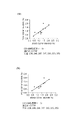
- FIG. 2 shows the correlation between bisulfite amplicon sequencing (BSAS) and flow cytometry (FCM) for the percentage of total monocytes in whole blood (Mo/WB).
- FIG. 2 shows the correlation between bisulfite amplicon sequencing (BSAS) and flow cytometry (FCM) for the percentage of total monocytes in whole blood (Mo/WB).
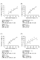
- FIG. 2 shows the correlation between bisulfite amplicon sequencing (BSAS) and flow cytometry (FCM) for the ratio of CD16-positive monocytes (CD16 + Mo/WB) in whole blood.
- FIG. 2 shows the correlation between bisulfite amplicon sequencing (BSAS) and flow cytometry (FCM) for the ratio of CD16-positive monocytes (CD16 + Mo/WB) in whole blood.
- FIG. 2 shows the correlation between bisulfite amplicon sequencing (BSAS) and flow cytometry (FCM) for the ratio of CD16-positive monocytes (CD16 + Mo/WB) in whole blood.
- FIG. 2 shows the correlation between bisulfite amplicon sequencing (BSAS) and flow cytometry (FCM) for the ratio of CD16-positive monocytes (CD16 + Mo/WB) in whole blood.
- FIG. 2 shows the correlation between bisulfite amplicon sequencing (BSAS) and flow cytometry (FCM) for the ratio of CD16-positive monocytes (CD16 + Mo/WB) in whole blood.
- FIG. 2 shows the correlation between bisulfite amplicon sequencing (BSAS) and flow cytometry (FCM) for the ratio of CD16-positive monocytes (CD16 + Mo/Mo) in total monocytes.
- FIG. 2 shows the correlation between bisulfite amplicon sequencing (BSAS) and flow cytometry (FCM) for the ratio of CD16-positive monocytes (CD16 + Mo/Mo) in total monocytes.
- monocytes are a type of leukocyte and are the largest mononuclear cells among leukocytes.
- CD16-positive monocytes are monocytes that express CD16 on their cell surface and include non-classical monocytes (CD14 + CD16 ++ monocytes) and intermediate monocytes (CD14 ++ CD16 + monocytes), It does not include classical monocytes (CD14 ++ CD16 ⁇ monocytes).
- total monocytes include classical monocytes, non-classical monocytes and intermediate monocytes.
- Phenotypic differences are caused by acquired changes in gene regulation (epigenetic changes) that are not accompanied by changes in DNA base sequences.
- Epigenetic modifications include DNA methylation and histone modifications.
- CpG cytosine of the sequence
- 5mC phosphodiester bond
- CpG sequences exist sporadically in genomic DNA, but there is a region that is frequently present upstream of the transcription initiation site of a gene and is called a CpG island. DNA methylation of CpG islands is known to be deeply involved in the regulation of gene transcription.
- the methylation pattern of genomic DNA differs depending on the type and state of the cell, regardless of whether it is a CpG island or not. Utilizing the fact that the methylation pattern of genomic DNA differs depending on the type of cell, it is possible to measure the proportion of a specific cell population in all nucleated cells. Bisulfite treatment of genomic DNA converts unmethylated cytosine to uracil, but does not convert methylated cytosine. Therefore, this treatment allows discrimination between methylated cytosine and unmethylated cytosine. Since unmethylated DNA and methylated DNA have different base sequences after treatment, DNA methylation can be quantified by microarray, quantitative PCR, sequence analysis, and the like.
- the method of the present disclosure is a method that utilizes cell-specific DNA methylation based on differences in DNA methylation between cell types.
- a method for measuring the percentage of CD16-positive monocytes in all nucleated cells in a human sample according to the present disclosure is characterized by determining the unmethylation status (bisulfite convertibility) of one or more CpG positions in a predetermined marker region. Including parsing.
- the “unmethylated state ratio” in the present disclosure is the count of the unmethylated state and the methylated state of the target CpG position obtained by the methylation quantification method. It can be determined by the ratio of the unmethylated state counts to the total.
- a predetermined marker region (which may be referred to herein as a “CD16-positive monocyte marker region”) is one or more of all nucleated cells in the sample, and only CD16-positive monocytes have specific unmethylated It is a region containing CpG positions, and the percentage of CD16-positive monocytes in all nucleated cells in human samples can be measured by analyzing the specific unmethylated CpG positions.
- CD16-positive monocyte marker regions include ELF5, MCTP2, CYTIP, LINC00189, SBF2, IL1RAP, CLEC5A, IFIT3, RNF144B, DEPDC5, KIAA0513, PIK3AP1, MYO9B, KDM4C, NEURL1, USP49, UBXN11, SERPINA1, DST, SNXB10, , TNFRSF1B, GUCY1B3, SMPDL3A, cg06107544, cg07135960, cg18409707, cg23212713, cg24766263, in particular any of the gene sequences consisting of ELF5, MCTP2, CYTIP, KIAA0513 and SERPINA1
- ELF5 E74 like ETS transcription factor 5
- Ets family transcription factor an epithelial cell-specific Ets family transcription factor. Involved in differentiation of keratinocytes and glandular epithelial cells (Oettgen et al., J. Biol. Chem. 274, 29439-29452 (1999) 8 October 1999). It is located on chromosome 11 from 34500342 to 34535347 (GRCh37).
- SEQ ID NO:23 shows the methyl sequence of the DNA sequence of SEQ ID NO:22 after bisulfite treatment
- SEQ ID NO:24 shows the non-methyl sequence of the DNA sequence of SEQ ID NO:22 after bisulfite treatment.
- ELF5 target region genomic DNA sequence Chromosome 11: 34533543-34534042 (F strand) (SEQ ID NO: 22) ACCCCACCCTCTGAGTGGTATCATTGGGAAGAAAATCGGTTTTCCTTCCTTAAAGCCTCAGTGATACCCAAATGTCCACTCCGTCCCAAGACTTGTGAGCAAGGGAGGGTCAATTTCCCAGCCCTTGGACCTCCCTAAGAAGGGGCCCACTGAGGAGAACCCAGTGGAGTATTTCAACACTTACTGCTAAAGAAAGATTGAGGGTTGGGTGCTTGAGGGTTTGTTCTTCTGAGAGGAACCCTAGCAACTGTGAGAACCAATTTGCTGACCTCTTGTCAAGTGATTCCTCATTTTTCTTTAGCTCGCTTAGGGAGTTTGCGTG
- the DNA sequence of SEQ ID NO: 25 is a portion (500 bp) of the MCTP2 gene.
- MCTP2 multiple C2 and transmembrane domain containing 2
- MCTP2 is a protein having three C2 domains and two transmembrane domains. Unlike proteins with other C2 domains, it has been reported to strongly bind to Ca2+ in the absence of phospholipids (Shin et al., J. Biol. Chem. 280, 1641-1651 (2005) 14 January 2005). It is located on chromosome 15 from 94774772 to 95027181 (GRCh37).
- SEQ ID NO:26 shows the methyl sequence of the DNA sequence of SEQ ID NO:25 after bisulfite treatment and SEQ ID NO:27 shows the non-methyl sequence of the DNA sequence of SEQ ID NO:25 after bisulfite treatment.
- the DNA sequence of SEQ ID NO: 28 is a portion (500 bp) of the CYTIP gene.
- CYTIP cytohesin 1 interacting protein
- CYTH1 Cytohensin 1
- SNX27 sorting nexin 27
- SEQ ID NO:29 shows the methyl sequence of the DNA sequence of SEQ ID NO:28 after bisulfite treatment
- SEQ ID NO:30 shows the non-methyl sequence of the DNA sequence of SEQ ID NO:28 after bisulfite treatment.
- Target region genomic DNA sequence of CYTIP Chromosome 2 158275531-158276030 (R strand) (SEQ ID NO: 28) GCCTCAGTCCCCTCTGTGGGATAGGCCAAGACTTGTGTGCTGTCTTGACCCATGTGGCAGCTACTTAATTCCCACTGTGGGTCTCAGCTCTCGTGTGCTGGGGCAGCCAAGAAGGGAGGCAGTTAAGAAGTTAGGGTGCTTAGCAGTGTTCTAAGTTTAGCCCGCTAAGGAAGCATTTCAGATAGTTCTGTTCCTTTTTTGTGGAAAATAAATAGCTGTCCAAGCCAGGCACGGTGCCTCACGCCTGTAATCCCTTTGGGAAGCTGAGGTAGGCAGATCACGAGGTCAGGAGTTCGAGATCAGCCTGGCCAGCGTGGTGAAACCCCGTCGCTACTAAAAAGACAGAAATTTGGCCGGGCATGGTGGGGCGTGCCTGTAATCCCAGCTACTTGGGAGACAGAAATTTGGCCGGGCATGGTGGGGCGTGCCTGTAATCCCAGCTACT
- the DNA sequence of SEQ ID NO: 31 is a portion (500 bp) of the KIAA0513 gene. KIAA0513 is strongly expressed in the brain (Lauriat et al., Brain Res. 1121 (1), 1-11 (2006) 22 November 2006). There are many unclear points about the function. It is located on chromosome 16 from 85095985 to 85096484 (GRCh37).
- SEQ ID NO:32 shows the methyl sequence of the DNA sequence of SEQ ID NO:31 after bisulfite treatment and SEQ ID NO:33 shows the non-methyl sequence of the DNA sequence of SEQ ID NO:31 after bisulfite treatment.
- the DNA sequence of SEQ ID NO: 34 is a portion (500 bp) of the SERPINA1 gene.
- SERPINA1 serpin family A member 1
- ⁇ 1-antitrypsin inhibits serine proteases such as trypsin and elastase (Gettins, Chem. Rev. 102 (12), 4751-4803 (2002) Epub 8 November 2002 ). It is located on chromosome 14 from 94857896 to 94858395 (GRCh37).
- SEQ ID NO:35 shows the methyl sequence of the DNA sequence of SEQ ID NO:34 after bisulfite treatment
- SEQ ID NO:36 shows the non-methyl sequence of the DNA sequence of SEQ ID NO:34 after bisulfite treatment.
- One or more CpG positions in these marker regions are unmethylated CpG positions specific only to CD16-positive monocytes among all nucleated cells in the sample. In some embodiments, 2 or more CpG positions, 3 or more CpG positions, 4 or more CpG positions, 5 or more CpG positions, 6 or more CpG positions, or 7 or more CpG positions are analyzed.
- Criteria for selecting unmethylated CpG positions specific only to CD16-positive monocytes include: in CD16-positive monocytes, the unmethylated state of the CpG positions is 70% or more, 80% or more, 90% or more; % or more, and in cells other than CD16-positive monocytes, the unmethylated state of the CpG position is 20% or less, 15% or less, 10% or less, or 5% or less.
- the selection criteria for unmethylated CpG positions specific only to CD16-positive monocytes are 70% or more for CD16-positive monocytes and 1% or less for granulocytes; It can be set at 2% or less for T cells and 5% or less for CD16-negative monocytes, B cells and NK cells.
- CpG positions in the DNA sequence of SEQ ID NO: 22 there are multiple CpG positions in the DNA sequence of SEQ ID NO: 22, but among all the nucleated cells in the sample, the unmethylated CpG position specific to only CD16-positive monocytes is SEQ ID NO: 22.
- CpG positions at positions 304 and 319 of the DNA sequence have been identified. Analysis of the unmethylated state of one CpG position is also specific, but analysis of the unmethylated state of two or more CpG positions shows higher specificity, thus allowing the total presence in the sample to be determined with high measurement accuracy.
- the percentage of CD16-positive monocytes in nuclear cells can be measured.
- the unmethylated CpG position of SEQ ID NO: 25 is specific only for CD16-positive monocytes.
- CpG positions at positions 171, 179, 184 and 247 of the DNA sequence have been identified.
- Analysis of the unmethylated state of one CpG position has specificity, but analysis of the unmethylated state of 2 or more, 3 or more or 4 CpG positions shows higher specificity and thus high measurement accuracy. can measure the percentage of CD16-positive monocytes in total nucleated cells in a sample.
- Examples of such combinations include: two CpG positions at positions 171 and 179; two CpG positions at positions 171 and 184; two CpG positions at positions 179 and 184; three CpG positions at positions 171, 179 and 184; three CpG positions at positions 171, 179 and 247; three CpG positions at positions 171, 184 and 247; A CpG position and selected from the group consisting of four CpG positions, positions 171, 179, 184 and 247.
- CpG positions in the DNA sequence of SEQ ID NO: 28 are multiple CpG positions in the DNA sequence of SEQ ID NO: 28, but among all the nucleated cells in the sample, the unmethylated CpG position specific only to CD16-positive monocytes is SEQ ID NO: 28. CpG positions at positions 285, 299, 317, 330, 333 and 359 of the DNA sequence have been identified. There is also specificity in analyzing the unmethylation status of one CpG position specific to CD16-positive monocytes only, but selected from the group consisting of positions 236, 246, 285, 299, 317, 330, 333 and 359.
- Positions 236 and 246 are CpG positions used in combination with other CpG positions to reduce the background of T cells and the like.
- Examples of such combinations include four CpG positions at positions 246, 285, 299 and 317; 4 CpG positions at positions 246, 285, 317 and 333; 4 CpG positions at positions 246, 285, 317 and 359; 4 CpG positions at positions 246, 285, 333 and 359; , 4 CpG positions at positions 246, 299, 317 and 333, 4 CpG positions at positions 246, 299, 317 and 359, 4 at positions 246, 317, 333 and 359.
- the unmethylated CpG position specific only to CD16-positive monocytes is CpG positions at positions 51, 181, 242, 293 and 350 of the DNA sequence have been identified.
- Analysis of the unmethylated status of one CpG position is also specific, but analysis of the unmethylated status of 2 or more, 3 or more, 4 or more or 5 CpG positions shows higher specificity, thus , can measure the percentage of CD16-positive monocytes in all nucleated cells in a sample with high measurement accuracy. Examples of such combinations are selected from the group consisting of two CpG positions at positions 51 and 181, two CpG positions at positions 51 and 242, and three CpG positions at positions 51, 181 and 242.
- CpG positions in the DNA sequence of SEQ ID NO: 34 there are multiple CpG positions in the DNA sequence of SEQ ID NO: 34, but among all the nucleated cells in the sample, the unmethylated CpG position specific only to CD16-positive monocytes is SEQ ID NO: 34.
- CpG positions at positions 169, 205, 213 and 329 of the DNA sequence have been identified. Analysis of the unmethylated state of one CpG position has specificity, but analysis of the unmethylated state of 2 or more, 3 or more or 4 CpG positions shows higher specificity and thus high measurement accuracy.
- a method for determining the percentage of total monocytes in total nucleated cells in a human sample comprises analyzing the unmethylation status of one or more CpG positions in a given marker region.
- a predetermined marker region (which may be referred to herein as a “total monocyte marker region”) is one or more non-methylated markers specific to all types of monocytes among all nucleated cells in the sample. It is a region containing methylated CpG sites, and by analyzing the specific unmethylated CpG sites, the proportion of total monocytes in total nucleated cells in a human sample can be measured.
- the pan-monocyte marker region is selected from the gene/genomic region group consisting of MTSS1, NLRC3, LINC01010, MS4A7, RG9MTD1, LINC00299, CACNA1D, REV3L, GPATCH11, cg02612894 and cg06297318, particularly MTSS1, NLRC3, RG9MTD1, LINC00299, Any partial region of a DNA sequence selected from the gene/genomic region group consisting of CACNA1D, cg02612894 and cg06297318, for example, a contiguous 10-2000 bp, 50 A marker region of ⁇ 1000 bp, 100-800 bp, or 200-700 bp, containing one or more CpG positions.
- the marker region consists of at least one DNA sequence selected from the group consisting of SEQ ID NOS: 1, 4, 7, 10, 13, 16 and 19.
- the DNA sequence of SEQ ID NO: 1 is a portion (500 bp) of the MTSS1 gene.
- MTSS1 (MTSS I-BAR domain containing 1) is a cytoskeletal protein containing a WASP-homology2 domain and an inverse BAR domain (IRSp53 and MIM homology domains). It has been reported to have an effect of suppressing tumor metastasis (Lee et al., Neoplasia 4(4): 291-294 (2002)). It is located on chromosome 8 from 125563025 to 125740730 (GRCh37).
- SEQ ID NO:2 shows the methyl sequence of the DNA sequence of SEQ ID NO:1 after bisulfite treatment
- SEQ ID NO:3 shows the non-methyl sequence of the DNA sequence of SEQ ID NO:1 after bisulfite treatment.
- Target region genomic DNA sequence of MTSS1 Chromosome 8: 125714947-125715446 (R strand) (SEQ ID NO: 1) GATCATTGGTTGCTTGGGGATAGGGTGAAAAAGGAGAGTGAAGTGGGTAAGACAGATCTTTTGGGGTGGTGGAAATGTTCTAAAATTGGATTGCGGTGATGATTGTGCAACTCCAAATTTACTAAAAATCATTGACTCATACCCTGAAAAAGGGTGTATTTTATGGTATGTAAATTATGCCTCCGTAAAGCTGCCTAAACAAGATCCAGTGCTGAGCTCTAAGGGGCTGTGATTGCCTAGTCGTGCTTGTTCATATATCATGTGATATGAAATAGAAAATACGTTGGCATCTTCTTATTAAAGCTCTCTACCCTTTTAAATCATTGTGGT
- the DNA sequence of SEQ ID NO: 4 is a portion (500 bp) of the NLRC3 gene.
- NLRC3 NLR family CARD domain containing 3
- SEQ ID NO: 5 shows the methyl sequence of the DNA sequence of SEQ ID NO: 4 after bisulfite treatment
- SEQ ID NO: 6 shows the non-methyl sequence of the DNA sequence of SEQ ID NO: 4 after bisulfite treatment.
- Target region genomic DNA sequence of NLRC3 Chromosome 16: 3597031-3597530 (F strand) (SEQ ID NO: 4) CGACTCCACTCCCAGGTATATCCAGCCAGGAGAAGGAAAGCTCACGTCCACACAGAAACTGGTGCACAAAGGTCCACAGCAGCACCGTTCCTAACAGCCAAGTAGGAACAACCCAACGTCCGTCAGCTGATGAATGGAAATGAAACCTGTGTACCCATACAATGGATATCATTCAGCCTTACAAAGGAACTAAGTATTGAGACAGGCTACAATGTGGATGAACCTTGAAAATCTGACACCAAATGAAAGACACCACAAAAGGCCACGTGTCGTATGATTCTATTTTTGGCCACGTATCGCACAATTCCATTTATATGCGGTGTCC
- the DNA sequence of SEQ ID NO: 7 is a portion (500 bp) of the RG9MTD1/TRMT10C gene.
- RG9MTD1 RNA (Guanine-9-) methyltransferase domain containing 1
- TRMT10C tRNA methyltransferase 10C
- GRCh37 101280680-101285290
- SEQ ID NO:8 shows the methyl sequence of the DNA sequence of SEQ ID NO:7 after bisulfite treatment
- SEQ ID NO:9 shows the non-methyl sequence of the DNA sequence of SEQ ID NO:7 after bisulfite treatment.
- Target region genomic DNA sequence of RG9MTD1 Chromosome 3: 101283988-101284487 (F strand) (SEQ ID NO: 7) GGAATGTGTTTCTAACACAGCAAAAAAAATATTTAAAATATTTATATACGAAGGAAAAAGTGAAAAAAGCTAGGCAAATAAAAAAGGAAATGAAAGCAGCAGCAAGGGAAGAAGCAAAAAATATCAAGCTGCTAGAAACCACTGAGGAAGATAAACAGAAAAACTTTCTATTTTTACGACTTTGGGATAGGAATATGGACATAGCAATGGGCTGGAAGGGTGCCCAGGCCATGCAGTTTGGACAACCTTTGGTTTTTGACATGGCTTACGAAAATTATATGAAACGAAAAGAATTGCAGAATACTGTTTCCC
- the DNA sequence of SEQ ID NO: 10 is a portion (500 bp) of the LINC00299 gene.
- LINC00299 long intergenic non-protein coding RNA 299
- SEQ ID NO: 11 shows the methyl sequence of the DNA sequence of SEQ ID NO: 10 after bisulfite treatment
- SEQ ID NO: 12 shows the non-methyl sequence of the DNA sequence of SEQ ID NO: 10 after bisulfite treatment.
- Target region genomic DNA sequence of LINC00299 Chromosome 2: 8333940-8334439 (R strand) (SEQ ID NO: 10) TCGCAAGTGGTAGTAACAAAAGTCCAAAGGGTAGATATTGATTATTTACCAGGTCACATGATCGATAGAGTATTATTTCCATCCTGATTACCTCTAGGTGAGGCAGGCCAGCCTAGATGGTTCTAAGCCTCTGAGTAGAGCATGTCCTGAAATTAACTTGAATAACCTAATTTCTGCCTTCAAGTCGATCTTCCTACACACTGTGCTAAGGGCAAAACAGAAGTGAAACAAGAAAGATTAACAAGATCTGTGCCAGCTATTAGAGGAGAAGTCATTGTTTGTTCGCATTTAAGGAAAAGGACACTATTTGGCAGCGTGGGGTGCCTCTGTGCCGGCTGGAGCTAATGATCGTCGTCAGGATTGAGCTGGGAGGCAGGTGCCCATCGCCTGGCCCTGACTGCTCTTACTGACAGGTGCTAATGGGAGGGGCAGGCAGGCATCGCCTGGCCC
- the DNA sequence of SEQ ID NO: 13 is a portion (500 bp) of the CACNA1D gene.
- the CACNA1D (calcium voltage-gated channel subunit alpha1 D) gene encodes the ⁇ 1D subunit (Cav1.3) of the voltage-gated L-type calcium channel (Williams et al., Neuron 8(1), 71-84 (1992 )).
- L-type voltage-gated calcium channels are characterized by slow inactivation (Nowychy et al., Nature 316 (6027), 440-443 (1985) August 1985). It is located on chromosome 3 from 53529076 to 53847179 (GRCh37).
- SEQ ID NO: 14 shows the methyl sequence of the DNA sequence of SEQ ID NO: 13 after bisulfite treatment and SEQ ID NO: 15 shows the non-methyl sequence of the DNA sequence of SEQ ID NO: 13 after bisulfite treatment.
- the DNA sequence of SEQ ID NO: 16 is 500 bp before and after the genomic region whose CG cluster ID is cg02612894. It is located on chromosome 1 from 87641701 to 87642200 (GRCh37). There are no genes in this region.
- SEQ ID NO: 17 shows the methyl sequence of the DNA sequence of SEQ ID NO: 16 after bisulfite treatment and SEQ ID NO: 18 shows the non-methyl sequence of the DNA sequence of SEQ ID NO: 16 after bisulfite treatment.
- the DNA sequence of SEQ ID NO: 19 is 500 bp before and after the genomic region whose CG cluster ID is cg06297318. It is located on chromosome 6 from 127667460 to 127667959 (GRCh37). There are no genes in this region.
- SEQ ID NO:20 shows the methyl sequence of the DNA sequence of SEQ ID NO:19 after bisulfite treatment and SEQ ID NO:21 shows the non-methyl sequence of the DNA sequence of SEQ ID NO:19 after bisulfite treatment.
- Target region genomic DNA sequence near cg06297318 Chromosome 6: 127667460-127667959 (R strand) (SEQ ID NO: 19) GATATTTGTCTTTCTGTGCCTGGCTTATTTCACTTAATGTTCTCCAGGTCCATCTATGTTGCCACAAATGACAGGATTTCATTCCTTTTAATGGCTGAATAGTATTCCATTGTATGTATATACCACATTTTCTTTATCCGCTCATCCACTGATGGACACTTAGGTTGATTCCATATCTCGGCTCTTATGAACAGTGCTGCAATAAACATGAGAGTGCAGATATCTCTGCAACATACTGATTTTACTTCCTTCAGATATATACCCAGGCGTGGGATTGTGGGATCATATGGCAGTTTTGTTTAATTTTTTGAGGAACCTCTATACTGTTTTCTATAATGGCTAGAGTAATTCAAATTCTTTTGTTTATTATTTTTTTGAGACGGAGTCTCGCTGTGTCACCCAGGCTGGAGTGCAGTGGTGTGATCTCGGCTCACTGCAGCCTCTGCC
- One or more CpG positions in these marker regions are unmethylated CpG positions specific only to all monocytes among all nucleated cells in the sample. Criteria for selecting unmethylated CpG positions specific only to all monocytes are that the unmethylation status of the CpG positions is 70% or more, 80% or more, or 90% or more in all monocytes. % or more, and in cells other than monocytes, the unmethylation state of the CpG position is 25% or less, 20% or less, 15% or less, 10% or less, or 5% or less.
- the selection criteria for unmethylated CpG positions specific to all monocytes only are ⁇ 70% for total monocytes and ⁇ 5% for granulocytes, T It can be set at 10% or less for cells and 25% or less for B cells and NK cells.
- CpG positions there are multiple CpG positions in the DNA sequence of SEQ. CpG positions at positions 96, 186, 246 and 286 of . Analysis of the unmethylated state of one CpG position is also specific, but analysis of the unmethylated state of two or more CpG positions shows higher specificity, thus allowing the total presence in the sample to be determined with high measurement accuracy. The percentage of total monocytes in nuclear cells can be measured. Examples of such combinations include two CpG positions at positions 246 and 286; three CpG positions at positions 96, 186 and 286; three CpG positions at positions 96, 246 and 286; CpG positions and selected from the group consisting of four CpG positions, positions 96, 186, 246 and 286.
- the DNA sequence of SEQ ID NO: 4 is the unmethylated CpG position specific only to monocytes.
- analysis by one CpG position can use position 45, 277, 299, 304 or 324.
- Analysis of the unmethylation status of one CpG position is also specific, but analysis of the unmethylation status of 2 or more, 3 or more, 4 or more, or 5 or more CpG positions shows higher specificity.
- Examples of such combinations include: two CpG positions at positions 45 and 86; two CpG positions at positions 45 and 117; two CpG positions at positions 45 and 121; two CpG positions, two CpG positions at positions 272 and 299; two CpG positions at positions 272 and 304; two CpG positions at positions 272 and 324; two CpG positions at positions 277 and 299; two CpG positions, two CpG positions at positions 277 and 324, two CpG positions at positions 299 and 304, two CpG positions at positions 299 and 324, two CpG positions at positions 304 and 324, positions 45, 86 and three CpG positions at positions 45, 86 and 121; three CpG positions at positions 45, 117 and 121; three CpG positions at positions 272, 277 and 299; 3 CpG positions at positions 272, 277 and 324; 3 CpG positions at positions
- the DNA sequence of SEQ ID NO: 7 is the unmethylated CpG position specific only to monocytes.
- analysis by one type of CpG position can use the CpG position at position 419 .
- Analysis of the unmethylated status of one CpG position is also specific, but analysis of the unmethylated status of 2 or more, 3 or more or 4 CpG positions shows higher specificity and thus high measurement. It can measure the percentage of total monocytes in total nucleated cells in a sample with precision.
- Examples of such combinations include two CpG positions at positions 179 and 271, two CpG positions at positions 271 and 419, two CpG positions at positions 287 and 419, three CpG positions at positions 179, 271 and 287, and , positions 271, 287 and 419.
- the DNA sequence of SEQ ID NO: 10 is the unmethylated CpG position specific only to monocytes.
- CpG positions at positions 188 and 288 of are identified.
- analysis by one type of CpG position can use the CpG position at position 288 .
- Analysis of the unmethylated status of one CpG position is also specific, but two analyzes show higher specificity, thus measuring the proportion of total monocytes in total nucleated cells in a sample with high measurement accuracy. can do.
- the DNA sequence of SEQ ID NO: 13 is the unmethylated CpG position specific only to monocytes.
- analysis by one type of CpG location can use the CpG location at location 362 .
- Analysis of the unmethylated status of one CpG position is also specific, but analysis of the unmethylated status of 2 or more, 3 or more or 4 CpG positions shows higher specificity and thus high measurement. It can measure the percentage of total monocytes in total nucleated cells in a sample with precision.
- Examples of such combinations include two CpG positions at positions 279 and 362, two CpG positions at positions 291 and 362, two CpG positions at positions 326 and 362, three CpG positions at positions 279, 291 and 362, positions 3 CpG positions at positions 279, 326 and 362; 3 CpG positions at positions 291, 326 and 362;
- the DNA sequence of SEQ ID NO: 16 is the unmethylated CpG position specific only to monocytes.
- analysis by one type of CpG position can use the CpG position at position 182 .
- Analysis of the unmethylated status of one CpG position is also specific, but analysis of the unmethylated status of 2 or more, 3 or more, 4 or more or 5 CpG positions shows higher specificity, thus , can measure the percentage of total monocytes in total nucleated cells in a sample with high measurement accuracy.
- Examples of such combinations include two CpG positions at positions 182 and 204, two CpG positions at positions 182 and 237, two CpG positions at positions 182 and 243, two CpG positions at positions 182 and 301, position 182, three CpG positions at positions 182, 204 and 243; three CpG positions at positions 182, 204 and 301; three CpG positions at positions 182, 237 and 243; 3 CpG positions at 301, 3 CpG positions at positions 182, 243 and 301, 4 CpG positions at positions 182, 204, 237 and 243, 4 CpG positions at positions 182, 204, 237 and 301, position 182, 4 CpG positions at positions 204, 243 and 301; 4 CpG positions at positions 182, 237, 243 and 301;
- the DNA sequence of SEQ ID NO: 19 is the unmethylated CpG position specific only to monocytes. CpG positions at positions 179 and 268 of . Analysis of the unmethylated status of one CpG position is also specific, but two analyzes show higher specificity, thus measuring the proportion of total monocytes in total nucleated cells in a sample with high measurement accuracy. can do.
- a method for calculating the percentage of CD16-positive monocytes in total monocytes in a human sample according to the present disclosure is a method for measuring the percentage of CD16-positive monocytes in total nucleated cells in a human sample according to the present disclosure.
- Determining the percentage of CD16-positive monocytes in total nucleated cells in a human sample by a method, and a method for determining the percentage of total monocytes in total nucleated cells in a human sample according to the present disclosure Determining the percentage of total monocytes in all nucleated cells in a human sample and measuring the percentage of total monocytes in total nucleated cells in the human sample as the percentage of CD16 positive monocytes in total nucleated cells in the human sample Calculating the percentage of CD16 positive monocytes among the total monocytes in the human sample by dividing by .
- DNA methylation quantification methods include next-generation sequencing, pyrosequencing, nanopore sequencing, quantitative methylation-specific PCR (qMSP), quantitative methylation-sensitive high-resolution melting (MS-HRM), methylation-sensitive Single-stranded nucleotide primer extension (Ms-SNuPE), combined bisulfite restriction analysis (COBRA), microarray, etc.
- qMSP quantitative methylation-specific PCR
- MS-HRM quantitative methylation-sensitive high-resolution melting
- Ms-SNuPE methylation-sensitive Single-stranded nucleotide primer extension
- COBRA combined bisulfite restriction analysis
- the human sample in the present disclosure may be any human tissue sample or human body fluid sample as long as it contains the monocytes to be measured. Mucous membranes, cells, tissues, etc. may be mentioned, and the blood sample may be selected from the group consisting of whole blood and peripheral blood mononuclear cells. In certain embodiments, the human sample is a human blood sample. Total nucleated cells when the sample is whole blood generally refers to total white blood cells.
- the method for measuring the percentage of CD16-positive monocytes and the method for measuring the percentage of total monocytes of the present disclosure can be used instead of the measurement method using flow cytometry.
- Biomarkers are, for example, efficacy predictive markers, pharmacodynamic markers, prognostic markers, patient stratification markers, monitoring markers and diagnostic markers.
- Treatment methods that use the ratio of CD16-positive monocytes in the baseline total monocytes before drug administration in a subject as a drug efficacy prediction or patient stratification marker, and treatment methods that use the ratio as a pharmacodynamic marker include, for example, antifractalkine It is a therapeutic method for a disease associated with fractalkine using an antibody. As a specific example, methods of treatment with Quetmolimab, a humanized anti-human fractalkine antibody, using such markers are described in WO2020/218232. In the method of treatment, the anti-fractalkine antibody is administered to a human at least 100 mg per dose, for example, 100 mg, 200 mg, 400 mg, 600 mg, or 800 mg. Diseases associated with fractalkine are, for example, rheumatoid arthritis, inflammatory bowel disease, Crohn's disease, and the like, which are described in US2012/0213799 (incorporated by reference in its entirety).
- Example 1 Comprehensive DNA methylation analysis of monocyte subsets and whole blood Regions showing patterns were extracted from the whole genome.
- CD16-negative monocytes classical monocytes
- CD16-positive monocytes non-classical monocytes
- DNA extraction was performed using DNeasy Blood & Tissue Kit (Qiagen). After performing bisulfite treatment of DNA using EZ DNA Methylation Kit (Zymo Research), comprehensive analysis of genomic DNA from classical monocytes, non-classical monocytes, and whole blood was performed using Infinium Methylation EPIC BeadChip (Illumina). DNA methylation analysis was performed.
- Tables 1 and 2 show the methylation levels of each cell population for candidate regions of total monocyte markers (sometimes simply referred to herein as "monocyte markers") and CD16-positive monocyte markers. Numerical values in the table indicate methylation rates between 0 and 1. As monocyte markers, regions with low methylation levels in monocyte subsets (classical monocytes and non-classical monocytes) and high methylation levels in whole blood were extracted. As CD16-positive monocyte markers, regions with low methylation levels in non-classical monocytes and high methylation levels in classical monocytes and whole blood were extracted.
- Example 2 Narrowing down candidate regions for monocytes and CD16-positive monocyte markers
- Monocytes, CD16-negative monocytes, and CD16-positive monocytes were isolated from blood of healthy subjects collected with heparin or EDTA in the same manner as in Example 1. Isolated.
- T cells, B cells, and NK cells were isolated by staining with APC-Cy7-labeled anti-CD3 antibody (Biolegend), BV421-labeled anti-CD19 antibody (Biolegend), and PE-labeled anti-CD56 antibody (Beckman Coulter). did.
- DNA extraction was performed from the isolated cells using the DNeasy Blood & Tissue Kit (Qiagen). DNA was subjected to bisulfite treatment using EZ DNA Methylation-Lightning Kit (Zymo Research) or EpiTect Fast Bisulfite Conversion Kit (Qiagen).
- CpG For monocyte markers, specific non-methyl CpG was extracted in candidate regions around MTSS1, NLRC3, LINC01010, MS4A7, RG9MTD1, LINC00299, CACNA1D, REV3L and GPATCH11 genes, cg02612894 and cg06297318, amplified by the primers in Table 3. .
- CD16-positive cell markers ELF5, MCTP2, CYTIP, LINC00189, SBF2, IL1RAP, CLEC5A, IFIT3, RNF144B, DEPDC5, KIAA0513, PIK3AP1, MYO9B, KDM4C, NEURL1, USP4, UBXN11, amplified by the primers in Table 4, Specific non-methyl CpGs were extracted in candidate regions around SERPINA1, DST, SNX10, RAP1B, TNFRSF1B, GUCY1B3 and SMPDL3A genes, cg06107544, cg07135960, cg18409707, cg23212713 and cg24766263.
- Example 3 Verification of specificity of monocytes and CD16-positive monocyte markers by bisulfite amplicon sequencing For some of the CpGs that are specifically non-methyl in the target cells, selected in Example 2, the following The cell specificity was quantitatively analyzed by the method of
- Monocytes classical monocytes, non-classical monocytes, granulocytes, T cells, B cells, and NK cells were isolated in the same manner as in Examples 1 and 2. DNA extraction and bisulfite treatment of DNA were carried out in the same manner as in Examples 1 and 2.
- These primers are capable of amplifying both methyl and non-methyl sequences (before and after bisulfite treatment) of the target region.
- each target region (after bisulfite treatment or before treatment) is SEQ ID NO: 2 or 3, 5 or 6, 8 or 9, 11 or 12, 14 or 15, 17 or 18, 20 or 21, 23 or 24 , 26 or 27, 29 or 30, 32 or 33, and 35 or 35.
- the target CpG-containing fragment was amplified by 35 cycles of PCR.
- a DNA fragment purified using AMPure XP (Beckman Coulter) was used as a template and the index primers listed in Table 7 were used to amplify the fragment by 8 cycles of PCR.
- AMPure XP the base sequence of the fragment was analyzed by MiSeq (Illumina).
- the obtained base sequences were quality trimmed and mapped to the reference sequence using CLC Genomics Workbench (CLC Bio).
- CLC Genomics Workbench CLC Bio
- target CpGs were methylated using Samtools (Li et al., Bioinformatics 25(16): 2078-2079 (2009) Epub 8 June 2009) and the grep command on LinuxTM. , counted the number of reads that were non-methyl.
- Spreadsheet software was used to analyze the percentage of one CpG being non-methyl and the percentage of multiple CpGs being all non-methyl.
- the ratio (%) of each CpG being unmethylated or the ratio (%) of all of the multiple CpGs being unmethylated was analyzed.
- the results of monocyte markers MTSS1, NLRC3, RG9MTD1, LINC00299, CACNA1D, around cg02612894, and around cg06297318 are shown in Tables 8, 9, 10, 11, 12, 13 and 14, and the CD16-positive monocyte marker ELF5 , MCTP2, CYTIP, KIAA0513, and SERPINA1 are shown in Tables 15, 16, 17, 18 and 19, respectively.
- the percentage of all sites to be analyzed that are non-methylated is shown.
- positions 96, 186, 246 and 286 of SEQ ID NO: 1 are monocyte-specific non-methyl CpGs, each of which is specific by itself, but two Greater specificity was shown when 3 or 4 combinations were used. In particular, the combinations marked with "*" in Table 8 had high specificity.
- positions 45, 86, 117, 121, 272, 277, 299, 304 and 324 of SEQ ID NO: 4 are monocyte-specific non-methyl CpG, each alone Although specific, higher specificity was shown when combinations of 2, 3, 4 or 5 were used. In particular, the combinations marked with "*" in Table 9 had high specificity.
- positions 179, 271, 287 and 419 of SEQ ID NO:7 are CpGs that are monocyte-specific non-methyl, each alone has specificity, but two Greater specificity was shown when 3 or 4 combinations were used. In particular, the combinations marked with "*" in Table 10 had high specificity.
- positions 188 and 288 of SEQ ID NO: 10 are monocyte-specific non-methyl CpG, each of which has specificity alone, but when both are used in combination, , showed higher specificity. In particular, the combinations marked with "*" in Table 11 had high specificity.
- positions 279, 291, 326 and 362 of SEQ ID NO: 13 are CpGs that are monocyte-specific non-methyl, each alone is specific, but two Greater specificity was shown when 3 or 4 combinations were used. In particular, the combinations marked with "*" in Table 12 had high specificity.
- positions 182, 204, 237, 243 and 301 of SEQ ID NO: 16 are CpGs that are monocyte-specific non-methyl, and each alone has specificity. , showed higher specificity when combinations of 2, 3, 4, or 5 were used. In particular, the combinations marked with "*" in Table 13 had high specificity.
- positions 179 and 268 of SEQ ID NO: 19 are monocyte-specific non-methyl CpG, each of which has specificity by itself, but the combination of both is used. showed higher specificity.
- positions 304 and 319 of SEQ ID NO: 22 are CD16-positive monocyte-specific non-methyl CpGs, each of which has specificity alone, but the combination of both showed higher specificity when using .
- positions 171, 179, 184 and 247 of SEQ ID NO: 25 are CD16-positive monocyte-specific non-methyl CpGs, each of which has specificity by itself. , showed higher specificity when combinations of 2, 3 or 4 were used. In particular, the combinations marked with "*" in Table 16 had high specificity.
- positions 285, 299, 317, 330, 333 and 359 of SEQ ID NO:28 are CD16-positive monocyte-specific non-methyl CpGs, each alone However, higher specificity was shown when combinations of 2, 3, 4, 5, or 6, as well as additional combinations of either or both of positions 236, 246 were used. .
- positions 236 and 246 of SEQ ID NO: 28 are not effective in distinguishing between non-classical monocytes and classical monocytes, they are used in combination with other CpG positions to reduce the background of T cells and the like. was taken. In particular, the combinations marked with "*" in Table 17 had high specificity.
- positions 51, 181, 242, 293 and 350 of SEQ ID NO: 31 are CD16-positive monocyte-specific non-methyl CpGs, each of which alone also has specificity. showed higher specificity when combinations of 2, 3 or 4 were used. In particular, the combinations marked with "*" in Table 18 had high specificity.
- positions 169, 205, 213, and 329 of SEQ ID NO:34 are CD16-positive monocyte-specific non-methyl CpGs, each of which has specificity on its own, but , showed higher specificity when combinations of 2, 3 or 4 were used. In particular, the combinations marked with "*" in Table 19 had high specificity.
- CD16-positive monocyte-specific non-methyl CpG exists in the genes of ELF5, MCTP2, CYTIP, KIAA0513, and SERPINA1. Furthermore, it was found that measuring the proportion of non-methyl CpGs improves specificity in quantifying CD16-positive monocytes. Using this, it was shown that CD16-positive monocytes in cell populations such as blood and tissues can be specifically measured. It was also shown that CD16-positive monocytes among monocytes can be specifically measured by measuring the specific unmethylated CpG of each of the monocyte marker and the CD16-positive monocyte marker.
- Example 4 Verification of correlation between bisulfite amplicon sequence and flow cytometry in measurement of monocytes and CD16-positive monocytes
- For the CD16-positive monocyte marker bisulfite amplicon sequencing was performed using whole blood DNA, and the correlation with the results of flow cytometry of peripheral blood cells after hemolysis was analyzed.
- Percentage of total monocytes in all nucleated cells in whole blood (percentage of total monocytes in whole blood) (%) and percentage of CD16-positive monocytes in all nucleated cells in whole blood (CD16-positive single cells in whole blood) The percentage of spheres) (%) is measured as the percentage of each marker target site that is non-methyl.
- the percentage of CD16-positive monocytes in total monocytes was calculated by dividing the percentage of CD16-positive monocytes in whole blood by the percentage of total monocytes in whole blood and multiplying by 100.
- Table 21 and Figures 1A and 1B show the correlation between the percentage of total monocytes in whole blood measured using the monocyte markers MTSS1, NLRC3, RG9MTD1, LINC00299, CACNA1D and around cg06297318 and flow cytometry.
- Table 22 and FIGS. 2A to 2D show the correlation between the percentage of CD16-positive monocytes in whole blood measured using the cell marker CYTIP and flow cytometry.
- Table 23 shows the correlation between the CD16-positive monocyte marker and the ratio of CD16-positive monocytes in total monocytes calculated using the monocyte marker and flow cytometry.
- FIG. 10 shows a correlation diagram between the percentage of CD16-positive monocytes in total monocytes and flow cytometry when combinations of positions 246, 285, 299, 317, and 330 of the CD16-positive monocyte marker CYTIP are combined with various monocyte markers. 3A and 3B.
- the ratio of total monocytes in whole blood and the ratio of CD16-positive monocytes in whole blood showed a high correlation with flow cytometry.
- Correlation analysis with flow cytometry was performed for all 112 combinations of 14 positive monocyte markers (Table 23). In all 112 combinations, bisulfite amplicon sequencing and flow cytometry results were significantly correlated (Table 23). The correlation coefficients ranged from 0.6455 to 0.9273, showing a high positive correlation with the results of flow cytometry in all 112 combinations.
- positions 246, 285, 299, 317, and 330 of the CD16-positive monocyte marker CYTIP and the percentage of CD16-positive monocytes in all monocytes when combined with various monocyte markers are compared with the bisulfite amplicon sequence.
- a high positive correlation was shown between flow cytometry and flow cytometry (Fig. 3A, Fig. 3B).
Landscapes
- Life Sciences & Earth Sciences (AREA)
- Chemical & Material Sciences (AREA)
- Organic Chemistry (AREA)
- Health & Medical Sciences (AREA)
- Engineering & Computer Science (AREA)
- Proteomics, Peptides & Aminoacids (AREA)
- Wood Science & Technology (AREA)
- Zoology (AREA)
- Genetics & Genomics (AREA)
- Biotechnology (AREA)
- General Engineering & Computer Science (AREA)
- Bioinformatics & Cheminformatics (AREA)
- Biophysics (AREA)
- Microbiology (AREA)
- Molecular Biology (AREA)
- Biochemistry (AREA)
- General Health & Medical Sciences (AREA)
- Physics & Mathematics (AREA)
- Biomedical Technology (AREA)
- Analytical Chemistry (AREA)
- Immunology (AREA)
- Plant Pathology (AREA)
- Measuring Or Testing Involving Enzymes Or Micro-Organisms (AREA)
Priority Applications (1)
| Application Number | Priority Date | Filing Date | Title |
|---|---|---|---|
| JP2023505640A JPWO2022191293A1 (https=) | 2021-03-12 | 2022-03-10 |
Applications Claiming Priority (2)
| Application Number | Priority Date | Filing Date | Title |
|---|---|---|---|
| JP2021-040367 | 2021-03-12 | ||
| JP2021040367 | 2021-03-12 |
Publications (1)
| Publication Number | Publication Date |
|---|---|
| WO2022191293A1 true WO2022191293A1 (ja) | 2022-09-15 |
Family
ID=83228108
Family Applications (1)
| Application Number | Title | Priority Date | Filing Date |
|---|---|---|---|
| PCT/JP2022/010678 Ceased WO2022191293A1 (ja) | 2021-03-12 | 2022-03-10 | 全単球及びcd16陽性単球の割合の測定方法 |
Country Status (2)
| Country | Link |
|---|---|
| JP (1) | JPWO2022191293A1 (https=) |
| WO (1) | WO2022191293A1 (https=) |
Citations (3)
| Publication number | Priority date | Publication date | Assignee | Title |
|---|---|---|---|---|
| JP2016515397A (ja) * | 2013-04-19 | 2016-05-30 | エピオンティス ゲーエムベーハー | 生体サンプルにおける定量的細胞組成を特定する方法 |
| JP2018529346A (ja) * | 2015-09-25 | 2018-10-11 | エピオンティス ゲーエムベーハー | 免疫細胞、特に単球の同定のためのエピジェネティックマーカーとしてのpark2 |
| JP2021502806A (ja) * | 2017-10-27 | 2021-02-04 | エピオンティス ゲーエムベーハー | 免疫細胞、特に非古典的単球の同定のためのエピジェネティックマーカーとしての単位複製配列領域 |
-
2022
- 2022-03-10 WO PCT/JP2022/010678 patent/WO2022191293A1/ja not_active Ceased
- 2022-03-10 JP JP2023505640A patent/JPWO2022191293A1/ja active Pending
Patent Citations (3)
| Publication number | Priority date | Publication date | Assignee | Title |
|---|---|---|---|---|
| JP2016515397A (ja) * | 2013-04-19 | 2016-05-30 | エピオンティス ゲーエムベーハー | 生体サンプルにおける定量的細胞組成を特定する方法 |
| JP2018529346A (ja) * | 2015-09-25 | 2018-10-11 | エピオンティス ゲーエムベーハー | 免疫細胞、特に単球の同定のためのエピジェネティックマーカーとしてのpark2 |
| JP2021502806A (ja) * | 2017-10-27 | 2021-02-04 | エピオンティス ゲーエムベーハー | 免疫細胞、特に非古典的単球の同定のためのエピジェネティックマーカーとしての単位複製配列領域 |
Non-Patent Citations (2)
| Title |
|---|
| ZAWADA ADAM M., SCHNEIDER JENNY S., MICHEL ANNE I., ROGACEV KYRILL S., HUMMEL BJÖRN, KREZDORN NICOLAS, MÜLLER SOEREN, ROTTER BJÖRN: "DNA methylation profiling reveals differences in the 3 human monocyte subsets and identifies uremia to induce DNA methylation changes during differentiation", EPIGENETICS, LANDES BIOSCIENCE, US, vol. 11, no. 4, 2 April 2016 (2016-04-02), US , pages 259 - 272, XP055965308, ISSN: 1559-2294, DOI: 10.1080/15592294.2016.1158363 * |
| ZHANG LU; HOFER THOMAS P.; ZAWADA ADAM M.; ROTTER BJöRN; KREZDORN NICOLAS; NOESSNER ELFRIEDE; DEVAUX YVAN; HEINE GUNNAR; ZIEG: "Epigenetics in non-classical monocytes support their pro-inflammatory gene expression", IMMUNOBIOLOGY, URBAN UND FISCHER VERLAG, DE, vol. 225, no. 3, 1 May 2020 (2020-05-01), DE , XP086169013, ISSN: 0171-2985, DOI: 10.1016/j.imbio.2020.151958 * |
Also Published As
| Publication number | Publication date |
|---|---|
| JPWO2022191293A1 (https=) | 2022-09-15 |
Similar Documents
| Publication | Publication Date | Title |
|---|---|---|
| EP3561074B1 (en) | Method for identifying the quantitative cellular composition in a biological sample | |
| Jara-Acevedo et al. | Detection of the KIT D816V mutation in peripheral blood of systemic mastocytosis: diagnostic implications | |
| US9926599B2 (en) | Epigenetic marker for the identification of CD3CD4 positive T lymphocytes | |
| Zheng et al. | A single-cell map of peripheral alterations after FMT treatment in patients with systemic lupus erythematosus | |
| Strobl et al. | Anti-apoptotic molecule BCL2 is a therapeutic target in steroid-refractory graft-versus-host disease | |
| EP3105345B1 (en) | Predicting increased risk for alzheimer's disease | |
| JP2016505243A (ja) | CD8+Tリンパ球、特にCD8α及びβTリンパ球の亜集団の同定用のエピジェネティック法 | |
| CA2842266C (en) | Epigenetic marker for the identification of natural killer cells | |
| WO2022191293A1 (ja) | 全単球及びcd16陽性単球の割合の測定方法 | |
| WO2015002845A1 (en) | Biomarker for preeclampsia | |
| EP3055425B1 (en) | Predicting increased risk for cancer | |
| JP7105460B2 (ja) | 成人t細胞白血病/リンパ腫の検出方法 | |
| JP7684545B2 (ja) | 軽度認知障害の検出方法 | |
| JP2021501574A (ja) | 免疫細胞、特に好塩基性顆粒球の同定のためのエピジェネティックマーカーとしてのmcc | |
| Hashemi et al. | CD38 gene polymorphisms and genetic susceptibility to chronic lymphocytic leukemia | |
| Pitaksalee | Rheumatoid arthritis: alterations in DNA methylation patterns in CD4+ T-cells: Understanding pathogenesis and developing a qMSP biomarker for classification | |
| Pereyra et al. | Targeted Long-Read Bisulfite Sequencing for Promoter Methylation Analysis in Severe Preterm Birth | |
| Bocova | DNA methylation biomarkers for deconvolution of hematopoietic subsets in clinical settings | |
| JP2017143784A (ja) | Dnaメチル化パターンに基づく免疫状態解析法 |
Legal Events
| Date | Code | Title | Description |
|---|---|---|---|
| 121 | Ep: the epo has been informed by wipo that ep was designated in this application |
Ref document number: 22767247 Country of ref document: EP Kind code of ref document: A1 |
|
| WWE | Wipo information: entry into national phase |
Ref document number: 2023505640 Country of ref document: JP |
|
| NENP | Non-entry into the national phase |
Ref country code: DE |
|
| 122 | Ep: pct application non-entry in european phase |
Ref document number: 22767247 Country of ref document: EP Kind code of ref document: A1 |