WO2022188856A1 - Method of treating diseases using gremlin1 antagonists - Google Patents
Method of treating diseases using gremlin1 antagonists Download PDFInfo
- Publication number
- WO2022188856A1 WO2022188856A1 PCT/CN2022/080297 CN2022080297W WO2022188856A1 WO 2022188856 A1 WO2022188856 A1 WO 2022188856A1 CN 2022080297 W CN2022080297 W CN 2022080297W WO 2022188856 A1 WO2022188856 A1 WO 2022188856A1
- Authority
- WO
- WIPO (PCT)
- Prior art keywords
- seq
- cancer
- grem1
- sequence
- chain variable
- Prior art date
- Legal status (The legal status is an assumption and is not a legal conclusion. Google has not performed a legal analysis and makes no representation as to the accuracy of the status listed.)
- Ceased
Links
Images


Classifications
-
- A—HUMAN NECESSITIES
- A61—MEDICAL OR VETERINARY SCIENCE; HYGIENE
- A61P—SPECIFIC THERAPEUTIC ACTIVITY OF CHEMICAL COMPOUNDS OR MEDICINAL PREPARATIONS
- A61P35/00—Antineoplastic agents
-
- A—HUMAN NECESSITIES
- A61—MEDICAL OR VETERINARY SCIENCE; HYGIENE
- A61K—PREPARATIONS FOR MEDICAL, DENTAL OR TOILETRY PURPOSES
- A61K45/00—Medicinal preparations containing active ingredients not provided for in groups A61K31/00 - A61K41/00
- A61K45/06—Mixtures of active ingredients without chemical characterisation, e.g. antiphlogistics and cardiaca
-
- C—CHEMISTRY; METALLURGY
- C07—ORGANIC CHEMISTRY
- C07K—PEPTIDES
- C07K16/00—Immunoglobulins [IG], e.g. monoclonal or polyclonal antibodies
- C07K16/18—Immunoglobulins [IG], e.g. monoclonal or polyclonal antibodies against material from animals or humans
- C07K16/22—Immunoglobulins [IG], e.g. monoclonal or polyclonal antibodies against material from animals or humans against growth factors ; against growth regulators
-
- G—PHYSICS
- G01—MEASURING; TESTING
- G01N—INVESTIGATING OR ANALYSING MATERIALS BY DETERMINING THEIR CHEMICAL OR PHYSICAL PROPERTIES
- G01N33/00—Investigating or analysing materials by specific methods not covered by groups G01N1/00 - G01N31/00
- G01N33/48—Biological material, e.g. blood, urine; Haemocytometers
- G01N33/50—Chemical analysis of biological material, e.g. blood, urine; Testing involving biospecific ligand binding methods; Immunological testing
- G01N33/53—Immunoassay; Biospecific binding assay; Materials therefor
- G01N33/575—Immunoassay; Biospecific binding assay; Materials therefor for cancer
- G01N33/57515—Immunoassay; Biospecific binding assay; Materials therefor for cancer of the breast
-
- G—PHYSICS
- G01—MEASURING; TESTING
- G01N—INVESTIGATING OR ANALYSING MATERIALS BY DETERMINING THEIR CHEMICAL OR PHYSICAL PROPERTIES
- G01N33/00—Investigating or analysing materials by specific methods not covered by groups G01N1/00 - G01N31/00
- G01N33/48—Biological material, e.g. blood, urine; Haemocytometers
- G01N33/50—Chemical analysis of biological material, e.g. blood, urine; Testing involving biospecific ligand binding methods; Immunological testing
- G01N33/53—Immunoassay; Biospecific binding assay; Materials therefor
- G01N33/575—Immunoassay; Biospecific binding assay; Materials therefor for cancer
- G01N33/57555—Immunoassay; Biospecific binding assay; Materials therefor for cancer of the prostate
-
- G—PHYSICS
- G01—MEASURING; TESTING
- G01N—INVESTIGATING OR ANALYSING MATERIALS BY DETERMINING THEIR CHEMICAL OR PHYSICAL PROPERTIES
- G01N33/00—Investigating or analysing materials by specific methods not covered by groups G01N1/00 - G01N31/00
- G01N33/48—Biological material, e.g. blood, urine; Haemocytometers
- G01N33/50—Chemical analysis of biological material, e.g. blood, urine; Testing involving biospecific ligand binding methods; Immunological testing
- G01N33/68—Chemical analysis of biological material, e.g. blood, urine; Testing involving biospecific ligand binding methods; Immunological testing involving proteins, peptides or amino acids
- G01N33/6893—Chemical analysis of biological material, e.g. blood, urine; Testing involving biospecific ligand binding methods; Immunological testing involving proteins, peptides or amino acids related to diseases not provided for elsewhere
-
- A—HUMAN NECESSITIES
- A61—MEDICAL OR VETERINARY SCIENCE; HYGIENE
- A61K—PREPARATIONS FOR MEDICAL, DENTAL OR TOILETRY PURPOSES
- A61K39/00—Medicinal preparations containing antigens or antibodies
- A61K2039/505—Medicinal preparations containing antigens or antibodies comprising antibodies
-
- C—CHEMISTRY; METALLURGY
- C07—ORGANIC CHEMISTRY
- C07K—PEPTIDES
- C07K2317/00—Immunoglobulins specific features
- C07K2317/20—Immunoglobulins specific features characterized by taxonomic origin
- C07K2317/24—Immunoglobulins specific features characterized by taxonomic origin containing regions, domains or residues from different species, e.g. chimeric, humanized or veneered
-
- C—CHEMISTRY; METALLURGY
- C07—ORGANIC CHEMISTRY
- C07K—PEPTIDES
- C07K2317/00—Immunoglobulins specific features
- C07K2317/30—Immunoglobulins specific features characterized by aspects of specificity or valency
- C07K2317/31—Immunoglobulins specific features characterized by aspects of specificity or valency multispecific
-
- C—CHEMISTRY; METALLURGY
- C07—ORGANIC CHEMISTRY
- C07K—PEPTIDES
- C07K2317/00—Immunoglobulins specific features
- C07K2317/50—Immunoglobulins specific features characterized by immunoglobulin fragments
- C07K2317/52—Constant or Fc region; Isotype
-
- C—CHEMISTRY; METALLURGY
- C07—ORGANIC CHEMISTRY
- C07K—PEPTIDES
- C07K2317/00—Immunoglobulins specific features
- C07K2317/50—Immunoglobulins specific features characterized by immunoglobulin fragments
- C07K2317/56—Immunoglobulins specific features characterized by immunoglobulin fragments variable (Fv) region, i.e. VH and/or VL
- C07K2317/565—Complementarity determining region [CDR]
-
- C—CHEMISTRY; METALLURGY
- C07—ORGANIC CHEMISTRY
- C07K—PEPTIDES
- C07K2317/00—Immunoglobulins specific features
- C07K2317/70—Immunoglobulins specific features characterized by effect upon binding to a cell or to an antigen
- C07K2317/73—Inducing cell death, e.g. apoptosis, necrosis or inhibition of cell proliferation
-
- C—CHEMISTRY; METALLURGY
- C07—ORGANIC CHEMISTRY
- C07K—PEPTIDES
- C07K2317/00—Immunoglobulins specific features
- C07K2317/70—Immunoglobulins specific features characterized by effect upon binding to a cell or to an antigen
- C07K2317/76—Antagonist effect on antigen, e.g. neutralization or inhibition of binding
-
- G—PHYSICS
- G01—MEASURING; TESTING
- G01N—INVESTIGATING OR ANALYSING MATERIALS BY DETERMINING THEIR CHEMICAL OR PHYSICAL PROPERTIES
- G01N2800/00—Detection or diagnosis of diseases
- G01N2800/52—Predicting or monitoring the response to treatment, e.g. for selection of therapy based on assay results in personalised medicine; Prognosis
Definitions
- the present disclosure generally relates to a novel method of treating GREM1 related diseases using GREM1 antagonist.
- Gremlin1 is a highly conserved secreted protein in the DAN family of BMP antagonists. It was reported to bind to BMP-2, BMP-4 or BMP-7 to form heterodimers and prevent BMP ligands from interacting with the corresponding BMP receptors, then subsequently to inhibit the activation of BMP signaling. Gremlin1 is a pivotal protein during embryogenesis, and is closely related to tissue fibrotic lesions as well as glioma and colon cancer. However, our understanding of Gremlin1, as a secreted protein, is far from in-depth. Besides the BMP signaling pathway, whether Gremlin1 exerts its function through non-BMP mechanism has not been elucidated.
- an antibody means one antibody or more than one antibody.
- the present disclosure provides a method of treating a GREM1-expressing disease or condition in a subject in need thereof, comprising administering to the subject a therapeutically effective amount of GREM1 antagonist, wherein the disease or condition is characterized in reduced or inhibited androgen receptor (AR) signaling.
- a GREM1-expressing disease or condition in a subject in need thereof, comprising administering to the subject a therapeutically effective amount of GREM1 antagonist, wherein the disease or condition is characterized in reduced or inhibited androgen receptor (AR) signaling.
- AR androgen receptor
- the subject is receiving or has received an AR inhibitor.
- the disease or condition is resistant to an AR inhibitor.
- the disease or condition is AR-associated cancer (such as prostate cancer, breast cancer, glioblastoma, melanoma, bladder cancer, renal cell carcinoma, pancreatic cancer, hepatocellular carcinoma, ovarian cancer, endometrial cancer, mantle cell lymphoma, or salivary gland cancer) , or AR-associated non-cancer conditions (such as, hair loss, acne, hirsutism, ovarian cysts, polycystic ovary disease, precocious puberty, spinal and bulbar muscular atrophy, or age-related macular degeneration) .
- AR-associated cancer such as prostate cancer, breast cancer, glioblastoma, melanoma, bladder cancer, renal cell carcinoma, pancreatic cancer, hepatocellular carcinoma, ovarian cancer, endometrial cancer, mantle cell lymphoma, or salivary gland cancer
- AR-associated non-cancer conditions such as, hair loss, acne, hirsutism, ovarian cysts, polyc
- the present disclosure provides a method of treating a GREM1-expressing cancer in a subject in need thereof, comprising administering to the subject a therapeutically effective amount of GREM1 antagonist, wherein the cancer is characterized in reduced androgen receptor (AR) signaling.
- a GREM1-expressing cancer in a subject in need thereof, comprising administering to the subject a therapeutically effective amount of GREM1 antagonist, wherein the cancer is characterized in reduced androgen receptor (AR) signaling.
- AR reduced androgen receptor
- the cancer is an AR-expressing cancer or is an AR negative cancer.
- the cancer is prostate cancer, breast cancer, lung cancer, head and neck cancer, testis cancer, endometrial cancer, ovarian cancer, and skin cancer.
- the subject is receiving or has received an androgen deprivation therapy, or is resistant to an androgen deprivation therapy.
- the cancer is metastatic. In some embodiments, the cancer is metastatic prostate cancer.
- the cancer is lung metastasis of a cancer. In some embodiments, the cancer is lung metastasis of prostate cancer.
- the present disclosure provides a method of increasing sensitivity of an AR-expressing cancer to an androgen deprivation therapy in a subject, comprising administering to the subject a therapeutically effective amount of GREM1 antagonist.
- the present disclosure provides a method of treating a GREM1-related disease or condition characterized in deficiency in PTEN and/or p53 in a subject in need thereof, or inhibiting FGFR1 activation in a subject in need thereof, or inhibiting MAPK signaling in a subject in need thereof, comprising administering to the subject a therapeutically effective amount of GREM1 antagonist.
- the deficiency in PTEN and/or p53 is characterized in absence of functional PTEN and/or p53.
- the deficiency in PTEN and/or p53 is characterized in the presence of inactivating mutation in PTEN and/or p53.
- the deficiency in PTEN and/or p53 is characterized in absence of PTEN and/or p53 expression.
- the GREM1 related disease or condition is characterized in GREM1 expression or overexpression.
- the GREM1-related disease or condition is selected from the group consisting of cancer, fibrotic disease, angiogenesis, glaucoma or retinal disease, kidney disease, pulmonary arterial hypertension, and osteoarthritis (OA) .
- the GREM1-related disease or condition is cancer.
- the cancer is prostate cancer, breast cancer, glioma, liposarcoma, hepatocellular carcinoma, lung cancer, cervical cancer, endometrial carcinoma, ulterine leiomyosarcoma, squamous cell carcinoma of the head and neck, thyroid cancer, liver cancer, pancreatic cancer, bladder cancer, colon cancer, esophageal cancer, bile duct cancer, osteosarcoma, glioblastoma, ovarian cancer, gastric cancer, triple negative breast cancer (TNBC) , small cell lung cancer or melanoma.
- TNBC triple negative breast cancer
- the cancer is prostate cancer.
- the prostate cancer is: a) negative in androgen receptor (AR) expression, b) negative in both androgen receptor (AR) expression and neuroendocrine (NE) differentiation; c) resistant to an androgen deprivation therapy, optionally castration-resistant, d) showing a level of Prostate Specific Antigen (PSA) lower than a reference level, or e) any combinations of a) to d) .
- AR negative in androgen receptor
- AR negative in both androgen receptor
- NE neuroendocrine
- PSA Prostate Specific Antigen
- the cancer is breast cancer.
- the breast cancer is triple negative breast cancer.
- the fibrotic disease is lung fibrosis, skin fibrosis, diabetic nephropathy, or ischaemic renal injury.
- the GREM1 antagonist reduces GREM1 level or GREM1 activity.
- the GREM1 antagonist reduces the GREM1 activity selectively in cancer cell over in non-cancer cell.
- the GREM1 antagonist comprises an anti-GREM1 antibody or antigen-binding fragment thereof, an inhibitory GREM1 mimetic peptide, an inhibitory nucleic acid targeting GREM1 RNA or DNA, a polynucleotide encoding the inhibitory nucleic acid, a compound inhibiting interaction between gremlin and BMP, a compound inhibiting the GREM1 activity.
- the inhibitory nucleic acid targeting GREM1 RNA or DNA comprises a short hairpin RNA (shRNA) , micro interfering RNA (miRNA) , double strand RNA (dsRNA) , small interfering RNA (siRNA) , guide RNA, or antisense oligonucleotide.
- shRNA short hairpin RNA
- miRNA micro interfering RNA
- dsRNA double strand RNA
- siRNA small interfering RNA
- guide RNA guide RNA
- antisense oligonucleotide antisense oligonucleotide
- the GREM1 antagonist comprises a GREM1-FGFR1 axis inhibitor.
- the GREM1-FGFR1 axis inhibitor inhibits GREM1 dependent FGFR1 signaling.
- the GREM1-FGFR1 axis inhibitor blocks binding between GREM1 and FGFR1.
- the GREM1-FGFR1 axis inhibitor comprises an FGFR1-binding inhibitor.
- the FGFR1-binding inhibitor binds to extracellular domain 2 of FGFR1, and optionally binds to FGFR1 at an epitope comprising residue Glu 160, wherein residue number is according to SEQ ID NO: 75.
- the GREM1-FGFR1 axis inhibitor binds to hGREM1 at an epitope comprising residue Lys 123 and/or residue Lys 124, wherein residue number is according to SEQ ID NO: 69; or blocks FGFR1 binding to the residue Lys 123 and/or residue Lys 124 of hGREM1.
- the GREM1 antagonist or GREM1-FGFR1 axis inhibitor comprises an antibody against hGREM1 or an antigen-binding fragment thereof.
- the antibody comprises at least one of the following characteristics: a) capable of reducing hGREM1-mediated inhibition on BMP signaling selectively in a cancer cell over a non-cancer cell; b) exhibiting no more than 50%reduction of hGREM1-mediated inhibition on BMP signaling in a non-cancer cell; c) capable of binding to a chimeric hGREM1 comprising an amino acid sequence of SEQ ID NO: 68; d) capable of binding to hGREM1 but not specifically binding to mouse gremlin1; e) binding to hGREM1 at an epitope comprising residue Gln27 and/or residue Asn33, wherein residue number is according to SEQ ID NO: 69, or binds to a hGREM1 fragment comprising residue Gln27 and/or residue Asn33, optionally the hGREM1 fragment has a length of at least 3 (e.g.
- the antibody comprises a linear epitope or a conformational epitope.
- the anti-GREM1 antibody or antigen-binding fragment thereof comprises a heavy chain variable (VH) region and/or a light chain variable (VL) region
- the heavy chain variable region comprises: a) a heavy chain complementarity determining region 1 (HCDR 1) comprises a sequence selected from the group consisting of SEQ ID NOs: 1, 11, 21 and 31, b) a HCDR2 comprises a sequence selected from the group consisting of SEQ ID NOs: 2, 12, 22 and 32, and c) a HCDR3 comprises a sequence selected from the group consisting of SEQ ID NOs: 3, 13, 23 and 33
- the light chain variable region comprises: d) a light chain complementarity determining region 1 (LCDR1) comprises a sequence selected from the group consisting of SEQ ID NOs: 4, 14, 24 and 34, e) a LCDR2 comprises a sequence selected from the group consisting of SEQ ID NOs: 5, 15, 25 and 35, and f) a LCDR3 comprises a
- the heavy chain variable region is selected from the group consisting of: a) a heavy chain variable region comprising a HCDR1 comprising the sequence of SEQ ID NO: 1, a HCDR2 comprising the sequence of SEQ ID NO: 2, and a HCDR3 comprising the sequence of SEQ ID NO: 3; b) a heavy chain variable region comprising a HCDR1 comprising the sequence of SEQ ID NO: 11, a HCDR2 comprising the sequence of SEQ ID NO: 12, and a HCDR3 comprising the sequence of SEQ ID NO: 13; c) a heavy chain variable region comprising a HCDR1 comprising the sequence of SEQ ID NO: 21, a HCDR2 comprising the sequence of SEQ ID NO: 22, and a HCDR3 comprising the sequence of SEQ ID NO: 23; and d) a heavy chain variable region comprising a HCDR1 comprising the sequence of SEQ ID NO: 31, a HCDR2 comprising the sequence of SEQ ID NO: 3.
- the light chain variable region is selected from the group consisting of: a) a light chain variable region comprising a LCDR1 comprising the sequence of SEQ ID NO: 4, a LCDR2 comprising the sequence of SEQ ID NO: 5, and a LCDR3 comprising the sequence of SEQ ID NO: 6; b) a light chain variable region comprising a LCDR1 comprising the sequence of SEQ ID NO: 14, a LCDR2 comprising the sequence of SEQ ID NO: 15, and a LCDR3 comprising the sequence of SEQ ID NO: 16; c) a light chain variable region comprising a LCDR1 comprising the sequence of SEQ ID NO: 24, a LCDR2 comprising the sequence of SEQ ID NO: 25, and a LCDR3 comprising the sequence of SEQ ID NO: 26; and d) a light chain variable region comprising a LCDR1 comprising the sequence of SEQ ID NO: 34, a LCDR2 comprising the sequence of SEQ ID NO: 35, and a LCDR3 comprising the sequence of
- the heavy chain variable region comprises a HCDR1 comprising the sequence of SEQ ID NO: 1, a HCDR2 comprising the sequence of SEQ ID NO: 2, and a HCDR3 comprising the sequence of SEQ ID NO: 3; and the light chain variable region comprises a LCDR1 comprising the sequence of SEQ ID NO: 4, a LCDR2 comprising the sequence of SEQ ID NO: 5, and a LCDR3 comprising the sequence of SEQ ID NO: 6; b) the heavy chain variable region comprises a HCDR1 comprising the sequence of SEQ ID NO: 11, a HCDR2 comprising the sequence of SEQ ID NO: 12, and a HCDR3 comprising the sequence of SEQ ID NO: 13; and the light chain variable region comprises a LCDR1 comprising the sequence of SEQ ID NO: 14, a LCDR2 comprising the sequence of SEQ ID NO: 15, and a LCDR3 comprising the sequence of SEQ ID NO: 16; c) the heavy chain variable region comprises a HCDR1 comprising the sequence
- the heavy chain variable region comprises a sequence selected from the group consisting of SEQ ID NO: 7, SEQ ID NO: 17, SEQ ID NO: 27, SEQ ID NO: 37, SEQ ID NO: 41, SEQ ID NO: 43, SEQ ID NO: 45, SEQ ID NO: 51, SEQ ID NO: 53, SEQ ID NO: 55 and SEQ ID NO: 57, and a homologous sequence thereof having at least 80%sequence identity yet retaining specific binding specificity or affinity to gremlin.
- the light chain variable region comprises a sequence selected from the group consisting of SEQ ID NO: 8, SEQ ID NO: 18, SEQ ID NO: 28, SEQ ID NO: 38, SEQ ID NO: 47, SEQ ID NO: 49, SEQ ID NO: 59 and SEQ ID NO: 61, and a homologous sequence thereof having at least 80%sequence identity yet retaining specific binding specificity or affinity to gremlin.
- the anti-GREM1 antibody or antigen-binding fragment thereof comprising: a) a heavy chain variable region comprising the sequence of SEQ ID NO: 7 and a light chain variable region comprising the sequence of SEQ ID NO: 8; or b) a heavy chain variable region comprising a sequence of SEQ ID NO: 17 and a light chain variable region comprising a sequence of SEQ ID NO: 18; or c) a heavy chain variable region comprising a sequence of SEQ ID NO: 27 and a light chain variable region comprising a sequence of SEQ ID NO: 28; or d) a heavy chain variable region comprising a sequence of SEQ ID NO: 37 and a light chain variable region comprising a sequence of SEQ ID NO: 38; or e) a heavy chain variable region comprising a sequence selected from the group consisting of SEQ ID NO: 41, SEQ ID NO: 43 and SEQ ID NO: 45, and a light chain variable region comprising a sequence selected from the group consisting of SEQ ID NO:
- the anti-GREM1 antibody or antigen-binding fragment thereof further comprising one or more amino acid residue substitutions or modifications yet retains specific binding specificity or affinity to GREM1.
- At least one of the substitutions or modifications is in one or more of the CDR sequences, and/or in one or more of the non-CDR regions of the VH or VL sequences.
- the anti-GREM1 antibody or antigen-binding fragment thereof further comprising an immunoglobulin constant region, optionally a constant region of human Ig, or optionally a constant region of human IgG.
- the constant region comprises a constant region of human IgG1, IgG2, IgG3, or IgG4.
- the anti-GREM1 antibody or antigen-binding fragment thereof is humanized.
- the anti-GREM1 antibody or antigen-binding fragment thereof is bispecific.
- the present disclosure provides an antibody or antigen-binding fragment thereof, capable of specifically binding to a first and a second epitope of gremlin, or capable of specifically binding to both hGREM1 and a second antigen.
- the present disclosure provides an antigen-binding fragment thereof, wherein the second antigen comprises a tumor antigen.
- the present disclosure provides an antigen-binding fragment thereof, wherein the tumor antigen comprises prostate specific antigen (PSA) , CA-125, gangliosides G (D2) , G (M2) and G (D3) , CD20, CD52, CD33, Ep-CAM, CEA, bombesin-like peptides, HER2/neu, epidermal growth factor receptor (EGFR) , erbB2, erbB3/HER3, erbB4, CD44v6, Ki-67, cancer-associated mucin, VEGF, VEGFRs (e.g., VEGFR-1, VEGFR-2, VEGFR-3) , estrogen receptors, Lewis-Y antigen, TGF ⁇ 1, IGF-1 receptor, EGF ⁇ , c-Kit receptor, transferrin receptor, Claudin 18.2, GPC-3, Nectin-4, ROR1, methothelin, PCMA, MAGE-1, MAGE-3, BAGE, GAGE-1, GAGE-2, pl5, BCR-ABL
- the anti-GREM1 antibody or antigen-binding fragment thereof is not cross-reactive to mouse GREM1.
- the second therapeutic agent comprises an anti-cancer therapy
- the anti-cancer therapy is selected from a chemotherapeutic agent, radiation therapy, an immunotherapy agent, anti-angiogenesis agent (e.g. antagonist of a VEGFR such as VEGFR-1, VEGFR-2, and VEGFR-3) , a targeted therapy agent, a cellular therapy agent, a gene therapy agent, a hormonal therapy agent, cytokines, palliative care, surgery for the treatment of cancer (e.g., tumorectomy) , one or more anti-emetics, treatments for complications arising from chemotherapy, or a diet supplement for cancer patients (e.g. indole-3-carbinol) .
- a chemotherapeutic agent e.g. antagonist of a VEGFR such as VEGFR-1, VEGFR-2, and VEGFR-3
- a targeted therapy agent e.g. antagonist of a VEGFR such as VEGFR-1, VEGFR-2, and VEGFR-3
- the anti-cancer therapy comprises an anti-prostate cancer drug, optionally an androgen deprivation therapy.
- the anti-prostate cancer drug is selected from the group consisting of Abiraterone Acetate, Apalutamide, Bicalutamide, Cabazitaxel, Casodex (Bicalutamide) , Darolutamide, Degarelix, Docetaxel, Eligard (Leuprolide Acetate) , Enzalutamide, Erleada (Apalutamide) , Firmagon (Degarelix) , Flutamide, Goserelin Acetate, Histrelin (Vantas) , Jevtana (Cabazitaxel) , Leuprolide Acetate, Lupron (Leuprolide Acetate) , Lupron Depot (Leuprolide Acetate) , Lynparza (Olaparib) , Ketoconazole (Nizoral) , Mitoxantrone Hydrochloride, Nilandron (Nilutamide) , Nilutamide, Nubeqa (Darolutamide)
- the present disclosure provides a method of determining likelihood of responsiveness to a GREM1 antagonist in a subject having or suspected of having cancer, comprising: (a) detecting androgen receptor (AR) expression or signaling in a biological sample from the subject, and (b) determining the likelihood of responsiveness based on the AR expression or signaling detected in step (a) .
- AR androgen receptor
- the subject is determined to have likelihood of responsiveness to a GREM1 antagonist when the subject is detected to be absent in AR expression or signaling, or is detected to have reduced AR expression or signaling relative to a reference level.
- the subject is determined to have likelihood of responsiveness to a GREM1 antagonist when the subject is detected to have GREM1 expression.
- the present disclosure provides a method of detecting presence or amount of GREM1 in a sample determined to be absent in AR expression or determined to have reduced androgen receptor (AR) signaling, comprising contacting the sample with a detection reagent for detection of GREM1, and determining the presence or the amount of GREM1 in the sample.
- AR androgen receptor
- the present disclosure provides a method of determining likelihood of responsiveness to a GREM1 antagonist in a subject having or suspected of having a disease or condition, comprising: (a) detecting deficiency of PTEN and/or p53 in a biological sample from the subject, and (b) determining the likelihood of responsiveness based on the deficiency of PTEN and/or p53 detected in step (a) .
- the subject is determined to have likelihood of responsiveness to a GREM1 antagonist when the subject is detected to be deficient in PTEN and/or p53.
- the method further comprises detecting GREM1 expression in a biological sample from the subject.
- the subject is determined to have likelihood of responsiveness to a GREM1 antagonist when the subject is detected to have GREM1 expression.
- the present disclosure provides a method of detecting presence or amount of GREM1 in a sample determined to be deficient in PTEN and/or p53, comprising contacting the sample with a detection reagent for detection of GREM1, and determining the presence or the amount of GREM1 in the sample.
- the sample is obtained from a subject having or suspected of having a GREM1 related disease or condition.
- the GREM1 related disease or condition is cancer, fibrotic disease, angiogenesis, glaucoma or retinal disease, kidney disease, pulmonary arterial hypertension, or osteoarthritis (OA) .
- the cancer is prostate cancer, breast cancer, glioma, liposarcoma, hepatocellular carcinoma, lung cancer, cervical cancer, endometrial carcinoma, uterine leiomyosarcoma, squamous cell carcinoma of the head and neck, thyroid cancer, liver cancer, pancreatic cancer, bladder cancer, colon cancer, esophageal cancer, bile duct cancer, osteosarcoma, glioblastoma, ovarian cancer, gastric cancer, triple negative breast cancer (TNBC) , small cell lung cancer or melanoma.
- TNBC triple negative breast cancer
- the cancer is prostate cancer or breast cancer. wherein the prostate cancer is: a) resistant to an androgen deprivation therapy, optionally castration-resistant, and/or b) showing a level of Prostate Specific Antigen (PSA) lower than a reference level.
- PSA Prostate Specific Antigen
- the method further comprises administering a therapeutically effective amount of a GREM1 antagonist to the subject determined to have likelihood of responsiveness.
- FIG. 1B shows that GREM1 staining intensity is significantly higher in CRPCs than in HSPCs. Cytoplasm H score are analyzed with the Aperio ScanScope software.
- FIG. 1D shows that GREM1 mRNA transcription is downregulated by AR activation by R1881 (1nM) , and is significantly increased by AR inhibition by enzalutamide (10 ⁇ g/ml) in LNCaP cells.
- FIG. 1E shows that Gremlin1 protein level is downregulated by AR activation by R1881 (1nM) , and is significantly increased by AR inhibition by enzalutamide (10 ⁇ g/ml) in LNCaP cells.
- FIG. 1F shows that GREM1 promoter driven luciferase activity is downregulated by AR activation by R1881 (1nM) , and is significantly increased by AR inhibition by enzalutamide (10 ⁇ g/ml) in LNCaP cells.
- Two-tailed Student’s t test was used for the statistical analysis. *, P ⁇ 0.05; **, P ⁇ 0.01; ***, P ⁇ 0.001. Data are presented as means ⁇ SEM. )
- FIG. 1G shows chromatin immunoprecipitation (ChIP) assay results showing the enrichment levels of AR to the Gremlin1 promoter in LNCaP cells upon the treatment of R1881 or enzalutamide. Enz: enzalutamide.
- Two-tailed Student’s t test was used for the statistical analysis. *, P ⁇ 0.05; **, P ⁇ 0.01; ***, P ⁇ 0.001. Data are presented as means ⁇ SEM. )
- FIG. 1H shows a shorter overall survival in PCa patients with higher Gremlin1 expression (p ⁇ 0.05) .
- FIG. 2A shows that compared to parental LNCaP cells, LNCaP castration resistant cells (LNCaP-R) display higher expression of Gremlin1.
- FIG. 2B shows immunoblotting analysis of GREM1 expression in AR overexpressed LNCaP cells.
- FIG. 2C shows q-PCR analysis of GREM1 expression in AR overexpressed LNCaP cells.
- FIG. 2D shows immunoblotting analysis of GREM1 expression in AR knockout LNCaP cells.
- FIG. 2E shows q-PCR analysis of GREM1 expression in AR knockout LNCaP cells. (Two-tailed Student’ s t test was used for the statistical analysis. *, P ⁇ 0.05; **, P ⁇ 0.01; ***, P ⁇ 0.001. Data are presented as means ⁇ SEM. )
- FIG. 3A shows that immunoblotting confirms the efficiency of GREM1 knockdown or GREM1 overexpression in PC3 cells.
- FIG. 3B shows that GREM1 knockdown leads to a suppression of sphere formation ability in PC3 cells, while GREM1 overexpression or addition of exogenous Gremlin1 protein display a promoting effect. Experiments were performed in triplicate.
- FIG. 3C shows that GREM1 knockdown leads to a suppression of cell proliferation in PC3 cells, while GREM1 overexpression or addition of exogenous Gremlin1 protein display a promoting effect. Experiments were performed in triplicate.
- FIG. 3D shows knockdown of GREM1 increases cell apoptosis in PC3 cells.
- FIG. 3E shows that GREM1 knockdown represses PC3 xenograft growth in vivo.
- FIG. 3G shows that the over-expression of Grem1 is verified by immunoblotting in Himyc mouse PCa derived organoid.
- FIG. 3H shows that Gremlin1 promotes the organoid formation and androgen deprivation therapy (ADT) tolerance of the Himyc PCa organoids.
- ADT organoid formation and androgen deprivation therapy
- FIG. 4A shows immunoblotting confirming the efficiency of GREM1 knockdown and overexpression in LNCaP cells.
- FIG. 4B shows that GREM1 knockdown inhibits the sphere formation ability in LNCaP cells, while the overexpression of GREM1 or external addition of Gremlin1 protein exerts an opposite effect.
- FIG. 4C shows that Gremlin1 promotes the growth of LNCaP PCa cells under androgen-deprivation therapy. ADT, androgen-deprivation therapy. Experiments were performed in triplicate.
- FIG. 4D shows that knockdown of GREM1 increases cell apoptosis in LNCaP cells upon androgen-deprivation therapy.
- GREM1 expression and addition of exogenous Gremlin1 protein repress cell apoptosis in LNCaP cells treated with ADT.
- Two-tailed Student’s t test was used for the statistical analysis. *, P ⁇ 0.05; **, P ⁇ 0.01; ***, P ⁇ 0.001. Data are presented as means ⁇ SEM. )
- FIG. 5A shows immunoblotting confirming the efficiency of GREM1 overexpression in LAPC4 cells.
- FIG. 5B shows that Gremlin1 enhances sphere forming of LAPC4.
- FIG. 5C shows that Gremlin1 promotes the growth of LAPC4 cells under the ADT treatment.
- FIG. 5D shows that GREM1 overexpression prevents cell death upon enzalutamide treatment characterized decreased Annexin V/PI staining.
- FIG. 6A shows gene set enrichment analysis of RNA-seq data demonstrates FGFR1 and MAPK signaling pathway are the most enriched signaling pathways in the GREM1 overexpressed LNCaP subline.
- FIG. 6B shows gene set enrichment analysis of RNA-seq data demonstrates FGFR1 and MAPK signaling pathway are the most enriched signaling pathways in the GREM1 overexpressed LNCaP subline.
- FIG. 6C shows that the FGFR/MEK/ERK signaling pathway is activated by Gremlin1 protein in PC3 cells and LNCaP-resistance cells in a dose dependent manner.
- FGF (20ng/ml) is used as a positive control to stimulate FGFR.
- FIG. 6D shows that activation of the MEK/ERK signaling pathway by Gremlin1 is independent on BMP4.
- PC3 cells and LNCaP-resistance cells were treated with Gremlin1 protein in the presence of BMP4 (20ng/ml) or without BMP4.
- FIG. 6E shows that Gremlin1 (100ng/ml) treatment leads to a prolonged stimulation of the FGFR/MEK/ERK signaling activation than FGF (20ng/ml) in PC3 cells.
- FIG. 6F shows that Gremlin1 (100ng/ml) treatment leads to a prolonged stimulation of the FGFR/MEK/ERK signaling activation than FGF (20ng/ml) in LNCaP cells.
- FIG. 6G shows that activation of FGFR/MEK/ERK signaling pathway is abrogated by CRISPR/Cas9 mediated FGFR1 knockout.
- FIG. 6H shows that Gremlin1 activates the MEK/ERK signaling pathway through FGFR, wherein PC3 and LNCaP-resistance cells were treated with Gremlin1 (100ng/ml) , FGF1 (20ng/ml) , or a FGFR1/2/3 inhibitor BGJ398 (1 ⁇ M) , as indicated.
- FIG. 6I shows that the activation of MEK/ERK signaling pathway by Gremlin1 is independent on EGFR, wherein PC3 and LNCaP-resistance cells were treated with Gremlin1 (100ng/ml) , EGF (20ng/ml) , or an EGFR inhibitor Erlonitib (1 ⁇ M) , as indicated.
- FIG. 7A shows that compared to parental AR dependent LNCaP cells, LNCaP castration resistant cells (LNCaP R) show a strong activation of the FGFR1/MEK/ERK signaling pathway.
- LNCaP R LNCaP castration resistant cells
- FIG. 7B shows that FGFR1/MEK/ERK signaling pathway is upregulated in the GREM1 overexpressed murine HiMyc PCa organoid compared to control organoids.
- FIG. 8C shows immunoblotting results showing the efficiency of FGFR1 knockout in LNCaP cells.
- FIG. 8D shows that FGFR1 knockout significantly attenuates the positive effects of Gremlin1 on LNCaP cells proliferation.
- (Two-tailed Student’s t test was used for the statistical analysis. *, P ⁇ 0.05; **, P ⁇ 0.01; ***, P ⁇ 0.001. Data are presented as means ⁇ SEM. ) .
- FIG. 8E shows that FGFR1 knockout significantly attenuates the positive effects of Gremlin1 on sphere formation. (Two-tailed Student’s t test was used for the statistical analysis. *, P ⁇ 0.05; **, P ⁇ 0.01; ***, P ⁇ 0.001. Data are presented as means ⁇ SEM. ) .
- FIG. 9B shows that Predicted interaction of protein structures of Gremlin1 and FGFR1 extracellular domain imitated by Z-dock (http: //zdock. umassmed. edu/) . Protein structures are generated from PDB (http: //www. rcsb. org/) . Gremlin1: 5AEJ; FGFR1: 3ojv.
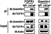
- FIG. 9C shows that Gremlin1 co-immunoprecipitates with FGFR1 in 293T cells and LNCaP resistance cells transfected with flag-tagged Gremlin1 and HA-tagged FGFR1 expressing plasmids.
- FIG. 9D shows that Endogenous Gremlin1 co-immunoprecipitates with FGFR1 in LNCaP-resistance cells.
- FIG. 9E shows that Gremlin 1 but not Gremlin 2 or other members of DAN protein family binds to FGFR1 as measured by Enzyme-linked immunosorbent assay (ELISA) .
- ELISA Enzyme-linked immunosorbent assay
- FIG. 9F shows that Interaction of purified Gremlin1 and soluble FGFR1 protein is demonstrated by pulldown experiments.
- FIG. 9G shows that Soluble FGFR1 competitively inhibits the activation of FGFR1/MEK/ERK signaling by Gremlin1 in PC3.
- FIG. 9H shows that BiFC assay shows colocalization between Gremlin1 and FGFR1 in LNCaP-resistance cells.
- FIG. 9I shows immunofluorescent staining images of Gremlin1 and FGFR1 in LNCaP-R cells.
- the cells were treated with Gremlin1 (100 ng/ml) or PBS for 10 mins at 37 °C.
- FIG. 9J shows the diagram of truncated FGFR1.
- FIG. 9K shows the Co-IP assay results between truncated FGFR1 and Gremlin1 (left panel) or FGF1 (right panel) .
- FIG. 9L shows the Gremlin1 mutagenesis strategies. Point mutations are bolded and underlined.
- FIG. 9M shows the Gremlin1 K123A-K124A mutant disrupts the co-immunoprecipitation between Gremlin1 and FGFR1, wherein the numbering is relative to SEQ ID NO: 69.
- FIG. 9N shows the schematic of FGFR1 mutations. Point mutations are bolded and underlined.
- FIG. 9O shows that the co-immunoprecipitation of FGFR1 and Gremlin1 is impaired by the FGFR1 E160A mutation.
- FIG. 9P shows schematics of FGFR1 mutations.
- FIG. 9Q shows that FGFR1-C176G or FGFR1-R248Q mutation abolishes co-immunoprecipitation of FGF1 and FGFR1 (left panel) , but do not influence the forming of protein complex between Gremlin1 and FGFR1 (right panel) .
- FIGs. 9R-9U show that the binding between Gremlin1 and FGFR1 is not affected by addition of FGF1, and vice versa, which are revealed by Fortebio (R) , co-immunostaining (S) and pull-down (T, U) assays.
- FIG. 9V shows docking module that highlights the key amino acid residues in the binding pocket between Gremlin1 and FGFR1.
- FIG. 10A shows that binding specificity of anti-murine-Gremlin1 antibody to Gremlin1 is validated by the enzyme-linked immunosorbent assay. Ab is anti-murine-Gremlin1 in this figure.
- FIG. 10B shows that Gremlin1 is highly expressed in the castrated Pbsn-Cre4; PTEN fl/fl ; Trp53 fl/fl murine PCa model. Representative images of Gremlin1 immunostaining are presented.
- FIG. 10C shows that Anti-Gremlin1 antibody (10ug/ml) exerts a significant inhibitory effect on PCa growth. Obvious suppression is found in gross tumor appearance.
- FIG. 10D shows that Anti-Gremlin1 antibody (10ug/ml) exerts a significant inhibitory effect on PCa growth. Obvious suppression is found in gross tumor weight.
- FIG. 10E shows that Anti-Gremlin1 antibody (10ug/ml) exerts a significant inhibitory effect on PCa growth. Obvious suppression is found in gross a significant reduction in PCNA positive cells.
- FIG. 10F shows that Anti-Gremlin1 treatment markedly represses the development of invasive PCa in castrated Pbsn-Cre; PTEN fl/fl ; Trp53 fl/fl mice.
- FIGs. 10G and 10H show that Gene set enrichment analysis indicates a significant suppression of the FGFR signaling pathway in the prostates of anti-Gremlin1 treatment group.
- FIGs. 10I and 10J show that the immunostaining and immunoblot analysis show inhibitory effects of anti-Gremlin1 antibody on the FGFR1/MAPK signaling pathway in prostates of Pbsn-Cre; PTEN fl/fl ; Trp53 fl/fl mice.
- Two-tailed Student’s t test was used for the statistical analysis. *, P ⁇ 0.05; **, P ⁇ 0.01; ***, P ⁇ 0.001. Data are presented as means ⁇ SEM. ) .
- FIG. 10M shows that the activation of FGFR1/MEK/ERK signaling pathway is suppressed by the Gremlin1 antibody in LNCaP-R cells.
- FIG. 10N shows that annexin-V/DAPI staining demonstrates that anti-Gremlin1 antibody displays a synergistic effect with enzalutamide in inducing cell death.
- Ab anti-human-Gremlin1.
- ADT treated with enzalutamide at 10 ⁇ g/ml. (Two-tailed Student’s t test was used for the statistical analysis. *, P ⁇ 0.05; **, P ⁇ 0.01; ***, P ⁇ 0.001. Data are presented as means ⁇ SEM. )
- FIG. 10O shows schematics illustrating the treatments in a Pbsn-Cre4; Ptenfl/fl; Trp53fl/fl GEMM. Mice which were castrated at 2 months received anti-Gremlin1 antibody (i.p., 10 mg/kg) or IgG, as indicated, three times a week for 2 months.
- FIG. 11A shows that Gremlin was mainly expressed by the epithelial cells in castrated Pbsn-Cre; PTEN fl/fl ; Trp53 fl/fl PCa.
- ECAD Ecadherin
- VIM Vimentin.
- FIG. 11B shows that Gremlin1 antibody treatment does not induce major side effects when administered systemically to mice (10mg/kg twice a week) . No obvious alterations are detected in peripheral blood cell counts in mice received the antibody treatment. Ab: anti-mGREM1 antibody. (Two-tailed Student’s t test was used for the statistical analysis. *, P ⁇ 0.05; **, P ⁇ 0.01; ***, P ⁇ 0.001. Data are presented as means ⁇ SEM. )
- FIG. 11C shows that Gremlin1 antibody treatment does not induce major side effects when administered systemically to mice (10mg/kg twice a week) . No obvious alterations are detected in major organs in mice received the antibody treatment.
- Ab anti-mGREM1 antibody.
- Two-tailed Student’s t test was used for the statistical analysis. *, P ⁇ 0.05; **, P ⁇ 0.01; ***, P ⁇ 0.001. Data are presented as means ⁇ SEM. )
- FIG. 12A shows that binding specificity of the anti-human-Gremlin1 (14E3) to Gremlin1 is validated by the enzyme-linked immunosorbent assay.
- Ab is anti-human-Gremlin1 in this figure.
- FIG. 12B shows that the antibody against human Gremlin1 (10ug/ml) represses cell proliferation of PC3 cells.
- FIG. 12C shows that anti-Gremlin1 antibody (10ug/ml) exerts an inhibitory effect on sphere forming of PC3 cells.
- FIG. 12D shows that Gremlin1 antibody neutralizes the activation of FGFR1/MEK/ERK signaling by Gremlin1 protein in PC3 cells in a dose-dependent manner.
- FIG. 12E and 12F show that anti-Gremlin1 treatment markedly impedes the in vivo growth of PC3 tumor xenografts in serial passage experiments.
- Antibody was given at indicated time points (see arrowhead) at 10 mg/kg via intra-peritoneal injection.
- FIG. 13 shows treatment with 14E3 reduced tumor volume in PC3 CRPC model and increased percent survival.
- FIG. 14A show that the antibody against Gremlin1 (100ng/ml) facilitates the inhibition of in vitro cell proliferation by enzalutamide (1ug/ml) .
- FIG. 14B shows that anti-Gremlin1 treatment suppresses the sphere formation ability of LNCaP cells.
- FIG. 14C shows that the activation of FGFR1/MEK/ERK signaling pathway is suppressed by the Gremlin1 antibody in LNCaP cells
- FIG. 14D shows that Annexin-V/DAPI staining demonstrates that anti-Gremlin1 antibody displays a synergistic effect with enzalutamide in inducing cell death.
- Ab anti-human-Gremlin1 14E3.
- Two-tailed Student’s t test was used for the statistical analysis. *, P ⁇ 0.05; **, P ⁇ 0.01; ***, P ⁇ 0.001. Data are presented as means ⁇ SEM. ) .
- FIG. 15 shows 14E3 reduced the GREM1-mediated promotion on cancer cell migration.
- FIG. 16A-16C show that 14E3 reduced the GREM1-mediated increase in the percentage of PSA-low population independent of the BMP-binding loop.
- FIG. 17A shows that immunoblotting confirms the efficiency of BMPRII knockout in LNCaP cells.
- FIG. 17B and 17C show that BMPRII knockout showing no significant influence to the inhibitory effect of Gremlin1 antibody on LNCaP cell proliferation and sphere formation.
- Ab anti-human-Gremlin1 14E3.
- Two-tailed Student’s t test was used for the statistical analysis. *, P ⁇ 0.05; **, P ⁇ 0.01; ***, P ⁇ 0.001. Data are presented as means ⁇ SEM. )
- FIG. 18A shows that immunofluorescent staining of AMCAR and DAPI indicates the tumor origin of patient derived organoids.
- FIG. 18B and 18C show that decreased organoid size and number suggests an inhibitory effect of the antibody against Gremlin1 14E3 on PDO forming and growth.
- Two-tailed Student’s t test was used for the statistical analysis. *, P ⁇ 0.05; **, P ⁇ 0.01; ***, P ⁇ 0.001. Data are presented as means ⁇ SEM. ) .
- FIG. 18D shows that detail information of patient samples used in this patient derived organoid experiment.
- FIG. 19A shows heatmap images of tumors in each mouse model from the control group (mIgG2a) and the experiment group (14E3) respectively. Each of the control group and the experiment group has 16 mice.
- FIG. 19B shows that the antibodies against Gremlin1 (e.g., 14E3) exerted no obvious influence on the body weight in the PCa metastasis mice model study.
- Gremlin1 e.g., 14E3
- FIG. 19C shows that the average radiance intensity was decreased with the Gremlin1 antibody treatment in the PCa metastasis mice model study.
- FIG. 19D shows images of the lung tissue sections, where the arrows indicate metastases sites in the lung.
- FIG. 19E shows the statistics of the number of micrometastases in lung in the PCa metastasis mice model study.
- activating mutation refers a mutation or a post-transcriptional modification that results in at least partial (or complete) loss of function or activity of the gene or of the gene product of biomarker (such as AR, PTEN and/or p53) , or results in a non-functional gene or gene product.
- the activity of the affected gene or gene product of the biomarker would be significantly lower than wild-type counterpart or even be eliminated.
- An inactivating mutation can be a translocation, intragenic chromosome breaks, inversions, deletion (e.g., biallelic deletion, heterozygous or homozygous copy number loss) , micro copy number alterations, insertion, substitution, aberrant splicing, or any combination thereof, which reduces the biological activity of the biomarker.
- insertion or deletion in a polynucleotide sequence may cause frame shift, which changes the reading frame of the codons and results in a completely different translated gene product from the original. This often generates truncated proteins that result in loss of function.
- the term “deletion” when used as a type of inactivating mutation of a biomarker refers to a mutation in which one or more nucleobase pairs are lost or deleted from a polynucleotide sequence, or in which one or more amino acid residue are deleted from a polypeptide sequence. For example, it can refer to deletion, loss, or removal of an entire coding region or a portion thereof of the biomarker.
- substitution is a mutation that exchanges one nucleobase for another in a polynucleotide sequence, or that substitutes one amino acid residue for another in a polypeptide sequence.
- Substitution in a polynucleotide sequence can: 1) change a codon to one that encodes a different amino acid residue, and therefore will cause change in amino acid sequence in the protein produced, or 2) change to a codon that encodes the same amino acid residue thereby causing no change in the protein produced; or 3) change an amino-acid-coding codon to a single “stop” codon and cause an incomplete protein (an incomplete protein is usually nonfunctional) .
- an “insertion” is a mutation in which one or more extra nucleobase pairs are inserted into a place in a polynucleotide sequence, or in which one or more amino acid residue is inserted into a polypeptide sequence.
- a “translocation” refers to a type of chromosomal abnormality resulted from the exchange of genetic materials between two non-homologous chromosomes.
- a translocation may be either balanced or unbalanced; a balanced translocation results in no gain or loss of material, while an unbalanced translocation may result in trisomy or monosomy of a particular chromosome segment.
- Chromosomal translocations are typically seen in cases of leukemia, like, for instance, in acute myeloid leukemia.
- level with respect to a biomarker such as AR, PTEN, and/or p53 refers to the amount or quantity of the biomarker of interest present in a sample. Such amount or quantity may be expressed in the absolute terms, i.e., the total quantity of the biomarker in the sample, or in the relative terms, i.e., the concentration or percentage of the biomarker in the sample.
- Level of a biomarker can be measured at DNA level (for example, as represented by the amount or quantity or copy number of the gene in a chromosomal region) , at RNA level (for example as mRNA amount or quantity) , or at protein level (for example as protein or protein complex amount or quantity) .
- reference level with respect to a biomarker refers to a benchmark level which allows for comparison.
- a reference level may be chosen by the persons skilled in the art according to the desired purpose. Means for determining suitable reference levels are known to the persons skilled in the art, e.g. a reference level can be determined from experience, existing knowledge or data collected from clinical studies.
- the term “negative” with respect to a biomarker means that the biomarker is test negative or absent in a test sample.
- the biomarker which is negative in a test sample may have a level comparable or undistinguishable from the negative control level in a sample lacking such a biomarker, or alternatively, may have a level below a threshold level that defines presence or a positive result.
- “likelihood” and “likely” with respect to response of a subject to a treatment is a measurement of how probable the therapeutic response is to occur in the subject. It may be used interchangeably with “probability” . Likelihood refers to a probability that is more than speculation, but less than certainty. Thus, a therapeutic response is likely if a reasonable person using common sense, training or experience concludes that, given the circumstances, a therapeutic response is probable.
- beneficial or favorable response to the therapy refers to beneficial or favorable response to the therapy, as opposed to unfavorable responses, i.e. adverse events.
- antibody as used herein includes any immunoglobulin, monoclonal antibody, polyclonal antibody, multivalent antibody, bivalent antibody, monovalent antibody, multispecific antibody, or bispecific antibody that binds to a specific antigen.
- a native intact antibody comprises two heavy (H) chains and two light (L) chains.
- Mammalian heavy chains are classified as alpha, delta, epsilon, gamma, and mu, each heavy chain consists of a variable region (V H ) and a first, second, and third constant region (C H1 , C H2 , C H3 , respectively) ;
- mammalian light chains are classified as ⁇ or ⁇ , while each light chain consists of a variable region (V L ) and a constant region.
- the antibody has a “Y” shape, with the stem of the Y consisting of the second and third constant regions of two heavy chains bound together via disulfide bonding.
- Each arm of the Y includes the variable region and first constant region of a single heavy chain bound to the variable and constant regions of a single light chain.
- the variable regions of the light and heavy chains are responsible for antigen binding.
- the variable regions in both chains generally contain three highly variable loops called the complementarity determining regions (CDRs) (light chain CDRs including LCDR1, LCDR2, and LCDR3, heavy chain CDRs including HCDR1, HCDR2, HCDR3) .
- CDRs complementarity determining regions
- CDR boundaries for the antibodies and antigen-binding domains disclosed herein may be defined or identified by the conventions of Kabat, IMGT, AbM, Chothia, or Al-Lazikani (Al-Lazikani, B., Chothia, C., Lesk, A.M., J. Mol. Biol., 273 (4) , 927 (1997) ; Chothia, C. et al., J Mol Biol. Dec 5; 186 (3) : 651-63 (1985) ; Chothia, C. and Lesk, A.M., J. Mol. Biol., 196, 901 (1987) ; N.R.
- the three CDRs are interposed between flanking stretches known as framework regions (FRs) , which are more highly conserved than the CDRs and form a scaffold to support the hypervariable loops.
- FRs framework regions
- the constant regions of the heavy and light chains are not involved in antigen-binding, but exhibit various effector functions.
- Antibodies are assigned to classes based on the amino acid sequence of the constant region of their heavy chain.
- the five major classes or isotypes of antibodies are IgA, IgD, IgE, IgG, and IgM, which are characterized by the presence of alpha, delta, epsilon, gamma, and mu heavy chains, respectively.
- the antibody provided herein encompasses any antigen-binding fragments thereof.
- antigen-binding fragment refers to an antibody fragment formed from a fragment of an antibody comprising one or more CDRs, or any other antibody portion that binds to an antigen but does not comprise an intact native antibody structure.
- antigen-binding fragment include, without limitation, a diabody, a Fab, a Fab', a F (ab') 2 , a Fd, an Fv fragment, a disulfide stabilized Fv fragment (dsFv) , a (dsFv) 2 , a bispecific dsFv (dsFv-dsFv') , a disulfide stabilized diabody (ds diabody) , a single-chain antibody molecule (scFv) , an scFv dimer (bivalent diabody) , a multispecific antibody, a camelized single domain antibody, a nanobody, a domain antibody, and a bivalent domain antibody.
- An antigen-binding fragment include,
- Fab with regard to an antibody refers to a monovalent antigen-binding fragment of the antibody consisting of a single light chain (both variable and constant regions) bound to the variable region and first constant region of a single heavy chain by a disulfide bond.
- Fab can be obtained by papain digestion of an antibody at the residues proximal to the N-terminus of the disulfide bond between the heavy chains of the hinge region.
- Fab' refers to a Fab fragment that includes a portion of the hinge region, which can be obtained by pepsin digestion of an antibody at the residues proximal to the C-terminus of the disulfide bond between the heavy chains of the hinge region and thus is different from Fab in a small number of residues (including one or more cysteines) in the hinge region.
- F (ab') 2 refers to a dimer of Fab’ that comprises two light chains and part of two heavy chains.
- Fv with regard to an antibody refers to the smallest fragment of the antibody to bear the complete antigen binding site.
- a Fv fragment consists of the variable region of a single light chain bound to the variable region of a single heavy chain.
- a “dsFv” refers to a disulfide-stabilized Fv fragment that the linkage between the variable region of a single light chain and the variable region of a single heavy chain is a disulfide bond.
- Single-chain Fv antibody or “scFv” refers to an engineered antibody consisting of a light chain variable region and a heavy chain variable region connected to one another directly or via a peptide linker sequence (Huston JS et al. Proc Natl Acad Sci USA, 85: 5879 (1988) ) .
- a “scFv dimer” refers to a single chain comprising two heavy chain variable regions and two light chain variable regions with a linker.
- an “scFv dimer” is a bivalent diabody or bivalent ScFv (BsFv) comprising V H -V L (linked by a peptide linker) dimerized with another V H -V L moiety such that V H 's of one moiety coordinate with the V L 's of the other moiety and form two binding sites which can target the same antigens (or eptipoes) or different antigens (or eptipoes) .
- a “scFv dimer” is a bispecific diabody comprising V H1 -V L2 (linked by a peptide linker) associated with V L1 -V H2 (also linked by a peptide linker) such that V H1 and V L1 coordinate and V H2 and V L2 coordinate and each coordinated pair has a different antigen specificity.
- Single-chain Fv-Fc antibody or “scFv-Fc” refers to an engineered antibody consisting of a scFv connected to the Fc region of an antibody.
- “Camelized single domain antibody, ” “heavy chain antibody, ” “nanobody” or “HCAb” refers to an antibody that contains two V H domains and no light chains (Riechmann L. and Muyldermans S., J Immunol Methods. Dec 10; 231 (1-2) : 25-38 (1999) ; Muyldermans S., J Biotechnol. Jun; 74 (4) : 277-302 (2001) ; WO94/04678; WO94/25591; U.S. Patent No. 6,005,079) . Heavy chain antibodies were originally obtained from Camelidae (camels, dromedaries, and llamas) .
- VHH domain The variable domain of a heavy chain antibody (VHH domain) represents the smallest known antigen-binding unit generated by adaptive immune responses (Koch-Nolte F.
- “Diabodies” include small antibody fragments with two antigen-binding sites, wherein the fragments comprise a V H domain connected to a V L domain in a single polypeptide chain (V H -V L or V L -V H ) (see, e.g., Holliger P. et al., Proc Natl Acad Sci U S A. Jul 15; 90 (14) : 6444-8 (1993) ; EP404097; WO93/11161) .
- the two domains on the same chain cannot be paired, because the linker is too short, thus, the domains are forced to pair with the complementary domains of another chain, thereby creating two antigen-binding sites.
- the antigen–binding sites may target the same of different antigens (or epitopes) .
- a “domain antibody” refers to an antibody fragment containing only the variable region of a heavy chain or the variable region of a light chain.
- two or more V H domains are covalently joined with a peptide linker to form a bivalent or multivalent domain antibody.
- the two V H domains of a bivalent domain antibody may target the same or different antigens.
- a “ (dsFv) 2 ” comprises three peptide chains: two V H moieties linked by a peptide linker and bound by disulfide bridges to two V L moieties.
- a “bispecific ds diabody” comprises V H1 -V L2 (linked by a peptide linker) bound to V L1 -V H2 (also linked by a peptide linker) via a disulfide bridge between V H1 and V L1 .
- a “bispecific dsFv” or “dsFv-dsFv'” comprises three peptide chains: a V H1 -V H2 moiety wherein the heavy chains are bound by a peptide linker (e.g., a long flexible linker) and paired via disulfide bridges to V L1 and V L2 moieties, respectively.
- a peptide linker e.g., a long flexible linker
- disulfide bridges to V L1 and V L2 moieties
- humanized means that the antibody or antigen-binding fragment comprises CDRs derived from non-human animals, FR regions derived from human, and when applicable, constant regions derived from human.
- the amino acid residues of the variable region framework of the humanized gremlin antibody are substituted for sequence optimization.
- the variable region framework sequences of the humanized gremlin antibody chain are at least 65%, 70%, 75%, 80%, 85%, 90%, 95%or 100%identical to the corresponding human variable region framework sequences.
- chimeric refers to an antibody or antigen-binding fragment that has a portion of heavy and/or light chain derived from one species, and the rest of the heavy and/or light chain derived from a different species.
- a chimeric antibody may comprise a constant region derived from human and a variable region derived from a non-human species, such as from mouse.
- germline sequence refers to the nucleic acid sequence encoding a variable region amino acid sequence or subsequence that shares the highest determined amino acid sequence identity with a reference variable region amino acid sequence or subsequence in comparison to all other known variable region amino acid sequences encoded by germline immunoglobulin variable region sequences.
- the germline sequence can also refer to the variable region amino acid sequence or subsequence with the highest amino acid sequence identity with a reference variable region amino acid sequence or subsequence in comparison to all other evaluated variable region amino acid sequences.
- the germline sequence can be framework regions only, complementarity determining regions only, framework and complementarity determining regions, a variable segment (as defined above) , or other combinations of sequences or subsequences that comprise a variable region. Sequence identity can be determined using the methods described herein, for example, aligning two sequences using BLAST, ALIGN, or another alignment algorithm known in the art.
- the germline nucleic acid or amino acid sequence can have at least about 90%, 91, 92%, 93%, 94%, 95%, 96%, 97%, 98%, 99%, or 100%sequence identity with the reference variable region nucleic acid or amino acid sequence.
- Germline sequences can be determined, for example, through the publicly available international ImMunoGeneTics database (IMGT) and V-base.
- Anti-human gremlin1 antibody , “anti-hGREM1 antibody” or “an antibody against human gremlin1” as used herein interchangeably and refers to an antibody that is capable of specific binding to human gremlin1 with a sufficient specificity and/or affinity, for example, to provide for therapeutic use.
- affinity refers to the strength of non-covalent interaction between an immunoglobulin molecule (i.e. antibody) or fragment thereof and an antigen.
- the term “specific binding” or “specifically binds” as used herein refers to a non-random binding reaction between two molecules, such as for example between an antibody and an antigen.
- the antibodies or antigen-binding fragments provided herein specifically bind to human and/or non-human gremlin1 with a binding affinity (K D ) of ⁇ 10 -6 M (e.g., ⁇ 5x10 -7 M, ⁇ 2x10 -7 M, ⁇ 10 -7 M, ⁇ 5x10 -8 M, ⁇ 2x10 -8 M, ⁇ 10 -8 M, ⁇ 5x10 -9 M, ⁇ 4x10 -9 M, ⁇ 3x10 -9 M, ⁇ 2x10 -9 M, or ⁇ 10 -9 M.
- K D binding affinity
- K D used herein refers to the ratio of the dissociation rate to the association rate (k off /k on ) , which may be determined by using any conventional method known in the art, including but are not limited to surface plasmon resonance method, microscale thermophoresis method, HPLC-MS method and flow cytometry (such as FACS) method.
- the K D value can be appropriately determined by using flow cytometry method.
- a variety of immunoassay formats may be used to select antibodies specifically immunoreactive with a particular protein.
- solid-phase ELISA immunoassays are routinely used to select antibodies specifically immunoreactive with a protein (see, e.g., Harlow &Lane, Using Antibodies, A Laboratory Manual (1998) , for a description of immunoassay formats and conditions that can be used to determine specific immunoreactivity) .
- a specific or selective binding reaction will produce a signal at least twice over the background signal and more typically at least 10 to 100 times over the background.
- amino acid refers to an organic compound containing amine (-NH 2 ) and carboxyl (-COOH) functional groups, along with a side chain specific to each amino acid.
- amine -NH 2
- -COOH carboxyl
- a “conservative substitution” with reference to amino acid sequence refers to replacing an amino acid residue with a different amino acid residue having a side chain with similar physiochemical properties.
- conservative substitutions can be made among amino acid residues with hydrophobic side chains (e.g. Met, Ala, Val, Leu, and Ile) , among residues with neutral hydrophilic side chains (e.g. Cys, Ser, Thr, Asn and Gln) , among residues with acidic side chains (e.g. Asp, Glu) , among amino acids with basic side chains (e.g. His, Lys, and Arg) , or among residues with aromatic side chains (e.g. Trp, Tyr, and Phe) .
- conservative substitution usually does not cause significant change in the protein conformational structure, and therefore could retain the biological activity of a protein.
- Percent (%) sequence identity with respect to amino acid sequence (or nucleic acid sequence) is defined as the percentage of amino acid (or nucleic acid) residues in a candidate sequence that are identical to the amino acid (or nucleic acid) residues in a reference sequence, after aligning the sequences and, if necessary, introducing gaps, to achieve the maximum correspondence. Alignment for purposes of determining percent amino acid (or nucleic acid) sequence identity can be achieved, for example, using publicly available tools such as BLASTN, BLASTp (available on the website of U.S. National Center for Biotechnology Information (NCBI) , see also, Altschul S.F. et al, J. Mol. Biol., 215: 403–410 (1990) ; Stephen F.
- the non-identical residue positions may differ by conservative amino acid substitutions.
- a “conservative amino acid substitution” is one in which an amino acid residue is substituted by another amino acid residue having a side chain (R group) with similar chemical properties (e.g., charge or hydrophobicity) .
- R group side chain
- a conservative amino acid substitution will not substantially change the functional properties of a protein.
- the percent or degree of similarity may be adjusted upwards to correct for the conservative nature of the substitution. Means for making this adjustment are well known to those of skill in the art. See, e.g., Pearson (1994) Methods Mol. Biol. 24: 307-331, which is herein incorporated by reference.
- a “homologous sequence” refers to a polynucleotide sequence (or its complementary strand) or an amino acid sequence that has sequence identity of at least 80% (e.g. at least 85%, 88%, 90%, 91%, 92%, 93%, 94%, 95%, 96%, 97%, 98%, 99%) to another sequence when optionally aligned.
- an “isolated” substance has been altered by the hand of man from the natural state. If an “isolated” composition or substance occurs in nature, it has been changed or removed from its original environment, or both.
- a polynucleotide or a polypeptide naturally present in a living animal is not “isolated, ” but the same polynucleotide or polypeptide is “isolated” if it has been sufficiently separated from the coexisting materials of its natural state so as to exist in a substantially pure state.
- An isolated “nucleic acid” or “polynucleotide” are used interchangeably and refer to the sequence of an isolated nucleic acid molecule.
- an “isolated antibody or antigen-binding fragment thereof” refers to the antibody or antigen-binding fragments having a purity of at least 60%, 70%, 75%, 80%, 81%, 82%, 83%, 84%, 85%, 86%, 87%, 88%, 89%, 90%, 91%, 92%, 93%, 94%, 95%, 96%, 97%, 98%, 99%as determined by electrophoretic methods (such as SDS-PAGE, isoelectric focusing, capillary electrophoresis) , or chromatographic methods (such as ion exchange chromatography or reverse phase HPLC) .
- electrophoretic methods such as SDS-PAGE, isoelectric focusing, capillary electrophoresis
- chromatographic methods such as ion exchange chromatography or reverse phase HPLC
- subject includes human and non-human animals.
- Non-human animals include all vertebrates, e.g., mammals and non-mammals, such as non-human primates, mouse, rat, cat, rabbit, sheep, dog, cow, chickens, amphibians, and reptiles. Except when noted, the terms “patient” or “subject” are used herein interchangeably.
- Treating” or “treatment” of a condition as used herein includes preventing or alleviating a condition, slowing the onset or rate of development of a condition, reducing the risk of developing a condition, preventing or delaying the development of symptoms associated with a condition, reducing or ending symptoms associated with a condition, generating a complete or partial regression of a condition, curing a condition, or some combination thereof.
- greylin1 or “GREM1” refers to the variant 1 of gremlin, and encompasses gremlin1 in different species such as in human, mouse, monkey, and so on. GREM1 is evolutionarily conserved and the human gremlin1 gene (hGREM1) has been mapped to chromosome 15q13-q15 (Topol L Z et al., (1997) Mol. Cell Biol., 17: 4801-4810; Topol L Z et al., Cytogenet Cell Genet., 89: 79-84) .
- hGREM1 The amino acid sequence of hGREM1 is accessibly by GenBank database under the accession number NP-037504 or Uniprot Database via the accession number O60565, and is provided herein as SEQ ID NO: 66.
- GenBank database under the accession number NP-037504 or Uniprot Database via the accession number O60565, and is provided herein as SEQ ID NO: 66.
- human gremlin1 and the term “hGREM1” are used interchangeably in the present disclosure.
- GREM1-related disease or condition refers to any disease or condition caused by, exacerbated by, or otherwise linked to increased expression or activities of GREM1.
- the GREM1 related condition is, for example, glaucoma, cancer, fibrotic disease, angiogenesis, retinal disease, kidney disease, pulmonary arterial hypertension, or osteoarthritis (OA) .
- Cancer refers to any medical condition characterized by malignant cell growth or neoplasm, abnormal proliferation, infiltration or metastasis, and can be benign or malignant, and includes both solid tumors and non-solid cancers (e.g. hematologic malignancies) such as leukemia.
- solid tumor refers to a solid mass of neoplastic and/or malignant cells.
- pharmaceutically acceptable indicates that the designated carrier, vehicle, diluent, excipient (s) , and/or salt is generally chemically and/or physically compatible with the other ingredients comprising the formulation, and physiologically compatible with the recipient thereof.
- terapéuticaally effective amount or “effective amount” means the amount of a pharmaceutical agent that that produces some desired local or systemic therapeutic effect at a reasonable benefit/risk ratio applicable to any treatment. When administered for preventing a disease, the amount is sufficient to avoid or delay onset of the disease. A therapeutically effective amount or an effective amount need not be curative or prevent a disease or condition from ever occurring. In certain embodiments, a therapeutically-effective amount of a pharmaceutical agent will depend on its therapeutic index, solubility, and the like.
- references to “about” a value or parameter herein includes (and describes) embodiments that are directed to that value or parameter per se.
- description referring to “about X” includes description of “X. ”
- Numeric ranges are inclusive of the numbers defining the range.
- the term “about” refers to the indicated value of the variable and to all values of the variable that are within the experimental error of the indicated value (e.g. within the 95%confidence interval for the mean) or within 10 percent of the indicated value, whichever is greater.
- the term “about” is used within the context of a time period (years, months, weeks, days etc. )
- the term “about” means that period of time plus or minus one amount of the next subordinate time period (e.g. about 1 year means 11-13 months; about 6 months means 6 months plus or minus 1 week; about 1 week means 6-8 days; etc. ) , or within 10 percent of the indicated value, whichever is greater.
- the present disclosure provides novel medical uses of gremlin1 (GREM1) antagonists.
- GREM1 gremlin1
- the novel medical uses are, in part, based on the unexpected discovery that transcription of GREM1 is suppressed by androgen receptor (AR) and unleashed upon androgen deprivation therapy (ADT) .
- the novel medical uses are, in part, based on the discovery that deficiency in PTEN and/or p53 promotes GREM1 expression.
- GREM1 is significantly upregulated in advance prostate cancers including castration resistant prostate cancers (CRPCs) , and positively correlates with development of castration resistance and poor overall survival. It has been shown by the inventors that GREM1 antagonists are useful in treating related conditions.
- CRPCs castration resistant prostate cancers
- the present disclosure provides a method of treating a GREM1-expressing disease or condition in a subject in need thereof, comprising administering to the subject a therapeutically effective amount of GREM1 antagonist, wherein the disease or condition is characterized in reduced or inhibited androgen receptor (AR) signaling.
- a GREM1-expressing disease or condition in a subject in need thereof, comprising administering to the subject a therapeutically effective amount of GREM1 antagonist, wherein the disease or condition is characterized in reduced or inhibited androgen receptor (AR) signaling.
- AR androgen receptor
- the subject is receiving or has received an AR inhibitor.
- the disease or condition is resistant to an AR inhibitor.
- AR inhibitor as used herein refers to a therapeutic agent useful in inhibiting AR activity, for example, those used in androgen deprivation therapy.
- the disease or condition is AR-associated cancer (such as prostate cancer, breast cancer, glioblastoma, melanoma, bladder cancer, renal cell carcinoma, pancreatic cancer, hepatocellular carcinoma, ovarian cancer, endometrial cancer, mantle cell lymphoma, or salivary gland cancer) , or AR-associated non-cancer conditions (such as, hair loss, acne, hirsutism, ovarian cysts, polycystic ovary disease, precocious puberty, spinal and bulbar muscular atrophy, or age-related macular degeneration) .
- AR-associated cancer such as prostate cancer, breast cancer, glioblastoma, melanoma, bladder cancer, renal cell carcinoma, pancreatic cancer, hepatocellular carcinoma, ovarian cancer, endometrial cancer, mantle cell lymphoma, or salivary gland cancer
- AR-associated non-cancer conditions such as, hair loss, acne, hirsutism, ovarian cysts, polyc
- the present disclosure provides methods of treating GREM1-expressing cancer in a subject in need thereof, comprising administering to the subject a therapeutically effective amount of GREM1 antagonist, wherein the cancer is characterized in reduced androgen receptor (AR) signaling.
- a therapeutically effective amount of GREM1 antagonist wherein the cancer is characterized in reduced androgen receptor (AR) signaling.
- Androgen receptor is a member of the steroid and nuclear receptor superfamily, and is mainly expressed in androgen target tissues, such as the prostate, skeletal muscle, liver, and central nervous system (CNS) , with the highest expression level observed in the prostate, adrenal gland, and epididymis.
- AR is a soluble protein that functions as an intracellular transcriptional factor. Upon binding and activation by androgens, AR mediates transcription of target genes that modulate growth and differentiation of prostate epithelial cells. AR signaling is crucial for the development and maintenance of male reproductive organs including the prostate gland.
- AR signaling refers to AR signaling the level of which is substantially lower than the normal or baseline level of AR signaling, for example, a level of AR signaling in the healthy cell or tissue sample, or an average level of the AR signaling in the general cancer patient population or in a cancer patient population of a particular cancer of interest or in a patient population having AR dependent prostate cancers.
- Cancer having reduced androgen receptor (AR) signaling can be an AR-expressing cancer, where the AR signaling is inhibited, for example, due to treatment (e.g. pharmacological treatment or surgical treatment) , or due to reduced expression level of AR, or due to certain inactivating mutations in AR.
- the cancer having reduced AR signaling can be negative in AR expression, in particular for cancers that normally express AR (such as prostate cancer) .
- the cancer is an AR-expressing cancer.
- Different types of cancers are known to express AR.
- Examples of AR-expressing cancer include without limitation, prostate cancer, breast cancer, lung cancer, head and neck cancer, testis cancer, endometrial cancer, ovarian cancer, and skin cancer.
- the AR-expressing cancer is prostate cancer or breast cancer.
- the subject is receiving or has received androgen deprivation therapy (ADT) .
- ADT androgen deprivation therapy
- ADT refers to therapies that suppresses androgen, by reducing levels of androgen or by inhibiting biological functions of androgen such as by inhibiting AR signaling.
- the main androgens in the body are testosterone and dihydrotestosterone (DHT) .
- the subject or the cancer is resistant to an ADT.
- resistant it is meant that the disease has no or reduced responsiveness or sensitivity to an ADT. Reduced responsiveness can be indicated by, for example, requirement of an increased dose to achieve a given efficacy.
- the disease can be non-responsive to an ADT. For example, the cancer cells or tumor size increases despite of the treatment with the an ADT, or the disease showed regression back to its former state, for example, return of previous symptoms following partial recovery.
- the resistance to an ADT can be de novo or acquired.
- the subject or the cancer has reduced expression level of AR, or having one or more inactivating mutations in AR.
- Over 800 different AR mutations have been identified in patients with androgen insensitivity syndrome, and prostate cancer.
- four different types of mutations have been detected to inactivate AR, including: a) single point mutations resulting in amino acid substitutions or premature stop codons; b) nucleotide insertions or deletions leading to a frame shift and premature rumination; c) complete or partial gene deletions; and d) intronic mutations causing alternative splicing (see, for details, K. Eisermann et al, Transl Androl, Urol. 2013 Sep; 2 (3) : 137–147) .
- the cancer is negative in androgen receptor (AR) expression, i.e., AR-negative cancer.
- AR-negative cancer as used herein means a cancer originally having AR expression but becomes AR-negative.
- the AR-negative cancer is prostate cancer or breast cancer.
- Some prostate cancer cell lines are known to be AR-negative, such as PC3 cell line.
- An AR-negative cancer can be tested negative (or non-detectable) in AR expression or AR signaling, or can have a detected level of AR expression comparable to that of a known AR-negative prostate cancer cell.
- the prostate cancer or breast cancer is negative in both androgen receptor (AR) expression and neuroendocrine (NE) differentiation.
- NE differentiation in prostate cancer is a well-recognized phenotypic change by which prostate cancer cells transdifferentiate into NE-like cells.
- NE-like cells lack the expression of androgen receptor and prostate specific antigen, and are resistant to treatments.
- the NE differentiation can be assessed by measuring the protein level or mRNA level of NE markers chromogranin A (CgA) , ratio of CgA/prostate specific antigen (PSA) , and/or neuron specific enolsase (NSE) . See, e.g., Hu et al., Front Oncol.
- CgA chromogranin A
- PSA ratio of CgA/prostate specific antigen
- NSE neuron specific enolsase
- the prostate cancer is further characterized in having a level of Prostate Specific Antigen (PSA) lower than a reference level.
- PSA Prostate Specific Antigen
- PSA is a classic downstream target of AR. Normally, very little PSA is secreted in the blood. Increases in glandular size and tissue damage caused by benign prostatic hypertrophy, prostatitis, or prostate cancer may increase circulating PSA levels. Prostate cancer cells at advanced stages that are poorly differentiated or undifferentiated produce less PSA and are accompanied with a low level of PSA (for example, less than 4 ng/ml) . It is also believed that prostate cancer cells having low level of PSA or negative for PSA could be resistant to anti-androgens, chemotherapeutic drugs, pro-oxidants, or radiation, and may be castration-resistant (Skvortsov S. et al, STEM CELLS, Vol. 36, Issue 10, 1457–1474) .
- the reference level of PSA can be a threshold level of PSA normally found in a PSA positive prostate cancer.
- the reference level of PSA can also be an average level of the PSA in a general prostate cancer patient population or in a patient population having prostate cancers before progressing into advanced stages.
- Certain reference levels of PSA in blood can be, for example, about 2 ng/ml, about 4 ng/ml, about 6 ng/ml, about 8 ng/ml or about 10 ng/ml as measured using immunodetectable assays, e.g., the Hybritech (San Diego, Calif) , Tosoh (Foster City, Calif) , Bayer Centaur PSA Assay kit (Tarrytown, NY) , or Abbott assays (Chicago, Ill) . See, e.g., Dan et al., Cancer, Volume 109, Issue 2, 2007. https: //doi. org/10.1002/cncr. 22372 ; and Oesterling et al., J Urol. 1995; 154: 1090-1095, disclosure of which are hereby incorporated by reference in their entirety.
- the prostate cancer is negative for PSA.
- the prostate cancer does not express PSA, or is tested to be negative in a test for PSA.
- the prostate cancer is castration-resistant.
- Castration-resistant prostate cancer is an advanced prostate cancer that is capable to grow despite of low levels of circulating androgens.
- CRPC may present as either a continuous rise in PSA levels, the progression of pre-existing disease, and/or the appearance of new metastasis, despite a serum testosterone value below 50 ng/dL after ADT (Toshiyuki Kamoto et al., Nihon Rinsho. 2014 Dec; 72 (12) : 2103-7; Fred Saad et al., Can Urol Assoc J. 2010 Dec; 4 (6) : 380–384 ) .
- CRPC can remain dependent on AR signaling despite depletion or reduction of androgens.
- CRPC can be developed via amplifying AR expression, mutating the AR gene and/or genes encoding coactivators/corepressors, activating androgen-independent AR pathways, and/or producing alternative androgen, so as to remain the dependency on AR pathway for disease progression (Thenappan et al., Transl Androl Urol. 2015 Jun; 4 (3) : 365–380. )
- Some CRPC can bypass the requirement for AR signaling.
- the prostate cancer is: a) negative in androgen receptor (AR) expression, b) negative in both androgen receptor (AR) expression and neuroendocrine (NE) differentiation; c) resistant to an androgen deprivation therapy, optionally castration-resistant, d) showing a level of Prostate Specific Antigen (PSA) lower than a reference level, or e) any combinations of a) to d) .
- AR negative in androgen receptor
- AR negative in both androgen receptor
- NE neuroendocrine
- PSA Prostate Specific Antigen
- the cancer is further determined to be deficient in PTEN and/or p53.
- the cancer is metastatic.
- a metastatic cancer can spread or has spread from its site of origin to another part of the body.
- a metastatic tumor is the same type of cancer as the primary tumor.
- a metastatic cancer may spread to areas near the primary site, or to distant parts of the body.
- the cancer having reduced AR signaling is further characterized in GREM1 expression or overexpression.
- the GREM1 expression or overexpression can be in a disease cell or in a disease microenvironment.
- the term “overexpression” with respect to GREM1 as used herein refers to an increased expression level relative to a reference level.
- the reference level can be the level of GREM1 expression found in normal cells of the same tissue type, optionally normalized to expression level of another gene (e.g. a house keeping gene) .
- the reference level can be the level of GREM1 expression found in healthy subjects.
- the expression level which can be determined based on nucleic acid level or protein level.
- the GREM1-expressing cancer has a GREM1 expression level at least 10%higher (e.g. at least 15%, 20%, 30%, 35%, 40%, 50%or 1-fold, 2-fold, 3-fold or even higher) than a reference level.
- the present disclosure further provides methods of increasing sensitivity of an AR-expressing cancer to an androgen deprivation therapy (ADT) in a subject, comprising administering to the subject a therapeutically effective amount of GREM1 antagonist.
- ADT androgen deprivation therapy
- the methods comprises administering to the subject the GREM1 antagonist in combination with the ADT.
- the present disclosure provides methods of treating a GREM1-related disease or condition characterized in deficiency of PTEN and/or p53 in a subject.
- PTEN and p53 contribute to the regulation of self-renewal and differentiation in prostate progenitors and presumptive tumor initiating cells for prostate cancer.
- the term PTEN and/or p53 provided herein are intended to encompass different forms including mRNA, protein and also DNA (e.g. genomic DNA) . Therefore, the level and/or activity and/or mutation status of PTEN and/or p53 can be measured with RNA (e.g. mRNA) , protein or DNA (e.g. genomic DNA) .
- TP53 is a transcription factor capable of regulating a number of genes that regulate e.g. cell cycle and apoptosis.
- Alternative names for p53 include, e.g., antigen NY-CO-13, phosphorprotein p53, tumor suppressor p53 and cellular tumor antigen p53.
- p53 as used herein can indicate the TP protein as well as the polynucleotide (e.g. DNA or RNA) encoding the TP53 protein, including all isoforms and variants.
- the gene of p53 is available in GenBank database under the NCBI Reference Sequence of NG_017013.2, and exemplary sequence of human p53 protein is available in UniProtKB database under the accession number of P04637 (P53-HUMAN) .
- the protein of p53 comprises an amino acid sequence of SEQ ID NO: 73.
- PTEN Pten
- PTEN tyrosine phosphatase PTEN tyrosine phosphatase
- PTEN also known as phosphatase and tensin homolog deleted on chromosome ten, is a tumor suppressor that acts as a dual-specificity protein phosphatase that antagonizes the PI3K signaling pathway through its lipid phosphatase activity and negatively regulates the MAPK pathway through its protein phosphatase activity (Pezzolesi et al., Hum. Molec. Genet. 16: 1058-1071, 2007. ) .
- PTEN as used herein can refer to the PTEN protein as well as the DNA (e.g.
- the coding gene sequence or the RNA encoding for the PTEN, including all isoforms and variants.
- Exemplary sequence of human PTEN is available in UniProtKB database under the accession number of P60484 (PTEN_HUMAN) , with three isoforms: isoform 1 (P60484-1) , isoform alpha (P60484-2) and isoform 3 (P60484-3) .
- Exemplary sequence of gene of PTEN is available in GenBank database under the NCBI Reference Sequence of NC_000010.11.
- the protein of PTEN comprises an amino acid sequence of SEQ ID NO: 74.
- deficiency or “deficient” refers to insufficiency in activity or level, and can include, for example, being less than normal activity or level, or being absent or null in activity or level.
- deficiency in activity or level of PTEN and/or p53 can result in PTEN and/or p53 having no or less than normal function, or an absence of or reduced expression level of PTEN and/or p53 in a biological sample.
- the deficiency in PTEN and/or p53 is characterized in absence of functional PTEN and/or p53.
- the deficiency in activity or level of PTEN and/or p53 can be indicated by the presence of the inactivating mutation in PTEN and/or p53.
- the deficiency in activity or level of PTEN and/or p53 can be indicated by the expression level or copy number of PTEN and/or p53 in the biological sample. Accordingly, to determine if there is deficiency in activity or level of PTEN and/or p53 in the biological sample, the methods provided herein can comprise the step of determining if expression level or copy number of PTEN and/or p53 is reduced in the biological sample relative to a reference level.
- Mutation status or expression level of PTEN and/or p53 at DNA or RNA level can be measured by any methods known in the art, for example, without limitation, an amplification assay, a hybridization assay, or a sequencing assay. Mutation status or expression level of PTEN and/or p53 at protein level can be measured by any methods known in the art, for example, without limitation, immunoassays.
- the deficiency in PTEN and/or p53 is characterized in absence of PTEN and/or p53 expression.
- the deficiency in activity or level of PTEN and/or p53 can be indicated by epigenetic silencing, transcriptional repression, or microRNA (miRNA) regulation of PTEN and/or p53.
- epigenetic silencing transcriptional repression
- miRNA microRNA
- GREM1 related disease or condition characterized in deficiency in PTEN and/or p53 is further characterized in GREM1 expression or overexpression.
- GREM1 expression can be determined using methods provided above.
- the subject is human. In certain embodiments, the subject is identified as having a GREM1 expression or overexpression, optionally in a biological sample obtained from the subject.
- GREM1-related disease or condition GREM1-related disease or condition
- the cancer is prostate cancer.
- the prostate cancer is: a) negative in androgen receptor (AR) expression, b) negative in both androgen receptor (AR) expression and neuroendocrine (NE) differentiation; c) resistant to an androgen deprivation therapy, optionally castration-resistant, d) showing a level of Prostate Specific Antigen (PSA) lower than a reference level, or e) any combinations of a) to d) .
- AR negative in androgen receptor
- AR negative in both androgen receptor
- NE neuroendocrine
- PSA Prostate Specific Antigen
- the cancer is breast cancer.
- the breast cancer can be triple negative breast cancer.
- the fibrotic disease is lung fibrosis, skin fibrosis, Diabetic nephropathy, or ischaemic renal injury.
- a GREM1-related disease or condition can be a disease or condition that would benefit from modulation of GREM1 activity (e.g. reduction in GREM1 activity) .
- the GREM1 related disease or condition is characterized in GREM1 expression or overexpression.
- the GREM1-related disease or condition characterized in deficiency in PTEN and/or p53 is cancer, in particular, GREM1-expressing cancer.
- the cancer is selected from solid tumors or hematological tumors.
- the solid tumor is adrenocortical carcinoma, anal cancer, astrocytoma, childhood cerebellar or cerebral, basal-cell carcinoma, bile duct cancer, bladder cancer, bone tumor, brain cancer, cerebellar astrocytoma, cerebral astrocytoma/malignant glioma, ependymoma, medulloblastoma, supratentorial primitive neuroectodermal tumors, visual pathway and hypothalamic glioma, breast cancer, Burkitt's lymphoma, cervical cancer, colon cancer, emphysema, endometrial cancer, esophageal cancer, Ewing's sarcoma, retinoblastoma, gastric (stomach) cancer, glioma, head and neck cancer, heart cancer, Hodgkin lymphoma, islet cell carcinoma (endocrine pancrea),
- GREM1 is found to be abundantly secreted by a subset of bone marrow (BM) mesenchymal stromal cells, and is considered to play a critical role in MM disease development.
- BM bone marrow
- Analysis of human and mouse BM stromal samples by quantitative PCR showed that GREM1/Grem1 expression was significantly higher in the MM tumor-bearing cohorts compared to healthy control.
- Anti-GREM1 antibodies have been shown to decrease MM tumor burden in mice (K. Clark et al., Cancers 2020, 12, 2149) .
- WM Waldenstrom macroglobulinemia
- MM multiple myeloma
- TNBC triple negative breast cancer
- small cell lung cancer esophageal cancer
- osteosarcoma esarcoma
- gastric cancer esarcoma
- the cancer is selected from the group consisting of prostate cancer, gastric-esophageal cancer, lung cancer (e.g., non-small cell lung cancer) , liver cancer, colon cancer, colorectal cancer, glioma, pancreatic cancer, bladder cancer and breast cancer.
- the cancer is triple negative breast cancer.
- the cancer is multiple myeloma.
- the cancer is prostate cancer, breast cancer or liver cancer.
- the tumor suppressors Pten and p53 are frequently lost in prostate cancer or breast cancer.
- the PTEN and/or p53-deficient disease or condition may also be a non-cancer disease, as long as the disease is characterized in PTEN and/or p53 deficiency which is further associated with GREM1 upregulation.
- non-cancer diseases such as lung and skin fibrosis and diabetic and ischaemic renal injury have been reported to involve dysregulation of p53 or PTEN, and these disease are also known to be associated with GREM1 expression (see, for details, Rohan Samarakoon et al., Loss of Tumour Suppressor PTEN Expression in Renal Injury Initiates SMAD3 and p53 Dependent Fibrotic Responses, J Pathol.
- the GREM1-related disease or condition characterized in deficiency in PTEN and/or p53 is a fibrotic disease.
- Fibrotic disease is a disease or condition that involves fibrosis. Fibrosis is a scarring process that is a common feature of chronic organ injury, for example in lungs, liver, kidney, skin, heart, gut or muscle. Fibrosis is characterized by elevated activity of transforming growth factor-beta (TGF- ⁇ ) resulting in increased and altered deposition of extracellular matrix and other fibrosis-associated proteins. Elevated GREM1 expression has been found in many fibrotic diseases, suggesting that GREM1 may be an important marker of fibrosis (Costello, et al., 2010, Am. J. Respir.
- TGF- ⁇ transforming growth factor-beta
- Fibrotic disease can include fibrotic disease in lungs, liver, kidney, eyes, skin, heart, gut or muscle.
- fibrotic disease in lungs include pulmonary fibrosis, cystic fibrosis, pulmonary hypertension, progressive massive fibrosis, bronchiolitis obliterans, airway remodeling associated with chronic asthma or idiopathic pulmonary.
- fibrotic disease in liver include cirrhosis or non-alcoholic steatohepatitis.
- fibrotic disease in kidney include such as renal fibrosis, ischemic renal injury, tubulointerstitial fibrosis, diabetic nephropathy, nephrosclerosis, or nephrotoxicity.
- fibrotic disease in eyes include such as corneal fibrosis, subretinal fibrosis.
- fibrotic disease in skin include such as nephrogenic systemic fibrosis, keloid or scleroderma.
- fibrotic disease in heart include endomyocardial fibrosis or old myocardial infarction.
- the GREM1-related disease or condition is pulmonary artery hypertension (PAH) .
- PAH pulmonary arterial hypertension
- the term “pulmonary arterial hypertension” ( “PAH” ) refers to a progressive lung disorder which is characterized by sustained elevation of pulmonary artery pressure. GREM1 has been found to be elevated in the wall of small intrapulmonary vessels of mice during hypoxia.
- the GREM1-related disease or condition is osteoarthritis (OA) .
- OA osteoarthritis
- GREM1 is reported as a mechanical loading-inducible factor in chondrocytes, and is detected at high levels in middle and deep layers of cartilage after cyclic strain or hydrostatic pressure loading.
- GREM1 is reported to be up-regulated in osteoarthritis, and GREM1 concentrations in serum and in synovial fluid are correlated with the onset and severity of knee OA (J. Yi, et al., Med Sci Monit, 2016; 22: 4062-4065) .
- GREM1 activates nuclear factor- ⁇ B signaling, leading to subsequent induction of catabolic enzymes.
- the GREM1-related disease or condition is angiogenesis.
- GREM1 is an agonist of the major proangiogenic receptor vascular endothelial growth factor receptor-2 (VEGFR-2) .
- VEGFR-2 vascular endothelial growth factor receptor-2
- HS Heparan sulfate
- GAGs glycosaminoglycans
- Anti-GREM1 antibodies have been found to alleviate or ameliorate one or more symptoms associated with angiogenesis or heparin-mediated angiogenesis (see, for details, U.S. patent application US20200157194) .
- the GREM1-related disease or condition is glaucoma.
- Glaucoma may be caused by altered expression of one or more BMP family genes in the eye, which leads to elevated increased intraocular pressure and/or glaucomatous optic neuropathy.
- GREM1 has been found to have an increased expression in glaucomatous trabecular meshwork cells.
- GREM1 antagonists have been found to alleviate or ameliorate one or more symptoms associated with angiogenesis or glaucoma (see, for details, U.S. patent US7744873) .
- GREM1 any function or activity of GREM1 can be reduced.
- the GREM1 antagonist reduces GREM1-mediated inhibition on BMP signaling and/or GREM1-mediated activation of MAPK signaling, optionally in a cancer cell.
- the GREM1 antagonist inhibits BMP non-dependent GREM1 activity.
- the GREM1 antagonist selectively reduces the function or activity GREM1 in cancer cell over non-cancer cell.
- the reduction of function or activity or level of GREM1 can be measured using any suitable assay performed in the presence and absence of the GREM1 antagonist.
- the GREM1 antagonist comprises a GREM1-FGFR1 axis inhibitor.
- GREM1 appears to play a role in the activation of MAPK signaling, which may be independent of BMP, and possibly acts as a novel ligand of FGFR. Therefore, a GREM1-FGFR1 axis inhibitor provided herein refers to any inhibitor that can interfere with or inhibit the signaling of GREM1 dependent FGFR1 signaling, or blocks binding between GREM1 and FGFR1.
- the FGFR1-binding inhibitor binds to extracellular domain 2 of FGFR1, and optionally binds to FGFR1 at an epitope comprising residue Glu 160, wherein residue number is according to SEQ ID NO: 75.
- the GREM1-FGFR1 axis inhibitor binds to hGREM1 at an epitope comprising residue Lys 123 and/or residue Lys 124, wherein residue number is according to SEQ ID NO: 69; or blocks FGFR1 binding to the residue Lys 123 and/or residue Lys 124 of FGFR1.
- the GREM1 antagonist may be an anti-GREM1 antibody or antigen-binding fragment thereof, a GREM1 mimetic peptide, a nucleic acid targeting gremlin RNA or DNA, a compound inhibiting interaction between gremlin and BMP, or a compound inhibiting GREM1 mediated biological activity.
- the GREM1 antagonist can comprise an anti-GREM1 antibody or antigen-binding fragment thereof, an inhibitory GREM1 mimetic peptide, an inhibitory nucleic acid targeting GREM1 RNA or DNA, a compound inhibiting interaction between gremlin and BMP, a polynucleotide encoding the inhibitory nucleic acid, a compound inhibiting the GREM1 activity.
- the inhibitory nucleic acid targeting GREM1 RNA or DNA comprises a short hairpin RNA (shRNA) , micro interfering RNA (miRNA) , double strand RNA (dsRNA) , small interfering RNA (siRNA) , guide RNA, or antisense oligonucleotide.
- shRNA short hairpin RNA
- miRNA micro interfering RNA
- dsRNA double strand RNA
- siRNA small interfering RNA
- guide RNA guide RNA
- antisense oligonucleotide antisense oligonucleotide
- the nucleic acid targeting gremlin RNA or DNA is a non-coding nucleic acid, for example, short hairpin RNA (shRNA) , micro interfering RNA (miRNA) , double strand RNA (dsRNA) , small interfering RNA (siRNA) , guide RNA, antisense oligonucleotide, or the polynucleotide encoding such.
- shRNA short hairpin RNA
- miRNA micro interfering RNA
- dsRNA double strand RNA
- siRNA small interfering RNA
- guide RNA antisense oligonucleotide
- antisense oligonucleotide or the polynucleotide encoding such.
- the GREM1 antagonist may be an agent interfering with (e.g. reducing) GREM1 binding to BMP, such as BMP2/4/7.
- the GREM1 antagonist may be an anti-GREM1 antibody, a GREM1 mimetic peptide, or a chemical compound that reduces or blocks binding of GREM1 to BMP, thereby reduces GREM1-mediated inhibition on BMP signaling.
- a GREM1 antagonist may compete with GREM1 for binding to BMP, but it may also bind to a different epitope or binding site that does not directly affects GREM1 binding to BMP but still reduces its biological function mediated by GREM1.
- the GREM1 antagonist comprises an anti-GREM1 antibody, a compound inhibiting interaction between GREM1 and BMP, or a compound inhibiting GREM1 mediated biological activity (e.g. activation of MAPK signaling, or inhibition on BMP signaling) .
- the GREM1 antagonist comprises an antibody against human gremlin1 (hGREM1) or an antigen-binding fragment thereof, comprising a heavy chain variable (VH) region and/or a light chain variable (VL) region, wherein the heavy chain variable region is selected from the group consisting of:
- a heavy chain variable region comprising a HCDR1 comprising the sequence of SEQ ID NO: 1, a HCDR2 comprising the sequence of SEQ ID NO: 2, and a HCDR3 comprising the sequence of SEQ ID NO: 3;
- a heavy chain variable region comprising a HCDR1 comprising the sequence of SEQ ID NO: 11, a HCDR2 comprising the sequence of SEQ ID NO: 12, and a HCDR3 comprising the sequence of SEQ ID NO: 13;
- a heavy chain variable region comprising a HCDR1 comprising the sequence of SEQ ID NO: 21, a HCDR2 comprising the sequence of SEQ ID NO: 22, and a HCDR3 comprising the sequence of SEQ ID NO: 23;
- a heavy chain variable region comprising a HCDR1 comprising the sequence of SEQ ID NO: 31, a HCDR2 comprising the sequence of SEQ ID NO: 32, and a HCDR3 comprising the sequence of SEQ ID NO: 33.
- the GREM1 antagonist comprises an antibody against human gremlin1 (hGREM1) or an antigen-binding fragment thereof, comprising a heavy chain variable (VH) region and/or a light chain variable (VL) region, wherein the light chain variable region is selected from the group consisting of:
- a) a light chain variable region comprising a LCDR1 comprising the sequence of SEQ ID NO: 4, a LCDR2 comprising the sequence of SEQ ID NO: 5, and a LCDR3 comprising the sequence of SEQ ID NO: 6;
- the GREM1 antagonist comprises an antibody against human gremlin1 (hGREM1) or an antigen-binding fragment thereof, comprising a heavy chain variable (VH) region and/or a light chain variable (VL) region, wherein:
- the heavy chain variable region comprises a HCDR1 comprising the sequence of SEQ ID NO: 1, a HCDR2 comprising the sequence of SEQ ID NO: 2, and a HCDR3 comprising the sequence of SEQ ID NO: 3; and the light chain variable region comprises a LCDR1 comprising the sequence of SEQ ID NO: 4, a LCDR2 comprising the sequence of SEQ ID NO: 5, and a LCDR3 comprising the sequence of SEQ ID NO: 6;
- the heavy chain variable region comprises a HCDR1 comprising the sequence of SEQ ID NO: 21, a HCDR2 comprising the sequence of SEQ ID NO: 22, and a HCDR3 comprising the sequence of SEQ ID NO: 23; and the light chain variable region comprises a LCDR1 comprising the sequence of SEQ ID NO: 24, a LCDR2 comprising the sequence of SEQ ID NO: 25, and a LCDR3 comprising the sequence of SEQ ID NO: 26; or
- the heavy chain variable region comprises a HCDR1 comprising the sequence of SEQ ID NO: 31, a HCDR2 comprising the sequence of SEQ ID NO: 32, and a HCDR3 comprising the sequence of SEQ ID NO: 33; and the light chain variable region comprises a LCDR1 comprising the sequence of SEQ ID NO: 34, a LCDR2 comprising the sequence of SEQ ID NO: 35, and a LCDR3 comprising the sequence of SEQ ID NO: 36.
- the GREM1 antagonist comprises an antibody against human gremlin1 (hGREM1) or an antigen-binding fragment thereof, comprising a heavy chain variable (VH) region and/or a light chain variable (VL) region, wherein the light chain variable region comprises a sequence selected from the group consisting of SEQ ID NO: 8, SEQ ID NO: 18, SEQ ID NO: 28, SEQ ID NO: 38, SEQ ID NO: 47, SEQ ID NO: 49, SEQ ID NO: 59 and SEQ ID NO: 61, and a homologous sequence thereof having at least 80%sequence identity yet retaining specific binding specificity or affinity to gremlin.
- hGREM1 human gremlin1
- the GREM1 antagonist comprises:
- “14E3” as used herein refers to a mouse antibody having a heavy chain variable region of SEQ ID NO: 7, and a light chain variable region of SEQ ID NO: 8.
- 69H5 refers to a mouse antibody having a heavy chain variable region of SEQ ID NO: 27, and a light chain variable region of SEQ ID NO: 28.
- 56C11 refers to a mouse antibody having a heavy chain variable region of SEQ ID NO: 37, and a light chain variable region of SEQ ID NO: 38.
- Table 1 shows the CDR sequences of these anti-hGREM1 antibodies.
- the heavy chain and light chain variable region sequences are also provided below in Table 2.
- the GREM1 antagonist comprises an anti-human GREM1 antibody or antigen-binding fragment thereof, which is: a) capable of binding to hGREM1 at an epitope comprising residue Gln27 and/or residue Asn33, wherein residue number is according to SEQ ID NO: 69, and/or b) capable of binding to a hGREM1 fragment comprising residue Gln27 and/or residue Asn33, optionally the hGREM1 fragment has a length of at least 3 (e.g.
- the GREM1 antagonist as disclosed herein that is administered in combination with one or more additional therapeutic agents may be administered simultaneously with the one or more additional therapeutic agents, and in certain of these embodiments the GREM1 antagonist and the additional therapeutic agent (s) may be administered as part of the same pharmaceutical composition.
- a GREM1 antagonist administered “in combination” with another therapeutic agent does not have to be administered simultaneously with or in the same composition as the agent.
- a GREM1 antagonist administered prior to or after another agent is considered to be administered “in combination” with that agent as the phrase is used herein, even if the GREM1 antagonist and second agent are administered via different routes.
- Immunotherapy refers to a type of that stimulates immune system to fight against disease such as cancer or that boosts immune system in a general way.
- Immunotherapy includes passive immunotherapy by delivering agents with established tumor-immune reactivity (such as effector cells) that can directly or indirectly mediate anti-tumor effects and does not necessarily depend on an intact host immune system (such as an antibody therapy or CAR-T cell therapy) .
- Immunotherapy can further include active immunotherapy, in which treatment relies on the in vivo stimulation of the endogenous host immune system to react against diseased cells with the administration of immune response-modifying agents.
- Checkpoint modulators can interfere with the ability of cancer cells to avoid immune system attack, and help the immune system respond more strongly to a tumor.
- Immune checkpoint molecule can mediate co-stimulatory signal to augment immune response, or can mediate co-inhibitory signals to suppress immune response.
- Cytokine therapy can also be used to enhance tumor antigen presentation to the immune system.
- the two main types of cytokines used to treat cancer are interferons and interleukins.
- Examples of cytokine therapy include, without limitation, interferons such as interferon- ⁇ , - ⁇ , and – ⁇ , colony stimulating factors such as macrophage-CSF, granulocyte macrophage CSF, and granulocyte-CSF, insulin growth factor (IGF-1) , vascular endothelial growth factor (VEGF) , transforming growth factor (TGF) , fibroblast growth factor (FGF) , interleukins such as IL-1, IL-1 ⁇ , IL-2, IL-3, IL-4, IL-5, IL-6, IL-7, IL-8, IL-9, IL-10, IL-11, and IL-12, tumor necrosis factors such as TNF- ⁇ and TNF- ⁇ or any combination thereof.
- interferons such as interferon- ⁇ ,
- Therapeutic vaccines work against cancer by boosting the immune system’s response to cancer cells.
- Therapeutic vaccines can comprise non-pathogenic microorganism (e.g. Mycobacterium bovis Bacillus Calmette-Guérin, BCG) , genetically modified virus targeting a tumor cell, or one or more immunogenic components.
- BCG can be inserted directly into the bladder with a catheter and can cause an immune response against bladder cancer cells.
- Anti-angiogenesis agent can block the growth of blood vessels that support tumor growth.
- Some of the anti-angiogenesis agent target VEGF or its receptor VEGFR.
- Examples of anti-angiogenesis agent include, without limitation, Axitinib, Bevacizumab, Cabozantinib, Everolimus, Lenalidomide, Lenvatinib mesylate, Pazopanib, Ramucirumab, Regorafenib, Sorafenib, Sunitinib, Thalidomide, Vandetanib, and Ziv-aflibercept.
- Targeted therapy is a type of therapy that acts on specific molecules associated with cancer, such as specific proteins that are present in cancer cells but not normal cells or that are more abundant in cancer cells, or the target molecules in the cancer microenvironment that contributes to cancer growth and survival.
- Targeted therapy targets a therapeutic agent to a tumor, thereby sparing of normal tissue from the effects of the therapeutic agent.
- the PARP inhibitor is olaparib, or rucaparib.
- the diet supplement for cancer patients can be a suitable supplement that has a protective effect against cancer.
- the diet supplement comprises indole-3-carbinol or comprises a derivative thereof that gives rise to indole-3-carbinol after ingestion. Indole-3-carbinol is believed to have protective effects against cancer and also may be preventative against precancerous conditions.
- the antibodies or antigen-binding fragments disclosed herein may be administered in combination with indole-3-carbinol or a derivative thereof that gives rise to indole-3-carbinol after ingestion.
- such combination is useful for treating gremlin-related diseases.
- such combination is useful for treating cancer, for example, breast cancer, hepatocellular carcinoma, and colorectal cancer.
- such combination is useful for treating breast cancer, for example, triple negative breast cancer.
- Dosage regimens may be adjusted to provide the optimum desired response (e.g., a therapeutic response) .
- a single dose may be administered, or several divided doses may be administered over time.
- AR expression can be detected using any suitable methods known in the art.
- the method provided herein involves contacting the biological sample with an agent capable of detecting the presence or level of AR expression in the biological sample.
- the detection of AR expression can be based on the presence or absence of AR expression, wherein the absence of AR expression indicates that the sample is negative for AR.
- the subject is determined to have likelihood of responsiveness to a GREM1 antagonist when the subject is detected to be absent in AR expression or signaling, or is detected to have reduced AR expression or signaling relative to a reference level.
- the method further comprises recommending the subject to test GREM1 expression, when the subject is detected to be absent in AR expression or signaling, or is detected to have reduced AR expression or signaling relative to a reference level.
- GREM1 expression can be detected using any suitable methods known in the art.
- the method provided herein involves contacting the biological sample with an agent capable of detecting the presence or level of GREM1 expression in the biological sample.
- the detection of GREM1 expression can be based on the presence or absence of GREM1 expression, wherein the presence of GREM1 expression indicates that the sample is positive for GREM1.
- the method when GREM1 expression is not detected in the biological sample, the method further comprising monitoring GREM1 expression in the subject after a course of time, for example, after a month, after two months, after three months, and so on.
- the present disclosure provides a method of determining likelihood of responsiveness to a GREM1 antagonist in a subject having or suspected of having a disease or condition, comprising: (a) detecting deficiency of PTEN and/or p53 in a biological sample from the subject, and (b) determining the likelihood of responsiveness based on the deficiency of PTEN and/or p53 detected in step (a) .
- the method further comprises detecting expression level of functional PTEN and/or p53 using any suitable methods known in the art, for example, without limitation, an amplification assay, a hybridization assay, a sequencing assay, or immunoassays.
- the method provided herein involves contacting the biological sample with an agent capable of detecting the presence or level of functional PTEN and/or p53 in the biological sample.
- the detection of functional PTEN and/or p53 expression can be based on the presence or absence or level of functional PTEN and/or p53, wherein the absence or reduced level of functional PTEN and/or p53 indicates that the sample is deficient in in activity or level of PTEN and/or p53.
- the method further comprises detecting mutation status of PTEN and/or p53, for example, at DNA or RNA level.
- the subject is determined to have likelihood of responsiveness to a GREM1 antagonist when the subject is detected to be deficient in PTEN and/or p53.
- the method further comprises detecting GREM1 expression in a biological sample from the subject.
- GREM1 expression can be detected and determined using any similar methods describe above.
- the subject is determined to have likelihood of responsiveness to a GREM1 antagonist when the subject is detected to have GREM1 expression.
- the method when GREM1 expression is not detected in the biological sample, the method further comprising monitoring GREM1 expression in the subject after a course of time, for example, after a month, after two months, after three months, and so on.
- the present disclosure provides a method of detecting presence or amount of GREM1 in a sample determined to be deficient in PTEN and/or p53, comprising contacting the sample with a detection reagent for detection of GREM1, and determining the presence or the amount of GREM1 in the sample.
- the sample is obtained from a subject having or suspected of having a GREM1 related disease or condition, as disclosed herein.
- the method further comprises administering a therapeutically effective amount of a GREM1 antagonist (for example any of the anti-GREM1 antibody or antigen-binding fragments thereof provided herein) to the subject determined to have likelihood of responsiveness to a GREM1 antagonist.
- a GREM1 antagonist for example any of the anti-GREM1 antibody or antigen-binding fragments thereof provided herein
- Activity level of a biomarker can be measured by a suitable functional assay known in the art.
- RT-PCR reverse transcriptase polymerase chain reaction
- the method further comprises isolating the nucleic acid from the sample, if RNA or DNA level of the AR, PTEN and/or p53 is to be measured.
- RNA or DNA level of the AR, PTEN and/or p53 is to be measured.
- Various methods of extraction are suitable for isolating the DNA or RNA from cells or tissues, such as phenol and chloroform extraction, and various other methods as described in, for example, Ausubel et al., Current Protocols of Molecular Biology (1997) John Wiley &Sons, and Sambrook and Russell, Molecular Cloning: A Laboratory Manual 3 rd ed. (2001) .
- kits can also be used to isolate RNA, including for example, the NucliSens extraction kit (Biomerieux, Marcy l'Etoile, France) , QIAamp TM mini blood kit, Agencourt Genfind TM , mini columns (Qiagen) , RNA mini kit (Thermo Fisher Scientific) , and Eppendorf Phase Lock Gels TM .
- NucliSens extraction kit Biomerieux, Marcy l'Etoile, France
- QIAamp TM mini blood kit Agencourt Genfind TM , mini columns (Qiagen)
- RNA mini kit Thermo Fisher Scientific
- Eppendorf Phase Lock Gels TM Eppendorf Phase Lock Gels TM .
- a skilled person can readily extract or isolate RNA or DNA following the manufacturer’s protocol.
- the present disclosure further provides a kit for use in the methods described herein.
- the kit comprises: a first reagent for measuring expression level of or presence or absence of inactivating mutation of AR. In one embodiment, the kit further comprises a second reagent for detecting presence or absence or expression level of GREM1.
- the first reagent comprises one or more primers, one or more probes, and/or one or more antibodies, directed to PTEN, or p53, or AR.
- the second reagent comprises one or more primers, one or more probes, and/or one or more antibodies, directed to GREM1.
- the primers, the probes, and/or the antibodies may or may not be detectably labeled.
- Secreted protein is a group of important potential therapeutic target for anti-cancer drug development.
- CRPC castration-resistant prostate cancer
- Gremlin1 was ranked at the top differentially expressed genes encoding secreted proteins in hormone refractory PCa (Best, C.J., et al. Molecular alterations in primary prostate cancer after androgen ablation therapy. Clin Cancer Res 11, 6823-6834 (2005) ) .
- Metastatic prostate cancer is a devastating disease and most cancers progress upon serial treatments with either androgen receptor antagonist or chemotherapy.
- One of the key cell types resistant to these treatments are cells with stem cells like property which has the capability of forming tumor spheres in suspension culture.
- To explore the role of GREM1 in the progression of CRPC we utilized AR independent CRPC cell line PC3, as well as the AR dependent PCa cell line LNCaP and LAPC4. We generated cell sublines with loss or gain of GREM1 expression (FIG. 3A, 4A and 5A) .
- RNA-sequencing To address the mechanism underlying the oncogenic effect of GREM1, we performed RNA-sequencing to compare the transcriptional difference between GREM1-overexpressing LNCaP sublines and their control cells. We listed the most significantly differential expressing gene sets in FIG. 6A. FGFR and MAPK signaling were the top hits in the upregulated signaling pathways. Further gene set enrichment analysis (GSEA) showed an enrichment in signaling by FGFR1 and activation of MAPK activity in LNCaP cells transfected with the GREM1 expressing lentivirus (FIG. 6B) . Moreover, in addition to an upregulation of GREM1 (FIG.
- Activation of MAPK/FGFR1 signaling axis by GREM1 can be attenuated by the FGFR1 inhibitor BGJ398 but not the EGFR inhibitor Erlotinib (FIG. 6H, 6I) .
- the PCa proliferation and sphere-formation promoting roles of GREM1 were significantly compromised by treatment with the FGFR inhibit BGJ398 or MEK inhibitor Trametinib, but not affected by addition of BMP4.
- FIG. 10O shows schematics illustrating the treatments in a Pbsn-Cre4; Pten fl/fl ; Trp53 fl/fl GEMM.
- anti-mGREM1 antibody anti-mGREM1 antibody
- IgG IgG
- BMPRII was knockout by CRISP/Cas9, which did not abrogate the inhibitory effect of 14E3 on PCa cells, indicating a BMP signaling was independent role of the anti-GREM1 antibody (FIG. 17A, 17B and 17C) .
- LNCaP-PSA cells were plated in 24-well plates at 10000/well in RPMI 1640/10 %FBS (GIBCO) , 1%P/S (complete media) and incubated at 37 °C and 5% CO 2 overnight. The next day, remove media, 1ug/ml human gremlin (ACRO) or human gremlin with serially diluted antibodies were added to the cells. Change medium every three days. On day 7, remove media from the wells, wash with PBS twice, run flow cytometer (Bechman) using FITC channel. As shown in FIG.
- GREM1 as a potent driver of CRPC, is transcriptionally inhibited by AR.
- Mechanisms of castration resistance development in PCa can be summarized into two major categories, 1) reactivate the AR signaling pathway through AR amplification, mutation or alternative splicing, or upregulation of glucocorticoid receptor (GR) , 2) activation of alternative signaling pathway such as FGF, PRC1, BCL2 for AR-independent tumor growth and escape of cell death.
- GR glucocorticoid receptor
- mice were anesthetized and administered with D-luciferein (ThermoFisher, L2916) at 15mg/kg for 5 mins.
- the Images were captured by the in vivo imaging system (Caliper IVIS bioluminescence system, Caliper LifeScience. USA) .
- 14E3 showed obvious inhibition on the average radiance intensity without influencing bodyweight, indicating that the antibodies against Gremlin1 (e.g., 14E3) significantly decreased the PCa metastasis in the mouse model subject to intracardiac injection.
- Gremlin1 e.g., 14E3
- 14E3 could potently reduce the formation of PCa metastases in the lung.
Landscapes
- Health & Medical Sciences (AREA)
- Life Sciences & Earth Sciences (AREA)
- Chemical & Material Sciences (AREA)
- Immunology (AREA)
- Engineering & Computer Science (AREA)
- Molecular Biology (AREA)
- General Health & Medical Sciences (AREA)
- Medicinal Chemistry (AREA)
- Biomedical Technology (AREA)
- Hematology (AREA)
- Urology & Nephrology (AREA)
- Organic Chemistry (AREA)
- Biochemistry (AREA)
- General Physics & Mathematics (AREA)
- Physics & Mathematics (AREA)
- Analytical Chemistry (AREA)
- Food Science & Technology (AREA)
- Microbiology (AREA)
- Cell Biology (AREA)
- Pathology (AREA)
- Biotechnology (AREA)
- Proteomics, Peptides & Aminoacids (AREA)
- Pharmacology & Pharmacy (AREA)
- Animal Behavior & Ethology (AREA)
- Public Health (AREA)
- Veterinary Medicine (AREA)
- General Chemical & Material Sciences (AREA)
- Nuclear Medicine, Radiotherapy & Molecular Imaging (AREA)
- Chemical Kinetics & Catalysis (AREA)
- Biophysics (AREA)
- Genetics & Genomics (AREA)
- Epidemiology (AREA)
- Medicines Containing Antibodies Or Antigens For Use As Internal Diagnostic Agents (AREA)
- Medicines That Contain Protein Lipid Enzymes And Other Medicines (AREA)
- Peptides Or Proteins (AREA)
- Acyclic And Carbocyclic Compounds In Medicinal Compositions (AREA)
- Investigating Or Analysing Biological Materials (AREA)
- Pharmaceuticals Containing Other Organic And Inorganic Compounds (AREA)
Priority Applications (9)
| Application Number | Priority Date | Filing Date | Title |
|---|---|---|---|
| BR112023018204A BR112023018204A2 (pt) | 2021-03-11 | 2022-03-11 | Método de tratamento de doenças usando antagonistas de gremlin1 |
| JP2023555769A JP2024513692A (ja) | 2021-03-11 | 2022-03-11 | グレムリン1アンタゴニストを使用して疾患を処置する方法 |
| CN202280020454.8A CN116940380A (zh) | 2021-03-11 | 2022-03-11 | 使用grem1拮抗剂治疗疾病的方法 |
| KR1020237034659A KR20230156936A (ko) | 2021-03-11 | 2022-03-11 | 그렘린1 길항제를 사용하여 질환을 치료하는 방법 |
| CA3213121A CA3213121A1 (en) | 2021-03-11 | 2022-03-11 | Method of treating diseases using gremlin1 antagonists |
| EP22766378.8A EP4304646A4 (en) | 2021-03-11 | 2022-03-11 | Methods for treating diseases with Gremlin1 antagonists |
| AU2022233762A AU2022233762A1 (en) | 2021-03-11 | 2022-03-11 | Method of treating diseases using gremlin1 antagonists |
| US18/549,928 US20240190950A1 (en) | 2021-03-11 | 2022-03-11 | Method of treating diseases using gremlin1 antagonists |
| MX2023010594A MX2023010594A (es) | 2021-03-11 | 2022-03-11 | Metodo para el tratamiento contra enfermedades mediante el uso de antagonistas de gremlina 1. |
Applications Claiming Priority (4)
| Application Number | Priority Date | Filing Date | Title |
|---|---|---|---|
| CN2021080142 | 2021-03-11 | ||
| CNPCT/CN2021/080142 | 2021-03-11 | ||
| CNPCT/CN2022/076516 | 2022-02-16 | ||
| CN2022076516 | 2022-02-16 |
Publications (1)
| Publication Number | Publication Date |
|---|---|
| WO2022188856A1 true WO2022188856A1 (en) | 2022-09-15 |
Family
ID=83226314
Family Applications (1)
| Application Number | Title | Priority Date | Filing Date |
|---|---|---|---|
| PCT/CN2022/080297 Ceased WO2022188856A1 (en) | 2021-03-11 | 2022-03-11 | Method of treating diseases using gremlin1 antagonists |
Country Status (11)
| Country | Link |
|---|---|
| US (1) | US20240190950A1 (https=) |
| EP (1) | EP4304646A4 (https=) |
| JP (1) | JP2024513692A (https=) |
| KR (1) | KR20230156936A (https=) |
| CN (1) | CN116940380A (https=) |
| AU (1) | AU2022233762A1 (https=) |
| BR (1) | BR112023018204A2 (https=) |
| CA (1) | CA3213121A1 (https=) |
| MX (1) | MX2023010594A (https=) |
| TW (1) | TW202300527A (https=) |
| WO (1) | WO2022188856A1 (https=) |
Cited By (3)
| Publication number | Priority date | Publication date | Assignee | Title |
|---|---|---|---|---|
| WO2024213066A1 (en) * | 2023-04-13 | 2024-10-17 | Suzhou Transcenta Therapeutics Co., Ltd. | Combination therapies involving grem1 antagonists for treatment of cancer |
| CN119746060A (zh) * | 2025-03-07 | 2025-04-04 | 南昌大学第一附属医院 | 一种树突状细胞疫苗制剂及其制备方法 |
| EP4277927A4 (en) * | 2021-01-18 | 2025-07-23 | Suzhou Transcenta Therapeutics Co Ltd | NEW ANTI-GREMLIN-1 ANTIBODIES |
Families Citing this family (1)
| Publication number | Priority date | Publication date | Assignee | Title |
|---|---|---|---|---|
| CN117801109B (zh) * | 2024-03-01 | 2024-05-03 | 再少年(北京)生物科技有限公司 | iPS诱导定向分化成内皮祖细胞的方法及应用 |
Citations (4)
| Publication number | Priority date | Publication date | Assignee | Title |
|---|---|---|---|---|
| CN104321342A (zh) * | 2012-03-15 | 2015-01-28 | 首尔大学校产学协力团 | Gremlin-1抗体 |
| US20180208586A1 (en) * | 2015-07-22 | 2018-07-26 | THE ROYAL INSTITUTION FOR THE ADCANCEMENT OF LEARNING/McGILL UNIVERSITY | Compounds and uses thereof in the treatment of cancers and other medical conditions |
| CN110099924A (zh) * | 2016-12-19 | 2019-08-06 | Ucb生物制药私人有限公司 | Gremlin-1晶体结构和抑制性抗体 |
| WO2019243801A1 (en) * | 2018-06-18 | 2019-12-26 | UCB Biopharma SRL | Gremlin-1 antagonist for the prevention and treatment of cancer |
Family Cites Families (2)
| Publication number | Priority date | Publication date | Assignee | Title |
|---|---|---|---|---|
| EA201591762A1 (ru) * | 2013-03-14 | 2015-12-30 | Ридженерон Фармасьютикалз, Инк. | Человеческие антитела к grem1 |
| WO2022152290A1 (en) * | 2021-01-18 | 2022-07-21 | Suzhou Transcenta Therapeutics Co., Ltd. | Novel anti-gremlin1 antibodies |
-
2022
- 2022-03-11 EP EP22766378.8A patent/EP4304646A4/en active Pending
- 2022-03-11 MX MX2023010594A patent/MX2023010594A/es unknown
- 2022-03-11 CA CA3213121A patent/CA3213121A1/en active Pending
- 2022-03-11 TW TW111109016A patent/TW202300527A/zh unknown
- 2022-03-11 AU AU2022233762A patent/AU2022233762A1/en active Pending
- 2022-03-11 WO PCT/CN2022/080297 patent/WO2022188856A1/en not_active Ceased
- 2022-03-11 JP JP2023555769A patent/JP2024513692A/ja active Pending
- 2022-03-11 US US18/549,928 patent/US20240190950A1/en active Pending
- 2022-03-11 BR BR112023018204A patent/BR112023018204A2/pt unknown
- 2022-03-11 CN CN202280020454.8A patent/CN116940380A/zh active Pending
- 2022-03-11 KR KR1020237034659A patent/KR20230156936A/ko active Pending
Patent Citations (4)
| Publication number | Priority date | Publication date | Assignee | Title |
|---|---|---|---|---|
| CN104321342A (zh) * | 2012-03-15 | 2015-01-28 | 首尔大学校产学协力团 | Gremlin-1抗体 |
| US20180208586A1 (en) * | 2015-07-22 | 2018-07-26 | THE ROYAL INSTITUTION FOR THE ADCANCEMENT OF LEARNING/McGILL UNIVERSITY | Compounds and uses thereof in the treatment of cancers and other medical conditions |
| CN110099924A (zh) * | 2016-12-19 | 2019-08-06 | Ucb生物制药私人有限公司 | Gremlin-1晶体结构和抑制性抗体 |
| WO2019243801A1 (en) * | 2018-06-18 | 2019-12-26 | UCB Biopharma SRL | Gremlin-1 antagonist for the prevention and treatment of cancer |
Non-Patent Citations (2)
| Title |
|---|
| NECKMANN ULRIKE, WOLOWCZYK CAMILLA, HALL MARTINA, ALMAAS EIVIND, REN JIANG, ZHAO SEN, JOHANNESSEN BJARNE, SKOTHEIM ROLF I., BJØRKØ: "GREM1 is associated with metastasis and predicts poor prognosis in ER-negative breast cancer patients", CELL COMMUNICATION AND SIGNALING, vol. 17, no. 1, 1 December 2019 (2019-12-01), XP055965328, DOI: 10.1186/s12964-019-0467-7 * |
| See also references of EP4304646A4 * |
Cited By (3)
| Publication number | Priority date | Publication date | Assignee | Title |
|---|---|---|---|---|
| EP4277927A4 (en) * | 2021-01-18 | 2025-07-23 | Suzhou Transcenta Therapeutics Co Ltd | NEW ANTI-GREMLIN-1 ANTIBODIES |
| WO2024213066A1 (en) * | 2023-04-13 | 2024-10-17 | Suzhou Transcenta Therapeutics Co., Ltd. | Combination therapies involving grem1 antagonists for treatment of cancer |
| CN119746060A (zh) * | 2025-03-07 | 2025-04-04 | 南昌大学第一附属医院 | 一种树突状细胞疫苗制剂及其制备方法 |
Also Published As
| Publication number | Publication date |
|---|---|
| AU2022233762A1 (en) | 2023-10-05 |
| TW202300527A (zh) | 2023-01-01 |
| EP4304646A1 (en) | 2024-01-17 |
| KR20230156936A (ko) | 2023-11-15 |
| CA3213121A1 (en) | 2022-09-15 |
| US20240190950A1 (en) | 2024-06-13 |
| CN116940380A (zh) | 2023-10-24 |
| EP4304646A4 (en) | 2025-05-28 |
| JP2024513692A (ja) | 2024-03-27 |
| BR112023018204A2 (pt) | 2023-10-24 |
| MX2023010594A (es) | 2023-09-25 |
Similar Documents
| Publication | Publication Date | Title |
|---|---|---|
| WO2022188856A1 (en) | Method of treating diseases using gremlin1 antagonists | |
| US20240360241A1 (en) | Humanized antibodies that bind lgr5 | |
| US11524074B2 (en) | Cancer-cell-specific anti-TMEM-180 monoclonal antibody, anticancer drug, and cancer testing method | |
| JP2022058699A (ja) | 再発性小細胞肺癌の処置方法において使用するための抗pd-1抗体 | |
| WO2022152290A1 (en) | Novel anti-gremlin1 antibodies | |
| TWI664977B (zh) | 抗fgfr2抗體及其與其他藥劑之組合 | |
| KR102644658B1 (ko) | 모노클로날 항체 항-fgfr4 | |
| JP2022501332A (ja) | 膀胱がんの治療方法および診断方法 | |
| WO2011007853A1 (ja) | 癌特異的アイソフォームに対するモノクローナル抗体 | |
| US20240425604A1 (en) | Treatment of immune checkpoint inhibitor-treated cancers with high egfr expression using an antibody that binds at least egfr | |
| RU2853818C9 (ru) | Способ лечения заболеваний с применением антагонистов GREMLIN1 | |
| RU2853818C1 (ru) | Способ лечения заболеваний с применением антагонистов GREMLIN1 | |
| JPWO2015186823A1 (ja) | Folr1標的薬および葉酸代謝拮抗剤によるがん患者のための治療方法並びに医薬 | |
| HK40094175A (zh) | 使用grem1拮抗剂治疗疾病的方法 | |
| TW202214251A (zh) | 對抗癌劑之抵抗性改善劑 | |
| HK40030423B (zh) | 用於癌症的诊断和治疗方法 | |
| HK1234072B (en) | Humanized antibodies that bind lgr5 | |
| HK1234072A1 (en) | Humanized antibodies that bind lgr5 |
Legal Events
| Date | Code | Title | Description |
|---|---|---|---|
| 121 | Ep: the epo has been informed by wipo that ep was designated in this application |
Ref document number: 22766378 Country of ref document: EP Kind code of ref document: A1 |
|
| WWE | Wipo information: entry into national phase |
Ref document number: MX/A/2023/010594 Country of ref document: MX |
|
| WWE | Wipo information: entry into national phase |
Ref document number: 18549928 Country of ref document: US Ref document number: 2023555769 Country of ref document: JP Ref document number: 202280020454.8 Country of ref document: CN Ref document number: 3213121 Country of ref document: CA |
|
| REG | Reference to national code |
Ref country code: BR Ref legal event code: B01A Ref document number: 112023018204 Country of ref document: BR |
|
| WWE | Wipo information: entry into national phase |
Ref document number: 2022233762 Country of ref document: AU Ref document number: AU2022233762 Country of ref document: AU |
|
| WWE | Wipo information: entry into national phase |
Ref document number: 11202306481X Country of ref document: SG |
|
| ENP | Entry into the national phase |
Ref document number: 2022233762 Country of ref document: AU Date of ref document: 20220311 Kind code of ref document: A |
|
| ENP | Entry into the national phase |
Ref document number: 20237034659 Country of ref document: KR Kind code of ref document: A |
|
| WWE | Wipo information: entry into national phase |
Ref document number: 2022766378 Country of ref document: EP Ref document number: 2023124113 Country of ref document: RU Ref document number: 1020237034659 Country of ref document: KR |
|
| NENP | Non-entry into the national phase |
Ref country code: DE |
|
| ENP | Entry into the national phase |
Ref document number: 2022766378 Country of ref document: EP Effective date: 20231011 |
|
| ENP | Entry into the national phase |
Ref document number: 112023018204 Country of ref document: BR Kind code of ref document: A2 Effective date: 20230908 |
|
| WWG | Wipo information: grant in national office |
Ref document number: 2023124113 Country of ref document: RU |