WO2022124247A1 - Agent for preventing or treating frontotemporal lobar degeneration - Google Patents
Agent for preventing or treating frontotemporal lobar degeneration Download PDFInfo
- Publication number
- WO2022124247A1 WO2022124247A1 PCT/JP2021/044619 JP2021044619W WO2022124247A1 WO 2022124247 A1 WO2022124247 A1 WO 2022124247A1 JP 2021044619 W JP2021044619 W JP 2021044619W WO 2022124247 A1 WO2022124247 A1 WO 2022124247A1
- Authority
- WO
- WIPO (PCT)
- Prior art keywords
- amino acid
- acid sequence
- mice
- antibody
- ftld
- Prior art date
Links
- 208000002339 Frontotemporal Lobar Degeneration Diseases 0.000 title abstract description 162
- 201000011240 Frontotemporal dementia Diseases 0.000 title abstract description 138
- 125000003275 alpha amino acid group Chemical group 0.000 claims abstract description 115
- 101001025337 Homo sapiens High mobility group protein B1 Proteins 0.000 claims abstract description 30
- 102000053637 human HMGB1 Human genes 0.000 claims abstract description 11
- 150000001413 amino acids Chemical class 0.000 claims description 47
- 239000003814 drug Substances 0.000 claims description 26
- 229940124597 therapeutic agent Drugs 0.000 claims description 24
- 230000000069 prophylactic effect Effects 0.000 claims description 15
- 230000007850 degeneration Effects 0.000 claims description 6
- 210000003478 temporal lobe Anatomy 0.000 claims description 3
- 230000000694 effects Effects 0.000 abstract description 21
- 230000003449 preventive effect Effects 0.000 abstract description 12
- 239000003795 chemical substances by application Substances 0.000 abstract description 8
- 230000001747 exhibiting effect Effects 0.000 abstract 1
- 241000699670 Mus sp. Species 0.000 description 93
- 238000011746 C57BL/6J (JAX™ mouse strain) Methods 0.000 description 92
- 241000699666 Mus <mouse, genus> Species 0.000 description 77
- 238000010172 mouse model Methods 0.000 description 61
- 108090000623 proteins and genes Proteins 0.000 description 52
- 102000019204 Progranulins Human genes 0.000 description 45
- 108010012809 Progranulins Proteins 0.000 description 45
- 238000000034 method Methods 0.000 description 43
- 102100026145 Transitional endoplasmic reticulum ATPase Human genes 0.000 description 42
- 108010027273 Valosin Containing Protein Proteins 0.000 description 42
- 235000001014 amino acid Nutrition 0.000 description 42
- 229940024606 amino acid Drugs 0.000 description 41
- 102100038279 Charged multivesicular body protein 2b Human genes 0.000 description 40
- 101710163882 Charged multivesicular body protein 2b Proteins 0.000 description 40
- 210000004027 cell Anatomy 0.000 description 40
- 235000018102 proteins Nutrition 0.000 description 36
- 102000004169 proteins and genes Human genes 0.000 description 36
- 238000012360 testing method Methods 0.000 description 33
- 108090000765 processed proteins & peptides Proteins 0.000 description 29
- 239000012634 fragment Substances 0.000 description 28
- 229920001184 polypeptide Polymers 0.000 description 28
- 102000004196 processed proteins & peptides Human genes 0.000 description 28
- 210000003710 cerebral cortex Anatomy 0.000 description 24
- 238000002073 fluorescence micrograph Methods 0.000 description 22
- 102100037907 High mobility group protein B1 Human genes 0.000 description 21
- 239000000427 antigen Substances 0.000 description 20
- 108091007433 antigens Proteins 0.000 description 20
- 102000036639 antigens Human genes 0.000 description 20
- 102000047174 Disks Large Homolog 4 Human genes 0.000 description 15
- 108700019745 Disks Large Homolog 4 Proteins 0.000 description 15
- 101150069842 dlg4 gene Proteins 0.000 description 15
- 230000027455 binding Effects 0.000 description 14
- 238000011532 immunohistochemical staining Methods 0.000 description 14
- 208000024891 symptom Diseases 0.000 description 13
- 125000000539 amino acid group Chemical group 0.000 description 12
- 230000026731 phosphorylation Effects 0.000 description 12
- 238000006366 phosphorylation reaction Methods 0.000 description 12
- 208000037265 diseases, disorders, signs and symptoms Diseases 0.000 description 11
- 230000035772 mutation Effects 0.000 description 11
- 108010026424 tau Proteins Proteins 0.000 description 11
- 102000013498 tau Proteins Human genes 0.000 description 11
- XLYOFNOQVPJJNP-UHFFFAOYSA-N water Substances O XLYOFNOQVPJJNP-UHFFFAOYSA-N 0.000 description 11
- FWBHETKCLVMNFS-UHFFFAOYSA-N 4',6-Diamino-2-phenylindol Chemical compound C1=CC(C(=N)N)=CC=C1C1=CC2=CC=C(C(N)=N)C=C2N1 FWBHETKCLVMNFS-UHFFFAOYSA-N 0.000 description 10
- 102000003786 Vesicle-associated membrane protein 2 Human genes 0.000 description 10
- 108090000169 Vesicle-associated membrane protein 2 Proteins 0.000 description 10
- 241001465754 Metazoa Species 0.000 description 9
- 201000010099 disease Diseases 0.000 description 9
- 230000014509 gene expression Effects 0.000 description 9
- 210000004408 hybridoma Anatomy 0.000 description 9
- 229940126619 mouse monoclonal antibody Drugs 0.000 description 9
- 230000017074 necrotic cell death Effects 0.000 description 9
- 239000000243 solution Substances 0.000 description 9
- 238000004458 analytical method Methods 0.000 description 8
- 230000003750 conditioning effect Effects 0.000 description 8
- 239000013598 vector Substances 0.000 description 8
- 208000024827 Alzheimer disease Diseases 0.000 description 7
- 101000979001 Homo sapiens Methionine aminopeptidase 2 Proteins 0.000 description 7
- 101000969087 Homo sapiens Microtubule-associated protein 2 Proteins 0.000 description 7
- 102100023174 Methionine aminopeptidase 2 Human genes 0.000 description 7
- 102000015695 Myristoylated Alanine-Rich C Kinase Substrate Human genes 0.000 description 7
- 108010063737 Myristoylated Alanine-Rich C Kinase Substrate Proteins 0.000 description 7
- 210000004899 c-terminal region Anatomy 0.000 description 7
- 230000003920 cognitive function Effects 0.000 description 7
- 238000002198 surface plasmon resonance spectroscopy Methods 0.000 description 7
- 230000008685 targeting Effects 0.000 description 7
- 108020004414 DNA Proteins 0.000 description 6
- 206010012289 Dementia Diseases 0.000 description 6
- 101000935587 Homo sapiens Flavin reductase (NADPH) Proteins 0.000 description 6
- 210000004556 brain Anatomy 0.000 description 6
- 230000002265 prevention Effects 0.000 description 6
- 230000009261 transgenic effect Effects 0.000 description 6
- 108700010013 HMGB1 Proteins 0.000 description 5
- 210000005013 brain tissue Anatomy 0.000 description 5
- 230000008045 co-localization Effects 0.000 description 5
- 238000006467 substitution reaction Methods 0.000 description 5
- 230000000946 synaptic effect Effects 0.000 description 5
- 230000001225 therapeutic effect Effects 0.000 description 5
- 238000001262 western blot Methods 0.000 description 5
- 230000005778 DNA damage Effects 0.000 description 4
- 231100000277 DNA damage Toxicity 0.000 description 4
- 206010064571 Gene mutation Diseases 0.000 description 4
- PEDCQBHIVMGVHV-UHFFFAOYSA-N Glycerine Chemical compound OCC(O)CO PEDCQBHIVMGVHV-UHFFFAOYSA-N 0.000 description 4
- 102000055207 HMGB1 Human genes 0.000 description 4
- MTCFGRXMJLQNBG-UHFFFAOYSA-N Serine Natural products OCC(N)C(O)=O MTCFGRXMJLQNBG-UHFFFAOYSA-N 0.000 description 4
- 108090000848 Ubiquitin Proteins 0.000 description 4
- 102000044159 Ubiquitin Human genes 0.000 description 4
- 230000002159 abnormal effect Effects 0.000 description 4
- 238000007792 addition Methods 0.000 description 4
- 230000008859 change Effects 0.000 description 4
- 238000006243 chemical reaction Methods 0.000 description 4
- 238000012217 deletion Methods 0.000 description 4
- 230000037430 deletion Effects 0.000 description 4
- 210000002472 endoplasmic reticulum Anatomy 0.000 description 4
- 230000013595 glycosylation Effects 0.000 description 4
- 238000006206 glycosylation reaction Methods 0.000 description 4
- 210000003000 inclusion body Anatomy 0.000 description 4
- 239000003550 marker Substances 0.000 description 4
- 230000004845 protein aggregation Effects 0.000 description 4
- 230000004044 response Effects 0.000 description 4
- QTBSBXVTEAMEQO-UHFFFAOYSA-N Acetic acid Chemical compound CC(O)=O QTBSBXVTEAMEQO-UHFFFAOYSA-N 0.000 description 3
- 206010003062 Apraxia Diseases 0.000 description 3
- 239000004475 Arginine Substances 0.000 description 3
- 241000283690 Bos taurus Species 0.000 description 3
- 108020004705 Codon Proteins 0.000 description 3
- LFQSCWFLJHTTHZ-UHFFFAOYSA-N Ethanol Chemical compound CCO LFQSCWFLJHTTHZ-UHFFFAOYSA-N 0.000 description 3
- 108060003951 Immunoglobulin Proteins 0.000 description 3
- WHUUTDBJXJRKMK-VKHMYHEASA-N L-glutamic acid Chemical compound OC(=O)[C@@H](N)CCC(O)=O WHUUTDBJXJRKMK-VKHMYHEASA-N 0.000 description 3
- 206010035226 Plasma cell myeloma Diseases 0.000 description 3
- 241000700159 Rattus Species 0.000 description 3
- 239000006180 TBST buffer Substances 0.000 description 3
- 238000010162 Tukey test Methods 0.000 description 3
- 210000000628 antibody-producing cell Anatomy 0.000 description 3
- ODKSFYDXXFIFQN-UHFFFAOYSA-N arginine Natural products OC(=O)C(N)CCCNC(N)=N ODKSFYDXXFIFQN-UHFFFAOYSA-N 0.000 description 3
- 210000003719 b-lymphocyte Anatomy 0.000 description 3
- 230000037396 body weight Effects 0.000 description 3
- 230000030833 cell death Effects 0.000 description 3
- KRKNYBCHXYNGOX-UHFFFAOYSA-N citric acid Chemical compound OC(=O)CC(O)(C(O)=O)CC(O)=O KRKNYBCHXYNGOX-UHFFFAOYSA-N 0.000 description 3
- 208000010877 cognitive disease Diseases 0.000 description 3
- 238000010586 diagram Methods 0.000 description 3
- 238000002474 experimental method Methods 0.000 description 3
- 239000013604 expression vector Substances 0.000 description 3
- -1 for example Substances 0.000 description 3
- 102000018358 immunoglobulin Human genes 0.000 description 3
- 238000003780 insertion Methods 0.000 description 3
- 230000037431 insertion Effects 0.000 description 3
- 230000003993 interaction Effects 0.000 description 3
- 238000001990 intravenous administration Methods 0.000 description 3
- 238000004519 manufacturing process Methods 0.000 description 3
- 201000000050 myeloid neoplasm Diseases 0.000 description 3
- 230000001537 neural effect Effects 0.000 description 3
- 210000002569 neuron Anatomy 0.000 description 3
- 239000012188 paraffin wax Substances 0.000 description 3
- 239000000047 product Substances 0.000 description 3
- 238000011160 research Methods 0.000 description 3
- 238000007447 staining method Methods 0.000 description 3
- 210000001519 tissue Anatomy 0.000 description 3
- 238000011830 transgenic mouse model Methods 0.000 description 3
- 238000005406 washing Methods 0.000 description 3
- QKNYBSVHEMOAJP-UHFFFAOYSA-N 2-amino-2-(hydroxymethyl)propane-1,3-diol;hydron;chloride Chemical compound Cl.OCC(N)(CO)CO QKNYBSVHEMOAJP-UHFFFAOYSA-N 0.000 description 2
- 102000007469 Actins Human genes 0.000 description 2
- 108010085238 Actins Proteins 0.000 description 2
- DCXYFEDJOCDNAF-UHFFFAOYSA-N Asparagine Natural products OC(=O)C(N)CC(N)=O DCXYFEDJOCDNAF-UHFFFAOYSA-N 0.000 description 2
- 206010003694 Atrophy Diseases 0.000 description 2
- 241000283707 Capra Species 0.000 description 2
- 208000028698 Cognitive impairment Diseases 0.000 description 2
- 230000033616 DNA repair Effects 0.000 description 2
- 102000052510 DNA-Binding Proteins Human genes 0.000 description 2
- 101710096438 DNA-binding protein Proteins 0.000 description 2
- 102000004190 Enzymes Human genes 0.000 description 2
- 108090000790 Enzymes Proteins 0.000 description 2
- 241000287828 Gallus gallus Species 0.000 description 2
- WHUUTDBJXJRKMK-UHFFFAOYSA-N Glutamic acid Natural products OC(=O)C(N)CCC(O)=O WHUUTDBJXJRKMK-UHFFFAOYSA-N 0.000 description 2
- DHMQDGOQFOQNFH-UHFFFAOYSA-N Glycine Chemical compound NCC(O)=O DHMQDGOQFOQNFH-UHFFFAOYSA-N 0.000 description 2
- 102000008394 Immunoglobulin Fragments Human genes 0.000 description 2
- 108010021625 Immunoglobulin Fragments Proteins 0.000 description 2
- 101150070547 MAPT gene Proteins 0.000 description 2
- 241000124008 Mammalia Species 0.000 description 2
- 241000699660 Mus musculus Species 0.000 description 2
- 206010028980 Neoplasm Diseases 0.000 description 2
- CTQNGGLPUBDAKN-UHFFFAOYSA-N O-Xylene Chemical compound CC1=CC=CC=C1C CTQNGGLPUBDAKN-UHFFFAOYSA-N 0.000 description 2
- 206010034719 Personality change Diseases 0.000 description 2
- ONIBWKKTOPOVIA-UHFFFAOYSA-N Proline Natural products OC(=O)C1CCCN1 ONIBWKKTOPOVIA-UHFFFAOYSA-N 0.000 description 2
- 108020004511 Recombinant DNA Proteins 0.000 description 2
- FAPWRFPIFSIZLT-UHFFFAOYSA-M Sodium chloride Chemical compound [Na+].[Cl-] FAPWRFPIFSIZLT-UHFFFAOYSA-M 0.000 description 2
- AYFVYJQAPQTCCC-UHFFFAOYSA-N Threonine Natural products CC(O)C(N)C(O)=O AYFVYJQAPQTCCC-UHFFFAOYSA-N 0.000 description 2
- 239000004473 Threonine Substances 0.000 description 2
- ZHAFUINZIZIXFC-UHFFFAOYSA-N [9-(dimethylamino)-10-methylbenzo[a]phenoxazin-5-ylidene]azanium;chloride Chemical compound [Cl-].O1C2=CC(=[NH2+])C3=CC=CC=C3C2=NC2=C1C=C(N(C)C)C(C)=C2 ZHAFUINZIZIXFC-UHFFFAOYSA-N 0.000 description 2
- 238000009825 accumulation Methods 0.000 description 2
- 239000000654 additive Substances 0.000 description 2
- 230000000996 additive effect Effects 0.000 description 2
- 230000010056 antibody-dependent cellular cytotoxicity Effects 0.000 description 2
- 230000037444 atrophy Effects 0.000 description 2
- 230000000903 blocking effect Effects 0.000 description 2
- 230000008499 blood brain barrier function Effects 0.000 description 2
- 230000007910 cell fusion Effects 0.000 description 2
- 210000003855 cell nucleus Anatomy 0.000 description 2
- 238000012258 culturing Methods 0.000 description 2
- 230000006378 damage Effects 0.000 description 2
- 238000013461 design Methods 0.000 description 2
- 230000010339 dilation Effects 0.000 description 2
- 208000035475 disorder Diseases 0.000 description 2
- 229940079593 drug Drugs 0.000 description 2
- 239000003937 drug carrier Substances 0.000 description 2
- 229940088598 enzyme Drugs 0.000 description 2
- 230000008014 freezing Effects 0.000 description 2
- 238000007710 freezing Methods 0.000 description 2
- 230000006870 function Effects 0.000 description 2
- 235000013922 glutamic acid Nutrition 0.000 description 2
- 239000004220 glutamic acid Substances 0.000 description 2
- 230000036541 health Effects 0.000 description 2
- 239000003112 inhibitor Substances 0.000 description 2
- 238000001361 intraarterial administration Methods 0.000 description 2
- 238000007918 intramuscular administration Methods 0.000 description 2
- 238000012423 maintenance Methods 0.000 description 2
- 239000012528 membrane Substances 0.000 description 2
- 239000000203 mixture Substances 0.000 description 2
- 230000004048 modification Effects 0.000 description 2
- 238000012986 modification Methods 0.000 description 2
- 238000003012 network analysis Methods 0.000 description 2
- 210000002241 neurite Anatomy 0.000 description 2
- 238000002360 preparation method Methods 0.000 description 2
- 230000000750 progressive effect Effects 0.000 description 2
- 238000000746 purification Methods 0.000 description 2
- 239000000523 sample Substances 0.000 description 2
- 238000012216 screening Methods 0.000 description 2
- 210000002966 serum Anatomy 0.000 description 2
- 230000035939 shock Effects 0.000 description 2
- 235000020183 skimmed milk Nutrition 0.000 description 2
- 210000004989 spleen cell Anatomy 0.000 description 2
- 238000007920 subcutaneous administration Methods 0.000 description 2
- 230000001629 suppression Effects 0.000 description 2
- 239000008096 xylene Substances 0.000 description 2
- DGVVWUTYPXICAM-UHFFFAOYSA-N β‐Mercaptoethanol Chemical compound OCCS DGVVWUTYPXICAM-UHFFFAOYSA-N 0.000 description 2
- BQCIDUSAKPWEOX-UHFFFAOYSA-N 1,1-Difluoroethene Chemical compound FC(F)=C BQCIDUSAKPWEOX-UHFFFAOYSA-N 0.000 description 1
- 102100027831 14-3-3 protein theta Human genes 0.000 description 1
- GOJUJUVQIVIZAV-UHFFFAOYSA-N 2-amino-4,6-dichloropyrimidine-5-carbaldehyde Chemical group NC1=NC(Cl)=C(C=O)C(Cl)=N1 GOJUJUVQIVIZAV-UHFFFAOYSA-N 0.000 description 1
- FWMNVWWHGCHHJJ-SKKKGAJSSA-N 4-amino-1-[(2r)-6-amino-2-[[(2r)-2-[[(2r)-2-[[(2r)-2-amino-3-phenylpropanoyl]amino]-3-phenylpropanoyl]amino]-4-methylpentanoyl]amino]hexanoyl]piperidine-4-carboxylic acid Chemical compound C([C@H](C(=O)N[C@H](CC(C)C)C(=O)N[C@H](CCCCN)C(=O)N1CCC(N)(CC1)C(O)=O)NC(=O)[C@H](N)CC=1C=CC=CC=1)C1=CC=CC=C1 FWMNVWWHGCHHJJ-SKKKGAJSSA-N 0.000 description 1
- 208000006888 Agnosia Diseases 0.000 description 1
- 241001047040 Agnosia Species 0.000 description 1
- QNAYBMKLOCPYGJ-UHFFFAOYSA-N Alanine Chemical group CC([NH3+])C([O-])=O QNAYBMKLOCPYGJ-UHFFFAOYSA-N 0.000 description 1
- GUBGYTABKSRVRQ-XLOQQCSPSA-N Alpha-Lactose Chemical compound O[C@@H]1[C@@H](O)[C@@H](O)[C@@H](CO)O[C@H]1O[C@@H]1[C@@H](CO)O[C@H](O)[C@H](O)[C@H]1O GUBGYTABKSRVRQ-XLOQQCSPSA-N 0.000 description 1
- 101100178203 Arabidopsis thaliana HMGB3 gene Proteins 0.000 description 1
- 206010003445 Ascites Diseases 0.000 description 1
- 238000000035 BCA protein assay Methods 0.000 description 1
- 241000282693 Cercopithecidae Species 0.000 description 1
- 108010077544 Chromatin Proteins 0.000 description 1
- 241000699800 Cricetinae Species 0.000 description 1
- FBPFZTCFMRRESA-KVTDHHQDSA-N D-Mannitol Chemical compound OC[C@@H](O)[C@@H](O)[C@H](O)[C@H](O)CO FBPFZTCFMRRESA-KVTDHHQDSA-N 0.000 description 1
- CKLJMWTZIZZHCS-UHFFFAOYSA-N D-OH-Asp Natural products OC(=O)C(N)CC(O)=O CKLJMWTZIZZHCS-UHFFFAOYSA-N 0.000 description 1
- 208000005156 Dehydration Diseases 0.000 description 1
- 238000002965 ELISA Methods 0.000 description 1
- 241000196324 Embryophyta Species 0.000 description 1
- 241000588724 Escherichia coli Species 0.000 description 1
- 239000004471 Glycine Substances 0.000 description 1
- 101150091750 HMG1 gene Proteins 0.000 description 1
- 101150021904 HMGB1 gene Proteins 0.000 description 1
- 101100396606 Haemophilus influenzae (strain ATCC 51907 / DSM 11121 / KW20 / Rd) iga gene Proteins 0.000 description 1
- 241000238631 Hexapoda Species 0.000 description 1
- 101710168537 High mobility group protein B1 Proteins 0.000 description 1
- 108010033040 Histones Proteins 0.000 description 1
- 101001051777 Homo sapiens Protein kinase C alpha type Proteins 0.000 description 1
- 101001026864 Homo sapiens Protein kinase C gamma type Proteins 0.000 description 1
- 101001059454 Homo sapiens Serine/threonine-protein kinase MARK2 Proteins 0.000 description 1
- 102000008100 Human Serum Albumin Human genes 0.000 description 1
- 108091006905 Human Serum Albumin Proteins 0.000 description 1
- 102000006496 Immunoglobulin Heavy Chains Human genes 0.000 description 1
- 108010019476 Immunoglobulin Heavy Chains Proteins 0.000 description 1
- 102000013463 Immunoglobulin Light Chains Human genes 0.000 description 1
- 108010065825 Immunoglobulin Light Chains Proteins 0.000 description 1
- 206010061218 Inflammation Diseases 0.000 description 1
- ONIBWKKTOPOVIA-BYPYZUCNSA-N L-Proline Chemical compound OC(=O)[C@@H]1CCCN1 ONIBWKKTOPOVIA-BYPYZUCNSA-N 0.000 description 1
- QNAYBMKLOCPYGJ-REOHCLBHSA-N L-alanine Chemical compound C[C@H](N)C(O)=O QNAYBMKLOCPYGJ-REOHCLBHSA-N 0.000 description 1
- ODKSFYDXXFIFQN-BYPYZUCNSA-P L-argininium(2+) Chemical compound NC(=[NH2+])NCCC[C@H]([NH3+])C(O)=O ODKSFYDXXFIFQN-BYPYZUCNSA-P 0.000 description 1
- DCXYFEDJOCDNAF-REOHCLBHSA-N L-asparagine Chemical compound OC(=O)[C@@H](N)CC(N)=O DCXYFEDJOCDNAF-REOHCLBHSA-N 0.000 description 1
- CKLJMWTZIZZHCS-REOHCLBHSA-N L-aspartic acid Chemical compound OC(=O)[C@@H](N)CC(O)=O CKLJMWTZIZZHCS-REOHCLBHSA-N 0.000 description 1
- HNDVDQJCIGZPNO-YFKPBYRVSA-N L-histidine Chemical compound OC(=O)[C@@H](N)CC1=CN=CN1 HNDVDQJCIGZPNO-YFKPBYRVSA-N 0.000 description 1
- AGPKZVBTJJNPAG-WHFBIAKZSA-N L-isoleucine Chemical compound CC[C@H](C)[C@H](N)C(O)=O AGPKZVBTJJNPAG-WHFBIAKZSA-N 0.000 description 1
- ROHFNLRQFUQHCH-YFKPBYRVSA-N L-leucine Chemical compound CC(C)C[C@H](N)C(O)=O ROHFNLRQFUQHCH-YFKPBYRVSA-N 0.000 description 1
- KDXKERNSBIXSRK-YFKPBYRVSA-N L-lysine Chemical compound NCCCC[C@H](N)C(O)=O KDXKERNSBIXSRK-YFKPBYRVSA-N 0.000 description 1
- COLNVLDHVKWLRT-QMMMGPOBSA-N L-phenylalanine Chemical compound OC(=O)[C@@H](N)CC1=CC=CC=C1 COLNVLDHVKWLRT-QMMMGPOBSA-N 0.000 description 1
- QIVBCDIJIAJPQS-VIFPVBQESA-N L-tryptophane Chemical compound C1=CC=C2C(C[C@H](N)C(O)=O)=CNC2=C1 QIVBCDIJIAJPQS-VIFPVBQESA-N 0.000 description 1
- OUYCCCASQSFEME-QMMMGPOBSA-N L-tyrosine Chemical compound OC(=O)[C@@H](N)CC1=CC=C(O)C=C1 OUYCCCASQSFEME-QMMMGPOBSA-N 0.000 description 1
- KZSNJWFQEVHDMF-BYPYZUCNSA-N L-valine Chemical compound CC(C)[C@H](N)C(O)=O KZSNJWFQEVHDMF-BYPYZUCNSA-N 0.000 description 1
- GUBGYTABKSRVRQ-QKKXKWKRSA-N Lactose Natural products OC[C@H]1O[C@@H](O[C@H]2[C@H](O)[C@@H](O)C(O)O[C@@H]2CO)[C@H](O)[C@@H](O)[C@H]1O GUBGYTABKSRVRQ-QKKXKWKRSA-N 0.000 description 1
- ROHFNLRQFUQHCH-UHFFFAOYSA-N Leucine Natural products CC(C)CC(N)C(O)=O ROHFNLRQFUQHCH-UHFFFAOYSA-N 0.000 description 1
- KDXKERNSBIXSRK-UHFFFAOYSA-N Lysine Natural products NCCCCC(N)C(O)=O KDXKERNSBIXSRK-UHFFFAOYSA-N 0.000 description 1
- 239000004472 Lysine Substances 0.000 description 1
- 229930195725 Mannitol Natural products 0.000 description 1
- 102000029749 Microtubule Human genes 0.000 description 1
- 108091022875 Microtubule Proteins 0.000 description 1
- 238000012347 Morris Water Maze Methods 0.000 description 1
- 208000026072 Motor neurone disease Diseases 0.000 description 1
- 208000028389 Nerve injury Diseases 0.000 description 1
- 108091028043 Nucleic acid sequence Proteins 0.000 description 1
- 241000283973 Oryctolagus cuniculus Species 0.000 description 1
- 244000131316 Panax pseudoginseng Species 0.000 description 1
- 235000005035 Panax pseudoginseng ssp. pseudoginseng Nutrition 0.000 description 1
- 235000003140 Panax quinquefolius Nutrition 0.000 description 1
- 108090000526 Papain Proteins 0.000 description 1
- 229930040373 Paraformaldehyde Natural products 0.000 description 1
- 241001494479 Pecora Species 0.000 description 1
- 208000000609 Pick Disease of the Brain Diseases 0.000 description 1
- 239000004365 Protease Substances 0.000 description 1
- 229940124158 Protease/peptidase inhibitor Drugs 0.000 description 1
- 101710150593 Protein beta Proteins 0.000 description 1
- 102100024924 Protein kinase C alpha type Human genes 0.000 description 1
- 102100037314 Protein kinase C gamma type Human genes 0.000 description 1
- 102000004022 Protein-Tyrosine Kinases Human genes 0.000 description 1
- 108090000412 Protein-Tyrosine Kinases Proteins 0.000 description 1
- 108010026552 Proteome Proteins 0.000 description 1
- 240000004808 Saccharomyces cerevisiae Species 0.000 description 1
- 206010039491 Sarcoma Diseases 0.000 description 1
- 102100028904 Serine/threonine-protein kinase MARK2 Human genes 0.000 description 1
- 229930006000 Sucrose Natural products 0.000 description 1
- CZMRCDWAGMRECN-UGDNZRGBSA-N Sucrose Chemical compound O[C@H]1[C@H](O)[C@@H](CO)O[C@@]1(CO)O[C@@H]1[C@H](O)[C@@H](O)[C@H](O)[C@@H](CO)O1 CZMRCDWAGMRECN-UGDNZRGBSA-N 0.000 description 1
- NINIDFKCEFEMDL-UHFFFAOYSA-N Sulfur Chemical compound [S] NINIDFKCEFEMDL-UHFFFAOYSA-N 0.000 description 1
- 241000282898 Sus scrofa Species 0.000 description 1
- 208000034799 Tauopathies Diseases 0.000 description 1
- 239000007983 Tris buffer Substances 0.000 description 1
- 229920004890 Triton X-100 Polymers 0.000 description 1
- QIVBCDIJIAJPQS-UHFFFAOYSA-N Tryptophan Natural products C1=CC=C2C(CC(N)C(O)=O)=CNC2=C1 QIVBCDIJIAJPQS-UHFFFAOYSA-N 0.000 description 1
- KZSNJWFQEVHDMF-UHFFFAOYSA-N Valine Natural products CC(C)C(N)C(O)=O KZSNJWFQEVHDMF-UHFFFAOYSA-N 0.000 description 1
- 102100036976 X-ray repair cross-complementing protein 6 Human genes 0.000 description 1
- 101710124907 X-ray repair cross-complementing protein 6 Proteins 0.000 description 1
- 230000005856 abnormality Effects 0.000 description 1
- 230000002378 acidificating effect Effects 0.000 description 1
- 239000004480 active ingredient Substances 0.000 description 1
- 235000004279 alanine Nutrition 0.000 description 1
- WNROFYMDJYEPJX-UHFFFAOYSA-K aluminium hydroxide Chemical compound [OH-].[OH-].[OH-].[Al+3] WNROFYMDJYEPJX-UHFFFAOYSA-K 0.000 description 1
- 125000003368 amide group Chemical group 0.000 description 1
- 238000010171 animal model Methods 0.000 description 1
- 229940125644 antibody drug Drugs 0.000 description 1
- 201000007201 aphasia Diseases 0.000 description 1
- 239000003048 aphrodisiac agent Substances 0.000 description 1
- 230000002509 aphrodisiac effect Effects 0.000 description 1
- 125000003118 aryl group Chemical group 0.000 description 1
- 235000009582 asparagine Nutrition 0.000 description 1
- 229960001230 asparagine Drugs 0.000 description 1
- 235000003704 aspartic acid Nutrition 0.000 description 1
- 230000003542 behavioural effect Effects 0.000 description 1
- OQFSQFPPLPISGP-UHFFFAOYSA-N beta-carboxyaspartic acid Natural products OC(=O)C(N)C(C(O)=O)C(O)=O OQFSQFPPLPISGP-UHFFFAOYSA-N 0.000 description 1
- 239000011230 binding agent Substances 0.000 description 1
- 230000015572 biosynthetic process Effects 0.000 description 1
- 210000004369 blood Anatomy 0.000 description 1
- 239000008280 blood Substances 0.000 description 1
- 210000001218 blood-brain barrier Anatomy 0.000 description 1
- 210000001124 body fluid Anatomy 0.000 description 1
- 239000010839 body fluid Substances 0.000 description 1
- 210000001185 bone marrow Anatomy 0.000 description 1
- 230000005978 brain dysfunction Effects 0.000 description 1
- UDSAIICHUKSCKT-UHFFFAOYSA-N bromophenol blue Chemical compound C1=C(Br)C(O)=C(Br)C=C1C1(C=2C=C(Br)C(O)=C(Br)C=2)C2=CC=CC=C2S(=O)(=O)O1 UDSAIICHUKSCKT-UHFFFAOYSA-N 0.000 description 1
- 239000000872 buffer Substances 0.000 description 1
- 201000011510 cancer Diseases 0.000 description 1
- 125000003178 carboxy group Chemical group [H]OC(*)=O 0.000 description 1
- 238000005119 centrifugation Methods 0.000 description 1
- 239000003153 chemical reaction reagent Substances 0.000 description 1
- 210000003483 chromatin Anatomy 0.000 description 1
- 210000000349 chromosome Anatomy 0.000 description 1
- 238000003759 clinical diagnosis Methods 0.000 description 1
- 230000004186 co-expression Effects 0.000 description 1
- 230000006999 cognitive decline Effects 0.000 description 1
- 230000000052 comparative effect Effects 0.000 description 1
- 230000000295 complement effect Effects 0.000 description 1
- 239000002299 complementary DNA Substances 0.000 description 1
- 238000013329 compounding Methods 0.000 description 1
- 210000003618 cortical neuron Anatomy 0.000 description 1
- 238000005520 cutting process Methods 0.000 description 1
- 235000018417 cysteine Nutrition 0.000 description 1
- 229940112382 cysteine / methionine Drugs 0.000 description 1
- 150000001945 cysteines Chemical class 0.000 description 1
- 230000007423 decrease Effects 0.000 description 1
- 230000018044 dehydration Effects 0.000 description 1
- 238000006297 dehydration reaction Methods 0.000 description 1
- 239000008367 deionised water Substances 0.000 description 1
- 229910021641 deionized water Inorganic materials 0.000 description 1
- 230000001419 dependent effect Effects 0.000 description 1
- 238000001514 detection method Methods 0.000 description 1
- 230000006866 deterioration Effects 0.000 description 1
- 238000002059 diagnostic imaging Methods 0.000 description 1
- 235000013681 dietary sucrose Nutrition 0.000 description 1
- 230000029087 digestion Effects 0.000 description 1
- 239000003085 diluting agent Substances 0.000 description 1
- 238000010790 dilution Methods 0.000 description 1
- 239000012895 dilution Substances 0.000 description 1
- 239000000539 dimer Substances 0.000 description 1
- 238000010494 dissociation reaction Methods 0.000 description 1
- 230000005593 dissociations Effects 0.000 description 1
- 238000004090 dissolution Methods 0.000 description 1
- 230000004064 dysfunction Effects 0.000 description 1
- 230000007613 environmental effect Effects 0.000 description 1
- 238000009472 formulation Methods 0.000 description 1
- 235000008434 ginseng Nutrition 0.000 description 1
- 229930195712 glutamate Natural products 0.000 description 1
- ZDXPYRJPNDTMRX-UHFFFAOYSA-N glutamine Natural products OC(=O)C(N)CCC(N)=O ZDXPYRJPNDTMRX-UHFFFAOYSA-N 0.000 description 1
- 210000002216 heart Anatomy 0.000 description 1
- HNDVDQJCIGZPNO-UHFFFAOYSA-N histidine Natural products OC(=O)C(N)CC1=CN=CN1 HNDVDQJCIGZPNO-UHFFFAOYSA-N 0.000 description 1
- 230000001744 histochemical effect Effects 0.000 description 1
- 150000002430 hydrocarbons Chemical group 0.000 description 1
- 125000002887 hydroxy group Chemical group [H]O* 0.000 description 1
- 101150026046 iga gene Proteins 0.000 description 1
- 125000001841 imino group Chemical group [H]N=* 0.000 description 1
- 230000003053 immunization Effects 0.000 description 1
- 230000016784 immunoglobulin production Effects 0.000 description 1
- 229940072221 immunoglobulins Drugs 0.000 description 1
- 230000006872 improvement Effects 0.000 description 1
- 230000002757 inflammatory effect Effects 0.000 description 1
- 230000004054 inflammatory process Effects 0.000 description 1
- 239000007924 injection Substances 0.000 description 1
- 238000002347 injection Methods 0.000 description 1
- 210000000936 intestine Anatomy 0.000 description 1
- 238000000185 intracerebroventricular administration Methods 0.000 description 1
- AGPKZVBTJJNPAG-UHFFFAOYSA-N isoleucine Natural products CCC(C)C(N)C(O)=O AGPKZVBTJJNPAG-UHFFFAOYSA-N 0.000 description 1
- 229960000310 isoleucine Drugs 0.000 description 1
- 239000007951 isotonicity adjuster Substances 0.000 description 1
- 210000003734 kidney Anatomy 0.000 description 1
- 239000008101 lactose Substances 0.000 description 1
- 230000003902 lesion Effects 0.000 description 1
- 210000000265 leukocyte Anatomy 0.000 description 1
- 239000007788 liquid Substances 0.000 description 1
- 210000004185 liver Anatomy 0.000 description 1
- 238000004020 luminiscence type Methods 0.000 description 1
- 210000004072 lung Anatomy 0.000 description 1
- 210000001165 lymph node Anatomy 0.000 description 1
- 210000004698 lymphocyte Anatomy 0.000 description 1
- 239000012139 lysis buffer Substances 0.000 description 1
- 210000004962 mammalian cell Anatomy 0.000 description 1
- 239000000594 mannitol Substances 0.000 description 1
- 235000010355 mannitol Nutrition 0.000 description 1
- 239000000463 material Substances 0.000 description 1
- 230000007246 mechanism Effects 0.000 description 1
- 206010027175 memory impairment Diseases 0.000 description 1
- 210000004688 microtubule Anatomy 0.000 description 1
- 235000013336 milk Nutrition 0.000 description 1
- 239000008267 milk Substances 0.000 description 1
- 210000004080 milk Anatomy 0.000 description 1
- 208000005264 motor neuron disease Diseases 0.000 description 1
- 230000001338 necrotic effect Effects 0.000 description 1
- 210000005036 nerve Anatomy 0.000 description 1
- 230000008764 nerve damage Effects 0.000 description 1
- 238000007857 nested PCR Methods 0.000 description 1
- 208000015122 neurodegenerative disease Diseases 0.000 description 1
- 230000000626 neurodegenerative effect Effects 0.000 description 1
- 210000002682 neurofibrillary tangle Anatomy 0.000 description 1
- 230000003961 neuronal insult Effects 0.000 description 1
- 230000005015 neuronal process Effects 0.000 description 1
- 230000007935 neutral effect Effects 0.000 description 1
- 238000001668 nucleic acid synthesis Methods 0.000 description 1
- 102000039446 nucleic acids Human genes 0.000 description 1
- 108020004707 nucleic acids Proteins 0.000 description 1
- 150000007523 nucleic acids Chemical class 0.000 description 1
- 239000002773 nucleotide Substances 0.000 description 1
- 125000003729 nucleotide group Chemical group 0.000 description 1
- 210000004940 nucleus Anatomy 0.000 description 1
- 235000016709 nutrition Nutrition 0.000 description 1
- 239000006174 pH buffer Substances 0.000 description 1
- 229940055729 papain Drugs 0.000 description 1
- 235000019834 papain Nutrition 0.000 description 1
- 229920002866 paraformaldehyde Polymers 0.000 description 1
- 230000003950 pathogenic mechanism Effects 0.000 description 1
- 230000036285 pathological change Effects 0.000 description 1
- 231100000915 pathological change Toxicity 0.000 description 1
- 230000001575 pathological effect Effects 0.000 description 1
- 239000000137 peptide hydrolase inhibitor Substances 0.000 description 1
- 238000010647 peptide synthesis reaction Methods 0.000 description 1
- 210000005259 peripheral blood Anatomy 0.000 description 1
- 239000011886 peripheral blood Substances 0.000 description 1
- 239000008194 pharmaceutical composition Substances 0.000 description 1
- 239000000546 pharmaceutical excipient Substances 0.000 description 1
- 239000000825 pharmaceutical preparation Substances 0.000 description 1
- 229940127557 pharmaceutical product Drugs 0.000 description 1
- COLNVLDHVKWLRT-UHFFFAOYSA-N phenylalanine Natural products OC(=O)C(N)CC1=CC=CC=C1 COLNVLDHVKWLRT-UHFFFAOYSA-N 0.000 description 1
- 239000008363 phosphate buffer Substances 0.000 description 1
- 230000000865 phosphorylative effect Effects 0.000 description 1
- 239000002504 physiological saline solution Substances 0.000 description 1
- 229920000642 polymer Polymers 0.000 description 1
- 229920000131 polyvinylidene Polymers 0.000 description 1
- 230000004481 post-translational protein modification Effects 0.000 description 1
- 239000000843 powder Substances 0.000 description 1
- 125000002924 primary amino group Chemical group [H]N([H])* 0.000 description 1
- 230000008569 process Effects 0.000 description 1
- 238000012545 processing Methods 0.000 description 1
- 201000002212 progressive supranuclear palsy Diseases 0.000 description 1
- 238000010188 recombinant method Methods 0.000 description 1
- 230000006798 recombination Effects 0.000 description 1
- 238000011084 recovery Methods 0.000 description 1
- 230000009467 reduction Effects 0.000 description 1
- 230000022532 regulation of transcription, DNA-dependent Effects 0.000 description 1
- 238000012552 review Methods 0.000 description 1
- 239000012723 sample buffer Substances 0.000 description 1
- 238000000926 separation method Methods 0.000 description 1
- 125000003607 serino group Chemical group [H]N([H])[C@]([H])(C(=O)[*])C(O[H])([H])[H] 0.000 description 1
- 230000011664 signaling Effects 0.000 description 1
- 238000002741 site-directed mutagenesis Methods 0.000 description 1
- 210000002027 skeletal muscle Anatomy 0.000 description 1
- 210000003491 skin Anatomy 0.000 description 1
- 239000011780 sodium chloride Substances 0.000 description 1
- 238000002415 sodium dodecyl sulfate polyacrylamide gel electrophoresis Methods 0.000 description 1
- 239000002904 solvent Substances 0.000 description 1
- 241000894007 species Species 0.000 description 1
- 210000000952 spleen Anatomy 0.000 description 1
- 230000002269 spontaneous effect Effects 0.000 description 1
- 239000003381 stabilizer Substances 0.000 description 1
- 239000000758 substrate Substances 0.000 description 1
- 229960004793 sucrose Drugs 0.000 description 1
- 235000000346 sugar Nutrition 0.000 description 1
- 229910052717 sulfur Inorganic materials 0.000 description 1
- 239000011593 sulfur Substances 0.000 description 1
- 239000006228 supernatant Substances 0.000 description 1
- 230000004083 survival effect Effects 0.000 description 1
- 210000000225 synapse Anatomy 0.000 description 1
- 230000004697 synapse damage Effects 0.000 description 1
- 229940126585 therapeutic drug Drugs 0.000 description 1
- 238000002054 transplantation Methods 0.000 description 1
- LENZDBCJOHFCAS-UHFFFAOYSA-N tris Chemical compound OCC(N)(CO)CO LENZDBCJOHFCAS-UHFFFAOYSA-N 0.000 description 1
- OUYCCCASQSFEME-UHFFFAOYSA-N tyrosine Natural products OC(=O)C(N)CC1=CC=C(O)C=C1 OUYCCCASQSFEME-UHFFFAOYSA-N 0.000 description 1
- 210000003934 vacuole Anatomy 0.000 description 1
- 239000004474 valine Substances 0.000 description 1
- 230000002792 vascular Effects 0.000 description 1
- 239000008215 water for injection Substances 0.000 description 1
Images


Classifications
-
- A—HUMAN NECESSITIES
- A61—MEDICAL OR VETERINARY SCIENCE; HYGIENE
- A61P—SPECIFIC THERAPEUTIC ACTIVITY OF CHEMICAL COMPOUNDS OR MEDICINAL PREPARATIONS
- A61P25/00—Drugs for disorders of the nervous system
- A61P25/28—Drugs for disorders of the nervous system for treating neurodegenerative disorders of the central nervous system, e.g. nootropic agents, cognition enhancers, drugs for treating Alzheimer's disease or other forms of dementia
-
- C—CHEMISTRY; METALLURGY
- C07—ORGANIC CHEMISTRY
- C07K—PEPTIDES
- C07K16/00—Immunoglobulins [IGs], e.g. monoclonal or polyclonal antibodies
- C07K16/18—Immunoglobulins [IGs], e.g. monoclonal or polyclonal antibodies against material from animals or humans
- C07K16/24—Immunoglobulins [IGs], e.g. monoclonal or polyclonal antibodies against material from animals or humans against cytokines, lymphokines or interferons
-
- A—HUMAN NECESSITIES
- A61—MEDICAL OR VETERINARY SCIENCE; HYGIENE
- A61K—PREPARATIONS FOR MEDICAL, DENTAL OR TOILETRY PURPOSES
- A61K39/00—Medicinal preparations containing antigens or antibodies
- A61K2039/505—Medicinal preparations containing antigens or antibodies comprising antibodies
-
- C—CHEMISTRY; METALLURGY
- C07—ORGANIC CHEMISTRY
- C07K—PEPTIDES
- C07K2317/00—Immunoglobulins specific features
- C07K2317/20—Immunoglobulins specific features characterized by taxonomic origin
- C07K2317/21—Immunoglobulins specific features characterized by taxonomic origin from primates, e.g. man
-
- C—CHEMISTRY; METALLURGY
- C07—ORGANIC CHEMISTRY
- C07K—PEPTIDES
- C07K2317/00—Immunoglobulins specific features
- C07K2317/50—Immunoglobulins specific features characterized by immunoglobulin fragments
- C07K2317/56—Immunoglobulins specific features characterized by immunoglobulin fragments variable (Fv) region, i.e. VH and/or VL
- C07K2317/565—Complementarity determining region [CDR]
-
- C—CHEMISTRY; METALLURGY
- C07—ORGANIC CHEMISTRY
- C07K—PEPTIDES
- C07K2317/00—Immunoglobulins specific features
- C07K2317/90—Immunoglobulins specific features characterized by (pharmaco)kinetic aspects or by stability of the immunoglobulin
- C07K2317/92—Affinity (KD), association rate (Ka), dissociation rate (Kd) or EC50 value
Definitions
- the present invention relates to a preparation for preventing or treating frontotemporal lobar degeneration (FTLD).
- FTLD frontotemporal lobar degeneration
- Frontotemporal lobar degeneration is the second to third most common early-onset neurodegenerative dementia after Alzheimer's disease, and is considered to be highly associated with motor neuron disease. Frontotemporal lobar degeneration presents with marked behavior and symptoms of personality changes, often accompanied by speech dysfunction, which gradually progresses to cognitive impairment and dementia.
- VCP Valosin-Containing Protein
- PGRN progranulin
- CHMP2B Charge Multivesicular Body Protein 2B
- TDP43 Transactive Response DNA Binding Protein of 43 kD
- HMGB1 High Mobility Group Box 1 protein is known as one of the non-histone chromatin-related proteins involved in DNA structure maintenance and transcriptional regulation. Recently, this HMGB1 not only functions in the nucleus, but also is released extracellularly by cell necrosis and actively secreted extracellularly by a vascular inflammatory signal response, so that so-called DAMPs (damage-associated molecular patterns) ) Is also attracting attention.
- DAMPs damage-associated molecular patterns
- HMGB1 leaked from the cell due to neuronal necrosis induces phosphorylation of the 46th serine (Ser46) of MARCKS, which is a substrate for phosphorylating enzyme, and degenerates neuronal processes.
- Non-Patent Document 1 a mouse monoclonal antibody against HMGB1 was prepared, and it was found that such a mouse monoclonal antibody inhibits the phosphorylation of Ser46 of MARCKS (Non-Patent Document 1).
- the present inventors have reported that such a mouse monoclonal antibody or a human monoclonal antibody recovers cognitive impairment in an Alzheimer's disease model mouse (Patent Documents 3 and 4).
- the subject of the present invention is a preventive agent for frontotemporal lobar degeneration, which is not accompanied by significant side effects and exhibits excellent preventive / therapeutic effects against frontotemporal lobar degeneration caused by different causes. It is to provide a therapeutic agent.
- FTLD model mice having four different causative genes, specifically mice in which the 262nd threonine (T) of VCP is replaced with alanin (A) (VCP T262A -KI mouse); PGRN 504. Mice with stop codons at the second arginine (R) (PGRN R504X -KI mice); mice with stop codons at the 165th glutamine (Q) of CHMP2B (CHMP2B Q165X- KI mice); and 267th asparagine of TDP43.
- T 262nd threonine
- A alanin
- R mice with stop codons at the second arginine (R)
- mice with stop codons at the 165th glutamine (Q) of CHMP2B CHMP2B Q165X- KI mice
- 267th asparagine of TDP43 When the human monoclonal antibody # 129 prepared in the example of International Publication No.
- Patent Document 4 is administered to a mouse (TDP43 N267S- KI mouse) in which (N) is replaced with serine (S).
- N is replaced with serine
- S serine
- the present invention is as follows. [1] It specifically binds to human HMGB1 and Heavy chain complementarity determination region (CDR) 1 consisting of the amino acid sequence shown in SEQ ID NO: 1 or an amino acid sequence in which one or more amino acids are substituted, deleted, added, and / or inserted in the amino acid sequence; SEQ ID NO: Heavy chain CDR2 consisting of the amino acid sequence shown in 2 or the amino acid sequence in which one or more amino acids are substituted, deleted, added, and / or inserted; and the amino acid sequence shown in SEQ ID NO: 3 or the amino acid sequence thereof.
- CDR Heavy chain complementarity determination region
- Heavy chain CDR3 consisting of an amino acid sequence in which one or more amino acids are substituted, deleted, added, and / or inserted in the amino acid sequence
- Light chain CDR1 consisting of the amino acid sequence shown in SEQ ID NO: 4 or an amino acid sequence in which one or more amino acids are substituted, deleted, added, and / or inserted in the amino acid sequence
- Light chain CDR2 consisting of an amino acid sequence in which one or more amino acids are substituted, deleted, added, and / or inserted in the amino acid sequence
- amino acid sequence shown in SEQ ID NO: 6 or one or more in the amino acid sequence.
- a prophylactic or therapeutic agent for frontal temporal lobe degeneration comprising a human monoclonal antibody comprising a light chain CDR3; consisting of an amino acid sequence in which an amino acid is substituted, deleted, added, and / or inserted.
- the human monoclonal antibody has a heavy chain variable region consisting of an amino acid sequence having at least 80% or more sequence identity with the amino acid sequence shown in SEQ ID NO: 7, and at least 80% or more of the amino acid sequence shown in SEQ ID NO: 8.
- the prophylactic or therapeutic agent according to the above [1] which comprises a light chain variable region consisting of an amino acid sequence having the same sequence.
- the human monoclonal antibody has a heavy chain consisting of an amino acid sequence having at least 80% or more sequence identity with the amino acid sequence shown in SEQ ID NO: 9, and a sequence of at least 80% or more with the amino acid sequence shown in SEQ ID NO: 10.
- HMGB1 and Heavy chain complementarity determination region consisting of the amino acid sequence shown in SEQ ID NO: 1 or an amino acid sequence in which one or more amino acids are substituted, deleted, added, and / or inserted in the amino acid sequence
- SEQ ID NO: Heavy chain CDR2 consisting of the amino acid sequence shown in 2 or the amino acid sequence in which one or more amino acids are substituted, deleted, added, and / or inserted
- amino acid sequence shown in SEQ ID NO: 3 or the amino acid sequence thereof
- Heavy chain CDR3 consisting of an amino acid sequence in which one or more amino acids are substituted, deleted, added, and / or inserted in the amino acid sequence
- Light chain CDR1 consisting of the amino acid sequence shown in SEQ ID NO: 4 or an amino acid sequence in which one or more amino acids are substituted, deleted, added, and / or inserted in the amino acid sequence
- Light chain CDR2 consisting of an amino acid sequence in which one or more amino acids are substituted, deleted, added, and / or inserted in the amino acid sequence
- amino acid sequence shown in SEQ ID NO: 6 or one or more in the amino acid sequence.
- a human monoclonal antibody (hereinafter, also referred to as “human monoclonal antibody”) containing a light chain CDR3; consisting of an amino acid sequence in which an amino acid is substituted, deleted, added, and / or inserted, is used in the frontotemporal lobe.
- Methods of preventing or treating frontotemporal lobar degeneration including the step of administering to a subject in need of prevention or treatment of degeneration;
- the Human Monoclonal Antibody for use as a prophylactic or therapeutic agent for frontotemporal lobar degeneration;
- Human Monoclonal Antibodies for use in the prevention or treatment of frontotemporal lobar degeneration;
- Use of the Human Monoclonal Antibodies to Produce Prophylactic or Therapeutic Agents for Frontotemporal Lobe Degeneration Can be mentioned.
- the symptoms of frontotemporal lobar degeneration caused by different causative genes are effective without significant side effects.
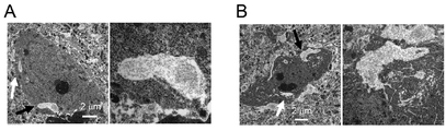
- FIG. 2-1D is a diagram showing the results of analysis of pSer46-MARKCS levels in the cerebral cortex by immunohistochemical staining.
- the pSer46-MARKS level in the C57BL / 6J mouse was similarly quantified, it was all 0 (zero).
- the arrow in the image on the left indicates the vacuole.
- FIG. 7A is a diagram showing the results of performing a Y-maze test at 5 months of age (FIG. 7B) or 6 months of age (FIG. 7C).
- FIG. 7A is a diagram showing the results of performing a Y-maze test at 5 months of age (FIG. 7B) or 6 months of age (FIG. 7C).
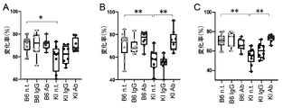
- Four types of FTLD model mice (PGRN R504X -KI mouse [FIG. 9A], TDP43 N267S- KI mouse [FIG.
- Q165X -KI mouse [Fig. 9D]) (“KI Ab” in the figure); 4 such FTLD model mice treated with control human IgG (“KI IgG” in the figure); 4 untreated FTLDs Model mice (“KI nt.” In the figure); or unadministered C57BL / 6J mice (“B6 nt.” In the figure); 6 months old (C57BL / 6J mice and CHMP2B, respectively).
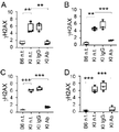
- Q165X -KI mouse [Fig. 17D]) (“KI Ab” in the figure); 4 such FTLD model mice treated with control human IgG (“KI IgG” in the figure); 4 untreated FTLDs Model mice (“KI nt.” In the figure); or unadministered C57BL / 6J mice (“B6 nt.” In the figure); 6 months old (C57BL / 6J mice and CHMP2B, respectively).
- Phosphoresis of synaptic instability-related proteins (pSer203-tau) (lane (5)) and expression levels of two synaptic-related proteins (PSD95 and VAMP2) (lanes (6) and (7), respectively); 2 Expression levels of a type of DNA damage-related protein ( ⁇ H2AX and 53BP1) (lanes (8) and (9), respectively); phosphorylation of one type of cell death-related protein (pSer46-MARKCS) level (lane (10)); 1 Phosphoresis (pSer77-Ku70) levels of a variety of DNA repair-related proteins (lane (11)); one protein aggregation marker (pSer409 / 410-TDP43) level (lane (12)); and the internal standard protein ⁇ . -The figure which shows the result of having analyzed the expression level of actin (lane (13)); by the Western blotting method.
- the prophylactic or therapeutic agent for frontotemporal lobar degeneration of the present invention is an agent containing the Human Monoclonal Antibody specified for the purpose of "preventing or treating frontotemporal lobar degeneration" (hereinafter, "" This is sometimes referred to as “preventive / therapeutic agent”).
- the preventive / therapeutic agent may be used alone as a pharmaceutical product (formulation), or may be further mixed with an additive and used as a form of a composition (pharmaceutical composition).
- frontotemporal lobar degeneration refers to non-Alzheimer's disease-type nerves that present with atrophy of the human frontotemporal lobe and temporal lobe in the early stage and lead to atrophy of the entire brain in the late stage. It means degenerative disease, and "frontotemporal lobar degeneration” includes three types of diseases classified according to clinical characteristics, specifically, frontotemporal dementia (FTD) and progressive nonfluent aphrodisiac. (PNFA) and progressive dementia (SD) are included, and pathologically, there are four types of diseases, specifically FTLD-Tau, depending on the type of protein that accumulates as an abnormal protein in the cell.
- FDD frontotemporal dementia
- PNFA progressive nonfluent aphrodisiac.
- SD progressive dementia
- frontotemporal lobar degeneration is due to “familial frontotemporal lobar degeneration” and “hereditary frontotemporal lobar degeneration” caused by gene mutations, and environmental factors such as lifestyle and stress. "Sporadic frontotemporal lobar degeneration” is also included.
- the above-mentioned "FTLD-Tau” is classified into “3R Tau” type, "4R Tau” type and “3/4R Tau” type according to the number of repeats of the microtubule binding region in the tau protein that is predominantly accumulated in the cell. being classified.
- the "3R Tau” type includes FTLD (Pick's disease) accompanied by Pick spheres, FTLD (FTLD-17) accompanied by MAPT (microtube-related protein tau) gene mutation, and the “4R Tau” type includes. Includes basal nuclear degeneration of the cerebral cortex, progressive supranuclear palsy, multisystem tauopathy with dementia, ginseng granular dementia (gluttonous granulopathy), FTLD with MAPT gene mutation (FTLD-17), etc.
- "3 / 4R Tau” type includes neurofibrillary tangle dementia, FTLD (FTLD-17) with MAPT gene mutation, and the like.
- the FTLD group in which tau is negative and has ubiquitin-positive inclusion bodies is referred to as "FTLD-U” and includes the above-mentioned FTLD-TDP, FTLD-UPS and FTLD-FUS.
- FTLD-TDP means a disease in which TDP43 (Transactive Response DNA Binding Protein of 43 kD) is positive among FTLD-U, and the disease is accompanied by a PGRN (progranulin) mutation such as PGRN R504X .
- FTLD-FUS means a disease in which TDP43 is negative and FUS (Fused In Sarcoma) is positive in FTLD-U
- FTLD-FUS means a nerve cell intermediate diameter filament inclusion body.
- Diseases, atypical FTLD-U, basic inclusion body diseases, FTLD with FUS mutations, etc. are included.
- FTLD-UPS means a disease which is a kind of FTLD-U in which TDP43 is negative, and such a disease includes FTLD accompanied by CHMP2B (Charged Multivesicular Body Protein 2B) mutation such as CHMP2B Q165X. And so on.
- CHMP2B Charge Multivesicular Body Protein 2B
- prevention of frontotemporal lobar degeneration includes not only the suppression of the onset and onset of frontotemporal lobar degeneration, but also the time of onset and delay in the onset of frontotemporal lobar degeneration. Is done.
- treatment of frontotemporal lobar degeneration includes not only recovery and improvement of lesions and symptoms of frontotemporal lobar degeneration including accumulation of abnormal proteins in cells, but also suppression of their progression. ..
- onset of frontotemporal lobar degeneration refers to memory impairment, higher brain dysfunction (aphasia, apraxia, agnosia, constitutional apraxia), and personality changes as determined by clinical diagnosis. It means the appearance of such symptoms and / or the appearance of apraxia of the brain as determined by diagnostic imaging.
- affected by frontotemporal lobar degeneration means a pathological change peculiar to frontotemporal lobar degeneration (for example, accumulation of abnormal protein in cells), although such a symptom does not occur. It also means the state in which.
- the human monoclonal antibody contained in the preventive / therapeutic agent has a high affinity for human HMGB1 protein and is the 46th serine (Ser46) in MARCKS, as demonstrated in this example described later. It has the effect of lowering / suppressing the phosphorylation (pSer46-MARCKS) level of HMGB1. Therefore, as the frontotemporal lobar degeneration to be prevented or treated by the preventive / therapeutic agent, frontotemporal lobar degeneration caused by HMGB1 and / or pSer46-MARKCS can be preferably exemplified.
- the human monoclonal antibody includes a heavy (H) chain variable region consisting of an amino acid sequence having at least 80% sequence identity with the amino acid sequence shown in SEQ ID NO: 7, and an amino acid sequence shown in SEQ ID NO: 8 and at least 80.
- a human monoclonal antibody containing a light (L) chain variable region consisting of an amino acid sequence having% or more sequence identity is preferable, and from an amino acid sequence having at least 80% or more sequence identity with the amino acid sequence shown in SEQ ID NO: 9.
- a human monoclonal antibody comprising a heavy chain comprising an amino acid sequence consisting of an amino acid sequence having at least 80% or more sequence identity with the amino acid sequence shown in SEQ ID NO: 10 is more preferable.
- HMGB1 High Mobility Group Box 1
- HMG1, HMG3, SBP-1, and HMG-1 As of human origin, it is typically a protein consisting of the amino acid sequence specified by NCBI Reference Sequence: NP_002119.1 (NCBI Reference Sequence: protein encoded by the nucleotide sequence specified by NM_002128.5).
- NCBI Reference Sequence protein encoded by the nucleotide sequence specified by NM_002128.5.
- the DNA sequence of a gene is mutated in nature (that is, non-artificially) due to the mutation or the like, and the amino acid sequence of the protein encoded by the mutation is also modified accordingly. Therefore, HMGB1 to which the Human Monoclonal Antibody binds includes a protein consisting of the amino acid sequence specified in NCBI reference sequence: NP_002119.1, as well as naturally occurring variants of such protein.
- the term "antibody that specifically binds to human HMGB1” means an antibody that recognizes and binds to human HMGB1 by a highly specific recognition mechanism between an antigen and an antibody.
- the human monoclonal antibody is preferably isolated.
- “separated” means that the antibody originally exists by artificially removing the antibody from the environment in which the antibody originally exists or expressing the antibody in an environment different from the environment in which the antibody originally exists. It means that it exists in a state different from the state it is doing. That is, the "separated antibody” is an antibody derived from a certain individual, and the tissue or body fluid (blood) in or derived from the body of the individual is not subjected to external manipulation (artificial manipulation).
- an antibody produced from an organism or a cell produced by an artificial operation for example, an antibody produced from a hybridoma
- Such "antibodies produced from organisms or cells produced by artificial manipulation” do not include antibodies produced from naturally occurring organisms or B cells (without artificial manipulation).
- the term "monoclonal antibody” means an antibody (including a functional fragment of an antibody) obtained from a substantially homogeneous population of antibodies. Monoclonal antibodies recognize a single determinant on an antigen.
- the Human Monoclonal Antibodies include classes and subclasses of human immunoglobulins, as well as the morphology of functional fragments of such antibodies.
- the class and subclass of the present human monoclonal antibody include IgG such as IgG1, IgG2, IgG3 and IgG4; IgA such as IGA1 and IGA2; IgD; IgE; IgM; and the like.
- the Human Monoclonal Antibodies typically include heavy chain CDR1, heavy chain CDR2, and heavy chain CDR3, light chain CDR1, light chain CDR2, and light chain CDR3, and each of these CDR1-3.
- a framework region (FR) is linked to the amino (N) and carboxyl (C) ends of the region.
- the heavy chain FR is the heavy chain FR1 linked to the N-terminal of the heavy chain CDR1; the heavy chain FR2 linked between the C-terminal of the heavy chain CDR1 and the N-terminal of the heavy chain CDR2; heavy chain FR2; Heavy chain FR3 linked between the C-terminal of chain CDR2 and the N-terminal of heavy chain CDR3; and heavy chain FR4 linked to the C-terminal of heavy chain CDR3; can be mentioned.
- the light chain FR is the light chain FR1 linked to the N-terminal of the light chain CDR1; the light chain FR2 linked between the C-terminal of the light chain CDR1 and the N-terminal of the light chain CDR2.
- the light chain FR3 linked between the C-terminal of the light chain CDR2 and the N-terminal of the light chain CDR3; and the light chain FR4 linked to the C-terminal of the light chain CDR3; can be mentioned.
- the heavy chain FR1 is at least 80% or more of the polypeptide consisting of the amino acid residues 1 to 30 of the amino acid sequence represented by (HF1) SEQ ID NO: 7 or the polypeptide (HF1').
- a polypeptide consisting of an amino acid sequence having the same sequence of Specifically, the heavy chain FR2 is at least 80% or more of the polypeptide consisting of the amino acid residues 36 to 49 of the amino acid sequence represented by (HF2) SEQ ID NO: 7 or the polypeptide (HF2').
- the heavy chain FR4 includes a polypeptide consisting of amino acid residues 105 to 115 of the amino acid sequence represented by (HF4) SEQ ID NO: 7, or (HF4') such a polypeptide at least 80% or more.
- HF4 amino acid sequence represented by (HF4) SEQ ID NO: 7, or (HF4')
- a polypeptide consisting of an amino acid sequence having the same sequence identity as above can be mentioned.
- the light chain FR1 is at least 80% or more of a polypeptide consisting of amino acid residues 1 to 23 of the amino acid sequence represented by (LF1) SEQ ID NO: 8 or (LF1') such a polypeptide.
- a polypeptide consisting of an amino acid sequence having the same sequence of Specifically, the light chain FR2 is at least 80% or more of a polypeptide consisting of amino acid residues 35 to 49 of the amino acid sequence represented by (LF2) SEQ ID NO: 8 or (LF2') such a polypeptide.
- a polypeptide consisting of an amino acid sequence having the same sequence of Specifically, the light chain FR3 is at least 80% or more of a polypeptide consisting of amino acid residues 57 to 88 of the amino acid sequence represented by (LF3) SEQ ID NO: 8 or (LF3') such a polypeptide.
- a polypeptide consisting of an amino acid sequence having the same sequence of Specifically, the light chain FR4 is at least 80% or more of a polypeptide consisting of amino acid residues 98 to 107 of the amino acid sequence represented by (LF4) SEQ ID NO: 8 or (LF4') such a polypeptide.
- a polypeptide consisting of an amino acid sequence having the same sequence identity as above can be mentioned.
- the Human Monoclonal Antibody is a human antibody.
- human antibody examples include human chimeric antibody, humanized antibody, fully human antibody and the like, and humanized antibody and fully human antibody can be preferably exemplified.
- human chimeric antibody connects a variable region of an antibody derived from a non-human animal (for example, a non-human mammal such as chicken, mouse, rat, cow) and a constant region of a human-derived antibody. It is an antibody that has been produced.
- the human chimeric antibody is, for example, an antibody derived from human bone marrow by immunizing an antigen against a non-human animal (preferably a non-human mammal) and cutting out an antibody variable region (variable region) that binds to the antigen from the gene of the mouse monoclonal antibody.
- Examples of the human constant region of the human chimeric antibody include C ⁇ 1, C ⁇ 2, C ⁇ 3, C ⁇ 4, C ⁇ , C ⁇ , C ⁇ 1, C ⁇ 2, and C ⁇ in the heavy chain, and C ⁇ and C ⁇ in the light chain. be able to.
- the amino acid sequences of these constant regions and the base sequences encoding them are known.
- one or more amino acids in a human-derived antibody constant region may be substituted, deleted, added, and / or inserted. can.
- humanized antibody refers to a human-derived gene sequence of an antigen-binding site (CDR) of an antibody derived from a non-human animal (for example, a non-human mammal such as chicken, mouse, rat, or cow). It is an antibody transplanted (CDR graphing) into an antibody gene, and a method for producing the antibody is known such as overlap extension PCR (for example, European Patent Application Publication No. 239400, European Patent Application Publication No. 125023). , International Publication No. 90/07861, International Publication No. 96/02576).
- the variable region of an antibody is usually composed of three CDRs sandwiched between four framework regions (FR).
- the CDR is substantially the region that determines the binding specificity of the antibody. While the amino acid sequences of CDRs are highly diverse, the amino acid sequences that make up FR often show high homology even among antibodies with different binding specificities. Therefore, it is generally said that the binding specificity of one antibody can be transplanted to another antibody by transplanting CDR. Further, from the viewpoint of maintaining the function of the CDR, in the transplantation of the non-human-derived CDR into the human FR, a human FR having high homology with the FR derived from the non-human animal is selected.
- the amino acids in the CDR not only recognize the antigen, but also coordinate with the amino acids of FR in the vicinity of the CDR and are involved in the maintenance of the loop structure of the CDR, so that they are adjacent to the CDR to be transplanted. It is preferable to use a human FR having an amino acid sequence having high homology with the amino acid sequence of the FR.
- a search system specialized for antibodies available on the Internet (http://www.bioinf.org.uk/abysis/). It can be done by using it. Mutations can be introduced into non-CDR sequences of non-human-derived antibodies to match the sequence of human FR thus obtained. Alternatively, if a gene (cDNA) encoding the amino acid sequence of human FR obtained by the search is available, a non-human-derived CDR may be introduced into the sequence. The introduction of mutations and the like can be performed using techniques known in the art such as nucleic acid synthesis and site-directed mutagenesis.
- the CDR forms a good antigen-binding site when linked via the CDR.
- the FR of a human-derived antibody such as that can be preferably selected. If necessary, the amino acid residue of FR can be replaced so that the CDR of the humanized antibody forms an appropriate antigen binding site according to the method described in the literature "Cancer Res, 1993, 53, 851-856". Further, by measuring and evaluating the affinity of the mutant antibody substituted with the amino acid for the antigen, a mutant FR sequence having a desired property can be selected.
- Fully human antibody means an antibody in which all sequences of the antibody are of human origin.
- Fully human antibodies can be made, for example, in transgenic mice engineered to express the genes for human heavy and light chain antibodies.
- Transgenic mice that produce human antibodies can be prepared, for example, according to the methods described in International Publication No. 02/4478, US Pat. No. 6,657,103 (Abgenix), and the like.
- the hybridoma cell line fused with B cells derived from a transgenic mouse that produces the desired antibody is then described, for example, in US Pat. No. 5,569,825; US Pat. No. 5,625,126; US Pat. No. 5,633,425; USA.
- the human monoclonal antibody includes not only an antibody consisting of the whole antibody but also a functional fragment that is a part (partial fragment) of the antibody and specifically recognizes the HMGB1 protein.
- Such functional fragments include Fab, Fab', F (ab') 2 , variable region fragment (Fv), disulfide bond Fv, single chain Fv (scFv), sc (Fv) 2 , diabody, polyspecificity. Examples thereof include antibodies and polymers thereof.
- Fab means a monovalent antigen-binding fragment of an immunoglobulin consisting of one light chain and a part of a heavy chain. It can be obtained by papain digestion of the antibody and by a recombinant method. "Fab'” differs from Fab by the addition of a few residues at the carboxy terminus of the heavy chain CH1 domain, including one or more cysteines in the hinge region of the antibody.
- F (ab') 2 is meant a divalent antigen-binding fragment of an immunoglobulin consisting of both light chain and both heavy chain moieties.
- a “variable region fragment (Fv)” is the smallest antibody fragment with complete antigen recognition and binding site. Fv is a dimer in which heavy chain variable regions and light chain variable regions are strongly linked by non-covalent bonds.
- a “single chain Fv (scFv)” comprises a heavy chain variable chain and a light chain variable region of an antibody, which are present in a single polypeptide chain.
- “Sc (Fv) 2” is a single chain obtained by binding two heavy chain variable regions and two light chain variable regions with a linker or the like.
- a “diabody” is a small antibody fragment with two antigen binding sites, the fragment containing a heavy chain variable region bound to a light chain variable region within the same polypeptide chain, each region being separate.
- a “multispecific antibody” is a monoclonal antibody that has binding specificity for at least two different antigens.
- two heavy chains can be prepared by co-expression of two immunoglobulin heavy chain / light chain pairs with different specificities.
- the H-chain CDRs 1-3 and L-chain CDRs 1-3 in the Human Monoclonal Antibodies include one or more amino acids in the amino acid sequences of SEQ ID NOs: 1-6 without reducing the desired activity (ie, affinity for HMGB1). Includes variants with substitutions, deletions, additions, and / or insertions.
- the Human Monoclonal Antibody containing such a modified version of CDR can be produced, for example, by introducing a mutation into the DNA encoding the antibody chain of human monoclonal antibody # 129, or by peptide synthesis.
- Modification of amino acids in regions other than the CDR of the antibody is considered to have a relatively small effect on the affinity with the antigen, but at present, the amino acids of the CDR are modified to modify the antigen.
- Techniques for screening antibodies with enhanced affinity for amino acids are known (eg, literature "PNAS, 102: 8466-8471 (2005)”; literature “Protein Engineering, Design & Selection, 21: 485-493 (2008)”; International Publication No.
- the term "plurality of amino acids” in “substitution, deletion, addition, and / or insertion of one or more amino acids” is preferably 10 amino acids or less, more preferably 5 amino acids or less, and even more preferably 3. It means within amino acids (for example, within 2 amino acids and 1 amino acid). Conservative substitutions are preferred for amino acid substitutions, deletions, additions, and insertions. Such “conservative substitution” means substituting with another amino acid residue having a chemically similar side chain. Groups of amino acid residues with chemically similar amino acid side chains are well known in the art.
- acidic amino acids (aspartic acid and glutamic acid), basic amino acids (lysine, arginine, histidine), and in the case of neutral amino acids, amino acids having a hydrocarbon chain (glycine, alanine, valine, leucine, isoleucine, proline), hydroxy groups.
- Amino acids with (serine / threonine), sulfur-containing amino acids (cysteine / methionine), amino acids with amide groups (asparagin / glutamic acid), amino acids with imino groups (proline), amino acids with aromatic groups (phenylalanine / tyrosine / It can be classified as tryptophan).
- a predetermined amino acid sequence and "at least 80% or more sequence identity” are preferably at least 85% or more sequence identity, more preferably at least 90% or more sequence identity, and even more preferably. Means sequence identity of at least 95% or higher (eg, 96% or higher, 97% or higher, 98% or higher, 99% or higher, 100%).
- Amino acid sequence homology can be determined using a BLASTP (amino acid level) program (see, eg, reference "J. Mol. Biol., 215: 403-410, 1990").
- the program is based on the algorithm BLAST (eg, reference “Proc.Natl.Acad.Sci.USA, 87: 2264-2268,1990", reference “Proc.Natl.Acad.Sci.USA, 90: 5873-5877,1993””. See).
- BLAST eg, reference "Proc.Natl.Acad.Sci.USA, 87: 2264-2268,1990", reference “Proc.Natl.Acad.Sci.USA, 90: 5873-5877,1993””.
- the amino acid sequence is analyzed using the Gapped BLAST program, it can be performed according to the method described in the document "Nucleic Acids Res. 25: 3389-3402, 1997”.
- the default parameters of each program are used. Specific methods of these analysis methods are known.
- the Human Monoclonal Antibodies include those modified by the post-translational process of the antibody, such as changing the number and position of glycosylation sites. Such variants can improve ADCC activity (antibody-dependent cellular cytotoxicity) of the antibody.
- Glycosylation of an antibody is typically N- or O-linked. Glycosylation of an antibody is highly dependent on the host cell used to express the antibody. Modification of the glycosylation pattern can be performed by a known method such as introduction or deletion of a specific enzyme involved in sugar production (Japanese Patent Laid-Open No. 2008-113663, US Pat. No. 5,047,335, US Pat. No. 5,510,261). US Pat. No. 5,278,299, International Publication No. 99/54342).
- the human monoclonal antibody is deamidated by substituting an amino acid that is deamidated for the purpose of increasing the stability of the antibody or an amino acid adjacent to the amino acid that is deamidated with another amino acid. It may be one in which the formation is suppressed, or one in which glutamate is replaced with another amino acid to increase the stability of the antibody.
- the human monoclonal antibody can be produced by a known hybridoma method or a known recombinant DNA method.
- Typical examples of the hybridoma method include the Caller and Milstein methods (Reference "Nature, 256: 495 (1975)").
- the antibody-producing cells used in the cell fusion step in this method are animals immunized with an antigen (HMGB1 protein, its partial peptide, a protein in which Fc protein or the like is fused to them, or a cell expressing these).
- an antigen HMGB1 protein, its partial peptide, a protein in which Fc protein or the like is fused to them, or a cell expressing these.
- spleen cells, lymph node cells, peripheral blood leukocytes, etc. of mice, rats, hamsters, rabbits, monkeys, goats and other mammals For example, spleen cells, lymph node cells, peripheral blood leukocytes, etc. of mice, rats, hamsters, rabbits,
- antibody-producing cells obtained by allowing an antigen to act in a medium against the above-mentioned cells or lymphocytes isolated in advance from a non-immune animal.
- Various known cell lines can be used as myeloma cells.
- the antibody-producing cells and myeloma cells may be of different animal species origin as long as they can be fused, but are preferably of the same animal species origin.
- Hybridomas are produced, for example, by cell fusion between spleen cells obtained from antigen-immunized mice and mouse myeloma cells, and subsequent screening produces hybridomas that produce monoclonal antibodies specific for the HMGB1 protein.
- Human monoclonal antibodies that specifically bind to the HMGB1 protein can be obtained by culturing hybridomas and from the ascites of mammals to which the hybridomas have been administered.
- the antibody gene encoding the human monoclonal antibody is cloned from a hybridoma, B cell, etc., incorporated into an appropriate vector, and this is incorporated into a host cell (for example, a mammalian cell line such as HEK cell, Escherichia coli, yeast). It is a method of introducing the human monoclonal antibody into a cell, an insect cell, a plant cell, etc.) to produce the human monoclonal antibody as a recombinant antibody (see, for example, "Eur.J. Biochem. 192: 767-775 (1990)").
- the antibody gene encoding the human monoclonal antibody may be separately incorporated into the expression vector to transform the host cell, and the heavy chain and the light chain are encoded.
- the antibody gene may be integrated into a single expression vector to transform the host cell (see International Publication No. 94/11523).
- the human monoclonal antibody can be obtained in a substantially pure and uniform form by culturing the host cell, separating and purifying it in the host cell or from the culture solution. For the separation and purification of the antibody, the method used in the purification of a normal polypeptide can be used.
- transgenic animal cow, goat, sheep, pig, etc.
- human monoclonal antibody gene derived from the milk of the transgenic animal can be used. It is also possible to obtain a large amount of monoclonal antibody.
- the additive is a compounding component such as a pharmaceutically acceptable carrier, a binder, a stabilizer, an excipient, a diluent, a pH buffer, an isotonic agent, a solubilizing agent, and a nutritional agent.
- a pharmaceutically acceptable carrier include when the preventive / therapeutic agent is a powder, for example, mannitol, lactose, saccharose, human albumin, etc., and when the preventive / therapeutic agent is a liquid.
- physiological saline, water for injection, phosphate buffer, aluminum hydroxide and the like can be mentioned.
- the subject of administration of the preventive / therapeutic agent may be a subject (human) who requires prevention or treatment of frontotemporal lobar degeneration, and is diagnosed as having a high risk of developing frontotemporal lobar degeneration, for example. Examples include those who have been treated and patients who have developed frontotemporal lobar degeneration.
- Examples of the administration method of the preventive / therapeutic agent include injection method (for example, subcutaneous administration, intramuscular administration, intravenous administration, intraarterial administration), oral mucosal administration, rectal administration, and local administration to the brain (for example, subcutaneous administration, intramuscular administration, intravenous administration, intraarterial administration). Intracerebroventricular administration), transdermal administration, oral administration and the like can be mentioned, and intravenous administration can be preferably exemplified.
- the dose of the Human Monoclonal Antibody in the Prophylactic / Therapeutic Agent may vary depending on the age, body weight, gender, health condition, and degree of symptom progression of the subject, but is usually 0. per kg of body weight per day for adults. It is 1 to 1000 mg, preferably 1 to 100 mg.
- the prophylactic / therapeutic agent may be used in combination with a known drug used for the prevention or treatment of frontotemporal lobar degeneration.
- anti-human HMGB1 antibody As the anti-human HMGB1 antibody, the human monoclonal antibody (human IgG antibody) # 129 prepared in the example of the international patent application (PCT-JP2019-36926 [Pamphlet of International Publication No. 2020/059847]) was used. .. The amino acid sequences of the H and L chains of human monoclonal antibody # 129 are shown in Tables 1 and 2, respectively.
- the underlined region in the table indicates the H chain V region (SEQ ID NO: 7).
- the three regions surrounded by squares in the table indicate H chain CDR1 (SEQ ID NO: 1), H chain CDR2 (SEQ ID NO: 2), and H chain CDR3 (SEQ ID NO: 3) in order from the amino-terminal side.
- the underlined region in the table indicates the L chain V region (SEQ ID NO: 8).
- the three regions surrounded by squares in the table indicate L-chain CDR1 (SEQ ID NO: 4), L-chain CDR2 (SEQ ID NO: 5), and L-chain CDR3 (SEQ ID NO: 6) in order from the amino-terminal side.
- FTLD model mice 4 types of FTLD model mice (VCP T262A -KI mouse, PGRN R504X -KI mouse, CHMP2B Q165X- KI mouse, and TDP43 N267S- KI mouse) (referred to as "4 types FTLD model mouse" in the present specification).
- PGRN R504X- KI mice were prepared according to the method described in the document "Nat. Commun., 2018 9, 433".
- a targeting vector for producing VCP T262A- KI mice was constructed. Specifically, the PCR product of a 5.4 kb NotI-XhoI fragment amplified from a Bac clone (ID: RP23-111G9 or RP23-124L1) was subcloned into the PspOMI-XhoI site of pBS-DTA. Similarly, a 2.9 kb BamHI-NotI fragment amplified from the same Bac clone was subcloned into pBS-LNL (-) with a Neo cassette (loxP-Neo-loxP).
- a 4.5 kb fragment (1.6 kb Neo cassette + 2.9 kb fragment) was excised by Notl treatment, and a 0.8 kb XhoI-Smal fragment having a T262A mutation was amplified by PCR from the same Bac clone, and XhoI and XhoI and SmaI processing was performed.
- Such 0.8 kb and 4.5 kb fragments were subcloned into the Xho I-Not I site of the targeting vector. After linearizing the targeting vector by NotI treatment, such fragments were electroporated into C57BL / 6J background ES cells.
- VCP T262A -KI mice were selected from non-transgenic littermates.
- a targeting vector for producing CHMP2B Q165X- KI mice was constructed. Specifically, the PCR product of the 5.7 kb ClaI-SalI fragment amplified from the Bac clone (ID: RP23-13H5 or RP23-273E18) is subcloned into the ClaI-SalI site of pBS-DTA (manufactured by Unitech). did. A 3.0 kb SacII-NotI fragment was amplified from a Bac clone and subcloned into pBS-LNL (+) with a Neo cassette (loxP-Neo-loxP).
- SacII-NotI and 4.6 kb fragments (1.6 kb Neo cassette + 3.0 kb fragment) were subcloned into the SacII-ClaI site of the targeting vector.
- primer set primer 3 [5'-TGGATTTTATTTATGTCTGAATGTG-3'; SEQ ID NO: 13] and primer 4 [5'-ATAAGCAACTTCACAAGGCATCTTA-3'; SEQ ID NO: 14]) that amplifies the DNA fragment containing the LoxP sequence.
- CHMP2B Q165X- KI mice were selected from non-transgenic litters.
- a targeting vector for producing TDP43 N267S- KI mice was constructed. Specifically, a 5.9 kb ClaI-SalI fragment was amplified from a Bac clone (ID: RP23-364M1 or RP23-331P21) and subcloned into the ClaI-SalI site of pBS-TK (manufactured by Unitech). A 2.7 kb SacII-NotI fragment was amplified from a Bac clone and subcloned into pA-LNL (+) with a Neo cassette (loxP-Neo-loxP).
- a 4.3 kb fragment (1.6 kb Neo cassette + 2.7 kb fragment) was subcloned into the SacII-ClaI site of the targeting vector.
- primer set primer 5 [5'-ACAGTTGGGTGTGATAGCAGGTACT-3'; SEQ ID NO: 15] and primer 6 [5'-TCGAGAATTACAGGAATGTATCATC-3'; SEQ ID NO: 16], which amplifies a DNA fragment containing a LoxP sequence.
- TDP43 N267S- KI mice were selected from non-transgenic litters.
- an antibody reaction using a secondary antibody (see Table 3) against various primary antibodies was performed at room temperature for 1 hour, and after washing 3 times with PBS, 0.2 ⁇ g / mL DAPI (4'). , 6-diamidino-2-phenylindole), stain the cell nuclei, and use a confocal microscope (FV1200IX83, manufactured by Olympus) to obtain fluorescent images derived from the above 11 types of proteins and fluorescent images derived from the cell nuclei. Obtained.
- the number of pSer46-MARKS-positive cells (cells) per region of 143 ⁇ m ⁇ 143 ⁇ m was calculated (see FIGS. 2-1 and 9).
- KDEL signal regions were measured for MAP2-positive cells, which are markers of mature neurons, based on DAPI fluorescence images, KDEL fluorescence images, and MAP2 fluorescence images (see FIG. 10).
- the ⁇ H2AX signal region was measured for MAP2-positive cells based on the DAPI fluorescence image, the ⁇ H2AX fluorescence image, and the MAP2 fluorescence image (see FIG. 11).
- 53BP1 signal regions were measured for MAP2-positive cells based on DAPI fluorescence images, 53BP1 fluorescence images, and MAP2 fluorescence images (see FIG. 12).
- the pSer409 / 410-TDP43 signal region in the ubiquitin-positive encapsulater was measured based on the DAPI fluorescence image, the pSer409 / 410-TDP43 fluorescence image, and the ubiquitin fluorescence image (see FIG. 13).
- the p62 signal region in the inclusion body was measured based on the DAPI fluorescence image and the p62 fluorescence image (see FIG. 14).
- the proportion of cells in which the VAMP2 signal and the PSD95 signal were co-localized was measured based on the DAPI fluorescence image, the VAMP2 fluorescence fluorescence image, and the PSD95 fluorescence image (see FIG. 15). Further, the proportion of cells in which the pSer203-tau signal and the PSD95 signal were co-localized was measured based on the DAPI fluorescence image, the pSer203-tau fluorescence image, and the PSD95 fluorescence image (see FIG. 16).
- Y-maze test a Y-maze (manufactured by Ohara Medical Industry Co., Ltd.) consisting of three identical arms having the same angle between the arms was used. Place the mouse on the end of one arm, let it move freely in the maze for 8 minutes, and the total number of moves from one arm to another, the number of times it entered a new arm different from the previous arm. By dividing, the rate of spontaneous change was calculated (displayed as the rate of change [Alternation Rate]; vertical axis in FIGS. 4 to 7).
- the freezing response was measured in the same chamber without foot shock 24 hours after receiving the conditioning (65 dB white noise, 30 seconds + foot shock, 0.4 mA, 2 seconds) (total freezing). Displayed as a percentage [Total Freeze percent]; vertical axis in FIG. 8).
- the KD of the mouse monoclonal antibody 2C8C was 1.95 ⁇ 10 -9 M, whereas the KD of the human monoclonal antibody # 129 of the present invention was 3.79 ⁇ 10 -11 M.
- the human monoclonal antibody # 129 of the present invention has an affinity for human HMGB1. It shows that it is an antibody with extremely high sex.
- pSer46-MARKS originates from an early stage before the onset of Alzheimer's disease, and HMGB1 leaked from the cell due to neuronal necrosis induces phosphorylation of MARCKS and induces degeneration of neurites (reference "SCIENTIFIC”). REPORT, 2016 Aug 25; 6: 31895 "). Therefore, whether or not MARCKS phosphorylation and neuronal necrosis occurred even in the early stage of FTLD onset was analyzed using 4 types of FTLD model mice aged 1 to 12 months.
- the four-kind FTLD model mouse had an increased signal region derived from DNA damage markers ( ⁇ H2AX and 53BP1) in the cerebral cortex as compared with the C57BL / 6J mouse, whereas the administration of the human monoclonal antibody # 129 of the present invention increased the signal region. It was shown that the increase in the signal regions derived from ⁇ H2AX and 53BP1 was significantly suppressed to the same extent as in C57BL / 6J mice (see FIGS. 11 and 12).
- the four FTLD model mice had an increased signal region derived from protein aggregation markers (pSer409 / 410-TDP43 and p62) in the cerebral cortex as compared with C57BL / 6J mice, whereas the human monoclonal antibody # of the present invention was used.
- Administration of 129 was shown to significantly suppress such increase in pSer409 / 410-TDP43 and p62-derived signaling regions (see FIGS. 13 and 14).
- the present inventors predicted synaptic disorders using the co-localization of VAMP2 and PSD95 and the co-localization of pSer203-tau and PSD95 as indicators by the analysis of phosphorylated proteome. Therefore, as a result of analyzing synaptic disorders using these co-localizations as an index, the co-localization of VAMP2 and PSD95 in the cerebral cortex was reduced in the four-kind FTLD model mice compared with the C57BL / 6J mice. , The administration of the human monoclonal antibody # 129 of the present invention has been shown to significantly suppress such reduction (see FIG. 15).
- the two types of phosphorylation levels (pSer319-PKC ⁇ and pSer643-PKC ⁇ ) predicted by dynamic network analysis in> 2SD) were higher in the four-kind FTLD model mouse than in the C57BL / 6J mouse. It was shown that administration of the human monoclonal antibody # 129 of the present invention to four FTLD model mice suppressed such increases in pSer319-PKC ⁇ and pSer643-PKC ⁇ levels (lane (1) in FIG. 17 and). See (3).
- phosphorylation of synaptic instability-related protein was increased in 4 FTLD model mice as compared with C57BL / 6J mice, whereas the human monoclonal antibody # 129 of the present invention was 4 Administration to seed FTLD model mice was shown to suppress such increases in pSer203-tau levels (see lane (5) in FIG. 17).
- the expression level of synapse-related proteins was lower in the 4-type FTLD model mouse than in the C57BL / 6J mouse, whereas the human monoclonal antibody # 129 of the present invention was used in the 4-type FTLD model. It was shown that administration to mice suppressed such a decrease in the expression levels of PSD95 and VAMP2 (see lanes (6) and (7) in FIG. 17).
- the expression level of DNA damage-related proteins ( ⁇ H2AX and 53BP1) was increased in the 4-type FTLD model mouse as compared with the C57BL / 6J mouse, whereas the expression level of the human monoclonal antibody # 129 of the present invention was increased in the 4-type FTLD. It was shown that administration to model mice suppressed such an increase in the expression levels of ⁇ H2AX and 53BP1 (see lanes (8) and (9) in FIG. 17).
- the phosphorylation (pSer46-MARKKS) level of the cell death-related protein was increased in the four FTLD model mice as compared with the C57BL / 6J mouse, whereas the human monoclonal antibody # 129 of the present invention was used in four species.
- Administration to FTLD model mice was shown to suppress such increases in pSer46-MARKS levels (see lane (10) in FIG. 17).
- the phosphorylation (pSer77-Ku70) level of the DNA repair-related protein was increased in the 4-type FTLD model mouse as compared with the C57BL / 6J mouse, whereas the 4-type human monoclonal antibody # 129 of the present invention was used.
- Administration to FTLD model mice was shown to suppress such increases in pSer77-Ku70 levels (see lane (11) in FIG. 17).
- the level of the protein aggregation marker (pSer409 / 410-TDP43) was increased in the 4-type FTLD model mouse as compared with the C57BL / 6J mouse, whereas the level of the human monoclonal antibody # 129 of the present invention was increased in the 4-type FTLD.
- Administration to model mice was shown to suppress such increases in pSer409 / 410-TDP43 levels (see lane (12) in FIG. 17).
- the present invention contributes to the prevention and / or treatment of FTLD.
Abstract
Description
〔1〕ヒトHMGB1に特異的に結合し、かつ、
配列番号1に示されるアミノ酸配列又は該アミノ酸配列において1若しくは複数のアミノ酸が置換、欠失、付加、及び/又は挿入されているアミノ酸配列からなる重鎖相補性決定領域(CDR)1;配列番号2に示されるアミノ酸配列又は該アミノ酸配列において1若しくは複数のアミノ酸が置換、欠失、付加、及び/又は挿入されているアミノ酸配列からなる重鎖CDR2;及び配列番号3に示されるアミノ酸配列又は該アミノ酸配列において1若しくは複数のアミノ酸が置換、欠失、付加、及び/又は挿入されているアミノ酸配列からなる重鎖CDR3;と、
配列番号4に示されるアミノ酸配列又は該アミノ酸配列において1若しくは複数のアミノ酸が置換、欠失、付加、及び/又は挿入されているアミノ酸配列からなる軽鎖CDR1;配列番号5に示されるアミノ酸配列又は該アミノ酸配列において1若しくは複数のアミノ酸が置換、欠失、付加、及び/又は挿入されているアミノ酸配列からなる軽鎖CDR2;及び配列番号6に示されるアミノ酸配列又は該アミノ酸配列において1若しくは複数のアミノ酸が置換、欠失、付加、及び/又は挿入されているアミノ酸配列からなる軽鎖CDR3;とを含む
ヒトモノクローナル抗体を含む、前頭側頭葉変性症の予防剤又は治療剤。
〔2〕ヒトモノクローナル抗体が、配列番号7に示されるアミノ酸配列と少なくとも80%以上の配列同一性を有するアミノ酸配列からなる重鎖可変領域と、配列番号8に示されるアミノ酸配列と少なくとも80%以上の配列同一性を有するアミノ酸配列からなる軽鎖可変領域とを含む、上記〔1〕に記載の予防剤又は治療剤。
〔3〕ヒトモノクローナル抗体が、配列番号9に示されるアミノ酸配列と少なくとも80%以上の配列同一性を有するアミノ酸配列からなる重鎖と、配列番号10に示されるアミノ酸配列と少なくとも80%以上の配列同一性を有するアミノ酸配列からなる軽鎖とを含む、上記〔1〕又は〔2〕に記載の予防剤又は治療剤。
〔4〕静脈内投与される、上記〔1〕~〔3〕のいずれかに記載の予防剤又は治療剤。 That is, the present invention is as follows.
[1] It specifically binds to human HMGB1 and
Heavy chain complementarity determination region (CDR) 1 consisting of the amino acid sequence shown in SEQ ID NO: 1 or an amino acid sequence in which one or more amino acids are substituted, deleted, added, and / or inserted in the amino acid sequence; SEQ ID NO: Heavy chain CDR2 consisting of the amino acid sequence shown in 2 or the amino acid sequence in which one or more amino acids are substituted, deleted, added, and / or inserted; and the amino acid sequence shown in SEQ ID NO: 3 or the amino acid sequence thereof. Heavy chain CDR3 consisting of an amino acid sequence in which one or more amino acids are substituted, deleted, added, and / or inserted in the amino acid sequence;
Light chain CDR1 consisting of the amino acid sequence shown in SEQ ID NO: 4 or an amino acid sequence in which one or more amino acids are substituted, deleted, added, and / or inserted in the amino acid sequence; the amino acid sequence shown in SEQ ID NO: 5 or Light chain CDR2 consisting of an amino acid sequence in which one or more amino acids are substituted, deleted, added, and / or inserted in the amino acid sequence; and the amino acid sequence shown in SEQ ID NO: 6 or one or more in the amino acid sequence. A prophylactic or therapeutic agent for frontal temporal lobe degeneration, comprising a human monoclonal antibody comprising a light chain CDR3; consisting of an amino acid sequence in which an amino acid is substituted, deleted, added, and / or inserted.
[2] The human monoclonal antibody has a heavy chain variable region consisting of an amino acid sequence having at least 80% or more sequence identity with the amino acid sequence shown in SEQ ID NO: 7, and at least 80% or more of the amino acid sequence shown in SEQ ID NO: 8. The prophylactic or therapeutic agent according to the above [1], which comprises a light chain variable region consisting of an amino acid sequence having the same sequence.
[3] The human monoclonal antibody has a heavy chain consisting of an amino acid sequence having at least 80% or more sequence identity with the amino acid sequence shown in SEQ ID NO: 9, and a sequence of at least 80% or more with the amino acid sequence shown in SEQ ID NO: 10. The prophylactic or therapeutic agent according to the above [1] or [2], which comprises a light chain consisting of an amino acid sequence having the same identity.
[4] The prophylactic or therapeutic agent according to any one of the above [1] to [3], which is administered intravenously.
ヒトHMGB1に特異的に結合し、かつ、
配列番号1に示されるアミノ酸配列又は該アミノ酸配列において1若しくは複数のアミノ酸が置換、欠失、付加、及び/又は挿入されているアミノ酸配列からなる重鎖相補性決定領域(CDR)1;配列番号2に示されるアミノ酸配列又は該アミノ酸配列において1若しくは複数のアミノ酸が置換、欠失、付加、及び/又は挿入されているアミノ酸配列からなる重鎖CDR2;及び配列番号3に示されるアミノ酸配列又は該アミノ酸配列において1若しくは複数のアミノ酸が置換、欠失、付加、及び/又は挿入されているアミノ酸配列からなる重鎖CDR3;と、
配列番号4に示されるアミノ酸配列又は該アミノ酸配列において1若しくは複数のアミノ酸が置換、欠失、付加、及び/又は挿入されているアミノ酸配列からなる軽鎖CDR1;配列番号5に示されるアミノ酸配列又は該アミノ酸配列において1若しくは複数のアミノ酸が置換、欠失、付加、及び/又は挿入されているアミノ酸配列からなる軽鎖CDR2;及び配列番号6に示されるアミノ酸配列又は該アミノ酸配列において1若しくは複数のアミノ酸が置換、欠失、付加、及び/又は挿入されているアミノ酸配列からなる軽鎖CDR3;とを含む
ヒトモノクローナル抗体(以下、「本件ヒトモノクローナル抗体」ということがある)を、前頭側頭葉変性症の予防又は治療を必要とする対象に投与するステップを含む、前頭側頭葉変性症を予防又は治療する方法;や、
前頭側頭葉変性症の予防剤又は治療剤として使用するための本件ヒトモノクローナル抗体;や、
前頭側頭葉変性症の予防又は治療における使用のための本件ヒトモノクローナル抗体;や、
前頭側頭葉変性症の予防剤又は治療剤を製造するための、本件ヒトモノクローナル抗体の使用;
を挙げることができる。 Further, as another embodiment of the present invention,
It specifically binds to human HMGB1 and
Heavy chain complementarity determination region (CDR) 1 consisting of the amino acid sequence shown in SEQ ID NO: 1 or an amino acid sequence in which one or more amino acids are substituted, deleted, added, and / or inserted in the amino acid sequence; SEQ ID NO: Heavy chain CDR2 consisting of the amino acid sequence shown in 2 or the amino acid sequence in which one or more amino acids are substituted, deleted, added, and / or inserted; and the amino acid sequence shown in SEQ ID NO: 3 or the amino acid sequence thereof. Heavy chain CDR3 consisting of an amino acid sequence in which one or more amino acids are substituted, deleted, added, and / or inserted in the amino acid sequence;
Light chain CDR1 consisting of the amino acid sequence shown in SEQ ID NO: 4 or an amino acid sequence in which one or more amino acids are substituted, deleted, added, and / or inserted in the amino acid sequence; the amino acid sequence shown in SEQ ID NO: 5 or Light chain CDR2 consisting of an amino acid sequence in which one or more amino acids are substituted, deleted, added, and / or inserted in the amino acid sequence; and the amino acid sequence shown in SEQ ID NO: 6 or one or more in the amino acid sequence. A human monoclonal antibody (hereinafter, also referred to as “human monoclonal antibody”) containing a light chain CDR3; consisting of an amino acid sequence in which an amino acid is substituted, deleted, added, and / or inserted, is used in the frontotemporal lobe. Methods of preventing or treating frontotemporal lobar degeneration, including the step of administering to a subject in need of prevention or treatment of degeneration;
The Human Monoclonal Antibody for use as a prophylactic or therapeutic agent for frontotemporal lobar degeneration;
Human Monoclonal Antibodies for use in the prevention or treatment of frontotemporal lobar degeneration;
Use of the Human Monoclonal Antibodies to Produce Prophylactic or Therapeutic Agents for Frontotemporal Lobe Degeneration;
Can be mentioned.
上記重鎖FR2としては、具体的には、(HF2)配列番号7で示されるアミノ酸配列の36~49番目のアミノ酸残基からなるポリペプチド、又は(HF2’)かかるポリペプチドと少なくとも80%以上の配列同一性を有するアミノ酸配列からなるポリペプチドを挙げることができ、
上記重鎖FR3としては、具体的には、(HF3)配列番号7で示されるアミノ酸配列の67~98番目のアミノ酸残基からなるポリペプチド、配列番号11で示されるアミノ酸配列の67~98番目のアミノ酸残基からなるポリペプチド、又は(HF3’)かかるポリペプチドと少なくとも80%以上の配列同一性を有するアミノ酸配列からなるポリペプチドを挙げることができ、
上記重鎖FR4としては、具体的には、(HF4)配列番号7で示されるアミノ酸配列の105~115番目のアミノ酸残基からなるポリペプチド、又は(HF4’)かかるポリペプチドと少なくとも80%以上の配列同一性を有するアミノ酸配列からなるポリペプチドを挙げることができる。 Specifically, the heavy chain FR1 is at least 80% or more of the polypeptide consisting of the
Specifically, the heavy chain FR2 is at least 80% or more of the polypeptide consisting of the amino acid residues 36 to 49 of the amino acid sequence represented by (HF2) SEQ ID NO: 7 or the polypeptide (HF2'). A polypeptide consisting of an amino acid sequence having the same sequence of
Specifically, the heavy chain FR3 is a polypeptide consisting of the 67th to 98th amino acid residues of the amino acid sequence represented by (HF3) SEQ ID NO: 7, and the 67th to 98th amino acid sequence represented by SEQ ID NO: 11. Examples thereof include a polypeptide consisting of amino acid residues of (HF3'), or a polypeptide consisting of an amino acid sequence having at least 80% or more sequence identity with such a polypeptide.
Specifically, the heavy chain FR4 includes a polypeptide consisting of amino acid residues 105 to 115 of the amino acid sequence represented by (HF4) SEQ ID NO: 7, or (HF4') such a polypeptide at least 80% or more. A polypeptide consisting of an amino acid sequence having the same sequence identity as above can be mentioned.
上記軽鎖FR2としては、具体的には、(LF2)配列番号8で示されるアミノ酸配列の35~49番目のアミノ酸残基からなるポリペプチド、又は(LF2’)かかるポリペプチドと少なくとも80%以上の配列同一性を有するアミノ酸配列からなるポリペプチドを挙げることができ、
上記軽鎖FR3としては、具体的には、(LF3)配列番号8で示されるアミノ酸配列の57~88番目のアミノ酸残基からなるポリペプチド、又は(LF3’)かかるポリペプチドと少なくとも80%以上の配列同一性を有するアミノ酸配列からなるポリペプチドを挙げることができ、
上記軽鎖FR4としては、具体的には、(LF4)配列番号8で示されるアミノ酸配列の98~107番目のアミノ酸残基からなるポリペプチド、又は(LF4’)かかるポリペプチドと少なくとも80%以上の配列同一性を有するアミノ酸配列からなるポリペプチドを挙げることができる。 Specifically, the light chain FR1 is at least 80% or more of a polypeptide consisting of
Specifically, the light chain FR2 is at least 80% or more of a polypeptide consisting of amino acid residues 35 to 49 of the amino acid sequence represented by (LF2) SEQ ID NO: 8 or (LF2') such a polypeptide. A polypeptide consisting of an amino acid sequence having the same sequence of
Specifically, the light chain FR3 is at least 80% or more of a polypeptide consisting of amino acid residues 57 to 88 of the amino acid sequence represented by (LF3) SEQ ID NO: 8 or (LF3') such a polypeptide. A polypeptide consisting of an amino acid sequence having the same sequence of
Specifically, the light chain FR4 is at least 80% or more of a polypeptide consisting of amino acid residues 98 to 107 of the amino acid sequence represented by (LF4) SEQ ID NO: 8 or (LF4') such a polypeptide. A polypeptide consisting of an amino acid sequence having the same sequence identity as above can be mentioned.
の方法に従って行うことができる。BLASTとGapped BLASTプログラムを用いる場合には、各プログラムのデフォルトパラメーターを用いる。これらの解析方法の具体的な手法は公知である。 In the present specification, a predetermined amino acid sequence and "at least 80% or more sequence identity" are preferably at least 85% or more sequence identity, more preferably at least 90% or more sequence identity, and even more preferably. Means sequence identity of at least 95% or higher (eg, 96% or higher, 97% or higher, 98% or higher, 99% or higher, 100%). Amino acid sequence homology can be determined using a BLASTP (amino acid level) program (see, eg, reference "J. Mol. Biol., 215: 403-410, 1990"). The program is based on the algorithm BLAST (eg, reference "Proc.Natl.Acad.Sci.USA, 87: 2264-2268,1990", reference "Proc.Natl.Acad.Sci.USA, 90: 5873-5877,1993"". See). When analyzing the amino acid sequence by BLASTP, the parameters are, for example, score = 50 and wordlength = 3. Further, when the amino acid sequence is analyzed using the Gapped BLAST program, it can be performed according to the method described in the document "Nucleic Acids Res. 25: 3389-3402, 1997". When using BLAST and Gapped BLAST programs, the default parameters of each program are used. Specific methods of these analysis methods are known.
[抗ヒトHMGB1抗体]
本実施例において、抗ヒトHMGB1抗体として、国際特許出願(PCT-JP2019-36926[国際公開2020/059847号パンフレット])の実施例で作製したヒトモノクローナル抗体(ヒト型IgG抗体)#129を使用した。ヒトモノクローナル抗体#129のH鎖及びL鎖のアミノ酸配列を、それぞれ表1及び2に示す。なお、ヒトモノクローナル抗体#129をマウスへ皮下投与し、脳内及び血清中の当該抗体濃度をELISA法にて測定した結果、投与した2~8%のヒトモノクローナル抗体#129が、血液脳関門(BBB)を通過し、脳内に移行していることが確認された。 1. 1. Materials and methods [anti-human HMGB1 antibody]
In this example, as the anti-human HMGB1 antibody, the human monoclonal antibody (human IgG antibody) # 129 prepared in the example of the international patent application (PCT-JP2019-36926 [Pamphlet of International Publication No. 2020/059847]) was used. .. The amino acid sequences of the H and L chains of human monoclonal antibody # 129 are shown in Tables 1 and 2, respectively. As a result of subcutaneously administering human monoclonal antibody # 129 to mice and measuring the antibody concentration in the brain and serum by the ELISA method, 2 to 8% of the administered human monoclonal antibody # 129 was found at the blood-brain barrier ( It was confirmed that it passed through BBB) and migrated into the brain.


本発明のヒトモノクローナル抗体#129と、比較対照として国際公開2018/030405号パンフレットに記載のマウスモノクローナル抗体2C8Cとを用いて、ヒトHMGB1タンパク質との相互作用を解析した。具体的には、本発明のヒトモノクローナル抗体#129、又はマウスモノクローナル抗体2C8Cを固定化したセンサーチップと、アナライトとしてヒトHMGB1タンパク質とを用い、表面プラズモン共鳴測定装置(「Biacore T100」;GE healthcare社製)によって経時的に相互作用を測定し、抗体親和性の指標である平衡解離定数(KD値)を算出した(図1参照)。 [Analysis of antibody affinity by surface plasmon resonance method (SPR)]
The interaction with human HMGB1 protein was analyzed using the human monoclonal antibody # 129 of the present invention and the mouse monoclonal antibody 2C8C described in International Publication No. 2018/030405 as a comparative control. Specifically, a surface plasmon resonance measuring device (“Biacore T100”; GE healthcare) using a sensor chip on which the human monoclonal antibody # 129 of the present invention or the mouse monoclonal antibody 2C8C is immobilized and a human HMGB1 protein as an analyst is used. The interaction was measured over time by (manufactured by the company), and the equilibrium dissociation constant (KD value), which is an index of antibody affinity, was calculated (see FIG. 1).
4種類のFTLDモデルマウス(VCPT262A-KIマウス、PGRNR504X-KIマウス、CHMP2BQ165X-KIマウス、及びTDP43N267S-KIマウス)(本明細書において、「4種FTLDモデルマウス」ということがある)のうち、PGRNR504X-KIマウスについては、文献「Nat. Commun., 2018 9, 433」に記載の方法に従って作製した。 [Preparation of 4 types of FTLD model mice]
4 types of FTLD model mice (VCP T262A -KI mouse, PGRN R504X -KI mouse, CHMP2B Q165X- KI mouse, and TDP43 N267S- KI mouse) (referred to as "4 types FTLD model mouse" in the present specification). Of these, PGRN R504X- KI mice were prepared according to the method described in the document "Nat. Commun., 2018 9, 433".
各種月齢の4種FTLDモデルマウス又はC57BL/6Jマウスからそれぞれ脳を摘出し、4%パラホルムアルデヒドを含むPBS中で12時間固定処理を行った後、ミクロトーム(大和光機工業社製)を用いて5μm厚の脳組織パラフィン切片を作製した。作製した脳組織パラフィン切片を、キシレンを用いて脱パラフィン処理を行い、0.01Mのクエン酸緩衝液(pH6.0)に浸けて再水和処理を行った後、120℃で15分間加熱処理を行った。その後、0.5%のtriton-X100を含むPBS中で透過処理を行い、10%のFBSを含むPBS中で60分間、ブロッキング処理を行った。その後、11種類のタンパク質(pSer46-MARCKS、γH2AX、53BP1、リン酸化TDP43[Ser409/410][pSer409/410-TDP43]、ユビキチン、p62、KDEL、MAP2、PSD95、VAMP2、及びリン酸化タウ[Ser203(マウス)/Ser214(ヒト)][pSer203-タウ])を検出する各種1次抗体(表3参照)を用いた抗体反応を、4℃で12時間行った。PBSで3回洗浄後、各種1次抗体に対する2次抗体(表3参照)を用いた抗体反応を、室温で1時間行い、PBSで3回洗浄後、0.2μg/mLのDAPI(4',6-diamidino-2-phenylindole)を含むPBS中で、細胞核を染色し、共焦点顕微鏡(FV1200IX83、Olympus社製)を用い、上記11種類のタンパク質由来の蛍光画像と、細胞核由来の蛍光画像を取得した。その後、DAPI蛍光画像及びpSer46-MARCKS蛍光画像を基に、143μm×143μmの領域当たりのpSer46-MARCKS陽性の細胞数(個)を算出した(図2-1及び図9参照)。また、DAPI蛍光画像、KDEL蛍光画像、及びMAP2蛍光画像を基に、成熟ニューロンのマーカーであるMAP2陽性細胞について、KDELシグナル領域を測定した(図10参照)。また、DAPI蛍光画像、γH2AX蛍光画像、及びMAP2蛍光画像を基に、MAP2陽性細胞について、γH2AXシグナル領域を測定した(図11参照)。また、DAPI蛍光画像、53BP1蛍光画像、及びMAP2蛍光画像を基に、MAP2陽性細胞について、53BP1シグナル領域を測定した(図12参照)。また、DAPI蛍光画像、pSer409/410-TDP43蛍光画像、及びユビキチン蛍光画像を基に、ユビキチン陽性封入体におけるpSer409/410-TDP43シグナル領域を測定した(図13参照)。また、DAPI蛍光画像及びp62蛍光画像を基に、封入体におけるp62シグナル領域を測定した(図14参照)。また、DAPI蛍光画像、VAMP2蛍光蛍光画像、及びPSD95蛍光画像を基に、VAMP2シグナルと、PSD95シグナルとが共局在する細胞の割合を測定した(図15参照)。また、DAPI蛍光画像、pSer203-タウ蛍光画像、及びPSD95蛍光画像を基に、pSer203-タウシグナルと、PSD95シグナルとが共局在する細胞の割合を測定した(図16参照)。 [Immunohistochemical staining method]
Brains were removed from 4 types of FTLD model mice or C57BL / 6J mice of various ages, fixed in PBS containing 4% paraformaldehyde for 12 hours, and then microtome (manufactured by Daiwa Kouki Kogyo Co., Ltd.) was used. A 5 μm thick brain tissue paraffin section was prepared. The prepared brain tissue paraffin section is deparaffinized with xylene, immersed in 0.01 M citric acid buffer (pH 6.0) for rehydration, and then heat-treated at 120 ° C. for 15 minutes. Was done. Then, permeation treatment was performed in PBS containing 0.5% triton-X100, and blocking treatment was performed in PBS containing 10% FBS for 60 minutes. After that, 11 kinds of proteins (pSer46-MARKS, γH2AX, 53BP1, phosphorylated TDP43 [Ser409 / 410] [pSer409 / 410-TDP43], ubiquitin, p62, KDEL, MAP2, PSD95, VAMP2, and phosphorylated tau [Ser203 ( An antibody reaction using various primary antibodies (see Table 3) for detecting mouse) / Ser214 (human)] [pSer203-tau]) was carried out at 4 ° C. for 12 hours. After washing 3 times with PBS, an antibody reaction using a secondary antibody (see Table 3) against various primary antibodies was performed at room temperature for 1 hour, and after washing 3 times with PBS, 0.2 μg / mL DAPI (4'). , 6-diamidino-2-phenylindole), stain the cell nuclei, and use a confocal microscope (FV1200IX83, manufactured by Olympus) to obtain fluorescent images derived from the above 11 types of proteins and fluorescent images derived from the cell nuclei. Obtained. Then, based on the DAPI fluorescence image and the pSer46-MARKCS fluorescence image, the number of pSer46-MARKS-positive cells (cells) per region of 143 μm × 143 μm was calculated (see FIGS. 2-1 and 9). In addition, KDEL signal regions were measured for MAP2-positive cells, which are markers of mature neurons, based on DAPI fluorescence images, KDEL fluorescence images, and MAP2 fluorescence images (see FIG. 10). In addition, the γH2AX signal region was measured for MAP2-positive cells based on the DAPI fluorescence image, the γH2AX fluorescence image, and the MAP2 fluorescence image (see FIG. 11). In addition, 53BP1 signal regions were measured for MAP2-positive cells based on DAPI fluorescence images, 53BP1 fluorescence images, and MAP2 fluorescence images (see FIG. 12). In addition, the pSer409 / 410-TDP43 signal region in the ubiquitin-positive encapsulater was measured based on the DAPI fluorescence image, the pSer409 / 410-TDP43 fluorescence image, and the ubiquitin fluorescence image (see FIG. 13). In addition, the p62 signal region in the inclusion body was measured based on the DAPI fluorescence image and the p62 fluorescence image (see FIG. 14). Further, the proportion of cells in which the VAMP2 signal and the PSD95 signal were co-localized was measured based on the DAPI fluorescence image, the VAMP2 fluorescence fluorescence image, and the PSD95 fluorescence image (see FIG. 15). Further, the proportion of cells in which the pSer203-tau signal and the PSD95 signal were co-localized was measured based on the DAPI fluorescence image, the pSer203-tau fluorescence image, and the PSD95 fluorescence image (see FIG. 16).

上記[免疫組織化学染色法]の項目において、脱パラフィン処理及び再水和処理を行った脳組織パラフィン切片を、脱イオン水で洗浄した後、クレシルバイオレット液(0.1%のクレシルバイオレット及び5滴の10%の酢酸溶液を含む300mLの液)中に、37℃で15分間浸けた。その後、エタノール液で脱水処理し、キシレンで透明処理を行った後、電子顕微鏡(H-7100、日立ハイテク社製)を用いて、ネクローシスした脳組織画像を取得した(図2-2参照)。 [Nissle staining method]
In the above item [Immune histochemical staining method], the paraffin sections of brain tissue that have been deparaffinized and rehydrated are washed with deionized water and then cresyl violet solution (0.1% cresyl violet). And in 300 mL of solution containing 5 drops of 10% acetic acid solution) at 37 ° C. for 15 minutes. Then, after dehydration treatment with an ethanol solution and transparent treatment with xylene, a necrosis brain tissue image was acquired using an electron microscope (H-7100, manufactured by Hitachi High-Tech) (see FIG. 2-2).
4種FTLDモデルマウス及びそのバックグラウンドマウス(C57BL/6J)に対して、表4に示す投与期間、月に1回の頻度で、体重30g当たり6μgのヒトモノクローナル抗体#129又は対照ヒトIgG(#12000C、Thermo Fisher Scientific社製)を尾静脈内投与した(図3)。各FTLDモデルマウスにおける、抗体の投与開始時期は、モーリスの水迷路試験により発症が確認された月齢である。 [Administration of anti-human HMGB1 antibody]
For 4 types of FTLD model mice and their background mice (C57BL / 6J), 6 μg of human monoclonal antibody # 129 or control human IgG (#) per 30 g of body weight was administered once a month for the administration period shown in Table 4. 12000C, manufactured by Thermo Fisher Scientific) was administered intravenously to the tail (Fig. 3). In each FTLD model mouse, the time to start the administration of the antibody is the age at which the onset was confirmed by the water maze test of Maurice.

表4に示す各試験期間又は試験日に、4種FTLDモデルマウスに対して、3種類の認知機能試験(モーリスの水迷路試験、Y-迷路試験、及び恐怖条件付け試験)を、文献「Hum Mol Genet 24, 540-558 (2015).」や文献「Mol. Psychiatry 20, 459-471 (2015).」に記載の方法に従って行った。具体的には、モーリスの水迷路試験においては、マウスに5日間、1日1回モーリスの水迷路試験を4回(60秒)受けさせ、プラットフォームにたどり着くまでの平均時間(latency)(図3の縦軸)を測定した。また、Y-迷路試験においては、それぞれのアームの間の角度が等しい3つの同一のアームからなるY-迷路(小原医科産業社製)を使用した。1つのアームの端にマウスを置き、8分間の間迷路中を自由に行動させ、1つのアームから別のアームへ移動した回数の合計で、前のアームとは異なる新しいアームに進入した回数を割ることにより、自発的変化の割合を算出した(変化率[Alternation Rate]として表示;図4~7の縦軸)。恐怖条件付け試験においては、条件付け(65dBのホワイトノイズ、30秒+フットショック、0.4mA、2秒)を受けた24時間後に、フットショックなしに同じチャンバー内において、すくみ反応を測定した(全すくみ割合[Total Freeze percent]として表示;図8の縦軸)。 [Cognitive function test]
Three types of cognitive function tests (Maurice's water maze test, Y-maze test, and fear conditioning test) were conducted for 4 types of FTLD model mice on each test period or test day shown in Table 4. The procedure was performed according to the method described in "Genet 24, 540-558 (2015)." And the document "Mol.
各種月齢の4種FTLDモデルマウス又はC57BL/6Jマウスの大脳皮質組織を、プロテアーゼ阻害剤カクテル(#539134、Calbiochem社製、希釈倍率1:100)を含む溶解バッファー(100mMのTris-HCl[pH7.5]、2%のSDS)の存在下で、4℃で1時間溶解処理を行った。遠心分離(12,000g×10分間)後、上清を回収し、同量のサンプルバッファー(62.5mMのTris-HCl、pH6.8、2%[w/v]のSDS、2.5%[v/v]の2-メルカプトエタノール、5%[v/v]のグリセロール、及び0.0025%[w/v]のブロモフェノールブルー)を添加した後、BCA法(Pierce BCA protein assay kit、Thermo Scientific社製)を用いて試料中のタンパク質量を均一化(equalize)した。その後、SDS-PAGEによりタンパク質試料を分離した後、セミドライ法によりPVDF(Poly Vinylidene Di-Fluoride)メンブレン(Immobilon-P[Millipore社製])に転写し、5%のスキムミルクを含むTBST(10mMのTris-HCl[pH8.0]、150mMのNaCl、0.05%のTween20)液中でブロッキング処理した後、0.1%のスキムミルク又はCan Get Signal溶液(東洋紡、大阪、日本)を含むTBST液中で、13種類のタンパク質(リン酸化PKCα[Ser319][pSer319-PKCα]、リン酸化PKCγ[Thr655][pThr655-PKCγ]、リン酸化PKCδ[Ser643][pSer643-PKCδ]、リン酸化PKCε[Ser729][pSer729-PKCε]、リン酸化タウ[Ser203(マウス)/Ser214(ヒト)][pSer203-タウ]、PSD95、VAMP2、γH2AX、53BP1、リン酸化TDP43[Ser409/410][pSer409/410-TDP43]、pSer46-MARCKS、リン酸化Ku70[Ser77/Ser78][pSer77-Ku70]、及びβ‐アクチン)を検出する各種1次抗体(表5参照)を用いた抗体反応を、4℃で12時間行った。メンブレンをTBST液で3回洗浄した後、各種1次抗体に対する2次抗体(表5参照)を用いた抗体反応を、室温で1時間行った。その後、ECL Prime ウェスタンブロッティング検出試薬(RPN2232、GE healthcare社製)及びルミネセンスメージアナライザー(ImageQuantLAS 500、GE healthcare社製)を用いて、上記13種類のタンパク質を検出した(図17参照)。 [Western blotting]
Cerebral cortex tissue of 4 types of FTLD model mice or C57BL / 6J mice of various ages was subjected to a lysis buffer (100 mM Tris-HCl [

[表面プラズモン共鳴法(SPR)による抗体親和性の解析]
マウスモノクローナル抗体2C8CのKDは、1.95×10⁻⁹Mであったのに対して、本発明のヒトモノクローナル抗体#129のKDは、3.79×10⁻¹¹Mであった。マウスモノクローナル抗体2C8CのKDの他、一般的な抗体医薬のKDも10-9M~10-10Mの範囲内であることを考慮すると、本発明のヒトモノクローナル抗体#129は、ヒトHMGB1に対する親和性が極めて高い抗体であることを示している。 2. 2. Results [Analysis of antibody affinity by surface plasmon resonance method (SPR)]
The KD of the mouse monoclonal antibody 2C8C was 1.95 × 10 -9 M, whereas the KD of the human monoclonal antibody # 129 of the present invention was 3.79 × 10 -11 M. Considering that the KD of the mouse monoclonal antibody 2C8C as well as the KD of a general antibody drug is in the range of 10-9 M to 10-10 M, the human monoclonal antibody # 129 of the present invention has an affinity for human HMGB1. It shows that it is an antibody with extremely high sex.
pSer46-MARCKSが、アルツハイマー病発症前の早い段階から生じていることや、ニューロンのネクローシスにより細胞内から漏出したHMGB1が、MARCKSのリン酸化を誘導し、神経突起の変性を誘導する(文献「SCIENTIFIC REPORT, 2016 Aug 25;6:31895」参照)。そこで、FTLDの発症初期の段階でも、MARCKSのリン酸化やニューロンのネクローシスが生じているかどうかを、1~12か月齢の4種FTLDモデルマウスを用いて解析した。 [Immunohistochemical staining method]
pSer46-MARKS originates from an early stage before the onset of Alzheimer's disease, and HMGB1 leaked from the cell due to neuronal necrosis induces phosphorylation of MARCKS and induces degeneration of neurites (reference "SCIENTIFIC"). REPORT, 2016 Aug 25; 6: 31895 "). Therefore, whether or not MARCKS phosphorylation and neuronal necrosis occurred even in the early stage of FTLD onset was analyzed using 4 types of FTLD model mice aged 1 to 12 months.
これらの結果は、FTLDの発症初期の段階から、ネクローシスが生じていることを示している。 As a result, four types of FTLD model mice (VCP T262A -KI mouse, PGRN R504X -KI mouse, CHMP2B Q165X- KI mouse, and TDP43 N267S -KI) of 1 month old, 3 months old, 6 months old, and 12 months old In the cerebral cortex of (mouse), cells derived from pSer46-MARKS were detected at high levels and DAPI signals derived from genomic DNA were reduced (see FIG. 2-1). In addition, dilation of the endoplasmic reticulum (ER), which is a characteristic of necrosis, was observed in the cerebral cortex of two 1-month-old FTLD model mice (VCP T262A -KI mouse and PGRN R504X -KI mouse) (Fig. 2-). 2).
These results indicate that necrosis occurs from the early stages of FTLD onset.
次に、本発明のヒトモノクローナル抗体#129を、4種FTLDモデルマウスに投与したとき、FTLDの症状が改善するかどうかを、3種類の認知機能試験(モーリスの水迷路試験、Y-迷路試験、及び恐怖条件付け試験)を用いて解析した。まず、モーリスの水迷路試験において、4種FTLDモデルマウス(図3中の「KI n.t.」)は、正常マウスであるC57BL/6Jマウス(図3中の「B6 n.t.」)と比べ、月齢の進行とともに、プラットフォームにたどり着くまでの時間が延長することが確認された(図3参照)。一方、対照ヒトIgGを4種FTLDモデルマウスに投与した場合(図3中の「KI IgG」)、プラットフォームにたどり着くまでの時間の延長に変化は認められなかったのに対して、本発明のヒトモノクローナル抗体#129を4種FTLDモデルマウスに投与した場合(図3中の「KI Ab」)、FTLDの変異遺伝子の種類に関わらず、いずれのFTLDモデルマウスにおいても、FTLD発症初期から、C57BL/6Jマウスと同程度まで、プラットフォームにたどり着くまでの時間の延長が有意に抑制(短縮)されることが示された(図3参照)。 [Therapeutic effect of human monoclonal antibody # 129 on the symptoms of FTLD model mice: cognitive function test]
Next, when the human monoclonal antibody # 129 of the present invention was administered to 4 types of FTLD model mice, whether or not the symptoms of FTLD were improved was determined by 3 types of cognitive function tests (Maurice's water maze test and Y-maze test). , And the fear conditioning test). First, in Maurice's water maze test, the four-kind FTLD model mouse (“KI nt.” In FIG. 3) was a normal mouse C57BL / 6J mouse (“B6 nt.” In FIG. 3). In comparison, it was confirmed that the time to reach the platform was extended as the age of the moon progressed (see Fig. 3). On the other hand, when control human IgG was administered to four FTLD model mice (“KI IgG” in FIG. 3), no change was observed in the prolongation of time to reach the platform, whereas the human of the present invention was found. When monoclonal antibody # 129 was administered to 4 types of FTLD model mice (“KI Ab” in FIG. 3), C57BL / It was shown that the prolongation of time to reach the platform was significantly suppressed (shortened) to the same extent as that of 6J mice (see FIG. 3).
次に、本発明のヒトモノクローナル抗体#129によるFTLDの認知機能改善効果について、免疫組織化学染色法を用いて解析した。まず、4種FTLDモデルマウス(図3中の「KI n.t.」)は、正常マウスであるC57BL/6Jマウス(図3中の「B6 n.t.」)と比べ、大脳皮質におけるネクローシスマーカー(pSer46-MARCKS)陽性細胞の割合が増加するとともに(図9参照)、ERマーカー(KDEL)由来のシグナル領域が増大した(図10参照)。一方、対照ヒトIgGを4種FTLDモデルマウスに投与した場合(図9及び10中の「KI IgG」)、かかるpSer46-MARCKS陽性細胞の割合の増加や、KDEL由来のシグナル領域の増加に変化は認められなかったのに対して、本発明のヒトモノクローナル抗体#129を4種FTLDモデルマウスに投与した場合(図9及び10中の「KI Ab」)、かかるpSer46-MARCKS陽性細胞の割合の増加や、KDEL由来のシグナル領域の増加は、C57BL/6Jマウスと同程度まで、有意に抑制されることが示された(図9及び10参照)。 [Therapeutic effect of human monoclonal antibody # 129 on the symptoms of FTLD model mice: immunohistochemical staining method]
Next, the cognitive function improving effect of FTLD by the human monoclonal antibody # 129 of the present invention was analyzed using an immunohistochemical staining method. First, the four-kind FTLD model mouse (“KI nt.” In FIG. 3) has necrosis in the cerebral cortex as compared with the normal mouse C57BL / 6J mouse (“B6 nt.” In FIG. 3). As the proportion of marker (pSer46-MARKCS) positive cells increased (see FIG. 9), the signal region derived from the ER marker (KDEL) increased (see FIG. 10). On the other hand, when control human IgG was administered to 4 types of FTLD model mice (“KI IgG” in FIGS. 9 and 10), there was no change in the increase in the proportion of pSer46-MARKCS-positive cells and the increase in the signal region derived from KDEL. In contrast to this, when the human monoclonal antibody # 129 of the present invention was administered to four FTLD model mice (“KI Ab” in FIGS. 9 and 10), the proportion of pSer46-MARKS-positive cells increased. It was also shown that the increase in the signal region derived from KDEL was significantly suppressed to the same extent as in C57BL / 6J mice (see FIGS. 9 and 10).
免疫組織化学染色法を用いた解析結果を裏付けるために、ウェスタンブロット法による解析を行った。まず、AD-FTLDのコアシグナルに関与する4種類のタンパク質のリン酸化(pSer319-PKCα、pThr655-PKCγ、pSer643-PKCδ、及びpSer729-PKCε)レベルを解析した結果、コアノード選択の厳しい閾値(中心性>2SD)で動的ネットワーク解析により予測された2種類のリン酸化レベル(pSer319-PKCα及びpSer643-PKCδ)は、C57BL/6Jマウスと比べ、4種FTLDモデルマウスの方が増加したのに対して、本発明のヒトモノクローナル抗体#129を4種FTLDモデルマウスへ投与することにより、かかるpSer319-PKCα及びpSer643-PKCδレベルの増加が抑制されることが示された(図17のレーン(1)及び(3)参照)。一方、コアノード選択が相対的に緩い条件(中心性>0)で動的ネットワーク解析により予測された2種類のリン酸化レベル(pThr655-PKCγ及びpSer729-PKCε)は、上記4種類のFTLDモデルマウスとC57BL/6Jマウスとの間で変化は認められなかった(図17のレーン(2)及び(4)参照)。 [Therapeutic effect of human monoclonal antibody # 129 on the symptoms of FTLD model mice: Western blotting]
Western blotting analysis was performed to support the results of analysis using immunohistochemical staining. First, as a result of analyzing the phosphorylation (pSer319-PKCα, pThr655-PKCγ, pSer643-PKCδ, and pSer729-PKCε) levels of four proteins involved in the core signal of AD-FTLD, a strict threshold (centrality) for core node selection was analyzed. The two types of phosphorylation levels (pSer319-PKCα and pSer643-PKCδ) predicted by dynamic network analysis in> 2SD) were higher in the four-kind FTLD model mouse than in the C57BL / 6J mouse. It was shown that administration of the human monoclonal antibody # 129 of the present invention to four FTLD model mice suppressed such increases in pSer319-PKCα and pSer643-PKCδ levels (lane (1) in FIG. 17 and). See (3). On the other hand, the two types of phosphorylation levels (pThr655-PKCγ and pSer729-PKCε) predicted by dynamic network analysis under the condition that the core node selection is relatively loose (centrality> 0) are the same as those of the above four types of FTLD model mice. No changes were observed with C57BL / 6J mice (see lanes (2) and (4) in FIG. 17).
さらに、本発明のヒトモノクローナル抗体#129の副作用について調べた。図3~8の認知機能試験の終了後、本発明のヒトモノクローナル抗体#129を投与した4種FTLDモデルマウスやC57BL/6Jマウスから肺、肝臓、腎臓、腸、脾臓、骨格筋、心臓及び皮膚を切開し、病理学的検査に行った。 [Side effects of the human monoclonal antibody # 129 of the present invention]
Furthermore, the side effects of the human monoclonal antibody # 129 of the present invention were investigated. After completion of the cognitive function test shown in FIGS. 3 to 8, lung, liver, kidney, intestine, spleen, skeletal muscle, heart and skin from 4 types of FTLD model mice and C57BL / 6J mice administered with the human monoclonal antibody # 129 of the present invention. Was incised and a pathological examination was performed.
Claims (4)
- ヒトHMGB1に特異的に結合し、かつ、
配列番号1に示されるアミノ酸配列又は該アミノ酸配列において1若しくは複数のアミノ酸が置換、欠失、付加、及び/又は挿入されているアミノ酸配列からなる重鎖相補性決定領域(CDR)1;配列番号2に示されるアミノ酸配列又は該アミノ酸配列において1若しくは複数のアミノ酸が置換、欠失、付加、及び/又は挿入されているアミノ酸配列からなる重鎖CDR2;及び配列番号3に示されるアミノ酸配列又は該アミノ酸配列において1若しくは複数のアミノ酸が置換、欠失、付加、及び/又は挿入されているアミノ酸配列からなる重鎖CDR3;と、
配列番号4に示されるアミノ酸配列又は該アミノ酸配列において1若しくは複数のアミノ酸が置換、欠失、付加、及び/又は挿入されているアミノ酸配列からなる軽鎖CDR1;配列番号5に示されるアミノ酸配列又は該アミノ酸配列において1若しくは複数のアミノ酸が置換、欠失、付加、及び/又は挿入されているアミノ酸配列からなる軽鎖CDR2;及び配列番号6に示されるアミノ酸配列又は該アミノ酸配列において1若しくは複数のアミノ酸が置換、欠失、付加、及び/又は挿入されているアミノ酸配列からなる軽鎖CDR3;とを含む
ヒトモノクローナル抗体を含む、前頭側頭葉変性症の予防剤又は治療剤。 It specifically binds to human HMGB1 and
Heavy chain complementarity determination region (CDR) 1 consisting of the amino acid sequence shown in SEQ ID NO: 1 or an amino acid sequence in which one or more amino acids are substituted, deleted, added, and / or inserted in the amino acid sequence; SEQ ID NO: Heavy chain CDR2 consisting of the amino acid sequence shown in 2 or the amino acid sequence in which one or more amino acids are substituted, deleted, added, and / or inserted; and the amino acid sequence shown in SEQ ID NO: 3 or the amino acid sequence thereof. Heavy chain CDR3 consisting of an amino acid sequence in which one or more amino acids are substituted, deleted, added, and / or inserted in the amino acid sequence;
Light chain CDR1 consisting of the amino acid sequence shown in SEQ ID NO: 4 or an amino acid sequence in which one or more amino acids are substituted, deleted, added, and / or inserted in the amino acid sequence; the amino acid sequence shown in SEQ ID NO: 5 or Light chain CDR2 consisting of an amino acid sequence in which one or more amino acids are substituted, deleted, added, and / or inserted in the amino acid sequence; and the amino acid sequence shown in SEQ ID NO: 6 or one or more in the amino acid sequence. A prophylactic or therapeutic agent for frontal temporal lobe degeneration, comprising a human monoclonal antibody comprising a light chain CDR3; consisting of an amino acid sequence in which an amino acid is substituted, deleted, added, and / or inserted. - ヒトモノクローナル抗体が、配列番号7に示されるアミノ酸配列と少なくとも80%以上の配列同一性を有するアミノ酸配列からなる重鎖可変領域と、配列番号8に示されるアミノ酸配列と少なくとも80%以上の配列同一性を有するアミノ酸配列からなる軽鎖可変領域とを含む、請求項1に記載の予防剤又は治療剤。 The human monoclonal antibody has a heavy chain variable region consisting of an amino acid sequence having at least 80% or more sequence identity with the amino acid sequence shown in SEQ ID NO: 7, and at least 80% or more sequence identity with the amino acid sequence shown in SEQ ID NO: 8. The prophylactic or therapeutic agent according to claim 1, which comprises a light chain variable region consisting of an amino acid sequence having sex.
- ヒトモノクローナル抗体が、配列番号9に示されるアミノ酸配列と少なくとも80%以上の配列同一性を有するアミノ酸配列からなる重鎖と、配列番号10に示されるアミノ酸配列と少なくとも80%以上の配列同一性を有するアミノ酸配列からなる軽鎖とを含む、請求項1又は2に記載の予防剤又は治療剤。 The human monoclonal antibody has a heavy chain consisting of an amino acid sequence having at least 80% or more sequence identity with the amino acid sequence shown in SEQ ID NO: 9, and at least 80% or more sequence identity with the amino acid sequence shown in SEQ ID NO: 10. The prophylactic or therapeutic agent according to claim 1 or 2, which comprises a light chain comprising an amino acid sequence having.
- 静脈内投与される、請求項1~3のいずれかに記載の予防剤又は治療剤。
The prophylactic or therapeutic agent according to any one of claims 1 to 3, which is administered intravenously.
Priority Applications (3)
| Application Number | Priority Date | Filing Date | Title |
|---|---|---|---|
| JP2022568255A JPWO2022124247A1 (en) | 2020-12-09 | 2021-12-06 | |
| US18/256,222 US20240025985A1 (en) | 2020-12-09 | 2021-12-06 | Agent for Preventing or Treating Frontotemporal Lobar Degeneration |
| EP21903350.3A EP4260872A1 (en) | 2020-12-09 | 2021-12-06 | Agent for preventing or treating frontotemporal lobar degeneration |
Applications Claiming Priority (2)
| Application Number | Priority Date | Filing Date | Title |
|---|---|---|---|
| JP2020-204343 | 2020-12-09 | ||
| JP2020204343 | 2020-12-09 |
Publications (1)
| Publication Number | Publication Date |
|---|---|
| WO2022124247A1 true WO2022124247A1 (en) | 2022-06-16 |
Family
ID=81973221
Family Applications (1)
| Application Number | Title | Priority Date | Filing Date |
|---|---|---|---|
| PCT/JP2021/044619 WO2022124247A1 (en) | 2020-12-09 | 2021-12-06 | Agent for preventing or treating frontotemporal lobar degeneration |
Country Status (4)
| Country | Link |
|---|---|
| US (1) | US20240025985A1 (en) |
| EP (1) | EP4260872A1 (en) |
| JP (1) | JPWO2022124247A1 (en) |
| WO (1) | WO2022124247A1 (en) |
Citations (25)
| Publication number | Priority date | Publication date | Assignee | Title |
|---|---|---|---|---|
| EP0125023A1 (en) | 1983-04-08 | 1984-11-14 | Genentech, Inc. | Recombinant immunoglobulin preparations, methods for their preparation, DNA sequences, expression vectors and recombinant host cells therefor |
| EP0239400A2 (en) | 1986-03-27 | 1987-09-30 | Medical Research Council | Recombinant antibodies and methods for their production |
| US4816397A (en) | 1983-03-25 | 1989-03-28 | Celltech, Limited | Multichain polypeptides or proteins and processes for their production |
| WO1990007861A1 (en) | 1988-12-28 | 1990-07-26 | Protein Design Labs, Inc. | CHIMERIC IMMUNOGLOBULINS SPECIFIC FOR p55 TAC PROTEIN OF THE IL-2 RECEPTOR |
| US5047335A (en) | 1988-12-21 | 1991-09-10 | The Regents Of The University Of Calif. | Process for controlling intracellular glycosylation of proteins |
| US5278299A (en) | 1991-03-18 | 1994-01-11 | Scripps Clinic And Research Foundation | Method and composition for synthesizing sialylated glycosyl compounds |
| WO1994011523A2 (en) | 1992-11-13 | 1994-05-26 | Idec Pharmaceuticals Corporation | Fully impaired consensus kozac sequences for mammalian expression |
| WO1996002576A1 (en) | 1994-07-13 | 1996-02-01 | Chugai Seiyaku Kabushiki Kaisha | Reconstituted human antibody against human interleukin-8 |
| US5510261A (en) | 1991-11-21 | 1996-04-23 | The Board Of Trustees Of The Leland Stanford Juniot University | Method of controlling the degradation of glycoprotein oligosaccharides produced by cultured Chinese hamster ovary cells |
| US5545806A (en) | 1990-08-29 | 1996-08-13 | Genpharm International, Inc. | Ransgenic non-human animals for producing heterologous antibodies |
| US5569825A (en) | 1990-08-29 | 1996-10-29 | Genpharm International | Transgenic non-human animals capable of producing heterologous antibodies of various isotypes |
| JPH08280387A (en) | 1994-06-30 | 1996-10-29 | Centro Immunologia Molecular | Method for obtaining modified immunoglobulin with decreased immunogenicity of domain of mouse antibody variable part andcomposition containing them |
| US5625126A (en) | 1990-08-29 | 1997-04-29 | Genpharm International, Inc. | Transgenic non-human animals for producing heterologous antibodies |
| US5633425A (en) | 1990-08-29 | 1997-05-27 | Genpharm International, Inc. | Transgenic non-human animals capable of producing heterologous antibodies |
| US5661016A (en) | 1990-08-29 | 1997-08-26 | Genpharm International Inc. | Transgenic non-human animals capable of producing heterologous antibodies of various isotypes |
| US5807715A (en) | 1984-08-27 | 1998-09-15 | The Board Of Trustees Of The Leland Stanford Junior University | Methods and transformed mammalian lymphocyte cells for producing functional antigen-binding protein including chimeric immunoglobulin |
| WO1999054342A1 (en) | 1998-04-20 | 1999-10-28 | Pablo Umana | Glycosylation engineering of antibodies for improving antibody-dependent cellular cytotoxicity |
| WO2002043478A2 (en) | 2000-11-30 | 2002-06-06 | Medarex, Inc. | Transgenic transchromosomal rodents for making human antibodies |
| WO2002051870A2 (en) | 2000-12-22 | 2002-07-04 | GRAD, Carole Legal Representative of KAPLAN, Howard | Phage display libraries of human vh fragments |
| US6657103B1 (en) | 1990-01-12 | 2003-12-02 | Abgenix, Inc. | Human antibodies derived from immunized xenomice |
| JP2008113663A (en) | 2000-10-06 | 2008-05-22 | Kyowa Hakko Kogyo Co Ltd | Antibody composition-producing cell |
| WO2015099094A1 (en) | 2013-12-27 | 2015-07-02 | 国立大学法人東京医科歯科大学 | Method for diagnosis of alzheimer's disease and frontotemporal lobar degeneration, diagnostic agent, therapeutic agent, and screening method for said agents |
| WO2018030405A1 (en) | 2016-08-09 | 2018-02-15 | 国立大学法人東京医科歯科大学 | Antibodies against hmgb1, and composition comprising same for treating or preventing alzheimer's disease |
| WO2018216705A1 (en) | 2017-05-23 | 2018-11-29 | 国立大学法人京都大学 | Prophylactic and/or therapeutic agent for neurodegenerative disease |
| WO2020059847A1 (en) | 2018-09-21 | 2020-03-26 | 国立大学法人 東京医科歯科大学 | Human monoclonal antibody binding specifically to human hmgb1, and pharmaceutical composition for treating or preventing alzheimer's disease containing said human monoclonal antibody |
-
2021
- 2021-12-06 WO PCT/JP2021/044619 patent/WO2022124247A1/en active Application Filing
- 2021-12-06 JP JP2022568255A patent/JPWO2022124247A1/ja active Pending
- 2021-12-06 EP EP21903350.3A patent/EP4260872A1/en active Pending
- 2021-12-06 US US18/256,222 patent/US20240025985A1/en active Pending
Patent Citations (26)
| Publication number | Priority date | Publication date | Assignee | Title |
|---|---|---|---|---|
| US4816397A (en) | 1983-03-25 | 1989-03-28 | Celltech, Limited | Multichain polypeptides or proteins and processes for their production |
| EP0125023A1 (en) | 1983-04-08 | 1984-11-14 | Genentech, Inc. | Recombinant immunoglobulin preparations, methods for their preparation, DNA sequences, expression vectors and recombinant host cells therefor |
| US4816567A (en) | 1983-04-08 | 1989-03-28 | Genentech, Inc. | Recombinant immunoglobin preparations |
| US5807715A (en) | 1984-08-27 | 1998-09-15 | The Board Of Trustees Of The Leland Stanford Junior University | Methods and transformed mammalian lymphocyte cells for producing functional antigen-binding protein including chimeric immunoglobulin |
| EP0239400A2 (en) | 1986-03-27 | 1987-09-30 | Medical Research Council | Recombinant antibodies and methods for their production |
| US5047335A (en) | 1988-12-21 | 1991-09-10 | The Regents Of The University Of Calif. | Process for controlling intracellular glycosylation of proteins |
| WO1990007861A1 (en) | 1988-12-28 | 1990-07-26 | Protein Design Labs, Inc. | CHIMERIC IMMUNOGLOBULINS SPECIFIC FOR p55 TAC PROTEIN OF THE IL-2 RECEPTOR |
| US6657103B1 (en) | 1990-01-12 | 2003-12-02 | Abgenix, Inc. | Human antibodies derived from immunized xenomice |
| US5545806A (en) | 1990-08-29 | 1996-08-13 | Genpharm International, Inc. | Ransgenic non-human animals for producing heterologous antibodies |
| US5569825A (en) | 1990-08-29 | 1996-10-29 | Genpharm International | Transgenic non-human animals capable of producing heterologous antibodies of various isotypes |
| US5625126A (en) | 1990-08-29 | 1997-04-29 | Genpharm International, Inc. | Transgenic non-human animals for producing heterologous antibodies |
| US5633425A (en) | 1990-08-29 | 1997-05-27 | Genpharm International, Inc. | Transgenic non-human animals capable of producing heterologous antibodies |
| US5661016A (en) | 1990-08-29 | 1997-08-26 | Genpharm International Inc. | Transgenic non-human animals capable of producing heterologous antibodies of various isotypes |
| US5278299A (en) | 1991-03-18 | 1994-01-11 | Scripps Clinic And Research Foundation | Method and composition for synthesizing sialylated glycosyl compounds |
| US5510261A (en) | 1991-11-21 | 1996-04-23 | The Board Of Trustees Of The Leland Stanford Juniot University | Method of controlling the degradation of glycoprotein oligosaccharides produced by cultured Chinese hamster ovary cells |
| WO1994011523A2 (en) | 1992-11-13 | 1994-05-26 | Idec Pharmaceuticals Corporation | Fully impaired consensus kozac sequences for mammalian expression |
| JPH08280387A (en) | 1994-06-30 | 1996-10-29 | Centro Immunologia Molecular | Method for obtaining modified immunoglobulin with decreased immunogenicity of domain of mouse antibody variable part andcomposition containing them |
| WO1996002576A1 (en) | 1994-07-13 | 1996-02-01 | Chugai Seiyaku Kabushiki Kaisha | Reconstituted human antibody against human interleukin-8 |
| WO1999054342A1 (en) | 1998-04-20 | 1999-10-28 | Pablo Umana | Glycosylation engineering of antibodies for improving antibody-dependent cellular cytotoxicity |
| JP2008113663A (en) | 2000-10-06 | 2008-05-22 | Kyowa Hakko Kogyo Co Ltd | Antibody composition-producing cell |
| WO2002043478A2 (en) | 2000-11-30 | 2002-06-06 | Medarex, Inc. | Transgenic transchromosomal rodents for making human antibodies |
| WO2002051870A2 (en) | 2000-12-22 | 2002-07-04 | GRAD, Carole Legal Representative of KAPLAN, Howard | Phage display libraries of human vh fragments |
| WO2015099094A1 (en) | 2013-12-27 | 2015-07-02 | 国立大学法人東京医科歯科大学 | Method for diagnosis of alzheimer's disease and frontotemporal lobar degeneration, diagnostic agent, therapeutic agent, and screening method for said agents |
| WO2018030405A1 (en) | 2016-08-09 | 2018-02-15 | 国立大学法人東京医科歯科大学 | Antibodies against hmgb1, and composition comprising same for treating or preventing alzheimer's disease |
| WO2018216705A1 (en) | 2017-05-23 | 2018-11-29 | 国立大学法人京都大学 | Prophylactic and/or therapeutic agent for neurodegenerative disease |
| WO2020059847A1 (en) | 2018-09-21 | 2020-03-26 | 国立大学法人 東京医科歯科大学 | Human monoclonal antibody binding specifically to human hmgb1, and pharmaceutical composition for treating or preventing alzheimer's disease containing said human monoclonal antibody |
Non-Patent Citations (18)
| Title |
|---|
| CANCER RES., vol. 53, 1993, pages 851 - 856 |
| EUR. J. BIOCHEM., vol. 192, 1990, pages 767 - 775 |
| FUJITA, KEITA; OKAZAWA, HITOSHI: "Novel insights of synapse pathology in neurodegenerative diseases", THE CELL, vol. 50, no. 14, 1 January 2018 (2018-01-01), JP , pages 25 (739) - 28 (768), XP009537575, ISSN: 1346-7557 * |
| GREEN ET AL., J. EXP. MED., vol. 188, 1998, pages 483 - 95 |
| HUM. MOL. GENET., vol. 24, 2015, pages 540 - 558 |
| J. BIOL. CHEM., vol. 280, 2005, pages 24880 - 24887 |
| J. MOL. BIOL., vol. 215, 1990, pages 403 - 410 |
| JAKOBOVITS, ADV. DRUG DEL. REV., vol. 31, 1998, pages 33 - 42 |
| MABS, vol. 6, no. 2, 2014, pages 437 - 45 |
| MOL. PSYCHIATRY, vol. 20, 2015, pages 459 - 471 |
| NAT. COMMUN., vol. 9, 2018, pages 433 |
| NATURE, vol. 256, 1975, pages 495 |
| NUCLEIC ACIDS RES., vol. 25, 1997, pages 3389 - 3402 |
| PNAS, vol. 102, 2005, pages 8466 - 8471 |
| PROC. NATL. ACAD. SCI. USA, vol. 87, 1990, pages 2264 - 2268 |
| PROC. NATL. ACAD. SCI. USA, vol. 90, 1993, pages 5873 - 5877 |
| PROTEIN ENGINEERING, DESIGN SELECTION, vol. 21, 2008, pages 345 - 351 |
| SCIENTIFIC REPORT, vol. 6, 25 August 2016 (2016-08-25), pages 31895 |
Also Published As
| Publication number | Publication date |
|---|---|
| US20240025985A1 (en) | 2024-01-25 |
| JPWO2022124247A1 (en) | 2022-06-16 |
| EP4260872A1 (en) | 2023-10-18 |
Similar Documents
| Publication | Publication Date | Title |
|---|---|---|
| CN105121465B (en) | TAU immunotherapy | |
| JP6306513B2 (en) | Anti-PHF-tau antibody and use thereof | |
| JP6875280B2 (en) | Anti-transthyretin antibody | |
| JP7101927B2 (en) | Anti-transthyretin antibody | |
| JP6955721B2 (en) | RGMa binding protein and its use | |
| JP2020185009A (en) | Anti-transthyretin antibodies | |
| JP7017013B2 (en) | Anti-transthyretin antibody | |
| US10730937B2 (en) | Antibodies against HMB1, and composition comprising same for treating or preventing alzheimer's disease | |
| JP2022116136A (en) | Tau immunotherapy | |
| JP2023017837A (en) | Antibodies to alpha-synuclein and uses thereof | |
| US20140127225A1 (en) | Compositions and methods for treating diseases of protein aggregation involving ic3b deposition | |
| JP2023134604A (en) | Variants with fc fragment having increased affinity for fcrn and increased affinity for at least one receptor of fc fragment | |
| JP2019525920A (en) | Combination therapy | |
| JP2022191397A (en) | Anti-apoe antibodies | |
| WO2020059847A1 (en) | Human monoclonal antibody binding specifically to human hmgb1, and pharmaceutical composition for treating or preventing alzheimer's disease containing said human monoclonal antibody | |
| WO2023143425A1 (en) | Method for improving cognitive disorders | |
| WO2022124247A1 (en) | Agent for preventing or treating frontotemporal lobar degeneration | |
| WO2023238869A1 (en) | Preventative agent or therapeutic agent for amyotrophic lateral sclerosis, parkinson's disease, huntington's disease, spinocerebellar ataxia, aging-related degenerative or neurological disease, brain aging, or diseases associated with brain aging | |
| WO2023148894A1 (en) | Usag-1 molecule-targeting neutralizing antibody for tooth regeneration treatment | |
| WO2020100779A1 (en) | Monoclonal antibody specifically binding to pser46-marcks |
Legal Events
| Date | Code | Title | Description |
|---|---|---|---|
| 121 | Ep: the epo has been informed by wipo that ep was designated in this application |
Ref document number: 21903350 Country of ref document: EP Kind code of ref document: A1 |
|
| ENP | Entry into the national phase |
Ref document number: 2022568255 Country of ref document: JP Kind code of ref document: A |
|
| WWE | Wipo information: entry into national phase |
Ref document number: 18256222 Country of ref document: US |
|
| NENP | Non-entry into the national phase |
Ref country code: DE |
|
| ENP | Entry into the national phase |
Ref document number: 2021903350 Country of ref document: EP Effective date: 20230710 |