WO2022099565A1 - 红光介导的核酸锚定型荧光探针及其制备方法和应用 - Google Patents
红光介导的核酸锚定型荧光探针及其制备方法和应用 Download PDFInfo
- Publication number
- WO2022099565A1 WO2022099565A1 PCT/CN2020/128480 CN2020128480W WO2022099565A1 WO 2022099565 A1 WO2022099565 A1 WO 2022099565A1 CN 2020128480 W CN2020128480 W CN 2020128480W WO 2022099565 A1 WO2022099565 A1 WO 2022099565A1
- Authority
- WO
- WIPO (PCT)
- Prior art keywords
- nucleic acid
- probe
- anchored
- compound
- red light
- Prior art date
- Legal status (The legal status is an assumption and is not a legal conclusion. Google has not performed a legal analysis and makes no representation as to the accuracy of the status listed.)
- Ceased
Links
Classifications
-
- C—CHEMISTRY; METALLURGY
- C09—DYES; PAINTS; POLISHES; NATURAL RESINS; ADHESIVES; COMPOSITIONS NOT OTHERWISE PROVIDED FOR; APPLICATIONS OF MATERIALS NOT OTHERWISE PROVIDED FOR
- C09K—MATERIALS FOR MISCELLANEOUS APPLICATIONS, NOT PROVIDED FOR ELSEWHERE
- C09K11/00—Luminescent materials, e.g. electroluminescent or chemiluminescent
- C09K11/06—Luminescent materials, e.g. electroluminescent or chemiluminescent containing organic luminescent materials
-
- C—CHEMISTRY; METALLURGY
- C07—ORGANIC CHEMISTRY
- C07D—HETEROCYCLIC COMPOUNDS
- C07D405/00—Heterocyclic compounds containing both one or more hetero rings having oxygen atoms as the only ring hetero atoms, and one or more rings having nitrogen as the only ring hetero atom
- C07D405/14—Heterocyclic compounds containing both one or more hetero rings having oxygen atoms as the only ring hetero atoms, and one or more rings having nitrogen as the only ring hetero atom containing three or more hetero rings
-
- C—CHEMISTRY; METALLURGY
- C07—ORGANIC CHEMISTRY
- C07K—PEPTIDES
- C07K1/00—General methods for the preparation of peptides, i.e. processes for the organic chemical preparation of peptides or proteins of any length
- C07K1/13—Labelling of peptides
-
- C—CHEMISTRY; METALLURGY
- C07—ORGANIC CHEMISTRY
- C07K—PEPTIDES
- C07K7/00—Peptides having 5 to 20 amino acids in a fully defined sequence; Derivatives thereof
- C07K7/64—Cyclic peptides containing only normal peptide links
-
- C—CHEMISTRY; METALLURGY
- C09—DYES; PAINTS; POLISHES; NATURAL RESINS; ADHESIVES; COMPOSITIONS NOT OTHERWISE PROVIDED FOR; APPLICATIONS OF MATERIALS NOT OTHERWISE PROVIDED FOR
- C09K—MATERIALS FOR MISCELLANEOUS APPLICATIONS, NOT PROVIDED FOR ELSEWHERE
- C09K2211/00—Chemical nature of organic luminescent or tenebrescent compounds
- C09K2211/10—Non-macromolecular compounds
- C09K2211/1003—Carbocyclic compounds
- C09K2211/1007—Non-condensed systems
-
- C—CHEMISTRY; METALLURGY
- C09—DYES; PAINTS; POLISHES; NATURAL RESINS; ADHESIVES; COMPOSITIONS NOT OTHERWISE PROVIDED FOR; APPLICATIONS OF MATERIALS NOT OTHERWISE PROVIDED FOR
- C09K—MATERIALS FOR MISCELLANEOUS APPLICATIONS, NOT PROVIDED FOR ELSEWHERE
- C09K2211/00—Chemical nature of organic luminescent or tenebrescent compounds
- C09K2211/10—Non-macromolecular compounds
- C09K2211/1018—Heterocyclic compounds
- C09K2211/1022—Heterocyclic compounds bridged by heteroatoms, e.g. N, P, Si or B
Definitions
- the invention belongs to the technical field of small molecule fluorescent probe bioimaging and tumor treatment, in particular to a novel anchored molecular probe and a preparation method thereof, as well as the application of the probe in multimodal imaging and tumor treatment.
- the present invention constructs a new type of anchored molecular probe, using its cross-linking ability group, better biocompatibility, active targeting of integrin and near-infrared
- the present invention adopts the following technical solutions.
- a novel red light-mediated nucleic acid-anchored fluorescent probe has the following chemical structural formula.
- the preparation method of the above-mentioned red light-mediated nucleic acid-anchored fluorescent probe includes the following steps.
- the preparation method of the above-mentioned red light-mediated nucleic acid-anchored fluorescent probe specifically includes the following steps.
- the invention discloses a red light-mediated probe-anchored cell method, comprising the following steps.
- tert-butoxycarbonyl-fluorenemethoxycarbonyl-lysine (Boc-Lys (Fmoc)-OH), trifluoroacetic acid and 3-(2-furan) propionic acid are reacted in an organic solvent to obtain Compound 1;
- Compound 1 is carried out with N-hydroxysuccinimide, 1-(3-dimethylaminopropyl)-3-ethylcarbodiimide hydrochloride and cRGD in an organic solvent to obtain compound 2;
- Compound 2 reacts with piperidine in an organic solvent to obtain compound 3;
- compound 3 reacts with Cy 7 SE in an organic solvent to obtain an anchored molecular probe f-CR;
- tert-butoxycarbonyl-fluorenylmethoxycarbonyl-lysine The molar ratio of 3-(2-furan)propionic acid is 1:(1 ⁇ 1.5); preferably, tert-butoxycarbonyl-fluorenylmethoxy
- the invention discloses the application of the above anchored molecular probe in the preparation of photoacoustic and fluorescent imaging reagents and tumor suppression.
- step (1) the reaction of the compound tert-butoxycarbonyl-fluorenemethoxycarbonyl-lysine and trifluoroacetic acid is carried out in dichloromethane, and the tert-butoxycarbonyl-fluorenemethoxycarbonyl-
- the molar ratio of the reaction of lysine and trifluoroacetic acid is 1:10; preferably, the reaction is carried out at room temperature for 0.5 h.
- the molar ratio of compound 1 and cRGD is 1: (1-1.2); preferably, compound 1 and N-hydroxysuccinimide and 1-(3-dimethylaminopropyl)-3- Ethylcarbodiimide hydrochloride in N,N-dimethylformamide, compound 1 with N-hydroxysuccinimide and 1-(3-dimethylaminopropyl)-3-ethylcarbon
- the molar ratio of diimine hydrochloride is 1:1.2:1.5; preferably, the reaction is carried out at 0 o C for 0.5 h, and then the reaction is carried out at room temperature for 2 h.
- step (3) the reaction of compound 2 and piperidine is carried out in N,N-dimethylformamide solvent, and the molar ratio of compound 2 to piperidine is 1:10; preferably, the reaction is carried out at room temperature for 0.2 h.
- step (4) the reaction of compound 3 and Cy 7 SE is carried out in N,N-dimethylformamide solvent containing N,N-diisopropylethylamine; the molar ratio of compound 3 to Cy 7 SE is 1 : (1 ⁇ 1.2), preferably, the molar ratio of compound 3, Cy 7 SE and N,N-diisopropylethylamine is 1:1.1:1; preferably, the reaction is 1 h at room temperature.
- step (5) cRGD reacts with Cy 7 SE in N,N-dimethylformamide solvent containing N,N-diisopropylethylamine; compounds cRGD, Cy 7 SE and N,N-diisopropylethylamine
- the molar ratio of isopropylethylamine is 1:1.1:1; preferably, the reaction is carried out at room temperature for 1 h.
- the compounds CR and f-CR of the present invention are both in the form of internal salts, which are conventional representation methods in the art.
- the present invention discloses the application of the above-mentioned red light-mediated nucleic acid-anchored fluorescent probes in in vivo fluorescence imaging or photoacoustic imaging in prolonging imaging time and tumor inhibition; or the above-mentioned novel anchored molecular probes in the preparation of long-term fluorescent imaging reagents , application in photoacoustic imaging reagents or tumor suppressor reagents; or the application of the above-mentioned novel anchored molecular probes in tumor cells by cross-linking with RNA to prolong tumor cell fluorescence imaging; or the above-mentioned novel anchored molecular probes in the preparation of tumor cells Application of cross-linking with RNA to inhibit tumor cell growth.
- the present invention has the following advantages compared with the prior art.
- a new type of anchored molecular probe f-CR is designed and synthesized, which can perform in vivo fluorescence and photoacoustic imaging for a long time under the red light-mediated generation of singlet oxygen.
- the target probe can undergo cross-linking reaction with RNA in cells under the red light-mediated generation of singlet oxygen, thereby prolonging the residence time of probe molecules in cells.
- the target probe of the present invention has a good ability to promote tumor cell apoptosis after cross-linking RNA in tumor cells.
- the target probe in the present invention has the ability to inhibit tumor growth in tumor-bearing mice after cross-linking reaction in vivo.
- FIG. 1 is a schematic diagram of the synthesis of the novel anchored molecular probe in Example 1.
- FIG. 1 is a schematic diagram of the synthesis of the novel anchored molecular probe in Example 1.
- Figure 2 shows the chemical structures of (a) the probe f-CR of the experimental group and the probe CR of the control group in Example 2, (b) the ultraviolet absorption and fluorescence emission pictures of the probe f-CR of the experimental group in aqueous solution, (c) ) TEM and particle size statistics of the probe f-CR in the experimental group in aqueous solution.
- Figure 3 shows (b) the gel electrophoresis of the cross-linking reaction between RNA and f-CR, (c) the confocal image of the experimental group f-CR and the control probe CR incubated with 4T1 cells for 6 hours and then incubated with RNA Select. and co-localization rate (d), (e) fluorescence and quantitative images of total cellular RNA extracted by RNA total kit, (f) gel electrophoresis image of total cytoplasmic RNA extracted by nuclear & cytoplasmic RNA extraction kit.
- Figure 4(a) MB and 4T1 cells were incubated with probe f-CR in the experimental group and probe CR in the control group for 6 hours, respectively, and then illuminated. Subsequently, the retention changes of the probe in the cells and the fluorescence intensity quantification were observed by confocal images (b ), (c) Schematic diagram of the animal experiment, (d) First intratumoral injection of MB followed by tail vein injection of the experimental group probe f-CR and the control group probe CR, one hour later, light was given to observe the changes in the retention and fluorescence of the probe in tumor tissue Intensity quantification (e).
- Figure 6(a) The cytotoxicity changes of the probe f-CR of the experimental group and the probe CR of the control group after co-incubating with MB and 4T1 cells for 12 h, respectively, (b) the probe f-CR of the experimental group and the probe CR of the control group, respectively After co-incubating with MB and 4T1 cells for 12 h and then light (660 nm 50 mW/cm 2 for 3 min), the cytotoxicity changes. (c) Probe f-CR in the experimental group and probe CR in the control group were significantly different from MB and 4T1 cells, respectively.
- Figure 7(a) The curve of tumor inhibition of the probe f-CR in the experimental group, the probe CR in the control group, and the PBS in the blank group for 13 consecutive days, respectively, (b) the comparative size of the back tumor of the mouse on the thirteenth day, (c) The tumor size in vitro on the 13th day, (d) 48 h after treatment, the changes of Tunel, H&E and Caspase 3 were investigated by immunofluorescence and immunohistochemistry, respectively.
- the steps of constructing and synthesizing tumor-anchored diagnosis and treatment integration in the present invention are as follows: tert-butoxycarbonyl-fluorenylmethoxycarbonyl-lysine is deprotected in a dichloromethane solution containing trifluoroacetic acid, and then combined with 3-(2-furan) Propionic acid reacts to obtain compound 1, which reacts with cyclic peptide cRGD after activating the carboxyl group with N-hydroxysuccinimide and 1-(3-dimethylaminopropyl)-3-ethylcarbodiimide hydrochloride , compound 2 is obtained, and compound 2 is deprotected in N,N-dimethylformamide solution containing piperidine to obtain compound 3, which is reacted with Cy 7 SE to obtain the anchored molecular probe f-CR.
- the cyclic peptide cRGD was reacted with Cy 7 SE to obtain the control probe CR.
- the method for long-term fluorescence in vivo imaging using the above-mentioned novel anchored molecular probe includes the following steps: firstly, methylene blue (MB) is pre-injected into the tumor, and then the new anchored molecular probe f-CR and the aqueous tail of the control group CR are injected into the tumor. Intravenous injection into tumor-bearing mice, in vivo fluorescence and photoacoustic imaging effects at different time points were observed under anesthesia.
- MB methylene blue
- aqueous solutions of the novel anchored molecular probe f-CR and the control group CR were used to co-incubate with tumor cells, respectively, and the fluorescence intensity in tumor cells was observed at different time points.
- the method of using the above-mentioned novel anchored molecular probe for in vivo tumor inhibition experiment includes the following steps: firstly, pre-injecting methylene blue into the tumor, and then injecting the aqueous solution of the novel anchored molecular probe f-CR and control group CR into the tail vein The tumor-bearing mice were injected into the body, and after 1 h, light was given to continuously observe and record the tumor inhibition.
- the tumor site was illuminated (660 nm, 50 mW/cm 2 , 3 min) to observe the imaging effect in real time. Finally, the fluorescence intensity of the tumor site of the mice at different time points was calculated by in vivo imaging analysis software. The acquired photoacoustic imaging data were then reconstructed and analyzed using MSOT InSight/inVision analysis software.
- Example 1 Synthesis of novel tumor-anchored diagnosis and treatment integrated probes CR and f-CR.
- control compound CR was used to synthesize cRGD (5 mg, 8.27 ⁇ mol), Cy7-SE (5.99 mg, 8.27 ⁇ mol) was dissolved in DMF (2 mL), DIPEA was slowly added, stirred at 0 °C for 10 min, then returned to room temperature and stirred for 2 h to collect the crude product , using preparative HPLC separation to obtain the control product.
- FIG. 1 The schematic diagram of the above reaction and the chemical structural formulas of the products involved are shown in FIG. 1 .
- Example 2 Physicochemical properties of the novel tumor-anchored integrated diagnosis and treatment probe:
- the control group probe CR and the experimental group probe f–CR prepared in Example 1 were diluted with ultrapure water to a concentration of 10 ⁇ M ( It can be completely dissolved), and its ultraviolet-visible-near-infrared spectrum and fluorescence spectrum were measured by ultraviolet-visible-near-infrared spectrophotometer and fluorescence spectrophotometer. As shown in Fig.
- control group probe CR and the experimental group probe f-CR prepared in Example 1 were diluted with ultrapure water to a concentration of 10 ⁇ M, and custom sequence RNA (5 ⁇ -ACAUCGGGAUAGCGAAGUUGAGAGAGAGGGAG-3 ⁇ ) 5 ⁇ M was added, MB 10 After mixing, the mixture was shaken at 4°C, given 660 nm light (50 mW/cm 2 , 10 minutes) or no light (protected from light), and the reaction solution was directly separated by RNA native gel electrophoresis, as shown in Figure 3b.
- the probe f-CR could label RNA obviously after adding RNA, MB and light irradiation, while the other groups had no obvious red fluorescence of Cy7.
- control group probe CR and the experimental group probe f-CR were added to the culture medium (HyClone DMEM high-glucose liquid medium containing 10% FBS) at a concentration of 10 ⁇ M, and MB 0.1 ⁇ M was added to the 4T1 cells respectively. After 6h, give 660 nm light (50 mW/cm 2 , 10 minutes) or no light (protect from light), and then stain with commercial RNA dye SYTOTM RNASelectTM green fluorescent cell stain (Thermo Fisher) as shown in Figure 3d.
- control group probe CR and the experimental group probe f–CR were added to the medium (HyClone DMEM high glucose liquid medium containing 10% FBS) at a concentration of 10 ⁇ M, and MB 0.1 ⁇ M was added, respectively, and added to 4T1 cells after culturing for 6 h. , given 660 nm light (50 mW/cm 2 , 10 minutes) or no light (protected from light) to observe the retention of the material in the cells, it was found that the retention experiment of the experimental group (f–CR+MB+660 nm) was much larger than that of the other groups. Each group is shown in Figure 4a and 4b; then the retention experiment of the probe in the mouse tumor was investigated.
- control group probe CR and the experimental group probe f-CR were added to PBS to a concentration of 100 ⁇ m and 200 ⁇ L, and MB ( PBS, 0.1 ⁇ M 50 ⁇ L) was injected intratumorally into mouse tumors, followed by tail vein injection of CR or f–CR, and 1 h later, 660 nm light (50 mW/cm 2 , 3 min) or no light (to avoid Light), the metabolism of the probe in the tumor at each time point was observed by IVIS as shown in Figure 4c, 4d and 4e, and it was found that the retention time of the experimental group (f–CR+MB+660 nm) was much longer than that of the other groups.
- control group probe CR and the experimental group probe f-CR were treated with PBS to a concentration of 100 ⁇ M 200 ⁇ L, MB (PBS, 0.1 ⁇ M 50 ⁇ L) was intratumorally injected into the mouse tumor 0.5 h in advance, followed by tail vein injection CR or f-CR were given 660 nm light (50 mW/cm 2 , 3 minutes) or no light (protected from light) after 1 h, and the photoacoustic signal changes at each time point were investigated by the photoacoustic imaging system.
- MB PBS, 0.1 ⁇ M 50 ⁇ L
- tail vein injection CR or f-CR were given 660 nm light (50 mW/cm 2 , 3 minutes) or no light (protected from light) after 1 h, and the photoacoustic signal changes at each time point were investigated by the photoacoustic imaging system.
- the acoustic imaging data were reconstructed and analyzed using MSOT InSight/inVision analysis software, and it was found that the experimental group (f–CR+MB+660 nm) had a long-term photoacoustic signal compared to the other groups, Figures 5a and 5b; the same as above Experimental method, 12 hours after the probe injection, the main organs of the mice were taken out, and the tumor tissue was frozen sectioned as shown in Figure 5c, and the fluorescence intensity of all organ tissue homogenates was quantified in Figure 5d. It was found that the experimental group (f–CR+ MB+660 nm) was much more enriched in tumors than other groups.
- control group probe CR and the experimental group probe f–CR were diluted with medium (HyClone DMEM high glucose liquid medium containing 10% FBS) to the concentration of 100, 50, 20, 10, 1, 0.1 ⁇ M, and MB 0.1 ⁇ M was added , respectively added to 4T1 cells and cultured for 12 h, it was found that neither CR nor f–CR had obvious toxicity.
- medium HyClone DMEM high glucose liquid medium containing 10% FBS
- the control probe CR and the experimental group probe f-CR were diluted with PBS to a concentration of 10 ⁇ M, MB 0.1 ⁇ M was added, and were shaken at 4°C with 660 nm light (50 mW). /cm 2 , 3 minutes) or no light (protected from light), each mixed solution was then transfected into the cells by a lipofection kit and cultured for 24 h.
- the experimental group (f–CR +MB+660 nm) showed a significant decrease in the intracellular expression of GFP as shown in Figure 6d, and the collected cells were analyzed by flow cytometry.
- the experimental group (f–CR+MB+660 nm) showed a decrease in GFP expression as shown in Figure 6e.
- the probe CR of the control group and the probe f-CR of the experimental group were treated with PBS to a concentration of 200 ⁇ M and 200 ⁇ L, and MB (PBS, 0.1 ⁇ M, 50 ⁇ L) was injected into the tumor of mice 0.5 h in advance, and then CR or f-CR was injected into the tail vein, and 1 hour later, 660 nm light (50 mW/cm 2 , 3 minutes) or no light (protected from light) was given, and then the tumor size was measured every day as shown in Figure 7a, the thirteenth day The mice were sacrificed as shown in Figure 7b, and the tumors were removed as shown in Figure 7c.
- the experimental group (f–CR+MB+660 nm) had good tumor suppressive ability.
- the mouse tumors were taken on the first day under the same conditions for H&E.
- the immunohistochemical and immunofluorescence analysis of Tunnel and Caspase-3 showed that the probe in the experimental group (f–CR+MB+660 nm) could induce apoptosis and necrosis of tumor tissue.
- the anchored molecular probe of the present invention utilizes red light-mediated biological cross-linking to improve the retention time of molecules in tumor tissue and inhibit tumor growth.
- the metabolism of the probe in the tumor at each time point was observed by IVIS, and it was found that the experimental group (f–CR+MB+660 nm) in vivo retention time is much longer than other groups.
Landscapes
- Chemical & Material Sciences (AREA)
- Organic Chemistry (AREA)
- Genetics & Genomics (AREA)
- Biochemistry (AREA)
- Biophysics (AREA)
- General Health & Medical Sciences (AREA)
- Health & Medical Sciences (AREA)
- Medicinal Chemistry (AREA)
- Molecular Biology (AREA)
- Proteomics, Peptides & Aminoacids (AREA)
- Life Sciences & Earth Sciences (AREA)
- Engineering & Computer Science (AREA)
- Materials Engineering (AREA)
- Analytical Chemistry (AREA)
- Medicines Containing Antibodies Or Antigens For Use As Internal Diagnostic Agents (AREA)
Abstract
一种红光介导的核酸锚定型荧光探针及其制备方法和应用,荧光探针具有在单线态氧介导下与细胞质中RNA发生交联反应的能力,实现了肿瘤组织长窗口期的成像;同时发现交联RNA后使得肿瘤细胞发生严重的细胞凋亡现象,实现对肿瘤的诊疗一体化。
Description
本发明属于小分子荧光探针生物成像和肿瘤治疗的技术领域,具体涉及新型锚定型分子探针及其制备方法,以及该探针在多模态成像和肿瘤治疗上的应用。
众所周知,癌症是威胁人类生命健康的最主要疾病之一,对经济社会的健康发展造成了极大的阻碍。根据国家癌症中心发布的数据,我国的肿瘤发病率逐年增长,并且呈现年轻化趋势,发展肿瘤诊断及治疗的材料、新技术迫在眉睫。近年来,研究者设计出各种针对肿瘤成像和治疗的材料,然而材料常常不能够在肿瘤组织长时间富集,从而极大的降低了材料的生物利用度。因此,开发新型的针对克服肿瘤组织高代谢的探针,具有明显的临床意义。生物交联反应是指在某些外源条件的刺激下,化合物能够与生物体内的大分子发生化学反应形成共价键。
为了克服上述现有材料及技术中存在的问题,本发明构建一种新型锚定型分子探针,利用其具有交联能力基团,较好的生物相容性,主动靶向整合素和近红外发射的优势,进行长时间活体荧光、光声成像和肿瘤治疗。
本发明采用以下技术方案。
一种新型红光介导的核酸锚定型荧光探针,其具有如下化学结构式。

上述红光介导的核酸锚定型荧光探针的制备方法,包括以下步骤。
(1)叔丁氧羰基-芴甲氧羰基-赖氨酸脱保护后与3-(2-呋喃)丙酸反应得到化合物1。
(2)化合物1活化羧基后与环肽cRGD反应,得到化合物2,再脱保护得到化合物3。
(3)化合物3与Cy 7 SE反应,得到所述红光介导的核酸锚定型荧光探针。
上述红光介导的核酸锚定型荧光探针的制备方法,具体包括以下步骤。
(1)叔丁氧羰基-芴甲氧羰基-赖氨酸在含三氟乙酸的二氯甲烷溶液中脱保护后与3-(2-呋喃)丙酸反应得到化合物1。
(2)化合物1利用N-羟基琥珀酰亚胺和1-(3-二甲氨基丙基)-3-乙基碳二亚胺盐酸盐活化羧基后与环肽cRGD反应,得到化合物2。
(3)化合物2在含有哌啶的N,N-二甲基甲酰胺溶液中脱保护得到化合物3。
(4)化合物3与Cy 7 SE反应,得到所述红光介导的核酸锚定型荧光探针f-CR。
本发明公开了一种红光介导的探针锚定细胞方法,包括以下步骤。
(1)叔丁氧羰基-芴甲氧羰基-赖氨酸脱保护后与3-(2-呋喃)丙酸反应得到化合物1。
(2)化合物1活化羧基后与环肽cRGD反应,得到化合物2,再脱保护得到化合物3。
(3)化合物3与Cy 7 SE反应,得到所述红光介导的核酸锚定型荧光探针。
(4)将所述红光介导的核酸锚定型荧光探针、亚甲基蓝、细胞共孵育,实现探针锚定细胞;其中,共孵育在光照下、培养基中进行,优选的,红光介导的核酸锚定型荧光探针、亚甲基蓝的摩尔比为100∶(0.8~1.2),优选100∶1。
上述技术方案中,叔丁氧羰基-芴甲氧羰基-赖氨酸(Boc-Lys (Fmoc)-OH)、三氟乙酸与3-(2-呋喃)丙酸在有机溶剂中进行反应,得到化合物1;化合物1与N-羟基琥珀酰亚胺、1-(3-二甲氨基丙基)-3-乙基碳二亚胺盐酸盐和cRGD在有机溶剂中进行,得到化合物2;化合物2与哌啶在有机溶剂中反应得到化合物3;化合物3与Cy 7 SE反应在有机溶剂中进行反应得到锚定型分子探针f-CR;叔丁氧羰基-芴甲氧羰基-赖氨酸、3-(2-呋喃)丙酸的摩尔比为1∶(1~1.5);优选的,叔丁氧羰基-芴甲氧羰基-赖氨酸,三氟乙酸与3-(2-呋喃)丙酸反应的摩尔比为1∶10∶1.2,化合物1与N-羟基琥珀酰亚胺,1-(3-二甲氨基丙基)-3-乙基碳二亚胺盐酸盐和cRGD的摩尔比为1∶1.2∶1.5∶1.1,化合物C-1-4与哌啶的摩尔比为1∶10,化合物3与Cy 7 SE的摩尔比为1∶1.1。
本发明公开了上述锚定型分子探针在制备光声、荧光成像试剂中和肿瘤抑制的应用。
根据本发明技术方案,其中:步骤(1)中,化合物叔丁氧羰基-芴甲氧羰基-赖氨酸与三氟乙酸反应在二氯甲烷中进行,叔丁氧羰基-芴甲氧羰基-赖氨酸与三氟乙酸反应的摩尔比为1∶10;优选的,反应在室温下进行反应0.5 h。
步骤(2)中,化合物1、cRGD的摩尔比为1∶(1~1.2);优选的,化合物1与N-羟基琥珀酰亚胺和1-(3-二甲氨基丙基)-3-乙基碳二亚胺盐酸盐在N,N-二甲基甲酰胺中进行,化合物1与N-羟基琥珀酰亚胺和1-(3-二甲氨基丙基)-3-乙基碳二亚胺盐酸盐的摩尔比为1∶1.2∶1.5;优选的,反应在0
oC进行0.5 h后室温反应2 h。随后加入cRGD反应在在含有N,N-二异丙基乙胺的N,N-二甲基甲酰胺溶剂中进行,cRGD和N,N-二异丙基乙胺的摩尔比为1:1.1:1;优选的,反应在室温下继续进行2 h。
步骤(3)中,化合物2与哌啶反应在N,N-二甲基甲酰胺溶剂中进行,化合物2与哌啶的摩尔比为1∶10;优选的,反应为室温反应0.2h。
步骤(4)中,化合物3与Cy 7 SE反应在含有N,N-二异丙基乙胺的N,N-二甲基甲酰胺溶剂中进行;化合物3与Cy 7 SE的摩尔比为1∶(1~1.2),优选的,化合物3,Cy 7 SE和N,N-二异丙基乙胺的摩尔比为1:1.1:1;优选的,反应为室温反应1 h。
步骤(5)中,cRGD与Cy 7 SE反应,含有N,N-二异丙基乙胺的N,N-二甲基甲酰胺溶剂中进行;化合物cRGD,Cy 7 SE和N,N-二异丙基乙胺的摩尔比为1∶1.1:1;优选的,室温反应1 h。
本发明中,化合物1、化合物2、化合物3、化合物CR、化合物f -CR的化学结构式分别如下。
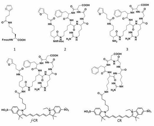
本发明化合物CR和f-CR都是内盐的形式,为本领域常规表示方法。
本发明公开了上述红光介导的核酸锚定型荧光探针在活体荧光成像或者光声成像中延长成像时间和肿瘤抑制中的应用;或者上述新型锚定型分子探针在制备长时间荧光成像试剂,光声成像试剂或者肿瘤抑制试剂中的应用;或者上述新型锚定型分子探针在肿瘤细胞内与RNA交联延长肿瘤细胞荧光成像的应用;或者上述新型锚定型分子探针在制备肿瘤细胞内与RNA交联从而抑制肿瘤细胞生长中的应用。
由于上述技术方案的运用,本发明与现有技术相比具有如下优点。
(1)本发明中设计合成了一种新型锚定型分子探针f–CR,在红光介导产生单线态氧下,可以长时间活体荧光及光声成像。
(2)本发明中目标探针可在红光介导产生单线态氧下的细胞内与RNA发生交联反应从而延长探针分子在细胞内的滞留时间。
(3)本发明中目标探针对肿瘤细胞内RNA交联后具有良好的促进肿瘤细胞凋亡的能力。
(4)本发明中目标探针在体内发生交联反应后对荷瘤小鼠的肿瘤生长具有抑制能力。
图1为实施例1中新型锚定型分子探针的合成示意图。
图2为实施例2中(a)实验组探针f–CR和对照组探针CR的化学结构,(b)实验组探针f–CR在水溶液中的紫外吸收和荧光发射图片,(c)实验组探针f–CR在水溶液中的TEM及粒径统计数据。
图3为(b)RNA与f–CR发生交联反应凝胶电泳图,(c)实验组f–CR和对照组探针CR分别与4T1细胞孵育6小时后再孵育RNA Select的共聚焦图片及共定位率(d),(e)通过RNA总体试剂盒提取的细胞总RNA的荧光及定量图片,(f)通过细胞核&细胞质RNA提取试剂盒提取的总细胞质RNA的凝胶电泳图。
图4(a)实验组探针f–CR和对照组探针CR分别MB和4T1细胞孵育6小时后进行光照,随后通过共聚焦图片观察探针在细胞内的滞留变化及荧光强度定量(b),(c)动物实验示意图,(d)首先瘤内注射MB随后尾静脉注射实验组探针f–CR和对照组探针CR,一小时后给予光照观察探针在肿瘤组织滞留变化和荧光强度定量(e)。
图5(a)相同实验条件下实验组探针f–CR和对照组探针CR,在肿瘤内光声信号变化及光声强度定量(b),(c)离体组织切片观察材料在肿瘤组织的富集量,(d)离体各组织(心,肝,脾,肺,肾,肿瘤)的探针定量。
图6(a)实验组探针f–CR和对照组探针CR分别与MB和4T1细胞共孵育12 h后细胞毒性变化,(b)实验组探针f–CR和对照组探针CR分别与MB和4T1细胞共孵育12 h后再给予光照(660 nm 50 mW/cm
2
3 min)后细胞毒性变化,(c)实验组探针f–CR和对照组探针CR分别与MB和4T1细胞共孵育12 h后再给予光照(660 nm 50 mW/cm
2
3 min)分别用live-dead和细胞明场,观察细胞凋亡变化,(d)GFP-mRNA分别与实验组探针f–CR和对照组探针CR在体外发生交联反应后转染到4T1细胞中通过共聚焦观察GFP的表达情况,用流式细胞仪观察GFP表达情况(e),(f)细胞凋亡的机理图。
图7(a)实验组探针f–CR和对照组探针CR和空白组PBS分别对于肿瘤抑制的连续13天变化曲线,(b)第十三天小鼠背部肿瘤对比大小,(c)第13天离体肿瘤尺寸大小,(d)治疗后48 h通过免疫荧光和免疫组化,分别考察Tunel,H&E,Caspase 3的变化。
下文将结合附图和具体实施例来进一步阐述本发明。应当理解的是,这些实施例仅用于解释和说明本发明中的技术方案,而并非旨在限制本发明的范围。此外,除非另有说明,下列实施例中所使用的材料、试剂、仪器等均可通过商业手段获得;具体制备方法与测试方法都为本领域常规方法。
本发明构建、合成肿瘤锚定型诊疗一体化的步骤如下:叔丁氧羰基-芴甲氧羰基-赖氨酸在含三氟乙酸的二氯甲烷溶液中脱保护后与3-(2-呋喃)丙酸反应得到化合物1,化合物1利用与N-羟基琥珀酰亚胺和1-(3-二甲氨基丙基)-3-乙基碳二亚胺盐酸盐活化羧基后与环肽cRGD反应,得到化合物2,化合物2,在含有哌啶的N,N-二甲基甲酰胺溶液中脱保护得到化合物3,化合物3与Cy 7 SE反应,得到锚定型分子探针f-CR。
环肽cRGD与Cy 7 SE反应,得到对照组探针CR。
利用上述新型锚定型分子探针进行长时间荧光活体成像的方法,包括以下步骤,首先将亚甲基蓝(MB)预先瘤内注射,随后将新型锚定型分子探针f-CR和对照组CR的水溶液尾静脉注射入荷瘤小鼠的体内,麻醉状态下观察不同时间点活体荧光,光声成像效果。
利用上述将新型锚定型分子探针f–CR和对照组CR的水溶液,分别与肿瘤细胞共孵育并在不同时间点观察肿瘤细胞内荧光强度。
利用上述将新型锚定型分子探针f–CR和对照组CR的水溶液,分别与肿瘤细胞共孵育48 h后观察肿瘤细胞的凋亡情况。
利用上述新型锚定型分子探针进行体内肿瘤抑制实验的方法,包括以下步骤,首先将亚甲基蓝预先瘤内注射,随后将所述新型锚定型分子探针f-CR和对照组CR的水溶液尾静脉注射入荷瘤小鼠的体内,1 h后给予光照连续观察记录肿瘤抑制情况。
(1)新型肿瘤锚定型诊疗一体化探针的活体荧光成像:将MB(浓度:0.1 μM,体积:50 μL)瘤内注射0.5小时后,将上述获得的实验组探针f–CR和对照组探针CR分别溶于PBS溶液中(浓度:100 μM,体积:200 μL),以尾静脉注射的方式将探针注入荷瘤(4T1小鼠乳腺癌)的BALB/c雌鼠体内,随后置于小动物活体光学成像系统/IVIS
Spectrum(PerkinElmer),并于1小时后给予肿瘤部位光照(660 nm 50 mW/cm
2 3 min),实时观察成像效果,最终通活体成像分析软件计算小鼠的肿瘤部位在不同时间点的荧光强度。
(2)新型肿瘤锚定型诊疗一体化探针的活体光声成像: 将MB(浓度:0.1 μM,体积:50 μL)瘤内注射0.5小时后,将上述获得的实验组探针f–CR和对照组探针CR分别溶于PBS溶液中(浓度:100 μM,体积:200 μL),以尾静脉注射的方式将探针注入荷瘤(4T1小鼠乳腺癌)的BALB/c雌鼠体内,同时打开小动物光声断层扫描成像系统,待光声成像仪水浴池中的水温达37℃时,放入麻醉好的小鼠,扫描小鼠的肿瘤部位图像。并于1小时后给予肿瘤部位光照(660
nm,50 mW/cm
2,3 min),实时观察成像效果,最终通活体成像分析软件计算小鼠的肿瘤部位在不同时间点的荧光强度。之后将获得的光声成像数据使用MSOT
InSight/inVision分析软件进行重建分析。
(3)新型肿瘤锚定型诊疗一体化探针的肿瘤抑制实验: 将左右双侧背部荷瘤(4T1小鼠乳腺癌)的BALB/c雌性小鼠(肿瘤体积约20 mm
3)随机分成3组(n=5):尾静脉注射PBS(10 mM, 200 μL)的小鼠左侧肿瘤(第1组,简写PBS),660 nm激光照射处理的右侧肿瘤(第2组,简写PBS+660 nm);仅尾静脉注射CR(200 μM, 200 μL)并预先瘤内注射MB(浓度:0.1 μM,体积:50 μL)左侧肿瘤(第3组,简写CR+MB),右侧肿瘤预先瘤内注射MB(浓度:0.1 μM,体积:50 μL)并660 nm激光照射处理的小鼠(第4组,简写CR+MB+660 nm);尾静脉注射f–CR(200 μM, 200 μL)并预先瘤内注射MB(浓度:0.1 μM,体积:50 μL)左侧肿瘤(第3组,简写f–CR+MB),右侧肿瘤预先瘤内注射MB(浓度:0.1 μM,体积:50 μL)并660 nm激光照射处理的小鼠(第4组,简写f–CR+MB+660 nm)。治疗后,每隔一天记录小鼠肿瘤体积变化并绘制小鼠存活率曲线。
实施例1:新型肿瘤锚定型诊疗一体化探针CR和f–CR合成。
合成化合物1:Boc-Lys
(Fmoc)-OH (0.5 g, 1.07 mmol)溶解于二氯甲烷10 mL同时加入三氟乙酸2 mL在室温下搅拌10 min脱去BOC,随后收集产物,随后加入3-(2-呋喃)丙酸(0.2 g, 1.43
mmol)、1-(3-二甲氨基丙基)-3-乙基碳二亚胺盐酸盐(0.82 g, 1.43 mmol)和N-羟基琥珀酰亚胺(0.2 g, 1.71 mmol)于室温搅拌4 h后加入N,N-二异丙基乙胺 (5 μL) 继续反应3 h。反应结束后用乙酸乙酯萃取三次再浓缩,随后通过硅胶柱洗脱得到纯产物化合物1(0.56 g, 80%)。C
28H
30N
2O
6
([M+H]
+): 489.2021, found ESI-MS: m/z 489.2031;化合物2合成 :cRGD
(10 mg, 16.57 μmol) and 1 (19.47 mg, 33.13 μmol)溶解于DMF(5 mL)在0℃搅拌10 min随后同时缓慢加入DIPEA (2 μL)。室温反应两小时后收集产物利用制备型HPLC分离产物得到产物化合物2 C
55H
69N
11O
12
([M-H]
-): 1076.5190, found ESI-MS: m/z 1076.5187;化合物3合成:化合物2 (10 mg, 9.29 μmol) 溶解于 DMF/二乙胺 (6 mL, 5:1, v/v) 溶液中室温反应1 h。浓缩粗产物后利用制备型HPLC分离产物得到产物化合物3 C
40H
59N
11O
10
([M-H]
-): 852.4374, found ESI-MS: m/z 852.4370;化合物f–CR合成:化合物3 (5 mg, 5.86 μmol),
Cy7-SE (4.24 mg, 5.86 μmol)溶解于DMF(2 mL)中在0℃搅拌10 min同时滴加到反应体系中,恢复室温继续反应2 h后利用制备型HPLC分离样品得到终产物。C
75H
99N
13O
17S
2
([M-H]
-): 1516.6651, found ESI-MS: m/z 1516.6664。
对照组化合物CR合成 cRGD (5 mg,
8.27 μmol), Cy7-SE (5.99 mg, 8.27 μmol)溶解于DMF (2 mL)缓慢加入DIPEA于0℃搅拌10 min随后恢复室温继续搅拌2 h收集粗产物,利用制备型HPLC分离得到对照组产物。C
62H
81N
11O
14S
2
([M-H]
-): 1266.5339, found ESI-MS: m/z 1266.5321。
上述反应示意图以及涉及各部产物的化学结构式如图1中的所示。
实施例2:新型肿瘤锚定型诊疗一体化探针的物理化学性质:将实施例1中制得的对照组探针CR和实验组探针f–CR用超纯水稀释至浓度为10 μM(可完全溶解),并使用紫外可见近红外分光光度计及荧光分光光度计测其紫外可见近红外光谱及荧光光谱。如图2(a)(b)所示,结果表明,探针CR和f–CR的最大吸收在747 nm,最大发射在789 nm;同时对照组探针没有发现组装,没有纳米结构,而实验组探针则发现组装成纳米结构,尺寸大于为7.2±0.9 nm左右,如图2(c)(d)。
将实施例1中制得的对照组探针CR和实验组探针f–CR用超纯水稀释至浓度为10 μM,加入定制序列RNA(5`-ACAUCGGGAUAGCGAAGUUGAGAGAGAGGGAG-3`)5 μM,MB 10 μM 混合后于4℃振荡,给予660 nm光照(50 mW/cm
2,10分钟)或者不光照(避光),将反应液直接用RNA非变性凝胶电泳分离,如图3b可以发现实验组探针f–CR在加入RNA,MB,并给予光照后能明显的标记上RNA,其他组则没有明显的Cy7的红色荧光。另外将对照组探针CR和实验组探针f–CR加入培养基(HyClone DMEM高糖液体培养基含10%FBS)中,浓度为10 μM,加入MB 0.1 μM,分别加入到4T1细胞中培养6h后,给予660 nm光照(50 mW/cm
2,10分钟)或者不光照(避光),随后再用商用RNA染料SYTO™ RNASelect™
green fluorescent cell stain(Thermo Fisher)分别染色如图3d,在探针的共定位实验中发现在实验组(f–CR+MB+660 nm)共定位率要大于其他各组;随后通过Trizol法提取出细胞内的总RNA,并通过荧光定量发现实验组(f–CR+MB+660 nm)的荧光强度远大于其他组如图3e;随后通过细胞质&细胞核总提试剂盒提取出细胞质RNA并通过RNA非变性胶分析发现实验组(f–CR+MB+660 nm)具有明显的红色荧光,其他组则没有明显的荧光产生如图3f。
对照组探针CR和实验组探针f–CR加入培养基(HyClone DMEM高糖液体培养基含10%FBS)中,浓度为10 μM,加入MB 0.1 μM,分别加入到4T1细胞中培养6h后,给予660 nm光照(50 mW/cm
2,10分钟)或者不光照(避光)观察材料在细胞内滞留的情况,发现实验组(f–CR+MB+660
nm)滞留实验远远大于其它各组如图4a和4b;随后考察探针在小鼠肿瘤内的滞留实验,对照组探针CR和实验组探针f–CR用PBS至浓度为100 Μm 200 μL,提前0.5h将MB(PBS,0.1 μM 50 μL)瘤内注射到小鼠肿瘤内,随后给予尾静脉注射CR或f–CR,在1 h后给予660 nm光照(50 mW/cm
2,3分钟)或者不光照(避光),通过IVIS观察探针在个时间点肿瘤内代谢情况如图4c,4d和4e,发现实验组(f–CR+MB+660 nm)在体内的滞留时间远大于其他各组。
对照组探针CR和实验组探针f–CR用PBS至浓度为100 μΜ 200 μL,提前0.5h将MB(PBS,0.1 μM 50 μL)瘤内注射到小鼠肿瘤内,随后给予尾静脉注射CR或f–CR,在1 h后给予660 nm光照(50 mW/cm
2,3分钟)或者不光照(避光),同时通过光声成像系统考察各时间点的光声信号变化情况,光声成像数据使用MSOT
InSight/inVision分析软件进行重建分析,发现相较于其他组,实验组(f–CR+MB+660 nm)具有长时间的光声信号,图5a和5b;与上述相同的实验方法,在探针注射12 h后将分别将小鼠各主要脏器取出,并将肿瘤组织冰冻切片图5c,所有脏器组织匀浆定量荧光强度图5d,发现实验组(f–CR+MB+660 nm)在肿瘤内的富集量要远大于其他各组。
对照组探针CR和实验组探针f–CR用培养基(HyClone DMEM高糖液体培养基含10%FBS)稀释至浓度为100,50,20,10,1,0.1 μM,加入MB 0.1 μM,分别加入到4T1细胞中培养12 h后,发现CR和f–CR均没有明显的毒性如图6a,相同实验条件在给予660 nm光照(50 mW/cm
2,3分钟)后继续培养48 h,发现实验组(f–CR+MB+660 nm)展现出一定的细胞毒性如图6b;同时通过Live-dead试剂和细胞明场的形态可以看出实验组(f–CR+MB+660
nm)展现出使得肿瘤细胞凋亡能力。
利用商用GFP-mRNA 2 μM,对照组探针CR和实验组探针f–CR用PBS稀释至浓度为10 μM,加入MB 0.1 μM,分别于4℃常规振荡,同时给予660 nm光照(50 mW/cm
2,3分钟)或者不光照(避光),随后将各混合溶液通过脂质体转染试剂盒转染至细胞内继续培养24 h,在细胞共聚焦图片下实验组(f–CR+MB+660 nm)展现出GFP在细胞内表达明显下降图6d,收集细胞通过流式细胞仪分析实验组(f–CR+MB+660
nm)展现出GFP表达量的下降如图6e。
肿瘤抑制实验,对照组探针CR和实验组探针f–CR用PBS至浓度为200 μΜ 200 μL,提前0.5h将MB(PBS,0.1 μM 50 μL)瘤内注射到小鼠肿瘤内,随后给予尾静脉注射CR或f–CR,在1 h后给予660 nm光照(50 mW/cm
2,3分钟)或者不光照(避光),随后每天量取肿瘤尺寸如图7a,第十三天处死小鼠如图7b,并取下肿瘤如图7c发现,实验组(f–CR+MB+660 nm)具有良好的肿瘤抑制能力,同时相同条件下在第一天取小鼠肿瘤,进行H&E,Tunnel和Caspase-3的免疫组化和免疫荧光分析得到,实验组(f–CR+MB+660
nm)探针能够引起肿瘤组织发生凋亡坏死。
本发明锚定型分子探针利用红光介导的生物交联,用于提高分子在肿瘤组织的滞留时间和抑制肿瘤生长。通过IVIS观察探针在个时间点肿瘤内代谢情况,发现实验组(f–CR+MB+660
nm)在体内的滞留时间远大于其他各组。
Claims (10)
- 一种红光介导的核酸锚定型荧光探针,其特征在于,所述红光介导的核酸锚定型荧光探针具有如下化学结构式:。

- 权利要求1所述红光介导的核酸锚定型荧光探针在活体荧光、光声成像和抑制肿瘤中的应用;或者权利要求1所述红光介导的核酸锚定型荧光探针在制备活体荧光、光声成像试剂和肿瘤抑制试剂应用;或者权利要求1所述红光介导的核酸锚定型荧光探针在提高探针在肿瘤组织的滞留时间和抑制肿瘤中的应用。
- 权利要求1所述红光介导的核酸锚定型荧光探针的制备方法,其特征在于,包括以下步骤:(1)叔丁氧羰基-芴甲氧羰基-赖氨酸脱保护后与3-(2-呋喃)丙酸反应得到化合物1;(2)化合物1活化羧基后与环肽cRGD反应,得到化合物2,再脱保护得到化合物3;(3)化合物3与Cy 7 SE反应,得到所述红光介导的核酸锚定型荧光探针。
- 根据权利要求3所述红光介导的核酸锚定型荧光探针的制备方法,其特征在于,叔丁氧羰基-芴甲氧羰基-赖氨酸在含三氟乙酸的二氯甲烷溶液中脱保护;化合物1利用与N-羟基琥珀酰亚胺和1-(3-二甲氨基丙基)-3-乙基碳二亚胺盐酸盐活化羧基。
- 根据权利要求3所述红光介导的核酸锚定型荧光探针的制备方法,其特征在于,步骤(1)中,叔丁氧羰基-芴甲氧羰基-赖氨酸、3-(2-呋喃)丙酸的摩尔比为1∶(1~1.5);反应在室温下进行。
- 根据权利要求3所述红光介导的核酸锚定型荧光探针的制备方法,其特征在于,步骤(2)中,化合物1、cRGD的摩尔比为1∶(1~1.2);反应在室温下进行。
- 根据权利要求3所述红光介导的核酸锚定型荧光探针的制备方法,其特征在于,步骤(3)中,化合物3与Cy 7 SE的反应在含有N,N-二异丙基乙胺溶剂中进行;化合物3与Cy 7 SE的摩尔比为1∶(1~1.2)。
- 一种红光介导的探针锚定细胞方法,其特征在于,包括以下步骤:(1)叔丁氧羰基-芴甲氧羰基-赖氨酸脱保护后与3-(2-呋喃)丙酸反应得到化合物1;(2)化合物1活化羧基后与环肽cRGD反应,得到化合物2,再脱保护得到化合物3;(3)化合物3与Cy 7 SE反应,得到所述红光介导的核酸锚定型荧光探针;(4)将所述红光介导的核酸锚定型荧光探针、亚甲基蓝、细胞共孵育,实现探针锚定细胞。
- 根据权利要求8所述红光介导的探针锚定细胞方法,其特征在于,共孵育在光照下、培养基中进行。
- 根据权利要求8所述红光介导的探针锚定细胞方法,其特征在于,红光介导的核酸锚定型荧光探针、亚甲基蓝的摩尔比为100∶(0.8~1.2)。
Priority Applications (2)
| Application Number | Priority Date | Filing Date | Title |
|---|---|---|---|
| PCT/CN2020/128480 WO2022099565A1 (zh) | 2020-11-12 | 2020-11-12 | 红光介导的核酸锚定型荧光探针及其制备方法和应用 |
| US18/037,561 US12480043B2 (en) | 2020-11-12 | 2020-11-12 | Red-light-mediated nucleic acid anchoring-type fluorescent probe, and preparation method therefor and use thereof |
Applications Claiming Priority (1)
| Application Number | Priority Date | Filing Date | Title |
|---|---|---|---|
| PCT/CN2020/128480 WO2022099565A1 (zh) | 2020-11-12 | 2020-11-12 | 红光介导的核酸锚定型荧光探针及其制备方法和应用 |
Publications (1)
| Publication Number | Publication Date |
|---|---|
| WO2022099565A1 true WO2022099565A1 (zh) | 2022-05-19 |
Family
ID=81601960
Family Applications (1)
| Application Number | Title | Priority Date | Filing Date |
|---|---|---|---|
| PCT/CN2020/128480 Ceased WO2022099565A1 (zh) | 2020-11-12 | 2020-11-12 | 红光介导的核酸锚定型荧光探针及其制备方法和应用 |
Country Status (2)
| Country | Link |
|---|---|
| US (1) | US12480043B2 (zh) |
| WO (1) | WO2022099565A1 (zh) |
Citations (4)
| Publication number | Priority date | Publication date | Assignee | Title |
|---|---|---|---|---|
| WO2017210246A2 (en) * | 2016-05-31 | 2017-12-07 | Tarveda Therapeutics, Inc. | Penicillamine conjugates and particles and formulations thereof |
| CN109180680A (zh) * | 2018-08-01 | 2019-01-11 | 苏州大学 | 一种紫外光触发交联型近红外分子探针及其制备方法与应用 |
| CN110684017A (zh) * | 2019-09-20 | 2020-01-14 | 苏州大学 | 高稳定性近红外二区小分子荧光探针及其制备方法和应用 |
| US10736932B2 (en) * | 2014-05-20 | 2020-08-11 | Ohio State Innovation Foundation | Small molecule Ras inhibitors |
Family Cites Families (1)
| Publication number | Priority date | Publication date | Assignee | Title |
|---|---|---|---|---|
| DE102015225216A1 (de) | 2015-12-15 | 2017-06-22 | Robert Bosch Gmbh | Injektoranordnung mit Gasinjektor |
-
2020
- 2020-11-12 US US18/037,561 patent/US12480043B2/en active Active
- 2020-11-12 WO PCT/CN2020/128480 patent/WO2022099565A1/zh not_active Ceased
Patent Citations (4)
| Publication number | Priority date | Publication date | Assignee | Title |
|---|---|---|---|---|
| US10736932B2 (en) * | 2014-05-20 | 2020-08-11 | Ohio State Innovation Foundation | Small molecule Ras inhibitors |
| WO2017210246A2 (en) * | 2016-05-31 | 2017-12-07 | Tarveda Therapeutics, Inc. | Penicillamine conjugates and particles and formulations thereof |
| CN109180680A (zh) * | 2018-08-01 | 2019-01-11 | 苏州大学 | 一种紫外光触发交联型近红外分子探针及其制备方法与应用 |
| CN110684017A (zh) * | 2019-09-20 | 2020-01-14 | 苏州大学 | 高稳定性近红外二区小分子荧光探针及其制备方法和应用 |
Non-Patent Citations (2)
| Title |
|---|
| KIM HYUNJIN, CHOI HAK SOO, EOM JOO BEOM, CHOI YONGDOO: "Mini-Platform for Off–On Near-Infrared Fluorescence Imaging Using Peptide-Targeting Ligands", BIOCONJUGATE CHEMISTRY, vol. 31, no. 3, 18 March 2020 (2020-03-18), US , pages 721 - 728, XP055929049, ISSN: 1043-1802, DOI: 10.1021/acs.bioconjchem.9b00844 * |
| MATHEJCZYK JULIA EVA, PAULI JUTTA, DULLIN CHRISTIAN, NAPP JOANNA, TIETZE LUTZ-F., KESSLER HORST, RESCH-GENGER UTE, ALVES FRAUKE: "Spectroscopically Well-Characterized RGD Optical Probe as a Prerequisite for Lifetime-Gated Tumor Imaging", MOLECULAR IMAGING, vol. 10, no. 6, 1 November 2011 (2011-11-01), pages 469 - 480, XP055929042, ISSN: 1536-0121, DOI: 10.2310/7290.2011.00018 * |
Also Published As
| Publication number | Publication date |
|---|---|
| US12480043B2 (en) | 2025-11-25 |
| US20230407169A1 (en) | 2023-12-21 |
Similar Documents
| Publication | Publication Date | Title |
|---|---|---|
| US12433961B2 (en) | Near-infrared fluorescent probe specifically targeting tumors as well as synthesis method and use thereof | |
| CN109180680B (zh) | 一种紫外光触发交联型近红外分子探针及其制备方法与应用 | |
| CN112592386B (zh) | 红光介导的核酸锚定型荧光探针及其制备方法和应用 | |
| CN115109081B (zh) | 一种辣椒素衍生化光敏剂及其制备方法与应用 | |
| CN110283583A (zh) | γ-谷氨酰转肽酶响应型分子探针及其应用 | |
| CN113292541B (zh) | 硝基还原酶响应的诊疗一体化探针及其制备方法与应用 | |
| CN110684017A (zh) | 高稳定性近红外二区小分子荧光探针及其制备方法和应用 | |
| CN118930523B (zh) | 一种蛋白锚定型铜响应光声探针及其制备方法与应用 | |
| CN111298140A (zh) | 还原响应的t1/t2切换型mri造影剂、其制备方法及应用 | |
| Zhang et al. | Synchronized activating therapeutic nano-agent: Enhancement and tracing for hypoxia-induced chemotherapy | |
| CN114010598B (zh) | 基于切伦科夫效应的酸响应纳米胶束及其制备方法和应用 | |
| CN104861039A (zh) | 一种酞菁基化合物、制备方法及作为单、双光子荧光探针在癌症靶向及线粒体标记中的应用 | |
| CN116217515B (zh) | 一种亚甲基蓝型探针及其制备方法和在检测半胱氨酸中的应用 | |
| CN114149482B (zh) | 一种螯合金属离子的智能转换双重刺激响应型探针及其制备方法和应用 | |
| CN112521373B (zh) | 一种多模态探针及其制备方法和应用 | |
| US20260007777A1 (en) | Heptamethine cyanine near-infrared fluorescent dye, preparation method therefor and use thereof | |
| CN110856747A (zh) | 一种过氧化氢激活的光敏剂及其制备方法与应用 | |
| CN115998908B (zh) | 一种诊疗一体化核酸锚定型荧光探针及其制备方法和应用 | |
| WO2026081650A1 (zh) | 一种靶向近红外荧光化合物及其制备方法和应用 | |
| WO2022099565A1 (zh) | 红光介导的核酸锚定型荧光探针及其制备方法和应用 | |
| CN114315791A (zh) | 用于实现手术导航和微小转移瘤成像的小分子化学发光探针及其制备方法和用途 | |
| CN102321159A (zh) | 一种具有肿瘤靶向性的光敏剂及其制备方法 | |
| CN115925785B (zh) | 一种亮氨酸氨基肽酶和谷胱甘肽双重刺激响应型探针及其制备方法和应用 | |
| CN115368345A (zh) | 一种靶向肿瘤细胞线粒体的小分子化合物及应用及制备方法 | |
| CN121287911B (zh) | 肺癌双重靶向的光动力协同铁死亡诊疗一体化探针及其制备方法与应用 |
Legal Events
| Date | Code | Title | Description |
|---|---|---|---|
| 121 | Ep: the epo has been informed by wipo that ep was designated in this application |
Ref document number: 20961122 Country of ref document: EP Kind code of ref document: A1 |
|
| NENP | Non-entry into the national phase |
Ref country code: DE |
|
| 122 | Ep: pct application non-entry in european phase |
Ref document number: 20961122 Country of ref document: EP Kind code of ref document: A1 |
|
| WWG | Wipo information: grant in national office |
Ref document number: 18037561 Country of ref document: US |