WO2022075047A1 - 医療画像処理装置及びその作動方法 - Google Patents
医療画像処理装置及びその作動方法 Download PDFInfo
- Publication number
- WO2022075047A1 WO2022075047A1 PCT/JP2021/034377 JP2021034377W WO2022075047A1 WO 2022075047 A1 WO2022075047 A1 WO 2022075047A1 JP 2021034377 W JP2021034377 W JP 2021034377W WO 2022075047 A1 WO2022075047 A1 WO 2022075047A1
- Authority
- WO
- WIPO (PCT)
- Prior art keywords
- image
- designated
- category
- medical
- images
- Prior art date
- Legal status (The legal status is an assumption and is not a legal conclusion. Google has not performed a legal analysis and makes no representation as to the accuracy of the status listed.)
- Ceased
Links
Images




Classifications
-
- G—PHYSICS
- G16—INFORMATION AND COMMUNICATION TECHNOLOGY [ICT] SPECIALLY ADAPTED FOR SPECIFIC APPLICATION FIELDS
- G16H—HEALTHCARE INFORMATICS, i.e. INFORMATION AND COMMUNICATION TECHNOLOGY [ICT] SPECIALLY ADAPTED FOR THE HANDLING OR PROCESSING OF MEDICAL OR HEALTHCARE DATA
- G16H30/00—ICT specially adapted for the handling or processing of medical images
- G16H30/40—ICT specially adapted for the handling or processing of medical images for processing medical images, e.g. editing
-
- G—PHYSICS
- G16—INFORMATION AND COMMUNICATION TECHNOLOGY [ICT] SPECIALLY ADAPTED FOR SPECIFIC APPLICATION FIELDS
- G16H—HEALTHCARE INFORMATICS, i.e. INFORMATION AND COMMUNICATION TECHNOLOGY [ICT] SPECIALLY ADAPTED FOR THE HANDLING OR PROCESSING OF MEDICAL OR HEALTHCARE DATA
- G16H30/00—ICT specially adapted for the handling or processing of medical images
-
- A—HUMAN NECESSITIES
- A61—MEDICAL OR VETERINARY SCIENCE; HYGIENE
- A61B—DIAGNOSIS; SURGERY; IDENTIFICATION
- A61B1/00—Instruments for performing medical examinations of the interior of cavities or tubes of the body by visual or photographical inspection, e.g. endoscopes; Illuminating arrangements therefor
- A61B1/00002—Operational features of endoscopes
- A61B1/00004—Operational features of endoscopes characterised by electronic signal processing
- A61B1/00009—Operational features of endoscopes characterised by electronic signal processing of image signals during a use of endoscope
- A61B1/000094—Operational features of endoscopes characterised by electronic signal processing of image signals during a use of endoscope extracting biological structures
-
- A—HUMAN NECESSITIES
- A61—MEDICAL OR VETERINARY SCIENCE; HYGIENE
- A61B—DIAGNOSIS; SURGERY; IDENTIFICATION
- A61B1/00—Instruments for performing medical examinations of the interior of cavities or tubes of the body by visual or photographical inspection, e.g. endoscopes; Illuminating arrangements therefor
- A61B1/00002—Operational features of endoscopes
- A61B1/00004—Operational features of endoscopes characterised by electronic signal processing
- A61B1/00009—Operational features of endoscopes characterised by electronic signal processing of image signals during a use of endoscope
- A61B1/000096—Operational features of endoscopes characterised by electronic signal processing of image signals during a use of endoscope using artificial intelligence
-
- A—HUMAN NECESSITIES
- A61—MEDICAL OR VETERINARY SCIENCE; HYGIENE
- A61B—DIAGNOSIS; SURGERY; IDENTIFICATION
- A61B1/00—Instruments for performing medical examinations of the interior of cavities or tubes of the body by visual or photographical inspection, e.g. endoscopes; Illuminating arrangements therefor
- A61B1/00002—Operational features of endoscopes
- A61B1/00039—Operational features of endoscopes provided with input arrangements for the user
- A61B1/0004—Operational features of endoscopes provided with input arrangements for the user for electronic operation
-
- A—HUMAN NECESSITIES
- A61—MEDICAL OR VETERINARY SCIENCE; HYGIENE
- A61B—DIAGNOSIS; SURGERY; IDENTIFICATION
- A61B1/00—Instruments for performing medical examinations of the interior of cavities or tubes of the body by visual or photographical inspection, e.g. endoscopes; Illuminating arrangements therefor
- A61B1/00002—Operational features of endoscopes
- A61B1/00043—Operational features of endoscopes provided with output arrangements
- A61B1/00045—Display arrangement
- A61B1/0005—Display arrangement combining images e.g. side-by-side, superimposed or tiled
-
- G—PHYSICS
- G06—COMPUTING OR CALCULATING; COUNTING
- G06V—IMAGE OR VIDEO RECOGNITION OR UNDERSTANDING
- G06V10/00—Arrangements for image or video recognition or understanding
- G06V10/20—Image preprocessing
- G06V10/25—Determination of region of interest [ROI] or a volume of interest [VOI]
-
- G—PHYSICS
- G06—COMPUTING OR CALCULATING; COUNTING
- G06V—IMAGE OR VIDEO RECOGNITION OR UNDERSTANDING
- G06V10/00—Arrangements for image or video recognition or understanding
- G06V10/70—Arrangements for image or video recognition or understanding using pattern recognition or machine learning
- G06V10/764—Arrangements for image or video recognition or understanding using pattern recognition or machine learning using classification, e.g. of video objects
-
- G—PHYSICS
- G16—INFORMATION AND COMMUNICATION TECHNOLOGY [ICT] SPECIALLY ADAPTED FOR SPECIFIC APPLICATION FIELDS
- G16H—HEALTHCARE INFORMATICS, i.e. INFORMATION AND COMMUNICATION TECHNOLOGY [ICT] SPECIALLY ADAPTED FOR THE HANDLING OR PROCESSING OF MEDICAL OR HEALTHCARE DATA
- G16H15/00—ICT specially adapted for medical reports, e.g. generation or transmission thereof
-
- G—PHYSICS
- G16—INFORMATION AND COMMUNICATION TECHNOLOGY [ICT] SPECIALLY ADAPTED FOR SPECIFIC APPLICATION FIELDS
- G16H—HEALTHCARE INFORMATICS, i.e. INFORMATION AND COMMUNICATION TECHNOLOGY [ICT] SPECIALLY ADAPTED FOR THE HANDLING OR PROCESSING OF MEDICAL OR HEALTHCARE DATA
- G16H30/00—ICT specially adapted for the handling or processing of medical images
- G16H30/20—ICT specially adapted for the handling or processing of medical images for handling medical images, e.g. DICOM, HL7 or PACS
-
- G—PHYSICS
- G16—INFORMATION AND COMMUNICATION TECHNOLOGY [ICT] SPECIALLY ADAPTED FOR SPECIFIC APPLICATION FIELDS
- G16H—HEALTHCARE INFORMATICS, i.e. INFORMATION AND COMMUNICATION TECHNOLOGY [ICT] SPECIALLY ADAPTED FOR THE HANDLING OR PROCESSING OF MEDICAL OR HEALTHCARE DATA
- G16H40/00—ICT specially adapted for the management or administration of healthcare resources or facilities; ICT specially adapted for the management or operation of medical equipment or devices
- G16H40/60—ICT specially adapted for the management or administration of healthcare resources or facilities; ICT specially adapted for the management or operation of medical equipment or devices for the operation of medical equipment or devices
- G16H40/63—ICT specially adapted for the management or administration of healthcare resources or facilities; ICT specially adapted for the management or operation of medical equipment or devices for the operation of medical equipment or devices for local operation
-
- G—PHYSICS
- G06—COMPUTING OR CALCULATING; COUNTING
- G06V—IMAGE OR VIDEO RECOGNITION OR UNDERSTANDING
- G06V10/00—Arrangements for image or video recognition or understanding
- G06V10/98—Detection or correction of errors, e.g. by rescanning the pattern or by human intervention; Evaluation of the quality of the acquired patterns
- G06V10/987—Detection or correction of errors, e.g. by rescanning the pattern or by human intervention; Evaluation of the quality of the acquired patterns with the intervention of an operator
-
- G—PHYSICS
- G06—COMPUTING OR CALCULATING; COUNTING
- G06V—IMAGE OR VIDEO RECOGNITION OR UNDERSTANDING
- G06V2201/00—Indexing scheme relating to image or video recognition or understanding
- G06V2201/03—Recognition of patterns in medical or anatomical images
Definitions
- the present invention relates to a medical image processing device for acquiring a report attached image attached to a report and a method for operating the medical image processing apparatus.
- Patent Document 1 describes that when an image to be attached to a report is selected from an image obtained by a capsule endoscope, a list is displayed as a digest image in order to make it easier to select the image.
- the work of selecting an image to be used for a report after inspection is automated by AI (Artificial Intelligence), and the report creation device is equipped with a function to automatically select an image suitable for the report, so that the user can use it. It is possible to reduce the workload of a certain doctor.
- AI Artificial Intelligence
- An object of the present invention is to provide a medical image processing apparatus and an operation method thereof that can reduce the burden of report creation work while reflecting the user's preference regarding the image to be included in the report.
- the medical image processing apparatus of the present invention has a processor, and the processor acquires a plurality of medical images, classifies each of the plurality of medical images into a plurality of categories, and displays the plurality of medical images on the first display screen of the display.
- the user's designated input for the medical image displayed on the first display screen is accepted, and the designated image group including the designated image that is the designated input medical image and the designated image group among multiple medical images are not specified and input.
- the non-designated image group including the non-designated image which is a medical image is determined, and based on the designated image category which is the category given to the designated image and the non-designated image category which is the category given to the non-designated image. , Acquires a selected image selected from a plurality of medical images, and displays the selected images in a list on the second display screen of the display.
- the selected image includes the first selected image selected from the non-designated image group based on the relationship between the designated image category and the non-designated image category. It is preferable that the relationship between the designated image category and the non-designated image category is different from that between the designated image category and the non-designated image category.
- the selected image preferably includes a second selected image that selects all of the designated images included in the designated image group.
- the processor determines the diagnostic usefulness of the medical image and acquires the selected image based on the usefulness in addition to the designated image category and the non-designated image category.
- the display mode of the image corresponding to the designated image and the display mode of the image corresponding to the non-designated image among the selected images are different.
- each medical image differs depending on the category on the first display screen, and / or the display mode of the selected image differs depending on the category on the second display screen.
- On the first display screen each medical image is displayed with character or symbol information according to the category, and / or, on the second display screen, each selected image is provided with character or symbol information according to the category. It is preferable to add and display it.
- the processor updates the medical images listed on the first display screen based on the designated image category given to the designated image and the non-designated image category given to the non-designated image. , It is preferable to accept the designated input for the updated medical image listed on the first display screen.
- the processor deletes or displays the display mode from the first display screen for the non-designated image to which the same non-designated image category as the specific designated image category is assigned. It is preferable to make an update that changes.
- the processor displays the designated image information, which is information related to the designated image, on the display, and the designated image information includes information representing the position information of the part corresponding to the designated image in characters or visually, or the number of designated images. It preferably contains at least the characters or visually represented information.
- the category is preferably information about the part of the subject.
- the category is preferably information about whether or not the image contains a region of interest.
- the category is preferably information about whether or not the image contains a treatment tool or the type of treatment tool included.
- the category is preferably information about the spraying state of the dye or stain in the image.
- the processor obtains a plurality of medical images, classifies each of the plurality of medical images into a plurality of categories, and displays the plurality of medical images in the first display.
- a step of displaying a list on the display screen a step of accepting a user's designated input for a medical image displayed on the first display screen, a designated image group including a designated image which is a designated input medical image, and a plurality of medical images.
- the medical image processing device 10 is connected to the endoscope system 11.
- the endoscope system 11 acquires a large amount of medical images 12 by the light source device 11a, the endoscope 11b, and the processor device 11c.
- the light source device 11a supplies illumination light for illuminating the inside of the body to the endoscope 11b.
- the endoscope 11b acquires an endoscope image inside the body by irradiating the inside of the body with illumination light and receiving and imaging the reflected light from the inside of the body.
- the processor device 11c performs image processing on the endoscope image from the endoscope 11b and displays it on a display for an endoscope (not shown).
- the endoscope system 11 is connected to the medical image processing device 10, and the acquired endoscope image is transmitted to the medical image processing device 10 as a medical image 12.
- the medical image processing apparatus 10 programs related to various processes are incorporated in a program memory (not shown).
- the medical image processing apparatus 10 is provided with a central control unit (not shown) configured by a processor.
- the central control unit By executing the program in the program memory by the central control unit, as shown in FIG. 2, the image acquisition unit 20, the image classification unit 21, the display control unit 22, the input reception unit 23, the image group determination unit 24, and the usefulness are used.
- the functions of the sex determination unit 25, the image selection unit 26, and the report creation device 27 are realized.
- the medical image processing apparatus 10 is provided with a display 30 for displaying various images.
- the medical image processing device 10 performs a process of selecting a report attached image, which is a medical image to be attached to the report created by the report creating device 27, from the medical images obtained by the endoscope system 11.
- the image acquisition unit 20 acquires a plurality of medical images 12 from the endoscope system 11 and transmits them to the image classification unit 21.
- the image classification unit 21 extracts the image information of the medical image 12 transmitted from the image acquisition unit 20, acquires category information, and calculates image quality information.
- the medical images 12 are classified into each category according to the acquired category information.
- the categories to be classified are any or a combination of the imaging site, the presence / absence and type of lesion which is one of the areas of interest, the state of use of the treatment tool, the state of spraying the pigment agent, and the like.
- image quality information information such as image brightness, noise, and degree of blurring is acquired and attached to the medical image 12.
- the image information is extracted by the trained CNN (Convolutional neural network).
- the trained CNN is obtained by training using an image group containing at least a plurality of medical images 12. It is preferable that the category information and the image quality information acquired at the time of photographing by the endoscope system 11 and attached to the medical image 12 have priority over the information extracted by the trained CNN.
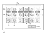
- the display control unit 22 displays a list of a plurality of medical images 12 on the first display screen 30a of the display 30.
- the medical image 12 displayed in a list on the first display screen 30a is a medical image 12 that has been classified into a plurality of categories by the image classification unit 21, or is classified into a plurality of categories by the image classification unit 21. It is preferable that the medical image 12 is not classified into any category.
- the input receiving unit 23 accepts at least the user's designated input for the medical image 12 displayed on the display 30. Specifically, as shown in FIG. 3, a cursor 32 that reflects a user operation by a mouse operation or the like is displayed on the display 30, and the user uses the cursor 32 to input a designated input to the medical image 12. conduct. Designated input is possible not only for one medical image but also for a plurality of images.
- the input receiving unit 23 receives the designated input specified by the cursor 32. When the designated input for the medical image is completed, the designated input complete icon 33 is operated using the cursor 32. It is preferable that the designated input medical image 12 is displayed by adding a thick frame 13 to the outer edge, or by adding a mark indicating the designated input.
- the image group determination unit 24 selects a designated image group including a designated image which is a designated input medical image and a non-designated image which is a medical image which is not designated input among a plurality of medical images acquired by the image acquisition unit 20. Determine the non-designated image group to be included. Specifically, as shown in FIG. 4, the medical image 12 that has been designated and input at the time when the designated input completion instruction is received by the input receiving unit 23 is designated as the designated image 35, and the designated image 35 is included in the designated image 35. The image group 36 is determined. Further, the medical image 12 that has not been designated and input at the time of completion of the designated input is designated as the non-designated image 38, and the non-designated image group 39 including those non-designated images is determined.
- the usefulness determination unit 25 determines the diagnostic usefulness of the medical image 12 based on the classification result and the image quality information obtained by the image classification unit 21. The higher the diagnostic usefulness, the more likely it is that the image is suitable as a report attachment image.
- the evaluation of usefulness is expressed by, for example, a graded evaluation. It can be evaluated that the medical image 12 having an item such as a lesion in the classification result of the category and having a low degree of blur in the image quality evaluation is highly useful. If there is no category information, the degree of blurring is large, the brightness is too high, or the brightness is too low, the usefulness is low.
- the medical image 12 whose usefulness has been determined is transmitted to the image selection unit 26.
- the usefulness is evaluated, and the evaluation threshold can be set arbitrarily by the user. The usefulness may be determined not only for the medical image 12, but also for the designated image 35 or the non-designated image.
- the image selection unit 26 selects from a plurality of medical images 12 based on the designated image category which is the category attached to the designated image 35 and the non-designated image category which is the category attached to the non-designated image 38. Get the selected image. More preferably, the image selection unit 26 may acquire the selected image based on the usefulness obtained by the usefulness determination unit 25 in addition to the designated image category and the non-designated image category.
- the number of selected images is preferably set so that the sum of the number of designated images and the number of selected images is a specific number. Further, the number of selected images may be set to the number specified by the doctor. It is preferable that the number of sheets is set by the input receiving unit 23.
- the selected image 40 acquired by the image selection unit 26 is selected from the non-designated image group 39 based on the relationship between the designated image category and the non-designated image category. It is preferable that the first selection image 42 is included. It is preferable that the relationship between the designated image category and the non-designated image category is different between the designated image category and the non-designated image category. In this case, as the first selection image 42, for example, "a non-designated image 38 of an imaging site different from the designated image 35" and "when the designated image 35 contains a lesion, the non-designated image does not contain a lesion".
- the selected image 40 includes the second selected image 44 in which all of the designated images 35 included in the designated image group 36 are selected.
- the display control unit 22 displays the selected image 40 on the second display screen 30b of the display 30.
- the user confirms the selected image 40 and determines that it is suitable as the report attached image to be used in the report creating device 27, the user operates the approval icon 46 displayed on the display 30 by using the cursor 32.
- the approval icon 46 is operated, the selected image 40 is transmitted to the report creating device 27 as a report attached image.
- the disapproval icon 47 is operated. In this case, the display 30 switches to the first display screen 30a and switches to a state of accepting the user's designated input.
- the user selects the second selection image 44 before automatically selecting the report attachment image, and then automatically selects the first selection image 42, and these first selection images 42.
- the selected image 40 including the second selected image 44 as a report attached image used in the report creating device 27, it is possible to reduce the burden of the report creating work while reflecting the preference of the doctor who is the user. can.
- the display mode of the image corresponding to the designated image 35 and the display mode of the image corresponding to the non-designated image 38 among the selected images 40 are different.
- a thick frame 51 is displayed on the outer edge of the image 50 corresponding to the designated image 35, while the thick frame 51 is not displayed on the image 52 corresponding to the non-designated image 38.
- each medical image 12 is preferably different depending on the category
- the display mode of each selected image 40 is preferably different depending on the category.
- each selected image 40 is given a character 54 according to the category.
- the characters according to the category include the site name, the lesion name, the light source used, and the presence or absence of treatment with the treatment tool.
- the symbol information corresponding to the category may be added to each selected image 40 and displayed.
- the symbolic information according to the category there is information that symbolizes the site name, the lesion name, the light source used, the presence or absence of treatment with the treatment tool, and the like.
- the first display screen 30a is also displayed by adding character or symbol information according to the category to each medical image 12 by the same method as the second display screen 30b.
- the display mode of the medical image 12 or the selected image 40 according to the category may be a mode in which characters or symbols are not used as described above.
- the display position of the medical image 12 or the selected image 40 may be changed according to the category.
- the medical image or the selected image 40 is displayed along the row direction (vertical direction)
- the input receiving unit 23 accepts the user's designated input for the specific medical image 55 listed on the first display screen 30a.
- the specific condition it is preferable to include at least one of the number of images included in the same category being a specific number or less, or an image having high diagnostic usefulness.
- the medical image 56 other than the specific medical image 55 is excluded from the designated input.
- the display is left on the first display screen 50a (for example, grayed out) although the visibility is lower than that of the specific medical image 55, or the first. It is preferably either completely excluded (hidden) from the display screen 50a.
- a report is created using the report creation screen 48 displayed on the display 30.
- pre-entered patient information, user information, and the like are automatically input.
- the user inputs the findings in the finding input field 49 and creates a report.
- the image acquisition unit 20 acquires a plurality of medical images 12 from the endoscope system 11.
- the image classification unit 21 classifies each medical image 12 into a plurality of categories.
- the display control unit 22 displays a plurality of medical images in a list on the first display screen 30a of the display 30.
- the input receiving unit 23 accepts the user's designated input for the medical image displayed on the first display screen 30a. When the designated input by the user is completed, the designated input completion icon 33 is operated.
- the image group determination unit 24 did not specify and input the designated image group 36 including the designated image 35 which is the designated input medical image 12 and the plurality of medical images 12.
- the non-designated image group 39 including the non-designated image 38 which is the medical image 12 is determined.
- the image selection unit 26 is selected from a plurality of medical images 12 based on the designated image category, which is the category assigned to the designated image 35, and the non-designated image category, which is the category assigned to the non-designated image 38.
- the selected image 40 is acquired.
- the display control unit 22 displays the selected images 40 in a list on the second display screen 30b of the display 30.
- the approval icon 46 When the user approves the selected image 40 displayed in the list as a report attached image, the approval icon 46 is operated. When the approval icon 46 is operated, the selected image 40 is transmitted to the report creating device 27 as a report attached image. On the other hand, when the selected image 40 is not approved as the report attached image, the disapproval icon 47 is operated. In this case, the display 30 switches to the first display screen 30a and switches to a state of accepting the user's designated input.
- the medical images listed on the first display screen 30a are updated every time the user's designated input is performed.
- the display control unit 22 updates the medical images listed on the first display screen 30a based on the designated image category and the non-designated image category each time the designated input is performed.
- the input receiving unit 23 accepts the user's designated input for the updated medical image listed on the first display screen 30a.
- the display control unit 22 does not specify the same as the specific designated image category X when the designated input is made to the designated image X1 to which the specific designated image category X is assigned.
- the non-designated image X2 to which the image category X is assigned is updated to be deleted from the first display screen 30a (hidden on the first display screen 30a). As a result, the number of images to be confirmed by the user is reduced, so that the workload of the user can be reduced.
- the image to be deleted from the first display screen 30a for example, "a non-designated image X2 having the same shooting portion as the designated image X1" and "a non-designated image whose shooting portion is close to the designated image X1".
- Image X2 "If the designated image X1 is an image during treatment with a treatment tool or an image during dye spraying, it is preferably a non-designated image X2 during treatment or dye spraying".
- the non-designated image X2 may be hidden on the first display screen 30a, or may be left on the first display screen 30a although its visibility is lower than that of the designated image X1 (for example, grayed out).
- the display control unit 22 displays the designated image information regarding the designated image on the display 30 in order to make it easier for the user to grasp the work status when the designated input is performed.
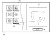
- the designated image information for example, it is preferable that the information represents the position information of the portion corresponding to the designated image in characters or visually.
- the site corresponding to the designated image is the rectum, which is the predetermined position of the large intestine, as shown in FIG. 13, "rectum" is displayed on the display 30 as the position information 62 of the characters, or in the schema of the large intestine FIG. 57. It is preferable to display the point 58 indicating that the designated input has been made on the display 30 in the portion corresponding to the “rectum”.
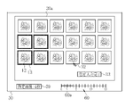
- the information represents the number of designated images 35 in characters or visually.
- the number of designated images 35 at the time of the designated input is 5, as shown in FIG. 14, "4" is displayed on the display 30 as the character information 59, or the designated image 35 is displayed.
- the number counting bar 60 representing the number of sheets it is preferable to display the length 60a corresponding to "4 sheets" on the display 30.
- various types such as an image acquisition unit 20, an image classification unit 21, a display control unit 22, an input reception unit 23, an image group determination unit 24, a usefulness determination unit 25, an image selection unit 26, and a report creation device 27.
- the hardware structure of the processing unit that executes the processing of is the following various processors.
- Various processors include CPU (Central Processing Unit), GPU (Graphical Processing Unit), FPGA (Field Programmable Gate Array), which are general-purpose processors that execute software (programs) and function as various processing units.
- a processing unit may be composed of one of these various processors, or a combination of two or more processors of the same type or different types (for example, a plurality of FPGAs, a combination of a CPU and an FPGA, or a CPU and a combination of two or more processors. It may be composed of a combination of GPUs, etc.). Further, a plurality of processing units may be configured by one processor. As an example of configuring a plurality of processing units with one processor, first, as represented by a computer such as a client or a server, one processor is configured by a combination of one or more CPUs and software. There is a form in which this processor functions as a plurality of processing units.
- SoC System On Chip
- the various processing units are configured by using one or more of the above-mentioned various processors as a hardware-like structure.
- the hardware-like structure of these various processors is, more specifically, an electric circuit (circuitry) in which circuit elements such as semiconductor elements are combined.
- the hardware structure of the storage unit is a storage device such as an HDD (hard disk drive) or SSD (solid state drive).
Landscapes
- Health & Medical Sciences (AREA)
- Engineering & Computer Science (AREA)
- Life Sciences & Earth Sciences (AREA)
- Medical Informatics (AREA)
- General Health & Medical Sciences (AREA)
- Public Health (AREA)
- Surgery (AREA)
- Radiology & Medical Imaging (AREA)
- Nuclear Medicine, Radiotherapy & Molecular Imaging (AREA)
- Physics & Mathematics (AREA)
- Biomedical Technology (AREA)
- Epidemiology (AREA)
- Primary Health Care (AREA)
- Biophysics (AREA)
- Optics & Photonics (AREA)
- Veterinary Medicine (AREA)
- Animal Behavior & Ethology (AREA)
- Molecular Biology (AREA)
- Heart & Thoracic Surgery (AREA)
- Pathology (AREA)
- Theoretical Computer Science (AREA)
- General Physics & Mathematics (AREA)
- Multimedia (AREA)
- Evolutionary Computation (AREA)
- Artificial Intelligence (AREA)
- Signal Processing (AREA)
- Computer Vision & Pattern Recognition (AREA)
- Software Systems (AREA)
- Databases & Information Systems (AREA)
- Computing Systems (AREA)
- General Business, Economics & Management (AREA)
- Business, Economics & Management (AREA)
- Medical Treatment And Welfare Office Work (AREA)
Priority Applications (4)
| Application Number | Priority Date | Filing Date | Title |
|---|---|---|---|
| CN202180069127.7A CN116325008B (zh) | 2020-10-09 | 2021-09-17 | 医疗图像处理装置及其工作方法 |
| EP21877348.9A EP4226840A4 (en) | 2020-10-09 | 2021-09-17 | DEVICE FOR PROCESSING MEDICAL IMAGES AND OPERATING METHOD THEREFOR |
| JP2022555341A JP7702423B2 (ja) | 2020-10-09 | 2021-09-17 | 医療画像処理装置及びその作動方法 |
| US18/297,432 US12555670B2 (en) | 2020-10-09 | 2023-04-07 | Medical image processing apparatus and operation method thereof |
Applications Claiming Priority (2)
| Application Number | Priority Date | Filing Date | Title |
|---|---|---|---|
| JP2020171382 | 2020-10-09 | ||
| JP2020-171382 | 2020-10-09 |
Related Child Applications (1)
| Application Number | Title | Priority Date | Filing Date |
|---|---|---|---|
| US18/297,432 Continuation US12555670B2 (en) | 2020-10-09 | 2023-04-07 | Medical image processing apparatus and operation method thereof |
Publications (1)
| Publication Number | Publication Date |
|---|---|
| WO2022075047A1 true WO2022075047A1 (ja) | 2022-04-14 |
Family
ID=81126485
Family Applications (1)
| Application Number | Title | Priority Date | Filing Date |
|---|---|---|---|
| PCT/JP2021/034377 Ceased WO2022075047A1 (ja) | 2020-10-09 | 2021-09-17 | 医療画像処理装置及びその作動方法 |
Country Status (5)
| Country | Link |
|---|---|
| US (1) | US12555670B2 (https=) |
| EP (1) | EP4226840A4 (https=) |
| JP (1) | JP7702423B2 (https=) |
| CN (1) | CN116325008B (https=) |
| WO (1) | WO2022075047A1 (https=) |
Cited By (1)
| Publication number | Priority date | Publication date | Assignee | Title |
|---|---|---|---|---|
| CN114724669A (zh) * | 2022-05-07 | 2022-07-08 | 丹阳慧创医疗设备有限公司 | 用于近红外脑功能成像装置的医学报告生成方法及装置 |
Citations (3)
| Publication number | Priority date | Publication date | Assignee | Title |
|---|---|---|---|---|
| WO2009008125A1 (ja) | 2007-07-12 | 2009-01-15 | Olympus Medical Systems Corp. | 画像処理装置、該動作方法及び該プログラム |
| US20110032259A1 (en) * | 2009-06-09 | 2011-02-10 | Intromedic Co., Ltd. | Method of displaying images obtained from an in-vivo imaging device and apparatus using same |
| JP2016158752A (ja) * | 2015-02-27 | 2016-09-05 | オリンパス株式会社 | 内視鏡業務支援システム |
Family Cites Families (12)
| Publication number | Priority date | Publication date | Assignee | Title |
|---|---|---|---|---|
| JP2001024971A (ja) * | 1999-07-06 | 2001-01-26 | Canon Inc | 画像処理装置及び画像処理方法並びにメモリ媒体 |
| US8014576B2 (en) * | 2005-11-23 | 2011-09-06 | The Medipattern Corporation | Method and system of computer-aided quantitative and qualitative analysis of medical images |
| WO2008075508A1 (ja) * | 2006-12-21 | 2008-06-26 | Konica Minolta Medical & Graphic, Inc. | 医用画像システム及び第1サーバ |
| JP5305700B2 (ja) * | 2007-04-25 | 2013-10-02 | 株式会社東芝 | 画像診断支援システム、及び画像診断支援方法 |
| JP5486364B2 (ja) * | 2009-09-17 | 2014-05-07 | 富士フイルム株式会社 | 読影レポート作成装置および方法並びにプログラム |
| CN104067277A (zh) * | 2012-01-27 | 2014-09-24 | 皇家飞利浦有限公司 | 医学选择系统 |
| JP6345487B2 (ja) * | 2013-07-11 | 2018-06-20 | キヤノンメディカルシステムズ株式会社 | 医用レポート作成装置、及び医用画像診断装置 |
| US10929508B2 (en) * | 2015-04-30 | 2021-02-23 | Merge Healthcare Solutions Inc. | Database systems and interactive user interfaces for dynamic interaction with, and indications of, digital medical image data |
| US10579234B2 (en) * | 2016-09-09 | 2020-03-03 | Merge Healthcare Solutions Inc. | Systems and user interfaces for opportunistic presentation of functionality for increasing efficiencies of medical image review |
| US10803581B2 (en) * | 2017-11-06 | 2020-10-13 | Beijing Keya Medical Technology Co., Ltd. | System and method for generating and editing diagnosis reports based on medical images |
| KR20190074671A (ko) * | 2017-12-20 | 2019-06-28 | (주)헬스허브 | 의료영상 디스플레이 프로토콜 지원 장치 및 그 방법 |
| KR102034648B1 (ko) * | 2018-05-16 | 2019-10-22 | 재단법인 아산사회복지재단 | 의료영상 관리 시스템, 방법 및 컴퓨터 판독 가능한 기록매체 |
-
2021
- 2021-09-17 WO PCT/JP2021/034377 patent/WO2022075047A1/ja not_active Ceased
- 2021-09-17 CN CN202180069127.7A patent/CN116325008B/zh active Active
- 2021-09-17 EP EP21877348.9A patent/EP4226840A4/en active Pending
- 2021-09-17 JP JP2022555341A patent/JP7702423B2/ja active Active
-
2023
- 2023-04-07 US US18/297,432 patent/US12555670B2/en active Active
Patent Citations (3)
| Publication number | Priority date | Publication date | Assignee | Title |
|---|---|---|---|---|
| WO2009008125A1 (ja) | 2007-07-12 | 2009-01-15 | Olympus Medical Systems Corp. | 画像処理装置、該動作方法及び該プログラム |
| US20110032259A1 (en) * | 2009-06-09 | 2011-02-10 | Intromedic Co., Ltd. | Method of displaying images obtained from an in-vivo imaging device and apparatus using same |
| JP2016158752A (ja) * | 2015-02-27 | 2016-09-05 | オリンパス株式会社 | 内視鏡業務支援システム |
Non-Patent Citations (1)
| Title |
|---|
| See also references of EP4226840A4 |
Cited By (2)
| Publication number | Priority date | Publication date | Assignee | Title |
|---|---|---|---|---|
| CN114724669A (zh) * | 2022-05-07 | 2022-07-08 | 丹阳慧创医疗设备有限公司 | 用于近红外脑功能成像装置的医学报告生成方法及装置 |
| CN114724669B (zh) * | 2022-05-07 | 2023-08-22 | 丹阳慧创医疗设备有限公司 | 用于近红外脑功能成像装置的医学报告生成方法及装置 |
Also Published As
| Publication number | Publication date |
|---|---|
| JP7702423B2 (ja) | 2025-07-03 |
| US20230245758A1 (en) | 2023-08-03 |
| EP4226840A1 (en) | 2023-08-16 |
| CN116325008B (zh) | 2026-02-24 |
| US12555670B2 (en) | 2026-02-17 |
| CN116325008A (zh) | 2023-06-23 |
| JPWO2022075047A1 (https=) | 2022-04-14 |
| EP4226840A4 (en) | 2024-03-20 |
Similar Documents
| Publication | Publication Date | Title |
|---|---|---|
| Yang et al. | Assessing inter-annotator agreement for medical image segmentation | |
| Waite et al. | A review of perceptual expertise in radiology-how it develops, how we can test it, and why humans still matter in the era of artificial intelligence | |
| JP5148928B2 (ja) | 眼底検査装置及びプログラム | |
| US9595088B2 (en) | Method of, and apparatus for, visualizing medical image data | |
| JP2022505154A (ja) | 生体内画像ストリームの精査用情報を生成及び表示するためのシステム並びに方法 | |
| EP3649918B1 (en) | Medical image processing device, endoscope device, diagnostic support device, medical service support device and report generation support device | |
| US11450425B2 (en) | Medical image processing apparatus, endoscope apparatus, diagnostic support apparatus, medical service support apparatus, and report creation support apparatus | |
| US20240259534A1 (en) | Medical image processing device, medical image processing method, and endoscope system | |
| JP7665310B2 (ja) | 医用画像処理装置、医用画像処理システム、医用画像処理方法及びプログラム | |
| JP4253055B2 (ja) | コンピュータ支援診断装置 | |
| US12555670B2 (en) | Medical image processing apparatus and operation method thereof | |
| CN110534182A (zh) | 医学影像的显示方法、显示设备和存储介质 | |
| US20230245312A1 (en) | Medical image processing apparatus and operation method thereof | |
| US12484759B2 (en) | Medical imaging apparatus and operating method for same | |
| Zang et al. | Predicting clinician fixations on glaucoma oct reports via cnn-based saliency prediction methods | |
| JP2004133736A (ja) | 医用画像表示方法及びその装置 | |
| WO2022264757A1 (ja) | 医用画像診断システム、医用画像診断方法及びプログラム | |
| JP4635077B2 (ja) | コンピュータ支援診断装置 | |
| Roy et al. | Medical Applications of Artificial Intelligence | |
| JPWO2022075047A5 (https=) | ||
| US20230222663A1 (en) | Medical imaging apparatus and operation method for medical imaging apparatus | |
| EP4524900A1 (en) | Method and system for improved interaction with medical 2d and 3d visualization | |
| Deepa et al. | AI-based Periapical Lesion Segmentation in 2D Periapical Radiographs using Residual U-Net | |
| Mori et al. | A method for specifying unobserved regions in virtual endoscopy system |
Legal Events
| Date | Code | Title | Description |
|---|---|---|---|
| 121 | Ep: the epo has been informed by wipo that ep was designated in this application |
Ref document number: 21877348 Country of ref document: EP Kind code of ref document: A1 |
|
| ENP | Entry into the national phase |
Ref document number: 2022555341 Country of ref document: JP Kind code of ref document: A |
|
| NENP | Non-entry into the national phase |
Ref country code: DE |
|
| ENP | Entry into the national phase |
Ref document number: 2021877348 Country of ref document: EP Effective date: 20230509 |