WO2022059799A1 - 情報処理装置及びプログラム - Google Patents
情報処理装置及びプログラム Download PDFInfo
- Publication number
- WO2022059799A1 WO2022059799A1 PCT/JP2021/034649 JP2021034649W WO2022059799A1 WO 2022059799 A1 WO2022059799 A1 WO 2022059799A1 JP 2021034649 W JP2021034649 W JP 2021034649W WO 2022059799 A1 WO2022059799 A1 WO 2022059799A1
- Authority
- WO
- WIPO (PCT)
- Prior art keywords
- information processing
- lesion
- area
- image
- anatomical region
- Prior art date
- Legal status (The legal status is an assumption and is not a legal conclusion. Google has not performed a legal analysis and makes no representation as to the accuracy of the status listed.)
- Ceased
Links
Images


Classifications
-
- G—PHYSICS
- G01—MEASURING; TESTING
- G01T—MEASUREMENT OF NUCLEAR OR X-RADIATION
- G01T1/00—Measuring X-radiation, gamma radiation, corpuscular radiation, or cosmic radiation
- G01T1/16—Measuring radiation intensity
- G01T1/161—Applications in the field of nuclear medicine, e.g. in vivo counting
-
- G—PHYSICS
- G06—COMPUTING OR CALCULATING; COUNTING
- G06T—IMAGE DATA PROCESSING OR GENERATION, IN GENERAL
- G06T7/00—Image analysis
Definitions
- the present invention relates to an information processing device and a program.
- Patent Document 1 a technique for supporting a diagnosis based on an image of a test result by a computer has been proposed.
- Patent Document 2 proposes a technique for supporting this.
- PET positron emission tomography
- FDG fluorodeoxyglucose: fluorodeoxyglucose
- the present invention has decided to provide an information processing device and a program capable of automatically identifying a lesion from the result of a PET examination and supporting a diagnosis by a doctor.
- an information processing apparatus that identifies a lesion based on a positron emission tomography examination.
- This information processing device includes an image acquisition unit and a lesion identification unit.
- the image acquisition unit is configured to be able to acquire an image including an anatomical region taken by a positron emission tomography apparatus.
- the lesion identification part is configured so that the lesion part can be identified from the part where the positron emitting nuclides are accumulated in the image based on the learned data by machine learning.
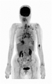
- An example of an image taken by the positron emission tomography apparatus 2 is shown.
- An example of an anatomical region specified by the region specifying unit 104 is shown.
- FIG. 1 It is a figure for demonstrating teacher data. It is a figure for demonstrating teacher data. It is a figure for demonstrating teacher data. It is a figure for demonstrating teacher data. It is a figure for demonstrating teacher data. It is a figure for demonstrating teacher data. It is a figure for demonstrating teacher data. It is a figure for demonstrating teacher data. It is a figure for demonstrating teacher data. It is a figure for demonstrating teacher data. It is a figure for demonstrating teacher data. It is a figure which showed the example of the lesion part identified by the lesion identification part 102. It is a figure which showed the example of the lesion part identified by the lesion identification part 102. It is a figure for demonstrating case 1. FIG. It is a figure for demonstrating case 1. FIG. It is a figure for demonstrating case 1. FIG. It is a figure for demonstrating case 1. FIG. It is a figure for demonstrating case 1. FIG. It is a figure for demonstrating case 1. FIG. It is a figure for demonstrating case 1.
- FIG. It is a figure for demonstrating case 1.
- FIG. It is a figure for demonstrating case 1.
- FIG. It is a figure for demonstrating case 2.
- the program for realizing the software appearing in the present embodiment may be provided as a non-transitory recording medium (Non-Transity Computer-Readable Medium) that can be read by a computer, or may be downloaded from an external server. It may be provided as possible, or it may be provided so that the program is started by an external computer and the function is realized by the client terminal (so-called cloud computing).
- Non-Transity Computer-Readable Medium Non-Transity Computer-Readable Medium
- the "part" may include, for example, a combination of hardware resources implemented by a circuit in a broad sense and information processing of software specifically realized by these hardware resources. ..
- various information is handled in this embodiment, and these information are, for example, physical values of signal values representing voltage and current, and signal values as a bit aggregate of a binary number composed of 0 or 1. It is represented by high-low or quantum superposition (so-called qubit), and communication / operation can be executed on a circuit in a broad sense.
- a circuit in a broad sense is a circuit realized by at least appropriately combining a circuit, a circuit, a processor, a memory, and the like. That is, an integrated circuit for a specific application (Application Specific Integrated Circuit: ASIC), a programmable logic device (for example, a simple programmable logic device (Simple Programmable Logic Device: SPLD), a composite programmable logic device (Complex Program)) It includes a programmable gate array (Field Programmable Gate Array: FPGA) and the like.
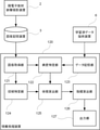
- FIG. 1 is a diagram showing an outline of the configuration of the information processing device 1 according to the embodiment of the present invention. As shown in the figure, the information processing device 1 has a processing unit 11, a storage unit 12, a temporary storage unit 13, an external device connection unit 14, and a communication unit 15, and these components are included. Is electrically connected to the inside of the information processing apparatus 1 via the communication bus 16.
- the processing unit 11 is realized by, for example, a central processing unit (CPU), operates according to a predetermined program stored in the storage unit 12, and realizes various functions.
- CPU central processing unit
- the storage unit 12 is a non-volatile storage medium that stores various information. This is realized by a storage device such as a hard disk drive (Hard Disk Drive: HDD) or a solid state drive (Solid State Drive: SSD).
- the storage unit 12 may be arranged in another device capable of communicating with the information processing device 1.
- the temporary storage unit 13 is a volatile storage medium. This is realized by, for example, a memory such as a random access memory (Random Access Memory: RAM), and temporarily stores information (arguments, arrays, etc.) required when the processing unit 11 operates.
- a memory such as a random access memory (Random Access Memory: RAM)
- RAM Random Access Memory
- the external device connection unit 14 is a connection unit conforming to a standard such as a universal serial bus (Universal Serial Bus: USB) or a high-definition multimedia interface (High-Definition Multimedia Interface: HDMI), and is an input device such as a keyboard or the like.
- a display device such as a monitor can be connected.
- the communication unit 15 is, for example, a communication means conforming to a local area network (LAN) standard, and realizes communication between the information processing device 1 and a local area network or a network such as the Internet via the information processing device 1. ..
- LAN local area network
- the information processing device 1 can be a computer for a general-purpose server, a personal computer, or the like, and the information processing device 1 can be configured by using a plurality of computers.
- the functions of the information processing device 1 will be described.
- the information processing apparatus 100 or the information processing apparatus 110 is realized by the information processing apparatus 1 operating according to the program.
- This program causes the information processing device 1 which is a computer to function as the information processing device 100 or the information processing device 110.
- Both the information processing device 100 and the information processing device 110 are information processing devices that identify the lesion portion based on the positron emission tomography examination.
- FIG. 2 is a block diagram showing a functional configuration of the information processing apparatus 100. Further, FIG. 3 is a block diagram showing a functional configuration of the information processing apparatus 110, and FIG. 4 is a block diagram showing a functional configuration of the information processing apparatus 120.
- the information processing apparatus 100 outputs an image acquisition unit 101, a lesion identification unit 102, a data storage unit 103, an area identification unit 104, an area calculation unit 105, an index calculation unit 106, and the like.
- a unit 107 is provided.
- the image acquisition unit 101 is configured to be able to acquire an image including an anatomical region photographed by the positron emission tomography apparatus 2 from the image storage apparatus 3.
- the image acquisition unit 101 can also be configured to acquire an image directly from the positron emission tomography apparatus 2 without going through the image storage apparatus 3.
- the image acquired by the image acquisition unit 101 is, for example, as shown in FIG. FIG. 5 shows an example of an image taken by the positron emission tomography apparatus 2.
- the anatomical region is a region representing a part or the whole of an anatomical structure such as an organ or tissue of the body, and here, the anatomical region is assumed to include the whole body of the subject.
- the information processing apparatus 100 can support the diagnosis by using only the chest, only the abdomen, and only the liver as the anatomical region.
- the lesion identification portion 102 is configured so that the lesion portion can be identified from the portion where the positron emitting nuclides are accumulated in the image acquired by the image acquisition unit 101 based on the learned data by machine learning.
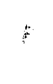
- the result of identifying the lesion portion by the lesion identification portion 102 is, for example, as shown in FIG. FIG. 6 shows an example of an image of the lesion portion identified by the lesion identification portion 102.
- the data storage unit 103 stores the learned data.
- the trained data is generated by machine learning using an image including an anatomical region taken by the positron emission tomography apparatus 2 and instruction information indicating a lesion in the image as teacher data.
- This instruction information is the one in which the lesion is instructed by a specialist.
- a deep learning algorithm called Semantic Segmentation is used. This semantic segmentation associates labels and categories with all pixels in an image, and is used to recognize a group of pixels that form a characteristic category. The details of the teacher data will be described later.
- the trained data stored in the data storage unit 103 may be periodically acquired from the trained data providing device 4 and updated.
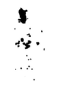
- the region specifying unit 104 is configured to be able to identify the anatomical region in the image. If the anatomical region is whole body, the identified results will be, for example, as shown in FIG. FIG. 7 shows an example of an anatomical region specified by the region identification unit 104.
- the area calculation unit 105 is configured to be able to calculate the area of the lesion portion specified by the lesion identification unit 102 in the image acquired by the image acquisition unit 101 and the area of the anatomical region specified by the region identification unit 104.
- the area of the anatomical area is the projected area of the subject in the image acquired by the image acquisition unit 101, and preferably the area of the anatomical area is the frontal projection of the subject in the image acquired by the image acquisition unit 101.
- the area calculation unit 105 may calculate the area of the lesion based on the contour of the lesion, and may calculate the area of the anatomical region based on the contour of the anatomical region.
- the area of the lesion may be calculated based on the number of pixels, and the area of the anatomical region may be calculated based on the number of pixels of the anatomical region.
- the index calculation unit 106 is configured to be able to calculate an index from the area of the lesion portion specified by the lesion identification unit 102 in the image acquired by the image acquisition unit 101 and the area of the anatomical region specified by the region identification unit 104.
- the index is the ratio of the area of the lesion to the area of the anatomical area. Specifically, the index is the total area of the lesion divided by the area of the anatomical region, the total area of the lesion is 529.5, and the area of the whole body, which is the anatomical region, is 32729. If it is 0, the index is 0.0162.
- the output unit 107 displays the index calculated by the index calculation unit 106 on a display device (not shown), outputs data such as CSV (Comma Separated Value), or prints out on paper via a printing device (not shown). .. At this time, the output unit 107 may output the image acquired by the image acquisition unit 101 together with the index.
- CSV Common Separated Value
- the information processing apparatus 110 includes an image acquisition unit 111, a lesion identification unit 112, a data acquisition unit 113, an area identification unit 114, an area calculation unit 115, and an index calculation unit 116.
- the output unit 117 is provided.
- the image acquisition unit 111, the lesion identification unit 112, the area identification unit 114, the area calculation unit 115, the index calculation unit 116, and the output unit 117 are the image acquisition unit 101, the lesion identification unit 102, and the area of the information processing apparatus 100, respectively. Since the functions are the same as those of the specific unit 104, the area calculation unit 105, the index calculation unit 106, and the output unit 107, the description thereof is omitted here.
- the data acquisition unit 113 is configured to be able to acquire learned data from the learned data providing device 5 via a communication network.
- the trained data acquired from the trained data providing device 5 is the same type as the trained data stored in the data storage unit 103 of the information processing device 100, and is the latest trained data.
- the information processing apparatus 120 includes an image acquisition unit 121, a lesion identification unit 122, a data storage unit 123, an area identification unit 124, a volume calculation unit 125, and an index calculation unit 126. , And an output unit 127.
- the image acquisition unit 121 is configured to be able to acquire an image including an anatomical region photographed by the positron emission tomography apparatus 2 from the image storage apparatus 3.
- the image acquisition unit 121 can also be configured to acquire an image directly from the positron emission tomography apparatus 2 without going through the image storage apparatus 3.
- the lesion identification portion 122 is configured so that the lesion portion can be identified from the portion where the positron emitting nuclides are accumulated in the image acquired by the image acquisition unit 121 based on the learned data by machine learning.
- the data storage unit 123 stores the learned data.
- the trained data is generated by machine learning using an image including an anatomical region taken by the positron emission tomography apparatus 2 and instruction information indicating a lesion in the image as teacher data.
- This instruction information is the one in which the lesion is instructed by a specialist.
- the area specifying unit 124 is configured to be able to specify the anatomical region in the image acquired by the image acquisition unit 121. This image is a three-dimensional image.
- the volume calculation unit 125 is configured to be able to calculate the volume of the lesion portion specified by the lesion identification unit 122 in the image acquired by the image acquisition unit 121 and the volume of the anatomical region specified by the region identification unit 124.
- the volume of the anatomical region is the integrated volume of the tomographic image of the subject in the image acquired by the image acquisition unit 121.
- the volume calculation unit 125 may calculate the volume of the lesion based on the contour of the lesion, and may calculate the volume of the anatomical region based on the contour of the anatomical region.
- the volume of the lesion may be calculated based on the number of pixels of the tomographic image, and the volume of the anatomical region may be calculated based on the number of pixels of the anatomical region.
- the index calculation unit 126 is configured to be able to calculate an index from the volume of the lesion portion specified by the lesion identification unit 122 and the volume of the anatomical region specified by the region identification unit 124.
- the index is the ratio of the volume of the lesion to the volume of the anatomical area.
- the output unit 127 displays the index calculated by the index calculation unit 126 on a display device (not shown), outputs data such as CSV, and prints out on paper through a printing device (not shown).
- a data acquisition unit similar to the data acquisition unit 113 of the information processing apparatus 110 may be provided.
- FIG. 8 is an activity diagram showing an operation flow of the information processing apparatus 100. Since the operation of the information processing apparatus 110 can be inferred from the operation of the information processing apparatus 100, the description of the operation of the information processing apparatus 110 will be omitted.
- an input unit accepts an input of identification information (A101).
- the identification information includes information for identifying the subject (examinee, patient), a reception number for imaging by the positron emission tomography apparatus 2, and the operation differs depending on the facility, but an image taken by the positron emission tomography apparatus 2. Can be identified.
- the image acquisition unit 101 acquires an image taken by the positron emission tomography device 2 from the image storage device 3 (A102).
- the lesion identification unit 102 identifies the lesion portion from the image taken by the positron emission tomography apparatus 2 (A103), and the area calculation unit 105 calculates the area of the lesion portion (A104). ..
- the area specifying unit 104 identifies the anatomical area (A105), and the area calculation unit 105 calculates the area of the area (A106). It should be noted that these processes do not necessarily have to be performed in parallel, and the processes may be performed in the order of A103, A104, A105, A106, the order of A103, A105, A104, A106, or the like.
- the index calculation unit 106 calculates the index based on the area of the lesion and the area of the anatomical region (A107), the output unit 107 outputs the index (A108), and the information processing apparatus 100 operates. To finish.
- teacher data is an image taken by a positron emission tomography device and data that a specialist or the like points out that the lesion is a lesion among the accumulated parts of FDG contained in the image, for example, marking data that marks the lesion. It is composed of a set of.
- marking data is used as teacher data for data without lesions.
- the marking of the lesion portion corresponds to labeling (labeling whether or not the lesion is a lesion) for the accumulated portion of FDG.
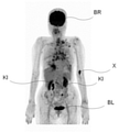
- the image taken by the positron emission tomography apparatus shown in FIG. 9 and the marking data shown in FIG. 10 form a set.
- the marking data mainly corresponds to the part where FDG is accumulated in the image taken by the positron emission tomography apparatus, excluding the brain BR, the kidney KI, and the bladder BL.
- the marking data mainly corresponds to the part where FDG is accumulated in the image taken by the positron emission tomography apparatus, excluding the brain BR, the heart HE, the kidney KI, and the bladder BL. Since the liver LI is basically a place where FDG is weakly accumulated, a strong FDG accumulation portion generated in the liver LI is marked as a lesion.
- the marking data mainly corresponds to the part where FDG is accumulated in the image taken by the positron emission tomography apparatus, excluding the brain BR, the kidney KI, and the bladder BL.
- the non-physiological accumulation part X is not a brain or kidney where FDG is generally accumulated, but a slight leak at the injection site of FDG, and a specialist or the like judges that it is not a lesion part. , Not marked.
- the marking data mainly corresponds to the part where FDG is accumulated in the image taken by the positron emission tomography apparatus, excluding the brain BR, the heart HE, the kidney KI, and the bladder BL.
- the image taken by the positron emission tomography apparatus shown in FIG. 17 and the marking data shown in FIG. 18 form a set.
- the marking data corresponds mainly to the portion where FDG is accumulated in the image taken by the positron emission tomography apparatus, excluding the brain BR, the heart HE, the kidney KI, and the bladder BL. Since it is determined that there is no lesion in the image taken by the positron emission tomography apparatus shown in 12A, the marking data is blank.
- the accumulation of FDG in areas other than the lesion does not always show the same tendency.
- the accumulation in the cardiac HE is performed. Accumulation of FDG is observed, but in the examples shown in FIGS. 13 and 14, there are cases where accumulation of FDG is not observed in the heart, so it is of great significance to use machine learning.
- an information processing device having a function of extracting an integrated portion of FDG from an image taken by a positron emission tomography apparatus and a function of specifying the contour of the extracted integrated portion is used by a specialist.
- Etc. can be made to create teacher data only by instructing the specified contour with a pointing device such as a mouse or a touch pen.
- FIGS. 19 and 20 show examples of lesions identified by the lesion identification portion 102.
- 19 and 20 are views showing an example of a lesion portion identified by the lesion identification portion 102.
- the lesion portion shown in FIG. 19 is a lesion portion identified by the lesion identification portion 102 from the image shown in FIG. 9, and the lesion portion shown in FIG. 20 is identified by the lesion identification portion 102 from the image shown in FIG. It is a lesion that has been damaged.
- FIGS. 27 to 30 are diagrams for explaining case 2
- FIGS. 31 to 34 are diagrams for explaining case 3.
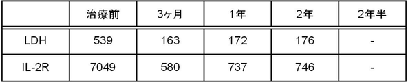
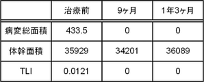
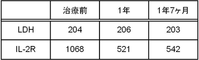
- Cases 1 to 3 are FDG-PET images taken (Interim PET) for the purpose of determining the therapeutic effect during treatment, and also LDH (serum lactate dehydrogenase) and IL-2R. (Interleukin 2 receptor) is being measured.
- FIG. 21 is an FDG-PET image taken in Case 1
- FIG. 22 shows the values of LDH and IL-2R.
- FIG. 23 shows an image in which the lesion portion is identified by the information processing apparatus 1
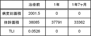
- FIG. 24 shows the result of calculating the total area of the lesion portion, the trunk area, and the TLI from the image. be.
- TLI Total Lesson Index
- FIG. 27 is an FDG-PET image taken in Case 2
- FIG. 28 shows the values of LDH and IL-2R.
- FIG. 29 shows an image in which the lesion portion is identified by the information processing apparatus 1
- FIG. 30 shows the result of calculating the total area of the lesion portion, the trunk area, and the TLI from the image. be.
- FIG. 31 is an FDG-PET image taken in Case 3
- FIG. 32 shows the values of LDH and IL-2R.
- FIG. 33 shows an image in which the lesion portion is identified by the information processing apparatus 1, and
- FIG. 34 shows the result of calculating the total area of the lesion portion, the trunk area, and the TLI from the image. be.
- the information processing apparatus includes a region specifying unit, a volume calculation unit, and an index calculation unit.
- the region specifying unit is configured to be able to specify an anatomical region in the image, and the image is three-dimensional. It is an image, the volume calculation unit is configured to be able to calculate the volume of the lesion and the volume of the anatomical region, the index calculation unit is configured to be able to calculate the index, and the index is the index.
- An information processing device that is the ratio of the volume of the lesion to the volume of the anatomical region. In the information processing device, the volume of the anatomical region is the integrated volume of the tomographic image of the subject in the image.
- the volume calculation unit calculates the volume of the lesion portion based on the contour of the lesion portion, and calculates the volume of the anatomical region based on the contour of the anatomical region.
- Processing equipment In the information processing apparatus, the volume calculation unit calculates the volume of the lesion based on the number of pixels of the tomographic image of the lesion, and the volume calculation unit calculates the volume of the lesion based on the number of pixels of the anatomical region.
- An information processing device that calculates the volume.
- the information processing apparatus includes an area specifying unit, an area calculation unit, and an index calculation unit.
- the area specifying unit is configured to be able to specify an anatomical region in the image, and the area calculation unit is configured.
- the area of the lesion and the area of the anatomical region can be calculated.
- the area of the anatomical region is the projected area of the subject in the image, and the index calculation unit calculates the index.
- An information processing device that is configured to be possible and the index is the ratio of the area of the lesion to the area of the anatomical region.
- the area of the anatomical region is the front projection area of the subject in the image.
- the area calculation unit calculates the area of the lesion based on the contour of the lesion, and calculates the area of the anatomical region based on the contour of the anatomical region. Processing equipment.
- the area calculation unit calculates the area of the lesion based on the number of pixels of the lesion, and calculates the area of the anatomical region based on the number of pixels of the anatomical region.
- Information processing equipment In the information processing device, the anatomical region is an information processing device including the whole body of the subject.
- the information processing device includes a data storage unit, and the data storage unit is an information processing device that stores the learned data.
- the information processing device includes a data acquisition unit, and the data acquisition unit is an information processing device configured to be able to acquire the trained data from the trained data providing device via a communication network.
- the learned data is generated by machine learning using an image including an anatomical region taken by the positron radiation tomography apparatus and instruction information indicating a lesion in the image as teacher data.
- the instruction information is an information processing device in which a lesion portion is instructed by a specialist.
- the lesion can be automatically identified from the test result of the positron emission tomography apparatus, the test result can be easily grasped even by a specialist, even if the specialist is not a specialist. , The labor required to grasp the inspection result can be reduced.
- the lesion can be quantitatively evaluated, so that it is possible to grasp the state transition of cancer or inflammation in the same manner as grasping the result of the blood test, for example.
- Information processing device 2 Positive electron radiation tomography device 3: Image storage device 4: Learned data providing device 5: Learned data providing device 6: Learned data providing device 11: Processing unit 12: Storage unit 13: Temporary storage Unit 14: External device connection unit 15: Communication unit 16: Communication bus 100: Information processing device 101: Image acquisition unit 102: Disease identification unit 103: Data storage unit 104: Area identification unit 105: Area calculation unit 106: Index calculation unit 107: Output unit 110: Information processing device 111: Image acquisition unit 112: Disease identification unit 113: Data acquisition unit 114: Area identification unit 115: Area calculation unit 116: Index calculation unit 117: Output unit 120: Information processing device 121: Image acquisition unit 122: Disease identification unit 123: Data acquisition unit 124: Area identification unit 125: Volume calculation unit 126: Index calculation unit 127: Output unit BL: Bladder BR: Brain HE: Heart KI: Kidney LI: Liver X: Non Physiological accumulation part
Landscapes
- Physics & Mathematics (AREA)
- Engineering & Computer Science (AREA)
- Health & Medical Sciences (AREA)
- General Physics & Mathematics (AREA)
- Life Sciences & Earth Sciences (AREA)
- Medical Informatics (AREA)
- Nuclear Medicine, Radiotherapy & Molecular Imaging (AREA)
- Optics & Photonics (AREA)
- General Health & Medical Sciences (AREA)
- Biomedical Technology (AREA)
- High Energy & Nuclear Physics (AREA)
- Molecular Biology (AREA)
- Spectroscopy & Molecular Physics (AREA)
- Computer Vision & Pattern Recognition (AREA)
- Theoretical Computer Science (AREA)
- Nuclear Medicine (AREA)
Priority Applications (1)
| Application Number | Priority Date | Filing Date | Title |
|---|---|---|---|
| JP2022550644A JP7794451B2 (ja) | 2020-09-18 | 2021-09-21 | 情報処理装置及びプログラム |
Applications Claiming Priority (2)
| Application Number | Priority Date | Filing Date | Title |
|---|---|---|---|
| JP2020-157226 | 2020-09-18 | ||
| JP2020157226 | 2020-09-18 |
Publications (1)
| Publication Number | Publication Date |
|---|---|
| WO2022059799A1 true WO2022059799A1 (ja) | 2022-03-24 |
Family
ID=80776760
Family Applications (1)
| Application Number | Title | Priority Date | Filing Date |
|---|---|---|---|
| PCT/JP2021/034649 Ceased WO2022059799A1 (ja) | 2020-09-18 | 2021-09-21 | 情報処理装置及びプログラム |
Country Status (2)
| Country | Link |
|---|---|
| JP (1) | JP7794451B2 (https=) |
| WO (1) | WO2022059799A1 (https=) |
Citations (7)
| Publication number | Priority date | Publication date | Assignee | Title |
|---|---|---|---|---|
| JP2005118510A (ja) * | 2003-10-15 | 2005-05-12 | Lexi:Kk | 脳の出血部位、腫瘍、挫傷等の計測および観察用プログラム |
| JP2019033924A (ja) * | 2017-08-17 | 2019-03-07 | 富士フイルム株式会社 | 学習データ生成支援装置および学習データ生成支援装置の作動方法並びに学習データ生成支援プログラム |
| JP2019149005A (ja) * | 2018-02-27 | 2019-09-05 | 富士フイルム株式会社 | 医療文書作成支援装置、方法およびプログラム |
| JP2020043927A (ja) * | 2018-09-14 | 2020-03-26 | 富士フイルム株式会社 | 医用画像処理装置、方法およびプログラム |
| JP2020054580A (ja) * | 2018-10-01 | 2020-04-09 | 富士フイルム株式会社 | 疾患領域を判別する判別器の学習装置、方法及びプログラム、疾患領域を判別する判別器、並びに疾患領域判別装置及びプログラム |
| JP2020062355A (ja) * | 2018-10-19 | 2020-04-23 | キヤノンメディカルシステムズ株式会社 | 画像処理装置、データ生成装置及びプログラム |
| WO2020085336A1 (ja) * | 2018-10-25 | 2020-04-30 | 富士フイルム株式会社 | 重み画像生成装置、方法およびプログラム、判別器学習装置、方法およびプログラム、領域抽出装置、方法およびプログラム並びに判別器 |
Family Cites Families (1)
| Publication number | Priority date | Publication date | Assignee | Title |
|---|---|---|---|---|
| JP6706345B2 (ja) * | 2016-12-19 | 2020-06-03 | 富士フイルム株式会社 | 類似症例検索装置とその作動方法および作動プログラム、並びに類似症例検索システム |
-
2021
- 2021-09-21 JP JP2022550644A patent/JP7794451B2/ja active Active
- 2021-09-21 WO PCT/JP2021/034649 patent/WO2022059799A1/ja not_active Ceased
Patent Citations (7)
| Publication number | Priority date | Publication date | Assignee | Title |
|---|---|---|---|---|
| JP2005118510A (ja) * | 2003-10-15 | 2005-05-12 | Lexi:Kk | 脳の出血部位、腫瘍、挫傷等の計測および観察用プログラム |
| JP2019033924A (ja) * | 2017-08-17 | 2019-03-07 | 富士フイルム株式会社 | 学習データ生成支援装置および学習データ生成支援装置の作動方法並びに学習データ生成支援プログラム |
| JP2019149005A (ja) * | 2018-02-27 | 2019-09-05 | 富士フイルム株式会社 | 医療文書作成支援装置、方法およびプログラム |
| JP2020043927A (ja) * | 2018-09-14 | 2020-03-26 | 富士フイルム株式会社 | 医用画像処理装置、方法およびプログラム |
| JP2020054580A (ja) * | 2018-10-01 | 2020-04-09 | 富士フイルム株式会社 | 疾患領域を判別する判別器の学習装置、方法及びプログラム、疾患領域を判別する判別器、並びに疾患領域判別装置及びプログラム |
| JP2020062355A (ja) * | 2018-10-19 | 2020-04-23 | キヤノンメディカルシステムズ株式会社 | 画像処理装置、データ生成装置及びプログラム |
| WO2020085336A1 (ja) * | 2018-10-25 | 2020-04-30 | 富士フイルム株式会社 | 重み画像生成装置、方法およびプログラム、判別器学習装置、方法およびプログラム、領域抽出装置、方法およびプログラム並びに判別器 |
Also Published As
| Publication number | Publication date |
|---|---|
| JP7794451B2 (ja) | 2026-01-06 |
| JPWO2022059799A1 (https=) | 2022-03-24 |
Similar Documents
| Publication | Publication Date | Title |
|---|---|---|
| JP7819016B2 (ja) | 画像処理装置、方法およびプログラム | |
| US11139067B2 (en) | Medical image display device, method, and program | |
| JP5263997B2 (ja) | 医用レポート作成装置、医用レポート作成方法および医用レポート作成プログラム | |
| CN109035284B (zh) | 基于深度学习的心脏ct图像分割方法、装置、设备及介质 | |
| US11580642B2 (en) | Disease region extraction apparatus, disease region extraction method, and disease region extraction program | |
| JP7436698B2 (ja) | 医用画像処理装置、方法およびプログラム | |
| US8045773B2 (en) | Method for segmenting a myocardial wall and device for detecting a coronary artery with pathological changes | |
| JP6929695B2 (ja) | 医用画像診断装置及び管理装置 | |
| JP6734111B2 (ja) | 所見情報作成装置及びシステム | |
| JP6785976B2 (ja) | 脳画像正規化装置、方法およびプログラム | |
| EP3597107A1 (en) | Topogram-based fat quantification for a computed tomography examination | |
| US11334990B2 (en) | Information processing apparatus, information processing method, and program | |
| CN1895185B (zh) | 显示检查对象的检查区域和药剂在体内影响的信息的方法 | |
| JP7794451B2 (ja) | 情報処理装置及びプログラム | |
| JP2022168985A (ja) | 医用画像処理装置及び医用画像処理方法 | |
| US12505544B2 (en) | Image processing apparatus, image processing method, and image processing program | |
| JP6956514B2 (ja) | X線ct装置及び医用情報管理装置 | |
| CN112790778B (zh) | 采集错误对齐 | |
| CN115760680A (zh) | 用于提供医学成像决策支持数据的方法 | |
| CN103295246B (zh) | 通过ecg分析对介入放射学图像的处理 | |
| US20250308024A1 (en) | Image processing device, image processing method, image processing program, learning device, learning method, and learning program | |
| JP7827513B2 (ja) | 情報処理装置、情報処理方法及び情報処理プログラム | |
| CN117830209B (zh) | 一种基于18f-fdg pet/ct的帕金森病预测方法及装置 | |
| Fum et al. | Automatic localization of anatomical landmarks in head cine fluoroscopy images via deep learning | |
| KR102850232B1 (ko) | X-선 영상 기반의 골감소증 진단 방법 및 그 장치 |
Legal Events
| Date | Code | Title | Description |
|---|---|---|---|
| 121 | Ep: the epo has been informed by wipo that ep was designated in this application |
Ref document number: 21869481 Country of ref document: EP Kind code of ref document: A1 |
|
| ENP | Entry into the national phase |
Ref document number: 2022550644 Country of ref document: JP Kind code of ref document: A |
|
| NENP | Non-entry into the national phase |
Ref country code: DE |
|
| 122 | Ep: pct application non-entry in european phase |
Ref document number: 21869481 Country of ref document: EP Kind code of ref document: A1 |