WO2022054925A1 - Complex containing neural retina-containing cell aggregates and matrix, and method for manufacturing same - Google Patents
Complex containing neural retina-containing cell aggregates and matrix, and method for manufacturing same Download PDFInfo
- Publication number
- WO2022054925A1 WO2022054925A1 PCT/JP2021/033387 JP2021033387W WO2022054925A1 WO 2022054925 A1 WO2022054925 A1 WO 2022054925A1 JP 2021033387 W JP2021033387 W JP 2021033387W WO 2022054925 A1 WO2022054925 A1 WO 2022054925A1
- Authority
- WO
- WIPO (PCT)
- Prior art keywords
- cells
- cell
- retinal
- neuroretinal
- matrix
- Prior art date
Links
- 0 C(CC1)C/C=C2/*=CC1=C2 Chemical compound C(CC1)C/C=C2/*=CC1=C2 0.000 description 1
Images


Classifications
-
- C—CHEMISTRY; METALLURGY
- C12—BIOCHEMISTRY; BEER; SPIRITS; WINE; VINEGAR; MICROBIOLOGY; ENZYMOLOGY; MUTATION OR GENETIC ENGINEERING
- C12N—MICROORGANISMS OR ENZYMES; COMPOSITIONS THEREOF; PROPAGATING, PRESERVING, OR MAINTAINING MICROORGANISMS; MUTATION OR GENETIC ENGINEERING; CULTURE MEDIA
- C12N5/00—Undifferentiated human, animal or plant cells, e.g. cell lines; Tissues; Cultivation or maintenance thereof; Culture media therefor
- C12N5/06—Animal cells or tissues; Human cells or tissues
- C12N5/0602—Vertebrate cells
- C12N5/0618—Cells of the nervous system
- C12N5/0621—Eye cells, e.g. cornea, iris pigmented cells
-
- A—HUMAN NECESSITIES
- A61—MEDICAL OR VETERINARY SCIENCE; HYGIENE
- A61L—METHODS OR APPARATUS FOR STERILISING MATERIALS OR OBJECTS IN GENERAL; DISINFECTION, STERILISATION OR DEODORISATION OF AIR; CHEMICAL ASPECTS OF BANDAGES, DRESSINGS, ABSORBENT PADS OR SURGICAL ARTICLES; MATERIALS FOR BANDAGES, DRESSINGS, ABSORBENT PADS OR SURGICAL ARTICLES
- A61L27/00—Materials for grafts or prostheses or for coating grafts or prostheses
- A61L27/50—Materials characterised by their function or physical properties, e.g. injectable or lubricating compositions, shape-memory materials, surface modified materials
- A61L27/58—Materials at least partially resorbable by the body
-
- A—HUMAN NECESSITIES
- A61—MEDICAL OR VETERINARY SCIENCE; HYGIENE
- A61K—PREPARATIONS FOR MEDICAL, DENTAL OR TOILETRY PURPOSES
- A61K35/00—Medicinal preparations containing materials or reaction products thereof with undetermined constitution
- A61K35/12—Materials from mammals; Compositions comprising non-specified tissues or cells; Compositions comprising non-embryonic stem cells; Genetically modified cells
- A61K35/30—Nerves; Brain; Eyes; Corneal cells; Cerebrospinal fluid; Neuronal stem cells; Neuronal precursor cells; Glial cells; Oligodendrocytes; Schwann cells; Astroglia; Astrocytes; Choroid plexus; Spinal cord tissue
-
- A—HUMAN NECESSITIES
- A61—MEDICAL OR VETERINARY SCIENCE; HYGIENE
- A61L—METHODS OR APPARATUS FOR STERILISING MATERIALS OR OBJECTS IN GENERAL; DISINFECTION, STERILISATION OR DEODORISATION OF AIR; CHEMICAL ASPECTS OF BANDAGES, DRESSINGS, ABSORBENT PADS OR SURGICAL ARTICLES; MATERIALS FOR BANDAGES, DRESSINGS, ABSORBENT PADS OR SURGICAL ARTICLES
- A61L27/00—Materials for grafts or prostheses or for coating grafts or prostheses
- A61L27/14—Macromolecular materials
- A61L27/22—Polypeptides or derivatives thereof, e.g. degradation products
- A61L27/225—Fibrin; Fibrinogen
-
- A—HUMAN NECESSITIES
- A61—MEDICAL OR VETERINARY SCIENCE; HYGIENE
- A61L—METHODS OR APPARATUS FOR STERILISING MATERIALS OR OBJECTS IN GENERAL; DISINFECTION, STERILISATION OR DEODORISATION OF AIR; CHEMICAL ASPECTS OF BANDAGES, DRESSINGS, ABSORBENT PADS OR SURGICAL ARTICLES; MATERIALS FOR BANDAGES, DRESSINGS, ABSORBENT PADS OR SURGICAL ARTICLES
- A61L27/00—Materials for grafts or prostheses or for coating grafts or prostheses
- A61L27/36—Materials for grafts or prostheses or for coating grafts or prostheses containing ingredients of undetermined constitution or reaction products thereof, e.g. transplant tissue, natural bone, extracellular matrix
- A61L27/38—Materials for grafts or prostheses or for coating grafts or prostheses containing ingredients of undetermined constitution or reaction products thereof, e.g. transplant tissue, natural bone, extracellular matrix containing added animal cells
- A61L27/3804—Materials for grafts or prostheses or for coating grafts or prostheses containing ingredients of undetermined constitution or reaction products thereof, e.g. transplant tissue, natural bone, extracellular matrix containing added animal cells characterised by specific cells or progenitors thereof, e.g. fibroblasts, connective tissue cells, kidney cells
- A61L27/3834—Cells able to produce different cell types, e.g. hematopoietic stem cells, mesenchymal stem cells, marrow stromal cells, embryonic stem cells
-
- A—HUMAN NECESSITIES
- A61—MEDICAL OR VETERINARY SCIENCE; HYGIENE
- A61L—METHODS OR APPARATUS FOR STERILISING MATERIALS OR OBJECTS IN GENERAL; DISINFECTION, STERILISATION OR DEODORISATION OF AIR; CHEMICAL ASPECTS OF BANDAGES, DRESSINGS, ABSORBENT PADS OR SURGICAL ARTICLES; MATERIALS FOR BANDAGES, DRESSINGS, ABSORBENT PADS OR SURGICAL ARTICLES
- A61L27/00—Materials for grafts or prostheses or for coating grafts or prostheses
- A61L27/40—Composite materials, i.e. containing one material dispersed in a matrix of the same or different material
- A61L27/44—Composite materials, i.e. containing one material dispersed in a matrix of the same or different material having a macromolecular matrix
-
- A—HUMAN NECESSITIES
- A61—MEDICAL OR VETERINARY SCIENCE; HYGIENE
- A61L—METHODS OR APPARATUS FOR STERILISING MATERIALS OR OBJECTS IN GENERAL; DISINFECTION, STERILISATION OR DEODORISATION OF AIR; CHEMICAL ASPECTS OF BANDAGES, DRESSINGS, ABSORBENT PADS OR SURGICAL ARTICLES; MATERIALS FOR BANDAGES, DRESSINGS, ABSORBENT PADS OR SURGICAL ARTICLES
- A61L27/00—Materials for grafts or prostheses or for coating grafts or prostheses
- A61L27/50—Materials characterised by their function or physical properties, e.g. injectable or lubricating compositions, shape-memory materials, surface modified materials
- A61L27/52—Hydrogels or hydrocolloids
-
- A—HUMAN NECESSITIES
- A61—MEDICAL OR VETERINARY SCIENCE; HYGIENE
- A61L—METHODS OR APPARATUS FOR STERILISING MATERIALS OR OBJECTS IN GENERAL; DISINFECTION, STERILISATION OR DEODORISATION OF AIR; CHEMICAL ASPECTS OF BANDAGES, DRESSINGS, ABSORBENT PADS OR SURGICAL ARTICLES; MATERIALS FOR BANDAGES, DRESSINGS, ABSORBENT PADS OR SURGICAL ARTICLES
- A61L2430/00—Materials or treatment for tissue regeneration
- A61L2430/16—Materials or treatment for tissue regeneration for reconstruction of eye parts, e.g. intraocular lens, cornea
-
- C—CHEMISTRY; METALLURGY
- C12—BIOCHEMISTRY; BEER; SPIRITS; WINE; VINEGAR; MICROBIOLOGY; ENZYMOLOGY; MUTATION OR GENETIC ENGINEERING
- C12N—MICROORGANISMS OR ENZYMES; COMPOSITIONS THEREOF; PROPAGATING, PRESERVING, OR MAINTAINING MICROORGANISMS; MUTATION OR GENETIC ENGINEERING; CULTURE MEDIA
- C12N2501/00—Active agents used in cell culture processes, e.g. differentation
- C12N2501/10—Growth factors
- C12N2501/115—Basic fibroblast growth factor (bFGF, FGF-2)
-
- C—CHEMISTRY; METALLURGY
- C12—BIOCHEMISTRY; BEER; SPIRITS; WINE; VINEGAR; MICROBIOLOGY; ENZYMOLOGY; MUTATION OR GENETIC ENGINEERING
- C12N—MICROORGANISMS OR ENZYMES; COMPOSITIONS THEREOF; PROPAGATING, PRESERVING, OR MAINTAINING MICROORGANISMS; MUTATION OR GENETIC ENGINEERING; CULTURE MEDIA
- C12N2501/00—Active agents used in cell culture processes, e.g. differentation
- C12N2501/10—Growth factors
- C12N2501/15—Transforming growth factor beta (TGF-β)
-
- C—CHEMISTRY; METALLURGY
- C12—BIOCHEMISTRY; BEER; SPIRITS; WINE; VINEGAR; MICROBIOLOGY; ENZYMOLOGY; MUTATION OR GENETIC ENGINEERING
- C12N—MICROORGANISMS OR ENZYMES; COMPOSITIONS THEREOF; PROPAGATING, PRESERVING, OR MAINTAINING MICROORGANISMS; MUTATION OR GENETIC ENGINEERING; CULTURE MEDIA
- C12N2501/00—Active agents used in cell culture processes, e.g. differentation
- C12N2501/30—Hormones
- C12N2501/375—Thyroid stimulating hormone [TSH]
-
- C—CHEMISTRY; METALLURGY
- C12—BIOCHEMISTRY; BEER; SPIRITS; WINE; VINEGAR; MICROBIOLOGY; ENZYMOLOGY; MUTATION OR GENETIC ENGINEERING
- C12N—MICROORGANISMS OR ENZYMES; COMPOSITIONS THEREOF; PROPAGATING, PRESERVING, OR MAINTAINING MICROORGANISMS; MUTATION OR GENETIC ENGINEERING; CULTURE MEDIA
- C12N2501/00—Active agents used in cell culture processes, e.g. differentation
- C12N2501/40—Regulators of development
- C12N2501/41—Hedgehog proteins; Cyclopamine (inhibitor)
-
- C—CHEMISTRY; METALLURGY
- C12—BIOCHEMISTRY; BEER; SPIRITS; WINE; VINEGAR; MICROBIOLOGY; ENZYMOLOGY; MUTATION OR GENETIC ENGINEERING
- C12N—MICROORGANISMS OR ENZYMES; COMPOSITIONS THEREOF; PROPAGATING, PRESERVING, OR MAINTAINING MICROORGANISMS; MUTATION OR GENETIC ENGINEERING; CULTURE MEDIA
- C12N2501/00—Active agents used in cell culture processes, e.g. differentation
- C12N2501/40—Regulators of development
- C12N2501/415—Wnt; Frizzeled
-
- C—CHEMISTRY; METALLURGY
- C12—BIOCHEMISTRY; BEER; SPIRITS; WINE; VINEGAR; MICROBIOLOGY; ENZYMOLOGY; MUTATION OR GENETIC ENGINEERING
- C12N—MICROORGANISMS OR ENZYMES; COMPOSITIONS THEREOF; PROPAGATING, PRESERVING, OR MAINTAINING MICROORGANISMS; MUTATION OR GENETIC ENGINEERING; CULTURE MEDIA
- C12N2501/00—Active agents used in cell culture processes, e.g. differentation
- C12N2501/70—Enzymes
- C12N2501/72—Transferases (EC 2.)
- C12N2501/727—Kinases (EC 2.7.)
-
- C—CHEMISTRY; METALLURGY
- C12—BIOCHEMISTRY; BEER; SPIRITS; WINE; VINEGAR; MICROBIOLOGY; ENZYMOLOGY; MUTATION OR GENETIC ENGINEERING
- C12N—MICROORGANISMS OR ENZYMES; COMPOSITIONS THEREOF; PROPAGATING, PRESERVING, OR MAINTAINING MICROORGANISMS; MUTATION OR GENETIC ENGINEERING; CULTURE MEDIA
- C12N2506/00—Differentiation of animal cells from one lineage to another; Differentiation of pluripotent cells
- C12N2506/02—Differentiation of animal cells from one lineage to another; Differentiation of pluripotent cells from embryonic cells
-
- C—CHEMISTRY; METALLURGY
- C12—BIOCHEMISTRY; BEER; SPIRITS; WINE; VINEGAR; MICROBIOLOGY; ENZYMOLOGY; MUTATION OR GENETIC ENGINEERING
- C12N—MICROORGANISMS OR ENZYMES; COMPOSITIONS THEREOF; PROPAGATING, PRESERVING, OR MAINTAINING MICROORGANISMS; MUTATION OR GENETIC ENGINEERING; CULTURE MEDIA
- C12N2506/00—Differentiation of animal cells from one lineage to another; Differentiation of pluripotent cells
- C12N2506/45—Differentiation of animal cells from one lineage to another; Differentiation of pluripotent cells from artificially induced pluripotent stem cells
-
- C—CHEMISTRY; METALLURGY
- C12—BIOCHEMISTRY; BEER; SPIRITS; WINE; VINEGAR; MICROBIOLOGY; ENZYMOLOGY; MUTATION OR GENETIC ENGINEERING
- C12N—MICROORGANISMS OR ENZYMES; COMPOSITIONS THEREOF; PROPAGATING, PRESERVING, OR MAINTAINING MICROORGANISMS; MUTATION OR GENETIC ENGINEERING; CULTURE MEDIA
- C12N2510/00—Genetically modified cells
-
- C—CHEMISTRY; METALLURGY
- C12—BIOCHEMISTRY; BEER; SPIRITS; WINE; VINEGAR; MICROBIOLOGY; ENZYMOLOGY; MUTATION OR GENETIC ENGINEERING
- C12N—MICROORGANISMS OR ENZYMES; COMPOSITIONS THEREOF; PROPAGATING, PRESERVING, OR MAINTAINING MICROORGANISMS; MUTATION OR GENETIC ENGINEERING; CULTURE MEDIA
- C12N2513/00—3D culture
-
- C—CHEMISTRY; METALLURGY
- C12—BIOCHEMISTRY; BEER; SPIRITS; WINE; VINEGAR; MICROBIOLOGY; ENZYMOLOGY; MUTATION OR GENETIC ENGINEERING
- C12N—MICROORGANISMS OR ENZYMES; COMPOSITIONS THEREOF; PROPAGATING, PRESERVING, OR MAINTAINING MICROORGANISMS; MUTATION OR GENETIC ENGINEERING; CULTURE MEDIA
- C12N2533/00—Supports or coatings for cell culture, characterised by material
- C12N2533/50—Proteins
- C12N2533/52—Fibronectin; Laminin
-
- C—CHEMISTRY; METALLURGY
- C12—BIOCHEMISTRY; BEER; SPIRITS; WINE; VINEGAR; MICROBIOLOGY; ENZYMOLOGY; MUTATION OR GENETIC ENGINEERING
- C12N—MICROORGANISMS OR ENZYMES; COMPOSITIONS THEREOF; PROPAGATING, PRESERVING, OR MAINTAINING MICROORGANISMS; MUTATION OR GENETIC ENGINEERING; CULTURE MEDIA
- C12N2533/00—Supports or coatings for cell culture, characterised by material
- C12N2533/50—Proteins
- C12N2533/54—Collagen; Gelatin
-
- C—CHEMISTRY; METALLURGY
- C12—BIOCHEMISTRY; BEER; SPIRITS; WINE; VINEGAR; MICROBIOLOGY; ENZYMOLOGY; MUTATION OR GENETIC ENGINEERING
- C12N—MICROORGANISMS OR ENZYMES; COMPOSITIONS THEREOF; PROPAGATING, PRESERVING, OR MAINTAINING MICROORGANISMS; MUTATION OR GENETIC ENGINEERING; CULTURE MEDIA
- C12N2533/00—Supports or coatings for cell culture, characterised by material
- C12N2533/50—Proteins
- C12N2533/56—Fibrin; Thrombin
-
- C—CHEMISTRY; METALLURGY
- C12—BIOCHEMISTRY; BEER; SPIRITS; WINE; VINEGAR; MICROBIOLOGY; ENZYMOLOGY; MUTATION OR GENETIC ENGINEERING
- C12N—MICROORGANISMS OR ENZYMES; COMPOSITIONS THEREOF; PROPAGATING, PRESERVING, OR MAINTAINING MICROORGANISMS; MUTATION OR GENETIC ENGINEERING; CULTURE MEDIA
- C12N2537/00—Supports and/or coatings for cell culture characterised by physical or chemical treatment
- C12N2537/10—Cross-linking
Definitions
- the present invention relates to a complex containing a cell aggregate containing a neural retina and a matrix, and a method for producing the same.
- Non-Patent Document 1 a method for producing a neural retina from pluripotent stem cells.
- Non-Patent Document 2 a method of solidifying the living retinal tissue with gelatin
- Non-Patent Document 3 a method of embedding the living retinal tissue with temperature-sensitive gelatin
- Non-Patent Document 4 A method of embedding human fetal-derived retinal tissue with gelatin in order to protect fragile fetal tissue (Non-Patent Document 4) and the like.
- Fibrin gel is also administered to humans for the purpose of tissue repair by tissue adhesion and closure.
- tissue adhesion and closure One example is Bolheel® tissue adhesion.
- a laminate of fibrin gel and sheet-like cell culture in which fibrin gel is used to increase the strength of tissue for transplantation is disclosed (Patent Document 3).
- Non-Patent Document 5 Furthermore, there is a report on transplantation of retinal progenitor cells embedded in fibrin gel.
- a method of formulating a tissue for transplantation for regenerative medicine application a method of easily adhering a plurality of tissues is desired. Therefore, it is an object of the present invention to provide a complex containing a cell aggregate containing two or more neural retinas.
- the present inventors have investigated a method for forming a complex by adhering or embedding a cell aggregate containing two or more neural retinas with some kind of "glue".
- the foreign substance "glue” hinders the engraftment of the complex after transplantation and can be a foreign substance to the living body. Therefore, as the "glue”, we considered that a matrix, which is a substance that adheres firmly in a small amount and is decomposed in a living body, is preferable.
- the present inventors used fibrin gel or gelatin as "glue" to embed cell aggregates containing two or more neural retinas, and the cell aggregates containing two or more neural retinas and the matrix. Succeeded in producing the complex.
- this method by embedding a cell aggregate containing two or more neural retinas in a matrix, it is possible to inhale and blow out with a syringe without breaking the matrix gel.
- a complex comprising a matrix and a cell aggregate containing a neural retina derived from two or more pluripotent stem cells, wherein the two or more cell aggregates are arranged in the matrix.
- the neural retina is a neural retinal sheet for transplantation having the following characteristics (1) to (10): (1) Derived from pluripotent stem cells (2) It has a three-dimensional structure and has a three-dimensional structure.
- the photoreceptor layer contains one or more cells selected from the group consisting of photoreceptor progenitor cells and photoreceptors.
- the inner layer contains one or more cells selected from the group consisting of retinal progenitor cells, ganglion cells, amacrine cells and bipolar cells, horizontal cells, and Muller glial cells.
- the surface of the neuroretinal layer has an apical surface and has an apical surface.
- the inner layer is present inside the photoreceptor layer existing along the apical surface.
- the area of the nerve retinal layer is 50% or more with respect to the total area of the surface of the nerve retinal sheet for transplantation.
- the area of the continuous epithelial structure is 80% or more of the total area of the apical surface of the neuroretinal layer.
- the expression of the neuroretinal cell-related gene was observed in the transplanted neural retinal sheet, and the expression of the non-neuretic cell-related gene was not observed, and the non-neuretic cell-related gene was found in the brain. Contains one or more genes selected from the group consisting of spinal tissue marker genes and eyeball-related tissue marker genes.
- [3] The complex according to [1] or [2], wherein the cell aggregate has a major axis of 600 ⁇ m to 2500 ⁇ m, and the two or more cell aggregates are arranged in a row in the matrix.
- [5] A method for producing a complex in which cell aggregates containing two or more neural retinas are arranged in a matrix. (1) The first step of producing cell aggregates of two or more neural retinas from pluripotent stem cells, (2) A second step of contacting the two or more cell aggregates with the matrix or the precursor of the matrix in a predetermined arrangement and then gelling the matrix. Manufacturing method, including.
- the matrix is a fibrin gel, and in the second step, the two or more cell aggregates are arranged in a row, and the two or more arranged cell aggregates are brought into contact with either a fibrinogen solution or a thrombin solution.
- the production method according to [5] or [6], wherein the matrix is gelatin.
- a pharmaceutical composition comprising the complex according to any one of [1] to [3].
- a method for treating a disease caused by a disorder of retinal cells or retinal tissue or a disorder of retinal tissue which comprises transplanting the complex according to any one of [1] to [3] into a subject requiring transplantation. ..
- NR neural retinas
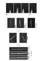
- Example 1 a bright-field stereomicrograph and a fluorescent entity examined in a process of encapsulating a plurality (8 to 13) transplant nerve retinas (Caps) in a fibringel (complex) and aspiration / ejection using a surflo. It is a micrograph and a bright field + fluorescent stereomicrograph.
- Example 2 a bright-field stereomicrograph examined the process of encapsulating a plurality (8 to 10) nerve retinas for transplantation (Cap) in gelatin (complex) and suction / ejection using a 1 ml tip with a thick tip. Photographs, fluorescent stereomicrographs and brightfield + fluorescent stereomicrographs.
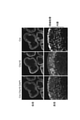
- 6 is a fluorescence microscope image showing the result of immunostaining with Crx and Chx10 on a cell aggregate containing a nerve retina for transplantation in Reference Example 1.
- 6 is a fluorescence microscope image showing the result of immunostaining with Rx and Recoverin on a cell aggregate containing a nerve retina for transplantation in Reference Example 1.
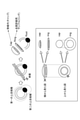
- It is a conceptual diagram which makes a cap and a ring from a typical cell aggregate.
- the parts shown in black and gray mean unintended tissues.
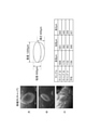
- the height, major axis and minor axis of the implant in Reference Example 3 are shown together with an image of a typical implant and a schematic diagram of the implant.
- FIG. 6 is a confocal fluorescence microscope image showing the result of immunostaining the graft with Crx, Chx10, Rx and Recoverin in Reference Example 4.
- the result of quantitative PCR analysis of gene expression in RNA extracted from a cap and a ring in Reference Example 5 is shown.
- the result of quantitative PCR analysis of gene expression in RNA extracted from a cap and a ring in Reference Example 5 is shown. It is an image showing the result of transplanting a transplant piece (cap) under the rat retina after analyzing RNA extracted from the ring by quantitative PCR in Reference Example 6, and observing the engraftment image after transplantation with a fluorescence microscope.
- 6 is a fluorescence microscope image showing the result of immunostaining on a cap and a ring prepared from one cell aggregate in Reference Example 7.
- 8 is a fluorescence microscope image showing the result of immunostaining a cap made from one cell aggregate in Reference Example 8.
- stem cell an undifferentiated cell capable of differentiating and proliferating (particularly self-renewal).
- Stem cells include subpopulations such as pluripotent stem cells, multipotent stem cells, and unipotent stem cells, depending on their ability to differentiate.
- Pluripotent stem cells are capable of being cultured in vitro and capable of differentiating into all three germ layers (ectoderm, mesodermal, endoderm) and / or all cell lineages belonging to extraembryonic tissues (pluripotent). It refers to stem cells having sex (pluripotency).
- Multipotent stem cells mean stem cells capable of differentiating into multiple, but not all, types of tissues and cells.
- Unipotent stem cells mean stem cells that have the ability to differentiate into specific tissues or cells.
- pluripotent stem cells can be derived from fertilized eggs, cloned embryos, reproductive stem cells, tissue stem cells, somatic cells, etc.
- pluripotent stem cells include embryonic stem cells (ES cells: Embryonic stem cells), EG cells (Embryonic germ cells), and induced pluripotent stem cells (iPS cells: induced pluripotent stem cells).
- Muse cells Multi-lineage differentialing stress ending cells obtained from mesenchymal stem cells (MSC) and GS cells produced from germ cells (eg, testis) are also included in pluripotent stem cells.
- Embryonic stem cells Human embryonic stem cells were established in 1998 and are being used in regenerative medicine. Embryonic stem cells can be produced by culturing inner cell aggregates on feeder cells or in a medium containing bFGF. Methods for producing embryonic stem cells are described in, for example, WO96 / 22362, WO02 / 10157, US5,843,780, US6,200,806, US6,280,718 and the like. Embryonic stem cells can be obtained from predetermined institutions or can be purchased commercially. For example, human embryonic stem cells KhES-1, KhES-2 and KhES-3 are available from the Institute for Frontier Medical Sciences, Kyoto University.
- Human ES cells (derived from KhES-1, non-patent Document 1) genetically modified to have Rx :: Venus, Rx :: AcGFP and Crx :: Venus reporter genes, which are human embryonic stem cells, are physics and chemistry research corporations. It is available from the office.
- “Induced pluripotent stem cells” are cells in which pluripotency is induced by reprogramming somatic cells by a known method or the like.
- Induced pluripotent stem cells were established in mouse cells by Yamanaka et al. In 2006 (Cell, 2006,126 (4), pp.663-676). Induced pluripotent stem cells were also established in human fibroblasts in 2007 and have pluripotency and self-renewal ability similar to embryonic stem cells (Cell, 2007, 131 (5), pp.861-872; Science). , 2007, 318 (5858), pp. 1917-1920; Nat. Biotechnol., 2008, 26 (1), pp. 101-106).
- Specific pluripotent stem cells include differentiated somatic cells such as fibroblasts and peripheral blood mononuclear cells in Oct3 / 4, Sox2, Klf4, Myc (c-Myc, N-Myc, L-Myc). Examples thereof include cells that have been reprogrammed to induce pluripotency by expression of any combination of a plurality of genes selected from the reprogramming gene group including Glis1, Nanog, All4, lin28, Esrrb and the like. Preferred combinations of reprogramming factors include (1) Oct3 / 4, Sox2, Klf4, and Myc (c-Myc or L-Myc), (2) Oct3 / 4, Sox2, Klf4, Lin28, and L-Myc (Stem). Cells, 2013; 31: 458-466) can be mentioned.
- induced pluripotent stem cells In addition to the method of producing induced pluripotent stem cells by direct reprogramming by gene expression, it is also possible to induce induced pluripotent stem cells from somatic cells by adding a compound or the like (Science, 2013, 341, pp. 651-). 654).
- induced pluripotent stem cells for example, 201B7 cells, 201B7-Ff cells, 253G1 cells, 253G4 cells, 1201C1 cells, 1205D1 cells, 1210B2 cells established at Kyoto University.
- 1231A3 cells and other human induced pluripotent cell lines are available from Kyoto University and iPS Academia Japan Co., Ltd.
- the induced pluripotent stem cells for example, Ff-I01 cells, Ff-I14 cells and QHJI01s04 cells established at Kyoto University are available from Kyoto University.
- pluripotent stem cells are preferably embryonic stem cells or induced pluripotent stem cells, and more preferably induced pluripotent stem cells.
- pluripotent stem cells are human pluripotent stem cells, preferably human induced pluripotent stem cells (iPS cells) or human embryonic stem cells (ES cells) (eg, embryos within 14 days of fertilization). Embryonic stem cells established from).
- iPS cells human induced pluripotent stem cells
- ES cells human embryonic stem cells
- Pluripotent stem cells such as human iPS cells can be subjected to maintenance culture and expansion culture by a method well known to those skilled in the art.
- the “retinal tissue” means a tissue in which one or more types of retinal cells constituting each retinal layer exist in a living retina according to a certain order, and is called "Neural Retina”.
- Retinal cell means a cell constituting each retinal layer in a living retina or a progenitor cell thereof.
- Retinal cells include photoreceptor cells (rod photoreceptor cells, pyramidal photoreceptor cells), horizontal cells, amacrine cells, intervening nerve cells, retinal ganglion cells (ganglion cells), and bipolar cells (rod bipolar cells, pyramidal cells).
- Bipolar cells Muller glia cells, retinal pigment epithelial (RPE) cells, hairy bodies, these precursor cells (eg, photoreceptor precursor cells, bipolar cell precursor cells, etc.), retinal precursor cells, etc. Not limited to.
- the neuroretinal layer also referred to as neuroretinal cells or neural retina-related cells
- photoreceptor cells rod photoreceptor cells, pyramidal vision
- Cells horizontal cells, amacrine cells, intervening nerve cells, retinal ganglion cells (ganglion cells), bipolar cells (rod bipolar cells, pyramidal bipolar cells), Mullerglia cells, and precursors thereof (eg, visual imaging).
- Examples include cells such as cell precursor cells, bipolar cell precursor cells, etc.). That is, the neuroretinal cells do not include retinal pigment epithelial cells and ciliary cells.
- “Mature retinal cells” means cells that can be contained in the retinal tissue of a human adult, specifically, photoreceptor cells (rod photoreceptor cells, pyramidal photoreceptor cells), horizontal cells, amacrine cells, intervening cells. Means differentiated cells such as nerve cells, retinal ganglion cells (ganglion cells), bipolar cells (rod bipolar cells, pyramidal bipolar cells), Mullerglia cells, retinal pigment epithelial (RPE) cells, hairy body cells, etc. do.
- “Immature retinal cells” means progenitor cells that have been determined to differentiate into mature retinal cells (eg, photoreceptor progenitor cells, bipolar cell progenitor cells, retinal progenitor cells, etc.).
- the photoreceptor cells, horizontal cell precursors, bipolar cell precursor cells, amacrine cell precursor cells, retinal ganglion cell precursor cells, Mullagria precursor cells, and retinal pigment epithelial precursor cells are photoreceptor cells, horizontal cells, and bipolar cells, respectively.
- Retinal progenitor cell means photoreceptor precursor cell, horizontal cell precursor cell, bipolar cell precursor cell, amacrine cell precursor cell, retinal ganglion cell precursor cell, Mullagria cell, retinal pigment epithelial precursor cell, etc. It is a precursor cell that can differentiate into any of the immature retinal cells, and finally, photoreceptor cells, rod photoreceptors, pyramidal photoreceptors, horizontal cells, bipolar cells, amacrine cells, and retinal ganglion cells. , A precursor cell that can differentiate into any mature retinal line cell such as retinal pigment epithelial cell.
- the "photoreceptor cell” exists in the photoreceptor layer of the retina in a living body and has a role of absorbing a light stimulus and converting it into an electric signal.
- photoreceptor cells There are two types of photoreceptor cells: cones that function in the light and rods (or rods, rods) that function in the dark (referred to as pyramidal photoreceptors and rod photoreceptors, respectively).
- the pyramidal photoreceptors include S-opsin-expressing S-opsin and receiving blue light, L-opsin-expressing L-opsin and receiving red light, and M-opsin. M pyramidal photoreceptors that receive green light can be mentioned.
- the photoreceptor cells differentiate from the photoreceptor progenitor cells and mature.
- the cell is a photoreceptor cell or a photoreceptor precursor cell
- Whether or not the cell is a photoreceptor cell or a photoreceptor precursor cell
- cell markers described later Crx and Blimp1 expressed in photoreceptor precursor cells, Recoverin expressed in photoreceptor cells, maturation). It can be easily confirmed by the expression of rhodopsin (S-Opin, M / L-Opin, etc.) expressed in photoreceptor cells, the formation of the outer segment structure, and the like.
- the photoreceptor progenitor cells are Crx-positive cells and the photoreceptors are rhodopsin, S-Opsin and M / L-Opin positive cells.
- rod photoreceptors are NRL and Rhodopsin positive cells.
- the S-pyramidal photoreceptors are S-opsin-positive cells
- the L-pyramidal photoreceptors are L-opsin-positive cells
- the M-pyramidal photoreceptors are M-opsin-positive cells. That is, the photoreceptor cells in the present specification are concepts including photoreceptor progenitor cells and mature photoreceptor cells.
- the neural retina preferably contains 10% or more, more preferably 20% or more of photoreceptor cells or photoreceptor precursor cells.
- the neural retina preferably contains 10% or more, more preferably 20% or more of neural retinal progenitor cells. In addition, the neural retina preferably contains 10% or more of photoreceptor progenitor cells. Also, in one embodiment, the neural retina contains 3% or more of mature photoreceptor cells.
- neuroretinal cells can be confirmed by the presence or absence of expression of a neuroretinal cell-related gene (hereinafter, may be referred to as "nerve retinal cell marker” or “nerve retinal marker”).
- a neuroretinal cell-related gene hereinafter, may be referred to as "nerve retinal cell marker” or “nerve retinal marker”
- the presence or absence of expression of the neuroretinal cell marker, or the proportion of cells positive for the neuroretinal cell marker in the cell population or tissue can be easily confirmed by those skilled in the art.
- a method using an antibody, a method using a nucleic acid primer, and a method using a sequence reaction can be mentioned.
- the expression of a protein of a neuroretinal cell marker can be determined by, for example, a method such as flow cytometry or immunostaining using a commercially available antibody to determine the number of cells positive for a specific neuroretinal cell marker. It can be confirmed by dividing by the total number of cells.
- a method using a nucleic acid primer the expression of RNA of a neuroretinal cell marker can be confirmed by, for example, a PCR method, a semi-quantitative PCR method, or a quantitative PCR method (eg, real-time PCR method).
- a method using a sequence reaction the expression of RNA of a neuroretinal cell marker can be confirmed by using, for example, a nucleic acid sequencer (eg, next-generation sequencer).
- Neuroretinal cell markers include Rx (also referred to as Rax) and PAX6 expressed in retinal progenitor cells, Rx, PAX6 and Chx10 (also referred to as Vsx2) expressed in neuroretinal progenitor cells, Crx expressed in photoreceptor progenitor cells, and Blimp 1 and the like can be mentioned. It is also expressed in Chx10 strongly expressed in bipolar cells, PKC ⁇ , Go ⁇ , VSX1 and L7 expressed in bipolar cells, TuJ1 and Brn3 expressed in retinal ganglion cells, Conetinin and HPC-1 expressed in amacrine cells, and horizontal cells.
- Calbindin Recoverin expressed in photoreceptors and photoreceptor precursors, Rhodopsin expressed in rod cells, Nrl expressed in rod photoreceptors and rod photoreceptor precursors, S-opsin and LM expressed in cone photoreceptors -OPsin, pyramidal cells, RXR- ⁇ expressed in pyramidal photoreceptors and ganglion cells, among pyramidal photoreceptors, TR ⁇ 2, OTX2 expressed in pyramidal photoreceptors appearing in the early stage of differentiation or precursors thereof And OC2, Pax6 commonly expressed in horizontal cells, amacrine cells and ganglion cells.
- “Positive cell” means a cell expressing a specific marker on the cell surface or intracellularly.
- Chx10 positive cell means a cell expressing the Chx10 protein.
- retinal pigment epithelial cell means an epithelial cell existing outside the neural retina in the living retina. Whether or not the cell is a retinal pigment epithelial cell can be determined by those skilled in the art, for example, the expression of cell markers (RPE65, MITF, CRALBP, MERTK, BEST1, TTR, etc.), the presence of melanin granules (blackish brown), and between cells. It can be easily confirmed by the tight junction of the cell, and the characteristic cell morphology of polygonal and paving stones. Whether or not the cell has the function of retinal pigment epithelial cell can be easily confirmed by the secretory ability of cytokines such as VEGF and PEDF. In one embodiment, the retinal pigment epithelial cells are RPE65-positive cells, MITF-positive cells, or RPE65-positive and MITF-positive cells.
- the "retinal layer” means each layer constituting the retina, specifically, the retinal pigment epithelial layer, the photoreceptor layer, the external limiting membrane, the outer nuclear layer, the outer plexiform layer, the inner nuclear layer, the inner plexiform layer, and the like.
- the ganglion cell layer, nerve fiber layer and inner limiting membrane can be mentioned.
- the “nerve retina layer” means each layer constituting the nerve retina, and specifically, the photoreceptor layer, the outer limiting membrane, the outer nuclear layer, the outer plexiform layer, the inner nuclear layer, the inner plexiform layer, and the ganglion cell. Layers, nerve fiber layers and inner limiting membranes can be mentioned.
- the "photoreceptor layer” is one or more species formed on the outermost side of the neural retina and selected from the group consisting of photoreceptor cells (rod photoreceptor cells, cone photoreceptors), photoreceptor precursor cells and retinal precursor cells. It means the retinal layer containing many cells. Each layer other than the photoreceptor layer is called the inner layer. Which cell constitutes each retinal layer can be confirmed by a known method, for example, the presence or absence of expression of a cell marker or the degree of expression.
- the layer containing proliferating neuroretinal precursor cells is called "neuroblastic layer", and inner neuroblastic layer and outer neuroblastic layer are used.
- neutral layer the layer containing proliferating neuroretinal precursor cells.
- inner neuroblastic layer and outer neuroblastic layer are used.
- Those skilled in the art can make a judgment by a method well known, for example, under a bright-field microscope, the shade of color (the outer neuroblastic layer is light and the inner neuroblastic layer is dark).
- the "ciliary body” includes the developmental process and the adult “ciliary body”, “ciliary body periphery”, and “Ciliary body”. Markers for the “ciliary body” include Zic1, MAL, HNF1beta, FoxQ1, CLDN2, CLDN1, GPR177, AQP1 and AQP4.
- the "ciliary marginal zone (CMZ)” is, for example, a tissue existing in the boundary region between the neural retina and the retinal pigment epithelium in the living retina, and the tissue stem cells (retinal stem cells) of the retina. The area to be included can be mentioned.
- the ciliary margin is also referred to as a ciliary margin or a retinal margin, and the ciliary margin, the ciliary margin and the retinal margin are equivalent tissues.
- the peripheral part of the ciliary body plays an important role in supplying retinal progenitor cells and differentiated cells to the retinal tissue, maintaining the retinal tissue structure, and the like.
- the marker gene at the periphery of the ciliary body include the Rdh10 gene (positive), the Otx1 gene (positive), and the Zic1 (positive).
- the "ciliary body peripheral edge-like structure" is a structure similar to the ciliary body peripheral edge.
- the "cell aggregate” is not particularly limited as long as a plurality of cells adhere to each other to form a three-dimensional structure, and for example, cells dispersed in a medium such as a medium are used. It refers to a mass formed as a group, or a mass of cells formed through cell division. Cell aggregates also include those forming specific tissues.
- Sphere-like cell aggregate means a cell aggregate having a three-dimensional shape close to a spherical surface.
- a three-dimensional shape close to a sphere is a shape having a three-dimensional structure, and when projected onto a two-dimensional surface, for example, a sphere showing a circular or elliptical shape and a plurality of spheres are fused and formed. (For example, when projected in two dimensions, it indicates a shape formed by overlapping 2 to 4 circles or ellipses).
- the core portion of the aggregate has a follicular layered structure, and is characterized in that the central portion is dark and the outer edge portion is brightly observed under a bright-field microscope.
- Epithelial tissue is a tissue formed by cells covering the surface of the body surface, lumen (digestive tract, etc.), body cavity (pericardial cavity, etc.) without gaps.
- the cells that form the epithelial tissue are called epithelial cells.
- Epithelial cells have apical-basal polarity.
- Epithelial cells can form a cell layer by forming strong bonds between epithelial cells by adhesion junction and / or tight junction.
- Epithelial tissue is a tissue formed by stacking one to a dozen or so layers of these cell layers.
- Tissues that can form epithelial tissue also include fetal and / or adult retinal tissue, cerebrospinal tissue, eyeball tissue, nervous tissue and the like.
- the neural retina as used herein is also an epithelial tissue.
- Epithelial structure means a structure characteristic of epithelial tissue, such as the apical surface or basement membrane.
- the epithelial structure including nerve tissue is referred to as neuroepithelium.
- the epithelial structure including the neuroretinal is called the neural retinal epithelium.
- Continuous epithelial tissue is a tissue having a continuous epithelial structure.
- the continuous epithelial structure is a state in which epithelial tissue is continuous.
- the continuous epithelial tissue means, for example, 10 cells to 107 cells in the tangential direction with respect to the epithelial tissue, preferably 30 cells to 107 cells in the tangential direction, and more preferably 102 cells to 107 cells side by side. It is a state of being.
- the continuous epithelial structure does not have a structure in which the apical surface is divided as can be seen in the rosette-like structure.
- the number of cells per area of the cross section of the retinal tissue having a continuous epithelial structure is, for example, the number of cells per area of the cross section of the retinal tissue having a continuous epithelial structure in one aspect of cells in a frozen section having a thickness of about 10 ⁇ m.
- 10 cells to 900 cells preferably 30 cells to 300 cells, more preferably 50 cells to 250 cells, still more preferably, per 100 ⁇ m 2 .
- the continuous epithelium formed in the retinal tissue generally includes at least the photoreceptor layer (outer nuclear layer) among the layers in which the retinal tissue has an apical surface peculiar to the epithelial tissue and the apical surface forms the neuroretinal layer. It is formed parallel and continuously on the surface of retinal tissue.
- an apical surface is formed on the surface of the aggregate, and 10 cells or more, preferably 30 cells or more, more preferably 30 cells or more in the tangential direction with respect to the surface.
- a continuous neural epithelium is formed in which 100 or more cells, more preferably 400 or more photoreceptor cells or photoreceptor precursor cells are regularly and continuously arranged in a row.
- the neuroretinal including such continuous neuroepithelium is a neuroretinal epithelium containing continuous epithelium.
- the epithelial tissue is polarized to form an "apical surface” and a “basement membrane".
- the “basement membrane” refers to a layer (basement membrane) on the basal side produced by epithelial cells, which is rich in laminin and type IV collagen and has a thickness of 50 to 100 nm.
- the "apical surface” refers to the surface (surface layer surface) formed on the opposite side of the "basement membrane”.
- the "apical surface” is an photoreceptor cell in which an external limiting membrane is formed and the photoreceptor cells and photoreceptor precursor cells are present in the retinal tissue whose developmental stage has progressed to the extent that photoreceptor cells or photoreceptor precursor cells are recognized.
- apical-PKC apical-PKC
- E-cadherin E-cadherin
- N-cadherin an antibody against a marker on the apical surface
- the retinal tissue contains continuous epithelium can be confirmed by the continuity of the apical surface of the retinal tissue (that is, the undivided morphology).
- the continuity of the apical surface is, for example, a marker of the apical surface (eg, aPKC, E-cadherin, N-cadherin), a marker of a photoreceptor cell or a photoreceptor progenitor cell located on the apical surface side (eg, Crx or recovery). It can be determined by immunostaining and analyzing the positional relationship between the apical surface, the photoreceptor layer, and each retinal layer on the acquired image or the like.
- DAPI staining For the retinal layer other than the apical surface and the photoreceptor layer (outer nuclear layer), DAPI staining, PI staining, Hoechst staining, or marker proteins localized in the cell nucleus (Rx, Chx10, Ki67, Crx, etc.), etc. It can be identified by immunostaining with.
- the presence of cells continuously expressing a marker of cells existing on the apical surface side that is, a photoreceptor marker or a photoreceptor progenitor cell marker and a marker capable of staining the cell nucleus, is present on the apical surface. Continuity can be identified.
- the complex of the present invention contains a cell aggregate containing two or more pluripotent stem cell-derived neural retinas and a matrix, and the two or more cell aggregates are arranged in the matrix.
- cell aggregates including the neural retina, a matrix, and a complex containing these will be described in detail.
- the cell aggregate containing the neural retina is a cell aggregate (sphere-like cell aggregate) described later.
- the cell aggregate comprising the neural retina is a sheet-like retinal tissue excised from the sphere-like cell aggregate.
- the sheet-shaped retinal tissue is preferably a sheet-shaped neural retina containing a continuous epithelium of the neural retina (hereinafter, also referred to as a neural retinal sheet).
- the cell aggregate comprising the neural retina is a neural retina for transplantation, preferably a neural retina sheet for transplantation.
- the nerve retina for transplantation or the nerve retina sheet for transplantation is a human nerve retina suitable for human transplantation, and more preferably consists of only the nerve retina.
- a neural retinal layer including at least a photoreceptor layer is formed, and the photoreceptor layer contains at least one or more cells selected from the group consisting of photoreceptor cells, photoreceptor precursor cells, and retinal precursor cells.
- the photoreceptor layer is formed at least on the outermost side of the cell aggregate, and the photoreceptor or photoreceptor precursor cells may also be present inside the cell aggregate, or the photoreceptor layer is also formed inside. May be.
- the photoreceptor cells and the like are continuously present in the tangential direction of the surface of the cell aggregate, that is, adhere to each other, and the photoreceptor cells and the like are continuously present in the tangential direction of the surface of the cell aggregate. Etc.
- the tangential direction refers to the tangential direction with respect to the surface of the cell aggregate, that is, the direction in which the photoreceptor cells and the like in the photoreceptor layer are lined up, and is the parallel direction or the lateral direction with respect to the nerve retina.
- the neural retina is derived from pluripotent stem cells, and is preferably derived from human pluripotent stem cells.
- the human pluripotent stem cell is preferably a human embryonic stem cell or a human induced pluripotent stem cell, and more preferably a human induced pluripotent stem cell.
- the nerve retina is preferably a nerve retina for transplantation.
- the front surface (Apical surface) and the back surface (Basal surface) are included, and the surface forms an adhesive bond between cells to form an apical surface including a neuroretinal layer which is an epithelial tissue.
- the back surface includes the retinal tissue constituting the basal plane close to the inner layer of the neural retina.
- Such retinal tissue can be called neuroretinal epithelium.
- a neural retina sheet which is a sheet-shaped retinal tissue, is preferable.
- the front surface has a smooth shape with little change in curvature, and the back surface has an irregular shape with a large change in curvature.
- the change in the curvature of the surface of the retinal tissue may be close to the change in the curvature of the ellipse (for example, the major axis 1 to 10 with respect to the minor axis 1) (which can also be said to be a continuous change in the curvature).
- the change in curvature of the back surface of the retinal tissue is close to a sudden change in curvature (which can also be said to be a sudden change in curvature) that goes back and forth between positive and negative values, such as "saw teeth”. There is.
- Cell aggregates containing the neural retina are derived from pluripotent stem cells and can be specifically obtained by inducing differentiation of pluripotent stem cells.
- the method disclosed in 2014 Jun 10; 5: 4047 is mentioned as a method for inducing differentiation of the neuroretinal, but is not particularly limited.
- a cell aggregate containing a neural retina can be prepared by a method including the following steps (A), (B), (C) and (D).
- B A step of forming cell aggregates by suspending and culturing the cells obtained in step (A).
- C The step of further suspend-culturing the cell aggregate obtained in step (B) in a medium containing a BMP signal transduction pathway agent, and the step of (D) the cell aggregate obtained in step (C). The process of suspension culture and maturation.
- step (A) may further contain a TGF ⁇ family signaling pathway inhibitor and / or a sonic hedgehog signaling pathway agent.
- step (B) may include a sonic hedgehog signaling pathway agent and / or a Wnt signaling pathway inhibitor, as described below.
- This method is also disclosed, for example, in WO2015 / 025967, WO2016 / 063985, WO2017 / 183732, and more specifically in WO2015 / 025967, WO2016 / 063985, WO2017 / 183732, WO2019 / 017492, WO2019 / 054514, WO2019 / 054515. Etc. can be referred to.
- the "pluripotent stem cell culture medium” used in the step (A) is a medium capable of culturing pluripotent stem cells under feeder-free conditions, and examples of the medium include a medium containing an undifferentiated maintenance factor. ..
- the undifferentiated maintenance factor is not particularly limited as long as it is a substance having an action of suppressing the differentiation of pluripotent stem cells.
- undifferentiated maintenance factors commonly used by those skilled in the art include FGF signaling pathway agonists, TGF ⁇ family signaling pathway agonists, insulin and the like.
- FGF signal transduction pathway agent include fibroblast growth factors (eg, bFGF, FGF4 and FGF8).
- TGF ⁇ family signal transduction pathway agent include a TGF ⁇ signal transduction pathway agent and a Nodal / Activin signal transduction pathway agent.
- TGF ⁇ signal transduction pathway agent include TGF ⁇ 1 and TGF ⁇ 2.
- Nodal / Activin signal transduction pathway agent examples include Nodal, ActivinA, and ActivinB.
- the medium in step (A) preferably contains bFGF as an undifferentiated maintenance factor.
- the concentration of the undifferentiated maintenance factor in the medium used in the step (A) is a concentration capable of maintaining the undifferentiated state of the pluripotent stem cells to be cultured, and can be appropriately set by those skilled in the art.
- concentration is usually about 4 ng to 500 ng / mL, preferably about 10 ng to 200 ng / mL, and more preferably about 30 ng to 150 ng. It is about / mL.
- Essential 8 media manufactured by Life Technologies.
- Essential 8 medium is added to DMEM / F12 medium as additives such as L-ascorbic acid-2-phosphate magnesium (64 mg / L), sodium cellenium (14 ⁇ g / L), insulin (19.4 mg / L), NaHCO3 (543 mg).
- TGF ⁇ 1 (2 ng / mL) or Nodal (100 ng / mL)
- S-media manufactured by DS Pharma Biomedical
- StemPro manufactured by Life Technologies
- hESF9 Proc. Natl. Acad. Sci. USA.
- the present invention can be easily carried out.
- the medium used in the step (A) is a serum-free medium to which none of the BMP signal transduction pathway agent, the Wnt signal transduction pathway agent and the Wnt signal transduction pathway inhibitor is added.
- the medium used for preparing cell aggregates containing the neural retina is a basal medium for cell proliferation (also referred to as basal medium). Call) can be used.
- the cell proliferation basal medium is not particularly limited as long as the cells can be cultured, and a commercially available basal medium can be appropriately used as the cell proliferation medium.
- It can be used for culturing animal cells such as F-12 medium, DMEM / F12 medium, IMDM / F12 medium, ham medium, RPMI 1640 medium, Fisher's medium, Leibovitz's L-15 medium or a mixed medium thereof.
- the medium can be mentioned. Further, a medium to which N2 medium which is an auxiliary medium is added may be used.
- the TGF ⁇ family signal transduction pathway inhibitor represents a TGF ⁇ family signaling pathway, that is, a substance that inhibits the signal transduction pathway transmitted by the Smad family, and specifically, a TGF ⁇ signal transduction pathway inhibitor (specifically, a TGF ⁇ signal transduction pathway inhibitor ( Examples: SB431542, LY-364497, SB505124, A-83-01, etc.), Nodal / Activein signaling pathway inhibitors (eg SB431542, A-83-01, etc.) and BMP signaling pathway inhibitors (eg LDN193189, etc.) Dorsomorphin, etc.) can be mentioned. These substances are commercially available and available.
- the sonic hedgehog (hereinafter, may be referred to as "Sh") signal transduction pathway acting substance is a substance capable of enhancing signal transduction mediated by Sh.
- Sh signal transduction pathway agent include SHH, a partial peptide of SHH (for example, Sonic Hedgehog N-Terminus (Shh-N), recombinant Human Sonic Hedgehog (C24II) N-Terminus (SHH-C24II), Reconn and Recon).
- Hedgehog C25II N-Terminus (SHH-C25II)
- hedgehog family proteins other than Sh eg, Hh, IHH, DHH, EHH, TwHH
- PMA Purmorphamine
- SAG Sonened Agonist
- the concentrations of the TGF ⁇ family signal transduction pathway inhibitor and the sonic hedgehog signaling pathway agent may be any concentration that can induce differentiation into retinal cells.
- SB431542 is usually used at a concentration of 0.1-200 ⁇ M, preferably 2-50 ⁇ M.
- A-83-01 is usually used at a concentration of 0.05 to 50 ⁇ M, preferably 0.5 to 5 ⁇ M.
- LDN193189 is usually used at a concentration of 1 to 2000 nM, preferably 10 to 300 nM.
- SAG is usually used at a concentration of 1 to 2000 nM, preferably 10 to 700 nM.
- PMA is usually used at a concentration of 0.002 to 20 ⁇ M, preferably 0.02 to 2 ⁇ M.
- an appropriate matrix may be used as the scaffold in order to provide the pluripotent stem cells with a scaffold that replaces the feeder cells.
- Matrix that can be used as a scaffold includes laminin (Nat Biotechnol 28,611-615 (2010)), laminin fragment (Nat Commun 3,1236 (2012)), and basement membrane standard (Nat Biotechnol 19,971-). 974 (2001)), gelatin, collagen, heparan sulfate proteoglycan, entactin, vitronectin and the like.
- the culture time of pluripotent stem cells in step (A) is the step (eg, 100 nM to 700 nM) when culturing in the presence of a TGF ⁇ family signaling pathway inhibitor and / or a sonic hedgehog signaling pathway agonist (eg, 100 nM to 700 nM).
- the effect of improving the quality of the cell aggregates formed in B) is not particularly limited as long as it can be achieved, but it is usually 0.5 to 144 hours. In one embodiment, it is preferably 2 to 96 hours, more preferably 6 to 48 hours, still more preferably 12 to 48 hours, still more preferably 18 to 28 hours (eg, 24 hours).
- the medium used in step (B) can be a serum-containing medium or a serum-free medium.
- a serum-free medium is preferably used from the viewpoint of avoiding contamination with chemically undetermined components.
- a serum-free medium supplemented with an appropriate amount of a serum substitute such as KSR on the market can be mentioned.
- the amount of KSR added to the serum-free medium is usually about 1% to about 30%, preferably about 2% to about 20%.
- dispersed cells are prepared by the cell dispersion operation obtained in step (A).
- the "dispersed cells” obtained by the dispersion operation are, for example, 70% (preferably 80% or more) of single cells and 20 to 50 cell clusters of 30% or less (preferably 20% or less).
- the state to do is mentioned.
- the dispersed cells include a state in which there is almost no adhesion between cells (for example, surface adhesion).
- a plurality of cells are aggregated to form an aggregate. do.
- a multi-well plate U-bottom, V-bottom
- the cells rapidly aggregate.
- One aggregate is formed in each well (SFEBq method).
- SFEBq method When cells are suspension-cultured using a 96-well plate, from about 1 ⁇ 10 3 to about 1 ⁇ 10 5 cells per well (preferably from about 3 ⁇ 10 3 to about 5 ⁇ 10 4 cells, from about 4 ⁇ 10 3 ).
- a solution prepared to be about 2 ⁇ 10 4 cells is added to the wells, and the plate is allowed to stand to form aggregates.
- the medium used in step (B) comprises a Sonic hedgehog signaling pathway agent.
- a cell aggregate containing a neural retina can be prepared by a method including the following steps (A), (B) and (C): (A) Medium containing pluripotent stem cells in the absence of feeder cells and optionally containing a TGF ⁇ family signaling pathway inhibitor and / or a sonic hedgehog signaling pathway agonist. The process of culturing in, (B) A step of forming cell aggregates by suspending and culturing the cells obtained in step (A) in a medium containing a sonic hedgehog signaling pathway agent.
- step (C) A step of further suspension-culturing the cell aggregate obtained in step (B) in a medium containing a BMP signal transduction pathway agent.
- a BMP signal transduction pathway agent e.g., a BMP signal transduction pathway agent
- the above-mentioned substances can be used at the above-mentioned concentrations (eg, 10 nM to 300 nM).
- the Sonic hedgehog signaling pathway agonist is preferably included in the medium from the start of suspension culture.
- a ROCK inhibitor eg, Y-27632
- the culture time is, for example, 12 hours to 6 days.
- the medium used in step (B) is, in one example, selected from the group consisting of BMP signaling pathway agonists, Wnt signaling pathway agonists, TGF ⁇ family signaling pathway inhibitors and TGF ⁇ family signaling pathway agonists 1 It is a medium to which the above (preferably all) is not added.
- the cell aggregate in step (B) is a differentiation stage that expresses a pluripotent marker. Specifically, it is a differentiation stage in which one or more markers selected from Oct3 / 4, Sox2, Klf4, Nanog, All4, lin28, Esrrb and Esrrrb can be detected.
- a BMP signal transduction pathway agent is a substance that can enhance a signal transduction pathway mediated by BMP.
- BMP signal transduction pathway agent examples include BMP proteins such as BMP2, BMP4 or BMP7, GDF proteins such as GDF7, anti-BMP receptor antibodies, BMP partial peptides and the like.
- BMP2 protein, BMP4 protein and BMP7 protein are available from, for example, R & D Systems, and the GDF7 protein is available, for example, from Wako Pure Chemical Industries, Ltd.
- Examples of the medium used in the step (C) include a serum-free medium or a serum medium (preferably a serum-free medium) to which a BMP signal transduction pathway agent is added.
- the serum-free medium and the serum medium can be prepared as described above.
- the medium used in the step (C) is, in one example, one or more (preferably all) selected from the group consisting of Wnt signaling pathway agonists, TGF ⁇ family signaling pathway inhibitors and TGF ⁇ family signaling pathway agonists. It is a medium without addition.
- the medium used in the step (C) is, in one example, a medium to which the sonic hedgehog signaling pathway agent is not added.
- the medium used in the step (C) is a medium to which a Wnt signaling pathway agent may be added.
- the concentration of the BMP signal transduction pathway agent may be any concentration that can induce differentiation into retinal cells.
- the concentration is about 0.01 nM to about 1 ⁇ M, preferably about 0.1 nM to about 100 nM, more preferably about 1 nM to about 10 nM, still more preferably about 1.5 nM (55 ng / mL).
- the BMP signal transduction pathway agent may be added about 24 hours after the start of the suspension culture in step (A), and is added to the medium within a few days (for example, within 15 days) after the start of the suspension culture. May be good.
- the BMP signaling pathway agonist is added to the medium between the 1st and 15th days after the start of suspension culture, more preferably between the 1st and 9th days, and most preferably the 3rd day. do.
- a part or all of the medium is replaced with a medium containing BMP4, and the end of BMP4.
- the medium can be prepared to a concentration of about 1 to 10 nM and cultured in the presence of BMP4 for, for example, 1 to 12 days, preferably 2 to 9 days, and more preferably 2 to 5 days.
- a part or all of the medium can be replaced with a medium containing BMP4 once or twice.
- the concentration of BMP4 can be gradually reduced.
- BMP4 BMP signal transduction pathway agonist
- the culture conditions such as the culture temperature and the CO 2 concentration in the steps (A) to (C) can be appropriately set.
- the culture temperature is, for example, about 30 ° C to about 40 ° C, preferably about 37 ° C.
- the CO 2 concentration is, for example, about 1% to about 10%, preferably about 5%.
- retinal cells at various differentiation stages can be produced as retinal cells contained in cell aggregates. That is, to produce retinal cells in cell aggregates containing immature retinal cells (eg, retinal precursor cells, photoreceptor precursor cells) and mature retinal cells (eg, photoreceptors) in various proportions. can do.
- immature retinal cells eg, retinal precursor cells, photoreceptor precursor cells
- mature retinal cells eg, photoreceptors
- step (B) and / or step (C) The method disclosed in WO2017 / 183732 can also be used in the above steps (B) and / or step (C). That is, in step (B) and / or step (C), cell aggregates can be formed by suspension culture in a medium further containing a Wnt signaling pathway inhibitor.
- the Wnt signaling pathway inhibitor used in step (B) and / or step (C) is not particularly limited as long as it can suppress Wnt-mediated signal transduction, and is a protein, nucleic acid, or small molecule compound. And so on.
- the Wnt-mediated signal is transmitted via the Wnt receptor, which exists as a heterodimer of Frizzled (Fz) and LRP5 / 6 (low-density lipoprotein receptor-related protein 5/6).
- Examples of the Wnt signaling pathway inhibitor include the expression of a substance that directly acts on the Wnt or Wnt receptor (anti-Wnt neutralizing antibody, anti-Wnt receptor neutralizing antibody, etc.), and the expression of a gene encoding the Wnt or Wnt receptor.
- Inhibiting substances eg, antisense oligonucleotides, siRNA, etc.
- substances that inhibit the binding of Wnt receptors to Wnt soluble Wnt receptors, dominant negative Wnt receptors, Wnt antagonists, Dkk1, Cerberus proteins, etc.
- Substances that inhibit physiological activity caused by signaling by Wnt receptors [CKI-7 (N- (2-aminoethyl) -5-chloroisoquinoline-8-sulfonamide), D4476 (4- [4- (2,2) 3-Dihydro-1,4-benzodioxin-6-yl) -5- (2-pyridinyl) -1H-imidazol-2-yl] benzamide), IWR-1-endo (IWR1e) (4-[(3aR, 3aR,) 4S, 7R, 7aS) -1,3,3a,4,7,7a-hexahydro-1,3-dioxo-4,7-me
- the Wnt signaling pathway inhibitor may contain one or more of these.
- CKI-7, D4476, IWR-1-endo (IWR1e), IWP-2 and the like are known Wnt signaling pathway inhibitors, and commercially available products and the like are appropriately available.
- IWR1e is preferably used as the Wnt signaling pathway inhibitor.
- the concentration of the Wnt signaling pathway inhibitor in the step (B) may be any concentration that can induce the formation of good cell aggregates.
- the medium in the case of IWR-1-endo, the medium should have a concentration of about 0.1 ⁇ M to about 100 ⁇ M, preferably about 0.3 ⁇ M to about 30 ⁇ M, more preferably about 1 ⁇ M to about 10 ⁇ M, and even more preferably about 3 ⁇ M. Added.
- a Wnt signaling pathway inhibitor other than IWR-1-endo it is desirable that it be used at a concentration exhibiting Wnt signaling pathway inhibitory activity equivalent to the concentration of IWR-1-endo.
- step (B) the timing of adding the Wnt signaling pathway inhibitor to the medium is preferably earlier.
- the Wnt signaling pathway inhibitor is usually within 6 days, preferably within 3 days, more preferably within 1 day, more preferably within 12 hours, and even more preferably within step (B) from the start of suspension culture in step (B). It is added to the medium at the start of suspension culture in. Specifically, for example, a basal medium to which a Wnt signaling pathway inhibitor is added can be added, or a part or all of the medium can be exchanged with the basal medium.
- the period during which the cells obtained in step (A) are allowed to act on the Wnt signaling pathway inhibitor in step (B) is not particularly limited, but is preferably added to the medium at the start of suspension culture in step (B). It is allowed to act until the end of step (B) (immediately before the addition of the BMP signal transduction pathway agonist). More preferably, as will be described later, the Wnt signaling pathway inhibitor is continuously exposed even after the completion of the step (B) (that is, during the period of the step (C)). In one embodiment, as described later, even after the completion of step (B) (that is, during the period of step (C)), the Wnt signaling pathway inhibitor is continuously acted on until retinal tissue is formed. May be good.
- any of the above-mentioned Wnt signaling pathway inhibitors can be used as the Wnt signaling pathway inhibitor, but preferably the Wnt signaling pathway inhibitor used in the step (B). The same type is used in step (C).
- the concentration of the Wnt signaling pathway inhibitor in the step (C) may be any concentration that can induce retinal progenitor cells and retinal tissue.
- the medium in the case of IWR-1-endo, the medium should have a concentration of about 0.1 ⁇ M to about 100 ⁇ M, preferably about 0.3 ⁇ M to about 30 ⁇ M, more preferably about 1 ⁇ M to about 10 ⁇ M, and even more preferably about 3 ⁇ M. Added.
- a Wnt signaling pathway inhibitor other than IWR-1-endo it is desirable that it be used at a concentration exhibiting Wnt signaling pathway inhibitory activity equivalent to the concentration of IWR-1-endo.
- the concentration of the Wnt signaling pathway inhibitor in the medium of step (C) is preferably 50 to 150, more preferably 80, when the concentration of the Wnt signaling pathway inhibitor in the medium of step (B) is 100. It is more preferably ⁇ 120, more preferably 90 to 110, and more preferably equal to the concentration of the Wnt signaling pathway inhibitor in the medium of step (B).
- a Wnt signaling pathway inhibitor is added to the medium at the beginning of step (C). More preferably, after the Wnt signaling pathway inhibitor is added in step (B), it is continuously contained in the medium in step (C) (that is, from the start of step (B)). More preferably, after the Wnt signaling pathway inhibitor is added at the start of suspension culture in step (B), it is continuously contained in the medium in step (C) as well.
- a BMP signaling pathway agent eg, BMP4 may be added to the culture obtained in step (B) (suspension of aggregates in a medium containing a Wnt signaling pathway inhibitor).
- the period of action on the Wnt signaling pathway inhibitor is not particularly limited, but preferably, when the Wnt signaling pathway inhibitor is added at the start of the suspension culture in the step (B), the suspension culture initiation in the step (B) is started. Starting from time, it may be 2 to 30 days, more preferably 6 to 20 days, 8 to 18 days, 10 to 18 days, or 10 to 17 days (eg, 10 days).
- the period of action on the Wnt signaling pathway inhibitor is calculated from the start of suspension culture in step (B) when the Wnt signaling pathway inhibitor is added at the start of suspension culture in step (B). As a point, it is preferably 3 to 15 days (for example, 5 days, 6 days, 7 days), and more preferably 6 to 10 days (for example, 6 days).
- the cell aggregates obtained by the above-mentioned method are cultured in a serum-free medium or a serum medium containing a Wnt signal transduction pathway agent and / or an FGF signal transduction pathway inhibitor for a period of about 2 to 4 days (step (D). )) Then, in a serum-free medium or a serum medium containing no Wnt signal transduction pathway agent and FGF signal transduction pathway inhibitor, about 30 to 200 days (30 to 150 days, 50 to 120 days, 60 days to). By culturing (step (E)) for 90 days, a neuroretalium containing a hair-like peripheral edge-like structure can also be produced.
- the cell aggregates obtained in the steps (A) to (C) are collected on the 6th to 30th days and the 10th to 20th days (10th and 11th days) after the start of the suspension culture in the step (B). From the cell aggregates of the eyes, the 12th day, the 13th day, the 14th day, the 15th day, the 16th day, the 17th day, the 18th day, the 19th day or the 20th day), the above step (D) and By the step (E), a neural retina containing a hair-like peripheral edge-like structure can be produced.
- the Wnt signaling pathway agonist is not particularly limited as long as it can enhance signal transduction mediated by Wnt.
- Specific Wnt signaling pathway agents include, for example, GSK3 ⁇ inhibitors (eg, 6-Bromodyrubin-3'-oxime (BIO), CHIR99021, Kenpaullone).
- GSK3 ⁇ inhibitors eg, 6-Bromodyrubin-3'-oxime (BIO)
- CHIR99021 6-Bromodyrubin-3'-oxime (BIO)
- BIO 6-Bromodyrubin-3'-oxime
- Kenpaullone e.g, Kenpaullone
- the FGF signal transduction pathway inhibitor is not particularly limited as long as it can inhibit FGF-mediated signal transduction.
- the FGF signal transduction pathway inhibitor include SU-5402, AZD4547, BGJ398 and the like.
- SU-5402 it is added at a concentration of about 0.1 ⁇ M to about 100 ⁇ M, preferably about 1 ⁇ M to about 30 ⁇ M, and more preferably about 5 ⁇ M.
- the medium used in step (D) is, in one example, from a BMP signaling pathway agonist, a Wnt signaling pathway inhibitor, an SHH signaling pathway agonist, a TGF ⁇ family signaling pathway inhibitor and a TGF ⁇ family signaling pathway agonist. It is a medium to which 1 or more (preferably all) selected from the group is not added.
- Step (E) can be cultured using the continuous epithelial tissue maintenance medium disclosed in WO2019 / 017492. That is, the continuous epithelial structure of the neural retina can be maintained by culturing using the medium for maintaining continuous epithelial tissue.
- a medium in which a B27 supplement (eg, Thermo Fisher Scientific, 12587010) is blended with a Neurobasal medium (eg, manufactured by Thermo Fisher Scientific, 21103049) can be mentioned as a medium for maintaining continuous epithelial tissue.
- the culture in the above step (E) is to be gradually replaced with a medium for maintaining continuous epithelial tissue in order to achieve both differentiation and / or maturation of retinal cells (particularly photoreceptor cells) and maintenance of continuous epithelial structure.
- a basal medium for cell proliferation eg, DMEM / F12 medium supplemented with 10% bovine fetal serum, 1% N2 supplement, and 100 ⁇ M taurine
- a mixed medium of basal medium for cell proliferation and medium for maintaining continuous epithelial tissue DMEM / F12 medium with 10% bovine fetal serum, 1% N2 supplement, and 100 ⁇ M taurine added with 10% in Neurobasal medium).
- Thyroid hormone signaling pathway regardless of whether a medium for cell proliferation, a medium for maintaining continuous epithelial tissue, or a mixed medium thereof is used in a part or all of the above steps (E). It may further contain the agent.
- a medium containing a thyroid hormone signaling pathway agent WO2019 / 054514 can be referred to.
- a medium containing a thyroid hormone signaling pathway agent By culturing in a medium containing a thyroid hormone signaling pathway agent, the proportion of bipolar cells, amacrine cells, ganglion cells, horizontal cells, etc. contained in the neural retina was low, and the proportion of photoreceptor precursor cells was increased. It enables the production of cell aggregates containing the neural retina.
- the thyroid hormone signaling pathway acting substance is a substance capable of enhancing thyroid hormone-mediated signal transduction, and is not particularly limited as long as it can enhance the thyroid hormone signaling pathway.
- the thyroid hormone signaling pathway agent include triiodothyronine (hereinafter, may be abbreviated as T3), thyroxine (hereinafter, may be abbreviated as T4), and thyroid hormone receptor (preferably TR ⁇ receptor). Examples include agonists.
- T3 When used as a thyroid hormone signal transduction pathway agent, it can be added to the medium so as to be in the range of 0.1 to 1000 nM, for example. Preferred are 1 to 500 nM; more preferably 10 to 100 nM; even more preferably 30 to 90 nM; even more preferably a concentration having a thyroid hormone signaling enhancing activity corresponding to T3 at a concentration of around 60 nM.
- T4 When used as a thyroid hormone signaling pathway agent, it can be added to the medium in the range of 1 nM to 500 ⁇ M, for example. It is preferably in the range of 50 nM to 50 ⁇ M; more preferably 500 nM to 5 ⁇ M.
- the concentration may be as high as the above-mentioned concentration of T3 or T4, which is comparable to the agonist activity.
- the medium used in the step (E) may appropriately contain L-glutamine, taurine, serum and the like.
- the medium used in the step (E) is, for example, a BMP signaling pathway agonist, an FGF signaling pathway inhibitor, a Wnt signaling pathway agonist, a Wnt signaling pathway inhibitor, an SHH signaling pathway agonist, a TGF ⁇ family. It is a medium to which one or more (preferably all) selected from the group consisting of a signal transduction pathway inhibitor and a TGF ⁇ family signal transduction pathway agonist is not added.
- a cell aggregate containing a neural retina can be prepared by a method including the following steps (A) to (E): (A) Medium in which pluripotent stem cells are present in the absence of feeder cells and may contain undifferentiated maintenance factors and optionally TGF ⁇ family signaling pathway inhibitors and / or sonic hedgehog signaling pathway agonists. The process of culturing in, (B) Cell aggregates are obtained by suspending the cells obtained in step (A) in a medium containing a Wnt signaling pathway inhibitor and / or a sonic Hedgehog signaling pathway agonist.
- step (C) A step of further suspension-culturing the cell aggregate obtained in step (B) in a medium containing a BMP signal transduction pathway agent.
- step (D) The cell aggregate obtained in the step (C) is placed in a serum-free medium or a serum medium containing a Wnt signal transduction pathway agent and / or an FGF signal transduction pathway inhibitor for about 2 to 4 days.
- step (E) The cell aggregate obtained in step (D) is a serum-free medium or a serum-free medium which does not contain a Wnt signal transduction pathway agent and an FGF signal transduction pathway inhibitor and may contain a thyroid hormone signal transduction pathway agent.
- a cell aggregate containing a neural retina can be prepared by a method including the following steps (A) to (E): (A) Pluripotent stem cells in the absence of feeder cells in a medium containing an undifferentiated maintenance factor and containing a TGF ⁇ family signaling pathway inhibitor and / or a sonic hedgehog signaling pathway agonist for 12 hours to The process of culturing for 48 hours, (B) The cells obtained in step (A) are placed in a medium containing a Wnt signaling pathway inhibitor and / or a Sonic Hedgehog signaling pathway agonist for 12 hours to 72 days (24 hours to 48 hours).
- the process of forming cell aggregates by suspension culture (C) A step of suspending and culturing the cell aggregate obtained in step (B) in a medium containing a BMP signal transduction pathway agent for another 8 to 15 days (10 to 13 days). (D) The cell aggregate obtained in step (C) is cultured for 2 to 4 days in a serum-free medium or a serum medium containing a Wnt signal transduction pathway agent and / or an FGF signal transduction pathway inhibitor. Process and (E) The cell aggregate obtained in step (D) is a serum-free medium or a serum-free medium which does not contain a Wnt signal transduction pathway agent and an FGF signal transduction pathway inhibitor and may contain a thyroid hormone signal transduction pathway agent. A step of culturing in a serum medium for about 10 to 200 days.
- step (E) the cells are cultured in the basal medium for cell proliferation for 10 to 30 days, and then the basal medium for cell proliferation and the medium for maintaining continuous epithelial tissue containing the thyroid hormone signaling pathway agonist are mixed for 10 to 40 days. It may include a step of culturing in a medium and further culturing in a medium for maintaining continuous epithelial tissue containing a thyroid hormone signaling pathway agonist for 20 to 140 days. In one embodiment, step (E) comprises culturing in the presence of a thyroid hormone signaling pathway agent for 20-60 days (30-50 days). In one embodiment, the culture period from step (B) to step (E) is 70 to 100 days (80 to 90 days).
- cell aggregates containing the neural retina can be produced, but the present invention is not limited thereto.
- cell aggregates comprising the neural retina can also be obtained as a mixture of cell aggregates.
- one cell aggregate can be produced in one well of a 96-well plate, or one cell aggregate containing a nerve retina can be obtained one by one.
- the neural retina includes a neural retinal layer including a photoreceptor layer and an inner layer, and contains one or more cells selected from the group consisting of photoreceptor precursor cells and photoreceptors in the photoreceptor layer, and is epithelial. It has a tissue structure and has an apical surface and a base. The structure of the epithelial tissue is preferably a continuous epithelial structure.
- the neural retina according to the present invention can be obtained by cutting out from the cell aggregate of the neural retina using tweezers, a knife, scissors or the like.
- a neural retinal sheet can be obtained by cutting out from the cell aggregates of the sphere-shaped neural retina.
- the neuroretinal sheet means a multi-layered sheet-like structure that maintains the layered structure of the neuroretinal layer described above.
- a plurality of neural retinas may be present in an overlapping manner in a cell aggregate containing a neural retina (eg, see conceptual diagrams (1) and (2) in FIG. 6).
- the cell aggregate comprising the neural retina comprises at least a first epithelial tissue and a second epithelial tissue
- the first epithelial tissue comprises a human neural retina
- the second epithelial tissue is the first. It has a continuity of tangential tilt on the surface of the epithelial tissue and may contain cells other than retinal cells and / or retinal pigment epithelial cells.
- Examples of cells other than retinal cells contained in the second epithelial tissue include cells of eyeball-related tissue and cerebrospinal tissue, and examples of eyeball-related tissue include retinal pigment epithelial cells and ciliary peripheral structure. ..
- the neural retina according to the present invention can be obtained by visually cutting out the neural retina so as not to contain the second epithelial tissue from the aggregate.
- the neural retina according to the present invention may include a region of the cell aggregate on the first epithelial tissue farthest from the second epithelial tissue, particularly a region near the center of the first epithelial tissue.
- the neural retina according to the invention may include a region near the center of continuous epithelial tissue.
- the cell aggregate containing the neural retina is a surface of the first epithelial tissue (target epithelial tissue) including the neural retina for transplantation, and a surface different from the continuity of the inclination of the tangent line on the surface of the first epithelial tissue. It has a continuum of tangential slopes and contains a second epithelial tissue (non-target epithelial tissue) containing non-neuroretinal cells.
- the first epithelial tissue does not substantially contain non-neurological retinal cells (non-target cells) and represents an epithelial tissue capable of cutting out a neural retina for transplantation.
- the second epithelial tissue may contain a nerve retina, but is an epithelial tissue unsuitable for cutting out a nerve retina for transplantation because it contains cells of interest.
- the cell aggregate containing the neural retina contains only the first epithelial tissue (target epithelial tissue) including the neural retina for transplantation and does not include the non-target epithelial tissue.
- the nerve retina for transplantation contains at least the photoreceptor layer, the photoreceptor layer is formed at least on the outermost side of the cell aggregate, and the photoreceptor cells or photoreceptor precursor cells may also be present inside or inside. Also, a photoreceptor layer may be formed.
- the photoreceptor cells and the like are continuously present in the tangential direction of the surface of the cell aggregate, that is, adhere to each other, and the photoreceptor cells and the like are continuously present in the tangential direction of the surface of the cell aggregate. Etc. are formed in the photoreceptor layer.
- the tangential direction refers to the tangential direction with respect to the surface of the cell aggregate, that is, the direction in which the photoreceptor cells and the like in the photoreceptor layer are lined up, and is the parallel direction or the lateral direction with respect to the nerve retina.
- the inclination of the tangent line on the surface of the epithelial tissue refers to the direction in which the cells are lined up when the cells are lined up in a certain direction in the epithelial tissue, with respect to the epithelial tissue (or epithelial sheet). Refers to the parallel direction or the lateral direction.
- the cell aggregate used to prepare the neural retina for transplantation may contain unintended tissue other than the neural retina.
- the non-target tissue include epithelial tissues other than the neural retina, that is, a second epithelial tissue including the non-target epithelial tissue.
- Second epithelial tissues include eyeball-related tissues and cerebrospinal tissues.
- the eyeball-related tissue means the tissue around the eyeball tissue of the non-neuronal retina, and includes retinal pigment epithelial cells, ciliary body (eg, ciliary body periphery), crystalline lens, and cornea.
- the cerebrospinal tissue means the neural tissue of the brain and spinal cord, and includes the forebrain, the telencephalon, the cerebrum, the diencephalon, the hypothalamus, the midbrain, the posterior brain, the cerebrum, and the spinal cord.
- the cerebrospinal tissue may include the pituitary gland.
- Examples of cell aggregates containing the first epithelial tissue and the second epithelial tissue include the cell aggregates shown in the conceptual diagram in FIG. 5 and the conceptual diagrams (3) and (5) in FIG.
- eyeball-related tissues in a part of the neural retina which is the first epithelial tissue, eyeball-related tissues (retinal pigment epithelial cells, ciliary body) (black portion in FIG. 5) are present as the second epithelial tissue.
- An example of a cell aggregate is shown.
- the conceptual diagram (3) in FIG. 6 shows an eyeball-related tissue as a second epithelial tissue in the case where a plurality of neural retinas are overlapped (eg, conceptual diagrams (1) and (2) in FIG. 6).
- FIG. 6 An example of a cell aggregate in which retinal pigment epithelial cells and ciliary bodies (black portions in the conceptual diagram (3) in FIG. 6) are present is shown.
- the conceptual diagram (5) in FIG. 6 shows an example of a cell aggregate in which a cerebrospinal tissue (cerebrum or the like) (gray portion of the conceptual diagram (5) in FIG. 6) is present as a second epithelial tissue.
- unintended tissue may be contained inside the cell aggregate containing the nerve retina for transplantation.
- the nerve retina for transplantation and the sample for quality evaluation from cell aggregates containing no unintended tissue inside.
- ⁇ Extraction process> When excising the neural retina for transplantation from the cell aggregate, it is desirable to use the inclusion of marker-positive cells of photoreceptor cells or photoreceptor precursor cells as an index. When excising the neural retina for transplantation from the cell aggregate, it is desirable to use the inclusion of marker-positive cells of retinal progenitor cells or neural retinal progenitor cells as an index. In addition, it is desirable to use the absence of marker-positive cells of non-target cells as an index.
- a nerve for transplantation which contains photoreceptor cells or photoreceptor precursor cell marker-positive cells and whose marker-positive cells of non-target cells are below the standard (or negative for non-target cell markers)
- a nerve for transplantation is performed in advance. It is desirable to evaluate the quality of the retina.
- a part or all of cell aggregates including nerve retinas having an epithelial structure derived from pluripotent stem cells are sampled as a quality evaluation sample (hereinafter, "extraction step"). It can be evaluated by detecting the marker expressed in the extracted sample by a method well known to those skilled in the art. Extracting a part of cell aggregates as a sample for quality evaluation means selecting some cell aggregates (one or more) or all cell aggregates from a plurality of cell aggregates and selecting the cell aggregates. It means that a part of the aggregate is isolated as an evaluation sample using tweezers, scissors and / or a knife (eg, dissect).
- Extracting all of the cell aggregates as a sample for quality evaluation means selecting one or more cell aggregates of some cell aggregates (one or more) from a plurality of cell aggregates and selecting one or more cell aggregates. It means that the entire collection is taken out separately as a sample for quality evaluation (pick up).
- selecting one or more cell aggregates from a plurality of cell aggregates it is preferable to randomly extract them.
- the cell aggregate is extracted as "cell aggregate containing the quality evaluation sample"
- the whole cell aggregate is extracted as the quality evaluation sample.
- the cell aggregate in the case is described as "cell aggregate of a sample for quality evaluation”.
- the quality evaluation sample is part of a cell aggregate containing a neural retina having an epithelial structure derived from pluripotent stem cells.
- the site that has a continuous epithelial structure and the outer neuroblastic layer and the inner neuroblastic layer appear to be divided into two layers can be determined to be the neural retina.
- eyeball-related tissues as the second epithelial tissue particularly retinal pigment epithelial cells, are black in color visually or under a microscope, and can be easily distinguished from the neural retina by those skilled in the art.
- the cerebrospinal tissue as the second epithelial tissue is a morphological feature, that is, a continuous epithelial structure cannot be confirmed on the surface of cell aggregates, which is a morphological feature peculiar to the neural retina.
- those skilled in the art can isolate a nerve retina for transplantation and a sample for quality evaluation from the first epithelial tissue including the nerve retina even if it is a cell aggregate containing the second epithelial tissue.
- a quality evaluation sample is set and extracted according to a certain positional relationship with a candidate for a nerve retina for transplantation or a nerve retina for transplantation. That is, the region to be cut out as a quality evaluation sample can be determined by setting the nerve retina for transplantation or a candidate thereof.
- the nerve retina for transplantation also referred to as a transplant piece, a cap (Cap)
- a candidate thereof are, in one embodiment, near the center of the above-mentioned cell aggregate (eg, epithelial tissue (continuous epithelial tissue)).
- the quality evaluation sample when the transplantation nerve retina and the quality evaluation sample are extracted from the same cell aggregate can be the transplantation nerve retina set as described above by a person skilled in the art. It is a continuous or close area at least in a part, and can be set as a narrow area as much as possible within the range where quality evaluation is possible.
- the above-mentioned transplantation in the cell aggregate is a quality evaluation sample in which a part of one or more cell aggregates in the same lot is extracted as a quality evaluation sample.
- a part of the nerve reticle or a candidate portion thereof can be extracted.
- the size to be cut out as a quality evaluation sample may be the size described in (Neuroretinal Sheet for Transplantation) described later, or may be further smaller. Therefore, a quality evaluation sample can be set and extracted according to the relationship with the position of the nerve retina for transplantation or a candidate thereof and the above-mentioned size.
- the method for evaluating the quality of a neuroretinal for transplantation is to detect the expression of a neuroretinal cell-related gene and a non-neuretic cell-related gene (non-target cell-related gene) in a sample for quality evaluation (Detection step) is included. In the detection step, it is preferable to quantitatively detect the expression level of the gene.
- Non-target cell-related genes include one or more genes selected from the group consisting of cerebrospinal tissue marker genes and eyeball-related tissue marker genes.
- neuroretinal cell-related genes means a gene expressed by the neuroretinal cell.
- Neuroretinal cell-related genes include photoreceptor cells (rod photoreceptor cells, pyramidal photoreceptor cells), horizontal cells, amacrine cells, intervening nerve cells, retinal ganglion cells (ganglion cells), and bipolar cells (rod bipolar cells). , Pyramidal bipolar cells), Mullagria cells, precursor cells of these cells, neuroretinal precursor cells, etc., and genes that are highly expressed as compared with non-target cells are preferable.
- neuroretinal cell-related gene examples include the above-mentioned neuroretinal cell markers, and RAX, Chx10, SIX3, SIX6, RCVRN, CRX, NRL and NESTIN are preferable.
- GenBank ID of the neuroretinal cell marker is shown in Table 1 below.
- neuroretinal cell-related genes are preferable as the neuroretinal cell-related genes, but the genes are not limited thereto.
- examples of neuroretinal cell-related genes include Rax2, Vsx1, Blimp1, RXRG, S-opsin, M / L-opsin, Rhodopsin, Brn3, L7 and the like.
- Non-neuroretinal cell-related genes Genes expressed in non-target cells induced as by-products in the process of producing cell aggregates containing neuroretine as pharmaceutical raw materials, and cells or tissues in which the non-target cells may be produced as by-products (hereinafter referred to as , Non-neuroretinal cell-related genes or non-target cell-related genes) can be identified.
- examples of the non-neuroretinal cell-related gene include a cerebrospinal tissue marker gene and an eyeball-related tissue marker gene.
- an undifferentiated iPS cell marker gene may be included as a non-neuroretinal cell-related gene.
- the cerebral spinal cord tissue marker gene may be one or more genes selected from the group consisting of a telencephalic marker gene, a mesenchymal / middle brain marker gene, and a spinal cord marker gene.
- the diencephalon / middle brain marker gene may be one or more genes selected from the group consisting of a diencephalon marker gene, a diencephalon marker gene, and a hypothalamic marker gene related to the hypothalamus that is a part of the diencephalon. ..
- the eyeball-related tissue marker gene may be one or more genes selected from the group consisting of an optic stalk marker gene, a hairy body marker gene, a crystalline body marker gene, and a retinal pigment epithelial marker gene.
- the end-brain marker gene means a gene expressed in the end-brain.
- the end-brain marker gene may contain one or more genes selected from the group consisting of FoxG1 (also known as Bf1), Emx2, Dlx2, Dlx1 and Dlx5.
- FoxG1 also known as Bf1
- Emx2 also known as Bf1
- Emx2 also known as Bf1
- Emx2 also known as Bf1
- Emx2 also known as Bf1
- Emx2 also known as Bf1
- Emx2 also known as Bf1
- Emx2 also known as Bf1
- Emx2 also known as Bf1
- Emx2 also known as Bf1
- Emx2 also known as Bf1
- Emx2 also known as Bf1
- Emx2 also known as Bf1
- Emx2 also known as Bf1
- Emx2 also known as Bf1
- Emx2 also known as Bf1
- Emx2 also known as Bf
- the gene shown in Table 2 is preferable as the end-brain marker gene, but the gene is not limited thereto.
- examples of the telencephalic marker gene include Emx1, LHX2, LHX6, LHX7, Gsh2 and the like.
- the diencephalon / midbrain marker gene means a gene expressed in the diencephalon and / or the midbrain.
- the diencephalon / midbrain marker gene may contain one or more genes selected from the group consisting of OTX1, OTX2 and DMBX1.
- the GenBank IDs of diencephalon / midbrain marker genes are shown in Table 3 below.
- the diencephalon / midbrain marker gene may include a hypothalamic marker described later with respect to the hypothalamus, which is a region of the diencephalon.
- the diencephalon / midbrain marker gene may contain one or more genes selected from the group consisting of OTX1, OTX2, OTX2, DMBX1, Rx, Nkx2.1, OTP, FGFR2, EFNA5 and GAD1.
- the hypothalamic marker gene means a gene expressed in the hypothalamus.
- the hypothalamic marker gene may contain one or more genes selected from the group consisting of Rx, Nkx2.1, Dmbx1, OTP, gad1, FGFR2 and EFNA5.
- GenBank ID of the hypothalamic marker gene is shown in Table 4 below.
- a spinal cord marker gene means a gene expressed in the spinal cord.
- the spinal cord marker gene may contain one or more genes selected from the group consisting of HoxB2, HoxA5, HOXC5, HOXD1, HOXD3 and HOXD4.
- the GenBank ID of the spinal cord marker gene is shown in Table 5 below.
- the spinal cord marker genes are preferably, but not limited to, the genes shown in Table 5.
- examples of spinal cord marker genes include genes that form Hox clusters.
- retinoids for example, retinoic acid, retinal, retinol, all-trans-retinoic acid, 11-cis-retinoic acid, etc.
- a good retinoic acid is produced.
- Expression of the HOX gene eg, HOXC5, HOXA5 and HOXB2 can also be observed.
- the expression of the HOX gene is considered to be regulated by the retinoic acid signal, and the expression of the HOX gene is increased to the extent that it does not affect the induction of differentiation of retinal tissue.
- the effect of this retinoic acid signal is thought to be due to the promotion of retrograde along the anterior-posterior axis.
- HOX genes for example, HOXC5, HOXX5 and HOXB2 are excluded from the genes to be evaluated for quality. Alternatively, even if the expression of these genes is observed, it can be judged that the quality of the neuroretinal for transplantation is good.
- the OpticStalk marker gene means a gene expressed in OpticStalk.
- the OpticStalk marker gene may include one or more genes selected from the group consisting of GREM1, GPR17, ACVR1C, CDH6, Pax2, Pax8, GAD2 and SEMA5A.
- GenBank IDs of the OpticStalk marker genes are shown in Table 6 below.
- the lens marker gene means a gene expressed in the lens.
- the lens marker gene may contain one or more genes selected from the group consisting of CRYAA and CRYBA1.
- the GenBank ID of the lens marker gene is shown in Table 7 below.
- a ciliary marker gene means a gene expressed in a ciliary body, a ciliary body periphery, or a Ciliary body.
- the ciliary marker gene may contain one or more genes selected from the group consisting of Zic1, MAL, HNF1beta, FoxQ1, CLDN2, CLDN1, GPR177, AQP1 and AQP4.
- the GenBank ID of the ciliary marker gene is shown in Table 8 below.
- the retinal pigment epithelial marker gene means a gene expressed in retinal pigment epithelial cells.
- the retinal pigment epithelial marker gene includes the above-mentioned retinal pigment epithelial marker, and may contain one or more genes selected from the group consisting of MITF, TTR, and BEST1.
- the GenBank ID of the retinal pigment epithelial marker gene is shown in Table 9 below.
- the non-target cell-related gene may further contain an undifferentiated pluripotent stem cell marker gene.
- the undifferentiated pluripotent stem cell marker gene may contain one or more genes selected from the group consisting of Oct3 / 4, Nanog and lin28.
- the undifferentiated pluripotent stem cell marker gene is one or more genes selected from the group consisting of Oct3 / 4, Nanog and lin28.
- GenBank IDs of undifferentiated pluripotent stem cell marker genes are shown in Table 10 below.
- the detection of expression of neuroretinary cell-related genes and non-neuroretinal system non-target cell-related genes is not particularly limited, but is limited to Western blotting, immunostaining, flow cytometry analysis / flow cytometer (FACS®). , BD), Northern blotting, electrophoresis, PCR (preferably quantitative PCR (qPCR) and / or real-time PCR), gene chip analysis, next-generation sequencer, and the like.
- quantitative PCR is useful from the viewpoints of quantification, detection sensitivity, stability and speed of results.
- an apparatus used for performing single-cell quantitative PCR for example, Biomark HD (manufactured by Fluidigm)
- Biomark HD manufactured by Fluidigm
- the expression levels of the respective neuroretinal cell-related genes and non-neuroretinal cell-related genes in two or more quality evaluation samples may be simultaneously detected by quantitative PCR.
- the quantitative PCR may be carried out, for example, by a method including the following steps (1) to (5). Specific methods are well known to those skilled in the art.
- a flow path plate having a flow path connecting the cells and independent primer wells in each primer well group, a solution containing nucleic acids obtained from two or more quality evaluation samples (sample solution), and one or more of the above.
- sample solution a solution containing nucleic acids obtained from two or more quality evaluation samples
- primer solution a solution containing nucleic acids obtained from two or more quality evaluation samples
- sample solution a solution containing nucleic acids obtained from two or more quality evaluation samples
- sample solution containing one or more primers specific to the neuroretinal cell-related gene or the above-mentioned non-neuretic cell-related gene.
- the sample solution is added so as to be 1 sample solution / 1 sample well for each quality evaluation sample.
- the above-mentioned primer solution is added to one or more primer wells so as to form different primer well groups.
- the primer is separately mixed with the nucleic acid via the flow path, and quantitative PCR is performed using the mixed solution obtained in (5) and (4).
- flow cytometry analysis using a flow cytometer that can detect the proportion of expressed cells is also useful.
- a flow cytometer FACS (registered trademark), etc.
- FACS registered trademark
- the use of a high-throughput flow cytometer is also useful for detecting the expression of neuroretinal cell-related genes and non-nerve retinal cell-related genes.
- a commercially available product eg, MACSQuant (registered trademark) Analyzers: Miltenyi Biotec
- MACSQuant registered trademark
- the neuroretinal sheet for transplantation cut out from the cell aggregate can be used for transplantation when the expression of the neuroretinal cell-related gene is observed and the expression of the non-neurotic cell-related gene is not observed. That is, it can be determined that it is suitable for transplantation (determination step).
- the fact that the expression of a neuroretinal cell-related gene is observed means that the gene expression of the neuroretinal cell-related gene is substantially detectable by the detection method (for example, above the lower limit of detection). It means that expression is observed.
- the fact that the expression of non-neurotic cell-related genes is not observed means that the expression of non-neuretic cell-related genes cannot be substantially detected by the detection method in the gene expression detection method (for example, detection). Less than the lower limit).
- Substantial detectability means that the gene is detected to the extent that the gene cannot be said to be substantially functioning. A person skilled in the art can appropriately set it according to the gene and the detection method.
- a range of more than 0% to 10% or less and more than 0% to 5% or less is substantially detectable based on the lower limit of detection of the gene expression. It can be determined that it is not (that is, the expression of the gene cannot be detected).
- the retina for transplantation in the quantitative PCR method, it is preferable to determine that the retina for transplantation can be used when the following criteria 1 and 2 are satisfied.
- Criterion 1 The difference ( ⁇ Ct value) between the Thrashold Cycle (Ct) value of the neuroretinal cell-related gene and the Ct value of the internal standard gene is 10 or less.
- Criterion 2 The difference ( ⁇ Ct value) between the Ct value of the non-neuroretinal cell-related gene and the Ct value of the internal standard gene is 5 or more.
- the Threshold Cycle (Ct) value means the number of cycles in which the amount of amplification product becomes constant in the region where gene amplification by PCR occurs exponentially. Since the Ct value is inversely correlated with the initial amount of the gene, it is used to calculate the initial copy number of the gene. In one embodiment, the "2 ⁇ Ct value (2 to the Ct value)" is inversely proportional to the initial amount of the gene and is therefore used to calculate the initial copy number of the gene. Specifically, a sample containing twice the initial amount of genes has a Ct value one cycle earlier than a sample containing only half the number of copies of the gene before amplification. A certain amount of amplification product may be set within a region where gene amplification by PCR occurs exponentially, and can be set by those skilled in the art.
- the internal standard gene means a gene having a small difference in expression level between samples.
- a gene well known to those skilled in the art can be appropriately used, and examples thereof include 18S ribosomal RNA, ⁇ -actin, HPRT, ⁇ -tubulin, transferrin receptor, ubiquitin, GAPDH, and the like, preferably GAPDH. Is.
- the Ct value is inversely correlated with the initial amount of the gene, it depends on the expression level of the gene in the cell. That is, when the concentration of the nucleic acid-containing solution is constant, the Ct value differs depending on the internal standard gene used, and the difference between the Ct value of the specific gene and the Ct value of the internal standard gene ( ⁇ Ct value) affects the internal standard gene used. receive. Unless otherwise specified, the ⁇ Ct value in the present specification is described based on the value when GAPDH is used as an internal standard gene.
- the ⁇ Ct values of Criteria 1 and Criteria 2 can be corrected by comparing the expression levels of GAPDH and the internal standard gene other than GAPDH.
- the Ct value of GAPDH is about 1 lower than the Ct value of ⁇ -actin, that is, the absolute amount of RNA of GAPDH is ⁇ . Since it is about twice the absolute amount of RNA of actin, Criterion 1: The difference ( ⁇ Ct value) between the Thrashold Cycle (Ct) value of the neuroretinal cell-related gene and the Ct value of the internal standard gene is 9 or less. Criterion 2: Ct value and internal standard gene of the non-neuretic cell-related gene The difference ( ⁇ Ct value) from the Ct value of can be 4 or more.
- the Ct value of GAPDH is about 7 lower than that of HPRT, that is, the absolute amount of GAPDH RNA is the absolute amount of HPRT RNA. Since it is about 27 (128 times), Criterion 1: The difference ( ⁇ Ct value) between the Thrashold Cycle (Ct) value of the neuroretinal cell-related gene and the Ct value of the internal standard gene is 3 or less. Criterion 2: Ct value and internal standard gene of the non-neuroretinal cell-related gene The difference ( ⁇ Ct value) from the Ct value of can be ⁇ 2 or more.
- the neuroretinal cell-related gene may be any of the above-mentioned genes. There are a plurality of genes as neuroretinal cell-related genes. That is, even when extracted from the same neuroretinal cell, the Ct value of the reference value 1 may differ depending on the type of the neuroretinal cell-related gene.
- a person skilled in the art can set a ⁇ Ct value at which it can be determined that the neuroretinal cell-related gene is expressed for each gene from publicly known information such as the expression site and expression level of the neuroretinal cell-related gene. can.
- the ⁇ Ct value may be 20 or less, preferably 15 or less, and more preferably 10 or less.
- the ⁇ Ct value may be 16 or less, preferably 11 or less, and more preferably 6 or less.
- the difference ( ⁇ Ct value) between the Ct value of a neuroretinal cell-related gene and the Ct value of an internal standard gene is, for example, 25 or less, 20 or less, 15 or less, or 10 or less. It's okay.
- the difference between the Ct value of the neuroretinal cell-related gene and the Ct value of the internal standard gene may be, for example, -10 or more, -5 or more, 0 or more, or 5 or more.
- the non-neuroretinal cell-related gene may be the above-mentioned gene.
- a person skilled in the art should set a ⁇ Ct value at which it can be determined that the neuroretinal cell-related gene is expressed for each gene from publicly known information such as the expression site and expression level of the non-neurotic cell-related gene. Can be done.
- the ⁇ Ct value may be 5 or more.
- the ⁇ Ct value may be 5 or more.
- the difference between the Ct value of a non-neuroretinal cell-related gene and the Ct value of an internal standard gene may be 30 or less, 25 or less, or 20 or less.
- the difference between the Ct value of the non-neuroretinal cell-related gene and the Ct value of the internal standard gene may be, for example, 0 or more, 3 or more, or 5 or more.
- the neural retina according to the present invention is (1) Derived from pluripotent stem cells (2) It has a three-dimensional structure and has a three-dimensional structure. (3) Containing a neuroretinal layer having a plurality of layered structures including a photoreceptor layer and an inner layer, (4) The photoreceptor layer contains one or more cells selected from the group consisting of photoreceptor progenitor cells and photoreceptors. (5) The inner layer contains one or more cells selected from the group consisting of retinal progenitor cells, ganglion cells, amacrine cells and bipolar cells. (6) The surface of the nerve retinal layer has an apical surface and has an apical surface.
- the area of the nerve retinal layer is 50% or more of the total area of the surface of the nerve retinal sheet for transplantation.
- the area of the continuous epithelial structure is 80% or more of the total area of the apical surface of the neuroretinal layer.
- the expression of the neuroretinal cell-related gene was observed in the neuroretinal sheet for transplantation, and the expression of the non-neuretic cell-related gene was not observed. Containing one or more genes selected from the group consisting of tissue marker genes and eyeball-related tissue marker genes, It is a neuroretinal sheet for transplantation characterized by this.
- the above-mentioned nerve retinal sheet for transplantation is preferably a sheet-like cell aggregate obtained by cutting out a part including the nerve retina from a sphere-like cell aggregate derived from pluripotent stem cells.
- the specific production method is as described in, for example, the international application PCT / JP2020 / 011254.
- the neural retina includes (3) a neural retinal layer having a plurality of layered structures including a photoreceptor layer and an inner layer.
- the photoreceptor layer is present on the outside (surface) of the nerve retina, but an ectopic photoreceptor layer may also be present on the inner layer.
- the neural retina comprises (5) one or more cells in which the inner layer is selected from the group consisting of retinal precursor cells, ganglion cells, amacrine cells and bipolar cells, but ectopic photoreceptor cells. It may contain one or more cells selected from the group consisting of precursor cells and photoreceptor cells.
- the content of ganglion cells, amacrine cells, horizontal cells is 30% or less of the total number of cells, and the content of retinal ganglion, ganglion cells, amacrine cells, horizontal cells and bipolar cells is 30 of the total number of cells.
- a neuroretinal ganglion having a content of% or less and / or a bipolar cell content of 10% or less of the total number of cells is also provided.
- the area of the neural retina layer is 40% or more, preferably 50% or more, and more preferably 60% or more with respect to the total area of the surface of the neural retina (8).
- the area of the continuous epithelial structure of the nerve retina is 60% or more, preferably 70% or more, and more preferably 80% or more with respect to the total area of the apical surface of the nerve retinal layer.
- the neuroretinal cell-related gene and the non-neuretic cell-related gene are the above-mentioned genes.
- the fact that the expression of the neuroretinal cell-related gene in the transplanted neural retinal sheet is observed and the expression of the non-neuretic cell-related gene is not observed means that a part of the transplanted neural retinal sheet is taken. It can be found by detecting the expression of the gene. Further, if it is a neuroretinal sheet for transplantation isolated from a cell aggregate in which the expression of a neuroretinal cell-related gene is substantially observed and the expression of a non-neuretic cell-related gene is substantially not observed. , It is not necessary to detect the gene expression of the nerve retinal sheet for transplantation itself. Substantially observed or no expression of the gene is determined by whether or not the level is substantially detectable by the detection method.
- the neuroretinal cell-related gene in the neuroretinal sheet for transplantation may be, for example, one or more selected from the group consisting of Rx, Chx10, Pax6 and Crx.
- the ratio of cells expressing neuroretinal cell-related genes (positive cells) to the total number of cells varies depending on the stage of differentiation of the neural retina.
- the ratio of Rx-positive cells to the total number of cells in the neuroretinal sheet for transplantation may be 30% or more, 40% or more, 50% or more, or 60% or more.
- the ratio of Chx10-positive cells or Pax6-positive cells to the total number of cells in the neuroretinal sheet for transplantation may be 10% or more, 20% or more, 30% or more, 40% or more, or 50% or more. ..
- the ratio of Crx-positive cells to the total number of cells in the neuroretinal sheet for transplantation may be 10% or more, 20% or more, 30% or more, 40% or more, or 50% or more.
- the ratio of Rx-positive cells to the total number of cells in the neuroretinal sheet for transplantation is 30% or more and 80% or less, 40% or more and 70% or less, 45% or more and 60% or less, or 50% or more and 60% or less. May be.
- the ratio of Chx10-positive cells or Pax6-positive cells to the total number of cells in the neuroretinal sheet for transplantation is 10% or more and 80% or less, 20% or more and 70% or less, 30% or more and 60% or less, or 40%. It may be 50% or more and 50% or less.
- the ratio of Crx-positive cells to the total number of cells in the neuroretinal sheet for transplantation is 10% or more and 70% or less, 10% or more and 60% or less, 20% or more and 60% or less, 30% or more and 60% or less. It is 40% or more and 60% or less, or 50% or more and 60% or less.
- the ratio of (1) Chx10-positive and Pax6-positive cells (neuroretinal progenitor cells) to the total number of cells in the neuroretinal sheet for transplantation is 10% or more and 50% or less or 10% or more and 30% or less, ( 2)
- the proportion of Chx10-positive and Pax6-negative cells (progenitor cells biased toward bipolar cells) is 10% or more and 25% or less or 15% or more and 25% or less
- Chx10-negative and Pax6-positive cells ganglion cells and amacrine cells).
- the ratio of (1) Chx10-positive and Pax6-positive cells (neuroretinal progenitor cells) to the total number of cells in the neuroretinal sheet for transplantation is 20% or more and 40% or less, and (2) Chx10-positive and Pax6.
- the proportion of negative cells (progenitor cells biased toward bipolar cells) is 5% or more and 20% or less, and (3) the proportion of Chx10 negative and Pax6 positive cells (ganglion cells and amacrine cells) is 5% or more and 20% or less or 5 It may be% or more and 15% or less.
- the transplantable nerve retina sheet according to the present invention is a transplantable nerve retina determined to be usable as a transplantable nerve retina by the above-mentioned quality evaluation method for the transplantable nerve retina, and is isolated. It may be a sheet-shaped nerve retina for transplantation.
- the nerve retinal sheet for transplantation according to the present invention is isolated from a cell aggregate containing the nerve retina, and the nerve retina for transplantation including a region near the center of continuous epithelial tissue in the cell aggregate. It may be a sheet.
- the neural retina is a neural retinal sheet for transplantation.
- (1) It was isolated from a cell aggregate containing the neuroretinal layer, and was isolated.
- (2) Includes a region near the center of continuous epithelial tissue in the cell aggregate, and (3) A neuroretinal sheet for transplantation having a major axis of 600 ⁇ m to 2500 ⁇ m, a minor axis of 200 ⁇ m to 1500 ⁇ m, and a thickness of 100 ⁇ m to 1000 ⁇ m.
- the implantable neuroretinal sheet according to the present invention is isolated from a cell aggregate containing a neural retina, retinal pigment epithelial cells and a ciliary peripheral structure, and is a surface of the neuroretinal epithelial structure. It may be a neuroretinal sheet for transplantation, which has a continuity of the inclination of the tangent line different from the continuity of the inclination of the tangent line in the above and contains a region on the epithelial structure farthest from the portion containing the retinal pigment epithelial cells.
- the nerve retinal sheet for transplantation according to the present invention is isolated from a cell aggregate containing a nerve retina and cerebrospinal tissue, and is different from the continuity of the inclination of the tangent line on the surface of the nerve retinal epithelial structure. It may be a neuroretinal sheet for transplantation that has a continuity of surface tangential tilt and contains a region on the epithelial structure that is farthest from the portion containing the cerebrospinal tissue.
- the neuroretinal sheet for transplantation according to the present invention is isolated from a cell aggregate containing two or more neuroretinal epithelium, of which morphologically good and / or size suitable for isolation.
- a large neuroretinal epithelium it may be a neuroretinal sheet for transplantation including the central portion thereof.
- the neural retina is a neural retinal sheet for transplantation.
- (1) It is isolated from a cell aggregate containing at least the first epithelial tissue and the second epithelial tissue, the first epithelial tissue contains the human neuroretine, and the second epithelial tissue is the first.
- the second epithelial tissue is a neuroretinal sheet for transplantation, which is a tissue selected from the group consisting of a tissue different from the nerve retina of the eyeball-related tissue, the cerebrospinal tissue and other first epithelial tissues.
- the major axis of the neuroretinal sheet for transplantation according to the present invention may be, for example, 300 ⁇ m to 3300 ⁇ m, preferably 600 ⁇ m to 2500 ⁇ m, and more preferably 1100 ⁇ m to 1700 ⁇ m.
- the minor axis of the neuroretinal sheet for transplantation according to the present invention may be, for example, 100 ⁇ m to 2000 ⁇ m, preferably 200 ⁇ m to 1500 ⁇ m, and more preferably 400 ⁇ m to 1100 ⁇ m.
- the height of the neuroretinal sheet for transplantation according to the present invention may be, for example, 50 ⁇ m to 1500 ⁇ m, preferably 100 ⁇ m to 1000 ⁇ m, and more preferably 200 ⁇ m to 700 ⁇ m.
- the volume of the neuroretinal sheet for transplantation according to the present invention may be, for example, 0.001 mm 3 to 4.0 mm 3 , preferably 0.01 mm 3 to 1.5 mm 3 , more preferably 0.07 mm. It is 3 to 0.57 mm 3 .
- the method for measuring the major axis, the minor axis and the height of the neuroretinal sheet for transplantation is not particularly limited, and for example, it may be measured from an image taken under a microscope.
- a neuroretinal sheet for transplantation cut out from a cell aggregate was imaged with a front image taken with the cut surface facing the objective lens side and a state in which the cut surface was tilted so as to be vertical when viewed from the objective lens.
- a side image can be taken with a stereomicroscope and measured from the captured image.
- the major axis means the longest line segment and its length among the line segments connecting the two end points on the sheet cross section in the front image.
- the minor axis means the longest line segment and its length among the line segments connecting the two end points on the sheet cross section and orthogonal to the major axis in the front image.
- the height is a line segment orthogonal to the sheet cross section, and means the longest line segment and its length among the line segments having the intersection with the sheet cross section and the apex of the retinal sheet as the end point.
- the matrix in the present invention (hereinafter, may be referred to as matrix gel) is a matrix that can embed a cell aggregate containing two or more nerve retinas alive and can be in the form of a gel, preferably a hydrogel.
- a gel preferably a hydrogel.
- fibrin gel, gelatin, collagen, pectin, hyaluronic acid, alginic acid can be mentioned, and fibrin gel or gelatin is preferable.
- the term "hydrogel” refers to a polymer in which a liquid such as water is incorporated inside a polymer crosslinked into a three-dimensional network structure. Since these hydrogels have biodegradability and a desired melting point, they are easy to handle and can be expected to have a high engraftment rate. Biodegradability means that it is decomposed by an enzyme or the like in the living body and is absorbed or excreted.
- the hydrogel in the present invention may be a hydrogel having a melting point in the range of 20 ° C to 40 ° C. That is, it has the characteristic that it melts at body temperature when transplanted.
- a hydrogel having a melting point in the range of 20 ° C to 40 ° C. That is, it has the characteristic that it melts at body temperature when transplanted.
- Hydrogels such as fibrin gels other than gelatin preferably have the same physical characteristics as gelatin described later.
- the matrix gel is preferably fibrin gel.
- Fibrin gel is a gel-like fibrin obtained by reacting a fibrinogen solution with a thrombin solution.
- a substance having the property of forming a gel by a reaction is referred to as a matrix precursor, and for example, thrombin and fibrinogen, which form a fibrin gel by a reaction, are examples of matrix precursors.
- the fibrinogen solution can be prepared by dissolving fibrinogen powder or the like in a solution capable of dissolving aprotinin-containing fibrinogen, and the concentration thereof is not particularly limited, but is, for example, 40 to 480 mg / ml, preferably 80 mg / mL to 320 mg /.
- the thrombin solution can be prepared by dissolving thrombin powder or the like in a thrombin solution containing calcium chloride hydrate, and the concentration thereof is not particularly limited, but is, for example, 125 units / mL to 750 units / mL (example: 75 units). / ML).
- the fibrinogen solution and the thrombin solution By contacting or mixing the fibrinogen solution and the thrombin solution, both can be reacted, in which case the fibrinogen solution and the thrombin solution have an activity ratio of 1: 1 to 1: 9, preferably 1: 3 to 1: 1. It is preferably used in the range of 4.
- the matrix gel is preferably gelatin.
- Gelatin is collagen that is insoluble in water, pretreated with, for example, an acid or an alkali, and solubilized by thermal hydrolysis. In the present specification, unless otherwise specified, it means gelatin in the state of hydrogel, but it may be clarified as "hydrogel of gelatin". Heating breaks the molecular structure of the collagen triple-stranded helix and solubilizes it by splitting it into three random molecules.
- a raw material for gelatin beef bone and cowhide, pig skin, pork bone, fish scale and the like are mainly used. In order to extract gelatin from these collagen raw materials, the raw materials are pretreated with an inorganic acid such as hydrochloric acid or sulfuric acid or lime.
- the former may be referred to as acid-treated gelatin (or A-type gelatin), and the latter may be referred to as alkali-treated (lime-treated) gelatin (B-type gelatin).
- the properties of gelatin differ between acid-treated gelatin and alkali-treated gelatin.
- hydrolyzing by heat treatment the molecular weight of gelatin is lowered, the solubility is improved, and the jelly strength is lowered under the same conditions.
- it can be heat-treated with warm water (50 ° C. to 80 ° C.) and may be repeated a plurality of times.
- gelatin can also be obtained, for example, gelatin LS-H (Nitta Gelatin Co., Ltd., pig skin alkali-treated gelatin, non-heat-treated gelatin, high jelly strength) and gelatin LS-W (Nitta gelatin). Co., Ltd., pig skin alkaline-treated gelatin, heat-treated gelatin, low jelly strength). Alkaline-treated (lime-treated) gelatin (B-type gelatin) is preferable, and heat-treated gelatin is preferable.
- Collagen is composed of three polypeptide chains ( ⁇ chains) having a molecular weight of about 100,000. When collagen molecules are denatured by heat treatment, they are divided into three ⁇ chains ( ⁇ components). In addition, ⁇ -chain dimers ( ⁇ component: molecular weight of about 200,000) and trimers ( ⁇ component: molecular weight of about 300,000) can also be produced.
- gelatin is an aggregate of molecules having various molecular weights because collagen, a part of intramolecular or intramolecular bonds of gelatin is also randomly cleaved by the gelatin treatment step. Although it depends on the presence or absence of heat treatment, commercially available gelatin usually has a molecular weight distribution of tens of thousands to millions.
- a molecular weight distribution in which 50% or more (preferably 60% or more, 70% or more, 80% or more, 90% or more) is contained in the range of about 100,000 to about 300,000 is preferable.
- the preferred average molecular weight of gelatin ranges from about 50,000 to about 1 million, about 100,000 to about 800,000, about 100,000 to about 600,000, about 200,000 to about 500,000, and about 300,000 to about 500,000. Is.
- the molecular weight distribution and the average molecular weight can be measured by a method well known to those skilled in the art. For example, using a high performance liquid chromatograph, the molecular weight distribution can be estimated by obtaining a chromatogram of a gelatin aqueous solution by a gel filtration method, and the average molecular weight can be further estimated by pullulan conversion or the like.
- the “isoionic point” is a state in which an aqueous solution of a protein or an amphoteric electrolyte does not coexist with other ions (excluding hydrogen ions and hydroxide ions generated by ionization of water, and ions of the target amphoteric electrolyte itself). It means the pH shown by.
- the isoionic point can be measured by a method well known to those skilled in the art. As a specific example, the hydrogen ion concentration may be measured with a pH meter of a test solution (gelatin aqueous solution) desalted with an ion exchange resin.
- the isoionic point is as low as about pH 5 because most of the gelatin is deamidated.
- acid-treated gelatin has a low deamidation rate and exhibits an isoionic point of pH 7 to 9 close to that of collagen.
- the gelatin aqueous solution is charged + (plus) on the lower pH side than the isoionic point and-(minus) on the high pH side.
- the hydrogel (gelatin or the like) in the present specification exhibits isoionic points of about pH 4 to about pH 7, about pH 5 to about pH 7, and about pH 6 to about pH 7.
- hydrogels containing gelatin the solution changes phase from gel to sol and from sol to gel by heating or cooling.
- Hydrogels such as gelatin are cooled to lose fluidity to form a gel (jelly), and heated to acquire fluidity to form a sol (aqueous solution).
- gelatin becomes jelly because some of the gelatin molecules form a network between the molecules by forming a spiral structure similar to collagen by cooling.
- the "melting point” means the temperature at which solification occurs under a constant pressure
- the "freezing point” means the temperature at which gelation occurs under a constant pressure. As mentioned above, continued cooling forms a stronger gel.
- the hydrogel in the present specification has a melting point of 20 ° C to 40 ° C (eg, 20 ° C to 35 ° C, 25 ° C to 35 ° C, 30 ° C to 40 ° C, 35 ° C to 40 ° C).
- the melting point of a gel is a measure of the strength of the network, and as the concentration and molecular weight of the hydrogel increases, the melting point of the hydrogel (eg gelatin) increases. Further, for example, when the solid content is increased by saccharides, the melting point and the freezing point tend to increase. In this way, it is possible to change the melting point and the freezing point within a certain range.
- the method for measuring the melting point of the hydrogel is not particularly limited, and for example, it can be measured by the method specified in JIS K6503. Specifically, a test solution (eg, a gelatin solution having a concentration of 10 w / w%) is placed in a glass tube having a diameter of 10 mm to form a gel having a height of 45 mm from a position of 5 mm at the end so that the end 5 mm is at the bottom.
- the melting point may be the temperature at which the gel melts and the upper end of the bubbles at the end rises by 10 mm when the temperature is raised by placing the gel in a water tank.
- the method for measuring the freezing point of the hydrogel is not particularly limited, and for example, it can be measured by the method specified in JIS K6503. Specifically, the test solution at 35 ° C. (eg, a gelatin solution having a concentration of 10 w / w%) is gradually cooled in a water tank at 15 ° C. with a buffer bath at 35 ° C. while stirring, and the return phenomenon (solution) is returned.
- the freezing point may be the temperature at which bubbles generated during stirring do not move in the stirring direction due to inertia when stirring is stopped, but are pulled back in the opposite direction). In order to make it easier to check the return phenomenon, it is advisable to put a piece of filter paper in the test solution.
- the "jelly strength" of a hydrogel means the mechanical strength of the object that formed the gel. It is usually expressed as the force required to deform a gel of a certain shape or the force required to break the gel (unit: g, dyne (s) / cm 2 or g / cm 2 ), and is mainly a measure of the hardness of the gel. Is. It should be noted that 1 dyne (dyne (s)) is defined as a force that gives an acceleration of 1 centimeter per second squared (cm / s2) in that direction when acting on an object having a mass of 1 gram (g).
- the jelly strength of a hydrogel containing gelatin varies depending on its concentration, temperature, pH, coexisting substances, etc., but under the same conditions, it varies depending on its unique properties such as molecular weight.
- the "jelly strength" of gelatin is tested, for example, by the method specified in Japanese Pharmacopoeia Yaksik No. 0531 No. 3 or JIS K6503. Specifically, the load required to push down the surface of the jelly prepared by cooling the gelatin solution at 10 ° C. for 17 hours with a plunger having a diameter of 1/2 inch (12.7 mm) by 4 mm is defined as the jelly strength. do. Usually, as the gelatin concentration increases, so does the jelly strength.
- the jelly strength of gelatin is usually significantly reduced at acidic pH (eg, pH 4 or less) and alkaline pH (eg, pH 8 or higher). Further, in a certain range (eg, molecular weight 30,000 to 70,000), the higher the molecular weight, the higher the jelly strength, but when the molecular weight exceeds a certain level (eg, the molecular weight is 100,000 or more), the jelly strength becomes constant. Further, usually, gelation requires cooling at 20 ° C. or lower, and for example, in the cooling temperature range of about 4 ° C. to 20 ° C., the lower the cooling temperature, the higher the jelly strength.
- the intramolecular network is formed inside the gel even after gel formation, so the jelly strength increases over 1 to 5 hours after the start of cooling. Rise.
- the jelly strength increases when it is cooled rapidly. This is because a fine network is formed between the molecules without a time margin for the molecules to orient.
- the jelly is within a range that does not affect the cells or tissues to be embedded and to the extent that the hydrogel does not collapse in a normal transplantation operation of the complex or the like. It may be strong.
- the jelly strength is about 5% by weight to 30% by weight in a hydrogel (gelatin), for example, the jelly strength of a hydrogel (gelatin) is 50 g or more, 100 g or more, 200 g or more by the method specified in JIS K6503. It may be 500 g or more, 1000 g or more, 1200 g or more, 1300 g or more, 1400 g or more, or 1500 g or more.
- the jelly strength of the hydrogel (gelatin) may be 3000 g or less, 2500 g or less, or 2000 g or less.
- Viscosity is an index showing the tenacity of a fluid. Those skilled in the art can measure the viscosity by a well-known method. For example, it can be measured by the method specified in JIS K6503. That is, the time during which a certain amount of gelatin solution (60 ° C., 6.67%) flows down the pipette type viscometer may be converted into a viscosity value (unit: mPa ⁇ s).
- the viscosity of a hydrogel containing gelatin in a sol state is affected by the concentration of gelatin, the temperature of the system, pH, coexisting salts, and the like. Generally, the viscosity increases as the concentration of gelatin or the like increases or the temperature decreases. For example, in B-type gelatin, the viscosity depends on the pH, and the viscosity becomes the minimum in the vicinity of the pH at the isoionic point. On the other hand, in A-type gelatin, no significant relationship between viscosity and pH is observed.
- the viscosity of the hydrogel containing gelatin is not particularly limited as long as it has a viscosity sufficient to form a complex described later.
- a hydrogel In order to embed a living cell, it is necessary to embed the cell using a hydrogel at about 40 ° C (about 30 ° C to about 50 ° C) in consideration of damage to the cell. Therefore, at about 40 ° C. (about 30 ° C. to 50 ° C.), a hydrogel exhibiting a viscosity necessary for embedding cells, for example, a viscosity of about 2 to 50 mPa ⁇ s (5 to 30 mPa ⁇ s). Can be mentioned.
- the “foaming rate” means the ratio (V 1 / V 0 ) of the total volume (V 1 ) including bubbles to the original volume (V 0 ) of the sample. It is preferable that the foaming rate is low. Specifically, it is 1.2 or less (preferably 1.15 or less, 1.1 or less, 1.05 or less).
- the foaming rate can be measured by, for example, the 10th edition (2006 edition) of the Paggy method, which is a photographic gelatin test method. Specifically, 50 mL of a 50 mL test solution at 50 ° C. placed in a measuring cylinder may be vibrated at an amplitude of 300 mm and a frequency of 145 times / minute for 1 minute, and then the total product including bubbles may be read 3 minutes after the vibration is stopped. ..
- the hydrogel described in the present specification is administered in vivo by transplantation, it is preferable that the hydrogel meets the criteria for the purity test specified in the Japanese Pharmacopoeia.
- the Japanese Pharmacopoeia has established quality standards and test methods for gelatin and purified gelatin, and specifically meets the following criteria.
- (1) Offensive odor and insoluble matter When 40 mL of water is added to 1.0 g of this product and heated to dissolve, the liquid has no unpleasant odor. Further, this liquid is clear or slightly turbid even if it is turbid, and its color is not darker than that of the color comparison liquid A.
- the concentration of hydrogel is preferably high from the viewpoint of preventing collapse due to various operations and dissolution during operations.
- the concentration of hydrogel is the ratio of hydrogel to the medium (solvent) in which the hydrogel is dissolved.
- the concentration of the hydrogel may be, for example, 10% by weight to 50% by weight, preferably 25% by weight to 50% by weight, 30% by weight to 50% by weight, or 20% by weight to 40% by weight when gelatin is used. %.
- the concentration of the hydrogel may be dissolved in the weighed hydrogel with an appropriate medium so as to be within the above range.
- the medium may be any medium as long as it does not affect the cells, and examples thereof include a buffer salt solution such as HBSS.
- the pH of the hydrogel is preferably near neutral for the purpose of reducing damage to the tissue to be embedded and the living body after transplantation. For example, it may be 6 to 8 or 6.5 to 7.5, preferably 7 to 7.5.
- the pH of the hydrogel may be measured as the pH in a solution or sol state. As described above, within the pH range, the jelly strength does not decrease significantly.
- the method for measuring the pH of the hydrogel is not particularly limited, and for example, it can be measured by a commercially available pH meter or pH test paper.
- the hydrogel is preferably a hydrogel having biodegradability. After transplanting the complex into humans, biodegradation of the biodegradable hydrogel causes the transplanted neural retina to gradually come into contact with the living retina, resulting in a high engraftment rate.
- Gelatin obtained by treating collagen, which is a main biological constituent, is a biodegradable hydrogel.
- the hydrogel in the present specification is preferably a hydrogel having a melting point in the range of 20 ° C to 40 ° C and satisfying one or more of the following physical properties.
- Preparation method Alkaline treatment and / or heat treatment
- Average molecular weight Approximately 100,000 to approximately 500,000
- Concentration 10% by weight to 50% by weight (preferably 25% by weight to 50% by weight, 30) Weight% to 50% by weight, or 20% to 40% by weight)
- Jelly strength 50 g or more, 100 g or more, 200 g or more, 500 g or more, 1000 g or more, 1200 g or more, 1300 g or more, 1400 g or more or 1500 g or more (3000 g or less, 2500 g or less or 2000 g or less)
- pH: 6 to 8 preferably 6.5 to 7.5, 7 to 7.5
- Isoionic point Acidic region (about pH4 to about pH7, about pH5 to about pH7, about pH6 to about pH7) (7)
- the complex comprises a cell aggregate containing two or more neural retinas and a matrix in which two or more cell aggregates are arranged in the matrix.
- "arranged in a matrix” means that two or more cell aggregates exist in a state of being embedded in the matrix gel, that is, in a state of being covered by the matrix gel. Further, since the two or more cell aggregates are covered with the matrix gel, the cell aggregates do not come into direct contact with the external environment (preservative solution or the like) of the complex.
- the neuroretinal sheet (Cap) for transplantation cut out from the cell aggregate, particularly the sphere-shaped cell aggregate, is small, it is difficult to handle as it is, but there is an advantage that it is easy to handle by arranging it in the matrix gel.
- a plurality of cell aggregates can be transplanted at the same time.
- two or more cell aggregates may or may not be partially in contact with each other, but from the viewpoint of reducing cell damage due to mechanical contact, the two or more cell aggregates may or may not be in contact with each other. It is preferable that they are separated by a matrix gel and do not touch each other. The fact that they are not in contact with each other means that the cell aggregates are not in physical contact with each other. It can be confirmed, for example, visually (eg, under a microscope) that the cell aggregates are not in contact with each other.
- cell aggregates containing two or more neural retinas are arranged in a row in a matrix gel.
- the row means that cell aggregates including two or more nerve retinas are connected to form a linear shape, and the linear shape may be a straight line or a curve extending in a substantially fixed direction. .. It is preferable that 2 to 20 cell aggregates are arranged in a row, and 4 to 16, 4 to 14, 4 to 10, and 6 to 8 cell aggregates are arranged in a row. Is more preferable.
- their apical surfaces or bases may be oriented in the same direction or may be oriented in different directions, but each apical surface may be oriented in the same direction, in a linear direction and approximately. It is preferable that they are lined up in a horizontal row facing in the direction of a right angle.
- the neural retina may be a neural retina for transplantation (also referred to as a graft or a cap) cut out from a cell aggregate, or the cell aggregate is dispersed in a single cell and resheeted. It may be a modified neuroretinal sheet.
- the complex of the present invention can treat a wider range of disorders of retinal tissue by including cell aggregates comprising two or more neural retinas.
- the method for producing a complex in which cell aggregates containing two or more neural retinas are arranged in the matrix of the present invention is (1) a first step of producing cell aggregates of two or more neural retinas from pluripotent stem cells. And (2) a second step of contacting the matrix or the precursor of the matrix with two or more cell aggregates in a predetermined arrangement and then gelling the matrix.
- the cell aggregate containing two or more neural retinas is preferably derived from human pluripotent stem cells, and is preferably a tissue for transplantation.
- the method for producing cell aggregates (sphere-like cell aggregates, neural retinal sheets, neural retinal sheets for transplantation) containing neural retina from pluripotent stem cells is as described above.
- an extracellular matrix eg, collagen
- the recovery of cell aggregates including the neural retina may be performed by exfoliation with an enzyme that decomposes the extracellular matrix (eg, collagenase). In that case, it is preferable to wash the enzyme multiple times with a medium or the like in order to prevent contamination with the enzyme.
- two or more cell aggregates are brought into contact with the matrix precursor in a predetermined arrangement, and then the matrix is gelled.
- the matrix precursors are thrombin and fibrinogen.
- thrombin is generally a gelling agent and is not called a precursor, thrombin is also referred to as a matrix precursor in the present specification. All of the two or more cell aggregates are contacted with either the fibrinogen solution or the thrombin solution as the matrix precursor, and then further contacted with the other of the fibrinogen solution and the thrombin solution, reacted and gelled.
- the two or more cell aggregates are brought into contact with either the fibrinogen solution or the thrombin solution, and then the remaining cell aggregates of the two or more cell aggregates are brought into the fibrinogen solution and thrombin. It may be gelled by contacting it with the other side of the solution and reacting it.
- contact with the solution means touching the cell aggregate so as to be immersed in the fibrinogen solution or the thrombin solution.
- fibrinogen solution and the thrombin solution in an activity ratio of 1: 1 to 1: 9, preferably 1: 3 to 1: 4.
- two or more cell aggregates are brought into contact with the matrix in a predetermined arrangement, and then the matrix is gelled.
- the matrix include gelatin, collagen, pectin, hyaluronic acid, and alginic acid as described above, and gelatin is preferable.
- the appropriate concentration of the gelatin solution and the gelation method are as described above.
- Contacting two or more cell aggregates with a matrix or a precursor of a matrix in a predetermined arrangement means that the two or more cell aggregates are arranged in a desired arrangement (for example, in a row) and then the matrix or matrix. It means contacting with the precursor of the cell, or arranging two or more cell aggregates in the matrix or the precursor of the matrix in the desired arrangement (eg, in a row). For example, it can be carried out by preparing a container having an elongated groove and arranging two or more cell aggregates at predetermined intervals and orientations.
- the shape of the groove may be V-shaped, U-shaped, or U-shaped, and is not limited to the shape.
- the diameter of the groove can be the diameter of the complex, for example, the depth may be 100 ⁇ m to 2000 ⁇ m, and the length of the groove can be the length of the complex, for example, 300 ⁇ m to 50,000 ⁇ m.
- the length is not particularly limited, and it is preferable to adjust the length so that it can be transplanted. Further, it is preferable to leave a space of about 10 ⁇ m to 50 ⁇ m between two or more cell aggregates. It is preferable that the apical surface of each nerve retina of two or more cell aggregates faces in the same direction, and in particular, faces in a direction approximately perpendicular to the extending direction of the groove and is arranged in a horizontal row. As one aspect, by setting the diameter of the width of the groove to about the same as the minor diameter of the cell aggregate to about twice the diameter, it becomes easy to arrange the cell aggregates in a horizontal row.
- either one of the fibrinogen solution and the thrombin solution which are the precursors of fibrin gel, is added to the groove, and two or more cell aggregates are arranged in the groove at predetermined intervals and orientations to form the fibrinogen solution. And the other of the thrombin solution may be further gently added and gelled so that two or more cell aggregates are not washed away from the predetermined position.
- a gelatin solution is added to the groove, and two or more cell aggregates are arranged in the groove at predetermined intervals and directions so that the two or more cell aggregates are not washed away from the predetermined position. The gelatin solution may be gently added and gelled by cooling.
- an operation of adapting the cell aggregate to the matrix precursor or the matrix may be performed prior to the operation. Specifically, it is possible to easily reach the desired final gelatin concentration by gradually acclimatizing with a matrix precursor or a matrix having a concentration lower than the concentration to be gelled.
- the complex obtained by the production method of the present invention can be sucked in and blown out with a syringe without breaking the matrix gel.
- a cell aggregate containing more neural retinas can be introduced into the transplantation site with one transplantation.
- the matrix gel is biodegradable, the transplanted neural retina gradually comes into contact with the living retina and becomes engrafted.
- compositions containing the complex obtained in the present invention as an active ingredient can be mentioned.
- the pharmaceutical composition preferably further comprises a pharmaceutically acceptable carrier.
- the pharmaceutical composition can be used in the treatment of diseases based on disorders of retinal cells or retinal tissue or damage to retinal tissue.
- Diseases caused by disorders of retinal cells or tissues include, for example, retinal degeneration disease, macular degeneration, age-related macular degeneration, retinitis pigmentosa, glaucoma, corneal disease, retinal detachment, central serous chorioretinal disease, and cone.
- Examples include ocular diseases such as body dystrophy and pyramidal rod dystrophy.
- Examples of the damaged state of the retinal tissue include a state in which photoreceptor cells, retinal pigment epithelial cells, and the like are degenerated and dead.
- a physiological aqueous solvent physiological saline solution, buffer solution, serum-free medium, etc.
- the pharmaceutical composition may contain a preservative, a stabilizer, a reducing agent, an tonicity agent, etc., which are usually used in a medicine containing a tissue or a cell to be transplanted in transplantation medicine.
- a therapeutic agent for a disease caused by a disorder of retinal cells or retinal tissue or a damage of retinal tissue which comprises the complex obtained in the present invention.
- the therapeutic agent of the present invention is a disordered state of retinal cells or tissues by transplanting the complex obtained by the present invention into a patient having a disorder of retinal cells or tissues or a disease due to damage to the retinal tissues.
- the damaged condition of the retinal tissue can be treated.
- Diseases caused by damage to retinal cells or retinal tissue or damage to retinal tissue include the above-mentioned diseases.
- the complex of the present invention can be used for transplantation into the fundus of a patient. Further, in a transplanted patient, the complex of the present invention can be used because the nerve retina of the transplanted complex can be transplanted so as to engraft in a state facing the neuroretinal layer of the patient.
- the transplant composition preferably further contains a pharmaceutically acceptable carrier, and the pharmaceutically acceptable carrier is as described above.
- One aspect of the present invention includes a method for treating a disease based on a disorder of retinal cells or retinal tissue or a damage of retinal tissue, which comprises the following steps. (1) A step of transplanting the complex obtained by the present invention into the fundus of a patient, (2) A step in which the neural retina of the transplanted complex is engrafted in the living body of the patient in a state facing the neural retina layer of the patient.
- the complex obtained in the present invention comprises transplanting into a subject in need of transplantation (eg, subretinal of an eye suffering from an ophthalmic disease) of retinal cells or retinal tissue.
- a subject in need of transplantation eg, subretinal of an eye suffering from an ophthalmic disease
- Examples include methods of treating diseases based on disorders or damage to retinal tissue.
- the complex of the present invention can be used as a therapeutic agent for a disease caused by a disorder of retinal cells or retinal tissue, or to supplement a corresponding damaged site in a damaged state of the retinal tissue.
- the complex of the present invention is transplanted into a patient having a disease due to a disorder of retinal cells or retinal tissue in need of transplantation, or a patient with a damaged state of retinal tissue, and the damaged retinal tissue is supplemented.
- transplantation method examples include a method of transplanting the complex of the present invention for transplantation under the retina at the injured site by incising the eyeball.
- the method of transplantation examples include a method of injecting using a thin tube and a method of sandwiching and transplanting with tweezers, and examples of the thin tube include an injection needle and the like.
- StemFit medium (trade name: AK03N, manufactured by Ajinomoto Co., Inc.) was used as the feeder-free medium, and Laminin511-E8 (trade name, manufactured by Nippi Co., Ltd.) was used as a scaffold to replace the feeder cells.
- human ES cells that have become subcontracted (60% of the culture area is covered with cells) are washed with PBS and then converted into single cells using TrypLE Select (trade name, manufactured by Life Technologies). Distributed. Then, human ES cells dispersed into single cells were seeded in a plastic culture dish coated with Laminin 511-E8, and cultured under feeder-free conditions in StemFit medium in the presence of Y27632 (ROCK inhibitor, 10 ⁇ M). ..
- the number of seeded cells of human ES dispersed in the single cells was 1. 2 ⁇ 10 4 cells were used.
- the medium was replaced with StemFit medium containing no Y27632.
- the medium was replaced with StemFit medium containing no Y27632 once every 1 to 2 days. Then, the cells were cultured under feeder-free conditions until one day before the subcontractor.
- Human ES cells were washed with PBS, treated with a cell dispersion using TripLE Select, and further dispersed into single cells by a pipetting operation. Then, human ES cells dispersed in a single cell were converted into 1.2 ⁇ 10 4 cells per well of a non-cell adhesion 96-well culture plate (trade name: PrimeSurface 96-well V-bottom plate, manufactured by Sumitomo Bakelite). The cells were suspended in 100 ⁇ L of serum-free medium and cultured at 37 ° C. and 5% CO 2 .
- the serum-free medium (gfCDM + KSR) was a serum-free medium obtained by adding 10% KSR, 450 ⁇ M 1-monothioglycerol, and 1 ⁇ Chemically defined lipid cone to a 1: 1 mixture of F-12 medium and IMDM medium. was used.
- Y27632 (ROCK inhibitor, final concentration 10 ⁇ M) and SAG (Shh signal transduction pathway agent, 300 nM or 30 nM, 0 nM) were added to the serum-free medium.
- SAG Shh signal transduction pathway agent, 300 nM or 30 nM, 0 nM
- a medium containing human recombinant BMP4 (trade name: Recombinant Human BMP-4, manufactured by R & D), which does not contain Y27632 and SAG, was used to obtain a final concentration of exogenous human recombinant BMP4 at a final concentration of 1. 50 ⁇ L of medium containing .5 nM was added. From the 6th day after the start of the suspension culture, half the amount was exchanged once every 3 days with a medium containing no Y27632 and SAG and human recombinant BMP4.
- the aggregate 14 days after the start of the suspension culture was transferred to a 90 mm low-adhesion culture dish (manufactured by Sumitomo Bakelite), and the Wnt signal transduction pathway activator (CHIR99021, 3 ⁇ M) and the FGF signal transduction pathway inhibitor (SU5402,
- the cells were cultured in a serum-free medium containing 5 ⁇ M) (DMEM / F12 medium supplemented with 1% N2 Suprement) under the conditions of 37 ° C. and 5% CO 2 for 3 days.
- the cells were cultured in a 90 mm low-adhesion culture dish (manufactured by Sumitomo Bakelite) using DMEM / F12 medium (NucT0 medium) containing serum without the Wnt signal transduction pathway acting substance and the FGF signal transduction pathway inhibitor. From the 40th day after the start of the suspension culture, the cells were cultured using a mixed medium of NucT0 medium and NucT2 medium (NucT1 medium). From the 60th day onward, the cells were cultured in Neurobasal medium (NucT2 medium) containing the thyroid hormone signaling pathway agent T3. A plurality of Caps were cut out from the aggregates 82 days after the start of suspension culture using microscissors. Using the neuroretinal tissue piece (Cap) thus obtained, the formation of a complex was examined.
- a container for arranging and standing Caps side by side a container was prepared by digging a groove having a width of about 1 mm, a depth of about 1 mm, and a length of about 20 mm in a silicon gel using a scalpel. Eight to thirteen cut-out Caps were arranged in the groove at approximately equal intervals using tweezers.
- StemFit medium (trade name: AK03N, manufactured by Ajinomoto Co., Inc.) was used as the feeder-free medium, and Laminin511-E8 (trade name, manufactured by Nippi Co., Ltd.) was used as a scaffold to replace the feeder cells.
- Caps were cut out from the aggregates produced as in Example 1 82 days after the start of suspension culture using microscissors. Using the neuroretinal tissue piece (Cap) thus obtained, the formation of a complex was examined.
- a container for arranging and standing Caps side by side a container was prepared by digging a groove having a width of about 1 mm, a depth of about 1 mm, and a length of about 20 mm in a silicon gel using a scalpel. Eight to ten cut caps were arranged in the groove at approximately equal intervals using tweezers.
- a neural retinal tissue piece (Cap) and solidifying it, a plurality of neural retinal caps can be solidified to prepare a composite gel that is easy to handle.
- ⁇ Reference Example 1 Manufacture of retinal sheet including neural retina> Human iPS cells (DSP-SQ strain, established by Sumitomo Dainippon Pharma Co., Ltd.) were feeder-free cultured according to the method described in Scientific Reports 4, 3594 (2014). StemFit medium (AK03N, manufactured by Ajinomoto Co., Inc.) was used as the feeder-free medium, and Laminin511-E8 (manufactured by Nippi Co., Ltd.) was used as the feeder-free scaffold.
- StemFit medium AK03N, manufactured by Ajinomoto Co., Inc.
- Laminin511-E8 manufactured by Nippi Co., Ltd.
- human iPS cells (DSP-SQ strain) were feeder-free cultured using StemFit medium until 2 days before the subcontractor (to the extent that 30% of the culture area was covered with cells). Human iPS cells 2 days before the subcontractor were subjected to feeder-free culture for 2 days in the presence of SAG (300 nM) (precondition treatment).
- Human iPS cells treated with Precondition were treated with a cell dispersion using TripLE Select (manufactured by Life Technologies), and further dispersed into single cells by a pipetting operation. Then, 100 ⁇ l of human iPS cells dispersed in the above single cells were added to 1.3 ⁇ 104 cells per well of a non-cell adhesion 96-well culture plate (Prime Surface 96V bottom plate, manufactured by Sumitomo Bakelite). The cells were suspended in a serum-free medium and cultured at 37 ° C. and 5% CO2.
- the serum-free medium (gfCDM + KSR) was a serum-free medium obtained by adding 10% KSR, 450 ⁇ M 1-monothioglycerol, and 1 ⁇ Chemically defined lipid cone to a 1: 1 mixture of F-12 medium and IMDM medium. Using. At the start of suspension culture (0th day after the start of suspension culture), Y27632 (final concentration 20 ⁇ M) and SAG (final concentration 10 nM) were added to the serum-free medium.
- the final concentration of exogenous human recombinant BMP4 becomes 1.5 nM (55 ng / ml) in a medium containing human recombinant BMP4 (manufactured by R & D) without Y27632 and SAG. As described above, 50 ⁇ l of the new serum-free medium was added.
- the medium was replaced with the above serum-free medium containing Y27632, SAG and human recombinant BMP4.
- 60 ⁇ l of the medium in the incubator was discarded and 90 ⁇ l of the new serum-free medium was added so that the total amount of the medium was 180 ⁇ l.
- half the medium was replaced with the above serum-free medium containing Y27632, SAG and human recombinant BMP4.
- half the volume of the medium in the incubator that is, 90 ⁇ l was discarded, and 90 ⁇ l of the new serum-free medium was added to bring the total medium volume to 180 ⁇ l.
- the cell mass 13 days after the start of suspension culture thus obtained was subjected to a serum-free medium containing CHIR99021 (3 ⁇ M) and SU5402 (5 ⁇ M) (DMEM / F12 medium supplemented with 1% N 2 supplement). ) For 3 days, that is, until the 16th day after the start of suspension culture.
- the obtained cell aggregates 16 days after the start of suspension culture were used, and the serum media shown in the following [1], [2] and [3] were used for the following culture medium under 5% CO 2 conditions. , The cells were cultured until the 75th day after the start of suspension culture. [1] From 16th day to 40th day after the start of suspension culture: Medium in which 10% fetal bovine serum, 1% N 2 supplement, and 100 ⁇ M taurine are added to DMEM / F12 medium (hereinafter referred to as A medium).
- FIGS. 3 and 4 are images captured by a low-magnification lens in the upper row and images captured by a high-magnification lens in the lower row.
- the surface of this cell mass was found to be morphologically an apical surface. Furthermore, as a result of analyzing the image of FIG. 4, it was found that a Recoverin-positive layer (photoreceptor layer) was formed in this nerve tissue, and an Rx-positive layer was also formed. From these results, in this neural tissue, a photoreceptor layer containing Crx-positive cells and Recoverin-positive cells was formed on the surface, and a retinal precursor cell layer containing Chx10-positive cells was formed inside the photoreceptor layer, and the retinal precursor was formed. It was found that a cell layer was also formed inside the cell. That is, it was found that a neural retina containing a photoreceptor layer and a retinal progenitor cell layer could be produced from human iPS cells by this production method, and that the neural retina has a continuous epithelial structure.
- a three-dimensional retina prepared from human iPS cells is composed of a neural retina having a neuroepithelial structure with continuity in cell composition and distribution.
- the neural retina having this neuroepithelial structure has a layered structure composed of a photoreceptor layer and an inner layer, and has a characteristic appearance and morphology (Fig. 3).
- the human three-dimensional retina has a size of about 1 to 2 mm, and each shape is different.
- the neural retina used for transplantation is the main product, but the non-nerve retina, eyeball-related tissues (RPE, hairy body, etc.) and cerebrospinal tissue (final brain). , Spinal cord, etc.), so a central part of the neural retina not including the non-neuronal retina was cut out to obtain a retinal piece (graft, Cap, cap) (FIGS. 5 and 6).
- Cap peripheral part of the retinal piece
- Ring quality evaluation sample
- PCR quantitative PCR
- FIGS. 5 and 6 are conceptual diagrams of typical cell aggregates.
- the site where the neuroepithelial structure (preferably continuous epithelial structure) peculiar to the neuroretina, in which the photoreceptor layer and the inner layer are divided into two layers, is recognized as a graft (cap), and the peripheral part of the cap is a nerve similar to the cap.
- the site where the epithelial structure (preferably continuous epithelial structure) is seen is called the quality evaluation sample (ring), and the site other than the cap and ring is called the root.
- the method for isolating the cap and ring from the neuroepithelial structure contained in one cell aggregate is as follows.
- a graft (cap) was prepared by the following method (Fig. 7). First, a bright-field image (phase difference image) of cell aggregates 99 days after the start of suspension culture prepared from human iPS cells (DSP-SQ strain) according to the method described in Reference Example 1 was taken with an inverted microscope (manufactured by Olympus Corporation). The image was taken with. After confirming that the nerve retina was on the cell aggregate, the cell aggregate was transferred under a stereomicroscope, and nerve retinas of various sizes were cut out as grafts by the following method. We also examined the effect of the size of the graft on the transplant operation by the transplant device.
- a tissue in which no continuous epithelial structure was observed or a site in which a continuous epithelial structure was observed but the outer neuroblastic layer and the inner neuroblastic layer could not be distinguished was used as a by-product. Then, while observing with a stereomicroscope, the nerve retina was cut out from the cell aggregate under a stereomicroscope using tapered tweezers and scissors to prepare a tissue piece.
- a stereomicroscope was used to capture a frontal image of the cut out graft with the cut surface facing the objective lens and a lateral image taken with the cut surface tilted so that it is vertical when viewed from the objective lens. The image was taken. Then, the major axis, the minor axis, and the height of the graft were measured from the captured image. In the measurement, the major axis refers to the longest line segment and its length among the line segments connecting the two endpoints on the cross section of the retinal sheet in the front image.
- the minor axis refers to the longest line segment and its length among the line segments connecting the two end points on the cross section of the retinal sheet and orthogonal to the major axis in the front image.
- the height is a line segment orthogonal to the cross section of the retinal sheet in the lateral image, and refers to the longest line segment and its length among the line segments having the intersection with the cross section of the retinal sheet and the surface of the retinal sheet as the end points.
- the major axis, minor axis, height and volume were calculated for each of the 11 grafts that were successfully transplanted with the implant device. Table 11 summarizes the results of obtaining the average value, maximum value, and minimum value for each parameter.
- the graft (cap) has at least a major axis of 0.8 to 1.7 mm, a minor axis of 0.4 to 1.1 mm, a height of 0.2 to 0.7 mm, and an apparent volume. It was found to be about 0.07 to 0.57 mm 3 .
- a graft (cap) was prepared by the following method (number: 18001MF, d89, H5). First, from the cell aggregates 89 days after the start of suspension culture prepared from human iPS cells (DSP-SQ strain) according to the method described in Reference Example 1, a graft (cap) was prepared by the method described in Reference Examples 2 and 3. Was isolated.
- the graft was fixed with 4% paraformaldehyde to prepare a frozen section.
- Frozen sections are immunohistochemical against Chx10 (anti-Chx10 antibody, Exalpha, sheep), which is one of the neuroretinal markers, and Crx (anti-Crx antibody, Takara, rabbit), which is one of the markers of photoreceptor progenitor cells. Staining was performed (Fig. 8).
- Another frozen section is immunohistochemical against Rx (anti-Rx antibody, Takara, Guinea Pig), one of the neuroretinal markers, and Recoverin (anti-Recoverin antibody, Proteintech, rabbit), one of the photoreceptor markers. Staining was performed (Fig. 8).
- Cell nuclei were stained with DAPI. These stained sections were observed using a confocal laser scanning microscope (manufactured by Olympus), and immunostained images were obtained.
- a neural tissue densely packed with cells is formed on the surface of the graft (cap) (left side in the figure), and the neural tissue forms a neuroepithelial structure (particularly a continuous epithelial structure).
- a Crx-positive layer photoreceptor layer, FIG. 8 having a thickness of about 2 to 10 cells is formed on the surface of the cell mass, and a thickness of about 5 to 20 cells is formed inside the Crx-positive layer. It was found that a Chx10-positive layer was formed, and a layer in which Crx-positive cells were present was formed inside the layer (FIG. 8).
- this graft (cap) was found to be morphologically an apical surface. Furthermore, it was found that a Recoverin-positive layer (photoreceptor layer, arrow in FIG. 8) was formed in this nerve tissue, and an Rx-positive layer was also formed. From these results, in this neural tissue, a photoreceptor layer containing Crx-positive cells and Recoverin-positive cells was formed on the surface, and a retinal precursor cell layer containing Chx10-positive cells was formed inside the photoreceptor layer, and the retinal precursor was formed. It was found that a cell layer was also formed inside the cell. That is, it was found that a implant (cap) can be produced as a neural retina containing a photoreceptor layer and a retinal progenitor cell layer, and this neural retina has a continuous epithelial structure.
- the ring was cut out and isolated under a stereomicroscope using tapered tweezers and scissors as in the case of the graft.
- Caps and rings isolated from the neural retina and by-products were extracted with total RNA using a spin column (RNeasy Micro kit, manufactured by QIAGEN) by the method described in the manual included with the kit.
- the Pre-Run-completed reaction solution is injected into a multi-well (96.96 Dynamic Array IFC, manufactured by Fluidigm) with a flow path using an IFC controller HX (manufactured by Fluidigm), and a multi-sample real-time PCR system (Biomark) is injected.
- HD manufactured by Fluidigm
- the PCR probes used for verification are shown in Table 12.
- the results are shown in heat maps in FIGS. 9A and 9B.
- the gene expression level was evaluated by the ⁇ Ct value calculated from the difference between the Ct value of the target gene and the Ct value of GAPDH, which is a gene used as an internal standard. The lower the ⁇ Ct value, the higher the gene expression level, and the higher the ⁇ Ct value, the lower the gene expression level. Gray indicates high gene expression, and black indicates low gene expression (lighter color indicates higher gene expression).
- the neuroretinal marker gene group was expressed in the cap and ring isolated from the neural retina in both lot 1 and lot 2.
- the gene expression in the cap and ring isolated from the by-product was examined, the expression level of the neuroretinal marker gene group was low in both lots, contrary to the neuroretina, and the expression of the marker gene group of the by-product was low. The amount was high. Furthermore, when the gene expression in the cap and ring isolated from the same cell aggregate was compared, the expression level of the neuroretinal marker gene and the expression of the marker gene of the by-product were compared in the cap and ring isolated from any neural retina and by-product. The amounts were found to be comparable.
- cell aggregates were prepared from human iPS cells (DSP-SQ strain) according to the method described in Reference Example 1. Then, the cap and the ring were isolated from the cell aggregates 75 days after the start of the suspension culture by the method described in Reference Example 5. The isolated caps were stored using a commercially available storage solution during the genetic analysis of the ring. The isolated ring was subjected to gene expression analysis by a real-time PCR method using Biomark HD (manufactured by Fluidigm) by the method described in Reference Example 5.
- Biomark HD manufactured by Fluidigm
- a ring expressing a neuroretinal marker gene and not expressing a by-product marker gene was selected, and a cap corresponding to this ring was selected as a implant (retinal sheet to be used for transplantation).
- the graft was washed with a buffer solution (manufactured by Thermo Fisher Scientific) and then immersed in a transplant medium to prepare a composition for transplantation.
- This was transplanted under the retina of a retinal degenerated nude rat (photoreceptor degeneration model, SD-Foxn1 Tg (S334ter) 3LavRrc nude rat) using an injector described in a publicly known document (Shirai et al. PNAS 113, E81-E90). ..
- Eye tissue equivalent to 230 to 240 days after the start of suspension culture was fixed with paraformaldehyde (fixed with PFA) and replaced with sucrose. Frozen sections of the immobilized eye tissue were prepared using a cryostat. These frozen sections are used as human nuclei (anti-HuNu antibody, Millipore, mouse, or anti-HNA antibody), Recoverin (anti-Recoverin antibody, Proteintech, rabbit), which is one of the markers of photoreceptor cells, and markers of bipolar cells. Immunostaining was performed on PKC ⁇ (anti-PKC ⁇ antibody, R & D systems, goat), which is one of the above.
- Table 13 shows the results of summarizing the quality evaluation results and transplantation results of the transplanted pieces by the gene expression analysis of the ring.
- the method for calculating the ⁇ Ct value the method described in Reference Example 5 was used.
- the ⁇ Ct value of Recoverin which is one of the neuroretinal marker genes, is 10 or less
- the ⁇ Ct value of the by-product marker genes FOXG1, HOXB2, ZIC1 and OCT3 / 4 is 5 or more, respectively.
- the quality evaluation test ring-PCR test.
- the transplantation results if human nucleus-positive and recall-positive photoreceptor cells could be detected under the retina, it was evaluated as having good engraftment. In addition, it was determined that hypertrophy was not detected unless the transplantation site was thicker than the appropriate engraftment size.
- FIG. 1 A typical engraftment image is shown in FIG.
- Recoverin-positive photoreceptor cells were detected in all 14 eyes and that they engrafted well. Since these cells were HuNu-positive, it was found that the recallin-positive photoreceptor cells were derived from the transplanted cap. No hypertrophy was detected in all 14 eyes.
- the expression level of the neural retina and the marker gene of the by-product was examined by the gene expression analysis of the ring before transplantation, and it engrafted well under the retina, that is, the photoreceptor cells engrafted and hypertrophy did not occur. was able to be sorted.
- human iPS cells DSP-SQ strain
- the cap and ring of the neural retina were isolated from the cell aggregates 120 days after the start of suspension culture by the method described in Reference Example 5.
- they were fixed with 4% paraformaldehyde (fixed with PFA) and replaced with sucrose. Frozen sections of the fixed caps and rings were made using cryostats.
- PAX2 anti-PAX2 antibody, Thermo Fisher Scientific, rabbit
- NANOG anti-NANOG antibody, Merck, mouse
- Immune staining was performed.
- Immunohistochemistry which is one of the immunostaining methods, is used to determine the proportion of photoreceptor progenitor cells and neuroretinal progenitor cells among the constituent cells of the neuroretinal sheet prepared from cell aggregates differentiated from pluripotent stem cells. ) was analyzed and quantified.
- Human iPS cells (DSP-SQ strain) were differentiated into retinas by the production method described in Reference Example 1. Then, the cap and the ring were isolated from the cell aggregates on the 84th day, the 92nd day, and the 93rd day after the start of the suspension culture by the method described in Reference Example 3. Gene expression analysis of the isolated ring was performed by the method described in Reference Example 6. A ring expressing a neuroretinal marker gene and not expressing a by-product marker gene was selected by the method described in Reference Example 6, and a cap corresponding to this ring was used as a neuroretinal sheet for transplantation.
- one neuroretinal sheet for transplantation was added from the cell aggregate 84 days after the start of suspension culture, and two neuroretinal sheets for transplantation were started from the cell aggregate 92 days after the start of suspension culture.
- One neuroretinal sheet for transplantation was prepared from the cell aggregates 93 days later. That is, a total of four neuroretinal sheets for transplantation were prepared.
- the obtained neuroretinal sheet for transplantation was cultured in B medium for 7 days for analysis.
- the cultured neuroretinal sheet for transplantation was fixed with 4% paraformaldehyde to prepare frozen sections.
- Frozen sections were used for Chx10 (anti-Chx10 antibody, Exalpha, sheep), which is one of the neuroretinal progenitor cell markers, and Crx (anti-Crx antibody, Takara, rabbit), which is one of the photoreceptor progenitor cell markers. Immune staining was performed on the cells.
- Another frozen section is immunohistochemical against Rx (anti-Rx antibody, Takara, Guinea Pig), one of the neuroretinal markers, and Recoverin (anti-Recoverin antibody, Proteintech, rabbit), one of the photoreceptor markers. Staining was performed.
- Cell nuclei were stained with DAPI. These stained sections were observed using a fluorescence microscope (manufactured by Keyence), and immunostained images were obtained.
- Immunostaining images were analyzed with ImageJ (version 1.52a, manufactured by NIH), and for each of the four transplant neuroretinal sheets, the number of DAPI-positive cells, the number of DAPI-positive and Chx10-positive cells, and the number of DAPI-positive and Crx-positive cells were positive. The number of cells was analyzed. Similarly, the immunostaining image was analyzed, and the number of DAPI-positive cells and the number of DAPI-positive and Rx-positive cells were analyzed. From these values, the proportion of Chx10-positive cells, the proportion of Crx-positive cells, and the proportion of Rx-positive cells were calculated. The results obtained are shown in Table 14.
- the proportion of Chx10-positive cells contained in the neuroretinal sheet for transplantation cut out from the cell aggregate was about 23 to 45%
- the proportion of Crx-positive cells was about 30-56%
- the proportion of Rx-positive cells was about 30 to 56%. It was found to be about 40 to 54%.
- Chx10-positive neuroretinal progenitor cells about 40% (30 to 56%) of Crx-positive photoreceptor progenitor cells, and Rx-positive in the neuroretinal sheet for transplantation. It was suggested that cells were contained in about 47% (40-54%).
- Human iPS cells (QHJI01s04 strain) were differentiated into retinas by the production method described in Reference Example 1. Then, caps and rings were isolated from the cell aggregates 88 days after the start of suspension culture by the method described in Reference Example 3. Then, the cap was used as a neuroretinal sheet for transplantation.
- the neuroretinal sheet for transplantation was stored at a low temperature of 17 ° C. for 2 days.
- the obtained neuroretinal sheets for transplantation were combined into one sample, washed with PBS, and enzymatically treated with a nerve cell dispersion (manufactured by WAKO, including papain) at 37 ° C. for about 30 minutes. It was dispersed into single cells by pipetting to obtain a single cell suspension.
- the obtained single cell suspension was fixed with a fixing solution (CytoFix manufactured by BD) to obtain a sample for FACS.
- the sample for FACS was blocked and permeated (drilling of the cell membrane) using a Perm / Wash solution (manufactured by BD) containing serum.
- immunostaining was performed with the following fluorescently labeled antibody, anti-Chx10 antibody (manufactured by Santa Cruz), anti-Pax6 antibody (manufactured by BD), and anti-Crx antibody (manufactured by Santa Cruz). Then, it was analyzed by flow cytometry using an analyzer (manufactured by BD).
- the Chx10-positive and Pax6-positive fraction (neuroretinal progenitor cell fraction) was 11.5%
- the Chx10-positive and Pax6-negative fraction (bipolar cell-biased progenitor cell fraction) was 23.4%
- Chx10-negative It was also found that the Pax6 positive fraction (ganglion cell and amacrine cell fraction) was 10.7% and the Crx positive cell fraction (photoreceptor progenitor cell fraction) was 17.4%.
- human iPS cells (DSP-SQ strain) were differentiated into retinas by the production method described in Reference Example 1. Then, 11 cell aggregates 88 days after the start of suspension culture were prepared, and 11 caps and rings were isolated from each cell aggregate by the method described in Reference Example 3. Eleven caps were combined into one cap sample. Similarly, 11 rings were combined into one ring sample. The cap sample and the ring sample were washed with PBS, respectively, and enzymatically treated with a nerve cell dispersion (WAKO, including papain) at 37 ° C. for about 30 minutes, and a single cell suspension of each of the cap and the ring was used.
- a nerve cell dispersion WAKO, including papain
- the proportion of Chx10-positive cells, which are neuroretinal progenitor cell markers, is 29.4%
- the proportion of Crx-positive cells, which are photoreceptor precursor cell markers, is 15.8%
- pluripotent stem cell markers (non-target cells) was less than 1%.
- the proportion of Chx10-positive cells, which are neural retinal progenitor cell markers, is 28.1%
- the proportion of Crx-positive cells, which are photoreceptor precursor cell markers is 21.7%
- the proportion of SSEA-4 positive cells was less than 1%.
- the cap sample and the ring sample were neural retinas containing Chx10-positive cells and Crx-positive cells, and substantially free of undifferentiated iPS cells. Then, it was demonstrated that the ratio of Chx10-positive cells and Crx-positive cells contained in the cap sample was equivalent to the ratio of Chx10-positive cells and Crx-positive cells contained in the ring sample. And if the ring sample is a neural retina, it can be demonstrated that the cap is also a neural retina.
- the Chx10-positive cell fraction (neuroretinal progenitor cell fraction) contained in the neuroretinal sheet for transplantation is about 30% (). It was found that the Crx-positive cell fraction (visual progenitor cell fraction) was about 17% (about 10 to 30%).
Abstract
Description
[1]
2以上の多能性幹細胞由来の神経網膜を含む細胞凝集体と、マトリクスとを含み、上記2以上の細胞凝集体が上記マトリクス中に配置されている、複合体。
[2]
上記神経網膜が、下記(1)~(10)の特徴を有する移植用神経網膜シートである、[1]に記載の複合体:
(1)多能性幹細胞由来であり、
(2)3次元構造を有し、
(3)視細胞層及び内層を含む複数の層構造を有する神経網膜層を含み、
(4)上記視細胞層が、視細胞前駆細胞及び視細胞からなる群から選択される1以上の細胞を含み、
(5)上記内層が、網膜前駆細胞、神経節細胞、アマクリン細胞及び双極細胞、水平細胞、ミュラーグリア細胞からなる群から選択される1以上の細胞を含み、
(6)上記神経網膜層の表面が、頂端面を有し、
(7)上記頂端面に沿って存在する視細胞層の内側に上記内層が存在し、
(8)上記移植用神経網膜シートの表面の総面積に対して、上記神経網膜層の面積が50%以上であり、
(9)上記神経網膜層の頂端面の総面積に対して、連続上皮構造の面積が80%以上であり、
(10)上記移植用神経網膜シート中の神経網膜系細胞関連遺伝子の発現が認められ、かつ、非神経網膜系細胞関連遺伝子の発現が認められず、上記非神経網膜系細胞関連遺伝子が、脳脊髄組織マーカー遺伝子及び眼球関連組織マーカー遺伝子からなる群から選択される1以上の遺伝子を含む。
[3]
上記細胞凝集体が600μm~2500μmの長径を有し、上記2以上の細胞凝集体が上記マトリクス中に列状に配置されている、[1]又は[2]に記載の複合体。
[4]
上記マトリクスがフィブリンゲル又はゼラチンである、[1]~[3]のいずれかに記載の複合体。
[5]
マトリクス中に2以上の神経網膜を含む細胞凝集体が配置された複合体の製造方法であって、
(1)多能性幹細胞から2以上の神経網膜の細胞凝集体を作製する第一工程と、
(2)上記2以上の細胞凝集体を所定の配置でマトリクス又はマトリクスの前駆体と接触させてから、上記マトリクスをゲル化させる第二工程と、
を含む、製造方法。
[6]
第二工程において、2個~20個の細胞凝集体を列状に配置することを含む、[5]に記載の製造方法。
[7]
上記マトリクスがフィブリンゲルであり、第二工程において、上記2以上の細胞凝集体を列状に配置し、配置された2以上の細胞凝集体を、フィブリノーゲン溶液及びトロンビン溶液のいずれか一方に接触させ、さらにフィブリノーゲン溶液及びトロンビン溶液の他方に接触させることにより、上記フィブリノーゲン及び上記トロンビンを反応させ、ゲル化させる、[5]又は[6]に記載の製造方法。
[8]
上記マトリクスがゼラチンである、[5]又は[6]に記載の製造方法。
[9]
[1]~[3]のいずれかに記載の複合体を含む、医薬組成物。
[10]
[1]~[3]のいずれかに記載の複合体を、移植を必要とする対象に移植することを含む、網膜系細胞若しくは網膜組織の障害又は網膜組織の損傷に基づく疾患の、治療方法。 That is, the present invention relates to the following inventions.
[1]
A complex comprising a matrix and a cell aggregate containing a neural retina derived from two or more pluripotent stem cells, wherein the two or more cell aggregates are arranged in the matrix.
[2]
The complex according to [1], wherein the neural retina is a neural retinal sheet for transplantation having the following characteristics (1) to (10):
(1) Derived from pluripotent stem cells
(2) It has a three-dimensional structure and has a three-dimensional structure.
(3) Containing a neuroretinal layer having a plurality of layered structures including a photoreceptor layer and an inner layer,
(4) The photoreceptor layer contains one or more cells selected from the group consisting of photoreceptor progenitor cells and photoreceptors.
(5) The inner layer contains one or more cells selected from the group consisting of retinal progenitor cells, ganglion cells, amacrine cells and bipolar cells, horizontal cells, and Muller glial cells.
(6) The surface of the neuroretinal layer has an apical surface and has an apical surface.
(7) The inner layer is present inside the photoreceptor layer existing along the apical surface.
(8) The area of the nerve retinal layer is 50% or more with respect to the total area of the surface of the nerve retinal sheet for transplantation.
(9) The area of the continuous epithelial structure is 80% or more of the total area of the apical surface of the neuroretinal layer.
(10) The expression of the neuroretinal cell-related gene was observed in the transplanted neural retinal sheet, and the expression of the non-neuretic cell-related gene was not observed, and the non-neuretic cell-related gene was found in the brain. Contains one or more genes selected from the group consisting of spinal tissue marker genes and eyeball-related tissue marker genes.
[3]
The complex according to [1] or [2], wherein the cell aggregate has a major axis of 600 μm to 2500 μm, and the two or more cell aggregates are arranged in a row in the matrix.
[4]
The complex according to any one of [1] to [3], wherein the matrix is fibrin gel or gelatin.
[5]
A method for producing a complex in which cell aggregates containing two or more neural retinas are arranged in a matrix.
(1) The first step of producing cell aggregates of two or more neural retinas from pluripotent stem cells,
(2) A second step of contacting the two or more cell aggregates with the matrix or the precursor of the matrix in a predetermined arrangement and then gelling the matrix.
Manufacturing method, including.
[6]
The production method according to [5], which comprises arranging 2 to 20 cell aggregates in a row in the second step.
[7]
The matrix is a fibrin gel, and in the second step, the two or more cell aggregates are arranged in a row, and the two or more arranged cell aggregates are brought into contact with either a fibrinogen solution or a thrombin solution. The production method according to [5] or [6], wherein the fibrinogen and the thrombin are reacted and gelled by further contacting the other of the fibrinogen solution and the thrombin solution.
[8]
The production method according to [5] or [6], wherein the matrix is gelatin.
[9]
A pharmaceutical composition comprising the complex according to any one of [1] to [3].
[10]
A method for treating a disease caused by a disorder of retinal cells or retinal tissue or a disorder of retinal tissue, which comprises transplanting the complex according to any one of [1] to [3] into a subject requiring transplantation. ..
「幹細胞」とは、分化能及び増殖能(特に自己複製能)を有する未分化な細胞を意味する。幹細胞には、分化能力に応じて、多能性幹細胞(pluripotent stem cell)、複能性幹細胞(multipotent stem cell)、単能性幹細胞(unipotent stem cell)等の亜集団が含まれる。多能性幹細胞とは、インビトロにおいて培養することが可能で、かつ、三胚葉(外胚葉、中胚葉、内胚葉)及び/又は胚体外組織に属する細胞系譜すべてに分化しうる能力(分化多能性(pluripotency))を有する幹細胞をいう。複能性幹細胞とは、全ての種類ではないが、複数種の組織や細胞へ分化し得る能力を有する幹細胞を意味する。単能性幹細胞とは、特定の組織や細胞へ分化し得る能力を有する幹細胞を意味する。 [Definition]
By "stem cell" is meant an undifferentiated cell capable of differentiating and proliferating (particularly self-renewal). Stem cells include subpopulations such as pluripotent stem cells, multipotent stem cells, and unipotent stem cells, depending on their ability to differentiate. Pluripotent stem cells are capable of being cultured in vitro and capable of differentiating into all three germ layers (ectoderm, mesodermal, endoderm) and / or all cell lineages belonging to extraembryonic tissues (pluripotent). It refers to stem cells having sex (pluripotency). Multipotent stem cells mean stem cells capable of differentiating into multiple, but not all, types of tissues and cells. Unipotent stem cells mean stem cells that have the ability to differentiate into specific tissues or cells.
本発明の複合体は、2以上の多能性幹細胞由来の神経網膜を含む細胞凝集体と、マトリクスとを含み、前記2以上の細胞凝集体が前記マトリクス中に配置されている。以下、神経網膜を含む細胞凝集体、マトリクス、及びこれらを含む複合体について詳細に説明する。 [Complex]
The complex of the present invention contains a cell aggregate containing two or more pluripotent stem cell-derived neural retinas and a matrix, and the two or more cell aggregates are arranged in the matrix. Hereinafter, cell aggregates including the neural retina, a matrix, and a complex containing these will be described in detail.
一態様において、神経網膜を含む細胞凝集体は、後述の細胞凝集体(スフェア状細胞凝集体)である。一態様において、神経網膜を含む細胞凝集体は、スフェア状細胞凝集体から切り出されたシート状の網膜組織である。シート状の網膜組織は、好ましくは神経網膜の連続上皮を含むシート状の神経網膜(以下、神経網膜シートともいう。)である。一態様において、神経網膜を含む細胞凝集体は、移植用神経網膜、好ましくは移植用神経網膜シートである。移植用神経網膜又は移植用神経網膜シートは、ヒト移植用に適したヒト神経網膜であり、より好ましくは神経網膜のみからなる。 (1) Cell Aggregate Containing the Neural Retina In one embodiment, the cell aggregate containing the neural retina is a cell aggregate (sphere-like cell aggregate) described later. In one embodiment, the cell aggregate comprising the neural retina is a sheet-like retinal tissue excised from the sphere-like cell aggregate. The sheet-shaped retinal tissue is preferably a sheet-shaped neural retina containing a continuous epithelium of the neural retina (hereinafter, also referred to as a neural retinal sheet). In one embodiment, the cell aggregate comprising the neural retina is a neural retina for transplantation, preferably a neural retina sheet for transplantation. The nerve retina for transplantation or the nerve retina sheet for transplantation is a human nerve retina suitable for human transplantation, and more preferably consists of only the nerve retina.
神経網膜を含む細胞凝集体は、多能性幹細胞由来であり、具体的に多能性幹細胞を分化誘導することによって得ることができる。WO2011/055855、WO2013/077425、WO2015/025967、WO2016/063985、WO2016/063986、WO2017/183732、PLoS One. 2010 Jan 20;5(1):e8763.、Stem Cells. 2011 Aug;29(8):1206-18.、Proc Natl Acad Sci USA. 2014 Jun 10;111(23):8518-23、Nat Commun. 2014 Jun 10;5:4047に開示されている方法が、神経網膜の分化誘導方法として挙げられるが、特に限定されない。 (Method for producing cell aggregates)
Cell aggregates containing the neural retina are derived from pluripotent stem cells and can be specifically obtained by inducing differentiation of pluripotent stem cells. WO2011 / 055855, WO2013 / 0774425, WO2015 / 025967, WO2016 / 063985, WO2016 / 063986, WO2017 / 183732, PLoS One. 2010 Jan 20; 5 (1): e8763. , Stem Cells. 2011 Aug; 29 (8): 1206-18. , Proc Natl Acad Sci USA. 2014
Nakano, T. et al. Cell Stem Cell 10,771-785 (2012).
Kawahara A. et al. Nature Communications,6, p.6286(2015).
Kuwahara A, Yamasaki S, et al. Sci Rep.2019 Dec 12;9(1):18936.
Lamba, D. A., Gust, J. & Reh, T. A.Cell Stem Cell 4, 73-79, (2009).
Zhu, J., Cifuentes, H., Reynolds, J. &Lamba, D. A. Cell Stem Cell 20,374-384.e375(2017)
Meyer, J. S. et al. Stem Cells 29,1206-1218, (2011).
Zhong, X. et al. Nat Commun 5,doi:10.1038/ncomms5047 (2014).
Boucherie, C., et al. Stem Cells 31,408-414, doi:10.1002/stem.1268 (2013).
Gonzalez-Cordero, A. et al. Nat Biotechnol31, 741-747, (2013).
Mellough, C. B. et al. Stem Cells 33,2416-2430, (2015).
Hallam, D. et al. Stem Cells,doi:10.1002/stem.2883 (2018).
Reichman, S. et al.PNAS 111, 8518-8523,(2014).
Gagliardi, G. et al. Stem Cell Reports 11,665-680, (2018).
Tucker, B. A., et al. Stem Cells Transl Med2, 16-24, (2013).
Wahlin, K. J. et al. Sci Rep 7, 766,(2017).
DiStefano, T. et al. Stem Cell Reports 10,300-313, (2018). In addition, as a method for producing retinal tissue, the method described in Bryce T. McLelland et al., IOVS, May 2018, Vol. 59, No. 6, p2586 can be mentioned. In addition, as a method for producing retinal tissue, the methods described in the following documents can be mentioned.
Nakano, T. et al. Cell Stem Cell 10,771-785 (2012).
Kawahara A. et al. Nature Communications, 6, p.6286 (2015).
Kuwahara A, Yamasaki S, et al. Sci Rep.2019
Lamba, D.A., Gust, J. & Reh, T.A.
Zhu, J., Cifuentes, H., Reynolds, J. & Lamba, D.A. Cell Stem Cell 20,374-384.e375 (2017)
Meyer, J.S. et al. Stem Cells 29,1206-1218, (2011).
Zhong, X. et al.
Boucherie, C., et al. Stem Cells 31,408-414, doi: 10.1002 / stem.1268 (2013).
Gonzalez-Cordero, A. et al. Nat Biotechnol31, 741-747, (2013).
Mellough, C.B. et al. Stem Cells 33,2416-2430, (2015).
Hallam, D. et al. Stem Cells, doi: 10.1002 / stem.2883 (2018).
Reichman, S. et al. PNAS 111, 8518-8523, (2014).
Gagliardi, G. et al. Stem Cell Reports 11,665-680, (2018).
Tucker, B.A., et al. Stem Cells Transl Med2, 16-24, (2013).
Wahlin, K. J. et al.
DiStefano, T. et al. Stem Cell Reports 10,300-313, (2018).
(A)多能性幹細胞を、フィーダー細胞非存在下で、多能性幹細胞培養用の培地で培養する工程、
(B)工程(A)で得られた細胞を浮遊培養することによって細胞凝集体を形成させる工程、
(C)工程(B)で得られた細胞凝集体を、BMPシグナル伝達経路作用物質を含む培地中でさらに浮遊培養する工程、及び
(D)工程(C)で得られた細胞凝集体を、浮遊培養し、成熟化させる工程。
なお、工程(A)は、さらにTGFβファミリーシグナル伝達経路阻害物質及び/又はソニック・ヘッジホッグシグナル伝達経路作用物質を含んでもよい。
また、工程(B)は、後述するように、ソニック・ヘッジホッグシグナル伝達経路作用物質、及び/又はWntシグナル伝達経路阻害物質を含んでいてもよい。 As a specific embodiment, a cell aggregate containing a neural retina can be prepared by a method including the following steps (A), (B), (C) and (D).
(A) A step of culturing pluripotent stem cells in a medium for pluripotent stem cell culture in the absence of feeder cells.
(B) A step of forming cell aggregates by suspending and culturing the cells obtained in step (A).
(C) The step of further suspend-culturing the cell aggregate obtained in step (B) in a medium containing a BMP signal transduction pathway agent, and the step of (D) the cell aggregate obtained in step (C). The process of suspension culture and maturation.
In addition, step (A) may further contain a TGFβ family signaling pathway inhibitor and / or a sonic hedgehog signaling pathway agent.
In addition, step (B) may include a sonic hedgehog signaling pathway agent and / or a Wnt signaling pathway inhibitor, as described below.
すなわち、具体的な一態様として、下記工程(A)、(B)及び(C)を含む方法によって神経網膜を含む細胞凝集体を調製することができる:
(A)多能性幹細胞を、フィーダー細胞非存在下で、かつ任意でTGFβファミリーシグナル伝達経路阻害物質及び/又はソニック・ヘッジホッグシグナル伝達経路作用物質を含んでもよい、未分化維持因子を含む培地で培養する工程、
(B)工程(A)で得られた細胞を、ソニック・ヘッジホッグシグナル伝達経路作用物質を含む培地中で浮遊培養することによって細胞凝集体を形成させる工程、
(C)工程(B)で得られた細胞凝集体を、BMPシグナル伝達経路作用物質を含む培地中でさらに浮遊培養する工程。
工程(B)におけるソニック・ヘッジホッグシグナル伝達経路作用物質としては、上述したものを上述の濃度(例:10nM~300nM)で用いることができる。ソニック・ヘッジホッグシグナル伝達経路作用物質は、好ましくは、浮遊培養開始時から培地に含まれる。培地には、ROCK阻害剤(例、Y-27632)を添加してもよい。培養時間は例えば、12時間~6日間である。工程(B)において用いられる培地は、一例において、BMPシグナル伝達経路作用物質、Wntシグナル伝達経路作用物質、TGFβファミリーシグナル伝達経路阻害物質及びTGFβファミリーシグナル伝達経路作用物質からなる群から選択される1以上(好ましくは全部)を添加されていない培地である。 In one embodiment, the medium used in step (B) comprises a Sonic hedgehog signaling pathway agent.
That is, as a specific embodiment, a cell aggregate containing a neural retina can be prepared by a method including the following steps (A), (B) and (C):
(A) Medium containing pluripotent stem cells in the absence of feeder cells and optionally containing a TGFβ family signaling pathway inhibitor and / or a sonic hedgehog signaling pathway agonist. The process of culturing in,
(B) A step of forming cell aggregates by suspending and culturing the cells obtained in step (A) in a medium containing a sonic hedgehog signaling pathway agent.
(C) A step of further suspension-culturing the cell aggregate obtained in step (B) in a medium containing a BMP signal transduction pathway agent.
As the sonic hedgehog signaling pathway agent in the step (B), the above-mentioned substances can be used at the above-mentioned concentrations (eg, 10 nM to 300 nM). The Sonic hedgehog signaling pathway agonist is preferably included in the medium from the start of suspension culture. A ROCK inhibitor (eg, Y-27632) may be added to the medium. The culture time is, for example, 12 hours to 6 days. The medium used in step (B) is, in one example, selected from the group consisting of BMP signaling pathway agonists, Wnt signaling pathway agonists, TGFβ family signaling pathway inhibitors and TGFβ family signaling
(A)多能性幹細胞を、フィーダー細胞非存在下で、未分化維持因子を含み、かつ任意でTGFβファミリーシグナル伝達経路阻害物質及び/又はソニック・ヘッジホッグシグナル伝達経路作用物質を含んでもよい培地で培養する工程、
(B)工程(A)で得られた細胞を、Wntシグナル伝達経路阻害物質及び/又はソニック・ヘッジホッグシグナル伝達経路作用物質を含んでいてもよい培地中で浮遊培養することによって細胞凝集体を形成させる工程、
(C)工程(B)で得られた細胞凝集体を、BMPシグナル伝達経路作用物質を含む培地中でさらに浮遊培養する工程、
(D)工程(C)で得られた細胞凝集体を、Wntシグナル伝達経路作用物質、及び/又は、FGFシグナル伝達経路阻害物質を含む無血清培地又は血清培地中で2日間から4日間程度の期間培養する工程、及び、
(E)工程(D)で得られた細胞凝集体を、Wntシグナル伝達経路作用物質及びFGFシグナル伝達経路阻害物質を含まず、甲状腺ホルモンシグナル伝達経路作用物質を含んでいてもよい無血清培地又は血清培地中で30日間~200日間程度培養する工程。 As a specific embodiment, a cell aggregate containing a neural retina can be prepared by a method including the following steps (A) to (E):
(A) Medium in which pluripotent stem cells are present in the absence of feeder cells and may contain undifferentiated maintenance factors and optionally TGFβ family signaling pathway inhibitors and / or sonic hedgehog signaling pathway agonists. The process of culturing in,
(B) Cell aggregates are obtained by suspending the cells obtained in step (A) in a medium containing a Wnt signaling pathway inhibitor and / or a sonic Hedgehog signaling pathway agonist. The process of forming,
(C) A step of further suspension-culturing the cell aggregate obtained in step (B) in a medium containing a BMP signal transduction pathway agent.
(D) The cell aggregate obtained in the step (C) is placed in a serum-free medium or a serum medium containing a Wnt signal transduction pathway agent and / or an FGF signal transduction pathway inhibitor for about 2 to 4 days. The process of culturing for a period and
(E) The cell aggregate obtained in step (D) is a serum-free medium or a serum-free medium which does not contain a Wnt signal transduction pathway agent and an FGF signal transduction pathway inhibitor and may contain a thyroid hormone signal transduction pathway agent. A step of culturing in a serum medium for about 30 to 200 days.
(A)多能性幹細胞を、フィーダー細胞非存在下で、未分化維持因子を含み、かつTGFβファミリーシグナル伝達経路阻害物質及び/又はソニック・ヘッジホッグシグナル伝達経路作用物質を含む培地で12時間~48時間培養する工程、
(B)工程(A)で得られた細胞を、Wntシグナル伝達経路阻害物質及び/又はソニック・ヘッジホッグシグナル伝達経路作用物質を含む培地中で、12時間~72日間(24時間~48時間)浮遊培養することによって細胞凝集体を形成させる工程、
(C)工程(B)で得られた細胞凝集体を、BMPシグナル伝達経路作用物質を含む培地中でさらに8日間~15日間(10日間~13日間)浮遊培養する工程、
(D)工程(C)で得られた細胞凝集体を、Wntシグナル伝達経路作用物質、及び/又は、FGFシグナル伝達経路阻害物質を含む無血清培地又は血清培地中で2日間から4日間培養する工程、及び、
(E)工程(D)で得られた細胞凝集体を、Wntシグナル伝達経路作用物質及びFGFシグナル伝達経路阻害物質を含まず、甲状腺ホルモンシグナル伝達経路作用物質を含んでいてもよい無血清培地又は血清培地中で10日間~200日間程度培養する工程。 As a specific embodiment, a cell aggregate containing a neural retina can be prepared by a method including the following steps (A) to (E):
(A) Pluripotent stem cells in the absence of feeder cells in a medium containing an undifferentiated maintenance factor and containing a TGFβ family signaling pathway inhibitor and / or a sonic hedgehog signaling pathway agonist for 12 hours to The process of culturing for 48 hours,
(B) The cells obtained in step (A) are placed in a medium containing a Wnt signaling pathway inhibitor and / or a Sonic Hedgehog signaling pathway agonist for 12 hours to 72 days (24 hours to 48 hours). The process of forming cell aggregates by suspension culture,
(C) A step of suspending and culturing the cell aggregate obtained in step (B) in a medium containing a BMP signal transduction pathway agent for another 8 to 15 days (10 to 13 days).
(D) The cell aggregate obtained in step (C) is cultured for 2 to 4 days in a serum-free medium or a serum medium containing a Wnt signal transduction pathway agent and / or an FGF signal transduction pathway inhibitor. Process and
(E) The cell aggregate obtained in step (D) is a serum-free medium or a serum-free medium which does not contain a Wnt signal transduction pathway agent and an FGF signal transduction pathway inhibitor and may contain a thyroid hormone signal transduction pathway agent. A step of culturing in a serum medium for about 10 to 200 days.
一態様において、工程(E)は、甲状腺ホルモンシグナル伝達経路作用物質の存在下に20日間~60日間(30日間~50日間)培養することを含む。
一態様において、工程(B)~工程(E)までの培養期間は、70日間~100日間(80日間~90日間)である。 Here, in step (E), the cells are cultured in the basal medium for cell proliferation for 10 to 30 days, and then the basal medium for cell proliferation and the medium for maintaining continuous epithelial tissue containing the thyroid hormone signaling pathway agonist are mixed for 10 to 40 days. It may include a step of culturing in a medium and further culturing in a medium for maintaining continuous epithelial tissue containing a thyroid hormone signaling pathway agonist for 20 to 140 days.
In one embodiment, step (E) comprises culturing in the presence of a thyroid hormone signaling pathway agent for 20-60 days (30-50 days).
In one embodiment, the culture period from step (B) to step (E) is 70 to 100 days (80 to 90 days).
細胞凝集体から移植用神経網膜を切り出す場合、視細胞又は視細胞前駆細胞のマーカー陽性細胞を含むことを指標とすることが望ましい。細胞凝集体から移植用神経網膜を切り出す場合、網膜前駆細胞又は神経網膜前駆細胞のマーカー陽性細胞を含むことを指標とすることが望ましい。また、目的外細胞のマーカー陽性細胞を含まないことを指標とすることが望ましい。 <Extraction process>
When excising the neural retina for transplantation from the cell aggregate, it is desirable to use the inclusion of marker-positive cells of photoreceptor cells or photoreceptor precursor cells as an index. When excising the neural retina for transplantation from the cell aggregate, it is desirable to use the inclusion of marker-positive cells of retinal progenitor cells or neural retinal progenitor cells as an index. In addition, it is desirable to use the absence of marker-positive cells of non-target cells as an index.
同一の細胞凝集体から移植用神経網膜と品質評価用サンプルを抽出する場合の品質評価用サンプル(リング(Ring)ともいう)は、当業者であれば、上述の通り設定した移植用神経網膜と少なくとも一部において連続又は近接する領域であり、品質評価が可能な範囲でできるだけ狭い領域として設定できる。同一ロットの該細胞凝集体中の1個又は複数の細胞凝集体の一部を品質評価用サンプルとして抽出する場合の品質評価用サンプルは、当業者であれば、当該細胞凝集体における上述の移植用神経網膜又はその候補の部分を抽出することができる。この場合の品質評価用サンプルとして切り出す大きさは、後述する(移植用神経網膜シート)に記載された大きさであってもよいし、さらに小さくてもよい。従って、移植用神経網膜又はその候補の位置との関係及び上記大きさにより、品質評価用サンプルを設定し、抽出することができる。 As described above, in one embodiment, a quality evaluation sample is set and extracted according to a certain positional relationship with a candidate for a nerve retina for transplantation or a nerve retina for transplantation. That is, the region to be cut out as a quality evaluation sample can be determined by setting the nerve retina for transplantation or a candidate thereof. Here, the nerve retina for transplantation (also referred to as a transplant piece, a cap (Cap)) and a candidate thereof are, in one embodiment, near the center of the above-mentioned cell aggregate (eg, epithelial tissue (continuous epithelial tissue)). , If it has a second epithelial tissue, it is the area on the first epithelial tissue that is further away from the second epithelial tissue), and the size described in (Neuroretalium Sheet for Transplantation) described below. It can be specified by such as. Therefore, those skilled in the art can set a nerve retina having the characteristics as a nerve retina for transplantation or a candidate thereof.
The quality evaluation sample (also referred to as Ring) when the transplantation nerve retina and the quality evaluation sample are extracted from the same cell aggregate can be the transplantation nerve retina set as described above by a person skilled in the art. It is a continuous or close area at least in a part, and can be set as a narrow area as much as possible within the range where quality evaluation is possible. If a person skilled in the art is a person skilled in the art, the above-mentioned transplantation in the cell aggregate is a quality evaluation sample in which a part of one or more cell aggregates in the same lot is extracted as a quality evaluation sample. A part of the nerve reticle or a candidate portion thereof can be extracted. In this case, the size to be cut out as a quality evaluation sample may be the size described in (Neuroretinal Sheet for Transplantation) described later, or may be further smaller. Therefore, a quality evaluation sample can be set and extracted according to the relationship with the position of the nerve retina for transplantation or a candidate thereof and the above-mentioned size.
本発明に係る移植用神経網膜の品質を評価する方法は、品質評価用サンプル中の神経網膜系細胞関連遺伝子及び非神経網膜系細胞関連遺伝子(目的外細胞関連遺伝子)の発現を検出すること(検出工程)を含む。当該検出工程は、前記遺伝子の発現量を定量的に検出する方が好ましい。目的外細胞関連遺伝子は、脳脊髄組織マーカー遺伝子及び眼球関連組織マーカー遺伝子からなる群から選択される1以上の遺伝子を含む。 <Detection process>
The method for evaluating the quality of a neuroretinal for transplantation according to the present invention is to detect the expression of a neuroretinal cell-related gene and a non-neuretic cell-related gene (non-target cell-related gene) in a sample for quality evaluation ( Detection step) is included. In the detection step, it is preferable to quantitatively detect the expression level of the gene. Non-target cell-related genes include one or more genes selected from the group consisting of cerebrospinal tissue marker genes and eyeball-related tissue marker genes.
神経網膜系細胞関連遺伝子(目的細胞関連遺伝子)は、神経網膜系細胞が発現する遺伝子を意味する。神経網膜系細胞関連遺伝子としては、視細胞(桿体視細胞、錐体視細胞)、水平細胞、アマクリン細胞、介在神経細胞、網膜神経節細胞(神経節細胞)、双極細胞(桿体双極細胞、錐体双極細胞)、ミュラーグリア細胞、あるいはこれらの細胞の前駆細胞、神経網膜前駆細胞等で、目的外細胞と比べて高く発現している遺伝子が好ましい。神経網膜系細胞関連遺伝子としては、上記の神経網膜系細胞マーカーが挙げられ、RAX、Chx10、SIX3、SIX6、RCVRN、CRX、NRL及びNESTINが好ましい。神経網膜系細胞マーカーのGenBank IDを下記の表1に示す。

The neuroretinal cell-related gene (target cell-related gene) means a gene expressed by the neuroretinal cell. Neuroretinal cell-related genes include photoreceptor cells (rod photoreceptor cells, pyramidal photoreceptor cells), horizontal cells, amacrine cells, intervening nerve cells, retinal ganglion cells (ganglion cells), and bipolar cells (rod bipolar cells). , Pyramidal bipolar cells), Mullagria cells, precursor cells of these cells, neuroretinal precursor cells, etc., and genes that are highly expressed as compared with non-target cells are preferable. Examples of the neuroretinal cell-related gene include the above-mentioned neuroretinal cell markers, and RAX, Chx10, SIX3, SIX6, RCVRN, CRX, NRL and NESTIN are preferable. The GenBank ID of the neuroretinal cell marker is shown in Table 1 below.
神経網膜を含む細胞凝集体を医薬品原材料として製造する過程で副生成物として誘導される目的外細胞、及び当該目的外細胞が副産物として製造される可能性のある細胞又は組織に発現する遺伝子(以下、非神経網膜系細胞関連遺伝子又は目的外細胞関連遺伝子という。)を検出することで同定することができる。 (Non-neuroretinal cell-related genes)
Genes expressed in non-target cells induced as by-products in the process of producing cell aggregates containing neuroretine as pharmaceutical raw materials, and cells or tissues in which the non-target cells may be produced as by-products (hereinafter referred to as , Non-neuroretinal cell-related genes or non-target cell-related genes) can be identified.


一態様において、神経網膜系細胞関連遺伝子及び非神経網膜系目的外細胞関連遺伝子の発現の検出は、特に限定されないが、ウェスタンブロッティング、免疫染色、フローサイトメトリー解析/フローサイトメーター(FACS(登録商標、BD社製)等)、ノーザンブロッティング、電気泳動法、PCR(好ましくは、定量PCR(qPCR)及び又はリアルタイムPCR)、ジーンチップ解析、次世代シークエンサー等の手法が挙げられる。このうち、定量性、検出感度、結果の安定性及び早さの観点で、定量PCRが有用である。さらに、シングルセルの定量PCRを行うために用いられる装置(例えば、Biomark HD(Fluidigm社製)等)を通常の定量PCRに応用することで、複数の品質評価用サンプルを短時間で評価することが可能である。 (Detection method)
In one embodiment, the detection of expression of neuroretinary cell-related genes and non-neuroretinal system non-target cell-related genes is not particularly limited, but is limited to Western blotting, immunostaining, flow cytometry analysis / flow cytometer (FACS®). , BD), Northern blotting, electrophoresis, PCR (preferably quantitative PCR (qPCR) and / or real-time PCR), gene chip analysis, next-generation sequencer, and the like. Of these, quantitative PCR is useful from the viewpoints of quantification, detection sensitivity, stability and speed of results. Furthermore, by applying an apparatus used for performing single-cell quantitative PCR (for example, Biomark HD (manufactured by Fluidigm)) to ordinary quantitative PCR, multiple quality evaluation samples can be evaluated in a short time. Is possible.
(1)1群の8以上800以下の独立したサンプルウェルからなるサンプルウェル群、1群以上の8以上800以下の独立したプライマーウェルからなるプライマーウェル群、及び、サンプルウェル群における独立したサンプルウェルとそれぞれのプライマーウェル群における独立したプライマーウェルとをつなぐ流路を有する流路プレートと、2以上の上記品質評価用サンプルから得られた核酸を含有する溶液(サンプル溶液)と、1以上の上記神経網膜系細胞関連遺伝子又は上記非神経網膜系細胞関連遺伝子に特異的なプライマーを1又は複数含有する溶液(プライマー溶液)を準備すること、
(2)サンプルウェル群において、上記サンプル溶液を上記品質評価用サンプルごとに1サンプル溶液/1サンプルウェルとなるように添加すること、
(3)上記1以上のプライマーウェル群において、上記プライマー溶液を、異なるプライマーウェル群となるように、1以上のプライマーウェルに添加すること、
(4)上記流路を介して、上記核酸に対して、上記プライマーを別々に混合すること、及び
(5)(4)で得られた混合液を用いて定量PCRを行うこと。 In one embodiment, the expression levels of the respective neuroretinal cell-related genes and non-neuroretinal cell-related genes in two or more quality evaluation samples may be simultaneously detected by quantitative PCR. The quantitative PCR may be carried out, for example, by a method including the following steps (1) to (5). Specific methods are well known to those skilled in the art.
(1) A sample well group consisting of 8 or more and 800 or less independent sample wells in one group, a primer well group consisting of 8 or more and 800 or less independent primer wells in one group or more, and an independent sample well in the sample well group. A flow path plate having a flow path connecting the cells and independent primer wells in each primer well group, a solution containing nucleic acids obtained from two or more quality evaluation samples (sample solution), and one or more of the above. To prepare a solution (primer solution) containing one or more primers specific to the neuroretinal cell-related gene or the above-mentioned non-neuretic cell-related gene.
(2) In the sample well group, the sample solution is added so as to be 1 sample solution / 1 sample well for each quality evaluation sample.
(3) In the above-mentioned one or more primer well groups, the above-mentioned primer solution is added to one or more primer wells so as to form different primer well groups.
(4) The primer is separately mixed with the nucleic acid via the flow path, and quantitative PCR is performed using the mixed solution obtained in (5) and (4).
細胞凝集体から切り出された移植用神経網膜シートは、神経網膜系細胞関連遺伝子の発現が認められ、かつ、非神経網膜系細胞関連遺伝子の発現が認められない場合に、移植に使用可能である、すなわち移植に適していると判定すること(判定工程)ができる。 <Judgment process>
The neuroretinal sheet for transplantation cut out from the cell aggregate can be used for transplantation when the expression of the neuroretinal cell-related gene is observed and the expression of the non-neurotic cell-related gene is not observed. That is, it can be determined that it is suitable for transplantation (determination step).
基準1:神経網膜系細胞関連遺伝子のThreshold Cycle(Ct)値と内部標準遺伝子のCt値との差(ΔCt値)が10以下
基準2:.非神経網膜系細胞関連遺伝子のCt値と内部標準遺伝子のCt値との差(ΔCt値)が5以上 In one embodiment, in the quantitative PCR method, it is preferable to determine that the retina for transplantation can be used when the following
Criterion 1: The difference (ΔCt value) between the Thrashold Cycle (Ct) value of the neuroretinal cell-related gene and the Ct value of the internal standard gene is 10 or less. Criterion 2: The difference (ΔCt value) between the Ct value of the non-neuroretinal cell-related gene and the Ct value of the internal standard gene is 5 or more.
基準1:神経網膜系細胞関連遺伝子のThreshold Cycle(Ct)値と内部標準遺伝子のCt値との差(ΔCt値)が9以下
基準2:非神経網膜系細胞関連遺伝子のCt値と内部標準遺伝子のCt値との差(ΔCt値)が4以上とすることができる。 In one embodiment, when β-actin is used as the internal standard gene, in GAPDH and β-actin, the Ct value of GAPDH is about 1 lower than the Ct value of β-actin, that is, the absolute amount of RNA of GAPDH is β. Since it is about twice the absolute amount of RNA of actin,
Criterion 1: The difference (ΔCt value) between the Thrashold Cycle (Ct) value of the neuroretinal cell-related gene and the Ct value of the internal standard gene is 9 or less. Criterion 2: Ct value and internal standard gene of the non-neuretic cell-related gene The difference (ΔCt value) from the Ct value of can be 4 or more.
基準1:神経網膜系細胞関連遺伝子のThreshold Cycle(Ct)値と内部標準遺伝子のCt値との差(ΔCt値)が3以下
基準2:非神経網膜系細胞関連遺伝子のCt値と内部標準遺伝子のCt値との差(ΔCt値)が-2以上とすることができる。 In one embodiment, when HPRT is used as the internal standard gene, in GAPDH and HPRT, the Ct value of GAPDH is about 7 lower than that of HPRT, that is, the absolute amount of GAPDH RNA is the absolute amount of HPRT RNA. Since it is about 27 (128 times),
Criterion 1: The difference (ΔCt value) between the Thrashold Cycle (Ct) value of the neuroretinal cell-related gene and the Ct value of the internal standard gene is 3 or less. Criterion 2: Ct value and internal standard gene of the non-neuroretinal cell-related gene The difference (ΔCt value) from the Ct value of can be −2 or more.
(1)多能性幹細胞由来であり、
(2)3次元構造を有し、
(3)視細胞層及び内層を含む複数の層構造を有する神経網膜層を含み、
(4)視細胞層が、視細胞前駆細胞及び視細胞からなる群から選択される1種以上の細胞を含み、
(5)内層が、網膜前駆細胞、神経節細胞、アマクリン細胞及び双極細胞からなる群から選択される1種以上の細胞を含み、
(6)神経網膜層の表面が、頂端面を有し、
(7)頂端面に沿って存在する視細胞層の内側に内層が存在し、
(8)移植用神経網膜シートの表面の総面積に対して、神経網膜層の面積が50%以上であり、
(9)神経網膜層の頂端面の総面積に対して、連続上皮構造の面積が80%以上である、
(10)移植用神経網膜シート中の神経網膜系細胞関連遺伝子の発現が認められ、かつ、非神経網膜系細胞関連遺伝子の発現が認められず、上記非神経網膜系細胞関連遺伝子が、脳脊髄組織マーカー遺伝子及び眼球関連組織マーカー遺伝子からなる群から選択される1以上の遺伝子を含む、
ことを特徴とする移植用神経網膜シートであるである。 In one embodiment, the neural retina according to the present invention is
(1) Derived from pluripotent stem cells
(2) It has a three-dimensional structure and has a three-dimensional structure.
(3) Containing a neuroretinal layer having a plurality of layered structures including a photoreceptor layer and an inner layer,
(4) The photoreceptor layer contains one or more cells selected from the group consisting of photoreceptor progenitor cells and photoreceptors.
(5) The inner layer contains one or more cells selected from the group consisting of retinal progenitor cells, ganglion cells, amacrine cells and bipolar cells.
(6) The surface of the nerve retinal layer has an apical surface and has an apical surface.
(7) There is an inner layer inside the photoreceptor layer that exists along the apical surface,
(8) The area of the nerve retinal layer is 50% or more of the total area of the surface of the nerve retinal sheet for transplantation.
(9) The area of the continuous epithelial structure is 80% or more of the total area of the apical surface of the neuroretinal layer.
(10) The expression of the neuroretinal cell-related gene was observed in the neuroretinal sheet for transplantation, and the expression of the non-neuretic cell-related gene was not observed. Containing one or more genes selected from the group consisting of tissue marker genes and eyeball-related tissue marker genes,
It is a neuroretinal sheet for transplantation characterized by this.
(1)神経網膜層を含む細胞凝集体より単離されたものであり、
(2)該細胞凝集体における連続上皮組織の中心付近の領域を含み、かつ、
(3)長径が600μm~2500μm、短径が200μm~1500μm及び厚みが100μm~1000μmである
移植用神経網膜シートである。 In one embodiment, the neural retina is a neural retinal sheet for transplantation.
(1) It was isolated from a cell aggregate containing the neuroretinal layer, and was isolated.
(2) Includes a region near the center of continuous epithelial tissue in the cell aggregate, and
(3) A neuroretinal sheet for transplantation having a major axis of 600 μm to 2500 μm, a minor axis of 200 μm to 1500 μm, and a thickness of 100 μm to 1000 μm.
(1)少なくとも第一の上皮組織及び第二の上皮組織を含む細胞凝集体より単離されたものであり、第一の上皮組織はヒト神経網膜を含み、第二の上皮組織は、第一の上皮組織の表面における接線の傾きの連続性と異なる表面の接線の傾きの連続性を有し、かつ非神経網膜系細胞を含み、
(2)第二の上皮組織から最も離れた第一の上皮組織上の領域を含み、かつ、
(3)長径が600μm~2500μmであり、短径が200μm~1500μmであり、厚みが100μm~1000μmであり、
第二の上皮組織が、眼球関連組織、脳脊髄組織及びその他の第一の上皮組織の神経網膜とは異なる組織からなる群から選択される組織である
移植用神経網膜シートである。 In one embodiment, the neural retina is a neural retinal sheet for transplantation.
(1) It is isolated from a cell aggregate containing at least the first epithelial tissue and the second epithelial tissue, the first epithelial tissue contains the human neuroretine, and the second epithelial tissue is the first. Has a continuity of tangential tilt on the surface of the epithelial tissue that differs from the continuity of tangent tilt on the surface of the epithelial tissue, and contains non-neuroretinal cells.
(2) Includes the region on the first epithelial tissue that is farthest from the second epithelial tissue, and
(3) The major axis is 600 μm to 2500 μm, the minor axis is 200 μm to 1500 μm, and the thickness is 100 μm to 1000 μm.
The second epithelial tissue is a neuroretinal sheet for transplantation, which is a tissue selected from the group consisting of a tissue different from the nerve retina of the eyeball-related tissue, the cerebrospinal tissue and other first epithelial tissues.
体積=2/3×円周率(π)×(長径/2)×(短径/2)×高さ The method for measuring the major axis, the minor axis and the height of the neuroretinal sheet for transplantation is not particularly limited, and for example, it may be measured from an image taken under a microscope. For example, a neuroretinal sheet for transplantation cut out from a cell aggregate was imaged with a front image taken with the cut surface facing the objective lens side and a state in which the cut surface was tilted so as to be vertical when viewed from the objective lens. A side image can be taken with a stereomicroscope and measured from the captured image. Here, the major axis means the longest line segment and its length among the line segments connecting the two end points on the sheet cross section in the front image. The minor axis means the longest line segment and its length among the line segments connecting the two end points on the sheet cross section and orthogonal to the major axis in the front image. The height is a line segment orthogonal to the sheet cross section, and means the longest line segment and its length among the line segments having the intersection with the sheet cross section and the apex of the retinal sheet as the end point. The volume of the sheet means a volume calculated according to the following formula after approximating that the graft is an ellipsoid whose cross section is divided in half so as to pass through the major axis.
Volume = 2/3 x pi (π) x (major diameter / 2) x (minor diameter / 2) x height
本発明におけるマトリクス(以下、マトリクスゲルという場合がある)は、2以上の神経網膜を含む細胞凝集体を生きたまま包埋でき、ゲル状、好ましくはハイドロゲルになり得るマトリクスであればよく、例えば、フィブリンゲル、ゼラチン、コラーゲン、ペクチン、ヒアルロン酸、アルギン酸が挙げられ、フィブリンゲル又はゼラチンであることが好ましい。本明細書における「ハイドロゲル」とは、三次元の網目構造に架橋された高分子の内部に、水等の液体を取り込んだものである。これらのハイドロゲルは、生分解性及び所望の融解点を有するため、扱いやすく、かつ、高い生着率を期待できる。生分解性とは、生体内の酵素等により分解され、吸収若しくは排泄されることを意味する。 (2) Matrix The matrix in the present invention (hereinafter, may be referred to as matrix gel) is a matrix that can embed a cell aggregate containing two or more nerve retinas alive and can be in the form of a gel, preferably a hydrogel. For example, fibrin gel, gelatin, collagen, pectin, hyaluronic acid, alginic acid can be mentioned, and fibrin gel or gelatin is preferable. As used herein, the term "hydrogel" refers to a polymer in which a liquid such as water is incorporated inside a polymer crosslinked into a three-dimensional network structure. Since these hydrogels have biodegradability and a desired melting point, they are easy to handle and can be expected to have a high engraftment rate. Biodegradability means that it is decomposed by an enzyme or the like in the living body and is absorbed or excreted.
(1)異臭及び不溶物 本品1.0gに水40mLを加え、加熱して溶かすとき、液は不快臭がない。また、この液は澄明であるか、又は濁ることがあってもわずかであり、その色は色の比較液Aより濃くない。
(2)亜硫酸塩 60ppm以下
(3)重金属 50ppm以下(20ppm以下)
(4)ヒ素 1ppm以下
(5)水銀 0.1ppm以下
(6)乾燥減量 15.0%以下
(7)強熱残分 2.0%以下 Since the hydrogel described in the present specification is administered in vivo by transplantation, it is preferable that the hydrogel meets the criteria for the purity test specified in the Japanese Pharmacopoeia. For example, the Japanese Pharmacopoeia has established quality standards and test methods for gelatin and purified gelatin, and specifically meets the following criteria.
(1) Offensive odor and insoluble matter When 40 mL of water is added to 1.0 g of this product and heated to dissolve, the liquid has no unpleasant odor. Further, this liquid is clear or slightly turbid even if it is turbid, and its color is not darker than that of the color comparison liquid A.
(2) Sulfite 60ppm or less (3) Heavy metal 50ppm or less (20ppm or less)
(4) Arsenic 1ppm or less (5) Mercury 0.1ppm or less (6) Dry weight loss 15.0% or less (7) Ignition residue 2.0% or less
(1)調製方法:アルカリ処理及び/又は加熱処理
(2)平均分子量:約10万~約50万
(3)濃度:10重量%~50重量%(好ましくは25重量%~50重量%、30重量%~50重量%、又は20重量%~40重量%)
(4)ゼリー強度:50g以上、100g以上、200g以上、500g以上、1000g以上、1200g以上、1300g以上、1400g以上又は1500g以上(3000g以下、2500g以下又は2000g以下)
(5)pH:6~8(好ましくは、6.5~7.5、7~7.5)
(6)等イオン点:酸性領域(約pH4~約pH7、約pH5~約pH7、約pH6~約pH7)
(7)起泡率:1.2以下(好ましくは、1.15以下、1.1以下、1.05以下)
(8)粘度:約40℃(30℃~50℃程度)において、約5~30mPa・s。 In one aspect, the hydrogel in the present specification is preferably a hydrogel having a melting point in the range of 20 ° C to 40 ° C and satisfying one or more of the following physical properties.
(1) Preparation method: Alkaline treatment and / or heat treatment (2) Average molecular weight: Approximately 100,000 to approximately 500,000 (3) Concentration: 10% by weight to 50% by weight (preferably 25% by weight to 50% by weight, 30) Weight% to 50% by weight, or 20% to 40% by weight)
(4) Jelly strength: 50 g or more, 100 g or more, 200 g or more, 500 g or more, 1000 g or more, 1200 g or more, 1300 g or more, 1400 g or more or 1500 g or more (3000 g or less, 2500 g or less or 2000 g or less)
(5) pH: 6 to 8 (preferably 6.5 to 7.5, 7 to 7.5)
(6) Isoionic point: Acidic region (about pH4 to about pH7, about pH5 to about pH7, about pH6 to about pH7)
(7) Foaming rate: 1.2 or less (preferably 1.15 or less, 1.1 or less, 1.05 or less)
(8) Viscosity: Approximately 5 to 30 mPa · s at about 40 ° C. (about 30 ° C. to 50 ° C.).
<複合体の構造>
一態様において、複合体は、2以上の神経網膜を含む細胞凝集体と、マトリクスとを含み、2以上の細胞凝集体がマトリクス中に配置されているものである。ここで、「マトリクス中に配置される」こととは、2以上の細胞凝集体がマトリクスゲルの中に埋め込まれた状態、すなわち、マトリクスゲルによって覆われている状態で存在することを意味する。また、2以上の細胞凝集体はマトリクスゲルによって覆われているため、細胞凝集体は複合体の外部環境(保存液など)に直接的に接しない。細胞凝集体、特にスフェア状の細胞凝集体から切り出した移植用神経網膜シート(Cap)が小さいため、そのままでは扱いにくいが、マトリクスゲルの中に配置されることで扱いやすくなるという利点がある。また、複数の細胞凝集体を同時に移植可能となる。 (3) Complex <Structure of complex>
In one embodiment, the complex comprises a cell aggregate containing two or more neural retinas and a matrix in which two or more cell aggregates are arranged in the matrix. Here, "arranged in a matrix" means that two or more cell aggregates exist in a state of being embedded in the matrix gel, that is, in a state of being covered by the matrix gel. Further, since the two or more cell aggregates are covered with the matrix gel, the cell aggregates do not come into direct contact with the external environment (preservative solution or the like) of the complex. Since the neuroretinal sheet (Cap) for transplantation cut out from the cell aggregate, particularly the sphere-shaped cell aggregate, is small, it is difficult to handle as it is, but there is an advantage that it is easy to handle by arranging it in the matrix gel. In addition, a plurality of cell aggregates can be transplanted at the same time.
本発明のマトリクス中に2以上の神経網膜を含む細胞凝集体が配置された複合体の製造方法は、(1)多能性幹細胞から2以上の神経網膜の細胞凝集体を作製する第一工程と、(2)2以上の細胞凝集体を所定の配置でマトリクス又はマトリクスの前駆体と接触させてから、上記マトリクスをゲル化させる第二工程と、を含む。 [Method for manufacturing complex]
The method for producing a complex in which cell aggregates containing two or more neural retinas are arranged in the matrix of the present invention is (1) a first step of producing cell aggregates of two or more neural retinas from pluripotent stem cells. And (2) a second step of contacting the matrix or the precursor of the matrix with two or more cell aggregates in a predetermined arrangement and then gelling the matrix.
本発明の一態様として、本発明で得られる複合体を有効成分として含有する医薬組成物が挙げられる。医薬組成物は、本発明の複合体のほか、好ましくはさらに医薬として許容される担体を含む。 [Pharmaceutical composition, therapeutic agent and therapeutic method]
As one aspect of the present invention, a pharmaceutical composition containing the complex obtained in the present invention as an active ingredient can be mentioned. In addition to the complex of the present invention, the pharmaceutical composition preferably further comprises a pharmaceutically acceptable carrier.
(1)本発明で得られる複合体を患者の眼底に移植する工程、
(2)患者の生体内において、移植された複合体の神経網膜が該患者の神経網膜層に向いた状態で生着される工程。 One aspect of the present invention includes a method for treating a disease based on a disorder of retinal cells or retinal tissue or a damage of retinal tissue, which comprises the following steps.
(1) A step of transplanting the complex obtained by the present invention into the fundus of a patient,
(2) A step in which the neural retina of the transplanted complex is engrafted in the living body of the patient in a state facing the neural retina layer of the patient.
複数個の神経網膜を含む細胞凝集体(移植片)を、フィブリンゲル内に封入する複合体の検討を行った。移植片として神経網膜組織片(capということもある)を用いた。Capは以下のように作成した。Rx::Venusレポーター遺伝子を持つように遺伝子改変したヒトES細胞(KhES-1株由来(非特許文献6))を、「Scientific Reports,4,3594 (2014)」に記載の方法に準じてフィーダーフリー条件下で培養した。フィーダーフリー培地としてはStemFit培地(商品名:AK03N、味の素社製)、フィーダー細胞に代わる足場としてLaminin511-E8(商品名、ニッピ社製)を用いた。 <Example 1 Preparation of composite gel of fibrin gel and multiple nerve retinas for transplantation>
We investigated a complex that encloses cell aggregates (grafts) containing multiple neural retinas in a fibrin gel. A neuroretinal tissue piece (sometimes called cap) was used as a graft. Cap was created as follows. Rx :: Human ES cells genetically modified to have a Venus reporter gene (derived from KhES-1 strain (Non-Patent Document 6)) are fed according to the method described in "Scientific Reports, 4,3594 (2014)". It was cultured under free conditions. StemFit medium (trade name: AK03N, manufactured by Ajinomoto Co., Inc.) was used as the feeder-free medium, and Laminin511-E8 (trade name, manufactured by Nippi Co., Ltd.) was used as a scaffold to replace the feeder cells.
複数個の神経網膜を含む細胞凝集体(移植片)を、ゼラチン内に封入する複合体の検討を行った。移植片として神経網膜組織片(capということもある)を用いた。Capは以下のように作成した。Rx::Venusレポーター遺伝子を持つように遺伝子改変したヒトES細胞(KhES-1株由来(非特許文献1))を、「Scientific Reports,4,3594 (2014)」に記載の方法に準じてフィーダーフリー条件下で培養した。フィーダーフリー培地としてはStemFit培地(商品名:AK03N、味の素社製)、フィーダー細胞に代わる足場としてLaminin511-E8(商品名、ニッピ社製)を用いた。 <Example 2 Preparation of composite gel of gelatin and multiple nerve retinas for transplantation>
We investigated a complex in which cell aggregates (grafts) containing multiple neural retinas were encapsulated in gelatin. A neuroretinal tissue piece (sometimes called cap) was used as a graft. Cap was created as follows. Rx :: Human ES cells genetically modified to have a Venus reporter gene (derived from KhES-1 strain (Non-Patent Document 1)) are fed according to the method described in "Scientific Reports, 4,3594 (2014)". It was cultured under free conditions. StemFit medium (trade name: AK03N, manufactured by Ajinomoto Co., Inc.) was used as the feeder-free medium, and Laminin511-E8 (trade name, manufactured by Nippi Co., Ltd.) was used as a scaffold to replace the feeder cells.
ヒトiPS細胞(DSP-SQ株、大日本住友製薬株式会社にて樹立)を、Scientific Reports、4、3594(2014)に記載の方法に準じてフィーダーフリー培養した。フィーダーフリー培地としてはStemFit培地(AK03N、味の素社製)、フィーダーフリー足場にはLaminin511-E8(ニッピ社製)を用いた。 <Reference Example 1 Manufacture of retinal sheet including neural retina>
Human iPS cells (DSP-SQ strain, established by Sumitomo Dainippon Pharma Co., Ltd.) were feeder-free cultured according to the method described in
[1]浮遊培養開始後16日目から40日目まで:DMEM/F12培地に、10%牛胎児血清、1%N2 supplement、及び100μMタウリンが添加された培地(以下A培地という)。
[2]浮遊培養開始後40日目から60日目まで:A培地と、Neurobasal培地に、10%牛胎児血清、2%B27 supplement、2mM glutamine、60nMのT3及び100μMタウリンが添加された培地(以下B培地という)を1:3の比率で混合した培地。
[3]浮遊培養開始後60日目以降:B培地。 The obtained cell aggregates 16 days after the start of suspension culture were used, and the serum media shown in the following [1], [2] and [3] were used for the following culture medium under 5% CO 2 conditions. , The cells were cultured until the 75th day after the start of suspension culture.
[1] From 16th day to 40th day after the start of suspension culture: Medium in which 10% fetal bovine serum, 1% N 2 supplement, and 100 μM taurine are added to DMEM / F12 medium (hereinafter referred to as A medium).
[2] From 40th day to 60th day after the start of suspension culture: Medium A and Neurobasal medium supplemented with 10% fetal bovine serum, 2% B27 supplement, 2 mM glutamine, 60 nM T3 and 100 μM taurine ( A medium in which (hereinafter referred to as B medium) is mixed at a ratio of 1: 3.
[3] 60 days after the start of suspension culture: B medium.
ヒトiPS細胞から作製した立体網膜は、細胞の組成・分布に連続性のある神経上皮構造をもつ神経網膜からなる。この神経上皮構造をもつ神経網膜は、視細胞層と内層から構成される層構造をもち、特徴的な外観・形態をもつ(図3)。 <Reference example 2 Preparation and evaluation of graft>
A three-dimensional retina prepared from human iPS cells is composed of a neural retina having a neuroepithelial structure with continuity in cell composition and distribution. The neural retina having this neuroepithelial structure has a layered structure composed of a photoreceptor layer and an inner layer, and has a characteristic appearance and morphology (Fig. 3).
移植片(キャップ)を以下の方法で作製した(図7)。まず、参考例1に記載した方法に従ってヒトiPS細胞(DSP-SQ株)から作製した浮遊培養開始後99日目の細胞凝集体の明視野像(位相差像)を倒立顕微鏡(オリンパス社製)で撮像した。細胞凝集体上に神経網膜があることを確かめた後に、細胞凝集体を実体顕微鏡下に移し、以下の方法で、様々な大きさの神経網膜を移植片として切り出した。また、移植片の大きさによる移植用デバイスによる移植操作への影響について検討した。 <Reference example 3 Shape of graft (cap)>
A graft (cap) was prepared by the following method (Fig. 7). First, a bright-field image (phase difference image) of cell aggregates 99 days after the start of suspension culture prepared from human iPS cells (DSP-SQ strain) according to the method described in Reference Example 1 was taken with an inverted microscope (manufactured by Olympus Corporation). The image was taken with. After confirming that the nerve retina was on the cell aggregate, the cell aggregate was transferred under a stereomicroscope, and nerve retinas of various sizes were cut out as grafts by the following method. We also examined the effect of the size of the graft on the transplant operation by the transplant device.
細胞凝集体について、倒立顕微鏡(ニコン社製、ECLIPSE Ti)で明視野像(位相差像)を観察した。特に一つ一つ細胞の形態及び細胞同士の接着状態の特徴に着目して観察した。そして、細胞凝集体において、連続した上皮構造をしており、outer neuroblastic layer(視細胞層及び神経網膜前駆細胞層を含む)とinner neuroblastic layerとが2層に分かれて見える部位を神経網膜と判定した。また、連続した上皮構造が認められない組織や、連続した上皮構造が見られるものの、outer neuroblastic layer とinner neuroblastic layerとが区別できず1層に見える部位を、副産物とした。その後、実体顕微鏡で観察しながら、細胞凝集体から神経網膜を先細ピンセット及びハサミを用いて実体顕微鏡下で切り出して組織片を作製した。 (Cut out the retinal sheet of the neural retina)
A bright-field image (phase difference image) was observed with an inverted microscope (ECLIPSE Ti manufactured by Nikon Corporation) for the cell aggregates. In particular, we focused on the characteristics of the morphology of each cell and the state of adhesion between cells. Then, in the cell aggregate, the part having a continuous epithelial structure and the outer neuroblastic layer (including the photoreceptor layer and the neuroretinal progenitor cell layer) and the inner neuroblastic layer appear to be divided into two layers is determined to be the neural retina. did. In addition, a tissue in which no continuous epithelial structure was observed or a site in which a continuous epithelial structure was observed but the outer neuroblastic layer and the inner neuroblastic layer could not be distinguished was used as a by-product. Then, while observing with a stereomicroscope, the nerve retina was cut out from the cell aggregate under a stereomicroscope using tapered tweezers and scissors to prepare a tissue piece.
切り出した移植片について、切断面を対物レンズ側に向けた状態で撮像した正面画像と、切断面が対物レンズから見て垂直になるよう傾けた状態で撮像した横面画像とを、実体顕微鏡で撮像した。その後、撮像した画像から、移植片の長径、短径、及び高さを測定した。測定するにあたって、長径とは、正面画像において、網膜シート断面上の2つの端点を結ぶ線分のうち、最も長い線分及びその長さを指すこととした。短径とは、正面画像において、網膜シート断面上の2つの端点を結ぶ線分で長径と直交する線分のうち、最も長い線分及びその長さを指すこととした。高さとは、横面画像において、網膜シート断面に直交する線分で、網膜シート断面との交点と網膜シートの表面を端点とする線分のうち、最も長い線分及びその長さを指すこととした。さらに、移植片の体積について、移植片が、断面が長径を通るように半割した楕円体であると近似し、以下の計算式に従い算出した。
体積=2/3×円周率π×(長径/2)×(短径/2)×高さ (Effects on transplantation operation of cut out retinal sheet)
A stereomicroscope was used to capture a frontal image of the cut out graft with the cut surface facing the objective lens and a lateral image taken with the cut surface tilted so that it is vertical when viewed from the objective lens. The image was taken. Then, the major axis, the minor axis, and the height of the graft were measured from the captured image. In the measurement, the major axis refers to the longest line segment and its length among the line segments connecting the two endpoints on the cross section of the retinal sheet in the front image. The minor axis refers to the longest line segment and its length among the line segments connecting the two end points on the cross section of the retinal sheet and orthogonal to the major axis in the front image. The height is a line segment orthogonal to the cross section of the retinal sheet in the lateral image, and refers to the longest line segment and its length among the line segments having the intersection with the cross section of the retinal sheet and the surface of the retinal sheet as the end points. And said. Further, the volume of the graft was calculated according to the following formula, approximating that the graft was an ellipsoid whose cross section was divided in half so as to pass through the major axis.
Volume = 2/3 x pi x (major diameter / 2) x (minor diameter / 2) x height

移植片(キャップ)を以下の方法で作製した(番号:18001MF、d89、H5)。まず、参考例1に記載した方法に従ってヒトiPS細胞(DSP-SQ株)から作製した浮遊培養開始後89日目の細胞凝集体から、参考例2及び3に記載の方法で移植片(キャップ)を単離した。 <Reference Example 4 Cell composition of the graft (cap)>
A graft (cap) was prepared by the following method (number: 18001MF, d89, H5). First, from the cell aggregates 89 days after the start of suspension culture prepared from human iPS cells (DSP-SQ strain) according to the method described in Reference Example 1, a graft (cap) was prepared by the method described in Reference Examples 2 and 3. Was isolated.
キャップ及びリングの遺伝子発現を以下の方法で比較した。まず、参考例1に記載した方法に従って、ヒトiPS細胞(DSP-SQ株)から浮遊培養開始後99日目の細胞凝集体を作製し、ロット1とした。さらに、参考例1に記載した方法に従って、ヒトiPS細胞(DSP-SQ株)から浮遊培養開始後82日目の細胞凝集体を作製し、ロット2とした。この2つのロットについて、参考例3に記載の方法で、顕微鏡を用いて主産物である神経網膜と副産物を判定し、神経網膜のキャップと副産物のキャップをそれぞれ単離した。リングは、移植片と同様に先細ピンセット及びハサミを用いて実体顕微鏡下で切り出して単離した。神経網膜及び副産物から単離したキャップ及びリングをスピンカラム(QIAGEN社製,RNeasy Micro kit)を用いて、キットに付属のマニュアルに記載の方法でtotal RNAを抽出した。 <Reference example 5 Verification of equivalence between cap and ring>
The gene expression of caps and rings was compared by the following methods. First, according to the method described in Reference Example 1, cell aggregates 99 days after the start of suspension culture were prepared from human iPS cells (DSP-SQ strain) and used as

参考例5に記載の方法でリングの遺伝子発現を解析した後に、対応するキャップ(神経網膜シート)を網膜シートとして使用した。網膜シートに、移植用媒体として「ビスコート4倍希釈(ビスコート:オペガード=1:3)」に浸して移植用組成物を調製した。これを網膜変性ヌードラットに移植し、移植後の生着像を評価した。 <Reference Example 6 Transplantation result of graft>
After analyzing the gene expression of the ring by the method described in Reference Example 5, the corresponding cap (neuroretinal sheet) was used as the retinal sheet. A composition for transplantation was prepared by immersing a retinal sheet in "Viscoat 4-fold dilution (Viscort: Opegard = 1: 3)" as a medium for transplantation. This was transplanted to retinal degenerated nude rats, and the engraftment image after transplantation was evaluated.

参考例5では、PCR法でキャップとリングの細胞の組成が同等であることを実証した。そこで次に免疫染色法で、キャップとリングの細胞の組成と組織構造が同等かを調べた。 <Reference Example 7 Expression of markers in caps and rings>.
In Reference Example 5, it was demonstrated by the PCR method that the cell compositions of the cap and the ring were equivalent. Then, by immunostaining, it was examined whether the cell composition and tissue structure of the cap and the ring were equivalent.
多能性幹細胞から分化した細胞凝集体から作製した神経網膜シートの構成細胞のうち、視細胞前駆細胞及び神経網膜前駆細胞の割合を、免疫染色法の一つである免疫組織染色(Immunohistochemistry;IHC)で解析・定量した。 <Reference Example 8 Ratio of photoreceptor progenitor cells and neuroretinal progenitor cells constituting the neuroretinal sheet for transplantation>
Immunohistochemistry (IHC), which is one of the immunostaining methods, is used to determine the proportion of photoreceptor progenitor cells and neuroretinal progenitor cells among the constituent cells of the neuroretinal sheet prepared from cell aggregates differentiated from pluripotent stem cells. ) Was analyzed and quantified.

様々な多能性幹細胞から分化した細胞凝集体から作製した移植用神経網膜シートの構成細胞の組成を、免疫染色法の一つであるフローサイトメトリー法(FACSともいう)により調べた。 <Reference Example 9 Ratio of photoreceptor progenitor cells and neuroretinal progenitor cells constituting the neuroretinal sheet for transplantation>
The composition of the constituent cells of the neuroretinal sheet for transplantation prepared from cell aggregates differentiated from various pluripotent stem cells was examined by the flow cytometry method (also referred to as FACS), which is one of the immunostaining methods.

Claims (7)
- 2以上の多能性幹細胞由来の神経網膜を含む細胞凝集体と、マトリクスとを含み、前記2以上の細胞凝集体が前記マトリクス中に配置されている、複合体であって、
前記神経網膜が、下記(1)~(10)の特徴を有する移植用神経網膜シートである複合体:
(1)多能性幹細胞由来であり、
(2)3次元構造を有し、
(3)視細胞層及び内層を含む複数の層構造を有する神経網膜層を含み、
(4)前記視細胞層が、視細胞前駆細胞及び視細胞からなる群から選択される1以上の細胞を含み、
(5)前記内層が、網膜前駆細胞、神経節細胞、アマクリン細胞及び双極細胞からなる群から選択される1以上の細胞を含み、
(6)前記神経網膜層の表面が、頂端面を有し、
(7)前記頂端面に沿って存在する視細胞層の内側に前記内層が存在し、
(8)前記移植用神経網膜シートの表面の総面積に対して、前記神経網膜層の面積が50%以上であり、
(9)前記神経網膜層の頂端面の総面積に対して、連続上皮構造の面積が80%以上であり、
(10)前記移植用神経網膜シート中の神経網膜系細胞関連遺伝子の発現が認められ、かつ、非神経網膜系細胞関連遺伝子の発現が認められず、前記非神経網膜系細胞関連遺伝子が、脳脊髄組織マーカー遺伝子及び眼球関連組織マーカー遺伝子からなる群から選択される1以上の遺伝子を含む。 A complex comprising a matrix and a cell aggregate containing a neural retina derived from two or more pluripotent stem cells, wherein the two or more cell aggregates are arranged in the matrix.
A complex in which the neural retina is a neural retinal sheet for transplantation having the following characteristics (1) to (10):
(1) Derived from pluripotent stem cells
(2) It has a three-dimensional structure and has a three-dimensional structure.
(3) Containing a neuroretinal layer having a plurality of layered structures including a photoreceptor layer and an inner layer,
(4) The photoreceptor layer contains one or more cells selected from the group consisting of photoreceptor progenitor cells and photoreceptors.
(5) The inner layer contains one or more cells selected from the group consisting of retinal progenitor cells, ganglion cells, amacrine cells and bipolar cells.
(6) The surface of the neuroretinal layer has an apical surface and has an apical surface.
(7) The inner layer is present inside the photoreceptor layer existing along the apical surface.
(8) The area of the nerve retinal layer is 50% or more with respect to the total area of the surface of the nerve retinal sheet for transplantation.
(9) The area of the continuous epithelial structure is 80% or more with respect to the total area of the apical surface of the neuroretinal layer.
(10) The expression of the neuroretinal cell-related gene was observed in the transplanted neural retinal sheet, and the expression of the non-neuretic cell-related gene was not observed, and the non-neuretic cell-related gene was found in the brain. Contains one or more genes selected from the group consisting of spinal tissue marker genes and eyeball-related tissue marker genes. - 前記細胞凝集体が600μm~2500μmの長径を有し、前記2以上の細胞凝集体が前記マトリクス中に列状に配置されている、請求項1に記載の複合体。 The complex according to claim 1, wherein the cell aggregate has a major axis of 600 μm to 2500 μm, and the two or more cell aggregates are arranged in a row in the matrix.
- 前記マトリクスがフィブリンゲル又はゼラチンである、請求項1又は2に記載の複合体。 The complex according to claim 1 or 2, wherein the matrix is fibrin gel or gelatin.
- マトリクス中に2以上の神経網膜を含む細胞凝集体が配置された複合体の製造方法であって、
(1)多能性幹細胞から2以上の神経網膜の細胞凝集体を作製する第一工程と、
(2)前記2以上の細胞凝集体を所定の配置でマトリクス又はマトリクスの前駆体と接触させてから、前記マトリクスをゲル化させる第二工程と、
を含む、製造方法。 A method for producing a complex in which cell aggregates containing two or more neural retinas are arranged in a matrix.
(1) The first step of producing cell aggregates of two or more neural retinas from pluripotent stem cells,
(2) A second step of contacting the two or more cell aggregates with a matrix or a precursor of the matrix in a predetermined arrangement and then gelling the matrix.
Manufacturing method, including. - 第二工程において、2個~20個の細胞凝集体を列状に配置することを含む、請求項4に記載の製造方法。 The production method according to claim 4, which comprises arranging 2 to 20 cell aggregates in a row in the second step.
- 前記マトリクスがフィブリンゲルであり、第二工程において、前記2以上の細胞凝集体を列状に配置し、配置された2以上の細胞凝集体を、フィブリノーゲン溶液及びトロンビン溶液のいずれか一方に接触させ、さらにフィブリノーゲン溶液及びトロンビン溶液の他方に接触させることにより、前記フィブリノーゲン及び前記トロンビンを反応させ、ゲル化させる、請求項4又は5に記載の製造方法。 The matrix is a fibrin gel, and in the second step, the two or more cell aggregates are arranged in a row, and the arranged two or more cell aggregates are brought into contact with either a fibrinogen solution or a thrombin solution. The production method according to claim 4 or 5, wherein the fibrinogen and the thrombin are reacted and gelled by further contacting the other of the fibrinogen solution and the thrombin solution.
- 前記マトリクスがゼラチンである、請求項4又は5に記載の製造方法。 The production method according to claim 4 or 5, wherein the matrix is gelatin.
Priority Applications (5)
| Application Number | Priority Date | Filing Date | Title |
|---|---|---|---|
| CN202180062086.9A CN116157512A (en) | 2020-09-11 | 2021-09-10 | Composite comprising cell aggregate containing neural retina and matrix, and method for producing same |
| US18/025,036 US20230313133A1 (en) | 2020-09-11 | 2021-09-10 | Complex Containing Neural Retina-Containing Cell Aggregates and Matrix, and Method for Manufacturing Same |
| CA3194226A CA3194226A1 (en) | 2020-09-11 | 2021-09-10 | Complex containing neural retina-containing cell aggregates and matrix, and method for manufacturing same |
| EP21866882.0A EP4206321A1 (en) | 2020-09-11 | 2021-09-10 | Complex containing neural retina-containing cell aggregates and matrix, and method for manufacturing same |
| JP2022548364A JPWO2022054925A1 (en) | 2020-09-11 | 2021-09-10 |
Applications Claiming Priority (2)
| Application Number | Priority Date | Filing Date | Title |
|---|---|---|---|
| JP2020-153257 | 2020-09-11 | ||
| JP2020153257 | 2020-09-11 |
Publications (1)
| Publication Number | Publication Date |
|---|---|
| WO2022054925A1 true WO2022054925A1 (en) | 2022-03-17 |
Family
ID=80631729
Family Applications (1)
| Application Number | Title | Priority Date | Filing Date |
|---|---|---|---|
| PCT/JP2021/033387 WO2022054925A1 (en) | 2020-09-11 | 2021-09-10 | Complex containing neural retina-containing cell aggregates and matrix, and method for manufacturing same |
Country Status (6)
| Country | Link |
|---|---|
| US (1) | US20230313133A1 (en) |
| EP (1) | EP4206321A1 (en) |
| JP (1) | JPWO2022054925A1 (en) |
| CN (1) | CN116157512A (en) |
| CA (1) | CA3194226A1 (en) |
| WO (1) | WO2022054925A1 (en) |
Cited By (1)
| Publication number | Priority date | Publication date | Assignee | Title |
|---|---|---|---|---|
| WO2023090427A1 (en) * | 2021-11-19 | 2023-05-25 | 国立研究開発法人理化学研究所 | Production method for sheet-like retinal tissue |
Citations (31)
| Publication number | Priority date | Publication date | Assignee | Title |
|---|---|---|---|---|
| WO1996022362A1 (en) | 1995-01-20 | 1996-07-25 | Wisconsin Alumni Research Foundation | Primate embryonic stem cells |
| WO1997021993A2 (en) | 1995-12-13 | 1997-06-19 | The Regents Of The University Of California | Nuclear receptor ligands and ligand binding domains |
| WO2000039077A2 (en) | 1998-12-24 | 2000-07-06 | Karo Bio Ab | Thyroid receptor ligands |
| JP2000256190A (en) | 1999-03-01 | 2000-09-19 | Pfizer Prod Inc | Antiobestic medicine similar to thyroid hormone |
| US6280718B1 (en) | 1999-11-08 | 2001-08-28 | Wisconsin Alumni Reasearch Foundation | Hematopoietic differentiation of human pluripotent embryonic stem cells |
| WO2001098256A1 (en) | 2000-06-21 | 2001-12-27 | Karo Bio Ab | Thyroid receptor ligands, pharmaceutical compositions comprising them and their use in the treatment of disorders influenced by thyroid hormones |
| JP2002053564A (en) | 2000-04-21 | 2002-02-19 | Pfizer Prod Inc | Thyroid receptor ligand |
| WO2002051805A1 (en) | 2000-12-27 | 2002-07-04 | Bayer Aktiengesellschaft | Indole derivatives as ligands of thyroid receptors |
| WO2002094319A1 (en) | 2001-05-18 | 2002-11-28 | Kissei Pharmaceutical Co., Ltd. | Preventive or recurrence-suppressive agents for liver cancer |
| WO2002101057A1 (en) | 2001-06-08 | 2002-12-19 | Dnavec Research Inc. | Gene transfer into primate embryonic stem cells using vsv-g pseudo type simian immunodeficiency virus vector |
| JP2002370978A (en) | 2001-05-31 | 2002-12-24 | Pfizer Prod Inc | Method for treating hair loss using thyromimetic compound |
| WO2003018515A2 (en) | 2001-08-24 | 2003-03-06 | Karo Bio Ab | Prime ring substituted thyroid receptor antagonists for the treatment of cardiac and metabolic disorders |
| WO2003064369A1 (en) | 2002-01-30 | 2003-08-07 | Kissei Pharmaceutical Co., Ltd. | Novel thyroid hormone receptor ligand, medicinal compositions containing the same and use thereof |
| WO2003084915A1 (en) | 2002-04-11 | 2003-10-16 | Karo Bio Ab | Novel thyroid receptor ligands |
| WO2003094845A2 (en) | 2002-05-08 | 2003-11-20 | Bristol-Myers Squibb Company | Pyridine-based thyroid receptor ligands |
| WO2004066929A2 (en) | 2003-01-24 | 2004-08-12 | Bristol-Myers Squibb Company | Cycloalkyl containing anilide ligands for the thyroid receptor |
| WO2004093799A2 (en) | 2003-04-18 | 2004-11-04 | Bristol-Myers Squibb Company | Thyroid receptor ligands |
| WO2007009913A1 (en) | 2005-07-21 | 2007-01-25 | F. Hoffmann-La Roche Ag | Pyridazinone derivatives as thyroid hormone receptor agonists |
| WO2007132475A1 (en) | 2006-05-15 | 2007-11-22 | Cadila Healthcare Limited | Selective tr-beta 1 agonist |
| WO2010122980A1 (en) | 2009-04-20 | 2010-10-28 | 田辺三菱製薬株式会社 | Novel thyroid hormone β receptor agonist |
| WO2011055855A1 (en) | 2009-11-05 | 2011-05-12 | Riken | A method for differentiation induction in cultured stem cells |
| WO2013077425A1 (en) | 2011-11-25 | 2013-05-30 | 住友化学株式会社 | Methods for producing retinal tissue and retina-related cell |
| WO2015025967A1 (en) | 2013-08-23 | 2015-02-26 | 住友化学株式会社 | Method for producing retinal tissue and retina-related cells |
| JP2016052271A (en) | 2014-09-03 | 2016-04-14 | テルモ株式会社 | Production method for laminated body of sheet-like cell culture and fibrin gel |
| WO2016063985A1 (en) | 2014-10-24 | 2016-04-28 | 大日本住友製薬株式会社 | Production method for nerve tissue |
| WO2016063986A1 (en) | 2014-10-24 | 2016-04-28 | 大日本住友製薬株式会社 | Production method for retinal tissue |
| WO2017183732A1 (en) | 2016-04-22 | 2017-10-26 | 大日本住友製薬株式会社 | Method for producing retinal tissue |
| WO2019017492A1 (en) | 2017-07-20 | 2019-01-24 | 国立研究開発法人理化学研究所 | Method for maturation of retinal tissue containing continuous epithelium |
| WO2019028088A1 (en) * | 2017-07-31 | 2019-02-07 | Biotime, Inc. | Compositions and methods for restoring or preventing loss of vision caused by disease or traumatic injury |
| WO2019054514A1 (en) | 2017-09-14 | 2019-03-21 | 国立研究開発法人理化学研究所 | Method for producing retinal tissues |
| WO2019054515A1 (en) | 2017-09-14 | 2019-03-21 | 国立研究開発法人理化学研究所 | Method for amplifying cone photoreceptors or rod photoreceptors using dorsalization signal transmitter or ventralization signal transmitter |
-
2021
- 2021-09-10 CN CN202180062086.9A patent/CN116157512A/en active Pending
- 2021-09-10 JP JP2022548364A patent/JPWO2022054925A1/ja active Pending
- 2021-09-10 EP EP21866882.0A patent/EP4206321A1/en active Pending
- 2021-09-10 CA CA3194226A patent/CA3194226A1/en active Pending
- 2021-09-10 US US18/025,036 patent/US20230313133A1/en active Pending
- 2021-09-10 WO PCT/JP2021/033387 patent/WO2022054925A1/en unknown
Patent Citations (33)
| Publication number | Priority date | Publication date | Assignee | Title |
|---|---|---|---|---|
| WO1996022362A1 (en) | 1995-01-20 | 1996-07-25 | Wisconsin Alumni Research Foundation | Primate embryonic stem cells |
| US5843780A (en) | 1995-01-20 | 1998-12-01 | Wisconsin Alumni Research Foundation | Primate embryonic stem cells |
| US6200806B1 (en) | 1995-01-20 | 2001-03-13 | Wisconsin Alumni Research Foundation | Primate embryonic stem cells |
| WO1997021993A2 (en) | 1995-12-13 | 1997-06-19 | The Regents Of The University Of California | Nuclear receptor ligands and ligand binding domains |
| WO2000039077A2 (en) | 1998-12-24 | 2000-07-06 | Karo Bio Ab | Thyroid receptor ligands |
| JP2000256190A (en) | 1999-03-01 | 2000-09-19 | Pfizer Prod Inc | Antiobestic medicine similar to thyroid hormone |
| US6280718B1 (en) | 1999-11-08 | 2001-08-28 | Wisconsin Alumni Reasearch Foundation | Hematopoietic differentiation of human pluripotent embryonic stem cells |
| JP2002053564A (en) | 2000-04-21 | 2002-02-19 | Pfizer Prod Inc | Thyroid receptor ligand |
| WO2001098256A1 (en) | 2000-06-21 | 2001-12-27 | Karo Bio Ab | Thyroid receptor ligands, pharmaceutical compositions comprising them and their use in the treatment of disorders influenced by thyroid hormones |
| WO2002051805A1 (en) | 2000-12-27 | 2002-07-04 | Bayer Aktiengesellschaft | Indole derivatives as ligands of thyroid receptors |
| WO2002094319A1 (en) | 2001-05-18 | 2002-11-28 | Kissei Pharmaceutical Co., Ltd. | Preventive or recurrence-suppressive agents for liver cancer |
| JP2002370978A (en) | 2001-05-31 | 2002-12-24 | Pfizer Prod Inc | Method for treating hair loss using thyromimetic compound |
| WO2002101057A1 (en) | 2001-06-08 | 2002-12-19 | Dnavec Research Inc. | Gene transfer into primate embryonic stem cells using vsv-g pseudo type simian immunodeficiency virus vector |
| WO2003018515A2 (en) | 2001-08-24 | 2003-03-06 | Karo Bio Ab | Prime ring substituted thyroid receptor antagonists for the treatment of cardiac and metabolic disorders |
| WO2003064369A1 (en) | 2002-01-30 | 2003-08-07 | Kissei Pharmaceutical Co., Ltd. | Novel thyroid hormone receptor ligand, medicinal compositions containing the same and use thereof |
| WO2003084915A1 (en) | 2002-04-11 | 2003-10-16 | Karo Bio Ab | Novel thyroid receptor ligands |
| WO2003094845A2 (en) | 2002-05-08 | 2003-11-20 | Bristol-Myers Squibb Company | Pyridine-based thyroid receptor ligands |
| WO2004066929A2 (en) | 2003-01-24 | 2004-08-12 | Bristol-Myers Squibb Company | Cycloalkyl containing anilide ligands for the thyroid receptor |
| WO2004093799A2 (en) | 2003-04-18 | 2004-11-04 | Bristol-Myers Squibb Company | Thyroid receptor ligands |
| WO2007009913A1 (en) | 2005-07-21 | 2007-01-25 | F. Hoffmann-La Roche Ag | Pyridazinone derivatives as thyroid hormone receptor agonists |
| WO2007132475A1 (en) | 2006-05-15 | 2007-11-22 | Cadila Healthcare Limited | Selective tr-beta 1 agonist |
| WO2010122980A1 (en) | 2009-04-20 | 2010-10-28 | 田辺三菱製薬株式会社 | Novel thyroid hormone β receptor agonist |
| WO2011055855A1 (en) | 2009-11-05 | 2011-05-12 | Riken | A method for differentiation induction in cultured stem cells |
| WO2013077425A1 (en) | 2011-11-25 | 2013-05-30 | 住友化学株式会社 | Methods for producing retinal tissue and retina-related cell |
| WO2015025967A1 (en) | 2013-08-23 | 2015-02-26 | 住友化学株式会社 | Method for producing retinal tissue and retina-related cells |
| JP2016052271A (en) | 2014-09-03 | 2016-04-14 | テルモ株式会社 | Production method for laminated body of sheet-like cell culture and fibrin gel |
| WO2016063985A1 (en) | 2014-10-24 | 2016-04-28 | 大日本住友製薬株式会社 | Production method for nerve tissue |
| WO2016063986A1 (en) | 2014-10-24 | 2016-04-28 | 大日本住友製薬株式会社 | Production method for retinal tissue |
| WO2017183732A1 (en) | 2016-04-22 | 2017-10-26 | 大日本住友製薬株式会社 | Method for producing retinal tissue |
| WO2019017492A1 (en) | 2017-07-20 | 2019-01-24 | 国立研究開発法人理化学研究所 | Method for maturation of retinal tissue containing continuous epithelium |
| WO2019028088A1 (en) * | 2017-07-31 | 2019-02-07 | Biotime, Inc. | Compositions and methods for restoring or preventing loss of vision caused by disease or traumatic injury |
| WO2019054514A1 (en) | 2017-09-14 | 2019-03-21 | 国立研究開発法人理化学研究所 | Method for producing retinal tissues |
| WO2019054515A1 (en) | 2017-09-14 | 2019-03-21 | 国立研究開発法人理化学研究所 | Method for amplifying cone photoreceptors or rod photoreceptors using dorsalization signal transmitter or ventralization signal transmitter |
Non-Patent Citations (40)
| Title |
|---|
| "PAGI method", 2006, article "a method for testing photographic gelatin" |
| ATSUSHI KUWAHARA ET AL.: "Generation of a ciliary margin-like stem cell niche from self-organizing human retinal tissue", NATURE COMMUNICATIONS, vol. 6, 2015, XP055791403, DOI: 10.1038/ncomms7286 |
| ATSUSHI KUWAHARA, OZONE CHIKAFUMI, NAKANO TOKUSHIGE, SAITO KOICHI, EIRAKU MOTOTSUGU, SASAI YOSHIKI: "Generation of a ciliary margin-like stem cell niche from self-organizing human retinal tissue", NATURE COMMUNICATIONS, NATURE PUBLISHING GROUP, UK, vol. 6, 19 February 2015 (2015-02-19), UK, pages 6286, XP055371363, ISSN: 2041-1723, DOI: 10.1038/ncomms7286 * |
| BEN M’BAREK KARIM, HABELER WALTER, PLANCHERON ALEXANDRA, JARRAYA MOHAMED, REGENT FLORIAN, TERRAY ANGÉLIQUE, YANG YING, CHATROUSSE : "Human ESC–derived retinal epithelial cell sheets potentiate rescue of photoreceptor cell loss in rats with retinal degeneration", SCIENCE TRANSLATIONAL MEDICINE, vol. 9, no. 421, 20 December 2017 (2017-12-20), pages 7471, XP055905294, ISSN: 1946-6234, DOI: 10.1126/scitranslmed.aai7471 * |
| BOUCHERIE, C ET AL., STEM CELLS, vol. 31, 2013, pages 408 - 414 |
| BRYCE T. MCLELLAND ET AL., IOVS, vol. 59, no. 6, May 2018 (2018-05-01), pages 2586 |
| CELL, vol. 131, no. 5, 2007, pages 861 - 872 |
| DISTEFANO, T. ET AL., STEM CELL REPORTS, vol. 10, 2018, pages 300 - 313 |
| GONZALEZ-CORDERO, A. ET AL., NAT BIOTECHNOL, vol. 31, 2013, pages 741 - 747 |
| HALLAM, D. ET AL., STEM CELLS, 2018 |
| KAWAHARA A. ET AL., NATURE COMMUNICATIONS, vol. 6, 2015, pages 6286 |
| KUWAHARA AYAMASAKI S ET AL., SCI REP, vol. 9, no. 1, 12 December 2019 (2019-12-12), pages 18936 |
| LAMBA, D.A.GUST, JREH, T.A, CELL STEM CELL, vol. 4, 2009, pages 73 - 79 |
| M' BAREK ET AL., SCI. TRANSL. MED, vol. 9, December 2017 (2017-12-01) |
| MELLOUGH, C.B. ET AL., STEM CELLS, vol. 33, 2015, pages 2416 - 2430 |
| MEYER, J.S. ET AL., STEM CELLS, vol. 29, 2011, pages 1206 - 1218 |
| NAKANO, T. ET AL., CELL STEM CELL, vol. 10, 2012, pages 771 - 785 |
| NAT BIOTECHNOL, vol. 19, 2001, pages 971 - 974 |
| NAT BIOTECHNOL, vol. 28, 2010, pages 611 - 615 |
| NAT COMMUN, vol. 3, 2012, pages 1236 |
| NAT COMMUN., vol. 5, 10 June 2014 (2014-06-10), pages 4047 |
| NAT. BIOTECHNOL., vol. 26, no. 1, 2008, pages 101 - 106 |
| NATURE METHODS, vol. 8, 2011, pages 424 - 429 |
| PLOS ONE, vol. 5, no. 1, 20 January 2010 (2010-01-20), pages e8763 |
| PROC NATL ACAD SCI USA., vol. 111, no. 23, 10 June 2014 (2014-06-10), pages 8518 - 23 |
| PROC. NATL. ACAD. SCI. USA., vol. 105, no. 36, 9 September 2008 (2008-09-09), pages 13409 - 14 |
| REICHMAN, S. ET AL., PNAS, vol. 111, 2014, pages 8518 - 8523 |
| SCIENCE, vol. 318, no. 5858, 2007, pages 1917 - 1920 |
| SCIENCE, vol. 341, 2013, pages 651 - 654 |
| SEILER ET AL., PROG RETIN EYE RES, vol. 31, no. 6, November 2012 (2012-11-01), pages 661 - 687 |
| SHIRAI ET AL., PNAS, vol. 113, pages E81 - E90 |
| STEM CELLS, vol. 29, no. 8, August 2011 (2011-08-01), pages 1206 - 18 |
| TAMER A. E. AHMED, RANDY RINGUETTE, VALERIE A. WALLACE, MAY GRIFFITH: "Autologous Fibrin Glue as an Encapsulating Scaffold for Delivery of Retinal Progenitor Cells", FRONTIERS IN BIOENGINEERING AND BIOTECHNOLOGY, vol. 2, XP055685326, DOI: 10.3389/fbioe.2014.00085 * |
| TAMER A.E. AHMED ET AL.: "Frontiersin Bioengineering and Biotechnology", BIOMATERIALS, vol. 2, no. 85, February 2015 (2015-02-01), pages 1 - 11 |
| TAYLOR ET AL., IOVS, vol. 30, August 1989 (1989-08-01) |
| TUCKER, B.A ET AL., STEM CELLS TRANSL MED, vol. 2, 2013, pages 16 - 24 |
| WAHLIN, K.J. ET AL., SCI REP, vol. 7, 2017, pages 766 |
| YAMANAKA ET AL., CELL, vol. 126, no. 4, 2006, pages 663 - 676 |
| ZHONG, X. ET AL., NAT COMMUN, vol. 5, 2014 |
| ZHU, J.CIFUENTES, HREYNOLDS, JLAMBA, D.A, CELL STEM CELL, vol. 20, no. 374-384, 2017, pages e375 |
Cited By (1)
| Publication number | Priority date | Publication date | Assignee | Title |
|---|---|---|---|---|
| WO2023090427A1 (en) * | 2021-11-19 | 2023-05-25 | 国立研究開発法人理化学研究所 | Production method for sheet-like retinal tissue |
Also Published As
| Publication number | Publication date |
|---|---|
| CN116157512A (en) | 2023-05-23 |
| EP4206321A1 (en) | 2023-07-05 |
| CA3194226A1 (en) | 2022-03-17 |
| JPWO2022054925A1 (en) | 2022-03-17 |
| US20230313133A1 (en) | 2023-10-05 |
Similar Documents
| Publication | Publication Date | Title |
|---|---|---|
| JP7373174B2 (en) | Cell aggregate containing retinal tissue and method for producing the same | |
| JP6581966B2 (en) | Photoreceptors and photoreceptor precursors generated from pluripotent stem cells | |
| US10485829B2 (en) | Methods of producing human RPE cells and pharmaceutical preparations of human RPE cells | |
| EP3636748A1 (en) | Improved methods of producing rpe cells and compositions of rpe cells | |
| JPWO2019054514A1 (en) | Method of manufacturing retinal tissue | |
| WO2020184720A1 (en) | Method for evaluating quality of transplant neural retina, and transplant neural retina sheet | |
| TW202024323A (en) | Cell mass containing olfactory nerve cell or its precursor cell, and method for manufacturing the same | |
| WO2022054925A1 (en) | Complex containing neural retina-containing cell aggregates and matrix, and method for manufacturing same | |
| WO2022054924A1 (en) | Medium for tissue for transplantation | |
| WO2020218480A1 (en) | Composite including neural retina, retinal pigment epithelial cells, and hydrogel, and method for producing same | |
| WO2023090427A1 (en) | Production method for sheet-like retinal tissue | |
| WO2022239868A1 (en) | Method for producing retinal tissue | |
| WO2022138803A1 (en) | Forcing and producing method for layered retinal tissue including photoreceptor cells |
Legal Events
| Date | Code | Title | Description |
|---|---|---|---|
| 121 | Ep: the epo has been informed by wipo that ep was designated in this application |
Ref document number: 21866882 Country of ref document: EP Kind code of ref document: A1 |
|
| ENP | Entry into the national phase |
Ref document number: 2022548364 Country of ref document: JP Kind code of ref document: A |
|
| ENP | Entry into the national phase |
Ref document number: 3194226 Country of ref document: CA |
|
| ENP | Entry into the national phase |
Ref document number: 2021866882 Country of ref document: EP Effective date: 20230328 |
|
| NENP | Non-entry into the national phase |
Ref country code: DE |