WO2021206170A1 - 画像診断装置、画像診断方法、画像診断プログラムおよび学習済みモデル - Google Patents
画像診断装置、画像診断方法、画像診断プログラムおよび学習済みモデル Download PDFInfo
- Publication number
- WO2021206170A1 WO2021206170A1 PCT/JP2021/015061 JP2021015061W WO2021206170A1 WO 2021206170 A1 WO2021206170 A1 WO 2021206170A1 JP 2021015061 W JP2021015061 W JP 2021015061W WO 2021206170 A1 WO2021206170 A1 WO 2021206170A1
- Authority
- WO
- WIPO (PCT)
- Prior art keywords
- gastric cancer
- endoscopic
- moving image
- diagnostic imaging
- images
- Prior art date
- Legal status (The legal status is an assumption and is not a legal conclusion. Google has not performed a legal analysis and makes no representation as to the accuracy of the status listed.)
- Ceased
Links
Images





Classifications
-
- G—PHYSICS
- G06—COMPUTING OR CALCULATING; COUNTING
- G06T—IMAGE DATA PROCESSING OR GENERATION, IN GENERAL
- G06T7/00—Image analysis
- G06T7/0002—Inspection of images, e.g. flaw detection
- G06T7/0012—Biomedical image inspection
-
- A—HUMAN NECESSITIES
- A61—MEDICAL OR VETERINARY SCIENCE; HYGIENE
- A61B—DIAGNOSIS; SURGERY; IDENTIFICATION
- A61B1/00—Instruments for performing medical examinations of the interior of cavities or tubes of the body by visual or photographical inspection, e.g. endoscopes; Illuminating arrangements therefor
- A61B1/00002—Operational features of endoscopes
- A61B1/00004—Operational features of endoscopes characterised by electronic signal processing
- A61B1/00009—Operational features of endoscopes characterised by electronic signal processing of image signals during a use of endoscope
- A61B1/000094—Operational features of endoscopes characterised by electronic signal processing of image signals during a use of endoscope extracting biological structures
-
- A—HUMAN NECESSITIES
- A61—MEDICAL OR VETERINARY SCIENCE; HYGIENE
- A61B—DIAGNOSIS; SURGERY; IDENTIFICATION
- A61B1/00—Instruments for performing medical examinations of the interior of cavities or tubes of the body by visual or photographical inspection, e.g. endoscopes; Illuminating arrangements therefor
- A61B1/00002—Operational features of endoscopes
- A61B1/00004—Operational features of endoscopes characterised by electronic signal processing
- A61B1/00009—Operational features of endoscopes characterised by electronic signal processing of image signals during a use of endoscope
- A61B1/000096—Operational features of endoscopes characterised by electronic signal processing of image signals during a use of endoscope using artificial intelligence
-
- A—HUMAN NECESSITIES
- A61—MEDICAL OR VETERINARY SCIENCE; HYGIENE
- A61B—DIAGNOSIS; SURGERY; IDENTIFICATION
- A61B1/00—Instruments for performing medical examinations of the interior of cavities or tubes of the body by visual or photographical inspection, e.g. endoscopes; Illuminating arrangements therefor
- A61B1/00002—Operational features of endoscopes
- A61B1/00043—Operational features of endoscopes provided with output arrangements
- A61B1/00045—Display arrangement
- A61B1/0005—Display arrangement combining images e.g. side-by-side, superimposed or tiled
-
- A—HUMAN NECESSITIES
- A61—MEDICAL OR VETERINARY SCIENCE; HYGIENE
- A61B—DIAGNOSIS; SURGERY; IDENTIFICATION
- A61B1/00—Instruments for performing medical examinations of the interior of cavities or tubes of the body by visual or photographical inspection, e.g. endoscopes; Illuminating arrangements therefor
- A61B1/00002—Operational features of endoscopes
- A61B1/00043—Operational features of endoscopes provided with output arrangements
- A61B1/00055—Operational features of endoscopes provided with output arrangements for alerting the user
-
- A—HUMAN NECESSITIES
- A61—MEDICAL OR VETERINARY SCIENCE; HYGIENE
- A61B—DIAGNOSIS; SURGERY; IDENTIFICATION
- A61B1/00—Instruments for performing medical examinations of the interior of cavities or tubes of the body by visual or photographical inspection, e.g. endoscopes; Illuminating arrangements therefor
- A61B1/06—Instruments for performing medical examinations of the interior of cavities or tubes of the body by visual or photographical inspection, e.g. endoscopes; Illuminating arrangements therefor with illuminating arrangements
- A61B1/063—Instruments for performing medical examinations of the interior of cavities or tubes of the body by visual or photographical inspection, e.g. endoscopes; Illuminating arrangements therefor with illuminating arrangements for monochromatic or narrow-band illumination
-
- A—HUMAN NECESSITIES
- A61—MEDICAL OR VETERINARY SCIENCE; HYGIENE
- A61B—DIAGNOSIS; SURGERY; IDENTIFICATION
- A61B1/00—Instruments for performing medical examinations of the interior of cavities or tubes of the body by visual or photographical inspection, e.g. endoscopes; Illuminating arrangements therefor
- A61B1/273—Instruments for performing medical examinations of the interior of cavities or tubes of the body by visual or photographical inspection, e.g. endoscopes; Illuminating arrangements therefor for the upper alimentary canal, e.g. oesophagoscopes, gastroscopes
-
- G—PHYSICS
- G06—COMPUTING OR CALCULATING; COUNTING
- G06T—IMAGE DATA PROCESSING OR GENERATION, IN GENERAL
- G06T11/00—Two-dimensional [2D] image generation
-
- G—PHYSICS
- G06—COMPUTING OR CALCULATING; COUNTING
- G06T—IMAGE DATA PROCESSING OR GENERATION, IN GENERAL
- G06T7/00—Image analysis
- G06T7/70—Determining position or orientation of objects or cameras
-
- G—PHYSICS
- G06—COMPUTING OR CALCULATING; COUNTING
- G06T—IMAGE DATA PROCESSING OR GENERATION, IN GENERAL
- G06T2207/00—Indexing scheme for image analysis or image enhancement
- G06T2207/10—Image acquisition modality
- G06T2207/10016—Video; Image sequence
-
- G—PHYSICS
- G06—COMPUTING OR CALCULATING; COUNTING
- G06T—IMAGE DATA PROCESSING OR GENERATION, IN GENERAL
- G06T2207/00—Indexing scheme for image analysis or image enhancement
- G06T2207/10—Image acquisition modality
- G06T2207/10068—Endoscopic image
-
- G—PHYSICS
- G06—COMPUTING OR CALCULATING; COUNTING
- G06T—IMAGE DATA PROCESSING OR GENERATION, IN GENERAL
- G06T2207/00—Indexing scheme for image analysis or image enhancement
- G06T2207/20—Special algorithmic details
- G06T2207/20081—Training; Learning
-
- G—PHYSICS
- G06—COMPUTING OR CALCULATING; COUNTING
- G06T—IMAGE DATA PROCESSING OR GENERATION, IN GENERAL
- G06T2207/00—Indexing scheme for image analysis or image enhancement
- G06T2207/20—Special algorithmic details
- G06T2207/20084—Artificial neural networks [ANN]
-
- G—PHYSICS
- G06—COMPUTING OR CALCULATING; COUNTING
- G06T—IMAGE DATA PROCESSING OR GENERATION, IN GENERAL
- G06T2207/00—Indexing scheme for image analysis or image enhancement
- G06T2207/30—Subject of image; Context of image processing
- G06T2207/30004—Biomedical image processing
- G06T2207/30092—Stomach; Gastric
-
- G—PHYSICS
- G06—COMPUTING OR CALCULATING; COUNTING
- G06T—IMAGE DATA PROCESSING OR GENERATION, IN GENERAL
- G06T2207/00—Indexing scheme for image analysis or image enhancement
- G06T2207/30—Subject of image; Context of image processing
- G06T2207/30004—Biomedical image processing
- G06T2207/30096—Tumor; Lesion
-
- G—PHYSICS
- G06—COMPUTING OR CALCULATING; COUNTING
- G06T—IMAGE DATA PROCESSING OR GENERATION, IN GENERAL
- G06T2210/00—Indexing scheme for image generation or computer graphics
- G06T2210/41—Medical
Definitions
- the present invention relates to a diagnostic imaging apparatus, a diagnostic imaging method, a diagnostic imaging program, and a trained model.
- Gastric cancer is one of the most common cancers in the world and one with a high cancer-related mortality rate.
- endoscopic equipment there are many cases where gastric cancer is detected early by endoscopy. As a result, the mortality rate from gastric cancer has decreased in recent years.
- ESD endoscopic submucosal dissection
- the treatment of early gastric cancer has become a minimally invasive treatment.
- ESD indications are limited to intramucosal cancer (where cancer infiltration remains up to the lamina intestinal), and it is important to detect and diagnose gastric cancer at an earlier stage. There is.
- gastric cancer is diagnosed endoscopically.
- an NBI combined magnifying endoscope (ME-NBI) has been developed that can irradiate the stomach of a subject with narrow band light (NBI: Narrow Band Imaging, narrow band imaging) and magnify the stomach.
- NBI narrow band light
- a magnifying endoscope combined with NBI has a higher diagnostic ability for gastric cancer than a normal endoscope.
- endoscopists need considerable effort to master the technique of diagnosing gastric cancer by ME-NBI. This is because most cases of gastric cancer have chronic inflammation (gastritis) associated with Helicobacter pylori infection on the background mucosa, and it is difficult to distinguish between gastric cancer and gastritis.
- gastric cancer is compared with other gastrointestinal cancers in which the background mucosa does not show chronic inflammation associated with Helicobacter pylori infection (for example, esophageal cancer judged by the color and unevenness of the mucosa and colon cancer characterized by polyps). Is difficult to diagnose properly.
- Helicobacter pylori infection for example, esophageal cancer judged by the color and unevenness of the mucosa and colon cancer characterized by polyps.
- AI Artificial Intelligence
- CNN convolutional neural network
- CAD Computer-aided diagnosis
- AI using deep learning is attracting attention in various medical fields, and features of radiation oncology, skin cancer classification, diabetic retinopathy, histological classification of gastric biopsy, and colorectal lesions by super-magnifying endoscopy.
- AI can obtain the same accuracy as a specialist at the microscopic endoscopy level (see Non-Patent Document 1).
- Non-Patent Document 2 it has been announced that AI with a deep learning function exhibits the same diagnostic imaging ability as a specialist
- Patent Document 2 patent documents using various machine learning methods (patents). (See References 1 and 2) also exists.
- AI's diagnostic imaging ability is comparable to that of specialists.
- diagnostic imaging technology that uses AI's diagnostic imaging capability to diagnose gastric cancer in real time is still available in actual medical practice (actual clinical practice). ) Has not been introduced, and it is expected that it will be put into practical use in the future.
- the extraction of the unique characteristic amount of each gastrointestinal cancer esophageal cancer, gastric cancer, colon cancer, etc.
- determination of its pathological level are different. It is important to design the AI program according to its characteristics.
- An object of the present invention is an image diagnostic apparatus, an image diagnosis method, an image diagnosis program, and a learned image diagnosis device capable of diagnosing gastric cancer in real time in gastric endoscopy performed by using a magnifying endoscope combined with NBI. To provide a model.
- the diagnostic imaging apparatus is An endoscopic moving image acquisition unit that irradiates the subject's stomach with narrow-band light and acquires an endoscopic moving image taken while observing the stomach in an enlarged manner.
- an estimation unit that estimates the presence of gastric cancer in the acquired endoscopic moving images and outputs the estimation results, and an estimation unit. To be equipped.
- the diagnostic imaging method An endoscopic moving image acquisition step of irradiating the subject's stomach with narrow-band light and acquiring an endoscopic moving image taken while observing the stomach in a magnified state.
- a convolutional neural network trained from gastric cancer images and non-gastric cancer images as teacher data the presence of gastric cancer in the acquired endoscopic moving image is estimated, and an estimation step of outputting the estimation result is performed. including.
- the diagnostic imaging program On the computer Endoscopic moving image acquisition processing that irradiates the subject's stomach with narrow-band light and acquires an endoscopic moving image taken while observing the stomach in a magnified state.
- a convolutional neural network trained from gastric cancer images and non-gastric cancer images as teacher data, the presence of gastric cancer in the acquired endoscopic moving images is estimated, and an estimation process that outputs the estimation results is performed. To be executed.
- the trained model according to the present invention Obtained by training a convolutional neural network using gastric cancer images and non-gastric cancer images as teacher data.
- the subject's stomach is irradiated with narrow-band light, the presence of gastric cancer is estimated in the endoscopic moving image taken while observing the stomach in a magnified state, and the computer is made to function to output the estimation result.
- gastric cancer can be diagnosed in real time in gastric endoscopy performed using a magnifying endoscope combined with NBI.
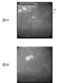
- FIG. 4A and 4B are diagrams showing an example in which the determination result image is superimposed and displayed on the endoscopic moving image in the present embodiment.
- 5A, 5B, and 5C are diagrams showing examples of endoscopic images used as teacher data. It is a figure which shows the characteristic of the subject and a lesion (gastric cancer) about the endoscopic moving image used in the data set for evaluation test.
- FIG. 1 is a block diagram showing an overall configuration of the diagnostic imaging apparatus 100.
- FIG. 2 is a diagram showing an example of the hardware configuration of the diagnostic imaging apparatus 100 according to the present embodiment.
- the diagnostic imaging apparatus 100 has an endoscopic image diagnostic capability possessed by a convolutional neural network (CNN) in an endoscopy of a digestive organ (stomach in the present embodiment) by a doctor (for example, an endoscopist). Make a real-time diagnosis of gastric cancer using.
- An endoscopic imaging device 200 and a display device 300 are connected to the diagnostic imaging device 100.
- the endoscope imaging device 200 is, for example, an electronic endoscope (also referred to as a videoscope) having a built-in imaging means, a camera-mounted endoscope in which a camera head having a built-in imaging means is attached to an optical endoscope, or the like. be.
- the endoscopic imaging device 200 is inserted into the digestive organ, for example, through the mouth or nose of the subject, and images a diagnosis target site in the digestive organ.
- the endoscope imaging device 200 irradiates the stomach of the subject with narrow band light (for example, narrow band light for NBI) in response to a doctor's operation (for example, button operation). Is imaged as an endoscopic moving image of the site to be diagnosed in the stomach, for example, in a state of being magnified 80 times.
- the endoscopic moving image is composed of a plurality of endoscopic images that are continuous in time.
- the endoscope imaging device 200 outputs the endoscopic moving image data D1 representing the captured endoscopic moving image to the diagnostic imaging device 100.
- the display device 300 is, for example, a liquid crystal display, and displays the endoscopic moving image and the determination result image output from the diagnostic imaging device 100 so that the doctor can identify them.
- the diagnostic imaging apparatus 100 has a CPU (Central Processing Unit) 101, a ROM (Read Only Memory) 102, a RAM (Random Access Memory) 103, and an external storage device (for example, a flash memory) as main components. It is a computer equipped with 104, a communication interface 105, a GPU (Graphics Processing Unit) 106, and the like.
- CPU Central Processing Unit
- ROM Read Only Memory
- RAM Random Access Memory
- an external storage device for example, a flash memory
- Each function of the diagnostic imaging apparatus 100 includes, for example, a control program (for example, an diagnostic imaging program) in which the CPU 101 and GPU 106 are stored in a ROM 102, a RAM 103, an external storage device 104, and various data (for example, endoscopic moving image data). It is realized by referring to the training teacher data, the model data of the convolutional neural network (structural data, learned weight parameters, etc.), and the RAM 103 functions as, for example, a data work area or a temporary save area.
- a control program for example, an diagnostic imaging program
- the CPU 101 and GPU 106 are stored in a ROM 102, a RAM 103, an external storage device 104, and various data (for example, endoscopic moving image data). It is realized by referring to the training teacher data, the model data of the convolutional neural network (structural data, learned weight parameters, etc.), and the RAM 103 functions as, for example, a data work area or a temporary save area.
- diagnostic imaging apparatus 100 may be realized by processing by the DSP (Digital Signal Processor) instead of or in combination with the processing by the CPU 101 and GPU 106.
- DSP Digital Signal Processor
- a part or all of each function may be realized by processing by a dedicated hardware circuit in place of or in combination with processing by software.
- the diagnostic imaging apparatus 100 includes an endoscopic moving image acquisition unit 10, an estimation unit 20, and a display control unit 30.
- the learning device 40 has a function of generating model data (corresponding to the "learned model” of the present invention) of the convolutional neural network used in the diagnostic imaging device 100.
- the display control unit 30 also functions as the "warning output control unit” of the present invention.
- the endoscopic moving image acquisition unit 10 acquires the endoscopic moving image data D1 output from the endoscopic imaging device 200. Then, the endoscopic moving image acquisition unit 10 outputs the acquired endoscopic moving image data D1 to the estimation unit 20.
- the endoscope moving image acquisition unit 10 may directly acquire the endoscopic moving image data D1 from the endoscope imaging device 200, or the endoscope stored in the external storage device 104.
- the moving image data D1 or the endoscopic moving image data D1 provided via an Internet line or the like may be acquired.
- the estimation unit 20 uses a convolutional neural network to display a lesion in the endoscopic moving image represented by the endoscopic moving image data D1 output from the endoscopic moving image acquisition unit 10 (in the present embodiment, the estimation unit 20). Estimates the presence of gastric cancer) and outputs the estimation result. Specifically, the estimation unit 20 determines the lesion name (name) and lesion position (position) of the lesion existing in the endoscopic moving image and the certainty (also referred to as accuracy) of the lesion name and lesion position. presume. Then, the estimation unit 20 displays the endoscopic moving image data D1 output from the endoscopic moving image acquisition unit 10 and the estimation result data D2 representing the estimation result of the lesion name, the lesion position, and the certainty. Output to 30.
- the estimation unit 20 the endoscopic image having a certainty level of a predetermined value (for example, 0.5) or more in the endoscopic moving image represented by the endoscopic moving image data D1 is displayed for a predetermined time (for example, 0.5). If a predetermined number (for example, 3) is continuously present within 0.5 seconds), it is estimated that a lesion (gastric cancer) is present in the endoscopic moving image.
- the predetermined number is set so as to increase as the predetermined value decreases.
- the estimation unit 20 estimates the probability score as an index showing the certainty of the lesion name and the lesion position.
- the probability score is represented by a value greater than 0 and less than or equal to 1. The higher the probability score, the higher the certainty of the lesion name and lesion location.
- the probability score is an example of an index indicating the degree of certainty of the lesion name and the lesion position, and an index of any other aspect may be used.
- the probability score may be represented by a value of 0% to 100%, or may be represented by any of several levels.
- a convolutional neural network is a type of feedforward neural network and is based on knowledge in the structure of the visual cortex of the brain. Basically, it has a structure in which a convolution layer responsible for extracting local features of an image and a pooling layer (subsampling layer) that summarizes features for each region are repeated. According to each layer of the convolutional neural network, it possesses multiple neurons (Neurons), and each neuron is arranged so as to correspond to the visual cortex. The basic function of each neuron consists of signal input and output. However, when transmitting signals between neurons in each layer, instead of outputting the input signal as it is, a coupling load is set for each input, and the sum of the weighted inputs is each.
- the algorithm for constructing it is not particularly limited.
- FIG. 3 is a diagram showing a configuration of a convolutional neural network according to the present embodiment.
- the model data (structural data, learned weight parameters, etc.) of the convolutional neural network is stored in the external storage device 104 together with the diagnostic imaging program.
- the convolutional neural network has, for example, a feature extraction unit Na and an identification unit Nb.
- the feature extraction unit Na performs a process of extracting image features from an input image (specifically, an endoscopic image constituting the endoscopic moving image represented by the endoscopic moving image data D1).
- the identification unit Nb outputs an estimation result related to the image from the image features extracted by the feature extraction unit Na.
- the feature extraction unit Na is configured by hierarchically connecting a plurality of feature amount extraction layers Na1, Na2, and so on.
- Each feature amount extraction layer Na1, Na2 ... Provides a convolution layer, an activation layer, and a pooling layer.
- the feature amount extraction layer Na1 of the first layer scans the input image for each predetermined size by raster scanning. Then, the feature amount extraction layer Na1 extracts the feature amount contained in the input image by performing the feature amount extraction process on the scanned data by the convolutional layer, the activation layer and the pooling layer.
- the feature amount extraction layer Na1 of the first layer extracts a relatively simple single feature amount such as a linear feature amount extending in the horizontal direction and a linear feature amount extending in the diagonal direction.
- the feature amount sampling layer Na2 of the second layer scans an image (also referred to as a feature map) input from the feature amount sampling layer Na1 of the previous layer at predetermined size intervals by, for example, raster scanning. Then, the feature amount extraction layer Na2 extracts the feature amount contained in the input image by similarly performing the feature amount extraction process by the convolutional layer, the activation layer and the pooling layer on the scanned data.
- the feature amount extraction layer Na2 of the second layer is integrated with reference to the positional relationship of a plurality of feature amounts extracted by the feature amount extraction layer Na1 of the first layer, so that it is a higher-dimensional complex. Extract features.
- the feature amount sampling layers after the second layer perform the same processing as the feature amount extraction layer Na2 of the second layer. do. Then, the output of the feature amount extraction layer of the final layer (each value in the map of the plurality of feature maps) is input to the identification unit Nb.
- the identification unit Nb is composed of, for example, a multi-layer perceptron in which a plurality of fully connected layers (Fully Connected) are hierarchically connected.
- the fully connected layer on the input side of the identification unit Nb is fully connected to each value in the map of the plurality of feature maps acquired from the feature extraction unit Na, and the product-sum operation is performed while changing the weighting coefficient for each value. Go and output.
- the fully connected layer of the next layer of the identification unit Nb is fully coupled to the values output by each element of the fully connected layer of the previous layer, and the product-sum operation is performed while applying different weighting factors to each value. Then, in the final stage of the identification unit Nb, the lesion name and lesion position of the lesion existing in the image (endoscopic image) input to the feature extraction unit Na, and the probability score (confidence) of the lesion name and lesion position.
- a layer for example, a softmax function, etc. that outputs a degree
- the convolutional neural network is desired from the input endoscopic image by performing learning processing using reference data (hereinafter referred to as "teacher data") marked in advance by an experienced endoscopist.
- the estimation function can be possessed so that the estimation result (here, the lesion name, the lesion position and the probability score) can be output.
- the estimation result here, the lesion name, the lesion position and the probability score
- the convolutional neural network in the present embodiment receives the endoscopic moving image data D1 as an input (Input in FIG. 3), and constitutes an endoscopic moving image represented by the endoscopic moving image data D1.
- the lesion name, lesion position, and probability score according to the image features of the above are output as the estimation result data D2 (Auto of FIG. 3).
- the convolutional neural network has a configuration in which information related to the subject's age, gender, region, or medical history can be input in addition to the endoscopic moving image data D1 (for example, input by the identification unit Nb). It may be provided as an element). Since the importance of real-world data in clinical practice is particularly recognized, it is possible to develop a more useful system in clinical practice by adding such information on subject attributes. That is, the characteristics of the endoscopic image are considered to have a correlation with information related to the subject's age, gender, region, medical history, family medical history, etc. By referring to the subject attribute information such as age in addition to the data D1, the lesion name and the lesion position can be estimated with higher accuracy. Since the pathophysiology of the disease may differ depending on the region and race, this method should be adopted especially when the present invention is used internationally.
- the estimation unit 20 also performs processing for converting the size and aspect ratio of the endoscope image, color division processing for the endoscope image, and color conversion processing for the endoscope image as preprocessing. , Color extraction processing, brightness gradient extraction processing, and the like may be performed. In order to prevent overfitting and improve accuracy, it is also preferable to adjust the weighting.
- the display control unit 30 has a lesion name represented by the estimation result data D2 output from the estimation unit 20 on the endoscopic motion image represented by the endoscopic moving image data D1 output from the estimation unit 20. A judgment result image for superimposing and displaying the lesion position and the probability score is generated. Then, the display control unit 30 outputs the endoscopic moving image data D1 and the determination result image data D3 representing the generated determination result image to the display device 300.
- a digital image processing system such as structural enhancement, color enhancement, difference processing, high contrast, and high definition of the lesion part of the endoscopic moving image is connected to understand and judge the observer (for example, a doctor). It can also be displayed with some processing to help.
- the display device 300 superimposes and displays the determination result image represented by the determination result image data D3 on the endoscope moving image represented by the endoscope moving image data D1 output from the display control unit 30.
- the endoscopic moving image and the determination result image displayed on the display device 300 are used for real-time diagnostic assistance and diagnostic support by a doctor.
- FIG. 4 is a diagram showing an example in which the determination result image is superimposed and displayed on the endoscopic moving image.
- a determination result image As shown in FIG. 4A, as a determination result image, a rectangular frame 50 indicating a lesion position (range) estimated by the estimation unit 20, a lesion name (for example, gastric cancer) and a probability score (for example, 0.85). Is displayed.
- the display control unit 30 when the probability score is equal to or higher than a certain threshold value (for example, 0.4), the display control unit 30 displays a rectangular frame indicating the lesion position, the lesion name, and the probability score on the endoscopic moving image. Overlay display (see FIG. 4A).
- a certain threshold for example, 0.4
- the display control unit 30 sets the probability score on the endoscopic moving image.
- the rectangular frame indicating the lesion position, the lesion name, and the probability score are not displayed (see FIG. 4B). That is, the display control unit 30 changes the display mode of the determination result image on the endoscopic moving image according to the probability score represented by the estimation result data D2 output from the estimation unit 20.
- the display control unit 30 controls the display device 300 and emits a screen for displaying the endoscopic moving image.
- a warning is displayed and output by causing the lesion to blink or blinking the rectangular area of the lesion determination part. This can effectively alert the doctor that the lesion is present in the endoscopic moving image.
- a warning sound may be sounded (output) from a speaker (not shown) to output a warning. Further, at this time, it is also possible to independently calculate and display the determination probability and the estimated probability.
- the convolutional neural network of the estimation unit 20 estimates the lesion position, the lesion name, and the probability score from the endoscopic moving image data D1 (specifically, the endoscopic image constituting the endoscopic moving image).
- the teacher data D4 stored in an external storage device (not shown) is input so as to be possible, and the convolutional neural network of the learning device 40 is subjected to the learning process.
- the learning device 40 irradiates the stomachs of a plurality of subjects with narrow-band light in the gastric endoscopy performed in the past, and the endoscopy is performed in a state in which the stomachs are magnified and observed.
- the endoscopic image (still image) captured by the imaging device 200 and the lesion name and lesion position of the lesion (stomach cancer) existing in the endoscopic image determined in advance by the doctor are used as teacher data D4.
- the learning device 40 reduces the error (also referred to as loss) of the output data with respect to the correct answer value (fault name and lesion position) when the endoscopic image is input to the convolutional neural network. Performs learning processing of a convolutional neural network.
- the learning device 40 reflects the lesion (stomach cancer), that is, the existing endoscopic image (corresponding to the “stomach cancer image” of the present invention) and the lesion (stomach cancer) are not reflected. That is, a non-existent endoscopic image (corresponding to the "non-stomach cancer image” of the present invention) is used as the teacher data D4 for learning processing.
- FIG. 5 is a diagram showing an example of an endoscopic image used as teacher data.
- FIG. 5A shows an endoscopic image (gastric cancer image) in which differentiated gastric cancer is present as a lesion.
- FIG. 5B shows an endoscopic image (gastric cancer image) in which undifferentiated gastric cancer is present as a lesion.
- FIG. 5C shows an endoscopic image (non-gastric cancer image) in which gastric cancer as a lesion does not exist.
- the endoscopic image as teacher data D4 in the learning process mainly uses the abundant database of Japan's top-class cancer treatment hospitals, and is prepared by the instructor of the Japan Gastroenterological Endoscopy Society who has abundant diagnosis and treatment experience. All images were examined in detail, sorted, and marked for the location of the lesion (gastric cancer) by precise manual treatment.
- teacher data D4 endoscopic image data
- an expert endoscopist with abundant experience can directly connect to the diagnostic accuracy of the diagnostic imaging apparatus 100.
- a sufficient number of cases with image selection, lesion identification, and feature extraction marking is an extremely important process.
- Such high-precision data cleansing work and use of high-quality reference data provide highly reliable AI program output results.
- the teacher data D4 of the endoscopic image may be pixel value data or data that has undergone a predetermined color conversion process or the like. Further, as the pretreatment, a texture feature, a shape feature, an uneven state, a spread feature, etc., which are characteristic of the cancerous portion, may be extracted from a comparison between an inflammatory image and a non-inflammatory image. Further, the teacher data D4 may perform learning processing in association with information related to the subject's age, gender, region or medical history, family medical history, etc., in addition to the endoscopic image data.
- the algorithm when the learning device 40 performs the learning process may be a known method.
- the learning device 40 uses, for example, a known backpropagation (backpropagation method) to perform learning processing on a convolutional neural network and adjust network parameters (weighting coefficient, bias, etc.).
- backpropagation method backpropagation method
- the model data (structural data, learned weight parameters, etc.) of the convolutional neural network subjected to the learning process by the learning device 40 is stored in the external storage device 104 together with the diagnostic imaging program, for example.
- Known CNN models include, for example, GoogleLeNet, ResNet, SENet and the like.
- the diagnostic imaging apparatus 100 irradiates the stomach of the subject with narrow-band light and acquires an endoscopic moving image taken in a state where the stomach is magnified and observed.
- the presence of gastric cancer in the acquired endoscopic moving image is estimated by using the endoscopic moving image acquisition unit 10 and the convolutional neural network adjusted using the gastric cancer image and the non-gastric cancer image as teacher data, and the estimation result. It is provided with an estimation unit 20 for outputting.
- the convolutional neural network includes endoscopic images (gastric cancer image, non-stomach cancer image) of a plurality of stomachs (digestive organs) obtained in advance for each of a plurality of subjects, and preliminarily for each of the plurality of subjects. It is learned based on the lesion name of the obtained lesion (stomach cancer) and the definite determination result of the lesion position. Therefore, it is possible to estimate the lesion name and lesion position of the stomach of a new subject in a short time and with an accuracy comparable to that of a substantially experienced endoscopist.
- the diagnostic imaging apparatus 100 can also be used as a diagnostic support tool that directly supports the diagnosis of endoscopic moving images by an endoscopist in a medical examination room.
- the diagnostic imaging apparatus 100 can be used as a central diagnostic support service that supports the diagnosis of endoscopic moving images transmitted from a plurality of examination rooms, or can be remotely controlled via an Internet line in a remote institution. It can also be used as a diagnostic support service to support the diagnosis of endoscopic moving images.
- the diagnostic imaging apparatus 100 can also be operated on the cloud. Furthermore, these endoscopic moving images and AI judgment results can be directly converted into a video library and used as teaching materials and materials for education and training and research.
- Narrow-band light was applied to the stomachs of multiple subjects in cases (395 cases) in which ESD was performed as the initial treatment at the Cancer Research Association Ariake Hospital between April 2005 and December 2016. Then, the stomach is magnified by irradiating the endoscopic image of 1492 in which gastric cancer is present and the stomachs of a plurality of subjects with narrow band light.
- a teacher data set (teacher data) that is imaged by an endoscopic imaging device in the observed state, extracts 1078 endoscopic images in the absence of gastric cancer from an electronic chart device, and is used for learning a convolutional neural network in an diagnostic imaging device. ).
- GIF-H240Z, GIF-H260Z, and GIF-H290 manufactured by Olympus Medical Systems Co., Ltd. were used.
- the endoscopic image taken by the endoscopic imaging device with the subject's stomach observed at high magnification and gastric cancer are observed in 60% or more of the entire image. Included (existing) endoscopic images.
- endoscopic images with widespread mucus, blood, out of focus, or poor image quality due to halation were excluded from the teacher dataset.
- the instructor of the Japan Gastroenterological Endoscopy Society who is a specialist in gastric cancer, examines and selects the prepared endoscopic images in detail, marks the lesion position of the lesion by precise manual processing, and prepares the teacher data set. I prepared it.
- GoogleNet which is composed of 22 layers and has a structure common to the previous CNN but has a sufficient number of parameters and expressive power, was used as a convolutional neural network.
- the Caffe Deep Learning Framework developed at the Berkeley Vision and Learning Center (BVLC) was used for learning and evaluation testing. All layers of the convolutional neural network are fine-tuned with a global learning rate of 0.0001 using stochastic gradient descent. Each endoscopic image was resized to 224 x 224 pixels for compatibility with CNN.
- GIF-H240Z, GIF-H260Z, and GIF-H290 manufactured by Olympus Medical Systems Co., Ltd. were used as in the preparation of the teacher data set.
- the data set for the evaluation test included an endoscopic moving image taken by an endoscopic imaging device for 10 seconds with a strong magnified observation of the subject's stomach as an endoscopic moving image satisfying the eligibility criteria. ..
- evaluation test data are used as endoscopic moving images that meet the exclusion criteria. Excluded from the set.
- the instructor of the Japan Gastroenterological Endoscopy Society who is an expert on gastric cancer, examined the prepared endoscopic moving images in detail, and endoscopic moving images with gastric cancer and endoscopic moving images without gastric cancer. And were selected, and a data set for evaluation test was prepared.
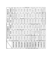
- FIG. 6 is a diagram showing the characteristics of the subject and the lesion (gastric cancer) related to the endoscopic moving image used in the data set for the evaluation test.
- the ratio (%) to the whole is shown in parentheses.
- the median (interquartile range) [whole range] is shown.
- the median tumor diameter was 14 mm
- the interquartile range (entire range) of the tumor was 9 to 20 (1-48) mm.
- 60 lesions (69.0%) were the most depressed type.
- the data set for the evaluation test is input to the convolutional neural network-based diagnostic imaging device that has been trained using the teacher data set, and each endoscope that constitutes the data set for the evaluation test. It was evaluated whether or not it was possible to correctly diagnose whether or not gastric cancer was present in the moving image.
- the diagnostic imaging apparatus diagnoses that a lesion is present in the endoscopic moving image when a predetermined number of endoscopic images having a certainty level of a predetermined value or more exist continuously within a predetermined time.
- whether or not gastric cancer can be correctly diagnosed in each endoscopic moving image can be correctly diagnosed by changing the predetermined time, certainty, and predetermined number of values in various ways and using the changed values. was evaluated.
- the correct diagnosis rate, sensitivity, specificity, positive predictive value (PPV) and negative predictive value (NPV) for the diagnostic ability of the diagnostic imaging device (or endoscopist) are calculated by the following formula (1). )-(5).
- Correct diagnosis rate (number of endoscopic moving images that could correctly diagnose the presence or absence of gastric cancer in the evaluation test data set) / (number of all endoscopic moving images that make up the evaluation test data set) ) ⁇ ⁇ ⁇ (1)
- Sensitivity (Number of endoscopic moving images that could correctly diagnose the presence of gastric cancer in the evaluation test data set) / (Number of endoscopic moving images that actually have gastric cancer in the evaluation test data set) ⁇ ⁇
- Specificity (number of endoscopic moving images that could correctly diagnose the absence of gastric cancer in the evaluation test data set) / (number of endoscopic moving images that do not actually have gastric cancer in the evaluation test data set) ...
- Positive predictive value (PPV) (Number of endoscopic moving images diagnosed as having gastric cancer in the evaluation test data set) / (Evaluation test data set) Number of endoscopic moving images diagnosed as having gastric cancer in Japan) ...
- Negative predictive value (NPV) (Number of endoscopic moving images diagnosed as having no gastric cancer in the evaluation test data set, which do not actually have gastric cancer) / (Evaluation test data Number of endoscopic moving images diagnosed as having gastric cancer in the set) ...
- predetermined time 0.1 seconds or more and 0.5 seconds or less
- certainty 0.6
- certainty 0.5
- combination of predetermined number 3
- the medium and negative predictive values were calculated.
- the correct diagnosis rate 85.1% (95% CI: 79.0 to 89.6)
- the sensitivity 87.4% (95% CI: 78.8 to 92.8)
- the specificity 82. 8% (95% CI: 73.5 to 89.3)
- positive predictive value 83.5% (95% CI: 74.6 to 89.7)
- negative predictive value 86.7% (77) It was .8-92.4).
- FIG. 8 shows the number of correct diagnosis, the number of misdiagnosis, and the unconfirmed number of correct diagnosis, the number of false diagnosis, and the number of correct diagnosis, the number of correct diagnosis, and the number of unconfirmed diagnosis of the endoscopic moving image in which gastric cancer is present and the endoscopic moving image in which gastric cancer is not present. It is a figure showing a number.
- FIG. 9 is a diagram showing the correct diagnosis rate, sensitivity, specificity, positive predictive value and negative predictive value of the diagnostic imaging apparatus and 11 endoscopic skilled doctors A to K.
- 95% confidence intervals were also calculated and compared by each of the diagnostic imaging apparatus and the endoscopist skilled doctors A to K.
- the Mcnemar test is used to compare the correctness, sensitivity and specificity between diagnostic imaging equipment and endoscopists AK, while comparing the positive and negative predictive values. Used the binomial test (see P value in FIG. 9).
- the statistically significant difference was set to less than 0.05.
- "JMP13" was used as a high-performance interactive tool for visualizing data and performing statistical analysis.
- the diagnostic imaging apparatus is significantly superior to two endoscopists H and K and significantly inferior to one endoscopist I. rice field. In addition, no significant difference was observed between the diagnostic imaging system and the eight endoscopists A to G and J.
- the diagnostic imaging system was significantly superior to the three endoscopists C, J, and K. In addition, no significant difference was observed between the diagnostic imaging system and the eight endoscopists A, B, D to I.
- the diagnostic imaging system was significantly superior to the two endoscopists H and K, and significantly inferior to the three endoscopists C, F and I. In addition, no significant difference was observed between the diagnostic imaging system and the six endoscopists A, B, D, E, G, and J.
- the diagnostic imaging apparatus was significantly superior to the two endoscopists H and K, and significantly inferior to the two endoscopists C and F. In addition, no significant difference was observed between the diagnostic imaging system and the seven endoscopists A, B, D, E, G, I, and J.
- the diagnostic imaging device was significantly superior to the two endoscopists J and K. In addition, no significant difference was observed between the diagnostic imaging system and the nine endoscopists AI.
- the predetermined time, the certainty, and the predetermined number of values at which the correct diagnosis rate of the diagnostic imaging apparatus is highest are obtained.
- gastric cancer in actual clinical practice, if gastric cancer can be clearly detected in the endoscopic moving image (10 seconds) for 0.5 seconds (if there are three consecutive endoscopic images with a certainty of 0.5 or more). ), It shows that gastric cancer can be diagnosed in real time with a high accuracy rate. In addition, there is a tendency that the diagnostic performance of the diagnostic imaging apparatus can be maintained by increasing the number of endoscopic images required for diagnosing the presence of gastric cancer even when the certainty is low.
- the values of the predetermined time, certainty, and the predetermined number at which the correct diagnosis rate of the diagnostic imaging apparatus is highest are described, but when the correct diagnosis rate is maintained at 70% or more or 80% or more, The diagnostic performance can be exhibited in a wider range of predetermined time, certainty and a predetermined number of combinations.
- the diagnostic ability of the diagnostic imaging device was compared with the diagnostic ability of 11 endoscopists.
- the diagnostic imaging apparatus has a diagnostic ability equal to or higher than that of a skilled endoscope doctor.
- Sensitivity is paramount because endoscopy is a screening test for diagnosing gastric cancer.
- the diagnostic imaging apparatus was particularly excellent in sensitivity as compared with an endoscopist. From this, the diagnosis of gastric cancer by the diagnostic imaging device not only supports (supports) the diagnosis of endoscopists who have not mastered the diagnostic technique of gastric cancer by ME-NBI, but also has acquired the diagnostic technique. It was also found to be beneficial for endoscopists.
- Non-Patent Document 3 as a result of evaluating the diagnostic ability of a computer-assisted diagnostic (CAD) system for gastric cancer using an endoscopic image (still image) taken by an NBI combined magnifying endoscope, the correct diagnosis rate is 85. It is described that the sensitivity was 3%, the sensitivity was 95.4%, the specificity was 71.0%, the positive predictive value was 82.3%, and the negative predictive value was 91.7%. In addition, severe atrophic gastritis, localized atrophy, and intestinal metaplasia have been described as examples of causes of false positives.
- CAD computer-assisted diagnostic
- Non-Patent Document 3 does not compare the diagnostic ability of the computer-aided diagnostic system with the diagnostic ability of an endoscopic expert who has acquired the diagnostic technique of gastric cancer by ME-NBI, and therefore evaluates the diagnostic ability.
- the diagnostic difficulty of the endoscopic images used for this was unknown, limiting the interpretation of the diagnostic capabilities of computer-aided diagnostic systems.
- Non-Patent Document 4 the same examination as in Non-Patent Document 3 was carried out, and it was found that the diagnostic imaging apparatus was significantly superior in sensitivity and negative predictive value as compared with two endoscopists. Have been described. However, the number of endoscopists compared to computer-aided diagnostic systems is small, that is, the diagnostic ability of individual endoscopists may be strongly biased in the results, so the diagnostic ability is evaluated. The diagnostic difficulty of the endoscopic images used for this was unknown, and the interpretation of the diagnostic capabilities of computer-aided diagnostic systems was limited. Further, in Non-Patent Document 4, the AUC is not calculated, and the diagnostic accuracy of the computer-aided diagnosis system as an image diagnostic device is also unknown.
- Non-Patent Documents 3 and 4 studies using still images (endoscopic images) are carried out, which is useful when performing secondary interpretation of endoscopic images after endoscopic examination.
- Non-Patent Document 5 states that the sensitivity in diagnosing gastric cancer pick-up was 94.1% with respect to the diagnostic performance of the computer-aided diagnostic system in endoscopic moving images taken using a normal endoscope. Have been described. However, in Non-Patent Document 5, only the evaluation of sensitivity is described, the endoscopic moving image captured by the NBI combined magnifying endoscope is not used, and the computer-assisted diagnostic system and the inside. Evaluation of the difficulty of diagnosis of endoscopic moving images and gastric cancer of the computer-assisted diagnostic system because the diagnostic ability was not compared with the endoscopic expert and the AUC in the computer-assisted diagnostic system was not calculated. There is a limit to the interpretation of the diagnostic ability of the patient, and sufficient evaluation has not been performed, and the judgment of its usefulness in clinical practice is unknown.
- the diagnostic imaging apparatus in the present invention was 0.8684, and the comprehensive diagnostic ability and reliability as a medical device were very high.
- the diagnostic imaging apparatus of the present invention compares the diagnostic ability with many skilled endoscopists, it is appropriate to set weights and parameters in CNN, and further, the difficulty level for moving image evaluation. Can be properly evaluated. It is also possible to adjust to reduce the bias that occurs in comparison with a small number of skilled doctors by making comparisons with many skilled doctors.
- a computer-aided diagnosis (CAD) system can provide performance with diagnostic capabilities equal to or better than that of a skilled doctor. It was shown that it can be used not only in clinical practice but also as an education and training system.
- CAD computer-aided diagnosis
- a magnifying endoscope combined with NBI is used, and detailed observation of lesions is possible as compared with a normal endoscope and a non-magnifying endoscope combined with NBI, and its diagnostic ability is high. It was highly useful.
- moving images are used instead of still images, and gastric cancer can be diagnosed in real time by using an image diagnostic apparatus in actual clinical practice.
- the present invention is a diagnostic imaging device, a diagnostic imaging method, a diagnostic imaging program, and a trained model capable of diagnosing gastric cancer in real time in gastric endoscopy performed using a magnifying endoscope combined with NBI. It is useful.
- Endoscopic moving image acquisition unit 20
- Estimating unit 30
- Display control unit 40
- Learning device 100
- Image diagnostic device 101
- CPU 102
- ROM 103
- RAM 104
- External storage device 105
- Communication interface 200
- Endoscope imager 300
- Display device D1
- Endoscope moving image data D2
- Estimation result data D3
- Judgment result image data D4 Teacher data
Landscapes
- Health & Medical Sciences (AREA)
- Life Sciences & Earth Sciences (AREA)
- Engineering & Computer Science (AREA)
- Surgery (AREA)
- Physics & Mathematics (AREA)
- Medical Informatics (AREA)
- General Health & Medical Sciences (AREA)
- Nuclear Medicine, Radiotherapy & Molecular Imaging (AREA)
- Radiology & Medical Imaging (AREA)
- Veterinary Medicine (AREA)
- Molecular Biology (AREA)
- Public Health (AREA)
- Biophysics (AREA)
- Optics & Photonics (AREA)
- Pathology (AREA)
- Animal Behavior & Ethology (AREA)
- Biomedical Technology (AREA)
- Heart & Thoracic Surgery (AREA)
- Theoretical Computer Science (AREA)
- General Physics & Mathematics (AREA)
- Computer Vision & Pattern Recognition (AREA)
- Signal Processing (AREA)
- Quality & Reliability (AREA)
- Artificial Intelligence (AREA)
- Evolutionary Computation (AREA)
- Gastroenterology & Hepatology (AREA)
- Endoscopes (AREA)
- Image Analysis (AREA)
Priority Applications (2)
| Application Number | Priority Date | Filing Date | Title |
|---|---|---|---|
| US17/995,592 US20230162356A1 (en) | 2020-04-10 | 2021-04-09 | Diagnostic imaging device, diagnostic imaging method, diagnostic imaging program, and learned model |
| JP2022514135A JPWO2021206170A1 (https=) | 2020-04-10 | 2021-04-09 |
Applications Claiming Priority (2)
| Application Number | Priority Date | Filing Date | Title |
|---|---|---|---|
| JP2020070848 | 2020-04-10 | ||
| JP2020-070848 | 2020-04-10 |
Publications (1)
| Publication Number | Publication Date |
|---|---|
| WO2021206170A1 true WO2021206170A1 (ja) | 2021-10-14 |
Family
ID=78024031
Family Applications (1)
| Application Number | Title | Priority Date | Filing Date |
|---|---|---|---|
| PCT/JP2021/015061 Ceased WO2021206170A1 (ja) | 2020-04-10 | 2021-04-09 | 画像診断装置、画像診断方法、画像診断プログラムおよび学習済みモデル |
Country Status (3)
| Country | Link |
|---|---|
| US (1) | US20230162356A1 (https=) |
| JP (1) | JPWO2021206170A1 (https=) |
| WO (1) | WO2021206170A1 (https=) |
Families Citing this family (4)
| Publication number | Priority date | Publication date | Assignee | Title |
|---|---|---|---|---|
| JP7377769B2 (ja) * | 2020-06-08 | 2023-11-10 | Hoya株式会社 | プログラム、情報処理方法及び情報処理装置 |
| WO2022114357A1 (ko) * | 2020-11-25 | 2022-06-02 | 주식회사 아이도트 | 영상 병변 진단 시스템 |
| JPWO2023067922A1 (https=) * | 2021-10-20 | 2023-04-27 | ||
| KR102722605B1 (ko) * | 2023-11-21 | 2024-10-28 | 주식회사 메디인테크 | 내시경 영상에서 병변을 탐지하는 방법 및 이를 수행하는 인공신경망 모델을 학습시키는 방법 및 컴퓨팅 장치 |
Citations (6)
| Publication number | Priority date | Publication date | Assignee | Title |
|---|---|---|---|---|
| JP2010141847A (ja) * | 2008-12-15 | 2010-06-24 | Canon Inc | 画像処理装置及び画像処理方法 |
| US20180253839A1 (en) * | 2015-09-10 | 2018-09-06 | Magentiq Eye Ltd. | A system and method for detection of suspicious tissue regions in an endoscopic procedure |
| US20180263568A1 (en) * | 2017-03-09 | 2018-09-20 | The Board Of Trustees Of The Leland Stanford Junior University | Systems and Methods for Clinical Image Classification |
| WO2018180386A1 (ja) * | 2017-03-30 | 2018-10-04 | 国立研究開発法人産業技術総合研究所 | 超音波画像診断支援方法、およびシステム |
| WO2018225448A1 (ja) * | 2017-06-09 | 2018-12-13 | 智裕 多田 | 消化器官の内視鏡画像による疾患の診断支援方法、診断支援システム、診断支援プログラム及びこの診断支援プログラムを記憶したコンピュータ読み取り可能な記録媒体 |
| JP2019518581A (ja) * | 2016-06-08 | 2019-07-04 | リサーチ ディヴェロプメント ファウンデーション | 血管内光干渉断層撮影法を用いた冠動脈プラークの自動特徴分析およびリスク評価のためのシステムおよび方法 |
Family Cites Families (3)
| Publication number | Priority date | Publication date | Assignee | Title |
|---|---|---|---|---|
| US9324145B1 (en) * | 2013-08-08 | 2016-04-26 | Given Imaging Ltd. | System and method for detection of transitions in an image stream of the gastrointestinal tract |
| JP2018051190A (ja) * | 2016-09-30 | 2018-04-05 | 学校法人北里研究所 | 脳手術支援システム、脳手術支援システム作動方法及びプログラム |
| CN108154509B (zh) * | 2018-01-12 | 2022-11-11 | 平安科技(深圳)有限公司 | 癌症识别方法、装置及存储介质 |
-
2021
- 2021-04-09 JP JP2022514135A patent/JPWO2021206170A1/ja active Pending
- 2021-04-09 US US17/995,592 patent/US20230162356A1/en active Pending
- 2021-04-09 WO PCT/JP2021/015061 patent/WO2021206170A1/ja not_active Ceased
Patent Citations (6)
| Publication number | Priority date | Publication date | Assignee | Title |
|---|---|---|---|---|
| JP2010141847A (ja) * | 2008-12-15 | 2010-06-24 | Canon Inc | 画像処理装置及び画像処理方法 |
| US20180253839A1 (en) * | 2015-09-10 | 2018-09-06 | Magentiq Eye Ltd. | A system and method for detection of suspicious tissue regions in an endoscopic procedure |
| JP2019518581A (ja) * | 2016-06-08 | 2019-07-04 | リサーチ ディヴェロプメント ファウンデーション | 血管内光干渉断層撮影法を用いた冠動脈プラークの自動特徴分析およびリスク評価のためのシステムおよび方法 |
| US20180263568A1 (en) * | 2017-03-09 | 2018-09-20 | The Board Of Trustees Of The Leland Stanford Junior University | Systems and Methods for Clinical Image Classification |
| WO2018180386A1 (ja) * | 2017-03-30 | 2018-10-04 | 国立研究開発法人産業技術総合研究所 | 超音波画像診断支援方法、およびシステム |
| WO2018225448A1 (ja) * | 2017-06-09 | 2018-12-13 | 智裕 多田 | 消化器官の内視鏡画像による疾患の診断支援方法、診断支援システム、診断支援プログラム及びこの診断支援プログラムを記憶したコンピュータ読み取り可能な記録媒体 |
Non-Patent Citations (1)
| Title |
|---|
| AOKI TOMONORI: "Clinical usefulness of a deep learning-based system as the first screening on small-bowel capsule endoscopy reading", DIGESTIVE ENDOSCOPY, vol. 32, 23 August 2019 (2019-08-23), pages 585 - 591, XP055863991 * |
Also Published As
| Publication number | Publication date |
|---|---|
| JPWO2021206170A1 (https=) | 2021-10-14 |
| US20230162356A1 (en) | 2023-05-25 |
Similar Documents
| Publication | Publication Date | Title |
|---|---|---|
| JP7335552B2 (ja) | 画像診断支援装置、学習済みモデル、画像診断支援装置の作動方法および画像診断支援プログラム | |
| JP7550409B2 (ja) | 画像診断装置、画像診断方法、および画像診断プログラム | |
| de Groof et al. | Deep learning algorithm detection of Barrett’s neoplasia with high accuracy during live endoscopic procedures: a pilot study (with video) | |
| US20250143543A1 (en) | Method for Real-Time Detection of Objects, Structures or Patterns in a Video, an Associated System and an Associated Computer Readable Medium | |
| US20220172828A1 (en) | Endoscopic image display method, apparatus, computer device, and storage medium | |
| WO2021206170A1 (ja) | 画像診断装置、画像診断方法、画像診断プログラムおよび学習済みモデル | |
| Pan et al. | Bleeding detection in wireless capsule endoscopy based on probabilistic neural network | |
| WO2021036616A1 (zh) | 一种医疗图像处理方法、医疗图像识别方法及装置 | |
| CN111899229A (zh) | 一种基于深度学习多模型融合技术的胃早癌辅助诊断方法 | |
| CN115018767B (zh) | 基于本征表示学习的跨模态内镜图像转换及病灶分割方法 | |
| KR102531400B1 (ko) | 인공 지능 기반 대장 내시경 영상 진단 보조 시스템 및 방법 | |
| KR102637484B1 (ko) | 인공지능 기반의 내시경 진단 보조 시스템 및 이의 제어방법 | |
| US20230351592A1 (en) | Clinical decision support system having a multi-ordered hierarchy of classification modules | |
| Babu et al. | Gastrointestinal tract disease detection via deep learning based Duo-Feature Optimized Hexa-Classification model | |
| CN117854705A (zh) | 上消化道多病变多任务智能诊断方法、装置、设备及介质 | |
| JP2023079866A (ja) | 超拡大内視鏡による胃癌の検査方法、診断支援方法、診断支援システム、診断支援プログラム、学習済みモデル及び画像診断支援装置 | |
| US12562279B2 (en) | Portable edge AI-assisted diagnosis and quality control system for gastrointestinal endoscopy | |
| US12400328B2 (en) | Method for deleting redundant images of endoscope | |
| Katayama et al. | Development of Computer-Aided Diagnosis System Using Single FCN Capable for Indicating Detailed Inference Results in Colon NBI Endoscopy | |
| Dayananda | Novel Convoluted local energy oriented patten (CLEOP) for the classification of wireless capsule endoscopy images | |
| Lopes et al. | Improving Endoscopy Lesion Classification Using Self-Supervised Deep Learning | |
| KR20250152725A (ko) | 인공지능 기반 내시경 병변 통합 탐지 시스템 | |
| TWM620109U (zh) | 輔助判斷系統 |
Legal Events
| Date | Code | Title | Description |
|---|---|---|---|
| 121 | Ep: the epo has been informed by wipo that ep was designated in this application |
Ref document number: 21784254 Country of ref document: EP Kind code of ref document: A1 |
|
| ENP | Entry into the national phase |
Ref document number: 2022514135 Country of ref document: JP Kind code of ref document: A |
|
| NENP | Non-entry into the national phase |
Ref country code: DE |
|
| 122 | Ep: pct application non-entry in european phase |
Ref document number: 21784254 Country of ref document: EP Kind code of ref document: A1 |