WO2021206170A1 - Diagnostic imaging device, diagnostic imaging method, diagnostic imaging program, and learned model - Google Patents
Diagnostic imaging device, diagnostic imaging method, diagnostic imaging program, and learned model Download PDFInfo
- Publication number
- WO2021206170A1 WO2021206170A1 PCT/JP2021/015061 JP2021015061W WO2021206170A1 WO 2021206170 A1 WO2021206170 A1 WO 2021206170A1 JP 2021015061 W JP2021015061 W JP 2021015061W WO 2021206170 A1 WO2021206170 A1 WO 2021206170A1
- Authority
- WO
- WIPO (PCT)
- Prior art keywords
- gastric cancer
- endoscopic
- moving image
- diagnostic imaging
- images
- Prior art date
Links
Images


Classifications
-
- G—PHYSICS
- G06—COMPUTING; CALCULATING OR COUNTING
- G06T—IMAGE DATA PROCESSING OR GENERATION, IN GENERAL
- G06T7/00—Image analysis
- G06T7/0002—Inspection of images, e.g. flaw detection
- G06T7/0012—Biomedical image inspection
-
- A—HUMAN NECESSITIES
- A61—MEDICAL OR VETERINARY SCIENCE; HYGIENE
- A61B—DIAGNOSIS; SURGERY; IDENTIFICATION
- A61B1/00—Instruments for performing medical examinations of the interior of cavities or tubes of the body by visual or photographical inspection, e.g. endoscopes; Illuminating arrangements therefor
- A61B1/00002—Operational features of endoscopes
- A61B1/00004—Operational features of endoscopes characterised by electronic signal processing
- A61B1/00009—Operational features of endoscopes characterised by electronic signal processing of image signals during a use of endoscope
- A61B1/000094—Operational features of endoscopes characterised by electronic signal processing of image signals during a use of endoscope extracting biological structures
-
- A—HUMAN NECESSITIES
- A61—MEDICAL OR VETERINARY SCIENCE; HYGIENE
- A61B—DIAGNOSIS; SURGERY; IDENTIFICATION
- A61B1/00—Instruments for performing medical examinations of the interior of cavities or tubes of the body by visual or photographical inspection, e.g. endoscopes; Illuminating arrangements therefor
- A61B1/00002—Operational features of endoscopes
- A61B1/00004—Operational features of endoscopes characterised by electronic signal processing
- A61B1/00009—Operational features of endoscopes characterised by electronic signal processing of image signals during a use of endoscope
- A61B1/000096—Operational features of endoscopes characterised by electronic signal processing of image signals during a use of endoscope using artificial intelligence
-
- A—HUMAN NECESSITIES
- A61—MEDICAL OR VETERINARY SCIENCE; HYGIENE
- A61B—DIAGNOSIS; SURGERY; IDENTIFICATION
- A61B1/00—Instruments for performing medical examinations of the interior of cavities or tubes of the body by visual or photographical inspection, e.g. endoscopes; Illuminating arrangements therefor
- A61B1/00002—Operational features of endoscopes
- A61B1/00043—Operational features of endoscopes provided with output arrangements
- A61B1/00045—Display arrangement
- A61B1/0005—Display arrangement combining images e.g. side-by-side, superimposed or tiled
-
- A—HUMAN NECESSITIES
- A61—MEDICAL OR VETERINARY SCIENCE; HYGIENE
- A61B—DIAGNOSIS; SURGERY; IDENTIFICATION
- A61B1/00—Instruments for performing medical examinations of the interior of cavities or tubes of the body by visual or photographical inspection, e.g. endoscopes; Illuminating arrangements therefor
- A61B1/00002—Operational features of endoscopes
- A61B1/00043—Operational features of endoscopes provided with output arrangements
- A61B1/00055—Operational features of endoscopes provided with output arrangements for alerting the user
-
- A—HUMAN NECESSITIES
- A61—MEDICAL OR VETERINARY SCIENCE; HYGIENE
- A61B—DIAGNOSIS; SURGERY; IDENTIFICATION
- A61B1/00—Instruments for performing medical examinations of the interior of cavities or tubes of the body by visual or photographical inspection, e.g. endoscopes; Illuminating arrangements therefor
- A61B1/06—Instruments for performing medical examinations of the interior of cavities or tubes of the body by visual or photographical inspection, e.g. endoscopes; Illuminating arrangements therefor with illuminating arrangements
- A61B1/063—Instruments for performing medical examinations of the interior of cavities or tubes of the body by visual or photographical inspection, e.g. endoscopes; Illuminating arrangements therefor with illuminating arrangements for monochromatic or narrow-band illumination
-
- A—HUMAN NECESSITIES
- A61—MEDICAL OR VETERINARY SCIENCE; HYGIENE
- A61B—DIAGNOSIS; SURGERY; IDENTIFICATION
- A61B1/00—Instruments for performing medical examinations of the interior of cavities or tubes of the body by visual or photographical inspection, e.g. endoscopes; Illuminating arrangements therefor
- A61B1/273—Instruments for performing medical examinations of the interior of cavities or tubes of the body by visual or photographical inspection, e.g. endoscopes; Illuminating arrangements therefor for the upper alimentary canal, e.g. oesophagoscopes, gastroscopes
-
- G—PHYSICS
- G06—COMPUTING; CALCULATING OR COUNTING
- G06T—IMAGE DATA PROCESSING OR GENERATION, IN GENERAL
- G06T11/00—2D [Two Dimensional] image generation
-
- G—PHYSICS
- G06—COMPUTING; CALCULATING OR COUNTING
- G06T—IMAGE DATA PROCESSING OR GENERATION, IN GENERAL
- G06T7/00—Image analysis
- G06T7/70—Determining position or orientation of objects or cameras
-
- G—PHYSICS
- G06—COMPUTING; CALCULATING OR COUNTING
- G06T—IMAGE DATA PROCESSING OR GENERATION, IN GENERAL
- G06T2207/00—Indexing scheme for image analysis or image enhancement
- G06T2207/10—Image acquisition modality
- G06T2207/10016—Video; Image sequence
-
- G—PHYSICS
- G06—COMPUTING; CALCULATING OR COUNTING
- G06T—IMAGE DATA PROCESSING OR GENERATION, IN GENERAL
- G06T2207/00—Indexing scheme for image analysis or image enhancement
- G06T2207/10—Image acquisition modality
- G06T2207/10068—Endoscopic image
-
- G—PHYSICS
- G06—COMPUTING; CALCULATING OR COUNTING
- G06T—IMAGE DATA PROCESSING OR GENERATION, IN GENERAL
- G06T2207/00—Indexing scheme for image analysis or image enhancement
- G06T2207/20—Special algorithmic details
- G06T2207/20081—Training; Learning
-
- G—PHYSICS
- G06—COMPUTING; CALCULATING OR COUNTING
- G06T—IMAGE DATA PROCESSING OR GENERATION, IN GENERAL
- G06T2207/00—Indexing scheme for image analysis or image enhancement
- G06T2207/20—Special algorithmic details
- G06T2207/20084—Artificial neural networks [ANN]
-
- G—PHYSICS
- G06—COMPUTING; CALCULATING OR COUNTING
- G06T—IMAGE DATA PROCESSING OR GENERATION, IN GENERAL
- G06T2207/00—Indexing scheme for image analysis or image enhancement
- G06T2207/30—Subject of image; Context of image processing
- G06T2207/30004—Biomedical image processing
- G06T2207/30092—Stomach; Gastric
-
- G—PHYSICS
- G06—COMPUTING; CALCULATING OR COUNTING
- G06T—IMAGE DATA PROCESSING OR GENERATION, IN GENERAL
- G06T2207/00—Indexing scheme for image analysis or image enhancement
- G06T2207/30—Subject of image; Context of image processing
- G06T2207/30004—Biomedical image processing
- G06T2207/30096—Tumor; Lesion
-
- G—PHYSICS
- G06—COMPUTING; CALCULATING OR COUNTING
- G06T—IMAGE DATA PROCESSING OR GENERATION, IN GENERAL
- G06T2210/00—Indexing scheme for image generation or computer graphics
- G06T2210/41—Medical
Definitions
- the present invention relates to a diagnostic imaging apparatus, a diagnostic imaging method, a diagnostic imaging program, and a trained model.
- Gastric cancer is one of the most common cancers in the world and one with a high cancer-related mortality rate.
- endoscopic equipment there are many cases where gastric cancer is detected early by endoscopy. As a result, the mortality rate from gastric cancer has decreased in recent years.
- ESD endoscopic submucosal dissection
- the treatment of early gastric cancer has become a minimally invasive treatment.
- ESD indications are limited to intramucosal cancer (where cancer infiltration remains up to the lamina intestinal), and it is important to detect and diagnose gastric cancer at an earlier stage. There is.
- gastric cancer is diagnosed endoscopically.
- an NBI combined magnifying endoscope (ME-NBI) has been developed that can irradiate the stomach of a subject with narrow band light (NBI: Narrow Band Imaging, narrow band imaging) and magnify the stomach.
- NBI narrow band light
- a magnifying endoscope combined with NBI has a higher diagnostic ability for gastric cancer than a normal endoscope.
- endoscopists need considerable effort to master the technique of diagnosing gastric cancer by ME-NBI. This is because most cases of gastric cancer have chronic inflammation (gastritis) associated with Helicobacter pylori infection on the background mucosa, and it is difficult to distinguish between gastric cancer and gastritis.
- gastric cancer is compared with other gastrointestinal cancers in which the background mucosa does not show chronic inflammation associated with Helicobacter pylori infection (for example, esophageal cancer judged by the color and unevenness of the mucosa and colon cancer characterized by polyps). Is difficult to diagnose properly.
- Helicobacter pylori infection for example, esophageal cancer judged by the color and unevenness of the mucosa and colon cancer characterized by polyps.
- AI Artificial Intelligence
- CNN convolutional neural network
- CAD Computer-aided diagnosis
- AI using deep learning is attracting attention in various medical fields, and features of radiation oncology, skin cancer classification, diabetic retinopathy, histological classification of gastric biopsy, and colorectal lesions by super-magnifying endoscopy.
- AI can obtain the same accuracy as a specialist at the microscopic endoscopy level (see Non-Patent Document 1).
- Non-Patent Document 2 it has been announced that AI with a deep learning function exhibits the same diagnostic imaging ability as a specialist
- Patent Document 2 patent documents using various machine learning methods (patents). (See References 1 and 2) also exists.
- AI's diagnostic imaging ability is comparable to that of specialists.
- diagnostic imaging technology that uses AI's diagnostic imaging capability to diagnose gastric cancer in real time is still available in actual medical practice (actual clinical practice). ) Has not been introduced, and it is expected that it will be put into practical use in the future.
- the extraction of the unique characteristic amount of each gastrointestinal cancer esophageal cancer, gastric cancer, colon cancer, etc.
- determination of its pathological level are different. It is important to design the AI program according to its characteristics.
- An object of the present invention is an image diagnostic apparatus, an image diagnosis method, an image diagnosis program, and a learned image diagnosis device capable of diagnosing gastric cancer in real time in gastric endoscopy performed by using a magnifying endoscope combined with NBI. To provide a model.
- the diagnostic imaging apparatus is An endoscopic moving image acquisition unit that irradiates the subject's stomach with narrow-band light and acquires an endoscopic moving image taken while observing the stomach in an enlarged manner.
- an estimation unit that estimates the presence of gastric cancer in the acquired endoscopic moving images and outputs the estimation results, and an estimation unit. To be equipped.
- the diagnostic imaging method An endoscopic moving image acquisition step of irradiating the subject's stomach with narrow-band light and acquiring an endoscopic moving image taken while observing the stomach in a magnified state.
- a convolutional neural network trained from gastric cancer images and non-gastric cancer images as teacher data the presence of gastric cancer in the acquired endoscopic moving image is estimated, and an estimation step of outputting the estimation result is performed. including.
- the diagnostic imaging program On the computer Endoscopic moving image acquisition processing that irradiates the subject's stomach with narrow-band light and acquires an endoscopic moving image taken while observing the stomach in a magnified state.
- a convolutional neural network trained from gastric cancer images and non-gastric cancer images as teacher data, the presence of gastric cancer in the acquired endoscopic moving images is estimated, and an estimation process that outputs the estimation results is performed. To be executed.
- the trained model according to the present invention Obtained by training a convolutional neural network using gastric cancer images and non-gastric cancer images as teacher data.
- the subject's stomach is irradiated with narrow-band light, the presence of gastric cancer is estimated in the endoscopic moving image taken while observing the stomach in a magnified state, and the computer is made to function to output the estimation result.
- gastric cancer can be diagnosed in real time in gastric endoscopy performed using a magnifying endoscope combined with NBI.
- FIG. 4A and 4B are diagrams showing an example in which the determination result image is superimposed and displayed on the endoscopic moving image in the present embodiment.
- 5A, 5B, and 5C are diagrams showing examples of endoscopic images used as teacher data. It is a figure which shows the characteristic of the subject and a lesion (gastric cancer) about the endoscopic moving image used in the data set for evaluation test.
- FIG. 1 is a block diagram showing an overall configuration of the diagnostic imaging apparatus 100.
- FIG. 2 is a diagram showing an example of the hardware configuration of the diagnostic imaging apparatus 100 according to the present embodiment.
- the diagnostic imaging apparatus 100 has an endoscopic image diagnostic capability possessed by a convolutional neural network (CNN) in an endoscopy of a digestive organ (stomach in the present embodiment) by a doctor (for example, an endoscopist). Make a real-time diagnosis of gastric cancer using.
- An endoscopic imaging device 200 and a display device 300 are connected to the diagnostic imaging device 100.
- the endoscope imaging device 200 is, for example, an electronic endoscope (also referred to as a videoscope) having a built-in imaging means, a camera-mounted endoscope in which a camera head having a built-in imaging means is attached to an optical endoscope, or the like. be.
- the endoscopic imaging device 200 is inserted into the digestive organ, for example, through the mouth or nose of the subject, and images a diagnosis target site in the digestive organ.
- the endoscope imaging device 200 irradiates the stomach of the subject with narrow band light (for example, narrow band light for NBI) in response to a doctor's operation (for example, button operation). Is imaged as an endoscopic moving image of the site to be diagnosed in the stomach, for example, in a state of being magnified 80 times.
- the endoscopic moving image is composed of a plurality of endoscopic images that are continuous in time.
- the endoscope imaging device 200 outputs the endoscopic moving image data D1 representing the captured endoscopic moving image to the diagnostic imaging device 100.
- the display device 300 is, for example, a liquid crystal display, and displays the endoscopic moving image and the determination result image output from the diagnostic imaging device 100 so that the doctor can identify them.
- the diagnostic imaging apparatus 100 has a CPU (Central Processing Unit) 101, a ROM (Read Only Memory) 102, a RAM (Random Access Memory) 103, and an external storage device (for example, a flash memory) as main components. It is a computer equipped with 104, a communication interface 105, a GPU (Graphics Processing Unit) 106, and the like.
- CPU Central Processing Unit
- ROM Read Only Memory
- RAM Random Access Memory
- an external storage device for example, a flash memory
- Each function of the diagnostic imaging apparatus 100 includes, for example, a control program (for example, an diagnostic imaging program) in which the CPU 101 and GPU 106 are stored in a ROM 102, a RAM 103, an external storage device 104, and various data (for example, endoscopic moving image data). It is realized by referring to the training teacher data, the model data of the convolutional neural network (structural data, learned weight parameters, etc.), and the RAM 103 functions as, for example, a data work area or a temporary save area.
- a control program for example, an diagnostic imaging program
- the CPU 101 and GPU 106 are stored in a ROM 102, a RAM 103, an external storage device 104, and various data (for example, endoscopic moving image data). It is realized by referring to the training teacher data, the model data of the convolutional neural network (structural data, learned weight parameters, etc.), and the RAM 103 functions as, for example, a data work area or a temporary save area.
- diagnostic imaging apparatus 100 may be realized by processing by the DSP (Digital Signal Processor) instead of or in combination with the processing by the CPU 101 and GPU 106.
- DSP Digital Signal Processor
- a part or all of each function may be realized by processing by a dedicated hardware circuit in place of or in combination with processing by software.
- the diagnostic imaging apparatus 100 includes an endoscopic moving image acquisition unit 10, an estimation unit 20, and a display control unit 30.
- the learning device 40 has a function of generating model data (corresponding to the "learned model” of the present invention) of the convolutional neural network used in the diagnostic imaging device 100.
- the display control unit 30 also functions as the "warning output control unit” of the present invention.
- the endoscopic moving image acquisition unit 10 acquires the endoscopic moving image data D1 output from the endoscopic imaging device 200. Then, the endoscopic moving image acquisition unit 10 outputs the acquired endoscopic moving image data D1 to the estimation unit 20.
- the endoscope moving image acquisition unit 10 may directly acquire the endoscopic moving image data D1 from the endoscope imaging device 200, or the endoscope stored in the external storage device 104.
- the moving image data D1 or the endoscopic moving image data D1 provided via an Internet line or the like may be acquired.
- the estimation unit 20 uses a convolutional neural network to display a lesion in the endoscopic moving image represented by the endoscopic moving image data D1 output from the endoscopic moving image acquisition unit 10 (in the present embodiment, the estimation unit 20). Estimates the presence of gastric cancer) and outputs the estimation result. Specifically, the estimation unit 20 determines the lesion name (name) and lesion position (position) of the lesion existing in the endoscopic moving image and the certainty (also referred to as accuracy) of the lesion name and lesion position. presume. Then, the estimation unit 20 displays the endoscopic moving image data D1 output from the endoscopic moving image acquisition unit 10 and the estimation result data D2 representing the estimation result of the lesion name, the lesion position, and the certainty. Output to 30.
- the estimation unit 20 the endoscopic image having a certainty level of a predetermined value (for example, 0.5) or more in the endoscopic moving image represented by the endoscopic moving image data D1 is displayed for a predetermined time (for example, 0.5). If a predetermined number (for example, 3) is continuously present within 0.5 seconds), it is estimated that a lesion (gastric cancer) is present in the endoscopic moving image.
- the predetermined number is set so as to increase as the predetermined value decreases.
- the estimation unit 20 estimates the probability score as an index showing the certainty of the lesion name and the lesion position.
- the probability score is represented by a value greater than 0 and less than or equal to 1. The higher the probability score, the higher the certainty of the lesion name and lesion location.
- the probability score is an example of an index indicating the degree of certainty of the lesion name and the lesion position, and an index of any other aspect may be used.
- the probability score may be represented by a value of 0% to 100%, or may be represented by any of several levels.
- a convolutional neural network is a type of feedforward neural network and is based on knowledge in the structure of the visual cortex of the brain. Basically, it has a structure in which a convolution layer responsible for extracting local features of an image and a pooling layer (subsampling layer) that summarizes features for each region are repeated. According to each layer of the convolutional neural network, it possesses multiple neurons (Neurons), and each neuron is arranged so as to correspond to the visual cortex. The basic function of each neuron consists of signal input and output. However, when transmitting signals between neurons in each layer, instead of outputting the input signal as it is, a coupling load is set for each input, and the sum of the weighted inputs is each.
- the algorithm for constructing it is not particularly limited.
- FIG. 3 is a diagram showing a configuration of a convolutional neural network according to the present embodiment.
- the model data (structural data, learned weight parameters, etc.) of the convolutional neural network is stored in the external storage device 104 together with the diagnostic imaging program.
- the convolutional neural network has, for example, a feature extraction unit Na and an identification unit Nb.
- the feature extraction unit Na performs a process of extracting image features from an input image (specifically, an endoscopic image constituting the endoscopic moving image represented by the endoscopic moving image data D1).
- the identification unit Nb outputs an estimation result related to the image from the image features extracted by the feature extraction unit Na.
- the feature extraction unit Na is configured by hierarchically connecting a plurality of feature amount extraction layers Na1, Na2, and so on.
- Each feature amount extraction layer Na1, Na2 ... Provides a convolution layer, an activation layer, and a pooling layer.
- the feature amount extraction layer Na1 of the first layer scans the input image for each predetermined size by raster scanning. Then, the feature amount extraction layer Na1 extracts the feature amount contained in the input image by performing the feature amount extraction process on the scanned data by the convolutional layer, the activation layer and the pooling layer.
- the feature amount extraction layer Na1 of the first layer extracts a relatively simple single feature amount such as a linear feature amount extending in the horizontal direction and a linear feature amount extending in the diagonal direction.
- the feature amount sampling layer Na2 of the second layer scans an image (also referred to as a feature map) input from the feature amount sampling layer Na1 of the previous layer at predetermined size intervals by, for example, raster scanning. Then, the feature amount extraction layer Na2 extracts the feature amount contained in the input image by similarly performing the feature amount extraction process by the convolutional layer, the activation layer and the pooling layer on the scanned data.
- the feature amount extraction layer Na2 of the second layer is integrated with reference to the positional relationship of a plurality of feature amounts extracted by the feature amount extraction layer Na1 of the first layer, so that it is a higher-dimensional complex. Extract features.
- the feature amount sampling layers after the second layer perform the same processing as the feature amount extraction layer Na2 of the second layer. do. Then, the output of the feature amount extraction layer of the final layer (each value in the map of the plurality of feature maps) is input to the identification unit Nb.
- the identification unit Nb is composed of, for example, a multi-layer perceptron in which a plurality of fully connected layers (Fully Connected) are hierarchically connected.
- the fully connected layer on the input side of the identification unit Nb is fully connected to each value in the map of the plurality of feature maps acquired from the feature extraction unit Na, and the product-sum operation is performed while changing the weighting coefficient for each value. Go and output.
- the fully connected layer of the next layer of the identification unit Nb is fully coupled to the values output by each element of the fully connected layer of the previous layer, and the product-sum operation is performed while applying different weighting factors to each value. Then, in the final stage of the identification unit Nb, the lesion name and lesion position of the lesion existing in the image (endoscopic image) input to the feature extraction unit Na, and the probability score (confidence) of the lesion name and lesion position.
- a layer for example, a softmax function, etc. that outputs a degree
- the convolutional neural network is desired from the input endoscopic image by performing learning processing using reference data (hereinafter referred to as "teacher data") marked in advance by an experienced endoscopist.
- the estimation function can be possessed so that the estimation result (here, the lesion name, the lesion position and the probability score) can be output.
- the estimation result here, the lesion name, the lesion position and the probability score
- the convolutional neural network in the present embodiment receives the endoscopic moving image data D1 as an input (Input in FIG. 3), and constitutes an endoscopic moving image represented by the endoscopic moving image data D1.
- the lesion name, lesion position, and probability score according to the image features of the above are output as the estimation result data D2 (Auto of FIG. 3).
- the convolutional neural network has a configuration in which information related to the subject's age, gender, region, or medical history can be input in addition to the endoscopic moving image data D1 (for example, input by the identification unit Nb). It may be provided as an element). Since the importance of real-world data in clinical practice is particularly recognized, it is possible to develop a more useful system in clinical practice by adding such information on subject attributes. That is, the characteristics of the endoscopic image are considered to have a correlation with information related to the subject's age, gender, region, medical history, family medical history, etc. By referring to the subject attribute information such as age in addition to the data D1, the lesion name and the lesion position can be estimated with higher accuracy. Since the pathophysiology of the disease may differ depending on the region and race, this method should be adopted especially when the present invention is used internationally.
- the estimation unit 20 also performs processing for converting the size and aspect ratio of the endoscope image, color division processing for the endoscope image, and color conversion processing for the endoscope image as preprocessing. , Color extraction processing, brightness gradient extraction processing, and the like may be performed. In order to prevent overfitting and improve accuracy, it is also preferable to adjust the weighting.
- the display control unit 30 has a lesion name represented by the estimation result data D2 output from the estimation unit 20 on the endoscopic motion image represented by the endoscopic moving image data D1 output from the estimation unit 20. A judgment result image for superimposing and displaying the lesion position and the probability score is generated. Then, the display control unit 30 outputs the endoscopic moving image data D1 and the determination result image data D3 representing the generated determination result image to the display device 300.
- a digital image processing system such as structural enhancement, color enhancement, difference processing, high contrast, and high definition of the lesion part of the endoscopic moving image is connected to understand and judge the observer (for example, a doctor). It can also be displayed with some processing to help.
- the display device 300 superimposes and displays the determination result image represented by the determination result image data D3 on the endoscope moving image represented by the endoscope moving image data D1 output from the display control unit 30.
- the endoscopic moving image and the determination result image displayed on the display device 300 are used for real-time diagnostic assistance and diagnostic support by a doctor.
- FIG. 4 is a diagram showing an example in which the determination result image is superimposed and displayed on the endoscopic moving image.
- a determination result image As shown in FIG. 4A, as a determination result image, a rectangular frame 50 indicating a lesion position (range) estimated by the estimation unit 20, a lesion name (for example, gastric cancer) and a probability score (for example, 0.85). Is displayed.
- the display control unit 30 when the probability score is equal to or higher than a certain threshold value (for example, 0.4), the display control unit 30 displays a rectangular frame indicating the lesion position, the lesion name, and the probability score on the endoscopic moving image. Overlay display (see FIG. 4A).
- a certain threshold for example, 0.4
- the display control unit 30 sets the probability score on the endoscopic moving image.
- the rectangular frame indicating the lesion position, the lesion name, and the probability score are not displayed (see FIG. 4B). That is, the display control unit 30 changes the display mode of the determination result image on the endoscopic moving image according to the probability score represented by the estimation result data D2 output from the estimation unit 20.
- the display control unit 30 controls the display device 300 and emits a screen for displaying the endoscopic moving image.
- a warning is displayed and output by causing the lesion to blink or blinking the rectangular area of the lesion determination part. This can effectively alert the doctor that the lesion is present in the endoscopic moving image.
- a warning sound may be sounded (output) from a speaker (not shown) to output a warning. Further, at this time, it is also possible to independently calculate and display the determination probability and the estimated probability.
- the convolutional neural network of the estimation unit 20 estimates the lesion position, the lesion name, and the probability score from the endoscopic moving image data D1 (specifically, the endoscopic image constituting the endoscopic moving image).
- the teacher data D4 stored in an external storage device (not shown) is input so as to be possible, and the convolutional neural network of the learning device 40 is subjected to the learning process.
- the learning device 40 irradiates the stomachs of a plurality of subjects with narrow-band light in the gastric endoscopy performed in the past, and the endoscopy is performed in a state in which the stomachs are magnified and observed.
- the endoscopic image (still image) captured by the imaging device 200 and the lesion name and lesion position of the lesion (stomach cancer) existing in the endoscopic image determined in advance by the doctor are used as teacher data D4.
- the learning device 40 reduces the error (also referred to as loss) of the output data with respect to the correct answer value (fault name and lesion position) when the endoscopic image is input to the convolutional neural network. Performs learning processing of a convolutional neural network.
- the learning device 40 reflects the lesion (stomach cancer), that is, the existing endoscopic image (corresponding to the “stomach cancer image” of the present invention) and the lesion (stomach cancer) are not reflected. That is, a non-existent endoscopic image (corresponding to the "non-stomach cancer image” of the present invention) is used as the teacher data D4 for learning processing.
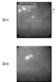
- FIG. 5 is a diagram showing an example of an endoscopic image used as teacher data.
- FIG. 5A shows an endoscopic image (gastric cancer image) in which differentiated gastric cancer is present as a lesion.
- FIG. 5B shows an endoscopic image (gastric cancer image) in which undifferentiated gastric cancer is present as a lesion.
- FIG. 5C shows an endoscopic image (non-gastric cancer image) in which gastric cancer as a lesion does not exist.
- the endoscopic image as teacher data D4 in the learning process mainly uses the abundant database of Japan's top-class cancer treatment hospitals, and is prepared by the instructor of the Japan Gastroenterological Endoscopy Society who has abundant diagnosis and treatment experience. All images were examined in detail, sorted, and marked for the location of the lesion (gastric cancer) by precise manual treatment.
- teacher data D4 endoscopic image data
- an expert endoscopist with abundant experience can directly connect to the diagnostic accuracy of the diagnostic imaging apparatus 100.
- a sufficient number of cases with image selection, lesion identification, and feature extraction marking is an extremely important process.
- Such high-precision data cleansing work and use of high-quality reference data provide highly reliable AI program output results.
- the teacher data D4 of the endoscopic image may be pixel value data or data that has undergone a predetermined color conversion process or the like. Further, as the pretreatment, a texture feature, a shape feature, an uneven state, a spread feature, etc., which are characteristic of the cancerous portion, may be extracted from a comparison between an inflammatory image and a non-inflammatory image. Further, the teacher data D4 may perform learning processing in association with information related to the subject's age, gender, region or medical history, family medical history, etc., in addition to the endoscopic image data.
- the algorithm when the learning device 40 performs the learning process may be a known method.
- the learning device 40 uses, for example, a known backpropagation (backpropagation method) to perform learning processing on a convolutional neural network and adjust network parameters (weighting coefficient, bias, etc.).
- backpropagation method backpropagation method
- the model data (structural data, learned weight parameters, etc.) of the convolutional neural network subjected to the learning process by the learning device 40 is stored in the external storage device 104 together with the diagnostic imaging program, for example.
- Known CNN models include, for example, GoogleLeNet, ResNet, SENet and the like.
- the diagnostic imaging apparatus 100 irradiates the stomach of the subject with narrow-band light and acquires an endoscopic moving image taken in a state where the stomach is magnified and observed.
- the presence of gastric cancer in the acquired endoscopic moving image is estimated by using the endoscopic moving image acquisition unit 10 and the convolutional neural network adjusted using the gastric cancer image and the non-gastric cancer image as teacher data, and the estimation result. It is provided with an estimation unit 20 for outputting.
- the convolutional neural network includes endoscopic images (gastric cancer image, non-stomach cancer image) of a plurality of stomachs (digestive organs) obtained in advance for each of a plurality of subjects, and preliminarily for each of the plurality of subjects. It is learned based on the lesion name of the obtained lesion (stomach cancer) and the definite determination result of the lesion position. Therefore, it is possible to estimate the lesion name and lesion position of the stomach of a new subject in a short time and with an accuracy comparable to that of a substantially experienced endoscopist.
- the diagnostic imaging apparatus 100 can also be used as a diagnostic support tool that directly supports the diagnosis of endoscopic moving images by an endoscopist in a medical examination room.
- the diagnostic imaging apparatus 100 can be used as a central diagnostic support service that supports the diagnosis of endoscopic moving images transmitted from a plurality of examination rooms, or can be remotely controlled via an Internet line in a remote institution. It can also be used as a diagnostic support service to support the diagnosis of endoscopic moving images.
- the diagnostic imaging apparatus 100 can also be operated on the cloud. Furthermore, these endoscopic moving images and AI judgment results can be directly converted into a video library and used as teaching materials and materials for education and training and research.
- Narrow-band light was applied to the stomachs of multiple subjects in cases (395 cases) in which ESD was performed as the initial treatment at the Cancer Research Association Ariake Hospital between April 2005 and December 2016. Then, the stomach is magnified by irradiating the endoscopic image of 1492 in which gastric cancer is present and the stomachs of a plurality of subjects with narrow band light.
- a teacher data set (teacher data) that is imaged by an endoscopic imaging device in the observed state, extracts 1078 endoscopic images in the absence of gastric cancer from an electronic chart device, and is used for learning a convolutional neural network in an diagnostic imaging device. ).
- GIF-H240Z, GIF-H260Z, and GIF-H290 manufactured by Olympus Medical Systems Co., Ltd. were used.
- the endoscopic image taken by the endoscopic imaging device with the subject's stomach observed at high magnification and gastric cancer are observed in 60% or more of the entire image. Included (existing) endoscopic images.
- endoscopic images with widespread mucus, blood, out of focus, or poor image quality due to halation were excluded from the teacher dataset.
- the instructor of the Japan Gastroenterological Endoscopy Society who is a specialist in gastric cancer, examines and selects the prepared endoscopic images in detail, marks the lesion position of the lesion by precise manual processing, and prepares the teacher data set. I prepared it.
- GoogleNet which is composed of 22 layers and has a structure common to the previous CNN but has a sufficient number of parameters and expressive power, was used as a convolutional neural network.
- the Caffe Deep Learning Framework developed at the Berkeley Vision and Learning Center (BVLC) was used for learning and evaluation testing. All layers of the convolutional neural network are fine-tuned with a global learning rate of 0.0001 using stochastic gradient descent. Each endoscopic image was resized to 224 x 224 pixels for compatibility with CNN.
- GIF-H240Z, GIF-H260Z, and GIF-H290 manufactured by Olympus Medical Systems Co., Ltd. were used as in the preparation of the teacher data set.
- the data set for the evaluation test included an endoscopic moving image taken by an endoscopic imaging device for 10 seconds with a strong magnified observation of the subject's stomach as an endoscopic moving image satisfying the eligibility criteria. ..
- evaluation test data are used as endoscopic moving images that meet the exclusion criteria. Excluded from the set.
- the instructor of the Japan Gastroenterological Endoscopy Society who is an expert on gastric cancer, examined the prepared endoscopic moving images in detail, and endoscopic moving images with gastric cancer and endoscopic moving images without gastric cancer. And were selected, and a data set for evaluation test was prepared.
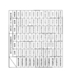
- FIG. 6 is a diagram showing the characteristics of the subject and the lesion (gastric cancer) related to the endoscopic moving image used in the data set for the evaluation test.
- the ratio (%) to the whole is shown in parentheses.
- the median (interquartile range) [whole range] is shown.
- the median tumor diameter was 14 mm
- the interquartile range (entire range) of the tumor was 9 to 20 (1-48) mm.
- 60 lesions (69.0%) were the most depressed type.
- the data set for the evaluation test is input to the convolutional neural network-based diagnostic imaging device that has been trained using the teacher data set, and each endoscope that constitutes the data set for the evaluation test. It was evaluated whether or not it was possible to correctly diagnose whether or not gastric cancer was present in the moving image.
- the diagnostic imaging apparatus diagnoses that a lesion is present in the endoscopic moving image when a predetermined number of endoscopic images having a certainty level of a predetermined value or more exist continuously within a predetermined time.
- whether or not gastric cancer can be correctly diagnosed in each endoscopic moving image can be correctly diagnosed by changing the predetermined time, certainty, and predetermined number of values in various ways and using the changed values. was evaluated.
- the correct diagnosis rate, sensitivity, specificity, positive predictive value (PPV) and negative predictive value (NPV) for the diagnostic ability of the diagnostic imaging device (or endoscopist) are calculated by the following formula (1). )-(5).
- Correct diagnosis rate (number of endoscopic moving images that could correctly diagnose the presence or absence of gastric cancer in the evaluation test data set) / (number of all endoscopic moving images that make up the evaluation test data set) ) ⁇ ⁇ ⁇ (1)
- Sensitivity (Number of endoscopic moving images that could correctly diagnose the presence of gastric cancer in the evaluation test data set) / (Number of endoscopic moving images that actually have gastric cancer in the evaluation test data set) ⁇ ⁇
- Specificity (number of endoscopic moving images that could correctly diagnose the absence of gastric cancer in the evaluation test data set) / (number of endoscopic moving images that do not actually have gastric cancer in the evaluation test data set) ...
- Positive predictive value (PPV) (Number of endoscopic moving images diagnosed as having gastric cancer in the evaluation test data set) / (Evaluation test data set) Number of endoscopic moving images diagnosed as having gastric cancer in Japan) ...
- Negative predictive value (NPV) (Number of endoscopic moving images diagnosed as having no gastric cancer in the evaluation test data set, which do not actually have gastric cancer) / (Evaluation test data Number of endoscopic moving images diagnosed as having gastric cancer in the set) ...
- predetermined time 0.1 seconds or more and 0.5 seconds or less
- certainty 0.6
- certainty 0.5
- combination of predetermined number 3
- the medium and negative predictive values were calculated.
- the correct diagnosis rate 85.1% (95% CI: 79.0 to 89.6)
- the sensitivity 87.4% (95% CI: 78.8 to 92.8)
- the specificity 82. 8% (95% CI: 73.5 to 89.3)
- positive predictive value 83.5% (95% CI: 74.6 to 89.7)
- negative predictive value 86.7% (77) It was .8-92.4).
- FIG. 8 shows the number of correct diagnosis, the number of misdiagnosis, and the unconfirmed number of correct diagnosis, the number of false diagnosis, and the number of correct diagnosis, the number of correct diagnosis, and the number of unconfirmed diagnosis of the endoscopic moving image in which gastric cancer is present and the endoscopic moving image in which gastric cancer is not present. It is a figure showing a number.
- FIG. 9 is a diagram showing the correct diagnosis rate, sensitivity, specificity, positive predictive value and negative predictive value of the diagnostic imaging apparatus and 11 endoscopic skilled doctors A to K.
- 95% confidence intervals were also calculated and compared by each of the diagnostic imaging apparatus and the endoscopist skilled doctors A to K.
- the Mcnemar test is used to compare the correctness, sensitivity and specificity between diagnostic imaging equipment and endoscopists AK, while comparing the positive and negative predictive values. Used the binomial test (see P value in FIG. 9).
- the statistically significant difference was set to less than 0.05.
- "JMP13" was used as a high-performance interactive tool for visualizing data and performing statistical analysis.
- the diagnostic imaging apparatus is significantly superior to two endoscopists H and K and significantly inferior to one endoscopist I. rice field. In addition, no significant difference was observed between the diagnostic imaging system and the eight endoscopists A to G and J.
- the diagnostic imaging system was significantly superior to the three endoscopists C, J, and K. In addition, no significant difference was observed between the diagnostic imaging system and the eight endoscopists A, B, D to I.
- the diagnostic imaging system was significantly superior to the two endoscopists H and K, and significantly inferior to the three endoscopists C, F and I. In addition, no significant difference was observed between the diagnostic imaging system and the six endoscopists A, B, D, E, G, and J.
- the diagnostic imaging apparatus was significantly superior to the two endoscopists H and K, and significantly inferior to the two endoscopists C and F. In addition, no significant difference was observed between the diagnostic imaging system and the seven endoscopists A, B, D, E, G, I, and J.
- the diagnostic imaging device was significantly superior to the two endoscopists J and K. In addition, no significant difference was observed between the diagnostic imaging system and the nine endoscopists AI.
- the predetermined time, the certainty, and the predetermined number of values at which the correct diagnosis rate of the diagnostic imaging apparatus is highest are obtained.
- gastric cancer in actual clinical practice, if gastric cancer can be clearly detected in the endoscopic moving image (10 seconds) for 0.5 seconds (if there are three consecutive endoscopic images with a certainty of 0.5 or more). ), It shows that gastric cancer can be diagnosed in real time with a high accuracy rate. In addition, there is a tendency that the diagnostic performance of the diagnostic imaging apparatus can be maintained by increasing the number of endoscopic images required for diagnosing the presence of gastric cancer even when the certainty is low.
- the values of the predetermined time, certainty, and the predetermined number at which the correct diagnosis rate of the diagnostic imaging apparatus is highest are described, but when the correct diagnosis rate is maintained at 70% or more or 80% or more, The diagnostic performance can be exhibited in a wider range of predetermined time, certainty and a predetermined number of combinations.
- the diagnostic ability of the diagnostic imaging device was compared with the diagnostic ability of 11 endoscopists.
- the diagnostic imaging apparatus has a diagnostic ability equal to or higher than that of a skilled endoscope doctor.
- Sensitivity is paramount because endoscopy is a screening test for diagnosing gastric cancer.
- the diagnostic imaging apparatus was particularly excellent in sensitivity as compared with an endoscopist. From this, the diagnosis of gastric cancer by the diagnostic imaging device not only supports (supports) the diagnosis of endoscopists who have not mastered the diagnostic technique of gastric cancer by ME-NBI, but also has acquired the diagnostic technique. It was also found to be beneficial for endoscopists.
- Non-Patent Document 3 as a result of evaluating the diagnostic ability of a computer-assisted diagnostic (CAD) system for gastric cancer using an endoscopic image (still image) taken by an NBI combined magnifying endoscope, the correct diagnosis rate is 85. It is described that the sensitivity was 3%, the sensitivity was 95.4%, the specificity was 71.0%, the positive predictive value was 82.3%, and the negative predictive value was 91.7%. In addition, severe atrophic gastritis, localized atrophy, and intestinal metaplasia have been described as examples of causes of false positives.
- CAD computer-assisted diagnostic
- Non-Patent Document 3 does not compare the diagnostic ability of the computer-aided diagnostic system with the diagnostic ability of an endoscopic expert who has acquired the diagnostic technique of gastric cancer by ME-NBI, and therefore evaluates the diagnostic ability.
- the diagnostic difficulty of the endoscopic images used for this was unknown, limiting the interpretation of the diagnostic capabilities of computer-aided diagnostic systems.
- Non-Patent Document 4 the same examination as in Non-Patent Document 3 was carried out, and it was found that the diagnostic imaging apparatus was significantly superior in sensitivity and negative predictive value as compared with two endoscopists. Have been described. However, the number of endoscopists compared to computer-aided diagnostic systems is small, that is, the diagnostic ability of individual endoscopists may be strongly biased in the results, so the diagnostic ability is evaluated. The diagnostic difficulty of the endoscopic images used for this was unknown, and the interpretation of the diagnostic capabilities of computer-aided diagnostic systems was limited. Further, in Non-Patent Document 4, the AUC is not calculated, and the diagnostic accuracy of the computer-aided diagnosis system as an image diagnostic device is also unknown.
- Non-Patent Documents 3 and 4 studies using still images (endoscopic images) are carried out, which is useful when performing secondary interpretation of endoscopic images after endoscopic examination.
- Non-Patent Document 5 states that the sensitivity in diagnosing gastric cancer pick-up was 94.1% with respect to the diagnostic performance of the computer-aided diagnostic system in endoscopic moving images taken using a normal endoscope. Have been described. However, in Non-Patent Document 5, only the evaluation of sensitivity is described, the endoscopic moving image captured by the NBI combined magnifying endoscope is not used, and the computer-assisted diagnostic system and the inside. Evaluation of the difficulty of diagnosis of endoscopic moving images and gastric cancer of the computer-assisted diagnostic system because the diagnostic ability was not compared with the endoscopic expert and the AUC in the computer-assisted diagnostic system was not calculated. There is a limit to the interpretation of the diagnostic ability of the patient, and sufficient evaluation has not been performed, and the judgment of its usefulness in clinical practice is unknown.
- the diagnostic imaging apparatus in the present invention was 0.8684, and the comprehensive diagnostic ability and reliability as a medical device were very high.
- the diagnostic imaging apparatus of the present invention compares the diagnostic ability with many skilled endoscopists, it is appropriate to set weights and parameters in CNN, and further, the difficulty level for moving image evaluation. Can be properly evaluated. It is also possible to adjust to reduce the bias that occurs in comparison with a small number of skilled doctors by making comparisons with many skilled doctors.
- a computer-aided diagnosis (CAD) system can provide performance with diagnostic capabilities equal to or better than that of a skilled doctor. It was shown that it can be used not only in clinical practice but also as an education and training system.
- CAD computer-aided diagnosis
- a magnifying endoscope combined with NBI is used, and detailed observation of lesions is possible as compared with a normal endoscope and a non-magnifying endoscope combined with NBI, and its diagnostic ability is high. It was highly useful.
- moving images are used instead of still images, and gastric cancer can be diagnosed in real time by using an image diagnostic apparatus in actual clinical practice.
- the present invention is a diagnostic imaging device, a diagnostic imaging method, a diagnostic imaging program, and a trained model capable of diagnosing gastric cancer in real time in gastric endoscopy performed using a magnifying endoscope combined with NBI. It is useful.
- Endoscopic moving image acquisition unit 20
- Estimating unit 30
- Display control unit 40
- Learning device 100
- Image diagnostic device 101
- CPU 102
- ROM 103
- RAM 104
- External storage device 105
- Communication interface 200
- Endoscope imager 300
- Display device D1
- Endoscope moving image data D2
- Estimation result data D3
- Judgment result image data D4 Teacher data
Landscapes
- Health & Medical Sciences (AREA)
- Life Sciences & Earth Sciences (AREA)
- Engineering & Computer Science (AREA)
- Surgery (AREA)
- Physics & Mathematics (AREA)
- Medical Informatics (AREA)
- General Health & Medical Sciences (AREA)
- Nuclear Medicine, Radiotherapy & Molecular Imaging (AREA)
- Radiology & Medical Imaging (AREA)
- Veterinary Medicine (AREA)
- Molecular Biology (AREA)
- Public Health (AREA)
- Biophysics (AREA)
- Optics & Photonics (AREA)
- Pathology (AREA)
- Animal Behavior & Ethology (AREA)
- Biomedical Technology (AREA)
- Heart & Thoracic Surgery (AREA)
- Theoretical Computer Science (AREA)
- General Physics & Mathematics (AREA)
- Computer Vision & Pattern Recognition (AREA)
- Signal Processing (AREA)
- Quality & Reliability (AREA)
- Artificial Intelligence (AREA)
- Evolutionary Computation (AREA)
- Gastroenterology & Hepatology (AREA)
- Endoscopes (AREA)
- Image Analysis (AREA)
Abstract
Provided are a diagnostic imaging device, diagnostic imaging method, diagnostic imaging program, and learned model with which stomach cancer diagnosis can be carried out in real time during endoscopic examination performed using NBI in combination with a magnifying endoscope. The diagnostic imaging device comprises an endoscopic video image acquisition unit which emits narrow-band light at a subject's stomach and acquires an endoscopic video image captured while the stomach is in a state of magnified observation, and an estimation unit which uses a convolutional neural network, which has been caused to learn using stomach cancer images and non-stomach cancer images as teaching data, to estimate the presence of stomach cancer in the acquired endoscopic video image, and outputs estimation results.
Description
本発明は、画像診断装置、画像診断方法、画像診断プログラムおよび学習済みモデルに関する。
The present invention relates to a diagnostic imaging apparatus, a diagnostic imaging method, a diagnostic imaging program, and a trained model.
胃癌は、世界において最も多く認められる癌の1つであり、癌関連死亡率が高い癌の1つである。一方で、内視鏡機器の発展により、内視鏡で胃癌が早期に見つかる例が多くなっている。その結果、近年、胃癌による死亡率は減少している。さらに、内視鏡的粘膜下層剥離術(ESD)の開発により、早期胃癌の治療は低侵襲治療となっている。ただし、日本の胃癌治療ガイドラインにおいて、ESDの適応は粘膜内癌(粘膜固有層までに癌の浸潤が留まるもの)に限られており、より早期で胃癌を発見、診断することが重要となっている。
Gastric cancer is one of the most common cancers in the world and one with a high cancer-related mortality rate. On the other hand, with the development of endoscopic equipment, there are many cases where gastric cancer is detected early by endoscopy. As a result, the mortality rate from gastric cancer has decreased in recent years. Furthermore, with the development of endoscopic submucosal dissection (ESD), the treatment of early gastric cancer has become a minimally invasive treatment. However, in Japan's gastric cancer treatment guidelines, ESD indications are limited to intramucosal cancer (where cancer infiltration remains up to the lamina propria), and it is important to detect and diagnose gastric cancer at an earlier stage. There is.
一般に、胃癌の診断は内視鏡で行われる。近年、被験者の胃に対して狭帯域光を照射し(NBI: Narrow Band Imaging、狭帯域光法)、当該胃を拡大観察することが可能なNBI併用拡大内視鏡(ME-NBI)が開発されている。NBI併用拡大内視鏡は通常の内視鏡よりも胃癌の診断能が高いことが報告されている。しかし、内視鏡医は、ME-NBIによる胃癌の診断技術を習得するために相当な努力が必要である。それは、胃癌は、その背景粘膜に、ピロリ菌感染に伴う慢性炎症(胃炎)が認められる例が大半であり、胃癌と胃炎とを識別することが難しいからである。特に、炎症細胞浸潤が強い場合には、胃癌の局在および範囲が不明瞭となり、経験の浅い内視鏡医では胃癌を見逃す傾向がある。そのため、より高度の診断技術が内視鏡医に求められる。したがって、背景粘膜にピロリ菌感染に伴う慢性炎症が認められない他の消化管癌(例えば、粘膜の色や凹凸で判定する食道癌やポリープが特徴的な大腸癌等)と比較して、胃癌を適切に診断することは困難である。
Generally, gastric cancer is diagnosed endoscopically. In recent years, an NBI combined magnifying endoscope (ME-NBI) has been developed that can irradiate the stomach of a subject with narrow band light (NBI: Narrow Band Imaging, narrow band imaging) and magnify the stomach. Has been done. It has been reported that a magnifying endoscope combined with NBI has a higher diagnostic ability for gastric cancer than a normal endoscope. However, endoscopists need considerable effort to master the technique of diagnosing gastric cancer by ME-NBI. This is because most cases of gastric cancer have chronic inflammation (gastritis) associated with Helicobacter pylori infection on the background mucosa, and it is difficult to distinguish between gastric cancer and gastritis. In particular, when inflammatory cell infiltration is strong, the localization and extent of gastric cancer becomes unclear, and inexperienced endoscopists tend to overlook gastric cancer. Therefore, more advanced diagnostic techniques are required of endoscopists. Therefore, gastric cancer is compared with other gastrointestinal cancers in which the background mucosa does not show chronic inflammation associated with Helicobacter pylori infection (for example, esophageal cancer judged by the color and unevenness of the mucosa and colon cancer characterized by polyps). Is difficult to diagnose properly.
近年、ディープラーニング(深層学習)を用いた人工知能(AI:Artificial Intelligence)が開発され、医療分野においても応用されている。さらに、AIに入力された画像の特徴を維持したまま畳み込み学習を行う畳み込みニューラルネットワーク(CNN:Convolutional Neural Network)が開発され、学習した画像の分類を行うコンピューター支援診断(CAD:Computer-Aided Diagnosis)システムの画像診断能力は劇的に向上している。
In recent years, artificial intelligence (AI: Artificial Intelligence) using deep learning has been developed and is also applied in the medical field. Furthermore, a convolutional neural network (CNN) that performs convolutional learning while maintaining the characteristics of the image input to AI has been developed, and computer-aided diagnosis (CAD: Computer-Aided Diagnosis) that classifies the learned image. The diagnostic imaging capabilities of the system have improved dramatically.
ディープラーニングを用いたAIは様々な医療分野で注目されており、放射線腫瘍学、皮膚がん分類、糖尿病性網膜症、胃生検の組織学的分類、超拡大内視鏡による大腸病変の特徴付けを含む医療分野の画像診断をAIが専門医の診断を支援することができるとの様々な報告がある。特に、顕微内視鏡レベルにおいてはAIが専門医と同等の精度を出せることが証明されている(非特許文献1を参照)。また、皮膚科では、ディープラーニング機能を持ったAIが専門医と同等の画像診断能力を発揮することが発表されており(非特許文献2を参照)、各種機械学習法を利用した特許文献(特許文献1、2を参照)も存在する。
AI using deep learning is attracting attention in various medical fields, and features of radiation oncology, skin cancer classification, diabetic retinopathy, histological classification of gastric biopsy, and colorectal lesions by super-magnifying endoscopy. There are various reports that AI can support the diagnosis of specialists in diagnostic imaging in the medical field, including attachment. In particular, it has been proved that AI can obtain the same accuracy as a specialist at the microscopic endoscopy level (see Non-Patent Document 1). In addition, in dermatology, it has been announced that AI with a deep learning function exhibits the same diagnostic imaging ability as a specialist (see Non-Patent Document 2), and patent documents using various machine learning methods (patents). (See References 1 and 2) also exists.
ただし、静止画を教師データとして学習に用い、検査時に撮像した静止画をAIで判定させる場合には、静止画が撮像されないと、AIが判定できないため、AIが内視鏡検査中の病巣の見落としの有無に関しては補助できないことに留意する必要がある。また、リアルタイムで動画として判定する場合には、内視鏡検査中に癌の検出を補助するため、検出する癌の数が増加するという点でも、実臨床において有益と考えられる。
However, when a still image is used for learning as teacher data and the still image captured at the time of examination is judged by AI, the AI cannot be determined unless the still image is captured. It should be noted that we cannot assist in the presence or absence of oversight. In addition, when it is judged as a moving image in real time, it is considered to be beneficial in actual clinical practice in that the number of cancers to be detected increases because it assists the detection of cancer during endoscopy.
上記のように、AIの画像診断能力は専門医並みであることが示唆されている。しかしながら、NBI併用拡大内視鏡を用いて行われる胃の内視鏡検査において、AIの画像診断能力を使用して胃癌の診断をリアルタイムに行う画像診断技術は、まだ実際の医療現場(実臨床)には導入されておらず、今後の実用化が期待されている状況である。また、内視鏡による消化器癌の診断には、各消化器癌(食道癌、胃癌、大腸癌など)の固有の特徴量の抽出と、その病態レベルの判定が異なるので、各癌種の特徴に沿ったAIプログラムの設計が重要である。
As mentioned above, it is suggested that AI's diagnostic imaging ability is comparable to that of specialists. However, in gastric endoscopy performed using an NBI combined magnifying endoscope, diagnostic imaging technology that uses AI's diagnostic imaging capability to diagnose gastric cancer in real time is still available in actual medical practice (actual clinical practice). ) Has not been introduced, and it is expected that it will be put into practical use in the future. In addition, in the diagnosis of gastrointestinal cancer by endoscopy, the extraction of the unique characteristic amount of each gastrointestinal cancer (esophageal cancer, gastric cancer, colon cancer, etc.) and the determination of its pathological level are different. It is important to design the AI program according to its characteristics.
本発明の目的は、NBI併用拡大内視鏡を用いて行われる胃の内視鏡検査において、胃癌の診断をリアルタイムに行うことが可能な画像診断装置、画像診断方法、画像診断プログラムおよび学習済みモデルを提供することである。
An object of the present invention is an image diagnostic apparatus, an image diagnosis method, an image diagnosis program, and a learned image diagnosis device capable of diagnosing gastric cancer in real time in gastric endoscopy performed by using a magnifying endoscope combined with NBI. To provide a model.
本発明に係る画像診断装置は、
被験者の胃に対して狭帯域光を照射し、当該胃を拡大観察した状態で撮像された内視鏡動画像を取得する内視鏡動画像取得部と、
胃癌画像および非胃癌画像を教師データとして学習させた畳み込みニューラルネットワークを用いて、取得された前記内視鏡動画像内における胃癌の存在を推定し、推定結果を出力する推定部と、
を備える。 The diagnostic imaging apparatus according to the present invention is
An endoscopic moving image acquisition unit that irradiates the subject's stomach with narrow-band light and acquires an endoscopic moving image taken while observing the stomach in an enlarged manner.
Using a convolutional neural network trained from gastric cancer images and non-gastric cancer images as teacher data, an estimation unit that estimates the presence of gastric cancer in the acquired endoscopic moving images and outputs the estimation results, and an estimation unit.
To be equipped.
本発明に係る画像診断方法は、
被験者の胃に対して狭帯域光を照射し、当該胃を拡大観察した状態で撮像された内視鏡動画像を取得する内視鏡動画像取得工程と、
胃癌画像および非胃癌画像を教師データとして学習させた畳み込みニューラルネットワークを用いて、取得された前記内視鏡動画像内における胃癌の存在を推定し、推定結果を出力する推定工程と、
を含む。 The diagnostic imaging method according to the present invention
An endoscopic moving image acquisition step of irradiating the subject's stomach with narrow-band light and acquiring an endoscopic moving image taken while observing the stomach in a magnified state.
Using a convolutional neural network trained from gastric cancer images and non-gastric cancer images as teacher data, the presence of gastric cancer in the acquired endoscopic moving image is estimated, and an estimation step of outputting the estimation result is performed.
including.
本発明に係る画像診断プログラムは、
コンピューターに、
被験者の胃に対して狭帯域光を照射し、当該胃を拡大観察した状態で撮像された内視鏡動画像を取得する内視鏡動画像取得処理と、
胃癌画像および非胃癌画像を教師データとして学習させた畳み込みニューラルネットワークを用いて、取得された前記内視鏡動画像内における胃癌の存在を推定し、推定結果を出力する推定処理と、
を実行させる。 The diagnostic imaging program according to the present invention
On the computer
Endoscopic moving image acquisition processing that irradiates the subject's stomach with narrow-band light and acquires an endoscopic moving image taken while observing the stomach in a magnified state.
Using a convolutional neural network trained from gastric cancer images and non-gastric cancer images as teacher data, the presence of gastric cancer in the acquired endoscopic moving images is estimated, and an estimation process that outputs the estimation results is performed.
To be executed.
本発明に係る学習済みモデルは、
胃癌画像および非胃癌画像を教師データとして畳み込みニューラルネットワークを学習させることによって得られ、
被験者の胃に対して狭帯域光を照射し、当該胃を拡大観察した状態で撮像された内視鏡動画像内における胃癌の存在を推定し、推定結果を出力するようコンピューターを機能させる。 The trained model according to the present invention
Obtained by training a convolutional neural network using gastric cancer images and non-gastric cancer images as teacher data.
The subject's stomach is irradiated with narrow-band light, the presence of gastric cancer is estimated in the endoscopic moving image taken while observing the stomach in a magnified state, and the computer is made to function to output the estimation result.
本発明によれば、NBI併用拡大内視鏡を用いて行われる胃の内視鏡検査において、胃癌の診断をリアルタイムに行うことができる。
According to the present invention, gastric cancer can be diagnosed in real time in gastric endoscopy performed using a magnifying endoscope combined with NBI.
以下、本実施の形態を図面に基づいて詳細に説明する。
Hereinafter, the present embodiment will be described in detail based on the drawings.
[画像診断装置の全体構成]
まず、本実施の形態における画像診断装置100の構成について説明する。図1は、画像診断装置100の全体構成を示すブロック図である。図2は、本実施の形態における画像診断装置100のハードウェア構成の一例を示す図である。 [Overall configuration of diagnostic imaging equipment]
First, the configuration of thediagnostic imaging apparatus 100 according to the present embodiment will be described. FIG. 1 is a block diagram showing an overall configuration of the diagnostic imaging apparatus 100. FIG. 2 is a diagram showing an example of the hardware configuration of the diagnostic imaging apparatus 100 according to the present embodiment.
まず、本実施の形態における画像診断装置100の構成について説明する。図1は、画像診断装置100の全体構成を示すブロック図である。図2は、本実施の形態における画像診断装置100のハードウェア構成の一例を示す図である。 [Overall configuration of diagnostic imaging equipment]
First, the configuration of the
画像診断装置100は、医師(例えば、内視鏡医)による消化器(本実施の形態では、胃)の内視鏡検査において、畳み込みニューラルネットワーク(CNN)が有する内視鏡画像の画像診断能力を使用して胃癌の診断をリアルタイムに行う。画像診断装置100には、内視鏡撮像装置200および表示装置300が接続されている。
The diagnostic imaging apparatus 100 has an endoscopic image diagnostic capability possessed by a convolutional neural network (CNN) in an endoscopy of a digestive organ (stomach in the present embodiment) by a doctor (for example, an endoscopist). Make a real-time diagnosis of gastric cancer using. An endoscopic imaging device 200 and a display device 300 are connected to the diagnostic imaging device 100.
内視鏡撮像装置200は、例えば、撮像手段を内蔵した電子内視鏡(ビデオスコープともいう)や、光学式内視鏡に撮像手段を内蔵したカメラヘッドを装着したカメラ装着内視鏡等である。内視鏡撮像装置200は、例えば、被験者の口または鼻から消化器に挿入され、当該消化器内の診断対象部位を撮像する。
The endoscope imaging device 200 is, for example, an electronic endoscope (also referred to as a videoscope) having a built-in imaging means, a camera-mounted endoscope in which a camera head having a built-in imaging means is attached to an optical endoscope, or the like. be. The endoscopic imaging device 200 is inserted into the digestive organ, for example, through the mouth or nose of the subject, and images a diagnosis target site in the digestive organ.
本実施の形態では、内視鏡撮像装置200は、医師の操作(例えば、ボタン操作)に応じて、被験者の胃に対して狭帯域光(例えば、NBI用狭帯域光)を照射し当該胃を例えば80倍に拡大観察した状態で当該胃内の診断対象部位を内視鏡動画像として撮像する。内視鏡動画像は、時間的に連続する複数の内視鏡画像から構成される。内視鏡撮像装置200は、撮像した内視鏡動画像を表す内視鏡動画像データD1を画像診断装置100に出力する。
In the present embodiment, the endoscope imaging device 200 irradiates the stomach of the subject with narrow band light (for example, narrow band light for NBI) in response to a doctor's operation (for example, button operation). Is imaged as an endoscopic moving image of the site to be diagnosed in the stomach, for example, in a state of being magnified 80 times. The endoscopic moving image is composed of a plurality of endoscopic images that are continuous in time. The endoscope imaging device 200 outputs the endoscopic moving image data D1 representing the captured endoscopic moving image to the diagnostic imaging device 100.
表示装置300は、例えば、液晶ディスプレイであり、画像診断装置100から出力された内視鏡動画像および判定結果画像を、医師に識別可能に表示する。
The display device 300 is, for example, a liquid crystal display, and displays the endoscopic moving image and the determination result image output from the diagnostic imaging device 100 so that the doctor can identify them.
図2に示すように、画像診断装置100は、主たるコンポーネントとして、CPU(Central Processing Unit)101、ROM(Read Only Memory)102、RAM(Random Access Memory)103、外部記憶装置(例えば、フラッシュメモリ)104、通信インターフェイス105およびGPU(Graphics Processing Unit)106等を備えたコンピューターである。
As shown in FIG. 2, the diagnostic imaging apparatus 100 has a CPU (Central Processing Unit) 101, a ROM (Read Only Memory) 102, a RAM (Random Access Memory) 103, and an external storage device (for example, a flash memory) as main components. It is a computer equipped with 104, a communication interface 105, a GPU (Graphics Processing Unit) 106, and the like.
画像診断装置100の各機能は、例えば、CPU101,GPU106がROM102、RAM103、外部記憶装置104等に記憶された制御プログラム(例えば、画像診断プログラム)や各種データ(例えば、内視鏡動画像データ、学習用教師データ、畳み込みニューラルネットワークのモデルデータ(構造データおよび学習済み重みパラメータ等)などを参照することによって実現される。なお、RAM103は、例えば、データの作業領域や一時退避領域として機能する。
Each function of the diagnostic imaging apparatus 100 includes, for example, a control program (for example, an diagnostic imaging program) in which the CPU 101 and GPU 106 are stored in a ROM 102, a RAM 103, an external storage device 104, and various data (for example, endoscopic moving image data). It is realized by referring to the training teacher data, the model data of the convolutional neural network (structural data, learned weight parameters, etc.), and the RAM 103 functions as, for example, a data work area or a temporary save area.
なお、画像診断装置100の各機能の一部または全部は、CPU101,GPU106による処理に代えて、または、これと共に、DSP(Digital Signal Processor)による処理によって実現されても良い。また、同様に、各機能の一部または全部は、ソフトウェアによる処理に代えて、または、これと共に、専用のハードウェア回路による処理によって実現されても良い。
Note that some or all of the functions of the diagnostic imaging apparatus 100 may be realized by processing by the DSP (Digital Signal Processor) instead of or in combination with the processing by the CPU 101 and GPU 106. Similarly, a part or all of each function may be realized by processing by a dedicated hardware circuit in place of or in combination with processing by software.
図1に示すように、画像診断装置100は、内視鏡動画像取得部10、推定部20および表示制御部30を備えている。学習装置40は、画像診断装置100において使用される畳み込みニューラルネットワークのモデルデータ(本発明の「学習済みモデル」に対応)を生成する機能を有する。なお、表示制御部30は、本発明の「警告出力制御部」としても機能する。
As shown in FIG. 1, the diagnostic imaging apparatus 100 includes an endoscopic moving image acquisition unit 10, an estimation unit 20, and a display control unit 30. The learning device 40 has a function of generating model data (corresponding to the "learned model" of the present invention) of the convolutional neural network used in the diagnostic imaging device 100. The display control unit 30 also functions as the "warning output control unit" of the present invention.
[内視鏡動画像取得部]
内視鏡動画像取得部10は、内視鏡撮像装置200から出力された内視鏡動画像データD1を取得する。そして、内視鏡動画像取得部10は、取得した内視鏡動画像データD1を推定部20に出力する。なお、内視鏡動画像取得部10は、内視鏡動画像データD1を取得する際、内視鏡撮像装置200から直接取得しても良いし、外部記憶装置104に格納された内視鏡動画像データD1や、インターネット回線等を介して提供された内視鏡動画像データD1を取得しても良い。 [Endoscopic moving image acquisition unit]
The endoscopic movingimage acquisition unit 10 acquires the endoscopic moving image data D1 output from the endoscopic imaging device 200. Then, the endoscopic moving image acquisition unit 10 outputs the acquired endoscopic moving image data D1 to the estimation unit 20. The endoscope moving image acquisition unit 10 may directly acquire the endoscopic moving image data D1 from the endoscope imaging device 200, or the endoscope stored in the external storage device 104. The moving image data D1 or the endoscopic moving image data D1 provided via an Internet line or the like may be acquired.
内視鏡動画像取得部10は、内視鏡撮像装置200から出力された内視鏡動画像データD1を取得する。そして、内視鏡動画像取得部10は、取得した内視鏡動画像データD1を推定部20に出力する。なお、内視鏡動画像取得部10は、内視鏡動画像データD1を取得する際、内視鏡撮像装置200から直接取得しても良いし、外部記憶装置104に格納された内視鏡動画像データD1や、インターネット回線等を介して提供された内視鏡動画像データD1を取得しても良い。 [Endoscopic moving image acquisition unit]
The endoscopic moving
[推定部]
推定部20は、畳み込みニューラルネットワークを用いて、内視鏡動画像取得部10から出力された内視鏡動画像データD1により表される内視鏡動画像内における病変(本実施の形態では、胃癌)の存在を推定し、推定結果を出力する。具体的には、推定部20は、内視鏡動画像内に存在する病変の病変名(名称)および病変位置(位置)と、当該病変名および病変位置の確信度(確度ともいう)とを推定する。そして、推定部20は、内視鏡動画像取得部10から出力された内視鏡動画像データD1と、病変名、病変位置および確信度の推定結果を表す推定結果データD2とを表示制御部30に出力する。 [Estimator]
Theestimation unit 20 uses a convolutional neural network to display a lesion in the endoscopic moving image represented by the endoscopic moving image data D1 output from the endoscopic moving image acquisition unit 10 (in the present embodiment, the estimation unit 20). Estimates the presence of gastric cancer) and outputs the estimation result. Specifically, the estimation unit 20 determines the lesion name (name) and lesion position (position) of the lesion existing in the endoscopic moving image and the certainty (also referred to as accuracy) of the lesion name and lesion position. presume. Then, the estimation unit 20 displays the endoscopic moving image data D1 output from the endoscopic moving image acquisition unit 10 and the estimation result data D2 representing the estimation result of the lesion name, the lesion position, and the certainty. Output to 30.
推定部20は、畳み込みニューラルネットワークを用いて、内視鏡動画像取得部10から出力された内視鏡動画像データD1により表される内視鏡動画像内における病変(本実施の形態では、胃癌)の存在を推定し、推定結果を出力する。具体的には、推定部20は、内視鏡動画像内に存在する病変の病変名(名称)および病変位置(位置)と、当該病変名および病変位置の確信度(確度ともいう)とを推定する。そして、推定部20は、内視鏡動画像取得部10から出力された内視鏡動画像データD1と、病変名、病変位置および確信度の推定結果を表す推定結果データD2とを表示制御部30に出力する。 [Estimator]
The
また、推定部20は、内視鏡動画像データD1により表される内視鏡動画像内において確信度が所定値(例えば、0.5)以上である内視鏡画像が所定時間(例えば、0.5秒)内に所定数(例えば、3)連続して存在する場合、内視鏡動画像内に病変(胃癌)が存在すると推定する。ここで、上記所定数は、上記所定値が小さくなるにつれて大きくなるように設定される。推定部20は、内視鏡動画像内に病変が存在すると推定した場合、その旨(推定結果)を表示制御部30に出力する。
Further, in the estimation unit 20, the endoscopic image having a certainty level of a predetermined value (for example, 0.5) or more in the endoscopic moving image represented by the endoscopic moving image data D1 is displayed for a predetermined time (for example, 0.5). If a predetermined number (for example, 3) is continuously present within 0.5 seconds), it is estimated that a lesion (gastric cancer) is present in the endoscopic moving image. Here, the predetermined number is set so as to increase as the predetermined value decreases. When the estimation unit 20 estimates that a lesion exists in the endoscopic moving image, the estimation unit 20 outputs that fact (estimation result) to the display control unit 30.
本実施の形態では、推定部20は、病変名および病変位置の確信度を示す指標として確率スコアを推定する。確率スコアは、0より大きく、1以下の値で表される。確率スコアが高いほど、病変名および病変位置の確信度が高いことを意味する。
In the present embodiment, the estimation unit 20 estimates the probability score as an index showing the certainty of the lesion name and the lesion position. The probability score is represented by a value greater than 0 and less than or equal to 1. The higher the probability score, the higher the certainty of the lesion name and lesion location.
なお、確率スコアは、病変名および病変位置の確信度を示す指標の一例であって、その他の任意の態様の指標が用いられてもよい。例えば、確率スコアは、0%~100%の値で表される態様であっても良いし、数段階のレベル値のうちの何れで表される態様であっても良い。
The probability score is an example of an index indicating the degree of certainty of the lesion name and the lesion position, and an index of any other aspect may be used. For example, the probability score may be represented by a value of 0% to 100%, or may be represented by any of several levels.
畳み込みニューラルネットワークは、順伝播型ニューラルネットワークの一種であって、脳の視覚野の構造における知見に基づくものである。基本的に、画像の局所的な特徴抽出を担う畳み込み層と、局所毎に特徴をまとめあげるプーリング層(サブサンプリング層)とを繰り返した構造となっている。畳み込みニューラルネットワークの各層によれば、複数のニューロン(Neuron)を所持し、個々のニューロンが視覚野と対応するような形で配置されている。それぞれのニューロンの基本的な働きは、信号の入力と出力とからなる。ただし、各層のニューロン間は、相互に信号を伝達する際に、入力された信号をそのまま出力するのではなく、それぞれの入力に対して結合荷重を設定し、その重み付きの入力の総和が各ニューロンに設定されている閾値を超えた時に、次の層のニューロンに信号を出力する。学習データからこれらニューロン間の結合荷重を算出しておく。これによって、リアルタイムのデータを入力することによって、出力値の推定が可能となる。この目的に適合する畳み込みニューラルネットワークであれば、それを構成するアルゴリズムは特に限定されない。
A convolutional neural network is a type of feedforward neural network and is based on knowledge in the structure of the visual cortex of the brain. Basically, it has a structure in which a convolution layer responsible for extracting local features of an image and a pooling layer (subsampling layer) that summarizes features for each region are repeated. According to each layer of the convolutional neural network, it possesses multiple neurons (Neurons), and each neuron is arranged so as to correspond to the visual cortex. The basic function of each neuron consists of signal input and output. However, when transmitting signals between neurons in each layer, instead of outputting the input signal as it is, a coupling load is set for each input, and the sum of the weighted inputs is each. When the threshold set for the neuron is exceeded, a signal is output to the neuron in the next layer. The connection load between these neurons is calculated from the training data. This makes it possible to estimate the output value by inputting real-time data. As long as it is a convolutional neural network suitable for this purpose, the algorithm for constructing it is not particularly limited.
図3は、本実施の形態における畳み込みニューラルネットワークの構成を示す図である。なお、畳み込みニューラルネットワークのモデルデータ(構造データおよび学習済み重みパラメータ等)は、画像診断プログラムと共に、外部記憶装置104に格納されている。
FIG. 3 is a diagram showing a configuration of a convolutional neural network according to the present embodiment. The model data (structural data, learned weight parameters, etc.) of the convolutional neural network is stored in the external storage device 104 together with the diagnostic imaging program.
図3に示すように、畳み込みニューラルネットワークは、例えば、特徴抽出部Naと識別部Nbとを有する。特徴抽出部Naは、入力される画像(具体的には、内視鏡動画像データD1により表される内視鏡動画像を構成する内視鏡画像)から画像特徴を抽出する処理を施す。識別部Nbは、特徴抽出部Naにより抽出された画像特徴から画像に係る推定結果を出力する。
As shown in FIG. 3, the convolutional neural network has, for example, a feature extraction unit Na and an identification unit Nb. The feature extraction unit Na performs a process of extracting image features from an input image (specifically, an endoscopic image constituting the endoscopic moving image represented by the endoscopic moving image data D1). The identification unit Nb outputs an estimation result related to the image from the image features extracted by the feature extraction unit Na.
特徴抽出部Naは、複数の特徴量抽出層Na1、Na2・・・が階層的に接続されて構成される。各特徴量抽出層Na1、Na2・・・は、畳み込み層(Convolution layer)、活性化層(Activation layer)およびプーリング層(Pooling layer)を備える。
The feature extraction unit Na is configured by hierarchically connecting a plurality of feature amount extraction layers Na1, Na2, and so on. Each feature amount extraction layer Na1, Na2 ... Provides a convolution layer, an activation layer, and a pooling layer.
第1層目の特徴量抽出層Na1は、入力される画像を、ラスタスキャンにより所定サイズ毎に走査する。そして、特徴量抽出層Na1は、走査したデータに対して、畳み込み層、活性化層およびプーリング層によって特徴量抽出処理を施すことにより、入力画像に含まれる特徴量を抽出する。第1層目の特徴量抽出層Na1は、例えば、水平方向に延びる線状の特徴量や斜め方向に延びる線状の特徴量等の比較的シンプルな単独の特徴量を抽出する。
The feature amount extraction layer Na1 of the first layer scans the input image for each predetermined size by raster scanning. Then, the feature amount extraction layer Na1 extracts the feature amount contained in the input image by performing the feature amount extraction process on the scanned data by the convolutional layer, the activation layer and the pooling layer. The feature amount extraction layer Na1 of the first layer extracts a relatively simple single feature amount such as a linear feature amount extending in the horizontal direction and a linear feature amount extending in the diagonal direction.
第2層目の特徴量抽出層Na2は、前階層の特徴量抽出層Na1から入力される画像(特徴マップとも称される)を、例えば、ラスタスキャンにより所定サイズ毎に走査する。そして、特徴量抽出層Na2は、走査したデータに対して、同様に、畳み込み層、活性化層およびプーリング層による特徴量抽出処理を施すことにより、入力画像に含まれる特徴量を抽出する。なお、第2層目の特徴量抽出層Na2は、第1層目の特徴量抽出層Na1が抽出した複数の特徴量の位置関係などを参照しながら統合させることで、より高次元の複合的な特徴量を抽出する。
The feature amount sampling layer Na2 of the second layer scans an image (also referred to as a feature map) input from the feature amount sampling layer Na1 of the previous layer at predetermined size intervals by, for example, raster scanning. Then, the feature amount extraction layer Na2 extracts the feature amount contained in the input image by similarly performing the feature amount extraction process by the convolutional layer, the activation layer and the pooling layer on the scanned data. The feature amount extraction layer Na2 of the second layer is integrated with reference to the positional relationship of a plurality of feature amounts extracted by the feature amount extraction layer Na1 of the first layer, so that it is a higher-dimensional complex. Extract features.
第2層目以降の特徴量抽出層(図3では、説明の便宜として、特徴量抽出層Naを2階層のみを示す)は、第2層目の特徴量抽出層Na2と同様の処理を実行する。そして、最終層の特徴量抽出層の出力(複数の特徴マップのマップ内の各値)が、識別部Nbに対して入力される。
The feature amount sampling layers after the second layer (in FIG. 3, for convenience of explanation, only two layers of the feature amount extraction layer Na are shown) perform the same processing as the feature amount extraction layer Na2 of the second layer. do. Then, the output of the feature amount extraction layer of the final layer (each value in the map of the plurality of feature maps) is input to the identification unit Nb.
識別部Nbは、例えば、複数の全結合層(Fully Connected)が階層的に接続された多層パーセプトロンによって構成される。
The identification unit Nb is composed of, for example, a multi-layer perceptron in which a plurality of fully connected layers (Fully Connected) are hierarchically connected.
識別部Nbの入力側の全結合層は、特徴抽出部Naから取得した複数の特徴マップのマップ内の各値に全結合し、その各値に対して重み係数を変化させながら積和演算を行って出力する。
The fully connected layer on the input side of the identification unit Nb is fully connected to each value in the map of the plurality of feature maps acquired from the feature extraction unit Na, and the product-sum operation is performed while changing the weighting coefficient for each value. Go and output.
識別部Nbの次階層の全結合層は、前階層の全結合層の各素子が出力する値に全結合し、その各値に対して異なる重み係数を適用しながら積和演算を行う。そして、識別部Nbの最後段には、特徴抽出部Naに入力される画像(内視鏡画像)内に存在する病変の病変名および病変位置と、当該病変名および病変位置の確率スコア(確信度)とを出力する層(例えば、ソフトマックス関数等)が設けられる。
The fully connected layer of the next layer of the identification unit Nb is fully coupled to the values output by each element of the fully connected layer of the previous layer, and the product-sum operation is performed while applying different weighting factors to each value. Then, in the final stage of the identification unit Nb, the lesion name and lesion position of the lesion existing in the image (endoscopic image) input to the feature extraction unit Na, and the probability score (confidence) of the lesion name and lesion position. A layer (for example, a softmax function, etc.) that outputs a degree) is provided.
畳み込みニューラルネットワークは、あらかじめ経験豊富な内視鏡医によってマーキング処理されたリファレンスデータ(以下、「教師データ」という)を用いて学習処理を行っておくことよって、入力される内視鏡画像から所望の推定結果(ここでは、病変名、病変位置および確率スコア)を出力し得るように、推定機能を保有することができる。このとき、代表的な病態をカバーし、十分な量の教師データで学習させ、重みを適正に調整することによって、過学習を防ぎ、胃癌診断に汎化された性能を有するAIプログラムを作成することができる。
The convolutional neural network is desired from the input endoscopic image by performing learning processing using reference data (hereinafter referred to as "teacher data") marked in advance by an experienced endoscopist. The estimation function can be possessed so that the estimation result (here, the lesion name, the lesion position and the probability score) can be output. At this time, by covering typical pathological conditions, learning with a sufficient amount of teacher data, and adjusting the weight appropriately, overfitting is prevented and an AI program having generalized performance for gastric cancer diagnosis is created. be able to.
本実施の形態における畳み込みニューラルネットワークは、内視鏡動画像データD1を入力とし(図3のInput)、内視鏡動画像データD1により表される内視鏡動画像を構成する内視鏡画像の画像特徴に応じた病変名、病変位置および確率スコアを推定結果データD2として出力する(図3のOutput)ように構成される。
The convolutional neural network in the present embodiment receives the endoscopic moving image data D1 as an input (Input in FIG. 3), and constitutes an endoscopic moving image represented by the endoscopic moving image data D1. The lesion name, lesion position, and probability score according to the image features of the above are output as the estimation result data D2 (Auto of FIG. 3).
なお、畳み込みニューラルネットワークは、より好適には、内視鏡動画像データD1に加えて、被験者の年齢、性別、地域、または既病歴に係る情報を入力し得る構成(例えば、識別部Nbの入力素子として設ける)としても良い。実臨床におけるリアルワールドデータの重要性は特に認められていることから、こうした被験者属性の情報を追加することによって、実臨床において、より有用なシステムに展開することができる。すなわち、内視鏡画像の特徴は、被験者の年齢、性別、地域、既病歴、家族病歴等に係る情報と相関関係を有すると考えられており、畳み込みニューラルネットワークに対して、内視鏡動画像データD1に加えて年齢等の被験者属性情報を参照させることによって、より高精度に病変名および病変位置を推定し得る構成とすることができる。この手法は、地域や人種間によっても疾患の病態が異なることがあることから、特に本発明を国際的に活用する場合には、取り入れるべき事項である。
More preferably, the convolutional neural network has a configuration in which information related to the subject's age, gender, region, or medical history can be input in addition to the endoscopic moving image data D1 (for example, input by the identification unit Nb). It may be provided as an element). Since the importance of real-world data in clinical practice is particularly recognized, it is possible to develop a more useful system in clinical practice by adding such information on subject attributes. That is, the characteristics of the endoscopic image are considered to have a correlation with information related to the subject's age, gender, region, medical history, family medical history, etc. By referring to the subject attribute information such as age in addition to the data D1, the lesion name and the lesion position can be estimated with higher accuracy. Since the pathophysiology of the disease may differ depending on the region and race, this method should be adopted especially when the present invention is used internationally.
また、推定部20は、畳み込みニューラルネットワークによる処理の他、前処理として、内視鏡画像のサイズやアスペクト比に変換する処理、内視鏡画像の色分割処理、内視鏡画像の色変換処理、色抽出処理、輝度勾配抽出処理等を行っても良い。なお、過学習を防ぎ、精度を高めるためには、重みづけの調整を行うことも好ましい。
In addition to the processing by the convolutional neural network, the estimation unit 20 also performs processing for converting the size and aspect ratio of the endoscope image, color division processing for the endoscope image, and color conversion processing for the endoscope image as preprocessing. , Color extraction processing, brightness gradient extraction processing, and the like may be performed. In order to prevent overfitting and improve accuracy, it is also preferable to adjust the weighting.
[表示制御部]
表示制御部30は、推定部20から出力された内視鏡動画像データD1により表される内視鏡動画像上において、推定部20から出力された推定結果データD2により表される病変名、病変位置および確率スコアを重畳表示するための判定結果画像を生成する。そして、表示制御部30は、内視鏡動画像データD1と、生成した判定結果画像を表す判定結果画像データD3とを表示装置300に出力する。この場合、内視鏡動画像の病変部の構造強調や色彩強調、差分処理、高コントラスト化、高精細化などのデジタル画像処理システムを接続し、観察者(例えば、医師)の理解と判定を助ける加工を施して表示させることもできる。 [Display control unit]
The display control unit 30 has a lesion name represented by the estimation result data D2 output from theestimation unit 20 on the endoscopic motion image represented by the endoscopic moving image data D1 output from the estimation unit 20. A judgment result image for superimposing and displaying the lesion position and the probability score is generated. Then, the display control unit 30 outputs the endoscopic moving image data D1 and the determination result image data D3 representing the generated determination result image to the display device 300. In this case, a digital image processing system such as structural enhancement, color enhancement, difference processing, high contrast, and high definition of the lesion part of the endoscopic moving image is connected to understand and judge the observer (for example, a doctor). It can also be displayed with some processing to help.
表示制御部30は、推定部20から出力された内視鏡動画像データD1により表される内視鏡動画像上において、推定部20から出力された推定結果データD2により表される病変名、病変位置および確率スコアを重畳表示するための判定結果画像を生成する。そして、表示制御部30は、内視鏡動画像データD1と、生成した判定結果画像を表す判定結果画像データD3とを表示装置300に出力する。この場合、内視鏡動画像の病変部の構造強調や色彩強調、差分処理、高コントラスト化、高精細化などのデジタル画像処理システムを接続し、観察者(例えば、医師)の理解と判定を助ける加工を施して表示させることもできる。 [Display control unit]
The display control unit 30 has a lesion name represented by the estimation result data D2 output from the
表示装置300は、表示制御部30から出力された内視鏡動画像データD1により表される内視鏡動画像上に、判定結果画像データD3により表される判定結果画像を重畳表示させる。表示装置300に表示される内視鏡動画像および判定結果画像は、医師によるリアルタイムの診断補助及び診断支援に用いられる。
The display device 300 superimposes and displays the determination result image represented by the determination result image data D3 on the endoscope moving image represented by the endoscope moving image data D1 output from the display control unit 30. The endoscopic moving image and the determination result image displayed on the display device 300 are used for real-time diagnostic assistance and diagnostic support by a doctor.
図4は、内視鏡動画像上に判定結果画像を重畳表示させた例を示す図である。図4Aに示すように、判定結果画像として、推定部20により推定された病変位置(範囲)を示す矩形枠50、病変名(例えば、胃癌:gastric cancer)および確率スコア(例えば、0.85)が表示される。
FIG. 4 is a diagram showing an example in which the determination result image is superimposed and displayed on the endoscopic moving image. As shown in FIG. 4A, as a determination result image, a rectangular frame 50 indicating a lesion position (range) estimated by the estimation unit 20, a lesion name (for example, gastric cancer) and a probability score (for example, 0.85). Is displayed.
本実施の形態では、表示制御部30は、確率スコアがある閾値(例えば、0.4)以上である場合、内視鏡動画像上において、病変位置を示す矩形枠、病変名および確率スコアを重畳表示させる(図4Aを参照)。一方、表示制御部30は、確率スコアがある閾値(例えば、0.4)未満である場合、つまり内視鏡動画像内に病変が存在する確率が低い場合、内視鏡動画像上において、病変位置を示す矩形枠、病変名および確率スコアを表示させない(図4Bを参照)。すなわち、表示制御部30は、推定部20から出力された推定結果データD2により表される確率スコアに応じて、内視鏡動画像上における判定結果画像の表示態様を変更する。
In the present embodiment, when the probability score is equal to or higher than a certain threshold value (for example, 0.4), the display control unit 30 displays a rectangular frame indicating the lesion position, the lesion name, and the probability score on the endoscopic moving image. Overlay display (see FIG. 4A). On the other hand, when the probability score is less than a certain threshold (for example, 0.4), that is, when the probability that a lesion is present in the endoscopic moving image is low, the display control unit 30 sets the probability score on the endoscopic moving image. The rectangular frame indicating the lesion position, the lesion name, and the probability score are not displayed (see FIG. 4B). That is, the display control unit 30 changes the display mode of the determination result image on the endoscopic moving image according to the probability score represented by the estimation result data D2 output from the estimation unit 20.
また、表示制御部30は、内視鏡動画像内に病変が存在すると推定した旨が推定部20から出力された場合、表示装置300を制御し、内視鏡動画像を表示する画面を発光させたり、病変判定部の矩形範囲を点滅させることによって警告を表示出力させる。これにより、医師に対して、内視鏡動画像内に病変が存在することの注意を効果的に促すことができる。なお、内視鏡動画像内に病変が存在すると推定部20により推定された場合、図示しないスピーカーから警告音を鳴らす(出力する)ことによって警告を出力させても良い。さらにこのとき、判定確率や推定確率を独自に算出して表示させることも可能である。
Further, when the estimation unit 20 outputs that the lesion is estimated to exist in the endoscopic moving image, the display control unit 30 controls the display device 300 and emits a screen for displaying the endoscopic moving image. A warning is displayed and output by causing the lesion to blink or blinking the rectangular area of the lesion determination part. This can effectively alert the doctor that the lesion is present in the endoscopic moving image. When the estimation unit 20 estimates that a lesion exists in the endoscopic moving image, a warning sound may be sounded (output) from a speaker (not shown) to output a warning. Further, at this time, it is also possible to independently calculate and display the determination probability and the estimated probability.
[学習装置]
学習装置40は、推定部20の畳み込みニューラルネットワークが内視鏡動画像データD1(具体的には、内視鏡動画像を構成する内視鏡画像)から病変位置、病変名および確率スコアを推定し得るように、図示しない外部記憶装置に記憶されている教師データD4を入力し、学習装置40の畳み込みニューラルネットワークに対して学習処理を行う。 [Learning device]
In thelearning device 40, the convolutional neural network of the estimation unit 20 estimates the lesion position, the lesion name, and the probability score from the endoscopic moving image data D1 (specifically, the endoscopic image constituting the endoscopic moving image). The teacher data D4 stored in an external storage device (not shown) is input so as to be possible, and the convolutional neural network of the learning device 40 is subjected to the learning process.
学習装置40は、推定部20の畳み込みニューラルネットワークが内視鏡動画像データD1(具体的には、内視鏡動画像を構成する内視鏡画像)から病変位置、病変名および確率スコアを推定し得るように、図示しない外部記憶装置に記憶されている教師データD4を入力し、学習装置40の畳み込みニューラルネットワークに対して学習処理を行う。 [Learning device]
In the
本実施の形態では、学習装置40は、過去に行われた胃の内視鏡検査において、複数の被験者の胃に対して狭帯域光を照射し、当該胃を拡大観察した状態で内視鏡撮像装置200により撮像された内視鏡画像(静止画像)と、医師によってあらかじめ判定された、当該内視鏡画像内に存在する病変(胃癌)の病変名および病変位置と、を教師データD4として用いて学習処理を行う。具体的には、学習装置40は、畳み込みニューラルネットワークに内視鏡画像を入力した際の正解値(病変名および病変位置)に対する出力データの誤差(損失とも称される)が小さくなるように、畳み込みニューラルネットワークの学習処理を行う。
In the present embodiment, the learning device 40 irradiates the stomachs of a plurality of subjects with narrow-band light in the gastric endoscopy performed in the past, and the endoscopy is performed in a state in which the stomachs are magnified and observed. The endoscopic image (still image) captured by the imaging device 200 and the lesion name and lesion position of the lesion (stomach cancer) existing in the endoscopic image determined in advance by the doctor are used as teacher data D4. Use to perform learning processing. Specifically, the learning device 40 reduces the error (also referred to as loss) of the output data with respect to the correct answer value (fault name and lesion position) when the endoscopic image is input to the convolutional neural network. Performs learning processing of a convolutional neural network.
本実施の形態では、学習装置40は、病変(胃癌)が映り込んでいる、つまり存在する内視鏡画像(本発明の「胃癌画像」に対応)と、病変(胃癌)が映り込んでいない、つまり存在しない内視鏡画像(本発明の「非胃癌画像」に対応)とを、教師データD4として用いて学習処理を行う。
In the present embodiment, the learning device 40 reflects the lesion (stomach cancer), that is, the existing endoscopic image (corresponding to the “stomach cancer image” of the present invention) and the lesion (stomach cancer) are not reflected. That is, a non-existent endoscopic image (corresponding to the "non-stomach cancer image" of the present invention) is used as the teacher data D4 for learning processing.
図5は、教師データとして用いられる内視鏡画像の例を示す図である。図5Aは、病変として、分化型の胃癌が存在する内視鏡画像(胃癌画像)を示す。図5Bは、病変として、未分化型の胃癌が存在する内視鏡画像(胃癌画像)を示す。図5Cは、病変としての胃癌が存在しない内視鏡画像(非胃癌画像)を示す。
FIG. 5 is a diagram showing an example of an endoscopic image used as teacher data. FIG. 5A shows an endoscopic image (gastric cancer image) in which differentiated gastric cancer is present as a lesion. FIG. 5B shows an endoscopic image (gastric cancer image) in which undifferentiated gastric cancer is present as a lesion. FIG. 5C shows an endoscopic image (non-gastric cancer image) in which gastric cancer as a lesion does not exist.
学習処理における教師データD4としての内視鏡画像は、日本トップクラスの癌治療専門病院の豊富なデータベースを主に使用し、豊富な診断・治療経験を有する日本消化器内視鏡学会指導医がすべての画像を詳細に検討、選別し、精密な手動処理で病変(胃癌)の病変位置に対するマーキングを行った。リファレンスデータとなる教師データD4(内視鏡画像データ)の精度管理とバイアスの排除のためには、そのまま画像診断装置100の診断精度に直結するために、豊富な経験を有するエキスパート内視鏡医による画像選別と病変同定、特徴抽出のマーキングが行われた十分量の症例数が極めて重要な工程である。このような高精度のデータクレンジング作業と高品質なリファレンンスデータの利用によって、信頼性の高いAIプログラムの出力結果が提供される。
The endoscopic image as teacher data D4 in the learning process mainly uses the abundant database of Japan's top-class cancer treatment hospitals, and is prepared by the instructor of the Japan Gastroenterological Endoscopy Society who has abundant diagnosis and treatment experience. All images were examined in detail, sorted, and marked for the location of the lesion (gastric cancer) by precise manual treatment. In order to control the quality of the teacher data D4 (endoscopic image data), which is the reference data, and eliminate bias, an expert endoscopist with abundant experience can directly connect to the diagnostic accuracy of the diagnostic imaging apparatus 100. A sufficient number of cases with image selection, lesion identification, and feature extraction marking is an extremely important process. Such high-precision data cleansing work and use of high-quality reference data provide highly reliable AI program output results.
内視鏡画像の教師データD4は、画素値のデータであっても良いし、所定の色変換処理等がなされたデータであっても良い。また、前処理として、炎症像と非炎症像の比較から癌部に特徴的なテクスチャ特徴、形状特徴、凹凸状況、広がり特徴等を抽出したものが用いられても良い。また、教師データD4は、内視鏡画像データに加えて、被験者の年齢、性別、地域または既病歴、家族病歴等に係る情報を関連付けて学習処理を行ってもよい。
The teacher data D4 of the endoscopic image may be pixel value data or data that has undergone a predetermined color conversion process or the like. Further, as the pretreatment, a texture feature, a shape feature, an uneven state, a spread feature, etc., which are characteristic of the cancerous portion, may be extracted from a comparison between an inflammatory image and a non-inflammatory image. Further, the teacher data D4 may perform learning processing in association with information related to the subject's age, gender, region or medical history, family medical history, etc., in addition to the endoscopic image data.
なお、学習装置40が学習処理を行う際のアルゴリズムは、公知の手法であってよい。学習装置40は、例えば、公知のバックプロパゲーション(Backpropagation:誤差逆伝播法)を用いて、畳み込みニューラルネットワークに対して学習処理を施し、ネットワークパラメータ(重み係数、バイアス等)を調整する。そして、学習装置40によって学習処理が施された畳み込みニューラルネットワークのモデルデータ(構造データおよび学習済み重みパラメータ等)は、例えば、画像診断プログラムと共に、外部記憶装置104に格納される。公知のCNNモデルとしては、たとえば、GoogLeNet、ResNet、SENetなどが挙げられる。
The algorithm when the learning device 40 performs the learning process may be a known method. The learning device 40 uses, for example, a known backpropagation (backpropagation method) to perform learning processing on a convolutional neural network and adjust network parameters (weighting coefficient, bias, etc.). Then, the model data (structural data, learned weight parameters, etc.) of the convolutional neural network subjected to the learning process by the learning device 40 is stored in the external storage device 104 together with the diagnostic imaging program, for example. Known CNN models include, for example, GoogleLeNet, ResNet, SENet and the like.
以上詳しく説明したように、本実施の形態では、画像診断装置100は、被験者の胃に対して狭帯域光を照射し、当該胃を拡大観察した状態で撮像された内視鏡動画像を取得する内視鏡動画像取得部10と、胃癌画像および非胃癌画像を教師データとして調整された畳み込みニューラルネットワークを用いて、取得された内視鏡動画像内における胃癌の存在を推定し、推定結果を出力する推定部20とを備える。
As described in detail above, in the present embodiment, the diagnostic imaging apparatus 100 irradiates the stomach of the subject with narrow-band light and acquires an endoscopic moving image taken in a state where the stomach is magnified and observed. The presence of gastric cancer in the acquired endoscopic moving image is estimated by using the endoscopic moving image acquisition unit 10 and the convolutional neural network adjusted using the gastric cancer image and the non-gastric cancer image as teacher data, and the estimation result. It is provided with an estimation unit 20 for outputting.
具体的には、畳み込みニューラルネットワークは、複数の被験者のそれぞれについて予め得られている複数の胃(消化器)の内視鏡画像(胃癌画像、非胃癌画像)と、複数の被験者のそれぞれについて予め得られている病変(胃癌)の病変名および病変位置の確定判定結果とに基づいて学習されている。そのため、短時間、かつ、実質的に経験豊富な内視鏡医に匹敵する精度で、新規被験者の胃の病変名および病変位置を推定することができる。したがって、胃の内視鏡検査において、本実施の形態による畳み込みニューラルネットワークが有する内視鏡動画像の診断能力を使用して胃癌の診断をリアルタイムに行うことができる。実臨床においては、画像診断装置100は、診察室で内視鏡医による内視鏡動画像の診断を直接的に支援する診断支援ツールとして利用することもできる。また、画像診断装置100は、複数の診察室から伝送される内視鏡動画像の診断を支援する中央診断支援サービスとして利用することや、インターネット回線を通じた遠隔操作によって、遠隔地の機関における内視鏡動画像の診断を支援する診断支援サービスとして利用することもできる。また、画像診断装置100は、クラウド上で動作させることもできる。さらに、これらの内視鏡動画像とAI判定結果をそのまま動画ライブラリー化し、教育研修や研究のための教材や資料として活用することもできる。
Specifically, the convolutional neural network includes endoscopic images (gastric cancer image, non-stomach cancer image) of a plurality of stomachs (digestive organs) obtained in advance for each of a plurality of subjects, and preliminarily for each of the plurality of subjects. It is learned based on the lesion name of the obtained lesion (stomach cancer) and the definite determination result of the lesion position. Therefore, it is possible to estimate the lesion name and lesion position of the stomach of a new subject in a short time and with an accuracy comparable to that of a substantially experienced endoscopist. Therefore, in the endoscopy of the stomach, the diagnosis of gastric cancer can be performed in real time by using the diagnostic ability of the endoscopic moving image possessed by the convolutional neural network according to the present embodiment. In actual clinical practice, the diagnostic imaging apparatus 100 can also be used as a diagnostic support tool that directly supports the diagnosis of endoscopic moving images by an endoscopist in a medical examination room. In addition, the diagnostic imaging apparatus 100 can be used as a central diagnostic support service that supports the diagnosis of endoscopic moving images transmitted from a plurality of examination rooms, or can be remotely controlled via an Internet line in a remote institution. It can also be used as a diagnostic support service to support the diagnosis of endoscopic moving images. The diagnostic imaging apparatus 100 can also be operated on the cloud. Furthermore, these endoscopic moving images and AI judgment results can be directly converted into a video library and used as teaching materials and materials for education and training and research.
なお、上記実施の形態は、何れも本発明を実施するにあたっての具体化の一例を示したものに過ぎず、これらによって本発明の技術的範囲が限定的に解釈されてはならないものである。すなわち、本発明はその要旨、またはその主要な特徴から逸脱することなく、様々な形で実施することができる。
Note that all of the above embodiments are merely examples of embodiment of the present invention, and the technical scope of the present invention should not be construed in a limited manner by these. That is, the present invention can be implemented in various forms without departing from its gist or its main features.
[実験例]
最後に、上記実施の形態の構成における効果を確認するための評価試験について説明する。 [Experimental example]
Finally, an evaluation test for confirming the effect in the configuration of the above embodiment will be described.
最後に、上記実施の形態の構成における効果を確認するための評価試験について説明する。 [Experimental example]
Finally, an evaluation test for confirming the effect in the configuration of the above embodiment will be described.
[教師データセットの準備]
2005年4月~2016年12月の間に公益財団法人がん研究会有明病院で初回治療としてESDが行われた症例(395例)において、複数の被験者の胃に対して狭帯域光を照射し、当該胃を拡大観察した状態で内視鏡撮像装置により撮像され、胃癌が存在する1492の内視鏡画像と、複数の被験者の胃に対して狭帯域光を照射し、当該胃を拡大観察した状態で内視鏡撮像装置により撮像され、胃癌が存在しない1078の内視鏡画像とを電子カルテ装置から抽出し、画像診断装置における畳み込みニューラルネットワークの学習に使用する教師データセット(教師データ)として用意した。内視鏡撮像装置としては、オリンパスメディカルシステムズ社のGIF-H240Z、GIF-H260Z、GIF-H290を用いた。 [Preparation of teacher dataset]
Narrow-band light was applied to the stomachs of multiple subjects in cases (395 cases) in which ESD was performed as the initial treatment at the Cancer Research Association Ariake Hospital between April 2005 and December 2016. Then, the stomach is magnified by irradiating the endoscopic image of 1492 in which gastric cancer is present and the stomachs of a plurality of subjects with narrow band light. A teacher data set (teacher data) that is imaged by an endoscopic imaging device in the observed state, extracts 1078 endoscopic images in the absence of gastric cancer from an electronic chart device, and is used for learning a convolutional neural network in an diagnostic imaging device. ). As the endoscopic imaging apparatus, GIF-H240Z, GIF-H260Z, and GIF-H290 manufactured by Olympus Medical Systems Co., Ltd. were used.
なお、教師データセットとしての内視鏡画像には、被験者の胃を強拡大観察した状態で内視鏡撮像装置により撮像された内視鏡画像と、画像全体の60%以上に胃癌が認められる(存在する)内視鏡画像とを含めた。一方、粘液、血液が広範に付着している、ピントが合っていないまたはハレーションの理由により画像品質が悪い内視鏡画像は、教師データセットから除外した。胃癌の専門家である日本消化器内視鏡学会指導医は、用意された内視鏡画像を詳細に検討、選別し、精密な手動処理で病変の病変位置に対するマーキングを行い、教師データセットを用意した。
In the endoscopic image as the teacher data set, the endoscopic image taken by the endoscopic imaging device with the subject's stomach observed at high magnification and gastric cancer are observed in 60% or more of the entire image. Included (existing) endoscopic images. On the other hand, endoscopic images with widespread mucus, blood, out of focus, or poor image quality due to halation were excluded from the teacher dataset. The instructor of the Japan Gastroenterological Endoscopy Society, who is a specialist in gastric cancer, examines and selects the prepared endoscopic images in detail, marks the lesion position of the lesion by precise manual processing, and prepares the teacher data set. I prepared it.
[学習・アルゴリズム]
胃癌の診断を行う画像診断装置を構築するため、22層のレイヤーで構成され、以前のCNNと共通の構造を持ちながら、十分なパラメータ数と表現力を有するGoogleNetを畳み込みニューラルネットワークとして使用した。バークレービジョン及びラーニングセンター(BVLC:Berkeley Vision and Learning Center)で開発されたCaffeディープラーニングフレームワークを学習および評価試験に使用した。畳み込みニューラルネットワークの全ての層は、確率的勾配降下法を使用して、グローバル学習率0.0001で微調整されている。CNNと互換性を持たせるために、各内視鏡画像を224×224ピクセルにリサイズした。 [Learning / Algorithm]
In order to construct an image diagnostic apparatus for diagnosing gastric cancer, GoogleNet, which is composed of 22 layers and has a structure common to the previous CNN but has a sufficient number of parameters and expressive power, was used as a convolutional neural network. The Caffe Deep Learning Framework developed at the Berkeley Vision and Learning Center (BVLC) was used for learning and evaluation testing. All layers of the convolutional neural network are fine-tuned with a global learning rate of 0.0001 using stochastic gradient descent. Each endoscopic image was resized to 224 x 224 pixels for compatibility with CNN.
[評価試験用データセットの準備]
構築された畳み込みニューラルネットワークベースの画像診断装置の診断精度を評価するために、2019年4月~2019年8月の間に公益財団法人がん研究会有明病院で初回治療としてESDが行われた症例において、複数の被験者の胃に対して狭帯域光を照射し、当該胃を拡大観察した状態で内視鏡撮像装置により撮像され、胃癌が存在する87の内視鏡動画像と、複数の被験者の胃に対して狭帯域光を照射し、当該胃を拡大観察した状態で内視鏡撮像装置により撮像され、胃癌が存在しない87の内視鏡動画像とを評価試験用データセットとして収集した。具体的には、同じ症例において、ESDの前に病変周囲にマーキングを行った後、胃癌が映り込んでいる内視鏡動画像と、胃癌が映り込んでいない内視鏡動画像とを撮像した。評価試験用データセットを構成する各内視鏡動画像のフレームレートは、30fps(1内視鏡画像=0.033秒)である。内視鏡撮像装置としては、教師データセットの準備と同様に、オリンパスメディカルシステムズ社のGIF-H240Z、GIF-H260Z、GIF-H290を用いた。 [Preparation of data set for evaluation test]
In order to evaluate the diagnostic accuracy of the constructed convolutional neural network-based diagnostic imaging device, ESD was performed as the initial treatment at the Cancer Research Association Ariake Hospital between April 2019 and August 2019. In the case, the stomachs of a plurality of subjects were irradiated with narrow-band light, and the stomachs were magnified and observed, and the images were taken by an endoscopic imaging device. Narrow-band light was applied to the stomach of the subject, and 87 endoscopic moving images in the absence of gastric cancer were collected as a data set for evaluation tests. bottom. Specifically, in the same case, after marking the area around the lesion before ESD, an endoscopic moving image in which gastric cancer was reflected and an endoscopic moving image in which gastric cancer was not reflected were imaged. .. The frame rate of each endoscopic moving image constituting the evaluation test data set is 30 fps (1 endoscopic image = 0.033 seconds). As the endoscopic imaging apparatus, GIF-H240Z, GIF-H260Z, and GIF-H290 manufactured by Olympus Medical Systems Co., Ltd. were used as in the preparation of the teacher data set.
なお、評価試験用データセットには、適格基準を満たす内視鏡動画像として、被験者の胃を強拡大観察した状態で内視鏡撮像装置により10秒間撮像された内視鏡動画像を含めた。一方、粘液、血液が広範に付着している、ピントが合っていないまたはハレーションの理由により画像品質が悪い内視鏡動画像については、除外基準を満たす内視鏡動画像として、評価試験用データセットから除外した。胃癌の専門家である日本消化器内視鏡学会指導医は、用意された内視鏡動画像を詳細に検討し、胃癌が存在する内視鏡動画像と胃癌が存在しない内視鏡動画像とを選別し、評価試験用データセットを用意した。
The data set for the evaluation test included an endoscopic moving image taken by an endoscopic imaging device for 10 seconds with a strong magnified observation of the subject's stomach as an endoscopic moving image satisfying the eligibility criteria. .. On the other hand, for endoscopic moving images with poor image quality due to widespread adhesion of mucus and blood, out of focus, or halation, evaluation test data are used as endoscopic moving images that meet the exclusion criteria. Excluded from the set. The instructor of the Japan Gastroenterological Endoscopy Society, who is an expert on gastric cancer, examined the prepared endoscopic moving images in detail, and endoscopic moving images with gastric cancer and endoscopic moving images without gastric cancer. And were selected, and a data set for evaluation test was prepared.
図6は、評価試験用データセットに用いられた内視鏡動画像に関する被験者および病変(胃癌)の特徴を示す図である。図6において括弧内は、全体に対する割合(%)を示している。ただし、年齢および腫瘍径については、中央値(四分位範囲)[全範囲]を示している。図6に示すように例えば、腫瘍径の中央値は14mmであり、腫瘍の四分位範囲(全範囲)は9~20(1~48)mmであった。肉眼型(分類)では、60病変(69.0%)で陥凹型が最も多かった。深達度では、粘膜内が74病変(85.1%)であり、粘膜下層浸潤(<500μm)が10病変(11.5%)であり、粘膜下層浸潤(≧500μm)が3病変(3.4%)であった。
FIG. 6 is a diagram showing the characteristics of the subject and the lesion (gastric cancer) related to the endoscopic moving image used in the data set for the evaluation test. In FIG. 6, the ratio (%) to the whole is shown in parentheses. However, for age and tumor diameter, the median (interquartile range) [whole range] is shown. As shown in FIG. 6, for example, the median tumor diameter was 14 mm, and the interquartile range (entire range) of the tumor was 9 to 20 (1-48) mm. In the macroscopic type (classification), 60 lesions (69.0%) were the most depressed type. In terms of invasion depth, there were 74 lesions (85.1%) in the mucosa, 10 lesions (11.5%) in the submucosal infiltration (<500 μm), and 3 lesions (3) in the submucosal infiltration (≧ 500 μm). It was 0.4%).
[評価試験の方法]
本評価試験では、教師データセットを用いて学習処理が行われた畳み込みニューラルネットワークベースの画像診断装置に対して評価試験用データセットを入力し、当該評価試験用データセットを構成する各内視鏡動画像内に胃癌が存在するか否かを正しく診断できるか否かについて評価した。画像診断装置は、確信度が所定値以上である内視鏡画像が所定時間内に所定数連続して存在する場合、内視鏡動画像内に病変が存在すると診断する。本評価試験では、所定時間、確信度および所定数の値をいろいろと変更し、変更後の値を用いて各内視鏡動画像内に胃癌が存在するか否かを正しく診断できるか否かについて評価した。そして、画像診断装置の正診率(後述する)が最も高くなる所定時間、確信度および所定数の値を求め、その場合におけるROC曲線(Receiver Operating Characteristic Curve)を生成し、AUC(Area Under the Curve)を算出した。 [Evaluation test method]
In this evaluation test, the data set for the evaluation test is input to the convolutional neural network-based diagnostic imaging device that has been trained using the teacher data set, and each endoscope that constitutes the data set for the evaluation test. It was evaluated whether or not it was possible to correctly diagnose whether or not gastric cancer was present in the moving image. The diagnostic imaging apparatus diagnoses that a lesion is present in the endoscopic moving image when a predetermined number of endoscopic images having a certainty level of a predetermined value or more exist continuously within a predetermined time. In this evaluation test, whether or not gastric cancer can be correctly diagnosed in each endoscopic moving image can be correctly diagnosed by changing the predetermined time, certainty, and predetermined number of values in various ways and using the changed values. Was evaluated. Then, the values of the predetermined time, certainty, and the predetermined number at which the correct diagnosis rate (described later) of the diagnostic imaging apparatus is highest are obtained, and the ROC curve (Receiver Operating Characteristic Curve) in that case is generated to generate the AUC (Area Under the). Curve) was calculated.
また、本評価試験では、画像診断装置の診断能力と、ME-NBIによる胃癌の診断技術を習得した内視鏡熟練医(専門家)の診断能力とを比較するため、内視鏡熟練医は、評価試験用データセットを構成する各内視鏡動画像を1回ずつ見て、当該内視鏡動画像内に胃癌が存在するか否かについて診断を行った。なお、内視鏡熟練医として、公益財団法人がん研究会有明病院においてME-NBIによる胃癌の診断を実臨床で行っている日本消化器内視鏡学会認定専門医11人を選定した。
In addition, in this evaluation test, in order to compare the diagnostic ability of the diagnostic imaging device with the diagnostic ability of an endoscopist (expert) who has acquired the diagnostic technique of gastric cancer by ME-NBI, the endoscopist , Each endoscopic moving image constituting the evaluation test data set was viewed once, and a diagnosis was made as to whether or not gastric cancer was present in the endoscopic moving image. Eleven specialists certified by the Japanese Gastroenterological Endoscopy Society, who are clinically diagnosing gastric cancer by ME-NBI at the Cancer Institute Ariake Hospital, were selected as endoscopists.
本評価試験では、画像診断装置(または内視鏡熟練医)の診断能力に対する正診率、感度、特異度、陽性的中率(PPV)および陰性的中率(NPV)を次の式(1)~(5)を用いて算出した。
正診率=(評価試験用データセットにおいて胃癌が存在するか否かを正しく診断できた内視鏡動画像の数)/(評価試験用データセットを構成する全ての内視鏡動画像の数)・・・(1)
感度=(評価試験用データセットにおいて胃癌が存在することを正しく診断できた内視鏡動画像の数)/(評価試験用データセットにおいて実際に胃癌が存在する内視鏡動画像の数)・・・(2)
特異度=(評価試験用データセットにおいて胃癌が存在しないことを正しく診断できた内視鏡動画像の数)/(評価試験用データセットにおいて実際に胃癌が存在しない内視鏡動画像の数)・・・(3)
陽性的中率(PPV)=(評価試験用データセットにおいて胃癌が存在すると診断した内視鏡動画像のうち、実際に胃癌が存在する内視鏡動画像の数)/(評価試験用データセットにおいて胃癌が存在すると診断した内視鏡動画像の数)・・・(4)
陰性的中率(NPV)=(評価試験用データセットにおいて胃癌が存在しないと診断した内視鏡動画像のうち、実際に胃癌が存在しない内視鏡動画像の数)/(評価試験用データセットにおいて胃癌が存在すると診断した内視鏡動画像の数)・・・(5) In this evaluation test, the correct diagnosis rate, sensitivity, specificity, positive predictive value (PPV) and negative predictive value (NPV) for the diagnostic ability of the diagnostic imaging device (or endoscopist) are calculated by the following formula (1). )-(5).
Correct diagnosis rate = (number of endoscopic moving images that could correctly diagnose the presence or absence of gastric cancer in the evaluation test data set) / (number of all endoscopic moving images that make up the evaluation test data set) ) ・ ・ ・ (1)
Sensitivity = (Number of endoscopic moving images that could correctly diagnose the presence of gastric cancer in the evaluation test data set) / (Number of endoscopic moving images that actually have gastric cancer in the evaluation test data set)・ ・ (2)
Specificity = (number of endoscopic moving images that could correctly diagnose the absence of gastric cancer in the evaluation test data set) / (number of endoscopic moving images that do not actually have gastric cancer in the evaluation test data set) ... (3)
Positive predictive value (PPV) = (Number of endoscopic moving images diagnosed as having gastric cancer in the evaluation test data set) / (Evaluation test data set) Number of endoscopic moving images diagnosed as having gastric cancer in Japan) ... (4)
Negative predictive value (NPV) = (Number of endoscopic moving images diagnosed as having no gastric cancer in the evaluation test data set, which do not actually have gastric cancer) / (Evaluation test data Number of endoscopic moving images diagnosed as having gastric cancer in the set) ... (5)
[評価試験の結果]
評価試験においては、画像診断装置の正診率が最も高くなる所定時間、確信度および所定数の値をそれぞれ求めた。その結果、所定時間=0.5秒、確信度=0.5、所定数=3の組み合わせにおいて、画像診断装置の正診率(85.1%)が最も高くなった。そして、その場合におけるROC曲線(図7を参照)を生成した結果、AUCは0.8684と算出された。 [Results of evaluation test]
In the evaluation test, the predetermined time, the certainty, and the predetermined number of values at which the correct diagnosis rate of the diagnostic imaging apparatus was highest were determined. As a result, the correct diagnosis rate (85.1%) of the diagnostic imaging apparatus was the highest in the combination of the predetermined time = 0.5 seconds, the certainty = 0.5, and the predetermined number = 3. Then, as a result of generating the ROC curve (see FIG. 7) in that case, the AUC was calculated to be 0.8684.
画像診断装置の正診率が80%以上、かつ、AUCが0.8より大きくなる条件としては、上記の組み合わせの他に、所定時間=0.1秒以上0.5秒以下、確信度=0.6、所定数=1の組み合わせ、所定時間=0.1秒以上0.5秒以下、確信度=0.5、所定数=3の組み合わせ、所定時間=0.3秒以上0.5秒以下、確信度=0.45、所定数=5の組み合わせ、所定時間=0.2秒、確信度=0.4、所定数=5の組み合わせが存在することがわかった。
In addition to the above combinations, predetermined time = 0.1 seconds or more and 0.5 seconds or less, certainty = 0.6, combination of predetermined number = 1, predetermined time = 0.1 seconds or more and 0.5 seconds or less, certainty = 0.5, combination of predetermined number = 3, predetermined time = 0.3 seconds or more and 0.5 It was found that there are combinations of seconds or less, certainty = 0.45, predetermined number = 5, predetermined time = 0.2 seconds, certainty = 0.4, and predetermined number = 5.
画像診断装置の正診率が最も高くなる条件(所定時間=0.5秒、確信度=0.5、所定数=3)において、画像診断装置の正診率、感度、特異度、陽性的中率、陰性的中率を算出した。その結果、正診率=85.1%(95%CI:79.0~89.6)、感度=87.4%(95%CI:78.8~92.8)、特異度=82.8%(95%CI:73.5~89.3)、陽性的中率=83.5%(95%CI:74.6~89.7)、陰性的中率=86.7%(77.8~92.4)であった。
Under the conditions where the correct diagnosis rate of the diagnostic imaging device is the highest (predetermined time = 0.5 seconds, certainty = 0.5, predetermined number = 3), the correct diagnosis rate, sensitivity, specificity, and positive of the diagnostic imaging device. The medium and negative predictive values were calculated. As a result, the correct diagnosis rate = 85.1% (95% CI: 79.0 to 89.6), the sensitivity = 87.4% (95% CI: 78.8 to 92.8), and the specificity = 82. 8% (95% CI: 73.5 to 89.3), positive predictive value = 83.5% (95% CI: 74.6 to 89.7), negative predictive value = 86.7% (77) It was .8-92.4).
また、評価試験において、画像診断装置の診断能力と、内視鏡熟練医(専門家)の診断能力とを比較した。図8は、胃癌が存在する内視鏡動画像、胃癌が存在しない内視鏡動画像について、画像診断装置、11人の内視鏡熟練医A~Kの正診数、誤診数および未確定数を表す図である。
Also, in the evaluation test, the diagnostic ability of the diagnostic imaging device was compared with the diagnostic ability of an endoscopist (expert). FIG. 8 shows the number of correct diagnosis, the number of misdiagnosis, and the unconfirmed number of correct diagnosis, the number of false diagnosis, and the number of correct diagnosis, the number of correct diagnosis, and the number of unconfirmed diagnosis of the endoscopic moving image in which gastric cancer is present and the endoscopic moving image in which gastric cancer is not present. It is a figure showing a number.
図9は、画像診断装置、11人の内視鏡熟練医A~Kの正診率、感度、特異度、陽性的中率および陰性的中率を表す図である。図9において、画像診断装置および内視鏡熟練医A~Kのそれぞれで95%信頼区間も算出して比較を行った。画像診断装置と内視鏡熟練医A~Kとの間において、正診率、感度および特異度の比較にはMcnemar(マクネマー)検定を用いる一方、陽性的中率および陰性的中率の比較には2項検定を用いた(図9のP値を参照)。ここで、各検定において、統計学的有意差は0.05未満とした。今回の評価試験においては、データを視覚化し、統計分析を行う高性能な対話型ツールとして「JMP13」を用いた。
FIG. 9 is a diagram showing the correct diagnosis rate, sensitivity, specificity, positive predictive value and negative predictive value of the diagnostic imaging apparatus and 11 endoscopic skilled doctors A to K. In FIG. 9, 95% confidence intervals were also calculated and compared by each of the diagnostic imaging apparatus and the endoscopist skilled doctors A to K. The Mcnemar test is used to compare the correctness, sensitivity and specificity between diagnostic imaging equipment and endoscopists AK, while comparing the positive and negative predictive values. Used the binomial test (see P value in FIG. 9). Here, in each test, the statistically significant difference was set to less than 0.05. In this evaluation test, "JMP13" was used as a high-performance interactive tool for visualizing data and performing statistical analysis.
図9に示すように、正診率においては、画像診断装置は、2人の内視鏡熟練医H,Kより有意に優れており、1人の内視鏡熟練医Iより有意に劣っていた。また、画像診断装置と8人の内視鏡熟練医A~G,Jとの間において有意差は認められなかった。
As shown in FIG. 9, in terms of correct diagnosis rate, the diagnostic imaging apparatus is significantly superior to two endoscopists H and K and significantly inferior to one endoscopist I. rice field. In addition, no significant difference was observed between the diagnostic imaging system and the eight endoscopists A to G and J.
感度においては、画像診断装置は、3人の内視鏡熟練医C,J,Kより有意に優れていた。また、画像診断装置と8人の内視鏡熟練医A,B,D~Iとの間において有意差は認められなかった。
In terms of sensitivity, the diagnostic imaging system was significantly superior to the three endoscopists C, J, and K. In addition, no significant difference was observed between the diagnostic imaging system and the eight endoscopists A, B, D to I.
特異度においては、画像診断装置は、2人の内視鏡熟練医H,Kより有意に優れており、3人の内視鏡熟練医C,F,Iより有意に劣っていた。また、画像診断装置と6人の内視鏡熟練医A,B,D,E,G,Jとの間において有意差は認められなかった。
In terms of specificity, the diagnostic imaging system was significantly superior to the two endoscopists H and K, and significantly inferior to the three endoscopists C, F and I. In addition, no significant difference was observed between the diagnostic imaging system and the six endoscopists A, B, D, E, G, and J.
陽性的中率においては、画像診断装置は、2人の内視鏡熟練医H,Kより有意に優れており、2人の内視鏡熟練医C,Fより有意に劣っていた。また、画像診断装置と7人の内視鏡熟練医A,B,D,E,G,I,Jとの間において有意差は認められなかった。
In the positive predictive value, the diagnostic imaging apparatus was significantly superior to the two endoscopists H and K, and significantly inferior to the two endoscopists C and F. In addition, no significant difference was observed between the diagnostic imaging system and the seven endoscopists A, B, D, E, G, I, and J.
陰性的中率においては、画像診断装置は、2人の内視鏡熟練医J,Kより有意に優れていた。また、画像診断装置と9人の内視鏡熟練医A~Iとの間において有意差は認められなかった。
In the negative predictive value, the diagnostic imaging device was significantly superior to the two endoscopists J and K. In addition, no significant difference was observed between the diagnostic imaging system and the nine endoscopists AI.
[評価試験に対する考察]
上記したように、画像診断装置の正診率が最も高くなる所定時間、確信度および所定数の値をそれぞれ求めた。その結果、所定時間=0.5秒、確信度=0.5、所定数=3の組み合わせにおいて、画像診断装置の正診率(85.1%)が最も高くなった。これは、画像診断装置による胃癌の診断における最良の診断条件が、確信度が0.5以上である内視鏡画像が0.5秒内に3つ連続して存在する場合、内視鏡動画像内に胃癌が存在する診断を行うことであることを意味している。つまり、実臨床において、内視鏡動画像(10秒間)において0.5秒間、胃癌を明瞭に検出できれば(確信度が0.5以上である内視鏡画像が3つ連続して存在すれば)、高い正診率でリアルタイムに胃癌と診断できることを表している。また、確信度が低い場合であっても胃癌が存在すると診断するために必要な内視鏡画像の数を増大させることによって、画像診断装置の診断性能を維持することができる傾向があった。評価試験の結果においては、画像診断装置の正診率が最も高くなる所定時間、確信度および所定数の値を記載したが、正診率70%以上または80%以上を維持しようとした場合、より広範な所定時間、確信度および所定数の組み合わせにおいて、その診断性能を発揮することができる。 [Consideration for evaluation test]
As described above, the predetermined time, the certainty, and the predetermined number of values at which the correct diagnosis rate of the diagnostic imaging apparatus is highest are obtained. As a result, the correct diagnosis rate (85.1%) of the diagnostic imaging apparatus was the highest in the combination of the predetermined time = 0.5 seconds, the certainty = 0.5, and the predetermined number = 3. This is because the best diagnostic condition in the diagnosis of gastric cancer by the diagnostic imaging device is that when there are three consecutive endoscopic images with a certainty of 0.5 or more within 0.5 seconds, the endoscopic moving image It means making a diagnosis of the presence of gastric cancer in the image. That is, in actual clinical practice, if gastric cancer can be clearly detected in the endoscopic moving image (10 seconds) for 0.5 seconds (if there are three consecutive endoscopic images with a certainty of 0.5 or more). ), It shows that gastric cancer can be diagnosed in real time with a high accuracy rate. In addition, there is a tendency that the diagnostic performance of the diagnostic imaging apparatus can be maintained by increasing the number of endoscopic images required for diagnosing the presence of gastric cancer even when the certainty is low. In the results of the evaluation test, the values of the predetermined time, certainty, and the predetermined number at which the correct diagnosis rate of the diagnostic imaging apparatus is highest are described, but when the correct diagnosis rate is maintained at 70% or more or 80% or more, The diagnostic performance can be exhibited in a wider range of predetermined time, certainty and a predetermined number of combinations.
ただし実際の画像診断装置の診断能力については、画像診断装置単独では評価困難であるため、画像診断装置の診断能力と、11人の内視鏡熟練医の診断能力とを比較した。その結果、総合的に画像診断装置は内視鏡熟練医と同等以上の診断能力を有することがわかった。内視鏡検査は胃癌の診断を行うためのスクリーニング検査であるため、感度が最も重要である。評価試験の結果として、画像診断装置は、内視鏡熟練医と比較して、特に感度が優れていた。このことから、画像診断装置による胃癌の診断は、ME-NBIによる胃癌の診断技術を習得していない内視鏡医の診断のサポート(支援)となるだけでなく、当該診断技術を習得した内視鏡熟練医においても有益であることがわかった。
However, since it is difficult to evaluate the actual diagnostic ability of the diagnostic imaging device by itself, the diagnostic ability of the diagnostic imaging device was compared with the diagnostic ability of 11 endoscopists. As a result, it was found that the diagnostic imaging apparatus has a diagnostic ability equal to or higher than that of a skilled endoscope doctor. Sensitivity is paramount because endoscopy is a screening test for diagnosing gastric cancer. As a result of the evaluation test, the diagnostic imaging apparatus was particularly excellent in sensitivity as compared with an endoscopist. From this, the diagnosis of gastric cancer by the diagnostic imaging device not only supports (supports) the diagnosis of endoscopists who have not mastered the diagnostic technique of gastric cancer by ME-NBI, but also has acquired the diagnostic technique. It was also found to be beneficial for endoscopists.
非特許文献3には、NBI併用拡大内視鏡により撮像された内視鏡画像(静止画像)を用いてコンピューター支援診断(CAD)システムの胃癌の診断能力を評価した結果、正診率85.3%、感度95.4%、特異度71.0%、陽性的中率82.3%、陰性的中率91.7%であったことが記載されている。また、偽陽性となる原因の例として、重度の萎縮性胃炎、限局した萎縮、腸上皮化生が記載されている。しかしながら、非特許文献3においては、コンピューター支援診断システムの診断能力と、ME-NBIによる胃癌の診断技術を習得した内視鏡熟練医の診断能力とを比較していないため、診断能力を評価するために使用された内視鏡画像の診断難易度が不明であり、コンピューター支援診断システムの診断能力の解釈に制限があった。
In Non-Patent Document 3, as a result of evaluating the diagnostic ability of a computer-assisted diagnostic (CAD) system for gastric cancer using an endoscopic image (still image) taken by an NBI combined magnifying endoscope, the correct diagnosis rate is 85. It is described that the sensitivity was 3%, the sensitivity was 95.4%, the specificity was 71.0%, the positive predictive value was 82.3%, and the negative predictive value was 91.7%. In addition, severe atrophic gastritis, localized atrophy, and intestinal metaplasia have been described as examples of causes of false positives. However, Non-Patent Document 3 does not compare the diagnostic ability of the computer-aided diagnostic system with the diagnostic ability of an endoscopic expert who has acquired the diagnostic technique of gastric cancer by ME-NBI, and therefore evaluates the diagnostic ability. The diagnostic difficulty of the endoscopic images used for this was unknown, limiting the interpretation of the diagnostic capabilities of computer-aided diagnostic systems.
また、非特許文献4には、非特許文献3と同様の検討を行い、画像診断装置は、2名の内視鏡熟練医と比較して感度および陰性的中率が有意に優れていると記載されている。しかし、コンピューター支援診断システムと比較される内視鏡熟練医の人数が少ない、すなわち内視鏡熟練医個人の診断能力によるバイアスが結果に強く出ている可能性があるため、診断能力を評価するために使用された内視鏡画像の診断難易度が不明であり、コンピューター支援診断システムの診断能力の解釈に制限があった。また、非特許文献4においては、AUCに関しても算出されておらず、コンピューター支援診断システムの画像診断装置としての診断精度においても不明であった。さらに言えば、非特許文献3,4においては、静止画像(内視鏡画像)を用いた検討を行っており、内視鏡検査後に内視鏡画像の二次読影を行う場合には有用であるものの、動画での検討を行っていないため、胃癌の診断をリアルタイムに行う実際の医療現場に導入することは困難であった。
Further, in Non-Patent Document 4, the same examination as in Non-Patent Document 3 was carried out, and it was found that the diagnostic imaging apparatus was significantly superior in sensitivity and negative predictive value as compared with two endoscopists. Have been described. However, the number of endoscopists compared to computer-aided diagnostic systems is small, that is, the diagnostic ability of individual endoscopists may be strongly biased in the results, so the diagnostic ability is evaluated. The diagnostic difficulty of the endoscopic images used for this was unknown, and the interpretation of the diagnostic capabilities of computer-aided diagnostic systems was limited. Further, in Non-Patent Document 4, the AUC is not calculated, and the diagnostic accuracy of the computer-aided diagnosis system as an image diagnostic device is also unknown. Furthermore, in Non-Patent Documents 3 and 4, studies using still images (endoscopic images) are carried out, which is useful when performing secondary interpretation of endoscopic images after endoscopic examination. However, it was difficult to introduce it into an actual medical field where gastric cancer is diagnosed in real time because it has not been examined with a video.
非特許文献5には、通常の内視鏡を用いて撮像された内視鏡動画像におけるコンピューター支援診断システムの診断性能に関して、胃癌の拾い上げの診断における感度が94.1%であったことが記載されている。しかしながら、非特許文献5においては、感度のみの評価が記載されている点、NBI併用拡大内視鏡を用いて撮像された内視鏡動画像が用いられていない点、コンピューター支援診断システムと内視鏡熟練医との間における診断能力の比較がされていない点、コンピューター支援診断システムにおけるAUCが算出されていない点から、内視鏡動画像の診断難易度の評価およびコンピューター支援診断システムの胃癌の診断能力の解釈に制限があり十分な評価が行えておらず、実地医療における有用性の判定が不明である。
Non-Patent Document 5 states that the sensitivity in diagnosing gastric cancer pick-up was 94.1% with respect to the diagnostic performance of the computer-aided diagnostic system in endoscopic moving images taken using a normal endoscope. Have been described. However, in Non-Patent Document 5, only the evaluation of sensitivity is described, the endoscopic moving image captured by the NBI combined magnifying endoscope is not used, and the computer-assisted diagnostic system and the inside. Evaluation of the difficulty of diagnosis of endoscopic moving images and gastric cancer of the computer-assisted diagnostic system because the diagnostic ability was not compared with the endoscopic expert and the AUC in the computer-assisted diagnostic system was not calculated. There is a limit to the interpretation of the diagnostic ability of the patient, and sufficient evaluation has not been performed, and the judgment of its usefulness in clinical practice is unknown.
以上のとおり、従来の先行技術ではリアルタイム動画による検討が行われていないために、本発明と比べて実臨床での有用性や精度などの評価が十分ではない。しかしながら、本発明ではこれらの課題を克服する試みが達成され、以下の点が従来技術に比べて特に優れている。(1)本発明における画像診断装置のAUCは0.8684であり、医療機器としての総合的診断能力並びに信頼性は非常に高かった。(2)本発明における画像診断装置は、多くの内視鏡熟練医と診断能力の比較を行っているため、CNNにおける重みづけやパラメータの設定が適切であり、さらに動画評価のための難易度を適正に評価することが可能である。また多くの熟練医との比較を行うことで、少数の熟練医との比較で生じるバイアスを低下させることも調整できる。その上で、コンピューター支援診断(CAD)システムが熟練医と同等以上の診断能力を有する性能をもたらすことができる。実臨床での利用のほか、教育訓練用システムとしても利用できることを示した。(3)本発明では、NBI併用拡大内視鏡を用いており、通常内視鏡やNBI併用非拡大内視鏡よりも病変の詳細観察が可能となり、その診断能力が高いため、実臨床における有用性が高かった。(4)本発明では、静止画の代わりに動画を用いており、実臨床において画像診断装置を用いて胃癌の診断をリアルタイムに行うことができる。これによって、静止画を検査後に見直して判定する手間と時間がなくなり、内視鏡検査時に即時に胃癌の診断支援を行うことができ、検査効率や費用対効果の点で非常に優れる。(5)静止画による診断では写真が撮られたもののみを評価してするため、内視鏡検査時に検出する癌の数は限られてしまうことになるが、本発明による動画では、静止画のように患部を撮影するタイミングが関係なく連続的に胃粘膜を観察できるために、検査中にリアルタイムで癌の検出を補助し、また検出できる癌の数が制限されないという点が、胃癌のサーベイランスという意味で実臨床において非常に有用である。
As described above, since the conventional prior art has not been examined by real-time moving images, the evaluation of usefulness and accuracy in actual clinical practice is not sufficient as compared with the present invention. However, in the present invention, an attempt to overcome these problems has been achieved, and the following points are particularly superior to the prior art. (1) The AUC of the diagnostic imaging apparatus in the present invention was 0.8684, and the comprehensive diagnostic ability and reliability as a medical device were very high. (2) Since the diagnostic imaging apparatus of the present invention compares the diagnostic ability with many skilled endoscopists, it is appropriate to set weights and parameters in CNN, and further, the difficulty level for moving image evaluation. Can be properly evaluated. It is also possible to adjust to reduce the bias that occurs in comparison with a small number of skilled doctors by making comparisons with many skilled doctors. On top of that, a computer-aided diagnosis (CAD) system can provide performance with diagnostic capabilities equal to or better than that of a skilled doctor. It was shown that it can be used not only in clinical practice but also as an education and training system. (3) In the present invention, a magnifying endoscope combined with NBI is used, and detailed observation of lesions is possible as compared with a normal endoscope and a non-magnifying endoscope combined with NBI, and its diagnostic ability is high. It was highly useful. (4) In the present invention, moving images are used instead of still images, and gastric cancer can be diagnosed in real time by using an image diagnostic apparatus in actual clinical practice. This eliminates the time and effort required to review and judge a still image after an examination, and can immediately support the diagnosis of gastric cancer at the time of endoscopy, which is extremely excellent in terms of examination efficiency and cost effectiveness. (5) In the diagnosis using a still image, only the photographed one is evaluated, so that the number of cancers detected at the time of endoscopy is limited. However, in the moving image according to the present invention, the still image is used. Since the gastric mucosa can be continuously observed regardless of the timing of imaging the affected area, it assists the detection of cancer in real time during the examination, and the number of cancers that can be detected is not limited, which is the surveillance of gastric cancer. In that sense, it is very useful in clinical practice.
本出願は、2020年4月10日付で出願された日本国特許出願(特願2020-070848)に基づくものであり、その内容はここに参照として取り込まれる。
This application is based on a Japanese patent application (Japanese Patent Application No. 2020-070848) filed on April 10, 2020, the contents of which are incorporated herein by reference.
本発明は、NBI併用拡大内視鏡を用いて行われる胃の内視鏡検査において、胃癌の診断をリアルタイムに行うことが可能な画像診断装置、画像診断方法、画像診断プログラムおよび学習済みモデルとして有用である。
The present invention is a diagnostic imaging device, a diagnostic imaging method, a diagnostic imaging program, and a trained model capable of diagnosing gastric cancer in real time in gastric endoscopy performed using a magnifying endoscope combined with NBI. It is useful.
10 内視鏡動画像取得部
20 推定部
30 表示制御部
40 学習装置
100 画像診断装置
101 CPU
102 ROM
103 RAM
104 外部記憶装置
105 通信インターフェイス
200 内視鏡撮像装置
300 表示装置
D1 内視鏡動画像データ
D2 推定結果データ
D3 判定結果画像データ
D4 教師データ 10 Endoscopic movingimage acquisition unit 20 Estimating unit 30 Display control unit 40 Learning device 100 Image diagnostic device 101 CPU
102 ROM
103 RAM
104 External storage device 105Communication interface 200 Endoscope imager 300 Display device D1 Endoscope moving image data D2 Estimation result data D3 Judgment result image data D4 Teacher data
20 推定部
30 表示制御部
40 学習装置
100 画像診断装置
101 CPU
102 ROM
103 RAM
104 外部記憶装置
105 通信インターフェイス
200 内視鏡撮像装置
300 表示装置
D1 内視鏡動画像データ
D2 推定結果データ
D3 判定結果画像データ
D4 教師データ 10 Endoscopic moving
102 ROM
103 RAM
104 External storage device 105
Claims (9)
- 被験者の胃に対して狭帯域光を照射し、当該胃を拡大観察した状態で撮像された内視鏡動画像を取得する内視鏡動画像取得部と、
胃癌画像および非胃癌画像を教師データとして学習させた畳み込みニューラルネットワークを用いて、取得された前記内視鏡動画像内における胃癌の存在を推定し、推定結果を出力する推定部と、
を備える画像診断装置。 An endoscopic moving image acquisition unit that irradiates the subject's stomach with narrow-band light and acquires an endoscopic moving image taken while observing the stomach in an enlarged manner.
Using a convolutional neural network trained from gastric cancer images and non-gastric cancer images as teacher data, an estimation unit that estimates the presence of gastric cancer in the acquired endoscopic moving images and outputs the estimation results, and an estimation unit.
A diagnostic imaging device. - 前記推定部は、前記内視鏡動画像内に存在する胃癌の位置を推定し、
推定された胃癌の位置を、前記内視鏡動画像上に重畳表示させる表示制御部を備える、
請求項1に記載の画像診断装置。 The estimation unit estimates the position of gastric cancer present in the endoscopic moving image, and estimates the position of gastric cancer.
A display control unit for superimposing and displaying the estimated position of gastric cancer on the endoscopic moving image is provided.
The diagnostic imaging apparatus according to claim 1. - 前記推定部は、前記胃癌の位置の確信度を推定し、
前記表示制御部は、推定された前記確信度が所定値以上である場合、前記胃癌の位置を前記内視鏡動画像上に重畳表示させる、
請求項2に記載の画像診断装置。 The estimation unit estimates the certainty of the location of the gastric cancer and
When the estimated certainty is equal to or higher than a predetermined value, the display control unit superimposes and displays the position of the gastric cancer on the endoscopic moving image.
The diagnostic imaging apparatus according to claim 2. - 前記推定部は、前記内視鏡動画像内において前記確信度が前記所定値以上である内視鏡画像が所定時間内に所定数連続して存在する場合、前記内視鏡動画像内に胃癌が存在すると推定する、
請求項3に記載の画像診断装置。 When a predetermined number of endoscopic images whose certainty is equal to or higher than the predetermined value are continuously present in the endoscopic moving image within a predetermined time, the estimation unit has gastric cancer in the endoscopic moving image. Presumed to exist,
The diagnostic imaging apparatus according to claim 3. - 前記所定数は、前記所定値が小さくなるにつれて大きくなる、
請求項4に記載の画像診断装置。 The predetermined number increases as the predetermined value decreases.
The diagnostic imaging apparatus according to claim 4. - 前記内視鏡動画像内に胃癌が存在すると推定された場合、警告を出力させる警告出力制御部を備える、
請求項4に記載の画像診断装置。 A warning output control unit for outputting a warning when it is presumed that gastric cancer is present in the endoscopic moving image is provided.
The diagnostic imaging apparatus according to claim 4. - 被験者の胃に対して狭帯域光を照射し、当該胃を拡大観察した状態で撮像された内視鏡動画像を取得する内視鏡動画像取得工程と、
胃癌画像および非胃癌画像を教師データとして学習させた畳み込みニューラルネットワークを用いて、取得された前記内視鏡動画像内における胃癌の存在を推定し、推定結果を出力する推定工程と、
を含む画像診断方法。 An endoscopic moving image acquisition step of irradiating the subject's stomach with narrow-band light and acquiring an endoscopic moving image taken while observing the stomach in a magnified state.
Using a convolutional neural network trained from gastric cancer images and non-gastric cancer images as teacher data, the presence of gastric cancer in the acquired endoscopic moving image is estimated, and an estimation step of outputting the estimation result is performed.
Diagnostic imaging methods including. - コンピューターに、
被験者の胃に対して狭帯域光を照射し、当該胃を拡大観察した状態で撮像された内視鏡動画像を取得する内視鏡動画像取得処理と、
胃癌画像および非胃癌画像を教師データとして学習させた畳み込みニューラルネットワークを用いて、取得された前記内視鏡動画像内における胃癌の存在を推定し、推定結果を出力する推定処理と、
を実行させる画像診断プログラム。 On the computer
Endoscopic moving image acquisition processing that irradiates the subject's stomach with narrow-band light and acquires an endoscopic moving image taken while observing the stomach in a magnified state.
Using a convolutional neural network trained from gastric cancer images and non-gastric cancer images as teacher data, the presence of gastric cancer in the acquired endoscopic moving images is estimated, and an estimation process that outputs the estimation results is performed.
A diagnostic imaging program that runs. - 胃癌画像および非胃癌画像を教師データとして畳み込みニューラルネットワークを学習させることによって得られ、
被験者の胃に対して狭帯域光を照射し、当該胃を拡大観察した状態で撮像された内視鏡動画像内における胃癌の存在を推定し、推定結果を出力するようコンピューターを機能させる学習済みモデル。 Obtained by training a convolutional neural network using gastric cancer images and non-gastric cancer images as teacher data.
Learned to irradiate the subject's stomach with narrow-band light, estimate the presence of gastric cancer in the endoscopic moving image taken while observing the stomach in a magnified view, and make the computer function to output the estimation result. model.
Priority Applications (2)
| Application Number | Priority Date | Filing Date | Title |
|---|---|---|---|
| JP2022514135A JPWO2021206170A1 (en) | 2020-04-10 | 2021-04-09 | |
| US17/995,592 US20230162356A1 (en) | 2020-04-10 | 2021-04-09 | Diagnostic imaging device, diagnostic imaging method, diagnostic imaging program, and learned model |
Applications Claiming Priority (2)
| Application Number | Priority Date | Filing Date | Title |
|---|---|---|---|
| JP2020070848 | 2020-04-10 | ||
| JP2020-070848 | 2020-04-10 |
Publications (1)
| Publication Number | Publication Date |
|---|---|
| WO2021206170A1 true WO2021206170A1 (en) | 2021-10-14 |
Family
ID=78024031
Family Applications (1)
| Application Number | Title | Priority Date | Filing Date |
|---|---|---|---|
| PCT/JP2021/015061 WO2021206170A1 (en) | 2020-04-10 | 2021-04-09 | Diagnostic imaging device, diagnostic imaging method, diagnostic imaging program, and learned model |
Country Status (3)
| Country | Link |
|---|---|
| US (1) | US20230162356A1 (en) |
| JP (1) | JPWO2021206170A1 (en) |
| WO (1) | WO2021206170A1 (en) |
Citations (6)
| Publication number | Priority date | Publication date | Assignee | Title |
|---|---|---|---|---|
| JP2010141847A (en) * | 2008-12-15 | 2010-06-24 | Canon Inc | Image processor and method of processing image |
| US20180253839A1 (en) * | 2015-09-10 | 2018-09-06 | Magentiq Eye Ltd. | A system and method for detection of suspicious tissue regions in an endoscopic procedure |
| US20180263568A1 (en) * | 2017-03-09 | 2018-09-20 | The Board Of Trustees Of The Leland Stanford Junior University | Systems and Methods for Clinical Image Classification |
| WO2018180386A1 (en) * | 2017-03-30 | 2018-10-04 | 国立研究開発法人産業技術総合研究所 | Ultrasound imaging diagnosis assistance method and system |
| WO2018225448A1 (en) * | 2017-06-09 | 2018-12-13 | 智裕 多田 | Disease diagnosis support method, diagnosis support system and diagnosis support program employing endoscopic image of digestive organ, and computer-readable recording medium having said diagnosis support program stored thereon |
| JP2019518581A (en) * | 2016-06-08 | 2019-07-04 | リサーチ ディヴェロプメント ファウンデーション | System and method for automated feature analysis and risk assessment of coronary artery plaques using intravascular optical coherence tomography |
-
2021
- 2021-04-09 WO PCT/JP2021/015061 patent/WO2021206170A1/en active Application Filing
- 2021-04-09 US US17/995,592 patent/US20230162356A1/en active Pending
- 2021-04-09 JP JP2022514135A patent/JPWO2021206170A1/ja active Pending
Patent Citations (6)
| Publication number | Priority date | Publication date | Assignee | Title |
|---|---|---|---|---|
| JP2010141847A (en) * | 2008-12-15 | 2010-06-24 | Canon Inc | Image processor and method of processing image |
| US20180253839A1 (en) * | 2015-09-10 | 2018-09-06 | Magentiq Eye Ltd. | A system and method for detection of suspicious tissue regions in an endoscopic procedure |
| JP2019518581A (en) * | 2016-06-08 | 2019-07-04 | リサーチ ディヴェロプメント ファウンデーション | System and method for automated feature analysis and risk assessment of coronary artery plaques using intravascular optical coherence tomography |
| US20180263568A1 (en) * | 2017-03-09 | 2018-09-20 | The Board Of Trustees Of The Leland Stanford Junior University | Systems and Methods for Clinical Image Classification |
| WO2018180386A1 (en) * | 2017-03-30 | 2018-10-04 | 国立研究開発法人産業技術総合研究所 | Ultrasound imaging diagnosis assistance method and system |
| WO2018225448A1 (en) * | 2017-06-09 | 2018-12-13 | 智裕 多田 | Disease diagnosis support method, diagnosis support system and diagnosis support program employing endoscopic image of digestive organ, and computer-readable recording medium having said diagnosis support program stored thereon |
Non-Patent Citations (1)
| Title |
|---|
| AOKI TOMONORI: "Clinical usefulness of a deep learning-based system as the first screening on small-bowel capsule endoscopy reading", DIGESTIVE ENDOSCOPY, vol. 32, 23 August 2019 (2019-08-23), pages 585 - 591, XP055863991 * |
Also Published As
| Publication number | Publication date |
|---|---|
| US20230162356A1 (en) | 2023-05-25 |
| JPWO2021206170A1 (en) | 2021-10-14 |
Similar Documents
| Publication | Publication Date | Title |
|---|---|---|
| JP7335552B2 (en) | Diagnostic imaging support device, learned model, operating method of diagnostic imaging support device, and diagnostic imaging support program | |
| de Groof et al. | Deep learning algorithm detection of Barrett’s neoplasia with high accuracy during live endoscopic procedures: a pilot study (with video) | |
| WO2021036616A1 (en) | Medical image processing method, medical image recognition method and device | |
| WO2021220822A1 (en) | Diagnostic imaging device, diagnostic imaging method, diagnostic imaging program, and learned model | |
| US20220296081A1 (en) | Method for real-time detection of objects, structures or patterns in a video, an associated system and an associated computer readable medium | |
| US20180263568A1 (en) | Systems and Methods for Clinical Image Classification | |
| US20220172828A1 (en) | Endoscopic image display method, apparatus, computer device, and storage medium | |
| KR102531400B1 (en) | Artificial intelligence-based colonoscopy diagnosis supporting system and method | |
| CN111862090B (en) | Method and system for esophageal cancer preoperative management based on artificial intelligence | |
| CN115018767A (en) | Cross-modal endoscope image conversion and lesion segmentation method based on eigen expression learning | |
| Xu et al. | Upper gastrointestinal anatomy detection with multi‐task convolutional neural networks | |
| CN116664929A (en) | Laryngoscope image multi-attribute classification method based on multi-modal information fusion | |
| Bravo et al. | Automatic classification of esophagogastroduodenoscopy sub-anatomical regions | |
| WO2021206170A1 (en) | Diagnostic imaging device, diagnostic imaging method, diagnostic imaging program, and learned model | |
| KR102637484B1 (en) | A system that assists endoscopy diagnosis based on artificial intelligence and method for controlling the same | |
| CN114581408A (en) | Gastroscope polyp detection method based on YOLOV5 | |
| US20230351592A1 (en) | Clinical decision support system having a multi-ordered hierarchy of classification modules | |
| Katayama et al. | Development of Computer-Aided Diagnosis System Using Single FCN Capable for Indicating Detailed Inference Results in Colon NBI Endoscopy | |
| Nguyen et al. | Automatic classification of upper gastrointestinal tract diseases from endoscopic images | |
| Babu et al. | Gastrointestinal tract disease detection via deep learning based Duo-Feature Optimized Hexa-Classification model | |
| Dayananda | Novel Convoluted local energy oriented patten (CLEOP) for the classification of wireless capsule endoscopy images | |
| TWM620109U (en) | Aid judgment system | |
| CN117854705A (en) | Multi-lesion multi-task intelligent diagnosis method, device, equipment and medium for upper digestive tract | |
| Obukhova et al. | IMAGE ANALYSIS AND PROCESSING IN CLINICAL DECISION SUPPORT SYSTEMS | |
| CN118115442A (en) | Dead angle inspection compensation method for digestive endoscopy based on neural network simulation |
Legal Events
| Date | Code | Title | Description |
|---|---|---|---|
| 121 | Ep: the epo has been informed by wipo that ep was designated in this application |
Ref document number: 21784254 Country of ref document: EP Kind code of ref document: A1 |
|
| ENP | Entry into the national phase |
Ref document number: 2022514135 Country of ref document: JP Kind code of ref document: A |
|
| NENP | Non-entry into the national phase |
Ref country code: DE |
|
| 122 | Ep: pct application non-entry in european phase |
Ref document number: 21784254 Country of ref document: EP Kind code of ref document: A1 |