WO2021205788A1 - 心電図分析装置、心電図分析方法及びプログラム - Google Patents
心電図分析装置、心電図分析方法及びプログラム Download PDFInfo
- Publication number
- WO2021205788A1 WO2021205788A1 PCT/JP2021/008444 JP2021008444W WO2021205788A1 WO 2021205788 A1 WO2021205788 A1 WO 2021205788A1 JP 2021008444 W JP2021008444 W JP 2021008444W WO 2021205788 A1 WO2021205788 A1 WO 2021205788A1
- Authority
- WO
- WIPO (PCT)
- Prior art keywords
- machine learning
- electrocardiogram
- seizure
- arrhythmia
- learning model
- Prior art date
- Legal status (The legal status is an assumption and is not a legal conclusion. Google has not performed a legal analysis and makes no representation as to the accuracy of the status listed.)
- Ceased
Links
Images


Classifications
-
- A—HUMAN NECESSITIES
- A61—MEDICAL OR VETERINARY SCIENCE; HYGIENE
- A61B—DIAGNOSIS; SURGERY; IDENTIFICATION
- A61B5/00—Measuring for diagnostic purposes; Identification of persons
- A61B5/0002—Remote monitoring of patients using telemetry, e.g. transmission of vital signals via a communication network
- A61B5/0004—Remote monitoring of patients using telemetry, e.g. transmission of vital signals via a communication network characterised by the type of physiological signal transmitted
- A61B5/0006—ECG or EEG signals
-
- A—HUMAN NECESSITIES
- A61—MEDICAL OR VETERINARY SCIENCE; HYGIENE
- A61B—DIAGNOSIS; SURGERY; IDENTIFICATION
- A61B5/00—Measuring for diagnostic purposes; Identification of persons
- A61B5/24—Detecting, measuring or recording bioelectric or biomagnetic signals of the body or parts thereof
- A61B5/316—Modalities, i.e. specific diagnostic methods
- A61B5/318—Heart-related electrical modalities, e.g. electrocardiography [ECG]
- A61B5/346—Analysis of electrocardiograms
- A61B5/349—Detecting specific parameters of the electrocardiograph cycle
- A61B5/361—Detecting fibrillation
-
- A—HUMAN NECESSITIES
- A61—MEDICAL OR VETERINARY SCIENCE; HYGIENE
- A61B—DIAGNOSIS; SURGERY; IDENTIFICATION
- A61B5/00—Measuring for diagnostic purposes; Identification of persons
- A61B5/24—Detecting, measuring or recording bioelectric or biomagnetic signals of the body or parts thereof
- A61B5/316—Modalities, i.e. specific diagnostic methods
- A61B5/318—Heart-related electrical modalities, e.g. electrocardiography [ECG]
- A61B5/346—Analysis of electrocardiograms
- A61B5/349—Detecting specific parameters of the electrocardiograph cycle
- A61B5/363—Detecting tachycardia or bradycardia
-
- A—HUMAN NECESSITIES
- A61—MEDICAL OR VETERINARY SCIENCE; HYGIENE
- A61B—DIAGNOSIS; SURGERY; IDENTIFICATION
- A61B5/00—Measuring for diagnostic purposes; Identification of persons
- A61B5/72—Signal processing specially adapted for physiological signals or for diagnostic purposes
- A61B5/7235—Details of waveform analysis
- A61B5/7264—Classification of physiological signals or data, e.g. using neural networks, statistical classifiers, expert systems or fuzzy systems
- A61B5/7267—Classification of physiological signals or data, e.g. using neural networks, statistical classifiers, expert systems or fuzzy systems involving training the classification device
Definitions
- the present invention relates to an electrocardiogram analyzer for analyzing an electrocardiogram, an electrocardiogram analysis method, and a program.
- Waveform abnormalities that can be evaluated by electrocardiogram include paroxysmal arrhythmias. Arrhythmias that occur seizures occur at a low frequency, for example, once every day to several months. Specific examples of paroxysmal arrhythmias include paroxysmal tachycardia, ventricular tachycardia, supraventricular tachycardia, and atrial flutter, and ventricular fibrillation. The electrocardiogram exhibits a sinus rhythm electrocardiogram waveform when no seizures have occurred.
- the electrocardiogram of the subject of the electrocardiogram analysis is measured for a long time (for example, 24 hours) by the Holter electrocardiograph, the electrocardiogram does not always include the waveform of the seizure, so that the subject has a seizure-induced arrhythmia. In some cases, it was difficult to identify whether or not the patient had.
- an object of the present invention is to make it easy to identify whether or not the subject of the electrocardiogram analysis has a seizure-induced arrhythmia.
- the electrocardiogram analyzer of the first aspect of the present invention uses the teacher's electrocardiogram data of a patient having an arrhythmia that occurs in a seizure during the non-attack period in which the arrhythmia that occurs in the seizure does not occur.
- a machine learning unit having a machine-learned machine learning model, an input processing unit for inputting electrocardiogram data of an analyzed person to be analyzed into the machine learning model, and the analyzed person output from the machine learning model. It has an output control unit that outputs abnormal information regarding whether or not the patient has an arrhythmia that occurs spontaneously to an information terminal.
- the machine learning unit uses the teacher's electrocardiogram data in the non-seizure period, which is at least one predetermined period before and after the seizure period in which the seizure-induced arrhythmia is identified, to perform machine learning. You may have the above-mentioned machine learning model.
- the machine learning unit has at least one of paroxysmal atrial fibrillation, ventricular tachycardia, supraventricular tachycardia, atrial flutter, and ventricular fibrillation as the paroxysmal arrhythmia. It may have the machine learning model machine-learned using the teacher's electrocardiogram data of the patient.
- the input processing unit may input the electrocardiogram data measured at the same sampling rate as the teacher's electrocardiogram data into the machine learning model.
- the output control unit may output the abnormality information indicating the probability that the person to be analyzed has the arrhythmia caused by the seizure to the information terminal.
- the input processing unit inputs a plurality of the electrocardiogram data measured in a plurality of different periods into the machine learning model, and the output control unit inputs the abnormality information regarding the change in the probability in the plurality of different periods. It may be output to an information terminal.
- the input processing unit may input the electrocardiogram data measured by the electrocardiograph worn by the person to be analyzed in daily life into the machine learning model.
- the output control unit has a value, a degree or a score of the probability that the analyzed subject develops the seizure-induced arrhythmia, and a value of the probability that the analyzed subject does not develop the seizure-induced arrhythmia. , Degree or score, the presence or absence of the paroxysmal arrhythmia in the analyzed person, and the presence or absence of signs of the paroxysmal arrhythmia in the analyzed person. , May be output to the information terminal.
- the machine learning unit receives determination information indicating whether or not the subject to be analyzed has the arrhythmia caused by the seizure from the information terminal from which the abnormality information is output, and receives the determination information and the electrocardiogram.
- the machine learning model may be regenerated by machine learning using the data.
- the electrocardiogram analysis method of the second aspect of the present invention is performed by a computer for teachers of a patient having an arrhythmia caused by a seizure during a non-attack period in which the arrhythmia caused by the seizure does not occur.
- a step of acquiring a machine-learned machine learning model using the electrocardiogram data a step of inputting the electrocardiogram data of the person to be analyzed into the machine-learning model, and the step of being analyzed output from the machine-learning model. It has a step of outputting abnormal information regarding whether or not a person has the arrhythmia caused by the seizure to an information terminal.
- the program of the third aspect of the present invention uses a computer as a teacher's electrocardiogram data of a patient having an arrhythmia that occurs in a seizure during a non-attack period in which the arrhythmia that occurs in the seizure does not occur.
- a machine learning unit having a machine learning model that has been machine-learned, an input processing unit that inputs electrocardiogram data of the person to be analyzed into the machine learning model, and the person to be analyzed that is output from the machine learning model. It functions as an output control unit that outputs abnormal information regarding whether or not the patient has an arrhythmia that occurs spontaneously to an information terminal.
- the present invention it is possible to easily identify whether or not the subject of the electrocardiogram analysis has a seizure-induced arrhythmia.
- FIG. 1 is a diagram for explaining an outline of the electrocardiogram analysis system S according to the present embodiment.
- the electrocardiogram analysis system S includes an electrocardiograph 1, a doctor terminal 2, and an electrocardiogram analyzer 3. A plurality of electrocardiographs 1 and doctor terminals 2 may be provided.
- the electrocardiogram analysis system S may include other devices such as servers and terminals.
- the electrocardiograph 1 is an electrocardiograph worn by a human being. For example, an electrocardiogram that generates electrocardiogram data showing a human electrocardiogram by measuring an electric potential while being worn on a human wrist, palm, chest, or the like. It is a measuring device. That is, the electrocardiograph 1 is a Holter electrocardiograph (also referred to as a wearable electrocardiograph or a continuously worn electrocardiograph).
- the electrocardiograph 1 transmits the generated electrocardiogram data to the electrocardiogram analyzer 3 via the network N including the wireless communication line.
- the electrocardiogram data generated by the electrocardiograph 1 may be delivered to the electrocardiogram analyzer 3 using, for example, a storage medium without going through the network N.
- the doctor terminal 2 is an information terminal used by a medical worker such as a doctor who examines a human being, and includes, for example, a display (display device) and a computer.
- the doctor terminal 2 is associated with the medical worker who uses the doctor terminal 2 in advance by an ID or the like given to the medical worker.
- the doctor terminal 2 outputs the abnormality information output by the electrocardiogram analyzer 3 based on the electrocardiogram data generated by the electrocardiograph 1.
- the doctor terminal 2 may display the abnormality information on the display, or may output a voice indicating the abnormality information from the speaker.
- the electrocardiogram analyzer 3 is a device that outputs abnormal information regarding whether or not the subject has an arrhythmia that occurs seizures based on the electrocardiogram data generated by the electrocardiograph 1, and is, for example, a server. ..
- the abnormal information is information representing the value, degree or score of the probability that the analyzed subject develops a seizure-induced arrhythmia, and the presence or absence or a sign of a seizure-induced arrhythmia in the analyzed subject.
- the abnormal information may be information representing a value, degree, or score of the probability that the analyzed subject does not develop a seizure-induced arrhythmia.
- the abnormal information may be information indicating the presence or absence of seizure-induced arrhythmia in the subject to be analyzed.
- the abnormal information may be information regarding the occurrence of other seizure-induced arrhythmias.
- the paroxysmal arrhythmia is at least one of paroxysmal tachycardia such as paroxysmal atrial fibrillation, ventricular tachycardia, supraventricular tachycardia, and atrial flutter, and ventricular fibrillation.
- FIG. 2 is a block diagram of the electrocardiogram analysis system S according to the present embodiment.
- the arrows indicate the main data flows, and there may be data flows not shown in FIG.
- each block shows not a hardware (device) unit configuration but a functional unit configuration. Therefore, the block shown in FIG. 2 may be mounted in a single device, or may be mounted separately in a plurality of devices. Data can be exchanged between blocks via any means such as a data bus, a network, or a portable storage medium.
- the electrocardiogram analyzer 3 has a communication unit 31, a storage unit 32, a machine learning unit 33, and a control unit 34.
- the control unit 34 includes an input processing unit 341, a result acquisition unit 342, and an output control unit 343.
- the communication unit 31 has a communication controller for transmitting and receiving data between the electrocardiograph 1 and the doctor terminal 2 via the network N.
- the communication unit 31 notifies the control unit 34 of the data received from the electrocardiograph 1 and the doctor terminal 2 via the network N. Further, the communication unit 31 transmits the data output from the control unit 34 to the doctor terminal 2 via the network N.
- the storage unit 32 is a storage medium including a ROM (Read Only Memory), a RAM (Random Access Memory), a hard disk drive, and the like.
- the storage unit 32 stores in advance the program executed by the control unit 34.
- the storage unit 32 may be provided outside the electrocardiogram analyzer 3, and in that case, data may be exchanged with the control unit 34 via the network N.
- the machine learning unit 33 is a machine learning model that outputs abnormal information regarding whether or not it has an arrhythmia that occurs spontaneously in the input electrocardiogram data by learning based on the teacher's electrocardiogram data used as the teacher data. Is generated and the generated machine learning model is held.
- Machine learning models include normal ECG data (ie, human ECG data that does not develop seizure arrhythmias) and abnormal ECG data (ie, human ECG data that develops seizure arrhythmias). ) And is a model generated by machine learning as teacher data. Further, the machine learning unit 33 may hold an externally generated machine learning model.
- the internal configuration of the machine learning model is arbitrary, but it is composed of, for example, CNN (Convolutional Neural Network) and RNN (Recurrent Neural Network).
- the machine learning unit 33 includes, for example, a processor that executes various operations using the CNN, and a memory that stores the coefficients of the CNN.
- the machine learning model included in the machine learning unit 33 outputs abnormal information regarding whether or not the subject has a seizure-induced arrhythmia based on the input electrocardiogram data.
- the machine learning unit 33 may include only a memory for storing an externally generated machine learning model. At least a part of the functions of the machine learning unit 33 may be built in the control unit 34.
- the control unit 34 is, for example, a processor such as a CPU (Central Processing Unit), and functions as an input processing unit 341, a result acquisition unit 342, and an output control unit 343 by executing a program stored in the storage unit 32. At least a part of the function of the control unit 34 may be performed by an electric circuit. Further, at least a part of the functions of the control unit 34 may be realized by the control unit 34 executing a program executed via the network.
- a processor such as a CPU (Central Processing Unit)
- At least a part of the function of the control unit 34 may be performed by an electric circuit. Further, at least a part of the functions of the control unit 34 may be realized by the control unit 34 executing a program executed via the network.
- the input processing unit 341 inputs the electrocardiogram data of the person to be analyzed to the machine learning model of the machine learning unit 33.
- the result acquisition unit 342 acquires the information output by the machine learning model possessed by the machine learning unit 33.
- the output control unit 343 outputs to the doctor terminal 2 abnormal information corresponding to whether or not the subject to be analyzed has an arrhythmia that occurs seizures, which is output from the machine learning model of the machine learning unit 33.
- the detailed processing to be executed by the input processing unit 341, the result acquisition unit 342, and the output control unit 343 will be described later.
- the electrocardiogram analysis system S is not limited to the specific configuration shown in FIG.
- the electrocardiograph 1, the doctor terminal 2, and the electrocardiogram analyzer 3 may be configured by connecting two or more physically separated devices by wire or wirelessly.
- the electrocardiogram analyzer 3 may be composed of a single computer, a plurality of computers that cooperate with each other, or a cloud that is a collection of computer resources. Two or more of the electrocardiograph 1, the doctor terminal 2, and the electrocardiogram analyzer 3 may be configured as one device.
- the electrocardiogram analyzer 3 generates a machine learning model by performing machine learning in advance using the teacher's electrocardiogram data.
- FIG. 3 is a schematic diagram for explaining a method in which the electrocardiogram analyzer 3 performs machine learning.
- the electrocardiograph 1 measures the patient's electrocardiogram. Patients have an arrhythmia that is paroxysmal by a doctor (eg, paroxysmal tachycardia such as paroxysmal atrial fibrillation, ventricular tachycardia, supraventricular tachycardia, atrial flutter, and at least one of ventricular fibrillation). A person who has been diagnosed with.
- the electrocardiograph 1 generates electrocardiogram data H0 indicating the measured electrocardiogram and transmits the electrocardiogram data H0 to the electrocardiogram analyzer 3 via the network N.
- the input processing unit 341 acquires the electrocardiogram data H0 transmitted by the electrocardiograph 1.
- Patients with paroxysmal arrhythmias have a non-seizure period T0 in which no arrhythmia attacks occur and a seizure period T1 in which arrhythmia attacks occur.
- the doctor can identify the seizure period T1 by looking at the electrocardiogram.
- the non-seizure period T0 is at least one predetermined period before and after the seizure period T1 specified by the doctor as having an arrhythmia attack, and is not specified by the doctor as having an arrhythmia attack. It is a period.
- the non-seizure period T0 is preferably 7 days before or after the seizure period T1, more preferably 24 hours before or after the seizure period T1.
- the input processing unit 341 inputs the portion of the electrocardiogram data H0 having a non-seizure period T0 to the machine learning unit 33 as electrocardiogram data for a human teacher having a seizure-induced arrhythmia. Further, the input processing unit 341 uses the electrocardiogram data of a human being diagnosed by a doctor as not developing an arrhythmia caused by a seizure as the electrocardiogram data for a human teacher who does not have an arrhythmia caused by a seizure, as a machine learning unit 33. Enter in.
- the input processing unit 341 use the electrocardiogram data for a predetermined period (for example, during sleep) in which a human is resting as the electrocardiogram data for the teacher. Further, it is desirable that the input processing unit 341 uses the electrocardiogram data for a period specified by the doctor as the teacher's electrocardiogram data for a period in which a symptom different from the seizure-induced arrhythmia to be analyzed does not occur. As a result, the electrocardiogram analysis system S can exclude data that can be noise and improve the accuracy of machine learning.
- the machine learning unit 33 performs known machine learning (for example, CNN or RNN) using the input electrocardiogram data for teachers, so that the input electrocardiogram data is generated by the arrhythmia of a human being. Generate a machine learning model that outputs abnormal information regarding whether or not it is electrocardiogram data. Further, the machine learning unit 33 may acquire a machine learning model generated by an external device (server or the like) by the above-mentioned machine learning method.
- known machine learning for example, CNN or RNN
- the machine learning unit 33 may acquire a machine learning model generated by an external device (server or the like) by the above-mentioned machine learning method.
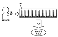
- FIG. 4 is a schematic diagram for explaining a method in which the electrocardiogram analyzer 3 analyzes electrocardiogram data.
- the electrocardiograph 1 measures the electrocardiogram of the subject to be analyzed.
- the electrocardiograph 1 generates electrocardiogram data H1 indicating the measured electrocardiogram and transmits the electrocardiogram data H1 to the electrocardiogram analyzer 3 via the network N.
- the input processing unit 341 acquires the electrocardiogram data H1 transmitted by the electrocardiograph 1. It is desirable that the input processing unit 341 sequentially acquires the electrocardiogram data H1 measured by the electrocardiograph 1 (that is, a Holter electrocardiograph or a patch type electrocardiograph) worn by the person to be analyzed in daily life. As a result, the electrocardiogram analysis system S can promptly notify the medical staff of information on whether or not the subject to be analyzed has a seizure-induced arrhythmia.
- the input processing unit 341 is acquired from the electrocardiogram data indicating the electrocardiogram of the subject to be analyzed, which is measured in advance in a hospital or the like, the electrocardiogram data acquired from the electrocardiograph mounted on the steering wheel of the automobile, and the 12-lead electrocardiograph.
- the electrocardiogram data, the electrocardiogram data acquired from the electrocardiograph mounted on the smart watch, and the like may be acquired.
- the input processing unit 341 inputs the acquired electrocardiogram data H1 into the machine learning model of the machine learning unit 33 as the electrocardiogram data to be analyzed.
- the input processing unit 341 inputs the electrocardiogram data of the analysis target measured at the same sampling rate (for example, 1000 Hz) as the electrocardiogram data for the teacher into the machine learning model.
- the electrocardiogram analyzer 3 can suppress the influence of the analysis result on the difference in the sampling rate between the electrocardiogram data.
- the input processing unit 341 performs a process of converting the sampling rate to the electrocardiogram data to be analyzed, and then performs a machine learning model. You may enter in.
- the machine learning model of the machine learning unit 33 When the electrocardiogram data is input, the machine learning model of the machine learning unit 33 outputs abnormal information regarding whether or not the input electrocardiogram data is the electrocardiogram data of a human who has an arrhythmia that occurs spontaneously. For example, the machine learning model outputs abnormal information as abnormality information regarding whether or not the human being (that is, the person to be analyzed) who is the measurement source of the input electrocardiogram data develops a seizure-induced arrhythmia.
- the machine learning model provides anomalous information such as the value of the probability that the subject will develop a seizure-induced arrhythmia, the degree of probability that the subject will develop a seizure-induced arrhythmia, and the subject's paroxysmal arrhythmia.
- At least one of the score of the probability of developing the resulting arrhythmia, the presence or absence of the seizure-occurring arrhythmia in the analyzed subject, and the presence or absence of the presence or absence of the seizure-occurring arrhythmia in the analyzed subject is output.
- the result acquisition unit 342 acquires, as an analysis result, abnormal information output by the machine learning model of the machine learning unit 33 regarding whether or not the subject has a seizure-induced arrhythmia.
- the output control unit 343 outputs the abnormality information acquired by the result acquisition unit 342 to the doctor terminal 2.
- Abnormal information is represented by a value, degree or score of the probability that the subject will develop a paroxysmal arrhythmia.
- the abnormal information may be information representing a value, degree, or score of the probability that the analyzed subject does not develop a seizure-induced arrhythmia.
- the degree of probability is represented, for example, by letters or symbols such as high, low, ⁇ , ⁇ , etc. associated with each range of probability values.
- the score of the probability is represented by, for example, the score obtained by converting the probability by a predetermined formula.
- the abnormal information may be information indicating the presence or absence of seizure-induced arrhythmia or the presence or absence of signs of arrhythmia in the subject to be analyzed.
- the presence or absence of arrhythmia is represented by, for example, characters or symbols indicating the determination result of the presence or absence of arrhythmia or the presence or absence of signs of arrhythmia by a machine learning model.
- the abnormality information may be output for the entire electrocardiogram data, or may be output in association with each of a plurality of periods constituting the electrocardiogram data.
- the abnormal information may be information regarding the occurrence of other seizure-induced arrhythmias.
- the doctor terminal 2 may display the abnormality information on the display, or may output a voice indicating the abnormality information from the speaker.
- the doctor terminal 2 may directly display the abnormality information, and the abnormality information is converted into other information ("re-examination is required", “medication is required”, etc. when the probability is equal to or higher than a predetermined value). It may be converted and then output.
- FIG. 5 is a schematic diagram of the analysis result screen displayed by the doctor terminal 2.
- the analysis result screen includes the electrocardiogram H and the abnormality information P.
- the electrocardiogram H represents at least a part of the electrocardiogram data used in the analysis.
- the anomaly information P represents, for example, the value or degree of probability that the subject has a seizure-induced arrhythmia output from a machine learning model.
- the electrocardiogram analysis system S detects signs of seizure-induced arrhythmia during the non-seizure period, which is difficult for the doctor to detect by analyzing the electrocardiogram using machine learning, and the subject to be analyzed. Can be displayed as information as to whether or not a seizure-induced arrhythmia is present.
- the doctor can easily identify whether or not the analyzed subject has a seizure-induced arrhythmia, and additionally if necessary. It is possible to judge whether or not to perform an inspection or the like.
- the electrocardiogram analyzer 3 may display the change in probability in a plurality of different periods on the doctor terminal 2.
- the input processing unit 341 displays the electrocardiogram data of the subject to be analyzed measured by the electrocardiograph 1 during a plurality of different periods (for example, specific days of each of the plurality of months). Is input to the machine learning model of the machine learning unit 33 as the electrocardiogram data to be analyzed.
- the result acquisition unit 342 acquires the abnormal information output by the machine learning model of the machine learning unit 33 regarding whether or not the subject has a seizure-induced arrhythmia in association with each of a plurality of different periods.
- the output control unit 343 causes the doctor terminal 2 to display the abnormal information regarding the change in the probability of the plurality of different periods acquired by the result acquisition unit 342.
- the anomaly information represents the change in probability by, for example, a graph of the transition over time or the degree of change in probability (difference, rate of change, etc.).
- the doctor can determine the risk of developing a paroxysmal arrhythmia in the subject to be analyzed.
- the analysis result screen may include an area (eg, a button or selection box) for inputting a doctor's diagnosis of whether or not the subject has a seizure-induced arrhythmia.
- the doctor terminal 2 transmits the input contents to the electrocardiogram analyzer 3 as determination information as to whether or not the analyzed subject has a seizure-induced arrhythmia.
- the electrocardiogram analyzer 3 receives the determination information transmitted by the doctor terminal 2.
- the machine learning unit 33 uses the received determination information and the electrocardiogram data of the person to be analyzed to perform the above-mentioned machine learning, so that the input electrocardiogram data is generated in an arrhythmia of a human being. Regenerate a machine learning model that outputs abnormal information regarding whether or not it is electrocardiogram data.
- the electrocardiogram analysis system S can receive feedback of the diagnosis result by the doctor and improve the accuracy of the abnormality information output by the machine learning model.
- FIG. 6 is a diagram showing a flowchart of an electrocardiogram analysis method executed by the electrocardiogram analysis system S according to the present embodiment.
- the present embodiment includes an electrocardiogram analysis method illustrated in FIG. 6, a program for executing the electrocardiogram analysis method, and a computer-readable recording medium storing the program.
- the input processing unit 341 acquires the electrocardiogram data transmitted by the electrocardiograph 1 of the patient who has developed a seizure-induced arrhythmia (S11).
- the input processing unit 341 inputs a portion of the patient's electrocardiogram data during the non-seizure period to the machine learning unit 33 as human teacher's electrocardiogram data having a seizure-occurring arrhythmia. Further, the input processing unit 341 uses the electrocardiogram data of a human being diagnosed by a doctor as not developing an arrhythmia caused by a seizure as the electrocardiogram data for a human teacher who does not have an arrhythmia caused by a seizure, as a machine learning unit 33. Enter in.
- the machine learning unit 33 performs machine learning using the input electrocardiogram data for teachers, and as a result, the input electrocardiogram data is an abnormality relating to whether or not the input electrocardiogram data is the electrocardiogram data of a human who has developed arrhythmia that occurs spontaneously.
- a machine learning model that outputs information is generated (S12). When the machine learning unit 33 uses an externally generated machine learning model, steps S11 to S12 may be omitted.
- the input processing unit 341 acquires the electrocardiogram data transmitted by the electrocardiograph 1 of the person to be analyzed (S13).
- the input processing unit 341 inputs the acquired electrocardiogram data to the machine learning model of the machine learning unit 33 as the electrocardiogram data to be analyzed (S14).
- the machine learning model of the machine learning unit 33 outputs abnormal information regarding whether or not the input electrocardiogram data is the electrocardiogram data of a human having an arrhythmia that occurs spontaneously.
- the result acquisition unit 342 acquires, as an analysis result, abnormal information regarding whether or not the subject has a seizure-induced arrhythmia output by the machine learning model of the machine learning unit 33 (S15).
- the output control unit 343 causes the doctor terminal 2 to display abnormal information regarding whether or not the result acquisition unit 342 has acquired the result (S16).
- the electrocardiogram analysis system S allows the subject to be analyzed by inputting the electrocardiogram data of the subject to be analyzed into a machine learning model in which the electrocardiogram data of a patient having a seizure-induced arrhythmia during the non-attack period is machine-learned. Outputs abnormal information regarding whether or not a person has a seizure-induced arrhythmia. This makes it easier for the electrocardiogram analysis system S to identify whether or not the subject has a seizure-induced arrhythmia.
- the electrocardiogram analyzer 3 is used to assist the diagnosis by a doctor, but may be used for other purposes.
- the electrocardiogram analyzer 3 is a health examination result display system that displays the possibility of occurrence of arrhythmia that occurs spontaneously in the subject based on abnormal information and the necessity of re-examination or detailed examination. It may be used in an insurance examination support system that includes abnormal information in the criteria for insurance participation examination of the analyzed person, and an insurance contract document device that includes abnormal information in the display items of the insurance contract document of the analyzed person.
- the electrocardiogram analyzer 3 can support the insurance participation examination and the like based on the health condition of the analyzed person.
- the electrocardiogram analyzer 3 may be used in a clinical trial investigator selection system that includes abnormal information in a display item of information regarding a clinical trial candidate. Thereby, the electrocardiogram analyzer 3 can support a judgment such as excluding a candidate who may have a seizure-induced arrhythmia from the investigators of the clinical trial regarding the evaluation of the electrocardiograph.
- the electrocardiogram analyzer 3 may be used as a drug administration determination support system or a drug administration contraindication determination support device that utilizes abnormal information. As a result, the electrocardiogram analyzer 3 assists in determining whether or not to administer a drug or the like that cannot be administered when there is an arrhythmia to a patient who may have a paroxysmal arrhythmia, or causes a seizure disease. It is possible to use anomalous information as a digital biomarker to stratify a patient.
- the electrocardiogram analyzer 3 may be used as a vehicle driving control device for safely stopping the vehicle when the occurrence of an arrhythmia that occurs sporadically while driving the vehicle is detected based on the abnormality information.
- the electrocardiogram analyzer 3 may be used in an alert system that recommends that a medical institution be consulted by displaying the occurrence of an arrhythmia that occurs seizures based on abnormal information on a smart device such as a smartphone.
- the processor of the electrocardiogram analyzer 3 is the main body of each step included in the electrocardiogram analysis method shown in FIG. That is, the processor of the electrocardiogram analyzer 3 reads a program for executing the electrocardiogram analysis method shown in FIG. 6 from the storage unit, executes the program, and controls each part of the electrocardiogram analysis system S, so that FIG. 6 shows. Perform the ECG analysis method shown. Some of the steps included in the electrocardiogram analysis method shown in FIG. 6 may be omitted, the order between the steps may be changed, and a plurality of steps may be performed in parallel.
Landscapes
- Health & Medical Sciences (AREA)
- Life Sciences & Earth Sciences (AREA)
- Engineering & Computer Science (AREA)
- Cardiology (AREA)
- Physics & Mathematics (AREA)
- Animal Behavior & Ethology (AREA)
- Biomedical Technology (AREA)
- Heart & Thoracic Surgery (AREA)
- Medical Informatics (AREA)
- Molecular Biology (AREA)
- Surgery (AREA)
- Biophysics (AREA)
- General Health & Medical Sciences (AREA)
- Public Health (AREA)
- Veterinary Medicine (AREA)
- Pathology (AREA)
- Artificial Intelligence (AREA)
- Physiology (AREA)
- Computer Networks & Wireless Communication (AREA)
- Evolutionary Computation (AREA)
- Fuzzy Systems (AREA)
- Mathematical Physics (AREA)
- Computer Vision & Pattern Recognition (AREA)
- Psychiatry (AREA)
- Signal Processing (AREA)
- Measurement And Recording Of Electrical Phenomena And Electrical Characteristics Of The Living Body (AREA)
Priority Applications (5)
| Application Number | Priority Date | Filing Date | Title |
|---|---|---|---|
| JP2021544452A JP7002168B1 (ja) | 2020-04-08 | 2021-03-04 | 心電図分析装置、心電図分析方法及びプログラム |
| EP21784075.0A EP4079224B1 (en) | 2020-04-08 | 2021-03-04 | Electrocardiographic analysis device, electrocardiographic analysis method, and program |
| JP2021205708A JP7701048B2 (ja) | 2020-04-08 | 2021-12-20 | 心電図分析装置、心電図分析方法及びプログラム |
| US17/811,860 US12582315B2 (en) | 2020-04-08 | 2022-07-11 | Electrocardiogram analysis apparatus, electrocardiogram analyzing method, and non-transitory computer-readable storage medium |
| JP2025098324A JP2025128317A (ja) | 2020-04-08 | 2025-06-12 | 心電図分析装置、心電図分析方法及びプログラム |
Applications Claiming Priority (2)
| Application Number | Priority Date | Filing Date | Title |
|---|---|---|---|
| JP2020069705 | 2020-04-08 | ||
| JP2020-069705 | 2020-04-08 |
Related Child Applications (1)
| Application Number | Title | Priority Date | Filing Date |
|---|---|---|---|
| US17/811,860 Continuation US12582315B2 (en) | 2020-04-08 | 2022-07-11 | Electrocardiogram analysis apparatus, electrocardiogram analyzing method, and non-transitory computer-readable storage medium |
Publications (1)
| Publication Number | Publication Date |
|---|---|
| WO2021205788A1 true WO2021205788A1 (ja) | 2021-10-14 |
Family
ID=78022561
Family Applications (1)
| Application Number | Title | Priority Date | Filing Date |
|---|---|---|---|
| PCT/JP2021/008444 Ceased WO2021205788A1 (ja) | 2020-04-08 | 2021-03-04 | 心電図分析装置、心電図分析方法及びプログラム |
Country Status (4)
| Country | Link |
|---|---|
| US (1) | US12582315B2 (https=) |
| EP (1) | EP4079224B1 (https=) |
| JP (3) | JP7002168B1 (https=) |
| WO (1) | WO2021205788A1 (https=) |
Cited By (6)
| Publication number | Priority date | Publication date | Assignee | Title |
|---|---|---|---|---|
| US12213791B2 (en) | 2020-08-06 | 2025-02-04 | Irhythm Technologies, Inc. | Wearable device |
| US12245859B2 (en) | 2013-01-24 | 2025-03-11 | Irhythm Technologies, Inc. | Physiological monitoring device |
| US12274554B2 (en) | 2010-05-12 | 2025-04-15 | Irhythm Technologies, Inc. | Device features and design elements for long-term adhesion |
| USD1083114S1 (en) | 2021-08-06 | 2025-07-08 | Irhythm Technologies, Inc. | Physiological monitoring device |
| US12507931B2 (en) | 2020-08-06 | 2025-12-30 | Irhythm Technologies, Inc. | Wearable device with conductive traces and insulator |
| US12603173B2 (en) | 2014-10-31 | 2026-04-14 | Irhythm Technologies, Inc. | Wearable monitor |
Families Citing this family (7)
| Publication number | Priority date | Publication date | Assignee | Title |
|---|---|---|---|---|
| KR102788715B1 (ko) * | 2022-05-04 | 2025-04-01 | 주식회사 뷰노 | 심전도 데이터를 분석하기 위한 방법 및 장치 |
| EP4550352A4 (en) * | 2022-07-29 | 2025-09-10 | Medical Ai Co Ltd | METHOD, PROGRAM AND DEVICE FOR PROVIDING ECG INTERPRETATION SERVICE |
| JP7672033B2 (ja) * | 2023-02-10 | 2025-05-07 | 公立大学法人横浜市立大学 | 心不全推定装置、心不全推定システム、心不全推定方法、及び心不全推定プログラム |
| WO2024191200A1 (ko) * | 2023-03-13 | 2024-09-19 | 주식회사 메디컬에이아이 | 인공 지능 모델에 기반하여 잠재적 심방 세동을 식별하는 방법, 프로그램 및 장치 |
| KR102877334B1 (ko) * | 2023-10-17 | 2025-10-29 | (주)씨어스테크놀로지 | 온라인 머신러닝 알고리즘을 이용한 심전도 판독장치 |
| JP7493293B1 (ja) | 2024-03-13 | 2024-05-31 | 株式会社カルディオインテリジェンス | 心電図解析のための支援装置、支援システム、およびプログラム |
| KR102832591B1 (ko) * | 2024-08-30 | 2025-07-10 | 주식회사 휴이노 | 심전도 신호에서 이상 파형의 발생을 예측하기 위한 방법 및 시스템 |
Citations (2)
| Publication number | Priority date | Publication date | Assignee | Title |
|---|---|---|---|---|
| JP2007195693A (ja) | 2006-01-25 | 2007-08-09 | Matsushita Electric Works Ltd | 携帯型心電計測装置 |
| JP2018519122A (ja) * | 2015-05-13 | 2018-07-19 | アライヴコア インコーポレイテッド | 不一致モニタリング |
Family Cites Families (12)
| Publication number | Priority date | Publication date | Assignee | Title |
|---|---|---|---|---|
| US8909330B2 (en) | 2009-05-20 | 2014-12-09 | Sotera Wireless, Inc. | Body-worn device and associated system for alarms/alerts based on vital signs and motion |
| US9314178B2 (en) | 2013-03-14 | 2016-04-19 | Greatbach, Ltd. | Cardiac signal recording using dynamically generated detection thresholds |
| US10779744B2 (en) * | 2015-10-27 | 2020-09-22 | Cardiologs Technologies Sas | Automatic method to delineate or categorize an electrocardiogram |
| CN107516075B (zh) * | 2017-08-03 | 2020-10-09 | 安徽华米智能科技有限公司 | 心电信号的检测方法、装置及电子设备 |
| WO2019046294A1 (en) | 2017-08-28 | 2019-03-07 | University Of South Florida | SYSTEM AND METHOD FOR PREDICTING ATRIAL FIBRILLATION (AF) |
| US20240099593A1 (en) * | 2017-10-06 | 2024-03-28 | Alivecor, Inc. | Machine learning health analysis with a mobile device |
| JP2019115618A (ja) * | 2017-12-27 | 2019-07-18 | オムロンヘルスケア株式会社 | 情報処理装置、情報処理方法、及び情報処理プログラム |
| CN108577823B (zh) * | 2018-04-27 | 2021-02-02 | 京东方科技集团股份有限公司 | 一种心律失常检测装置和心律失常检测系统 |
| JP6483890B1 (ja) * | 2018-04-27 | 2019-03-13 | 国立大学法人滋賀医科大学 | 診断支援装置、機械学習装置、診断支援方法、機械学習方法および機械学習プログラム |
| US11723577B2 (en) * | 2019-05-06 | 2023-08-15 | Medtronic, Inc. | Visualization of arrhythmia detection by machine learning |
| CN110403600B (zh) * | 2019-07-26 | 2022-02-08 | 武汉海星通技术股份有限公司 | 基于差值时间散点图的阵发性房颤智能分析方法及系统 |
| US12224052B2 (en) * | 2019-10-03 | 2025-02-11 | Rom Technologies, Inc. | System and method for using AI, machine learning and telemedicine for long-term care via an electromechanical machine |
-
2021
- 2021-03-04 WO PCT/JP2021/008444 patent/WO2021205788A1/ja not_active Ceased
- 2021-03-04 JP JP2021544452A patent/JP7002168B1/ja active Active
- 2021-03-04 EP EP21784075.0A patent/EP4079224B1/en active Active
- 2021-12-20 JP JP2021205708A patent/JP7701048B2/ja active Active
-
2022
- 2022-07-11 US US17/811,860 patent/US12582315B2/en active Active
-
2025
- 2025-06-12 JP JP2025098324A patent/JP2025128317A/ja active Pending
Patent Citations (2)
| Publication number | Priority date | Publication date | Assignee | Title |
|---|---|---|---|---|
| JP2007195693A (ja) | 2006-01-25 | 2007-08-09 | Matsushita Electric Works Ltd | 携帯型心電計測装置 |
| JP2018519122A (ja) * | 2015-05-13 | 2018-07-19 | アライヴコア インコーポレイテッド | 不一致モニタリング |
Cited By (14)
| Publication number | Priority date | Publication date | Assignee | Title |
|---|---|---|---|---|
| US12324668B2 (en) | 2010-05-12 | 2025-06-10 | Irhythm Technologies, Inc. | Device features and design elements for long-term adhesion |
| US12408856B1 (en) | 2010-05-12 | 2025-09-09 | Irhythm Technologies, Inc. | Device features and design elements for long-term adhesion |
| US12303277B2 (en) | 2010-05-12 | 2025-05-20 | Irhythm Technologies, Inc. | Device features and design elements for long-term adhesion |
| US12274554B2 (en) | 2010-05-12 | 2025-04-15 | Irhythm Technologies, Inc. | Device features and design elements for long-term adhesion |
| US12245860B2 (en) | 2013-01-24 | 2025-03-11 | Irhythm Technologies, Inc. | Physiological monitoring device |
| US12303275B2 (en) | 2013-01-24 | 2025-05-20 | Irhythm Technologies, Inc. | Physiological monitoring device |
| US12357212B2 (en) | 2013-01-24 | 2025-07-15 | Irhythm Technologies, Inc. | Physiological monitoring device |
| US12245859B2 (en) | 2013-01-24 | 2025-03-11 | Irhythm Technologies, Inc. | Physiological monitoring device |
| US12402819B1 (en) | 2013-01-24 | 2025-09-02 | Irhythm Technologies, Inc. | Physiological monitoring device |
| US12603173B2 (en) | 2014-10-31 | 2026-04-14 | Irhythm Technologies, Inc. | Wearable monitor |
| US12582339B2 (en) | 2020-08-06 | 2026-03-24 | Irhythm Technologies, Inc. | Electrical components for physiological monitoring device |
| US12213791B2 (en) | 2020-08-06 | 2025-02-04 | Irhythm Technologies, Inc. | Wearable device |
| US12507931B2 (en) | 2020-08-06 | 2025-12-30 | Irhythm Technologies, Inc. | Wearable device with conductive traces and insulator |
| USD1083114S1 (en) | 2021-08-06 | 2025-07-08 | Irhythm Technologies, Inc. | Physiological monitoring device |
Also Published As
| Publication number | Publication date |
|---|---|
| EP4079224B1 (en) | 2025-07-23 |
| JP2025128317A (ja) | 2025-09-02 |
| US20220346647A1 (en) | 2022-11-03 |
| JP7002168B1 (ja) | 2022-01-20 |
| EP4079224C0 (en) | 2025-07-23 |
| JP2022037153A (ja) | 2022-03-08 |
| EP4079224A4 (en) | 2023-06-21 |
| JP7701048B2 (ja) | 2025-07-01 |
| JPWO2021205788A1 (https=) | 2021-10-14 |
| US12582315B2 (en) | 2026-03-24 |
| EP4079224A1 (en) | 2022-10-26 |
Similar Documents
| Publication | Publication Date | Title |
|---|---|---|
| JP7002168B1 (ja) | 心電図分析装置、心電図分析方法及びプログラム | |
| AU2022201530B2 (en) | Apparatus, systems and methods for predicting, screening and monitoring of encephalopathy/delirium | |
| US20230099854A1 (en) | Methods and systems for arrhythmia tracking and scoring | |
| JP5584413B2 (ja) | 患者監視システム及び監視する方法 | |
| US20170156592A1 (en) | Healthcare systems and monitoring method for physiological signals | |
| JP6692355B2 (ja) | バイタルサインサンプリング周波数が限定されるときのスコア信頼区間推定に関する方法 | |
| US11412978B2 (en) | Information processing system and program | |
| CN103948383A (zh) | 用于快速检测心肌缺血的ecg数据显示方法 | |
| Faragli et al. | Cardiovascular screening in low-income settings using a novel 4-lead smartphone-based electrocardiograph (D-Heart®) | |
| Vardas et al. | The electrocardiogram endeavour: from the Holter single-lead recordings to multilead wearable devices supported by computational machine learning algorithms | |
| Shusterman et al. | Personalized ECG monitoring and adaptive machine learning | |
| Xiao | The role of telemetry monitoring: From diagnosing arrhythmia to predictive models of patient instability | |
| US20160106330A1 (en) | Cardiac monitoring device and method | |
| Van Heuverswyn et al. | Clinical validation of a 13-lead electrocardiogram derived from a self-applicable 3-lead recording for diagnosis of myocardial supply ischaemia and common non-ischaemic electrocardiogram abnormalities at rest | |
| Tsampi et al. | Extending the Sana mobile healthcare platform with features providing ECG analysis | |
| Evenson et al. | Skilled cardiac monitoring at the bedside: An algorithm for success | |
| CN114599286A (zh) | 学习装置、学习方法以及测定装置 | |
| JP7479106B1 (ja) | プログラム、出力装置及び出力方法 | |
| WO2025227126A1 (en) | Detecting cardiac arrhythmia and myocardial ischemia from electrocardiography data using machine learning | |
| WO2024106005A1 (ja) | プログラム、出力装置及び出力方法 | |
| CN120611325A (zh) | 基于多维轨迹图的时序性信号异常确定方法、装置、设备及介质 | |
| Hughes-Noehrer et al. | Increasing the accuracy of ST-segment elevation detection in an ECG through automated visual rules | |
| Goldwyn et al. | HealthVisor: A look into data-rich bio-monitoring | |
| BR112018011326B1 (pt) | Sistema para rastreamento de delírio de paciente |
Legal Events
| Date | Code | Title | Description |
|---|---|---|---|
| ENP | Entry into the national phase |
Ref document number: 2021544452 Country of ref document: JP Kind code of ref document: A |
|
| 121 | Ep: the epo has been informed by wipo that ep was designated in this application |
Ref document number: 21784075 Country of ref document: EP Kind code of ref document: A1 |
|
| ENP | Entry into the national phase |
Ref document number: 2021784075 Country of ref document: EP Effective date: 20220718 |
|
| NENP | Non-entry into the national phase |
Ref country code: DE |
|
| WWG | Wipo information: grant in national office |
Ref document number: 2021784075 Country of ref document: EP |