WO2021182645A1 - 頻脈性不整脈の副伝導路の局在部位予測方法及び局在部位予測装置、並びに、特発性心筋症の診断支援方法及び診断支援装置 - Google Patents
頻脈性不整脈の副伝導路の局在部位予測方法及び局在部位予測装置、並びに、特発性心筋症の診断支援方法及び診断支援装置 Download PDFInfo
- Publication number
- WO2021182645A1 WO2021182645A1 PCT/JP2021/010258 JP2021010258W WO2021182645A1 WO 2021182645 A1 WO2021182645 A1 WO 2021182645A1 JP 2021010258 W JP2021010258 W JP 2021010258W WO 2021182645 A1 WO2021182645 A1 WO 2021182645A1
- Authority
- WO
- WIPO (PCT)
- Prior art keywords
- conduction path
- model
- electrocardiogram
- data
- classification information
- Prior art date
- Legal status (The legal status is an assumption and is not a legal conclusion. Google has not performed a legal analysis and makes no representation as to the accuracy of the status listed.)
- Ceased
Links
Images










Classifications
-
- A—HUMAN NECESSITIES
- A61—MEDICAL OR VETERINARY SCIENCE; HYGIENE
- A61B—DIAGNOSIS; SURGERY; IDENTIFICATION
- A61B5/00—Measuring for diagnostic purposes; Identification of persons
- A61B5/24—Detecting, measuring or recording bioelectric or biomagnetic signals of the body or parts thereof
- A61B5/316—Modalities, i.e. specific diagnostic methods
- A61B5/318—Heart-related electrical modalities, e.g. electrocardiography [ECG]
- A61B5/346—Analysis of electrocardiograms
Definitions
- the present invention relates to, for example, a method for predicting the localization site of an accessory pathway in tachyarrhythmia such as Wolff-Parkinson-White syndrome, a method for supporting diagnosis of idiopathic cardiomyopathy, and a device thereof. ..
- tachyarrhythmias include paroxysmal supraventricular tachycardia such as WPW syndrome and atrioventricular node recurrent tachycardia, atrial flutter, atrial tachycardia, and atrial fibrillation.
- WPW syndrome is a disease of tachyarrhythmia that occurs in the atrium of the heart and the pulse is accelerated.
- excitement is propagated from the atrium to the ventricle through a single stimulus pathway, but in WPW syndrome, a secondary pathway, separate from the normal pathway between the atrium and the ventricle, is born. There is an extra conduction path that exists.
- Tachyarrhythmia occurs due to the rapid reciprocation of electrical stimulation between the normal conduction path and the accessory conduction path. On a daily basis, there is no particular inconvenience and it may not be a problem throughout life, but in some cases tachycardia (faster pulse, higher pulse rate) attacks may occur. In addition to WPW syndrome, when an arrhythmia called atrial fibrillation is also present, atrial excitement descends the accessory conduction path, causing severe tachycardia and transitioning to fatal arrhythmia (ventricular fibrillation, etc.). Or heart failure.
- ablation catheter As a treatment for arrhythmia such as WPW syndrome, instead of the conventional open heart surgery, ablation catheter is inserted into the heart and energy is irradiated to ablate the above-mentioned accessory pathway.
- Catheter ablation myocardial ablation
- Catheter ablation is a well-established treatment for WPW syndrome and the like.
- Of particular importance when treating catheter ablation is the identification of accessory pathways. Identifying the location of the accessory pathway prior to cauterization, that is, predicting the localization of the accessory pathway before surgery, is important in developing a therapeutic strategy.
- an abnormal stimulus conduction time has been obtained by inserting a catheter electrode around the atrioventricular ring where the accessory conduction path exists, electrically stimulating from the inside of the atrium and the ventricle, and measuring the conduction time of the stimulus.
- a method of specifying the site as a secondary conduction path portion There is known a method of specifying the site as a secondary conduction path portion.
- the mitral valve annulus site on the left heart side of the heart and the tricuspid annulus site on the right heart side are examined separately in order, and the stimulation conduction time of multiple sites is measured. There was a problem that it was necessary and the inspection required a long time.
- a first electrode lead is inserted into the coronary sinus of the heart to measure the activity potential of the mitral valve annulus on the left heart side, and a second electrode is inserted into the right coronary artery of the heart.
- a secondary conduction path detection device that inserts a lead, measures the activity potential of the tricuspid annulus site on the right heart side, and detects the early excitable site as the secondary conduction path site from multiple active potentials obtained from each electrode lead.
- the sub-conducting path detection device disclosed in Patent Document 1 makes it possible to identify the sub-conducting path site in a short time, but since each electrode lead is inserted into the patient's heart in the examination, it is a burden on the patient. there were.
- potential information can be visualized three-dimensionally using a catheter and magnetism, and the intracardiac potential recording by the catheter electrode and the position of the catheter electrode obtained by using magnetism are simultaneously computer-processed. Therefore, the stereoscopic image of the heart can be displayed on the display, and the excitement propagation process and potential wave height during tachycardia can be displayed in real time.
- excitatory propagation is expressed as the movement of the waveform of an electric signal, the path of the electric signal is complicated under a situation where an arrhythmia is occurring, and it is difficult to display the moving waveform information in an easy-to-understand manner.
- doctors have focused on the delta wave of the electrocardiogram and discriminated the left side secondary conduction path (A type), the right side secondary conduction path (B type), and the septal secondary conduction path (C type) for the location where the secondary conduction path exists. ing.
- a type left side secondary conduction path
- B type right side secondary conduction path
- C type septal secondary conduction path
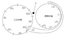
- FIG. 17 citing FIG. 2 of Non-Patent Document 1
- the 13 subdivided sites are RA (right anterior wall), RAL (right anterior wall), RAS (right anterior septum), RL (right wall), RPL (right posterior wall), RP (right posterior wall), RPS ( Right posterior septum), MS (middle septum), LAL (left anterior septum), LL (left wall), LPL (left posterior wall), LP (left posterior wall), LPS (left posterior septum).
- H is the bundle of His (normal stimulus conduction path) and CS is the coronary sinus.
- a Chiang algorithm which is a combination of a delta wave and a QRS wave and has high prediction accuracy, is known as an estimation algorithm for an auxiliary conduction path (see Non-Patent Document 2).
- a decision tree is created under the condition that the delta wave of the early excitatory wave can be sufficiently identified, and the positive wave (+), negative wave (-), biphasic wave or equipotential wave ( ⁇ ), and based on the polarity (+/-) of the delta wave ( ⁇ ), as shown in FIG. 18 (citing FIG. 3 of Non-Patent Document 1 and quoting from Non-Patent Document 2), Steps 1 to 1 to It is a diagnostic logic that advances the steps in the order of 4.
- the prediction accuracy of the secondary conduction path of the Chiang algorithm is 80% or more correct, although it varies depending on the part.
- the above-mentioned subdivided 13 sites are classified into 9 sites by integrating some sites.
- the delta wave which is often indistinguishable, is excluded from the algorithm, and only the QRS wave is used.
- the diagnostic logic proceeds in steps or 4 steps (see Non-Patent Document 3).
- the prediction accuracy of the secondary conduction path of the St George algorithm is about 85% in the classification of 10 sites.
- the reality is that a complete prediction algorithm has not yet been established.
- the first problem is that many algorithms, like the Chiang algorithm, use a decision tree model, and there are cases where it is difficult to classify the boundary type.
- the second problem is that the anatomical axis of the heart is different for each individual, which causes an error in the classification result.
- cardiomyopathy that causes abnormalities in the myocardium (heart muscle)
- typical ones are hypertrophic cardiomyopathy and dilated cardiomyopathy.
- Hypertrophic cardiomyopathy is a disease in which the myocardium is thickened and dilated function is reduced
- dilated cardiomyopathy is a disease in which the myocardium is thinned and contractile force is reduced.
- It is one of the idiopathic cardiomyopathy, which is a muscular disease. Since these cardiomyopathy are more likely to cause a fatal cardiac accident when accompanied by an arrhythmia, measures such as medication to prevent the arrhythmia are required.
- cardiomyopathy can be detected accurately by ultrasonic echo, but it is preferable that early detection of cardiomyopathy can be performed by an electrocardiogram, which is a simpler test.
- electrocardiogram devices in recent years are equipped with an automatic diagnosis function, but these are basically automatic diagnoses for adults and cannot be used for pediatric diagnosis, and are equivalent to pediatric diagnosis in the medical field. Since there are many cases where the diagnosis is made based on the experience of the doctor, there is a demand for a prediction method / device that can support the diagnosis of the doctor.
- Tadayuki Hiroki Estimating the accessory pathway site by 12-lead electrocardiogram: 3.
- WPW syndrome Estimating and treating the accessory pathway site (Special Feature II: 64th Annual Meeting of the Japanese Circulation Society, Cardiologist, 2001, Volume 9 1 No., p.57-66.
- Chiang CE et al. “An accurate stepwise electrocardiographic algorithm for localization of accessories pathways inpatients with Wolff-Parkinson-White syndrome from a comprehensive analysis ofdelta waves and R / S ratio during : 40-46.
- Xie B et al. “Localizat ioof naccessorypathways from the l2-lead electrocardiogram using a new algorithm”, Am J Cardio1,1994; 74: 161-165.
- Tachyarrhythmia such as WPW syndrome is a disease with a high morbidity rate, and it is desired to be able to accurately diagnose the localization of accessory pathways at the preoperative stage.
- WPW syndrome is a disease with a high morbidity rate, and it is desired to be able to accurately diagnose the localization of accessory pathways at the preoperative stage.
- the details of the surgical procedure before the operation are detailed. There was a problem that it was not possible to explain the information and the risk of complications.
- idiopathic cardiomyopathy such as hypertrophic cardiomyopathy
- the present invention provides a method and a device capable of predicting the localized site of an accessory conduction path of tachyarrhythmia such as WPW syndrome with high accuracy, and a method and a device capable of diagnosing and supporting idiopathic cardiomyopathy.
- the purpose is to provide.
- the method for predicting the localization site of the accessory conduction path of the present invention is the biopotential signal data set of the electrocardiogram and the accessory conduction path of tachyarrhythmia.
- an AI (artificial intelligence) model for example, a deep neural network model or a convolutional neural network
- a more robust algorithm can be used as a subordinate.
- the localized part of the conduction path can be predicted, and the localized part information of the secondary conduction path can be output easily and automatically.
- the biopotential signal data set of the electrocardiogram is input to the AI model as teacher data, and the classification result output by the AI model is used as the localization site of the secondary conduction path of the teacher data.
- the weighting coefficient of the AI model is optimized by comparing it with the classification information.
- the signal waveform data of the 12-lead electrocardiogram is preferable, but other electrocardiogram signal waveform data can also be used as long as the technical idea of the present invention is followed.
- the accessory conduction path is a conduction path that is separate from the normal conduction path that transmits electrical excitement in the heart, and the accessory conduction path includes a Kent bundle, a James bundle, and a Maheim fiber.
- the secondary conduction path refers to the Kent bundle.
- the biopotential signal data set it is expected that there are few things that can be teacher data, but the biopotential signal data may be compressed and stretched in the time axis direction to inflate the number of teacher data. However, padding by flipping left and right and up and down is not possible.
- the method for predicting the localization site of the accessory conduction path of the present invention includes the biopotential signal data set of the electrocardiogram, the chest roentgen image data, and the station of the accessory conduction path of tachyarrhythmia.
- the localization site of the accessory conduction path of tachyarrhythmia such as WPW syndrome can be predicted with higher accuracy.
- the accuracy can be further improved by adding the chest X-ray image data of the same case to the input of the above AI model.
- the mixed data of the normal chest X-ray image and the chest X-ray image of cardiac hypertrophy Is further provided in the AI model so that the presence or absence of cardiac hypertrophy can be determined in advance, so that the prediction can be made with higher accuracy.
- Chest X-ray image data can be widely obtained as compared with the electrocardiogram biopotential signal data set.
- normal chest X-ray image and chest X-ray image data of cardiac hypertrophy are used in advance for AI model.
- the weighting coefficient of the AI model is adjusted by inputting and pre-training.
- the AI model used in the method for predicting the localization site of the accessory pathway according to the second aspect of the present invention is a one-dimensional convolutional neural network that inputs an electrocardiogram biopotential signal data set as one-dimensional matrix data, and a chest roentgen image data. It is preferable to include at least a two-dimensional convolutional neural network to be input as two-dimensional matrix data and a deep neural network for calculating feature data output from each convolutional neural network. This is because the biopotential signal data set of the electrocardiogram is input-calculated as one-dimensional matrix data, and the chest roentgen image data is input-calculated as two-dimensional matrix data, so that different convolutional neural networks are used.
- a two-dimensional convolutional neural network is used in which data in which a normal chest X-ray image and a chest X-ray image of cardiac hypertrophy are mixed are input to an AI model and pre-trained so that the presence or absence of cardiac hypertrophy can be determined.
- Chest X-ray images unlike electrocardiographic biopotential signal datasets, are pre-trained to improve the predictive accuracy of 2D convolutional neural networks, as many images are already accessible as open sources.
- the localization site classification information of the secondary conduction path is the septum in the position classification based on the clinical electrophysiological examination. It is the localization site information of three categories: the site on the side, the site on the free wall side of the mitral valve annulus, and the site on the free wall side of the tricuspid valve annulus.
- the program of the present invention causes a computer to perform a step of calculating the localization site classification information of the secondary conduction path using the learned AI model in the above-described method for predicting the localization site of the secondary conduction path. ..
- the localization site prediction device for the accessory conduction path of the present invention inputs and learns the biopotential signal data set of the electrocardiogram and the localization site classification information of the accessory conduction path of tachyarrhythmia as teacher data.
- the trained AI model, the input processing unit for inputting the biopotential signal data set of the subject's electrocardiogram, and the input biopotential signal data set are input to the trained AI model, and the station of the secondary conduction path is input.
- a calculation processing unit that calculates the location classification information it is possible to predict the localized site of the accessory conduction path of tachyarrhythmia such as WPW syndrome with high accuracy.
- the device for predicting the localization site of the accessory pathway includes a biopotential signal data set of an electrocardiogram, chest roentgen image data, localization site classification information of the accessory pathway of tachyarrhythmia, and information.
- a trained AI model that has been trained by inputting cardiac hypertrophy classification information as teacher data, an input processing unit that inputs a subject's electrocardiogram biopotential signal data set and chest roentgen image data, and an input biopotential signal data set.
- the accessory conduction path of tachyarrhythmia such as WPW syndrome can be provided. Localized sites can be predicted with higher accuracy.
- the trained AI model is used for the normal chest X-ray image and the chest X-ray image data of cardiac hypertrophy before the teacher data is input and trained.
- the AI model used for the localization site prediction device of the accessory conduction path is a one-dimensional convolutional neural network that inputs an electrocardiogram biopotential signal data set as one-dimensional matrix data, and a chest roentgen image data. It is preferable to include at least a two-dimensional convolutional neural network to be input as two-dimensional matrix data and a deep neural network for calculating feature data output from each convolutional neural network.
- the localization site classification information of the secondary conduction path is the site on the septal side in the position classification based on the clinical electrophysiological examination, the monk. It is the localized part information of three classifications of the part on the free wall side of the cap annulus and the part on the free wall side of the tricuspid annulus.
- the method for supporting the diagnosis of idiopathic cardiomyopathy of the present invention is a step (learning step) in which a biopotential signal data set of an electrocardiogram and classification information of hypertrophic cardiomyopathy and normal cases are input to an AI model as teacher data for learning.
- a step (calculation step) is provided in which the biopotential signal data set of the electrocardiogram of the subject is input to the trained AI model to calculate the classification information of hypertrophic cardiomyopathy or normal cases.
- classification information of dilated cardiomyopathy is added as teacher data, and classification information of hypertrophic cardiomyopathy, dilated cardiomyopathy or normal cases is calculated in the above calculation step. It is also possible to do.
- chest X-ray image data may be further added as teacher data. By adding and learning chest X-ray image data, the classification accuracy can be further improved.
- the device for diagnosing idiopathic cardiomyopathy of the present invention includes a trained AI model in which a biopotential signal data set of an electrocardiogram, classification information of hypertrophic cardiomyopathy and normal cases are input and trained as teacher data, and a subject.
- Input processing unit that inputs the biopotential signal data set of the electrocardiogram and the input biopotential signal data set are input to the trained AI model, and the calculation process for calculating the classification information of hypertrophic cardiomyopathy or normal cases. It has a part.
- classification information of dilated cardiomyopathy is added as teacher data
- classification information of hypertrophic cardiomyopathy, dilated cardiomyopathy or normal cases is added to the calculation processing unit. It is also possible to calculate.
- chest X-ray image data may be further added as teacher data.
- the localization site of the accessory conduction path can be predicted with high accuracy based on non-invasive electrocardiogram information.
- the doctor can explain the surgical procedure in detail and the risk of complications to the patient before the operation.
- the localized site of the accessory conduction path can be predicted with higher accuracy by using the commonly performed chest X-ray image data.
- diagnosis support for idiopathic cardiomyopathy such as hypertrophic cardiomyopathy can be performed based on electrocardiogram information.
- Block diagram of the localization site prediction device of the secondary conduction path of Example 1 Explanatory drawing of learning of AI model in local part prediction apparatus of secondary conduction path of Example 1
- Explanatory drawing of the difference in the waveform data of the 12-lead electrocardiogram due to the localized part of the accessory conduction path Explanatory drawing about classification of localized part of auxiliary conduction path
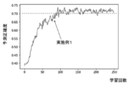
- Explanatory drawing about conversion of waveform data of 12-lead ECG A graph showing the prediction accuracy in the prediction device of the first embodiment.
- Block diagram of the localization site prediction device of the secondary conduction path of Example 2 Explanatory drawing of learning of AI model in local part prediction apparatus of secondary conduction path of Example 2
- Process flow diagram of the method for predicting the localized part of the accessory conduction path of Example 2 A graph showing the prediction accuracy in the prediction device of the second embodiment.
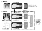
- Explanatory drawing of learning data about chest X-ray image Explanatory drawing of localization site prediction apparatus of secondary conduction path of Example 3
- Explanatory drawing of AI model in localization site prediction apparatus of secondary conduction path of Example 3 Graph showing prediction accuracy in the prediction device of Example 3
- Explanatory drawing of classification (13 parts) of localized part of auxiliary conduction path Explanatory diagram of decision tree in Chiang algorithm
- Explanatory diagram of decision tree in St George algorithm Block diagram of the diagnosis support device for idiopathic cardiomyopathy of Example 4
- Explanatory drawing of learning of AI model in diagnostic support apparatus of idiopathic cardiomyopathy of Example 4 Process flow diagram of the method for supporting the diagnosis of idiopathic cardiomyopathy of Example 4 Graph showing prediction accuracy in the diagnostic support device of Example 4
- the biopotential signal data set of the electrocardiogram is used as the waveform data set of the 12-lead electrocardiogram, and the waveform data set of the 12-lead electrocardiogram and the localization site classification information of the accessory pathway of WPW syndrome are used.
- the waveform data set of the subject's 12-lead electrocardiogram is input to the trained AI model, and the localization site classification information of the accessory pathway of WPW syndrome is obtained. calculate.
- the prediction method and the prediction device of this embodiment will be described with reference to FIGS. 1 to 7.
- the localization site prediction device 1 of the accessory pathway of Example 1 inputs a 12-lead electrocardiogram data set and a localization site classification label of the accessory pathway of WPW syndrome as teacher data.
- the trained AI model 13 is provided, and the input processing unit 11 inputs the 12-lead electrocardiogram data set 5, and the calculation processing unit 12 inputs the input data set 5 into the trained AI model 13. Then, the localization site classification label of the accessory conduction path of WPW syndrome and its probability 14 are calculated. Then, the prediction result regarding the classification of the localized portion of the accessory conduction path is output.
- the AI model used in Example 1 matrix data of a 12-lead electrocardiogram data set for 60 cases of WPW syndrome is used as learning data, and the probabilities of labels of three categories of Type A to C are used. Is output, compared with the classification label of the correct answer, and the weighting coefficient of the AI model is adjusted repeatedly to learn.
- the AI model used in Example 1 (the same applies to other Examples 2 and 3) is a one-dimensional convolutional neural network (CNN), and is used as a batch normalization method and an activation function in order to stabilize the learning of the neural network.
- ReLU Rectified Linear Unit
- L2 was used as the regularization method
- Adam was used as the optimization method.
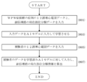
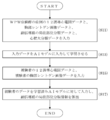
- the 12-lead electrocardiogram data of the case of WPW syndrome and the localization site classification data of the secondary conduction path are input (step). S01), the input data is input to the AI model and trained (step S03).
- the 12-lead electrocardiogram data may be the waveform image data of the electrocardiogram or may be matrix data in which the waveform image data is one-dimensionalized in time series.
- the subject's 12-lead ECG data is input (step S05).
- the data of the subject is input to the trained AI model, and the localization site classification information of the accessory conduction path is calculated (step S07).
- the 12-lead electrocardiogram is used as a standard when recording an electrocardiogram, and records the observation of the electrical activity of the heart from various directions, and captures the electrical activity of the heart as a three-dimensional object.
- Chest leads (V 1 to V 6 ) are recordings of electrical changes in the heart projected onto a horizontal plane.
- Bipolar limb leads (lead I) are potential differences between the left and right hands, and bipolar limb leads (lead II) are left legs.
- bipolar limb lead (III lead) is a record of the potential difference between the left foot and the left hand.
- V R Tankyokushi induction
- aV L Tankyokushi induction
- aV F Tankyokushi induction
- WPW syndrome shows a specific electrocardiographic biopotential signal due to premature excitement of the ventricles of the heart.
- accessory conduction pathways include the Kent bundle, the James bundle, the Maheim fiber, and the Atrial His bundle, but in WPW syndrome, the QRS complex is a fusion wave of early ventricular excitation via the Kent bundle and ventricular excitation via the normal conduction pathway. appear. As shown in FIG.
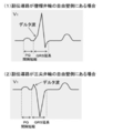
- delta wave in QRS wave seen in V 1 induction, PQ interval shortened, based on such QRS extension, can determine a WPW syndrome with anterograde Kent bundle conduction
- FIG. 4 (1) , (2) the localization position of the accessory pathway of WPW syndrome can be determined.
- Those having no forward Kent bundle conduction and only retrograde Kent bundle conduction are called latent WPW syndrome, but it is difficult to diagnose from an electrocardiogram other than at the time of an attack because a delta wave does not appear in the QRS complex. Therefore, electrophysiological tests are used to determine latent WPW syndrome.
- the localization site of the accessory pathway can be predicted with higher accuracy for WPW syndrome having antegrade Kent bundle conduction.
- the site on the septal side and the free wall side of the mitral valve annulus in the position classification based on the clinical electrophysiological examination There are 3 categories, the site and the site on the free wall side of the tricuspid annulus, and 3 categories are predicted.
- the accessory conduction pathways are classified into 13 sites, 8 on the right ventricular side and 5 on the left ventricular side (see FIG. 17).
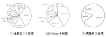
- the pie chart of FIG. 5 (1) shows all parts (13 classifications)
- the pie chart of FIG. 5 (2) shows 9 classifications of the Chiang algorithm
- the pie chart of FIG. 5 (3) shows 3 classifications of this embodiment.
- LAL and LL, LPL and LP, RAS and RA, and RPL and RP are integrated into 9 classifications out of all parts (13 classifications).
- three new classifications are created in addition to the nine classifications of the Chiang algorithm, and as shown in FIG. 5 (3), LAL, LL, LPL, and LP corresponding to the part on the free wall side of the mitral valve annulus.
- LAL, LL, LPL, and LP corresponding to the part on the free wall side of the mitral valve annulus.
- RP, RPL, RL, RAL, and RA corresponding to the free wall side of the tricuspid annulus are integrated into Type B
- LPS, RAS, and MS corresponding to the septal side.
- RPS was integrated into Type C.
- three classifications of Type A to C are predicted as localized sites of the accessory pathway in WPW syndrome.
- the data set for 69 cases of WPW syndrome in which the presence of delta waves was observed in the waveform data in the 12-lead electrocardiogram was used to confirm the prediction method and the prediction accuracy of the prediction device in this example.
- the data set for 69 cases is divided into each category and the number of data in each category is specified.
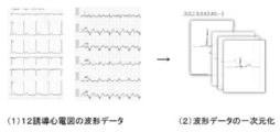
- the waveform data of the 12-lead electrocardiogram appears as an image in a different format depending on the measuring device, but as shown in FIG. 6 (2), it depends on the measuring device by making the image of the waveform data one-dimensional. No unified data structure. Specifically, an image of 12-channel waveform data of a 12-lead electrocardiogram is extracted with the same time width (about 0.6 seconds), divided by a predetermined number of time divisions (145 as described later), and the image is a time-series primary. It was used as the original data.
- the waveform data image is one-dimensional data such as ⁇ 0,0,1,3,2,4,5,60, ... ⁇ .
- the image of the waveform data of the 12-lead ECG for 69 people was made one-dimensional and converted into the matrix data of (69,145,12).
- 69 is the number of data sets
- 145 is the number of time divisions in each waveform data
- 12 is the waveform data of each lead in the 12-lead ECG.
- the waveform data set for 69 people is randomly divided into the training data and the test data
- the waveform data set for 60 people is prepared for the training data
- the waveform data set for 9 people is prepared for the test data.
- the prediction accuracy was measured.
- the test data is not used for learning the AI model, but is the target data predicted by the AI model as new data.
- the measurement result of the prediction accuracy is obtained by randomly dividing the training data and the test data 50 times in total and calculating the average prediction accuracy of the test data.
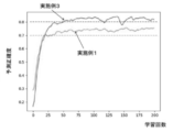
- the prediction accuracy of the localization site classification of the accessory pathway of WPW syndrome in the prediction device and the prediction method of Example 1 is shown in the graph of FIG. 7.
- the trained AI model showed a prediction accuracy of 70% or more after 100 or more iterative learnings.
- the learning for 60 learning data was regarded as one learning.
- the classification is predicted by both the conventional Chiang algorithm and the St George algorithm, and the prediction accuracy is predicted by the Chiang algorithm. Is 55.1% (38/69), and the prediction accuracy of the St George algorithm is 57.9% (40/69), both of which are lower than the prediction accuracy of 70% in this example. all right.
- the conventional Chiang algorithm and St George algorithm create an algorithm by collecting only typical examples of a single accessory pathway, so they are anomaly or dual. It is presumed that this is due to the fact that it cannot be applied to Accessory Pathway) and the prediction accuracy of actual case data is not as high as the prediction accuracy (%) described in the above-mentioned non-patent documents.
- the localization site of the accessory conduction path of WPW syndrome can be predicted with higher accuracy than the conventional algorithm.
- the biopotential signal data set of the electrocardiogram is used as the waveform data set of the 12-lead electrocardiogram, the chest X-ray image data, the waveform data set of the 12-lead electrocardiogram, and the station of the accessory pathway of WPW syndrome.
- the subject's 12-lead ECG waveform data set and chest roentgen image data are applied to the trained AI model. Input to calculate the localization site classification information of the accessory pathway of WPW syndrome.
- the prediction method and the prediction device of this embodiment will be described with reference to FIGS. 8 to 11.
- the localization site prediction device 2 of the accessory pathway of Example 2 includes the 12-lead electrocardiogram data set 5, the chest X-ray image data 6, and the localization of the accessory pathway of WPW syndrome.
- the AI model 23 has been trained by inputting the site classification label and the cardiac hypertrophy classification information as teacher data, and the input processing unit 21 inputs the 12-lead electrocardiogram data set 5 and the chest X-ray image data 6.
- the input data (5, 6) is input to the trained AI model 23, and the localization site classification label of the accessory conduction path of WPW syndrome and its probability 24 are calculated. Then, the prediction result regarding the classification of the localized portion of the accessory conduction path is output.
- the one-dimensional matrix data of the 12-lead electrocardiogram data set for 60 cases of WPW syndrome and the same chest roentgen image for 60 people are 2 as learning data.
- the probabilities of the labels of the three categories of Type A to C are output, compared with the correct classification labels, and the weighting coefficient of the AI model is adjusted repeatedly for learning.
- two-dimensional matrix data of a chest X-ray image is input to an input layer (first block) of a two-dimensional CNN (convolutional neural network).
- the one-dimensional matrix data of the 12-lead ECG is input to the input layer (first block) of the one-dimensional CNN.
- each of the two-dimensional matrix data of the chest X-ray image and the one-dimensional matrix data of the 12-lead electrocardiogram the convolution calculation and the reduction of the feature map space are repeated by the convolution layer and the pooling layer of the respective CNNs.
- Each feature output from the fully connected layer of each CNN is input to each DNN (deep neural network) of the second block, calculated and output, and further input to the DNN of the third block, calculated and classified label. (0, 1, 2) is output.
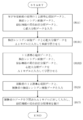
- the processing of the method for predicting the localization site of the accessory pathway in Example 2 is as follows: First, 12-lead ECG data, chest X-ray image data, and classification of the localization site of the accessory pathway in the case of WPW syndrome.
- the data and the cardiac hypertrophy classification data are input (step S11), and the input data is input to the AI model for training (step S13).
- the 12-lead electrocardiogram data may be the waveform image data of the electrocardiogram or may be matrix data in which the waveform image data is one-dimensionalized in time series.
- the subject's 12-lead ECG data and chest X-ray image data are input (step S15).
- the data of the subject is input to the trained AI model, and the localization site classification information of the accessory conduction path is calculated (step S17).
- the graph of FIG. 11 shows the prediction accuracy of the localization site classification of the accessory pathway of WPW syndrome in the prediction device and the prediction method of Example 2.
- Example 1 no significant difference was found between Example 1 and Example 2.
- Example 2 In order to correctly recognize chest X-ray images, a considerable number of data in units of 1000 is required, but in the predictive measurement of Example 2, only 60 chest X-ray images for learning out of 69 cases are learned. It is presumed that there was no difference in the prediction accuracy because the cardiac hypertrophy could not be correctly recognized from the image.
- 12-lead electrocardiogram data, chest X-ray image data, localization site classification information of the accessory conduction path, and cardiac hypertrophy classification information are input to the AI model as teacher data for learning.
- the mixed data of the normal chest X-ray image and the chest X-ray image of the heart enlargement are input to the AI model and pre-trained so that the presence or absence of the heart enlargement can be judged with a certain degree of accuracy.
- 12-lead electrocardiogram data, chest X-ray image data, localization site classification information of the accessory conduction path, and cardiac hypertrophy classification information are input to the AI model as teacher data for training.
- the prediction method and the prediction device of this embodiment will be described with reference to FIGS. 12 to 16.
- pre-learning was performed using 1000 or more images from an open source instead of 60 chest X-ray images for learning among the 69 cases used in Example 2.
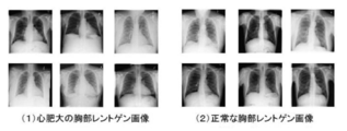
- the 12 chest X-ray images shown in FIG. 12 are a part of 1510 images selected from 112,120 open-source chest X-ray images, and the cardiac hypertrophy as shown in the 6 images of FIG. 12 (1).
- a 1510 image in which a chest X-ray image and a normal chest X-ray image as shown in the 6 images of FIG. 12 (2) were mixed was input to the AI model and pre-trained.
- the functional block of the localization site prediction device for the secondary conduction path of Example 3 is the same as that of the device of Example 2, and the description thereof will be omitted.
- image data in which normal chest X-ray images and chest X-ray images of cardiac hypertrophy are mixed and cardiac hypertrophy classification data (normal: 0, Cardiac hypertrophy: 1) was input to the AI model and pre-learned, and then the AI model pre-learned using chest X-ray images using the transfer learning method was used to localize the accessory pathway. It is used for the AI model of the learning target related to site prediction.
- the processing of the method for predicting the localization site of the accessory pathway in Example 3 is as follows: First, 12-lead ECG data, chest X-ray image data, and classification of the localization site of the accessory pathway in the case of WPW syndrome.
- the data and the cardiac hypertrophy classification data are input (step S11).
- the mixed image data of the normal chest X-ray image and the chest X-ray image of cardiac hypertrophy and the cardiac hypertrophy classification data are input to the AI model and pre-learned (step S131).
- the AI model after the pre-training is trained by inputting 12-lead electrocardiogram data, chest X-ray image data, localization site classification data of the accessory conduction path, and cardiac hypertrophy classification data (step S132).
- the subject's 12-lead ECG data and chest X-ray image data are input (step S15), and the subject's data is input to the trained AI model to calculate the localization site classification information of the accessory conduction path (step).
- S17 the processing of the
- 1510 image data in which an open source normal chest X-ray image and a chest X-ray image of cardiac hypertrophy are mixed are input to a two-dimensional CNN, and 1510 images are input.
- Cardiac hypertrophy classification data normal: 0, cardiac hypertrophy: 1 corresponding to each image data is given to the two-dimensional CNN as teacher data and sufficiently iteratively trained (pre-learning).
- pre-learning sufficiently iteratively trained
- chest X-rays for 60 cases of WPW syndrome are applied to the pre-learned two-dimensional CNN.
- Image data is input, and in parallel, a data set of 12-lead electrocardiograms for 60 people is input to a one-dimensional CNN, and each CNN is repeatedly learned.
- the AI model used in Example 3 is the same as the AI model used in Example 2, and the two-dimensional matrix data of the chest roentgen image is input to the input layer (first block) of the two-dimensional CNN, and a 12-lead electrocardiogram is obtained.
- the one-dimensional matrix data of is input to the input layer (first block) of the one-dimensional CNN.
- the convolution calculation and the reduction of the feature map space are repeated by the convolution layer and the pooling layer of the respective CNNs.
- Each feature output from the fully connected layer of each CNN is input to each DNN of the second block and calculated, and the output is further input to the DNN of the third block and calculated to be a classification label (0,1,1). 2) is output.
- Example 3 The prediction accuracy of the localization site classification of the accessory pathway of WPW syndrome in the prediction device and the prediction method of Example 3 is shown in the graph of FIG. In the graph, a remarkable difference was observed between Example 1 and Example 3, and Example 3 showed a prediction accuracy of 80% or more.
- the teacher data of the 12-lead electrocardiogram data and the localization site classification information of the accessory conduction path is input to the AI model and trained, the normal chest X-ray image and the chest X-ray image of cardiac hypertrophy are mixed.
- the prediction accuracy was further improved.
- the biopotential signal data set of the electrocardiogram is set as the waveform data set of the 12-lead electrocardiogram, the waveform data set of the 12-lead electrocardiogram, and hypertrophic cardiomyopathy and normality.
- the waveform data set of the subject's 12-lead ECG is input to the trained AI model to cause hypertrophic cardiomyopathy or normal cases.
- the classification information of is calculated.
- the prediction method and the prediction device of this embodiment will be described with reference to FIGS. 20 to 23.
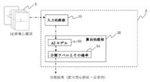
- the idiopathic cardiomyopathy diagnosis support device 3 of this embodiment inputs a 12-lead electrocardiogram data set and a classification label of hypertrophic cardiomyopathy and a normal case as teacher data for learning.
- the trained AI model 33 is provided, the input processing unit 31 inputs the 12-lead electrocardiogram data set 5, and the calculation processing unit 32 inputs the input data set 5 into the trained AI model 33.
- the classification label of hypertrophic cardiomyopathy or normal cases and its probability 34 are calculated. Then, the result regarding the classification of hypertrophic cardiomyopathy or normal case is output.
- the AI model used in Example 4 matrix data of a 12-lead electrocardiogram data set for 25 cases of hypertrophic cardiomyopathy and 133 normal cases is used as learning data, and hypertrophic cardiomyopathy is used.
- the probabilities of the labels of the two categories of cardiomyopathy and normal cases are output, compared with the correct category labels, and the weighting coefficient of the AI model is adjusted repeatedly for learning.
- the AI model used in this embodiment is a one-dimensional convolutional neural network (CNN) as in Examples 1 to 3 described above, and is a batch normalization method for stabilizing the learning of the neural network, and ReLU as an activation function. (Rectified Linear Unit), L2 was used as the regularization method, and Adam was used as the optimization method.
- step S31 12-lead electrocardiogram data of a case of hypertrophic cardiomyopathy and a normal case, and classification data of hypertrophic cardiomyopathy and a normal case are input (step).
- step S31 the input data is input to the AI model and trained (step S33).
- the 12-lead electrocardiogram data may be the waveform image data of the electrocardiogram or may be matrix data in which the waveform image data is one-dimensionalized in time series.
- step S35 the subject's 12-lead ECG data is input (step S35).
- the subject data is input to the trained AI model, and the classification information of hypertrophic cardiomyopathy or normal cases is calculated (step S37).
- the 12-lead electrocardiogram is as described in detail in Example 1.
- the waveform data of the 12-lead electrocardiogram appears as an image in a different format depending on the measuring device, but by making the image of the waveform data one-dimensional, a unified data structure independent of the measuring device is obtained (FIG. 6). )
- An image of 12-channel waveform data of a 12-lead electrocardiogram is extracted with the same time width (about 0.6 seconds), divided by a predetermined number of time divisions (145 as described later), and the image is one-dimensional in time series. It is used as data.
- hypertrophic cardiomyopathy Even in hypertrophic cardiomyopathy, it shows a specific electrocardiogram biopotential signal. In 75-96% of hypertrophic cardiomyopathy, some electrocardiographic abnormalities such as abnormal Q wave, ST-T change, negative T wave, and high potential of left chest lead are observed, and electrocardiogram is a highly sensitive and useful screening test. In many asymptomatic cases, the electrocardiogram triggers the diagnosis. Individual electrocardiographic findings are associated with the type, degree, and stage of hypertrophy and help to understand the condition.
- the data sets for 25 cases of hypertrophic cardiomyopathy and 133 cases of normal cases are used, and the cases of hypertrophic cardiomyopathy and normal cases in the diagnostic support method and device of this example are used. It was used to confirm the classification prediction accuracy.
- the images of the waveform data of the 12-lead electrocardiogram for a total of 158 patients, 25 cases of hypertrophic cardiomyopathy and 133 normal cases, were made into one dimension and converted into the matrix data of (158, 145, 12).
- 158 is the number of data sets
- 145 is the number of time divisions in each waveform data
- 12 is the waveform data of each lead in the 12-lead ECG.
- the waveform data set for 158 people is randomly divided into the training data and the test data, and the waveform data set for 111 people is prepared for the training data and the waveform data set for 47 people is prepared for the test data.
- the prediction accuracy was measured.
- the test data is not used for learning the AI model, but is the target data predicted by the AI model as new data.
- the measurement result of the prediction accuracy is obtained by randomly dividing the training data and the test data 20 times in total and calculating the average prediction accuracy of the test data.
- the prediction accuracy of the classification of hypertrophic cardiomyopathy cases and normal cases in the diagnostic support device and method of this example is shown in the graph of FIG. 23.
- the trained AI model showed a classification prediction accuracy of 93% or more.
- the learning for 111 learning data was regarded as one learning.
- the present invention is useful as a method and device for predicting the localized site of the accessory conduction path of WPW syndrome, and as a method and device for diagnosing idiopathic cardiomyopathy such as hypertrophic cardiomyopathy, and is associated with an electronic medical record system in hospitals and the like. It is expected to be used as an application to be used.
Landscapes
- Health & Medical Sciences (AREA)
- Life Sciences & Earth Sciences (AREA)
- Cardiology (AREA)
- Heart & Thoracic Surgery (AREA)
- Molecular Biology (AREA)
- Pathology (AREA)
- Engineering & Computer Science (AREA)
- Biomedical Technology (AREA)
- Physics & Mathematics (AREA)
- Medical Informatics (AREA)
- Biophysics (AREA)
- Surgery (AREA)
- Animal Behavior & Ethology (AREA)
- General Health & Medical Sciences (AREA)
- Public Health (AREA)
- Veterinary Medicine (AREA)
- Measurement And Recording Of Electrical Phenomena And Electrical Characteristics Of The Living Body (AREA)
Abstract
WPW症候群などの頻脈性不整脈の副伝導路の局在部位を高精度に予測する、並びに、特発性心筋症を診断支援する方法及び装置を提供する。方法は、心電図の生体電位信号データセットと、頻脈性不整脈の副伝導路の局在部位分類情報又は特発性心筋症分類情報とを教師データとしてAIモデルに入力して学習させるステップと、学習済みのAIモデルに対して、被験者の心電図の生体電位信号データセットを入力して、副伝導路の局在部位分類情報などを算出するステップを備える。装置は、心電図の生体電位信号データセットと、頻脈性不整脈の副伝導路の局在部位分類情報又は特発性心筋症分類情報とを教師データとして入力し学習させた学習済みのAIモデルと、被験者の心電図の生体電位信号データセットを入力する入力処理部と、入力した生体電位信号データセットを、学習済みのAIモデルに入力して、副伝導路の局在部位分類情報などを算出する算出処理部を備える。
Description
本発明は、例えば、WPW症候群 (Wolff-Parkinson-White syndrome)などの頻脈性不整脈における副伝導路の局在部位予測方法、特発性心筋症の診断支援方法、及びこれらの装置に関するものである。
頻脈性不整脈として、WPW症候群や房室結節回帰性頻拍などの発作性上室性頻拍、心房粗動、心房頻拍、心房細動が挙げられる。
ここで、WPW症候群とは、心臓の心房で起こり脈が速くなる頻脈性不整脈の病気である。正常な心臓では、興奮は1本の刺激伝導路を通って心房から心室へと伝搬されるが、WPW症候群では、心房と心室との間に正常な伝導路とは別に、副伝導路という生まれつき存在する余分な伝導路がある。正常伝導路と副伝導路の間で速い周期で電気刺激が往復することによって頻脈性の不整脈が生じる。日常的には、特に不自由はなく、一生涯を通じて問題にならない場合もあるが、中には頻脈(脈が速くなる、脈拍数が多くなる)発作を生じる場合がある。また、WPW症候群とは別に心房細動という不整脈が併発していると、心房興奮が副伝導路を下降し、激しい頻脈を生じて、致死的な不整脈(心室細動など)へ移行する場合や心不全状態となる場合もある。
ここで、WPW症候群とは、心臓の心房で起こり脈が速くなる頻脈性不整脈の病気である。正常な心臓では、興奮は1本の刺激伝導路を通って心房から心室へと伝搬されるが、WPW症候群では、心房と心室との間に正常な伝導路とは別に、副伝導路という生まれつき存在する余分な伝導路がある。正常伝導路と副伝導路の間で速い周期で電気刺激が往復することによって頻脈性の不整脈が生じる。日常的には、特に不自由はなく、一生涯を通じて問題にならない場合もあるが、中には頻脈(脈が速くなる、脈拍数が多くなる)発作を生じる場合がある。また、WPW症候群とは別に心房細動という不整脈が併発していると、心房興奮が副伝導路を下降し、激しい頻脈を生じて、致死的な不整脈(心室細動など)へ移行する場合や心不全状態となる場合もある。
WPW症候群などの不整脈の治療としては、従来の開心術による副伝導路切断に代わり、非開胸的に心臓内へ焼灼用カテーテルを挿入してエネルギー照射することにより、上記の副伝導路を焼灼するカテーテルアブレーション(心筋焼灼術)が行われている。カテーテルアブレーションは、WPW症候群などの根治的治療として確立された治療法である。カテーテルアブレーションの治療を行う際に特に重要なのが、副伝導路の特定である。焼灼に先立って副伝導路の位置の特定、すなわち、術前に副伝導路の局在を予測することは、治療戦略を立てる上で重要である。
従来から、副伝導路の存在する房室間輪の周囲にカテーテル電極を挿入し、心房内および心室内より電気的に刺激を与え、刺激の伝導時間を計測することにより、異常な刺激伝導時間の部位を副伝導路部として特定する方法が知られている。しかし、この方法では、心臓の左心側の僧帽弁輪の部位、右心側の三尖弁輪の部位の検査を別々に順番に位置をずらして複数の部位の刺激伝導時間を計測する必要があり、検査に長時間を必要とする問題があった。
検査時間を短縮すべく、心臓の冠状静脈洞に第1の電極リードを挿入して左心側の僧帽弁輪部の活動電位を計測し、また、心臓の右冠状動脈に第2の電極リードを挿入して右心側の三尖弁輪の部位の活動電位を計測し、各電極リードから得られた複数の活動電位から早期興奮部位を副伝導路部位として検出する副伝導路検出装置が知られている(特許文献1を参照)。特許文献1に開示された副伝導路検出装置では、短時間で副伝導路部位を特定することが可能となるが、検査において、各電極リードを患者の心臓に挿入するため、患者にとって負担であった。
検査時間を短縮すべく、心臓の冠状静脈洞に第1の電極リードを挿入して左心側の僧帽弁輪部の活動電位を計測し、また、心臓の右冠状動脈に第2の電極リードを挿入して右心側の三尖弁輪の部位の活動電位を計測し、各電極リードから得られた複数の活動電位から早期興奮部位を副伝導路部位として検出する副伝導路検出装置が知られている(特許文献1を参照)。特許文献1に開示された副伝導路検出装置では、短時間で副伝導路部位を特定することが可能となるが、検査において、各電極リードを患者の心臓に挿入するため、患者にとって負担であった。
また、カテーテルアブレーションにおいて、カテーテルと磁気を使って3次元的に電位情報を描出することができ、カテーテル電極による心内電位記録と、磁気を利用して得られるカテーテル電極の位置を同時にコンピュータ処理して、心臓立体画像をディスプレイ表示して、頻拍中の興奮伝播過程や電位波高をリアルタイムに表示できる。しかしながら、興奮伝播は、電気信号の波形の移動として表されるが、不整脈が発生しているような状況下では電気信号の進路が複雑であり、移動する波形情報を分かりやすく表示するのは難しく、心臓全体の興奮伝播の様子を可視化しても、どの部分で副伝導路が発生しているのかは分かりづらく、副伝導路部位の見落としが発生しやすい。かかる問題に鑑み、心臓の電気生理マッピングデータと心臓の解剖学的な形態との位置関係を把握し、アブレーション治療のターゲット部位を正確に特定し、その場所に正確にナビゲートする技術が知られている(特許文献2を参照)。
一方で、近年、多数例の成果をもとに分析した12誘導心電図によるWPW症候群の副伝導路を推定するアルゴリズムが引き続いて提唱されている。その多くはデルタ波やQRS波の波形から局在を予測する決定木モデルである。心室興奮波は早期興奮のデルタ波と融合収縮の幅広いQRS波から構成され、古典的な心電図分類では、V1又はV2のQRS波形によってA型、B型が分かれ、それぞれ左側副伝導路、右側副伝導路によるものと見做されている(非特許文献1を参照)。従来、医師が心電図のデルタ波に着目し副伝導路の存在する場所について、左側副伝導路(A型)、右側副伝導路(B型)、中隔副伝導路(C型)を判別している。
しかしながら、心電図のデルタ波による医師の判断では、誤診を起こす確率が高いといった問題がある。
しかしながら、心電図のデルタ波による医師の判断では、誤診を起こす確率が高いといった問題がある。
また、最新の臨床電気生理学的な観点から、副伝導路は、図17(非特許文献1の図2を引用)に示すように、右室側が8ヵ所、左室側が5ヵ所の総計13部位に細分化されている(図は左前斜位撮影時を想定)。細分化された13部位は、RA(右前壁)、RAL(右前側壁)、RAS(右前中隔)、RL(右側壁)、RPL(右後側壁)、RP(右後壁)、RPS(右後中隔)、MS(中部中隔)、LAL(左前側壁)、LL(左側壁)、LPL(左後側壁)、LP(左後壁)、LPS(左後中隔)であり、図中におけるHはヒス束(正常刺激伝導路)、CSは冠静脈洞である。
副伝導路の推定アルゴリズムにおいては、デルタ波とQRS波を組み合わせたもので、予測正確度が高いChiangアルゴリズムが知られている(非特許文献2を参照)。例えば、Chiangアルゴリズムは、早期興奮波のデルタ波が十分識別できる症例という条件下で、決定木が作成されており、陽性波(+),陰性波(-),二相性波あるいは等電位波(±)と定義し、デルタ波(Δ)の極性(+/-)に基づいて、図18(非特許文献1の図3を引用、非特許文献2より引用)に示すように、ステップ1~4の順にステップを進む診断ロジックになっている。Chiangアルゴリズムの副伝導路の予測正確度は、部位によって違いはあるが80%以上の正解度である。なお、Chiangアルゴリズムにおいては、上述の細分化された13部位は、幾つかの部位が統合されて9部位に分類されている。
また、St Georgeアルゴリズムは、しばしば識別不能となるデルタ波をアルゴリズムから外してQRS波のみで、図19(非特許文献1の図4を引用、非特許文献3より引用)に示すように、3ステップ又は4ステップで進む診断ロジックになっている(非特許文献3を参照)。St Georgeアルゴリズムの副伝導路の予測正確度は、10の部位数の分類で85%程度である。
しかしながら、完全な予測アルゴリズムは未だ確立されていないのが実状である。副伝導路を予測する完全なアルゴリズムが確立されていない理由として、2つの大きな問題点が挙げられる。1つ目の問題点は、多くのアルゴリズムは、Chiangアルゴリズムの如く、決定木モデルを用いており、境界型の分類困難な場合が存在することである。そして、2つ目の問題点は、心臓の解剖学的な軸は個々によって異なっており、それにより分類結果に誤差が生じることである。
また、不整脈以外の心臓病として、心筋(心臓の筋肉)に異常をきたす心筋症があり、代表的なものが肥大型心筋症と拡張型心筋症である。肥大型心筋症とは、心筋が肥厚し拡張機能が低下する疾患であり、一方、拡張型心筋症とは、心筋が薄くなって、収縮力が低下する疾患であり、共に、原因不明の心臓の筋肉の病気である特発性心筋症のうちのひとつである。これらの心筋症は、不整脈が伴うと致死性心事故が起こる可能性が高くなるため、不整脈を予防するための投薬などの措置が必要とされる。これらの心筋症は、超音波エコーで精度よく発見が可能であるが、より簡便な検査である心電図において、心筋症の早期発見が行えることが好ましい。
また、近年の心電図装置には、自動診断機能が搭載されているものがあるが、それらは基本的に成人向けの自動診断であり、小児診断には使用できず、医療の現場では小児診断にあたる医師の経験に基づき診断しているケースが多いことから、医師の診断を支援できる予測方法・予測装置が求められている。
また、近年の心電図装置には、自動診断機能が搭載されているものがあるが、それらは基本的に成人向けの自動診断であり、小児診断には使用できず、医療の現場では小児診断にあたる医師の経験に基づき診断しているケースが多いことから、医師の診断を支援できる予測方法・予測装置が求められている。
広木忠行, 12誘導心電図による副伝導路部位の推定:3.WPW症候群:副伝導路部位の推定と治療(特集II:第64回 日本循環器学会学術集会,循環器専門医, 2001, 9巻1 号,p.57-66.
Chiang CE et al., "An accurate stepwiseelectrocardiographic algorithm for localization of accessory pathways inpatients with Wolff-Parkinson-White syndrome from a comprehensive analysis ofdelta waves and R/S ratio during sinus rhythm.", Am J Cardiol,1995 Jul 1,76:40-46.
Xie B et al., "Localizat ioof naccessorypathways from the l2-lead electrocardiogram using a new algorithm", Am J Cardio1,1994; 74: 161-165.
WPW症候群などの頻脈性不整脈は、罹患率の多い疾患であり、術前の段階で精度の高い副伝導路の局在診断ができることが望まれている。しかしながら、上述の如く、副伝導路を予測する完全なアルゴリズムが確立されておらず、また術前での副伝導路の局在診断の精度が高くなかったことから、術前に術式の詳細な説明や合併症リスクの説明が行えないといった問題があった。
また、肥大型心筋症などの特発性心筋症は、超音波エコーを用いず、簡便な検査手法の心電図を用いて精度よく特発性心筋症の早期発見が行えることが好ましい。
かかる状況に鑑みて、本発明は、WPW症候群などの頻脈性不整脈の副伝導路の局在部位を高精度に予測できる方法及び装置、並びに、特発性心筋症を診断支援できる方法及び装置を提供することを目的とする。
上記課題を解決すべく、本発明の第1の観点によれば、本発明の副伝導路の局在部位予測方法は、心電図の生体電位信号データセットと、頻脈性不整脈の副伝導路の局在部位分類情報とを教師データとしてAIモデルに入力して学習させるステップと、学習済みのAIモデルに対して、被験者の心電図の生体電位信号データセットを入力して、副伝導路の局在部位分類情報を算出するステップを備えることにより、WPW症候群などの頻脈性不整脈の副伝導路の局在部位を高精度に予測可能である。
上記及び後述する本発明では、従来のルールベースの決定木アルゴリズムではなく、AI(人工知能)モデル、例えば、深層ニューラルネットワークモデルや畳み込みニューラルネットワークを用いることにより、より堅牢性の高いアルゴリズムで、副伝導路の局在部位を予測でき、副伝導路の局在部位情報を簡単かつ自動で出力することができる。
教師データとしてAIモデルに入力して学習させるステップは、教師データとして心電図の生体電位信号データセットをAIモデルに入力し、AIモデルが出力した分類結果を、教師データの副伝導路の局在部位分類情報と比較させて、AIモデルの重み付け係数を最適化していくものである。
上記及び後述する本発明では、従来のルールベースの決定木アルゴリズムではなく、AI(人工知能)モデル、例えば、深層ニューラルネットワークモデルや畳み込みニューラルネットワークを用いることにより、より堅牢性の高いアルゴリズムで、副伝導路の局在部位を予測でき、副伝導路の局在部位情報を簡単かつ自動で出力することができる。
教師データとしてAIモデルに入力して学習させるステップは、教師データとして心電図の生体電位信号データセットをAIモデルに入力し、AIモデルが出力した分類結果を、教師データの副伝導路の局在部位分類情報と比較させて、AIモデルの重み付け係数を最適化していくものである。
ここで、心電図の生体電位信号データセットとしては、12誘導心電図の信号波形データが好ましいが、本発明の技術思想に従う限りにおいて、その他の心電図の信号波形データを用いることもできる。副伝導路とは、心臓内にある電気的興奮を伝える正常の伝導路とは別にある伝導路であり、副伝導路には、ケント束、ジェームス束、マハイム線維などがある。本明細書において、副伝導路はケント束を指す。
心電図の生体電位信号データセットの場合、教師データとなり得るものが少ないことが予想されるが、生体電位信号データを時間軸方向に圧縮・引き伸ばしをして、教師データ数を水増ししてもよい。但し、左右・上下の反転(flipping)による水増しは不可である。
心電図の生体電位信号データセットの場合、教師データとなり得るものが少ないことが予想されるが、生体電位信号データを時間軸方向に圧縮・引き伸ばしをして、教師データ数を水増ししてもよい。但し、左右・上下の反転(flipping)による水増しは不可である。
本発明の第2の観点によれば、本発明の副伝導路の局在部位予測方法は、心電図の生体電位信号データセットと、胸部レントゲン画像データと、頻脈性不整脈の副伝導路の局在部位分類情報と、心肥大分類情報とを教師データとしてAIモデルに入力して学習させるステップと、学習済みのAIモデルに対して、被験者の心電図の生体電位信号データセットと胸部レントゲン画像データを入力して、副伝導路の局在部位分類情報を算出するステップを備えることにより、WPW症候群などの頻脈性不整脈の副伝導路の局在部位をより高精度に予測可能である。
心臓の解剖学的な位置情報を加味する目的で、同一症例の胸部レントゲン画像データを上記のAIモデルの入力に追加することで、さらに精度を向上させることができる。
心臓の解剖学的な位置情報を加味する目的で、同一症例の胸部レントゲン画像データを上記のAIモデルの入力に追加することで、さらに精度を向上させることができる。
本発明の第2の観点による副伝導路の局在部位予測方法において、教師データをAIモデルに入力して学習させるステップの前に、正常胸部レントゲン画像および心肥大の胸部レントゲン画像の混在したデータをAIモデルに入力して心肥大の有無を判断できるように事前学習させるステップを更に備えることにより、更に高精度に予測可能である。
胸部レントゲン画像データは、心電図の生体電位信号データセットに比べて、広くデータを入手することができ、この内、正常胸部レントゲン画像および心肥大の胸部レントゲン画像データを用いて、事前にAIモデルに入力して事前学習させて、AIモデルの重み付け係数を調整する。
胸部レントゲン画像データは、心電図の生体電位信号データセットに比べて、広くデータを入手することができ、この内、正常胸部レントゲン画像および心肥大の胸部レントゲン画像データを用いて、事前にAIモデルに入力して事前学習させて、AIモデルの重み付け係数を調整する。
本発明の第2の観点による副伝導路の局在部位予測方法に用いるAIモデルは、心電図の生体電位信号データセットを1次元行列データとして入力する1次元畳み込みニューラルネットワークと、胸部レントゲン画像データを2次元行列データとして入力する2次元畳み込みニューラルネットワークと、それぞれの畳み込みニューラルネットワークから出力される特徴データを演算する深層ニューラルネットワークを少なくとも備えることが好ましい。心電図の生体電位信号データセットは、1次元行列データとして入力演算し、胸部レントゲン画像データは、2次元行列データとして入力演算することから、各々異なる畳み込みニューラルネットワークを用いるためである。
また、正常胸部レントゲン画像および心肥大の胸部レントゲン画像の混在したデータをAIモデルに入力して心肥大の有無を判断できるように事前学習させるものは、2次元畳み込みニューラルネットワークである。胸部レントゲン画像は、心電図の生体電位信号データセットと異なり、既に多くの画像がオープンソースとしてアクセス可能であることから、事前学習させて、2次元畳み込みニューラルネットワークの予測正確度を高めるのである。
また、正常胸部レントゲン画像および心肥大の胸部レントゲン画像の混在したデータをAIモデルに入力して心肥大の有無を判断できるように事前学習させるものは、2次元畳み込みニューラルネットワークである。胸部レントゲン画像は、心電図の生体電位信号データセットと異なり、既に多くの画像がオープンソースとしてアクセス可能であることから、事前学習させて、2次元畳み込みニューラルネットワークの予測正確度を高めるのである。
本発明の第1又は第2の観点による副伝導路の局在部位予測方法において、具体的には、副伝導路の局在部位分類情報が、臨床電気生理学的検査に基づく位置分類における中隔側の部位、僧帽弁輪の自由壁側の部位、三尖弁輪の自由壁側の部位の3分類の局在部位情報である。
上述したとおり、臨床電気生理学的な観点によれば、総計13部位に細分化されているが、実際の治療現場においては、中隔側の部位、僧帽弁輪の自由壁側の部位、三尖弁輪の自由壁側の部位の3分類の何れかであるかといった局在部位情報がわかることにより、カテーテルアブレーション治療のサポートにできる。
なお、3分類よりも細分化して副伝導路の詳細な部位を予測できれば、術前情報として、よりよい情報になる。学習データ量を増やすことによって、より詳細な分類予測ができ、より有用な治療のサポートとすることができる。
上述したとおり、臨床電気生理学的な観点によれば、総計13部位に細分化されているが、実際の治療現場においては、中隔側の部位、僧帽弁輪の自由壁側の部位、三尖弁輪の自由壁側の部位の3分類の何れかであるかといった局在部位情報がわかることにより、カテーテルアブレーション治療のサポートにできる。
なお、3分類よりも細分化して副伝導路の詳細な部位を予測できれば、術前情報として、よりよい情報になる。学習データ量を増やすことによって、より詳細な分類予測ができ、より有用な治療のサポートとすることができる。
本発明のプログラムは、上述した本発明の副伝導路の局在部位予測方法における学習済みAIモデルを用いて副伝導路の局在部位分類情報を算出するステップを、コンピュータに実行させるものである。
次に、本発明の副伝導路の局在部位予測装置について説明する。
本発明の第1の観点による副伝導路の局在部位予測装置は、心電図の生体電位信号データセットと、頻脈性不整脈の副伝導路の局在部位分類情報とを教師データとして入力し学習させた学習済みのAIモデルと、被験者の心電図の生体電位信号データセットを入力する入力処理部と、入力した生体電位信号データセットを、学習済みのAIモデルに入力して、副伝導路の局在部位分類情報を算出する算出処理部を備えることにより、WPW症候群などの頻脈性不整脈の副伝導路の局在部位を高精度に予測可能である。
本発明の第1の観点による副伝導路の局在部位予測装置は、心電図の生体電位信号データセットと、頻脈性不整脈の副伝導路の局在部位分類情報とを教師データとして入力し学習させた学習済みのAIモデルと、被験者の心電図の生体電位信号データセットを入力する入力処理部と、入力した生体電位信号データセットを、学習済みのAIモデルに入力して、副伝導路の局在部位分類情報を算出する算出処理部を備えることにより、WPW症候群などの頻脈性不整脈の副伝導路の局在部位を高精度に予測可能である。
本発明の第2の観点による副伝導路の局在部位予測装置は、心電図の生体電位信号データセットと、胸部レントゲン画像データと、頻脈性不整脈の副伝導路の局在部位分類情報と、心肥大分類情報とを教師データとして入力し学習させた学習済みのAIモデルと、被験者の心電図の生体電位信号データセットと胸部レントゲン画像データを入力する入力処理部と、入力した生体電位信号データセットと胸部レントゲン画像データを、学習済みのAIモデルに入力して、副伝導路の局在部位分類情報を算出する算出処理部を備えることにより、WPW症候群などの頻脈性不整脈の副伝導路の局在部位をより高精度に予測可能である。
本発明の第2の観点による副伝導路の局在部位予測装置において、学習済みのAIモデルは、教師データを入力して学習させる前に、正常胸部レントゲン画像および心肥大の胸部レントゲン画像データの混在したデータを、AIモデルに入力して心肥大の有無を判断できるように事前学習させたものであることにより、更に高精度に予測可能である。
本発明の第2の観点による副伝導路の局在部位予測装置に用いるAIモデルは、心電図の生体電位信号データセットを1次元行列データとして入力する1次元畳み込みニューラルネットワークと、胸部レントゲン画像データを2次元行列データとして入力する2次元畳み込みニューラルネットワークと、それぞれの畳み込みニューラルネットワークから出力される特徴データを演算する深層ニューラルネットワークを少なくとも備えることが好ましい。
本発明の第1又は第2の観点による副伝導路の局在部位予測装置において、副伝導路の局在部位分類情報が、臨床電気生理学的検査に基づく位置分類における中隔側の部位、僧帽弁輪の自由壁側の部位、三尖弁輪の自由壁側の部位の3分類の局在部位情報である。
次に、本発明の特発性心筋症の診断支援方法及び診断支援装置について説明する。
本発明の特発性心筋症の診断支援方法は、心電図の生体電位信号データセットと、肥大型心筋症と正常例の分類情報とを教師データとしてAIモデルに入力して学習させるステップ(学習ステップ)と、学習済みのAIモデルに対して、被験者の心電図の生体電位信号データセットを入力して、肥大型心筋症または正常例の分類情報を算出するステップ(算出ステップ)を備える。
本発明の特発性心筋症の診断支援方法において、教師データとして、拡張型心筋症の分類情報が追加され、上記算出ステップにおいて、肥大型心筋症、拡張型心筋症または正常例の分類情報を算出することも可能である。
本発明の特発性心筋症の診断支援方法における上記学習ステップにおいて、教師データとして、更に、胸部レントゲン画像データが追加されてもよい。胸部レントゲン画像データを追加して学習させることにより、より分類精度を高めることができる。
本発明の特発性心筋症の診断支援方法は、心電図の生体電位信号データセットと、肥大型心筋症と正常例の分類情報とを教師データとしてAIモデルに入力して学習させるステップ(学習ステップ)と、学習済みのAIモデルに対して、被験者の心電図の生体電位信号データセットを入力して、肥大型心筋症または正常例の分類情報を算出するステップ(算出ステップ)を備える。
本発明の特発性心筋症の診断支援方法において、教師データとして、拡張型心筋症の分類情報が追加され、上記算出ステップにおいて、肥大型心筋症、拡張型心筋症または正常例の分類情報を算出することも可能である。
本発明の特発性心筋症の診断支援方法における上記学習ステップにおいて、教師データとして、更に、胸部レントゲン画像データが追加されてもよい。胸部レントゲン画像データを追加して学習させることにより、より分類精度を高めることができる。
本発明の特発性心筋症の診断支援装置は、心電図の生体電位信号データセットと、肥大型心筋症と正常例の分類情報とを教師データとして入力し学習させた学習済みのAIモデルと、被験者の心電図の生体電位信号データセットを入力する入力処理部と、入力した生体電位信号データセットを、学習済みのAIモデルに入力して、肥大型心筋症または正常例の分類情報を算出する算出処理部を備える。本発明の特発性心筋症の診断支援装置において、教師データとして、拡張型心筋症の分類情報が追加され、上記算出処理部において、肥大型心筋症、拡張型心筋症または正常例の分類情報を算出することも可能である。なお、教師データとして、更に、胸部レントゲン画像データが追加されてもよい。
本発明の副伝導路の局在部位予測方法及び装置によれば、非侵襲的な心電図情報に基づいて、副伝導路の局在部位を高精度に予測できることから、WPW症候群などの頻脈性不整脈の治療現場において、術前に術式の詳細な説明や合併症リスクの説明を医師が患者に対して行えるといった効果がある。さらに、非侵襲的な心電図に加え、一般的に行われている胸部レントゲン画像データを用いて、より高精度に副伝導路の局在部位を予測できるといった効果がある。
また、本発明の特発性心筋症の診断支援方法及び装置によれば、心電図情報に基づいて、肥大型心筋症などの特発性心筋症の診断支援を行えるといった効果がある。
また、本発明の特発性心筋症の診断支援方法及び装置によれば、心電図情報に基づいて、肥大型心筋症などの特発性心筋症の診断支援を行えるといった効果がある。
以下、本発明の実施形態の一例を、図面を参照しながら詳細に説明していく。なお、本発明の範囲は、以下の実施例や図示例に限定されるものではなく、幾多の変更及び変形が可能である。
実施例1の予測方法及び予測装置では、心電図の生体電位信号データセットを12誘導心電図の波形データセットとし、12誘導心電図の波形データセットとWPW症候群の副伝導路の局在部位分類情報とを教師データとしてAIモデルに入力して学習させた後、学習済みのAIモデルに対して、被験者の12誘導心電図の波形データセットを入力して、WPW症候群の副伝導路の局在部位分類情報を算出する。以下、本実施例の予測方法及び予測装置について、図1~7を参照して説明する。
図1の機能ブロック図に示すとおり、実施例1の副伝導路の局在部位予測装置1は、12誘導心電図データセットとWPW症候群の副伝導路の局在部位分類ラベルとを教師データとして入力し学習させた学習済みのAIモデル13を備え、入力処理部11において、12誘導心電図データセット5を入力し、算出処理部12において、入力したデータセット5を学習済みのAIモデル13に入力して、WPW症候群の副伝導路の局在部位分類ラベルとその確率14を算出する。そして、副伝導路の局在部位の分類に関する予測結果を出力する。
図2に示すとおり、実施例1で用いるAIモデルでは、学習用データとしてWPW症候群の症例60名分の12誘導心電図のデータセットの行列データを使用し、TypeA~Cの3分類のラベルの確率を出力し、正解の分類ラベルと比較して、AIモデルの重み係数を調整することを繰り返しながら学習する。
実施例1(他の実施例2,3も同様)で用いるAIモデルは、一次元の畳み込みニューラルネットワーク(CNN)であり、ニューラルネットワークの学習安定化を図るためバッチ正規化手法、活性化関数としてReLU(Rectified Linear Unit)、正則化手法としてL2、最適化手法としてAdamを用いた。
実施例1(他の実施例2,3も同様)で用いるAIモデルは、一次元の畳み込みニューラルネットワーク(CNN)であり、ニューラルネットワークの学習安定化を図るためバッチ正規化手法、活性化関数としてReLU(Rectified Linear Unit)、正則化手法としてL2、最適化手法としてAdamを用いた。
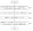
実施例1の副伝導路の局在部位予測方法の処理は、図3に示すとおり、先ず、WPW症候群の症例の12誘導心電図データと、副伝導路の局在部位分類データを入力し(ステップS01)、入力データをAIモデルに入力して学習させる(ステップS03)。ステップS01において、12誘導心電図データは、心電図の波形画像データであっても、波形画像データを時系列に一次元化した行列データとしてもよい。次に、被験者の12誘導心電図データを入力する(ステップS05)。被験者のデータを学習済みAIモデルに対して入力し、副伝導路の局在部位分類情報を算出する(ステップS07)。
12誘導心電図は、心電図を記録する際に標準的に用いられ、心臓の電気的活動を様々な方向から観察したものを記録し、心臓の電気的活動を立体的なものとして捉えるものである。12誘導心電図は、胸部誘導(V1~V6:単極誘導の6誘導)と肢誘導(I~III:双極肢誘導の3誘導、aVR,aVL,aVF:単極肢誘導の3誘導)に分かれている。胸部誘導(V1~V6)は、心臓の電気的変化を水平面に投影し記録したものであり、双極肢誘導(I誘導)は左手と右手の電位差、双極肢誘導(II誘導)は左足と右手の電位差、双極肢誘導(III誘導)は左足と左手の電位差を記録したものである。また、単極肢誘導(aVR)は興奮が右肩方向から、単極肢誘導(aVL)は興奮が左肩方向から、単極肢誘導(aVF)は興奮が横隔膜の方向から、それぞれ心臓の電気的中心に対して近づく・遠ざかる様子を記録したものである。
WPW症候群では、心臓の心室の早期興奮による特異的な心電図の生体電位信号を示す。正常な伝導路以外に1つ又は複数の副伝導路が存在し、心房からの興奮が正常な伝導路をバイパスして早期に心室に伝わる。副伝導路には、ケント束、ジェームス束、マハイム線維、心房ヒス束があるが、WPW症候群では、ケント束を介する心室早期興奮と、正常な伝導路を介する心室興奮の融合波としてQRS波が現れる。図4に示すとおり、V1誘導に見られるQRS波におけるデルタ波、PQ間隔短縮、QRS延長などに基づいて、順行性ケント束伝導を有するWPW症候群と判別でき、また、図4(1),(2)に示すように、WPW症候群の副伝導路の局在位置を判別できる。なお、順行性ケント束伝導がなく逆行性ケント束伝導のみを有するものは、潜在性WPW症候群と呼ばれるが、QRS波にデルタ波が現れないため発作時以外の心電図からは診断することは困難であり、そのため電気生理学的検査を用いて潜在性WPW症候群を判別する。本実施例では、順行性ケント束伝導を有するWPW症候群に関して、副伝導路の局在部位をより高精度に予測できることを説明する。
本実施例の予測装置及び予測方法では、WPW症候群における副伝導路の局在部位の分類について、臨床電気生理学的検査に基づく位置分類における中隔側の部位、僧帽弁輪の自由壁側の部位、三尖弁輪の自由壁側の部位の3分類とし、3分類を予測する。前述の如く、臨床電気生理学的な観点から、副伝導路は、右室側が8ヵ所、左室側が5ヵ所の13部位に分類されている(図17を参照のこと)。図5(1)の円グラフは全部位(13分類)、図5(2)の円グラフはChiangアルゴリズムの9分類、図5(3)の円グラフは本実施例の3分類を示す。Chiangアルゴリズムでは全部位(13分類)の内、LALとLL、LPLとLP、RASとRA、RPLとRPがそれぞれ統合されて9分類である。本実施例では、Chiangアルゴリズムの9分類とは別に新たに3分類を作成し、図5(3)に示すとおり、僧帽弁輪の自由壁側の部位に該当するLALとLLとLPLとLPを統合化してTypeAとし、三尖弁輪の自由壁側の部位に該当するRPとRPLとRLとRALとRAを統合化してTypeBとし、中隔側の部位に該当するLPSとRASとMSとRPSを統合化してTypeCとした。本実施例では、WPW症候群における副伝導路の局在部位として、TypeA~Cの3分類を予測する。
本実施例では、12誘導心電図において、波形データにデルタ波の存在を認めるWPW症候群の症例69名分のデータセットを、本実施例の予測方法及び予測装置の予測精度の確認に用いた。図5(1)~(3)に示す円グラフでは、症例69名分のデータセットに対して、それぞれの分類に区分けして、各分類のデータ数を明記している。
図6(1)のとおり、12誘導心電図の波形データは、測定機器によって異なるフォーマットの画像として現れるが、図6(2)のとおり、波形データの画像を一次元化することで測定機器に依存しない統一されたデータ構造とした。具体的には、12誘導心電図の12チャネルの波形データの画像を同一時間幅(約0.6秒間)で抜き出し、所定の時分割数(後述するように145)で分割し、画像を時系列の一次元データとした。例えば、波形データ画像を{ 0,0,1,3,2,4,5,60,・・・}のように一次元データとした。
69名分の12誘導心電図の波形データの画像を一次元化し、(69,145,12)の行列データに変換した。(69,145,12)の行列データにおいて、69はデータセット数であり、145は各波形データにおける時分割数であり、12は12誘導心電図における各誘導の波形データである。
そして、69名分の波形データセットを学習用データとテスト用データをランダムに分割して、学習用データに60名分の波形データセット、テスト用データに9名分の波形データセットを用意し、予測精度の測定を行った。テスト用データはAIモデルの学習には使用せず、新規なデータとしてAIモデルが予測する対象データである。予測精度の測定結果は、学習用データとテスト用データの分割をランダムに合計50回行い、テスト用データの平均予測正確度を算出したものである。
69名分の12誘導心電図の波形データの画像を一次元化し、(69,145,12)の行列データに変換した。(69,145,12)の行列データにおいて、69はデータセット数であり、145は各波形データにおける時分割数であり、12は12誘導心電図における各誘導の波形データである。
そして、69名分の波形データセットを学習用データとテスト用データをランダムに分割して、学習用データに60名分の波形データセット、テスト用データに9名分の波形データセットを用意し、予測精度の測定を行った。テスト用データはAIモデルの学習には使用せず、新規なデータとしてAIモデルが予測する対象データである。予測精度の測定結果は、学習用データとテスト用データの分割をランダムに合計50回行い、テスト用データの平均予測正確度を算出したものである。
実施例1の予測装置及び予測方法におけるWPW症候群の副伝導路の局在部位分類の予測精度について、図7のグラフに示す。グラフに示すとおり、100回以上の反復学習を行った後では、学習済みAIモデルは70%以上の予測正確度を示した。ここで、学習用データ60名分の学習を1回の学習とした。比較のために、予測精度測定に用いたWPW症候群の症例69名分のデータセットを用いて、従来のChiangアルゴリズムと、St Georgeアルゴリズムの両方のアルゴリズムで分類を予測すると、Chiangアルゴリズムでは予測正確度が55.1%(38/69)であり、St Georgeアルゴリズムでは予測正確度が57.9%(40/69)であり、共に、本実施例の予測精度の70%より下回っていることがわかった。これは、従来のChiangアルゴリズムやSt Georgeアルゴリズムでは、単一副伝導路(Single Accessory Pathway)の典型例のみを集めてアルゴリズムを作成しているため、奇形(anomaly)や二重副伝導路(dual Accessory Pathway)などには適応不可であること、実際の症例データでの予測精度は上述の非特許文献に記載された予測正確度(%)ほど高くないことが影響していると推察する。
以上説明したとおり、本実施例の予測方法又は予測装置によれば、従来のアルゴリズムに比べて、WPW症候群の副伝導路の局在部位をより高精度に予測可能である。
以上説明したとおり、本実施例の予測方法又は予測装置によれば、従来のアルゴリズムに比べて、WPW症候群の副伝導路の局在部位をより高精度に予測可能である。
実施例2の予測方法及び予測装置では、心電図の生体電位信号データセットを12誘導心電図の波形データセットとし、胸部レントゲン画像データと、12誘導心電図の波形データセットとWPW症候群の副伝導路の局在部位分類情報と、心肥大分類情報とを教師データとしてAIモデルに入力して学習させた後、学習済みのAIモデルに対して、被験者の12誘導心電図の波形データセットと胸部レントゲン画像データを入力して、WPW症候群の副伝導路の局在部位分類情報を算出する。以下、本実施例の予測方法及び予測装置について、図8~11を参照して説明する。
図8の機能ブロック図に示すとおり、実施例2の副伝導路の局在部位予測装置2は、12誘導心電図データセット5と、胸部レントゲン画像データ6と、WPW症候群の副伝導路の局在部位分類ラベルと、心肥大分類情報とを教師データとして入力し学習させた学習済みのAIモデル23を備え、入力処理部21において、12誘導心電図データセット5と胸部レントゲン画像データ6を入力し、算出処理部22において、入力したデータ(5,6)を学習済みのAIモデル23に入力して、WPW症候群の副伝導路の局在部位分類ラベルとその確率24を算出する。そして、副伝導路の局在部位の分類に関する予測結果を出力する。
図9に示すとおり、実施例2で用いるAIモデルでは、学習用データとしてWPW症候群の症例60名分の12誘導心電図のデータセットの1次元行列データと、同じ60名分の胸部レントゲン画像の2次元行列データを使用し、TypeA~Cの3分類のラベルの確率を出力し、正解の分類ラベルと比較して、AIモデルの重み係数を調整することを繰り返しながら学習する。実施例2で用いるAIモデルは、胸部レントゲン画像の2次元行列データを、2次元用CNN(畳み込みニューラルネットワーク)の入力層(第1ブロック)に入力する。また、12誘導心電図の1次元行列データを1次元用CNNの入力層(第1ブロック)に入力する。胸部レントゲン画像の2次元行列データ、12誘導心電図の1次元行列データのそれぞれに対して、それぞれのCNNの畳み込み層およびプーリング層によって、畳み込み演算と特徴マップ空間の縮小を繰り返す。それぞれのCNNの全結合層から出力されたそれぞれの特徴を、第2ブロックの各DNN(深層ニューラルネットワーク)に入力し演算し出力したものを、さらに第3ブロックのDNNに入力し演算し分類ラベル(0,1,2)を出力する。
実施例2の副伝導路の局在部位予測方法の処理は、図10に示すとおり、先ず、WPW症候群の症例の12誘導心電図データと、胸部レントゲン画像データと、副伝導路の局在部位分類データと、心肥大分類データを入力し(ステップS11)、入力データをAIモデルに入力して学習させる(ステップS13)。ステップS11において、12誘導心電図データは、心電図の波形画像データであっても、波形画像データを時系列に一次元化した行列データとしてもよい。次に、被験者の12誘導心電図データと胸部レントゲン画像データを入力する(ステップS15)。被験者のデータを学習済みAIモデルに対して入力し、副伝導路の局在部位分類情報を算出する(ステップS17)。
実施例2の予測装置及び予測方法におけるWPW症候群の副伝導路の局在部位分類の予測精度について、図11のグラフに示す。グラフにおいて、実施例1と実施例2の間で顕著な差異は見られなかった。胸部レントゲン画像を正しく認識するためには、1000枚単位の相当なデータ数が必要であるが、実施例2の予測測定では、症例69例の内の学習用の60枚の胸部レントゲン画像しか学習に用いておらず、画像から心肥大を正しく認識できず、予測精度に差異が見られなかったと推察する。
実施例3の予測装置及び予測方法では、12誘導心電図データと、胸部レントゲン画像データと、副伝導路の局在部位分類情報と、心肥大分類情報とを教師データとしてAIモデルに入力して学習させる際に、まず、正常胸部レントゲン画像および心肥大の胸部レントゲン画像の混在したデータをAIモデルに入力して心肥大の有無をある程度の正確度で判断できるように事前学習させた後で、事前学習済みのAIモデルに対して、12誘導心電図データと、胸部レントゲン画像データと、副伝導路の局在部位分類情報と、心肥大分類情報を教師データとしてAIモデルに入力して学習させる。以下、本実施例の予測方法及び予測装置について、図12~16を参照して説明する。
上記の事前学習では、実施例2で用いた症例69例の内の学習用の60枚の胸部レントゲン画像ではなく、オープンソースから1000枚以上の画像を用いて事前学習を行った。
図12に示す12枚の胸部レントゲン画像は、オープンソースの胸部レントゲン画像11万2120枚の中から選定した1510枚の一部であり、図12(1)の6画像に示すような心肥大の胸部レントゲン画像と、図12(2)の6画像に示すような正常胸部レントゲン画像の混在した1510画像を、AIモデルに入力して事前学習させた。
図12に示す12枚の胸部レントゲン画像は、オープンソースの胸部レントゲン画像11万2120枚の中から選定した1510枚の一部であり、図12(1)の6画像に示すような心肥大の胸部レントゲン画像と、図12(2)の6画像に示すような正常胸部レントゲン画像の混在した1510画像を、AIモデルに入力して事前学習させた。
実施例3の副伝導路の局在部位予測装置の機能ブロックは、実施例2の装置と同様であり説明は省略する。
実施例3の副伝導路の局在部位予測装置や方法では、図13に示すとおり、正常胸部レントゲン画像および心肥大の胸部レントゲン画像の混在した画像データと、心肥大分類データ(正常:0、心肥大:1)をAIモデルに入力して事前学習させた後、転移学習(Transfer Learning)の手法を用いて、胸部レントゲン画像を用いて事前学習させたAIモデルを、副伝導路の局在部位予測に関する学習対象のAIモデルに用いる。事前学習では、オープンソースの1510枚の胸部レントゲン画像を事前学習用データとして使用し、その後、WPW症候群の症例60名分の12誘導心電図のデータセットの行列データと60名分の胸部レントゲン画像データを使用し、TypeA~Cの3分類のラベルの確率を出力し、正解の分類ラベルと比較して、AIモデルの重み係数を調整することを繰り返し学習する。
実施例3の副伝導路の局在部位予測装置や方法では、図13に示すとおり、正常胸部レントゲン画像および心肥大の胸部レントゲン画像の混在した画像データと、心肥大分類データ(正常:0、心肥大:1)をAIモデルに入力して事前学習させた後、転移学習(Transfer Learning)の手法を用いて、胸部レントゲン画像を用いて事前学習させたAIモデルを、副伝導路の局在部位予測に関する学習対象のAIモデルに用いる。事前学習では、オープンソースの1510枚の胸部レントゲン画像を事前学習用データとして使用し、その後、WPW症候群の症例60名分の12誘導心電図のデータセットの行列データと60名分の胸部レントゲン画像データを使用し、TypeA~Cの3分類のラベルの確率を出力し、正解の分類ラベルと比較して、AIモデルの重み係数を調整することを繰り返し学習する。
実施例3の副伝導路の局在部位予測方法の処理は、図14に示すとおり、先ず、WPW症候群の症例の12誘導心電図データと、胸部レントゲン画像データと、副伝導路の局在部位分類データと、心肥大分類データを入力する(ステップS11)。そして、正常胸部レントゲン画像および心肥大の胸部レントゲン画像の混在した画像データと、心肥大分類データをAIモデルに入力して事前学習させる(ステップS131)。事前学習させた後のAIモデルに対して、12誘導心電図データと、胸部レントゲン画像データと、副伝導路の局在部位分類データと、心肥大分類データを入力して学習させる(ステップS132)。
その後、被験者の12誘導心電図データと胸部レントゲン画像データを入力し(ステップS15)、被験者のデータを学習済みAIモデルに対して入力して、副伝導路の局在部位分類情報を算出する(ステップS17)。
その後、被験者の12誘導心電図データと胸部レントゲン画像データを入力し(ステップS15)、被験者のデータを学習済みAIモデルに対して入力して、副伝導路の局在部位分類情報を算出する(ステップS17)。
図15に示すとおり、実施例3で用いるAIモデルでは、まず、オープンソースの正常胸部レントゲン画像および心肥大の胸部レントゲン画像の混在した1510枚の画像データを2次元CNNに入力し、1510枚の各画像データに対応する心肥大分類データ(正常:0、心肥大:1)を教師データとして2次元CNNに対して与えて十分に反復学習させる(事前学習)。そして、所定の予測正確度以上(ここでは85%以上の予測正確度)となるまで反復学習を繰り返した後、事前学習させた2次元CNNに対して、WPW症候群の症例60名分の胸部レントゲン画像データを入力し、併行して、同じく60名分の12誘導心電図のデータセットを1次元CNNに入力し、それぞれのCNNを繰り返し学習させる。
実施例3で用いるAIモデルは、実施例2で用いるAIモデルと同様であり、胸部レントゲン画像の2次元行列データを、2次元用CNNの入力層(第1ブロック)に入力し、12誘導心電図の1次元行列データを1次元用CNNの入力層(第1ブロック)に入力する。胸部レントゲン画像の2次元行列データ、12誘導心電図の1次元行列データのそれぞれに対して、それぞれのCNNの畳み込み層およびプーリング層によって、畳み込み演算と特徴マップ空間の縮小を繰り返す。それぞれのCNNの全結合層から出力されたそれぞれの特徴を、第2ブロックの各DNNに入力し演算し出力したものを、さらに第3ブロックのDNNに入力し演算し分類ラベル(0,1,2)を出力する。
実施例3で用いるAIモデルは、実施例2で用いるAIモデルと同様であり、胸部レントゲン画像の2次元行列データを、2次元用CNNの入力層(第1ブロック)に入力し、12誘導心電図の1次元行列データを1次元用CNNの入力層(第1ブロック)に入力する。胸部レントゲン画像の2次元行列データ、12誘導心電図の1次元行列データのそれぞれに対して、それぞれのCNNの畳み込み層およびプーリング層によって、畳み込み演算と特徴マップ空間の縮小を繰り返す。それぞれのCNNの全結合層から出力されたそれぞれの特徴を、第2ブロックの各DNNに入力し演算し出力したものを、さらに第3ブロックのDNNに入力し演算し分類ラベル(0,1,2)を出力する。
実施例3の予測装置及び予測方法におけるWPW症候群の副伝導路の局在部位分類の予測精度について、図16のグラフに示す。グラフにおいて、実施例1と実施例3の間で顕著な差異が見られ、実施例3は80%以上の予測正確度を示した。
以上のとおり、12誘導心電図データと、副伝導路の局在部位分類情報との教師データをAIモデルに入力して学習させる前に、正常胸部レントゲン画像および心肥大の胸部レントゲン画像の混在したデータをAIモデルに入力して心肥大の有無を判断できるように事前学習させることにより、予測精度が更に向上することがわかった。
以上のとおり、12誘導心電図データと、副伝導路の局在部位分類情報との教師データをAIモデルに入力して学習させる前に、正常胸部レントゲン画像および心肥大の胸部レントゲン画像の混在したデータをAIモデルに入力して心肥大の有無を判断できるように事前学習させることにより、予測精度が更に向上することがわかった。
実施例4の特発性心筋症の診断支援方法及び診断支援装置では、心電図の生体電位信号データセットを12誘導心電図の波形データセットとし、12誘導心電図の波形データセットと、肥大型心筋症と正常例の分類情報とを教師データとしてAIモデルに入力して学習させた後、学習済みのAIモデルに対して、被験者の12誘導心電図の波形データセットを入力して、肥大型心筋症または正常例の分類情報を算出する。以下、本実施例の予測方法及び予測装置について、図20~23を参照して説明する。
図20の機能ブロック図に示すとおり、本実施例の特発性心筋症の診断支援装置3は、12誘導心電図データセットと、肥大型心筋症と正常例の分類ラベルとを教師データとして入力し学習させた学習済みのAIモデル33を備え、入力処理部31において、12誘導心電図データセット5を入力し、算出処理部32において、入力したデータセット5を学習済みのAIモデル33に入力して、肥大型心筋症または正常例の分類ラベルとその確率34を算出する。そして、肥大型心筋症または正常例の分類に関する結果を出力する。
図21に示すとおり、実施例4で用いるAIモデルでは、学習用データとして肥大型心筋症の症例25名分と正常例133名分の12誘導心電図のデータセットの行列データを使用し、肥大型心筋症と正常例の2分類のラベルの確率を出力し、正解の分類ラベルと比較して、AIモデルの重み係数を調整することを繰り返しながら学習する。
本実施例で用いるAIモデルは、上述の実施例1~3と同様、一次元の畳み込みニューラルネットワーク(CNN)であり、ニューラルネットワークの学習安定化を図るためバッチ正規化手法、活性化関数としてReLU(Rectified
Linear Unit)、正則化手法としてL2、最適化手法としてAdamを用いた。
本実施例で用いるAIモデルは、上述の実施例1~3と同様、一次元の畳み込みニューラルネットワーク(CNN)であり、ニューラルネットワークの学習安定化を図るためバッチ正規化手法、活性化関数としてReLU(Rectified
Linear Unit)、正則化手法としてL2、最適化手法としてAdamを用いた。
本実施例の診断支援方法の処理は、図22に示すとおり、先ず、肥大型心筋症の症例と正常例の12誘導心電図データと、肥大型心筋症と正常例の分類データを入力し(ステップS31)、入力データをAIモデルに入力して学習させる(ステップS33)。ステップS31において、12誘導心電図データは、心電図の波形画像データであっても、波形画像データを時系列に一次元化した行列データとしてもよい。次に、被験者の12誘導心電図データを入力する(ステップS35)。被験者のデータを学習済みAIモデルに対して入力し、肥大型心筋症または正常例の分類情報を算出する(ステップS37)。
ここで、12誘導心電図については、実施例1において詳述したとおりである。なお、上述のとおり、12誘導心電図の波形データは、測定機器によって異なるフォーマットの画像として現れるが、波形データの画像を一次元化することで測定機器に依存しない統一されたデータ構造とし(図6を参照)、12誘導心電図の12チャネルの波形データの画像を同一時間幅(約0.6秒間)で抜き出し、所定の時分割数(後述するように145)で分割し、画像を時系列の一次元データとしている。
肥大型心筋症においても、特異的な心電図の生体電位信号を示す。肥大型心筋症の75~96%で異常Q波、ST-T変化、陰性T波、左側胸部誘導の高電位など何らかの心電図異常がみられ、心電図は感度の高い有用なスクリーニング検査である。無症候例の多くは心電図が診断の契機となる。個々の心電図所見は肥大の病型や程度,病期と関連しており,病態把握の助けとなる。
本実施例では、12誘導心電図において、肥大型心筋症の症例25名分と正常例133名分のデータセットを、本実施例の診断支援方法及び装置における肥大型心筋症の症例と正常例の分類予測精度の確認に用いた。
肥大型心筋症の症例25名分と正常例133名分の合計158名分の12誘導心電図の波形データの画像を一次元化し、(158,145,12)の行列データに変換した。(158,145,12)の行列データにおいて、158はデータセット数であり、145は各波形データにおける時分割数であり、12は12誘導心電図における各誘導の波形データである。
そして、158名分の波形データセットを学習用データとテスト用データをランダムに分割して、学習用データに111名分の波形データセット、テスト用データに47名分の波形データセットを用意し、予測精度の測定を行った。テスト用データはAIモデルの学習には使用せず、新規なデータとしてAIモデルが予測する対象データである。予測精度の測定結果は、学習用データとテスト用データの分割をランダムに合計20回行い、テスト用データの平均予測正確度を算出したものである。
肥大型心筋症の症例25名分と正常例133名分の合計158名分の12誘導心電図の波形データの画像を一次元化し、(158,145,12)の行列データに変換した。(158,145,12)の行列データにおいて、158はデータセット数であり、145は各波形データにおける時分割数であり、12は12誘導心電図における各誘導の波形データである。
そして、158名分の波形データセットを学習用データとテスト用データをランダムに分割して、学習用データに111名分の波形データセット、テスト用データに47名分の波形データセットを用意し、予測精度の測定を行った。テスト用データはAIモデルの学習には使用せず、新規なデータとしてAIモデルが予測する対象データである。予測精度の測定結果は、学習用データとテスト用データの分割をランダムに合計20回行い、テスト用データの平均予測正確度を算出したものである。
本実施例の診断支援装置及び方法における肥大型心筋症の症例と正常例の分類の予測精度について、図23のグラフに示す。グラフに示すとおり、60回以上の反復学習を行った後では、学習済みAIモデルは93%以上の分類予測正確度を示した。ここで、学習用データ111名分の学習を1回の学習とした。
以上説明したとおり、本実施例の診断支援装置及び方法によれば、90%を超える高い精度で肥大型心筋症の症例と正常例の分類の予測可能であり、医師による診断の支援に有用であることが示された。
以上説明したとおり、本実施例の診断支援装置及び方法によれば、90%を超える高い精度で肥大型心筋症の症例と正常例の分類の予測可能であり、医師による診断の支援に有用であることが示された。
本発明は、WPW症候群の副伝導路の局在部位の予測方法や装置、肥大型心筋症などの特発性心筋症の診断支援方法や装置として有用であり、病院等での電子カルテシステムに付随するアプリケーションとして利用が期待される。
1,2 副伝導路の局在部位予測装置
3 特発性心筋症の診断支援装置
5 12誘導心電図データセット
6 胸部レントゲン画像データ
11,21,31 入力処理部
12,22,32 算出処理部
13,23,33 AIモデル
14,24,34 分類ラベルとその確率
3 特発性心筋症の診断支援装置
5 12誘導心電図データセット
6 胸部レントゲン画像データ
11,21,31 入力処理部
12,22,32 算出処理部
13,23,33 AIモデル
14,24,34 分類ラベルとその確率
Claims (19)
- 心電図の生体電位信号データセットと、頻脈性不整脈の副伝導路の局在部位分類情報とを教師データとしてAIモデルに入力して学習させるステップと、
学習済みの前記AIモデルに対して、被験者の心電図の生体電位信号データセットを入力して、前記副伝導路の局在部位分類情報を算出するステップを備えることを特徴とする副伝導路の局在部位予測方法。 - 心電図の生体電位信号データセットと、胸部レントゲン画像データと、頻脈性不整脈の副伝導路の局在部位分類情報と、心肥大分類情報とを教師データとしてAIモデルに入力して学習させるステップと、
学習済みの前記AIモデルに対して、被験者の心電図の生体電位信号データセットと胸部レントゲン画像データを入力して、前記副伝導路の局在部位分類情報を算出するステップを備えることを特徴とする副伝導路の局在部位予測方法。 - 上記の教師データをAIモデルに入力して学習させるステップの前に、
正常胸部レントゲン画像および心肥大の胸部レントゲン画像データの混在したデータを、前記AIモデルに入力して心肥大の有無を判断できるように事前学習させるステップを更に備えることを特徴とする請求項2に記載の副伝導路の局在部位予測方法。 - 前記AIモデルは、
心電図の生体電位信号データセットを1次元行列データとして入力する1次元畳み込みニューラルネットワークと、
胸部レントゲン画像データを2次元行列データとして入力する2次元畳み込みニューラルネットワークと、
それぞれの畳み込みニューラルネットワークから出力される特徴データを演算する深層ニューラルネットワーク、
を少なくとも備えることを特徴とする請求項2又は3に記載の副伝導路の局在部位予測方法。 - 前記副伝導路の局在部位分類情報が、臨床電気生理学的検査に基づく位置分類における中隔側の部位、僧帽弁輪の自由壁側の部位、三尖弁輪の自由壁側の部位の3分類の局在部位情報であることを特徴とする請求項1~4の何れかに記載の副伝導路の局在部位予測方法。
- 心電図の生体電位信号データセットと、肥大型心筋症と正常例の分類情報とを教師データとしてAIモデルに入力して学習させるステップと、
学習済みの前記AIモデルに対して、被験者の心電図の生体電位信号データセットを入力して、肥大型心筋症または正常例の分類情報を算出するステップを備えることを特徴とする特発性心筋症の診断支援方法。 - 前記教師データとして、拡張型心筋症の分類情報が追加され、
前記算出するステップにおいて、肥大型心筋症、拡張型心筋症または正常例の分類情報を算出することを特徴とする請求項6に記載の特発性心筋症の診断支援方法。 - 前記AIモデルに入力して学習させるステップにおいて、前記教師データとして、更に、胸部レントゲン画像データが追加されることを特徴とする請求項6又は7に記載の特発性心筋症の診断支援方法。
- 前記生体電位信号データセットが、12誘導心電図の信号波形データであることを特徴とする請求項1~8の何れかに記載の副伝導路の局在部位予測方法。
- 請求項1~5の何れかに記載の副伝導路の局在部位予測方法における前記学習済みAIモデルを用いて前記副伝導路の局在部位分類情報を算出するステップを、コンピュータに実行させるプログラム。
- 請求項6~8の何れかに記載の副伝導路の局在部位予測方法における前記学習済みAIモデルを用いて前記副伝導路の局在部位分類情報を算出するステップを、コンピュータに実行させるプログラム。
- 心電図の生体電位信号データセットと、頻脈性不整脈の副伝導路の局在部位分類情報とを教師データとして入力し学習させた学習済みのAIモデルと、
被験者の心電図の生体電位信号データセットを入力する入力処理部と、
入力した前記生体電位信号データセットを、学習済みの前記AIモデルに入力して、前記副伝導路の局在部位分類情報を算出する算出処理部、
を備えることを特徴とする副伝導路の局在部位予測装置。 - 心電図の生体電位信号データセットと、胸部レントゲン画像データと、頻脈性不整脈の副伝導路の局在部位分類情報と、心肥大分類情報とを教師データとして入力し学習させた学習済みのAIモデルと、
被験者の心電図の生体電位信号データセットと胸部レントゲン画像データを入力する入力処理部と、
入力した前記生体電位信号データセットと前記胸部レントゲン画像データを、学習済みの前記AIモデルに入力して、前記副伝導路の局在部位分類情報を算出する算出処理部、
を備えることを特徴とする副伝導路の局在部位予測装置。 - 前記学習済みのAIモデルは、前記教師データを入力して学習させる前に、
正常胸部レントゲン画像および心肥大の胸部レントゲン画像データの混在したデータを、前記AIモデルに入力して心肥大の有無を判断できるように事前学習させたものであることを特徴とする請求項13に記載の副伝導路の局在部位予測装置。 - 前記AIモデルは、
心電図の生体電位信号データセットを1次元行列データとして入力する1次元畳み込みニューラルネットワークと、
胸部レントゲン画像データを2次元行列データとして入力する2次元畳み込みニューラルネットワークと、
それぞれの畳み込みニューラルネットワークから出力される特徴データを演算する深層ニューラルネットワーク、
を少なくとも備えることを特徴とする請求項13又は14に記載の副伝導路の局在部位予測装置。 - 前記副伝導路の局在部位分類情報が、臨床電気生理学的検査に基づく位置分類における中隔側の部位、僧帽弁輪の自由壁側の部位、三尖弁輪の自由壁側の部位の3分類の局在部位情報であることを特徴とする請求項12~15の何れかに記載の副伝導路の局在部位予測装置。
- 心電図の生体電位信号データセットと、肥大型心筋症と正常例の分類情報とを教師データとして入力し学習させた学習済みのAIモデルと、
被験者の心電図の生体電位信号データセットを入力する入力処理部と、
入力した前記生体電位信号データセットを、学習済みの前記AIモデルに入力して、肥大型心筋症または正常例の分類情報を算出する算出処理部、
を備えることを特徴とする特発性心筋症の診断支援装置。 - 前記教師データとして、拡張型心筋症の分類情報が追加され、
前記算出処理部において、肥大型心筋症、拡張型心筋症または正常例の分類情報を算出することを特徴とする請求項17に記載の特発性心筋症の診断支援方法。 - 前記教師データとして、更に、胸部レントゲン画像データが追加されることを特徴とする請求項17又は18に記載の特発性心筋症の診断支援装置。
Priority Applications (1)
| Application Number | Priority Date | Filing Date | Title |
|---|---|---|---|
| JP2022506868A JPWO2021182645A1 (ja) | 2020-03-13 | 2021-03-12 |
Applications Claiming Priority (2)
| Application Number | Priority Date | Filing Date | Title |
|---|---|---|---|
| JP2020044830 | 2020-03-13 | ||
| JP2020-044830 | 2020-03-13 |
Publications (1)
| Publication Number | Publication Date |
|---|---|
| WO2021182645A1 true WO2021182645A1 (ja) | 2021-09-16 |
Family
ID=77670952
Family Applications (1)
| Application Number | Title | Priority Date | Filing Date |
|---|---|---|---|
| PCT/JP2021/010258 Ceased WO2021182645A1 (ja) | 2020-03-13 | 2021-03-12 | 頻脈性不整脈の副伝導路の局在部位予測方法及び局在部位予測装置、並びに、特発性心筋症の診断支援方法及び診断支援装置 |
Country Status (2)
| Country | Link |
|---|---|
| JP (1) | JPWO2021182645A1 (ja) |
| WO (1) | WO2021182645A1 (ja) |
Citations (3)
| Publication number | Priority date | Publication date | Assignee | Title |
|---|---|---|---|---|
| US20110054335A1 (en) * | 2009-08-26 | 2011-03-03 | Siemens Medical Solutions Usa, Inc. | System for Cardiac Pathology Detection and Characterization |
| JP2018079317A (ja) * | 2016-11-09 | 2018-05-24 | バイオセンス・ウエブスター・(イスラエル)・リミテッドBiosense Webster (Israel) Ltd. | 生理液の流れの拘束を防止するための多電極カテーテル |
| JP2018187373A (ja) * | 2017-04-27 | 2018-11-29 | バイオセンス・ウエブスター・(イスラエル)・リミテッドBiosense Webster (Israel), Ltd. | マップ誘導自動心臓アブレーションのためのシステム及びプロセス |
-
2021
- 2021-03-12 WO PCT/JP2021/010258 patent/WO2021182645A1/ja not_active Ceased
- 2021-03-12 JP JP2022506868A patent/JPWO2021182645A1/ja active Pending
Patent Citations (3)
| Publication number | Priority date | Publication date | Assignee | Title |
|---|---|---|---|---|
| US20110054335A1 (en) * | 2009-08-26 | 2011-03-03 | Siemens Medical Solutions Usa, Inc. | System for Cardiac Pathology Detection and Characterization |
| JP2018079317A (ja) * | 2016-11-09 | 2018-05-24 | バイオセンス・ウエブスター・(イスラエル)・リミテッドBiosense Webster (Israel) Ltd. | 生理液の流れの拘束を防止するための多電極カテーテル |
| JP2018187373A (ja) * | 2017-04-27 | 2018-11-29 | バイオセンス・ウエブスター・(イスラエル)・リミテッドBiosense Webster (Israel), Ltd. | マップ誘導自動心臓アブレーションのためのシステム及びプロセス |
Also Published As
| Publication number | Publication date |
|---|---|
| JPWO2021182645A1 (ja) | 2021-09-16 |
Similar Documents
| Publication | Publication Date | Title |
|---|---|---|
| US20190090774A1 (en) | System and method for localization of origins of cardiac arrhythmia using electrocardiography and neural networks | |
| JP7836763B2 (ja) | 異常な生物学的リズムのターゲットへの方向をガイドし及びターゲットを治療するシステム及び方法 | |
| US12048487B2 (en) | Systems and methods for improving cardiac ablation procedures | |
| JP7743678B2 (ja) | 安定した不整脈心拍を検出するための、心臓マッピングアノテーションを計算及び検出するためのシステム及び方法 | |
| Yang et al. | Localization of origins of premature ventricular contraction by means of convolutional neural network from 12-lead ECG | |
| Itchhaporia et al. | Artificial neural networks: current status in cardiovascular medicine | |
| CN112617999A (zh) | 使用机器学习(ml)来引导心脏消融 | |
| CN102869303B (zh) | 在诊断ecg中对心肌梗塞尺寸的可视化 | |
| US12144633B2 (en) | Method and device for cardiac monitoring | |
| CN114098743B (zh) | 用于心脏标测的心律失常分类 | |
| JP2021186686A (ja) | コヒーレントアルゴリズムを使用したローカルノイズ識別 | |
| EP3338628A1 (en) | Computer-aided localization of site of origin of cardiac activation with discriminator leads | |
| CN115500841B (zh) | 一种融合时域与频域特征深度学习的室性早搏定位方法 | |
| JP7826597B2 (ja) | カテーテルの位置を決定するためのシステム及び方法 | |
| JP2021194543A (ja) | 心房細動 | |
| CN117562583A (zh) | 人工智能辅助的心脏功能检测系统及方法 | |
| JP2022040059A (ja) | 異常な心臓活動の異なるクラスへの分類 | |
| Zhu et al. | [Retracted] Artificial Intelligence‐Based Echocardiographic Left Atrial Volume Measurement with Pulmonary Vein Comparison | |
| Horvath et al. | Deep learning neural nets for detecting heart activity | |
| WO2021182645A1 (ja) | 頻脈性不整脈の副伝導路の局在部位予測方法及び局在部位予測装置、並びに、特発性心筋症の診断支援方法及び診断支援装置 | |
| US20230329617A1 (en) | Neural network intracardiac egm annotation | |
| Pool et al. | Artificial intelligence in patients with congenital heart disease: where do we stand? | |
| US20220238203A1 (en) | Adaptive navigation and registration interface for medical imaging | |
| CN117409956A (zh) | 模型训练装置、心脏图像处理装置及电子设备 | |
| Miri et al. | Applicability of body surface potential map in computerized optimization of biventricular pacing |
Legal Events
| Date | Code | Title | Description |
|---|---|---|---|
| 121 | Ep: the epo has been informed by wipo that ep was designated in this application |
Ref document number: 21767162 Country of ref document: EP Kind code of ref document: A1 |
|
| ENP | Entry into the national phase |
Ref document number: 2022506868 Country of ref document: JP Kind code of ref document: A |
|
| NENP | Non-entry into the national phase |
Ref country code: DE |
|
| 122 | Ep: pct application non-entry in european phase |
Ref document number: 21767162 Country of ref document: EP Kind code of ref document: A1 |