WO2021131944A1 - Oncogene therapeutic drug - Google Patents
Oncogene therapeutic drug Download PDFInfo
- Publication number
- WO2021131944A1 WO2021131944A1 PCT/JP2020/046891 JP2020046891W WO2021131944A1 WO 2021131944 A1 WO2021131944 A1 WO 2021131944A1 JP 2020046891 W JP2020046891 W JP 2020046891W WO 2021131944 A1 WO2021131944 A1 WO 2021131944A1
- Authority
- WO
- WIPO (PCT)
- Prior art keywords
- nucleic acid
- protein
- base sequence
- hif
- sequence
- Prior art date
Links
- 108700020796 Oncogene Proteins 0.000 title abstract description 11
- 229940126585 therapeutic drug Drugs 0.000 title description 2
- 102000039446 nucleic acids Human genes 0.000 claims abstract description 132
- 108020004707 nucleic acids Proteins 0.000 claims abstract description 132
- 150000007523 nucleic acids Chemical class 0.000 claims abstract description 132
- 108090000623 proteins and genes Proteins 0.000 claims abstract description 127
- 102000004169 proteins and genes Human genes 0.000 claims abstract description 72
- 101000868273 Homo sapiens CD44 antigen Proteins 0.000 claims abstract description 50
- 102100032912 CD44 antigen Human genes 0.000 claims abstract description 48
- 239000013598 vector Substances 0.000 claims description 58
- 241000701161 unidentified adenovirus Species 0.000 claims description 39
- 230000001093 anti-cancer Effects 0.000 claims description 30
- 101001046870 Homo sapiens Hypoxia-inducible factor 1-alpha Proteins 0.000 claims description 26
- 102100022875 Hypoxia-inducible factor 1-alpha Human genes 0.000 claims description 26
- 239000000203 mixture Substances 0.000 claims description 26
- 108091005804 Peptidases Proteins 0.000 claims description 25
- 239000004365 Protease Substances 0.000 claims description 24
- 229920002674 hyaluronan Polymers 0.000 claims description 16
- KIUKXJAPPMFGSW-DNGZLQJQSA-N (2S,3S,4S,5R,6R)-6-[(2S,3R,4R,5S,6R)-3-Acetamido-2-[(2S,3S,4R,5R,6R)-6-[(2R,3R,4R,5S,6R)-3-acetamido-2,5-dihydroxy-6-(hydroxymethyl)oxan-4-yl]oxy-2-carboxy-4,5-dihydroxyoxan-3-yl]oxy-5-hydroxy-6-(hydroxymethyl)oxan-4-yl]oxy-3,4,5-trihydroxyoxane-2-carboxylic acid Chemical compound CC(=O)N[C@H]1[C@H](O)O[C@H](CO)[C@@H](O)[C@@H]1O[C@H]1[C@H](O)[C@@H](O)[C@H](O[C@H]2[C@@H]([C@@H](O[C@H]3[C@@H]([C@@H](O)[C@H](O)[C@H](O3)C(O)=O)O)[C@H](O)[C@@H](CO)O2)NC(C)=O)[C@@H](C(O)=O)O1 KIUKXJAPPMFGSW-DNGZLQJQSA-N 0.000 claims description 13
- 229960003160 hyaluronic acid Drugs 0.000 claims description 13
- 238000011282 treatment Methods 0.000 claims description 13
- 206010006187 Breast cancer Diseases 0.000 claims description 7
- 208000026310 Breast neoplasm Diseases 0.000 claims description 7
- 206010060862 Prostate cancer Diseases 0.000 claims description 7
- FWMNVWWHGCHHJJ-SKKKGAJSSA-N 4-amino-1-[(2r)-6-amino-2-[[(2r)-2-[[(2r)-2-[[(2r)-2-amino-3-phenylpropanoyl]amino]-3-phenylpropanoyl]amino]-4-methylpentanoyl]amino]hexanoyl]piperidine-4-carboxylic acid Chemical compound C([C@H](C(=O)N[C@H](CC(C)C)C(=O)N[C@H](CCCCN)C(=O)N1CCC(N)(CC1)C(O)=O)NC(=O)[C@H](N)CC=1C=CC=CC=1)C1=CC=CC=C1 FWMNVWWHGCHHJJ-SKKKGAJSSA-N 0.000 claims description 6
- 102000002659 Amyloid Precursor Protein Secretases Human genes 0.000 claims description 6
- 108010043324 Amyloid Precursor Protein Secretases Proteins 0.000 claims description 6
- 206010061902 Pancreatic neoplasm Diseases 0.000 claims description 6
- 208000000236 Prostatic Neoplasms Diseases 0.000 claims description 6
- 208000005718 Stomach Neoplasms Diseases 0.000 claims description 6
- 208000003721 Triple Negative Breast Neoplasms Diseases 0.000 claims description 6
- 208000015486 malignant pancreatic neoplasm Diseases 0.000 claims description 6
- 201000002528 pancreatic cancer Diseases 0.000 claims description 6
- 208000008443 pancreatic carcinoma Diseases 0.000 claims description 6
- 208000022679 triple-negative breast carcinoma Diseases 0.000 claims description 6
- 206010017758 gastric cancer Diseases 0.000 claims description 5
- 201000011549 stomach cancer Diseases 0.000 claims description 5
- 239000002773 nucleotide Substances 0.000 claims description 4
- 125000003729 nucleotide group Chemical group 0.000 claims description 4
- 238000002360 preparation method Methods 0.000 claims description 4
- 102100037486 Reverse transcriptase/ribonuclease H Human genes 0.000 claims 4
- 230000001225 therapeutic effect Effects 0.000 abstract description 4
- 210000004027 cell Anatomy 0.000 description 104
- 108010070047 Notch Receptors Proteins 0.000 description 43
- 102000005650 Notch Receptors Human genes 0.000 description 43
- 206010028980 Neoplasm Diseases 0.000 description 32
- 241001591005 Siga Species 0.000 description 27
- 230000014509 gene expression Effects 0.000 description 26
- 241000700605 Viruses Species 0.000 description 24
- 238000000034 method Methods 0.000 description 23
- 230000004927 fusion Effects 0.000 description 22
- 102000035195 Peptidases Human genes 0.000 description 21
- 235000019419 proteases Nutrition 0.000 description 19
- 201000011510 cancer Diseases 0.000 description 18
- 108020004414 DNA Proteins 0.000 description 16
- 239000000243 solution Substances 0.000 description 16
- 239000013603 viral vector Substances 0.000 description 16
- LOKCTEFSRHRXRJ-UHFFFAOYSA-I dipotassium trisodium dihydrogen phosphate hydrogen phosphate dichloride Chemical compound P(=O)(O)(O)[O-].[K+].P(=O)(O)([O-])[O-].[Na+].[Na+].[Cl-].[K+].[Cl-].[Na+] LOKCTEFSRHRXRJ-UHFFFAOYSA-I 0.000 description 15
- 239000002953 phosphate buffered saline Substances 0.000 description 15
- 230000000694 effects Effects 0.000 description 14
- 238000003753 real-time PCR Methods 0.000 description 14
- 238000006243 chemical reaction Methods 0.000 description 12
- 208000015181 infectious disease Diseases 0.000 description 11
- IJGRMHOSHXDMSA-UHFFFAOYSA-N Atomic nitrogen Chemical compound N#N IJGRMHOSHXDMSA-UHFFFAOYSA-N 0.000 description 10
- 239000013601 cosmid vector Substances 0.000 description 10
- 238000012258 culturing Methods 0.000 description 10
- 238000000338 in vitro Methods 0.000 description 10
- 108091008146 restriction endonucleases Proteins 0.000 description 10
- 230000008685 targeting Effects 0.000 description 10
- 239000003814 drug Substances 0.000 description 9
- 229940124597 therapeutic agent Drugs 0.000 description 9
- 238000000746 purification Methods 0.000 description 8
- 239000006228 supernatant Substances 0.000 description 8
- 238000012360 testing method Methods 0.000 description 8
- 239000003446 ligand Substances 0.000 description 7
- 239000002609 medium Substances 0.000 description 7
- 102000005962 receptors Human genes 0.000 description 7
- 108020003175 receptors Proteins 0.000 description 7
- 108091022885 ADAM Proteins 0.000 description 6
- OKKJLVBELUTLKV-UHFFFAOYSA-N Methanol Chemical compound OC OKKJLVBELUTLKV-UHFFFAOYSA-N 0.000 description 6
- 230000009471 action Effects 0.000 description 6
- 239000007788 liquid Substances 0.000 description 6
- 238000004519 manufacturing process Methods 0.000 description 6
- 230000002829 reductive effect Effects 0.000 description 6
- 239000000126 substance Substances 0.000 description 6
- 102000029791 ADAM Human genes 0.000 description 5
- 102100030482 Hypoxia-inducible factor 3-alpha Human genes 0.000 description 5
- 101710083143 Hypoxia-inducible factor 3-alpha Proteins 0.000 description 5
- 150000001413 amino acids Chemical class 0.000 description 5
- 239000012830 cancer therapeutic Substances 0.000 description 5
- 238000004113 cell culture Methods 0.000 description 5
- 238000005119 centrifugation Methods 0.000 description 5
- 238000002474 experimental method Methods 0.000 description 5
- 239000012091 fetal bovine serum Substances 0.000 description 5
- 230000001965 increasing effect Effects 0.000 description 5
- 238000002347 injection Methods 0.000 description 5
- 239000007924 injection Substances 0.000 description 5
- 230000003834 intracellular effect Effects 0.000 description 5
- 230000002601 intratumoral effect Effects 0.000 description 5
- 229910052757 nitrogen Inorganic materials 0.000 description 5
- 230000008569 process Effects 0.000 description 5
- 238000002560 therapeutic procedure Methods 0.000 description 5
- XLYOFNOQVPJJNP-UHFFFAOYSA-N water Substances O XLYOFNOQVPJJNP-UHFFFAOYSA-N 0.000 description 5
- 206010021143 Hypoxia Diseases 0.000 description 4
- 241000699660 Mus musculus Species 0.000 description 4
- 241000699670 Mus sp. Species 0.000 description 4
- 101150043341 Socs3 gene Proteins 0.000 description 4
- 102000040945 Transcription factor Human genes 0.000 description 4
- 108091023040 Transcription factor Proteins 0.000 description 4
- 238000000246 agarose gel electrophoresis Methods 0.000 description 4
- 239000000872 buffer Substances 0.000 description 4
- SQQXRXKYTKFFSM-UHFFFAOYSA-N chembl1992147 Chemical compound OC1=C(OC)C(OC)=CC=C1C1=C(C)C(C(O)=O)=NC(C=2N=C3C4=NC(C)(C)N=C4C(OC)=C(O)C3=CC=2)=C1N SQQXRXKYTKFFSM-UHFFFAOYSA-N 0.000 description 4
- 238000003776 cleavage reaction Methods 0.000 description 4
- 238000012790 confirmation Methods 0.000 description 4
- 230000009977 dual effect Effects 0.000 description 4
- 239000001963 growth medium Substances 0.000 description 4
- 238000011580 nude mouse model Methods 0.000 description 4
- 238000002205 phenol-chloroform extraction Methods 0.000 description 4
- 239000000047 product Substances 0.000 description 4
- 230000002062 proliferating effect Effects 0.000 description 4
- 239000000523 sample Substances 0.000 description 4
- 230000007017 scission Effects 0.000 description 4
- 230000011664 signaling Effects 0.000 description 4
- 230000009466 transformation Effects 0.000 description 4
- 241000588724 Escherichia coli Species 0.000 description 3
- PEDCQBHIVMGVHV-UHFFFAOYSA-N Glycerine Chemical compound OCC(O)CO PEDCQBHIVMGVHV-UHFFFAOYSA-N 0.000 description 3
- 108091028043 Nucleic acid sequence Proteins 0.000 description 3
- 238000012300 Sequence Analysis Methods 0.000 description 3
- 108010002687 Survivin Proteins 0.000 description 3
- 108010040002 Tumor Suppressor Proteins Proteins 0.000 description 3
- 102000001742 Tumor Suppressor Proteins Human genes 0.000 description 3
- 108010019530 Vascular Endothelial Growth Factors Proteins 0.000 description 3
- 125000003275 alpha amino acid group Chemical group 0.000 description 3
- 230000000052 comparative effect Effects 0.000 description 3
- 238000012869 ethanol precipitation Methods 0.000 description 3
- 238000001415 gene therapy Methods 0.000 description 3
- 229940099552 hyaluronan Drugs 0.000 description 3
- KIUKXJAPPMFGSW-MNSSHETKSA-N hyaluronan Chemical compound CC(=O)N[C@H]1[C@H](O)O[C@H](CO)[C@@H](O)C1O[C@H]1[C@H](O)[C@@H](O)[C@H](O[C@H]2[C@@H](C(O[C@H]3[C@@H]([C@@H](O)[C@H](O)[C@H](O3)C(O)=O)O)[C@H](O)[C@@H](CO)O2)NC(C)=O)[C@@H](C(O)=O)O1 KIUKXJAPPMFGSW-MNSSHETKSA-N 0.000 description 3
- 238000001727 in vivo Methods 0.000 description 3
- 238000005259 measurement Methods 0.000 description 3
- 230000007246 mechanism Effects 0.000 description 3
- 108700025694 p53 Genes Proteins 0.000 description 3
- 108700015048 receptor decoy activity proteins Proteins 0.000 description 3
- 230000019491 signal transduction Effects 0.000 description 3
- 238000012546 transfer Methods 0.000 description 3
- 230000004614 tumor growth Effects 0.000 description 3
- 102100026596 Bcl-2-like protein 1 Human genes 0.000 description 2
- 108091003079 Bovine Serum Albumin Proteins 0.000 description 2
- HEDRZPFGACZZDS-UHFFFAOYSA-N Chloroform Chemical compound ClC(Cl)Cl HEDRZPFGACZZDS-UHFFFAOYSA-N 0.000 description 2
- 108010035532 Collagen Proteins 0.000 description 2
- 102000008186 Collagen Human genes 0.000 description 2
- 102000012422 Collagen Type I Human genes 0.000 description 2
- 108010022452 Collagen Type I Proteins 0.000 description 2
- 102100038595 Estrogen receptor Human genes 0.000 description 2
- 101000897480 Homo sapiens C-C motif chemokine 2 Proteins 0.000 description 2
- 239000006142 Luria-Bertani Agar Substances 0.000 description 2
- 206010061535 Ovarian neoplasm Diseases 0.000 description 2
- 102000001332 SRC Human genes 0.000 description 2
- 108060006706 SRC Proteins 0.000 description 2
- 108700005078 Synthetic Genes Proteins 0.000 description 2
- 102000006467 TATA-Box Binding Protein Human genes 0.000 description 2
- 108010044281 TATA-Box Binding Protein Proteins 0.000 description 2
- 108700025716 Tumor Suppressor Genes Proteins 0.000 description 2
- 102000044209 Tumor Suppressor Genes Human genes 0.000 description 2
- ISAKRJDGNUQOIC-UHFFFAOYSA-N Uracil Chemical compound O=C1C=CNC(=O)N1 ISAKRJDGNUQOIC-UHFFFAOYSA-N 0.000 description 2
- 102000005789 Vascular Endothelial Growth Factors Human genes 0.000 description 2
- 230000004913 activation Effects 0.000 description 2
- 230000000259 anti-tumor effect Effects 0.000 description 2
- QVGXLLKOCUKJST-UHFFFAOYSA-N atomic oxygen Chemical compound [O] QVGXLLKOCUKJST-UHFFFAOYSA-N 0.000 description 2
- 230000000903 blocking effect Effects 0.000 description 2
- 210000000170 cell membrane Anatomy 0.000 description 2
- 230000012292 cell migration Effects 0.000 description 2
- 238000002512 chemotherapy Methods 0.000 description 2
- 229920001436 collagen Polymers 0.000 description 2
- 208000029742 colonic neoplasm Diseases 0.000 description 2
- OPTASPLRGRRNAP-UHFFFAOYSA-N cytosine Chemical compound NC=1C=CNC(=O)N=1 OPTASPLRGRRNAP-UHFFFAOYSA-N 0.000 description 2
- 230000001419 dependent effect Effects 0.000 description 2
- 238000000502 dialysis Methods 0.000 description 2
- 238000010790 dilution Methods 0.000 description 2
- 239000012895 dilution Substances 0.000 description 2
- 239000002552 dosage form Substances 0.000 description 2
- 229940079593 drug Drugs 0.000 description 2
- 238000001962 electrophoresis Methods 0.000 description 2
- 239000012149 elution buffer Substances 0.000 description 2
- 238000011067 equilibration Methods 0.000 description 2
- 108010038795 estrogen receptors Proteins 0.000 description 2
- 238000011156 evaluation Methods 0.000 description 2
- -1 extracellular part) Proteins 0.000 description 2
- 238000000684 flow cytometry Methods 0.000 description 2
- MHMNJMPURVTYEJ-UHFFFAOYSA-N fluorescein-5-isothiocyanate Chemical compound O1C(=O)C2=CC(N=C=S)=CC=C2C21C1=CC=C(O)C=C1OC1=CC(O)=CC=C21 MHMNJMPURVTYEJ-UHFFFAOYSA-N 0.000 description 2
- 230000012010 growth Effects 0.000 description 2
- UYTPUPDQBNUYGX-UHFFFAOYSA-N guanine Chemical compound O=C1NC(N)=NC2=C1N=CN2 UYTPUPDQBNUYGX-UHFFFAOYSA-N 0.000 description 2
- 230000036541 health Effects 0.000 description 2
- 102000048851 human CD44 Human genes 0.000 description 2
- 230000007954 hypoxia Effects 0.000 description 2
- 230000001771 impaired effect Effects 0.000 description 2
- 230000001939 inductive effect Effects 0.000 description 2
- 230000003993 interaction Effects 0.000 description 2
- 239000006166 lysate Substances 0.000 description 2
- 230000010534 mechanism of action Effects 0.000 description 2
- 230000005012 migration Effects 0.000 description 2
- 238000013508 migration Methods 0.000 description 2
- 235000016709 nutrition Nutrition 0.000 description 2
- 230000035764 nutrition Effects 0.000 description 2
- 239000001301 oxygen Substances 0.000 description 2
- 229910052760 oxygen Inorganic materials 0.000 description 2
- 229920001184 polypeptide Polymers 0.000 description 2
- 108090000765 processed proteins & peptides Proteins 0.000 description 2
- 102000004196 processed proteins & peptides Human genes 0.000 description 2
- 102000003998 progesterone receptors Human genes 0.000 description 2
- 108090000468 progesterone receptors Proteins 0.000 description 2
- 238000011084 recovery Methods 0.000 description 2
- 230000001105 regulatory effect Effects 0.000 description 2
- UCSJYZPVAKXKNQ-HZYVHMACSA-N streptomycin Chemical compound CN[C@H]1[C@H](O)[C@@H](O)[C@H](CO)O[C@H]1O[C@@H]1[C@](C=O)(O)[C@H](C)O[C@H]1O[C@@H]1[C@@H](NC(N)=N)[C@H](O)[C@@H](NC(N)=N)[C@H](O)[C@H]1O UCSJYZPVAKXKNQ-HZYVHMACSA-N 0.000 description 2
- 230000001629 suppression Effects 0.000 description 2
- RWQNBRDOKXIBIV-UHFFFAOYSA-N thymine Chemical compound CC1=CNC(=O)NC1=O RWQNBRDOKXIBIV-UHFFFAOYSA-N 0.000 description 2
- 238000011144 upstream manufacturing Methods 0.000 description 2
- NHBKXEKEPDILRR-UHFFFAOYSA-N 2,3-bis(butanoylsulfanyl)propyl butanoate Chemical compound CCCC(=O)OCC(SC(=O)CCC)CSC(=O)CCC NHBKXEKEPDILRR-UHFFFAOYSA-N 0.000 description 1
- YXHLJMWYDTXDHS-IRFLANFNSA-N 7-aminoactinomycin D Chemical compound C[C@H]1OC(=O)[C@H](C(C)C)N(C)C(=O)CN(C)C(=O)[C@@H]2CCCN2C(=O)[C@@H](C(C)C)NC(=O)[C@H]1NC(=O)C1=C(N)C(=O)C(C)=C2OC(C(C)=C(N)C=C3C(=O)N[C@@H]4C(=O)N[C@@H](C(N5CCC[C@H]5C(=O)N(C)CC(=O)N(C)[C@@H](C(C)C)C(=O)O[C@@H]4C)=O)C(C)C)=C3N=C21 YXHLJMWYDTXDHS-IRFLANFNSA-N 0.000 description 1
- 108700012813 7-aminoactinomycin D Proteins 0.000 description 1
- 208000031261 Acute myeloid leukaemia Diseases 0.000 description 1
- 102100030907 Aryl hydrocarbon receptor nuclear translocator Human genes 0.000 description 1
- 208000032791 BCR-ABL1 positive chronic myelogenous leukemia Diseases 0.000 description 1
- 102100021663 Baculoviral IAP repeat-containing protein 5 Human genes 0.000 description 1
- 206010005003 Bladder cancer Diseases 0.000 description 1
- 208000003174 Brain Neoplasms Diseases 0.000 description 1
- 102100021943 C-C motif chemokine 2 Human genes 0.000 description 1
- 101150052909 CCL2 gene Proteins 0.000 description 1
- 102100025064 Cellular tumor antigen p53 Human genes 0.000 description 1
- 208000010833 Chronic myeloid leukaemia Diseases 0.000 description 1
- 241000725101 Clea Species 0.000 description 1
- 206010009944 Colon cancer Diseases 0.000 description 1
- 102000010958 Cortactin Human genes 0.000 description 1
- 108010037663 Cortactin Proteins 0.000 description 1
- 239000006144 Dulbecco’s modified Eagle's medium Substances 0.000 description 1
- 108010067770 Endopeptidase K Proteins 0.000 description 1
- 102000004190 Enzymes Human genes 0.000 description 1
- 108090000790 Enzymes Proteins 0.000 description 1
- 208000000461 Esophageal Neoplasms Diseases 0.000 description 1
- 102000016621 Focal Adhesion Protein-Tyrosine Kinases Human genes 0.000 description 1
- 108010067715 Focal Adhesion Protein-Tyrosine Kinases Proteins 0.000 description 1
- 102100037813 Focal adhesion kinase 1 Human genes 0.000 description 1
- 108091092584 GDNA Proteins 0.000 description 1
- 101150027313 Has2 gene Proteins 0.000 description 1
- 208000002250 Hematologic Neoplasms Diseases 0.000 description 1
- 102100032742 Histone-lysine N-methyltransferase SETD2 Human genes 0.000 description 1
- 241000282412 Homo Species 0.000 description 1
- 101000793115 Homo sapiens Aryl hydrocarbon receptor nuclear translocator Proteins 0.000 description 1
- 101000878536 Homo sapiens Focal adhesion kinase 1 Proteins 0.000 description 1
- 101000654725 Homo sapiens Histone-lysine N-methyltransferase SETD2 Proteins 0.000 description 1
- 208000008839 Kidney Neoplasms Diseases 0.000 description 1
- 241000713666 Lentivirus Species 0.000 description 1
- 206010025323 Lymphomas Diseases 0.000 description 1
- 102000043136 MAP kinase family Human genes 0.000 description 1
- 108091054455 MAP kinase family Proteins 0.000 description 1
- 241000124008 Mammalia Species 0.000 description 1
- 206010027476 Metastases Diseases 0.000 description 1
- 241001465754 Metazoa Species 0.000 description 1
- 208000034578 Multiple myelomas Diseases 0.000 description 1
- 241000711408 Murine respirovirus Species 0.000 description 1
- 241000699666 Mus <mouse, genus> Species 0.000 description 1
- 208000033761 Myelogenous Chronic BCR-ABL Positive Leukemia Diseases 0.000 description 1
- 208000033776 Myeloid Acute Leukemia Diseases 0.000 description 1
- 208000001894 Nasopharyngeal Neoplasms Diseases 0.000 description 1
- 108700019961 Neoplasm Genes Proteins 0.000 description 1
- 102000048850 Neoplasm Genes Human genes 0.000 description 1
- 230000005913 Notch signaling pathway Effects 0.000 description 1
- 101710163270 Nuclease Proteins 0.000 description 1
- 206010033128 Ovarian cancer Diseases 0.000 description 1
- 102000038030 PI3Ks Human genes 0.000 description 1
- 108091007960 PI3Ks Proteins 0.000 description 1
- 108020002230 Pancreatic Ribonuclease Proteins 0.000 description 1
- 102000005891 Pancreatic ribonuclease Human genes 0.000 description 1
- 229930040373 Paraformaldehyde Natural products 0.000 description 1
- 229930182555 Penicillin Natural products 0.000 description 1
- JGSARLDLIJGVTE-MBNYWOFBSA-N Penicillin G Chemical compound N([C@H]1[C@H]2SC([C@@H](N2C1=O)C(O)=O)(C)C)C(=O)CC1=CC=CC=C1 JGSARLDLIJGVTE-MBNYWOFBSA-N 0.000 description 1
- 108091000080 Phosphotransferase Proteins 0.000 description 1
- 206010035226 Plasma cell myeloma Diseases 0.000 description 1
- 102000015499 Presenilins Human genes 0.000 description 1
- 108010050254 Presenilins Proteins 0.000 description 1
- 238000010806 PrimeScriptTM RT Reagent kit Methods 0.000 description 1
- 108010076504 Protein Sorting Signals Proteins 0.000 description 1
- 241000508269 Psidium Species 0.000 description 1
- 208000015634 Rectal Neoplasms Diseases 0.000 description 1
- 206010038389 Renal cancer Diseases 0.000 description 1
- 102000011842 Serrate-Jagged Proteins Human genes 0.000 description 1
- 108010036039 Serrate-Jagged Proteins Proteins 0.000 description 1
- 108010034546 Serratia marcescens nuclease Proteins 0.000 description 1
- 208000000453 Skin Neoplasms Diseases 0.000 description 1
- 229920002385 Sodium hyaluronate Polymers 0.000 description 1
- 102000058015 Suppressor of Cytokine Signaling 3 Human genes 0.000 description 1
- 108700027337 Suppressor of Cytokine Signaling 3 Proteins 0.000 description 1
- 208000024770 Thyroid neoplasm Diseases 0.000 description 1
- 208000007097 Urinary Bladder Neoplasms Diseases 0.000 description 1
- 208000002495 Uterine Neoplasms Diseases 0.000 description 1
- 230000003213 activating effect Effects 0.000 description 1
- 239000004480 active ingredient Substances 0.000 description 1
- 238000007792 addition Methods 0.000 description 1
- 238000004458 analytical method Methods 0.000 description 1
- 230000002424 anti-apoptotic effect Effects 0.000 description 1
- 238000013459 approach Methods 0.000 description 1
- AQLMHYSWFMLWBS-UHFFFAOYSA-N arsenite(1-) Chemical compound O[As](O)[O-] AQLMHYSWFMLWBS-UHFFFAOYSA-N 0.000 description 1
- 210000002469 basement membrane Anatomy 0.000 description 1
- 230000006399 behavior Effects 0.000 description 1
- 210000000941 bile Anatomy 0.000 description 1
- 229940098773 bovine serum albumin Drugs 0.000 description 1
- 239000007853 buffer solution Substances 0.000 description 1
- 230000005907 cancer growth Effects 0.000 description 1
- 230000030833 cell death Effects 0.000 description 1
- 230000004663 cell proliferation Effects 0.000 description 1
- 239000003153 chemical reaction reagent Substances 0.000 description 1
- 239000003795 chemical substances by application Substances 0.000 description 1
- 238000004140 cleaning Methods 0.000 description 1
- 238000010367 cloning Methods 0.000 description 1
- 238000000749 co-immunoprecipitation Methods 0.000 description 1
- 239000002299 complementary DNA Substances 0.000 description 1
- 239000000470 constituent Substances 0.000 description 1
- 238000010276 construction Methods 0.000 description 1
- 210000004748 cultured cell Anatomy 0.000 description 1
- 229940104302 cytosine Drugs 0.000 description 1
- 210000004292 cytoskeleton Anatomy 0.000 description 1
- 238000007405 data analysis Methods 0.000 description 1
- 238000000354 decomposition reaction Methods 0.000 description 1
- 238000012217 deletion Methods 0.000 description 1
- 230000037430 deletion Effects 0.000 description 1
- 238000004925 denaturation Methods 0.000 description 1
- 230000036425 denaturation Effects 0.000 description 1
- 238000013461 design Methods 0.000 description 1
- 238000011161 development Methods 0.000 description 1
- 238000010586 diagram Methods 0.000 description 1
- 230000029087 digestion Effects 0.000 description 1
- 239000013024 dilution buffer Substances 0.000 description 1
- 201000010099 disease Diseases 0.000 description 1
- 208000037265 diseases, disorders, signs and symptoms Diseases 0.000 description 1
- 238000010494 dissociation reaction Methods 0.000 description 1
- 230000005593 dissociations Effects 0.000 description 1
- 239000003937 drug carrier Substances 0.000 description 1
- 230000002708 enhancing effect Effects 0.000 description 1
- 239000006167 equilibration buffer Substances 0.000 description 1
- 238000000605 extraction Methods 0.000 description 1
- 230000008014 freezing Effects 0.000 description 1
- 238000007710 freezing Methods 0.000 description 1
- 108020001507 fusion proteins Proteins 0.000 description 1
- 102000037865 fusion proteins Human genes 0.000 description 1
- 239000007789 gas Substances 0.000 description 1
- 238000001502 gel electrophoresis Methods 0.000 description 1
- 238000010353 genetic engineering Methods 0.000 description 1
- 239000000833 heterodimer Substances 0.000 description 1
- 230000001146 hypoxic effect Effects 0.000 description 1
- 230000006872 improvement Effects 0.000 description 1
- 230000008595 infiltration Effects 0.000 description 1
- 238000001764 infiltration Methods 0.000 description 1
- 230000002401 inhibitory effect Effects 0.000 description 1
- 230000035990 intercellular signaling Effects 0.000 description 1
- 230000031146 intracellular signal transduction Effects 0.000 description 1
- 238000001990 intravenous administration Methods 0.000 description 1
- 201000010982 kidney cancer Diseases 0.000 description 1
- 210000000265 leukocyte Anatomy 0.000 description 1
- 238000001638 lipofection Methods 0.000 description 1
- 208000014018 liver neoplasm Diseases 0.000 description 1
- 210000004705 lumbosacral region Anatomy 0.000 description 1
- 208000020816 lung neoplasm Diseases 0.000 description 1
- 239000003550 marker Substances 0.000 description 1
- 108010082117 matrigel Proteins 0.000 description 1
- 201000009023 maxillary cancer Diseases 0.000 description 1
- 230000001404 mediated effect Effects 0.000 description 1
- 230000009401 metastasis Effects 0.000 description 1
- 230000004048 modification Effects 0.000 description 1
- 238000012986 modification Methods 0.000 description 1
- 239000013642 negative control Substances 0.000 description 1
- 238000010899 nucleation Methods 0.000 description 1
- 238000012261 overproduction Methods 0.000 description 1
- 229920002866 paraformaldehyde Polymers 0.000 description 1
- 239000008188 pellet Substances 0.000 description 1
- 229940049954 penicillin Drugs 0.000 description 1
- 102000013415 peroxidase activity proteins Human genes 0.000 description 1
- 108040007629 peroxidase activity proteins Proteins 0.000 description 1
- 239000008055 phosphate buffer solution Substances 0.000 description 1
- 230000026731 phosphorylation Effects 0.000 description 1
- 238000006366 phosphorylation reaction Methods 0.000 description 1
- 102000020233 phosphotransferase Human genes 0.000 description 1
- 239000013600 plasmid vector Substances 0.000 description 1
- 238000011045 prefiltration Methods 0.000 description 1
- 201000001514 prostate carcinoma Diseases 0.000 description 1
- 239000008213 purified water Substances 0.000 description 1
- 238000001959 radiotherapy Methods 0.000 description 1
- 210000003370 receptor cell Anatomy 0.000 description 1
- 238000011160 research Methods 0.000 description 1
- 230000004044 response Effects 0.000 description 1
- 230000001177 retroviral effect Effects 0.000 description 1
- 230000002441 reversible effect Effects 0.000 description 1
- 238000000926 separation method Methods 0.000 description 1
- 201000000849 skin cancer Diseases 0.000 description 1
- 238000002415 sodium dodecyl sulfate polyacrylamide gel electrophoresis Methods 0.000 description 1
- 229940010747 sodium hyaluronate Drugs 0.000 description 1
- YWIVKILSMZOHHF-QJZPQSOGSA-N sodium;(2s,3s,4s,5r,6r)-6-[(2s,3r,4r,5s,6r)-3-acetamido-2-[(2s,3s,4r,5r,6r)-6-[(2r,3r,4r,5s,6r)-3-acetamido-2,5-dihydroxy-6-(hydroxymethyl)oxan-4-yl]oxy-2-carboxy-4,5-dihydroxyoxan-3-yl]oxy-5-hydroxy-6-(hydroxymethyl)oxan-4-yl]oxy-3,4,5-trihydroxyoxane-2- Chemical compound [Na+].CC(=O)N[C@H]1[C@H](O)O[C@H](CO)[C@@H](O)[C@@H]1O[C@H]1[C@H](O)[C@@H](O)[C@H](O[C@H]2[C@@H]([C@@H](O[C@H]3[C@@H]([C@@H](O)[C@H](O)[C@H](O3)C(O)=O)O)[C@H](O)[C@@H](CO)O2)NC(C)=O)[C@@H](C(O)=O)O1 YWIVKILSMZOHHF-QJZPQSOGSA-N 0.000 description 1
- 239000007787 solid Substances 0.000 description 1
- 238000009331 sowing Methods 0.000 description 1
- 210000000130 stem cell Anatomy 0.000 description 1
- 229960005322 streptomycin Drugs 0.000 description 1
- 238000006467 substitution reaction Methods 0.000 description 1
- 239000000758 substrate Substances 0.000 description 1
- 238000003786 synthesis reaction Methods 0.000 description 1
- 101150023847 tbp gene Proteins 0.000 description 1
- 238000010257 thawing Methods 0.000 description 1
- AYEKOFBPNLCAJY-UHFFFAOYSA-O thiamine pyrophosphate Chemical compound CC1=C(CCOP(O)(=O)OP(O)(O)=O)SC=[N+]1CC1=CN=C(C)N=C1N AYEKOFBPNLCAJY-UHFFFAOYSA-O 0.000 description 1
- 229940113082 thymine Drugs 0.000 description 1
- 201000002510 thyroid cancer Diseases 0.000 description 1
- 238000013518 transcription Methods 0.000 description 1
- 230000035897 transcription Effects 0.000 description 1
- 230000002103 transcriptional effect Effects 0.000 description 1
- 238000002054 transplantation Methods 0.000 description 1
- 241001430294 unidentified retrovirus Species 0.000 description 1
- 229940035893 uracil Drugs 0.000 description 1
- 201000005112 urinary bladder cancer Diseases 0.000 description 1
- 206010046766 uterine cancer Diseases 0.000 description 1
- 239000011534 wash buffer Substances 0.000 description 1
- 238000005406 washing Methods 0.000 description 1
Images


Classifications
-
- C—CHEMISTRY; METALLURGY
- C07—ORGANIC CHEMISTRY
- C07K—PEPTIDES
- C07K14/00—Peptides having more than 20 amino acids; Gastrins; Somatostatins; Melanotropins; Derivatives thereof
- C07K14/435—Peptides having more than 20 amino acids; Gastrins; Somatostatins; Melanotropins; Derivatives thereof from animals; from humans
- C07K14/705—Receptors; Cell surface antigens; Cell surface determinants
- C07K14/70596—Molecules with a "CD"-designation not provided for elsewhere
-
- C—CHEMISTRY; METALLURGY
- C12—BIOCHEMISTRY; BEER; SPIRITS; WINE; VINEGAR; MICROBIOLOGY; ENZYMOLOGY; MUTATION OR GENETIC ENGINEERING
- C12N—MICROORGANISMS OR ENZYMES; COMPOSITIONS THEREOF; PROPAGATING, PRESERVING, OR MAINTAINING MICROORGANISMS; MUTATION OR GENETIC ENGINEERING; CULTURE MEDIA
- C12N15/00—Mutation or genetic engineering; DNA or RNA concerning genetic engineering, vectors, e.g. plasmids, or their isolation, preparation or purification; Use of hosts therefor
- C12N15/09—Recombinant DNA-technology
- C12N15/63—Introduction of foreign genetic material using vectors; Vectors; Use of hosts therefor; Regulation of expression
- C12N15/79—Vectors or expression systems specially adapted for eukaryotic hosts
- C12N15/85—Vectors or expression systems specially adapted for eukaryotic hosts for animal cells
- C12N15/86—Viral vectors
-
- A—HUMAN NECESSITIES
- A61—MEDICAL OR VETERINARY SCIENCE; HYGIENE
- A61K—PREPARATIONS FOR MEDICAL, DENTAL OR TOILETRY PURPOSES
- A61K31/00—Medicinal preparations containing organic active ingredients
- A61K31/70—Carbohydrates; Sugars; Derivatives thereof
- A61K31/7088—Compounds having three or more nucleosides or nucleotides
-
- A—HUMAN NECESSITIES
- A61—MEDICAL OR VETERINARY SCIENCE; HYGIENE
- A61K—PREPARATIONS FOR MEDICAL, DENTAL OR TOILETRY PURPOSES
- A61K48/00—Medicinal preparations containing genetic material which is inserted into cells of the living body to treat genetic diseases; Gene therapy
- A61K48/005—Medicinal preparations containing genetic material which is inserted into cells of the living body to treat genetic diseases; Gene therapy characterised by an aspect of the 'active' part of the composition delivered, i.e. the nucleic acid delivered
-
- A—HUMAN NECESSITIES
- A61—MEDICAL OR VETERINARY SCIENCE; HYGIENE
- A61P—SPECIFIC THERAPEUTIC ACTIVITY OF CHEMICAL COMPOUNDS OR MEDICINAL PREPARATIONS
- A61P35/00—Antineoplastic agents
-
- C—CHEMISTRY; METALLURGY
- C07—ORGANIC CHEMISTRY
- C07K—PEPTIDES
- C07K14/00—Peptides having more than 20 amino acids; Gastrins; Somatostatins; Melanotropins; Derivatives thereof
- C07K14/435—Peptides having more than 20 amino acids; Gastrins; Somatostatins; Melanotropins; Derivatives thereof from animals; from humans
- C07K14/46—Peptides having more than 20 amino acids; Gastrins; Somatostatins; Melanotropins; Derivatives thereof from animals; from humans from vertebrates
- C07K14/47—Peptides having more than 20 amino acids; Gastrins; Somatostatins; Melanotropins; Derivatives thereof from animals; from humans from vertebrates from mammals
- C07K14/4701—Peptides having more than 20 amino acids; Gastrins; Somatostatins; Melanotropins; Derivatives thereof from animals; from humans from vertebrates from mammals not used
- C07K14/4702—Regulators; Modulating activity
-
- C—CHEMISTRY; METALLURGY
- C07—ORGANIC CHEMISTRY
- C07K—PEPTIDES
- C07K14/00—Peptides having more than 20 amino acids; Gastrins; Somatostatins; Melanotropins; Derivatives thereof
- C07K14/435—Peptides having more than 20 amino acids; Gastrins; Somatostatins; Melanotropins; Derivatives thereof from animals; from humans
- C07K14/705—Receptors; Cell surface antigens; Cell surface determinants
- C07K14/70585—CD44
-
- C—CHEMISTRY; METALLURGY
- C12—BIOCHEMISTRY; BEER; SPIRITS; WINE; VINEGAR; MICROBIOLOGY; ENZYMOLOGY; MUTATION OR GENETIC ENGINEERING
- C12N—MICROORGANISMS OR ENZYMES; COMPOSITIONS THEREOF; PROPAGATING, PRESERVING, OR MAINTAINING MICROORGANISMS; MUTATION OR GENETIC ENGINEERING; CULTURE MEDIA
- C12N15/00—Mutation or genetic engineering; DNA or RNA concerning genetic engineering, vectors, e.g. plasmids, or their isolation, preparation or purification; Use of hosts therefor
- C12N15/09—Recombinant DNA-technology
- C12N15/11—DNA or RNA fragments; Modified forms thereof; Non-coding nucleic acids having a biological activity
- C12N15/62—DNA sequences coding for fusion proteins
- C12N15/625—DNA sequences coding for fusion proteins containing a sequence coding for a signal sequence
-
- A—HUMAN NECESSITIES
- A61—MEDICAL OR VETERINARY SCIENCE; HYGIENE
- A61K—PREPARATIONS FOR MEDICAL, DENTAL OR TOILETRY PURPOSES
- A61K38/00—Medicinal preparations containing peptides
-
- A—HUMAN NECESSITIES
- A61—MEDICAL OR VETERINARY SCIENCE; HYGIENE
- A61K—PREPARATIONS FOR MEDICAL, DENTAL OR TOILETRY PURPOSES
- A61K48/00—Medicinal preparations containing genetic material which is inserted into cells of the living body to treat genetic diseases; Gene therapy
-
- C—CHEMISTRY; METALLURGY
- C07—ORGANIC CHEMISTRY
- C07K—PEPTIDES
- C07K2319/00—Fusion polypeptide
-
- C—CHEMISTRY; METALLURGY
- C07—ORGANIC CHEMISTRY
- C07K—PEPTIDES
- C07K2319/00—Fusion polypeptide
- C07K2319/33—Fusion polypeptide fusions for targeting to specific cell types, e.g. tissue specific targeting, targeting of a bacterial subspecies
-
- C—CHEMISTRY; METALLURGY
- C07—ORGANIC CHEMISTRY
- C07K—PEPTIDES
- C07K2319/00—Fusion polypeptide
- C07K2319/70—Fusion polypeptide containing domain for protein-protein interaction
-
- C—CHEMISTRY; METALLURGY
- C12—BIOCHEMISTRY; BEER; SPIRITS; WINE; VINEGAR; MICROBIOLOGY; ENZYMOLOGY; MUTATION OR GENETIC ENGINEERING
- C12N—MICROORGANISMS OR ENZYMES; COMPOSITIONS THEREOF; PROPAGATING, PRESERVING, OR MAINTAINING MICROORGANISMS; MUTATION OR GENETIC ENGINEERING; CULTURE MEDIA
- C12N2710/00—MICROORGANISMS OR ENZYMES; COMPOSITIONS THEREOF; PROPAGATING, PRESERVING, OR MAINTAINING MICROORGANISMS; MUTATION OR GENETIC ENGINEERING; CULTURE MEDIA dsDNA viruses
- C12N2710/00011—Details
- C12N2710/10011—Adenoviridae
- C12N2710/10311—Mastadenovirus, e.g. human or simian adenoviruses
- C12N2710/10341—Use of virus, viral particle or viral elements as a vector
- C12N2710/10343—Use of virus, viral particle or viral elements as a vector viral genome or elements thereof as genetic vector
-
- C—CHEMISTRY; METALLURGY
- C12—BIOCHEMISTRY; BEER; SPIRITS; WINE; VINEGAR; MICROBIOLOGY; ENZYMOLOGY; MUTATION OR GENETIC ENGINEERING
- C12N—MICROORGANISMS OR ENZYMES; COMPOSITIONS THEREOF; PROPAGATING, PRESERVING, OR MAINTAINING MICROORGANISMS; MUTATION OR GENETIC ENGINEERING; CULTURE MEDIA
- C12N2750/00—MICROORGANISMS OR ENZYMES; COMPOSITIONS THEREOF; PROPAGATING, PRESERVING, OR MAINTAINING MICROORGANISMS; MUTATION OR GENETIC ENGINEERING; CULTURE MEDIA ssDNA viruses
- C12N2750/00011—Details
- C12N2750/14011—Parvoviridae
- C12N2750/14111—Dependovirus, e.g. adenoassociated viruses
- C12N2750/14141—Use of virus, viral particle or viral elements as a vector
- C12N2750/14143—Use of virus, viral particle or viral elements as a vector viral genome or elements thereof as genetic vector
Definitions
- the present disclosure relates to, for example, a novel oncogene therapeutic agent or method, an active ingredient thereof, and the like.
- a novel oncogene therapeutic agent or method an active ingredient thereof, and the like.
- gene therapy using a viral vector has been attracting attention as one of the new treatments for cancer.
- This utilizes the characteristics of various viruses and uses the virus as a "carrier" of genes and a viral vector.
- a recombinant viral vector incorporating a therapeutic gene can be directly administered into the body to treat a disease.
- Oncogene therapy has a different mechanism of action from existing treatments such as chemotherapy, in which genes are introduced into cancer cells to directly suppress genes involved in cancer development and growth, or tumor suppressor genes are introduced into cells. Directly induce cell death. Therefore, it is expected to be an effective treatment method for cancers that are difficult to treat with existing chemotherapy and radiation therapy.
- HIF-3alpha4 is a dominant-negative regulator of HIF-1 and is down-regulated in renal cell . FASEB J. 2005 Sep; 19 (11): 1396-406.
- the present inventors conducted a study with the main purpose of providing a novel oncogene therapeutic agent or method.
- a viral vector that carries a gene is important for expressing a gene introduced from the outside in cancer cells.
- Viral vectors include, for example, retroviruses, lentiviruses, and Sendai viruses.
- the present disclosure includes, for example, the subjects described in the following sections.
- Item 1. Nucleic acid encoding a protein having a CD44 extracellular function (B) Nucleic acid encoding a protein having a Notch core region function (C) Nucleic acid encoding a protein having a HIF-3 ⁇ 4 function (A)-(B) -Nucleic acid having a structure linked in the order of (C).
- Item 2. Nucleic acid (A), nucleic acid (B), and nucleic acid (C) are nucleic acids having a structure in which (A)-(B)-(C) are linked in this order.
- Item 4. (D-1) Does it consist of the nucleotide sequence of SEQ ID NO: 4? (D-2) In the base sequence of SEQ ID NO: 4, a portion consisting of a base sequence in which one or more bases are deleted, substituted, or added and encoding a protein capable of binding to hyaluronic acid, a protease.
- the portion encoding the protein that can be cleaved and the portion encoding the protein that can bind to HIF-1 ⁇ have a structure in which they are linked in this order, or (D-3) In the base sequence of SEQ ID NO: 4, it comprises a base sequence in which one or more bases are deleted, substituted, or added, and encodes a protein having an anticancer activity.
- Item 8. Item 7. The vector according to Item 7, wherein the vector is an adenovirus vector.
- Item 10. Item 9. The anti-cancer composition according to Item 9, which is an injection.
- the anticancer composition according to Item 9 or 10 which is used for treating breast cancer, prostate cancer, gastric cancer, or pancreatic cancer.
- Item 12. Item 8. The anti-cancer composition according to Item 9 or 10, which is used for treating triple-negative breast cancer.
- a novel oncogene therapeutic agent having a very high anticancer effect can be provided.
- the therapeutic agent may be effective against small cancer types that are not effective with existing cancer therapeutic agents, and may also be effective against, for example, triple-negative (estrogen receptor negative, progesterone receptor negative, HER2-negative) breast cancer and the like. It can be preferably used.
- the sequence of the prepared CD44 / Notch / HIF-3 ⁇ 4 fusion gene is shown.
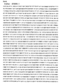
- the base sequence and amino acid sequence of human CD44 are shown.
- the underlined part is the part used for the production of the CD44 / Notch / HIF-3 ⁇ 4 fusion gene.
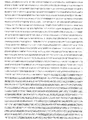
- the base sequence of Notch is shown.
- the base sequence of Notch (continued) is shown.
- the underlined part is the part used for the production of the CD44 / Notch / HIF-3 ⁇ 4 fusion gene.
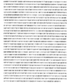
- the base sequence of Notch (continued) is shown.
- the amino acid sequence of Notch is shown.
- the underlined part is the part used for the production of the CD44 / Notch / HIF-3 ⁇ 4 fusion gene.
- the base sequence and amino acid sequence of human HIF-3 ⁇ 4 are shown.
- the underlined part is the part used for the production of the CD44 / Notch / HIF-3 ⁇ 4 fusion gene.
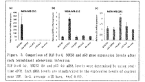
- the results of infecting cells in vitro with each recombinant adenovirus vector and evaluating whether each gene is normally introduced by a real-time PCR test are shown.
- Infect cells in vitro with each recombinant adenovirus vector containing ADX730 (genetically modified adenovirus vector incorporating a CD44 / Notch / HIF-3 ⁇ 4 fusion gene), and decoy of the CD44 Decoy receptor of ADX730.
- the results of a comparative study by a real-time PCR test are shown to see if the expression of the CD44 downstream genes Survivin and CCL2 genes is suppressed by function.
- Cells are infected in vitro with each recombinant adenovirus vector containing ADX730, and the HIF-1 ⁇ target genes VEGF, CyclinG2, and Bcl-xL genes are affected by the HIF-1 ⁇ function-suppressing effect of HIF-3 ⁇ 4 of ADX730.
- the results of a comparative study by a Real-time PCR test are shown to see if the expression is suppressed.
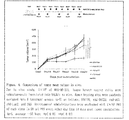
- each recombinant adenovirus vector containing ADX730 to nude mice transplanted with MDA-MB-231 human triple-negative breast cancer cells and examination of its tumor growth inhibitory effect are shown.
- a schematic diagram of the administration schedule is shown on the upper side.
- the lower side is a graph showing how the tumor volume was changed by the administration of each recombinant adenovirus vector or PBS (phosphate buffered saline).
- the present disclosure preferably includes, for example, a specific artificial nucleic acid, a viral vector incorporating the artificial nucleic acid, an anticancer composition containing the viral vector, a method for treating cancer using the composition, and the like. Not limited to these, the present disclosure includes everything disclosed herein and recognizable to those skilled in the art.
- the artificial nucleic acids included in the present disclosure include (A) a nucleic acid encoding a protein having a CD44 extracellular function, (B) a nucleic acid encoding a protein having a Notch core region function, and (C) a nucleic acid encoding a HIF-3 ⁇ 4 function. It is preferable that the nucleic acid encoding the protein is a nucleic acid having a structure in which (A)-(B)-(C) are linked in this order.
- the nucleic acid may be referred to as "nucleic acid of the present disclosure".
- the nucleic acid may be DNA, RNA, PNA or the like, but DNA is particularly preferable.
- the (A) side may be the 3'end or the 5'end, but the 5'end is preferable.
- CD44 is one of the receptors for hyaluronic acid and the like, and by binding to a ligand (for example, hyaluronic acid), it clusters and transmits a signal. It is known that this causes intracellular phenomena such as activation of kinases involved in various cell proliferation and cell migration such as c-Src, FAK, and MAPK. It is also known that after the signal transduction, the intracellular domain is transferred to the nucleus by being cleaved by a protease, and the isolated extracellular domain is released as soluble CD44. Due to these properties, CD44 is highly expressed in many types of cancer cells such as colon cancer, breast cancer, gastric cancer, pancreatic cancer, and prostate cancer, and is also being studied as a marker for cancer stem cells.
- a ligand for example, hyaluronic acid
- the extracellular function of CD44 is a receptor function for a ligand, and the function preferably includes, for example, hyaluronic acid binding ability.
- the protein having the extracellular function of CD44 may be, for example, the protein of the entire extracellular portion of CD44, or may include the cell membrane portion of CD44 as long as the receptor function for the ligand is not impaired. It may be a protein of a part of the extracellular component of CD44. Furthermore, one or more amino acids may be deleted, substituted, or added in such proteins as long as they have this function.
- the nucleic acid (A) is not particularly limited as long as it is a nucleic acid encoding such a polypeptide.
- examples of the preferred nucleic acid as the nucleic acid (A) include the following (a-1) or (a-2).
- A-1) Nucleic acid consisting of the nucleotide sequence of SEQ ID NO: 1 (a-2): In the nucleotide sequence of (a-1), 1 or 2 or more (for example, 1 to 30, 1 to 20, 1 to 10, Or a nucleic acid that comprises a base sequence in which 1, 2, 3, 4, or 5) bases are deleted, substituted, or added, and encodes a protein that can bind to hyaluronic acid.
- Whether the protein can bind to hyaluronic acid can be determined, for example, by obtaining these dissociation constants.
- Notch is a receptor expressed on the surface of cells
- the Notch signaling system is one of the main signaling systems responsible for intercellular signaling.
- the mechanism of intercellular signal transduction is that the signal-sending cell produces and releases a soluble ligand, which binds to a receptor on the cell surface of the signal-receiving cell. ..
- gene expression is regulated by activating intracellular signal transduction pathways such as the downstream phosphorylation cascade and changing the activity of specific transcription factors.
- the Notch signaling system is characterized in that signal transduction is performed by direct interaction between adjacent cells.
- Notch which acts as a receptor, binds to the ligand Delta or Serrate (Jagged in mammals) on the cell surface, and structural changes occur when physical force is applied, resulting in protein-cleaving enzymes such as ADAM protease or ⁇ -secretase.
- ADAM protease or ⁇ -secretase.
- the Notch core region is a region containing a site to be cleaved by the action of a protease, and the Notch core region function can be cleaved by a protease capable of cleaving the Notch core region (preferably limited separation). It is a function called.
- the protein having the Notch core region function may be, for example, a protein containing only a portion necessary for protease cleavage in the Notch core region, and is necessary for protease cleavage as long as it is cleaved by the action of protease.
- a protein containing one or more amino acids before and after a portion (for example, 1 to 30, 1 to 20, 1 to 10, or 1, 2, 3, 4, or 5) together with a portion required for protease cleavage. May be good.
- one or more eg, 1-30, 1-20, 1-10, or 1,2,3,4, or 5
- Amino acids may be deleted, substituted, or added.
- the nucleic acid (B) is not particularly limited as long as it is a nucleic acid encoding such a protein.
- examples of the preferred nucleic acid as the nucleic acid (B) include the following (b-1) or (b-2).
- B-1) Nucleic acid consisting of the base sequence of SEQ ID NO: 2
- b-2) From the base sequence in which one or more bases are deleted, substituted, or added in the base sequence of (b-1).
- ADAM protease As the protease, as described above, ADAM protease (ADAM protease) and ⁇ -secretase ( ⁇ -secretase) are preferably mentioned, and a polypeptide that is cleaved by either or both of these proteases can be encoded. More preferred. ADAM protease is a proteolytic enzyme belonging to a group called A Disintegrin And Metalloprotease family.
- protease Whether the protein can be cleaved by protease can be confirmed by treating the protein with protease and then electrophoresis (for example, SDS-PAGE).
- (b-2) is composed of a base sequence in which one or more bases are deleted, substituted, or added in the base sequence of (b-2'): (b-1), and is described above.
- a protein encoded by a nucleic acid having a structure linked in the order of (A)-(B)-(C) it encodes a protein that can be cleaved by an intracellular protease when a ligand binds to the protein encoded by the nucleic acid (A). More preferably, it is a nucleic acid.
- HIF Hydrofluid Inducible Factor
- hypoxia-inducing factor is a transcription factor that is activated when the inside of a cell falls into a hypoxic state, and is a heterodimer composed of HIF-1 ⁇ and HIF-1 ⁇ . It has been clarified that HIF-1 ⁇ is suppressed not only by decomposition by PHD under normal oxygen concentration but also by a transcription factor called IPAS (Inhibitory PAS domain protein) found in mice. IPAS was identified as a splicing variant of HIF-3 ⁇ , one of the HIFs. Although IPAS does not exhibit transcriptional activity by itself, it inhibits binding to DNA by interacting with HIF-1 ⁇ and suppresses the function of HIF-1 ⁇ . In humans, HIF-3 ⁇ 4, identified as a splicing variant of HIF-3 ⁇ , has been shown to perform a function similar to IPAS.
- the HIF-3 ⁇ 4 function is a function capable of suppressing HIF-1 ⁇ , and more specifically, a function of interacting (binding) with HIF-1 ⁇ .
- the protein having the HIF-3 ⁇ 4 function may be, for example, HIF-3 ⁇ 4 itself, or 1 or 2 or more (for example, 1 to 30, 1) in HIF-3 ⁇ 4 as long as HIF-1 ⁇ can be suppressed. It may be a protein to which amino acids of ⁇ 20, 1-10, or 1, 2, 3, 4, or 5) are further added. Furthermore, in such proteins, as long as HIF-1 ⁇ can be suppressed, one or two or more (for example, 1 to 30, 1 to 20, 1 to 10, or 1, 2, 3, 4, or 5) Amino acids may be deleted, substituted, or added.
- the nucleic acid (C) is not particularly limited as long as it is a nucleic acid encoding such a protein.
- examples of the preferred nucleic acid as the nucleic acid (C) include the following (c-1) or (c-2).
- C-1) Nucleic acid consisting of the base sequence of SEQ ID NO: 3
- c-2 From the base sequence in which one or more bases are deleted, substituted, or added in the base sequence of (c-1).
- Whether or not the protein can bind to HIF-1 ⁇ can be examined by a co-immunoprecipitation method or the like using the target protein, HIF-1 ⁇ , and an antibody that recognizes these.
- nucleic acid (A), nucleic acid (B), and nucleic acid (C) these nucleic acids may be directly linked, or each nucleic acid may be linked via a linker.
- the linker is not particularly limited as long as the effect of the nucleic acid of the present disclosure is not impaired, but is preferably a nucleic acid consisting of, for example, one or two or more bases.
- the base length of the nucleic acids of the present disclosure is preferably, for example, 8000 bp or less, and 7500, 7000, 6500, 6000, 5500, More preferably, it is 5000, 4500, 4000, 3500, or 3000 bp or less.
- nucleic acid of the present disclosure a nucleic acid consisting of the base sequence of (d-1) SEQ ID NO: 4 can be mentioned.
- the base sequence of SEQ ID NO: 4 comprises a base sequence in which one or more bases are deleted, substituted, or added, and Nucleic acids in which a portion encoding a protein that can bind to hyaluronic acid, a portion that encodes a protein that can be cleaved by a protease, and a portion that encodes a protein that can bind to HIF-1 ⁇ are linked in this order can also be mentioned. ..
- nucleic acid comprising a base sequence in which one or more bases are deleted, substituted, or added, and which encodes a protein having an anticancer activity is also available. Can be mentioned. Whether or not the protein encoded by the nucleic acid has an anticancer effect can be examined by incorporating the nucleic acid into an adenovirus vector so that it can be expressed and administering it to cancer cells.
- the number of bases, or the number of bases constituting the linker is preferably 1 to 100 (1, 2, 3, 4, 5, 6, 7, 8, 9, 10, 11, 12, 13, 14, etc.).
- the type of base in the present disclosure is not particularly limited, but is preferably A (adenitin), T (thymine), G (guanine), C (cytosine), or U (uracil).
- the nucleic acid of the present disclosure can be produced by a known method or a method that can be easily conceived from a known method.
- it can be produced by a genetic engineering method.
- DNA or RNA encoding CD44, Notch, and HIF-3 ⁇ 4 may be extracted from a human-derived sample, artificially mutated as necessary, and then ligated to produce the product.
- it may be produced by chemical synthesis.
- Fusion proteins encoded by the nucleic acids of the present disclosure include (i) a protein moiety having CD44 extracellular function, (ii) a protein moiety having Notch core region function, and (iii) a protein moiety having HIF-3 ⁇ 4 function. Therefore, (i) can function as a Decoy receptor, and a signal can be transmitted to (iii) via (ii). This becomes more effective when (i) is fused upstream of (ii).
- HIF-1 ⁇ activated in the tumor can be suppressed. It is considered that this makes it possible to suppress the gene to be inserted into the viral vector within an acceptable range and to exert an antitumor effect composed of a plurality of mechanisms of action.
- the vector in which the nucleic acid of the present disclosure is incorporated in an expressible manner is useful for treating cancer or also for increasing the production of the nucleic acid of the present disclosure.
- the vector include a plasmid vector, a cosmid vector, a phosmid vector, a virus vector and the like.
- viral vectors are particularly suitable.
- the viral vector include an adenoviral vector, a retroviral vector, a lentiviral vector, a Sendai viral vector and the like. Of these, the adenovirus vector is preferable.
- both a proliferative viral vector and a non-proliferative viral vector can be used. In particular, proliferative or non-proliferative adenovirus vectors are preferred.
- the nucleic acid of the present disclosure can be incorporated into a vector by a known method or a method that can be easily conceived from a known method.
- the anti-cancer composition containing the vector into which the nucleic acid of the present disclosure is expressively incorporated exhibits a very excellent anti-cancer effect (particularly a cancer therapeutic effect).
- the administration form of the anticancer composition is not particularly limited as long as the anticancer effect is exhibited.
- intratumoral administration is generally preferred, but intravenous administration may be used depending on the vector.
- the dosage form of the anticancer composition is not particularly limited as long as the anticancer effect is exhibited. For example, it is preferably an injection.
- the anti-cancer composition can appropriately contain components other than the above vector as needed.
- examples of such other components include a pharmaceutically acceptable carrier (for example, water), and an appropriate carrier can be selected and used according to the treatment site and the dosage form of the anticancer composition. ..
- Such an anti-cancer composition can also be prepared based on a known method.
- the cancer type to be treated with the anticancer composition is not particularly limited as long as the anticancer effect is exhibited.
- examples thereof include solid tumors and hematological malignancies, and more specifically, but not limited to, brain tumors, maxillary cancers, nasopharyngeal cancers, lung cancers, esophageal cancers, rectal cancers, colon cancers, liver cancers, gastric cancers, bile sac cancers.
- Pancreatic cancer skin cancer, breast cancer, uterine cancer, ovarian cancer, prostate cancer, kidney cancer, bladder cancer, thyroid cancer, multiple myeloma, lymphoma, acute myeloid leukemia, chronic myeloid leukemia, etc.
- the anti-cancer effect of the anti-cancer composition is very high, it is preferable because it can be used for cancer types for which existing cancer therapeutic agents are ineffective or small. For example, it can be preferably used for triple negative (estrogen receptor negative, progesterone receptor negative, HER2-negative) breast cancer and the like. Further, the type of anticancer effect of the anticancer composition is not particularly limited. For example, those capable of suppressing tumor growth and / or infiltration are preferable.
- CD44 / Notch / HIF-3 ⁇ 4 fusion gene was outsourced to GENEWIZ Solid science. Superior service. From each of these three types of human genes, restriction enzyme treatment with SwaI (TaKaRa, Siga, Japan), electrophoresis and purification were used as insert DNA.
- Each domain of the fusion gene is described in publicly known documents (particularly the above non-patent documents 9, 10 and 12) and the transmembrane domain search tool TMHMM (http://www.cbs.dtu.dk/services/TMHMM/). It was designed with reference to the signal peptide sequence search tool SignalP (http://www.cbs.dtu.dk/services/SignalP/).
- the sequence of the prepared CD44 / Notch / HIF-3 ⁇ 4 fusion gene is shown in FIG.
- the nucleotide sequences of the three human genes used are shown in FIGS. 2a to 2c, respectively.
- the pAxCAwtit2 cosmid vector attached to the Adenovirus Dual Expression Kit (TaKaRa, Siga, Japan) was used as the vector DNA for incorporating the insert DNA. Similar to the insert DNA, the cosmid vector was also purified by phenol-chloroform extraction after cleaving the SwaI sequence at the cloning site by restriction enzyme treatment.
- coli HST08 Premium Competent Cells were seeded on 100 ⁇ g / ml ampicillin-added LB agar medium (Nacalai Tesque, Kyoto, Japan) and cultured overnight at 37 ° C. After culturing, insert check PCR was performed using KOD FX Neo (TOYOBO CO., LTD., Osaka, Japan) using the colonies grown on the LB agar medium as a template to check whether the transformation was normal or not. Confirmed by gel electrophoresis.
- Lipofectamine LTX (invitrogen) using 10 ⁇ g of BspT104I digested cosmid was applied to HEK293 cells (National Institutes of Biomedical Innovation, Health and Nutrition) cultured in 60 mm cell culture Petri dish (TPP) to become confluent. , Waltham, MA) performed lipofection. Then, the cultured cells were collected and seeded on Biocoat Collagen I Cellware 96-Well Plate (CORNING, NY, USA).
- D-MEM Dulbecco's modified Eagle's medium
- FBS Fetal bovine serum
- ADX730 The adenovirus vector in which the CD44 / Notch / HIF-3 ⁇ 4 fusion gene is integrated so that it can be expressed is referred to as ADX730.
- HEK293 cells were cultured in Collagen Type I-Coated 25 cm 2 Flask (IWAKI) until they became 70 to 100% confluent, and 0.5 ml of 5% FBS-D-MEM and 15 ⁇ l of the secondary virus solution prepared above were added. Gently added to infect the virus. For infection, the plate was shaken several times in an incubator (37 ° C, 5% CO 2 ) and slowly shaken four times every 15 minutes. After 1 hour of infection, an additional 4.5 ml of 5% FBS-D-MEM was added to each well and cultured for 3 days. After culturing, it was confirmed that all the cells were denatured, and the cells were collected together with the culture medium.
- IWAKI Collagen Type I-Coated 25 cm 2 Flask
- HEK293 cells were cultured in Collagen Type I-Coated 75 cm 2 Flask (IWAKI) until 70-100% confluent, and 2 ml of 5% FBS-D-MEM and 50 ⁇ l of the tertiary virus solution prepared above were gently added. And infected the virus.
- the plate was shaken several times in an incubator (37 ° C, 5% CO 2 ) and slowly shaken four times every 15 minutes. After 1 hour of infection, another 13 ml of 5% FBS-D-MEM was added to each well and cultured for 3 days. After culturing, it was confirmed that all the cells were denatured, and the cells were collected together with the culture medium. The collected cells were frozen in liquid nitrogen and thawed in a warm bath at 37 ° C 6 times in the same manner as the primary virus solution. After the final freeze-thaw, centrifugation (3,000 rpm, 10 minutes, 4 ° C) was performed, and the collected supernatant was stored as a quaternary virus solution. All of these processes were performed according to the manual of the Adenovirus Dual Expression Kit (TaKaRa, Siga, Japan).
- the prepared cell pack was sufficiently stirred by vortex, 4 ⁇ l of 10% SDS was added, and the mixture was further stirred by vortex. After incubating at 50 ° C. for 1 hour, phenol-chloroform extraction and chloroform extraction were performed twice, and ethanol precipitation was performed. After ethanol precipitation, it was dissolved in 50 ⁇ l of TE Buffer containing RNase A, and 15 ⁇ l of it was used for restriction enzyme treatment with restriction enzyme XhoI. After the restriction enzyme treatment, the migration pattern of the obtained product was confirmed by agarose gel electrophoresis. All of these processes were performed according to the manual of Adenovirus Dual Expression Kit (TaKaRa, Siga, Japan).
- Mass culture and purification of recombinant adenovirus vector In order to use the recombinant adenovirus vector prepared above for subsequent experiments, mass culture and purification were performed.
- HEK293 cells were seeded to 70-100 % confluence in 5 Corning225cm 2 Flasks (CORNING, NY, USA).
- the virus was infected by gently adding 15 ml of 5% FBS-D-MEM and 150 ⁇ l of the tertiary virus solution prepared above. For infection, the plate was shaken several times in an incubator (37 ° C, 5% CO 2 ) and slowly shaken four times every 15 minutes.
- the filter was removed, the filter was attached to 5 ml syringe (TaKaRa, Siga, Japan) containing 3 ml of 1 ⁇ Elution Buffer (TaKaRa, Siga, Japan), and 1 ml of 1 ⁇ Elution Buffer was extruded and collected from the filter. After recovery, the filter was incubated once (room temperature, 5 minutes), and then the remaining 2 ml of 1 ⁇ Solution Buffer was extruded to elute the virus. All of these processes were performed according to the manual of the Adeno-X Maxi Purification Kit (TaKaRa, Siga, Japan).
- HEK293 cells were seeded into 12-Well Cell Culture Plate Flat Bottom (CORNING, NY, USA), and virus solution diluted 10-fold from 100-fold dilution to 10-fold dilution was added dropwise to each well for 2 days. It was cultured. After culturing, all the medium was removed, the cells were slightly dried, and then 1 ml of chilled methanol (FUJIFILM Wako Pure Chemical Corporation, Osaka, Japan) was added dropwise and incubated (-20 ° C, 10 minutes).
- HEK293 cells used in cell and medium experiments were D-MEM containing 10% FBS and 1% 100U / ml penicillin and 100mg / ml streptomycin (P / S; Nacalai Tesque, Kyoto, Japan), and A549 cells were 10% FBS.
- the cells were cultured using Ham's F-12K (FUJIFILM Wako Pure Chemical Corporation, Osaka, Japan) containing 1% P / S at 37 ° C. under 5% CO 2 conditions.
- MDA-MB-231 cells (The European Collection of Cell Cultures) were used at 37 ° C using Leibovitz's L-15 Medium (FUJIFILM Wako Pure Chemical Corporation, Osaka, Japan) containing 15% FBS and 1% P / S. Culturing was performed under conditions without CO 2 equilibration.
- ADX730 used in various recombinant virus experiments, recombinant adenovirus vector (rAd-SOCS3) incorporating SOCS3 gene, recombinant adenovirus vector (rAd-p53) incorporating p53 gene, and LacZ gene.
- the engineered recombinant adenovirus vector (rAd-LacZ) is mass-cultured and purified, and then Glycerol (Nacalai Tesque) is used using Slide-A-Lyzer Dialysis Cassette (Extra Strength) (Thermo Fisher Scientific, Waltham, MA).
- the buffer was exchanged with a dialysis buffer containing 10% and 1 mol / l-Tris-HCl Buffer Solution (Nacalai Tesque, Kyoto, Japan) 1%, and appropriate amounts of each were dispensed and stored at -80 ° C.
- rAd-SOCS3 and rAd-p53 are adenovirus vectors that are known candidates for oncogene therapy.
- Flow cytometry The cells were actually infected with ADX730 in vitro, and the comparison of the increase / decrease in the expression level of the CD44 region of the fusion gene incorporated into AD730 was evaluated using Flow cytemetry.
- MDA-MB-231 cells were seeded at 1 ⁇ 10 6 cells / well against 6-Well Cell Culture Plate Flat Bottom (CORNING, NY, USA) overnight at 37 ° C. without CO 2 equilibrium. It was cultured. After culturing, ADX730, rAd-SOCS3, rAd-p53, and rAd-LacZ were each infected with 40 multiplicity of infection (MOI) and cultured for another 48 hours.
- MOI multiplicity of infection
- the cells were washed with PBS and collected, and blocked using Blocking One Histo (Nacalai Tesque, Kyoto, Japan) at room temperature for 10 minutes. After blocking, wash the cells again with PBS and dilute 200-fold FITC anti-mouse / human CD44 Clone: IM7 (BioLegend, San Diego, CA) or 100-fold diluted FITC Rat IgG2a, ⁇ Isotype Ctrl Clone: RTK2758 (BioLegend, San) Diego, CA) was added dropwise, and the mixture was allowed to react for 30 minutes under shading on ice.
- IM7 BioLegend, San Diego, CA
- RTK2758 BioLegend, San) Diego, CA
- the cells were washed again with PBS, 100-fold diluted BD Pharmingen 7-AAD (BD Biosciences, San Diego, CA) was added dropwise, and the cells were reacted under shading on ice for 5 minutes. After the reaction, the cells were washed with PBS and measured using Guava easy Cyte (Merck Millipore, Burlington, MA). Data analysis was performed according to the attached software InCyte.
- the real-time PCR method was used to confirm the gene transfer by each recombinant adenovirus vector containing ADX730 and the effect obtained by its function.
- MDA-MB-231 cells were seeded at 1 ⁇ 10 6 cells / well against 6-Well Cell Culture Plate Flat Bottom, and Aneropack Kenki 5% (Mitsubishi Gas Chemical Company, Inc., Tokyo, Japan) and Sodium Hyaluronate. The cells were cultured overnight under the conditions of 37 ° C., 5% CO 2 , 1% ⁇ O 2, and 0.04 mg / ml by using (40 kDa to 80 kDa) (PG Research, Tokyo, Japan).
- ADX730, rAd-SOCS3, rAd-p53, and rAd-LacZ were each infected with 40 MOI, and further cultured for 48 hours. The cells were then harvested and Total RNA was extracted using NucleoSpin RNA (TaKaRa, Siga, Japan).
- cDNA was synthesized from the extracted RNA using PrimeScript RT reagent Kit with gDNA Eraser (TaKaRa, Siga, Japan), and the primers prepared using it as a template (Table 3), TB Green Premix Ex Taq II (TaKaRa, Siga) , Japan) and Thermal Cycler Dice Real Time System (TaKaRa, Siga, Japan) were used for PCR reaction and comparative C t method ( ⁇ C t method) analysis.
- mice Collection of mice and specimens In vivo experiments were conducted using mice in order to examine and compare the antitumor effects of ADX730 with other oncogene therapy agents.
- MDA-MB-231 cells 1 ⁇ 10 6 cells / 70 ⁇ l and Matrigel Martrix Basement Membrane HC (CORNING, NY, USA) for 6-week-old female BALB / cAJcl-nu / nu (CLEA Japan, Inc., Tokyo, Japan) )
- ADX730 (1 ⁇ 10 9 PFU / 50 ⁇ l), rAd-SOCS3 (1 ⁇ 10 9 PFU / 50 ⁇ l), rAd-p53 (1 ⁇ 10 9 PFU / 50 ⁇ l), rAd -LacZ (1 ⁇ 10 9 PFU / 50 ⁇ l) or PBS 50 ⁇ l was administered intratumorally every other day for a total of eight times (Days 14, 16, 18, 20, 22, 24, 26, 28). Tumor diameter measurement was performed twice a week from the start of virus administration, a total of 5 times.
- the tumor was collected and fixed at 4 ° C with 4% Paraformaldehyde Phosphate Buffer Solution (FUJIFILM Wako Pure Chemical Corporation, Osaka, Japan). Alternatively, it was stored at -80 ° C.
- the volume of the tumor was calculated by measuring the major axis (L) and the minor axis (W) and using the formula (W 2 ⁇ L) / 2.
- Each recombinant adenovirus vector containing ADX730 was infected with cells in vitro, and the normal introduction of each gene was evaluated by a real-time PCR test.
- the TBP (TATA-Box binding protein) gene was used as the endogenous control gene (forward primer used for measuring the expression of the TBP gene: 5'-GCCAGCTTCGGAGAGTTCTGGGATT-3', reverse primer: 5'-CGGGCACGAAGTGCAATGGTCTTTA-3'. ).
- the relative expression ratio with the control gene is shown in FIG. 3 as a result.
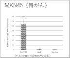
- ADX730 shows a high cancer therapeutic effect even if it is a cancer that is difficult to treat with existing therapeutic agents such as triple-negative breast cancer. Furthermore, in the above-mentioned Real-time PCR study, MDA-MB-231 cells derived from breast cancer were used, and the cells used were DU-145 cells derived from prostate cancer, MKN45 cells derived from gastric cancer, or The cells were changed to PANC-1 cells derived from pancreatic cancer, and the gene transfer by each recombinant adenovirus vector containing ADX730 was confirmed, and the effect obtained by the function was confirmed by the real-time PCR method.
- the number of seeded cells was 5 ⁇ 10 5 cells / well, and the introduction of the CD44 / Notch / HIF-3 ⁇ 4 fusion gene by ADX730 was confirmed and compared using a primer set targeting HIF-3 ⁇ 4 (Table 3). went.
- the results are shown in FIGS. 8a (DU-145 cells), 8b (MKN45 cells), and 8c (PANC-1 cells). It was confirmed that the expression of the CD44 / Notch / HIF-3 ⁇ 4 fusion gene was significantly increased regardless of which cell was used.
Abstract
Provided is a novel oncogene therapeutic means. More specifically, provided is a nucleic acid having a structure in which a nucleic acid (A) encoding a protein having a CD44 extracellular function, a nucleic acid (B) encoding a protein having a Notch core region function, and a nucleic acid (C) encoding a protein having an HIF-3α4 function are linked in the order of (A)-(B)-(C).
Description
本開示は、例えば、新規な癌遺伝子治療薬若しくは方法、及び、その有効成分等に関する。本明細書に記載される全ての文献(以下に先行技術文献として挙げられる非特許文献1~12を含む)の内容は参照により本明細書に組み込まれる。
The present disclosure relates to, for example, a novel oncogene therapeutic agent or method, an active ingredient thereof, and the like. The contents of all documents described herein (including Non-Patent Documents 1-12, which are listed below as prior art documents) are incorporated herein by reference.
近年、癌に対する新しい治療法の一つとしてウイルスベクターを用いた遺伝子治療法が注目されている。これは、各種ウイルスの特性を活かして、ウイルスを遺伝子の “運び屋”、ウイルスベクターとして利用するものである。その遺伝子導入の方法の一つとしてIn Vivo遺伝子治療があり、治療遺伝子を組み込んだ組換えウイルスベクターを体内に直接投与して疾患の治療を行うことができる。癌遺伝子治療は化学療法などの既存の治療法とは作用機序が異なり、遺伝子を癌細胞に導入して癌の発生や増殖に関わる遺伝子を直接抑制したり、癌抑制遺伝子を細胞に導入して直接的に細胞死を誘導したりする。従って既存の化学療法や放射線療法で治療が困難な癌に対する有効な治療法になりうると期待されている。
In recent years, gene therapy using a viral vector has been attracting attention as one of the new treatments for cancer. This utilizes the characteristics of various viruses and uses the virus as a "carrier" of genes and a viral vector. In Vivo gene therapy is one of the gene transfer methods, and a recombinant viral vector incorporating a therapeutic gene can be directly administered into the body to treat a disease. Oncogene therapy has a different mechanism of action from existing treatments such as chemotherapy, in which genes are introduced into cancer cells to directly suppress genes involved in cancer development and growth, or tumor suppressor genes are introduced into cells. Directly induce cell death. Therefore, it is expected to be an effective treatment method for cancers that are difficult to treat with existing chemotherapy and radiation therapy.
本発明者らは、新規な癌遺伝子治療薬若しくは方法を提供することを主な目的とし、検討を行った。
The present inventors conducted a study with the main purpose of providing a novel oncogene therapeutic agent or method.
外部から導入する遺伝子を癌細胞内で発現させるために重要となるのが遺伝子を運ぶウイルスベクターである。ウイルスベクターには、例えばレトロウイルスやレンチウイルス、センダイウイルスなどがある。
A viral vector that carries a gene is important for expressing a gene introduced from the outside in cancer cells. Viral vectors include, for example, retroviruses, lentiviruses, and Sendai viruses.
しかし、ウイルスベクターに挿入できる遺伝子の容量は限られているため、現状では許容量の範囲内で遺伝子を挿入してその効果を得るというようなデザインが必要である。そして、得られる癌の抑制効果には、その挿入した遺伝子の保有する機能に限るという制約があることから、ウイルスベクターに挿入する遺伝子の容量を、許容の範囲内で抑え、且つその効果をより効率よく得る方法を見出す必要がある。すなわち、限られた容量の中で、以下に効率よく癌抑制効果を奏する遺伝子を創出するかが、ウイルスベクターを用いた癌治療薬の効果を高めるための重要なポイントになる。
However, since the capacity of genes that can be inserted into viral vectors is limited, it is currently necessary to design such that genes are inserted within the permissible amount to obtain the effect. Since the obtained cancer-suppressing effect is limited to the function possessed by the inserted gene, the capacity of the gene to be inserted into the viral vector can be suppressed within an acceptable range, and the effect can be further enhanced. We need to find a way to get it efficiently. That is, it is an important point for enhancing the effect of a cancer therapeutic drug using a viral vector to create a gene that efficiently exerts a tumor suppressor effect in a limited volume.
このため、様々な遺伝子の改変や組み合わせを検討し、癌抑制効果の特に高い人工的な遺伝子の作出を試みた。その結果、CD44(特に細胞外部分)、Notch(特にコア領域)、及びHIF-3α4を融合させた人工遺伝子が、非常に高い癌抑制効果を奏することを見いだした。本発明者らは、当該知見に基づき、さらに改良を重ねた。
Therefore, we examined the modification and combination of various genes and tried to create an artificial gene with a particularly high tumor suppressor effect. As a result, it was found that an artificial gene fused with CD44 (particularly extracellular part), Notch (particularly core region), and HIF-3α4 exerts a very high tumor suppressor effect. Based on this finding, the present inventors have made further improvements.
本開示は例えば以下の項に記載の主題を包含する。
項1.
(A)CD44細胞外機能を有するタンパク質をコードする核酸
(B)Notchコア領域機能を有するタンパク質をコードする核酸
(C)HIF-3α4機能を有するタンパク質をコードする核酸
が(A)-(B)-(C)の順に連結された構造を有する核酸。
項2.
核酸(A)、核酸(B)、及び核酸(C)が、(A)-(B)-(C)の順に連結された構造を有する核酸であって、
核酸(A)が、
(a-1):配列番号1の塩基配列からなる核酸、あるいは
(a-2):(a-1)の塩基配列において、1又は2以上の塩基が欠失、置換、又は付加された塩基配列からなり、且つ、ヒアルロン酸と結合可能なタンパク質をコードする核酸
であり、
核酸(B)が、
(b-1):配列番号2の塩基配列からなる核酸、あるいは
(b-2):(b-1)の塩基配列において、1又は2以上の塩基が欠失、置換、又は付加された塩基配列からなり、且つ、プロテアーゼにより切断され得るタンパク質をコードする核酸
であり、
核酸(C)が、
(c-1):配列番号3の塩基配列からなる核酸、あるいは
(c-2):(c-1)の塩基配列において、1又は2以上の塩基が欠失、置換、又は付加された塩基配列からなり、且つ、HIF-1αと結合可能なタンパク質をコードする核酸である、
核酸。
項3.
核酸(A)が、
(a-1):配列番号1の塩基配列からなる核酸、あるいは
(a-2):(a-1)の塩基配列において、1又は2以上の塩基が欠失、置換、又は付加された塩基配列からなり、且つ、ヒアルロン酸と結合可能なタンパク質をコードする核酸
であり、
核酸(B)が、
(b-1):配列番号2の塩基配列からなる核酸、あるいは
(b-2):(b-1)の塩基配列において、1又は2以上の塩基が欠失、置換、又は付加された塩基配列からなり、且つ、前記(A)-(B)-(C)の順に連結された構造を有する核酸がコードするタンパク質において、プロテアーゼにより切断され得るタンパク質をコードする核酸
であり、
核酸(C)が、
(c-1):配列番号3の塩基配列からなる核酸、あるいは
(c-2):(c-1)の塩基配列において、1又は2以上の塩基が欠失、置換、又は付加された塩基配列からなり、且つ、HIF-1αと結合可能なタンパク質をコードする核酸
である、
項1に記載の核酸。
項4.
(d-1)配列番号4の塩基配列からなるか、
(d-2)配列番号4の塩基配列において、1又は2以上の塩基が欠失、置換、又は付加された塩基配列からなり、且つ、ヒアルロン酸と結合可能なタンパク質をコードする部分、プロテアーゼにより切断され得るタンパク質をコードする部分、及びHIF-1αと結合可能なタンパク質をコードする部分が、この順に連結された構造を有するか、あるいは、
(d-3)配列番号4の塩基配列において、1又は2以上の塩基が欠失、置換、又は付加された塩基配列からなり、且つ、抗癌作用を有するタンパク質をコードする、
核酸。
項5.
前記プロテアーゼが、ADAMプロテアーゼ又はγ-セクレターゼである、項1~4のいずれかに記載の核酸。
項6.
項1~5のいずれかに記載の核酸がコードするタンパク質。
項7.
項1~5のいずれかに記載の核酸が発現可能に組み込まれたベクター。
項8.
ベクターがアデノウイルスベクターである、項7に記載のベクター。
項9.
項1~5のいずれかに記載の核酸あるいは項7又は8に記載のベクターを含む、抗癌組成物。
項10.
注射剤である、項9に記載の抗癌組成物。
項11.
乳癌、前立腺癌、胃癌、又は膵臓癌治療用である、項9又は10に記載の抗癌組成物。
項12.
トリプルネガティブ乳癌治療用である、項9又は10に記載の抗癌組成物。 The present disclosure includes, for example, the subjects described in the following sections.
Item 1.
(A) Nucleic acid encoding a protein having a CD44 extracellular function (B) Nucleic acid encoding a protein having a Notch core region function (C) Nucleic acid encoding a protein having a HIF-3α4 function (A)-(B) -Nucleic acid having a structure linked in the order of (C).
Item 2.
Nucleic acid (A), nucleic acid (B), and nucleic acid (C) are nucleic acids having a structure in which (A)-(B)-(C) are linked in this order.
Nucleic acid (A)
(A-1): Nucleic acid consisting of the base sequence of SEQ ID NO: 1, or (a-2): Base in which one or more bases are deleted, substituted, or added in the base sequence of (a-1). A nucleic acid that consists of a sequence and encodes a protein that can bind to hyaluronic acid.
Nucleic acid (B)
(B-1): Nucleic acid consisting of the base sequence of SEQ ID NO: 2, or (b-2): Base in which one or more bases are deleted, substituted, or added in the base sequence of (b-1). A nucleic acid consisting of a sequence and encoding a protein that can be cleaved by a protease.
Nucleic acid (C)
(C-1): Nucleic acid consisting of the base sequence of SEQ ID NO: 3, or (c-2): Base in which one or more bases are deleted, substituted, or added in the base sequence of (c-1). A nucleic acid consisting of a sequence and encoding a protein capable of binding to HIF-1α.
Nucleic acid.
Item 3.
Nucleic acid (A)
(A-1): Nucleic acid consisting of the base sequence of SEQ ID NO: 1, or (a-2): Base in which one or more bases are deleted, substituted, or added in the base sequence of (a-1). A nucleic acid that consists of a sequence and encodes a protein that can bind to hyaluronic acid.
Nucleic acid (B)
(B-1): Nucleic acid consisting of the base sequence of SEQ ID NO: 2, or (b-2): Base in which one or more bases are deleted, substituted, or added in the base sequence of (b-1). It is a nucleic acid encoding a protein that can be cleaved by a protease among the proteins encoded by the nucleic acids having a sequence and having a structure linked in the order of (A)-(B)-(C).
Nucleic acid (C)
(C-1): Nucleic acid consisting of the base sequence of SEQ ID NO: 3, or (c-2): Base in which one or more bases are deleted, substituted, or added in the base sequence of (c-1). A nucleic acid consisting of a sequence and encoding a protein capable of binding to HIF-1α.
Item 2. The nucleic acid according toItem 1.
Item 4.
(D-1) Does it consist of the nucleotide sequence of SEQ ID NO: 4?
(D-2) In the base sequence of SEQ ID NO: 4, a portion consisting of a base sequence in which one or more bases are deleted, substituted, or added and encoding a protein capable of binding to hyaluronic acid, a protease. The portion encoding the protein that can be cleaved and the portion encoding the protein that can bind to HIF-1α have a structure in which they are linked in this order, or
(D-3) In the base sequence of SEQ ID NO: 4, it comprises a base sequence in which one or more bases are deleted, substituted, or added, and encodes a protein having an anticancer activity.
Nucleic acid.
Item 5.
Item 4. The nucleic acid according to any one of Items 1 to 4, wherein the protease is ADAM protease or γ-secretase.
Item 6.
A protein encoded by the nucleic acid according to any one ofItems 1 to 5.
Item 7.
A vector in which the nucleic acid according to any one ofItems 1 to 5 is incorporated so as to be expressible.
Item 8.
Item 7. The vector according to Item 7, wherein the vector is an adenovirus vector.
Item 9.
An anti-cancer composition comprising the nucleic acid according to any one ofItems 1 to 5 or the vector according to Item 7 or 8.
Item 10.
Item 9. The anti-cancer composition according to Item 9, which is an injection.
Item 11.
Item 9. The anticancer composition according toItem 9 or 10, which is used for treating breast cancer, prostate cancer, gastric cancer, or pancreatic cancer.
Item 12.
Item 8. The anti-cancer composition according toItem 9 or 10, which is used for treating triple-negative breast cancer.
項1.
(A)CD44細胞外機能を有するタンパク質をコードする核酸
(B)Notchコア領域機能を有するタンパク質をコードする核酸
(C)HIF-3α4機能を有するタンパク質をコードする核酸
が(A)-(B)-(C)の順に連結された構造を有する核酸。
項2.
核酸(A)、核酸(B)、及び核酸(C)が、(A)-(B)-(C)の順に連結された構造を有する核酸であって、
核酸(A)が、
(a-1):配列番号1の塩基配列からなる核酸、あるいは
(a-2):(a-1)の塩基配列において、1又は2以上の塩基が欠失、置換、又は付加された塩基配列からなり、且つ、ヒアルロン酸と結合可能なタンパク質をコードする核酸
であり、
核酸(B)が、
(b-1):配列番号2の塩基配列からなる核酸、あるいは
(b-2):(b-1)の塩基配列において、1又は2以上の塩基が欠失、置換、又は付加された塩基配列からなり、且つ、プロテアーゼにより切断され得るタンパク質をコードする核酸
であり、
核酸(C)が、
(c-1):配列番号3の塩基配列からなる核酸、あるいは
(c-2):(c-1)の塩基配列において、1又は2以上の塩基が欠失、置換、又は付加された塩基配列からなり、且つ、HIF-1αと結合可能なタンパク質をコードする核酸である、
核酸。
項3.
核酸(A)が、
(a-1):配列番号1の塩基配列からなる核酸、あるいは
(a-2):(a-1)の塩基配列において、1又は2以上の塩基が欠失、置換、又は付加された塩基配列からなり、且つ、ヒアルロン酸と結合可能なタンパク質をコードする核酸
であり、
核酸(B)が、
(b-1):配列番号2の塩基配列からなる核酸、あるいは
(b-2):(b-1)の塩基配列において、1又は2以上の塩基が欠失、置換、又は付加された塩基配列からなり、且つ、前記(A)-(B)-(C)の順に連結された構造を有する核酸がコードするタンパク質において、プロテアーゼにより切断され得るタンパク質をコードする核酸
であり、
核酸(C)が、
(c-1):配列番号3の塩基配列からなる核酸、あるいは
(c-2):(c-1)の塩基配列において、1又は2以上の塩基が欠失、置換、又は付加された塩基配列からなり、且つ、HIF-1αと結合可能なタンパク質をコードする核酸
である、
項1に記載の核酸。
項4.
(d-1)配列番号4の塩基配列からなるか、
(d-2)配列番号4の塩基配列において、1又は2以上の塩基が欠失、置換、又は付加された塩基配列からなり、且つ、ヒアルロン酸と結合可能なタンパク質をコードする部分、プロテアーゼにより切断され得るタンパク質をコードする部分、及びHIF-1αと結合可能なタンパク質をコードする部分が、この順に連結された構造を有するか、あるいは、
(d-3)配列番号4の塩基配列において、1又は2以上の塩基が欠失、置換、又は付加された塩基配列からなり、且つ、抗癌作用を有するタンパク質をコードする、
核酸。
項5.
前記プロテアーゼが、ADAMプロテアーゼ又はγ-セクレターゼである、項1~4のいずれかに記載の核酸。
項6.
項1~5のいずれかに記載の核酸がコードするタンパク質。
項7.
項1~5のいずれかに記載の核酸が発現可能に組み込まれたベクター。
項8.
ベクターがアデノウイルスベクターである、項7に記載のベクター。
項9.
項1~5のいずれかに記載の核酸あるいは項7又は8に記載のベクターを含む、抗癌組成物。
項10.
注射剤である、項9に記載の抗癌組成物。
項11.
乳癌、前立腺癌、胃癌、又は膵臓癌治療用である、項9又は10に記載の抗癌組成物。
項12.
トリプルネガティブ乳癌治療用である、項9又は10に記載の抗癌組成物。 The present disclosure includes, for example, the subjects described in the following sections.
(A) Nucleic acid encoding a protein having a CD44 extracellular function (B) Nucleic acid encoding a protein having a Notch core region function (C) Nucleic acid encoding a protein having a HIF-3α4 function (A)-(B) -Nucleic acid having a structure linked in the order of (C).
Item 2.
Nucleic acid (A), nucleic acid (B), and nucleic acid (C) are nucleic acids having a structure in which (A)-(B)-(C) are linked in this order.
Nucleic acid (A)
(A-1): Nucleic acid consisting of the base sequence of SEQ ID NO: 1, or (a-2): Base in which one or more bases are deleted, substituted, or added in the base sequence of (a-1). A nucleic acid that consists of a sequence and encodes a protein that can bind to hyaluronic acid.
Nucleic acid (B)
(B-1): Nucleic acid consisting of the base sequence of SEQ ID NO: 2, or (b-2): Base in which one or more bases are deleted, substituted, or added in the base sequence of (b-1). A nucleic acid consisting of a sequence and encoding a protein that can be cleaved by a protease.
Nucleic acid (C)
(C-1): Nucleic acid consisting of the base sequence of SEQ ID NO: 3, or (c-2): Base in which one or more bases are deleted, substituted, or added in the base sequence of (c-1). A nucleic acid consisting of a sequence and encoding a protein capable of binding to HIF-1α.
Nucleic acid.
Nucleic acid (A)
(A-1): Nucleic acid consisting of the base sequence of SEQ ID NO: 1, or (a-2): Base in which one or more bases are deleted, substituted, or added in the base sequence of (a-1). A nucleic acid that consists of a sequence and encodes a protein that can bind to hyaluronic acid.
Nucleic acid (B)
(B-1): Nucleic acid consisting of the base sequence of SEQ ID NO: 2, or (b-2): Base in which one or more bases are deleted, substituted, or added in the base sequence of (b-1). It is a nucleic acid encoding a protein that can be cleaved by a protease among the proteins encoded by the nucleic acids having a sequence and having a structure linked in the order of (A)-(B)-(C).
Nucleic acid (C)
(C-1): Nucleic acid consisting of the base sequence of SEQ ID NO: 3, or (c-2): Base in which one or more bases are deleted, substituted, or added in the base sequence of (c-1). A nucleic acid consisting of a sequence and encoding a protein capable of binding to HIF-1α.
Item 2. The nucleic acid according to
(D-1) Does it consist of the nucleotide sequence of SEQ ID NO: 4?
(D-2) In the base sequence of SEQ ID NO: 4, a portion consisting of a base sequence in which one or more bases are deleted, substituted, or added and encoding a protein capable of binding to hyaluronic acid, a protease. The portion encoding the protein that can be cleaved and the portion encoding the protein that can bind to HIF-1α have a structure in which they are linked in this order, or
(D-3) In the base sequence of SEQ ID NO: 4, it comprises a base sequence in which one or more bases are deleted, substituted, or added, and encodes a protein having an anticancer activity.
Nucleic acid.
Item 6.
A protein encoded by the nucleic acid according to any one of
Item 7.
A vector in which the nucleic acid according to any one of
Item 8.
Item 7. The vector according to Item 7, wherein the vector is an adenovirus vector.
Item 9.
An anti-cancer composition comprising the nucleic acid according to any one of
Item 9. The anti-cancer composition according to Item 9, which is an injection.
Item 11.
Item 9. The anticancer composition according to
Item 8. The anti-cancer composition according to
抗癌効果が非常に高い新規な癌遺伝子治療薬が提供され得る。当該治療薬は、既存の癌治療薬では効果が奏されない又は小さい癌種に対しても効果を奏し得、例えば、トリプルネガティブ(エストロゲン受容体陰性、プロゲステロン受容体陰性、HER2陰性)乳癌等にも好ましく用いることができる。
A novel oncogene therapeutic agent having a very high anticancer effect can be provided. The therapeutic agent may be effective against small cancer types that are not effective with existing cancer therapeutic agents, and may also be effective against, for example, triple-negative (estrogen receptor negative, progesterone receptor negative, HER2-negative) breast cancer and the like. It can be preferably used.
以下、本開示に包含される各実施形態について、さらに詳細に説明する。本開示は、例えば、特定の人工核酸、当該人工核酸を組み込んだウイルスベクター、当該ウイルスベクターを含有する抗癌用組成物、及び当該組成物を用いた癌の治療方法等を好ましく包含するが、これらに限定されるわけではなく、本開示は本明細書に開示され当業者が認識できる全てを包含する。
Hereinafter, each embodiment included in the present disclosure will be described in more detail. The present disclosure preferably includes, for example, a specific artificial nucleic acid, a viral vector incorporating the artificial nucleic acid, an anticancer composition containing the viral vector, a method for treating cancer using the composition, and the like. Not limited to these, the present disclosure includes everything disclosed herein and recognizable to those skilled in the art.
本開示に包含される人工核酸は、(A)CD44細胞外機能を有するタンパク質をコードする核酸、(B)Notchコア領域機能を有するタンパク質をコードする核酸、及び(C)HIF-3α4機能を有するタンパク質をコードする核酸が、(A)-(B)-(C)の順に連結された構造を有する核酸であることが好ましい。当該核酸を、「本開示の核酸」ということがある。なお、本開示において、核酸はDNA、RNA、PNA等であり得るが、特にDNAであることが好ましい。また、(A)-(B)-(C)の順に連結された構造を有する核酸において、(A)側が3’末端でも5’末端でもよいが、5’末端であることが好ましい。
The artificial nucleic acids included in the present disclosure include (A) a nucleic acid encoding a protein having a CD44 extracellular function, (B) a nucleic acid encoding a protein having a Notch core region function, and (C) a nucleic acid encoding a HIF-3α4 function. It is preferable that the nucleic acid encoding the protein is a nucleic acid having a structure in which (A)-(B)-(C) are linked in this order. The nucleic acid may be referred to as "nucleic acid of the present disclosure". In the present disclosure, the nucleic acid may be DNA, RNA, PNA or the like, but DNA is particularly preferable. Further, in a nucleic acid having a structure in which (A)-(B)-(C) are linked in this order, the (A) side may be the 3'end or the 5'end, but the 5'end is preferable.
CD44は、ヒアルロン酸などに対するレセプターの一つであり、リガンド(例えばヒアルロン酸)と結合することで、クラスター化しシグナルを伝達する。これにより、c-Src、FAK、MAPKといった様々な細胞増殖や細胞遊走に関わるキナーゼの活性化などの細胞内現象が起こることが知られている。また、当該シグナル伝達後、プロテアーゼによる切断を受けて細胞内ドメインが核へ移行し、切り離された細胞外ドメインは可溶性CD44として遊離することも知られている。このような性質から、CD44は大腸癌、乳癌、胃癌、膵臓癌、前立腺癌など、多くの種の癌細胞で高発現しており、癌幹細胞のマーカーとしても研究されている。
CD44 is one of the receptors for hyaluronic acid and the like, and by binding to a ligand (for example, hyaluronic acid), it clusters and transmits a signal. It is known that this causes intracellular phenomena such as activation of kinases involved in various cell proliferation and cell migration such as c-Src, FAK, and MAPK. It is also known that after the signal transduction, the intracellular domain is transferred to the nucleus by being cleaved by a protease, and the isolated extracellular domain is released as soluble CD44. Due to these properties, CD44 is highly expressed in many types of cancer cells such as colon cancer, breast cancer, gastric cancer, pancreatic cancer, and prostate cancer, and is also being studied as a marker for cancer stem cells.
本開示において、CD44細胞外機能とは、リガンドに対するレセプター機能のことであり、当該機能は例えばヒアルロン酸結合能を好ましく包含する。
In the present disclosure, the extracellular function of CD44 is a receptor function for a ligand, and the function preferably includes, for example, hyaluronic acid binding ability.
よって、CD44細胞外機能を有するタンパク質としては、例えばCD44の細胞外部分全体のタンパク質であってもよいし、リガンドに対するレセプター機能を損なわない範囲であれば、CD44の細胞膜部分が含まれていてもよいし、あるいはCD44の細胞外部分の一部のタンパク質であってもよい。さらには、当該機能を有する限り、このようなタンパク質において、1又は2以上のアミノ酸が欠失、置換、又は付加されていてもよい。核酸(A)は、このようなポリペプチドをコードする核酸であれば、特に制限はされない。
Therefore, the protein having the extracellular function of CD44 may be, for example, the protein of the entire extracellular portion of CD44, or may include the cell membrane portion of CD44 as long as the receptor function for the ligand is not impaired. It may be a protein of a part of the extracellular component of CD44. Furthermore, one or more amino acids may be deleted, substituted, or added in such proteins as long as they have this function. The nucleic acid (A) is not particularly limited as long as it is a nucleic acid encoding such a polypeptide.
より具体的には、核酸(A)として好ましい核酸として、例えば次の(a-1)又は(a-2)を挙げることができる。
(a-1):配列番号1の塩基配列からなる核酸
(a-2):(a-1)の塩基配列において、1又は2以上(例えば、1~30、1~20、1~10、又は1、2、3、4、若しくは5)の塩基が欠失、置換、又は付加された塩基配列からなり、且つ、ヒアルロン酸と結合可能なタンパク質をコードする核酸 More specifically, examples of the preferred nucleic acid as the nucleic acid (A) include the following (a-1) or (a-2).
(A-1): Nucleic acid consisting of the nucleotide sequence of SEQ ID NO: 1 (a-2): In the nucleotide sequence of (a-1), 1 or 2 or more (for example, 1 to 30, 1 to 20, 1 to 10, Or a nucleic acid that comprises a base sequence in which 1, 2, 3, 4, or 5) bases are deleted, substituted, or added, and encodes a protein that can bind to hyaluronic acid.
なお、タンパク質がヒアルロン酸と結合可能であるかは、例えばこれらの解離定数を求めることで判断できる。
Whether the protein can bind to hyaluronic acid can be determined, for example, by obtaining these dissociation constants.
Notchは細胞の表面に発現する受容体であり、Notchシグナル伝達系は細胞間シグナル伝達を担う主要なシグナル伝達系の1つである。多くの場合、細胞間シグナル伝達のメカニズムは、シグナルを送る側の細胞が可溶性のリガンドを産生、放出し、それがシグナルを受け取る側の細胞の細胞表面にある受容体に結合するという様式である。さらに、受容細胞側では、下流のリン酸化カスケードをはじめとする細胞内シグナル伝達経路が活性化し、特定の転写因子の活性が変化することにより遺伝子発現が調整される。一方でNotchシグナル伝達系は、隣り合った細胞間の直接的な相互作用によりシグナル伝達が行われるという特徴がある。受容体として働くNotchは細胞の表面において、リガンドであるDeltaもしくはSerrate (哺乳類ではJagged)と結合し、さらに物理的な力が加わることにより構造変化が起こり、ADAM proteaseあるいはγ-secretaseといったタンパク質切断酵素(プロテアーゼ)の作用により、Notchの細胞内ドメインが分離される。こうして細胞膜から遊離したNotchの細胞内ドメインは核へと移行し、標的遺伝子の上流において転写因子やコアクチベータと相互作用することによりその転写を調節している。
Notch is a receptor expressed on the surface of cells, and the Notch signaling system is one of the main signaling systems responsible for intercellular signaling. In many cases, the mechanism of intercellular signal transduction is that the signal-sending cell produces and releases a soluble ligand, which binds to a receptor on the cell surface of the signal-receiving cell. .. Furthermore, on the receptor cell side, gene expression is regulated by activating intracellular signal transduction pathways such as the downstream phosphorylation cascade and changing the activity of specific transcription factors. On the other hand, the Notch signaling system is characterized in that signal transduction is performed by direct interaction between adjacent cells. Notch, which acts as a receptor, binds to the ligand Delta or Serrate (Jagged in mammals) on the cell surface, and structural changes occur when physical force is applied, resulting in protein-cleaving enzymes such as ADAM protease or γ-secretase. By the action of (protease), the intracellular domain of Notch is separated. The intracellular domain of Notch released from the cell membrane in this way translocates to the nucleus and regulates its transcription by interacting with transcription factors and coactivators upstream of the target gene.
本開示において、Notchコア領域とは、プロテアーゼの作用により切断される部位を含む領域であり、Notchコア領域機能とはNotchコア領域を切断可能なプロテアーゼにより切断され得る(好ましくは限定分離され得る)という機能である。
In the present disclosure, the Notch core region is a region containing a site to be cleaved by the action of a protease, and the Notch core region function can be cleaved by a protease capable of cleaving the Notch core region (preferably limited separation). It is a function called.
よって、Notchコア領域機能を有するタンパク質としては、例えば、Notchコア領域においてプロテアーゼ切断に必要な部分だけを含むタンパク質であってもよいし、プロテアーゼの作用により切断される限りにおいて、プロテアーゼ切断に必要な部分の前後の1又は2以上(例えば、1~30、1~20、1~10、又は1、2、3、4、若しくは5)のアミノ酸をプロテアーゼ切断に必要な部分とともに含むタンパク質であってもよい。さらには、プロテアーゼの作用により切断される限り、このようなタンパク質において、1又は2以上(例えば、1~30、1~20、1~10、又は1、2、3、4、若しくは5)のアミノ酸が欠失、置換、又は付加されていてもよい。核酸(B)は、このようなタンパク質をコードする核酸であれば、特に制限はされない。
Therefore, the protein having the Notch core region function may be, for example, a protein containing only a portion necessary for protease cleavage in the Notch core region, and is necessary for protease cleavage as long as it is cleaved by the action of protease. A protein containing one or more amino acids before and after a portion (for example, 1 to 30, 1 to 20, 1 to 10, or 1, 2, 3, 4, or 5) together with a portion required for protease cleavage. May be good. Furthermore, in such proteins, one or more (eg, 1-30, 1-20, 1-10, or 1,2,3,4, or 5), as long as they are cleaved by the action of a protease. Amino acids may be deleted, substituted, or added. The nucleic acid (B) is not particularly limited as long as it is a nucleic acid encoding such a protein.
より具体的には、核酸(B)として好ましい核酸として、例えば次の(b-1)又は(b-2)を挙げることができる。
(b-1):配列番号2の塩基配列からなる核酸
(b-2):(b-1)の塩基配列において、1又は2以上の塩基が欠失、置換、又は付加された塩基配列からなり、且つ、プロテアーゼにより切断され得るタンパク質をコードする核酸 More specifically, examples of the preferred nucleic acid as the nucleic acid (B) include the following (b-1) or (b-2).
(B-1): Nucleic acid consisting of the base sequence of SEQ ID NO: 2 (b-2): From the base sequence in which one or more bases are deleted, substituted, or added in the base sequence of (b-1). Nucleic acid encoding a protein that can be cleaved by a protease
なお、プロテアーゼとしては、上述のように、ADAM protease(ADAMプロテアーゼ)及びγ-secretase(γ-セクレターゼ)が好ましく挙げられ、これらのいずれかあるいは両方のプロテアーゼでも切断されるポリペプチドをコードすることがより好ましい。なお、ADAMプロテアーゼは、メタロプロテアーゼ・ディスインテグリン(A Disintegrin And Metalloprotease)ファミリーというグループに属するタンパク質分解酵素である。
As the protease, as described above, ADAM protease (ADAM protease) and γ-secretase (γ-secretase) are preferably mentioned, and a polypeptide that is cleaved by either or both of these proteases can be encoded. More preferred. ADAM protease is a proteolytic enzyme belonging to a group called A Disintegrin And Metalloprotease family.
なお、タンパク質がプロテアーゼにより切断され得るかは、タンパク質をプロテアーゼで処理した後、電気泳動(例えばSDS-PAGE)することによって、確認することができる。
Whether the protein can be cleaved by protease can be confirmed by treating the protein with protease and then electrophoresis (for example, SDS-PAGE).
また、(b-2)は、(b-2’):(b-1)の塩基配列において、1又は2以上の塩基が欠失、置換、又は付加された塩基配列からなり、且つ、前記(A)-(B)-(C)の順に連結された構造を有する核酸がコードするタンパク質において、核酸(A)がコードするタンパク質にリガンドが結合すると細胞内プロテアーゼにより切断され得るタンパク質をコードする核酸であることが、より好ましい。
Further, (b-2) is composed of a base sequence in which one or more bases are deleted, substituted, or added in the base sequence of (b-2'): (b-1), and is described above. In a protein encoded by a nucleic acid having a structure linked in the order of (A)-(B)-(C), it encodes a protein that can be cleaved by an intracellular protease when a ligand binds to the protein encoded by the nucleic acid (A). More preferably, it is a nucleic acid.
HIF(Hypoxia Inducible Factor;低酸素誘導因子)は、細胞内が低酸素状態に陥った際に活性化される転写因子であり、HIF-1αとHIF-1βからなるヘテロ二量体である。HIF-1αは通常酸素濃度下におけるPHDによる分解だけでなく、マウスから発見されたIPAS (Inhibitory PAS domain protein)と呼ばれる転写因子によっても抑制されることが明らかとなっている。IPASは、HIFの一つであるHIF-3αのスプライシングバリアントとして同定された。IPASは、それ自体は転写活性を示さないが、HIF-1αとの相互作用によりDNAへの結合を阻害し、HIF-1αの機能を抑制する。ヒトでは、HIF-3αのスプライシングバリアントとして同定されたHIF-3α4が、IPASと同様な機能を果たすことが明らかとなっている。
HIF (Hypoxia Inducible Factor; hypoxia-inducing factor) is a transcription factor that is activated when the inside of a cell falls into a hypoxic state, and is a heterodimer composed of HIF-1α and HIF-1β. It has been clarified that HIF-1α is suppressed not only by decomposition by PHD under normal oxygen concentration but also by a transcription factor called IPAS (Inhibitory PAS domain protein) found in mice. IPAS was identified as a splicing variant of HIF-3α, one of the HIFs. Although IPAS does not exhibit transcriptional activity by itself, it inhibits binding to DNA by interacting with HIF-1α and suppresses the function of HIF-1α. In humans, HIF-3α4, identified as a splicing variant of HIF-3α, has been shown to perform a function similar to IPAS.
よって、本開示において、HIF-3α4機能とは、HIF-1αを抑制し得るという機能であり、より具体的にはHIF-1αと相互作用する(結合する)という機能である。
Therefore, in the present disclosure, the HIF-3α4 function is a function capable of suppressing HIF-1α, and more specifically, a function of interacting (binding) with HIF-1α.
従って、HIF-3α4機能を有するタンパク質としては、例えばHIF-3α4そのものであってもよいし、HIF-1αを抑制し得る限りにおいて、HIF-3α4に1又は2以上(例えば、1~30、1~20、1~10、又は1、2、3、4、若しくは5)のアミノ酸がさらに付加されたタンパク質であってもよい。さらには、HIF-1αを抑制し得る限り、このようなタンパク質において、1又は2以上(例えば、1~30、1~20、1~10、又は1、2、3、4、若しくは5)のアミノ酸が欠失、置換、又は付加されていてもよい。核酸(C)は、このようなタンパク質をコードする核酸であれば、特に制限はされない。
Therefore, the protein having the HIF-3α4 function may be, for example, HIF-3α4 itself, or 1 or 2 or more (for example, 1 to 30, 1) in HIF-3α4 as long as HIF-1α can be suppressed. It may be a protein to which amino acids of ~ 20, 1-10, or 1, 2, 3, 4, or 5) are further added. Furthermore, in such proteins, as long as HIF-1α can be suppressed, one or two or more (for example, 1 to 30, 1 to 20, 1 to 10, or 1, 2, 3, 4, or 5) Amino acids may be deleted, substituted, or added. The nucleic acid (C) is not particularly limited as long as it is a nucleic acid encoding such a protein.
より具体的には、核酸(C)として好ましい核酸として、例えば次の(c-1)又は(c-2)を挙げることができる。
(c-1):配列番号3の塩基配列からなる核酸
(c-2):(c-1)の塩基配列において、1又は2以上の塩基が欠失、置換、又は付加された塩基配列からなり、且つ、HIF-1αと結合可能なタンパク質をコードする核酸 More specifically, examples of the preferred nucleic acid as the nucleic acid (C) include the following (c-1) or (c-2).
(C-1): Nucleic acid consisting of the base sequence of SEQ ID NO: 3 (c-2): From the base sequence in which one or more bases are deleted, substituted, or added in the base sequence of (c-1). Nucleic acid encoding a protein that can bind to HIF-1α
なお、タンパク質がHIF-1αと結合可能かは、対象となるタンパク質、HIF-1α、及びこれらを認識する抗体を用いた、共免疫沈降法等により検討することができる。
Whether or not the protein can bind to HIF-1α can be examined by a co-immunoprecipitation method or the like using the target protein, HIF-1α, and an antibody that recognizes these.
また、核酸(A)、核酸(B)、及び核酸(C)の連結においては、これらの核酸が直接連結されていてもよいし、それぞれの核酸がリンカーを介して連結されていてもよい。リンカーとしては、本開示の核酸の効果が損なわない範囲であれば、特に制限はされないが、例えば1又は2以上の塩基からなる核酸であることが好ましい。
Further, in the linkage of nucleic acid (A), nucleic acid (B), and nucleic acid (C), these nucleic acids may be directly linked, or each nucleic acid may be linked via a linker. The linker is not particularly limited as long as the effect of the nucleic acid of the present disclosure is not impaired, but is preferably a nucleic acid consisting of, for example, one or two or more bases.
本開示の核酸((A)-(B)-(C)の順に連結された構造を有する核酸)の塩基長は、例えば8000bp以下であることが好ましく、7500、7000、6500、6000、5500、5000、4500、4000、3500、又は3000bp以下であることがより好ましい。
The base length of the nucleic acids of the present disclosure (nucleic acids having a structure in which (A)-(B)-(C) are linked in this order) is preferably, for example, 8000 bp or less, and 7500, 7000, 6500, 6000, 5500, More preferably, it is 5000, 4500, 4000, 3500, or 3000 bp or less.
また、本開示の核酸の好ましい一態様として、(d-1)配列番号4の塩基配列からなる核酸を挙げることができる。さらに、本開示の核酸の好ましい別の一態様として、(d-2)配列番号4の塩基配列において、1又は2以上の塩基が欠失、置換、又は付加された塩基配列からなり、且つ、ヒアルロン酸と結合可能なタンパク質をコードする部分、プロテアーゼにより切断され得るタンパク質をコードする部分、及びHIF-1αと結合可能なタンパク質をコードする部分が、この順に連結されている核酸も挙げることができる。またさらに、(d-3)配列番号4の塩基配列において、1又は2以上の塩基が欠失、置換、又は付加された塩基配列からなり、且つ、抗癌作用を有するタンパク質をコードする核酸も挙げることができる。核酸にコードされるタンパク質が抗癌作用を有するかは、当該核酸をアデノウイルスベクターに発現可能に組み込み、癌細胞に投与することで検討できる。
Further, as a preferable aspect of the nucleic acid of the present disclosure, a nucleic acid consisting of the base sequence of (d-1) SEQ ID NO: 4 can be mentioned. Furthermore, as another preferred embodiment of the nucleic acid of the present disclosure, (d-2) the base sequence of SEQ ID NO: 4 comprises a base sequence in which one or more bases are deleted, substituted, or added, and Nucleic acids in which a portion encoding a protein that can bind to hyaluronic acid, a portion that encodes a protein that can be cleaved by a protease, and a portion that encodes a protein that can bind to HIF-1α are linked in this order can also be mentioned. .. Furthermore, in the base sequence of (d-3) SEQ ID NO: 4, a nucleic acid comprising a base sequence in which one or more bases are deleted, substituted, or added, and which encodes a protein having an anticancer activity is also available. Can be mentioned. Whether or not the protein encoded by the nucleic acid has an anticancer effect can be examined by incorporating the nucleic acid into an adenovirus vector so that it can be expressed and administering it to cancer cells.
なお、(a-2)、(b-2)、(b-2’)、(c-2)、(d-2)、又は(d-3)において、欠失、置換、又は付加される塩基の数、あるいはリンカーを構成する塩基の数は、例えば、好ましくは1~100(1、2、3、4、5、6、7、8、9、10、11、12、13、14、15、16、17、18、19、20、21、22、23、24、25、26、27、28、29、30、31、32、33、34、35、36、37、38、39、40、41、42、43、44、45、46、47、48、49、50、51、52、53、54、55、56、57、58、59、60、61、62、63、64、65、66、67、68、69、70、71、72、73、74、75、76、77、78、79、80、81、82、83、84、85、86、87、88、89、90、91、92、93、94、95、96、97、98、99、又は100)個であり得る。また、本開示における塩基の種類は、特に制限はされないが、A(アデニチン)、T(チミン)、G(グアニン)、C(シトシン)、又はU(ウラシル)であることが好ましい。
It should be noted that deletions, substitutions, or additions are made in (a-2), (b-2), (b-2'), (c-2), (d-2), or (d-3). The number of bases, or the number of bases constituting the linker, is preferably 1 to 100 (1, 2, 3, 4, 5, 6, 7, 8, 9, 10, 11, 12, 13, 14, etc.). 15, 16, 17, 18, 19, 20, 21, 22, 23, 24, 25, 26, 27, 28, 29, 30, 31, 32, 33, 34, 35, 36, 37, 38, 39, 40, 41, 42, 43, 44, 45, 46, 47, 48, 49, 50, 51, 52, 53, 54, 55, 56, 57, 58, 59, 60, 61, 62, 63, 64, 65, 66, 67, 68, 69, 70, 71, 72, 73, 74, 75, 76, 77, 78, 79, 80, 81, 82, 83, 84, 85, 86, 87, 88, 89, It can be 90, 91, 92, 93, 94, 95, 96, 97, 98, 99, or 100). The type of base in the present disclosure is not particularly limited, but is preferably A (adenitin), T (thymine), G (guanine), C (cytosine), or U (uracil).
また、本開示の核酸は、公知の方法又は公知の方法から容易に想到できる方法により製造することができる。例えば、遺伝子工学的手法により製造することができる。例えば、CD44、Notch、及びHIF-3α4をコードするDNA又はRNAをヒト由来試料から抽出し、必要に応じてこれらを人為的に変異させたうえで、連結することで製造しても良いし、あるいは化学合成して製造してもよい。
Further, the nucleic acid of the present disclosure can be produced by a known method or a method that can be easily conceived from a known method. For example, it can be produced by a genetic engineering method. For example, DNA or RNA encoding CD44, Notch, and HIF-3α4 may be extracted from a human-derived sample, artificially mutated as necessary, and then ligated to produce the product. Alternatively, it may be produced by chemical synthesis.
理論に拘束されることを望むものではないが、本開示の核酸がコードするタンパク質が抗癌効果を示すのは、次のような作用機序によると考えられる。本開示の核酸がコードする融合タンパク質は、(i)CD44細胞外機能を有するタンパク質部分、(ii)Notchコア領域機能を有するタンパク質部分、及び(iii)HIF-3α4機能を有するタンパク質部分を含む。このため、(i)をDecoy receptorとして機能させ、(ii)を介して(iii)にシグナルを伝達させることができる。これは、(i)を(ii)の上流に融合させる場合に、より効果的となる。そして、(iii)を(ii)の下流に融合することによって、腫瘍で活性化しているHIF-1αを抑制することができる。これにより、ウイルスベクターに挿入する遺伝子を許容の範囲内で抑え、且つ、複数の作用機序からなる抗腫瘍効果を奏することができるものと考えられる。
Although it is not desired to be bound by theory, it is considered that the protein encoded by the nucleic acid of the present disclosure exhibits an anticancer effect due to the following mechanism of action. Fusion proteins encoded by the nucleic acids of the present disclosure include (i) a protein moiety having CD44 extracellular function, (ii) a protein moiety having Notch core region function, and (iii) a protein moiety having HIF-3α4 function. Therefore, (i) can function as a Decoy receptor, and a signal can be transmitted to (iii) via (ii). This becomes more effective when (i) is fused upstream of (ii). Then, by fusing (iii) downstream of (ii), HIF-1α activated in the tumor can be suppressed. It is considered that this makes it possible to suppress the gene to be inserted into the viral vector within an acceptable range and to exert an antitumor effect composed of a plurality of mechanisms of action.
本開示の核酸が発現可能に組み込まれたベクターは、癌治療のために、あるいはまた、本開示の核酸の増産のために、有用である。ベクターとしては、例えば、プラスミドベクター、コスミドベクター、フォスミドベクター、及びウイルスベクター等が挙げられる。癌治療のために本開示の核酸を用いる場合には、特にウイルスベクターが好適である。ウイルスベクターとしては、例えばアデノウイルスベクター、レトロウイルスベクター、レンチウイルスベクター、センダイウイルスベクター等が挙げられる。中でもアデノウイルスベクターが好ましい。また、増殖性のウイルスベクター及び非増殖性のウイルスベクターのいずれも用いることができる。特に、増殖性若しくは非増殖性のアデノウイルスベクターが好ましい。本開示の核酸のベクターへの組み込みは、公知の方法または公知の方法から容易に想到できる方法により行うことができる。
The vector in which the nucleic acid of the present disclosure is incorporated in an expressible manner is useful for treating cancer or also for increasing the production of the nucleic acid of the present disclosure. Examples of the vector include a plasmid vector, a cosmid vector, a phosmid vector, a virus vector and the like. When the nucleic acids of the present disclosure are used for the treatment of cancer, viral vectors are particularly suitable. Examples of the viral vector include an adenoviral vector, a retroviral vector, a lentiviral vector, a Sendai viral vector and the like. Of these, the adenovirus vector is preferable. Moreover, both a proliferative viral vector and a non-proliferative viral vector can be used. In particular, proliferative or non-proliferative adenovirus vectors are preferred. The nucleic acid of the present disclosure can be incorporated into a vector by a known method or a method that can be easily conceived from a known method.
本開示の核酸が発現可能に組み込まれたベクターを含む抗癌組成物は、非常に優れた抗癌効果(特に癌治療効果)を奏する。当該抗癌組成物の投与形態は抗癌効果が奏される限り特に限定されない。例えば、一般的には腫瘍内投与が好ましいが、ベクターによっては静脈内投与でもよい。また、当該抗癌組成物の剤形も、抗癌効果が奏される限り特に制限はされない。例えば、注射剤であることが好ましい。
The anti-cancer composition containing the vector into which the nucleic acid of the present disclosure is expressively incorporated exhibits a very excellent anti-cancer effect (particularly a cancer therapeutic effect). The administration form of the anticancer composition is not particularly limited as long as the anticancer effect is exhibited. For example, intratumoral administration is generally preferred, but intravenous administration may be used depending on the vector. Further, the dosage form of the anticancer composition is not particularly limited as long as the anticancer effect is exhibited. For example, it is preferably an injection.
抗癌組成物は、上記ベクター以外の成分も必要に応じて適宜含有することができる。このようなその他の成分としては、薬学的に許容される担体(例えば水)等が挙げられ、治療部位や抗癌組成物の剤形に応じて、適当な担体を選択して用いることができる。
The anti-cancer composition can appropriately contain components other than the above vector as needed. Examples of such other components include a pharmaceutically acceptable carrier (for example, water), and an appropriate carrier can be selected and used according to the treatment site and the dosage form of the anticancer composition. ..
このような抗癌組成物も、公知の方法に基づいて調製することができる。
Such an anti-cancer composition can also be prepared based on a known method.
当該抗癌組成物による治療対象となる癌種についても、抗癌効果が奏される限り特に限定はされない。例えば固形腫瘍および血液腫瘍が挙げられ、より具体的には、特に限定はされないが、例えば脳腫瘍、上顎癌、上咽頭癌、肺癌、食道癌、直腸癌、大腸癌、肝臓癌、胃癌、胆嚢癌、膵臓癌、皮膚癌、乳癌、子宮癌、卵巣癌、前立腺癌、腎臓癌、膀胱癌、甲状腺癌、多発性骨髄腫、リンパ腫、急性骨髄性白血病、慢性骨髄性白血病等が挙げられ、乳癌、前立腺癌、胃がん、膵臓癌等に対して特に効果的である。なお、当該抗癌組成物の抗癌効果が非常に高いため、既存の癌治療薬では効果が奏されない又は小さい癌種に対して用いることもでき、好ましい。例えば、トリプルネガティブ(エストロゲン受容体陰性、プロゲステロン受容体陰性、HER2陰性)乳癌等に好ましく用いることができる。また、当該抗癌組成物による抗癌効果の種類も特に制限はされない。例えば、腫瘍の増殖及び/又は浸潤を抑制することができるものが好ましい。
The cancer type to be treated with the anticancer composition is not particularly limited as long as the anticancer effect is exhibited. Examples thereof include solid tumors and hematological malignancies, and more specifically, but not limited to, brain tumors, maxillary cancers, nasopharyngeal cancers, lung cancers, esophageal cancers, rectal cancers, colon cancers, liver cancers, gastric cancers, bile sac cancers. , Pancreatic cancer, skin cancer, breast cancer, uterine cancer, ovarian cancer, prostate cancer, kidney cancer, bladder cancer, thyroid cancer, multiple myeloma, lymphoma, acute myeloid leukemia, chronic myeloid leukemia, etc. It is particularly effective against prostate cancer, gastric cancer, pancreatic cancer and the like. Since the anti-cancer effect of the anti-cancer composition is very high, it is preferable because it can be used for cancer types for which existing cancer therapeutic agents are ineffective or small. For example, it can be preferably used for triple negative (estrogen receptor negative, progesterone receptor negative, HER2-negative) breast cancer and the like. Further, the type of anticancer effect of the anticancer composition is not particularly limited. For example, those capable of suppressing tumor growth and / or infiltration are preferable.
なお、本明細書において「含む」とは、「本質的にからなる」と、「からなる」をも包含する(The term "comprising" includes "consisting essentially of” and "consisting of.")。また、本開示は、本明細書に説明した構成要件を任意の組み合わせを全て包含する。
In this specification, "including" also includes "consisting of" and "consisting of" (The term "comprising" includes "consisting essentially of" and "consisting of."). The present disclosure also includes all combinations of the constituent requirements described herein.
また、上述した本開示の各実施形態について説明した各種特性(性質、構造、機能等)は、本開示に包含される主題を特定するにあたり、どのように組み合わせられてもよい。すなわち、本開示には、本明細書に記載される組み合わせ可能な各特性のあらゆる組み合わせからなる主題が全て包含される。
Further, the various characteristics (property, structure, function, etc.) described for each embodiment of the present disclosure described above may be combined in any way in specifying the subject matter included in the present disclosure. That is, the present disclosure includes all subjects consisting of any combination of each combinable property described herein.
以下、例を示して本開示の実施形態をより具体的に説明するが、本開示の実施形態は下記の例に限定されるものではない。
Hereinafter, embodiments of the present disclosure will be described in more detail with reference to examples, but the embodiments of the present disclosure are not limited to the following examples.
<実験手法>
インサートDNA(CD44/Notch/HIF-3α4融合遺伝子)の構築
(A)CD44細胞外機能を有するタンパク質をコードする核酸、(B)Notchコア領域機能を有するタンパク質をコードする核酸、及び(C)HIF-3α4機能を有するタンパク質をコードする核酸が、(A)-(B)-(C)の順に連結された構造を有する核酸を、以下のようにして作製した。なお、本実施例において実際に作製された当該核酸の好ましい一態様を、「CD44/Notch/HIF-3α4融合遺伝子」とも標記する。 <Experimental method>
Construction of insert DNA (CD44 / Notch / HIF-3α4 fusion gene) (A) Nucleic acid encoding a protein having CD44 extracellular function, (B) Nucleic acid encoding a protein having Notch core region function, and (C) HIF A nucleic acid having a structure in which the nucleic acid encoding the protein having the -3α4 function was linked in the order of (A)-(B)-(C) was prepared as follows. A preferred embodiment of the nucleic acid actually produced in this example is also referred to as "CD44 / Notch / HIF-3α4 fusion gene".
CD44/Notch/HIF-3α4融合遺伝子の作製は、GENEWIZSolid science. Superior service. に外注した。これら3種のヒト遺伝子それぞれから、SwaI (TaKaRa, Siga, Japan)による制限酵素処理、電気泳動及び精製したものをインサートDNAとして使用した。なお、融合遺伝子の各ドメインは、公知文献(特に上記非特許文献9、10、及び12)や膜貫通ドメイン検索ツールTMHMM (http://www.cbs.dtu.dk/services/TMHMM/)、シグナルペプチド配列検索ツールSignalP (http://www.cbs.dtu.dk/services/SignalP/)を参考にデザインした。作製したCD44/Notch/HIF-3α4融合遺伝子の配列を図1に示す。また、用いた3種のヒト遺伝子(CD44、Notch、及びHIF-3α4)の塩基配列を、図2a~2cにそれぞれ示す。
The production of the CD44 / Notch / HIF-3α4 fusion gene was outsourced to GENEWIZ Solid science. Superior service. From each of these three types of human genes, restriction enzyme treatment with SwaI (TaKaRa, Siga, Japan), electrophoresis and purification were used as insert DNA. Each domain of the fusion gene is described in publicly known documents (particularly the above non-patent documents 9, 10 and 12) and the transmembrane domain search tool TMHMM (http://www.cbs.dtu.dk/services/TMHMM/). It was designed with reference to the signal peptide sequence search tool SignalP (http://www.cbs.dtu.dk/services/SignalP/). The sequence of the prepared CD44 / Notch / HIF-3α4 fusion gene is shown in FIG. In addition, the nucleotide sequences of the three human genes used (CD44, Notch, and HIF-3α4) are shown in FIGS. 2a to 2c, respectively.
ベクターDNAの作成
インサートDNAを組み込むためのベクターDNAには、Adenovirus Dual Expression Kit (TaKaRa, Siga, Japan)に付属のpAxCAwtit2コスミドベクターを使用した。コスミドベクターも、インサートDNAと同様、クローニングサイトに有するSwaI配列を制限酵素処理により切断し、フェノールクロロホルム抽出により精製した。 Preparation of vector DNA The pAxCAwtit2 cosmid vector attached to the Adenovirus Dual Expression Kit (TaKaRa, Siga, Japan) was used as the vector DNA for incorporating the insert DNA. Similar to the insert DNA, the cosmid vector was also purified by phenol-chloroform extraction after cleaving the SwaI sequence at the cloning site by restriction enzyme treatment.
インサートDNAを組み込むためのベクターDNAには、Adenovirus Dual Expression Kit (TaKaRa, Siga, Japan)に付属のpAxCAwtit2コスミドベクターを使用した。コスミドベクターも、インサートDNAと同様、クローニングサイトに有するSwaI配列を制限酵素処理により切断し、フェノールクロロホルム抽出により精製した。 Preparation of vector DNA The pAxCAwtit2 cosmid vector attached to the Adenovirus Dual Expression Kit (TaKaRa, Siga, Japan) was used as the vector DNA for incorporating the insert DNA. Similar to the insert DNA, the cosmid vector was also purified by phenol-chloroform extraction after cleaving the SwaI sequence at the cloning site by restriction enzyme treatment.
組換えコスミドベクターの作成及びE.coliへの形質転換導入
上記で作成したインサートDNA及びpAxCAwtit2コスミドベクターを連結させるため、Ligation Mix (TaKaRa, Siga, Japan)を用いてLigation反応(16℃、30分)を行った。反応後、反応溶液を用いてE.coli HST08 Premium Competent Cells (TaKaRa, Siga, Japan)への形質転換をキットのマニュアルに準拠した方法により行った。形質転換後のE.coli HST08 Premium Competent Cellsは、100μg/mlアンピシリン添加LB寒天培地(Nacalai Tesque, Kyoto, Japan)に播種し、37℃で一晩培養した。培養後、LB寒天培地上に生えたコロニーを鋳型にKOD FX Neo (TOYOBO CO., LTD., Osaka, Japan)を用いてインサートチェックPCRを行い、形質転換が正常に行われているかどうかをアガロースゲル電気泳動により確認した。確認後、適正なコロニーを100μg/mlアンピシリン添加LB液体培地(Nacalai Tesque, Kyoto, Japan)に継代培養し、後日培養物からGenopure Plasmid Maxi Kit (Roche)を用いてコスミドベクターの抽出を行った。抽出したコスミドベクターのサイズは40kbpを超えるため、さらにNruI(NEW ENGLAND BioLabs)による制限酵素処理及びLigationをすることで、アデノウイルスゲノムをほぼ欠失させ、コスミドサイズの縮小を行い、同様に抽出した縮小コスミドベクターを用いてDNAシーケンス解析による配列の確認を行った。 Preparation of recombinant cosmid vector and introduction of transformation into E. coli In order to ligate the insert DNA prepared above and the pAxCAwtit2 cosmid vector, a Ligation reaction (16 ° C, 30 minutes) using a Ligation Mix (TaKaRa, Siga, Japan). ) Was performed. After the reaction, transformation into E. coli HST08 Premium Competent Cells (TaKaRa, Siga, Japan) was performed using the reaction solution according to the method according to the kit manual. After transformation, E. coli HST08 Premium Competent Cells were seeded on 100 μg / ml ampicillin-added LB agar medium (Nacalai Tesque, Kyoto, Japan) and cultured overnight at 37 ° C. After culturing, insert check PCR was performed using KOD FX Neo (TOYOBO CO., LTD., Osaka, Japan) using the colonies grown on the LB agar medium as a template to check whether the transformation was normal or not. Confirmed by gel electrophoresis. After confirmation, the appropriate colonies were subcultured in 100 μg / ml ampicillin-added LB liquid medium (Nacalai Tesque, Kyoto, Japan), and the cosmid vector was extracted from the culture at a later date using the Genopure Gibraltar Maxi Kit (Roche). .. Since the size of the extracted cosmid vector exceeds 40 kbp, further restriction enzyme treatment with NruI (NEW ENGLAND BioLabs) and Ligation were performed to almost delete the adenovirus genome, reduce the cosmid size, and extract in the same manner. The sequence was confirmed by DNA sequence analysis using a reduced cosmid vector.
上記で作成したインサートDNA及びpAxCAwtit2コスミドベクターを連結させるため、Ligation Mix (TaKaRa, Siga, Japan)を用いてLigation反応(16℃、30分)を行った。反応後、反応溶液を用いてE.coli HST08 Premium Competent Cells (TaKaRa, Siga, Japan)への形質転換をキットのマニュアルに準拠した方法により行った。形質転換後のE.coli HST08 Premium Competent Cellsは、100μg/mlアンピシリン添加LB寒天培地(Nacalai Tesque, Kyoto, Japan)に播種し、37℃で一晩培養した。培養後、LB寒天培地上に生えたコロニーを鋳型にKOD FX Neo (TOYOBO CO., LTD., Osaka, Japan)を用いてインサートチェックPCRを行い、形質転換が正常に行われているかどうかをアガロースゲル電気泳動により確認した。確認後、適正なコロニーを100μg/mlアンピシリン添加LB液体培地(Nacalai Tesque, Kyoto, Japan)に継代培養し、後日培養物からGenopure Plasmid Maxi Kit (Roche)を用いてコスミドベクターの抽出を行った。抽出したコスミドベクターのサイズは40kbpを超えるため、さらにNruI(NEW ENGLAND BioLabs)による制限酵素処理及びLigationをすることで、アデノウイルスゲノムをほぼ欠失させ、コスミドサイズの縮小を行い、同様に抽出した縮小コスミドベクターを用いてDNAシーケンス解析による配列の確認を行った。 Preparation of recombinant cosmid vector and introduction of transformation into E. coli In order to ligate the insert DNA prepared above and the pAxCAwtit2 cosmid vector, a Ligation reaction (16 ° C, 30 minutes) using a Ligation Mix (TaKaRa, Siga, Japan). ) Was performed. After the reaction, transformation into E. coli HST08 Premium Competent Cells (TaKaRa, Siga, Japan) was performed using the reaction solution according to the method according to the kit manual. After transformation, E. coli HST08 Premium Competent Cells were seeded on 100 μg / ml ampicillin-added LB agar medium (Nacalai Tesque, Kyoto, Japan) and cultured overnight at 37 ° C. After culturing, insert check PCR was performed using KOD FX Neo (TOYOBO CO., LTD., Osaka, Japan) using the colonies grown on the LB agar medium as a template to check whether the transformation was normal or not. Confirmed by gel electrophoresis. After confirmation, the appropriate colonies were subcultured in 100 μg / ml ampicillin-added LB liquid medium (Nacalai Tesque, Kyoto, Japan), and the cosmid vector was extracted from the culture at a later date using the Genopure Gibraltar Maxi Kit (Roche). .. Since the size of the extracted cosmid vector exceeds 40 kbp, further restriction enzyme treatment with NruI (NEW ENGLAND BioLabs) and Ligation were performed to almost delete the adenovirus genome, reduce the cosmid size, and extract in the same manner. The sequence was confirmed by DNA sequence analysis using a reduced cosmid vector.
制限酵素処理及びフェノールクロロホルム抽出による組換えアデノウイルスベクターの作成
上記にようにして作製及び配列確認が行われた組換えコスミドベクターDNAを、BspT104I (TaKaRa, Siga, Japan)を用いて制限酵素処理した。制限酵素処理後は、フェノールクロロホルム抽出及びエタノール沈殿による精製を行い、30μlの滅菌精製水に溶解した。うち1μlを用いてアガロース電気泳動を行い、制限酵素BspT104Iによる消化を確認した。確認後、60mm細胞培養ペトリディッシュ(TPP)でコンフルエントになるよう培養したHEK293細胞(国立研究開発法人医薬基盤・健康・栄養研究所)に対して、BspT104I消化済みコスミド10μgを用いてLipofectamine LTX (invitrogen, Waltham, MA)によりリポフェクションを行った。その後、培養した細胞を回収し、Biocoat Collagen I Cellware 96-Well Plate(CORNING, NY, USA)に播種した。播種から5日後及び10日後、各ウェルに10% Fetal bovine serum (FBS ; Sigma Aldrich, St. Louis, MO)を含むDulbecco’s modified Eagle’s medium (D-MEM ; FUJIFILM Wako Pure Chemical Corporation, Osaka, Japan)を50μl加え、全ての細胞が完全に変性するまでさらに培養を行った。播種から18日後に判定を終了し、完全に細胞が変性していたウェルを4つ1.5mlエッペンチューブに回収し、液体窒素による凍結と37℃温浴による解凍を6回繰り返し行った。最後の凍結融解後、遠心(5,000rpm、5分、4℃)を行い、回収した上清を一次ウイルス液として保存した。これらの過程は全て、Adenovirus Dual Expression Kit (TaKaRa, Siga, Japan) のマニュアルに準拠した方法により行った。 Preparation of recombinant adenovirus vector by restriction enzyme treatment and phenol-chloroform extraction The recombinant cosmid vector DNA prepared and sequenced as described above was treated with restriction enzyme using BspT104I (TaKaRa, Siga, Japan). .. After the restriction enzyme treatment, purification was performed by phenol-chloroform extraction and ethanol precipitation, and the mixture was dissolved in 30 μl of sterile purified water. Agarose gel electrophoresis was performed using 1 μl of this, and digestion with the restriction enzyme BspT104I was confirmed. After confirmation, Lipofectamine LTX (invitrogen) using 10 μg of BspT104I digested cosmid was applied to HEK293 cells (National Institutes of Biomedical Innovation, Health and Nutrition) cultured in 60 mm cell culture Petri dish (TPP) to become confluent. , Waltham, MA) performed lipofection. Then, the cultured cells were collected and seeded on Biocoat Collagen I Cellware 96-Well Plate (CORNING, NY, USA). Five and ten days after sowing, each well was given Dulbecco's modified Eagle's medium (D-MEM; FUJIFILM Wako Pure Chemical Corporation, Osaka, Japan) containing 10% Fetal bovine serum (FBS; Sigma Aldrich, St. Louis, MO). 50 μl was added and further culture was performed until all cells were completely degenerated. Judgment was completed 18 days after seeding, and four wells with completely denatured cells were collected in a 1.5 ml Eppen tube, and frozen in liquid nitrogen and thawed in a 37 ° C. warm bath were repeated 6 times. After the final freeze-thaw, centrifugation (5,000 rpm, 5 minutes, 4 ° C) was performed, and the collected supernatant was stored as a primary virus solution. All of these processes were performed according to the manual of the Adenovirus Dual Expression Kit (TaKaRa, Siga, Japan).
上記にようにして作製及び配列確認が行われた組換えコスミドベクターDNAを、BspT104I (TaKaRa, Siga, Japan)を用いて制限酵素処理した。制限酵素処理後は、フェノールクロロホルム抽出及びエタノール沈殿による精製を行い、30μlの滅菌精製水に溶解した。うち1μlを用いてアガロース電気泳動を行い、制限酵素BspT104Iによる消化を確認した。確認後、60mm細胞培養ペトリディッシュ(TPP)でコンフルエントになるよう培養したHEK293細胞(国立研究開発法人医薬基盤・健康・栄養研究所)に対して、BspT104I消化済みコスミド10μgを用いてLipofectamine LTX (invitrogen, Waltham, MA)によりリポフェクションを行った。その後、培養した細胞を回収し、Biocoat Collagen I Cellware 96-Well Plate(CORNING, NY, USA)に播種した。播種から5日後及び10日後、各ウェルに10% Fetal bovine serum (FBS ; Sigma Aldrich, St. Louis, MO)を含むDulbecco’s modified Eagle’s medium (D-MEM ; FUJIFILM Wako Pure Chemical Corporation, Osaka, Japan)を50μl加え、全ての細胞が完全に変性するまでさらに培養を行った。播種から18日後に判定を終了し、完全に細胞が変性していたウェルを4つ1.5mlエッペンチューブに回収し、液体窒素による凍結と37℃温浴による解凍を6回繰り返し行った。最後の凍結融解後、遠心(5,000rpm、5分、4℃)を行い、回収した上清を一次ウイルス液として保存した。これらの過程は全て、Adenovirus Dual Expression Kit (TaKaRa, Siga, Japan) のマニュアルに準拠した方法により行った。 Preparation of recombinant adenovirus vector by restriction enzyme treatment and phenol-chloroform extraction The recombinant cosmid vector DNA prepared and sequenced as described above was treated with restriction enzyme using BspT104I (TaKaRa, Siga, Japan). .. After the restriction enzyme treatment, purification was performed by phenol-chloroform extraction and ethanol precipitation, and the mixture was dissolved in 30 μl of sterile purified water. Agarose gel electrophoresis was performed using 1 μl of this, and digestion with the restriction enzyme BspT104I was confirmed. After confirmation, Lipofectamine LTX (invitrogen) using 10 μg of BspT104I digested cosmid was applied to HEK293 cells (National Institutes of Biomedical Innovation, Health and Nutrition) cultured in 60 mm cell culture Petri dish (TPP) to become confluent. , Waltham, MA) performed lipofection. Then, the cultured cells were collected and seeded on Biocoat Collagen I Cellware 96-Well Plate (CORNING, NY, USA). Five and ten days after sowing, each well was given Dulbecco's modified Eagle's medium (D-MEM; FUJIFILM Wako Pure Chemical Corporation, Osaka, Japan) containing 10% Fetal bovine serum (FBS; Sigma Aldrich, St. Louis, MO). 50 μl was added and further culture was performed until all cells were completely degenerated. Judgment was completed 18 days after seeding, and four wells with completely denatured cells were collected in a 1.5 ml Eppen tube, and frozen in liquid nitrogen and thawed in a 37 ° C. warm bath were repeated 6 times. After the final freeze-thaw, centrifugation (5,000 rpm, 5 minutes, 4 ° C) was performed, and the collected supernatant was stored as a primary virus solution. All of these processes were performed according to the manual of the Adenovirus Dual Expression Kit (TaKaRa, Siga, Japan).
なお、CD44/Notch/HIF-3α4融合遺伝子が発現可能に組み込まれたアデノウイルスベクターを、ADX730と表記する。
The adenovirus vector in which the CD44 / Notch / HIF-3α4 fusion gene is integrated so that it can be expressed is referred to as ADX730.
高力価組換えアデノウイルスベクターの精製
Biocoat Collagen I Cellware 24-Well Plate (CORNING, NY, USA)に、HEK293細胞及びA549細胞(国立研究開発法人医薬基盤・健康・栄養研究所)をそれぞれ70~100%コンフルエントになるよう培養した。培養後、培地を取り除き、1ウェルにつき0.1mlの5% FBS-D-MEM及び上記で作成した一次ウイルス液10μlを添加し、各細胞にウイルスを感染させた。感染させる際は、インキュベーター内(37℃、5%CO2)でプレートを数回、ゆっくりと振盪させる操作を15分毎に4回行った。1時間の感染後、各ウェルにさらに0.4mlの5% FBS-D-MEMを添加し、3日間培養した。培養後、A549細胞で変性が認められず、HEK293細胞が完全に変性したクローンを一つ選別し、その細胞を培養液ごと回収した。回収した細胞は一次ウイルス液と同様に、液体窒素による凍結と37℃温浴による解凍を6回繰り返し行った。最後の凍結融解後、遠心(5,000rpm、5分、4℃)を行い、回収した上清を二次ウイルス液として保存した。 Purification of high-titer recombinant adenovirus vector 70-HEK293 cells and A549 cells (National Institutes of Biomedical Innovation, Health and Nutrition) on Biocoat Collagen I Cellware 24-Well Plate (CORNING, NY, USA) It was cultured to be 100% confluent. After culturing, the medium was removed, and 0.1 ml of 5% FBS-D-MEM and 10 μl of the primary virus solution prepared above were added per well to infect each cell with the virus. For infection, the plate was shaken several times in an incubator (37 ° C, 5% CO 2 ) and slowly shaken four times every 15 minutes. After 1 hour of infection, an additional 0.4 ml of 5% FBS-D-MEM was added to each well and cultured for 3 days. After culturing, no denaturation was observed in A549 cells, and one clone in which HEK293 cells were completely denatured was selected, and the cells were collected together with the culture medium. The collected cells were frozen in liquid nitrogen and thawed in a warm bath at 37 ° C 6 times in the same manner as the primary virus solution. After the final freeze-thaw, centrifugation (5,000 rpm, 5 minutes, 4 ° C) was performed, and the collected supernatant was stored as a secondary virus solution.
Biocoat Collagen I Cellware 24-Well Plate (CORNING, NY, USA)に、HEK293細胞及びA549細胞(国立研究開発法人医薬基盤・健康・栄養研究所)をそれぞれ70~100%コンフルエントになるよう培養した。培養後、培地を取り除き、1ウェルにつき0.1mlの5% FBS-D-MEM及び上記で作成した一次ウイルス液10μlを添加し、各細胞にウイルスを感染させた。感染させる際は、インキュベーター内(37℃、5%CO2)でプレートを数回、ゆっくりと振盪させる操作を15分毎に4回行った。1時間の感染後、各ウェルにさらに0.4mlの5% FBS-D-MEMを添加し、3日間培養した。培養後、A549細胞で変性が認められず、HEK293細胞が完全に変性したクローンを一つ選別し、その細胞を培養液ごと回収した。回収した細胞は一次ウイルス液と同様に、液体窒素による凍結と37℃温浴による解凍を6回繰り返し行った。最後の凍結融解後、遠心(5,000rpm、5分、4℃)を行い、回収した上清を二次ウイルス液として保存した。 Purification of high-titer recombinant adenovirus vector 70-HEK293 cells and A549 cells (National Institutes of Biomedical Innovation, Health and Nutrition) on Biocoat Collagen I Cellware 24-Well Plate (CORNING, NY, USA) It was cultured to be 100% confluent. After culturing, the medium was removed, and 0.1 ml of 5% FBS-D-MEM and 10 μl of the primary virus solution prepared above were added per well to infect each cell with the virus. For infection, the plate was shaken several times in an incubator (37 ° C, 5% CO 2 ) and slowly shaken four times every 15 minutes. After 1 hour of infection, an additional 0.4 ml of 5% FBS-D-MEM was added to each well and cultured for 3 days. After culturing, no denaturation was observed in A549 cells, and one clone in which HEK293 cells were completely denatured was selected, and the cells were collected together with the culture medium. The collected cells were frozen in liquid nitrogen and thawed in a warm bath at 37 ° C 6 times in the same manner as the primary virus solution. After the final freeze-thaw, centrifugation (5,000 rpm, 5 minutes, 4 ° C) was performed, and the collected supernatant was stored as a secondary virus solution.
次に、Collagen Type I-Coated 25cm2 Flask (IWAKI)にHEK293細胞を70~100%コンフルエントになるまで培養し、0.5mlの5% FBS-D-MEM及び上記で作成した二次ウイルス液15μlを静かに添加し、ウイルスを感染させた。感染させる際は、インキュベーター内(37℃、5%CO2)でプレートを数回、ゆっくりと振盪させる操作を15分毎に4回行った。1時間の感染後、各ウェルにさらに4.5mlの5% FBS-D-MEMを添加し、3日間培養した。培養後、全ての細胞が変性していることを確認し、その細胞を培養液ごと回収した。回収した細胞は一次ウイルス液と同様に、液体窒素による凍結と37℃温浴による解凍を6回繰り返し行った。最後の凍結融解後、遠心(3,000rpm、10分、4℃)を行い、回収した上清を三次ウイルス液として保存した。さらに、Collagen Type I-Coated 75cm2 Flask (IWAKI)にHEK293細胞を70~100%コンフルエントになるまで培養し、2mlの5% FBS-D-MEM及び上記で作成した三次ウイルス液50μlを静かに添加し、ウイルスを感染させた。感染させる際は、インキュベーター内(37℃、5%CO2)でプレートを数回、ゆっくりと振盪させる操作を15分毎に4回行った。1時間の感染後、各ウェルにさらに13mlの5% FBS-D-MEMを添加し、3日間培養した。培養後、全ての細胞が変性していることを確認し、その細胞を培養液ごと回収した。回収した細胞は一次ウイルス液と同様に、液体窒素による凍結と37℃温浴による解凍を6回繰り返し行った。最後の凍結融解後、遠心(3,000rpm、10分、4℃)を行い、回収した上清を四次ウイルス液として保存した。これらの過程は全て、Adenovirus Dual Expression Kit (TaKaRa, Siga, Japan) のマニュアルに準拠した方法により行った。
Next, HEK293 cells were cultured in Collagen Type I-Coated 25 cm 2 Flask (IWAKI) until they became 70 to 100% confluent, and 0.5 ml of 5% FBS-D-MEM and 15 μl of the secondary virus solution prepared above were added. Gently added to infect the virus. For infection, the plate was shaken several times in an incubator (37 ° C, 5% CO 2 ) and slowly shaken four times every 15 minutes. After 1 hour of infection, an additional 4.5 ml of 5% FBS-D-MEM was added to each well and cultured for 3 days. After culturing, it was confirmed that all the cells were denatured, and the cells were collected together with the culture medium. The collected cells were frozen in liquid nitrogen and thawed in a warm bath at 37 ° C 6 times in the same manner as the primary virus solution. After the final freeze-thaw, centrifugation (3,000 rpm, 10 minutes, 4 ° C) was performed, and the collected supernatant was stored as a tertiary virus solution. In addition, HEK293 cells were cultured in Collagen Type I-Coated 75 cm 2 Flask (IWAKI) until 70-100% confluent, and 2 ml of 5% FBS-D-MEM and 50 μl of the tertiary virus solution prepared above were gently added. And infected the virus. For infection, the plate was shaken several times in an incubator (37 ° C, 5% CO 2 ) and slowly shaken four times every 15 minutes. After 1 hour of infection, another 13 ml of 5% FBS-D-MEM was added to each well and cultured for 3 days. After culturing, it was confirmed that all the cells were denatured, and the cells were collected together with the culture medium. The collected cells were frozen in liquid nitrogen and thawed in a warm bath at 37 ° C 6 times in the same manner as the primary virus solution. After the final freeze-thaw, centrifugation (3,000 rpm, 10 minutes, 4 ° C) was performed, and the collected supernatant was stored as a quaternary virus solution. All of these processes were performed according to the manual of the Adenovirus Dual Expression Kit (TaKaRa, Siga, Japan).
組換えアデノウイルスベクターDNAの構造確認
上記で四次ウイルス液を作成し分注保存する際、一つのサンプルは最後の凍結融解後、遠心(5,000rpm、5分、4℃)を行い、上清を全て取り除き細胞のみを回収保存した(cell pack)。cell packに以下の試薬を加え、全量を400μlとした。 Confirmation of structure of recombinant adenovirus vector DNA When preparing the quaternary virus solution above and storing it in a dispensed manner, one sample is centrifuged (5,000 rpm, 5 minutes, 4 ° C) after the final freeze-thaw, and the supernatant. All cells were removed and only the cells were collected and stored (cell pack). The following reagents were added to the cell pack to bring the total volume to 400 μl.
上記で四次ウイルス液を作成し分注保存する際、一つのサンプルは最後の凍結融解後、遠心(5,000rpm、5分、4℃)を行い、上清を全て取り除き細胞のみを回収保存した(cell pack)。cell packに以下の試薬を加え、全量を400μlとした。 Confirmation of structure of recombinant adenovirus vector DNA When preparing the quaternary virus solution above and storing it in a dispensed manner, one sample is centrifuged (5,000 rpm, 5 minutes, 4 ° C) after the final freeze-thaw, and the supernatant. All cells were removed and only the cells were collected and stored (cell pack). The following reagents were added to the cell pack to bring the total volume to 400 μl.
10×TNE Buffer 40μl
Proteinase K (20mg/ml) 4μl
滅菌精製水 up to 400μl 10 ×TNE Buffer 40 μl
Proteinase K (20 mg / ml) 4 μl
Sterilized purified water up to 400 μl
Proteinase K (20mg/ml) 4μl
滅菌精製水 up to 400μl 10 ×
Proteinase K (20 mg / ml) 4 μl
Sterilized purified water up to 400 μl
調製したcell packをボルテックスにより十分に撹拌した後、10% SDSを4μl添加しさらにボルテックスで撹拌した。50℃で1時間インキュベートした後、フェノールクロロホルム抽出及びクロロホルム抽出を二回ずつ行い、エタノール沈殿した。エタノール沈殿後、RNaseAを含む50μlのTE Bufferに溶解し、そのうちの15μlを用いて制限酵素XhoIによる制限酵素処理を行った。制限酵素処理後、得られた産物の泳動パターンをアガロース電気泳動によって確認した。これらの過程は全て、Adenovirus Dual Expression Kit (TaKaRa, Siga, Japan) のマニュアルに準拠した方法により行った。
The prepared cell pack was sufficiently stirred by vortex, 4 μl of 10% SDS was added, and the mixture was further stirred by vortex. After incubating at 50 ° C. for 1 hour, phenol-chloroform extraction and chloroform extraction were performed twice, and ethanol precipitation was performed. After ethanol precipitation, it was dissolved in 50 μl of TE Buffer containing RNase A, and 15 μl of it was used for restriction enzyme treatment with restriction enzyme XhoI. After the restriction enzyme treatment, the migration pattern of the obtained product was confirmed by agarose gel electrophoresis. All of these processes were performed according to the manual of Adenovirus Dual Expression Kit (TaKaRa, Siga, Japan).
また、予め設計及び作成したインサートDNA特異的なプライマー(表1)を使用して、cell packを鋳型にPCR反応を行った。得られたPCR産物を用いてアガロース電気泳動を行い、そのインサートDNAの泳動パターンを確認した。同時に、コスミドベクターDNA特異的なプライマー及びシークエンス解析用のプライマーを設計し(表2)、それを用いてcell packを鋳型にPCR反応を行った。得られたPCR産物を用いてアガロース電気泳動及び精製を行い、それを鋳型にDNAシーケンス解析による配列の確認を行った。
In addition, using the insert DNA-specific primers (Table 1) designed and prepared in advance, a PCR reaction was performed using the cell pack as a template. Agarose gel electrophoresis was performed using the obtained PCR product, and the migration pattern of the insert DNA was confirmed. At the same time, a cosmid vector DNA-specific primer and a primer for sequence analysis were designed (Table 2), and a PCR reaction was carried out using the cell pack as a template. The obtained PCR product was used for agarose gel electrophoresis and purification, and the sequence was confirmed by DNA sequence analysis using it as a template.


組換えアデノウイルスベクターの大量培養と精製
上記で作成した組換えアデノウイルスベクターを以降の実験に用いるため、大量培養と精製を行った。Corning225cm2 Flask (CORNING, NY, USA) 5枚に対して、HEK293細胞を70~100%コンフルエントとなるように播種した。15mlの5% FBS-D-MEM及び上記で作成した三次ウイルス液150μlを静かに添加し、ウイルスを感染させた。感染させる際は、インキュベーター内(37℃、5%CO2)でプレートを数回、ゆっくりと振盪させる操作を15分毎に4回行った。1時間の感染後、各ウェルにさらに35mlの5% FBS-D-MEMを添加し、3日間培養した。培養後、全ての細胞が変性していることを確認し、その細胞を培養液ごと回収した。回収後、一度遠心(3000rpm、10分、4℃)を行い、上清を取り除き新しい10% FBS-D-MEMを5ml加え、細胞ペレットを再懸濁した。その後、液体窒素による凍結と37℃温浴による解凍を6回繰り返し行った。最後の凍結融解後、遠心(3000rpm、10分、4℃)を行い、再度細胞を回収した。上清のみを回収し、Benzonase Nuclease (25U/μl) (TaKaRa, Siga, Japan)を5ml加え、インキュベート(37℃、30分)した。その後、上清と等量の1×Dilution Buffer (TaKaRa, Siga, Japan)を添加し、20ml syringe (TaKaRa, Siga, Japan)を使用して、0.45μm Syringe-tip Pre-filter (TaKaRa, Siga, Japan)でライセートを濾過した。この時、同時にAdeno-X Maxi Purification Assembly (TaKaRa, Siga, Japan)のフィルターに5mlの1×Equilibration Buffer (TaKaRa, Siga, Japan)を流速3ml/min (~1drop/sec)で滴下し平衡化した。濾過したライセートを、平衡化したフィルターに通しウイルスをトラップさせ、その後フィルターを20mlの1×Wash Buffer (TaKaRa, Siga, Japan)で洗浄した。洗浄後、フィルターを取り外し、3mlの1×Elution Buffer (TaKaRa, Siga, Japan)を含む5ml syringe (TaKaRa, Siga, Japan)にフィルターを取り付け、そこから1mlの1×Elution Bufferを押し出し回収した。回収後、一度フィルターをインキュベート(室温、5分)し、その後残り2mlの1×Elution Bufferを押し出し、ウイルスを溶出した。これらの過程は全て、Adeno-X Maxi Purification Kit (TaKaRa, Siga, Japan) のマニュアルに準拠した方法により行った。 Mass culture and purification of recombinant adenovirus vector In order to use the recombinant adenovirus vector prepared above for subsequent experiments, mass culture and purification were performed. HEK293 cells were seeded to 70-100 % confluence in 5 Corning225cm 2 Flasks (CORNING, NY, USA). The virus was infected by gently adding 15 ml of 5% FBS-D-MEM and 150 μl of the tertiary virus solution prepared above. For infection, the plate was shaken several times in an incubator (37 ° C, 5% CO 2 ) and slowly shaken four times every 15 minutes. After 1 hour of infection, another 35 ml of 5% FBS-D-MEM was added to each well and cultured for 3 days. After culturing, it was confirmed that all the cells were denatured, and the cells were collected together with the culture medium. After recovery, the cells were once centrifuged (3000 rpm, 10 minutes, 4 ° C.), the supernatant was removed, 5 ml of fresh 10% FBS-D-MEM was added, and the cell pellet was resuspended. Then, freezing with liquid nitrogen and thawing with a warm bath at 37 ° C were repeated 6 times. After the final freeze-thaw, centrifugation (3000 rpm, 10 minutes, 4 ° C) was performed, and the cells were collected again. Only the supernatant was collected, 5 ml of Benzonase Nuclease (25 U / μl) (TaKaRa, Siga, Japan) was added, and the mixture was incubated (37 ° C., 30 minutes). Then, add 1 × Dilution Buffer (TaKaRa, Siga, Japan) equal to the supernatant and use 20 ml syringe (TaKaRa, Siga, Japan) to 0.45 μm Syringe-tip Pre-filter (TaKaRa, Siga, Japan). The lysate was filtered in Japan). At this time, at the same time, 5 ml of 1 × Equilibration Buffer (TaKaRa, Siga, Japan) was added dropwise to the filter of the Adeno-X Maxi Purification Assembly (TaKaRa, Siga, Japan) at a flow rate of 3 ml / min (~ 1 drop / sec) for equilibration. .. The filtered lysate was passed through an equilibrated filter to trap the virus, after which the filter was washed with 20 ml of 1 × Wash Buffer (TaKaRa, Siga, Japan). After cleaning, the filter was removed, the filter was attached to 5 ml syringe (TaKaRa, Siga, Japan) containing 3 ml of 1 × Elution Buffer (TaKaRa, Siga, Japan), and 1 ml of 1 × Elution Buffer was extruded and collected from the filter. After recovery, the filter was incubated once (room temperature, 5 minutes), and then the remaining 2 ml of 1 × Solution Buffer was extruded to elute the virus. All of these processes were performed according to the manual of the Adeno-X Maxi Purification Kit (TaKaRa, Siga, Japan).
上記で作成した組換えアデノウイルスベクターを以降の実験に用いるため、大量培養と精製を行った。Corning225cm2 Flask (CORNING, NY, USA) 5枚に対して、HEK293細胞を70~100%コンフルエントとなるように播種した。15mlの5% FBS-D-MEM及び上記で作成した三次ウイルス液150μlを静かに添加し、ウイルスを感染させた。感染させる際は、インキュベーター内(37℃、5%CO2)でプレートを数回、ゆっくりと振盪させる操作を15分毎に4回行った。1時間の感染後、各ウェルにさらに35mlの5% FBS-D-MEMを添加し、3日間培養した。培養後、全ての細胞が変性していることを確認し、その細胞を培養液ごと回収した。回収後、一度遠心(3000rpm、10分、4℃)を行い、上清を取り除き新しい10% FBS-D-MEMを5ml加え、細胞ペレットを再懸濁した。その後、液体窒素による凍結と37℃温浴による解凍を6回繰り返し行った。最後の凍結融解後、遠心(3000rpm、10分、4℃)を行い、再度細胞を回収した。上清のみを回収し、Benzonase Nuclease (25U/μl) (TaKaRa, Siga, Japan)を5ml加え、インキュベート(37℃、30分)した。その後、上清と等量の1×Dilution Buffer (TaKaRa, Siga, Japan)を添加し、20ml syringe (TaKaRa, Siga, Japan)を使用して、0.45μm Syringe-tip Pre-filter (TaKaRa, Siga, Japan)でライセートを濾過した。この時、同時にAdeno-X Maxi Purification Assembly (TaKaRa, Siga, Japan)のフィルターに5mlの1×Equilibration Buffer (TaKaRa, Siga, Japan)を流速3ml/min (~1drop/sec)で滴下し平衡化した。濾過したライセートを、平衡化したフィルターに通しウイルスをトラップさせ、その後フィルターを20mlの1×Wash Buffer (TaKaRa, Siga, Japan)で洗浄した。洗浄後、フィルターを取り外し、3mlの1×Elution Buffer (TaKaRa, Siga, Japan)を含む5ml syringe (TaKaRa, Siga, Japan)にフィルターを取り付け、そこから1mlの1×Elution Bufferを押し出し回収した。回収後、一度フィルターをインキュベート(室温、5分)し、その後残り2mlの1×Elution Bufferを押し出し、ウイルスを溶出した。これらの過程は全て、Adeno-X Maxi Purification Kit (TaKaRa, Siga, Japan) のマニュアルに準拠した方法により行った。 Mass culture and purification of recombinant adenovirus vector In order to use the recombinant adenovirus vector prepared above for subsequent experiments, mass culture and purification were performed. HEK293 cells were seeded to 70-100 % confluence in 5 Corning225cm 2 Flasks (CORNING, NY, USA). The virus was infected by gently adding 15 ml of 5% FBS-D-MEM and 150 μl of the tertiary virus solution prepared above. For infection, the plate was shaken several times in an incubator (37 ° C, 5% CO 2 ) and slowly shaken four times every 15 minutes. After 1 hour of infection, another 35 ml of 5% FBS-D-MEM was added to each well and cultured for 3 days. After culturing, it was confirmed that all the cells were denatured, and the cells were collected together with the culture medium. After recovery, the cells were once centrifuged (3000 rpm, 10 minutes, 4 ° C.), the supernatant was removed, 5 ml of fresh 10% FBS-D-MEM was added, and the cell pellet was resuspended. Then, freezing with liquid nitrogen and thawing with a warm bath at 37 ° C were repeated 6 times. After the final freeze-thaw, centrifugation (3000 rpm, 10 minutes, 4 ° C) was performed, and the cells were collected again. Only the supernatant was collected, 5 ml of Benzonase Nuclease (25 U / μl) (TaKaRa, Siga, Japan) was added, and the mixture was incubated (37 ° C., 30 minutes). Then, add 1 × Dilution Buffer (TaKaRa, Siga, Japan) equal to the supernatant and use 20 ml syringe (TaKaRa, Siga, Japan) to 0.45 μm Syringe-tip Pre-filter (TaKaRa, Siga, Japan). The lysate was filtered in Japan). At this time, at the same time, 5 ml of 1 × Equilibration Buffer (TaKaRa, Siga, Japan) was added dropwise to the filter of the Adeno-X Maxi Purification Assembly (TaKaRa, Siga, Japan) at a flow rate of 3 ml / min (~ 1 drop / sec) for equilibration. .. The filtered lysate was passed through an equilibrated filter to trap the virus, after which the filter was washed with 20 ml of 1 × Wash Buffer (TaKaRa, Siga, Japan). After cleaning, the filter was removed, the filter was attached to 5 ml syringe (TaKaRa, Siga, Japan) containing 3 ml of 1 × Elution Buffer (TaKaRa, Siga, Japan), and 1 ml of 1 × Elution Buffer was extruded and collected from the filter. After recovery, the filter was incubated once (room temperature, 5 minutes), and then the remaining 2 ml of 1 × Solution Buffer was extruded to elute the virus. All of these processes were performed according to the manual of the Adeno-X Maxi Purification Kit (TaKaRa, Siga, Japan).
組換えアデノウイルスベクターの力価測定
上記で作成した組換えアデノウイルスベクターの力価測定を次のようにして行った。12-Well Cell Culture Plate Flat Bottom (CORNING, NY, USA)に対してHEK293細胞を播種し、各ウェルに100倍希釈から1000万倍希釈まで10倍希釈ずつ行ったウイルス液を滴下し、二日間培養した。培養後、培地を全て取り除き、細胞を少し乾燥させてから冷えたMethanol (FUJIFILM Wako Pure Chemical Corporation, Osaka, Japan)を1ml滴下しインキュベート(-20℃、10分)した。その後、Methanolを全て取り除き、1% Bovine Serum Albumin (BSA ; Sigma Aldrich, St. Louis, MO)を含むDulbecco’s PBS (-) “Nissui” (PBS ; NISSUI PHARMACEUTICAL CO., LTD., Tokyo, Japan)で三回洗浄した。洗浄後、1%BSAを含むPBSで1000倍希釈したMouse Anti-Hexon Antibody (TaKaRa, Siga, Japan)を0.5ml滴下し、シェイカーで振盪しながらインキュベート(37℃、1時間)した。反応後、1%BSAを含むPBSで三回洗浄し、1%BSAを含むPBSで500倍希釈したHRP conjugate Rat Anti-Mouse Antibody (TaKaRa, Siga, Japan)を0.5ml滴下し、シェイカーで振盪しながらインキュベート(37℃、1時間)した。反応後、1%BSAを含むPBSで三回洗浄し、1×Stable Peroxidase Buffer (TaKaRa, Siga, Japan)で10倍希釈した10×DAB Substrate (TaKaRa, Siga, Japan)を0.5ml滴下し、インキュベート(室温、10分)した。反応後、反応液を全て取り除き1mlのPBSを加え、変性細胞を顕微鏡で観察しながらその力価を決定した。これらの過程は全て、Adeno-X Rapid Titer Kit (TaKaRa, Siga, Japan) のマニュアルに準拠した方法により行った。 Titer measurement of recombinant adenovirus vector The titer of the recombinant adenovirus vector prepared above was measured as follows. HEK293 cells were seeded into 12-Well Cell Culture Plate Flat Bottom (CORNING, NY, USA), and virus solution diluted 10-fold from 100-fold dilution to 10-fold dilution was added dropwise to each well for 2 days. It was cultured. After culturing, all the medium was removed, the cells were slightly dried, and then 1 ml of chilled methanol (FUJIFILM Wako Pure Chemical Corporation, Osaka, Japan) was added dropwise and incubated (-20 ° C, 10 minutes). Then remove all methanol and use Dulbecco's PBS (-) “Nissui” (PBS; NISSUI PHARMACEUTICAL CO., LTD., Tokyo, Japan) containing 1% Bovine Serum Albumin (BSA; Sigma Aldrich, St. Louis, MO). Washed three times. After washing, 0.5 ml of Mouse Anti-Hexon Antibody (TaKaRa, Siga, Japan) diluted 1000-fold with PBS containing 1% BSA was added dropwise, and the mixture was incubated with shaking with a shaker (37 ° C., 1 hour). After the reaction, wash 3 times with PBS containing 1% BSA, add 0.5 ml of HRP conjugate Rat Anti-Mouse Antibody (TaKaRa, Siga, Japan) diluted 500 times with PBS containing 1% BSA, and shake with a shaker. Incubated while (37 ° C., 1 hour). After the reaction, wash with PBS containing 1% BSA three times, add 0.5 ml of 10 × DAB Substrate (TaKaRa, Siga, Japan) diluted 10-fold with 1 × Stable Peroxidase Buffer (TaKaRa, Siga, Japan), and incubate. (Room temperature, 10 minutes). After the reaction, all the reaction solution was removed, 1 ml of PBS was added, and the titer was determined while observing the degenerated cells under a microscope. All of these processes were performed according to the manual of the Adeno-X Rapid Titer Kit (TaKaRa, Siga, Japan).
上記で作成した組換えアデノウイルスベクターの力価測定を次のようにして行った。12-Well Cell Culture Plate Flat Bottom (CORNING, NY, USA)に対してHEK293細胞を播種し、各ウェルに100倍希釈から1000万倍希釈まで10倍希釈ずつ行ったウイルス液を滴下し、二日間培養した。培養後、培地を全て取り除き、細胞を少し乾燥させてから冷えたMethanol (FUJIFILM Wako Pure Chemical Corporation, Osaka, Japan)を1ml滴下しインキュベート(-20℃、10分)した。その後、Methanolを全て取り除き、1% Bovine Serum Albumin (BSA ; Sigma Aldrich, St. Louis, MO)を含むDulbecco’s PBS (-) “Nissui” (PBS ; NISSUI PHARMACEUTICAL CO., LTD., Tokyo, Japan)で三回洗浄した。洗浄後、1%BSAを含むPBSで1000倍希釈したMouse Anti-Hexon Antibody (TaKaRa, Siga, Japan)を0.5ml滴下し、シェイカーで振盪しながらインキュベート(37℃、1時間)した。反応後、1%BSAを含むPBSで三回洗浄し、1%BSAを含むPBSで500倍希釈したHRP conjugate Rat Anti-Mouse Antibody (TaKaRa, Siga, Japan)を0.5ml滴下し、シェイカーで振盪しながらインキュベート(37℃、1時間)した。反応後、1%BSAを含むPBSで三回洗浄し、1×Stable Peroxidase Buffer (TaKaRa, Siga, Japan)で10倍希釈した10×DAB Substrate (TaKaRa, Siga, Japan)を0.5ml滴下し、インキュベート(室温、10分)した。反応後、反応液を全て取り除き1mlのPBSを加え、変性細胞を顕微鏡で観察しながらその力価を決定した。これらの過程は全て、Adeno-X Rapid Titer Kit (TaKaRa, Siga, Japan) のマニュアルに準拠した方法により行った。 Titer measurement of recombinant adenovirus vector The titer of the recombinant adenovirus vector prepared above was measured as follows. HEK293 cells were seeded into 12-Well Cell Culture Plate Flat Bottom (CORNING, NY, USA), and virus solution diluted 10-fold from 100-fold dilution to 10-fold dilution was added dropwise to each well for 2 days. It was cultured. After culturing, all the medium was removed, the cells were slightly dried, and then 1 ml of chilled methanol (FUJIFILM Wako Pure Chemical Corporation, Osaka, Japan) was added dropwise and incubated (-20 ° C, 10 minutes). Then remove all methanol and use Dulbecco's PBS (-) “Nissui” (PBS; NISSUI PHARMACEUTICAL CO., LTD., Tokyo, Japan) containing 1% Bovine Serum Albumin (BSA; Sigma Aldrich, St. Louis, MO). Washed three times. After washing, 0.5 ml of Mouse Anti-Hexon Antibody (TaKaRa, Siga, Japan) diluted 1000-fold with PBS containing 1% BSA was added dropwise, and the mixture was incubated with shaking with a shaker (37 ° C., 1 hour). After the reaction, wash 3 times with PBS containing 1% BSA, add 0.5 ml of HRP conjugate Rat Anti-Mouse Antibody (TaKaRa, Siga, Japan) diluted 500 times with PBS containing 1% BSA, and shake with a shaker. Incubated while (37 ° C., 1 hour). After the reaction, wash with PBS containing 1% BSA three times, add 0.5 ml of 10 × DAB Substrate (TaKaRa, Siga, Japan) diluted 10-fold with 1 × Stable Peroxidase Buffer (TaKaRa, Siga, Japan), and incubate. (Room temperature, 10 minutes). After the reaction, all the reaction solution was removed, 1 ml of PBS was added, and the titer was determined while observing the degenerated cells under a microscope. All of these processes were performed according to the manual of the Adeno-X Rapid Titer Kit (TaKaRa, Siga, Japan).
細胞及び培地
実験に使用したHEK293細胞は10%FBS及び1% 100U/ml penicillin and 100mg/ml streptomycin (P/S ; Nacalai Tesque, Kyoto, Japan)を含むD-MEMを、A549細胞は10%FBS及び1%P/Sを含むHam’s F-12K (FUJIFILM Wako Pure Chemical Corporation, Osaka, Japan)を用いて37℃、5%CO2条件下で培養を行った。また、MDA-MB-231細胞(The European Collection of Cell Cultures)は15%FBS及び1%P/Sを含むLeibovitz’s L-15 Medium(FUJIFILM Wako Pure Chemical Corporation, Osaka, Japan)を用いて37℃、CO2平衡化のない条件下で培養を行った。 HEK293 cells used in cell and medium experiments were D-MEM containing 10% FBS and 1% 100U / ml penicillin and 100mg / ml streptomycin (P / S; Nacalai Tesque, Kyoto, Japan), and A549 cells were 10% FBS. The cells were cultured using Ham's F-12K (FUJIFILM Wako Pure Chemical Corporation, Osaka, Japan) containing 1% P / S at 37 ° C. under 5% CO 2 conditions. In addition, MDA-MB-231 cells (The European Collection of Cell Cultures) were used at 37 ° C using Leibovitz's L-15 Medium (FUJIFILM Wako Pure Chemical Corporation, Osaka, Japan) containing 15% FBS and 1% P / S. Culturing was performed under conditions without CO 2 equilibration.
実験に使用したHEK293細胞は10%FBS及び1% 100U/ml penicillin and 100mg/ml streptomycin (P/S ; Nacalai Tesque, Kyoto, Japan)を含むD-MEMを、A549細胞は10%FBS及び1%P/Sを含むHam’s F-12K (FUJIFILM Wako Pure Chemical Corporation, Osaka, Japan)を用いて37℃、5%CO2条件下で培養を行った。また、MDA-MB-231細胞(The European Collection of Cell Cultures)は15%FBS及び1%P/Sを含むLeibovitz’s L-15 Medium(FUJIFILM Wako Pure Chemical Corporation, Osaka, Japan)を用いて37℃、CO2平衡化のない条件下で培養を行った。 HEK293 cells used in cell and medium experiments were D-MEM containing 10% FBS and 1% 100U / ml penicillin and 100mg / ml streptomycin (P / S; Nacalai Tesque, Kyoto, Japan), and A549 cells were 10% FBS. The cells were cultured using Ham's F-12K (FUJIFILM Wako Pure Chemical Corporation, Osaka, Japan) containing 1% P / S at 37 ° C. under 5% CO 2 conditions. In addition, MDA-MB-231 cells (The European Collection of Cell Cultures) were used at 37 ° C using Leibovitz's L-15 Medium (FUJIFILM Wako Pure Chemical Corporation, Osaka, Japan) containing 15% FBS and 1% P / S. Culturing was performed under conditions without CO 2 equilibration.
各種組換えウイルス
実験に使用したADX730、SOCS3遺伝子を組込んだ遺伝子組換えアデノウイルスベクター(rAd-SOCS3)、p53遺伝子を組込んだ遺伝子組換えアデノウイルスベクター(rAd-p53)及びLacZ遺伝子を組込んだ遺伝子組換えアデノウイルスベクター(rAd-LacZ)は、大量培養及び精製の後、Slide-A-Lyzer Dialysis Cassette (Extra Strength) (Thermo Fisher Scientific, Waltham, MA)を用いて、Glycerol (Nacalai Tesque, Kyoto, Japan) 10%、1mol/l-Tris-HCl Buffer Solution (Nacalai Tesque, Kyoto, Japan) 1%を含む透析バッファーへとバッファー交換を行い、それぞれ適量分注し-80℃で保存した。 ADX730 used in various recombinant virus experiments, recombinant adenovirus vector (rAd-SOCS3) incorporating SOCS3 gene, recombinant adenovirus vector (rAd-p53) incorporating p53 gene, and LacZ gene. The engineered recombinant adenovirus vector (rAd-LacZ) is mass-cultured and purified, and then Glycerol (Nacalai Tesque) is used using Slide-A-Lyzer Dialysis Cassette (Extra Strength) (Thermo Fisher Scientific, Waltham, MA). , Kyoto, Japan) The buffer was exchanged with a dialysis buffer containing 10% and 1 mol / l-Tris-HCl Buffer Solution (Nacalai Tesque, Kyoto, Japan) 1%, and appropriate amounts of each were dispensed and stored at -80 ° C.
実験に使用したADX730、SOCS3遺伝子を組込んだ遺伝子組換えアデノウイルスベクター(rAd-SOCS3)、p53遺伝子を組込んだ遺伝子組換えアデノウイルスベクター(rAd-p53)及びLacZ遺伝子を組込んだ遺伝子組換えアデノウイルスベクター(rAd-LacZ)は、大量培養及び精製の後、Slide-A-Lyzer Dialysis Cassette (Extra Strength) (Thermo Fisher Scientific, Waltham, MA)を用いて、Glycerol (Nacalai Tesque, Kyoto, Japan) 10%、1mol/l-Tris-HCl Buffer Solution (Nacalai Tesque, Kyoto, Japan) 1%を含む透析バッファーへとバッファー交換を行い、それぞれ適量分注し-80℃で保存した。 ADX730 used in various recombinant virus experiments, recombinant adenovirus vector (rAd-SOCS3) incorporating SOCS3 gene, recombinant adenovirus vector (rAd-p53) incorporating p53 gene, and LacZ gene. The engineered recombinant adenovirus vector (rAd-LacZ) is mass-cultured and purified, and then Glycerol (Nacalai Tesque) is used using Slide-A-Lyzer Dialysis Cassette (Extra Strength) (Thermo Fisher Scientific, Waltham, MA). , Kyoto, Japan) The buffer was exchanged with a dialysis buffer containing 10% and 1 mol / l-Tris-HCl Buffer Solution (Nacalai Tesque, Kyoto, Japan) 1%, and appropriate amounts of each were dispensed and stored at -80 ° C.
なお、rAd-SOCS3及びrAd-p53は、公知の癌遺伝子治療薬候補であるアデノウイルスベクターである。
Note that rAd-SOCS3 and rAd-p53 are adenovirus vectors that are known candidates for oncogene therapy.
Flow cytometry
ADX730を実際にin vitroで細胞に感染させ、AD730に組込んだ融合遺伝子のCD44領域の発現量の増減比較をFlow cytemetryを用いて評価した。6-Well Cell Culture Plate Flat Bottom (CORNING, NY, USA)に対して1×10⁶cells/wellでMDA-MB-231細胞を播種し、37℃、CO2平衡化のない条件下で一晩培養した。培養後、ADX730、rAd-SOCS3、rAd-p53、rAd-LacZをそれぞれ40 multiplicity of infection (MOI)で感染させ、さらに48時間培養した。その後PBSにて細胞を洗浄して回収し、Blocking One Histo (Nacalai Tesque, Kyoto, Japan)を用いて室温で10分間ブロッキングを行った。ブロッキング後、再びPBSで細胞を洗浄し、200倍希釈FITC anti-mouse/human CD44 Clone : IM7 (BioLegend, San Diego, CA)又は100倍希釈FITC Rat IgG2a, κ Isotype Ctrl Clone : RTK2758 (BioLegend, San Diego, CA)を滴下し、氷上遮光下で30分間反応させた。反応後、再びPBSで細胞を洗浄し、100倍希釈BD Pharmingen 7-AAD (BD Biosciences, San Diego, CA)を滴下し、氷上遮光下で5分間反応させた。反応後PBSで洗浄し、Guava easyCyte (Merck Millipore, Burlington, MA)を使用して測定を行った。データ解析は付属のソフトウェアInCyteに従い行った。 Flow cytometry
The cells were actually infected with ADX730 in vitro, and the comparison of the increase / decrease in the expression level of the CD44 region of the fusion gene incorporated into AD730 was evaluated using Flow cytemetry. MDA-MB-231 cells were seeded at 1 × 10 6 cells / well against 6-Well Cell Culture Plate Flat Bottom (CORNING, NY, USA) overnight at 37 ° C. without CO 2 equilibrium. It was cultured. After culturing, ADX730, rAd-SOCS3, rAd-p53, and rAd-LacZ were each infected with 40 multiplicity of infection (MOI) and cultured for another 48 hours. After that, the cells were washed with PBS and collected, and blocked using Blocking One Histo (Nacalai Tesque, Kyoto, Japan) at room temperature for 10 minutes. After blocking, wash the cells again with PBS and dilute 200-fold FITC anti-mouse / human CD44 Clone: IM7 (BioLegend, San Diego, CA) or 100-fold diluted FITC Rat IgG2a, κ Isotype Ctrl Clone: RTK2758 (BioLegend, San) Diego, CA) was added dropwise, and the mixture was allowed to react for 30 minutes under shading on ice. After the reaction, the cells were washed again with PBS, 100-fold diluted BD Pharmingen 7-AAD (BD Biosciences, San Diego, CA) was added dropwise, and the cells were reacted under shading on ice for 5 minutes. After the reaction, the cells were washed with PBS and measured using Guava easy Cyte (Merck Millipore, Burlington, MA). Data analysis was performed according to the attached software InCyte.
Real-time PCR
ADX730を含む各遺伝子組換えアデノウイルスベクターによる遺伝子導入の確認と、その機能によって得られる効果の確認をReal-time PCR法によって行った。6-Well Cell Culture Plate Flat Bottomに対して1×10⁶cells/wellでMDA-MB-231細胞を播種し、アネロパック・ケンキ 5% (Mitsubishi Gas Chemical Company, Inc., Tokyo, Japan)及びSodium Hyaluronate (40kDa~80kDa) (PG Research, Tokyo, Japan)を使用したことによる37℃、5% CO2、1%≦O2、0.04mg/ml条件下でそれぞれ一晩培養した。培養後、ADX730、rAd-SOCS3、rAd-p53、rAd-LacZをそれぞれ40 MOIで感染させ、さらに48時間培養した。その後細胞を回収し、NucleoSpin RNA (TaKaRa, Siga, Japan)を使用してTotal RNAを抽出した。さらに抽出したRNAからPrimeScript RT reagent Kit with gDNA Eraser (TaKaRa, Siga, Japan)を使用してcDNAを合成し、それを鋳型に作成したプライマー(表3)、TB Green Premix Ex Taq II (TaKaRa, Siga, Japan)及びThermal Cycler Dice Real Time System (TaKaRa, Siga, Japan)を使用してPCR反応及び比較Ct法(ΔΔCt法)法による解析を行った。 Real-time PCR
The real-time PCR method was used to confirm the gene transfer by each recombinant adenovirus vector containing ADX730 and the effect obtained by its function. MDA-MB-231 cells were seeded at 1 × 10 6 cells / well against 6-Well Cell Culture Plate Flat Bottom, andAneropack Kenki 5% (Mitsubishi Gas Chemical Company, Inc., Tokyo, Japan) and Sodium Hyaluronate. The cells were cultured overnight under the conditions of 37 ° C., 5% CO 2 , 1% ≤ O 2, and 0.04 mg / ml by using (40 kDa to 80 kDa) (PG Research, Tokyo, Japan). After culturing, ADX730, rAd-SOCS3, rAd-p53, and rAd-LacZ were each infected with 40 MOI, and further cultured for 48 hours. The cells were then harvested and Total RNA was extracted using NucleoSpin RNA (TaKaRa, Siga, Japan). Furthermore, cDNA was synthesized from the extracted RNA using PrimeScript RT reagent Kit with gDNA Eraser (TaKaRa, Siga, Japan), and the primers prepared using it as a template (Table 3), TB Green Premix Ex Taq II (TaKaRa, Siga) , Japan) and Thermal Cycler Dice Real Time System (TaKaRa, Siga, Japan) were used for PCR reaction and comparative C t method (ΔΔC t method) analysis.
ADX730を含む各遺伝子組換えアデノウイルスベクターによる遺伝子導入の確認と、その機能によって得られる効果の確認をReal-time PCR法によって行った。6-Well Cell Culture Plate Flat Bottomに対して1×10⁶cells/wellでMDA-MB-231細胞を播種し、アネロパック・ケンキ 5% (Mitsubishi Gas Chemical Company, Inc., Tokyo, Japan)及びSodium Hyaluronate (40kDa~80kDa) (PG Research, Tokyo, Japan)を使用したことによる37℃、5% CO2、1%≦O2、0.04mg/ml条件下でそれぞれ一晩培養した。培養後、ADX730、rAd-SOCS3、rAd-p53、rAd-LacZをそれぞれ40 MOIで感染させ、さらに48時間培養した。その後細胞を回収し、NucleoSpin RNA (TaKaRa, Siga, Japan)を使用してTotal RNAを抽出した。さらに抽出したRNAからPrimeScript RT reagent Kit with gDNA Eraser (TaKaRa, Siga, Japan)を使用してcDNAを合成し、それを鋳型に作成したプライマー(表3)、TB Green Premix Ex Taq II (TaKaRa, Siga, Japan)及びThermal Cycler Dice Real Time System (TaKaRa, Siga, Japan)を使用してPCR反応及び比較Ct法(ΔΔCt法)法による解析を行った。 Real-time PCR
The real-time PCR method was used to confirm the gene transfer by each recombinant adenovirus vector containing ADX730 and the effect obtained by its function. MDA-MB-231 cells were seeded at 1 × 10 6 cells / well against 6-Well Cell Culture Plate Flat Bottom, and

マウス及び検体の採取
ADX730による他の癌遺伝子治療薬との抗腫瘍効果の検討比較を行うため、マウスを用いたin vivo実験を行った。6週齢雌BALB/cAJcl-nu/nu (CLEA Japan, Inc., Tokyo, Japan)に対し、MDA-MB-231細胞1×10⁶ cell/70μlとMatrigel Martrix Basement Membrane HC (CORNING, NY, USA) 70μlを混合したものをマウスの右腰部に皮下接種し、各群5匹ずつ腫瘍の移植を行った(n=5)。腫瘍の生着を確認した移植から14日後、ADX730 (1×10⁹ PFU/50μl)、rAd-SOCS3 (1×10⁹ PFU/50μl)、rAd-p53 (1×10⁹ PFU/50μl)、rAd-LacZ (1×10⁹ PFU/50μl)またはPBS 50μlを、隔日で計八回(Days 14, 16, 18, 20, 22, 24, 26, 28)にわたり腫瘍内投与を行った。腫瘍径測定はウイルス投与開始日から週二回の計五回行い、測定終了後、腫瘍を回収し4% Paraformaldehyde Phosphate Buffer Solution (FUJIFILM Wako Pure Chemical Corporation, Osaka, Japan)による固定を経た4℃保存或いは-80℃保存を行った。腫瘍の体積は長径(L)と短径(W)を測定し、(W2×L)/2の計算式により算出した。 Collection of mice and specimens In vivo experiments were conducted using mice in order to examine and compare the antitumor effects of ADX730 with other oncogene therapy agents. MDA-MB-231 cells 1 × 10 6 cells / 70 μl and Matrigel Martrix Basement Membrane HC (CORNING, NY, USA) for 6-week-old female BALB / cAJcl-nu / nu (CLEA Japan, Inc., Tokyo, Japan) ) A mixture of 70 μl was subcutaneously inoculated into the right lumbar region of mice, and tumors were transplanted into 5 animals in each group (n = 5). 14 days after transplantation with confirmed tumor engraftment, ADX730 (1 × 10 9 PFU / 50 μl), rAd-SOCS3 (1 × 10 9 PFU / 50 μl), rAd-p53 (1 × 10 9 PFU / 50 μl), rAd -LacZ (1 × 10 9 PFU / 50 μl) orPBS 50 μl was administered intratumorally every other day for a total of eight times (Days 14, 16, 18, 20, 22, 24, 26, 28). Tumor diameter measurement was performed twice a week from the start of virus administration, a total of 5 times. After the measurement was completed, the tumor was collected and fixed at 4 ° C with 4% Paraformaldehyde Phosphate Buffer Solution (FUJIFILM Wako Pure Chemical Corporation, Osaka, Japan). Alternatively, it was stored at -80 ° C. The volume of the tumor was calculated by measuring the major axis (L) and the minor axis (W) and using the formula (W 2 × L) / 2.
ADX730による他の癌遺伝子治療薬との抗腫瘍効果の検討比較を行うため、マウスを用いたin vivo実験を行った。6週齢雌BALB/cAJcl-nu/nu (CLEA Japan, Inc., Tokyo, Japan)に対し、MDA-MB-231細胞1×10⁶ cell/70μlとMatrigel Martrix Basement Membrane HC (CORNING, NY, USA) 70μlを混合したものをマウスの右腰部に皮下接種し、各群5匹ずつ腫瘍の移植を行った(n=5)。腫瘍の生着を確認した移植から14日後、ADX730 (1×10⁹ PFU/50μl)、rAd-SOCS3 (1×10⁹ PFU/50μl)、rAd-p53 (1×10⁹ PFU/50μl)、rAd-LacZ (1×10⁹ PFU/50μl)またはPBS 50μlを、隔日で計八回(Days 14, 16, 18, 20, 22, 24, 26, 28)にわたり腫瘍内投与を行った。腫瘍径測定はウイルス投与開始日から週二回の計五回行い、測定終了後、腫瘍を回収し4% Paraformaldehyde Phosphate Buffer Solution (FUJIFILM Wako Pure Chemical Corporation, Osaka, Japan)による固定を経た4℃保存或いは-80℃保存を行った。腫瘍の体積は長径(L)と短径(W)を測定し、(W2×L)/2の計算式により算出した。 Collection of mice and specimens In vivo experiments were conducted using mice in order to examine and compare the antitumor effects of ADX730 with other oncogene therapy agents. MDA-MB-231 cells 1 × 10 6 cells / 70 μl and Matrigel Martrix Basement Membrane HC (CORNING, NY, USA) for 6-week-old female BALB / cAJcl-nu / nu (CLEA Japan, Inc., Tokyo, Japan) ) A mixture of 70 μl was subcutaneously inoculated into the right lumbar region of mice, and tumors were transplanted into 5 animals in each group (n = 5). 14 days after transplantation with confirmed tumor engraftment, ADX730 (1 × 10 9 PFU / 50 μl), rAd-SOCS3 (1 × 10 9 PFU / 50 μl), rAd-p53 (1 × 10 9 PFU / 50 μl), rAd -LacZ (1 × 10 9 PFU / 50 μl) or
<結果>
Flow cytometry
CD44に対する各サンプルの平均蛍光強度を比較すると、他の癌遺伝子治療薬であるrAd-SOCS3、rAd-p53或いはネガティブコントロールであるrAd-LacZを感染させた場合に対して、ADX730を感染させた場合その値が高いことが明らかとなった(表4)。 <Result>
Flow cytometry
Comparing the average fluorescence intensity of each sample against CD44, when infected with ADX730 as opposed to infected with other oncogene therapy drugs rAd-SOCS3, rAd-p53 or negative control rAd-LacZ It became clear that the value was high (Table 4).
Flow cytometry
CD44に対する各サンプルの平均蛍光強度を比較すると、他の癌遺伝子治療薬であるrAd-SOCS3、rAd-p53或いはネガティブコントロールであるrAd-LacZを感染させた場合に対して、ADX730を感染させた場合その値が高いことが明らかとなった(表4)。 <Result>
Flow cytometry
Comparing the average fluorescence intensity of each sample against CD44, when infected with ADX730 as opposed to infected with other oncogene therapy drugs rAd-SOCS3, rAd-p53 or negative control rAd-LacZ It became clear that the value was high (Table 4).

Real-time PCR
[各組換えアデノウイルスベクターの導入確認]
ADX730を含む各遺伝子組換えアデノウイルスベクターをin vitroで細胞に感染させ、各遺伝子の導入が正常に行われているかReal-time PCR試験によって評価した。なお、内在性コントール遺伝子としてTBP(TATA-Box結合タンパク質)遺伝子を用いた(TBP遺伝子の発現測定のため使用したフォワードプライマー:5’-GCCAGCTTCGGAGAGTTCTGGGATT-3’、リバースプライマー:5’-CGGGCACGAAGTGCAATGGTCTTTA-3’)。当該コントロール遺伝子との発現相対比を結果として図3に示す。
検討(a)では、HIF-3α4を標的としたプライマーを用いて、ADX730によるCD44/Notch/HIF-3α4融合遺伝子の導入確認とその比較を行い、他のベクターに比較してCD44/Notch/HIF-3α4融合遺伝子の発現が有意に増加したことが確認された。検討(b)では、SOCS3を標的としたプライマーを用いて、rAd-SOCS3によるSOCS3遺伝子の導入確認とその比較を行い、他のベクターと比較してSOCS3遺伝子の発現が有意に増加したことが確認された。検討(c)では、p53を標的としたプライマーを用いて、rAd-p53によるp53遺伝子の導入確認とその比較を行い、他のベクターと比較してp53遺伝子の発現が有意に増加したことが確認された。 Real-time PCR
[Confirmation of introduction of each recombinant adenovirus vector]
Each recombinant adenovirus vector containing ADX730 was infected with cells in vitro, and the normal introduction of each gene was evaluated by a real-time PCR test. The TBP (TATA-Box binding protein) gene was used as the endogenous control gene (forward primer used for measuring the expression of the TBP gene: 5'-GCCAGCTTCGGAGAGTTCTGGGATT-3', reverse primer: 5'-CGGGCACGAAGTGCAATGGTCTTTA-3'. ). The relative expression ratio with the control gene is shown in FIG. 3 as a result.
In study (a), the introduction of the CD44 / Notch / HIF-3α4 fusion gene by ADX730 was confirmed and compared using primers targeting HIF-3α4, and compared with other vectors, CD44 / Notch / HIF. It was confirmed that the expression of the -3α4 fusion gene was significantly increased. In study (b), we confirmed the introduction of the SOCS3 gene by rAd-SOCS3 and compared it using primers targeting SOCS3, and confirmed that the expression of the SOCS3 gene was significantly increased compared to other vectors. Was done. In study (c), we confirmed the introduction of the p53 gene by rAd-p53 and compared it using primers targeting p53, and confirmed that the expression of the p53 gene was significantly increased compared to other vectors. Was done.
[ADX730によるCD44下流遺伝子の抑制]
ADX730を含む各遺伝子組換えアデノウイルスベクターをin vitroで細胞に感染させ、ADX730のCD44 Decoy receptorのDecoy(おとり)機能によりCD44下流遺伝子の抑制が行われているかReal-time PCR試験によって比較検討した(図4)。検討(a)では、CD44下流のSurvivinを標的としたプライマーを用いてその発現比較を行い、ADX730によるD44/Notch/HIF-3α4融合遺伝子の導入によってSurvivin遺伝子の発現が有意に減少したことが確認された。検討(b)では、CD44下流のCCL2を標的としたプライマーを用いてその発現比較を行い、ADX730によるD44/Notch/HIF-3α4融合遺伝子の導入によってCCL2遺伝子の発現が有意に減少したことが確認された。 [Suppression of CD44 downstream gene by ADX730]
Each recombinant adenovirus vector containing ADX730 was infected with cells in vitro, and a real-time PCR test was conducted to compare whether the decoy function of the CD44 Decoy receptor of ADX730 suppresses the downstream gene of CD44. (Fig. 4). In study (a), the expression was compared using primers targeting Survivin downstream of CD44, and it was confirmed that the expression of the Survivin gene was significantly reduced by the introduction of the D44 / Notch / HIF-3α4 fusion gene by ADX730. Was done. In study (b), the expression was compared using primers targeting CCL2 downstream of CD44, and it was confirmed that the expression of the CCL2 gene was significantly reduced by the introduction of the D44 / Notch / HIF-3α4 fusion gene by ADX730. Was done.
[ADX730によるHIF-3α4標的遺伝子の抑制]
ADX730を含む各遺伝子組換えアデノウイルスベクターをin vitroで細胞に感染させ、ADX730のHIF-3α4のHIF-1α機能抑制作用により標的遺伝子の発現抑制が行われているかReal-time PCR試験によって比較検討した(図5)。検討(a)では、HIF-1α標的遺伝子のVEGFを標的としたプライマーを用いてその発現比較を行い、ADX730の導入によってその発現が有意に減少したことが確認された。検討(b)では、HIF-1α標的遺伝子のCyclinG2を標的としたプライマーを用いてその発現比較を行い、ADX730の導入によってその発現がrAd-SOCS3、rAd-p53と比較して有意に減少したことが確認された。検討(c)では、HIF-1α標的遺伝子のBcl-xLを標的としたプライマーを用いてその発現比較を行い、ADX730の導入によってその発現がrAd-SOCS3と比較して有意に減少したことが確認された。 [Suppression of HIF-3α4 target gene by ADX730]
Each recombinant adenovirus vector containing ADX730 is infected with cells in vitro, and whether the expression of the target gene is suppressed by the HIF-1α function-suppressing action of HIF-3α4 of ADX730 is compared and examined by a real-time PCR test. (Fig. 5). In study (a), the expression of the HIF-1α target gene was compared using a primer targeting VEGF, and it was confirmed that the expression was significantly reduced by the introduction of ADX730. In study (b), the expression was compared using a primer targeting the HIF-1α target gene CyclinG2, and the introduction of ADX730 significantly reduced its expression compared to rAd-SOCS3 and rAd-p53. Was confirmed. In study (c), the expression of the HIF-1α target gene was compared using a primer targeting Bcl-xL, and it was confirmed that the introduction of ADX730 significantly reduced its expression compared to rAd-SOCS3. Was done.
in vivo試験
ADX730を含む各遺伝子組換えアデノウイルスベクター(当該検討においては癌遺伝子治療薬ということもできる)を、MDA-MB-231ヒトトリプルネガティブ乳癌細胞を移植したヌードマウスに対して腫瘍内投与(注射)し、その経過観察を行った。ヌードマウスに癌細胞を移植してから14日後、各治療薬の投与を開始し、一日置き計八回の腫瘍内投与を続けた。癌細胞を移植してから28日後には、他の治療薬やコントロール群に対して、ADX730投与群において有意に腫瘍の増大が抑えられていることが分かった(図6)。また、投与終了後に全ての腫瘍を取り出し、その比較を行った(図7)。 In vivo study Intratumoral administration of each recombinant adenovirus vector containing ADX730 (which can also be called an oncogene therapy drug in this study) to nude mice transplanted with MDA-MB-231 human triple negative breast cancer cells. (Injection) was performed and the follow-up was performed. Fourteen days after the cancer cells were transplanted into nude mice, administration of each therapeutic agent was started, and intratumoral administration was continued eight times every other day. Twenty-eight days after transplanting the cancer cells, it was found that the tumor growth was significantly suppressed in the ADX730-administered group compared to other therapeutic agents and the control group (Fig. 6). In addition, after the administration was completed, all the tumors were taken out and compared (Fig. 7).
ADX730を含む各遺伝子組換えアデノウイルスベクター(当該検討においては癌遺伝子治療薬ということもできる)を、MDA-MB-231ヒトトリプルネガティブ乳癌細胞を移植したヌードマウスに対して腫瘍内投与(注射)し、その経過観察を行った。ヌードマウスに癌細胞を移植してから14日後、各治療薬の投与を開始し、一日置き計八回の腫瘍内投与を続けた。癌細胞を移植してから28日後には、他の治療薬やコントロール群に対して、ADX730投与群において有意に腫瘍の増大が抑えられていることが分かった(図6)。また、投与終了後に全ての腫瘍を取り出し、その比較を行った(図7)。 In vivo study Intratumoral administration of each recombinant adenovirus vector containing ADX730 (which can also be called an oncogene therapy drug in this study) to nude mice transplanted with MDA-MB-231 human triple negative breast cancer cells. (Injection) was performed and the follow-up was performed. Fourteen days after the cancer cells were transplanted into nude mice, administration of each therapeutic agent was started, and intratumoral administration was continued eight times every other day. Twenty-eight days after transplanting the cancer cells, it was found that the tumor growth was significantly suppressed in the ADX730-administered group compared to other therapeutic agents and the control group (Fig. 6). In addition, after the administration was completed, all the tumors were taken out and compared (Fig. 7).
当該結果から、ADX730は、トリプルネガティブ乳癌のような既存の治療薬では治療が難しい癌であったとしても、高い癌治療効果を示すことが明らかとなった。
またさらに、上記のReal-time PCRの検討においては、乳癌由来のMDA-MB-231細胞を用いていたところ、使用する細胞を、前立腺癌由来のDU-145細胞、胃癌由来のMKN45細胞、又は膵臓癌由来のPANC-1細胞に変更して、ADX730を含む各遺伝子組換えアデノウイルスベクターによる遺伝子導入の確認と、その機能によって得られる効果の確認をReal-time PCR法によって行った。なお、播種細胞数は5×10⁵cells/wellとし、HIF-3α4を標的としたプライマーセット(表3)を用いて、ADX730によるCD44/Notch/HIF-3α4融合遺伝子の導入確認とその比較を行った。結果を図8a(DU-145細胞)、図8b(MKN45細胞)、及び図8c(PANC-1細胞)に示す。いずれの細胞を用いた場合でも、CD44/Notch/HIF-3α4融合遺伝子の発現が有意に増加したことが確認された。 From the results, it was clarified that ADX730 shows a high cancer therapeutic effect even if it is a cancer that is difficult to treat with existing therapeutic agents such as triple-negative breast cancer.
Furthermore, in the above-mentioned Real-time PCR study, MDA-MB-231 cells derived from breast cancer were used, and the cells used were DU-145 cells derived from prostate cancer, MKN45 cells derived from gastric cancer, or The cells were changed to PANC-1 cells derived from pancreatic cancer, and the gene transfer by each recombinant adenovirus vector containing ADX730 was confirmed, and the effect obtained by the function was confirmed by the real-time PCR method. The number of seeded cells was 5 × 10 5 cells / well, and the introduction of the CD44 / Notch / HIF-3α4 fusion gene by ADX730 was confirmed and compared using a primer set targeting HIF-3α4 (Table 3). went. The results are shown in FIGS. 8a (DU-145 cells), 8b (MKN45 cells), and 8c (PANC-1 cells). It was confirmed that the expression of the CD44 / Notch / HIF-3α4 fusion gene was significantly increased regardless of which cell was used.
Claims (12)
- (A)CD44細胞外機能を有するタンパク質をコードする核酸
(B)Notchコア領域機能を有するタンパク質をコードする核酸
(C)HIF-3α4機能を有するタンパク質をコードする核酸
が(A)-(B)-(C)の順に連結された構造を有する核酸。 (A) Nucleic acid encoding a protein having a CD44 extracellular function (B) Nucleic acid encoding a protein having a Notch core region function (C) Nucleic acid encoding a protein having a HIF-3α4 function (A)-(B) -Nucleic acid having a structure linked in the order of (C). - 核酸(A)、核酸(B)、及び核酸(C)が、(A)-(B)-(C)の順に連結された構造を有する核酸であって、
核酸(A)が、
(a-1):配列番号1の塩基配列からなる核酸、あるいは
(a-2):(a-1)の塩基配列において、1又は2以上の塩基が欠失、置換、又は付加された塩基配列からなり、且つ、ヒアルロン酸と結合可能なタンパク質をコードする核酸
であり、
核酸(B)が、
(b-1):配列番号2の塩基配列からなる核酸、あるいは
(b-2):(b-1)の塩基配列において、1又は2以上の塩基が欠失、置換、又は付加された塩基配列からなり、且つ、プロテアーゼにより切断され得るタンパク質をコードする核酸
であり、
核酸(C)が、
(c-1):配列番号3の塩基配列からなる核酸、あるいは
(c-2):(c-1)の塩基配列において、1又は2以上の塩基が欠失、置換、又は付加された塩基配列からなり、且つ、HIF-1αと結合可能なタンパク質をコードする核酸である、
核酸。 Nucleic acid (A), nucleic acid (B), and nucleic acid (C) are nucleic acids having a structure in which (A)-(B)-(C) are linked in this order.
Nucleic acid (A)
(A-1): Nucleic acid consisting of the base sequence of SEQ ID NO: 1, or (a-2): Base in which one or more bases are deleted, substituted, or added in the base sequence of (a-1). A nucleic acid that consists of a sequence and encodes a protein that can bind to hyaluronic acid.
Nucleic acid (B)
(B-1): Nucleic acid consisting of the base sequence of SEQ ID NO: 2, or (b-2): Base in which one or more bases are deleted, substituted, or added in the base sequence of (b-1). A nucleic acid consisting of a sequence and encoding a protein that can be cleaved by a protease.
Nucleic acid (C)
(C-1): Nucleic acid consisting of the base sequence of SEQ ID NO: 3, or (c-2): Base in which one or more bases are deleted, substituted, or added in the base sequence of (c-1). A nucleic acid consisting of a sequence and encoding a protein capable of binding to HIF-1α.
Nucleic acid. - 核酸(A)が、
(a-1):配列番号1の塩基配列からなる核酸、あるいは
(a-2):(a-1)の塩基配列において、1又は2以上の塩基が欠失、置換、又は付加された塩基配列からなり、且つ、ヒアルロン酸と結合可能なタンパク質をコードする核酸
であり、
核酸(B)が、
(b-1):配列番号2の塩基配列からなる核酸、あるいは
(b-2):(b-1)の塩基配列において、1又は2以上の塩基が欠失、置換、又は付加された塩基配列からなり、且つ、前記(A)-(B)-(C)の順に連結された構造を有する核酸がコードするタンパク質において、プロテアーゼにより切断され得るタンパク質をコードする核酸
であり、
核酸(C)が、
(c-1):配列番号3の塩基配列からなる核酸、あるいは
(c-2):(c-1)の塩基配列において、1又は2以上の塩基が欠失、置換、又は付加された塩基配列からなり、且つ、HIF-1αと結合可能なタンパク質をコードする核酸
である、
請求項1に記載の核酸。 Nucleic acid (A)
(A-1): Nucleic acid consisting of the base sequence of SEQ ID NO: 1, or (a-2): Base in which one or more bases are deleted, substituted, or added in the base sequence of (a-1). A nucleic acid that consists of a sequence and encodes a protein that can bind to hyaluronic acid.
Nucleic acid (B)
(B-1): Nucleic acid consisting of the base sequence of SEQ ID NO: 2, or (b-2): Base in which one or more bases are deleted, substituted, or added in the base sequence of (b-1). It is a nucleic acid encoding a protein that can be cleaved by a protease among the proteins encoded by the nucleic acids having a sequence and having a structure linked in the order of (A)-(B)-(C).
Nucleic acid (C)
(C-1): Nucleic acid consisting of the base sequence of SEQ ID NO: 3, or (c-2): Base in which one or more bases are deleted, substituted, or added in the base sequence of (c-1). A nucleic acid consisting of a sequence and encoding a protein capable of binding to HIF-1α.
The nucleic acid according to claim 1. - (d-1)配列番号4の塩基配列からなるか、
(d-2)配列番号4の塩基配列において、1又は2以上の塩基が欠失、置換、又は付加された塩基配列からなり、且つ、ヒアルロン酸と結合可能なタンパク質をコードする部分、プロテアーゼにより切断され得るタンパク質をコードする部分、及びHIF-1αと結合可能なタンパク質をコードする部分が、この順に連結された構造を有するか、あるいは、
(d-3)配列番号4の塩基配列において、1又は2以上の塩基が欠失、置換、又は付加された塩基配列からなり、且つ、抗癌作用を有するタンパク質をコードする、
核酸。 (D-1) Does it consist of the nucleotide sequence of SEQ ID NO: 4?
(D-2) In the base sequence of SEQ ID NO: 4, a portion consisting of a base sequence in which one or more bases are deleted, substituted, or added and encoding a protein capable of binding to hyaluronic acid, a protease. The portion encoding the protein that can be cleaved and the portion encoding the protein that can bind to HIF-1α have a structure in which they are linked in this order, or
(D-3) In the base sequence of SEQ ID NO: 4, it comprises a base sequence in which one or more bases are deleted, substituted, or added, and encodes a protein having an anticancer activity.
Nucleic acid. - 前記プロテアーゼが、ADAMプロテアーゼ又はγ-セクレターゼである、請求項1~4のいずれかに記載の核酸。 The nucleic acid according to any one of claims 1 to 4, wherein the protease is ADAM protease or γ-secretase.
- 請求項1~5のいずれかに記載の核酸がコードするタンパク質。 A protein encoded by the nucleic acid according to any one of claims 1 to 5.
- 請求項1~5のいずれかに記載の核酸が発現可能に組み込まれたベクター。 A vector in which the nucleic acid according to any one of claims 1 to 5 is incorporated so as to be expressible.
- ベクターがアデノウイルスベクターである、請求項7に記載のベクター。 The vector according to claim 7, wherein the vector is an adenovirus vector.
- 請求項1~5のいずれかに記載の核酸あるいは請求項7又は8に記載のベクターを含む、抗癌組成物。 An anti-cancer composition comprising the nucleic acid according to any one of claims 1 to 5 or the vector according to claim 7 or 8.
- 注射剤である、請求項9に記載の抗癌組成物。 The anti-cancer composition according to claim 9, which is an injectable preparation.
- 乳癌、前立腺癌、胃癌、又は膵臓癌治療用である、請求項9又は10に記載の抗癌組成物。 The anticancer composition according to claim 9 or 10, which is used for treating breast cancer, prostate cancer, gastric cancer, or pancreatic cancer.
- トリプルネガティブ乳癌治療用である、請求項9又は10に記載の抗癌組成物。 The anti-cancer composition according to claim 9 or 10, which is for the treatment of triple-negative breast cancer.
Priority Applications (4)
| Application Number | Priority Date | Filing Date | Title |
|---|---|---|---|
| US17/789,447 US20230067359A1 (en) | 2019-12-27 | 2020-12-16 | Cancer gene therapy drug |
| CN202080090551.5A CN115151644A (en) | 2019-12-27 | 2020-12-16 | Cancer gene therapeutic agent |
| EP20908300.5A EP4083213A4 (en) | 2019-12-27 | 2020-12-16 | Cancer gene therapy drug |
| JP2021567329A JPWO2021131944A1 (en) | 2019-12-27 | 2020-12-16 |
Applications Claiming Priority (2)
| Application Number | Priority Date | Filing Date | Title |
|---|---|---|---|
| JP2019239677 | 2019-12-27 | ||
| JP2019-239677 | 2019-12-27 |
Publications (2)
| Publication Number | Publication Date |
|---|---|
| WO2021131944A1 true WO2021131944A1 (en) | 2021-07-01 |
| WO2021131944A8 WO2021131944A8 (en) | 2022-07-21 |
Family
ID=76575495
Family Applications (1)
| Application Number | Title | Priority Date | Filing Date |
|---|---|---|---|
| PCT/JP2020/046891 WO2021131944A1 (en) | 2019-12-27 | 2020-12-16 | Oncogene therapeutic drug |
Country Status (5)
| Country | Link |
|---|---|
| US (1) | US20230067359A1 (en) |
| EP (1) | EP4083213A4 (en) |
| JP (1) | JPWO2021131944A1 (en) |
| CN (1) | CN115151644A (en) |
| WO (1) | WO2021131944A1 (en) |
Citations (3)
| Publication number | Priority date | Publication date | Assignee | Title |
|---|---|---|---|---|
| JP2017504601A (en) * | 2013-12-20 | 2017-02-09 | セレクティスCellectis | Method for manipulating multi-input signal sensitive T cells for immunotherapy |
| JP2018506293A (en) * | 2015-02-24 | 2018-03-08 | ザ リージェンツ オブ ザ ユニバーシティ オブ カリフォルニア | Binding-induced transcription switch and method of using the same |
| JP2019528077A (en) * | 2016-08-23 | 2019-10-10 | ザ リージェンツ オブ ザ ユニバーシティ オブ カリフォルニア | Chimeric polypeptides cleavable by proteolysis and methods for their use |
Family Cites Families (3)
| Publication number | Priority date | Publication date | Assignee | Title |
|---|---|---|---|---|
| US20090118175A1 (en) * | 2005-05-06 | 2009-05-07 | Macina Roberto A | Compositions and Methods for Detection, Prognosis and Treatment of Breast Cancer |
| EP2853595A1 (en) * | 2013-09-30 | 2015-04-01 | Soluventis GmbH | NOTCH 1 specific siRNA molecules |
| US20160125127A1 (en) * | 2014-10-29 | 2016-05-05 | Council Of Scientific & Industrial Research | Identification of minimal combinations of oncoproteins in notch pathway to suppress human glioblastoma |
-
2020
- 2020-12-16 CN CN202080090551.5A patent/CN115151644A/en active Pending
- 2020-12-16 EP EP20908300.5A patent/EP4083213A4/en active Pending
- 2020-12-16 JP JP2021567329A patent/JPWO2021131944A1/ja active Pending
- 2020-12-16 US US17/789,447 patent/US20230067359A1/en active Pending
- 2020-12-16 WO PCT/JP2020/046891 patent/WO2021131944A1/en unknown
Patent Citations (3)
| Publication number | Priority date | Publication date | Assignee | Title |
|---|---|---|---|---|
| JP2017504601A (en) * | 2013-12-20 | 2017-02-09 | セレクティスCellectis | Method for manipulating multi-input signal sensitive T cells for immunotherapy |
| JP2018506293A (en) * | 2015-02-24 | 2018-03-08 | ザ リージェンツ オブ ザ ユニバーシティ オブ カリフォルニア | Binding-induced transcription switch and method of using the same |
| JP2019528077A (en) * | 2016-08-23 | 2019-10-10 | ザ リージェンツ オブ ザ ユニバーシティ オブ カリフォルニア | Chimeric polypeptides cleavable by proteolysis and methods for their use |
Non-Patent Citations (14)
| Title |
|---|
| BAZIL VHOREJSI V.: "Shedding of the CD44 adhesion molecule from leukocytes induced by anti-CD44 monoclonal antibody simulating the effect of a natural receptor ligand", J IMMUNOL., vol. 149, no. 3, 1 August 1992 (1992-08-01), pages 747 - 53, XP008156689 |
| BOURGUIGNON L.YZHU HSHAO L.CHEN Y.W.: "CD44 interaction with c-Src kinase promotes cortactin-mediated cytoskeleton function and hyaluronic acid-dependent ovarian tumor cell migration", J BIOL CHEM., vol. 276, no. 10, 17 November 2000 (2000-11-17), pages 7327 - 36 |
| CANCER RES., vol. 59, no. 5, 1 March 1999 (1999-03-01), pages 1141 - 5 |
| FUJITA Y.KITAGAWA M.NAKAMURA S.AZUMA K.ISHII G.HIGASHI M.KISHI H.HIWASA TKODA K.NAKAJIMA N.: "CD44 signaling through focal adhesion kinase and its anti-apoptotic effect", FEBS LETT, vol. 528, no. 1-3, 25 September 2002 (2002-09-25), pages 101 - 8, XP004383241, DOI: 10.1016/S0014-5793(02)03262-3 |
| GAO N.SHEN LZHANG Z.LEONARD S.S.HE H.ZHANG X.G.SHI X.JIANG B.H.: "Arsenite induces HIF-lalpha and VEGF through PI3K, Akt and reactive oxygen species in DU145 human prostate carcinoma cells", MOL CELL BIOCHEM, vol. 255, no. 1-2, January 2004 (2004-01-01), pages 33 - 45 |
| ISHIWATARI-HAYASAKA, H. ET AL.: "Requirements for signal delivery through CD 44: analysis using CD 44-Fas chimeric proteins", THE JOURNAL OF IMMUNOLOGY, vol. 163, 1999, pages 1258 - 1264, XP055838666 * |
| KOPAN R.ILAGAN MX: "The canonical Notch signaling pathway: unfolding the activation mechanism", CELL, vol. 137, no. 2, 17 April 2009 (2009-04-17), pages 216 - 33, XP055411700, DOI: 10.1016/j.cell.2009.03.045 |
| MAKINO Y., CAO R., SVENSSON K., BERTILSSON G., ASMAN M. TANAKA H., CAO Y., BERKENSTAM A., POELLINGER L.: "Inhibitory PAS domain protein is a negative regulator of hypoxia-inducible gene expression", NATURE, vol. 414, no. 6863, 29 November 2001 (2001-11-29), pages 550 - 4 |
| MAYNARD M.A., EVANS A.J., HOSOMI T., HARA S., JEWETT M.A., OHH M.: "Human HIF-3alpha4 is a dominant-negative regulator of HIF-1 and is down-regulated in renal cell carcinoma", FASEB J, vol. 19, no. 11, September 2005 (2005-09-01), pages 1396 - 406 |
| MORSUT L.ROYBAL K.T.XIONG X.GORDLEY R.MCOYLE S.M.THOMSON M.LIM W.A.: "Engineering Customized Cell Sensing and Response Behaviors Using Synthetic Notch Receptors", CELL, vol. 164, no. 4, 2016, pages 780 - 91, XP029416809, DOI: 10.1016/j.cell.2016.01.012 |
| MURAKAMI D.OKAMOTO I.NAGANO 0.KAWANO Y.TOMITA T.IWATSUBO TDE STROOPER B.YUMOTO ESAYA H.: "Presenilin-dependent gamma-secretase activity mediates the intramembranous cleavage of CD44", ONCOGENE, vol. 22, no. 10, 13 March 2003 (2003-03-13), pages 1511 - 6, XP037737092, DOI: 10.1038/sj.onc.1206298 |
| See also references of EP4083213A4 |
| SHONA, T.GRAEME, J.: "A Cancer Gene Therapy Approach that Targets Tumor-associated Hyaluronan", CANCER GROWTH AND METASTASIS 2009 (2, November 2009 (2009-11-01) |
| UEDA M.SAJI H.: "Radiolabeled Probes Targeting Hypoxia-Inducible Factor-1-Active Tumor Microenvironments", SCIENTIFIC WORLD JOURNAL, vol. 2014, 18 August 2014 (2014-08-18), pages 165461 |
Also Published As
| Publication number | Publication date |
|---|---|
| JPWO2021131944A1 (en) | 2021-07-01 |
| EP4083213A4 (en) | 2024-01-10 |
| WO2021131944A8 (en) | 2022-07-21 |
| EP4083213A1 (en) | 2022-11-02 |
| US20230067359A1 (en) | 2023-03-02 |
| CN115151644A (en) | 2022-10-04 |
Similar Documents
| Publication | Publication Date | Title |
|---|---|---|
| JP7048494B2 (en) | Conditional active chimeric antigen receptor for modified T cells | |
| US11840702B2 (en) | Adenovirus armed with bispecific T cell activator | |
| Zhu et al. | Gene therapy of gastric cancer using LIGHT-secreting human umbilical cord blood-derived mesenchymal stem cells | |
| JP2023099118A (en) | Modified mitochondria and use thereof | |
| JPH11511980A (en) | Antagonists of the tumorigenic activity of Mdm2 protein and its use in cancer therapy | |
| WO2012002582A1 (en) | Partial region polypeptide of reic/dkk-3 protein | |
| KR20130015270A (en) | Tumor-specific promoter and oncolytic virus vector comprising the same | |
| KR20230035583A (en) | Chimeric antigen receptors and uses thereof | |
| WO2011081294A2 (en) | Recombinant adenovirus having anti-angiogenesis activity | |
| US11215618B2 (en) | Articles and methods directed to personalized therapy of cancer | |
| JP4660486B2 (en) | Anti-cancer therapeutic agent containing human apolipoprotein (a) kringle LK68 or LK8 gene as an active ingredient, and cancer treatment method using the same | |
| JP2021536256A (en) | Conditional active chimeric antigen receptor for modified T cells | |
| WO2021131944A1 (en) | Oncogene therapeutic drug | |
| Jo et al. | Complete regression of metastatic de-differentiated liposarcoma with engineered mesenchymal stromal cells with dTRAIL and HSV-TK | |
| Zou et al. | Gene therapy for hepatocellular carcinoma using adenoviral vectors delivering a gene encoding IL-17A-neutralizing antibody fragments | |
| CN112876566A (en) | Construction and application of CD3 specific lentivirus | |
| JP2021512178A (en) | Use of PCBP1 to treat hyperproliferative disorders | |
| Kasuya et al. | Bispecific anti-HER2 and CD16 single-chain antibody production prolongs the use of stem cell-like cell transplantation against HER2-overexpressing cancer | |
| NL2031110B1 (en) | Recombinant adeno-associated virus vector, recombinant adeno-associated virus aav8-pd1 and use thereof | |
| US20240041960A1 (en) | Anti-cancer-associated non-tumor cell agent comprising virus | |
| WO2023083195A1 (en) | Preparation for chimeric antigen receptor immune cell constructed on basis of gas6 and use of chimeric antigen receptor immune cell | |
| CN111662929B (en) | Vector containing Hdac3 mutation and application thereof in gene therapy of tumor | |
| US20220018845A1 (en) | Articles and methods directed to personalized therapy of cancer | |
| EP4357369A1 (en) | Preparation and application of chimeric antigen receptor immune cell constructed on basis of granzyme b | |
| WO2023015822A1 (en) | Hypoxia-triggered artificial transcription factor and hypoxia-triggered transcription control system, and application thereof |
Legal Events
| Date | Code | Title | Description |
|---|---|---|---|
| 121 | Ep: the epo has been informed by wipo that ep was designated in this application |
Ref document number: 20908300 Country of ref document: EP Kind code of ref document: A1 |
|
| ENP | Entry into the national phase |
Ref document number: 2021567329 Country of ref document: JP Kind code of ref document: A |
|
| NENP | Non-entry into the national phase |
Ref country code: DE |
|
| ENP | Entry into the national phase |
Ref document number: 2020908300 Country of ref document: EP Effective date: 20220727 |