WO2021075536A1 - Therapy for diabetes using stem cell migration agent - Google Patents
Therapy for diabetes using stem cell migration agent Download PDFInfo
- Publication number
- WO2021075536A1 WO2021075536A1 PCT/JP2020/039045 JP2020039045W WO2021075536A1 WO 2021075536 A1 WO2021075536 A1 WO 2021075536A1 JP 2020039045 W JP2020039045 W JP 2020039045W WO 2021075536 A1 WO2021075536 A1 WO 2021075536A1
- Authority
- WO
- WIPO (PCT)
- Prior art keywords
- diabetes
- cells
- abnormal
- stem cells
- agent
- Prior art date
Links
- 206010012601 diabetes mellitus Diseases 0.000 title claims abstract description 250
- 210000000130 stem cell Anatomy 0.000 title claims abstract description 223
- 230000012292 cell migration Effects 0.000 title claims abstract description 52
- 238000002560 therapeutic procedure Methods 0.000 title abstract description 4
- 230000002159 abnormal effect Effects 0.000 claims abstract description 227
- 208000024891 symptom Diseases 0.000 claims abstract description 75
- 210000003958 hematopoietic stem cell Anatomy 0.000 claims description 182
- 239000003795 chemical substances by application Substances 0.000 claims description 173
- 208000037265 diseases, disorders, signs and symptoms Diseases 0.000 claims description 162
- 108090000623 proteins and genes Proteins 0.000 claims description 124
- 238000000034 method Methods 0.000 claims description 118
- 238000011282 treatment Methods 0.000 claims description 109
- 230000014509 gene expression Effects 0.000 claims description 104
- 102100023543 Vascular cell adhesion protein 1 Human genes 0.000 claims description 102
- 101000622304 Homo sapiens Vascular cell adhesion protein 1 Proteins 0.000 claims description 98
- 239000000203 mixture Substances 0.000 claims description 95
- 108060008682 Tumor Necrosis Factor Proteins 0.000 claims description 92
- 201000010099 disease Diseases 0.000 claims description 84
- 208000035475 disorder Diseases 0.000 claims description 77
- MZOFCQQQCNRIBI-VMXHOPILSA-N (3s)-4-[[(2s)-1-[[(2s)-1-[[(1s)-1-carboxy-2-hydroxyethyl]amino]-4-methyl-1-oxopentan-2-yl]amino]-5-(diaminomethylideneamino)-1-oxopentan-2-yl]amino]-3-[[2-[[(2s)-2,6-diaminohexanoyl]amino]acetyl]amino]-4-oxobutanoic acid Chemical compound OC[C@@H](C(O)=O)NC(=O)[C@H](CC(C)C)NC(=O)[C@H](CCCN=C(N)N)NC(=O)[C@H](CC(O)=O)NC(=O)CNC(=O)[C@@H](N)CCCCN MZOFCQQQCNRIBI-VMXHOPILSA-N 0.000 claims description 74
- 239000003112 inhibitor Substances 0.000 claims description 73
- 210000001185 bone marrow Anatomy 0.000 claims description 63
- 108010076181 Proinsulin Proteins 0.000 claims description 60
- 230000005012 migration Effects 0.000 claims description 58
- 238000013508 migration Methods 0.000 claims description 58
- 102000004169 proteins and genes Human genes 0.000 claims description 58
- 102100039619 Granulocyte colony-stimulating factor Human genes 0.000 claims description 42
- 108090000353 Histone deacetylase Proteins 0.000 claims description 40
- 238000001514 detection method Methods 0.000 claims description 39
- 239000003814 drug Substances 0.000 claims description 39
- 102000003964 Histone deacetylase Human genes 0.000 claims description 36
- 230000002265 prevention Effects 0.000 claims description 36
- 108010017080 Granulocyte Colony-Stimulating Factor Proteins 0.000 claims description 22
- 102000001301 EGF receptor Human genes 0.000 claims description 21
- 108060006698 EGF receptor Proteins 0.000 claims description 21
- 208000017169 kidney disease Diseases 0.000 claims description 19
- 208000004930 Fatty Liver Diseases 0.000 claims description 18
- 206010019708 Hepatic steatosis Diseases 0.000 claims description 18
- 208000010706 fatty liver disease Diseases 0.000 claims description 18
- 231100000240 steatosis hepatitis Toxicity 0.000 claims description 18
- 208000017442 Retinal disease Diseases 0.000 claims description 17
- 206010038923 Retinopathy Diseases 0.000 claims description 17
- 208000002249 Diabetes Complications Diseases 0.000 claims description 14
- 201000001119 neuropathy Diseases 0.000 claims description 14
- 230000007823 neuropathy Effects 0.000 claims description 14
- 230000002018 overexpression Effects 0.000 claims description 14
- 208000033808 peripheral neuropathy Diseases 0.000 claims description 14
- 102100028989 C-X-C chemokine receptor type 2 Human genes 0.000 claims description 13
- 206010012655 Diabetic complications Diseases 0.000 claims description 13
- 108010018951 Interleukin-8B Receptors Proteins 0.000 claims description 13
- 102000000852 Tumor Necrosis Factor-alpha Human genes 0.000 claims description 13
- 208000010392 Bone Fractures Diseases 0.000 claims description 12
- 208000030814 Eating disease Diseases 0.000 claims description 12
- 208000019454 Feeding and Eating disease Diseases 0.000 claims description 12
- 206010017076 Fracture Diseases 0.000 claims description 12
- 208000018522 Gastrointestinal disease Diseases 0.000 claims description 12
- 208000010643 digestive system disease Diseases 0.000 claims description 12
- 235000014632 disordered eating Nutrition 0.000 claims description 12
- 208000018685 gastrointestinal system disease Diseases 0.000 claims description 12
- 230000035876 healing Effects 0.000 claims description 12
- 208000019423 liver disease Diseases 0.000 claims description 12
- 208000017520 skin disease Diseases 0.000 claims description 12
- 101710085496 C-X-C motif chemokine 2 Proteins 0.000 claims description 11
- 102100038138 WD repeat-containing protein 26 Human genes 0.000 claims description 11
- 230000003111 delayed effect Effects 0.000 claims description 11
- 108010029961 Filgrastim Proteins 0.000 claims description 10
- 239000005411 L01XE02 - Gefitinib Substances 0.000 claims description 10
- 108010062867 Lenograstim Proteins 0.000 claims description 10
- 229960001686 afatinib Drugs 0.000 claims description 10
- ULXXDDBFHOBEHA-CWDCEQMOSA-N afatinib Chemical compound N1=CN=C2C=C(O[C@@H]3COCC3)C(NC(=O)/C=C/CN(C)C)=CC2=C1NC1=CC=C(F)C(Cl)=C1 ULXXDDBFHOBEHA-CWDCEQMOSA-N 0.000 claims description 10
- 229940121384 cxc chemokine receptor type 4 (cxcr4) antagonist Drugs 0.000 claims description 10
- 229960004177 filgrastim Drugs 0.000 claims description 10
- 229960002584 gefitinib Drugs 0.000 claims description 10
- XGALLCVXEZPNRQ-UHFFFAOYSA-N gefitinib Chemical compound C=12C=C(OCCCN3CCOCC3)C(OC)=CC2=NC=NC=1NC1=CC=C(F)C(Cl)=C1 XGALLCVXEZPNRQ-UHFFFAOYSA-N 0.000 claims description 10
- 229960002618 lenograstim Drugs 0.000 claims description 10
- 229960003278 osimertinib Drugs 0.000 claims description 10
- DUYJMQONPNNFPI-UHFFFAOYSA-N osimertinib Chemical compound COC1=CC(N(C)CCN(C)C)=C(NC(=O)C=C)C=C1NC1=NC=CC(C=2C3=CC=CC=C3N(C)C=2)=N1 DUYJMQONPNNFPI-UHFFFAOYSA-N 0.000 claims description 10
- HQQSBEDKMRHYME-UHFFFAOYSA-N pefloxacin mesylate Chemical compound [H+].CS([O-])(=O)=O.C1=C2N(CC)C=C(C(O)=O)C(=O)C2=CC(F)=C1N1CCN(C)CC1 HQQSBEDKMRHYME-UHFFFAOYSA-N 0.000 claims description 10
- 108010044644 pegfilgrastim Proteins 0.000 claims description 10
- 229960001373 pegfilgrastim Drugs 0.000 claims description 10
- 239000005551 L01XE03 - Erlotinib Substances 0.000 claims description 9
- 239000002269 analeptic agent Substances 0.000 claims description 9
- 239000002576 chemokine receptor CXCR4 antagonist Substances 0.000 claims description 9
- 229960001433 erlotinib Drugs 0.000 claims description 9
- AAKJLRGGTJKAMG-UHFFFAOYSA-N erlotinib Chemical compound C=12C=C(OCCOC)C(OCCOC)=CC2=NC=NC=1NC1=CC=CC(C#C)=C1 AAKJLRGGTJKAMG-UHFFFAOYSA-N 0.000 claims description 9
- 230000014759 maintenance of location Effects 0.000 claims description 9
- 239000003276 histone deacetylase inhibitor Substances 0.000 claims description 7
- 229940121372 histone deacetylase inhibitor Drugs 0.000 claims description 5
- 238000003745 diagnosis Methods 0.000 abstract description 42
- 208000037765 diseases and disorders Diseases 0.000 abstract 2
- 210000004027 cell Anatomy 0.000 description 186
- NOESYZHRGYRDHS-UHFFFAOYSA-N insulin Chemical compound N1C(=O)C(NC(=O)C(CCC(N)=O)NC(=O)C(CCC(O)=O)NC(=O)C(C(C)C)NC(=O)C(NC(=O)CN)C(C)CC)CSSCC(C(NC(CO)C(=O)NC(CC(C)C)C(=O)NC(CC=2C=CC(O)=CC=2)C(=O)NC(CCC(N)=O)C(=O)NC(CC(C)C)C(=O)NC(CCC(O)=O)C(=O)NC(CC(N)=O)C(=O)NC(CC=2C=CC(O)=CC=2)C(=O)NC(CSSCC(NC(=O)C(C(C)C)NC(=O)C(CC(C)C)NC(=O)C(CC=2C=CC(O)=CC=2)NC(=O)C(CC(C)C)NC(=O)C(C)NC(=O)C(CCC(O)=O)NC(=O)C(C(C)C)NC(=O)C(CC(C)C)NC(=O)C(CC=2NC=NC=2)NC(=O)C(CO)NC(=O)CNC2=O)C(=O)NCC(=O)NC(CCC(O)=O)C(=O)NC(CCCNC(N)=N)C(=O)NCC(=O)NC(CC=3C=CC=CC=3)C(=O)NC(CC=3C=CC=CC=3)C(=O)NC(CC=3C=CC(O)=CC=3)C(=O)NC(C(C)O)C(=O)N3C(CCC3)C(=O)NC(CCCCN)C(=O)NC(C)C(O)=O)C(=O)NC(CC(N)=O)C(O)=O)=O)NC(=O)C(C(C)CC)NC(=O)C(CO)NC(=O)C(C(C)O)NC(=O)C1CSSCC2NC(=O)C(CC(C)C)NC(=O)C(NC(=O)C(CCC(N)=O)NC(=O)C(CC(N)=O)NC(=O)C(NC(=O)C(N)CC=1C=CC=CC=1)C(C)C)CC1=CN=CN1 NOESYZHRGYRDHS-UHFFFAOYSA-N 0.000 description 87
- 102100040247 Tumor necrosis factor Human genes 0.000 description 82
- 241000699670 Mus sp. Species 0.000 description 80
- 102100031573 Hematopoietic progenitor cell antigen CD34 Human genes 0.000 description 54
- 101000777663 Homo sapiens Hematopoietic progenitor cell antigen CD34 Proteins 0.000 description 54
- 210000004369 blood Anatomy 0.000 description 53
- 239000008280 blood Substances 0.000 description 53
- 235000018102 proteins Nutrition 0.000 description 49
- 150000007523 nucleic acids Chemical group 0.000 description 47
- 229940125396 insulin Drugs 0.000 description 45
- 210000002754 ksl cell Anatomy 0.000 description 43
- 108020004999 messenger RNA Proteins 0.000 description 43
- 102000004877 Insulin Human genes 0.000 description 42
- 108090001061 Insulin Proteins 0.000 description 42
- WQZGKKKJIJFFOK-GASJEMHNSA-N Glucose Natural products OC[C@H]1OC(O)[C@H](O)[C@@H](O)[C@@H]1O WQZGKKKJIJFFOK-GASJEMHNSA-N 0.000 description 35
- 241000699666 Mus <mouse, genus> Species 0.000 description 35
- 239000008103 glucose Substances 0.000 description 33
- 230000001617 migratory effect Effects 0.000 description 31
- 108090000765 processed proteins & peptides Proteins 0.000 description 30
- 102000004196 processed proteins & peptides Human genes 0.000 description 28
- 239000000523 sample Substances 0.000 description 28
- 229920001184 polypeptide Polymers 0.000 description 26
- 230000006870 function Effects 0.000 description 25
- 102000039446 nucleic acids Human genes 0.000 description 25
- 108020004707 nucleic acids Proteins 0.000 description 25
- 238000012360 testing method Methods 0.000 description 25
- 108091034117 Oligonucleotide Proteins 0.000 description 24
- 108010014608 Proto-Oncogene Proteins c-kit Proteins 0.000 description 24
- 102000016971 Proto-Oncogene Proteins c-kit Human genes 0.000 description 24
- 210000002798 bone marrow cell Anatomy 0.000 description 24
- 102000040430 polynucleotide Human genes 0.000 description 24
- 108091033319 polynucleotide Proteins 0.000 description 24
- 239000002157 polynucleotide Substances 0.000 description 24
- 239000000126 substance Substances 0.000 description 24
- 241000282414 Homo sapiens Species 0.000 description 23
- 239000002773 nucleotide Substances 0.000 description 23
- 125000003729 nucleotide group Chemical group 0.000 description 23
- 150000001413 amino acids Chemical class 0.000 description 22
- 208000001072 type 2 diabetes mellitus Diseases 0.000 description 22
- 125000003275 alpha amino acid group Chemical group 0.000 description 21
- 229940079593 drug Drugs 0.000 description 21
- 206010067584 Type 1 diabetes mellitus Diseases 0.000 description 20
- 239000003550 marker Substances 0.000 description 20
- 230000001225 therapeutic effect Effects 0.000 description 20
- 108091028043 Nucleic acid sequence Proteins 0.000 description 19
- 210000005087 mononuclear cell Anatomy 0.000 description 19
- 241000282412 Homo Species 0.000 description 17
- 230000000694 effects Effects 0.000 description 16
- 238000001943 fluorescence-activated cell sorting Methods 0.000 description 15
- 108020004414 DNA Proteins 0.000 description 14
- 102000053602 DNA Human genes 0.000 description 14
- 238000012413 Fluorescence activated cell sorting analysis Methods 0.000 description 14
- 239000007850 fluorescent dye Substances 0.000 description 14
- -1 KIT Proteins 0.000 description 13
- 238000002347 injection Methods 0.000 description 13
- 239000007924 injection Substances 0.000 description 13
- 150000003839 salts Chemical class 0.000 description 13
- 230000001965 increasing effect Effects 0.000 description 12
- ZSJLQEPLLKMAKR-GKHCUFPYSA-N streptozocin Chemical compound O=NN(C)C(=O)N[C@H]1[C@@H](O)O[C@H](CO)[C@@H](O)[C@@H]1O ZSJLQEPLLKMAKR-GKHCUFPYSA-N 0.000 description 12
- ZSJLQEPLLKMAKR-UHFFFAOYSA-N Streptozotocin Natural products O=NN(C)C(=O)NC1C(O)OC(CO)C(O)C1O ZSJLQEPLLKMAKR-UHFFFAOYSA-N 0.000 description 11
- 239000012491 analyte Substances 0.000 description 11
- 238000009826 distribution Methods 0.000 description 11
- 229920002477 rna polymer Polymers 0.000 description 11
- 229960001052 streptozocin Drugs 0.000 description 11
- 230000001629 suppression Effects 0.000 description 11
- FAPWRFPIFSIZLT-UHFFFAOYSA-M Sodium chloride Chemical compound [Na+].[Cl-] FAPWRFPIFSIZLT-UHFFFAOYSA-M 0.000 description 10
- 230000027455 binding Effects 0.000 description 10
- 230000005764 inhibitory process Effects 0.000 description 10
- 239000000243 solution Substances 0.000 description 10
- 230000008685 targeting Effects 0.000 description 10
- 238000013518 transcription Methods 0.000 description 10
- 230000035897 transcription Effects 0.000 description 10
- 102100031585 ADP-ribosyl cyclase/cyclic ADP-ribose hydrolase 1 Human genes 0.000 description 9
- 101000777636 Homo sapiens ADP-ribosyl cyclase/cyclic ADP-ribose hydrolase 1 Proteins 0.000 description 9
- 230000001594 aberrant effect Effects 0.000 description 9
- 230000005856 abnormality Effects 0.000 description 9
- 230000004071 biological effect Effects 0.000 description 9
- 239000012634 fragment Substances 0.000 description 9
- 230000004048 modification Effects 0.000 description 9
- 238000012986 modification Methods 0.000 description 9
- 210000005259 peripheral blood Anatomy 0.000 description 9
- 239000011886 peripheral blood Substances 0.000 description 9
- 230000003449 preventive effect Effects 0.000 description 9
- YBJHBAHKTGYVGT-ZKWXMUAHSA-N (+)-Biotin Chemical compound N1C(=O)N[C@@H]2[C@H](CCCCC(=O)O)SC[C@@H]21 YBJHBAHKTGYVGT-ZKWXMUAHSA-N 0.000 description 8
- 102100031650 C-X-C chemokine receptor type 4 Human genes 0.000 description 8
- 241001465754 Metazoa Species 0.000 description 8
- 230000009471 action Effects 0.000 description 8
- 230000037396 body weight Effects 0.000 description 8
- 239000003153 chemical reaction reagent Substances 0.000 description 8
- 239000005090 green fluorescent protein Substances 0.000 description 8
- 239000000047 product Substances 0.000 description 8
- 102100039398 C-X-C motif chemokine 2 Human genes 0.000 description 7
- 208000032131 Diabetic Neuropathies Diseases 0.000 description 7
- 101000922348 Homo sapiens C-X-C chemokine receptor type 4 Proteins 0.000 description 7
- 101000889128 Homo sapiens C-X-C motif chemokine 2 Proteins 0.000 description 7
- 101150052863 THY1 gene Proteins 0.000 description 7
- 238000007792 addition Methods 0.000 description 7
- 239000000872 buffer Substances 0.000 description 7
- 239000000975 dye Substances 0.000 description 7
- 201000001421 hyperglycemia Diseases 0.000 description 7
- 239000003446 ligand Substances 0.000 description 7
- 238000004519 manufacturing process Methods 0.000 description 7
- 239000000546 pharmaceutical excipient Substances 0.000 description 7
- 238000011160 research Methods 0.000 description 7
- 238000010186 staining Methods 0.000 description 7
- RTKIYFITIVXBLE-QEQCGCAPSA-N trichostatin A Chemical compound ONC(=O)/C=C/C(/C)=C/[C@@H](C)C(=O)C1=CC=C(N(C)C)C=C1 RTKIYFITIVXBLE-QEQCGCAPSA-N 0.000 description 7
- XLYOFNOQVPJJNP-UHFFFAOYSA-N water Substances O XLYOFNOQVPJJNP-UHFFFAOYSA-N 0.000 description 7
- 102100022005 B-lymphocyte antigen CD20 Human genes 0.000 description 6
- 108090000790 Enzymes Proteins 0.000 description 6
- PEDCQBHIVMGVHV-UHFFFAOYSA-N Glycerine Chemical compound OCC(O)CO PEDCQBHIVMGVHV-UHFFFAOYSA-N 0.000 description 6
- 101000897405 Homo sapiens B-lymphocyte antigen CD20 Proteins 0.000 description 6
- SXEHKFHPFVVDIR-UHFFFAOYSA-N [4-(4-hydrazinylphenyl)phenyl]hydrazine Chemical compound C1=CC(NN)=CC=C1C1=CC=C(NN)C=C1 SXEHKFHPFVVDIR-UHFFFAOYSA-N 0.000 description 6
- 239000000427 antigen Substances 0.000 description 6
- 239000003181 biological factor Substances 0.000 description 6
- 230000008827 biological function Effects 0.000 description 6
- 239000000969 carrier Substances 0.000 description 6
- 230000001747 exhibiting effect Effects 0.000 description 6
- 238000009396 hybridization Methods 0.000 description 6
- 150000002632 lipids Chemical class 0.000 description 6
- 239000008194 pharmaceutical composition Substances 0.000 description 6
- YIQPUIGJQJDJOS-UHFFFAOYSA-N plerixafor Chemical compound C=1C=C(CN2CCNCCCNCCNCCC2)C=CC=1CN1CCCNCCNCCCNCC1 YIQPUIGJQJDJOS-UHFFFAOYSA-N 0.000 description 6
- 239000002243 precursor Substances 0.000 description 6
- 150000003384 small molecules Chemical class 0.000 description 6
- 239000011780 sodium chloride Substances 0.000 description 6
- 241000894007 species Species 0.000 description 6
- 238000002054 transplantation Methods 0.000 description 6
- 102000017420 CD3 protein, epsilon/gamma/delta subunit Human genes 0.000 description 5
- 208000032928 Dyslipidaemia Diseases 0.000 description 5
- 102000004190 Enzymes Human genes 0.000 description 5
- 208000017170 Lipid metabolism disease Diseases 0.000 description 5
- 108010004729 Phycoerythrin Proteins 0.000 description 5
- 108010029485 Protein Isoforms Proteins 0.000 description 5
- 102000001708 Protein Isoforms Human genes 0.000 description 5
- 108010000134 Vascular Cell Adhesion Molecule-1 Proteins 0.000 description 5
- 238000004458 analytical method Methods 0.000 description 5
- 108091007433 antigens Proteins 0.000 description 5
- 102000036639 antigens Human genes 0.000 description 5
- 229960002685 biotin Drugs 0.000 description 5
- 239000011616 biotin Substances 0.000 description 5
- 239000002299 complementary DNA Substances 0.000 description 5
- OPTASPLRGRRNAP-UHFFFAOYSA-N cytosine Chemical compound NC=1C=CNC(=O)N=1 OPTASPLRGRRNAP-UHFFFAOYSA-N 0.000 description 5
- 230000007423 decrease Effects 0.000 description 5
- 238000002372 labelling Methods 0.000 description 5
- 238000000691 measurement method Methods 0.000 description 5
- 239000008188 pellet Substances 0.000 description 5
- 229920000642 polymer Polymers 0.000 description 5
- 238000003753 real-time PCR Methods 0.000 description 5
- 230000001953 sensory effect Effects 0.000 description 5
- 239000002904 solvent Substances 0.000 description 5
- 230000000638 stimulation Effects 0.000 description 5
- 229940124597 therapeutic agent Drugs 0.000 description 5
- 210000003462 vein Anatomy 0.000 description 5
- 230000003442 weekly effect Effects 0.000 description 5
- PRDFBSVERLRRMY-UHFFFAOYSA-N 2'-(4-ethoxyphenyl)-5-(4-methylpiperazin-1-yl)-2,5'-bibenzimidazole Chemical compound C1=CC(OCC)=CC=C1C1=NC2=CC=C(C=3NC4=CC(=CC=C4N=3)N3CCN(C)CC3)C=C2N1 PRDFBSVERLRRMY-UHFFFAOYSA-N 0.000 description 4
- 102100024222 B-lymphocyte antigen CD19 Human genes 0.000 description 4
- LFQSCWFLJHTTHZ-UHFFFAOYSA-N Ethanol Chemical compound CCO LFQSCWFLJHTTHZ-UHFFFAOYSA-N 0.000 description 4
- WZUVPPKBWHMQCE-UHFFFAOYSA-N Haematoxylin Chemical compound C12=CC(O)=C(O)C=C2CC2(O)C1C1=CC=C(O)C(O)=C1OC2 WZUVPPKBWHMQCE-UHFFFAOYSA-N 0.000 description 4
- 101000980825 Homo sapiens B-lymphocyte antigen CD19 Proteins 0.000 description 4
- 101001046686 Homo sapiens Integrin alpha-M Proteins 0.000 description 4
- 101000979001 Homo sapiens Methionine aminopeptidase 2 Proteins 0.000 description 4
- 101000969087 Homo sapiens Microtubule-associated protein 2 Proteins 0.000 description 4
- 108010073816 IgE Receptors Proteins 0.000 description 4
- 102000009438 IgE Receptors Human genes 0.000 description 4
- 102100022338 Integrin alpha-M Human genes 0.000 description 4
- 102100022297 Integrin alpha-X Human genes 0.000 description 4
- 102000018697 Membrane Proteins Human genes 0.000 description 4
- 108010052285 Membrane Proteins Proteins 0.000 description 4
- 102100023174 Methionine aminopeptidase 2 Human genes 0.000 description 4
- ISAKRJDGNUQOIC-UHFFFAOYSA-N Uracil Chemical compound O=C1C=CNC(=O)N1 ISAKRJDGNUQOIC-UHFFFAOYSA-N 0.000 description 4
- 210000003719 b-lymphocyte Anatomy 0.000 description 4
- 235000020958 biotin Nutrition 0.000 description 4
- 238000006243 chemical reaction Methods 0.000 description 4
- 230000006378 damage Effects 0.000 description 4
- 229940039227 diagnostic agent Drugs 0.000 description 4
- 239000000032 diagnostic agent Substances 0.000 description 4
- 230000004069 differentiation Effects 0.000 description 4
- 238000002474 experimental method Methods 0.000 description 4
- 238000009472 formulation Methods 0.000 description 4
- 230000002641 glycemic effect Effects 0.000 description 4
- 238000012744 immunostaining Methods 0.000 description 4
- 230000001976 improved effect Effects 0.000 description 4
- 238000001727 in vivo Methods 0.000 description 4
- 238000001990 intravenous administration Methods 0.000 description 4
- 210000004153 islets of langerhan Anatomy 0.000 description 4
- 230000007774 longterm Effects 0.000 description 4
- 230000007830 nerve conduction Effects 0.000 description 4
- 230000001019 normoglycemic effect Effects 0.000 description 4
- 210000004940 nucleus Anatomy 0.000 description 4
- 230000003287 optical effect Effects 0.000 description 4
- 210000000496 pancreas Anatomy 0.000 description 4
- 229960002169 plerixafor Drugs 0.000 description 4
- 238000011002 quantification Methods 0.000 description 4
- 230000001105 regulatory effect Effects 0.000 description 4
- 210000003594 spinal ganglia Anatomy 0.000 description 4
- 210000001519 tissue Anatomy 0.000 description 4
- QKNYBSVHEMOAJP-UHFFFAOYSA-N 2-amino-2-(hydroxymethyl)propane-1,3-diol;hydron;chloride Chemical compound Cl.OCC(N)(CO)CO QKNYBSVHEMOAJP-UHFFFAOYSA-N 0.000 description 3
- QTBSBXVTEAMEQO-UHFFFAOYSA-N Acetic acid Chemical compound CC(O)=O QTBSBXVTEAMEQO-UHFFFAOYSA-N 0.000 description 3
- 241000283707 Capra Species 0.000 description 3
- 108020004705 Codon Proteins 0.000 description 3
- 206010009944 Colon cancer Diseases 0.000 description 3
- HMFHBZSHGGEWLO-SOOFDHNKSA-N D-ribofuranose Chemical compound OC[C@H]1OC(O)[C@H](O)[C@@H]1O HMFHBZSHGGEWLO-SOOFDHNKSA-N 0.000 description 3
- KCXVZYZYPLLWCC-UHFFFAOYSA-N EDTA Chemical compound OC(=O)CN(CC(O)=O)CCN(CC(O)=O)CC(O)=O KCXVZYZYPLLWCC-UHFFFAOYSA-N 0.000 description 3
- 238000002965 ELISA Methods 0.000 description 3
- 101150041489 HDAC3 gene Proteins 0.000 description 3
- 101150024367 HDAC8 gene Proteins 0.000 description 3
- 101150104125 Hdac4 gene Proteins 0.000 description 3
- 101000800116 Homo sapiens Thy-1 membrane glycoprotein Proteins 0.000 description 3
- 101000611183 Homo sapiens Tumor necrosis factor Proteins 0.000 description 3
- 206010022489 Insulin Resistance Diseases 0.000 description 3
- 206010054805 Macroangiopathy Diseases 0.000 description 3
- 241000124008 Mammalia Species 0.000 description 3
- 102100027754 Mast/stem cell growth factor receptor Kit Human genes 0.000 description 3
- MUBZPKHOEPUJKR-UHFFFAOYSA-N Oxalic acid Chemical compound OC(=O)C(O)=O MUBZPKHOEPUJKR-UHFFFAOYSA-N 0.000 description 3
- 229910019142 PO4 Inorganic materials 0.000 description 3
- PYMYPHUHKUWMLA-LMVFSUKVSA-N Ribose Natural products OC[C@@H](O)[C@@H](O)[C@@H](O)C=O PYMYPHUHKUWMLA-LMVFSUKVSA-N 0.000 description 3
- HEMHJVSKTPXQMS-UHFFFAOYSA-M Sodium hydroxide Chemical compound [OH-].[Na+] HEMHJVSKTPXQMS-UHFFFAOYSA-M 0.000 description 3
- 108010090804 Streptavidin Proteins 0.000 description 3
- CZMRCDWAGMRECN-UGDNZRGBSA-N Sucrose Chemical compound O[C@H]1[C@H](O)[C@@H](CO)O[C@@]1(CO)O[C@@H]1[C@H](O)[C@@H](O)[C@H](O)[C@@H](CO)O1 CZMRCDWAGMRECN-UGDNZRGBSA-N 0.000 description 3
- 229930006000 Sucrose Natural products 0.000 description 3
- 210000001744 T-lymphocyte Anatomy 0.000 description 3
- 102100033523 Thy-1 membrane glycoprotein Human genes 0.000 description 3
- ZMANZCXQSJIPKH-UHFFFAOYSA-N Triethylamine Chemical compound CCN(CC)CC ZMANZCXQSJIPKH-UHFFFAOYSA-N 0.000 description 3
- 229920004890 Triton X-100 Polymers 0.000 description 3
- JLCPHMBAVCMARE-UHFFFAOYSA-N [3-[[3-[[3-[[3-[[3-[[3-[[3-[[3-[[3-[[3-[[3-[[5-(2-amino-6-oxo-1H-purin-9-yl)-3-[[3-[[3-[[3-[[3-[[3-[[5-(2-amino-6-oxo-1H-purin-9-yl)-3-[[5-(2-amino-6-oxo-1H-purin-9-yl)-3-hydroxyoxolan-2-yl]methoxy-hydroxyphosphoryl]oxyoxolan-2-yl]methoxy-hydroxyphosphoryl]oxy-5-(5-methyl-2,4-dioxopyrimidin-1-yl)oxolan-2-yl]methoxy-hydroxyphosphoryl]oxy-5-(6-aminopurin-9-yl)oxolan-2-yl]methoxy-hydroxyphosphoryl]oxy-5-(6-aminopurin-9-yl)oxolan-2-yl]methoxy-hydroxyphosphoryl]oxy-5-(6-aminopurin-9-yl)oxolan-2-yl]methoxy-hydroxyphosphoryl]oxy-5-(6-aminopurin-9-yl)oxolan-2-yl]methoxy-hydroxyphosphoryl]oxyoxolan-2-yl]methoxy-hydroxyphosphoryl]oxy-5-(5-methyl-2,4-dioxopyrimidin-1-yl)oxolan-2-yl]methoxy-hydroxyphosphoryl]oxy-5-(4-amino-2-oxopyrimidin-1-yl)oxolan-2-yl]methoxy-hydroxyphosphoryl]oxy-5-(5-methyl-2,4-dioxopyrimidin-1-yl)oxolan-2-yl]methoxy-hydroxyphosphoryl]oxy-5-(5-methyl-2,4-dioxopyrimidin-1-yl)oxolan-2-yl]methoxy-hydroxyphosphoryl]oxy-5-(6-aminopurin-9-yl)oxolan-2-yl]methoxy-hydroxyphosphoryl]oxy-5-(6-aminopurin-9-yl)oxolan-2-yl]methoxy-hydroxyphosphoryl]oxy-5-(4-amino-2-oxopyrimidin-1-yl)oxolan-2-yl]methoxy-hydroxyphosphoryl]oxy-5-(4-amino-2-oxopyrimidin-1-yl)oxolan-2-yl]methoxy-hydroxyphosphoryl]oxy-5-(4-amino-2-oxopyrimidin-1-yl)oxolan-2-yl]methoxy-hydroxyphosphoryl]oxy-5-(6-aminopurin-9-yl)oxolan-2-yl]methoxy-hydroxyphosphoryl]oxy-5-(4-amino-2-oxopyrimidin-1-yl)oxolan-2-yl]methyl [5-(6-aminopurin-9-yl)-2-(hydroxymethyl)oxolan-3-yl] hydrogen phosphate Polymers Cc1cn(C2CC(OP(O)(=O)OCC3OC(CC3OP(O)(=O)OCC3OC(CC3O)n3cnc4c3nc(N)[nH]c4=O)n3cnc4c3nc(N)[nH]c4=O)C(COP(O)(=O)OC3CC(OC3COP(O)(=O)OC3CC(OC3COP(O)(=O)OC3CC(OC3COP(O)(=O)OC3CC(OC3COP(O)(=O)OC3CC(OC3COP(O)(=O)OC3CC(OC3COP(O)(=O)OC3CC(OC3COP(O)(=O)OC3CC(OC3COP(O)(=O)OC3CC(OC3COP(O)(=O)OC3CC(OC3COP(O)(=O)OC3CC(OC3COP(O)(=O)OC3CC(OC3COP(O)(=O)OC3CC(OC3COP(O)(=O)OC3CC(OC3COP(O)(=O)OC3CC(OC3COP(O)(=O)OC3CC(OC3COP(O)(=O)OC3CC(OC3CO)n3cnc4c(N)ncnc34)n3ccc(N)nc3=O)n3cnc4c(N)ncnc34)n3ccc(N)nc3=O)n3ccc(N)nc3=O)n3ccc(N)nc3=O)n3cnc4c(N)ncnc34)n3cnc4c(N)ncnc34)n3cc(C)c(=O)[nH]c3=O)n3cc(C)c(=O)[nH]c3=O)n3ccc(N)nc3=O)n3cc(C)c(=O)[nH]c3=O)n3cnc4c3nc(N)[nH]c4=O)n3cnc4c(N)ncnc34)n3cnc4c(N)ncnc34)n3cnc4c(N)ncnc34)n3cnc4c(N)ncnc34)O2)c(=O)[nH]c1=O JLCPHMBAVCMARE-UHFFFAOYSA-N 0.000 description 3
- HMFHBZSHGGEWLO-UHFFFAOYSA-N alpha-D-Furanose-Ribose Natural products OCC1OC(O)C(O)C1O HMFHBZSHGGEWLO-UHFFFAOYSA-N 0.000 description 3
- 230000008901 benefit Effects 0.000 description 3
- 238000010322 bone marrow transplantation Methods 0.000 description 3
- 230000005754 cellular signaling Effects 0.000 description 3
- 230000008859 change Effects 0.000 description 3
- VYXSBFYARXAAKO-WTKGSRSZSA-N chembl402140 Chemical compound Cl.C1=2C=C(C)C(NCC)=CC=2OC2=C\C(=N/CC)C(C)=CC2=C1C1=CC=CC=C1C(=O)OCC VYXSBFYARXAAKO-WTKGSRSZSA-N 0.000 description 3
- 239000007979 citrate buffer Substances 0.000 description 3
- 208000029742 colonic neoplasm Diseases 0.000 description 3
- 230000003247 decreasing effect Effects 0.000 description 3
- 230000002255 enzymatic effect Effects 0.000 description 3
- GNBHRKFJIUUOQI-UHFFFAOYSA-N fluorescein Chemical compound O1C(=O)C2=CC=CC=C2C21C1=CC=C(O)C=C1OC1=CC(O)=CC=C21 GNBHRKFJIUUOQI-UHFFFAOYSA-N 0.000 description 3
- MHMNJMPURVTYEJ-UHFFFAOYSA-N fluorescein-5-isothiocyanate Chemical compound O1C(=O)C2=CC(N=C=S)=CC=C2C21C1=CC=C(O)C=C1OC1=CC(O)=CC=C21 MHMNJMPURVTYEJ-UHFFFAOYSA-N 0.000 description 3
- 229910052739 hydrogen Inorganic materials 0.000 description 3
- 230000001900 immune effect Effects 0.000 description 3
- 238000010166 immunofluorescence Methods 0.000 description 3
- 238000000338 in vitro Methods 0.000 description 3
- 230000002401 inhibitory effect Effects 0.000 description 3
- 238000007912 intraperitoneal administration Methods 0.000 description 3
- 230000000670 limiting effect Effects 0.000 description 3
- 239000000463 material Substances 0.000 description 3
- 210000001616 monocyte Anatomy 0.000 description 3
- 238000010172 mouse model Methods 0.000 description 3
- 210000000056 organ Anatomy 0.000 description 3
- 102000013415 peroxidase activity proteins Human genes 0.000 description 3
- 108040007629 peroxidase activity proteins Proteins 0.000 description 3
- 239000010452 phosphate Substances 0.000 description 3
- 239000002504 physiological saline solution Substances 0.000 description 3
- 239000001267 polyvinylpyrrolidone Substances 0.000 description 3
- 229920000036 polyvinylpyrrolidone Polymers 0.000 description 3
- 235000013855 polyvinylpyrrolidone Nutrition 0.000 description 3
- 230000002829 reductive effect Effects 0.000 description 3
- 210000003497 sciatic nerve Anatomy 0.000 description 3
- 230000003248 secreting effect Effects 0.000 description 3
- 239000012453 solvate Substances 0.000 description 3
- 238000006467 substitution reaction Methods 0.000 description 3
- 239000000758 substrate Substances 0.000 description 3
- 239000005720 sucrose Substances 0.000 description 3
- 230000002485 urinary effect Effects 0.000 description 3
- WCAWZARVUUEHRX-FRKDHNBESA-N (2e,4e,6r)-7-[4-(dimethylamino)phenyl]-n-hydroxy-6-methyl-7-oxohepta-2,4-dienamide Chemical compound ONC(=O)/C=C/C=C/[C@@H](C)C(=O)C1=CC=C(N(C)C)C=C1 WCAWZARVUUEHRX-FRKDHNBESA-N 0.000 description 2
- VBICKXHEKHSIBG-UHFFFAOYSA-N 1-monostearoylglycerol Chemical compound CCCCCCCCCCCCCCCCCC(=O)OCC(O)CO VBICKXHEKHSIBG-UHFFFAOYSA-N 0.000 description 2
- CZIHNRWJTSTCEX-UHFFFAOYSA-N 2 Acetylaminofluorene Chemical compound C1=CC=C2C3=CC=C(NC(=O)C)C=C3CC2=C1 CZIHNRWJTSTCEX-UHFFFAOYSA-N 0.000 description 2
- 108010088751 Albumins Proteins 0.000 description 2
- 102000009027 Albumins Human genes 0.000 description 2
- GUBGYTABKSRVRQ-XLOQQCSPSA-N Alpha-Lactose Chemical compound O[C@@H]1[C@@H](O)[C@@H](O)[C@@H](CO)O[C@H]1O[C@@H]1[C@@H](CO)O[C@H](O)[C@H](O)[C@H]1O GUBGYTABKSRVRQ-XLOQQCSPSA-N 0.000 description 2
- 210000002237 B-cell of pancreatic islet Anatomy 0.000 description 2
- 238000011746 C57BL/6J (JAX™ mouse strain) Methods 0.000 description 2
- 102100036008 CD48 antigen Human genes 0.000 description 2
- OYPRJOBELJOOCE-UHFFFAOYSA-N Calcium Chemical compound [Ca] OYPRJOBELJOOCE-UHFFFAOYSA-N 0.000 description 2
- 229920002134 Carboxymethyl cellulose Polymers 0.000 description 2
- 108010008951 Chemokine CXCL12 Proteins 0.000 description 2
- 208000017667 Chronic Disease Diseases 0.000 description 2
- 102000004127 Cytokines Human genes 0.000 description 2
- 108090000695 Cytokines Proteins 0.000 description 2
- FBPFZTCFMRRESA-KVTDHHQDSA-N D-Mannitol Chemical compound OC[C@@H](O)[C@@H](O)[C@H](O)[C@H](O)CO FBPFZTCFMRRESA-KVTDHHQDSA-N 0.000 description 2
- LYCAIKOWRPUZTN-UHFFFAOYSA-N Ethylene glycol Chemical compound OCCO LYCAIKOWRPUZTN-UHFFFAOYSA-N 0.000 description 2
- ZHNUHDYFZUAESO-UHFFFAOYSA-N Formamide Chemical compound NC=O ZHNUHDYFZUAESO-UHFFFAOYSA-N 0.000 description 2
- 102000003688 G-Protein-Coupled Receptors Human genes 0.000 description 2
- 108090000045 G-Protein-Coupled Receptors Proteins 0.000 description 2
- 108010010803 Gelatin Proteins 0.000 description 2
- 208000002705 Glucose Intolerance Diseases 0.000 description 2
- 206010018429 Glucose tolerance impaired Diseases 0.000 description 2
- 101000716130 Homo sapiens CD48 antigen Proteins 0.000 description 2
- 101000801232 Homo sapiens Tumor necrosis factor receptor superfamily member 1B Proteins 0.000 description 2
- VEXZGXHMUGYJMC-UHFFFAOYSA-N Hydrochloric acid Chemical compound Cl VEXZGXHMUGYJMC-UHFFFAOYSA-N 0.000 description 2
- DGAQECJNVWCQMB-PUAWFVPOSA-M Ilexoside XXIX Chemical compound C[C@@H]1CC[C@@]2(CC[C@@]3(C(=CC[C@H]4[C@]3(CC[C@@H]5[C@@]4(CC[C@@H](C5(C)C)OS(=O)(=O)[O-])C)C)[C@@H]2[C@]1(C)O)C)C(=O)O[C@H]6[C@@H]([C@H]([C@@H]([C@H](O6)CO)O)O)O.[Na+] DGAQECJNVWCQMB-PUAWFVPOSA-M 0.000 description 2
- 102100020880 Kit ligand Human genes 0.000 description 2
- GUBGYTABKSRVRQ-QKKXKWKRSA-N Lactose Natural products OC[C@H]1O[C@@H](O[C@H]2[C@H](O)[C@@H](O)C(O)O[C@@H]2CO)[C@H](O)[C@@H](O)[C@H]1O GUBGYTABKSRVRQ-QKKXKWKRSA-N 0.000 description 2
- 229930195725 Mannitol Natural products 0.000 description 2
- 238000000636 Northern blotting Methods 0.000 description 2
- 102000015636 Oligopeptides Human genes 0.000 description 2
- 108010038807 Oligopeptides Proteins 0.000 description 2
- 241000283973 Oryctolagus cuniculus Species 0.000 description 2
- NBIIXXVUZAFLBC-UHFFFAOYSA-N Phosphoric acid Chemical compound OP(O)(O)=O NBIIXXVUZAFLBC-UHFFFAOYSA-N 0.000 description 2
- 229920002472 Starch Polymers 0.000 description 2
- 102100021669 Stromal cell-derived factor 1 Human genes 0.000 description 2
- 229930189037 Trapoxin Natural products 0.000 description 2
- 102100033732 Tumor necrosis factor receptor superfamily member 1A Human genes 0.000 description 2
- 102100033733 Tumor necrosis factor receptor superfamily member 1B Human genes 0.000 description 2
- 230000003187 abdominal effect Effects 0.000 description 2
- 230000007488 abnormal function Effects 0.000 description 2
- 230000021736 acetylation Effects 0.000 description 2
- 238000006640 acetylation reaction Methods 0.000 description 2
- 239000004480 active ingredient Substances 0.000 description 2
- 229960002964 adalimumab Drugs 0.000 description 2
- 239000003708 ampul Substances 0.000 description 2
- 230000000692 anti-sense effect Effects 0.000 description 2
- 239000007864 aqueous solution Substances 0.000 description 2
- 210000001367 artery Anatomy 0.000 description 2
- 210000000227 basophil cell of anterior lobe of hypophysis Anatomy 0.000 description 2
- WQZGKKKJIJFFOK-VFUOTHLCSA-N beta-D-glucose Chemical compound OC[C@H]1O[C@@H](O)[C@H](O)[C@@H](O)[C@@H]1O WQZGKKKJIJFFOK-VFUOTHLCSA-N 0.000 description 2
- 239000011230 binding agent Substances 0.000 description 2
- 230000000903 blocking effect Effects 0.000 description 2
- 238000009534 blood test Methods 0.000 description 2
- 239000011575 calcium Substances 0.000 description 2
- 229910052791 calcium Inorganic materials 0.000 description 2
- 239000002775 capsule Substances 0.000 description 2
- 125000003178 carboxy group Chemical group [H]OC(*)=O 0.000 description 2
- 235000010948 carboxy methyl cellulose Nutrition 0.000 description 2
- 210000001715 carotid artery Anatomy 0.000 description 2
- 239000001913 cellulose Substances 0.000 description 2
- 229920002678 cellulose Polymers 0.000 description 2
- 235000010980 cellulose Nutrition 0.000 description 2
- 208000026106 cerebrovascular disease Diseases 0.000 description 2
- 238000012512 characterization method Methods 0.000 description 2
- 238000009535 clinical urine test Methods 0.000 description 2
- 238000011284 combination treatment Methods 0.000 description 2
- 230000000295 complement effect Effects 0.000 description 2
- 150000001875 compounds Chemical class 0.000 description 2
- 230000001276 controlling effect Effects 0.000 description 2
- 108010027178 cyclic hydroxamic acid-containing peptide 1 Proteins 0.000 description 2
- 229940104302 cytosine Drugs 0.000 description 2
- 238000012217 deletion Methods 0.000 description 2
- 230000037430 deletion Effects 0.000 description 2
- 210000004443 dendritic cell Anatomy 0.000 description 2
- 239000008121 dextrose Substances 0.000 description 2
- NIJJYAXOARWZEE-UHFFFAOYSA-N di-n-propyl-acetic acid Natural products CCCC(C(O)=O)CCC NIJJYAXOARWZEE-UHFFFAOYSA-N 0.000 description 2
- BFMYDTVEBKDAKJ-UHFFFAOYSA-L disodium;(2',7'-dibromo-3',6'-dioxido-3-oxospiro[2-benzofuran-1,9'-xanthene]-4'-yl)mercury;hydrate Chemical compound O.[Na+].[Na+].O1C(=O)C2=CC=CC=C2C21C1=CC(Br)=C([O-])C([Hg])=C1OC1=C2C=C(Br)C([O-])=C1 BFMYDTVEBKDAKJ-UHFFFAOYSA-L 0.000 description 2
- 239000002552 dosage form Substances 0.000 description 2
- 238000005516 engineering process Methods 0.000 description 2
- 230000029142 excretion Effects 0.000 description 2
- 239000012997 ficoll-paque Substances 0.000 description 2
- 238000002795 fluorescence method Methods 0.000 description 2
- 239000008273 gelatin Substances 0.000 description 2
- 229920000159 gelatin Polymers 0.000 description 2
- 235000019322 gelatine Nutrition 0.000 description 2
- 235000011852 gelatine desserts Nutrition 0.000 description 2
- 230000002068 genetic effect Effects 0.000 description 2
- 230000004153 glucose metabolism Effects 0.000 description 2
- 230000013595 glycosylation Effects 0.000 description 2
- 238000006206 glycosylation reaction Methods 0.000 description 2
- 210000003630 histaminocyte Anatomy 0.000 description 2
- 210000002865 immune cell Anatomy 0.000 description 2
- 238000003125 immunofluorescent labeling Methods 0.000 description 2
- 238000011532 immunohistochemical staining Methods 0.000 description 2
- 238000003364 immunohistochemistry Methods 0.000 description 2
- 238000001114 immunoprecipitation Methods 0.000 description 2
- 230000006872 improvement Effects 0.000 description 2
- 238000000099 in vitro assay Methods 0.000 description 2
- 230000006698 induction Effects 0.000 description 2
- 229960000598 infliximab Drugs 0.000 description 2
- 238000001802 infusion Methods 0.000 description 2
- 239000004615 ingredient Substances 0.000 description 2
- 230000003914 insulin secretion Effects 0.000 description 2
- 230000003993 interaction Effects 0.000 description 2
- 239000008101 lactose Substances 0.000 description 2
- 239000007788 liquid Substances 0.000 description 2
- 210000003141 lower extremity Anatomy 0.000 description 2
- 210000004698 lymphocyte Anatomy 0.000 description 2
- 210000002540 macrophage Anatomy 0.000 description 2
- HQKMJHAJHXVSDF-UHFFFAOYSA-L magnesium stearate Chemical compound [Mg+2].CCCCCCCCCCCCCCCCCC([O-])=O.CCCCCCCCCCCCCCCCCC([O-])=O HQKMJHAJHXVSDF-UHFFFAOYSA-L 0.000 description 2
- 239000000594 mannitol Substances 0.000 description 2
- 235000010355 mannitol Nutrition 0.000 description 2
- 238000005259 measurement Methods 0.000 description 2
- 230000007246 mechanism Effects 0.000 description 2
- 208000010125 myocardial infarction Diseases 0.000 description 2
- 230000001537 neural effect Effects 0.000 description 2
- 229910052759 nickel Inorganic materials 0.000 description 2
- PXHVJJICTQNCMI-UHFFFAOYSA-N nickel Substances [Ni] PXHVJJICTQNCMI-UHFFFAOYSA-N 0.000 description 2
- 239000003921 oil Substances 0.000 description 2
- 235000019198 oils Nutrition 0.000 description 2
- 230000001575 pathological effect Effects 0.000 description 2
- 230000026731 phosphorylation Effects 0.000 description 2
- 238000006366 phosphorylation reaction Methods 0.000 description 2
- 239000000843 powder Substances 0.000 description 2
- 201000009104 prediabetes syndrome Diseases 0.000 description 2
- 238000003498 protein array Methods 0.000 description 2
- 230000002207 retinal effect Effects 0.000 description 2
- 238000003757 reverse transcription PCR Methods 0.000 description 2
- 229960003452 romidepsin Drugs 0.000 description 2
- 108010091666 romidepsin Proteins 0.000 description 2
- 210000002966 serum Anatomy 0.000 description 2
- 239000008159 sesame oil Substances 0.000 description 2
- 235000011803 sesame oil Nutrition 0.000 description 2
- 239000011734 sodium Substances 0.000 description 2
- 229910052708 sodium Inorganic materials 0.000 description 2
- 239000001509 sodium citrate Substances 0.000 description 2
- 230000002269 spontaneous effect Effects 0.000 description 2
- 239000003381 stabilizer Substances 0.000 description 2
- 239000008107 starch Substances 0.000 description 2
- 235000019698 starch Nutrition 0.000 description 2
- 239000008399 tap water Substances 0.000 description 2
- 235000020679 tap water Nutrition 0.000 description 2
- 229940126585 therapeutic drug Drugs 0.000 description 2
- ANRHNWWPFJCPAZ-UHFFFAOYSA-M thionine Chemical compound [Cl-].C1=CC(N)=CC2=[S+]C3=CC(N)=CC=C3N=C21 ANRHNWWPFJCPAZ-UHFFFAOYSA-M 0.000 description 2
- 108010060597 trapoxin A Proteins 0.000 description 2
- 229940035893 uracil Drugs 0.000 description 2
- MSRILKIQRXUYCT-UHFFFAOYSA-M valproate semisodium Chemical compound [Na+].CCCC(C(O)=O)CCC.CCCC(C([O-])=O)CCC MSRILKIQRXUYCT-UHFFFAOYSA-M 0.000 description 2
- 229960000604 valproic acid Drugs 0.000 description 2
- 238000001262 western blot Methods 0.000 description 2
- HDTRYLNUVZCQOY-UHFFFAOYSA-N α-D-glucopyranosyl-α-D-glucopyranoside Natural products OC1C(O)C(O)C(CO)OC1OC1C(O)C(O)C(O)C(CO)O1 HDTRYLNUVZCQOY-UHFFFAOYSA-N 0.000 description 1
- FTLYMKDSHNWQKD-UHFFFAOYSA-N (2,4,5-trichlorophenyl)boronic acid Chemical compound OB(O)C1=CC(Cl)=C(Cl)C=C1Cl FTLYMKDSHNWQKD-UHFFFAOYSA-N 0.000 description 1
- FVPXJPVSJNSJBL-UHFFFAOYSA-N 1h-pyrimidine-2,4-dione;1,3-thiazole Chemical class C1=CSC=N1.O=C1C=CNC(=O)N1 FVPXJPVSJNSJBL-UHFFFAOYSA-N 0.000 description 1
- VGONTNSXDCQUGY-RRKCRQDMSA-N 2'-deoxyinosine Chemical group C1[C@H](O)[C@@H](CO)O[C@H]1N1C(N=CNC2=O)=C2N=C1 VGONTNSXDCQUGY-RRKCRQDMSA-N 0.000 description 1
- MIJDSYMOBYNHOT-UHFFFAOYSA-N 2-(ethylamino)ethanol Chemical compound CCNCCO MIJDSYMOBYNHOT-UHFFFAOYSA-N 0.000 description 1
- 125000000134 2-(methylsulfanyl)ethyl group Chemical group [H]C([H])([H])SC([H])([H])C([H])([H])[*] 0.000 description 1
- NEAQRZUHTPSBBM-UHFFFAOYSA-N 2-hydroxy-3,3-dimethyl-7-nitro-4h-isoquinolin-1-one Chemical compound C1=C([N+]([O-])=O)C=C2C(=O)N(O)C(C)(C)CC2=C1 NEAQRZUHTPSBBM-UHFFFAOYSA-N 0.000 description 1
- GOLORTLGFDVFDW-UHFFFAOYSA-N 3-(1h-benzimidazol-2-yl)-7-(diethylamino)chromen-2-one Chemical compound C1=CC=C2NC(C3=CC4=CC=C(C=C4OC3=O)N(CC)CC)=NC2=C1 GOLORTLGFDVFDW-UHFFFAOYSA-N 0.000 description 1
- VOUAQYXWVJDEQY-QENPJCQMSA-N 33017-11-7 Chemical group OC(=O)CC[C@H](N)C(=O)N[C@@H](C)C(=O)N[C@@H](CCC(O)=O)C(=O)N[C@@H](CC(O)=O)C(=O)N[C@@H](CC(C)C)C(=O)N[C@@H](CCC(N)=O)C(=O)N[C@@H](C(C)C)C(=O)NCC(=O)N[C@@H](CCC(N)=O)C(=O)N[C@@H](C(C)C)C(=O)N[C@@H](CCC(O)=O)C(=O)N[C@@H](CC(C)C)C(=O)NCC(=O)NCC(=O)NCC(=O)N1CCC[C@H]1C(=O)NCC(=O)N[C@@H](C)C(=O)NCC(=O)N[C@@H](CO)C(=O)N[C@@H](CC(C)C)C(=O)N[C@@H](CCC(N)=O)C(=O)N1[C@H](C(=O)N[C@@H](CC(C)C)C(=O)N[C@@H](C)C(=O)N[C@@H](CC(C)C)C(=O)N[C@@H](CCC(O)=O)C(=O)NCC(=O)N[C@@H](CO)C(=O)N[C@@H](CC(C)C)C(=O)N[C@@H](CCC(N)=O)C(O)=O)CCC1 VOUAQYXWVJDEQY-QENPJCQMSA-N 0.000 description 1
- UJBCLAXPPIDQEE-UHFFFAOYSA-N 5-prop-1-ynyl-1h-pyrimidine-2,4-dione Chemical class CC#CC1=CNC(=O)NC1=O UJBCLAXPPIDQEE-UHFFFAOYSA-N 0.000 description 1
- 208000004998 Abdominal Pain Diseases 0.000 description 1
- 206010001580 Albuminuria Diseases 0.000 description 1
- 102000002260 Alkaline Phosphatase Human genes 0.000 description 1
- 108020004774 Alkaline Phosphatase Proteins 0.000 description 1
- QGZKDVFQNNGYKY-UHFFFAOYSA-O Ammonium Chemical compound [NH4+] QGZKDVFQNNGYKY-UHFFFAOYSA-O 0.000 description 1
- 206010002091 Anaesthesia Diseases 0.000 description 1
- 206010003210 Arteriosclerosis Diseases 0.000 description 1
- 241000972773 Aulopiformes Species 0.000 description 1
- 201000004569 Blindness Diseases 0.000 description 1
- 241000283690 Bos taurus Species 0.000 description 1
- 108010075254 C-Peptide Proteins 0.000 description 1
- 108050006947 CXC Chemokine Proteins 0.000 description 1
- 102000019388 CXC chemokine Human genes 0.000 description 1
- 108010061299 CXCR4 Receptors Proteins 0.000 description 1
- 241000282836 Camelus dromedarius Species 0.000 description 1
- 239000004215 Carbon black (E152) Substances 0.000 description 1
- 241000700199 Cavia porcellus Species 0.000 description 1
- 241000282693 Cercopithecidae Species 0.000 description 1
- 206010008479 Chest Pain Diseases 0.000 description 1
- VEXZGXHMUGYJMC-UHFFFAOYSA-M Chloride anion Chemical compound [Cl-] VEXZGXHMUGYJMC-UHFFFAOYSA-M 0.000 description 1
- 108020004635 Complementary DNA Proteins 0.000 description 1
- 208000022540 Consciousness disease Diseases 0.000 description 1
- 206010010774 Constipation Diseases 0.000 description 1
- 229920002261 Corn starch Polymers 0.000 description 1
- 241000699800 Cricetinae Species 0.000 description 1
- 238000000018 DNA microarray Methods 0.000 description 1
- 108010002156 Depsipeptides Proteins 0.000 description 1
- FEWJPZIEWOKRBE-JCYAYHJZSA-N Dextrotartaric acid Chemical compound OC(=O)[C@H](O)[C@@H](O)C(O)=O FEWJPZIEWOKRBE-JCYAYHJZSA-N 0.000 description 1
- 208000001380 Diabetic Ketoacidosis Diseases 0.000 description 1
- 208000007342 Diabetic Nephropathies Diseases 0.000 description 1
- 208000002230 Diabetic coma Diseases 0.000 description 1
- 206010012689 Diabetic retinopathy Diseases 0.000 description 1
- 101100476779 Dictyostelium discoideum scaA gene Proteins 0.000 description 1
- 241000196324 Embryophyta Species 0.000 description 1
- 241000283073 Equus caballus Species 0.000 description 1
- 208000010228 Erectile Dysfunction Diseases 0.000 description 1
- 108010037362 Extracellular Matrix Proteins Proteins 0.000 description 1
- 102000010834 Extracellular Matrix Proteins Human genes 0.000 description 1
- 241000282326 Felis catus Species 0.000 description 1
- 102400000321 Glucagon Human genes 0.000 description 1
- 108060003199 Glucagon Proteins 0.000 description 1
- 108010043121 Green Fluorescent Proteins Proteins 0.000 description 1
- 102000004144 Green Fluorescent Proteins Human genes 0.000 description 1
- 102000009465 Growth Factor Receptors Human genes 0.000 description 1
- 108010009202 Growth Factor Receptors Proteins 0.000 description 1
- 102100038009 High affinity immunoglobulin epsilon receptor subunit beta Human genes 0.000 description 1
- 108010033040 Histones Proteins 0.000 description 1
- 101000878594 Homo sapiens High affinity immunoglobulin epsilon receptor subunit beta Proteins 0.000 description 1
- 101000801228 Homo sapiens Tumor necrosis factor receptor superfamily member 1A Proteins 0.000 description 1
- 108010001336 Horseradish Peroxidase Proteins 0.000 description 1
- 229920002153 Hydroxypropyl cellulose Polymers 0.000 description 1
- 208000009451 Hyperglycemic Hyperosmolar Nonketotic Coma Diseases 0.000 description 1
- 102000001706 Immunoglobulin Fab Fragments Human genes 0.000 description 1
- 108010054477 Immunoglobulin Fab Fragments Proteins 0.000 description 1
- 102000017727 Immunoglobulin Variable Region Human genes 0.000 description 1
- 108010067060 Immunoglobulin Variable Region Proteins 0.000 description 1
- 206010061218 Inflammation Diseases 0.000 description 1
- 108010002386 Interleukin-3 Proteins 0.000 description 1
- HNDVDQJCIGZPNO-YFKPBYRVSA-N L-histidine Chemical compound OC(=O)[C@@H](N)CC1=CN=CN1 HNDVDQJCIGZPNO-YFKPBYRVSA-N 0.000 description 1
- 238000007397 LAMP assay Methods 0.000 description 1
- NNJVILVZKWQKPM-UHFFFAOYSA-N Lidocaine Chemical compound CCN(CC)CC(=O)NC1=C(C)C=CC=C1C NNJVILVZKWQKPM-UHFFFAOYSA-N 0.000 description 1
- 101710087603 Mast/stem cell growth factor receptor Kit Proteins 0.000 description 1
- 208000001089 Multiple system atrophy Diseases 0.000 description 1
- 108010085220 Multiprotein Complexes Proteins 0.000 description 1
- 102000007474 Multiprotein Complexes Human genes 0.000 description 1
- 241000699660 Mus musculus Species 0.000 description 1
- 101000889461 Mus musculus Trefoil factor 2 Proteins 0.000 description 1
- GXCLVBGFBYZDAG-UHFFFAOYSA-N N-[2-(1H-indol-3-yl)ethyl]-N-methylprop-2-en-1-amine Chemical compound CN(CCC1=CNC2=C1C=CC=C2)CC=C GXCLVBGFBYZDAG-UHFFFAOYSA-N 0.000 description 1
- 206010028980 Neoplasm Diseases 0.000 description 1
- 108090000028 Neprilysin Proteins 0.000 description 1
- 102000003729 Neprilysin Human genes 0.000 description 1
- 108091005461 Nucleic proteins Proteins 0.000 description 1
- CTQNGGLPUBDAKN-UHFFFAOYSA-N O-Xylene Chemical compound CC1=CC=CC=C1C CTQNGGLPUBDAKN-UHFFFAOYSA-N 0.000 description 1
- 208000008589 Obesity Diseases 0.000 description 1
- 206010030113 Oedema Diseases 0.000 description 1
- 206010031127 Orthostatic hypotension Diseases 0.000 description 1
- 240000007594 Oryza sativa Species 0.000 description 1
- 235000007164 Oryza sativa Nutrition 0.000 description 1
- 206010033128 Ovarian cancer Diseases 0.000 description 1
- 206010061535 Ovarian neoplasm Diseases 0.000 description 1
- 208000002193 Pain Diseases 0.000 description 1
- 241000282577 Pan troglodytes Species 0.000 description 1
- 206010061902 Pancreatic neoplasm Diseases 0.000 description 1
- 241001494479 Pecora Species 0.000 description 1
- QGMRQYFBGABWDR-UHFFFAOYSA-M Pentobarbital sodium Chemical compound [Na+].CCCC(C)C1(CC)C(=O)NC(=O)[N-]C1=O QGMRQYFBGABWDR-UHFFFAOYSA-M 0.000 description 1
- 108010043958 Peptoids Proteins 0.000 description 1
- 241000009328 Perro Species 0.000 description 1
- 229920003171 Poly (ethylene oxide) Polymers 0.000 description 1
- ZLMJMSJWJFRBEC-UHFFFAOYSA-N Potassium Chemical compound [K] ZLMJMSJWJFRBEC-UHFFFAOYSA-N 0.000 description 1
- 241000288906 Primates Species 0.000 description 1
- 102000003923 Protein Kinase C Human genes 0.000 description 1
- 108090000315 Protein Kinase C Proteins 0.000 description 1
- 108020004518 RNA Probes Proteins 0.000 description 1
- 239000003391 RNA probe Substances 0.000 description 1
- 241000700159 Rattus Species 0.000 description 1
- 108700005075 Regulator Genes Proteins 0.000 description 1
- 208000001647 Renal Insufficiency Diseases 0.000 description 1
- 238000010818 SYBR green PCR Master Mix Methods 0.000 description 1
- 238000012300 Sequence Analysis Methods 0.000 description 1
- VYPSYNLAJGMNEJ-UHFFFAOYSA-N Silicium dioxide Chemical compound O=[Si]=O VYPSYNLAJGMNEJ-UHFFFAOYSA-N 0.000 description 1
- 238000002105 Southern blotting Methods 0.000 description 1
- 108010039445 Stem Cell Factor Proteins 0.000 description 1
- 208000005718 Stomach Neoplasms Diseases 0.000 description 1
- 238000000692 Student's t-test Methods 0.000 description 1
- 241000282898 Sus scrofa Species 0.000 description 1
- 206010042674 Swelling Diseases 0.000 description 1
- 108091008874 T cell receptors Proteins 0.000 description 1
- 102000016266 T-Cell Antigen Receptors Human genes 0.000 description 1
- FEWJPZIEWOKRBE-UHFFFAOYSA-N Tartaric acid Natural products [H+].[H+].[O-]C(=O)C(O)C(O)C([O-])=O FEWJPZIEWOKRBE-UHFFFAOYSA-N 0.000 description 1
- 241000473945 Theria <moth genus> Species 0.000 description 1
- 208000007536 Thrombosis Diseases 0.000 description 1
- HDTRYLNUVZCQOY-WSWWMNSNSA-N Trehalose Natural products O[C@@H]1[C@@H](O)[C@@H](O)[C@@H](CO)O[C@@H]1O[C@@H]1[C@H](O)[C@@H](O)[C@@H](O)[C@@H](CO)O1 HDTRYLNUVZCQOY-WSWWMNSNSA-N 0.000 description 1
- RTKIYFITIVXBLE-UHFFFAOYSA-N Trichostatin A Natural products ONC(=O)C=CC(C)=CC(C)C(=O)C1=CC=C(N(C)C)C=C1 RTKIYFITIVXBLE-UHFFFAOYSA-N 0.000 description 1
- 239000013504 Triton X-100 Substances 0.000 description 1
- 101710187743 Tumor necrosis factor receptor superfamily member 1A Proteins 0.000 description 1
- 238000005411 Van der Waals force Methods 0.000 description 1
- DOQPXTMNIUCOSY-UHFFFAOYSA-N [4-cyano-4-(3,4-dimethoxyphenyl)-5-methylhexyl]-[2-(3,4-dimethoxyphenyl)ethyl]-methylazanium;chloride Chemical compound [H+].[Cl-].C1=C(OC)C(OC)=CC=C1CCN(C)CCCC(C#N)(C(C)C)C1=CC=C(OC)C(OC)=C1 DOQPXTMNIUCOSY-UHFFFAOYSA-N 0.000 description 1
- 238000010521 absorption reaction Methods 0.000 description 1
- DHKHKXVYLBGOIT-UHFFFAOYSA-N acetaldehyde Diethyl Acetal Natural products CCOC(C)OCC DHKHKXVYLBGOIT-UHFFFAOYSA-N 0.000 description 1
- 235000011054 acetic acid Nutrition 0.000 description 1
- VJHCJDRQFCCTHL-UHFFFAOYSA-N acetic acid 2,3,4,5,6-pentahydroxyhexanal Chemical compound CC(O)=O.OCC(O)C(O)C(O)C(O)C=O VJHCJDRQFCCTHL-UHFFFAOYSA-N 0.000 description 1
- DPXJVFZANSGRMM-UHFFFAOYSA-N acetic acid;2,3,4,5,6-pentahydroxyhexanal;sodium Chemical compound [Na].CC(O)=O.OCC(O)C(O)C(O)C(O)C=O DPXJVFZANSGRMM-UHFFFAOYSA-N 0.000 description 1
- 239000002253 acid Substances 0.000 description 1
- 230000002378 acidificating effect Effects 0.000 description 1
- 230000003213 activating effect Effects 0.000 description 1
- 230000004913 activation Effects 0.000 description 1
- 239000012190 activator Substances 0.000 description 1
- 210000000577 adipose tissue Anatomy 0.000 description 1
- 239000002671 adjuvant Substances 0.000 description 1
- HDTRYLNUVZCQOY-LIZSDCNHSA-N alpha,alpha-trehalose Chemical compound O[C@@H]1[C@@H](O)[C@H](O)[C@@H](CO)O[C@@H]1O[C@@H]1[C@H](O)[C@@H](O)[C@H](O)[C@@H](CO)O1 HDTRYLNUVZCQOY-LIZSDCNHSA-N 0.000 description 1
- 102000005840 alpha-Galactosidase Human genes 0.000 description 1
- 108010030291 alpha-Galactosidase Proteins 0.000 description 1
- 229910000147 aluminium phosphate Inorganic materials 0.000 description 1
- 125000003277 amino group Chemical group 0.000 description 1
- 150000003863 ammonium salts Chemical class 0.000 description 1
- 230000037005 anaesthesia Effects 0.000 description 1
- 238000010171 animal model Methods 0.000 description 1
- 230000003302 anti-idiotype Effects 0.000 description 1
- 230000006907 apoptotic process Effects 0.000 description 1
- 238000013459 approach Methods 0.000 description 1
- 239000012062 aqueous buffer Substances 0.000 description 1
- 238000003491 array Methods 0.000 description 1
- 208000011775 arteriosclerosis disease Diseases 0.000 description 1
- 238000013475 authorization Methods 0.000 description 1
- 230000005784 autoimmunity Effects 0.000 description 1
- 238000011888 autopsy Methods 0.000 description 1
- 210000003651 basophil Anatomy 0.000 description 1
- NCNRHFGMJRPRSK-MDZDMXLPSA-N belinostat Chemical compound ONC(=O)\C=C\C1=CC=CC(S(=O)(=O)NC=2C=CC=CC=2)=C1 NCNRHFGMJRPRSK-MDZDMXLPSA-N 0.000 description 1
- 229960003094 belinostat Drugs 0.000 description 1
- 239000012867 bioactive agent Substances 0.000 description 1
- 230000031018 biological processes and functions Effects 0.000 description 1
- 230000033228 biological regulation Effects 0.000 description 1
- 230000015572 biosynthetic process Effects 0.000 description 1
- 210000001772 blood platelet Anatomy 0.000 description 1
- 238000006664 bond formation reaction Methods 0.000 description 1
- 239000007853 buffer solution Substances 0.000 description 1
- 235000019577 caloric intake Nutrition 0.000 description 1
- 201000011510 cancer Diseases 0.000 description 1
- 239000001768 carboxy methyl cellulose Substances 0.000 description 1
- 239000008112 carboxymethyl-cellulose Substances 0.000 description 1
- 229950008138 carmellose Drugs 0.000 description 1
- 239000004359 castor oil Substances 0.000 description 1
- 235000019438 castor oil Nutrition 0.000 description 1
- 230000003197 catalytic effect Effects 0.000 description 1
- 125000002091 cationic group Chemical group 0.000 description 1
- 230000021164 cell adhesion Effects 0.000 description 1
- 230000007910 cell fusion Effects 0.000 description 1
- 230000004663 cell proliferation Effects 0.000 description 1
- 230000001413 cellular effect Effects 0.000 description 1
- 206010008118 cerebral infarction Diseases 0.000 description 1
- 210000000349 chromosome Anatomy 0.000 description 1
- 238000003759 clinical diagnosis Methods 0.000 description 1
- 238000010367 cloning Methods 0.000 description 1
- 238000000975 co-precipitation Methods 0.000 description 1
- 208000010877 cognitive disease Diseases 0.000 description 1
- 239000003086 colorant Substances 0.000 description 1
- 238000002648 combination therapy Methods 0.000 description 1
- 238000010835 comparative analysis Methods 0.000 description 1
- 239000012141 concentrate Substances 0.000 description 1
- 230000021615 conjugation Effects 0.000 description 1
- 238000010276 construction Methods 0.000 description 1
- 238000007796 conventional method Methods 0.000 description 1
- 239000008120 corn starch Substances 0.000 description 1
- 125000004122 cyclic group Chemical group 0.000 description 1
- 238000004163 cytometry Methods 0.000 description 1
- 230000034994 death Effects 0.000 description 1
- 230000002950 deficient Effects 0.000 description 1
- 230000001419 dependent effect Effects 0.000 description 1
- 238000011161 development Methods 0.000 description 1
- 229960000633 dextran sulfate Drugs 0.000 description 1
- 208000033679 diabetic kidney disease Diseases 0.000 description 1
- 238000002405 diagnostic procedure Methods 0.000 description 1
- 238000012631 diagnostic technique Methods 0.000 description 1
- 239000003085 diluting agent Substances 0.000 description 1
- 238000006471 dimerization reaction Methods 0.000 description 1
- 230000008034 disappearance Effects 0.000 description 1
- 239000007884 disintegrant Substances 0.000 description 1
- 208000002173 dizziness Diseases 0.000 description 1
- 231100000673 dose–response relationship Toxicity 0.000 description 1
- 230000035622 drinking Effects 0.000 description 1
- 238000001647 drug administration Methods 0.000 description 1
- 239000003937 drug carrier Substances 0.000 description 1
- 206010013781 dry mouth Diseases 0.000 description 1
- 238000013399 early diagnosis Methods 0.000 description 1
- 230000005611 electricity Effects 0.000 description 1
- 239000003995 emulsifying agent Substances 0.000 description 1
- 239000000839 emulsion Substances 0.000 description 1
- 238000005538 encapsulation Methods 0.000 description 1
- 210000002472 endoplasmic reticulum Anatomy 0.000 description 1
- 210000002889 endothelial cell Anatomy 0.000 description 1
- 210000003038 endothelium Anatomy 0.000 description 1
- 210000003989 endothelium vascular Anatomy 0.000 description 1
- INVTYAOGFAGBOE-UHFFFAOYSA-N entinostat Chemical compound NC1=CC=CC=C1NC(=O)C(C=C1)=CC=C1CNC(=O)OCC1=CC=CN=C1 INVTYAOGFAGBOE-UHFFFAOYSA-N 0.000 description 1
- 210000003979 eosinophil Anatomy 0.000 description 1
- 230000008472 epithelial growth Effects 0.000 description 1
- 210000000981 epithelium Anatomy 0.000 description 1
- 210000003743 erythrocyte Anatomy 0.000 description 1
- 210000003238 esophagus Anatomy 0.000 description 1
- 150000002148 esters Chemical class 0.000 description 1
- 230000001610 euglycemic effect Effects 0.000 description 1
- 230000005713 exacerbation Effects 0.000 description 1
- 238000010195 expression analysis Methods 0.000 description 1
- 210000002744 extracellular matrix Anatomy 0.000 description 1
- 229960004887 ferric hydroxide Drugs 0.000 description 1
- 238000011049 filling Methods 0.000 description 1
- 239000000796 flavoring agent Substances 0.000 description 1
- 235000013312 flour Nutrition 0.000 description 1
- 235000013355 food flavoring agent Nutrition 0.000 description 1
- 230000022244 formylation Effects 0.000 description 1
- 238000006170 formylation reaction Methods 0.000 description 1
- 239000003205 fragrance Substances 0.000 description 1
- 230000004927 fusion Effects 0.000 description 1
- 206010017758 gastric cancer Diseases 0.000 description 1
- MASNOZXLGMXCHN-ZLPAWPGGSA-N glucagon Chemical compound C([C@@H](C(=O)N[C@H](C(=O)N[C@@H](CCC(N)=O)C(=O)N[C@@H](CC=1C2=CC=CC=C2NC=1)C(=O)N[C@@H](CC(C)C)C(=O)N[C@@H](CCSC)C(=O)N[C@@H](CC(N)=O)C(=O)N[C@@H]([C@@H](C)O)C(O)=O)C(C)C)NC(=O)[C@H](CC(O)=O)NC(=O)[C@H](CCC(N)=O)NC(=O)[C@H](C)NC(=O)[C@H](CCCNC(N)=N)NC(=O)[C@H](CCCNC(N)=N)NC(=O)[C@H](CO)NC(=O)[C@H](CC(O)=O)NC(=O)[C@H](CC(C)C)NC(=O)[C@H](CC=1C=CC(O)=CC=1)NC(=O)[C@H](CCCCN)NC(=O)[C@H](CO)NC(=O)[C@H](CC=1C=CC(O)=CC=1)NC(=O)[C@H](CC(O)=O)NC(=O)[C@H](CO)NC(=O)[C@@H](NC(=O)[C@H](CC=1C=CC=CC=1)NC(=O)[C@@H](NC(=O)CNC(=O)[C@H](CCC(N)=O)NC(=O)[C@H](CO)NC(=O)[C@@H](N)CC=1NC=NC=1)[C@@H](C)O)[C@@H](C)O)C1=CC=CC=C1 MASNOZXLGMXCHN-ZLPAWPGGSA-N 0.000 description 1
- 229960004666 glucagon Drugs 0.000 description 1
- 235000001727 glucose Nutrition 0.000 description 1
- YQEMORVAKMFKLG-UHFFFAOYSA-N glycerine monostearate Natural products CCCCCCCCCCCCCCCCCC(=O)OC(CO)CO YQEMORVAKMFKLG-UHFFFAOYSA-N 0.000 description 1
- SVUQHVRAGMNPLW-UHFFFAOYSA-N glycerol monostearate Natural products CCCCCCCCCCCCCCCCC(=O)OCC(O)CO SVUQHVRAGMNPLW-UHFFFAOYSA-N 0.000 description 1
- ZEMPKEQAKRGZGQ-XOQCFJPHSA-N glycerol triricinoleate Natural products CCCCCC[C@@H](O)CC=CCCCCCCCC(=O)OC[C@@H](COC(=O)CCCCCCCC=CC[C@@H](O)CCCCCC)OC(=O)CCCCCCCC=CC[C@H](O)CCCCCC ZEMPKEQAKRGZGQ-XOQCFJPHSA-N 0.000 description 1
- 239000008187 granular material Substances 0.000 description 1
- 210000003714 granulocyte Anatomy 0.000 description 1
- 230000012010 growth Effects 0.000 description 1
- 230000036541 health Effects 0.000 description 1
- 208000019622 heart disease Diseases 0.000 description 1
- 230000025628 hematopoietic stem cell migration Effects 0.000 description 1
- HNDVDQJCIGZPNO-UHFFFAOYSA-N histidine Natural products OC(=O)C(N)CC1=CN=CN1 HNDVDQJCIGZPNO-UHFFFAOYSA-N 0.000 description 1
- 229940088597 hormone Drugs 0.000 description 1
- 239000005556 hormone Substances 0.000 description 1
- 229930195733 hydrocarbon Natural products 0.000 description 1
- 150000002430 hydrocarbons Chemical class 0.000 description 1
- 235000011167 hydrochloric acid Nutrition 0.000 description 1
- 239000001257 hydrogen Substances 0.000 description 1
- 230000002209 hydrophobic effect Effects 0.000 description 1
- WGCNASOHLSPBMP-UHFFFAOYSA-N hydroxyacetaldehyde Natural products OCC=O WGCNASOHLSPBMP-UHFFFAOYSA-N 0.000 description 1
- 239000001863 hydroxypropyl cellulose Substances 0.000 description 1
- 235000010977 hydroxypropyl cellulose Nutrition 0.000 description 1
- 239000001866 hydroxypropyl methyl cellulose Substances 0.000 description 1
- 235000010979 hydroxypropyl methyl cellulose Nutrition 0.000 description 1
- 229920003088 hydroxypropyl methyl cellulose Polymers 0.000 description 1
- UFVKGYZPFZQRLF-UHFFFAOYSA-N hydroxypropyl methyl cellulose Chemical compound OC1C(O)C(OC)OC(CO)C1OC1C(O)C(O)C(OC2C(C(O)C(OC3C(C(O)C(O)C(CO)O3)O)C(CO)O2)O)C(CO)O1 UFVKGYZPFZQRLF-UHFFFAOYSA-N 0.000 description 1
- 230000002218 hypoglycaemic effect Effects 0.000 description 1
- 210000000987 immune system Anatomy 0.000 description 1
- 208000026278 immune system disease Diseases 0.000 description 1
- 230000036039 immunity Effects 0.000 description 1
- 238000003018 immunoassay Methods 0.000 description 1
- 230000000951 immunodiffusion Effects 0.000 description 1
- 230000016784 immunoglobulin production Effects 0.000 description 1
- 230000036046 immunoreaction Effects 0.000 description 1
- 230000001771 impaired effect Effects 0.000 description 1
- 201000001881 impotence Diseases 0.000 description 1
- 238000007901 in situ hybridization Methods 0.000 description 1
- 238000012750 in vivo screening Methods 0.000 description 1
- 230000000415 inactivating effect Effects 0.000 description 1
- 230000006882 induction of apoptosis Effects 0.000 description 1
- 230000001939 inductive effect Effects 0.000 description 1
- 208000015181 infectious disease Diseases 0.000 description 1
- 230000004054 inflammatory process Effects 0.000 description 1
- 230000028709 inflammatory response Effects 0.000 description 1
- 229940102223 injectable solution Drugs 0.000 description 1
- 229910052500 inorganic mineral Inorganic materials 0.000 description 1
- 238000003780 insertion Methods 0.000 description 1
- 230000037431 insertion Effects 0.000 description 1
- 210000004347 intestinal mucosa Anatomy 0.000 description 1
- 230000003834 intracellular effect Effects 0.000 description 1
- 238000007918 intramuscular administration Methods 0.000 description 1
- 238000010253 intravenous injection Methods 0.000 description 1
- 150000002496 iodine Chemical class 0.000 description 1
- IEECXTSVVFWGSE-UHFFFAOYSA-M iron(3+);oxygen(2-);hydroxide Chemical compound [OH-].[O-2].[Fe+3] IEECXTSVVFWGSE-UHFFFAOYSA-M 0.000 description 1
- 230000007794 irritation Effects 0.000 description 1
- JJWLVOIRVHMVIS-UHFFFAOYSA-N isopropylamine Chemical compound CC(C)N JJWLVOIRVHMVIS-UHFFFAOYSA-N 0.000 description 1
- 201000006370 kidney failure Diseases 0.000 description 1
- 231100000518 lethal Toxicity 0.000 description 1
- 231100000636 lethal dose Toxicity 0.000 description 1
- 230000001665 lethal effect Effects 0.000 description 1
- 210000000265 leukocyte Anatomy 0.000 description 1
- 229960004194 lidocaine Drugs 0.000 description 1
- 208000013433 lightheadedness Diseases 0.000 description 1
- 230000037356 lipid metabolism Effects 0.000 description 1
- 230000029226 lipidation Effects 0.000 description 1
- 239000002502 liposome Substances 0.000 description 1
- 201000007270 liver cancer Diseases 0.000 description 1
- 208000014018 liver neoplasm Diseases 0.000 description 1
- 238000011068 loading method Methods 0.000 description 1
- 239000003589 local anesthetic agent Substances 0.000 description 1
- 239000000314 lubricant Substances 0.000 description 1
- 239000008176 lyophilized powder Substances 0.000 description 1
- ZLNQQNXFFQJAID-UHFFFAOYSA-L magnesium carbonate Chemical compound [Mg+2].[O-]C([O-])=O ZLNQQNXFFQJAID-UHFFFAOYSA-L 0.000 description 1
- 239000001095 magnesium carbonate Substances 0.000 description 1
- 229910000021 magnesium carbonate Inorganic materials 0.000 description 1
- 235000019359 magnesium stearate Nutrition 0.000 description 1
- 208000015486 malignant pancreatic neoplasm Diseases 0.000 description 1
- 241001515942 marmosets Species 0.000 description 1
- 235000012054 meals Nutrition 0.000 description 1
- 230000001404 mediated effect Effects 0.000 description 1
- 150000004667 medium chain fatty acids Chemical class 0.000 description 1
- 239000002207 metabolite Substances 0.000 description 1
- 229910052751 metal Inorganic materials 0.000 description 1
- 239000002184 metal Substances 0.000 description 1
- 239000003094 microcapsule Substances 0.000 description 1
- 239000011859 microparticle Substances 0.000 description 1
- 235000013336 milk Nutrition 0.000 description 1
- 239000008267 milk Substances 0.000 description 1
- 210000004080 milk Anatomy 0.000 description 1
- 239000011707 mineral Substances 0.000 description 1
- 235000010755 mineral Nutrition 0.000 description 1
- 150000007522 mineralic acids Chemical class 0.000 description 1
- 238000010369 molecular cloning Methods 0.000 description 1
- 238000012544 monitoring process Methods 0.000 description 1
- 210000003205 muscle Anatomy 0.000 description 1
- 230000035772 mutation Effects 0.000 description 1
- 210000000822 natural killer cell Anatomy 0.000 description 1
- 201000008383 nephritis Diseases 0.000 description 1
- 210000005036 nerve Anatomy 0.000 description 1
- 230000008035 nerve activity Effects 0.000 description 1
- 210000000944 nerve tissue Anatomy 0.000 description 1
- 230000007935 neutral effect Effects 0.000 description 1
- 210000000440 neutrophil Anatomy 0.000 description 1
- 238000003199 nucleic acid amplification method Methods 0.000 description 1
- 235000020824 obesity Nutrition 0.000 description 1
- 230000000414 obstructive effect Effects 0.000 description 1
- 229920001542 oligosaccharide Polymers 0.000 description 1
- 150000002482 oligosaccharides Chemical class 0.000 description 1
- 238000007410 oral glucose tolerance test Methods 0.000 description 1
- 150000007524 organic acids Chemical class 0.000 description 1
- 150000007530 organic bases Chemical class 0.000 description 1
- 235000006408 oxalic acid Nutrition 0.000 description 1
- 230000003647 oxidation Effects 0.000 description 1
- 238000007254 oxidation reaction Methods 0.000 description 1
- 239000006174 pH buffer Substances 0.000 description 1
- 230000036407 pain Effects 0.000 description 1
- 201000002528 pancreatic cancer Diseases 0.000 description 1
- 208000008443 pancreatic carcinoma Diseases 0.000 description 1
- 229960005184 panobinostat Drugs 0.000 description 1
- FWZRWHZDXBDTFK-ZHACJKMWSA-N panobinostat Chemical compound CC1=NC2=CC=C[CH]C2=C1CCNCC1=CC=C(\C=C\C(=O)NO)C=C1 FWZRWHZDXBDTFK-ZHACJKMWSA-N 0.000 description 1
- 239000012188 paraffin wax Substances 0.000 description 1
- 229960002275 pentobarbital sodium Drugs 0.000 description 1
- 230000010412 perfusion Effects 0.000 description 1
- 239000003208 petroleum Substances 0.000 description 1
- NTGBUUXKGAZMSE-UHFFFAOYSA-N phenyl n-[4-[4-(4-methoxyphenyl)piperazin-1-yl]phenyl]carbamate Chemical compound C1=CC(OC)=CC=C1N1CCN(C=2C=CC(NC(=O)OC=3C=CC=CC=3)=CC=2)CC1 NTGBUUXKGAZMSE-UHFFFAOYSA-N 0.000 description 1
- 239000008363 phosphate buffer Substances 0.000 description 1
- 235000011007 phosphoric acid Nutrition 0.000 description 1
- 239000006187 pill Substances 0.000 description 1
- 210000004180 plasmocyte Anatomy 0.000 description 1
- 230000000291 postprandial effect Effects 0.000 description 1
- 239000011591 potassium Substances 0.000 description 1
- 229910052700 potassium Inorganic materials 0.000 description 1
- 230000035935 pregnancy Effects 0.000 description 1
- 238000002360 preparation method Methods 0.000 description 1
- 239000003755 preservative agent Substances 0.000 description 1
- 125000002924 primary amino group Chemical group [H]N([H])* 0.000 description 1
- 229940002612 prodrug Drugs 0.000 description 1
- 239000000651 prodrug Substances 0.000 description 1
- 238000004393 prognosis Methods 0.000 description 1
- 230000002062 proliferating effect Effects 0.000 description 1
- 230000001737 promoting effect Effects 0.000 description 1
- 230000000069 prophylactic effect Effects 0.000 description 1
- 238000011321 prophylaxis Methods 0.000 description 1
- XJMOSONTPMZWPB-UHFFFAOYSA-M propidium iodide Chemical compound [I-].[I-].C12=CC(N)=CC=C2C2=CC=C(N)C=C2[N+](CCC[N+](C)(CC)CC)=C1C1=CC=CC=C1 XJMOSONTPMZWPB-UHFFFAOYSA-M 0.000 description 1
- QQONPFPTGQHPMA-UHFFFAOYSA-N propylene Natural products CC=C QQONPFPTGQHPMA-UHFFFAOYSA-N 0.000 description 1
- 125000004805 propylene group Chemical group [H]C([H])([H])C([H])([*:1])C([H])([H])[*:2] 0.000 description 1
- 235000004252 protein component Nutrition 0.000 description 1
- 230000004853 protein function Effects 0.000 description 1
- 230000005855 radiation Effects 0.000 description 1
- 150000003254 radicals Chemical class 0.000 description 1
- 230000010837 receptor-mediated endocytosis Effects 0.000 description 1
- 102000005962 receptors Human genes 0.000 description 1
- 108020003175 receptors Proteins 0.000 description 1
- 230000006798 recombination Effects 0.000 description 1
- 238000005215 recombination Methods 0.000 description 1
- 238000011084 recovery Methods 0.000 description 1
- 230000009467 reduction Effects 0.000 description 1
- 230000001177 retroviral effect Effects 0.000 description 1
- PYWVYCXTNDRMGF-UHFFFAOYSA-N rhodamine B Chemical compound [Cl-].C=12C=CC(=[N+](CC)CC)C=C2OC2=CC(N(CC)CC)=CC=C2C=1C1=CC=CC=C1C(O)=O PYWVYCXTNDRMGF-UHFFFAOYSA-N 0.000 description 1
- 239000002336 ribonucleotide Substances 0.000 description 1
- 235000009566 rice Nutrition 0.000 description 1
- 229940085605 saccharin sodium Drugs 0.000 description 1
- 235000019515 salmon Nutrition 0.000 description 1
- 229910052594 sapphire Inorganic materials 0.000 description 1
- 239000010980 sapphire Substances 0.000 description 1
- 108010038379 sargramostim Proteins 0.000 description 1
- 101150053010 sca1 gene Proteins 0.000 description 1
- 210000004739 secretory vesicle Anatomy 0.000 description 1
- 230000035945 sensitivity Effects 0.000 description 1
- 230000011664 signaling Effects 0.000 description 1
- 239000000741 silica gel Substances 0.000 description 1
- 229910002027 silica gel Inorganic materials 0.000 description 1
- RMAQACBXLXPBSY-UHFFFAOYSA-N silicic acid Chemical compound O[Si](O)(O)O RMAQACBXLXPBSY-UHFFFAOYSA-N 0.000 description 1
- 235000012239 silicon dioxide Nutrition 0.000 description 1
- 238000009097 single-agent therapy Methods 0.000 description 1
- 238000002741 site-directed mutagenesis Methods 0.000 description 1
- 210000003491 skin Anatomy 0.000 description 1
- MFBOGIVSZKQAPD-UHFFFAOYSA-M sodium butyrate Chemical compound [Na+].CCCC([O-])=O MFBOGIVSZKQAPD-UHFFFAOYSA-M 0.000 description 1
- NLJMYIDDQXHKNR-UHFFFAOYSA-K sodium citrate Chemical compound O.O.[Na+].[Na+].[Na+].[O-]C(=O)CC(O)(CC([O-])=O)C([O-])=O NLJMYIDDQXHKNR-UHFFFAOYSA-K 0.000 description 1
- RYYKJJJTJZKILX-UHFFFAOYSA-M sodium octadecanoate Chemical compound [Na+].CCCCCCCCCCCCCCCCCC([O-])=O RYYKJJJTJZKILX-UHFFFAOYSA-M 0.000 description 1
- 229960002232 sodium phenylbutyrate Drugs 0.000 description 1
- VPZRWNZGLKXFOE-UHFFFAOYSA-M sodium phenylbutyrate Chemical compound [Na+].[O-]C(=O)CCCC1=CC=CC=C1 VPZRWNZGLKXFOE-UHFFFAOYSA-M 0.000 description 1
- 239000003549 soybean oil Substances 0.000 description 1
- 235000012424 soybean oil Nutrition 0.000 description 1
- 230000009870 specific binding Effects 0.000 description 1
- 229910001220 stainless steel Inorganic materials 0.000 description 1
- 239000010935 stainless steel Substances 0.000 description 1
- 239000008223 sterile water Substances 0.000 description 1
- 150000003431 steroids Chemical class 0.000 description 1
- 239000000021 stimulant Substances 0.000 description 1
- 201000011549 stomach cancer Diseases 0.000 description 1
- 210000002536 stromal cell Anatomy 0.000 description 1
- 238000007920 subcutaneous administration Methods 0.000 description 1
- 125000001424 substituent group Chemical group 0.000 description 1
- 239000013589 supplement Substances 0.000 description 1
- 239000000829 suppository Substances 0.000 description 1
- 239000004094 surface-active agent Substances 0.000 description 1
- 238000001356 surgical procedure Methods 0.000 description 1
- 239000000375 suspending agent Substances 0.000 description 1
- 239000000725 suspension Substances 0.000 description 1
- 238000013268 sustained release Methods 0.000 description 1
- 239000012730 sustained-release form Substances 0.000 description 1
- 230000008961 swelling Effects 0.000 description 1
- 238000003786 synthesis reaction Methods 0.000 description 1
- 230000009885 systemic effect Effects 0.000 description 1
- 238000012353 t test Methods 0.000 description 1
- 239000003826 tablet Substances 0.000 description 1
- 239000000454 talc Substances 0.000 description 1
- 229910052623 talc Inorganic materials 0.000 description 1
- 239000011975 tartaric acid Substances 0.000 description 1
- 235000002906 tartaric acid Nutrition 0.000 description 1
- 230000000699 topical effect Effects 0.000 description 1
- 230000009261 transgenic effect Effects 0.000 description 1
- 238000011830 transgenic mouse model Methods 0.000 description 1
- 230000001052 transient effect Effects 0.000 description 1
- 238000013519 translation Methods 0.000 description 1
- 102000027257 transmembrane receptors Human genes 0.000 description 1
- 108091008578 transmembrane receptors Proteins 0.000 description 1
- 238000011269 treatment regimen Methods 0.000 description 1
- 150000003626 triacylglycerols Chemical class 0.000 description 1
- UFTFJSFQGQCHQW-UHFFFAOYSA-N triformin Chemical compound O=COCC(OC=O)COC=O UFTFJSFQGQCHQW-UHFFFAOYSA-N 0.000 description 1
- 238000010396 two-hybrid screening Methods 0.000 description 1
- 210000000689 upper leg Anatomy 0.000 description 1
- 210000002700 urine Anatomy 0.000 description 1
- 239000003981 vehicle Substances 0.000 description 1
- 229960000881 verapamil hydrochloride Drugs 0.000 description 1
- 229920002554 vinyl polymer Polymers 0.000 description 1
- 229960000237 vorinostat Drugs 0.000 description 1
- WAEXFXRVDQXREF-UHFFFAOYSA-N vorinostat Chemical compound ONC(=O)CCCCCCC(=O)NC1=CC=CC=C1 WAEXFXRVDQXREF-UHFFFAOYSA-N 0.000 description 1
- 238000005406 washing Methods 0.000 description 1
- 238000009736 wetting Methods 0.000 description 1
- 239000000080 wetting agent Substances 0.000 description 1
- 239000008096 xylene Substances 0.000 description 1
Images
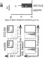


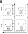


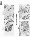


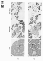

Classifications
-
- A—HUMAN NECESSITIES
- A61—MEDICAL OR VETERINARY SCIENCE; HYGIENE
- A61K—PREPARATIONS FOR MEDICAL, DENTAL OR TOILETRY PURPOSES
- A61K39/00—Medicinal preparations containing antigens or antibodies
- A61K39/395—Antibodies; Immunoglobulins; Immune serum, e.g. antilymphocytic serum
-
- C—CHEMISTRY; METALLURGY
- C07—ORGANIC CHEMISTRY
- C07K—PEPTIDES
- C07K16/00—Immunoglobulins [IGs], e.g. monoclonal or polyclonal antibodies
- C07K16/18—Immunoglobulins [IGs], e.g. monoclonal or polyclonal antibodies against material from animals or humans
- C07K16/28—Immunoglobulins [IGs], e.g. monoclonal or polyclonal antibodies against material from animals or humans against receptors, cell surface antigens or cell surface determinants
- C07K16/2803—Immunoglobulins [IGs], e.g. monoclonal or polyclonal antibodies against material from animals or humans against receptors, cell surface antigens or cell surface determinants against the immunoglobulin superfamily
- C07K16/2836—Immunoglobulins [IGs], e.g. monoclonal or polyclonal antibodies against material from animals or humans against receptors, cell surface antigens or cell surface determinants against the immunoglobulin superfamily against CD106
-
- A—HUMAN NECESSITIES
- A61—MEDICAL OR VETERINARY SCIENCE; HYGIENE
- A61P—SPECIFIC THERAPEUTIC ACTIVITY OF CHEMICAL COMPOUNDS OR MEDICINAL PREPARATIONS
- A61P1/00—Drugs for disorders of the alimentary tract or the digestive system
- A61P1/04—Drugs for disorders of the alimentary tract or the digestive system for ulcers, gastritis or reflux esophagitis, e.g. antacids, inhibitors of acid secretion, mucosal protectants
-
- A—HUMAN NECESSITIES
- A61—MEDICAL OR VETERINARY SCIENCE; HYGIENE
- A61K—PREPARATIONS FOR MEDICAL, DENTAL OR TOILETRY PURPOSES
- A61K31/00—Medicinal preparations containing organic active ingredients
- A61K31/33—Heterocyclic compounds
- A61K31/395—Heterocyclic compounds having nitrogen as a ring hetero atom, e.g. guanethidine or rifamycins
- A61K31/435—Heterocyclic compounds having nitrogen as a ring hetero atom, e.g. guanethidine or rifamycins having six-membered rings with one nitrogen as the only ring hetero atom
- A61K31/44—Non condensed pyridines; Hydrogenated derivatives thereof
- A61K31/4427—Non condensed pyridines; Hydrogenated derivatives thereof containing further heterocyclic ring systems
- A61K31/444—Non condensed pyridines; Hydrogenated derivatives thereof containing further heterocyclic ring systems containing a six-membered ring with nitrogen as a ring heteroatom, e.g. amrinone
-
- A—HUMAN NECESSITIES
- A61—MEDICAL OR VETERINARY SCIENCE; HYGIENE
- A61K—PREPARATIONS FOR MEDICAL, DENTAL OR TOILETRY PURPOSES
- A61K31/00—Medicinal preparations containing organic active ingredients
- A61K31/33—Heterocyclic compounds
- A61K31/395—Heterocyclic compounds having nitrogen as a ring hetero atom, e.g. guanethidine or rifamycins
- A61K31/495—Heterocyclic compounds having nitrogen as a ring hetero atom, e.g. guanethidine or rifamycins having six-membered rings with two or more nitrogen atoms as the only ring heteroatoms, e.g. piperazine or tetrazines
- A61K31/505—Pyrimidines; Hydrogenated pyrimidines, e.g. trimethoprim
- A61K31/506—Pyrimidines; Hydrogenated pyrimidines, e.g. trimethoprim not condensed and containing further heterocyclic rings
-
- A—HUMAN NECESSITIES
- A61—MEDICAL OR VETERINARY SCIENCE; HYGIENE
- A61K—PREPARATIONS FOR MEDICAL, DENTAL OR TOILETRY PURPOSES
- A61K31/00—Medicinal preparations containing organic active ingredients
- A61K31/33—Heterocyclic compounds
- A61K31/395—Heterocyclic compounds having nitrogen as a ring hetero atom, e.g. guanethidine or rifamycins
- A61K31/495—Heterocyclic compounds having nitrogen as a ring hetero atom, e.g. guanethidine or rifamycins having six-membered rings with two or more nitrogen atoms as the only ring heteroatoms, e.g. piperazine or tetrazines
- A61K31/505—Pyrimidines; Hydrogenated pyrimidines, e.g. trimethoprim
- A61K31/517—Pyrimidines; Hydrogenated pyrimidines, e.g. trimethoprim ortho- or peri-condensed with carbocyclic ring systems, e.g. quinazoline, perimidine
-
- A—HUMAN NECESSITIES
- A61—MEDICAL OR VETERINARY SCIENCE; HYGIENE
- A61K—PREPARATIONS FOR MEDICAL, DENTAL OR TOILETRY PURPOSES
- A61K31/00—Medicinal preparations containing organic active ingredients
- A61K31/33—Heterocyclic compounds
- A61K31/395—Heterocyclic compounds having nitrogen as a ring hetero atom, e.g. guanethidine or rifamycins
- A61K31/535—Heterocyclic compounds having nitrogen as a ring hetero atom, e.g. guanethidine or rifamycins having six-membered rings with at least one nitrogen and one oxygen as the ring hetero atoms, e.g. 1,2-oxazines
- A61K31/5375—1,4-Oxazines, e.g. morpholine
- A61K31/5377—1,4-Oxazines, e.g. morpholine not condensed and containing further heterocyclic rings, e.g. timolol
-
- A—HUMAN NECESSITIES
- A61—MEDICAL OR VETERINARY SCIENCE; HYGIENE
- A61K—PREPARATIONS FOR MEDICAL, DENTAL OR TOILETRY PURPOSES
- A61K38/00—Medicinal preparations containing peptides
- A61K38/16—Peptides having more than 20 amino acids; Gastrins; Somatostatins; Melanotropins; Derivatives thereof
- A61K38/17—Peptides having more than 20 amino acids; Gastrins; Somatostatins; Melanotropins; Derivatives thereof from animals; from humans
- A61K38/19—Cytokines; Lymphokines; Interferons
- A61K38/193—Colony stimulating factors [CSF]
-
- A—HUMAN NECESSITIES
- A61—MEDICAL OR VETERINARY SCIENCE; HYGIENE
- A61K—PREPARATIONS FOR MEDICAL, DENTAL OR TOILETRY PURPOSES
- A61K45/00—Medicinal preparations containing active ingredients not provided for in groups A61K31/00 - A61K41/00
- A61K45/06—Mixtures of active ingredients without chemical characterisation, e.g. antiphlogistics and cardiaca
-
- A—HUMAN NECESSITIES
- A61—MEDICAL OR VETERINARY SCIENCE; HYGIENE
- A61P—SPECIFIC THERAPEUTIC ACTIVITY OF CHEMICAL COMPOUNDS OR MEDICINAL PREPARATIONS
- A61P1/00—Drugs for disorders of the alimentary tract or the digestive system
- A61P1/14—Prodigestives, e.g. acids, enzymes, appetite stimulants, antidyspeptics, tonics, antiflatulents
-
- A—HUMAN NECESSITIES
- A61—MEDICAL OR VETERINARY SCIENCE; HYGIENE
- A61P—SPECIFIC THERAPEUTIC ACTIVITY OF CHEMICAL COMPOUNDS OR MEDICINAL PREPARATIONS
- A61P1/00—Drugs for disorders of the alimentary tract or the digestive system
- A61P1/16—Drugs for disorders of the alimentary tract or the digestive system for liver or gallbladder disorders, e.g. hepatoprotective agents, cholagogues, litholytics
-
- A—HUMAN NECESSITIES
- A61—MEDICAL OR VETERINARY SCIENCE; HYGIENE
- A61P—SPECIFIC THERAPEUTIC ACTIVITY OF CHEMICAL COMPOUNDS OR MEDICINAL PREPARATIONS
- A61P13/00—Drugs for disorders of the urinary system
- A61P13/12—Drugs for disorders of the urinary system of the kidneys
-
- A—HUMAN NECESSITIES
- A61—MEDICAL OR VETERINARY SCIENCE; HYGIENE
- A61P—SPECIFIC THERAPEUTIC ACTIVITY OF CHEMICAL COMPOUNDS OR MEDICINAL PREPARATIONS
- A61P17/00—Drugs for dermatological disorders
- A61P17/10—Anti-acne agents
-
- A—HUMAN NECESSITIES
- A61—MEDICAL OR VETERINARY SCIENCE; HYGIENE
- A61P—SPECIFIC THERAPEUTIC ACTIVITY OF CHEMICAL COMPOUNDS OR MEDICINAL PREPARATIONS
- A61P19/00—Drugs for skeletal disorders
-
- A—HUMAN NECESSITIES
- A61—MEDICAL OR VETERINARY SCIENCE; HYGIENE
- A61P—SPECIFIC THERAPEUTIC ACTIVITY OF CHEMICAL COMPOUNDS OR MEDICINAL PREPARATIONS
- A61P27/00—Drugs for disorders of the senses
- A61P27/02—Ophthalmic agents
-
- A—HUMAN NECESSITIES
- A61—MEDICAL OR VETERINARY SCIENCE; HYGIENE
- A61P—SPECIFIC THERAPEUTIC ACTIVITY OF CHEMICAL COMPOUNDS OR MEDICINAL PREPARATIONS
- A61P3/00—Drugs for disorders of the metabolism
- A61P3/08—Drugs for disorders of the metabolism for glucose homeostasis
- A61P3/10—Drugs for disorders of the metabolism for glucose homeostasis for hyperglycaemia, e.g. antidiabetics
-
- C—CHEMISTRY; METALLURGY
- C07—ORGANIC CHEMISTRY
- C07K—PEPTIDES
- C07K16/00—Immunoglobulins [IGs], e.g. monoclonal or polyclonal antibodies
- C07K16/18—Immunoglobulins [IGs], e.g. monoclonal or polyclonal antibodies against material from animals or humans
- C07K16/24—Immunoglobulins [IGs], e.g. monoclonal or polyclonal antibodies against material from animals or humans against cytokines, lymphokines or interferons
- C07K16/241—Tumor Necrosis Factors
-
- C—CHEMISTRY; METALLURGY
- C12—BIOCHEMISTRY; BEER; SPIRITS; WINE; VINEGAR; MICROBIOLOGY; ENZYMOLOGY; MUTATION OR GENETIC ENGINEERING
- C12N—MICROORGANISMS OR ENZYMES; COMPOSITIONS THEREOF; PROPAGATING, PRESERVING, OR MAINTAINING MICROORGANISMS; MUTATION OR GENETIC ENGINEERING; CULTURE MEDIA
- C12N5/00—Undifferentiated human, animal or plant cells, e.g. cell lines; Tissues; Cultivation or maintenance thereof; Culture media therefor
- C12N5/06—Animal cells or tissues; Human cells or tissues
- C12N5/0602—Vertebrate cells
- C12N5/0652—Cells of skeletal and connective tissues; Mesenchyme
- C12N5/0662—Stem cells
- C12N5/0663—Bone marrow mesenchymal stem cells (BM-MSC)
-
- G—PHYSICS
- G01—MEASURING; TESTING
- G01N—INVESTIGATING OR ANALYSING MATERIALS BY DETERMINING THEIR CHEMICAL OR PHYSICAL PROPERTIES
- G01N33/00—Investigating or analysing materials by specific methods not covered by groups G01N1/00 - G01N31/00
- G01N33/48—Biological material, e.g. blood, urine; Haemocytometers
- G01N33/50—Chemical analysis of biological material, e.g. blood, urine; Testing involving biospecific ligand binding methods; Immunological testing
- G01N33/53—Immunoassay; Biospecific binding assay; Materials therefor
- G01N33/569—Immunoassay; Biospecific binding assay; Materials therefor for microorganisms, e.g. protozoa, bacteria, viruses
- G01N33/56966—Animal cells
-
- G—PHYSICS
- G01—MEASURING; TESTING
- G01N—INVESTIGATING OR ANALYSING MATERIALS BY DETERMINING THEIR CHEMICAL OR PHYSICAL PROPERTIES
- G01N33/00—Investigating or analysing materials by specific methods not covered by groups G01N1/00 - G01N31/00
- G01N33/48—Biological material, e.g. blood, urine; Haemocytometers
- G01N33/50—Chemical analysis of biological material, e.g. blood, urine; Testing involving biospecific ligand binding methods; Immunological testing
- G01N33/68—Chemical analysis of biological material, e.g. blood, urine; Testing involving biospecific ligand binding methods; Immunological testing involving proteins, peptides or amino acids
- G01N33/6872—Intracellular protein regulatory factors and their receptors, e.g. including ion channels
-
- A—HUMAN NECESSITIES
- A61—MEDICAL OR VETERINARY SCIENCE; HYGIENE
- A61K—PREPARATIONS FOR MEDICAL, DENTAL OR TOILETRY PURPOSES
- A61K2300/00—Mixtures or combinations of active ingredients, wherein at least one active ingredient is fully defined in groups A61K31/00 - A61K41/00
Definitions
- the present disclosure relates to the treatment of diabetes and / or related diseases in which stem cell migration and suppression of abnormal stem cells are combined, and the diagnosis of diabetes and / or related diseases using stem cell migration and / or retention as an index. More specifically, the present disclosure is based on the treatment of diabetes and / or related diseases by migrating and suppressing abnormal hematopoietic stem cells, and by detecting the migration and / or residue of abnormal hematopoietic stem cells from a specific niche. Concerning the diagnosis of diabetes and / or related diseases.
- Non-Patent Document 1 Diabetes is generally classified into type 1 and type 2 (Non-Patent Document 1). However, since the details of the onset of both are unknown, it must be said that the classification of type 1 and type 2 itself lacks validity as being based on the etiology.
- various drugs are currently being developed for the treatment of diabetes, but most of them are aimed at controlling blood glucose and may be effective in preventing the progression of diabetes, but are insufficient for curing diabetes. Can be.
- diabetic complications such as neuropathy, nephropathy, liver disorder, retinopathy, fatty liver, gastrointestinal disorder, delayed fracture healing, eating disorder, and skin disorder are also common to diabetes classified into types 1 and 2. However, once it develops, it can be difficult to cure.
- the present inventors have found that a therapeutic strategy for diabetes and / or related diseases by suppressing abnormal stem cells is more effective when combined with stem cell migration. Based on this new finding, the present disclosure provides a means for the treatment and diagnosis of diabetes and / or related disorders.
- the present disclosure provides: (Item 1) A composition for treating and / or preventing diabetes and / or diabetes-related diseases, disorders and / or symptoms, comprising an inhibitor of abnormal hematopoietic stem cells (HSCs), wherein the inhibitor is a stem cell migrating agent. A composition, characterized in that it is administered in combination.
- the abnormal HCS is any of the above items, wherein the gene or protein selected from the group consisting of CD106 and its functional equivalents is not expressed at normal levels and / or is not functioning.
- Composition (Item 3) The composition of any of the above items, wherein the non-normal level expression is overexpression.
- the abnormal HCS is a composition according to any one of the above items, wherein a gene or protein selected from the group consisting of tumor necrosis factor alpha (TNF- ⁇ ) and proinsulin is not expressed at normal levels. ..
- the composition of any of the above items, wherein the disease, disorder and / or symptom comprises diabetic complications.
- the disease, disorder and / or symptom is selected from the group consisting of neuropathy, nephropathy, liver disorder, retinopathy, fatty liver, gastrointestinal disorder, delayed fracture healing, eating disorder, and skin disorder. Any composition.
- the stem cell migration agent comprises at least one agent selected from the group consisting of a CXCR4 antagonist, a CXCR2 stimulating agent, an epidermal growth factor receptor (EGFR) inhibitor, and a granulocyte colony stimulating factor (G-CSF) agent. , The composition of any of the above items.
- the stem cell migrating agent is at least one selected from the group consisting of prelixafor, GRO ⁇ 2 (MIP2), gefitinib, erlotinib, afatinib, osimertinib, filgrastim, naltograstim, lenograstim, pegfilgrastim.
- MIP2 GRO ⁇ 2
- a medicament for treating and / or preventing diabetes and / or diabetes-related diseases, disorders and / or symptoms which comprises a combination of a suppressor of abnormal hematopoietic stem cells (HSC) and a stem cell migrating agent.
- HSC hematopoietic stem cells
- compositions for treating and / or preventing diabetes and / or diabetes-related diseases, disorders and / or symptoms which comprises a stem cell migrating agent, wherein the stem cell migrating agent is an inhibitor of abnormal hematopoietic stem cells (HSC).
- HSC hematopoietic stem cells
- Compositions for the selection of treatments for the treatment and / or prevention of diabetes and / or diabetes-related diseases, disorders and / or symptoms comprising agents that detect the migration and / or retention of abnormal hematopoietic stem cells (HSCs). ..
- (Item 15) The composition of any of the above items, wherein the migration and / or residue is migration from the bone marrow niche and / or residue in the bone marrow niche.
- (Item 16) The composition of any of the above items, wherein the migration detection agent comprises a detection agent for CD106 or a functional equivalent.
- (Item 17) A method of treating and / or preventing diabetes or diabetes-related diseases, disorders and / or symptoms in a subject, agents and stem cell migrating agents that reduce or eliminate abnormal hematopoietic stem cells (HSCs) in the subject. A method comprising the step of administering an effective amount of.
- HSCs hematopoietic stem cells
- HSCs abnormal hematopoietic stem cells
- a method comprising the step of detecting the migration and / or residue of abnormal hematopoietic stem cells (HSC) in the subject.
- HSC abnormal hematopoietic stem cells
- (Item A1) A method of treating and / or preventing diabetes and / or diabetes-related diseases, disorders and / or symptoms in a subject, wherein an effective amount of an abnormal hematopoietic stem cell (HSC) inhibitor and stem cell migrating agent is applied to the subject.
- a method comprising the step of administering to.
- the abnormal HCS is any of the above items, wherein the gene or protein selected from the group consisting of CD106 and its functional equivalents is not expressed at normal levels and / or is not functioning.
- Method. (Item A3) The method of item A2, wherein the non-normal level expression is overexpression.
- (Item A4) The method of any of the above items, wherein the inhibitor comprises at least one selected from the group consisting of an anti-CD106 antibody or a functional variant thereof.
- the abnormal HCS is one in which a gene or protein selected from the group consisting of tumor necrosis factor alpha (TNF- ⁇ ) and proinsulin is not expressed at normal levels.
- TNF- ⁇ tumor necrosis factor alpha
- proinsulin proinsulin is not expressed at normal levels.
- the method of any of the above items, wherein the inhibitor comprises at least one selected from the group consisting of anti-TNF- ⁇ antibodies or functional variants thereof.
- (Item A7) The method of any of the above items, wherein the disease, disorder and / or symptom comprises diabetic complications.
- the disease, disorder and / or symptom is selected from the group consisting of neuropathy, nephropathy, liver disorder, retinopathy, fatty liver, gastrointestinal disorder, delayed fracture healing, eating disorder, and skin disorder. Either way.
- the stem cell migration agent comprises at least one agent selected from the group consisting of a CXCR4 antagonist, a CXCR2 stimulating agent, an epidermal growth factor receptor (EGFR) inhibitor, and a granulocyte colony stimulating factor (G-CSF) agent. , Any of the above items.
- the stem cell migrating agent is at least one selected from the group consisting of prelixafor, GRO ⁇ 2 (MIP2), gefitinib, erlotinib, afatinib, osimertinib, filgrastim, naltograstim, lenograstim, pegfilgrastim. Any of the above items, including.
- MIP2 GRO ⁇ 2
- gefitinib gefitinib
- erlotinib afatinib
- osimertinib afatinib
- filgrastim naltograstim
- lenograstim lenograstim
- pegfilgrastim pegfilgrastim. Any of the above items, including.
- HSCs hematopoietic stem cells
- (Item A13) The method of any of the above items, wherein said migration and / or residue is migration from the bone marrow niche and / or residue in the bone marrow niche.
- the migration detection agent comprises a detection agent for CD106 or a functional equivalent.
- (Item A15) A method of diagnosing diabetes or diabetes and / or diabetes-related diseases, disorders and / or symptoms in a subject by migrating and / or remaining abnormal hematopoietic stem cells (HSCs) in the subject. A method comprising the step of detecting migration and / or residue of HSC).
- (Item B1) A composition for the suppression of abnormal hematopoietic stem cells (HSCs) for use in combination with stem cell migrating agents to treat and / or prevent diabetes and / or diabetes-related diseases, disorders and / or symptoms.
- the abnormal HCS is any of the above items, wherein the gene or protein selected from the group consisting of CD106 and its functional equivalents is not expressed at normal levels and / or is not functioning. Composition.
- the abnormal HCS is a composition according to any one of the above items, wherein a gene or protein selected from the group consisting of tumor necrosis factor alpha (TNF- ⁇ ) and proinsulin is not expressed at normal levels. ..
- the composition of any of the above items, wherein the disease, disorder and / or symptom comprises diabetic complications.
- the disease, disorder and / or symptom is selected from the group consisting of neuropathy, nephropathy, liver disorder, retinopathy, fatty liver, gastrointestinal disorder, delayed fracture healing, eating disorder, and skin disorder.
- the stem cell migration agent comprises at least one agent selected from the group consisting of a CXCR4 antagonist, a CXCR2 stimulating agent, an epidermal growth factor receptor (EGFR) inhibitor, and a granulocyte colony stimulating factor (G-CSF) agent. , The composition of any of the above items.
- the stem cell migrating agent is at least one selected from the group consisting of prelixafor, GRO ⁇ 2 (MIP2), gefitinib, erlotinib, afatinib, osimertinib, filgrastim, naltograstim, lenograstim, pegfilgrastim.
- MIP2 GRO ⁇ 2
- HSCs hematopoietic stem cells
- (Item B14) Composition for detecting the migration and / or residue of abnormal hematopoietic stem cells (HSCs) for the selection of treatments for diabetes and / or diabetes-related diseases, disorders and / or symptoms and / or prevention. ..
- (Item B15) The composition of any of the above items, wherein the migration and / or residue is migration from the bone marrow niche and / or residue in the bone marrow niche.
- (Item B16) A composition according to any of the above items, comprising a detection agent for CD106 or a functional equivalent.
- (Item B17) The composition according to any one of the above items, which is a pharmaceutical composition.
- (Item B18) A composition according to any of the above items, further comprising an excipient, which is pharmaceutically acceptable.
- (Item C1) Use of inhibitors of abnormal hematopoietic stem cells (HSCs) and stem cell migrating agents in the manufacture of drugs for treating and / or preventing diabetes and / or diabetes-related diseases, disorders and / or symptoms.
- the abnormal HCS is any of the above items, wherein the gene or protein selected from the group consisting of CD106 and its functional equivalents is not expressed at normal levels and / or is not functioning. use.
- (Item C3) Use of any of the above items, said expression that is not at normal levels is overexpression.
- (Item C4) Use of any of the above items, wherein the inhibitor comprises at least one selected from the group consisting of anti-CD106 antibodies or functional variants thereof.
- the abnormal HCS is the use of any of the above items, wherein the gene or protein selected from the group consisting of tumor necrosis factor alpha (TNF- ⁇ ) and proinsulin is not expressed at normal levels.
- TNF- ⁇ tumor necrosis factor alpha
- proinsulin is not expressed at normal levels.
- Use of any of the above items, wherein the inhibitor comprises at least one selected from the group consisting of anti-TNF- ⁇ antibodies or functional variants thereof.
- (Item C7) Use of any of the above items, said disease, disorder and / or symptom, including diabetic complications.
- the disease, disorder and / or symptom is selected from the group consisting of neuropathy, nephropathy, liver disorder, retinopathy, fatty liver, gastrointestinal disorder, delayed fracture healing, eating disorder, and skin disorder. Use either.
- the stem cell migration agent comprises at least one agent selected from the group consisting of a CXCR4 antagonist, a CXCR2 stimulating agent, an epidermal growth factor receptor (EGFR) inhibitor, and a granulocyte colony stimulating factor (G-CSF) agent. , Use any of the above items.
- the stem cell migrating agent is at least one selected from the group consisting of prelixafor, GRO ⁇ 2 (MIP2), gefitinib, erlotinib, afatinib, osimertinib, filgrastim, naltograstim, lenograstim, pegfilgrastim.
- MIP2 GRO ⁇ 2
- gefitinib gefitinib
- erlotinib afatinib
- osimertinib afatinib
- filgrastim naltograstim
- lenograstim lenograstim
- pegfilgrastim pegfilgrastim.
- the migration detection agent comprises a detection agent for CD106 or a functional equivalent.
- (Item D1) A composition for treating and / or preventing diabetes and / or diabetes-related diseases, disorders and / or symptoms, comprising an inhibitor of abnormal hematopoietic stem cells (HSCs), wherein the inhibitor is a stem cell migrating agent. A composition, characterized in that it is administered in combination.
- HSCs hematopoietic stem cells
- the abnormal HCS is any of the above items, wherein the gene or protein selected from the group consisting of CD106 and its functional equivalents is not expressed at normal levels and / or is not functioning. Composition.
- the abnormal HCS is one in which a gene or protein selected from the group consisting of tumor necrosis factor alpha (TNF- ⁇ ), histone deacetylase (HDAC) and proinsulin is not expressed at normal levels. , The composition of any of the above items.
- HDAC inhibitor eg, tricostatin A
- the disease, disorder and / or symptom is selected from the group consisting of neuropathy, nephropathy, liver disorder, retinopathy, fatty liver, gastrointestinal disorder, delayed fracture healing, eating disorder, and skin disorder. Any composition.
- stem cell migration agent has an ability to migrate the abnormal HSC from a niche.
- the stem cell migration agent comprises at least one agent selected from the group consisting of a CXCR4 antagonist, a CXCR2 stimulating agent, an epidermal growth factor receptor (EGFR) inhibitor, and a granulocyte colony stimulating factor (G-CSF) agent. , The composition of any of the above items.
- the stem cell migrating agent is at least one selected from the group consisting of prelixafor, GRO ⁇ 2 (MIP2), gefitinib, erlotinib, afatinib, osimertinib, filgrastim, naltograstim, lenograstim, pegfilgrastim.
- MIP2 GRO ⁇ 2
- a medicament for treating and / or preventing diabetes and / or diabetes-related diseases, disorders and / or symptoms which comprises a combination of a suppressor of abnormal hematopoietic stem cells (HSC) and a stem cell migrating agent.
- HSC hematopoietic stem cells
- stem cell migrating agent an inhibitor of abnormal hematopoietic stem cells
- compositions for the selection of treatments for the treatment and / or prevention of diabetes and / or diabetes-related diseases, disorders and / or symptoms comprising agents that detect the migration and / or retention of abnormal hematopoietic stem cells (HSCs). ..
- HSCs hematopoietic stem cells
- the migration detection agent comprises a detection agent for CD106 or a functional equivalent.
- (Item D17) A method of treating and / or preventing diabetes or diabetes-related diseases, disorders and / or symptoms in a subject, agents and stem cell migrating agents that reduce or eliminate abnormal hematopoietic stem cells (HSCs) in the subject.
- a method comprising the step of administering an effective amount of.
- (Item D18) In a way that the migration and / or retention of abnormal hematopoietic stem cells (HSCs) is an indicator of treatment and / or prevention of diabetes or diabetes and / or diabetes-related diseases, disorders and / or symptoms in the subject.
- a method comprising the step of detecting the migration and / or residue of abnormal hematopoietic stem cells (HSC) in the subject.
- composition according to any one of the above items which is a pharmaceutical composition.
- composition according to any of the above items further comprising an excipient, which is pharmaceutically acceptable.
- Item E1 A method of treating and / or preventing diabetes and / or diabetes-related diseases, disorders and / or symptoms in a subject, wherein an effective amount of an abnormal hematopoietic stem cell (HSC) inhibitor and stem cell migrating agent is applied to the subject. A method comprising the step of administering to.
- HSC hematopoietic stem cell
- stem cell migrating agent A method comprising the step of administering to.
- the abnormal HCS is any of the above items, wherein the gene or protein selected from the group consisting of CD106 and its functional equivalents is not expressed at normal levels and / or is not functioning.
- the inhibitor comprises at least one selected from the group consisting of an anti-CD106 antibody or a functional variant thereof.
- the abnormal HCS is one in which a gene or protein selected from the group consisting of tumor necrosis factor alpha (TNF- ⁇ ), histone deacetylase (HDAC) and proinsulin is not expressed at normal levels. , Any of the above items.
- the disease, disorder and / or symptom comprises diabetic complications.
- the disease, disorder and / or symptom is selected from the group consisting of neuropathy, nephropathy, liver disorder, retinopathy, fatty liver, gastrointestinal disorder, delayed fracture healing, eating disorder, and skin disorder. Either way.
- the stem cell migration agent has the ability to migrate the abnormal HSC from a niche.
- the stem cell migration agent comprises at least one agent selected from the group consisting of a CXCR4 antagonist, a CXCR2 stimulating agent, an epidermal growth factor receptor (EGFR) inhibitor, and a granulocyte colony stimulating factor (G-CSF) agent. , Any of the above items.
- the stem cell migrating agent is at least one selected from the group consisting of prelixafor, GRO ⁇ 2 (MIP2), gefitinib, erlotinib, afatinib, osimertinib, filgrastim, naltograstim, lenograstim, pegfilgrastim. Any of the above items, including.
- (Item E12) A method of selecting treatment for diabetes and / or diabetes-related diseases, disorders and / or symptoms in a subject and / or migration of an effective amount of abnormal hematopoietic stem cells (HSCs).
- a method comprising administering to the subject an agent that detects a residue.
- (Item E13) The method of any of the above items, wherein said migration and / or residue is migration from the bone marrow niche and / or residue in the bone marrow niche.
- the migration detection agent comprises a detection agent for CD106 or a functional equivalent.
- (Item E15) A method of diagnosing diabetes or diabetes and / or diabetes-related diseases, disorders and / or symptoms in a subject by migrating and / or remaining abnormal hematopoietic stem cells (HSCs) in the subject.
- a method comprising the step of detecting migration and / or residue of HSC).
- Stem cell migrating agents or stem cell migrating agents for use in combination with stem cell migrating agents and abnormal hematopoietic stem cell (HSC) inhibitors to treat and / or prevent diabetes and / or diabetes-related diseases, disorders and / or symptoms.
- HSC abnormal hematopoietic stem cell
- the abnormal HCS is any of the above items, wherein the gene or protein selected from the group consisting of CD106 and its functional equivalents is not expressed at normal levels and / or is not functioning.
- Stem cell migration agent or abnormal HSC inhibitor (Item F3) The stem cell migration agent or abnormal HSC inhibitor according to any of the above items, wherein the non-normal level expression is overexpression. (Item F4) An inhibitor of any of the above items, comprising at least one selected from the group consisting of an anti-CD106 antibody or a functional variant thereof.
- the abnormal HCS is one in which a gene or protein selected from the group consisting of tumor necrosis factor alpha (TNF- ⁇ ), histone deacetylase (HDAC) and proinsulin is not expressed at normal levels.
- TNF- ⁇ tumor necrosis factor alpha
- HDAC histone deacetylase
- proinsulin proinsulin
- Stem cell migrating agent or abnormal HSC inhibitor of any of the above items Stem cell migrating agent or abnormal HSC inhibitor of any of the above items.
- the disease, disorder and / or symptom is a stem cell migration agent or abnormal HSC inhibitor according to any one of the above items, including diabetic complications.
- the disease, disorder and / or symptom is selected from the group consisting of neuropathy, nephropathy, liver disorder, retinopathy, fatty liver, gastrointestinal disorder, delayed fracture healing, eating disorder, and skin disorder. Any stem cell migrating agent or abnormal HSC inhibitor.
- the stem cell migration agent is a stem cell migration agent or an abnormal HSC inhibitor according to any one of the above items, which has an ability to migrate the abnormal HSC from a niche.
- the stem cell migration agent comprises at least one agent selected from the group consisting of a CXCR4 antagonist, a CXCR2 stimulating agent, an epidermal growth factor receptor (EGFR) inhibitor, and a granulocyte colony stimulating factor (G-CSF) agent.
- the stem cell migration agent is at least one selected from the group consisting of prelixafor, GRO ⁇ 2 (MIP2), gefitinib, erlotinib, afatinib, osimertinib, filgrastim, naltograstim, lenograstim, pegfilgrastim.
- MIP2 GRO ⁇ 2
- a stem cell migration agent or an abnormal HSC inhibitor according to any one of the above items.
- (Item F13) Stem cell migrating agents for stem cell migration for use in combination with abnormal hematopoietic stem cell (HSC) inhibitors to treat and / or prevent diabetes and / or diabetes-related diseases, disorders and / or symptoms Or an abnormal HSC inhibitor.
- HSC hematopoietic stem cell
- (Item F14) A drug that detects the migration and / or residue of abnormal hematopoietic stem cells (HSCs) for the selection of treatments for diabetes and / or diabetes-related diseases, disorders and / or symptoms and / or prevention.
- (Item F15) The agent of any of the above items, wherein said migration and / or residue is migration from the bone marrow niche and / or residue in the bone marrow niche.
- Item G1 Abnormal HSC inhibitors or stem cells in the manufacture of drugs to treat and / or prevent diabetes and / or diabetes-related diseases, disorders and / or symptoms with a combination of abnormal hematopoietic stem cell (HSC) inhibitors and stem cell migration agents. Use of at least one of the migratory agents.
- the abnormal HCS is any of the above items, wherein the gene or protein selected from the group consisting of CD106 and its functional equivalents is not expressed at normal levels and / or is not functioning. use.
- Item G3 Use of any of the above items, said expression that is not at normal levels is overexpression.
- the abnormal HCS is one in which a gene or protein selected from the group consisting of tumor necrosis factor alpha (TNF- ⁇ ), histone deacetylase (HDAC) and proinsulin is not expressed at normal levels.
- TNF- ⁇ tumor necrosis factor alpha
- HDAC histone deacetylase
- proinsulin proinsulin is not expressed at normal levels.
- Use of any of the above items. Use of any of the above items, wherein the inhibitor comprises at least one selected from the group consisting of an anti-TNF- ⁇ antibody or a functional variant thereof and an HDAC inhibitor (eg, tricostatin A).
- (Item G7) Use of any of the above items, said disease, disorder and / or symptom, including diabetic complications.
- the disease, disorder and / or symptom is selected from the group consisting of neuropathy, nephropathy, liver disorder, retinopathy, fatty liver, gastrointestinal disorder, delayed fracture healing, eating disorder, and skin disorder. Use either.
- (Item G9) Use of any of the above items, wherein the stem cell migration agent has the ability to migrate the abnormal HSC from a niche.
- the stem cell migration agent comprises at least one agent selected from the group consisting of a CXCR4 antagonist, a CXCR2 stimulating agent, an epidermal growth factor receptor (EGFR) inhibitor, and a granulocyte colony stimulating factor (G-CSF) agent. , Use any of the above items.
- the stem cell migrating agent is at least one selected from the group consisting of prelixafor, GRO ⁇ 2 (MIP2), gefitinib, erlotinib, afatinib, osimertinib, filgrastim, naltograstim, lenograstim, pegfilgrastim. Use of any of the above items, including.
- HSCs abnormal hematopoietic stem cells
- the present disclosure provides new treatments and diagnoses for diabetes and / or related disorders. It eliminates the need for donors, which have traditionally been required for pancreas transplantation and islet transplantation, which are methods for restoring insulin secretion in diabetic patients, and can overcome the limitation of supply capacity for the number of recipients. In addition, the patient burden such as surgery that the recipient has undergone can be avoided. Thus, the treatments of the present disclosure can be much simpler and less burdensome as they can be achieved by drug administration. In addition, since radical treatment is possible, the prognosis is expected to be good.
- the lower left two panels show the distribution of proinsulin-positive cells (enclosed in a square), the vertical axis is the number of KSL cells, and the horizontal axis is the fluorescence intensity of proinsulin. Shown.
- the two panels on the upper left show KSL cells (enclosed in a square) among lineage negative cells, the vertical axis is the fluorescence intensity of c-kit, and the horizontal axis is Sca-. It shows the fluorescence intensity of 1.
- the lower left two panels show the distribution of TNF- ⁇ positive cells (enclosed in a square), the vertical axis is the number of KSL cells, and the horizontal axis is the fluorescence of TNF- ⁇ . Indicates strength.
- Two panels (left: ICR, right: NOD) show KSL cells (enclosed in a square) among lineage negative cells, the vertical axis is the fluorescence intensity of c-kit, and the horizontal axis is the fluorescence intensity of Sca-1. Shown. FACS analysis of tumor necrosis factor alpha (TNF- ⁇ ) -positive cells and CD106-positive cells in KSL cells in ICR and NOD mouse mononuclear cells.
- TNF- ⁇ tumor necrosis factor alpha
- the upper two panels show the distribution of TNF- ⁇ positive cells (enclosed in a square), the vertical axis is the intensity of forward scattered light (FSC), and the horizontal axis is TNF- ⁇ . Shows the fluorescence intensity of.
- the bottom two panels show the distribution of CD106-positive cells (enclosed in a square), with the vertical axis representing the intensity of forward scattered light (FSC) and the horizontal axis representing the fluorescence intensity of CD106. Shown. FACS analysis of side population (SP) and non-SP fractions in KSL cells of non-DM and STZ-DM.
- the two panels on the left show the SP fraction (lower left circle) and non-SP fraction (upper right circle) of KSL cells, with the vertical axis representing.
- the fluorescence intensity of Hoechst Blue is shown, and the horizontal axis shows the fluorescence intensity of Hoechst Red.
- the two panels show the distribution of CD106-positive cells (enclosed in a square), the vertical axis is the intensity of laterally scattered light (SSC), and the horizontal axis is the fluorescence of CD106. Indicates strength.
- the two panels top: non-DM, bottom: STZ-DM
- the horizontal axis shows the fluorescence intensity of CD106.
- C FACS analysis of TNF- ⁇ positive cells in non-DM and STZ-DM KSL-non-SP cells.
- the two panels (top: non-DM, bottom: STZ-DM) show the distribution of TNF- ⁇ positive cells (enclosed in a square), the vertical axis is the intensity of laterally scattered light (SSC), and the horizontal axis is TNF-. Shows the fluorescence intensity of ⁇ .
- D FACS analysis of TNF- ⁇ positive cells in non-DM and STZ-DM KSL-SP cells.
- the two panels show the distribution of TNF- ⁇ positive cells (boxed, 0%), with the vertical axis representing the intensity of laterally scattered light (SSC). ,
- the horizontal axis shows the fluorescence intensity of TNF- ⁇ .
- HDACs histone deacetylase genes
- Transgenic mice transgenic with green fluorescent protein were treated with STZ to induce diabetes (STZ-DM GFP), and non-DM treated with intravenous citrate buffer injection as a control.
- STZ-DM GFP STZ-DM GFP
- non-DM GFP intravenous citrate buffer injection
- KSL cells obtained from non-DM mice and STZ-DM mice were transplanted into 9 Gy lethal dose-irradiated normoglycemic wild-type mice (non-DM-derived KSL-T, STZ-DM-derived KSL-T). ..
- the relative ratio (right) of the sensory nerve conduction velocity (SNCV) in the sciatic nerve of the case is shown. N. S indicates that there is no significant difference, and ** indicates P ⁇ 0.01.
- the four columns from the left are the results of KSL-T derived from STZ-DM, and from the left, the nucleus (blue), GFP (green), and the target molecule (upper row: MAP2, middle row: proinsulin, lower row: TNF- ⁇ ) (red). ), It is an image of merge.
- the lower left two panels show the distribution of proinsulin-positive cells (enclosed in a square), with the vertical axis representing the number of KSL cells and the horizontal. The axis shows the fluorescence intensity of proinsulin.
- the two panels on the upper left show KSL cells (enclosed in a square) among lineage-negative cells, and the vertical axis is c-kit.
- the fluorescence intensity and the horizontal axis indicate the fluorescence intensity of Sca-1.
- the lower left two panels show the distribution of TNF- ⁇ -positive cells (enclosed in a square), and the vertical axis represents the number of KSL cells.
- the horizontal axis shows the fluorescence intensity of TNF- ⁇ .
- the panels show insulin (upper left), CD34 (upper right), TNF- ⁇ (lower left), and CD106 (lower right), respectively.
- the left bar shows the results of healthy human subjects and the right bar shows the results of human diabetic patients.
- the vertical axis shows the relative mRNA expression level when the average result of healthy human subjects is 1.
- the upper left shows the results of non-DM mice
- the upper right shows the results of STZ-DM mice
- the lower two shows the results of two STZ-DM mice treated with a stem cell migration agent and an anti-CD106 antibody.
- About 8 to 10 islets were randomly selected in each mouse, and the ratio of the insulin positive area to the size of the islets was calculated by ImageJ software.
- the results of two STZ-DM mice (# 1 and # 2) treated with non-DM mice, STZ-DM mice, stem cell migration agents and anti-CD106 antibody are shown. ** P ⁇ 0.01 is shown. Changes in blood glucose and body weight when NOD mice are treated with stem cell migrants and anti-CD106 antibodies (250 ⁇ g / mouse dose, weekly tail vein administration) are shown.
- the graph above shows the blood glucose levels (vertical axis, mg / dL) on days 7 and 14.
- the graph below shows the body weight (vertical axis, g) on the 7th and 14th days.
- the blue line (# 670) and the orange line (# 671) show the results of two individuals who received the migratory agent and anti-CD106 antibody treatment twice a week, respectively, and the gray line (# 673). , The results of individuals who received the migratory agent and anti-CD106 antibody treatment only once. It is a stained image of insulin and nuclei of frozen mouse sections. From the left, the results of non-DM mice, NOD mice before the onset of diabetes, and NOD mice after the onset of diabetes are shown.
- the upper row is a 20x magnified image
- the lower row is a 40x magnified image.
- Arrowheads indicate islets. Proinsulin staining of bone marrow in patients without DM (DM (-)) and patients with DM (DM (+)). Bone marrow cells (arrows) stained with proinsulin antibody are observed in DM (+) cases but not in DM (-) cases. The scale bar indicates 50 ⁇ m.
- diabetes is used in the usual sense used in the art and is usually depleted of insulin by the destruction of pancreatic ⁇ -cells, except for those associated with pregnancy and those due to specific genetic abnormalities.
- Type 2 diabetes mellitus caused by decreased insulin secretion from ⁇ cells of the islets of Langerhans (pancreatic islets) in the pancreas and decreased ability to take up glucose into muscles and adipose tissues due to type 1 diabetes and obesity. are categorized. According to the results of this study, it is speculated that both type 1 diabetes and type 2 diabetes are caused by a common cell etiology mediated by immune disorders, which is completely different from the ones previously thought to be the etiology.
- Diabetes can be diagnosed by, for example, fasting blood glucose ⁇ 126 mg / dL, HbA1c ⁇ 6.5% in two or more tests, and a 2-hour value of 200 mg / dL or more in an oral glucose tolerance test (75 gOGTT).
- diabetes as used herein also includes juvenile-onset adult diabetes and borderline diabetes.
- diabetes-related diseases or “diabetes-related diseases, disorders and / or symptoms” may include any diabetes-related diseases, disorders and symptoms, examples of which are diabetic nerves.
- non-diabetic subject means a subject who does not have diabetes or a related disease.
- treatment refers to a disease or disorder that, in the event of such a condition, prevents the exacerbation of such disease or disorder, preferably maintains the status quo, more preferably alleviates, and further. It preferably refers to withdrawal, and includes the ability to exert a symptom-improving effect or a preventive effect on a patient's disease or one or more symptoms associated with the disease. Diagnosis in advance and appropriate treatment is called “companion treatment”, and the diagnostic agent for that purpose is sometimes called “companion diagnostic agent”.
- prevention means to prevent a certain disease or disorder from becoming such a condition before it becomes such a condition.
- the agent of the present disclosure can be used to make a diagnosis, and if necessary, the agent of the present disclosure can be used to prevent, for example, diabetes, or to take preventive measures.
- diagnosis refers to identifying various parameters related to a condition (for example, disease, disorder) in a subject and determining the current state or future of such condition.
- condition for example, disease, disorder
- the condition within the body can be investigated and such information can be used to determine the condition in the subject, the procedure or method for administration or prevention, etc.
- Various parameters can be selected.
- diagnosis means diagnosing the current state, but in a broad sense, it includes “early diagnosis”, “predictive diagnosis”, “pre-diagnosis” and the like.
- the diagnostic method of the present disclosure is industrially useful because it can be used from the body and can be carried out without the hands of medical personnel such as doctors.
- "predictive diagnosis, pre-diagnosis or diagnosis” may be referred to as "support” in order to clarify that it can be carried out without the hands of medical personnel such as doctors.
- the technique of the present disclosure can be applied to such a diagnostic technique.
- suppression of a target cell means that the growth rate of the target cell is reduced, the target cell is damaged, the target cell is reduced, and / or the target cell is killed.
- Suppression of target cells can be achieved by direct and / or indirect action.
- the direct action of suppressing a target cell is by targeting any characteristic of the cell (eg, an expressing molecule), resulting in damage to the target cell, reduction of the target cell and / or death of the target cell.
- Mechanisms achieved such as, but are not limited to, irradiation, induction of apoptosis, induction of attack by immune cells, and the like.
- the indirect action of suppressing the target cell includes, but is not limited to, a method of causing the immune system to learn to recognize and suppress the target cell.
- the "inhibitor" of a target cell means an agent that suppresses the target cell by any means.
- the means of suppressing the target cell can be arbitrary, eg, an immune cell attack caused by binding of the target cell to a targeting molecule (eg, an antibody), a radiation-generating molecule. The use of, but is not limited to this.
- the term "migratory" of a cell means that the cell migrates into peripheral blood and / or circulating blood.
- migration means that cells migrate from the bone marrow (or specific sites within it) into peripheral blood and / or circulating blood.
- a specific site in the bone marrow may be referred to as a niche or a specific niche, which refers to the microenvironment required for stem cells to maintain their properties in vivo.
- the present specification refers to, but is not limited to, migrating stem cells from a niche in a particular embodiment.
- the "migratory agent" of a cell means an agent that migrates a target cell by any means.
- hematopoietic stem cell refers to a stem cell capable of differentiating into a hematopoietic cell. In adult humans, it is mainly present in the bone marrow and produces white blood cells (neutrophils, eosinophils, basophils, lymphocytes, monocytes, macrophages), red blood cells, platelets, mast cells, and dendritic cells.
- white blood cells neutrils, eosinophils, basophils, lymphocytes, monocytes, macrophages
- red blood cells platelets, mast cells, and dendritic cells.
- Human hematopoietic stem cells for example, be characterized by CD34-positive Thy-1-positive, also, Lineage-negative CD34-positive CD38-negative CD90 + CD45RA-negative (i.e., Lin - CD34 + CD38 - CD90 + CD45RA -) also characterized by Obtain (see, for example, Proc Natl Acad Sci USA. 2011 Dec 13; 108 (50): 20012-20017).
- Mouse hematopoietic stem cells can be characterized as c-kit positive Sca-1 positive lineage marker negative (KSL) cells.
- Hematopoietic stem cells are also characterized by being negative when bone marrow cells stained with Hoechst 33342 dye are excited by ultraviolet light (350 nm) and developed with two types of optical filters, Hoechst blue and Hoechst red. Can be attached. Hematopoietic stem cells can be subdivided into long-term hematopoietic stem cells (LT-HSC), short-term hematopoietic stem cells (ST-HSC), and the like. For example, for the characteristics of hematopoietic stem cells, see Cytometry Research 19 (2): 25-32, 2009.
- LT-HSC long-term hematopoietic stem cell
- LT-HSCs are usually identified by surface antigen markers (eg, in mice) CD34 negative, CD150 positive, CD48 negative, Lin negative, sca1 positive, c-kit positive (or Lineage - c-Kit + Sca). It can be expressed as -1 + CD34 ⁇ / low CD150 + cell).
- LT-HSC can be described as CD34 negative, CD38 negative (or CD34 - CD38 - cells) (eg, in humans) (eg, Nat Immunol. 2010 Jul; 11 (7): 585-93; Blood 108, 2446-2454, 2006).
- ST-HSC short-term hematopoietic stem cell
- ST-HSCs are usually identified by surface antigen markers and can be expressed (eg, in mice) as CD34 positive, CD150 positive, CD48 negative, Lin negative, sca-1 positive, c-kit positive.
- ST-HSC can be described as CD34 positive, CD38 negative (or CD34 + CD38 - cells) (eg, in humans) (eg, Nat Immunol. 2010 Jul; 11 (7): 585-93; Blood 108, 2446-2454, 2006).
- abnormal hematopoietic stem cell or "abnormal HSC” refers to a hematopoietic stem cell that expresses abnormal function and / or lacks at least a part of normal function.
- HSC refers to cells in which the gene or protein of CD106 or its functional equivalent is not expressed at normal levels and / or is not functioning.
- not expressed at normal levels means that the gene or protein is expressed at a level or amount that is not the level of expression found in cells with normal function.
- CD106 it can be judged that it is abnormal when CD106 deviates from the normal expression (normal value found in normal cells), and preferably, when CD106 is expressed more than usual.
- KSL cells from non-diabetic and diabetic mice are fractionated by FACS, RNA is extracted, and then comparative analysis of gene expression levels is performed using QT-PCR to express genes at unusual levels (eg, for example). Overexpression) can be tested.
- FACS analysis can be performed to detect abnormal hematopoietic stem cell surface antigens and compare diabetic and non-diabetic subjects to test for unusual levels of protein expression.
- not functioning at normal levels means that the level of function found in cells with normal function is not found in the gene or protein.
- CD106 it can be determined to be abnormal if CD106 deviates from the level at which it normally functions (the normal level or value found in normal cells), and preferably CD106 functions at a higher level than normal. When observed, it can be determined to be abnormal. For example, by performing FACS analysis, detecting abnormal hematopoietic stem cell surface antigens, comparing diabetic and non-diabetic subjects and observing protein activation, the protein functions at normal levels. You can test that you are not.
- CXCR4 is a 7-transmembrane G protein-coupled receptor (GPCR), also known as CD184 or Fusin.
- GPCR G protein-coupled receptor
- the physiological ligand of CXCR4 is one of the CXC chemokines, which is stromal cell-derived factor-1 (SDF-1) that strongly induces the migration of monocytes and lymphocytes. Inhibition of CXCR4 can result in migration of hematopoietic stem cells (Future Oncol. 2007 Feb; 3 (1): 19-27).
- CD106 is a type I membrane protein of a member of the Ig superfamily, which is a kind of surface antigen and is also known as adhesion molecule VCAM-1 (Vascular cell adhesion molecule-1) or INCAM-100. It is a cell surface sialosaccharide protein expressed by cytokine-activated endothelium and is known to mediate leukocyte-endothelial cell adhesion and signaling.
- VCAM-1 isoform a precursor (nucleic acid sequence: NM_001078.4, amino acid sequence: NP_001069.1)
- VCAM-1 isoform b precursor (nucleic acid sequence: NM_080682.2, amino acid sequence: NP_542413.1)
- VCAM -1 Isoform c precursor (nucleic acid sequence: NM_001199834.1, amino acid sequence: NP_001186763.1) is typical.
- CD34 is a surface protein believed to be involved in the attachment of stem cells to the extracellular matrix or stromal cells, which is highly glycosylated and phosphorylated by protein kinase C. Is known.
- CD34 transcription variant 1 nucleic acid sequence: NM_001025109.2, amino acid sequence: NP_001020280.1
- CD34 transcription variant 2 nucleic acid sequence: NM_001773.3, amino acid sequence: NP_001764.1
- tumor necrosis factor alpha or its abbreviation "TNF- ⁇ ” is a multifunctional inflammation belonging to the tumor necrosis factor (TNF) superfamily also known as TNF, DIF, TNFA, TNFSF2, TNLG1F. It is an inducible cytokine and refers to a protein secreted mainly by macrophages. TNF- ⁇ can function via the receptors TNFRSF1A / TNFR1 and TNFRSF1B / TNFBR and is involved in the regulation of a wide range of biological processes such as cell proliferation, differentiation, apoptosis, lipid metabolism, and blood clots. In humans, the nucleic acid sequence: NM_000594.4 and the amino acid sequence: NP_000585.2 are typical.
- proinsulin refers to insulin precursor protein. Proinsulin is processed in the endoplasmic reticulum of pancreatic ⁇ -cells, and the C-peptide region is removed in immature secretory granules to produce insulin composed of A and B chains.
- proinsulin transcription variant 1 (nucleic acid sequence: NM_000207.3, amino acid sequence: NP_000198.1), proinsulin transcription variant 2 (nucleic acid sequence: NM_001185097.2, amino acid sequence: NP_001172026.1), proinsulin transcription variant 3 (Nucleic acid sequence: NM_001185098.1, Amino acid sequence: NP_001172027.1), Proinsulin transcription variant 4 (Nucleic acid sequence: NM_001291897.2, Amino acid sequence: NP_001278826.1) are typical.
- Kit isoform 1 precursor (nucleic acid sequence: NM_000222.2, amino acid sequence: NP_000213.1) and Kit isoform 2 precursor (nucleic acid sequence: NM_001093772.1, amino acid sequence: NP_001087241.1) are typical.
- CD20 is a member of the transmembrane 4A gene family, also known as MS4A1, B1, S7, Bp35, CD20, CVID5, MS4A2 or LEU-16, and the development of B cells into plasma cells. And refers to B lymphocyte surface molecules that play a role in differentiation.
- CD20 transcription variant 1 nucleic acid sequence: NM_152866.2, amino acid sequence: NP_690605.1
- CD20 transcription variant 3 are typical.
- each protein herein includes not only the protein having the amino acid sequence set forth in a particular accession number (or the nucleic acid encoding it), but also its functional equivalents. It is understood that it will be done.
- a "functional equivalent" of a molecule is a variant or variant of that molecule (eg, an amino acid sequence variant, etc.) that is a feature described herein (eg, a marker). ), Those that exhibit the same function (not necessarily the same degree) as the biological function of the molecule, and those that can change to the molecule itself at the time of action. Is understood to be included.
- the "functional variant" of a molecule includes a substance in which the molecule is modified while maintaining the function of the molecule (the degree may be changed).
- a functional variant of a binding molecule such as an antibody, is conjugated to another moiety (label, other functional molecule (protein, etc.), etc.) to maintain its binding function to the target molecule.
- Fragmented ones, etc. are included.
- a functional variant retains its underlying functionality prior to modification.
- one or a plurality of amino acids inserted, substituted or deleted, or added to one or both ends of the amino acid sequence can be used.
- "insertion, substitution or deletion of one or more amino acids in an amino acid sequence, or addition to one or both ends” is a well-known technical technique such as a site-specific mutagenesis method. It means that the modification has been made by a method or by a natural mutation, such as by substituting a plurality of amino acids that can occur naturally.
- biological function refers to a specific function that a gene, nucleic acid molecule or polypeptide may have in vivo when referring to a gene or a nucleic acid molecule or polypeptide related thereto. Examples include, but are not limited to, specific antibody production, enzymatic activity, and resistance imparting. For such biological activity, reference can be made to the documents cited in the accession numbers, Entrez numbers, etc. referred to in the above table, and such documents are also referred to in the present specification. Incorporate as. As used herein, biological function can be exerted by "biological activity”.
- biological activity refers to the activity that a certain factor (for example, polynucleotide, protein, etc.) can have in vivo, and exerts various functions (for example, transcription promoting activity). Activities include, for example, the activity of activating or inactivating another molecule by interacting with one molecule. When two factors interact, their biological activity is the binding between the two molecules and the resulting biological changes, eg, when one molecule is precipitated with an antibody to the other. When the molecules also coprecipitate, the two molecules are considered to be bound. Therefore, seeing such coprecipitation is one of the judgment methods. For example, if a factor is an enzyme, its biological activity comprises that enzymatic activity. In another example, when a factor is a ligand, the ligand comprises binding to the corresponding receptor. Such biological activity can be measured by techniques well known in the art.
- Derivatives or “similars” or “variants” of proteins or nucleic acids used herein are not intended to be limiting, but include molecules that contain regions that are substantially homologous to the protein or nucleic acid. Such molecules, in various embodiments, are at least 30% compared to sequences that are aligned over identical size amino acid or nucleic acid sequences or by computer homology programs known in the art. , 40%, 50%, 60%, 70%, 80%, 90%, 95%, 98% or 99% identical, or “derivatives” or “similars” or “variants” of such nucleic acids Can be hybridized to the original nucleic acid under stringent, moderately stringent, or non-stringent conditions.
- protein "derivatives” or “similars” or “mutants” are products of naturally occurring proteins that have undergone modifications such as amino acid substitutions, deletions, and additions, and are still organisms of naturally occurring proteins. Means a protein that exhibits a scientific function, but not necessarily to the same degree. For example, it is also possible to investigate the biological function of such proteins by appropriate and available in vitro assays described herein or known in the art. Although humans are primarily discussed in this disclosure, it is understood that this also applies to other species, such as those of other species within primates or species of animals of other genera, and these mammals are also referred to in this book. It is understood that it falls within the scope of disclosure.
- activity refers to the function of a molecule in the broadest sense. Activities are not intended to be limiting, but generally include the biological, biochemical, physical or chemical functions of the molecule. Activity activates, promotes, stabilizes, inhibits, suppresses, or destabilizes, for example, enzymatic activity, the ability to interact with other molecules, and the function of other molecules. Includes the ability to do, stability, and the ability to localize to specific intracellular locations. Where applicable, the term also relates to the function of protein complexes in the broadest sense.
- the “functionally active” proteins, polypeptides, fragments or derivatives used herein have structural, regulatory, or biochemical functions of the protein, such as biological activity.
- the term “gene” refers to a factor that defines a genetic trait. It is usually arranged in a certain order on the chromosome. A gene that defines the primary structure of a protein is called a structural gene, and a gene that influences its expression is called a regulatory gene. As used herein, “gene” may refer to "polynucleotide”, “oligonucleotide” and “nucleic acid” (here, including DNA, RNA, etc.).
- the “gene product” is a substance produced based on a gene and refers to a protein, mRNA or the like. Therefore, mRNA can enter the concept of genes and can also be a gene product. In addition, the term “gene expression” often refers to the transcription level of mRNA or the like.
- protein protein
- polypeptide oligopeptide
- peptide refers to a polymer of amino acids of any length.
- the polymer may be linear, branched or cyclic.
- the amino acid may be natural or non-natural, or may be a modified amino acid.
- the term may also include those assembled into a complex of multiple polypeptide chains.
- the term also includes naturally or artificially modified amino acid polymers. Such modifications include, for example, disulfide bond formation, glycosylation, lipidation, acetylation, phosphorylation or any other manipulation or modification (eg, conjugation with a labeling component).
- the definition also includes, for example, polypeptides containing one or more analogs of amino acids (including, for example, unnatural amino acids), peptide-like compounds (eg, peptoids) and other modifications known in the art. Will be done.
- polynucleotide refers to a polymer of nucleotides of arbitrary length.
- the term also includes “oligonucleotide derivatives" or “polynucleotide derivatives”.
- oligonucleotide derivative refers to an oligonucleotide or polynucleotide containing a derivative of a nucleotide or having an unusual bond between nucleotides, and is used interchangeably.
- such an oligonucleotide includes, for example, 2'-O-methyl-ribonucleotide, an oligonucleotide derivative in which a phosphate diester bond in an oligonucleotide is converted into a phosphorothioate bond, and a phosphate diester bond in an oligonucleotide.
- oligonucleotide derivatives converted to N3'-P5'phosphoroamidate bond an oligonucleotide derivative in which ribose and phosphate diester bond in the oligonucleotide are converted into peptide nucleic acid bond, and uracil in the oligonucleotide is C- 5 Oligonucleotide derivatives substituted with propynyl uracil, oligonucleotide derivatives in which uracil in the oligonucleotide is replaced with C-5 thiazole uracil, oligonucleotide derivatives in which cytosine in the oligonucleotide is replaced with C-5 propynyl citosine, oligonucleotides Oligonucleotide derivatives in which cytosine in nucleotides is replaced with phenoxazine-modified cytosine, oligonucleotide derivatives in which
- nucleic acid sequences are also conservatively modified variants (eg, degenerate codon substitutions) and complementary sequences, as are the explicitly indicated sequences. Is intended to be included.
- the degenerate codon substituent creates a sequence in which the third position of one or more selected (or all) codons is replaced with a mixed base and / or deoxyinosine residue. It can be achieved by (Batzer et al., Nucleic Acid Res. 19: 5081 (1991); Ohtsuka et al., J. Biol. Chem. 260: 2605-2608 (1985); Rossorini et al., Mol. Cell .Probes 8: 91-98 (1994)).
- nucleic acid is also used interchangeably with genes, cDNAs, mRNAs, oligonucleotides, and polynucleotides.
- nucleotide may be natural or non-natural.
- homology of a gene means the degree of identity of two or more gene sequences to each other, and generally, having “homology” means a high degree of identity or similarity.
- the higher the homology of two genes the higher the identity or similarity of their sequences.
- the DNA sequences are typically at least 50% identical, preferably at least 70% identical, and more preferably at least 80%, 90%. , 95%, 96%, 97%, 98% or 99%, the genes are homologous.
- a “homologous” or “homologous gene product” is a protein in another species, preferably a mammal, that exerts the same biological functions as the protein components of the complex further described herein. Means. Such homologues are also sometimes referred to as “ortholog gene products.”
- Amino acids may be referred to herein by either their generally known three-letter symbols or the one-letter symbols recommended by the IUPAC-IUB Biochemical Nomenclature Commission. Nucleotides can also be referred to by the generally recognized one-letter code.
- comparison of amino acid sequence and base sequence similarity, identity and homology is calculated using default parameters using BLAST, a tool for sequence analysis.
- the identity search can be performed using, for example, NCBI's BLAST 2.2.9 (issued May 12, 2004).
- the value of identity in the present specification usually refers to the value when the above BLAST is used and aligned under the default conditions. However, if a higher value is obtained by changing the parameter, the highest value is set as the identity value. When identity is evaluated in multiple regions, the highest value among them is set as the identity value. Similarity is a number that takes into account similar amino acids in addition to identity.
- polynucleotide that hybridizes under stringent conditions refers to well-known conditions commonly used in the art.
- a polynucleotide can be obtained by using a polynucleotide selected from the polynucleotides of the present disclosure as a probe and using a colony hybridization method, a plaque hybridization method, a Southern blot hybridization method, or the like. Specifically, using a filter on which DNA derived from colonies or plaques is immobilized, hybridization is performed at 65 ° C. in the presence of 0.7 to 1.0 M NaCl, and then the concentration is 0.1 to 2 times.
- Low stringency conditions include 35% formamide, 5xSSC, 50 mM Tris-HCl (pH 7.5), 5 mM EDTA, 0.02% polyvinylpyrrolidone (PVP), 0.02% BSA, 100 ⁇ g / ml denatured salmon sperm DNA, and A buffer consisting of 2xSSC, 25 mM Tris-HCl (pH 7.4), 5 mM EDTA, and 0.1% SDS hybridized at 40 ° C. for 18-20 hours in a buffer containing 10% (weight / volume) dextran sulfate. Wash in solution at 55 ° C. for 1-5 hours and then in buffer solution consisting of 2xSSC, 25 mM Tris-HCl (pH 7.4), 5 mM EDTA, and 0.1% SDS at 60 ° C. for 1.5 hours. Including that.
- a "purified" substance or biological factor is one in which at least a portion of the factors naturally associated with the biological factor have been removed. Therefore, the purity of the biological factor in the purified biological factor is usually higher (ie, enriched) than in the state in which the biological factor is normally present.
- the term "purified” as used herein is preferably at least 75% by weight, more preferably at least 85% by weight, even more preferably at least 95% by weight, and most preferably at least 98% by weight. It means that the same type of biological factor is present.
- the material used in the present disclosure is preferably a "purified" material.
- a "corresponding" amino acid or nucleic acid has, or has, in a polypeptide molecule or polynucleotide molecule, the same action as a given amino acid or nucleotide in a polypeptide or polynucleotide that serves as a reference for comparison.
- the corresponding amino acids are specified to be, for example, cysteineized, glutathioneized, SS bond formed, oxidized (eg, methionine side chain oxidation), formylation, acetylation, phosphorylation, glycosylation, myristylation, etc.
- the corresponding amino acid can be the amino acid responsible for dimerization.
- Such "corresponding" amino acids or nucleic acids may be regions or domains over a range. Thus, such cases are referred to herein as "corresponding" regions or domains.
- a "corresponding" gene eg, a polynucleotide sequence or molecule
- a gene for example, a polynucleotide sequence or a molecule
- the gene corresponding to a gene can be the ortholog of that gene.
- Such corresponding genes can be identified using techniques well known in the art. Thus, for example, a corresponding gene in an animal (eg, mouse, rat) searches the animal's sequence database using the sequence of the reference gene for the corresponding gene (eg, human) as a query sequence. It can be found by.
- fragment refers to a polypeptide or polynucleotide having a sequence length from 1 to n-1 with respect to a full-length polypeptide or polynucleotide (length n).
- length n the length of the fragment can be appropriately changed according to its purpose.
- the lower limit of the length is 3, 4, 5, 6, 7, 8, 9, 10, and so on.
- Amino acids such as 15, 20, 25, 30, 40, 50 and above are mentioned, and lengths represented by integers not specifically listed here (eg, 11) are also suitable as lower limits. obtain.
- a length represented by a non-integer may also be a suitable lower bound.
- a molecular fragment is a substance (typically a polypeptide) containing an arbitrary region of this molecule, as long as it can be used for the purposes of the present disclosure (eg, treatment, detection, diagnosis, etc.). , It does not have to have the biological function of a natural molecule.
- expression of a gene, polynucleotide, polypeptide or the like means that the gene or the like undergoes a certain action in vivo and becomes another form.
- a gene, polynucleotide, or the like is transcribed and translated into the form of a polypeptide, but transcribed to produce mRNA can also be an aspect of expression.
- the form of such a polypeptide can be post-translationally processed (derivatives as used herein).
- the expression level of a molecule can be determined by any method. Specifically, the expression level can be determined by assessing the amount of mRNA, the amount of protein, or the biological activity of the protein in this molecule.
- the term "expression level" refers to the amount at which a polypeptide, mRNA, etc. is expressed in a target cell, tissue, or the like. Such expression levels are evaluated using the antibodies of the present disclosure by any suitable method including immunological measurement methods such as ELISA method, RIA method, immunofluorescence method, Western blotting method, immunohistochemical staining method and the like.
- the expression level of the disclosed polypeptide at the protein level, or the poly used in the present disclosure evaluated by any suitable method including molecular biological measurement methods such as Northern blotting, dot blotting, and PCR.
- the expression level of the peptide at the mRNA level can be mentioned.
- “Change in expression” is the expression of the polypeptide used in the present disclosure at the protein or mRNA level as assessed by any suitable method, including the immunological or molecular biological measurement methods described above. It means that the amount increases or decreases. By measuring the expression level of a certain marker, various detections or diagnoses based on the marker can be performed.
- the term "marker (substance, protein or gene (nucleic acid))” refers to a certain state (for example, cell type, normal cell state, disease state, impaired state, or proliferative ability, differentiation state level, etc.).
- a substance that serves as a marker for tracking whether a substance is present or at risk. Examples of such markers include genes (nucleic acid DNA level), gene products (mRNA, proteins, etc.), metabolites, enzymes, and the like.
- detection, diagnosis, preliminary detection, prediction or pre-diagnosis of a condition is an agent, agent, factor or means specific for a marker associated with that condition, or a composition comprising them. , Kits or systems, etc.
- antibody is broadly referred to as a polyclonal antibody, a monoclonal antibody, a multispecific antibody, a chimeric antibody, and an anti-idiotype antibody, and fragments thereof such as Fv fragment, Fab'fragment, and F (ab'). 2 and Fab fragments, as well as conjugates or functional equivalents produced by other recombination (eg, chimeric antibodies, humanized antibodies, multifunctional antibodies, bispecific or oligospecific antibodies, single chains Includes antibodies, scFV, diabody, sc (Fv) 2 (single chain (Fv) 2 ), scFv-Fc).
- an antibody may be covalently linked or recombinantly fused to an enzyme such as alkaline phosphatase, horseradish peroxidase, ⁇ -galactosidase, or the like.
- an enzyme such as alkaline phosphatase, horseradish peroxidase, ⁇ -galactosidase, or the like.
- the origin, type, shape, etc. of the antibody used in the present disclosure are not limited. Specifically, known antibodies such as non-human animal antibodies (eg, mouse antibodies, rat antibodies, camel antibodies), human antibodies, chimeric antibodies, humanized antibodies and the like can be used. In the present disclosure, monoclonal or polyclonal can be used as an antibody, but a monoclonal antibody is preferable.
- the binding of the antibody to the target protein is preferably a specific binding.
- the term “means” refers to any tool that can be an arbitrary tool for achieving a certain purpose (eg, detection, diagnosis, treatment, prevention, migration), and in particular, in the present specification, “selective recognition (for example).
- “Means for detecting” means means for recognizing (detecting) a certain object differently from others.
- detection or “quantification” of polynucleotide or polypeptide expression uses appropriate methods, including methods for measuring mRNA and immunologically, including, for example, binding or interaction with a marker detector. Can be achieved.
- the molecular biological measurement method include a Northern blotting method, a dot blotting method, a PCR method, and the like.
- immunological measurement methods include ELISA method using a microtiter plate, RIA method, immunofluorescence method, luminescent immunoassay (LIA), immunoprecipitation method (IP), immunodiffusion method (SRID), and immunity. Examples thereof include immunofluorescence (TIA), Western blotting, and immunohistochemical staining.
- the ELISA method, the RIA method and the like are exemplified. It can also be performed by a gene analysis method using an array (for example, DNA array, protein array).
- DNA arrays are widely outlined in (Shujunsha ed., Cell Engineering Separate Volume "DNA Microarrays and Latest PCR Methods”).
- protein arrays see Nat Genet. It is detailed in 2002 Dec; 32 Suppl: 526-32.
- Examples of the gene expression analysis method include, but are not limited to, RT-PCR, RACE method, SCSP method, immunoprecipitation method, two-hybrid system, in vitro translation, and the like, in addition to the above.
- the detection agent or detection means of the present disclosure may be a complex or a complex molecule in which another substance (for example, a label or the like) is bound to a detectable portion (for example, an antibody or the like).
- a detectable portion for example, an antibody or the like.
- complex or “complex molecule” means any construct that includes two or more moieties.
- one portion is a polypeptide
- the other portion may be a polypeptide or other substance (eg, substrate, sugar, lipid, nucleic acid, other hydrocarbon, etc.). You may.
- two or more portions constituting the complex may be bonded by covalent bonds or other bonds (for example, hydrogen bond, ionic bond, hydrophobic interaction, van der Waals force, etc.). It may have been done.
- the "complex” includes a molecule formed by linking a plurality of molecules such as a polypeptide, a polynucleotide, a lipid, a sugar, and a small molecule.
- the detection agent or other medicine of the present disclosure can take the form of a probe and a primer.
- the probes and primers of the present disclosure can specifically hybridize with a target nucleic acid molecule.
- the probes and primers of the present disclosure need only be able to detect the expression of the target nucleic acid molecule, and may be a polymer composed of bases or base pairs such as a plurality of deoxyribonucleic acid (DNA) or ribonucleic acid (RNA). Double-stranded cDNA is also known to be available for tissue in situ hybridization, and the probes and primers of the present disclosure also include such double-stranded cDNA.
- RNA probes riboprobes
- the primers and primer sets according to the present disclosure are prepared according to a conventional method in a known method for detecting a target gene by using a nucleic acid amplification method such as PCR method, RT-PCR method, real-time PCR method, insitu PCR method, LAMP method and the like. It can be used as a primer and a primer set.
- a nucleic acid amplification method such as PCR method, RT-PCR method, real-time PCR method, insitu PCR method, LAMP method and the like. It can be used as a primer and a primer set.
- the term "probe” refers to a substance used as a means of searching used in biological experiments such as in vitro and / or in vivo screening, for example, a nucleic acid molecule containing a specific base sequence or a specific substance. Examples include, but are not limited to, peptides containing amino acid sequences, specific antibodies or fragments thereof. In the present specification, the probe is used as a marker detection means.
- nucleic acid molecule usually used as a probe examples include those having a nucleic acid sequence of at least 8 consecutive nucleotide lengths that is homologous or complementary to the nucleic acid sequence of the gene of interest. Such nucleic acid sequences preferably have at least 12 contiguous nucleotide lengths, preferably at least 9 contiguous nucleotide lengths, more preferably at least 10 contiguous nucleotide lengths, and even more preferably at least 11 contiguous nucleotide lengths.
- At least 13 contiguous nucleotide lengths at least 14 contiguous nucleotide lengths, at least 15 contiguous nucleotide lengths, at least 20 contiguous nucleotide lengths, at least 25 contiguous nucleotide lengths, at least 30 It can be at least a nucleic acid sequence of contiguous nucleotide length, at least 40 contiguous nucleotide lengths, and at least 50 contiguous nucleotide lengths.
- the nucleic acid sequence used as the probe is at least about 70% homologous, more preferably at least about 80% homologous, and even more preferably at least about 90% homologous to the above sequence, at least about 95%. Includes homologous nucleic acid sequences.
- the detection agent of the present disclosure can be labeled.
- the detection agent of the present disclosure may be a tag-bound one.
- label refers to an entity (eg, substance, energy, electromagnetic wave, etc.) for identifying a molecule or substance of interest from others.
- a labeling method include an RI (radioisotope) method, a fluorescence method, a biotin method, and a chemiluminescence method.
- the markers of the present disclosure or a plurality of factors or means for capturing the markers are labeled by the fluorescence method, the markers are labeled with fluorescent substances having different fluorescence maximum wavelengths. The difference in fluorescence emission maximum wavelength is preferably 10 nm or more.
- Alexa TM Fluor is a water-soluble fluorescent dye obtained by modifying coumarin, rhodamine, fluorescein, cyanine, etc., and is a series that supports a wide range of fluorescent wavelengths. It is stable, bright, and has low pH sensitivity.
- Examples of the combination of fluorescent dyes having a maximum fluorescence wavelength of 10 nm or more include a combination of Alexa TM 555 and Alexa TM 633, a combination of Alexa TM 488 and Alexa TM 555, and the like.
- any one that can bind to the base portion can be used, but cyanine dyes (for example, Cy3, Cy5, etc. of CyDye TM series), Rhodamine 6G reagent, N-acetoxy-N2- It is preferable to use acetylaminofluorene (AAF), AAIF (iodine derivative of AAF) and the like.
- AAF acetylaminofluorene
- AAIF iodine derivative of AAF
- the fluorescent substance having a difference in fluorescence emission maximum wavelength of 10 nm or more include a combination of Cy5 and Rhodamine 6G reagent, a combination of Cy3 and fluorescein, a combination of Rhodamine 6G reagent and fluorescein, and the like.
- such a label can be used to modify an object of interest so that it can be detected by the detection means used. Such modifications are known in the art and those skilled in the art can appropriately implement such methods depending on the label and the subject of interest
- a “tag” is a substance for selecting a molecule by a specific recognition mechanism such as a receptor-ligand, more specifically, a binding for binding a specific substance.
- a substance that acts as a partner eg, having a relationship such as biotin-avidin, biotin-streptavidin
- a specific substance to which a tag is bound can be selected by contacting a base material to which a binding partner of the tag sequence is bound.
- tags or signs are well known in the art. Typical tag sequences include, but are not limited to, myc tags, His tags, HA, Avi tags, and the like. Such tags may be attached to the markers or marker detectors of the present disclosure.
- drug As used herein, “drug”, “drug” or “factor” (both corresponding to agents in English) are used interchangeably in any sense as long as they can achieve their intended purpose. It may also be a substance or other element (eg, energy such as light, radioactivity, heat, electricity). Such substances include, for example, cells (eg, T cells), proteins, polypeptides, oligopeptides, peptides, polynucleotides, oligonucleotides, nucleotides, nucleic acids (eg, DNA such as cDNA, genomic DNA, mRNA.
- cells eg, T cells
- proteins eg, polypeptides, oligopeptides, peptides, polynucleotides, oligonucleotides, nucleotides, nucleic acids (eg, DNA such as cDNA, genomic DNA, mRNA.
- RNA Ribonucleic acid
- polypeptides oligosaccharides
- lipids lipids
- organic small molecules eg, hormones, ligands, signaling substances, organic small molecules, molecules synthesized with combinatorial chemistries, small molecules that can be used as pharmaceuticals (eg, etc.) , Small molecule ligands, etc.
- small molecules eg, hormones, ligands, signaling substances, organic small molecules, molecules synthesized with combinatorial chemistries, small molecules that can be used as pharmaceuticals (eg, etc.) , Small molecule ligands, etc.
- kits are a unit that is usually divided into two or more compartments and is provided with parts to be provided (for example, a test drug, a diagnostic drug, a therapeutic drug, an antibody, a label, an instruction manual, etc.).
- the form of this kit is preferred when the purpose is to provide a composition that should not be mixed and provided for stability and the like, but is preferably mixed and used immediately before use.
- kits are preferably instructions or instructions describing how to use the provided parts (eg, test agents, diagnostic agents, therapeutic agents, or how reagents should be treated).
- the kit When the kit is used as a reagent kit in the present specification, the kit usually includes an instruction manual or the like that describes how to use a test drug, a diagnostic drug, a therapeutic drug, an antibody, or the like. Is included.
- the "instruction” describes the method of using this disclosure to a doctor or another user.
- This instruction sheet contains words instructing the detection method of the present disclosure, how to use a diagnostic agent, or administration of a medicine or the like.
- the instruction sheet may include a wording instructing the administration site to be administered orally or to the esophagus (for example, by injection).
- This instruction is prepared and approved by the regulatory agency of the country in which this disclosure is implemented (eg, Ministry of Health, Labor and Welfare in Japan, Food and Drug Administration (FDA) in the United States, etc.). It is clearly stated that it has been received.
- the instruction sheet is a so-called package insert, which is usually provided in paper media, but is not limited thereto, and is in the form of, for example, an electronic medium (for example, a homepage provided on the Internet, an e-mail). But can be provided.
- the "quantity" of an analyte or the like in a sample generally refers to an absolute value that reflects the mass of the analyte or the like that can be detected in the volume of the sample. However, the quantity also contemplates a relative quantity compared to another analyte quantity. For example, the amount of analyte in the sample may be greater than the control or normal value of the analyte normally present in the sample.
- the "level" of an analyte or the like in a sample generally reflects a value such as the activity of the analyte when the analyte is a subject that exerts a function such as an enzyme. Refers to the absolute value to be used. However, the level also contemplates a relative level compared to another analyte level. For example, the level of the analyte in the sample may be greater than the control or normal value of the analyte normally present in the sample.
- the present inventors have found a new aspect in which the effect of diabetes treatment targeting abnormal stem cells characterized by abnormal expression such as CD106 can be improved in combination with stem cell migration.
- the present disclosure provides new therapeutic strategies and diagnoses for diabetes and / or related disorders based on this new diabetes treatment strategy.
- the abnormal stem cells of interest in the present disclosure can be hematopoietic stem cells.
- the abnormal stem cells of the present disclosure do not express at least one of CD106, CD34, TNF- ⁇ , proinsulin, and histone deacetylases (HDACs) at normal levels and / Or other features of abnormal stem cells described herein (eg, c-Kit positive, Sca-1 positive, hematopoietic stem cell lineage marker negative, above, characterized by non-functioning, as required. It can be further characterized by having short-term hematopoietic stem cell characteristics (such as CD38 negative).
- the abnormal stem cells of the present disclosure can be characterized by not expressing CD106 at normal levels. In one embodiment, the abnormal stem cells of the present disclosure do not (a) express CD106 at normal levels and (b) abnormally express CD34, TNF- ⁇ , proinsulin, and histone deacetylase. Characterized by not expressing at least one of the abnormal expression of chemogenetic (HDACs) at normal levels, and optionally other features of abnormal stem cells described herein (eg, c- It can be further characterized by having Kit positive, Sca-1 positive, hematopoietic stem cell lineage marker negative, short-term hematopoietic stem cell characteristics (CD38 negative, etc.).
- HDACs chemogenetic
- the abnormal stem cells of interest in the present disclosure can be characterized by aberrant expression of CD106.
- the abnormal stem cells of the present disclosure can be characterized by a CD106 expression level higher than the CD106 expression level in whole bone marrow cells or hematopoietic stem cells (eg, CD34-positive Thy-1 positive cells) of a non-diabetic subject population. ..
- the abnormal stem cells of the present disclosure are 1 ⁇ 10 2 or more, 2 ⁇ 10 2 or more, 5 ⁇ 10 2 or more, 1 ⁇ 10 3 or more, 2 ⁇ 10 3 or more, 5 ⁇ 10 per cell.
- the abnormal stem cells of the present disclosure can approximately 1 ⁇ 10 4 or more CD106 molecules per cell is characterized by expressing the cell surface.
- the abnormal stem cells of the present disclosure are whole bone marrow cells or hematopoietic stem cells (eg, CD34-positive Thy-1 positive cells) of a normal subject (eg, human) with a fluorescent dye-conjugated antibody against CD106. 1x or more, 2x or more, 5x or more, 10x or more, 20x or more of the median fluorescence intensity derived from this fluorescent dye observed for whole bone marrow cells or hematopoietic stem cells when labeled and analyzed by FACS. , 50 times or more, 100 times or more, 200 times or more, 500 times or more, or 1000 times or more can be indicated by the expression level exhibiting fluorescence intensity.
- the abnormal stem cells of the present disclosure are 10% or more, 20% or more, 30% or more, 40% or more, 50% or more than hematopoietic stem cells (for example, CD34-positive cells) derived from a non-diabetic subject population. , 60% or more, 70% or more, 80% or more, 90% or more, 100% or more, 150% or more or 200% or more high mRNA expression level of CD106.
- hematopoietic stem cells for example, CD34-positive cells
- the abnormal stem cells of interest in the present disclosure can be characterized by the expression of TNF- ⁇ .
- the abnormal stem cells of the present disclosure can be characterized by being positive for TNF- ⁇ expression.
- being positive for TNF- ⁇ expression refers to normal subject (eg, human) whole bone marrow cells or hematopoietic stem cells (eg, CD34-positive Thy-1 positive cells) relative to TNF- ⁇ .
- the median fluorescence intensity derived from this fluorescent dye observed for whole bone marrow cells or hematopoietic stem cells is 1-fold or more, 2-fold or more, 5-fold or more.
- the abnormal stem cells of the present disclosure are 10% or more, 20% or more, 30% or more, 40% or more, 50% or more than hematopoietic stem cells (for example, CD34-positive cells) derived from a non-diabetic subject population. , 60% or more, 70% or more, 80% or more, 90% or more, 100% or more, 150% or more or 200% or more high TNF- ⁇ mRNA expression level.
- the abnormal stem cells of interest in the present disclosure can be characterized by aberrant expression of proinsulin.
- the abnormal stem cells of the present disclosure can be characterized by being positive for proinsulin expression.
- being positive for proinsulin expression makes whole bone marrow cells or hematopoietic stem cells (eg, CD34-positive Thy-1 positive cells) of a non-diabetic subject (eg, human) a fluorescent dye for proinsulin. 1x or more, 2x or more, 5x or more, 10x As described above, it can be indicated by the expression level exhibiting a fluorescence intensity of 20 times or more, 50 times or more, 100 times or more, 200 times or more, 500 times or more, or 1000 times or more.
- the abnormal stem cells of the present disclosure are 10% or more, 20% or more, 30% or more, 40% or more, 50% or more than hematopoietic stem cells (for example, CD34-positive cells) derived from a non-diabetic subject population. , 60% or more, 70% or more, 80% or more, 90% or more, 100% or more, 150% or more, 200% or more, 300% or more, 400% or more, 500% or more, 700% or more or 1000% or more high insulin It can be characterized by the mRNA expression level of (or proinsulin).
- hematopoietic stem cells for example, CD34-positive cells
- the abnormal stem cells of interest in the present disclosure can be characterized by expression of c-Kit.
- the abnormal stem cells of the present disclosure can be characterized by being positive for expression of c-Kit.
- being positive for expression of c-Kit refers to whole bone marrow cells or hematopoietic stem cells (eg, CD34-positive Thy-1-positive cells) of a non-diabetic subject (eg, human) relative to c-Kit. 0.1 times or more, 0.2 times or more, the median fluorescence intensity derived from this fluorescent dye observed for whole bone marrow cells or hematopoietic stem cells when labeled with a fluorescent dye conjugated antibody and analyzed by FACS.
- cells are arranged in descending order of fluorescence intensity derived from this fluorescent dye, they are within the top 70%, 60%, 50%, 40%, 30%, 20% or 10%. It can be indicated by the expression level exhibiting the fluorescent intensity to which it belongs.
- the abnormal stem cells of interest in the present disclosure can be characterized by the expression of Sca-1. In one embodiment, the abnormal stem cells of the present disclosure can be characterized by being positive for Sca-1 expression. In one embodiment, positive for Sca-1 expression was analyzed by FACS, labeling all bone marrow cells of a non-diabetic subject (eg, human) with a fluorescent dye conjugated antibody against Sca-1. In some cases, 0.1 times or more, 0.2 times or more, 0.5 times or more, 1 time or more, 2 times or more, 5 times or more the median fluorescence intensity derived from this fluorescent dye observed for all bone marrow cells.
- the abnormal stem cells of interest in the present disclosure can be characterized by the expression of lineage markers of hematopoietic stem cells.
- lineage markers of hematopoietic stem cells CD3 (T cells), CD19 (B cells), NK1.1 (NK cells), CD11c (dendritic cells), CD11b (monocytes), Fc ⁇ RI (mast cells), Gr-1 (Granulocytes) and the like.
- the abnormal stem cells of the present disclosure are negative for expression of one or more (eg, all) of CD3, CD19, NK1.1, CD11c, CD11b, Fc ⁇ RI, and Gr-1. Can be characterized by.
- negative for each expression of CD3, CD19, NK1.1, CD11c, CD11b, Fc ⁇ RI, and Gr-1 means that the whole bone marrow cell of a non-diabetic subject (eg, human) is CD3.
- CD19, NK1.1, CD11c, CD11b, Fc ⁇ RI or Gr-1 labeled with a fluorochrome-conjugated antibody and analyzed by FACS, the center of the fluorochrome-derived fluorescence intensity observed for all bone marrow cells.
- the abnormal stem cells of the present disclosure are 10% or more, 20% or more, 30% or more, 40% or more, 50% or more, 60% or more, 70% more than hematopoietic stem cells derived from a non-diabetic subject population. Above, 80% or more, 90% or more, 100% or more, 150% or more or 200% or more can be characterized by the mRNA expression level of CD34.
- the abnormal stem cells covered by the present disclosure can be characterized by being hematopoietic stem cells.
- human hematopoietic stem cells can also be characterized by being CD34-positive, Thy-1, or Lineage-negative, CD34-positive, CD38-negative, CD90-positive, CD45RA-negative.
- mouse hematopoietic stem cells can be characterized by being c-kit positive Sca-1 positive lineage marker negative (KSL).
- being a hematopoietic stem cell means that bone marrow cells stained with Hoechst 33342 dye are excited by ultraviolet light (350 nm) and developed with two types of optical filters, Hoechst blue and Hoechst red. It can also be characterized by being negative.
- the abnormal stem cells covered by the present disclosure can be characterized by the cellular stage of hematopoietic stem cells. In one embodiment, the abnormal stem cells of the present disclosure can be short-term hematopoietic stem cells.
- the abnormal stem cells covered by the present disclosure can be characterized by the expression of histone deacetylase genes (HDACs).
- HDACs histone deacetylase genes
- the abnormal stem cells of the present disclosure are 10% or more, 20% or more, 30% or more, 40% more than hematopoietic stem cells derived from a non-diabetic subject population (eg, c-Kit positive lineage marker negative cells).
- HDACs enzyme genes
- proinsulin and / or TNF- ⁇ can be a marker indicating that the abnormal stem cells of the present disclosure are localized in the bone marrow.
- the disclosure provides treatment and / or prevention of diabetes and / or diabetes-related diseases, disorders and / or symptoms by migrating and suppressing abnormal stem cells having the characteristics described herein. provide.
- This treatment and / or prevention can be achieved by any means, such as implementation of the method, provision of compositions, combinations, kits, or systems for this purpose.
- an inhibitor such as an antibody
- the inhibitory effect on cells present in the bone marrow may be limited (see, eg, Examples) and therefore, as in the present disclosure, it inhibits abnormal stem cells.
- the inhibitory effect of abnormal stem cells can be improved.
- the treatment and / or prevention of the present disclosure can be performed by migrating and suppressing all cells having at least one characteristic of the abnormal stem cells described herein. That is, in this embodiment, the cells that migrate and are suppressed are not limited to the abnormal stem cells described herein. It may be technically difficult to migrate / suppress only the abnormal stem cells described herein, taking into account the benefits obtained from the migration / suppression of abnormal stem cells and the effects of the migration / suppression of other cells. Treatment and / or prevention may be selected. On the other hand, migration / suppression of some of the abnormal stem cells described herein may result in insufficient treatment and / or prevention of diabetes and / or diabetes-related diseases, disorders and / or symptoms.
- the characteristics of abnormal stem cells used for migration and the characteristics of abnormal stem cells used for suppression may be the same or different.
- the treatment and / or prevention of the present disclosure utilizes the characteristic that abnormal stem cells are hematopoietic stem cells for migration (eg, using CXCR4 antagonists and / or CXCR2 stimulants).
- the characteristic that abnormal stem cells abnormally express a specific cell surface protein (such as CD106) may be utilized for suppression (for example, using an anti-CD106 antibody).
- migration in the treatment and / or prevention of the present disclosure can be performed by migrating abnormal stem cells having any of the characteristics described herein.
- the migration of abnormal stem cells described herein can be carried out by a procedure that allows hematopoietic stem cells to migrate.
- the treatment for migrating hematopoietic stem cells includes inhibition of CXCR4 (Future Oncol. 2007 Feb; 3 (1): 19-27) and inhibition of epidermal growth factor receptor (EGFR) (Nature Medicine volume 16). , Pages 1141-1146 (2010)), Granulocyte colony stimulating factor (G-CSF) stimulation (Blood. 1995 Dec 15; 86 (12): 4437-45), and CXCR2 stimulation (Cell. 2018 Jan) 11; 172 (1-2): 191-204.e10), but is not limited to these.
- migration in the treatment and / or prevention of the present disclosure can be performed using an abnormal stem cell migration agent having any of the characteristics described herein.
- an abnormal stem cell migration agent having any of the characteristics described herein.
- a migratory agent that can be used for migration in the treatment and / or prevention of the present disclosure, for example, stem cells actually in the bone marrow and / or in the blood before and after administration of the migratory agent.
- migratory agents available for treatment and / or prevention of the present disclosure can be selected.
- migration in the treatment and / or prevention of the present disclosure can be performed using hematopoietic stem cell migration agents.
- the hematopoietic stem cell migrating agent inhibits CXCR4 (eg, plerixafor (AMD3100)), inhibition of epithelial growth factor receptor (EGFR) (eg, gefitinib, errotinib, afatinib, osimertinib, etc.), Functions such as triggering granulocyte colony-stimulating factor (G-CSF) stimulation (eg, filgrastim, naltograstim, lenograstim, pegfilgrastim, etc.) and triggering CXCR2 stimulation (eg, GRO ⁇ (MIP2)) It may have, but is not limited to.
- CXCR4 eg, plerixafor (AMD3100)
- EGFR epithelial growth factor receptor
- G-CSF granulocyte colony-stimulating factor
- MIP2 stimulation eg, GRO ⁇ (MIP2)
- hematopoietic stem cell migratory agents can be used in any combination, eg, CXCR4 inhibitors (eg, plerixafor (AMD3100)) and CXCR2 stimulators (eg, GRO ⁇ (MIP2)). ))
- CXCR4 inhibitors eg, plerixafor (AMD3100)
- CXCR2 stimulators eg, GRO ⁇ (MIP2)
- MIP2 stimulators eg, GRO ⁇ (MIP2)
- suppression of abnormal stem cells described herein is performed by targeting any feature of the abnormal stem cells described herein and allowing the targeted cells to migrate and suppress. be able to.
- the abnormal stem cells described herein target molecules expressed on the cell surface (CD106, CD34, TNF- ⁇ , proinsulin, c-Kit, Sca-1, etc.), they are specific to the cell surface expressed molecules. Molecules that bind specifically (antibodies, T cell receptors, low molecular weight compounds, etc.) can be used.
- a molecule that binds to a plurality of types of molecules such as a multispecific antibody can be used to target the target cell.
- the therapeutic and / or preventive inhibitions of the present disclosure are among CD106, CD34, TNF- ⁇ , proinsulin, and histone deacetylase (HDACs). It can be performed by migrating and suppressing abnormal stem cells characterized by at least one of the above. In one embodiment, inhibition in the treatment and / or prevention of the present disclosure can be accomplished by migrating and inhibiting abnormal stem cells characterized by CD106 aberrant expression. In one embodiment, the therapeutic and / or preventive inhibitions of the present disclosure include (a) CD106 overexpression and (b) CD34 overexpression, TNF- ⁇ overexpression, proinsulin overexpression, and histone deacetylase. It can be performed by migrating and suppressing abnormal stem cells characterized by at least one of (HDACs) abnormal expression.
- HDACs histone deacetylase
- the therapeutic and / or preventive inhibitions of the present disclosure target one or more of CD106, CD34, TNF- ⁇ , proinsulin, and histone deacetylases (HDACs). It can be carried out using an inhibitor of. In one embodiment, inhibition in the treatment and / or prevention of the present disclosure can be performed using inhibitors that target CD106. In one embodiment, the therapeutic and / or preventive inhibitions of the present disclosure are (a) CD106 and (b) at least one of CD34, TNF- ⁇ , proinsulin, and histone deacetylases (HDACs). It can be performed using one or more inhibitors that target one. As the inhibitor, any inhibitor described herein can be used, for example, an antibody that binds to the molecule.
- inhibitors targeting histone deacetylases can be HDAC inhibitors, such as TSA (tricostatin A), VPA (valproic acid), sodium butyrate (NaBu), SAHA (sveroylanilide hydroxamic acid or vorinostat), sodium phenylbutyrate, depsipeptides (FR901228, FK228), trapoxin (TPX), cyclic hydroxamic acid-containing peptide 1 (CHAP1), MS-275, LBH589, and PXD101.
- TSA tricostatin A
- VPA valproic acid
- NaBu sodium butyrate
- SAHA sveroylanilide hydroxamic acid or vorinostat
- TPX trapoxin
- CHAP1 cyclic hydroxamic acid-containing peptide 1
- MS-275 MS-275
- LBH589 LBH589
- PXD101 cyclic hydroxamic acid-containing peptide 1
- the treatment and / or prevention of the present disclosure can be the treatment and / or prevention of diabetes. In one embodiment, the treatment and / or prevention of the present disclosure is the treatment and / or prevention of neuropathy, nephropathy, liver disorder, retinopathy, fatty liver, gastrointestinal disorder, fracture healing delay, eating disorder or skin disorder. Can be.
- the treatment and / or prevention of the present disclosure is any known treatment and / or preventive treatment or means (eg, treatment and / or preventive treatment or means of diabetes and / or diabetes-related diseases, disorders and / or symptoms). May be combined with.
- the disclosure provides a diagnosis of diabetes and / or diabetes-related diseases, disorders and / or symptoms, or risks thereof, based on the migration and / or retention of abnormal stem cells described herein.
- This diagnosis can be achieved by any means, such as implementation of the method, provision of compositions, combinations, kits, or systems for this purpose.
- the diagnosis of the present disclosure is carried out by detecting the presence and / or abundance of at least some of the abnormal stem cells described herein at a particular site. obtain.
- the "abundance" of a cell is not only the number of cells (or any index indicating the number of cells) but also the ratio of a specific cell in a cell population (or a specific index). It also means any index) that indicates the proportion of cells.
- the diagnosis of the present disclosure can be performed by detecting the presence and / or abundance of the abnormal stem cells described herein in the bone marrow and / or the bone marrow niche.
- the diagnosis of the present disclosure can be performed by detecting the presence and / or abundance of the abnormal stem cells described herein in the circulating blood. In one embodiment, the diagnosis of the present disclosure can be performed by detecting the presence and / or abundance of abnormal stem cells at a particular site in a subject administered with the migratory agents described herein. For example, it can be performed based on changes in the presence and / or abundance of abnormal stem cells at a particular site (eg, bone marrow) before and after administration of the migratory agent.
- a particular site eg, bone marrow
- the subject has diabetes and / or a disease, disorder and / or symptom associated with diabetes, or the risk thereof.
- the subject may be subjected to the therapeutic and / or prophylactic treatment of the present disclosure with this migrating agent.
- the diagnosis of the present disclosure can be made based on quantitative indicators of at least one characteristic of abnormal stem cells described herein (eg, cell surface protein expression, mRNA expression).
- a subject-derived sample eg, a blood sample, a bone marrow sample
- a hematopoietic stem cell eg, a CD34-positive cell
- hematopoietic stem cells for example, CD34-positive cells
- the diagnosis of the present disclosure can be made based on the proportion of cells exhibiting at least one characteristic (including quantitative indicators) of the abnormal stem cells described herein. In one embodiment, the diagnosis of the present disclosure can be made based on the proportion of cells showing abnormal levels of CD106 expression in the subject's hematopoietic stem cells, optionally from a non-diabetic subject population or It can be performed by comparing the proportion of hematopoietic stem cells in the diabetic subject population that show abnormal levels of CD106 expression. In one embodiment, the proportion of cells showing abnormal levels of CD106 expression in a subject's hematopoietic stem cells at a particular site (eg, bone marrow) is not normal in the non-diabetic subject population.
- a particular site eg, bone marrow
- Subjects may be determined to have diabetes and / or diseases, disorders and / or symptoms associated with diabetes, or at risk thereof, or to be suitable for the abnormal cell migration treatment of the present disclosure.
- the diagnosis of the present disclosure can be carried out by detecting any feature of the abnormal stem cells described herein.
- the diagnostics of the present disclosure feature at least one of CD106 aberrant, CD34 aberrant, TNF- ⁇ aberrant, proinsulin aberrant, and histone deacetylase (HDACs) aberrant.
- HDACs histone deacetylase
- other characteristics of abnormal stem cells described herein eg, c-Kit positive, Sca-1 positive, hematopoietic stem cell lineage marker negative, short-term hematopoietic stem cell characteristics (CD38 negative, etc.)
- the diagnosis of the present disclosure can be performed by detecting cells characterized by CD106 aberrant expression.
- the diagnostics of the present disclosure are (a) CD106 abnormal expression, (b) CD34 abnormal expression, TNF- ⁇ abnormal expression, proinsulin abnormal expression, and histone deacetylase (HDACs) abnormal expression. It is characterized by at least one of them, and if necessary, other characteristics of the abnormal stem cells described herein (eg, c-Kit positive, Sca-1 positive, hematopoietic stem cell lineage marker negative, said short-term hematopoietic stem cell). It can be carried out by detecting cells further having the characteristics of (such as CD38 negative).
- the diagnostics of the present disclosure use one or more detectors that target at least one of CD106, CD34, TNF- ⁇ , proinsulin, and histone deacetylases (HDACs). Can be carried out. In one embodiment, the diagnostics of the present disclosure can be performed using a detector that targets CD106. In one embodiment, the diagnostics of the present disclosure target (a) CD106 and (b) at least one of CD34, TNF- ⁇ , proinsulin, and histone deacetylases (HDACs) 1 It can be performed using one or more detectors.
- the detection agent any detection agent described in the present specification can be used, and for example, an antibody that binds to the above molecule is included.
- the diagnosis of the present disclosure may be carried out by administration of a detection agent to a subject or by testing a sample derived from the subject.
- the diagnosis of the present disclosure may be made by administering the detection agent of the present disclosure to a subject to detect the presence and / or abundance of abnormal stem cells of the present disclosure in the bone marrow (or its specific niche).
- the diagnosis of the present disclosure takes a sample containing the cells of a subject (eg, a blood sample, a bone marrow sample) and at least one feature and / or bone marrow (or a particular niche thereof) of the abnormal stem cells of the present disclosure. This can be done by checking for the presence of cells with markers indicating their presence in.
- the diagnosis of the present disclosure can be a diagnosis of diabetes. In one embodiment, the diagnosis of the present disclosure can be a diagnosis of neuropathy, nephropathy, liver disorder, retinopathy, fatty liver, gastrointestinal disorder, delayed fracture healing, eating disorder or skin disorder, or risk thereof.
- diagnosis of the present disclosure may be combined with any known diagnosis (eg, diagnosis of diabetes and / or diabetes-related diseases, disorders and / or symptoms).
- the treatment and / or prevention of the present disclosure is based on the diagnosis of the present disclosure (eg, for a subject predicted to have abnormal stem cells in the bone marrow as a result of the diagnosis of the present disclosure). ) Can be implemented.
- the treatment, prevention and / or diagnosis of the present disclosure can be performed in any subject.
- the subject is a mammal such as a human, mouse, guinea pig, hamster, rat, mouse, rabbit, pig, sheep, goat, cow, horse, cat, dog, marmoset, monkey, or chimpanzee. Is.
- the subject is a human.
- abnormal stem cell inhibitors, migratory agents and abnormal stem cell detection agents described herein can be provided as various forms of compositions or pharmaceuticals.
- the routes of administration of the abnormal stem cell inhibitors, migratory agents and abnormal stem cell detection agents described herein are effective in treating, preventing or detecting diabetes and / or diabetes-related diseases, disorders and / or symptoms. Is preferable, and for example, it may be administered intravenously, subcutaneously, intramuscularly, intraperitoneally, orally.
- the administration form may be, for example, an injection, a capsule, a tablet, a granule, or the like.
- the aqueous solution for injection may be stored, for example, in a vial or a stainless steel container. Further, the aqueous solution for injection may contain, for example, physiological saline, sugar (for example, trehalose), NaCl, NaOH or the like. Further, the therapeutic agent may contain, for example, a buffer (for example, a phosphate buffer), a stabilizer, or the like.
- compositions, pharmaceuticals, inhibitors, migratory agents, detection agents, etc. of the present disclosure contain therapeutically effective amounts of active ingredients or detectable amounts of detection means, and pharmaceutically acceptable carriers or excipients.
- pharmaceutically acceptable is used in animals, and more specifically in humans, for use in government regulatory authorities, or in the pharmacopoeia or other generally accepted pharmacopoeia. It means that it is listed.
- carrier refers to a diluent, adjuvant, excipient, or vehicle to which a therapeutic or detection agent is administered together.
- Such carriers can also be sterile liquids such as water and oils, including but not limited to petroleum, animal, plant or synthetic origins, sesame oil, soybean oil, minerals. Includes oil, sesame oil, etc. Water is the preferred carrier for oral administration of the drug.
- saline and aqueous dextrose are the preferred carriers.
- saline solution, as well as aqueous dextrose and glycerol solutions, are used as liquid carriers for the injectable solution.
- Suitable excipients include light anhydrous silicic acid, crystalline cellulose, mannitol, starch, glucose, lactose, sucrose, gelatin, malt, rice, flour, choke, silica gel, sodium stearate, glycerol monostearate, talc, chloride.
- composition Sodium, defatted milk powder, glycerol, propylene, glycol, water, ethanol, carmellose calcium, carmellose sodium, hydroxypropyl cellulose, hydroxypropyl methyl cellulose, polyvinyl acetal diethylaminoacetate, polyvinyl pyrrolidone, gelatin, medium chain fatty acid triglyceride, polyoxyethylene hardening Includes castor oil 60, sucrose, carboxymethyl cellulose, corn starch, inorganic salts and the like.
- the composition can also contain small amounts of wetting or emulsifying agents, or pH buffers, if desired.
- compositions can also take the form of solutions, suspensions, emulsions, tablets, pills, capsules, powders, sustained release formulations and the like. It is also possible to formulate the composition as a suppository using traditional binders and carriers such as triglycerides. Oral formulations can also include standard carriers such as pharmaceutical grade mannitol, lactose, starch, magnesium stearate, saccharin sodium, cellulose, magnesium carbonate and the like. Examples of suitable carriers are E.I. W. Described in Martin, Remington's Pharmaceutical Sciences (Mark Publishing Company, Easton, U.S.A.).
- compositions contain a therapeutically effective amount of a therapeutic agent, preferably a purified form, along with an appropriate amount of carrier to provide a form for appropriate administration to the patient.
- a therapeutic agent preferably a purified form
- the formulation should be suitable for the mode of administration.
- surfactants for example, surfactants, excipients, colorants, fragrances, preservatives, stabilizers, buffers, suspending agents, tonicity agents, binders, disintegrants, lubricants, fluidity It may contain an accelerator, a flavoring agent and the like.
- the "salt” includes, for example, an anion salt formed of any acidic (eg, carboxyl) group or a cationic salt formed of any basic (eg, amino) group.
- the salts include inorganic salts or organic salts, and include, for example, the salts described in Berg et al., J. Pharm. Sci., 1977, 66, 1-19. Further, for example, a metal salt, an ammonium salt, a salt with an organic base, a salt with an inorganic acid, a salt with an organic acid and the like can be mentioned.
- a "solvate” is a compound formed by a solute and a solvent. For solvates, for example, J.
- the solvent is water, the solvate formed is a hydrate.
- the solvent is preferably one that does not interfere with the biological activity of the solute. Examples of such preferred solvents include, but are not limited to, water or various buffers.
- various delivery systems can be used together when administering the medicament, and such systems are used to administer the inhibitors and / or migratory agents of the present disclosure to appropriate sites. It is also possible to do.
- Such systems include, for example, encapsulation in liposomes, microparticles, and microcapsules: use of receptor-mediated endocytosis; construction of therapeutic nucleic acids as part of a retroviral vector or other vector. is there.
- Induction methods include, but are not limited to, intradermal, intramuscular, intraperitoneal, intravenous, subcutaneous, intranasal, epidural, and oral routes.
- the drug can be administered by any of the preferred routes, eg by injection, by bolus injection, by absorption through the epithelium or mucocutaneous lining (eg, oral, rectal and intestinal mucosa).
- Inhalers or nebulizers can be used, depending on the aerosolizing agent, and can also be administered with other bioactive agents. Administration can also be systemic or topical.
- the composition can be blended as a pharmaceutical composition adapted for administration to humans according to a known method.
- Such compositions can be administered by injection.
- the composition for injection administration is a solution in a sterile isotonic aqueous buffer.
- the composition can also include a solubilizer and a local anesthetic such as lidocaine to relieve pain at the injection site.
- the ingredients are fed separately or mixed together in a unit dosage form and lyophilized powder or water-free concentrate in a sealed container such as an ampoule or sachet indicating the amount of activator. It can be supplied as a thing.
- composition is to be administered by infusion, it can also be dispensed using an infusion bottle containing sterile drug grade water or saline. If the composition is to be administered by injection, it is also possible to provide an ampoule of sterile water or saline for injection so that the ingredients can be mixed prior to administration.
- compositions, pharmaceuticals, migratory agents, inhibitors of the present disclosure in neutral or salt forms or other prodrugs (eg, esters, etc.).
- Pharmaceutically acceptable salts include those formed with free carboxyl groups derived from hydrochloric acid, phosphoric acid, acetic acid, oxalic acid, tartaric acid, etc., isopropylamine, triethylamine, 2-ethylaminoethanol, histidine, prokine. It includes those formed with a free amine group such as those derived from such as, and those derived from sodium, potassium, ammonium, calcium, ferric hydroxide and the like.
- the amount of inhibitor and / or migratory agent disclosed herein may vary depending on the nature of the disorder or condition, but one of ordinary skill in the art can determine by standard clinical techniques as described herein.
- in vitro assays can optionally be used to assist in identifying optimal dosage ranges.
- the exact dose to be used in the formulation may also vary depending on the route of administration and the severity of the disease or disorder and should be determined according to the judgment of the attending physician and the circumstances of each patient. However, the dose is not particularly limited, and may be, for example, 0.001, 1, 5, 10, 15, 100, or 1000 mg / kg body weight per dose, within the range of any two of these values. There may be.
- the dosing interval is not particularly limited, but may be administered once or twice per 1, 7, 14, 21, or 28 days, and once or twice within the range of any two of these values. May be good.
- the dose, administration interval, and administration method may be appropriately selected depending on the age and weight of the patient, symptoms, target organ, and the like.
- the therapeutic agent preferably contains a therapeutically effective amount or an effective amount of the active ingredient that exerts a desired action.
- the effective dose can be estimated from the dose-response curve obtained in vitro or from an animal model test system.
- the pharmaceutical composition or therapeutic or prophylactic agent of the present disclosure can be provided as a kit.
- the present disclosure provides drug packs or kits comprising one or more containers filled with one or more components of the compositions or pharmaceuticals of the present disclosure.
- Example 1 Identification of abnormal stem cells involved in diabetes
- Stem cells obtained from diabetes model mice were analyzed to identify abnormal cells involved in diabetes.
- FACS Ficoll-Paque Plus (GE Healthcare Bio-Sciences AB, Uppsala , Sweden) was used to isolate mononuclear cells from whole bone marrow.
- TNF- ⁇ and CD106 mononuclear cells were subjected to PE-Cy7 conjugated streptavidin antibody (BD Biosciences, San Jose, CA), biotin mouse strain panel staining (BD Biosciences), APC conjugate.
- Anti-c-kit antibody (BD Biosciences), APC-Cy7 conjugated anti-Ly6A / E antibody (Sca-1) (BD Biosciences), fluorescein isothiocyanate (FITC) conjugated anti-CD106 antibody (BD Biosciences) and phycoerythrin (PE) Immunoreaction with conjugated anti-TNF- ⁇ antibody (eBiosciences).
- TNF- ⁇ -positive cells and proinsulin-positive cells are present in KSL cells, and CD106-expressing cells are increased, and hematopoietic stem cells having these characteristics develop characteristics as a chronic disease of diabetes. It is thought that it is the cause of this.
- Example 2 Identification of abnormal stem cells involved in type 1 diabetes
- stem cells obtained from type 1 diabetes model mice also characterized abnormal cells involved in diabetes.
- NOD mice had a reduced proportion of KSL cells of about 25% (Fig. 4).
- the proportion of TNF- ⁇ -positive cells and CD106-expressing cells in the KSL cell population was significantly increased in NOD mice as compared to ICR mice (FIG. 5).
- TNF- ⁇ -positive cells and CD106-expressing cells are also increased, and hematopoietic stem cells having these characteristics are the cause of the appearance of the characteristics as a chronic disease of diabetes. it is conceivable that.
- Example 3 Cell stage of abnormal stem cells involved in diabetes
- SP Side populations of KSL cells were analyzed by FACS according to the method reported by Goodell (J Exp Med. 1996 Apr 1; 183 (4): 1797-806; Nat Med. 1997 Dec; 3 (12). ): 1337-45).
- Hoechest 33342 dye is cell permeable and binds to DNA, but in hematopoietic stem cells this dye can be efficiently excreted extracellularly. Using this property, when bone marrow cells stained with this dye were excited with ultraviolet light (350 nm) and developed with two types of optical filters, Hoechst blue and Hoechst red, the hematopoietic stem cell fraction was not stained. It has been reported to be contained in a cell population (SP cells).
- Hoechst 33342 (Sigma-Aldrich Japan KK, Tokyo, Japan) as a dye at 37 ° C. for exactly 90 minutes. Hoechest 33342-positive cells were incubated with verapamil hydrochloride (Tocris bioscience, Bristol, UK) for gate setting and mononuclear cells were isolated by Ficoll-Paque Plus.
- 450BP20 450 / 20 nm bandpass filter
- 675EFLP 675 nm longpass edge filter
- CD106-positive and TNF- ⁇ -positive cells that characterize hematopoietic stem cell abnormalities were contained in non-SP cells (short-term hematopoietic stem cells: ST-HSC) in KSL cells, but in KSL cells at an earlier stage of differentiation. It was not contained in SP cells (long-term hematopoietic stem cells: LT-HSC). This means that HSC abnormalities in diabetes remain at the progenitor cell stage among stem cells, and there are no abnormalities in so-called "stem cells in stem cells”. By removing the progenitor cells, diabetes and related diseases Can be suggested that treatment is possible.
- ST-HSC short-term hematopoietic stem cells
- LT-HSC long-term hematopoietic stem cells
- stem cells in stem cells are targeted for treatment, bone marrow transplantation may be necessary, but if treatment is possible by removing progenitor cells, drug treatment is considered to be possible. Thus, it was revealed that the abnormal stem cells found in diabetes can be present in ST-HSC.
- Example 3 Further characterization of abnormal stem cells
- Hdacs epigenome-related genes
- stem cells In diabetic KSL cells, the expression of epigenome-related genes (Hdacs), which are important for regulating gene expression by histone modification, was increased, so hyperglycemia confer abnormal properties on stem cells at the gene level. It was thought that it was. It has been reported that cells exposed to transient hyperglycemia maintain the abnormalities of cells caused by hyperglycemia even when they are returned to the environment of normal blood glucose level (El-Osta et.al. J Exp Med. 2008 Sep 29; 205 (10): 2409-17), which also indicates that the increase in the expression of the histone deacetylase gene generated in KSL cells is due to the increase in the expression of CD106, TNF- ⁇ , and proinsulin-positive cells. It is thought that it can be involved in the appearance. To clarify this, it is conceivable to examine whether treatment with Hdac inhibitors (eg, tricostatin A) is possible.
- Hdac inhibitors eg
- FIG. 9 outlines a test in which non-DM or STZ-DM KSL cells are transplanted into normoglycemic mice.
- GFP-Tg mice The Jackson Laboratory, Bar Harbor, ME
- STZ-DM GFP STZ-DM GFP
- non-DM GFP intravenous citrate buffer injection control treatment
- KSL cells obtained from non-DM mice or STZ-DM mice were transplanted into normoglycemic wild-type C57BL / 6J mice (Claire, Osaka) irradiated with a lethal dose of 9 Gy of radiation (each, respectively).
- Non-DM-derived KSL-T or STZ-DM-derived KSL-T (Fig. 10, left).
- the above SNCV measurement was performed according to the following procedure.
- a mouse sensory nerve conduction test was performed using Medelec Sapphire EMG (Medelec, Woking, UK) under anesthesia (pentobarbital sodium, 5 mg / kg ip) at 30 ° C. to 32 ° C. Skin was cut from the left dorsal surface of the thigh to expose the sciatic nerve.
- the sensory nerve action potential (SNAP) was recorded and the sensory nerve conduction velocity (SNCV) was calculated by dividing the distance from the stimulation site to the recording site by the initial latency of the SNAP.
- KSL cell fraction of STZ-DM-derived KSL-T mice was found to have abnormal stem cell characteristics such as TNF- ⁇ and proinsulin, but the KSL cell fraction of non-DM-derived KSL-T mice was abnormal stem cells. No features were found.
- Example 6 Characteristics of abnormal stem cells in human diabetic patients
- the subjects from whom the samples were obtained were patients diagnosed with diabetes (3) (3 males / 0 females, average age 72.0 ⁇ 14.2), and healthy subjects (4) (3 males / female). 1. The average age was 76.5 ⁇ 8.5).
- Example 7 Treatment of type 1 diabetes targeting abnormal stem cells
- insulin pellets were implanted subcutaneously in the back of NOD mice confirmed to have elevated blood glucose levels to normalize blood glucose levels, and then 250 ⁇ g / mouse dose of anti-CD106 antibody was administered weekly to the tail vein. Investigate whether the normoglycemic level is maintained independently of insulin pellets.
- Diabetic mice in which insulin pellets were implanted and their blood glucose levels were almost normal were irradiated to kill bone marrow stem cells (both LT-HSC and ST-HSC), and instead only LT-HSC collected from normal mice was used.
- LT-HSC bone marrow stem cells
- ST-HSC is newly produced from LT-HSC in the bone marrow of the transplanted mouse.
- the abnormal ST-HSC originally present in the diabetic mouse, the T cell lineage progenitor cells differentiated from the ST-HSC (abnormality is observed in the STZ mouse and the diabetic NOD mouse) and the B cell lineage progenitor cell (NOD before the onset of diabetes).
- mice Since the abnormalities observed in mice) are dead, it is considered that these cells do not damage the organs.
- similar diabetic mice are prepared, and similar LT-HSC bone marrow transplantation is performed without filling insulin pellets and maintaining a high blood glucose level.
- hyperglycemia remains present, resulting in abnormal ST-HSC from LT-HSC, which is predicted to damage various organs and thus is not expected to cure diabetes and its complications. ..
- Abnormal ST-HSC is removed by treatment with insulin pellet + LT-HSC, damaged islets are normalized, and complications can disappear.
- Example 8 Combination treatment of stem cell inhibitor and migratory agent in streptozotocin-induced diabetic mice.
- streptozotocin-induced diabetic mice the therapeutic effect of a combination of an abnormal stem cell inhibitor and a migratory agent was tested.
- Method AMD3100 (5 mg / kg: abcam) + GRO ⁇ (0.1 mg / kg: Peprotech) was subcutaneously injected into a streptozotocin-induced diabetic mouse (3 months after the onset of diabetes) prepared in the same manner as above with physiological saline, and 15 After minutes, anti-CD106 antibody (250 ⁇ g / mouse) was injected into the tail vein (GA-CD106 group). This procedure was performed weekly. Similarly, a group to which the anti-CD106 antibody was administered without administering the AMD3100 and GRO ⁇ (CD106 group) and a group to which none of the AMD3100, GRO ⁇ and the anti-CD106 antibody was administered (non-treated group) were also tested. Blood glucose levels and body weight were measured daily (Fig. 15), and mice were perfused and fixed 20 days after the start of treatment.
- mice After 24 hours of fixation, the fixed mice were transferred to 15% sucrose and 0.1 M PB solution, and then the pancreas was collected from the mice. After preparing a frozen block of the pancreas, a frozen section of 8 ⁇ m was prepared by cryostat.
- the prepared frozen section was immunostained by performing the following procedure (FIG. 16).
- Incubate with blocking buffer 5% normal goat serum in PBS + 0.3% Triton-X100
- -Add a primary antibody (anti-insulin antibody: CST) and incubate at 4 ° C. overnight.
- the prepared section sample was observed under a microscope, about 8 to 10 islets were randomly selected, and the ratio of insulin positive to the size of the islets was calculated by Image J software (Fig. 17).
- the insulin-positive area of the pancreatic islets which remained only about 10% by STZ administration, recovered to about 30-40%. This indicates that abnormal stem cells released peripherally from the bone marrow niche by administration of the stem cell migration agent may be damaged by the anti-CD106 antibody, while normally functioning stem cells may regenerate islets.
- Example 9 Combination treatment of stem cell inhibitor and migratory agent in NOD mice
- NOD mice spontaneous type 1 diabetes model mice
- the therapeutic effect of combining an abnormal stem cell inhibitor and a migratory agent was tested.
- AMD3100 5 mg / kg: abcam
- GRO ⁇ 0.1 mg / kg: Peprotech
- anti-CD106 The antibody (250 ⁇ g / mouse) was administered via the tail vein (administration interval was weekly). Blood glucose and body weight were measured daily after the start of treatment (Fig. 18).
- Example 10 Abnormal cells in bone marrow of human diabetic patient
- the presence or absence of proinsulin-positive cells in the bone marrow was examined in patients with type 2 diabetes (DM) admitted to Shiga University of Medical Science who underwent pathological autopsy between January 1, 2000 and December 31, 2010. ..
- DM type 2 diabetes
- the tissue was embedded in paraffin at the Anatomy Center of Shiga University of Medical Science. 5 ⁇ m thick sections of paraffin-embedded samples were treated for immunohistochemistry using the avidin-biotin peroxidase complex (ABC) method and the diaminobenzidine (DAB) -nickel reaction.
- ABSC avidin-biotin peroxidase complex
- DABB diaminobenzidine
- the sections were treated with microwaves (10 mmol / L citrate buffer at pH 6.0, 0.5 kW for 10 minutes) and then antibodies against proinsulin (mouse monoclonal, Abcam). , UK) 1: 1,000 diluted in 0.1% PBS containing 0.3% Triton X-100 (PBST) overnight, followed by treatment for immunohistochemistry at 4 ° C. went. After the DAB-nickel reaction, the sections were counter-stained with nuclear fast red solution.
- Bone marrow-derived abnormal hematopoietic stem cells are identified in human diabetic patients and targeted to treat diabetes.
- HbA1c is defined as 6.5% or more or under treatment for diabetes, and a list of outpatients from the Department of Diabetes Endocrinology, Shiga Medical University Hospital, whose sex and age are matched with the non-diabetic group from electronic charts. Create and randomly enroll 80 people (40 people with type 1 diabetes and 40 people with type 2 diabetes).
- Neural conduction velocity test to know the presence or absence of diabetic neuropathy, retinopathy, nephropathy, fatty liver, dyslipidemia, and macroangiopathy, which are typical complications.
- Urinary albumin excretion rate quantification of intraperitoneal fat mass using abdominal CT, blood lipid test, electrocardiogram, carotid artery echo test, lower limb artery echo test.
- type 2 diabetes has an increased number of cells expressing both.
- type 1 diabetes the number of cells expressing TNF- ⁇ protein increases, but none of the cells express proinsulin.
- type 1 diabetic patients typical complications such as diabetic neuropathy, retinopathy, nephropathy, fatty liver, and dyslipidemia occur in TNF- ⁇ in CD34 / CD106 bone marrow progenitor cells in peripheral blood. It is associated with an increase in protein-positive cells.
- the onset of diabetic neuropathy, retinopathy, nephropathy, fatty liver, and dyslipidemia is an increase in TNF- ⁇ protein-positive cells in CD3 / CD106 bone marrow progenitor cells in peripheral blood and proinsulin. It is associated with an increase in positive cells.
- Neural conduction velocity test electrocardiogram RR interval test
- fundus retinal test to know the presence or absence of diabetic neuropathy, retinopathy, nephropathy, fatty liver, dyslipidemia, and macroangiopathy, which are typical complications.
- Urinary albumin excretion rate quantification of intraperitoneal fat mass using abdominal CT, blood lipid test, carotid artery echo test, lower limb artery echo test.
- Type 1 cases are randomly classified into 7 groups of 20 cases each, and type 2 cases are classified into 7 groups of 20 cases each.
- 20 patients were controlled with insulin treatment alone for 3 months (control group), and 60 of the remaining 120 patients started insulin therapy and at the same time 20 patients each received the following 3 types of treatment. Start with (treatment group without migrants).
- Anti-TNF- ⁇ antibody adalimumab 40 mg or infliximab 3 mg / kg
- anti-CD106 antibody 0. mg / kg
- tricostatin 0.5 mg / kg intravenously once a week Administer and continue these for 12 weeks.
- (Judgment item) Effect of eliminating abnormal hematopoietic stem cells Expressed proteins (CD34, proinsulin, TNF- ⁇ , CD106, etc.) and expressed genes (CD34 mRNA, insulin mRNA, TNF-) in CD34 / CD10 bone marrow progenitor cells in peripheral blood Quantify ⁇ mRNA, CD106 mRNA, etc.).
- (B) Effect of curing diabetes The insulin secretory capacity using blood glucose level, HbA1c, urinary CPR, lipid, and glucagon loading test is performed before and after treatment. Measurement of islet-related antibodies and the presence or absence of their disappearance will be clarified.
- C Therapeutic effect on complications The presence and change of diabetic neuropathy, retinopathy, nephropathy, fatty liver, dyslipidemia, and macroangiopathy as typical complications will be compared before and after treatment.
- the present disclosure provides improvements in the treatment of diabetes targeting stem cells, and based on this finding, the treatment and prevention of diabetes and / or diabetes-related diseases, disorders and / or symptoms, and diabetes and / or diabetes-related. New methods are provided for diagnosing diabetic diseases, disorders and / or symptoms, or their risks.
Abstract
Description
(項目1)
異常造血幹細胞(HSC)の抑制剤を含む糖尿病ならびに/または糖尿病に関連する疾患、障害および/もしくは症状を治療および/または予防するための組成物であって、該抑制剤は、幹細胞遊走剤と組み合わせて投与されることを特徴とする、組成物。
(項目2)
前記異常HCSは、CD106、およびその機能的等価物からなる群より選択される遺伝子またはタンパク質が通常のレベルで発現していないかおよび/または機能していないものである、上記項目のいずれかの組成物。
(項目3)
前記通常のレベルではない発現は過剰発現である、上記項目のいずれかの組成物。
(項目4)
前記抑制剤は、抗CD106抗体またはその機能的改変体からなる群より選択される少なくとも1つを含む、上記項目のいずれかの組成物。
(項目5)
前記異常HCSは、さらに、腫瘍壊死因子アルファ(TNF-α)およびプロインスリンからなる群より選択される遺伝子またはタンパク質が通常のレベルで発現していないものである、上記項目のいずれかの組成物。
(項目6)
前記抑制剤は、抗TNF-α抗体またはその機能的改変体からなる群より選択される少なくとも1つを含む、上記項目のいずれかの組成物。
(項目7)
前記疾患、障害および/もしくは症状は、糖尿病合併症を含む、上記項目のいずれかの組成物。
(項目8)
前記疾患、障害および/もしくは症状は、神経障害、腎症、肝障害、網膜症、脂肪肝、胃腸障害、骨折治癒遅延、摂食障害、および皮膚障害からなる群より選択される、上記項目のいずれかの組成物。
(項目9)
前記幹細胞遊走剤は、前記異常HSCをニッチから遊走させる能力を有する、上記項目のいずれかの組成物。
(項目10)
前記幹細胞遊走剤は、CXCR4拮抗剤、CXCR2刺激剤、上皮成長因子受容体(EGFR)阻害剤、顆粒球コロニー刺激因子(G-CSF)剤、からなる群より選択される少なくとも1つの薬剤を含む、上記項目のいずれかの組成物。
(項目11)
前記幹細胞遊走剤は、プレリキシサフォル、GROβ2(MIP2)、ゲフィチニブ、エルロチニブ、アファチニブ、オシメルチニブ、フィルグラスチム、ナルトグラスチム、レノグラスチム、ペグフィルグラスチム、からなる群より選択される少なくとも1つを含む、上記項目のいずれかの組成物。
(項目12)
異常造血幹細胞(HSC)の抑制剤と、幹細胞遊走剤との組み合わせを含む糖尿病ならびに/または糖尿病に関連する疾患、障害および/もしくは症状を治療および/または予防するための医薬。
(項目13)
幹細胞遊走剤を含む糖尿病ならびに/または糖尿病に関連する疾患、障害および/もしくは症状を治療および/または予防するための組成物であって、該幹細胞遊走剤は、異常造血幹細胞(HSC)の抑制剤と組み合わせて投与されることを特徴とする、組成物。
(項目14)
異常造血幹細胞(HSC)の遊走および/または残留を検出する薬剤を含む糖尿病ならびに/または糖尿病に関連する疾患、障害および/もしくは症状の治療および/または予防のための処置の選択のための組成物。
(項目15)
前記遊走および/または残留が、骨髄のニッチからの遊走および/または骨髄のニッチにおける残留である、上記項目のいずれかの組成物。
(項目16)
前記遊走検出剤は、CD106または機能的等価物の検出剤を含む、上記項目のいずれかの組成物。
(項目17)
被験体において糖尿病または糖尿病に関連する疾患、障害および/もしくは症状を治療および/または予防する方法であって、該被験体に対して異常造血幹細胞(HSC)を低減または消失させる薬剤および幹細胞遊走剤を有効量投与する工程を含む、方法。
(項目18)
異常造血幹細胞(HSC)の遊走および/または残留を、被験体において糖尿病または糖尿病ならびに/または糖尿病に関連する疾患、障害および/もしくは症状の治療および/または予防のための処置の指標とする方法であって、該被験体における異常造血幹細胞(HSC)の遊走および/または残留を検出する工程を含む、方法。
(項目19)
前記組成物は、薬学的組成物である、上記項目のいずれかの組成物。
(項目20)
薬学的に許容可能は賦形剤をさらに含む、上記項目のいずれかの組成物。
(項目A1)
被験体における糖尿病ならびに/または糖尿病に関連する疾患、障害および/もしくは症状を治療および/または予防する方法であって、有効量の異常造血幹細胞(HSC)の抑制剤および幹細胞遊走剤を該被験体に投与する工程を含む、方法。
(項目A2)
前記異常HCSは、CD106、およびその機能的等価物からなる群より選択される遺伝子またはタンパク質が通常のレベルで発現していないかおよび/または機能していないものである、上記項目のいずれかの方法。
(項目A3)
前記通常のレベルではない発現は過剰発現である、項目A2に記載の方法。
(項目A4)
前記抑制剤は、抗CD106抗体またはその機能的改変体からなる群より選択される少なくとも1つを含む、上記項目のいずれかの方法。
(項目A5)
前記異常HCSは、さらに、腫瘍壊死因子アルファ(TNF-α)およびプロインスリンからなる群より選択される遺伝子またはタンパク質が通常のレベルで発現していないものである、上記項目のいずれかの方法。
(項目A6)
前記抑制剤は、抗TNF-α抗体またはその機能的改変体からなる群より選択される少なくとも1つを含む、上記項目のいずれかの方法。
(項目A7)
前記疾患、障害および/もしくは症状は、糖尿病合併症を含む、上記項目のいずれかの方法。
(項目A8)
前記疾患、障害および/もしくは症状は、神経障害、腎症、肝障害、網膜症、脂肪肝、胃腸障害、骨折治癒遅延、摂食障害、および皮膚障害からなる群より選択される、上記項目のいずれかの方法。
(項目A9)
前記幹細胞遊走剤は、前記異常HSCをニッチから遊走させる能力を有する、上記項目のいずれかの方法。
(項目A10)
前記幹細胞遊走剤は、CXCR4拮抗剤、CXCR2刺激剤、上皮成長因子受容体(EGFR)阻害剤、顆粒球コロニー刺激因子(G-CSF)剤、からなる群より選択される少なくとも1つの薬剤を含む、上記項目のいずれかの方法。
(項目A11)
前記幹細胞遊走剤は、プレリキシサフォル、GROβ2(MIP2)、ゲフィチニブ、エルロチニブ、アファチニブ、オシメルチニブ、フィルグラスチム、ナルトグラスチム、レノグラスチム、ペグフィルグラスチム、からなる群より選択される少なくとも1つを含む、上記項目のいずれかの方法。
(項目A12)
被験体における糖尿病ならびに/または糖尿病に関連する疾患、障害および/もしくは症状の治療および/または予防のための処置を選択する方法であって、有効量の異常造血幹細胞(HSC)の遊走および/または残留を検出する薬剤を該被験体に投与する工程を含む、方法。
(項目A13)
前記遊走および/または残留が、骨髄のニッチからの遊走および/または骨髄のニッチにおける残留である、上記項目のいずれかの方法。
(項目A14)
前記遊走検出剤は、CD106または機能的等価物の検出剤を含む、上記項目のいずれかの方法。
(項目A15)
異常造血幹細胞(HSC)の遊走および/または残留を、被験体において糖尿病または糖尿病ならびに/または糖尿病に関連する疾患、障害および/もしくは症状を診断する方法であって、該被験体における異常造血幹細胞(HSC)の遊走および/または残留を検出する工程を含む、方法。
(項目B1)
糖尿病ならびに/または糖尿病に関連する疾患、障害および/もしくは症状を治療および/または予防するための、幹細胞遊走剤と組み合わせて使用するための、異常造血幹細胞(HSC)の抑制のための組成物。
(項目B2)
前記異常HCSは、CD106、およびその機能的等価物からなる群より選択される遺伝子またはタンパク質が通常のレベルで発現していないかおよび/または機能していないものである、上記項目のいずれかの組成物。
(項目B3)
前記通常のレベルではない発現は過剰発現である、上記項目のいずれかの組成物。
(項目B4)
抗CD106抗体またはその機能的改変体からなる群より選択される少なくとも1つを含む、上記項目のいずれかの抑制剤。
(項目B5)
前記異常HCSは、さらに、腫瘍壊死因子アルファ(TNF-α)およびプロインスリンからなる群より選択される遺伝子またはタンパク質が通常のレベルで発現していないものである、上記項目のいずれかの組成物。
(項目B6)
抗TNF-α抗体またはその機能的改変体からなる群より選択される少なくとも1つを含む、上記項目のいずれかの組成物。
(項目B7)
前記疾患、障害および/もしくは症状は、糖尿病合併症を含む、上記項目のいずれかの組成物。
(項目B8)
前記疾患、障害および/もしくは症状は、神経障害、腎症、肝障害、網膜症、脂肪肝、胃腸障害、骨折治癒遅延、摂食障害、および皮膚障害からなる群より選択される、上記項目のいずれかの組成物。
(項目B9)
前記幹細胞遊走剤は、前記異常HSCをニッチから遊走させる能力を有する、上記項目のいずれかの組成物。
(項目B10)
前記幹細胞遊走剤は、CXCR4拮抗剤、CXCR2刺激剤、上皮成長因子受容体(EGFR)阻害剤、顆粒球コロニー刺激因子(G-CSF)剤、からなる群より選択される少なくとも1つの薬剤を含む、上記項目のいずれかの組成物。
(項目B11)
前記幹細胞遊走剤は、プレリキシサフォル、GROβ2(MIP2)、ゲフィチニブ、エルロチニブ、アファチニブ、オシメルチニブ、フィルグラスチム、ナルトグラスチム、レノグラスチム、ペグフィルグラスチム、からなる群より選択される少なくとも1つを含む、上記項目のいずれかの組成物。
(項目B13)
糖尿病ならびに/または糖尿病に関連する疾患、障害および/もしくは症状を治療および/または予防するための、異常造血幹細胞(HSC)の抑制剤と組み合わせて使用するための、幹細胞遊走のための組成物。
(項目B14)
糖尿病ならびに/または糖尿病に関連する疾患、障害および/もしくは症状の治療および/または予防のための処置の選択のための、異常造血幹細胞(HSC)の遊走および/または残留を検出するための組成物。
(項目B15)
前記遊走および/または残留が、骨髄のニッチからの遊走および/または骨髄のニッチにおける残留である、上記項目のいずれかの組成物。
(項目B16)
CD106または機能的等価物の検出剤を含む、上記項目のいずれかの組成物。
(項目B17)
前記組成物は、薬学的組成物である、上記項目のいずれかの組成物。
(項目B18)
薬学的に許容可能は賦形剤をさらに含む、上記項目のいずれかの組成物。
(項目C1)
糖尿病ならびに/または糖尿病に関連する疾患、障害および/もしくは症状を治療および/または予防するための医薬の製造における、異常造血幹細胞(HSC)の抑制剤および幹細胞遊走剤の使用。
(項目C2)
前記異常HCSは、CD106、およびその機能的等価物からなる群より選択される遺伝子またはタンパク質が通常のレベルで発現していないかおよび/または機能していないものである、上記項目のいずれかの使用。
(項目C3)
前記通常のレベルではない発現は過剰発現である、上記項目のいずれかの使用。
(項目C4)
前記抑制剤は、抗CD106抗体またはその機能的改変体からなる群より選択される少なくとも1つを含む、上記項目のいずれかの使用。
(項目C5)
前記異常HCSは、さらに、腫瘍壊死因子アルファ(TNF-α)およびプロインスリンからなる群より選択される遺伝子またはタンパク質が通常のレベルで発現していないものである、上記項目のいずれかの使用。
(項目C6)
前記抑制剤は、抗TNF-α抗体またはその機能的改変体からなる群より選択される少なくとも1つを含む、上記項目のいずれかの使用。
(項目C7)
前記疾患、障害および/もしくは症状は、糖尿病合併症を含む、上記項目のいずれかの使用。
(項目C8)
前記疾患、障害および/もしくは症状は、神経障害、腎症、肝障害、網膜症、脂肪肝、胃腸障害、骨折治癒遅延、摂食障害、および皮膚障害からなる群より選択される、上記項目のいずれかの使用。
(項目C9)
前記幹細胞遊走剤は、前記異常HSCをニッチから遊走させる能力を有する、上記項目のいずれかの使用。
(項目C10)
前記幹細胞遊走剤は、CXCR4拮抗剤、CXCR2刺激剤、上皮成長因子受容体(EGFR)阻害剤、顆粒球コロニー刺激因子(G-CSF)剤、からなる群より選択される少なくとも1つの薬剤を含む、上記項目のいずれかの使用。
(項目C11)
前記幹細胞遊走剤は、プレリキシサフォル、GROβ2(MIP2)、ゲフィチニブ、エルロチニブ、アファチニブ、オシメルチニブ、フィルグラスチム、ナルトグラスチム、レノグラスチム、ペグフィルグラスチム、からなる群より選択される少なくとも1つを含む、上記項目のいずれかの使用。
(項目C14)
糖尿病ならびに/または糖尿病に関連する疾患、障害および/もしくは症状の治療および/または予防のための処置の選択のための医薬の製造における、異常造血幹細胞(HSC)の遊走および/または残留を検出する薬剤の使用。
(項目C15)
前記遊走および/または残留が、骨髄のニッチからの遊走および/または骨髄のニッチにおける残留である、上記項目のいずれかの使用。
(項目C16)
前記遊走検出剤は、CD106または機能的等価物の検出剤を含む、上記項目のいずれかの使用。
(項目D1)
異常造血幹細胞(HSC)の抑制剤を含む糖尿病ならびに/または糖尿病に関連する疾患、障害および/もしくは症状を治療および/または予防するための組成物であって、該抑制剤は、幹細胞遊走剤と組み合わせて投与されることを特徴とする、組成物。
(項目D2)
前記異常HCSは、CD106、およびその機能的等価物からなる群より選択される遺伝子またはタンパク質が通常のレベルで発現していないかおよび/または機能していないものである、上記項目のいずれかの組成物。
(項目D3)
前記通常のレベルではない発現は過剰発現である、上記項目のいずれかの組成物。
(項目D4)
前記抑制剤は、抗CD106抗体またはその機能的改変体からなる群より選択される少なくとも1つを含む、上記項目のいずれかの組成物。
(項目D5)
前記異常HCSは、さらに、腫瘍壊死因子アルファ(TNF-α)、ヒストン脱アセチル化酵素(HDAC)およびプロインスリンからなる群より選択される遺伝子またはタンパク質が通常のレベルで発現していないものである、上記項目のいずれかの組成物。
(項目D6)
前記抑制剤は、抗TNF-α抗体またはその機能的改変体およびHDAC阻害剤(例えば、トリコスタチンA)からなる群より選択される少なくとも1つを含む、上記項目のいずれかの組成物。
(項目D7)
前記疾患、障害および/もしくは症状は、糖尿病合併症を含む、上記項目のいずれかの組成物。
(項目D8)
前記疾患、障害および/もしくは症状は、神経障害、腎症、肝障害、網膜症、脂肪肝、胃腸障害、骨折治癒遅延、摂食障害、および皮膚障害からなる群より選択される、上記項目のいずれかの組成物。
(項目D9)
前記幹細胞遊走剤は、前記異常HSCをニッチから遊走させる能力を有する、上記項目のいずれかの組成物。
(項目D10)
前記幹細胞遊走剤は、CXCR4拮抗剤、CXCR2刺激剤、上皮成長因子受容体(EGFR)阻害剤、顆粒球コロニー刺激因子(G-CSF)剤、からなる群より選択される少なくとも1つの薬剤を含む、上記項目のいずれかの組成物。
(項目D11)
前記幹細胞遊走剤は、プレリキシサフォル、GROβ2(MIP2)、ゲフィチニブ、エルロチニブ、アファチニブ、オシメルチニブ、フィルグラスチム、ナルトグラスチム、レノグラスチム、ペグフィルグラスチム、からなる群より選択される少なくとも1つを含む、上記項目のいずれかの組成物。
(項目D12)
異常造血幹細胞(HSC)の抑制剤と、幹細胞遊走剤との組み合わせを含む糖尿病ならびに/または糖尿病に関連する疾患、障害および/もしくは症状を治療および/または予防するための医薬。
(項目D13)
幹細胞遊走剤を含む糖尿病ならびに/または糖尿病に関連する疾患、障害および/もしくは症状を治療および/または予防するための組成物であって、該幹細胞遊走剤は、異常造血幹細胞(HSC)の抑制剤と組み合わせて投与されることを特徴とする、組成物。
(項目D14)
異常造血幹細胞(HSC)の遊走および/または残留を検出する薬剤を含む糖尿病ならびに/または糖尿病に関連する疾患、障害および/もしくは症状の治療および/または予防のための処置の選択のための組成物。
(項目D15)
前記遊走および/または残留が、骨髄のニッチからの遊走および/または骨髄のニッチにおける残留である、上記項目のいずれかの組成物。
(項目D16)
前記遊走検出剤は、CD106または機能的等価物の検出剤を含む、上記項目のいずれかの組成物。
(項目D17)
被験体において糖尿病または糖尿病に関連する疾患、障害および/もしくは症状を治療および/または予防する方法であって、該被験体に対して異常造血幹細胞(HSC)を低減または消失させる薬剤および幹細胞遊走剤を有効量投与する工程を含む、方法。
(項目D18)
異常造血幹細胞(HSC)の遊走および/または残留を、被験体において糖尿病または糖尿病ならびに/または糖尿病に関連する疾患、障害および/もしくは症状の治療および/または予防のための処置の指標とする方法であって、該被験体における異常造血幹細胞(HSC)の遊走および/または残留を検出する工程を含む、方法。
(項目D19)
前記組成物は、薬学的組成物である、上記項目のいずれかの組成物。
(項目D20)
薬学的に許容可能は賦形剤をさらに含む、上記項目のいずれかの組成物。
(項目E1)
被験体における糖尿病ならびに/または糖尿病に関連する疾患、障害および/もしくは症状を治療および/または予防する方法であって、有効量の異常造血幹細胞(HSC)の抑制剤および幹細胞遊走剤を該被験体に投与する工程を含む、方法。
(項目E2)
前記異常HCSは、CD106、およびその機能的等価物からなる群より選択される遺伝子またはタンパク質が通常のレベルで発現していないかおよび/または機能していないものである、上記項目のいずれかの方法。
(項目E3)
前記通常のレベルではない発現は過剰発現である、上記項目のいずれかの方法。
(項目E4)
前記抑制剤は、抗CD106抗体またはその機能的改変体からなる群より選択される少なくとも1つを含む、上記項目のいずれかの方法。
(項目E5)
前記異常HCSは、さらに、腫瘍壊死因子アルファ(TNF-α)、ヒストン脱アセチル化酵素(HDAC)およびプロインスリンからなる群より選択される遺伝子またはタンパク質が通常のレベルで発現していないものである、上記項目のいずれかの方法。
(項目E6)
前記抑制剤は、抗TNF-α抗体またはその機能的改変体およびHDAC阻害剤(例えば、トリコスタチンA)からなる群より選択される少なくとも1つを含む、上記項目のいずれかの方法。
(項目E7)
前記疾患、障害および/もしくは症状は、糖尿病合併症を含む、上記項目のいずれかの方法。
(項目E8)
前記疾患、障害および/もしくは症状は、神経障害、腎症、肝障害、網膜症、脂肪肝、胃腸障害、骨折治癒遅延、摂食障害、および皮膚障害からなる群より選択される、上記項目のいずれかの方法。
(項目E9)
前記幹細胞遊走剤は、前記異常HSCをニッチから遊走させる能力を有する、上記項目のいずれかの方法。
(項目E10)
前記幹細胞遊走剤は、CXCR4拮抗剤、CXCR2刺激剤、上皮成長因子受容体(EGFR)阻害剤、顆粒球コロニー刺激因子(G-CSF)剤、からなる群より選択される少なくとも1つの薬剤を含む、上記項目のいずれかの方法。
(項目E11)
前記幹細胞遊走剤は、プレリキシサフォル、GROβ2(MIP2)、ゲフィチニブ、エルロチニブ、アファチニブ、オシメルチニブ、フィルグラスチム、ナルトグラスチム、レノグラスチム、ペグフィルグラスチム、からなる群より選択される少なくとも1つを含む、上記項目のいずれかの方法。
(項目E12)
被験体における糖尿病ならびに/または糖尿病に関連する疾患、障害および/もしくは症状の治療および/または予防のための処置を選択する方法であって、有効量の異常造血幹細胞(HSC)の遊走および/または残留を検出する薬剤を該被験体に投与する工程を含む、方法。
(項目E13)
前記遊走および/または残留が、骨髄のニッチからの遊走および/または骨髄のニッチにおける残留である、上記項目のいずれかの方法。
(項目E14)
前記遊走検出剤は、CD106または機能的等価物の検出剤を含む、上記項目のいずれかの方法。
(項目E15)
異常造血幹細胞(HSC)の遊走および/または残留を、被験体において糖尿病または糖尿病ならびに/または糖尿病に関連する疾患、障害および/もしくは症状を診断する方法であって、該被験体における異常造血幹細胞(HSC)の遊走および/または残留を検出する工程を含む、方法。
(項目F1)
糖尿病ならびに/または糖尿病に関連する疾患、障害および/もしくは症状を治療および/または予防するための、幹細胞遊走剤と異常造血幹細胞(HSC)抑制剤との組み合わせで使用するための、幹細胞遊走剤または異常HSC抑制剤。
(項目F2)
前記異常HCSは、CD106、およびその機能的等価物からなる群より選択される遺伝子またはタンパク質が通常のレベルで発現していないかおよび/または機能していないものである、上記項目のいずれかの幹細胞遊走剤または異常HSC抑制剤。
(項目F3)
前記通常のレベルではない発現は過剰発現である、上記項目のいずれかの幹細胞遊走剤または異常HSC抑制剤。
(項目F4)
抗CD106抗体またはその機能的改変体からなる群より選択される少なくとも1つを含む、上記項目のいずれかの抑制剤。
(項目F5)
前記異常HCSは、さらに、腫瘍壊死因子アルファ(TNF-α)、ヒストン脱アセチル化酵素(HDAC)およびプロインスリンからなる群より選択される遺伝子またはタンパク質が通常のレベルで発現していないものである、上記項目のいずれかの幹細胞遊走剤または異常HSC抑制剤。
(項目F6)
抗TNF-α抗体またはその機能的改変体およびHDAC阻害剤(例えば、トリコスタチンA)からなる群より選択される少なくとも1つを含む、上記項目のいずれかの幹細胞遊走剤または異常HSC抑制剤。
(項目F7)
前記疾患、障害および/もしくは症状は、糖尿病合併症を含む、上記項目のいずれかの幹細胞遊走剤または異常HSC抑制剤。
(項目F8)
前記疾患、障害および/もしくは症状は、神経障害、腎症、肝障害、網膜症、脂肪肝、胃腸障害、骨折治癒遅延、摂食障害、および皮膚障害からなる群より選択される、上記項目のいずれかの幹細胞遊走剤または異常HSC抑制剤。
(項目F9)
前記幹細胞遊走剤は、前記異常HSCをニッチから遊走させる能力を有する、上記項目のいずれかの幹細胞遊走剤または異常HSC抑制剤。
(項目F10)
前記幹細胞遊走剤は、CXCR4拮抗剤、CXCR2刺激剤、上皮成長因子受容体(EGFR)阻害剤、顆粒球コロニー刺激因子(G-CSF)剤、からなる群より選択される少なくとも1つの薬剤を含む、上記項目のいずれかの幹細胞遊走剤または異常HSC抑制剤。
(項目F11)
前記幹細胞遊走剤は、プレリキシサフォル、GROβ2(MIP2)、ゲフィチニブ、エルロチニブ、アファチニブ、オシメルチニブ、フィルグラスチム、ナルトグラスチム、レノグラスチム、ペグフィルグラスチム、からなる群より選択される少なくとも1つを含む、上記項目のいずれかの幹細胞遊走剤または異常HSC抑制剤。
(項目F13)
糖尿病ならびに/または糖尿病に関連する疾患、障害および/もしくは症状を治療および/または予防するための、異常造血幹細胞(HSC)の抑制剤と組み合わせて使用するための、幹細胞遊走のための幹細胞遊走剤または異常HSC抑制剤。
(項目F14)
糖尿病ならびに/または糖尿病に関連する疾患、障害および/もしくは症状の治療および/または予防のための処置の選択のための、異常造血幹細胞(HSC)の遊走および/または残留を検出する薬剤。
(項目F15)
前記遊走および/または残留が、骨髄のニッチからの遊走および/または骨髄のニッチにおける残留である、上記項目のいずれかの薬剤。
(項目F16)
CD106または機能的等価物の検出剤を含む、上記項目のいずれかの薬剤。
(項目G1)
異常造血幹細胞(HSC)抑制剤および幹細胞遊走剤の組み合わせで糖尿病ならびに/または糖尿病に関連する疾患、障害および/もしくは症状を治療および/または予防するための医薬の製造における、異常HSC抑制剤または幹細胞遊走剤のうちの少なくとも一方の使用。
(項目G2)
前記異常HCSは、CD106、およびその機能的等価物からなる群より選択される遺伝子またはタンパク質が通常のレベルで発現していないかおよび/または機能していないものである、上記項目のいずれかの使用。
(項目G3)
前記通常のレベルではない発現は過剰発現である、上記項目のいずれかの使用。
(項目G4)
前記抑制剤は、抗CD106抗体またはその機能的改変体からなる群より選択される少なくとも1つを含む、上記項目のいずれかの使用。
(項目G5)
前記異常HCSは、さらに、腫瘍壊死因子アルファ(TNF-α)、ヒストン脱アセチル化酵素(HDAC)およびプロインスリンからなる群より選択される遺伝子またはタンパク質が通常のレベルで発現していないものである、上記項目のいずれかの使用。
(項目G6)
前記抑制剤は、抗TNF-α抗体またはその機能的改変体およびHDAC阻害剤(例えば、トリコスタチンA)からなる群より選択される少なくとも1つを含む、上記項目のいずれかの使用。
(項目G7)
前記疾患、障害および/もしくは症状は、糖尿病合併症を含む、上記項目のいずれかの使用。
(項目G8)
前記疾患、障害および/もしくは症状は、神経障害、腎症、肝障害、網膜症、脂肪肝、胃腸障害、骨折治癒遅延、摂食障害、および皮膚障害からなる群より選択される、上記項目のいずれかの使用。
(項目G9)
前記幹細胞遊走剤は、前記異常HSCをニッチから遊走させる能力を有する、上記項目のいずれかの使用。
(項目G10)
前記幹細胞遊走剤は、CXCR4拮抗剤、CXCR2刺激剤、上皮成長因子受容体(EGFR)阻害剤、顆粒球コロニー刺激因子(G-CSF)剤、からなる群より選択される少なくとも1つの薬剤を含む、上記項目のいずれかの使用。
(項目G11)
前記幹細胞遊走剤は、プレリキシサフォル、GROβ2(MIP2)、ゲフィチニブ、エルロチニブ、アファチニブ、オシメルチニブ、フィルグラスチム、ナルトグラスチム、レノグラスチム、ペグフィルグラスチム、からなる群より選択される少なくとも1つを含む、上記項目のいずれかの使用。
(項目G14)
糖尿病ならびに/または糖尿病に関連する疾患、障害および/もしくは症状の治療および/または予防のための処置の選択のための医薬の製造における、異常造血幹細胞(HSC)の遊走および/または残留を検出する薬剤の使用。
(項目G15)
前記遊走および/または残留が、骨髄のニッチからの遊走および/または骨髄のニッチにおける残留である、上記項目のいずれかの使用。
(項目G16)
前記遊走検出剤は、CD106または機能的等価物の検出剤を含む、上記項目のいずれかの使用。 Accordingly, the present disclosure provides:
(Item 1)
A composition for treating and / or preventing diabetes and / or diabetes-related diseases, disorders and / or symptoms, comprising an inhibitor of abnormal hematopoietic stem cells (HSCs), wherein the inhibitor is a stem cell migrating agent. A composition, characterized in that it is administered in combination.
(Item 2)
The abnormal HCS is any of the above items, wherein the gene or protein selected from the group consisting of CD106 and its functional equivalents is not expressed at normal levels and / or is not functioning. Composition.
(Item 3)
The composition of any of the above items, wherein the non-normal level expression is overexpression.
(Item 4)
The composition of any of the above items, wherein the inhibitor comprises at least one selected from the group consisting of anti-CD106 antibodies or functional variants thereof.
(Item 5)
The abnormal HCS is a composition according to any one of the above items, wherein a gene or protein selected from the group consisting of tumor necrosis factor alpha (TNF-α) and proinsulin is not expressed at normal levels. ..
(Item 6)
The composition of any of the above items, wherein the inhibitor comprises at least one selected from the group consisting of anti-TNF-α antibodies or functional variants thereof.
(Item 7)
The composition of any of the above items, wherein the disease, disorder and / or symptom comprises diabetic complications.
(Item 8)
The disease, disorder and / or symptom is selected from the group consisting of neuropathy, nephropathy, liver disorder, retinopathy, fatty liver, gastrointestinal disorder, delayed fracture healing, eating disorder, and skin disorder. Any composition.
(Item 9)
The composition according to any one of the above items, wherein the stem cell migration agent has an ability to migrate the abnormal HSC from a niche.
(Item 10)
The stem cell migration agent comprises at least one agent selected from the group consisting of a CXCR4 antagonist, a CXCR2 stimulating agent, an epidermal growth factor receptor (EGFR) inhibitor, and a granulocyte colony stimulating factor (G-CSF) agent. , The composition of any of the above items.
(Item 11)
The stem cell migrating agent is at least one selected from the group consisting of prelixafor, GROβ2 (MIP2), gefitinib, erlotinib, afatinib, osimertinib, filgrastim, naltograstim, lenograstim, pegfilgrastim. The composition of any of the above items, including.
(Item 12)
A medicament for treating and / or preventing diabetes and / or diabetes-related diseases, disorders and / or symptoms, which comprises a combination of a suppressor of abnormal hematopoietic stem cells (HSC) and a stem cell migrating agent.
(Item 13)
A composition for treating and / or preventing diabetes and / or diabetes-related diseases, disorders and / or symptoms, which comprises a stem cell migrating agent, wherein the stem cell migrating agent is an inhibitor of abnormal hematopoietic stem cells (HSC). A composition, characterized in that it is administered in combination with.
(Item 14)
Compositions for the selection of treatments for the treatment and / or prevention of diabetes and / or diabetes-related diseases, disorders and / or symptoms, comprising agents that detect the migration and / or retention of abnormal hematopoietic stem cells (HSCs). ..
(Item 15)
The composition of any of the above items, wherein the migration and / or residue is migration from the bone marrow niche and / or residue in the bone marrow niche.
(Item 16)
The composition of any of the above items, wherein the migration detection agent comprises a detection agent for CD106 or a functional equivalent.
(Item 17)
A method of treating and / or preventing diabetes or diabetes-related diseases, disorders and / or symptoms in a subject, agents and stem cell migrating agents that reduce or eliminate abnormal hematopoietic stem cells (HSCs) in the subject. A method comprising the step of administering an effective amount of.
(Item 18)
In a way that the migration and / or retention of abnormal hematopoietic stem cells (HSCs) is an indicator of treatment and / or prevention of diabetes or diabetes and / or diabetes-related diseases, disorders and / or symptoms in the subject. A method comprising the step of detecting the migration and / or residue of abnormal hematopoietic stem cells (HSC) in the subject.
(Item 19)
The composition according to any one of the above items, which is a pharmaceutical composition.
(Item 20)
A composition according to any of the above items, further comprising an excipient, which is pharmaceutically acceptable.
(Item A1)
A method of treating and / or preventing diabetes and / or diabetes-related diseases, disorders and / or symptoms in a subject, wherein an effective amount of an abnormal hematopoietic stem cell (HSC) inhibitor and stem cell migrating agent is applied to the subject. A method comprising the step of administering to.
(Item A2)
The abnormal HCS is any of the above items, wherein the gene or protein selected from the group consisting of CD106 and its functional equivalents is not expressed at normal levels and / or is not functioning. Method.
(Item A3)
The method of item A2, wherein the non-normal level expression is overexpression.
(Item A4)
The method of any of the above items, wherein the inhibitor comprises at least one selected from the group consisting of an anti-CD106 antibody or a functional variant thereof.
(Item A5)
The method of any of the above items, wherein the abnormal HCS is one in which a gene or protein selected from the group consisting of tumor necrosis factor alpha (TNF-α) and proinsulin is not expressed at normal levels.
(Item A6)
The method of any of the above items, wherein the inhibitor comprises at least one selected from the group consisting of anti-TNF-α antibodies or functional variants thereof.
(Item A7)
The method of any of the above items, wherein the disease, disorder and / or symptom comprises diabetic complications.
(Item A8)
The disease, disorder and / or symptom is selected from the group consisting of neuropathy, nephropathy, liver disorder, retinopathy, fatty liver, gastrointestinal disorder, delayed fracture healing, eating disorder, and skin disorder. Either way.
(Item A9)
The method of any of the above items, wherein the stem cell migration agent has the ability to migrate the abnormal HSC from a niche.
(Item A10)
The stem cell migration agent comprises at least one agent selected from the group consisting of a CXCR4 antagonist, a CXCR2 stimulating agent, an epidermal growth factor receptor (EGFR) inhibitor, and a granulocyte colony stimulating factor (G-CSF) agent. , Any of the above items.
(Item A11)
The stem cell migrating agent is at least one selected from the group consisting of prelixafor, GROβ2 (MIP2), gefitinib, erlotinib, afatinib, osimertinib, filgrastim, naltograstim, lenograstim, pegfilgrastim. Any of the above items, including.
(Item A12)
A method of selecting treatment for diabetes and / or diabetes-related diseases, disorders and / or symptoms in a subject and / or migration of an effective amount of abnormal hematopoietic stem cells (HSCs). A method comprising administering to the subject an agent that detects a residue.
(Item A13)
The method of any of the above items, wherein said migration and / or residue is migration from the bone marrow niche and / or residue in the bone marrow niche.
(Item A14)
The method of any of the above items, wherein the migration detection agent comprises a detection agent for CD106 or a functional equivalent.
(Item A15)
A method of diagnosing diabetes or diabetes and / or diabetes-related diseases, disorders and / or symptoms in a subject by migrating and / or remaining abnormal hematopoietic stem cells (HSCs) in the subject. A method comprising the step of detecting migration and / or residue of HSC).
(Item B1)
A composition for the suppression of abnormal hematopoietic stem cells (HSCs) for use in combination with stem cell migrating agents to treat and / or prevent diabetes and / or diabetes-related diseases, disorders and / or symptoms.
(Item B2)
The abnormal HCS is any of the above items, wherein the gene or protein selected from the group consisting of CD106 and its functional equivalents is not expressed at normal levels and / or is not functioning. Composition.
(Item B3)
The composition of any of the above items, wherein the non-normal level expression is overexpression.
(Item B4)
An inhibitor of any of the above items, comprising at least one selected from the group consisting of an anti-CD106 antibody or a functional variant thereof.
(Item B5)
The abnormal HCS is a composition according to any one of the above items, wherein a gene or protein selected from the group consisting of tumor necrosis factor alpha (TNF-α) and proinsulin is not expressed at normal levels. ..
(Item B6)
The composition of any of the above items, comprising at least one selected from the group consisting of anti-TNF-α antibodies or functional variants thereof.
(Item B7)
The composition of any of the above items, wherein the disease, disorder and / or symptom comprises diabetic complications.
(Item B8)
The disease, disorder and / or symptom is selected from the group consisting of neuropathy, nephropathy, liver disorder, retinopathy, fatty liver, gastrointestinal disorder, delayed fracture healing, eating disorder, and skin disorder. Any composition.
(Item B9)
The composition according to any one of the above items, wherein the stem cell migration agent has an ability to migrate the abnormal HSC from a niche.
(Item B10)
The stem cell migration agent comprises at least one agent selected from the group consisting of a CXCR4 antagonist, a CXCR2 stimulating agent, an epidermal growth factor receptor (EGFR) inhibitor, and a granulocyte colony stimulating factor (G-CSF) agent. , The composition of any of the above items.
(Item B11)
The stem cell migrating agent is at least one selected from the group consisting of prelixafor, GROβ2 (MIP2), gefitinib, erlotinib, afatinib, osimertinib, filgrastim, naltograstim, lenograstim, pegfilgrastim. The composition of any of the above items, including.
(Item B13)
A composition for stem cell migration for use in combination with an inhibitor of abnormal hematopoietic stem cells (HSCs) for treating and / or preventing diabetes and / or diabetes-related diseases, disorders and / or symptoms.
(Item B14)
Composition for detecting the migration and / or residue of abnormal hematopoietic stem cells (HSCs) for the selection of treatments for diabetes and / or diabetes-related diseases, disorders and / or symptoms and / or prevention. ..
(Item B15)
The composition of any of the above items, wherein the migration and / or residue is migration from the bone marrow niche and / or residue in the bone marrow niche.
(Item B16)
A composition according to any of the above items, comprising a detection agent for CD106 or a functional equivalent.
(Item B17)
The composition according to any one of the above items, which is a pharmaceutical composition.
(Item B18)
A composition according to any of the above items, further comprising an excipient, which is pharmaceutically acceptable.
(Item C1)
Use of inhibitors of abnormal hematopoietic stem cells (HSCs) and stem cell migrating agents in the manufacture of drugs for treating and / or preventing diabetes and / or diabetes-related diseases, disorders and / or symptoms.
(Item C2)
The abnormal HCS is any of the above items, wherein the gene or protein selected from the group consisting of CD106 and its functional equivalents is not expressed at normal levels and / or is not functioning. use.
(Item C3)
Use of any of the above items, said expression that is not at normal levels is overexpression.
(Item C4)
Use of any of the above items, wherein the inhibitor comprises at least one selected from the group consisting of anti-CD106 antibodies or functional variants thereof.
(Item C5)
The abnormal HCS is the use of any of the above items, wherein the gene or protein selected from the group consisting of tumor necrosis factor alpha (TNF-α) and proinsulin is not expressed at normal levels.
(Item C6)
Use of any of the above items, wherein the inhibitor comprises at least one selected from the group consisting of anti-TNF-α antibodies or functional variants thereof.
(Item C7)
Use of any of the above items, said disease, disorder and / or symptom, including diabetic complications.
(Item C8)
The disease, disorder and / or symptom is selected from the group consisting of neuropathy, nephropathy, liver disorder, retinopathy, fatty liver, gastrointestinal disorder, delayed fracture healing, eating disorder, and skin disorder. Use either.
(Item C9)
Use of any of the above items, wherein the stem cell migration agent has the ability to migrate the abnormal HSC from a niche.
(Item C10)
The stem cell migration agent comprises at least one agent selected from the group consisting of a CXCR4 antagonist, a CXCR2 stimulating agent, an epidermal growth factor receptor (EGFR) inhibitor, and a granulocyte colony stimulating factor (G-CSF) agent. , Use any of the above items.
(Item C11)
The stem cell migrating agent is at least one selected from the group consisting of prelixafor, GROβ2 (MIP2), gefitinib, erlotinib, afatinib, osimertinib, filgrastim, naltograstim, lenograstim, pegfilgrastim. Use of any of the above items, including.
(Item C14)
Detecting the migration and / or residue of abnormal hematopoietic stem cells (HSCs) in the manufacture of drugs for the treatment and / or prevention of diabetes and / or diabetes-related diseases, disorders and / or symptoms. Use of drugs.
(Item C15)
Use of any of the above items, wherein the migration and / or residue is migration from the bone marrow niche and / or residue in the bone marrow niche.
(Item C16)
Use of any of the above items, wherein the migration detection agent comprises a detection agent for CD106 or a functional equivalent.
(Item D1)
A composition for treating and / or preventing diabetes and / or diabetes-related diseases, disorders and / or symptoms, comprising an inhibitor of abnormal hematopoietic stem cells (HSCs), wherein the inhibitor is a stem cell migrating agent. A composition, characterized in that it is administered in combination.
(Item D2)
The abnormal HCS is any of the above items, wherein the gene or protein selected from the group consisting of CD106 and its functional equivalents is not expressed at normal levels and / or is not functioning. Composition.
(Item D3)
The composition of any of the above items, wherein the non-normal level expression is overexpression.
(Item D4)
The composition of any of the above items, wherein the inhibitor comprises at least one selected from the group consisting of anti-CD106 antibodies or functional variants thereof.
(Item D5)
The abnormal HCS is one in which a gene or protein selected from the group consisting of tumor necrosis factor alpha (TNF-α), histone deacetylase (HDAC) and proinsulin is not expressed at normal levels. , The composition of any of the above items.
(Item D6)
The composition of any of the above items, wherein the inhibitor comprises at least one selected from the group consisting of an anti-TNF-α antibody or a functional variant thereof and an HDAC inhibitor (eg, tricostatin A).
(Item D7)
The composition of any of the above items, wherein the disease, disorder and / or symptom comprises diabetic complications.
(Item D8)
The disease, disorder and / or symptom is selected from the group consisting of neuropathy, nephropathy, liver disorder, retinopathy, fatty liver, gastrointestinal disorder, delayed fracture healing, eating disorder, and skin disorder. Any composition.
(Item D9)
The composition according to any one of the above items, wherein the stem cell migration agent has an ability to migrate the abnormal HSC from a niche.
(Item D10)
The stem cell migration agent comprises at least one agent selected from the group consisting of a CXCR4 antagonist, a CXCR2 stimulating agent, an epidermal growth factor receptor (EGFR) inhibitor, and a granulocyte colony stimulating factor (G-CSF) agent. , The composition of any of the above items.
(Item D11)
The stem cell migrating agent is at least one selected from the group consisting of prelixafor, GROβ2 (MIP2), gefitinib, erlotinib, afatinib, osimertinib, filgrastim, naltograstim, lenograstim, pegfilgrastim. The composition of any of the above items, including.
(Item D12)
A medicament for treating and / or preventing diabetes and / or diabetes-related diseases, disorders and / or symptoms, which comprises a combination of a suppressor of abnormal hematopoietic stem cells (HSC) and a stem cell migrating agent.
(Item D13)
A composition for treating and / or preventing diabetes and / or diabetes-related diseases, disorders and / or symptoms, which comprises a stem cell migrating agent, wherein the stem cell migrating agent is an inhibitor of abnormal hematopoietic stem cells (HSC). A composition, characterized in that it is administered in combination with.
(Item D14)
Compositions for the selection of treatments for the treatment and / or prevention of diabetes and / or diabetes-related diseases, disorders and / or symptoms, comprising agents that detect the migration and / or retention of abnormal hematopoietic stem cells (HSCs). ..
(Item D15)
The composition of any of the above items, wherein the migration and / or residue is migration from the bone marrow niche and / or residue in the bone marrow niche.
(Item D16)
The composition of any of the above items, wherein the migration detection agent comprises a detection agent for CD106 or a functional equivalent.
(Item D17)
A method of treating and / or preventing diabetes or diabetes-related diseases, disorders and / or symptoms in a subject, agents and stem cell migrating agents that reduce or eliminate abnormal hematopoietic stem cells (HSCs) in the subject. A method comprising the step of administering an effective amount of.
(Item D18)
In a way that the migration and / or retention of abnormal hematopoietic stem cells (HSCs) is an indicator of treatment and / or prevention of diabetes or diabetes and / or diabetes-related diseases, disorders and / or symptoms in the subject. A method comprising the step of detecting the migration and / or residue of abnormal hematopoietic stem cells (HSC) in the subject.
(Item D19)
The composition according to any one of the above items, which is a pharmaceutical composition.
(Item D20)
A composition according to any of the above items, further comprising an excipient, which is pharmaceutically acceptable.
(Item E1)
A method of treating and / or preventing diabetes and / or diabetes-related diseases, disorders and / or symptoms in a subject, wherein an effective amount of an abnormal hematopoietic stem cell (HSC) inhibitor and stem cell migrating agent is applied to the subject. A method comprising the step of administering to.
(Item E2)
The abnormal HCS is any of the above items, wherein the gene or protein selected from the group consisting of CD106 and its functional equivalents is not expressed at normal levels and / or is not functioning. Method.
(Item E3)
The method of any of the above items, wherein the non-normal level expression is overexpression.
(Item E4)
The method of any of the above items, wherein the inhibitor comprises at least one selected from the group consisting of an anti-CD106 antibody or a functional variant thereof.
(Item E5)
The abnormal HCS is one in which a gene or protein selected from the group consisting of tumor necrosis factor alpha (TNF-α), histone deacetylase (HDAC) and proinsulin is not expressed at normal levels. , Any of the above items.
(Item E6)
The method of any of the above items, wherein the inhibitor comprises at least one selected from the group consisting of an anti-TNF-α antibody or a functional variant thereof and an HDAC inhibitor (eg, tricostatin A).
(Item E7)
The method of any of the above items, wherein the disease, disorder and / or symptom comprises diabetic complications.
(Item E8)
The disease, disorder and / or symptom is selected from the group consisting of neuropathy, nephropathy, liver disorder, retinopathy, fatty liver, gastrointestinal disorder, delayed fracture healing, eating disorder, and skin disorder. Either way.
(Item E9)
The method of any of the above items, wherein the stem cell migration agent has the ability to migrate the abnormal HSC from a niche.
(Item E10)
The stem cell migration agent comprises at least one agent selected from the group consisting of a CXCR4 antagonist, a CXCR2 stimulating agent, an epidermal growth factor receptor (EGFR) inhibitor, and a granulocyte colony stimulating factor (G-CSF) agent. , Any of the above items.
(Item E11)
The stem cell migrating agent is at least one selected from the group consisting of prelixafor, GROβ2 (MIP2), gefitinib, erlotinib, afatinib, osimertinib, filgrastim, naltograstim, lenograstim, pegfilgrastim. Any of the above items, including.
(Item E12)
A method of selecting treatment for diabetes and / or diabetes-related diseases, disorders and / or symptoms in a subject and / or migration of an effective amount of abnormal hematopoietic stem cells (HSCs). A method comprising administering to the subject an agent that detects a residue.
(Item E13)
The method of any of the above items, wherein said migration and / or residue is migration from the bone marrow niche and / or residue in the bone marrow niche.
(Item E14)
The method of any of the above items, wherein the migration detection agent comprises a detection agent for CD106 or a functional equivalent.
(Item E15)
A method of diagnosing diabetes or diabetes and / or diabetes-related diseases, disorders and / or symptoms in a subject by migrating and / or remaining abnormal hematopoietic stem cells (HSCs) in the subject. A method comprising the step of detecting migration and / or residue of HSC).
(Item F1)
Stem cell migrating agents or stem cell migrating agents for use in combination with stem cell migrating agents and abnormal hematopoietic stem cell (HSC) inhibitors to treat and / or prevent diabetes and / or diabetes-related diseases, disorders and / or symptoms. Abnormal HSC inhibitor.
(Item F2)
The abnormal HCS is any of the above items, wherein the gene or protein selected from the group consisting of CD106 and its functional equivalents is not expressed at normal levels and / or is not functioning. Stem cell migration agent or abnormal HSC inhibitor.
(Item F3)
The stem cell migration agent or abnormal HSC inhibitor according to any of the above items, wherein the non-normal level expression is overexpression.
(Item F4)
An inhibitor of any of the above items, comprising at least one selected from the group consisting of an anti-CD106 antibody or a functional variant thereof.
(Item F5)
The abnormal HCS is one in which a gene or protein selected from the group consisting of tumor necrosis factor alpha (TNF-α), histone deacetylase (HDAC) and proinsulin is not expressed at normal levels. , Stem cell migrating agent or abnormal HSC inhibitor of any of the above items.
(Item F6)
A stem cell migrating agent or abnormal HSC inhibitor according to any of the above items, which comprises at least one selected from the group consisting of an anti-TNF-α antibody or a functional variant thereof and an HDAC inhibitor (eg, Trichostatin A).
(Item F7)
The disease, disorder and / or symptom is a stem cell migration agent or abnormal HSC inhibitor according to any one of the above items, including diabetic complications.
(Item F8)
The disease, disorder and / or symptom is selected from the group consisting of neuropathy, nephropathy, liver disorder, retinopathy, fatty liver, gastrointestinal disorder, delayed fracture healing, eating disorder, and skin disorder. Any stem cell migrating agent or abnormal HSC inhibitor.
(Item F9)
The stem cell migration agent is a stem cell migration agent or an abnormal HSC inhibitor according to any one of the above items, which has an ability to migrate the abnormal HSC from a niche.
(Item F10)
The stem cell migration agent comprises at least one agent selected from the group consisting of a CXCR4 antagonist, a CXCR2 stimulating agent, an epidermal growth factor receptor (EGFR) inhibitor, and a granulocyte colony stimulating factor (G-CSF) agent. , Stem cell migration agent or abnormal HSC inhibitor of any of the above items.
(Item F11)
The stem cell migration agent is at least one selected from the group consisting of prelixafor, GROβ2 (MIP2), gefitinib, erlotinib, afatinib, osimertinib, filgrastim, naltograstim, lenograstim, pegfilgrastim. A stem cell migration agent or an abnormal HSC inhibitor according to any one of the above items.
(Item F13)
Stem cell migrating agents for stem cell migration for use in combination with abnormal hematopoietic stem cell (HSC) inhibitors to treat and / or prevent diabetes and / or diabetes-related diseases, disorders and / or symptoms Or an abnormal HSC inhibitor.
(Item F14)
A drug that detects the migration and / or residue of abnormal hematopoietic stem cells (HSCs) for the selection of treatments for diabetes and / or diabetes-related diseases, disorders and / or symptoms and / or prevention.
(Item F15)
The agent of any of the above items, wherein said migration and / or residue is migration from the bone marrow niche and / or residue in the bone marrow niche.
(Item F16)
An agent of any of the above items, including a detection agent for CD106 or a functional equivalent.
(Item G1)
Abnormal HSC inhibitors or stem cells in the manufacture of drugs to treat and / or prevent diabetes and / or diabetes-related diseases, disorders and / or symptoms with a combination of abnormal hematopoietic stem cell (HSC) inhibitors and stem cell migration agents. Use of at least one of the migratory agents.
(Item G2)
The abnormal HCS is any of the above items, wherein the gene or protein selected from the group consisting of CD106 and its functional equivalents is not expressed at normal levels and / or is not functioning. use.
(Item G3)
Use of any of the above items, said expression that is not at normal levels is overexpression.
(Item G4)
Use of any of the above items, wherein the inhibitor comprises at least one selected from the group consisting of anti-CD106 antibodies or functional variants thereof.
(Item G5)
The abnormal HCS is one in which a gene or protein selected from the group consisting of tumor necrosis factor alpha (TNF-α), histone deacetylase (HDAC) and proinsulin is not expressed at normal levels. , Use of any of the above items.
(Item G6)
Use of any of the above items, wherein the inhibitor comprises at least one selected from the group consisting of an anti-TNF-α antibody or a functional variant thereof and an HDAC inhibitor (eg, tricostatin A).
(Item G7)
Use of any of the above items, said disease, disorder and / or symptom, including diabetic complications.
(Item G8)
The disease, disorder and / or symptom is selected from the group consisting of neuropathy, nephropathy, liver disorder, retinopathy, fatty liver, gastrointestinal disorder, delayed fracture healing, eating disorder, and skin disorder. Use either.
(Item G9)
Use of any of the above items, wherein the stem cell migration agent has the ability to migrate the abnormal HSC from a niche.
(Item G10)
The stem cell migration agent comprises at least one agent selected from the group consisting of a CXCR4 antagonist, a CXCR2 stimulating agent, an epidermal growth factor receptor (EGFR) inhibitor, and a granulocyte colony stimulating factor (G-CSF) agent. , Use any of the above items.
(Item G11)
The stem cell migrating agent is at least one selected from the group consisting of prelixafor, GROβ2 (MIP2), gefitinib, erlotinib, afatinib, osimertinib, filgrastim, naltograstim, lenograstim, pegfilgrastim. Use of any of the above items, including.
(Item G14)
Detecting the migration and / or residue of abnormal hematopoietic stem cells (HSCs) in the manufacture of drugs for the treatment and / or prevention of diabetes and / or diabetes-related diseases, disorders and / or symptoms. Use of drugs.
(Item G15)
Use of any of the above items, wherein the migration and / or residue is migration from the bone marrow niche and / or residue in the bone marrow niche.
(Item G16)
Use of any of the above items, wherein the migration detection agent comprises a detection agent for CD106 or a functional equivalent.
本明細書において、「糖尿病」は当該分野において使用される通常の意味で用いられ、妊娠に伴うものや特異な遺伝子異常によるものを除き、通常は、膵臓のβ細胞の破壊によってインスリンが枯渇して生じる1型糖尿病、および肥満などを原因として、膵臓のランゲルハンス島(膵島)のβ細胞からのインスリン分泌量が減少し、筋肉、脂肪組織へのグルコースの取り込み能が低下して生じる2型糖尿病に分類される。なお、本研究の結果によると、1型糖尿病および2型糖尿病の両者はこれまで成因と考えられていたものとは全く異なる、免疫異常を介した共通の細胞成因によって起こると推察される。糖尿病は、例えば、2回以上の検査で、空腹時血糖≧126mg/dL、HbA1c≧6.5%、経口ブドウ糖負荷試験(75gOGTT)で2時間値が200mg/dL以上などによって診断され得る。本明細書における「糖尿病」には若年発症成人型糖尿病、境界型糖尿病も含まれる。 (Definition)
As used herein, "diabetes" is used in the usual sense used in the art and is usually depleted of insulin by the destruction of pancreatic β-cells, except for those associated with pregnancy and those due to specific genetic abnormalities.
る)は、広義には、交換可能に使用され、意図する目的を達成することができる限りどのような物質または他の要素(例えば、光、放射能、熱、電気などのエネルギー)でもあってもよい。そのような物質としては、例えば、細胞(例えば、T細胞)、タンパク質、ポリペプチド、オリゴペプチド、ペプチド、ポリヌクレオチド、オリゴヌクレオチド、ヌクレオチド、核酸(例えば、cDNA、ゲノムDNAのようなDNA、mRNAのようなRNAを含む)、ポリサッカリド、オリゴサッカリド、脂質、有機低分子(例えば、ホルモン、リガンド、情報伝達物質、有機低分子、コンビナトリアルケミストリで合成された分子、医薬品として利用され得る低分子(例えば、低分子リガンドなど)など)、これらの複合分子が挙げられるがそれらに限定されない。 As used herein, "drug", "drug" or "factor" (both corresponding to agents in English) are used interchangeably in any sense as long as they can achieve their intended purpose. It may also be a substance or other element (eg, energy such as light, radioactivity, heat, electricity). Such substances include, for example, cells (eg, T cells), proteins, polypeptides, oligopeptides, peptides, polynucleotides, oligonucleotides, nucleotides, nucleic acids (eg, DNA such as cDNA, genomic DNA, mRNA. Such as RNA), polypeptides, oligosaccharides, lipids, organic small molecules (eg, hormones, ligands, signaling substances, organic small molecules, molecules synthesized with combinatorial chemistries, small molecules that can be used as pharmaceuticals (eg, etc.) , Small molecule ligands, etc.), but are not limited to these complex molecules.
本発明者らは、CD106などの異常発現によって特徴付けられる異常幹細胞を標的とする糖尿病治療の効果が、幹細胞の遊走と組み合わせることで改善され得るという新たな局面を見出した。本開示は、この新たな糖尿病治療戦略に基づき糖尿病および/またはその関連疾患について新たな治療戦略および診断を提供するものである。 (Summary of this disclosure)
The present inventors have found a new aspect in which the effect of diabetes treatment targeting abnormal stem cells characterized by abnormal expression such as CD106 can be improved in combination with stem cell migration. The present disclosure provides new therapeutic strategies and diagnoses for diabetes and / or related disorders based on this new diabetes treatment strategy.
以下に本開示の好ましい実施形態を説明する。以下に提供される実施形態は、本開示のよりよい理解のために提供されるものであり、本開示の範囲は以下の記載に限定されるべきでないことが理解される。従って、当業者は、本明細書中の記載を参酌して、本開示の範囲内で適宜改変を行うことができることは明らかである。また、本開示の以下の実施形態は単独でも使用されあるいはそれらを組み合わせて使用することができることが理解される。 (Preferable embodiment)
The preferred embodiments of the present disclosure will be described below. It is understood that the embodiments provided below are provided for a better understanding of the present disclosure and that the scope of the present disclosure should not be limited to the following description. Therefore, it is clear that a person skilled in the art can make appropriate modifications within the scope of the present disclosure in consideration of the description in the present specification. It is also understood that the following embodiments of the present disclosure may be used alone or in combination.
一つの実施形態において、本開示が対象とする異常幹細胞は、造血幹細胞であり得る。一つの実施形態において、本開示の異常幹細胞は、CD106、CD34、TNF-α、プロインスリン、およびヒストン脱アセチル化酵素(HDACs)のうちの少なくとも1つを通常のレベルで発現していないかおよび/または機能していないことによって特徴付けられ、必要に応じて、本明細書に記載の異常幹細胞の他の特徴(例えば、c-Kit陽性、Sca-1陽性、造血幹細胞の系統マーカー陰性、上記短期造血幹細胞の特徴(CD38陰性など))を有することによってさらに特徴付けられ得る。一つの実施形態において、本開示の異常幹細胞は、CD106を通常のレベルで発現していないことによって特徴付けられ得る。一つの実施形態において、本開示の異常幹細胞は、(a)CD106を通常のレベルで発現しておらず、(b)CD34異常発現、TNF-α異常発現、プロインスリン異常発現、およびヒストン脱アセチル化酵素(HDACs)異常発現のうちの少なくとも1つを通常のレベルで発現していないことによって特徴付けられ、必要に応じて、本明細書に記載の異常幹細胞の他の特徴(例えば、c-Kit陽性、Sca-1陽性、造血幹細胞の系統マーカー陰性、上記短期造血幹細胞の特徴(CD38陰性など))を有することによってさらに特徴付けられ得る。 (Characteristics of abnormal stem cells related to diabetes and / or related diseases)
In one embodiment, the abnormal stem cells of interest in the present disclosure can be hematopoietic stem cells. In one embodiment, the abnormal stem cells of the present disclosure do not express at least one of CD106, CD34, TNF-α, proinsulin, and histone deacetylases (HDACs) at normal levels and / Or other features of abnormal stem cells described herein (eg, c-Kit positive, Sca-1 positive, hematopoietic stem cell lineage marker negative, above, characterized by non-functioning, as required. It can be further characterized by having short-term hematopoietic stem cell characteristics (such as CD38 negative). In one embodiment, the abnormal stem cells of the present disclosure can be characterized by not expressing CD106 at normal levels. In one embodiment, the abnormal stem cells of the present disclosure do not (a) express CD106 at normal levels and (b) abnormally express CD34, TNF-α, proinsulin, and histone deacetylase. Characterized by not expressing at least one of the abnormal expression of chemogenetic (HDACs) at normal levels, and optionally other features of abnormal stem cells described herein (eg, c- It can be further characterized by having Kit positive, Sca-1 positive, hematopoietic stem cell lineage marker negative, short-term hematopoietic stem cell characteristics (CD38 negative, etc.).
一つの局面において、本開示は、本明細書に記載の特徴を有する異常幹細胞を遊走させ、抑制することによる糖尿病および/または糖尿病に関連する疾患、障害および/もしくは症状の治療および/または予防を提供する。この治療および/または予防は、方法の実施、この目的のための組成物、組合せ物、キット、またはシステムの提供など、任意の手段によって達成され得る。例えば、抗体などの抑制剤を使用する場合、骨髄中に存在する細胞に対する抑制効果は限定的であり得る(例えば、実施例を参照のこと)そのため、本開示のように、異常幹細胞を抑制する前に、異常幹細胞を遊走させることで、異常幹細胞の抑制効果が向上し得る。 (Treatment and / or prevention of diabetes and / or diabetes-related diseases, disorders and / or symptoms targeting abnormal stem cells in combination with stem cell migration)
In one aspect, the disclosure provides treatment and / or prevention of diabetes and / or diabetes-related diseases, disorders and / or symptoms by migrating and suppressing abnormal stem cells having the characteristics described herein. provide. This treatment and / or prevention can be achieved by any means, such as implementation of the method, provision of compositions, combinations, kits, or systems for this purpose. For example, when an inhibitor such as an antibody is used, the inhibitory effect on cells present in the bone marrow may be limited (see, eg, Examples) and therefore, as in the present disclosure, it inhibits abnormal stem cells. By migrating abnormal stem cells before, the inhibitory effect of abnormal stem cells can be improved.
一つの局面において、本開示は、本明細書に記載の異常幹細胞の遊走および/または残留に基づく糖尿病および/または糖尿病に関連する疾患、障害および/もしくは症状、あるいはそのリスクの診断を提供する。この診断は、方法の実施、この目的のための組成物、組合せ物、キット、またはシステムの提供など、任意の手段によって達成され得る。 (Diagnosis of diabetes and / or diabetes-related diseases, disorders and / or symptoms, or risk thereof based on abnormal stem cell migration and / or retention)
In one aspect, the disclosure provides a diagnosis of diabetes and / or diabetes-related diseases, disorders and / or symptoms, or risks thereof, based on the migration and / or retention of abnormal stem cells described herein. This diagnosis can be achieved by any means, such as implementation of the method, provision of compositions, combinations, kits, or systems for this purpose.
疾患、障害および/もしくは症状の診断)と組み合わされてもよい。 The diagnosis of the present disclosure may be combined with any known diagnosis (eg, diagnosis of diabetes and / or diabetes-related diseases, disorders and / or symptoms).
本明細書に記載される異常幹細胞の抑制剤、遊走剤および異常幹細胞の検出剤は、種々の形態の組成物または医薬として提供され得る。 (Pharmaceuticals, dosage forms, etc.)
The abnormal stem cell inhibitors, migratory agents and abnormal stem cell detection agents described herein can be provided as various forms of compositions or pharmaceuticals.
DM 糖尿病
STZ-DM ストレプトゾトシン誘導性糖尿病
KSL細胞 c-Kit陽性Sca-1陽性Lineage(系統マーカー)陰性細胞
SNCV 知覚神経伝達速度 In the following examples, each abbreviation has the following meaning.
DM Diabetes STZ-DM Streptozotocin-induced diabetes KSL cells c-Kit positive Sca-1 positive Lineage (lineage marker) negative cells SNCV sensory nerve conduction velocity
糖尿病に関与する異常細胞を特定するために、糖尿病モデルマウスから取得した幹細胞を分析した。 (Example 1: Identification of abnormal stem cells involved in diabetes)
Stem cells obtained from diabetes model mice were analyzed to identify abnormal cells involved in diabetes.
マウスおよび骨髄細胞の取得
試験には、C57BL/6Jマウス(野生型、日本クレア、大阪)を使用した。ストレプトゾトシン(STZ)(150mg/kg)(ナカライテスク、京都)の静脈内注射によって糖尿病を誘導して、2型糖尿病モデル(STZマウス)を作成した。8週齢のマウスから骨髄細胞を単離した。 Method C57BL / 6J mice (wild type, Claire Japan, Osaka) were used in the acquisition test of mice and bone marrow cells. Diabetes was induced by intravenous injection of streptozotocin (STZ) (150 mg / kg) (Nacalai Tesque, Kyoto) to create a
Ficoll-Paque Plus(GE Healthcare Bio-Sciences AB、Uppsala
、Sweden)を用いて全骨髄から単核細胞を単離した。 FACS
Ficoll-Paque Plus (GE Healthcare Bio-Sciences AB, Uppsala
, Sweden) was used to isolate mononuclear cells from whole bone marrow.
プロインスリン陽性細胞はSTZ-DMマウス由来の単核細胞のKSL画分において見出されたが、非DMマウスのKSL画分では観察されなかった(図1)。同様に、TNF-α陽性細胞も、STZ-DMマウス由来の単核細胞のKSL画分において見出されたが、非DMマウスのKSL画分では観察されなかった(図2)。CD106陽性細胞はSTZ-DMマウスおよび非DMマウス両方の単核細胞のKSL画分において見出されたが、STZ-DMマウスにおいてより多く発現していた(図3)。 Results Proinsulin-positive cells were found in the KSL fraction of mononuclear cells derived from STZ-DM mice, but not in the KSL fraction of non-DM mice (Fig. 1). Similarly, TNF-α-positive cells were also found in the KSL fraction of mononuclear cells derived from STZ-DM mice, but not in the KSL fraction of non-DM mice (FIG. 2). CD106-positive cells were found in the KSL fraction of mononuclear cells in both STZ-DM and non-DM mice, but were more expressed in STZ-DM mice (FIG. 3).
実施例1と同様に、1型糖尿病モデルマウスから取得した幹細胞についても糖尿病に関与する異常細胞を特徴付けた。 (Example 2: Identification of abnormal stem cells involved in
Similar to Example 1, stem cells obtained from
マウスおよび骨髄細胞の取得
試験には、ICRマウス(日本クレア、大阪)およびNODマウス(日本クレア、大阪)を使用した。実施例1と同様に単核細胞を単離した。 Method Acquisition of mouse and bone marrow cells ICR mice (Claire Japan, Osaka) and NOD mice (Claire Japan, Osaka) were used in the test. Mononuclear cells were isolated in the same manner as in Example 1.
ICRマウスと比較して、NODマウスでは、KSL細胞の割合が約25%に減少していた(図4)。KSL細胞集団に含まれるTNF-α陽性細胞、およびCD106発現細胞の割合は、ICRマウスと比較してNODマウスにおいて顕著に増大していた(図5)。 Results Compared to ICR mice, NOD mice had a reduced proportion of KSL cells of about 25% (Fig. 4). The proportion of TNF-α-positive cells and CD106-expressing cells in the KSL cell population was significantly increased in NOD mice as compared to ICR mice (FIG. 5).
異常幹細胞が、造血幹細胞のどの段階の細胞であるかを試験した。 (Example 3: Cell stage of abnormal stem cells involved in diabetes)
We tested at what stage of hematopoietic stem cells the abnormal stem cells were.
KSL細胞のサイドポピュレーション(SP)を、Goodellによって報告された方法に従ってFACSによって分析した(J Exp Med. 1996 Apr 1;183(4):1797-806;Nat Med. 1997 Dec;3(12):1337-45を参照のこと)。Hoechest 33342色素は細胞透過性がありDNAと結合するが、造血幹細胞ではこの色素が効率的に細胞外に排出され得る。この性質を用いて、この色素で染色した骨髄細胞を、紫外光(350nm)で励起し、Hoechst blueおよびHoechst redの2種類の光学フィルターで展開した場合、造血幹細胞分画が、染色されていない細胞集団(SP細胞)に含まれることが報告されている。全骨髄細胞を、色素としてHoechest 33342(Sigma-Aldrich Japan K.K.、東京、日本)を用いて37℃で正確に90分間染色した。ゲート設定のためにHoechest 33342陽性細胞を塩酸ベラパミル(Tocris bioscience、Bristol、UK)と共にインキュベートし、単核細胞をFicoll-Paque Plusによって分離した。次に、ビオチンマウス系統パネル染色した後、これらの細胞を、PE-Cy7コンジュゲート化ストレプトアビジン、APCコンジュゲート化抗c-kit、APC-Cy7コンジュゲート化抗Ly6A/E(Sca-1)、およびPEコンジュゲート化抗TNF-αまたはFITCコンジュゲート化抗CD106とインキュベートした。死細胞枯渇のために、ヨウ化プロピジウム(Sigma-Aldrich)を試料に添加して死細胞を除去し、FACS DIVAソフトウェア(BD Biosciences)を用いたFACS Aria Fusionを用いた分析を行った。Hoechest 33342で染色した細胞を350nmの紫外光で励起し、450BP20(450/20nmバンドパスフィルター)(Hoechst blue)および675EFLP(675nmロングパスエッジフィルター)(Hoechst red)の2種類の光学フィルターを使用した。 Methods Side populations (SP) of KSL cells were analyzed by FACS according to the method reported by Goodell (J Exp Med. 1996
非DMマウスと比較してSTZ-DMマウスにおいてKSL細胞中のSP細胞の割合が増大し、非SP細胞の割合が減少していた(図6)。非DMおよびSTZ-DMのいずれのマウスのSP細胞においてもCD106陽性細胞もTNF-α陽性細胞も検出されなかった(図7b、d)。他方、非SP細胞においては、非DM由来のTNF-α陽性細胞は検出されなかったが、STZ-DM由来のTNF-α陽性細胞が検出され、非DMマウスと比較してSTZ-DMマウスにおいてより多くのCD106陽性細胞が観察された(図7a、c)。そのため、CD106陽性細胞およびTNF-α陽性細胞は、STZ-DM中のKSL細胞の非SP画分に存在する。 Results The proportion of SP cells in KSL cells was increased and the proportion of non-SP cells was decreased in STZ-DM mice as compared with non-DM mice (Fig. 6). Neither CD106-positive cells nor TNF-α-positive cells were detected in either non-DM or STZ-DM mouse SP cells (FIGS. 7b, d). On the other hand, in non-SP cells, non-DM-derived TNF-α-positive cells were not detected, but STZ-DM-derived TNF-α-positive cells were detected, and in STZ-DM mice as compared with non-DM mice. More CD106 positive cells were observed (FIGS. 7a, 7a). Therefore, CD106-positive cells and TNF-α-positive cells are present in the non-SP fraction of KSL cells in STZ-DM.
このように、糖尿病において見出される異常幹細胞はST-HSCに存在し得ることが明らかとなった。 CD106-positive and TNF-α-positive cells that characterize hematopoietic stem cell abnormalities were contained in non-SP cells (short-term hematopoietic stem cells: ST-HSC) in KSL cells, but in KSL cells at an earlier stage of differentiation. It was not contained in SP cells (long-term hematopoietic stem cells: LT-HSC). This means that HSC abnormalities in diabetes remain at the progenitor cell stage among stem cells, and there are no abnormalities in so-called "stem cells in stem cells". By removing the progenitor cells, diabetes and related diseases Can be suggested that treatment is possible. When "stem cells in stem cells" are targeted for treatment, bone marrow transplantation may be necessary, but if treatment is possible by removing progenitor cells, drug treatment is considered to be possible.
Thus, it was revealed that the abnormal stem cells found in diabetes can be present in ST-HSC.
糖尿病に関与する異常幹細胞のさらなる特徴付けを行った。 (Example 3: Further characterization of abnormal stem cells)
Further characterization of abnormal stem cells involved in diabetes was performed.
実施例1と同様の非DMおよびSTZ-DMマウスのFACS sortによって得られたKSL細胞からRNAを抽出し、QT-PCR法を用いてヒストン脱アセチル化酵素遺伝子群(Hdacs)の遺伝子発現を解析した。 Method RNA was extracted from KSL cells obtained by FACS salt of non-DM and STZ-DM mice similar to Example 1, and gene expression of histone deacetylase genes (Hdacs) was expressed using the QT-PCR method. Analyzed.
結果を図8に示す。非DMマウスと比較して、STZ-DMマウスでは、Hdac3、Hdac4、Hdac8、およびHdac9のmRNA発現が有意に増加していることが明らかとなった。 Results The results are shown in FIG. It was revealed that the mRNA expression of Hdac3, Hdac4, Hdac8, and Hdac9 was significantly increased in STZ-DM mice as compared with non-DM mice.
上記で見出された異常幹細胞が、糖尿病を引き起こす原因となるかを試験した。 (Example 5: Diabetes onset due to abnormal stem cells)
We tested whether the abnormal stem cells found above cause diabetes.
非DMまたはSTZ-DM KSL細胞を正常血糖マウスに移植する試験の概略を図9に示す。GFP-Tgマウス(The Jackson Laboratory, Bar Harbor, ME)に対して、実施例1と同様にSTZ処置(STZ-DM GFP)、または静脈内クエン酸緩衝液注射の対照処置(非DM GFP)を施した。3ヵ月後、非DMマウスまたはSTZ-DMマウスのそれぞれから得られたKSL細胞を、9Gyの致死量の放射線を照射した正常血糖野生型C57BL/6Jマウス(クレア、大阪)に移植した(それぞれ、非DM由来KSL-TまたはSTZ-DM由来KSL-T)(図10、左)。移植から3ヶ月後の血糖値を観察した(非DM由来KSL-T(n=9)、STZ-DM由来KSL-T(n=10))。移植から3ヶ月後に坐骨神経におけるSNCVを測定した((非DM由来KSL-T(n=5)、STZ-DM由来KSL-T(n=6))(図10、右)。移植から3ヶ月後にそれぞれのマウスから後根神経節(DRG)を取得し、核、GFP、MAP2、プロインスリン、およびTNF-α(PEコンジュゲート化)について免疫蛍光染色を行った(図11、図12、図13)。 Method FIG. 9 outlines a test in which non-DM or STZ-DM KSL cells are transplanted into normoglycemic mice. GFP-Tg mice (The Jackson Laboratory, Bar Harbor, ME) were treated with STZ treatment (STZ-DM GFP) or intravenous citrate buffer injection control treatment (non-DM GFP) as in Example 1. gave. Three months later, KSL cells obtained from non-DM mice or STZ-DM mice were transplanted into normoglycemic wild-type C57BL / 6J mice (Claire, Osaka) irradiated with a lethal dose of 9 Gy of radiation (each, respectively). Non-DM-derived KSL-T or STZ-DM-derived KSL-T) (Fig. 10, left).
STZ-DM由来KSL-TマウスのKSL細胞画分中にはTNF-αやプロインスリンなど異常幹細胞の特徴が見出されたが、非DM由来KSL-TマウスのKSL細胞画分は異常幹細胞の特徴は見出されなかった。 Results The KSL cell fraction of STZ-DM-derived KSL-T mice was found to have abnormal stem cell characteristics such as TNF-α and proinsulin, but the KSL cell fraction of non-DM-derived KSL-T mice was abnormal stem cells. No features were found.
滋賀医科大学附属病院 集中治療室に入室した患者を調査した。試料を取得した被験者は、糖尿病であると診断された患者(3名)(男性3/女性0、平均年齢72.0±14.2)、および健常な被験者(4名)(男性3/女性1、平均年齢76.5±8.5)であった。 (Example 6: Characteristics of abnormal stem cells in human diabetic patients)
We investigated patients who entered the intensive care unit of Shiga University of Medical Science Hospital. The subjects from whom the samples were obtained were patients diagnosed with diabetes (3) (3 males / 0 females, average age 72.0 ± 14.2), and healthy subjects (4) (3 males / female). 1. The average age was 76.5 ± 8.5).
血糖値の上昇を確認したNODマウスに対して、インスリンペレットを背部皮下に埋め込み、血糖値を正常化させた後、250μg/マウスの用量の抗CD106抗体を一週間ごとに尾静脈投与した場合に、インスリンペレットに依存せず正常血糖値が持続されるかどうかを検討する。 (Example 7: Treatment of
When insulin pellets were implanted subcutaneously in the back of NOD mice confirmed to have elevated blood glucose levels to normalize blood glucose levels, and then 250 μg / mouse dose of anti-CD106 antibody was administered weekly to the tail vein. Investigate whether the normoglycemic level is maintained independently of insulin pellets.
ストレプトゾトシン誘導性糖尿病マウスにおいて、異常幹細胞の抑制剤と遊走剤とを組み合わせることのよる治療効果を試験した。 (Example 8: Combination treatment of stem cell inhibitor and migratory agent in streptozotocin-induced diabetic mice)
In streptozotocin-induced diabetic mice, the therapeutic effect of a combination of an abnormal stem cell inhibitor and a migratory agent was tested.
上記と同様に作製したストレプトゾトシン誘導糖尿病マウス(糖尿病発症後3ヶ月)に対して、AMD3100(5mg/kg:abcam)+GROβ(0.1mg/kg:Peprotech)を生理食塩水とともに皮下注射し、15分経過後、抗CD106抗体(250μg/マウス)を尾静脈注射した(GA-CD106群)。この処置を1週間ごとに行った。同様に、AMD3100およびGROβを投与せず抗CD106抗体を投与した群(CD106群)、AMD3100、GROβおよび抗CD106抗体のいずれも投与しなかった群(非処置群)についても試験した。毎日血糖値と体重を測定し(図15)、治療開始20日経過後にマウスの灌流固定を実施した。 Method AMD3100 (5 mg / kg: abcam) + GROβ (0.1 mg / kg: Peprotech) was subcutaneously injected into a streptozotocin-induced diabetic mouse (3 months after the onset of diabetes) prepared in the same manner as above with physiological saline, and 15 After minutes, anti-CD106 antibody (250 μg / mouse) was injected into the tail vein (GA-CD106 group). This procedure was performed weekly. Similarly, a group to which the anti-CD106 antibody was administered without administering the AMD3100 and GROβ (CD106 group) and a group to which none of the AMD3100, GROβ and the anti-CD106 antibody was administered (non-treated group) were also tested. Blood glucose levels and body weight were measured daily (Fig. 15), and mice were perfused and fixed 20 days after the start of treatment.
・3回PBS(-)で10分間洗浄する。
・0.3%H202のPBS(-)液に室温で30分間浸し、内因性ペルオキシダーゼを不活化する。
・3回PBS(-)で5分間洗浄する。
・遮断バッファー(PBS中5%正常ヤギ血清+0.3%トリトン-X100)とともに室温で1時間インキュベートする。
・一次抗体(抗インスリン抗体:CST社)を添加し、4℃で一晩インキュベートする。・3回PBS(-)で5分間洗浄する。
・ImmPRESS Reagent(抗ウサギ:VECTOR Laboratories)を添加し、室温で30分間インキュベートする。
・3回PBS(-)で5分間洗浄する。
・ImmPACT DAB基質(VECTOR Laboratories)を添加し、室温で30秒間反応させる。
・dH20を添加し、DAB反応を停止させる。
・30秒間ヘマトキシリンで対比染色する。
・水道水で洗浄する。
・脱水し、浸透させる。 Then, the prepared frozen section was immunostained by performing the following procedure (FIG. 16).
-
· 0.3%
-
Incubate with blocking buffer (5% normal goat serum in PBS + 0.3% Triton-X100) for 1 hour at room temperature.
-Add a primary antibody (anti-insulin antibody: CST) and incubate at 4 ° C. overnight. -
-Add ImmmPRESS Reagent (anti-rabbit: VECTOR Laboratories) and incubate at room temperature for 30 minutes.
-
-ImmPACT DAB substrate (VECTOR Laboratories) is added and reacted at room temperature for 30 seconds.
-Add dH 20 to stop the DAB reaction.
-Counter stain with hematoxylin for 30 seconds.
・ Wash with tap water.
・ Dehydrate and infiltrate.
幹細胞を遊走させ、抗CD106抗体で治療した群では、血糖値の減少を認め(図15)、免疫染色では、非治療群と比較して、インスリン陽性エリアが増加していた(図17)。 Results In the group in which stem cells were migrated and treated with the anti-CD106 antibody, a decrease in blood glucose level was observed (Fig. 15), and in immunostaining, the insulin positive area was increased as compared with the non-treated group (Fig. 17). ..
自然発症1型糖尿病モデルマウス(NODマウス)において、異常幹細胞の抑制剤と遊走剤とを組み合わせることのよる治療効果を試験した。 (Example 9: Combination treatment of stem cell inhibitor and migratory agent in NOD mice)
In
自然発症1型糖尿病モデルであるNODマウスを日本クレア(大阪府)から購入し、2週間毎に血糖値と体重を測定した。血糖値の上昇が認められた動物に対して、AMD3100(5mg/kg:abcam)+GROβ(0.1mg/kg:Peprotech)を生理食塩水で調製したのち皮下注射し、15分経過後、抗CD106抗体(250μg/マウス)を尾静脈経由で投与(投与間隔は1週間ごと)した。治療開始後、毎日血糖値および体重を測定した(図18)。 METHODS: NOD mice, a
・3回PBS(-)で10分間洗浄する。
・0.3%H202のPBS(-)液に室温で30分間浸し、内因性ペルオキシダーゼを不活化する。
・3回PBS(-)で5分間洗浄する。
・遮断バッファー(PBS中5%正常ヤギ血清+0.3%トリトン-X100)とともに室温で1時間インキュベートする。
・一次抗体(抗インスリン抗体:CST社)を添加し、4℃で一晩インキュベートする。・3回PBS(-)で5分間洗浄する。
・ImmPRESS Reagent(抗ウサギ:VECTOR Laboratories)を添加し、室温で30分間インキュベートする。
・3回PBS(-)で5分間洗浄する。
・ImmPACT DAB基質(VECTOR Laboratories)を添加し、室温で30秒間反応させる。
・dH20を添加し、DAB反応を停止させる。
・30秒間ヘマトキシリンで対比染色する。
・水道水で洗浄する。
・脱水し、浸透させる。
作製した各スライドから画像を取得した。 In addition, before starting treatment, in order to confirm the state of pancreatic islets between ICR mice, NOD but non-diabetic mice, and NOD mice with diabetes (all mice used the same age). Perfusion fixation was performed, and immunostaining was performed as follows (Fig. 19).
-
· 0.3%
-
Incubate with blocking buffer (5% normal goat serum in PBS + 0.3% Triton-X100) for 1 hour at room temperature.
-Add a primary antibody (anti-insulin antibody: CST) and incubate at 4 ° C. overnight. -
-Add ImmmPRESS Reagent (anti-rabbit: VECTOR Laboratories) and incubate at room temperature for 30 minutes.
-
-ImmPACT DAB substrate (VECTOR Laboratories) is added and reacted at room temperature for 30 seconds.
-Add dH 20 to stop the DAB reaction.
-Counter stain with hematoxylin for 30 seconds.
・ Wash with tap water.
・ Dehydrate and infiltrate.
Images were acquired from each of the prepared slides.
NODマウスにおいて、膵島の炎症反応は糖尿病発症前から非常に強く、インスリンの染色が非常に低度であった(図19)。また、上記処置による糖尿病マウスの血糖値の低下が確認された(図18)。
遊走剤と抗体との併用療法は、明らかな血糖低下作用を示した。この効果は抗体による膵島機能の回復による効果と考えられる。
1型糖尿病においても、異常な造血幹細胞の除去が有用な治療法であると示唆された。 Results In NOD mice, the islet inflammatory response was very strong before the onset of diabetes and insulin staining was very low (Fig. 19). In addition, a decrease in blood glucose level in diabetic mice was confirmed by the above treatment (FIG. 18).
The combination therapy of the migratory agent and the antibody showed a clear hypoglycemic effect. This effect is considered to be due to the recovery of islet function by the antibody.
It has been suggested that removal of abnormal hematopoietic stem cells is also a useful treatment for
ヒト糖尿病患者においても骨髄に異常細胞が存在することを確認した。 (Example 10: Abnormal cells in bone marrow of human diabetic patient)
It was confirmed that abnormal cells were present in the bone marrow even in human diabetic patients.
ヒト糖尿病患者において骨髄由来異常造血幹細胞を同定し、それを標的として糖尿病を治療する。 (Example 11: Application to humans)
Bone marrow-derived abnormal hematopoietic stem cells are identified in human diabetic patients and targeted to treat diabetes.
(対象)
非糖尿病群については、滋賀医科大学および附属病院職員において、耐糖能障害の既往がなく、現在も糖尿病治療を受けていないボランティアを募り、随時血糖値およびHbA1cを測定し、随時血糖値<140mg/dlかつHbA1c<6.0%を満たした者を非糖尿病と定義し、コントロール群として20名登録する。糖尿病群については、HbA1cが6.5%以上もしくは糖尿病治療中と定義し、滋賀医科大学附属病院糖尿病内分泌内科外来通院患者の中から電子カルテより非糖尿病群と性・年齢をマッチさせたリストを作成し、無作為に80名(1型糖尿病40名、2型糖尿病40名)を登録する。
For the non-diabetic group, Shiga University of Medical Science and affiliated hospital staff recruited volunteers who had no history of glucose intolerance and are not currently receiving diabetes treatment, and measured their blood glucose levels and HbA1c at any time, and their blood glucose levels <140 mg / Those who satisfy dl and HbA1c <6.0% are defined as non-diabetic, and 20 people are registered as a control group. For the diabetic group, HbA1c is defined as 6.5% or more or under treatment for diabetes, and a list of outpatients from the Department of Diabetes Endocrinology, Shiga Medical University Hospital, whose sex and age are matched with the non-diabetic group from electronic charts. Create and randomly enroll 80 people (40 people with
被験者の問診を行い、身長、体重を測定し、血液検査、尿検査を行い、糖尿病の有無を判定する。採取した血液から単核細胞を抽出し、CD34標識骨髄前駆細胞を採取して固定し、免疫染色により形態や発現タンパク質を同定する。また、mRNAを抽出した後、cDNAを調製し、定量的PCRによりmRNAの発現量を定量する。具体的には、末梢血中のCD34陽性かつCD106陽性(CD34/CD106)骨髄前駆細胞において、TNF-α mRNAおよびインスリンmRNAの発現量などを測定する。これらの細胞におけるタンパク質の発現の有無も測定する。また、糖尿病患者における糖尿病合併症の有無と血糖コントロールとの関連性を分析する。代表的な合併症である糖尿病性神経障害、網膜症、腎症、脂肪肝、脂質異常症の有無ならびに大血管障害を知るために、神経伝導速度検査、心電図R-R間隔検査、眼底網膜検査、尿中アルブミン排出率、腹部CTを用いた腹腔内脂肪量の定量、血液脂質検査、心電図、頸動脈エコー検査、下肢動脈エコー検査を行う。 (research method)
The subjects will be interviewed, their height and weight will be measured, and blood and urine tests will be performed to determine the presence or absence of diabetes. Mononuclear cells are extracted from the collected blood, CD34-labeled bone marrow progenitor cells are collected and fixed, and morphology and expressed proteins are identified by immunostaining. In addition, after extracting mRNA, cDNA is prepared and the expression level of mRNA is quantified by quantitative PCR. Specifically, the expression levels of TNF-α mRNA and insulin mRNA are measured in CD34-positive and CD106-positive (CD34 / CD106) bone marrow progenitor cells in peripheral blood. The presence or absence of protein expression in these cells is also measured. We also analyze the relationship between the presence or absence of diabetic complications and glycemic control in diabetic patients. Neural conduction velocity test, electrocardiogram RR interval test, fundus retinal test to know the presence or absence of diabetic neuropathy, retinopathy, nephropathy, fatty liver, dyslipidemia, and macroangiopathy, which are typical complications. , Urinary albumin excretion rate, quantification of intraperitoneal fat mass using abdominal CT, blood lipid test, electrocardiogram, carotid artery echo test, lower limb artery echo test.
(1)非糖尿病群ならびに2型糖尿病群では末梢血中のCD34/CD106骨髄前駆細胞において、TNF-α mRNAおよびインスリンmRNAの発現が見られるが、2型糖尿病群での発現量が増加する。一方、1型糖尿病群ではCD34/CD106骨髄前駆細胞において、TNF-α mRNAの発現は非糖尿病に比較して増加するが、インスリンmRNAは全く発現しない。
(2)非糖尿病群では末梢血中のCD34/CD106骨髄前駆細胞において、TNF-αおよびプロインスリンのタンパク質を発現している細胞はほとんどない。これに比し、2型糖尿病ではどちらも発現している細胞が増加する。一方、1型糖尿病ではTNF-αタンパク質を発現する細胞は増加するが、プロインスリンを発現している細胞は皆無である。
(3)1型糖尿病患者において、代表的な合併症である糖尿病性神経障害、網膜症、腎症、脂肪肝、脂質異常症の発症は末梢血中のCD34/CD106骨髄前駆細胞におけるTNF-αタンパク質陽性細胞の増加に関連する。一方、2型糖尿病患者においては糖尿病性神経障害、網膜症、腎症、脂肪肝、脂質異常症の発症は末梢血中のCD3/CD106骨髄前駆細胞におけるTNF-αタンパク質陽性細胞の増加ならびにプロインスリン陽性細胞の増加に関連する。 (Prediction of results)
(1) In the non-diabetic group and the
(2) In the non-diabetic group, few CD34 / CD106 bone marrow progenitor cells in peripheral blood express TNF-α and proinsulin proteins. In comparison,
(3) In
1)非糖尿病ではわずかではあるが血中にインスリンmRNAおよびTNF-α mRNAを発現するCD34/CD106骨髄前駆細胞が存在する。この細胞は膵島にホーミングした際に自己抗原を提示する内皮細胞として機能しているのではないかと想定される。
2)2型糖尿病では高血糖によりこの細胞(インスリンmRNAおよびTNF-αmRNAを発現するCD34/CD106骨髄前駆細胞)が骨髄内および血中にいる状態で、すでにプロインスリンならびにTNF-αタンパク質を発現すると同時に、異常な機能を持った血管内皮として分化したり、異常な細胞融合能を持つことによりインスリン抵抗性や様々な合併症の原因となるのではないかと想定される。 (Consideration and conclusion prediction)
1) In non-diabetes, there are a small amount of CD34 / CD106 bone marrow progenitor cells expressing insulin mRNA and TNF-α mRNA in the blood. It is speculated that these cells may function as endothelial cells that present self-antigens when homing to islets.
2) In
(対象)
研究計画1に従い、滋賀医科大学ならびに共同研究施設において、糖尿病患者280名(1型糖尿病140名、2型糖尿病140名)を登録する。1型、2型いずれにおいても糖尿病は一旦発症すると治癒することはないとされている。しかし、1型糖尿病においてはインスリンを使用した厳重な血糖管理により一時的な寛解を示すハネムーン期が出現することが知られている。その期間は報告により様々ではあるが、一般的には1ヶ月から13年(Wallensteen M, Dahlquist G, Persson B, Landin-OlssonM, Lernmark A, Sundkvist G, Thalme B (1988) Factors influencing the magnitude, duration, and rate off all of β-cell function in type 1 (insulin-dependent) diabetic children followed for two years from their clinical diagnosis. Diabetologia 31: 664-669)との報告がある。従って、本治療計画を施行したことで、ハネムーン期が発来したのか、疾患自体が治癒したのかの区別が困難となることが考えられる。また、2型糖尿病においても、厳格なコントロールによってインスリン抵抗性は軽度になることが報告されているH.E. Lebovitz (2001) Insulin resistance: definition and consequences. Clin Endocrinol Diabetes 109 Suppl 2: S135-S148)。これにより、インスリンや経口剤などの治療薬量、ならびに内因性のインスリン分泌能の改善が見られる可能性がある。従って、1型および2型のどちらにおいても、今回の治療研究では、インスリン単独により治療する群と新規治療法をする群を作製し、比較検討する必要がある。
According to
被験者の問診を行い、身長、体重を測定し、血液検査、尿検査を行い、糖尿病の有無を判定する。採取した血液から単核細胞を抽出し、CD34標識骨髄前駆細胞を採取して固定し、免疫染色により形態や発現タンパク質を同定する。また、mRNAを抽出した後、cDNAを調製し、定量的PCRによりmRNAの発現量を定量する。具体的には、末梢血中のCD34陽性かつCD106陽性骨髄前駆細胞において、TNF-α mRNAおよびインスリンmRNAの発現量などを測定する。これらの細胞におけるタンパク質の発現の有無も測定する。また、糖尿病患者における糖尿病合併症の有無と血糖コントロールとの関連性を分析する。代表的な合併症である糖尿病性神経障害、網膜症、腎症、脂肪肝、脂質異常症の有無ならびに大血管障害を知るために、神経伝導速度検査、心電図R-R間隔検査、眼底網膜検査、尿中アルブミン排出率、腹部CTを用いた腹腔内脂肪量の定量、血液脂質検査、頸動脈エコー検査、下肢動脈エコー検査を行う。 (research method)
The subjects will be interviewed, their height and weight will be measured, and blood and urine tests will be performed to determine the presence or absence of diabetes. Mononuclear cells are extracted from the collected blood, CD34-labeled bone marrow progenitor cells are collected and fixed, and morphology and expressed proteins are identified by immunostaining. In addition, after extracting mRNA, cDNA is prepared and the expression level of mRNA is quantified by quantitative PCR. Specifically, the expression levels of TNF-α mRNA and insulin mRNA are measured in CD34-positive and CD106-positive bone marrow progenitor cells in peripheral blood. The presence or absence of protein expression in these cells is also measured. We also analyze the relationship between the presence or absence of diabetic complications and glycemic control in diabetic patients. Neural conduction velocity test, electrocardiogram RR interval test, fundus retinal test to know the presence or absence of diabetic neuropathy, retinopathy, nephropathy, fatty liver, dyslipidemia, and macroangiopathy, which are typical complications. , Urinary albumin excretion rate, quantification of intraperitoneal fat mass using abdominal CT, blood lipid test, carotid artery echo test, lower limb artery echo test.
患者は1日6回の自己血糖測定を行う。血糖コントロールの目標はインスリン治療により食前血糖値140mg/dl以下、食後2時間血糖値200mg/dl以下とする。コントロール基準を満たすように積極的にインスリン量の増減を図り、最終的に12週目における血糖コントロールのためのインスリン必要量をもって、治療研究期間を終了する。
治療効果の判定は、治療前、治療終了時、治療終了後3、6、9、12ヶ月目において、下記の判定項目を治療効果として追跡判定する。 (Method of judging therapeutic effect)
Patients perform self-monitoring of
In the determination of the therapeutic effect, the following determination items are followed up and determined as the therapeutic effect before the treatment, at the end of the treatment, and at 3, 6, 9, and 12 months after the end of the treatment.
(A)異常造血幹細胞を消失させる効果
末梢血液中にあるCD34/CD10骨髄前駆細胞中における発現タンパク質(CD34、プロインスリン、TNF-α、CD106等)および発現遺伝子(CD34 mRNA、インスリンmRNA、TNF-α mRNA、CD106 mRNA等)を定量する。
(B)糖尿病を治癒させる効果
血糖値、HbA1c、尿中CPR、脂質、グルカゴン負荷試験を用いたインスリン分泌能を治療前後で行う。膵島関連抗体の測定ならびにその消失の有無を明らかにする。
(C)合併症に対する治療効果
代表的な合併症として糖尿病性神経障害、網膜症、腎症、脂肪肝、脂質異常症、大血管障害の有無ならびに変化を治療前後で比較検討する。 (Judgment item)
(A) Effect of eliminating abnormal hematopoietic stem cells Expressed proteins (CD34, proinsulin, TNF-α, CD106, etc.) and expressed genes (CD34 mRNA, insulin mRNA, TNF-) in CD34 / CD10 bone marrow progenitor cells in peripheral blood Quantify α mRNA, CD106 mRNA, etc.).
(B) Effect of curing diabetes The insulin secretory capacity using blood glucose level, HbA1c, urinary CPR, lipid, and glucagon loading test is performed before and after treatment. Measurement of islet-related antibodies and the presence or absence of their disappearance will be clarified.
(C) Therapeutic effect on complications The presence and change of diabetic neuropathy, retinopathy, nephropathy, fatty liver, dyslipidemia, and macroangiopathy as typical complications will be compared before and after treatment.
1)インスリン単独での治療により以下のことが明らかとなる。
(A)1型糖尿病、2型糖尿病ともに、異常造血幹細胞は消失しない。
(B)糖尿病を治癒させる効果は見られない。
(C)合併症に対する治療効果は多少見られるが、治癒することはない。 (Prediction of results)
1) Treatment with insulin alone reveals the following.
(A) Abnormal hematopoietic stem cells do not disappear in both
(B) No effect to cure diabetes is seen.
(C) There is some therapeutic effect on complications, but there is no cure.
(A)1型糖尿病、2型糖尿病ともに異常造血幹細胞が消失する。
(B)1型糖尿病、2型糖尿病ともに一旦治癒する。
(C)1型糖尿病、2型糖尿病ともに合併症の進行が停止し、明らかな治療効果が見られる。 2) The following will be clarified by the new treatment regardless of the presence or absence of the cell migration agent.
(A) Abnormal hematopoietic stem cells disappear in both
(B) Both
(C) The progression of complications has stopped in both
(A)遊走剤なしに比較して遊走剤を加えた新規治療により、1型糖尿病、2型糖尿病ともに異常造血幹細胞がより早期に消失する。
(B)遊走剤なしに比較して遊走剤を加えた新規治療により、1型糖尿病、2型糖尿病ともに治癒率の向上が見られる。
(C)遊走剤なしに比較して遊走剤を加えた新規治療により、1型糖尿病、2型糖尿病ともに合併症の治癒率の向上が見られる。 3) The following will be clarified by the new treatment with the addition of cell migration agents.
(A) Abnormal hematopoietic stem cells disappear earlier in both
(B) The cure rate of both
(C) The cure rate of complications is improved in both
1)1型ではインスリン単独治療であっても、血糖コントロール効果によりハネムーン期が発来する可能性がある。しかし、この場合、いずれは再度寛解が終了し、インスリン分泌能は再度欠乏してくる。
2)2型糖尿病ではインスリン単独治療により、血糖コントロールがよくなり、インスリン治療が必要なくなる可能性があるが、異常造血幹細胞が消失することはないので、糖尿病が治癒することはない。
3)細胞遊走剤を加えた新規治療により、1型糖尿病が治癒しても、自己抗体を産生する原因となった自己免疫が再燃すると、可能性は残っている。しかし、再度細胞遊走剤を加えた新規治療を行えばよい。
4)2型糖尿病で細胞遊走剤を加えた新規治療により完全に治癒しても、エネルギーの過剰摂取や運動不足により再び高血糖が起こってくると異常造血幹細胞が出現してくる可能性がある。この場合も細胞遊走剤を加えた新規治療を再度行えば治療可能である。 (Consideration and conclusion prediction)
1) In
2) In
3) Even if
4) Even if
以上のように、本開示の好ましい実施形態を用いて本開示を例示してきたが、本開示は、特許請求の範囲によってのみその範囲が解釈されるべきであることが理解される。本明細書において引用した特許、特許出願及び他の文献は、その内容自体が具体的に本明細書に記載されているのと同様にその内容が本明細書に対する参考として援用されるべきであることが理解される。 (Note)
As described above, the present disclosure has been illustrated using the preferred embodiments of the present disclosure, but it is understood that the scope of the present disclosure should be interpreted only by the scope of claims. The patents, patent applications and other documents cited herein should be incorporated herein by reference in their content as they are specifically described herein. Is understood.
Claims (18)
- 異常造血幹細胞(HSC)の抑制剤を含む糖尿病ならびに/または糖尿病に関連する疾患、障害および/もしくは症状を治療および/または予防するための組成物であって、該抑制剤は、幹細胞遊走剤と組み合わせて投与されることを特徴とする、組成物。 A composition for treating and / or preventing diabetes and / or diabetes-related diseases, disorders and / or symptoms, comprising an inhibitor of abnormal hematopoietic stem cells (HSCs), wherein the inhibitor is a stem cell migrating agent. A composition, characterized in that it is administered in combination.
- 前記異常HCSは、CD106、およびその機能的等価物からなる群より選択される遺伝子またはタンパク質が通常のレベルで発現していないかおよび/または機能していないものである、請求項1に記載の組成物。 The abnormal HCS is the one in which a gene or protein selected from the group consisting of CD106 and its functional equivalent is not expressed at normal levels and / or is not functioning, according to claim 1. Composition.
- 前記通常のレベルではない発現は過剰発現である、請求項2に記載の組成物。 The composition according to claim 2, wherein the non-normal level expression is overexpression.
- 前記抑制剤は、抗CD106抗体またはその機能的改変体からなる群より選択される少なくとも1つを含む、請求項1~3のいずれか一項に記載の組成物。 The composition according to any one of claims 1 to 3, wherein the inhibitor comprises at least one selected from the group consisting of an anti-CD106 antibody or a functional variant thereof.
- 前記異常HCSは、さらに、腫瘍壊死因子アルファ(TNF-α)、ヒストン脱アセチル化酵素(HDAC)およびプロインスリンからなる群より選択される遺伝子またはタンパク質が通常のレベルで発現していないものである、請求項2~4のいずれか一項に記載の組成物。 The abnormal HCS is one in which a gene or protein selected from the group consisting of tumor necrosis factor alpha (TNF-α), histone deacetylase (HDAC) and proinsulin is not expressed at normal levels. , The composition according to any one of claims 2 to 4.
- 前記抑制剤は、抗TNF-α抗体またはその機能的改変体およびHDAC阻害剤からなる群より選択される少なくとも1つを含む、請求項1~5のいずれか一項に記載の組成物。 The composition according to any one of claims 1 to 5, wherein the inhibitor comprises at least one selected from the group consisting of an anti-TNF-α antibody or a functional variant thereof and an HDAC inhibitor.
- 前記疾患、障害および/もしくは症状は、糖尿病合併症を含む、請求項1~6のいずれか一項に記載の組成物。 The composition according to any one of claims 1 to 6, wherein the disease, disorder and / or symptom includes diabetic complications.
- 前記疾患、障害および/もしくは症状は、神経障害、腎症、肝障害、網膜症、脂肪肝、胃腸障害、骨折治癒遅延、摂食障害、および皮膚障害からなる群より選択される、請求項1~6のいずれか一項に記載の組成物。 The disease, disorder and / or symptom is selected from the group consisting of neuropathy, nephropathy, liver disorder, retinopathy, fatty liver, gastrointestinal disorder, delayed fracture healing, eating disorder, and skin disorder, claim 1. The composition according to any one of 6 to 6.
- 前記幹細胞遊走剤は、前記異常HSCをニッチから遊走させる能力を有する、請求項1~8のいずれか一項に記載の組成物。 The composition according to any one of claims 1 to 8, wherein the stem cell migration agent has an ability to migrate the abnormal HSC from a niche.
- 前記幹細胞遊走剤は、CXCR4拮抗剤、CXCR2刺激剤、上皮成長因子受容体(EGFR)阻害剤、顆粒球コロニー刺激因子(G-CSF)剤、からなる群より選択される少なくとも1つの薬剤を含む、請求項1~9のいずれか一項に記載の組成物。 The stem cell migration agent comprises at least one agent selected from the group consisting of a CXCR4 antagonist, a CXCR2 stimulating agent, an epidermal growth factor receptor (EGFR) inhibitor, and a granulocyte colony stimulating factor (G-CSF) agent. , The composition according to any one of claims 1 to 9.
- 前記幹細胞遊走剤は、プレリキシサフォル、GROβ2(MIP2)、ゲフィチニブ、エルロチニブ、アファチニブ、オシメルチニブ、フィルグラスチム、ナルトグラスチム、レノグラスチム、ペグフィルグラスチム、からなる群より選択される少なくとも1つを含む、請求項1~10のいずれか一項に記載の組成物。 The stem cell migrating agent is at least one selected from the group consisting of prelixafor, GROβ2 (MIP2), gefitinib, erlotinib, afatinib, osimertinib, filgrastim, naltograstim, lenograstim, pegfilgrastim. The composition according to any one of claims 1 to 10, which comprises.
- 異常造血幹細胞(HSC)の抑制剤と、幹細胞遊走剤との組み合わせを含む糖尿病ならびに/または糖尿病に関連する疾患、障害および/もしくは症状を治療および/または予防するための医薬。 A medicament for treating and / or preventing diabetes and / or diabetes-related diseases, disorders and / or symptoms, which comprises a combination of a suppressor of abnormal hematopoietic stem cells (HSC) and a stem cell migrating agent.
- 幹細胞遊走剤を含む糖尿病ならびに/または糖尿病に関連する疾患、障害および/もしくは症状を治療および/または予防するための組成物であって、該幹細胞遊走剤は、異常造血幹細胞(HSC)の抑制剤と組み合わせて投与されることを特徴とする、組成物。 A composition for treating and / or preventing diabetes and / or diabetes-related diseases, disorders and / or symptoms, which comprises a stem cell migrating agent, wherein the stem cell migrating agent is an inhibitor of abnormal hematopoietic stem cells (HSC). A composition, characterized in that it is administered in combination with.
- 異常造血幹細胞(HSC)の遊走および/または残留を検出する薬剤を含む糖尿病ならびに/または糖尿病に関連する疾患、障害および/もしくは症状の治療および/または予防のための処置の選択のための組成物。 Compositions for the selection of treatments for the treatment and / or prevention of diabetes and / or diabetes-related diseases, disorders and / or symptoms, comprising agents that detect the migration and / or retention of abnormal hematopoietic stem cells (HSCs). ..
- 前記遊走および/または残留が、骨髄のニッチからの遊走および/または骨髄のニッチにおける残留である、請求項14に記載の組成物。 The composition of claim 14, wherein said migration and / or residue is migration from a bone marrow niche and / or residue in a bone marrow niche.
- 前記遊走検出剤は、CD106または機能的等価物の検出剤を含む、請求項14または15に記載の組成物。 The composition according to claim 14 or 15, wherein the migration detection agent comprises a detection agent for CD106 or a functional equivalent.
- 被験体において糖尿病または糖尿病に関連する疾患、障害および/もしくは症状を治療および/または予防する方法であって、該被験体に対して異常造血幹細胞(HSC)を低減または消失させる薬剤および幹細胞遊走剤を有効量投与する工程を含む、方法。 A method of treating and / or preventing diabetes or diabetes-related diseases, disorders and / or symptoms in a subject, agents and stem cell migrating agents that reduce or eliminate abnormal hematopoietic stem cells (HSCs) in the subject. A method comprising the step of administering an effective amount of.
- 異常造血幹細胞(HSC)の遊走および/または残留を、被験体において糖尿病または糖尿病ならびに/または糖尿病に関連する疾患、障害および/もしくは症状の治療および/または予防のための処置の指標とする方法であって、該被験体における異常造血幹細胞(HSC)の遊走および/または残留を検出する工程を含む、方法。 In a way that the migration and / or retention of abnormal hematopoietic stem cells (HSCs) is an indicator of treatment and / or prevention of diabetes or diabetes and / or diabetes-related diseases, disorders and / or symptoms in the subject. A method comprising the step of detecting the migration and / or residue of abnormal hematopoietic stem cells (HSCs) in the subject.
Priority Applications (7)
| Application Number | Priority Date | Filing Date | Title |
|---|---|---|---|
| JP2021552459A JPWO2021075536A1 (en) | 2019-10-18 | 2020-10-16 | |
| KR1020227016610A KR20220103718A (en) | 2019-10-18 | 2020-10-16 | Diabetes treatment using stem cell transfer agents |
| US17/769,710 US20220403032A1 (en) | 2019-10-18 | 2020-10-16 | Therapy for diabetes using stem cell migration agent |
| CN202080087264.9A CN114828890A (en) | 2019-10-18 | 2020-10-16 | Treatment of diabetes with stem cell migration agent |
| AU2020367414A AU2020367414A1 (en) | 2019-10-18 | 2020-10-16 | Therapy for diabetes using stem cell migration agent |
| EP20877196.4A EP4035680A4 (en) | 2019-10-18 | 2020-10-16 | Therapy for diabetes using stem cell migration agent |
| JP2023117134A JP2023139101A (en) | 2019-10-18 | 2023-07-18 | Diabetes treatment using stem cell chemotactic agents |
Applications Claiming Priority (2)
| Application Number | Priority Date | Filing Date | Title |
|---|---|---|---|
| JP2019-191369 | 2019-10-18 | ||
| JP2019191369 | 2019-10-18 |
Publications (1)
| Publication Number | Publication Date |
|---|---|
| WO2021075536A1 true WO2021075536A1 (en) | 2021-04-22 |
Family
ID=75538075
Family Applications (1)
| Application Number | Title | Priority Date | Filing Date |
|---|---|---|---|
| PCT/JP2020/039045 WO2021075536A1 (en) | 2019-10-18 | 2020-10-16 | Therapy for diabetes using stem cell migration agent |
Country Status (7)
| Country | Link |
|---|---|
| US (1) | US20220403032A1 (en) |
| EP (1) | EP4035680A4 (en) |
| JP (2) | JPWO2021075536A1 (en) |
| KR (1) | KR20220103718A (en) |
| CN (1) | CN114828890A (en) |
| AU (1) | AU2020367414A1 (en) |
| WO (1) | WO2021075536A1 (en) |
Cited By (3)
| Publication number | Priority date | Publication date | Assignee | Title |
|---|---|---|---|---|
| WO2022181797A1 (en) * | 2021-02-26 | 2022-09-01 | 国立大学法人滋賀医科大学 | Novel method and agent for treating, diagnosing and detecting diabetes and complications |
| CN115671112A (en) * | 2022-07-29 | 2023-02-03 | 中南民族大学 | New application of afatinib in prevention and treatment of type II diabetes |
| WO2023074794A1 (en) * | 2021-10-29 | 2023-05-04 | 国立大学法人滋賀医科大学 | Method or agent with hdac regulator, for treatment of diabetes and complications |
Citations (7)
| Publication number | Priority date | Publication date | Assignee | Title |
|---|---|---|---|---|
| JPH08509203A (en) * | 1992-10-15 | 1996-10-01 | ダナ−ファーバー キャンサー インステテュート インコーポレイテッド | Treatment of obesity-related diabetes type II insulin resistance with antagonists of TNF-α action |
| JP2009542197A (en) * | 2006-07-06 | 2009-12-03 | メルク セローノ ソシエテ アノニム | CSF3R polypeptides and uses thereof |
| JP2010508265A (en) * | 2006-10-30 | 2010-03-18 | ドン・ア・ファーム・カンパニー・リミテッド | G-CSF containing agent for preventing and treating diabetic peripheral neuropathy |
| JP2010538275A (en) * | 2007-08-31 | 2010-12-09 | マサチューセッツ インスティテュート オブ テクノロジー | Treatment of autoimmune diseases |
| WO2013073698A1 (en) * | 2011-11-17 | 2013-05-23 | 公益財団法人東京都医学総合研究所 | Cxcr4 activity inhibiting peptide and application thereof |
| JP2019509988A (en) * | 2016-02-05 | 2019-04-11 | ヤンセン バイオテツク,インコーポレーテツド | Anti-TNF antibodies, compositions, methods and uses for the treatment or prevention of type 1 diabetes |
| JP2019191369A (en) | 2018-04-25 | 2019-10-31 | 住友電気工業株式会社 | Optical connection component and method for manufacturing optical connection component |
-
2020
- 2020-10-16 AU AU2020367414A patent/AU2020367414A1/en active Pending
- 2020-10-16 CN CN202080087264.9A patent/CN114828890A/en active Pending
- 2020-10-16 JP JP2021552459A patent/JPWO2021075536A1/ja active Pending
- 2020-10-16 US US17/769,710 patent/US20220403032A1/en active Pending
- 2020-10-16 WO PCT/JP2020/039045 patent/WO2021075536A1/en unknown
- 2020-10-16 KR KR1020227016610A patent/KR20220103718A/en unknown
- 2020-10-16 EP EP20877196.4A patent/EP4035680A4/en active Pending
-
2023
- 2023-07-18 JP JP2023117134A patent/JP2023139101A/en active Pending
Patent Citations (7)
| Publication number | Priority date | Publication date | Assignee | Title |
|---|---|---|---|---|
| JPH08509203A (en) * | 1992-10-15 | 1996-10-01 | ダナ−ファーバー キャンサー インステテュート インコーポレイテッド | Treatment of obesity-related diabetes type II insulin resistance with antagonists of TNF-α action |
| JP2009542197A (en) * | 2006-07-06 | 2009-12-03 | メルク セローノ ソシエテ アノニム | CSF3R polypeptides and uses thereof |
| JP2010508265A (en) * | 2006-10-30 | 2010-03-18 | ドン・ア・ファーム・カンパニー・リミテッド | G-CSF containing agent for preventing and treating diabetic peripheral neuropathy |
| JP2010538275A (en) * | 2007-08-31 | 2010-12-09 | マサチューセッツ インスティテュート オブ テクノロジー | Treatment of autoimmune diseases |
| WO2013073698A1 (en) * | 2011-11-17 | 2013-05-23 | 公益財団法人東京都医学総合研究所 | Cxcr4 activity inhibiting peptide and application thereof |
| JP2019509988A (en) * | 2016-02-05 | 2019-04-11 | ヤンセン バイオテツク,インコーポレーテツド | Anti-TNF antibodies, compositions, methods and uses for the treatment or prevention of type 1 diabetes |
| JP2019191369A (en) | 2018-04-25 | 2019-10-31 | 住友電気工業株式会社 | Optical connection component and method for manufacturing optical connection component |
Non-Patent Citations (24)
| Title |
|---|
| BATZER ET AL., NUCLEIC ACID RES., vol. 19, 1991, pages 5081 |
| BERGE ET AL., J.PHARM.SCI., vol. 66, 1977, pages 1 - 19 |
| BLOOD, vol. 108, 2006, pages 2446 - 2454 |
| BLOOD, vol. 86, no. 12, 15 December 1995 (1995-12-15), pages 4437 - 45 |
| BROOKS MARVIN B.: "Erlotinib and Gefitinib, Epidermal Growth Factor Receptor Kinase Inhibitors, May Treat Non-Cancer-Related Tumor Necrosis Factor-a Mediated Inflammatory Diseases", ONCOLOGIST, vol. 18, no. 1, January 2013 (2013-01-01), pages e3 - e5, XP055819076, ISSN: 1083-7159 * |
| CELL, vol. 172, no. 1-2, 11 January 2018 (2018-01-11), pages 191 - 204 |
| CYTOMETRY RESEARCH, vol. 19, no. 2, 2009, pages 25 - 32 |
| EL-OSTA, J EXP MED., vol. 205, no. 10, 29 September 2008 (2008-09-29), pages 2409 - 17 |
| FUTURE ONCOL., vol. 3, no. 1, February 2007 (2007-02-01), pages 19 - 27 |
| GOKSEN DAMLA, DARCAN SUKRAN, BUYUKINAN MUAMMER, KURT EMINE, NP: "Improvement of Type 2 Diabetes in a Lung Cancer Patient Treated With Erlotinib", DIABETES CARE, vol. 29, no. 7, 7 July 2006 (2006-07-07), pages 1711, XP055819072, ISSN: 0149-5992 * |
| J EXP MED., vol. 183, no. 4, 1 April 1996 (1996-04-01), pages 1797 - 806 |
| J.HONIG ET AL., THE VAN NOSTRAND CHEMIST'S DICTIONARY, 1953, pages 650 |
| KATAGI, MIWAKO: "O-06-6 Identification of new therapeutic targets for the complete recovery of diabetic neuropathy", REGENERATIVE MEDICINE, vol. 18, 22 February 2019 (2019-02-22), JP , pages 109, XP009536259, ISSN: 1347-7919 * |
| KOJIMA, HIDETO: "P-01-089 Diabetic neuropathy is a hematopoietic stem cell disease that causes cell fusion in nerve cells", REGENERATIVE MEDICINE, vol. 18, no. Suppl., 22 February 2019 (2019-02-22) - 23 March 2019 (2019-03-23), JP , pages 288, XP009536258, ISSN: 1347-7919 * |
| NAT IMMUNOL., vol. 11, no. 7, July 2010 (2010-07-01), pages 585 - 93 |
| NAT MED., vol. 3, no. 12, December 1997 (1997-12-01), pages 1337 - 45 |
| NATURE MEDICINE, vol. 16, 2010, pages 1141 - 1146 |
| OHTSUKA ET AL., J. BIOL. CHEM., vol. 260, 1985, pages 2605 - 2608 |
| PERTSEVA NATALIA, BORYSOVA INNA, CHUB DARIA: "TGF-beta AND VCAM-1 SERUM CONCENTRATIONS AS DIAGNOSTIC BIOMARKERS OF DIABETIC KIDNEY DISEASE PROGRESSION", ROMANIAN JOURNAL OF DIABETES NUTRITION AND METABOLIC DISEASES, vol. 26, no. 2, 1 June 2019 (2019-06-01), pages 169 - 75, XP055817443, ISSN: 2068-8245 * |
| PROC NATL ACAD SCI USA., vol. 108, no. 50, 13 December 2011 (2011-12-13), pages 20012 - 20017 |
| ROSSOLINI ET AL., MOL. CELL. PROBES, vol. 8, 1994, pages 91 - 98 |
| See also references of EP4035680A4 |
| TSUKAMOTO, K. ET AL.: "Administration of Monoclonal Antibodies against Vascular Cell Adhesion Molecule-l/Very Late Antigen-4 Abrogates Predisposing Autoimmune Diabetes in NOD Mice", CELL IMMUNOL, vol. 165, 1995, pages 193 - 201, XP001097592, ISSN: 0008-8749, DOI: 10.1006/cimm.1995.1205 * |
| WALLENSTEEN MDAHLQUIST GPERSSON BLANDIN-OLSSON MLERNMARK ASUNDKVIST GTHALME B: "Factors influencing the magnitude, duration, and rate off all of β-cell function in type 1 (insulin-dependent) diabetic children followed for two years from their clinical diagnosis", DIABETOLOGIA, vol. 31, 1988, pages 664 - 669 |
Cited By (3)
| Publication number | Priority date | Publication date | Assignee | Title |
|---|---|---|---|---|
| WO2022181797A1 (en) * | 2021-02-26 | 2022-09-01 | 国立大学法人滋賀医科大学 | Novel method and agent for treating, diagnosing and detecting diabetes and complications |
| WO2023074794A1 (en) * | 2021-10-29 | 2023-05-04 | 国立大学法人滋賀医科大学 | Method or agent with hdac regulator, for treatment of diabetes and complications |
| CN115671112A (en) * | 2022-07-29 | 2023-02-03 | 中南民族大学 | New application of afatinib in prevention and treatment of type II diabetes |
Also Published As
| Publication number | Publication date |
|---|---|
| US20220403032A1 (en) | 2022-12-22 |
| AU2020367414A1 (en) | 2022-05-19 |
| EP4035680A4 (en) | 2024-01-24 |
| JP2023139101A (en) | 2023-10-03 |
| EP4035680A1 (en) | 2022-08-03 |
| JPWO2021075536A1 (en) | 2021-04-22 |
| KR20220103718A (en) | 2022-07-22 |
| CN114828890A (en) | 2022-07-29 |
Similar Documents
| Publication | Publication Date | Title |
|---|---|---|
| WO2021075536A1 (en) | Therapy for diabetes using stem cell migration agent | |
| JP6254125B2 (en) | Role of soluble uPAR in the pathogenesis of proteinuria | |
| Dewachter et al. | Inflammation in right ventricular failure: does it matter? | |
| CN111741752A (en) | GPCR heteromer inhibitors and uses thereof | |
| US20210023172A1 (en) | Methods and compositions for metabolic regulation | |
| WO2022181797A1 (en) | Novel method and agent for treating, diagnosing and detecting diabetes and complications | |
| Li et al. | Phosphodiesterase-5a knock-out suppresses inflammation by down-regulating adhesion molecules in cardiac rupture following myocardial infarction | |
| US20070208074A1 (en) | Methods and compositions for treating and preventing tumors | |
| Sourris et al. | Glucagon-like peptide-1 receptor signaling modifies the extent of diabetic kidney disease through dampening the receptor for advanced glycation end products–induced inflammation | |
| US20200072837A1 (en) | Methods for preventing, modulating and/or reducing cardiovascular disease | |
| TWI359271B (en) | Pharmaceutical composition for insulin resistance | |
| WO2021075535A1 (en) | Diabetes therapy targeting abnormal stem cells | |
| WO2023074794A1 (en) | Method or agent with hdac regulator, for treatment of diabetes and complications | |
| US20200225249A1 (en) | Compositions and methods for the diagnosis and treatment of diseases of the liver | |
| CA2801162A1 (en) | Diagnostic, screening and therapeutic applications of ocab-based tools | |
| JP2006265103A (en) | CysLT2 RECEPTOR GENE AND UTILIZATION OF RECEPTOR LIGAND | |
| KR20230137078A (en) | Catecholamine/ADRB2-mediated screening method for the therapeutic agents in liver diseases | |
| EP1705256A1 (en) | Methods for the treatment of insulin resistance and disease states characterized by insulin resistance | |
| JP2019131523A (en) | Agent for smoldering or chronic adult t cell leukemia (indolent atl) | |
| WO2015174441A1 (en) | Method for screening for prophylactic or therapeutic agent for diabetes | |
| JP2010532486A (en) | Detection and control of abnormal hematopoiesis |
Legal Events
| Date | Code | Title | Description |
|---|---|---|---|
| 121 | Ep: the epo has been informed by wipo that ep was designated in this application |
Ref document number: 20877196 Country of ref document: EP Kind code of ref document: A1 |
|
| ENP | Entry into the national phase |
Ref document number: 2021552459 Country of ref document: JP Kind code of ref document: A |
|
| NENP | Non-entry into the national phase |
Ref country code: DE |
|
| ENP | Entry into the national phase |
Ref document number: 2020877196 Country of ref document: EP Effective date: 20220429 |
|
| ENP | Entry into the national phase |
Ref document number: 2020367414 Country of ref document: AU Date of ref document: 20201016 Kind code of ref document: A |