WO2021039438A1 - Medical image processing system and method for operating same - Google Patents
Medical image processing system and method for operating same Download PDFInfo
- Publication number
- WO2021039438A1 WO2021039438A1 PCT/JP2020/030875 JP2020030875W WO2021039438A1 WO 2021039438 A1 WO2021039438 A1 WO 2021039438A1 JP 2020030875 W JP2020030875 W JP 2020030875W WO 2021039438 A1 WO2021039438 A1 WO 2021039438A1
- Authority
- WO
- WIPO (PCT)
- Prior art keywords
- interest
- region
- medical image
- movement
- image
- Prior art date
Links
Images


Classifications
-
- G—PHYSICS
- G16—INFORMATION AND COMMUNICATION TECHNOLOGY [ICT] SPECIALLY ADAPTED FOR SPECIFIC APPLICATION FIELDS
- G16H—HEALTHCARE INFORMATICS, i.e. INFORMATION AND COMMUNICATION TECHNOLOGY [ICT] SPECIALLY ADAPTED FOR THE HANDLING OR PROCESSING OF MEDICAL OR HEALTHCARE DATA
- G16H30/00—ICT specially adapted for the handling or processing of medical images
- G16H30/40—ICT specially adapted for the handling or processing of medical images for processing medical images, e.g. editing
-
- A—HUMAN NECESSITIES
- A61—MEDICAL OR VETERINARY SCIENCE; HYGIENE
- A61B—DIAGNOSIS; SURGERY; IDENTIFICATION
- A61B1/00—Instruments for performing medical examinations of the interior of cavities or tubes of the body by visual or photographical inspection, e.g. endoscopes; Illuminating arrangements therefor
- A61B1/04—Instruments for performing medical examinations of the interior of cavities or tubes of the body by visual or photographical inspection, e.g. endoscopes; Illuminating arrangements therefor combined with photographic or television appliances
- A61B1/045—Control thereof
-
- G—PHYSICS
- G06—COMPUTING; CALCULATING OR COUNTING
- G06T—IMAGE DATA PROCESSING OR GENERATION, IN GENERAL
- G06T7/00—Image analysis
- G06T7/0002—Inspection of images, e.g. flaw detection
- G06T7/0012—Biomedical image inspection
-
- G—PHYSICS
- G06—COMPUTING; CALCULATING OR COUNTING
- G06T—IMAGE DATA PROCESSING OR GENERATION, IN GENERAL
- G06T7/00—Image analysis
- G06T7/20—Analysis of motion
-
- G—PHYSICS
- G06—COMPUTING; CALCULATING OR COUNTING
- G06V—IMAGE OR VIDEO RECOGNITION OR UNDERSTANDING
- G06V10/00—Arrangements for image or video recognition or understanding
- G06V10/20—Image preprocessing
- G06V10/25—Determination of region of interest [ROI] or a volume of interest [VOI]
-
- G—PHYSICS
- G06—COMPUTING; CALCULATING OR COUNTING
- G06V—IMAGE OR VIDEO RECOGNITION OR UNDERSTANDING
- G06V10/00—Arrangements for image or video recognition or understanding
- G06V10/94—Hardware or software architectures specially adapted for image or video understanding
- G06V10/945—User interactive design; Environments; Toolboxes
-
- G—PHYSICS
- G16—INFORMATION AND COMMUNICATION TECHNOLOGY [ICT] SPECIALLY ADAPTED FOR SPECIFIC APPLICATION FIELDS
- G16H—HEALTHCARE INFORMATICS, i.e. INFORMATION AND COMMUNICATION TECHNOLOGY [ICT] SPECIALLY ADAPTED FOR THE HANDLING OR PROCESSING OF MEDICAL OR HEALTHCARE DATA
- G16H50/00—ICT specially adapted for medical diagnosis, medical simulation or medical data mining; ICT specially adapted for detecting, monitoring or modelling epidemics or pandemics
- G16H50/20—ICT specially adapted for medical diagnosis, medical simulation or medical data mining; ICT specially adapted for detecting, monitoring or modelling epidemics or pandemics for computer-aided diagnosis, e.g. based on medical expert systems
-
- G—PHYSICS
- G06—COMPUTING; CALCULATING OR COUNTING
- G06T—IMAGE DATA PROCESSING OR GENERATION, IN GENERAL
- G06T2207/00—Indexing scheme for image analysis or image enhancement
- G06T2207/10—Image acquisition modality
- G06T2207/10068—Endoscopic image
-
- G—PHYSICS
- G06—COMPUTING; CALCULATING OR COUNTING
- G06V—IMAGE OR VIDEO RECOGNITION OR UNDERSTANDING
- G06V2201/00—Indexing scheme relating to image or video recognition or understanding
- G06V2201/03—Recognition of patterns in medical or anatomical images
Definitions
- the present invention relates to a medical image processing system and an operation method thereof.
- Patent Document 1 when a region of interest is detected, visual information for emphasizing the position of the region of interest is added to and displayed on the endoscopic image being observed, and the display mode of the visual information is described. It is described that it changes according to the observation time of the region of interest.
- Patent Document 2 describes that it is determined whether or not the user is in a state where the lesion is being differentiated, and the discrimination support process is performed only when the user is in the state of being differentiated.
- Patent Document 3 describes that when the region of interest is emphasized, the object to be emphasized is changed according to the amount of movement of the subject.
- Patent Document 4 describes that alert information is displayed when a region of interest is detected, and that the display time of the alert information is changed according to the amount of movement of the image.
- the present invention has been made in view of such circumstances, and an object of the present invention is to provide a medical image processing system capable of appropriately notifying the detection of a region of interest and an operation method thereof.
- An image acquisition unit for acquiring a medical image, a display unit for displaying a medical image, a region of interest detection unit for detecting a region of interest in the medical image, and a device for capturing a medical image based on the medical image.
- the motion amount estimation unit that estimates the motion amount and the motion amount estimation unit that notifies the detection of the attention area when the attention area is detected, and changes the notification mode according to the motion amount estimated by the motion amount estimation unit after the notification.
- a medical image processing system equipped with a notification unit.
- the motion amount estimation unit is the medical image processing system of (1) that estimates the motion amount when the region of interest is detected.
- the notification unit emphasizes the area of interest in the medical image displayed on the display unit to notify the detection of the area of interest, and after the notification, emphasizes the area of interest according to the amount of movement estimated by the movement amount estimation unit.
- the notification unit is the medical image processing system of (3) that emphasizes the area of interest by surrounding the area of interest with a frame in the medical image displayed on the display unit.
- the notification unit is the medical image processing system of (4) that changes the degree of emphasis of the area of interest by changing at least one of the thickness, line type, color, shape, blinking degree, and brightness of the frame.
- the notification unit enhances the degree of emphasis of the attention area when the movement amount estimated by the movement amount estimation unit is equal to or more than the threshold value after the notification of the attention area is performed, whichever is one of (3) to (5).
- Medical image processing system
- the notification unit After notifying the attention area, the notification unit weakens the emphasis of the attention area or turns off the emphasis of the attention area when the movement amount estimated by the movement amount estimation unit is less than the threshold value (3).
- the notification unit weakens the emphasis of the attention area or turns off the emphasis of the attention area when the movement amount estimated by the movement amount estimation unit is less than the threshold value (3).
- the notification unit After notifying the region of interest, the notification unit enhances the emphasis of the region of interest when the amount of motion estimated by the motion amount estimation unit is equal to or greater than the first threshold value, and pays attention when the amount of movement is less than the second threshold value.
- the medical image processing system according to any one of (3) to (5), which reduces the degree of emphasis of the area or turns off the emphasis of the area of interest.
- the notification unit generates a sound to notify the detection of the region of interest, and after the notification, changes at least one of the volume, tone color, and type of the sound according to the movement amount estimated by the movement amount estimation unit. , (1) or (2) medical image processing system.
- An image acquisition unit that acquires a medical image, a region of interest detection unit that detects a region of interest from within the medical image, an information generation unit that generates information indicating a detection result when the region of interest is detected, and information.
- a storage unit that stores the image generated by the generation unit, a motion amount estimation unit that estimates the movement amount of the device that captures the medical image based on the medical image when the region of interest is detected, and a motion amount estimation unit on the screen. While the display unit having the first display area and the second display area and the medical image acquired by the image acquisition unit are displayed in the first display area, when the area of interest is detected, the amount of motion is estimated after the area of interest is detected.
- a medical image processing system including a display control unit that displays information stored in the storage unit in a second display area when the amount of movement estimated by the unit is equal to or greater than a threshold value.
- the information generation unit is the medical image processing system of (11) that generates an image in which the area of interest is surrounded by a frame in the medical image and emphasized.
- the motion amount estimation unit is a medical image processing system according to any one of (1) to (13), which estimates the motion amount at predetermined frame intervals.
- the movement amount estimation unit is the medical image processing system of (14) that estimates the movement amount based on the movement amount of the entire image.
- the movement amount estimation unit is the medical image processing system of (14) that estimates the movement amount based on the movement amount of the region of interest.
- a step of acquiring a medical image a step of detecting a region of interest in the medical image, a step of notifying the detection of the region of interest when the region of interest is detected, and a step of notifying the detection of the region of interest when the region of interest is detected.
- An operation method of a medical image processing system including a step of estimating the amount of movement of a device that captures a medical image and a step of changing a mode of notification according to the estimated amount of movement.
- a step of acquiring a medical image, a step of displaying the medical image in the first display area set in the screen of the display unit, a step of detecting a region of interest in the medical image, and a step of detecting the region of interest are detected.
- Medical medical imaging including a step of estimating the amount of movement and a step of displaying the information stored in the storage unit in the second display area set in the screen of the display unit when the estimated amount of movement is equal to or greater than the threshold value. How the image processing system works.
- the detection of the region of interest can be appropriately notified.
- System configuration diagram of the endoscope system of the first embodiment Schematic configuration of the endoscope Block diagram of the function of the endoscope image processing apparatus of the endoscope system of the first embodiment
- the figure which shows an example of the display image when the movement amount of the endoscope estimated by the movement amount estimation part is more than a threshold value.
- FIG. 1 Flowchart showing the processing procedure from detection of the area of interest to notification
- a flowchart showing a processing procedure after notification of detection of a region of interest The figure which shows an example of the display image when changing the shape of a frame and changing the degree of emphasis
- a flowchart showing a modified example of the processing procedure after notifying the detection of the region of interest The figure which shows an example of the image displayed on the display device when the amount of movement is less than a threshold value.
- System configuration diagram of the endoscope system of the second embodiment Block diagram of the function of the endoscope image processing apparatus of the endoscope system of the second embodiment
- a flowchart showing a processing procedure after notification of detection of a region of interest Block diagram of the function of the endoscope image processing apparatus of the endoscope system of the third embodiment
- the figure which shows an example of the emphasized image The figure which shows an example of the display image in a normal display state
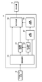
- FIG. 1 is a system configuration diagram of the endoscope system of the present embodiment.
- the endoscope system 1 of the present embodiment includes an endoscope 10, a light source device 20, a processor device 30, an endoscope image processing device 40, and a display device 50.
- the endoscope system 1 is an example of a medical image processing system.
- FIG. 2 is a schematic configuration diagram of the endoscope.
- the endoscope 10 is a flexible mirror (electronic endoscope).
- the endoscope 10 is mainly composed of an insertion unit 12, an operation unit 14, and a connection unit 16.
- the insertion portion 12 is mainly composed of a tip portion 12A, a curved portion 12B, and a soft portion 12C.
- the tip portion 12A is provided with an observation window, an illumination window, a nozzle, a forceps opening, and the like on its end face. Further, the tip portion 12A is provided with an imaging unit 12a inside.
- the imaging unit 12a includes a photographing optical system, an image sensor, and the like.
- the image sensor is composed of, for example, a color CCD (Charge Coupled Device) image sensor having a predetermined color filter arrangement (for example, a Bayer arrangement), a color CMOS (Complementary Metal-Oxide Semiconductor) image sensor, and the like.
- CCD Charge Coupled Device
- CMOS Complementary Metal-Oxide Semiconductor
- the operation unit 14 is provided with various operation members operated by an operator (user). For example, an angle knob 14A for operating the angle of the curved portion 12B, an air supply / water supply button 14B for performing an air supply / water supply operation, a suction button 14C for performing a suction operation, a shutter button 14D for performing a still image shooting, and the like are provided.
- an angle knob 14A for operating the angle of the curved portion 12B
- an air supply / water supply button 14B for performing an air supply / water supply operation
- a suction button 14C for performing a suction operation
- a shutter button 14D for performing a still image shooting, and the like are provided.
- the connecting portion 16 is composed of a flexible cord. At the tip of the connection portion 16, a connector 16A for connecting to the light source device 20 and a connector 16B for connecting to the processor device 30 are provided.
- the light source device 20 includes a light source, and supplies the light of the light source to the endoscope 10 as illumination light.
- the illumination light supplied to the endoscope 10 is emitted from an illumination window provided at the tip portion 12A.
- white light is used as the light of the light source.
- the processor device 30 takes in the imaging signal output from the endoscope 10 and performs predetermined signal processing to generate an observation image (endoscope image) by the endoscope 10.
- the processor device 30 also functions as a control unit for the entire system.
- the processor device 30 is composed of, for example, a computer equipped with a CPU (Central Processing Unit), a ROM (Read Only Memory), a RAM (Random Access Memory), and the like, and the CPU executes a predetermined program to perform internal viewing. A function of generating a mirror image and a function of controlling each part of the system are realized.
- Various programs executed by the CPU, data necessary for control, and the like are stored in the ROM.
- the RAM provides the CPU with a working memory space.
- the endoscope image processing device 40 acquires an endoscopic image output from the processor device 30 and displays it on the display device 50. Further, the endoscopic image processing device 40 detects a region of interest from the endoscopic image and notifies the detection.
- the endoscopic image processing device 40 is composed of, for example, a computer equipped with a CPU, ROM, RAM, and the like, and the CPU executes a predetermined program (endoscopic image processing program) to perform endoscopic image processing. Functions as a device. Various programs executed by the CPU, data necessary for control, and the like are stored in the ROM.
- the RAM provides the CPU with a working memory space.
- FIG. 3 is a block diagram of the functions of the endoscopic image processing device.
- the endoscopic image processing device 40 includes an image acquisition unit 40A for acquiring an endoscopic image, an attention area detection unit 40B for detecting an attention area from the acquired endoscopic image, and an acquired inside. It has the functions of a movement amount estimation unit 40C that estimates the movement amount of the endoscope 10 based on the endoscope image, and a display control unit 40D that controls the display of the endoscope image on the display device 50.
- the image acquisition unit 40A captures the endoscopic image output from the processor device 30.
- the endoscopic image is an example of a medical image.
- the imaging with the endoscope 10 is performed at a predetermined frame rate. Therefore, time-series endoscopic images are sequentially captured in the image acquisition unit 40A.
- the attention area detection unit 40B detects the attention area from the endoscopic image acquired by the image acquisition unit 40A.
- the "region of interest” here refers to a region suspected as a lesion in the endoscopic image.
- the attention area detection unit 40B detects the attention area from the endoscopic image by, for example, image recognition.
- Image recognition can be performed using, for example, an image recognition model generated by machine learning (for example, deep learning). In addition, it can be carried out by using a known method.
- the position and size of the region of interest are specified and detected.
- the position is acquired as information on the pixel position of the region of interest existing in the endoscopic image, for example.
- the size is acquired as, for example, the number of pixels in the region of interest existing in the endoscopic image.
- the motion amount estimation unit 40C estimates the motion amount of the endoscope 10 (a device for capturing a medical image) based on the endoscope image acquired by the image acquisition unit 40A. More specifically, the amount of movement of the tip portion 12A of the insertion portion 12 of the endoscope 10 provided with the imaging portion 12a is estimated.
- the “movement” here includes a movement due to the movement of the tip portion 12A, a movement due to the rotation of the tip portion 12A, a movement due to a change in direction, and the like.
- the "movement amount” is a value indicating the magnitude of movement of the tip portion 12A of the endoscope 10.
- the motion amount estimation unit 40C estimates the motion amount of the endoscope at predetermined frame intervals. Further, the motion amount estimation unit 40C estimates the motion amount of the endoscope based on the movement amount of the entire image. For example, when estimating the amount of movement of the endoscope for each frame, the amount of movement of the entire image is detected for each frame by comparing with the image of the immediately preceding frame. For example, when the acquired image is the image of the Nth frame, the movement amount of the entire image is detected by comparing with the image of the N-1th frame. Then, the amount of movement of the endoscope between frames is estimated based on the amount of movement of the entire detected image.
- the amount of movement of the entire image is detected, for example, as follows. First, in the front and rear frames, the corresponding feature points are extracted from the entire image. From the positional relationship of the extracted feature points, a motion vector representing the direction and magnitude of the motion of the entire image is obtained. The magnitude of the obtained motion vector is acquired as the amount of motion. When a plurality of feature points are extracted, the average value of the magnitudes of the motion vectors obtained from each feature point is calculated and acquired as the motion amount of the entire image.
- the motion amount estimation unit 40C estimates the motion amount of the endoscope 10 based on the endoscope image acquired by the image acquisition unit 40A when the attention region is detected by the attention region detection unit 40B. That is, when the region of interest is detected, the motion amount estimation process is started. Therefore, if the region of interest is not detected, the motion amount estimation process is not performed. In this way, the processing load (including the calculation load) in the endoscopic image processing device 40 can be reduced by performing the motion amount estimation processing only when the region of interest is detected.
- the attention region detection unit 40B detects the attention region, the motion amount estimation unit 40C is notified of the detection of the attention region.
- the motion amount estimation unit 40C starts the motion amount estimation process with the notification of this detection as a trigger.
- the display control unit 40D controls the display of the endoscopic image on the display device 50.
- the display control unit 40D causes the display device 50 to display the endoscopic image acquired by the image acquisition unit 40A as it is in a state where the region of interest is not detected. On the other hand, when the region of interest is detected, an image in which the region of interest is emphasized is displayed.
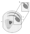
- FIG. 4 is a diagram showing an example of a display image when the region of interest is not detected.
- FIG. 5 is a diagram showing an example of a display image when a region of interest is detected.
- the endoscope image im1 taken by the endoscope 10 is displayed as it is.
- the endoscopic image im1 is displayed in a predetermined display area A set on the screen.
- the endoscopic image im2 in which the region of interest (the elliptical region indicated by the diagonal line in the same figure) X is emphasized is displayed.
- the endoscopic image im2 is displayed in a predetermined display area A set on the screen.
- the endoscopic image im2 in which the region of interest X is emphasized is generated by surrounding the region of interest X with a rectangular frame F in the image.
- the endoscopic image im2 on which the frame F is displayed is displayed on the display device 50, it is notified that the region of interest has been detected.
- the display control unit 40D displays the frame F with a predetermined line thickness (a reference first thickness).
- the degree of emphasis of the frame F (synonymous with the intensity of emphasis) is changed according to the amount of movement of the endoscope 10 estimated by the movement amount estimation unit 40C. That is, the thickness of the line of the frame F is changed.
- the line thickness of the frame F is made thicker than the first thickness. Change to the thickness (first thickness ⁇ second thickness).
- FIG. 6 is a diagram showing an example of a display image when the movement amount of the endoscope estimated by the movement amount estimation unit is equal to or larger than the threshold value.
- the thickness of the line of the frame F surrounding the attention region X becomes thicker.
- the degree of emphasis is strengthened.
- the display control unit 40D is an example of a notification unit, and by displaying a frame in the endoscopic image displayed on the display device 50, the operator is notified of the detection of the region of interest. In the case of this example, since the area of interest is surrounded by a frame, the detection result (position and size) of the area of interest is also notified.
- the display device 50 is composed of, for example, a display such as a liquid crystal display or an organic EL display (organic EL: Organic ElectroLuminescent, OEL).
- the display device 50 is an example of a display unit.
- the operation (operation method) of the endoscope system 1 of the present embodiment will be described.
- the photographed image endoscope image
- the region of interest is detected from the captured endoscopic image.
- the region of interest is surrounded by a frame in the displayed image, and the detection is notified (see FIG. 5).
- the mode of notification is changed according to the amount of movement of the endoscope 10 thereafter. That is, the degree of emphasis of the frame is changed.
- FIG. 7 is a flowchart showing a processing procedure from detection of the region of interest to notification.
- step S1 the endoscope image taken by the endoscope 10 is acquired (step S1).
- step S2 the region of interest is detected (step S2). That is, the region of interest is detected in the image based on the acquired endoscopic image.
- step S3 it is determined whether or not the region of interest has been detected based on the detection result of the region of interest.
- step S4 it is determined whether or not the shooting is completed. Whether or not the imaging is completed is determined by, for example, the presence or absence of an endoscopic image in the next frame. If the image acquisition unit 40A has not acquired the endoscopic image of the next frame, it is determined that the imaging has been completed. When the shooting is completed (YES in step S4), the process ends. On the other hand, when the shooting is not completed (when step S4 is NO), the process returns to step S1 and the image of the next frame is acquired.
- step S5 When the region of interest is detected (when step S3 is YES), the detection of the region of interest is notified (step S5). That is, as shown in FIG. 5, the region of interest X is displayed surrounded by a frame F in the endoscopic image being displayed. At this time, the frame F is displayed with the first thickness (reference thickness).
- FIG. 8 is a flowchart showing a processing procedure after notification of detection of the region of interest.
- the endoscopic image of the next frame is acquired (step S11).
- the region of interest is detected (step S12).
- it is determined whether or not the region of interest has been detected based on the detection result of the region of interest step S13).
- step S19 the notification is terminated (step S19). That is, the display of the frame F is turned off (the emphasis of the region of interest is turned off).
- the amount of movement of the endoscope 10 is estimated from the image (step S14). Then, the estimated movement amount and the threshold value are compared, and it is determined whether or not the estimated movement amount is equal to or greater than the threshold value (step S15).
- the threshold is predetermined.
- step S15 When the estimated amount of movement is equal to or greater than the threshold value (when step S15 is YES), the frame F surrounding the region of interest X is displayed with a second thickness as shown in FIG. 6 (step S16). That is, the degree of emphasis is increased and the frame F is displayed.
- step S15 when the estimated amount of movement is less than the threshold value (when step S15 is NO), the frame F is displayed with the first thickness (step S17). That is, as shown in FIG. 5, the frame F is displayed without increasing the degree of emphasis.
- step S18 it is determined whether or not the shooting is completed.
- the process is completed. If the photographing is not completed (when step S18 is NO), the process returns to step S11, and the endoscopic image of the next frame is acquired.
- the display mode of the frame F for notifying the detection of the region of interest is changed according to the amount of movement of the endoscope 10 after the detection. That is, when the amount of movement is large, the degree of emphasis is increased and displayed.
- the endoscope 10 moves significantly when the region of interest is automatically detected, it is assumed that the operator is passing by without noticing it. Therefore, in such a case, the degree of emphasis is increased to prevent oversight. As a result, the detection of the region of interest can be appropriately notified.
- the motion amount estimation process is performed only when the region of interest is detected. As a result, the processing load can be reduced and smoother diagnosis can be performed.
- Modification of the endoscope system of the first embodiment [1] Modification example of the method for changing the degree of emphasis when emphasizing the region of interest using a frame
- the degree of emphasis is changed by changing the thickness of the line of the frame.
- the method of change is not limited to this.
- the line type, color, shape, blinking degree, brightness, and the like of the frame can be changed to change the emphasis degree of the region of interest.
- the broken line is set as the reference line type (the line type displayed first when the region of interest is detected), and the movement exceeds the threshold value.
- the amount is changed to a solid line when it is estimated.
- a case where a solid line is used as a reference line type and a double line is changed when a movement amount exceeding a threshold value is estimated is exemplified.
- changing the frame color to change the degree of emphasis is when changing the frame color to a color that is easier to see (a conspicuous color). For example, white is used as a reference color (the color of the frame that is displayed first when the region of interest is detected), and when the amount of movement above the threshold is estimated, the color is changed to red or yellow. ..
- the case where the shape of the frame is changed to change the degree of emphasis is the case where the shape of the frame is changed to a shape that is more visible (a conspicuous shape).
- FIG. 9 is a diagram showing an example of a display image when the shape of the frame is changed to change the degree of emphasis.
- FIG. (A) shows an example of an image displayed on the display device 50 when a region of interest is detected.
- FIG. 3B shows an example of an image displayed on the display device 50 when the amount of movement after detection of the region of interest is equal to or greater than a threshold value.
- a frame F reference-shaped frame
- the frame F is changed to a frame F having a shape that completely surrounds the area of interest X as shown in FIG. ..
- changing the blinking degree means changing the blinking speed (blinking speed) when the frame is blinked and displayed. For example, when the area of interest is detected, the frame is blinked and displayed at the first blinking speed (reference blinking speed), and when the amount of movement above the threshold is estimated, it is higher than the first blinking speed. The frame is blinked and displayed at the fast second blinking speed. In addition, for example, when a region of interest is detected, the frame is displayed without blinking, and when the amount of movement equal to or greater than the threshold value is estimated, the frame is displayed by blinking.
- blinking speed blinking speed
- the frame when changing the brightness of the frame to change the degree of emphasis, for example, when a region of interest is detected, the frame is displayed with the first brightness (reference brightness), and the movement exceeds the threshold value.
- the frame is displayed with a second brightness that is brighter than the first brightness.
- the thickness and brightness of the frame can be changed to change the degree of emphasis of the area of interest.
- the degree of emphasis of the region of interest can be changed by changing the thickness of the frame and the degree of blinking.
- the degree of emphasis of the region of interest can be changed by changing the thickness, brightness, and degree of blinking of the frame.
- FIG. 10 is a flowchart showing a processing procedure after notification of detection of the region of interest in this modified example.
- the endoscopic image of the next frame is acquired (step S21).
- the region of interest is detected (step S22).
- step S29 if the region of interest is not detected (when step S23 is NO), the notification is terminated (step S29). That is, the display of the frame F is turned off (the emphasis of the region of interest is turned off).
- step S23 when the region of interest is detected (when step S23 is YES), the amount of movement of the endoscope 10 is estimated from the image (step S24). Then, the estimated movement amount and the threshold value are compared, and it is determined whether or not the estimated movement amount is less than the threshold value (step S25).
- the threshold is predetermined.
- FIG. 11 is a diagram showing an example of an image displayed on the display device when the amount of movement is less than the threshold value. As shown in the figure, the thickness of the frame F surrounding the region of interest X becomes thinner, and the degree of emphasis is weakened.
- step S25 when the estimated amount of movement is equal to or greater than the threshold value (when step S25 is NO), the frame F is displayed with the first thickness (step S27). That is, as shown in FIG. 5, the frame F is displayed without weakening the degree of emphasis.
- step S28 it is determined whether or not the shooting is completed.
- the process is completed. If the photographing is not completed (when step S28 is NO), the process returns to step S21, and the endoscopic image of the next frame is acquired.
- the degree of emphasis of the area of interest is weakened (the line of the frame surrounding the area of interest is thinned). ).
- the operator finds the lesion, the operator observes the lesion in detail, so that the movement of the endoscope 10 tends to be small. If the movement of the endoscope 10 becomes small after the detection of the region of interest, it is considered that the operator is also aware of it. Therefore, when the movement of the endoscope 10 becomes small after the detection of the region of interest, if the notification is continued with the same intensity, the concentration may be lowered. Therefore, when the amount of movement of the endoscope becomes small after the detection of the region of interest, the degree of emphasis of the region of interest is weakened. As a result, the detection of the region of interest can be appropriately notified.
- the degree of emphasis is weakened (the line of the frame is thinned), but the highlighting is turned off (the frame is turned off and the notification is sent. It can also be terminated).
- the degree of emphasis may be changed by changing the line type of the frame, the degree of blinking of the frame, the brightness of the frame, etc. You can also.
- FIG. 12 is a flowchart showing a processing procedure after notification of detection of the region of interest in this modified example.
- the endoscopic image is acquired (step S31).
- the region of interest is detected (step S32).
- step S41 if the region of interest is not detected (when step S33 is NO), the notification is terminated (step S41). That is, the display of the frame F is turned off (the emphasis of the region of interest is turned off).
- step S34 the amount of movement of the endoscope 10 is estimated from the image (step S34). Then, the estimated amount of movement is compared with the first threshold value, and it is determined whether or not the estimated amount of movement is equal to or greater than the first threshold value (step S35).
- the first threshold is predetermined.
- Step S35 When the estimated amount of movement is equal to or greater than the first threshold value (when step S35 is YES), the frame F surrounding the region of interest X is displayed as the second thickness (first thickness ⁇ second thickness). (Step S36). That is, as shown in FIG. 6, the attention region X is displayed surrounded by a thicker frame F, and the degree of emphasis of the attention region X is strengthened.
- the estimated movement amount is less than the first threshold value (when step S35 is NO)
- the estimated movement amount is compared with the second threshold value, and the estimated movement amount is the second threshold value. It is determined whether or not it is less than (step S37).
- the second threshold is predetermined.
- the second threshold value is set to a value smaller than the first threshold value (second threshold value ⁇ first threshold value).
- step S38 When the estimated amount of movement is less than the second threshold value (when step S37 is YES), the frame F surrounding the region of interest X is displayed with a third thickness (third thickness ⁇ first thickness). That is, as shown in FIG. 11, the attention region X is displayed surrounded by a frame F having a thinner thickness, and the degree of emphasis of the attention region X is weakened.
- step S37 when the estimated amount of movement is equal to or greater than the second threshold value (when step S37 is NO), the frame F surrounding the region of interest X has a first thickness (third thickness ⁇ first thickness ⁇ .
- the second thickness) is displayed (step S39). That is, as shown in FIG. 5, the degree of emphasis is not changed, and the frame F is displayed with the reference thickness (first thickness).
- step S40 it is determined whether or not the shooting is completed.
- the process is completed. If the imaging is not completed (NO in step S40), the process returns to step S31 and the next endoscopic image is acquired.
- the amount of movement of the endoscope 10 is estimated after the region of interest is emphasized and displayed, and when the estimated amount of movement of the endoscope 10 is large, the degree of emphasis is increased. (The line of frame F is thickened). Further, when the estimated amount of movement of the endoscope 10 is small, the degree of emphasis is weakened (the line of the frame F is thinned).
- the degree of emphasis is increased to prevent oversight.
- the degree of emphasis is weakened in order to prevent a decrease in concentration.
- the degree of emphasis is weakened (the line of the frame is thinned), but the highlighting is turned off (the frame is changed). It can also be erased).
- the degree of emphasis may be changed by changing the line type of the frame, the degree of blinking of the frame, the brightness of the frame, etc. You can also.
- (C) Modification example 3 of the method of changing the degree of emphasis according to the amount of movement the degree of emphasis is changed in two steps, but it may be changed in multiple steps. That is, the degree of emphasis may be changed stepwise according to the amount of movement of the endoscope 10. For example, when changing the thickness of the frame line to change the degree of emphasis, the thickness of the frame line can be changed stepwise according to the amount of movement.
- FIG. 13 is a system configuration diagram of the endoscope system of the present embodiment.
- the endoscope system 1 of the present embodiment is different from the endoscope system 1 of the first embodiment in that it further includes a speaker 60.
- FIG. 14 is a block diagram of the functions of the endoscopic image processing device.
- the endoscopic image processing device 40 of the present embodiment further has the function of the sound output control unit 40E that controls the output of the sound generated from the speaker 60. It is different from the endoscopic image processing device 40 of the form. In the following, only the main differences from the endoscopic image processing apparatus 40 of the first embodiment will be described.
- an endoscope image in which the detected region of interest X is surrounded by a frame F is displayed on the display device 50. (See FIG. 5).
- a predetermined notification sound (voice for notifying the detection of the region of interest) is output from the speaker 60.
- the volume of the notification sound is changed according to the amount of movement of the endoscope thereafter. Specifically, when the amount of movement of the endoscope 10 estimated by the amount of movement estimation unit 40C is equal to or greater than a predetermined threshold value, the volume is increased.
- the audio output generated from the speaker 60 is controlled by the audio output control unit 40E.
- the voice output control unit 40E controls the output of the notification sound from the speaker 60 based on the detection result of the attention area by the attention area detection unit 40B and the estimation result of the movement amount of the endoscope by the movement amount estimation unit 40C. To do. Specifically, the following processing is carried out.
- the voice output control unit 40E When the region of interest is detected by the region of interest 40B, the voice output control unit 40E outputs a notification sound from the speaker 60 at a predetermined first volume. After that, the voice output control unit 40E changes the volume of the notification sound according to the movement amount of the endoscope estimated by the movement amount estimation unit 40C. Specifically, when the amount of movement exceeds the threshold value, the volume is changed to a second volume (first volume ⁇ second volume) that is larger than the first volume.
- the voice output control unit 40E and the speaker 60 are examples of the notification unit.
- the detection of the region of interest is notified by sound, so when the region of interest is detected, the notification sound from the speaker 60 is output. At this time, a notification sound is output at the first volume to notify the detection of the region of interest.
- FIG. 15 is a flowchart showing a processing procedure after notification of detection of the region of interest.
- step S51 the endoscopic image of the next frame is acquired.
- step S52 the region of interest is detected.
- step S53 it is determined whether or not the region of interest has been detected based on the detection result of the region of interest.
- step S59 if the region of interest is not detected (when step S53 is NO), the output of the notification sound is turned off (step S59).
- step S53 when the region of interest is detected (YES in step S53), the amount of movement of the endoscope 10 is estimated from the image (step S54). Then, the estimated movement amount and the threshold value are compared, and it is determined whether or not the estimated movement amount is equal to or greater than the threshold value (step S55).
- the threshold is predetermined.
- step S55 When the estimated amount of movement is equal to or greater than the threshold value (when step S55 is YES), the notification sound is output from the speaker 60 at a second volume which is a louder volume (step S56).
- step S57 when the estimated amount of movement is less than the threshold value (when step S55 is NO), the notification sound is output from the speaker 60 at the first volume (step S57).
- step S58 it is determined whether or not the shooting is completed.
- step S58 it is determined whether or not the shooting is completed.
- step S58 the process is finished. If the imaging is not completed (NO in step S58), the process returns to step S51 and the next endoscopic image is acquired.
- the endoscope system 1 of the present embodiment when the detection of the region of interest is notified by sound, the volume thereof is changed according to the amount of movement of the endoscope 10 after the detection. .. As a result, when the region of interest is automatically detected, it can be appropriately prevented from being overlooked.
- the detection of the region of interest is notified only by sound has been described as an example, but it can also be used in combination with the notification on the screen. That is, apart from the notification by sound, the region of interest can be highlighted on the display screen for displaying the endoscopic image, and the detection result can be displayed.
- the volume of the notification sound is changed according to the amount of movement, but in addition, the tone color or type of the notification sound may be changed. In addition, the combination of volume, timbre, and type can be changed.
- the estimated amount of movement is compared with the threshold value, and the volume of the notification sound is increased when the amount is equal to or more than the threshold value.
- the mode of changing the volume is not limited to this. Absent.
- the estimated amount of movement may be compared with the threshold value, and if it is less than the threshold value, the volume of the notification sound may be reduced or the notification sound may be turned off.
- the estimated movement amount is compared with the first threshold value and the second threshold value (second threshold value ⁇ first threshold value), and the volume is increased when the movement amount of the endoscope is equal to or more than the first threshold value. However, if it is less than the second threshold value, the volume may be reduced or the notification sound may be turned off.
- the volume of the notification sound can be changed stepwise according to the amount of movement.
- the display mode of the region of interest may be changed along with the sound.
- the detection result is based on the amount of movement of the endoscope thereafter.
- An image showing is displayed. That is, when the endoscope is moved by a certain amount of movement or more after the detection, an image showing the detection result is displayed.
- the basic configuration of the endoscope system is the same as that of the first embodiment.
- the functions of the endoscopic image processing device 40 are different. Therefore, only the functions of the endoscopic image processing device 40 will be described here.
- FIG. 16 is a block diagram of the functions of the endoscopic image processing device of the present embodiment.
- the endoscopic image processing device 40 has an image generation unit 40F that generates an image showing a detection result of a region of interest, and an image storage unit that stores an image generated by the image generation unit 40F. It differs from the endoscopic image processing device 40 of the first embodiment in that it further has a function of 40G.
- the image generation unit 40F generates an image (emphasized image) in which the region of interest is emphasized in the endoscopic image as an image showing the detection result of the region of interest.
- the image generation unit 40F generates an emphasized image from the endoscopic image acquired by the image acquisition unit 40A based on the detection result of the attention area by the attention area detection unit 40B.
- FIG. 17 is a diagram showing an example of an emphasized image.
- the emphasized image im3 is composed of an image in which the region of interest (the elliptical region shown by the diagonal line in the figure) X is surrounded by the rectangular frame F in the endoscopic image im.
- the image generation unit 40F is an example of an information generation unit.
- the emphasized image is an example of information showing the detection result of the region of interest.
- the image storage unit 40G stores the emphasized image generated by the image generation unit 40F.
- the image storage unit 40G stores the emphasized images sequentially generated by the image generation unit 40F while sequentially rewriting them. Therefore, the latest emphasized image is stored in the image storage unit 40G.
- the image storage unit 40G is composed of RAM.
- the image storage unit 40G is an example of a storage unit.
- the display control unit 40D causes the display device 50 to display an endoscope image (an image taken by the endoscope 10) acquired by the image acquisition unit 40A. At this time, the display control unit 40D causes the endoscope image to be displayed in the first display area A1 set in the screen of the display device 50. Further, the display control unit 40D causes the display device 50 to display the emphasized image stored in the image storage unit 40G under certain conditions.
- the conditions for displaying the highlighted image are as follows. That is, the region of interest is detected by the region of interest detection unit 40B, and the amount of movement of the endoscope 10 thereafter is equal to or greater than the threshold value.
- the amount of movement of the endoscope 10 is the amount of movement of the endoscope 10 estimated by the movement amount estimation unit 40C after the detection of the region of interest.
- the emphasized image is displayed on the display device 50.
- the highlighted image is displayed in the second display area A2 set in the screen of the display device 50.
- the second display area A2 is an area different from the first display area A1.
- FIG. 18 is a diagram showing an example of a display image in a normal display state.
- the "normal display state” here means a state in which the emphasized image is not displayed.
- the endoscope image im taken by the endoscope 10 is displayed on the screen.
- the endoscopic image im is displayed in the first display area A1.
- FIG. 19 is a diagram showing an example of a displayed image when the emphasized image is displayed.
- the endoscope image im taken by the endoscope 10 is displayed in the first display area A1, and the emphasized image im3 is displayed in the second display area A2.
- the first display area A1 (so-called main window) is set large at a position on the left side of the screen, and the second display area A2 (so-called sub-window) is set small at a position on the lower right side of the screen.
- the first display area A1 (so-called main window) is set large at a position on the left side of the screen
- the second display area A2 (so-called sub-window) is set small at a position on the lower right side of the screen.
- An example of the case is shown.
- the display control unit 40D determines the necessity of displaying the emphasized image based on the movement amount of the endoscope 10 estimated by the movement amount estimation unit 40C. Specifically, the movement amount of the endoscope 10 estimated by the movement amount estimation unit 40C is compared with the threshold value, and it is determined that the emphasized image is displayed in the second display area A2 when the movement amount is equal to or more than the threshold value. ..
- the motion amount estimation unit 40C performs the motion amount estimation process when the attention region is detected by the attention region detection unit 40B. Therefore, the display control unit 40D outputs the estimation result of the subsequent movement amount of the endoscope 10 only when the region of interest is detected.
- FIG. 20 is a flowchart showing a processing procedure for displaying an endoscope image in the endoscope system of the present embodiment.
- the endoscope image taken by the endoscope 10 is acquired (step S61). Then, the acquired endoscopic image is displayed on the display device 50 (step S62). The endoscopic image is displayed in the first display area A1 as shown in FIG. In addition, the region of interest is detected from the acquired endoscopic image (step S63). Then, based on the detection result, it is determined whether or not the region of interest is detected (step S64).
- step S64 If the region of interest is not detected (when step S64 is NO), the process proceeds to step S69, and it is determined whether or not the shooting is completed. On the other hand, when the region of interest is detected (YES in step S64), the enhanced image is generated, and the generated enhanced image is stored in the image storage unit 40G (step S65).
- the amount of movement of the endoscope 10 is estimated from the image (step S66). Then, the estimated movement amount and the threshold value are compared, and it is determined whether or not the estimated movement amount is equal to or greater than the threshold value (step S67).
- step S67 If the estimated amount of movement is less than the threshold value (when step S67 is NO), the process proceeds to step S69, and it is determined whether or not the shooting is completed. On the other hand, when the estimated amount of movement is equal to or greater than the threshold value (when step S67 is YES), as shown in FIG. 19, an image in which the highlighted image im3 is displayed in the second display area A2 is displayed on the display device 50. (Step S68). After that, it is determined whether or not the shooting is completed (step S69). When the shooting is completed (YES in step S69), the process is completed. If the imaging is not completed (NO in step S69), the process returns to step S61 and the next endoscopic image is acquired.
- the highlighted image displayed in the second display area A2 is erased after a certain period of time has passed since it was last displayed.
- the time until erasure is predetermined (for example, 6 seconds). The operator may arbitrarily set this time.
- an image showing the detection result of the region of interest is displayed on the display device 50 only when the endoscope 10 is moved by a certain amount of movement or more. ..
- an image showing the detection result of the region of interest is displayed on the display device 50 only when the endoscope 10 is moved by a certain amount of movement or more, so that detection can be performed appropriately. Can be notified. That is, since the result is displayed only when there is a high possibility that it has been overlooked, the detection can be appropriately notified.
- the case where there is a high possibility that the endoscope is overlooked is a case where the endoscope 10 is greatly moved after the automatic detection of the region of interest. As a result, oversight can be reduced without affecting the diagnosis.
- the volume of the notification sound is changed according to the amount of movement, but in addition, the tone color or type of the notification sound may be changed. In addition, the combination of volume, timbre, and type can be changed.
- the estimated amount of movement is compared with the threshold value, and the volume of the notification sound is increased when the amount is equal to or more than the threshold value.
- the mode of changing the volume is not limited to this. Absent.
- the estimated amount of movement may be compared with the threshold value, and if it is less than the threshold value, the volume of the notification sound may be reduced or the notification sound may be turned off.
- the estimated movement amount is compared with the first threshold value and the second threshold value (second threshold value ⁇ first threshold value), and the volume is increased when the movement amount of the endoscope is equal to or more than the first threshold value. However, if it is less than the second threshold value, the volume may be reduced or the notification sound may be turned off.
- the volume of the notification sound can be changed stepwise according to the amount of movement.
- the display mode of the region of interest may be changed along with the sound.
- FIG. 21 is a conceptual diagram for generating a crop image. As shown in the figure, an image of the region W including the region of interest X is cut out from the endoscopic image im to generate a crop image im4. When displaying the information indicating the detection result of the region of interest, the crop image im4 is displayed in the second display region A2.
- the detection may be notified by sound. Even when using sound notification, it is possible that the area of interest on the screen cannot be recognized. Even in such a case, if the amount of movement does not decrease after the notification, it is determined that the motion is overlooked, and the detection result of the region of interest is displayed to appropriately prevent the oversight.
- the amount of movement of the endoscope is estimated based on the amount of movement of the entire image between the previous and next frames.
- the method of estimating the amount of movement of the endoscope is not limited to this.
- the amount of movement of the endoscope can be estimated based on the amount of movement of the region of interest in the endoscopic image.
- the amount of movement of the region of interest for example, first, in the frames before and after, the corresponding feature points are extracted from the region of interest.
- the motion vector of the region of interest is obtained from the positional relationship of the extracted feature points.
- the magnitude of the obtained motion vector is acquired as the amount of motion in the region of interest.
- the average value of the magnitudes of the motion vectors obtained from each feature point is calculated and acquired as the amount of motion in the region of interest.
- the amount of movement of the imaging unit is estimated from various known methods (time-series images (moving images)). Method) can be adopted.
- the present invention is applied to a system (endoscopic system) for processing an image (endoscopic image) taken by an endoscope as a medical image
- a system endoscopic system
- it can be applied to a system (ultrasonic diagnostic system) that processes an ultrasonic diagnostic image as a medical image.
- the ultrasonic diagnostic system similar to the endoscopic system, an operation of searching for a lesion is performed while moving an ultrasonic probe (a device for capturing a medical image). Therefore, the present invention works effectively as in the endoscopic system.
- any system in which an operation of searching for a lesion or the like is performed while moving a device can be similarly applied and effectively functioned.
- processors include CPUs, which are general-purpose processors that execute programs and function as various processing units, and programmable logic devices (programmable logic devices), which are processors whose circuit configurations can be changed after manufacturing, such as FPGAs (Field Programmable Gate Arrays).
- FPGAs Field Programmable Gate Arrays
- a dedicated electric circuit which is a processor having a circuit configuration specially designed for executing a specific process such as a Programmable Logic Device (PLD) and an ASIC (Application Specific Integrated Circuit), is included.
- PLD Programmable Logic Device
- ASIC Application Specific Integrated Circuit
- One processing unit may be composed of one of these various processors, or may be composed of two or more processors of the same type or different types.
- one processing unit may be composed of a plurality of FPGAs or a combination of a CPU and an FPGA.
- a plurality of processing units may be configured by one processor.
- one processor is configured by a combination of one or more CPUs and software, as represented by a computer such as a client or a server. There is a form in which the processor functions as a plurality of processing units.
- SoC System On Chip
- a processor that realizes the functions of the entire system including a plurality of processing units with one IC (Integrated Circuit) chip is used.
- the various processing units are configured by using one or more of the above-mentioned various processors as a hardware-like structure.
- circuitry that combines circuit elements such as semiconductor elements.
- the function of the endoscopic image processing device can be mounted on the processor device constituting the endoscopic device.
- the illumination light white light, light in one or more specific wavelength bands, or light in various wavelength bands according to the observation purpose such as a combination thereof is selected.
- the "specific wavelength band” is a band narrower than the white wavelength band. Specific examples relating to a specific wavelength band are shown below.
- the first example of a specific wavelength band is, for example, a blue band or a green band in the visible region.
- the wavelength band of the first example includes a wavelength band of 390 nm or more and 450 nm or less or a wavelength band of 530 nm or more and 550 nm or less, and the light of the first example is in the wavelength band of 390 nm or more and 450 nm or less or 530 nm or more and 550 nm or less. It has a peak wavelength within the wavelength band.
- the second example of a specific wavelength band is, for example, the red band in the visible region.
- the wavelength band of the second example includes a wavelength band of 585 nm or more and 615 nm or less or a wavelength band of 610 nm or more and 730 nm or less, and the light of the second example is within the wavelength band of 585 nm or more and 615 nm or less or 610 nm or more and 730 nm or less. It has a peak wavelength within the wavelength band.
- the third example of a specific wavelength band includes a wavelength band in which the absorption coefficient differs between oxidized hemoglobin and reduced hemoglobin, and the light in the third example peaks in a wavelength band in which the absorption coefficient differs between oxidized hemoglobin and reduced hemoglobin.
- the wavelength band of the third example includes a wavelength band of 400 ⁇ 10 nm, 440 ⁇ 10 nm, a wavelength band of 470 ⁇ 10 nm, or a wavelength band of 600 nm or more and 750 nm or less, and the light of the third example is the above 400 ⁇ . It has a peak wavelength in the wavelength band of 10 nm, 440 ⁇ 10 nm, 470 ⁇ 10 nm, or 600 nm or more and 750 nm or less.
- the fourth example of the specific wavelength band is used for observing the fluorescence emitted by the fluorescent substance in the living body (fluorescence observation), and the wavelength band of the excitation light that excites the fluorescent substance, for example, 390 nm to 470 nm.
- the fifth example of a specific wavelength band is the wavelength band of infrared light.
- the wavelength band of the fifth example includes a wavelength band of 790 nm or more and 820 nm or less or a wavelength band of 905 nm or more and 970 nm or less, and the light of the fifth example is in the wavelength band of 790 nm or more and 820 nm or less or 905 nm or more and 970 nm or less. It has a peak wavelength within the wavelength band.
- a laser light source As the type of light source, a laser light source, a xenon light source, an LED light source (LED: Light-Emitting Diode), or an appropriate combination thereof can be adopted.
- the type and wavelength of the light source, the presence or absence of a filter, etc. are preferably configured according to the type of subject, the purpose of observation, etc., and during observation, the wavelength of the illumination light is determined according to the type of subject, the purpose of observation, etc. It is preferable to combine and / or switch.
- switching the wavelength for example, by rotating a disk-shaped filter (rotary color filter) arranged in front of the light source and provided with a filter that transmits or blocks light of a specific wavelength, the wavelength of the emitted light is changed. You may switch.
- the image sensor provided in the imaging unit of the endoscope is not limited to the color image sensor in which the color filter is provided for each pixel, and may be a monochrome image sensor.
- the wavelength of the illumination light can be sequentially switched to perform surface-sequential (color-sequential) imaging.
- the wavelength of the emitted illumination light may be sequentially switched between purple, blue, green, and red, or the illumination light emitted by a rotary color filter (red, green, blue, etc.) that irradiates white light. You may switch the wavelength of.
- the wavelength of the illumination light emitted by the rotary color filter by irradiating one or a plurality of narrow band lights may be switched.
- the narrow band light may be infrared light having two or more wavelengths having different wavelengths.
- the processor device may generate an image (so-called special light image) having information in a specific wavelength band based on an image (so-called normal light image) obtained by imaging with white light.
- the processor device is, for example, red (Red, R), green (Green, G) and blue (Blue, B), or cyan (Cyan, C), magenta (Magenta, M) and yellow, which are usually contained in an optical image.
- a signal in a specific wavelength band can be acquired by performing an operation based on the color information of (Yellow, Y).
- a program for realizing the function of the endoscopic image processing device described in the above embodiment on a computer is recorded on an optical disk, a magnetic disk, or a computer-readable medium such as a semiconductor memory or other tangible non-temporary information storage medium.
- a program through this information storage medium.
- the program signal instead of the mode in which the program is stored and provided in such a tangible non-temporary information storage medium, it is also possible to provide the program signal as a download service by using a telecommunication line such as the Internet.
Abstract
Provided are a medical image processing system and a method for operating the same, whereby detection of a region of interest can be appropriately reported. In the present invention, a region of interest is detected from an endoscope image captured by an endoscope. When the region of interest is detected, the endoscope image is displayed in a display device (50) so that the region of interest is emphasized, and detection of a region of interest is reported. After the reporting, the degree of emphasis of the region of interest is changed in accordance with an endoscope movement amount estimated from the endoscope image.
Description
本発明は、医療画像処理システム及びその動作方法に関する。
The present invention relates to a medical image processing system and an operation method thereof.
内視鏡で撮影した画像(内視鏡画像)を解析し、注目領域を自動で検出して報知するシステムが知られている。
There is known a system that analyzes an image taken with an endoscope (endoscopic image) and automatically detects and notifies the area of interest.
特許文献1には、注目領域を検出した場合に、注目領域の位置を強調するための視覚情報を観察中の内視鏡画像に付加して表示すること、及び、その視覚情報の表示態様を注目領域の観察時間に応じて変えることが記載されている。
In Patent Document 1, when a region of interest is detected, visual information for emphasizing the position of the region of interest is added to and displayed on the endoscopic image being observed, and the display mode of the visual information is described. It is described that it changes according to the observation time of the region of interest.
特許文献2には、ユーザが病変部の鑑別を行っている状態か否かを判定し、鑑別を行っている状態の場合にのみ鑑別の支援処理を行うことが記載されている。
Patent Document 2 describes that it is determined whether or not the user is in a state where the lesion is being differentiated, and the discrimination support process is performed only when the user is in the state of being differentiated.
特許文献3には、注目領域を強調処理する場合に、被写体の動き量に応じて、強調処理する対象を変えることが記載されている。
Patent Document 3 describes that when the region of interest is emphasized, the object to be emphasized is changed according to the amount of movement of the subject.
特許文献4には、注目領域が検出された場合にアラート情報を表示すること、及び、そのアラート情報の表示時間を画像の動き量に応じて変えることが記載されている。
Patent Document 4 describes that alert information is displayed when a region of interest is detected, and that the display time of the alert information is changed according to the amount of movement of the image.
注目領域が検出された場合に、その検出をユーザに報知することは重要である。しかし、ユーザが病変部を発見した後も報知され続けると、かえって診断の妨げになるという問題がある。逆に、ユーザに報知したにもかかわらず、ユーザが病変部を見つけられないという問題もある。
When a region of interest is detected, it is important to notify the user of the detection. However, if the user continues to be notified even after the lesion is found, there is a problem that the diagnosis is rather hindered. On the contrary, there is also a problem that the user cannot find the lesion even though the user is notified.
本発明は、このような事情に鑑みてなされたもので、注目領域の検出を適切に報知できる医療画像処理システム及びその動作方法を提供することを目的とする。
The present invention has been made in view of such circumstances, and an object of the present invention is to provide a medical image processing system capable of appropriately notifying the detection of a region of interest and an operation method thereof.
(1)医療画像を取得する画像取得部と、医療画像を表示する表示部と、医療画像内から注目領域を検出する注目領域検出部と、医療画像に基づいて、医療画像を撮像する機器の動き量を推定する動き量推定部と、注目領域が検出された場合に注目領域の検出を報知し、かつ、報知後に動き量推定部で推定される動き量に応じて報知の態様を変更する報知部と、を備える医療画像処理システム。
(1) An image acquisition unit for acquiring a medical image, a display unit for displaying a medical image, a region of interest detection unit for detecting a region of interest in the medical image, and a device for capturing a medical image based on the medical image. The motion amount estimation unit that estimates the motion amount and the motion amount estimation unit that notifies the detection of the attention area when the attention area is detected, and changes the notification mode according to the motion amount estimated by the motion amount estimation unit after the notification. A medical image processing system equipped with a notification unit.
(2)動き量推定部は、注目領域が検出された場合に動き量を推定する、(1)の医療画像処理システム。
(2) The motion amount estimation unit is the medical image processing system of (1) that estimates the motion amount when the region of interest is detected.
(3)報知部は、表示部に表示される医療画像内で注目領域を強調して注目領域の検出を報知し、報知後に動き量推定部で推定される動き量に応じて注目領域の強調度合を変更する、(1)又は(2)の医療画像処理システム。
(3) The notification unit emphasizes the area of interest in the medical image displayed on the display unit to notify the detection of the area of interest, and after the notification, emphasizes the area of interest according to the amount of movement estimated by the movement amount estimation unit. The medical image processing system of (1) or (2) that changes the degree.
(4)報知部は、表示部に表示される医療画像内で注目領域を枠で囲って注目領域を強調する、(3)の医療画像処理システム。
(4) The notification unit is the medical image processing system of (3) that emphasizes the area of interest by surrounding the area of interest with a frame in the medical image displayed on the display unit.
(5)報知部は、枠の太さ、線種、色、形状、点滅度合及び明るさの少なくとも一つを変えて、注目領域の強調度合を変更する、(4)の医療画像処理システム。
(5) The notification unit is the medical image processing system of (4) that changes the degree of emphasis of the area of interest by changing at least one of the thickness, line type, color, shape, blinking degree, and brightness of the frame.
(6)報知部は、注目領域の報知後、動き量推定部で推定される動き量が閾値以上の場合に注目領域の強調度合を強くする、(3)から(5)のいずれか一の医療画像処理システム。
(6) The notification unit enhances the degree of emphasis of the attention area when the movement amount estimated by the movement amount estimation unit is equal to or more than the threshold value after the notification of the attention area is performed, whichever is one of (3) to (5). Medical image processing system.
(7)報知部は、注目領域の報知後、動き量推定部で推定される動き量が閾値未満の場合に注目領域の強調度合を弱くする、又は、注目領域の強調をオフする、(3)から(6)のいずれか一の医療画像処理システム。
(7) After notifying the attention area, the notification unit weakens the emphasis of the attention area or turns off the emphasis of the attention area when the movement amount estimated by the movement amount estimation unit is less than the threshold value (3). ) To (6), any one of the medical image processing systems.
(8)報知部は、注目領域の報知後、動き量推定部で推定される動き量が第1の閾値以上の場合に注目領域の強調度合を強くし、第2の閾値未満の場合に注目領域の強調度合を弱くする、又は、注目領域の強調をオフする、(3)から(5)のいずれか一の医療画像処理システム。
(8) After notifying the region of interest, the notification unit enhances the emphasis of the region of interest when the amount of motion estimated by the motion amount estimation unit is equal to or greater than the first threshold value, and pays attention when the amount of movement is less than the second threshold value. The medical image processing system according to any one of (3) to (5), which reduces the degree of emphasis of the area or turns off the emphasis of the area of interest.
(9)報知部は、音を発生させて、注目領域の検出を報知し、報知後に動き量推定部で推定される動き量に応じて音の音量、音色及び種類の少なくとも一つを変更する、(1)又は(2)の医療画像処理システム。
(9) The notification unit generates a sound to notify the detection of the region of interest, and after the notification, changes at least one of the volume, tone color, and type of the sound according to the movement amount estimated by the movement amount estimation unit. , (1) or (2) medical image processing system.
(10)医療画像を取得する画像取得部と、医療画像内から注目領域を検出する注目領域検出部と、注目領域が検出された場合に検出結果を示す情報を生成する情報生成部と、情報生成部で生成された画像を記憶する記憶部と、注目領域が検出された場合に医療画像に基づいて、医療画像を撮像する機器の動き量を推定する動き量推定部と、画面内に第1表示領域及び第2表示領域を有する表示部と、画像取得部で取得される医療画像を第1表示領域に表示する一方、注目領域が検出された場合において、注目領域の検出後に動き量推定部で推定される動き量が閾値以上の場合に、記憶部に記憶された情報を第2表示領域に表示する表示制御部と、を備えた医療画像処理システム。
(10) An image acquisition unit that acquires a medical image, a region of interest detection unit that detects a region of interest from within the medical image, an information generation unit that generates information indicating a detection result when the region of interest is detected, and information. A storage unit that stores the image generated by the generation unit, a motion amount estimation unit that estimates the movement amount of the device that captures the medical image based on the medical image when the region of interest is detected, and a motion amount estimation unit on the screen. While the display unit having the first display area and the second display area and the medical image acquired by the image acquisition unit are displayed in the first display area, when the area of interest is detected, the amount of motion is estimated after the area of interest is detected. A medical image processing system including a display control unit that displays information stored in the storage unit in a second display area when the amount of movement estimated by the unit is equal to or greater than a threshold value.
(11)情報生成部は、注目領域の検出結果を示す情報として、医療画像内で注目領域を強調した画像を生成する、(10)の医療画像処理システム。
(11) The medical image processing system of (10), in which the information generation unit generates an image in which the attention region is emphasized in the medical image as information indicating the detection result of the attention region.
(12)情報生成部は、医療画像内で注目領域を枠で囲って強調した画像を生成する、(11)の医療画像処理システム。
(12) The information generation unit is the medical image processing system of (11) that generates an image in which the area of interest is surrounded by a frame in the medical image and emphasized.
(13)情報生成部は、注目領域の検出結果を示す情報として、医療画像から注目領域を切り出した画像を生成する、(10)の医療画像処理システム。
(13) The medical image processing system of (10), in which the information generation unit generates an image obtained by cutting out the region of interest from the medical image as information indicating the detection result of the region of interest.
(14)動き量推定部は、あらかじめ定められたフレーム間隔で動き量を推定する、(1)から(13)のいずれか一の医療画像処理システム。
(14) The motion amount estimation unit is a medical image processing system according to any one of (1) to (13), which estimates the motion amount at predetermined frame intervals.
(15)動き量推定部は、画像全体の移動量に基づいて動き量を推定する、(14)の医療画像処理システム。
(15) The movement amount estimation unit is the medical image processing system of (14) that estimates the movement amount based on the movement amount of the entire image.
(16)動き量推定部は、注目領域の移動量に基づいて動き量を推定する、(14)の医療画像処理システム。
(16) The movement amount estimation unit is the medical image processing system of (14) that estimates the movement amount based on the movement amount of the region of interest.
(17)医療画像を取得するステップと、医療画像内から注目領域を検出するステップと、注目領域が検出された場合に注目領域の検出を報知するステップと、注目領域が検出された場合に、医療画像を撮像する機器の動き量を推定するステップと、推定された動き量に応じて報知の態様を変更するステップと、を含む医療画像処理システムの動作方法。
(17) A step of acquiring a medical image, a step of detecting a region of interest in the medical image, a step of notifying the detection of the region of interest when the region of interest is detected, and a step of notifying the detection of the region of interest when the region of interest is detected. An operation method of a medical image processing system including a step of estimating the amount of movement of a device that captures a medical image and a step of changing a mode of notification according to the estimated amount of movement.
(18)医療画像を取得するステップと、医療画像を表示部の画面内に設定された第1表示領域に表示するステップと、医療画像内から注目領域を検出するステップと、注目領域が検出された場合に検出結果を示す情報を生成するステップと、生成された注目領域の検出結果を示す情報を記憶部に記憶するステップと、注目領域が検出された場合に、医療画像を撮像する機器の動き量を推定するステップと、推定された動き量が閾値以上の場合に、記憶部に記憶された情報を表示部の画面内に設定された第2表示領域に表示するステップと、を含む医療画像処理システムの動作方法。
(18) A step of acquiring a medical image, a step of displaying the medical image in the first display area set in the screen of the display unit, a step of detecting a region of interest in the medical image, and a step of detecting the region of interest are detected. A step of generating information indicating the detection result in the case of, a step of storing the information indicating the detected detection result of the generated region of interest in the storage unit, and a step of storing a medical image when the region of interest is detected. Medical medical imaging including a step of estimating the amount of movement and a step of displaying the information stored in the storage unit in the second display area set in the screen of the display unit when the estimated amount of movement is equal to or greater than the threshold value. How the image processing system works.
本発明によれば、注目領域の検出を適切に報知できる。
According to the present invention, the detection of the region of interest can be appropriately notified.
以下、添付図面に従って本発明の好ましい実施形態について詳説する。
Hereinafter, preferred embodiments of the present invention will be described in detail with reference to the accompanying drawings.
[第1の実施の形態]
ここでは、医療画像として内視鏡で撮影された画像(内視鏡画像)を処理するシステム(内視鏡システム)に本発明を適用した場合を例に説明する。 [First Embodiment]
Here, a case where the present invention is applied to a system (endoscopic system) that processes an image (endoscopic image) taken by an endoscope as a medical image will be described as an example.
ここでは、医療画像として内視鏡で撮影された画像(内視鏡画像)を処理するシステム(内視鏡システム)に本発明を適用した場合を例に説明する。 [First Embodiment]
Here, a case where the present invention is applied to a system (endoscopic system) that processes an image (endoscopic image) taken by an endoscope as a medical image will be described as an example.
[システム構成]
図1は、本実施の形態の内視鏡システムのシステム構成図である。 [System configuration]
FIG. 1 is a system configuration diagram of the endoscope system of the present embodiment.
図1は、本実施の形態の内視鏡システムのシステム構成図である。 [System configuration]
FIG. 1 is a system configuration diagram of the endoscope system of the present embodiment.
同図に示すように、本実施の形態の内視鏡システム1は、内視鏡10、光源装置20、プロセッサ装置30、内視鏡画像処理装置40及び表示装置50を備える。内視鏡システム1は、医療画像処理システムの一例である。
As shown in the figure, the endoscope system 1 of the present embodiment includes an endoscope 10, a light source device 20, a processor device 30, an endoscope image processing device 40, and a display device 50. The endoscope system 1 is an example of a medical image processing system.
図2は、内視鏡の概略構成図である。
FIG. 2 is a schematic configuration diagram of the endoscope.
内視鏡10は、軟性鏡(電子内視鏡)である。内視鏡10は、主として、挿入部12、操作部14及び接続部16で構成される。
The endoscope 10 is a flexible mirror (electronic endoscope). The endoscope 10 is mainly composed of an insertion unit 12, an operation unit 14, and a connection unit 16.
挿入部12は、主として、先端部12A、湾曲部12B及び軟性部12Cで構成される。先端部12Aには、その端面に観察窓、照明窓、ノズル及び鉗子口等が備えられる。また、先端部12Aには、その内部に撮像部12aが備えられる。撮像部12aは、撮影光学系及びイメージセンサ等を含んで構成される。イメージセンサは、たとえば、所定のカラーフィルタ配列(たとえば、ベイヤ配列)等を有するカラーCCD(Charge Coupled Device)イメージセンサ、カラーCMOS(Complementary Metal-Oxide Semiconductor)イメージセンサ等で構成される。
The insertion portion 12 is mainly composed of a tip portion 12A, a curved portion 12B, and a soft portion 12C. The tip portion 12A is provided with an observation window, an illumination window, a nozzle, a forceps opening, and the like on its end face. Further, the tip portion 12A is provided with an imaging unit 12a inside. The imaging unit 12a includes a photographing optical system, an image sensor, and the like. The image sensor is composed of, for example, a color CCD (Charge Coupled Device) image sensor having a predetermined color filter arrangement (for example, a Bayer arrangement), a color CMOS (Complementary Metal-Oxide Semiconductor) image sensor, and the like.
操作部14には、術者(ユーザ)によって操作される各種操作部材が備えられる。たとえば、湾曲部12Bのアングル操作を行うアングルノブ14A、送気送水操作を行う送気送水ボタン14B、吸引操作を行う吸引ボタン14C、静止画の撮影を行うシャッタボタン14D等が備えられる。
The operation unit 14 is provided with various operation members operated by an operator (user). For example, an angle knob 14A for operating the angle of the curved portion 12B, an air supply / water supply button 14B for performing an air supply / water supply operation, a suction button 14C for performing a suction operation, a shutter button 14D for performing a still image shooting, and the like are provided.
接続部16は、可撓性を有するコードで構成される。接続部16の先端には、光源装置20に接続するためのコネクタ16A及びプロセッサ装置30に接続するためのコネクタ16Bが備えられる。
The connecting portion 16 is composed of a flexible cord. At the tip of the connection portion 16, a connector 16A for connecting to the light source device 20 and a connector 16B for connecting to the processor device 30 are provided.
光源装置20は、光源を備え、その光源の光を照明光として内視鏡10に供給する。内視鏡10に供給された照明光は、先端部12Aに備えられた照明窓から出射される。光源の光には、たとえば、白色光が用いられる。
The light source device 20 includes a light source, and supplies the light of the light source to the endoscope 10 as illumination light. The illumination light supplied to the endoscope 10 is emitted from an illumination window provided at the tip portion 12A. For example, white light is used as the light of the light source.
プロセッサ装置30は、内視鏡10から出力される撮像信号を取り込み、所定の信号処理を施して、内視鏡10による観察画像(内視鏡画像)を生成する。また、プロセッサ装置30は、システム全体の制御部としても機能する。プロセッサ装置30は、たとえば、CPU(Central Processing Unit)、ROM(Read Only Memory)及びRAM(Random Access Memory)等を備えたコンピュータで構成され、CPUが、所定のプログラムを実行することにより、内視鏡画像を生成する機能及びシステムの各部を制御する機能等が実現される。ROMには、CPUが実行する各種プログラム、及び、制御に必要なデータ等が記憶される。RAMは、CPUに作業用のメモリ空間を提供する。
The processor device 30 takes in the imaging signal output from the endoscope 10 and performs predetermined signal processing to generate an observation image (endoscope image) by the endoscope 10. The processor device 30 also functions as a control unit for the entire system. The processor device 30 is composed of, for example, a computer equipped with a CPU (Central Processing Unit), a ROM (Read Only Memory), a RAM (Random Access Memory), and the like, and the CPU executes a predetermined program to perform internal viewing. A function of generating a mirror image and a function of controlling each part of the system are realized. Various programs executed by the CPU, data necessary for control, and the like are stored in the ROM. The RAM provides the CPU with a working memory space.
内視鏡画像処理装置40は、プロセッサ装置30から出力される内視鏡画像を取得し、表示装置50に表示させる。また、内視鏡画像処理装置40は、内視鏡画像から注目領域を検出し、その検出を報知する。内視鏡画像処理装置40は、たとえば、CPU、ROM及びRAM等を備えたコンピュータで構成され、CPUが、所定のプログラム(内視鏡画像処理プログラム)を実行することにより、内視鏡画像処理装置として機能する。ROMには、CPUが実行する各種プログラム、及び、制御に必要なデータ等が記憶される。RAMは、CPUに作業用のメモリ空間を提供する。
The endoscope image processing device 40 acquires an endoscopic image output from the processor device 30 and displays it on the display device 50. Further, the endoscopic image processing device 40 detects a region of interest from the endoscopic image and notifies the detection. The endoscopic image processing device 40 is composed of, for example, a computer equipped with a CPU, ROM, RAM, and the like, and the CPU executes a predetermined program (endoscopic image processing program) to perform endoscopic image processing. Functions as a device. Various programs executed by the CPU, data necessary for control, and the like are stored in the ROM. The RAM provides the CPU with a working memory space.
図3は、内視鏡画像処理装置が有する機能のブロック図である。
FIG. 3 is a block diagram of the functions of the endoscopic image processing device.
同図に示すように、内視鏡画像処理装置40は、内視鏡画像を取得する画像取得部40A、取得した内視鏡画像内から注目領域を検出する注目領域検出部40B、取得した内視鏡画像に基づいて、内視鏡10の動き量を推定する動き量推定部40C、及び、表示装置50への内視鏡画像の表示を制御する表示制御部40Dの機能を有する。
As shown in the figure, the endoscopic image processing device 40 includes an image acquisition unit 40A for acquiring an endoscopic image, an attention area detection unit 40B for detecting an attention area from the acquired endoscopic image, and an acquired inside. It has the functions of a movement amount estimation unit 40C that estimates the movement amount of the endoscope 10 based on the endoscope image, and a display control unit 40D that controls the display of the endoscope image on the display device 50.
画像取得部40Aは、プロセッサ装置30から出力される内視鏡画像を取り込む。内視鏡画像は、医療画像の一例である。なお、内視鏡10による撮影は、あらかじめ定められたフレームレートで実施される。したがって、画像取得部40Aには、時系列の内視鏡画像が順次取り込まれる。
The image acquisition unit 40A captures the endoscopic image output from the processor device 30. The endoscopic image is an example of a medical image. The imaging with the endoscope 10 is performed at a predetermined frame rate. Therefore, time-series endoscopic images are sequentially captured in the image acquisition unit 40A.
注目領域検出部40Bは、画像取得部40Aで取得された内視鏡画像内から注目領域を検出する。ここでの「注目領域」とは、内視鏡画像内で病変部として疑わしい領域をいう。注目領域検出部40Bは、たとえば、画像認識により、内視鏡画像内から注目領域を検出する。画像認識は、たとえば、機械学習(たとえば、深層学習等)により生成した画像認識モデルを用いて行うことができる。この他、公知の手法を用いて行うことができる。
The attention area detection unit 40B detects the attention area from the endoscopic image acquired by the image acquisition unit 40A. The "region of interest" here refers to a region suspected as a lesion in the endoscopic image. The attention area detection unit 40B detects the attention area from the endoscopic image by, for example, image recognition. Image recognition can be performed using, for example, an image recognition model generated by machine learning (for example, deep learning). In addition, it can be carried out by using a known method.
なお、注目領域は、その位置及びサイズが特定されて検出される。位置は、たとえば、内視鏡画像内に存在する注目領域の画素位置の情報として取得される。また、サイズは、たとえば、内視鏡画像内に存在する注目領域の画素数として取得される。
The position and size of the region of interest are specified and detected. The position is acquired as information on the pixel position of the region of interest existing in the endoscopic image, for example. Further, the size is acquired as, for example, the number of pixels in the region of interest existing in the endoscopic image.
動き量推定部40Cは、画像取得部40Aで取得される内視鏡画像に基づいて、内視鏡10(医療画像を撮像する機器)の動き量を推定する。より具体的には、撮像部12aを備えた内視鏡10の挿入部12の先端部12Aの動き量を推定する。ここでの「動き」には、先端部12Aの移動による動き他、先端部12Aの回転による動き、向きの変化による動き等が含まれる。「動き量」とは、このような内視鏡10の先端部12Aの動きの大きさを示す値である。
The motion amount estimation unit 40C estimates the motion amount of the endoscope 10 (a device for capturing a medical image) based on the endoscope image acquired by the image acquisition unit 40A. More specifically, the amount of movement of the tip portion 12A of the insertion portion 12 of the endoscope 10 provided with the imaging portion 12a is estimated. The "movement" here includes a movement due to the movement of the tip portion 12A, a movement due to the rotation of the tip portion 12A, a movement due to a change in direction, and the like. The "movement amount" is a value indicating the magnitude of movement of the tip portion 12A of the endoscope 10.
動き量推定部40Cは、あらかじめ定められたフレーム間隔で内視鏡の動き量を推定する。また、動き量推定部40Cは、画像全体の移動量に基づいて、内視鏡の動き量を推定する。たとえば、毎フレームごとに内視鏡の動き量を推定する場合は、毎フレームごとに、直前のフレームの画像との対比で画像全体の移動量を検出する。たとえば、取得した画像がNフレーム目の画像の場合、N-1フレーム目の画像との対比で画像全体の移動量を検出する。そして、検出した画像全体の移動量に基づいて、フレーム間の内視鏡の動き量を推定する。
The motion amount estimation unit 40C estimates the motion amount of the endoscope at predetermined frame intervals. Further, the motion amount estimation unit 40C estimates the motion amount of the endoscope based on the movement amount of the entire image. For example, when estimating the amount of movement of the endoscope for each frame, the amount of movement of the entire image is detected for each frame by comparing with the image of the immediately preceding frame. For example, when the acquired image is the image of the Nth frame, the movement amount of the entire image is detected by comparing with the image of the N-1th frame. Then, the amount of movement of the endoscope between frames is estimated based on the amount of movement of the entire detected image.
画像全体の移動量の検出は、たとえば、次のように行われる。まず、前後のフレームにおいて、それぞれ対応する特徴点を画像全体から抽出する。抽出した特徴点の位置関係から画像全体の動きの向き及び大きさを表す動きベクトルを求める。求めた動きベクトルの大きさを動き量として取得する。特徴点を複数抽出する場合には、各特徴点から求められる動きベクトルの大きさの平均値を算出し、画像全体の動き量として取得する。
The amount of movement of the entire image is detected, for example, as follows. First, in the front and rear frames, the corresponding feature points are extracted from the entire image. From the positional relationship of the extracted feature points, a motion vector representing the direction and magnitude of the motion of the entire image is obtained. The magnitude of the obtained motion vector is acquired as the amount of motion. When a plurality of feature points are extracted, the average value of the magnitudes of the motion vectors obtained from each feature point is calculated and acquired as the motion amount of the entire image.
動き量推定部40Cは、注目領域検出部40Bにおいて注目領域が検出された場合に、画像取得部40Aで取得される内視鏡画像に基づいて、内視鏡10の動き量を推定する。すなわち、注目領域が検出されると、動き量の推定処理を開始する。したがって、注目領域が検出されていない場合は、動き量の推定処理は行われない。このように、注目領域が検出された場合に限り、動き量の推定処理を行うことにより、内視鏡画像処理装置40における処理負荷(計算負荷等を含む)を軽減できる。注目領域検出部40Bで注目領域が検出されると、動き量推定部40Cに注目領域の検出が通知される。動き量推定部40Cは、この検出の通知をトリガーとして、動き量の推定処理を開始する。
The motion amount estimation unit 40C estimates the motion amount of the endoscope 10 based on the endoscope image acquired by the image acquisition unit 40A when the attention region is detected by the attention region detection unit 40B. That is, when the region of interest is detected, the motion amount estimation process is started. Therefore, if the region of interest is not detected, the motion amount estimation process is not performed. In this way, the processing load (including the calculation load) in the endoscopic image processing device 40 can be reduced by performing the motion amount estimation processing only when the region of interest is detected. When the attention region detection unit 40B detects the attention region, the motion amount estimation unit 40C is notified of the detection of the attention region. The motion amount estimation unit 40C starts the motion amount estimation process with the notification of this detection as a trigger.
表示制御部40Dは、表示装置50への内視鏡画像の表示を制御する。表示制御部40Dは、注目領域が検出されていない状態では、画像取得部40Aで取得される内視鏡画像をそのまま表示装置50に表示させる。一方、注目領域が検出された場合は、注目領域を強調した画像を表示させる。
The display control unit 40D controls the display of the endoscopic image on the display device 50. The display control unit 40D causes the display device 50 to display the endoscopic image acquired by the image acquisition unit 40A as it is in a state where the region of interest is not detected. On the other hand, when the region of interest is detected, an image in which the region of interest is emphasized is displayed.
図4は、注目領域が検出されていない場合の表示画像の一例を示す図である。図5は、注目領域が検出された場合の表示画像の一例を示す図である。
FIG. 4 is a diagram showing an example of a display image when the region of interest is not detected. FIG. 5 is a diagram showing an example of a display image when a region of interest is detected.
図4に示すように、注目領域が検出されていない状態では、内視鏡10で撮影された内視鏡画像im1が、そのまま表示される。内視鏡画像im1は、画面内の設定された所定の表示領域Aに表示される。
As shown in FIG. 4, in the state where the region of interest is not detected, the endoscope image im1 taken by the endoscope 10 is displayed as it is. The endoscopic image im1 is displayed in a predetermined display area A set on the screen.
一方、注目領域が検出された場合は、図5に示すように、画像内で注目領域(同図において斜線で示す楕円形の領域)Xを強調した内視鏡画像im2が表示される。内視鏡画像im2は、画面内の設定された所定の表示領域Aに表示される。注目領域Xを強調した内視鏡画像im2は、画像内で注目領域Xを矩形の枠Fで囲うことで生成される。この枠Fが表示された内視鏡画像im2が表示装置50に表示されることにより、注目領域を検出したことが報知される。
On the other hand, when the region of interest is detected, as shown in FIG. 5, the endoscopic image im2 in which the region of interest (the elliptical region indicated by the diagonal line in the same figure) X is emphasized is displayed. The endoscopic image im2 is displayed in a predetermined display area A set on the screen. The endoscopic image im2 in which the region of interest X is emphasized is generated by surrounding the region of interest X with a rectangular frame F in the image. When the endoscopic image im2 on which the frame F is displayed is displayed on the display device 50, it is notified that the region of interest has been detected.
表示制御部40Dは、注目領域が検出された場合、あらかじめ定められた線の太さ(基準となる第1の太さ)で枠Fを表示する。その後、動き量推定部40Cで推定される内視鏡10の動き量に応じて、枠Fの強調度合(強調の強度と同義)を変更する。すなわち、枠Fの線の太さを変更する。具体的には、動き量推定部40Cで推定される内視鏡10の動き量が、あらかじめ定められた閾値以上の場合、枠Fの線の太さを第1の太さよりも太い第2の太さ(第1の太さ<第2の太さ)に変更する。
When the region of interest is detected, the display control unit 40D displays the frame F with a predetermined line thickness (a reference first thickness). After that, the degree of emphasis of the frame F (synonymous with the intensity of emphasis) is changed according to the amount of movement of the endoscope 10 estimated by the movement amount estimation unit 40C. That is, the thickness of the line of the frame F is changed. Specifically, when the movement amount of the endoscope 10 estimated by the movement amount estimation unit 40C is equal to or greater than a predetermined threshold value, the line thickness of the frame F is made thicker than the first thickness. Change to the thickness (first thickness <second thickness).
図6は、動き量推定部で推定される内視鏡の動き量が閾値以上の場合の表示画像の一例を示す図である。
FIG. 6 is a diagram showing an example of a display image when the movement amount of the endoscope estimated by the movement amount estimation unit is equal to or larger than the threshold value.
同図に示すように、枠Fの表示後に、動き量推定部40Cで推定される内視鏡10の動き量が閾値以上になると、注目領域Xを囲う枠Fの線の太さが太くなり、強調の度合が強められる。
As shown in the figure, when the movement amount of the endoscope 10 estimated by the movement amount estimation unit 40C becomes equal to or more than the threshold value after the frame F is displayed, the thickness of the line of the frame F surrounding the attention region X becomes thicker. , The degree of emphasis is strengthened.
表示制御部40Dは、報知部の一例であり、表示装置50に表示される内視鏡画像内で枠を表示することにより、術者に注目領域の検出が報知される。なお、本例の場合、注目領域が枠で囲われるので、注目領域の検出結果(位置及びサイズ)も報知される。
The display control unit 40D is an example of a notification unit, and by displaying a frame in the endoscopic image displayed on the display device 50, the operator is notified of the detection of the region of interest. In the case of this example, since the area of interest is surrounded by a frame, the detection result (position and size) of the area of interest is also notified.
表示装置50は、たとえば、液晶ディスプレイ、有機ELディスプレイ(有機EL:Organic ElectroLuminescent,OEL)等のディスプレイで構成される。表示装置50は、表示部の一例である。
The display device 50 is composed of, for example, a display such as a liquid crystal display or an organic EL display (organic EL: Organic ElectroLuminescent, OEL). The display device 50 is an example of a display unit.
[動作]
以下、本実施の形態の内視鏡システム1の動作(動作方法)について説明する。本実施の形態の内視鏡システム1では、内視鏡10で撮影すると、撮影した画像(内視鏡画像)が表示装置50に表示される(図4参照)。また、撮影した内視鏡画像から注目領域が検出される。注目領域が検出された場合は、表示画像内で注目領域が枠で囲われて、検出が報知される(図5参照)。注目領域の検出が報知されると、その後の内視鏡10の動き量に応じて、報知の態様が変更される。すなわち、枠の強調度合が変更される。以下、注目領域の検出から報知まで、及び、報知後の動作について説明する。 [motion]
Hereinafter, the operation (operation method) of theendoscope system 1 of the present embodiment will be described. In the endoscope system 1 of the present embodiment, when an image is taken with the endoscope 10, the photographed image (endoscope image) is displayed on the display device 50 (see FIG. 4). In addition, the region of interest is detected from the captured endoscopic image. When the region of interest is detected, the region of interest is surrounded by a frame in the displayed image, and the detection is notified (see FIG. 5). When the detection of the region of interest is notified, the mode of notification is changed according to the amount of movement of the endoscope 10 thereafter. That is, the degree of emphasis of the frame is changed. Hereinafter, the operation from the detection of the region of interest to the notification and the operation after the notification will be described.
以下、本実施の形態の内視鏡システム1の動作(動作方法)について説明する。本実施の形態の内視鏡システム1では、内視鏡10で撮影すると、撮影した画像(内視鏡画像)が表示装置50に表示される(図4参照)。また、撮影した内視鏡画像から注目領域が検出される。注目領域が検出された場合は、表示画像内で注目領域が枠で囲われて、検出が報知される(図5参照)。注目領域の検出が報知されると、その後の内視鏡10の動き量に応じて、報知の態様が変更される。すなわち、枠の強調度合が変更される。以下、注目領域の検出から報知まで、及び、報知後の動作について説明する。 [motion]
Hereinafter, the operation (operation method) of the
図7は、注目領域の検出から報知までの処理手順を示すフローチャートである。
FIG. 7 is a flowchart showing a processing procedure from detection of the region of interest to notification.
同図に示すように、まず、内視鏡10で撮影された内視鏡画像が取得される(ステップS1)。次いで、注目領域の検出が行われる(ステップS2)。すなわち、取得された内視鏡画像に基づいて、画像内から注目領域が検出される。次いで、注目領域の検出結果に基づいて、注目領域が検出されたか否かが判定される(ステップS3)。
As shown in the figure, first, the endoscope image taken by the endoscope 10 is acquired (step S1). Next, the region of interest is detected (step S2). That is, the region of interest is detected in the image based on the acquired endoscopic image. Next, it is determined whether or not the region of interest has been detected based on the detection result of the region of interest (step S3).
ここで、注目領域が検出されていない場合(ステップS3がNOの場合)は、次に、撮影が終了したか否かが判定される(ステップS4)。撮影が終了したか否かは、たとえば、次のフレームの内視鏡画像の有無により判定される。画像取得部40Aにおいて、次のフレームの内視鏡画像が取得されていない場合は、撮影が終了していると判定される。撮影が終了している場合(ステップS4がYESの場合)は、処理を終了する。一方、撮影が終了していない場合(ステップS4がNOの場合)は、ステップS1に戻り、次のフレームの画像が取得される。
Here, if the region of interest is not detected (when step S3 is NO), then it is determined whether or not the shooting is completed (step S4). Whether or not the imaging is completed is determined by, for example, the presence or absence of an endoscopic image in the next frame. If the image acquisition unit 40A has not acquired the endoscopic image of the next frame, it is determined that the imaging has been completed. When the shooting is completed (YES in step S4), the process ends. On the other hand, when the shooting is not completed (when step S4 is NO), the process returns to step S1 and the image of the next frame is acquired.
注目領域が検出されている場合(ステップS3がYESの場合)は、注目領域の検出が報知される(ステップS5)。すなわち、図5に示すように、表示中の内視鏡画像内で注目領域Xが枠Fで囲われて表示される。この際、枠Fが第1の太さ(基準の太さ)で表示される。
When the region of interest is detected (when step S3 is YES), the detection of the region of interest is notified (step S5). That is, as shown in FIG. 5, the region of interest X is displayed surrounded by a frame F in the endoscopic image being displayed. At this time, the frame F is displayed with the first thickness (reference thickness).
図8は、注目領域の検出の報知後の処理手順を示すフローチャートである。
FIG. 8 is a flowchart showing a processing procedure after notification of detection of the region of interest.
報知後、まず、次のフレームの内視鏡画像が取得される(ステップS11)。次いで、注目領域の検出が行われる(ステップS12)。次いで、注目領域の検出結果に基づいて、注目領域が検出されたか否かが判定される(ステップS13)。
After the notification, first, the endoscopic image of the next frame is acquired (step S11). Next, the region of interest is detected (step S12). Next, it is determined whether or not the region of interest has been detected based on the detection result of the region of interest (step S13).
ここで、注目領域が検出されなかった場合(ステップS13がNOの場合)は、報知が終了される(ステップS19)。すなわち、枠Fの表示が消される(注目領域の強調がオフされる)。
Here, if the region of interest is not detected (when step S13 is NO), the notification is terminated (step S19). That is, the display of the frame F is turned off (the emphasis of the region of interest is turned off).
一方、注目領域が検出された場合(ステップS13がYESの場合)は、画像から内視鏡10の動き量が推定される(ステップS14)。そして、推定された動き量と閾値とが比較され、推定された動き量が閾値以上か否かが判定される(ステップS15)。閾値は、あらかじめ定められる。
On the other hand, when the region of interest is detected (YES in step S13), the amount of movement of the endoscope 10 is estimated from the image (step S14). Then, the estimated movement amount and the threshold value are compared, and it is determined whether or not the estimated movement amount is equal to or greater than the threshold value (step S15). The threshold is predetermined.
推定された動き量が閾値以上の場合(ステップS15がYESの場合)、図6に示すように、注目領域Xを囲う枠Fが第2の太さで表示される(ステップS16)。すなわち、強調の度合が強められて、枠Fが表示される。
When the estimated amount of movement is equal to or greater than the threshold value (when step S15 is YES), the frame F surrounding the region of interest X is displayed with a second thickness as shown in FIG. 6 (step S16). That is, the degree of emphasis is increased and the frame F is displayed.
一方、推定された動き量が閾値未満の場合(ステップS15がNOの場合)、第1の太さで枠Fが表示される(ステップS17)。すなわち、図5に示すように、強調度合を強めずに枠Fが表示される。
On the other hand, when the estimated amount of movement is less than the threshold value (when step S15 is NO), the frame F is displayed with the first thickness (step S17). That is, as shown in FIG. 5, the frame F is displayed without increasing the degree of emphasis.
この後、撮影が終了したか否かが判定される(ステップS18)。撮影が終了している場合(ステップS18がYESの場合)は、処理が終了される。撮影が終了していない場合(ステップS18がNOの場合)は、ステップS11に戻り、次のフレームの内視鏡画像が取得される。
After that, it is determined whether or not the shooting is completed (step S18). When the shooting is completed (YES in step S18), the process is completed. If the photographing is not completed (when step S18 is NO), the process returns to step S11, and the endoscopic image of the next frame is acquired.
このように、本実施の形態の内視鏡システム1によれば、注目領域の検出を報知する枠Fの表示態様が、検出後の内視鏡10の動き量に応じて変更される。すなわち、動き量が大きい場合は、強調の度合が強められて表示される。
As described above, according to the endoscope system 1 of the present embodiment, the display mode of the frame F for notifying the detection of the region of interest is changed according to the amount of movement of the endoscope 10 after the detection. That is, when the amount of movement is large, the degree of emphasis is increased and displayed.
一般に、術者が病変部を発見した場合、術者は詳細に観察するため、内視鏡10の動きが小さくなる傾向にある。一方で、術者が病変部を探索している場合には、見落としがないように観察を続けるため、内視鏡10の動きが大きくなる傾向にある。
In general, when the operator finds a lesion, the operator observes it in detail, so that the movement of the endoscope 10 tends to be small. On the other hand, when the operator is searching for the lesion, the movement of the endoscope 10 tends to be large because the observation is continued so as not to be overlooked.
自動で注目領域を検出した場合に、内視鏡10の動きが大きい場合は、術者が気付かず通り過ぎている場合と想定される。したがって、このような場合は、見逃し防止のために強調の度合を強める。これにより、注目領域の検出を適切に報知できる。
If the endoscope 10 moves significantly when the region of interest is automatically detected, it is assumed that the operator is passing by without noticing it. Therefore, in such a case, the degree of emphasis is increased to prevent oversight. As a result, the detection of the region of interest can be appropriately notified.
また、本実施の形態の内視鏡システム1によれば、注目領域が検出された場合に限り、動き量の推定処理が行われる。これにより、処理負荷を軽減でき、よりスムーズな診断を行うことができる。
Further, according to the endoscope system 1 of the present embodiment, the motion amount estimation process is performed only when the region of interest is detected. As a result, the processing load can be reduced and smoother diagnosis can be performed.
なお、処理負荷を考慮しなくてよい場合などには、撮影開始から継続して、内視鏡の動き量を推定する構成とすることもできる。
If it is not necessary to consider the processing load, it is possible to estimate the amount of movement of the endoscope continuously from the start of shooting.
[第1の実施の形態の内視鏡システムの変形例]
[1]枠を用いて注目領域を強調する場合の強調度合の変更方法の変形例
上記実施の形態では、枠の線の太さを変えて、強調度合を変える構成としているが、強調度合を変更する方法は、これに限定されるものではない。この他、たとえば、枠の線種、色、形状、点滅度合及び明るさ等を変えて、注目領域の強調度合を変更することができる。 [Modification of the endoscope system of the first embodiment]
[1] Modification example of the method for changing the degree of emphasis when emphasizing the region of interest using a frame In the above embodiment, the degree of emphasis is changed by changing the thickness of the line of the frame. The method of change is not limited to this. In addition, for example, the line type, color, shape, blinking degree, brightness, and the like of the frame can be changed to change the emphasis degree of the region of interest.
ここで、枠の線種を変えて強調度合を変更する場合とは、たとえば、破線を基準の線種(注目領域が検出された場合に最初に表示される線種)とし、閾値以上の動き量が推定された場合に実線に変更する場合などが例示される。あるいは、実線を基準の線種とし、閾値以上の動き量が推定された場合に二重線に変更する場合などが例示される。
Here, when changing the line type of the frame to change the degree of emphasis, for example, the broken line is set as the reference line type (the line type displayed first when the region of interest is detected), and the movement exceeds the threshold value. An example is the case where the amount is changed to a solid line when it is estimated. Alternatively, a case where a solid line is used as a reference line type and a double line is changed when a movement amount exceeding a threshold value is estimated is exemplified.
また、枠の色を変えて強調度合を変更する場合とは、枠の色をより視認しやすい色(目立つ色)に変更する場合などである。たとえば、白を基準の色(注目領域が検出された場合に最初に表示される枠の色)とし、閾値以上の動き量が推定された場合に赤色又は黄色に変更する態様などが例示される。
Also, changing the frame color to change the degree of emphasis is when changing the frame color to a color that is easier to see (a conspicuous color). For example, white is used as a reference color (the color of the frame that is displayed first when the region of interest is detected), and when the amount of movement above the threshold is estimated, the color is changed to red or yellow. ..
また、枠の形状を変えて強調度合を変更する場合とは、枠の形状をより視認しやすい形状(目立つ形状)に変更する場合などである。
In addition, the case where the shape of the frame is changed to change the degree of emphasis is the case where the shape of the frame is changed to a shape that is more visible (a conspicuous shape).
図9は、枠の形状を変えて強調度合を変更する場合の表示画像の一例を示す図である。同図(A)は、注目領域が検出された場合に表示装置50に表示される画像の一例を示している。また、同図(B)は、注目領域の検出後の動き量が閾値以上の場合に表示装置50に表示される画像の一例を示している。
FIG. 9 is a diagram showing an example of a display image when the shape of the frame is changed to change the degree of emphasis. FIG. (A) shows an example of an image displayed on the display device 50 when a region of interest is detected. Further, FIG. 3B shows an example of an image displayed on the display device 50 when the amount of movement after detection of the region of interest is equal to or greater than a threshold value.
図9(A)に示すように、内視鏡画像内から注目領域Xが検出された場合は、矩形の四隅のみを囲う形状の枠F(基準形状の枠)を表示する。一方、その基準形状の枠Fの表示後に、閾値以上の動き量が推定された場合は、同図(B)に示すように、注目領域Xの周囲を完全に囲う形状の枠Fに変更する。
As shown in FIG. 9A, when the region of interest X is detected in the endoscopic image, a frame F (reference-shaped frame) having a shape surrounding only the four corners of the rectangle is displayed. On the other hand, if the amount of movement equal to or greater than the threshold value is estimated after displaying the frame F of the reference shape, the frame F is changed to a frame F having a shape that completely surrounds the area of interest X as shown in FIG. ..
また、点滅度合を変更するとは、枠を点滅させて表示させる場合において、点滅の速さ(点滅速度)を変更することをいう。たとえば、注目領域が検出された場合は、第1の点滅速度(基準の点滅速度)で枠を点滅させて表示し、閾値以上の動き量が推定された場合は、第1の点滅速度よりも速い第2の点滅速度で枠を点滅させて表示させる。この他、たとえば、注目領域が検出された場合は、点滅させずに枠を表示し、閾値以上の動き量が推定された場合に点滅させて表示させる態様が例示される。
Also, changing the blinking degree means changing the blinking speed (blinking speed) when the frame is blinked and displayed. For example, when the area of interest is detected, the frame is blinked and displayed at the first blinking speed (reference blinking speed), and when the amount of movement above the threshold is estimated, it is higher than the first blinking speed. The frame is blinked and displayed at the fast second blinking speed. In addition, for example, when a region of interest is detected, the frame is displayed without blinking, and when the amount of movement equal to or greater than the threshold value is estimated, the frame is displayed by blinking.
また、枠の明るさを変えて強調度合を変更する場合とは、たとえば、注目領域が検出された場合は、第1の明るさ(基準の明るさ)で枠を表示し、閾値以上の動き量が推定された場合は、第1の明るさよりも明るい第2の明るさで枠を表示させる。
In addition, when changing the brightness of the frame to change the degree of emphasis, for example, when a region of interest is detected, the frame is displayed with the first brightness (reference brightness), and the movement exceeds the threshold value. When the amount is estimated, the frame is displayed with a second brightness that is brighter than the first brightness.
これらは組み合わせで使用できる。たとえば、枠の太さと明るさを変えて、注目領域の強調度合を変更することができる。また、たとえば、枠の太さと点滅度合を変えて、注目領域の強調度合を変更することができる。更に、枠の太さと明るさと点滅度合を変えて、注目領域の強調度合を変更することができる。
These can be used in combination. For example, the thickness and brightness of the frame can be changed to change the degree of emphasis of the area of interest. Further, for example, the degree of emphasis of the region of interest can be changed by changing the thickness of the frame and the degree of blinking. Furthermore, the degree of emphasis of the region of interest can be changed by changing the thickness, brightness, and degree of blinking of the frame.
[2]動き量に応じた強調度合の変更方法の変形例
上記実施の形態では、推定された動き量を閾値と比較し、閾値以上の場合に強調度合を強める(枠を太くする)構成としているが、動き量に応じた強調度合の変更方法は、これに限定されるものではない。以下、動き量に応じた強調度合の変更方法の変形例について説明する。 [2] Modification example of the method of changing the degree of emphasis according to the amount of movement In the above embodiment, the estimated amount of movement is compared with the threshold value, and when the amount is equal to or greater than the threshold value, the degree of emphasis is strengthened (the frame is thickened). However, the method of changing the degree of emphasis according to the amount of movement is not limited to this. Hereinafter, a modified example of the method of changing the degree of emphasis according to the amount of movement will be described.
上記実施の形態では、推定された動き量を閾値と比較し、閾値以上の場合に強調度合を強める(枠を太くする)構成としているが、動き量に応じた強調度合の変更方法は、これに限定されるものではない。以下、動き量に応じた強調度合の変更方法の変形例について説明する。 [2] Modification example of the method of changing the degree of emphasis according to the amount of movement In the above embodiment, the estimated amount of movement is compared with the threshold value, and when the amount is equal to or greater than the threshold value, the degree of emphasis is strengthened (the frame is thickened). However, the method of changing the degree of emphasis according to the amount of movement is not limited to this. Hereinafter, a modified example of the method of changing the degree of emphasis according to the amount of movement will be described.
(a)動き量に応じた強調度合の変更方法の変形例1
本変形例では、内視鏡の動き量が閾値未満の場合、注目領域の強調度合を弱くする。すなわち、枠Fの線の太さを細くする。以下、本手法で内視鏡画像を表示する場合の処理手順について説明する。なお、注目領域の検出から報知までの処理手順は、上記実施の形態と同じである(図7参照)。したがって、ここでは、報知後の処理についてのみ説明する。 (A) Modification example of the method of changing the degree of emphasis according to the amount ofmovement 1
In this modification, when the amount of movement of the endoscope is less than the threshold value, the degree of emphasis of the region of interest is weakened. That is, the thickness of the line of the frame F is reduced. Hereinafter, the processing procedure when displaying the endoscopic image by this method will be described. The processing procedure from the detection of the region of interest to the notification is the same as that of the above embodiment (see FIG. 7). Therefore, here, only the processing after the notification will be described.
本変形例では、内視鏡の動き量が閾値未満の場合、注目領域の強調度合を弱くする。すなわち、枠Fの線の太さを細くする。以下、本手法で内視鏡画像を表示する場合の処理手順について説明する。なお、注目領域の検出から報知までの処理手順は、上記実施の形態と同じである(図7参照)。したがって、ここでは、報知後の処理についてのみ説明する。 (A) Modification example of the method of changing the degree of emphasis according to the amount of
In this modification, when the amount of movement of the endoscope is less than the threshold value, the degree of emphasis of the region of interest is weakened. That is, the thickness of the line of the frame F is reduced. Hereinafter, the processing procedure when displaying the endoscopic image by this method will be described. The processing procedure from the detection of the region of interest to the notification is the same as that of the above embodiment (see FIG. 7). Therefore, here, only the processing after the notification will be described.
図10は、本変形例における注目領域の検出の報知後の処理手順を示すフローチャートである。
FIG. 10 is a flowchart showing a processing procedure after notification of detection of the region of interest in this modified example.
報知後、まず、次のフレームの内視鏡画像が取得される(ステップS21)。次いで、注目領域の検出が行われる(ステップS22)。次いで、注目領域の検出結果に基づいて、注目領域が検出されたか否かが判定される(ステップS23)。
After the notification, first, the endoscopic image of the next frame is acquired (step S21). Next, the region of interest is detected (step S22). Next, it is determined whether or not the region of interest has been detected based on the detection result of the region of interest (step S23).
ここで、注目領域が検出されなかった場合(ステップS23がNOの場合)は、報知が終了される(ステップS29)。すなわち、枠Fの表示が消される(注目領域の強調がオフされる)。
Here, if the region of interest is not detected (when step S23 is NO), the notification is terminated (step S29). That is, the display of the frame F is turned off (the emphasis of the region of interest is turned off).
一方、注目領域が検出された場合(ステップS23がYESの場合)は、画像から内視鏡10の動き量が推定される(ステップS24)。そして、推定された動き量と閾値とが比較され、推定された動き量が閾値未満か否かが判定される(ステップS25)。閾値は、あらかじめ定められる。
On the other hand, when the region of interest is detected (when step S23 is YES), the amount of movement of the endoscope 10 is estimated from the image (step S24). Then, the estimated movement amount and the threshold value are compared, and it is determined whether or not the estimated movement amount is less than the threshold value (step S25). The threshold is predetermined.
推定された動き量が閾値未満の場合(ステップS25がYESの場合)、注目領域Xを囲う枠Fが第3の太さで表示される(ステップS26)。第3の太さは、第1の太さよりも細く設定される(第3の太さ<第1の太さ)。図11は、動き量が閾値未満の場合に表示装置に表示される画像の一例を示す図である。同図に示すように、注目領域Xを囲う枠Fの太さが細くなり、強調の度合が弱められる。
When the estimated amount of movement is less than the threshold value (when step S25 is YES), the frame F surrounding the region of interest X is displayed with a third thickness (step S26). The third thickness is set to be thinner than the first thickness (third thickness <first thickness). FIG. 11 is a diagram showing an example of an image displayed on the display device when the amount of movement is less than the threshold value. As shown in the figure, the thickness of the frame F surrounding the region of interest X becomes thinner, and the degree of emphasis is weakened.
一方、推定された動き量が閾値以上の場合(ステップS25がNOの場合)、第1の太さで枠Fが表示される(ステップS27)。すなわち、図5に示すように、強調度合を弱めずに枠Fが表示される。
On the other hand, when the estimated amount of movement is equal to or greater than the threshold value (when step S25 is NO), the frame F is displayed with the first thickness (step S27). That is, as shown in FIG. 5, the frame F is displayed without weakening the degree of emphasis.
この後、撮影が終了したか否かが判定される(ステップS28)。撮影が終了している場合(ステップS28がYESの場合)は、処理が終了される。撮影が終了していない場合(ステップS28がNOの場合)は、ステップS21に戻り、次のフレームの内視鏡画像が取得される。
After that, it is determined whether or not the shooting is completed (step S28). When the shooting is completed (YES in step S28), the process is completed. If the photographing is not completed (when step S28 is NO), the process returns to step S21, and the endoscopic image of the next frame is acquired.
このように、本変形例では、注目領域を強調して表示した後の内視鏡の動き量が小さい場合に、注目領域の強調度合が弱められる(注目領域を囲う枠の線が細くされる)。上記のように、術者が病変部を発見した場合、術者は詳細に観察するため、内視鏡10の動きが小さくなる傾向にある。注目領域の検出後に内視鏡10の動きが小さくなった場合は、術者も気づいていると考えられる。よって、注目領域の検出後に内視鏡10の動きが小さくなった場合に、同じ強度で報知を続けると、集中力の低下を招き得る。このため、注目領域の検出後に内視鏡の動き量が小さくなった場合は、注目領域の強調度合を弱める。これにより、注目領域の検出を適切に報知できる。
As described above, in this modification, when the amount of movement of the endoscope after the area of interest is emphasized and displayed is small, the degree of emphasis of the area of interest is weakened (the line of the frame surrounding the area of interest is thinned). ). As described above, when the operator finds the lesion, the operator observes the lesion in detail, so that the movement of the endoscope 10 tends to be small. If the movement of the endoscope 10 becomes small after the detection of the region of interest, it is considered that the operator is also aware of it. Therefore, when the movement of the endoscope 10 becomes small after the detection of the region of interest, if the notification is continued with the same intensity, the concentration may be lowered. Therefore, when the amount of movement of the endoscope becomes small after the detection of the region of interest, the degree of emphasis of the region of interest is weakened. As a result, the detection of the region of interest can be appropriately notified.
なお、本例では、推定された動き量が閾値未満の場合に、強調の度合を弱める構成(枠の線を細くする)としているが、強調表示をオフにする構成(枠を消し、報知を終了する)とすることもできる。
In this example, when the estimated amount of movement is less than the threshold value, the degree of emphasis is weakened (the line of the frame is thinned), but the highlighting is turned off (the frame is turned off and the notification is sent. It can also be terminated).
また、本例では、枠の線の太さを変えて強調度合を変える例で説明したが、枠の線種、枠の点滅度合又は枠の明るさ等を変えて、強調度合を変更することもできる。
Further, in this example, the example of changing the degree of emphasis by changing the thickness of the line of the frame has been described, but the degree of emphasis may be changed by changing the line type of the frame, the degree of blinking of the frame, the brightness of the frame, etc. You can also.
(b)動き量に応じた強調度合の変更方法の変形例2
本変形例では、内視鏡の動き量が第1の閾値以上の場合、注目領域の強調度合を強くし、第2の閾値未満の場合、注目領域の強調度合を弱くする。なお、注目領域の検出から報知までの処理の手順は、上記実施の形態と同じである(図7参照)。したがって、ここでは、報知後の処理についてのみ説明する。 (B) Modification example 2 of the method of changing the degree of emphasis according to the amount of movement
In this modification, when the amount of movement of the endoscope is equal to or greater than the first threshold value, the degree of emphasis of the region of interest is increased, and when the amount of movement of the endoscope is less than the second threshold value, the degree of emphasis of the region of interest is weakened. The procedure from the detection of the region of interest to the notification is the same as that of the above embodiment (see FIG. 7). Therefore, here, only the processing after the notification will be described.
図12は、本変形例における注目領域の検出の報知後の処理手順を示すフローチャートである。
FIG. 12 is a flowchart showing a processing procedure after notification of detection of the region of interest in this modified example.
まず、内視鏡画像が取得される(ステップS31)。次いで、注目領域の検出が行われる(ステップS32)。次いで、注目領域の検出結果に基づいて、注目領域が検出されたか否かが判定される(ステップS33)。
First, the endoscopic image is acquired (step S31). Next, the region of interest is detected (step S32). Next, it is determined whether or not the region of interest has been detected based on the detection result of the region of interest (step S33).
ここで、注目領域が検出されなかった場合(ステップS33がNOの場合)は、報知が終了される(ステップS41)。すなわち、枠Fの表示が消される(注目領域の強調がオフされる)。
Here, if the region of interest is not detected (when step S33 is NO), the notification is terminated (step S41). That is, the display of the frame F is turned off (the emphasis of the region of interest is turned off).
一方、注目領域が検出された場合(ステップS33がYESの場合)は、画像から内視鏡10の動き量が推定される(ステップS34)。そして、推定された動き量と第1の閾値とが比較され、推定された動き量が第1閾値以上か否かが判定される(ステップS35)。第1の閾値は、あらかじめ定められる。
On the other hand, when the region of interest is detected (when step S33 is YES), the amount of movement of the endoscope 10 is estimated from the image (step S34). Then, the estimated amount of movement is compared with the first threshold value, and it is determined whether or not the estimated amount of movement is equal to or greater than the first threshold value (step S35). The first threshold is predetermined.
推定された動き量が第1の閾値以上の場合(ステップS35がYESの場合)、注目領域Xを囲う枠Fが第2の太さ(第1の太さ<第2の太さ)で表示される(ステップS36)。すなわち、図6に示すように、より太い太さの枠Fで注目領域Xが囲われて表示され、注目領域Xの強調度合が強められる。
When the estimated amount of movement is equal to or greater than the first threshold value (when step S35 is YES), the frame F surrounding the region of interest X is displayed as the second thickness (first thickness <second thickness). (Step S36). That is, as shown in FIG. 6, the attention region X is displayed surrounded by a thicker frame F, and the degree of emphasis of the attention region X is strengthened.
一方、推定された動き量が、第1の閾値未満の場合(ステップS35がNOの場合)は、推定された動き量と第2の閾値とが比較され、推定された動き量が第2閾値未満か否かが判定される(ステップS37)。第2の閾値は、あらかじめ定められる。第2の閾値は、第1の閾値よりも小さい値に設定される(第2の閾値<第1の閾値)。
On the other hand, when the estimated movement amount is less than the first threshold value (when step S35 is NO), the estimated movement amount is compared with the second threshold value, and the estimated movement amount is the second threshold value. It is determined whether or not it is less than (step S37). The second threshold is predetermined. The second threshold value is set to a value smaller than the first threshold value (second threshold value <first threshold value).
推定された動き量が第2の閾値未満の場合(ステップS37がYESの場合)、注目領域Xを囲う枠Fが第3の太さ(第3の太さ<第1の太さ)で表示される(ステップS38)。すなわち、図11に示すように、より細い太さの枠Fで注目領域Xが囲われて表示され、注目領域Xの強調度合が弱められる。
When the estimated amount of movement is less than the second threshold value (when step S37 is YES), the frame F surrounding the region of interest X is displayed with a third thickness (third thickness <first thickness). (Step S38). That is, as shown in FIG. 11, the attention region X is displayed surrounded by a frame F having a thinner thickness, and the degree of emphasis of the attention region X is weakened.
一方、推定された動き量が第2の閾値以上の場合(ステップS37がNOの場合)、注目領域Xを囲う枠Fが第1の太さ(第3の太さ<第1の太さ<第2の太さ)で表示される(ステップS39)。すなわち、図5に示すように、強調度合は変更されず、基準の太さ(第1の太さ)で枠Fが表示される。
On the other hand, when the estimated amount of movement is equal to or greater than the second threshold value (when step S37 is NO), the frame F surrounding the region of interest X has a first thickness (third thickness <first thickness <. The second thickness) is displayed (step S39). That is, as shown in FIG. 5, the degree of emphasis is not changed, and the frame F is displayed with the reference thickness (first thickness).
この後、撮影が終了したか否かが判定される(ステップS40)。撮影が終了している場合(ステップS40がYESの場合)は、処理が終了される。撮影が終了していない場合(ステップS40がNOの場合)は、ステップS31に戻り、次の内視鏡画像が取得される。
After that, it is determined whether or not the shooting is completed (step S40). When the shooting is completed (YES in step S40), the process is completed. If the imaging is not completed (NO in step S40), the process returns to step S31 and the next endoscopic image is acquired.
このように、本変形例では、注目領域を強調して表示した後に内視鏡10の動き量が推定され、推定された内視鏡10の動き量が大きい場合は、強調の度合が強められる(枠Fの線が太くされる)。また、推定された内視鏡10の動き量が小さい場合は、強調の度合が弱められる(枠Fの線が細くされる)。
As described above, in this modification, the amount of movement of the endoscope 10 is estimated after the region of interest is emphasized and displayed, and when the estimated amount of movement of the endoscope 10 is large, the degree of emphasis is increased. (The line of frame F is thickened). Further, when the estimated amount of movement of the endoscope 10 is small, the degree of emphasis is weakened (the line of the frame F is thinned).
自動で注目領域を検出した場合に、内視鏡10の動きが大きい場合は、術者が気付かず通り過ぎている場合と想定される。よって、この場合は、見逃し防止のために、強調の度合を強める。一方で、注目領域の検出後に内視鏡10の動きが小さくなった場合は、術者も気づいていると想定される。よって、この場合は、集中力の低下を防ぐために、強調の度合を弱める。これにより、注目領域の検出を適切に報知できる。
If the endoscope 10 moves significantly when the region of interest is automatically detected, it is assumed that the operator is passing by without noticing it. Therefore, in this case, the degree of emphasis is increased to prevent oversight. On the other hand, if the movement of the endoscope 10 becomes small after the detection of the region of interest, it is assumed that the operator is also aware of it. Therefore, in this case, the degree of emphasis is weakened in order to prevent a decrease in concentration. As a result, the detection of the region of interest can be appropriately notified.
なお、本例では、推定された動き量が、第2の閾値未満の場合に、強調の度合を弱める構成(枠の線を細くする)としているが、強調表示をオフにする構成(枠を消す)とすることもできる。
In this example, when the estimated amount of movement is less than the second threshold value, the degree of emphasis is weakened (the line of the frame is thinned), but the highlighting is turned off (the frame is changed). It can also be erased).
また、本例では、枠の線の太さを変えて強調度合を変える例で説明したが、枠の線種、枠の点滅度合又は枠の明るさ等を変えて、強調度合を変更することもできる。
Further, in this example, the example of changing the degree of emphasis by changing the thickness of the line of the frame has been described, but the degree of emphasis may be changed by changing the line type of the frame, the degree of blinking of the frame, the brightness of the frame, etc. You can also.
(c)動き量に応じた強調度合の変更方法の変形例3
上記実施の形態では、強調度合を二段階で変更させる構成としているが、更に多段階で変更させる構成とすることもできる。すなわち、内視鏡10の動き量に応じて段階的に強調の度合を変更させる構成とすることもできる。たとえば、枠の線の太さを変えて強調の度合を変更する場合には、動き量に応じて、枠の線の太さを段階的に変更させることもできる。 (C) Modification example 3 of the method of changing the degree of emphasis according to the amount of movement
In the above embodiment, the degree of emphasis is changed in two steps, but it may be changed in multiple steps. That is, the degree of emphasis may be changed stepwise according to the amount of movement of theendoscope 10. For example, when changing the thickness of the frame line to change the degree of emphasis, the thickness of the frame line can be changed stepwise according to the amount of movement.
上記実施の形態では、強調度合を二段階で変更させる構成としているが、更に多段階で変更させる構成とすることもできる。すなわち、内視鏡10の動き量に応じて段階的に強調の度合を変更させる構成とすることもできる。たとえば、枠の線の太さを変えて強調の度合を変更する場合には、動き量に応じて、枠の線の太さを段階的に変更させることもできる。 (C) Modification example 3 of the method of changing the degree of emphasis according to the amount of movement
In the above embodiment, the degree of emphasis is changed in two steps, but it may be changed in multiple steps. That is, the degree of emphasis may be changed stepwise according to the amount of movement of the
[第2の実施の形態]
[構成]
本実施の形態では、内視鏡画像内から注目領域が検出された場合に、その検出を音で術者に報知する。そして、報知後は、内視鏡の動き量に応じて音量を変更する。 [Second Embodiment]
[Constitution]
In the present embodiment, when a region of interest is detected in the endoscopic image, the detection is notified to the operator by sound. Then, after the notification, the volume is changed according to the amount of movement of the endoscope.
図13は、本実施の形態の内視鏡システムのシステム構成図である。
FIG. 13 is a system configuration diagram of the endoscope system of the present embodiment.
同図に示すように、本実施の形態の内視鏡システム1は、更にスピーカ60を備える点で上記第1の実施の形態の内視鏡システム1と相違する。
As shown in the figure, the endoscope system 1 of the present embodiment is different from the endoscope system 1 of the first embodiment in that it further includes a speaker 60.
図14は、内視鏡画像処理装置が有する機能のブロック図である。
FIG. 14 is a block diagram of the functions of the endoscopic image processing device.
同図に示すように、本実施の形態の内視鏡画像処理装置40は、スピーカ60から発生させる音声の出力を制御する音声出力制御部40Eの機能を更に有する点で上記第1の実施の形態の内視鏡画像処理装置40と相違する。以下においては、第1の実施の形態の内視鏡画像処理装置40との主な相違点についてのみ説明する。
As shown in the figure, the endoscopic image processing device 40 of the present embodiment further has the function of the sound output control unit 40E that controls the output of the sound generated from the speaker 60. It is different from the endoscopic image processing device 40 of the form. In the following, only the main differences from the endoscopic image processing apparatus 40 of the first embodiment will be described.
本実施の形態の内視鏡システム1では、内視鏡画像内から注目領域が検出された場合に、検出された注目領域Xが枠Fで囲われた内視鏡画像が表示装置50に表示される(図5参照)。また、これと同時に所定の報知音(注目領域の検出を報知する音声)がスピーカ60から出力される。報知音は、その後の内視鏡の動き量に応じて、音量が変更される。具体的には、動き量推定部40Cで推定される内視鏡10の動き量が、あらかじめ定められた閾値以上の場合、音量が大きくされる。
In the endoscope system 1 of the present embodiment, when a region of interest is detected in the endoscopic image, an endoscope image in which the detected region of interest X is surrounded by a frame F is displayed on the display device 50. (See FIG. 5). At the same time, a predetermined notification sound (voice for notifying the detection of the region of interest) is output from the speaker 60. The volume of the notification sound is changed according to the amount of movement of the endoscope thereafter. Specifically, when the amount of movement of the endoscope 10 estimated by the amount of movement estimation unit 40C is equal to or greater than a predetermined threshold value, the volume is increased.
上記のように、スピーカ60から発生させる音声の出力は、音声出力制御部40Eによって制御される。音声出力制御部40Eは、注目領域検出部40Bによる注目領域の検出結果、及び、動き量推定部40Cによる内視鏡の動き量の推定結果に基づいて、スピーカ60からの報知音の出力を制御する。具体的には、以下の処理を実施する。音声出力制御部40Eは、注目領域検出部40Bによって注目領域が検出されると、あらかじめ定められた第1の音量で報知音をスピーカ60から出力させる。その後、音声出力制御部40Eは、動き量推定部40Cで推定される内視鏡の動き量に応じて、報知音の音量を変化させる。具体的には、動き量が閾値以上になると、第1の音量よりも大きな第2の音量(第1の音量<第2の音量)に変更する。
As described above, the audio output generated from the speaker 60 is controlled by the audio output control unit 40E. The voice output control unit 40E controls the output of the notification sound from the speaker 60 based on the detection result of the attention area by the attention area detection unit 40B and the estimation result of the movement amount of the endoscope by the movement amount estimation unit 40C. To do. Specifically, the following processing is carried out. When the region of interest is detected by the region of interest 40B, the voice output control unit 40E outputs a notification sound from the speaker 60 at a predetermined first volume. After that, the voice output control unit 40E changes the volume of the notification sound according to the movement amount of the endoscope estimated by the movement amount estimation unit 40C. Specifically, when the amount of movement exceeds the threshold value, the volume is changed to a second volume (first volume <second volume) that is larger than the first volume.
本実施の形態の内視鏡システム1において、音声出力制御部40E及びスピーカ60は、報知部の一例である。
In the endoscope system 1 of the present embodiment, the voice output control unit 40E and the speaker 60 are examples of the notification unit.
[動作]
注目領域の検出から報知までの処理の手順は、枠を表示させて検出を報知する場合と同じなので、ここでは、検出の報知後の処理についてのみ説明する。 [motion]
Since the procedure from the detection of the region of interest to the notification is the same as the case where the frame is displayed to notify the detection, only the processing after the detection notification will be described here.
なお、本実施の形態の内視鏡システムでは、音で注目領域の検出を報知するので、注目領域が検出されると、スピーカ60からの報知音が出力される。この際、第1の音量で報知音が出力されて、注目領域の検出が報知される。
In the endoscope system of the present embodiment, the detection of the region of interest is notified by sound, so when the region of interest is detected, the notification sound from the speaker 60 is output. At this time, a notification sound is output at the first volume to notify the detection of the region of interest.
図15は、注目領域の検出の報知後の処理手順を示すフローチャートである。
FIG. 15 is a flowchart showing a processing procedure after notification of detection of the region of interest.
報知後、まず、次のフレームの内視鏡画像が取得される(ステップS51)。次いで、注目領域の検出が行われる(ステップS52)。次いで、注目領域の検出結果に基づいて、注目領域が検出されたか否かが判定される(ステップS53)。
After the notification, first, the endoscopic image of the next frame is acquired (step S51). Next, the region of interest is detected (step S52). Next, it is determined whether or not the region of interest has been detected based on the detection result of the region of interest (step S53).
ここで、注目領域が検出されなかった場合(ステップS53がNOの場合)は、報知音の出力がオフされる(ステップS59)。
Here, if the region of interest is not detected (when step S53 is NO), the output of the notification sound is turned off (step S59).
一方、注目領域が検出された場合(ステップS53がYESの場合)、画像から内視鏡10の動き量が推定される(ステップS54)。そして、推定された動き量と閾値とが比較され、推定された動き量が閾値以上か否かが判定される(ステップS55)。閾値は、あらかじめ定められる。
On the other hand, when the region of interest is detected (YES in step S53), the amount of movement of the endoscope 10 is estimated from the image (step S54). Then, the estimated movement amount and the threshold value are compared, and it is determined whether or not the estimated movement amount is equal to or greater than the threshold value (step S55). The threshold is predetermined.
推定された動き量が閾値以上の場合(ステップS55がYESの場合)、報知音が、より大きな音量である第2の音量でスピーカ60から出力される(ステップS56)。
When the estimated amount of movement is equal to or greater than the threshold value (when step S55 is YES), the notification sound is output from the speaker 60 at a second volume which is a louder volume (step S56).
一方、推定された動き量が閾値未満の場合(ステップS55がNOの場合)、報知音が、第1の音量でスピーカ60から出力される(ステップS57)。
On the other hand, when the estimated amount of movement is less than the threshold value (when step S55 is NO), the notification sound is output from the speaker 60 at the first volume (step S57).
この後、撮影が終了しているか否かが判定される(ステップS58)。撮影が終了している場合(ステップS58がYESの場合)は、処理が終了される。撮影が終了していない場合(ステップS58がNOの場合)は、ステップS51に戻り、次の内視鏡画像が取得される。
After this, it is determined whether or not the shooting is completed (step S58). When the shooting is finished (when step S58 is YES), the process is finished. If the imaging is not completed (NO in step S58), the process returns to step S51 and the next endoscopic image is acquired.
このように、本実施の形態の内視鏡システム1によれば、注目領域の検出を音で報知する場合において、その音量が、検出後の内視鏡10の動き量に応じて変更される。これにより、自動で注目領域を検出した場合に、その見逃しを適切に防止できる。
As described above, according to the endoscope system 1 of the present embodiment, when the detection of the region of interest is notified by sound, the volume thereof is changed according to the amount of movement of the endoscope 10 after the detection. .. As a result, when the region of interest is automatically detected, it can be appropriately prevented from being overlooked.
なお、本実施の形態では、注目領域の検出を音でのみ報知する場合を例に説明したが、画面上での報知と組み合わせて使用することもできる。すなわち、音による報知とは別に、内視鏡画像を表示する表示画面上で注目領域を強調表示して、検出の結果を表示することもできる。
In the present embodiment, the case where the detection of the region of interest is notified only by sound has been described as an example, but it can also be used in combination with the notification on the screen. That is, apart from the notification by sound, the region of interest can be highlighted on the display screen for displaying the endoscopic image, and the detection result can be displayed.
[第2の実施の形態の内視鏡システムの変形例]
上記実施の形態では、動き量に応じて、報知音の音量を変更する構成としているが、その他、報知音の音色又は種類を変更する構成とすることもできる。また、音量、音色及び種類の組み合わせを変更する構成とすることもできる。 [Modification of the endoscope system of the second embodiment]
In the above embodiment, the volume of the notification sound is changed according to the amount of movement, but in addition, the tone color or type of the notification sound may be changed. In addition, the combination of volume, timbre, and type can be changed.
また、上記実施の形態では、推定された動き量を閾値と比較し、閾値以上の場合に報知音の音量を大きくする構成としているが、音量を変更する態様は、これに限定されるものではない。たとえば、推定された動き量を閾値と比較し、閾値未満の場合に報知音の音量を小さく、又は、報知音をオフにする構成とすることもできる。また、推定された動き量を第1の閾値及び第2の閾値(第2の閾値<第1の閾値)と比較し、内視鏡の動き量が第1の閾値以上の場合に音量を大きくし、第2の閾値未満の場合に音量を小さくする、又は、報知音をオフにする構成とすることもできる。
Further, in the above embodiment, the estimated amount of movement is compared with the threshold value, and the volume of the notification sound is increased when the amount is equal to or more than the threshold value. However, the mode of changing the volume is not limited to this. Absent. For example, the estimated amount of movement may be compared with the threshold value, and if it is less than the threshold value, the volume of the notification sound may be reduced or the notification sound may be turned off. Further, the estimated movement amount is compared with the first threshold value and the second threshold value (second threshold value <first threshold value), and the volume is increased when the movement amount of the endoscope is equal to or more than the first threshold value. However, if it is less than the second threshold value, the volume may be reduced or the notification sound may be turned off.
また、動き量に応じて、報知音の音量を段階的に変化させる構成とすることもできる。また、音と共に注目領域の表示態様も変化させてもよい。
In addition, the volume of the notification sound can be changed stepwise according to the amount of movement. In addition, the display mode of the region of interest may be changed along with the sound.
[第3の実施の形態]
本実施の形態の内視鏡システムでは、撮影した内視鏡画像から注目領域を自動で検出する場合において、注目領域が検出された場合に、その後の内視鏡の動き量に基づいて検出結果を示す画像が表示される。すなわち、検出後に一定以上の動き量で内視鏡が動かされた場合に、検出結果を示す画像が表示される。 [Third Embodiment]
In the endoscope system of the present embodiment, when the region of interest is automatically detected from the captured endoscope image, when the region of interest is detected, the detection result is based on the amount of movement of the endoscope thereafter. An image showing is displayed. That is, when the endoscope is moved by a certain amount of movement or more after the detection, an image showing the detection result is displayed.
内視鏡システムの基本構成は、第1の実施の形態と同じである。内視鏡画像処理装置40が有する機能が異なる。したがって、ここでは、内視鏡画像処理装置40の有する機能についてのみ説明する。
The basic configuration of the endoscope system is the same as that of the first embodiment. The functions of the endoscopic image processing device 40 are different. Therefore, only the functions of the endoscopic image processing device 40 will be described here.
図16は、本実施の形態の内視鏡画像処理装置が有する機能のブロック図である。
FIG. 16 is a block diagram of the functions of the endoscopic image processing device of the present embodiment.
同図に示すように、内視鏡画像処理装置40は、注目領域の検出結果を示す画像を生成する画像生成部40F、及び、その画像生成部40Fで生成された画像を記憶する画像記憶部40Gの機能を更に有する点で上記第1の実施の形態の内視鏡画像処理装置40と相違する。
As shown in the figure, the endoscopic image processing device 40 has an image generation unit 40F that generates an image showing a detection result of a region of interest, and an image storage unit that stores an image generated by the image generation unit 40F. It differs from the endoscopic image processing device 40 of the first embodiment in that it further has a function of 40G.
画像生成部40Fは、注目領域の検出結果を示す画像として、内視鏡画像内で注目領域を強調した画像(強調画像)を生成する。画像生成部40Fは、注目領域検出部40Bによる注目領域の検出結果に基づいて、画像取得部40Aで取得される内視鏡画像から強調画像を生成する。図17は、強調画像の一例を示す図である。同図に示すように、強調画像im3は、内視鏡画像im内で注目領域(同図において斜線で示す楕円形の領域)Xを矩形の枠Fで囲った画像で構成される。画像生成部40Fは、情報生成部の一例である。また、強調画像は、注目領域の検出結果を示す情報の一例である。
The image generation unit 40F generates an image (emphasized image) in which the region of interest is emphasized in the endoscopic image as an image showing the detection result of the region of interest. The image generation unit 40F generates an emphasized image from the endoscopic image acquired by the image acquisition unit 40A based on the detection result of the attention area by the attention area detection unit 40B. FIG. 17 is a diagram showing an example of an emphasized image. As shown in the figure, the emphasized image im3 is composed of an image in which the region of interest (the elliptical region shown by the diagonal line in the figure) X is surrounded by the rectangular frame F in the endoscopic image im. The image generation unit 40F is an example of an information generation unit. The emphasized image is an example of information showing the detection result of the region of interest.
画像記憶部40Gは、画像生成部40Fで生成された強調画像を記憶する。画像記憶部40Gは、画像生成部40Fで順次生成される強調画像を順次書き換えながら記憶する。したがって、画像記憶部40Gには、最新の強調画像が記憶される。画像記憶部40Gは、RAMで構成される。画像記憶部40Gは、記憶部の一例である。
The image storage unit 40G stores the emphasized image generated by the image generation unit 40F. The image storage unit 40G stores the emphasized images sequentially generated by the image generation unit 40F while sequentially rewriting them. Therefore, the latest emphasized image is stored in the image storage unit 40G. The image storage unit 40G is composed of RAM. The image storage unit 40G is an example of a storage unit.
表示制御部40Dは、画像取得部40Aで取得される内視鏡画像(内視鏡10で撮影された画像)を表示装置50に表示させる。この際、表示制御部40Dは、表示装置50の画面内に設定された第1表示領域A1に内視鏡画像を表示させる。また、表示制御部40Dは、一定の条件下で画像記憶部40Gに記憶された強調画像を表示装置50に表示させる。強調画像を表示させる条件は、次のとおりである。すなわち、注目領域検出部40Bにおいて注目領域が検出され、かつ、その後の内視鏡10の動き量が閾値以上の場合である。内視鏡10の動き量は、注目領域の検出後に動き量推定部40Cで推定される内視鏡10の動き量である。この条件を満たす場合に、強調画像が表示装置50に表示される。強調画像は、表示装置50の画面内に設定された第2表示領域A2に表示される。第2表示領域A2は、第1表示領域A1とは異なる領域である。
The display control unit 40D causes the display device 50 to display an endoscope image (an image taken by the endoscope 10) acquired by the image acquisition unit 40A. At this time, the display control unit 40D causes the endoscope image to be displayed in the first display area A1 set in the screen of the display device 50. Further, the display control unit 40D causes the display device 50 to display the emphasized image stored in the image storage unit 40G under certain conditions. The conditions for displaying the highlighted image are as follows. That is, the region of interest is detected by the region of interest detection unit 40B, and the amount of movement of the endoscope 10 thereafter is equal to or greater than the threshold value. The amount of movement of the endoscope 10 is the amount of movement of the endoscope 10 estimated by the movement amount estimation unit 40C after the detection of the region of interest. When this condition is satisfied, the emphasized image is displayed on the display device 50. The highlighted image is displayed in the second display area A2 set in the screen of the display device 50. The second display area A2 is an area different from the first display area A1.
図18は、通常の表示状態での表示画像の一例を示す図である。なお、ここでの「通常の表示状態」とは、強調画像を表示していない状態をいう。
FIG. 18 is a diagram showing an example of a display image in a normal display state. The "normal display state" here means a state in which the emphasized image is not displayed.
同図に示すように、通常の表示状態では、内視鏡10で撮影された内視鏡画像imのみが画面に表示される。内視鏡画像imは、第1表示領域A1に表示される。
As shown in the figure, in the normal display state, only the endoscope image im taken by the endoscope 10 is displayed on the screen. The endoscopic image im is displayed in the first display area A1.
図19は、強調画像を表示した場合の表示画像の一例を示す図である。
FIG. 19 is a diagram showing an example of a displayed image when the emphasized image is displayed.
同図に示すように、内視鏡10で撮影された内視鏡画像imが第1表示領域A1に表示され、かつ、強調画像im3が第2表示領域A2に表示される。
As shown in the figure, the endoscope image im taken by the endoscope 10 is displayed in the first display area A1, and the emphasized image im3 is displayed in the second display area A2.
図18及び図19に示す例では、画面左寄りの位置に大きく第1表示領域A1(いわゆるメインウインドウ)を設定し、画面右下の位置に小さく第2表示領域A2(いわゆるサブウインドウ)を設定した場合の例を示している。
In the examples shown in FIGS. 18 and 19, the first display area A1 (so-called main window) is set large at a position on the left side of the screen, and the second display area A2 (so-called sub-window) is set small at a position on the lower right side of the screen. An example of the case is shown.
表示制御部40Dは、動き量推定部40Cで推定される内視鏡10の動き量に基づいて、強調画像の表示の要否を判定する。具体的には、動き量推定部40Cで推定される内視鏡10の動き量と閾値とを比較し、動き量が閾値以上の場合に強調画像を第2表示領域A2に表示させると判定する。
The display control unit 40D determines the necessity of displaying the emphasized image based on the movement amount of the endoscope 10 estimated by the movement amount estimation unit 40C. Specifically, the movement amount of the endoscope 10 estimated by the movement amount estimation unit 40C is compared with the threshold value, and it is determined that the emphasized image is displayed in the second display area A2 when the movement amount is equal to or more than the threshold value. ..
なお、動き量推定部40Cは、注目領域検出部40Bで注目領域が検出された場合に、動き量の推定処理を行う。したがって、表示制御部40Dには、注目領域が検出された場合にのみ、以後の内視鏡10の動き量の推定結果が出力される。
Note that the motion amount estimation unit 40C performs the motion amount estimation process when the attention region is detected by the attention region detection unit 40B. Therefore, the display control unit 40D outputs the estimation result of the subsequent movement amount of the endoscope 10 only when the region of interest is detected.
[動作]
図20は、本実施の形態の内視鏡システムにおける内視鏡画像の表示の処理手順を示すフローチャートである。 [motion]
FIG. 20 is a flowchart showing a processing procedure for displaying an endoscope image in the endoscope system of the present embodiment.
図20は、本実施の形態の内視鏡システムにおける内視鏡画像の表示の処理手順を示すフローチャートである。 [motion]
FIG. 20 is a flowchart showing a processing procedure for displaying an endoscope image in the endoscope system of the present embodiment.
まず、内視鏡10で撮影された内視鏡画像が取得される(ステップS61)。そして、取得された内視鏡画像が表示装置50に表示される(ステップS62)。内視鏡画像は、図18に示すように、第1表示領域A1に表示される。また、取得された内視鏡画像から注目領域の検出が行われる(ステップS63)。そして、その検出結果に基づいて、注目領域が検出されたか否かが判定される(ステップS64)。
First, the endoscope image taken by the endoscope 10 is acquired (step S61). Then, the acquired endoscopic image is displayed on the display device 50 (step S62). The endoscopic image is displayed in the first display area A1 as shown in FIG. In addition, the region of interest is detected from the acquired endoscopic image (step S63). Then, based on the detection result, it is determined whether or not the region of interest is detected (step S64).
注目領域が検出されなかった場合(ステップS64がNOの場合)は、ステップS69に進み、撮影が終了したか否かが判定される。一方、注目領域が検出された場合(ステップS64がYESの場合)は、強調画像が生成され、生成された強調画像が画像記憶部40Gに記憶される(ステップS65)。
If the region of interest is not detected (when step S64 is NO), the process proceeds to step S69, and it is determined whether or not the shooting is completed. On the other hand, when the region of interest is detected (YES in step S64), the enhanced image is generated, and the generated enhanced image is stored in the image storage unit 40G (step S65).
また、注目領域が検出されると、画像から内視鏡10の動き量が推定される(ステップS66)。そして、推定された動き量と閾値とが比較され、推定された動き量が閾値以上か否かが判定される(ステップS67)。
Further, when the region of interest is detected, the amount of movement of the endoscope 10 is estimated from the image (step S66). Then, the estimated movement amount and the threshold value are compared, and it is determined whether or not the estimated movement amount is equal to or greater than the threshold value (step S67).
推定された動き量が閾値未満の場合(ステップS67がNOの場合)は、ステップS69に進み、撮影が終了したか否かが判定される。一方、推定された動き量が閾値以上の場合(ステップS67がYESの場合)、図19に示すように、第2表示領域A2に強調画像im3が表示された画像が表示装置50に表示される(ステップS68)。この後、撮影が終了しているか否かが判定される(ステップS69)。撮影が終了している場合(ステップS69がYESの場合)は、処理が終了される。撮影が終了していない場合(ステップS69がNOの場合)は、ステップS61に戻り、次の内視鏡画像が取得される。
If the estimated amount of movement is less than the threshold value (when step S67 is NO), the process proceeds to step S69, and it is determined whether or not the shooting is completed. On the other hand, when the estimated amount of movement is equal to or greater than the threshold value (when step S67 is YES), as shown in FIG. 19, an image in which the highlighted image im3 is displayed in the second display area A2 is displayed on the display device 50. (Step S68). After that, it is determined whether or not the shooting is completed (step S69). When the shooting is completed (YES in step S69), the process is completed. If the imaging is not completed (NO in step S69), the process returns to step S61 and the next endoscopic image is acquired.
なお、第2表示領域A2に表示された強調画像は、最後に表示されてから一定時間経過後に消去される。消去されるまでの時間は、あらかじめ定められる(たとえば、6秒)。なお、この時間を術者が任意に設定できるようにしてもよい。
The highlighted image displayed in the second display area A2 is erased after a certain period of time has passed since it was last displayed. The time until erasure is predetermined (for example, 6 seconds). The operator may arbitrarily set this time.
このように、本実施の形態の内視鏡システムによれば、一定以上の動き量で内視鏡10が動かされた場合にのみ注目領域の検出結果を示す画像が表示装置50に表示される。
As described above, according to the endoscope system of the present embodiment, an image showing the detection result of the region of interest is displayed on the display device 50 only when the endoscope 10 is moved by a certain amount of movement or more. ..
注目領域が検出されたフレームがわずかな場合に、術者が気付いていない病変では見逃してしまう可能性がある。この場合、注目領域の検出結果を示す画像を表示装置50に表示することで、見逃しを低減できる。一方で、術者が気付いている場合にも、当該画像が表示されると、冗長な情報となり、集中力の低下を招き得る。
When the frame of interest was detected in a small number of frames, it may be overlooked in lesions that the surgeon is not aware of. In this case, by displaying an image showing the detection result of the region of interest on the display device 50, oversight can be reduced. On the other hand, even if the surgeon is aware of it, if the image is displayed, it becomes redundant information, which may lead to a decrease in concentration.
本実施の形態の内視鏡システムでは、一定以上の動き量で内視鏡10が動かされた場合にのみ注目領域の検出結果を示す画像が表示装置50に表示されるので、適切に検出を報知できる。すなわち、見逃している可能性が高い場合のみ結果が表示されるので、適切に検出を報知できる。見逃している可能性が高い場合とは、注目領域の自動検出後に内視鏡10が大きく動かされている場合である。これにより、診断に影響することなく、見逃しを低減できる。
In the endoscope system of the present embodiment, an image showing the detection result of the region of interest is displayed on the display device 50 only when the endoscope 10 is moved by a certain amount of movement or more, so that detection can be performed appropriately. Can be notified. That is, since the result is displayed only when there is a high possibility that it has been overlooked, the detection can be appropriately notified. The case where there is a high possibility that the endoscope is overlooked is a case where the endoscope 10 is greatly moved after the automatic detection of the region of interest. As a result, oversight can be reduced without affecting the diagnosis.
[第3の実施の形態の内視鏡システムの変形例]
上記実施の形態では、動き量に応じて、報知音の音量を変更する構成としているが、その他、報知音の音色又は種類を変更する構成とすることもできる。また、音量、音色及び種類の組み合わせを変更する構成とすることもできる。 [Modification of the endoscope system according to the third embodiment]
In the above embodiment, the volume of the notification sound is changed according to the amount of movement, but in addition, the tone color or type of the notification sound may be changed. In addition, the combination of volume, timbre, and type can be changed.
また、上記実施の形態では、推定された動き量を閾値と比較し、閾値以上の場合に報知音の音量を大きくする構成としているが、音量を変更する態様は、これに限定されるものではない。たとえば、推定された動き量を閾値と比較し、閾値未満の場合に報知音の音量を小さく、又は、報知音をオフにする構成とすることもできる。また、推定された動き量を第1の閾値及び第2の閾値(第2の閾値<第1の閾値)と比較し、内視鏡の動き量が第1の閾値以上の場合に音量を大きくし、第2の閾値未満の場合に音量を小さくする、又は、報知音をオフにする構成とすることもできる。
Further, in the above embodiment, the estimated amount of movement is compared with the threshold value, and the volume of the notification sound is increased when the amount is equal to or more than the threshold value. However, the mode of changing the volume is not limited to this. Absent. For example, the estimated amount of movement may be compared with the threshold value, and if it is less than the threshold value, the volume of the notification sound may be reduced or the notification sound may be turned off. Further, the estimated movement amount is compared with the first threshold value and the second threshold value (second threshold value <first threshold value), and the volume is increased when the movement amount of the endoscope is equal to or more than the first threshold value. However, if it is less than the second threshold value, the volume may be reduced or the notification sound may be turned off.
また、動き量に応じて、報知音の音量を段階的に変化させる構成とすることもできる。また、音と共に注目領域の表示態様も変化させてもよい。
In addition, the volume of the notification sound can be changed stepwise according to the amount of movement. In addition, the display mode of the region of interest may be changed along with the sound.
[第3の実施の形態の内視鏡システムの変形例]
[強調画像の変形例]
上記実施の形態では、強調画像として、内視鏡画像内で注目領域を枠で囲った画像を生成する場合を例に説明したが、強調画像の構成は、これに限定されるものではない。内視鏡画像内で注目領域を識別可能な画像であればよい。 [Modification of the endoscope system according to the third embodiment]
[Transformation example of emphasized image]
In the above embodiment, the case of generating an image in which the region of interest is surrounded by a frame in the endoscopic image has been described as an example of the enhanced image, but the configuration of the enhanced image is not limited to this. Any image may be used as long as the region of interest can be identified in the endoscopic image.
[注目領域の検出結果を示す情報の変形例]
上記実施の形態では、注目領域の検出結果を示す情報として、強調画像を生成する場合を例に説明したが、注目領域の検出結果を示す情報の形態は、これに限定されるものではない。この他、たとえば、内視鏡画像から注目領域を切り出した画像(いわゆるクロップ画像)を注目領域の検出結果を示す情報として生成してもよい。図21は、クロップ画像の生成の概念図である。同図に示すように、内視鏡画像imから注目領域Xを含んだ領域Wの画像を切り出して、クロップ画像im4を生成する。注目領域の検出結果を示す情報を表示する場合は、このクロップ画像im4を第2表示領域A2に表示する。 [Transformation example of information indicating the detection result of the region of interest]
In the above embodiment, the case of generating the emphasized image as the information indicating the detection result of the region of interest has been described as an example, but the form of the information indicating the detection result of the region of interest is not limited to this. In addition, for example, an image obtained by cutting out the region of interest from the endoscopic image (so-called crop image) may be generated as information indicating the detection result of the region of interest. FIG. 21 is a conceptual diagram for generating a crop image. As shown in the figure, an image of the region W including the region of interest X is cut out from the endoscopic image im to generate a crop image im4. When displaying the information indicating the detection result of the region of interest, the crop image im4 is displayed in the second display region A2.
[音による注目領域の報知]
注目領域が検出された場合に、その検出を音で報知してもよい。音の報知を利用する場合であっても、画面上での注目領域を認識できない場合が考えられる。このような場合にも、報知後に動き量が低下しない場合には、見逃していると判断して、注目領域の検出結果を表示することにより、適切に見逃しを防止できる。 [Notification of attention area by sound]
When the region of interest is detected, the detection may be notified by sound. Even when using sound notification, it is possible that the area of interest on the screen cannot be recognized. Even in such a case, if the amount of movement does not decrease after the notification, it is determined that the motion is overlooked, and the detection result of the region of interest is displayed to appropriately prevent the oversight.
[その他の実施の形態]
[動き量の推定方法について]
上記実施の形態では、内視鏡(医療画像を撮像する機器)の動き量を推定する際、前後のフレーム間での画像全体の移動量に基づいて、内視鏡の動き量を推定する構成としているが、内視鏡の動き量を推定する方法は、これに限定されるものではない。この他、たとえば、内視鏡画像内における注目領域の移動量に基づいて、内視鏡の動き量を推定することもできる。注目領域の移動量については、たとえば、まず、前後のフレームにおいて、それぞれ対応する特徴点を注目領域から抽出する。そして、抽出した特徴点の位置関係から注目領域の動きベクトルを求める。求めた動きベクトルの大きさを注目領域の動き量として取得する。特徴点を複数抽出する場合には、各特徴点から求められる動きベクトルの大きさの平均値を算出し、注目領域の動き量として取得する。 [Other embodiments]
[About the method of estimating the amount of movement]
In the above embodiment, when estimating the amount of movement of the endoscope (a device that captures a medical image), the amount of movement of the endoscope is estimated based on the amount of movement of the entire image between the previous and next frames. However, the method of estimating the amount of movement of the endoscope is not limited to this. In addition, for example, the amount of movement of the endoscope can be estimated based on the amount of movement of the region of interest in the endoscopic image. Regarding the amount of movement of the region of interest, for example, first, in the frames before and after, the corresponding feature points are extracted from the region of interest. Then, the motion vector of the region of interest is obtained from the positional relationship of the extracted feature points. The magnitude of the obtained motion vector is acquired as the amount of motion in the region of interest. When a plurality of feature points are extracted, the average value of the magnitudes of the motion vectors obtained from each feature point is calculated and acquired as the amount of motion in the region of interest.
この他、内視鏡画像から内視鏡(医療画像を撮像する機器)の動き量を推定する方法については、各種公知の手法(時系列の画像(動画)から撮像部の動き量を推定する手法)を採用できる。
In addition, as for the method of estimating the amount of movement of the endoscope (device that captures medical images) from the endoscope image, the amount of movement of the imaging unit is estimated from various known methods (time-series images (moving images)). Method) can be adopted.
[その他の医療画像処理システムへの適用について]
上記実施の形態では、医療画像として内視鏡で撮影された画像(内視鏡画像)を処理するシステム(内視鏡システム)に本発明を適用した場合を例に説明したが、本発明の適用は、これに限定されるものではない。この他、たとえば、医療画像として超音波診断画像を処理するシステム(超音波診断システム)等にも適用できる。超音波診断システムでは、内視鏡システムと同様に、超音波プローブ(医療画像を撮像する機器)を動かしながら病変を探す操作が行われる。したがって、内視鏡システムと同様に、本発明が有効に作用する。超音波診断システム以外にも、機器を動かしながら病変等を探す操作が行われるシステムであれば、同様に適用でき、かつ、有効に機能させることができる。 [Application to other medical image processing systems]
In the above embodiment, the case where the present invention is applied to a system (endoscopic system) for processing an image (endoscopic image) taken by an endoscope as a medical image has been described as an example, but the present invention has been described. The application is not limited to this. In addition, for example, it can be applied to a system (ultrasonic diagnostic system) that processes an ultrasonic diagnostic image as a medical image. In the ultrasonic diagnostic system, similar to the endoscopic system, an operation of searching for a lesion is performed while moving an ultrasonic probe (a device for capturing a medical image). Therefore, the present invention works effectively as in the endoscopic system. In addition to the ultrasonic diagnostic system, any system in which an operation of searching for a lesion or the like is performed while moving a device can be similarly applied and effectively functioned.
[内視鏡画像処理装置のハードウェア構成について]
内視鏡画像処理装置の諸機能は、各種のプロセッサ(processor)で実現できる。各種のプロセッサには、プログラムを実行して各種の処理部として機能する汎用的なプロセッサであるCPU、FPGA(Field Programmable Gate Array)などの製造後に回路構成を変更可能なプロセッサであるプログラマブルロジックデバイス(Programmable Logic Device:PLD)、ASIC(Application Specific Integrated Circuit)などの特定の処理を実行させるために専用に設計された回路構成を有するプロセッサである専用電気回路などが含まれる。 [Hardware configuration of endoscopic image processing device]
Various functions of the endoscopic image processing device can be realized by various processors. Various processors include CPUs, which are general-purpose processors that execute programs and function as various processing units, and programmable logic devices (programmable logic devices), which are processors whose circuit configurations can be changed after manufacturing, such as FPGAs (Field Programmable Gate Arrays). A dedicated electric circuit, which is a processor having a circuit configuration specially designed for executing a specific process such as a Programmable Logic Device (PLD) and an ASIC (Application Specific Integrated Circuit), is included.
1つの処理部は、これら各種のプロセッサのうちの1つで構成されていてもよいし、同種又は異種の2つ以上のプロセッサで構成されてもよい。たとえば、1つの処理部は、複数のFPGA、又は、CPU及びFPGAの組み合わせによって構成されてもよい。また、複数の処理部を1つのプロセッサで構成してもよい。複数の処理部を1つのプロセッサで構成する例としては、第1に、クライアントやサーバなどのコンピュータに代表されるように、1つ以上のCPUとソフトウェアの組み合わせで1つのプロセッサを構成し、このプロセッサが複数の処理部として機能する形態がある。第2に、システムオンチップ(System On Chip:SoC)などに代表されるように、複数の処理部を含むシステム全体の機能を1つのIC(Integrated Circuit)チップで実現するプロセッサを使用する形態がある。このように、各種の処理部は、ハードウェア的な構造として、上記各種のプロセッサを1つ以上用いて構成される。
One processing unit may be composed of one of these various processors, or may be composed of two or more processors of the same type or different types. For example, one processing unit may be composed of a plurality of FPGAs or a combination of a CPU and an FPGA. Further, a plurality of processing units may be configured by one processor. As an example of configuring a plurality of processing units with one processor, first, one processor is configured by a combination of one or more CPUs and software, as represented by a computer such as a client or a server. There is a form in which the processor functions as a plurality of processing units. Secondly, as typified by System On Chip (SoC), there is a form in which a processor that realizes the functions of the entire system including a plurality of processing units with one IC (Integrated Circuit) chip is used. is there. As described above, the various processing units are configured by using one or more of the above-mentioned various processors as a hardware-like structure.
更に、これらの各種のプロセッサのハードウェア的な構造は、より具体的には、半導体素子などの回路素子を組み合わせた電気回路(circuitry)である。
Furthermore, the hardware structure of these various processors is, more specifically, an electric circuit (circuitry) that combines circuit elements such as semiconductor elements.
また、内視鏡画像処理装置の機能は、内視鏡装置を構成するプロセッサ装置に搭載することもできる。
Further, the function of the endoscopic image processing device can be mounted on the processor device constituting the endoscopic device.
[照明光について]
照明光は、白色光、あるいは、1又は複数の特定の波長帯域の光、あるいは、これらの組み合わせなど観察目的に応じた各種波長帯域の光が選択される。「特定の波長帯域」は、白色の波長帯域よりも狭い帯域である。特定の波長帯域に関する具体例を以下に示す。 [About illumination light]
As the illumination light, white light, light in one or more specific wavelength bands, or light in various wavelength bands according to the observation purpose such as a combination thereof is selected. The "specific wavelength band" is a band narrower than the white wavelength band. Specific examples relating to a specific wavelength band are shown below.
特定の波長帯域の第1例は、たとえば、可視域の青色帯域又は緑色帯域である。この第1例の波長帯域は、390nm以上450nm以下の波長帯域又は530nm以上550nm以下の波長帯域を含み、かつ、第1例の光は、390nm以上450nm以下の波長帯域内又は530nm以上550nm以下の波長帯域内にピーク波長を有する。
The first example of a specific wavelength band is, for example, a blue band or a green band in the visible region. The wavelength band of the first example includes a wavelength band of 390 nm or more and 450 nm or less or a wavelength band of 530 nm or more and 550 nm or less, and the light of the first example is in the wavelength band of 390 nm or more and 450 nm or less or 530 nm or more and 550 nm or less. It has a peak wavelength within the wavelength band.
特定の波長帯域の第2例は、たとえば、可視域の赤色帯域である。この第2例の波長帯域は、585nm以上615nm以下の波長帯域又は610nm以上730nm以下の波長帯域を含み、かつ、第2例の光は、585nm以上615nmの波長帯域内以下又は610nm以上730nm以下の波長帯域内にピーク波長を有する。
The second example of a specific wavelength band is, for example, the red band in the visible region. The wavelength band of the second example includes a wavelength band of 585 nm or more and 615 nm or less or a wavelength band of 610 nm or more and 730 nm or less, and the light of the second example is within the wavelength band of 585 nm or more and 615 nm or less or 610 nm or more and 730 nm or less. It has a peak wavelength within the wavelength band.
特定の波長帯域の第3例は、酸化ヘモグロビンと還元ヘモグロビンとで吸光係数が異なる波長帯域を含み、かつ、第3例の光は、酸化ヘモグロビンと還元ヘモグロビンとで吸光係数が異なる波長帯域にピーク波長を有する。この第3例の波長帯域は、400±10nm、440±10nmの波長帯域、470±10nmの波長帯域、又は600nm以上750nm以下の波長帯域を含み、かつ、第3例の光は、上記400±10nm、440±10nm、470±10nm、又は600nm以上750nm以下の波長帯域内にピーク波長を有する。
The third example of a specific wavelength band includes a wavelength band in which the absorption coefficient differs between oxidized hemoglobin and reduced hemoglobin, and the light in the third example peaks in a wavelength band in which the absorption coefficient differs between oxidized hemoglobin and reduced hemoglobin. Has a wavelength. The wavelength band of the third example includes a wavelength band of 400 ± 10 nm, 440 ± 10 nm, a wavelength band of 470 ± 10 nm, or a wavelength band of 600 nm or more and 750 nm or less, and the light of the third example is the above 400 ±. It has a peak wavelength in the wavelength band of 10 nm, 440 ± 10 nm, 470 ± 10 nm, or 600 nm or more and 750 nm or less.
特定の波長帯域の第4例は、生体内の蛍光物質が発する蛍光の観察(蛍光観察)に用いられ、かつ、この蛍光物質を励起させる励起光の波長帯域、たとえば、390nmから470nmである。
The fourth example of the specific wavelength band is used for observing the fluorescence emitted by the fluorescent substance in the living body (fluorescence observation), and the wavelength band of the excitation light that excites the fluorescent substance, for example, 390 nm to 470 nm.
特定の波長帯域の第5例は、赤外光の波長帯域である。この第5例の波長帯域は、790nm以上820nm以下の波長帯域又は905nm以上970nm以下の波長帯域を含み、かつ、第5例の光は、790nm以上820nm以下の波長帯域内又は905nm以上970nm以下の波長帯域内にピーク波長を有する。
The fifth example of a specific wavelength band is the wavelength band of infrared light. The wavelength band of the fifth example includes a wavelength band of 790 nm or more and 820 nm or less or a wavelength band of 905 nm or more and 970 nm or less, and the light of the fifth example is in the wavelength band of 790 nm or more and 820 nm or less or 905 nm or more and 970 nm or less. It has a peak wavelength within the wavelength band.
[照明光の切り替えについて]
光源の種類は、レーザ光源、キセノン光源、若しくは、LED光源(LED:Light-Emitting Diode)、又は、これらの適宜の組み合わせを採用できる。光源の種類、波長、フィルタの有無等は被写体の種類、観察の目的等に応じて構成することが好ましく、また、観察の際は、被写体の種類、観察の目的等に応じて照明光の波長を組み合わせ、及び/又は、切り替えることが好ましい。波長を切り替える場合、たとえば、光源の前方に配置され、特定波長の光を透過又は遮光するフィルタが設けられた円板状のフィルタ(ロータリカラーフィルタ)を回転させることにより、照射する光の波長を切り替えてもよい。 [About switching the illumination light]
As the type of light source, a laser light source, a xenon light source, an LED light source (LED: Light-Emitting Diode), or an appropriate combination thereof can be adopted. The type and wavelength of the light source, the presence or absence of a filter, etc. are preferably configured according to the type of subject, the purpose of observation, etc., and during observation, the wavelength of the illumination light is determined according to the type of subject, the purpose of observation, etc. It is preferable to combine and / or switch. When switching the wavelength, for example, by rotating a disk-shaped filter (rotary color filter) arranged in front of the light source and provided with a filter that transmits or blocks light of a specific wavelength, the wavelength of the emitted light is changed. You may switch.
[撮像部について]
内視鏡の撮像部に備えられるイメージセンサは、各画素に対しカラーフィルタが配設されたカラーイメージセンサに限定されるものではなく、モノクロイメージセンサでもよい。モノクロイメージセンサを用いる場合、照明光の波長を順次切り替えて面順次(色順次)で撮像することができる。たとえば、出射する照明光の波長を、紫色、青色、緑色、及び赤色の間で順次切り替えてもよいし、白色光を照射してロータリカラーフィルタ(赤色、緑色、青色等)により出射する照明光の波長を切り替えてもよい。また、1又は複数の狭帯域光を照射してロータリカラーフィルタにより出射する照明光の波長を切り替えてもよい。狭帯域光は、波長の異なる2波長以上の赤外光でもよい。 [About the imaging unit]
The image sensor provided in the imaging unit of the endoscope is not limited to the color image sensor in which the color filter is provided for each pixel, and may be a monochrome image sensor. When a monochrome image sensor is used, the wavelength of the illumination light can be sequentially switched to perform surface-sequential (color-sequential) imaging. For example, the wavelength of the emitted illumination light may be sequentially switched between purple, blue, green, and red, or the illumination light emitted by a rotary color filter (red, green, blue, etc.) that irradiates white light. You may switch the wavelength of. Further, the wavelength of the illumination light emitted by the rotary color filter by irradiating one or a plurality of narrow band lights may be switched. The narrow band light may be infrared light having two or more wavelengths having different wavelengths.
[特殊光画像の生成例]
プロセッサ装置は、白色光を用いて撮像して得られた画像(いわゆる通常光画像)に基づいて、特定の波長帯域の情報を有する画像(いわゆる特殊光画像)を生成してもよい。プロセッサ装置は、たとえば、通常光画像に含まれる赤(Red、R)、緑(Green、G)及び青(Blue、B)、又は、シアン(Cyan,C)、マゼンタ(Magenta,M)及びイエロー(Yellow,Y)の色情報に基づく演算を行うことで、特定の波長帯域の信号を取得できる。 [Example of generating a special light image]
The processor device may generate an image (so-called special light image) having information in a specific wavelength band based on an image (so-called normal light image) obtained by imaging with white light. The processor device is, for example, red (Red, R), green (Green, G) and blue (Blue, B), or cyan (Cyan, C), magenta (Magenta, M) and yellow, which are usually contained in an optical image. A signal in a specific wavelength band can be acquired by performing an operation based on the color information of (Yellow, Y).
[コンピュータに内視鏡画像処理装置の機能を実現させるプログラムについて]
上記の実施形態で説明した内視鏡画像処理装置の機能をコンピュータに実現させるプログラムを光ディスク、磁気ディスク、若しくは、半導体メモリその他の有体物たる非一時的な情報記憶媒体であるコンピュータ可読媒体に記録し、この情報記憶媒体を通じてプログラムを提供することが可能である。またこのような有体物たる非一時的な情報記憶媒体にプログラムを記憶させて提供する態様に代えて、インターネットなどの電気通信回線を利用してプログラム信号をダウンロードサービスとして提供することも可能である。 [About the program that makes the computer realize the functions of the endoscopic image processing device]
A program for realizing the function of the endoscopic image processing device described in the above embodiment on a computer is recorded on an optical disk, a magnetic disk, or a computer-readable medium such as a semiconductor memory or other tangible non-temporary information storage medium. , It is possible to provide a program through this information storage medium. Further, instead of the mode in which the program is stored and provided in such a tangible non-temporary information storage medium, it is also possible to provide the program signal as a download service by using a telecommunication line such as the Internet.
また、上記の実施形態で説明した内視鏡画像処理装置の機能の一部又は全部をアプリケーションサーバとして提供し、電気通信回線を通じて処理機能を提供するサービスを行うことも可能である。
It is also possible to provide a part or all of the functions of the endoscopic image processing device described in the above embodiment as an application server, and provide a service that provides the processing function through a telecommunication line.
[実施形態及び変形例等の組み合わせについて]
上記実施形態で説明した構成要素、及び変形例で説明した構成要素は、適宜組み合わせて用いることができ、また、一部の構成要素を置き換えることもできる。 [Combination of Embodiments and Modifications]
The components described in the above-described embodiment and the components described in the modified examples can be used in combination as appropriate, or some components can be replaced.
1 内視鏡システム
10 内視鏡
12 挿入部
12A 先端部
12B 湾曲部
12C 軟性部
12a 撮像部
14 操作部
14A アングルノブ
14B 送気送水ボタン
14C 吸引ボタン
14D シャッタボタン
16 接続部
16A コネクタ
16B コネクタ
20 光源装置
30 プロセッサ装置
40 内視鏡画像処理装置
40A 画像取得部
40B 注目領域検出部
40C 動き量推定部
40D 表示制御部
40E 音声出力制御部
40F 画像生成部
40G 画像記憶部
50 表示装置
60 スピーカ
A 表示領域
A1 第1表示領域
A2 第2表示領域
F 枠
W クロップ画像を切り取る領域
X 注目領域
im 内視鏡画像
im1 内視鏡画像
im2 内視鏡画像
im3 強調画像
im4 クロップ画像
S1~S5 注目領域の検出から報知までの処理手順
S11~S19 注目領域の検出の報知後の処理手順
S21~S29 注目領域の検出の報知後の処理手順
S31~S41 注目領域の検出の報知後の処理手順
S51~S59 注目領域の検出の報知後の処理手順
S61~S69 内視鏡画像の表示の処理手順 1Endoscope system 10 Endoscope 12 Insertion part 12A Tip part 12B Curved part 12C Flexible part 12a Imaging part 14 Operation part 14A Angle knob 14B Air supply Water supply button 14C Suction button 14D Shutter button 16 Connection part 16A Connector 16B Connector 20 Light source Device 30 Processor device 40 Endoscopic image processing device 40A Image acquisition unit 40B Focus area detection unit 40C Motion estimation unit 40D Display control unit 40E Audio output control unit 40F Image generation unit 40G Image storage unit 50 Display device 60 Speaker A Display area A1 First display area A2 Second display area F Frame W Crop image cut area X Attention area im Endoscope image im1 Endoscope image im2 Endoscope image im3 Emphasis image im4 Crop image S1 to S5 From detection of attention area Processing procedure up to notification S11 to S19 Processing procedure after notification of detection of attention area S21 to S29 Processing procedure after notification of detection of attention area S31 to S41 Processing procedure after notification of detection of attention area S51 to S59 Processing procedure after notification of detection S61 to S69 Processing procedure for displaying endoscopic images
10 内視鏡
12 挿入部
12A 先端部
12B 湾曲部
12C 軟性部
12a 撮像部
14 操作部
14A アングルノブ
14B 送気送水ボタン
14C 吸引ボタン
14D シャッタボタン
16 接続部
16A コネクタ
16B コネクタ
20 光源装置
30 プロセッサ装置
40 内視鏡画像処理装置
40A 画像取得部
40B 注目領域検出部
40C 動き量推定部
40D 表示制御部
40E 音声出力制御部
40F 画像生成部
40G 画像記憶部
50 表示装置
60 スピーカ
A 表示領域
A1 第1表示領域
A2 第2表示領域
F 枠
W クロップ画像を切り取る領域
X 注目領域
im 内視鏡画像
im1 内視鏡画像
im2 内視鏡画像
im3 強調画像
im4 クロップ画像
S1~S5 注目領域の検出から報知までの処理手順
S11~S19 注目領域の検出の報知後の処理手順
S21~S29 注目領域の検出の報知後の処理手順
S31~S41 注目領域の検出の報知後の処理手順
S51~S59 注目領域の検出の報知後の処理手順
S61~S69 内視鏡画像の表示の処理手順 1
Claims (18)
- 医療画像を取得する画像取得部と、
前記医療画像を表示する表示部と、
前記医療画像内から注目領域を検出する注目領域検出部と、
前記医療画像に基づいて、前記医療画像を撮像する機器の動き量を推定する動き量推定部と、
前記注目領域が検出された場合に前記注目領域の検出を報知し、かつ、報知後に前記動き量推定部で推定される前記動き量に応じて報知の態様を変更する報知部と、
を備える医療画像処理システム。 The image acquisition department that acquires medical images and
A display unit that displays the medical image and
An area of interest detection unit that detects an area of interest from the medical image,
A motion amount estimation unit that estimates the motion amount of the device that captures the medical image based on the medical image,
A notification unit that notifies the detection of the area of interest when the region of interest is detected and changes the mode of notification according to the amount of movement estimated by the movement amount estimation unit after the notification.
Medical image processing system equipped with. - 前記動き量推定部は、前記注目領域が検出された場合に前記動き量を推定する、
請求項1に記載の医療画像処理システム。 The movement amount estimation unit estimates the movement amount when the area of interest is detected.
The medical image processing system according to claim 1. - 前記報知部は、前記表示部に表示される前記医療画像内で前記注目領域を強調して前記注目領域の検出を報知し、報知後に前記動き量推定部で推定される前記動き量に応じて前記注目領域の強調度合を変更する、
請求項1又は2に記載の医療画像処理システム。 The notification unit emphasizes the area of interest in the medical image displayed on the display unit to notify the detection of the area of interest, and responds to the amount of movement estimated by the movement amount estimation unit after the notification. Changing the degree of emphasis of the area of interest,
The medical image processing system according to claim 1 or 2. - 前記報知部は、前記表示部に表示される前記医療画像内で前記注目領域を枠で囲って前記注目領域を強調する、
請求項3に記載の医療画像処理システム。 The notification unit emphasizes the area of interest by surrounding the area of interest with a frame in the medical image displayed on the display unit.
The medical image processing system according to claim 3. - 前記報知部は、前記枠の太さ、線種、色、形状、点滅度合及び明るさの少なくとも一つを変えて、前記注目領域の強調度合を変更する、
請求項4に記載の医療画像処理システム。 The notification unit changes the degree of emphasis of the area of interest by changing at least one of the thickness, line type, color, shape, blinking degree, and brightness of the frame.
The medical image processing system according to claim 4. - 前記報知部は、前記注目領域の報知後、前記動き量推定部で推定される前記動き量が閾値以上の場合に前記注目領域の強調度合を強くする、
請求項3から5のいずれか一項に記載の医療画像処理システム。 After the notification of the attention region, the notification unit enhances the emphasis of the attention region when the movement amount estimated by the movement amount estimation unit is equal to or larger than the threshold value.
The medical image processing system according to any one of claims 3 to 5. - 前記報知部は、前記注目領域の報知後、前記動き量推定部で推定される前記動き量が閾値未満の場合に前記注目領域の強調度合を弱くする、又は、前記注目領域の強調をオフする、
請求項3から6のいずれか一項に記載の医療画像処理システム。 After the notification of the attention region, the notification unit weakens the emphasis of the attention region or turns off the emphasis of the attention region when the movement amount estimated by the movement amount estimation unit is less than the threshold value. ,
The medical image processing system according to any one of claims 3 to 6. - 前記報知部は、前記注目領域の報知後、前記動き量推定部で推定される前記動き量が第1の閾値以上の場合に前記注目領域の強調度合を強くし、第2の閾値未満の場合に前記注目領域の強調度合を弱くする、又は、前記注目領域の強調をオフする、
請求項3から5のいずれか一項に記載の医療画像処理システム。 After notifying the attention region, the notification unit enhances the emphasis of the attention region when the movement amount estimated by the movement amount estimation unit is equal to or greater than the first threshold value, and is less than the second threshold value. To weaken the emphasis of the area of interest, or turn off the emphasis of the area of interest.
The medical image processing system according to any one of claims 3 to 5. - 前記報知部は、音を発生させて、前記注目領域の検出を報知し、報知後に前記動き量推定部で推定される前記動き量に応じて前記音の音量、音色及び種類の少なくとも一つを変更する、
請求項1又は2に記載の医療画像処理システム。 The notification unit generates a sound to notify the detection of the region of interest, and after the notification, at least one of the volume, timbre, and type of the sound is set according to the movement amount estimated by the movement amount estimation unit. change,
The medical image processing system according to claim 1 or 2. - 医療画像を取得する画像取得部と、
前記医療画像内から注目領域を検出する注目領域検出部と、
前記注目領域が検出された場合に検出結果を示す情報を生成する情報生成部と、
前記情報生成部で生成された画像を記憶する記憶部と、
前記注目領域が検出された場合に前記医療画像に基づいて、前記医療画像を撮像する機器の動き量を推定する動き量推定部と、
画面内に第1表示領域及び第2表示領域を有する表示部と、
前記画像取得部で取得される前記医療画像を前記第1表示領域に表示する一方、前記注目領域が検出された場合において、前記注目領域の検出後に前記動き量推定部で推定される前記動き量が閾値以上の場合に、前記記憶部に記憶された情報を前記第2表示領域に表示する表示制御部と、
を備えた医療画像処理システム。 The image acquisition department that acquires medical images and
An area of interest detection unit that detects an area of interest from the medical image,
An information generation unit that generates information indicating the detection result when the region of interest is detected, and
A storage unit that stores an image generated by the information generation unit,
A motion amount estimation unit that estimates the motion amount of the device that captures the medical image based on the medical image when the region of interest is detected.
A display unit having a first display area and a second display area in the screen,
While the medical image acquired by the image acquisition unit is displayed in the first display area, when the attention region is detected, the movement amount estimated by the movement amount estimation unit after the detection of the attention region is performed. When is equal to or greater than the threshold value, the display control unit that displays the information stored in the storage unit in the second display area, and the display control unit.
Medical image processing system equipped with. - 前記情報生成部は、前記注目領域の検出結果を示す情報として、前記医療画像内で前記注目領域を強調した画像を生成する、
請求項10に記載の医療画像処理システム。 The information generation unit generates an image in which the attention region is emphasized in the medical image as information indicating the detection result of the attention region.
The medical image processing system according to claim 10. - 前記情報生成部は、前記医療画像内で前記注目領域を枠で囲って強調した画像を生成する、
請求項11に記載の医療画像処理システム。 The information generation unit generates an image in which the area of interest is surrounded by a frame and emphasized in the medical image.
The medical image processing system according to claim 11. - 前記情報生成部は、前記注目領域の検出結果を示す情報として、前記医療画像から前記注目領域を切り出した画像を生成する、
請求項10に記載の医療画像処理システム。 The information generation unit generates an image obtained by cutting out the attention region from the medical image as information indicating the detection result of the attention region.
The medical image processing system according to claim 10. - 前記動き量推定部は、あらかじめ定められたフレーム間隔で前記動き量を推定する、
請求項1から13のいずれか一項に記載の医療画像処理システム。 The movement amount estimation unit estimates the movement amount at predetermined frame intervals.
The medical image processing system according to any one of claims 1 to 13. - 前記動き量推定部は、画像全体の移動量に基づいて前記動き量を推定する、
請求項14に記載の医療画像処理システム。 The movement amount estimation unit estimates the movement amount based on the movement amount of the entire image.
The medical image processing system according to claim 14. - 前記動き量推定部は、前記注目領域の移動量に基づいて前記動き量を推定する、
請求項14に記載の医療画像処理システム。 The movement amount estimation unit estimates the movement amount based on the movement amount of the attention region.
The medical image processing system according to claim 14. - 医療画像を取得するステップと、
前記医療画像内から注目領域を検出するステップと、
前記注目領域が検出された場合に前記注目領域の検出を報知するステップと、
前記注目領域が検出された場合に、前記医療画像を撮像する機器の動き量を推定するステップと、
推定された前記動き量に応じて報知の態様を変更するステップと、
を含む医療画像処理システムの動作方法。 Steps to get medical images and
The step of detecting the region of interest from the medical image and
A step of notifying the detection of the region of interest when the region of interest is detected, and
A step of estimating the amount of movement of the device that captures the medical image when the region of interest is detected, and
A step of changing the mode of notification according to the estimated amount of movement, and
How to operate a medical image processing system including. - 医療画像を取得するステップと、
前記医療画像を表示部の画面内に設定された第1表示領域に表示するステップと、
前記医療画像内から注目領域を検出するステップと、
前記注目領域が検出された場合に検出結果を示す情報を生成するステップと、
生成された前記注目領域の検出結果を示す情報を記憶部に記憶するステップと、
前記注目領域が検出された場合に、前記医療画像を撮像する機器の動き量を推定するステップと、
推定された前記動き量が閾値以上の場合に、前記記憶部に記憶された情報を前記表示部の前記画面内に設定された第2表示領域に表示するステップと、
を含む医療画像処理システムの動作方法。 Steps to get medical images and
A step of displaying the medical image in the first display area set in the screen of the display unit, and
The step of detecting the region of interest from the medical image and
A step of generating information indicating a detection result when the region of interest is detected, and
A step of storing the generated information indicating the detection result of the region of interest in the storage unit, and
A step of estimating the amount of movement of the device that captures the medical image when the region of interest is detected, and
When the estimated amount of movement is equal to or greater than the threshold value, the step of displaying the information stored in the storage unit in the second display area set in the screen of the display unit, and
How to operate a medical image processing system including.
Priority Applications (2)
| Application Number | Priority Date | Filing Date | Title |
|---|---|---|---|
| JP2021542746A JP7225417B2 (en) | 2019-08-27 | 2020-08-14 | Medical image processing system and method of operating medical image processing apparatus |
| US17/672,247 US20220172827A1 (en) | 2019-08-27 | 2022-02-15 | Medical image processing system and operation method therefor |
Applications Claiming Priority (2)
| Application Number | Priority Date | Filing Date | Title |
|---|---|---|---|
| JP2019-154640 | 2019-08-27 | ||
| JP2019154640 | 2019-08-27 |
Related Child Applications (1)
| Application Number | Title | Priority Date | Filing Date |
|---|---|---|---|
| US17/672,247 Continuation US20220172827A1 (en) | 2019-08-27 | 2022-02-15 | Medical image processing system and operation method therefor |
Publications (1)
| Publication Number | Publication Date |
|---|---|
| WO2021039438A1 true WO2021039438A1 (en) | 2021-03-04 |
Family
ID=74684555
Family Applications (1)
| Application Number | Title | Priority Date | Filing Date |
|---|---|---|---|
| PCT/JP2020/030875 WO2021039438A1 (en) | 2019-08-27 | 2020-08-14 | Medical image processing system and method for operating same |
Country Status (3)
| Country | Link |
|---|---|
| US (1) | US20220172827A1 (en) |
| JP (1) | JP7225417B2 (en) |
| WO (1) | WO2021039438A1 (en) |
Cited By (1)
| Publication number | Priority date | Publication date | Assignee | Title |
|---|---|---|---|---|
| WO2022210834A1 (en) * | 2021-03-31 | 2022-10-06 | 国立研究開発法人理化学研究所 | Tomographic image processing device, tomographic image processing method, program, information recording medium, and puncture member |
Citations (4)
| Publication number | Priority date | Publication date | Assignee | Title |
|---|---|---|---|---|
| WO2011096279A1 (en) * | 2010-02-05 | 2011-08-11 | オリンパス株式会社 | Image processing device, endoscope system, program and image processing method |
| WO2014091964A1 (en) * | 2012-12-13 | 2014-06-19 | オリンパス株式会社 | Fluorescent observation device |
| WO2016199273A1 (en) * | 2015-06-11 | 2016-12-15 | オリンパス株式会社 | Endoscope device and operation method for endoscope device |
| WO2018198161A1 (en) * | 2017-04-24 | 2018-11-01 | オリンパス株式会社 | Endoscope image processing apparatus and endoscope image processing method |
-
2020
- 2020-08-14 WO PCT/JP2020/030875 patent/WO2021039438A1/en active Application Filing
- 2020-08-14 JP JP2021542746A patent/JP7225417B2/en active Active
-
2022
- 2022-02-15 US US17/672,247 patent/US20220172827A1/en active Pending
Patent Citations (4)
| Publication number | Priority date | Publication date | Assignee | Title |
|---|---|---|---|---|
| WO2011096279A1 (en) * | 2010-02-05 | 2011-08-11 | オリンパス株式会社 | Image processing device, endoscope system, program and image processing method |
| WO2014091964A1 (en) * | 2012-12-13 | 2014-06-19 | オリンパス株式会社 | Fluorescent observation device |
| WO2016199273A1 (en) * | 2015-06-11 | 2016-12-15 | オリンパス株式会社 | Endoscope device and operation method for endoscope device |
| WO2018198161A1 (en) * | 2017-04-24 | 2018-11-01 | オリンパス株式会社 | Endoscope image processing apparatus and endoscope image processing method |
Cited By (1)
| Publication number | Priority date | Publication date | Assignee | Title |
|---|---|---|---|---|
| WO2022210834A1 (en) * | 2021-03-31 | 2022-10-06 | 国立研究開発法人理化学研究所 | Tomographic image processing device, tomographic image processing method, program, information recording medium, and puncture member |
Also Published As
| Publication number | Publication date |
|---|---|
| US20220172827A1 (en) | 2022-06-02 |
| JPWO2021039438A1 (en) | 2021-03-04 |
| JP7225417B2 (en) | 2023-02-20 |
Similar Documents
| Publication | Publication Date | Title |
|---|---|---|
| EP2229870B1 (en) | Endoscope system, endoscope video processor and method of driving endoscope system | |
| US20220095903A1 (en) | Augmented medical vision systems and methods | |
| EP3858222B1 (en) | Medical image processing device, medical image processing method, program, diagnosis assistance device, and endoscope system | |
| US20210106209A1 (en) | Endoscope system | |
| JP7337073B2 (en) | MEDICAL IMAGE PROCESSING APPARATUS, ENDOSCOPE SYSTEM, AND METHOD OF OPERATION OF MEDICAL IMAGE PROCESSING APPARATUS | |
| US20210343011A1 (en) | Medical image processing apparatus, endoscope system, and medical image processing method | |
| US20210233648A1 (en) | Medical image processing apparatus, medical image processing method, program, and diagnosis support apparatus | |
| JP2024023832A (en) | Image processing device, endoscope system, and image processing program | |
| JP7146925B2 (en) | MEDICAL IMAGE PROCESSING APPARATUS, ENDOSCOPE SYSTEM, AND METHOD OF OPERATION OF MEDICAL IMAGE PROCESSING APPARATUS | |
| JP7374280B2 (en) | Endoscope device, endoscope processor, and method of operating the endoscope device | |
| US20220327702A1 (en) | Medical image processing apparatus, method for operating medical image processing apparatus, and endoscope system | |
| WO2019123827A1 (en) | Endoscope system and endoscope processor | |
| JP7130043B2 (en) | MEDICAL IMAGE PROCESSING APPARATUS, ENDOSCOPE SYSTEM, AND METHOD OF OPERATION OF MEDICAL IMAGE PROCESSING APPARATUS | |
| WO2021039438A1 (en) | Medical image processing system and method for operating same | |
| WO2020054542A1 (en) | Medical image processing device, medical image processing method and program, and endoscope system | |
| JP2006192065A (en) | Image processor | |
| JP2006192058A (en) | Image processor | |
| US20220383533A1 (en) | Medical image processing apparatus, endoscope system, medical image processing method, and program | |
| JP2023014288A (en) | Medical image processing device, processor device, endoscope system, operation method of medical image processing device, and program | |
| US20220151467A1 (en) | Medical image processing apparatus, endoscope system, medical image processing method, and program | |
| US20240049944A1 (en) | Recording Medium, Method for Generating Learning Model, and Surgery Support Device | |
| US20140240673A1 (en) | Image processing apparatus, ophthalmologic imaging apparatus, ophthalmologic imaging system, ophthalmologic imaging method, and non-transitory tangible medium having program stored thereon | |
| US20220151461A1 (en) | Medical image processing apparatus, endoscope system, and medical image processing method | |
| JP7029281B2 (en) | Endoscope system and how to operate the endoscope system | |
| WO2021153471A1 (en) | Medical image processing device, medical image processing method, and program |
Legal Events
| Date | Code | Title | Description |
|---|---|---|---|
| 121 | Ep: the epo has been informed by wipo that ep was designated in this application |
Ref document number: 20857088 Country of ref document: EP Kind code of ref document: A1 |
|
| ENP | Entry into the national phase |
Ref document number: 2021542746 Country of ref document: JP Kind code of ref document: A |
|
| NENP | Non-entry into the national phase |
Ref country code: DE |
|
| 122 | Ep: pct application non-entry in european phase |
Ref document number: 20857088 Country of ref document: EP Kind code of ref document: A1 |