WO2020241772A1 - 情報処理装置、スクリーニング装置、情報処理方法、スクリーニング方法、及びプログラム - Google Patents
情報処理装置、スクリーニング装置、情報処理方法、スクリーニング方法、及びプログラム Download PDFInfo
- Publication number
- WO2020241772A1 WO2020241772A1 PCT/JP2020/021177 JP2020021177W WO2020241772A1 WO 2020241772 A1 WO2020241772 A1 WO 2020241772A1 JP 2020021177 W JP2020021177 W JP 2020021177W WO 2020241772 A1 WO2020241772 A1 WO 2020241772A1
- Authority
- WO
- WIPO (PCT)
- Prior art keywords
- neurodegenerative disease
- image
- cells
- cell
- model
- Prior art date
- Legal status (The legal status is an assumption and is not a legal conclusion. Google has not performed a legal analysis and makes no representation as to the accuracy of the status listed.)
- Ceased
Links
Images






Classifications
-
- A—HUMAN NECESSITIES
- A61—MEDICAL OR VETERINARY SCIENCE; HYGIENE
- A61B—DIAGNOSIS; SURGERY; IDENTIFICATION
- A61B5/00—Measuring for diagnostic purposes; Identification of persons
- A61B5/40—Detecting, measuring or recording for evaluating the nervous system
- A61B5/4076—Diagnosing or monitoring particular conditions of the nervous system
-
- A—HUMAN NECESSITIES
- A61—MEDICAL OR VETERINARY SCIENCE; HYGIENE
- A61B—DIAGNOSIS; SURGERY; IDENTIFICATION
- A61B5/00—Measuring for diagnostic purposes; Identification of persons
- A61B5/40—Detecting, measuring or recording for evaluating the nervous system
- A61B5/4076—Diagnosing or monitoring particular conditions of the nervous system
- A61B5/4082—Diagnosing or monitoring movement diseases, e.g. Parkinson, Huntington or Tourette
-
- A—HUMAN NECESSITIES
- A61—MEDICAL OR VETERINARY SCIENCE; HYGIENE
- A61B—DIAGNOSIS; SURGERY; IDENTIFICATION
- A61B5/00—Measuring for diagnostic purposes; Identification of persons
- A61B5/40—Detecting, measuring or recording for evaluating the nervous system
- A61B5/4076—Diagnosing or monitoring particular conditions of the nervous system
- A61B5/4088—Diagnosing of monitoring cognitive diseases, e.g. Alzheimer, prion diseases or dementia
-
- G—PHYSICS
- G06—COMPUTING OR CALCULATING; COUNTING
- G06N—COMPUTING ARRANGEMENTS BASED ON SPECIFIC COMPUTATIONAL MODELS
- G06N20/00—Machine learning
- G06N20/20—Ensemble learning
-
- G—PHYSICS
- G06—COMPUTING OR CALCULATING; COUNTING
- G06N—COMPUTING ARRANGEMENTS BASED ON SPECIFIC COMPUTATIONAL MODELS
- G06N3/00—Computing arrangements based on biological models
- G06N3/02—Neural networks
- G06N3/04—Architecture, e.g. interconnection topology
- G06N3/0464—Convolutional networks [CNN, ConvNet]
-
- G—PHYSICS
- G06—COMPUTING OR CALCULATING; COUNTING
- G06N—COMPUTING ARRANGEMENTS BASED ON SPECIFIC COMPUTATIONAL MODELS
- G06N3/00—Computing arrangements based on biological models
- G06N3/02—Neural networks
- G06N3/08—Learning methods
- G06N3/084—Backpropagation, e.g. using gradient descent
-
- G—PHYSICS
- G06—COMPUTING OR CALCULATING; COUNTING
- G06N—COMPUTING ARRANGEMENTS BASED ON SPECIFIC COMPUTATIONAL MODELS
- G06N3/00—Computing arrangements based on biological models
- G06N3/02—Neural networks
- G06N3/08—Learning methods
- G06N3/09—Supervised learning
-
- G—PHYSICS
- G06—COMPUTING OR CALCULATING; COUNTING
- G06T—IMAGE DATA PROCESSING OR GENERATION, IN GENERAL
- G06T7/00—Image analysis
- G06T7/0002—Inspection of images, e.g. flaw detection
- G06T7/0012—Biomedical image inspection
-
- G—PHYSICS
- G06—COMPUTING OR CALCULATING; COUNTING
- G06V—IMAGE OR VIDEO RECOGNITION OR UNDERSTANDING
- G06V10/00—Arrangements for image or video recognition or understanding
- G06V10/70—Arrangements for image or video recognition or understanding using pattern recognition or machine learning
- G06V10/82—Arrangements for image or video recognition or understanding using pattern recognition or machine learning using neural networks
-
- G—PHYSICS
- G06—COMPUTING OR CALCULATING; COUNTING
- G06V—IMAGE OR VIDEO RECOGNITION OR UNDERSTANDING
- G06V20/00—Scenes; Scene-specific elements
- G06V20/60—Type of objects
- G06V20/69—Microscopic objects, e.g. biological cells or cellular parts
-
- G—PHYSICS
- G16—INFORMATION AND COMMUNICATION TECHNOLOGY [ICT] SPECIALLY ADAPTED FOR SPECIFIC APPLICATION FIELDS
- G16H—HEALTHCARE INFORMATICS, i.e. INFORMATION AND COMMUNICATION TECHNOLOGY [ICT] SPECIALLY ADAPTED FOR THE HANDLING OR PROCESSING OF MEDICAL OR HEALTHCARE DATA
- G16H20/00—ICT specially adapted for therapies or health-improving plans, e.g. for handling prescriptions, for steering therapy or for monitoring patient compliance
- G16H20/10—ICT specially adapted for therapies or health-improving plans, e.g. for handling prescriptions, for steering therapy or for monitoring patient compliance relating to drugs or medications, e.g. for ensuring correct administration to patients
-
- G—PHYSICS
- G16—INFORMATION AND COMMUNICATION TECHNOLOGY [ICT] SPECIALLY ADAPTED FOR SPECIFIC APPLICATION FIELDS
- G16H—HEALTHCARE INFORMATICS, i.e. INFORMATION AND COMMUNICATION TECHNOLOGY [ICT] SPECIALLY ADAPTED FOR THE HANDLING OR PROCESSING OF MEDICAL OR HEALTHCARE DATA
- G16H30/00—ICT specially adapted for the handling or processing of medical images
- G16H30/20—ICT specially adapted for the handling or processing of medical images for handling medical images, e.g. DICOM, HL7 or PACS
-
- G—PHYSICS
- G16—INFORMATION AND COMMUNICATION TECHNOLOGY [ICT] SPECIALLY ADAPTED FOR SPECIFIC APPLICATION FIELDS
- G16H—HEALTHCARE INFORMATICS, i.e. INFORMATION AND COMMUNICATION TECHNOLOGY [ICT] SPECIALLY ADAPTED FOR THE HANDLING OR PROCESSING OF MEDICAL OR HEALTHCARE DATA
- G16H30/00—ICT specially adapted for the handling or processing of medical images
- G16H30/40—ICT specially adapted for the handling or processing of medical images for processing medical images, e.g. editing
-
- G—PHYSICS
- G16—INFORMATION AND COMMUNICATION TECHNOLOGY [ICT] SPECIALLY ADAPTED FOR SPECIFIC APPLICATION FIELDS
- G16H—HEALTHCARE INFORMATICS, i.e. INFORMATION AND COMMUNICATION TECHNOLOGY [ICT] SPECIALLY ADAPTED FOR THE HANDLING OR PROCESSING OF MEDICAL OR HEALTHCARE DATA
- G16H40/00—ICT specially adapted for the management or administration of healthcare resources or facilities; ICT specially adapted for the management or operation of medical equipment or devices
- G16H40/60—ICT specially adapted for the management or administration of healthcare resources or facilities; ICT specially adapted for the management or operation of medical equipment or devices for the operation of medical equipment or devices
- G16H40/67—ICT specially adapted for the management or administration of healthcare resources or facilities; ICT specially adapted for the management or operation of medical equipment or devices for the operation of medical equipment or devices for remote operation
-
- G—PHYSICS
- G16—INFORMATION AND COMMUNICATION TECHNOLOGY [ICT] SPECIALLY ADAPTED FOR SPECIFIC APPLICATION FIELDS
- G16H—HEALTHCARE INFORMATICS, i.e. INFORMATION AND COMMUNICATION TECHNOLOGY [ICT] SPECIALLY ADAPTED FOR THE HANDLING OR PROCESSING OF MEDICAL OR HEALTHCARE DATA
- G16H50/00—ICT specially adapted for medical diagnosis, medical simulation or medical data mining; ICT specially adapted for detecting, monitoring or modelling epidemics or pandemics
- G16H50/20—ICT specially adapted for medical diagnosis, medical simulation or medical data mining; ICT specially adapted for detecting, monitoring or modelling epidemics or pandemics for computer-aided diagnosis, e.g. based on medical expert systems
-
- G—PHYSICS
- G16—INFORMATION AND COMMUNICATION TECHNOLOGY [ICT] SPECIALLY ADAPTED FOR SPECIFIC APPLICATION FIELDS
- G16H—HEALTHCARE INFORMATICS, i.e. INFORMATION AND COMMUNICATION TECHNOLOGY [ICT] SPECIALLY ADAPTED FOR THE HANDLING OR PROCESSING OF MEDICAL OR HEALTHCARE DATA
- G16H50/00—ICT specially adapted for medical diagnosis, medical simulation or medical data mining; ICT specially adapted for detecting, monitoring or modelling epidemics or pandemics
- G16H50/30—ICT specially adapted for medical diagnosis, medical simulation or medical data mining; ICT specially adapted for detecting, monitoring or modelling epidemics or pandemics for calculating health indices; for individual health risk assessment
-
- G—PHYSICS
- G16—INFORMATION AND COMMUNICATION TECHNOLOGY [ICT] SPECIALLY ADAPTED FOR SPECIFIC APPLICATION FIELDS
- G16H—HEALTHCARE INFORMATICS, i.e. INFORMATION AND COMMUNICATION TECHNOLOGY [ICT] SPECIALLY ADAPTED FOR THE HANDLING OR PROCESSING OF MEDICAL OR HEALTHCARE DATA
- G16H50/00—ICT specially adapted for medical diagnosis, medical simulation or medical data mining; ICT specially adapted for detecting, monitoring or modelling epidemics or pandemics
- G16H50/70—ICT specially adapted for medical diagnosis, medical simulation or medical data mining; ICT specially adapted for detecting, monitoring or modelling epidemics or pandemics for mining of medical data, e.g. analysing previous cases of other patients
-
- C—CHEMISTRY; METALLURGY
- C12—BIOCHEMISTRY; BEER; SPIRITS; WINE; VINEGAR; MICROBIOLOGY; ENZYMOLOGY; MUTATION OR GENETIC ENGINEERING
- C12M—APPARATUS FOR ENZYMOLOGY OR MICROBIOLOGY; APPARATUS FOR CULTURING MICROORGANISMS FOR PRODUCING BIOMASS, FOR GROWING CELLS OR FOR OBTAINING FERMENTATION OR METABOLIC PRODUCTS, i.e. BIOREACTORS OR FERMENTERS
- C12M41/00—Means for regulation, monitoring, measurement or control, e.g. flow regulation
- C12M41/48—Automatic or computerized control
-
- G—PHYSICS
- G06—COMPUTING OR CALCULATING; COUNTING
- G06T—IMAGE DATA PROCESSING OR GENERATION, IN GENERAL
- G06T2207/00—Indexing scheme for image analysis or image enhancement
- G06T2207/10—Image acquisition modality
- G06T2207/10056—Microscopic image
-
- G—PHYSICS
- G06—COMPUTING OR CALCULATING; COUNTING
- G06T—IMAGE DATA PROCESSING OR GENERATION, IN GENERAL
- G06T2207/00—Indexing scheme for image analysis or image enhancement
- G06T2207/20—Special algorithmic details
- G06T2207/20084—Artificial neural networks [ANN]
-
- G—PHYSICS
- G06—COMPUTING OR CALCULATING; COUNTING
- G06T—IMAGE DATA PROCESSING OR GENERATION, IN GENERAL
- G06T2207/00—Indexing scheme for image analysis or image enhancement
- G06T2207/30—Subject of image; Context of image processing
- G06T2207/30004—Biomedical image processing
- G06T2207/30016—Brain
-
- G—PHYSICS
- G06—COMPUTING OR CALCULATING; COUNTING
- G06T—IMAGE DATA PROCESSING OR GENERATION, IN GENERAL
- G06T2207/00—Indexing scheme for image analysis or image enhancement
- G06T2207/30—Subject of image; Context of image processing
- G06T2207/30004—Biomedical image processing
- G06T2207/30024—Cell structures in vitro; Tissue sections in vitro
Definitions
- the present invention relates to an information processing device, a screening device, an information processing method, a screening method, and a program.
- the present application claims priority based on Japanese Patent Application No. 2019-103293 filed on May 31, 2019, the contents of which are incorporated herein by reference.
- Non-Patent Document 1 describes that the nucleus, cell life and death, and cell type (whether or not they are nerve cells) can be identified by a machine-learned model of a microscopic image of cultured cells.
- Non-Patent Document 2 describes that it was possible to distinguish between lung adenocarcinoma, squamous cell carcinoma, and healthy lung tissue by machine learning a microscopic image of the pathological tissue of lung cancer. ..
- Non-Patent Documents 1 and 2 determine the current state of cells and tissues, and do not predict the diseases that the subject will develop in the future.
- the present invention is an information processing device, a screening device, an information processing method, a screening method, and a program capable of accurately predicting a subject's future disease based on an image of cells differentiated from pluripotent stem cells derived from the subject. Is intended to provide.
- One aspect of the present invention is to capture at least the nerves in an image of an image of cells differentiated from pluripotent stem cells derived from a subject and an image of a neurodegenerative disease cell differentiated from the pluripotent stem cells.
- the image acquired by the acquisition unit is input to the model learned based on the data associated with the information indicating that it is a degenerative disease, and based on the output result of the model in which the image is input, It is an information processing apparatus including a prediction unit for predicting that the subject develops the neurodegenerative disease or the effect of a drug on the neurodegenerative disease.
- the present invention it is possible to accurately predict the future disease of a subject based on an image of cells differentiated from pluripotent stem cells derived from the subject.
- FIG. 1 is a diagram showing an example of an information processing system 1 including the information processing device 100 according to the first embodiment.
- the information processing system 1 according to the first embodiment includes, for example, one or more terminal devices 10 and an information processing device 100. These devices are connected via a network NW.
- the network NW includes, for example, the Internet, a WAN (Wide Area Network), a LAN (Local Area Network), a provider terminal, a wireless communication network, a wireless base station, a dedicated line, and the like. Not all combinations of the devices shown in FIG. 1 need be able to communicate with each other, and the network NW may include a local network in part.
- the terminal device 10 is, for example, a terminal device including an input device, a display device, a communication device, a storage device, and an arithmetic unit.
- the terminal device 10 is a personal computer, a mobile phone, a tablet terminal, or the like.
- the communication device includes a network card such as a NIC (Network Interface Card), a wireless communication module, and the like.
- the terminal device 10 may be installed in a facility (for example, a research institution, a university, or a company) that conducts research or drug discovery development using pluripotent stem cells.
- the pluripotent stem cells described above include, for example, embryonic stem cells (ES cells), induced pluripotent stem cells (iPS cells), embryonic stem (ntES) cells derived from cloned embryos obtained by nuclear transplantation, and sperm stem cells (sperm stem cells). "GS cells”), embryonic stem cells ("EG cells”), induced pluripotent stem (iPS) cells and the like.
- Preferred pluripotent stem cells are ES cells, iPS cells and ntES cells. More preferred pluripotent stem cells are human pluripotent stem cells, particularly preferably human ES cells and human iPS cells.
- the cells that can be used in the present invention are not only pluripotent stem cells, but also cell groups induced by so-called "direct reprogramming" in which differentiation is directly induced into desired cells without passing through pluripotent stem cells. There may be.
- an employee working in a facility takes an image of a desired cell induced to differentiate from pluripotent stem cells with a microscope or the like, and captures the captured digital image (hereinafter referred to as cell image IMG) via a terminal device 10. Is transmitted to the information processing apparatus 100.
- cell image IMG captured digital image
- the information processing device 100 receives the cell image IMG from the terminal device 10, the subject (user) who is the source of extracting the pluripotent stem cells before inducing differentiation from the cell image IMG by using deep learning Predict that certain neurodegenerative diseases will develop at some point in the future.
- the cells induced to differentiate from pluripotent stem cells may be cells associated with neurodegenerative diseases such as nerve cells, glial cells, vascular endothelial cells, pericite, choroidal flora cells, and immune system cells.
- Neurodegenerative diseases include, for example, Alzheimer's disease, Parkinson's disease, amyotrophic lateral sclerosis (ALS), spinocerebellar degeneration, frontotemporal lobar degeneration, Lewy body dementias, multiple system atrophy, and Huntington's disease. , Progressive supranuclear palsy, corticobasal degeneration, etc.
- a neurodegenerative disease cell differentiated from a pluripotent stem cell is a cell differentiated from a pluripotent stem cell and shows a phenotype of a neurodegenerative disease.
- Neurodegenerative disease cells differentiated from pluripotent stem cells include, for example, cells differentiated from pluripotent stem cells derived from patients with neurodegenerative disease, and pluripotent cells derived from healthy individuals who have introduced gene mutations that cause neurodegenerative disease. It is possible to use cells differentiated from stem cells, cells differentiated from pluripotent stem cells derived from healthy subjects, and cultured under conditions that cause neurodegenerative diseases.
- the information processing apparatus 100 predicts that the subject from which the pluripotent stem cell is extracted will develop Alzheimer's disease at some point in the future. To do. Alzheimer's disease is characterized by the accumulation of substances such as amyloid ⁇ and tau protein in nerve cells in the brain, as well as the atrophy of the brain due to the death of nerve cells. Therefore, the information processing apparatus 100 predicts that the nerve cells induced to differentiate from pluripotent stem cells will exhibit the phenotype of neurodegenerative disease at a certain point in the future, so that the subject will develop Alzheimer's disease at a certain point in the future. Determine if it develops.
- the phenotype is a genotype expressed as a trait of an organism, and includes, for example, the morphology, structure, behavior, and physiological properties of the organism.
- Phenotypes of neurodegenerative diseases include, for example, accumulation of amyloid ⁇ in nerve cells, accumulation of tau protein, cell death and the like.
- the cells induced to differentiate from pluripotent stem cells are nerve cells, the neurodegenerative disease is Alzheimer's disease, and the phenotype of the disease is cell death.
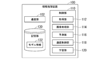
- FIG. 2 is a diagram showing an example of the configuration of the information processing apparatus 100 according to the first embodiment.
- the information processing device 100 includes, for example, a communication unit 102, a control unit 110, and a storage unit 130.
- the communication unit 102 includes, for example, a communication interface such as a NIC.
- the communication unit 102 communicates with the terminal device 10 and the like via the network NW.
- the control unit 110 includes, for example, an acquisition unit 112, an image processing unit 114, a prediction unit 116, a communication control unit 118, and a learning unit 120.
- the component of the control unit 110 is realized by, for example, a processor such as a CPU (Central Processing Unit) or a GPU (Graphics Processing Unit) executing a program stored in the storage unit 130.
- a processor such as a CPU (Central Processing Unit) or a GPU (Graphics Processing Unit) executing a program stored in the storage unit 130.
- Part or all of the components of the control unit 110 are realized by hardware (circuit unit; circuitry) such as LSI (Large Scale Integration), ASIC (Application Specific Integrated Circuit), or FPGA (Field-Programmable Gate Array). It may be realized by the cooperation of software and hardware.
- the storage unit 130 is realized by a storage device such as an HDD (Hard Disc Drive), a flash memory, an EEPROM (Electrically Erasable Programmable Read Only Memory), a ROM (Read Only Memory), or a RAM (Random Access Memory).
- Model information 132 is stored in the storage unit 130 in addition to various programs such as firmware and application programs. Model information 132 will be described later.
- FIG. 3 is a flowchart showing a series of run-time processing flows by the control unit 110 according to the first embodiment. The processing of this flowchart may be repeated, for example, at a predetermined cycle.
- the acquisition unit 112 acquires the cell image IMG of the nerve cell from the terminal device 10 via the communication unit 102 (step S100).
- the image processing unit 114 crops the cell image IMG acquired by the acquisition unit 112 to generate a plurality of images (hereinafter, referred to as cropping image IMG C ) having a size smaller than that of the cell image IMG (step S102). ).
- FIG. 4 is a diagram for explaining a method of generating a cropping image IMG C.
- the image processing unit 114 scans a region R (also referred to as a crop region) having a certain aspect ratio while sliding it on the cell image IMG, and cuts out a pixel region overlapping the region R as a cropped image IMG C to obtain several hundreds.
- a cropping image IMG C of about 120 images (for example, about 120 images) is generated.
- the aspect ratio of the region R may be, for example, a ratio of 128 ⁇ 128 pixels, 256 ⁇ 256 pixels, 512 ⁇ 512 pixels, 768 ⁇ 768 pixels, 1020 ⁇ 1020 pixels.
- the aspect ratio of the area R is assumed to be the aspect ratio at the time of training described later.
- the larger the aspect ratio of the region R and the larger the size of the cropping image IMG C the greater the calculation load due to deep learning, but the higher the prediction accuracy. That is, it is preferable to increase the size of the cropping image IMG C in order to improve the prediction accuracy, and to decrease the size of the cropping image IMG C in order to improve the calculation speed.
- the prediction unit 116 a plurality of cropping images IMG C generated by the image processing unit 114, and inputs to the prediction model MDL model information 132 indicates (step S104).
- Model information 132 is information (program or data structure) that defines a prediction model MDL for predicting cell death of a nerve cell from a cell image of the nerve cell.
- the prediction model MDL is implemented by various neural networks such as, for example, a convolutional neural network (CNN).
- CNN convolutional neural network
- the model information 132 includes, for example, connection information on how the units included in each of the input layer, one or more hidden layers (intermediate layers), and the output layer constituting each neural network are connected to each other. It contains various information such as the coupling coefficient given to the data input / output between the coupled units.
- the connection information includes, for example, the number of units included in each layer, information that specifies the type of unit to which each unit is connected, an activation function that implements each unit, a gate provided between neurons in the hidden layer, and the like. Contains information.
- the activation function that implements the neuron may be, for example, a rectified linear function (ReLU function), a sigmoid function, a step function, or other function.
- the gate selectively passes or weights the data transmitted between the units, for example, depending on the value returned by the activation function (eg 1 or 0).
- the coupling coefficient includes, for example, a weight given to the output data when data is output from a unit of a certain layer to a unit of a deeper layer in a hidden layer of a neural network.
- the coupling coefficient may include a bias component peculiar to each layer.
- FIG. 5 is a diagram showing an example of the prediction model MDL according to the first embodiment.
- the prediction model MDL includes, for example, K models WL-1 to WL-K.
- Each model WL is pre-learned to output a score indicating the likelihood of cell death of a nerve cell as a likelihood or probability when a cropping image IMG C cut out from a cell image IMG of a nerve cell is input. It is a weak learner.
- model WL includes CNN.
- the model WLs are in parallel with each other. The method of generating one learning model by combining a plurality of weak learners in this way is called ensemble learning.
- the prediction model MDL normalizes the score of each model WL which is a weak learner, and outputs the normalized score.
- the normalization of the score is shown in the formula (1).
- Formula (1) is implemented by, for example, a fully connected layer.
- the prediction model MDL may normalize the scores by dividing the sum of the scores of all the model WLs by K, which is the sum of the model WLs.
- the prediction model MDL is a combination of K model WLs, but the prediction model MDL is not limited to this.
- the prediction model MDL may be one model WL.
- the prediction unit 116 determines whether or not the score (normalized score) output by the prediction model MDL is equal to or higher than the threshold value (step S106).
- the prediction unit 116 predicts that nerve cells die when the score is equal to or higher than the threshold value (step S108), and predicts that nerve cells do not die when the score is lower than the threshold value (step S110). That is, the prediction unit 116 determines that the probability of developing Alzheimer's disease is high when the score is equal to or higher than the threshold value, and determines that the probability of developing Alzheimer's disease is low when the score is less than the threshold value.
- the communication control unit 118 transmits the prediction result by the prediction unit 116 to the terminal device 10 via the communication unit 102 (step S112).
- the communication control unit 118 may transmit information indicating the presence or absence of cell death of nerve cells, or may transmit information indicating the probability of developing Alzheimer's disease.
- the user who operates the terminal device 10 has a future nerve cell transferred to the cell image IMG transmitted to the information processing device 100.
- Training is a state in which the prediction model MDL used at runtime is trained.
- FIG. 6 is a flowchart showing a flow of a series of training processes by the control unit 110 according to the first embodiment.
- the learning unit 120 generates teacher data for learning the prediction model MDL (step S200).
- the teacher data information indicating that the nerve cell will die at a certain point in the future is a teacher label (also called a target) for the cell image IMG that images the nerve cell that has been induced to differentiate from the pluripotent stem cell. It is the data associated with.
- the teacher data is when the cell image IMG, which is an image of nerve cells induced to differentiate from pluripotent stem cells, is used as input data, and the information representing the phenotype of neurodegenerative diseases such as cell death is used as the correct output data.
- it is a data set that combines these input data and output data. Cell death at some point in the future represents a more prominent neurodegenerative disease phenotype than when the cell image IMG was imaged.
- a subject's pluripotent stem cells are induced to differentiate to produce multiple nerve cells.
- a drug A that promotes cell death is administered to the plurality of nerve cells thus produced so as to be in the same environment as in the brain of a patient suffering from Alzheimer's disease.
- nerve cells are administered with drug C, which does not suppress cell death and causes dead cells to develop fluorescent color.
- Images of the nerve cells in the group to which the drug B was administered and the nerve cells in the group to which the drug C was administered are imaged in chronological order according to the passage of time.
- the cell image of the nerve cells in the group to which the drug B was administered is associated with information indicating that cell death does not occur (for example, a score of 0.0) as a teacher label, and the nerve cells in the group to which the drug C was administered are associated with the IMG.
- Information indicating cell death (for example, score 1.0) is associated with the cell image IMG as a teacher label.
- the image processing unit 114 crops the cell image IMGs to generate a plurality of cropped image IMGs (step S202). ..
- FIG. 7 is a diagram for explaining a method of generating a cropping image IMG C.
- the image processing unit 114 selects one cell image IMG k from a group of cell images captured in time series for a certain nerve cell.
- the image processing unit 114 scans the selected cell image IMG k while sliding the region R having a predetermined aspect ratio, and cuts out a pixel region that overlaps the region R as a cropping image IMG C to obtain a plurality of cropped images IMG C. (K) is generated.
- the image processing unit 114 reselects a cell image IMG k + 1, which is different from the previously selected cell image IMG k , from the cell image group captured in time series, and similarly generates a plurality of cropping images IMG C (k + 1) . To do.
- the image processing unit 114 performs the same image processing with respect to the cell image group captured in time series for the other neurons may generate a plurality of cropping the image IMG C.
- Each of these large amounts of cropped image IMG C is associated with a teacher label of the cell image IMG from which it was cut out.
- the learning unit 120 a plurality of cropping images IMG C generated from the cell image IMG teacher data by the image processing unit 114, divided into a cropping image Tr_IMG C for training, and cropping images Va_IMG C for validation ,
- the cropping image Tr_IMG C for training is input to the i-th model WL-i among the K model WLs included as weak learners in the prediction model MDL (step S204).
- the parameters of the i-th model WL-i weights and bias components described above, the kernel of the convolutional layer, etc.
- the learning unit 120 the output result from the i-th model WL-i input cropping image Tr_IMG C for training, i.e. to obtain a score s i (step S206).
- the learning unit 120 calculates a score s i obtained from the i-th model WL-i, the error (also referred to as a loss) between the associated scores as training labels cropping image Tr_IMG C for training (Step S208).
- the learning unit 120 determines the parameters of the i-th model WL-i so that the error becomes small based on the gradient method such as the error reverse transmission number (step S210).
- the learning unit 120 determines whether or not the learning for the i-th model WL-i has been repeated a predetermined number of times E (for example, about 30 times) (step S212), and if the predetermined number of times E has not been reached, S204
- the i-th model WL-i is learned by inputting the same image as the training cropping image Tr_IMG C used for training in the previous process into the i-th model WL-i. Repeat that.
- the learning unit 120 stores the parameters updated by learning in the storage unit 130, and inputs the cropping image Tr_IMG C for training to the i-th model WL-i in which the parameters are initialized.
- E models WL-i having different parameters are generated by the time the learning for the i-th model WL-i reaches a predetermined number of times E.
- the learning unit 120 when the learning unit 120 has learned the i-th model WL-i a predetermined number of times E, the learning unit 120 inputs the cropping image Va_IMG C for verification into each of the E i-th model WL-i. (Step S214).
- the learning unit 120 selects the model WL-i having the highest prediction accuracy from the E th-th model WL-i (step S216). For example, the learning unit 120 has the smallest error between the score s i obtained when the cropping image Va_IMG C for verification is input and the score of the teacher label among the E third models WL-i. Select WL-i.
- the learning unit 120 determines whether or not all of the K model WLs included as weak learners in the prediction model MDL have been learned (step S218), and the learning of the K model WLs has been completed. If not, the process returns to S204, based on cropping the image Tr_IMG C for training to learn the (i + 1) th model WL- the (i + 1).
- the learning unit 120 ends the processing of this flowchart.
- the information processing apparatus 100 corresponds to an image obtained by imaging cells of a neurodegenerative disease differentiated from pluripotent stem cells with at least information indicating that the neurodegenerative disease is a teacher label.
- An image of cells differentiated from the pluripotent stem cells derived from the subject was input to the predicted model MDL learned based on the attached teacher data, and based on the output result of the predicted model MDL in which the image was input, the image was input. Since the subject predicts that he / she will develop a neurodegenerative disease, the subject's future disease can be predicted accurately.
- the prediction model MDL is used to predict the appearance of a phenotype of a neurodegenerative disease in a cell, when such a phenotype is predicted to appear, the phenotype is predicted.
- the cells can be observed from the time of prediction until the actual appearance of the phenotype.
- the cause of the onset of neurodegenerative diseases can be investigated.
- the mechanism of the administered drug can be elucidated by observing the cells until the phenotype actually appears while administering various drugs to the cells in which the phenotype is predicted to appear.
- the phenotype does not appear or the time until appearance occurs.
- the administered drug has an effect of suppressing the onset of neurodegenerative diseases.
- the teacher data is data in which a score indicating whether the nerve cell will die at a certain point in the future or not will be associated with the cell image as a teacher label. It was explained as being, but it is not limited to this.
- the teacher data in addition to the above-mentioned score for the cell image, the time predicted to cause cell death (an example of the second time) from the time when the cell image was imaged (an example of the first time). The time until is the data associated with the teacher label.
- the learning unit 120 uses such teacher data to obtain a cell death probability P1, a cell death non-cell death probability P2, and a time t until cell death, respectively.
- the prediction unit 116 predicts the time until the subject develops Alzheimer's disease based on the t element of the vector output by the prediction model MDL.
- the teacher label is not limited to the score indicating the presence or absence of cell death at a certain point in the future and the time until cell death, but also includes the attributes of the subject who is the source of pluripotent stem cells before differentiation of nerve cells. May be good. Attributes may include, for example, various information such as gender, age, weight, height, lifestyle, illness, and family medical history.
- the plurality of model WLs included as weak learners in the prediction model MDL are not limited to CNNs, and may include, for example, a recurrent network (RNN) in which the intermediate layer is an LSTM (Long short-term memory).
- the model WL may be a PredNet that is a combination of RNN and CNN.
- PredNet is a neural network that predicts a frame image at a time later than that time from a frame image at a certain time.
- the second embodiment will be described.
- the screening device 100A for determining whether or not the test substance is a prophylactic or therapeutic agent for neurodegenerative diseases based on the output result of the prediction model MDL will be described.
- the differences from the first embodiment will be mainly described, and the points common to the first embodiment will be omitted.
- the same parts as those of the first embodiment will be described with the same reference numerals.
- FIG. 8 is a diagram showing an example of the configuration of the screening device 100A according to the second embodiment.
- the screening device 100A includes the configuration of the information processing device 100 according to the first embodiment described above.
- the screening device 100A includes a communication unit 102, a control unit 110A, and a storage unit 130.
- the control unit 110A further includes a drug determination unit 122 in addition to the acquisition unit 112, the image processing unit 114, the prediction unit 116, the communication control unit 118, and the learning unit 120 described above.
- the acquisition unit 112 acquires an image of a neurodegenerative disease cell differentiated from a pluripotent stem cell that has been contacted with a test substance.
- the test substance is not particularly limited, and examples thereof include a natural compound library, a synthetic compound library, an existing drug library, and a metabolite library.
- the preventive agent for a neurodegenerative disease means a drug capable of suppressing the onset of the neurodegenerative disease or alleviating the symptom by administering it to a subject before the onset of the neurodegenerative disease.
- a therapeutic agent for a neurodegenerative disease is a drug that can alleviate the symptoms of a neurodegenerative disease by administering it to a patient after the onset of the neurodegenerative disease.
- the learning unit 120 learns the prediction model MDL based on the teacher data, as in the first embodiment described above.
- a phenotype such as cell death is associated as a score with respect to a cell image obtained by imaging a cell of a neurodegenerative disease such as Alzheimer's disease differentiated from pluripotent stem cells. Data.
- the predictive model MDL outputs as a score that the phenotype of the neurodegenerative disease appears in the cell when the cell image is input.
- the prediction unit 116 inputs the image acquired by the acquisition unit 112 into the trained prediction model MDL. Then, the prediction unit 116 predicts whether or not the phenotype of the neurodegenerative disease appears in the cells to which the test substance is administered, based on the output result of the prediction model MDL in which the image is input.
- the drug determination unit 122 determines whether or not the test substance is a preventive agent or a therapeutic agent for a neurodegenerative disease based on the prediction result of the prediction unit 116.
- the drug determination unit 122 determines that the test substance is a preventive agent or a therapeutic agent for a neurodegenerative disease when the following condition (1) is satisfied, and when the condition (2) is satisfied, the test substance is a nerve. It may be determined that it is neither a preventive agent nor a therapeutic agent for degenerative diseases.
- Condition (1) The score output by the prediction model MDL in which the image is input is below the threshold value, and it is predicted that the phenotype of neurodegenerative disease does not appear in the cells to which the test substance is administered.
- Condition (2) The score output by the prediction model MDL in which the image is input is equal to or higher than the threshold value, and it is predicted that the phenotype of the neurodegenerative disease will appear in the cells to which the test substance is administered.
- the screening device 100A acquires an image of a neurodegenerative disease cell differentiated from a pluripotent stem cell that has been contacted with a test substance, and uses it as a trained prediction model MDL.
- the acquired image is input, and based on the output result of the prediction model MDL in which the image is input, whether or not the phenotype of the neurodegenerative disease appears in the cells to which the test substance is administered is predicted.
- the screening device 100A determines whether or not the test substance is a prophylactic or therapeutic agent for neurodegenerative diseases based on the prediction result of whether or not the phenotype appears in the cells.
- the presence or absence of the phenotype of the neurodegenerative disease is predicted, and further, a new drug that can be a preventive or therapeutic agent for the neurodegenerative disease is efficiently used. Can be found.
- FIG. 9 is a diagram showing an example of the hardware configuration of the information processing device 100 and the screening device 100A of the embodiment.
- the information processing device 100 includes NIC100-1, CPU100-2, RAM100-3, ROM100-4, secondary storage devices 100-5 such as flash memory and HDD, and drive device 100-6, which are internal buses or dedicated communication lines. It is configured to be interconnected by.
- a portable storage medium such as an optical disk is mounted on the drive device 100-6.
- a program stored in a portable storage medium mounted on the secondary storage device 100-5 or the drive device 100-6 is expanded into the RAM 100-3 by a DMA controller (not shown) or the like, and executed by the CPU 100-2.
- the control units 110 and 110A are realized.
- the program referenced by the control unit 110 or 110A may be downloaded from another device via the network NW.
- [Expression example 1] The embodiment described above can be expressed as follows.
- the processor It has a memory for storing programs and When the processor executes the program, Obtained images of cells differentiated from pluripotent stem cells derived from the subject, The acquired image is used for a model learned based on data in which at least information indicating the neurodegenerative disease is associated with an image obtained by imaging a neurodegenerative disease cell differentiated from a pluripotent stem cell.
- the subject develops the neurodegenerative disease or predicts the effect of the drug on the neurodegenerative disease.
- An information processing device that is configured as such.
- [Expression example 2] The embodiment described above can also be expressed as follows.
- the processor It has a memory for storing programs and When the processor executes the program, Images of neurodegenerative disease cells differentiated from pluripotent stem cells that were contacted with the test substance were acquired. The acquired image is used for a model learned based on data in which at least information indicating the phenotype of the neurodegenerative disease is associated with an image of a neurodegenerative disease cell differentiated from pluripotent stem cells. Based on the output result of the model in which the image is input, it is predicted whether or not the phenotype of the neurodegenerative disease appears in the cells of the neurodegenerative disease differentiated from the pluripotent stem cells. Based on the predicted result, it is determined whether or not the test substance is a prophylactic or therapeutic agent for the neurodegenerative disease. A screening device that is configured to.
- Example 1 (Establishment of human iPS cells) A reprogramming factor was introduced into human peripheral blood mononuclear cells using an episomal vector. As reprogramming factors, SOX2, KLF4, Oct4, L-MYC, LIN28, and dominant negative p53 were used. After a few days, the cells were harvested and seeded on a dish coated with iMatrix (Nippi). The next day, the medium was replaced with StemFit AK03 (Ajinomoto Co., Inc.). Then, the medium was changed every two days. Twenty days after the introduction of the reprogramming factor, colonies of iPS cells were collected and expanded.
- SOX2, KLF4, Oct4, L-MYC, LIN28, and dominant negative p53 were used. After a few days, the cells were harvested and seeded on a dish coated with iMatrix (Nippi). The next day, the medium was replaced with StemFit AK03 (Ajinomoto Co.,
- the fluorescent dye binds to the DNA to generate fluorescence, which is the standard for standard time-lapse imaging systems (product name “Incubite ZOOM”, Essen Biosciences). It can be detected using the FITC-green filter set (exposure time 400 ms). Images were taken over time every 6 hours after drug administration.
- the experiment was performed using three 96-well plates. Of the three plates, rows A and L of the two plates 24-48 hours after drug application in the dimethyl sulfoxide (DMSO) -administered group and the Z-VAD-FMK (Promega) -administered group.
- the column wells were used for the training dataset (480 images per class) and the remaining column wells were used for the validation dataset (160 images per class). Images of the remaining one plate 24 hours after drug application were used as the test dataset (64 images per class).
- the 256 x 256 pixel image for training was randomly flipped horizontally with a 50% probability, and then a 224 x 224 pixel area was randomly cropped from the image.
- the cropped 224 x 224 pixel image was finally used as input to the neural network.
- Cross entropy was used as the loss function.
- the errors were backpropagated through the network and the weights were optimized by the Stochastic Gradient Descent (SGD) using mini-batch.
- the momentum coefficient in SGD was set to 0.9.
- Classification accuracy was calculated for each epoch for the verification data (cropping image Va_IMG C for verification).
- the epoch is an iterative process for learning to repeat the model WL until it reaches a predetermined number of times E.
- the network parameters in the epoch when the network showed the best classification accuracy were saved in each training trial. That is, E parameters were saved.
- the average output (normalized score) from the last fully connected layer of the 20 trained networks was used to classify the test data.
- the 224x224 pixel image was cropped from the center of the resized 256x256 pixel image without inversion.
- Accuracy, F1 score, and area under the curve (AUC) in ROC (Receiver Operating Characteristic) analysis were calculated and verified by triple cross-validation.
- the accuracy of the validation data was evaluated by rotating the plates of each dataset and calculating the average of the results of the three trials.
- the nerve cell As the nerve cell, the nerve cell obtained by inducing the temporary expression of NGN2 in the cell selected in Experimental Example 2 was used.
- the coating agent poly-D-lysine (PDL, Sigma) final concentration 1% (v / v), Matrigel (Corning) final concentration 2% (v / v), Synthemax II SC (Corning) final concentration. 2% (v / v) and CellNest (Fujifilm) final concentration 1% (v / v) were used in various combinations. For comparison, a similar study was performed on uncoated culture dishes.
- FIG. 10 is a diagram showing a micrograph taken one day later
- FIG. 11 is a diagram showing a micrograph taken 10 days later.
- coating with PDL is indicated as “P”
- coating with Matrigel is indicated as “M”
- coating with Synthemax II SC is indicated. It is indicated as “S”
- the coating with CellNest is indicated as “C”.
- Coating with PDL (Sigma) and Matrigel (Corning) is indicated as “PM”, and the same applies to combinations of other coating agents.
- Coating with PDL (Sigma), Matrigel (Corning) and Synthemax II SC is referred to as "PMS”, and the same applies to combinations of other coating agents. The fact that no coating was applied is indicated as "Non”.
- PMSC coating that is, PDL (Sigma) final concentration 1% (v / v), Matrigel (Corning) final concentration 2% (v / v), Synthemax II SC (Corning) final concentration 2% ( It was revealed that a cell culture vessel coated with a mixture of v / v) and CellNest (Fujifilm) at a final concentration of 1% (v / v) is most suitable for long-term observation of nerve cells. ..
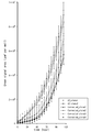
- FIG. 12 is a diagram showing an example of the results of the bioassay.
- the green signal area the area of the region that fluorescently developed green on the nerve cell image
- each row from row A to row H was green.
- the average value of the signal area was calculated.
- the green signal area is the area of the area where cell death has occurred.
- the change in the average value of the green signal area with the passage of time was plotted on the graph. As a result, there was no significant difference between columns, indicating the stability of the assay.
- FIG. 13 is a diagram showing an example of the application result of the gene modification technology. Familial Alzheimer's disease is known to be caused by a mutation in the gene PSEN1 G384A.
- AD_clone1 and AD_clone2 in the figure represent changes over time in the green signal area of the nerve cell image in which the PSEN1 G384A mutation has occurred.
- Corrected_clones 1 to 4 represent changes over time in the green signal area when the mutation is normalized to the wild type in the nerve cells in which the PSEN1 G384A mutation has occurred by using a gene modification technique. As shown in the figure, the average value of the green signal area was smaller in Corrected_clone1 to 4 than in AD_clone1 and AD_clone2, indicating that the gene modification technique alleviated cell death.
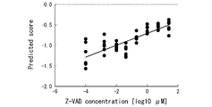
- FIG. 14 is a diagram showing the relationship between the Z-VAD-FMK concentration and the score of the prediction model MDL.
- the vertical axis in the figure represents the score of the prediction model MDL, and the horizontal axis represents the Z-VAD-FMK concentration.
- the score represents a stochastic numerical value regarding the life and death of cells. The higher the score, the higher the probability that the cell will survive, and the lower the score, the higher the probability that the caspase 3 of the cell will be activated or undergo apoptosis. As shown in the figure, the higher the concentration of Z-VAD-FMK, the higher the score.
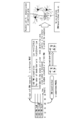
- FIG. 15 is a diagram for schematically explaining the proposed method.
- Z-VAD-FMK is added to neurons to express gene expression in individual cells prior to cell death (prior to caspase 3 activation).
- an image is acquired, and where the change is occurring is confirmed using a prediction model MDL based on CNN (WHERE in the figure).
- WHERE in the figure.
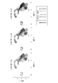
- FIG. 16 is a diagram showing an example of cell clustering results.
- a visualization method a type of dimensional compression method
- UMAP a type of dimensional compression method
- the concentration of Z-VAD-FMK added to nerve cells 0 [ ⁇ M], 0.2 [ ⁇ M], and 25 [ ⁇ M].
- the cell population was separated into a total of 15 types of clusters from 0 to 14.
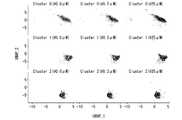
- FIG. 17 is a diagram showing an example of clusters 0, 1, and 2.
- the first line in the figure represents cluster 0, the second line represents cluster 1, and the third line represents cluster 2.
- the first column shows that the concentration of Z-VAD-FMK is 0 [ ⁇ M]
- the second column shows that the concentration of Z-VAD-FMK is 0.2 [ ⁇ M].
- the third column shows that the concentration of Z-VAD-FMK is 25 [ ⁇ M].
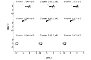
- FIG. 18 is a diagram showing an example of clusters 3, 4, and 5.
- the first line in the figure represents cluster 3, the second line represents cluster 4, and the third line represents cluster 5.
- FIG. 19 is a diagram showing an example of clusters 6, 7, and 8.
- the first line in the figure represents cluster 6, the second line represents cluster 7, and the third line represents cluster 8.
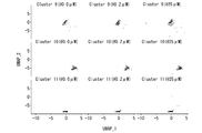
- FIG. 20 is a diagram showing an example of clusters 9, 10, and 11.
- the first line in the figure represents cluster 9, the second line represents cluster 10, and the third line represents cluster 11.
- FIG. 21 is a diagram showing an example of clusters 12, 13, and 14.
- the first line in the figure represents cluster 12, the second line represents cluster 13, and the third line represents cluster 14.
- FIG. 20 is a diagram showing an example of clusters 9, 10, and 11.
- the columns in these figures also show that the concentration of Z-VAD-FMK is 0 [ ⁇ M] in the first column, and the concentration of Z-VAD-FMK in the second column is 0.2 [. It means that it is [ ⁇ M], and the third column shows that the concentration of Z-VAD-FMK is 25 [ ⁇ M].
- FIG. 22 is a diagram showing the relationship between the Z-VAD-FMK concentration and the cluster size.
- Caspase 3 is activated and accounts for a few [%] of all cell deaths, and is included in its own cluster with the addition of Z-VAD-FMK (ie, suppressing apoptosis).
- a cluster that satisfies the condition that the number of cells is decreasing was searched from 14 clusters. As a result, the cluster 12 was identified as satisfying the condition.
- FIG. 23 is a diagram showing an example of the analysis result of Pathway.
- the addition of Z-VAD-FMK contributes to a pathway specifically associated with high apoptosis and cell death in clusters in which the number of cells decreases (in other words, clusters heading for cell death). It has been shown. This showed the validity of this experimental method.
Landscapes
- Engineering & Computer Science (AREA)
- Health & Medical Sciences (AREA)
- Medical Informatics (AREA)
- General Health & Medical Sciences (AREA)
- Public Health (AREA)
- Biomedical Technology (AREA)
- Life Sciences & Earth Sciences (AREA)
- Physics & Mathematics (AREA)
- Theoretical Computer Science (AREA)
- Data Mining & Analysis (AREA)
- Epidemiology (AREA)
- Primary Health Care (AREA)
- Pathology (AREA)
- Neurology (AREA)
- General Physics & Mathematics (AREA)
- Molecular Biology (AREA)
- Databases & Information Systems (AREA)
- Biophysics (AREA)
- Software Systems (AREA)
- Evolutionary Computation (AREA)
- Nuclear Medicine, Radiotherapy & Molecular Imaging (AREA)
- Radiology & Medical Imaging (AREA)
- Computing Systems (AREA)
- Artificial Intelligence (AREA)
- Neurosurgery (AREA)
- Computer Vision & Pattern Recognition (AREA)
- Mathematical Physics (AREA)
- General Engineering & Computer Science (AREA)
- Veterinary Medicine (AREA)
- Animal Behavior & Ethology (AREA)
- Surgery (AREA)
- Heart & Thoracic Surgery (AREA)
- Physiology (AREA)
- Computational Linguistics (AREA)
- Developmental Disabilities (AREA)
- Quality & Reliability (AREA)
- Multimedia (AREA)
- Psychology (AREA)
- Psychiatry (AREA)
- Hospice & Palliative Care (AREA)
Priority Applications (4)
| Application Number | Priority Date | Filing Date | Title |
|---|---|---|---|
| JP2021522872A JPWO2020241772A1 (https=) | 2019-05-31 | 2020-05-28 | |
| SG11202113016UA SG11202113016UA (en) | 2019-05-31 | 2020-05-28 | Information processing device, screening device, information processing method, screening method, and program |
| US17/614,218 US12462378B2 (en) | 2019-05-31 | 2020-05-28 | Information processing device, screening device, information processing method, screening method, and program |
| EP20814392.5A EP3978595A4 (en) | 2019-05-31 | 2020-05-28 | INFORMATION PROCESSING DEVICE, SCREENING DEVICE, INFORMATION PROCESSING METHOD, SCREENING METHOD AND PROGRAM |
Applications Claiming Priority (2)
| Application Number | Priority Date | Filing Date | Title |
|---|---|---|---|
| JP2019103293 | 2019-05-31 | ||
| JP2019-103293 | 2019-05-31 |
Publications (1)
| Publication Number | Publication Date |
|---|---|
| WO2020241772A1 true WO2020241772A1 (ja) | 2020-12-03 |
Family
ID=73552587
Family Applications (1)
| Application Number | Title | Priority Date | Filing Date |
|---|---|---|---|
| PCT/JP2020/021177 Ceased WO2020241772A1 (ja) | 2019-05-31 | 2020-05-28 | 情報処理装置、スクリーニング装置、情報処理方法、スクリーニング方法、及びプログラム |
Country Status (5)
| Country | Link |
|---|---|
| US (1) | US12462378B2 (https=) |
| EP (1) | EP3978595A4 (https=) |
| JP (1) | JPWO2020241772A1 (https=) |
| SG (1) | SG11202113016UA (https=) |
| WO (1) | WO2020241772A1 (https=) |
Cited By (3)
| Publication number | Priority date | Publication date | Assignee | Title |
|---|---|---|---|---|
| CN114419500A (zh) * | 2022-01-07 | 2022-04-29 | 乐普(北京)医疗器械股份有限公司 | 基于心脏超声视频筛选舒张期和收缩期图像的方法和装置 |
| WO2023008428A1 (ja) * | 2021-07-29 | 2023-02-02 | 株式会社島津製作所 | 細胞画像解析方法 |
| WO2023094809A1 (en) * | 2021-11-23 | 2023-06-01 | Ucl Business Ltd | Methods and apparatus for analysing imaging data |
Families Citing this family (9)
| Publication number | Priority date | Publication date | Assignee | Title |
|---|---|---|---|---|
| JP7678577B2 (ja) * | 2019-05-31 | 2025-05-16 | 国立大学法人京都大学 | 情報処理装置、情報処理方法、及びプログラム |
| WO2020241772A1 (ja) | 2019-05-31 | 2020-12-03 | 国立大学法人京都大学 | 情報処理装置、スクリーニング装置、情報処理方法、スクリーニング方法、及びプログラム |
| US20230419480A1 (en) * | 2020-05-14 | 2023-12-28 | New York Stem Cell Foundation, Inc. | Method and system for predicting cellular aging |
| WO2022061176A1 (en) * | 2020-09-18 | 2022-03-24 | New York Stem Cell Foundation, Inc. | Methods and systems for predicting neurodegenerative disease state |
| US12518129B2 (en) * | 2020-11-30 | 2026-01-06 | Oracle International Corporation | Method and system for over-prediction in neural networks |
| EP4348596A1 (en) * | 2021-05-26 | 2024-04-10 | Genentech, Inc. | Detecting tertiary lymphoid structures in digital pathology images |
| WO2022270179A1 (ja) * | 2021-06-24 | 2022-12-29 | 富士フイルム株式会社 | 情報処理装置、情報処理方法、及びプログラム |
| US20230377355A1 (en) * | 2022-05-20 | 2023-11-23 | New York Stem Cell Foundation, Inc. | Synthetic pooling for enriching disease signatures |
| CN121605492A (zh) * | 2024-06-14 | 2026-03-03 | 京东方科技集团股份有限公司 | 疾病预测方法、模型的训练方法及装置、存储介质 |
Citations (6)
| Publication number | Priority date | Publication date | Assignee | Title |
|---|---|---|---|---|
| JP2013520960A (ja) * | 2010-03-03 | 2013-06-10 | 国立大学法人京都大学 | iPS細胞由来の神経細胞を用いた蛋白質ミスフォールディング病の診断方法 |
| WO2017022854A1 (ja) * | 2015-08-05 | 2017-02-09 | 国立大学法人京都大学 | 運動ニューロン疾患の検査方法及び治療剤のスクリーニング方法 |
| JP2018045559A (ja) * | 2016-09-16 | 2018-03-22 | 富士通株式会社 | 情報処理装置、情報処理方法およびプログラム |
| WO2018211687A1 (ja) * | 2017-05-19 | 2018-11-22 | 株式会社オプティム | コンピュータシステム、被験者診断方法及び支援プログラム |
| WO2018216705A1 (ja) * | 2017-05-23 | 2018-11-29 | 国立大学法人京都大学 | 神経変性疾患の予防及び/又は治療剤 |
| JP2019103293A (ja) | 2017-12-05 | 2019-06-24 | マブチモーター株式会社 | モータ |
Family Cites Families (10)
| Publication number | Priority date | Publication date | Assignee | Title |
|---|---|---|---|---|
| US7711174B2 (en) | 2004-05-13 | 2010-05-04 | The Charles Stark Draper Laboratory, Inc. | Methods and systems for imaging cells |
| EP2488992B1 (en) | 2009-10-13 | 2024-05-29 | The Charles Stark Draper Laboratory, Inc. | Mathematical image analysis based cell reprogramming with applications for epigenetic and non-epigenetic base induced pluripotent stem cell derivation |
| EP3134729A1 (en) * | 2014-04-22 | 2017-03-01 | Q-State Biosciences, Inc. | Analysis of compounds for pain and sensory disorders |
| WO2016109813A2 (en) * | 2014-12-31 | 2016-07-07 | Wisconsin Alumni Research Foundation | Human pluripotent stem cell-based models for predictive developmental neural toxicity |
| CA2978740A1 (en) | 2015-03-06 | 2016-09-15 | Blanchette Rockefeller Neurosciences Institute | Methods for classifying populations including alzheimer's disease populations |
| WO2017136285A1 (en) | 2016-02-01 | 2017-08-10 | The Board Of Trustees Of The Leland Stanford Junior University | Method and systems for analyzing functional imaging data |
| EP3553165A4 (en) | 2016-12-06 | 2019-12-25 | Fujifilm Corporation | CELL IMAGE EVALUATION DEVICE AND CELL IMAGE EVALUATION CONTROL PROGRAM |
| US10249389B2 (en) | 2017-05-12 | 2019-04-02 | The Regents Of The University Of Michigan | Individual and cohort pharmacological phenotype prediction platform |
| US20220028488A1 (en) | 2018-12-07 | 2022-01-27 | President And Fellows Of Harvard College | Drug discovery and early disease identification platform using electronic health records, genetics and stem cells |
| WO2020241772A1 (ja) | 2019-05-31 | 2020-12-03 | 国立大学法人京都大学 | 情報処理装置、スクリーニング装置、情報処理方法、スクリーニング方法、及びプログラム |
-
2020
- 2020-05-28 WO PCT/JP2020/021177 patent/WO2020241772A1/ja not_active Ceased
- 2020-05-28 US US17/614,218 patent/US12462378B2/en active Active
- 2020-05-28 SG SG11202113016UA patent/SG11202113016UA/en unknown
- 2020-05-28 EP EP20814392.5A patent/EP3978595A4/en active Pending
- 2020-05-28 JP JP2021522872A patent/JPWO2020241772A1/ja active Pending
Patent Citations (6)
| Publication number | Priority date | Publication date | Assignee | Title |
|---|---|---|---|---|
| JP2013520960A (ja) * | 2010-03-03 | 2013-06-10 | 国立大学法人京都大学 | iPS細胞由来の神経細胞を用いた蛋白質ミスフォールディング病の診断方法 |
| WO2017022854A1 (ja) * | 2015-08-05 | 2017-02-09 | 国立大学法人京都大学 | 運動ニューロン疾患の検査方法及び治療剤のスクリーニング方法 |
| JP2018045559A (ja) * | 2016-09-16 | 2018-03-22 | 富士通株式会社 | 情報処理装置、情報処理方法およびプログラム |
| WO2018211687A1 (ja) * | 2017-05-19 | 2018-11-22 | 株式会社オプティム | コンピュータシステム、被験者診断方法及び支援プログラム |
| WO2018216705A1 (ja) * | 2017-05-23 | 2018-11-29 | 国立大学法人京都大学 | 神経変性疾患の予防及び/又は治療剤 |
| JP2019103293A (ja) | 2017-12-05 | 2019-06-24 | マブチモーター株式会社 | モータ |
Non-Patent Citations (4)
| Title |
|---|
| CHRISTIANSEN E. M. ET AL.: "In Silico Labeling: Predicting Fluorescent Labels in Unlabeled Images", CELL, vol. 173, no. 3, 2018, pages 792 - 803, XP002788720, DOI: 10.1016/j.cell.2018.03.040 |
| COUDRAY N. ET AL.: "Classification and mutation prediction from non-small cell lung cancer histopathology images using deep learning", NAT MED., vol. 24, no. 10, 2018, pages 1559 - 1567, XP036608997, DOI: 10.1038/s41591-018-0177-5 |
| See also references of EP3978595A4 |
| TAKAYUKI KONDO, HARUHISA INOUE, RYOSUKE TAKAHASHI: "15. Therapeutic Application of iPS Cells", NIHON NAIKA GAKKAI ZASSHI, vol. 102, no. 8, 2013, pages 2015 - 2022, XP055766711 * |
Cited By (5)
| Publication number | Priority date | Publication date | Assignee | Title |
|---|---|---|---|---|
| WO2023008428A1 (ja) * | 2021-07-29 | 2023-02-02 | 株式会社島津製作所 | 細胞画像解析方法 |
| JPWO2023008428A1 (https=) * | 2021-07-29 | 2023-02-02 | ||
| JP7736065B2 (ja) | 2021-07-29 | 2025-09-09 | 株式会社島津製作所 | 細胞画像解析方法 |
| WO2023094809A1 (en) * | 2021-11-23 | 2023-06-01 | Ucl Business Ltd | Methods and apparatus for analysing imaging data |
| CN114419500A (zh) * | 2022-01-07 | 2022-04-29 | 乐普(北京)医疗器械股份有限公司 | 基于心脏超声视频筛选舒张期和收缩期图像的方法和装置 |
Also Published As
| Publication number | Publication date |
|---|---|
| JPWO2020241772A1 (https=) | 2020-12-03 |
| US20220215543A1 (en) | 2022-07-07 |
| SG11202113016UA (en) | 2021-12-30 |
| EP3978595A1 (en) | 2022-04-06 |
| EP3978595A4 (en) | 2023-07-05 |
| US12462378B2 (en) | 2025-11-04 |
Similar Documents
| Publication | Publication Date | Title |
|---|---|---|
| US12462378B2 (en) | Information processing device, screening device, information processing method, screening method, and program | |
| Haloi | Improved microaneurysm detection using deep neural networks | |
| Tagaris et al. | Assessment of parkinson’s disease based on deep neural networks | |
| CN116597916B (zh) | 一种基于器官芯片和深度学习的抗肿瘤化合物预后药效的预测方法 | |
| Masson et al. | Identifying neural substrates of competitive interactions and sequence transitions during mechanosensory responses in Drosophila | |
| JP2011229410A (ja) | 細胞評価装置、インキュベータ、プログラム、および、培養方法 | |
| Dhakhinamoorthy et al. | Hybrid whale and gray wolf deep learning optimization algorithm for prediction of Alzheimer’s disease | |
| US20230351587A1 (en) | Methods and systems for predicting neurodegenerative disease state | |
| JPWO2017109860A1 (ja) | 画像処理装置 | |
| JPWO2018235251A1 (ja) | 解析装置、解析プログラム及び解析方法 | |
| Li et al. | Recurrent neural networks with interpretable cells predict and classify worm behaviour | |
| Huttner et al. | Loss of Sec-1 family domain-containing 1 (Scfd1) causes severe cardiac defects and endoplasmic reticulum stress in zebrafish | |
| Perni et al. | Automated Behavioral Analysis of Large C. elegans Populations Using a Wide Field-of-view Tracking Platform. | |
| Nitta et al. | Direct evaluation of neuroaxonal degeneration with the causative genes of neurodegenerative diseases in Drosophila using the automated axon quantification system, MeDUsA | |
| Sharma et al. | Quantum Grey Wolf optimisation and evolutionary algorithms for diagnosis of Alzheimer's disease | |
| Pearce et al. | Automatic sample segmentation & detection of Parkinson’s disease using synthetic staining & deep learning | |
| JP7678577B2 (ja) | 情報処理装置、情報処理方法、及びプログラム | |
| Hasan et al. | A modified CNN model for diagnosing schizophrenia disease using fMRI data | |
| JP2020124220A (ja) | 画像処理装置 | |
| Safwan et al. | A Comparative Analysis on Detecting Physiological Age of Caenorhabditis Elegans Using Deep Learning Techniques | |
| Bogensperger et al. | On the Influence of Beta Cell Granule Counting for Classification in Type 1 Diabetes | |
| Halldorsdottir | Social Behavior in a Zebrafish Model of Schizophrenia | |
| Fang et al. | In silico labeling enables kinetic myelination assay in brightfield | |
| Uvaliyev et al. | Alzheimer’s Disease Detection from Retinal Images Using Machine Learning and Deep Learning Techniques: A Perspective | |
| Yoon et al. | Decoding spontaneous pain from brain cellular calcium signals using deep learning |
Legal Events
| Date | Code | Title | Description |
|---|---|---|---|
| 121 | Ep: the epo has been informed by wipo that ep was designated in this application |
Ref document number: 20814392 Country of ref document: EP Kind code of ref document: A1 |
|
| ENP | Entry into the national phase |
Ref document number: 2021522872 Country of ref document: JP Kind code of ref document: A |
|
| NENP | Non-entry into the national phase |
Ref country code: DE |
|
| ENP | Entry into the national phase |
Ref document number: 2020814392 Country of ref document: EP Effective date: 20220103 |
|
| WWG | Wipo information: grant in national office |
Ref document number: 17614218 Country of ref document: US |