WO2020218465A1 - Method for diagnosing endometriosis, disease state monitoring method, and kit - Google Patents
Method for diagnosing endometriosis, disease state monitoring method, and kit Download PDFInfo
- Publication number
- WO2020218465A1 WO2020218465A1 PCT/JP2020/017595 JP2020017595W WO2020218465A1 WO 2020218465 A1 WO2020218465 A1 WO 2020218465A1 JP 2020017595 W JP2020017595 W JP 2020017595W WO 2020218465 A1 WO2020218465 A1 WO 2020218465A1
- Authority
- WO
- WIPO (PCT)
- Prior art keywords
- markers shown
- endometriosis
- abundance
- marker
- markers
- Prior art date
Links
Images


Classifications
-
- G—PHYSICS
- G01—MEASURING; TESTING
- G01N—INVESTIGATING OR ANALYSING MATERIALS BY DETERMINING THEIR CHEMICAL OR PHYSICAL PROPERTIES
- G01N33/00—Investigating or analysing materials by specific methods not covered by groups G01N1/00 - G01N31/00
- G01N33/48—Biological material, e.g. blood, urine; Haemocytometers
- G01N33/50—Chemical analysis of biological material, e.g. blood, urine; Testing involving biospecific ligand binding methods; Immunological testing
- G01N33/68—Chemical analysis of biological material, e.g. blood, urine; Testing involving biospecific ligand binding methods; Immunological testing involving proteins, peptides or amino acids
- G01N33/6893—Chemical analysis of biological material, e.g. blood, urine; Testing involving biospecific ligand binding methods; Immunological testing involving proteins, peptides or amino acids related to diseases not provided for elsewhere
-
- G—PHYSICS
- G01—MEASURING; TESTING
- G01N—INVESTIGATING OR ANALYSING MATERIALS BY DETERMINING THEIR CHEMICAL OR PHYSICAL PROPERTIES
- G01N2800/00—Detection or diagnosis of diseases
- G01N2800/36—Gynecology or obstetrics
- G01N2800/364—Endometriosis, i.e. non-malignant disorder in which functioning endometrial tissue is present outside the uterine cavity
-
- G—PHYSICS
- G01—MEASURING; TESTING
- G01N—INVESTIGATING OR ANALYSING MATERIALS BY DETERMINING THEIR CHEMICAL OR PHYSICAL PROPERTIES
- G01N2800/00—Detection or diagnosis of diseases
- G01N2800/56—Staging of a disease; Further complications associated with the disease
-
- G—PHYSICS
- G01—MEASURING; TESTING
- G01N—INVESTIGATING OR ANALYSING MATERIALS BY DETERMINING THEIR CHEMICAL OR PHYSICAL PROPERTIES
- G01N2800/00—Detection or diagnosis of diseases
- G01N2800/70—Mechanisms involved in disease identification
- G01N2800/7052—Fibrosis
Definitions
- the present disclosure determines how to diagnose endometriosis or how to determine the presence or absence of endometriosis in a subject, and the degree of fibrosis or adhesion in the endometrium of a subject with endometriosis.
- the present invention relates to a method, a method for predicting pain in a subject suffering from endometriosis, a method for monitoring the pathological condition of a subject suffering from endometriosis, and the like, and a kit for performing these methods.
- Non-Patent Document 1 Endometriosis is an estrogen-dependent inflammatory disease found in 6-10% of pregnant women, and infertility and pelvic pain are found in more than 50% of patients (Non-Patent Document 1).
- Non-Patent Document 2 Various hypotheses have been put forward regarding the onset mechanism of endometriosis (Non-Patent Document 2), and the relationship with various factors such as inflammatory cytokines / chemokines, growth factors, and hormones has been reported (Non-patent). Document 3).
- drugs targeting hormones have already been developed, and it has been shown that pain can be alleviated and pathological conditions can be improved by using hormone drugs such as GnRH antagonists and progesterone preparations (Non-Patent Documents 4 and 5).
- Non-Patent Document 6 drug development targeting inflammatory cytokines and chemokines is progressing, and anti-IL-8 antibody shows a strong pathological improvement effect in a monkey model of endometriosis (Patent Document 1).
- Non-Patent Documents 7 and 8 There is a strong demand for non-invasive diagnostic methods such as blood markers that place less burden on patients, but highly accurate diagnostic methods have not yet been established (Non-Patent Document 9).
- the present disclosure has been made in view of such circumstances, and an object thereof is a method for diagnosing endometriosis or a method for determining the presence or absence of endometriosis in a subject, suffering from endometriosis. How to determine the degree of fibrosis or adhesions in the endometrium of a subject, how to predict the pain of a subject with endometriosis, how to monitor the pathology of a subject with endometriosis, etc. The purpose is to provide a kit for performing these methods.
- the inventors of the present disclosure have found that there are a plurality of markers whose abundance differs from that of healthy subjects in patients with endometriosis. By measuring the abundance of markers, it is possible to diagnose whether or not the subject has endometriosis, to determine the degree of endometrial fibrosis and adhesion, and to the subject with endometriosis. We found that pain can be predicted and that the pathophysiology of subjects suffering from endometriosis can be monitored.
- a method for diagnosing endometriosis Type V collagen MMP degradation products, markers shown in Table 1A, markers shown in Table 2A, markers shown in Table 1B, markers shown in Table 2B, markers shown in Table 5A, in samples obtained from the subject.
- a method comprising measuring the abundance of at least one marker selected from the group consisting of the markers shown in Table 6A, the markers shown in Table 5B, and the markers shown in Table 6B.
- a method for determining the presence or absence of endometriosis in a subject are examples of endometriosis in a subject.
- Type V collagen MMP degradation products markers shown in Table 1A, markers shown in Table 2A, markers shown in Table 1B, markers shown in Table 2B, markers shown in Table 5A, in samples obtained from the subject.
- a method comprising measuring the abundance of at least one marker selected from the group consisting of the markers shown in Table 6A, the markers shown in Table 5B, and the markers shown in Table 6B.
- the subject from which the sample is derived further comprises a step indicating that the subject has or may have endometriosis.
- Method. [4] A method for determining the degree of endometrial fibrosis in a subject. Type V collagen MMP degradation products, markers shown in Table 1A, markers shown in Table 2A, markers shown in Table 1B, markers shown in Table 2B, markers shown in Table 5A, in samples obtained from the subject.
- a method comprising measuring the abundance of at least one marker selected from the group consisting of the markers shown in Table 6A, the markers shown in Table 5B, and the markers shown in Table 6B.
- Select from the group consisting of type V collagen MMP degradation products markers shown in Table 1A, markers shown in Table 2A, markers shown in Table 5A, and markers shown in Table 6A in the sample obtained from the subject. If the abundance of at least one marker is high, or at least one selected from the group consisting of the markers shown in Table 1B, the markers shown in Table 2B, the markers shown in Table 5B, and the markers shown in Table 6B.
- a method for determining the degree of endometrial adhesion in a subject Type V collagen MMP degradation products, markers shown in Table 1A, markers shown in Table 2A, markers shown in Table 1B, markers shown in Table 2B, markers shown in Table 5A, in samples obtained from the subject.
- a method comprising measuring the abundance of at least one marker selected from the group consisting of the markers shown in Table 6A, the markers shown in Table 5B, and the markers shown in Table 6B.
- [7] Select from the group consisting of type V collagen MMP degradation products, markers shown in Table 1A, markers shown in Table 2A, markers shown in Table 5A, and markers shown in Table 6A in the sample obtained from the subject. If the abundance of at least one marker is high, or at least one selected from the group consisting of the markers shown in Table 1B, the markers shown in Table 2B, the markers shown in Table 5B, and the markers shown in Table 6B.
- the method according to [6] further comprising a step in which the subject from which the sample is derived is indicated to have or may have adherence in the endometrium when the abundance of one marker is low.
- [8] A method for predicting the degree of pain due to endometriosis in a subject who has or is suspected of having endometriosis.
- Type V collagen MMP degradation products markers shown in Table 1A, markers shown in Table 2A, markers shown in Table 1B, markers shown in Table 2B, markers shown in Table 5A, in samples obtained from the subject.
- a method comprising measuring the abundance of at least one marker selected from the group consisting of the markers shown in Table 6A, the markers shown in Table 5B, and the markers shown in Table 6B.
- the abundance of at least one marker is high, or at least one selected from the group consisting of the markers shown in Table 1B, the markers shown in Table 2B, the markers shown in Table 5B, and the markers shown in Table 6B.
- Type V collagen MMP degradation products markers shown in Table 1A, markers shown in Table 2A, markers shown in Table 1B, markers shown in Table 2B, markers shown in Table 5A, in samples obtained from the subject.
- a method comprising measuring the abundance of at least one marker selected from the group consisting of the markers shown in Table 6A, the markers shown in Table 5B, and the markers shown in Table 6B.
- At least one marker is high, or at least one selected from the group consisting of the markers shown in Table 1B, the markers shown in Table 2B, the markers shown in Table 5B, and the markers shown in Table 6B.
- the abundance is determined to be high, [3], [5], [7], [9], or [11].
- the method described. [13] The abundance of at least one marker selected from the group consisting of the type V collagen MMP degradation product, the marker shown in Table 1A, the marker shown in Table 2A, and the marker shown in Table 5A was obtained from a healthy person. If the abundance of the marker is more than twice the abundance of the same marker in the obtained sample, and / or the abundance of at least one marker selected from the markers shown in Table 6A is the abundance of the same marker in the sample obtained from a healthy person.
- the abundance of at least one marker selected from the group consisting of the markers shown in Table 1B, the markers shown in Table 2B, the markers shown in Table 5B, and the markers shown in Table 6B is obtained from a healthy person.
- the abundance of at least one marker selected from the group consisting of the markers shown in Table 1B, the markers shown in Table 2B, and the markers shown in Table 5B is the same marker in a sample obtained from a healthy person.
- the abundance is 0.5 times or less, and / or when the abundance of at least one marker selected from the markers shown in Table 6B is 0.625 times or less of the abundance of the marker in a sample obtained from a healthy person.
- Type V collagen MMP degradation products markers shown in Table 1, markers shown in Table 2A, markers shown in Table 1B, markers shown in Table 2B, in each of the multiple samples taken from the subject at different time points.
- a method comprising the step of measuring the abundance of at least one marker selected from the group consisting of the markers shown in Table 5A, the markers shown in Table 6A, the markers shown in Table 5B, and the markers shown in Table 6B.
- At least one marker selected from the group consisting of the type V collagen MMP degradation product the marker shown in Table 1A, the marker shown in Table 2A, the marker shown in Table 5A, and the marker shown in Table 6A.
- Method. At least one marker selected from the group consisting of the type V collagen MMP degradation products, the markers shown in Table 1A, the markers shown in Table 2A, the markers shown in Table 5A, and the markers shown in Table 6A.
- [16] which further comprises a step in which the condition of endometriosis is worsened or may be exacerbated in the subject from which the sample is derived if the abundance of the sample decreases over time.
- Method. [19] The method according to any one of [1] to [18], wherein the marker is measured as a polypeptide.
- the sample is a blood sample.
- Kit including.
- a kit for determining the presence or absence of endometriosis in a subject which is a type V collagen MMP degradation product, a marker shown in Table 1A, a marker shown in Table 2A, and a marker shown in Table 1B.
- a kit containing reagents for measurement [24] Select from the group consisting of type V collagen MMP degradation products, markers shown in Table 1A, markers shown in Table 2A, markers shown in Table 5A, and markers shown in Table 6A in the sample obtained from the subject. If the abundance of at least one marker is high, or at least one selected from the group consisting of the markers shown in Table 1B, the markers shown in Table 2B, the markers shown in Table 5B, and the markers shown in Table 6B. If the abundance of one marker is low, it further comprises instructions stating that the subject from which the sample is derived has, or is likely to have, endometriosis, [22] or [ 23].
- the abundance of at least one marker is high, or at least one selected from the group consisting of the markers shown in Table 1B, the markers shown in Table 2B, the markers shown in Table 5B, and the markers shown in Table 6B. Further including instructions stating that the low abundance of one marker indicates that fibrosis in the endometrium has occurred or is likely to occur in the subject from which the sample is derived [25].
- the abundance of at least one marker is high, or at least one selected from the group consisting of the markers shown in Table 1B, the markers shown in Table 2B, the markers shown in Table 5B, and the markers shown in Table 6B. [27] further includes instructions stating that the low abundance of one marker indicates that adhesions in the endometrium have occurred or may have occurred in the subject from which the sample was derived. Described kit. [29] A kit for predicting the degree of pain due to endometriosis in subjects with or suspected of having endometriosis, the type V collagen MMP degradation products, in Table 1A.
- markers shown markers shown in Table 2A, markers shown in Table 1B, markers shown in Table 2B, markers shown in Table 5A, markers shown in Table 6A, markers shown in Table 5B, shown in Table 6B.
- Kit [29] further comprises instructions stating that the low abundance of one marker indicates that pain due to endometriosis occurs or is likely to occur in the subject from which the sample is derived.
- Kit. A kit for determining the progression of endometriosis in subjects with or suspected of having endometriosis, the type V collagen MMP degradation products, shown in Table 1A. Markers, markers shown in Table 2A, markers shown in Table 1B, markers shown in Table 2B, markers shown in Table 5A, markers shown in Table 6A, markers shown in Table 5B, shown in Table 6B. A kit containing a reagent for measuring the abundance of at least one marker selected from the group consisting of markers.
- the abundance is higher than the abundance of the marker in the sample obtained from a healthy person, it is determined that the abundance is high, [24], [26], [28], [30], or [32]. Described kit.
- the abundance of at least one marker selected from the group consisting of the type V collagen MMP degradation product, the marker shown in Table 1A, the marker shown in Table 2A, and the marker shown in Table 5A was obtained from a healthy person.
- the abundance of the marker is more than twice the abundance of the same marker in the obtained sample, and / or the abundance of at least one marker selected from the markers shown in Table 6A is the abundance of the same marker in the sample obtained from a healthy person.
- the abundance of at least one marker selected from the group consisting of the markers shown in Table 1B, the markers shown in Table 2B, the markers shown in Table 5B, and the markers shown in Table 6B is obtained from a healthy person.
- the abundance of at least one marker selected from the group consisting of the markers shown in Table 1B, the markers shown in Table 2B, and the markers shown in Table 5B is the same marker in a sample obtained from a healthy person. When the abundance is 0.5 times or less, and / or when the abundance of at least one marker selected from the markers shown in Table 6B is 0.625 times or less of the abundance of the marker in a sample obtained from a healthy person.
- the abundance of the sample increases over time, it further includes instructions stating that the condition of endometriosis has improved or may be improved in the subject from which the sample is derived. 37] or the kit according to [38]. [40] At least one marker selected from the group consisting of the type V collagen MMP degradation products, the markers shown in Table 1A, the markers shown in Table 2A, the markers shown in Table 5A, and the markers shown in Table 6A. At least one marker selected when the abundance increases over time or from the group consisting of the markers shown in Table 1B, the markers shown in Table 2B, the markers shown in Table 5B, and the markers shown in Table 6B.
- the abundance of the sample decreases over time, further includes instructions stating that the condition of endometriosis is worsening or may be worsened in the subject from which the sample is derived, [ 37] or the kit according to [38].
- the sample is a blood sample.
- the abundance of the marker in the sample is the polypeptide concentration of the marker in the blood sample.
- Type V collagen MMP degradation product The concentration of Type V collagen MMP degradation product in the blood sample is measured by specifically recognizing the peptide represented by SEQ ID NO: 4 or SEQ ID NO: 6 [42] or [43].
- Type V collagen MMP degradation products markers shown in Table 1A, markers shown in Table 2A, markers shown in Table 1B, markers shown in Table 2B, markers shown in Table 5A, markers shown in Table 6A, table.
- Type V collagen MMP degradation products markers shown in Table 1A, markers shown in Table 2A, markers shown in Table 1B, markers shown in Table 2B, markers shown in Table 5A, markers shown in Table 6A, table.
- Type V collagen MMP degradation products markers shown in Table 1A, markers shown in Table 2A, markers shown in Table 1B, markers shown in Table 2B, markers shown in Table 5A, markers shown in Table 6A, table.
- Type V collagen MMP degradation products markers shown in Table 1A, markers shown in Table 2A, markers shown in Table 1B, markers shown in Table 2B, markers shown in Table 5A, markers shown in Table 6A, table.
- Type V collagen MMP degradation products markers shown in Table 1A, markers shown in Table 2A, markers shown in Table 1B, markers shown in Table 2B, markers shown in Table 5A, markers shown in Table 6A, table.
- FIGS. 1-1 to 1-10 are diagrams showing the results of analysis of relative protein expression levels in plasma of healthy subjects / patients with endometriosis by LC-MS.
- the title of each graph shows the Gene Symbol: UniProt ID of the target protein, the X-axis is the sample name, and the Y-axis is the relative expression level of the protein. The bars in the graph are the standard deviations of protein relative expression in the three datasets obtained from the triple analysis.
- FIG. 1-1 shows the data for Gene Symbol: CALU and UniProt ID: O43852.
- Figure 1-2 shows the data for Gene Symbol: CAT and UniProt ID: P04040.
- Figure 1-3 shows the data for Gene Symbol: HBA1 and UniProt ID: P69905.
- FIG. 1-4 shows the data for Gene Symbol: HBB and UniProt ID: P68871.
- FIG. 1-5 shows the data for Gene Symbol: HBD and UniProt ID: P02042.
- FIG. 1-6 shows the data for Gene Symbol: PPIA and UniProt ID: P62937.
- FIG. 1-7 shows the data for Gene Symbol: PRDX1; PRDX4 and UniProt ID: Q06830.
- FIG. 1-8 shows the data for Gene Symbol: S100A6 and UniProt ID: P06703.
- FIG. 1-9 shows the data for Gene Symbol: TPI1 and UniProt ID: P60174.
- FIG. 1-10 shows the data for Gene Symbol: UBE2V1; UBE2V2 and UniProt ID: Q13404.
- FIG. 2-1 to 2-10 are diagrams showing the results of protein expression analysis in plasma of healthy subjects / patients with endometriosis by SOMAscan (registered trademark).
- the title of each graph is the Gene Symbol: UniProt ID of the target protein, the X-axis is the sample name, and the Y-axis is the value of the aptamer signal of the target protein in SOMAscan®.
- FIG. 2-1 shows the data for Gene Symbol: BGN and UniProt ID: P21810.
- FIG. 2-2 shows the data for Gene Symbol: CDH12 and UniProt ID: P55289.
- Figure 2-3 shows the data for Gene Symbol: Symbol: DDX19B and UniProt ID: Q9UMR2.
- FIG. 2-5 shows the data for Gene Symbol: IL2 and UniProt ID: P60568.
- FIG. 2-6 shows the data for Gene Symbol: LAG3 and UniProt ID: P18627.
- FIG. 2-7 shows the data for Gene Symbol: MFGE8 and UniProt ID: Q08431.
- FIG. 2-8 shows the data for Gene Symbol: PDE5A and UniProt ID: O76074.
- FIG. 2-9 shows the data for Gene Symbol: SERPINE2 and UniProt ID: P07093.
- FIG. 2-10 shows the data for Gene Symbol: SMPDL3A and UniProt ID: Q92484.
- FIG. 3 is a diagram showing the results of type V collagen degradation product (C5M) expression level analysis in plasma of healthy subjects / patients with endometriosis.
- the X-axis is the sample name and the Y-axis is the concentration of the target degradation product in plasma (ng / ml).
- the markers in the present disclosure can be paraphrased as biomarkers, and can be objectively measured and evaluated as indicators of normal biological processes, pathogenic processes, or pharmacological responsiveness to treatment. Refers to a chemical substance.
- the marker is useful for evaluating the presence or absence of a disease, the progress state, or the susceptibility to morbidity, or for evaluating or predicting the effect, optimal dose, or safety of a drug, or predicting the prognosis.
- the marker in the present disclosure is specified by a protein name, a protein fragment name or a gene name, and it is preferable to measure the gene as a marker as a polypeptide or a polynucleotide (including the morphology of DNA and the morphology of mRNA). It is preferable to measure the protein or protein fragment as a polypeptide.
- the marker abundance can be measured by selecting an appropriate method according to the morphology of the marker or the type of sample for which the marker abundance is to be measured.
- the measurement can be performed by an immunological method using an antibody that specifically binds to the polypeptide.
- an enzyme-linked immunosorbent assay ELISA, EIA
- FIA Fluorescence immunoassay
- RIA Radiation immunoassay
- LIA Luminescent immunoassay
- ECL Electrochemical luminescence
- Western blotting method Surface plasmon resonance method, Method using antibody array
- FACS fluorescence activated cell selection
- FACS fluorescence activated cell selection
- the measurement can be performed by a genetic engineering method using an oligonucleotide that specifically binds to the polynucleotide.
- a genetic engineering method using an oligonucleotide that specifically binds to the polynucleotide.
- the polymerase chain reaction (PCR) method can be used.
- PCR polymerase chain reaction
- RT-PCR Reverse transcription PCR
- Q-PCR real-time quantitative PCR
- Northern blotting method including methods using oligonucleotide arrays such as DNA microarrays
- hybridization method including methods using oligonucleotide arrays such as DNA microarrays
- “abundance” includes protein expression level, gene expression level and protein fragment concentration.
- the abundance of markers can be relative abundance. Relative abundance measurements can be made by comparing the levels of specific markers and other protein / metabolite levels in samples obtained from the subject with controls. Relative abundance may be measured by LC / MS (Liquid Chromatography / Mass Spectrometry).
- a marker abundance is high or high means that the measured value of the marker is higher or higher than a predetermined value (control level) set for the marker.
- the abundance of a marker is low or low means that it is lower than, less than, or less than or equal to a predetermined value (control level) set for the marker.
- the predetermined value in the present disclosure means a value determined in advance based on some scientific basis, and the presence or absence of endometriosis is determined based on the value, and the endometrium in the subject is determined. Determining the degree of fibrosis, determining the degree of endometrial adhesion in a subject, predicting the degree of pain in a subject suffering from endometriosis, determining the degree of progression of endometriosis, or of endometriosis Any value may be used as long as the pathological condition can be monitored.
- the predetermined value in the present disclosure may be set for each marker.
- the predetermined value in the present disclosure can be set from the measured value of the marker in a sample (control sample) obtained from a healthy subject, for example, a healthy adult. It has been found in the present disclosure that marker measurements in samples obtained from subjects with endometriosis are increased or decreased compared to marker measurements in samples obtained from healthy subjects. ing. Therefore, as one possible means, the average value of the measured values of the markers in the samples obtained from a plurality of healthy subjects may be used as it is as a predetermined value. In addition, as another possible means, the average value of the measured values of the markers in the samples obtained from multiple healthy subjects is 0.1 times, 0.2 times, 0.3 times, 0.4 times, 0.5 times, 1.0 times, and 1.5 times the standard deviation.
- a value obtained by adding a value of times, 2.0 times, 2.5 times, or 3.0 times may be used as a predetermined value. Therefore, in one aspect of the present disclosure, the abundance of markers (control level) measured in a sample (control sample) obtained from a healthy subject is compared with the measured value of the marker in a sample obtained from a test subject. , Determining the presence or absence of endometriosis in the subject, determining the degree of endometrial fibrosis in the subject, determining the degree of endometrial adhesion in the subject, suffering or suffering from endometriosis It is shown to predict the degree of pain in a suspected subject, determine the progression of endometriosis, and monitor the pathology of endometriosis.
- the measured value or the predetermined value of the marker in the present disclosure may be a numerical value of the measurement result of the abundance of the marker by some method.
- the measured value or the predetermined value of the marker may be the value obtained as a result of the measurement (for example, color development intensity) as it is, or a positive control sample in which the amount of the marker contained is known is separately used. You may prepare and use the value (for example, concentration) which converted the measurement result by comparison with it. Alternatively, a scored value (for example, grades 1, 2, 3, etc.) may be used by dividing the value obtained as described above into a certain range.
- sample in the present disclosure can be rephrased as a biological sample, and refers to an organ, tissue, cell, body fluid, or a mixture thereof contained in the living body.
- Specific examples include skin, airway, intestinal tract, urogenital tract, nerve, tumor, bone marrow, blood cells, blood (whole blood, plasma, serum), lymph, cerebrospinal fluid, intraperitoneal fluid, synovial fluid, and lung.
- Examples include fluid, plasma, sputum, and urine.
- a preferred sample in the present disclosure is blood, and a particularly preferred sample is plasma or serum.
- a sample obtained from a subject may be processed by methods such as concentration, purification, extraction, isolation, or physical / chemical treatment before being subjected to measurement of marker abundance.
- blood cells or plasma components may be separated from a blood sample, or DNA or RNA may be extracted from a tissue / cell sample.
- unnecessary components may be denatured / removed by heating or chemical reagents.
- processing is mainly performed for the purpose of improving the sensitivity and specificity for measuring the abundance of markers.
- the subject from which the sample is obtained may be a subject who has already been diagnosed with endometriosis or a subject suspected of having endometriosis.
- the subject suffering from endometriosis may be any subject as long as it suffers from endometriosis. It may be a subject who has not yet been treated for endometriosis, or it may be a subject who has already been treated.
- the subject of this disclosure is mammals.
- Mammals include, but are not limited to, domesticated animals (eg cows, sheep, cats, dogs, and horses), primates (eg humans, and non-human primates such as monkeys), rabbits, and Includes rodents (such as mice and rats).
- the subject is a human.
- markers in the present disclosure include type V collagen MMP (Matrix metalloproteinase) degradation products (containing the amino acid sequence set forth in SEQ ID NO: 4 at its N-terminus, and / or the amino acid sequence set forth in SEQ ID NO: 6.
- a type V collagen-derived polypeptide containing, eg, a protein fragment shown in SEQ ID NO: 2 and 3, markers shown in Table 1A, markers shown in Table 2A, markers shown in Table 1B, Examples thereof include the markers shown in Table 2B, the markers shown in Table 5A, the markers shown in Table 6A, the markers shown in Table 5B, and the markers shown in Table 6B.
- sequences of the markers (mRNA, protein) shown in these tables can be easily obtained from a database known to those skilled in the art (Uniprot: https://www.uniprot.org/) based on the Uniprot ID described in the above table. can do. Specifically, it is possible to search for the corresponding Uniprot entry based on the Uniprot ID and obtain the sequence described in the "sequences" item. Furthermore, the ID / accession number of another database (for example, RefSeq, EMBL, GenBank, DDBJ, CCDS, PIR, UniGene, Ensemble, GeneID, KEGG, USCS) is obtained from the description of the "sequences" item and further sequenced from the database. It is also possible to obtain.
- sequence of each marker in this disclosure is the latest version of the corresponding Uniprot entry release number prior to 2018-09 (eg, 2018_07 if 2018-08, 2018-08 does not exist, if neither 2018_08 nor 2018_07 exists. Refers to the array and ID / accession number described in 2018_06, the same applies hereafter).
- the marker group described in the above table includes markers indicating fibrosis (TGFB1, SPARC, etc.). Furthermore, the marker group described in the above table contains many molecules considered to be derived from platelets. For example, it is stated in uniprot that PPPP, THBS1, PF4, SERPINA1, TIMP3, APP, CALU, CASP3, MMRN1, SAA1, SRGN, VWF, etc. are involved in platelet degranulation. Zhang et al. Induced epithelial-mesenchymal transition (EMT) and fibroblast-to-myofibroblast transdifferentiation (FMT) by TGFb1 released from platelets activated in an in vitro experimental system using endometrial-derived cells to produce collagen.
- EMT epithelial-mesenchymal transition
- FMT fibroblast-to-myofibroblast transdifferentiation
- TGFb1 and platelet-derived proteins were increased and fibrosis was induced (Molecular and Cellular Endocrinology, 428, 1-16, 2016). This is consistent with the selection of TGFb1 and platelet-derived proteins as markers in the present disclosure, suggesting that changes in these proteins may reflect changes in platelet activation and the like.
- the type V collagen MMP degradation product is also called MMP mediated type V collagen degradation, and is a product obtained by degrading the alpha 3 chain (Refseq: NP_056534.2, SEQ ID NO: 1) of type V collagen by MMP.
- the type V collagen MMP degradation product is a type V collagen degradation product produced by cleaving between amino acids 1316 (G) and 1317 (H) of type V collagen shown in SEQ ID NO: 1. (Clin Biochem. 2012 May; 45 (7-8): 541-6).
- Type V collagen-derived polypeptide containing the amino acid sequence set forth in SEQ ID NO: 4 at the N-terminal and / or the amino acid sequence set forth in SEQ ID NO: 6 at the C-terminal. If present, it is included in Type V collagen MMP degradation products.
- Type V collagen MMP degradation products include the protein fragments set forth in SEQ ID NO: 2 or 3.
- the peptide fragment of SEQ ID NO: 4 (HMGREGREGE) may be referred to as C5M.
- the presence of a type V collagen MMP degradation product can be detected as long as the type V collagen fragment after cleavage between amino acids 1316 (G) and 1317 (H) set forth in SEQ ID NO: 1 can be detected. It doesn't matter how.
- the abundance of a Type V collagen MMP degradation product is detected using an antibody that specifically recognizes the peptide HMGREGREGE (SEQ ID NO: 4) located at the N-terminus of the Type V collagen fragment set forth in SEQ ID NO: 2. Can be done.
- an antibody capable of recognizing the peptide HMGREGREGE (SEQ ID NO: 4) located at the N-terminus of the type V collagen fragment shown in SEQ ID NO: 2 and not GHMGREGREGE (SEQ ID NO: 5) is used.
- the abundance of type V collagen MMP degradation products can be detected (Clin Biochem. 2012 May; 45 (7-8): 541-6.) Establishes ELISA detection using such antibodies. ).
- the abundance of the type V collagen MMP degradation product is determined using an antibody that specifically recognizes the peptide GPPGKRGPSG (SEQ ID NO: 6) located at the C-terminus of the type V collagen fragment set forth in SEQ ID NO: 3. Can be detected.
- an antibody capable of recognizing the peptide GPPGKRGPSG (SEQ ID NO: 6) located at the C-terminus of the type V collagen fragment shown in SEQ ID NO: 3 and not recognizing GPPGKRGPSGH (SEQ ID NO: 7) is used.
- the abundance of type V collagen MMP degradation products can be detected.
- the type V collagen MMP degradation products the markers shown in Table 1A, the markers shown in Table 2A, the markers shown in Table 1B, the markers shown in Table 2B, the markers shown in Table 5A, in Table 6A.
- a marker selected from the group consisting of the markers shown, the markers shown in Table 5B, and the markers shown in Table 6B may be used alone as markers, or a plurality of selected proteins may be used in combination as markers. May be good. When a plurality of proteins are used as markers, each marker may be measured individually and then the results may be combined, or each marker may be measured at the same time.
- the abundance of the marker of the present disclosure can be measured by using the measuring method described in the present disclosure, or by using a commercially available measuring reagent. Specific measured values and predetermined values of each marker described in the present specification can be interpreted as values measured using the measuring reagent.
- the predetermined value (control level) set for the markers of the present disclosure may vary depending on the type of sample of interest for which the abundance of each marker is to be measured, for example, 0.1 to 100 ng / mL. It can be set from the range. Alternatively, 0.2 to 90 ng / mL, 0.3 to 80 ng / mL, 0.4 to 70 ng / mL, 0.5 to 60 ng / mL, 1.0 to 50 ng / mL, 1.5 to 40 ng / mL, 2.0 to 30 ng / mL , 2.5 to 20 ng / mL, 3.0 to 10 ng / mL, etc., but is not limited to these.
- the predetermined values set for each marker of the present disclosure are 0.1 ng / mL, 0.2 ng / mL, 0.3 ng / mL, 0.4 ng / mL, 0.5 ng / mL, 0.6 ng / mL, 0.7 ng.
- ng / mL 0.8 ng / mL, 0.9 ng / mL, 1.0 ng / mL, 1.5 ng / mL, 2.0 ng / mL, 2.5 ng / mL, 3.0 ng / mL, 3.5 ng / mL, 4.0 ng / mL, 4.5 ng / mL, 5.0 ng / mL, 5.5 ng / mL, 6.0 ng / mL, 6.5 ng / mL, 7.0 ng / mL, 7.5 ng / mL, 8.0 ng / mL, 8.5 ng / mL, 9.0 ng / mL, 9.5 ng / mL, 10 ng / mL, 15 ng / mL, 20 ng / mL, 25 ng / mL, 30 ng / mL, 35 ng /
- the abundance of each marker of the present disclosure when the abundance of each marker of the present disclosure is larger than a predetermined value (control level), it can be determined that the abundance is high or the abundance is high.
- a predetermined value control level
- it is selected from a group consisting of, but not limited to, the type V collagen MMP degradation products, the markers shown in Table 1A, the markers shown in Table 2A, and the markers shown in Table 5A.
- the abundance of at least one marker is, for example, 1.1 times or more, 1.2 times or more, 1.3 times or more, 1.4 times or more the abundance of the same marker in a sample obtained from a healthy person.
- the abundance of at least one marker selected from the markers shown in Table 6A is, but not limited to, the abundance of the same marker in a sample obtained from a healthy person.
- the abundance of each marker of the present disclosure when the abundance of each marker of the present disclosure is smaller than a predetermined value (control level), it can be determined that the abundance is low or the abundance is low.
- a predetermined value control level
- it is selected from the group consisting of, but not limited to, the type V collagen MMP degradation products, the markers shown in Table 1B, the markers shown in Table 2B, and the markers shown in Table 5B.
- the abundance of at least one marker is, for example, 0.9 times or less, 0.8 times or less, 0.7 times or less, 0.6 times or less of the abundance of the same marker in a sample obtained from a healthy person.
- the abundance of at least one marker selected from the markers shown in Table 6B is, but not limited to, the abundance of the same marker in a sample obtained from a healthy person. For example, 0.9 times or less, 0.8 times or less, 0.7 times or less, 0.6 times or less, 0.5 times or less, 0.4 times or less, 0.3 times or less, 0.2 times or less, When it is 0.1 times or less or less, it can be determined that "the abundance is low” or "the abundance is low”.
- One aspect of the disclosure relates to a method for diagnosing endometriosis or a method for determining the presence or absence of endometriosis in a subject. .. Also, one aspect of the present disclosure relates to a method for detecting endometriosis or a marker thereof. Also, one aspect of the disclosure relates to methods for assisting the diagnosis or decision. Also, one aspect of the disclosure relates to a method for providing instructions for such a diagnosis or decision. Also, one aspect of the disclosure relates to reagents and kits for use in those methods.
- Each of the above methods is a type V collagen MMP degradation product in a sample obtained from a subject, a marker shown in Table 1A, a marker shown in Table 2A, a marker shown in Table 1B, a marker shown in Table 2B, in Table 5A. It involves measuring the abundance of at least one marker selected from the group consisting of the markers shown, the markers shown in Table 6A, the markers shown in Table 5B, and the markers shown in Table 6B. In addition, a step of comparing the abundance with a predetermined value (control level) can be included.
- the reagents are type V collagen MMP degradation products, markers shown in Table 1A, markers shown in Table 2A, markers shown in Table 1B, markers shown in Table 2B, markers shown in Table 5A, shown in Table 6A.
- the kit contains the reagents.
- Each of the above methods consisted of a group consisting of type V collagen MMP degradation products in a sample obtained from the subject, the markers shown in Table 1A, the markers shown in Table 2A, the markers shown in Table 5A, and the markers shown in Table 6A.
- the abundance of at least one marker selected is high compared to a predetermined value (control level), or in the sample obtained from the subject, the markers shown in Table 1B, the markers shown in Table 2B, in Table 5B. If the abundance of at least one marker selected from the group consisting of the markers shown and the markers shown in Table 6B is low compared to a predetermined value (control level), the subject from which the sample is derived is endometriosis. You may include steps that are or are indicated to be affected by.
- each of the above methods can be performed in vivo or in vitro, but is preferably performed in vitro.
- each of the above methods may include a step of obtaining a sample from the target.
- Each of the above methods may also include the step of treating a subject who has or has been shown to have or may have endometriosis.
- each of the above methods may include a step of administering a known therapeutic agent for endometriosis to a subject who has or is indicated to have endometriosis. That is, the present invention relates to a method for treating endometriosis in a subject who has or is indicated to have endometriosis by each of the above methods.
- Various therapeutic agents for endometriosis are known to those skilled in the art, and examples thereof include, but are not limited to, estrogen / progesterone mixture, progesterone preparation, GnRH agonist, GnRH antagonist, and danazol. be able to.
- the kit consists of a group consisting of type V collagen MMP degradation products, markers shown in Table 1A, markers shown in Table 2A, markers shown in Table 5A, and markers shown in Table 6A in a sample obtained from the subject. If the abundance of at least one marker selected is high compared to a given value (control level), or in Table 1B, the markers shown in Table 2B, the markers shown in Table 5B, in Table 6B. If the abundance of at least one marker selected from the group consisting of the indicated markers is low compared to a given value (control level), then the subject from which the sample is derived suffers from endometriosis or Additional instructions may be included stating that the possibility is indicated.
- Determining the degree of endometrial fibrosis in a subject One aspect of the disclosure provides methods for determining the degree of endometrial fibrosis in a subject, methods for assisting the decision, and instructions for the decision. With respect to methods, reagents and kits for use in them.
- Each of the above methods is a type V collagen MMP degradation product in a sample obtained from a subject, a marker shown in Table 1A, a marker shown in Table 2A, a marker shown in Table 1B, a marker shown in Table 2B, in Table 5A. It involves measuring the abundance of at least one marker selected from the group consisting of the markers shown, the markers shown in Table 6A, the markers shown in Table 5B, and the markers shown in Table 6B. In addition, a step of comparing the abundance with a predetermined value (control level) can be included.
- the reagents are type V collagen MMP degradation products, markers shown in Table 1A, markers shown in Table 2A, markers shown in Table 1B, markers shown in Table 2B, markers shown in Table 5A, shown in Table 6A.
- the kit contains the reagents.
- Each of the above methods consisted of a group consisting of type V collagen MMP degradation products in a sample obtained from the subject, the markers shown in Table 1A, the markers shown in Table 2A, the markers shown in Table 5A, and the markers shown in Table 6A.
- the abundance of at least one marker selected is high compared to a predetermined value (control level), or in the sample obtained from the subject, the markers shown in Table 1B, the markers shown in Table 2B, in Table 5B.
- control level a predetermined value
- the abundance of at least one marker selected from the group consisting of the markers shown and the markers shown in Table 6B is low compared to a predetermined value (control level), in the subject from which the sample is derived, in the endometrial. It may include steps in which fibrosis is occurring or is indicated to be possible.
- each of the above methods can be performed in vivo or in vitro, but is preferably performed in vitro.
- each of the above methods may include a step of obtaining a sample from the target.
- Each of the above methods may also include the step of treating a subject who has or has been shown to have fibrosis in the endometrium.
- each of the above methods comprises a step of administering a known therapeutic agent for suppressing fibrosis in the endometrium to a subject who has or has been shown to have fibrosis in the endometrium. May be included.
- the present invention relates to a method for suppressing fibrosis in a subject in which fibrosis in the endometrium has occurred or is shown to be possible by each of the above methods.
- the therapeutic agent for suppressing fibrosis in the endometrium the above-mentioned therapeutic agent for endometriosis can be exemplified.
- the kit consists of a group consisting of type V collagen MMP degradation products, markers shown in Table 1A, markers shown in Table 2A, markers shown in Table 5A, and markers shown in Table 6A in a sample obtained from the subject. If the abundance of at least one marker selected is high compared to a given value (control level), or in Table 1B, the markers shown in Table 2B, the markers shown in Table 5B, in Table 6B. When the abundance of at least one marker selected from the group consisting of the indicated markers is low compared to a predetermined value (control level), fibrosis in the endometrial membrane occurs in the subject from which the sample is derived. , Or additional instructions stating that it may be indicated.
- One aspect of the disclosure is to provide methods for determining the degree of endometrial adhesion in a subject, methods to assist the decision, and instructions for the decision. With respect to methods, reagents and kits for use in those methods.
- Each of the above methods is a type V collagen MMP degradation product in a sample obtained from a subject, a marker shown in Table 1A, a marker shown in Table 2A, a marker shown in Table 1B, a marker shown in Table 2B, in Table 5A. It involves measuring the abundance of at least one marker selected from the group consisting of the markers shown, the markers shown in Table 6A, the markers shown in Table 5B, and the markers shown in Table 6B. In addition, a step of comparing the abundance with a predetermined value (control level) can be included.
- the reagents are type V collagen MMP degradation products, markers shown in Table 1A, markers shown in Table 2A, markers shown in Table 1B, markers shown in Table 2B, markers shown in Table 5A, shown in Table 6A.
- the kit contains the reagents.
- Each of the above methods consisted of a group consisting of type V collagen MMP degradation products in a sample obtained from the subject, the markers shown in Table 1A, the markers shown in Table 2A, the markers shown in Table 5A, and the markers shown in Table 6A.
- the abundance of at least one marker selected is high compared to a predetermined value (control level), or in the sample obtained from the subject, the markers shown in Table 1B, the markers shown in Table 2B, in Table 5B.
- control level a predetermined value
- the abundance of at least one marker selected from the group consisting of the markers shown and the markers shown in Table 6B is low compared to a predetermined value (control level), in the subject from which the sample is derived, in the endometrial. It may include steps that indicate that adhesions have occurred or are likely to occur.
- each of the above methods can be performed in vivo or in vitro, but is preferably performed in vitro.
- each of the above methods may include a step of obtaining a sample from the target.
- Each of the above methods may also include the step of treating a subject who has, or is indicated to have, adhesions in the endometrium.
- each of the above methods includes a step of administering a known therapeutic agent for suppressing adhesions in the endometrium to a subject who has or is shown to have adhesions in the endometrium. May be good.
- the present invention relates to a method for suppressing adhesion in the endometrium in a subject in which adhesion in the endometrium has occurred or is shown to be likely by each of the above methods.
- a therapeutic agent for suppressing adhesion in the endometrium the above-mentioned therapeutic agent for endometriosis can be exemplified.
- the kit comprises the concentration of type V collagen MMP degradation products in the sample obtained from the subject, the markers shown in Table 1A, the markers shown in Table 2A, the markers shown in Table 5A, and the markers shown in Table 6A. If the abundance of at least one marker selected from the group is high compared to a given value (control level), or the markers shown in Table 1B, the markers shown in Table 2B, the markers shown in Table 5B, the table. When the abundance of at least one marker selected from the group consisting of the markers indicated by 6B is low compared to a predetermined value (control level), adhesion in the endometrial membrane occurs in the subject from which the sample is derived. Additional instructions may be included stating that they are, or are likely to be.
- Each of the above methods is a type V collagen MMP degradation product in a sample obtained from a subject, a marker shown in Table 1A, a marker shown in Table 2A, a marker shown in Table 1B, a marker shown in Table 2B, in Table 5A. It involves measuring the abundance of at least one marker selected from the group consisting of the markers shown, the markers shown in Table 6A, the markers shown in Table 5B, and the markers shown in Table 6B. In addition, a step of comparing the abundance with a predetermined value (control level) can be included.
- the reagents are type V collagen MMP degradation products, markers shown in Table 1A, markers shown in Table 2A, markers shown in Table 1B, markers shown in Table 2B, markers shown in Table 5A, shown in Table 6A.
- the kit contains the reagents.
- Each of the above methods consisted of a group consisting of type V collagen MMP degradation products in a sample obtained from the subject, the markers shown in Table 1A, the markers shown in Table 2A, the markers shown in Table 5A, and the markers shown in Table 6A.
- the abundance of at least one marker selected is high compared to a predetermined value (control level), or in the sample obtained from the subject, the markers shown in Table 1B, the markers shown in Table 2B, in Table 5B. If the abundance of at least one marker selected from the group consisting of the markers shown and the markers shown in Table 6B is low compared to a predetermined value (control level), endometriosis in the subject from which the sample is derived. It may include steps that are indicated to cause or may cause pain.
- each of the above methods can be performed in vivo or in vitro, but is preferably performed in vitro.
- each of the above methods may include a step of obtaining a sample from the target.
- each of the above methods may include a step of relieving pain due to endometriosis in a subject who develops or is shown to have pain due to endometriosis.
- each of the above methods includes a step of administering a known therapeutic agent for suppressing the pain caused by endometriosis to a subject who has or is shown to have pain due to endometriosis. May be good.
- the present invention relates to a method for suppressing pain due to endometriosis in a subject in which pain due to endometriosis is caused or may be caused by each of the above methods.
- the therapeutic agent for suppressing the pain caused by endometriosis the above-mentioned therapeutic agent for endometriosis can be exemplified.
- the kit consists of a group consisting of type V collagen MMP degradation products, markers shown in Table 1A, markers shown in Table 2A, markers shown in Table 5A, and markers shown in Table 6A in a sample obtained from the subject. If the abundance of at least one marker selected is high compared to a given value (control level), or in Table 1B, the markers shown in Table 2B, the markers shown in Table 5B, in Table 6B. When the abundance of at least one marker selected from the group consisting of the indicated markers is low compared to a predetermined value (control level), pain due to endometriosis occurs in the subject from which the sample is derived, or Additional instructions may be included stating that the possibility is indicated.
- One aspect of the disclosure is how to determine the progression of endometriosis in a subject who has or is suspected of having endometriosis. With respect to methods to assist, methods to provide instructions for the decision, reagents and kits to be used in those methods.
- Each of the above methods is a type V collagen MMP degradation product in a sample obtained from a subject, a marker shown in Table 1A, a marker shown in Table 2A, a marker shown in Table 1B, a marker shown in Table 2B, in Table 5A. It involves measuring the abundance of at least one marker selected from the group consisting of the markers shown, the markers shown in Table 6A, the markers shown in Table 5B, and the markers shown in Table 6B. In addition, a step of comparing the abundance with a predetermined value (control level) can be included.
- the reagents are type V collagen MMP degradation products, markers shown in Table 1A, markers shown in Table 2A, markers shown in Table 1B, markers shown in Table 2B, markers shown in Table 5A, shown in Table 6A.
- the kit contains the reagents.
- Each of the above methods consisted of a group consisting of type V collagen MMP degradation products in a sample obtained from the subject, the markers shown in Table 1A, the markers shown in Table 2A, the markers shown in Table 5A, and the markers shown in Table 6A.
- the abundance of at least one marker selected is high compared to a predetermined value (control level), or in the sample obtained from the subject, the markers shown in Table 1B, the markers shown in Table 2B, in Table 5B. If the abundance of at least one marker selected from the group consisting of the markers shown and the markers shown in Table 6B is low compared to a predetermined value (control level), endometriosis in the subject from which the sample is derived. May include steps that are indicated to be or may be in progress.
- each of the above methods can be performed in vivo or in vitro, but is preferably performed in vitro.
- each of the above methods may include a step of obtaining a sample from the target.
- Each of the above methods may also include the step of treating a subject who has been or is indicated to have advanced endometriosis.
- each of the above methods includes a step of administering a known therapeutic agent for suppressing the progression of endometriosis to a subject who has progressed or is indicated to have endometriosis. May be good. That is, the present invention relates to a method for suppressing the progression of endometriosis in a subject in which endometriosis has progressed or is shown to be likely by each of the above methods.
- the therapeutic agent for suppressing the progression of endometriosis the above-mentioned therapeutic agent for endometriosis can be exemplified.
- the kit consists of a group consisting of type V collagen MMP degradation products, markers shown in Table 1A, markers shown in Table 2A, markers shown in Table 5A, and markers shown in Table 6A in a sample obtained from the subject. If the abundance of at least one marker selected is high compared to a given value (control level), or in Table 1B, the markers shown in Table 2B, the markers shown in Table 5B, in Table 6B. If the abundance of at least one marker selected from the group consisting of the indicated markers is low compared to a given value (control level), endometriosis is advanced or in the subject from which the sample is derived. Additional instructions may be included stating that the possibility is indicated.
- Endometriosis Pathology Monitoring One aspect of this disclosure is a method of monitoring the pathology of endometriosis in a subject who has or is suspected of having endometriosis, to assist in that monitoring. Methods, methods for providing instructions for such monitoring, reagents and kits for use in those methods. Monitoring in the present disclosure includes determining therapeutic efficacy and / or providing information about future treatment content or policy.
- the type V collagen MMP degradation product in the sample obtained from the subject includes measuring the abundance of at least one marker selected from the group consisting of the markers shown in Table 6A, the markers shown in Table 6A, the markers shown in Table 5B, and the markers shown in Table 6B.
- the reagents are type V collagen MMP degradation products, markers shown in Table 1A, markers shown in Table 2A, markers shown in Table 1B, markers shown in Table 2B, markers shown in Table 5A, shown in Table 6A.
- the kit contains the reagents.
- Each of the above methods is a type V collagen MMP degradation product, a marker shown in Table 1A, a marker shown in Table 2A, a marker shown in Table 1B, and Table 2B in each of a plurality of samples collected at different time points from the subject.
- the abundance of at least one marker selected from the group consisting of the markers shown by, the markers shown in Table 5A, the markers shown in Table 6A, the markers shown in Table 5B, and the markers shown in Table 6B is measured.
- the process may be included.
- a step of comparing the abundance of the marker in each sample may be included for a plurality of samples collected at different time points.
- the timing and the number of times the sample is collected are not particularly limited, and the sample can be collected at various time points before, during, and after the treatment, if necessary. In addition, samples can be collected at various timings before, during, and after the treatment. In the present disclosure, "plurality" is not particularly limited, and examples thereof include 2, 3, 4, 5, 6, 7, 8, 9, 10, or more times.
- the kit comprises type V collagen MMP degradation products, markers shown in Table 1A, markers shown in Table 2A, markers shown in Table 1B, in each of multiple samples taken from the subject at different time points.
- the written instruction may be included.
- Each of the above methods is at least one marker selected from the group consisting of type V collagen MMP degradation products, markers shown in Table 1A, markers shown in Table 2A, markers shown in Table 5A, markers shown in Table 6A. At least one selected from the group consisting of the markers shown in Table 1B, the markers shown in Table 2B, the markers shown in Table 5B, and the markers shown in Table 6B when the abundance of If the abundance of the marker increases over time, a step may be included that indicates that the condition of endometriosis has improved or may be improved in the subject from which the sample is derived.
- At least one selected from the group consisting of type V collagen MMP degradation products markers shown in Table 1A, markers shown in Table 2A, markers shown in Table 5A, and markers shown in Table 6A.
- the abundance of markers increases over time, or at least one selected from the group consisting of the markers shown in Table 1B, the markers shown in Table 2B, the markers shown in Table 5B, and the markers shown in Table 6B. If the abundance of one marker decreases over time, a step may be included that indicates that the condition of endometriosis is or may be exacerbated in the subject from which the sample is derived.
- each of the above methods can be performed in vivo or in vitro, but is preferably performed in vitro.
- each of the above methods may include a step of obtaining a sample from the target.
- Each of the above methods may also include the step of treating a subject whose pathology of endometriosis has been or is indicated to be exacerbated.
- each of the above methods may include a step of administering a known therapeutic agent for endometriosis to a subject whose pathological condition of endometriosis is worsened or is shown to be likely to be worsened.
- the present invention relates to a method for treating endometriosis in a subject whose pathological condition of endometriosis has been or may be exacerbated by each of the above methods.
- the therapeutic agent for endometriosis the above-mentioned ones can be used.
- the kit is at least one marker selected from the group consisting of the type V collagen MMP degradation products, the markers shown in Table 1A, the markers shown in Table 2A, the markers shown in Table 5A, and the markers shown in Table 6A. At least one selected from the group consisting of the markers shown in Table 1B, the markers shown in Table 2B, the markers shown in Table 5B, and the markers shown in Table 6B when the abundance of Including instructions stating that if the abundance of the marker increases over time, the subject from which the sample is derived indicates that the condition of endometriosis has improved or may be improved. good.
- the kit at least one selected from the group consisting of the type V collagen MMP degradation products, the markers shown in Table 1A, the markers shown in Table 2A, the markers shown in Table 5A, and the markers shown in Table 6A.
- the abundance of markers increases over time, or at least one selected from the group consisting of the markers shown in Table 1B, the markers shown in Table 2B, the markers shown in Table 5B, and the markers shown in Table 6B.
- the reagents included in the kits of the present disclosure are not particularly limited as long as the abundance of markers can be measured, but reagents according to the form of the markers can be appropriately selected.
- the marker form is a polypeptide
- a reagent containing an antibody that specifically binds to the polypeptide is preferable, and when the marker form is a polynucleotide, an oligonucleotide that specifically binds to the polynucleotide is used.
- Reagents containing are preferred.
- the antibody or polynucleotide itself may be a reagent.
- Polynucleotides in the present disclosure include DNA morphology and mRNA morphology.
- the antibody in the present disclosure may be a chimeric antibody, a humanized antibody, or a human antibody.
- the antibody may be a multispecific antibody, eg, a bispecific antibody.
- the antibody in the present disclosure may be a fragment of an antibody or a modified product thereof.
- antibody fragments include Fab, F (ab') 2, Fv or single chain Fv (scFv) in which H chain and L chain Fv are linked with an appropriate linker.
- Fab fragment of an antibody or a modified product thereof.
- antibody fragments include Fab, F (ab') 2, Fv or single chain Fv (scFv) in which H chain and L chain Fv are linked with an appropriate linker.
- scFv single chain Fv
- These antibodies can be obtained by methods well known to those skilled in the art.
- Antibodies can be labeled by commonly known methods.
- As the labeling substance a labeling substance known to those skilled in the art such as a fluorescent dye, an enzyme, a
- the oligonucleotide in the present disclosure can be any oligonucleotide that specifically hybridizes to at least a portion of the polynucleotide that is the marker in the present disclosure or its complementary strand.
- the nucleotide sequence of the oligonucleotide for detecting such a polynucleotide is selected from the nucleotide sequences complementary to the sense strand of the marker in the present disclosure.
- Oligonucleotides usually have a chain length of at least 15 bp or higher.
- the oligonucleotides in the present disclosure can also be labeled and used by commonly known methods.
- the oligonucleotides in the present disclosure can be produced, for example, by a commercially available oligonucleotide synthesizer.
- the kit of the present disclosure preferably contains a positive control sample as a reference for measuring the abundance of markers.
- the positive control sample is not particularly limited as long as the amount of the marker contained therein is specified in advance, but can be appropriately prepared depending on the form of the marker measured by the kit.
- the kit of the present invention includes, for example, sterile water, saline, vegetable oil, surfactant, lipid, solubilizer, buffer, protein stabilizer (BSA, gelatin, etc.), preservative, blocking solution. , Reaction solution, reaction terminator, reagent for processing the sample and the like may be mixed as necessary.
- type V collagen MMP degradation products markers shown in Table 1A, markers shown in Table 2A, markers shown in Table 1B, markers shown in Table 2B, markers shown in Table 5A, markers shown in Table 6A.
- the markers shown in Table 5B, the set of markers shown in Table 6B are also included in the present disclosure.
- Such sets include diagnosing endometriosis, determining the presence or absence of endometriosis, determining the degree of fibrosis or adhesion in the endometrium of patients with endometriosis, endometriosis. It can be used for predicting pain in patients suffering from endometriosis, monitoring the pathological condition of patients suffering from endometriosis, and the like.
- Example 1 Search for biomarkers by LC-MS analysis using plasma of healthy humans and endometriosis patients (1-1) Acquisition of human plasma samples Plasma derived from healthy subjects and patients with endometriosis was purchased from Proteogenex.
- the peptides were labeled with TMT 10plex Mass Tag Labeling Kits and Reagents (ThermoFisher Scientific) (hereinafter referred to as TMT 10-plex) by the method recommended by the manufacturer. Labeled peptides were fractionated by the manufacturer's recommended method using the Pierce TM High pH Reversed-Phase Peptide Fractionation Kit (ThermoFisher Scientific), with each fraction being orbitrap fusion Lumos (Ultimate3000, Dionex) in the nano-LC system (Ultimate3000, Dionex). It was analyzed using an LC-MS analysis system linked with ThermoFisher scientific).
- the proteins in the sample were identified using MaxQuant software (http://www.biochem.mpg.de/5111795/maxquant) from Raw data of LC-MS analysis, and their relative expression levels were analyzed.
- the database search for protein identification was performed with the following parameters. Taxonomy: uniprot human, Fixed modification: carbamidomethylation (C), Variable modification: oxidation (M); deamidation (NQ), Acetyl (protein N-term), FDR (protein, peptide) ⁇ 1%.
- Taxonomy uniprot human
- Fixed modification carbamidomethylation (C)
- Variable modification oxidation (M)
- Acetyl protein N-term
- FDR protein, peptide
- the target protein signal value used in the above formula uses the column of Reporter intensity corrected in Protein groups.txt, which is one of the output files of Max Qunat, for each label of TMT 10-plex, pipeline pilot ( It is calculated by dividing the total intensity by the total intensity of the control sample using BIOVIA) and multiplying by 1x10 11 .
- the selected proteins are promising as biomarkers for the diagnosis of endometriosis, and graphs of relative expression levels of representative proteins are shown in FIGS. 1-1 to 1-10.
- Example 2 Biomarker search by aptamer-binding protein analysis (SOMAscan®) using plasma of healthy humans and endometriosis patients (2-1) Acquisition of human plasma samples Plasma derived from healthy subjects and patients with endometriosis was purchased from Proteogenex.
- SOMAscan® aptamer-binding protein analysis
- the selected proteins are promising as biomarkers for the diagnosis of endometriosis, and the expression level graphs of representative proteins are shown in FIGS. 2-1 to 2-10.
- Example 3 Biomarker search by extracellular matrix degradation product analysis using plasma of healthy humans and endometriosis patients (3-1) Acquisition of human plasma samples Plasma derived from healthy subjects and patients with endometriosis was purchased from Proteogenex.
- Plasma samples (3 healthy subjects and 3 endometriosis patients) were analyzed by an ELISA system using an antibody specific to the cleavage site of the extracellular matrix and sampled.
- the total amount of extracellular matrix in the cells and the degradation / synthesis state of extracellular matrix were analyzed (Nordic Bioscience). Specifically, the expression level of each degradation product was analyzed using an antibody against the cleavage site contained in the degradation products when type III, IV, V, VI collagen, decorin, or nidogen was degraded.
- type V collagen MMP degradation product (C5M) was significantly increased in 2 of 3 patients (C5M concentration was increased in 2 cases at 10.2 ng / ml and 11.8 ng / ml in plasma.
- C5M concentration in healthy subjects was below the detection limit, suggesting that C5M is a promising biomarker for endometriosis.
- a plasma concentration graph of type V collagen MMP degradation product (C5M) is shown in FIG.
- Example 4 Search for biomarkers by Luminex xMAP TM technology using plasma of healthy humans and endometriosis patients (4-1) Acquisition of human plasma samples Healthy human plasma (5 secretory plasma samples, 5 proliferative plasma samples) was purchased from Proteogenex. As for plasma of endometriosis patients, a total of 111 plasma samples were obtained from 37 endometriosis patients before, 3 days after, and 1 month after surgery.
- Luminex xMAP TM Plasma samples (proteins in them were analyzed using Luminex xMAP TM (trademark of Luminex).
- Luminex xMAP TM technology has various concentrations of macrobeads. It is possible to dye with the two colors of fluorescent dyes combined in the above, use the content of the fluorescent dye as an identification code, fix the substance that binds to the individual analysis target to each bead, and analyze multiple items at the same time with a small sample. It is a technology that can be done. In this example, 152 kinds of proteins were selected and analyzed using a protein measurement panel.
- an antibody that binds to the target protein is immobilized on the beads for each panel, a plasma sample is reacted with the antibody on the beads, and a reporter antibody (labeled with a fluorescent dye) against the target protein is further reacted. After that, the fluorescence intensity was measured with two types of lasers using flow cytometry technology, and the expression level of the target protein was analyzed (using the Luminex100 device).
- Example 5 Search for biomarkers by LC-MS analysis using plasma of healthy humans and endometriosis patients (5-1) Acquisition of human plasma samples Healthy human plasma (5 secretory plasma samples, 5 proliferative plasma samples) was purchased from Proteogenex. As for plasma of endometriosis patients, a total of 125 plasma samples were obtained from 39 endometriosis patients before, 3 days after, and 1 month after surgery.
- the peptide was labeled with TMT10plex by the manufacturer's recommended method. Labeled peptides were fractionated by the High pH Reversed-Phase method using AssayMAP reversed phase (RP-S) cartridges (Agilent technologies) and Triethylamine, with each fraction being nano-LC system (EASY-nLC TM). The analysis was performed using an LC-MS analysis system in which Orbitrap Fusion Lumos (ThermoFisher scientific) was connected to 1200 system, ThermoFisher Scientific). The analysis samples were plasma of 5 healthy subjects (10 samples in total for each of the secretory and proliferative stages) and 39 patients with endometriosis (125 samples in total before, 3 days after and 1 month after surgery).
- the proteins in the sample were identified using Thermo Scientific TM Proteome Discoverer TM from Raw data of LC-MS analysis, and their relative expression levels were analyzed.
- the database search for protein identification was performed with the following parameters. Taxonomy: uniprot human Static modifications: TMT6plex / +229.163 Da (Any N-Terminus), Carbamidomethyl / + 57.021 Da (C, C-Terminus), TMT6plex / +229.163 Da (K, C-Terminus) Dynamic Modifications: Oxidation / + 15.995 Da (M), Acetyl / + 42.011Da (N-Terminus)
- FDR Target FDR (Strict) / 0.01, Target FDR (Relaxed) / 0.05
- the relative expression level of the target protein in the preoperative plasma sample of the endometriosis patient with respect to the target protein in the plasma sample of a healthy person was calculated by analysis software (Proteome Discoverer
- the concentration of type V collagen MMP degradation products in a sample obtained from a subject or the markers shown in Table 1A, the markers shown in Table 2A, the markers shown in Table 1B, the markers shown in Table 2B, By measuring the abundance of at least one marker selected from the group consisting of the markers shown in Table 5A, the markers shown in Table 6A, the markers shown in Table 5B, and the markers shown in Table 6B. It has been proved that it is possible to diagnose whether or not the patient has endometriosis.
- the invention of the present disclosure makes it possible to diagnose endometriosis by a non-invasive method, and is extremely useful for the diagnosis and treatment of the disease.
Landscapes
- Life Sciences & Earth Sciences (AREA)
- Health & Medical Sciences (AREA)
- Engineering & Computer Science (AREA)
- Molecular Biology (AREA)
- Chemical & Material Sciences (AREA)
- Biomedical Technology (AREA)
- Urology & Nephrology (AREA)
- Hematology (AREA)
- Immunology (AREA)
- Biotechnology (AREA)
- Analytical Chemistry (AREA)
- Cell Biology (AREA)
- Proteomics, Peptides & Aminoacids (AREA)
- Food Science & Technology (AREA)
- Medicinal Chemistry (AREA)
- Physics & Mathematics (AREA)
- Microbiology (AREA)
- Biochemistry (AREA)
- General Health & Medical Sciences (AREA)
- General Physics & Mathematics (AREA)
- Pathology (AREA)
- Investigating Or Analysing Biological Materials (AREA)
- Measuring Or Testing Involving Enzymes Or Micro-Organisms (AREA)
Abstract
The inventors of the present invention discovered that a plurality of markers in amounts differing from the amounts thereof in a normal subject are present in a blood plasma sample from an endometriosis patient. The inventors also discovered that by measuring the amounts of the markers present, it is possible to diagnose whether a subject is affected by endometriosis, the degree of fibrosis and adhesion of the endometrium in a subject can be determined, pain in a patient affected or suspected of being affected by endometriosis can be estimated, and the disease state of the patient can be monitored.
Description
本開示は、子宮内膜症を診断する方法または対象における子宮内膜症の罹患の有無を決定する方法、子宮内膜症に罹患した対象の子宮内膜における線維化または癒着の程度を決定する方法、子宮内膜症に罹患した対象の痛みを予測する方法、子宮内膜症に罹患した対象の病態をモニタリングする方法等、およびこれらの方法を行うためのキット等に関する。
The present disclosure determines how to diagnose endometriosis or how to determine the presence or absence of endometriosis in a subject, and the degree of fibrosis or adhesion in the endometrium of a subject with endometriosis. The present invention relates to a method, a method for predicting pain in a subject suffering from endometriosis, a method for monitoring the pathological condition of a subject suffering from endometriosis, and the like, and a kit for performing these methods.
子宮内膜症は、妊娠可能な女性の6~10%に認められるエストロゲン依存性の炎症性疾患で、50%以上の患者において不妊症や骨盤痛が認められる(非特許文献1)。子宮内膜症の発症メカニズムに関して、様々な仮説が唱えられており(非特許文献2)、炎症性サイトカイン・ケモカイン、増殖因子、ホルモンなど様々な因子との関連性が報告されている(非特許文献3)。また、ホルモンを標的とした薬剤はすでに開発されており、ホルモン剤であるGnRHアンタゴニストやプロゲステロン製剤によって、疼痛の緩和、病態の改善が示されている(非特許文献4、5)。しかし、ホルモン剤治療は副作用が強く、長期的な使用が困難であることが治療上の問題となっている(非特許文献6)。また、炎症性サイトカイン・ケモカインを標的とした薬剤開発が進んでおり、子宮内膜症サルモデルにおいて、抗IL-8抗体が強い病態改善効果を示している(特許文献1)。
Endometriosis is an estrogen-dependent inflammatory disease found in 6-10% of pregnant women, and infertility and pelvic pain are found in more than 50% of patients (Non-Patent Document 1). Various hypotheses have been put forward regarding the onset mechanism of endometriosis (Non-Patent Document 2), and the relationship with various factors such as inflammatory cytokines / chemokines, growth factors, and hormones has been reported (Non-patent). Document 3). In addition, drugs targeting hormones have already been developed, and it has been shown that pain can be alleviated and pathological conditions can be improved by using hormone drugs such as GnRH antagonists and progesterone preparations (Non-Patent Documents 4 and 5). However, hormonal drug treatment has strong side effects and is difficult to use for a long period of time, which is a therapeutic problem (Non-Patent Document 6). In addition, drug development targeting inflammatory cytokines and chemokines is progressing, and anti-IL-8 antibody shows a strong pathological improvement effect in a monkey model of endometriosis (Patent Document 1).
子宮内膜症の診断において、内視鏡による観察や開腹手術など浸潤的な手法においてのみ確定診断が可能となる。また、患者において普段の月経に伴う症状(痛みなど)と初期的な子宮内膜症の症状の違いに気づくことが困難である。これらにより、患者候補群の診断までのハードルが高く、発症から確定診断まで平均して7~11年を要すると考えられている。確定診断の遅れは子宮内膜症の治療開始時期の遅れとなり、臨床上大きな問題となっている(非特許文献7、8)。このため血液マーカーなど患者の負担が少ない非浸潤的な診断法が強く求められているが、精度の高い診断法はまだ確立されていない(非特許文献9)。
In the diagnosis of endometriosis, a definitive diagnosis is possible only by infiltration methods such as endoscopic observation and laparotomy. In addition, it is difficult for patients to notice the difference between the symptoms associated with normal menstruation (pain, etc.) and the symptoms of early endometriosis. As a result, the hurdle to diagnosis of the patient candidate group is high, and it is considered that it takes 7 to 11 years on average from the onset to the definitive diagnosis. The delay in definitive diagnosis delays the start of treatment for endometriosis, which has become a major clinical problem (Non-Patent Documents 7 and 8). For this reason, there is a strong demand for non-invasive diagnostic methods such as blood markers that place less burden on patients, but highly accurate diagnostic methods have not yet been established (Non-Patent Document 9).
本開示はこのような状況に鑑みてなされたものであり、その目的は、子宮内膜症を診断する方法または対象における子宮内膜症の罹患の有無を決定する方法、子宮内膜症に罹患した対象の子宮内膜における線維化または癒着の程度を決定する方法、子宮内膜症に罹患した対象の痛みを予測する方法、子宮内膜症に罹患した対象の病態をモニタリングする方法等、およびこれらの方法を行うためのキットを提供することにある。
The present disclosure has been made in view of such circumstances, and an object thereof is a method for diagnosing endometriosis or a method for determining the presence or absence of endometriosis in a subject, suffering from endometriosis. How to determine the degree of fibrosis or adhesions in the endometrium of a subject, how to predict the pain of a subject with endometriosis, how to monitor the pathology of a subject with endometriosis, etc. The purpose is to provide a kit for performing these methods.
本開示の発明者らは、子宮内膜症の診断方法及び病態モニタリング方法について鋭意研究を行った結果、子宮内膜症患者において存在量が健常人と異なる複数のマーカーが存在すること、それらのマーカーの存在量を測定することにより、対象が子宮内膜症を罹患しているか否かを診断できること、子宮内膜の線維化、癒着の程度を決定できること、子宮内膜症に罹患した対象の痛みを予測できること、子宮内膜症に罹患した対象の病態をモニタリングできることを見出した。
As a result of diligent research on the method for diagnosing endometriosis and the method for monitoring the pathological condition, the inventors of the present disclosure have found that there are a plurality of markers whose abundance differs from that of healthy subjects in patients with endometriosis. By measuring the abundance of markers, it is possible to diagnose whether or not the subject has endometriosis, to determine the degree of endometrial fibrosis and adhesion, and to the subject with endometriosis. We found that pain can be predicted and that the pathophysiology of subjects suffering from endometriosis can be monitored.
本開示は、このような知見に基づくものであり、具体的には以下の発明に関する。
[1]子宮内膜症を診断する方法であって、
対象から得られた試料における、タイプVコラーゲンMMP分解産物、表1Aで示されるマーカー、表2Aで示されるマーカー、表1Bで示されるマーカー、表2Bで示されるマーカー、表5Aで示されるマーカー、表6Aで示されるマーカー、表5Bで示されるマーカー、表6Bで示されるマーカーからなるグループから選択される少なくとも一つのマーカーの存在量を測定する工程を含む方法。
[2]対象における子宮内膜症の罹患の有無を決定する方法であって、
対象から得られた試料における、タイプVコラーゲンMMP分解産物、表1Aで示されるマーカー、表2Aで示されるマーカー、表1Bで示されるマーカー、表2Bで示されるマーカー、表5Aで示されるマーカー、表6Aで示されるマーカー、表5Bで示されるマーカー、表6Bで示されるマーカーからなるグループから選択される少なくとも一つのマーカーの存在量を測定する工程を含む方法。
[3]対象から得られた試料における、タイプVコラーゲンMMP分解産物、表1Aで示されるマーカー、表2Aで示されるマーカー、表5Aで示されるマーカー、表6Aで示されるマーカーからなるグループから選択される少なくとも一つのマーカーの存在量が高い場合、または表1Bで示されるマーカー、表2Bで示されるマーカー、表5Bで示されるマーカー、表6Bで示されるマーカーからなるグループから選択される少なくとも一つのマーカーの存在量が低い場合、前記試料が由来する対象が子宮内膜症に罹患している、またはその可能性があると示される工程を更に含む、[1]または[2]に記載の方法。
[4]対象における子宮内膜の線維化の程度を決定する方法であって、
対象から得られた試料における、タイプVコラーゲンMMP分解産物、表1Aで示されるマーカー、表2Aで示されるマーカー、表1Bで示されるマーカー、表2Bで示されるマーカー、表5Aで示されるマーカー、表6Aで示されるマーカー、表5Bで示されるマーカー、表6Bで示されるマーカーからなるグループから選択される少なくとも一つのマーカーの存在量を測定する工程を含む方法。
[5]対象から得られた試料における、タイプVコラーゲンMMP分解産物、表1Aで示されるマーカー、表2Aで示されるマーカー、表5Aで示されるマーカー、表6Aで示されるマーカーからなるグループから選択される少なくとも一つのマーカーの存在量が高い場合、または表1Bで示されるマーカー、表2Bで示されるマーカー、表5Bで示されるマーカー、表6Bで示されるマーカーからなるグループから選択される少なくとも一つのマーカーの存在量が低い場合、前記試料が由来する対象において子宮内膜における線維化が発生している、またはその可能性があると示される工程を更に含む、[4]に記載の方法。
[6]対象における子宮内膜の癒着の程度を決定する方法であって、
対象から得られた試料における、タイプVコラーゲンMMP分解産物、表1Aで示されるマーカー、表2Aで示されるマーカー、表1Bで示されるマーカー、表2Bで示されるマーカー、表5Aで示されるマーカー、表6Aで示されるマーカー、表5Bで示されるマーカー、表6Bで示されるマーカーからなるグループから選択される少なくとも一つのマーカーの存在量を測定する工程を含む方法。
[7]対象から得られた試料における、タイプVコラーゲンMMP分解産物、表1Aで示されるマーカー、表2Aで示されるマーカー、表5Aで示されるマーカー、表6Aで示されるマーカーからなるグループから選択される少なくとも一つのマーカーの存在量が高い場合、または表1Bで示されるマーカー、表2Bで示されるマーカー、表5Bで示されるマーカー、表6Bで示されるマーカーからなるグループから選択される少なくとも一つのマーカーの存在量が低い場合、前記試料が由来する対象が子宮内膜における癒着が発生している、またはその可能性があると示される工程を更に含む、[6]に記載の方法。
[8]子宮内膜症を罹患しているまたは罹患していると疑われる対象における子宮内膜症による痛みの程度を予測する方法であって、
対象から得られた試料における、タイプVコラーゲンMMP分解産物、表1Aで示されるマーカー、表2Aで示されるマーカー、表1Bで示されるマーカー、表2Bで示されるマーカー、表5Aで示されるマーカー、表6Aで示されるマーカー、表5Bで示されるマーカー、表6Bで示されるマーカーからなるグループから選択される少なくとも一つのマーカーの存在量を測定する工程を含む方法。
[9]対象から得られた試料における、タイプVコラーゲンMMP分解産物、表1Aで示されるマーカー、表2Aで示されるマーカー、表5Aで示されるマーカー、表6Aで示されるマーカーからなるグループから選択される少なくとも一つのマーカーの存在量が高い場合、または表1Bで示されるマーカー、表2Bで示されるマーカー、表5Bで示されるマーカー、表6Bで示されるマーカーからなるグループから選択される少なくとも一つのマーカーの存在量が低い場合、前記試料が由来する対象において子宮内膜症による痛みが発生する、またはその可能性があると示される工程を更に含む、[8]に記載の方法。
[10]子宮内膜症を罹患しているまたは罹患していると疑われる対象における子宮内膜症の進行度を決定する方法であって、
対象から得られた試料における、タイプVコラーゲンMMP分解産物、表1Aで示されるマーカー、表2Aで示されるマーカー、表1Bで示されるマーカー、表2Bで示されるマーカー、表5Aで示されるマーカー、表6Aで示されるマーカー、表5Bで示されるマーカー、表6Bで示されるマーカーからなるグループから選択される少なくとも一つのマーカーの存在量を測定する工程を含む方法。
[11]対象から得られた試料における、タイプVコラーゲンMMP分解産物、表1Aで示されるマーカー、表2Aで示されるマーカー、表5Aで示されるマーカー、表6Aで示されるマーカーからなるグループから選択される少なくとも一つのマーカーの存在量が高い場合、または表1Bで示されるマーカー、表2Bで示されるマーカー、表5Bで示されるマーカー、表6Bで示されるマーカーからなるグループから選択される少なくとも一つのマーカーの存在量が低い場合、前記試料が由来する対象において子宮内膜症が進行している、またはその可能性があると示される工程を更に含む、[10]に記載の方法。
[12]前記タイプVコラーゲンMMP分解産物、表1Aで示されるマーカー、表2Aで示されるマーカー、表5Aで示されるマーカー、表6Aで示されるマーカーからなるグループから選択される少なくとも一つのマーカーの存在量が健常人から得られた試料における同マーカーの存在量より高い場合、存在量が高いと判定される、[3]、[5]、[7]、[9]、または[11]に記載の方法。
[13]前記タイプVコラーゲンMMP分解産物、表1Aで示されるマーカー、表2Aで示されるマーカー、表5Aで示されるマーカーからなるグループから選択される少なくとも一つのマーカーの存在量が健常人から得られた試料における同マーカーの存在量の2倍以上の場合、および/または表6Aで示されるマーカーから選択される少なくとも一つのマーカーの存在量が健常人から得られた試料における同マーカーの存在量の1.6倍以上の場合、存在量が高いと判定される、[3]、[5]、[7]、[9]、または[11]に記載の方法。
[14]前記表1Bで示されるマーカー、表2Bで示されるマーカー、表5Bで示されるマーカー、表6Bで示されるマーカーからなるグループから選択される少なくとも一つのマーカーの存在量が健常人から得られた試料における同マーカーの存在量より低い場合、存在量が低いと判定される、[3]、[5]、[7]、[9]、または[11]に記載の方法。
[15]前記表1Bで示されるマーカー、表2Bで示されるマーカー、表5Bで示されるマーカーからなるグループから選択される少なくとも一つのマーカーの存在量が健常人から得られた試料における同マーカーの存在量の0.5倍以下の場合、および/または表6Bで示されるマーカーから選択される少なくとも一つのマーカーの存在量が健常人から得られた試料における同マーカーの存在量の0.625倍以下の場合、存在量が低いと判定される、[3]、[5]、[7]、[9]、または[11]に記載の方法。
[16]子宮内膜症を罹患しているまたは罹患していると疑われる対象における子宮内膜症の病態をモニタリングする方法であって、
対象から異なる時点で採取された複数の試料のそれぞれにおける、タイプVコラーゲンMMP分解産物、表1Aで示されるマーカー、表2Aで示されるマーカー、表1Bで示されるマーカー、表2Bで示されるマーカー、表5Aで示されるマーカー、表6Aで示されるマーカー、表5Bで示されるマーカー、表6Bで示されるマーカーからなるグループから選択される少なくとも一つのマーカーの存在量をそれぞれ測定する工程を含む方法。
[17]前記タイプVコラーゲンMMP分解産物、表1Aで示されるマーカー、表2Aで示されるマーカー、表5Aで示されるマーカー、表6Aで示されるマーカーからなるグループから選択される少なくとも一つのマーカーの存在量が経時的に減少する場合、または前記表1Bで示されるマーカー、表2Bで示されるマーカー、表5Bで示されるマーカー、表6Bで示されるマーカーからなるグループから選択される少なくとも一つのマーカーの存在量が経時的に増加する場合、前記試料が由来する対象において子宮内膜症の病態が改善している、またはその可能性があると示される工程をさらに含む、[16]に記載の方法。
[18]前記タイプVコラーゲンMMP分解産物、表1Aで示されるマーカー、表2Aで示されるマーカー、表5Aで示されるマーカー、表6Aで示されるマーカーからなるグループから選択される少なくとも一つのマーカーの存在量が経時的に増加する場合、または前記表1Bで示されるマーカー、表2Bで示されるマーカー、表5Bで示されるマーカー、表6Bで示されるマーカーからなるグループから選択される少なくとも一つのマーカーの存在量が経時的に減少する場合、前記試料が由来する対象において子宮内膜症の病態が悪化している、またはその可能性があると示される工程をさらに含む、[16]に記載の方法。
[19]前記マーカーをポリペプチドとして測定する、[1]から[18]のいずれか一項に記載の方法。
[20]前記試料が血液試料である、[1]から[19]のいずれか一項に記載の方法。
[21]前記試料におけるマーカーの存在量は、血液試料中の当該マーカーのポリペプチド濃度である、[20]に記載の方法。
[21-1]前記血液試料中のタイプVコラーゲンMMP分解産物濃度は配列番号:4または配列番号:6で示されるペプチドを特異的に認識することより測定される、[20]または[21]に記載の方法。
[22]子宮内膜症を診断するためのキットであって、タイプVコラーゲンMMP分解産物、表1Aで示されるマーカー、表2Aで示されるマーカー、表1Bで示されるマーカー、表2Bで示されるマーカー、表5Aで示されるマーカー、表6Aで示されるマーカー、表5Bで示されるマーカー、表6Bで示されるマーカーからなるグループから選択される少なくとも一つのマーカーの存在量を測定するための試薬を含むキット。
[23]対象における子宮内膜症の罹患の有無を決定するためのキットであって、タイプVコラーゲンMMP分解産物、表1Aで示されるマーカー、表2Aで示されるマーカー、表1Bで示されるマーカー、表2Bで示されるマーカー、表5Aで示されるマーカー、表6Aで示されるマーカー、表5Bで示されるマーカー、表6Bで示されるマーカーからなるグループから選択される少なくとも一つのマーカーの存在量を測定するための試薬を含むキット。
[24]対象から得られた試料における、タイプVコラーゲンMMP分解産物、表1Aで示されるマーカー、表2Aで示されるマーカー、表5Aで示されるマーカー、表6Aで示されるマーカーからなるグループから選択される少なくとも一つのマーカーの存在量が高い場合、または表1Bで示されるマーカー、表2Bで示されるマーカー、表5Bで示されるマーカー、表6Bで示されるマーカーからなるグループから選択される少なくとも一つのマーカーの存在量が低い場合、前記試料が由来する対象が子宮内膜症に罹患している、またはその可能性があると示されることを記載した指示書をさらに含む、[22]または[23]に記載のキット。
[25]対象における子宮内膜の線維化の程度を決定するためのキットであって、タイプVコラーゲンMMP分解産物、表1Aで示されるマーカー、表2Aで示されるマーカー、表1Bで示されるマーカー、表2Bで示されるマーカー、表5Aで示されるマーカー、表6Aで示されるマーカー、表5Bで示されるマーカー、表6Bで示されるマーカーからなるグループから選択される少なくとも一つのマーカーの存在量を測定するための試薬を含むキット。
[26]対象から得られた試料における、タイプVコラーゲンMMP分解産物、表1Aで示されるマーカー、表2Aで示されるマーカー、表5Aで示されるマーカー、表6Aで示されるマーカーからなるグループから選択される少なくとも一つのマーカーの存在量が高い場合、または表1Bで示されるマーカー、表2Bで示されるマーカー、表5Bで示されるマーカー、表6Bで示されるマーカーからなるグループから選択される少なくとも一つのマーカーの存在量が低い場合、前記試料が由来する対象において子宮内膜における線維化が発生している、またはその可能性があると示されることを記載した指示書をさらに含む、[25]に記載のキット。
[27]対象における子宮内膜の癒着の程度を決定するためのキットであって、タイプVコラーゲンMMP分解産物、表1Aで示されるマーカー、表2Aで示されるマーカー、表1Bで示されるマーカー、表2Bで示されるマーカー、表5Aで示されるマーカー、表6Aで示されるマーカー、表5Bで示されるマーカー、表6Bで示されるマーカーからなるグループから選択される少なくとも一つのマーカーの存在量を測定するための試薬を含むキット。
[28]対象から得られた試料における、タイプVコラーゲンMMP分解産物、表1Aで示されるマーカー、表2Aで示されるマーカー、表5Aで示されるマーカー、表6Aで示されるマーカーからなるグループから選択される少なくとも一つのマーカーの存在量が高い場合、または表1Bで示されるマーカー、表2Bで示されるマーカー、表5Bで示されるマーカー、表6Bで示されるマーカーからなるグループから選択される少なくとも一つのマーカーの存在量が低い場合、前記試料が由来する対象において子宮内膜における癒着が発生している、またはその可能性があると示されることを記載した指示書をさらに含む、[27]に記載のキット。
[29]子宮内膜症を罹患しているまたは罹患していると疑われる対象における子宮内膜症による痛みの程度を予測するためのキットであって、タイプVコラーゲンMMP分解産物、表1Aで示されるマーカー、表2Aで示されるマーカー、表1Bで示されるマーカー、表2Bで示されるマーカー、表5Aで示されるマーカー、表6Aで示されるマーカー、表5Bで示されるマーカー、表6Bで示されるマーカーからなるグループから選択される少なくとも一つのマーカーの存在量を測定するための試薬を含むキット。
[30]対象から得られた試料における、タイプVコラーゲンMMP分解産物、表1Aで示されるマーカー、表2Aで示されるマーカー、表5Aで示されるマーカー、表6Aで示されるマーカーからなるグループから選択される少なくとも一つのマーカーの存在量が高い場合、または表1Bで示されるマーカー、表2Bで示されるマーカー、表5Bで示されるマーカー、表6Bで示されるマーカーからなるグループから選択される少なくとも一つのマーカーの存在量が低い場合、前記試料が由来する対象において子宮内膜症による痛みが発生する、またはその可能性があると示されることを記載した指示書をさらに含む、[29]に記載のキット。
[31]子宮内膜症を罹患しているまたは罹患していると疑われる対象における子宮内膜症の進行度を決定するためのキットであって、タイプVコラーゲンMMP分解産物、表1Aで示されるマーカー、表2Aで示されるマーカー、表1Bで示されるマーカー、表2Bで示されるマーカー、表5Aで示されるマーカー、表6Aで示されるマーカー、表5Bで示されるマーカー、表6Bで示されるマーカーからなるグループから選択される少なくとも一つのマーカーの存在量を測定するための試薬を含むキット。
[32]対象から得られた試料における、タイプVコラーゲンMMP分解産物、表1Aで示されるマーカー、表2Aで示されるマーカー、表5Aで示されるマーカー、表6Aで示されるマーカーからなるグループから選択される少なくとも一つのマーカーの存在量が高い場合、または表1Bで示されるマーカー、表2Bで示されるマーカー、表5Bで示されるマーカー、表6Bで示されるマーカーからなるグループから選択される少なくとも一つのマーカーの存在量が低い場合、前記試料が由来する対象において子宮内膜症が進行している、またはその可能性があると示されることを記載した指示書をさらに含む、[31]に記載のキット。
[33]前記タイプVコラーゲンMMP分解産物、表1Aで示されるマーカー、表2Aで示されるマーカー、表5Aで示されるマーカー、表6Aで示されるマーカーからなるグループから選択される少なくとも一つのマーカーの存在量が健常人から得られた試料における同マーカーの存在量より高い場合、存在量が高いと判定される、[24]、[26]、[28]、[30]、または[32]に記載のキット。
[34]前記タイプVコラーゲンMMP分解産物、表1Aで示されるマーカー、表2Aで示されるマーカー、表5Aで示されるマーカーからなるグループから選択される少なくとも一つのマーカーの存在量が健常人から得られた試料における同マーカーの存在量の2倍以上の場合、および/または表6Aで示されるマーカーから選択される少なくとも一つのマーカーの存在量が健常人から得られた試料における同マーカーの存在量の1.6倍以上の場合、存在量が高いと判定される、[24]、[26]、[28]、[30]、または[32]に記載のキット。
[35]前記表1Bで示されるマーカー、表2Bで示されるマーカー、表5Bで示されるマーカー、表6Bで示されるマーカーからなるグループから選択される少なくとも一つのマーカーの存在量が健常人から得られた試料における同マーカーの存在量より低い場合、存在量が低いと判定される、[24]、[26]、[28]、[30]、または[32]に記載のキット。
[36]前記表1Bで示されるマーカー、表2Bで示されるマーカー、表5Bで示されるマーカーからなるグループから選択される少なくとも一つのマーカーの存在量が健常人から得られた試料における同マーカーの存在量の0.5倍以下の場合、および/または表6Bで示されるマーカーから選択される少なくとも一つのマーカーの存在量が健常人から得られた試料における同マーカーの存在量の0.625倍以下の場合、存在量が低いと判定される、[24]、[26]、[28]、[30]、または[32]に記載のキット。
[37]子宮内膜症を罹患しているまたは罹患していると疑われる対象における子宮内膜症の病態をモニタリングするためのキットであって、タイプVコラーゲンMMP分解産物、表1Aで示されるマーカー、表2Aで示されるマーカー、表1Bで示されるマーカー、表2Bで示されるマーカー、表5Aで示されるマーカー、表6Aで示されるマーカー、表5Bで示されるマーカー、表6Bで示されるマーカーからなるグループから選択される少なくとも一つのマーカーの存在量を測定するための試薬を含むキット。
[38]対象から異なる時点で採取された複数の試料のそれぞれにおける、タイプVコラーゲンMMP分解産物、表1Aで示されるマーカー、表2Aで示されるマーカー、表1Bで示されるマーカー、表2Bで示されるマーカー、表5Aで示されるマーカー、表6Aで示されるマーカー、表5Bで示されるマーカー、表6Bで示されるマーカーからなるグループから選択される少なくとも一つのマーカーの存在量を比較すると記載した指示書をさらに含む、[37]に記載のキット。
[39]前記タイプVコラーゲンMMP分解産物、表1Aで示されるマーカー、表2Aで示されるマーカー、表5Aで示されるマーカー、表6Aで示されるマーカーからなるグループから選択される少なくとも一つのマーカーの存在量が経時的に減少する場合、または前記表1Bで示されるマーカー、表2Bで示されるマーカー、表5Bで示されるマーカー、表6Bで示されるマーカーからなるグループから選択される少なくとも一つのマーカーの存在量が経時的に増加する場合、前記試料が由来する対象において子宮内膜症の病態が改善している、またはその可能性があると示されることを記載した指示書をさらに含む、[37]または[38]に記載のキット。
[40]前記タイプVコラーゲンMMP分解産物、表1Aで示されるマーカー、表2Aで示されるマーカー、表5Aで示されるマーカー、表6Aで示されるマーカーからなるグループから選択される少なくとも一つのマーカーの存在量が経時的に増加する場合、または前記表1Bで示されるマーカー、表2Bで示されるマーカー、表5Bで示されるマーカー、表6Bで示されるマーカーからなるグループから選択される少なくとも一つのマーカーの存在量が経時的に減少する場合、前記試料が由来する対象において子宮内膜症の病態が悪化している、またはその可能性があると示されることを記載した指示書をさらに含む、[37]または[38]に記載のキット。
[41]前記マーカーをポリペプチドとして測定する、[22]から[40]のいずれか一項に記載のキット。
[42]前記試料が血液試料である、[22]から[41]のいずれか一項に記載のキット。
[43]前記試料におけるマーカーの存在量は、血液試料中の当該マーカーのポリペプチド濃度である、[42]に記載のキット。
[43-1]前記血液試料中のタイプVコラーゲンMMP分解産物濃度は配列番号:4または配列番号:6で示されるペプチドを特異的に認識することより測定される、[42]または[43]に記載のキット。
[44]子宮内膜症の診断に用いるための、
タイプVコラーゲンMMP分解産物、表1Aで示されるマーカー、表2Aで示されるマーカー、表1Bで示されるマーカー、表2Bで示されるマーカー、表5Aで示されるマーカー、表6Aで示されるマーカー、表5Bで示されるマーカー、表6Bで示されるマーカーからなるグループから選択される少なくとも一つのマーカーの存在量を測定するための試薬。
[45]対象における子宮内膜症の罹患の有無の決定に用いるための、
タイプVコラーゲンMMP分解産物、表1Aで示されるマーカー、表2Aで示されるマーカー、表1Bで示されるマーカー、表2Bで示されるマーカー、表5Aで示されるマーカー、表6Aで示されるマーカー、表5Bで示されるマーカー、表6Bで示されるマーカーからなるグループから選択される少なくとも一つのマーカーの存在量を測定するための試薬。
[46]対象における子宮内膜の線維化の程度の決定に用いるための、
タイプVコラーゲンMMP分解産物、表1Aで示されるマーカー、表2Aで示されるマーカー、表1Bで示されるマーカー、表2Bで示されるマーカー、表5Aで示されるマーカー、表6Aで示されるマーカー、表5Bで示されるマーカー、表6Bで示されるマーカーからなるグループから選択される少なくとも一つのマーカーの存在量を測定するための試薬。
[47]対象における子宮内膜の癒着の程度の決定に用いるための、
タイプVコラーゲンMMP分解産物、表1Aで示されるマーカー、表2Aで示されるマーカー、表1Bで示されるマーカー、表2Bで示されるマーカー、表5Aで示されるマーカー、表6Aで示されるマーカー、表5Bで示されるマーカー、表6Bで示されるマーカーからなるグループから選択される少なくとも一つのマーカーの存在量を測定するための試薬。
[48]子宮内膜症を罹患しているまたは罹患していると疑われる対象における子宮内膜症による痛みの程度の予測に用いるための、
タイプVコラーゲンMMP分解産物、表1Aで示されるマーカー、表2Aで示されるマーカー、表1Bで示されるマーカー、表2Bで示されるマーカー、表5Aで示されるマーカー、表6Aで示されるマーカー、表5Bで示されるマーカー、表6Bで示されるマーカーからなるグループから選択される少なくとも一つのマーカーの存在量を測定するための試薬。
[49]子宮内膜症を罹患しているまたは罹患していると疑われる対象における子宮内膜症の進行度の決定に用いるための、
タイプVコラーゲンMMP分解産物、表1Aで示されるマーカー、表2Aで示されるマーカー、表1Bで示されるマーカー、表2Bで示されるマーカー、表5Aで示されるマーカー、表6Aで示されるマーカー、表5Bで示されるマーカー、表6Bで示されるマーカーからなるグループから選択される少なくとも一つのマーカーの存在量を測定するための試薬。
[50]子宮内膜症を罹患しているまたは罹患していると疑われる対象における子宮内膜症の病態のモニタリングに用いるための、
タイプVコラーゲンMMP分解産物、表1Aで示されるマーカー、表2Aで示されるマーカー、表1Bで示されるマーカー、表2Bで示されるマーカー、表5Aで示されるマーカー、表6Aで示されるマーカー、表5Bで示されるマーカー、表6Bで示されるマーカーからなるグループから選択される少なくとも一つのマーカーの存在量を測定するための試薬。
[51]前記マーカーをポリペプチドとして測定する、[44]から[50]のいずれか一項に記載の試薬。
[52]対象から得られた血液試料中の当該マーカーのポリペプチド濃度を測定することで、前記マーカーの存在量を測定する、[44]から[51]のいずれか一項に記載の試薬。
[53]前記血液試料中に存在する配列番号:4または配列番号:6で示されるペプチドを特異的に認識することより、前記血液試料中のタイプVコラーゲンMMP分解産物濃度を測定する、[51]に記載の試薬。 The present disclosure is based on such findings, and specifically relates to the following inventions.
[1] A method for diagnosing endometriosis.
Type V collagen MMP degradation products, markers shown in Table 1A, markers shown in Table 2A, markers shown in Table 1B, markers shown in Table 2B, markers shown in Table 5A, in samples obtained from the subject. A method comprising measuring the abundance of at least one marker selected from the group consisting of the markers shown in Table 6A, the markers shown in Table 5B, and the markers shown in Table 6B.
[2] A method for determining the presence or absence of endometriosis in a subject.
Type V collagen MMP degradation products, markers shown in Table 1A, markers shown in Table 2A, markers shown in Table 1B, markers shown in Table 2B, markers shown in Table 5A, in samples obtained from the subject. A method comprising measuring the abundance of at least one marker selected from the group consisting of the markers shown in Table 6A, the markers shown in Table 5B, and the markers shown in Table 6B.
[3] Select from the group consisting of type V collagen MMP degradation products, markers shown in Table 1A, markers shown in Table 2A, markers shown in Table 5A, and markers shown in Table 6A in the sample obtained from the subject. If the abundance of at least one marker is high, or at least one selected from the group consisting of the markers shown in Table 1B, the markers shown in Table 2B, the markers shown in Table 5B, and the markers shown in Table 6B. [1] or [2], wherein when the abundance of one marker is low, the subject from which the sample is derived further comprises a step indicating that the subject has or may have endometriosis. Method.
[4] A method for determining the degree of endometrial fibrosis in a subject.
Type V collagen MMP degradation products, markers shown in Table 1A, markers shown in Table 2A, markers shown in Table 1B, markers shown in Table 2B, markers shown in Table 5A, in samples obtained from the subject. A method comprising measuring the abundance of at least one marker selected from the group consisting of the markers shown in Table 6A, the markers shown in Table 5B, and the markers shown in Table 6B.
[5] Select from the group consisting of type V collagen MMP degradation products, markers shown in Table 1A, markers shown in Table 2A, markers shown in Table 5A, and markers shown in Table 6A in the sample obtained from the subject. If the abundance of at least one marker is high, or at least one selected from the group consisting of the markers shown in Table 1B, the markers shown in Table 2B, the markers shown in Table 5B, and the markers shown in Table 6B. The method according to [4], further comprising a step indicating that fibrosis in the endometrium has occurred or is likely to occur in the subject from which the sample is derived when the abundance of one marker is low.
[6] A method for determining the degree of endometrial adhesion in a subject.
Type V collagen MMP degradation products, markers shown in Table 1A, markers shown in Table 2A, markers shown in Table 1B, markers shown in Table 2B, markers shown in Table 5A, in samples obtained from the subject. A method comprising measuring the abundance of at least one marker selected from the group consisting of the markers shown in Table 6A, the markers shown in Table 5B, and the markers shown in Table 6B.
[7] Select from the group consisting of type V collagen MMP degradation products, markers shown in Table 1A, markers shown in Table 2A, markers shown in Table 5A, and markers shown in Table 6A in the sample obtained from the subject. If the abundance of at least one marker is high, or at least one selected from the group consisting of the markers shown in Table 1B, the markers shown in Table 2B, the markers shown in Table 5B, and the markers shown in Table 6B. The method according to [6], further comprising a step in which the subject from which the sample is derived is indicated to have or may have adherence in the endometrium when the abundance of one marker is low.
[8] A method for predicting the degree of pain due to endometriosis in a subject who has or is suspected of having endometriosis.
Type V collagen MMP degradation products, markers shown in Table 1A, markers shown in Table 2A, markers shown in Table 1B, markers shown in Table 2B, markers shown in Table 5A, in samples obtained from the subject. A method comprising measuring the abundance of at least one marker selected from the group consisting of the markers shown in Table 6A, the markers shown in Table 5B, and the markers shown in Table 6B.
[9] Select from the group consisting of type V collagen MMP degradation products, markers shown in Table 1A, markers shown in Table 2A, markers shown in Table 5A, and markers shown in Table 6A in the sample obtained from the subject. If the abundance of at least one marker is high, or at least one selected from the group consisting of the markers shown in Table 1B, the markers shown in Table 2B, the markers shown in Table 5B, and the markers shown in Table 6B. The method according to [8], further comprising a step in which it is indicated that pain due to endometriosis occurs or is likely to occur in the subject from which the sample is derived when the abundance of one marker is low.
[10] A method for determining the degree of progression of endometriosis in a subject who has or is suspected of having endometriosis.
Type V collagen MMP degradation products, markers shown in Table 1A, markers shown in Table 2A, markers shown in Table 1B, markers shown in Table 2B, markers shown in Table 5A, in samples obtained from the subject. A method comprising measuring the abundance of at least one marker selected from the group consisting of the markers shown in Table 6A, the markers shown in Table 5B, and the markers shown in Table 6B.
[11] Select from the group consisting of type V collagen MMP degradation products, markers shown in Table 1A, markers shown in Table 2A, markers shown in Table 5A, and markers shown in Table 6A in the sample obtained from the subject. If the abundance of at least one marker is high, or at least one selected from the group consisting of the markers shown in Table 1B, the markers shown in Table 2B, the markers shown in Table 5B, and the markers shown in Table 6B. The method according to [10], further comprising a step indicating that endometriosis is advanced or may be advanced in the subject from which the sample is derived when the abundance of one marker is low.
[12] At least one marker selected from the group consisting of the type V collagen MMP degradation product, the marker shown in Table 1A, the marker shown in Table 2A, the marker shown in Table 5A, and the marker shown in Table 6A. When the abundance is higher than the abundance of the marker in the sample obtained from a healthy person, the abundance is determined to be high, [3], [5], [7], [9], or [11]. The method described.
[13] The abundance of at least one marker selected from the group consisting of the type V collagen MMP degradation product, the marker shown in Table 1A, the marker shown in Table 2A, and the marker shown in Table 5A was obtained from a healthy person. If the abundance of the marker is more than twice the abundance of the same marker in the obtained sample, and / or the abundance of at least one marker selected from the markers shown in Table 6A is the abundance of the same marker in the sample obtained from a healthy person. The method according to [3], [5], [7], [9], or [11], wherein the abundance is determined to be high when 1.6 times or more of the above.
[14] The abundance of at least one marker selected from the group consisting of the markers shown in Table 1B, the markers shown in Table 2B, the markers shown in Table 5B, and the markers shown in Table 6B is obtained from a healthy person. The method according to [3], [5], [7], [9], or [11], wherein the abundance is determined to be low when the abundance of the marker is lower than the abundance of the marker in the obtained sample.
[15] The abundance of at least one marker selected from the group consisting of the markers shown in Table 1B, the markers shown in Table 2B, and the markers shown in Table 5B is the same marker in a sample obtained from a healthy person. When the abundance is 0.5 times or less, and / or when the abundance of at least one marker selected from the markers shown in Table 6B is 0.625 times or less of the abundance of the marker in a sample obtained from a healthy person. The method according to [3], [5], [7], [9], or [11], wherein the abundance is determined to be low.
[16] A method for monitoring the pathophysiology of endometriosis in a subject who has or is suspected of having endometriosis.
Type V collagen MMP degradation products, markers shown in Table 1, markers shown in Table 2A, markers shown in Table 1B, markers shown in Table 2B, in each of the multiple samples taken from the subject at different time points. A method comprising the step of measuring the abundance of at least one marker selected from the group consisting of the markers shown in Table 5A, the markers shown in Table 6A, the markers shown in Table 5B, and the markers shown in Table 6B.
[17] At least one marker selected from the group consisting of the type V collagen MMP degradation product, the marker shown in Table 1A, the marker shown in Table 2A, the marker shown in Table 5A, and the marker shown in Table 6A. At least one marker selected from the group consisting of the markers shown in Table 1B, the markers shown in Table 2B, the markers shown in Table 5B, and the markers shown in Table 6B when the abundance decreases with time. [16], further comprising a step in which the condition of endometriosis is shown to be ameliorated or likely to be improved in the subject from which the sample is derived if the abundance of the sample increases over time. Method.
[18] At least one marker selected from the group consisting of the type V collagen MMP degradation products, the markers shown in Table 1A, the markers shown in Table 2A, the markers shown in Table 5A, and the markers shown in Table 6A. At least one marker selected when the abundance increases over time or from the group consisting of the markers shown in Table 1B, the markers shown in Table 2B, the markers shown in Table 5B, and the markers shown in Table 6B. [16], which further comprises a step in which the condition of endometriosis is worsened or may be exacerbated in the subject from which the sample is derived if the abundance of the sample decreases over time. Method.
[19] The method according to any one of [1] to [18], wherein the marker is measured as a polypeptide.
[20] The method according to any one of [1] to [19], wherein the sample is a blood sample.
[21] The method according to [20], wherein the abundance of the marker in the sample is the polypeptide concentration of the marker in the blood sample.
[21-1] The concentration of Type V collagen MMP degradation product in the blood sample is measured by specifically recognizing the peptide represented by SEQ ID NO: 4 or SEQ ID NO: 6 [20] or [21]. The method described in.
[22] A kit for diagnosing endometriosis, which is a type V collagen MMP degradation product, a marker shown in Table 1A, a marker shown in Table 2A, a marker shown in Table 1B, and a marker shown in Table 2B. A reagent for measuring the abundance of at least one marker selected from the group consisting of a marker, a marker shown in Table 5A, a marker shown in Table 6A, a marker shown in Table 5B, and a marker shown in Table 6B. Kit including.
[23] A kit for determining the presence or absence of endometriosis in a subject, which is a type V collagen MMP degradation product, a marker shown in Table 1A, a marker shown in Table 2A, and a marker shown in Table 1B. , The abundance of at least one marker selected from the group consisting of the markers shown in Table 2B, the markers shown in Table 5A, the markers shown in Table 6A, the markers shown in Table 5B, and the markers shown in Table 6B. A kit containing reagents for measurement.
[24] Select from the group consisting of type V collagen MMP degradation products, markers shown in Table 1A, markers shown in Table 2A, markers shown in Table 5A, and markers shown in Table 6A in the sample obtained from the subject. If the abundance of at least one marker is high, or at least one selected from the group consisting of the markers shown in Table 1B, the markers shown in Table 2B, the markers shown in Table 5B, and the markers shown in Table 6B. If the abundance of one marker is low, it further comprises instructions stating that the subject from which the sample is derived has, or is likely to have, endometriosis, [22] or [ 23].
[25] A kit for determining the degree of endometrial fibrosis in a subject, the type V collagen MMP degradation product, the marker shown in Table 1A, the marker shown in Table 2A, the marker shown in Table 1B. , The abundance of at least one marker selected from the group consisting of the markers shown in Table 2B, the markers shown in Table 5A, the markers shown in Table 6A, the markers shown in Table 5B, and the markers shown in Table 6B. A kit containing reagents for measurement.
[26] Select from the group consisting of type V collagen MMP degradation products, markers shown in Table 1A, markers shown in Table 2A, markers shown in Table 5A, and markers shown in Table 6A in the sample obtained from the subject. If the abundance of at least one marker is high, or at least one selected from the group consisting of the markers shown in Table 1B, the markers shown in Table 2B, the markers shown in Table 5B, and the markers shown in Table 6B. Further including instructions stating that the low abundance of one marker indicates that fibrosis in the endometrium has occurred or is likely to occur in the subject from which the sample is derived [25]. The kit described in.
[27] A kit for determining the degree of endometrial adhesions in a subject, the type V collagen MMP degradation products, markers shown in Table 1A, markers shown in Table 2A, markers shown in Table 1B, Measure the abundance of at least one marker selected from the group consisting of the markers shown in Table 2B, the markers shown in Table 5A, the markers shown in Table 6A, the markers shown in Table 5B, and the markers shown in Table 6B. Kit containing reagents to do.
[28] Select from the group consisting of type V collagen MMP degradation products, markers shown in Table 1A, markers shown in Table 2A, markers shown in Table 5A, and markers shown in Table 6A in the sample obtained from the subject. If the abundance of at least one marker is high, or at least one selected from the group consisting of the markers shown in Table 1B, the markers shown in Table 2B, the markers shown in Table 5B, and the markers shown in Table 6B. [27] further includes instructions stating that the low abundance of one marker indicates that adhesions in the endometrium have occurred or may have occurred in the subject from which the sample was derived. Described kit.
[29] A kit for predicting the degree of pain due to endometriosis in subjects with or suspected of having endometriosis, the type V collagen MMP degradation products, in Table 1A. Markers shown, markers shown in Table 2A, markers shown in Table 1B, markers shown in Table 2B, markers shown in Table 5A, markers shown in Table 6A, markers shown in Table 5B, shown in Table 6B. A kit containing a reagent for measuring the abundance of at least one marker selected from the group consisting of the markers.
[30] Select from the group consisting of type V collagen MMP degradation products, markers shown in Table 1A, markers shown in Table 2A, markers shown in Table 5A, and markers shown in Table 6A in the sample obtained from the subject. If the abundance of at least one marker is high, or at least one selected from the group consisting of the markers shown in Table 1B, the markers shown in Table 2B, the markers shown in Table 5B, and the markers shown in Table 6B. [29] further comprises instructions stating that the low abundance of one marker indicates that pain due to endometriosis occurs or is likely to occur in the subject from which the sample is derived. Kit.
[31] A kit for determining the progression of endometriosis in subjects with or suspected of having endometriosis, the type V collagen MMP degradation products, shown in Table 1A. Markers, markers shown in Table 2A, markers shown in Table 1B, markers shown in Table 2B, markers shown in Table 5A, markers shown in Table 6A, markers shown in Table 5B, shown in Table 6B. A kit containing a reagent for measuring the abundance of at least one marker selected from the group consisting of markers.
[32] Select from the group consisting of type V collagen MMP degradation products, markers shown in Table 1A, markers shown in Table 2A, markers shown in Table 5A, and markers shown in Table 6A in the sample obtained from the subject. If the abundance of at least one marker is high, or at least one selected from the group consisting of the markers shown in Table 1B, the markers shown in Table 2B, the markers shown in Table 5B, and the markers shown in Table 6B. [31], further comprising an instruction stating that if the abundance of one marker is low, it indicates that endometriosis is advanced or may be in the subject from which the sample is derived. Kit.
[33] At least one marker selected from the group consisting of the type V collagen MMP degradation products, the markers shown in Table 1A, the markers shown in Table 2A, the markers shown in Table 5A, and the markers shown in Table 6A. When the abundance is higher than the abundance of the marker in the sample obtained from a healthy person, it is determined that the abundance is high, [24], [26], [28], [30], or [32]. Described kit.
[34] The abundance of at least one marker selected from the group consisting of the type V collagen MMP degradation product, the marker shown in Table 1A, the marker shown in Table 2A, and the marker shown in Table 5A was obtained from a healthy person. If the abundance of the marker is more than twice the abundance of the same marker in the obtained sample, and / or the abundance of at least one marker selected from the markers shown in Table 6A is the abundance of the same marker in the sample obtained from a healthy person. The kit according to [24], [26], [28], [30], or [32], wherein the abundance is determined to be high when 1.6 times or more of the above.
[35] The abundance of at least one marker selected from the group consisting of the markers shown in Table 1B, the markers shown in Table 2B, the markers shown in Table 5B, and the markers shown in Table 6B is obtained from a healthy person. The kit according to [24], [26], [28], [30], or [32], wherein the abundance is determined to be low when the abundance of the marker is lower than that in the sample obtained.
[36] The abundance of at least one marker selected from the group consisting of the markers shown in Table 1B, the markers shown in Table 2B, and the markers shown in Table 5B is the same marker in a sample obtained from a healthy person. When the abundance is 0.5 times or less, and / or when the abundance of at least one marker selected from the markers shown in Table 6B is 0.625 times or less of the abundance of the marker in a sample obtained from a healthy person. The kit according to [24], [26], [28], [30], or [32], which is determined to have a low abundance.
[37] A kit for monitoring the pathophysiology of endometriosis in subjects with or suspected of having endometriosis, the type V collagen MMP degradation products, shown in Table 1A. Markers, markers shown in Table 2A, markers shown in Table 1B, markers shown in Table 2B, markers shown in Table 5A, markers shown in Table 6A, markers shown in Table 5B, markers shown in Table 6B. A kit containing reagents for measuring the abundance of at least one marker selected from the group consisting of.
[38] Type V collagen MMP degradation products, markers shown in Table 1A, markers shown in Table 2A, markers shown in Table 1B, shown in Table 2B, in each of a plurality of samples taken from the subject at different time points. Indicated to compare the abundance of at least one marker selected from the group consisting of the markers, the markers shown in Table 5A, the markers shown in Table 6A, the markers shown in Table 5B, and the markers shown in Table 6B. The kit according to [37], further comprising a book.
[39] At least one marker selected from the group consisting of the type V collagen MMP degradation products, the markers shown in Table 1A, the markers shown in Table 2A, the markers shown in Table 5A, and the markers shown in Table 6A. At least one marker selected from the group consisting of the markers shown in Table 1B, the markers shown in Table 2B, the markers shown in Table 5B, and the markers shown in Table 6B when the abundance decreases with time. If the abundance of the sample increases over time, it further includes instructions stating that the condition of endometriosis has improved or may be improved in the subject from which the sample is derived. 37] or the kit according to [38].
[40] At least one marker selected from the group consisting of the type V collagen MMP degradation products, the markers shown in Table 1A, the markers shown in Table 2A, the markers shown in Table 5A, and the markers shown in Table 6A. At least one marker selected when the abundance increases over time or from the group consisting of the markers shown in Table 1B, the markers shown in Table 2B, the markers shown in Table 5B, and the markers shown in Table 6B. If the abundance of the sample decreases over time, further includes instructions stating that the condition of endometriosis is worsening or may be worsened in the subject from which the sample is derived, [ 37] or the kit according to [38].
[41] The kit according to any one of [22] to [40], wherein the marker is measured as a polypeptide.
[42] The kit according to any one of [22] to [41], wherein the sample is a blood sample.
[43] The kit according to [42], wherein the abundance of the marker in the sample is the polypeptide concentration of the marker in the blood sample.
[43-1] The concentration of Type V collagen MMP degradation product in the blood sample is measured by specifically recognizing the peptide represented by SEQ ID NO: 4 or SEQ ID NO: 6 [42] or [43]. The kit described in.
[44] For use in the diagnosis of endometriosis,
Type V collagen MMP degradation products, markers shown in Table 1A, markers shown in Table 2A, markers shown in Table 1B, markers shown in Table 2B, markers shown in Table 5A, markers shown in Table 6A, table. A reagent for measuring the abundance of at least one marker selected from the group consisting of the markers shown in 5B and the markers shown in Table 6B.
[45] For use in determining the presence or absence of endometriosis in a subject,
Type V collagen MMP degradation products, markers shown in Table 1A, markers shown in Table 2A, markers shown in Table 1B, markers shown in Table 2B, markers shown in Table 5A, markers shown in Table 6A, table. A reagent for measuring the abundance of at least one marker selected from the group consisting of the markers shown in 5B and the markers shown in Table 6B.
[46] For use in determining the degree of endometrial fibrosis in a subject,
Type V collagen MMP degradation products, markers shown in Table 1A, markers shown in Table 2A, markers shown in Table 1B, markers shown in Table 2B, markers shown in Table 5A, markers shown in Table 6A, table. A reagent for measuring the abundance of at least one marker selected from the group consisting of the markers shown in 5B and the markers shown in Table 6B.
[47] For use in determining the degree of endometrial adhesions in a subject,
Type V collagen MMP degradation products, markers shown in Table 1A, markers shown in Table 2A, markers shown in Table 1B, markers shown in Table 2B, markers shown in Table 5A, markers shown in Table 6A, table. A reagent for measuring the abundance of at least one marker selected from the group consisting of the markers shown in 5B and the markers shown in Table 6B.
[48] For use in predicting the degree of pain due to endometriosis in subjects with or suspected of having endometriosis.
Type V collagen MMP degradation products, markers shown in Table 1A, markers shown in Table 2A, markers shown in Table 1B, markers shown in Table 2B, markers shown in Table 5A, markers shown in Table 6A, table. A reagent for measuring the abundance of at least one marker selected from the group consisting of the markers shown in 5B and the markers shown in Table 6B.
[49] For use in determining the progression of endometriosis in subjects with or suspected of having endometriosis.
Type V collagen MMP degradation products, markers shown in Table 1A, markers shown in Table 2A, markers shown in Table 1B, markers shown in Table 2B, markers shown in Table 5A, markers shown in Table 6A, table. A reagent for measuring the abundance of at least one marker selected from the group consisting of the markers shown in 5B and the markers shown in Table 6B.
[50] For use in monitoring the pathophysiology of endometriosis in subjects with or suspected of having endometriosis.
Type V collagen MMP degradation products, markers shown in Table 1A, markers shown in Table 2A, markers shown in Table 1B, markers shown in Table 2B, markers shown in Table 5A, markers shown in Table 6A, table. A reagent for measuring the abundance of at least one marker selected from the group consisting of the markers shown in 5B and the markers shown in Table 6B.
[51] The reagent according to any one of [44] to [50], which measures the marker as a polypeptide.
[52] The reagent according to any one of [44] to [51], which measures the abundance of the marker by measuring the polypeptide concentration of the marker in the blood sample obtained from the subject.
[53] The concentration of Type V collagen MMP degradation product in the blood sample is measured by specifically recognizing the peptide represented by SEQ ID NO: 4 or SEQ ID NO: 6 present in the blood sample [51]. ] The reagent described in.
マーカー
本開示におけるマーカーとは、バイオマーカーと言い換えることもでき、正常な生物学的過程、発病過程、又は治療に対する薬理学的な応答性の指標として客観的に測定され評価され得る特定の生体内化学物質を指す。マーカーは、疾患の有無、進行状態、もしくは罹患のしやすさの評価、あるいは薬剤の効果、至適用量、もしくは安全性の評価もしくは予測、あるいは予後の予測などに有用である。本開示におけるマーカーはタンパク質名、タンパク質断片名または遺伝子名で特定されており、マーカーとなる遺伝子をポリペプチドまたはポリヌクレオチド(DNAの形態およびmRNAの形態を含む)として測定することが好ましく、マーカーとなるタンパク質またはタンパク質断片をポリペプチドとして測定することが好ましい。 Markers The markers in the present disclosure can be paraphrased as biomarkers, and can be objectively measured and evaluated as indicators of normal biological processes, pathogenic processes, or pharmacological responsiveness to treatment. Refers to a chemical substance. The marker is useful for evaluating the presence or absence of a disease, the progress state, or the susceptibility to morbidity, or for evaluating or predicting the effect, optimal dose, or safety of a drug, or predicting the prognosis. The marker in the present disclosure is specified by a protein name, a protein fragment name or a gene name, and it is preferable to measure the gene as a marker as a polypeptide or a polynucleotide (including the morphology of DNA and the morphology of mRNA). It is preferable to measure the protein or protein fragment as a polypeptide.
本開示におけるマーカーとは、バイオマーカーと言い換えることもでき、正常な生物学的過程、発病過程、又は治療に対する薬理学的な応答性の指標として客観的に測定され評価され得る特定の生体内化学物質を指す。マーカーは、疾患の有無、進行状態、もしくは罹患のしやすさの評価、あるいは薬剤の効果、至適用量、もしくは安全性の評価もしくは予測、あるいは予後の予測などに有用である。本開示におけるマーカーはタンパク質名、タンパク質断片名または遺伝子名で特定されており、マーカーとなる遺伝子をポリペプチドまたはポリヌクレオチド(DNAの形態およびmRNAの形態を含む)として測定することが好ましく、マーカーとなるタンパク質またはタンパク質断片をポリペプチドとして測定することが好ましい。 Markers The markers in the present disclosure can be paraphrased as biomarkers, and can be objectively measured and evaluated as indicators of normal biological processes, pathogenic processes, or pharmacological responsiveness to treatment. Refers to a chemical substance. The marker is useful for evaluating the presence or absence of a disease, the progress state, or the susceptibility to morbidity, or for evaluating or predicting the effect, optimal dose, or safety of a drug, or predicting the prognosis. The marker in the present disclosure is specified by a protein name, a protein fragment name or a gene name, and it is preferable to measure the gene as a marker as a polypeptide or a polynucleotide (including the morphology of DNA and the morphology of mRNA). It is preferable to measure the protein or protein fragment as a polypeptide.
マーカー存在量の測定方法
マーカーの存在量の測定は、マーカーの形態あるいはマーカーの存在量を測定しようとする試料の種類に応じて適切な方法を選択して実施することができる。マーカーの形態がポリペプチドの場合は、当該ポリペプチドに特異的に結合する抗体を用いた免疫学的手法により測定を行うことができ、そのような手法として例えば、酵素免疫測定法(ELISA、EIA)、蛍光免疫測定法(FIA)、放射免疫測定法(RIA)、発光免疫測定法(LIA)、電気化学発光(ECL)法、ウエスタンブロッティング法、表面プラズモン共鳴法、抗体アレイを用いた方法、免疫組織染色法、蛍光活性化細胞選別(FACS)法、イムノクロマトグラフィー法、免疫沈降法、免疫比濁法、ラテックス凝集法などを挙げることができる。マーカーの形態がポリヌクレオチドの場合は、当該ポリヌクレオチドに特異的に結合するオリゴヌクレオチドを用いた遺伝子工学的手法により測定を行うことができ、そのような手法として例えば、ポリメラーゼ連鎖反応(PCR)法、逆転写PCR(RT-PCR)法、リアルタイム定量PCR(Q-PCR)法、ノーザンブロッティング法、ハイブリダイゼーション法(DNAマイクロアレイなどのオリゴヌクレオチドアレイを用いた方法も含む)などを挙げることができる。
測定対象となるマーカーが遺伝子から発現されるタンパク質の場合、存在量を発現レベルに言い換えることが出来る。本開示においては、「存在量」はタンパク質の発現レベル、遺伝子発現レベルおよびタンパク質断片の濃度を含む。 Method for Measuring Marker Abundance The marker abundance can be measured by selecting an appropriate method according to the morphology of the marker or the type of sample for which the marker abundance is to be measured. When the form of the marker is a polypeptide, the measurement can be performed by an immunological method using an antibody that specifically binds to the polypeptide. As such a method, for example, an enzyme-linked immunosorbent assay (ELISA, EIA) ), Fluorescence immunoassay (FIA), Radiation immunoassay (RIA), Luminescent immunoassay (LIA), Electrochemical luminescence (ECL) method, Western blotting method, Surface plasmon resonance method, Method using antibody array, Examples thereof include an immunohistochemical staining method, a fluorescence activated cell selection (FACS) method, an immunochromatography method, an immunoprecipitation method, an immunoturbidimetric method, and a latex aggregation method. When the marker morphology is a polynucleotide, the measurement can be performed by a genetic engineering method using an oligonucleotide that specifically binds to the polynucleotide. As such a method, for example, the polymerase chain reaction (PCR) method can be used. , Reverse transcription PCR (RT-PCR) method, real-time quantitative PCR (Q-PCR) method, Northern blotting method, hybridization method (including methods using oligonucleotide arrays such as DNA microarrays) and the like.
When the marker to be measured is a protein expressed from a gene, the abundance can be paraphrased as the expression level. In the present disclosure, "abundance" includes protein expression level, gene expression level and protein fragment concentration.
本開示の一態様において、マーカーの存在量は相対的な存在量であることができる。相対的な存在量の測定は、対象から得られた試料中の特定のマーカーのレベルおよび他のタンパク質/代謝産物レベルをコントロールと比較することで測定できる。相対的な存在量は、LC/MS(液体クロマトグラフィー/質量分析法)によって測定してもよい。
In one aspect of the present disclosure, the abundance of markers can be relative abundance. Relative abundance measurements can be made by comparing the levels of specific markers and other protein / metabolite levels in samples obtained from the subject with controls. Relative abundance may be measured by LC / MS (Liquid Chromatography / Mass Spectrometry).
マーカー存在量の判断基準
本開示において、マーカーの存在量が高いまたは多いとは、マーカーの測定値がそのマーカーに対して設定された所定の値(対照レベル)より高いまたは多いことを意味し、マーカーの存在量が低いまたは少ないとは、そのマーカーに対して設定された所定の値(対照レベル)より低いもしくは少ないまたは対照レベル以下であることを意味する。 Criteria for determining marker abundance In the present disclosure, a marker abundance is high or high means that the measured value of the marker is higher or higher than a predetermined value (control level) set for the marker. The abundance of a marker is low or low means that it is lower than, less than, or less than or equal to a predetermined value (control level) set for the marker.
本開示において、マーカーの存在量が高いまたは多いとは、マーカーの測定値がそのマーカーに対して設定された所定の値(対照レベル)より高いまたは多いことを意味し、マーカーの存在量が低いまたは少ないとは、そのマーカーに対して設定された所定の値(対照レベル)より低いもしくは少ないまたは対照レベル以下であることを意味する。 Criteria for determining marker abundance In the present disclosure, a marker abundance is high or high means that the measured value of the marker is higher or higher than a predetermined value (control level) set for the marker. The abundance of a marker is low or low means that it is lower than, less than, or less than or equal to a predetermined value (control level) set for the marker.
本開示における所定の値とは、何らかの科学的根拠に基づいて予め決定された値を意味するが、その値を基準にして、子宮内膜症の罹患の有無の決定、対象における子宮内膜の線維化の程度の決定、対象における子宮内膜の癒着の程度の決定、子宮内膜症に罹患した対象における痛みの程度の予測、子宮内膜症の進行度の決定、または子宮内膜症の病態モニタリングができる限り、どのような値であっても構わない。本開示における所定の値は、マーカーごとに設定されていてもよい。
The predetermined value in the present disclosure means a value determined in advance based on some scientific basis, and the presence or absence of endometriosis is determined based on the value, and the endometrium in the subject is determined. Determining the degree of fibrosis, determining the degree of endometrial adhesion in a subject, predicting the degree of pain in a subject suffering from endometriosis, determining the degree of progression of endometriosis, or of endometriosis Any value may be used as long as the pathological condition can be monitored. The predetermined value in the present disclosure may be set for each marker.
本開示における所定の値は、健常な対象、例えば健康成人から得られた試料(対照試料)におけるマーカーの測定値から設定することができる。健常な対象から得られた試料におけるマーカーの測定値と比較して、子宮内膜症に罹患した対象から得られた試料におけるマーカーの測定値は増大もしくは減少していることが本開示において判明している。そのため、一つの取り得る手段として、複数の健常な対象から得られた試料におけるマーカーの測定値の平均値をそのまま所定の値としてもよい。また、別の取り得る手段として、複数の健常な対象から得られた試料におけるマーカーの測定値の平均値に標準偏差の0.1倍、0.2倍、0.3倍、0.4倍、0.5倍、1.0倍、1.5倍、2.0倍、2.5倍、または3.0倍の値を加えた値を所定の値としてもよい。したがって、本開示の一態様において、健常人から得られた試料(対照試料)において測定されたマーカーの存在量(対照レベル)と被験対象から得られた試料におけるマーカーの測定値とを比較して、被験対象における子宮内膜症の罹患の有無の決定、対象における子宮内膜の線維化の程度の決定、対象における子宮内膜の癒着の程度の決定、子宮内膜症に罹患したまたは罹患していると疑われる対象における痛みの程度の予測、子宮内膜症の進行度の決定、子宮内膜症の病態モニタリングを行うことが示される。
The predetermined value in the present disclosure can be set from the measured value of the marker in a sample (control sample) obtained from a healthy subject, for example, a healthy adult. It has been found in the present disclosure that marker measurements in samples obtained from subjects with endometriosis are increased or decreased compared to marker measurements in samples obtained from healthy subjects. ing. Therefore, as one possible means, the average value of the measured values of the markers in the samples obtained from a plurality of healthy subjects may be used as it is as a predetermined value. In addition, as another possible means, the average value of the measured values of the markers in the samples obtained from multiple healthy subjects is 0.1 times, 0.2 times, 0.3 times, 0.4 times, 0.5 times, 1.0 times, and 1.5 times the standard deviation. A value obtained by adding a value of times, 2.0 times, 2.5 times, or 3.0 times may be used as a predetermined value. Therefore, in one aspect of the present disclosure, the abundance of markers (control level) measured in a sample (control sample) obtained from a healthy subject is compared with the measured value of the marker in a sample obtained from a test subject. , Determining the presence or absence of endometriosis in the subject, determining the degree of endometrial fibrosis in the subject, determining the degree of endometrial adhesion in the subject, suffering or suffering from endometriosis It is shown to predict the degree of pain in a suspected subject, determine the progression of endometriosis, and monitor the pathology of endometriosis.
本開示におけるマーカーの測定値や所定の値は、マーカーの存在量の測定結果を何らかの方法で数値化したものであってもよい。本開示においては、マーカーの測定値や所定の値は、測定の結果得られる値(例えば発色強度など)をそのまま用いてもよいし、また、含まれるマーカーの量が既知の陽性対照試料を別途用意して、それとの比較で測定結果を換算した値(例えば濃度など)を用いてもよい。あるいは、上記のようにして得られた値を一定の範囲で区切るなどしてスコア化した値(例えばグレード1、2、3など)を用いてもよい。
The measured value or the predetermined value of the marker in the present disclosure may be a numerical value of the measurement result of the abundance of the marker by some method. In the present disclosure, the measured value or the predetermined value of the marker may be the value obtained as a result of the measurement (for example, color development intensity) as it is, or a positive control sample in which the amount of the marker contained is known is separately used. You may prepare and use the value (for example, concentration) which converted the measurement result by comparison with it. Alternatively, a scored value (for example, grades 1, 2, 3, etc.) may be used by dividing the value obtained as described above into a certain range.
試料
本開示における試料は、生体試料と言い換えることもでき、生体内に含まれる器官、組織、細胞、体液、あるいはそれらの混合物を指す。具体的な例としては、皮膚、気道、腸管、尿生殖路、神経、腫瘍、骨髄、血球、血液(全血、血漿、血清)、リンパ液、脳脊髄液、腹腔内液、滑液、肺内液、唾液、喀痰、尿などを挙げることができる。また、これらを洗浄して得られるもの、あるいは生体外で培養して得られるものも本開示における試料に含まれる。本開示における好ましい試料は血液であり、特に好ましい試料は血漿または血清である。 Sample The sample in the present disclosure can be rephrased as a biological sample, and refers to an organ, tissue, cell, body fluid, or a mixture thereof contained in the living body. Specific examples include skin, airway, intestinal tract, urogenital tract, nerve, tumor, bone marrow, blood cells, blood (whole blood, plasma, serum), lymph, cerebrospinal fluid, intraperitoneal fluid, synovial fluid, and lung. Examples include fluid, plasma, sputum, and urine. In addition, those obtained by washing these or those obtained by culturing in vitro are also included in the samples in the present disclosure. A preferred sample in the present disclosure is blood, and a particularly preferred sample is plasma or serum.
本開示における試料は、生体試料と言い換えることもでき、生体内に含まれる器官、組織、細胞、体液、あるいはそれらの混合物を指す。具体的な例としては、皮膚、気道、腸管、尿生殖路、神経、腫瘍、骨髄、血球、血液(全血、血漿、血清)、リンパ液、脳脊髄液、腹腔内液、滑液、肺内液、唾液、喀痰、尿などを挙げることができる。また、これらを洗浄して得られるもの、あるいは生体外で培養して得られるものも本開示における試料に含まれる。本開示における好ましい試料は血液であり、特に好ましい試料は血漿または血清である。 Sample The sample in the present disclosure can be rephrased as a biological sample, and refers to an organ, tissue, cell, body fluid, or a mixture thereof contained in the living body. Specific examples include skin, airway, intestinal tract, urogenital tract, nerve, tumor, bone marrow, blood cells, blood (whole blood, plasma, serum), lymph, cerebrospinal fluid, intraperitoneal fluid, synovial fluid, and lung. Examples include fluid, plasma, sputum, and urine. In addition, those obtained by washing these or those obtained by culturing in vitro are also included in the samples in the present disclosure. A preferred sample in the present disclosure is blood, and a particularly preferred sample is plasma or serum.
本開示において、対象から得られた試料は、マーカーの存在量の測定に供される前に、濃縮、精製、抽出、単離、あるいは物理的/化学的処理などの方法により加工されてもよい。例えば、血液試料から血球あるいは血漿成分を分離してもよいし、また、組織/細胞試料からDNAやRNAを抽出してもよい。あるいは加熱や化学試薬により不要成分を変性/除去してもよい。このような加工は、主にマーカーの存在量を測定する感度や特異性を向上させる目的で行われる。
In the present disclosure, a sample obtained from a subject may be processed by methods such as concentration, purification, extraction, isolation, or physical / chemical treatment before being subjected to measurement of marker abundance. .. For example, blood cells or plasma components may be separated from a blood sample, or DNA or RNA may be extracted from a tissue / cell sample. Alternatively, unnecessary components may be denatured / removed by heating or chemical reagents. Such processing is mainly performed for the purpose of improving the sensitivity and specificity for measuring the abundance of markers.
本開示中の、子宮内膜の線維化の程度の決定、子宮内膜の癒着の程度の決定、子宮内膜症による痛みの程度の予測、子宮内膜症の進行度の決定、子宮内膜症の病態のモニタリングにおいて、試料を取得する対象は、子宮内膜症を罹患していると既に診断された対象または子宮内膜症を罹患していると疑われる対象であっても良い。子宮内膜症を罹患した対象は、子宮内膜症に罹患した対象であればどのような対象であってもよい。子宮内膜症の治療をまだ受けていない対象であってもよいし、治療をすでに受けている対象であってもよい。
本開示中の対象は、哺乳類である。哺乳類は、これに限定されないが、飼いならされた動物(例えば牛、ヒツジ、猫、犬、および馬など)、霊長類(例えばヒト、およびサルのような非ヒト霊長類など)、ウサギ、およびげっ歯類(マウスやラットなど)を含む。ある実施態様では、対象はヒトである。 Determination of the degree of endometriosis fibrosis, determination of the degree of endometrial adhesion, prediction of the degree of pain due to endometriosis, determination of the degree of progression of endometriosis, endometriosis in the present disclosure. In monitoring the pathophysiology of the disease, the subject from which the sample is obtained may be a subject who has already been diagnosed with endometriosis or a subject suspected of having endometriosis. The subject suffering from endometriosis may be any subject as long as it suffers from endometriosis. It may be a subject who has not yet been treated for endometriosis, or it may be a subject who has already been treated.
The subject of this disclosure is mammals. Mammals include, but are not limited to, domesticated animals (eg cows, sheep, cats, dogs, and horses), primates (eg humans, and non-human primates such as monkeys), rabbits, and Includes rodents (such as mice and rats). In some embodiments, the subject is a human.
本開示におけるマーカー
本開示におけるマーカーとしては、タイプVコラーゲンMMP(Matrix metalloproteinase)分解産物(配列番号:4に記載のアミノ酸配列をそのN末端に含む、および/または配列番号:6に記載のアミノ酸配列をそのC末端に含む、タイプVコラーゲン由来のポリペプチド、例えば、配列番号:2、3に示すタンパク質断片)、表1Aで示されるマーカー、表2Aで示されるマーカー、表1Bで示されるマーカー、表2Bで示されるマーカー、表5Aで示されるマーカー、表6Aで示されるマーカー、表5Bで示されるマーカー、表6Bで示されるマーカーを挙げることができる。 Markers in the present disclosure The markers in the present disclosure include type V collagen MMP (Matrix metalloproteinase) degradation products (containing the amino acid sequence set forth in SEQ ID NO: 4 at its N-terminus, and / or the amino acid sequence set forth in SEQ ID NO: 6. A type V collagen-derived polypeptide containing, eg, a protein fragment shown in SEQ ID NO: 2 and 3, markers shown in Table 1A, markers shown in Table 2A, markers shown in Table 1B, Examples thereof include the markers shown in Table 2B, the markers shown in Table 5A, the markers shown in Table 6A, the markers shown in Table 5B, and the markers shown in Table 6B.
本開示におけるマーカーとしては、タイプVコラーゲンMMP(Matrix metalloproteinase)分解産物(配列番号:4に記載のアミノ酸配列をそのN末端に含む、および/または配列番号:6に記載のアミノ酸配列をそのC末端に含む、タイプVコラーゲン由来のポリペプチド、例えば、配列番号:2、3に示すタンパク質断片)、表1Aで示されるマーカー、表2Aで示されるマーカー、表1Bで示されるマーカー、表2Bで示されるマーカー、表5Aで示されるマーカー、表6Aで示されるマーカー、表5Bで示されるマーカー、表6Bで示されるマーカーを挙げることができる。 Markers in the present disclosure The markers in the present disclosure include type V collagen MMP (Matrix metalloproteinase) degradation products (containing the amino acid sequence set forth in SEQ ID NO: 4 at its N-terminus, and / or the amino acid sequence set forth in SEQ ID NO: 6. A type V collagen-derived polypeptide containing, eg, a protein fragment shown in SEQ ID NO: 2 and 3, markers shown in Table 1A, markers shown in Table 2A, markers shown in Table 1B, Examples thereof include the markers shown in Table 2B, the markers shown in Table 5A, the markers shown in Table 6A, the markers shown in Table 5B, and the markers shown in Table 6B.


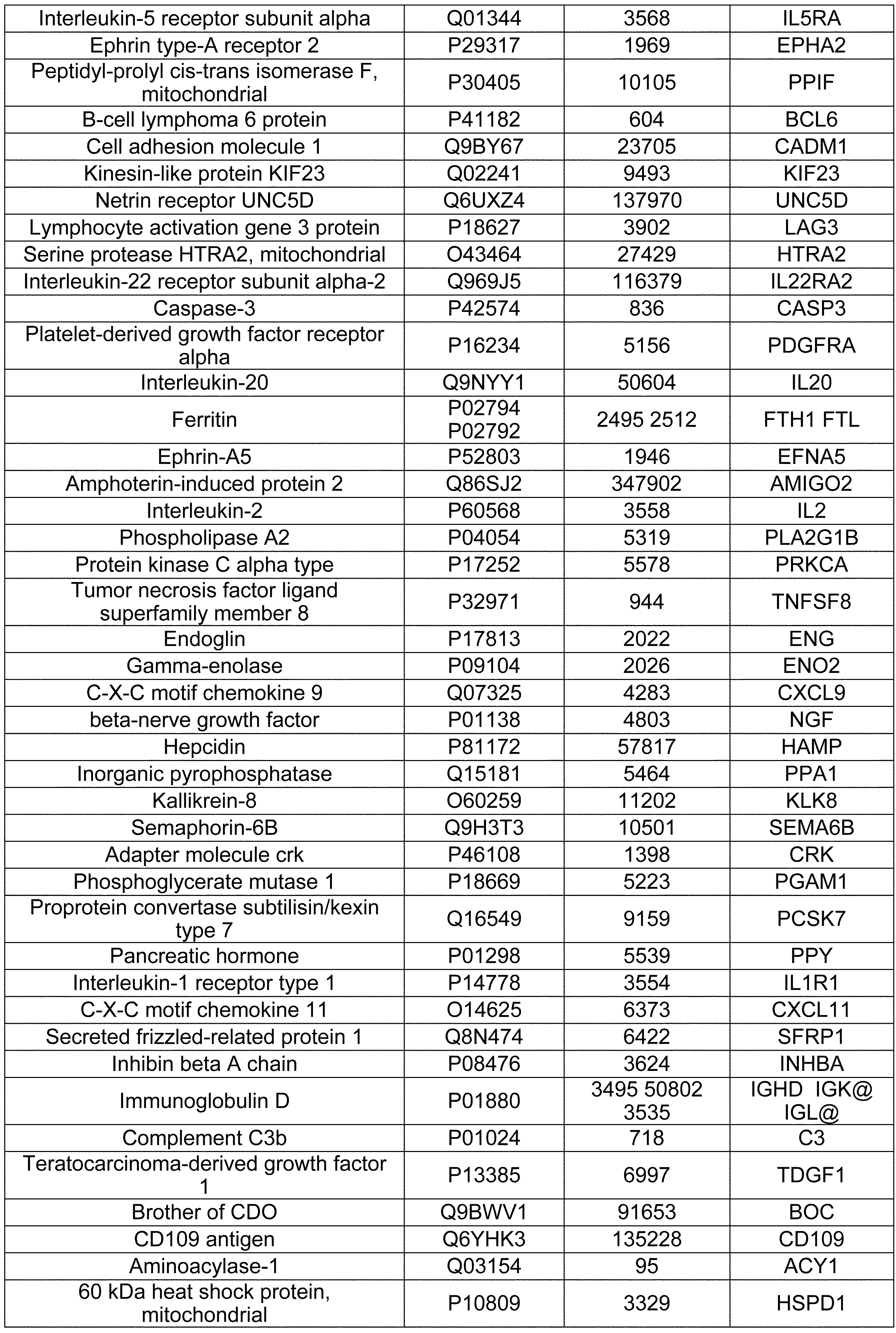
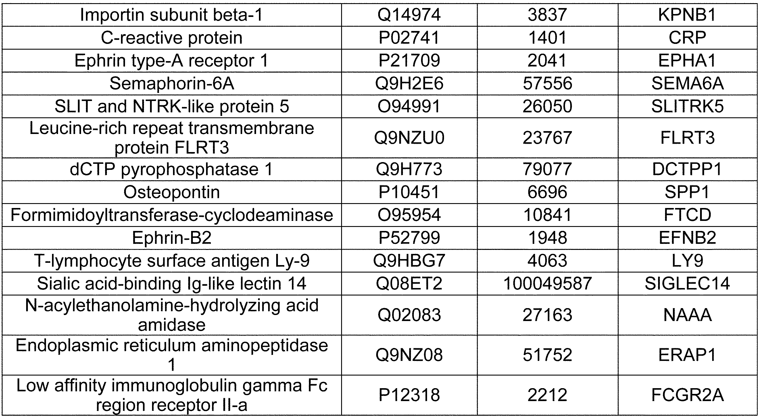


これらの表に示されるマーカー(mRNA、タンパク質)の配列は、上記表に記載のUniprot IDに基づき、当業者公知のデータベース(Uniprot:https://www.uniprot.org/)より、容易に取得することができる。具体的には、Uniprot IDを基に対応するUniprot entryを検索し、「sequences」項目に記載されている配列を取得することが可能である。さらに、「sequences」項目の記載から他のデータベース(例えば、RefSeq、EMBL、GenBank、DDBJ、CCDS、PIR、UniGene、Ensemble、GeneID、KEGG、USCS)のID/accession numberを取得し当該データベースから更に配列を取得することも可能である。本開示における各マーカーの配列は、対応するUniprot entryのrelease numberが2018-09より前の最新バージョン(例えば、2018-08、2018-08が存在しない場合は2018_07、2018_08も2018_07も存在しない場合は2018_06、以降同様)に記載されている配列やID/accession numberを指している。
また、Uniprotのリリースデータのアーカイブが保存されているFTP(ftp://ftp.uniprot.org/pub/databases/uniprot/previous_releases/)において、Directory「release-2018_08/knowledgebase」(「release-2018_08」に含まれる「knowledgebase」Directory)に含まれるデータからも、上記表中の記載のUniprot IDに基づき、表中のマーカーのアミノ酸配列を取得することも出来る。 The sequences of the markers (mRNA, protein) shown in these tables can be easily obtained from a database known to those skilled in the art (Uniprot: https://www.uniprot.org/) based on the Uniprot ID described in the above table. can do. Specifically, it is possible to search for the corresponding Uniprot entry based on the Uniprot ID and obtain the sequence described in the "sequences" item. Furthermore, the ID / accession number of another database (for example, RefSeq, EMBL, GenBank, DDBJ, CCDS, PIR, UniGene, Ensemble, GeneID, KEGG, USCS) is obtained from the description of the "sequences" item and further sequenced from the database. It is also possible to obtain. The sequence of each marker in this disclosure is the latest version of the corresponding Uniprot entry release number prior to 2018-09 (eg, 2018_07 if 2018-08, 2018-08 does not exist, if neither 2018_08 nor 2018_07 exists. Refers to the array and ID / accession number described in 2018_06, the same applies hereafter).
Also, in the FTP (ftp://ftp.uniprot.org/pub/databases/uniprot/previous_releases/) where the archive of Uniprot release data is saved, Directory "release-2018_08 / knowledgebase"("release-2018_08" It is also possible to obtain the amino acid sequence of the marker in the table based on the Uniprot ID described in the above table from the data included in the "knowledge base" Directory) included in.
上記表中に記載のマーカー群に、線維化を示すマーカー(TGFB1およびSPARC等)が含まれている。更に、上記表中に記載のマーカー群は、血小板由来と考えられる分子が多く含まれる。例えば、PPBP, THBS1, PF4, SERPINA1, TIMP3, APP, CALU, CASP3, MMRN1, SAA1, SRGN, VWFなどは血小板の脱顆粒などに関与することがuniprotに記載されている。Zhangらは子宮内膜由来細胞を用いたin vitroの実験系で活性化した血小板から放出されたTGFb1によってepithelial-mesenchymal transition (EMT)およびfibroblast-to-myofibroblast transdifferentiation (FMT)が誘導され、コラーゲン産生が増加し、線維化が誘導されることを示した(Molecular and Cellular Endocrinology, 428, 1-16, 2016)。このことは、本開示においてTGFb1および血小板由来タンパク質がマーカーとして選抜されことと一致しており、これらのタンパク質の変化は血小板の活性化等の変化を反映した可能性が示唆された。
The marker group described in the above table includes markers indicating fibrosis (TGFB1, SPARC, etc.). Furthermore, the marker group described in the above table contains many molecules considered to be derived from platelets. For example, it is stated in uniprot that PPPP, THBS1, PF4, SERPINA1, TIMP3, APP, CALU, CASP3, MMRN1, SAA1, SRGN, VWF, etc. are involved in platelet degranulation. Zhang et al. Induced epithelial-mesenchymal transition (EMT) and fibroblast-to-myofibroblast transdifferentiation (FMT) by TGFb1 released from platelets activated in an in vitro experimental system using endometrial-derived cells to produce collagen. Was increased and fibrosis was induced (Molecular and Cellular Endocrinology, 428, 1-16, 2016). This is consistent with the selection of TGFb1 and platelet-derived proteins as markers in the present disclosure, suggesting that changes in these proteins may reflect changes in platelet activation and the like.
タイプVコラーゲンMMP分解産物は、MMP mediated type V collagen degradationとも呼ばれ、タイプVコラーゲンのalpha 3 chain(Refseq: NP_056534.2、配列番号:1)がMMPにより分解された産物である。具体的に、タイプVコラーゲンMMP分解産物は配列番号:1で示すタイプVコラーゲンの1316番アミノ酸(G)と1317番アミノ酸(H)の間が切断されることにより産生されたタイプVコラーゲン分解産物である(Clin Biochem. 2012 May;45(7-8):541-6)。具体的には、タイプVコラーゲン由来のポリペプチドであって、配列番号:4に記載のアミノ酸配列をN末端に含む、および/または配列番号:6に記載のアミノ酸配列をC末端に含むものであれば、タイプVコラーゲンMMP分解産物に含まれる。タイプVコラーゲンMMP分解産物の例示として、配列番号:2または3に示すタンパク質断片が列挙される。タイプVコラーゲンMMP分解産物の中で、配列番号:4(HMGREGREGE)のペプチド断片をC5Mと称することがある。
The type V collagen MMP degradation product is also called MMP mediated type V collagen degradation, and is a product obtained by degrading the alpha 3 chain (Refseq: NP_056534.2, SEQ ID NO: 1) of type V collagen by MMP. Specifically, the type V collagen MMP degradation product is a type V collagen degradation product produced by cleaving between amino acids 1316 (G) and 1317 (H) of type V collagen shown in SEQ ID NO: 1. (Clin Biochem. 2012 May; 45 (7-8): 541-6). Specifically, it is a type V collagen-derived polypeptide containing the amino acid sequence set forth in SEQ ID NO: 4 at the N-terminal and / or the amino acid sequence set forth in SEQ ID NO: 6 at the C-terminal. If present, it is included in Type V collagen MMP degradation products. Examples of type V collagen MMP degradation products include the protein fragments set forth in SEQ ID NO: 2 or 3. Among the type V collagen MMP degradation products, the peptide fragment of SEQ ID NO: 4 (HMGREGREGE) may be referred to as C5M.
タイプVコラーゲンMMP分解産物の存在の検出は、配列番号:1に記載の1316番アミノ酸(G)と1317番アミノ酸(H)の間で切断された後のタイプVコラーゲン断片を検出できる限り、その方法を問わない。ある態様において、配列番号:2で示すタイプVコラーゲン断片のN末端に位置するペプチドHMGREGREGE(配列番号:4)を特異的に認識する抗体を用いてタイプVコラーゲンMMP分解産物の存在量を検出することが出来る。より具体的な態様において、配列番号:2で示すタイプVコラーゲン断片のN末端に位置するペプチドHMGREGREGE(配列番号:4)を認識でき、GHMGREGREGE(配列番号:5)を認識しない抗体を用いて、タイプVコラーゲンMMP分解産物の存在量を検出することができる(Clin Biochem. 2012 May;45(7-8):541-6.では、そのような抗体を使用したELISA法検出を確立している)。ある別の態様において、配列番号:3で示すタイプVコラーゲン断片のC末端に位置するペプチドGPPGKRGPSG(配列番号:6)を特異的に認識する抗体を用いてタイプVコラーゲンMMP分解産物の存在量を検出することができる。より具体的な態様において、配列番号:3で示すタイプVコラーゲン断片のC末端に位置するペプチドGPPGKRGPSG(配列番号:6)を認識でき、GPPGKRGPSGH(配列番号:7)を認識しない抗体を用いて、タイプVコラーゲンMMP分解産物の存在量を検出することができる。
The presence of a type V collagen MMP degradation product can be detected as long as the type V collagen fragment after cleavage between amino acids 1316 (G) and 1317 (H) set forth in SEQ ID NO: 1 can be detected. It doesn't matter how. In some embodiments, the abundance of a Type V collagen MMP degradation product is detected using an antibody that specifically recognizes the peptide HMGREGREGE (SEQ ID NO: 4) located at the N-terminus of the Type V collagen fragment set forth in SEQ ID NO: 2. Can be done. In a more specific embodiment, an antibody capable of recognizing the peptide HMGREGREGE (SEQ ID NO: 4) located at the N-terminus of the type V collagen fragment shown in SEQ ID NO: 2 and not GHMGREGREGE (SEQ ID NO: 5) is used. The abundance of type V collagen MMP degradation products can be detected (Clin Biochem. 2012 May; 45 (7-8): 541-6.) Establishes ELISA detection using such antibodies. ). In another embodiment, the abundance of the type V collagen MMP degradation product is determined using an antibody that specifically recognizes the peptide GPPGKRGPSG (SEQ ID NO: 6) located at the C-terminus of the type V collagen fragment set forth in SEQ ID NO: 3. Can be detected. In a more specific embodiment, an antibody capable of recognizing the peptide GPPGKRGPSG (SEQ ID NO: 6) located at the C-terminus of the type V collagen fragment shown in SEQ ID NO: 3 and not recognizing GPPGKRGPSGH (SEQ ID NO: 7) is used. The abundance of type V collagen MMP degradation products can be detected.
本開示においては、タイプVコラーゲンMMP分解産物、表1Aで示されるマーカー、表2Aで示されるマーカー、表1Bで示されるマーカー、表2Bで示されるマーカー、表5Aで示されるマーカー、表6Aで示されるマーカー、表5Bで示されるマーカー、表6Bで示されるマーカーからなるグループから選択されるタンパク質を単独でマーカーとして用いてもよいし、前記選択される複数のタンパク質を組み合わせてマーカーとして用いてもよい。複数のタンパク質をマーカーとする場合には、各マーカーをそれぞれ単独で測定した後にそれらの結果を組み合わせてもよいし、あるいは各マーカーを同時に測定してもよい。
In the present disclosure, the type V collagen MMP degradation products, the markers shown in Table 1A, the markers shown in Table 2A, the markers shown in Table 1B, the markers shown in Table 2B, the markers shown in Table 5A, in Table 6A. A marker selected from the group consisting of the markers shown, the markers shown in Table 5B, and the markers shown in Table 6B may be used alone as markers, or a plurality of selected proteins may be used in combination as markers. May be good. When a plurality of proteins are used as markers, each marker may be measured individually and then the results may be combined, or each marker may be measured at the same time.
本開示のマーカーの存在量の測定は、本開示に記載の測定方法を用いて行うこともできるし、市販されている測定試薬を用いて行うこともできる。本明細書中に記載されている各マーカーの具体的な測定値や所定の値は、前記測定試薬を用いて測定された値として解釈することができる。
The abundance of the marker of the present disclosure can be measured by using the measuring method described in the present disclosure, or by using a commercially available measuring reagent. Specific measured values and predetermined values of each marker described in the present specification can be interpreted as values measured using the measuring reagent.
本開示のマーカーに対して設定された所定の値(対照レベル)は、各マーカーの存在量を測定しようとする対象の試料の種類によっても変動し得るが、例えば、0.1から100 ng/mLの範囲から設定することができる。あるいは、0.2から90 ng/mL、0.3から80 ng/mL、0.4から70 ng/mL、0.5から60 ng/mL、1.0から50 ng/mL、1.5から40 ng/mL、2.0から30 ng/mL、2.5から20 ng/mL、3.0から10 ng/mLなどの範囲から設定することもできるが、これらに限定されない。
The predetermined value (control level) set for the markers of the present disclosure may vary depending on the type of sample of interest for which the abundance of each marker is to be measured, for example, 0.1 to 100 ng / mL. It can be set from the range. Alternatively, 0.2 to 90 ng / mL, 0.3 to 80 ng / mL, 0.4 to 70 ng / mL, 0.5 to 60 ng / mL, 1.0 to 50 ng / mL, 1.5 to 40 ng / mL, 2.0 to 30 ng / mL , 2.5 to 20 ng / mL, 3.0 to 10 ng / mL, etc., but is not limited to these.
また、本開示の各マーカーに対して設定された所定の値は、0.1 ng/mL、0.2 ng/mL、0.3 ng/mL、0.4 ng/mL、0.5 ng/mL、0.6 ng/mL、0.7 ng/mL、0.8 ng/mL、0.9 ng/mL、1.0 ng/mL、1.5 ng/mL、2.0 ng/mL、2.5 ng/mL、3.0 ng/mL、3.5 ng/mL、4.0 ng/mL、4.5 ng/mL、5.0 ng/mL、5.5 ng/mL、6.0 ng/mL、6.5 ng/mL、7.0 ng/mL、7.5 ng/mL、8.0 ng/mL、8.5 ng/mL、9.0 ng/mL、9.5 ng/mL、10 ng/mL、15 ng/mL、20 ng/mL、25 ng/mL、30 ng/mL、35 ng/mL、40 ng/mL、45 ng/mL、50 ng/mL、60 ng/mL、70 ng/mL、80 ng/mL、90 ng/mL、100 ng/mLなどの値から設定することもできるが、これらに限定されない。
In addition, the predetermined values set for each marker of the present disclosure are 0.1 ng / mL, 0.2 ng / mL, 0.3 ng / mL, 0.4 ng / mL, 0.5 ng / mL, 0.6 ng / mL, 0.7 ng. / mL, 0.8 ng / mL, 0.9 ng / mL, 1.0 ng / mL, 1.5 ng / mL, 2.0 ng / mL, 2.5 ng / mL, 3.0 ng / mL, 3.5 ng / mL, 4.0 ng / mL, 4.5 ng / mL, 5.0 ng / mL, 5.5 ng / mL, 6.0 ng / mL, 6.5 ng / mL, 7.0 ng / mL, 7.5 ng / mL, 8.0 ng / mL, 8.5 ng / mL, 9.0 ng / mL, 9.5 ng / mL, 10 ng / mL, 15 ng / mL, 20 ng / mL, 25 ng / mL, 30 ng / mL, 35 ng / mL, 40 ng / mL, 45 ng / mL, 50 ng / mL, 60 ng It can be set from values such as / mL, 70 ng / mL, 80 ng / mL, 90 ng / mL, and 100 ng / mL, but is not limited to these.
本開示においては、本開示の各マーカーの存在量が所定の値(対照レベル)よりも大きい場合に、「存在量が高い」または「存在量が多い」と決定することができる。例えば本開示においては、これらに限定されるものではないが、前記タイプVコラーゲンMMP分解産物、表1Aで示されるマーカー、表2Aで示されるマーカー、表5Aで示されるマーカーからなるグループから選択される少なくとも一つのマーカーの存在量が、健常人から得られた試料における同一のマーカーの存在量の、例えば1.1倍以上、1.2倍以上、1.3倍以上、1.4倍以上、1.5倍以上、1.6倍以上、1.7倍以上、1.8倍以上、1.9倍以上、2.0倍以上、2.1倍以上、2.2倍以上、2.3倍以上、2.4倍以上、2.5倍以上、2.6倍以上、2.7倍以上、2.8倍以上、2.9倍以上、3.0倍以上、またはそれ以上の場合、「存在量が高い」または「存在量が多い」と判定することができる。あるいは本開示においては、これらに限定されるものではないが、表6Aで示されるマーカーから選択される少なくとも一つのマーカーの存在量が、健常人から得られた試料における同一のマーカーの存在量の、例えば1.10倍以上、1.15倍以上、1.20倍以上、1.25倍以上、1.30倍以上、1.35倍以上、1.40倍以上、1.45倍以上、1.50倍以上、1.55倍以上、1.60倍以上、1.65倍以上、1.70倍以上、1.75倍以上、1.80倍以上、1.85倍以上、1.90倍以上、1.95倍以上、2.00倍以上、またはそれ以上の場合、「存在量が高い」または「存在量が多い」と判定することができる。
In the present disclosure, when the abundance of each marker of the present disclosure is larger than a predetermined value (control level), it can be determined that the abundance is high or the abundance is high. For example, in the present disclosure, it is selected from a group consisting of, but not limited to, the type V collagen MMP degradation products, the markers shown in Table 1A, the markers shown in Table 2A, and the markers shown in Table 5A. The abundance of at least one marker is, for example, 1.1 times or more, 1.2 times or more, 1.3 times or more, 1.4 times or more the abundance of the same marker in a sample obtained from a healthy person. , 1.5 times or more, 1.6 times or more, 1.7 times or more, 1.8 times or more, 1.9 times or more, 2.0 times or more, 2.1 times or more, 2.2 times or more, 2 .3 times or more, 2.4 times or more, 2.5 times or more, 2.6 times or more, 2.7 times or more, 2.8 times or more, 2.9 times or more, 3.0 times or more, or more In the case of, it can be determined that "the abundance amount is high" or "the abundance amount is large". Alternatively, in the present disclosure, the abundance of at least one marker selected from the markers shown in Table 6A is, but not limited to, the abundance of the same marker in a sample obtained from a healthy person. For example, 1.10 times or more, 1.15 times or more, 1.20 times or more, 1.25 times or more, 1.30 times or more, 1.35 times or more, 1.40 times or more, 1.45 times or more, 1.50 times or more, 1.55 times or more, 1.60 times or more, 1.65 times or more, 1.70 times or more, 1.75 times or more, 1.80 times or more, 1.85 times or more 1. When it is 90 times or more, 1.95 times or more, 2.00 times or more, or more, it can be determined that "the abundance amount is high" or "the abundance amount is large".
本開示においては、本開示の各マーカーの存在量が所定の値(対照レベル)よりも小さい場合に、「存在量が低い」または「存在量が少ない」と決定することができる。例えば本開示においては、これらに限定されるものではないが、前記タイプVコラーゲンMMP分解産物、表1Bで示されるマーカー、表2Bで示されるマーカー、表5Bで示されるマーカーからなるグループから選択される少なくとも一つのマーカーの存在量が、健常人から得られた試料における同一のマーカーの存在量の、例えば0.9倍以下、0.8倍以下、0.7倍以下、0.6倍以下、0.5倍以下、0.4倍以下、0.3倍以下、0.2倍以下、0.1倍以下またはそれ以下の場合、「存在量が低い」または「存在量が少ない」と判定することができる。あるいは本開示においては、これらに限定されるものではないが、表6Bで示されるマーカーから選択される少なくとも一つのマーカーの存在量が、健常人から得られた試料における同一のマーカーの存在量の、例えば0.9倍以下、0.8倍以下、0.7倍以下、0.6倍以下、0.5倍以下、0.4倍以下、0.3倍以下、0.2倍以下、0.1倍以下またはそれ以下の場合、「存在量が低い」または「存在量が少ない」と判定することができる。
In the present disclosure, when the abundance of each marker of the present disclosure is smaller than a predetermined value (control level), it can be determined that the abundance is low or the abundance is low. For example, in the present disclosure, it is selected from the group consisting of, but not limited to, the type V collagen MMP degradation products, the markers shown in Table 1B, the markers shown in Table 2B, and the markers shown in Table 5B. The abundance of at least one marker is, for example, 0.9 times or less, 0.8 times or less, 0.7 times or less, 0.6 times or less of the abundance of the same marker in a sample obtained from a healthy person. , 0.5 times or less, 0.4 times or less, 0.3 times or less, 0.2 times or less, 0.1 times or less or less, "low abundance" or "low abundance" Can be determined. Alternatively, in the present disclosure, the abundance of at least one marker selected from the markers shown in Table 6B is, but not limited to, the abundance of the same marker in a sample obtained from a healthy person. For example, 0.9 times or less, 0.8 times or less, 0.7 times or less, 0.6 times or less, 0.5 times or less, 0.4 times or less, 0.3 times or less, 0.2 times or less, When it is 0.1 times or less or less, it can be determined that "the abundance is low" or "the abundance is low".
子宮内膜症の診断及び対象における子宮内膜症の罹患の有無の決定
本開示の一側面は、子宮内膜症の診断方法、または対象における子宮内膜症の罹患の有無を決定する方法に関する。また本開示の一側面は、子宮内膜症またはそのマーカーの検出方法に関する。また本開示の一側面は、当該診断または決定を補助するための方法に関する。また本開示の一側面は、当該診断または決定についての指示を提供するための方法に関する。また本開示の一側面は、それら方法に使用するための試薬及びキットに関する。 Diagnosis of Endometriosis and Determination of Subject with Endometriosis One aspect of the disclosure relates to a method for diagnosing endometriosis or a method for determining the presence or absence of endometriosis in a subject. .. Also, one aspect of the present disclosure relates to a method for detecting endometriosis or a marker thereof. Also, one aspect of the disclosure relates to methods for assisting the diagnosis or decision. Also, one aspect of the disclosure relates to a method for providing instructions for such a diagnosis or decision. Also, one aspect of the disclosure relates to reagents and kits for use in those methods.
本開示の一側面は、子宮内膜症の診断方法、または対象における子宮内膜症の罹患の有無を決定する方法に関する。また本開示の一側面は、子宮内膜症またはそのマーカーの検出方法に関する。また本開示の一側面は、当該診断または決定を補助するための方法に関する。また本開示の一側面は、当該診断または決定についての指示を提供するための方法に関する。また本開示の一側面は、それら方法に使用するための試薬及びキットに関する。 Diagnosis of Endometriosis and Determination of Subject with Endometriosis One aspect of the disclosure relates to a method for diagnosing endometriosis or a method for determining the presence or absence of endometriosis in a subject. .. Also, one aspect of the present disclosure relates to a method for detecting endometriosis or a marker thereof. Also, one aspect of the disclosure relates to methods for assisting the diagnosis or decision. Also, one aspect of the disclosure relates to a method for providing instructions for such a diagnosis or decision. Also, one aspect of the disclosure relates to reagents and kits for use in those methods.
上記各方法は、対象から得られた試料におけるタイプVコラーゲンMMP分解産物、表1Aで示されるマーカー、表2Aで示されるマーカー、表1Bで示されるマーカー、表2Bで示されるマーカー、表5Aで示されるマーカー、表6Aで示されるマーカー、表5Bで示されるマーカー、表6Bで示されるマーカーからなるグループから選択される少なくとも一つのマーカーの存在量を測定することを含む。また、当該存在量を所定の値(対照レベル)と比較する工程を含むことができる。
上記試薬は、タイプVコラーゲンMMP分解産物、表1Aで示されるマーカー、表2Aで示されるマーカー、表1Bで示されるマーカー、表2Bで示されるマーカー、表5Aで示されるマーカー、表6Aで示されるマーカー、表5Bで示されるマーカー、表6Bで示されるマーカーからなるグループから選択される少なくとも一つのマーカーの存在量を測定するための試薬である。上記キットは、前記試薬を含む。 Each of the above methods is a type V collagen MMP degradation product in a sample obtained from a subject, a marker shown in Table 1A, a marker shown in Table 2A, a marker shown in Table 1B, a marker shown in Table 2B, in Table 5A. It involves measuring the abundance of at least one marker selected from the group consisting of the markers shown, the markers shown in Table 6A, the markers shown in Table 5B, and the markers shown in Table 6B. In addition, a step of comparing the abundance with a predetermined value (control level) can be included.
The reagents are type V collagen MMP degradation products, markers shown in Table 1A, markers shown in Table 2A, markers shown in Table 1B, markers shown in Table 2B, markers shown in Table 5A, shown in Table 6A. A reagent for measuring the abundance of at least one marker selected from the group consisting of the markers, the markers shown in Table 5B, and the markers shown in Table 6B. The kit contains the reagents.
上記各方法は、対象から得られた試料におけるタイプVコラーゲンMMP分解産物、表1Aで示されるマーカー、表2Aで示されるマーカー、表5Aで示されるマーカー、表6Aで示されるマーカーからなるグループから選択される少なくとも一つのマーカーの存在量が所定の値(対照レベル)と比較して高い場合、または対象から得られた試料における表1Bで示されるマーカー、表2Bで示されるマーカー、表5Bで示されるマーカー、表6Bで示されるマーカーからなるグループから選択される少なくとも一つのマーカーの存在量が所定の値(対照レベル)と比較して低い場合、前記試料が由来する対象が子宮内膜症に罹患している、またはその可能性があると示される工程を含めてもよい。
Each of the above methods consisted of a group consisting of type V collagen MMP degradation products in a sample obtained from the subject, the markers shown in Table 1A, the markers shown in Table 2A, the markers shown in Table 5A, and the markers shown in Table 6A. When the abundance of at least one marker selected is high compared to a predetermined value (control level), or in the sample obtained from the subject, the markers shown in Table 1B, the markers shown in Table 2B, in Table 5B. If the abundance of at least one marker selected from the group consisting of the markers shown and the markers shown in Table 6B is low compared to a predetermined value (control level), the subject from which the sample is derived is endometriosis. You may include steps that are or are indicated to be affected by.
上記各方法はin vivoおよびin vitroのいずれにおいても行うことができるが、in vitroにおいて行うことが好ましい。
また上記各方法は、対象から試料を取得する工程を含めてもよい。
また上記各方法は、子宮内膜症に罹患している、またはその可能性があると示された対象を治療する工程を含めてもよい。また上記各方法は、子宮内膜症に罹患している、またはその可能性があると示された対象に公知の子宮内膜症の治療薬を投与する工程を含めてもよい。すなわち本発明は、上記各方法によって子宮内膜症に罹患している、またはその可能性があると示された対象における子宮内膜症を治療する方法に関する。子宮内膜症の治療薬は当業者に様々なものが知られており、これらに限定されるものではないが、エストロゲン/プロゲステロン合剤、プロゲステロン製剤、GnRHアゴニスト、GnRHアンタゴニスト、ダナゾールなどを例示することができる。 Each of the above methods can be performed in vivo or in vitro, but is preferably performed in vitro.
In addition, each of the above methods may include a step of obtaining a sample from the target.
Each of the above methods may also include the step of treating a subject who has or has been shown to have or may have endometriosis. In addition, each of the above methods may include a step of administering a known therapeutic agent for endometriosis to a subject who has or is indicated to have endometriosis. That is, the present invention relates to a method for treating endometriosis in a subject who has or is indicated to have endometriosis by each of the above methods. Various therapeutic agents for endometriosis are known to those skilled in the art, and examples thereof include, but are not limited to, estrogen / progesterone mixture, progesterone preparation, GnRH agonist, GnRH antagonist, and danazol. be able to.
上記キットは、対象から得られた試料における、タイプVコラーゲンMMP分解産物、表1Aで示されるマーカー、表2Aで示されるマーカー、表5Aで示されるマーカー、表6Aで示されるマーカーからなるグループから選択される少なくとも一つのマーカーの存在量が所定の値(対照レベル)と比較して高い場合、または表1Bで示されるマーカー、表2Bで示されるマーカー、表5Bで示されるマーカー、表6Bで示されるマーカーからなるグループから選択される少なくとも一つのマーカーの存在量が所定の値(対照レベル)と比較して低い場合、前記試料が由来する対象が子宮内膜症に罹患している、またはその可能性があると示されることを記載した指示書をさらに含めても良い。
The kit consists of a group consisting of type V collagen MMP degradation products, markers shown in Table 1A, markers shown in Table 2A, markers shown in Table 5A, and markers shown in Table 6A in a sample obtained from the subject. If the abundance of at least one marker selected is high compared to a given value (control level), or in Table 1B, the markers shown in Table 2B, the markers shown in Table 5B, in Table 6B. If the abundance of at least one marker selected from the group consisting of the indicated markers is low compared to a given value (control level), then the subject from which the sample is derived suffers from endometriosis or Additional instructions may be included stating that the possibility is indicated.
対象における子宮内膜の線維化の程度の決定
本開示の一側面は、対象における子宮内膜の線維化の程度を決定する方法、当該決定を補助するための方法、当該決定についての指示を提供するための方法、それらに使用するための試薬及びキットに関する。 Determining the degree of endometrial fibrosis in a subject One aspect of the disclosure provides methods for determining the degree of endometrial fibrosis in a subject, methods for assisting the decision, and instructions for the decision. With respect to methods, reagents and kits for use in them.
本開示の一側面は、対象における子宮内膜の線維化の程度を決定する方法、当該決定を補助するための方法、当該決定についての指示を提供するための方法、それらに使用するための試薬及びキットに関する。 Determining the degree of endometrial fibrosis in a subject One aspect of the disclosure provides methods for determining the degree of endometrial fibrosis in a subject, methods for assisting the decision, and instructions for the decision. With respect to methods, reagents and kits for use in them.
上記各方法は、対象から得られた試料におけるタイプVコラーゲンMMP分解産物、表1Aで示されるマーカー、表2Aで示されるマーカー、表1Bで示されるマーカー、表2Bで示されるマーカー、表5Aで示されるマーカー、表6Aで示されるマーカー、表5Bで示されるマーカー、表6Bで示されるマーカーからなるグループから選択される少なくとも一つのマーカーの存在量を測定することを含む。また、当該存在量を所定の値(対照レベル)と比較する工程を含むことができる。
上記試薬は、タイプVコラーゲンMMP分解産物、表1Aで示されるマーカー、表2Aで示されるマーカー、表1Bで示されるマーカー、表2Bで示されるマーカー、表5Aで示されるマーカー、表6Aで示されるマーカー、表5Bで示されるマーカー、表6Bで示されるマーカーからなるグループから選択される少なくとも一つのマーカーの存在量を測定するための試薬である。上記キットは、前記試薬を含む。 Each of the above methods is a type V collagen MMP degradation product in a sample obtained from a subject, a marker shown in Table 1A, a marker shown in Table 2A, a marker shown in Table 1B, a marker shown in Table 2B, in Table 5A. It involves measuring the abundance of at least one marker selected from the group consisting of the markers shown, the markers shown in Table 6A, the markers shown in Table 5B, and the markers shown in Table 6B. In addition, a step of comparing the abundance with a predetermined value (control level) can be included.
The reagents are type V collagen MMP degradation products, markers shown in Table 1A, markers shown in Table 2A, markers shown in Table 1B, markers shown in Table 2B, markers shown in Table 5A, shown in Table 6A. A reagent for measuring the abundance of at least one marker selected from the group consisting of the markers, the markers shown in Table 5B, and the markers shown in Table 6B. The kit contains the reagents.
上記試薬は、タイプVコラーゲンMMP分解産物、表1Aで示されるマーカー、表2Aで示されるマーカー、表1Bで示されるマーカー、表2Bで示されるマーカー、表5Aで示されるマーカー、表6Aで示されるマーカー、表5Bで示されるマーカー、表6Bで示されるマーカーからなるグループから選択される少なくとも一つのマーカーの存在量を測定するための試薬である。上記キットは、前記試薬を含む。 Each of the above methods is a type V collagen MMP degradation product in a sample obtained from a subject, a marker shown in Table 1A, a marker shown in Table 2A, a marker shown in Table 1B, a marker shown in Table 2B, in Table 5A. It involves measuring the abundance of at least one marker selected from the group consisting of the markers shown, the markers shown in Table 6A, the markers shown in Table 5B, and the markers shown in Table 6B. In addition, a step of comparing the abundance with a predetermined value (control level) can be included.
The reagents are type V collagen MMP degradation products, markers shown in Table 1A, markers shown in Table 2A, markers shown in Table 1B, markers shown in Table 2B, markers shown in Table 5A, shown in Table 6A. A reagent for measuring the abundance of at least one marker selected from the group consisting of the markers, the markers shown in Table 5B, and the markers shown in Table 6B. The kit contains the reagents.
上記各方法は、対象から得られた試料におけるタイプVコラーゲンMMP分解産物、表1Aで示されるマーカー、表2Aで示されるマーカー、表5Aで示されるマーカー、表6Aで示されるマーカーからなるグループから選択される少なくとも一つのマーカーの存在量が所定の値(対照レベル)と比較して高い場合、または対象から得られた試料における表1Bで示されるマーカー、表2Bで示されるマーカー、表5Bで示されるマーカー、表6Bで示されるマーカーからなるグループから選択される少なくとも一つのマーカーの存在量が所定の値(対照レベル)と比較して低い場合、前記試料が由来する対象において子宮内膜における線維化が発生している、またはその可能性があると示される工程を含めてもよい。
Each of the above methods consisted of a group consisting of type V collagen MMP degradation products in a sample obtained from the subject, the markers shown in Table 1A, the markers shown in Table 2A, the markers shown in Table 5A, and the markers shown in Table 6A. When the abundance of at least one marker selected is high compared to a predetermined value (control level), or in the sample obtained from the subject, the markers shown in Table 1B, the markers shown in Table 2B, in Table 5B. When the abundance of at least one marker selected from the group consisting of the markers shown and the markers shown in Table 6B is low compared to a predetermined value (control level), in the subject from which the sample is derived, in the endometrial. It may include steps in which fibrosis is occurring or is indicated to be possible.
上記各方法はin vivoおよびin vitroのいずれにおいても行うことができるが、in vitroにおいて行うことが好ましい。
また上記各方法は、対象から試料を取得する工程を含めてもよい。
また上記各方法は、子宮内膜における線維化が発生している、またはその可能性があると示された対象を治療する工程を含めてもよい。また上記各方法は、子宮内膜における線維化が発生している、またはその可能性があると示された対象に公知の子宮内膜における線維化を抑制するための治療薬を投与する工程を含めてもよい。すなわち本発明は、上記各方法によって子宮内膜における線維化が発生している、またはその可能性があると示された対象における、線維化を抑制する方法に関する。子宮内膜における線維化を抑制するための治療薬としては、上述の子宮内膜症の治療薬を例示することができる。 Each of the above methods can be performed in vivo or in vitro, but is preferably performed in vitro.
In addition, each of the above methods may include a step of obtaining a sample from the target.
Each of the above methods may also include the step of treating a subject who has or has been shown to have fibrosis in the endometrium. In addition, each of the above methods comprises a step of administering a known therapeutic agent for suppressing fibrosis in the endometrium to a subject who has or has been shown to have fibrosis in the endometrium. May be included. That is, the present invention relates to a method for suppressing fibrosis in a subject in which fibrosis in the endometrium has occurred or is shown to be possible by each of the above methods. As the therapeutic agent for suppressing fibrosis in the endometrium, the above-mentioned therapeutic agent for endometriosis can be exemplified.
上記キットは、対象から得られた試料における、タイプVコラーゲンMMP分解産物、表1Aで示されるマーカー、表2Aで示されるマーカー、表5Aで示されるマーカー、表6Aで示されるマーカーからなるグループから選択される少なくとも一つのマーカーの存在量が所定の値(対照レベル)と比較して高い場合、または表1Bで示されるマーカー、表2Bで示されるマーカー、表5Bで示されるマーカー、表6Bで示されるマーカーからなるグループから選択される少なくとも一つのマーカーの存在量が所定の値(対照レベル)と比較して低い場合、前記試料が由来する対象において子宮内膜における線維化が発生している、またはその可能性があると示されることを記載した指示書をさらに含めても良い。
The kit consists of a group consisting of type V collagen MMP degradation products, markers shown in Table 1A, markers shown in Table 2A, markers shown in Table 5A, and markers shown in Table 6A in a sample obtained from the subject. If the abundance of at least one marker selected is high compared to a given value (control level), or in Table 1B, the markers shown in Table 2B, the markers shown in Table 5B, in Table 6B. When the abundance of at least one marker selected from the group consisting of the indicated markers is low compared to a predetermined value (control level), fibrosis in the endometrial membrane occurs in the subject from which the sample is derived. , Or additional instructions stating that it may be indicated.
対象における子宮内膜の癒着程度の決定
本開示の一側面は、対象における子宮内膜の癒着の程度を決定する方法、当該決定を補助するための方法、当該決定についての指示を提供するための方法、それら方法に使用するための試薬及びキットに関する。 Determining the Degree of Endometrial Adhesion in a Subject One aspect of the disclosure is to provide methods for determining the degree of endometrial adhesion in a subject, methods to assist the decision, and instructions for the decision. With respect to methods, reagents and kits for use in those methods.
本開示の一側面は、対象における子宮内膜の癒着の程度を決定する方法、当該決定を補助するための方法、当該決定についての指示を提供するための方法、それら方法に使用するための試薬及びキットに関する。 Determining the Degree of Endometrial Adhesion in a Subject One aspect of the disclosure is to provide methods for determining the degree of endometrial adhesion in a subject, methods to assist the decision, and instructions for the decision. With respect to methods, reagents and kits for use in those methods.
上記各方法は、対象から得られた試料におけるタイプVコラーゲンMMP分解産物、表1Aで示されるマーカー、表2Aで示されるマーカー、表1Bで示されるマーカー、表2Bで示されるマーカー、表5Aで示されるマーカー、表6Aで示されるマーカー、表5Bで示されるマーカー、表6Bで示されるマーカーからなるグループから選択される少なくとも一つのマーカーの存在量を測定することを含む。また、当該存在量を所定の値(対照レベル)と比較する工程を含むことができる。
上記試薬は、タイプVコラーゲンMMP分解産物、表1Aで示されるマーカー、表2Aで示されるマーカー、表1Bで示されるマーカー、表2Bで示されるマーカー、表5Aで示されるマーカー、表6Aで示されるマーカー、表5Bで示されるマーカー、表6Bで示されるマーカーからなるグループから選択される少なくとも一つのマーカーの存在量を測定するための試薬である。上記キットは、前記試薬を含む。 Each of the above methods is a type V collagen MMP degradation product in a sample obtained from a subject, a marker shown in Table 1A, a marker shown in Table 2A, a marker shown in Table 1B, a marker shown in Table 2B, in Table 5A. It involves measuring the abundance of at least one marker selected from the group consisting of the markers shown, the markers shown in Table 6A, the markers shown in Table 5B, and the markers shown in Table 6B. In addition, a step of comparing the abundance with a predetermined value (control level) can be included.
The reagents are type V collagen MMP degradation products, markers shown in Table 1A, markers shown in Table 2A, markers shown in Table 1B, markers shown in Table 2B, markers shown in Table 5A, shown in Table 6A. A reagent for measuring the abundance of at least one marker selected from the group consisting of the markers, the markers shown in Table 5B, and the markers shown in Table 6B. The kit contains the reagents.
上記試薬は、タイプVコラーゲンMMP分解産物、表1Aで示されるマーカー、表2Aで示されるマーカー、表1Bで示されるマーカー、表2Bで示されるマーカー、表5Aで示されるマーカー、表6Aで示されるマーカー、表5Bで示されるマーカー、表6Bで示されるマーカーからなるグループから選択される少なくとも一つのマーカーの存在量を測定するための試薬である。上記キットは、前記試薬を含む。 Each of the above methods is a type V collagen MMP degradation product in a sample obtained from a subject, a marker shown in Table 1A, a marker shown in Table 2A, a marker shown in Table 1B, a marker shown in Table 2B, in Table 5A. It involves measuring the abundance of at least one marker selected from the group consisting of the markers shown, the markers shown in Table 6A, the markers shown in Table 5B, and the markers shown in Table 6B. In addition, a step of comparing the abundance with a predetermined value (control level) can be included.
The reagents are type V collagen MMP degradation products, markers shown in Table 1A, markers shown in Table 2A, markers shown in Table 1B, markers shown in Table 2B, markers shown in Table 5A, shown in Table 6A. A reagent for measuring the abundance of at least one marker selected from the group consisting of the markers, the markers shown in Table 5B, and the markers shown in Table 6B. The kit contains the reagents.
上記各方法は、対象から得られた試料におけるタイプVコラーゲンMMP分解産物、表1Aで示されるマーカー、表2Aで示されるマーカー、表5Aで示されるマーカー、表6Aで示されるマーカーからなるグループから選択される少なくとも一つのマーカーの存在量が所定の値(対照レベル)と比較して高い場合、または対象から得られた試料における表1Bで示されるマーカー、表2Bで示されるマーカー、表5Bで示されるマーカー、表6Bで示されるマーカーからなるグループから選択される少なくとも一つのマーカーの存在量が所定の値(対照レベル)と比較して低い場合、前記試料が由来する対象において子宮内膜における癒着が発生している、またはその可能性があると示される工程を含めてもよい。
Each of the above methods consisted of a group consisting of type V collagen MMP degradation products in a sample obtained from the subject, the markers shown in Table 1A, the markers shown in Table 2A, the markers shown in Table 5A, and the markers shown in Table 6A. When the abundance of at least one marker selected is high compared to a predetermined value (control level), or in the sample obtained from the subject, the markers shown in Table 1B, the markers shown in Table 2B, in Table 5B. When the abundance of at least one marker selected from the group consisting of the markers shown and the markers shown in Table 6B is low compared to a predetermined value (control level), in the subject from which the sample is derived, in the endometrial. It may include steps that indicate that adhesions have occurred or are likely to occur.
上記各方法はin vivoおよびin vitroのいずれにおいても行うことができるが、in vitroにおいて行うことが好ましい。
また上記各方法は、対象から試料を取得する工程を含めてもよい。
また上記各方法は、子宮内膜における癒着が発生している、またはその可能性があると示された対象を治療する工程を含めてもよい。また上記各方法は、子宮内膜における癒着が発生している、またはその可能性があると示された対象に公知の子宮内膜における癒着を抑制するための治療薬を投与する工程を含めてもよい。すなわち本発明は、上記各方法によって子宮内膜における癒着が発生している、またはその可能性があると示された対象における、子宮内膜における癒着を抑制する方法に関する。子宮内膜における癒着を抑制するための治療薬としては、上述の子宮内膜症の治療薬を例示することができる。 Each of the above methods can be performed in vivo or in vitro, but is preferably performed in vitro.
In addition, each of the above methods may include a step of obtaining a sample from the target.
Each of the above methods may also include the step of treating a subject who has, or is indicated to have, adhesions in the endometrium. In addition, each of the above methods includes a step of administering a known therapeutic agent for suppressing adhesions in the endometrium to a subject who has or is shown to have adhesions in the endometrium. May be good. That is, the present invention relates to a method for suppressing adhesion in the endometrium in a subject in which adhesion in the endometrium has occurred or is shown to be likely by each of the above methods. As a therapeutic agent for suppressing adhesion in the endometrium, the above-mentioned therapeutic agent for endometriosis can be exemplified.
上記キットは、対象から得られた試料における、タイプVコラーゲンMMP分解産物の濃度、表1Aで示されるマーカー、表2Aで示されるマーカー、表5Aで示されるマーカー、表6Aで示されるマーカーからなるグループから選択される少なくとも一つのマーカーの存在量が所定の値(対照レベル)と比較して高い場合、または表1Bで示されるマーカー、表2Bで示されるマーカー、表5Bで示されるマーカー、表6Bで示されるマーカーからなるグループから選択される少なくとも一つのマーカーの存在量が所定の値(対照レベル)と比較して低い場合、前記試料が由来する対象において子宮内膜における癒着が発生している、またはその可能性があると示されることを記載した指示書をさらに含めても良い。
The kit comprises the concentration of type V collagen MMP degradation products in the sample obtained from the subject, the markers shown in Table 1A, the markers shown in Table 2A, the markers shown in Table 5A, and the markers shown in Table 6A. If the abundance of at least one marker selected from the group is high compared to a given value (control level), or the markers shown in Table 1B, the markers shown in Table 2B, the markers shown in Table 5B, the table. When the abundance of at least one marker selected from the group consisting of the markers indicated by 6B is low compared to a predetermined value (control level), adhesion in the endometrial membrane occurs in the subject from which the sample is derived. Additional instructions may be included stating that they are, or are likely to be.
子宮内膜症に罹患しているまたは罹患していると疑われる対象における子宮内膜症による痛みの程度の予測
本開示の一側面は、子宮内膜症に罹患しているまたは罹患していると疑われる対象における子宮内膜症による痛みの程度を予測する方法、当該予測を補助するための方法、当該予測についての指示を提供するための方法、それら方法に使用するための試薬及びキットに関する。 Predicting the Degree of Endometriosis Pain in Subjects with or Suspected of Having Endometriosis One aspect of the disclosure is that it has or is suffering from endometriosis. Concerning methods for predicting the degree of endometriosis pain in suspected subjects, methods for assisting the prediction, methods for providing instructions for the prediction, reagents and kits for use in those methods. ..
本開示の一側面は、子宮内膜症に罹患しているまたは罹患していると疑われる対象における子宮内膜症による痛みの程度を予測する方法、当該予測を補助するための方法、当該予測についての指示を提供するための方法、それら方法に使用するための試薬及びキットに関する。 Predicting the Degree of Endometriosis Pain in Subjects with or Suspected of Having Endometriosis One aspect of the disclosure is that it has or is suffering from endometriosis. Concerning methods for predicting the degree of endometriosis pain in suspected subjects, methods for assisting the prediction, methods for providing instructions for the prediction, reagents and kits for use in those methods. ..
上記各方法は、対象から得られた試料におけるタイプVコラーゲンMMP分解産物、表1Aで示されるマーカー、表2Aで示されるマーカー、表1Bで示されるマーカー、表2Bで示されるマーカー、表5Aで示されるマーカー、表6Aで示されるマーカー、表5Bで示されるマーカー、表6Bで示されるマーカーからなるグループから選択される少なくとも一つのマーカーの存在量を測定することを含む。また、当該存在量を所定の値(対照レベル)と比較する工程を含むことができる。
上記試薬は、タイプVコラーゲンMMP分解産物、表1Aで示されるマーカー、表2Aで示されるマーカー、表1Bで示されるマーカー、表2Bで示されるマーカー、表5Aで示されるマーカー、表6Aで示されるマーカー、表5Bで示されるマーカー、表6Bで示されるマーカーからなるグループから選択される少なくとも一つのマーカーの存在量を測定するための試薬である。上記キットは、前記試薬を含む。 Each of the above methods is a type V collagen MMP degradation product in a sample obtained from a subject, a marker shown in Table 1A, a marker shown in Table 2A, a marker shown in Table 1B, a marker shown in Table 2B, in Table 5A. It involves measuring the abundance of at least one marker selected from the group consisting of the markers shown, the markers shown in Table 6A, the markers shown in Table 5B, and the markers shown in Table 6B. In addition, a step of comparing the abundance with a predetermined value (control level) can be included.
The reagents are type V collagen MMP degradation products, markers shown in Table 1A, markers shown in Table 2A, markers shown in Table 1B, markers shown in Table 2B, markers shown in Table 5A, shown in Table 6A. A reagent for measuring the abundance of at least one marker selected from the group consisting of the markers, the markers shown in Table 5B, and the markers shown in Table 6B. The kit contains the reagents.
上記試薬は、タイプVコラーゲンMMP分解産物、表1Aで示されるマーカー、表2Aで示されるマーカー、表1Bで示されるマーカー、表2Bで示されるマーカー、表5Aで示されるマーカー、表6Aで示されるマーカー、表5Bで示されるマーカー、表6Bで示されるマーカーからなるグループから選択される少なくとも一つのマーカーの存在量を測定するための試薬である。上記キットは、前記試薬を含む。 Each of the above methods is a type V collagen MMP degradation product in a sample obtained from a subject, a marker shown in Table 1A, a marker shown in Table 2A, a marker shown in Table 1B, a marker shown in Table 2B, in Table 5A. It involves measuring the abundance of at least one marker selected from the group consisting of the markers shown, the markers shown in Table 6A, the markers shown in Table 5B, and the markers shown in Table 6B. In addition, a step of comparing the abundance with a predetermined value (control level) can be included.
The reagents are type V collagen MMP degradation products, markers shown in Table 1A, markers shown in Table 2A, markers shown in Table 1B, markers shown in Table 2B, markers shown in Table 5A, shown in Table 6A. A reagent for measuring the abundance of at least one marker selected from the group consisting of the markers, the markers shown in Table 5B, and the markers shown in Table 6B. The kit contains the reagents.
上記各方法は、対象から得られた試料におけるタイプVコラーゲンMMP分解産物、表1Aで示されるマーカー、表2Aで示されるマーカー、表5Aで示されるマーカー、表6Aで示されるマーカーからなるグループから選択される少なくとも一つのマーカーの存在量が所定の値(対照レベル)と比較して高い場合、または対象から得られた試料における表1Bで示されるマーカー、表2Bで示されるマーカー、表5Bで示されるマーカー、表6Bで示されるマーカーからなるグループから選択される少なくとも一つのマーカーの存在量が所定の値(対照レベル)と比較して低い場合、前記試料が由来する対象において子宮内膜症による痛みが発生する、またはその可能性があると示される工程を含めてもよい。
Each of the above methods consisted of a group consisting of type V collagen MMP degradation products in a sample obtained from the subject, the markers shown in Table 1A, the markers shown in Table 2A, the markers shown in Table 5A, and the markers shown in Table 6A. When the abundance of at least one marker selected is high compared to a predetermined value (control level), or in the sample obtained from the subject, the markers shown in Table 1B, the markers shown in Table 2B, in Table 5B. If the abundance of at least one marker selected from the group consisting of the markers shown and the markers shown in Table 6B is low compared to a predetermined value (control level), endometriosis in the subject from which the sample is derived. It may include steps that are indicated to cause or may cause pain.
上記各方法はin vivoおよびin vitroのいずれにおいても行うことができるが、in vitroにおいて行うことが好ましい。
また上記各方法は、対象から試料を取得する工程を含めてもよい。
また上記各方法は、子宮内膜症による痛みが発生する、またはその可能性があると示された対象において、子宮内膜症による痛みを緩和する工程を含めてもよい。また上記各方法は、子宮内膜症による痛みが発生する、またはその可能性があると示された対象に公知の子宮内膜症による痛みを抑制するための治療薬を投与する工程を含めてもよい。すなわち本発明は、上記各方法によって子宮内膜症による痛みが発生する、またはその可能性があると示された対象における、子宮内膜症による痛みを抑制する方法に関する。子宮内膜症による痛みを抑制するための治療薬としては、上述の子宮内膜症の治療薬を例示することができる。 Each of the above methods can be performed in vivo or in vitro, but is preferably performed in vitro.
In addition, each of the above methods may include a step of obtaining a sample from the target.
In addition, each of the above methods may include a step of relieving pain due to endometriosis in a subject who develops or is shown to have pain due to endometriosis. In addition, each of the above methods includes a step of administering a known therapeutic agent for suppressing the pain caused by endometriosis to a subject who has or is shown to have pain due to endometriosis. May be good. That is, the present invention relates to a method for suppressing pain due to endometriosis in a subject in which pain due to endometriosis is caused or may be caused by each of the above methods. As the therapeutic agent for suppressing the pain caused by endometriosis, the above-mentioned therapeutic agent for endometriosis can be exemplified.
上記キットは、対象から得られた試料における、タイプVコラーゲンMMP分解産物、表1Aで示されるマーカー、表2Aで示されるマーカー、表5Aで示されるマーカー、表6Aで示されるマーカーからなるグループから選択される少なくとも一つのマーカーの存在量が所定の値(対照レベル)と比較して高い場合、または表1Bで示されるマーカー、表2Bで示されるマーカー、表5Bで示されるマーカー、表6Bで示されるマーカーからなるグループから選択される少なくとも一つのマーカーの存在量が所定の値(対照レベル)と比較して低い場合、前記試料が由来する対象において子宮内膜症による痛みが発生する、またはその可能性があると示されることを記載した指示書をさらに含めても良い。
The kit consists of a group consisting of type V collagen MMP degradation products, markers shown in Table 1A, markers shown in Table 2A, markers shown in Table 5A, and markers shown in Table 6A in a sample obtained from the subject. If the abundance of at least one marker selected is high compared to a given value (control level), or in Table 1B, the markers shown in Table 2B, the markers shown in Table 5B, in Table 6B. When the abundance of at least one marker selected from the group consisting of the indicated markers is low compared to a predetermined value (control level), pain due to endometriosis occurs in the subject from which the sample is derived, or Additional instructions may be included stating that the possibility is indicated.
子宮内膜症の進行度の決定
本開示の一側面は、子宮内膜症に罹患しているまたは罹患していると疑われる対象において子宮内膜症の進行度を決定する方法、当該決定を補助するための方法、当該決定についての指示を提供するための方法、それら方法に使用するための試薬及びキットに関する。 Determining the Progression of Endometriosis One aspect of the disclosure is how to determine the progression of endometriosis in a subject who has or is suspected of having endometriosis. With respect to methods to assist, methods to provide instructions for the decision, reagents and kits to be used in those methods.
本開示の一側面は、子宮内膜症に罹患しているまたは罹患していると疑われる対象において子宮内膜症の進行度を決定する方法、当該決定を補助するための方法、当該決定についての指示を提供するための方法、それら方法に使用するための試薬及びキットに関する。 Determining the Progression of Endometriosis One aspect of the disclosure is how to determine the progression of endometriosis in a subject who has or is suspected of having endometriosis. With respect to methods to assist, methods to provide instructions for the decision, reagents and kits to be used in those methods.
上記各方法は、対象から得られた試料におけるタイプVコラーゲンMMP分解産物、表1Aで示されるマーカー、表2Aで示されるマーカー、表1Bで示されるマーカー、表2Bで示されるマーカー、表5Aで示されるマーカー、表6Aで示されるマーカー、表5Bで示されるマーカー、表6Bで示されるマーカーからなるグループから選択される少なくとも一つのマーカーの存在量を測定することを含む。また、当該存在量を所定の値(対照レベル)と比較する工程を含むことができる。
上記試薬は、タイプVコラーゲンMMP分解産物、表1Aで示されるマーカー、表2Aで示されるマーカー、表1Bで示されるマーカー、表2Bで示されるマーカー、表5Aで示されるマーカー、表6Aで示されるマーカー、表5Bで示されるマーカー、表6Bで示されるマーカーからなるグループから選択される少なくとも一つのマーカーの存在量を測定するための試薬である。上記キットは、前記試薬を含む。 Each of the above methods is a type V collagen MMP degradation product in a sample obtained from a subject, a marker shown in Table 1A, a marker shown in Table 2A, a marker shown in Table 1B, a marker shown in Table 2B, in Table 5A. It involves measuring the abundance of at least one marker selected from the group consisting of the markers shown, the markers shown in Table 6A, the markers shown in Table 5B, and the markers shown in Table 6B. In addition, a step of comparing the abundance with a predetermined value (control level) can be included.
The reagents are type V collagen MMP degradation products, markers shown in Table 1A, markers shown in Table 2A, markers shown in Table 1B, markers shown in Table 2B, markers shown in Table 5A, shown in Table 6A. A reagent for measuring the abundance of at least one marker selected from the group consisting of the markers, the markers shown in Table 5B, and the markers shown in Table 6B. The kit contains the reagents.
上記試薬は、タイプVコラーゲンMMP分解産物、表1Aで示されるマーカー、表2Aで示されるマーカー、表1Bで示されるマーカー、表2Bで示されるマーカー、表5Aで示されるマーカー、表6Aで示されるマーカー、表5Bで示されるマーカー、表6Bで示されるマーカーからなるグループから選択される少なくとも一つのマーカーの存在量を測定するための試薬である。上記キットは、前記試薬を含む。 Each of the above methods is a type V collagen MMP degradation product in a sample obtained from a subject, a marker shown in Table 1A, a marker shown in Table 2A, a marker shown in Table 1B, a marker shown in Table 2B, in Table 5A. It involves measuring the abundance of at least one marker selected from the group consisting of the markers shown, the markers shown in Table 6A, the markers shown in Table 5B, and the markers shown in Table 6B. In addition, a step of comparing the abundance with a predetermined value (control level) can be included.
The reagents are type V collagen MMP degradation products, markers shown in Table 1A, markers shown in Table 2A, markers shown in Table 1B, markers shown in Table 2B, markers shown in Table 5A, shown in Table 6A. A reagent for measuring the abundance of at least one marker selected from the group consisting of the markers, the markers shown in Table 5B, and the markers shown in Table 6B. The kit contains the reagents.
上記各方法は、対象から得られた試料におけるタイプVコラーゲンMMP分解産物、表1Aで示されるマーカー、表2Aで示されるマーカー、表5Aで示されるマーカー、表6Aで示されるマーカーからなるグループから選択される少なくとも一つのマーカーの存在量が所定の値(対照レベル)と比較して高い場合、または対象から得られた試料における表1Bで示されるマーカー、表2Bで示されるマーカー、表5Bで示されるマーカー、表6Bで示されるマーカーからなるグループから選択される少なくとも一つのマーカーの存在量が所定の値(対照レベル)と比較して低い場合、前記試料が由来する対象において子宮内膜症が進行している、またはその可能性があると示される工程を含めてもよい。
Each of the above methods consisted of a group consisting of type V collagen MMP degradation products in a sample obtained from the subject, the markers shown in Table 1A, the markers shown in Table 2A, the markers shown in Table 5A, and the markers shown in Table 6A. When the abundance of at least one marker selected is high compared to a predetermined value (control level), or in the sample obtained from the subject, the markers shown in Table 1B, the markers shown in Table 2B, in Table 5B. If the abundance of at least one marker selected from the group consisting of the markers shown and the markers shown in Table 6B is low compared to a predetermined value (control level), endometriosis in the subject from which the sample is derived. May include steps that are indicated to be or may be in progress.
上記各方法はin vivoおよびin vitroのいずれにおいても行うことができるが、in vitroにおいて行うことが好ましい。
また上記各方法は、対象から試料を取得する工程を含めてもよい。
また上記各方法は、子宮内膜症が進行している、またはその可能性があると示された対象を治療する工程を含めてもよい。また上記各方法は、子宮内膜症が進行している、またはその可能性があると示された対象に公知の子宮内膜症の進行を抑制するための治療薬を投与する工程を含めてもよい。すなわち本発明は、上記各方法によって子宮内膜症が進行している、またはその可能性があると示された対象における、子宮内膜症の進行を抑制する方法に関する。子宮内膜症の進行を抑制するための治療薬としては、上述の子宮内膜症の治療薬を例示することができる。 Each of the above methods can be performed in vivo or in vitro, but is preferably performed in vitro.
In addition, each of the above methods may include a step of obtaining a sample from the target.
Each of the above methods may also include the step of treating a subject who has been or is indicated to have advanced endometriosis. In addition, each of the above methods includes a step of administering a known therapeutic agent for suppressing the progression of endometriosis to a subject who has progressed or is indicated to have endometriosis. May be good. That is, the present invention relates to a method for suppressing the progression of endometriosis in a subject in which endometriosis has progressed or is shown to be likely by each of the above methods. As the therapeutic agent for suppressing the progression of endometriosis, the above-mentioned therapeutic agent for endometriosis can be exemplified.
上記キットは、対象から得られた試料における、タイプVコラーゲンMMP分解産物、表1Aで示されるマーカー、表2Aで示されるマーカー、表5Aで示されるマーカー、表6Aで示されるマーカーからなるグループから選択される少なくとも一つのマーカーの存在量が所定の値(対照レベル)と比較して高い場合、または表1Bで示されるマーカー、表2Bで示されるマーカー、表5Bで示されるマーカー、表6Bで示されるマーカーからなるグループから選択される少なくとも一つのマーカーの存在量が所定の値(対照レベル)と比較して低い場合、前記試料が由来する対象において子宮内膜症が進行している、またはその可能性があると示されることを記載した指示書をさらに含めても良い。
The kit consists of a group consisting of type V collagen MMP degradation products, markers shown in Table 1A, markers shown in Table 2A, markers shown in Table 5A, and markers shown in Table 6A in a sample obtained from the subject. If the abundance of at least one marker selected is high compared to a given value (control level), or in Table 1B, the markers shown in Table 2B, the markers shown in Table 5B, in Table 6B. If the abundance of at least one marker selected from the group consisting of the indicated markers is low compared to a given value (control level), endometriosis is advanced or in the subject from which the sample is derived. Additional instructions may be included stating that the possibility is indicated.
子宮内膜症の病態モニタリング
本開示の一側面は、子宮内膜症を罹患しているまたは罹患していると疑われる対象において子宮内膜症の病態をモニタリングする方法、当該モニタリングを補助するための方法、当該モニタリングについての指示を提供するための方法、それら方法に使用するための試薬及びキットに関する。
本開示におけるモニタリングは、治療効果を判定すること、および/または将来の治療内容あるいは治療方針についての情報を提供することを含む。 Endometriosis Pathology Monitoring One aspect of this disclosure is a method of monitoring the pathology of endometriosis in a subject who has or is suspected of having endometriosis, to assist in that monitoring. Methods, methods for providing instructions for such monitoring, reagents and kits for use in those methods.
Monitoring in the present disclosure includes determining therapeutic efficacy and / or providing information about future treatment content or policy.
上記各方法においては、対象から得られた試料におけるタイプVコラーゲンMMP分解産物、表1Aで示されるマーカー、表2Aで示されるマーカー、表1Bで示されるマーカー、表2Bで示されるマーカー、表5Aで示されるマーカー、表6Aで示されるマーカー、表5Bで示されるマーカー、表6Bで示されるマーカーからなるグループから選択される少なくとも一つのマーカーの存在量を測定することを含む。
上記試薬は、タイプVコラーゲンMMP分解産物、表1Aで示されるマーカー、表2Aで示されるマーカー、表1Bで示されるマーカー、表2Bで示されるマーカー、表5Aで示されるマーカー、表6Aで示されるマーカー、表5Bで示されるマーカー、表6Bで示されるマーカーからなるグループから選択される少なくとも一つのマーカーの存在量を測定するための試薬である。上記キットは、前記試薬を含む。 In each of the above methods, the type V collagen MMP degradation product in the sample obtained from the subject, the marker shown in Table 1A, the marker shown in Table 2A, the marker shown in Table 1B, the marker shown in Table 2B, Table 5A. Includes measuring the abundance of at least one marker selected from the group consisting of the markers shown in Table 6A, the markers shown in Table 6A, the markers shown in Table 5B, and the markers shown in Table 6B.
The reagents are type V collagen MMP degradation products, markers shown in Table 1A, markers shown in Table 2A, markers shown in Table 1B, markers shown in Table 2B, markers shown in Table 5A, shown in Table 6A. A reagent for measuring the abundance of at least one marker selected from the group consisting of the markers, the markers shown in Table 5B, and the markers shown in Table 6B. The kit contains the reagents.
上記各方法は、対象から異なる時点で採取された複数の試料のそれぞれにおける、タイプVコラーゲンMMP分解産物、表1Aで示されるマーカー、表2Aで示されるマーカー、表1Bで示されるマーカー、表2Bで示されるマーカー、表5Aで示されるマーカー、表6Aで示されるマーカー、表5Bで示されるマーカー、表6Bで示されるマーカーからなるグループから選択される少なくとも一つのマーカーの存在量をそれぞれ測定する工程を含めても良い。また、異なる時点で採取された複数の試料について、各試料における前記マーカーの存在量を比較する工程を含めてもよい。本開示においては、試料を採取するタイミングや回数は特に制限されず、必要に応じて、治療前、治療中、治療後の様々な時点において試料を採取することができる。また、治療前、治療中、治療後のそれぞれの期間中においても、様々なタイミングで試料を採取することができる。本開示において「複数」は特に限定されるものではないが、例えば、2、3、4、5、6、7、8、9、10、またはそれ以上の回数を挙げることができる。
上記キットは、対象から得られた、異なる時点で採取された複数試料のそれぞれにおける、タイプVコラーゲンMMP分解産物、表1Aで示されるマーカー、表2Aで示されるマーカー、表1Bで示されるマーカー、表2Bで示されるマーカー、表5Aで示されるマーカー、表6Aで示されるマーカー、表5Bで示されるマーカー、表6Bで示されるマーカーからなるグループから選択される少なくとも一つのマーカーの存在量を比較すると記載した指示書を含めても良い。 Each of the above methods is a type V collagen MMP degradation product, a marker shown in Table 1A, a marker shown in Table 2A, a marker shown in Table 1B, and Table 2B in each of a plurality of samples collected at different time points from the subject. The abundance of at least one marker selected from the group consisting of the markers shown by, the markers shown in Table 5A, the markers shown in Table 6A, the markers shown in Table 5B, and the markers shown in Table 6B is measured. The process may be included. In addition, a step of comparing the abundance of the marker in each sample may be included for a plurality of samples collected at different time points. In the present disclosure, the timing and the number of times the sample is collected are not particularly limited, and the sample can be collected at various time points before, during, and after the treatment, if necessary. In addition, samples can be collected at various timings before, during, and after the treatment. In the present disclosure, "plurality" is not particularly limited, and examples thereof include 2, 3, 4, 5, 6, 7, 8, 9, 10, or more times.
The kit comprises type V collagen MMP degradation products, markers shown in Table 1A, markers shown in Table 2A, markers shown in Table 1B, in each of multiple samples taken from the subject at different time points. Compare the abundance of at least one marker selected from the group consisting of the markers shown in Table 2B, the markers shown in Table 5A, the markers shown in Table 6A, the markers shown in Table 5B, and the markers shown in Table 6B. Then, the written instruction may be included.
上記各方法は、タイプVコラーゲンMMP分解産物、表1Aで示されるマーカー、表2Aで示されるマーカー、表5Aで示されるマーカー、表6Aで示されるマーカーからなるグループから選択される少なくとも一つのマーカーの存在量が経時的に減少した場合、または前記表1Bで示されるマーカー、表2Bで示されるマーカー、表5Bで示されるマーカー、表6Bで示されるマーカーからなるグループから選択される少なくとも一つのマーカーの存在量が経時的に増加した場合、前記試料が由来する対象において子宮内膜症の病態が改善している、またはその可能性があると示される工程を含めてもよい。
上記各方法においては、タイプVコラーゲンMMP分解産物、表1Aで示されるマーカー、表2Aで示されるマーカー、表5Aで示されるマーカー、表6Aで示されるマーカーからなるグループから選択される少なくとも一つのマーカーの存在量が経時的に増加した場合、または前記表1Bで示されるマーカー、表2Bで示されるマーカー、表5Bで示されるマーカー、表6Bで示されるマーカーからなるグループから選択される少なくとも一つのマーカーの存在量が経時的に減少した場合、前記試料が由来する対象において子宮内膜症の病態が悪化している、またはその可能性があると示される工程を含めてもよい。 Each of the above methods is at least one marker selected from the group consisting of type V collagen MMP degradation products, markers shown in Table 1A, markers shown in Table 2A, markers shown in Table 5A, markers shown in Table 6A. At least one selected from the group consisting of the markers shown in Table 1B, the markers shown in Table 2B, the markers shown in Table 5B, and the markers shown in Table 6B when the abundance of If the abundance of the marker increases over time, a step may be included that indicates that the condition of endometriosis has improved or may be improved in the subject from which the sample is derived.
In each of the above methods, at least one selected from the group consisting of type V collagen MMP degradation products, markers shown in Table 1A, markers shown in Table 2A, markers shown in Table 5A, and markers shown in Table 6A. When the abundance of markers increases over time, or at least one selected from the group consisting of the markers shown in Table 1B, the markers shown in Table 2B, the markers shown in Table 5B, and the markers shown in Table 6B. If the abundance of one marker decreases over time, a step may be included that indicates that the condition of endometriosis is or may be exacerbated in the subject from which the sample is derived.
上記各方法はin vivoおよびin vitroのいずれにおいても行うことができるが、in vitroにおいて行うことが好ましい。
また上記各方法は、対象から試料を取得する工程を含めてもよい。
また上記各方法は、子宮内膜症の病態が悪化している、またはその可能性があると示された対象を治療する工程を含めてもよい。また上記各方法は、子宮内膜症の病態が悪化している、またはその可能性があると示された対象に公知の子宮内膜症の治療薬を投与する工程を含めてもよい。すなわち本発明は、上記各方法によって子宮内膜症の病態が悪化している、またはその可能性があると示された対象における子宮内膜症を治療する方法に関する。子宮内膜症の治療薬としては、上述のものを使用することができる。 Each of the above methods can be performed in vivo or in vitro, but is preferably performed in vitro.
In addition, each of the above methods may include a step of obtaining a sample from the target.
Each of the above methods may also include the step of treating a subject whose pathology of endometriosis has been or is indicated to be exacerbated. In addition, each of the above methods may include a step of administering a known therapeutic agent for endometriosis to a subject whose pathological condition of endometriosis is worsened or is shown to be likely to be worsened. That is, the present invention relates to a method for treating endometriosis in a subject whose pathological condition of endometriosis has been or may be exacerbated by each of the above methods. As the therapeutic agent for endometriosis, the above-mentioned ones can be used.
上記キットは、前記タイプVコラーゲンMMP分解産物、表1Aで示されるマーカー、表2Aで示されるマーカー、表5Aで示されるマーカー、表6Aで示されるマーカーからなるグループから選択される少なくとも一つのマーカーの存在量が経時的に減少した場合、または前記表1Bで示されるマーカー、表2Bで示されるマーカー、表5Bで示されるマーカー、表6Bで示されるマーカーからなるグループから選択される少なくとも一つのマーカーの存在量が経時的に増加した場合、前記試料が由来する対象において子宮内膜症の病態が改善している、またはその可能性があると示されることを記載した指示書を含めても良い。
上記キットにおいては、前記タイプVコラーゲンMMP分解産物、表1Aで示されるマーカー、表2Aで示されるマーカー、表5Aで示されるマーカー、表6Aで示されるマーカーからなるグループから選択される少なくとも一つのマーカーの存在量が経時的に増加した場合、または前記表1Bで示されるマーカー、表2Bで示されるマーカー、表5Bで示されるマーカー、表6Bで示されるマーカーからなるグループから選択される少なくとも一つのマーカーの存在量が経時的に減少した場合、前記試料が由来する対象において子宮内膜症の病態が悪化している、またはその可能性があると示されることを記載した指示書を含めても良い。 The kit is at least one marker selected from the group consisting of the type V collagen MMP degradation products, the markers shown in Table 1A, the markers shown in Table 2A, the markers shown in Table 5A, and the markers shown in Table 6A. At least one selected from the group consisting of the markers shown in Table 1B, the markers shown in Table 2B, the markers shown in Table 5B, and the markers shown in Table 6B when the abundance of Including instructions stating that if the abundance of the marker increases over time, the subject from which the sample is derived indicates that the condition of endometriosis has improved or may be improved. good.
In the kit, at least one selected from the group consisting of the type V collagen MMP degradation products, the markers shown in Table 1A, the markers shown in Table 2A, the markers shown in Table 5A, and the markers shown in Table 6A. When the abundance of markers increases over time, or at least one selected from the group consisting of the markers shown in Table 1B, the markers shown in Table 2B, the markers shown in Table 5B, and the markers shown in Table 6B. Including instructions stating that if the abundance of one marker decreases over time, it indicates that the condition of endometriosis is worsening or likely in the subject from which the sample is derived. Is also good.
試薬・キット
本開示のキットに含まれる試薬は、マーカーの存在量を測定可能であれば特に限定されないが、マーカーの形態に応じた試薬を適宜選択することができる。マーカーの形態がポリペプチドの場合は、当該ポリペプチドに特異的に結合する抗体を含む試薬が好ましく、また、マーカーの形態がポリヌクレオチドの場合は、当該ポリヌクレオチドに特異的に結合するオリゴヌクレオチドを含む試薬が好ましい。本開示においては、抗体またはポリヌクレオチド自体が試薬であってもよい。本開示におけるポリヌクレオチドは、DNAの形態およびmRNAの形態を含む。 Reagents / Kits The reagents included in the kits of the present disclosure are not particularly limited as long as the abundance of markers can be measured, but reagents according to the form of the markers can be appropriately selected. When the marker form is a polypeptide, a reagent containing an antibody that specifically binds to the polypeptide is preferable, and when the marker form is a polynucleotide, an oligonucleotide that specifically binds to the polynucleotide is used. Reagents containing are preferred. In the present disclosure, the antibody or polynucleotide itself may be a reagent. Polynucleotides in the present disclosure include DNA morphology and mRNA morphology.
本開示のキットに含まれる試薬は、マーカーの存在量を測定可能であれば特に限定されないが、マーカーの形態に応じた試薬を適宜選択することができる。マーカーの形態がポリペプチドの場合は、当該ポリペプチドに特異的に結合する抗体を含む試薬が好ましく、また、マーカーの形態がポリヌクレオチドの場合は、当該ポリヌクレオチドに特異的に結合するオリゴヌクレオチドを含む試薬が好ましい。本開示においては、抗体またはポリヌクレオチド自体が試薬であってもよい。本開示におけるポリヌクレオチドは、DNAの形態およびmRNAの形態を含む。 Reagents / Kits The reagents included in the kits of the present disclosure are not particularly limited as long as the abundance of markers can be measured, but reagents according to the form of the markers can be appropriately selected. When the marker form is a polypeptide, a reagent containing an antibody that specifically binds to the polypeptide is preferable, and when the marker form is a polynucleotide, an oligonucleotide that specifically binds to the polynucleotide is used. Reagents containing are preferred. In the present disclosure, the antibody or polynucleotide itself may be a reagent. Polynucleotides in the present disclosure include DNA morphology and mRNA morphology.
本開示における抗体は、キメラ抗体、ヒト化抗体、またはヒト抗体であってよい。またいくつかの実施態様において、抗体は、多重特異性抗体、例えば二重特異性抗体であってもよい。あるいは本開示における抗体は、抗体の断片やその修飾物であってもよい。例えば、抗体の断片としては、Fab、F(ab')2、Fv又はH鎖とL鎖のFvを適当なリンカーで連結させたシングルチェインFv(scFv)が挙げられる。これらの抗体は当業者の周知の方法により取得することができる。
抗体は通常知られている方法により標識することができる。標識物質としては、蛍光色素、酵素、補酵素、化学発光物質、放射性物質などの当業者に公知の標識物質を用いることができる。 The antibody in the present disclosure may be a chimeric antibody, a humanized antibody, or a human antibody. Also, in some embodiments, the antibody may be a multispecific antibody, eg, a bispecific antibody. Alternatively, the antibody in the present disclosure may be a fragment of an antibody or a modified product thereof. For example, antibody fragments include Fab, F (ab') 2, Fv or single chain Fv (scFv) in which H chain and L chain Fv are linked with an appropriate linker. These antibodies can be obtained by methods well known to those skilled in the art.
Antibodies can be labeled by commonly known methods. As the labeling substance, a labeling substance known to those skilled in the art such as a fluorescent dye, an enzyme, a coenzyme, a chemiluminescent substance, and a radioactive substance can be used.
本開示におけるオリゴヌクレオチドは、本開示におけるマーカーであるポリヌクレオチドまたはその相補鎖の少なくとも一部に特異的にハイブリダイズする任意のオリゴヌクレオチドとすることができる。このようなポリヌクレオチドを検出するためのオリゴヌクレオチドの塩基配列は、本開示におけるマーカーのセンス鎖に相補的な塩基配列から選択される。オリゴヌクレオチドは、通常少なくとも15bp以上の鎖長を有する。
本開示におけるオリゴヌクレオチドも通常知られている方法により標識して使用することができる。本開示におけるオリゴヌクレオチドは、例えば市販のオリゴヌクレオチド合成機により作製することができる。 The oligonucleotide in the present disclosure can be any oligonucleotide that specifically hybridizes to at least a portion of the polynucleotide that is the marker in the present disclosure or its complementary strand. The nucleotide sequence of the oligonucleotide for detecting such a polynucleotide is selected from the nucleotide sequences complementary to the sense strand of the marker in the present disclosure. Oligonucleotides usually have a chain length of at least 15 bp or higher.
The oligonucleotides in the present disclosure can also be labeled and used by commonly known methods. The oligonucleotides in the present disclosure can be produced, for example, by a commercially available oligonucleotide synthesizer.
また、本開示のキットは、マーカーの存在量を測定する際の基準となる陽性対照試料を含むことが好ましい。陽性対照試料は、そこに含まれるマーカーの量が予め特定されている試料であれば特に限定されないが、当該キットで測定されるマーカーの形態に応じて適宜調製することができる。例えば、マーカーの形態がポリペプチドの場合、当該マーカーと同じポリペプチドを単離精製して定量したものを含む試料が陽性対照試料として好ましい。
本発明のキットは、これらのほかに、例えば、滅菌水、生理食塩水、植物油、界面活性剤、脂質、溶解補助剤、緩衝剤、タンパク質安定剤(BSAやゼラチンなど)、保存剤、ブロッキング溶液、反応溶液、反応停止液、試料を処理するための試薬等が必要に応じて混合されていてもよい。 In addition, the kit of the present disclosure preferably contains a positive control sample as a reference for measuring the abundance of markers. The positive control sample is not particularly limited as long as the amount of the marker contained therein is specified in advance, but can be appropriately prepared depending on the form of the marker measured by the kit. For example, when the form of the marker is a polypeptide, a sample containing the same polypeptide as the marker isolated, purified and quantified is preferable as a positive control sample.
In addition to these, the kit of the present invention includes, for example, sterile water, saline, vegetable oil, surfactant, lipid, solubilizer, buffer, protein stabilizer (BSA, gelatin, etc.), preservative, blocking solution. , Reaction solution, reaction terminator, reagent for processing the sample and the like may be mixed as necessary.
また、タイプVコラーゲンMMP分解産物、表1Aで示されるマーカー、表2Aで示されるマーカー、表1Bで示されるマーカー、表2Bで示されるマーカー、表5Aで示されるマーカー、表6Aで示されるマーカー、表5Bで示されるマーカー、表6Bで示されるマーカーのセットもまた本開示に含まれる。このようなセットは、子宮内膜症の診断、子宮内膜症の罹患の有無の決定、子宮内膜症に罹患した患者の子宮内膜における線維化または癒着の程度の決定、子宮内膜症に罹患した患者の痛みの予測、子宮内膜症に罹患した患者の病態のモニタリングなどに使用することができる。
Also, type V collagen MMP degradation products, markers shown in Table 1A, markers shown in Table 2A, markers shown in Table 1B, markers shown in Table 2B, markers shown in Table 5A, markers shown in Table 6A. , The markers shown in Table 5B, the set of markers shown in Table 6B are also included in the present disclosure. Such sets include diagnosing endometriosis, determining the presence or absence of endometriosis, determining the degree of fibrosis or adhesion in the endometrium of patients with endometriosis, endometriosis. It can be used for predicting pain in patients suffering from endometriosis, monitoring the pathological condition of patients suffering from endometriosis, and the like.
なお、本明細書において引用された全ての先行技術文献は、参照として本明細書に組み入れられる。
It should be noted that all prior art documents cited herein are incorporated herein by reference.
本開示は、以下の実施例によってさらに例示されるが、下記の実施例に限定されるものではない。
The present disclosure is further illustrated by the following examples, but is not limited to the following examples.
(実施例1)ヒト健常人および子宮内膜症患者の血漿を用いたLC-MS解析によるバイオマーカー探索
(1-1)ヒト血漿サンプルの取得
健常人および子宮内膜症患者由来血漿はProteogenex社より購入した。 (Example 1) Search for biomarkers by LC-MS analysis using plasma of healthy humans and endometriosis patients
(1-1) Acquisition of human plasma samples Plasma derived from healthy subjects and patients with endometriosis was purchased from Proteogenex.
(1-2)血漿タンパク質のLC-MS解析
各血漿サンプル中の多量タンパク質はSeppro IgY14カラム(Sigma aldrich)を用いてメーカー推奨の方法によりカラムに結合され除去された。カラムに結合しなかったフロースルー画分のタンパク質はメタノール/クロロホルム法により沈殿され、8M尿素/400mM重炭酸アンモニウム溶液に溶解された。溶解タンパク質は還元、アルキル化された後、リジルエンドペプチダーゼおよびトリプシンにより消化されペプチドとなった。このペプチド溶液はMonospin C18 (GL Science)を用いてメーカー推奨の方法により脱塩された。 (1-2) LC-MS analysis of plasma proteins A large amount of proteins in each plasma sample was bound to and removed from the column by a method recommended by the manufacturer using a Seppro IgY14 column (Sigma aldrich). The protein in the flow-through fraction that did not bind to the column was precipitated by the methanol / chloroform method and dissolved in 8M urea / 400 mM ammonium bicarbonate solution. The lysed protein was reduced and alkylated and then digested with lysyl endopeptidase and trypsin to form peptides. This peptide solution was desalted using Monospin C18 (GL Science) by the manufacturer's recommended method.
各血漿サンプル中の多量タンパク質はSeppro IgY14カラム(Sigma aldrich)を用いてメーカー推奨の方法によりカラムに結合され除去された。カラムに結合しなかったフロースルー画分のタンパク質はメタノール/クロロホルム法により沈殿され、8M尿素/400mM重炭酸アンモニウム溶液に溶解された。溶解タンパク質は還元、アルキル化された後、リジルエンドペプチダーゼおよびトリプシンにより消化されペプチドとなった。このペプチド溶液はMonospin C18 (GL Science)を用いてメーカー推奨の方法により脱塩された。 (1-2) LC-MS analysis of plasma proteins A large amount of proteins in each plasma sample was bound to and removed from the column by a method recommended by the manufacturer using a Seppro IgY14 column (Sigma aldrich). The protein in the flow-through fraction that did not bind to the column was precipitated by the methanol / chloroform method and dissolved in 8M urea / 400 mM ammonium bicarbonate solution. The lysed protein was reduced and alkylated and then digested with lysyl endopeptidase and trypsin to form peptides. This peptide solution was desalted using Monospin C18 (GL Science) by the manufacturer's recommended method.
前記ペプチドはTMT10plex Mass Tag Labeling Kits and Reagents (ThermoFisher Scientific )(以後TMT 10-plexと表記する)でメーカー推奨の方法により標識された。標識ペプチドは、PierceTM High pH Reversed-Phase Peptide Fractionation Kit (ThermoFisher Scientific)を用いてメーカー推奨の方法で分画され、おのおのの画分はnano-LC system (Ultimate3000, Dionex社)にOrbitrap fusion Lumos (ThermoFisher scientific)を繋げたLC-MS解析システムを用いて分析された。分析サンプルは、健常人3例、子宮内膜症患者3例の血漿のそれぞれから得たペプチドと、この6種類のペプチド溶液を等量混合したサンプル(コントロール)の7種類を含む。各サンプルはそれぞれ3回分析され、分析ごとに独立したデータセットとして後に解析された(N=3, 以下データセットTMT1, 2, 3)。
The peptides were labeled with TMT 10plex Mass Tag Labeling Kits and Reagents (ThermoFisher Scientific) (hereinafter referred to as TMT 10-plex) by the method recommended by the manufacturer. Labeled peptides were fractionated by the manufacturer's recommended method using the Pierce TM High pH Reversed-Phase Peptide Fractionation Kit (ThermoFisher Scientific), with each fraction being orbitrap fusion Lumos (Ultimate3000, Dionex) in the nano-LC system (Ultimate3000, Dionex). It was analyzed using an LC-MS analysis system linked with ThermoFisher scientific). The analysis sample includes 7 types of a sample (control) in which peptides obtained from plasmas of 3 healthy subjects and 3 endometriosis patients are mixed in equal amounts of these 6 types of peptide solutions. Each sample was analyzed three times and later analyzed as an independent dataset for each analysis (N = 3, hereinafter dataset TMT1, 2, 3).
サンプル中のタンパク質は、LC-MS分析のRaw dataからMaxQuant software (http://www.biochem.mpg.de/5111795/maxquant)を用いて同定され、相対発現量が分析された。タンパク質同定のためのデータベース検索は以下のパラメーターで行った。Taxonomy: uniprot human, Fixed modification: carbamidomethylation (C), Variable modification: oxidation (M); deamidation (NQ), Acetyl (protein N-term), FDR (protein, peptide) <1%。相対発現量は、以下の式で算出された:
 The proteins in the sample were identified using MaxQuant software (http://www.biochem.mpg.de/5111795/maxquant) from Raw data of LC-MS analysis, and their relative expression levels were analyzed. The database search for protein identification was performed with the following parameters. Taxonomy: uniprot human, Fixed modification: carbamidomethylation (C), Variable modification: oxidation (M); deamidation (NQ), Acetyl (protein N-term), FDR (protein, peptide) <1%. The relative expression level was calculated by the following formula:
上記式で使用される対象タンパク質シグナル値は、MaxQunatのoutputファイルの一つであるProtein groups.txt中のReporter intensity correctedの列を使用し、TMT 10-plexの各ラベルそれぞれに対し、pipeline pilot (BIOVIA)を用いてintensity総和をコントロールサンプルのintensity総和で割り、1x10¹¹を掛けて算出されたものである。
The target protein signal value used in the above formula uses the column of Reporter intensity corrected in Protein groups.txt, which is one of the output files of Max Qunat, for each label of TMT 10-plex, pipeline pilot ( It is calculated by dividing the total intensity by the total intensity of the control sample using BIOVIA) and multiplying by 1x10 11 .
(1-3)バイオマーカー候補分子の選抜
LC-MS結果データは、統計プログラミング環境R及びマイクロソフトエクセルを用いて、TMT1, 2, 3それぞれのデータセットごとに解析された。データセットごとに、各サンプル(健常人3例、子宮内膜症患者3例)中の各タンパク質の相対発現量から、以下の基準により、子宮内膜症患者で発現変化が認められた58個のタンパク質が選抜された(表3A、表3B)。
 - 健常人3例と子宮内膜症患者3例における当該タンパク質の相対発現量がWelch t-testでp<0.05
(1-3) Selection of biomarker candidate molecules LC-MS result data was analyzed for each data set of TMT1, 2 and 3 using statistical programming environment R and Microsoft Excel. From the relative expression level of each protein in each sample (3 healthy subjects, 3 endometriosis patients) for each data set, 58 patients with endometriosis showed changes in expression according to the following criteria. Proteins were selected (Table 3A, Table 3B).
- 健常人3例と子宮内膜症患者3例における当該タンパク質の相対発現量がWelch t-testでp<0.05
(1-3) Selection of biomarker candidate molecules LC-MS result data was analyzed for each data set of TMT1, 2 and 3 using statistical programming environment R and Microsoft Excel. From the relative expression level of each protein in each sample (3 healthy subjects, 3 endometriosis patients) for each data set, 58 patients with endometriosis showed changes in expression according to the following criteria. Proteins were selected (Table 3A, Table 3B).
-The relative expression level of the protein in 3 healthy subjects and 3 endometriosis patients was p <0.05 in Welch t-test.
LC-MS結果データは、統計プログラミング環境R及びマイクロソフトエクセルを用いて、TMT1, 2, 3それぞれのデータセットごとに解析された。データセットごとに、各サンプル(健常人3例、子宮内膜症患者3例)中の各タンパク質の相対発現量から、以下の基準により、子宮内膜症患者で発現変化が認められた58個のタンパク質が選抜された(表3A、表3B)。
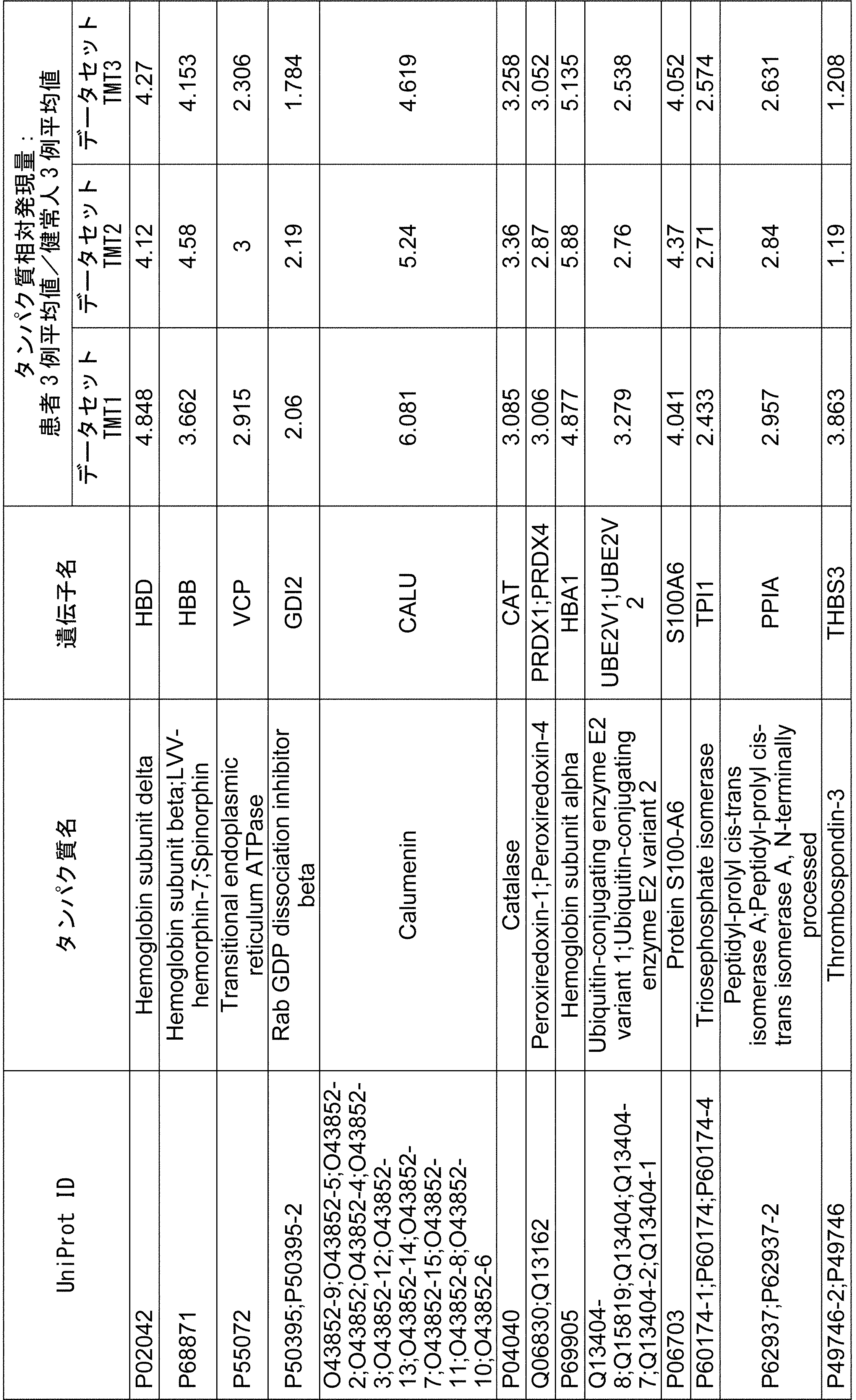


選抜されたタンパク質は、子宮内膜症診断のバイオマーカーとして有望であり、中に代表的なタンパク質の相対発現量グラフは図1-1から図1-10で示された。
The selected proteins are promising as biomarkers for the diagnosis of endometriosis, and graphs of relative expression levels of representative proteins are shown in FIGS. 1-1 to 1-10.
(実施例2)ヒト健常人および子宮内膜症患者の血漿を用いたアプタマー結合タンパク質解析(SOMAscan(登録商標))によるバイオマーカー探索
(2-1)ヒト血漿サンプルの取得
健常人および子宮内膜症患者由来血漿はProteogenex社より購入した。 (Example 2) Biomarker search by aptamer-binding protein analysis (SOMAscan®) using plasma of healthy humans and endometriosis patients
(2-1) Acquisition of human plasma samples Plasma derived from healthy subjects and patients with endometriosis was purchased from Proteogenex.
(2-2)血漿タンパク質のSOMAscan(登録商標)による解析
血漿サンプル(健常人3例および子宮内膜症患者3例)中の低発現タンパク質は、SOMAscan(登録商標、Somalogic社)を用いて分析された。具体的には、先ず1310タンパク質に対しておのおの特異的に結合するアプタマーが用意され、血漿と混合され、血漿中のタンパク質と結合させられた。次にアプタマーと結合したタンパク質がビオチン化され、アプタマー―タンパク質複合体がストレプトアビジンビーズを用いて精製された。その後、アプタマーが溶出され、マイクロアレイによって検出されることで、当該アプタマーと結合した血漿中タンパク質の発現量分析が行われた。 (2-2) Analysis of plasma proteins by SOMAscan (registered trademark) Low-expressed proteins in plasma samples (3 healthy subjects and 3 endometriosis patients) are analyzed using SOMAscan (registered trademark, Somalogic). Was done. Specifically, first, aptamers that specifically bind to the 1310 protein were prepared, mixed with plasma, and bound to the protein in plasma. The protein bound to the aptamer was then biotinylated and the aptamer-protein complex was purified using streptavidin beads. Then, the aptamer was eluted and detected by the microarray, so that the expression level of the plasma protein bound to the aptamer was analyzed.
血漿サンプル(健常人3例および子宮内膜症患者3例)中の低発現タンパク質は、SOMAscan(登録商標、Somalogic社)を用いて分析された。具体的には、先ず1310タンパク質に対しておのおの特異的に結合するアプタマーが用意され、血漿と混合され、血漿中のタンパク質と結合させられた。次にアプタマーと結合したタンパク質がビオチン化され、アプタマー―タンパク質複合体がストレプトアビジンビーズを用いて精製された。その後、アプタマーが溶出され、マイクロアレイによって検出されることで、当該アプタマーと結合した血漿中タンパク質の発現量分析が行われた。 (2-2) Analysis of plasma proteins by SOMAscan (registered trademark) Low-expressed proteins in plasma samples (3 healthy subjects and 3 endometriosis patients) are analyzed using SOMAscan (registered trademark, Somalogic). Was done. Specifically, first, aptamers that specifically bind to the 1310 protein were prepared, mixed with plasma, and bound to the protein in plasma. The protein bound to the aptamer was then biotinylated and the aptamer-protein complex was purified using streptavidin beads. Then, the aptamer was eluted and detected by the microarray, so that the expression level of the plasma protein bound to the aptamer was analyzed.
(2-3)バイオマーカー候補の選抜
SOMAscan(登録商標)のアプタマーシグナルデータは、統計プログラミング環境R及びマイクロソフトエクセルを用いて解析された。以下の基準により、子宮内膜症患者で発現変化が認められた200個のタンパク質が選抜された(表4A、表4B)。
 上記計算において、アプタマーシグナルの最大値が1000未満のものは検出限界以下とみなして解析対象から除かれた。
(2-3) Selection of biomarker candidates The aptamer signal data of SOMAscan (registered trademark) was analyzed using the statistical programming environment R and Microsoft Excel. Based on the following criteria, 200 proteins with altered expression were selected in patients with endometriosis (Tables 4A and 4B).
上記計算において、アプタマーシグナルの最大値が1000未満のものは検出限界以下とみなして解析対象から除かれた。
(2-3) Selection of biomarker candidates The aptamer signal data of SOMAscan (registered trademark) was analyzed using the statistical programming environment R and Microsoft Excel. Based on the following criteria, 200 proteins with altered expression were selected in patients with endometriosis (Tables 4A and 4B).
In the above calculation, those with a maximum aptamer signal of less than 1000 were considered to be below the detection limit and excluded from the analysis target.
SOMAscan(登録商標)のアプタマーシグナルデータは、統計プログラミング環境R及びマイクロソフトエクセルを用いて解析された。以下の基準により、子宮内膜症患者で発現変化が認められた200個のタンパク質が選抜された(表4A、表4B)。


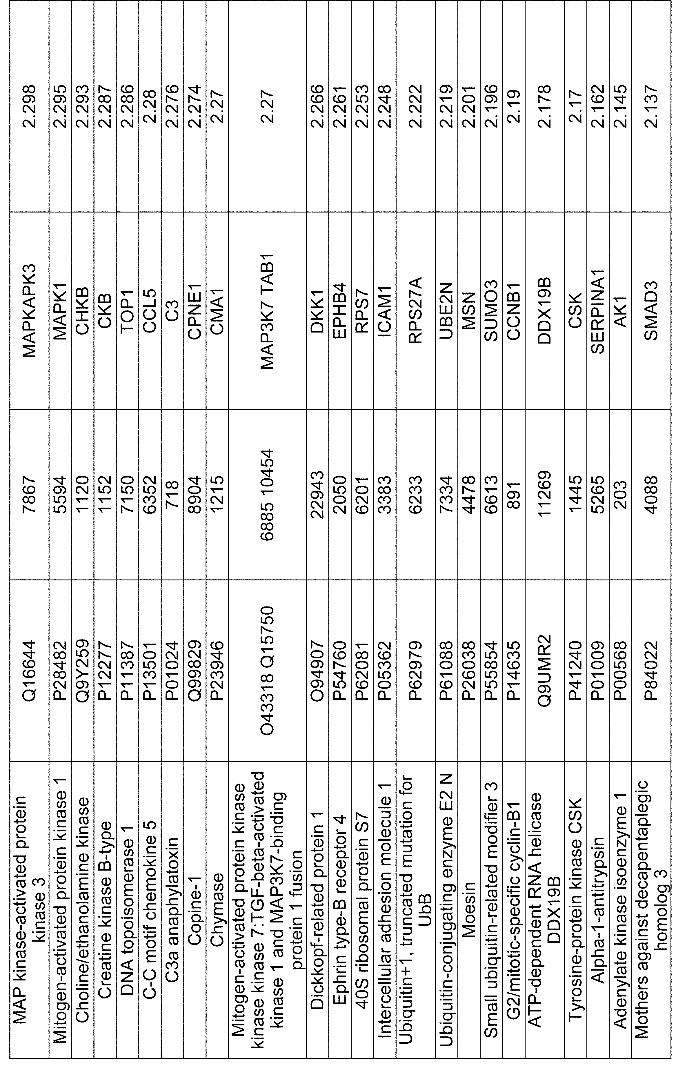


選抜されたタンパク質は、子宮内膜症診断のバイオマーカーとして有望であり、中に代表的なタンパク質の発現量グラフは図2-1から図2-10で示された。
The selected proteins are promising as biomarkers for the diagnosis of endometriosis, and the expression level graphs of representative proteins are shown in FIGS. 2-1 to 2-10.
(実施例3)ヒト健常人および子宮内膜症患者の血漿を用いた細胞外マトリックス分解産物分析によるバイオマーカー探索
(3-1)ヒト血漿サンプルの取得
健常人および子宮内膜症患者由来血漿はProteogenex社より購入した。 (Example 3) Biomarker search by extracellular matrix degradation product analysis using plasma of healthy humans and endometriosis patients
(3-1) Acquisition of human plasma samples Plasma derived from healthy subjects and patients with endometriosis was purchased from Proteogenex.
(3-2)血漿タンパク質の細胞外マトリックス測定
血漿サンプル(健常人3例および子宮内膜症患者3例)は、細胞外マトリックスの切断部位特異的な抗体を用いたELISA系で分析され、サンプル中の細胞外マトリックス総量及び細胞外マトリックスの分解/合成状態が分析された(Nordic Bioscience社)。具体的に、タイプIII, IV, V, VIコラーゲン、decorin、またはnidogenが分解されるときの分解産物に含まれる切断部位に対する抗体を用いて、それぞれの分解産物の発現量が分析された。その結果、タイプVコラーゲンMMP分解産物(C5M)が3例中2例の患者で顕著に上昇している(C5Mの濃度が2例の上昇患者血漿中において10.2ng/ml、11.8ng/mlである)のに対し、健常者の血漿中のC5M濃度は全て検出限界以下であり、C5Mが子宮内膜症のバイオマーカーとして有望であることが示唆された。タイプVコラーゲンMMP分解産物(C5M)の血漿中濃度グラフが図3に示された。 (3-2) Extracellular matrix measurement of plasma proteins Plasma samples (3 healthy subjects and 3 endometriosis patients) were analyzed by an ELISA system using an antibody specific to the cleavage site of the extracellular matrix and sampled. The total amount of extracellular matrix in the cells and the degradation / synthesis state of extracellular matrix were analyzed (Nordic Bioscience). Specifically, the expression level of each degradation product was analyzed using an antibody against the cleavage site contained in the degradation products when type III, IV, V, VI collagen, decorin, or nidogen was degraded. As a result, type V collagen MMP degradation product (C5M) was significantly increased in 2 of 3 patients (C5M concentration was increased in 2 cases at 10.2 ng / ml and 11.8 ng / ml in plasma. On the other hand, the plasma C5M concentration in healthy subjects was below the detection limit, suggesting that C5M is a promising biomarker for endometriosis. A plasma concentration graph of type V collagen MMP degradation product (C5M) is shown in FIG.
血漿サンプル(健常人3例および子宮内膜症患者3例)は、細胞外マトリックスの切断部位特異的な抗体を用いたELISA系で分析され、サンプル中の細胞外マトリックス総量及び細胞外マトリックスの分解/合成状態が分析された(Nordic Bioscience社)。具体的に、タイプIII, IV, V, VIコラーゲン、decorin、またはnidogenが分解されるときの分解産物に含まれる切断部位に対する抗体を用いて、それぞれの分解産物の発現量が分析された。その結果、タイプVコラーゲンMMP分解産物(C5M)が3例中2例の患者で顕著に上昇している(C5Mの濃度が2例の上昇患者血漿中において10.2ng/ml、11.8ng/mlである)のに対し、健常者の血漿中のC5M濃度は全て検出限界以下であり、C5Mが子宮内膜症のバイオマーカーとして有望であることが示唆された。タイプVコラーゲンMMP分解産物(C5M)の血漿中濃度グラフが図3に示された。 (3-2) Extracellular matrix measurement of plasma proteins Plasma samples (3 healthy subjects and 3 endometriosis patients) were analyzed by an ELISA system using an antibody specific to the cleavage site of the extracellular matrix and sampled. The total amount of extracellular matrix in the cells and the degradation / synthesis state of extracellular matrix were analyzed (Nordic Bioscience). Specifically, the expression level of each degradation product was analyzed using an antibody against the cleavage site contained in the degradation products when type III, IV, V, VI collagen, decorin, or nidogen was degraded. As a result, type V collagen MMP degradation product (C5M) was significantly increased in 2 of 3 patients (C5M concentration was increased in 2 cases at 10.2 ng / ml and 11.8 ng / ml in plasma. On the other hand, the plasma C5M concentration in healthy subjects was below the detection limit, suggesting that C5M is a promising biomarker for endometriosis. A plasma concentration graph of type V collagen MMP degradation product (C5M) is shown in FIG.
(実施例4)ヒト健常人および子宮内膜症患者の血漿を用いたLuminex xMAP TM technologyによるバイオマーカー探索
(4-1)ヒト血漿サンプルの取得
健常人血漿(分泌期血漿サンプル5例、増殖期血漿サンプル5例)はProteogenex社より購入された。子宮内膜症患者の血漿は、子宮内膜症患者37例から、手術前、手術3日後および手術1か月後それぞれの血漿計111サンプルが取得された。 (Example 4) Search for biomarkers by Luminex xMAP TM technology using plasma of healthy humans and endometriosis patients
(4-1) Acquisition of human plasma samples Healthy human plasma (5 secretory plasma samples, 5 proliferative plasma samples) was purchased from Proteogenex. As for plasma of endometriosis patients, a total of 111 plasma samples were obtained from 37 endometriosis patients before, 3 days after, and 1 month after surgery.
(4-2)血漿タンパク質のLuminex xMAP TM technologyによる解析
血漿サンプル(中のタンパク質は、Luminex xMAPTM(Luminex社の商標)を用いて分析された。Luminex xMAPTM technologyは、マクロビーズを色々な濃度で組み合わせた2色の蛍光色素で染色し、蛍光色素の含有量を識別コードとして用い、それぞれのビーズに個別の解析対象と結合する物質を固定させ、少量サンプルで多項目を同時に解析することができる技術である。
本実施例では、152種のタンパク質を選択し、タンパク質測定パネルを用いて分析を行った。具体的には、先ずパネル毎に、ビーズに対象タンパクに結合する抗体が固定されており、ビーズ上の抗体に対し血漿サンプルを反応させ、さらに対象タンパクに対するレポーター抗体(蛍光色素で標識)を反応させたのち、フローサイトメトリー技術を利用し、2種類のレーザーで蛍光強度の測定を行って、対象タンパク質の発現量を分析した(Luminex100装置使用)。 (4-2) Analysis of plasma proteins by Luminex xMAP TM technology Plasma samples (proteins in them were analyzed using Luminex xMAP TM (trademark of Luminex). Luminex xMAP TM technology has various concentrations of macrobeads. It is possible to dye with the two colors of fluorescent dyes combined in the above, use the content of the fluorescent dye as an identification code, fix the substance that binds to the individual analysis target to each bead, and analyze multiple items at the same time with a small sample. It is a technology that can be done.
In this example, 152 kinds of proteins were selected and analyzed using a protein measurement panel. Specifically, first, an antibody that binds to the target protein is immobilized on the beads for each panel, a plasma sample is reacted with the antibody on the beads, and a reporter antibody (labeled with a fluorescent dye) against the target protein is further reacted. After that, the fluorescence intensity was measured with two types of lasers using flow cytometry technology, and the expression level of the target protein was analyzed (using the Luminex100 device).
(4-3)バイオマーカー候補の選抜
得られた各種タンパク質の血漿中濃度は、マイクロソフトエクセルを用いて解析された。以下の基準により、子宮内膜症患者で発現変化が認められた24個のタンパク質が選抜された(表7A、表7B)。
 選抜された24個のタンパク質のうち、12個のタンパク質は、子宮内膜症患者の手術時で最も高く、手術一か月後にその値が下がり、さらに健常人で最も低い値を示すものであった。
(4-3) Selection of Biomarker Candidates The plasma concentrations of the obtained various proteins were analyzed using Microsoft Excel. Twenty-four proteins with altered expression were selected in patients with endometriosis according to the following criteria (Tables 7A and 7B).
選抜された24個のタンパク質のうち、12個のタンパク質は、子宮内膜症患者の手術時で最も高く、手術一か月後にその値が下がり、さらに健常人で最も低い値を示すものであった。
(4-3) Selection of Biomarker Candidates The plasma concentrations of the obtained various proteins were analyzed using Microsoft Excel. Twenty-four proteins with altered expression were selected in patients with endometriosis according to the following criteria (Tables 7A and 7B).
Of the 24 proteins selected, 12 proteins were the highest at the time of surgery for endometriosis patients, decreased one month after surgery, and showed the lowest value in healthy subjects. It was.
得られた各種タンパク質の血漿中濃度は、マイクロソフトエクセルを用いて解析された。以下の基準により、子宮内膜症患者で発現変化が認められた24個のタンパク質が選抜された(表7A、表7B)。

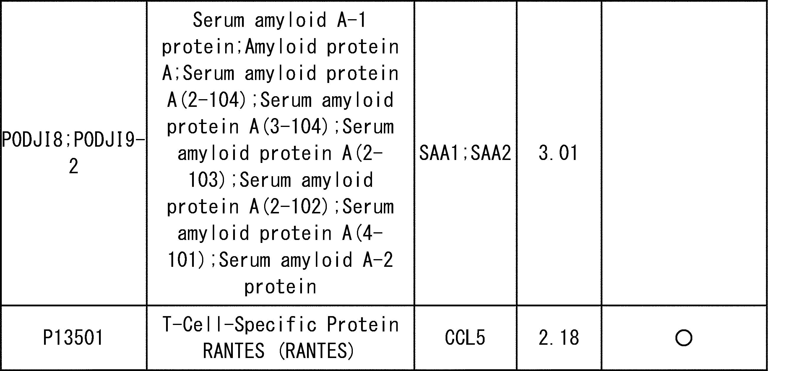

(実施例5)ヒト健常人および子宮内膜症患者の血漿を用いたLC-MS解析によるバイオマーカー探索
(5-1)ヒト血漿サンプルの取得
健常人血漿(分泌期血漿サンプル5例、増殖期血漿サンプル5例)はProteogenex社より購入された。子宮内膜症患者の血漿は、子宮内膜症患者39例から、手術前、手術3日後および手術1か月後それぞれの血漿計125サンプルが取得された。 (Example 5) Search for biomarkers by LC-MS analysis using plasma of healthy humans and endometriosis patients
(5-1) Acquisition of human plasma samples Healthy human plasma (5 secretory plasma samples, 5 proliferative plasma samples) was purchased from Proteogenex. As for plasma of endometriosis patients, a total of 125 plasma samples were obtained from 39 endometriosis patients before, 3 days after, and 1 month after surgery.
(5-2)血漿タンパク質のLC-MS解析
各血漿サンプルの中の多量タンパク質はHigh SelectTM Top14 Abundant Protein Depletion Mini Spin Columns (Thermo)を用いてメーカー推奨の方法によりカラムに結合され除去された。カラムに結合しなかったフロースルー画分のタンパク質はメタノール/クロロホルム法により沈殿され、8M尿素/400mM 重炭酸アンモニウム溶液に溶解された。溶解タンパク質は還元、アルキル化された後、リジルエンドペプチターゼおよびトリプシンにより消化されペプチドとなった。このペプチド溶液はMonospin C18(GL Science)を用いてメーカー推奨の方法により脱塩された。 (5-2) LC-MS analysis of plasma proteins The large amount of proteins in each plasma sample was bound to the column and removed by the method recommended by the manufacturer using High Select TM Top14 Abundant Protein Depletion Mini Spin Columns (Thermo). The protein in the flow-through fraction that did not bind to the column was precipitated by the methanol / chloroform method and dissolved in 8M urea / 400 mM ammonium bicarbonate solution. The lysed protein was reduced and alkylated and then digested with lysyl endopeptidase and trypsin to become a peptide. This peptide solution was desalted using Monospin C18 (GL Science) by the manufacturer's recommended method.
各血漿サンプルの中の多量タンパク質はHigh SelectTM Top14 Abundant Protein Depletion Mini Spin Columns (Thermo)を用いてメーカー推奨の方法によりカラムに結合され除去された。カラムに結合しなかったフロースルー画分のタンパク質はメタノール/クロロホルム法により沈殿され、8M尿素/400mM 重炭酸アンモニウム溶液に溶解された。溶解タンパク質は還元、アルキル化された後、リジルエンドペプチターゼおよびトリプシンにより消化されペプチドとなった。このペプチド溶液はMonospin C18(GL Science)を用いてメーカー推奨の方法により脱塩された。 (5-2) LC-MS analysis of plasma proteins The large amount of proteins in each plasma sample was bound to the column and removed by the method recommended by the manufacturer using High Select TM Top14 Abundant Protein Depletion Mini Spin Columns (Thermo). The protein in the flow-through fraction that did not bind to the column was precipitated by the methanol / chloroform method and dissolved in 8M urea / 400 mM ammonium bicarbonate solution. The lysed protein was reduced and alkylated and then digested with lysyl endopeptidase and trypsin to become a peptide. This peptide solution was desalted using Monospin C18 (GL Science) by the manufacturer's recommended method.
前記ペプチドはTMT10plexでメーカー推奨の方法により標識された。標識ペプチドは、AssayMAP reversed phase (RP-S) cartridges (Agilent technologies)を使用し、Triethylamineを用いたHigh pH Reversed-Phase法で分画され、おのおのの画分はnano-LC system(EASY-nLCTM 1200 system, ThermoFisher Scientific社)にOrbitrap Fusion Lumos(ThermoFisher scientific)を繋げたLC-MS解析システムを用いて分析された。分析サンプルは、健常人5例(分泌期、増殖期それぞれで計10サンプル)、子宮内膜症患者39例(手術前、手術3日後および手術一か月後の計125サンプル)の血漿のそれぞれから得たペプチドとなる。それぞれの分析の際に、測定間の補正を行うため購入した健常人7サンプルと購入した子宮内膜症患者7サンプルを等量混合したサンプルをコントロールとして使用し、各分析10サンプルなるようにした。各サンプルはそれぞれ3回分析され、それらは解析の過程で統合された。
The peptide was labeled with TMT10plex by the manufacturer's recommended method. Labeled peptides were fractionated by the High pH Reversed-Phase method using AssayMAP reversed phase (RP-S) cartridges (Agilent technologies) and Triethylamine, with each fraction being nano-LC system (EASY-nLC TM). The analysis was performed using an LC-MS analysis system in which Orbitrap Fusion Lumos (ThermoFisher scientific) was connected to 1200 system, ThermoFisher Scientific). The analysis samples were plasma of 5 healthy subjects (10 samples in total for each of the secretory and proliferative stages) and 39 patients with endometriosis (125 samples in total before, 3 days after and 1 month after surgery). It becomes a peptide obtained from. At the time of each analysis, 7 samples of healthy subjects purchased and 7 samples of endometriosis patients purchased were used as controls to make corrections between measurements, and 10 samples were used for each analysis. .. Each sample was analyzed three times, and they were integrated during the course of the analysis.
サンプル中のタンパク質は、LC-MS分析のRaw dataからThermo ScientificTM Proteome DiscovererTMを用いて同定され、相対発現量が分析された。タンパク質同定のためのデータベース検索は以下のパラメーターで行った。
Taxonomy: uniprot human
Static modifications: TMT6plex/ +229.163 Da (Any N-Terminus), Carbamidomethyl/ +57.021 Da (C, C-Terminus), TMT6plex / +229.163 Da (K, C-Terminus)
Dynamic Modifications: Oxidation/ +15.995 Da (M), Acetyl/ +42.011Da (N-Terminus)
FDR: Target FDR (Strict)/ 0.01, Target FDR (Relaxed)/ 0.05
健常人の血漿サンプル中の対象タンパク質に対する子宮内膜症患者の術前の血漿サンプル中の対象タンパク質の相対発現量は解析ソフト(Proteome Discoverer)で計算された。 The proteins in the sample were identified using Thermo Scientific TM Proteome Discoverer TM from Raw data of LC-MS analysis, and their relative expression levels were analyzed. The database search for protein identification was performed with the following parameters.
Taxonomy: uniprot human
Static modifications: TMT6plex / +229.163 Da (Any N-Terminus), Carbamidomethyl / + 57.021 Da (C, C-Terminus), TMT6plex / +229.163 Da (K, C-Terminus)
Dynamic Modifications: Oxidation / + 15.995 Da (M), Acetyl / + 42.011Da (N-Terminus)
FDR: Target FDR (Strict) / 0.01, Target FDR (Relaxed) / 0.05
The relative expression level of the target protein in the preoperative plasma sample of the endometriosis patient with respect to the target protein in the plasma sample of a healthy person was calculated by analysis software (Proteome Discoverer).
Taxonomy: uniprot human
Static modifications: TMT6plex/ +229.163 Da (Any N-Terminus), Carbamidomethyl/ +57.021 Da (C, C-Terminus), TMT6plex / +229.163 Da (K, C-Terminus)
Dynamic Modifications: Oxidation/ +15.995 Da (M), Acetyl/ +42.011Da (N-Terminus)
FDR: Target FDR (Strict)/ 0.01, Target FDR (Relaxed)/ 0.05
健常人の血漿サンプル中の対象タンパク質に対する子宮内膜症患者の術前の血漿サンプル中の対象タンパク質の相対発現量は解析ソフト(Proteome Discoverer)で計算された。 The proteins in the sample were identified using Thermo Scientific TM Proteome Discoverer TM from Raw data of LC-MS analysis, and their relative expression levels were analyzed. The database search for protein identification was performed with the following parameters.
Taxonomy: uniprot human
Static modifications: TMT6plex / +229.163 Da (Any N-Terminus), Carbamidomethyl / + 57.021 Da (C, C-Terminus), TMT6plex / +229.163 Da (K, C-Terminus)
Dynamic Modifications: Oxidation / + 15.995 Da (M), Acetyl / + 42.011Da (N-Terminus)
FDR: Target FDR (Strict) / 0.01, Target FDR (Relaxed) / 0.05
The relative expression level of the target protein in the preoperative plasma sample of the endometriosis patient with respect to the target protein in the plasma sample of a healthy person was calculated by analysis software (Proteome Discoverer).
(5-3)バイオマーカー候補分子の選抜
LC-MS結果データは、Proteome Discoverer で解析された。サンプル(健常人5例の増殖期・分泌期の10サンプル、子宮内膜症患者39例の手術前サンプル)中の各タンパク質の相対発現量から、以下の基準により、子宮内膜症患者で発現変化が認められた27個のタンパク質が選抜された(表8A、表8B)。
- Abundance Ratio(存在比) がANOVA and tukey HSD post hocでP-Value ≦0.5 及び
- Abundance Ratios(存在比)≦0.6250または≧1.6000
 選抜された27個のタンパク質のうち、16個のタンパク質は、子宮内膜症患者の手術時で最も高く、手術一か月後にその値が下がり、さらに健常人で最も低い値を示すものであった。
(5-3) Selection of biomarker candidate molecules LC-MS result data was analyzed by Proteome Discoverer. Based on the relative expression level of each protein in the samples (10 samples in the proliferative / secretory phase of 5 healthy subjects, 39 preoperative samples of endometriosis patients), it is expressed in endometriosis patients according to the following criteria. Twenty-seven proteins with altered activity were selected (Table 8A, Table 8B).
選抜された27個のタンパク質のうち、16個のタンパク質は、子宮内膜症患者の手術時で最も高く、手術一か月後にその値が下がり、さらに健常人で最も低い値を示すものであった。
(5-3) Selection of biomarker candidate molecules LC-MS result data was analyzed by Proteome Discoverer. Based on the relative expression level of each protein in the samples (10 samples in the proliferative / secretory phase of 5 healthy subjects, 39 preoperative samples of endometriosis patients), it is expressed in endometriosis patients according to the following criteria. Twenty-seven proteins with altered activity were selected (Table 8A, Table 8B).
-Abundance Ratio is ANOVA and tukey HSD post hoc with P-Value ≤ 0.5 and-Abundance Ratios (Abundance Ratio) ≤ 0.6250 or ≥ 1.6000
Of the 27 proteins selected, 16 proteins were the highest at the time of surgery in endometriosis patients, decreased one month after surgery, and showed the lowest value in healthy subjects. It was.
LC-MS結果データは、Proteome Discoverer で解析された。サンプル(健常人5例の増殖期・分泌期の10サンプル、子宮内膜症患者39例の手術前サンプル)中の各タンパク質の相対発現量から、以下の基準により、子宮内膜症患者で発現変化が認められた27個のタンパク質が選抜された(表8A、表8B)。
- Abundance Ratio(存在比) がANOVA and tukey HSD post hocでP-Value ≦0.5 及び
- Abundance Ratios(存在比)≦0.6250または≧1.6000
-Abundance Ratio is ANOVA and tukey HSD post hoc with P-Value ≤ 0.5 and-Abundance Ratios (Abundance Ratio) ≤ 0.6250 or ≥ 1.6000


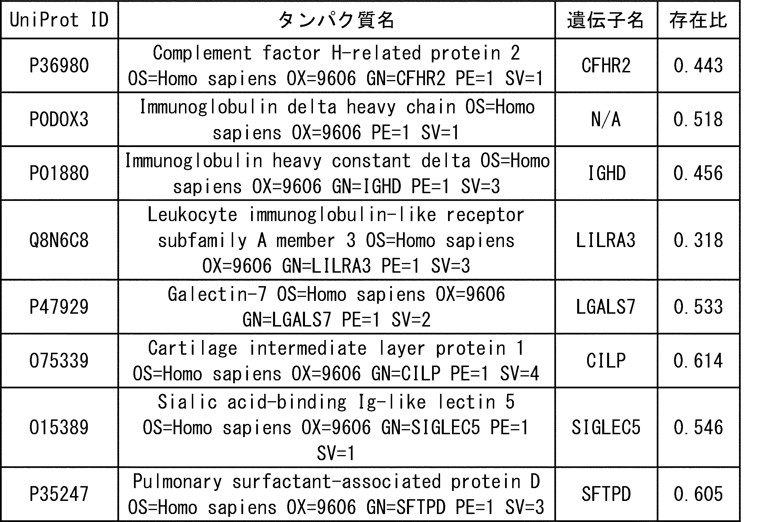
本開示によって、対象から得られた試料における、タイプVコラーゲンMMP分解産物の濃度、または表1Aで示されるマーカー、表2Aで示されるマーカー、表1Bで示されるマーカー、表2Bで示されるマーカー、表5Aで示されるマーカー、表6Aで示されるマーカー、表5Bで示されるマーカー、表6Bで示されるマーカーからなるグループから選択される少なくとも一つのマーカーの存在量を測定することによって、当該対象が子宮内膜症に罹患しているか否かを診断できることが証明された。本開示の発明は、非浸潤的な手法により子宮内膜症を診断することが可能にするものであり、当該疾患の診断及び治療に極めて有用である。
According to the present disclosure, the concentration of type V collagen MMP degradation products in a sample obtained from a subject, or the markers shown in Table 1A, the markers shown in Table 2A, the markers shown in Table 1B, the markers shown in Table 2B, By measuring the abundance of at least one marker selected from the group consisting of the markers shown in Table 5A, the markers shown in Table 6A, the markers shown in Table 5B, and the markers shown in Table 6B. It has been proved that it is possible to diagnose whether or not the patient has endometriosis. The invention of the present disclosure makes it possible to diagnose endometriosis by a non-invasive method, and is extremely useful for the diagnosis and treatment of the disease.
Claims (15)
- 子宮内膜症を診断する方法であって、
対象から得られた試料における、タイプVコラーゲンMMP分解産物、1A群で示されるマーカー、2A群で示されるマーカー、1B群で示されるマーカー、2B群で示されるマーカー、3A群で示されるマーカー、4A群で示されるマーカー、3B群で示されるマーカー、4B群で示されるマーカーからなるグループから選択される少なくとも一つのマーカーの存在量を測定する工程を含む方法。
[1A群]

 [1B群]
[1B群]
[2A群]

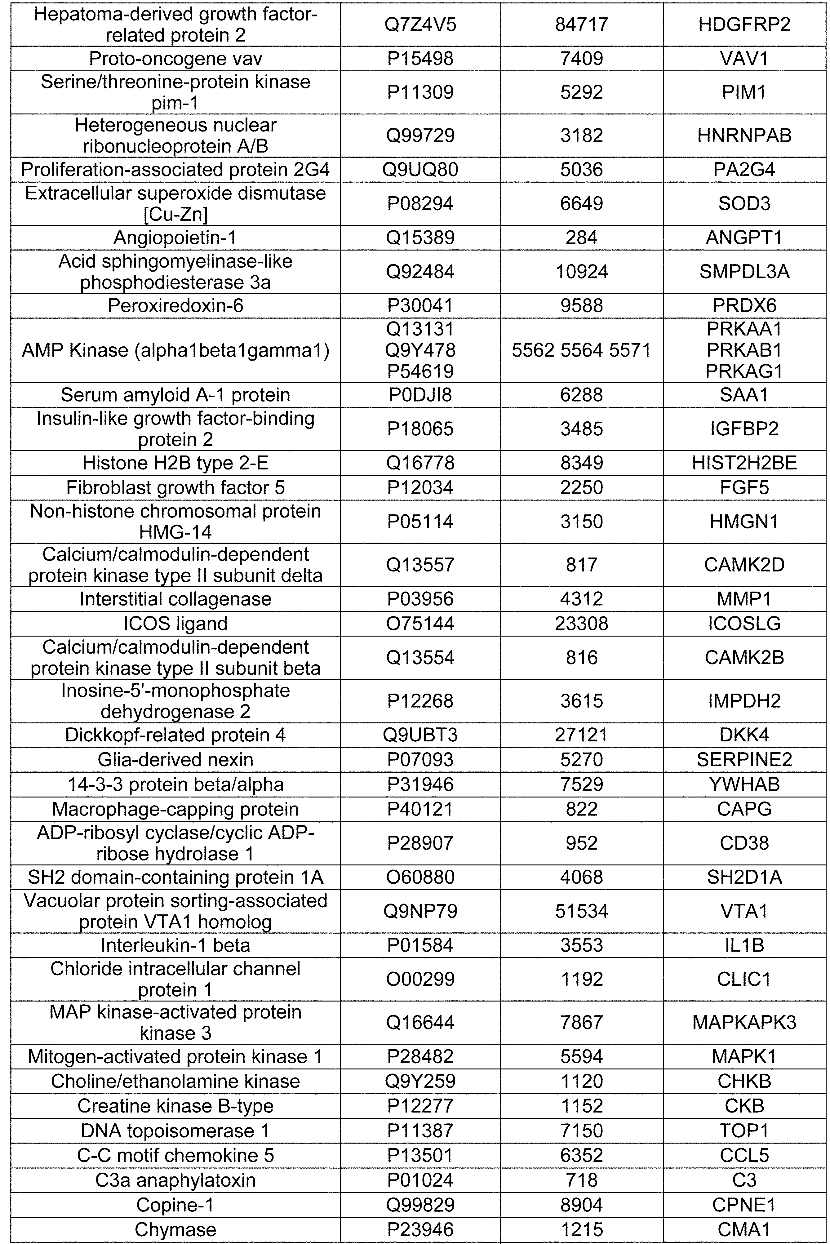 [2B群]
[2B群]
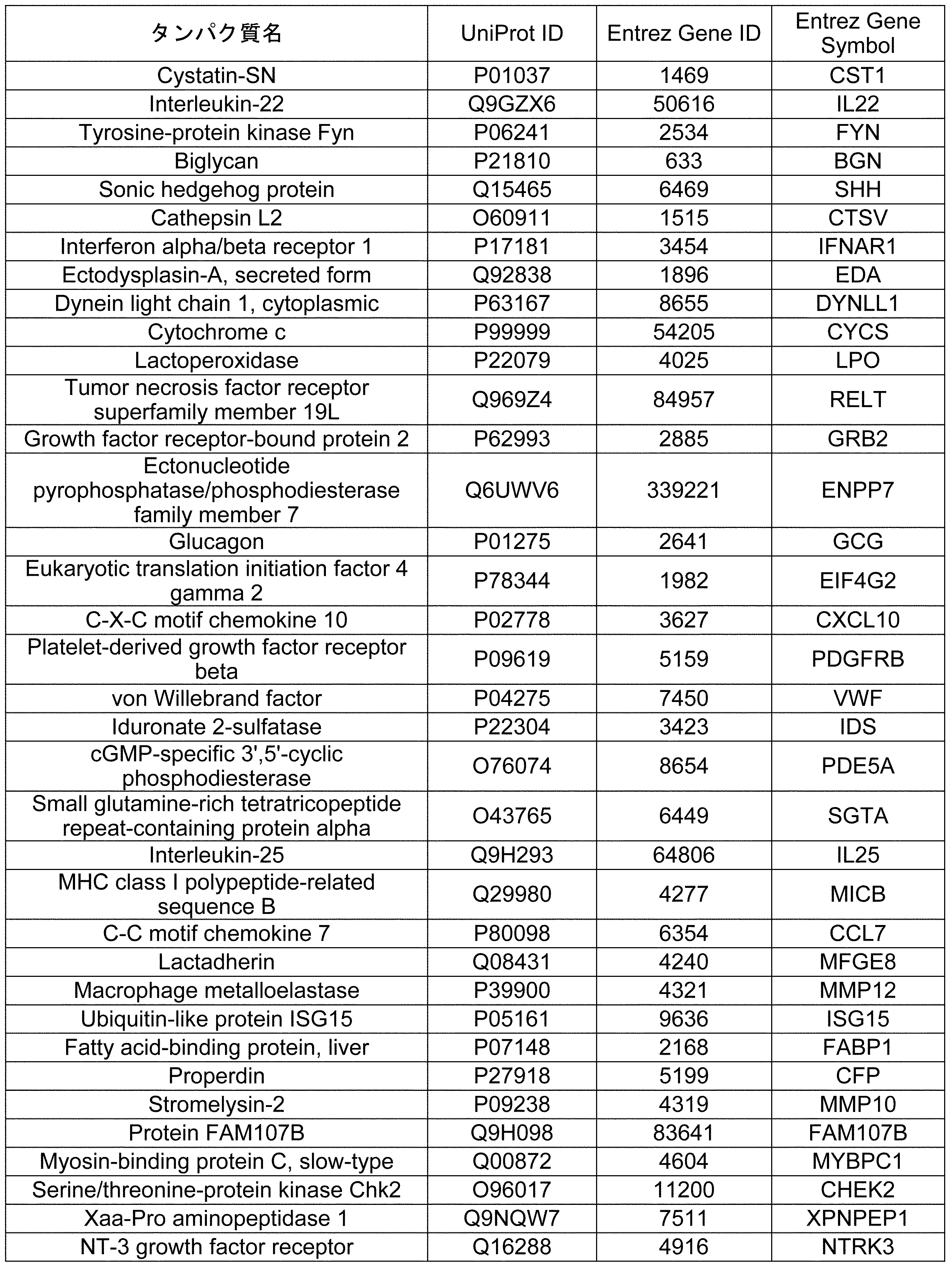
 [3A群]
[3A群]

[3B群]
[4A群]
[4B群]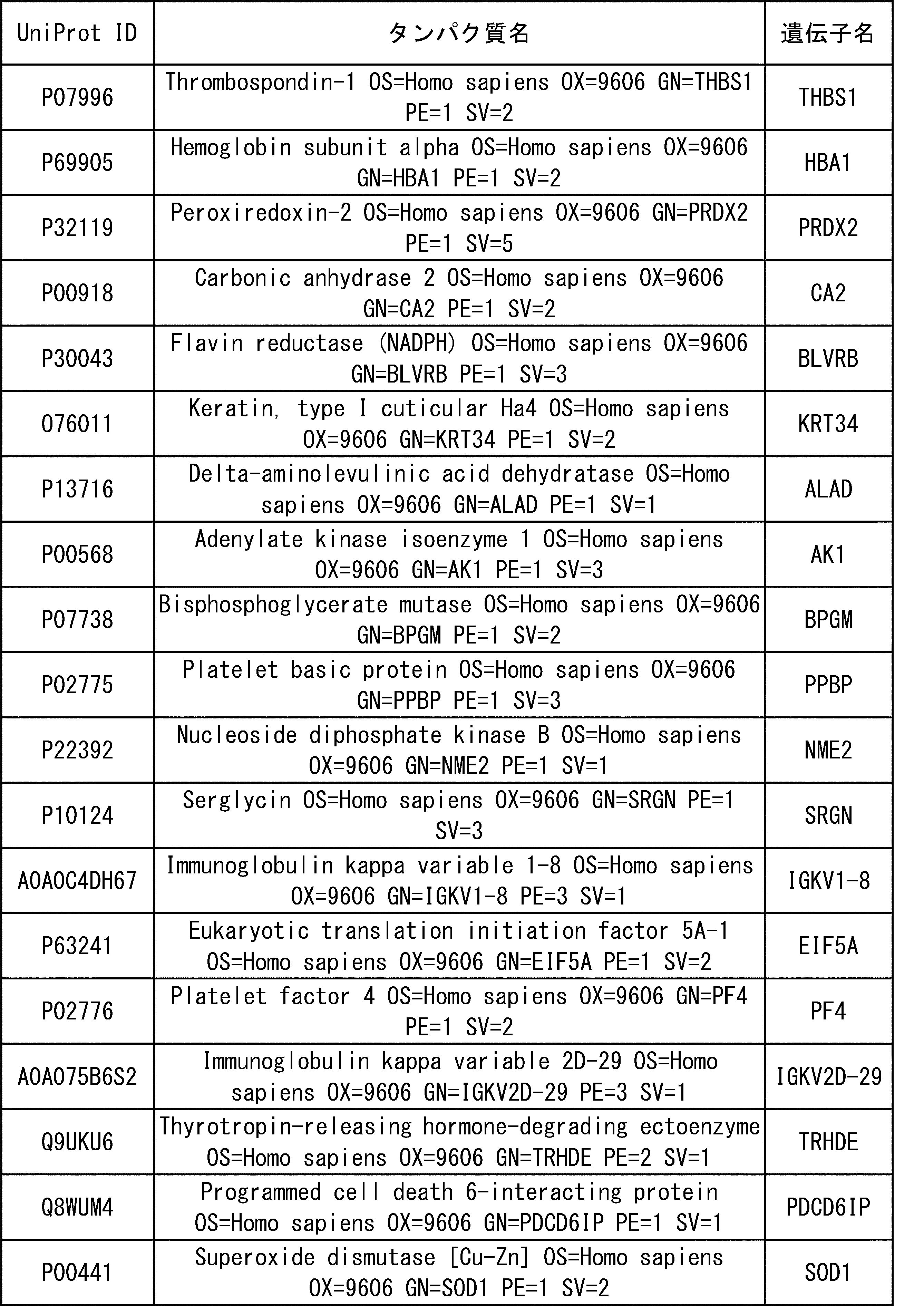
A method of diagnosing endometriosis
In the sample obtained from the subject, the type V collagen MMP degradation product, the marker shown in group 1A, the marker shown in group 2A, the marker shown in group 1B, the marker shown in group 2B, the marker shown in group 3A, A method comprising measuring the abundance of at least one marker selected from the group consisting of the markers represented by group 4A, the markers represented by group 3B, and the markers represented by group 4B.
[Group 1A]
[Group 1B]
[Group 2A]
[Group 2B]
[Group 3A]
[Group 3B]
[Group 4A]
[Group 4B]
- 対象における子宮内膜症の罹患の有無を決定する方法であって、
対象から得られた試料における、請求項1に記載されるマーカーから選択される少なくとも一つのマーカーの存在量を測定する工程を含む方法。 A method of determining the presence or absence of endometriosis in a subject.
A method comprising the step of measuring the abundance of at least one marker selected from the markers according to claim 1 in a sample obtained from a subject. - 対象における子宮内膜の線維化の程度を決定する方法であって、
対象から得られた試料における、請求項1に記載されるマーカーから選択される少なくとも一つのマーカーの存在量を測定する工程を含む方法。 A method of determining the degree of endometrial fibrosis in a subject.
A method comprising the step of measuring the abundance of at least one marker selected from the markers according to claim 1 in a sample obtained from a subject. - 対象における子宮内膜の癒着の程度を決定する方法であって、
対象から得られた試料における、請求項1に記載されるマーカーから選択される少なくとも一つのマーカーの存在量を測定する工程を含む方法。 A method of determining the degree of endometrial adhesions in a subject
A method comprising the step of measuring the abundance of at least one marker selected from the markers according to claim 1 in a sample obtained from a subject. - 子宮内膜症を罹患しているまたは罹患していると疑われる対象における子宮内膜症による痛みの程度を予測する方法であって、
対象から得られた試料における、請求項1に記載されるマーカーから選択される少なくとも一つのマーカーの存在量を測定する工程を含む方法。 A method of predicting the degree of endometriosis pain in a subject who has or is suspected of having endometriosis.
A method comprising the step of measuring the abundance of at least one marker selected from the markers according to claim 1 in a sample obtained from a subject. - 子宮内膜症を罹患しているまたは罹患していると疑われる対象における子宮内膜症の進行度を決定する方法であって、
対象から得られた試料における、請求項1に記載されるマーカーから選択される少なくとも一つのマーカーの存在量を測定する工程を含む方法。 A method of determining the progression of endometriosis in a subject who has or is suspected of having endometriosis.
A method comprising the step of measuring the abundance of at least one marker selected from the markers according to claim 1 in a sample obtained from a subject. - 子宮内膜症を罹患しているまたは罹患していると疑われる対象における子宮内膜症の病態をモニタリングする方法であって、
対象から異なる時点で採取された複数の試料のそれぞれにおける、請求項1に記載されるマーカーから選択される少なくとも一つのマーカーの存在量をそれぞれ測定する工程を含む方法。 A method of monitoring the pathophysiology of endometriosis in subjects who have or are suspected of having endometriosis.
A method comprising the step of measuring the abundance of at least one marker selected from the markers according to claim 1 in each of a plurality of samples collected at different time points from the subject. - 前記マーカーの存在量は、血液試料中の当該マーカーのポリペプチド濃度である、請求項1から請求項7のいずれか一項に記載の方法。 The method according to any one of claims 1 to 7, wherein the abundance of the marker is the polypeptide concentration of the marker in the blood sample.
- 子宮内膜症を診断するためのキットであって、
請求項1に記載されるマーカーから選択される少なくとも一つのマーカーの存在量を測定するための試薬を含むキット。 A kit for diagnosing endometriosis
A kit comprising a reagent for measuring the abundance of at least one marker selected from the markers according to claim 1. - 対象における子宮内膜症の罹患の有無を決定するためのキットであって、
請求項1に記載されるマーカーから選択される少なくとも一つのマーカーの存在量を測定するための試薬を含むキット。 A kit for determining the presence or absence of endometriosis in a subject.
A kit comprising a reagent for measuring the abundance of at least one marker selected from the markers according to claim 1. - 対象における子宮内膜の線維化の程度を決定するためのキットであって、
請求項1に記載されるマーカーから選択される少なくとも一つのマーカーの存在量を測定するための試薬を含むキット。 A kit for determining the degree of endometrial fibrosis in a subject
A kit comprising a reagent for measuring the abundance of at least one marker selected from the markers according to claim 1. - 対象における子宮内膜の癒着の程度を決定するためのキットであって、
請求項1に記載されるマーカーから選択される少なくとも一つのマーカーの存在量を測定するための試薬を含むキット。 A kit for determining the degree of endometrial adhesions in a subject
A kit comprising a reagent for measuring the abundance of at least one marker selected from the markers according to claim 1. - 子宮内膜症を罹患しているまたは罹患していると疑われる対象における子宮内膜症による痛みの程度を予測するためのキットであって、
請求項1に記載されるマーカーから選択される少なくとも一つのマーカーの存在量を測定するための試薬を含むキット。 A kit for predicting the degree of endometriosis pain in subjects who have or are suspected of having endometriosis.
A kit comprising a reagent for measuring the abundance of at least one marker selected from the markers according to claim 1. - 子宮内膜症を罹患しているまたは罹患していると疑われる対象における子宮内膜症の進行度を決定するためのキットであって、
請求項1に記載されるマーカーから選択される少なくとも一つのマーカーの存在量を測定するための試薬を含むキット。 A kit for determining the progression of endometriosis in subjects with or suspected of having endometriosis.
A kit comprising a reagent for measuring the abundance of at least one marker selected from the markers according to claim 1. - 子宮内膜症を罹患しているまたは罹患していると疑われる対象における子宮内膜症の病態をモニタリングするためのキットであって、
請求項1に記載されるマーカーから選択される少なくとも一つのマーカーの存在量を測定するための試薬と、対象から異なる時点で採取された複数の試料のそれぞれにおける、請求項1に記載されるマーカーから選択される少なくとも一つのマーカーの存在量を比較すると記載した指示書を含む、キット。 A kit for monitoring the pathophysiology of endometriosis in subjects with or suspected of having endometriosis.
The marker according to claim 1 in a reagent for measuring the abundance of at least one marker selected from the markers according to claim 1 and a plurality of samples collected at different time points from the subject. A kit containing instructions stating that the abundance of at least one marker selected from is compared.
Priority Applications (4)
| Application Number | Priority Date | Filing Date | Title |
|---|---|---|---|
| US17/605,656 US20220221472A1 (en) | 2019-04-26 | 2020-04-24 | Method for diagnosing endometriosis, disease state monitoring method, and kit |
| EP20796111.1A EP3961217A4 (en) | 2019-04-26 | 2020-04-24 | Method for diagnosing endometriosis, disease state monitoring method, and kit |
| CN202080041887.2A CN113994208A (en) | 2019-04-26 | 2020-04-24 | Method for diagnosing endometriosis, method for monitoring disease state, and kit |
| JP2021516229A JPWO2020218465A1 (en) | 2019-04-26 | 2020-04-24 |
Applications Claiming Priority (2)
| Application Number | Priority Date | Filing Date | Title |
|---|---|---|---|
| JP2019-086223 | 2019-04-26 | ||
| JP2019086223 | 2019-04-26 |
Publications (1)
| Publication Number | Publication Date |
|---|---|
| WO2020218465A1 true WO2020218465A1 (en) | 2020-10-29 |
Family
ID=72942212
Family Applications (1)
| Application Number | Title | Priority Date | Filing Date |
|---|---|---|---|
| PCT/JP2020/017595 WO2020218465A1 (en) | 2019-04-26 | 2020-04-24 | Method for diagnosing endometriosis, disease state monitoring method, and kit |
Country Status (5)
| Country | Link |
|---|---|
| US (1) | US20220221472A1 (en) |
| EP (1) | EP3961217A4 (en) |
| JP (1) | JPWO2020218465A1 (en) |
| CN (1) | CN113994208A (en) |
| WO (1) | WO2020218465A1 (en) |
Cited By (2)
| Publication number | Priority date | Publication date | Assignee | Title |
|---|---|---|---|---|
| CN113325183A (en) * | 2021-06-01 | 2021-08-31 | 中国医学科学院北京协和医院 | Kit for differential diagnosis of EM/FEM |
| WO2021184060A1 (en) * | 2020-03-16 | 2021-09-23 | Proteomics International Pty Ltd | Endometriosis biomarkers |
Citations (4)
| Publication number | Priority date | Publication date | Assignee | Title |
|---|---|---|---|---|
| JP2002325600A (en) * | 2000-09-25 | 2002-11-12 | Schering Ag | Method for diagnosing heterotopic endometrio in vitro |
| JP2003531580A (en) * | 2000-02-25 | 2003-10-28 | メトリオジェーン・バイオサイエンシイズ・インコーポレイテッド | Endometriosis-related markers and uses thereof |
| JP2009168646A (en) * | 2008-01-17 | 2009-07-30 | Fuji Pharmaceutical Co Ltd | Specific biomarker for endometriosis |
| US20160251718A1 (en) * | 2013-10-01 | 2016-09-01 | The Regents Of The University Of California | Endometriosis Classifier |
Family Cites Families (4)
| Publication number | Priority date | Publication date | Assignee | Title |
|---|---|---|---|---|
| CA2497132A1 (en) * | 2002-08-30 | 2004-03-25 | Oncotherapy Science, Inc. | Method of diagnosing ovarian endometriosis |
| CN101243319B (en) * | 2005-06-22 | 2016-01-06 | 约翰·霍普金斯大学 | The biomarker of ovarian cancer: CTAP3-related protein |
| US9404932B2 (en) * | 2007-11-05 | 2016-08-02 | Nordic Bioscience A/S | Pathology biomarker assay |
| KR20170014561A (en) * | 2015-07-30 | 2017-02-08 | 한국과학기술연구원 | Composition or kit comprising CXCL7 measuring agent diagnosis of colorectal cancer and method for diagnosis using the same |
-
2020
- 2020-04-24 EP EP20796111.1A patent/EP3961217A4/en active Pending
- 2020-04-24 WO PCT/JP2020/017595 patent/WO2020218465A1/en unknown
- 2020-04-24 US US17/605,656 patent/US20220221472A1/en active Pending
- 2020-04-24 CN CN202080041887.2A patent/CN113994208A/en active Pending
- 2020-04-24 JP JP2021516229A patent/JPWO2020218465A1/ja active Pending
Patent Citations (4)
| Publication number | Priority date | Publication date | Assignee | Title |
|---|---|---|---|---|
| JP2003531580A (en) * | 2000-02-25 | 2003-10-28 | メトリオジェーン・バイオサイエンシイズ・インコーポレイテッド | Endometriosis-related markers and uses thereof |
| JP2002325600A (en) * | 2000-09-25 | 2002-11-12 | Schering Ag | Method for diagnosing heterotopic endometrio in vitro |
| JP2009168646A (en) * | 2008-01-17 | 2009-07-30 | Fuji Pharmaceutical Co Ltd | Specific biomarker for endometriosis |
| US20160251718A1 (en) * | 2013-10-01 | 2016-09-01 | The Regents Of The University Of California | Endometriosis Classifier |
Non-Patent Citations (15)
| Title |
|---|
| "UniProt", Database accession no. Q92484 |
| BESTE MT ET AL.: "Molecular network analysis of endometriosis reveals a role for c-Jun-regulated macrophage activation", SCI TRANSL MED, vol. 6, 2014, pages 222 - 216 |
| CLIN BIOCHEM, vol. 45, no. 7-8, May 2012 (2012-05-01), pages 541 - 6 |
| EK, MALIN ET AL.: "AXINI in Plasma or Serum Is a Potential New Biomarker for Endometriosis", INTERNATIONAL JOURNAL OF MOLECULAR SCIENCES, vol. 20, no. 1, 7 January 2019 (2019-01-07), pages 189, XP055759148 * |
| FASSBENDER A ET AL.: "Update on biomarkers for the detection of endometriosis", BIOMED RES INT, vol. 2015, 2015, pages 130854 |
| FASSBENDER, AMELIE ET AL.: "Update on Biomarkers for the Detection of Endometriosis", BIOMED RESEARCH INTERNATIONAL, vol. 2015, no. Article ID 130854, 2015, pages 1 - 14, XP055759145 * |
| GIUDICE LC: "Endometriosis", N ENGL J MED, vol. 362, 2010, pages 2389 - 98 |
| GRANDE, GIUSEPPE ET AL.: "Cervical mucus proteome in endometriosis", CLINICAL PROTEOMICS, vol. 14, no. 7, 2 February 2017 (2017-02-02), pages 1 - 11, XP021237717, DOI: 10.1186/s12014-017-9142-4 * |
| GREENE R ET AL.: "Diagnostic experience among 4,334 women reporting surgically diagnosed endometriosis", FERTILITY AND STERILITY, vol. 91, 2009, pages 32 - 39, XP025842262, DOI: 10.1016/j.fertnstert.2007.11.020 |
| IRUNGU, STELLA ET AL.: "Discovery of non-invasive biomarkers for the diagnosis of endometriosis", CLINICAL PROTEOMICS, vol. 16, no. 14, 6 April 2019 (2019-04-06), pages 1 - 16, XP021271590, DOI: 10.1186/s12014-019-9235-3 * |
| KOHLER G ET AL.: "A dose-ranging study to determine the efficacy and safety of 1, 2, and 4mg of dienogest daily for endometriosis", INT J GYNAECOL OBSTET., vol. 108, 2010, pages 21 - 5, XP026794452 |
| MANDERSON L ET AL.: "Circuit breaking: Pathways of treatment seeking for women with endometriosis in Australia", QUALITATIVE HEALTH RESEARCH, vol. 18, 2008, pages 522 - 534 |
| MOLECULAR AND CELLULAR ENDOCRINOLOGY, vol. 428, 2016, pages 1 - 16 |
| ROGERS PA ET AL.: "Research priorities for endometriosis", REPROD SCI, vol. 24, 2017, pages 202 - 226 |
| TAYLOR HS ET AL.: "Treatment of Endometriosis-Associated Pain with Elagolix, an Oral GnRH Antagonist", N ENGL J MED, vol. 377, 2017, pages 28 - 40, XP055497065, DOI: 10.1056/NEJMoa1700089 |
Cited By (2)
| Publication number | Priority date | Publication date | Assignee | Title |
|---|---|---|---|---|
| WO2021184060A1 (en) * | 2020-03-16 | 2021-09-23 | Proteomics International Pty Ltd | Endometriosis biomarkers |
| CN113325183A (en) * | 2021-06-01 | 2021-08-31 | 中国医学科学院北京协和医院 | Kit for differential diagnosis of EM/FEM |
Also Published As
| Publication number | Publication date |
|---|---|
| EP3961217A4 (en) | 2023-09-27 |
| JPWO2020218465A1 (en) | 2020-10-29 |
| US20220221472A1 (en) | 2022-07-14 |
| EP3961217A1 (en) | 2022-03-02 |
| CN113994208A (en) | 2022-01-28 |
Similar Documents
| Publication | Publication Date | Title |
|---|---|---|
| CN107099581B (en) | Methods of prognosing, diagnosing and treating idiopathic pulmonary fibrosis | |
| US20180106817A1 (en) | Protein biomarkers and therapeutic targets for renal disorders | |
| US20230194536A1 (en) | Markers for the diagnosis and treatment of non-alcoholic steatohepatitis (nash) and advance liver fibrosis | |
| JP2010503385A (en) | Mammalian oocyte developmental eligibility granule membrane marker and use thereof | |
| US20120316076A1 (en) | Biomarkers for the diagnosis of lacunar stroke | |
| AU2014207321A1 (en) | Anti-TNF and anti-IL17 combination therapy biomarkers for inflammatory disease | |
| KR102316507B1 (en) | Biomarker for Ovarian reserve and use thereof | |
| WO2020218465A1 (en) | Method for diagnosing endometriosis, disease state monitoring method, and kit | |
| EP3346270B1 (en) | Composition for diagnosing infectious diseases or infectious complications by using tryptophanyl-trna synthetase and method for detecting diagnostic marker | |
| KR101945348B1 (en) | Composition and method for detecting expression levels of CHI3L1 as diagnostic markers for Normal pressure hydrocephalus | |
| KR102535150B1 (en) | Composition for Predicting Cancer Prognosis | |
| CN110656169B (en) | Diagnostic markers for atrial fibrillation | |
| KR20200145783A (en) | Urinary exosome-derived biomarkers for diagnosis or prognosis of antibody-mediated rejection in kidney allografts | |
| JP5568807B2 (en) | Identification of melanoma markers using proteomic analysis | |
| KR102460127B1 (en) | Biomarker for Diagnosing Periodontal Disease and Use Thereof | |
| KR102547531B1 (en) | Biomarkers for evaluating treatment responsiveness of major depressive disorder | |
| KR102089032B1 (en) | Method for diagnosing Alzheimer's disease using complement component C8 gamma | |
| WO2017146530A1 (en) | Renal cell carcinoma diagnostic composition and method for detecting diagnostic marker | |
| KR102185987B1 (en) | A biomarker for diagnosing diabetic nephropathy comprising RIPK3 and the uses thereof | |
| CA2926372A1 (en) | Diagnosis and monitoring of endometriosis through bdnf and full-length ntrk2 levels | |
| KR102533728B1 (en) | Markers specific for brain derived ectosome and non-invasive method for diagnosing dementia | |
| EP4332242A1 (en) | Method for predicting prognosis of gastric cancer | |
| WO2012168482A1 (en) | Markers for impaired bone fracture healing | |
| KR101883788B1 (en) | Composition for diagnosing responsiveness to treatment with methotrexate and diagnostic method using the same | |
| CA2744508A1 (en) | Use of alpha-2-macroglobulin for determining osteonecrosis disease status and for screening therapeutic agents for treating osteonecrosis |
Legal Events
| Date | Code | Title | Description |
|---|---|---|---|
| 121 | Ep: the epo has been informed by wipo that ep was designated in this application |
Ref document number: 20796111 Country of ref document: EP Kind code of ref document: A1 |
|
| ENP | Entry into the national phase |
Ref document number: 2021516229 Country of ref document: JP Kind code of ref document: A |
|
| NENP | Non-entry into the national phase |
Ref country code: DE |
|
| ENP | Entry into the national phase |
Ref document number: 2020796111 Country of ref document: EP Effective date: 20211126 |