WO2020217865A1 - Adamts4のapp切断活性制御物質のスクリーニング方法 - Google Patents
Adamts4のapp切断活性制御物質のスクリーニング方法 Download PDFInfo
- Publication number
- WO2020217865A1 WO2020217865A1 PCT/JP2020/014433 JP2020014433W WO2020217865A1 WO 2020217865 A1 WO2020217865 A1 WO 2020217865A1 JP 2020014433 W JP2020014433 W JP 2020014433W WO 2020217865 A1 WO2020217865 A1 WO 2020217865A1
- Authority
- WO
- WIPO (PCT)
- Prior art keywords
- app669
- amount
- adamts4
- cells
- cleavage activity
- Prior art date
- Legal status (The legal status is an assumption and is not a legal conclusion. Google has not performed a legal analysis and makes no representation as to the accuracy of the status listed.)
- Ceased
Links
Images












Classifications
-
- G—PHYSICS
- G01—MEASURING; TESTING
- G01N—INVESTIGATING OR ANALYSING MATERIALS BY DETERMINING THEIR CHEMICAL OR PHYSICAL PROPERTIES
- G01N33/00—Investigating or analysing materials by specific methods not covered by groups G01N1/00 - G01N31/00
- G01N33/48—Biological material, e.g. blood, urine; Haemocytometers
- G01N33/50—Chemical analysis of biological material, e.g. blood, urine; Testing involving biospecific ligand binding methods; Immunological testing
- G01N33/5005—Chemical analysis of biological material, e.g. blood, urine; Testing involving biospecific ligand binding methods; Immunological testing involving human or animal cells
- G01N33/5008—Chemical analysis of biological material, e.g. blood, urine; Testing involving biospecific ligand binding methods; Immunological testing involving human or animal cells for testing or evaluating the effect of chemical or biological compounds, e.g. drugs, cosmetics
- G01N33/502—Chemical analysis of biological material, e.g. blood, urine; Testing involving biospecific ligand binding methods; Immunological testing involving human or animal cells for testing or evaluating the effect of chemical or biological compounds, e.g. drugs, cosmetics for testing non-proliferative effects
- G01N33/5038—Chemical analysis of biological material, e.g. blood, urine; Testing involving biospecific ligand binding methods; Immunological testing involving human or animal cells for testing or evaluating the effect of chemical or biological compounds, e.g. drugs, cosmetics for testing non-proliferative effects involving detection of metabolites per se
-
- G—PHYSICS
- G01—MEASURING; TESTING
- G01N—INVESTIGATING OR ANALYSING MATERIALS BY DETERMINING THEIR CHEMICAL OR PHYSICAL PROPERTIES
- G01N33/00—Investigating or analysing materials by specific methods not covered by groups G01N1/00 - G01N31/00
- G01N33/48—Biological material, e.g. blood, urine; Haemocytometers
- G01N33/50—Chemical analysis of biological material, e.g. blood, urine; Testing involving biospecific ligand binding methods; Immunological testing
- G01N33/5005—Chemical analysis of biological material, e.g. blood, urine; Testing involving biospecific ligand binding methods; Immunological testing involving human or animal cells
- G01N33/5008—Chemical analysis of biological material, e.g. blood, urine; Testing involving biospecific ligand binding methods; Immunological testing involving human or animal cells for testing or evaluating the effect of chemical or biological compounds, e.g. drugs, cosmetics
- G01N33/502—Chemical analysis of biological material, e.g. blood, urine; Testing involving biospecific ligand binding methods; Immunological testing involving human or animal cells for testing or evaluating the effect of chemical or biological compounds, e.g. drugs, cosmetics for testing non-proliferative effects
-
- C—CHEMISTRY; METALLURGY
- C12—BIOCHEMISTRY; BEER; SPIRITS; WINE; VINEGAR; MICROBIOLOGY; ENZYMOLOGY; MUTATION OR GENETIC ENGINEERING
- C12N—MICROORGANISMS OR ENZYMES; COMPOSITIONS THEREOF; PROPAGATING, PRESERVING, OR MAINTAINING MICROORGANISMS; MUTATION OR GENETIC ENGINEERING; CULTURE MEDIA
- C12N15/00—Mutation or genetic engineering; DNA or RNA concerning genetic engineering, vectors, e.g. plasmids, or their isolation, preparation or purification; Use of hosts therefor
- C12N15/09—Recombinant DNA-technology
-
- C—CHEMISTRY; METALLURGY
- C12—BIOCHEMISTRY; BEER; SPIRITS; WINE; VINEGAR; MICROBIOLOGY; ENZYMOLOGY; MUTATION OR GENETIC ENGINEERING
- C12Q—MEASURING OR TESTING PROCESSES INVOLVING ENZYMES, NUCLEIC ACIDS OR MICROORGANISMS; COMPOSITIONS OR TEST PAPERS THEREFOR; PROCESSES OF PREPARING SUCH COMPOSITIONS; CONDITION-RESPONSIVE CONTROL IN MICROBIOLOGICAL OR ENZYMOLOGICAL PROCESSES
- C12Q1/00—Measuring or testing processes involving enzymes, nucleic acids or microorganisms; Compositions therefor; Processes of preparing such compositions
- C12Q1/34—Measuring or testing processes involving enzymes, nucleic acids or microorganisms; Compositions therefor; Processes of preparing such compositions involving hydrolase
- C12Q1/37—Measuring or testing processes involving enzymes, nucleic acids or microorganisms; Compositions therefor; Processes of preparing such compositions involving hydrolase involving peptidase or proteinase
-
- C—CHEMISTRY; METALLURGY
- C12—BIOCHEMISTRY; BEER; SPIRITS; WINE; VINEGAR; MICROBIOLOGY; ENZYMOLOGY; MUTATION OR GENETIC ENGINEERING
- C12Q—MEASURING OR TESTING PROCESSES INVOLVING ENZYMES, NUCLEIC ACIDS OR MICROORGANISMS; COMPOSITIONS OR TEST PAPERS THEREFOR; PROCESSES OF PREPARING SUCH COMPOSITIONS; CONDITION-RESPONSIVE CONTROL IN MICROBIOLOGICAL OR ENZYMOLOGICAL PROCESSES
- C12Q1/00—Measuring or testing processes involving enzymes, nucleic acids or microorganisms; Compositions therefor; Processes of preparing such compositions
- C12Q1/68—Measuring or testing processes involving enzymes, nucleic acids or microorganisms; Compositions therefor; Processes of preparing such compositions involving nucleic acids
-
- G—PHYSICS
- G01—MEASURING; TESTING
- G01N—INVESTIGATING OR ANALYSING MATERIALS BY DETERMINING THEIR CHEMICAL OR PHYSICAL PROPERTIES
- G01N33/00—Investigating or analysing materials by specific methods not covered by groups G01N1/00 - G01N31/00
- G01N33/15—Medicinal preparations ; Physical properties thereof, e.g. dissolubility
-
- G—PHYSICS
- G01—MEASURING; TESTING
- G01N—INVESTIGATING OR ANALYSING MATERIALS BY DETERMINING THEIR CHEMICAL OR PHYSICAL PROPERTIES
- G01N2333/00—Assays involving biological materials from specific organisms or of a specific nature
- G01N2333/435—Assays involving biological materials from specific organisms or of a specific nature from animals; from humans
- G01N2333/46—Assays involving biological materials from specific organisms or of a specific nature from animals; from humans from vertebrates
- G01N2333/47—Assays involving proteins of known structure or function as defined in the subgroups
- G01N2333/4701—Details
- G01N2333/4709—Amyloid plaque core protein
-
- G—PHYSICS
- G01—MEASURING; TESTING
- G01N—INVESTIGATING OR ANALYSING MATERIALS BY DETERMINING THEIR CHEMICAL OR PHYSICAL PROPERTIES
- G01N2333/00—Assays involving biological materials from specific organisms or of a specific nature
- G01N2333/90—Enzymes; Proenzymes
- G01N2333/914—Hydrolases (3)
- G01N2333/948—Hydrolases (3) acting on peptide bonds (3.4)
- G01N2333/95—Proteinases, i.e. endopeptidases (3.4.21-3.4.99)
- G01N2333/964—Proteinases, i.e. endopeptidases (3.4.21-3.4.99) derived from animal tissue
- G01N2333/96425—Proteinases, i.e. endopeptidases (3.4.21-3.4.99) derived from animal tissue from mammals
- G01N2333/96427—Proteinases, i.e. endopeptidases (3.4.21-3.4.99) derived from animal tissue from mammals in general
- G01N2333/9643—Proteinases, i.e. endopeptidases (3.4.21-3.4.99) derived from animal tissue from mammals in general with EC number
- G01N2333/96486—Metalloendopeptidases (3.4.24)
Definitions
- the present invention relates to a method for screening an APP cleavage activity controlling substance of ADAMTS4.
- AD Alzheimer's disease
- AD is the main cause of dementia and is a neurodegenerative disease that accounts for more than half of all dementia.
- the number of dementia patients is estimated to be about 50 million worldwide in 2017, and is expected to reach about 82 million in 2030. It is estimated that the number of dementia patients in Japan will reach 7 million by 2025.
- amyloid ⁇ (A ⁇ ) is deeply involved in the onset of Alzheimer's disease.
- Amyloid precursor protein (APP) which is a transmembrane protein consisting of 770 residues of amino acids, is cleaved by various proteases to produce A ⁇ of various molecular species (see FIG. 1).
- a ⁇ 1-40 having 40 amino acid residues and A ⁇ 1-42 having 42 amino acid residues are present, and A ⁇ 1-42 has strong cohesiveness.
- senile plaques appear in the brain due to aggregation of A ⁇ with fibrosis, which is considered to be the earliest pathological change.
- a ⁇ 1-40 and A ⁇ 1-42 are produced from APP by cleavage by ⁇ -secretase that cleaves the N-terminal and cleavage by ⁇ -secretase that cleaves the C-terminal. It is also known that ⁇ -secretase that cleaves the N-terminal of leucine at position 17 of A ⁇ also exists, and ⁇ -secretase is involved in the production of sAPP ⁇ .
- Non-Patent Documents 1, 2, and 3 In addition to A ⁇ 1-40 and A ⁇ 1-42, it has been reported that various A ⁇ molecular species (that is, A ⁇ -related peptides) are produced from APP, and there are multiple types of A ⁇ -related peptides in human plasma. It has been shown (Non-Patent Documents 1, 2, and 3).
- APP669-711 (also called A ⁇ (-3-40)) was reported to be effective as a component of blood biomarkers for estimating the state of amyloid accumulation in the brain (non-patent). Documents 4 and 5). Since the C-terminal of APP669-711 is the same as the C-terminal of A ⁇ 1-40, it is considered that the site was cleaved by ⁇ -secretase. What kind of protease cleaved the N-terminal of APP669-711? Is not clear.
- Non-Patent Document 6 discloses that when sAPP ⁇ is produced by cleaving APP by ⁇ -secretase, the amount of A ⁇ (that is, A ⁇ 1-40 and A ⁇ 1-42) produced decreases.
- An approach has been reported that utilizes this mechanism to suppress amyloid accumulation in the brain by regulating ⁇ -secretase activity, which is useful for the treatment of neurodegenerative diseases.
- WO 2015/111430 discloses a method for measuring an APP-cleaving peptide.
- Japanese Patent Application Laid-Open No. 2017-20980 discloses a method for mass spectrometry of a polypeptide.
- Kaneko N Yamamoto R, Sato TA, Tanaka K .: Identification and quantification of amyloid beta-related peptides in human plasma using matrix-assisted laser desorption / ionization time-of-flight mass spectrometry.ProcJpnAcadS 2014; 90 (3): 104-17.
- Kaneko N Nakamura A, Washimi Y, Kato T, Sakurai T, Arahata Y, Bundo M, Takeda A, Niida S, Ito K, Toba K, Tanaka K, Yanagisawa K.: Novel plasma biomarker Acad Ser B Phys Biol Sci. 2014; 90 (9): 353-64.
- a substance that strongly controls (strongly promotes) the N-terminal cleavage activity of APP669 it is considered that it can also be used to suppress the production amount of A ⁇ (A ⁇ 1-40 and A ⁇ 1-42). That is, a control substance for the activity of the N-terminal cleaving enzyme of APP669 may be a drug candidate, and in order to develop it, a method for screening the control substance is required.
- an object of the present invention is to identify the N-terminal cleaving enzyme of APP669. Another object of the present invention is to provide a method for screening a substance that controls the N-terminal cleavage activity of APP669.
- a disintegrin and metalloproteinase with thrombospondin motifs 4 is one of the enzymes that cleave the N-terminal of APP669. Furthermore, suppression of the production of APP669-x (peptide with N-terminal APP669) increases A ⁇ (A ⁇ 1-40 and A ⁇ 1-42), and the APP fragment with N-terminal APP669 is cleaved by ⁇ -secretase (BACE1). I also confirmed that I would not receive it. From these findings, the present invention was reached.
- a first aspect of the present invention is a method for screening a control substance for the N-terminal cleavage activity of APP669.
- a step of evaluating the N-terminal cleavage activity of APP669 and It is a screening method including.
- a second aspect of the present invention is a method for screening a control substance for the N-terminal cleavage activity of APP669.
- a step of allowing ADAMTS4 to act on an APP fragment containing a full-length APP or a sequence around the APP669 position in the presence of a candidate substance and
- a step of evaluating the N-terminal cleavage activity of APP669 and It is a screening method including.
- a third aspect of the present invention is an N-terminal cleavage agent for APP669 containing ADAMTS4.
- the present invention is based on the novel finding that ADAMTS4 cleaves the N-terminal site of amino acid 669 of amyloid precursor protein (APP) to produce APP669-x peptide, which comprises ADAMTS4.
- An N-terminal cleavage agent for APP669 is provided.
- a method for screening a substance that controls the N-terminal cleavage activity of APP669 using the amount of APP669-x peptide as an index is provided.
- a method for screening a substance that controls the N-terminal cleavage activity of APP669 by ADAMTS4 using the amount of APP669-x peptide as an index is provided.
- the amount of amyloid ⁇ (A ⁇ ) produced from the same APP varies according to the change in the amount of APP669-x peptide produced. Therefore, molecular-targeted drugs and gene therapies focusing on ADAMTS4 It is a useful method for screening effective substances.
- amyloid ⁇ (A ⁇ ) is deeply involved in the development of Alzheimer's disease, and therefore, according to the screening method of the present invention, it is based on controlling the N-terminal cleavage activity of APP669. It enables screening that is useful for the development of preventive and therapeutic agents.
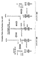
- FIG. 1 is a diagram schematically showing a production pathway of A ⁇ peptide by degradation of amyloid precursor protein (APP).
- FIG. 2 shows the sequence of the modified ADAMTS4 gene region in A549-ADAMTS4KO cells. The upper row shows the base sequence (SEQ ID NO: 12) for the original genome, and the lower row shows the base sequence for the genome (3 alleles) after gene editing: 16 bp-deficient sequence (SEQ ID NO: 13), 29 bp from the top. A missing sequence (SEQ ID NO: 14) and a 20 bp missing sequence (SEQ ID NO: 15) are shown.
- SEQ ID NO: 12 the base sequence for the original genome
- SEQ ID NO: 13 16 bp-deficient sequence
- SEQ ID NO: 14 16 bp-deficient sequence
- SEQ ID NO: 15 are shown.
- FIG. 3 is a graph showing the amount of APP669-711 produced in the culture supernatant when GM6001 (25 ⁇ M) or DMSO was added as a control to A549-ADAMTS4KO cells and A549-wt cells, respectively, in Example 1.
- the vertical axis is the average value of the normalized intensity.
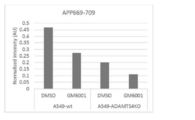
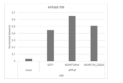
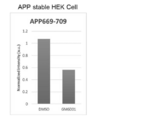
- FIG. 4 is a graph relating to the amount of APP669-709 produced in Example 1.
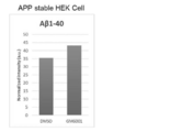
- FIG. 5 is a graph relating to the amount of A ⁇ 1-40 produced in Example 1.
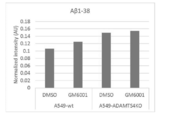
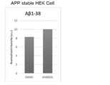
- FIG. 6 is a graph relating to the amount of A ⁇ 1-38 produced in Example 1.
- FIG. 7 shows the production of APP669-711 in the culture supernatant when ADAMTS4 plasmid vector and empty pcDNA3.1 hygro + vector were co-transfected into APP table HEK at a ratio of 1:10 and 10: 1 in Example 2. It is a graph showing the amount, and cells without transfection of vector (mock) and cells with transfection of empty pcDNA3.1 hygro + vector only (empty) were defined as controls. The vertical axis is the average value of Normalized intensity.
- FIG. 8 is a graph relating to the amount of APP669-709 produced in Example 2.
- FIG. 9 is a graph relating to the amount of A ⁇ 1-40 produced in Example 2.
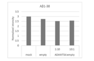
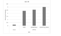
- FIG. 10 is a graph relating to the amount of A ⁇ 1-38 produced in Example 2.
- FIG. 11 is a graph showing the amount of APP669-711 produced in the culture supernatant when EGFP, ADAMTS4, and inactive ADAMTS4_E362A were co-transfected into HEK cells together with human APP in Example 3.
- the vertical axis is the average value of Normalized intensity.
- FIG. 12 is a graph relating to the amount of APP669-709 produced in Example 3.
- FIG. 13 is a graph relating to the amount of A ⁇ 1-40 produced in Example 3.
- FIG. 14 is a graph relating to the amount of A ⁇ 1-38 produced in Example 3.
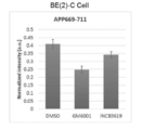
- FIG. 15 is a graph showing the amount of APP669-711 produced in the culture supernatant when 25 ⁇ M GM6001 or 10 ⁇ M INCB3619 was allowed to act on BE (2) -C cells in Example 4.
- the vertical axis is the average value of Normalized intensity.
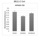
- FIG. 16 is a graph relating to the amount of APP669-709 produced for BE (2) -C cells in Example 4.
- FIG. 17 is a graph relating to the amount of A ⁇ 1-40 produced for BE (2) -C cells in Example 4.
- FIG. 18 is a graph relating to the amount of A ⁇ 1-38 produced for BE (2) -C cells in Example 4.
- FIG. 19 is a graph showing the amount of APP669-711 produced in the culture supernatant when 25 ⁇ M GM6001 or 10 ⁇ M INCB3619 was allowed to act on A549 cells in Example 4.
- the vertical axis is the average value of Normalized intensity.
- FIG. 20 is a graph relating to the amount of APP669-709 produced in Example 4.
- FIG. 21 is a graph relating to the amount of A ⁇ 1-40 produced in Example 4.
- FIG. 22 is a graph relating to the amount of A ⁇ 1-38 produced in Example 4.
- FIG. 23 is a graph showing the amount of APP669-711 produced in the culture supernatant when 25 ⁇ M GM6001 was allowed to act on APPtable HEK cells in Example 4.
- the vertical axis is the average value of Normalized intensity.
- FIG. 24 is a graph relating to the amount of APP669-709 produced in Example 4.
- FIG. 25 is a graph relating to the amount of A ⁇ 1-40 produced in Example 4.
- FIG. 26 is a graph relating to the amount of A ⁇ 1-38 produced in Example 4.
- FIG. 27 shows the amount of APP669-711 produced in the culture supernatant when the vector of MT-MMP1, 2, 3, 4, 5, or 6 was transfected with respect to APP table HEK cells in Example 5. It is a graph.
- the vertical axis is the average value of Normalized intensity.
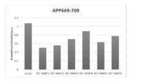
- FIG. 28 is a graph relating to the amount of APP669-709 produced in Example 5.
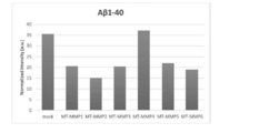
- FIG. 29 is a graph relating to the amount of A ⁇ 1-40 produced in Example 5.
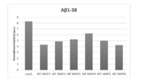
- FIG. 30 is a graph relating to the amount of A ⁇ 1-38 produced in Example 5.
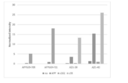
- FIG. 31 shows A ⁇ -related peptides (APP669-709, APP669-711, A ⁇ 1-38, A ⁇ 1) in the culture supernatant when Full-length APP, c102, or c99 was transfected into HEK cells in Example 6. It is a graph which shows the production amount of -40).
- the vertical axis is the average value of Normalized intensity.
- ADAMTS4 The ADAMTS (A disintegrin and metalloproteinase with thrombospondin motifs) family is a subfamily of ADAM and is an extracellular protease family found in mammals and the like. There are currently 19 members in ADAMTS. ADAMTS is classified into several subgroups centering on the substrate to be cleaved, and ADAMTS 1,4,5,8,9,15, and 20 are grouped as aggrecanase / Proteoglycanase. ing.
- ADAMTS4 As described above, ADAMTS4 is known, and the ADAMTS4 gene sequence is registered as NP_005090.3. Two SNPs are present in the ADAMTS4 gene (rs423367, rs41270041), and the encoded protein causes their respective amino acid substitutions (Q626R, P720A).
- ADAMTS4 is one of the enzymes that cleave the N-terminal of APP669. That is, ADAMTS4 cleaves the N-terminal site of amino acid 669 of amyloid precursor protein (APP) to produce APP669-x peptide.
- APP amyloid precursor protein
- the full length APP consists of 770 residues of amino acids, so x means an integer up to 770 that is greater than 669.
- an N-terminal cutting agent for APP669 containing ADAMTS4 is provided. Further, in relation to the screening method described below, a method of using ADAMTS4 as an N-terminal cleavage agent of APP669 in an in vitro system, and a method of using ADAMTS4 as an N-terminal cleavage agent of APP669 of ADAMTS4 in an in vitro system. Use is provided.
- the screening method is A method for screening a control substance for the N-terminal cleavage activity of APP669. The process of allowing the candidate substance to act on the cultured cells, The step of measuring the A ⁇ -related peptide produced from the cultured cells and A step of evaluating the N-terminal cleavage activity of APP669 and including.
- the A ⁇ -related peptide includes various molecular species, and among them, the peptide cleaved at the N-terminal of APP669 can be represented as APP669-x. Since the full length APP consists of 770 residues of amino acids, x means an integer up to 770 that is greater than 669.
- the A ⁇ -related peptide also includes a peptide cleaved on the N-terminal side of the 669th position of APP. Depending on the candidate substance, it may be cleaved at various sites of APP.
- APP672-709 (A ⁇ 1-38) (SEQ ID NO: 1): DAEFRHDSGYEVHHQKLVFFAEDVGSNKGAIIGLMVGG APP672-711 (A ⁇ 1-40) (SEQ ID NO: 2): DAEFRHDSGYEVHHQKLVFFAEDVGSNKGAIIGLMVGGVV APP669-709 (A ⁇ (-3-38)) (SEQ ID NO: 3): VKMDAEFRHDSGYEVHHQKLVFFAEDVGSNKGAIIGLMVGG APP669-711 (A ⁇ (-3-40)) (SEQ ID NO: 4): VKMDAEFRHDSGYEVHHQKLVFFAEDVGSNKGAIIGLMVGGVV APP672-713 (A ⁇ 1-42) (SEQ ID NO: 5): DAEFRHDSGYEVHHQKLVFFAEDVGSNKGAIIGLMVGGVVIA
- APP672-709 and A ⁇ 1-38 represent the same peptide.
- APP672-711 and A ⁇ 1-40 represent the same peptide
- APP669-709 and A ⁇ (-3-38) represent the same peptide
- APP669-711 and A ⁇ (-3-40) represent the same peptide.
- And APP672-713 and A ⁇ 1-42 represent the same peptide.
- a candidate substance for controlling the N-terminal cleavage activity of APP669 is allowed to act on cultured cells. This step can be carried out by a usual culture method.
- the A ⁇ -related peptide produced from the cultured cells is measured.
- This measurement step can be performed on the culture supernatant sample by a known method.
- the A ⁇ -related peptide to be measured (A ⁇ -related peptide other than APP669-x and / or APP669-x) is measured.
- the concentration and / or amount of A ⁇ -related peptide is basically measured, but other units used by those skilled in the art according to the concentration and / or amount, for example, the detected ionic strength in mass spectrometry may be used. ..
- the measurement of A ⁇ -related peptide may be performed by a test based on biomolecule-specific affinity.
- Testing based on biomolecule-specific affinity is a method well known to those of skill in the art and is not particularly limited, but immunoassays are preferred. Specifically, Western blot, radioimmunoassay, ELISA (Enzyme-Linked ImmunoSorbent Assay) (including sandwich immunomethod, competitive method, and direct adsorption method), immunoprecipitation method, precipitation reaction, immunodiffusion method, immunoaggregation measurement, Immunoassays including competing and non-competitive assay systems, such as complement fixation test analysis, immunoradiomimunoassay, fluorescent immunoassay, protein A immunoassay, etc. are included. In the immunoassay, an antibody that binds to an A ⁇ -related peptide in a biological sample is detected.
- Measurement of A ⁇ -related peptide is performed on an immunoglobulin having an antigen-binding site that can recognize a peptide derived from amyloid precursor protein (APP), or an immunoglobulin fragment containing an antigen-binding site that can recognize a peptide derived from amyloid precursor protein (APP). It may be carried out using the antibody-immobilized carrier prepared using. Peptides in a sample can be detected by a mass spectrometer by an immunoprecipitation method using the antibody-immobilized carrier (Immunoprecipitation-Mass Spectrometry; IP-MS).
- IP-MS immunoprecipitation-Mass Spectrometry
- immunoprecipitation may be continuously performed, and then the peptide in the sample may be detected by a mass spectrometer (cIP-MS).
- cIP-MS mass spectrometer
- the mass spectrometry method used in this case is preferably a matrix-assisted laser desorption / ionization (MALDI) mass spectrometry method, an electrospray ionization (ESI) mass spectrometry method, or the like.
- MALDI matrix-assisted laser desorption / ionization
- ESI electrospray ionization
- MALDI-TOF Microx Assisted Laser Desorption Ionization-Flight Time
- MALDI-IT Matrix Assisted Laser Desorption Ionization-Ion Trap
- MALDI-IT-TOF Matrix Assisted Laser Desorption
- MALDI-IT-TOF Matrix Assisted Laser Desorption
- MALDI-FTICR matrix-assisted laser desorption ionization-Fourier conversion ion cyclotron resonance
- ESI-QqQ electrospray ionization-triple quadrupole
- ESI-Qq-TOF electrospray ionization-tandem quadrupole-flight time
- ESI-FTICR electrospray ionization-Fourier conversion ion cyclotron resonance
- the matrix and the matrix solvent can be appropriately determined by those skilled in the art.
- ⁇ -cyano-4-hydroxycinnamic acid CHCA
- 2,5-dihydroxybenzoic acid 2,5-DHB
- sinapic acid 3-aminoquinoline (3-AQ) and the like are used. Can be done.
- the matrix solvent for example, it can be selected from the group consisting of acetonitrile (ACN), trifluoroacetic acid (TFA), methanol, ethanol and water. More specifically, ACN-TFA aqueous solution, ACN aqueous solution, methanol-TFA aqueous solution, methanol aqueous solution, ethanol-TFA aqueous solution, ethanol solution and the like can be used.
- a matrix additive (comatrix) may be used in combination.
- the matrix additive can be appropriately selected by those skilled in the art.
- a phosphonic acid group-containing compound can be used as the matrix additive.
- phosphonic acid Phosphonic acid
- methylphosphonic acid Metalphosphonic acid
- phenylphosphonic acid Phenylphosphonic acid
- 1-naphthylmethylphosphonic acid 1-Naphthylmethylphosphonic acid
- MDPNA Methylenediphosphonic acid
- Ethylenediphosphonic acid Ethylene-1-hydroxy-1,1-diphosphonic acid (Ethane-1-). Hydroxy-1,1-diphosphonic acid), Nitrilotriphosphonic acid, Ethylenediaminetetraphosphonic acid and the like.
- a compound having 2 or more, preferably 2 to 4 phosphonic acid groups in one molecule is preferable.
- the N-terminal cleavage activity of APP669 is evaluated from the measured amount of A ⁇ -related peptide produced. This evaluation step can be performed, for example, as follows.
- the A ⁇ -related peptide is APP669-x
- the amount of APP669-x of the control cells on which the candidate substance is not allowed to act as a reference level When the amount of APP669-x in the cells on which the candidate substance was acted was higher than the reference level, it was judged that the N-terminal cleavage activity of APP669 was increased, and the amount of APP669-x in the cells on which the candidate substance was acted was increased. When it is lower than the reference level, it is judged that the N-terminal cleavage activity of APP669 has decreased.
- APP669-709 only one kind of APP669-x, for example, APP669-709 may be evaluated.
- a plurality of species such as APP669-709, APP669-711, and APP669-713 may be evaluated.
- the cleavage activity on the N-terminal side of APP669 is focused on, so it is possible to comprehensively evaluate these multiple species (individual comparison, comparison as a total amount). Conceivable.
- the A ⁇ -related peptide is an A ⁇ -related peptide other than APP669-x and APP669-x
- the step of evaluating the N-terminal cleavage activity of APP669 Using the amount of APP669-x and the amount of A ⁇ -related peptides other than APP669-x of the control cells on which the candidate substance is not acted as reference levels, When the amount of APP669-x in the cell on which the candidate substance is allowed to act is higher than the reference level and the amount of A ⁇ -related peptide other than APP669-x is equal to or lower than the reference level, the APP669 It was judged that the N-terminal cleavage activity was increased, and the amount of APP669-x in the cells on which the candidate substance was allowed to act was lower than the reference level, and the amount of A ⁇ -related peptides other than APP669-x was equivalent to the reference level. Alternatively, when it is higher than the reference level,
- the APP669 cleavage activity can be specifically evaluated by referring to A ⁇ 1-y (a peptide having the N-terminal at the A ⁇ 1 position).
- the candidate substance for the control substance for the N-terminal cleavage activity of APP669 may be selected from low molecular weight compounds, peptides, proteins, nucleic acids and the like. A wide variety of substances are screened.
- low molecular weight compound a low molecular weight compound capable of binding to the target polypeptide can be considered.
- low molecular weight compounds that bind to certain proteins such as those used in drug discovery.
- low molecular weight compounds that inhibit ADAMTS4 include GM6001 and Calcium pentasan polysulfate.
- peptide a peptide consisting of 2 to 50 amino acid residues can be considered.
- soybean peptides that have been reported to suppress the expression of ADAMTS4 are exemplified.
- a protein consisting of 50 amino acid residues or more can be considered.
- examples thereof include IL-1 cytokines and TNF- ⁇ , which are reported to promote the expression of ADAMTS4, and TIMP-3, which inhibits ADAMTS4 activity.
- nucleic acid when ADAMTS4 is targeted, nucleic acids (vector, CRISPR-Cas9 system, siRNA, miRNA, antisense oligonucleotide, aptamer) that increase or decrease the gene expression of ADAMTS4 can be considered. Nucleic acids (vectors, CRISPR-Cas9 system, siRNA, miRNA, antisense oligonucleotides, aptamers) that increase or decrease gene expression can be considered even when an enzyme other than ADAMTS4 is targeted.
- the screening method A method for screening a control substance for the N-terminal cleavage activity of APP669.
- a step of allowing ADAMTS4 to act on an APP fragment containing a full-length APP or a sequence around the APP669 position in the presence of a candidate substance and The step of measuring APP669-x produced from the APP fragment containing the full-length APP or the sequence around the APP669 position, and A step of evaluating the N-terminal cleavage activity of APP669 and including.
- ADAMTS4 is one of the enzymes that cleave the N-terminal of APP669, it is also possible to screen a control substance for APP669 cleaving activity in vitro according to the above steps.
- the step of evaluating the N-terminal cleavage activity of APP669 can be performed, for example, as follows.
- the step of evaluating the N-terminal cleavage activity of APP669 for example, Using the amount of APP669-x in the control system in which the candidate substance does not exist as a reference level, When the amount of APP669-x in the system in which the candidate substance was present was higher than the reference level, it was judged that the N-terminal cleavage activity of APP669 was increased, and APP669-x in the system in which the candidate substance was present was determined. When the amount is lower than the reference level, it is judged that the N-terminal cleavage activity of APP669 is reduced.
- the amount of A ⁇ -related peptides other than APP669-x may be measured and evaluated.
- the measuring method and the candidate substance are the same as described above.
- kits A kit for screening a control substance for the N-terminal cleavage activity of APP669, which comprises ADAMTS4, a full-length APP or an APP fragment containing a sequence around the APP669 position, and other components described above.
- An APP fragment containing a sequence around the APP669 position is an amino acid sequence of 4 amino acids around the APP669 position [that is, APP665 (E), 666 (I), 667 (S), 668 (E), 669 (V), An APP fragment containing 670 (K), 671 (M), 672 (D), and 673 (A)], the N-terminal side and / or the C-terminal side may be longer. It is considered that the sequence around the APP669 position recognized by ADAMTS4 may be included.
- Neutral surfactants are represented by the following abbreviations. n-Decyl- ⁇ -D-maltoside (DM) n-Dodecyl- ⁇ -D-maltoside (DDM) n-Nonyl- ⁇ -D-thiomaltoside (NTM)
- Example 1 Evaluation by ADAMTS4 knockout of A549 cells and GM6001 action
- Effect of CRISPR / Cas9 clustered regularly interspaced short palindromic repeats / CRISPR associated proteins
- ADAMTS4 knockout using guide RNA gRNA
- A549 cells are human alveolar basal epithelial adenocarcinoma cells.
- the gRNA was designed using the CRISPR Search (https://www.sanger.ac.uk/htgt/wge/find_crisprs).
- a vector was created by inserting one by one.
- a plurality of monoclonal cells were analyzed, and it was estimated that the ADAMTS4 gene region of A549 cells had 3 alleles. Then, as shown in FIG. 2, monoclonal cells lacking the gene region of the ADAMTS4 gene at 16 bp, 29 bp, and 20 bp were obtained. Since neither is a multiple of 3, it is presumed that normal ADAMTS4 protein expression does not occur. Therefore, it was considered that these cells were A549 cells (A549-ADAMTS4 KO cells) in which endogenous ADAMTS4 was knocked out (KO).
- FIG. 2 shows the sequence of the modified ADAMTS4 gene region in A549-ADAMTS4KO cells.
- the upper row is the base sequence for the original genome: GGTGGTGGCAGATGACAAGATGGCCGCATTCCACGGTGCGGGGCTAAAGCGCTACCTGCTAACAGTGATGGCAGCAGCAGCCAAGGCCTTCAAGCA (SEQ ID NO: 12) is shown.
- the lower row shows the base sequence of the genome (3 alleles) after gene editing: From above, 16bp missing sequence: GGTGGTGGCAGATGACAAGATGGCCGCATTCCACGCTACCTGCTAACAGTGATGGCAGCAGCAGCCAAGGCCTTCAAGCA (SEQ ID NO: 13), 29bp missing sequence: GGTGGTGGCAGATGACAAGATGGCCGTAGCTAACAGTGATGGCAGCAGCAGCCAAGGCCTTCAAGCA (SEQ ID NO: 14), and 20bp-deficient sequence: GGTGGTGGCAGATGACAAGATGGCCGCATTCCAACCTGCTAACAGTGATGGCAGCAGCAGCCAAGGCCTTCAAGCA (SEQ ID NO: 15) Is shown.
- GM6001 (25 ⁇ M), which is a metalloprotease inhibitor, or dimethyl sulfoxide (DMSO) as a control was added to each of the A549-ADAMTS4KO cells and A549-wild type (wt) cells, and the cells were incubated for 96 hours. The culture supernatant was then collected and stored at ⁇ 80 ° C. until measurement.
- DMSO dimethyl sulfoxide
- IP-MS immunoprecipitation
- MS mass spectrometry
- the cryopreserved culture supernatant is thawed and the reaction solution containing the internal standard peptide [800 mM GlcNAc, 0.2% (w / v) NTM, 0.2% (w / v) DDM, 300 mM NaCl, 100 mM Tris-HCl. buffer (pH 7.4)] 250 ⁇ L and culture supernatant 250 ⁇ L were mixed.
- the mixture was mixed with antibody-immobilized beads and subjected to an antigen-antibody reaction at 4 ° C. for 1 hour. 22 pM stable isotope-labeled A ⁇ 1-38 and 100 pM stable isotope-labeled A ⁇ 1-15 were used as internal standard peptides.
- the antibody-immobilized beads were washed with the first wash buffer [first wash buffer (0.1% DDM, 0.1% NTM, 50 mM Tris-HCl (pH 7.4), 150 mM NaCl). Wash once with 100 ⁇ L and wash once with 50 ⁇ L of 50 mM ammonium acetate buffer]. Then, the A ⁇ -related peptide captured in the antibody-immobilized beads was eluted with a glycine buffer containing DDM (50 mM Glycine buffer containing 0.1% DDM, pH 2.8).
- a Tris buffer solution containing DDM (800 mM GlcNAc, 0.2% (w / v) DDM, 300 mM NaCl, 300 mM Tris-HCl buffer (pH 7.4)] was added to neutralize the pH (pH 7. It was returned to 4). Then, once again, the neutral eluate was contacted with the antibody-immobilized beads for 1 hour at 4 ° C. to react the A ⁇ -related peptide with the antigen-antibody reaction, and washed with the second washing buffer [second washing buffer (0.
- ⁇ -cyano-4-hydroxycinnamic acid was used as a matrix for Linear TOF.
- the matrix solution was prepared by dissolving 1 mg of CHCA in 1 mL of 70% (v / v) acetonitrile. 0.4% (w / v) methanediphosphonic acid (MDPNA) was used as the matrix additive. Equal amounts of 1 mg / mL CHCA solution and 0.4% (w / v) MDPNA were mixed to obtain a matrix / matrix additive solution [0.5 mg / mL CHCA / 0.2% (w / v) MDPNA]. ..
- Mass spectrum data was acquired by Linear TOF in positive ion mode using AXIMA Performance (Shimadzu / KRATOS, Manchester, UK).
- the m / z value of Linear TOF is indicated by the average mass of the peak.
- the m / z value was calibrated using human angiotensin II, human ACTH fragment 18-39, bovine insulin oxidized beta-chain, and bovine insulin as external standards.
- the signal intensity of each A ⁇ -related peptide was standardized using an internal standard peptide. Then, the average value of the normalized intensity of the four mass spectra obtained from one sample was obtained and evaluated as the amount of A ⁇ -related peptide produced.
- S / N ⁇ 3 the number of data that does not reach the lower limit of detection
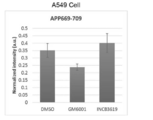
- FIG. 3 to 6 are graphs showing the amount of A ⁇ -related peptide produced in the culture supernatant when GM6001 (25 ⁇ M) or DMSO was added as a control to A549-ADAMTS4KO cells and A549-wt cells, respectively.
- the vertical axis is the average value of Normalized intensity.
- FIG. 3 is a graph relating to APP669-711 production
- FIG. 4 is a graph relating to APP669-709 production
- FIG. 5 is a graph relating to A ⁇ 1-40 production
- FIG. 6 is a graph relating to A ⁇ 1-38 production. It is a graph about the quantity.
- APP669-711 A ⁇ (-3-40)
- APP669- in both A549-ADAMTS4KO cells and A549-wt cells were compared with the control DMSO.
- the production of 709 A ⁇ (-3-38)
- ADAMTS4 is a metalloprotease and is known to be inhibited by GM6001. From this, it is considered that GM6001 inhibited the ADAMTS4 activity and reduced the production amounts of APP669-711 (A ⁇ (-3-40)) and APP669-709 (A ⁇ (-3-38)).
- Table 1 shows the amino acid sequences of A ⁇ 1-38, A ⁇ 1-40, APP669-709 (A ⁇ (-3-38)), and APP669-711 (A ⁇ (-3-40)).
- Example 2 Evaluation of APP table HEK cells by ADAMTS4 transfection
- Non-Patent Document 7 a vector in which the cDNA of human APP (695 amino acid isoform) was inserted into cDNA 3.1 for expression of mammalian cells was used (Non-Patent Document 7), and was used in Human Embryonic Kidney cells 293 (HEK cells).
- An APP table HEK in which human APP was stably expressed was prepared.
- a cDNA library was prepared from all mRNAs of human neuroblastoma-derived BE2 (C) cells (purchased from ATCC), and the human ADAMTS4 gene was cloned by PCR.
- the sequence that overlaps the original vector is shown in uppercase, and the ADAMTS4 sequence is shown in lowercase.
- the first letter acc in lowercase letters is not the array of ADAMTS4 itself, but contains the Kozak sequence (acc) for improving the translation efficiency of ADAMTS4.
- a human ADAMTS4 cDNA was inserted into a pcDNA3.1 hygro + vector in which the neomycin / kanamycin resistance gene of pcDNA3.1 was replaced with a hygromycin resistance gene by HiFi Assembly to create an ADAMTS4 plasmid vector.
- the obtained cDNA sequence is shown as SEQ ID NO: 18, and the amino acid sequence encoded by the cDNA sequence is shown as SEQ ID NO: 19.
- This cDNA was 100% consistent with the ADAMTS4 gene sequence NP_0050900.3 registered in the database.
- There are two SNPs in the ADAMTS4 gene (rs423367, rs41270041), which cause amino acid substitutions (Q626R, P720A), respectively.
- This vector and empty pcDNA3.1 hygro + vector were co-transfected at a ratio of 1:10 and 10: 1.
- cells that did not transfect the vector (mock) and cells that transfected only the empty pcDNA3.1 hygro + vector (empty) were also prepared. After 6 hours, the medium was replaced with new DMEM medium, and after 48 hours, the culture supernatant was collected. The culture supernatant was measured by the same IP-MS as in Example 1. However, the IP antibody used was an anti-A ⁇ antibody clone 4G8 (BioLegend), and the internal standard peptide used was only 22 pM stable isotope-labeled A ⁇ 1-38.
- FIGS. 7 to 10 show the amount of A ⁇ -related peptide produced in the culture supernatant when ADAMTS4 plasmid vector and empty pcDNA3.1 hygro + vector were co-transfected into APP table HEK at a ratio of 1:10 and 10: 1.
- cells without vector transfection (mock) and cells with empty pcDNA3.1 hygro + vector transfection (empty) were defined as controls.
- the vertical axis is the average value of Normalized intensity.
- FIG. 7 is a graph relating to the production amount of APP669-711
- FIG. 8 is a graph relating to the production amount of APP669-709
- FIG. 9 is a graph relating to the production amount of A ⁇ 1-40
- FIG. 10 is a graph relating to the production amount of A ⁇ 1-38. It is a graph about the quantity.
- APP669-711 A ⁇ (-3-40)
- APP669-709 A ⁇ (A ⁇ (A ⁇ )) in cells overexpressing ADAMTS4 by co-transfection of ADAMTS4 plasmid vector and empty pcDNA3.1 hygro + vector at a ratio of 10: 1.
- the production amount of -3-38)) tended to increase slightly (Figs. 7 and 8).
- APP669-711 A ⁇ (-3-40) and N-terminal side are different, C-terminal side is the same A ⁇ 1-40
- APP669-709 (A ⁇ (-3-38)) and N-terminal side are different, C-terminal side.
- ADAMTS4_E362A is an inactive mutant in which glutamic acid (E) at amino acid sequence 362, which is the active center of ADAMTS4, is replaced with alanine (A). Therefore, ADAMTS4_E362A vector was created by the PCR method.
- EGFP vector an empty vector in which EGFP was inserted into the pcDNA3.1 hygro + vector, ADAMTS4 vector, or ADAMTS4_E362A vector was co-transfected into HEK cells.
- EGFP vector was used as a control.
- a mock with no transfection was also used as a control. After incubating for 96 hours, the culture supernatant was collected and measured by the same IP-MS as in Example 1.
- FIGS. 11-14 are graphs showing the amount of A ⁇ -related peptide produced in the culture supernatant when EGFP, ADAMTS4, and inactive ADAMTS4_E362A were co-transfected into HEK cells together with human APP.
- the vertical axis is the average value of Normalized intensity.
- FIG. 11 is a graph relating to the production amount of APP669-711
- FIG. 12 is a graph relating to the production amount of APP669-709
- FIG. 13 is a graph relating to the production amount of A ⁇ 1-40
- FIG. 14 is a graph relating to the production amount of A ⁇ 1-38. It is a graph about the quantity.
- ADAMTS4 plasmid vector was transfected and ADAMTS4 was overexpressed
- EGFP was observed.
- the production amounts of APP669-711 (A ⁇ (-3-40)) and APP669-709 (A ⁇ (-3-38)) equivalent to those of the overexpressed control cells were shown.
- ADAMTS4 is one of the APP669-cleaving (N-terminal side) enzymes. Further, in Example 3, since the influence of the activity or inactivity of ADAMTS4 is reflected in the production amount of APP669-x, the Cleavage activity of APP669 containing ADAMTS4 is measured by measuring the production amount of APP669-x. Changes can also be captured, and it is considered that it can be used for screening of substances that control the APP669 cleavage activity.
- Example 4 Evaluation of a compound that controls APP669 cleavage activity
- a metalloprotease inhibitor 25 ⁇ M GM6001 (Cosmo Bio) or 10 ⁇ M INCB3619 (Non-Patent Document 8) was allowed to act on human neuroblastoma BE (2) -C and human alveolar basal epithelial adenocarcinoma cell A549.
- DMSO was added as a control and incubated for 96 hours. Then, the culture supernatant was collected and measured by the same IP-MS as in Example 1. However, 20 pM stable isotope-labeled A ⁇ 1-38 and 100 pM stable isotope-labeled A ⁇ 1-15 were used as internal standard peptides.
- FIGS. 15-18 The results for BE (2) -C cells are shown in FIGS. 15-18.
- FIGS. 15 to 18 are graphs showing the amount of A ⁇ -related peptide produced in the culture supernatant when 25 ⁇ M GM6001 or 10 ⁇ M INCB3619 was allowed to act on BE (2) -C cells.
- the vertical axis is the average value of Normalized intensity.
- FIG. 15 is a graph relating to APP669-711 production
- FIG. 16 is a graph relating to APP669-709 production
- FIG. 17 is a graph relating to A ⁇ 1-40 production
- FIG. 18 is a graph relating to A ⁇ 1-38 production. It is a graph about the quantity.
- FIGS. 19-22 The results for A549 cells are shown in FIGS. 19-22.
- 19 to 22 are graphs showing the amount of A ⁇ -related peptide produced in the culture supernatant when 25 ⁇ M GM6001 or 10 ⁇ M INCB3619 was allowed to act on A549 cells.
- the vertical axis is the average value of Normalized intensity.
- FIG. 19 is a graph relating to the production amount of APP669-711
- FIG. 20 is a graph relating to the production amount of APP669-709
- FIG. 21 is a graph relating to the production amount of A ⁇ 1-40
- FIG. 22 is a graph relating to the production amount of A ⁇ 1-38. It is a graph about the quantity.
- FIGS. 23 to 26 are graphs showing the amount of A ⁇ -related peptide produced in the culture supernatant when 25 ⁇ M GM6001 was allowed to act on APP table HEK cells.
- the vertical axis is the average value of Normalized intensity.
- FIG. 23 is a graph relating to APP669-711 production
- FIG. 24 is a graph relating to APP669-709 production
- FIG. 25 is a graph relating to A ⁇ 1-40 production
- FIG. 26 is a graph relating to A ⁇ 1-38 production. It is a graph about the quantity.
- Example 5 Evaluation of APP table HEK cells by MT-MMP transfection
- the expression vector in which the FLAG-tagged human MT-MMP1, MT-MMP2, MT-MMP3, MT-MMP4, MT-MMP5, or MT-MMP6 is inserted into the mammalian cell expression vector pCEP4 was invited by Motoharu Seiki. It was donated by a professor (Kanazawa University).
- FIGS. 27 to 30 are graphs showing the amount of A ⁇ -related peptide produced in the culture supernatant when a vector of MT-MMP1, 2, 3, 4, 5, or 6 was transfected into APP table HEK cells. ..
- the vertical axis is the average value of Normalized intensity.
- 27 is a graph relating to APP669-711 production
- FIG. 28 is a graph relating to APP669-709 production
- FIG. 29 is a graph relating to A ⁇ 1-40 production
- FIG. 30 is a graph relating to A ⁇ 1-38 production. It is a graph about the quantity.
- Example 6 Evaluation of ⁇ -site cleavage by APP, c102, c99 transfection
- BACE1 ⁇ -secretase
- C102 is a protein whose N-terminal is APP669 and whose C-terminal is the C-terminal of APP, and is a protein on the C-terminal side after cleavage of APP669.
- c99 is a protein on the C-terminal side after ⁇ -site cleavage ( ⁇ -CTF, referring to FIG. 1).
- Non-Patent Document 10 As a vector for expressing c99 inserted into the membrane, a cDNA encoding SPA4CT (Non-Patent Document 10) to which 2 amino acids (DA) are added following the signal sequence of APP is inserted into the mammalian cell expression vector pcDNA4. was used (Non-Patent Document 11).
- a vector expressing c102 in which 3 amino acids (VKM) were added before the c99 sequence to this vector was prepared by the PCR method. Full length APP, c102, or c99 was transfected into HEK cells. As a control, untransfected HEK cells (no) were also evaluated. After 96 hours, the culture supernatant was collected and measured by the same IP-MS as in Example 1. However, 20 pM stable isotope-labeled A ⁇ 1-38 and 100 pM stable isotope-labeled A ⁇ 1-15 were used as internal standard peptides.
- FIG. 31 shows the production of A ⁇ -related peptides (APP669-709, APP669-711, A ⁇ 1-38, A ⁇ 1-40) in the culture supernatant when Full-length APP, c102, or c99 was transfected into HEK cells. It is a graph showing the quantity. The vertical axis is the average value of Normalized intensity.
- the results of control (no), full length APP transfection, c102 transfection, and c99 transfection are shown in order from the left side.
- the amount of APP669-709 produced is too small for control (no), c99 and does not appear in the bar graph.
- the production amount of APP669-711 is too small for the control (no) and c99 and does not appear in the bar graph.
- c102 is not subjected to ⁇ -site cleavage. That is, if the APP669 cleavage activity can be promoted, it is possible to suppress the production of A ⁇ 1-x including A ⁇ 1-40 and A ⁇ 1-42. ing.
- a method for screening a control substance for the N-terminal cleavage activity of APP669. The process of allowing the candidate substance to act on the cultured cells, The step of measuring the A ⁇ -related peptide produced from the cultured cells and A step of evaluating the N-terminal cleavage activity of APP669 and Screening methods, including.
- the candidate substance is selected from the group consisting of low molecular weight compounds, peptides, proteins and nucleic acids.
- the candidate substance is selected from the group consisting of low molecular weight compounds, peptides, proteins and nucleic acids designed for ADAMTS4.
- a step of allowing ADAMTS4 to act on an APP fragment containing a full-length APP or a sequence around the APP669 position in the presence of a candidate substance and The step of measuring APP669-x produced from the APP fragment containing the full-length APP or the sequence around the APP669 position, and A step of evaluating the N-terminal cleavage activity of APP669 and Screening methods, including.
Landscapes
- Health & Medical Sciences (AREA)
- Life Sciences & Earth Sciences (AREA)
- Engineering & Computer Science (AREA)
- Chemical & Material Sciences (AREA)
- Immunology (AREA)
- Organic Chemistry (AREA)
- Biomedical Technology (AREA)
- Molecular Biology (AREA)
- Bioinformatics & Cheminformatics (AREA)
- Biotechnology (AREA)
- Wood Science & Technology (AREA)
- Zoology (AREA)
- General Health & Medical Sciences (AREA)
- Biochemistry (AREA)
- Physics & Mathematics (AREA)
- Microbiology (AREA)
- Analytical Chemistry (AREA)
- Genetics & Genomics (AREA)
- Proteomics, Peptides & Aminoacids (AREA)
- Urology & Nephrology (AREA)
- Hematology (AREA)
- General Engineering & Computer Science (AREA)
- Biophysics (AREA)
- Pathology (AREA)
- General Physics & Mathematics (AREA)
- Food Science & Technology (AREA)
- Medicinal Chemistry (AREA)
- Cell Biology (AREA)
- Tropical Medicine & Parasitology (AREA)
- Toxicology (AREA)
- Pharmacology & Pharmacy (AREA)
- Plant Pathology (AREA)
- Measuring Or Testing Involving Enzymes Or Micro-Organisms (AREA)
- Investigating Or Analysing Biological Materials (AREA)
Abstract
Description
候補物質を培養細胞に作用させる工程と、
前記培養細胞から産生されるAβ関連ペプチドを測定する工程と、
APP669のN末端切断活性を評価する工程と、
を含む、スクリーニング方法である。
In vitroの系で、候補物質存在下のもとADAMTS4を全長APP又はAPP669位前後の配列を含むAPP断片に作用させる工程と、
前記全長APP又はAPP669位前後の配列を含むAPP断片から産生されるAPP669-xを測定する工程と、
APP669のN末端切断活性を評価する工程と、
を含む、スクリーニング方法である。
ADAMTS(トロンボスポンジンモチーフを有するディスインテグリンおよびメタロプロテアーゼ,A disintegrin and metalloproteinase with thrombospondin motifs)ファミリーは、ADAMのサブファミリーであり、哺乳動物等に見いだされる細胞外プロテアーゼファミリーである。ADAMTSには現在19のメンバーが存在している。ADAMTSは切断する基質を中心にいくつかのサブグループに分類されており、ADAMTS1,4,5,8,9,15,及び20が、アグリカナーゼ/プロテオグリカナーゼ(Aggrecanase/Proteoglycanase)と呼ばれる一群とされている。
本発明の実施形態において、スクリーニング方法は、
APP669のN末端切断活性の制御物質をスクリーニングする方法であって、
候補物質を培養細胞に作用させる工程と、
前記培養細胞から産生されるAβ関連ペプチドを測定する工程と、
APP669のN末端切断活性を評価する工程と、
を含む。
DAEFRHDSGYEVHHQKLVFFAEDVGSNKGAIIGLMVGG
APP672-711(Aβ1-40)(配列番号2):
DAEFRHDSGYEVHHQKLVFFAEDVGSNKGAIIGLMVGGVV
APP669-709(Aβ(-3-38))(配列番号3):
VKMDAEFRHDSGYEVHHQKLVFFAEDVGSNKGAIIGLMVGG
APP669-711(Aβ(-3-40))(配列番号4):
VKMDAEFRHDSGYEVHHQKLVFFAEDVGSNKGAIIGLMVGGVV
APP672-713(Aβ1-42)(配列番号5):
DAEFRHDSGYEVHHQKLVFFAEDVGSNKGAIIGLMVGGVVIA
前記候補物質を作用させていないコントロール細胞のAPP669-x量を基準レベルとして、
前記候補物質を作用させた細胞のAPP669-x量が前記基準レベルよりも高い場合にAPP669のN末端切断活性が増加したと判断し、及び
前記候補物質を作用させた細胞のAPP669-x量が前記基準レベルよりも低い場合にAPP669のN末端切断活性が低下したと判断する。
前記APP669のN末端切断活性を評価する工程において、
前記候補物質を作用させていないコントロール細胞のAPP669-x量及びAPP669-x以外のAβ関連ペプチド量の各量を基準レベルとして、
前記候補物質を作用させた細胞のAPP669-x量が前記基準レベルよりも高く、且つ、APP669-x以外のAβ関連ペプチド量が前記基準レベルと同等もしくは前記基準レベルよりも低い場合に、APP669のN末端切断活性が増加したと判断し、及び
前記候補物質を作用させた細胞のAPP669-x量が前記基準レベルよりも低く、且つ、APP669-x以外のAβ関連ペプチド量が前記基準レベルと同等もしくは前記基準レベルよりも高い場合に、APP669のN末端切断活性が低下したと判断する。
APP669のN末端切断活性の制御物質をスクリーニングする方法であって、
In vitroの系で、候補物質存在下のもとADAMTS4を全長APP又はAPP669位前後の配列を含むAPP断片に作用させる工程と、
前記全長APP又はAPP669位前後の配列を含むAPP断片から産生されるAPP669-xを測定する工程と、
APP669のN末端切断活性を評価する工程と、
を含む。
前記候補物質を存在させていないコントロール系でのAPP669-x量を基準レベルとして、
前記候補物質を存在させた系でのAPP669-x量が前記基準レベルよりも高い場合にAPP669のN末端切断活性が増加したと判断し、及び
前記候補物質を存在させた系でのAPP669-x量が前記基準レベルよりも低い場合にAPP669のN末端切断活性が低下したと判断する。
APP669のN末端切断活性の制御物質をスクリーニングするためのキットであって、ADAMTS4、全長APP又はAPP669位前後の配列を含むAPP断片、その他の上述した各成分を含むキット。
n-Decyl-β-D-maltoside(DM)
n-Dodecyl-β-D-maltoside(DDM)
n-Nonyl-β-D-thiomaltoside(NTM)
A549細胞においてCRISPR/Cas9(clustered regularly interspaced short palindromic repeats/CRISPR associated proteins)及びガイドRNA(gRNA)を用いたADAMTS4ノックアウト、及び、メタロプロテアーゼ阻害剤であるGM6001の処理によるAPP669-x産生への影響を評価した。A549細胞は、ヒト肺胞基底上皮腺癌細胞である。gRNAは、CRISPR Search(https://www.sanger.ac.uk/htgt/wge/find_crisprs)を用いて設計した。
5’-GCGCTACCTGCTAACAGTGA-3'(配列番号6)
3'-CGCGATGGACGATTGTCACT-5'(すなわち、5’-TCACTGTTAGCAGGTAGCGC-3':配列番号7)
5'-CATTCCACGGTGCGGGGCTA-3'(配列番号8)
3'-GTAAGGTGCCACGCCCCGAT-5'(すなわち、5’-TAGCCCCGCACCGTGGAATG-3':配列番号9)
3’-CCTGGGACTGGTGAAACTGTGT-5’(すなわち、5’-TGTGTCAAAGTGGTCAGGGTCC-3':配列番号11)
GGTGGTGGCAGATGACAAGATGGCCGCATTCCACGGTGCGGGGCTAAAGCGCTACCTGCTAACAGTGATGGCAGCAGCAGCCAAGGCCTTCAAGCA(配列番号12)を示している。
上から、
16bp欠損した配列:
GGTGGTGGCAGATGACAAGATGGCCGCATTCCACGCTACCTGCTAACAGTGATGGCAGCAGCAGCCAAGGCCTTCAAGCA(配列番号13),
29bp欠損した配列:
GGTGGTGGCAGATGACAAGATGGCCGTAGCTAACAGTGATGGCAGCAGCAGCCAAGGCCTTCAAGCA(配列番号14),及び
20bp欠損した配列:
GGTGGTGGCAGATGACAAGATGGCCGCATTCCAACCTGCTAACAGTGATGGCAGCAGCAGCCAAGGCCTTCAAGCA(配列番号15)
を示している。

ADAMTS4過剰発現によるAPP669-x産生への影響を評価した。
5'-GGAGACCCAAGCTGGaccatgtcccagacaggctcgc-3'(配列番号16)
3'-ggacccgcccgtcctttattCGCAAATTTGAATTCGAACCA-5' (すなわち、5’-ACCAAGCTTAAGTTTA
AACGCttatttcctgcccgcccagg-3':配列番号17)
ADAMTS4と不活性型ADAMTS4(E362A)の過剰発現によるAPP669-x産生への影響を比較した。
5'-cactgctgctcatgcgctgggtcatgtct-3'(配列番号20)
3'-gtgacgacgagtacgcgacccagtacaga-5'(すなわち、5’-agacatgacccagcgcatgagcagcagtg-3':配列番号21)
メタロプロテアーゼ阻害剤によるAPP669-x産生への影響を評価した。
メタロプロテアーゼであるMT-MMP1,2,3,4,5,又は6によるAPP669-x産生への影響を評価した。
APP669切断後のc102にβ-セクレターゼ(BACE1)によるβサイト切断が生じるかどうかを評価した。
APP669のN末端切断活性の制御物質をスクリーニングする方法であって、
候補物質を培養細胞に作用させる工程と、
前記培養細胞から産生されるAβ関連ペプチドを測定する工程と、
APP669のN末端切断活性を評価する工程と、
を含む、スクリーニング方法。
前記Aβ関連ペプチドがAPP669-xである、上記(1)に記載の方法。
前記APP669のN末端切断活性を評価する工程において、
前記候補物質を作用させていないコントロール細胞のAPP669-x量を基準レベルとして、
前記候補物質を作用させた細胞のAPP669-x量が前記基準レベルよりも高い場合にAPP669のN末端切断活性が増加したと判断し、及び
前記候補物質を作用させた細胞のAPP669-x量が前記基準レベルよりも低い場合にAPP669のN末端切断活性が低下したと判断する、上記(2)に記載の方法。
前記Aβ関連ペプチドが、APP669-x、及び、APP669-x以外のAβ関連ペプチドである、上記(1)に記載の方法。
前記APP669のN末端切断活性を評価する工程において、
前記候補物質を作用させていないコントロール細胞のAPP669-x量及びAPP669-x以外のAβ関連ペプチド量の各量を基準レベルとして、
前記候補物質を作用させた細胞のAPP669-x量が前記基準レベルよりも高く、且つ、APP669-x以外のAβ関連ペプチド量が前記基準レベルと同等もしくは前記基準レベルよりも低い場合に、APP669のN末端切断活性が増加したと判断し、及び
前記候補物質を作用させた細胞のAPP669-x量が前記基準レベルよりも低く、且つ、APP669-x以外のAβ関連ペプチド量が前記基準レベルと同等もしくは前記基準レベルよりも高い場合に、APP669のN末端切断活性が低下したと判断する、上記(4)に記載の方法。
前記APP669-x以外のAβ関連ペプチドがAβ1-y(ここで、y=x-671)である、上記(4)又は(5)に記載の方法。
前記候補物質が、低分子化合物、ペプチド、タンパク質及び核酸からなる群から選ばれる、上記(1)~(6)のいずれかに記載の方法。
前記候補物質が、ADAMTS4をターゲットに設計された低分子化合物、ペプチド、タンパク質及び核酸からなる群から選ばれる、上記(1)~(7)のいずれかに記載の方法。
APP669のN末端切断活性の制御物質をスクリーニングする方法であって、
In vitroの系で、候補物質存在下のもとADAMTS4を全長APP又はAPP669位前後の配列を含むAPP断片に作用させる工程と、
前記全長APP又はAPP669位前後の配列を含むAPP断片から産生されるAPP669-xを測定する工程と、
APP669のN末端切断活性を評価する工程と、
を含む、スクリーニング方法。
前記APP669のN末端切断活性を評価する工程において、
前記候補物質を存在させていないコントロール系でのAPP669-x量を基準レベルとして、
前記候補物質を存在させた系でのAPP669-x量が前記基準レベルよりも高い場合にAPP669のN末端切断活性が増加したと判断し、及び
前記候補物質を存在させた系でのAPP669-x量が前記基準レベルよりも低い場合にAPP669のN末端切断活性が低下したと判断する、上記(9)に記載の方法。
前記候補物質が、低分子化合物、ペプチド、タンパク質及び核酸からなる群から選ばれる、上記(9)又は(10)に記載の方法。
ADAMTS4を含むAPP669のN末端切断剤。
In vitroの系において、ADAMTS4をAPP669のN末端切断剤として使用する方法。
In vitroの系における、ADAMTS4のAPP669のN末端切断剤としての使用。
Claims (12)
- APP669のN末端切断活性の制御物質をスクリーニングする方法であって、
候補物質を培養細胞に作用させる工程と、
前記培養細胞から産生されるAβ関連ペプチドを測定する工程と、
APP669のN末端切断活性を評価する工程と、
を含む、スクリーニング方法。 - 前記Aβ関連ペプチドがAPP669-xである、請求項1に記載の方法。
- 前記APP669のN末端切断活性を評価する工程において、
前記候補物質を作用させていないコントロール細胞のAPP669-x量を基準レベルとして、
前記候補物質を作用させた細胞のAPP669-x量が前記基準レベルよりも高い場合にAPP669のN末端切断活性が増加したと判断し、及び
前記候補物質を作用させた細胞のAPP669-x量が前記基準レベルよりも低い場合にAPP669のN末端切断活性が低下したと判断する、請求項2に記載の方法。 - 前記Aβ関連ペプチドが、APP669-x、及び、APP669-x以外のAβ関連ペプチドである、請求項1に記載の方法。
- 前記APP669のN末端切断活性を評価する工程において、
前記候補物質を作用させていないコントロール細胞のAPP669-x量及びAPP669-x以外のAβ関連ペプチド量の各量を基準レベルとして、
前記候補物質を作用させた細胞のAPP669-x量が前記基準レベルよりも高く、且つ、APP669-x以外のAβ関連ペプチド量が前記基準レベルと同等もしくは前記基準レベルよりも低い場合に、APP669のN末端切断活性が増加したと判断し、及び
前記候補物質を作用させた細胞のAPP669-x量が前記基準レベルよりも低く、且つ、APP669-x以外のAβ関連ペプチド量が前記基準レベルと同等もしくは前記基準レベルよりも高い場合に、APP669のN末端切断活性が低下したと判断する、請求項4に記載の方法。 - 前記APP669-x以外のAβ関連ペプチドがAβ1-y(ここで、y=x-671)である、請求項4又は5に記載の方法。
- 前記候補物質が、低分子化合物、ペプチド、タンパク質及び核酸からなる群から選ばれる、請求項1~6のいずれか1項に記載の方法。
- 前記候補物質が、ADAMTS4をターゲットに設計された低分子化合物、ペプチド、タンパク質及び核酸からなる群から選ばれる、請求項1~7のいずれか1項に記載の方法。
- APP669のN末端切断活性の制御物質をスクリーニングする方法であって、
In vitroの系で、候補物質存在下のもとADAMTS4を全長APP又はAPP669位前後の配列を含むAPP断片に作用させる工程と、
前記全長APP又はAPP669位前後の配列を含むAPP断片から産生されるAPP669-xを測定する工程と、
APP669のN末端切断活性を評価する工程と、
を含む、スクリーニング方法。 - 前記APP669のN末端切断活性を評価する工程において、
前記候補物質を存在させていないコントロール系でのAPP669-x量を基準レベルとして、
前記候補物質を存在させた系でのAPP669-x量が前記基準レベルよりも高い場合にAPP669のN末端切断活性が増加したと判断し、及び
前記候補物質を存在させた系でのAPP669-x量が前記基準レベルよりも低い場合にAPP669のN末端切断活性が低下したと判断する、請求項9に記載の方法。 - 前記候補物質が、低分子化合物、ペプチド、タンパク質及び核酸からなる群から選ばれる、請求項9又は10に記載の方法。
- ADAMTS4を含むAPP669のN末端切断剤。
Priority Applications (2)
| Application Number | Priority Date | Filing Date | Title |
|---|---|---|---|
| US17/606,289 US12461092B2 (en) | 2019-04-26 | 2020-03-30 | Screening method for APP cleavage activity-controlling substances of ADAMTS4 |
| JP2021515912A JP7270940B2 (ja) | 2019-04-26 | 2020-03-30 | Adamts4のapp切断活性制御物質のスクリーニング方法 |
Applications Claiming Priority (2)
| Application Number | Priority Date | Filing Date | Title |
|---|---|---|---|
| JP2019-086092 | 2019-04-26 | ||
| JP2019086092 | 2019-04-26 |
Publications (1)
| Publication Number | Publication Date |
|---|---|
| WO2020217865A1 true WO2020217865A1 (ja) | 2020-10-29 |
Family
ID=72942537
Family Applications (1)
| Application Number | Title | Priority Date | Filing Date |
|---|---|---|---|
| PCT/JP2020/014433 Ceased WO2020217865A1 (ja) | 2019-04-26 | 2020-03-30 | Adamts4のapp切断活性制御物質のスクリーニング方法 |
Country Status (3)
| Country | Link |
|---|---|
| US (1) | US12461092B2 (ja) |
| JP (1) | JP7270940B2 (ja) |
| WO (1) | WO2020217865A1 (ja) |
Citations (4)
| Publication number | Priority date | Publication date | Assignee | Title |
|---|---|---|---|---|
| WO2005108949A2 (en) * | 2004-05-12 | 2005-11-17 | Galapagos N.V. | Methods, compositions and compound assays for inhibiting amyloid-beta protein production |
| US20090305314A1 (en) * | 2006-02-01 | 2009-12-10 | Dirk Beher | Upregulation of Adamts4 Protease Activity for the Treatment of Alzheimer's Disease |
| WO2015178398A1 (ja) * | 2014-05-22 | 2015-11-26 | 株式会社 島津製作所 | 脳内のアミロイドβペプチド蓄積状態を評価するサロゲート・バイオマーカー及びその分析方法 |
| WO2017047529A1 (ja) * | 2015-09-16 | 2017-03-23 | 株式会社 島津製作所 | 脳内のアミロイドβ蓄積状態を評価するマルチプレックスバイオマーカー及びその分析方法 |
Family Cites Families (3)
| Publication number | Priority date | Publication date | Assignee | Title |
|---|---|---|---|---|
| JP5838481B2 (ja) | 2012-08-16 | 2016-01-06 | 公立大学法人名古屋市立大学 | Adamts−3を用いたリーリン分解 |
| WO2015111430A1 (ja) | 2014-01-21 | 2015-07-30 | 株式会社 島津製作所 | App切断型ペプチドの測定方法 |
| JP6424757B2 (ja) | 2015-07-14 | 2018-11-21 | 株式会社島津製作所 | ポリペプチドの質量分析方法 |
-
2020
- 2020-03-30 US US17/606,289 patent/US12461092B2/en active Active
- 2020-03-30 JP JP2021515912A patent/JP7270940B2/ja active Active
- 2020-03-30 WO PCT/JP2020/014433 patent/WO2020217865A1/ja not_active Ceased
Patent Citations (4)
| Publication number | Priority date | Publication date | Assignee | Title |
|---|---|---|---|---|
| WO2005108949A2 (en) * | 2004-05-12 | 2005-11-17 | Galapagos N.V. | Methods, compositions and compound assays for inhibiting amyloid-beta protein production |
| US20090305314A1 (en) * | 2006-02-01 | 2009-12-10 | Dirk Beher | Upregulation of Adamts4 Protease Activity for the Treatment of Alzheimer's Disease |
| WO2015178398A1 (ja) * | 2014-05-22 | 2015-11-26 | 株式会社 島津製作所 | 脳内のアミロイドβペプチド蓄積状態を評価するサロゲート・バイオマーカー及びその分析方法 |
| WO2017047529A1 (ja) * | 2015-09-16 | 2017-03-23 | 株式会社 島津製作所 | 脳内のアミロイドβ蓄積状態を評価するマルチプレックスバイオマーカー及びその分析方法 |
Non-Patent Citations (1)
| Title |
|---|
| NAKAMURA, A. ET AL.: "High performance plasma amyloid-beta biomarkers for Alzheimer's disease", NATURE, vol. 554, 2018, pages 249 - 254, XP055618943, DOI: 10.1038/nature25456 * |
Also Published As
| Publication number | Publication date |
|---|---|
| JP7270940B2 (ja) | 2023-05-11 |
| US12461092B2 (en) | 2025-11-04 |
| US20220326223A1 (en) | 2022-10-13 |
| JPWO2020217865A1 (ja) | 2020-10-29 |
Similar Documents
| Publication | Publication Date | Title |
|---|---|---|
| Bai et al. | Proteomic landscape of Alzheimer’s Disease: novel insights into pathogenesis and biomarker discovery | |
| Bauer et al. | Quantitative analysis of human centrosome architecture by targeted proteomics and fluorescence imaging | |
| Ahlf et al. | Evaluation of the compact high-field orbitrap for top-down proteomics of human cells | |
| US11467167B2 (en) | SRM methods in Alzheimer's disease and neurological disease assays | |
| Li et al. | Use of 18O labels to monitor deamidation during protein and peptide sample processing | |
| Staes et al. | Global differential non-gel proteomics by quantitative and stable labeling of tryptic peptides with oxygen-18 | |
| Brinkmalm et al. | An online nano‐LC‐ESI‐FTICR‐MS method for comprehensive characterization of endogenous fragments from amyloid β and amyloid precursor protein in human and cat cerebrospinal fluid | |
| Chicooree et al. | The application of targeted mass spectrometry‐based strategies to the detection and localization of post‐translational modifications | |
| US11835434B2 (en) | Methods for absolute quantification of low-abundance polypeptides using mass spectrometry | |
| Toropygin et al. | The N‐domain of angiotensin‐converting enzyme specifically hydrolyzes the Arg‐5‐His‐6 bond of Alzheimer's Aβ‐(1‐16) peptide and its isoAsp‐7 analogue with different efficiency as evidenced by quantitative matrix‐assisted laser desorption/ionization time‐of‐flight mass spectrometry | |
| JP5468073B2 (ja) | タンパク質の定量方法 | |
| CN103827311A (zh) | 用于神经退行性病症的生物化学标志物 | |
| US20150111238A1 (en) | Method for detecting disease biomarkers | |
| Trevisiol et al. | The use of proteases complementary to trypsin to probe isoforms and modifications | |
| JP6098538B2 (ja) | 糖ペプチドの分析方法、および糖鎖構造解析用プログラム | |
| Grasso | The use of mass spectrometry to study amyloid‐β peptides | |
| Liepold et al. | Matrix development for the detection of phosphorylated amyloid-β peptides by MALDI-TOF-MS | |
| US20110143379A1 (en) | Detection and quantitation of full-length thioredoxin (trx) and truncated thioredoxin (trx 80) in complex samples | |
| Griffiths et al. | Analysis of protein post-translational modifications by mass spectrometry | |
| Nepomuceno et al. | Detection of genetic variants of transthyretin by liquid chromatography–dual electrospray ionization Fourier-transform ion-cyclotron-resonance mass spectrometry | |
| Kilpatrick et al. | Reference measurement procedure development for C-reactive protein in human serum | |
| Zhang et al. | Top-down mass spectrometry on tissue extracts and biofluids with isoelectric focusing and superficially porous silica liquid chromatography | |
| WO2015074048A1 (en) | Measurement of gamma-carboxylation of proteins | |
| JP7270940B2 (ja) | Adamts4のapp切断活性制御物質のスクリーニング方法 | |
| Zhou et al. | Analysis of the subcellular phosphoproteome using a novel phosphoproteomic reactor |
Legal Events
| Date | Code | Title | Description |
|---|---|---|---|
| 121 | Ep: the epo has been informed by wipo that ep was designated in this application |
Ref document number: 20794028 Country of ref document: EP Kind code of ref document: A1 |
|
| ENP | Entry into the national phase |
Ref document number: 2021515912 Country of ref document: JP Kind code of ref document: A |
|
| NENP | Non-entry into the national phase |
Ref country code: DE |
|
| 122 | Ep: pct application non-entry in european phase |
Ref document number: 20794028 Country of ref document: EP Kind code of ref document: A1 |
|
| WWG | Wipo information: grant in national office |
Ref document number: 17606289 Country of ref document: US |