WO2020213724A1 - Chimeric receptor recognizing modification site of antibody - Google Patents
Chimeric receptor recognizing modification site of antibody Download PDFInfo
- Publication number
- WO2020213724A1 WO2020213724A1 PCT/JP2020/016915 JP2020016915W WO2020213724A1 WO 2020213724 A1 WO2020213724 A1 WO 2020213724A1 JP 2020016915 W JP2020016915 W JP 2020016915W WO 2020213724 A1 WO2020213724 A1 WO 2020213724A1
- Authority
- WO
- WIPO (PCT)
- Prior art keywords
- antibody
- mutation
- domain
- region
- amino acid
- Prior art date
Links
- 108700010039 chimeric receptor Proteins 0.000 title claims abstract description 217
- 230000004048 modification Effects 0.000 title claims abstract description 53
- 238000012986 modification Methods 0.000 title claims abstract description 53
- 230000035772 mutation Effects 0.000 claims abstract description 682
- 230000027455 binding Effects 0.000 claims abstract description 373
- 238000009739 binding Methods 0.000 claims abstract description 271
- 239000008194 pharmaceutical composition Substances 0.000 claims abstract description 141
- 238000006467 substitution reaction Methods 0.000 claims abstract description 65
- 238000012217 deletion Methods 0.000 claims abstract description 48
- 230000037430 deletion Effects 0.000 claims abstract description 48
- 238000007792 addition Methods 0.000 claims abstract description 43
- 235000001014 amino acid Nutrition 0.000 claims description 207
- 210000004027 cell Anatomy 0.000 claims description 203
- 125000003275 alpha amino acid group Chemical group 0.000 claims description 117
- 210000001744 T-lymphocyte Anatomy 0.000 claims description 87
- 230000004068 intracellular signaling Effects 0.000 claims description 49
- 239000013598 vector Substances 0.000 claims description 37
- 150000007523 nucleic acids Chemical class 0.000 claims description 35
- 102000039446 nucleic acids Human genes 0.000 claims description 33
- 108020004707 nucleic acids Proteins 0.000 claims description 33
- 230000002829 reductive effect Effects 0.000 claims description 24
- 230000014509 gene expression Effects 0.000 claims description 20
- QNAYBMKLOCPYGJ-REOHCLBHSA-N L-alanine Chemical compound C[C@H](N)C(O)=O QNAYBMKLOCPYGJ-REOHCLBHSA-N 0.000 claims description 18
- 235000004279 alanine Nutrition 0.000 claims description 18
- 108010073807 IgG Receptors Proteins 0.000 claims description 17
- 102000009490 IgG Receptors Human genes 0.000 claims description 17
- OUYCCCASQSFEME-QMMMGPOBSA-N L-tyrosine Chemical compound OC(=O)[C@@H](N)CC1=CC=C(O)C=C1 OUYCCCASQSFEME-QMMMGPOBSA-N 0.000 claims description 12
- OUYCCCASQSFEME-UHFFFAOYSA-N tyrosine Natural products OC(=O)C(N)CC1=CC=C(O)C=C1 OUYCCCASQSFEME-UHFFFAOYSA-N 0.000 claims description 11
- KZSNJWFQEVHDMF-UHFFFAOYSA-N Valine Natural products CC(C)C(N)C(O)=O KZSNJWFQEVHDMF-UHFFFAOYSA-N 0.000 claims description 10
- KZSNJWFQEVHDMF-BYPYZUCNSA-N L-valine Chemical compound CC(C)[C@H](N)C(O)=O KZSNJWFQEVHDMF-BYPYZUCNSA-N 0.000 claims description 9
- 239000004474 valine Substances 0.000 claims description 9
- 239000004475 Arginine Substances 0.000 claims description 7
- ODKSFYDXXFIFQN-UHFFFAOYSA-N arginine Natural products OC(=O)C(N)CCCNC(N)=N ODKSFYDXXFIFQN-UHFFFAOYSA-N 0.000 claims description 7
- ZDXPYRJPNDTMRX-UHFFFAOYSA-N glutamine Natural products OC(=O)C(N)CCC(N)=O ZDXPYRJPNDTMRX-UHFFFAOYSA-N 0.000 claims description 5
- ONIBWKKTOPOVIA-UHFFFAOYSA-N Proline Natural products OC(=O)C1CCCN1 ONIBWKKTOPOVIA-UHFFFAOYSA-N 0.000 claims description 4
- 230000001105 regulatory effect Effects 0.000 claims description 4
- 108010039491 Ricin Proteins 0.000 claims description 2
- 150000001413 amino acids Chemical class 0.000 abstract description 138
- 239000000427 antigen Substances 0.000 description 208
- 102000036639 antigens Human genes 0.000 description 207
- 108091007433 antigens Proteins 0.000 description 207
- 210000001519 tissue Anatomy 0.000 description 173
- 206010028980 Neoplasm Diseases 0.000 description 157
- 229940024606 amino acid Drugs 0.000 description 148
- 201000011510 cancer Diseases 0.000 description 113
- 238000000034 method Methods 0.000 description 110
- 241000282414 Homo sapiens Species 0.000 description 102
- 239000002207 metabolite Substances 0.000 description 95
- 108090000765 processed proteins & peptides Proteins 0.000 description 91
- 102000004196 processed proteins & peptides Human genes 0.000 description 83
- 229920001184 polypeptide Polymers 0.000 description 79
- 150000001875 compounds Chemical class 0.000 description 71
- 108090000623 proteins and genes Proteins 0.000 description 70
- 102000017420 CD3 protein, epsilon/gamma/delta subunit Human genes 0.000 description 61
- 108050005493 CD3 protein, epsilon/gamma/delta subunit Proteins 0.000 description 61
- 229910052700 potassium Inorganic materials 0.000 description 46
- 102000016266 T-Cell Antigen Receptors Human genes 0.000 description 44
- 125000000539 amino acid group Chemical group 0.000 description 44
- 108091008874 T cell receptors Proteins 0.000 description 43
- 230000002757 inflammatory effect Effects 0.000 description 43
- 102000005962 receptors Human genes 0.000 description 42
- 108020003175 receptors Proteins 0.000 description 42
- 102000053602 DNA Human genes 0.000 description 40
- 108020004414 DNA Proteins 0.000 description 40
- 102000004169 proteins and genes Human genes 0.000 description 38
- 235000018102 proteins Nutrition 0.000 description 37
- 239000012634 fragment Substances 0.000 description 32
- 229910052740 iodine Inorganic materials 0.000 description 32
- 230000011664 signaling Effects 0.000 description 32
- 229910052717 sulfur Inorganic materials 0.000 description 32
- 102000008394 Immunoglobulin Fragments Human genes 0.000 description 31
- 108010021625 Immunoglobulin Fragments Proteins 0.000 description 31
- 210000002865 immune cell Anatomy 0.000 description 31
- -1 CTL-019 Proteins 0.000 description 29
- 229910052731 fluorine Inorganic materials 0.000 description 28
- 229910052720 vanadium Inorganic materials 0.000 description 28
- KRKNYBCHXYNGOX-UHFFFAOYSA-N citric acid Chemical compound OC(=O)CC(O)(C(O)=O)CC(O)=O KRKNYBCHXYNGOX-UHFFFAOYSA-N 0.000 description 27
- 108060003951 Immunoglobulin Proteins 0.000 description 26
- 102000018358 immunoglobulin Human genes 0.000 description 26
- 229910052721 tungsten Inorganic materials 0.000 description 26
- 229910052727 yttrium Inorganic materials 0.000 description 26
- OIRDTQYFTABQOQ-KQYNXXCUSA-N adenosine Chemical compound C1=NC=2C(N)=NC=NC=2N1[C@@H]1O[C@H](CO)[C@@H](O)[C@H]1O OIRDTQYFTABQOQ-KQYNXXCUSA-N 0.000 description 24
- 210000004408 hybridoma Anatomy 0.000 description 23
- 229910052757 nitrogen Inorganic materials 0.000 description 23
- 241001465754 Metazoa Species 0.000 description 22
- 230000006870 function Effects 0.000 description 21
- 229910052739 hydrogen Inorganic materials 0.000 description 20
- 229910052698 phosphorus Inorganic materials 0.000 description 20
- 238000004519 manufacturing process Methods 0.000 description 18
- ZKHQWZAMYRWXGA-KQYNXXCUSA-J ATP(4-) Chemical compound C1=NC=2C(N)=NC=NC=2N1[C@@H]1O[C@H](COP([O-])(=O)OP([O-])(=O)OP([O-])([O-])=O)[C@@H](O)[C@H]1O ZKHQWZAMYRWXGA-KQYNXXCUSA-J 0.000 description 17
- ZKHQWZAMYRWXGA-UHFFFAOYSA-N Adenosine triphosphate Natural products C1=NC=2C(N)=NC=NC=2N1C1OC(COP(O)(=O)OP(O)(=O)OP(O)(O)=O)C(O)C1O ZKHQWZAMYRWXGA-UHFFFAOYSA-N 0.000 description 17
- 230000004936 stimulating effect Effects 0.000 description 17
- 101000946843 Homo sapiens T-cell surface glycoprotein CD8 alpha chain Proteins 0.000 description 16
- 102100034922 T-cell surface glycoprotein CD8 alpha chain Human genes 0.000 description 16
- 230000001086 cytosolic effect Effects 0.000 description 16
- 238000011282 treatment Methods 0.000 description 16
- 238000003752 polymerase chain reaction Methods 0.000 description 15
- CKLJMWTZIZZHCS-REOHCLBHSA-N L-aspartic acid Chemical compound OC(=O)[C@@H](N)CC(O)=O CKLJMWTZIZZHCS-REOHCLBHSA-N 0.000 description 14
- 239000012636 effector Substances 0.000 description 14
- 230000031146 intracellular signal transduction Effects 0.000 description 14
- MTCFGRXMJLQNBG-REOHCLBHSA-N (2S)-2-Amino-3-hydroxypropansäure Chemical compound OC[C@H](N)C(O)=O MTCFGRXMJLQNBG-REOHCLBHSA-N 0.000 description 13
- 210000003719 b-lymphocyte Anatomy 0.000 description 13
- XEYBRNLFEZDVAW-ARSRFYASSA-N dinoprostone Chemical compound CCCCC[C@H](O)\C=C\[C@H]1[C@H](O)CC(=O)[C@@H]1C\C=C/CCCC(O)=O XEYBRNLFEZDVAW-ARSRFYASSA-N 0.000 description 13
- 230000003834 intracellular effect Effects 0.000 description 13
- 239000002126 C01EB10 - Adenosine Substances 0.000 description 12
- WHUUTDBJXJRKMK-UHFFFAOYSA-N Glutamic acid Natural products OC(=O)C(N)CCC(O)=O WHUUTDBJXJRKMK-UHFFFAOYSA-N 0.000 description 12
- DHMQDGOQFOQNFH-UHFFFAOYSA-N Glycine Chemical compound NCC(O)=O DHMQDGOQFOQNFH-UHFFFAOYSA-N 0.000 description 12
- WHUUTDBJXJRKMK-VKHMYHEASA-N L-glutamic acid Chemical compound OC(=O)[C@@H](N)CCC(O)=O WHUUTDBJXJRKMK-VKHMYHEASA-N 0.000 description 12
- 230000004913 activation Effects 0.000 description 12
- 238000001994 activation Methods 0.000 description 12
- 229960005305 adenosine Drugs 0.000 description 12
- 235000003704 aspartic acid Nutrition 0.000 description 12
- OQFSQFPPLPISGP-UHFFFAOYSA-N beta-carboxyaspartic acid Natural products OC(=O)C(N)C(C(O)=O)C(O)=O OQFSQFPPLPISGP-UHFFFAOYSA-N 0.000 description 12
- 230000015572 biosynthetic process Effects 0.000 description 12
- 230000000295 complement effect Effects 0.000 description 12
- 230000000694 effects Effects 0.000 description 12
- 238000000338 in vitro Methods 0.000 description 12
- 238000002823 phage display Methods 0.000 description 12
- 239000013615 primer Substances 0.000 description 12
- NFGXHKASABOEEW-UHFFFAOYSA-N 1-methylethyl 11-methoxy-3,7,11-trimethyl-2,4-dodecadienoate Chemical compound COC(C)(C)CCCC(C)CC=CC(C)=CC(=O)OC(C)C NFGXHKASABOEEW-UHFFFAOYSA-N 0.000 description 11
- 101000914514 Homo sapiens T-cell-specific surface glycoprotein CD28 Proteins 0.000 description 11
- UGQMRVRMYYASKQ-KQYNXXCUSA-N Inosine Chemical compound O[C@@H]1[C@H](O)[C@@H](CO)O[C@H]1N1C2=NC=NC(O)=C2N=C1 UGQMRVRMYYASKQ-KQYNXXCUSA-N 0.000 description 11
- 241000699666 Mus <mouse, genus> Species 0.000 description 11
- 102100027213 T-cell-specific surface glycoprotein CD28 Human genes 0.000 description 11
- 229960002986 dinoprostone Drugs 0.000 description 11
- 230000004927 fusion Effects 0.000 description 11
- 239000003446 ligand Substances 0.000 description 11
- XEYBRNLFEZDVAW-UHFFFAOYSA-N prostaglandin E2 Natural products CCCCCC(O)C=CC1C(O)CC(=O)C1CC=CCCCC(O)=O XEYBRNLFEZDVAW-UHFFFAOYSA-N 0.000 description 11
- KDCGOANMDULRCW-UHFFFAOYSA-N 7H-purine Chemical group N1=CNC2=NC=NC2=C1 KDCGOANMDULRCW-UHFFFAOYSA-N 0.000 description 10
- 108010047041 Complementarity Determining Regions Proteins 0.000 description 10
- 108700018351 Major Histocompatibility Complex Proteins 0.000 description 10
- 241000699670 Mus sp. Species 0.000 description 10
- DFPAKSUCGFBDDF-UHFFFAOYSA-N Nicotinamide Chemical compound NC(=O)C1=CC=CN=C1 DFPAKSUCGFBDDF-UHFFFAOYSA-N 0.000 description 10
- 206010035226 Plasma cell myeloma Diseases 0.000 description 10
- KDYFGRWQOYBRFD-UHFFFAOYSA-N Succinic acid Natural products OC(=O)CCC(O)=O KDYFGRWQOYBRFD-UHFFFAOYSA-N 0.000 description 10
- KDYFGRWQOYBRFD-NUQCWPJISA-N butanedioic acid Chemical compound O[14C](=O)CC[14C](O)=O KDYFGRWQOYBRFD-NUQCWPJISA-N 0.000 description 10
- 108020001507 fusion proteins Proteins 0.000 description 10
- 102000037865 fusion proteins Human genes 0.000 description 10
- 235000013922 glutamic acid Nutrition 0.000 description 10
- 239000004220 glutamic acid Substances 0.000 description 10
- 238000001727 in vivo Methods 0.000 description 10
- 208000032839 leukemia Diseases 0.000 description 10
- 201000001441 melanoma Diseases 0.000 description 10
- 201000000050 myeloid neoplasm Diseases 0.000 description 10
- 230000007935 neutral effect Effects 0.000 description 10
- 239000002777 nucleoside Substances 0.000 description 10
- 229930010796 primary metabolite Natural products 0.000 description 10
- 239000000047 product Substances 0.000 description 10
- 210000002536 stromal cell Anatomy 0.000 description 10
- 230000020382 suppression by virus of host antigen processing and presentation of peptide antigen via MHC class I Effects 0.000 description 10
- 101000716102 Homo sapiens T-cell surface glycoprotein CD4 Proteins 0.000 description 9
- 108091028043 Nucleic acid sequence Proteins 0.000 description 9
- 241000283984 Rodentia Species 0.000 description 9
- 102100036011 T-cell surface glycoprotein CD4 Human genes 0.000 description 9
- 101150117115 V gene Proteins 0.000 description 9
- 230000010056 antibody-dependent cellular cytotoxicity Effects 0.000 description 9
- 238000013459 approach Methods 0.000 description 9
- 230000001472 cytotoxic effect Effects 0.000 description 9
- 210000000822 natural killer cell Anatomy 0.000 description 9
- LDHMAVIPBRSVRG-UHFFFAOYSA-O 1-methylnicotinamide Chemical compound C[N+]1=CC=CC(C(N)=O)=C1 LDHMAVIPBRSVRG-UHFFFAOYSA-O 0.000 description 8
- 102100026120 IgG receptor FcRn large subunit p51 Human genes 0.000 description 8
- 101710177940 IgG receptor FcRn large subunit p51 Proteins 0.000 description 8
- 102100022153 Tumor necrosis factor receptor superfamily member 4 Human genes 0.000 description 8
- LEHOTFFKMJEONL-UHFFFAOYSA-N Uric Acid Chemical compound N1C(=O)NC(=O)C2=C1NC(=O)N2 LEHOTFFKMJEONL-UHFFFAOYSA-N 0.000 description 8
- 239000013604 expression vector Substances 0.000 description 8
- 150000002303 glucose derivatives Chemical class 0.000 description 8
- 230000004153 glucose metabolism Effects 0.000 description 8
- 230000001976 improved effect Effects 0.000 description 8
- 238000003780 insertion Methods 0.000 description 8
- 230000037431 insertion Effects 0.000 description 8
- 150000002632 lipids Chemical class 0.000 description 8
- 210000004698 lymphocyte Anatomy 0.000 description 8
- 210000002540 macrophage Anatomy 0.000 description 8
- 239000002609 medium Substances 0.000 description 8
- 125000003835 nucleoside group Chemical group 0.000 description 8
- 230000019491 signal transduction Effects 0.000 description 8
- 235000000346 sugar Nutrition 0.000 description 8
- WUUGFSXJNOTRMR-IOSLPCCCSA-N 5'-S-methyl-5'-thioadenosine Chemical compound O[C@@H]1[C@H](O)[C@@H](CSC)O[C@H]1N1C2=NC=NC(N)=C2N=C1 WUUGFSXJNOTRMR-IOSLPCCCSA-N 0.000 description 7
- 108010021472 Fc gamma receptor IIB Proteins 0.000 description 7
- 101000934341 Homo sapiens T-cell surface glycoprotein CD5 Proteins 0.000 description 7
- AYFVYJQAPQTCCC-GBXIJSLDSA-N L-threonine Chemical compound C[C@@H](O)[C@H](N)C(O)=O AYFVYJQAPQTCCC-GBXIJSLDSA-N 0.000 description 7
- 102100029204 Low affinity immunoglobulin gamma Fc region receptor II-a Human genes 0.000 description 7
- 102100029205 Low affinity immunoglobulin gamma Fc region receptor II-b Human genes 0.000 description 7
- MTCFGRXMJLQNBG-UHFFFAOYSA-N Serine Natural products OCC(N)C(O)=O MTCFGRXMJLQNBG-UHFFFAOYSA-N 0.000 description 7
- 102100025244 T-cell surface glycoprotein CD5 Human genes 0.000 description 7
- 239000004473 Threonine Substances 0.000 description 7
- 239000002253 acid Substances 0.000 description 7
- 230000000259 anti-tumor effect Effects 0.000 description 7
- 239000002299 complementary DNA Substances 0.000 description 7
- 230000004069 differentiation Effects 0.000 description 7
- 230000001965 increasing effect Effects 0.000 description 7
- 230000003993 interaction Effects 0.000 description 7
- HCZHHEIFKROPDY-UHFFFAOYSA-N kynurenic acid Chemical compound C1=CC=C2NC(C(=O)O)=CC(=O)C2=C1 HCZHHEIFKROPDY-UHFFFAOYSA-N 0.000 description 7
- 238000012216 screening Methods 0.000 description 7
- 108091026890 Coding region Proteins 0.000 description 6
- 241000588724 Escherichia coli Species 0.000 description 6
- 101000917839 Homo sapiens Low affinity immunoglobulin gamma Fc region receptor III-B Proteins 0.000 description 6
- 108010054477 Immunoglobulin Fab Fragments Proteins 0.000 description 6
- 102000001706 Immunoglobulin Fab Fragments Human genes 0.000 description 6
- QIVBCDIJIAJPQS-VIFPVBQESA-N L-tryptophane Chemical compound C1=CC=C2C(C[C@H](N)C(O)=O)=CNC2=C1 QIVBCDIJIAJPQS-VIFPVBQESA-N 0.000 description 6
- 102100029185 Low affinity immunoglobulin gamma Fc region receptor III-B Human genes 0.000 description 6
- 206010025323 Lymphomas Diseases 0.000 description 6
- 206010039491 Sarcoma Diseases 0.000 description 6
- AYFVYJQAPQTCCC-UHFFFAOYSA-N Threonine Natural products CC(O)C(N)C(O)=O AYFVYJQAPQTCCC-UHFFFAOYSA-N 0.000 description 6
- QIVBCDIJIAJPQS-UHFFFAOYSA-N Tryptophan Natural products C1=CC=C2C(CC(N)C(O)=O)=CNC2=C1 QIVBCDIJIAJPQS-UHFFFAOYSA-N 0.000 description 6
- 230000000735 allogeneic effect Effects 0.000 description 6
- 230000004071 biological effect Effects 0.000 description 6
- 210000004369 blood Anatomy 0.000 description 6
- 239000008280 blood Substances 0.000 description 6
- 150000001720 carbohydrates Chemical group 0.000 description 6
- 230000000139 costimulatory effect Effects 0.000 description 6
- 238000004132 cross linking Methods 0.000 description 6
- 238000005516 engineering process Methods 0.000 description 6
- 230000003053 immunization Effects 0.000 description 6
- 239000002773 nucleotide Substances 0.000 description 6
- 125000003729 nucleotide group Chemical group 0.000 description 6
- 230000002265 prevention Effects 0.000 description 6
- 229920002477 rna polymer Polymers 0.000 description 6
- 241000894007 species Species 0.000 description 6
- 238000012360 testing method Methods 0.000 description 6
- 102100038080 B-cell receptor CD22 Human genes 0.000 description 5
- 241000282693 Cercopithecidae Species 0.000 description 5
- 108010019670 Chimeric Antigen Receptors Proteins 0.000 description 5
- 206010009944 Colon cancer Diseases 0.000 description 5
- 102000004190 Enzymes Human genes 0.000 description 5
- 108090000790 Enzymes Proteins 0.000 description 5
- 108010021468 Fc gamma receptor IIA Proteins 0.000 description 5
- 102000009109 Fc receptors Human genes 0.000 description 5
- 108010087819 Fc receptors Proteins 0.000 description 5
- 101000884305 Homo sapiens B-cell receptor CD22 Proteins 0.000 description 5
- 101000935040 Homo sapiens Integrin beta-2 Proteins 0.000 description 5
- 101000917858 Homo sapiens Low affinity immunoglobulin gamma Fc region receptor III-A Proteins 0.000 description 5
- 101000633784 Homo sapiens SLAM family member 7 Proteins 0.000 description 5
- 102000006496 Immunoglobulin Heavy Chains Human genes 0.000 description 5
- 108010019476 Immunoglobulin Heavy Chains Proteins 0.000 description 5
- 108010067060 Immunoglobulin Variable Region Proteins 0.000 description 5
- 102000017727 Immunoglobulin Variable Region Human genes 0.000 description 5
- 102100022339 Integrin alpha-L Human genes 0.000 description 5
- 102100025390 Integrin beta-2 Human genes 0.000 description 5
- ROHFNLRQFUQHCH-YFKPBYRVSA-N L-leucine Chemical compound CC(C)C[C@H](N)C(O)=O ROHFNLRQFUQHCH-YFKPBYRVSA-N 0.000 description 5
- COLNVLDHVKWLRT-QMMMGPOBSA-N L-phenylalanine Chemical compound OC(=O)[C@@H](N)CC1=CC=CC=C1 COLNVLDHVKWLRT-QMMMGPOBSA-N 0.000 description 5
- 206010058467 Lung neoplasm malignant Diseases 0.000 description 5
- 206010029260 Neuroblastoma Diseases 0.000 description 5
- 102100029198 SLAM family member 7 Human genes 0.000 description 5
- 230000002378 acidificating effect Effects 0.000 description 5
- 238000001042 affinity chromatography Methods 0.000 description 5
- 239000011324 bead Substances 0.000 description 5
- 210000004899 c-terminal region Anatomy 0.000 description 5
- 238000004113 cell culture Methods 0.000 description 5
- 238000002659 cell therapy Methods 0.000 description 5
- 230000008859 change Effects 0.000 description 5
- 238000012875 competitive assay Methods 0.000 description 5
- 230000002950 deficient Effects 0.000 description 5
- 230000001419 dependent effect Effects 0.000 description 5
- 238000004520 electroporation Methods 0.000 description 5
- 229940088598 enzyme Drugs 0.000 description 5
- 230000013595 glycosylation Effects 0.000 description 5
- 238000006206 glycosylation reaction Methods 0.000 description 5
- 230000028993 immune response Effects 0.000 description 5
- 230000016784 immunoglobulin production Effects 0.000 description 5
- 230000000670 limiting effect Effects 0.000 description 5
- 201000005202 lung cancer Diseases 0.000 description 5
- 208000020816 lung neoplasm Diseases 0.000 description 5
- 230000004060 metabolic process Effects 0.000 description 5
- 230000036961 partial effect Effects 0.000 description 5
- 125000002924 primary amino group Chemical group [H]N([H])* 0.000 description 5
- 239000000126 substance Substances 0.000 description 5
- 239000000758 substrate Substances 0.000 description 5
- 238000002560 therapeutic procedure Methods 0.000 description 5
- 238000001890 transfection Methods 0.000 description 5
- 239000011800 void material Substances 0.000 description 5
- YBJHBAHKTGYVGT-ZKWXMUAHSA-N (+)-Biotin Chemical compound N1C(=O)N[C@@H]2[C@H](CCCCC(=O)O)SC[C@@H]21 YBJHBAHKTGYVGT-ZKWXMUAHSA-N 0.000 description 4
- 208000024893 Acute lymphoblastic leukemia Diseases 0.000 description 4
- 208000014697 Acute lymphocytic leukaemia Diseases 0.000 description 4
- 208000010839 B-cell chronic lymphocytic leukemia Diseases 0.000 description 4
- 208000028564 B-cell non-Hodgkin lymphoma Diseases 0.000 description 4
- 102100024222 B-lymphocyte antigen CD19 Human genes 0.000 description 4
- 206010006187 Breast cancer Diseases 0.000 description 4
- 208000026310 Breast neoplasm Diseases 0.000 description 4
- 102100026094 C-type lectin domain family 12 member A Human genes 0.000 description 4
- 101710188619 C-type lectin domain family 12 member A Proteins 0.000 description 4
- 102100027207 CD27 antigen Human genes 0.000 description 4
- 102100032937 CD40 ligand Human genes 0.000 description 4
- 201000009030 Carcinoma Diseases 0.000 description 4
- SHZGCJCMOBCMKK-UHFFFAOYSA-N D-mannomethylose Natural products CC1OC(O)C(O)C(O)C1O SHZGCJCMOBCMKK-UHFFFAOYSA-N 0.000 description 4
- PNNNRSAQSRJVSB-SLPGGIOYSA-N Fucose Natural products C[C@H](O)[C@@H](O)[C@H](O)[C@H](O)C=O PNNNRSAQSRJVSB-SLPGGIOYSA-N 0.000 description 4
- 102000010956 Glypican Human genes 0.000 description 4
- 108050001154 Glypican Proteins 0.000 description 4
- 108050007237 Glypican-3 Proteins 0.000 description 4
- 102100026122 High affinity immunoglobulin gamma Fc receptor I Human genes 0.000 description 4
- 208000017604 Hodgkin disease Diseases 0.000 description 4
- 208000021519 Hodgkin lymphoma Diseases 0.000 description 4
- 208000010747 Hodgkins lymphoma Diseases 0.000 description 4
- 241001272567 Hominoidea Species 0.000 description 4
- 241000282412 Homo Species 0.000 description 4
- 101000980825 Homo sapiens B-lymphocyte antigen CD19 Proteins 0.000 description 4
- 101000914511 Homo sapiens CD27 antigen Proteins 0.000 description 4
- 101000868215 Homo sapiens CD40 ligand Proteins 0.000 description 4
- 101000913074 Homo sapiens High affinity immunoglobulin gamma Fc receptor I Proteins 0.000 description 4
- 101000994375 Homo sapiens Integrin alpha-4 Proteins 0.000 description 4
- 101000934338 Homo sapiens Myeloid cell surface antigen CD33 Proteins 0.000 description 4
- 101000738771 Homo sapiens Receptor-type tyrosine-protein phosphatase C Proteins 0.000 description 4
- 101000934346 Homo sapiens T-cell surface antigen CD2 Proteins 0.000 description 4
- 102100032818 Integrin alpha-4 Human genes 0.000 description 4
- 102100032816 Integrin alpha-6 Human genes 0.000 description 4
- 102100037877 Intercellular adhesion molecule 1 Human genes 0.000 description 4
- XUJNEKJLAYXESH-REOHCLBHSA-N L-Cysteine Chemical compound SC[C@H](N)C(O)=O XUJNEKJLAYXESH-REOHCLBHSA-N 0.000 description 4
- SHZGCJCMOBCMKK-DHVFOXMCSA-N L-fucopyranose Chemical compound C[C@@H]1OC(O)[C@@H](O)[C@H](O)[C@@H]1O SHZGCJCMOBCMKK-DHVFOXMCSA-N 0.000 description 4
- ROHFNLRQFUQHCH-UHFFFAOYSA-N Leucine Natural products CC(C)CC(N)C(O)=O ROHFNLRQFUQHCH-UHFFFAOYSA-N 0.000 description 4
- 108010064548 Lymphocyte Function-Associated Antigen-1 Proteins 0.000 description 4
- 208000031422 Lymphocytic Chronic B-Cell Leukemia Diseases 0.000 description 4
- KDXKERNSBIXSRK-UHFFFAOYSA-N Lysine Natural products NCCCCC(N)C(O)=O KDXKERNSBIXSRK-UHFFFAOYSA-N 0.000 description 4
- 241000124008 Mammalia Species 0.000 description 4
- 102100025243 Myeloid cell surface antigen CD33 Human genes 0.000 description 4
- 108091007491 NSP3 Papain-like protease domains Proteins 0.000 description 4
- 206010033128 Ovarian cancer Diseases 0.000 description 4
- 206010061535 Ovarian neoplasm Diseases 0.000 description 4
- 208000006664 Precursor Cell Lymphoblastic Leukemia-Lymphoma Diseases 0.000 description 4
- 101710120463 Prostate stem cell antigen Proteins 0.000 description 4
- 102100036735 Prostate stem cell antigen Human genes 0.000 description 4
- 102100037422 Receptor-type tyrosine-protein phosphatase C Human genes 0.000 description 4
- 108020004511 Recombinant DNA Proteins 0.000 description 4
- 201000000582 Retinoblastoma Diseases 0.000 description 4
- 102100029215 Signaling lymphocytic activation molecule Human genes 0.000 description 4
- 208000005718 Stomach Neoplasms Diseases 0.000 description 4
- 102100025237 T-cell surface antigen CD2 Human genes 0.000 description 4
- 238000002617 apheresis Methods 0.000 description 4
- 210000000170 cell membrane Anatomy 0.000 description 4
- 208000032852 chronic lymphocytic leukemia Diseases 0.000 description 4
- 238000010367 cloning Methods 0.000 description 4
- 208000029742 colonic neoplasm Diseases 0.000 description 4
- 238000004590 computer program Methods 0.000 description 4
- 239000000470 constituent Substances 0.000 description 4
- 230000001276 controlling effect Effects 0.000 description 4
- 231100000433 cytotoxic Toxicity 0.000 description 4
- 238000000354 decomposition reaction Methods 0.000 description 4
- 201000010099 disease Diseases 0.000 description 4
- 208000037265 diseases, disorders, signs and symptoms Diseases 0.000 description 4
- 238000010494 dissociation reaction Methods 0.000 description 4
- 230000005593 dissociations Effects 0.000 description 4
- 206010017758 gastric cancer Diseases 0.000 description 4
- 239000001963 growth medium Substances 0.000 description 4
- 238000002649 immunization Methods 0.000 description 4
- 230000002163 immunogen Effects 0.000 description 4
- 210000003292 kidney cell Anatomy 0.000 description 4
- JVTAAEKCZFNVCJ-UHFFFAOYSA-N lactic acid Chemical compound CC(O)C(O)=O JVTAAEKCZFNVCJ-UHFFFAOYSA-N 0.000 description 4
- 239000000203 mixture Substances 0.000 description 4
- 201000008968 osteosarcoma Diseases 0.000 description 4
- COLNVLDHVKWLRT-UHFFFAOYSA-N phenylalanine Natural products OC(=O)C(N)CC1=CC=CC=C1 COLNVLDHVKWLRT-UHFFFAOYSA-N 0.000 description 4
- 210000002307 prostate Anatomy 0.000 description 4
- 238000000746 purification Methods 0.000 description 4
- 238000000926 separation method Methods 0.000 description 4
- 230000000638 stimulation Effects 0.000 description 4
- 201000011549 stomach cancer Diseases 0.000 description 4
- 238000003786 synthesis reaction Methods 0.000 description 4
- 238000010361 transduction Methods 0.000 description 4
- 230000026683 transduction Effects 0.000 description 4
- 238000005406 washing Methods 0.000 description 4
- 206010003445 Ascites Diseases 0.000 description 3
- 241000894006 Bacteria Species 0.000 description 3
- 102100037904 CD9 antigen Human genes 0.000 description 3
- 102100025064 Cellular tumor antigen p53 Human genes 0.000 description 3
- 102100038449 Claudin-6 Human genes 0.000 description 3
- 108090000229 Claudin-6 Proteins 0.000 description 3
- 102100038083 Endosialin Human genes 0.000 description 3
- 108091006027 G proteins Proteins 0.000 description 3
- 102000030782 GTP binding Human genes 0.000 description 3
- 108091000058 GTP-Binding Proteins 0.000 description 3
- 239000004471 Glycine Substances 0.000 description 3
- 101000738354 Homo sapiens CD9 antigen Proteins 0.000 description 3
- 101000721661 Homo sapiens Cellular tumor antigen p53 Proteins 0.000 description 3
- 101001078158 Homo sapiens Integrin alpha-1 Proteins 0.000 description 3
- 101000994365 Homo sapiens Integrin alpha-6 Proteins 0.000 description 3
- 101000935043 Homo sapiens Integrin beta-1 Proteins 0.000 description 3
- 101001055144 Homo sapiens Interleukin-2 receptor subunit alpha Proteins 0.000 description 3
- 101000777628 Homo sapiens Leukocyte antigen CD37 Proteins 0.000 description 3
- 101000578784 Homo sapiens Melanoma antigen recognized by T-cells 1 Proteins 0.000 description 3
- 101000589305 Homo sapiens Natural cytotoxicity triggering receptor 2 Proteins 0.000 description 3
- 101000633786 Homo sapiens SLAM family member 6 Proteins 0.000 description 3
- 101000824971 Homo sapiens Sperm surface protein Sp17 Proteins 0.000 description 3
- 101000914484 Homo sapiens T-lymphocyte activation antigen CD80 Proteins 0.000 description 3
- 108010091358 Hypoxanthine Phosphoribosyltransferase Proteins 0.000 description 3
- 102100029098 Hypoxanthine-guanine phosphoribosyltransferase Human genes 0.000 description 3
- 102000013463 Immunoglobulin Light Chains Human genes 0.000 description 3
- 108010065825 Immunoglobulin Light Chains Proteins 0.000 description 3
- 102100025323 Integrin alpha-1 Human genes 0.000 description 3
- 102100022338 Integrin alpha-M Human genes 0.000 description 3
- 102100022297 Integrin alpha-X Human genes 0.000 description 3
- 102100025304 Integrin beta-1 Human genes 0.000 description 3
- 108010064593 Intercellular Adhesion Molecule-1 Proteins 0.000 description 3
- 102100026878 Interleukin-2 receptor subunit alpha Human genes 0.000 description 3
- DCXYFEDJOCDNAF-REOHCLBHSA-N L-asparagine Chemical compound OC(=O)[C@@H](N)CC(N)=O DCXYFEDJOCDNAF-REOHCLBHSA-N 0.000 description 3
- ZDXPYRJPNDTMRX-VKHMYHEASA-N L-glutamine Chemical compound OC(=O)[C@@H](N)CCC(N)=O ZDXPYRJPNDTMRX-VKHMYHEASA-N 0.000 description 3
- 102100031586 Leukocyte antigen CD37 Human genes 0.000 description 3
- 102100028389 Melanoma antigen recognized by T-cells 1 Human genes 0.000 description 3
- 108010004217 Natural Cytotoxicity Triggering Receptor 1 Proteins 0.000 description 3
- 108010004222 Natural Cytotoxicity Triggering Receptor 3 Proteins 0.000 description 3
- 102100032870 Natural cytotoxicity triggering receptor 1 Human genes 0.000 description 3
- 102100032851 Natural cytotoxicity triggering receptor 2 Human genes 0.000 description 3
- 102100032852 Natural cytotoxicity triggering receptor 3 Human genes 0.000 description 3
- 241000283973 Oryctolagus cuniculus Species 0.000 description 3
- 102100026547 Platelet-derived growth factor receptor beta Human genes 0.000 description 3
- 241000700159 Rattus Species 0.000 description 3
- 102100029197 SLAM family member 6 Human genes 0.000 description 3
- 108010074687 Signaling Lymphocytic Activation Molecule Family Member 1 Proteins 0.000 description 3
- 108010003723 Single-Domain Antibodies Proteins 0.000 description 3
- 230000006044 T cell activation Effects 0.000 description 3
- 102100027222 T-lymphocyte activation antigen CD80 Human genes 0.000 description 3
- 108060008683 Tumor Necrosis Factor Receptor Proteins 0.000 description 3
- 101710165473 Tumor necrosis factor receptor superfamily member 4 Proteins 0.000 description 3
- 230000002159 abnormal effect Effects 0.000 description 3
- 238000010521 absorption reaction Methods 0.000 description 3
- 150000007513 acids Chemical class 0.000 description 3
- 230000009824 affinity maturation Effects 0.000 description 3
- 210000000628 antibody-producing cell Anatomy 0.000 description 3
- 230000000890 antigenic effect Effects 0.000 description 3
- 239000000872 buffer Substances 0.000 description 3
- 235000014633 carbohydrates Nutrition 0.000 description 3
- 239000003795 chemical substances by application Substances 0.000 description 3
- XUJNEKJLAYXESH-UHFFFAOYSA-N cysteine Natural products SCC(N)C(O)=O XUJNEKJLAYXESH-UHFFFAOYSA-N 0.000 description 3
- 235000018417 cysteine Nutrition 0.000 description 3
- 125000000151 cysteine group Chemical group N[C@@H](CS)C(=O)* 0.000 description 3
- 238000011161 development Methods 0.000 description 3
- 230000018109 developmental process Effects 0.000 description 3
- 239000000539 dimer Substances 0.000 description 3
- LOKCTEFSRHRXRJ-UHFFFAOYSA-I dipotassium trisodium dihydrogen phosphate hydrogen phosphate dichloride Chemical compound P(=O)(O)(O)[O-].[K+].P(=O)(O)([O-])[O-].[Na+].[Na+].[Cl-].[K+].[Cl-].[Na+] LOKCTEFSRHRXRJ-UHFFFAOYSA-I 0.000 description 3
- 239000003814 drug Substances 0.000 description 3
- 210000004602 germ cell Anatomy 0.000 description 3
- 230000012010 growth Effects 0.000 description 3
- 230000001900 immune effect Effects 0.000 description 3
- 239000012642 immune effector Substances 0.000 description 3
- 229940072221 immunoglobulins Drugs 0.000 description 3
- 229940121354 immunomodulator Drugs 0.000 description 3
- 208000015181 infectious disease Diseases 0.000 description 3
- 230000001404 mediated effect Effects 0.000 description 3
- 238000002703 mutagenesis Methods 0.000 description 3
- 231100000350 mutagenesis Toxicity 0.000 description 3
- 239000002245 particle Substances 0.000 description 3
- 239000002953 phosphate buffered saline Substances 0.000 description 3
- 239000013612 plasmid Substances 0.000 description 3
- 102000040430 polynucleotide Human genes 0.000 description 3
- 108091033319 polynucleotide Proteins 0.000 description 3
- 239000002157 polynucleotide Substances 0.000 description 3
- 230000004044 response Effects 0.000 description 3
- 239000000523 sample Substances 0.000 description 3
- 230000028327 secretion Effects 0.000 description 3
- 210000002966 serum Anatomy 0.000 description 3
- 239000007790 solid phase Substances 0.000 description 3
- 239000000243 solution Substances 0.000 description 3
- 210000000130 stem cell Anatomy 0.000 description 3
- 230000009466 transformation Effects 0.000 description 3
- 230000005945 translocation Effects 0.000 description 3
- 102000003298 tumor necrosis factor receptor Human genes 0.000 description 3
- RJBDSRWGVYNDHL-XNJNKMBASA-N (2S,4R,5S,6S)-2-[(2S,3R,4R,5S,6R)-5-[(2S,3R,4R,5R,6R)-3-acetamido-4,5-dihydroxy-6-(hydroxymethyl)oxan-2-yl]oxy-2-[(2R,3S,4R,5R,6R)-4,5-dihydroxy-2-(hydroxymethyl)-6-[(E,2R,3S)-3-hydroxy-2-(octadecanoylamino)octadec-4-enoxy]oxan-3-yl]oxy-3-hydroxy-6-(hydroxymethyl)oxan-4-yl]oxy-5-amino-6-[(1S,2R)-2-[(2S,4R,5S,6S)-5-amino-2-carboxy-4-hydroxy-6-[(1R,2R)-1,2,3-trihydroxypropyl]oxan-2-yl]oxy-1,3-dihydroxypropyl]-4-hydroxyoxane-2-carboxylic acid Chemical compound CCCCCCCCCCCCCCCCCC(=O)N[C@H](CO[C@@H]1O[C@H](CO)[C@@H](O[C@@H]2O[C@H](CO)[C@H](O[C@@H]3O[C@H](CO)[C@H](O)[C@H](O)[C@H]3NC(C)=O)[C@H](O[C@@]3(C[C@@H](O)[C@H](N)[C@H](O3)[C@H](O)[C@@H](CO)O[C@@]3(C[C@@H](O)[C@H](N)[C@H](O3)[C@H](O)[C@H](O)CO)C(O)=O)C(O)=O)[C@H]2O)[C@H](O)[C@H]1O)[C@@H](O)\C=C\CCCCCCCCCCCCC RJBDSRWGVYNDHL-XNJNKMBASA-N 0.000 description 2
- XTWYTFMLZFPYCI-KQYNXXCUSA-N 5'-adenylphosphoric acid Chemical compound C1=NC=2C(N)=NC=NC=2N1[C@@H]1O[C@H](COP(O)(=O)OP(O)(O)=O)[C@@H](O)[C@H]1O XTWYTFMLZFPYCI-KQYNXXCUSA-N 0.000 description 2
- GANZODCWZFAEGN-UHFFFAOYSA-N 5-mercapto-2-nitro-benzoic acid Chemical compound OC(=O)C1=CC(S)=CC=C1[N+]([O-])=O GANZODCWZFAEGN-UHFFFAOYSA-N 0.000 description 2
- 101710109924 A-kinase anchor protein 4 Proteins 0.000 description 2
- 102100031585 ADP-ribosyl cyclase/cyclic ADP-ribose hydrolase 1 Human genes 0.000 description 2
- XTWYTFMLZFPYCI-UHFFFAOYSA-N Adenosine diphosphate Natural products C1=NC=2C(N)=NC=NC=2N1C1OC(COP(O)(=O)OP(O)(O)=O)C(O)C1O XTWYTFMLZFPYCI-UHFFFAOYSA-N 0.000 description 2
- 229920000936 Agarose Polymers 0.000 description 2
- 102000002260 Alkaline Phosphatase Human genes 0.000 description 2
- 108020004774 Alkaline Phosphatase Proteins 0.000 description 2
- 102100037982 Alpha-1,6-mannosylglycoprotein 6-beta-N-acetylglucosaminyltransferase A Human genes 0.000 description 2
- DCXYFEDJOCDNAF-UHFFFAOYSA-N Asparagine Natural products OC(=O)C(N)CC(N)=O DCXYFEDJOCDNAF-UHFFFAOYSA-N 0.000 description 2
- 102100022005 B-lymphocyte antigen CD20 Human genes 0.000 description 2
- 108010051118 Bone Marrow Stromal Antigen 2 Proteins 0.000 description 2
- 102100037086 Bone marrow stromal antigen 2 Human genes 0.000 description 2
- 241000283690 Bos taurus Species 0.000 description 2
- 102100024263 CD160 antigen Human genes 0.000 description 2
- 102100038077 CD226 antigen Human genes 0.000 description 2
- 101150013553 CD40 gene Proteins 0.000 description 2
- 102100032912 CD44 antigen Human genes 0.000 description 2
- CURLTUGMZLYLDI-UHFFFAOYSA-N Carbon dioxide Chemical compound O=C=O CURLTUGMZLYLDI-UHFFFAOYSA-N 0.000 description 2
- 102100026735 Coagulation factor VIII Human genes 0.000 description 2
- 108020004705 Codon Proteins 0.000 description 2
- 102100035167 Coiled-coil domain-containing protein 54 Human genes 0.000 description 2
- 102000004127 Cytokines Human genes 0.000 description 2
- 108090000695 Cytokines Proteins 0.000 description 2
- SRBFZHDQGSBBOR-IOVATXLUSA-N D-xylopyranose Chemical compound O[C@@H]1COC(O)[C@H](O)[C@H]1O SRBFZHDQGSBBOR-IOVATXLUSA-N 0.000 description 2
- 102100036912 Desmin Human genes 0.000 description 2
- 108010044052 Desmin Proteins 0.000 description 2
- 238000002965 ELISA Methods 0.000 description 2
- 102100025137 Early activation antigen CD69 Human genes 0.000 description 2
- 102100031507 Fc receptor-like protein 5 Human genes 0.000 description 2
- 241000724791 Filamentous phage Species 0.000 description 2
- BCCRXDTUTZHDEU-VKHMYHEASA-N Gly-Ser Chemical compound NCC(=O)N[C@@H](CO)C(O)=O BCCRXDTUTZHDEU-VKHMYHEASA-N 0.000 description 2
- 102100036242 HLA class II histocompatibility antigen, DQ alpha 2 chain Human genes 0.000 description 2
- 102100029360 Hematopoietic cell signal transducer Human genes 0.000 description 2
- 108010007712 Hepatitis A Virus Cellular Receptor 1 Proteins 0.000 description 2
- 102100034459 Hepatitis A virus cellular receptor 1 Human genes 0.000 description 2
- 102100022132 High affinity immunoglobulin epsilon receptor subunit gamma Human genes 0.000 description 2
- 101000777636 Homo sapiens ADP-ribosyl cyclase/cyclic ADP-ribose hydrolase 1 Proteins 0.000 description 2
- 101000897405 Homo sapiens B-lymphocyte antigen CD20 Proteins 0.000 description 2
- 101000761938 Homo sapiens CD160 antigen Proteins 0.000 description 2
- 101000884298 Homo sapiens CD226 antigen Proteins 0.000 description 2
- 101000868273 Homo sapiens CD44 antigen Proteins 0.000 description 2
- 101000911390 Homo sapiens Coagulation factor VIII Proteins 0.000 description 2
- 101000737052 Homo sapiens Coiled-coil domain-containing protein 54 Proteins 0.000 description 2
- 101000934374 Homo sapiens Early activation antigen CD69 Proteins 0.000 description 2
- 101000884275 Homo sapiens Endosialin Proteins 0.000 description 2
- 101000846908 Homo sapiens Fc receptor-like protein 5 Proteins 0.000 description 2
- 101000930801 Homo sapiens HLA class II histocompatibility antigen, DQ alpha 2 chain Proteins 0.000 description 2
- 101000990188 Homo sapiens Hematopoietic cell signal transducer Proteins 0.000 description 2
- 101000824104 Homo sapiens High affinity immunoglobulin epsilon receptor subunit gamma Proteins 0.000 description 2
- 101000878602 Homo sapiens Immunoglobulin alpha Fc receptor Proteins 0.000 description 2
- 101001035237 Homo sapiens Integrin alpha-D Proteins 0.000 description 2
- 101001046687 Homo sapiens Integrin alpha-E Proteins 0.000 description 2
- 101001046686 Homo sapiens Integrin alpha-M Proteins 0.000 description 2
- 101001015004 Homo sapiens Integrin beta-3 Proteins 0.000 description 2
- 101000971538 Homo sapiens Killer cell lectin-like receptor subfamily F member 1 Proteins 0.000 description 2
- 101000971605 Homo sapiens Kita-kyushu lung cancer antigen 1 Proteins 0.000 description 2
- 101000878605 Homo sapiens Low affinity immunoglobulin epsilon Fc receptor Proteins 0.000 description 2
- 101000917826 Homo sapiens Low affinity immunoglobulin gamma Fc region receptor II-a Proteins 0.000 description 2
- 101000917824 Homo sapiens Low affinity immunoglobulin gamma Fc region receptor II-b Proteins 0.000 description 2
- 101000633780 Homo sapiens Signaling lymphocytic activation molecule Proteins 0.000 description 2
- 101000914496 Homo sapiens T-cell antigen CD7 Proteins 0.000 description 2
- 101000809875 Homo sapiens TYRO protein tyrosine kinase-binding protein Proteins 0.000 description 2
- 101000655352 Homo sapiens Telomerase reverse transcriptase Proteins 0.000 description 2
- 101000894428 Homo sapiens Transcriptional repressor CTCFL Proteins 0.000 description 2
- 101000801234 Homo sapiens Tumor necrosis factor receptor superfamily member 18 Proteins 0.000 description 2
- 101000851376 Homo sapiens Tumor necrosis factor receptor superfamily member 8 Proteins 0.000 description 2
- 101001047681 Homo sapiens Tyrosine-protein kinase Lck Proteins 0.000 description 2
- 101000814512 Homo sapiens X antigen family member 1 Proteins 0.000 description 2
- 102000009786 Immunoglobulin Constant Regions Human genes 0.000 description 2
- 108010009817 Immunoglobulin Constant Regions Proteins 0.000 description 2
- 102100038005 Immunoglobulin alpha Fc receptor Human genes 0.000 description 2
- 102100029616 Immunoglobulin lambda-like polypeptide 1 Human genes 0.000 description 2
- 101710107067 Immunoglobulin lambda-like polypeptide 1 Proteins 0.000 description 2
- 229930010555 Inosine Natural products 0.000 description 2
- 108090000723 Insulin-Like Growth Factor I Proteins 0.000 description 2
- 102000014429 Insulin-like growth factor Human genes 0.000 description 2
- 102100039904 Integrin alpha-D Human genes 0.000 description 2
- 102100022341 Integrin alpha-E Human genes 0.000 description 2
- 102100032999 Integrin beta-3 Human genes 0.000 description 2
- 102100020793 Interleukin-13 receptor subunit alpha-2 Human genes 0.000 description 2
- 101710112634 Interleukin-13 receptor subunit alpha-2 Proteins 0.000 description 2
- 108010032774 Interleukin-2 Receptor alpha Subunit Proteins 0.000 description 2
- 102000007351 Interleukin-2 Receptor alpha Subunit Human genes 0.000 description 2
- 102000015696 Interleukins Human genes 0.000 description 2
- 108010063738 Interleukins Proteins 0.000 description 2
- 102100021458 Killer cell lectin-like receptor subfamily F member 1 Human genes 0.000 description 2
- 102100021533 Kita-kyushu lung cancer antigen 1 Human genes 0.000 description 2
- AGPKZVBTJJNPAG-WHFBIAKZSA-N L-isoleucine Chemical compound CC[C@H](C)[C@H](N)C(O)=O AGPKZVBTJJNPAG-WHFBIAKZSA-N 0.000 description 2
- 241000713666 Lentivirus Species 0.000 description 2
- 102100025586 Leukocyte immunoglobulin-like receptor subfamily A member 2 Human genes 0.000 description 2
- 101710196509 Leukocyte immunoglobulin-like receptor subfamily A member 2 Proteins 0.000 description 2
- 102100020943 Leukocyte-associated immunoglobulin-like receptor 1 Human genes 0.000 description 2
- 102100038007 Low affinity immunoglobulin epsilon Fc receptor Human genes 0.000 description 2
- 108060001084 Luciferase Proteins 0.000 description 2
- 239000005089 Luciferase Substances 0.000 description 2
- 102100033486 Lymphocyte antigen 75 Human genes 0.000 description 2
- 101710157884 Lymphocyte antigen 75 Proteins 0.000 description 2
- 239000004472 Lysine Substances 0.000 description 2
- 108010052285 Membrane Proteins Proteins 0.000 description 2
- 108010000123 Myelin-Oligodendrocyte Glycoprotein Proteins 0.000 description 2
- 102100023302 Myelin-oligodendrocyte glycoprotein Human genes 0.000 description 2
- OVRNDRQMDRJTHS-UHFFFAOYSA-N N-acelyl-D-glucosamine Natural products CC(=O)NC1C(O)OC(CO)C(O)C1O OVRNDRQMDRJTHS-UHFFFAOYSA-N 0.000 description 2
- MBLBDJOUHNCFQT-LXGUWJNJSA-N N-acetylglucosamine Natural products CC(=O)N[C@@H](C=O)[C@@H](O)[C@H](O)[C@H](O)CO MBLBDJOUHNCFQT-LXGUWJNJSA-N 0.000 description 2
- 102100038082 Natural killer cell receptor 2B4 Human genes 0.000 description 2
- 102000048850 Neoplasm Genes Human genes 0.000 description 2
- 108700019961 Neoplasm Genes Proteins 0.000 description 2
- 230000004989 O-glycosylation Effects 0.000 description 2
- 102100025128 Olfactory receptor 51E2 Human genes 0.000 description 2
- 101710187841 Olfactory receptor 51E2 Proteins 0.000 description 2
- 102000015636 Oligopeptides Human genes 0.000 description 2
- 108010038807 Oligopeptides Proteins 0.000 description 2
- 102100032364 Pannexin-3 Human genes 0.000 description 2
- 101710165197 Pannexin-3 Proteins 0.000 description 2
- 102100026181 Placenta-specific protein 1 Human genes 0.000 description 2
- 108050005093 Placenta-specific protein 1 Proteins 0.000 description 2
- 102000010780 Platelet-Derived Growth Factor Human genes 0.000 description 2
- 108010038512 Platelet-Derived Growth Factor Proteins 0.000 description 2
- 101710164680 Platelet-derived growth factor receptor beta Proteins 0.000 description 2
- 206010060862 Prostate cancer Diseases 0.000 description 2
- 208000000236 Prostatic Neoplasms Diseases 0.000 description 2
- 102000004245 Proteasome Endopeptidase Complex Human genes 0.000 description 2
- 108090000708 Proteasome Endopeptidase Complex Proteins 0.000 description 2
- 101800001295 Putative ATP-dependent helicase Proteins 0.000 description 2
- 101800001006 Putative helicase Proteins 0.000 description 2
- 102100020718 Receptor-type tyrosine-protein kinase FLT3 Human genes 0.000 description 2
- 208000007660 Residual Neoplasm Diseases 0.000 description 2
- 102100027610 Rho-related GTP-binding protein RhoC Human genes 0.000 description 2
- 108010017324 STAT3 Transcription Factor Proteins 0.000 description 2
- 240000004808 Saccharomyces cerevisiae Species 0.000 description 2
- 102100027744 Semaphorin-4D Human genes 0.000 description 2
- 102100024040 Signal transducer and activator of transcription 3 Human genes 0.000 description 2
- 108010090804 Streptavidin Proteins 0.000 description 2
- 230000005867 T cell response Effects 0.000 description 2
- 102100027208 T-cell antigen CD7 Human genes 0.000 description 2
- 102100025131 T-cell differentiation antigen CD6 Human genes 0.000 description 2
- 102100038717 TYRO protein tyrosine kinase-binding protein Human genes 0.000 description 2
- IQFYYKKMVGJFEH-XLPZGREQSA-N Thymidine Chemical compound O=C1NC(=O)C(C)=CN1[C@@H]1O[C@H](CO)[C@@H](O)C1 IQFYYKKMVGJFEH-XLPZGREQSA-N 0.000 description 2
- 108090000253 Thyrotropin Receptors Proteins 0.000 description 2
- 102100029337 Thyrotropin receptor Human genes 0.000 description 2
- 102100021393 Transcriptional repressor CTCFL Human genes 0.000 description 2
- 108090000992 Transferases Proteins 0.000 description 2
- 102100031989 Transmembrane protease serine 2 Human genes 0.000 description 2
- 102100028785 Tumor necrosis factor receptor superfamily member 14 Human genes 0.000 description 2
- 102100033728 Tumor necrosis factor receptor superfamily member 18 Human genes 0.000 description 2
- 102100040245 Tumor necrosis factor receptor superfamily member 5 Human genes 0.000 description 2
- 102100036857 Tumor necrosis factor receptor superfamily member 8 Human genes 0.000 description 2
- 102100024036 Tyrosine-protein kinase Lck Human genes 0.000 description 2
- TVWHNULVHGKJHS-UHFFFAOYSA-N Uric acid Natural products N1C(=O)NC(=O)C2NC(=O)NC21 TVWHNULVHGKJHS-UHFFFAOYSA-N 0.000 description 2
- 102000013532 Uroplakin II Human genes 0.000 description 2
- 108010065940 Uroplakin II Proteins 0.000 description 2
- 102100022748 Wilms tumor protein Human genes 0.000 description 2
- 101710127857 Wilms tumor protein Proteins 0.000 description 2
- 102100039490 X antigen family member 1 Human genes 0.000 description 2
- 238000009825 accumulation Methods 0.000 description 2
- 230000009471 action Effects 0.000 description 2
- UDMBCSSLTHHNCD-KQYNXXCUSA-N adenosine 5'-monophosphate Chemical compound C1=NC=2C(N)=NC=NC=2N1[C@@H]1O[C@H](COP(O)(O)=O)[C@@H](O)[C@H]1O UDMBCSSLTHHNCD-KQYNXXCUSA-N 0.000 description 2
- 108010034034 alpha-1,6-mannosylglycoprotein beta 1,6-N-acetylglucosaminyltransferase Proteins 0.000 description 2
- 108010087408 alpha-beta T-Cell Antigen Receptors Proteins 0.000 description 2
- 230000003321 amplification Effects 0.000 description 2
- 230000005888 antibody-dependent cellular phagocytosis Effects 0.000 description 2
- 101150010487 are gene Proteins 0.000 description 2
- 235000009582 asparagine Nutrition 0.000 description 2
- 229960001230 asparagine Drugs 0.000 description 2
- 230000001580 bacterial effect Effects 0.000 description 2
- 230000008901 benefit Effects 0.000 description 2
- 239000012472 biological sample Substances 0.000 description 2
- 229960002685 biotin Drugs 0.000 description 2
- 235000020958 biotin Nutrition 0.000 description 2
- 239000011616 biotin Substances 0.000 description 2
- 210000001185 bone marrow Anatomy 0.000 description 2
- 238000002619 cancer immunotherapy Methods 0.000 description 2
- 230000015556 catabolic process Effects 0.000 description 2
- 239000001913 cellulose Substances 0.000 description 2
- 229920002678 cellulose Polymers 0.000 description 2
- 238000007385 chemical modification Methods 0.000 description 2
- 210000004978 chinese hamster ovary cell Anatomy 0.000 description 2
- 238000003776 cleavage reaction Methods 0.000 description 2
- 239000011248 coating agent Substances 0.000 description 2
- 238000000576 coating method Methods 0.000 description 2
- 238000010276 construction Methods 0.000 description 2
- 102000003675 cytokine receptors Human genes 0.000 description 2
- 108010057085 cytokine receptors Proteins 0.000 description 2
- 230000001461 cytolytic effect Effects 0.000 description 2
- 230000003013 cytotoxicity Effects 0.000 description 2
- 231100000135 cytotoxicity Toxicity 0.000 description 2
- 230000007423 decrease Effects 0.000 description 2
- 230000007547 defect Effects 0.000 description 2
- 210000005045 desmin Anatomy 0.000 description 2
- 238000010586 diagram Methods 0.000 description 2
- 238000006471 dimerization reaction Methods 0.000 description 2
- 150000004662 dithiols Chemical class 0.000 description 2
- 229940079593 drug Drugs 0.000 description 2
- 238000010828 elution Methods 0.000 description 2
- 238000011156 evaluation Methods 0.000 description 2
- 230000003176 fibrotic effect Effects 0.000 description 2
- 239000012530 fluid Substances 0.000 description 2
- 108010003374 fms-Like Tyrosine Kinase 3 Proteins 0.000 description 2
- 238000012239 gene modification Methods 0.000 description 2
- 230000005017 genetic modification Effects 0.000 description 2
- 235000013617 genetically modified food Nutrition 0.000 description 2
- 238000010362 genome editing Methods 0.000 description 2
- 230000036541 health Effects 0.000 description 2
- 229940088597 hormone Drugs 0.000 description 2
- 239000005556 hormone Substances 0.000 description 2
- FDGQSTZJBFJUBT-UHFFFAOYSA-N hypoxanthine Chemical compound O=C1NC=NC2=C1NC=N2 FDGQSTZJBFJUBT-UHFFFAOYSA-N 0.000 description 2
- 230000036039 immunity Effects 0.000 description 2
- 230000005847 immunogenicity Effects 0.000 description 2
- 230000001939 inductive effect Effects 0.000 description 2
- 230000005764 inhibitory process Effects 0.000 description 2
- 229960003786 inosine Drugs 0.000 description 2
- 102000006495 integrins Human genes 0.000 description 2
- 108010044426 integrins Proteins 0.000 description 2
- 102000002467 interleukin receptors Human genes 0.000 description 2
- 108010093036 interleukin receptors Proteins 0.000 description 2
- 150000002500 ions Chemical class 0.000 description 2
- 238000002955 isolation Methods 0.000 description 2
- 238000002372 labelling Methods 0.000 description 2
- 239000004310 lactic acid Substances 0.000 description 2
- 235000014655 lactic acid Nutrition 0.000 description 2
- 210000000265 leukocyte Anatomy 0.000 description 2
- 108010025001 leukocyte-associated immunoglobulin-like receptor 1 Proteins 0.000 description 2
- 238000004020 luminiscence type Methods 0.000 description 2
- 210000004072 lung Anatomy 0.000 description 2
- 210000001165 lymph node Anatomy 0.000 description 2
- 229920002521 macromolecule Polymers 0.000 description 2
- 230000014759 maintenance of location Effects 0.000 description 2
- 230000007246 mechanism Effects 0.000 description 2
- 125000001360 methionine group Chemical group N[C@@H](CCSC)C(=O)* 0.000 description 2
- 238000002156 mixing Methods 0.000 description 2
- 238000010369 molecular cloning Methods 0.000 description 2
- 201000006417 multiple sclerosis Diseases 0.000 description 2
- 210000003205 muscle Anatomy 0.000 description 2
- 238000003199 nucleic acid amplification method Methods 0.000 description 2
- 150000003833 nucleoside derivatives Chemical class 0.000 description 2
- 210000000056 organ Anatomy 0.000 description 2
- 230000005298 paramagnetic effect Effects 0.000 description 2
- 230000000149 penetrating effect Effects 0.000 description 2
- 210000005259 peripheral blood Anatomy 0.000 description 2
- 239000011886 peripheral blood Substances 0.000 description 2
- 210000005105 peripheral blood lymphocyte Anatomy 0.000 description 2
- 102000054765 polymorphisms of proteins Human genes 0.000 description 2
- 238000004321 preservation Methods 0.000 description 2
- 230000008569 process Effects 0.000 description 2
- 238000012545 processing Methods 0.000 description 2
- 230000005180 public health Effects 0.000 description 2
- 239000002213 purine nucleotide Substances 0.000 description 2
- 150000003212 purines Chemical class 0.000 description 2
- 238000003127 radioimmunoassay Methods 0.000 description 2
- 102000016914 ras Proteins Human genes 0.000 description 2
- 238000003259 recombinant expression Methods 0.000 description 2
- 230000006798 recombination Effects 0.000 description 2
- 238000005215 recombination Methods 0.000 description 2
- 230000010076 replication Effects 0.000 description 2
- 238000011160 research Methods 0.000 description 2
- 230000001177 retroviral effect Effects 0.000 description 2
- 108010073531 rhoC GTP-Binding Protein Proteins 0.000 description 2
- 230000007017 scission Effects 0.000 description 2
- 230000003248 secreting effect Effects 0.000 description 2
- 230000035945 sensitivity Effects 0.000 description 2
- 230000001568 sexual effect Effects 0.000 description 2
- 210000003491 skin Anatomy 0.000 description 2
- 125000006850 spacer group Chemical group 0.000 description 2
- 210000004989 spleen cell Anatomy 0.000 description 2
- 210000001550 testis Anatomy 0.000 description 2
- 230000001225 therapeutic effect Effects 0.000 description 2
- DSNBHJFQCNUKMA-SCKDECHMSA-N thromboxane A2 Chemical compound OC(=O)CCC\C=C/C[C@@H]1[C@@H](/C=C/[C@@H](O)CCCCC)O[C@@H]2O[C@H]1C2 DSNBHJFQCNUKMA-SCKDECHMSA-N 0.000 description 2
- 108010078373 tisagenlecleucel Proteins 0.000 description 2
- 230000009261 transgenic effect Effects 0.000 description 2
- 102000035160 transmembrane proteins Human genes 0.000 description 2
- 108091005703 transmembrane proteins Proteins 0.000 description 2
- 210000004881 tumor cell Anatomy 0.000 description 2
- 241000701161 unidentified adenovirus Species 0.000 description 2
- 241001430294 unidentified retrovirus Species 0.000 description 2
- 229940116269 uric acid Drugs 0.000 description 2
- HDTRYLNUVZCQOY-UHFFFAOYSA-N α-D-glucopyranosyl-α-D-glucopyranoside Natural products OC1C(O)C(O)C(CO)OC1OC1C(O)C(O)C(O)C(CO)O1 HDTRYLNUVZCQOY-UHFFFAOYSA-N 0.000 description 1
- KYBXNPIASYUWLN-WUCPZUCCSA-N (2s)-5-hydroxypyrrolidine-2-carboxylic acid Chemical compound OC1CC[C@@H](C(O)=O)N1 KYBXNPIASYUWLN-WUCPZUCCSA-N 0.000 description 1
- LAQPKDLYOBZWBT-NYLDSJSYSA-N (2s,4s,5r,6r)-5-acetamido-2-{[(2s,3r,4s,5s,6r)-2-{[(2r,3r,4r,5r)-5-acetamido-1,2-dihydroxy-6-oxo-4-{[(2s,3s,4r,5s,6s)-3,4,5-trihydroxy-6-methyloxan-2-yl]oxy}hexan-3-yl]oxy}-3,5-dihydroxy-6-(hydroxymethyl)oxan-4-yl]oxy}-4-hydroxy-6-[(1r,2r)-1,2,3-trihydrox Chemical compound O[C@H]1[C@H](O)[C@H](O)[C@H](C)O[C@H]1O[C@H]([C@@H](NC(C)=O)C=O)[C@@H]([C@H](O)CO)O[C@H]1[C@H](O)[C@@H](O[C@]2(O[C@H]([C@H](NC(C)=O)[C@@H](O)C2)[C@H](O)[C@H](O)CO)C(O)=O)[C@@H](O)[C@@H](CO)O1 LAQPKDLYOBZWBT-NYLDSJSYSA-N 0.000 description 1
- SISDGGGHEWAVOI-UHFFFAOYSA-N 2,4,5-trinitrophenol Chemical compound OC1=CC([N+]([O-])=O)=C([N+]([O-])=O)C=C1[N+]([O-])=O SISDGGGHEWAVOI-UHFFFAOYSA-N 0.000 description 1
- 125000000979 2-amino-2-oxoethyl group Chemical group [H]C([*])([H])C(=O)N([H])[H] 0.000 description 1
- 238000005084 2D-nuclear magnetic resonance Methods 0.000 description 1
- TVZGACDUOSZQKY-LBPRGKRZSA-N 4-aminofolic acid Chemical compound C1=NC2=NC(N)=NC(N)=C2N=C1CNC1=CC=C(C(=O)N[C@@H](CCC(O)=O)C(O)=O)C=C1 TVZGACDUOSZQKY-LBPRGKRZSA-N 0.000 description 1
- 102100022464 5'-nucleotidase Human genes 0.000 description 1
- 229940117976 5-hydroxylysine Drugs 0.000 description 1
- WUUGFSXJNOTRMR-UHFFFAOYSA-N 5alpha-Hydroxy-3abeta,5beta,8-trimethyl-1-(1,5-dimethyl-hexen-(4)-yl)-4abetaH,7abetaH-dicyclopentano[a.d]cyclooctaen-(8) Natural products OC1C(O)C(CSC)OC1N1C2=NC=NC(N)=C2N=C1 WUUGFSXJNOTRMR-UHFFFAOYSA-N 0.000 description 1
- MHUWZNTUIIFHAS-XPWSMXQVSA-N 9-octadecenoic acid 1-[(phosphonoxy)methyl]-1,2-ethanediyl ester Chemical compound CCCCCCCC\C=C\CCCCCCCC(=O)OCC(COP(O)(O)=O)OC(=O)CCCCCCC\C=C\CCCCCCCC MHUWZNTUIIFHAS-XPWSMXQVSA-N 0.000 description 1
- 102100022907 Acrosin-binding protein Human genes 0.000 description 1
- HRPVXLWXLXDGHG-UHFFFAOYSA-N Acrylamide Chemical compound NC(=O)C=C HRPVXLWXLXDGHG-UHFFFAOYSA-N 0.000 description 1
- 108010085238 Actins Proteins 0.000 description 1
- 102000007469 Actins Human genes 0.000 description 1
- 208000031261 Acute myeloid leukaemia Diseases 0.000 description 1
- 102100026402 Adhesion G protein-coupled receptor E2 Human genes 0.000 description 1
- 102100026423 Adhesion G protein-coupled receptor E5 Human genes 0.000 description 1
- 108010076365 Adiponectin Proteins 0.000 description 1
- 102100031786 Adiponectin Human genes 0.000 description 1
- UCTWMZQNUQWSLP-UHFFFAOYSA-N Adrenaline Natural products CNCC(O)C1=CC=C(O)C(O)=C1 UCTWMZQNUQWSLP-UHFFFAOYSA-N 0.000 description 1
- 102100021266 Alpha-(1,6)-fucosyltransferase Human genes 0.000 description 1
- 102100023635 Alpha-fetoprotein Human genes 0.000 description 1
- 102100022749 Aminopeptidase N Human genes 0.000 description 1
- 102100032187 Androgen receptor Human genes 0.000 description 1
- 102100023003 Ankyrin repeat domain-containing protein 30A Human genes 0.000 description 1
- 101710145634 Antigen 1 Proteins 0.000 description 1
- 102000006306 Antigen Receptors Human genes 0.000 description 1
- 108010083359 Antigen Receptors Proteins 0.000 description 1
- 206010003210 Arteriosclerosis Diseases 0.000 description 1
- 101710192393 Attachment protein G3P Proteins 0.000 description 1
- 241000271566 Aves Species 0.000 description 1
- 108090001008 Avidin Proteins 0.000 description 1
- 102100029822 B- and T-lymphocyte attenuator Human genes 0.000 description 1
- 102100025218 B-cell differentiation antigen CD72 Human genes 0.000 description 1
- 108091007065 BIRCs Proteins 0.000 description 1
- 208000008035 Back Pain Diseases 0.000 description 1
- DWRXFEITVBNRMK-UHFFFAOYSA-N Beta-D-1-Arabinofuranosylthymine Natural products O=C1NC(=O)C(C)=CN1C1C(O)C(O)C(CO)O1 DWRXFEITVBNRMK-UHFFFAOYSA-N 0.000 description 1
- 102100032529 C-type lectin domain family 1 member B Human genes 0.000 description 1
- 102100040840 C-type lectin domain family 7 member A Human genes 0.000 description 1
- 102100039521 C-type lectin domain family 9 member A Human genes 0.000 description 1
- 108010056102 CD100 antigen Proteins 0.000 description 1
- 108010017009 CD11b Antigen Proteins 0.000 description 1
- 102100038078 CD276 antigen Human genes 0.000 description 1
- 108010058905 CD44v6 antigen Proteins 0.000 description 1
- 102100036008 CD48 antigen Human genes 0.000 description 1
- 102100025221 CD70 antigen Human genes 0.000 description 1
- 102100027217 CD82 antigen Human genes 0.000 description 1
- 101710139831 CD82 antigen Proteins 0.000 description 1
- 102100035793 CD83 antigen Human genes 0.000 description 1
- 102000024905 CD99 Human genes 0.000 description 1
- 108060001253 CD99 Proteins 0.000 description 1
- 101100518995 Caenorhabditis elegans pax-3 gene Proteins 0.000 description 1
- OYPRJOBELJOOCE-UHFFFAOYSA-N Calcium Chemical compound [Ca] OYPRJOBELJOOCE-UHFFFAOYSA-N 0.000 description 1
- 241000282836 Camelus dromedarius Species 0.000 description 1
- 241000282421 Canidae Species 0.000 description 1
- 241000282465 Canis Species 0.000 description 1
- 241000283707 Capra Species 0.000 description 1
- 101710132601 Capsid protein Proteins 0.000 description 1
- 102100025466 Carcinoembryonic antigen-related cell adhesion molecule 3 Human genes 0.000 description 1
- 102000016289 Cell Adhesion Molecules Human genes 0.000 description 1
- 108010067225 Cell Adhesion Molecules Proteins 0.000 description 1
- 102000000844 Cell Surface Receptors Human genes 0.000 description 1
- 108010001857 Cell Surface Receptors Proteins 0.000 description 1
- 241000251730 Chondrichthyes Species 0.000 description 1
- 102000007345 Chromogranins Human genes 0.000 description 1
- 108010007718 Chromogranins Proteins 0.000 description 1
- 108010005939 Ciliary Neurotrophic Factor Proteins 0.000 description 1
- 102100031614 Ciliary neurotrophic factor Human genes 0.000 description 1
- 101710094648 Coat protein Proteins 0.000 description 1
- 206010009900 Colitis ulcerative Diseases 0.000 description 1
- 208000001333 Colorectal Neoplasms Diseases 0.000 description 1
- 102100025680 Complement decay-accelerating factor Human genes 0.000 description 1
- 102100030886 Complement receptor type 1 Human genes 0.000 description 1
- 102100032768 Complement receptor type 2 Human genes 0.000 description 1
- 108091035707 Consensus sequence Proteins 0.000 description 1
- 241000699800 Cricetinae Species 0.000 description 1
- 241000699802 Cricetulus griseus Species 0.000 description 1
- 208000011231 Crohn disease Diseases 0.000 description 1
- 102100027417 Cytochrome P450 1B1 Human genes 0.000 description 1
- 239000003155 DNA primer Substances 0.000 description 1
- 101100481408 Danio rerio tie2 gene Proteins 0.000 description 1
- 201000004624 Dermatitis Diseases 0.000 description 1
- 206010012438 Dermatitis atopic Diseases 0.000 description 1
- 229920002307 Dextran Polymers 0.000 description 1
- BWGNESOTFCXPMA-UHFFFAOYSA-N Dihydrogen disulfide Chemical compound SS BWGNESOTFCXPMA-UHFFFAOYSA-N 0.000 description 1
- 102100025012 Dipeptidyl peptidase 4 Human genes 0.000 description 1
- 102100023471 E-selectin Human genes 0.000 description 1
- 101150029707 ERBB2 gene Proteins 0.000 description 1
- 101710194146 Ecotin Proteins 0.000 description 1
- 101710144543 Endosialin Proteins 0.000 description 1
- 108010055196 EphA2 Receptor Proteins 0.000 description 1
- 102100030340 Ephrin type-A receptor 2 Human genes 0.000 description 1
- 101800003838 Epidermal growth factor Proteins 0.000 description 1
- 108010066687 Epithelial Cell Adhesion Molecule Proteins 0.000 description 1
- 102000018651 Epithelial Cell Adhesion Molecule Human genes 0.000 description 1
- 241000283086 Equidae Species 0.000 description 1
- 101000585551 Equus caballus Pregnancy-associated glycoprotein Proteins 0.000 description 1
- 241000402754 Erythranthe moschata Species 0.000 description 1
- 102000003951 Erythropoietin Human genes 0.000 description 1
- 108090000394 Erythropoietin Proteins 0.000 description 1
- 102100020903 Ezrin Human genes 0.000 description 1
- 201000003542 Factor VIII deficiency Diseases 0.000 description 1
- 241000282324 Felis Species 0.000 description 1
- 102100037362 Fibronectin Human genes 0.000 description 1
- 102000002090 Fibronectin type III Human genes 0.000 description 1
- 108050009401 Fibronectin type III Proteins 0.000 description 1
- 108010067306 Fibronectins Proteins 0.000 description 1
- 206010016654 Fibrosis Diseases 0.000 description 1
- 229920001917 Ficoll Polymers 0.000 description 1
- 102000010449 Folate receptor beta Human genes 0.000 description 1
- 108050001930 Folate receptor beta Proteins 0.000 description 1
- 102000003817 Fos-related antigen 1 Human genes 0.000 description 1
- 108090000123 Fos-related antigen 1 Proteins 0.000 description 1
- 102100021197 G-protein coupled receptor family C group 5 member D Human genes 0.000 description 1
- 102100022086 GRB2-related adapter protein 2 Human genes 0.000 description 1
- 101710113436 GTPase KRas Proteins 0.000 description 1
- 102100021260 Galactosylgalactosylxylosylprotein 3-beta-glucuronosyltransferase 1 Human genes 0.000 description 1
- 102000007563 Galectins Human genes 0.000 description 1
- 108010046569 Galectins Proteins 0.000 description 1
- 101710088083 Glomulin Proteins 0.000 description 1
- 102100021181 Golgi phosphoprotein 3 Human genes 0.000 description 1
- 108010017080 Granulocyte Colony-Stimulating Factor Proteins 0.000 description 1
- 102000004269 Granulocyte Colony-Stimulating Factor Human genes 0.000 description 1
- 208000031886 HIV Infections Diseases 0.000 description 1
- 208000037357 HIV infectious disease Diseases 0.000 description 1
- 102100028972 HLA class I histocompatibility antigen, A alpha chain Human genes 0.000 description 1
- 102100031547 HLA class II histocompatibility antigen, DO alpha chain Human genes 0.000 description 1
- 108010075704 HLA-A Antigens Proteins 0.000 description 1
- 102000025850 HLA-A2 Antigen Human genes 0.000 description 1
- 108010074032 HLA-A2 Antigen Proteins 0.000 description 1
- 102000006354 HLA-DR Antigens Human genes 0.000 description 1
- 108010058597 HLA-DR Antigens Proteins 0.000 description 1
- 101100082540 Haemophilus influenzae (strain ATCC 51907 / DSM 11121 / KW20 / Rd) pcp gene Proteins 0.000 description 1
- 206010019280 Heart failures Diseases 0.000 description 1
- 208000002250 Hematologic Neoplasms Diseases 0.000 description 1
- 102100031573 Hematopoietic progenitor cell antigen CD34 Human genes 0.000 description 1
- 208000009292 Hemophilia A Diseases 0.000 description 1
- 206010019909 Hernia Diseases 0.000 description 1
- 108010088652 Histocompatibility Antigens Class I Proteins 0.000 description 1
- 102000008949 Histocompatibility Antigens Class I Human genes 0.000 description 1
- 101100118545 Holotrichia diomphalia EGF-like gene Proteins 0.000 description 1
- 102100027893 Homeobox protein Nkx-2.1 Human genes 0.000 description 1
- 101000678236 Homo sapiens 5'-nucleotidase Proteins 0.000 description 1
- 101000718211 Homo sapiens Adhesion G protein-coupled receptor E2 Proteins 0.000 description 1
- 101000718243 Homo sapiens Adhesion G protein-coupled receptor E5 Proteins 0.000 description 1
- 101000819490 Homo sapiens Alpha-(1,6)-fucosyltransferase Proteins 0.000 description 1
- 101000757160 Homo sapiens Aminopeptidase N Proteins 0.000 description 1
- 101000757191 Homo sapiens Ankyrin repeat domain-containing protein 30A Proteins 0.000 description 1
- 101000864344 Homo sapiens B- and T-lymphocyte attenuator Proteins 0.000 description 1
- 101000934359 Homo sapiens B-cell differentiation antigen CD72 Proteins 0.000 description 1
- 101000942284 Homo sapiens C-type lectin domain family 1 member B Proteins 0.000 description 1
- 101000749325 Homo sapiens C-type lectin domain family 7 member A Proteins 0.000 description 1
- 101000888548 Homo sapiens C-type lectin domain family 9 member A Proteins 0.000 description 1
- 101000884279 Homo sapiens CD276 antigen Proteins 0.000 description 1
- 101100166600 Homo sapiens CD28 gene Proteins 0.000 description 1
- 101000716130 Homo sapiens CD48 antigen Proteins 0.000 description 1
- 101000934356 Homo sapiens CD70 antigen Proteins 0.000 description 1
- 101000946856 Homo sapiens CD83 antigen Proteins 0.000 description 1
- 101000914337 Homo sapiens Carcinoembryonic antigen-related cell adhesion molecule 3 Proteins 0.000 description 1
- 101000856022 Homo sapiens Complement decay-accelerating factor Proteins 0.000 description 1
- 101000727061 Homo sapiens Complement receptor type 1 Proteins 0.000 description 1
- 101000941929 Homo sapiens Complement receptor type 2 Proteins 0.000 description 1
- 101000725164 Homo sapiens Cytochrome P450 1B1 Proteins 0.000 description 1
- 101000908391 Homo sapiens Dipeptidyl peptidase 4 Proteins 0.000 description 1
- 101000622123 Homo sapiens E-selectin Proteins 0.000 description 1
- 101000987586 Homo sapiens Eosinophil peroxidase Proteins 0.000 description 1
- 101000920686 Homo sapiens Erythropoietin Proteins 0.000 description 1
- 101000932480 Homo sapiens Fms-related tyrosine kinase 3 ligand Proteins 0.000 description 1
- 101001040713 Homo sapiens G-protein coupled receptor family C group 5 member D Proteins 0.000 description 1
- 101000900690 Homo sapiens GRB2-related adapter protein 2 Proteins 0.000 description 1
- 101000894906 Homo sapiens Galactosylgalactosylxylosylprotein 3-beta-glucuronosyltransferase 1 Proteins 0.000 description 1
- 101000746367 Homo sapiens Granulocyte colony-stimulating factor Proteins 0.000 description 1
- 101000866278 Homo sapiens HLA class II histocompatibility antigen, DO alpha chain Proteins 0.000 description 1
- 101000777663 Homo sapiens Hematopoietic progenitor cell antigen CD34 Proteins 0.000 description 1
- 101000852815 Homo sapiens Insulin receptor Proteins 0.000 description 1
- 101001078133 Homo sapiens Integrin alpha-2 Proteins 0.000 description 1
- 101000994378 Homo sapiens Integrin alpha-3 Proteins 0.000 description 1
- 101000994369 Homo sapiens Integrin alpha-5 Proteins 0.000 description 1
- 101001046683 Homo sapiens Integrin alpha-L Proteins 0.000 description 1
- 101001046677 Homo sapiens Integrin alpha-V Proteins 0.000 description 1
- 101001046668 Homo sapiens Integrin alpha-X Proteins 0.000 description 1
- 101001015037 Homo sapiens Integrin beta-7 Proteins 0.000 description 1
- 101000599852 Homo sapiens Intercellular adhesion molecule 1 Proteins 0.000 description 1
- 101000599858 Homo sapiens Intercellular adhesion molecule 2 Proteins 0.000 description 1
- 101001057504 Homo sapiens Interferon-stimulated gene 20 kDa protein Proteins 0.000 description 1
- 101000599056 Homo sapiens Interleukin-6 receptor subunit beta Proteins 0.000 description 1
- 101001043809 Homo sapiens Interleukin-7 receptor subunit alpha Proteins 0.000 description 1
- 101001018097 Homo sapiens L-selectin Proteins 0.000 description 1
- 101001129927 Homo sapiens Leptin receptor Proteins 0.000 description 1
- 101000608935 Homo sapiens Leukosialin Proteins 0.000 description 1
- 101001047640 Homo sapiens Linker for activation of T-cells family member 1 Proteins 0.000 description 1
- 101001090688 Homo sapiens Lymphocyte cytosolic protein 2 Proteins 0.000 description 1
- 101001063392 Homo sapiens Lymphocyte function-associated antigen 3 Proteins 0.000 description 1
- 101000620359 Homo sapiens Melanocyte protein PMEL Proteins 0.000 description 1
- 101000946889 Homo sapiens Monocyte differentiation antigen CD14 Proteins 0.000 description 1
- 101000623901 Homo sapiens Mucin-16 Proteins 0.000 description 1
- 101001109503 Homo sapiens NKG2-C type II integral membrane protein Proteins 0.000 description 1
- 101001109501 Homo sapiens NKG2-D type II integral membrane protein Proteins 0.000 description 1
- 101000581981 Homo sapiens Neural cell adhesion molecule 1 Proteins 0.000 description 1
- 101000622137 Homo sapiens P-selectin Proteins 0.000 description 1
- 101000873418 Homo sapiens P-selectin glycoprotein ligand 1 Proteins 0.000 description 1
- 101001124867 Homo sapiens Peroxiredoxin-1 Proteins 0.000 description 1
- 101000692259 Homo sapiens Phosphoprotein associated with glycosphingolipid-enriched microdomains 1 Proteins 0.000 description 1
- 101001071312 Homo sapiens Platelet glycoprotein IX Proteins 0.000 description 1
- 101001070790 Homo sapiens Platelet glycoprotein Ib alpha chain Proteins 0.000 description 1
- 101000692455 Homo sapiens Platelet-derived growth factor receptor beta Proteins 0.000 description 1
- 101001136981 Homo sapiens Proteasome subunit beta type-9 Proteins 0.000 description 1
- 101000702132 Homo sapiens Protein spinster homolog 1 Proteins 0.000 description 1
- 101001091538 Homo sapiens Pyruvate kinase PKM Proteins 0.000 description 1
- 101000633778 Homo sapiens SLAM family member 5 Proteins 0.000 description 1
- 101001059454 Homo sapiens Serine/threonine-protein kinase MARK2 Proteins 0.000 description 1
- 101000884271 Homo sapiens Signal transducer CD24 Proteins 0.000 description 1
- 101000873927 Homo sapiens Squamous cell carcinoma antigen recognized by T-cells 3 Proteins 0.000 description 1
- 101000934376 Homo sapiens T-cell differentiation antigen CD6 Proteins 0.000 description 1
- 101000596234 Homo sapiens T-cell surface protein tactile Proteins 0.000 description 1
- 101000799461 Homo sapiens Thrombopoietin Proteins 0.000 description 1
- 101000835093 Homo sapiens Transferrin receptor protein 1 Proteins 0.000 description 1
- 101000638154 Homo sapiens Transmembrane protease serine 2 Proteins 0.000 description 1
- 101000795169 Homo sapiens Tumor necrosis factor receptor superfamily member 13C Proteins 0.000 description 1
- 101000648507 Homo sapiens Tumor necrosis factor receptor superfamily member 14 Proteins 0.000 description 1
- 101000679857 Homo sapiens Tumor necrosis factor receptor superfamily member 3 Proteins 0.000 description 1
- 101000611023 Homo sapiens Tumor necrosis factor receptor superfamily member 6 Proteins 0.000 description 1
- 101000622304 Homo sapiens Vascular cell adhesion protein 1 Proteins 0.000 description 1
- UGQMRVRMYYASKQ-UHFFFAOYSA-N Hypoxanthine nucleoside Natural products OC1C(O)C(CO)OC1N1C(NC=NC2=O)=C2N=C1 UGQMRVRMYYASKQ-UHFFFAOYSA-N 0.000 description 1
- XQFRJNBWHJMXHO-RRKCRQDMSA-N IDUR Chemical compound C1[C@H](O)[C@@H](CO)O[C@H]1N1C(=O)NC(=O)C(I)=C1 XQFRJNBWHJMXHO-RRKCRQDMSA-N 0.000 description 1
- 102000009438 IgE Receptors Human genes 0.000 description 1
- 108010073816 IgE Receptors Proteins 0.000 description 1
- 108700005091 Immunoglobulin Genes Proteins 0.000 description 1
- 102000012745 Immunoglobulin Subunits Human genes 0.000 description 1
- 108010079585 Immunoglobulin Subunits Proteins 0.000 description 1
- 208000022559 Inflammatory bowel disease Diseases 0.000 description 1
- 102000055031 Inhibitor of Apoptosis Proteins Human genes 0.000 description 1
- 102100025305 Integrin alpha-2 Human genes 0.000 description 1
- 102100032819 Integrin alpha-3 Human genes 0.000 description 1
- 102100032817 Integrin alpha-5 Human genes 0.000 description 1
- 102100022337 Integrin alpha-V Human genes 0.000 description 1
- 108010041100 Integrin alpha6 Proteins 0.000 description 1
- 108010030465 Integrin alpha6beta1 Proteins 0.000 description 1
- 108010047852 Integrin alphaVbeta3 Proteins 0.000 description 1
- 102100033016 Integrin beta-7 Human genes 0.000 description 1
- 102100037872 Intercellular adhesion molecule 2 Human genes 0.000 description 1
- 102000014150 Interferons Human genes 0.000 description 1
- 108010050904 Interferons Proteins 0.000 description 1
- 108010017521 Interleukin-11 Receptors Proteins 0.000 description 1
- 102000004553 Interleukin-11 Receptors Human genes 0.000 description 1
- 102100026879 Interleukin-2 receptor subunit beta Human genes 0.000 description 1
- 102100037792 Interleukin-6 receptor subunit alpha Human genes 0.000 description 1
- 102100037795 Interleukin-6 receptor subunit beta Human genes 0.000 description 1
- 102100021593 Interleukin-7 receptor subunit alpha Human genes 0.000 description 1
- 108010044467 Isoenzymes Proteins 0.000 description 1
- 102000042838 JAK family Human genes 0.000 description 1
- 108091082332 JAK family Proteins 0.000 description 1
- 102000011782 Keratins Human genes 0.000 description 1
- 108010076876 Keratins Proteins 0.000 description 1
- 125000000998 L-alanino group Chemical group [H]N([*])[C@](C([H])([H])[H])([H])C(=O)O[H] 0.000 description 1
- ODKSFYDXXFIFQN-BYPYZUCNSA-P L-argininium(2+) Chemical compound NC(=[NH2+])NCCC[C@H]([NH3+])C(O)=O ODKSFYDXXFIFQN-BYPYZUCNSA-P 0.000 description 1
- FFEARJCKVFRZRR-BYPYZUCNSA-N L-methionine Chemical compound CSCC[C@H](N)C(O)=O FFEARJCKVFRZRR-BYPYZUCNSA-N 0.000 description 1
- LRQKBLKVPFOOQJ-YFKPBYRVSA-N L-norleucine Chemical compound CCCC[C@H]([NH3+])C([O-])=O LRQKBLKVPFOOQJ-YFKPBYRVSA-N 0.000 description 1
- 102100033467 L-selectin Human genes 0.000 description 1
- 125000000510 L-tryptophano group Chemical group [H]C1=C([H])C([H])=C2N([H])C([H])=C(C([H])([H])[C@@]([H])(C(O[H])=O)N([H])[*])C2=C1[H] 0.000 description 1
- 241000283953 Lagomorpha Species 0.000 description 1
- 235000019687 Lamb Nutrition 0.000 description 1
- 102100039564 Leukosialin Human genes 0.000 description 1
- 102100024032 Linker for activation of T-cells family member 1 Human genes 0.000 description 1
- 208000008930 Low Back Pain Diseases 0.000 description 1
- 102100034709 Lymphocyte cytosolic protein 2 Human genes 0.000 description 1
- 102100030984 Lymphocyte function-associated antigen 3 Human genes 0.000 description 1
- 102100026964 M1-specific T cell receptor beta chain Human genes 0.000 description 1
- 102000043129 MHC class I family Human genes 0.000 description 1
- 108091054437 MHC class I family Proteins 0.000 description 1
- 102000043131 MHC class II family Human genes 0.000 description 1
- 108091054438 MHC class II family Proteins 0.000 description 1
- 108700012912 MYCN Proteins 0.000 description 1
- 101150022024 MYCN gene Proteins 0.000 description 1
- 101710125418 Major capsid protein Proteins 0.000 description 1
- 238000000585 Mann–Whitney U test Methods 0.000 description 1
- 102100022430 Melanocyte protein PMEL Human genes 0.000 description 1
- 108010061593 Member 14 Tumor Necrosis Factor Receptors Proteins 0.000 description 1
- 208000001145 Metabolic Syndrome Diseases 0.000 description 1
- 102100027869 Moesin Human genes 0.000 description 1
- 102100035877 Monocyte differentiation antigen CD14 Human genes 0.000 description 1
- 102100034256 Mucin-1 Human genes 0.000 description 1
- 108010008707 Mucin-1 Proteins 0.000 description 1
- 102100023123 Mucin-16 Human genes 0.000 description 1
- 241001529936 Murinae Species 0.000 description 1
- 101000987583 Mus musculus Eosinophil peroxidase Proteins 0.000 description 1
- 101000920670 Mus musculus Erythropoietin Proteins 0.000 description 1
- 101000932479 Mus musculus Fms-related tyrosine kinase 3 ligand Proteins 0.000 description 1
- 101000746384 Mus musculus Granulocyte colony-stimulating factor Proteins 0.000 description 1
- 101000852813 Mus musculus Insulin receptor Proteins 0.000 description 1
- 101001129925 Mus musculus Leptin receptor Proteins 0.000 description 1
- 101100518997 Mus musculus Pax3 gene Proteins 0.000 description 1
- 101100351020 Mus musculus Pax5 gene Proteins 0.000 description 1
- 101000836210 Mus musculus Somatotropin Proteins 0.000 description 1
- 101100481410 Mus musculus Tek gene Proteins 0.000 description 1
- 101000799460 Mus musculus Thrombopoietin Proteins 0.000 description 1
- 208000009525 Myocarditis Diseases 0.000 description 1
- OVRNDRQMDRJTHS-CBQIKETKSA-N N-Acetyl-D-Galactosamine Chemical compound CC(=O)N[C@H]1[C@@H](O)O[C@H](CO)[C@H](O)[C@@H]1O OVRNDRQMDRJTHS-CBQIKETKSA-N 0.000 description 1
- 108700026495 N-Myc Proto-Oncogene Proteins 0.000 description 1
- MBLBDJOUHNCFQT-UHFFFAOYSA-N N-acetyl-D-galactosamine Natural products CC(=O)NC(C=O)C(O)C(O)C(O)CO MBLBDJOUHNCFQT-UHFFFAOYSA-N 0.000 description 1
- OVRNDRQMDRJTHS-RTRLPJTCSA-N N-acetyl-D-glucosamine Chemical compound CC(=O)N[C@H]1C(O)O[C@H](CO)[C@@H](O)[C@@H]1O OVRNDRQMDRJTHS-RTRLPJTCSA-N 0.000 description 1
- OVRNDRQMDRJTHS-FMDGEEDCSA-N N-acetyl-beta-D-glucosamine Chemical compound CC(=O)N[C@H]1[C@H](O)O[C@H](CO)[C@@H](O)[C@@H]1O OVRNDRQMDRJTHS-FMDGEEDCSA-N 0.000 description 1
- 230000004988 N-glycosylation Effects 0.000 description 1
- 102100030124 N-myc proto-oncogene protein Human genes 0.000 description 1
- 102000027581 NK cell receptors Human genes 0.000 description 1
- 108091008877 NK cell receptors Proteins 0.000 description 1
- 102100022683 NKG2-C type II integral membrane protein Human genes 0.000 description 1
- 102100022680 NKG2-D type II integral membrane protein Human genes 0.000 description 1
- 101710141230 Natural killer cell receptor 2B4 Proteins 0.000 description 1
- 206010028851 Necrosis Diseases 0.000 description 1
- 108090000028 Neprilysin Proteins 0.000 description 1
- 102000003729 Neprilysin Human genes 0.000 description 1
- 108010025020 Nerve Growth Factor Proteins 0.000 description 1
- 102000007072 Nerve Growth Factors Human genes 0.000 description 1
- 102100027347 Neural cell adhesion molecule 1 Human genes 0.000 description 1
- 102100024964 Neural cell adhesion molecule L1 Human genes 0.000 description 1
- 102000008763 Neurofilament Proteins Human genes 0.000 description 1
- 108010088373 Neurofilament Proteins Proteins 0.000 description 1
- 208000015914 Non-Hodgkin lymphomas Diseases 0.000 description 1
- 101710141454 Nucleoprotein Proteins 0.000 description 1
- KUIFHYPNNRVEKZ-VIJRYAKMSA-N O-(N-acetyl-alpha-D-galactosaminyl)-L-threonine Chemical compound OC(=O)[C@@H](N)[C@@H](C)O[C@H]1O[C@H](CO)[C@H](O)[C@H](O)[C@H]1NC(C)=O KUIFHYPNNRVEKZ-VIJRYAKMSA-N 0.000 description 1
- 101710160107 Outer membrane protein A Proteins 0.000 description 1
- 102100023472 P-selectin Human genes 0.000 description 1
- 102100034925 P-selectin glycoprotein ligand 1 Human genes 0.000 description 1
- 238000012408 PCR amplification Methods 0.000 description 1
- 102100040891 Paired box protein Pax-3 Human genes 0.000 description 1
- 101710149060 Paired box protein Pax-3 Proteins 0.000 description 1
- 102100037504 Paired box protein Pax-5 Human genes 0.000 description 1
- 101710149067 Paired box protein Pax-5 Proteins 0.000 description 1
- 102000035195 Peptidases Human genes 0.000 description 1
- 108091005804 Peptidases Proteins 0.000 description 1
- 102100026066 Phosphoprotein associated with glycosphingolipid-enriched microdomains 1 Human genes 0.000 description 1
- 102000012288 Phosphopyruvate Hydratase Human genes 0.000 description 1
- 108010022181 Phosphopyruvate Hydratase Proteins 0.000 description 1
- 108091000080 Phosphotransferase Proteins 0.000 description 1
- 102100036851 Platelet glycoprotein IX Human genes 0.000 description 1
- 102100034173 Platelet glycoprotein Ib alpha chain Human genes 0.000 description 1
- 208000002151 Pleural effusion Diseases 0.000 description 1
- 102100037891 Plexin domain-containing protein 1 Human genes 0.000 description 1
- 108050009432 Plexin domain-containing protein 1 Proteins 0.000 description 1
- 241000276498 Pollachius virens Species 0.000 description 1
- 239000002202 Polyethylene glycol Substances 0.000 description 1
- 102100033237 Pro-epidermal growth factor Human genes 0.000 description 1
- 101710083689 Probable capsid protein Proteins 0.000 description 1
- 102100023832 Prolyl endopeptidase FAP Human genes 0.000 description 1
- 239000004365 Protease Substances 0.000 description 1
- 102100035764 Proteasome subunit beta type-9 Human genes 0.000 description 1
- 102000001253 Protein Kinase Human genes 0.000 description 1
- 102000004022 Protein-Tyrosine Kinases Human genes 0.000 description 1
- 108090000412 Protein-Tyrosine Kinases Proteins 0.000 description 1
- 102000013009 Pyruvate Kinase Human genes 0.000 description 1
- 108020005115 Pyruvate Kinase Proteins 0.000 description 1
- 102100034911 Pyruvate kinase PKM Human genes 0.000 description 1
- 108010025832 RANK Ligand Proteins 0.000 description 1
- 239000012980 RPMI-1640 medium Substances 0.000 description 1
- 102100022127 Radixin Human genes 0.000 description 1
- 241000713810 Rat sarcoma virus Species 0.000 description 1
- 102100029216 SLAM family member 5 Human genes 0.000 description 1
- 108010029477 STAT5 Transcription Factor Proteins 0.000 description 1
- 229920002684 Sepharose Polymers 0.000 description 1
- 102100028904 Serine/threonine-protein kinase MARK2 Human genes 0.000 description 1
- 102100038081 Signal transducer CD24 Human genes 0.000 description 1
- 102100024481 Signal transducer and activator of transcription 5A Human genes 0.000 description 1
- 102000008115 Signaling Lymphocytic Activation Molecule Family Member 1 Human genes 0.000 description 1
- FAPWRFPIFSIZLT-UHFFFAOYSA-M Sodium chloride Chemical compound [Na+].[Cl-] FAPWRFPIFSIZLT-UHFFFAOYSA-M 0.000 description 1
- 102100022441 Sperm surface protein Sp17 Human genes 0.000 description 1
- 102100035748 Squamous cell carcinoma antigen recognized by T-cells 3 Human genes 0.000 description 1
- 241000282887 Suidae Species 0.000 description 1
- 230000006052 T cell proliferation Effects 0.000 description 1
- 108010092262 T-Cell Antigen Receptors Proteins 0.000 description 1
- 102100024219 T-cell surface glycoprotein CD1a Human genes 0.000 description 1
- 102100035268 T-cell surface protein tactile Human genes 0.000 description 1
- 102000036693 Thrombopoietin Human genes 0.000 description 1
- 108010041111 Thrombopoietin Proteins 0.000 description 1
- 108010057966 Thyroid Nuclear Factor 1 Proteins 0.000 description 1
- 102000004357 Transferases Human genes 0.000 description 1
- 102100026144 Transferrin receptor protein 1 Human genes 0.000 description 1
- 102100034030 Transient receptor potential cation channel subfamily M member 8 Human genes 0.000 description 1
- 101710081844 Transmembrane protease serine 2 Proteins 0.000 description 1
- HDTRYLNUVZCQOY-WSWWMNSNSA-N Trehalose Natural products O[C@@H]1[C@@H](O)[C@@H](O)[C@@H](CO)O[C@@H]1O[C@@H]1[C@H](O)[C@@H](O)[C@@H](O)[C@@H](CO)O1 HDTRYLNUVZCQOY-WSWWMNSNSA-N 0.000 description 1
- 102100024568 Tumor necrosis factor ligand superfamily member 11 Human genes 0.000 description 1
- 102100029690 Tumor necrosis factor receptor superfamily member 13C Human genes 0.000 description 1
- 102100033733 Tumor necrosis factor receptor superfamily member 1B Human genes 0.000 description 1
- 101710187830 Tumor necrosis factor receptor superfamily member 1B Proteins 0.000 description 1
- 102100022156 Tumor necrosis factor receptor superfamily member 3 Human genes 0.000 description 1
- 102100040403 Tumor necrosis factor receptor superfamily member 6 Human genes 0.000 description 1
- 102100039094 Tyrosinase Human genes 0.000 description 1
- 108060008724 Tyrosinase Proteins 0.000 description 1
- 201000006704 Ulcerative Colitis Diseases 0.000 description 1
- 102000009484 Vascular Endothelial Growth Factor Receptors Human genes 0.000 description 1
- 108010034265 Vascular Endothelial Growth Factor Receptors Proteins 0.000 description 1
- 102100023543 Vascular cell adhesion protein 1 Human genes 0.000 description 1
- 108010065472 Vimentin Proteins 0.000 description 1
- 102100035071 Vimentin Human genes 0.000 description 1
- 101710101493 Viral myc transforming protein Proteins 0.000 description 1
- 241000700605 Viruses Species 0.000 description 1
- IXKSXJFAGXLQOQ-XISFHERQSA-N WHWLQLKPGQPMY Chemical compound C([C@@H](C(=O)N[C@@H](CC=1C2=CC=CC=C2NC=1)C(=O)N[C@@H](CC(C)C)C(=O)N[C@@H](CCC(N)=O)C(=O)N[C@@H](CC(C)C)C(=O)N1CCC[C@H]1C(=O)NCC(=O)N[C@@H](CCC(N)=O)C(=O)N[C@@H](CC(O)=O)C(=O)N1CCC[C@H]1C(=O)N[C@@H](CCSC)C(=O)N[C@@H](CC=1C=CC(O)=CC=1)C(O)=O)NC(=O)[C@@H](N)CC=1C2=CC=CC=C2NC=1)C1=CNC=N1 IXKSXJFAGXLQOQ-XISFHERQSA-N 0.000 description 1
- 238000001790 Welch's t-test Methods 0.000 description 1
- 101100351021 Xenopus laevis pax5 gene Proteins 0.000 description 1
- 101001038499 Yarrowia lipolytica (strain CLIB 122 / E 150) Lysine acetyltransferase Proteins 0.000 description 1
- 241001193851 Zeta Species 0.000 description 1
- 230000003187 abdominal effect Effects 0.000 description 1
- 201000000690 abdominal obesity-metabolic syndrome Diseases 0.000 description 1
- 229920006243 acrylic copolymer Polymers 0.000 description 1
- 230000003213 activating effect Effects 0.000 description 1
- 239000012190 activator Substances 0.000 description 1
- 239000000853 adhesive Substances 0.000 description 1
- 230000001070 adhesive effect Effects 0.000 description 1
- 239000002671 adjuvant Substances 0.000 description 1
- 239000003463 adsorbent Substances 0.000 description 1
- 238000012867 alanine scanning Methods 0.000 description 1
- 125000001931 aliphatic group Chemical group 0.000 description 1
- 239000003513 alkali Substances 0.000 description 1
- 230000009042 allosteric modification Effects 0.000 description 1
- HDTRYLNUVZCQOY-LIZSDCNHSA-N alpha,alpha-trehalose Chemical compound O[C@@H]1[C@@H](O)[C@H](O)[C@@H](CO)O[C@@H]1O[C@@H]1[C@H](O)[C@@H](O)[C@H](O)[C@@H](CO)O1 HDTRYLNUVZCQOY-LIZSDCNHSA-N 0.000 description 1
- WQZGKKKJIJFFOK-PHYPRBDBSA-N alpha-D-galactose Chemical compound OC[C@H]1O[C@H](O)[C@H](O)[C@@H](O)[C@H]1O WQZGKKKJIJFFOK-PHYPRBDBSA-N 0.000 description 1
- 102000006707 alpha-beta T-Cell Antigen Receptors Human genes 0.000 description 1
- 150000003862 amino acid derivatives Chemical class 0.000 description 1
- 229960003896 aminopterin Drugs 0.000 description 1
- 238000004458 analytical method Methods 0.000 description 1
- 108010080146 androgen receptors Proteins 0.000 description 1
- 210000004102 animal cell Anatomy 0.000 description 1
- 230000005875 antibody response Effects 0.000 description 1
- 238000009175 antibody therapy Methods 0.000 description 1
- 238000010913 antigen-directed enzyme pro-drug therapy Methods 0.000 description 1
- 230000006907 apoptotic process Effects 0.000 description 1
- PYMYPHUHKUWMLA-UHFFFAOYSA-N arabinose Natural products OCC(O)C(O)C(O)C=O PYMYPHUHKUWMLA-UHFFFAOYSA-N 0.000 description 1
- YZXBAPSDXZZRGB-DOFZRALJSA-N arachidonic acid Chemical class CCCCC\C=C/C\C=C/C\C=C/C\C=C/CCCC(O)=O YZXBAPSDXZZRGB-DOFZRALJSA-N 0.000 description 1
- 150000004982 aromatic amines Chemical class 0.000 description 1
- 208000011775 arteriosclerosis disease Diseases 0.000 description 1
- 125000003118 aryl group Chemical group 0.000 description 1
- 125000000613 asparagine group Chemical group N[C@@H](CC(N)=O)C(=O)* 0.000 description 1
- 206010003549 asthenia Diseases 0.000 description 1
- 208000006673 asthma Diseases 0.000 description 1
- 201000008937 atopic dermatitis Diseases 0.000 description 1
- 230000003190 augmentative effect Effects 0.000 description 1
- 229950009579 axicabtagene ciloleucel Drugs 0.000 description 1
- 239000002585 base Substances 0.000 description 1
- 210000003651 basophil Anatomy 0.000 description 1
- 102000012740 beta Adrenergic Receptors Human genes 0.000 description 1
- 108010079452 beta Adrenergic Receptors Proteins 0.000 description 1
- SRBFZHDQGSBBOR-UHFFFAOYSA-N beta-D-Pyranose-Lyxose Natural products OC1COC(O)C(O)C1O SRBFZHDQGSBBOR-UHFFFAOYSA-N 0.000 description 1
- 108010051210 beta-Fructofuranosidase Proteins 0.000 description 1
- IQFYYKKMVGJFEH-UHFFFAOYSA-N beta-L-thymidine Natural products O=C1NC(=O)C(C)=CN1C1OC(CO)C(O)C1 IQFYYKKMVGJFEH-UHFFFAOYSA-N 0.000 description 1
- 238000002306 biochemical method Methods 0.000 description 1
- 230000031018 biological processes and functions Effects 0.000 description 1
- 238000005460 biophysical method Methods 0.000 description 1
- 210000001772 blood platelet Anatomy 0.000 description 1
- 210000004204 blood vessel Anatomy 0.000 description 1
- 210000000481 breast Anatomy 0.000 description 1
- 229960005520 bryostatin Drugs 0.000 description 1
- MJQUEDHRCUIRLF-TVIXENOKSA-N bryostatin 1 Chemical compound C([C@@H]1CC(/[C@@H]([C@@](C(C)(C)/C=C/2)(O)O1)OC(=O)/C=C/C=C/CCC)=C\C(=O)OC)[C@H]([C@@H](C)O)OC(=O)C[C@H](O)C[C@@H](O1)C[C@H](OC(C)=O)C(C)(C)[C@]1(O)C[C@@H]1C\C(=C\C(=O)OC)C[C@H]\2O1 MJQUEDHRCUIRLF-TVIXENOKSA-N 0.000 description 1
- MUIWQCKLQMOUAT-AKUNNTHJSA-N bryostatin 20 Natural products COC(=O)C=C1C[C@@]2(C)C[C@]3(O)O[C@](C)(C[C@@H](O)CC(=O)O[C@](C)(C[C@@]4(C)O[C@](O)(CC5=CC(=O)O[C@]45C)C(C)(C)C=C[C@@](C)(C1)O2)[C@@H](C)O)C[C@H](OC(=O)C(C)(C)C)C3(C)C MUIWQCKLQMOUAT-AKUNNTHJSA-N 0.000 description 1
- 239000011575 calcium Substances 0.000 description 1
- 229910052791 calcium Inorganic materials 0.000 description 1
- 239000003710 calcium ionophore Substances 0.000 description 1
- 238000004422 calculation algorithm Methods 0.000 description 1
- 239000001569 carbon dioxide Substances 0.000 description 1
- 229910002092 carbon dioxide Inorganic materials 0.000 description 1
- 125000003178 carboxy group Chemical group [H]OC(*)=O 0.000 description 1
- 239000000969 carrier Substances 0.000 description 1
- 150000001768 cations Chemical class 0.000 description 1
- 230000005779 cell damage Effects 0.000 description 1
- 230000007910 cell fusion Effects 0.000 description 1
- 230000010261 cell growth Effects 0.000 description 1
- 208000037887 cell injury Diseases 0.000 description 1
- 239000007795 chemical reaction product Substances 0.000 description 1
- 210000000038 chest Anatomy 0.000 description 1
- 238000013375 chromatographic separation Methods 0.000 description 1
- 238000004587 chromatography analysis Methods 0.000 description 1
- 230000002759 chromosomal effect Effects 0.000 description 1
- 210000000349 chromosome Anatomy 0.000 description 1
- 230000001684 chronic effect Effects 0.000 description 1
- 230000004186 co-expression Effects 0.000 description 1
- 230000001447 compensatory effect Effects 0.000 description 1
- 230000002860 competitive effect Effects 0.000 description 1
- 238000012790 confirmation Methods 0.000 description 1
- 210000002808 connective tissue Anatomy 0.000 description 1
- 238000007796 conventional method Methods 0.000 description 1
- 229920001577 copolymer Polymers 0.000 description 1
- 230000008878 coupling Effects 0.000 description 1
- 238000010168 coupling process Methods 0.000 description 1
- 238000005859 coupling reaction Methods 0.000 description 1
- 239000003431 cross linking reagent Substances 0.000 description 1
- 210000004748 cultured cell Anatomy 0.000 description 1
- 238000012258 culturing Methods 0.000 description 1
- UFULAYFCSOUIOV-UHFFFAOYSA-N cysteamine Chemical compound NCCS UFULAYFCSOUIOV-UHFFFAOYSA-N 0.000 description 1
- 210000000805 cytoplasm Anatomy 0.000 description 1
- 230000006378 damage Effects 0.000 description 1
- 230000003247 decreasing effect Effects 0.000 description 1
- 230000007850 degeneration Effects 0.000 description 1
- 230000018044 dehydration Effects 0.000 description 1
- 238000006297 dehydration reaction Methods 0.000 description 1
- YSMODUONRAFBET-UHFFFAOYSA-N delta-DL-hydroxylysine Natural products NCC(O)CCC(N)C(O)=O YSMODUONRAFBET-UHFFFAOYSA-N 0.000 description 1
- 210000004443 dendritic cell Anatomy 0.000 description 1
- 238000013461 design Methods 0.000 description 1
- 230000001687 destabilization Effects 0.000 description 1
- 229910052805 deuterium Inorganic materials 0.000 description 1
- 238000000502 dialysis Methods 0.000 description 1
- 238000002050 diffraction method Methods 0.000 description 1
- 230000029087 digestion Effects 0.000 description 1
- 238000007865 diluting Methods 0.000 description 1
- 238000010790 dilution Methods 0.000 description 1
- 239000012895 dilution Substances 0.000 description 1
- 230000003292 diminished effect Effects 0.000 description 1
- 238000009826 distribution Methods 0.000 description 1
- 210000003162 effector t lymphocyte Anatomy 0.000 description 1
- 230000009881 electrostatic interaction Effects 0.000 description 1
- 210000001163 endosome Anatomy 0.000 description 1
- 230000002255 enzymatic effect Effects 0.000 description 1
- 229940116977 epidermal growth factor Drugs 0.000 description 1
- 102000052116 epidermal growth factor receptor activity proteins Human genes 0.000 description 1
- 108700015053 epidermal growth factor receptor activity proteins Proteins 0.000 description 1
- 210000002919 epithelial cell Anatomy 0.000 description 1
- 210000004955 epithelial membrane Anatomy 0.000 description 1
- YSMODUONRAFBET-UHNVWZDZSA-N erythro-5-hydroxy-L-lysine Chemical compound NC[C@H](O)CC[C@H](N)C(O)=O YSMODUONRAFBET-UHNVWZDZSA-N 0.000 description 1
- 210000003743 erythrocyte Anatomy 0.000 description 1
- 229940105423 erythropoietin Drugs 0.000 description 1
- 150000002148 esters Chemical class 0.000 description 1
- CJAONIOAQZUHPN-KKLWWLSJSA-N ethyl 12-[[2-[(2r,3r)-3-[2-[(12-ethoxy-12-oxododecyl)-methylamino]-2-oxoethoxy]butan-2-yl]oxyacetyl]-methylamino]dodecanoate Chemical compound CCOC(=O)CCCCCCCCCCCN(C)C(=O)CO[C@H](C)[C@@H](C)OCC(=O)N(C)CCCCCCCCCCCC(=O)OCC CJAONIOAQZUHPN-KKLWWLSJSA-N 0.000 description 1
- 230000001747 exhibiting effect Effects 0.000 description 1
- 108010055671 ezrin Proteins 0.000 description 1
- 230000002349 favourable effect Effects 0.000 description 1
- 210000004700 fetal blood Anatomy 0.000 description 1
- 230000001605 fetal effect Effects 0.000 description 1
- 230000004761 fibrosis Effects 0.000 description 1
- 239000007850 fluorescent dye Substances 0.000 description 1
- 125000002446 fucosyl group Chemical group C1([C@@H](O)[C@H](O)[C@H](O)[C@@H](O1)C)* 0.000 description 1
- 230000005714 functional activity Effects 0.000 description 1
- 230000005021 gait Effects 0.000 description 1
- 229930182830 galactose Natural products 0.000 description 1
- 108010062214 gamma-delta T-Cell Antigen Receptors Proteins 0.000 description 1
- 102000011778 gamma-delta T-Cell Antigen Receptors Human genes 0.000 description 1
- PFJKOHUKELZMLE-VEUXDRLPSA-N ganglioside GM3 Chemical compound O[C@@H]1[C@@H](O)[C@H](OC[C@@H]([C@H](O)/C=C/CCCCCCCCCCCCC)NC(=O)CCCCCCCCCCCCC\C=C/CCCCCCCC)O[C@H](CO)[C@H]1O[C@H]1[C@H](O)[C@@H](O[C@]2(O[C@H]([C@H](NC(C)=O)[C@@H](O)C2)[C@H](O)[C@H](O)CO)C(O)=O)[C@@H](O)[C@@H](CO)O1 PFJKOHUKELZMLE-VEUXDRLPSA-N 0.000 description 1
- 150000002270 gangliosides Chemical class 0.000 description 1
- 239000000499 gel Substances 0.000 description 1
- 238000001502 gel electrophoresis Methods 0.000 description 1
- 230000002068 genetic effect Effects 0.000 description 1
- 238000010353 genetic engineering Methods 0.000 description 1
- 239000011521 glass Substances 0.000 description 1
- 230000002518 glial effect Effects 0.000 description 1
- 150000004676 glycans Chemical class 0.000 description 1
- 230000002414 glycolytic effect Effects 0.000 description 1
- 210000004565 granule cell Anatomy 0.000 description 1
- 210000003714 granulocyte Anatomy 0.000 description 1
- 230000003394 haemopoietic effect Effects 0.000 description 1
- 230000009033 hematopoietic malignancy Effects 0.000 description 1
- 239000000833 heterodimer Substances 0.000 description 1
- 210000003630 histaminocyte Anatomy 0.000 description 1
- HNDVDQJCIGZPNO-UHFFFAOYSA-N histidine Natural products OC(=O)C(N)CC1=CN=CN1 HNDVDQJCIGZPNO-UHFFFAOYSA-N 0.000 description 1
- 239000000710 homodimer Substances 0.000 description 1
- 108091008039 hormone receptors Proteins 0.000 description 1
- 208000033519 human immunodeficiency virus infectious disease Diseases 0.000 description 1
- 102000007318 human leptin receptor Human genes 0.000 description 1
- 239000001257 hydrogen Substances 0.000 description 1
- 125000001165 hydrophobic group Chemical group 0.000 description 1
- 125000002349 hydroxyamino group Chemical group [H]ON([H])[*] 0.000 description 1
- 229910052588 hydroxylapatite Inorganic materials 0.000 description 1
- 210000001822 immobilized cell Anatomy 0.000 description 1
- 210000000987 immune system Anatomy 0.000 description 1
- 238000001114 immunoprecipitation Methods 0.000 description 1
- 238000009169 immunotherapy Methods 0.000 description 1
- 208000030603 inherited susceptibility to asthma Diseases 0.000 description 1
- 239000003112 inhibitor Substances 0.000 description 1
- 239000007924 injection Substances 0.000 description 1
- 238000002347 injection Methods 0.000 description 1
- 238000011081 inoculation Methods 0.000 description 1
- 230000010354 integration Effects 0.000 description 1
- 229940079322 interferon Drugs 0.000 description 1
- 210000001596 intra-abdominal fat Anatomy 0.000 description 1
- 238000007912 intraperitoneal administration Methods 0.000 description 1
- 239000001573 invertase Substances 0.000 description 1
- 235000011073 invertase Nutrition 0.000 description 1
- AGPKZVBTJJNPAG-UHFFFAOYSA-N isoleucine Natural products CCC(C)C(N)C(O)=O AGPKZVBTJJNPAG-UHFFFAOYSA-N 0.000 description 1
- 229960000310 isoleucine Drugs 0.000 description 1
- 210000003734 kidney Anatomy 0.000 description 1
- 229940045426 kymriah Drugs 0.000 description 1
- 238000011031 large-scale manufacturing process Methods 0.000 description 1
- 230000021633 leukocyte mediated immunity Effects 0.000 description 1
- GZQKNULLWNGMCW-PWQABINMSA-N lipid A (E. coli) Chemical class O1[C@H](CO)[C@@H](OP(O)(O)=O)[C@H](OC(=O)C[C@@H](CCCCCCCCCCC)OC(=O)CCCCCCCCCCCCC)[C@@H](NC(=O)C[C@@H](CCCCCCCCCCC)OC(=O)CCCCCCCCCCC)[C@@H]1OC[C@@H]1[C@@H](O)[C@H](OC(=O)C[C@H](O)CCCCCCCCCCC)[C@@H](NC(=O)C[C@H](O)CCCCCCCCCCC)[C@@H](OP(O)(O)=O)O1 GZQKNULLWNGMCW-PWQABINMSA-N 0.000 description 1
- 210000004185 liver Anatomy 0.000 description 1
- 201000007270 liver cancer Diseases 0.000 description 1
- 208000014018 liver neoplasm Diseases 0.000 description 1
- 230000004807 localization Effects 0.000 description 1
- 230000007774 longterm Effects 0.000 description 1
- 101150074251 lpp gene Proteins 0.000 description 1
- 210000002751 lymph Anatomy 0.000 description 1
- 125000003588 lysine group Chemical group [H]N([H])C([H])([H])C([H])([H])C([H])([H])C([H])([H])C([H])(N([H])[H])C(*)=O 0.000 description 1
- 230000002101 lytic effect Effects 0.000 description 1
- FVVLHONNBARESJ-NTOWJWGLSA-H magnesium;potassium;trisodium;(2r,3s,4r,5r)-2,3,4,5,6-pentahydroxyhexanoate;acetate;tetrachloride;nonahydrate Chemical compound O.O.O.O.O.O.O.O.O.[Na+].[Na+].[Na+].[Mg+2].[Cl-].[Cl-].[Cl-].[Cl-].[K+].CC([O-])=O.OC[C@@H](O)[C@@H](O)[C@H](O)[C@@H](O)C([O-])=O FVVLHONNBARESJ-NTOWJWGLSA-H 0.000 description 1
- 230000003211 malignant effect Effects 0.000 description 1
- 238000013507 mapping Methods 0.000 description 1
- 239000012528 membrane Substances 0.000 description 1
- 210000004379 membrane Anatomy 0.000 description 1
- 229960003151 mercaptamine Drugs 0.000 description 1
- 108020004999 messenger RNA Proteins 0.000 description 1
- 230000002503 metabolic effect Effects 0.000 description 1
- 230000008558 metabolic pathway by substance Effects 0.000 description 1
- MYWUZJCMWCOHBA-VIFPVBQESA-N methamphetamine Chemical compound CN[C@@H](C)CC1=CC=CC=C1 MYWUZJCMWCOHBA-VIFPVBQESA-N 0.000 description 1
- 229930182817 methionine Natural products 0.000 description 1
- 230000003278 mimic effect Effects 0.000 description 1
- 239000007758 minimum essential medium Substances 0.000 description 1
- 239000003607 modifier Substances 0.000 description 1
- 108010071525 moesin Proteins 0.000 description 1
- 210000001616 monocyte Anatomy 0.000 description 1
- 239000000178 monomer Substances 0.000 description 1
- 210000004165 myocardium Anatomy 0.000 description 1
- 210000000651 myofibroblast Anatomy 0.000 description 1
- YOHYSYJDKVYCJI-UHFFFAOYSA-N n-[3-[[6-[3-(trifluoromethyl)anilino]pyrimidin-4-yl]amino]phenyl]cyclopropanecarboxamide Chemical compound FC(F)(F)C1=CC=CC(NC=2N=CN=C(NC=3C=C(NC(=O)C4CC4)C=CC=3)C=2)=C1 YOHYSYJDKVYCJI-UHFFFAOYSA-N 0.000 description 1
- 229950006780 n-acetylglucosamine Drugs 0.000 description 1
- 230000017074 necrotic cell death Effects 0.000 description 1
- 210000005036 nerve Anatomy 0.000 description 1
- 210000000944 nerve tissue Anatomy 0.000 description 1
- 210000005044 neurofilament Anatomy 0.000 description 1
- 210000002569 neuron Anatomy 0.000 description 1
- 239000003900 neurotrophic factor Substances 0.000 description 1
- 210000000440 neutrophil Anatomy 0.000 description 1
- 229960003966 nicotinamide Drugs 0.000 description 1
- 235000005152 nicotinamide Nutrition 0.000 description 1
- 239000011570 nicotinamide Substances 0.000 description 1
- 230000009871 nonspecific binding Effects 0.000 description 1
- 238000007899 nucleic acid hybridization Methods 0.000 description 1
- 229920001778 nylon Polymers 0.000 description 1
- 238000002515 oligonucleotide synthesis Methods 0.000 description 1
- 229920001542 oligosaccharide Polymers 0.000 description 1
- 150000002482 oligosaccharides Chemical class 0.000 description 1
- 210000004798 organs belonging to the digestive system Anatomy 0.000 description 1
- 201000008482 osteoarthritis Diseases 0.000 description 1
- 210000001672 ovary Anatomy 0.000 description 1
- 101710135378 pH 6 antigen Proteins 0.000 description 1
- 238000004091 panning Methods 0.000 description 1
- XYJRXVWERLGGKC-UHFFFAOYSA-D pentacalcium;hydroxide;triphosphate Chemical compound [OH-].[Ca+2].[Ca+2].[Ca+2].[Ca+2].[Ca+2].[O-]P([O-])([O-])=O.[O-]P([O-])([O-])=O.[O-]P([O-])([O-])=O XYJRXVWERLGGKC-UHFFFAOYSA-D 0.000 description 1
- 238000010647 peptide synthesis reaction Methods 0.000 description 1
- 210000003819 peripheral blood mononuclear cell Anatomy 0.000 description 1
- 210000001322 periplasm Anatomy 0.000 description 1
- 206010034674 peritonitis Diseases 0.000 description 1
- 230000002688 persistence Effects 0.000 description 1
- 239000012071 phase Substances 0.000 description 1
- 150000008301 phosphite esters Chemical class 0.000 description 1
- 150000008300 phosphoramidites Chemical class 0.000 description 1
- 102000020233 phosphotransferase Human genes 0.000 description 1
- 230000004962 physiological condition Effects 0.000 description 1
- 210000002826 placenta Anatomy 0.000 description 1
- 239000013600 plasmid vector Substances 0.000 description 1
- 108010054442 polyalanine Proteins 0.000 description 1
- 229920001223 polyethylene glycol Polymers 0.000 description 1
- 229920001282 polysaccharide Polymers 0.000 description 1
- 239000005017 polysaccharide Substances 0.000 description 1
- OXCMYAYHXIHQOA-UHFFFAOYSA-N potassium;[2-butyl-5-chloro-3-[[4-[2-(1,2,4-triaza-3-azanidacyclopenta-1,4-dien-5-yl)phenyl]phenyl]methyl]imidazol-4-yl]methanol Chemical compound [K+].CCCCC1=NC(Cl)=C(CO)N1CC1=CC=C(C=2C(=CC=CC=2)C2=N[N-]N=N2)C=C1 OXCMYAYHXIHQOA-UHFFFAOYSA-N 0.000 description 1
- 210000004986 primary T-cell Anatomy 0.000 description 1
- 108010013873 proacrosin Proteins 0.000 description 1
- 230000002062 proliferating effect Effects 0.000 description 1
- 230000035755 proliferation Effects 0.000 description 1
- 230000000644 propagated effect Effects 0.000 description 1
- 235000019419 proteases Nutrition 0.000 description 1
- 238000000159 protein binding assay Methods 0.000 description 1
- 108060006633 protein kinase Proteins 0.000 description 1
- 230000002797 proteolythic effect Effects 0.000 description 1
- 108010048484 radixin Proteins 0.000 description 1
- 238000002708 random mutagenesis Methods 0.000 description 1
- 108010014186 ras Proteins Proteins 0.000 description 1
- 230000009257 reactivity Effects 0.000 description 1
- 230000008707 rearrangement Effects 0.000 description 1
- 230000009467 reduction Effects 0.000 description 1
- 210000004994 reproductive system Anatomy 0.000 description 1
- 230000000717 retained effect Effects 0.000 description 1
- 230000002441 reversible effect Effects 0.000 description 1
- 238000012552 review Methods 0.000 description 1
- 206010039073 rheumatoid arthritis Diseases 0.000 description 1
- 238000013391 scatchard analysis Methods 0.000 description 1
- 238000004062 sedimentation Methods 0.000 description 1
- 238000002864 sequence alignment Methods 0.000 description 1
- 125000003607 serino group Chemical group [H]N([H])[C@]([H])(C(=O)[*])C(O[H])([H])[H] 0.000 description 1
- 125000005630 sialyl group Chemical group 0.000 description 1
- 230000007781 signaling event Effects 0.000 description 1
- 238000004088 simulation Methods 0.000 description 1
- 238000002741 site-directed mutagenesis Methods 0.000 description 1
- 229940047047 sodium arsenate Drugs 0.000 description 1
- 239000011780 sodium chloride Substances 0.000 description 1
- 210000001082 somatic cell Anatomy 0.000 description 1
- 238000001179 sorption measurement Methods 0.000 description 1
- 238000004611 spectroscopical analysis Methods 0.000 description 1
- 210000001032 spinal nerve Anatomy 0.000 description 1
- 210000000952 spleen Anatomy 0.000 description 1
- 230000006641 stabilisation Effects 0.000 description 1
- 238000011105 stabilization Methods 0.000 description 1
- 238000010561 standard procedure Methods 0.000 description 1
- 238000007920 subcutaneous administration Methods 0.000 description 1
- 150000008163 sugars Chemical class 0.000 description 1
- 230000008093 supporting effect Effects 0.000 description 1
- 230000004083 survival effect Effects 0.000 description 1
- 230000002459 sustained effect Effects 0.000 description 1
- 206010042863 synovial sarcoma Diseases 0.000 description 1
- 230000008685 targeting Effects 0.000 description 1
- 125000000341 threoninyl group Chemical group [H]OC([H])(C([H])([H])[H])C([H])(N([H])[H])C(*)=O 0.000 description 1
- 230000002992 thymic effect Effects 0.000 description 1
- 229940104230 thymidine Drugs 0.000 description 1
- 229950007137 tisagenlecleucel Drugs 0.000 description 1
- 238000012546 transfer Methods 0.000 description 1
- 238000011426 transformation method Methods 0.000 description 1
- 238000013519 translation Methods 0.000 description 1
- 238000002054 transplantation Methods 0.000 description 1
- 150000005691 triesters Chemical class 0.000 description 1
- 241001515965 unidentified phage Species 0.000 description 1
- VBEQCZHXXJYVRD-GACYYNSASA-N uroanthelone Chemical compound C([C@@H](C(=O)N[C@H](C(=O)N[C@@H](CS)C(=O)N[C@@H](CC(N)=O)C(=O)N[C@@H](CS)C(=O)N[C@H](C(=O)N[C@@H]([C@@H](C)CC)C(=O)NCC(=O)N[C@@H](CC=1C=CC(O)=CC=1)C(=O)N[C@@H](CO)C(=O)NCC(=O)N[C@@H](CC(O)=O)C(=O)N[C@@H](CCCNC(N)=N)C(=O)N[C@@H](CS)C(=O)N[C@@H](CCC(N)=O)C(=O)N[C@@H]([C@@H](C)O)C(=O)N[C@@H](CCCNC(N)=N)C(=O)N[C@@H](CC(O)=O)C(=O)N[C@@H](CC(C)C)C(=O)N[C@@H](CCCNC(N)=N)C(=O)N[C@@H](CC=1C2=CC=CC=C2NC=1)C(=O)N[C@@H](CC=1C2=CC=CC=C2NC=1)C(=O)N[C@@H](CCC(O)=O)C(=O)N[C@@H](CC(C)C)C(=O)N[C@@H](CCCNC(N)=N)C(O)=O)C(C)C)[C@@H](C)O)NC(=O)[C@H](CO)NC(=O)[C@H](CC(O)=O)NC(=O)[C@H](CC(C)C)NC(=O)[C@H](CO)NC(=O)[C@H](CCC(O)=O)NC(=O)[C@@H](NC(=O)[C@H](CC=1NC=NC=1)NC(=O)[C@H](CCSC)NC(=O)[C@H](CS)NC(=O)[C@@H](NC(=O)CNC(=O)CNC(=O)[C@H](CC(N)=O)NC(=O)[C@H](CC(C)C)NC(=O)[C@H](CS)NC(=O)[C@H](CC=1C=CC(O)=CC=1)NC(=O)CNC(=O)[C@H](CC(O)=O)NC(=O)[C@H](CC=1C=CC(O)=CC=1)NC(=O)[C@H](CO)NC(=O)[C@H](CO)NC(=O)[C@H]1N(CCC1)C(=O)[C@H](CS)NC(=O)CNC(=O)[C@H]1N(CCC1)C(=O)[C@H](CC=1C=CC(O)=CC=1)NC(=O)[C@H](CO)NC(=O)[C@@H](N)CC(N)=O)C(C)C)[C@@H](C)CC)C1=CC=C(O)C=C1 VBEQCZHXXJYVRD-GACYYNSASA-N 0.000 description 1
- 210000005048 vimentin Anatomy 0.000 description 1
- 210000002845 virion Anatomy 0.000 description 1
- 210000001835 viscera Anatomy 0.000 description 1
- 238000002424 x-ray crystallography Methods 0.000 description 1
- 229940045208 yescarta Drugs 0.000 description 1
Images


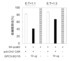

Classifications
-
- A—HUMAN NECESSITIES
- A61—MEDICAL OR VETERINARY SCIENCE; HYGIENE
- A61K—PREPARATIONS FOR MEDICAL, DENTAL OR TOILETRY PURPOSES
- A61K35/00—Medicinal preparations containing materials or reaction products thereof with undetermined constitution
- A61K35/12—Materials from mammals; Compositions comprising non-specified tissues or cells; Compositions comprising non-embryonic stem cells; Genetically modified cells
- A61K35/14—Blood; Artificial blood
- A61K35/17—Lymphocytes; B-cells; T-cells; Natural killer cells; Interferon-activated or cytokine-activated lymphocytes
-
- A—HUMAN NECESSITIES
- A61—MEDICAL OR VETERINARY SCIENCE; HYGIENE
- A61K—PREPARATIONS FOR MEDICAL, DENTAL OR TOILETRY PURPOSES
- A61K39/00—Medicinal preparations containing antigens or antibodies
- A61K39/395—Antibodies; Immunoglobulins; Immune serum, e.g. antilymphocytic serum
-
- A—HUMAN NECESSITIES
- A61—MEDICAL OR VETERINARY SCIENCE; HYGIENE
- A61K—PREPARATIONS FOR MEDICAL, DENTAL OR TOILETRY PURPOSES
- A61K38/00—Medicinal preparations containing peptides
- A61K38/16—Peptides having more than 20 amino acids; Gastrins; Somatostatins; Melanotropins; Derivatives thereof
-
- A—HUMAN NECESSITIES
- A61—MEDICAL OR VETERINARY SCIENCE; HYGIENE
- A61K—PREPARATIONS FOR MEDICAL, DENTAL OR TOILETRY PURPOSES
- A61K39/00—Medicinal preparations containing antigens or antibodies
- A61K39/46—Cellular immunotherapy
- A61K39/461—Cellular immunotherapy characterised by the cell type used
- A61K39/4611—T-cells, e.g. tumor infiltrating lymphocytes [TIL], lymphokine-activated killer cells [LAK] or regulatory T cells [Treg]
-
- A—HUMAN NECESSITIES
- A61—MEDICAL OR VETERINARY SCIENCE; HYGIENE
- A61K—PREPARATIONS FOR MEDICAL, DENTAL OR TOILETRY PURPOSES
- A61K39/00—Medicinal preparations containing antigens or antibodies
- A61K39/46—Cellular immunotherapy
- A61K39/463—Cellular immunotherapy characterised by recombinant expression
- A61K39/4631—Chimeric Antigen Receptors [CAR]
-
- A—HUMAN NECESSITIES
- A61—MEDICAL OR VETERINARY SCIENCE; HYGIENE
- A61K—PREPARATIONS FOR MEDICAL, DENTAL OR TOILETRY PURPOSES
- A61K39/00—Medicinal preparations containing antigens or antibodies
- A61K39/46—Cellular immunotherapy
- A61K39/464—Cellular immunotherapy characterised by the antigen targeted or presented
- A61K39/4643—Vertebrate antigens
- A61K39/4644—Cancer antigens
- A61K39/464499—Undefined tumor antigens, e.g. tumor lysate or antigens targeted by cells isolated from tumor
-
- A—HUMAN NECESSITIES
- A61—MEDICAL OR VETERINARY SCIENCE; HYGIENE
- A61P—SPECIFIC THERAPEUTIC ACTIVITY OF CHEMICAL COMPOUNDS OR MEDICINAL PREPARATIONS
- A61P35/00—Antineoplastic agents
-
- C—CHEMISTRY; METALLURGY
- C07—ORGANIC CHEMISTRY
- C07K—PEPTIDES
- C07K14/00—Peptides having more than 20 amino acids; Gastrins; Somatostatins; Melanotropins; Derivatives thereof
- C07K14/435—Peptides having more than 20 amino acids; Gastrins; Somatostatins; Melanotropins; Derivatives thereof from animals; from humans
- C07K14/705—Receptors; Cell surface antigens; Cell surface determinants
- C07K14/70503—Immunoglobulin superfamily
- C07K14/7051—T-cell receptor (TcR)-CD3 complex
-
- C—CHEMISTRY; METALLURGY
- C07—ORGANIC CHEMISTRY
- C07K—PEPTIDES
- C07K14/00—Peptides having more than 20 amino acids; Gastrins; Somatostatins; Melanotropins; Derivatives thereof
- C07K14/435—Peptides having more than 20 amino acids; Gastrins; Somatostatins; Melanotropins; Derivatives thereof from animals; from humans
- C07K14/705—Receptors; Cell surface antigens; Cell surface determinants
- C07K14/70503—Immunoglobulin superfamily
- C07K14/70521—CD28, CD152
-
- C—CHEMISTRY; METALLURGY
- C07—ORGANIC CHEMISTRY
- C07K—PEPTIDES
- C07K14/00—Peptides having more than 20 amino acids; Gastrins; Somatostatins; Melanotropins; Derivatives thereof
- C07K14/435—Peptides having more than 20 amino acids; Gastrins; Somatostatins; Melanotropins; Derivatives thereof from animals; from humans
- C07K14/705—Receptors; Cell surface antigens; Cell surface determinants
- C07K14/70578—NGF-receptor/TNF-receptor superfamily, e.g. CD27, CD30, CD40, CD95
-
- C—CHEMISTRY; METALLURGY
- C07—ORGANIC CHEMISTRY
- C07K—PEPTIDES
- C07K16/00—Immunoglobulins [IGs], e.g. monoclonal or polyclonal antibodies
- C07K16/18—Immunoglobulins [IGs], e.g. monoclonal or polyclonal antibodies against material from animals or humans
-
- C—CHEMISTRY; METALLURGY
- C07—ORGANIC CHEMISTRY
- C07K—PEPTIDES
- C07K16/00—Immunoglobulins [IGs], e.g. monoclonal or polyclonal antibodies
- C07K16/18—Immunoglobulins [IGs], e.g. monoclonal or polyclonal antibodies against material from animals or humans
- C07K16/28—Immunoglobulins [IGs], e.g. monoclonal or polyclonal antibodies against material from animals or humans against receptors, cell surface antigens or cell surface determinants
- C07K16/2803—Immunoglobulins [IGs], e.g. monoclonal or polyclonal antibodies against material from animals or humans against receptors, cell surface antigens or cell surface determinants against the immunoglobulin superfamily
- C07K16/2809—Immunoglobulins [IGs], e.g. monoclonal or polyclonal antibodies against material from animals or humans against receptors, cell surface antigens or cell surface determinants against the immunoglobulin superfamily against the T-cell receptor (TcR)-CD3 complex
-
- C—CHEMISTRY; METALLURGY
- C07—ORGANIC CHEMISTRY
- C07K—PEPTIDES
- C07K16/00—Immunoglobulins [IGs], e.g. monoclonal or polyclonal antibodies
- C07K16/18—Immunoglobulins [IGs], e.g. monoclonal or polyclonal antibodies against material from animals or humans
- C07K16/28—Immunoglobulins [IGs], e.g. monoclonal or polyclonal antibodies against material from animals or humans against receptors, cell surface antigens or cell surface determinants
- C07K16/30—Immunoglobulins [IGs], e.g. monoclonal or polyclonal antibodies against material from animals or humans against receptors, cell surface antigens or cell surface determinants from tumour cells
- C07K16/303—Liver or Pancreas
-
- C—CHEMISTRY; METALLURGY
- C07—ORGANIC CHEMISTRY
- C07K—PEPTIDES
- C07K16/00—Immunoglobulins [IGs], e.g. monoclonal or polyclonal antibodies
- C07K16/42—Immunoglobulins [IGs], e.g. monoclonal or polyclonal antibodies against immunoglobulins
- C07K16/4283—Immunoglobulins [IGs], e.g. monoclonal or polyclonal antibodies against immunoglobulins against an allotypic or isotypic determinant on Ig
-
- C—CHEMISTRY; METALLURGY
- C12—BIOCHEMISTRY; BEER; SPIRITS; WINE; VINEGAR; MICROBIOLOGY; ENZYMOLOGY; MUTATION OR GENETIC ENGINEERING
- C12N—MICROORGANISMS OR ENZYMES; COMPOSITIONS THEREOF; PROPAGATING, PRESERVING, OR MAINTAINING MICROORGANISMS; MUTATION OR GENETIC ENGINEERING; CULTURE MEDIA
- C12N15/00—Mutation or genetic engineering; DNA or RNA concerning genetic engineering, vectors, e.g. plasmids, or their isolation, preparation or purification; Use of hosts therefor
- C12N15/09—Recombinant DNA-technology
- C12N15/11—DNA or RNA fragments; Modified forms thereof; Non-coding nucleic acids having a biological activity
- C12N15/62—DNA sequences coding for fusion proteins
-
- C—CHEMISTRY; METALLURGY
- C12—BIOCHEMISTRY; BEER; SPIRITS; WINE; VINEGAR; MICROBIOLOGY; ENZYMOLOGY; MUTATION OR GENETIC ENGINEERING
- C12N—MICROORGANISMS OR ENZYMES; COMPOSITIONS THEREOF; PROPAGATING, PRESERVING, OR MAINTAINING MICROORGANISMS; MUTATION OR GENETIC ENGINEERING; CULTURE MEDIA
- C12N15/00—Mutation or genetic engineering; DNA or RNA concerning genetic engineering, vectors, e.g. plasmids, or their isolation, preparation or purification; Use of hosts therefor
- C12N15/09—Recombinant DNA-technology
- C12N15/63—Introduction of foreign genetic material using vectors; Vectors; Use of hosts therefor; Regulation of expression
-
- C—CHEMISTRY; METALLURGY
- C12—BIOCHEMISTRY; BEER; SPIRITS; WINE; VINEGAR; MICROBIOLOGY; ENZYMOLOGY; MUTATION OR GENETIC ENGINEERING
- C12N—MICROORGANISMS OR ENZYMES; COMPOSITIONS THEREOF; PROPAGATING, PRESERVING, OR MAINTAINING MICROORGANISMS; MUTATION OR GENETIC ENGINEERING; CULTURE MEDIA
- C12N5/00—Undifferentiated human, animal or plant cells, e.g. cell lines; Tissues; Cultivation or maintenance thereof; Culture media therefor
- C12N5/06—Animal cells or tissues; Human cells or tissues
- C12N5/0602—Vertebrate cells
- C12N5/0634—Cells from the blood or the immune system
- C12N5/0636—T lymphocytes
-
- C—CHEMISTRY; METALLURGY
- C12—BIOCHEMISTRY; BEER; SPIRITS; WINE; VINEGAR; MICROBIOLOGY; ENZYMOLOGY; MUTATION OR GENETIC ENGINEERING
- C12N—MICROORGANISMS OR ENZYMES; COMPOSITIONS THEREOF; PROPAGATING, PRESERVING, OR MAINTAINING MICROORGANISMS; MUTATION OR GENETIC ENGINEERING; CULTURE MEDIA
- C12N5/00—Undifferentiated human, animal or plant cells, e.g. cell lines; Tissues; Cultivation or maintenance thereof; Culture media therefor
- C12N5/10—Cells modified by introduction of foreign genetic material
-
- A—HUMAN NECESSITIES
- A61—MEDICAL OR VETERINARY SCIENCE; HYGIENE
- A61K—PREPARATIONS FOR MEDICAL, DENTAL OR TOILETRY PURPOSES
- A61K2239/00—Indexing codes associated with cellular immunotherapy of group A61K39/46
- A61K2239/10—Indexing codes associated with cellular immunotherapy of group A61K39/46 characterized by the structure of the chimeric antigen receptor [CAR]
- A61K2239/11—Antigen recognition domain
- A61K2239/13—Antibody-based
-
- A—HUMAN NECESSITIES
- A61—MEDICAL OR VETERINARY SCIENCE; HYGIENE
- A61K—PREPARATIONS FOR MEDICAL, DENTAL OR TOILETRY PURPOSES
- A61K2239/00—Indexing codes associated with cellular immunotherapy of group A61K39/46
- A61K2239/31—Indexing codes associated with cellular immunotherapy of group A61K39/46 characterized by the route of administration
-
- A—HUMAN NECESSITIES
- A61—MEDICAL OR VETERINARY SCIENCE; HYGIENE
- A61K—PREPARATIONS FOR MEDICAL, DENTAL OR TOILETRY PURPOSES
- A61K2239/00—Indexing codes associated with cellular immunotherapy of group A61K39/46
- A61K2239/38—Indexing codes associated with cellular immunotherapy of group A61K39/46 characterised by the dose, timing or administration schedule
-
- A—HUMAN NECESSITIES
- A61—MEDICAL OR VETERINARY SCIENCE; HYGIENE
- A61K—PREPARATIONS FOR MEDICAL, DENTAL OR TOILETRY PURPOSES
- A61K2300/00—Mixtures or combinations of active ingredients, wherein at least one active ingredient is fully defined in groups A61K31/00 - A61K41/00
-
- A—HUMAN NECESSITIES
- A61—MEDICAL OR VETERINARY SCIENCE; HYGIENE
- A61K—PREPARATIONS FOR MEDICAL, DENTAL OR TOILETRY PURPOSES
- A61K38/00—Medicinal preparations containing peptides
-
- C—CHEMISTRY; METALLURGY
- C07—ORGANIC CHEMISTRY
- C07K—PEPTIDES
- C07K2317/00—Immunoglobulins specific features
- C07K2317/30—Immunoglobulins specific features characterized by aspects of specificity or valency
- C07K2317/31—Immunoglobulins specific features characterized by aspects of specificity or valency multispecific
-
- C—CHEMISTRY; METALLURGY
- C07—ORGANIC CHEMISTRY
- C07K—PEPTIDES
- C07K2317/00—Immunoglobulins specific features
- C07K2317/50—Immunoglobulins specific features characterized by immunoglobulin fragments
- C07K2317/52—Constant or Fc region; Isotype
- C07K2317/522—CH1 domain
-
- C—CHEMISTRY; METALLURGY
- C07—ORGANIC CHEMISTRY
- C07K—PEPTIDES
- C07K2317/00—Immunoglobulins specific features
- C07K2317/50—Immunoglobulins specific features characterized by immunoglobulin fragments
- C07K2317/52—Constant or Fc region; Isotype
- C07K2317/524—CH2 domain
-
- C—CHEMISTRY; METALLURGY
- C07—ORGANIC CHEMISTRY
- C07K—PEPTIDES
- C07K2317/00—Immunoglobulins specific features
- C07K2317/50—Immunoglobulins specific features characterized by immunoglobulin fragments
- C07K2317/52—Constant or Fc region; Isotype
- C07K2317/526—CH3 domain
-
- C—CHEMISTRY; METALLURGY
- C07—ORGANIC CHEMISTRY
- C07K—PEPTIDES
- C07K2317/00—Immunoglobulins specific features
- C07K2317/60—Immunoglobulins specific features characterized by non-natural combinations of immunoglobulin fragments
- C07K2317/62—Immunoglobulins specific features characterized by non-natural combinations of immunoglobulin fragments comprising only variable region components
- C07K2317/622—Single chain antibody (scFv)
-
- C—CHEMISTRY; METALLURGY
- C07—ORGANIC CHEMISTRY
- C07K—PEPTIDES
- C07K2317/00—Immunoglobulins specific features
- C07K2317/70—Immunoglobulins specific features characterized by effect upon binding to a cell or to an antigen
- C07K2317/71—Decreased effector function due to an Fc-modification
-
- C—CHEMISTRY; METALLURGY
- C07—ORGANIC CHEMISTRY
- C07K—PEPTIDES
- C07K2317/00—Immunoglobulins specific features
- C07K2317/70—Immunoglobulins specific features characterized by effect upon binding to a cell or to an antigen
- C07K2317/73—Inducing cell death, e.g. apoptosis, necrosis or inhibition of cell proliferation
-
- C—CHEMISTRY; METALLURGY
- C07—ORGANIC CHEMISTRY
- C07K—PEPTIDES
- C07K2317/00—Immunoglobulins specific features
- C07K2317/90—Immunoglobulins specific features characterized by (pharmaco)kinetic aspects or by stability of the immunoglobulin
- C07K2317/92—Affinity (KD), association rate (Ka), dissociation rate (Kd) or EC50 value
-
- C—CHEMISTRY; METALLURGY
- C07—ORGANIC CHEMISTRY
- C07K—PEPTIDES
- C07K2319/00—Fusion polypeptide
- C07K2319/01—Fusion polypeptide containing a localisation/targetting motif
- C07K2319/02—Fusion polypeptide containing a localisation/targetting motif containing a signal sequence
-
- C—CHEMISTRY; METALLURGY
- C07—ORGANIC CHEMISTRY
- C07K—PEPTIDES
- C07K2319/00—Fusion polypeptide
- C07K2319/01—Fusion polypeptide containing a localisation/targetting motif
- C07K2319/03—Fusion polypeptide containing a localisation/targetting motif containing a transmembrane segment
-
- C—CHEMISTRY; METALLURGY
- C12—BIOCHEMISTRY; BEER; SPIRITS; WINE; VINEGAR; MICROBIOLOGY; ENZYMOLOGY; MUTATION OR GENETIC ENGINEERING
- C12N—MICROORGANISMS OR ENZYMES; COMPOSITIONS THEREOF; PROPAGATING, PRESERVING, OR MAINTAINING MICROORGANISMS; MUTATION OR GENETIC ENGINEERING; CULTURE MEDIA
- C12N2501/00—Active agents used in cell culture processes, e.g. differentation
- C12N2501/20—Cytokines; Chemokines
- C12N2501/23—Interleukins [IL]
- C12N2501/2302—Interleukin-2 (IL-2)
-
- C—CHEMISTRY; METALLURGY
- C12—BIOCHEMISTRY; BEER; SPIRITS; WINE; VINEGAR; MICROBIOLOGY; ENZYMOLOGY; MUTATION OR GENETIC ENGINEERING
- C12N—MICROORGANISMS OR ENZYMES; COMPOSITIONS THEREOF; PROPAGATING, PRESERVING, OR MAINTAINING MICROORGANISMS; MUTATION OR GENETIC ENGINEERING; CULTURE MEDIA
- C12N2501/00—Active agents used in cell culture processes, e.g. differentation
- C12N2501/50—Cell markers; Cell surface determinants
- C12N2501/515—CD3, T-cell receptor complex
-
- C—CHEMISTRY; METALLURGY
- C12—BIOCHEMISTRY; BEER; SPIRITS; WINE; VINEGAR; MICROBIOLOGY; ENZYMOLOGY; MUTATION OR GENETIC ENGINEERING
- C12N—MICROORGANISMS OR ENZYMES; COMPOSITIONS THEREOF; PROPAGATING, PRESERVING, OR MAINTAINING MICROORGANISMS; MUTATION OR GENETIC ENGINEERING; CULTURE MEDIA
- C12N2510/00—Genetically modified cells
-
- C—CHEMISTRY; METALLURGY
- C12—BIOCHEMISTRY; BEER; SPIRITS; WINE; VINEGAR; MICROBIOLOGY; ENZYMOLOGY; MUTATION OR GENETIC ENGINEERING
- C12N—MICROORGANISMS OR ENZYMES; COMPOSITIONS THEREOF; PROPAGATING, PRESERVING, OR MAINTAINING MICROORGANISMS; MUTATION OR GENETIC ENGINEERING; CULTURE MEDIA
- C12N2533/00—Supports or coatings for cell culture, characterised by material
- C12N2533/50—Proteins
- C12N2533/52—Fibronectin; Laminin
Definitions
- the present disclosure describes chimeric receptors, cells expressing chimeric receptors, and methods for treating diseases using the cells, particularly adoptive immuno-cell therapy, adoptive immuno-T cell therapy, CAR-T therapy and T using the cells. Regarding cell redirecting antibodies.
- Chimeric Antigen Receptor artificially fuses an antibody that recognizes a cell surface antigen such as cancer cells with a signal transduction region that induces activation of T cells. It is a chimeric protein.
- CAR-expressing T cells (hereinafter, also simply referred to as “CAR-T cells”) can be produced. It is made.
- CAR-expressing T cells produced by such a method are used for the treatment of diseases such as cancer by adoptive immunotherapy.
- Such CAR-T cells are reactive to antigen-expressing target cells and can lead to target cell damage independent of interaction with major histocompatibility complex (MHC). ..
- MHC major histocompatibility complex
- Non-Patent Document 1 Cancer immunotherapy by administration of CAR-T cells, more specifically, therapy in which T cells are collected from a patient, a gene encoding CAR is introduced into the T cells, expanded and cultured, and then transferred to the patient again. , Clinical trials are being conducted all over the world (Non-Patent Document 1). Results have shown that cancer immunotherapy by administration of CAR-T cells is effective in hematopoietic malignancies such as leukemia and lymphoma.
- CD19-antigen CAR-T Kymriah® Novartis, tisagenlecleucel, CTL-019, CD3 Zeta-CD137
- Yescarta® KiTE, axicabtagene ciloleucel, CD3 Zeta-CD28
- Non-Patent Document 5 Non-Patent Document 5
- T cell-redirecting antibody which is an antibody whose antitumor effect is based on cytotoxicity that mobilizes T cells as effector cells
- T cell redirecting antibodies are any of the constituent subsystems of the T cell receptor (TCR) complex on T cells. It is a bispecific antibody that contains a binding domain to a cell, particularly a domain that binds to a CD3 epsilon chain and a domain that binds to an antigen on a target cancer cell.
- CAR-T cells Although therapy with CAR-T cells is promising in the treatment of cancer patients, there are some limitations to the expansion of clinical application of CAR-T cells.
- the economic cost and labor involved in the operation for identifying antigen recognition sites for various tumor antigens and newly establishing CAR-T cells into which they have been introduced are major issues.
- tumor antigens targeted by CAR may cause tumor immunoavoidance by reducing their expression levels or mutating depending on the treatment. In particular, current CAR-T cells recognize only one target antigen, so tumor immunoavoidance causes diminished or extinct therapeutic efficacy.
- CAR-T cells and T cell redirecting antibodies that can change the tumor antigen to be recognized according to the treatment stage, and treatment methods using them have been studied, but sufficient therapeutic effect when administered to patients. There is a demand for cheaper treatment due to the development of a treatment method having high safety and a technology that can be used for general purposes.
- the pharmaceutical composition comprises cells expressing the chimeric receptor.
- Chimeric receptors include extracellular binding domains, transmembrane domains and intracellular signaling domains.
- the mutant antibody can bind to the extracellular binding domain of the chimeric receptor via a portion containing the mutation in the extracellular binding domain.
- the pharmaceutical composition, wherein the extracellular binding domain does not specifically bind to an antibody that does not contain the mutation.
- the pharmaceutical composition comprises a mutated antibody having a mutation in the CH1 region, CH2 region, CH3 region, CL region, or framework region that comprises at least one amino acid substitution, deletion, addition, or modification.
- Chimeric receptors include extracellular binding domains, transmembrane domains and intracellular signaling domains.
- the mutant antibody can bind to the extracellular binding domain of the chimeric receptor via a portion containing the mutation in the extracellular binding domain.
- the pharmaceutical composition wherein the extracellular binding domain does not specifically bind to an antibody that does not contain the mutation.
- the pharmaceutical composition comprises a bispecific antibody
- Bispecific antibodies are: (1) a domain containing an antibody variable region that specifically binds to a mutant antibody via a portion containing the mutation, and (2) an antibody having binding activity to a molecule expressed on the surface of a T cell.
- the pharmaceutical composition comprising a domain comprising a variable region and not specifically binding to a mutation-free antibody.
- a pharmaceutical composition for use in combination with administration of a bispecific antibody comprises a mutated antibody having a mutation in the CH1 region, CH2 region, CH3 region, CL region, or framework region that comprises at least one amino acid substitution, deletion, addition, or modification.
- Bispecific antibodies are: (1) a domain containing an antibody variable region that specifically binds to a mutant antibody via a portion containing the mutation, and (2) an antibody having binding activity to a molecule expressed on the surface of a T cell.
- the pharmaceutical composition comprising a domain comprising a variable region and not specifically binding to a mutation-free antibody. [5] Any of [1] to [4], wherein the mutant antibody contains the mutation in the CH2 region, and the mutant antibody has a reduced binding activity to the Fc gamma receptor and C1q as compared with the corresponding non-mutant antibody.
- the pharmaceutical composition described in Crab The pharmaceutical composition described in Crab.
- mutant antibodies were 234, 235, 236, 237, 238, 265, 266, 267, 268, 269, 270, 271, 295, 296, 298, 300, 324, 325, 326, 327, It has a mutation in the CH2 region at any of the positions represented by 328, 329, 330, 331, 332, 333, 334, 335, 336, and 337, and the mutant antibody is extracellular through the portion containing the mutation.
- the pharmaceutical composition according to any one of [1] to [5], which binds to a binding domain.
- the CH2 region of the mutant antibody is determined by EU numbering.
- the amino acid at position 235 is arginine
- the amino acid at position 236 is arginine
- the amino acid at position 239 is ricin
- the amino acid at position 250 is valine
- the amino acid at position 252 is tyrosine
- the amino acid at position 297 is alanine
- the amino acid at position 307 is glutamine
- the amino acid at position 308 is proline
- the amino acid at position 311 is alanine
- the amino acid at position 434 is tyrosine, and Any of [1] to [6], which has a mutation selected from the group in which the amino acid at position 436 is valine, and the mutant antibody binds to the extracellular binding domain via the portion containing the mutation.
- the pharmaceutical composition according to.
- the vector according to [9] which can be operably linked to at least one regulatory element for expression of a chimeric receptor or bispecific antibody.
- [A1-1] A chimeric receptor containing an extracellular binding domain, a transmembrane domain, and an intracellular signal transduction domain. It is possible for the extracellular binding domain to specifically bind to a mutant antibody having a mutation containing at least one amino acid substitution, deletion, addition or modification in the CH2 region via the portion containing the mutation. The chimeric receptor that does not specifically bind to a mutation-free antibody.
- a mutant antibody is an antibody in which the binding activity to any Fc ⁇ receptor of Fc ⁇ I, Fc ⁇ IIA, Fc ⁇ IIB, Fc ⁇ IIIA and Fc ⁇ IIIB is reduced as compared with the corresponding non-mutant antibody [A1-1].
- the chimeric receptor according to [A1-2].
- Mutations are represented by EU numbering positions, 231, 232, 233, 234, 235, 236, 237, 238, 239, 240, 264, 265, 266, 267, 269, 270.
- the [A1-3-2] mutation has one or more mutations selected from the group of 234A, 235A, and 297A, which are represented by the position by EU numbering and the amino acid after the mutation, and the portion containing the mutation.
- the chimeric receptor according to any one of [A1-1] to [A1-3], wherein the extracellular binding domain can bind to the mutant antibody via.
- the [A1-3-3] mutation further comprises one or more mutations selected from the group 349C, 356C, 366W or S, 368A, 407V, represented by EU numbered position and post-mutation amino acid.
- the chimeric receptor according to any one of [A1-1] to [A1-3], wherein the extracellular binding domain can bind to the mutant antibody via the portion containing the mutation.
- the mutation is represented by the position by EU numbering and the amino acid after the mutation, the following combination: (1) 235R and 239K; (2) 235R and 236R; (3) 235R, 239K and 297A; (4) 235R, 236R and 239K; (5) 252Y and 434Y; (6) 235R, 239K, 252Y and 434Y; (7) 252Y, 434Y and 436V; (8) 235R, 239K, 252Y, 434Y and 436V; (9) 250V, 252Y, 307Q, 308P, 311A, 434Y and 436V; and (10) 235R, 239K, 250V, 252Y, 307Q, 308P, 311A, 434Y and 436V; and (10) 235R, 239K, 250V, 252Y, 307Q, 308P, 311A, 434Y and 436V.
- the chimeric according to any one of [A1-1] to
- the mutation has a mutation at one or more positions selected from the group 235, 236, and 239, which are indicated by EU numbering positions, and through the portion containing the mutation, The chimeric receptor according to any one of [A1-1] to [A1-3], wherein the extracellular binding domain binds to a mutant antibody.
- Mutations are represented by EU numbering positions, 234, 235, 236, 237, 238, 265, 266, 267, 268, 269, 270, 271, 295, 296, 298, 300.
- a mutant antibody is an antibody in which the binding activity to any Fc ⁇ receptor of Fc ⁇ I, Fc ⁇ IIA, Fc ⁇ IIB, Fc ⁇ IIIA and Fc ⁇ IIIB is enhanced as compared with the corresponding non-mutant antibody [A1-1].
- [A1-5-1] Mutations are represented by EU numbering positions, 231, 232, 233, 234, 235, 236, 237, 238, 239, 240, 265, 266, 267, 268, 269, 270. , 271, 295, 296, 298, 300, 324, 325, 326, 327, 328, 329, 330, 331, 332, 333, 334, 335, 336 and 337 mutations at one or more positions selected from the group
- the chimeric receptor according to either [A1-1] or [A1-5], wherein the extracellular binding domain can bind to the mutant antibody through the portion containing the mutation.
- the mutation is represented by the position by EU numbering and the amino acid after the mutation, the following combination: (1) 234Y, 235Y, 236W, 268D, 270E and 298A; (2) 234Y, 235Q, 236W, 239M, 268D, 270E and 298A; (3) 234Y, 235Q, 236W, 239M, 268D, 270E and 298A; (4) 234Y, 235Y, 236W, 268D, 298A and 327D; (5) 234Y, 235Y, 236W, 239M, 268D, 298A and 327D; (6) 234Y, 235Y, 236W, 239M, 268D, 298A, 327D, 328W and 334L; (7) 326D, 330M and 334E; (8) 270E, 326D, 330M and 334E; and (9) 270E, 326D, 330K and 334
- the mutation is represented by the position by EU numbering and the amino acid after the mutation, the following combination: (1) 234L, S, F, E, V, D, Q, I, M, T, A, G or H, 235Y or Q, 236W, 239M or I, 268D, and 298A; (2) 234L, S, F, E, V, D, Q, I, M, T, A, G or H, 235Y or Q, 236W, 239M or I, 268D, 298A and 327D; (3) 234F, E, D, S or L, 235Y or Q, 236W, 239M or I, 268D, 298A and 327D; (4) 270E, 326D, 330A, K, M, F, I, Y or H, and 334E; (5) 270E, 326D, 330A, K, M, F, I, Y or H, and 334E; (6) 270E, 326D, 330A, K
- the mutant antibody maintains or decreases the binding activity to both H-type and R-type, which are gene polymorphisms of Fc ⁇ RIIa, and the binding activity to Fc ⁇ RIIb as compared with the corresponding non-mutated antibody.
- the chimeric receptor according to [A1-1] which is an antibody enhanced by.
- the [A1-6-1] mutation is one or more selected from the group of 233, 234, 237, 238, 239, 267, 268, 296, 271, 323, 326, 330, represented by EU numbering position.
- the chimeric receptor according to either [A1-1] or [A1-6] which has a mutation at the position of and is capable of binding the extracellular binding domain to the mutant antibody through the portion containing the mutation. body.
- [A1-6-2] Mutations are represented by EU numbered positions and post-mutation amino acids, 238D, 328E, 237W, 267V, 267Q, 268N, 271G, 326M, 239D, 267A, 234W, 237A, 237D, Has one or more mutations selected from the group 237E, 237L, 237M, 237Y, 330K, 330R, 233D, 268D, 268E, 326D, 326S, 326T, 323I, 323L, 323M, 296D, 326A, 326N, 330M , The chimeric receptor according to either [A1-1] or [A1-6], wherein the extracellular binding domain can bind to the mutant antibody through the portion containing the mutation.
- Mutations include mutations at position 238, represented by EU numbered positions, and are selected from 235, 237, 241, 268, 295, 296, 298, 323, 324 and 330.
- the mutation contains a mutation at one or more positions, and the mutation reduces the binding activity to all active Fc ⁇ Rs, and the extracellular binding domain can bind to the mutant antibody through the portion containing the mutation.
- [A1-6-5] mutations include mutations at position 238, represented by EU numbered positions, and (1) 241, 268, 296 and 324; (2) 237, 241, 296 and 330.
- [A1-6-7] mutations are represented by EU numbering positions: (1) 233, 238, 264, 267, 268 and 271; (2) 233, 237, 238, 264, 267, 268, 271 , 296, 297, 330 and 396; (3) Includes mutations in the position of a combination selected from the group consisting of 233, 238, 264, 267, 268, 271 and 296, and by mutation binding to all active Fc ⁇ Rs.
- the chimeric receptor according to either [A1-1] or [A1-6], wherein the activity is reduced and the extracellular binding domain can bind to the mutant antibody through the portion containing the mutation. ..
- the [A1-6-8] mutation comprises 238D, represented by the position by EU numbering and the post-mutation amino acid, as well as 235F, 237Q or D, 241M or L, 268P, 295M or V, 296E, H, Contains one or more mutations selected from N or D, 298A or M, 323I, 324N or H, and 330H or Y, and the mutations reduce the binding activity to all active Fc ⁇ Rs and include such mutations.
- the chimeric receptor according to either [A1-1] or [A1-6], wherein the extracellular binding domain can bind to the mutant antibody via a moiety.
- the [A1-6-9] mutation comprises 238D, represented by the position by EU numbering and the post-mutation amino acid, and (1) 241M, 268P, 296E and 324H; (2) 237Q or D, 241M, 296E and 330H; (3) Includes mutations in a combination selected from the group consisting of 235F, 237Q or D, 241M and 296E, and the mutations reduce the binding activity to all active Fc ⁇ Rs and include such mutations.
- the chimeric receptor according to either [A1-1] or [A1-6], wherein the extracellular binding domain can bind to the mutant antibody via a moiety.
- the [A1-6-10] mutations include 238D and 271G, represented by EU numbered positions and post-mutation amino acids, as well as 234A, H, N, K or R, 235A, 236Q, 237R or K, Contains two or more mutations selected from 239K, 265K, N, R, S or V, 267K, R or Y, and 297A, and the mutations reduce the binding activity to all active Fc ⁇ Rs.
- the chimeric receptor according to either [A1-1] or [A1-6], wherein the extracellular binding domain can bind to the mutant antibody via a portion containing.
- Mutations include 238D and 271G, represented by EU numbered positions and post-mutation amino acids, and (1) 233D, 238D, 264I, 267R, 268E and 271G; (2). 233D, 237D, 238D, 264I, 267A, 268E, 271G, 296D, 297A, 330R and 396M; (3) Includes mutations in combinations selected from the group 233D, 238D, 264I, 267R, 268P, 271G and 296E, and , The mutation reduces the binding activity to all active Fc ⁇ Rs, and the extracellular binding domain can bind to the mutant antibody through the portion containing the mutation, [A1-1] or [A1-6].
- [A1-6-13] Mutations include mutations at one or more positions selected from the group 322, or 327, 330 and 331, represented by EU numbered positions, which bind to complement by mutation.
- the chimeric receptor according to [A1-1] or [A1-2], wherein is reduced and the extracellular binding domain can bind to the mutated antibody through the portion containing the mutation.
- the [A1-6-14] mutation comprises one or more mutations selected from the group 322A or E, 327G, 330S and 331S, represented by the position by EU numbering and the amino acid after the mutation, and includes such mutation.
- [A1-6-15] mutations include mutations at one or more positions selected from the group 238, 271, 327, 330, 331, represented by EU numbered positions, in the Fc region of native IgG.
- the [A1-6-16] mutation has one or more mutations selected from the group 238D, 271G, 327G, 330S and 331S, represented by the position by EU numbering and the amino acid after the mutation.
- the [A1-6-17] mutant antibody further comprises one or more mutations selected from the group 233D, 237D, 264I, 267A, 268D or E, and the extracellular binding domain through the portion containing the mutation.
- the [A1-6-18] mutation comprises 238D and 271G, represented by EU numbering position and post-mutation amino acid, and (1) 237D, 238D, 268D or E, 271G, 327G, 330S and 331S; (2) 233D, 237D, 238D, 268D, 271G, 327G, 330S and 331S; (3) 238D, 267A, 268E, 271G, 327G, 330S and 331S; (4) 238D, 264I, 267A, 271G, 327G, 330S [A1-1] or [A1-6], which comprises a mutation in the position of a combination selected from the group consisting of 331S and 331S, through which the extracellular binding domain can bind to the mutant antibody.
- the [A1-6-19] mutation comprises one or more mutations selected from the group 233, 238, 264, 267, 268, 271, 327, 330 and 331, expressed in EU numbered positions.
- the chimeric receptor according to [A1-1] or [A1-6], wherein the extracellular binding domain can bind to the mutant antibody via the mutation-containing moiety.
- [A1-6-20] mutations are represented by EU numbering positions: (1) 237, 238, 268, 271, 327, 330 and 331; (2) 233, 237, 238, 268, 271, 327 , 330 and 331; (3) 238, 267, 268, 271, 327, 330 and 331; (4) Mutations in the position of one or more combinations selected from 238, 264, 267, 271, 327, 330 and 331.
- the chimeric receptor according to [A1-1] or [A1-6], wherein the extracellular binding domain can bind to the mutant antibody through the portion containing the mutation.
- the mutant antibody Compared with the corresponding non-mutated antibody, the mutant antibody has different binding activity to the antigen depending on the ion concentration condition and has the binding activity to FcRn under the condition of neutral pH, but in pH.
- the chimeric receptor according to [A1-1] which is an antibody that does not form a heterocomplex containing two molecules of FcRn and one molecule of active Fc ⁇ receptor under sexual conditions.
- the [A1-7-1] mutation comprises the mutation at one or more positions selected from the group 235, 237, 238, 239, 270, 298, 325 and 329, represented by EU numbered positions.
- [A1-1] or [A1-7] wherein the extracellular binding domain can bind to the mutant antibody through the portion containing one or more mutations selected from the group. Chimeric receptor.
- a chimeric receptor comprising an extracellular binding domain, a transmembrane domain and an intracellular signaling domain, wherein the extracellular binding domain substitutes, deletes, adds or modifies at least one amino acid in the CH3 region.
- the chimeric receptor that can specifically bind to a mutant antibody having a mutation containing the above mutation through a portion containing the mutation and does not specifically bind to an antibody not containing the mutation.
- Mutant antibodies improve stability, homogeneity, immunogenicity, safety, production efficiency and / or plasma retention of mutant antibodies compared to the corresponding non-mutant antibodies, [A2-2]. A2-1].
- the chimeric receptor is provided.
- [A2-3] The chimeric receptor according to [A2-1] or [A2-2], wherein the mutant antibody is an antibody lacking any amino acid at the position represented by 446 or 447 in EU numbering. ..
- the [A2-3-1] mutation may include a mutation at the position represented by EU numbering, further represented by 434, through which the extracellular binding domain binds to the mutant antibody.
- [A2-3-2] Mutations are represented by EU numbering positions and are further selected from the group 131, 133, 137, 138, 219, 268, 330, 331, 335, 339, 397, 419.
- a mutant antibody is an antibody containing a mutation in an amino acid residue at the interface so that two or more amino acid residues forming the interface have the same charge.
- the mutant antibody is an antibody containing two or more heavy chain CH3 regions, and the amino acid residue of the first heavy chain has the same charge, and the mutation is EU.
- a mutation is contained in any one of the positions of one or more combinations selected from the above, and the extracellular binding domain can bind to the mutant antibody through the portion containing the mutation, [A2-1], [A2-].
- the mutation has a mutation at one or more positions selected from the group of 356, 357, 370, 399, 409, 439, which is indicated by the position according to the EU numbering, and includes the mutation.
- the mutation is represented by the position according to the EU numbering, and the amino acid residues 10, 12, 23, 39, 43 and 105 in the Kabat numbering in the heavy chain variable region, or the weight thereof.
- a mutation is included at one or more positions selected from the group 137, 196, 203, 214, 217, 233, 268, 274, 276 and 297 in EU numbering in the chain constant region, and through the portion containing the mutation,
- the chimeric receptor according to any one of [A2-4-1] to [A2-4-3], wherein the extracellular binding domain can bind to a mutant antibody.
- [A2-5] The chimeric antibody according to [A2-1] or [A2-2], wherein the mutant antibody is an antibody that promotes heterogeneity of the polypeptide under reducing conditions as compared with the corresponding non-mutant antibody. Receptor.
- the mutant antibody is an antibody containing two or more heavy chain CH3 regions, and the amino acid residue of the first heavy chain is a mutation having the same charge, and the mutation is EU.
- the chimeric receptor is an antibody containing two or more heavy chain CH3 regions, and the amino acid residue of the first heavy chain is a mutation having the same charge, and the mutation is EU.
- [A2-5-2] The group of 397M, F or Y, 392D, E, T, V, or I, 356K, 397F or Y, 439E, in which the mutation is represented by the position by EU numbering and the amino acid after the mutation.
- [A2-1], [A2-2] or [A2-] which has one or more mutations selected from the above and can bind the extracellular binding domain to the mutant antibody through the portion containing the mutation.
- the mutant antibody is an antibody containing two or more heavy chain CH3 regions and at any of the positions represented by 349, 351, 354, 356, 394 and 407 in EU numbering.
- [A2-5-4] In EU numbering, a mutation or a defect in the core hinge region at any of the positions represented by 226 and / or 229 is included, and the extracellular binding domain is mutated through the portion containing the mutation.
- the chimeric receptor according to [A2-1], [A2-2] or [A2-5] which can bind to.
- Mutant antibody is an antibody in which 220-225 is further replaced with YGP by EU numbering or 219-229 is deficient in EU numbering, [A2 -5-4].
- the chimeric receptor [A2-6]
- the mutant antibody comprises a first polypeptide and a second polypeptide that come into contact at the boundary portion, and the first polypeptide is located in the void at the boundary portion of the second polypeptide.
- the mutant antibody is an antibody containing two or more heavy chain CH3 regions, and is mutated at one or more positions selected from the group of 366, 394, 405, 407 by EU numbering.
- the mutant antibody is an antibody containing two or more types of heavy chain CH3 regions, and in EU numbering, mutations in the first and second heavy chain CH3 regions are located and mutated by EU numbering.
- the first heavy chain CH3 region is 366Y and the second heavy chain CH3 region is 407T; (2) The first heavy chain CH3 region is 366W and the second heavy chain CH3 region is 497A; (3) The first heavy chain CH3 region is 405A and the second heavy chain CH3 region is 394W; (4) The first heavy chain CH3 region is 407T and the second heavy chain CH3 region is 366Y; (5) The first heavy chain CH3 region is 366Y and 405A, and the second heavy chain CH3 region is 394W and 407T; (6) The first heavy chain CH3 region is 366W and 405W, and the second heavy chain CH3 region is 394S and 407A; (7) The first heavy chain CH3 region is 405W and 407A, and the second heavy chain CH3 region is 366W and 394S; (8) The first heavy chain CH3 region is 405W and the second heavy chain CH3 region is 394S.
- the chimeric receptor. [A3-1] A chimeric receptor containing an extracellular binding domain, a transmembrane domain, and an intracellular signal transduction domain. It is possible for the extracellular binding domain to specifically bind to a mutant antibody having a mutation containing at least one amino acid substitution, deletion, addition or modification in the CH1 region via the portion containing the mutation. The chimeric receptor that does not specifically bind to a mutation-free antibody.
- [A3-2] The chimeric receptor according to [A3-1], wherein the mutant antibody is an antibody having improved pharmacokinetics and hinge region heterogeneity as compared with the corresponding non-mutant antibody.
- the mutation has a mutation at one or more positions selected from the group 131, 133, 137, 138, which is represented by the position according to the EU numbering, and the mutation is transmitted through the portion containing the mutation.
- the chimeric receptor according to [A3-1] or [A3-2] wherein the extracellular binding domain can bind to the mutant antibody.
- [A3-2-2] One or more mutations selected from the group 131S, 133K, 220S, 137G, 138G, 268Q, 355Q and 419E, in which the mutation is represented by the position by EU numbering and the amino acid after the mutation.
- Mutations are indicated by EU numbered positions and further mutated to one or more positions selected from the group 220, 268, 330, 331, 339, 355, 419, 446, 447.
- [A4-1] A chimeric receptor containing an extracellular binding domain, a transmembrane domain, and an intracellular signal transduction domain.
- the extracellular binding domain is capable of specifically binding to a mutated antibody having a mutation containing at least one amino acid substitution, deletion, addition or modification in the hinge region via the portion containing the mutation.
- the chimeric receptor that does not specifically bind to a mutation-free antibody.
- a mutant antibody is an antibody in which the binding activity to any Fc ⁇ receptor of Fc ⁇ I, Fc ⁇ IIA, Fc ⁇ IIB, Fc ⁇ IIIA and Fc ⁇ IIIB is enhanced as compared with the corresponding non-mutant antibody [A4-1].
- the chimeric receptor described in. [A4-2-1] One or more positions where the mutation is selected from the group 220, 221, 222, 223, 224, 225, 226, 227, 228, 229, 230, represented by EU numbered positions.
- the chimeric receptor according to [A4-1] or [A4-2] which comprises a mutation in and is capable of binding an extracellular binding domain to a mutant antibody through a portion containing the mutation.
- [A4-2-2] mutations are represented by EU numbering positions, and further, 231, 232, 233, 234, 235, 236, 237, 238, 239, 240, 264, 265, 266, 267, 269. , 270, 295, 296, 297, 298, 299, 300, 325, 327, 328, 329, 330, 331, 332 containing the mutation at one or more positions selected from the group, and through the portion containing the mutation.
- the chimeric receptor according to [A4-2-1] wherein the extracellular binding domain can bind to the mutant antibody.
- the [A4-2-3] mutation comprises one or more mutations selected from the group 235R, 239K and 297A, represented by EU numbered position and post-mutation amino acid, through the portion containing the mutation.
- [A4-2-4] mutations are represented by EU numbering positions, and 231, 232, 233, 234, 235, 236, 237, 238, 239, 240, 241, 243, 244, 245, 246.
- the mutation is represented by the position by EU numbering and the amino acid after the mutation, 221K or Y, 222F, W, E or Y, 223F, W, E or K, 224F, W, E or Contains one or more mutations selected from the group Y, 225E, K or W, 227E, G, K or Y, 228E, G, K or Y, 230A, E, G or Y and contains the mutation.
- the chimeric receptor according to [A4-1] or [A4-2], wherein the extracellular binding domain can bind to the mutant antibody via the same.
- a mutant antibody is an antibody in which the binding activity to any Fc ⁇ receptor of Fc ⁇ I, Fc ⁇ IIA, Fc ⁇ IIB, Fc ⁇ IIIA and Fc ⁇ IIIB is reduced as compared with the corresponding non-mutant antibody [A4-1].
- the [A4-3-1] mutation contains the mutation at one or more positions selected from the group 220, 226, and 229, represented by EU numbered positions, and the cell is routed through the portion containing the mutation.
- the chimeric receptor according to [A4-1] or [A4-3], wherein the outer binding domain can bind to the mutant antibody.
- the extracellular binding domain can specifically bind to a mutant antibody having a mutation containing at least one amino acid substitution, deletion, addition or modification to the CH2 and CH3 regions via the portion containing the mutation.
- [A5-2-1] One or more mutations selected from the group 235, 236, 239, 327, 330, 331, 428, 434, 436, 438, and 440, represented by EU numbering position.
- the chimeric receptor according to [A5-1] or [A5-2], which contains a mutation at a position and the extracellular binding domain can bind to the mutant antibody through the portion containing the mutation.
- the chimeric receptor according to [A5-1] or [A5-2] which has an extracellular binding domain that binds to a mutant antibody through a portion containing the mutation.
- Mutations are represented by EU numbering positions, 238, 244, 245, 249, 250, 251, 252, 253, 254, 255, 256, 257, 258, 260, 262, 265. , 270, 272, 279, 283, 285, 286, 288, 293, 303, 305, 307, 308, 309, 311, 312, 314, 316, 317, 318, 332, 339, 340, 341, 343, 356 , 360, 362, 375, 376, 377, 378, 380, 382, 385, 386, 387, 388, 389, 400, 413, 415, 423, 424, 427, 428, 430, 431, 433, 434, 435 , 436, 438, 439, 440, 442 and 447 have mutations at one or more positions selected from the group, and the extracellular binding domain can bind to the mutant antibody through the portion containing the mutation.
- Mutations are represented by EU numbering positions and post-mutation amino acids. 238L, 244L, 245R, 249P, and Contains one or more mutations selected from 250Q or E, or 251R, D, E, or L, 252F, S, T, or Y, 254S or T, 255R, G, I, or L, 256A, R, N, D, Q, E, P, or T, 257A, I, M, N, S, or V, 258D, 260S, 262L, 270K, 272L, or R, 279 A, D, G, H, M, N, Q, R, S, T, W, or Y, 283A, D, F, G, H, I, K, L, N, P, Q, R, S, T, W, or Y, 285N, 286F, 288N, or P, 293V, 307A, E, Q, or
- [A5-3] The chimeric receptor according to [A5-1], wherein the mutant antibody is an antibody in which the binding activity to FcRn at a neutral pH is enhanced as compared with the corresponding non-mutant antibody.
- Mutations are represented by EU numbering positions, 248, 250, 252, 254, 255, 256, 257, 258, 265, 286, 289, 297, 303, 305, 307, 308. , 309, 311, 312, 314, 315, 317, 332, 334, 360, 376, 380, 382, 384, 385, 386, 387, 389, 424, 428, 433, 434, and 436.
- the chimeric receptor according to [A5-1] or [A5-3], which contains a mutation at one or more positions, and the extracellular binding domain can bind to the mutant antibody through the portion containing the mutation. .. [A5-3-2]
- the mutation is represented by the position by EU numbering and the amino acid after the mutation, 237M, 248I, 250A, F, I, M, Q, S, V, W or Y, 252F, W or Y, 254T, 255E, 256D, E or Q, 257A, G, I, L, M, N, S, T or Y, 258H, 265A, 286A or E, 289H, 297A, 303A, 305A, 307A, D, F, G, H, I, K, L, M, N, P, Q, R, S, V, W or Y, 308A, F, I, L, M, P, Q or T, 309A, D, E, P or R, 311A
- Mutations are represented by EU numbering positions, 238, 250, 252, 254, 255, 258, 286, 307, 308, 309, 311, 315, 428, 433, 434, and [A5-1] or [A5-3], which contains a mutation at one or more positions selected from the group of 436, and the extracellular binding domain can bind to the mutant antibody through the portion containing the mutation.
- the mutation is represented by the position by EU numbering and the amino acid after the mutation, 238D, 250V, 252Y, 254T, 255L, 256E, 258D or I, 286E, 307Q, 308P, 309E, 311A or Contains one or more mutations selected from the group H, 315D, 428I, 433A, K, P, R or S, 434Y or W, and 436I, L, V, T or F, and through the portion containing the mutation.
- the chimeric receptor according to [A5-1] or [A5-3], wherein the extracellular binding domain can bind to the mutant antibody.
- [A5-4] The antibody according to [A5-1], wherein the mutant antibody has a higher binding activity to the antigen at a neutral pH than the binding activity to the antigen at an acidic pH as compared with the corresponding non-mutated antibody.
- Chimeric receptor. [A5-4-1] The mutation comprises the mutation at one or more positions selected from the group 257, 308, 428 and 434, represented by EU numbered positions, and through the portion containing the mutation.
- the chimeric receptor according to [A5-1] or [A5-4], wherein the extracellular binding domain can bind to a mutant antibody.
- the [A5-4-2] mutation contains one or more mutations selected from the group of 257A, 308P, 428L, 434Y, which are represented by the position by EU numbering and the amino acid after the mutation, and the portion containing the mutation.
- the chimeric receptor according to [A5-1] or [A5-4], wherein the extracellular binding domain can bind to the mutant antibody through the antibody.
- [A5-5] The chimeric receptor according to [A5-1], wherein the mutant antibody is an antibody in which the Fc ⁇ RIIb binding activity is enhanced as compared with the corresponding non-mutant antibody.
- [A5-5-1] Mutations are represented by EU numbering positions, 231, 232, 233, 234, 235, 236, 237, 238, 239, 264, 266, 267, 268, 271, 295, 298. , 325, 326, 327, 328, 330, 331, 332, 334, and 396, which contain a mutation at one or more positions selected from the group, and the extracellular binding domain is mutated through the portion containing the mutation.
- Mutations include mutations in 236, represented by EU numbering positions, and (1) 231, 232, 233, 234, 235, 237, 238, 239, 264, 266, 267, 268, 271, 295, 298, 325, 326, 327, 328, 330, 331, 332, 334, and 396; (2) Containing mutations at the position of one or more combinations selected from 231, 232, 235, 239, 268, 295, 298, 326, 330, and 396; or (3) 268, 295, 326, and 330.
- the chimeric receptor according to [A5-1] or [A5-5], wherein the extracellular binding domain can bind to the mutant antibody via the portion containing the mutation.
- the mutation is represented by the position by EU numbering and the amino acid after the mutation, the following combination: (1) 231D, E, F, G, H, I, K, L, M, N, P, Q, R, S, T, V, W, or Y, (2) 232A, D, E, F, G, H, I, K, L, M, N, Q, R, S, T, V, W, or Y, (3) 233D, (4) 234W or Y, (5) 235W, (6) 236A, D, E, H, I, L, M, N, Q, S, T, or V, (7) 237D, or Y, (8) 238E, I, M, Q, or Y, (9) 239I, L, N, P, or V, (10) 264I, (11) 266F, (12) 267A, H, or L, (13) 268D, or E, (14) 271D, E, or G, (15) 295L, (16) 298L
- Mutations are represented by EU numbering positions, (1) Contains mutations at one or more positions selected from 234, 238, 250, 264, 267, 307 and 330. (2) Select from 285, 311, 312, 315, 318, 333, 335, 337, 341, 342, 343, 384, 385, 388, 390, 399, 400, 401, 402, 413, 420, 422 and 431
- the chimeric receptor according to [A5-1] or [A5-5] wherein the extracellular binding domain can bind to the mutant antibody through the mutation-containing moiety at two or more positions. body.
- the mutation is represented by the position by EU numbering and the amino acid after the mutation, the following combination: (1) 234, 238, 250, 264, 307, 311, 330 and 343; (2) 234, 238, 250, 264, 307, 311, 330 or 413; (3) 234, 238, 250, 264, 267, 307, 311 or 343; (4) 234, 238, 250, 264, 267, 307, 311, 330 and 413; (5) 234, 238, 250, 267, 307, 311, 330 and 343; (6) 234, 238, 250, 267, 307, 311, 330 and 413; (7) 234, 238, 250, 307, 311, 330 and 343; (8) 234, 238, 250, 307, 311, 330 and 413; (9) 238, 250, 264, 267, 307, 311, 330 and 343; (10) 238, 250, 264, 267, 307, 311, 330 and 413; (11) 238, 250, 264, 264, 264
- the chimeric receptor according to [A5-1] or [A5-6], which contains a mutation at one or more positions, and the extracellular binding domain can bind to the mutant antibody through the portion containing the mutation. .. [A6-1]
- the extracellular binding domain is capable of specifically binding to a mutant antibody having a mutation containing at least one amino acid substitution, deletion, addition or modification to the framework region, and is specific to a mutation-free antibody.
- the chimeric receptor that does not bind specifically.
- a mutant antibody is an antibody in which the binding activity to any Fc ⁇ receptor of Fc ⁇ I, Fc ⁇ IIA, Fc ⁇ IIB, Fc ⁇ IIIA and Fc ⁇ IIIB is enhanced as compared with the corresponding non-mutant antibody [A6-1].
- the chimeric receptor described in. [A6-2-1] A portion of a mutation that contains a mutation at one or more positions selected from the group 10, 12, 23, 39, 43, and 105, represented by Kabat numbering positions.
- the chimeric receptor according to [A6-1] or [A6-2] wherein the extracellular binding domain can bind to the mutant antibody via.
- [A6-3-1] Mutations are represented by Kabat numbering positions, (1) Heavy chains 1, 3, 5, 8, 10, 12, 13, 15, 16, 18, 19, 23, 25, 26, 39, 41, 42, 43, 44, 46, 68, 71 , 72, 73, 75, 76, 77, 81, 82, 82a, 82b, 83, 84, 85, 86, 105, 108, 110, and 112 with mutations in one or more positions selected; (2) Light chain 1, 3, 7, 8, 9, 11, 12, 16, 17, 18, 20, 22, 37, 38, 39, 41, 42, 43, 45, 46, 49, 57, Mutates to one or more positions selected from 60, 63, 65, 66, 68, 69, 70, 74, 76, 77, 79, 80, 81, 85, 100, 103, 105, 106, 107, and 108 Including; The chimeric receptor according to [A6-1] or [A6-3], wherein the extracellular binding domain can bind to the mutant antibody via the portion containing the mutation.
- [A6-3-2] Furthermore, mutations are indicated by EU numbering positions, and further, 196, 253, 254, 256, 258, 278, 280, 281, 282, 285, 286, 307, 309, 311. , 315, 327, 330, 342, 343, 345, 356, 358, 359, 361, 362, 373, 382, 384, 385, 386, 387, 389, 399, 400, 401, 402, 413, 415, 418 , 419, 421, 424, 430, 433, 434, and 443 containing the mutation at one or more positions selected from the group, and the extracellular binding domain binding to the mutant antibody through the portion containing the mutation.
- the chimeric receptor according to [A6-1] or [A6-3].
- [A7-1] A chimeric receptor containing an extracellular binding domain, a transmembrane domain, and an intracellular signal transduction domain.
- the extracellular binding domain is capable of specifically binding to a mutated antibody that has a mutation that comprises at least one amino acid substitution, deletion, addition, or modification to the CL region, and is specific to a mutation-free antibody.
- the chimeric receptor that does not bind to.
- [A7-1] The chimeric receptor according to [A7-1], wherein the mutant antibody is an antibody that suppresses the association of CH1 and CL as compared with the corresponding non-mutant antibody.
- the mutation has a mutation at one or more positions selected from the group of 123, 131, 160, 180, which is represented by the position according to the EU numbering, and the mutation is transmitted through the portion containing the mutation.
- the mutation is represented by the position by EU numbering and the amino acid after the mutation, the following combination: (1) Amino acid 147 contained in CH1 and amino acid 180 contained in CL; (2) Amino acid 147 contained in CH1 and amino acid 131 contained in CL; (3) Amino acid 175 contained in CH1 and amino acid 160 contained in CL; (4) Amino acid 213 contained in CH1 and amino acid 123 contained in CL; An antibody that contains a mutation in one of the positions selected from the combination of, and has an amino acid mutation in which the amino acid residues of the combination repel each other in a charged manner, and the extracellular binding domain is formed through the portion containing the mutation.
- the mutation has a mutation at one or more positions selected from the group 235, 236, and 239, which are indicated by EU numbering positions, and through the portion containing the mutation, The chimeric receptor according to any one of [A1-1] to [A1-3], wherein the extracellular binding domain binds to a mutant antibody.
- [A8-2] The chimeric receptor according to [A8-1], wherein the extracellular binding domain comprises a single-strand Fv fragment.
- [A8-3] The chimeric receptor according to [A8-1], wherein the extracellular binding domain comprises a CrossMab fragment.
- [A8-4] The chimeric receptor according to [A8-1], wherein the extracellular binding domain comprises an antibody single-strand Fab fragment.
- the intracellular signal transduction domain contains a stimulus molecule signal transduction domain derived from a stimulus molecule, [A1-1] to [A1-7-2], [A2-1] to [A2-6-2]. ], [A3-1] to [A3-2-3], [A4-1] to [A4-3-1], [A5-1] to [A5-6-1], [A6-1] to The chimeric receptor according to any one of [A6-3-2] and [A7-1] to [A7-2-2].
- [A8-6] The chimeric receptor according to any one of [A8-5], wherein the intracellular signal transduction domain includes a co-stimulation molecule signaling domain derived from a co-stimulating molecule and a functional signaling domain derived from a stimulating molecule. body.
- the intracellular signaling domain comprises a co-stimulating molecule signaling domain derived from one or more co-stimulating molecules and a stimulating molecule signaling domain derived from a stimulating molecule [A8-5] or [A8].
- the chimeric receptor according to any one of.
- the intracellular signaling domain is a co-stimulating molecule signaling domain derived from one or more co-stimulating molecules, a stimulating molecule signaling domain derived from a stimulating molecule, and other functional domains and / or motifs.
- the transmembrane domain is an alpha chain, beta chain or zeta chain of a T cell receptor, CD28, CD3 epsilon, CD45, CD4, CD5, CD8, CD9, CD16, CD22, CD33, CD37, CD64,
- the chimeric receptor according to any one of [A8-1] to [A8-8] selected from the group consisting of CD80, CD86, CD134, CD137 and CD154.
- [A8-10] Contains an extracellular binding domain that may contain the amino acid sequences of SEQ ID NOs: 19 and 21 capable of recognizing a given antigen via an antibody containing a mutation in the Fc gamma receptor binding domain of CH2 [A8-10]. -1].
- the chimeric receptor is an alpha chain, beta chain or zeta chain of a T cell receptor, CD28, CD3 epsilon, CD45, CD4, CD5, CD8, CD9, CD16, CD22, CD33, CD37, CD64,
- [A8-11] The chimeric receptor according to [A8-1] or [A8-10], which comprises the hinge domain and transmembrane domain of CD8 alpha of SEQ ID NO: 22.
- [A8-12] The chimeric receptor according to any one of [A8-1], [A8-10], and [A8-11], which comprises the CD28 molecule of SEQ ID NO: 23.
- [A8-13] The chimeric receptor according to any one of [A8-1], [A8-10] to [A8-12], which comprises a costimulatory molecule signaling domain derived from the 4-1BB molecule of SEQ ID NO: 24. ..
- [A8-14] The chimeric receptor according to any one of [A8-1], [A8-10] to [A8-13], which comprises a stimulating molecular signaling domain derived from the CD3 zeta molecule of SEQ ID NO: 25.
- the hinge domain and transmembrane domain of the CD8 alpha include the CD8 alpha molecular amino acid sequence encoded by the sequence of nucleotide sequences 1271-1519 (Genbank NM001768.6), [A8-9], [A8-]. 10], and the chimeric receptor according to any one of [A8-12] to [A8-14].
- the intracellular signal transduction domains are CD28, CD2, CD4, CD5, CD8 alpha, CD8 beta, CD134, CD137, IL-2Rb, OX40, CD27, CDS, ICAM-1, LFA-1 (CD11a). / CD18) and 4-1BB (CD137).
- the chimeric receptor according to [A8-1] which comprises one or more costimulatory molecular signaling domains selected from the group consisting of the cytoplasmic domain or the binding domain of STAT3.
- the intracellular signaling domain is a human CD28 molecular amino acid sequence encoded by the sequence of nucleotide sequences 760 to 882 (Genbank NM006139.2) and / or the sequence of nucleotide sequences 760 to 882 (Genbank NM006139.2).
- the chimeric receptor according to [A8-1] which comprises a 4-1BB molecular amino acid sequence encoded by.
- the intracellular signal transduction domains include CD3 zeta, common FcR gamma (FCER1G), Fc gamma RIIa, FcR beta (Fc epsilon R1b), CD3 gamma, CD3 delta, CD3 epsilon, CD79a, CD79b, DAP10 and The chimeric receptor according to [A8-1], which comprises one or more stimulus molecule signaling domains selected from the group consisting of DAP12.
- the intracellular signal transduction domain contains a CD3 zeta molecular amino acid sequence encoded by the sequence of nucleotide sequences 299 to 637 (Genbank NM000734.3), a 2A peptide (F2A), and an eGFP molecule, and is a chimeric receptor.
- the chimeric receptor according to [A8-1] which is located at the C-terminal of the above.
- the chimeric receptor according to [A8-1] which comprises a part or all of the amino acid sequence of SEQ ID NO: 17.
- [A9-1] [A1-1] to [A1-7-2], [A2-1] to [A2-6-2], [A3-1] to [A3-2-3], [A4- 1]-[A4-3-1], [A5-1]-[A5-6-1], [A6-1]-[A6--3-2], [A7-1]-[A7-2- 2], and an isolated nucleic acid encoding the chimeric receptor according to any one of [A8-1] to [A8-20].
- [A9-2] A vector containing the isolated nucleic acid according to [A9-1].
- [A9-3] The vector according to [A9-2], which can be operably linked to at least one regulatory element for expression of a chimeric receptor.
- [A9-4] The vector according to [A9-2] or [A9-3], which is selected from the group consisting of DNA, RNA, plasmid, lentiviral vector, adenovirus vector, or retroviral vector.
- [A9-5] Cells transformed or transduced with the isolated nucleic acid according to [A9-1] or the vector according to any one of [A9-2] to [A9-4].
- [A9-6] The cell according to [A9-5], which is a T lymphocyte or NK cell or a macrophage.
- [A9-7] The cell according to [A9-5] or [A9-6], which is a T lymphocyte whose expression of an endogenous T cell receptor is blocked or eliminated.
- [A9-8] The cell according to any one of [A9-5] to [A9-7], which is a cell activated and / or proliferated by Exvivo.
- [A9-9] Gene-modified cells by retrovirus transduction, lentivirus transduction, DNA electroporation and RNA electroporation, DNA or RNA transfection or Gene editing, [A9-5] to [A9-8]. ] The cell described in any of.
- [A9-10] Mutant antibody can bind to tumor antigen, [A1-1] to [A1-7-2], [A2-1] to [A2-6-2], [A3-1] -[A3-2-3], [A4-1]-[A4-3-1], [A5-1]-[A5-6-1], [A6-1]-[A6-3-1] , [A7-1] to [A7-2-2], and [A8-1] to [A8-20].
- Chimeric receptors include extracellular binding domains, transmembrane domains and intracellular signaling domains.
- the mutant antibody can bind to the extracellular binding domain of the chimeric receptor via the portion containing the mutation.
- the pharmaceutical composition wherein the extracellular binding domain does not specifically bind to an antibody that does not contain the mutation.
- the pharmaceutical composition comprises a mutated antibody having a mutation in the CH1 region, CH2 region, CH3 region, CL region, or framework region that comprises at least one amino acid substitution, deletion, addition, or modification.
- Chimeric receptors include extracellular binding domains, transmembrane domains and intracellular signaling domains.
- the extracellular binding domain can bind to the mutant antibody via the portion containing the mutation,
- the pharmaceutical composition, wherein the extracellular binding domain does not specifically bind to an antibody that does not contain the mutation.
- [A10-3] Chimera receptors are [A1-1] to [A1-7-2], [A2-1] to [A2-6-2], [A3-1] to [A3-2-3] , [A4-1] to [A4-3-1], [A5-1] to [A5-6-1], [A6-1] to [A6-3-1], [A7-1] to [ A7-2-2], and the pharmaceutical composition according to [A10-1] or [A10-2], which is the chimeric receptor according to any one of [A8-1] to [A8-20].
- the pharmaceutical composition according to any one. [A10-6] A mutant antibody has a mutation in the CH2 region containing at least one amino acid substitution, deletion, addition or modification, and the extracellular binding domain binds to the mutant antibody via the portion containing the mutation.
- [A10-7] Mutations in the CH2 region are EU numbered 231, 232, 233, 234, 235, 236, 237, 238, 239, 240, 264, 265, 266, 267, 269, 270, 295, 296,
- the chimeric receptor complex according to [A10-6] which is a mutation at any of the positions represented by 297, 298, 299, 300, 325, 327, 328, 329, 330, 331 and 332.
- [A10-8] The CH2 region of the mutant antibody The pharmaceutical composition according to [A10-6] or [A10-7], which comprises a mutation selected from the group of 234A, 235A, and / or 297A, wherein the mutation position is expressed by EU numbering.
- a mutant antibody has a mutation in the CH1 region containing at least one amino acid substitution, deletion, addition or modification, and the extracellular binding domain binds to the mutant antibody via the portion containing the mutation.
- [A10-12] The mutant antibody is a full-length antibody, Fab, or F (ab'). 2 The pharmaceutical composition according to [A10-11].
- Pharmaceutical composition. [A10-14] The CH1 region of the mutant antibody The pharmaceutical composition according to any one of [A10-10] to [A10-13], which comprises a mutation selected from the group 131S, 133K, 220S, 137G and 138G, and the mutation position is represented by EU numbering. object.
- a mutant antibody has a mutation in the CH3 region containing at least one amino acid substitution, deletion, addition or modification, and the extracellular binding domain binds to the mutant antibody via the portion containing the mutation.
- the CH3 region of the [A10-18] mutant antibody At a position selected from the group 397M, F or Y, 392D, E, T, V, or I, 356K, 397F or Y, 439E, 366Y, 366W, 394S, 394W, 405A, 405W, 407T, and 407A.
- the pharmaceutical composition according to [A10-16] or [A10-17], which comprises a mutation and the mutation position is represented by EU numbering.
- [A10-19] The CH3 region of the mutant antibody (1) 356 and 439; (2) 357 and 370; (3) 399 and 409; and (4) 399, 409, 356 and 439
- [A10-20] The pharmaceutical composition according to any one of [A10-16] to [A10-19], wherein the CH3 terminal GK of the mutant antibody is deleted.
- a mutant antibody has a mutation in the CL region containing at least one amino acid substitution, deletion, addition or modification, and the extracellular binding domain binds to the mutant antibody via the portion containing the mutation.
- the mutant antibody is a full-length antibody, Fab, or F (ab'). 2
- the mutant antibody has a mutation in the CH2 and CH3 regions containing at least one amino acid substitution, deletion, addition or modification, and the extracellular binding domain binds to the mutant antibody via the portion containing the mutation.
- the chimeric receptor complex according to any one of [A10-1] to [A10-5].
- the CH2 and CH3 regions of the mutant antibody are 397M, F or Y, 392D, E, T, V, or I, 356K, 397F or Y, 439E, 366Y, 366W, 394S, 394W, 405A, 405W. , 407T, 407A.
- the mutant antibody has a mutation in the framework region containing at least one amino acid substitution, deletion, addition or modification, and the extracellular binding domain binds to the mutant antibody via the portion containing the mutation.
- [A10-27] Mutations in the framework region are mutations at any of the positions represented by 10, 12, 23, 39, 43 and 105, represented by Kabat numbering positions, [A10-26].
- the mutant antibody has a mutation in the hinge part, and the position of the mutation is represented by 220, 221, 222, 223, 224, 225, 226, 227, 228, 229, or 230 in EU numbering.
- the pharmaceutical composition according to any one of [A10-1] to [A10-28] which comprises any mutation represented by 230A, E, G or Y.
- a mutant antibody having a second mutation has a longer half-life in blood or a higher isoelectric point than a corresponding antibody that does not have the mutation, [A10-32].
- the pharmaceutical composition according to any one of [A10-1] to [A10-34], wherein the [A10-35] mutant antibody can bind to a tumor antigen.
- [A10-36] The pharmaceutical composition according to any one of [A10-1] to [A10-35] for use in the treatment or prevention of cancer.
- [A10-37] The pharmaceutical composition according to [A10-36], wherein the cancer is selected from the group consisting of carcinoma, lymphoma, sarcoma, retinoblastoma and leukemia.
- Cancer is B cell lineage acute lymphoblastic leukemia, B cell chronic lymphocytic leukemia, B cell non-Hodgkin's lymphoma, breast cancer, gastric cancer, neuroblastoma, osteosarcoma, lung cancer, melanoma, prostate cancer
- the pharmaceutical composition according to [A10-36] which is selected from the group consisting of colon cancer, kidney cell cancer, ovarian cancer, rhabdomyomyoma, leukemia and Hodgkin's lymphoma.
- the extracellular binding domain of the chimeric receptor is an extracellular binding domain in which the binding activity to the mutant antibody changes depending on the concentration of the target tissue-specific compound, [A10-1] to [A10-].
- the pharmaceutical composition according to any one of. [A11-2] The pharmaceutical composition according to [A11-1], wherein the target tissue is a cancer tissue.
- the cancer tissue-specific compound is a cancer cell-specific metabolite, an immune cell-specific metabolite infiltrating the cancer tissue, and a stromal cell-specific metabolite of the cancer tissue [A11-2].
- the pharmaceutical composition according to. [A11-4] The pharmaceutical composition according to [A11-1], wherein the target tissue is an inflammatory tissue.
- the inflammatory tissue-specific compound is an immune cell-specific metabolite infiltrating the inflammatory tissue and a normal cell-specific metabolite injured in the inflammatory tissue [A11-4].
- the target tissue-specific metabolite is selected from nucleoside having a purine ring structure, amino acids and their metabolites, lipids and their metabolites, primary metabolites of glucose metabolism, nicotine amide and its metabolites.
- the pharmaceutical composition according to any one of [A11-1] to [A11-5], which is at least one compound.
- the target tissue-specific metabolites are from adenosine, adenosine triphosphate, inosin, alanine, glutamic acid, aspartic acid, quinurenin, prostaglandin E2, succinic acid, citric acid, or 1-methylnicotinamide.
- the pharmaceutical composition according to any one of [A11-1] to [A11-6], which is at least one compound selected.
- the extracellular binding domain is an extracellular binding domain in which the binding activity to a mutant antibody changes depending on the concentration of a compound specific to the target tissue, [A1-1] to [A1-7-2].
- the chimeric receptor described in. [A12-2] The chimeric receptor according to [A12-1], wherein the target tissue is a cancer tissue.
- the cancer tissue-specific compound is a cancer cell-specific metabolite, an immune cell-specific metabolite infiltrating the cancer tissue, and a stromal cell-specific metabolite of the cancer tissue [A12-2].
- the chimeric receptor described in. [A12-4] The chimeric receptor according to [A12-1], wherein the target tissue is an inflammatory tissue.
- the inflammatory tissue-specific compound is an immune cell-specific metabolite infiltrating the inflammatory tissue and a normal cell-specific metabolite injured in the inflammatory tissue [A12-4].
- the chimeric receptor described in. [A12-6] The target tissue-specific metabolite is selected from nucleoside having a purine ring structure, amino acids and their metabolites, lipids and their metabolites, primary metabolites of glucose metabolism, nicotine amide and its metabolites.
- the chimeric receptor according to any one of [A12-1] to [A12-5], which is at least one compound.
- the target tissue-specific metabolites are from adenosine, adenosine triphosphate, inosin, alanine, glutamic acid, aspartic acid, quinurenin, prostaglandin E2, succinic acid, citric acid, or 1-methylnicotinamide.
- the pharmaceutical composition according to any one of [A10-1] to [A10-39], wherein the [A13-1] mutant antibody is an antibody whose antigen-binding activity changes depending on the concentration of a target tissue-specific compound. object.
- the cancer tissue-specific compound is a cancer cell-specific metabolite, an immune cell-specific metabolite infiltrating the cancer tissue, and a stromal cell-specific metabolite of the cancer tissue [A13-2].
- the inflammatory tissue-specific compound is an immune cell-specific metabolite infiltrating the inflammatory tissue and a normal cell-specific metabolite injured in the inflammatory tissue [A13-4].
- the target tissue-specific metabolite is selected from nucleoside having a purine ring structure, amino acids and their metabolites, lipids and their metabolites, primary metabolites of glucose metabolism, nicotine amide and its metabolites.
- the pharmaceutical composition according to any one of [A13-1] to [A13-5], which is at least one compound.
- the target tissue-specific metabolites are from adenosine, adenosine triphosphate, inosin, alanine, glutamic acid, aspartic acid, quinurenin, prostaglandin E2, succinic acid, citric acid, or 1-methylnicotinamide.
- the mutant antibody is an antibody whose binding activity to an antigen changes depending on the concentration of a compound specific to the target tissue, [A1-1] to [A1-7-2], [A2-1]. -[A2-6-1], [A3-1] to [A3-2-3], [A4-1] to [A4-3-1], [A5-1] to [A5-6-1] , [A6-1] to [A6-3-2], [A7-1] to [A7-2-2], and [A8-1] to [A8-20]. .. [A14-2] The chimeric receptor according to [A14-1], wherein the target tissue is a cancer tissue.
- the cancer tissue-specific compound is a cancer cell-specific metabolite, an immune cell-specific metabolite infiltrating the cancer tissue, and a stromal cell-specific metabolite of the cancer tissue [A14-2].
- the chimeric receptor described in. [A14-4] The chimeric receptor according to [A14-1], wherein the target tissue is an inflammatory tissue.
- the inflammatory tissue-specific compound is an immune cell-specific metabolite infiltrating the inflammatory tissue and a normal cell-specific metabolite injured in the inflammatory tissue [A14-4].
- the target tissue-specific metabolite is selected from nucleoside having a purine ring structure, amino acids and their metabolites, lipids and their metabolites, primary metabolites of glucose metabolism, nicotine amide and its metabolites.
- the chimeric receptor according to any one of [A14-1] to [A14-5], which is at least one compound.
- the target tissue-specific metabolites are from adenosine, adenosine triphosphate, inosin, alanine, glutamic acid, aspartic acid, quinurenin, prostaglandin E2, succinic acid, citric acid, or 1-methylnicotinamide.
- the chimeric receptor according to any one of [A14-1] to [A14-6], which is at least one compound selected.
- [B1-1] (1) It has binding activity to a domain containing an antibody variable region capable of specifically binding to a mutant antibody via a portion containing the mutation, and (2) a molecule expressed on the surface of T cells.
- the bispecific antibody that can specifically bind through a mutation-containing moiety and does not specifically bind to a mutation-free antibody.
- a mutant antibody is an antibody in which the binding activity to any Fc ⁇ receptor of Fc ⁇ I, Fc ⁇ IIA, Fc ⁇ IIB, Fc ⁇ IIIA and Fc ⁇ IIIB is reduced as compared with the corresponding non-mutant antibody [B1-1].
- Mutations are represented by EU numbering positions 231, 232, 233, 234, 235, 236, 237, 238, 239, 240, 264, 265, 266, 267, 269, 270.
- the [B1-3-2] mutation has a mutation selected from the group of 234A, 235A, and / or 297A, which is represented by the position by EU numbering and the amino acid after the mutation, and the mutation is defined as one or more.
- the bispecific antibody according to any one of [B1-1] to [B1-3], wherein the domain of (1) can bind to the mutant antibody via the inclusion portion.
- the [B1-3-3] mutation comprises one or more mutations selected from the group 349C, 356C, 366W or S, 368A, 407V, represented by EU numbered position and post-mutation amino acid.
- the bispecific antibody according to any one of [B1-1] to [B1-3], wherein the domain of (1) can bind to the mutant antibody via a portion containing.
- the mutation is represented by the position by EU numbering and the amino acid after the mutation, the following combination: (1) 235R and 239K; (2) 235R and 236R; (3) 235R, 239K and 297A; (4) 235R, 236R and 239K; (5) 252Y and 434Y; (6) 235R, 239K, 252Y and 434Y; (7) 252Y, 434Y and 436V; (8) 235R, 239K, 252Y, 434Y and 436V; (9) 250V, 252Y, 307Q, 308P, 311A, 434Y and 436V; (10) 235R, 239K, 250V, 252Y, 307Q, 308P, 311A, 434Y and 436V; (10) 235R, 239K, 250V, 252Y, 307Q, 308P, 311A, 434Y and 436V Any of [B1-1] to [B1-3], which contains one or more combinations of mutation
- Mutations are represented by EU numbering positions, 234, 235, 236, 237, 238, 265, 266, 267, 268, 269, 270, 271, 295, 296, 298, 300.
- a mutant antibody is an antibody in which the binding activity to any Fc ⁇ receptor of Fc ⁇ I, Fc ⁇ IIA, Fc ⁇ IIB, Fc ⁇ IIIA and Fc ⁇ IIIB is enhanced as compared with the corresponding non-mutant antibody [B1-1].
- [B1-5-1] Mutations are represented by EU numbering positions, 231, 232, 233, 234, 235, 236, 237, 238, 239, 240, 265, 266, 267, 268, 269, 270. , 271, 295, 296, 298, 300, 324, 325, 326, 327, 328, 329, 330, 331, 332, 333, 334, 335, 336 and 337 mutated to one or more positions selected from the group
- the bispecific antibody according to [B1-1] or [B1-5], wherein the domain of (1) can bind to the mutant antibody through the portion containing the mutation.
- the mutation is represented by the position by EU numbering and the amino acid after the mutation, the following combination: (1) 234Y, 235Y, 236W, 268D, 270E and 298A; (2) 234Y, 235Q, 236W, 239M, 268D, 270E and 298A; (3) 234Y, 235Q, 236W, 239M, 268D, 270E and 298A; (4) 234Y, 235Y, 236W, 268D, 298A and 327D; (5) 234Y, 235Y, 236W, 239M, 268D, 298A and 327D; (6) 234Y, 235Y, 236W, 239M, 268D, 298A, 327D, 328W and 334L; (7) 326D, 330M and 334E; (8) 270E, 326D, 330M and 334E; and (9) 270E, 326D, 330K and 334
- the [B1-5-3] mutation is represented by the position by EU numbering and the amino acid after the mutation, the following combination: (1) 234L, S, F, E, V, D, Q, I, M, T, A, G or H, 235Y or Q, 236W, 239M or I, 268D, and 298A; (2) 234L, S, F, E, V, D, Q, I, M, T, A, G or H, 235Y or Q, 236W, 239M or I, 268D, 298A and 327D; (3) 234F, E, D, S or L, 235Y or Q, 236W, 239M or I, 268D, 298A and 327D; (4) 270E, 326D, 330A, K, M, F, I, Y or H, and 334E; (5) 270E, 326D, 330A, K, M, F, I, Y or H, and 334E; (6) 270E,
- Bispecific antibody The mutant antibody maintains or decreases the binding activity to both H-type and R-type, which are gene polymorphisms of Fc ⁇ RIIa, and the binding activity to Fc ⁇ RIIb as compared with the corresponding non-mutated antibody.
- the bispecific antibody according to [B1-1] which is an antibody in which is enhanced.
- the bispecificity according to [B1-1] or [B1-6], wherein a mutation is contained at the position of, and the domain of (1) can be bound to the mutant antibody through the portion containing the mutation.
- Mutations are represented by EU numbered positions and post-mutation amino acids, 238D, 328E, 237W, 267V, 267Q, 268N, 271G, 326M, 239D, 267A, 234W, 237A, 237D, Includes one or more mutations selected from the group 237E, 237L, 237M, 237Y, 330K, 330R, 233D, 268D, 268E, 326D, 326S, 326T, 323I, 323L, 323M, 296D, 326A, 326N, 330M.
- the bispecific antibody according to. [B1-6-4] Mutations include mutations in 238, indicated by EU numbering positions, and are selected from 235, 237, 241, 268, 295, 296, 298, 323, 324 and 330 1 The above position contains a mutation, and the mutation reduces the binding activity to all active Fc ⁇ Rs, and the domain of (1) can bind to the mutant antibody through the portion containing the mutation. , [B1-1] or [B1-6].
- the [B1-6-5] mutations include mutations in 238, represented by EU numbering positions, and (1) 241, 268, 296 and 324; (2) 237, 241, 296 and 330; ( 3) A mutation is included at the position of one or more combinations selected from the group consisting of 235, 237, 241 and 296, and the binding activity to all active Fc ⁇ Rs is reduced by the mutation, and the portion containing the mutation is included.
- the bispecific antibody according to [B1-1] or [B1-6], wherein the domain of (1) can be bound to the mutant antibody through the antibody.
- Mutations include mutations in 238 and 271, indicated by EU numbering positions, and two or more selected from 234, 235, 236, 237, 239, 265, 267 and 297.
- the mutation is contained at the position of, and the binding activity to all active Fc ⁇ Rs is reduced by the mutation, and the domain of (1) can bind to the mutant antibody through the portion containing the mutation.
- [B1-6-7] mutations are represented by EU numbering positions, (1) 233, 238, 264, 267, 268 and 271; (2) 233, 237, 238, 264, 267, 268, 271, 296, 297, 330 and 396; and (3) all combinations containing and by mutation at the position of any combination selected from the group consisting of 233, 238, 264, 267, 268, 271 and 296.
- the [B1-6-8] mutation comprises 238D, represented by EU numbered position and post-mutation amino acid, as well as 235F, 237Q or D, 241M or L, 268P, 295M or V, 296E, H, N. Or a portion that contains one or more mutations selected from D, 298A or M, 323I, 324N or H, and 330H or Y, and that the mutation reduces the binding activity to all active Fc ⁇ Rs and contains the mutation.
- [B1-6-9] mutations include mutations in 238D, represented by EU numbered positions and post-mutation amino acids, as well as (1) 241M, 268P, 296E and 324H; (2) 237Q or D, 241M, 296E and 330H; (3) Contains one or more combinations of mutations selected from the group consisting of 235F, 237Q or D, 241M and 296E, and the mutations reduce the binding activity to all active Fc ⁇ Rs.
- the bispecific antibody according to [B1-1] or [B1-6], wherein the domain of (1) can bind to the mutant antibody through the portion containing the mutation.
- the [B1-6-10] mutations include mutations in 238D and 271G, represented by EU numbered positions and post-mutation amino acids, as well as 234A, H, N, K or R, 235A, 236Q, 237R or. It contains one or more mutations selected from K, 239K, 265K, N, R, S or V, 267K, R or Y, and 297A, and the mutations reduce the binding activity to all active Fc ⁇ Rs.
- the bispecific antibody according to [B1-1] or [B1-6], wherein the domain of (1) can bind to the mutant antibody through the portion containing the mutation.
- the [B1-6-11] mutations include mutations in 238D and 271G, represented by EU numbered positions and post-mutation amino acids, and (1) 233D, 238D, 264I, 267R, 268E and 271G; (2) 233D, 237D, 238D, 264I, 267A, 268E, 271G, 296D, 297A, 330R and 396M; (3) One or more combinations selected from the group 233D, 238D, 264I, 267R, 268P, 271G and 296E The binding activity to all active Fc ⁇ Rs is reduced by the mutation, and the (1) domain can bind to the mutant antibody through the portion containing the mutation, [B1- 1] or the bispecific antibody according to [B1-6].
- the [B1-6-13] mutation contains a mutation at one or more positions selected from the group 322, 327, 330 and 331, represented by EU numbered positions, which reduces binding to complement.
- the [B1-6-14] mutation comprises one or more mutations selected from the group 322A or E, or 327G, 330S and 331S, represented by EU numbered position and post-mutation amino acid.
- the [B1-6-15] mutation contains the mutation at one or more positions selected from the group 238, 271, 327, 330, 331, represented by EU numbered positions, in the Fc region of native IgG.
- the binding activity of the mutant antibody to Fc ⁇ RIIb is maintained, and the binding activity to all active Fc ⁇ R is reduced by the mutation, and the above-mentioned ( 1)
- the bispecific antibody according to [B1-1] or [B1-2], wherein the domain can bind to a mutant antibody.
- the [B1-6-16] mutation comprises one or more mutations selected from the group 238D, 271G, 327G, 330S and 331S, represented by EU numbered positions and post-mutation amino acids, and includes such mutations.
- the bispecific antibody according to [B1-1] or [B1-6], wherein the domain of (1) can be bound to the mutant antibody via a portion.
- the [B1-6-17] mutant antibody further comprises one or more mutations selected from the group 233D, 237D, 264I, 267A, 268D or E, and through the portion comprising the mutation, said (1).
- the bispecific antibody according to [B1-6-14], wherein the domain of the antibody can bind to the mutant antibody.
- mutations include mutations in 238D and 271G, represented by EU numbered positions and post-mutation amino acids, and (1) 237D, 238D, 268D or E, 271G, 327G, 330S and 331S; (2) 233D, 237D, 238D, 268D, 271G, 327G, 330S and 331S; (3) 238D, 267A, 268E, 271G, 327G, 330S and 331S; (4) 238D, 264I, 267A, 271G, 327G , 330S and 331S, which comprises one or more combinations of mutations selected from the group, and the domain of (1) can bind to the mutant antibody through the portion containing the mutation, [B1-1] or The bispecific antibody according to [B1-6].
- Mutations include mutations at one or more positions selected from the group 233, 238, 264, 267, 268, 271, 327, 330 and 331, expressed in EU numbered positions.
- the bispecific antibody according to [B1-1] or [B1-6], wherein the domain of (1) can bind to the mutant antibody through the portion containing the mutation.
- [B1-6-20] mutations are represented by EU numbering positions: (1) 237, 238, 268, 271, 327, 330 and 331; (2) 233, 237, 238, 268, 271, 327 , 330 and 331; (3) 238, 267, 268, 271, 327, 330 and 331; (4) Mutation to one or more combinations of positions selected from 238, 264, 267, 271, 327, 330 and 331
- the bispecific antibody according to [B1-1] or [B1-6], wherein the domain of (1) can be bound to the mutant antibody through a portion containing the mutation.
- the mutant antibody has a binding activity to the antigen changed depending on the ion concentration condition and has a binding activity to FcRn under the condition of neutral pH, but in pH.
- the bispecific antibody according to [B1-1] which is an antibody that does not form a heterocomplex containing two molecules of FcRn and one molecule of active Fc ⁇ receptor under sexual conditions.
- the [B1-7-1] mutation comprises the mutation at one or more positions selected from the group 235, 237, 238, 239, 270, 298, 325 and 329, represented by EU numbering positions.
- [B2-1] It has a binding activity to a domain containing an antibody variable region capable of specifically binding to a mutant antibody via a portion containing the mutation, and (2) a molecule expressed on the surface of a T cell.
- the domain of (1) can be specifically bound to a mutant antibody having a mutation containing at least one amino acid substitution, deletion, addition or modification in the CH3 region via a portion containing the mutation.
- Mutant antibodies improve stability, homogeneity, immunogenicity, safety, production efficiency and / or plasma retention of mutant antibodies compared to the corresponding non-mutant antibodies, [B2-2] B2-1].
- the bispecific antibody [B2-3] The bispecific antibody according to [B2-1] or [B2-2], wherein the mutant antibody is an antibody lacking any of the amino acids at positions represented by 446,447 in EU numbering.
- the [B2-3-1] mutation comprises a mutation at the position represented by EU numbering and further represented by 434, through which the domain of (1) binds to the mutant antibody.
- [B2-3-2] Mutations are represented by EU numbering positions and are further selected from the group 131, 133, 137, 138, 219, 268, 330, 331, 335, 339, 397, 419.
- [B2-3-3] One or more selected from the group of 131, 133, 137, 138, 214, 217, 219, 220, 221, 222, 233, 234, 235, 236, 409 by EU numbering.
- the bispecificity according to [B2-1] to [B2-3], wherein a mutation is contained at the position of, and the domain of (1) can bind to the mutant antibody through the portion containing the mutation. antibody.
- a mutant antibody is an antibody in which a mutation of an amino acid residue is introduced into the interface so that two or more amino acid residues forming the interface have the same charge.
- the mutant antibody is an antibody containing two or more heavy chain CH3 regions, and the amino acid residue of the first heavy chain has the same charge, and the mutation is EU.
- the following combinations, indicated by numbered positions: (1) 356 and 439; (2) 357 and 370; and (3) 399 and 409 A mutation is contained in any one of the positions of one or more combinations selected from the above, and the domain of the above (1) can bind to the mutant antibody through the portion containing the mutation, [B2-1] and [B2].
- the [B2-4-3] mutation is further associated with amino acid residues 10, 12, 23, 39, 43 and 105 by Kabat numbering in the heavy chain variable region, or EU numbering in the heavy chain constant region 137, A mutation is contained at one or more positions selected from the group of 196, 203, 214, 217, 233, 268, 274, 276 and 297, and the domain of (1) above is a mutant antibody through the portion containing the mutation.
- the bispecific antibody according to any one of [B2-4-1] to [B2-4-3], which can bind to.
- [B2-5] The second described in [B2-1] or [B2-2], wherein the mutant antibody is an antibody that promotes heterozygousness of a polypeptide under reducing conditions as compared with the corresponding non-mutant antibody.
- the mutant antibody is an antibody containing two or more heavy chain CH3 regions, and the amino acid residue of the first heavy chain has the same charge, and the mutation is EU.
- a mutation is contained in any one of the positions of one or more combinations selected from the above, and the domain of the above (1) can bind to the mutant antibody through the portion containing the mutation, [B2-1], [ The bispecific antibody according to [B2-2] or [B2-5].
- [B2-5-2] The group of 397M, F or Y, 392D, E, T, V, or I, 356K, 397F or Y, 439E, in which the mutation is represented by the position by EU numbering and the amino acid after the mutation. [B2-1], [B2-2] or [B2], which contains one or more mutations selected from the above, and the domain of (1) can bind to the mutant antibody through the portion containing the mutation. -5].
- Mutant antibody is an antibody containing two or more heavy chain CH3 regions at any of the positions represented by 349, 351, 354, 356, 394 and 407 in EU numbering.
- the bispecific antibody according to [B2-1], [B2-2] or [B2-5] which can bind to a mutant antibody.
- [B2-5-5] Mutant antibody is an antibody in which 220-225 is further replaced by YGP-P by EU numbering or 219-229 is deficient by EU numbering, [B2 -5-4].
- the mutant antibody comprises a first polypeptide and a second polypeptide that come into contact at the boundary portion, and the first polypeptide is located in the void at the boundary portion of the second polypeptide.
- the mutant antibody is an antibody containing two or more heavy chain CH3 regions, and is mutated at one or more positions selected from the group of 366, 394, 405, 407 by EU numbering.
- the mutant antibody is an antibody containing two or more types of heavy chain CH3 regions, and in EU numbering, mutations in the first and second heavy chain CH3 regions are located and mutated by EU numbering.
- the first heavy chain CH3 region is 366Y and the second heavy chain CH3 region is 407T; (2) The first heavy chain CH3 region is 366W and the second heavy chain CH3 region is 497A; (3) The first heavy chain CH3 region is 405A and the second heavy chain CH3 region is 394W; (4) The first heavy chain CH3 region is 407T, and the second heavy chain CH3 region is 366Y; (5) The first heavy chain CH3 region is 366Y and 405A, and the second heavy chain CH3 region is 394W and 407T; (6) The first heavy chain CH3 region is 366W and 405W, and the second heavy chain CH3 region is 394S and 407A; (7) The first heavy chain CH3 region is 405W and 407A, and the second heavy chain CH3 region is 366W and 394S; (8) The first heavy chain CH3 region is 405W and the second heavy chain CH3 region is 394S.
- Bispecific antibody [B3-1] (1) Has binding activity to a domain containing an antibody variable region capable of specifically binding to a mutant antibody via a portion containing the mutation, and (2) a molecule expressed on the surface of T cells. A bispecific antibody containing a domain containing an antibody variable region.
- the domain of (1) can be specifically bound to a mutant antibody having a mutation containing at least one amino acid substitution, deletion, addition or modification in the CH1 region via a portion containing the mutation.
- the bispecific antibody that does not specifically bind to an antibody that does not contain a mutation The bispecific antibody according to [B3-1], wherein the [B3-2] mutant antibody is an antibody having improved pharmacokinetics and hinge region heterogeneity as compared with the corresponding non-mutant antibody.
- the [B3-2-1] mutation contains the mutation at one or more positions selected from the group 131, 133, 137, 138, represented by EU numbered positions, through the portion containing the mutation.
- the bispecific antibody according to [B3-1] or [B3-2], wherein the domain of (1) can bind to a mutant antibody.
- [B3-2-2] One or more mutations selected from the group 131S, 133K, 220S, 137G, 138G, 268Q, 355Q and 419E, which are represented by EU numbered positions and post-mutation amino acids.
- the bispecific antibody according to [B3-1] or [B3-2], wherein the domain of (1) can be bound to the mutant antibody through a portion containing the mutation.
- Mutations are represented by EU numbered positions and are further mutated to one or more positions selected from the group 220, 268, 330, 331, 339, 355, 419, 446, 447.
- a mutant antibody is an antibody in which the binding activity to any Fc ⁇ receptor of Fc ⁇ I, Fc ⁇ IIA, Fc ⁇ IIB, Fc ⁇ IIIA and Fc ⁇ IIIB is enhanced as compared with the corresponding non-mutant antibody [B4-1].
- the bispecific antibody according to. [B4-2-1] One or more mutations selected from the group 220, 221, 222, 223, 224, 225, 226, 227, 228, 229, and 230, represented by EU numbering position.
- the bispecific antibody according to [B4-1] or [B4-2], which contains a mutation at a position and the domain of (1) can bind to the mutant antibody through the portion containing the mutation. ..
- [B4-2-2] mutations are represented by EU numbering positions, plus 231, 232, 233, 234, 235, 236, 237, 238, 239, 240, 264, 265, 266, 267, 269. , 270, 295, 296, 297, 298, 299, 300, 325, 327, 328, 329, 330, 331, and 332 containing mutations at one or more positions selected from the group containing the mutations.
- the bispecific antibody according to [B4-2-1] wherein the domain of (1) can be bound to the mutant antibody through the antibody.
- the [B4-2-3] mutation comprises one or more mutations selected from the group 235R, 239K and 297A, represented by EU numbered position and post-mutation amino acid, through the portion containing the mutation.
- [B4-2-4] mutations are represented by EU numbering positions, and 231, 232, 233, 234, 235, 236, 237, 238, 239, 240, 241, 243, 244, 245, 246.
- the bispecific antibody according to any one of [B4-2-1] to [B4-2-3], which can bind to an antibody.
- the mutation is represented by EU numbering position and post-mutation amino acid, 221K or Y, 222F, W, E or Y, 223F, W, E or K, 224F, W, E or Contains one or more mutations selected from the group Y, 225E, K or W, 227E, G, K or Y, 228E, G, K or Y, 230A, E, G or Y, and contains the portion containing the mutation.
- the bispecific antibody according to [B4-1] or [B4-2], wherein the domain of (1) can be bound to the mutant antibody through the antibody.
- a mutant antibody is an antibody in which the binding activity to any Fc ⁇ receptor of Fc ⁇ I, Fc ⁇ IIA, Fc ⁇ IIB, Fc ⁇ IIIA and Fc ⁇ IIIB is reduced as compared with the corresponding non-mutant antibody [B4-1].
- the bispecific antibody according to. [B4-3-1] The mutation comprises one or more mutations selected from the group 220, 226, 229, which are represented by EU numbering positions, and through the portion containing the mutation, the above (1).
- the bispecific antibody according to [B4-1] or [B4-3], wherein the domain of the antibody can bind to the mutant antibody.
- [B5-1] (1) Anti-antibody that specifically binds to a mutant antibody via a portion containing the mutation.
- a bispecific antibody containing a domain containing an antibody variable region may specifically bind to a mutant antibody having a mutation containing at least one amino acid substitution, deletion, addition or modification in the CH2 region and the CH3 region via the portion containing the mutation.
- the bispecific antibody that is capable and does not specifically bind to a mutation-free antibody.
- the bispecific antibody according to [B5-1], wherein the [B5-2] mutant antibody is an antibody in which the binding activity to FcRn at an acidic pH is enhanced as compared with the corresponding non-mutant antibody.
- [B5-2-1] One or more selected from the group of 235, 236, 239, 327, 330, 331, 428, 434, 436, 438, and 440, in which the mutation is represented by EU numbering position.
- the bispecific antibody according to [B5-1] or [B5-2], which contains a mutation at a position and the domain of (1) can bind to the mutant antibody through the portion containing the mutation. .. [B5-2-2]
- Mutations are selected from the group 235R, 236R, 239K, 327G, 330S, 331S, 428L, 434A, 436T, 438R, and 440E, represented by EU numbered positions and post-mutation amino acids.
- [B5-2-3] Mutations are represented by EU numbering positions, 238, 244, 245, 249, 250, 251, 252, 253, 254, 255, 256, 257, 258, 260, 262, 265.
- Bispecific antibody The bispecific antibody according to [B5-1], wherein the mutant antibody is an antibody in which the binding activity to FcRn at a neutral pH is enhanced as compared with the corresponding non-mutant antibody.
- Mutations are represented by EU numbering positions, 248, 250, 252, 254, 255, 256, 257, 258, 265, 286, 289, 297, 303, 305, 307, 308. , 309, 311, 312, 314, 315, 317, 332, 334, 360, 376, 380, 382, 384, 385, 386, 387, 389, 424, 428, 433, 434, and 436.
- the mutation is represented by the position by EU numbering and the amino acid after the mutation, 237M, 248I, 250A, F, I, M, Q, S, V, W or Y, 252F, W or Y, 254T, 255E, 256D, E or Q, 257A, G, I, L, M, N, S, T or Y, 258H, 265A, 286A or E, 289H, 297A, 303A, 305A, 307A, D, F, G, H, I, K, L, M, N, P, Q, R, S, V, W or Y, 308A, F, I, L, M, P, Q or T, 309A, D, E, P or R, 311A, H or I, 312A or
- [B5-3-4] Mutations are represented by EU numbered positions and post-mutation amino acids, 238D, 250V, 252Y, 254T, 255L, 256E, 258D or I, 286E, 307Q, 308P, 309E, 311A or Contains one or more mutations selected from the group H, 315D, 428I, 433A, K, P, R or S, 434Y or W, and 436I, L, V, T or F, and through the portion containing the mutation.
- the bispecific antibody according to [B5-1] or [B5-3], wherein the domain of (1) can bind to the mutant antibody.
- [B5-1] Described in [B5-1], wherein the mutant antibody is an antibody having a higher binding activity to the antigen at a neutral pH than the binding activity to the antigen at an acidic pH as compared with the corresponding non-mutated antibody.
- Bispecific antibody. [B5-4-1]
- the mutation comprises a mutation at one or more positions selected from the group 257, 308, 428 and 434, represented by EU numbered positions, through the portion containing the mutation.
- the bispecific antibody according to [B5-1] or [B5-4], wherein the domain of (1) can bind to a mutant antibody.
- the mutation contains one or more mutations selected from the group of 257A, 308P, 428L, 434Y, which are represented by the position by EU numbering and the amino acid after the mutation, and the portion containing the mutation is included.
- the bispecific antibody according to [B5-1] or [B5-4], wherein the domain of (1) can be bound to the mutant antibody through the antibody.
- the bispecific antibody according to [B5-1] wherein the mutant antibody is an antibody in which the Fc ⁇ RIIb binding activity is enhanced as compared with the corresponding non-mutant antibody.
- the bispecific antibody according to [B5-1] or [B5-5] which can bind to a mutant antibody.
- Mutations include mutations at positions 236, represented by EU numbering positions, as well as (1) 231, 232, 233, 234, 235, 237, 238, 239, 264, 266, 267, 268, 271, 295, 298, 325, 326, 327, 328, 330, 331, 332, 334, and 396; (2) Containing mutations at the position of one or more combinations selected from 231, 232, 235, 239, 268, 295, 298, 326, 330, and 396; or (3) 268, 295, 326, and 330.
- the bispecific antibody according to [B5-1] or [B5-5], wherein the domain of (1) can bind to the mutant antibody through the portion containing the mutation.
- the mutation is represented by the position by EU numbering and the amino acid after the mutation, the following combination: (1) 231D, E, F, G, H, I, K, L, M, N, P, Q, R, S, T, V, W, or Y, (2) 232A, D, E, F, G, H, I, K, L, M, N, Q, R, S, T, V, W, or Y, (3) 233D, (4) 234W or Y, (5) 235W, (6) 236A, D, E, H, I, L, M, N, Q, S, T, or V, (7) 237D, or Y, (8) 238E, I, M, Q, or Y, (9) 239I, L, N, P, or V, (10) 264I, (11) 266F, (12) 267A, H, or L, (13) 268D, or E, (14) 271D, E, or G, (15) 295L, (16) 298L
- Mutations are represented by EU numbering positions, (1) Contains mutations at one or more positions selected from 234, 238, 250, 264, 267, 307 and 330; (2) Select from 285, 311, 312, 315, 318, 333, 335, 337, 341, 342, 343, 384, 385, 388, 390, 399, 400, 401, 402, 413, 420, 422 and 431 [B5-1] or [B5-5], wherein the domain (1) can bind to the mutant antibody through the portion containing the mutation, which contains the mutation at two or more positions. Bispecific antibody.
- the [B5-5-6] mutation is represented by the position by EU numbering and the amino acid after the mutation, the following combination: (1) 234, 238, 250, 264, 307, 311, 330 and 343; (2) 234, 238, 250, 264, 307, 311, 330 or 413; (3) 234, 238, 250, 264, 267, 307, 311 or 343; (4) 234, 238, 250, 264, 267, 307, 311, 330 and 413; (5) 234, 238, 250, 267, 307, 311, 330 and 343; (6) 234, 238, 250, 267, 307, 311, 330 and 413; (7) 234, 238, 250, 307, 311, 330 and 343; (8) 234, 238, 250, 307, 311, 330 and 413; (9) 238, 250, 264, 267, 307, 311, 330 and 343; (10) 238, 250, 264, 267, 307, 311, 330 and 413; (11) 238, 250, 264
- the bispecific antibody according to. The bispecific antibody according to [B5-1], wherein the [B5-6] mutant antibody is an antibody whose stability is improved by mutation at the loop site of the Fc region. [B5-6-1] Mutations are represented by EU numbering positions, 234, 235, 236, 237, 238, 239, 247, 250, 265, 266, 267, 268, 269, 270, 271, 295. , 296, 298, 300, 307, 309, 315, 324, 325, 326, 327, 329, 330, 333, 335, 337, 360, 385, 386, 387, 389, 428, and 433.
- [B6-1] (1) Anti-antibody that specifically binds to a mutant antibody via a portion containing the mutation. Has binding activity to domains containing body variable regions and (2) molecules expressed on the surface of T cells A bispecific antibody containing a domain containing an antibody variable region. An antibody in which the domain of (1) can specifically bind to a mutant antibody having a mutation containing at least one amino acid substitution, deletion, addition or modification in the framework region, and the antibody does not contain the mutation. The bispecific antibody that does not specifically bind to.
- a mutant antibody is an antibody in which the binding activity to any Fc ⁇ receptor of Fc ⁇ I, Fc ⁇ IIA, Fc ⁇ IIB, Fc ⁇ IIIA and Fc ⁇ IIIB is enhanced as compared with the corresponding non-mutant antibody [B6-1].
- the bispecific antibody according to. [B6-2-1] A portion of a mutation having one or more selected mutations from the group 10, 12, 23, 39, 43 and 105, represented by Kabat numbering positions, containing the mutation.
- the bispecific antibody according to [B6-1] or [B6-2], wherein the domain of (1) can be bound to the mutant antibody via.
- [B6-2-2] Mutations are represented by Kabat numbering positions, 10, 12, 23, 39, 43 and 105, or EU numbering, 137, 196, 203, 214, 217, 233, 268. , 274, 276, 297, 355, 392, 419, 435 containing the mutation at one or more positions selected from the group, and the domain of (1) above binds to the mutant antibody through the portion containing the mutation.
- [B6-3-1] mutations are represented by Kabat numbering positions, (1) Heavy chains 1, 3, 5, 8, 10, 12, 13, 15, 16, 18, 19, 23, 25, 26, 39, 41, 42, 43, 44, 46, 68, 71 , 72, 73, 75, 76, 77, 81, 82, 82a, 82b, 83, 84, 85, 86, 105, 108, 110, and 112; (2) Light chain 1, 3, 7, 8, 9, 11, 12, 16, 17, 18, 20, 22, 37, 38, 39, 41, 42, 43, 45, 46, 49, 57, 60, 63, 65, 66, 68, 69, 70, 74, 76, 77, 79, 80, 81, 85, 100, 103, 105, 106, 107, and 108
- the bispecific according to [B6-1] or [B6-3] wherein the domain of (1) can bind to the mutant antibody through a portion containing a mutation at any of the positions represented by.
- mutations are represented by EU numbering positions, and further, 196, 253, 254, 256, 258, 278, 280, 281, 282, 285, 286, 307, 309, 311. , 315, 327, 330, 342, 343, 345, 356, 358, 359, 361, 362, 373, 382, 384, 385, 386, 387, 389, 399, 400, 401, 402, 413, 415, 418 , 419, 421, 424, 430, 433, 434, and 443 contains a mutation at one or more positions selected from the group, and the domain of (1) above binds to the mutant antibody through the portion containing the mutation.
- [B7-1] (1) Anti-antibody that specifically binds to a mutant antibody via a portion containing the mutation. Has binding activity to domains containing body variable regions and (2) molecules expressed on the surface of T cells A bispecific antibody containing a domain containing an antibody variable region.
- the domain of (1) can specifically bind to a mutant antibody having a mutation containing at least one amino acid substitution, deletion, addition or modification in the CL region, and the antibody does not contain the mutation.
- [B7-1] The bispecific antibody according to [B7-1], wherein the mutant antibody is an antibody that suppresses the association of CH1 and CL as compared with the corresponding non-mutant antibody.
- the [B7-2-1] mutation contains the mutation at one or more positions selected from the group 123, 131, 160, 180, represented by EU numbered positions, through the portion containing the mutation.
- the bispecific antibody according to [B7-1] or [B7-2], wherein the domain of (1) can bind to a mutant antibody.
- [B7-2-2] Mutations are represented by EU numbering positions, the following combinations: (1) Amino acid 147 contained in CH1 and amino acid 180 contained in CL; (2) Amino acid 147 contained in CH1 and amino acid 131 contained in CL; (3) Amino acid 175 contained in CH1 and amino acid 160 contained in CL; (4) Amino acid 213 contained in CH1 and amino acid 123 contained in CL; An antibody having an amino acid mutation in which one of the positions selected from the combinations of the above combinations contains a mutation and the amino acid residues of the combination repel each other in terms of charge.
- [B8-1] Molecules expressed on the surface of T cells are CD3, CD2, CD4, CD5, CD6, CD8, CD16, CD28 and CD44, [B1-1] to [B1-7-2], [B2]. -1] to [B2-6-2], and [B3-1] to [B3-2-3], [B4-1] to [B4-3-1], [B5-1] to [B5- 6-1], [B6-1] to [B6-3-2], and [B7-1] to [B7-2-2].
- [B9-1] [B1-1] to [B1-7-2], [B2-1] to [B2-6-2], [B3-1] to [B3-2-3], [B4- 1] to [B4-3-1], [B5-1] to [B5-6-1], [B6-1] to [B6-3-2], [B7-1] to [B7-2-]
- An isolated nucleic acid encoding the bispecific antibody according to any of 2] and [B8-1].
- [B9-2] A vector containing the isolated nucleic acid according to [B9-1].
- [B9-3] The vector according to [B9-2], which can be operably linked to at least one regulatory element for the expression of a bispecific antibody.
- [B9-4] The vector according to [B9-2] or [B9-3], which is selected from the group consisting of DNA, RNA, plasmid, lentiviral vector, adenovirus vector, or retroviral vector. Cells transformed or transduced with the isolated nucleic acid described in [B9-5] [B9-1] or the vector described in any of [B9-2] to [B9-4].
- [B9-6] The cell according to [B9-5], which is a T lymphocyte or NK cell or a macrophage.
- [B9-7] The cell according to [B9-5] or [B9-6], which is a T lymphocyte whose expression of the endogenous T cell receptor is blocked or eliminated.
- [B9-8] The cell according to any one of [B9-5] to [B9-7], which is a cell activated and / or proliferated by Exvivo.
- [B9-9] Gene-modified cells by retrovirus transduction, lentivirus transduction, DNA electroporation and RNA electroporation, DNA or RNA transfection or Gene editing, [B9-5] to [B9-8]. ] The cell described in any of.
- Mutant antibody can bind to tumor antigen, [B1-1] to [B1-7-2], [B2-1] to [B2-6-2], [B3-1] -[B3-2-3], [B4-1]-[B4-3-1], [B5-1]-[B5-6-1], [B6-1]-[B6-3-1] , [B7-1] to [B7-2-2], and [B8-1].
- [B10-1] For use in combination with administration of a mutant antibody having a mutation in the CH1 region, CH2 region, CH3 region, CL region, or framework region containing at least one amino acid substitution, deletion, addition, or modification.
- a pharmaceutical composition comprises a bispecific antibody
- the bispecific antibody has (1) binding activity to a domain containing an antibody variable region capable of specifically binding to a mutant antibody via a portion containing the mutation, and (2) a molecule expressed on the surface of a T cell.
- the pharmaceutical composition comprising a domain comprising an antibody variable region comprising, and which does not specifically bind to a mutation-free antibody.
- the pharmaceutical composition comprises a mutated antibody having a mutation in the CH1 region, CH2 region, CH3 region, CL region, or framework region that comprises at least one amino acid substitution, deletion, addition, or modification.
- the bispecific antibody has (1) binding activity to a domain containing an antibody variable region capable of specifically binding to a mutant antibody via a portion containing the mutation, and (2) a molecule expressed on the surface of a T cell.
- the pharmaceutical composition comprising a domain comprising an antibody variable region comprising, and which does not specifically bind to a mutation-free antibody.
- Bispecific antibodies are [B1-1] to [B1-7-2], [B2-1] to [B2-6-2], [B3-1] to [B3-2-]. 3], [B4-1] to [B4-3-1], [B5-1] to [B5-6-1], [B6-1] to [B6-3-1], [B7-1] -The pharmaceutical composition according to [B7-2-2] and [B8-1].
- the mutant antibody has a mutation in the CH2 region containing at least one amino acid substitution, deletion, addition or modification, and the domain of (1) is bound to the mutant antibody via a portion containing the mutation.
- [B10-7] Mutations in the CH2 region are EU numbered 231, 232, 233, 234, 235, 236, 237, 238, 239, 240, 264, 265, 266, 267, 269, 270, 295, 296,
- the chimeric receptor complex according to [B10-6] which is a mutation at any of the positions represented by 297, 298, 299, 300, 325, 327, 328, 329, 330, 331 and 332.
- [B10-8] The CH2 region of the mutant antibody The pharmaceutical composition according to [B10-6] or [B10-7], which comprises a mutation selected from the group of 234A, 235A, and / or 297A, wherein the mutation position is represented by EU numbering.
- the mutant antibody has a mutation in the CH1 region containing at least one amino acid substitution, deletion, addition or modification, and the domain of (1) is bound to the mutant antibody via a portion containing the mutation.
- [B10-12] Mutant antibody is full-length antibody, Fab, or F (ab') 2
- Pharmaceutical composition. [B10-14] The CH1 region of the mutant antibody The pharmaceutical composition according to any one of [B10-10] to [B10-13], which comprises a mutation selected from the group 131S, 133K, 220S, 137G and 138G, and the mutation position is represented by EU numbering. object. [B10-16]
- the mutant antibody has a mutation in the CH3 region containing at least one amino acid substitution, deletion, addition or modification, and the domain of (1) is bound to the mutant antibody via a portion containing the mutation.
- the CH3 region of the mutant antibody Includes mutations selected from the group 397M, F or Y, 392D, E, T, V, or I, 356K, 397F or Y, 439E, 366Y, 366W, 394S, 394W, 405A, 405W, 407T, 407A.
- the above mutation position is represented by EU numbering, the pharmaceutical composition according to [B10-16] or [B10-17].
- [B10-20] The pharmaceutical composition according to any one of [B10-16] to [B10-19], wherein the GK at the end of the CH3 region of the mutant antibody is deleted.
- a mutant antibody has a mutation in the CL region containing at least one amino acid substitution, deletion, addition or modification, and the extracellular binding domain binds to the mutant antibody via the portion containing the mutation.
- the pharmaceutical composition according to any one of [B10-1] to [B10-5].
- the mutant antibody is a full-length antibody, Fab, or F (ab'). 2
- [B10-23] The pharmaceutical composition according to [B10-21] or [B10-22], wherein the mutation in the CL region is a mutation at any of the positions represented by 123, 131, 160 and 180 in EU numbering. object.
- the mutant antibody has a mutation in the CH2 and CH3 regions containing at least one amino acid substitution, deletion, addition or modification, and the extracellular binding domain binds to the mutant antibody via the portion containing the mutation.
- the pharmaceutical composition according to any one of [B10-1] to [B10-5].
- the CH2 and CH3 regions of the mutant antibody are 397M, F or Y, 392D, E, T, V, or I, 356K, 397F or Y, 439E, 366Y, 366W, 394S, 394W, 405A, 405W. , 407T, 407A.
- the mutant antibody has a mutation in the framework region containing at least one amino acid substitution, deletion, addition or modification, and the domain of (1) is via a portion of the mutant antibody containing the mutation.
- [B10-27] Mutations in the framework region are mutations at any of the positions represented by 10, 12, 23, 39, 43 and 105, represented by Kabat numbering positions, [B10-26].
- the mutant antibody has a mutation in the hinge part, and the position of the mutation is any one represented by 220, 221, 222, 223, 224, 225, 226, 227, 228, 229, 230 in EU numbering.
- the pharmaceutical composition according to any one of [B10-1] to [B10-27].
- [B10-29] The hinge region of the mutant antibody 221K or Y, 222F, W, E or Y, 223F, W, E or K, 224F, W, E or Y, 225E, K or W, 227E, G, K or Y, 228E, G, K or Y, 230
- the pharmaceutical composition according to any one of [B10-1] to [B10-29], wherein the [B10-30] mutant antibody further comprises a second mutation other than the mutation involved in binding to the bispecific antibody. ..
- [B10-31] A mutant antibody having a second mutation has a longer half-life in blood or a higher isoelectric point than a corresponding antibody that does not have the mutation, [B10-30].
- the pharmaceutical composition according to any one of [B10-1] to [B10-32], wherein the [B10-33] mutant antibody can bind to a tumor antigen.
- [B10-34] The pharmaceutical composition according to any one of [B10-1] to [B10-33] for use in the treatment or prevention of cancer.
- [B10-35] The pharmaceutical composition according to [B10-34], wherein the cancer is selected from the group consisting of carcinoma, lymphoma, sarcoma, retinoblastoma and leukemia.
- Cancer is B cell lineage acute lymphoblastic leukemia, B cell chronic lymphocytic leukemia, B cell non-Hodgkin's lymphoma), breast cancer, gastric cancer, neuroblastoma, osteosarcoma, lung cancer, melanoma, prostate
- the pharmaceutical composition according to [B10-35] which is selected from the group consisting of cancer, colon cancer, kidney cell cancer, ovarian cancer, rhabdomyomyoma, leukemia and Hodgkin's lymphoma.
- the domain of the bispecific antibody (1) is the domain of the (1) whose binding activity to the mutant antibody changes depending on the concentration of the target tissue-specific compound [B10-].
- the cancer tissue-specific compound is a cancer cell-specific metabolite, an immune cell-specific metabolite infiltrating the cancer tissue, and a stromal cell-specific metabolite of the cancer tissue [B11-2].
- the inflammatory tissue-specific compound is an immune cell-specific metabolite infiltrating the inflammatory tissue and a normal cell-specific metabolite injured in the inflammatory tissue [B11-4].
- the target tissue-specific metabolite is selected from nucleoside having a purine ring structure, amino acids and their metabolites, lipids and their metabolites, primary metabolites of glucose metabolism, nicotine amide and its metabolites.
- the target tissue-specific metabolites are from adenosine, adenosine triphosphate, inosin, alanine, glutamic acid, aspartic acid, quinurenin, prostaglandin E2, succinic acid, citric acid, or 1-methylnicotinamide.
- the pharmaceutical composition according to any one of [B11-1] to [B11-6], which is at least one compound selected.
- the domain of the bispecific antibody (1) is the domain of the (1) whose binding activity to the mutant antibody changes depending on the concentration of the target tissue-specific compound [B1-]. 1] to [B1-7-2], [B2-1] to [B2-6-2], [B3-1] to [B3-2-3], [B4-1] to [B4-3-3] 1], [B5-1] to [B5-6-1], [B6-1] to [B6-3-2], [B7-1] to [B7-2-2], and [B8-1]. ]
- the bispecific antibody according to any one of.
- the cancer tissue-specific compound is a cancer cell-specific metabolite, an immune cell-specific metabolite infiltrating the cancer tissue, and a stromal cell-specific metabolite of the cancer tissue [B12-2].
- the inflammatory tissue-specific compound is an immune cell-specific metabolite infiltrating the inflammatory tissue and a normal cell-specific metabolite injured in the inflammatory tissue [B12-4].
- the target tissue-specific metabolite is selected from nucleoside having a purine ring structure, amino acids and their metabolites, lipids and their metabolites, primary metabolites of glucose metabolism, nicotinamide and its metabolites.
- the bispecific antibody according to any one of [B12-1] to [B12-5], which is at least one compound.
- the target tissue-specific metabolite is from adenosine, adenosine triphosphate, inosin, alanine, glutamic acid, aspartic acid, quinurenin, prostaglandin E2, succinic acid, citric acid, or 1-methylnicotinamide.
- the cancer tissue-specific compound is a cancer cell-specific metabolite, an immune cell-specific metabolite infiltrating the cancer tissue, and a stromal cell-specific metabolite of the cancer tissue [B13-2].
- the inflammatory tissue-specific compound is an immune cell-specific metabolite infiltrating the inflammatory tissue and a normal cell-specific metabolite injured in the inflammatory tissue [B13-4].
- the pharmaceutical composition according to. [B13-6] The target tissue-specific metabolite is selected from nucleoside having a purine ring structure, amino acids and their metabolites, lipids and their metabolites, primary metabolites of glucose metabolism, nicotine amide and its metabolites.
- the target tissue-specific metabolites are from adenosine, adenosine triphosphate, inosin, alanine, glutamic acid, aspartic acid, quinurenin, prostaglandin E2, succinic acid, citric acid, or 1-methylnicotinamide.
- the pharmaceutical composition according to any one of [B13-1] to [B13-6], which is at least one compound selected.
- Mutant antibody is an antibody whose antigen-binding activity changes depending on the concentration of a target tissue-specific compound, [B1-1] to [B1-7-2], [B2-1].
- the inflammatory tissue-specific compound is an immune cell-specific metabolite infiltrating the inflammatory tissue and a normal cell-specific metabolite injured in the inflammatory tissue [B14-4].
- the bispecific antibody according to. [B14-6] The target tissue-specific metabolite is selected from nucleoside having a purine ring structure, amino acids and their metabolites, lipids and their metabolites, primary metabolites of glucose metabolism, nicotine amide and its metabolites.
- the bispecific antibody according to any one of [B14-1] to [B14-5], which is at least one compound.
- the target tissue-specific metabolites are from adenosine, adenosine triphosphate, inosin, alanine, glutamic acid, aspartic acid, quinurenin, prostaglandin E2, succinic acid, citric acid, or 1-methylnicotinamide.
- the bispecific antibody according to any one of [B14-1] to [B14-6], which is at least one compound selected.
- [C1-1] A method for treating a subject in need of treatment.
- the extracellular binding domain can bind to the extracellular binding domain of the chimeric receptor via the portion containing the mutation in the mutant antibody.
- the method, wherein the extracellular binding domain does not specifically bind to an antibody that does not contain the mutation.
- [C1-2] The method according to [C1-1], wherein the pharmaceutical composition according to any one of [A10-1] to [A10-39] is administered to a subject.
- [C1-3] A method for treating a subject in need of treatment.
- the bispecific antibody has (1) binding activity to a domain containing an antibody variable region capable of specifically binding to a mutant antibody via a portion containing the mutation, and (2) a molecule expressed on the surface of a T cell.
- the method comprising a domain comprising an antibody variable region comprising, and not specifically binding to a mutation-free antibody.
- [C1-5] The method according to any one of [C1-1] to [C1-4] for use in the treatment or prevention of cancer.
- [C1-5] The method according to [C1-4], wherein the cancer is selected from the group consisting of carcinoma, lymphoma, sarcoma, retinoblastoma and leukemia.
- Cancer is B-cell lineage acute lymphoblastic leukemia, B-cell chronic lymphocytic leukemia, B-cell non-Hodgkin's lymphoma), breast cancer, gastric cancer, neuroblastoma, osteosarcoma, lung cancer, melanoma, prostate
- [C1-7] The pharmaceutical composition according to any one of [A11-1] to [A11-7] and [A13-1] to [A13-7] is administered to a subject, according to [C1-1]. the method of.
- [C1-8] The pharmaceutical composition according to any one of [B11-1] to [B11-7] and [B13-1] to [B13-7] is administered to a subject, according to [C1-3]. the method of. [C1-9] The method according to [C1-7] or [C1-8] for use in the treatment or prevention of cancer. [C1-10] The method according to [C1-9], wherein the cancer is selected from the group consisting of carcinoma, lymphoma, sarcoma, retinoblastoma and leukemia.
- Cancer is B cell lineage acute lymphoblastic leukemia, B cell chronic lymphocytic leukemia, B cell non-Hodgkin's lymphoma), breast cancer, gastric cancer, neuroblastoma, osteosarcoma, lung cancer, melanoma, prostate
- the method according to [C1-9] which is selected from the group consisting of cancer, colon cancer, kidney cell cancer, ovarian cancer, rhabdomyomyoma, leukemia and Hodgkin's lymphoma.
- FIG. 1 is a diagram showing a mode of general-purpose TRAB using an antibody that specifically binds to modified Fc, and general-purpose CAR-T having an extracellular binding domain that specifically binds to modified Fc.
- X represents a modification that does not bind to Fc gamma R as an example of the modified Fc.
- FIG. 2 is a graph showing the results of the binding affinity test for the antigen of the prepared antibody.
- FIG. 3-1 is a graph showing the results of an evaluation test of T cell activation of general-purpose TRAB that binds to a silent Fc antibody.
- the horizontal axis shows the concentration of general-purpose TRAB, and the vertical axis shows the luminescence of luciferase.
- FIG. 1 is a diagram showing a mode of general-purpose TRAB using an antibody that specifically binds to modified Fc, and general-purpose CAR-T having an extracellular binding domain that specifically binds to modified Fc.
- X represents a modification
- FIG. 3-2 is a graph showing the results of an evaluation test of T cell activation of general-purpose TRAB that binds to a delta GK-Fc antibody.
- the horizontal axis shows the concentration of general-purpose TRAB, and the vertical axis shows the luminescence of luciferase.
- FIG. 4 is a schematic diagram showing the arrangement order of the vector structure and the components of the frame unit from the 5'end to the 3'end.
- FIG. 6 is a graph showing the results of an antitumor effect confirmation test in mice. The horizontal axis shows the number of days when the day of initial administration of the primary antibody is 0.
- the enumeration of numerical ranges by endpoints includes all numbers and fractions contained within that range (eg, 1-5 are 1, 1.5, 2, 2.75, 3, 3. 90, 4, and 5). It should also be understood that all numbers and fractions will also be modified by the term "about”. However, when it is clear that the numerical value indicated by the numerical range indicates an integer, the numerical range is understood to enumerate the integers included in the range in a limited manner. In such cases, for example, 1-5, or 1-5, are understood to limitly enumerate 1, 2, 3, 4, and 5.
- chimeric receptor when expressed in immune effector cells, including at least extracellular binding domains, transmembrane domains and intracellular signaling domains, causes specificity and intracellular signal production for target cells such as cancer cells. Refers to a recombinant polypeptide that causes.
- chimeric antigen receptor or "CAR” means a chimeric receptor in which an extracellular binding domain binds to an antigen.
- extracellular binding domain means any proteinaceous molecule or a part thereof that can specifically bind to a molecule such as a predetermined antigen, for example, a light of a monoclonal antibody variable region specific to a tumor antigen or the like.
- the extracellular binding domain can also be paraphrased as an extracellular recognition domain.
- transmembrane domain includes a polypeptide that is located between the extracellular binding domain and the intracellular signaling domain and has the function of penetrating the cell membrane.
- intracellular signaling domain is known to serve to transmit signals that trigger the activation or inhibition of intracellular biological processes, such as the activation of immune cells such as T cells or NK cells.
- oligopeptide domain or polypeptide domain means any oligopeptide domain or polypeptide domain described below, "stimulatory molecule signaling domain” derived from at least one T cell stimulator, and described below, “stimulated molecule signal transduction domain” from at least one T cell co-stimulator Includes “co-stimulatory molecular signaling domain”.
- an antibody having a mutation in an IgG1, IgG2, IgG3 or IgG4 sequence at a site other than the antigen recognition site may be referred to as an "adapter antibody” or a "primary antibody”.
- the extracellular binding domain of the chimeric receptor that recognizes the mutation site of the adapter antibody or the T cell redirecting antibody may be referred to as a "secondary antibody”.
- an extracellular hinge domain and a transmembrane domain may be included between the extracellular binding domain and the intracellular signaling domain.
- extracellular hinge domain means a domain that connects an extracellular binding domain with a transmembrane domain.
- the extracellular hinge domain is not particularly limited as long as it can link the extracellular binding domain and the transmembrane domain. It may be derived from a natural protein or may be artificially designed.
- the extracellular hinge domain can be composed of, for example, about 10 to 300 amino acids, preferably about 20 to 100 amino acids.
- the extracellular hinge domain preferably does not interfere with the ability of the extracellular binding domain to bind the Fc region mutant antibody of the present disclosure to the Fc region, and does not interfere with signal transduction via the intracellular signal transduction domain. ..
- the term "transmembrane domain” is not particularly limited as long as it is a polypeptide located between the extracellular binding domain and the intracellular signal transduction domain and having a function of penetrating the cell membrane.
- the transmembrane domain may be derived from a natural protein or may be artificially designed. Transmembrane domains derived from native proteins can be obtained from any membrane binding protein or transmembrane protein.
- examples of the extracellular hinge domain include extracellular hinge domains of CD8 alpha, CD8 beta, CD28, CD4, NKp30, NKp44, and NKp46. You may also use the hinge region of immunoglobulin (eg IgG4, etc.).
- the proteins from which the transmembrane domain is derived include, for example, alpha and beta chains of the T cell receptor, CD3 zeta, CD28, CD3 epsilon, CD45, CD4, CD5, CD8 alpha, CD8 beta, CD9, CD16, CD22, CD33, CD37, CD64, CD80, CD86, CD134, CD137, ICOS, CD154, GITR, NKp30, NKp44, NKp46 and the like can be mentioned.
- the chimeric receptor is a molecule containing a domain as defined below.
- the chimeric receptor comprises a chimeric fusion protein comprising an extracellular binding domain, an extracellular hinge domain, a transmembrane domain, and an intracellular signaling domain comprising a stimulating molecule signaling domain derived from a stimulating molecule.
- the chimeric receptor comprises an extracellular binding domain, an extracellular hinge domain, a transmembrane domain, a co-stimulating molecule-derived signaling domain and a stimulating molecule-derived functional signaling domain.
- the chimeric receptor comprises an extracellular antigen binding domain, a transmembrane domain, two functional signaling domains from one or more costimulatory molecules, and functional signaling from a stimulating molecule.
- a chimeric fusion protein containing an intracellular signaling domain containing a domain In one embodiment, the chimeric receptor comprises an extracellular antigen binding domain, a transmembrane domain, at least two co-stimulatory molecule signaling domains derived from one or more co-stimulating molecules, and a stimulating molecule signaling derived from a stimulating molecule.
- the chimeric receptors described herein can be used as chimeric antigen receptors (CARs).
- the chimeric receptor comprises an additional leader sequence at the amino terminus (N-terminus) of the chimeric receptor fusion protein. In one embodiment, the chimeric receptor further comprises a leader sequence at the N-terminus of the extracellular antigen recognition domain, where the leader sequence is during cell processing and localization of the chimeric receptor to the cell membrane. It may be cleaved from the antigen recognition domain (eg scFV).
- the antigen recognition domain eg scFV
- domain means, for example, a region of a polypeptide that is folded into a specific structure and / or has a specific function independently of other regions.
- the domain can be, for example, the cytoplasmic portion of the molecule or part thereof.
- the "cytoplasmic domain” of a molecule means the full-length cytoplasmic domain, or a portion thereof that transmits intracellular signals when activated.
- scFv means a fusion protein comprising at least one antibody fragment comprising a variable region of a light chain and at least one antibody fragment comprising a variable region of a heavy chain.
- scFv can be expressed as a single chain polypeptide in which the light chain variable region and the heavy chain variable region are continuously linked, eg, via a synthetic linker, eg, a short flexible polypeptide linker. It means a fusion protein that can, and in addition, scFv retains the specificity of the antibody from which it was derived.
- scFv may have a light chain variable region (VL) and a heavy chain variable region (VH) in any order, for example, at the N-terminal and C-terminal of a polypeptide. With respect to the terminus, the scFv may contain a VL-linker-VH or may contain a VH-linker-VL.
- VL light chain variable region
- VH heavy chain variable region
- Various methods for preparing scFv are known, including US Pat. No. 4,649,778, Science, vol. 242, pp. 423-442 (1988), Nature, vol. 334, p. 54454 (1989), and Includes the methods described in Science, vol. 242, pp. 1038-1041 (1988).
- the flexible polypeptide linker is, but is not limited to, (Gly ⁇ SUB> 4 ⁇ / SUB> Ser) ⁇ SUB> 5 ⁇ / SUB>, (Gly ⁇ SUB> 4 ⁇ / SUB> Ser). ) ⁇ SUB> 4 ⁇ / SUB> or (Gly ⁇ SUB> 4 ⁇ / SUB> Ser) ⁇ SUB> 3 ⁇ / SUB>.
- the linker comprises multiple iterations of (Gly ⁇ SUB> 2 ⁇ / SUB> Ser), (Gly Ser) or (Gly ⁇ SUB> 3 ⁇ / SUB> Ser).
- the linker described in WO 2012/138475, which is incorporated herein by reference, is also included within the scope of this disclosure.
- stimulation refers to a primary response induced by binding of a stimulating molecule (eg, TCR / CD3 complex or CAR) to a ligand of its co-origin (or tumor antigen in the case of CAR), thereby these.
- a stimulating molecule eg, TCR / CD3 complex or CAR
- CAR co-origin
- Signaling events are mediated, such as, but not limited to, signal transduction via the TCR / CD3 complex, or signal transduction via the appropriate NK receptor or signaling domain of CAR.
- Stimuli may also mediate altered expression of certain molecules.
- the term "stimulatory molecule” refers to an immune cell, such as a T cell, NK cell or Refers to molecules expressed by B cells.
- the signal is, for example, a primary signal initiated by the binding of the TCR / CD3 complex to the MHC molecule that presented the peptide, thereby proliferating, activating, differentiating, and allogeneic, including but not limited to these. Mediation of T cell responses such as those occurs.
- Primary cytoplasmic signaling sequences that act in the form of stimuli may contain signaling motifs, which are immunoreceptor tyrosine-based activations. Known as a motif or ITAM.
- ITAMs containing intracellular signaling sequences having specific uses in the present disclosure include, but are not limited to, CD3 Zetas, Common FcR Gamma (FCER1G), Fc Gamma RIIa, FcR Beta (Fc Epsilon R1b), Derived from CD3 gamma, CD3 delta, CD3 epsilon, CD22, CD79a, CD79b, CD278 (“ICOS”), Fc epsilon RI, CD66d, CD32, DAP10, DAP12, CLEC2, CLEC7A (Detectin1), CLEC9A, EZRIN, RADIXIN and MOESIN. Things can be mentioned.
- the intracellular signaling domain in any one or more chimeric receptors of the present disclosure comprises an intracellular signaling sequence, eg, a CD3-zeta primary signaling sequence.
- CD3 zeta means all mammalian species, preferably human differentiation antigen group 3 (CD3) T cell co-receptors.
- CD3 comprises a CD3 zeta chain, a CD3 delta chain and two CD3 epsilon chains.
- the CD3 zeta chain (eg, NCBI RefSeq: NP_932170.1) comprises an intracellular signaling domain that can be used to manipulate the chimeric receptor.
- the primary signaling sequence of the CD3-zeta is the full length or part of the cytoplasmic region sequence (nucleotide sequence 299-634) of Genbank NM000734.3, or a non-human species, eg, Equivalent residues from mice, rodents, monkeys, apes and the like.
- the intracellular signaling domain may include the cytoplasmic domain of the interleukin receptor chain.
- one or more cells selected from i) an extracellular domain capable of binding a given antigen, ii) a transmembrane domain, and iii) a cytoplasmic costimulatory domain and / or a cytoplasmic domain of an interleukin receptor chain.
- An intracellular segment comprising an intracellular signaling domain and a CD3 zeta intracellular signaling domain comprising an exogenous STAT3-related motif (where the intracellular segment is an endogenous or extrinsic JAK binding motif and STAT5.
- a chimeric receptor comprising comprising (comprising a related motif) is disclosed.
- these domains are optionally fused directly or indirectly, starting at the N-terminus and in the above order.
- these domains within the intracellular segment are fused in reverse order.
- signal transduction domain is used to regulate cell activity through defined signaling pathways by generating a second messenger or by acting as an effector in response to such a messenger.
- signal transduction domain refers to the functional part of a protein that acts by transmitting information within.
- Intracellular signaling domain refers to the intracellular portion of a molecule.
- the intracellular signal transduction domain can generate a signal that promotes the immune effector function of cells containing chimeric receptors such as CAR, such as CART cells.
- CAR chimeric receptors
- immune effector functions in CART cells include cytolytic activity and helper activity, such as cytokine secretion.
- an intracellular signaling domain is a portion of a protein that transmits an effector function signal and causes the cell to perform a specialized function. The entire intracellular signaling domain may be employed, but in many cases it is not always necessary to use the entire strand.
- intracellular signaling domain As long as the truncated portion of the intracellular signaling domain is used, such a truncated portion can be used in place of an intact strand as long as it transmits an effector function signal.
- intracellular signaling domain is meant to include any truncated portion of the intracellular signaling domain sufficient to transmit effector function signals.
- the intracellular signaling domain may include a primary intracellular signaling domain.
- Typical primary intracellular signaling domains include those derived from molecules involved in primary stimulation or antigen-dependent simulation.
- the intracellular signaling domain may include a co-stimulating intracellular domain.
- Typical co-stimulating intracellular signaling domains include co-stimulating signals or those derived from molecules involved in antigen-independent stimulation.
- the primary intracellular signaling domain may comprise the cytoplasmic sequence of the T cell receptor, and the co-stimulating intracellular signaling domain may be the cytoplasm from a co-receptor or co-stimulating molecule. It may contain an internal sequence.
- co-stimulatory molecule mediates a co-stimulatory response by T cells, such as, but not limited to, by specifically binding to a co-stimulatory ligand (secondary signal) of the same origin on T cells Refers to a combined partner.
- Co-stimulatory molecules are cell surface molecules other than antigen receptors or their ligands that contribute to an efficient immune response.
- co-stimulating intracellular signaling domain refers to the intracellular portion of a co-stimulating molecule.
- the intracellular signaling domain may comprise the entire intracellular portion, or the entire native intracellular signaling domain of the molecule from which it was obtained, or a functional fragment or derivative thereof.
- the costimulatory molecule is not limited to these, but is limited to MHC class I molecule, TNF receptor protein, immunoglobulin-like protein, cytokine receptor, integrin, signaling lymphocyte activation molecule (SLAM protein), and activated NK cell receptor.
- BTLA Toll ligand receptor
- OX40 CD134
- the co-stimulatory molecule is selected from, for example, 4-1BB (ie CD137), CD27, CD28 and / or OX40 to enhance T cell receptor stimulation.
- 4-1BB is derived from a member of the TNFR superfamily having the sequence amino acid sequence provided as GenBank Accession No. AAA6268.2, or from non-human species such as mice, rodents, monkeys, apes and the like. Refers to an equivalent residue, where "4-1BB costimulatory domain” refers to amino acid residues 214-255 of GenBank Accession No. AAA6268.2, or non-human species such as mice, rodents, monkeys, apes and the like. Defined as an equivalent residue from.
- the "4-1BB co-stimulating domain" is the full length of the nucleotide sequence 886-1026 of Genbank NM001561.5, or part or non-human species such as mice, rodents, monkeys, apes and the like. Equivalent residue from.
- T cell-redirecting antibody is an antibody whose antitumor effect is based on cytotoxicity that mobilizes T cells as effector cells (Nature (1985) 314 (6012)). , 628-31., Int J Cancer (1988) 41 (4), 609-15., Proc Natl Acad Sci USA (1986) 83 (5), 1453-7).
- T cell redirecting antibodies are bound to any of the constituent subsystems of the T cell receptor (TCR) complex on T cells, especially those that bind to the CD3 epsilon chain in CD3 and on target cancer cells. It is a bispecific antibody containing a domain that binds to an antigen.
- the TRAB of the present disclosure is a bispecific antibody containing a domain that binds to a constituent subunit of the T cell receptor complex and a domain that binds to a site containing an antibody mutation.
- the "domain containing an antibody variable region having T cell receptor complex binding activity” includes a region that specifically binds to and is complementary to a part or all of the T cell receptor complex. Refers to the portion of the T cell receptor complex antibody consisting of.
- the T cell receptor complex may be the T cell receptor itself or an adapter molecule that constitutes the T cell receptor complex together with the T cell receptor.
- a suitable adapter is a CD3.
- the “domain containing an antibody variable region having CD3 binding activity” refers to a portion of an anti-CD3 antibody comprising a region that specifically binds to and complements a part or all of CD3.
- the domain comprises a light chain variable region (VL) of the anti-CD3 antibody and a heavy chain variable region (VH) of the anti-CD3 antibody.
- VL light chain variable region
- VH heavy chain variable region
- Examples of such domains are "scFv (single chain Fv)", “single chain antibody”, “Fv”, “scFv2 (single chain Fv 2)", “Fab” or "F (ab'). ) 2 ”and the like are preferably mentioned.
- the domain containing the antibody variable region having CD3 binding activity according to the present disclosure may bind to any epitope present in the gamma chain, delta chain or epsilon chain sequence constituting human CD3.
- the light chain variable region (VL) of the anti-CD3 antibody and the heavy chain variable region (VH) of the anti-CD3 antibody that bind to an epitope present in the extracellular region of the epsilon chain of the human CD3 complex are included. Domains are preferably used. Such domains include the light chain variable region (VL) of the anti-CD3 antibody and the heavy chain variable region (VH) of the anti-CD3 antibody described in Examples, as well as the OKT3 antibody (Proc. Natl. Acad.
- a CD3 binding domain containing 1980) 77, 4914-4917) and the light chain variable region (VL) of various known CD3 antibodies and the heavy chain variable region (VH) of CD3 antibodies is preferably used.
- a domain containing an antibody variable region originating from an anti-CD3 antibody having desired properties obtained by immunizing a desired animal with a gamma chain, a delta chain or an epsilon chain constituting human CD3 by the above method Can be used as appropriate.
- the anti-CD3 antibody which is the origin of the domain containing the antibody variable region having CD3 binding activity, a humanized antibody or a human antibody as described above is appropriately used.
- the structure of the gamma chain, delta chain or epsilon chain constituting CD3 has its polynucleotide sequence indicated by RefSeq registration numbers of NM_000073.2, NM_000732.4 and M_000733.3, and its polypeptide sequence is NP_000064.1. , NP_000723.1, NP_000724.1, indicated by RefSeq registration numbers.
- mutants means substitutions, deletions, additions or modifications of one or more amino acids, or any combination thereof.
- the production of antibodies containing mutations can reduce or enhance binding to desired properties, such as Fc receptors, improve the pharmacokinetics of antibodies, reduce heterogeneity, and increase commercial productivity. It can be aimed at improving or acquiring any property, such as being recognized by the TRAB and / or chimeric receptors of the present disclosure.
- variants also include molecules modified by conservative substitutions, especially commonly known, as long as they retain substantially the same function as the original sequence.
- Deletions and insertions of amino acid sequences include amino-terminal and / or carboxy-terminal deletions and insertions of amino acids.
- a particular amino acid mutation is a "mutation" herein. Mutations also include substitutions with unnatural amino acids or naturally occurring amino acid derivatives of 20 standard amino acids. Amino acid mutations can be generated using genes or chemical methods well known in the art. Genetic methods include site-specific mutagenesis, PCR, gene synthesis and the like. In methods other than genetic engineering, chemical modifications such as chemical modifications can also be useful. The number of mutations may be at least one in the region specified in the present disclosure, and up to 50, 40, 30, 20, 20, 10, and 5 amino acids in other regions. It may include deletions and / or conservative substitutions. Further, in the present disclosure, intentionally introducing a mutation regardless of whether or not the property is acquired is referred to as "modification".
- the secondary antibody When the secondary antibody specifically binds through the portion containing the mutation of the primary antibody, the secondary antibody recognizes the modified amino acid of the primary antibody (mutant antibody) and its surroundings as an epitope in the antigen. May be combined.
- modified amino acids and their surroundings in the present disclosure most commonly includes at least 5, such as about 8 to about 10, 6 to, which include the modified amino acids, such as the linear epitope of an antigen. It may contain 20 amino acid sequences, or it may contain a three-dimensional structure around the modified amino acid of the primary antibody that the secondary antibody recognizes, such as the three-dimensional structure epitope of the antigen.
- antigen refers to a molecule that elicits an immune response. This immune response can include either antibody production or specific activation of immunocompetent cells, or both. Any macromolecule, including virtually any protein or peptide, can serve as an antigen. Furthermore, the antigen may be recombinant or derived from genomic DNA. Any DNA containing a nucleotide sequence or a partial nucleotide sequence that encodes a protein that elicits an immune response will result in the same "antigen" as when the term antigen is used in the present disclosure. Moreover, the antigen is not necessarily encoded solely by the full-length nucleotide sequence of the gene.
- the present disclosure includes, but is not limited to, the use of partial nucleotide sequences of more than one gene, and these nucleotide sequences are encoded by a polypeptide that elicits a more desirable immune response. It is easily understood that they are arranged in various combinations.
- the antigen does not necessarily have to be encoded by a "gene”.
- the antigen may be produced or synthesized, may be derived from a biological sample, or may be a macromolecule other than a polypeptide.
- biological samples include, but are not limited to, tissue samples, tumor samples, cells or fluids containing other biological components.
- the molecule to which the extracellular binding domain binds can also be referred to as an antigen.
- the primary antibody to which the bispecific antibody (TRAB), which is the secondary antibody of the present disclosure, binds can also be referred to as an antigen.
- the antigen preferably includes, for example, a receptor, a tumor antigen, an MHC antigen, a differentiation antigen, or the like.
- receptors include, for example, hematopoietic factor receptor family, cytokine receptor family, tyrosine kinase type receptor family, serine / threonine kinase type receptor family, TNF receptor family, G protein conjugated receptor family, GPI.
- receptors belonging to a receptor family such as an anchor-type receptor family, a tyrosine phosphatase-type receptor family, an adhesion factor family, and a hormone receptor family.
- many documents such as Cooke BA., King RJB., Van der Molen HJ. Ed.
- G-CSF Granulocyte Colony Stimulating Factor
- TPO Mouse Thrombopoietin Receptor
- tumor antigen refers to an antigen expressed in a cancer cell, and means an antigenic biomolecule whose expression becomes recognized in relation to malignant changes in the cell.
- Tumor antigens of the present disclosure include tumor-specific antigens (antigens that are present only in tumor cells and not found in other normal cells) and tumor-related antigens (other organs and tissues or heterologous and allogeneic normals). Antigens that are also present in cells or that are expressed during development and / or differentiation) are included.
- abnormal sugar chains that appear on the cell surface or protein molecules when cells become cancerous are also tumor antigens, and are also called cancer sugar chain antigens.
- tumor antigens examples include GPC3 (Int J Cancer. (2003) 103 (4), which belongs to the GPI-anchored receptor family as the above-mentioned receptor but is expressed in some cancers including liver cancer. , 455-65), EpCAM (Proc Natl Acad Sci U S A. (1989) 86 (1), 27-31) expressed in multiple cancers including lung cancer (the polynucleotide sequence is RefSeq registration number NM_002354. 2. Polypeptide sequences are listed in RefSeq Registration No. NP_002345.2), EGFR, CA19-9, CA15-3, serial SSEA-1 (SLX), Her2, prostate stem cell antigen (PSCA), alpha.
- GPC3 Int J Cancer. (2003) 103 (4), which belongs to the GPI-anchored receptor family as the above-mentioned receptor but is expressed in some cancers including liver cancer. , 455-65
- EpCAM Proc Natl Acad Sci U S A. (1989) 86 (1), 27-3
- AFP AFP
- CEA Cancer Fetal Antigen
- CA-125 Tumor Antigen-125
- Carretinin MUC-1, MUC-16, Epithelial Membrane Protein (EMA), Epithelial Tumor Antigen (ETA), Tyrosinase, melanoma-related antigen (MAGE), chromogranin, cytokeratin, desmin, glial fibrous acid protein (GFAP), gross cystic disease fluid protein (GCDFP-15), HMB-45 antigen, protein Melan-A (melanoma antigen recognized by T lymphocytes; MART-1), myo-D1, muscle-specific actin (MSA), neurofilament, nerve-specific enolase (NSE), placenta alkaline phosphatase, synaptophysis, Tyroglobulin, thyroid transcription factor-1, pyruvate kinase isoenzyme type M2 dimer form (tumor M2-PK), GD2 (ganglioside G2), EG2
- thyroid stimulating hormone receptor CD171; CS-1 (CD2 subset 1, CRACC, SLAMF7, CD319 and 19A24); C-type lectin-like molecule-1 (CLL-1); ganglioside GD3 (aNeu5Ac (2-) 8) aNeu5Ac (2-3) bDGalp (1-4) bDGlcp (1-1) Cer); Tn antigen (Tn Ag); T antigen (T Ag); Fms-like tyrosine kinase 3 (FLT3); CD38; CD44v6; B7H3 (CD276); KIT (CD117); interleukin-13 receptor subunit alpha-2 (IL-13Ra2); interleukin 11 receptor alpha (IL-11Ra); interleukin 2 receptor alpha (IL-2Ra) Prostate stem cell antigen (PSCA); protease serine 21 (PRSS21); vascular endothelial cell growth factor receptor 2 (VEGFR2); Lewis (Y) antigen (TSHR); CD
- MHC antigen is a gene product of major histocompatibility complex (MHC), and among them, sugar proteins expressed on the cell membrane are mainly classified into MHC class I antigen and MHC class II antigen.
- MHC class I antigens include HLA-A, -B, -C, -E, -F, -G, -H, and MHC class II antigens include HLA-DR, -DQ, -DP. Is done. It also includes tumor antigen-derived peptides presented to these MHC antigens. Tumor antigens such as GP100, MART-1, MAGE-1 and complexes with MHC presenting mutants such as RAS and p53 are also considered as one of the tumor antigens.
- Differentiation antigen is a general term for cell surface molecules in which macrophages, T cells, B cells, etc. fluctuate as they differentiate from bone marrow stem cells.
- Differentiation antigens include CD1, CD2, CD4, CD5, CD6, CD7, CD8, CD10, CD11a, CD11b, CD11c, CD13, CD14, CD15s, CD16, CD18, CD19, CD20, CD21, CD22, CD23, CD25, CD27.
- the extracellular binding domain of the chimeric receptor is in the antigen binding region of an antibody that can bind to a given antigen via a specific antibody that contains a mutation in the CH1, CH2, CH3, CL, FR regions of the antibody. Yes and / or includes it.
- the extracellular binding domain of the chimeric receptor binds via a portion of the antibody that binds to a given antigen.
- the antigen of the antibody include a receptor, a tumor antigen, an MHC antigen, a differentiation antigen and the like.
- the TRAB (“T cell-redirecting antibody”) used as the secondary antibody of the present disclosure is specified to contain mutations in the CH1, CH2, CH3, CL, FR regions of the antibody. Can bind to a given antigen via an antibody (primary antibody).
- an antibody primary antibody
- a bi-specific antibody that contains a domain for any of the constituent subsystems of the T cell receptor (TCR) complex on T cells and a domain that binds to an antigen on one of the targets, cancer cells is compared.
- the primary antibody can strongly bind to the target antigen and bring the cancer cells and T cells close to each other to exert a strong immune action.
- the extracellular binding domain of the chimeric receptor of the present disclosure is an antibody that binds to a predetermined antigen depending on the concentration of the target tissue-specific compound. This includes the case where the domain has a change in binding activity to (primary antibody).
- the term "core hinge region” is a region sandwiched between at least two cysteine residues forming a disulfide bond between heavy chains in the hinge region, and in IgG1 and IgG4, for example, EU numbering in an antibody heavy chain.
- a region composed of amino acids 226 to 229 can be mentioned.
- the core hinge region in human IgG1 refers to the region of the antibody heavy chain consisting of EU numbering 226th Cys, 227th Pro, 228th Pro, and 229th Cys.
- the TRABs and chimeric receptors of the present disclosure have a "domain recognizing sites containing mutations in antibodies" that are specific to the portion of the primary antibody that contains the mutated amino acid.
- the domain containing the antibody variable region is provided by the variable domain of one or more antibodies, and preferably the domain containing the antibody variable region includes an antibody light chain variable region (VL) and an antibody heavy chain variable region (VH).
- domains containing such antibody variable regions are “scFv (single chain Fv)", “single chain antibody”, “Fv”, “scFv2 (single chain Fv 2)", “Fab” or “F (ab') 2" and the like are preferably mentioned.
- the term "specific" refers to a condition in which a molecule that specifically binds does not exhibit the same binding activity to a molecule other than the one or more of the other molecule to which it binds, or its binding property is significantly reduced. Say. It is also used when the domain containing the antibody variable region is specific to a specific epitope among a plurality of epitopes contained in a certain antigen. When an epitope to which a domain containing an antibody variable region binds is contained in a plurality of different antigens, an antigen-binding molecule having a domain containing the antibody variable region can bind to various antigens containing the epitope. ..
- the term "non-specifically bound" is present in IgG1 or IgG4 but not in their variants, or is not present in IgG1 or IgG4 but in those variants.
- An existing site containing a mutant amino acid can be used as an epitope and can bind at a different concentration (for example, high concentration or low concentration) as compared with the site without mutation.
- the chimeric receptors or TRABs of the present disclosure contain mutant amino acids when they are at least 10-fold, at least 100-fold, at least 1000-fold, at least 10000-fold, or more, with increasing concentration differences to infinity. It exhibits the same binding activity as the primary antibody having the site containing and the primary antibody having the site without mutation. In another embodiment, it refers to a relationship in which a primary antibody having a site containing a mutant amino acid and a primary antibody having the site without a mutation do not compete with each other.
- the terms “competitive” or “cross-competitive” are used synonymously in the present disclosure to refer to the ability of antibody molecules. Interference with binding may be direct or indirect (eg, by allosteric modification of the antibody molecule or target). The extent to which an antibody molecule can interfere with the binding of another antibody molecule to a target and, therefore, whether it can be said to compete with it is determined using a competitive binding assay, eg, as described herein. Can be determined. In some embodiments, the competitive binding assay is a quantitative competitive assay. In some embodiments, in a competitive binding assay (eg, the competitive assay described herein), the binding of the first antibody molecule to the target is 10% or greater, eg, 20% or greater, 30% or greater, 40.
- the first antibody molecule is said to compete with the second antibody molecule for binding to the target.
- epitope refers to a component of an antigen that specifically interacts with an antibody molecule. Such components typically include or are part of elements such as amino acid side chains or sugar side chains. Epitopes can be defined, for example, by methods known in the art or disclosed herein, eg, by crystallography, or by hydrogen-deuterium exchange. At least one or some of the components on the antibody molecule that specifically interact with the epitope are typically located within the CDR. Epitopes typically have specific three-dimensional structural features. Epitopes typically have specific charge characteristics. Some epitopes are linear epitopes, while others are conformational epitopes.
- the epitope can also be defined by the antigen-binding activity of an antibody molecule that recognizes the epitope.
- the antigen is a peptide or polypeptide
- the epitope is a sugar chain
- the chimeric receptor of the present disclosure has an amino acid sequence derived from an antibody as a part of an extracellular binding domain, the binding site of the molecule to which the extracellular binding domain binds can also be referred to as an epitope.
- a linear epitope is an epitope containing an epitope in which the primary amino acid sequence is recognized.
- Linear epitopes typically contain at least three, and most usually at least five, such as about 8 to about 10, 6 to 20 amino acids in a unique sequence.
- a three-dimensional structure epitope in contrast to a linear epitope, is an epitope in which the primary sequence of the amino acid containing the epitope is not a single defining component of the recognized epitope (eg, the primary sequence of the amino acid necessarily defines the epitope. It is an epitope that is not recognized by the antibody).
- the conformational epitope may contain an increased number of amino acids relative to the linear epitope.
- antibodies recognize the three-dimensional structure of peptides or proteins.
- a protein molecule when a protein molecule is folded to form a three-dimensional structure, certain amino acid and / or polypeptide backbones that form the conformational epitope are parallel, allowing the antibody to recognize the epitope.
- Methods for determining the conformation of an epitope include, but are not limited to, for example, X-ray crystallography, two-dimensional nuclear magnetic resonance spectroscopy and site-specific spin labeling and electromagnetic paramagnetic resonance spectroscopy. See, for example, Epitope Mapping Protocols in Methods in Molecular Biology (1996), Vol. 66, Morris (eds.).
- target tissue-specific compound refers to a compound that is differentially present in the target tissue as compared to a non-target tissue.
- the target tissue-specific compound is qualitative, such as being present in the target tissue but not in the non-target tissue, or not in the target tissue but present in the non-target tissue. It can be a compound defined by a specific target tissue specificity.
- cancer tissue-specific compound refers to a compound that is differentially present in cancer tissue as compared to non-cancer tissue.
- the cancer tissue-specific compound is qualitative, such as being present in cancerous tissue but not in non-cancerous tissue, or not present in cancerous tissue but present in non-cancerous tissue. It can be a compound defined by a specific cancer tissue specificity. In another embodiment, the cancer tissue specific compound is defined by quantitative cancer tissue specificity, such as being present in the cancer tissue at a different concentration (eg, high or low concentration) compared to the non-cancer tissue. Can be a compound to be used. For example, cancer tissue-specific compounds are differentially present at any concentration.
- cancer tissue-specific compounds are at least 5%, at least 10%, at least 15%, at least 20%, at least 25%, at least 30%, at least 35%, at least 40%, at least 45%, at least 50%, At least 55%, at least 60%, at least 65%, at least 70%, at least 75%, at least 80%, at least 85%, at least 90%, at least 95%, at least 100%, at least 110%, at least 120%, at least 130 %, At least 140%, at least 150%, at least 2x, at least 5x, at least 10x, at least 50x, at least 100x, at least 103x, at least 104x, at least 105x, at least 106x, or more It can be present at increasing concentrations up to infinity (ie, if it is absent in non-cancerous tissue), or is generally at least 5%, at least I0%, at least 15%, at least 20%.
- Cancer tissue-specific compounds have p-values of less than 0.05 and / or q as determined using either a statistically significant concentration (ie, either Welch's t-test or Wilcoxon rank sum test). The value is less than 0.10), preferably present differentially.
- a non-limiting aspect of the cancer tissue-specific compound is a cancer tissue-specific metabolite (cancer tissue-specific) produced by the metabolic activity peculiar to cancer cells, immune cells, and stromal cells contained in the cancer tissue as follows. Specific metabolites; cancer cell-specific metabolites, immune cell-specific metabolites infiltrating cancer tissues, cancer stromal cell-specific metabolites) can be exemplified.
- the term "metabolism” refers to the chemical changes that occur within the tissues of an organism and includes “anabolism” and "catabolism”. Assimilation refers to the biosynthesis or accumulation of molecules, and catabolism refers to the decomposition of molecules.
- a "metabolite” is an intermediate or product resulting from substance metabolism.
- Primary metabolite refers to a metabolite that is directly involved in the growth or reproduction process of a cell or organism.
- the cancer tissue-specific compound used in the present disclosure or the cancer tissue-specific metabolite at least one compound selected from the following compounds is preferably mentioned.
- Glycolytic systems such as lactic acid, succinic acid, and citric acid, or primary metabolites of the Krebs circuit
- nucleoside having a purine ring structure such as methylthioadenosine (MTA; CAS number: 2457-80-9), inosin which is a decomposition product thereof, etc.
- MTA methylthioadenosine
- Uric acid Accumulation in high concentration in cancer tissue by metabolism of nicotine amide 1-Methylnicotine amide, etc.
- inflammatory tissue-specific compound refers to a compound that is differentially present in inflammatory tissue as compared to non-inflammatory tissue. In the present specification, the term “inflamed tissue” is used.
- Nerve tissue and muscle tissue such as spinal sheath in multiple sclerosis, severe asthenia, and multiple sclerosis Joint bronchial asthma in rheumatoid arthritis and osteoarthritis and lungs (alveoli) in COPD Digestive organs in inflammatory bowel disease, Crohn's disease, ulcerative colitis or colorectal cancer
- Fibrotic tissue in fibrosis in the liver, kidneys and lungs Rejection in organ and skin transplants (including transplant-to-host disease)
- Tissue hemophilia A arteriosclerosis or heart failure, myocarditis, blood vessels in peritonitis, heart (myocardium) Visceral fat in metabolic syndrome
- Skin tissue in atopic dermatitis and other dermatitis
- the tissue or the like that is occurring is preferably exemplified.
- Autoantibody-producing B cells, fibrotic cells, myofibroblasts and the like are preferably exemplified as the CAR-T cells or cells attacked by TRAB of the present disclosure, and the antigens include CD19, CD20, desminin 3, MuSK. , TNP (2, 4, 5-trinitrophenol), CEA, MOG (myelin oligodendrocyte glycoprotein), FAP, PDGFR, FVIII, vimentin, integrin, adiponectin, CD26, desmin, HLA-A2, etc. are preferably exemplified (Front Immunol). . 2018; 9: 2359.).
- inflammatory tissue-specific metabolites are metabolites that are highly produced by immune cells infiltrating inflammatory tissue and that are highly produced by normal cells that are injured in inflammatory tissue. is there.
- infiltrating immune cells include effector T cells, mature dendritic cells, neutrophils, granule cells (mast cells), basophils and the like.
- the metabolites in the present disclosure also include compounds released from the inside of cells to the outside of cells when cells (immune cells, normal cells) existing in inflamed tissues die due to apoptosis, necrosis, or the like.
- Suitable examples thereof include at least one compound selected from the following compounds as a non-limiting aspect of the inflammatory tissue-specific compound used in the present disclosure or the inflammatory tissue-specific metabolite.
- Metabolites of arachidonic acid such as prostaglandin E2
- Nucleoside having a purine ring structure such as ADP
- AMP adenosine monophosphate
- MTA methylthioadenosine
- cancer tissue-specific compounds examples include ATP, adenosine, inosine, MTA, prostaglandin E2, succinic acid, lactic acid or quinurenin. Be done.
- antibody refers to a protein or polypeptide sequence derived from an immunoglobulin molecule that specifically binds to an antigen.
- Monoclonal antibodies, polyclonal antibodies, and mouse-human chimeric antibodies, humanized antibodies and murine, bovine, rabbit, rat, goat, camel, shark, human antibodies and other biological antibodies shall be included.
- Antibodies may be derived from recombinant sources and / or may be produced in transgenic animals. The antibody may also be synthetic.
- antibody fragment refers to at least a portion of an antibody that retains the ability to specifically interact with an epitope of an antigen (eg, by binding, steric hindrance, stabilization / destabilization, spatial distribution).
- an Fd fragment consisting of, but not limited to, Fab, Fab', F (ab') ⁇ SUB> 2 ⁇ / SUB>, scFv, dsFv, ds-scFv, VH and CH1 domains.
- Multispecific antibodies; and isolated CDRs or other epitope-binding fragments of the antibody include, but are not limited to.
- Antigen-binding fragments may also be incorporated into single domain antibodies, maxibodies, minibodies, nanobodies, intrabodies, diabodies, triabodies, tetrabodies, v-NAR and bis-scFv (eg, Hollinger and). See Hudson, Nature Biotechnology 23: 1126-1136, 2005).
- the antigen-binding fragment can also be grafted onto a polypeptide-based scaffold such as fibronectin type III (Fn3) (see US Pat. No. 6,703,199 describing the minibody of the fibronectin polypeptide). ).
- Fn3 fibronectin type III
- antibody heavy chain refers to the larger of the two types of polypeptide chains present in those naturally occurring conformational antibody molecules.
- the class to which an antibody belongs is usually determined based on the antibody heavy chain.
- antibody light chain refers to the smaller of the two types of polypeptide chains present in the antibody molecules of their naturally occurring conformations. Kappa ( ⁇ ) and lambda ( ⁇ ) light chains refer to two major antibody light chain isotypes.
- recombinant antibody refers to an antibody produced using recombinant DNA technology, such as an antibody expressed by a bacteriophage or yeast expression system.
- the term is also construed to mean a DNA molecule encoding an antibody (where this DNA molecule expresses an antibody protein), or an antibody produced by the synthesis of an amino acid sequence that specifies an antibody, in this case DNA.
- the amino acid sequence is obtained using recombinant DNA or amino acid sequence techniques available and well known in the art.
- the "humanized" form of a non-human (eg, mouse) antibody is a chimeric immunoglobulin, immunoglobulin chain or fragment thereof [eg, Fv, Fab, Fab', containing the smallest sequence from a non-human immunoglobulin. , F (ab') ⁇ SUB> 2 ⁇ / SUB> or other antigen-binding partial sequence of the antibody].
- humanized antibodies and their antibody fragments are non-humans, such as mice, rats or rabbits, in which residues from the recipient's complementarity determining regions (CDRs) have the desired specificity, affinity, and ability.
- Human immunoglobulins (recipient antibodies or antibody fragments) that have been replaced with residues from the CDRs of the species (donor antibody).
- the Fv framework region (FR) residues of human immunoglobulin have been replaced with the corresponding non-human residues.
- the humanized antibody / antibody fragment may contain residues that are not found in either the recipient antibody or the transferred CDR or framework sequence. These modifications may further refine and optimize the performance of the antibody or antibody fragment.
- humanized antibodies or antibody fragments thereof correspond to all or substantially all of the CDR regions of non-human immunoglobulins and all or a significant portion of the FR regions of the human immunoglobulin sequences.
- the humanized antibody or antibody fragment may also contain at least a portion of an immunoglobulin constant region (Fc), typically a human immunoglobulin constant immunoglobulin constant region (Fc).
- Fc immunoglobulin constant region
- Fc human immunoglobulin constant immunoglobulin constant region
- the term "fully human” refers to an immunoglobulin, such as an antibody or antibody fragment, when the entire molecule is of human origin or consists of an amino acid sequence identical to the human form of the antibody or immunoglobulin.
- antibody-producing cells are taken from humans with cancer and fused with myeloma cells by standard somatic cell fusion procedures, thus These cells can be immortalized to obtain hybridoma cells.
- Such techniques are well known in the art (eg, the hybridoma technique first developed by Kohler and Milstein (Nature 256: 495-497 (1975)) and the human B cell hybridoma technique (Kozbor et al., Immunol.).
- Hybridoma cells can be immunochemically screened for the production of antibodies that are specifically reactive with cancer cells, and the isolation of monoclonal antibodies. Can be done.
- variable region refers to the heavy or light chain domain of an antibody involved in binding the antibody to an antigen.
- the heavy and light chain variable domains of native antibodies are similar, with each domain usually containing four conserved framework regions (FR) and three hypervariable regions (HVR).
- FR conserved framework regions
- HVR hypervariable regions
- antibodies that bind to a particular antigen may be isolated by screening a complementary library of VL or VH domains using the VH or VL domains from the antibody that binds to that antigen, respectively. See, for example, Portolano et al., J. Immunol. 150: 880-887 (1993); Clarkson et al., Nature 352: 624-628 (1991).
- hypervariable region or "HVR” used in the present disclosure are hypervariable in sequence (“complementarity determining regions” or “CDRs”) and / or structurally defined loops. Refers to each region of the variable domain of an antibody that forms (“supervariable loop”) and / or contains antigen contact residues (“antigen contact”). Usually, the antibody contains 6 HVRs: 3 for VH (H1, H2, H3) and 3 for VL (L1, L2, L3).
- HVRs in this disclosure include: (a) Amino acid residues 26-32 (L1), 50-52 (L2), 91-96 (L3), 26-32 (H1), Hypervariable loops occurring at 53-55 (H2) and 96-101 (H3) (Chothia and Lesk, J. Mol. Biol. 196: 901-917 (1987)); (b) Amino acid residues 24-34 CDRs (Kabat et al., Sequences) that occur at (L1), 50-56 (L2), 89-97 (L3), 31-35b (H1), 50-65 (H2), and 95-102 (H3). of Proteins of Immunological Interest, 5th Ed.
- variable domain FR refers to variable domain residues other than hypervariable region (HVR) residues.
- a variable domain FR usually consists of four FR domains: FR1, FR2, FR3, and FR4.
- sequences of HVR and FR usually appear in VH (or VL) in the following order: FR1-H1 (L1) -FR2-H2 (L2) -FR3-H3 (L3) -FR4.
- the "% (%) amino acid sequence identity" in the present disclosure for a reference polypeptide sequence is any preservation after aligning the sequences to obtain maximum percent sequence identity and, if necessary, introducing gaps. It is defined as the percentage ratio of amino acid residues in a candidate sequence that are identical to the amino acid residues in the reference polypeptide sequence when no target substitution is considered part of the sequence identity. Alignments for the purpose of determining percent amino acid sequence identity are publicly available in a variety of methods within the skill of the art, such as BLAST, BLAST-2, ALIGN, or Megalign (DNA STAR) software. This can be achieved by using computer software. One of skill in the art can determine the appropriate parameters for sequence alignment, including any algorithm required to achieve maximum alignment over the overall length of the sequence being compared.
- ALIGN-2 sequence comparison computer program ALIGN-2.
- the ALIGN-2 Sequence Comparison Computer Program is the work of Genetec, and its source code was submitted to the United States Copyright Office (US Copyright Office, Washington DC, 20559) along with user documents under the United States Copyright Registration Number TXU510087. It is registered.
- the ALIGN-2 program is publicly available from Genentech, Inc., South San Francisco, California, or may be compiled from source code.
- the ALIGN-2 program is compiled for use on UNIX operating systems, including Digital UNIX® V4.0D. All sequence comparison parameters are set by the ALIGN-2 program and do not fluctuate.
- % amino acid sequence identity (or given amino acid sequence) of a given amino acid sequence A to, to, or to a given amino acid sequence B.
- a given amino acid sequence A, which has or contains some% amino acid sequence identity to, to, or to B) is calculated as: fraction X / Y: Hundredfold.
- X is the number of amino acid residues scored by the sequence alignment program ALIGN-2 as a match that is identical in the alignment of A and B in that program
- Y is the total number of amino acid residues in B. ..
- the "Fc region” in the present disclosure is used to define the C-terminal region of the immunoglobulin heavy chain containing at least a part of the constant region.
- the term includes the Fc region of a native sequence and the mutant Fc region.
- the human IgG heavy chain Fc region extends from Cys226 or from Pro230 to the carboxyl end of the heavy chain.
- the C-terminal lysine (Lys447) in the Fc region may or may not be present.
- the numbering of amino acid residues in the Fc region or constant region is Kabat et al., Sequences of Proteins of Immunological Interest, 5th Ed. Public Health Service, National Institutes of Health, Bethesda, MD. follow the EU numbering system (also known as the EU index) described in 1991.
- the "mutated Fc region" in the present disclosure includes an amino acid sequence different from that of the natural sequence Fc region due to at least one amino acid mutation (modification), preferably one or more amino acid substitutions or deletions.
- the mutant Fc region has at least one amino acid substitution in the native sequence Fc region or in the Fc region of the parent polypeptide, eg, about 1 to, as compared to the native sequence Fc region or the Fc region of the parent polypeptide. It has about 10 amino acid substitutions, preferably about 1 to about 5 amino acid substitutions.
- mutant Fc regions of the present disclosure are preferably at least about 80% homology, most preferably at least about 90% homology with them, more preferably with the native sequence Fc region and / or the Fc region of the parent polypeptide. It has at least about 95% homology with them.
- a mutant Fc region comprises a mutant Fc region that does not increase the occurrence of intercellular cross-linking with other immune cells as compared to the corresponding non-mutant Fc region.
- the Fc region includes an Fc region in which the binding activity to the Fc gamma receptor of any one of Fc ⁇ RI, Fc ⁇ RIIA, Fc ⁇ RIIB, Fc ⁇ RIIIIA and / or Fc ⁇ RIIIB is reduced.
- the mutant Fc region comprises an antibody Fc region lacking any of the positions represented by 446,447 in EU numbering.
- the mutant antibody when a mutant antibody has reduced binding activity to all active Fc ⁇ Rs due to mutation, the binding activity of the mutant antibody to all active Fc ⁇ Rs is reduced as compared to a non-mutated antibody. Decreased activity can be confirmed by assaying in a manner well known to those of skill in the art.
- the mutant antibody is an antibody with enhanced binding activity to Fc ⁇ RIa as compared to the corresponding non-mutant antibody, and the mutant antibody is Fc ⁇ I, Fc ⁇ IIA, Fc ⁇ IIB, as compared to the corresponding non-mutant antibody.
- antigen-binding activity at acidic pH means antigen-binding activity at pH 4.0 to pH 6.5. It preferably means the antigen-binding activity at pH 5.5 to pH 6.5, and particularly preferably means the antigen-binding activity at pH 5.8 to pH 6.0, which is close to the pH in the early endosome in vivo.
- antigen-binding activity at neutral pH means antigen-binding activity at pH 6.7 to pH 10.0. It preferably means the antigen-binding activity at pH 7.0 to pH 8.0, and particularly preferably means the antigen-binding activity at pH 7.4, which is close to the pH of plasma in vivo.
- the "boundary portion” includes the "contact” amino acid residue of the first polypeptide that interacts with one or more "contact” amino acid residues of the boundary portion of the second polypeptide.
- a preferred boundary portion is a region (or part thereof) of immunoglobulin, such as a variable region or a constant region.
- a preferred boundary portion comprises the CH3 region of an immunoglobulin, preferably derived from an IgG antibody, most preferably from a human IgG1 antibody.
- protrusion refers to at least one amino acid side chain protruding from the boundary portion of the first polypeptide, and thus adjacent border portions (ie, first) to stabilize heterologous multimers. It can be placed in the compensatory voids of the interface of the two polypeptides), which allows, for example, the formation of preferred heterologous multimers to outweigh the formation of allogeneic multimers.
- the protrusions may be present at the original boundary or may be introduced synthetically (eg, by modification of the nucleic acid encoding the boundary).
- the nucleic acid encoding the boundary portion of the first polypeptide is modified to encode a protrusion.
- at least one nucleic acid encoding at least one "original" amino acid residue within the boundary portion of the first polypeptide has a larger side chain volume than the original amino acid residue.
- Substituted with a nucleic acid encoding an "introduced" amino acid residue It will be recognized that there can be more than one original and corresponding introduction residue.
- the upper limit of the original number of residues to be substituted is the total number of residues at the boundary of the first polypeptide.
- the side chain volumes of the various amino residues are shown in the table below.
- Preferred introduction residues for projection formation are generally naturally occurring amino acid residues, preferably selected from arginine (R), phenylalanine (F), tyrosine (Y) and tryptophan (W). Most preferably tryptophan and tyrosine.
- the original residue for protrusion formation has a small side chain volume such as alanine, asparagine, aspartic acid, glycine, serine, threonine or valine.
- "void" refers to at least one amino acid side chain recessed from the boundary of the second polypeptide, thus accommodating the corresponding protrusion of the adjacent boundary of the first polypeptide. ..
- the voids may be present at the original boundary or may be introduced synthetically (eg, by modification of the nucleic acid encoding the boundary). Normally, the nucleic acid encoding the boundary portion of the second polypeptide is modified to encode the void. To achieve this, at least one nucleic acid encoding at least one "original" amino acid residue within the boundary portion of the second polypeptide has a smaller side chain volume than the original amino acid residue. Substituted with DNA encoding individual "introduced" amino acid residues. It will be recognized that there can be one or more original and introduced residues. The upper limit of the original number of residues to be substituted is the total number of residues at the boundary of the second polypeptide.
- the side chain volumes of the various amino residues are shown in the first above.
- the preferred introduction residue for void formation is usually a naturally occurring amino acid residue, preferably selected from alanine (A), serine (S), threonine (T) and valine (V). Most preferably serine, alanine or threonine.
- the original residue for projection formation has a large side chain volume such as tyrosine, arginine, phenylalanine or tryptophan.
- host cell The "host cell”, “host cell line”, and “host cell culture” in the present disclosure are used interchangeably and are of cells into which foreign nucleic acids have been introduced, including progeny of such cells.
- Host cells include “transformants” and “transformants”, which include primary transformants and progeny derived from those cells regardless of the number of passages.
- Offspring do not have to be exactly identical in content to the parent cell and nucleic acid and may contain mutations. Mutant offspring with the same function or biological activity as those used when the original transformed cells were screened or selected are also included in the present disclosure.
- vector in the present disclosure refers to a nucleic acid molecule capable of increasing another nucleic acid to which it is linked.
- the term includes a vector as a self-replicating nucleic acid structure and a vector incorporated into the genome of the host cell into which it has been introduced. Certain vectors can result in the expression of nucleic acids to which they are operably linked. Such vectors are also referred to herein as "expression vectors.”
- the monoclonal antibodies of the present disclosure can be made using the hybridoma method first described by Kohler et al., Nature, 256: 495 (1975), or by the recombinant DNA method (US Pat. No. 4,816,567). ..
- a mouse or other suitable host animal such as a hamster, is immunized to produce or induce lymphocytes capable of producing an antibody that specifically binds to a protein (antigen) used for immunization.
- Antibodies are generally produced in animals by multiple subcutaneous (sc) or intraperitoneal (ip) injections of the antigen and adjuvant.
- the animal is immunized with an antigen fused to the Fc site of the immunoglobulin heavy chain.
- the animal is immunized with an antigen-IgG1 fusion protein.
- animals are treated with monophosphorylated lipid A (MPL) / trehalose dicrynomycolate (TDM) (Ribi Immunochem. Research, Inc., Hamilton, MT) against immunogenic conjugates or derivatives of the antigen. Immunized, the solution is injected subcutaneously at multiple sites. Two weeks later, the animals will be additionally exempted. After 7-14 days, blood is drawn from the animal and the serum is tested for antibody titer. Additional immunity of animals until antibody titer reaches plateau.
- MPL monophosphorylated lipid A
- TDM trehalose dicrynomycolate
- lymphocytes can be immunized in vitro. Lymphocytes are then fused with myeloma cells using a suitable melt such as polyethylene glycol to form hybridoma cells (Goding, Monoclonal Antibodies: Principles and Practice, pp. 59-103 (Academic Press, 1986). )). Alternatively, a method is also known in which a cDNA encoding a mutant of an antigen is operably linked with an expression control region to immunize an immunized animal with DNA. DNA immunization does not require purification of the immunogen.
- the hybridoma cells thus prepared are sown and proliferated in a suitable medium preferably containing one or more substances that inhibit the growth or survival of unfused parent myeloma cells.
- a suitable medium preferably containing one or more substances that inhibit the growth or survival of unfused parent myeloma cells.
- the parent myeloma cells lack the enzyme hypoxanthine-chinganidin phosphoribosyl transferase (HGPRT or HPRT)
- the medium for hybridomas is typically a substance that interferes with the growth of HGPRT-deficient cells. It will contain hypoxanthine, aminopterin and thymidine (HAT medium).
- Preferred myeloma cells are cells that fuse efficiently, support stable, high-level production of antibodies by selected antibody-producing cells, and are sensitive to media such as HAT medium.
- preferred myeloma-strained cells are MOPC-21 and MPC-11 mouse tumors available from mouse myeloma strains, such as the Soak Institute Cell Distribution Center, San Diego, California USA, and It is derived from SP-2 or X63-Ag8-653 cells available from Rockville, Maryland USA, an American cultured cell lineage preservation institution.
- Human myeloma and mouse-human heteromyeloma cell lines have also been disclosed for the production of human monoclonal antibodies (Kozbor, J. Immunol., 133: 3001 (1984); Brodeur et al., Monoclonal Antibody Production Techniques and Applications. , Pages 51-63 (Marcel Dekker, Inc., New York, 1987)).
- the cDNA encoding the antibody variable region can be obtained directly from the antibody-producing cells of an immunized animal.
- a method of limitingly diluting immunized rabbit B cells and culturing with supporting cells is known.
- the specificity of the antibody produced in the culture medium can be compared in advance, and the cDNA encoding the antibody variable region can be recovered from the cells producing the target antibody by a method such as PCR.
- the medium in which the hybridoma cells are growing is assayed for monoclonal antibody production.
- the binding specificity of the monoclonal antibody produced by the hybridoma cells is measured by immunoprecipitation or in vitro binding assay, such as radioimmunoassay (RIA) or enzyme-linked immunosorbent assay (ELISA).
- RIA radioimmunoassay
- ELISA enzyme-linked immunosorbent assay
- the binding affinity of monoclonal antibodies can be measured, for example, by the Scatchard analysis method of Munson et al., Anal. Biochem., 107: 220 (1980).
- the clones can be subcloned by limiting dilution and propagated by standard methods (Goding, Monoclonal Antibodies). : Principles and Practice, pp. 59-103 (Academic Press, 1986)).
- Medium suitable for this purpose includes, for example, D-MEM or RPMI-1640 medium.
- the hybridoma cells can grow in vivo as ascites tumors in animals.
- Monoclonal antibodies secreted by subclones can be obtained from media, ascites, or serum by conventional immunoglobulin purification methods such as protein A-SEPHAROSE, hydroxyapatite chromatography, gel electrophoresis, dialysis, or affinity chromatography. It is preferably separated.
- the antibodies included in the present disclosure can be identified using a combinatorial library to screen for synthetic antibody clones with the desired activity.
- synthetic antibody clones are selected by screening phage libraries with phage displaying various fragments of the antibody variable region (Fv) fused to the phage coat protein. Such phage libraries are sorted by affinity chromatography for the desired antigen.
- a clone expressing an Fv fragment capable of binding to the desired antigen is absorbed into the antigen, thereby separating it from the unbound clone of the library.
- the bound clone can then be eluted from the antigen and further enriched by an additional cycle of antigen absorption / elution.
- Any antibody of the present disclosure designs appropriate antigen screening techniques for selecting phage clones of interest, followed by Fv sequences from phage clones of interest, and Kabat et al., Sequences of It can be obtained by constructing a full-length antibody clone using the appropriate constant region (Fc) sequence described in Proteins of Immunological Interest, Fifth Edition, NIH Publication 91-3242, Bethesda MD (1991), vols. 1-3. ..
- the antigen-binding domain of an antibody is formed by two variable (V) regions of about 110 amino acids, the light (VL) and heavy (VH) chains, both of which have three hypervariable loops or complementary strand determination regions ( CDR) exists.
- V variable
- VH variable
- VH single-chain Fv
- VH and VL covalently linked via short, flexible peptides, as described in Winter et al., Ann. Rev. Immunol., 12: 433-455 (1994). It can be functionally displayed on phage either as a scFv) fragment or as a Fab fragment that is non-covalently interacting with a constant domain.
- the scFv-encoded phage clone and the Fab-encoded phage clone are collectively referred to as "Fv phage clone” or "Fv clone”.
- the repertoire of VH and VL genes can be isolated and cloned by the polymerase chain reaction (PCR) and randomly recombined in the phage library, such as Winter et al., Ann. Rev. Immunol., 12: 433- It is possible to search for antigen-binding clones as described in 455 (1994). Libraries from immunized sources do not need to construct hybridomas and provide high affinity antibodies to the immunogen.
- a single source of human antibodies against a wide range of non-self and also self-antigens can be obtained by cloning the natural repertoire and without any immunization as described in Griffiths et al., EMBO J, 12: 725-734 (1993). It is possible to provide.
- the native library also cloned the unrearranged V gene segment from stem cells, as described in Hoogenboom and Winter, J. Mol. Biol. 227: 381-388 (1992).
- PCR primers with random sequences can be used to encode highly variable CDR3 regions and synthetically produced by completing in vitro rearrangements.
- Filamentous phage is used to display antibody fragments by fusion with the minor coat protein pIII.
- This antibody fragment can be displayed as a single-strand Fv fragment, and its VH and VL domains are described in, for example, Marks et al., J. Mol. Biol. 222: 581-597 (1991). Or, for example, Hoogenboom et al., Nucl. Acids. Res., 19: 4133-4137 (1991), where one strand fuses with pIII and the other strand is several wild coats.
- a flexible polypeptide spacer such as a Fab fragment secreted into the periplasm of a bacterial host cell with an assembly of Fab-coated protein structures that becomes visible on the surface of the phage by replacing the protein. Is connected to.
- the nucleic acid encoding the antibody gene fragment is obtained from immune cells collected from humans or animals. If a library biased in favor of the desired clone is desired, the individual is immunized with an antigen to generate an antibody response, and the circulation of spleen cells and / or other peripheral blood lymphocytes (PBL). B cells are harvested for library construction.
- PBL peripheral blood lymphocytes
- Further enrichment of the antigen-reacting cell population involves isolating B cells expressing antigen-specific membrane-binding antibodies using appropriate screening techniques, such as cell separation by affinity chromatography with antigens, or fluorescent dyes. It can be obtained by labeling, adsorption of cells to antigen and subsequent fluorescence marking cell sorter (FACS).
- FACS fluorescence marking cell sorter
- the utilization of spleen cells and / or B cells or other PBLs from non-immunized donors provides a better indication of the possible antibody repertoire and also provides any animal (human or non-human or non-immune source) with a different immunogen. It is possible to construct an antibody library using human) species.
- stem cells are collected from individuals to provide nucleic acids encoding non-rearranged antibody gene segments.
- the immune cells of interest can be obtained from various animal species such as humans, mice, rats, lagomorphs, wolves, canines, felines, pigs, cows, horses, and birds.
- Nucleic acid encoding an antibody variable gene segment was recovered from cells of interest and amplified.
- the desired DNA is lymph as described in Orlando et al., Proc. Natl. Acad. Sci. (USA), 86: 3833-3837 (1989). It can be obtained by isolating genomic DNA or mRNA from spheres and performing a polymerase chain reaction (PCR) with primers matching the 5'and 3'ends of the rearranged VH and VL genes, and thus of expression. Diverse V gene repertoires for this can be made.
- This V gene is based on the 5'end back primer and J segment of the exon encoding the mature V domain, as described in Orlando et al., (1989) and Ward et al., Nature, 341: 544-546 (1989). It is possible to amplify from cDNA and genomic DNA with a forward primer. However, for amplification from cDNA, back primers are also given to Leader Exxon as described in Jones et al., Biotechnol., 9: 88-89 (1991), and forward primers are Sastry et al., Proc. It is possible to base within the constant region as described in Natl. Acad. Sci. (USA) 86: 5728-5732 (1989).
- degeneration can be incorporated into the primers as described by Orlando et al. (1989) or Sastry et al. (1989).
- degeneration can be incorporated into the primers as described by Orlando et al. (1989) or Sastry et al. (1989).
- Maximize the diversity of the library by using PCR primers targeting each V gene family to amplify all available VH and VL sequences present in the nucleic acid sample of immune cells, as described in.
- rare restriction sites were tagged as described in Orlando et al. (1989) or as described in Clackson et al., Nature, 352: 624-628 (1991). Further PCR amplification with the primer can be introduced as a tag into one
- V gene repertoire can be derived in vivo from the V gene segment.
- Most human VH gene segments have been cloned and sequenced (reported in Tomlinson et al., J. Mol. Biol. 227: 776-798 (1992)) and mapped (Matsuda et al., Nature Genet., 3: 88-94 (1993)); These cloned segments (including all major conformations of the H1 and H2 loops) are Hoogenboom and Winter, J. Mol. Biol. 227: 381-388 (1992). ), It is used to generate a diverse VH gene repertoire with PCR primers encoding H3 loops of varying sequences and lengths.
- the VH repertoire is also all focused on a single long H3 loop, as described in Barbas et al., Proc. Natl. Acad. Sci. USA, 89: 4457-4461 (1992). It can be produced with the sequence diversity of. Human V ⁇ and V ⁇ segments have been cloned and sequenced (Williams and Winter, Eur. J. Immunol., 23: 1456-1461 (1993)) and can be used to generate synthetic light chain repertoires.
- the synthetic V gene repertoire based on the range of VH and VL folds and the length of L3 and H3 encodes antibodies with considerable structural diversity. Following amplification of the V gene encoding DNA, the V gene segment of the reproductive system can be rearranged in vitro according to the method of Hoogenboom and Winter, J. Mol. Biol. 227: 381-388 (1992).
- the antibody fragment repertoire can be constructed by combining the VH and VL gene repertoires together in several ways. Each repertoire was made with a different vector, and the vector was prepared in vitro as described in, for example, Hogrefe et al., Gene, 128: 119-126 (1993), or in a combinatorial infection, such as Waterhouse et al., Nucl. Acids Res., 21: It can be made in vivo by the loxP system described in 2265-2266 (1993). This in vivo recombination technique utilizes double-stranded Fab fragments to overcome the library size limitations imposed by E. coli transformation efficiency.
- Naive's VH and VL repertoires are individually cloned, one into the phagemid and the other into the phage vector.
- the two libraries are then combined by phage infection of phagemid-containing bacteria such that each cell has a different combination and the size of the library is limited only by the number of cells present (about 1012 clones). Be done.
- Both vectors have an in vivo recombination signal such that the VH and VL genes are recombined into a single replicon and packaged together into a phage virion.
- These huge libraries provide many diverse antibodies with good affinity (about 10-8M Kd-1).
- this repertoire can be continuously cloned into the same vector as described in, for example, Barbas et al., Proc. Natl. Acad. Sci. USA 88: 7978-7982 (1991), or Clakson et al., Nature, 352. It can be assembled by cloning after PCR as described in: 624-628 (1991). The PCR assembly can also be utilized to ligate the DNA encoding the flexible peptide spacer with VH and VL DNA to form a single chain Fv (scFv) repertoire.
- scFv single chain Fv
- intracellular PCR assembly involves PCR to remove VH and VL genes in lymphocytes, as described in Embleton et al., Nucl. Acids Res., 20: 3831-3837 (1992). Combined and then utilized to clone the repertoire of linked genes.
- Antibodies produced by the naive library may have a moderate affinity (Kd-1 of about 106-107 M-1), but Winter et al. (1994), supra. Affinity maturation can also be mimicked in vitro by constructing and releasing from a second library as described in. For example, Hawkins et al., J. Mol. Biol. 226: 889-896 (1992), or Gram et al., Proc. Natl. Acad. Sci USA, 89: 3576-3580 (1992). Mutations can be randomly introduced in vitro by using (Leung et al., Technique, 1: 11-15 (1989)).
- PCR with primers having random sequences extending to the CDRs of interest is utilized, and higher affinity.
- Affinity maturation can be achieved by screening clones.
- WO 96077554 (published March 14, 1996) describes a method of inducing mutagenesis into the complementarity determining regions of immunoglobulin light chains to create a library of light chain genes.
- Other effective techniques are selected by phage display with a repertoire of naturally occurring V-domain variants obtained from non-immunized donors, as described in Marks et al., Biotechnol. 10: 779-783 (1992). Recombining the VH or VL domains that have been made, and screening for higher affinity in several chain shufflings. This technique allows the production of antibodies and antibody fragments with an affinity in the range of 10-9M.
- nucleic acids and amino acid sequences of antigens such as tumor antigens included in the present disclosure are known in the art.
- the nucleic acid sequence encoding the antigen of interest can be designed using the amino acid sequence of the desired region of the antigen.
- Nucleic acid encoding the target antigen can be prepared by various methods known in the art. These methods include any of the methods described in Engels et al., Agnew. Chem. Int. Ed. Engl., 28: 716-734 (1989), such as triesters, phosphite esters, phosphoramidite and H-phosphones. It includes, but is not limited to, a chemical synthesis method by the ester method. In one embodiment, codons preferred for the expression host cell are used to set the antigen-encoding DNA. In contrast, the DNA encoding the antigen can be isolated from a genomic or cDNA library.
- the DNA molecule is operably linked to an expression control sequence of an expression vector such as a plasmid, which control sequence is recognized by the host cell transformed with that vector.
- a plasmid vector has replication and control sequences derived from a species compatible with its host cell. This vector usually has a replication site as well as a sequence encoding a protein capable of providing phenotypic selection in transformed cells.
- Vectors suitable for expression in prokaryotic and eukaryotic host cells are known in the art, some of which are described herein. Cells derived from prokaryotes such as yeast or multicellular organisms such as mammals can be used.
- the DNA encoding the antigen is operably linked by the host cell to a secretory leader sequence that causes the secretion of the expression product into the medium.
- secretory leader sequences include stII, ecotin, lamB, herpes GD, lpp, alkaline phosphatase, invertase, and alpha factor.
- secretory leader sequences include stII, ecotin, lamB, herpes GD, lpp, alkaline phosphatase, invertase, and alpha factor.
- secretory leader sequences include stII, ecotin, lamB, herpes GD, lpp, alkaline phosphatase, invertase, and alpha factor.
- 36 amino acid leader sequence of protein A Abrahmsen et al., EMBO J., 4: 3901 (1985)
- Host cells are transfected with the above-mentioned expression or cloning vectors of the invention, preferably transformed to be suitable for inducing promoters, selecting transformants, or amplifying genes encoding the desired sequence. It is cultured in a general culture medium modified to.
- Transfection means the uptake of an expression vector by a host cell for which it is unknown whether an arbitrary encoding sequence is actually expressed.
- Many methods of transfection are known to technicians with conventional skills, such as the CaPO ⁇ SUB> 4 ⁇ / SUB> sedimentation method and electroporation. Successful transfections are generally recognized when signs of the vector's function appear in the host cell.
- Transformation means introducing DNA into an organism so that it can be replicated either as an extrachromosomal component or by chromosomal integration. Depending on the host cell used, transformation is performed using basic techniques suitable for the cell. Transformation methods are known in the art, some of which are further described herein.
- the prokaryotic host cell used to produce the antigen can generally be cultured as described in Sambrook et al., Supra.
- the mammalian host cells used to produce the antigen can be cultured in a variety of media. The media are well known in the art and some of them are described herein.
- Host cells referred to in this disclosure include cells from in vitro cultures as well as cells within the host animal. Purification of the antigen is carried out using methods recognized in the art. Some of them are described herein.
- Phage Display Clone For use in affinity chromatography separation of phage display clones, for example, agarose beads, acrylamide beads, glass beads, cellulose, various acrylic copolymers, hydroxylmethacrylic gels, polyacrylics and polymethacrylics.
- Purified antigenic proteins can be attached to suitable substrates such as copolymers, nylons, neutral and ionic carriers. Adhesion of the antigenic protein to the substrate can be accomplished by the method described in Methods in Enzymology, Vol. 44 (1976).
- Widely used techniques for attaching protein ligands to polysaccharide substrates such as agarose, dextran or cellulose include activation of the carrier with cyanide halide, followed by primary peptide ligands to the activated substrate. Couplings of aliphatic or aromatic amines are included.
- the antigen can be used to coat the wells of the absorption plate, expressed on host cells attached to the absorption plate or used for cell sorting, or for capture by streptavidin-coated beads. It can be conjugated to biotin or utilized in any other method of the art for panning a phage display library.
- a sample of the phage library is contacted with the immobilized antigen under conditions suitable for binding at least a portion of the phage particles to the adsorbent.
- conditions suitable for binding at least a portion of the phage particles to the adsorbent usually, conditions including pH, ionic strength, temperature, etc. are selected to mimic physiological conditions.
- Phage bound to the solid phase are washed and then with an acid as described in, for example, Barbas et al., Proc. Natl. Acad. Sci. USA, 88: 7978-7982 (1991), or, for example, Marks et al., J. Mol. Elution with alkali as described in Mol. Biol.
- Phage can be concentrated 20 to 1,000 times in the first selection. Furthermore, the concentrated phage can be grown in a bacterial culture medium for further selection.
- the efficiency of selection depends on many factors, including the dynamics of dissociation during washing and whether multiple antibody fragments on a single phage are simultaneously associated with the antigen.
- Antibodies with a primary dissociation constant can be retained by utilizing a short wash, a polyvalent phage display and a high coating density of solid phase antigens. High densities not only stabilize phage through multivalent interactions, but also favor rebinding of dissociated phage.
- Selection of antibodies with slow dissociation kinetics (and good binding affinity) includes long washings as described in Bass et al., Proteins, 8: 309-314 (1990) and WO 92/09690.
- the phage can be incubated with an excessively biotinylated antigen, but with a molar concentration of biotinylated antigen that is lower than the target molar affinity constant for the antigen. Can be incubated. High affinity binding phage can then be captured by paramagnetic beads coated with streptavidin. Such "equilibrium capture” refers to the selection of antibodies with a sensitivity that allows the isolation of mutant clones with slightly two-fold higher affinity from excess phage with low affinity according to the binding affinity. enable. It is also possible to manipulate the conditions used to wash the phage bound to the solid phase to identify on the basis of the dissociation constant.
- the DNA encoding the hybridoma-derived monoclonal antibody or the phage display Fv clone of the present disclosure specifically amplifies the heavy and light chain or phage DNA templates encoding the region of interest of the hybridoma using conventional methods (eg,). It is immediately separated and sequenced (by using the oligonucleotide primers set in this way). Once isolated, the DNA is placed in an expression vector, which is then put into an expression vector, such as Escherichia coli cells, monkey COS cells, Chinese hamster ovary (CHO) cells, or myeloma cells, which otherwise do not produce antibody proteins. It can be transfected into host cells to obtain the synthesis of monoclonal antibodies in recombinant host cells.
- an expression vector such as Escherichia coli cells, monkey COS cells, Chinese hamster ovary (CHO) cells, or myeloma cells, which otherwise do not produce antibody proteins. It can be transfected into host cells to obtain the synthesis
- the DNA encoding the Fv clone of the present disclosure is complete in combination with a known DNA sequence encoding a heavy and / or light chain constant region (eg, a suitable DNA sequence can be obtained from Kabat, etc. above). Clone encoding long or partial heavy and / or light chains can be formed. It will be appreciated that any isotype constant region can be used for this, such as IgG, IgM, IgA, IgD and IgE constant regions. Such constant regions can be obtained from any human or animal species. Fv obtained from variable domain DNA of one animal (eg, human) species and then fused to constant region DNA of another animal species to form a "hybrid" full-length heavy chain and / or light chain coding sequence.
- a heavy and / or light chain constant region eg, a suitable DNA sequence can be obtained from Kabat, etc. above.
- Clone encoding long or partial heavy and / or light chains can be formed. It will be appreciated that any isotype constant region can be
- an Fv clone obtained from human variable DNA is fused to human constant region DNA to form the coding sequence for all human, full-length or partial heavy and / or light chains.
- the DNA encoding the antibody derived from the hybridoma of the present disclosure is, for example, substituted with the coding sequence of the human heavy chain and light chain constant domain instead of the homologous mouse sequence derived from the hybridoma clone (for example, Morrison et al., Proc It can be modified by. Nat. Acad. Sci. USA, 81: 6851 (1984) method).
- the DNA encoding an antibody or antibody fragment from a hybridoma or Fv clone can be further modified by covalently binding all or part of the non-immunoglobulin polypeptide coding sequence to the immunoglobulin coding sequence.
- “chimeric” or “hybrid” antibodies are prepared to have binding specificity for antibodies derived from the Fv clones or hybridoma clones of the present disclosure.
- This disclosure includes antibody fragments.
- the F (ab') 2 fragment can be separated directly from the recombinant host cell culture.
- Fab and F (ab') 2 fragments with increased in vivo half-life containing salvage receptor binding epitope residues are described in US Pat. No. 5,869,046. Other methods for the production of antibody fragments will be apparent to those of skill in the art.
- the selected antibody is a single chain Fv fragment (scFV). See WO 93/16185; US Pat. No. 5,571,894; and US Pat. No. 5,587,458.
- Fv and sFv are the only species that have complete bindings lacking constant regions; therefore, they are suitable for reducing non-specific binding during in vivo use.
- the sFv fusion protein can be constructed at either the amino-terminus or the carboxy-terminus of sFv to obtain a fusion of effector proteins. See Antibody Engineering, ed. Borrebaeck above.
- the antibody fragment may be, for example, a "linear antibody" as described in US Pat. No. 5,641,870. Such linear fragments may be unispecific or bispecific.
- the present disclosure includes humanized antibodies.
- Various methods for humanizing non-human antibodies have been well known.
- a humanized antibody has one or more amino acid residues of non-human origin introduced into it. These non-human amino acid residues are often referred to as "introduced” residues, typically obtained from “introduced” variable domains.
- Humanization is essentially the method of Winter and collaborators by substituting the relevant high frequency variable region sequences of human antibodies (Jones et al., Nature, 321: 522-525 (1986), Riechmann et al., Nature, It can be carried out using 332: 323-327 (1988), Verhoeyen et al., Science, 239: 1534-1536 (1988)).
- humanized antibody is a chimeric antibody (US Pat. No. 4,816,567) in which substantially less than the fully human variable domain has been replaced with the relevant sequence from a non-human species.
- humanized antibodies are typically human antibodies in which some high frequency variable region residues and, in some cases, some FR residues are replaced by residues from similar sites in rodent antibodies. Is.
- the "best fit method” screens the variable domain sequences of rodent antibodies against the entire library of known human variable domain sequences. Next, the human sequence closest to that of the rodent is accepted as the human framework region of the humanized antibody (Sims et al., J. Immunol., 151: 2296 (1993); Chothia et al., J. Mol. Biol., 196. : 901 (1987)).
- Other methods use specific framework regions derived from the consensus sequences of all human antibodies in a specific subgroup of light or heavy chains. The same framework can be used for several different humanized antibodies (Carter et al., Proc. Natl. Acad. Sci. USA, 89: 4285 (1992); Presta et al., J. Immunol., 151: 2623 (1993)). ..
- humanized antibodies are prepared by a method using a three-dimensional model of parental and humanized sequences through the steps of analyzing the parental sequence and various conceptual humanized products.
- Three-dimensional immunoglobulin models are generally available and are well known to those of skill in the art. Guessing Selected Candidate Immunoglobulin Sequences Computer programs that illustrate and display the three-dimensional conformational structure are available for purchase. These indications allow analysis of the likely role of residues in the functioning of candidate immunoglobulin sequences, i.e., of residues that affect the ability of candidate immune grogulin to bind to antigens.
- FR residues can be selected and combined from the recipient and transfer sequences so that the desired antibody properties, such as increased affinity for the target antigen, are achieved.
- high frequency variable region residues directly and most substantially affect antigen binding.
- the antibody contained in the present disclosure can be constructed by binding an Fv clone variable domain sequence selected from a human-derived phage display library to a known human constant domain sequence as described above.
- the human monoclonal antibody of the present disclosure can be prepared by the hybridoma method.
- Human myeloma and mouse-e human heteromyeloma cell lines for the production of human monoclonal antibodies are, for example, Kozbor, J. Immunol. 133, 3001 (1984); Brodeur et al., Monoclonal Antibody Production Techniques and Applications, pp.51- 63 (Marcel Dekker, Inc., New York, 1987); and Boerner et al., J. Immunol., 147: 86 (1991).
- transgenic animals eg, mice
- transgenic animals eg, mice
- JH antibody heavy chain binding region
- transgenic animals eg, mice
- JH antibody heavy chain binding region
- Translocation of the human germline immunoglobulin gene sequence in such germline mutant mice causes the production of human antibodies by inoculation of the antigen (charenge).
- Jakobovits et al. Proc. Natl. Acad. Sci. USA 90, 2551-255 (1993); Jakobovits et al., Nature 362, 255-258 (1993).
- gene shuffling is to obtain human antibodies from non-human, eg, rodent antibodies, if the human antibody has similar affinities and properties to the starting non-human, eg, rodent antibody. It can also be used.
- epipe imprinting either the heavy chain variable region gene or the light chain variable region gene of the non-human antibody fragment obtained by the above-mentioned phage display technique is replaced with a repertoire of human V domain genes. Create a population of non-human / human chain scFv or Fab chimerics.
- a non-human chain / human chain chimeric scFv or Fab is isolated, i.e., which restores the antigen binding site where the human chain was disrupted by removal of the matched non-human chain in the initial phage display clone.
- the epitope governs (imprints) the selection of human chain partners. Repeating this step to replace the remaining non-human strands gives a human antibody (see PCT patent application WO 93/06213 published April 1, 1993).
- this technique results in fully human antibodies with no FR or CDR residues of non-human origin.
- the bispecific antibody is a monoclonal antibody, preferably a human antibody or a humanized antibody, which has binding specificity for at least two different epitopes. In this case, one of the binding specificities is for a particular antigen and the other is for any other antigen.
- An exemplary bispecific antibody can bind to two different epitopes of one antigen.
- Bispecific antibodies can also be used to localize cytotoxic activity in antigen-expressing cells. These antibodies have an antigen-binding arm and an arm that binds to CD3 to exert T cell-dependent cytotoxic activity.
- the bispecific antibody can be prepared as a full-length antibody or antibody fragment (eg, F (ab') 2 bispecific antibody).
- the "domain containing an antibody variable region having T cell receptor complex binding activity” includes a region that specifically binds to and is complementary to a part or all of the T cell receptor complex. Refers to the portion of the T cell receptor complex antibody consisting of.
- the T cell receptor complex may be the T cell receptor itself or an adapter molecule that constitutes the T cell receptor complex together with the T cell receptor.
- a suitable adapter is a CD3.
- the "domain containing an antibody variable region having a T cell receptor binding activity” is a T cell comprising a region that specifically binds to and is complementary to a part or all of the T cell receptor.
- Receptor The part of the antibody.
- the portion of the T cell receptor to which the domain of the present disclosure binds may be a variable region or a constant region, but is preferably an epitope present in the constant region.
- sequence of the constant region for example, T cell receptor ⁇ chain of RefSeq registration number CAA26636.1 (SEQ ID NO: 64), T cell receptor ⁇ chain of RefSeq registration number C25777 (SEQ ID NO: 65), RefSeq registration number A26659.
- T cell receptor ⁇ 1 chain (SEQ ID NO: 66), T cell receptor ⁇ 2 chain of RefSeq registration number AAB63312.1 (SEQ ID NO: 67), T cell receptor ⁇ chain of RefSeq registration number AAA61033.1 (SEQ ID NO:: The sequence of 68) can be mentioned.
- the domain containing an antibody variable region having CD3 binding activity refers to a portion of a CD3 antibody comprising a region that specifically binds to and is complementary to a part or all of CD3.
- the domain comprises a light chain variable region (VL) of the anti-CD3 antibody and a heavy chain variable region (VH) of the anti-CD3 antibody.
- VL light chain variable region
- VH heavy chain variable region
- Examples of such domains are “scFv (single chain Fv)", “single chain antibody”, “Fv”, “scFv2 (single chain Fv 2)", “Fab” or "F (ab'). ) 2 ”and the like are preferably mentioned.
- the domain containing the antibody variable region having CD3 binding activity according to the present disclosure may bind to any epitope present in the gamma chain, delta chain or epsilon chain sequence constituting human CD3.
- the light chain variable region (VL) of the anti-CD3 antibody and the heavy chain variable region (VH) of the anti-CD3 antibody that bind to an epitope present in the extracellular region of the epsilon chain of the human CD3 complex are included. Domains are preferably used. Such domains include the light chain variable region (VL) of the anti-CD3 antibody and the heavy chain variable region (VH) of the anti-CD3 antibody described in Examples, as well as the OKT3 antibody (Proc. Natl. Acad.
- a CD3 binding domain containing 1980) 77, 4914-4917) and the light chain variable region (VL) of various known CD3 antibodies and the heavy chain variable region (VH) of CD3 antibodies is preferably used.
- a domain containing an antibody variable region originating from an anti-CD3 antibody having desired properties obtained by immunizing a desired animal with the ⁇ chain, ⁇ chain or ⁇ chain constituting human CD3 by the above method Can be used as appropriate.
- the anti-CD3 antibody which is the origin of the domain containing the antibody variable region having CD3 binding activity, a humanized antibody or a human antibody as described above is appropriately used.
- the structure of the gamma chain, delta chain or epsilon chain constituting CD3 has its polynucleotide sequence of RefSeq registration number NM_000073.2, NM_000732.4 and NM_000733.3, and its polypeptide sequence of NP_000064.1 It is registered as NP_000723.1 and NP_000724.1.
- bispecific antibodies are known in the art. Traditional recombinant production of bispecific antibodies is based on the co-expression of two immunoglobulin heavy chain-light chain pairs, where the two heavy chains have different specificities (Millstein et al., Nature, 305: 537-539 (1983)). Due to the random selection of immunoglobulin heavy and light chains, these hybridomas produce a possible mixture of 10 different antibody molecules, only one of which is the correct bispecific structure. Has. Purification of the correct molecule, usually performed by an affinity chromatography step, is rather cumbersome and the product yield is low. A similar method is disclosed in EMBO J. 10: 3655-3659 (1991), published in International Publication 93/08829 and Traunecker et al., May 13, 1993.
- a different and more preferred approach is to fuse an antibody variable domain (antigen-antibody binding site) with the desired binding specificity with an immunoglobulin constant domain sequence.
- the fusion is preferably a fusion with an immunoglobulin heavy chain constant domain comprising at least a portion of the hinge, CH2 and CH3 regions. It is desirable that the first heavy chain constant region (CH1) containing the site required for light chain binding is present in at least one of the fusions. Fusion of the immunoglobulin heavy chain, preferably DNA encoding the immunoglobulin light chain, is inserted into a separate expression vector and co-transfected into a suitable host organism.
- the bispecific antibody is a hybrid immunoglobulin heavy chain of one arm with first binding specificity and a hybrid immunoglobulin heavy chain-light chain pair of the other arm (first). (Provides two binding specificities).
- This asymmetric structure separates the desired bispecific compound from the unwanted combination of immunoglobulin chains, as the easy separation method is provided when only half of the bispecific molecules have the immunoglobulin light chain. It turned out to make things easier. This approach is disclosed in International Publication 94/04690. For further details on producing bispecific antibodies, see, eg, Suresh et al., Methods in Enzymology, 121: 210 (1986).
- a suitable interface comprises at least a portion of the CH3 domain of the antibody constant domain.
- one or more small amino acid side chains from the interface of the first antibody molecule are replaced with larger side chains (eg, tyrosine or tryptophan).
- Complementary "cavities" of the same or similar size as the large side chains are created at the interface of the second antibody molecule by replacing the large amino acid side chains with smaller ones (eg, alanine or threonine). This provides a mechanism for increasing the yield of heterodimers relative to other unwanted end products such as homodimers.
- the “interface” in the present disclosure usually refers to an association surface at the time of association (interaction), and the amino acid residue forming the interface is usually 1 or one contained in the polypeptide region provided for the association.
- the interaction includes a case where amino acid residues approaching each other at the time of association form a hydrogen bond, an electrostatic interaction, a salt bridge, and the like.
- the "amino acid residue forming an interface” specifically refers to an amino acid residue contained in the polypeptide region constituting the interface.
- the polypeptide region constituting the interface refers to a polypeptide region that selectively binds between molecules in an antibody, a ligand, a receptor, a substrate, or the like.
- a heavy chain constant region, a heavy chain variable region, a light chain constant region, a light chain variable region, and the like can be exemplified.
- controlling the association and “controlling the association” mean controlling to obtain the desired association state, and more specifically, the undesired association between the heavy chain and the light chain. It refers to controlling so that it is not formed.
- Bispecific antibody includes cross-linked antibody and "heteroconjugated antibody".
- one antibody of the heteroconjugate may be bound to avidin and the other to biotin.
- Such antibodies are used, for example, to target immune system cells to unwanted cells (US Pat. No. 4,676,980) and to treat HIV infection (International Publication 91/00360, International Publication 92/00373 and European Patent 03089). No.) etc.
- the use of ee has been proposed.
- Heteroconjugated antibodies can be produced by suitable cross-linking methods. Suitable cross-linking agents are well known in the art and are described in US Pat. No. 4,676,980, etc., along with multiple cross-linking methods.
- the chimeric receptor of the present disclosure can be engineered to include an extracellular domain having an antigen-binding domain fused to the intracellular signaling domain of the T cell antigen receptor complex zeta chain (eg, CD3 zeta).
- the CARs of the present disclosure can reinduce antigen recognition based on antigen binding specificity when expressed in T cells.
- the chimeric receptor can be designed to include a transmembrane domain fused to its extracellular domain.
- a transmembrane domain that naturally accompanies one of the domains within the chimeric receptor is used.
- transmembrane domains should be avoided from binding to transmembrane domains of the same or different surface membrane proteins in order to minimize interaction with other members of the receptor complex. It can also be selected or modified by selection, or by amino acid substitution for it.
- the transmembrane domain may come from either a natural or synthetic source. If the source is natural, the domain can be derived from any membrane binding or transmembrane protein.
- Transmembrane regions of particular utility in the present disclosure are the alpha, beta, or zeta chains of the T cell receptor, CD28, CD3 epsilon, CD45, CD4, CD5, CD8, CD9, CD16, CD22, CD33, CD37. , CD64, CD80, CD86, CD134, CD137, CD154 (ie, including at least their transmembrane regions).
- the transmembrane domain may be synthetic, in which case it is believed to primarily contain hydrophobic residues such as leucine and valine.
- triplets of phenylalanine, tryptophan and valine will be found at each end of the synthetic transmembrane domain.
- a short oligopeptide or polypeptide linker may form a chain between the transmembrane domain of the chimeric receptor and the cytoplasmic signaling domain.
- the glycine-serine doublet is a particularly suitable linker.
- the cytoplasmic domain of the chimeric receptor of the present disclosure is responsible for the activation of at least one of the normal effector functions of immune cells expressing the chimeric receptor.
- effector function refers to the specialized function of a cell.
- the effector function of T cells may be cytolytic activity, or helper activity including cytokine secretion.
- intracellular signaling domain refers to a portion of a protein that transmits an effector function signal to guide a cell to perform a specialized function. Usually the entire intracellular signaling domain can be used, but in many cases it is not necessary to use the entire chain.
- intracellular signaling domain shall therefore include any shortened portion of the intracellular signaling domain that is sufficient to transmit effector function signals.
- Preferred examples of intracellular signaling domains for use with the chimeric receptors of the present disclosure are T cell receptors (TCRs), which act cooperatively to elicit signaling after engagement of an antigen with the receptor. Includes the cytoplasmic sequences of co-receptors, as well as any derivatives or variants of these sequences and any synthetic sequences with the same functional capacity.
- T cell activation is mediated by two distinct classes of cytoplasmic signaling sequences: those that elicit antigen-dependent primary activation through the TCR (primary cytoplasmic signaling sequences). , And those that act in an antigen-independent manner to produce a secondary or co-stimulating signal (secondary cytoplasmic signaling sequence).
- T cells Prior to T cell growth and genetic modification of the present disclosure, a source of T cells is obtained from the subject.
- T cells are available from a number of sources, including peripheral blood mononuclear cells, bone marrow, lymph node tissue, cord blood, thymic tissue, tissue from the site of infection, ascites, pleural effusion, spleen tissue, and tumors. ..
- any variety of T cell lines available in the art can be used.
- T cells are obtained from blood units collected from a subject using any of a variety of techniques known to those of skill in the art, such as Ficoll TM separation.
- cells from bloodshed of an individual are obtained by apheresis.
- Apheresis products typically include T cells, monocytes, granulocytes, lymphocytes including B cells, other nucleated white blood cells, red blood cells, and platelets.
- the cells collected by apheresis can be washed to remove the plasma fraction and then placed in a suitable buffer or medium for subsequent processing steps. In one aspect of the disclosure, these cells are washed with phosphate buffered saline (PBS).
- PBS phosphate buffered saline
- the wash solution is calcium-free and magnesium-free, or not all, but many divalent cations. Again, surprisingly, the initial activation step in the absence of calcium results in enhanced activation.
- the washing phase follows the manufacturer's instructions for a semi-automated "flow-through” centrifuge (eg, Cobe 2991 cell processor, Baxter CytoMate, or Haemonetics Cell Saver 5). It can be realized by a method known in the art such as by using it.
- a semi-automated "flow-through” centrifuge eg, Cobe 2991 cell processor, Baxter CytoMate, or Haemonetics Cell Saver 5
- the cells are resuspended in various biocompatible buffers, for example Ca2 + -free, Mg2 + -free PBS, PlasmaLyte A, or other saline solutions with or without buffer. Can be done.
- the cells may be resuspended directly in the culture medium by removing unwanted components of the apheresis sample.
- T cells can be activated and expanded using the method described in 20060121005.
- the T cells of the present disclosure are augmented by contact with agents that stimulate CD3 / TCR complex-related signals and a ligand associated with a ligand that stimulates co-stimulatory molecules on the surface of the T cells.
- T cell populations as described herein, for example, by contact with an anti-CD3 antibody or antigen-binding fragment thereof or an anti-CD2 antibody immobilized on the surface, or a protein kinase with a calcium ionophore. It can be stimulated by contact with a C activator (eg, bryostatin).
- ligands that bind to accessory molecules are used.
- a population of T cells can be contacted with anti-CD3 and anti-CD28 antibodies under conditions suitable for stimulating T cell proliferation.
- Anti-CD3 and anti-CD28 antibodies to stimulate the proliferation of either CD4 + T cells or CD8 + T cells.
- anti-CD28 antibodies include 9.3, B-T3, XR-CD28 (Diaclone, Besancon, France), which can be used in the same manner as other methods commonly known in the art (Berg et al). ., Transplant Proc. 30 (8): 3975-3977, 1998; Haanen et al., J. Exp. Med. 190 (9): 13191328, 1999; Garland et al., J. Immunol Meth. 227 (1-) 2): 53-63, 1999).
- the disclosure includes cells transduced with a lentiviral vector (LV) (eg, T cells).
- LV lentiviral vector
- T cells eg, T cells
- LV encodes a chimeric receptor that combines the antigen recognition domain of a specific antibody with the intracellular domain of CD3- ⁇ , CD28, 4-1BB or any combination thereof.
- transduced T cells can elicit a T cell response mediated by chimeric receptors.
- the present disclosure provides the use of CAR to re-induce the specificity of primary T cells towards tumor antigens.
- the present disclosure is also a method for stimulating a T cell-mediated immune response against a target cell population or tissue in a mammal, comprising administering to the mammal a CAR-expressing T cell, said CAR.
- the disclosure comprises a type of cell therapy in which a T cell is genetically modified to express a chimeric receptor and the CAR-T cell is infused into a recipient in need thereof.
- the infused cells can kill the tumor cells in the recipient.
- CAR-T cells can replicate in vivo to provide long-term persistence that can lead to sustained tumor control.
- bispecific antibodies from antibody fragments are also described in the literature. For example, chemical bonds can be used to prepare bispecific antibodies.
- Brennan et al., Science, 229: 81 (1985) describe the procedure for proteolytically cleaving a complete antibody to produce an F (ab') 2 fragment. These fragments are reduced in the presence of the dithiol complex-forming agent sodium arsenate to stabilize nearby dithiol and prevent intermolecular disulfide formation.
- the Fab'fragment produced is then converted to a thionitrobenzoate (TNB) derivative.
- TAB thionitrobenzoate
- One of the Fab'-TNB derivatives is then reconverted to Fab'-thiol by reduction with mercaptoethylamine and mixed with an equimolar amount of the other Fab'-TNB derivative to form a bispecific antibody.
- the bispecific antibody produced can be used as an agent for selective immobilization of the enzyme.
- bispecific antibodies were produced using leucine zippers.
- Leucine zipper peptides from the Fos and Jun proteins were genetically fused to the Fab'portions of two different antibodies.
- the antibody homodimer is reduced in the hinge region to form a monomer and then reoxidized to form an antibody heterodimer.
- This method can also be used for the production of antibody homodimers.
- the fragment consists of binding the heavy chain variable domain (VH) to the light chain variable domain (VL) with a linker short enough to allow pairing between the two domains on the same chain. Therefore, the VH and VL domains of one fragment are forcibly paired with the complementary VL and VH domains of the other fragment to form two antigen binding sites.
- VH and VL domains of one fragment are forcibly paired with the complementary VL and VH domains of the other fragment to form two antigen binding sites.
- Other bispecific antibody fragment production strategies using single chain Fv (sFv) dimers have also been reported. See Gruber et al., J. Immunol., 152: 5368 (1994).
- Antibodies with more than divalent are also possible. For example, a trispecific antibody can be prepared. Tutt et al., J. Immunol. 147: 60 (1991).
- Multivalent antibodies can be internalized (and / or catabolized) faster than divalent antibodies by cells expressing the antigen to which the antibody binds.
- the antibodies of the present disclosure can be multivalent antibodies (other than IgM class) with 3 or more binding sites (eg, tetravalent antibodies) and are facilitated by recombinant expression of nucleic acids encoding the polypeptide chains of the antibodies. Can be generated in.
- Multivalent antibodies have a dimerization domain and 3 or more antigen binding sites. Preferred dimerization domains have (or consist of) Fc regions or hinge regions. In this scenario, the antibody would have an Fc region and three or more antigen binding sites at the amino terminus of the Fc region.
- a preferred multivalent antibody has (or consists of) 3 to 8, preferably 4 antigen binding sites.
- the multivalent antibody has at least one polypeptide chain (preferably two polypeptide chains), and the polypeptide chain (s) has two or more variable domains.
- the polypeptide chain (s) has VD1- (X1) n- VD2- (X2) n- Fc, where VD1 is the first variable domain and VD2 is the second variable domain.
- Fc is one of the polypeptide chains of the Fc region, X1 and X2 represent amino acids or polypeptides, and n is 0 or 1.
- the polypeptide chain (s) can have: VH-CH1-flexible linker-VH-CH1-Fc region chain; or VH-CH1-VH-CH1-Fc region chain.
- the multivalent antibody preferably further comprises at least two (preferably four) light chain variable domain polypeptides.
- the multivalent antibody has, for example, about 2 to about 8 light chain variable domain polypeptides.
- the light chain variable domain polypeptides discussed herein have a light chain variable domain and, in some cases, an additional CL domain.
- modification of the amino acid sequence of the antibody disclosed herein is considered. For example, it would be desirable if the binding affinity and / or biological properties of the antibody could be improved.
- Amino acid sequence variants of an antibody are prepared by introducing appropriate nucleotide changes into the nucleic acid of the antibody or by peptide synthesis. Such modifications include, for example, deletions, or insertions or substitutions of residues within the amino acid sequence of the antibody. Deletions, insertions or substitutions may be combined in any way as long as the final composition has the desired characteristics. Amino acid changes can be introduced into the antibody amino acid sequence of the subject when the sequence is formed.
- mutagenesis A useful method for identifying a particular residue or region of an antibody that is a preferred position for mutagenesis is described by Cunningham and Wells in Science, 244: 1081-1085 (1989), "alanine scanning. It is called "mutagenesis".
- the target residue or set of residues is identified (eg, charged residues such as arg, asp, his, lys, and glu), and neutral or negatively charged amino acids (most preferably). Is replaced with (alanine or polyalanine) and affects the interaction of amino acids with antigens.
- Those amino acid positions exhibiting functional sensitivity to substitution are then purified by introducing additional or other variants at or for the site of substitution.
- Amino acid sequence insertion includes amino-terminal fusions and / or carboxy-terminal fusions ranging in length from 1 residue to a polypeptide having 100 or more residues, and intrasequence insertion of single or multiple amino acid residues. including.
- terminal insertion include antibodies with N-terminal methionyl residues, or antibodies fused to cytotoxic damaging polypeptides.
- Other insertion variants of the antibody molecule include fusion of the antibody to the N-terminus or C-terminus of the polypeptide or enzyme (eg, for ADEPT) that increases the serum half-life of the antibody.
- Glycosylation of the polypeptide is typically either N- or O-bonded.
- the N bond means the binding of the carbohydrate moiety to the side chain of the asparagine residue.
- the tripeptide sequences of asparagine-X-serine and asparagine-X-threonine are recognition sequences for the enzymatic binding of sugar chain moieties to the asparagine side chain. Therefore, the presence of any of these tripeptide sequences in a polypeptide creates a potential glycosylation site.
- O-linked glycosylation means that one of the sugars N-acetylgalactosamine, galactose, or xylose binds to a hydroxyamino acid, most commonly serine or threonine, but also 5-hydroxyproline or 5-hydroxylysine. Also used.
- Addition of a glycosylation site to an antibody is conveniently accomplished by altering the amino acid sequence to include one or more of the above-mentioned tripeptide sequences (of the N-linked glycosylation site). The change is also made by the addition or substitution of one or more serine or threonine residues to the original antibody sequence (in the case of O-linked glycosylation sites).
- the antibody contains an Fc region, the carbohydrates attached to it may be modified.
- an antibody having a mature carbohydrate structure lacking fucose that adheres to the Fc region of the antibody is described in US Publication No. 2003/0157108 (Presta, L.). See also US Publication No.
- Suitable glycosylation variants in the present disclosure contain the Fc region, and the carbohydrate structure adhered to the Fc region lacks fucose. Such variants have improved ADCC function.
- the Fc region further comprises one or more amino acid substitutions that further improve ADCC, such as substitutions at positions 298, 333 and / or 334 of the Fc region (Eu residue numbering).
- Examples of literature relating to "defucose” or “fucose-deficient” antibodies include: US Publication No. 2003/0151782; International Publication No. 2000/61739; International Publication No. 2001/29246; US Publication No. 2003/0115614; US Publication No. 2002/016432; US Publication Number 2004/090621; US Publication Number 2004/0132140; US Publication Number 2004/0110704; US Publication Number 2004/01/10282; US Publication Number 2004/0109865; International Publication 2003/08511; International Publication No. 2003/084570; International Publication 2005/035586; International Publication 2005/035778 ;; International Publication 2005/053742; Okazaki et al. J.
- knockout cell lines eg ⁇ -1,6-fucosyl transferase gene, FUT8, -knockout CHO cells (Yamane-Ohnuki et al. Biotech. Bioeng. 87: 614 (2004)) and so on.
- mutants are amino acid substitution mutants. These variants have different residues inserted in at least one amino acid (at least 2, at least 3, at least 4 or more) residues in the antibody molecule.
- the sites of greatest interest for substitution mutations include highly variable regions, but FR alternating changes are also considered.
- Substantial modifications in the biological properties of the antibody include (a) the structure of the polypeptide backbone of the substitution region, eg, sheet or helical arrangement, (b) charge or hydrophobicity of the molecule at the target site, or (c) side chain It is achieved by choosing substantially different substitutions in their effect of maintaining bulk.
- Naturally occurring residues can be grouped based on common side chain properties: (1) hydrophobicity: norleucine, met, ala, val, leu, ile; (2) neutral hydrophilicity: cys, ser, thr, asn, gln; (3) Acidity: asp, glu; (4) Basicity: his, lys, arg; (5) Residues affecting chain orientation: gly, pro; and (6) Aromatic : Trp, tyr, phe. Non-conservative replacement would require exchanging one member of one of these classifications for another.
- amino acids charged amino acids are known.
- lysine (K), arginine (R), and histidine (H) are known as positively charged amino acids (positively charged amino acids).
- Aspartic acid (D), glutamic acid (E) and the like are known as negatively charged amino acids (negatively charged amino acids).
- uncharged amino acids or non-polar amino acids include alanine (A), asparagine (N), cysteine (C), glutamine (Q), glycine (G), isoleucine (I), and leucine (L).
- amino acids that repel each other in charge are (1) One amino acid is a positively charged amino acid and the other amino acid is also a positively charged amino acid. (2) One amino acid is a negatively charged amino acid and the other amino acid is also a negatively charged amino acid.
- a type of substitution variant comprises the substitution of one or more frequently variable region residues of a parent antibody (eg, humanized or human antibody).
- a parent antibody eg, humanized or human antibody
- the mutants selected and obtained for further development have improved biological properties compared to the parent antibody from which they were made.
- a convenient method for making such substitution mutants involves affinity mutations using a phage display. Briefly, several frequently variable region sites (eg, 6-7 sites) are mutated to produce all possible amino acid substitutions at each site. The multivalent antibody thus produced is displayed as a fusion from the filamentous phage particles to the gene III product of M13 packed in each particle. Phage display variants are then screened for their biological activity (eg, binding affinity) as disclosed herein.
- Alanine scanning mutagenesis can be performed to identify high frequency variable region sites that are candidates for modification, and high frequency variable region residues that significantly contribute to antigen binding can be identified. Alternatively, or in addition, it may be advantageous to analyze the crystal structure of the antigen-antibody complex to identify the point of contact between the antibody and the antigen. Such contact and flanking residues are candidates for substitution according to the techniques described herein. Once such mutants are generated, the mutant panel is screened as described herein and antibodies with superior properties in one or more related assays are selected for further development.
- Nucleic acid molecules encoding amino acid sequence variants of antibodies are prepared by various methods known in the art. These methods are, but are not limited to, isolation from natural sources (in the case of naturally occurring amino acid sequence variants) or oligonucleotide-mediated (or site) of mutant or non-mutant of initially prepared antibodies. Includes preparation by (specific) mutagenesis, PCR mutagenesis, and cassette mutagenesis.
- Fc region variants can include human Fc region sequences (eg, human IgG1, IgG2, IgG3 or IgG4 Fc regions) that have amino acid modifications (eg, substitutions) at one or more amino acid positions, including hinge cysteine modifications.
- an antibody using the methods of the present disclosure has one or more mutations, eg, in the Fc region, as compared to a wild-type corresponding antibody. Nevertheless, this antibody retains substantially the same characteristics of therapeutic utility compared to its wild-type counterpart.
- WO 99/51642 causes a specific mutation in the Fc region that results in altering (ie, improving or reducing) C1q binding and / or complement-dependent cytotoxicity (CDC). Can be considered. See also Duncan & Winter Nature 322: 738-40 (1988); US Pat. No. 5,648,260; US Pat. No. 5,624,821; and WO 94/29351 for other examples of Fc region variants.
- WO 00/42072 (Presta) and WO 2004/056312 (Lowman) disclose antibody variants with improved or diminished binding to FcR. The contents of these patent documents are specifically incorporated herein by reference. See also Shields et al. J. Biol. Chem.
- Neonatal Fc receptor FcRn Antibodies with improved binding to
- US Patent Publication 2005/0014934A1 Hinton et al.
- These antibodies comprise an Fc region with one or more substitutions that enhances the binding of the Fc region to FcRn.
- Polypeptide variants in which the Fc region amino acid sequence has been altered to increase or decrease C1q binding capacity are disclosed in US Pat. No. 6,194,551, B1, WO 99/51642. The contents of these patent documents are specifically incorporated herein by reference. See also Idusogie et al. J. Immunol. 164: 4178-4184 (2000).
- the antibodies of the present disclosure can be further modified to include additional non-proteinogenic moieties known in the art and readily available.
- the suitable moiety for derivatization of the antibody is a water soluble polymer.
- water-soluble polymers include, but are not limited to, polyethylene glycol (PEG), ethylene glycol / propylene glycol copolymers, carboxymethyl cellulose, dextran, polyvinyl alcohol, polyvinylpyrrolidone, poly-1,3.
- polystyrene resin ethylene glycol / maleic anhydride copolymer
- polyamino acid homopolymer or random copolymer
- dextran or poly (n-vinylpyrrolidone) polyethylene glycol, propylene glycol homopolymer, prolipylene Includes oxide / ethylene oxide copolymers, polyoxyethylene glycol (eg, glycerol), polyvinyl alcohol, and mixtures thereof.
- Polyethylene glycol propionaldehyde will be advantageous in production due to its stability in water.
- the polymer may have any molecular weight and may be branched or unbranched.
- the number of polymers that bind to the antibody may vary, and if more than one polymer binds, they may be the same or different molecules. In general, the number and / or type of polymer used for derivatization is not limited, but whether the antibody derivative is used for treatment under certain conditions, the specific antibody to be improved. It can be determined based on considerations including properties or functions.
- Purified antibodies are subjected to a series of assays including, but not limited to, N-terminal sequencing, amino acid analysis, non-modified size exclusion high performance liquid chromatography (HPLC), mass spectrometry, ion exchange chromatography and papaine digestion. It can be further characterized.
- the secondary antibody produced here is analyzed for its biological activity.
- the secondary antibodies of the present disclosure are tested for their antigen binding activity.
- Antigen binding assays known in the art and used herein include Western blot, radioimmunoassay, ELISA (enzyme-linked immunosorbent assay), "sandwich” immunoassay, immunoprecipitation assay, fluorescent immunoassay, and protein A. Any direct or competitive binding assay using techniques such as immunoassay is included without limitation. Antigen binding and other assays are described in the Examples section below.
- the binding activity of a test antigen-binding molecule to an antigen is quantitatively evaluated by comparing the signal levels produced by the enzymatic reaction. That is, a test polypeptide aggregate is added to an ELISA plate on which a protein containing an antigen is immobilized, and a test antigen-binding molecule bound to a cell is detected using an enzyme-labeled antibody that recognizes the test antigen-binding molecule.
- the binding activity of the test antigen-binding molecule to the antigen can be compared by creating a dilution series of the test antigen-binding molecule and determining the antibody-binding titer to the antigen.
- kd dissociation rate constant
- apparent kd Apparent dissociation rate constant
- the kd (dissociation rate constant) and the apparent kd (apparent dissociation rate constant) can be measured by a method known to those skilled in the art, and for example, Biacore (GE Healthcare), a flow cytometer, or the like can be used. It is possible.
- an antigen-binding domain or an antigen-binding molecule containing the domain
- the conditions other than the concentration of the compound are the same.
- Antigen-binding domain dependent on the target tissue-specific compound An antigen-binding domain (or an antigen-binding molecule containing the domain) whose antigen-binding activity changes depending on the concentration of the target tissue-specific compound, that is, target tissue-specific.
- the method and the like shown in the above-mentioned section of binding activity can be appropriately applied.
- the antigen-binding domain (or including the domain) to the antigen in the presence of the compound rather than the antigen-binding domain (or the antigen-binding molecule containing the domain) to the antigen in the absence of the target tissue-specific compound.
- the antigen against the antigen in the absence and presence of the target tissue-specific compound or in the presence of low and high concentrations.
- the binding activity of the binding domain is compared.
- the antigen-binding domain in the presence of a higher concentration of the compound than the binding activity of the antigen-binding domain (or antigen-binding molecule containing the domain) to the antigen in the presence of a low concentration of the target tissue-specific compound.
- the antigen in the presence of low and high concentrations of the target tissue-specific compound in order to confirm that the binding activity of the antigen-binding domain (or the antigen-binding molecule containing the domain) to the antigen changes higher, the antigen in the presence of low and high concentrations of the target tissue-specific compound.
- the binding activity of the antigen-binding domain (or the antigen-binding molecule containing the domain) to the antigen is compared. See, for example, WO2013180200, US20190359704.
- an antigen-binding domain (or the domain) in which the antigen-binding activity in the absence of the target tissue-specific compound, which is one aspect provided by the present invention, is lower than the antigen-binding activity in the presence of the compound.
- the antigen-binding molecule containing can be obtained by screening the antigen-binding domain (or antigen-binding molecule) containing the following steps (a) and (c).
- A A step of obtaining the antigen-binding activity of an antigen-binding domain (or antigen-binding molecule) in the absence of a target tissue-specific compound
- B A step of obtaining the antigen-binding activity of an antigen-binding domain (or antigen-binding molecule) in the presence of a target tissue-specific compound
- C A step of selecting an antigen-binding domain (or antigen-binding molecule) whose antigen-binding activity in the absence of a target tissue-specific compound is lower than that in the presence of the compound.
- one aspect provided by the present invention is an antigen-binding domain (or) in which the antigen-binding activity of a target tissue-specific compound in the presence of a low concentration is lower than the antigen-binding activity of the compound in the presence of a high concentration.
- the antigen-binding molecule containing the domain can be obtained by screening the antigen-binding domain (or antigen-binding molecule) containing the following steps (a) to (c).
- A A step of obtaining the antigen-binding activity of an antigen-binding domain (or antigen-binding molecule) in the presence of a low concentration of a target tissue-specific compound
- B A step of obtaining the antigen-binding activity of an antigen-binding domain (or antigen-binding molecule) in the presence of a high concentration of a target tissue-specific compound.
- C A step of selecting an antigen-binding domain (or antigen-binding molecule) whose antigen-binding activity in the presence of a low concentration of a target tissue-specific compound is lower than that in the presence of a high concentration of the compound.
- tests can be performed in in vitro and / or in vivo assays to measure inhibition of cell proliferation. If it is desirable to promote or not promote apoptosis, assay tests to measure apoptosis can be performed.
- Methods for examining the growth and / or proliferation of cancer cells or for determining apoptosis of cancer cells are well known in the art and some are described and exemplified herein.
- Illustrative methods for determining cell growth and / or proliferation or apoptosis include, for example, BrdU uptake assay, MTT, [3H] -thymidine uptake (eg TopCount assay (PerkinElmer)), cell viability assay (eg CellTiter). -Glo (Promega)), DNA fragmentation assay, caspase activation assay, trypan blue removal, chromatin morphology assay and the like.
- the present disclosure is intended for an antibody having an effector function.
- the Fc activity of the antibody is measured.
- In vivo and / or in vitro cytotoxicity assays can be performed to confirm reduced / depleted CDC and / or ADCC activity.
- an Fc receptor (FcR) binding assay can be performed to confirm that the antibody is deficient in Fc ⁇ R binding (ie, almost deficient in ADCC activity) but maintains FcRn binding capacity.
- FcR Fc receptor
- NK cells which are the first cells involved in ADCC, express only Fc ⁇ RIII, while mononuclear cells express Fc ⁇ RI, Fc ⁇ RII and Fc ⁇ RIII.
- FcR expression in hematopoietic cells is summarized in Table 3 on page 464 of Ravetch and Kinet, Annu. Rev. Immunol 9: 457-92 (1991).
- Examples of in vitro assays for assessing ADCC activity of molecules of interest are described in US Pat. No. 5,500,362 or 5,821,337.
- Assays for detecting ADCC activity are also exemplified herein. Effector cells useful for such assays include peripheral blood mononuclear cells (PBMC) and natural killer (NK) cells.
- PBMC peripheral blood mononuclear cells
- NK natural killer
- ADCC activity of the molecule of interest can be assessed in vivo within an animal model as disclosed, for example, in Clynes et al. PNAS (USA) 95: 652-656 (1998).
- CDC assays may be performed, for example as described in Gazzano-Santoro et al., J. Immunol. Methods 202: 163 (1996).
- FcRn binding and in vivo clearance / half-life measurements can be performed using methods known in the art.
- the encoding nucleic acid is isolated and inserted into a replication vector for further cloning (amplification of DNA) or expression.
- the DNA encoding the antibody is easily isolated and sequenced by conventional procedures (eg, using oligonucleotide probes that can specifically bind to the genes encoding the heavy and light chains of the antibody).
- Many vectors are available. The vector is selected depending to some extent on the host cell used. In general, suitable host cells are cells from prokaryotes or eukaryotes (generally mammals).
- Constant regions of any isotype may be used for this purpose, including constant regions of any isotype, including IgG, IgM, IgA, IgD and IgE constant regions, such constant regions that can be obtained from either human or animal species. Will be understood.
- the polynucleotide sequence encoding the polypeptide component of the antibody of the present disclosure can be obtained using standard recombinant techniques.
- the desired polynucleotide sequence can be isolated and sequenced from antibody-producing cells such as hybridoma cells.
- the polynucleotide can be synthesized using a nucleotide synthesizer or the PCR method.
- the polypeptide-encoding sequence is inserted into a recombinant vector capable of replicating and expressing a heterologous polynucleotide in a prokaryotic host. Many vectors available and known in the art can be used for the purposes of this disclosure.
- vector components include replication origins, selectable marker genes, promoters, ribosome binding sites (RBSs), signal sequences, heterologous nucleic acid insertions and transcription termination sequences.
- a plasmid vector containing a replicon and a control sequence derived from a species compatible with the host cell is used in association with the host cell.
- the vector usually has a replication origin as well as a marking sequence capable of providing phenotypic selection in transformed cells.
- E. coli is generally transformed with pBR322, a plasmid derived from E. coli species. Since pBR322 contains the coding genes for ampicillin (Amp) and tetracycline (Tet) resistance, transformed cells can be easily identified.
- pBR322 its derivatives or other microbial plasmids or bacteriophages also contain or are modified to include promoters that can be used by microorganisms expressing exogenous proteins. Examples of pBR322 derivatives used for the expression of specific antibodies are described in detail in US Pat. No. 5,648,237, Carter et al.
- phage vectors containing control sequences compatible with replicons and host microorganisms can be used as transforming vectors in the context of these hosts.
- bacteriophages such as ⁇ GEM.TM.-11 can be used to make recombinant vectors that can be used to transform sensitive host cells such as E. coli LE392.
- the expression vectors of the present disclosure may contain two or more promoter-cistron (translation unit) pairs encoding each polypeptide component.
- the promoter is an untranslated sequence located upstream (5') of the cistron that regulates its expression.
- Prokaryotic promoters typically come in two classes, inducible and constitutive.
- An inducible promoter is a promoter that induces to increase the transcriptional level of cistron under its control in response to changes in culture conditions, such as the presence or absence of nutrients or changes in temperature.
- promoters recognized by various potential host cells are known.
- the promoter of choice is operably linked to the cistron DNA encoding the light or heavy chain by removing the promoter from the source DNA by restriction enzyme digestion and inserting the isolated promoter into the vector of the present disclosure. Can be done.
- Both the native promoter sequence and many heterologous promoters can be used to generate amplification and / or expression of the target gene.
- heterologous promoters are useful because they transcribe more commonly expressed target genes and improve efficiency as compared to natural target polypeptide promoters.
- Promoters suitable for use in prokaryotic hosts include PhoA promoters, ⁇ -garactamase and lactose promoter systems, tryptophan (trp) promoter systems and hybrid promoters such as tac or trc promoters.
- PhoA promoters e.g., PhoA promoters
- ⁇ -garactamase and lactose promoter systems tryptophan (trp) promoter systems
- hybrid promoters such as tac or trc promoters.
- other promoters that are functional in the bacterium eg, other known bacterial or phage promoters
- Its nucleotide sequences have been published, and one of ordinary skill in the art will operably bind them to the cistrons encoding the target light and heavy chains using linkers or adapters that supply any required restriction sites. Can be done (Siebenlist et al. (1980) Cell 20: 269).
- each cistron in the recombinant vector comprises a secretory signal sequence component that induces transcription of the polypeptide expressed across the membrane.
- the signal sequence can be a component of the vector or part of the target polypeptide DNA inserted into the vector.
- the signal sequence selected for the purposes of the present invention must be one that is recognized and processed by the host cell (ie, cleaved by a signal peptidase).
- the signal sequence can be, for example, alkaline phosphatase, penicillinase, Ipp or thermostable enterotoxin II (STII) leader, Lamb, PhoE, PelB.
- OpPA and MBP are replaced by a prokaryotic signal sequence selected from the group.
- the signal sequence used for both cistrons in the expression system is the STII signal sequence or a variant thereof.
- the presence of a secretory signal sequence within each cistron is not required as the immunoglobulins according to the present disclosure are produced in the cytoplasm of the host cell.
- immunoglobulin light and heavy chains are expressed, folded and aggregated to form functional immunoglobulins in the cytoplasm.
- There are host systems that exhibit suitable cytoplasmic conditions for disulfide bond formation and are capable of suitably folding and assembling the expressed protein subunits eg, E. coli trxB-system). Proba and Pluckthun Gene, 159: 203 (1995).
- Prokaryotic host cells suitable for expressing the antibodies of the present disclosure include archaea and eubacteria, such as Gram-negative or Gram-positive organisms.
- useful bacteria include Escherichia (eg Escherichia coli), Bacillus (eg Bacillus subtilis), Enterobacter, Pseudomonas aeruginosa (eg Pseudomonas aeruginosa), Paracoccus, Serratia marcescans, Klebsiella, Proteus. Includes genus, Pseudomonas aeruginosa, Root Granules, Vitreoscilla or Paracoccus.
- Gram-negative bacteria are used.
- coli cells are used as the host of the present disclosure.
- Escherichia coli strain W3110 strain (Bachmann, Cellular and Molecular) including 33D3 strain (US Pat. No. 5,639,635) having genotype W3110 ⁇ fhuA ( ⁇ tonA) ptr3 lac Iq lacL8 ⁇ omp T ⁇ (nmpc-fepE) degP41 kanR Includes 2 (Washington, DC: American Society for Microbiology, 1987), pp. 1190-1219; ATCC deposit number 27,325) and derivatives thereof.
- strains and derivatives thereof such as Escherichia coli e Escherichia coli B, Escherichia coli ⁇ 1776 (ATCC 31,537) and Escherichia coli RV308 (ATCC 31,608) are also suitable.
- This example is not limited but exemplary. Methods of constructing derivatives of any of the above bacteria having the defined genotype are known to those of skill in the art and are described, for example, in Bass et al., Proteins, 8: 309-314 (1990). In general, it is necessary to select a suitable bacterium in consideration of the replication ability of replicon in bacterial cells.
- plasmid such as pBR322, pBR325, pACYC177, or pKN410
- Escherichia coli, Sellasia, or Salmonella can be preferably used as the host.
- the host cell must secrete a minimal amount of proteolytic enzyme, preferably additional protease inhibitors can be introduced into the cell culture.
- the prokaryotic cells used to produce the polypeptides of the present disclosure are known in the art and are grown in media suitable for culturing selected host cells.
- suitable media include Luria medium (LB) plus essential nutrient supplements.
- the medium also comprises a selection agent selected based on the composition of the expression vector to selectively allow the growth of prokaryotic cells containing the expression vector.
- ampicillin is added to the growth medium of cells expressing the ampicillin resistance gene.
- Any required supplement in addition to carbon, nitrogen and inorganic phosphate sources can be contained at appropriate concentrations introduced alone or as a mixture with other supplements or media such as composite nitrogen sources.
- the culture medium may contain one or more reducing agents selected from the group consisting of glutathione, cysteine, cystamine, thioglycolate, dithioerythritol and dithiothreitol.
- Prokaryotic host cells are cultured at an appropriate temperature.
- the preferred temperature is in the range of about 20 ° C to about 39 ° C, more preferably about 25 ° C to about 37 ° C, even more preferably about 30 ° C.
- the pH of the medium can be any pH in the range of about 5 to about 9, depending primarily on the host organism.
- the pH is preferably from about 6.8 to about 7.4, more preferably about 7.0.
- an inducible promoter When an inducible promoter is used in the expression vector of the present disclosure, protein expression is induced under conditions suitable for the activity of the promoter.
- the PhoA promoter is used for transcriptional regulation of the polypeptide. Therefore, transformed host cells are cultured in phosphate-limited medium for induction.
- the phosphate limited medium is C.I. R. A. P medium (see, for example, Simmons et al., J. Immunol. Methods (2002), 263: 133-147).
- Various other inducing factors may be used as known to those of skill in the art depending on the vector construct used.
- the expressed polypeptide of the present disclosure is secreted into and recovered from the perimeter of the cell membrane of a host cell.
- Protein recovery generally involves destroying the microorganism by means such as osmotic shock, sonication or lysis. Once the cells are destroyed, the cell debris or whole cells can be removed by centrifugation or filtration.
- the protein can be further purified, for example, by affinity resin chromatography. Alternatively, the protein can be transported to culture medium where it can be separated. Cells can be removed from the culture and the culture supernatant is filtered and concentrated for further purification of the protein produced.
- the expressed polypeptide can be further isolated and identified using commonly known methods such as polyacrylamide gel electrophoresis (PAGE) and Western blot assay.
- PAGE polyacrylamide gel electrophoresis
- antibody production is inherited in large quantities by fermentation.
- Various large-scale feeding fermentation methods can be used for the production of recombinant proteins.
- Large-scale fermentation has a capacity of at least 1000 liters, preferably about 1000 to 100,000 liters. These fermenters use stirring blades that disperse oxygen and nutrients, especially glucose (a preferred carbon / energy source).
- Small-scale fermentation generally means fermentation in a fermenter, which has a volume of about 100 liters or less and can range from about 1 liter to about 100 liters.
- the induction of protein expression is typically when the cells have grown to the desired density, eg, about 180-220 OD550, at the stage where the cells are in the early stationary phase under appropriate conditions.
- desired density eg, about 180-220 OD550
- various inducers can be used depending on the vector construct used. Cells may be grown for a short period of time prior to induction. Cells are usually induced for about 12-50 hours, but may be longer or shorter.
- Various fermentation conditions can be modified to improve the production yield and quality of the polypeptides disclosed in the present disclosure.
- Dsb proteins for example, Dsb proteins (DsbA, DsbB, DsbC, DsbD and / or DsbG) or FkpA (peptidylpropylsis with chaperone activity, trans-isomerase).
- Further vectors that overexpress the chaperone protein, such as, can be used to co-transform host prokaryotic cells. Chaperone proteins have been demonstrated to facilitate proper folding and solubility of heterologous proteins produced in bacterial host cells. Chen et al.
- Certain host strains lacking proteolytic enzymes can be used in the present disclosure to minimize proteolysis of expressed heterologous proteins, especially those susceptible to proteolysis.
- a prokaryotic host cell line has been modified to gene into a gene encoding a known bacterial protease such as Protease III, OpT, DegP, Tsp, Protease I, Protease Mi, Protease V, Protease VI and combinations thereof. Mutations can occur.
- a known bacterial protease such as Protease III, OpT, DegP, Tsp, Protease I, Protease Mi, Protease V, Protease VI and combinations thereof. Mutations can occur.
- E. coli protease-deficient strains are available, for example, Joly et al. (1998); Georgiou et al., US Pat. No. 5,264,365; Georgiou et al., US Pat. No. 5,508,192; Hara et al.
- an E. coli strain transformed with a plasmid that overexpresses one or more chaperone proteins lacking a protein-soluble enzyme is used as the host cell for the expression system of the present disclosure.
- a standard protein purification method known in the art can be used.
- the following methods are examples of suitable purification procedures: fractionation with immunoaffinity or ion exchange columns, ethanol precipitation, reverse phase HPLC, chromatography with positive exchange resins such as silica or DEAE, chromatofocusing, SDS- PAGE, ammonium sulfate precipitation method and gel filtration method using, for example, Sephadex G-75.
- protein A immobilized on the solid layer is used in the immunoaffinity purification method of the full-length antibody product of the present disclosure.
- Protein A is a 41 kD cell wall protein isolated from Staphylococcus aureus that binds to the Fc region of the antibody with high affinity. Lindmark et al. (1983) J. Immunol. Meth. 62: 1-13.
- the solid layer on which the protein A is immobilized is preferably a glass or silica surface, more preferably a column containing a glass column or a silicic acid column having adjusted pores. In some methods, the column is coated with a reagent such as glycerol to prevent adhesion of non-specific contaminants.
- the preparation from the cell culture is applied to the protein A fixed solid layer as described above to specifically bind the antibody of interest to protein A.
- the solid layer is then washed to remove contaminants that are non-specifically bound to the solid layer.
- the antibody of interest is removed from the solid layer by elution.
- a vector includes, but is not limited to, one or more of the following: signal sequence, origin of replication, one or more marker genes, enhancer factors, promoters and transcription terminal factors.
- Vectors used in eukaryotic host cells may contain a signal sequence or mature protein or other polypeptide having a specific cleavage site at the N-terminus of the polypeptide of interest.
- a preferably selected heterologous signal sequence is one that is recognized and processed by the host cell (ie, cleaved by a signal peptidase).
- mammalian signal sequences as well as viral secretion leaders such as herpes simplex gD signals are available.
- the DNA of such a precursor region is bound to the DNA encoding the multivalent antibody with a matching reading frame.
- mammalian expression vectors do not require a replication origin component.
- the SV40 starting point is typically used simply because it has an initial promoter.
- the expression and cloning vector contains a select gene, also referred to as a selectable marker.
- Typical selection genes are (a) resistant to antibiotics or other toxins such as ampicillin, neomycin, methotrexate or tetracycline, (b) supplemented auxotrophic deficiencies if necessary, or (c) complexed. Encodes a protein that supplies important nutrients not available from the medium.
- a drug that suppresses the growth of host cells is used. Cells successfully transformed with a heterologous gene produce proteins that confer drug resistance and thus survive the selection process. Examples of such dominant selection use the drugs neomycin, mycophenolic acid and hygromycin.
- selectable markers suitable for mammalian cells are those that allow the identification of cellular components capable of capturing antibody nucleic acids, such as DHFR, thymidine kinase, metallothionein I and II, preferably. Primates metallothionein genes, adenosine deaminase, ornithine decarboxylase, etc.
- DHFR thymidine kinase
- metallothionein I and II preferably.
- cells transformed with the DHFR selection gene are first identified by culturing all of the transformants in a medium containing methotrexate (Mtx), a competitive antagonist of DHFR.
- Mtx methotrexate
- Suitable host cells when using wild-type DHFR are Chinese hamster ovary (CHO) strained cells with defective DHFR activity (eg, ATCC CRL-9096). Alternatively, it comprises host cells transformed or co-transformed with the DNA sequence encoding the antibody, wild DHFR protein, and other selectable markers such as aminoglycoside 3'-phosphotransferase (APH) (particularly, endogenous DHFR). Wild-type hosts) can be selected by cell proliferation in medium containing a selection agent for selectable markers such as kanamycin, neomycin or aminoglycoside antibiotics such as G418. See U.S. Pat. No. 4,965,199.
- Expression and cloning vectors usually include promoters that are recognized by the host organism and operably linked to antibody polypeptide nucleic acids.
- Eukaryotic promoter sequences are known. Virtually all eukaryotic genes have AT-rich regions found approximately 25 to 30 bases upstream from the transcription start site. Another sequence found 70 to 80 bases upstream from the transcription start site of many genes is the CNCAAT region where N is any nucleotide. At the 3'end of most eukaryotic genes is the AATAAA sequence, which is a signal for the addition of a polyA tail to the 3'end of the coding sequence. All of these sequences are properly inserted into eukaryotic expression vectors.
- Transcription of antibody polypeptides from vectors in mammalian host cells includes, for example, polyomavirus, infectious epithelioma virus, adenovirus (eg, adenovirus 2), bovine papillomavirus, trisarcoma virus, cytomegalovirus, etc.
- adenovirus eg, adenovirus 2
- bovine papillomavirus eg.g, adenovirus 2
- trisarcoma virus cytomegalovirus
- cytomegalovirus etc.
- promoters are hosted by promoters obtained from the genome of viruses such as retrovirus, hepatitis B virus and monkey virus 40 (SV40), heterologous mammalian promoters such as actin or immunoglobulin promoters, heat shock promoters. It is regulated as long as it is compatible with the cell line.
- the early and late promoters of the SV40 virus are conveniently obtained as SV40 restriction fragments further comprising an origin of replication of the SV40 virus.
- the earliest promoter of human cytomegalovirus is conveniently obtained as a HindIIIE restriction fragment.
- a system for expressing DNA in a mammalian host using bovine papillomavirus as a vector is disclosed in US Pat. No. 4,419,446. A variant of this system is disclosed in US Pat. No. 4601978.
- Rous sarcoma virus long-term repeats can be used as promoters.
- Enhancer sequences derived from mammalian genes are currently known (globin, elastase, albumin, alpha-fetoprotein and insulin).
- eukaryotic cell virus-derived enhancers will be used. Examples include the late replication origin SV40 enhancer (100-270 base pairs), the cytomegalovirus early promoter enhancer, the late replication origin polyoma enhancer and the adenovirus enhancer. See also Yaniv, Nature, 297: 17-18 (1982) for enhancing factors for the activation of eukaryotic promoters.
- the enhancer can be spliced into the vector at the 5'or 3'position of the antibody polypeptide coding sequence, but is preferably located at the 5'position from the promoter.
- expression vectors used in eukaryotic host cells typically contain sequences required for transcription termination and mRNA stabilization. Such sequences can generally be obtained from the 5'and sometimes 3'untranslated regions of eukaryotic or viral DNA or cDNA. These regions contain nucleotide segments that are transcribed as polyadenylated fragments into the untranslated portion of the antibody-encoding mRNA.
- One useful transcription termination component is the bovine growth hormone polyadenylation region. See WO 94/11026 and the expression vectors disclosed therein.
- Suitable host cells for cloning or expressing the DNA in the vectors described herein include the higher eukaryotic cells described herein, including vertebrate host cells. Proliferation of vertebrate cells in culture (tissue culture) has become a routine procedure. Examples of useful mammalian host strain cells are monkey kidney CV1 strain transformed by SV40 (COS-7, ATCC CRL1651); human embryo kidney strain (293 or subcloned for growth in suspension culture). 293 cells, Graham et al., J. Gen Virol., 36:59 (1977)); Hamster infant kidney cells (BHK, ATCC CCL10); Chinese hamster ovary cells /-DHFR (CHO, Urlaub et al., Proc. Natl. Acad. Sci.
- COS-7 monkey kidney CV1 strain transformed by SV40
- human embryo kidney strain (293 or subcloned for growth in suspension culture). 293 cells, Graham et al., J. Gen Virol., 36:59 (1977)
- Host cells are transformed with the expression or cloning vectors described above for antibody production and appropriately modified to induce promoters, select transformants, or amplify genes encoding the desired sequence.
- the cells are cultured in the conventional nutrient medium.
- the host cells used to produce the antibodies of the present disclosure can be cultured in various media.
- Examples of commercially available media are F10 (Sigma) of Ham, minimum essential medium ((MEM), (Sigma), RPMI-1640 (Sigma) and Dalveco's improved Eagle's medium ((DMEM), Sigma) as hosts. Suitable for cell culture.
- F10 Sigma
- MEM minimum essential medium
- RPMI-1640 Sigma
- DMEM Dalveco's improved Eagle's medium
- DMEM Dalveco's improved Eagle's medium
- Suitable for cell culture are also, Ham et al., Meth. Enz. 58:44 (1979), Barnes et al., Anal. Biochem. 102: 255 (1980), US Pat. No. 4,767,704; No. 4657866; No. 4927762. No. 4560655; or 5122469; International Publication No. 90/03430; International Publication No.
- any medium described in US Reissue Patent No. 30985 can be used as a medium for host cells. All of these media include hormones and / or other growth factors (eg, insulin, transferase, or epidermal growth factors), salts (eg, sodium chloride, calcium, magnesium and phosphate), buffers (eg, HEEPS), Supplement as needed with nucleotides (eg adenosine and thymidine), antibiotics (eg GENTAMY CINTM drug), trace elements (defined as inorganic compounds whose final concentration is usually present in the micromol range) and glucose or equivalent energy sources. Any other required supplement can also be included in suitable concentrations known to those of skill in the art. Culture conditions, such as temperature, pH, etc., are the host cells selected for expression. Has been used in the past and will be obvious to those skilled in the art.
- hormones and / or other growth factors eg, insulin, transferase, or epidermal growth factors
- salts eg, sodium chloride, calcium, magnesium and
- antibodies are produced intracellularly or secreted directly into the medium.
- the first step is to remove the particulate debris, either in the host cell or in the lysed fragment, by, for example, centrifugation or ultrafiltration. If the antibody is secreted into the medium, the supernatant from such an expression system is generally first concentrated, generally first using a commercially available protein concentration filter, such as an Amicon or Pellicon ultrafiltration device. Protease inhibitors such as PMSF may be included in any of the above steps to inhibit proteolysis, or antibiotics may be included to prevent the growth of exogenous contaminants.
- the antibody composition prepared from the cells can be purified by using, for example, hydroxyapatite chromatography, gel electrophoresis, dialysis, and affinity chromatography, and affinity chromatography is the preferred purification technique.
- the suitability of protein A as an affinity ligand depends on the species and isotype of the immunoglobulin Fc region present in the antibody. Protein A can be used to purify antibodies based on human ⁇ 1, ⁇ 2, or ⁇ 4 heavy chains (Lindmark et al., J. immunol. Meth. 62: 1-13 (1983)). Protein G is recommended for all mouse isotypes and human ⁇ 3 (Guss et al., EMBO J. 5: 16571575 (1986)).
- the matrix to which the affinity ligand is attached is most often agarose, but other materials can be used. Mechanically stable matrices such as pore control glass and poly (styrenedivinyl) benzene allow for faster flow rates and shorter treatment times than can be achieved with agarose. If the antibody contains the CH3 domain, Bakerbond ABXTM resin (JT Baker, Phillipsburg, NJ) e (polyaspartic acid column), chromatofocusing, SDS-PAGE, and ammonium sulfate precipitation methods are also utilized depending on the multivalent antibody recovered. It is possible. Following the preliminary purification step, elution buffer containing the antibody and contaminants of interest at a pH of about 2.5-4.5, preferably low salt concentration (eg, about 0-0.25 M salt). Perform low pH hydrophobic action chromatography using.
- a pH of about 2.5-4.5 preferably low salt concentration (eg, about 0-0.25 M salt).
- the tumor can be a solid tumor, or a non-solid tumor or a soft tissue tumor.
- soft tissue tumors include leukemias (eg, chronic myeloid leukemia, acute myeloid leukemia, adult acute lymphocytic leukemia, acute myeloid leukemia, mature B-cell acute lymphocytic leukemia, chronic lymphocytic leukemia, prelymphocytic leukemia). , Or hairy cell leukemia), or lymphoma (eg, non-Hodikin lymphoma, cutaneous T-cell lymphoma, or Hodgkin's disease).
- Solid tumors include any cancer of body tissue other than blood, bone marrow, or lymphatic system.
- Solid tumors are further divided into those originating from epithelial cells and those originating from non-epithelial cells.
- solid epithelial cell tumors include gastrointestinal tract, large intestine, breast, prostate, lung, kidney, liver, pancreas, ovary, head and neck, oral cavity, gastric tumor, duodenum, small intestine, large intestine, anus, gallbladder, labia majora, Includes tumors of the nasopharyn, skin, uterus, male genitals, prostate, urinary tract, bladder, and skin.
- Solid tumors of non-epithelial origin include sarcomas, brain tumors, and bone tumors. Other examples of tumors are described elsewhere in the disclosure.
- diagnostic tests are performed on the patients of the present disclosure, eg, before and / or during and after treatment.
- a sample can be taken from a patient in need of treatment.
- the sample for diagnosing it can usually be a tumor sample, or other biological sample.
- a biological fluid containing, but not limited to, blood, urine, saliva, ascites, or derivatives such as serum and plasma.
- the biological sample is a fixed sample, such as a formalin-fixed, paraffin-embedded (FFPE) sample, or a frozen sample.
- FFPE formalin-fixed, paraffin-embedded
- RNA or protein expression include, but are not limited to, gene expression profiling, polymerase chain reaction (PCR) including quantitative real-time PCR (qRT-PC), microarray analysis, SAGE method, MassARRAY, massively parallel. Includes gene expression analysis by signature sequencing (MPSS), proteomics, immunohistochemistry (IHC) and more.
- PCR polymerase chain reaction
- MPSS signature sequencing
- IHC immunohistochemistry
- the mRNA is quantified.
- Such mRNA analysis is preferably performed using the technique of polymerase chain reaction (PCR) or by microarray analysis.
- PCR polymerase chain reaction
- the preferred form of PCR is quantitative real-time PcR (qRT-PCR).
- expression of one or more of the genes is considered positive expression if it is greater than or equal to the median, eg, compared to other samples of the same tumor type.
- the median expression level can basically be determined at the same time as the measurement of gene expression or can be determined in advance.
- RNA isolation, purification, primer extension, and amplification are described in articles in various publications ( For example, Godfrey et al. J. Molec. Diagnostics 2: 84-91 (2000); Specht et al. Am. J. Pathol. 158: 419-29 (2001)). Briefly, the typical process begins with cutting a sample of paraffin-embedded tumor tissue about 10 micrograms thick. RNA is then extracted to remove protein and DNA. After analysis of RNA concentration, RNA is reverse transcribed using a gene-specific promoter, including RNA repair and / or amplification steps as needed, followed by PCR.
- antigen overexpression can be analyzed by IHC.
- An IHC assay may be performed on paraffin-embedded tissue sections from tumor biopsy to meet antigen protein staining intensity criteria such as: Score 0: No staining is observed, or membrane staining is observed in less than 10% of tumor cells.
- tumors with a 0 or 1+ score for antigen overexpression can be characterized by not overexpressing antigen, whereas tumors with a 2+ or 3+ score are antigen overexpression. It can be characterized by expression.
- antigen overexpressing tumors can be assessed by immunohistochemical scores corresponding to the number of copies of the antigen molecule expressed per cell and biochemically quantified. can do.
- the FISH assay was formalin-fixed, paraffin-embedded. It can be performed on tumor tissue to determine the presence and / or extent of antigen amplification or translocation in the tumor (if any).
- the treatment methods included in the present disclosure can be performed as a combination therapy.
- the combination therapy can further include one or more chemotherapeutic agents.
- Concomitant administration provides a period during which co- or co-occurrence with separate or single pharmaceutical formulations and preferably the biological activity of both (or all) activators are simultaneously exerted. Includes continuous administration in any order.
- chemotherapeutic agents are usually administered at known doses to such agents, or in some cases due to adverse side effects resulting from the combination of the agents or the administration of antimetabolite chemotherapeutic agents. Is reduced. Preparation and dosing regimens of such chemotherapeutic agents can be used according to the manufacturer's instructions or by empirical determination of those skilled in the art.
- the preferred chemotherapeutic agents to combine are renalidemid (REVLIMID), proteasome inhibitors (eg bortezomib (VELCADE) and PS342), plant-derived (vinblastine, vinblastine, epirubicin, irinotecan, nogitecan, paclitaxel, docetaxel) bora.
- REVLIMID renalidemid
- proteasome inhibitors eg bortezomib (VELCADE) and PS342
- plant-derived vinblastine, vinblastine, epirubicin, irinotecan, nogitecan, paclitaxel, docetaxel
- docetaxel and paclitaxel including docetaxel and paclitaxel, binca (eg vincristine or vincristine), platinum compounds (eg carboplatin, cisplatin or nedaplatin), aromatase inhibitors (eg retrosol, anastrosol, or exemestane), anti-estrogen (eg flubestrant) Or tamoxyphene), etopocid, thiotepa, alkylating agents (sikuphosphamide, ifosfamide), methotrexate, liposome doxorubicin, pegulated liposome doxorubicin, capecitabin, gemcitabine, vincristine, melthalin, anticancer antibiotics (bleomycin, pebromycin, mycin) Doxorubicin, epirubicin, pirarubicin), vincristine, irinotecan, COX-2 inhibitors (eg selecoxib) or steroids
- dexamethasone and lenalidomide or dexamethasone, or voltezomib, or vincristine
- doxorubicin and dexamethasone or salidomid and dexamethasone
- liposome doxorubicin, vincristine and dexamethasone, or lenalidomide and dexamethasone, or voltezomib and dexamethasone, or voltezomib, doxorubicin, and dexamethasone are combined.
- e, or taxan eg, paclitaxel, docetaxel
- pemetrexed or methtrexed
- vinblastine doxorubicin and cisplatin
- carboplatin or mitomycin in combination with 5-fluorouracil.
- the formulations, doses, and administration of the above therapeutic agents are determined according to good medical practice. Factors to consider in this context include the particular disorder to be treated, the particular subject to be treated, the clinical condition of the individual patient, the cause of the disorder, the site of delivery of the drug, the method of administration, the dosing regimen, and the drugs used in combination. Includes interactions between each other and other factors known to healthcare professionals.
- Therapeutic formulations use standard methods known in the art to include active ingredients of the desired purity, any physiologically acceptable carrier, and excipients or stabilizers (Remington's Pharmaceutical Sciences (20th). Edition), A. Gennaro ed., 2000, Lippincott, Williams & Wilkins, Philadelphia, PA).
- the formulations herein can also include two or more active compounds required for a particular indication to be treated, preferably compounds with complementary activity that do not reverse each other. Such molecules are appropriately combined in an amount effective for the intended purpose.
- the therapeutic agent of the present disclosure is intravenously administered as a bolus to a human patient, or is continuously infused over a certain period of time, intramuscularly, orally, intracephaly, subcutaneously, intraarticularly, or meninges. It can be administered according to known methods such as intrathecal administration, intrathecal administration, oral administration, topical administration, or inhalation. Exobibo strategies can also be used for therapeutic applications.
- nucleic acid refers to deoxyribonucleic acid (DNA) or ribonucleic acid (RNA), and their polymers in either single or double stranded form.
- nucleic acid includes a gene, cDNA or mRNA.
- the nucleic acid molecule is a synthetic (eg, chemically synthesized) nucleic acid molecule or a recombinant nucleic acid molecule.
- the term includes nucleic acids containing analogs or derivatives of natural nucleotides that have similar binding properties to reference nucleic acids and are metabolized in a manner similar to naturally occurring nucleotides.
- nucleic acid sequences also express their conservatively modified variants (eg, degenerate codon substitutions), alleles, orthologs, SNPs, and complementary sequences, as well.
- degenerate codon substitutions are sequences in which the third position of one or more selected (or all) codons is substituted with a hybrid base and / or deoxyinosine residue. May be achieved by generating [Batzer et al., Nucleic Acid Res. 19: 5081 (1991); Ohtsuka et al., J. Biol. Chem. 260: 2605-2608 (1985); and Rossolini et al., Mol. Cell. Probes 8: 91-98 (1994)].
- nucleic acid sequence means a sequence of a nucleoside or nucleotide monomer consisting of a natural base, sugar and sugar-to-sugar (main chain) bond.
- the term also includes modified or substituted sequences comprising unnatural monomers or parts thereof.
- the nucleic acid sequence of the present application can be a deoxyribonucleic acid sequence (DNA) or a ribonucleic acid sequence (RNA) and includes natural bases including adenine, guanine, cytosine, thymine and uracil. These sequences may also contain modified bases. Examples of such modified bases include aza and derazaadenine, guanine, cytosine, thymine and uracil; as well as xanthine and hypoxanthine.
- isolated nucleic acid is a cellular material or culture medium when produced by recombinant DNA technology, or a chemical precursor or other chemistry when chemically synthesized. Means a nucleic acid that is substantially free of substances. The isolated nucleic acid is also substantially free of sequences that are naturally adjacent to the nucleic acid from which it is derived (ie, sequences located at the 5'and 3'ends of the nucleic acid).
- nucleic acid shall include DNA and RNA and may be either double or single strand and corresponds to the sense or antisense strand.
- nucleic acid includes complementary nucleic acid sequences, such as cDNA.
- coding refers to the synthesis of other polymers and polymers in biological processes that have either a defined sequence of nucleotides (eg, rRNA, tRNA and mRNA) or a defined sequence of amino acids.
- a defined sequence of nucleotides eg, rRNA, tRNA and mRNA
- a defined sequence of nucleotides eg, rRNA, tRNA and mRNA
- a defined sequence of amino acids e.g, rRNA, tRNA and mRNA
- Both the coding strand, whose nucleotide sequence is identical to the mRNA sequence and usually shown in the sequence listing, and the non-coding strand used as a template for transcription of the gene or cDNA, are proteins or other cDNAs of the gene or cDNA. It can be referred to as encoding the product.
- nucleotide sequence encoding an amino acid sequence includes all nucleotide sequences that are degenerate from each other or that encode the same amino acid sequence. Also, the phrase nucleotide sequence encoding a protein or RNA may include introns to the extent that the nucleotide sequence encoding the protein can contain introns in some cases. Also, the nucleic acid molecule can be operably linked to at least one regulatory element for expression of the chimeric receptor.
- peptide refers to compounds composed of amino acid residues covalently linked by peptide bonds.
- a protein or peptide must contain at least two amino acids and there is no limit to the maximum number of amino acids that can constitute a sequence of a protein or peptide.
- Polypeptides include any peptide or protein containing two or more amino acids combined with each other in a peptide bond.
- the term refers to short chains commonly referred to in the art as, for example, peptides, oligopeptides and oligomers, and proteins in which many types are commonly present in the art. Refers to both with longer chains.
- polypeptide examples include biologically active fragments, substantially homologous polypeptides, oligopeptides, homodimers, heterodimers, variants of polypeptides, and modified polys. Examples include peptides, derivatives, analogs and fusion proteins. Polypeptides include natural peptides, recombinant peptides, or combinations thereof.
- isolated polypeptide is also referred to as "isolated protein” and is a cellular material or culture medium when produced by recombinant DNA technology, or a chemical precursor when chemically synthesized. Means a polypeptide that is substantially free of the body or other chemicals.
- amino acid includes all natural and modified amino acids.
- conservative amino acid variation is one in which one amino acid residue is replaced with another amino acid residue without compromising the desired properties of the protein.
- conservative sequence modification refers to an amino acid modification that does not significantly affect or alter the binding characteristics of an antibody or antibody fragment containing that amino acid sequence. Such conservative modifications include amino acid substitutions, additions and deletions. Modifications can be introduced into the antibodies or antibody fragments of the present disclosure by standard techniques known in the art such as site-specific mutagenesis and PCR-mediated mutagenesis.
- a conservative amino acid substitution is a substitution in which an amino acid residue is replaced with an amino acid residue having a similar side chain.
- a family of amino acid residues with similar side chains is defined in the art.
- amino acids with basic side chains eg, lysine, arginine, histidine
- acidic side chains eg, aspartic acid, glutamic acid
- uncharged polar side chains eg, glycine, asparagine, glutamine, serine.
- Threonine, tyrosine, cysteine, tryptophan non-polar side chains (eg, alanine, valine, leucine, isoleucine, proline, phenylalanine, methionine), beta branched side chains (eg, threonine, valine, isoleucine) and aromatic side chains.
- chimeric receptor of the present disclosure includes (eg, tyrosine, phenylalanine, tryptophan, histidine).
- one or more amino acid residues within the chimeric receptor of the present disclosure may be replaced by other amino acid residues of the same side chain family, and modified chimeric receptors are described herein. Can be tested using a functional assay.
- expression refers to the transcription and / or translation of a particular nucleotide sequence driven by a promoter.
- transfer vector refers to a composition that contains an isolated nucleic acid and can be used to deliver the isolated nucleic acid inside a cell.
- Numerous vectors are known in the art, including, but not limited to, linear polynucleotides, polynucleotides bound to ionic or amphipathic compounds, plasmids, and viruses.
- the term “transfer vector” includes an autonomously replicating plasmid or virus.
- the term is also to be construed as further including non-plasmid and non-viral compounds that facilitate the transfer of nucleic acids to cells, such as polylysine compounds, liposomes, and the like.
- Examples of viral transfer vectors include, but are not limited to, adenovirus vectors, adeno-associated virus vectors, retroviral vectors, lentiviral vectors, and the like.
- vector refers to a polynucleotide capable of inserting (linking), amplifying, and transporting a nucleic acid molecule to a host cell.
- the term includes a vector as a self-replicating nucleic acid structure and a vector incorporated into the genome of the host cell into which it has been introduced.
- Certain vectors can result in the expression of nucleic acids to which they are operably linked.
- Such vectors are also referred to herein as "expression vectors.”
- the expression vector can be introduced into a host cell by a method using a virus or an electroporation method, but the introduction of the expression vector is not limited to the introduction in vitro, and the vector is directly administered to the living body and live. It can also be introduced into host cells in the body.
- Expression vectors include anything known in the art, such as cosmids, plasmids (eg, naked or contained in liposomes) and viruses (eg, wrench) incorporating recombinant polynucleotides. Viruses, retroviruses, adenoviruses, and adeno-associated viruses).
- transfection or “transformation” or “transduction” refers to the process of introducing or introducing an exogenous nucleic acid into a host cell.
- a “transfected” or “transformed” or “transduced” cell is a cell that has been transfected, transformed, or transduced with an exogenous nucleic acid.
- the cells include the primary cells of interest and their progeny.
- promoter refers to a DNA sequence recognized by the cell's synthetic mechanism, or an introduced synthetic mechanism, required to initiate specific transcription of a polynucleotide sequence.
- the function of regulatory regions such as promoters is essential. .. In some cases, this region comprises a core promoter sequence, and in other examples, this region may also contain enhancer sequences and other regulators required for gene product expression, eg, tissue. It may be a sequence that expresses a gene product in a specific manner.
- constitutive promoter when linked to function with a polynucleotide that encodes or specifies the gene product, causes the gene product in the cell under almost or all physiological conditions of the cell. Refers to the nucleotide sequence to be produced.
- inducible promoter when linked to function with a polynucleotide that encodes or specifies a gene product, is only when an inducer that substantially corresponds to the promoter is present in the cell. , Refers to a nucleotide sequence that produces a gene product in a cell.
- tissue-specific promoter when encoded or linked to function with a polynucleotide specified by the gene, is only when the cell is substantially the tissue type cell corresponding to the promoter. , Refers to a nucleotide sequence that produces a gene product in a cell.
- retrovirus virus refers to one of the genus of the retrovirus family. Lentiviruses are unique among retroviruses in that they can infect non-dividing cells, which are gene delivery vectors because they can deliver a significant amount of genetic information to the DNA of the host cell. Is one of the most efficient methods of. HIV, SIV, and FIV are all examples of lentiviruses.
- wanch virus vector refers to a vector derived from at least a portion of the wrench virus genome, in particular the self as shown in Milone et al., Mol. Ther. 17 (8): 1453-1464 (2009).
- examples include inactivated lentiviral vectors.
- Other examples of lentiviral vectors that may be used in clinical facilities include, but are not limited to, Oxford BioMedica's LENTIVECTOR® gene delivery technology, Lentien's LENTIMAX TM vector system. And similar ones.
- Non-clinical type lentiviral vectors are also available and are expected to be known to those of skill in the art.
- antigen-presenting cell refers to accessory cells (eg, B cells, dendritic cells, and the same species) that present foreign antigens complexed with major histocompatibility complex (MHC) on their surface. ) And other immune system cells. T cells may use their T cell receptor (TCR) to recognize these complexes. APC processes antigens and causes T cells to present them.
- accessory cells eg, B cells, dendritic cells, and the same species
- MHC major histocompatibility complex
- T cells may use their T cell receptor (TCR) to recognize these complexes.
- TCR T cell receptor
- Immuno effector cells refers to cells involved in promoting an immune response, eg, an immune effector response.
- immune effector cells include T cells such as alpha / beta T cells and gamma / delta T cells, B cells, natural killer (NK) cells, natural killer T (NK-T) cells, mast cells, and bone marrow. Phagocytes derived from the system can be mentioned.
- T cells examples include human-derived T cells and non-human mammal-derived T cells such as dogs, cats, pigs, and mice.
- T cells are isolated from immune cells that infiltrate body fluids such as blood and bone marrow fluid, tissues such as spleen, thymus, and lymph nodes, or cancer tissues such as primary tumors, metastatic tumors, and cancerous ascites. , Can be obtained by purification.
- immune cells include alpha beta T cells, gamma delta T cells, CD8 + T cells, CD4 + T cells, tumor infiltrating T cells, memory T cells, naive T cells, and NKT cells.
- immune effector function or immune effector response refers to, for example, the function or response of an immune effector cell to enhance or promote the immune attack of a target cell.
- immune effector function or response refers to the properties of T or NK cells that promote the killing or inhibition of growth or proliferation of target cells.
- T cells primary and accessory stimuli are examples of immune effector function or response.
- effector function refers to the specialized function of cells.
- the effector function of T cells can be, for example, helper activity such as cytolytic activity or cytokine secretion.
- homologous refers to any material derived from a different animal of the same species as the individual into which the material is introduced. Two or more individuals are said to be allogeneic to each other if the genes at one or more loci are not the same. In some embodiments, allogeneic materials from individuals of the same species may be genetically different to the extent that they interact antigenically.
- heterogeneous refers to grafts derived from animals of different species.
- object means all members of the animal kingdom, including humans.
- the subject is a human.
- treatment may alleviate or alleviate or alleviate at least one symptom associated with such condition or such condition.
- the term “treatment” also means arrest, delay in onset (ie, time to clinical manifestation of the disease) and / or reduce the risk of developing or exacerbating the disease.
- the term “treatment” can mean elimination or reduction of a patient's tumor burden or prevention, delay or prevention of metastasis.
- treatment is an approach to obtain beneficial or desired outcomes, including clinical outcomes, as is well understood in the art.
- Beneficial or desired clinical outcomes whether detectable or undetectable, alleviate or ameliorate one or more symptoms or conditions, reduce the degree of disease, condition of the disease, without limitation.
- Treatment can also mean prolonging survival compared to expected survival without treatment.
- a disease or disorder is to reduce the degree and / or unwanted clinical manifestation of the disorder or disease state and / or slow or prolong the course of progression compared to when the disorder is not treated. Means that.
- Tumor is a general term for things that are formed on the surface of the body or inside the body, touched as a lump, or have parts of different colors. Tumors are malignant, which has three characteristics of autonomous growth, infiltration and metastasis, and malaise, and benign, which is characterized only by autonomous growth.
- the malignant tumor controls abnormal cells. Refers to a disease characterized by impossibility of growth. Cancer cells can spread locally or through the bloodstream and lymphatic system to other parts of the body. Examples of various cancers are described in this disclosure and are not limited to breast cancer, prostate cancer, ovarian cancer, cervical cancer, skin cancer, pancreatic cancer, colorectal cancer, kidney cancer.
- tumors include cancer, liver cancer, brain cancer, lymphoma, leukemia, lung cancer and the like.
- tumor and cancer are used synonymously in the present disclosure, for example, both terms include solid and humoral, eg, diffuse or circulating tumors.
- cancer or “tumor” as used herein includes precancerous, as well as malignant cancers and tumors.
- Examples of the cancer in the anticancer agent of the present disclosure and the cancer treatment method described later include adenocarcinoma, squamous cell carcinoma, adenocarcinomasum, undifferentiated cancer, large cell cancer, and small cell.
- adenocarcinoma skin cancer, breast cancer, prostate cancer, bladder cancer, vaginal cancer, cervical cancer, uterine cancer, liver cancer, kidney cancer, pancreatic cancer, spleen cancer, lung cancer, tracheal cancer , Bronchial cancer, colon cancer, small bowel cancer, gastric cancer, esophageal cancer, bile sac cancer, testis cancer, ovarian cancer, bone tissue, cartilage tissue, adipose tissue, muscle tissue, vascular tissue
- sarcoma such as chondrosarcoma, Ewing sarcoma, malignant vascular endothelial tumor, malignant Schwan tumor, osteosarcoma, and soft tissue sarcoma, hepatoblastoma
- the tumor antigen in the context of a cancer type is a marker expressed by both normal cells and cancer cells, such as a phylogenetic marker, such as CD19 on B cells.
- the tumor antigens of the present disclosure include primary or metastatic melanoma, thoracic adenocarcinoma, lymphoma, sarcoma, lung cancer, liver cancer, non-hodgkin lymphoma, hodgkin lymphoma, leukemia, uterine cancer, cervical cancer. Derived from cancer, including, but not limited to, bladder cancer, kidney cancer, and adenocarcinoma, such as breast cancer, prostate cancer, ovarian cancer, pancreatic cancer and the like.
- the tumor antigen is an antigen that is common to certain proliferative disorders.
- the cancer-related antigen is overexpressed in cancer cells as compared to normal cells, eg, 1-fold expression, 2-fold overexpression, or 3-fold overexpression as compared to normal cells.
- cancer-related antigens contain deletions, additions or mutations as compared to cell surface molecules that are improperly synthesized in cancer cells, such as molecules expressed in normal cells. It is a molecule.
- the cancer-related antigen is expressed exclusively on the cell surface of cancer cells, either in whole or as a fragment (eg, MHC / peptide), and is neither synthesized nor expressed on the surface of normal cells.
- the chimeric receptors and TRABs of the present disclosure include CARs and TRABs that include an antigen binding domain (eg, an antibody or antibody fragment) that binds to a peptide presented by MHC.
- an antigen binding domain eg, an antibody or antibody fragment
- peptides derived from endogenous proteins fill the pockets of major histocompatibility complex (MHC) class I molecules and are recognized by the T cell receptor (TCR) on CD8 + T lymphocytes.
- MHC class I complex is constitutively expressed by all nucleated cells.
- virus-specific and / or tumor-specific peptide / MHC complexes represent a unique class of cell surface targets for immunotherapy.
- HLA human leukocyte antigen
- TCR-like antibodies can be identified by screening libraries such as the human scFv phage-presenting library.
- the phrase “treating or preventing cancer” refers to inhibiting the replication of cancer cells, providing antitumor immunity, inhibiting the spread (metastasis) of cancer, and tumor growth. It means inhibiting, reducing the number of cancer cells or tumor growth, or improving cancer-related symptoms.
- the terms “prevent”, “prevent” and “prevention” are used before the subject begins to suffer from the condition or before the condition recurs. Refers to the act of being done. Prevention does not have to result in complete prevention of the condition, and the term includes partial prevention or alleviation of the condition or symptoms of the condition, or reduction of the risk of developing the condition.
- anticancer effect is not limited to these, for example, reducing tumor volume, reducing the number of cancer cells, reducing the number of metastases, increasing expected lifespan, reducing cancer cell growth, cancer. It refers to a biological effect that can be demonstrated by various means, such as reduced cell survival or improvement of various physiological symptoms associated with a cancerous condition. "Anti-cancer effect” can also be demonstrated by the ability of peptides, polynucleotides, cells and antibodies described in this disclosure to prevent cancer development in the first place. The term “antitumor effect” is to be demonstrated by various means, such as, but not limited to, reducing tumor volume, reducing the number of tumor cells, reducing tumor cell growth, or reducing tumor cell survival. Refers to the biological action that can occur.
- the term "therapeutically effective" as applied to a dose or amount is a compound or pharmaceutical composition (eg, eg) sufficient to produce the desired activity when administered to a subject in need of treatment.
- Tumor surface receptors comprising the chimeric receptors of the present disclosure and optionally combined with tumor-specific cytotoxic monoclonal antibodies or other antitumor molecules including Fc moieties (eg, Fc moieties of immunoglobulins or Fc-containing DNA or RNA).
- a composition comprising T lymphocytes (and / or NK cells), or a composition comprising a primary antibody of the present disclosure, further comprising a ligand (eg, a fusion molecule consisting of a cytokine, an immune cell receptor) bound to.
- the amount. "Therapeutically effective” in the present disclosure refers to the amount of a compound or pharmaceutical composition effective in delaying, stopping, alleviating or alleviating the manifestation of at least one symptom of a disorder treated by the methods of the present disclosure. It should be noted that when a combination of active ingredients is administered, the effective amount of the combination may or may not include the amount of each ingredient that would be effective when administered individually. ..
- administer is used in therapeutically effective amounts to reduce and / or inhibit the diffusion of cells expressing a given antigen, eg, chimeric receptor expressing cells or the present application. Means to administer the composition of.
- the term "pharmaceutically acceptable” is such a composition that, when administered to a mammal (eg, human), is physiologically tolerant and typically does not produce unwanted reactions.
- a mammal eg, human
- the term “pharmaceutically acceptable” as used herein is approved by federal or state regulators for use in mammals, more specifically in humans, or in the United States Pharmacopeia or other generally. It means that it is listed in the recognized pharmacopoeia.
- the term "consisting of” and its derivatives, as used in the present disclosure, express the existence of the features, elements, components, groups, integers, and / or processes described. It shall be an open-ended term that does not preclude the existence of features, elements, components, groups, integers, and / or processes that are not described. The above also applies to words with similar meanings, such as the terms “include” and “have” and their derivatives.
- the chimeric receptors of the present disclosure are in addition to the two signaling domains described herein, namely the CD3 zeta and 4-1BB / CD137 or CD3 zeta. Includes one or more signaling domains. In one particular embodiment, several signaling domains are fused together for additive or synergistic action.
- useful additional signaling domains are TCR Zeta Chain, CD27, CD28, OX40 / CD134, 4-1BB / CD137, Fc Epsilon RIy, ICOS / CD278, ILRB / CD122, IL-2RG / CD132, Includes one or more parts or all of DAP-10 and CD40.
- an extracellular domain capable of binding to a given antigen via a mutant antibody
- ii) a transmembrane domain and iii) a cytoplasmic costimulatory domain and / or a cytoplasmic domain of an interleukin receptor chain.
- An intracellular segment comprising one or more intracellular signaling domains and a CD3 zeta intracellular signaling domain comprising an exogenous STAT3-related motif (where the intracellular segment is endogenous or exogenous JAK).
- a chimeric receptor comprising (consisting of a binding motif and a STAT5-related motif) is disclosed.
- these domains are optionally fused directly or indirectly, starting at the N-terminus and in the above order.
- these domains within the intracellular segment are fused in reverse order.
- an extracellular domain capable of binding to a given antigen via a mutant antibody an ii) a transmembrane domain, and an ii) a cytoplasmic domain of an IL receptor chain and optionally at least one complementary cytoplasmic domain.
- a chimeric receptor comprising an intracellular segment, comprising one or more intracellular signaling domains comprising.
- these domains are optionally fused directly or indirectly, starting at the N-terminus and in the above order.
- these domains within the intracellular segment are fused in reverse order.
- the IL receptor chain is proximal to the transmembrane domain and / or near or forms the N-terminus of the chimeric receptor intracellular segment. In other embodiments, the IL receptor chain is close to or forms the C-terminus of the intracellular segment of the chimeric receptor. In some embodiments, the IL receptor strand is upstream or N-terminal to the CD3 zeta intracellular signaling domain comprising the exogenous STAT3-related motif YXXQ.
- the chimeric receptor expressing cells are mediated by an endogenous TCR by a given antigen present in the MHC complex and / or endogenous. It can be activated by CD80 / 86 molecules via CD28, for example by B cells.
- cells expressing the chimeric receptor of the present disclosure are also provided.
- Such cells can, for example, have a high growth rate and / or high viability, produce high amounts of cytokines, and / or bind to the chimeric receptor compared to parent cells that do not express the chimeric receptor. It can have high cytotoxic activity against cells having a predetermined / preselected antigen on its surface.
- the chimeric receptor transfectation T cells of the present disclosure have high cell division and proliferative ability dependent on the primary antibody and tumor antigen, and accept the cells as a treatment. Provides antitumor effect and improves overall survival in mice. Therefore, antitumor effect in humans is also expected.
- the extracellular domain used for the chimeric receptor of the present disclosure is a domain comprising a proteinaceous molecule or a part thereof that can bind to a portion containing a mutation of a targeted mutant antibody.
- the antigen binding domain of an antibody is included. This domain binds to the portion of the targeted mutant antibody that contains the mutation, and the antigen recognition domain of the mutant antibody interacts with the target antigen, eg, the surface antigen of the target cell, such as a cancer cell, thereby targeting.
- Immune effector expressing cells and chimeric receptors Cross-links cells to exert immune effector function.
- the extracellular domains used for the chimeric receptors of the present disclosure are variable regions of antibodies (eg, H and L chains), single strands and binding fragments thereof or TCRs (TCR alpha, TCR beta, TCR gamma, TCR delta). ),
- TCR alpha, TCR beta, TCR gamma, TCR delta TCR alpha, TCR beta, TCR gamma, TCR delta.
- an antibody Fab fragment, an antibody variable region [V region of H chain (VH) and V region of L chain (VL)] or an extracellular ligand binding domain of a receptor can be used.
- single chain variable fragments (scFv) can be used.
- the extracellular domain of the chimeric receptor of the present disclosure may bind to only one portion containing a mutation of a targeted mutant antibody, or to two or more portions containing a mutation of a targeted mutant antibody. May be combined.
- the present disclosure includes both a chimeric receptor comprising one extracellular domain and a chimeric receptor comprising two or more extracellular domains.
- Mutant antibodies recognized by the extracellular domain of the chimeric receptor of the present disclosure are antibodies that recognize the target antigen, and in some cases, cell surface antigens or soluble antigens, or antibodies that interact with the antigen. Including some.
- antigens for the mutant antibody include viral antigens, bacterial (particularly infectious bacterial) antigens, parasite antigens, cell surface markers on target cells associated with a particular pathology (eg, tumor antigens) and autoimmunity. Contains the surface molecules of the resulting immune cells.
- the mutant antibodies that bind to the extracellular binding domain of the chimeric receptor are from the retroviral family (eg, human immunodeficiency viruses such as HIV-1 and HIV-LP), the picornavirus family (eg). , Poliovirus, Hepatitis A virus, Enterovirus, Human coxsackie virus, Rhinovirus, and Echovirus), Eczema virus, Coronavirus, Bullous stomatitis virus, Mad dog disease virus, Ebola virus, Parainfluenza virus, Epidemic parotitis virus , Meadow virus, respiratory vesicle virus, influenza virus, hepatitis B virus, parvovirus, adenovirus family, herpesvirus family [eg, type 1 and type 2 simple herpesvirus (HSV), varicella-belt virus, site Megalovirus (CMV), and herpesvirus], Poxvirus family (eg, acne virus, vaccinia virus, and Poxvirus), or chimeric receptors that can bind via mutant
- retroviral family
- the mutant antibody that binds to the extracellular binding domain of the chimeric receptor is Staphylococci, Streptococcus, Escherichia coli, Pseudomonas, or Salmonella.
- a chimeric receptor capable of binding to an antigen derived from a strain of (Salmonella) via a mutant antibody.
- infectious bacteria such as Helicobacter pyroris, Legionella pneumophilia, Mycobacterium strains (eg, M. tuberculosis, M. tuberculosis). avium), M. intracellulare, M. kansaii, or M.
- a chimeric receptor capable of binding to and from an antigen derived from Mycobacterium tuberculosis (Clostridium tetani) via a mutant antibody.
- the mutant antibodies that bind to the extracellular binding domain of the chimeric receptor are 5T4, alpha 5 beta1-integrin, 707-AP, AFP, ART-4, B7H4, BAGE, beta-catenin / m.
- Intracellular segment of the chimeric receptor may contain one or more intracellular signaling domains, and the extracellular domain existing in the same molecule binds to its cognate antigen / ligand ( It is a proteinaceous molecule that can signal cells when it interacts.
- the chimeric receptor intracellular segment comprises a CD3 zeta intracellular signaling domain comprising an exogenous STAT3-related motif.
- the intracellular segment of the chimeric receptor comprises one or more intracellular signaling domains selected from the cytoplasmic domain and / or the cytoplasmic costimulatory domain of the IL receptor chain, said intracellular segment being endogenous.
- it comprises an exogenous JAK binding motif and a STAT5-related motif.
- the primary cytoplasmic signaling sequence regulates the primary activation of the TCR complex.
- the CD3 zeta intracellular signaling domain provides the primary cytoplasmic signal.
- the primary cytoplasmic signaling sequence may include a signaling motif known as the immunoreceptor tyrosine activation motif (ITAM) [Nature, vol. 338, pp. 383-384 (1989)].
- ITAM immunoreceptor tyrosine activation motif
- ITIM immunoreceptor tyrosine inhibitory motif
- intracellular signal transduction domains having ITAM and / or ITIM can be used.
- the ITAM motif within the intracellular domain of the CD3 zeta of the chimeric receptor is maintained.
- an intracellular signaling domain having an ITAM that can be used in place of or in place of the CD3 zeta FcR gamma, FcR beta, CD3 gamma, CD3 delta, CD3 epsilon, CD5, CD22. , CD79a, CD79b, and intracellular signaling domains with ITAM derived from CD66d.
- examples of an intracellular domain containing one or more ITAMs include amino acids 52 to 164 of CD3 zeta (NCBI RefSeq: NP_932170.1) and Fc epsilon RI gamma (NCBI RefSeq: NP_004097.1).
- the amino acid residue represented by "X" in the STAT3-related motif YXXQ can be any natural amino acid, including any modified natural amino acid that retains a STAT3 bond.
- the amino acid X is independently selected from leucine, arginine, histidine, phenylalanine, lysine, proline, methionine, valine, glutamine, threonine, aspartic acid.
- the amino acid X is arginine.
- the amino acid X is histidine.
- the two amino acid residues flanking the tyrosine residue of the STAT3-related motif YXXQ are arginine-histidine.
- the exogenous STAT3-related motif is YRHQ.
- the exogenous STAT3-related motif YXXQ may be introduced into any part of the intracellular domain of the CD3 zeta, but in some embodiments the YXXQ-related motif is inserted near the C-terminal region.
- many endogenous YXXXQ motifs are believed to be located near or within 100 aa from the C-terminus.
- GP130 and LIFR studies have shown that the YXXQ motif near the C-terminal region is more functional when it is more proximal (Schmitz J et al. J Immunol. 2000; 164: 848-54). ; Tomida M et al. Blood. 1999; 93: 1934-41).
- the exogenous STAT3-related motif YXXQ is introduced into any portion of the intracellular domain of the CD3 zeta within the 200 amino acid residue from the C-terminus of the chimeric receptor.
- STAT3-related motifs are within 200 amino acid residues, within 150 amino acid residues, within 100 amino acid residues, within 90 amino acid residues, within 80 amino acid residues, within 70 amino acid residues, from the C-terminal of the chimeric receptor. It is introduced within 60 amino acid residues, within 50 amino acid residues, within 40 amino acid residues, within 30 amino acid residues, within 20 amino acid residues or within 10 amino acid residues.
- the exogenous STAT3-related motif is introduced at a location other than ITAM.
- the CD3 zeta intracellular domain comprising an exogenous STAT3-related motif comprises at least one ITAM motif. In one embodiment, the CD3 zeta intracellular domain comprising an exogenous STAT3-related motif comprises two ITAM motifs. In a further embodiment, the CD3 zeta intracellular domain comprising an exogenous STAT3-related motif comprises three ITAM motifs.
- the exogenous STAT3-related motif is that the amino acid residues Leu-His-Met at amino acid residues 104-106 of the intracellular signaling domain of the CD3 zeta are tyrosine at residue 104 and tyrosine residues 105 and It can be introduced by substituting with any other two amino acid residues adjacent at number 106.
- Amino acid residues 104-105-106 in the intracellular signaling domain of the CD3 zeta correspond to amino acid residues 156-157-158 of the full-length CD3 zeta (eg, NCBI RefSeq: NP_932170.1).
- the chimeric receptor in certain embodiments comprises an intracellular segment comprising an IL receptor chain and one or more intracellular signaling domains selected from the cytoplasmic domain of the cytoplasmic co-stimulation domain.
- the cytoplasmic domain of the IL receptor chain can be selected from any chain of the IL receptor, eg, IL-2 receptor beta chain (NCBI REFSEQ: NP_000869.1) (IL-21 receptor).
- a cytoplasmic domain comprising 257 to 825 can be used. The entire region of the cytoplasmic domain of the IL receptor chain is available.
- a terminal cleavage fragment of the cytoplasmic domain of the IL receptor chain can also be used.
- the terminally truncated fragment comprises up to 250 amino acids in the ILR cytoplasmic domain, or is 50-200 amino acids or 80-150 amino acids.
- the cytoplasmic domain of the IL receptor chain and optionally the end-cleaving fragment of said cytoplasmic domain of the IL receptor chain, is at least a STAT-related motif, optionally a STAT5-related motif, and a JAK-binding motif (box-1). Also known as a motif).
- the cytoplasmic domain of the IL receptor chain or a terminal cleavage fragment thereof comprises a STAT5-related motif and a JAK-binding motif.
- the cytoplasmic domain and / or end-cleavage fragment of the IL receptor chain is a mutant that induces a variant having the same function, eg, STAT signaling, and optionally STAT5 signaling and / or JAK signaling.
- the body is included.
- the cytoplasmic domain of the IL-2 receptor (IL-2R) beta chain can be used.
- the IL-2R beta chain cytoplasmic domain that can be used in the present disclosure include amino acid numbers 266-551 of the IL-2R beta chain (NCBI RefSeq: NP_000869.1).
- peptides having the sequences of amino acids 266-337 and 530-551 are included.
- terminal cleavage fragments of the cytoplasmic domain of the IL-2R beta chain can be used.
- This terminal cleavage fragment i) allows association with the tyrosine kinase JAK1 and is also called the BOX-1 motif, a JAK binding motif (eg, NCBI RefSeq: NP_000869.1 amino acid numbers 278-286), and ii) STAT.
- a JAK binding motif eg, NCBI RefSeq: NP_000869.1 amino acid numbers 278-286
- STAT eg, NCBI RefSeq: NP_000869.1 amino acid numbers 278-286
- STAT eg, NCBI RefSeq: NP_000869.1 amino acid numbers 278-286
- the intracellular segment may comprise an exogenous JAK binding motif, or a signaling molecule comprising the JAK binding motif.
- JAK binding motifs include IL2R gamma (IL2RG), erythropoietin receptor (EpoR), thrombopoietin receptor (TpoR), granulocyte macrophage colony stimulating factor receptor (GM-CSFR), and growth hormone receptor (GHR).
- the IL-2R beta chain comprises three functional STAT5 binding motifs used for STAT5 association, YFFF, YCTF and YLSL. Mutations in these tyrosine residues can negate the IL-2 reactivity of the IL-2R beta chain (Friedmann et al., 1996).
- the erythropoietin receptor (EpoR) comprises two tyrosine residues that mediate activation of STAT5, namely Y343 and Y401, both of which have been described to have a YXXL motif (Klingmuller et al., 1996). ). Therefore, YXXL can be a preferred motif for mobilization of STAT5.
- the STAT5-related motif is an IL-2R beta chain STAT5-related motif and comprises tyrosine residue-510 (tyrosine residue 510 is amino acid number 536 of NCBI RefSeq: NP_000869.1). Become.
- the STAT5-related motifs can be derived from IL2R gamma, EpoR, TpoR, GM-CSFR and GHR.
- the STAT5-related motif of the IL-2R beta chain comprises the amino acid residue YXXL.
- the amino acid residue represented by "X" in the STAT5-related motif can be any natural amino acid, including any modified natural amino acid that retains a STAT5 bond.
- the intracellular segment comprises one or more JAK binding motifs that can be placed or introduced into any of the intracellular signaling domains.
- the cytoplasmic domain of the IL-21 receptor (IL-21R) alpha chain can be used.
- IL-21R alpha chain cytoplasmic domains that can be used in the present disclosure include intracellular signal transduction domains comprising amino acid numbers 256-538 (NCBI RefSeq: NP_0685710.) Of the IL-21R alpha chain. ..
- a truncated fragment of the cytoplasmic domain of the IL-21R alpha chain can be used.
- the terminally cleaved fragment contains a box-1 motif (NCBI RefSeq: amino acid numbers 266 to 274 of NP_068570.1) required for association with the tyrosine kinase JAK1, and also contains a STAT-related motif.
- the STAT-related motif is on the tyrosine residue-500 (NCBI RefSeq: amino acid number 519 of NP_000869.1) and the tyrosine residue 500 required for STAT1 / 3 association, i.e. on the C-terminal side of YLRQ. It contains 3 adjacent residues.
- intracellular signaling domains include cytoplasmic regions derived from TCR complexes and / or costimulatory molecules, and any mutant having the same function as their sequence.
- Other examples include the cytoplasmic signaling domains listed in Table 2 of Sadelain et al 2009, which are incorporated herein by reference.
- Activation of native T cells induces antigen-dependent primary activation (eg, provided by the CD3 zeta, eg, primary cytoplasmic signal) via two different types of intracellular signaling domains, the TCR complex. It is transmitted by a domain to act and a domain that acts antigen-independently to provide a secondary or co-stimulating signal (secondary cytoplasmic signal).
- antigen-dependent primary activation eg, provided by the CD3 zeta, eg, primary cytoplasmic signal
- secondary cytoplasmic signal two different types of intracellular signaling domains
- the chimeric receptor intracellular segment of the present disclosure comprises a CD3 zeta intracellular cytoplasmic signaling domain comprising an exogenous STAT3-related motif and optionally a secondary cytoplasmic signaling sequence.
- intracellular domains comprising the secondary or costimulatory cytoplasmic signaling domains available in the present disclosure include CD2, CD4, CD5, CD8 alpha, CD8 beta, CD28, CD134, CD137 (4-1BB), Contains sequences derived from ICOS, and CD154, eg, a truncated fragment thereof comprising a signaling motif. Specific examples thereof include amino acid numbers 236 to 351 of CD2 (NCBI RefSeq: NP_001758.2), amino acid numbers 421 to 458 of CD4 (NCBI RefSeq: NP_0000607.1), and amino acid numbers of CD5 (NCBI RefSeq: NP_055022.2).
- amino acid numbers 207-235 of CD8 alpha (NCBI RefSeq: NP_0017599.3), amino acid numbers 196-210 of CD8 beta (GenBank: AAA3564.1), amino acid numbers 180 of CD28 (NCBI RefSeq: NP_006130.1).
- ⁇ 220 Amino acid numbers 214 to 255 of CD137 (4-1BB, NCBI RefSeq: NP_001552.2), Amino acids 241 to 277 of CD134 (OX40, NCBI RefSeq: NP_003318.1), and ICOS (NCBI RefSeq: NP_036224. ) Includes peptides having the sequences of amino acids 166-199, and variants thereof having the same function as those of these peptides.
- an intracellular segment having one or more, eg, 2 or 3 intracellular signaling domains, in addition to the intracellular signaling domain of the CD3 zeta comprising an exogenous STAT3-related motif.
- a chimeric receptor comprising.
- the present disclosure also includes a chimeric receptor comprising an intracellular segment having two or more identical intracellular signaling domains in series.
- the disclosure discloses a chimeric receptor in which the cytoplasmic domain of the IL receptor is on the N-terminal side of the intracellular signaling domain of the CD3 zeta, ie, the cytoplasmic domain of the IL receptor and intracellular signaling of the CD3 zeta.
- the domain provides a chimeric receptor comprising those linked in this order from the N-terminal side.
- the present disclosure also presents the chimeric receptor obtained by further adding the intracellular domain of CD28 (eg, the cytoplasmic costimulatory domain of CD28) to the chimeric receptor described above, i.e., the intracellular signaling domain of CD28 and IL. It also includes a chimeric receptor comprising the cytoplasmic domain of the receptor and the intracellular signaling domain of the CD3 zeta containing the exogenous STAT3 motif linked in this order from the N-terminal side.
- CD28 eg, the cytoplasmic costimulatory domain of CD28
- the chimeric receptor comprises a CD3 zeta intracellular signal comprising an exogenous STAT3-related motif and an intracellular signaling domain selected from the cytoplasmic and cytoplasmic costimulatory domains of the interleukin receptor chain. It comprises an intracellular segment comprising a transduction domain, wherein at least one of the intracellular signaling domains comprises an endogenous or extrinsic JAK binding motif and a STAT5-related motif.
- the chimeric receptor is the cytoplasm of an IL receptor chain fragment comprising a CD3 zeta intracellular signaling domain with an extrinsic STAT3-related motif and an endogenous or extrinsic JAK binding motif and a STAT5-related motif. It comprises a domain and a cytoplasmic co-stimulating domain of CD28.
- oligopeptide or polypeptide linkers can be inserted between domains of intracellular segments such that they are linked there and / or they are linked to other domains.
- a linker having a length of 2 to 10 amino acids can be used.
- a linker having a glycine-serine sequence can be used.
- the linker IDGGGGGSGGGGSGGGGS can be inserted between the CD28 cytoplasmic domain and the partial cytoplasmic IL-2 receptor beta domain.
- the linker KLGGSGP can be inserted between the partially cytoplasmic IL-2 receptor beta domain and the intracellular domain of the CD3 zeta chain.
- an extracellular domain capable of binding a given antigen ii) a transmembrane domain, and iii) one or more intracellular signals comprising the cytoplasmic domain of an interleukin receptor chain and optionally a complementary cytoplasmic domain.
- An intracellular segment, comprising a transmembrane domain is provided.
- the cytoplasmic domain of the IL receptor strand may be selected from any of the IL receptor strands described herein. The entire region of the cytoplasmic domain of the IL receptor chain is available. Alternatively, terminal cleavage fragments of said cytoplasmic domain of the IL receptor chain can also be used. Examples of full length and end-cut fragments thereof are shown herein.
- the terminal cleavage fragment comprises at least one tyrosine kinase-related motif (also known as a box-1 motif) and a STAT (signal transduction transcriptional activator) -related motif described herein. May be good.
- the terminally truncated fragment comprises up to 250 amino acids in the ILR cytoplasmic domain, or is 50-200 amino acids or 80-150 amino acids.
- the STAT-related motif of the IL-2R beta chain comprises tyrosine residue-510 (tyrosine residue 510 is amino acid number 536 of NCBI RefSeq: NP_000869.1).
- the STAT-related motif comprises a tyrosine residue 510 and four residues flanking the C-terminal side of the tyrosine residue 510, namely YLSLQ.
- STAT-related motifs are also known, such as IL-6 YXXQ, and in some cases YXPQ, IL-10 YXXQ, IL-12 YLPSNID, IL-2 YLSLQ, YCTFP, YFFFH, IL-7.
- Any STAT signaling domain can be used and / or introduced into the ILR chain.
- the chimeric receptor intracellular segment comprises at least one complementary signaling domain other than that present within the IL receptor.
- intracellular signaling domains include cytoplasmic regions and / or costimulatory molecules from the TCR complex, and any mutant having the same function as such a sequence.
- the disclosure includes a chimeric receptor comprising an intracellular segment comprising one or more, eg, 2 or 3 intracellular signaling domains, in addition to the cytoplasmic domain of the IL receptor.
- the chimeric receptor comprises the cytoplasmic domain of the IL receptor and the intracellular signaling domain of the CD3 zeta.
- the chimeric receptor comprises the cytoplasmic domain of the IL receptor, the intracellular signaling domain of the CD3 zeta and the cytoplasmic co-stimulation domain of CD28.
- the chimeric receptor comprises an intracellular segment comprising the CD3 zeta intracellular signaling domain, one or more cytoplasmic costimulatory domains, wherein the intracellular segment comprises a JAK binding motif, the intracellular segment.
- the intracellular segment comprises a JAK binding motif, the intracellular segment.
- the chimeric receptor of the present disclosure comprises a transmembrane domain.
- the transmembrane domain may be derived from a natural polypeptide or may be artificially designed.
- Transmembrane domains from native polypeptides can be obtained from membrane binding or transmembrane proteins.
- a transmembrane domain of CD154, or GITR can be used.
- transmembrane domains are predominantly polypeptides containing hydrophobic residues such as leucine and valine. For example, triplets of phenylalanine, tryptophan and valine can be found at each end of the synthetic transmembrane domain.
- a short-chain oligopeptide or polypeptide linker such as a linker with a length of 2-10 amino acids, can be placed between the transmembrane domain and the intracellular segment as described herein.
- a linker sequence having a glycine-serine continuous sequence can be used.
- transmembrane domain having the sequence of amino acid numbers 153 to 179 of CD28 (NCBI RefSeq: NP_006130.1) can be used as the transmembrane domain.
- the spacer domain can be placed between the extracellular domain and the transmembrane domain, or between the intracellular segment and the transmembrane domain.
- the spacer domain means any oligopeptide or polypeptide that serves to link the transmembrane domain to the extracellular domain and / or the transmembrane domain to the intracellular segment.
- the spacer domain comprises up to 300 amino acids, eg, about 10-100 amino acids, or about 25-50 amino acids.
- the spacer domain preferably has a sequence that promotes antigen binding to and via a mutant antibody of the chimeric receptor and enhances signal transduction to cells.
- amino acids that are expected to promote binding include cysteine, charged amino acids, and serine and threonine within potential glycosylation sites, which should be used as the amino acids that make up the spacer domain. Can be done.
- the spacer domain has amino acid numbers 118-178 of CD8 alpha (NCBI RefSeq: NP_001759.3), i.e., the hinge region of CD8 alpha, amino acids 135-195 of CD8 beta (GenBank: AAA3564.1).
- a polypeptide comprising or consisting of amino acid numbers 315 to 396 of CD4 (NCBI RefSeq: NP_0000607.1), amino acids 114 to 152 of CD28 (NCBI RefSeq: NP_006130.1), or a portion thereof. ..
- the spacer domain may be an artificially synthesized sequence.
- the chimeric receptors of the present disclosure can be designed to form polymers, especially dimers.
- cysteine is inserted into the spacer domain and / or transmembrane domain to polymerize (dimerize) the chimeric receptor, eg, via a disulfide bond.
- the signal peptide sequence can be linked to the N-terminal.
- the signal peptide sequence is present at the N-terminus of many secretory and membrane proteins and has a length of 15-30 amino acids.
- Many of the protein molecules having the intracellular domain described herein are membrane proteins and have a signal peptide sequence.
- Signal peptides derived from such secretory proteins and membrane proteins can be used as signal peptides for the chimeric receptors of the present disclosure. Any signal peptide can be used.
- the signal peptide can be an oncostatin M signal peptide.
- the signal peptide may be of human origin or may be of non-human origin, such as insect cells or viruses. In certain embodiments, the signal peptide is a human signal peptide.
- the present disclosure provides nucleic acids encoding the chimeric receptors described herein.
- the nucleic acid encoding the chimeric receptor can be easily prepared by a conventional method from the amino acid sequence of the indicated chimeric receptor.
- the nucleotide sequence encoding the amino acid sequence can be obtained from the NCBI RefSeq ID or GenBank Accession Number described above for the amino acid sequence of each domain and is disclosed using standard molecular biological and / or chemical procedures.
- Nucleic acids can be made. For example, nucleic acids can be synthesized based on nucleotide sequences, and the nucleic acids of the present disclosure can be made by combining DNA fragments obtained from a cDNA library using the polymerase chain reaction (PCR).
- PCR polymerase chain reaction
- the nucleic acid of the present disclosure can be linked to another nucleic acid so that it is expressed under the control of a suitable promoter.
- suitable promoters include the expression of genes or operably linked constructs by the action of promoters, drugs, etc. (eg, tetracycline or doxorubicin) that constitutively promote the expression of genes or operably linked constructs. Includes a promoter to induce.
- the nucleic acids of the present disclosure can also be linked to nucleic acids comprising promoters or other regulatory elements that cooperate with transcription initiation sites, such as enhancer or terminator sequences, for efficient transcription of the nucleic acid. ..
- a gene that can be a marker for confirming the expression of the nucleic acid may be incorporated.
- the nucleic acid is a codon-optimized nucleic acid for expression in a particular host.
- a composition comprising the nucleic acid of the present disclosure with a pharmaceutically acceptable excipient provides a composition comprising the nucleic acid of the present disclosure as an active ingredient together with a pharmaceutically acceptable excipient.
- Suitable pharmaceutically acceptable excipients are well known to those of skill in the art. Examples of pharmaceutically acceptable excipients include phosphate buffered physiological saline (eg 0.01M phosphate, 0.138M NaCl, 0.0027M KCl, pH 7.4), hydrochloride, hydrogen bromide.
- Aqueous solutions containing inorganic acid salts such as acid salts, phosphates, or sulfates, physiological saline, glycol or ethanol solutions, and organic acids such as acetates, propionates, malonates or benzoates.
- Adjuvants such as wetting or emulsifying agents and pH buffers can also be used.
- Suitable pharmaceutically acceptable excipients are those described in Remington's Pharmaceutical Sciences (Mack Pub. Co., NJ 1991), which is incorporated herein by reference. Can be used.
- the compositions of the present disclosure can be formulated in known forms suitable for parenteral administration, eg injection or infusion.
- compositions of the present disclosure may include formulation additives such as anti-precipitation agents, preservatives, stabilizers and / or dispersants, as well as preservatives to prolong their effectiveness during storage.
- the composition may be in a dry form for reconstitution with a suitable sterile liquid prior to use.
- particles such as microscopically sized gold particles can be coated with DNA.
- the nucleic acid of the present disclosure is a substance that promotes the transfer of the nucleic acid to the cell, for example, in addition to the above-mentioned reagents, a liposome or a cationic lipid, etc. It may be combined with a reagent for introducing nucleic acid.
- vectors that carry the nucleic acids of the present disclosure are also useful.
- a composition in a form suitable for in vivo administration which contains the nucleic acid of the present disclosure carried by a suitable vector, is suitable for in vivo gene therapy.
- composition containing the nucleic acid of the present disclosure as an active ingredient is prepared, for example, by an antigen to which a chimeric receptor encoded by the nucleic acid is bound via a mutant antibody, for example, cancer [blood cancer (leukemia), solid tumor, etc. ], Infectious diseases such as tuberculosis, MRSA, VRE, or deep fungi, which are caused by viruses, bacteria, or fungi such as inflammatory / autoimmune diseases (asthma, eczema), hepatitis, and influenza and HIV. It can be administered for the treatment of diseases such as illness.
- a mutant antibody for example, cancer [blood cancer (leukemia), solid tumor, etc. ], Infectious diseases such as tuberculosis, MRSA, VRE, or deep fungi, which are caused by viruses, bacteria, or fungi such as inflammatory / autoimmune diseases (asthma, eczema), hepatitis, and influenza and HIV. It can be administered for
- compositions comprising the nucleic acids of the present disclosure as active ingredients can be administered parenterally, eg, intradermally, intramuscularly, subcutaneously, intraperitoneally, intranasally, intraarterial, intravenously, intratumorally, or into lymphatic vessels.
- parenterally eg, intradermally, intramuscularly, subcutaneously, intraperitoneally, intranasally, intraarterial, intravenously, intratumorally, or into lymphatic vessels.
- the nucleic acid encoding the chimeric receptor described in the present specification is applied to cells. Includes the process of introduction. This step is performed ex vivo. For example, ex to produce cells that express the chimeric receptors of the present disclosure. It can be transformed with a viral or non-viral vector carrying the nucleic acids of the present disclosure in vivo.
- mammalian cells such as human cells, or cells derived from non-human mammals such as monkeys, mice, rats, pigs, horses, or dogs can be used.
- the mammal is a human.
- a composition comprising a chimeric antigen receptor, a nucleic acid encoding the chimeric antigen receptor, a cell expressing the chimeric antigen receptor, and any of the above is provided.
- one or more of the above may be used in the field of adoptive immunogene therapy targeting antigens such as tumor antigens and / or in screening or other in vitro assays.
- the chimeric antigen receptor of the present disclosure can be introduced into a cell, and for example, an enhanced or increased expression level of the chimeric antigen receptor can be obtained in the cell. Such cells can exert cytotoxic activity against cells expressing the target antigen.
- Isolating immune cells from mammals are T cells
- Isolated immune cells are transfected or transfected with a nucleic acid encoding the chimeric receptor described herein; and c) optionally transfected or transfected.
- autologous T lymphocytes or autologous NK cells autologous macrophages are activated and / or proliferated in Exvivo prior to reintroduction into the subject.
- the T lymphocyte or NK cell is an allogeneic T lymphocyte or allogeneic NK cell.
- the allogeneic T lymphocyte is a T lymphocyte in which the expression of the endogenous T cell receptor is blocked or eliminated.
- allogeneic T lymphocytes or allogeneic NK cells are activated and / or proliferated in Exvivo prior to introduction into the subject.
- the chimeric receptor is selected by a method selected from the group consisting of retrovirus transduction, lentivirus transduction, DNA electroporation and RNA electroporation, DNA or RNA transfection or gene modification by Gene editing. , T lymphocytes or NK cells or macrophages.
- the NK cells used in the methods of the present disclosure lack or rarely express major histocompatibility complex I and / or II molecules, and such cell lines are K562 [ATCC, CCL 243; Lozzio et al., Blood 45. (3): 321-334 (1975); Klein et al., Int. J. Cancer 18: 421-431 (1976)], but not necessarily limited to these.
- the cell line used lacks or barely expresses both MHC I and II molecules, such as K562.
- solid supports can be used in place of cell lines.
- Such a support preferably binds to NK cells and is capable of inducing an early activation event and / or a proliferative response or binding of a molecule having such action, thereby acting as a scaffold. At least one molecule is attached to its surface.
- the support may have a CD137 ligand protein, a CD137 antibody, an IL-15 protein or an IL-15 receptor antibody bound to its surface.
- the support has an IL-15 receptor antibody and a CD137 antibody bound to its surface.
- T lymphocytes are one or more agents selected from the group consisting of anti-CD3 / CD28, IL-2 and phytohaemagglutinin. Can be activated in the presence.
- NK cells are the CD137 ligand protein, CD137 antibody, IL-15 protein, IL-15 receptor antibody, IL-2 protein, IL-12 protein, IL-. It can be activated in the presence of one or more agents selected from the group consisting of 21 proteins and K562 cell lines.
- the cells used in the method of the present disclosure are not particularly limited, and any cell can be used.
- the cells obtained by reprogramming to make are available (see, eg, Themeli et al 2013).
- PBMC Peripheral blood mononuclear cells
- immune cells including, for example, T cells, dendritic cells, B cells, hematopoietic stem cells, macrophages, monocytes, NK cells or hematopoietic cells (neutrophils, neutrophils)]
- Umbilical blood mononuclear cells fibroblasts, adipocytes, hepatocytes, cutaneous keratinocytes, mesenchymal stem cells, adipose stem cells, various cancer cell lines, or nerve stem cells can be used.
- NK cells or T cells, T cell progenitor cells (hematopoietic stem cells, lymphocyte progenitor cells, etc.) or cell populations containing them can be used.
- T cells examples include CD8-positive T cells, CD4-positive T cells, regulatory T cells, cytotoxic T cells, and tumor-infiltrating lymphocytes.
- Cell populations containing T cells and T cell progenitor cells include PBMCs.
- the above-mentioned cells may be collected from a living body, obtained by expanding culture of cells collected from a living body, or established as a cell line.
- the nucleic acid is introduced into the living body itself or cells collected from the same kind of living body. be able to.
- the present disclosure also includes vectors containing such polynucleotides, vectors in which such polynucleotides are operably linked to at least one regulatory element for expression of a chimeric receptor. Including) is also provided.
- useful vectors of the present disclosure include, for example, viral vectors such as retroviral and lentiviral vectors.
- such a vector also comprises a suicide gene.
- suicide gene refers to a gene that causes the cell expressing the suicide gene to die.
- a suicide gene can be a gene that imparts sensitivity to a drug, eg, a drug, to a cell in which the gene is expressed and causes the cell to die when the cell comes into contact with or is exposed to the drug. ..
- Suicide genes are known in the art (eg, Suicide Gene Therapy: Methods and Reviews, Springer, Caroline J.
- HSV herpes simplex virus
- TK thymidine kinase gene
- cytosine deaminase purine nucleoside phosphorylase
- nitroreductase caspase 8.
- the nucleic acid encoding the chimeric receptor of the present disclosure can be introduced into a vector, and the vector can be introduced into cells.
- retroviral vectors including onco-retroviral vectors, lentiviral vectors, and pseudoviral vectors
- adenovirus vectors including onco-retroviral vectors, lentiviral vectors, and pseudoviral vectors
- AAV adeno-associated virus
- Simian viral vectors Simian viral vectors
- vaccinia viral vectors or Sendai viral vectors Epstein-Bar.
- Viral vectors such as viral (EBV) vectors and HSV vectors can be used.
- a viral vector that lacks replication ability so as not to self-replicate in infected cells can be used.
- non-viral vectors are also described in this disclosure in WO96 / 10038, WO97 / 18185, WO97 / 25329, WO97 / 30170 and WO97 / 31934 (incorporated herein by reference). As such, it can be used in combination with condensing agents such as liposomes or cationic lipids.
- the nucleic acids of the present disclosure can also be introduced into cells by calcium phosphate transduction, DEAE-dextran, electroporation, or particle impact.
- the methods of the present disclosure are to select suitable packaging cells based on the LTR sequence and the packaging signal sequence processed by the vector, and to use the packaging cells for a retrovirus. This can be done by making particles.
- An example of packaging cells is PG13 (ATCC). CRL-10686), PA317 (ATCC CRL-9078), GP + E-86 and GP + envAm-12 (US Pat. No. 5,278,056), and Psi-Crip [Proceedings of the National Academy of Sciences of the United States of America] , Vol. 85, pp. 6460-6464 (1988)].
- Retrovirus particles can also be made using 293 cells or 293T cells with high transfection efficiency. Many types of viral vectors produced on the basis of packaging cells that can be used to package retroviruses and retroviral vectors are widely marketed by many companies.
- the present disclosure also provides a host cell containing the chimeric receptor described above.
- useful host cells include T lymphocytes and NK cells, which may be autologous or allogeneic (endogenous T cell receptors have been removed or retained).
- the host cell is an autologous T lymphocyte isolated from a patient with cancer.
- such autologous T lymphocytes are activated and proliferated in Exvivo.
- the chimeric receptor of the present disclosure can be introduced into a host cell by any method known in the art.
- particularly useful methods include retroviral transduction, lentiviral transduction and DNA and mRNA electroporation.
- mRNA electroporation results in effective expression of the chimeric receptors of the present disclosure in T lymphocytes.
- references describing retrovirus transduction include Anderson et al., US Pat. No. 5,399,346; Mann et al., Cell 33: 153 (1983); Taemin et al., US Pat. No. 4,650,764; Temin et al., US Pat. No. 5,124,263; Dougherty et al.
- Host cell activation and proliferation can be used to allow the integration of viral vectors into the genome and the expression of genes encoding the chimeric receptors of the present disclosure.
- mRNA electroporation is used, activation and proliferation are not required (although electroporation is more effective when performed on activated cells).
- host cells T lymphocytes or NKT cells
- mRNA electroporation when the receptor is transiently (typically 3-5 days) expressed. It has the potential to express the chimeric receptors of the present disclosure for extended periods of time.
- viral transduction is complex, expensive and difficult to carry out, while mRNA electroporation is much simpler and much easier to carry out.
- transient expression is useful if there is potential for toxicity and should be useful in the early stages of clinical trials due to the potential for side effects.
- One aspect is the method of making cells disclosed herein, comprising transfecting or transducing cells with the nucleic acids or vectors described herein.
- the isolated immune cell is an isolated T cell.
- the isolated cells are CD3 +, optionally prior to transduction or transfection, and optionally soluble or membrane-bound anti-CD3 antibodies such as OKT3 or mOKT3, and / or APC. Stimulated by.
- the APC is an artificial APC (aAPC).
- the APC expresses a membrane-type anti-CD3 monoclonal antibody.
- the transfection or transduction step is repeated.
- the transfection or transduction step can be performed two, three, or four times, or, for example, until sufficient expression levels are achieved.
- the transfection or transduction step can be performed 5 times.
- cells are transfected or transduced for at least 2 consecutive days.
- cells are transfected or transduced for 2 consecutive days, 3 consecutive days, or 4 consecutive days.
- chimeric receptor transduced cells are stimulated with irradiated cells expressing a given antigen.
- chimeric receptor transduced T cells are irradiated with effector: target ratio 100: 1, 75: 1, 50: 1, 25: 1, 20: 1, 15: 1, 10: 1, 9: 1, 8: 1, 7: 1, 6: 1, 5: 1, 4: 1, 3: 1, 2: 1, 1: 1, 1: 2, 1: 3, 1: 4, 1: 5, 1: Stimulate at 6, 1: 7, 1: 8, 1: 9, 1:10, 1:15, 1:20, 1:25, 1:50 or 1: 100.
- a nucleic acid encoding the chimeric receptor described in the present specification is introduced by the production method described in the present specification. It is an expressed cell.
- the cells of the present disclosure bind to a specific antigen via a chimeric receptor, whereby a signal is transmitted to the cell, and as a result, the cell is activated.
- Activation of cells expressing the chimeric receptor depends on the type of host cell and the intracellular domain of the chimeric receptor, and can be used as indicators such as cytokine release, increased cell proliferation rate, or changes in cell surface molecules. It can be confirmed based on. For example, the release of cytotoxic cytokines (tumor necrosis factor, phosphotoxin, etc.) from activated cells causes the destruction of antigen-expressing target cells.
- cytokine release or alteration of cell surface molecules stimulates other immune cells such as B cells, dendritic cells, NK cells, and macrophages.
- the T lymphocytes used in the methods of the present disclosure are most preferably isolated first from blood samples and preferably exvivo using standard methods such as anti-CD3 / CD28 beads, IL-2 or phytohaemagglutinin.
- the patient's own cells ie, autologous cells
- allogeneic T lymphocytes can be used (preferably allogeneic T lymphocytes in which the expression of endogenous T cell receptors is blocked or eliminated). See Torikai et al., Blood, 2012 119: 5697-5705.
- T lymphocytes and NK cells After isolation (and optionally activation and / or proliferation), T lymphocytes and NK cells from the patient are characterized with a polynucleotide encoding the chimeric receptor of the present disclosure (or a vector containing such a polynucleotide). Introduce (or electroperforate) so that the chimeric receptor is expressed on the cell surface of T or NK cells. Modified cells can then be administered to the patient (eg, 1 day after therapeutic antibody infusion).
- chimeric receptor-expressing cells nucleic acids, vectors, cells or compositions of the present disclosure for treating a disease is provided.
- Another aspect is a method of treating or preventing a disease in a mammal, comprising administering to a mammal in need thereof an effective amount of the cells or compositions of the present disclosure.
- a further aspect is a method of providing anti-tumor immunity in a mammal, comprising administering to a mammal in need thereof an effective amount of the cells or compositions of the present disclosure.
- a therapeutic agent containing chimeric receptor-expressing cells as an active ingredient can be administered parenterally to intradermal, intramuscular, subcutaneous, peritoneal, intranasal, intraarterial, intravenous, intratumoral, or imported lymphatic vessels.
- it can be administered by injection or infusion, but the route of administration is not limited.
- a patient has a therapeutically effective dose in the range of about 10 ⁇ SUP> 5 ⁇ / SUP> to 10 ⁇ SUP> 10 ⁇ / SUP> or more cells / body weight kilograms (cells / Kg). It can be treated by infusion of T lymphocytes or NK cells containing the chimeric receptor. The infusion can be repeated as often and as many times as possible until the desired response is achieved, as long as the patient is tolerable. Appropriate infusion doses and schedules will vary from patient to patient, but can be determined by the treating physician for a particular patient.
- an initial dose of about 10 ⁇ SUP> 6 ⁇ / SUP> cells / Kg is infused and increased to 10 ⁇ SUP> 8 ⁇ / SUP> or more cells / Kg.
- IL-2 can be used in combination to proliferate the injected cells after injection.
- the amount of IL-2 can be about 1-5 x 10 ⁇ SUP> 6 ⁇ / SUP> international units / square meter of body surface.
- a method of incorporating the nucleic acid encoding the chimeric receptor of the present disclosure into a living body using a viral vector or the like and directly expressing the chimeric receptor can be mentioned.
- the viral vector include, but are not limited to, adenovirus. It is also possible to directly incorporate the nucleic acid encoding the chimeric receptor into the living body by an electroporation method or a method of directly administering the nucleic acid without using a viral vector, and secrete and express the chimeric receptor. It is also possible to continuously secrete the chimeric receptor in the living body by administering the genetically modified cells to the living body.
- the therapeutic agent comprises cells expressing the chimeric receptor as an active ingredient, and may further contain a suitable excipient.
- excipients include the above-mentioned pharmaceutically acceptable excipients for compositions comprising the nucleic acids of the present disclosure as active ingredients, various cell culture media, and isotonic sodium chloride.
- compositions of the Disclosure further aspects of the disclosure provide pharmaceutical compositions.
- the present disclosure comprises (i) a polynucleotide encoding a chimeric receptor of the present disclosure or a vector comprising such a polynucleotide and (ii) a pharmaceutical composition comprising a pharmaceutically acceptable carrier or additive. Provide things.
- Suitable additives for use in the pharmaceutical compositions of the present disclosure are well known to those of skill in the art and include, for example, tissue culture medium (eg, for cell survival in Exvivo) or aqueous saline solution (eg, cells injected into a patient). When you do).
- tissue culture medium eg, for cell survival in Exvivo
- aqueous saline solution eg, cells injected into a patient. When you do).
- a detailed description of pharmaceutically acceptable additives is available from Remington's Pharmaceutical Sciences (Mack Pub. Co., N.J. 1991).
- the disease to which the cells expressing the chimeric receptor are administered is not particularly limited as long as the disease is sensitive to the therapy using the cells expressing the chimeric receptor.
- the disease is cancer.
- the cancer is a blood cancer or a solid tumor.
- hematological malignancies are leukemia, lymphoma or myeloma.
- solid tumors include adenocarcinoma, squamous cell carcinoma, adenocarcinoma, undifferentiated cancer, large cell carcinoma, small cell carcinoma, skin cancer, breast cancer, prostate cancer, bladder cancer, Vaginal cancer, cervical cancer, uterine cancer, liver cancer, kidney cancer, pancreatic cancer, spleen cancer, lung cancer, tracheal cancer, bronchial cancer, colon cancer, small intestine cancer, stomach cancer, esophagus Cancers such as cancer, bile sac cancer, testis cancer, ovarian cancer, cancers of bone tissue, cartilage tissue, adipose tissue, muscle tissue, vascular tissue and hematopoietic tissue, as well as chondrosarcoma, Ewing sarcoma, malignant Memoromas such as vascular endothelial tumor
- the disease is an inflammatory / autoimmune disease (asthma, eczema), hepatitis, and an infectious disease such as tuberculosis, MRSA, the cause of which is a virus, bacterium, or fungus such as influenza and HIV. VRE, and deep mycosis.
- Cells expressing the chimeric receptors of the present disclosure that bind to antigens processed by cells that are desired to be reduced or eliminated for the treatment of the aforementioned diseases, such as tumor antigens, viral antigens, or bacterial antigens. , Administered for the treatment of these diseases.
- one aspect is a method of reducing the number of cells expressing a given antigen in a subject, comprising administering to a subject in need thereof an effective amount of the chimeric receptor expressing cells of the present disclosure.
- the chimeric receptor-expressing cells specifically bind to a given antigen via a mutant antibody that binds to the extracellular binding domain.
- the cells of the present disclosure can also be used for the prevention of infectious diseases after bone marrow transplantation or radiation exposure, donor lymphocyte transfusion, etc. for the purpose of relieving recurrent leukemia.
- the subject is suspected of having or has cancer. In one embodiment, the subject is suspected of having an inflammatory disease or has an inflammatory disease.
- the chimeric receptors of the present disclosure impart antibody-dependent cytotoxic activity to T lymphocytes and enhance antibody-dependent cellular cytotoxicity (ADCC) in NK cells. Once the receptor is bound by an antibody (or other antitumor molecule containing the Fc portion) that binds to the tumor cells, T cell activation, sustained proliferation and such other antitumor molecules containing the Fc portion ) Induces specific cytotoxicity against targeted cancer cells.
- ADCC antibody-dependent cytotoxic activity to T lymphocytes and enhance antibody-dependent cellular cytotoxicity
- the chimeric receptors of the present disclosure promote T cell therapy that allows the use of one chimeric receptor for a variety of cancer cell types. Given the immune escape mechanism utilized by the tumor, it may ultimately be advantageous. At the same time, it enables targeting of various antigens. Unbound immunoglobulin does not exert any stimulation into the injected T cells because the T cells expressing the chimeric receptor of the present disclosure are activated only by antibodies bound to the target cells. To limit any possible autoimmune reactivity, the use of mRNA electroporation for transient expression of chimeric receptors can further enhance clinical safety.
- the present disclosure is a method of enhancing the effect of antibody-based immunotherapy for cancer in a subject in need thereof, the subject being T lymphocytes or NK cells (the T lymphocytes).
- NK cells provide a method comprising administering a therapeutically effective amount of (including the chimeric receptors of the present disclosure).
- the T lymphocyte or NK cell is an autologous T lymphocyte or NK cell isolated from the subject.
- autologous T lymphocytes or NK cells are activated and / or proliferated in Exvivo prior to reintroduction into the subject.
- the T lymphocyte or NK cell is an allogeneic T lymphocyte or NK cell.
- T lymphocytes are allogeneic T lymphocytes in which the expression of endogenous T cell receptors is blocked or eliminated.
- allogeneic T lymphocytes are activated and / or proliferated in Exvivo prior to introduction into the subject.
- T lymphocytes can be activated by any method known in the art, for example, in the presence of anti-CD3 / CD28, IL-2 and / or phytohaemagglutinin.
- NK cells are selected, for example, from the group consisting of CD137 ligand protein, CD137 antibody, IL-15 protein, IL-15 receptor antibody, IL-2 protein, IL-12 protein, IL-21 protein and K562 cell line. It can be activated by any method known in the art in the presence of one or more agents. See, for example, US Pat. Nos. 7,435,596 and 8,026,097 for a description of useful methods for growing NK cells.
- the introduction (or reintroduction) of T lymphocytes or NK cells into the subject is followed by a therapeutically effective amount of a PD-1 / PD-L1 signal inhibitor and / or a VEGF signal inhibitor.
- a PD-1 / PD-L1 signal inhibitor and / or a VEGF signal inhibitor Administer to the subject.
- compositions and methods described herein can be utilized in combination with other types of treatments for cancer such as chemotherapy, surgery, radiation, gene therapy and the like. Such treatments may be applied simultaneously or sequentially (in any order) with immunotherapy according to the present disclosure.
- the appropriate therapeutically effective dose of each agent can be reduced by additive or synergistic action.
- the treatments of the present disclosure include, for example, therapeutic vaccines (including, but not limited to, GVAX, DC vaccines, etc.), checkpoint inhibitors (CTLA4, PD1, LAG3, TIM3, etc.). It can be combined with other immunomodulatory treatments such as (but not limited to) or activators (including, but not limited to, agents that enhance 41BB, OX40, etc.).
- compositions of the present disclosure may also include one or more additional active compounds, preferably those having complementary activity that do not adversely affect each other, if required for the particular indication to be treated.
- additional active compounds include, for example, PD-1 / PD-L1 signal inhibitors and VEGF signal inhibitors or.
- the disclosure provides a pharmaceutical composition
- a pharmaceutical composition comprising (i) a host cell comprising the chimeric receptor of the present disclosure and (ii) a pharmaceutically acceptable carrier or additive.
- such pharmaceutical compositions further include monoclonal antibodies capable of exerting cytotoxicity against cancer cells (eg, rituximab, trastuzumab, hu14.18K322A, etc.) or other antitumor molecules (eg, hu14.18K322A) or other antitumor molecules (such as Fc moieties).
- it further comprises a mixed component consisting of an Fc portion of an immunoglobulin or a ligand (eg, a cytokine, immune cell receptor) bound to a tumor surface receptor in combination with an Fc-containing DNA or RNA.
- a mixed component consisting of an Fc portion of an immunoglobulin or a ligand (eg, a cytokine, immune cell receptor) bound to a tumor surface receptor in combination with an Fc-containing DNA or RNA.
- the appropriate dose of antibody to be used depends on the type of cancer to be treated, the severity and course of the disease, previous treatment, the patient's clinical history and response to the antibody and the discretion of the treating physician.
- the antibody can be administered to the patient at once or in a series of multiple treatments.
- the progress of treatment according to the present disclosure can be easily monitored by conventional techniques and assays.
- the administration of the antibody can be carried out by any suitable route, including systemic administration and direct administration to the site of the disease (eg, to the primary tumor).
- Non-limiting examples of other therapeutic agents useful in combination with the immunotherapy of the present disclosure include: (i) Anti-angiogenic agents (eg, TNP-470, platelet factor 4, thrombospondin-1, tissue metalloproteinase inhibitors (TIMP1 and TIMP2), prolactin (16Kd fragment), angiostatin (38Kd fragment of plasminogen) ), Endostatin, bFGF soluble receptor, transforming growth factor beta, interferon alpha, soluble KDR and FLT-1 receptor, placenta proliferin-related proteins and those described in Carmeliet and Jain (2000);
- Anti-angiogenic agents eg, TNP-470, platelet factor 4, thrombospondin-1, tissue metalloproteinase inhibitors (TIMP1 and TIMP2), prolactin (16Kd fragment), angiostatin (38Kd fragment of plasminogen)
- Endostatin eg, bFGF soluble receptor, transforming growth factor beta, interfer
- VEGF antagonists or VEGF receptors such as anti-VEGF antibody, VEGF variant, soluble VEGF receptor fragment, aptamer capable of blocking VEGF or VEGFR, neutralized anti-VEGFR antibody, inhibitor of VEGFR tyrosine kinase and any combination thereof.
- Body antagonist VEGF antagonists or VEGF receptors such as anti-VEGF antibody, VEGF variant, soluble VEGF receptor fragment, aptamer capable of blocking VEGF or VEGFR, neutralized anti-VEGFR antibody, inhibitor of VEGFR tyrosine kinase and any combination thereof.
- Body antagonist such as anti-VEGF antibody, VEGF variant, soluble VEGF receptor fragment, aptamer capable of blocking VEGF or VEGFR, neutralized anti-VEGFR antibody, inhibitor of VEGFR tyrosine kinase and any combination thereof.
- pyrimidine analogs (5-fluorouracil, floxuridine, capecitabin, gemcitabine and citarabin), purine analogs, folic acid antagonists and related inhibitors (mercaptopurine, thioguanine, pentostatin and 2-chlorodeoxyadenosin (cladribin)).
- Chemotherapeutic compounds such as; natural products such as vinca alkaloids (vinblastine, vincrystin and binorerbin), taxan (pacrytaxel, docetaxel), vincristin, vinblastine, nocodazole, etoposide and microtubule disruptors such as navelvin, epipod Phyllotoxin (etoposide, teniposide), DNA damage agents (actinomycin, amsacline, anthracyclines, bleomycin, busulfane, camptothecin, carboplatin, chlorambusyl, cisplatin, cyclophosphamide, cytoxan, dactinomycin, daunorubicin, doxorubicin, epirubicin , Hexamethylmelamine, oxaliplatin, iposphamide, melfaran, mechloretamine, mitomycin, mitoxanthrone, nitrosourea, plica
- Example 1 General-purpose T cell redirecting antibody (TRAB) and general-purpose CAR that bind to modified Fc
- the mode of a general-purpose TRAB that specifically binds to an antibody having a mutation in a site other than the antigen recognition domain and a general-purpose CAR having an extracellular binding domain that specifically binds to an antibody having a mutation in a site other than the antigen recognition domain is shown. Shown in 1. As an example of mutation at a site other than the antigen recognition domain, delta GK-Fc from which two amino acids (Gly, Lys) at the C-terminal of H chain have been removed, and Fc gamma R by mutation in the Fc gamma R recognition domain of CH2.
- a TRAB that is a bispecific antibody having a domain that specifically binds to an antibody having a mutation in a site other than the antigen recognition domain and an anti-CD3 domain is referred to as a "universal" TRAB.
- CAR-T expressing a chimeric receptor having scFv or Fab or scFab as a modified Fc binding domain is defined as a "universal" CAR for the modified Fc.
- a combination containing one or more proteins containing an antibody mutation site that binds to an antigen that is specifically or selectively expressed on a target cell in a subject in need of treatment is administered as a primary combination. To do.
- the injury activity is exhibited against any target antigen by changing the primary formulation.
- Such a general-purpose CAR-T is more versatile and useful than a CAR-T that recognizes an antigen that is directly or selectively expressed in a target cell.
- Example 2 Preparation of anti-silent Fc antibody and anti-delta GK-Fc antibody and measurement of affinity for each antigen 2-1
- Preparation of anti-GPC3 antibody having a constant region of SG115 which is silent Fc and delta GK-Fc Tumor Antibodies with variable regions for the antigen (GPC3) and constant regions of SG115 disclosed in WO 2016/098356 were generated. Gly and Lys at the C-terminus of the heavy chain of H0000-SG115 / GL4-k0a were genetically modified.
- the Fc in which Gly and Lys at the C-terminal of the heavy chain are dropped in this way is called a delta GK-Fc.
- the anti-GPC3 antibody H0000 (SEQ ID NO: 1) / GL4 (SEQ ID NO: 2) was used as the binding domain for GPC3.
- SG115 (SEQ ID NO: 3) was used as the heavy chain constant region, and k0a (SEQ ID NO: 4) was used as the light chain constant region.
- the antibodies in the present specification are named according to the following rules; (heavy chain variable region)-(heavy chain constant region) / (light chain variable region)-(light chain constant region).
- the variable region of an antibody may be indicated according to the following rules: (Heavy chain variable region) / (Light chain variable region).
- Anti-GPC3 antibody H0000 / GL4 was used for the variable region.
- the antibodies shown in Table 1 were expressed and purified according to the method of Reference Example 1. 2-3 Preparation of bispecific antibody between anti-silent Fc antibody, anti-delta GK-Fc antibody and anti-CD3 epsilon antibody Anti-silent Fc antibody against SG115, which is silent Fc, or anti-delta GK-Fc against delta GK-Fc A bispecific antibody was prepared in which the IgG of the antibody was used as the basic skeleton and one variable region was replaced with an antibody against CD3 epsilon.
- SKA0009H (SEQ ID NO: 6) / SKA0009Ls (SEQ ID NO: 7) was used as the binding domain for SG115.
- the anti-CD3 antibody TR01H113 (SEQ ID NO: 8) / L0011 (SEQ ID NO: 9) was used as the binding domain for CD3 epsilon.
- YG55H (SEQ ID NO: 10) / YG55L (SEQ ID NO: 11) was used as the binding domain for delta GK-Fc.
- F760mnN17 (SEQ ID NO: 12) is used for the heavy chain constant region
- F760mnN17GK (SEQ ID NO: 13) is used for the heavy chain constant region.
- TR01H113 / L0011 was in the variable region
- F760mnP17 (SEQ ID NO: 14) or F760mnP17GK (SEQ ID NO: 15) was used for the heavy chain constant region.
- K0 (SEQ ID NO: 4), k0a (SEQ ID NO: 4), or k0MT (SEQ ID NO: 4) was used for the light chain constant region.
- k0, k0a, and k0MT have different names due to their different base sequences, they have the same amino acid sequence.
- the bispecific antibody in the present specification is named according to the following rules; AA (first antigen heavy chain) / XX (first antigen light chain) // BB (second antigen heavy chain) / YY (Second antigen light chain).
- the antibodies shown in Table 2 were expressed and purified according to the method of Reference Example 1. Next, the two obtained homozygotes were combined under the reaction conditions shown below to prepare a bispecific antibody shown in Table 3, and the reaction product was evaluated according to the method of Reference Example 2.
- HBS-EP + (GE Healthcare Japan) was used as the running buffer and the modified GPC3 (SEQ ID NO: 16) was covalently coupled to the CM4 chip using the amine coupling kit (GE Healthcare Japan).
- Anti-silent Fc antibody SKA0009H-F760mnN17 / SKA0009Ls-k0a // TR01H113-F760mnP17 / L0011-k0a solution used as an analyzer, anti-delta GK-Fc antibody YG55H-F760mnN17GK / YG55L-k0 // TR01H113-F760mnP17G
- the L0011-k0a solution was prepared with HBS-EP + to 15.6, 62.5, 250, 1000 nM.
- the anti-GPC3 antibody solution was first captured by the modified GPC3, and then the anti-silent Fc antibody solution or the anti-delta GK-Fc antibody solution was injected for 1 minute at a flow rate of 10 ⁇ L / min to react, and then to HBS-EP +.
- the dissociation phase was measured for 3 minutes of switching.
- the sensor chip was washed with 10 mM Gly-HCl, pH 1.5 and regenerated.
- the anti-GPC3 antibody solution was captured by the modified GPC3 and reacted by injecting HBS-EP + for 1 minute, then switched to HBS-EP + and the dissociation phase was measured for 3 minutes.
- the obtained plasmid fragment was inserted into an animal cell expression vector to prepare a heavy chain expression vector and a light chain expression vector.
- the base sequence of the obtained expression vector was determined by a method known to those skilled in the art.
- the prepared plasmid was transiently introduced into human embryonic renal cell-derived HEK293H strain (Invitrogen), FreeStyle293 cells (Invitrogen), or Expi293 cells (Invitrogen) to express the antibody.
- the culture supernatant was obtained through a 0.22 ⁇ m filter MILLEX (R) -GV (Millipore) or a 0.45 ⁇ m filter MILLEX (R) -GV (Millipore).
- rProtein A Sepharose Fast Flow GE Healthcare Japan
- Protein G Sepharose 4 Fast Flow GE Healthcare Japan
- the absorbance at 280 nm was measured using a spectrophotometer, and the antibody concentration was calculated from the obtained value using the extinction coefficient calculated by a method such as PACE (Protein Science 1995; 4).
- the cytotoxic activity of general-purpose TRAB was first evaluated by an in vitro assay.
- the activation ability of T cells which is said to represent the cytotoxic activity of T cells, is shown as an index.
- the T cell activation ability was evaluated using a reporter gene assay using luciferase, which was developed and marketed by Promega.
- Silent Fc x CD3 SKA0009H-F760mnN17 / SKA0009Ls-k0MT // TR01H113-F760mnP17 / L0011-k0a
- bispecific antibody delta GK-Fc x CD3 YG55H-F760mnN17GK consisting of anti-delta GK-Fc antibody and anti-CD3 antibody
- SK-HEP1 cells which are human liver cancer cell lines.
- SK-pca-60 cells (1.25 ⁇ 10 ⁇ SUP> 4 ⁇ / SUP> cells per well, see EP3296395) and Jurkat-NFAT-RE-luc2 cells (1) in which human GPC3 was forcibly expressed (purchased from ATCC) 7.5 ⁇ 10 ⁇ SUP> 4 ⁇ / SUP> cells per well, purchased from Promega) were added to 50 ⁇ L of mixed cell solution, and 24 hours later, luciferase activity was measured using the Bio-Glo Luciferase assay system (Promega, G7941). .. The luciferase activity was measured by measuring the luminescence value using 2104 EnVision (Perkin Elmer), and the results are shown in FIGS. 3-1 and 3-2.
- General-purpose TRAB using anti-silent Fc antibody produces T cells in the presence of the primary antibody, silent Fc antibody that recognizes tumor antigen, specifically for cancer cells expressing tumor antigen and in an antibody concentration-dependent manner. Activated.
- general-purpose TRAB using anti-delta GK-Fc antibody is a primary antibody, cancer cell-specific and antibody concentration that expresses tumor antigen in the presence of delta GK-Fc antibody that recognizes tumor antigen.
- T cells were activated.
- general-purpose TRAB using anti-silent Fc antibody and general-purpose TRAB using anti-delta GK-Fc antibody activate T cells in a primary antibody-dependent manner that recognizes tumor antigens, and cells expressing tumor antigens It was shown to be injured.
- general-purpose CAR-T using an anti-silent Fc antibody and general-purpose CAR-T using an anti-delta GK-Fc antibody activate T cells in a primary antibody-dependent manner that recognizes a tumor antigen and express the tumor antigen. It was expected to damage cells and that their cytotoxic activity would induce antitumor effects.
- Example 4 Preparation of general-purpose CAR-T that binds to modified Fc (4-1) Construction of a retroviral vector that expresses a chimeric receptor that binds to modified Fc Chimeric receptor that expresses scFv of an anti-CH2 antibody that recognizes modified Fc A retroviral vector of the body was generated.
- PMSGV1-CD8-28BBZ as a retroviral vector skeleton (Hughes MS et al., Hum Gene Ther 2005 Apr; 16 (4): 457-72, obtained from Dr.
- FIG. 4 is a schematic diagram of the arrangement order of the vector construct and the frame-based components from the 5'end to the 3'end.
- Silent Fc is used as the modified CH2 region, and the human scFv for this region is called anti-silent Fc-scFv, which is a 25-amino acid GS linker consisting of 5 repetitions of Gly-Gly-Gly-Gly-Ser following the light chain variable region. And a heavy chain variable region.
- Anti-silent Fc-scFv, hinge region and transmembrane region of human CD8 alpha chain (nucleotide sequence 1271-1519, Genbank NM001768.6), and human CD28 molecule (nucleotide sequence 760-882, Genbank NM006139.2), 4- 1BB molecule (nucleotide sequence 886-1026, Genbank NM001561.5) CD3 zeta molecule (nucleotide sequence 299-634, Genbank NM000734.3) cytoplasmic region, followed by 2A peptide (F2A) (derived from foot and mouth disease virus) and eGFP Nucleotides (nucleotide sequences 5521-6237bp, Genebank KF957646.1) were ligated.
- F2A 2A peptide
- anti-silent Fc-CAR The gene encoding anti-silent Fc-CAR (SEQ ID NO: 17) was synthesized by methods known to those of skill in the art. This sequence was ligated to pMSGV1 to generate an anti-silent Fc-CD28-4-1BB-CD3 zeta-eGFP-CAR retroviral vector (referred to as anti-silent Fc-CAR).
- (4-2) Preparation of general-purpose CAR-T Using the retroviral transduction method of human T cells and using the retroviral vector constructed in Example 4-1 to generate scFv of an anti-silent Fc antibody that recognizes modified Fc. A general-purpose CAR-T to be expressed was prepared.
- the above-mentioned anti-silent Fc-CAR vector and pAmpho plasmid were transfected into the GP2-293 packaging cell line (Takara Bio) by lipofectamine 2000 or 3000 (Life Technology) to prevent them.
- a retrovirus into which the silent Fc-CAR vector was introduced was produced. Forty-eight hours after transfection, the supernatant containing the retrovirus was collected to prepare transduction plates adsorbed on two 24-well plates.
- the cells were transferred to a transduction plate and further cultured overnight to obtain human T cells into which an anti-silent Fc-CAR vector was introduced (anti-silent Fc-CAR expressing T cells). (4-3) Confirmation of CAR protein expression rate After transduction, cells are maintained in the presence of human IL-2 and used in experiments 7 to 8 days after the start of culture of transduced peripheral blood mononuclear cells. It was. Surface expression of anti-silent Fc-CAR in transduced human T cells is determined by flow cytometry after staining the cells with CD3 or CD8 and biotin-labeled anti-GPC3 silent Fc antibody H0000-SG115 / GL4-k0a. It was.
- Example 5 Evaluation of in vitro cytotoxic activity of general-purpose CAR-T against modified Fc
- the cytotoxic activity of general-purpose CAR-T cells (anti-silent Fc-CAR) against modified Fc prepared in Example 4-1 is BD FACSVerse ⁇ It was evaluated by SUP> TM ⁇ / SUP> (BD Biosciences).
- SK-Hep1 was prepared as a negative subject, and SK-pca60, which stably expressed human GPC3 in SK-Hep1, was prepared as a target cell.
- Negative subjects and target cells were seeded on a 6-well plate with 1 ⁇ 10 ⁇ SUP> 5 ⁇ / SUP> cells and 3 ⁇ 10 ⁇ SUP> 5 ⁇ / SUP> cells, respectively.
- CAR expressing cells are regarded as effector cells, and the ratio of effector cells to target cells (E: T) is 1: 1 or 1: 3 so that 1 ⁇ 10 ⁇ SUP> 5 ⁇ / SUP> Mixed with cells.
- the anti-GPC3 silent Fc antibody H0000-SG115 / GL4-k0a prepared in Example 2-1 was added in an amount of 10 ⁇ g per well.
- CAR-T cells and target cells were recovered 48 hours after the addition.
- the recovered cells were stained with dead cells using Zombie Aqua ⁇ SUP> TM ⁇ / SUP> Fixable Viability Kit (BioLegend, 423102), and CAR-T cells were stained with anti-human CD45 antibody (BioLegend, 304039). Was done.
- Cytotoxic activity was assessed as a percentage of residual cancer cells.
- the percentage of residual cancer cells was calculated as the percentage of CD45-fractionated cells in living cells. The results are shown in Figures 5-1 and 5-2.
- SK-pca60 cells (6-1) Preparation of Cell Line and Heterologous Tumor Strain Transplanted Mouse Model SK-pca60 cells were used as target cells.
- SK-pca60 cells were maintained in DMEM medium (Sigma, D5796) supplemented with 10% fetal bovine serum (Sigma, Lot. 14L368) and Genetisin (Gibco, 15140-122) to a final concentration of 0.4 mg / mL. It was succeeded.
- NOD / Shi-scid, IL-2R gamma KOjic mice (NOG mice, 5 weeks old, ⁇ ) purchased from Claire Japan were used as mice.
- SK-pca60 cells are transplanted under the abdomen of mice so that they are 5 x 10 ⁇ SUP> 6 ⁇ / SUP> cells / mouse, and the volume of the transplanted tumor is approximately 60 mm ⁇ SUP> 3 ⁇ / SUP> to 100 mm.
- the model was established when it became ⁇ SUP> 3 ⁇ / SUP>.
- Tumor volume major axis x minor axis x minor axis / 2
- the general-purpose CAR-T cells (anti-silent Fc-CAR) for the modified Fc prepared in Example 4-1 were Hanks so that the CAR-expressing cells were 1.6 x 10 ⁇ SUP> 7 ⁇ / SUP> cells / mL. 'Prepared with Balanced Salt solution (Sigma, H9269).
- the prepared dosing solution and the vehicle histidine buffer were administered in a volume of 10 mL / kg. After that, the antibody was continuously administered to each mouse once a week.
- each CAR-expressing T cell was administered from the tail vein so as to be 5 x 10 ⁇ SUP> 6 ⁇ / SUP> cells per animal.
- the prepared CAR-T cell solution and histidine buffer were administered at 300 ⁇ L / mouse.
- TGI Tumor Growth Inhibition
- TGI (%) (1- (mean value of tumor volume of the group of interest at the time of measurement-mean value of tumor volume of the group of interest at the time of initial administration) ⁇ (mean value of tumor volume of the control group at the time of measurement- Mean value of tumor volume in the control group at the time of initial administration)) ⁇ 100
- TGI 94 25 days after CAR-T cell transplantation
Abstract
Description
また副作用軽減などの観点から、標的組織への高選択的な抗体の作成についても報告がされている(特許文献8)。 As one of the bispecific antibodies, T cell-redirecting antibody (T cell-redirecting antibody), which is an antibody whose antitumor effect is based on cytotoxicity that mobilizes T cells as effector cells, has been known since the 1980s. (Patent Document 7, Non-Patent Documents 6, 7, 8). Unlike antibodies whose antitumor effect is ADCC, which mobilizes NK cells and macrophages as effector cells, T cell redirecting antibodies are any of the constituent subsystems of the T cell receptor (TCR) complex on T cells. It is a bispecific antibody that contains a binding domain to a cell, particularly a domain that binds to a CD3 epsilon chain and a domain that binds to an antigen on a target cancer cell. Simultaneous binding of the T cell redirecting antibody to the CD3 epsilon chain and the tumor antigen allows the T cell to approach the cancer cell. As a result, the cytotoxic effect of T cells exerts an antitumor effect on cancer cells.
In addition, from the viewpoint of reducing side effects, the production of highly selective antibodies against target tissues has also been reported (Patent Document 8).
また、T細胞リダイレクティング抗体の腫瘍抗原が正常組織に発現している場合は正常組織の傷害がおこり強い副作用を誘導するため、標的組織でのみ選択的に活性発現することが重要である。
治療段階に応じて認識する腫瘍抗原を変更することが可能なCAR-T細胞およびT細胞リダイレクティング抗体、それらを利用した治療方法について研究がされているが、患者に投与するに当たり十分な治療効果を有し、かつ高い安全性を有する治療方法および汎用的に利用できる技術の開発により、より安価な治療が求められている。 Although therapy with CAR-T cells is promising in the treatment of cancer patients, there are some limitations to the expansion of clinical application of CAR-T cells. First, since a single tumor antigen is not universally expressed in all cancers, the antigen recognition site for CAR needs to be constructed for each targeted tumor antigen. Secondly, the economic cost and labor involved in the operation for identifying antigen recognition sites for various tumor antigens and newly establishing CAR-T cells into which they have been introduced are major issues. Third, tumor antigens targeted by CAR may cause tumor immunoavoidance by reducing their expression levels or mutating depending on the treatment. In particular, current CAR-T cells recognize only one target antigen, so tumor immunoavoidance causes diminished or extinct therapeutic efficacy.
In addition, when the tumor antigen of the T cell redirecting antibody is expressed in normal tissue, damage to the normal tissue occurs and strong side effects are induced. Therefore, it is important to selectively express the activity only in the target tissue.
CAR-T cells and T cell redirecting antibodies that can change the tumor antigen to be recognized according to the treatment stage, and treatment methods using them have been studied, but sufficient therapeutic effect when administered to patients. There is a demand for cheaper treatment due to the development of a treatment method having high safety and a technology that can be used for general purposes.
該医薬組成物はキメラ受容体を発現する細胞を含み、
キメラ受容体は細胞外結合ドメイン、膜貫通ドメインおよび細胞内シグナル伝達ドメインを含み、
変異抗体は細胞外結合ドメインに前記変異を含む部分を介してキメラ受容体の細胞外結合ドメインと結合することができ、
細胞外結合ドメインは、前記変異を含まない抗体に特異的に結合しない、前記医薬組成物。
[2]キメラ受容体を発現する細胞の投与と組み合わせて用いるための、医薬組成物であって、
該医薬組成物はCH1領域、CH2領域、CH3領域、CL領域、またはフレームワーク領域に少なくとも1つのアミノ酸の置換、欠失、付加または修飾を含む変異を有する変異抗体を含み、
キメラ受容体は細胞外結合ドメイン、膜貫通ドメインおよび細胞内シグナル伝達ドメインを含み、
変異抗体は細胞外結合ドメインに前記変異を含む部分を介してキメラ受容体の細胞外結合ドメインと結合することができ、
細胞外結合ドメインは、前記変異を含まない抗体に特異的に結合しない、前記医薬組成物。
[3]CH1領域、CH2領域、CH3領域、CL領域、またはフレームワーク領域に少なくとも1つのアミノ酸の置換、欠失、付加または修飾を含む変異を有する変異抗体の投与と組み合わせて用いるための、医薬組成物であって、
該医薬組成物は二重特異性抗体を含み、
二重特異性抗体は、(1)変異抗体に前記変異を含む部分を介して特異的に結合する抗体可変領域を含むドメイン、および(2)T細胞表面に発現する分子に結合活性を有する抗体可変領域を含むドメインを含み、変異を含まない抗体に特異的に結合しない、前記医薬組成物。
[4]二重特異性抗体の投与と組み合わせて用いるための、医薬組成物であって、
該医薬組成物はCH1領域、CH2領域、CH3領域、CL領域、またはフレームワーク領域に少なくとも1つのアミノ酸の置換、欠失、付加または修飾を含む変異を有する変異抗体を含み、
二重特異性抗体は、(1)変異抗体に前記変異を含む部分を介して特異的に結合する抗体可変領域を含むドメイン、および(2)T細胞表面に発現する分子に結合活性を有する抗体可変領域を含むドメインを含み、変異を含まない抗体に特異的に結合しない、前記医薬組成物。
[5]前記変異抗体がCH2領域に前記変異を含み、対応する非変異抗体と比較して変異抗体がFcガンマ受容体及びC1qに対する低下した結合活性を有する、[1]~[4]のいずれかに記載の医薬組成物。
[6]EUナンバリングで、変異抗体が234, 235, 236, 237, 238, 265, 266, 267, 268, 269, 270, 271, 295, 296, 298, 300, 324, 325, 326, 327, 328, 329, 330, 331, 332, 333, 334, 335, 336, および337 で表される位置のいずれかにおけるCH2領域の変異を有し、当該変異を含む部分を介して変異抗体が細胞外結合ドメインに結合する、[1]~[5]のいずれかに記載の医薬組成物。
[7]EUナンバリングで変異抗体のCH2領域が、
235位のアミノ酸がアルギニンである、
236位のアミノ酸がアルギニンである、
239位のアミノ酸がリシンである、
250位のアミノ酸がバリンである、
252位のアミノ酸がチロシンである、
297位のアミノ酸がアラニンである、
307位のアミノ酸がグルタミンである、
308位のアミノ酸がプロリンである、
311位のアミノ酸がアラニンである、
434位のアミノ酸がチロシンである、および
436位のアミノ酸がバリンである、の群からから選択される変異を有し、当該変異を含む部分を介して変異抗体が細胞外結合ドメインに結合する、[1]~[6]のいずれかに記載の医薬組成物。
[8][1]~[7]のいずれかに記載の医薬組成物に含まれるキメラ受容体、または二重特異性抗体をコードする、単離された核酸。
[9][8]に記載の単離された核酸を含むベクター。
[10]キメラ受容体または二重特異性抗体の発現のための少なくとも1個の調節エレメントと作動可能なように結合され得る、[9]に記載のベクター。
[11][8]に記載の単離された核酸または[9]又は[10]のいずれかに記載のベクターで形質転換または形質導入された細胞。
[12]変異抗体が腫瘍抗原に結合する、[1]~[7]のいずれかに記載の医薬組成物。
[A1-1]細胞外結合ドメイン、膜貫通ドメインおよび細胞内シグナル伝達ドメインを含むキメラ受容体であって、
細胞外結合ドメインが、CH2領域に少なくとも1つのアミノ酸の置換、欠失、付加または修飾を含む変異を有する変異抗体に当該変異を含む部分を介して特異的に結合することが可能であり、かつ変異を含まない抗体に特異的に結合しない、前記キメラ受容体。
[A1-2]変異抗体が、対応する非変異抗体と比較して、他の免疫細胞との細胞間架橋の発生を増加させない、[A1-1]に記載のキメラ受容体。
[A1-3]変異抗体が、対応する非変異抗体と比較して、FcγI、FcγIIA、FcγIIB、FcγIIIA及びFcγIIIBのいずれかのFcγ受容体に対する結合活性が低下した抗体である、[A1-1]または[A1-2]に記載のキメラ受容体。
[A1-3-1]変異が、EUナンバリングによる位置で表記される、231、232、233、234、235、236、237、238、239、240、264、265、266、267、269、270、295、296、297、298、299、300、325、327、328、329、330、331および332の群から選択される1以上の位置で変異を有し、当該変異を含む部分を介して、細胞外結合ドメインが変異抗体に結合することができる、[A1-1]~[A1-3]のいずれかに記載のキメラ受容体。
[A1-3-2]変異が、EUナンバリングによる位置と変異後のアミノ酸で表記される、234A, 235A、及び297Aの群から選択される1以上の変異を有し、当該変異を含む部分を介して、細胞外結合ドメインが変異抗体に結合することができる、[A1-1]~[A1-3]のいずれかに記載のキメラ受容体。
[A1-3-3]変異が,EUナンバリングによる位置と変異後のアミノ酸で表記される、349C, 356C、366WまたはS、368A, 407Vの群から選択される1以上の変異をさらに有し、当該変異を含む部分を介して、細胞外結合ドメインが変異抗体に結合することができる、[A1-1]~[A1-3]のいずれかに記載のキメラ受容体。
[A1-3-4]変異が,EUナンバリングによる位置と変異後のアミノ酸で表記される、次の組み合わせ:
(1) 235Rおよび239K;
(2) 235Rおよび236R;
(3) 235R, 239Kおよび297A;
(4) 235R, 236Rおよび239K;
(5) 252Yおよび434Y;
(6) 235R, 239K, 252Yおよび434Y;
(7) 252Y, 434Yおよび436V;
(8) 235R, 239K, 252Y, 434Yおよび436V;
(9) 250V, 252Y, 307Q, 308P, 311A, 434Yおよび436V;並びに
(10) 235R, 239K, 250V, 252Y, 307Q, 308P, 311A, 434Yおよび436V
から選ばれる1以上の組み合わせで、当該変異を含む部分を介して、細胞外結合ドメインが変異抗体に結合することができる、[A1-1]~[A1-3]のいずれかに記載のキメラ受容体。
[A1-3-5]変異が,EUナンバリングによる位置が表記される、235、236、および239の群から選択される1以上の位置に変異を有し、当該変異を含む部分を介して、細胞外結合ドメインが変異抗体に結合する、[A1-1]~[A1-3]のいずれかに記載のキメラ受容体。
[A1-4]変異抗体が、対応する非変異抗体と比較して、FcγRIaに対する結合活性が増強した抗体である、[A1-1]に記載のキメラ受容体。
[A1-4-1]変異が,EUナンバリングによる位置で表記される、234, 235, 236, 237, 238, 265, 266, 267, 268, 269, 270, 271, 295, 296, 298, 300, 324, 325, 326, 327, 328, 329, 330, 331, 332, 333, 334, 335, 336および337の群から選択される1以上の位置で変異を有し、当該変異を含む部分を介して、細胞外結合ドメインが変異抗体に結合することができる、[A1-1]または[A1-4]のいずれかに記載のキメラ受容体。
[A1-5]変異抗体が、対応する非変異抗体と比較して、FcγI、FcγIIA、FcγIIB、FcγIIIA及びFcγIIIBのいずれかのFcγ受容体に対する結合活性が増強した抗体である、[A1-1]に記載のキメラ受容体。
[A1-5-1]変異が、EUナンバリングによる位置で表記される、231, 232, 233, 234, 235, 236, 237, 238, 239, 240, 265, 266, 267, 268, 269, 270, 271, 295, 296, 298, 300, 324, 325, 326, 327, 328, 329, 330, 331, 332, 333, 334, 335, 336および337の群から選択される1以上の位置で変異を有し、当該変異を含む部分を介して、細胞外結合ドメインが変異抗体に結合することができる、[A1-1]または[A1-5]のいずれかに記載のキメラ受容体。
[A1-5-2]変異が、EUナンバリングによる位置と変異後のアミノ酸で表記される、次の組み合わせ:
(1) 234Y, 235Y, 236W, 268D, 270E及び298A;
(2) 234Y, 235Q, 236W, 239M, 268D, 270E及び298A;
(3) 234Y, 235Q, 236W, 239M, 268D, 270E及び298A;
(4) 234Y, 235Y, 236W, 268D, 298A及び327D;
(5) 234Y, 235Y, 236W, 239M, 268D, 298A及び327D;
(6) 234Y, 235Y, 236W, 239M, 268D, 298A, 327D, 328W及び334L;
(7) 326D, 330M及び334E;
(8) 270E, 326D, 330M及び334E; 並びに
(9) 270E, 326D, 330K及び334E
から選ばれる1以上の組み合わせであり、当該変異を含む部分を介して、細胞外結合ドメインが変異抗体に結合することができる、[A1-1]または[A1-5]のいずれかに記載のキメラ受容体。
[A1-5-3]変異が、EUナンバリングによる位置と変異後のアミノ酸で表記される、次の組み合わせ:
(1) 234L、S、F、E、V、D、Q、I、M、T、A、G又はH、235Y又はQ、236W、239M又はI、268D、及び298A;
(2) 234L、S、F、E、V、D、Q、I、M、T、A、G又はH、235Y又はQ、236W、239M又はI、268D、298A及び327D;
(3) 234F、E、D、S又はL、235Y又はQ、236W、239M又はI、268D、298A及び327D;
(4) 270E、326D、330A、K、M、F、I、Y又はH、及び334E;
(5) 270E、326D、330A、K、M、F、I、Y又はH、及び334E;
(6) 270E、326D、330A、F又はK、及び334E;
(7) 234L、S、F、E、V、D、Q、I、M、T、A、G又はH、235Y又はQ、236W、239M又はI、268D、270E、及び、298A
から選ばれる1以上の組み合わせであり、当該変異を含む部分を介して、細胞外結合ドメインが変異抗体に結合することができる、[A1-1]または[A1-5]のいずれかに記載のキメラ受容体。
[A1-6]変異抗体が、対応する非変異抗体と比較して、FcγRIIaの遺伝子多型であるH型、R型のいずれに対しても結合活性が維持又は減少し、かつFcγRIIbに対する結合活性が増強した抗体である、[A1-1]に記載のキメラ受容体。
[A1-6-1]変異が、EUナンバリングによる位置で表記される、233、234、237、238、239、267、268、296、271、323、326、330の群から選択される1以上の位置で変異を有し、当該変異を含む部分を介して、細胞外結合ドメインが変異抗体に結合することができる、[A1-1]または[A1-6]のいずれかに記載のキメラ受容体。
[A1-6-2]変異が、EUナンバリングによる位置と変異後のアミノ酸で表記される、238D, 328E, 237W, 267V, 267Q, 268N, 271G, 326M, 239D, 267A, 234W, 237A, 237D, 237E, 237L, 237M, 237Y, 330K, 330R, 233D, 268D, 268E, 326D, 326S, 326T, 323I, 323L, 323M, 296D, 326A, 326N, 330Mの群から選択される1以上の変異を有し、当該変異を含む部分を介して、細胞外結合ドメインが変異抗体に結合することができる、[A1-1]または[A1-6]のいずれかに記載のキメラ受容体。
[A1-6-3] 変異が,EUナンバリングによる位置で表記される、次の組み合わせ:
(1) 238、233、237、268、271、296及び330;
(2) 238、237、268、271、296及び330;
(3) 238、233、237、268、271、296、330及び332;
(4) 238、233、237、264、267、268、271及び330;
(5) 238、233、237、267、268、271、296、330及び332;
(6) 238、237、267、268、271、296、330及び332;
(7) 238、233、237、268、271、296、327及び330;
(8) 238、233、237、264、267、268及び271;
(9) 238、233、237、264、267、268、271、296及び330;
(10) 238、233、237、264、267、268、271、296、330及び396;
(11) 238、237、264、267、268、271及び330;
(12) 238、237、264、267、268、271、296及び330;
(13) 238、264、267、268及び271;
(14) 238、264、267、268、271及び296;
(15) 238、237、267、268、271、296及び330;
(16) 238、233、237、264、267、268、271、330及び396;
(17) 238、233、237、264、267、268、271、296、327、330及び396;
(18) 238、233、237、264、267、268、271、272及び296;
(19) 238、237、264、267、268、271、272及び330;
(20) 238、237、264、267、268、271、272、296及び330;
(21) 238、233、264、267、268及び271;
(21) 238、237、267、268、271、296及び330;
(22) 238、264、267、268、271、272及び296;並びに
(22) 238、233、264、267、268、271及び296
から選ばれる1以上の組み合わせの位置で変異を有し、当該変異を含む部分を介して、細胞外結合ドメインが変異抗体に結合することができる、[A1-1]または[A1-6]のいずれかに記載のキメラ受容体。
[A1-6-4]変異が、EUナンバリングによる位置で表記される、238の位置の変異を含み、並びに、235, 237, 241, 268, 295, 296, 298, 323, 324及び330から選ばれる1以上の位置の変異を含み、かつ、変異によりすべての活性型FcγRに対する結合活性が減少しており、当該変異を含む部分を介して、細胞外結合ドメインが変異抗体に結合することができる、[A1-1]または[A1-6]のいずれかに記載のキメラ受容体。
[A1-6-5]変異が、EUナンバリングによる位置で表記される、238の位置の変異を含み、並びに、(1)241、268、296及び324;(2) 237、241、296及び330;(3) 235、237、241及び296からなる群から選ばれる組合せの位置の変異を含み、かつ、変異によりすべての活性型FcγRに対する結合活性が減少しており、当該変異を含む部分を介して、細胞外結合ドメインが変異抗体に結合することができる、[A1-1]または[A1-6]のいずれかに記載のキメラ受容体。
[A1-6-6]変異が、EUナンバリングによる位置で表記される、238及び271の位置の変異を含み、並びに、234, 235, 236, 237, 239, 265, 267及び297から選ばれる1以上の位置の変異を含み、かつ、変異によりすべての活性型FcγRに対する結合活性が減少しており、当該変異を含む部分を介して、細胞外結合ドメインが変異抗体に結合することができる、[A1-1]または[A1-6]のいずれかに記載のキメラ受容体。
[A1-6-7]変異が、EUナンバリングによる位置で表記される、(1) 233、238、264、267、268及び271;(2)233、237、238、264、267、268、271、296、297、330及び396;(3) 233、238、264、267、268、271及び296からなる群から選ばれる組合せの位置の変異を含み、かつ、変異によりすべての活性型FcγRに対する結合活性が減少しており、当該変異を含む部分を介して、細胞外結合ドメインが変異抗体に結合することができる、[A1-1]または[A1-6]のいずれかに記載のキメラ受容体。
[A1-6-8]変異が、EUナンバリングによる位置と変異後のアミノ酸で表記される、238Dを含み、並びに、235F, 237QまたはD, 241MまたはL, 268P, 295MまたはV, 296E, H, NまたはD, 298AまたはM, 323I, 324NまたはH, 及び330HまたはYから選ばれる1以上の変異を含み、かつ、変異によりすべての活性型FcγRに対する結合活性が減少しており、当該変異を含む部分を介して、細胞外結合ドメインが変異抗体に結合することができる、[A1-1]または[A1-6]のいずれかに記載のキメラ受容体。
[A1-6-9]変異が、EUナンバリングによる位置と変異後のアミノ酸で表記される、238Dを含み、並びに、(1)241M、268P、296E及び324H; (2) 237QまたはD、241M、296E及び330H; (3) 235F、237QまたはD、241M及び296Eからなる群から選ばれる組合せの変異を含み、かつ、変異によりすべての活性型FcγRに対する結合活性が減少しており、当該変異を含む部分を介して、細胞外結合ドメインが変異抗体に結合することができる、[A1-1]または[A1-6]のいずれかに記載のキメラ受容体。
[A1-6-10]変異が、EUナンバリングによる位置と変異後のアミノ酸で表記される、238D及び271Gを含み、並びに、234A, H, N, KまたはR, 235A, 236Q, 237RまたはK, 239K, 265K,N, R, SまたはV, 267K, RまたはY、及び297Aから選ばれる2以上の変異を含み、かつ、変異によりすべての活性型FcγRに対する結合活性が減少しており、当該変異を含む部分を介して、細胞外結合ドメインが変異抗体に結合することができる、[A1-1]または[A1-6]のいずれかに記載のキメラ受容体。
[A1-6-11]変異が、EUナンバリングによる位置と変異後のアミノ酸で表記される、238D及び271Gを含み, 並びに、(1) 233D、238D、264I、267R、268E及び271G; (2)233D、237D、238D、264I、267A、268E、271G、296D、297A、330R及び396M; (3) 233D、238D、264I、267R、268P、271G及び296Eの群から選ばれる組合せの変異を含み、かつ、変異によりすべての活性型FcγRに対する結合活性が減少しており、当該変異を含む部分を介して、細胞外結合ドメインが変異抗体に結合することができる、[A1-1]または[A1-6]のいずれかに記載のキメラ受容体。
[A1-6-12]変異抗体が、さらに、補体への結合が減少している抗体である、[A1-6-1]~[A1-6-11]のいずれかに記載のキメラ受容体。
[A1-6-13]変異が、EUナンバリングによる位置で表記される、322、又は、327、330及び331の群から選択される1以上の位置の変異を含み、変異により補体への結合が減少しており、当該変異を含む部分を介して、細胞外結合ドメインが変異抗体に結合することができる、[A1-1]または[A1-2]に記載のキメラ受容体。
[A1-6-14]変異が、EUナンバリングによる位置と変異後のアミノ酸で表記される、322AまたはE、327G、330S及び331Sの群から選択される1以上の変異を含み、当該変異を含む部分を介して、細胞外結合ドメインが変異抗体に結合することができる、[A1-1]または[A1-2]に記載のキメラ受容体。
[A1-6-15]変異が、EUナンバリングによる位置で表記される、238, 271, 327, 330, 331の群から選択される1以上の位置の変異を含み、天然型IgGのFc領域を有する非変異抗体と比較した場合に、変異抗体のFcγRIIbに対する結合活性が維持され、かつ、変異によりすべての活性型FcγRに対する結合活性が減少しており、当該変異を含む部分を介して、細胞外結合ドメインが変異抗体に結合することができる、[A1-1]または[A1-2]に記載のキメラ受容体。
[A1-6-16]変異が、EUナンバリングによる位置と変異後のアミノ酸で表記される、238D, 271G, 327G, 330S及び331Sの群から選択される1以上の変異を有し、当該変異を含む部分を介して、細胞外結合ドメインが変異抗体に結合することができる、[A1-1]または[A1-6]に記載のキメラ受容体。
[A1-6-17]変異抗体が、さらに、233D, 237D, 264I, 267A, 268DまたはEの群から選択される1以上の変異を含み、当該変異を含む部分を介して、細胞外結合ドメインが変異抗体に結合することができる、[A1-6-14]に記載のキメラ受容体。
[A1-6-18]変異が、EUナンバリングによる位置と変異後のアミノ酸で表記される、238Dおよび271Gを含み, かつ(1) 237D、238D、268D又はE、271G、327G、330S及び331S; (2)233D、237D、238D、268D、271G、327G、330S及び331S; (3) 238D、267A、268E、271G、327G、330S及び331S; (4) 238D、264I、267A、271G、327G、330S及び331Sからなる群から選ばれる組合せの位置の変異を含み、当該変異を含む部分を介して、細胞外結合ドメインが変異抗体に結合することができる、[A1-1]または[A1-6]に記載のキメラ受容体。
[A1-6-19]変異が、EUナンバリングによる位置で表記される、233, 238, 264, 267, 268, 271, 327, 330および331の群から選択される1以上の変異を含み、当該変異を含む部分を介して、細胞外結合ドメインが変異抗体に結合することができる、[A1-1]または[A1-6]に記載のキメラ受容体。
[A1-6-20]変異が,EUナンバリングによる位置で表記される、(1)237, 238, 268, 271, 327, 330および331; (2)233, 237, 238, 268, 271, 327, 330および331; (3)238, 267, 268, 271, 327, 330および331; (4)238, 264, 267, 271, 327, 330および331から選ばれる1以上の組み合わせの位置の変異を含み、当該変異を含む部分を介して、細胞外結合ドメインが変異抗体に結合することができる、[A1-1]または[A1-6]に記載のキメラ受容体。
[A1-7]変異抗体が、対応する非変異抗体と比較して、イオン濃度の条件によって抗原に対する結合活性が変化し、pH中性域の条件下でFcRnに対する結合活性を有するが、pH中性域の条件下で二分子のFcRnおよび一分子の活性型Fcγレセプターを含むヘテロ複合体を形成しない抗体である、[A1-1]に記載のキメラ受容体。
[A1-7-1]変異が、EUナンバリングによる位置で表記される、235、237、238、239、270、298、325および329の群から選択される1以上の位置に変異を含み、当該変異を含む部分を介して、細胞外結合ドメインが変異抗体に結合することができる、[A1-1]または[A1-7]に記載のキメラ受容体。
[A1-7-2] 変異が、EUナンバリングによる位置と変異後のアミノ酸で表記される、235KまたはR、 237KまたはR、 238KまたはR、 239LまたはR、 270F、 298G、 325G、および329KまたはRの群から選択される1以上の変異を含み、当該変異を含む部分を介して、細胞外結合ドメインが変異抗体に結合することができる、[A1-1]または[A1-7]に記載のキメラ受容体。
[A2-1]細胞外結合ドメイン、膜貫通ドメインおよび細胞内シグナル伝達ドメインを含むキメラ受容体であって、細胞外結合ドメインが、CH3領域に少なくとも1つのアミノ酸の置換、欠失、付加または修飾を含む変異を有する変異抗体に当該変異を含む部分を介して特異的に結合することが可能であり、かつ変異を含まない抗体に特異的に結合しない、前記キメラ受容体。
[A2-2]変異抗体が、対応する非変異抗体と比較して、変異抗体の安定性、均一性、免疫原性、安全性、製造効率性および/又は血漿中滞留性を改善する、[A2-1]に記載のキメラ受容体。
[A2-3]変異抗体が、EUナンバリングで446, または447で表される位置のいずれかのアミノ酸を欠損した抗体である、[A2-1]または[A2-2]に記載のキメラ受容体。
[A2-3-1]変異が,EUナンバリングによる位置で表記される、さらに、434で表される位置における変異を含み、該変異を介して、細胞外結合ドメインが変異抗体に結合することができる、[A2-1]~[A2-3]のいずれかに記載のキメラ受容体。
[A2-3-2]変異が、EUナンバリングによる位置で表記される、さらに、131, 133, 137, 138, 219, 268, 330, 331, 335, 339, 397, 419の群から選択される位置の変異を含み、当該変異を含む部分を介して、細胞外結合ドメインが変異抗体に結合することができる、[A2-1]~[A2-3]のいずれかに記載のキメラ受容体。
[A2-3-3]EUナンバリングで、さらに、131, 133, 137, 138, 214, 217, 219, 220, 221, 222, 233, 234, 235, 236, 409の群から選択される位置に変異を含み、当該変異を含む部分を介して、細胞外結合ドメインが変異抗体に結合することができる、[A2-1]~[A2-3]のいずれかに記載のキメラ受容体。
[A2-4]変異抗体が、界面を形成する2残基以上のアミノ酸残基が同種の電荷となるように該界面にアミノ酸残基の変異を含む抗体である、[A2-1]または[A2-2]に記載のキメラ受容体。
[A2-4-1]変異抗体が、2種以上の重鎖 CH3領域を含む抗体であって、第1の重鎖のアミノ酸残基 が同種の電荷を有する変異であって、変異が,EUナンバリングによる位置で表記される、次の組み合わせ:
(1)356 および439;
(2)357および370;並びに
(3)399および409
から選ばれる1以上の組み合わせの位置のいずれか一方に変異を含み、当該変異を含む部分を介して、細胞外結合ドメインが変異抗体に結合することができる、[A2-1]、[A2-2]または[A2-4]に記載のキメラ受容体。
[A2-4-2] 変異が,EUナンバリングによる位置で表記される、356, 357, 370, 399, 409, 439の群から選択される1以上の位置に変異を有し、当該変異を含む部分を介して、細胞外結合が変異抗体ドメインに結合することができる、[A2-1]、[A2-2]または[A2-4]に記載のキメラ受容体。
[A2-4-3] 変異が,EUナンバリングによる位置で表記される、さらに、該重鎖可変領域における Kabatナンバリングで 10、 12、 23、 39、 43および105のアミノ酸残基、若しくは、該重鎖定常領域における EUナンバリングで 137、 196、 203、 214、 217、 233、 268、 274、 276および297の群から選択される1以上の位置に変異を含み、当該変異を含む部分を介して、細胞外結合ドメインが変異抗体に結合することができる、[A2-4-1]~[A2-4-3]のいずれかに記載のキメラ受容体。
[A2-5]変異抗体が、対応する非変異抗体と比較して、還元状況下においてポリペプチドのヘテロ化を促進した抗体である、[A2-1]または[A2-2]に記載のキメラ受容体。
[A2-5-1]変異抗体が、2種以上の重鎖 CH3領域を含む抗体であって、第1の重鎖のアミノ酸残基 が同種の電荷を有する変異であって、変異が,EUナンバリングによる位置で表記される、次の組み合わせ:
(1)356 および439;
(2)357および370;
(3)399および409;並びに
(4)399、409、356および439
から選ばれる1以上の組み合わせの位置のいずれか一方に変異を有し、当該変異を含む部分を介して、細胞外結合ドメインが変異抗体に結合することができる、[A2-1]、[A2-2]または[A2-5]に記載のキメラ受容体。
[A2-5-2]変異が,EUナンバリングによる位置と変異後のアミノ酸で表記される、397M, F若しくはY、392D,E,T,V,若しくはI、356K、397F若しくはY、439Eの群から選択される1以上の変異を有し、当該変異を含む部分を介して、細胞外結合ドメインが変異抗体に結合することができる、[A2-1]、[A2-2]または[A2-5]に記載のキメラ受容体。
[A2-5-3]変異抗体が、2種以上の重鎖 CH3領域を含む抗体であって、EUナンバリングで、349、351、354、356、394及び407で表される位置のいずれかにおける少なくとも一つのアミノ酸残基がシステインで、当該変異を含む部分を介して、細胞外結合ドメインが変異抗体に結合することができる、[A2-1]、[A2-2]、[A2-5]または[A2-5-1]に記載のキメラ受容体。
[A2-5-4]EUナンバリングで、226および/または229 で表される位置のいずれかにおける変異又はコアヒンジ領域の欠損を含み、当該変異を含む部分を介して、細胞外結合ドメインが変異抗体に結合することができる、[A2-1]、[A2-2]または[A2-5]に記載のキメラ受容体。
[A2-5-5]変異抗体が、さらに、EUナンバリングで220~225がY-G-P-Pに置換され、又はEUナンバリングで219~229が欠損している、抗体である、[A2-5-4]に記載のキメラ受容体。
[A2-6]変異抗体が、境界部分において接触する第1のポリペプチドおよび第2のポリペプチドを含んでなり、第1のポリペプチドは第2のポリペプチドの境界部分の空隙に位置させることができる突起をその境界部分に有する抗体である、[A2-1]または[A2-2]に記載のキメラ受容体。
[A2-6-1] 変異抗体が、2種以上の重鎖 CH3領域を含む抗体であって、EUナンバリングで、366, 394, 405, 407の群から選択される1以上の位置に変異を含み、当該変異を含む部分を介して、細胞外結合ドメインが変異抗体に結合することができる、[A2-1]、[A2-2]または[A2-6]に記載のキメラ受容体。
[A2-6-2]変異抗体が、2種以上の重鎖 CH3領域を含む抗体であって、EUナンバリングで、第1及び第2の重鎖 CH3領域において変異が,EUナンバリングによる位置と変異後のアミノ酸で表記される、次の組み合わせ:
(1)第1の重鎖CH3領域が366Yおよび第2の重鎖CH3領域が407T;
(2)第1の重鎖CH3領域が366Wおよび第2の重鎖CH3領域が497A;
(3)第1の重鎖CH3領域が405Aおよび第2の重鎖CH3領域が394W;
(4)第1の重鎖CH3領域が407Tおよび第2の重鎖CH3領域が366Y;
(5)第1の重鎖CH3領域が366Yおよび405A, 並びに第2の重鎖CH3領域が394Wおよび407T;
(6)第1の重鎖CH3領域が366Wおよび405W, 並びに第2の重鎖CH3領域が394Sおよび407A;
(7)第1の重鎖CH3領域が405Wおよび407A、並びに第2の重鎖CH3領域が366Wおよび394S;
(8)第1の重鎖CH3領域が405Wおよび第2の重鎖CH3領域が394S
から選ばれる1以上の組み合わせで変異を有し、当該変異を含む部分を介して、細胞外結合ドメインが変異抗体に結合することができる、[A2-1]、[A2-2]または[A2-6]に記載のキメラ受容体。
[A3-1]細胞外結合ドメイン、膜貫通ドメインおよび細胞内シグナル伝達ドメインを含むキメラ受容体であって、
細胞外結合ドメインが、CH1領域に少なくとも1つのアミノ酸の置換、欠失、付加または修飾を含む変異を有する変異抗体に当該変異を含む部分を介して特異的に結合することが可能であり、かつ変異を含まない抗体に特異的に結合しない、前記キメラ受容体。
[A3-2]変異抗体が、対応する非変異抗体と比較して、薬物動態、ヒンジ領域ヘテロジェニティーを改善した抗体である、[A3-1]に記載のキメラ受容体。
[A3-2-1] 変異が,EUナンバリングによる位置で表記される、131, 133, 137, 138の群から選択される1以上の位置で変異を有し、当該変異を含む部分を介して、細胞外結合ドメインが変異抗体に結合することができる、[A3-1]または[A3-2]に記載のキメラ受容体。
[A3-2-2]変異が,EUナンバリングによる位置と変異後のアミノ酸で表記される、131S, 133K, 220S, 137G, 138G, 268Q, 355Qおよび419Eの群から選択される1以上の変異を含み、当該変異を含む部分を介して、細胞外結合ドメインが変異抗体に結合することができる、[A3-1]または[A3-2]に記載のキメラ受容体。
[A3-2-3] 変異が,EUナンバリングによる位置で表記される、さらに、 220, 268, 330, 331, 339, 355, 419, 446, 447の群から選択される1以上の位置に変異を含み、当該変異を含む部分を介して、細胞外結合ドメインが変異抗体に結合することができる、[A3-2-1]または[A3-2-2]に記載のキメラ受容体。
[A4-1]細胞外結合ドメイン、膜貫通ドメインおよび細胞内シグナル伝達ドメインを含むキメラ受容体であって、
細胞外結合ドメインが、ヒンジ領域に少なくとも1つのアミノ酸の置換、欠失、付加または修飾を含む変異を有する変異抗体に当該変異を含む部分を介して特異的に結合することが可能であり、かつ変異を含まない抗体に特異的に結合しない、前記キメラ受容体。
[A4-2]変異抗体が、対応する非変異抗体と比較して、FcγI、FcγIIA、FcγIIB、FcγIIIA及びFcγIIIBのいずれかのFcγ受容体に対する結合活性が増強した抗体である、[A4-1]に記載のキメラ受容体。
[A4-2-1]変異が、EUナンバリングによる位置で表記される、220, 221, 222, 223, 224, 225, 226, 227, 228, 229, 230の群から選択される1以上の位置に変異を含み、当該変異を含む部分を介して、細胞外結合ドメインが変異抗体に結合することができる、[A4-1]または[A4-2]に記載のキメラ受容体。
[A4-2-2]変異が、EUナンバリングによる位置で表記される、さらに、231、232、233、234、235、236、237、238、239、240、264、265、266、267、269、270、295、296、297、298、299、300、325、327、328、329、330、331、332の群から選択される1以上の位置に変異を含み、当該変異を含む部分を介して、細胞外結合ドメインが変異抗体に結合することができる、[A4-2-1]に記載のキメラ受容体。
[A4-2-3]変異が,EUナンバリングによる位置と変異後のアミノ酸で表記される、235R、239Kおよび297Aの群から選択される1以上の変異を含み、当該変異を含む部分を介して、細胞外結合ドメインが変異抗体に結合することができる、[A4-1]または[A4-2]に記載のキメラ受容体。
[A4-2-4]変異が、EUナンバリングによる位置で表記される、さらに、 231, 232, 233, 234, 235, 236, 237, 238, 239, 240, 241, 243, 244, 245, 246, 247, 249, 250, 251, 252, 254, 255, 256, 257, 258, 260, 262, 263, 264, 265, 266, 267, 268, 269, 270, 271, 272, 273, 274, 275, 276, 278, 279, 280, 281, 282, 283, 284, 285, 286, 288, 290, 291, 292, 293, 294, 295, 296, 297, 298, 299, 300, 301, 302, 303, 304, 305, 307, 308, 309, 311, 312, 313, 314, 315, 316, 317, 318, 320, 322, 323, 324, 325, 326, 327, 328, 329, 330, 331, 332, 333, 334, 335, 336, 337, 339, 341, 343, 375, 376, 377, 378, 379, 380, 382, 385, 386, 387, 389, 392, 396, 421, 423, 427, 428, 429, 430, 431, 433, 434, 436, 438, 440および442の群から選択される1以上の位置に変異を有し、当該変異を含む部分を介して、細胞外結合ドメインが変異抗体に結合することができる、[A4-2-1]~[A4-2-3]のいずれかに記載のキメラ受容体。
[A4-2-5]変異が、EUナンバリングによる位置と変異後のアミノ酸で表記される、221KまたはY, 222F, W, EまたはY、223F, W, EまたはK、224F, W, EまたはY、225E, KまたはW、227E, G, KまたはY、228E, G, KまたはY、230A, E, GまたはYの群から選択される1以上の変異を含み、当該変異を含む部分を介して、細胞外結合ドメインが変異抗体に結合することができる、[A4-1]または[A4-2]に記載のキメラ受容体。
[A4-3]変異抗体が、対応する非変異抗体と比較して、FcγI、FcγIIA、FcγIIB、FcγIIIA及びFcγIIIBのいずれかのFcγ受容体に対する結合活性が低下した抗体である、[A4-1]に記載のキメラ受容体。
[A4-3-1]変異が、EUナンバリングによる位置で表記される、220、226、および229の群から選択される1以上の位置に変異を含み、当該変異を含む部分を介して、細胞外結合ドメインが変異抗体に結合することができる、[A4-1]または[A4-3]に記載のキメラ受容体。
[A5-1]細胞外結合ドメイン、膜貫通ドメインおよび細胞内シグナル伝達ドメインを含むキメラ受容体であって、
細胞外結合ドメインが、CH2領域及びCH3領域に少なくとも1つのアミノ酸の置換、欠失、付加または修飾を含む変異を有する変異抗体に当該変異を含む部分を介して特異的に結合することが可能であり、かつ変異を含まない抗体に特異的に結合しない、前記キメラ受容体。
[A5-2]変異抗体が、対応する非変異抗体と比較して、酸性pHにおけるFcRnに対する結合活性が増強した抗体である、[A5-1]に記載のキメラ受容体。
[A5-2-1]変異が、EUナンバリングによる位置で表記される、235、236、239、327、330、331、428、434、436、438、及び440の群から選択される1以上の位置に変異を含み、当該変異を含む部分を介して、細胞外結合ドメインが変異抗体に結合することができる、[A5-1]または[A5-2]に記載のキメラ受容体。
[A5-2-2]変異が,EUナンバリングによる位置と変異後のアミノ酸で表記される、235R、236R、239K、327G、330S、331S、428L、434A、436T、438R、及び440Eの群から1またはそれ以上選択される変異を有し、当該変異を含む部分を介して、細胞外結合ドメインが変異抗体に結合することができる、[A5-1]または[A5-2]に記載のキメラ受容体。
[A5-2-2-1]変異が,EUナンバリングによる位置が表記される、214、235、236、239、327、330、331、446、447の群から1以上選択される位置に変異を有し、当該変異を含む部分を介して、細胞外結合ドメインが変異抗体に結合する、[A5-1]又は[A5-2]に記載のキメラ受容体。
[A5-2-3]変異が、EUナンバリングによる位置で表記される、238、244、245、249、250、251、252、253、254、255、256、257、258、260、262、265、270、272、279、283、285、286、288、293、303、305、307、308、309、311、312、314、316、317、318、332、339、340、341、343、356、360、362、375、376、377、378、380、382、385、386、387、388、389、400、413、415、423、424、427、428、430、431、433、434、435、436、438、439、440、442および447の群から選択される1以上の位置に変異を有し、当該変異を含む部分を介して、細胞外結合ドメインが変異抗体に結合することができる、[A5-1]または[A5-2]に記載のキメラ受容体。
[A5-2-4]変異が、EUナンバリングによる位置と変異後のアミノ酸で表記される、
238L、
244L、
245R、
249P、および
250QまたはEから選択される1以上の変異を含み、または
251R、D、E、またはL、
252F、S、T、またはY、
254SまたはT、
255R、G、I、またはL、
256A、R、N、D、Q、E、P、またはT、
257A、I、M、N、S、またはV、
258D、
260S、
262L、
270K、
272L、またはR、
279A、D、G、H、M、N、Q、R、S、T、W、またはY、
283A、D、F、G、H、I、K、L、N、P、Q、R、S、T、W、またはY、
285N、
286F、
288N、またはP、
293V、
307A、E、Q、またはM、
311A、E、I、K、L、M、S 、V、またはW、
309P、
312A、D、またはP、
314AまたはL、
316K、
317P、
318N、またはT、
332F、H、K、L、M、R、S、またはW、
339N、T、またはW、
341P、
343E、H、K、Q、R、T、またはY、
375R、
376G、I、M、P、T、またはV、
377K、
378D、N、またはV、
380A、N、S、またはT、
382F、H、I、K、L、M、N、Q、R、S、T、V、W、またはY、
385A、R、D、G、H、K、S、またはT、
386R、D、I、K、M、P、S、またはT、
387A、R、H、P、S、またはT、
389N、P、またはS、
423N、
427N、
428L、M、F、S、またはT、
430A、F、G、H、I、K、L、M、N、Q、R、S、T、V、またはY、
431H、またはN、
433R、Q、H、I、K、P、またはS、
434A、G、H、F、S、W、またはY、
436R、N、H、I、L、K、M、またはT、
438K、L、T、またはW、
440K、および、
442K、308I、P、またはT、
から選択される1以上の変異を含み、当該変異を含む部分を介して、細胞外結合ドメインが変異抗体に結合することができる、[A5-1]または[A5-2]に記載のキメラ受容体。
[A5-3]変異抗体が、対応する非変異抗体と比較して、中性pHにおけるFcRnに対する結合活性が増強した抗体である、[A5-1]に記載のキメラ受容体。
[A5-3-1]変異が,EUナンバリングによる位置で表記される、248、250、252、254、255、256、257、258、265、286、289、297、303、305、307、308、309、311、312、314、315、317、332、334、360、376、380、382、384、385、386、387、389、424、428、433、434、および436の群から選択される1以上の位置に変異を含み、当該変異を含む部分を介して、細胞外結合ドメインが変異抗体に結合することができる、[A5-1]または[A5-3]に記載のキメラ受容体。
[A5-3-2]変異が、EUナンバリングによる位置と変異後のアミノ酸で表記される、237M, 248I, 250A, F, I, M, Q, S, V, WまたはY, 252F, WまたはY、254T、255E、256D, EまたはQ, 257A, G, I, L, M, N, S, TまたはY、258H, 265A, 286AまたはE, 289H, 297A, 303A, 305A, 307A, D, F, G, H, I, K, L, M, N, P, Q, R, S, V, WまたはY, 308A, F, I, L, M, P, QまたはT, 309A, D, E, PまたはR, 311A, HまたはI, 312AまたはH, 314KまたはR, 315A, DまたはH, 317A, 332V, 334L, 360H, 376A, 380A, 382A, 384A, 385DまたはH, 386P, 387E, 389AまたはS, 424A, 428A, D, F, G, H, I, K, L, N, P, Q, S, T, V, WまたはY, および436H, I, L, またはVの群から選択される1以上の変異を含み、当該変異を含む部分を介して、細胞外結合ドメインが変異抗体に結合することができる、[A5-1]または[A5-3]に記載のキメラ受容体。
[A5-3-3]変異が,EUナンバリングによる位置で表記される、238, 250, 252, 254, 255, 258, 286, 307, 308, 309, 311, 315, 428, 433, 434, および436の群から選択される1以上の位置に変異を含み、当該変異を含む部分を介して、細胞外結合ドメインが変異抗体に結合することができる、[A5-1]または[A5-3]に記載のキメラ受容体。
[A5-3-4]変異が、EUナンバリングによる位置と変異後のアミノ酸で表記される、238D, 250V, 252Y, 254T, 255L, 256E, 258DまたはI, 286E, 307Q, 308P, 309E, 311AまたはH, 315D, 428I, 433A, K, P, RまたはS, 434YまたはW, および436I, L, V, TまたはFの群から選択される1以上の変異を含み、当該変異を含む部分を介して、細胞外結合ドメインが変異抗体に結合することができる、[A5-1]または[A5-3]に記載のキメラ受容体。
[A5-4]変異抗体が、対応する非変異抗体と比較して、酸性pHにおける抗原に対する結合活性よりも中性pHにおける当該抗原に対する結合活性が高い抗体である、[A5-1]に記載のキメラ受容体。
[A5-4-1]変異が,EUナンバリングによる位置で表記される、257、308、428および434の群から選択される1以上の位置に変異を含み、当該変異を含む部分を介して、細胞外結合ドメインが変異抗体に結合することができる、[A5-1]または[A5-4]に記載のキメラ受容体。
[A5-4-2]変異が,EUナンバリングによる位置と変異後のアミノ酸で表記される、257A, 308P, 428L, 434Yの群から選択される1以上の変異を含み、当該変異を含む部分を介して、細胞外結合ドメインが変異抗体に結合することができる、[A5-1]または[A5-4]に記載のキメラ受容体。
[A5-5]変異抗体が、対応する非変異抗体と比較して、FcγRIIb結合活性が増強した抗体である、[A5-1]に記載のキメラ受容体。
[A5-5-1]変異が,EUナンバリングによる位置で表記される、231、232、233、234、235、236、237、238、239、264、266、267、268、271、295、298、325、326、327、328、330、331、332、334、および396の群から選択される1以上の位置に変異を含み、当該変異を含む部分を介して、細胞外結合ドメインが変異抗体に結合することができる、[A5-1]または[A5-5]に記載のキメラ受容体。
[A5-5-2]変異が,EUナンバリングによる位置で表記される、236に変異を含み、並びに、(1) 231、232、233、234、235、237、238、239、264、266、267、268、271、295、298、325、326、327、328、330、331、332、334、および396;
(2) 231、232、235、239、268、295、298、326、330、および396;または (3) 268、295、326、および330から選ばれる1以上の組み合わせの位置に変異を含み、当該変異を含む部分を介して、細胞外結合ドメインが変異抗体に結合することができる、[A5-1]または[A5-5]に記載のキメラ受容体。
[A5-5-3]変異が,EUナンバリングによる位置で表記される、285、311、312、315、318、333、335、337、341、342、343、384、385、388、390、399、400、401、402、413、420、422、および431の群から選択される1以上の位置に変異を含み、当該変異を含む部分を介して、細胞外結合ドメインが変異抗体に結合することができる、[A5-1]または[A5-5]に記載のキメラ受容体。
[A5-5-4] 変異が、EUナンバリングによる位置と変異後のアミノ酸で表記される、次の組み合わせ:
(1) 231D、E、F、G、H、I、K、L、M、N、P、Q、R、S、T、V、W、またはY、
(2) 232A、D、E、F、G、H、I、K、L、M、N、Q、R、S、T、V、W、またはY、
(3) 233D、
(4) 234W、またはY、
(5) 235W、
(6) 236A、D、E、H、I、L、M、N、Q、S、T、またはV、
(7) 237D、またはY、
(8) 238E、I、M、Q、またはY、
(9) 239I、L、N、P、またはV、
(10) 264I、
(11) 266F、
(12) 267A、H、またはL、
(13) 268D、またはE、
(14) 271D、E、またはG、
(15) 295L、
(16) 298L、
(17) 325E、F、I、またはL、
(18) 326T、
(19) 327I、またはN、
(20) 328T、
(21) 330K、またはR、
(22) 331E、
(23) 332D、
(24) 334D、I、M、V、またはY、および
(25) 396A、D、E、F、G、H、I、K、L、M、N、Q、R、S、T、V、W、またはY
から選ばれる1以上の組み合わせの変異を含み、当該変異を含む部分を介して、細胞外結合ドメインが変異抗体に結合することができる、[A5-1]または[A5-5]に記載のキメラ受容体。
[A5-5-5] 変異が,EUナンバリングによる位置で表記される、
(1) 234, 238, 250, 264, 267, 307および330 から選択される1以上の位置に変異を含み、
(2) 285, 311, 312, 315, 318, 333, 335, 337, 341, 342, 343, 384, 385, 388, 390, 399, 400, 401, 402, 413, 420, 422および431から選択される2以上の位置に変異を含み、当該変異を含む部分を介して、細胞外結合ドメインが変異抗体に結合することができる、[A5-1]または[A5-5]に記載のキメラ受容体。
[A5-5-6]変異が,EUナンバリングによる位置と変異後のアミノ酸で表記される、次の組み合わせ:
(1) 234, 238, 250, 264, 307, 311, 330および343;
(2) 234, 238, 250, 264, 307, 311, 330または413;
(3) 234, 238, 250, 264, 267, 307, 311または343;
(4) 234, 238, 250, 264, 267, 307, 311, 330および413;
(5) 234, 238, 250, 267, 307, 311, 330および343;
(6) 234, 238, 250, 267, 307, 311, 330および413;
(7) 234, 238, 250, 307, 311, 330および343;
(8) 234, 238, 250, 307, 311, 330および413;
(9) 238, 250, 264, 267, 307, 311, 330および343;
(10) 238, 250, 264, 267, 307, 311, 330および413;
(11) 238, 250, 264, 307, 311, 330および343;
(12) 238, 250, 264, 307, 311, 330および413;
(13) 238, 250, 267, 307, 311, 330および343;
(14) 238, 250, 267, 307, 311, 330および413;
(15) 238, 250, 307, 311, 330および343;並びに
(16) 238, 250, 307, 311, 330, および413
から選ばれる1以上の組み合わせの位置に変異を含み、当該変異を含む部分を介して、細胞外結合ドメインが変異抗体に結合することができる、[A5-1]または[A5-5]に記載のキメラ受容体。
[A5-6]変異抗体が、Fc領域のループ部位における変異により、安定性が向上した抗体である、[A5-1]に記載のキメラ受容体。
[A5-6-1]変異が、EUナンバリングによる位置で表記される、234、235、236、237、238、239、247、250、265、266、267、268、269、270、271、295、296、298、300、307、309、315、324、325、326、327、329、330、333、335、337、360、385、386、387、389、428、及び433の群から選択される1以上の位置に変異を含み、当該変異を含む部分を介して、細胞外結合ドメインが変異抗体に結合することができる、[A5-1]または[A5-6]に記載のキメラ受容体。
[A6-1]細胞外結合ドメイン、膜貫通ドメインおよび細胞内シグナル伝達ドメインを含むキメラ受容体であって、
細胞外結合ドメインが、フレームワーク領域に少なくとも1つのアミノ酸の置換、欠失、付加または修飾を含む変異を有する変異抗体に特異的に結合することが可能であり、かつ変異を含まない抗体に特異的に結合しない、前記キメラ受容体。
[A6-2]変異抗体が、対応する非変異抗体と比較して、FcγI、FcγIIA、FcγIIB、FcγIIIA及びFcγIIIBのいずれかのFcγ受容体に対する結合活性が増強した抗体である、[A6-1]に記載のキメラ受容体。
[A6-2-1]変異が、Kabatナンバリングによる位置で表記される、10、 12、 23、 39、 43および 105の群から選択される1以上の位置に変異を含み、当該変異を含む部分を介して、細胞外結合ドメインが変異抗体に結合することができる、[A6-1]または[A6-2]に記載のキメラ受容体。
[A6-2-2]変異が、Kabatナンバリングによる位置で表記される、10、 12、 23、 39、 43および105、若しくは、EUナンバリングで、137、 196、 203、 214、 217、 233、 268、 274、 276、 297, 355, 392, 419, および435の群から選択される1以上の位置に変異を有し、当該変異を含む部分を介して、細胞外結合ドメインが変異抗体に結合することができる、[A6-1]または[A6-2]に記載のキメラ受容体。
[A6-3]変異抗体が、対応する非変異抗体と比較して、pH依存的に抗原に結合することができる抗体である、[A6-1]に記載のキメラ受容体。
[A6-3-1]変異が,Kabatナンバリングによる位置で表記される、
(1) 重鎖の、 1, 3, 5, 8, 10, 12, 13, 15, 16, 18, 19, 23, 25, 26, 39, 41, 42, 43, 44, 46, 68, 71, 72, 73, 75, 76, 77, 81, 82, 82a, 82b, 83, 84, 85, 86, 105, 108, 110, および 112から選択される1以上の位置に変異を含み;
(2) 軽鎖の 1, 3, 7, 8, 9, 11, 12, 16, 17, 18, 20, 22, 37, 38, 39, 41, 42, 43, 45, 46, 49, 57, 60, 63, 65, 66, 68, 69, 70, 74, 76, 77, 79, 80, 81, 85, 100, 103, 105, 106, 107, および108から選択される1以上の位置に変異を含み;
当該変異を含む部分を介して、細胞外結合ドメインが変異抗体に結合することができる、[A6-1]または[A6-3]に記載のキメラ受容体。
[A6-3-2]さらに、変異が,EUナンバリングによる位置で表記される、さらに、196, 253, 254, 256, 258, 278, 280, 281, 282, 285, 286, 307, 309, 311, 315, 327, 330, 342, 343, 345, 356, 358, 359, 361, 362, 373, 382, 384, 385, 386, 387, 389, 399, 400, 401, 402, 413, 415, 418, 419, 421, 424, 430, 433, 434, および443の群から選択される1以上の位置に変異を含み、当該変異を含む部分を介して、細胞外結合ドメインが変異抗体に結合することができる、[A6-1]または[A6-3]に記載のキメラ受容体。
[A7-1]細胞外結合ドメイン、膜貫通ドメインおよび細胞内シグナル伝達ドメインを含むキメラ受容体であって、
細胞外結合ドメインが、CL領域に少なくとも1つのアミノ酸の置換、欠失、付加または修飾を含む変異を有する変異抗体に特異的に結合することが可能であり、かつ変異を含まない抗体に特異的に結合しない、前記キメラ受容体。
[A7-2]変異抗体が、対応する非変異抗体と比較して、CH1とCLの会合を抑制する抗体である、[A7-1]に記載のキメラ受容体。
[A7-2-1]変異が,EUナンバリングによる位置で表記される、123, 131, 160, 180の群から選択される1以上の位置に変異を有し、当該変異を含む部分を介して、細胞外結合ドメインが変異抗体に結合することができる、[A7-1]または[A7-2]に記載のキメラ受容体。
[A7-2-2] 変異が,EUナンバリングによる位置と変異後のアミノ酸で表記される、次の組み合わせ:
(1)CH1に含まれるアミノ酸147 およびCLに含まれるアミノ酸180;
(2)CH1に含まれるアミノ酸147およびCLに含まれるアミノ酸131;
(3)CH1に含まれるアミノ酸175およびCLに含まれるアミノ酸160;
(4)CH1に含まれるアミノ酸213およびCLに含まれるアミノ酸123;
の組み合わせから選択される位置のいずれか一方に変異を含み、当該組み合わせのアミノ酸残基が互いに電荷的に反発するアミノ酸変異を有する抗体で、当該変異を含む部分を介して、細胞外結合ドメインが変異抗体に結合することができる、[A7-1]または[A7-2]に記載のキメラ受容体。
[A1-3-5]変異が,EUナンバリングによる位置が表記される、235、236、および239の群から選択される1以上の位置に変異を有し、当該変異を含む部分を介して、細胞外結合ドメインが変異抗体に結合する、[A1-1]~[A1-3]のいずれかに記載のキメラ受容体。
[A8-1]細胞外結合ドメインが、抗体一本鎖Fvフラグメント、CrossMabフラグメント、または抗体一本鎖Fabフラグメントを含んでなる、[A1-1]~[A1-7-2]、[A2-1]~[A2-6-2]、[A3-1]~[A3-2-3]、[A4-1]~[A4-3-1]、[A5-1]~[A5-6-1]、[A6-1]~[A6-3-2]、および[A7-1]~[A7-2-2]のいずれかに記載のキメラ受容体。
[A8-2]前記細胞外結合ドメインが、一本鎖Fvフラグメントを含んでなる、[A8-1]に記載のキメラ受容体。
[A8-3]前記細胞外結合ドメインが、CrossMabフラグメントを含んでなる、[A8-1]に記載のキメラ受容体。
[A8-4]前記細胞外結合ドメインが、抗体一本鎖Fabフラグメントを含んでなる、[A8-1]に記載のキメラ受容体。
[A8-5]細胞内シグナル伝達ドメインは、刺激分子由来の刺激分子シグナル伝達ドメインを含む、[A1-1]~[A1-7-2]、[A2-1]~[A2-6-2]、[A3-1]~[A3-2-3]、[A4-1]~[A4-3-1]、[A5-1]~[A5-6-1]、[A6-1]~[A6-3-2]、および[A7-1]~[A7-2-2]のいずれかに記載のキメラ受容体。
[A8-6]細胞内シグナル伝達ドメインが、共刺激分子由来の共刺激分子シグナル伝達ドメインおよび刺激分子由来の機能的なシグナル伝達ドメインを含む、[A8-5]のいずれかに記載のキメラ受容体。
[A8-7]前記細胞内シグナル伝達ドメインが、1つまたは複数の共刺激分子由来の共刺激分子シグナル伝達ドメインおよび刺激分子由来の刺激分子シグナル伝達ドメインを含む、[A8-5]または[A8-6]のいずれかに記載のキメラ受容体。
[A8-8]前記細胞内シグナル伝達ドメインが、1つまたは複数の共刺激分子由来の共刺激分子シグナル伝達ドメイン、刺激分子由来の刺激分子シグナル伝達ドメイン、及びその他の機能性ドメインおよび/またはモチーフを含む、[A8-5]に記載のキメラ受容体。
[A8-9]前記膜貫通ドメインが、T細胞受容体のアルファ鎖、ベータ鎖またはゼータ鎖、CD28、CD3イプシロン、CD45、CD4、CD5、CD8、CD9、CD16、CD22、CD33、CD37、CD64、CD80、CD86、CD134、CD137およびCD154からなる群より選択される、[A8-1]~[A8-8]のいずれかに記載のキメラ受容体。
[A8-10]CH2のFcガンマ受容体結合ドメインに変異を含む抗体を介して所定の抗原を認識し得る、配列番号19および21のアミノ酸配列を含んでよい細胞外結合ドメインを含む、[A8-1]に記載のキメラ受容体。
[A8-11]配列番号22のCD8アルファのヒンジドメインならびに膜貫通ドメインを含む、[A8-1]または[A8-10]に記載のキメラ受容体。
[A8-12]配列番号23のCD28分子を含む、[A8-1]、[A8-10]、および[A8-11]のいずれかに記載のキメラ受容体。
[A8-13]配列番号24の4-1BB分子由来の共刺激分子シグナル伝達ドメインを含む、[A8-1]、[A8-10]~[A8-12]のいずれかに記載のキメラ受容体。
[A8-14]配列番号25のCD3ゼータ分子由来の刺激分子シグナル伝達ドメインを含む、[A8-1]、[A8-10]~[A8-13]のいずれかに記載のキメラ受容体。
[A8-15]前記CD8アルファのヒンジドメインおよび膜貫通ドメインは、ヌクレオチド配列1271~1519(Genbank NM001768.6)の配列がコードするCD8アルファ分子アミノ酸配列を含む、[A8-9]、[A8-10]、および[A8-12]~[A8-14]のいずれかに記載のキメラ受容体。
[A8-16]前記細胞内シグナル伝達ドメインは、CD28、CD2、CD4、CD5、CD8アルファ、CD8ベータ、CD134、CD137、IL-2Rb、OX40、CD27、CDS、ICAM-1、LFA-1(CD11a/CD18)および4-1BB(CD137)の細胞質ドメインもしくはSTAT3の結合ドメインからなる群より選択される1以上の共刺激分子シグナル伝達ドメインを含む、[A8-1]に記載のキメラ受容体。
[A8-17]前記細胞内シグナル伝達ドメインは、ヌクレオチド配列760~882(Genbank NM006139.2)の配列がコードするヒトCD28分子アミノ酸配列および/またはヌクレオチド配列760~882(Genbank NM006139.2)の配列がコードする4-1BB分子アミノ酸配列を含む、[A8-1]に記載のキメラ受容体。
[A8-18]前記細胞内シグナル伝達ドメインは、CD3ゼータ、共通FcRガンマ(FCER1G)、FcガンマRIIa、FcRベータ(FcイプシロンR1b)、CD3ガンマ、CD3デルタ、CD3イプシロン、CD79a、CD79b、DAP10およびDAP12からなる群より1又は複数選択される刺激分子シグナル伝達ドメインを含む、[A8-1]に記載のキメラ受容体。
[A8-19]前記細胞内シグナル伝達ドメインは、ヌクレオチド配列299~637(Genbank NM000734.3) の配列がコードするCD3ゼータ分子アミノ酸配列、2Aペプチド(F2A)、およびeGFP分子を含み、キメラ受容体のC末端に位置する、[A8-1]に記載のキメラ受容体。
[A8-20]配列番号17のアミノ酸配列の一部若しくは全部を含む、[A8-1]に記載のキメラ受容体。
[A9-1][A1-1]~[A1-7-2]、[A2-1]~[A2-6-2]、[A3-1]~[A3-2-3]、[A4-1]~[A4-3-1]、[A5-1]~[A5-6-1]、[A6-1]~[A6-3-2]、[A7-1]~[A7-2-2]、および[A8-1]~[A8-20]のいずれかに記載のキメラ受容体をコードする、単離された核酸。
[A9-2][A9-1]に記載の単離された核酸を含むベクター。
[A9-3]キメラ受容体の発現のための少なくとも1個の調節エレメントと作動可能なように結合され得る、[A9-2]に記載のベクター。
[A9-4]DNA、RNA、プラスミド、レンチウイルスベクター、アデノウイルスベクター、またはレトロウイルスベクターからなる群より選択される、[A9-2]または[A9-3]に記載のベクター。
[A9-5][A9-1]に記載の単離された核酸または[A9-2]~[A9-4]のいずれかに記載のベクターで形質転換または形質導入された細胞。
[A9-6]Tリンパ球またはNK細胞またはマクロファージである、[A9-5]に記載の細胞。
[A9-7]内在性T細胞受容体の発現が阻止されているまたは排除されているTリンパ球である、[A9-5]または[A9-6]に記載の細胞。
[A9-8]エクスビボで活性化および/または増殖させた細胞である、[A9-5]~[A9-7]のいずれかに記載の細胞。
[A9-9]レトロウイルス形質導入、レンチウイルス形質導入、DNAエレクトロポレーションおよびRNAエレクトロポレーション、DNAあるいはRNAトランスフェクションもしくはGene editingによる遺伝子改変細胞である、[A9-5]~[A9-8]のいずれかに記載の細胞。
[A9-10]変異抗体が腫瘍抗原に結合することができる、[A1-1]~[A1-7-2]、[A2-1]~[A2-6-2]、[A3-1]~[A3-2-3]、[A4-1]~[A4-3-1]、[A5-1]~[A5-6-1]、[A6-1]~[A6-3-2]、[A7-1]~[A7-2-2]、および[A8-1]~[A8-20]のいずれかに記載のキメラ受容体。
[A10-1]CH1領域、CH2領域、CH3領域、CL領域、またはフレームワーク領域に少なくとも1つのアミノ酸の置換、欠失、付加または修飾を含む変異を有する変異抗体の投与と組み合わせて用いるための、医薬組成物であって、
該医薬組成物はキメラ受容体を発現する細胞を含み、
キメラ受容体は細胞外結合ドメイン、膜貫通ドメインおよび細胞内シグナル伝達ドメインを含み、
変異抗体は前記変異を含む部分を介してキメラ受容体の細胞外結合ドメインと結合することができ、
細胞外結合ドメインは、前記変異を含まない抗体に特異的に結合しない、前記医薬組成物。
[A10-2]キメラ受容体を発現する細胞の投与と組み合わせて用いるための、医薬組成物であって、
該医薬組成物はCH1領域、CH2領域、CH3領域、CL領域、またはフレームワーク領域に少なくとも1つのアミノ酸の置換、欠失、付加または修飾を含む変異を有する変異抗体を含み、
キメラ受容体は細胞外結合ドメイン、膜貫通ドメインおよび細胞内シグナル伝達ドメインを含み、
細胞外結合ドメインは変異抗体に前記変異を含む部分を介して結合することができ、
細胞外結合ドメインは、前記変異を含まない抗体に特異的に結合しない、前記医薬組成物。
[A10-3]キメラ受容体が[A1-1]~[A1-7-2]、[A2-1]~[A2-6-2]、[A3-1]~[A3-2-3]、[A4-1]~[A4-3-1]、[A5-1]~[A5-6-1]、[A6-1]~[A6-3-2]、[A7-1]~[A7-2-2]、および[A8-1]~[A8-20]のいずれかに記載のキメラ受容体である、[A10-1]または[A10-2]に記載の医薬組成物。
[A10-4]細胞が[A9-5]~[A9-9]のいずれかに記載の細胞である、[A10-1]~[A10-3]のいずれかに記載の医薬組成物。
[A10-5]対応する非変異抗体と比較して、変異抗体の血中半減期が延長されている、または変異抗体の等電点が高い、[A10-1]~[A10-4]のいずれかに記載の医薬組成物。
[A10-6]変異抗体がCH2領域に少なくとも1つのアミノ酸の置換、欠失、付加または修飾を含む変異を有し、細胞外結合ドメインが変異抗体に該変異を含む部分を介して結合することができる、[A10-1]~[A10-5]のいずれかに記載の医薬組成物。
[A10-7]CH2領域の変異が、EUナンバリングで231、232、233、234、235、236、237、238、239、240、264、265、266、267、269、270、295、296、297、298、299、300、325、327、328、329、330、331および332で表される位置のいずれかにおける変異である、[A10-6]に記載のキメラ受容体複合体。
[A10-8]変異抗体のCH2領域が、
234A, 235A、及び/又は297Aの群からから選択される変異を含み、上記変異位置はEUナンバリングで表される、[A10-6]または[A10-7]に記載の医薬組成物。
[A10-9]変異抗体のCH2領域が、
(1)235Rおよび239K;
(2)235Rおよび236R;
(3)235R, 239Kおよび297A;
(4)235R, 236Rおよび239K;
(5)252Yおよび434Y;
(6)235R, 239K, 252Yおよび434Y;
(7)252Y, 434Yおよび436V;
(8)235R, 239K, 252Y, 434Yおよび436V;
(9)250V, 252Y, 307Q, 308P, 311A, 434Yおよび436V;並びに
(10)235R, 239K, 250V, 252Y, 307Q, 308P, 311A, 434Yおよび436V
の群からから選択される組合せの変異を含み、上記変異位置はEUナンバリングで表される、[A10-6]または[A10-7]のいずれかに記載の医薬組成物。
[A10-10]変異抗体のCH2領域が、配列番号3のアミノ酸配列を含む、[A10-6]~[A10-9]のいずれかに記載の医薬組成物。
[A10-11]変異抗体がCH1領域に少なくとも1つのアミノ酸の置換、欠失、付加または修飾を含む変異を有し、細胞外結合ドメインが変異抗体に該変異を含む部分を介して結合することができる、[A10-1]~[A10-5]のいずれかに記載の医薬組成物。
[A10-12]変異抗体が全長抗体、Fab、またはF(ab’)2である、[A10-11]に記載の医薬組成物。
[A10-13]CH1領域の変異が、EUナンバリングで131, 133, 137および138で表される位置のいずれかにおける変異である、[A10-10]~[A10-12]のいずれかに記載の医薬組成物。
[A10-14]変異抗体のCH1領域が、
131S, 133K, 220S, 137Gおよび138Gの群からから選択される変異を含み、上記変異位置はEUナンバリングで表される、[A10-10]~[A10-13]のいずれかに記載の医薬組成物。
[A10-16]変異抗体がCH3領域に少なくとも1つのアミノ酸の置換、欠失、付加または修飾を含む変異を有し、細胞外結合ドメインが変異抗体に該変異を含む部分を介して結合することができる、[A10-1]~[A10-5]のいずれかに記載の医薬組成物。
[A10-17]CH3領域の変異が、EUナンバリングで349, 351, 354, 356, 357, 366, 370, 394, 399, 405, 407, 409, 439、446および447で表される位置のいずれかにおける変異である、[A10-16]に記載のキメラ受容体複合体。
[A10-18]変異抗体のCH3領域が、
397M, F若しくはY、392D,E,T,V,若しくはI、356K、397F若しくはY、439E, 366Y, 366W, 394S, 394W, 405A, 405W, 407T, および407Aの群からから選択される位置に変異を含み、上記変異位置はEUナンバリングで表される、[A10-16]または[A10-17]に記載の医薬組成物。
[A10-19]変異抗体のCH3領域が、
(1)356 および439;
(2)357および370;
(3)399および409;並びに
(4)399、409、356および439
の群からから選択される組み合わせの位置いずれか一方に変異を含み、上記変異位置はEUナンバリングで表される、[A10-16]または[A10-17]に記載の医薬組成物。
[A10-20]変異抗体のCH3末端のGKが欠失している、[A10-16]~[A10-19]のいずれかに記載の医薬組成物。
[A10-21]変異抗体がCL領域に少なくとも1つのアミノ酸の置換、欠失、付加または修飾を含む変異を有し、細胞外結合ドメインが変異抗体に該変異を含む部分を介して結合することができる、[A10-1]~[A10-5]のいずれかに記載のキメラ受容体複合体。
[A10-22]変異抗体が全長抗体、Fab、またはF(ab’)2である、[A10-21]に記載の医薬組成物。
[A10-23]CL領域の変異が、EUナンバリングで123, 131, 160および180で表される位置のいずれかにおける変異である、[A10-21]または[A10-22]に記載の医薬組成物。
[A10-24]変異抗体がCH2およびCH3領域に少なくとも1つのアミノ酸の置換、欠失、付加または修飾を含む変異を有し、細胞外結合ドメインは変異抗体に該変異を含む部分を介して結合することができる、[A10-1]~[A10-5]のいずれかに記載のキメラ受容体複合体。
[A10-25]変異抗体のCH2およびCH3領域が、397M, F若しくはY、392D,E,T,V,若しくはI、356K、397F若しくはY、439E, 366Y, 366W, 394S, 394W, 405A, 405W, 407T, 407Aの群からから選択される変異を含み、上記変異位置はEUナンバリングで表される、[A10-24]に記載の医薬組成物。
[A10-26]変異抗体がフレームワーク領域に少なくとも1つのアミノ酸の置換、欠失、付加または修飾を含む変異を有し、細胞外結合ドメインは変異抗体に該変異を含む部分を介して結合することができる、[A10-1]~[A10-5]のいずれかに記載の医薬組成物。
[A10-27]フレームワーク領域の変異が、Kabatナンバリングによる位置で表記される、10、 12、 23、 39、 43および 105で表される位置のいずれかにおける変異である、[A10-26]に記載の医薬組成物。
[A10-28]変異抗体がヒンジ部に変異を有し、変異の位置がEUナンバリングで220, 221, 222, 223, 224, 225, 226, 227, 228, 229, または230で表されるいずれかである、[A10-1]~[A10-27]のいずれかに記載の医薬組成物。
[A10-29]変異抗体のヒンジ領域が、
221KまたはY、222F, W, EまたはY、223F, W, EまたはK、224F、W, EまたはY、225E、KまたはW、227E、G, KまたはY、228E, G, KまたはY、または230A, E, GまたはYで表されるいずれかの変異を含むである、[A10-1]~[A10-28]のいずれかに記載の医薬組成物。
[A10-30]細胞が、同種異系Tリンパ球、同種異系NK細胞、または同種異系マクロファージに由来する、[A10-1]~[A10-29]のいずれかに記載の医薬組成物。
[A10-31]細胞が、投与対象から単離された自己Tリンパ球または自己NK細胞または自己マクロファージに由来する、[A10-1]~[A10-30]のいずれかに記載の医薬組成物。
[A10-32]変異抗体が細胞外結合ドメインへの結合に関与する変異以外の第2の変異をさらに含む、[A10-1]~[A10-31]のいずれかに記載の医薬組成物。
[A10-33]第2の変異を有する変異抗体が、当該変異を有さない対応する抗体と比較して、血中半減期が延長されている、または等電点が高い、[A10-32]に記載の医薬組成物。
[A10-34]投与対象においてTリンパ球またはNK細胞抗体依存性細胞毒性(ADCC)あるいはマクロファージ抗体依存性細胞毒性(ADCP)による治療または予防において使用するための、[A10-1]~[A10-33]のいずれかに記載の医薬組成物。
[A10-35]変異抗体が腫瘍抗原に結合することができる、[A10-1]~[A10-34]のいずれかに記載の医薬組成物。
[A10-36]がんの治療または予防において用いるための、[A10-1]~[A10-35]のいずれかに記載の医薬組成物。
[A10-37]がんが、癌腫、リンパ腫、肉腫、芽細胞腫および白血病からなる群から選択される、[A10-36]に記載の医薬組成物。
[A10-38]がんが、B細胞系譜急性リンパ芽球性白血病、B細胞慢性リンパ性白血病、B細胞非ホジキンリンパ腫、乳癌、胃癌、神経芽腫、骨肉腫、肺癌、黒色腫、前立腺癌、結腸癌、腎臓細胞癌、卵巣癌、横紋筋肉腫、白血病およびホジキンリンパ腫からなる群から選択される、[A10-36]に記載の医薬組成物。
[A10-39]養子免疫細胞療法、養子免疫T細胞療法、またはCAR-T療法において使用するための、[A10-1]~[A10-37]のいずれかに記載の医薬組成物。
[A11-1]キメラ受容体の細胞外結合ドメインが、標的組織特異的な化合物の濃度に応じて変異抗体に対する結合活性が変化する細胞外結合ドメインである、[A10-1]~[A10-39]のいずれかに記載の医薬組成物。
[A11-2]標的組織が癌組織である、[A11-1]に記載の医薬組成物。
[A11-3]前記癌組織特異的な化合物が、癌細胞特異的代謝産物、癌組織に浸潤している免疫細胞特異的代謝産物、癌組織のストローマ細胞特異的代謝産物である[A11-2]に記載の医薬組成物。
[A11-4]標的組織が炎症性組織である[A11-1]に記載の医薬組成物。
[A11-5]前記炎症組織特異的な化合物が、炎症性組織に浸潤している免疫細胞特異的代謝産物、炎症組織において傷害を受けている正常細胞特異的代謝産物である[A11-4]に記載の医薬組成物。
[A11-6]前記標的組織特異的代謝産物が、プリン環構造を有するヌクレオシド、アミノ酸とその代謝産物、脂質とその代謝産物、糖代謝の一次代謝産物、ニコチンアミドとその代謝産物から選択される少なくとも一つの化合物である[A11-1]~[A11-5]のいずれかに記載の医薬組成物。
[A11-7]前記標的組織特異的代謝産物が、アデノシン、アデノシン3リン酸、イノシン、アラニン、グルタミン酸、アスパラギン酸、キヌレニン、プロスタグランジンE2、コハク酸、クエン酸、または1-メチルニコチンアミドから選択される少なくとも一つの化合物である[A11-1]~[A11-6]のいずれかに記載の医薬組成物。
[A12-1]細胞外結合ドメインが、標的組織特異的な化合物の濃度に応じて変異抗体に対する結合活性が変化する細胞外結合ドメインである、[A1-1]~[A1-7-2]、[A2-1]~[A2-6-2]、[A3-1]~[A3-2-3]、[A4-1]~[A4-3-1]、[A5-1]~[A5-6-1]、[A6-1]~[A6-3-2]、[A7-1]~[A7-2-2]、および[A8-1]~[A8-20]のいずれかに記載のキメラ受容体。
[A12-2]標的組織が癌組織である、[A12-1]に記載のキメラ受容体。
[A12-3]前記癌組織特異的な化合物が、癌細胞特異的代謝産物、癌組織に浸潤している免疫細胞特異的代謝産物、癌組織のストローマ細胞特異的代謝産物である[A12-2]に記載のキメラ受容体。
[A12-4]標的組織が炎症性組織である[A12-1]に記載のキメラ受容体。
[A12-5]前記炎症組織特異的な化合物が、炎症性組織に浸潤している免疫細胞特異的代謝産物、炎症組織において傷害を受けている正常細胞特異的代謝産物である[A12-4]に記載のキメラ受容体。
[A12-6]前記標的組織特異的代謝産物が、プリン環構造を有するヌクレオシド、アミノ酸とその代謝産物、脂質とその代謝産物、糖代謝の一次代謝産物、ニコチンアミドとその代謝産物から選択される少なくとも一つの化合物である[A12-1]~[A12-5]のいずれかに記載のキメラ受容体。
[A12-7]前記標的組織特異的代謝産物が、アデノシン、アデノシン3リン酸、イノシン、アラニン、グルタミン酸、アスパラギン酸、キヌレニン、プロスタグランジンE2、コハク酸、クエン酸、または1-メチルニコチンアミドから選択される少なくとも一つの化合物である[A12-1]~[A12-6]のいずれかに記載のキメラ受容体。
[A13-1]変異抗体が、標的組織特異的な化合物の濃度に応じて抗原に対する結合活性が変化する抗体である、[A10-1]~[A10-39]のいずれかに記載の医薬組成物。
[A13-2]標的組織が癌組織である、[A13-1]に記載の医薬組成物。
[A13-3]前記癌組織特異的な化合物が、癌細胞特異的代謝産物、癌組織に浸潤している免疫細胞特異的代謝産物、癌組織のストローマ細胞特異的代謝産物である[A13-2]に記載の医薬組成物。
[A13-4]標的組織が炎症性組織である[A13-1]に記載の医薬組成物。
[A13-5]前記炎症組織特異的な化合物が、炎症性組織に浸潤している免疫細胞特異的代謝産物、炎症組織において傷害を受けている正常細胞特異的代謝産物である[A13-4]に記載の医薬組成物。
[A13-6]前記標的組織特異的代謝産物が、プリン環構造を有するヌクレオシド、アミノ酸とその代謝産物、脂質とその代謝産物、糖代謝の一次代謝産物、ニコチンアミドとその代謝産物から選択される少なくとも一つの化合物である[A13-1]~[A13-5]のいずれかに記載の医薬組成物。
[A13-7]前記標的組織特異的代謝産物が、アデノシン、アデノシン3リン酸、イノシン、アラニン、グルタミン酸、アスパラギン酸、キヌレニン、プロスタグランジンE2、コハク酸、クエン酸、または1-メチルニコチンアミドから選択される少なくとも一つの化合物である[A13-1]~[A13-6]のいずれかに記載の医薬組成物。
[A14-1]変異抗体が、標的組織特異的な化合物の濃度に応じて抗原に対する結合活性が変化する抗体である、[A1-1]~[A1-7-2]、[A2-1]~[A2-6-2]、[A3-1]~[A3-2-3]、[A4-1]~[A4-3-1]、[A5-1]~[A5-6-1]、[A6-1]~[A6-3-2]、[A7-1]~[A7-2-2]、および[A8-1]~[A8-20]のいずれかに記載のキメラ受容体。
[A14-2]標的組織が癌組織である、[A14-1]に記載のキメラ受容体。
[A14-3]前記癌組織特異的な化合物が、癌細胞特異的代謝産物、癌組織に浸潤している免疫細胞特異的代謝産物、癌組織のストローマ細胞特異的代謝産物である[A14-2]に記載のキメラ受容体。
[A14-4]標的組織が炎症性組織である[A14-1]に記載のキメラ受容体。
[A14-5]前記炎症組織特異的な化合物が、炎症性組織に浸潤している免疫細胞特異的代謝産物、炎症組織において傷害を受けている正常細胞特異的代謝産物である[A14-4]に記載のキメラ受容体。
[A14-6]前記標的組織特異的代謝産物が、プリン環構造を有するヌクレオシド、アミノ酸とその代謝産物、脂質とその代謝産物、糖代謝の一次代謝産物、ニコチンアミドとその代謝産物から選択される少なくとも一つの化合物である[A14-1]~[A14-5]のいずれかに記載のキメラ受容体。
[A14-7]前記標的組織特異的代謝産物が、アデノシン、アデノシン3リン酸、イノシン、アラニン、グルタミン酸、アスパラギン酸、キヌレニン、プロスタグランジンE2、コハク酸、クエン酸、または1-メチルニコチンアミドから選択される少なくとも一つの化合物である[A14-1]~[A14-6]のいずれかに記載のキメラ受容体。
[B1-1](1)変異抗体に前記変異を含む部分を介して特異的に結合することができる抗体可変領域を含むドメイン、および(2)T細胞表面に発現する分子に結合活性を有する抗体可変領域を含むドメインを含む二重特異性抗体であって、前記(1)のドメインが、CH2領域に少なくとも1つのアミノ酸の置換、欠失、付加または修飾を含む変異を有する変異抗体に当該変異を含む部分を介して特異的に結合することが可能であり、かつ変異を含まない抗体に特異的に結合しない、前記二重特異性抗体。
[B1-2]変異抗体が、対応する非変異抗体と比較して、他の免疫細胞との細胞間架橋の発生を増加させない、[B1-1]に記載の二重特異性抗体。
[B1-3]変異抗体が、対応する非変異抗体と比較して、FcγI、FcγIIA、FcγIIB、FcγIIIA及びFcγIIIBのいずれかのFcγ受容体に対する結合活性が低下した抗体である、[B1-1]または[B1-2]に記載の二重特異性抗体。
[B1-3-1]変異が、EUナンバリングによる位置で表記される、231、232、233、234、235、236、237、238、239、240、264、265、266、267、269、270、295、296、297、298、299、300、325、327、328、329、330、331および332の群から選択される1以上の位置に変異を含み、当該変異を含む部分を介して、前記(1)のドメインが変異抗体に結合することができる、[B1-1]~[B1-3]のいずれかに記載の二重特異性抗体。
[B1-3-2]変異が、EUナンバリングによる位置と変異後のアミノ酸で表記される、234A, 235A、及び/又は297Aの群から1またはそれ以上選択される変異を有し、当該変異を含む部分を介して、前記(1)のドメインが変異抗体に結合することができる、[B1-1]~[B1-3]のいずれかに記載の二重特異性抗体。
[B1-3-3]変異が、EUナンバリングによる位置と変異後のアミノ酸で表記される、349C, 356C、366WまたはS、368A, 407Vの群から選択される1以上の変異を含み、当該変異を含む部分を介して、前記(1)のドメインが変異抗体に結合することができる、[B1-1]~[B1-3]のいずれかに記載の二重特異性抗体。
[B1-3-4]変異が,EUナンバリングによる位置と変異後のアミノ酸で表記される、次の組み合わせ:
(1) 235Rおよび239K;
(2) 235Rおよび236R;
(3) 235R, 239Kおよび297A;
(4) 235R, 236Rおよび239K;
(5) 252Yおよび434Y;
(6) 235R, 239K, 252Yおよび434Y;
(7) 252Y, 434Yおよび436V;
(8) 235R, 239K, 252Y, 434Yおよび436V;
(9) 250V, 252Y, 307Q, 308P, 311A, 434Yおよび436V;
(10) 235R, 239K, 250V, 252Y, 307Q, 308P, 311A, 434Yおよび436V
から選ばれる1以上の組み合わせの変異を含み、当該変異を含む部分を介して、前記(1)のドメインが変異抗体に結合することができる、[B1-1]~[B1-3]のいずれかに記載の二重特異性抗体。
[B1-3-5]変異が,EUナンバリングによる位置が表記される、235、236、および239の群から1またはそれ以上選択される変異を有し、当該変異を含む部分を介して、変異抗体が細胞外結合ドメインに結合する、[B1-1]~[B1-3]のいずれかに記載の二重特異性抗体。
[B1-4]変異抗体が、対応する非変異抗体と比較して、FcγRIaに対する結合活性が増強した抗体である、[B1-1]に記載の二重特異性抗体。
[B1-4-1]変異が、EUナンバリングによる位置で表記される、234, 235, 236, 237, 238, 265, 266, 267, 268, 269, 270, 271, 295, 296, 298, 300, 324, 325, 326, 327, 328, 329, 330, 331, 332, 333, 334, 335, 336および337の群から選択される1以上の位置に変異を含み、当該変異を含む部分を介して、前記(1)のドメインが変異抗体に結合することができる、[B1-1]または[B1-4]に記載の二重特異性抗体。
[B1-5]変異抗体が、対応する非変異抗体と比較して、FcγI、FcγIIA、FcγIIB、FcγIIIA及びFcγIIIBのいずれかのFcγ受容体に対する結合活性が増強した抗体である、[B1-1]に記載の二重特異性抗体。
[B1-5-1]変異が,EUナンバリングによる位置で表記される、231, 232, 233, 234, 235, 236, 237, 238, 239, 240, 265, 266, 267, 268, 269, 270, 271, 295, 296, 298, 300, 324, 325, 326, 327, 328, 329, 330, 331, 332, 333, 334, 335, 336および337の群から選択される1以上の位置に変異を含み、当該変異を含む部分を介して、前記(1)のドメインが変異抗体に結合することができる、[B1-1]または[B1-5]に記載の二重特異性抗体。
[B1-5-2]変異が,EUナンバリングによる位置と変異後のアミノ酸で表記される、次の組み合わせ:
(1) 234Y, 235Y, 236W, 268D, 270E及び298A;
(2) 234Y, 235Q, 236W, 239M, 268D, 270E及び298A;( 3) 234Y, 235Q, 236W, 239M, 268D, 270E及び298A;
(4) 234Y, 235Y, 236W, 268D, 298A及び327D;
(5) 234Y, 235Y, 236W, 239M, 268D, 298A及び327D;
(6) 234Y, 235Y, 236W, 239M, 268D, 298A, 327D, 328W及び334L;
(7) 326D, 330M及び334E;
(8) 270E, 326D, 330M及び334E;並びに
(9) 270E, 326D, 330K及び334E
から選ばれる1以上の組み合わせの変異を含み、当該変異を含む部分を介して、前記(1)のドメインが変異抗体に結合することができる、[B1-1]または[B1-5]に記載の二重特異性抗体。
[B1-5-3]変異が、EUナンバリングによる位置と変異後のアミノ酸で表記される、次の組み合わせ:
(1) 234L、S、F、E、V、D、Q、I、M、T、A、G又はH、235Y又はQ、236W、239M又はI、268D、及び298A;
(2) 234L、S、F、E、V、D、Q、I、M、T、A、G又はH、235Y又はQ、236W、239M又はI、268D、298A及び327D;
(3) 234F、E、D、S又はL、235Y又はQ、236W、239M又はI、268D、298A及び327D;
(4) 270E、326D、330A、K、M、F、I、Y又はH、及び334E;
(5) 270E、326D、330A、K、M、F、I、Y又はH、及び334E;
(6) 270E、326D、330A、F又はK、及び334E;
(7) 234L、S、F、E、V、D、Q、I、M、T、A、G又はH、235Y又はQ、236W、239M又はI、268D、270E、及び、298A
から選ばれる1以上の組み合わせの変異を含み、当該変異を含む部分を介して、前記(1)のドメインが変異抗体に結合することができる、[B1-1]または[B1-5]に記載の二重特異性抗体。
[B1-6]変異抗体が、対応する非変異抗体と比較して、FcγRIIaの遺伝子多型であるH型、R型のいずれに対しても結合活性が維持又は減少し、かつFcγRIIbに対する結合活性が増強した抗体である、[B1-1]に記載の二重特異性抗体。
[B1-6-1]変異が、EUナンバリングによる位置で表記される、233、234、237、238、239、267、268、296、271、323、326、330の群から選択される1以上の位置に変異を含み、当該変異を含む部分を介して、前記(1)のドメインが変異抗体に結合することができる、[B1-1]または[B1-6]に記載の二重特異性抗体。
[B1-6-2]変異が,EUナンバリングによる位置と変異後のアミノ酸で表記される、238D, 328E, 237W, 267V, 267Q, 268N, 271G, 326M, 239D, 267A, 234W, 237A, 237D, 237E, 237L, 237M, 237Y, 330K, 330R, 233D, 268D, 268E, 326D, 326S, 326T, 323I, 323L, 323M, 296D, 326A, 326N, 330Mの群から選択される1以上の変異を含み、当該変異を含む部分を介して、前記(1)のドメインが変異抗体に結合することができる、[B1-1]または[B1-6]に記載の二重特異性抗体。
[B1-6-3]変異が、EUナンバリングによる位置で表記される、次の組み合わせ:
(1) 238、233、237、268、271、296及び330;
(2) 238、237、268、271、296及び330;
(3) 238、233、237、268、271、296、330及び332;
(4) 238、233、237、264、267、268、271及び330;
(5) 238、233、237、267、268、271、296、330及び332;
(6) 238、237、267、268、271、296、330及び332;
(7) 238、233、237、268、271、296、327及び330;
(8) 238、233、237、264、267、268及び271;
(9) 238、233、237、264、267、268、271、296及び330;
(10) 238、233、237、264、267、268、271、296、330及び396;
(11) 238、237、264、267、268、271及び330;
(12) 238、237、264、267、268、271、296及び330;
(13) 238、264、267、268及び271;
(14) 238、264、267、268、271及び296;
(15) 238、237、267、268、271、296及び330;
(16) 238、233、237、264、267、268、271、330及び396;
(17) 238、233、237、264、267、268、271、296、327、330及び396;
(18) 238、233、237、264、267、268、271、272及び296;
(19) 238、237、264、267、268、271、272及び330;
(20) 238、237、264、267、268、271、272、296及び330;
(21) 238、233、264、267、268及び271;
(22) 238、237、267、268、271、296及び330;
(23) 238、264、267、268、271、272及び296;並びに
(24) 238、233、264、267、268、271及び296
から選ばれる1以上の組み合わせの位置に変異を含み、当該変異を含む部分を介して、前記(1)のドメインが変異抗体に結合することができる、[B1-1]または[B1-6]に記載の二重特異性抗体。
[B1-6-4]変異が、EUナンバリングによる位置で表記される、238に変異を含み、並びに、235, 237, 241, 268, 295, 296, 298, 323, 324及び330から選ばれる1以上の位置に変異を含み、かつ、変異によりすべての活性型FcγRに対する結合活性が減少しており、当該変異を含む部分を介して、前記(1)のドメインが変異抗体に結合することができる、[B1-1]または[B1-6]に記載の二重特異性抗体。
[B1-6-5]変異が、EUナンバリングによる位置で表記される、238に変異を含み、並びに、(1)241、268、296及び324;(2) 237、241、296及び330;(3) 235、237、241及び296からなる群から選ばれる1以上の組み合わせの位置に変異を含み、かつ、変異によりすべての活性型FcγRに対する結合活性が減少しており、当該変異を含む部分を介して、前記(1)のドメインが変異抗体に結合することができる、[B1-1]または[B1-6]に記載の二重特異性抗体。
[B1-6-6]変異が、EUナンバリングによる位置で表記される、238及び271に変異を含み、, 並びに、234, 235, 236, 237, 239, 265, 267及び297から選ばれる2以上の位置に変異を含み、かつ、変異によりすべての活性型FcγRに対する結合活性が減少しており、当該変異を含む部分を介して、前記(1)のドメインが変異抗体に結合することができる、[B1-1]または[B1-6]に記載の二重特異性抗体。
[B1-6-7]変異が、EUナンバリングによる位置で表記される、、(1) 233、238、264、267、268及び271;(2)233、237、238、264、267、268、271、296、297、330及び396;並びに(3) 233、238、264、267、268、271及び296からなる群から選ばれるいずれかの組み合わせの位置に変異を含み、かつ、変異によりすべての活性型FcγRに対する結合活性が減少しており、当該変異を含む部分を介して、前記(1)のドメインが変異抗体に結合することができる、[B1-1]または[B1-6]に記載の二重特異性抗体。
[B1-6-8]変異が、EUナンバリングによる位置と変異後のアミノ酸で表記される、238Dを含み、並びに235F, 237QまたはD, 241MまたはL, 268P, 295MまたはV, 296E, H, NまたはD, 298AまたはM, 323I, 324NまたはH, 及び330HまたはYから選ばれる1以上の変異を含み、かつ、変異によりすべての活性型FcγRに対する結合活性が減少しており、当該変異を含む部分を介して、前記(1)のドメインが変異抗体に結合することができる、[B1-1]または[B1-6]に記載の二重特異性抗体。
[B1-6-9]変異が,EUナンバリングによる位置と変異後のアミノ酸で表記される、238Dの変異を含み、並びに、(1)241M、268P、296E及び324H; (2) 237QまたはD、241M、296E及び330H; (3) 235F、237QまたはD、241M及び296Eからなる群から選ばれる1以上の組み合わせの変異を含み、かつ、変異によりすべての活性型FcγRに対する結合活性が減少しており、当該変異を含む部分を介して、前記(1)のドメインが変異抗体に結合することができる、[B1-1]または[B1-6]に記載の二重特異性抗体。
[B1-6-10]変異が,EUナンバリングによる位置と変異後のアミノ酸で表記される、238D及び271Gの変異を含み、並びに、234A, H, N, KまたはR, 235A, 236Q, 237RまたはK, 239K, 265K,N, R, SまたはV, 267K, RまたはY、及び297Aから選ばれる1以上の変異を含み、かつ、変異によりすべての活性型FcγRに対する結合活性が減少しており、当該変異を含む部分を介して、前記(1)のドメインが変異抗体に結合することができる、[B1-1]または[B1-6]に記載の二重特異性抗体。
[B1-6-11]変異が,EUナンバリングによる位置と変異後のアミノ酸で表記される、238D及び271Gの変異を含み、, 並びに、(1) 233D、238D、264I、267R、268E及び271G; (2)233D、237D、238D、264I、267A、268E、271G、296D、297A、330R及び396M; (3) 233D、238D、264I、267R、268P、271G及び296Eの群から選ばれる1以上の組み合わせの変異を含み、かつ、変異によりすべての活性型FcγRに対する結合活性が減少しており、当該変異を含む部分を介して、前記(1)ドメインが変異抗体に結合することができる、[B1-1]または[B1-6]に記載の二重特異性抗体。
[B1-6-12]変異抗体が、さらに、補体への結合が減少している抗体である、[B1-6-1]~[B1-6-11]のいずれかに記載の二重特異性抗体。
[B1-6-13]変異が、EUナンバリングによる位置で表記される、322、327、330及び331の群から選択される1以上の位置に変異を含み、変異により補体への結合が減少しており、当該変異を含む部分を介して、前記(1)のドメインが変異抗体に結合することができる、[B1-1]または[B1-2]に記載の二重特異性抗体。
[B1-6-14]変異が、EUナンバリングによる位置と変異後のアミノ酸で表記される、322AまたはE、若しくは、327G、330S及び331Sの群から選択される1以上の変異を含み、当該変異を含む部分を介して、前記(1)のドメインが変異抗体に結合することができる、[B1-1]または[B1-2]に記載の二重特異性抗体。
[B1-6-15]変異が,EUナンバリングによる位置で表記される、238, 271, 327, 330, 331の群から選択される1以上の位置に変異を含み、天然型IgGのFc領域を有する非変異抗体と比較した場合に、変異抗体のFcγRIIbに対する結合活性が維持され、かつ、変異によりすべての活性型FcγRに対する結合活性が減少しており、当該変異を含む部分を介して、前記(1)ドメインが変異抗体に結合することができる、[B1-1]または[B1-2]に記載の二重特異性抗体。
[B1-6-16]変異が,EUナンバリングによる位置と変異後のアミノ酸で表記される、238D, 271G, 327G, 330S及び331Sの群から選択される1以上の変異を含み、当該変異を含む部分を介して、前記(1)のドメインが変異抗体に結合することができる、[B1-1]または[B1-6]に記載の二重特異性抗体。
[B1-6-17]変異抗体が、さらに、233D, 237D, 264I, 267A, 268DまたはEの群から選択される1以上の変異を含み、当該変異を含む部分を介して、前記(1)のドメインが変異抗体に結合することができる、[B1-6-14]に記載の二重特異性抗体。
[B1-6-18]変異が,EUナンバリングによる位置と変異後のアミノ酸で表記される、238Dおよび271Gの変異を含み, かつ(1) 237D、238D、268D又はE、271G、327G、330S及び331S; (2)233D、237D、238D、268D、271G、327G、330S及び331S; (3) 238D、267A、268E、271G、327G、330S及び331S; (4) 238D、264I、267A、271G、327G、330S及び331Sからなる群から選ばれる1以上の組み合わせの変異を含み、当該変異を含む部分を介して、前記(1)のドメインが変異抗体に結合することができる、[B1-1]または[B1-6]に記載の二重特異性抗体。
[B1-6-19]変異が、EUナンバリングによる位置で表記される、233, 238, 264, 267, 268, 271, 327, 330および331の群から選択される1以上の位置に変異を含み、当該変異を含む部分を介して、前記(1)のドメインが変異抗体に結合することができる、[B1-1]または[B1-6]に記載の二重特異性抗体。
[B1-6-20]変異が、EUナンバリングによる位置で表記される、(1)237, 238, 268, 271, 327, 330および331; (2)233, 237, 238, 268, 271, 327, 330および331; (3)238, 267, 268, 271, 327, 330および331; (4)238, 264, 267, 271, 327, 330および331から選ばれる1以上の組み合わせの位置に変異を含み、当該変異を含む部分を介して、前記(1)のドメインが変異抗体に結合することができる、[B1-1]または[B1-6]に記載の二重特異性抗体。
[B1-7]変異抗体が、対応する非変異抗体と比較して、イオン濃度の条件によって抗原に対する結合活性が変化し、pH中性域の条件下でFcRnに対する結合活性を有するが、pH中性域の条件下で二分子のFcRnおよび一分子の活性型Fcγレセプターを含むヘテロ複合体を形成しない抗体である、[B1-1]に記載の二重特異性抗体。
[B1-7-1]変異が,EUナンバリングによる位置で表記される、235、237、238、239、270、298、325および329の群から選択される1以上の位置に変異を含み、当該変異を含む部分を介して、前記(1)のドメインが変異抗体に結合することができる、[B1-1]または[B1-7]に記載の二重特異性抗体。
[B1-7-2] 変異が,EUナンバリングによる位置と変異後のアミノ酸で表記される、235KまたはR、 237KまたはR、 238KまたはR、 239LまたはR、 270F、 298G、 325G、もしくは 329KまたはRの群から選択される1以上の変異を含み、当該変異を含む部分を介して、前記(1)のドメインが変異抗体に結合することができる、[B1-1]または[B1-7]に記載の二重特異性抗体。
[B2-1](1)変異抗体に前記変異を含む部分を介して特異的に結合することができる抗体可変領域を含むドメイン、および(2)T細胞表面に発現する分子に結合活性を有する抗体可変領域を含むドメインを含む二重特異性抗体であって、
前記(1)のドメインが、CH3領域に少なくとも1つのアミノ酸の置換、欠失、付加または修飾を含む変異を有する変異抗体に当該変異を含む部分を介して特異的に結合することが可能であり、かつ変異を含まない抗体に特異的に結合しない、前記二重特異性抗体。
[B2-2]変異抗体が、対応する非変異抗体と比較して、変異抗体の安定性、均一性、免疫原性、安全性、製造効率性および/又は血漿中滞留性を改善する、[B2-1]に記載の二重特異性抗体。
[B2-3]変異抗体が、EUナンバリングで446,447で表される位置のアミノ酸のいずれかを欠損した抗体である、[B2-1]または[B2-2]に記載の二重特異性抗体。
[B2-3-1]変異が,EUナンバリングによる位置で表記される、さらに、434で表される位置における変異を含み、該変異を介して、前記(1)のドメインが変異抗体に結合することができる、[B2-1]~[B2-3]に記載の二重特異性抗体。
[B2-3-2]変異が,EUナンバリングによる位置で表記される、さらに、131, 133, 137, 138, 219, 268, 330, 331, 335, 339, 397, 419の群から選択される1以上の位置に変異を含み、当該変異を含む部分を介して、前記(1)のドメインが変異抗体に結合することができる、[B2-1]~[B2-3]に記載の二重特異性抗体。
[B2-3-3]EUナンバリングで、さらに、131, 133, 137, 138, 214, 217, 219, 220, 221, 222, 233, 234, 235, 236, 409の群から選択される1以上の位置に変異を含み、当該変異を含む部分を介して、前記(1)のドメインが変異抗体に結合することができる、[B2-1]~[B2-3]に記載の二重特異性抗体。
[B2-4]変異抗体が、界面を形成する 2残基以上のアミノ酸残基が同種の電荷となるように該界面にアミノ酸残基の変異を導入された抗体である、[B2-1]または[B2-2]に記載の二重特異性抗体。
[B2-4-1]変異抗体が、2種以上の重鎖 CH3領域を含む抗体であって、第1の重鎖のアミノ酸残基が同種の電荷を有する変異であって、変異が,EUナンバリングによる位置で表記される、次の組み合わせ:
(1)356 および439;
(2)357および370;並びに
(3)399および409
から選ばれる1以上の組み合わせの位置いずれか一方に変異を含み、当該変異を含む部分を介して、前記(1)のドメインが変異抗体に結合することができる、[B2-1]、[B2-2]または[B2-4]に記載の二重特異性抗体。
[B2-4-2]変異が,EUナンバリングによる位置で表記される、356, 357, 370, 399, 409, 439の群から選択される1以上の位置に変異を含み、当該変異を含む部分を介して、前記(1)のドメインが変異抗体に結合することができる、[B2-1]、[B2-2]または[B2-4]に記載の二重特異性抗体。
[B2-4-3]変異が、さらに、該重鎖可変領域における Kabatナンバリングで 10、 12、 23、 39、 43および105のアミノ酸残基、または、該重鎖定常領域における EUナンバリングで 137、 196、 203、 214、 217、 233、 268、 274、 276および297の群から選択される1以上の位置に変異を含み、当該変異を含む部分を介して、前記(1)のドメインが変異抗体に結合することができる、[B2-4-1]~[B2-4-3]のいずれかに記載の二重特異性抗体。
[B2-5]変異抗体が、対応する非変異抗体と比較して、還元状況下においてポリペプチドのヘテロ化を促進した抗体である、[B2-1]または[B2-2]に記載の二重特異性抗体。
[B2-5-1]変異抗体が、2種以上の重鎖 CH3領域を含む抗体であって、第1の重鎖のアミノ酸残基 が同種の電荷を有する変異であって、変異が、EUナンバリングによる位置で表記される、次の組み合わせ:
(1)356 および439; (2)357および370; (3)399および409;並びに
(4)399、409、356および439
から選ばれる1以上の組み合わせの位置のいずれか一方に変異を含み、当該変異を含む部分を介して、前記(1)のドメインが変異抗体に結合することができる、[B2-1]、[B2-2]または[B2-5]に記載の二重特異性抗体。
[B2-5-2]変異が、EUナンバリングによる位置と変異後のアミノ酸で表記される、397M, F若しくはY、392D,E,T,V,若しくはI、356K、397F若しくはY、439Eの群から選択される1以上の変異を含み、当該変異を含む部分を介して、前記(1)のドメインが変異抗体に結合することができる、[B2-1]、[B2-2]または[B2-5]に記載の二重特異性抗体。
[B2-5-3]変異抗体が、2種以上の重鎖 CH3領域を含む抗体であって、EUナンバリングで、349、351、354、356、394及び407で表される位置のいずれかにおける少なくとも一つのアミノ酸残基がシステインで、当該変異を含む部分を介して、前記(1)のドメインが変異抗体に結合することができる、[B2-1]、[B2-2]、[B2-5]または[B2-5-1]に記載の二重特異性抗体。
[B2-5-4]EUナンバリングで、226および/または229 で表される位置のいずれかにおける変異又はコアヒンジ領域の欠損を含み、当該変異を含む部分を介して、前記(1)のドメインが変異抗体に結合することができる、[B2-1]、[B2-2]または[B2-5]に記載の二重特異性抗体。
[B2-5-5]変異抗体が、さらに、EUナンバリングで220~225がY-G-P-Pに置換され、又はEUナンバリングで219~229が欠損している、抗体である、[B2-5-4]に記載の二重特異性抗体。
[B2-6]変異抗体が、境界部分において接触する第1のポリペプチドおよび第2のポリペプチドを含んでなり、第1のポリペプチドは第2のポリペプチドの境界部分の空隙に位置させることができる突起をその境界部分に有する抗体である、[B2-1]または[B2-2]に記載の二重特異性抗体。
[B2-6-1] 変異抗体が、2種以上の重鎖 CH3領域を含む抗体であって、EUナンバリングで、366, 394, 405, 407の群から選択される1以上の位置に変異を含み、当該変異を含む部分を介して、前記(1)のドメインが変異抗体に結合することができる、[B2-1]、[B2-2]または[B2-6]に記載の二重特異性抗体。
[B2-6-2]変異抗体が、2種以上の重鎖 CH3領域を含む抗体であって、EUナンバリングで、第1及び第2の重鎖 CH3領域において変異が,EUナンバリングによる位置と変異後のアミノ酸で表記される、次の組み合わせ:
(1)第1の重鎖CH3領域が366Yおよび第2の重鎖CH3領域が407T;
(2)第1の重鎖CH3領域が366Wおよび第2の重鎖CH3領域が497A;
(3)第1の重鎖CH3領域が405Aおよび第2の重鎖CH3領域が394W;
(4)第1の重鎖CH3領域が407T, 並びに、第2の重鎖CH3領域が366Y;
(5)第1の重鎖CH3領域が366Yおよび405A, 並びに 第2の重鎖CH3領域が394Wおよび407T;
(6)第1の重鎖CH3領域が366Wおよび405W, 並びに、第2の重鎖CH3領域が394Sおよび407A;
(7)第1の重鎖CH3領域が405Wおよび407A、並びに、第2の重鎖CH3領域が366Wおよび394S;
(8)第1の重鎖CH3領域が405Wおよび第2の重鎖CH3領域が394S
から選ばれる1以上の組み合わせの変異を含み、当該変異を含む部分を介して、前記(1)のドメインが変異抗体に結合することができる、[B2-1]、[B2-2]または[B2-6]に記載の二重特異性抗体。
[B3-1](1)変異抗体に前記変異を含む部分を介して特異的に結合することができる抗体可変領域を含むドメイン、および(2)T細胞表面に発現する分子に結合活性を有する抗体可変領域を含むドメインを含む二重特異性抗体であって、
前記(1)のドメインが、CH1領域に少なくとも1つのアミノ酸の置換、欠失、付加または修飾を含む変異を有する変異抗体に当該変異を含む部分を介して特異的に結合することが可能であり、かつ変異を含まない抗体に特異的に結合しない、前記二重特異性抗体。
[B3-2]変異抗体が、対応する非変異抗体と比較して、薬物動態、ヒンジ領域ヘテロジェニティーを改善した抗体である、[B3-1]に記載の二重特異性抗体。
[B3-2-1]変異が,EUナンバリングによる位置で表記される、131, 133, 137, 138の群から選択される1以上の位置に変異を含み、当該変異を含む部分を介して、前記(1)のドメインが変異抗体に結合することができる、[B3-1]または[B3-2]に記載の二重特異性抗体。
[B3-2-2]変異が,EUナンバリングによる位置と変異後のアミノ酸で表記される、131S, 133K, 220S, 137G, 138G, 268Q, 355Qおよび419Eの群から選択される1以上の変異を含み、当該変異を含む部分を介して、前記(1)のドメインが変異抗体に結合することができる、[B3-1]または[B3-2]に記載の二重特異性抗体。
[B3-2-3]変異が,EUナンバリングによる位置で表記される、さらに、 220, 268, 330, 331, 339, 355, 419, 446, 447の群から選択される1以上の位置に変異を含み、当該変異を含む部分を介して、前記(1)のドメインが変異抗体に結合することができる、[B3-2-1]~[B3-2-2]のいずれかに記載の二重特異性抗体。
[B4-1](1)変異抗体に前記変異を含む部分を介して特異的に結合することができる抗体可変領域を含むドメイン、および(2)T細胞表面に発現する分子に結合活性を有する抗体可変領域を含むドメインを含む二重特異性抗体であって、
前記(1)のドメインが、ヒンジ領域に少なくとも1つのアミノ酸の置換、欠失、付加または修飾を含む変異を有する変異抗体に当該変異を含む部分を介して特異的に結合することが可能であり、かつ変異を含まない抗体に特異的に結合しない、前記二重特異性抗体。
[B4-2]変異抗体が、対応する非変異抗体と比較して、FcγI、FcγIIA、FcγIIB、FcγIIIA及びFcγIIIBのいずれかのFcγ受容体に対する結合活性が増強した抗体である、[B4-1]に記載の二重特異性抗体。
[B4-2-1]変異が,EUナンバリングによる位置で表記される、220, 221, 222, 223, 224, 225, 226, 227, 228, 229, および230の群から選択される1以上の位置に変異を含み、当該変異を含む部分を介して、前記(1)のドメインが変異抗体に結合することができる、[B4-1]または[B4-2]に記載の二重特異性抗体。
[B4-2-2]変異が,EUナンバリングによる位置で表記される、さらに、231、232、233、234、235、236、237、238、239、240、264、265、266、267、269、270、295、296、297、298、299、300、325、327、328、329、330、331、および332の群から選択される1以上の位置に変異を含み、当該変異を含む部分を介して、前記(1)のドメインが変異抗体に結合することができる、[B4-2-1]に記載の二重特異性抗体。
[B4-2-3]変異が,EUナンバリングによる位置と変異後のアミノ酸で表記される、235R、239Kおよび297Aの群から選択される1以上の変異を含み、当該変異を含む部分を介して、前記(1)のドメインが変異抗体に結合することができる、[B4-1]または[B4-2]に記載の二重特異性抗体。
[B4-2-4]変異が,EUナンバリングによる位置で表記される、さらに、 231, 232, 233, 234, 235, 236, 237, 238, 239, 240, 241, 243, 244, 245, 246, 247, 249, 250, 251, 252, 254, 255, 256, 257, 258, 260, 262, 263, 264, 265, 266, 267, 268, 269, 270, 271, 272, 273, 274, 275, 276, 278, 279, 280, 281, 282, 283, 284, 285, 286, 288, 290, 291, 292, 293, 294, 295, 296, 297, 298, 299, 300, 301, 302, 303, 304, 305, 307, 308, 309, 311, 312, 313, 314, 315, 316, 317, 318, 320, 322, 323, 324, 325, 326, 327, 328, 329, 330, 331, 332, 333, 334, 335, 336, 337, 339, 341, 343, 375, 376, 377, 378, 379, 380, 382, 385, 386, 387, 389, 392, 396, 421, 423, 427, 428, 429, 430, 431, 433, 434, 436, 438, 440および442の群から選択される1以上の位置に変異を含み、当該変異を含む部分を介して、前記(1)のドメインが変異抗体に結合することができる、[B4-2-1]~[B4-2-3]のいずれかに記載の二重特異性抗体。
[B4-2-5]変異が,EUナンバリングによる位置と変異後のアミノ酸で表記される、221KまたはY、222F, W, EまたはY、223F, W, EまたはK、224F, W, EまたはY、225E, KまたはW、227E, G, KまたはY、228E, G, KまたはY、230A, E, GまたはYの群から選択される1以上の変異を含み、当該変異を含む部分を介して、前記(1)のドメインが変異抗体に結合することができる、[B4-1]または[B4-2]に記載の二重特異性抗体。
[B4-3]変異抗体が、対応する非変異抗体と比較して、FcγI、FcγIIA、FcγIIB、FcγIIIA及びFcγIIIBのいずれかのFcγ受容体に対する結合活性が低下した抗体である、[B4-1]に記載の二重特異性抗体。
[B4-3-1] 変異が,EUナンバリングによる位置で表記される、220、226、229の群から選択される1以上の変異を含み、当該変異を含む部分を介して、前記(1)のドメインが変異抗体に結合することができる、[B4-1]または[B4-3]に記載の二重特異性抗体。
[B5-1](1)変異抗体に前記変異を含む部分を介して特異的に結合する抗
体可変領域を含むドメイン、および(2)T細胞表面に発現する分子に結合活性を有する
抗体可変領域を含むドメインを含む二重特異性抗体であって、
前記(1)のドメインが、CH2領域及びCH3領域に少なくとも1つのアミノ酸の置換、欠失、付加または修飾を含む変異を有する変異抗体に当該変異を含む部分を介して特異的に結合することが可能であり、かつ変異を含まない抗体に特異的に結合しない、前記二重特異性抗体。
[B5-2]変異抗体が、対応する非変異抗体と比較して、酸性pHにおけるFcRnに対する結合活性が増強した抗体である、[B5-1]に記載の二重特異性抗体。
[B5-2-1] 変異が,EUナンバリングによる位置で表記される、235、236、239、327、330、331、428、434、436、438、及び440の群から選択される1以上の位置に変異を含み、当該変異を含む部分を介して、前記(1)のドメインが変異抗体に結合することができる、[B5-1]または[B5-2]に記載の二重特異性抗体。
[B5-2-2]変異が,EUナンバリングによる位置と変異後のアミノ酸で表記される、235R、236R、239K、327G、330S、331S、428L、434A、436T、438R、及び440Eの群から選択される1以上の変異を含み、当該変異を含む部分を介して、前記(1)のドメインが変異抗体に結合することができる、[B5-1]または[B5-2]に記載の二重特異性抗体。
[B5-2-2-1]変異が,EUナンバリングによる位置が表記される、214、235、236、239、327、330、331、446、447の群から1以上選択される変異を有し、当該変異を含む部分を介して、変異抗体が細胞外結合ドメインに結合する、[B5-1]又は[B5-2]に記載の二重特異性抗体。
[B5-2-3] 変異が,EUナンバリングによる位置で表記される、238、244、245、249、250、251、252、253、254、255、256、257、258、260、262、265、270、272、279、283、285、286、288、293、303、305、307、308、309、311、312、314、316、317、318、332、339、340、341、343、356、360、362、375、376、377、378、380、382、385、386、387、388、389、400、413、415、423、424、427、428、430、431、433、434、435、436、438、439、440、442または447の群から選択される1以上の位置に変異を含み、当該変異を含む部分を介して、前記(1)のドメインが変異抗体に結合することができる、[B5-1]または[B5-2]に記載の二重特異性抗体。
[B5-2-4]変異が,EUナンバリングによる位置と変異後のアミノ酸で表記される、
238L、
244L、
245R、
249P、
250QまたはE
から選択される1以上の変異を含み、または
251R、D、E、またはL、
252F、S、T、またはY、
254SまたはT、
255R、G、I、またはL、
256A、R、N、D、Q、E、P、またはT、
257A、I、M、N、S、またはV、
258D、
260S、
262L、
270K、
272L、またはR、
279A、D、G、H、M、N、Q、R、S、T、W、またはY、
283A、D、F、G、H、I、K、L、N、P、Q、R、S、T、W、またはY、
285N、
286F、
288N、またはP、
293V、
307A、E、Q、またはM、
311A、E、I、K、L、M、S 、V、またはW、
309P、
312A、D、またはP、
314AまたはL、
316K、
317P、
318N、またはT、
332F、H、K、L、M、R、S、またはW、
339N、T、またはW、
341P、
343E、H、K、Q、R、T、またはY、
375R、
376G、I、M、P、T、またはV、
377K、
378D、N、またはV、
380A、N、S、またはT
382F、H、I、K、L、M、N、Q、R、S、T、V、W、またはY、
385A、R、D、G、H、K、S、またはT、
386R、D、I、K、M、P、S、またはT、
387A、R、H、P、S、またはT、
389N、P、またはS、
423N、
427N、
428L、M、F、S、またはT、
430A、F、G、H、I、K、L、M、N、Q、R、S、T、V、またはY、
431H、またはN、
433R、Q、H、I、K、P、またはS、
434A、G、H、F、S、W、またはY、
436R、N、H、I、L、K、M、またはT、
438K、L、T、またはW、
440K、および、
442K、308I、P、またはT、
から選択される1以上の変異を含み、当該変異を含む部分を介して、前記(1)のドメインが変異抗体に結合することができる、[B5-1]または[B5-2]に記載の二重特異性抗体。
[B5-3]変異抗体が、対応する非変異抗体と比較して、中性pHにおけるFcRnに対する結合活性が増強した抗体である、[B5-1]に記載の二重特異性抗体。
[B5-3-1]変異が、EUナンバリングによる位置で表記される、248、250、252、254、255、256、257、258、265、286、289、297、303、305、307、308、309、311、312、314、315、317、332、334、360、376、380、382、384、385、386、387、389、424、428、433、434、および436の群から選択される1以上の位置に変異を含み、当該変異を含む部分を介して、前記(1)のドメインが変異抗体に結合することができる、[B5-1]または[B5-3]に記載の二重特異性抗体。
[B5-3-2]変異が、EUナンバリングによる位置と変異後のアミノ酸で表記される、237M, 248I, 250A, F, I, M, Q, S, V, WまたはY, 252F, WまたはY、254T、255E、256D, EまたはQ, 257A, G, I, L, M, N, S, TまたはY、258H, 265A, 286AまたはE, 289H, 297A, 303A, 305A, 307A, D, F, G, H, I, K, L, M, N, P, Q, R, S, V, WまたはY, 308A, F, I, L, M, P, QまたはT, 309A, D, E, PまたはR, 311A, HまたはI, 312AまたはH, 314KまたはR, 315A, DまたはH, 317A, 332V, 334L, 360H, 376A, 380A, 382A, 384A, 385DまたはH, 386P, 387E, 389AまたはS, 424A, 428A, D, F, G, H, I, K, L, N, P, Q, S, T, V, WまたはY, および436H, I, L, Vの群から選択される1以上の変異を含み、当該変異を含む部分を介して、前記(1)のドメインが変異抗体に結合することができる、[B5-1]または[B5-3]に記載の二重特異性抗体。
[B5-3-3]変異が,EUナンバリングによる位置で表記される、238, 250, 252, 254, 255, 258, 286, 307, 308, 309, 311, 315, 428, 433, 434, または436の群から選択される1以上の位置に変異を含み、当該変異を含む部分を介して、前記(1)のドメインが変異抗体に結合することができる、[B5-1]または[B5-3]に記載の二重特異性抗体。
[B5-3-4]変異が,EUナンバリングによる位置と変異後のアミノ酸で表記される、238D, 250V, 252Y, 254T, 255L, 256E, 258DまたはI, 286E, 307Q, 308P, 309E, 311AまたはH, 315D, 428I, 433A, K, P, RまたはS, 434YまたはW, および436I, L, V, TまたはFの群から選択される1以上の変異を含み、当該変異を含む部分を介して、前記(1)のドメインが変異抗体に結合することができる、[B5-1]または[B5-3]に記載の二重特異性抗体。
[B5-4]変異抗体が、対応する非変異抗体と比較して、酸性pHにおける抗原に対する結合活性よりも中性pHにおける当該抗原に対する結合活性が高い抗体である、[B5-1]に記載の二重特異性抗体。
[B5-4-1]変異が,EUナンバリングによる位置で表記される、257、308、428および434の群から選択される1以上の位置に変異を含み、当該変異を含む部分を介して、前記(1)のドメインが変異抗体に結合することができる、[B5-1]または[B5-4]に記載の二重特異性抗体。
[B5-4-2]変異が,EUナンバリングによる位置と変異後のアミノ酸で表記される、257A, 308P, 428L, 434Yの群から選択される1以上の変異を含み、当該変異を含む部分を介して、前記(1)のドメインが変異抗体に結合することができる、[B5-1]または[B5-4]に記載の二重特異性抗体。
[B5-5]変異抗体が、対応する非変異抗体と比較して、FcγRIIb結合活性が増強した抗体である、[B5-1]に記載の二重特異性抗体。
[B5-5-1]変異が,EUナンバリングによる位置で表記される、231、232、233、234、235、236、237、238、239、264、266、267、268、271、295、298、325、326、327、328、330、331、332、334、および396の群から選択される1以上の位置に変異を含み、当該変異を含む部分を介して、前記(1)のドメインが変異抗体に結合することができる、[B5-1]または[B5-5]に記載の二重特異性抗体。
[B5-5-2]変異が,EUナンバリングによる位置で表記される、236の位置に変異を含み、並びに、(1) 231、232、233、234、235、237、238、239、264、266、267、268、271、295、298、325、326、327、328、330、331、332、334、および396;
(2) 231、232、235、239、268、295、298、326、330、および396;または (3) 268、295、326、および330から選ばれる1以上の組み合わせの位置に変異を含み、当該変異を含む部分を介して、前記(1)のドメインが変異抗体に結合することができる、[B5-1]または[B5-5]に記載の二重特異性抗体。
[B5-5-3]変異が,EUナンバリングによる位置で表記される、285、311、312、315、318、333、335、337、341、342、343、384、385、388、390、399、400、401、402、413、420、422、および431の群から選択される1以上の位置に変異を含み、当該変異を含む部分を介して、前記(1)のドメインが変異抗体に結合することができる、[B5-1]または[B5-5]に記載の二重特異性抗体。
[B5-5-4] 変異が,EUナンバリングによる位置と変異後のアミノ酸で表記される、次の組み合わせ:
(1) 231D、E、F、G、H、I、K、L、M、N、P、Q、R、S、T、V、W、またはY、
(2) 232A、D、E、F、G、H、I、K、L、M、N、Q、R、S、T、V、W、またはY、
(3) 233D、
(4) 234W、またはY、
(5) 235W、
(6) 236A、D、E、H、I、L、M、N、Q、S、T、またはV、
(7) 237D、またはY、
(8) 238E、I、M、Q、またはY、
(9) 239I、L、N、P、またはV、
(10) 264I、
(11) 266F、
(12) 267A、H、またはL、
(13) 268D、またはE、
(14) 271D、E、またはG、
(15) 295L、
(16) 298L、
(17) 325E、F、I、またはL、
(18) 326T、
(19) 327I、またはN、
(20) 328T、
(21) 330K、またはR、
(22) 331E、
(23) 332D、
(24) 334D、I、M、V、またはY、および
(25) 396A、D、E、F、G、H、I、K、L、M、N、Q、R、S、T、V、W、またはY
から選ばれる1以上の変異を含み、当該変異を含む部分を介して、前記(1)のドメインが変異抗体に結合することができる、[B5-1]または[B5-5]に記載の二重特異性抗体。
[B5-5-5] 変異が、EUナンバリングによる位置で表記される、
(1) 234, 238, 250, 264, 267, 307および330 から選択される1以上の位置に変異を含み;
(2) 285, 311, 312, 315, 318, 333, 335, 337, 341, 342, 343, 384, 385, 388, 390, 399, 400, 401, 402, 413, 420, 422および431から選択される2以上の位置に変異を含み、当該変異を含む部分を介して、前記(1)のドメインが変異抗体に結合することができる、[B5-1]または[B5-5]に記載の二重特異性抗体。
[B5-5-6]変異が、EUナンバリングによる位置と変異後のアミノ酸で表記される、次の組み合わせ:
(1) 234, 238, 250, 264, 307, 311, 330および343;
(2) 234, 238, 250, 264, 307, 311, 330または413;
(3) 234, 238, 250, 264, 267, 307, 311または343;
(4) 234, 238, 250, 264, 267, 307, 311, 330および413;
(5) 234, 238, 250, 267, 307, 311, 330および343;
(6) 234, 238, 250, 267, 307, 311, 330および413;
(7) 234, 238, 250, 307, 311, 330および343;
(8) 234, 238, 250, 307, 311, 330および413;
(9) 238, 250, 264, 267, 307, 311, 330および343;
(10) 238, 250, 264, 267, 307, 311, 330および413;
(11) 238, 250, 264, 307, 311, 330および343;
(12) 238, 250, 264, 307, 311, 330および413;
(13) 238, 250, 267, 307, 311, 330および343;
(14) 238, 250, 267, 307, 311, 330および413;
(15) 238, 250, 307, 311, 330および343;並びに
(16) 238, 250, 307, 311, 330, および413
から選ばれる1以上の組み合わせの位置に変異を含み、当該変異を含む部分を介して、前記(1)のドメインが変異抗体に結合することができる、[B5-1]または[B5-5]に記載の二重特異性抗体。
[B5-6]変異抗体が、Fc領域のループ部位における変異により、安定性が向上した抗体である、[B5-1]に記載の二重特異性抗体。
[B5-6-1]変異が、EUナンバリングによる位置で表記される、234、235、236、237、238、239、247、250、265、266、267、268、269、270、271、295、296、298、300、307、309、315、324、325、326、327、329、330、333、335、337、360、385、386、387、389、428、及び433の群から選択される1以上の位置に変異を含み、当該変異を含む部分を介して、前記(1)のドメインが変異抗体に結合することができる、[B5-1]または[B5-6]に記載の二重特異性抗体。
[B6-1](1)変異抗体に前記変異を含む部分を介して特異的に結合する抗
体可変領域を含むドメイン、および(2)T細胞表面に発現する分子に結合活性を有する
抗体可変領域を含むドメインを含む二重特異性抗体であって、
前記(1)のドメインが、フレームワーク領域に少なくとも1つのアミノ酸の置換、欠失、付加または修飾を含む変異を有する変異抗体に特異的に結合することが可能であり、かつ変異を含まない抗体に特異的に結合しない、前記二重特異性抗体。
[B6-2]変異抗体が、対応する非変異抗体と比較して、FcγI、FcγIIA、FcγIIB、FcγIIIA及びFcγIIIBのいずれかのFcγ受容体に対する結合活性が増強した抗体である、[B6-1]に記載の二重特異性抗体。
[B6-2-1]変異が、Kabatナンバリングによる位置で表記される、10、 12、 23、 39、 43および 105の群から1またはそれ以上選択される変異を有し、当該変異を含む部分を介して、前記(1)のドメインが変異抗体に結合することができる、[B6-1]または[B6-2]に記載の二重特異性抗体。
[B6-2-2]変異が、Kabatナンバリングによる位置で表記される、10、 12、 23、 39、 43および105、若しくは、EUナンバリングで、137、 196、 203、 214、 217、 233、 268、 274、 276、 297, 355, 392, 419, 435の群から選択される1以上の位置に変異を含み、当該変異を含む部分を介して、前記(1)のドメインが変異抗体に結合することができる、[B6-1]または[B6-2]に記載の二重特異性抗体。
[B6-3]変異抗体が、対応する非変異抗体と比較して、pH依存的に抗原に結合することができる抗体である、[B6-1]に記載の二重特異性抗体。
[B6-3-1]変異が、Kabatナンバリングによる位置で表記される、
(1) 重鎖の、 1, 3, 5, 8, 10, 12, 13, 15, 16, 18, 19, 23, 25, 26, 39, 41, 42, 43, 44, 46, 68, 71, 72, 73, 75, 76, 77, 81, 82, 82a, 82b, 83, 84, 85, 86, 105, 108, 110, および112;
(2) 軽鎖の 1, 3, 7, 8, 9, 11, 12, 16, 17, 18, 20, 22, 37, 38, 39, 41, 42, 43, 45, 46, 49, 57, 60, 63, 65, 66, 68, 69, 70, 74, 76, 77, 79, 80, 81, 85, 100, 103, 105, 106, 107, および108
で表される位置のいずれかの変異を含む部分を介して、前記(1)のドメインが変異抗体に結合することができる、[B6-1]または[B6-3]に記載の二重特異性抗体。
[B6-3-2]さらに、変異が,EUナンバリングによる位置で表記される、さらに、196, 253, 254, 256, 258, 278, 280, 281, 282, 285, 286, 307, 309, 311, 315, 327, 330, 342, 343, 345, 356, 358, 359, 361, 362, 373, 382, 384, 385, 386, 387, 389, 399, 400, 401, 402, 413, 415, 418, 419, 421, 424, 430, 433, 434, and 443の群から選択される1以上の位置に変異を含み、当該変異を含む部分を介して、前記(1)のドメインが変異抗体に結合することができる、[B6-1]または[B6-3]に記載の二重特異性抗体。
[B7-1](1)変異抗体に前記変異を含む部分を介して特異的に結合する抗
体可変領域を含むドメイン、および(2)T細胞表面に発現する分子に結合活性を有する
抗体可変領域を含むドメインを含む二重特異性抗体であって、
前記(1)のドメインが、CL領域に少なくとも1つのアミノ酸の置換、欠失、付加または修飾を含む変異を有する変異抗体に特異的に結合することが可能であり、かつ変異を含まない抗体に特異的に結合しない、前記二重特異性抗体。
[B7-2]変異抗体が、対応する非変異抗体と比較して、CH1とCLの会合を抑制する抗体である、[B7-1]に記載の二重特異性抗体。
[B7-2-1]変異が,EUナンバリングによる位置で表記される、123, 131, 160, 180の群から選択される1以上の位置に変異を含み、当該変異を含む部分を介して、前記(1)のドメインが変異抗体に結合することができる、[B7-1]または[B7-2]に記載の二重特異性抗体。
[B7-2-2]変異が,EUナンバリングによる位置で表記される、次の組み合わせ:
(1)CH1に含まれるアミノ酸147 およびCLに含まれるアミノ酸180;
(2)CH1に含まれるアミノ酸147およびCLに含まれるアミノ酸131;
(3)CH1に含まれるアミノ酸175およびCLに含まれるアミノ酸160;
(4)CH1に含まれるアミノ酸213およびCLに含まれるアミノ酸123;
の組み合わせから選択される位置のいずれか一方に変異を含み、当該組み合わせのアミノ酸残基が互いに電荷的に反発するアミノ酸変異を有する抗体で、当該変異を含む部分を介して、前記(1)のドメインが変異抗体に結合することができる、[B7-1]または[B7-2]に記載の二重特異性抗体。
[B8-1]T細胞表面に発現する分子が、CD3, CD2, CD4, CD5, CD6, CD8, CD16, CD28およびCD44である、[B1-1]~[B1-7-2]、[B2-1]~[B2-6-2]、および[B3-1]~[B3-2-3]、[B4-1]~[B4-3-1]、[B5-1]~[B5-6-1]、[B6-1]~[B6-3-2]、および[B7-1]~[B7-2-2]のいずれかに記載の二重特異性抗体。
[B9-1][B1-1]~[B1-7-2]、[B2-1]~[B2-6-2]、[B3-1]~[B3-2-3]、[B4-1]~[B4-3-1]、[B5-1]~[B5-6-1]、[B6-1]~[B6-3-2]、[B7-1]~[B7-2-2]、および[B8-1]のいずれかに記載の二重特異性抗体をコードする、単離された核酸。
[B9-2][B9-1]に記載の単離された核酸を含むベクター。
[B9-3]二重特異性抗体の発現のための少なくとも1個の調節エレメントと作動可能なように結合され得る、[B9-2]に記載のベクター。
[B9-4]DNA、RNA、プラスミド、レンチウイルスベクター、アデノウイルスベクター、またはレトロウイルスベクターからなる群より選択される、[B9-2]または[B9-3]に記載のベクター。
[B9-5][B9-1]に記載の単離された核酸または[B9-2]~[B9-4]のいずれかに記載のベクターで形質転換または形質導入された細胞。
[B9-6]Tリンパ球またはNK細胞またはマクロファージである、[B9-5]に記載の細胞。
[B9-7]内在性T細胞受容体の発現が阻止されているまたは排除されているTリンパ球である、[B9-5]または[B9-6]に記載の細胞。
[B9-8]エクスビボで活性化および/または増殖させた細胞である、[B9-5]~[B9-7]のいずれかに記載の細胞。
[B9-9]レトロウイルス形質導入、レンチウイルス形質導入、DNAエレクトロポレーションおよびRNAエレクトロポレーション、DNAあるいはRNAトランスフェクションもしくはGene editingによる遺伝子改変細胞である、[B9-5]~[B9-8]のいずれかに記載の細胞。
[B9-10]変異抗体が腫瘍抗原に結合することができる、[B1-1]~[B1-7-2]、[B2-1]~[B2-6-2]、[B3-1]~[B3-2-3]、[B4-1]~[B4-3-1]、[B5-1]~[B5-6-1]、[B6-1]~[B6-3-2]、[B7-1]~[B7-2-2]、および[B8-1]のいずれかに記載の二重特異性抗体。
[B10-1]CH1領域、CH2領域、CH3領域、CL領域、またはフレームワーク領域に少なくとも1つのアミノ酸の置換、欠失、付加または修飾を含む変異を有する変異抗体の投与と組み合わせて用いるための、医薬組成物であって、
該医薬組成物は二重特異性抗体を含み、
二重特異性抗体は、(1)変異抗体に前記変異を含む部分を介して特異的に結合することができる抗体可変領域を含むドメイン、および(2)T細胞表面に発現する分子に結合活性を有する抗体可変領域を含むドメインを含み、変異を含まない抗体に特異的に結合しない、前記医薬組成物。
[B10-2]二重特異性抗体の投与と組み合わせて用いるための、医薬組成物であって、
該医薬組成物はCH1領域、CH2領域、CH3領域、CL領域、またはフレームワーク領域に少なくとも1つのアミノ酸の置換、欠失、付加または修飾を含む変異を有する変異抗体を含み、
二重特異性抗体は、(1)変異抗体に前記変異を含む部分を介して特異的に結合することができる抗体可変領域を含むドメイン、および(2)T細胞表面に発現する分子に結合活性を有する抗体可変領域を含むドメインを含み、変異を含まない抗体に特異的に結合しない、前記医薬組成物。
[B10-3]二重特異性抗体が[B1-1]~[B1-7-2]、[B2-1]~[B2-6-2]、[B3-1]~[B3-2-3]、[B4-1]~[B4-3-1]、[B5-1]~[B5-6-1]、[B6-1]~[B6-3-2]、[B7-1]~[B7-2-2]、および[B8-1]に記載の医薬組成物。
[B10-4]細胞が[B9-5]~[B9-9]のいずれかに記載の細胞である、[B10-1]~[B10-3]のいずれかに記載の医薬組成物。
[B10-5]対応する非変異抗体と比較して、変異抗体の血中半減期が延長されている、または変異抗体の等電点が高い、[B10-1]~[B10-4]のいずれかに記載の医薬組成物。
[B10-6]変異抗体がCH2領域に少なくとも1つのアミノ酸の置換、欠失、付加または修飾を含む変異を有し、前記(1)のドメインが変異抗体に該変異を含む部分を介して結合することができる、[B10-1]~[B10-5]のいずれかに記載の医薬組成物。
[B10-7]CH2領域の変異が、EUナンバリングで231、232、233、234、235、236、237、238、239、240、264、265、266、267、269、270、295、296、297、298、299、300、325、327、328、329、330、331および332で表される位置のいずれかにおける変異である、[B10-6]に記載のキメラ受容体複合体。
[B10-8]変異抗体のCH2領域が、
234A, 235A、及び/又は297Aの群からから選択される変異を含み、上記変異位置はEUナンバリングで表される、[B10-6]または[B10-7]に記載の医薬組成物。
[B10-9]変異抗体のCH2領域が、
(1)235Rおよび239K;
(2)235Rおよび236R;
(3)235R, 239Kおよび297A;
(4)235R, 236Rおよび239K;
(5)252Yおよび434Y;
(6)235R, 239K, 252Yおよび434Y;
(7)252Y, 434Yおよび436V;
(8)235R, 239K, 252Y, 434Yおよび436V;
(9)250V, 252Y, 307Q, 308P, 311A, 434Yおよび436V;
(10)235R, 239K, 250V, 252Y, 307Q, 308P, 311A, 434Yおよび436Vの群からから選択される変異の組合せを含み、上記変異位置はEUナンバリングで表される、[B10-6]または[B10-7]のいずれかに記載の医薬組成物。
[B10-10]変異抗体のCH2領域が、配列番号3のアミノ酸配列を含む、[B10-6]~[B10-9]のいずれかに記載の医薬組成物。
[B10-11]変異抗体がCH1領域に少なくとも1つのアミノ酸の置換、欠失、付加または修飾を含む変異を有し、前記(1)のドメインが変異抗体に該変異を含む部分を介して結合することができる、[B10-1]~[B10-5]のいずれかに記載の医薬組成物。
[B10-12]変異抗体が全長抗体、Fab、またはF(ab’)2である、[B10-11]に記載の医薬組成物。
[B10-13]CH1領域の変異が、EUナンバリングで131, 133, 137および138で表される位置のいずれかにおける変異である、[B10-10]~[B10-12]のいずれかに記載の医薬組成物。
[B10-14]変異抗体のCH1領域が、
131S, 133K, 220S, 137Gおよび138Gの群からから選択される変異を含み、上記変異位置はEUナンバリングで表される、[B10-10]~[B10-13]のいずれかに記載の医薬組成物。
[B10-16]変異抗体がCH3領域に少なくとも1つのアミノ酸の置換、欠失、付加または修飾を含む変異を有し、前記(1)のドメインが変異抗体に該変異を含む部分を介して結合することができる、[B10-1]~[B10-5]のいずれかに記載の医薬組成物。
[B10-17]CH3領域の変異が、EUナンバリングで349, 351, 354, 356, 357, 366, 370, 394, 399, 405, 407, 409, 439、446および447で表される位置のいずれかにおける変異である、[B10-16]に記載の医薬組成物。
[B10-18]変異抗体のCH3領域が、
397M, F若しくはY、392D,E,T,V,若しくはI、356K、397F若しくはY、439E, 366Y, 366W, 394S, 394W, 405A, 405W, 407T, 407Aの群からから選択される変異を含み、上記変異位置はEUナンバリングで表される、[B10-16]または[B10-17]に記載の医薬組成物。
[B10-19]変異抗体のCH3領域が、
(1)356 および439;
(2)357および370;
(3)399および409;並びに
(4)399、409、356および439
の群から選択される組み合わせの位置のいずれか一方に変異を含み、上記変異位置はEUナンバリングで表される、[B10-16]または[B10-17]に記載の医薬組成物。
[B10-20]変異抗体のCH3領域末端のGKが欠失している、[B10-16]~[B10-19]のいずれかに記載の医薬組成物。
[B10-21]変異抗体がCL領域に少なくとも1つのアミノ酸の置換、欠失、付加または修飾を含む変異を有し、細胞外結合ドメインが変異抗体に該変異を含む部分を介して結合することができる、[B10-1]~[B10-5]のいずれかに記載の医薬組成物。
[B10-22]変異抗体が全長抗体、Fab、またはF(ab’)2である、[B10-21]に記載の医薬組成物。
[B10-23]CL領域の変異が、EUナンバリングで123, 131, 160および180で表される位置のいずれかにおける変異である、[B10-21]または[B10-22]に記載の医薬組成物。
[B10-24]変異抗体がCH2およびCH3領域に少なくとも1つのアミノ酸の置換、欠失、付加または修飾を含む変異を有し、細胞外結合ドメインが変異抗体に該変異を含む部分を介して結合することができる、[B10-1]~[B10-5]のいずれかに記載の医薬組成物。
[B10-25]変異抗体のCH2およびCH3領域が、397M, F若しくはY、392D,E,T,V,若しくはI、356K、397F若しくはY、439E, 366Y, 366W, 394S, 394W, 405A, 405W, 407T, 407Aの群からから選択される変異を含み、上記変異位置はEUナンバリングで表される、[B10-24]に記載の医薬組成物。
[B10-26]変異抗体がフレームワーク領域に少なくとも1つのアミノ酸の置換、欠失、付加または修飾を含む変異を有し、前記(1)のドメインが変異抗体に該変異を含む部分を介して結合することができる、[B10-1]~[B10-5]のいずれかに記載の医薬組成物。
[B10-27]フレームワーク領域の変異が、Kabatナンバリングによる位置で表記される、10、 12、 23、 39、 43および 105で表される位置のいずれかにおける変異である、[B10-26]に記載の医薬組成物。
[B10-28]変異抗体がヒンジ部に変異を有し、変異の位置がEUナンバリングで220, 221, 222, 223, 224, 225, 226, 227, 228, 229, 230で表されるいずれかである、[B10-1]~[B10-27]のいずれかに記載の医薬組成物。
[B10-29]変異抗体のヒンジ領域が、
221KまたはY, 222F, W, EまたはY、223F, W, EまたはK、224F, W, EまたはY、225E, KまたはW、227E, G, KまたはY、228E, G, KまたはY、230A, E, GまたはYで表されるいずれかの変異を含む、[B10-1]~[B10-28]のいずれかに記載の医薬組成物。
[B10-30]変異抗体が二重特異性抗体への結合に関与する変異以外の第2の変異をさらに含む、[B10-1]~[B10-29]のいずれかに記載の医薬組成物。
[B10-31]第2の変異を有する変異抗体が、当該変異を有さない対応する抗体と比較して、血中半減期が延長されている、または等電点が高い、[B10-30]に記載の医薬組成物。
[B10-32]投与対象においてTリンパ球またはNK細胞抗体依存性細胞毒性(ADCC)あるいはマクロファージ抗体依存性細胞毒性(ADCP)による治療または予防において使用するための、[B10-1]~[B10-31]のいずれかに記載の医薬組成物。
[B10-33]変異抗体が腫瘍抗原に結合することができる、[B10-1]~[B10-32]のいずれかに記載の医薬組成物。
[B10-34]がんの治療または予防において用いるための、[B10-1]~[B10-33]のいずれかに記載の医薬組成物。
[B10-35]がんが、癌腫、リンパ腫、肉腫、芽細胞腫および白血病からなる群から選択される、[B10-34]に記載の医薬組成物。
[B10-36]がんが、B細胞系譜急性リンパ芽球性白血病、B細胞慢性リンパ性白血病、B細胞非ホジキンリンパ腫)、乳癌、胃癌、神経芽腫、骨肉腫、肺癌、黒色腫、前立腺癌、結腸癌、腎臓細胞癌、卵巣癌、横紋筋肉腫、白血病およびホジキンリンパ腫からなる群から選択される、[B10-35]に記載の医薬組成物。
[B11-1]二重特異性抗体の前記(1)のドメインが、標的組織特異的な化合物の濃度に応じて変異抗体に対する結合活性が変化する前記(1)のドメインである、[B10-1]~[B10-31]のいずれかに記載の医薬組成物。
[B11-2]標的組織が癌組織である、[B11-1]に記載の医薬組成物。
[B11-3]前記癌組織特異的な化合物が、癌細胞特異的代謝産物、癌組織に浸潤している免疫細胞特異的代謝産物、癌組織のストローマ細胞特異的代謝産物である[B11-2]に記載の医薬組成物。
[B11-4]標的組織が炎症性組織である[B11-1]に記載の医薬組成物。
[B11-5]前記炎症組織特異的な化合物が、炎症性組織に浸潤している免疫細胞特異的代謝産物、炎症組織において傷害を受けている正常細胞特異的代謝産物である[B11-4]に記載の医薬組成物。
[B11-6]前記標的組織特異的代謝産物が、プリン環構造を有するヌクレオシド、アミノ酸とその代謝産物、脂質とその代謝産物、糖代謝の一次代謝産物、ニコチンアミドとその代謝産物から選択される少なくとも一つの化合物である[B11-1]~[B11-5]のいずれかに記載の医薬組成物。
[B11-7]前記標的組織特異的代謝産物が、アデノシン、アデノシン3リン酸、イノシン、アラニン、グルタミン酸、アスパラギン酸、キヌレニン、プロスタグランジンE2、コハク酸、クエン酸、または1-メチルニコチンアミドから選択される少なくとも一つの化合物である[B11-1]~[B11-6]のいずれかに記載の医薬組成物。
[B12-1]二重特異性抗体の前記(1)のドメインが、標的組織特異的な化合物の濃度に応じて変異抗体に対する結合活性が変化する前記(1)のドメインである、[B1-1]~[B1-7-2]、[B2-1]~[B2-6-2]、[B3-1]~[B3-2-3]、[B4-1]~[B4-3-1]、[B5-1]~[B5-6-1]、[B6-1]~[B6-3-2]、[B7-1]~[B7-2-2]、および[B8-1]のいずれかに記載の二重特異性抗体。
[B12-2]標的組織が癌組織である、[B12-1]に記載の二重特異性抗体。
[B12-3]前記癌組織特異的な化合物が、癌細胞特異的代謝産物、癌組織に浸潤している免疫細胞特異的代謝産物、癌組織のストローマ細胞特異的代謝産物である[B12-2]に記載の二重特異性抗体。
[B12-4]標的組織が炎症性組織である[B12-1]に記載の二重特異性抗体。
[B12-5]前記炎症組織特異的な化合物が、炎症性組織に浸潤している免疫細胞特異的代謝産物、炎症組織において傷害を受けている正常細胞特異的代謝産物である[B12-4]に記載の二重特異性抗体。
[B12-6]前記標的組織特異的代謝産物が、プリン環構造を有するヌクレオシド、アミノ酸とその代謝産物、脂質とその代謝産物、糖代謝の一次代謝産物、ニコチンアミドとその代謝産物から選択される少なくとも一つの化合物である[B12-1]~[B12-5]のいずれかに記載の二重特異性抗体。
[B12-7]前記標的組織特異的代謝産物が、アデノシン、アデノシン3リン酸、イノシン、アラニン、グルタミン酸、アスパラギン酸、キヌレニン、プロスタグランジンE2、コハク酸、クエン酸、または1-メチルニコチンアミドから選択される少なくとも一つの化合物である[B12-1]~[B12-6]のいずれかに記載の二重特異性抗体。
[B13-1]変異抗体が、標的組織特異的な化合物の濃度に応じて抗原に対する結合活性が変化する抗体である、[B10-1]~[B10-31]のいずれかに記載の医薬組成物。
[B13-2]標的組織が癌組織である、[B13-1]に記載の医薬組成物。
[B13-3]前記癌組織特異的な化合物が、癌細胞特異的代謝産物、癌組織に浸潤している免疫細胞特異的代謝産物、癌組織のストローマ細胞特異的代謝産物である[B13-2]に記載の医薬組成物。
[B13-4]標的組織が炎症性組織である[B13-1]に記載の医薬組成物。
[B13-5]前記炎症組織特異的な化合物が、炎症性組織に浸潤している免疫細胞特異的代謝産物、炎症組織において傷害を受けている正常細胞特異的代謝産物である[B13-4]に記載の医薬組成物。
[B13-6]前記標的組織特異的代謝産物が、プリン環構造を有するヌクレオシド、アミノ酸とその代謝産物、脂質とその代謝産物、糖代謝の一次代謝産物、ニコチンアミドとその代謝産物から選択される少なくとも一つの化合物である[B13-1]~[B13-5]のいずれかに記載の医薬組成物。
[B13-7]前記標的組織特異的代謝産物が、アデノシン、アデノシン3リン酸、イノシン、アラニン、グルタミン酸、アスパラギン酸、キヌレニン、プロスタグランジンE2、コハク酸、クエン酸、または1-メチルニコチンアミドから選択される少なくとも一つの化合物である[B13-1]~[B13-6]のいずれかに記載の医薬組成物。
[B14-1]変異抗体が、標的組織特異的な化合物の濃度に応じて抗原に対する結合活性が変化する抗体である、[B1-1]~[B1-7-2]、[B2-1]~[B2-6-2]、[B3-1]~[B3-2-3]、[B4-1]~[B4-3-1]、[B5-1]~[B5-6-1]、[B6-1]~[B6-3-2]、[B7-1]~[B7-2-2]、および[B8-1]のいずれかに記載の二重特異性抗体。
[B14-2]標的組織が癌組織である、[B14-1]に記載の二重特異性抗体。
[B14-3]前記癌組織特異的な化合物が、癌細胞特異的代謝産物、癌組織に浸潤している免疫細胞特異的代謝産物、癌組織のストローマ細胞特異的代謝産物である[B14-2]に記載の二重特異性抗体。
[B14-4]標的組織が炎症性組織である[B14-1]に記載の二重特異性抗体。
[B14-5]前記炎症組織特異的な化合物が、炎症性組織に浸潤している免疫細胞特異的代謝産物、炎症組織において傷害を受けている正常細胞特異的代謝産物である[B14-4]に記載の二重特異性抗体。
[B14-6]前記標的組織特異的代謝産物が、プリン環構造を有するヌクレオシド、アミノ酸とその代謝産物、脂質とその代謝産物、糖代謝の一次代謝産物、ニコチンアミドとその代謝産物から選択される少なくとも一つの化合物である[B14-1]~[B14-5]のいずれかに記載の二重特異性抗体。
[B14-7]前記標的組織特異的代謝産物が、アデノシン、アデノシン3リン酸、イノシン、アラニン、グルタミン酸、アスパラギン酸、キヌレニン、プロスタグランジンE2、コハク酸、クエン酸、または1-メチルニコチンアミドから選択される少なくとも一つの化合物である[B14-1]~[B14-6]のいずれかに記載の二重特異性抗体。
[C1-1]処置を必要とする対象を治療するための方法であって、
CH1領域、CH2領域、CH3領域、CL領域、またはフレームワーク領域に少なくとも1つのアミノ酸の置換、欠失、付加または修飾を含む変異を有する変異抗体を治療有効量で対象に投与すること、およびキメラ受容体を発現する細胞を治療有効量で対象に投与することを含み、
細胞外結合ドメインは変異抗体に前記変異を含む部分を介してキメラ受容体の細胞外結合ドメインと結合することができ、
細胞外結合ドメインは、前記変異を含まない抗体に特異的に結合しない、前記方法。
[C1-2][A10-1]~[A10-39]のいずれかに記載の医薬組成物を対象に投与する、[C1-1]に記載の方法。
[C1-3]処置を必要とする対象を治療するための方法であって、
CH1領域、CH2領域、CH3領域、CL領域、またはフレームワーク領域に少なくとも1つのアミノ酸の置換、欠失、付加または修飾を含む変異を有する変異抗体を治療有効量で対象に投与すること、および二重特異性抗体を治療有効量で対象に投与することを含み、
二重特異性抗体は、(1)変異抗体に前記変異を含む部分を介して特異的に結合することができる抗体可変領域を含むドメイン、および(2)T細胞表面に発現する分子に結合活性を有する抗体可変領域を含むドメインを含み、変異を含まない抗体に特異的に結合しない、前記方法。
[C1-4][B10-1]~[B10-36]のいずれかに記載の医薬組成物を対象に投与する、[C1-3]に記載の方法。
[C1-5]がんの治療または予防において用いるための、[C1-1]~[C1-4]のいずれかに記載の方法。
[C1-5]がんが、癌腫、リンパ腫、肉腫、芽細胞腫および白血病からなる群から選択される、[C1-4]に記載の方法。
[C1-6]がんが、B細胞系譜急性リンパ芽球性白血病、B細胞慢性リンパ性白血病、B細胞非ホジキンリンパ腫)、乳癌、胃癌、神経芽腫、骨肉腫、肺癌、黒色腫、前立腺癌、結腸癌、腎臓細胞癌、卵巣癌、横紋筋肉腫、白血病およびホジキンリンパ腫からなる群から選択される、[C1-4]に記載の方法。
[C1-7][A11-1]~[A11-7]および[A13-1]~[A13-7]のいずれかに記載の医薬組成物を対象に投与する、[C1-1]に記載の方法。
[C1-8][B11-1]~[B11-7]および[B13-1]~[B13-7]のいずれかに記載の医薬組成物を対象に投与する、[C1-3]に記載の方法。
[C1-9]がんの治療または予防において用いるための、[C1-7]または[C1-8]に記載の方法。
[C1-10]がんが、癌腫、リンパ腫、肉腫、芽細胞腫および白血病からなる群から選択される、[C1-9]に記載の方法。
[C1-11]がんが、B細胞系譜急性リンパ芽球性白血病、B細胞慢性リンパ性白血病、B細胞非ホジキンリンパ腫)、乳癌、胃癌、神経芽腫、骨肉腫、肺癌、黒色腫、前立腺癌、結腸癌、腎臓細胞癌、卵巣癌、横紋筋肉腫、白血病およびホジキンリンパ腫からなる群から選択される、[C1-9]に記載の方法。 [1] A drug for use in combination with administration of a mutant antibody having a mutation containing at least one amino acid substitution, deletion, addition or modification in the CH1 region, CH2 region, CH3 region, CL region, or framework region. It ’s a composition,
The pharmaceutical composition comprises cells expressing the chimeric receptor.
Chimeric receptors include extracellular binding domains, transmembrane domains and intracellular signaling domains.
The mutant antibody can bind to the extracellular binding domain of the chimeric receptor via a portion containing the mutation in the extracellular binding domain.
The pharmaceutical composition, wherein the extracellular binding domain does not specifically bind to an antibody that does not contain the mutation.
[2] A pharmaceutical composition for use in combination with administration of cells expressing a chimeric receptor.
The pharmaceutical composition comprises a mutated antibody having a mutation in the CH1 region, CH2 region, CH3 region, CL region, or framework region that comprises at least one amino acid substitution, deletion, addition, or modification.
Chimeric receptors include extracellular binding domains, transmembrane domains and intracellular signaling domains.
The mutant antibody can bind to the extracellular binding domain of the chimeric receptor via a portion containing the mutation in the extracellular binding domain.
The pharmaceutical composition, wherein the extracellular binding domain does not specifically bind to an antibody that does not contain the mutation.
[3] A medicament for use in combination with administration of a mutant antibody having a mutation containing at least one amino acid substitution, deletion, addition or modification in the CH1 region, CH2 region, CH3 region, CL region, or framework region. It ’s a composition,
The pharmaceutical composition comprises a bispecific antibody
Bispecific antibodies are: (1) a domain containing an antibody variable region that specifically binds to a mutant antibody via a portion containing the mutation, and (2) an antibody having binding activity to a molecule expressed on the surface of a T cell. The pharmaceutical composition comprising a domain comprising a variable region and not specifically binding to a mutation-free antibody.
[4] A pharmaceutical composition for use in combination with administration of a bispecific antibody.
The pharmaceutical composition comprises a mutated antibody having a mutation in the CH1 region, CH2 region, CH3 region, CL region, or framework region that comprises at least one amino acid substitution, deletion, addition, or modification.
Bispecific antibodies are: (1) a domain containing an antibody variable region that specifically binds to a mutant antibody via a portion containing the mutation, and (2) an antibody having binding activity to a molecule expressed on the surface of a T cell. The pharmaceutical composition comprising a domain comprising a variable region and not specifically binding to a mutation-free antibody.
[5] Any of [1] to [4], wherein the mutant antibody contains the mutation in the CH2 region, and the mutant antibody has a reduced binding activity to the Fc gamma receptor and C1q as compared with the corresponding non-mutant antibody. The pharmaceutical composition described in Crab.
[6] With EU numbering, mutant antibodies were 234, 235, 236, 237, 238, 265, 266, 267, 268, 269, 270, 271, 295, 296, 298, 300, 324, 325, 326, 327, It has a mutation in the CH2 region at any of the positions represented by 328, 329, 330, 331, 332, 333, 334, 335, 336, and 337, and the mutant antibody is extracellular through the portion containing the mutation. The pharmaceutical composition according to any one of [1] to [5], which binds to a binding domain.
[7] The CH2 region of the mutant antibody is determined by EU numbering.
The amino acid at position 235 is arginine,
The amino acid at position 236 is arginine,
The amino acid at position 239 is ricin,
The amino acid at
The amino acid at position 252 is tyrosine,
The amino acid at position 297 is alanine,
The amino acid at position 307 is glutamine,
The amino acid at position 308 is proline,
The amino acid at position 311 is alanine,
The amino acid at position 434 is tyrosine, and
Any of [1] to [6], which has a mutation selected from the group in which the amino acid at position 436 is valine, and the mutant antibody binds to the extracellular binding domain via the portion containing the mutation. The pharmaceutical composition according to.
[8] An isolated nucleic acid encoding a chimeric receptor or bispecific antibody contained in the pharmaceutical composition according to any one of [1] to [7].
[9] A vector containing the isolated nucleic acid according to [8].
[10] The vector according to [9], which can be operably linked to at least one regulatory element for expression of a chimeric receptor or bispecific antibody.
[11] Cells transformed or transduced with the isolated nucleic acid according to [8] or the vector according to any one of [9] or [10].
[12] The pharmaceutical composition according to any one of [1] to [7], wherein the mutant antibody binds to a tumor antigen.
[A1-1] A chimeric receptor containing an extracellular binding domain, a transmembrane domain, and an intracellular signal transduction domain.
It is possible for the extracellular binding domain to specifically bind to a mutant antibody having a mutation containing at least one amino acid substitution, deletion, addition or modification in the CH2 region via the portion containing the mutation. The chimeric receptor that does not specifically bind to a mutation-free antibody.
[A1-2] The chimeric receptor according to [A1-1], wherein the mutant antibody does not increase the occurrence of cell-cell cross-linking with other immune cells as compared to the corresponding non-mutant antibody.
[A1-3] A mutant antibody is an antibody in which the binding activity to any Fcγ receptor of FcγI, FcγIIA, FcγIIB, FcγIIIA and FcγIIIB is reduced as compared with the corresponding non-mutant antibody [A1-1]. Alternatively, the chimeric receptor according to [A1-2].
[A1-3-1] Mutations are represented by EU numbering positions, 231, 232, 233, 234, 235, 236, 237, 238, 239, 240, 264, 265, 266, 267, 269, 270. , 295, 296, 297, 298, 299, 300, 325, 327, 328, 329, 330, 331 and 332 with mutations at one or more positions selected from the group and through the portion containing the mutation. , The chimeric receptor according to any one of [A1-1] to [A1-3], wherein the extracellular binding domain can bind to the mutant antibody.
The [A1-3-2] mutation has one or more mutations selected from the group of 234A, 235A, and 297A, which are represented by the position by EU numbering and the amino acid after the mutation, and the portion containing the mutation. The chimeric receptor according to any one of [A1-1] to [A1-3], wherein the extracellular binding domain can bind to the mutant antibody via.
The [A1-3-3] mutation further comprises one or more mutations selected from the group 349C, 356C, 366W or S, 368A, 407V, represented by EU numbered position and post-mutation amino acid. The chimeric receptor according to any one of [A1-1] to [A1-3], wherein the extracellular binding domain can bind to the mutant antibody via the portion containing the mutation.
[A1-3-4] The mutation is represented by the position by EU numbering and the amino acid after the mutation, the following combination:
(1) 235R and 239K;
(2) 235R and 236R;
(3) 235R, 239K and 297A;
(4) 235R, 236R and 239K;
(5) 252Y and 434Y;
(6) 235R, 239K, 252Y and 434Y;
(7) 252Y, 434Y and 436V;
(8) 235R, 239K, 252Y, 434Y and 436V;
(9) 250V, 252Y, 307Q, 308P, 311A, 434Y and 436V; and
(10) 235R, 239K, 250V, 252Y, 307Q, 308P, 311A, 434Y and 436V
The chimeric according to any one of [A1-1] to [A1-3], wherein the extracellular binding domain can bind to the mutant antibody via a portion containing the mutation in one or more combinations selected from. Receptor.
[A1-3-5] The mutation has a mutation at one or more positions selected from the group 235, 236, and 239, which are indicated by EU numbering positions, and through the portion containing the mutation, The chimeric receptor according to any one of [A1-1] to [A1-3], wherein the extracellular binding domain binds to a mutant antibody.
[A1-4] The chimeric receptor according to [A1-1], wherein the mutant antibody is an antibody in which the binding activity to FcγRIa is enhanced as compared with the corresponding non-mutant antibody.
[A1-4-1] Mutations are represented by EU numbering positions, 234, 235, 236, 237, 238, 265, 266, 267, 268, 269, 270, 271, 295, 296, 298, 300. , 324, 325, 326, 327, 328, 329, 330, 331, 332, 333, 334, 335, 336 and 337 having mutations at one or more positions selected from the group and containing the mutations. The chimeric receptor according to either [A1-1] or [A1-4], wherein the extracellular binding domain can bind to the mutant antibody via the same.
[A1-5] A mutant antibody is an antibody in which the binding activity to any Fcγ receptor of FcγI, FcγIIA, FcγIIB, FcγIIIA and FcγIIIB is enhanced as compared with the corresponding non-mutant antibody [A1-1]. The chimeric receptor described in.
[A1-5-1] Mutations are represented by EU numbering positions, 231, 232, 233, 234, 235, 236, 237, 238, 239, 240, 265, 266, 267, 268, 269, 270. , 271, 295, 296, 298, 300, 324, 325, 326, 327, 328, 329, 330, 331, 332, 333, 334, 335, 336 and 337 mutations at one or more positions selected from the group The chimeric receptor according to either [A1-1] or [A1-5], wherein the extracellular binding domain can bind to the mutant antibody through the portion containing the mutation.
[A1-5-2] The mutation is represented by the position by EU numbering and the amino acid after the mutation, the following combination:
(1) 234Y, 235Y, 236W, 268D, 270E and 298A;
(2) 234Y, 235Q, 236W, 239M, 268D, 270E and 298A;
(3) 234Y, 235Q, 236W, 239M, 268D, 270E and 298A;
(4) 234Y, 235Y, 236W, 268D, 298A and 327D;
(5) 234Y, 235Y, 236W, 239M, 268D, 298A and 327D;
(6) 234Y, 235Y, 236W, 239M, 268D, 298A, 327D, 328W and 334L;
(7) 326D, 330M and 334E;
(8) 270E, 326D, 330M and 334E; and
(9) 270E, 326D, 330K and 334E
[A1-1] or [A1-5], wherein the extracellular binding domain can bind to the mutant antibody via a portion containing the mutation, which is one or more combinations selected from the above. Chimeric receptor.
[A1-5-3] The mutation is represented by the position by EU numbering and the amino acid after the mutation, the following combination:
(1) 234L, S, F, E, V, D, Q, I, M, T, A, G or H, 235Y or Q, 236W, 239M or I, 268D, and 298A;
(2) 234L, S, F, E, V, D, Q, I, M, T, A, G or H, 235Y or Q, 236W, 239M or I, 268D, 298A and 327D;
(3) 234F, E, D, S or L, 235Y or Q, 236W, 239M or I, 268D, 298A and 327D;
(4) 270E, 326D, 330A, K, M, F, I, Y or H, and 334E;
(5) 270E, 326D, 330A, K, M, F, I, Y or H, and 334E;
(6) 270E, 326D, 330A, F or K, and 334E;
(7) 234L, S, F, E, V, D, Q, I, M, T, A, G or H, 235Y or Q, 236W, 239M or I, 268D, 270E, and 298A
[A1-1] or [A1-5], wherein the extracellular binding domain can bind to the mutant antibody via a portion containing the mutation, which is one or more combinations selected from the above. Chimeric receptor.
[A1-6] The mutant antibody maintains or decreases the binding activity to both H-type and R-type, which are gene polymorphisms of FcγRIIa, and the binding activity to FcγRIIb as compared with the corresponding non-mutated antibody. The chimeric receptor according to [A1-1], which is an antibody enhanced by.
The [A1-6-1] mutation is one or more selected from the group of 233, 234, 237, 238, 239, 267, 268, 296, 271, 323, 326, 330, represented by EU numbering position. The chimeric receptor according to either [A1-1] or [A1-6], which has a mutation at the position of and is capable of binding the extracellular binding domain to the mutant antibody through the portion containing the mutation. body.
[A1-6-2] Mutations are represented by EU numbered positions and post-mutation amino acids, 238D, 328E, 237W, 267V, 267Q, 268N, 271G, 326M, 239D, 267A, 234W, 237A, 237D, Has one or more mutations selected from the group 237E, 237L, 237M, 237Y, 330K, 330R, 233D, 268D, 268E, 326D, 326S, 326T, 323I, 323L, 323M, 296D, 326A, 326N, 330M , The chimeric receptor according to either [A1-1] or [A1-6], wherein the extracellular binding domain can bind to the mutant antibody through the portion containing the mutation.
[A1-6-3] Mutations are represented by EU numbering positions, the following combinations:
(1) 238, 233, 237, 268, 271, 296 and 330;
(2) 238, 237, 268, 271, 296 and 330;
(3) 238, 233, 237, 268, 271, 296, 330 and 332;
(4) 238, 233, 237, 264, 267, 268, 271 and 330;
(5) 238, 233, 237, 267, 268, 271, 296, 330 and 332;
(6) 238, 237, 267, 268, 271, 296, 330 and 332;
(7) 238, 233, 237, 268, 271, 296, 327 and 330;
(8) 238, 233, 237, 264, 267, 268 and 271;
(9) 238, 233, 237, 264, 267, 268, 271, 296 and 330;
(10) 238, 233, 237, 264, 267, 268, 271, 296, 330 and 396;
(11) 238, 237, 264, 267, 268, 271 and 330;
(12) 238, 237, 264, 267, 268, 271, 296 and 330;
(13) 238, 264, 267, 268 and 271;
(14) 238, 264, 267, 268, 271 and 296;
(15) 238, 237, 267, 268, 271, 296 and 330;
(16) 238, 233, 237, 264, 267, 268, 271, 330 and 396;
(17) 238, 233, 237, 264, 267, 268, 271, 296, 327, 330 and 396;
(18) 238, 233, 237, 264, 267, 268, 271, 272 and 296;
(19) 238, 237, 264, 267, 268, 271, 272 and 330;
(20) 238, 237, 264, 267, 268, 271, 272, 296 and 330;
(21) 238, 233, 264, 267, 268 and 271;
(21) 238, 237, 267, 268, 271, 296 and 330;
(22) 238, 264, 267, 268, 271, 272 and 296;
(22) 238, 233, 264, 267, 268, 271 and 296
[A1-1] or [A1-6], which has a mutation at the position of one or more combinations selected from, and the extracellular binding domain can bind to the mutant antibody through the portion containing the mutation. The chimeric receptor according to any.
[A1-6-4] Mutations include mutations at position 238, represented by EU numbered positions, and are selected from 235, 237, 241, 268, 295, 296, 298, 323, 324 and 330. The mutation contains a mutation at one or more positions, and the mutation reduces the binding activity to all active FcγRs, and the extracellular binding domain can bind to the mutant antibody through the portion containing the mutation. , [A1-1] or [A1-6].
[A1-6-5] mutations include mutations at position 238, represented by EU numbered positions, and (1) 241, 268, 296 and 324; (2) 237, 241, 296 and 330. (3) Includes a mutation in the position of a combination selected from the group consisting of 235, 237, 241 and 296, and the mutation reduces the binding activity to all active FcγRs through the portion containing the mutation. The chimeric receptor according to either [A1-1] or [A1-6], wherein the extracellular binding domain can bind to the mutant antibody.
[A1-6-6] Mutations include mutations at positions 238 and 271, represented by EU numbered positions, and are selected from 234, 235, 236, 237, 239, 265, 267 and 297 1 The mutation includes the mutation at the above position, and the mutation reduces the binding activity to all active FcγRs, and the extracellular binding domain can bind to the mutant antibody through the portion containing the mutation. The chimeric receptor according to either [A1-1] or [A1-6].
[A1-6-7] mutations are represented by EU numbering positions: (1) 233, 238, 264, 267, 268 and 271; (2) 233, 237, 238, 264, 267, 268, 271 , 296, 297, 330 and 396; (3) Includes mutations in the position of a combination selected from the group consisting of 233, 238, 264, 267, 268, 271 and 296, and by mutation binding to all active FcγRs. The chimeric receptor according to either [A1-1] or [A1-6], wherein the activity is reduced and the extracellular binding domain can bind to the mutant antibody through the portion containing the mutation. ..
The [A1-6-8] mutation comprises 238D, represented by the position by EU numbering and the post-mutation amino acid, as well as 235F, 237Q or D, 241M or L, 268P, 295M or V, 296E, H, Contains one or more mutations selected from N or D, 298A or M, 323I, 324N or H, and 330H or Y, and the mutations reduce the binding activity to all active FcγRs and include such mutations. The chimeric receptor according to either [A1-1] or [A1-6], wherein the extracellular binding domain can bind to the mutant antibody via a moiety.
The [A1-6-9] mutation comprises 238D, represented by the position by EU numbering and the post-mutation amino acid, and (1) 241M, 268P, 296E and 324H; (2) 237Q or D, 241M, 296E and 330H; (3) Includes mutations in a combination selected from the group consisting of 235F, 237Q or D, 241M and 296E, and the mutations reduce the binding activity to all active FcγRs and include such mutations. The chimeric receptor according to either [A1-1] or [A1-6], wherein the extracellular binding domain can bind to the mutant antibody via a moiety.
The [A1-6-10] mutations include 238D and 271G, represented by EU numbered positions and post-mutation amino acids, as well as 234A, H, N, K or R, 235A, 236Q, 237R or K, Contains two or more mutations selected from 239K, 265K, N, R, S or V, 267K, R or Y, and 297A, and the mutations reduce the binding activity to all active FcγRs. The chimeric receptor according to either [A1-1] or [A1-6], wherein the extracellular binding domain can bind to the mutant antibody via a portion containing.
[A1-6-11] Mutations include 238D and 271G, represented by EU numbered positions and post-mutation amino acids, and (1) 233D, 238D, 264I, 267R, 268E and 271G; (2). 233D, 237D, 238D, 264I, 267A, 268E, 271G, 296D, 297A, 330R and 396M; (3) Includes mutations in combinations selected from the group 233D, 238D, 264I, 267R, 268P, 271G and 296E, and , The mutation reduces the binding activity to all active FcγRs, and the extracellular binding domain can bind to the mutant antibody through the portion containing the mutation, [A1-1] or [A1-6]. ] The chimeric receptor according to any one of.
The chimeric receptor according to any one of [A1-6-1] to [A1-6-11], wherein the [A1-6-12] mutant antibody is an antibody in which binding to complement is further reduced. body.
[A1-6-13] Mutations include mutations at one or more positions selected from the group 322, or 327, 330 and 331, represented by EU numbered positions, which bind to complement by mutation. The chimeric receptor according to [A1-1] or [A1-2], wherein is reduced and the extracellular binding domain can bind to the mutated antibody through the portion containing the mutation.
The [A1-6-14] mutation comprises one or more mutations selected from the group 322A or E, 327G, 330S and 331S, represented by the position by EU numbering and the amino acid after the mutation, and includes such mutation. The chimeric receptor according to [A1-1] or [A1-2], wherein the extracellular binding domain can bind to the mutant antibody via a moiety.
[A1-6-15] mutations include mutations at one or more positions selected from the group 238, 271, 327, 330, 331, represented by EU numbered positions, in the Fc region of native IgG. When compared with the non-mutant antibody possessed, the binding activity of the mutant antibody to FcγRIIb is maintained, and the binding activity to all active FcγR is reduced by the mutation, and the binding activity to all active FcγR is reduced, and extracellularly through the portion containing the mutation. The chimeric receptor according to [A1-1] or [A1-2], wherein the binding domain can bind to the mutant antibody.
The [A1-6-16] mutation has one or more mutations selected from the group 238D, 271G, 327G, 330S and 331S, represented by the position by EU numbering and the amino acid after the mutation. The chimeric receptor according to [A1-1] or [A1-6], wherein the extracellular binding domain can bind to the mutant antibody via the containing portion.
The [A1-6-17] mutant antibody further comprises one or more mutations selected from the group 233D, 237D, 264I, 267A, 268D or E, and the extracellular binding domain through the portion containing the mutation. The chimeric receptor according to [A1-6-14], wherein is capable of binding to a mutant antibody.
The [A1-6-18] mutation comprises 238D and 271G, represented by EU numbering position and post-mutation amino acid, and (1) 237D, 238D, 268D or E, 271G, 327G, 330S and 331S; (2) 233D, 237D, 238D, 268D, 271G, 327G, 330S and 331S; (3) 238D, 267A, 268E, 271G, 327G, 330S and 331S; (4) 238D, 264I, 267A, 271G, 327G, 330S [A1-1] or [A1-6], which comprises a mutation in the position of a combination selected from the group consisting of 331S and 331S, through which the extracellular binding domain can bind to the mutant antibody. The chimeric receptor described in.
The [A1-6-19] mutation comprises one or more mutations selected from the group 233, 238, 264, 267, 268, 271, 327, 330 and 331, expressed in EU numbered positions. The chimeric receptor according to [A1-1] or [A1-6], wherein the extracellular binding domain can bind to the mutant antibody via the mutation-containing moiety.
[A1-6-20] mutations are represented by EU numbering positions: (1) 237, 238, 268, 271, 327, 330 and 331; (2) 233, 237, 238, 268, 271, 327 , 330 and 331; (3) 238, 267, 268, 271, 327, 330 and 331; (4) Mutations in the position of one or more combinations selected from 238, 264, 267, 271, 327, 330 and 331. The chimeric receptor according to [A1-1] or [A1-6], wherein the extracellular binding domain can bind to the mutant antibody through the portion containing the mutation.
[A1-7] Compared with the corresponding non-mutated antibody, the mutant antibody has different binding activity to the antigen depending on the ion concentration condition and has the binding activity to FcRn under the condition of neutral pH, but in pH. The chimeric receptor according to [A1-1], which is an antibody that does not form a heterocomplex containing two molecules of FcRn and one molecule of active Fcγ receptor under sexual conditions.
The [A1-7-1] mutation comprises the mutation at one or more positions selected from the group 235, 237, 238, 239, 270, 298, 325 and 329, represented by EU numbered positions. The chimeric receptor according to [A1-1] or [A1-7], wherein the extracellular binding domain can bind to the mutant antibody via the mutation-containing moiety.
[A1-7-2] Mutations are represented by EU numbered positions and post-mutation amino acids, 235K or R, 237K or R, 238K or R, 239L or R, 270F, 298G, 325G, and 329K or R. [A1-1] or [A1-7], wherein the extracellular binding domain can bind to the mutant antibody through the portion containing one or more mutations selected from the group. Chimeric receptor.
[A2-1] A chimeric receptor comprising an extracellular binding domain, a transmembrane domain and an intracellular signaling domain, wherein the extracellular binding domain substitutes, deletes, adds or modifies at least one amino acid in the CH3 region. The chimeric receptor that can specifically bind to a mutant antibody having a mutation containing the above mutation through a portion containing the mutation and does not specifically bind to an antibody not containing the mutation.
[A2-2] Mutant antibodies improve stability, homogeneity, immunogenicity, safety, production efficiency and / or plasma retention of mutant antibodies compared to the corresponding non-mutant antibodies, [A2-2]. A2-1]. The chimeric receptor.
[A2-3] The chimeric receptor according to [A2-1] or [A2-2], wherein the mutant antibody is an antibody lacking any amino acid at the position represented by 446 or 447 in EU numbering. ..
The [A2-3-1] mutation may include a mutation at the position represented by EU numbering, further represented by 434, through which the extracellular binding domain binds to the mutant antibody. The chimeric receptor according to any one of [A2-1] to [A2-3].
[A2-3-2] Mutations are represented by EU numbering positions and are further selected from the group 131, 133, 137, 138, 219, 268, 330, 331, 335, 339, 397, 419. The chimeric receptor according to any one of [A2-1] to [A2-3], which comprises a position mutation and in which the extracellular binding domain can bind to the mutant antibody through the portion containing the mutation.
[A2-3-3] With EU numbering, the position is selected from the group of 131, 133, 137, 138, 214, 217, 219, 220, 221, 222, 233, 234, 235, 236, 409. The chimeric receptor according to any one of [A2-1] to [A2-3], which comprises a mutation and in which the extracellular binding domain can bind to the mutant antibody through the portion containing the mutation.
[A2-4] A mutant antibody is an antibody containing a mutation in an amino acid residue at the interface so that two or more amino acid residues forming the interface have the same charge. [A2-1] or [A2-1] A2-2] The chimeric receptor.
[A2-4-1] The mutant antibody is an antibody containing two or more heavy chain CH3 regions, and the amino acid residue of the first heavy chain has the same charge, and the mutation is EU. The following combinations, indicated by numbered positions:
(1) 356 and 439;
(2) 357 and 370; and
(3) 399 and 409
A mutation is contained in any one of the positions of one or more combinations selected from the above, and the extracellular binding domain can bind to the mutant antibody through the portion containing the mutation, [A2-1], [A2-]. 2] or the chimeric receptor according to [A2-4].
[A2-4-2] The mutation has a mutation at one or more positions selected from the group of 356, 357, 370, 399, 409, 439, which is indicated by the position according to the EU numbering, and includes the mutation. The chimeric receptor according to [A2-1], [A2-2] or [A2-4], wherein the extracellular binding can bind to the mutant antibody domain via a moiety.
[A2-4-3] The mutation is represented by the position according to the EU numbering, and the
[A2-5] The chimeric antibody according to [A2-1] or [A2-2], wherein the mutant antibody is an antibody that promotes heterogeneity of the polypeptide under reducing conditions as compared with the corresponding non-mutant antibody. Receptor.
[A2-5-1] The mutant antibody is an antibody containing two or more heavy chain CH3 regions, and the amino acid residue of the first heavy chain is a mutation having the same charge, and the mutation is EU. The following combinations, indicated by numbered positions:
(1) 356 and 439;
(2) 357 and 370;
(3) 399 and 409; and
(4) 399, 409, 356 and 439
[A2-1], [A2], which have a mutation in one or more of the positions of one or more combinations selected from the above, and the extracellular binding domain can bind to the mutant antibody through the portion containing the mutation. -2] or [A2-5]. The chimeric receptor.
[A2-5-2] The group of 397M, F or Y, 392D, E, T, V, or I, 356K, 397F or Y, 439E, in which the mutation is represented by the position by EU numbering and the amino acid after the mutation. [A2-1], [A2-2] or [A2-], which has one or more mutations selected from the above and can bind the extracellular binding domain to the mutant antibody through the portion containing the mutation. 5] The chimeric receptor according to.
[A2-5-3] The mutant antibody is an antibody containing two or more heavy chain CH3 regions and at any of the positions represented by 349, 351, 354, 356, 394 and 407 in EU numbering. [A2-1], [A2-2], [A2-5], in which at least one amino acid residue is cysteine and the extracellular binding domain can bind to the mutant antibody via the portion containing the mutation. Alternatively, the chimeric receptor according to [A2-5-1].
[A2-5-4] In EU numbering, a mutation or a defect in the core hinge region at any of the positions represented by 226 and / or 229 is included, and the extracellular binding domain is mutated through the portion containing the mutation. The chimeric receptor according to [A2-1], [A2-2] or [A2-5], which can bind to.
[A2-5-5] Mutant antibody is an antibody in which 220-225 is further replaced with YGP by EU numbering or 219-229 is deficient in EU numbering, [A2 -5-4]. The chimeric receptor.
[A2-6] The mutant antibody comprises a first polypeptide and a second polypeptide that come into contact at the boundary portion, and the first polypeptide is located in the void at the boundary portion of the second polypeptide. The chimeric receptor according to [A2-1] or [A2-2], which is an antibody having a peptide having a protrusion at its boundary.
[A2-6-1] The mutant antibody is an antibody containing two or more heavy chain CH3 regions, and is mutated at one or more positions selected from the group of 366, 394, 405, 407 by EU numbering. The chimeric receptor according to [A2-1], [A2-2] or [A2-6], wherein the extracellular binding domain can bind to the mutant antibody through the portion containing the mutation.
[A2-6-2] The mutant antibody is an antibody containing two or more types of heavy chain CH3 regions, and in EU numbering, mutations in the first and second heavy chain CH3 regions are located and mutated by EU numbering. The following combinations, represented by the following amino acids:
(1) The first heavy chain CH3 region is 366Y and the second heavy chain CH3 region is 407T;
(2) The first heavy chain CH3 region is 366W and the second heavy chain CH3 region is 497A;
(3) The first heavy chain CH3 region is 405A and the second heavy chain CH3 region is 394W;
(4) The first heavy chain CH3 region is 407T and the second heavy chain CH3 region is 366Y;
(5) The first heavy chain CH3 region is 366Y and 405A, and the second heavy chain CH3 region is 394W and 407T;
(6) The first heavy chain CH3 region is 366W and 405W, and the second heavy chain CH3 region is 394S and 407A;
(7) The first heavy chain CH3 region is 405W and 407A, and the second heavy chain CH3 region is 366W and 394S;
(8) The first heavy chain CH3 region is 405W and the second heavy chain CH3 region is 394S.
[A2-1], [A2-2] or [A2], which have a mutation in one or more combinations selected from the above, and the extracellular binding domain can bind to the mutant antibody through the portion containing the mutation. -6]. The chimeric receptor.
[A3-1] A chimeric receptor containing an extracellular binding domain, a transmembrane domain, and an intracellular signal transduction domain.
It is possible for the extracellular binding domain to specifically bind to a mutant antibody having a mutation containing at least one amino acid substitution, deletion, addition or modification in the CH1 region via the portion containing the mutation. The chimeric receptor that does not specifically bind to a mutation-free antibody.
[A3-2] The chimeric receptor according to [A3-1], wherein the mutant antibody is an antibody having improved pharmacokinetics and hinge region heterogeneity as compared with the corresponding non-mutant antibody.
[A3-2-1] The mutation has a mutation at one or more positions selected from the group 131, 133, 137, 138, which is represented by the position according to the EU numbering, and the mutation is transmitted through the portion containing the mutation. , The chimeric receptor according to [A3-1] or [A3-2], wherein the extracellular binding domain can bind to the mutant antibody.
[A3-2-2] One or more mutations selected from the group 131S, 133K, 220S, 137G, 138G, 268Q, 355Q and 419E, in which the mutation is represented by the position by EU numbering and the amino acid after the mutation. The chimeric receptor according to [A3-1] or [A3-2], wherein the extracellular binding domain can bind to the mutant antibody through a portion containing the mutation.
[A3-2-3] Mutations are indicated by EU numbered positions and further mutated to one or more positions selected from the group 220, 268, 330, 331, 339, 355, 419, 446, 447. The chimeric receptor according to [A3-2-1] or [A3-2-2], wherein the extracellular binding domain can bind to the mutant antibody through a portion containing the mutation.
[A4-1] A chimeric receptor containing an extracellular binding domain, a transmembrane domain, and an intracellular signal transduction domain.
The extracellular binding domain is capable of specifically binding to a mutated antibody having a mutation containing at least one amino acid substitution, deletion, addition or modification in the hinge region via the portion containing the mutation. The chimeric receptor that does not specifically bind to a mutation-free antibody.
[A4-2] A mutant antibody is an antibody in which the binding activity to any Fcγ receptor of FcγI, FcγIIA, FcγIIB, FcγIIIA and FcγIIIB is enhanced as compared with the corresponding non-mutant antibody [A4-1]. The chimeric receptor described in.
[A4-2-1] One or more positions where the mutation is selected from the group 220, 221, 222, 223, 224, 225, 226, 227, 228, 229, 230, represented by EU numbered positions. The chimeric receptor according to [A4-1] or [A4-2], which comprises a mutation in and is capable of binding an extracellular binding domain to a mutant antibody through a portion containing the mutation.
[A4-2-2] mutations are represented by EU numbering positions, and further, 231, 232, 233, 234, 235, 236, 237, 238, 239, 240, 264, 265, 266, 267, 269. , 270, 295, 296, 297, 298, 299, 300, 325, 327, 328, 329, 330, 331, 332 containing the mutation at one or more positions selected from the group, and through the portion containing the mutation. The chimeric receptor according to [A4-2-1], wherein the extracellular binding domain can bind to the mutant antibody.
The [A4-2-3] mutation comprises one or more mutations selected from the group 235R, 239K and 297A, represented by EU numbered position and post-mutation amino acid, through the portion containing the mutation. , The chimeric receptor according to [A4-1] or [A4-2], wherein the extracellular binding domain can bind to the mutant antibody.
[A4-2-4] mutations are represented by EU numbering positions, and 231, 232, 233, 234, 235, 236, 237, 238, 239, 240, 241, 243, 244, 245, 246. , 247, 249, 250, 251, 252, 254, 255, 256, 257, 258, 260, 262, 263, 264, 265, 266, 267, 268, 269, 270, 271, 272, 273, 274, 275 , 276, 278, 279, 280, 281, 282, 283, 284, 285, 286, 288, 290, 291, 292, 293, 294, 295, 296, 297, 298, 299, 300, 301, 302, 303 , 304, 305, 307, 308, 309, 311, 312, 313, 314, 315, 316, 317, 318, 320, 322, 323, 324, 325, 326, 327, 328, 329, 330, 331, 332 , 333, 334, 335, 336, 337, 339, 341, 343, 375, 376, 377, 378, 379, 380, 382, 385, 386, 387, 389, 392, 396, 421, 423, 427, 428 , 429, 430, 431, 433, 434, 436, 438, 440 and 442 have mutations at one or more positions selected from the group, and the extracellular binding domain is mutated through the portion containing the mutation. The chimeric receptor according to any one of [A4-2-1] to [A4-2-3], which can bind to.
[A4-2-5] The mutation is represented by the position by EU numbering and the amino acid after the mutation, 221K or Y, 222F, W, E or Y, 223F, W, E or K, 224F, W, E or Contains one or more mutations selected from the group Y, 225E, K or W, 227E, G, K or Y, 228E, G, K or Y, 230A, E, G or Y and contains the mutation. The chimeric receptor according to [A4-1] or [A4-2], wherein the extracellular binding domain can bind to the mutant antibody via the same.
[A4-3] A mutant antibody is an antibody in which the binding activity to any Fcγ receptor of FcγI, FcγIIA, FcγIIB, FcγIIIA and FcγIIIB is reduced as compared with the corresponding non-mutant antibody [A4-1]. The chimeric receptor described in.
The [A4-3-1] mutation contains the mutation at one or more positions selected from the group 220, 226, and 229, represented by EU numbered positions, and the cell is routed through the portion containing the mutation. The chimeric receptor according to [A4-1] or [A4-3], wherein the outer binding domain can bind to the mutant antibody.
[A5-1] A chimeric receptor containing an extracellular binding domain, a transmembrane domain, and an intracellular signal transduction domain.
The extracellular binding domain can specifically bind to a mutant antibody having a mutation containing at least one amino acid substitution, deletion, addition or modification to the CH2 and CH3 regions via the portion containing the mutation. The chimeric receptor that does not specifically bind to an antibody that is present and does not contain mutations.
[A5-2] The chimeric receptor according to [A5-1], wherein the mutant antibody is an antibody in which the binding activity to FcRn at an acidic pH is enhanced as compared with the corresponding non-mutant antibody.
[A5-2-1] One or more mutations selected from the group 235, 236, 239, 327, 330, 331, 428, 434, 436, 438, and 440, represented by EU numbering position. The chimeric receptor according to [A5-1] or [A5-2], which contains a mutation at a position and the extracellular binding domain can bind to the mutant antibody through the portion containing the mutation.
[A5-2-2] 1 from the group of 235R, 236R, 239K, 327G, 330S, 331S, 428L, 434A, 436T, 438R, and 440E, in which the mutation is represented by the position by EU numbering and the amino acid after the mutation. The chimeric receptor according to [A5-1] or [A5-2], which has a mutation selected or more, and the extracellular binding domain can bind to the mutant antibody through the portion containing the mutation. body.
[A5-2-2-1] Mutation at a position selected from the group of 214, 235, 236, 239, 327, 330, 331, 446, 447, where the position by EU numbering is indicated. The chimeric receptor according to [A5-1] or [A5-2], which has an extracellular binding domain that binds to a mutant antibody through a portion containing the mutation.
[A5-2-3] Mutations are represented by EU numbering positions, 238, 244, 245, 249, 250, 251, 252, 253, 254, 255, 256, 257, 258, 260, 262, 265. , 270, 272, 279, 283, 285, 286, 288, 293, 303, 305, 307, 308, 309, 311, 312, 314, 316, 317, 318, 332, 339, 340, 341, 343, 356 , 360, 362, 375, 376, 377, 378, 380, 382, 385, 386, 387, 388, 389, 400, 413, 415, 423, 424, 427, 428, 430, 431, 433, 434, 435 , 436, 438, 439, 440, 442 and 447 have mutations at one or more positions selected from the group, and the extracellular binding domain can bind to the mutant antibody through the portion containing the mutation. , [A5-1] or [A5-2].
[A5-2-4] Mutations are represented by EU numbering positions and post-mutation amino acids.
238L,
244L,
245R,
249P, and
Contains one or more mutations selected from 250Q or E, or
251R, D, E, or L,
252F, S, T, or Y,
254S or T,
255R, G, I, or L,
256A, R, N, D, Q, E, P, or T,
257A, I, M, N, S, or V,
258D,
260S,
262L,
270K,
272L, or R,
279 A, D, G, H, M, N, Q, R, S, T, W, or Y,
283A, D, F, G, H, I, K, L, N, P, Q, R, S, T, W, or Y,
285N,
286F,
288N, or P,
293V,
307A, E, Q, or M,
311A, E, I, K, L, M, S, V, or W,
309P,
312A, D, or P,
314A or L,
316K,
317P,
318N, or T,
332F, H, K, L, M, R, S, or W,
339N, T, or W,
341P,
343E, H, K, Q, R, T, or Y,
375R,
376G, I, M, P, T, or V,
377K,
378D, N, or V,
380A, N, S, or T,
382F, H, I, K, L, M, N, Q, R, S, T, V, W, or Y,
385A, R, D, G, H, K, S, or T,
386R, D, I, K, M, P, S, or T,
387A, R, H, P, S, or T,
389N, P, or S,
423N,
427N,
428L, M, F, S, or T,
430A, F, G, H, I, K, L, M, N, Q, R, S, T, V, or Y,
431H, or N,
433R, Q, H, I, K, P, or S,
434A, G, H, F, S, W, or Y,
436R, N, H, I, L, K, M, or T,
438K, L, T, or W,
440K and
442K, 308I, P, or T,
The chimeric receptor according to [A5-1] or [A5-2], which comprises one or more mutations selected from the above and is capable of binding an extracellular binding domain to a mutant antibody through a portion containing the mutation. body.
[A5-3] The chimeric receptor according to [A5-1], wherein the mutant antibody is an antibody in which the binding activity to FcRn at a neutral pH is enhanced as compared with the corresponding non-mutant antibody.
[A5-3-1] Mutations are represented by EU numbering positions, 248, 250, 252, 254, 255, 256, 257, 258, 265, 286, 289, 297, 303, 305, 307, 308. , 309, 311, 312, 314, 315, 317, 332, 334, 360, 376, 380, 382, 384, 385, 386, 387, 389, 424, 428, 433, 434, and 436. The chimeric receptor according to [A5-1] or [A5-3], which contains a mutation at one or more positions, and the extracellular binding domain can bind to the mutant antibody through the portion containing the mutation. ..
[A5-3-2] The mutation is represented by the position by EU numbering and the amino acid after the mutation, 237M, 248I, 250A, F, I, M, Q, S, V, W or Y, 252F, W or Y, 254T, 255E, 256D, E or Q, 257A, G, I, L, M, N, S, T or Y, 258H, 265A, 286A or E, 289H, 297A, 303A, 305A, 307A, D, F, G, H, I, K, L, M, N, P, Q, R, S, V, W or Y, 308A, F, I, L, M, P, Q or T, 309A, D, E, P or R, 311A, H or I, 312A or H, 314K or R, 315A, D or H, 317A, 332V, 334L, 360H, 376A, 380A, 382A, 384A, 385D or H, 386P, 387E, From the group 389A or S, 424A, 428A, D, F, G, H, I, K, L, N, P, Q, S, T, V, W or Y, and 436 H, I, L, or V The chimeric receptor according to [A5-1] or [A5-3], which comprises one or more selected mutations and in which the extracellular binding domain can bind to the mutant antibody through the portion containing the mutation. ..
[A5-3-3] Mutations are represented by EU numbering positions, 238, 250, 252, 254, 255, 258, 286, 307, 308, 309, 311, 315, 428, 433, 434, and [A5-1] or [A5-3], which contains a mutation at one or more positions selected from the group of 436, and the extracellular binding domain can bind to the mutant antibody through the portion containing the mutation. The chimeric receptor described in.
[A5-3-4] The mutation is represented by the position by EU numbering and the amino acid after the mutation, 238D, 250V, 252Y, 254T, 255L, 256E, 258D or I, 286E, 307Q, 308P, 309E, 311A or Contains one or more mutations selected from the group H, 315D, 428I, 433A, K, P, R or S, 434Y or W, and 436I, L, V, T or F, and through the portion containing the mutation. The chimeric receptor according to [A5-1] or [A5-3], wherein the extracellular binding domain can bind to the mutant antibody.
[A5-4] The antibody according to [A5-1], wherein the mutant antibody has a higher binding activity to the antigen at a neutral pH than the binding activity to the antigen at an acidic pH as compared with the corresponding non-mutated antibody. Chimeric receptor.
[A5-4-1] The mutation comprises the mutation at one or more positions selected from the group 257, 308, 428 and 434, represented by EU numbered positions, and through the portion containing the mutation. The chimeric receptor according to [A5-1] or [A5-4], wherein the extracellular binding domain can bind to a mutant antibody.
The [A5-4-2] mutation contains one or more mutations selected from the group of 257A, 308P, 428L, 434Y, which are represented by the position by EU numbering and the amino acid after the mutation, and the portion containing the mutation. The chimeric receptor according to [A5-1] or [A5-4], wherein the extracellular binding domain can bind to the mutant antibody through the antibody.
[A5-5] The chimeric receptor according to [A5-1], wherein the mutant antibody is an antibody in which the FcγRIIb binding activity is enhanced as compared with the corresponding non-mutant antibody.
[A5-5-1] Mutations are represented by EU numbering positions, 231, 232, 233, 234, 235, 236, 237, 238, 239, 264, 266, 267, 268, 271, 295, 298. , 325, 326, 327, 328, 330, 331, 332, 334, and 396, which contain a mutation at one or more positions selected from the group, and the extracellular binding domain is mutated through the portion containing the mutation. The chimeric receptor according to [A5-1] or [A5-5], which can bind to.
[A5-5-2] Mutations include mutations in 236, represented by EU numbering positions, and (1) 231, 232, 233, 234, 235, 237, 238, 239, 264, 266, 267, 268, 271, 295, 298, 325, 326, 327, 328, 330, 331, 332, 334, and 396;
(2) Containing mutations at the position of one or more combinations selected from 231, 232, 235, 239, 268, 295, 298, 326, 330, and 396; or (3) 268, 295, 326, and 330. The chimeric receptor according to [A5-1] or [A5-5], wherein the extracellular binding domain can bind to the mutant antibody via the portion containing the mutation.
[A5-5-3] Mutations are represented by EU numbering positions, 285, 311, 312, 315, 318, 333, 335, 337, 341, 342, 343, 384, 385, 388, 390, 399. , 400, 401, 402, 413, 420, 422, and 431 with a mutation at one or more positions selected from the group, and the extracellular binding domain binds to the mutant antibody through the portion containing the mutation. A chimeric receptor according to [A5-1] or [A5-5].
[A5-5-4] The mutation is represented by the position by EU numbering and the amino acid after the mutation, the following combination:
(1) 231D, E, F, G, H, I, K, L, M, N, P, Q, R, S, T, V, W, or Y,
(2) 232A, D, E, F, G, H, I, K, L, M, N, Q, R, S, T, V, W, or Y,
(3) 233D,
(4) 234W or Y,
(5) 235W,
(6) 236A, D, E, H, I, L, M, N, Q, S, T, or V,
(7) 237D, or Y,
(8) 238E, I, M, Q, or Y,
(9) 239I, L, N, P, or V,
(10) 264I,
(11) 266F,
(12) 267A, H, or L,
(13) 268D, or E,
(14) 271D, E, or G,
(15) 295L,
(16) 298L,
(17) 325E, F, I, or L,
(18) 326T,
(19) 327I, or N,
(20) 328T,
(21) 330K, or R,
(22) 331E,
(23) 332D,
(24) 334D, I, M, V, or Y, and
(25) 396A, D, E, F, G, H, I, K, L, M, N, Q, R, S, T, V, W, or Y
The chimeric according to [A5-1] or [A5-5], which comprises one or more combinations of mutations selected from the above and is capable of binding an extracellular binding domain to a mutant antibody through a portion containing the mutation. Receptor.
[A5-5-5] Mutations are represented by EU numbering positions,
(1) Contains mutations at one or more positions selected from 234, 238, 250, 264, 267, 307 and 330.
(2) Select from 285, 311, 312, 315, 318, 333, 335, 337, 341, 342, 343, 384, 385, 388, 390, 399, 400, 401, 402, 413, 420, 422 and 431 The chimeric receptor according to [A5-1] or [A5-5], wherein the extracellular binding domain can bind to the mutant antibody through the mutation-containing moiety at two or more positions. body.
[A5-5-6] The mutation is represented by the position by EU numbering and the amino acid after the mutation, the following combination:
(1) 234, 238, 250, 264, 307, 311, 330 and 343;
(2) 234, 238, 250, 264, 307, 311, 330 or 413;
(3) 234, 238, 250, 264, 267, 307, 311 or 343;
(4) 234, 238, 250, 264, 267, 307, 311, 330 and 413;
(5) 234, 238, 250, 267, 307, 311, 330 and 343;
(6) 234, 238, 250, 267, 307, 311, 330 and 413;
(7) 234, 238, 250, 307, 311, 330 and 343;
(8) 234, 238, 250, 307, 311, 330 and 413;
(9) 238, 250, 264, 267, 307, 311, 330 and 343;
(10) 238, 250, 264, 267, 307, 311, 330 and 413;
(11) 238, 250, 264, 307, 311, 330 and 343;
(12) 238, 250, 264, 307, 311, 330 and 413;
(13) 238, 250, 267, 307, 311, 330 and 343;
(14) 238, 250, 267, 307, 311, 330 and 413;
(15) 238, 250, 307, 311, 330 and 343; and
(16) 238, 250, 307, 311, 330, and 413
[A5-1] or [A5-5], wherein the mutation is contained at the position of one or more combinations selected from the above, and the extracellular binding domain can bind to the mutant antibody via the portion containing the mutation. Chimeric receptor.
[A5-6] The chimeric receptor according to [A5-1], wherein the mutant antibody is an antibody whose stability is improved by mutation at the loop site of the Fc region.
[A5-6-1] Mutations are represented by EU numbering positions, 234, 235, 236, 237, 238, 239, 247, 250, 265, 266, 267, 268, 269, 270, 271, 295. , 296, 298, 300, 307, 309, 315, 324, 325, 326, 327, 329, 330, 333, 335, 337, 360, 385, 386, 387, 389, 428, and 433. The chimeric receptor according to [A5-1] or [A5-6], which contains a mutation at one or more positions, and the extracellular binding domain can bind to the mutant antibody through the portion containing the mutation. ..
[A6-1] A chimeric receptor containing an extracellular binding domain, a transmembrane domain, and an intracellular signal transduction domain.
The extracellular binding domain is capable of specifically binding to a mutant antibody having a mutation containing at least one amino acid substitution, deletion, addition or modification to the framework region, and is specific to a mutation-free antibody. The chimeric receptor that does not bind specifically.
[A6-2] A mutant antibody is an antibody in which the binding activity to any Fcγ receptor of FcγI, FcγIIA, FcγIIB, FcγIIIA and FcγIIIB is enhanced as compared with the corresponding non-mutant antibody [A6-1]. The chimeric receptor described in.
[A6-2-1] A portion of a mutation that contains a mutation at one or more positions selected from the
[A6-2-2] Mutations are represented by Kabat numbering positions, 10, 12, 23, 39, 43 and 105, or EU numbering, 137, 196, 203, 214, 217, 233, 268. , 274, 276, 297, 355, 392, 419, and 435 have mutations at one or more positions selected from the group, and the extracellular binding domain binds to the mutant antibody through the portion containing the mutation. The chimeric receptor according to [A6-1] or [A6-2], which can be used.
[A6-3] The chimeric receptor according to [A6-1], wherein the mutant antibody is an antibody capable of binding to an antigen in a pH-dependent manner as compared with a corresponding non-mutant antibody.
[A6-3-1] Mutations are represented by Kabat numbering positions,
(1)
(2)
The chimeric receptor according to [A6-1] or [A6-3], wherein the extracellular binding domain can bind to the mutant antibody via the portion containing the mutation.
[A6-3-2] Furthermore, mutations are indicated by EU numbering positions, and further, 196, 253, 254, 256, 258, 278, 280, 281, 282, 285, 286, 307, 309, 311. , 315, 327, 330, 342, 343, 345, 356, 358, 359, 361, 362, 373, 382, 384, 385, 386, 387, 389, 399, 400, 401, 402, 413, 415, 418 , 419, 421, 424, 430, 433, 434, and 443 containing the mutation at one or more positions selected from the group, and the extracellular binding domain binding to the mutant antibody through the portion containing the mutation. The chimeric receptor according to [A6-1] or [A6-3].
[A7-1] A chimeric receptor containing an extracellular binding domain, a transmembrane domain, and an intracellular signal transduction domain.
The extracellular binding domain is capable of specifically binding to a mutated antibody that has a mutation that comprises at least one amino acid substitution, deletion, addition, or modification to the CL region, and is specific to a mutation-free antibody. The chimeric receptor that does not bind to.
[A7-1] The chimeric receptor according to [A7-1], wherein the mutant antibody is an antibody that suppresses the association of CH1 and CL as compared with the corresponding non-mutant antibody.
[A7-2-1] The mutation has a mutation at one or more positions selected from the group of 123, 131, 160, 180, which is represented by the position according to the EU numbering, and the mutation is transmitted through the portion containing the mutation. , The chimeric receptor according to [A7-1] or [A7-2], wherein the extracellular binding domain can bind to the mutant antibody.
[A7-2-2] The mutation is represented by the position by EU numbering and the amino acid after the mutation, the following combination:
(1) Amino acid 147 contained in CH1 and amino acid 180 contained in CL;
(2) Amino acid 147 contained in CH1 and amino acid 131 contained in CL;
(3) Amino acid 175 contained in CH1 and amino acid 160 contained in CL;
(4) Amino acid 213 contained in CH1 and amino acid 123 contained in CL;
An antibody that contains a mutation in one of the positions selected from the combination of, and has an amino acid mutation in which the amino acid residues of the combination repel each other in a charged manner, and the extracellular binding domain is formed through the portion containing the mutation. The chimeric receptor according to [A7-1] or [A7-2], which can bind to a mutant antibody.
[A1-3-5] The mutation has a mutation at one or more positions selected from the group 235, 236, and 239, which are indicated by EU numbering positions, and through the portion containing the mutation, The chimeric receptor according to any one of [A1-1] to [A1-3], wherein the extracellular binding domain binds to a mutant antibody.
[A8-1] [A1-1] to [A1-7-2], [A2-], wherein the extracellular binding domain comprises an antibody single-stranded Fv fragment, a CrossMab fragment, or an antibody single-stranded Fab fragment. 1] to [A2-6-2], [A3-1] to [A3-2-3], [A4-1] to [A4-3-1], [A5-1] to [A5-6-] 1], [A6-1] to [A6-3-2], and [A7-1] to [A7-2-2].
[A8-2] The chimeric receptor according to [A8-1], wherein the extracellular binding domain comprises a single-strand Fv fragment.
[A8-3] The chimeric receptor according to [A8-1], wherein the extracellular binding domain comprises a CrossMab fragment.
[A8-4] The chimeric receptor according to [A8-1], wherein the extracellular binding domain comprises an antibody single-strand Fab fragment.
[A8-5] The intracellular signal transduction domain contains a stimulus molecule signal transduction domain derived from a stimulus molecule, [A1-1] to [A1-7-2], [A2-1] to [A2-6-2]. ], [A3-1] to [A3-2-3], [A4-1] to [A4-3-1], [A5-1] to [A5-6-1], [A6-1] to The chimeric receptor according to any one of [A6-3-2] and [A7-1] to [A7-2-2].
[A8-6] The chimeric receptor according to any one of [A8-5], wherein the intracellular signal transduction domain includes a co-stimulation molecule signaling domain derived from a co-stimulating molecule and a functional signaling domain derived from a stimulating molecule. body.
[A8-7] The intracellular signaling domain comprises a co-stimulating molecule signaling domain derived from one or more co-stimulating molecules and a stimulating molecule signaling domain derived from a stimulating molecule [A8-5] or [A8]. -6] The chimeric receptor according to any one of.
[A8-8] The intracellular signaling domain is a co-stimulating molecule signaling domain derived from one or more co-stimulating molecules, a stimulating molecule signaling domain derived from a stimulating molecule, and other functional domains and / or motifs. The chimeric receptor according to [A8-5], which comprises.
[A8-9] The transmembrane domain is an alpha chain, beta chain or zeta chain of a T cell receptor, CD28, CD3 epsilon, CD45, CD4, CD5, CD8, CD9, CD16, CD22, CD33, CD37, CD64, The chimeric receptor according to any one of [A8-1] to [A8-8] selected from the group consisting of CD80, CD86, CD134, CD137 and CD154.
[A8-10] Contains an extracellular binding domain that may contain the amino acid sequences of SEQ ID NOs: 19 and 21 capable of recognizing a given antigen via an antibody containing a mutation in the Fc gamma receptor binding domain of CH2 [A8-10]. -1]. The chimeric receptor.
[A8-11] The chimeric receptor according to [A8-1] or [A8-10], which comprises the hinge domain and transmembrane domain of CD8 alpha of SEQ ID NO: 22.
[A8-12] The chimeric receptor according to any one of [A8-1], [A8-10], and [A8-11], which comprises the CD28 molecule of SEQ ID NO: 23.
[A8-13] The chimeric receptor according to any one of [A8-1], [A8-10] to [A8-12], which comprises a costimulatory molecule signaling domain derived from the 4-1BB molecule of SEQ ID NO: 24. ..
[A8-14] The chimeric receptor according to any one of [A8-1], [A8-10] to [A8-13], which comprises a stimulating molecular signaling domain derived from the CD3 zeta molecule of SEQ ID NO: 25.
[A8-15] The hinge domain and transmembrane domain of the CD8 alpha include the CD8 alpha molecular amino acid sequence encoded by the sequence of nucleotide sequences 1271-1519 (Genbank NM001768.6), [A8-9], [A8-]. 10], and the chimeric receptor according to any one of [A8-12] to [A8-14].
[A8-16] The intracellular signal transduction domains are CD28, CD2, CD4, CD5, CD8 alpha, CD8 beta, CD134, CD137, IL-2Rb, OX40, CD27, CDS, ICAM-1, LFA-1 (CD11a). / CD18) and 4-1BB (CD137). The chimeric receptor according to [A8-1], which comprises one or more costimulatory molecular signaling domains selected from the group consisting of the cytoplasmic domain or the binding domain of STAT3.
[A8-17] The intracellular signaling domain is a human CD28 molecular amino acid sequence encoded by the sequence of nucleotide sequences 760 to 882 (Genbank NM006139.2) and / or the sequence of nucleotide sequences 760 to 882 (Genbank NM006139.2). The chimeric receptor according to [A8-1], which comprises a 4-1BB molecular amino acid sequence encoded by.
[A8-18] The intracellular signal transduction domains include CD3 zeta, common FcR gamma (FCER1G), Fc gamma RIIa, FcR beta (Fc epsilon R1b), CD3 gamma, CD3 delta, CD3 epsilon, CD79a, CD79b, DAP10 and The chimeric receptor according to [A8-1], which comprises one or more stimulus molecule signaling domains selected from the group consisting of DAP12.
[A8-19] The intracellular signal transduction domain contains a CD3 zeta molecular amino acid sequence encoded by the sequence of nucleotide sequences 299 to 637 (Genbank NM000734.3), a 2A peptide (F2A), and an eGFP molecule, and is a chimeric receptor. The chimeric receptor according to [A8-1], which is located at the C-terminal of the above.
[A8-20] The chimeric receptor according to [A8-1], which comprises a part or all of the amino acid sequence of SEQ ID NO: 17.
[A9-1] [A1-1] to [A1-7-2], [A2-1] to [A2-6-2], [A3-1] to [A3-2-3], [A4- 1]-[A4-3-1], [A5-1]-[A5-6-1], [A6-1]-[A6--3-2], [A7-1]-[A7-2- 2], and an isolated nucleic acid encoding the chimeric receptor according to any one of [A8-1] to [A8-20].
[A9-2] A vector containing the isolated nucleic acid according to [A9-1].
[A9-3] The vector according to [A9-2], which can be operably linked to at least one regulatory element for expression of a chimeric receptor.
[A9-4] The vector according to [A9-2] or [A9-3], which is selected from the group consisting of DNA, RNA, plasmid, lentiviral vector, adenovirus vector, or retroviral vector.
[A9-5] Cells transformed or transduced with the isolated nucleic acid according to [A9-1] or the vector according to any one of [A9-2] to [A9-4].
[A9-6] The cell according to [A9-5], which is a T lymphocyte or NK cell or a macrophage.
[A9-7] The cell according to [A9-5] or [A9-6], which is a T lymphocyte whose expression of an endogenous T cell receptor is blocked or eliminated.
[A9-8] The cell according to any one of [A9-5] to [A9-7], which is a cell activated and / or proliferated by Exvivo.
[A9-9] Gene-modified cells by retrovirus transduction, lentivirus transduction, DNA electroporation and RNA electroporation, DNA or RNA transfection or Gene editing, [A9-5] to [A9-8]. ] The cell described in any of.
[A9-10] Mutant antibody can bind to tumor antigen, [A1-1] to [A1-7-2], [A2-1] to [A2-6-2], [A3-1] -[A3-2-3], [A4-1]-[A4-3-1], [A5-1]-[A5-6-1], [A6-1]-[A6-3-1] , [A7-1] to [A7-2-2], and [A8-1] to [A8-20].
[A10-1] For use in combination with administration of a mutant antibody having a mutation containing at least one amino acid substitution, deletion, addition or modification in the CH1 region, CH2 region, CH3 region, CL region, or framework region. , A pharmaceutical composition
The pharmaceutical composition comprises cells expressing the chimeric receptor.
Chimeric receptors include extracellular binding domains, transmembrane domains and intracellular signaling domains.
The mutant antibody can bind to the extracellular binding domain of the chimeric receptor via the portion containing the mutation.
The pharmaceutical composition, wherein the extracellular binding domain does not specifically bind to an antibody that does not contain the mutation.
[A10-2] A pharmaceutical composition for use in combination with administration of cells expressing a chimeric receptor.
The pharmaceutical composition comprises a mutated antibody having a mutation in the CH1 region, CH2 region, CH3 region, CL region, or framework region that comprises at least one amino acid substitution, deletion, addition, or modification.
Chimeric receptors include extracellular binding domains, transmembrane domains and intracellular signaling domains.
The extracellular binding domain can bind to the mutant antibody via the portion containing the mutation,
The pharmaceutical composition, wherein the extracellular binding domain does not specifically bind to an antibody that does not contain the mutation.
[A10-3] Chimera receptors are [A1-1] to [A1-7-2], [A2-1] to [A2-6-2], [A3-1] to [A3-2-3] , [A4-1] to [A4-3-1], [A5-1] to [A5-6-1], [A6-1] to [A6-3-1], [A7-1] to [ A7-2-2], and the pharmaceutical composition according to [A10-1] or [A10-2], which is the chimeric receptor according to any one of [A8-1] to [A8-20].
The pharmaceutical composition according to any one of [A10-1] to [A10-3], wherein the [A10-4] cell is the cell according to any one of [A9-5] to [A9-9].
[A10-5] Of [A10-1] to [A10-4], the half-life of the mutant antibody is extended or the isoelectric point of the mutant antibody is higher than that of the corresponding non-mutant antibody. The pharmaceutical composition according to any one.
[A10-6] A mutant antibody has a mutation in the CH2 region containing at least one amino acid substitution, deletion, addition or modification, and the extracellular binding domain binds to the mutant antibody via the portion containing the mutation. The pharmaceutical composition according to any one of [A10-1] to [A10-5].
[A10-7] Mutations in the CH2 region are EU numbered 231, 232, 233, 234, 235, 236, 237, 238, 239, 240, 264, 265, 266, 267, 269, 270, 295, 296, The chimeric receptor complex according to [A10-6], which is a mutation at any of the positions represented by 297, 298, 299, 300, 325, 327, 328, 329, 330, 331 and 332.
[A10-8] The CH2 region of the mutant antibody
The pharmaceutical composition according to [A10-6] or [A10-7], which comprises a mutation selected from the group of 234A, 235A, and / or 297A, wherein the mutation position is expressed by EU numbering.
[A10-9] The CH2 region of the mutant antibody
(1) 235R and 239K;
(2) 235R and 236R;
(3) 235R, 239K and 297A;
(4) 235R, 236R and 239K;
(5) 252Y and 434Y;
(6) 235R, 239K, 252Y and 434Y;
(7) 252Y, 434Y and 436V;
(8) 235R, 239K, 252Y, 434Y and 436V;
(9) 250V, 252Y, 307Q, 308P, 311A, 434Y and 436V; and
(10) 235R, 239K, 250V, 252Y, 307Q, 308P, 311A, 434Y and 436V
The pharmaceutical composition according to any one of [A10-6] or [A10-7], which comprises a combination of mutations selected from the group of A10-6 and whose mutation positions are represented by EU numbering.
The pharmaceutical composition according to any one of [A10-6] to [A10-9], wherein the CH2 region of the [A10-10] mutant antibody comprises the amino acid sequence of SEQ ID NO: 3.
[A10-11] A mutant antibody has a mutation in the CH1 region containing at least one amino acid substitution, deletion, addition or modification, and the extracellular binding domain binds to the mutant antibody via the portion containing the mutation. The pharmaceutical composition according to any one of [A10-1] to [A10-5].
[A10-12] The mutant antibody is a full-length antibody, Fab, or F (ab'). 2 The pharmaceutical composition according to [A10-11].
[A10-13] Described in any of [A10-10] to [A10-12], wherein the mutation in the CH1 region is a mutation at any of the positions represented by 131, 133, 137 and 138 in EU numbering. Pharmaceutical composition.
[A10-14] The CH1 region of the mutant antibody
The pharmaceutical composition according to any one of [A10-10] to [A10-13], which comprises a mutation selected from the group 131S, 133K, 220S, 137G and 138G, and the mutation position is represented by EU numbering. object.
[A10-16] A mutant antibody has a mutation in the CH3 region containing at least one amino acid substitution, deletion, addition or modification, and the extracellular binding domain binds to the mutant antibody via the portion containing the mutation. The pharmaceutical composition according to any one of [A10-1] to [A10-5].
[A10-17] Any of the positions where the mutation in the CH3 region is represented by EU numbering as 349, 351, 354, 356, 357, 366, 370, 394, 399, 405, 407, 409, 439, 446 and 447. The chimeric receptor complex according to [A10-16], which is a mutation in the above.
The CH3 region of the [A10-18] mutant antibody
At a position selected from the group 397M, F or Y, 392D, E, T, V, or I, 356K, 397F or Y, 439E, 366Y, 366W, 394S, 394W, 405A, 405W, 407T, and 407A. The pharmaceutical composition according to [A10-16] or [A10-17], which comprises a mutation and the mutation position is represented by EU numbering.
[A10-19] The CH3 region of the mutant antibody
(1) 356 and 439;
(2) 357 and 370;
(3) 399 and 409; and
(4) 399, 409, 356 and 439
The pharmaceutical composition according to [A10-16] or [A10-17], wherein the mutation is contained in any one of the positions of the combination selected from the group, and the mutation position is represented by EU numbering.
[A10-20] The pharmaceutical composition according to any one of [A10-16] to [A10-19], wherein the CH3 terminal GK of the mutant antibody is deleted.
[A10-21] A mutant antibody has a mutation in the CL region containing at least one amino acid substitution, deletion, addition or modification, and the extracellular binding domain binds to the mutant antibody via the portion containing the mutation. The chimeric receptor complex according to any one of [A10-1] to [A10-5].
[A10-22] The mutant antibody is a full-length antibody, Fab, or F (ab'). 2 The pharmaceutical composition according to [A10-21].
[A10-23] The pharmaceutical composition according to [A10-21] or [A10-22], wherein the mutation in the CL region is a mutation at any of the positions represented by 123, 131, 160 and 180 in EU numbering. object.
[A10-24] The mutant antibody has a mutation in the CH2 and CH3 regions containing at least one amino acid substitution, deletion, addition or modification, and the extracellular binding domain binds to the mutant antibody via the portion containing the mutation. The chimeric receptor complex according to any one of [A10-1] to [A10-5].
[A10-25] The CH2 and CH3 regions of the mutant antibody are 397M, F or Y, 392D, E, T, V, or I, 356K, 397F or Y, 439E, 366Y, 366W, 394S, 394W, 405A, 405W. , 407T, 407A. The pharmaceutical composition according to [A10-24], which comprises a mutation selected from the group of 407T, 407A, wherein the mutation position is represented by EU numbering.
[A10-26] The mutant antibody has a mutation in the framework region containing at least one amino acid substitution, deletion, addition or modification, and the extracellular binding domain binds to the mutant antibody via the portion containing the mutation. The pharmaceutical composition according to any one of [A10-1] to [A10-5], which can be used.
[A10-27] Mutations in the framework region are mutations at any of the positions represented by 10, 12, 23, 39, 43 and 105, represented by Kabat numbering positions, [A10-26]. The pharmaceutical composition according to.
[A10-28] The mutant antibody has a mutation in the hinge part, and the position of the mutation is represented by 220, 221, 222, 223, 224, 225, 226, 227, 228, 229, or 230 in EU numbering. The pharmaceutical composition according to any one of [A10-1] to [A10-27].
[A10-29] The hinge region of the mutant antibody
221K or Y, 222F, W, E or Y, 223F, W, E or K, 224F, W, E or Y, 225E, K or W, 227E, G, K or Y, 228E, G, K or Y, Alternatively, the pharmaceutical composition according to any one of [A10-1] to [A10-28], which comprises any mutation represented by 230A, E, G or Y.
The pharmaceutical composition according to any one of [A10-1] to [A10-29], wherein the [A10-30] cells are derived from allogeneic T lymphocytes, allogeneic NK cells, or allogeneic macrophages. ..
The pharmaceutical composition according to any one of [A10-1] to [A10-30], wherein the [A10-31] cells are derived from autologous T lymphocytes or autologous NK cells or autologous macrophages isolated from the administration subject. ..
[A10-32] The pharmaceutical composition according to any one of [A10-1] to [A10-31], wherein the mutant antibody further comprises a second mutation other than the mutation involved in binding to the extracellular binding domain.
[A10-33] A mutant antibody having a second mutation has a longer half-life in blood or a higher isoelectric point than a corresponding antibody that does not have the mutation, [A10-32]. ] The pharmaceutical composition according to.
[A10-34] [A10-1] to [A10] for use in the treatment or prevention of T lymphocyte or NK cell antibody-dependent cellular cytotoxicity (ADCC) or macrophage antibody-dependent cellular cytotoxicity (ADCP) in an administration subject. -33] The pharmaceutical composition according to any one of.
The pharmaceutical composition according to any one of [A10-1] to [A10-34], wherein the [A10-35] mutant antibody can bind to a tumor antigen.
[A10-36] The pharmaceutical composition according to any one of [A10-1] to [A10-35] for use in the treatment or prevention of cancer.
[A10-37] The pharmaceutical composition according to [A10-36], wherein the cancer is selected from the group consisting of carcinoma, lymphoma, sarcoma, retinoblastoma and leukemia.
[A10-38] Cancer is B cell lineage acute lymphoblastic leukemia, B cell chronic lymphocytic leukemia, B cell non-Hodgkin's lymphoma, breast cancer, gastric cancer, neuroblastoma, osteosarcoma, lung cancer, melanoma, prostate cancer The pharmaceutical composition according to [A10-36], which is selected from the group consisting of colon cancer, kidney cell cancer, ovarian cancer, rhabdomyomyoma, leukemia and Hodgkin's lymphoma.
[A10-39] The pharmaceutical composition according to any one of [A10-1] to [A10-37] for use in adoptive immune cell therapy, adoptive immune T cell therapy, or CAR-T therapy.
[A11-1] The extracellular binding domain of the chimeric receptor is an extracellular binding domain in which the binding activity to the mutant antibody changes depending on the concentration of the target tissue-specific compound, [A10-1] to [A10-]. 39] The pharmaceutical composition according to any one of.
[A11-2] The pharmaceutical composition according to [A11-1], wherein the target tissue is a cancer tissue.
[A11-3] The cancer tissue-specific compound is a cancer cell-specific metabolite, an immune cell-specific metabolite infiltrating the cancer tissue, and a stromal cell-specific metabolite of the cancer tissue [A11-2]. ] The pharmaceutical composition according to.
[A11-4] The pharmaceutical composition according to [A11-1], wherein the target tissue is an inflammatory tissue.
[A11-5] The inflammatory tissue-specific compound is an immune cell-specific metabolite infiltrating the inflammatory tissue and a normal cell-specific metabolite injured in the inflammatory tissue [A11-4]. The pharmaceutical composition according to.
[A11-6] The target tissue-specific metabolite is selected from nucleoside having a purine ring structure, amino acids and their metabolites, lipids and their metabolites, primary metabolites of glucose metabolism, nicotine amide and its metabolites. The pharmaceutical composition according to any one of [A11-1] to [A11-5], which is at least one compound.
[A11-7] The target tissue-specific metabolites are from adenosine, adenosine triphosphate, inosin, alanine, glutamic acid, aspartic acid, quinurenin, prostaglandin E2, succinic acid, citric acid, or 1-methylnicotinamide. The pharmaceutical composition according to any one of [A11-1] to [A11-6], which is at least one compound selected.
[A12-1] The extracellular binding domain is an extracellular binding domain in which the binding activity to a mutant antibody changes depending on the concentration of a compound specific to the target tissue, [A1-1] to [A1-7-2]. , [A2-1] to [A2-6-2], [A3-1] to [A3-2-3], [A4-1] to [A4-3-1], [A5-1] to [ Any of [A5-6-1], [A6-1] to [A6-3-2], [A7-1] to [A7-2-2], and [A8-1] to [A8-20] The chimeric receptor described in.
[A12-2] The chimeric receptor according to [A12-1], wherein the target tissue is a cancer tissue.
[A12-3] The cancer tissue-specific compound is a cancer cell-specific metabolite, an immune cell-specific metabolite infiltrating the cancer tissue, and a stromal cell-specific metabolite of the cancer tissue [A12-2]. ] The chimeric receptor described in.
[A12-4] The chimeric receptor according to [A12-1], wherein the target tissue is an inflammatory tissue.
[A12-5] The inflammatory tissue-specific compound is an immune cell-specific metabolite infiltrating the inflammatory tissue and a normal cell-specific metabolite injured in the inflammatory tissue [A12-4]. The chimeric receptor described in.
[A12-6] The target tissue-specific metabolite is selected from nucleoside having a purine ring structure, amino acids and their metabolites, lipids and their metabolites, primary metabolites of glucose metabolism, nicotine amide and its metabolites. The chimeric receptor according to any one of [A12-1] to [A12-5], which is at least one compound.
[A12-7] The target tissue-specific metabolites are from adenosine, adenosine triphosphate, inosin, alanine, glutamic acid, aspartic acid, quinurenin, prostaglandin E2, succinic acid, citric acid, or 1-methylnicotinamide. The chimeric receptor according to any one of [A12-1] to [A12-6], which is at least one compound selected.
The pharmaceutical composition according to any one of [A10-1] to [A10-39], wherein the [A13-1] mutant antibody is an antibody whose antigen-binding activity changes depending on the concentration of a target tissue-specific compound. object.
[A13-2] The pharmaceutical composition according to [A13-1], wherein the target tissue is a cancer tissue.
[A13-3] The cancer tissue-specific compound is a cancer cell-specific metabolite, an immune cell-specific metabolite infiltrating the cancer tissue, and a stromal cell-specific metabolite of the cancer tissue [A13-2]. ] The pharmaceutical composition according to.
[A13-4] The pharmaceutical composition according to [A13-1], wherein the target tissue is an inflammatory tissue.
[A13-5] The inflammatory tissue-specific compound is an immune cell-specific metabolite infiltrating the inflammatory tissue and a normal cell-specific metabolite injured in the inflammatory tissue [A13-4]. The pharmaceutical composition according to.
[A13-6] The target tissue-specific metabolite is selected from nucleoside having a purine ring structure, amino acids and their metabolites, lipids and their metabolites, primary metabolites of glucose metabolism, nicotine amide and its metabolites. The pharmaceutical composition according to any one of [A13-1] to [A13-5], which is at least one compound.
[A13-7] The target tissue-specific metabolites are from adenosine, adenosine triphosphate, inosin, alanine, glutamic acid, aspartic acid, quinurenin, prostaglandin E2, succinic acid, citric acid, or 1-methylnicotinamide. The pharmaceutical composition according to any one of [A13-1] to [A13-6], which is at least one compound selected.
[A14-1] The mutant antibody is an antibody whose binding activity to an antigen changes depending on the concentration of a compound specific to the target tissue, [A1-1] to [A1-7-2], [A2-1]. -[A2-6-1], [A3-1] to [A3-2-3], [A4-1] to [A4-3-1], [A5-1] to [A5-6-1] , [A6-1] to [A6-3-2], [A7-1] to [A7-2-2], and [A8-1] to [A8-20]. ..
[A14-2] The chimeric receptor according to [A14-1], wherein the target tissue is a cancer tissue.
[A14-3] The cancer tissue-specific compound is a cancer cell-specific metabolite, an immune cell-specific metabolite infiltrating the cancer tissue, and a stromal cell-specific metabolite of the cancer tissue [A14-2]. ] The chimeric receptor described in.
[A14-4] The chimeric receptor according to [A14-1], wherein the target tissue is an inflammatory tissue.
[A14-5] The inflammatory tissue-specific compound is an immune cell-specific metabolite infiltrating the inflammatory tissue and a normal cell-specific metabolite injured in the inflammatory tissue [A14-4]. The chimeric receptor described in.
[A14-6] The target tissue-specific metabolite is selected from nucleoside having a purine ring structure, amino acids and their metabolites, lipids and their metabolites, primary metabolites of glucose metabolism, nicotine amide and its metabolites. The chimeric receptor according to any one of [A14-1] to [A14-5], which is at least one compound.
[A14-7] The target tissue-specific metabolites are from adenosine, adenosine triphosphate, inosin, alanine, glutamic acid, aspartic acid, quinurenin, prostaglandin E2, succinic acid, citric acid, or 1-methylnicotinamide. The chimeric receptor according to any one of [A14-1] to [A14-6], which is at least one compound selected.
[B1-1] (1) It has binding activity to a domain containing an antibody variable region capable of specifically binding to a mutant antibody via a portion containing the mutation, and (2) a molecule expressed on the surface of T cells. A bispecific antibody containing a domain containing an antibody variable region, wherein the domain (1) has a mutation in the CH2 region containing at least one amino acid substitution, deletion, addition or modification. The bispecific antibody that can specifically bind through a mutation-containing moiety and does not specifically bind to a mutation-free antibody.
[B1-2] The bispecific antibody according to [B1-1], wherein the mutant antibody does not increase the occurrence of intercellular cross-linking with other immune cells as compared to the corresponding non-mutant antibody.
[B1-3] A mutant antibody is an antibody in which the binding activity to any Fcγ receptor of FcγI, FcγIIA, FcγIIB, FcγIIIA and FcγIIIB is reduced as compared with the corresponding non-mutant antibody [B1-1]. Alternatively, the bispecific antibody according to [B1-2].
[B1-3-1] Mutations are represented by EU numbering positions 231, 232, 233, 234, 235, 236, 237, 238, 239, 240, 264, 265, 266, 267, 269, 270. , 295, 296, 297, 298, 299, 300, 325, 327, 328, 329, 330, 331 and 332 containing the mutation at one or more positions selected from the group, and through the portion containing the mutation, The bispecific antibody according to any one of [B1-1] to [B1-3], wherein the domain of (1) can bind to a mutant antibody.
The [B1-3-2] mutation has a mutation selected from the group of 234A, 235A, and / or 297A, which is represented by the position by EU numbering and the amino acid after the mutation, and the mutation is defined as one or more. The bispecific antibody according to any one of [B1-1] to [B1-3], wherein the domain of (1) can bind to the mutant antibody via the inclusion portion.
The [B1-3-3] mutation comprises one or more mutations selected from the group 349C, 356C, 366W or S, 368A, 407V, represented by EU numbered position and post-mutation amino acid. The bispecific antibody according to any one of [B1-1] to [B1-3], wherein the domain of (1) can bind to the mutant antibody via a portion containing.
[B1-3-4] The mutation is represented by the position by EU numbering and the amino acid after the mutation, the following combination:
(1) 235R and 239K;
(2) 235R and 236R;
(3) 235R, 239K and 297A;
(4) 235R, 236R and 239K;
(5) 252Y and 434Y;
(6) 235R, 239K, 252Y and 434Y;
(7) 252Y, 434Y and 436V;
(8) 235R, 239K, 252Y, 434Y and 436V;
(9) 250V, 252Y, 307Q, 308P, 311A, 434Y and 436V;
(10) 235R, 239K, 250V, 252Y, 307Q, 308P, 311A, 434Y and 436V
Any of [B1-1] to [B1-3], which contains one or more combinations of mutations selected from the above, and the domain of (1) can bind to the mutant antibody through the portion containing the mutation. The bispecific antibody described in Crab.
[B1-3-5] Mutations have mutations selected from the group 235, 236, and 239, whose positions are indicated by EU numbering, and are mutated through the portion containing the mutation. The bispecific antibody according to any one of [B1-1] to [B1-3], wherein the antibody binds to an extracellular binding domain.
The bispecific antibody according to [B1-1], wherein the [B1-4] mutant antibody is an antibody in which the binding activity to FcγRIa is enhanced as compared with the corresponding non-mutant antibody.
[B1-4-1] Mutations are represented by EU numbering positions, 234, 235, 236, 237, 238, 265, 266, 267, 268, 269, 270, 271, 295, 296, 298, 300. , 324, 325, 326, 327, 328, 329, 330, 331, 332, 333, 334, 335, 336 and 337 containing a mutation at one or more positions selected from the group, and through the portion containing the mutation. The bispecific antibody according to [B1-1] or [B1-4], wherein the domain of (1) can bind to the mutant antibody.
[B1-5] A mutant antibody is an antibody in which the binding activity to any Fcγ receptor of FcγI, FcγIIA, FcγIIB, FcγIIIA and FcγIIIB is enhanced as compared with the corresponding non-mutant antibody [B1-1]. The bispecific antibody according to.
[B1-5-1] Mutations are represented by EU numbering positions, 231, 232, 233, 234, 235, 236, 237, 238, 239, 240, 265, 266, 267, 268, 269, 270. , 271, 295, 296, 298, 300, 324, 325, 326, 327, 328, 329, 330, 331, 332, 333, 334, 335, 336 and 337 mutated to one or more positions selected from the group The bispecific antibody according to [B1-1] or [B1-5], wherein the domain of (1) can bind to the mutant antibody through the portion containing the mutation.
[B1-5-2] The mutation is represented by the position by EU numbering and the amino acid after the mutation, the following combination:
(1) 234Y, 235Y, 236W, 268D, 270E and 298A;
(2) 234Y, 235Q, 236W, 239M, 268D, 270E and 298A; (3) 234Y, 235Q, 236W, 239M, 268D, 270E and 298A;
(4) 234Y, 235Y, 236W, 268D, 298A and 327D;
(5) 234Y, 235Y, 236W, 239M, 268D, 298A and 327D;
(6) 234Y, 235Y, 236W, 239M, 268D, 298A, 327D, 328W and 334L;
(7) 326D, 330M and 334E;
(8) 270E, 326D, 330M and 334E; and
(9) 270E, 326D, 330K and 334E
[B1-1] or [B1-5], wherein the domain of (1) is capable of binding to a mutant antibody, which comprises one or more combinations of mutations selected from the above, and through the portion containing the mutation. Bispecific antibody.
The [B1-5-3] mutation is represented by the position by EU numbering and the amino acid after the mutation, the following combination:
(1) 234L, S, F, E, V, D, Q, I, M, T, A, G or H, 235Y or Q, 236W, 239M or I, 268D, and 298A;
(2) 234L, S, F, E, V, D, Q, I, M, T, A, G or H, 235Y or Q, 236W, 239M or I, 268D, 298A and 327D;
(3) 234F, E, D, S or L, 235Y or Q, 236W, 239M or I, 268D, 298A and 327D;
(4) 270E, 326D, 330A, K, M, F, I, Y or H, and 334E;
(5) 270E, 326D, 330A, K, M, F, I, Y or H, and 334E;
(6) 270E, 326D, 330A, F or K, and 334E;
(7) 234L, S, F, E, V, D, Q, I, M, T, A, G or H, 235Y or Q, 236W, 239M or I, 268D, 270E, and 298A
[B1-1] or [B1-5], wherein the domain of (1) is capable of binding to a mutant antibody, which comprises one or more combinations of mutations selected from the above, and through the portion containing the mutation. Bispecific antibody.
[B1-6] The mutant antibody maintains or decreases the binding activity to both H-type and R-type, which are gene polymorphisms of FcγRIIa, and the binding activity to FcγRIIb as compared with the corresponding non-mutated antibody. The bispecific antibody according to [B1-1], which is an antibody in which is enhanced.
One or more selected from the group of 233, 234, 237, 238, 239, 267, 268, 296, 271, 323, 326, 330 in which the [B1-6-1] mutation is represented by EU numbering position. The bispecificity according to [B1-1] or [B1-6], wherein a mutation is contained at the position of, and the domain of (1) can be bound to the mutant antibody through the portion containing the mutation. antibody.
[B1-6-2] Mutations are represented by EU numbered positions and post-mutation amino acids, 238D, 328E, 237W, 267V, 267Q, 268N, 271G, 326M, 239D, 267A, 234W, 237A, 237D, Includes one or more mutations selected from the group 237E, 237L, 237M, 237Y, 330K, 330R, 233D, 268D, 268E, 326D, 326S, 326T, 323I, 323L, 323M, 296D, 326A, 326N, 330M. The bispecific antibody according to [B1-1] or [B1-6], wherein the domain of (1) can bind to the mutant antibody through the portion containing the mutation.
[B1-6-3] Mutations are represented by EU numbering positions, the following combinations:
(1) 238, 233, 237, 268, 271, 296 and 330;
(2) 238, 237, 268, 271, 296 and 330;
(3) 238, 233, 237, 268, 271, 296, 330 and 332;
(4) 238, 233, 237, 264, 267, 268, 271 and 330;
(5) 238, 233, 237, 267, 268, 271, 296, 330 and 332;
(6) 238, 237, 267, 268, 271, 296, 330 and 332;
(7) 238, 233, 237, 268, 271, 296, 327 and 330;
(8) 238, 233, 237, 264, 267, 268 and 271;
(9) 238, 233, 237, 264, 267, 268, 271, 296 and 330;
(10) 238, 233, 237, 264, 267, 268, 271, 296, 330 and 396;
(11) 238, 237, 264, 267, 268, 271 and 330;
(12) 238, 237, 264, 267, 268, 271, 296 and 330;
(13) 238, 264, 267, 268 and 271;
(14) 238, 264, 267, 268, 271 and 296;
(15) 238, 237, 267, 268, 271, 296 and 330;
(16) 238, 233, 237, 264, 267, 268, 271, 330 and 396;
(17) 238, 233, 237, 264, 267, 268, 271, 296, 327, 330 and 396;
(18) 238, 233, 237, 264, 267, 268, 271, 272 and 296;
(19) 238, 237, 264, 267, 268, 271, 272 and 330;
(20) 238, 237, 264, 267, 268, 271, 272, 296 and 330;
(21) 238, 233, 264, 267, 268 and 271;
(22) 238, 237, 267, 268, 271, 296 and 330;
(23) 238, 264, 267, 268, 271, 272 and 296;
(24) 238, 233, 264, 267, 268, 271 and 296
[B1-1] or [B1-6], wherein the mutation is contained at the position of one or more combinations selected from the above, and the domain of (1) can be bound to the mutant antibody through the portion containing the mutation. The bispecific antibody according to.
[B1-6-4] Mutations include mutations in 238, indicated by EU numbering positions, and are selected from 235, 237, 241, 268, 295, 296, 298, 323, 324 and 330 1 The above position contains a mutation, and the mutation reduces the binding activity to all active FcγRs, and the domain of (1) can bind to the mutant antibody through the portion containing the mutation. , [B1-1] or [B1-6].
The [B1-6-5] mutations include mutations in 238, represented by EU numbering positions, and (1) 241, 268, 296 and 324; (2) 237, 241, 296 and 330; ( 3) A mutation is included at the position of one or more combinations selected from the group consisting of 235, 237, 241 and 296, and the binding activity to all active FcγRs is reduced by the mutation, and the portion containing the mutation is included. The bispecific antibody according to [B1-1] or [B1-6], wherein the domain of (1) can be bound to the mutant antibody through the antibody.
[B1-6-6] Mutations include mutations in 238 and 271, indicated by EU numbering positions, and two or more selected from 234, 235, 236, 237, 239, 265, 267 and 297. The mutation is contained at the position of, and the binding activity to all active FcγRs is reduced by the mutation, and the domain of (1) can bind to the mutant antibody through the portion containing the mutation. The bispecific antibody according to [B1-1] or [B1-6].
[B1-6-7] mutations are represented by EU numbering positions, (1) 233, 238, 264, 267, 268 and 271; (2) 233, 237, 238, 264, 267, 268, 271, 296, 297, 330 and 396; and (3) all combinations containing and by mutation at the position of any combination selected from the group consisting of 233, 238, 264, 267, 268, 271 and 296. [B1-1] or [B1-6], wherein the binding activity to the active FcγR is reduced and the domain of (1) can bind to the mutant antibody through the portion containing the mutation. Bispecific antibody.
The [B1-6-8] mutation comprises 238D, represented by EU numbered position and post-mutation amino acid, as well as 235F, 237Q or D, 241M or L, 268P, 295M or V, 296E, H, N. Or a portion that contains one or more mutations selected from D, 298A or M, 323I, 324N or H, and 330H or Y, and that the mutation reduces the binding activity to all active FcγRs and contains the mutation. The bispecific antibody according to [B1-1] or [B1-6], wherein the domain of (1) can be bound to the mutant antibody via.
[B1-6-9] mutations include mutations in 238D, represented by EU numbered positions and post-mutation amino acids, as well as (1) 241M, 268P, 296E and 324H; (2) 237Q or D, 241M, 296E and 330H; (3) Contains one or more combinations of mutations selected from the group consisting of 235F, 237Q or D, 241M and 296E, and the mutations reduce the binding activity to all active FcγRs. The bispecific antibody according to [B1-1] or [B1-6], wherein the domain of (1) can bind to the mutant antibody through the portion containing the mutation.
The [B1-6-10] mutations include mutations in 238D and 271G, represented by EU numbered positions and post-mutation amino acids, as well as 234A, H, N, K or R, 235A, 236Q, 237R or. It contains one or more mutations selected from K, 239K, 265K, N, R, S or V, 267K, R or Y, and 297A, and the mutations reduce the binding activity to all active FcγRs. The bispecific antibody according to [B1-1] or [B1-6], wherein the domain of (1) can bind to the mutant antibody through the portion containing the mutation.
The [B1-6-11] mutations include mutations in 238D and 271G, represented by EU numbered positions and post-mutation amino acids, and (1) 233D, 238D, 264I, 267R, 268E and 271G; (2) 233D, 237D, 238D, 264I, 267A, 268E, 271G, 296D, 297A, 330R and 396M; (3) One or more combinations selected from the group 233D, 238D, 264I, 267R, 268P, 271G and 296E The binding activity to all active FcγRs is reduced by the mutation, and the (1) domain can bind to the mutant antibody through the portion containing the mutation, [B1- 1] or the bispecific antibody according to [B1-6].
The double according to any one of [B1-6-1] to [B1-6-11], wherein the [B1-6-12] mutant antibody is an antibody in which binding to complement is further reduced. Specific antibody.
The [B1-6-13] mutation contains a mutation at one or more positions selected from the group 322, 327, 330 and 331, represented by EU numbered positions, which reduces binding to complement. The bispecific antibody according to [B1-1] or [B1-2], wherein the domain of (1) can bind to the mutant antibody through the portion containing the mutation.
The [B1-6-14] mutation comprises one or more mutations selected from the group 322A or E, or 327G, 330S and 331S, represented by EU numbered position and post-mutation amino acid. The bispecific antibody according to [B1-1] or [B1-2], wherein the domain of (1) can bind to the mutant antibody via a portion containing.
The [B1-6-15] mutation contains the mutation at one or more positions selected from the group 238, 271, 327, 330, 331, represented by EU numbered positions, in the Fc region of native IgG. When compared with the non-mutant antibody having, the binding activity of the mutant antibody to FcγRIIb is maintained, and the binding activity to all active FcγR is reduced by the mutation, and the above-mentioned ( 1) The bispecific antibody according to [B1-1] or [B1-2], wherein the domain can bind to a mutant antibody.
The [B1-6-16] mutation comprises one or more mutations selected from the group 238D, 271G, 327G, 330S and 331S, represented by EU numbered positions and post-mutation amino acids, and includes such mutations. The bispecific antibody according to [B1-1] or [B1-6], wherein the domain of (1) can be bound to the mutant antibody via a portion.
The [B1-6-17] mutant antibody further comprises one or more mutations selected from the group 233D, 237D, 264I, 267A, 268D or E, and through the portion comprising the mutation, said (1). The bispecific antibody according to [B1-6-14], wherein the domain of the antibody can bind to the mutant antibody.
[B1-6-18] mutations include mutations in 238D and 271G, represented by EU numbered positions and post-mutation amino acids, and (1) 237D, 238D, 268D or E, 271G, 327G, 330S and 331S; (2) 233D, 237D, 238D, 268D, 271G, 327G, 330S and 331S; (3) 238D, 267A, 268E, 271G, 327G, 330S and 331S; (4) 238D, 264I, 267A, 271G, 327G , 330S and 331S, which comprises one or more combinations of mutations selected from the group, and the domain of (1) can bind to the mutant antibody through the portion containing the mutation, [B1-1] or The bispecific antibody according to [B1-6].
[B1-6-19] Mutations include mutations at one or more positions selected from the group 233, 238, 264, 267, 268, 271, 327, 330 and 331, expressed in EU numbered positions. The bispecific antibody according to [B1-1] or [B1-6], wherein the domain of (1) can bind to the mutant antibody through the portion containing the mutation.
[B1-6-20] mutations are represented by EU numbering positions: (1) 237, 238, 268, 271, 327, 330 and 331; (2) 233, 237, 238, 268, 271, 327 , 330 and 331; (3) 238, 267, 268, 271, 327, 330 and 331; (4) Mutation to one or more combinations of positions selected from 238, 264, 267, 271, 327, 330 and 331 The bispecific antibody according to [B1-1] or [B1-6], wherein the domain of (1) can be bound to the mutant antibody through a portion containing the mutation.
[B1-7] Compared with the corresponding non-mutated antibody, the mutant antibody has a binding activity to the antigen changed depending on the ion concentration condition and has a binding activity to FcRn under the condition of neutral pH, but in pH. The bispecific antibody according to [B1-1], which is an antibody that does not form a heterocomplex containing two molecules of FcRn and one molecule of active Fcγ receptor under sexual conditions.
The [B1-7-1] mutation comprises the mutation at one or more positions selected from the group 235, 237, 238, 239, 270, 298, 325 and 329, represented by EU numbering positions. The bispecific antibody according to [B1-1] or [B1-7], wherein the domain of (1) can bind to the mutant antibody through the portion containing the mutation.
[B1-7-2] Mutations are represented by EU numbered positions and post-mutation amino acids, 235K or R, 237K or R, 238K or R, 239L or R, 270F, 298G, 325G, or 329K or R. In [B1-1] or [B1-7], which contains one or more mutations selected from the group of, and through the portion containing the mutation, the domain of (1) can bind to the mutant antibody. The bispecific antibody described.
[B2-1] (1) It has a binding activity to a domain containing an antibody variable region capable of specifically binding to a mutant antibody via a portion containing the mutation, and (2) a molecule expressed on the surface of a T cell. A bispecific antibody containing a domain containing an antibody variable region.
The domain of (1) can be specifically bound to a mutant antibody having a mutation containing at least one amino acid substitution, deletion, addition or modification in the CH3 region via a portion containing the mutation. The bispecific antibody that does not specifically bind to an antibody that does not contain a mutation.
[B2-2] Mutant antibodies improve stability, homogeneity, immunogenicity, safety, production efficiency and / or plasma retention of mutant antibodies compared to the corresponding non-mutant antibodies, [B2-2] B2-1]. The bispecific antibody.
[B2-3] The bispecific antibody according to [B2-1] or [B2-2], wherein the mutant antibody is an antibody lacking any of the amino acids at positions represented by 446,447 in EU numbering.
The [B2-3-1] mutation comprises a mutation at the position represented by EU numbering and further represented by 434, through which the domain of (1) binds to the mutant antibody. The bispecific antibody according to [B2-1] to [B2-3], which can be used.
[B2-3-2] Mutations are represented by EU numbering positions and are further selected from the group 131, 133, 137, 138, 219, 268, 330, 331, 335, 339, 397, 419. The double according to [B2-1] to [B2-3], wherein the mutation is contained at one or more positions, and the domain of (1) can bind to the mutant antibody through the portion containing the mutation. Specific antibody.
[B2-3-3] One or more selected from the group of 131, 133, 137, 138, 214, 217, 219, 220, 221, 222, 233, 234, 235, 236, 409 by EU numbering. The bispecificity according to [B2-1] to [B2-3], wherein a mutation is contained at the position of, and the domain of (1) can bind to the mutant antibody through the portion containing the mutation. antibody.
[B2-4] A mutant antibody is an antibody in which a mutation of an amino acid residue is introduced into the interface so that two or more amino acid residues forming the interface have the same charge. [B2-1] Alternatively, the bispecific antibody according to [B2-2].
[B2-4-1] The mutant antibody is an antibody containing two or more heavy chain CH3 regions, and the amino acid residue of the first heavy chain has the same charge, and the mutation is EU. The following combinations, indicated by numbered positions:
(1) 356 and 439;
(2) 357 and 370; and
(3) 399 and 409
A mutation is contained in any one of the positions of one or more combinations selected from the above, and the domain of the above (1) can bind to the mutant antibody through the portion containing the mutation, [B2-1] and [B2]. -2] or the bispecific antibody according to [B2-4].
[B2-4-2] The portion in which the mutation is contained in one or more positions selected from the group of 356, 357, 370, 399, 409, 439, which is indicated by the position according to the EU numbering, and contains the mutation. The bispecific antibody according to [B2-1], [B2-2] or [B2-4], wherein the domain of (1) can be bound to the mutant antibody via.
The [B2-4-3] mutation is further associated with
[B2-5] The second described in [B2-1] or [B2-2], wherein the mutant antibody is an antibody that promotes heterozygousness of a polypeptide under reducing conditions as compared with the corresponding non-mutant antibody. Heterospecific antibody.
[B2-5-1] The mutant antibody is an antibody containing two or more heavy chain CH3 regions, and the amino acid residue of the first heavy chain has the same charge, and the mutation is EU. The following combinations, indicated by numbered positions:
(1) 356 and 439; (2) 357 and 370; (3) 399 and 409;
(4) 399, 409, 356 and 439
A mutation is contained in any one of the positions of one or more combinations selected from the above, and the domain of the above (1) can bind to the mutant antibody through the portion containing the mutation, [B2-1], [ The bispecific antibody according to [B2-2] or [B2-5].
[B2-5-2] The group of 397M, F or Y, 392D, E, T, V, or I, 356K, 397F or Y, 439E, in which the mutation is represented by the position by EU numbering and the amino acid after the mutation. [B2-1], [B2-2] or [B2], which contains one or more mutations selected from the above, and the domain of (1) can bind to the mutant antibody through the portion containing the mutation. -5]. The bispecific antibody.
[B2-5-3] Mutant antibody is an antibody containing two or more heavy chain CH3 regions at any of the positions represented by 349, 351, 354, 356, 394 and 407 in EU numbering. [B2-1], [B2-2], [B2-], in which at least one amino acid residue is cysteine and the domain of (1) can be bound to the mutant antibody via the portion containing the mutation. 5] or the bispecific antibody according to [B2-5-1].
[B2-5-4] In EU numbering, the domain of (1) above contains a mutation or a defect in the core hinge region at any of the positions represented by 226 and / or 229, and the domain containing the mutation is present. The bispecific antibody according to [B2-1], [B2-2] or [B2-5], which can bind to a mutant antibody.
[B2-5-5] Mutant antibody is an antibody in which 220-225 is further replaced by YGP-P by EU numbering or 219-229 is deficient by EU numbering, [B2 -5-4]. Bispecific antibody.
[B2-6] The mutant antibody comprises a first polypeptide and a second polypeptide that come into contact at the boundary portion, and the first polypeptide is located in the void at the boundary portion of the second polypeptide. The bispecific antibody according to [B2-1] or [B2-2], which is an antibody having a protrusion capable of forming at its boundary.
[B2-6-1] The mutant antibody is an antibody containing two or more heavy chain CH3 regions, and is mutated at one or more positions selected from the group of 366, 394, 405, 407 by EU numbering. The bispecific according to [B2-1], [B2-2] or [B2-6], wherein the domain of (1) can be bound to the mutant antibody through the portion containing the mutation. Sex antibody.
[B2-6-2] The mutant antibody is an antibody containing two or more types of heavy chain CH3 regions, and in EU numbering, mutations in the first and second heavy chain CH3 regions are located and mutated by EU numbering. The following combinations, represented by the following amino acids:
(1) The first heavy chain CH3 region is 366Y and the second heavy chain CH3 region is 407T;
(2) The first heavy chain CH3 region is 366W and the second heavy chain CH3 region is 497A;
(3) The first heavy chain CH3 region is 405A and the second heavy chain CH3 region is 394W;
(4) The first heavy chain CH3 region is 407T, and the second heavy chain CH3 region is 366Y;
(5) The first heavy chain CH3 region is 366Y and 405A, and the second heavy chain CH3 region is 394W and 407T;
(6) The first heavy chain CH3 region is 366W and 405W, and the second heavy chain CH3 region is 394S and 407A;
(7) The first heavy chain CH3 region is 405W and 407A, and the second heavy chain CH3 region is 366W and 394S;
(8) The first heavy chain CH3 region is 405W and the second heavy chain CH3 region is 394S.
[B2-1], [B2-2] or [B2-1], [B2-2] or [B2-1], which contains one or more combinations of mutations selected from the above, and the domain of (1) can bind to the mutant antibody through the portion containing the mutation. B2-6]. Bispecific antibody.
[B3-1] (1) Has binding activity to a domain containing an antibody variable region capable of specifically binding to a mutant antibody via a portion containing the mutation, and (2) a molecule expressed on the surface of T cells. A bispecific antibody containing a domain containing an antibody variable region.
The domain of (1) can be specifically bound to a mutant antibody having a mutation containing at least one amino acid substitution, deletion, addition or modification in the CH1 region via a portion containing the mutation. The bispecific antibody that does not specifically bind to an antibody that does not contain a mutation.
The bispecific antibody according to [B3-1], wherein the [B3-2] mutant antibody is an antibody having improved pharmacokinetics and hinge region heterogeneity as compared with the corresponding non-mutant antibody.
The [B3-2-1] mutation contains the mutation at one or more positions selected from the group 131, 133, 137, 138, represented by EU numbered positions, through the portion containing the mutation. The bispecific antibody according to [B3-1] or [B3-2], wherein the domain of (1) can bind to a mutant antibody.
[B3-2-2] One or more mutations selected from the group 131S, 133K, 220S, 137G, 138G, 268Q, 355Q and 419E, which are represented by EU numbered positions and post-mutation amino acids. The bispecific antibody according to [B3-1] or [B3-2], wherein the domain of (1) can be bound to the mutant antibody through a portion containing the mutation.
[B3-2-3] Mutations are represented by EU numbered positions and are further mutated to one or more positions selected from the group 220, 268, 330, 331, 339, 355, 419, 446, 447. The second described in any of [B3-2-1] to [B3-2-2], wherein the domain of (1) can bind to a mutant antibody through a portion containing the mutation. Heavy specific antibody.
[B4-1] (1) It has binding activity to a domain containing an antibody variable region capable of specifically binding to a mutant antibody via a portion containing the mutation, and (2) a molecule expressed on the surface of T cells. A bispecific antibody containing a domain containing an antibody variable region.
The domain of (1) can be specifically bound to a mutant antibody having a mutation containing at least one amino acid substitution, deletion, addition or modification in the hinge region via a portion containing the mutation. The bispecific antibody that does not specifically bind to an antibody that does not contain a mutation.
[B4-2] A mutant antibody is an antibody in which the binding activity to any Fcγ receptor of FcγI, FcγIIA, FcγIIB, FcγIIIA and FcγIIIB is enhanced as compared with the corresponding non-mutant antibody [B4-1]. The bispecific antibody according to.
[B4-2-1] One or more mutations selected from the group 220, 221, 222, 223, 224, 225, 226, 227, 228, 229, and 230, represented by EU numbering position. The bispecific antibody according to [B4-1] or [B4-2], which contains a mutation at a position and the domain of (1) can bind to the mutant antibody through the portion containing the mutation. ..
[B4-2-2] mutations are represented by EU numbering positions, plus 231, 232, 233, 234, 235, 236, 237, 238, 239, 240, 264, 265, 266, 267, 269. , 270, 295, 296, 297, 298, 299, 300, 325, 327, 328, 329, 330, 331, and 332 containing mutations at one or more positions selected from the group containing the mutations. The bispecific antibody according to [B4-2-1], wherein the domain of (1) can be bound to the mutant antibody through the antibody.
The [B4-2-3] mutation comprises one or more mutations selected from the group 235R, 239K and 297A, represented by EU numbered position and post-mutation amino acid, through the portion containing the mutation. , The bispecific antibody according to [B4-1] or [B4-2], wherein the domain of (1) can bind to a mutant antibody.
[B4-2-4] mutations are represented by EU numbering positions, and 231, 232, 233, 234, 235, 236, 237, 238, 239, 240, 241, 243, 244, 245, 246. , 247, 249, 250, 251, 252, 254, 255, 256, 257, 258, 260, 262, 263, 264, 265, 266, 267, 268, 269, 270, 271, 272, 273, 274, 275 , 276, 278, 279, 280, 281, 282, 283, 284, 285, 286, 288, 290, 291, 292, 293, 294, 295, 296, 297, 298, 299, 300, 301, 302, 303 , 304, 305, 307, 308, 309, 311, 312, 313, 314, 315, 316, 317, 318, 320, 322, 323, 324, 325, 326, 327, 328, 329, 330, 331, 332 , 333, 334, 335, 336, 337, 339, 341, 343, 375, 376, 377, 378, 379, 380, 382, 385, 386, 387, 389, 392, 396, 421, 423, 427, 428 , 429, 430, 431, 433, 434, 436, 438, 440 and 442 contain mutations at one or more positions selected from the group, and the domain of (1) above is mutated through the portion containing the mutations. The bispecific antibody according to any one of [B4-2-1] to [B4-2-3], which can bind to an antibody.
[B4-2-5] The mutation is represented by EU numbering position and post-mutation amino acid, 221K or Y, 222F, W, E or Y, 223F, W, E or K, 224F, W, E or Contains one or more mutations selected from the group Y, 225E, K or W, 227E, G, K or Y, 228E, G, K or Y, 230A, E, G or Y, and contains the portion containing the mutation. The bispecific antibody according to [B4-1] or [B4-2], wherein the domain of (1) can be bound to the mutant antibody through the antibody.
[B4-3] A mutant antibody is an antibody in which the binding activity to any Fcγ receptor of FcγI, FcγIIA, FcγIIB, FcγIIIA and FcγIIIB is reduced as compared with the corresponding non-mutant antibody [B4-1]. The bispecific antibody according to.
[B4-3-1] The mutation comprises one or more mutations selected from the group 220, 226, 229, which are represented by EU numbering positions, and through the portion containing the mutation, the above (1). The bispecific antibody according to [B4-1] or [B4-3], wherein the domain of the antibody can bind to the mutant antibody.
[B5-1] (1) Anti-antibody that specifically binds to a mutant antibody via a portion containing the mutation.
Has binding activity to domains containing body variable regions and (2) molecules expressed on the surface of T cells
A bispecific antibody containing a domain containing an antibody variable region.
The domain of (1) may specifically bind to a mutant antibody having a mutation containing at least one amino acid substitution, deletion, addition or modification in the CH2 region and the CH3 region via the portion containing the mutation. The bispecific antibody that is capable and does not specifically bind to a mutation-free antibody.
The bispecific antibody according to [B5-1], wherein the [B5-2] mutant antibody is an antibody in which the binding activity to FcRn at an acidic pH is enhanced as compared with the corresponding non-mutant antibody.
[B5-2-1] One or more selected from the group of 235, 236, 239, 327, 330, 331, 428, 434, 436, 438, and 440, in which the mutation is represented by EU numbering position. The bispecific antibody according to [B5-1] or [B5-2], which contains a mutation at a position and the domain of (1) can bind to the mutant antibody through the portion containing the mutation. ..
[B5-2-2] Mutations are selected from the group 235R, 236R, 239K, 327G, 330S, 331S, 428L, 434A, 436T, 438R, and 440E, represented by EU numbered positions and post-mutation amino acids. [B5-1] or [B5-2], wherein the domain of (1) can bind to a mutant antibody through a portion containing one or more mutations. Specific antibody.
[B5-2-2-1] Mutations have mutations selected from the group 214, 235, 236, 239, 327, 330, 331, 446, 447, whose positions are indicated by EU numbering. The bispecific antibody according to [B5-1] or [B5-2], wherein the mutant antibody binds to the extracellular binding domain via a portion containing the mutation.
[B5-2-3] Mutations are represented by EU numbering positions, 238, 244, 245, 249, 250, 251, 252, 253, 254, 255, 256, 257, 258, 260, 262, 265. , 270, 272, 279, 283, 285, 286, 288, 293, 303, 305, 307, 308, 309, 311, 312, 314, 316, 317, 318, 332, 339, 340, 341, 343, 356 , 360, 362, 375, 376, 377, 378, 380, 382, 385, 386, 387, 388, 389, 400, 413, 415, 423, 424, 427, 428, 430, 431, 433, 434, 435 , 436, 438, 439, 440, 442 or 447, and the mutation is contained at one or more positions selected from the group, and the domain of (1) can be bound to the mutant antibody through the portion containing the mutation. The bispecific antibody according to [B5-1] or [B5-2] that can be produced.
[B5-2-4] Mutations are represented by EU numbered positions and post-mutation amino acids.
238L,
244L,
245R,
249P,
250Q or E
Contains one or more mutations selected from, or
251R, D, E, or L,
252F, S, T, or Y,
254S or T,
255R, G, I, or L,
256A, R, N, D, Q, E, P, or T,
257A, I, M, N, S, or V,
258D,
260S,
262L,
270K,
272L, or R,
279 A, D, G, H, M, N, Q, R, S, T, W, or Y,
283A, D, F, G, H, I, K, L, N, P, Q, R, S, T, W, or Y,
285N,
286F,
288N, or P,
293V,
307A, E, Q, or M,
311A, E, I, K, L, M, S, V, or W,
309P,
312A, D, or P,
314A or L,
316K,
317P,
318N, or T,
332F, H, K, L, M, R, S, or W,
339N, T, or W,
341P,
343E, H, K, Q, R, T, or Y,
375R,
376G, I, M, P, T, or V,
377K,
378D, N, or V,
380A, N, S, or T
382F, H, I, K, L, M, N, Q, R, S, T, V, W, or Y,
385A, R, D, G, H, K, S, or T,
386R, D, I, K, M, P, S, or T,
387A, R, H, P, S, or T,
389N, P, or S,
423N,
427N,
428L, M, F, S, or T,
430A, F, G, H, I, K, L, M, N, Q, R, S, T, V, or Y,
431H, or N,
433R, Q, H, I, K, P, or S,
434A, G, H, F, S, W, or Y,
436R, N, H, I, L, K, M, or T,
438K, L, T, or W,
440K and
442K, 308I, P, or T,
[B5-1] or [B5-2], wherein the domain of (1) can bind to a mutant antibody, including one or more mutations selected from the above, and through the portion containing the mutation. Bispecific antibody.
[B5-1] The bispecific antibody according to [B5-1], wherein the mutant antibody is an antibody in which the binding activity to FcRn at a neutral pH is enhanced as compared with the corresponding non-mutant antibody.
[B5-3-1] Mutations are represented by EU numbering positions, 248, 250, 252, 254, 255, 256, 257, 258, 265, 286, 289, 297, 303, 305, 307, 308. , 309, 311, 312, 314, 315, 317, 332, 334, 360, 376, 380, 382, 384, 385, 386, 387, 389, 424, 428, 433, 434, and 436. [B5-1] or [B5-3], wherein the mutation is contained at one or more positions, and the domain of (1) can bind to the mutant antibody through the portion containing the mutation. Heavy specific antibody.
[B5-3-2] The mutation is represented by the position by EU numbering and the amino acid after the mutation, 237M, 248I, 250A, F, I, M, Q, S, V, W or Y, 252F, W or Y, 254T, 255E, 256D, E or Q, 257A, G, I, L, M, N, S, T or Y, 258H, 265A, 286A or E, 289H, 297A, 303A, 305A, 307A, D, F, G, H, I, K, L, M, N, P, Q, R, S, V, W or Y, 308A, F, I, L, M, P, Q or T, 309A, D, E, P or R, 311A, H or I, 312A or H, 314K or R, 315A, D or H, 317A, 332V, 334L, 360H, 376A, 380A, 382A, 384A, 385D or H, 386P, 387E, Select from the group 389A or S, 424A, 428A, D, F, G, H, I, K, L, N, P, Q, S, T, V, W or Y, and 436 H, I, L, V [B5-1] or [B5-3], wherein the domain of (1) can bind to a mutant antibody through a portion containing the mutation. Specific antibody.
[B5-3-3] Mutations are represented by EU numbering positions, 238, 250, 252, 254, 255, 258, 286, 307, 308, 309, 311, 315, 428, 433, 434, or [B5-1] or [B5-], which contains a mutation at one or more positions selected from the group of 436, and the domain of (1) can bind to the mutant antibody through the portion containing the mutation. 3] The bispecific antibody according to.
[B5-3-4] Mutations are represented by EU numbered positions and post-mutation amino acids, 238D, 250V, 252Y, 254T, 255L, 256E, 258D or I, 286E, 307Q, 308P, 309E, 311A or Contains one or more mutations selected from the group H, 315D, 428I, 433A, K, P, R or S, 434Y or W, and 436I, L, V, T or F, and through the portion containing the mutation. The bispecific antibody according to [B5-1] or [B5-3], wherein the domain of (1) can bind to the mutant antibody.
[B5-1] Described in [B5-1], wherein the mutant antibody is an antibody having a higher binding activity to the antigen at a neutral pH than the binding activity to the antigen at an acidic pH as compared with the corresponding non-mutated antibody. Bispecific antibody.
[B5-4-1] The mutation comprises a mutation at one or more positions selected from the group 257, 308, 428 and 434, represented by EU numbered positions, through the portion containing the mutation. The bispecific antibody according to [B5-1] or [B5-4], wherein the domain of (1) can bind to a mutant antibody.
[B5-4-2] The mutation contains one or more mutations selected from the group of 257A, 308P, 428L, 434Y, which are represented by the position by EU numbering and the amino acid after the mutation, and the portion containing the mutation is included. The bispecific antibody according to [B5-1] or [B5-4], wherein the domain of (1) can be bound to the mutant antibody through the antibody.
[B5-1] The bispecific antibody according to [B5-1], wherein the mutant antibody is an antibody in which the FcγRIIb binding activity is enhanced as compared with the corresponding non-mutant antibody.
[B5-5-1] Mutations are represented by EU numbering positions, 231, 232, 233, 234, 235, 236, 237, 238, 239, 264, 266, 267, 268, 271, 295, 298. , 325, 326, 327, 328, 330, 331, 332, 334, and 396, the mutation is contained in one or more positions selected from the group, and the domain of (1) is present through the portion containing the mutation. The bispecific antibody according to [B5-1] or [B5-5], which can bind to a mutant antibody.
[B5-5-2] Mutations include mutations at positions 236, represented by EU numbering positions, as well as (1) 231, 232, 233, 234, 235, 237, 238, 239, 264, 266, 267, 268, 271, 295, 298, 325, 326, 327, 328, 330, 331, 332, 334, and 396;
(2) Containing mutations at the position of one or more combinations selected from 231, 232, 235, 239, 268, 295, 298, 326, 330, and 396; or (3) 268, 295, 326, and 330. The bispecific antibody according to [B5-1] or [B5-5], wherein the domain of (1) can bind to the mutant antibody through the portion containing the mutation.
[B5-5-3] Mutations are represented by EU numbering positions, 285, 311, 312, 315, 318, 333, 335, 337, 341, 342, 343, 384, 385, 388, 390, 399. , 400, 401, 402, 413, 420, 422, and 431, the mutation is contained at one or more positions selected from the group, and the domain of (1) is bound to the mutant antibody through the portion containing the mutation. The bispecific antibody according to [B5-1] or [B5-5], which can be used.
[B5-5-4] The mutation is represented by the position by EU numbering and the amino acid after the mutation, the following combination:
(1) 231D, E, F, G, H, I, K, L, M, N, P, Q, R, S, T, V, W, or Y,
(2) 232A, D, E, F, G, H, I, K, L, M, N, Q, R, S, T, V, W, or Y,
(3) 233D,
(4) 234W or Y,
(5) 235W,
(6) 236A, D, E, H, I, L, M, N, Q, S, T, or V,
(7) 237D, or Y,
(8) 238E, I, M, Q, or Y,
(9) 239I, L, N, P, or V,
(10) 264I,
(11) 266F,
(12) 267A, H, or L,
(13) 268D, or E,
(14) 271D, E, or G,
(15) 295L,
(16) 298L,
(17) 325E, F, I, or L,
(18) 326T,
(19) 327I, or N,
(20) 328T,
(21) 330K, or R,
(22) 331E,
(23) 332D,
(24) 334D, I, M, V, or Y, and
(25) 396A, D, E, F, G, H, I, K, L, M, N, Q, R, S, T, V, W, or Y
[B5-1] or [B5-5], wherein the domain of (1) can bind to a mutant antibody through a portion containing one or more mutations selected from the above. Heavy specific antibody.
[B5-5-5] Mutations are represented by EU numbering positions,
(1) Contains mutations at one or more positions selected from 234, 238, 250, 264, 267, 307 and 330;
(2) Select from 285, 311, 312, 315, 318, 333, 335, 337, 341, 342, 343, 384, 385, 388, 390, 399, 400, 401, 402, 413, 420, 422 and 431 [B5-1] or [B5-5], wherein the domain (1) can bind to the mutant antibody through the portion containing the mutation, which contains the mutation at two or more positions. Bispecific antibody.
The [B5-5-6] mutation is represented by the position by EU numbering and the amino acid after the mutation, the following combination:
(1) 234, 238, 250, 264, 307, 311, 330 and 343;
(2) 234, 238, 250, 264, 307, 311, 330 or 413;
(3) 234, 238, 250, 264, 267, 307, 311 or 343;
(4) 234, 238, 250, 264, 267, 307, 311, 330 and 413;
(5) 234, 238, 250, 267, 307, 311, 330 and 343;
(6) 234, 238, 250, 267, 307, 311, 330 and 413;
(7) 234, 238, 250, 307, 311, 330 and 343;
(8) 234, 238, 250, 307, 311, 330 and 413;
(9) 238, 250, 264, 267, 307, 311, 330 and 343;
(10) 238, 250, 264, 267, 307, 311, 330 and 413;
(11) 238, 250, 264, 307, 311, 330 and 343;
(12) 238, 250, 264, 307, 311, 330 and 413;
(13) 238, 250, 267, 307, 311, 330 and 343;
(14) 238, 250, 267, 307, 311, 330 and 413;
(15) 238, 250, 307, 311, 330 and 343; and
(16) 238, 250, 307, 311, 330, and 413
[B5-1] or [B5-5], wherein the mutation is contained at the position of one or more combinations selected from the above, and the domain of (1) can be bound to the mutant antibody through the portion containing the mutation. The bispecific antibody according to.
The bispecific antibody according to [B5-1], wherein the [B5-6] mutant antibody is an antibody whose stability is improved by mutation at the loop site of the Fc region.
[B5-6-1] Mutations are represented by EU numbering positions, 234, 235, 236, 237, 238, 239, 247, 250, 265, 266, 267, 268, 269, 270, 271, 295. , 296, 298, 300, 307, 309, 315, 324, 325, 326, 327, 329, 330, 333, 335, 337, 360, 385, 386, 387, 389, 428, and 433. [B5-1] or [B5-6], wherein the mutation is contained at one or more positions, and the domain of (1) can bind to the mutant antibody through the portion containing the mutation. Heavy specific antibody.
[B6-1] (1) Anti-antibody that specifically binds to a mutant antibody via a portion containing the mutation.
Has binding activity to domains containing body variable regions and (2) molecules expressed on the surface of T cells
A bispecific antibody containing a domain containing an antibody variable region.
An antibody in which the domain of (1) can specifically bind to a mutant antibody having a mutation containing at least one amino acid substitution, deletion, addition or modification in the framework region, and the antibody does not contain the mutation. The bispecific antibody that does not specifically bind to.
[B6-2] A mutant antibody is an antibody in which the binding activity to any Fcγ receptor of FcγI, FcγIIA, FcγIIB, FcγIIIA and FcγIIIB is enhanced as compared with the corresponding non-mutant antibody [B6-1]. The bispecific antibody according to.
[B6-2-1] A portion of a mutation having one or more selected mutations from the
[B6-2-2] Mutations are represented by Kabat numbering positions, 10, 12, 23, 39, 43 and 105, or EU numbering, 137, 196, 203, 214, 217, 233, 268. , 274, 276, 297, 355, 392, 419, 435 containing the mutation at one or more positions selected from the group, and the domain of (1) above binds to the mutant antibody through the portion containing the mutation. The bispecific antibody according to [B6-1] or [B6-2], which can be used.
[B6-1] The bispecific antibody according to [B6-1], wherein the mutant antibody is an antibody capable of binding to an antigen in a pH-dependent manner as compared with a corresponding non-mutant antibody.
[B6-3-1] mutations are represented by Kabat numbering positions,
(1)
(2)
The bispecific according to [B6-1] or [B6-3], wherein the domain of (1) can bind to the mutant antibody through a portion containing a mutation at any of the positions represented by. Sex antibody.
[B6-3-2] In addition, mutations are represented by EU numbering positions, and further, 196, 253, 254, 256, 258, 278, 280, 281, 282, 285, 286, 307, 309, 311. , 315, 327, 330, 342, 343, 345, 356, 358, 359, 361, 362, 373, 382, 384, 385, 386, 387, 389, 399, 400, 401, 402, 413, 415, 418 , 419, 421, 424, 430, 433, 434, and 443 contains a mutation at one or more positions selected from the group, and the domain of (1) above binds to the mutant antibody through the portion containing the mutation. The bispecific antibody according to [B6-1] or [B6-3], which can be used.
[B7-1] (1) Anti-antibody that specifically binds to a mutant antibody via a portion containing the mutation.
Has binding activity to domains containing body variable regions and (2) molecules expressed on the surface of T cells
A bispecific antibody containing a domain containing an antibody variable region.
The domain of (1) can specifically bind to a mutant antibody having a mutation containing at least one amino acid substitution, deletion, addition or modification in the CL region, and the antibody does not contain the mutation. The bispecific antibody that does not specifically bind.
[B7-1] The bispecific antibody according to [B7-1], wherein the mutant antibody is an antibody that suppresses the association of CH1 and CL as compared with the corresponding non-mutant antibody.
The [B7-2-1] mutation contains the mutation at one or more positions selected from the group 123, 131, 160, 180, represented by EU numbered positions, through the portion containing the mutation. The bispecific antibody according to [B7-1] or [B7-2], wherein the domain of (1) can bind to a mutant antibody.
[B7-2-2] Mutations are represented by EU numbering positions, the following combinations:
(1) Amino acid 147 contained in CH1 and amino acid 180 contained in CL;
(2) Amino acid 147 contained in CH1 and amino acid 131 contained in CL;
(3) Amino acid 175 contained in CH1 and amino acid 160 contained in CL;
(4) Amino acid 213 contained in CH1 and amino acid 123 contained in CL;
An antibody having an amino acid mutation in which one of the positions selected from the combinations of the above combinations contains a mutation and the amino acid residues of the combination repel each other in terms of charge. The bispecific antibody according to [B7-1] or [B7-2], wherein the domain can bind to the mutant antibody.
[B8-1] Molecules expressed on the surface of T cells are CD3, CD2, CD4, CD5, CD6, CD8, CD16, CD28 and CD44, [B1-1] to [B1-7-2], [B2]. -1] to [B2-6-2], and [B3-1] to [B3-2-3], [B4-1] to [B4-3-1], [B5-1] to [B5- 6-1], [B6-1] to [B6-3-2], and [B7-1] to [B7-2-2].
[B9-1] [B1-1] to [B1-7-2], [B2-1] to [B2-6-2], [B3-1] to [B3-2-3], [B4- 1] to [B4-3-1], [B5-1] to [B5-6-1], [B6-1] to [B6-3-2], [B7-1] to [B7-2-] An isolated nucleic acid encoding the bispecific antibody according to any of 2] and [B8-1].
[B9-2] A vector containing the isolated nucleic acid according to [B9-1].
[B9-3] The vector according to [B9-2], which can be operably linked to at least one regulatory element for the expression of a bispecific antibody.
[B9-4] The vector according to [B9-2] or [B9-3], which is selected from the group consisting of DNA, RNA, plasmid, lentiviral vector, adenovirus vector, or retroviral vector.
Cells transformed or transduced with the isolated nucleic acid described in [B9-5] [B9-1] or the vector described in any of [B9-2] to [B9-4].
[B9-6] The cell according to [B9-5], which is a T lymphocyte or NK cell or a macrophage.
[B9-7] The cell according to [B9-5] or [B9-6], which is a T lymphocyte whose expression of the endogenous T cell receptor is blocked or eliminated.
[B9-8] The cell according to any one of [B9-5] to [B9-7], which is a cell activated and / or proliferated by Exvivo.
[B9-9] Gene-modified cells by retrovirus transduction, lentivirus transduction, DNA electroporation and RNA electroporation, DNA or RNA transfection or Gene editing, [B9-5] to [B9-8]. ] The cell described in any of.
[B9-10] Mutant antibody can bind to tumor antigen, [B1-1] to [B1-7-2], [B2-1] to [B2-6-2], [B3-1] -[B3-2-3], [B4-1]-[B4-3-1], [B5-1]-[B5-6-1], [B6-1]-[B6-3-1] , [B7-1] to [B7-2-2], and [B8-1].
[B10-1] For use in combination with administration of a mutant antibody having a mutation in the CH1 region, CH2 region, CH3 region, CL region, or framework region containing at least one amino acid substitution, deletion, addition, or modification. , A pharmaceutical composition
The pharmaceutical composition comprises a bispecific antibody
The bispecific antibody has (1) binding activity to a domain containing an antibody variable region capable of specifically binding to a mutant antibody via a portion containing the mutation, and (2) a molecule expressed on the surface of a T cell. The pharmaceutical composition comprising a domain comprising an antibody variable region comprising, and which does not specifically bind to a mutation-free antibody.
[B10-2] A pharmaceutical composition for use in combination with administration of a bispecific antibody.
The pharmaceutical composition comprises a mutated antibody having a mutation in the CH1 region, CH2 region, CH3 region, CL region, or framework region that comprises at least one amino acid substitution, deletion, addition, or modification.
The bispecific antibody has (1) binding activity to a domain containing an antibody variable region capable of specifically binding to a mutant antibody via a portion containing the mutation, and (2) a molecule expressed on the surface of a T cell. The pharmaceutical composition comprising a domain comprising an antibody variable region comprising, and which does not specifically bind to a mutation-free antibody.
[B10-3] Bispecific antibodies are [B1-1] to [B1-7-2], [B2-1] to [B2-6-2], [B3-1] to [B3-2-]. 3], [B4-1] to [B4-3-1], [B5-1] to [B5-6-1], [B6-1] to [B6-3-1], [B7-1] -The pharmaceutical composition according to [B7-2-2] and [B8-1].
The pharmaceutical composition according to any one of [B10-1] to [B10-3], wherein the [B10-4] cell is the cell according to any one of [B9-5] to [B9-9].
[B10-5] of [B10-1] to [B10-4], wherein the mutant antibody has a longer half-life in blood or a higher isoelectric point of the mutant antibody as compared with the corresponding non-mutant antibody. The pharmaceutical composition according to any one.
[B10-6] The mutant antibody has a mutation in the CH2 region containing at least one amino acid substitution, deletion, addition or modification, and the domain of (1) is bound to the mutant antibody via a portion containing the mutation. The pharmaceutical composition according to any one of [B10-1] to [B10-5].
[B10-7] Mutations in the CH2 region are EU numbered 231, 232, 233, 234, 235, 236, 237, 238, 239, 240, 264, 265, 266, 267, 269, 270, 295, 296, The chimeric receptor complex according to [B10-6], which is a mutation at any of the positions represented by 297, 298, 299, 300, 325, 327, 328, 329, 330, 331 and 332.
[B10-8] The CH2 region of the mutant antibody
The pharmaceutical composition according to [B10-6] or [B10-7], which comprises a mutation selected from the group of 234A, 235A, and / or 297A, wherein the mutation position is represented by EU numbering.
[B10-9] The CH2 region of the mutant antibody
(1) 235R and 239K;
(2) 235R and 236R;
(3) 235R, 239K and 297A;
(4) 235R, 236R and 239K;
(5) 252Y and 434Y;
(6) 235R, 239K, 252Y and 434Y;
(7) 252Y, 434Y and 436V;
(8) 235R, 239K, 252Y, 434Y and 436V;
(9) 250V, 252Y, 307Q, 308P, 311A, 434Y and 436V;
(10) Includes combinations of mutations selected from the group 235R, 239K, 250V, 252Y, 307Q, 308P, 311A, 434Y and 436V, the mutation positions being expressed by EU numbering, [B10-6] or The pharmaceutical composition according to any one of [B10-7].
The pharmaceutical composition according to any one of [B10-6] to [B10-9], wherein the CH2 region of the [B10-10] mutant antibody comprises the amino acid sequence of SEQ ID NO: 3.
[B10-11] The mutant antibody has a mutation in the CH1 region containing at least one amino acid substitution, deletion, addition or modification, and the domain of (1) is bound to the mutant antibody via a portion containing the mutation. The pharmaceutical composition according to any one of [B10-1] to [B10-5].
[B10-12] Mutant antibody is full-length antibody, Fab, or F (ab') 2 The pharmaceutical composition according to [B10-11].
[B10-13] Described in any of [B10-10] to [B10-12], wherein the mutation in the CH1 region is a mutation at any of the positions represented by 131, 133, 137 and 138 in EU numbering. Pharmaceutical composition.
[B10-14] The CH1 region of the mutant antibody
The pharmaceutical composition according to any one of [B10-10] to [B10-13], which comprises a mutation selected from the group 131S, 133K, 220S, 137G and 138G, and the mutation position is represented by EU numbering. object.
[B10-16] The mutant antibody has a mutation in the CH3 region containing at least one amino acid substitution, deletion, addition or modification, and the domain of (1) is bound to the mutant antibody via a portion containing the mutation. The pharmaceutical composition according to any one of [B10-1] to [B10-5].
[B10-17] Any of the positions where the mutation in the CH3 region is represented by EU numbering as 349, 351, 354, 356, 357, 366, 370, 394, 399, 405, 407, 409, 439, 446 and 447. The pharmaceutical composition according to [B10-16], which is a mutation in
[B10-18] The CH3 region of the mutant antibody
Includes mutations selected from the group 397M, F or Y, 392D, E, T, V, or I, 356K, 397F or Y, 439E, 366Y, 366W, 394S, 394W, 405A, 405W, 407T, 407A. , The above mutation position is represented by EU numbering, the pharmaceutical composition according to [B10-16] or [B10-17].
[B10-19] The CH3 region of the mutant antibody
(1) 356 and 439;
(2) 357 and 370;
(3) 399 and 409; and
(4) 399, 409, 356 and 439
The pharmaceutical composition according to [B10-16] or [B10-17], wherein the mutation is contained in any one of the positions of the combination selected from the group, and the mutation position is represented by EU numbering.
[B10-20] The pharmaceutical composition according to any one of [B10-16] to [B10-19], wherein the GK at the end of the CH3 region of the mutant antibody is deleted.
[B10-21] A mutant antibody has a mutation in the CL region containing at least one amino acid substitution, deletion, addition or modification, and the extracellular binding domain binds to the mutant antibody via the portion containing the mutation. The pharmaceutical composition according to any one of [B10-1] to [B10-5].
[B10-22] The mutant antibody is a full-length antibody, Fab, or F (ab'). 2 The pharmaceutical composition according to [B10-21].
[B10-23] The pharmaceutical composition according to [B10-21] or [B10-22], wherein the mutation in the CL region is a mutation at any of the positions represented by 123, 131, 160 and 180 in EU numbering. object.
[B10-24] The mutant antibody has a mutation in the CH2 and CH3 regions containing at least one amino acid substitution, deletion, addition or modification, and the extracellular binding domain binds to the mutant antibody via the portion containing the mutation. The pharmaceutical composition according to any one of [B10-1] to [B10-5].
[B10-25] The CH2 and CH3 regions of the mutant antibody are 397M, F or Y, 392D, E, T, V, or I, 356K, 397F or Y, 439E, 366Y, 366W, 394S, 394W, 405A, 405W. , 407T, 407A. The pharmaceutical composition according to [B10-24], which comprises a mutation selected from the group of 407T, 407A, wherein the mutation position is represented by EU numbering.
[B10-26] The mutant antibody has a mutation in the framework region containing at least one amino acid substitution, deletion, addition or modification, and the domain of (1) is via a portion of the mutant antibody containing the mutation. The pharmaceutical composition according to any one of [B10-1] to [B10-5], which can be bound.
[B10-27] Mutations in the framework region are mutations at any of the positions represented by 10, 12, 23, 39, 43 and 105, represented by Kabat numbering positions, [B10-26]. The pharmaceutical composition according to.
[B10-28] The mutant antibody has a mutation in the hinge part, and the position of the mutation is any one represented by 220, 221, 222, 223, 224, 225, 226, 227, 228, 229, 230 in EU numbering. The pharmaceutical composition according to any one of [B10-1] to [B10-27].
[B10-29] The hinge region of the mutant antibody
221K or Y, 222F, W, E or Y, 223F, W, E or K, 224F, W, E or Y, 225E, K or W, 227E, G, K or Y, 228E, G, K or Y, 230 The pharmaceutical composition according to any of [B10-1] to [B10-28], which comprises any mutation represented by A, E, G or Y.
The pharmaceutical composition according to any one of [B10-1] to [B10-29], wherein the [B10-30] mutant antibody further comprises a second mutation other than the mutation involved in binding to the bispecific antibody. ..
[B10-31] A mutant antibody having a second mutation has a longer half-life in blood or a higher isoelectric point than a corresponding antibody that does not have the mutation, [B10-30]. ] The pharmaceutical composition according to.
[B10-32] [B10-1] to [B10] to be used in the treatment or prevention of T lymphocyte or NK cell antibody-dependent cellular cytotoxicity (ADCC) or macrophage antibody-dependent cellular cytotoxicity (ADCP) in an administration subject. -31] The pharmaceutical composition according to any one of.
The pharmaceutical composition according to any one of [B10-1] to [B10-32], wherein the [B10-33] mutant antibody can bind to a tumor antigen.
[B10-34] The pharmaceutical composition according to any one of [B10-1] to [B10-33] for use in the treatment or prevention of cancer.
[B10-35] The pharmaceutical composition according to [B10-34], wherein the cancer is selected from the group consisting of carcinoma, lymphoma, sarcoma, retinoblastoma and leukemia.
[B10-36] Cancer is B cell lineage acute lymphoblastic leukemia, B cell chronic lymphocytic leukemia, B cell non-Hodgkin's lymphoma), breast cancer, gastric cancer, neuroblastoma, osteosarcoma, lung cancer, melanoma, prostate The pharmaceutical composition according to [B10-35], which is selected from the group consisting of cancer, colon cancer, kidney cell cancer, ovarian cancer, rhabdomyomyoma, leukemia and Hodgkin's lymphoma.
[B11-1] The domain of the bispecific antibody (1) is the domain of the (1) whose binding activity to the mutant antibody changes depending on the concentration of the target tissue-specific compound [B10-]. 1] The pharmaceutical composition according to any one of [B10-31].
[B11-2] The pharmaceutical composition according to [B11-1], wherein the target tissue is a cancer tissue.
[B11-3] The cancer tissue-specific compound is a cancer cell-specific metabolite, an immune cell-specific metabolite infiltrating the cancer tissue, and a stromal cell-specific metabolite of the cancer tissue [B11-2]. ] The pharmaceutical composition according to.
[B11-4] The pharmaceutical composition according to [B11-1], wherein the target tissue is an inflammatory tissue.
[B11-5] The inflammatory tissue-specific compound is an immune cell-specific metabolite infiltrating the inflammatory tissue and a normal cell-specific metabolite injured in the inflammatory tissue [B11-4]. The pharmaceutical composition according to.
[B11-6] The target tissue-specific metabolite is selected from nucleoside having a purine ring structure, amino acids and their metabolites, lipids and their metabolites, primary metabolites of glucose metabolism, nicotine amide and its metabolites. The pharmaceutical composition according to any one of [B11-1] to [B11-5], which is at least one compound.
[B11-7] The target tissue-specific metabolites are from adenosine, adenosine triphosphate, inosin, alanine, glutamic acid, aspartic acid, quinurenin, prostaglandin E2, succinic acid, citric acid, or 1-methylnicotinamide. The pharmaceutical composition according to any one of [B11-1] to [B11-6], which is at least one compound selected.
[B12-1] The domain of the bispecific antibody (1) is the domain of the (1) whose binding activity to the mutant antibody changes depending on the concentration of the target tissue-specific compound [B1-]. 1] to [B1-7-2], [B2-1] to [B2-6-2], [B3-1] to [B3-2-3], [B4-1] to [B4-3-3] 1], [B5-1] to [B5-6-1], [B6-1] to [B6-3-2], [B7-1] to [B7-2-2], and [B8-1]. ] The bispecific antibody according to any one of.
[B12-2] The bispecific antibody according to [B12-1], wherein the target tissue is a cancer tissue.
[B12-3] The cancer tissue-specific compound is a cancer cell-specific metabolite, an immune cell-specific metabolite infiltrating the cancer tissue, and a stromal cell-specific metabolite of the cancer tissue [B12-2]. ] The bispecific antibody according to.
[B12-4] The bispecific antibody according to [B12-1], wherein the target tissue is an inflammatory tissue.
[B12-5] The inflammatory tissue-specific compound is an immune cell-specific metabolite infiltrating the inflammatory tissue and a normal cell-specific metabolite injured in the inflammatory tissue [B12-4]. The bispecific antibody according to.
[B12-6] The target tissue-specific metabolite is selected from nucleoside having a purine ring structure, amino acids and their metabolites, lipids and their metabolites, primary metabolites of glucose metabolism, nicotinamide and its metabolites. The bispecific antibody according to any one of [B12-1] to [B12-5], which is at least one compound.
[B12-7] The target tissue-specific metabolite is from adenosine, adenosine triphosphate, inosin, alanine, glutamic acid, aspartic acid, quinurenin, prostaglandin E2, succinic acid, citric acid, or 1-methylnicotinamide. The bispecific antibody according to any one of [B12-1] to [B12-6], which is at least one compound selected.
The pharmaceutical composition according to any one of [B10-1] to [B10-31], wherein the [B13-1] mutant antibody is an antibody whose antigen-binding activity changes depending on the concentration of a target tissue-specific compound. object.
[B13-2] The pharmaceutical composition according to [B13-1], wherein the target tissue is a cancer tissue.
[B13-3] The cancer tissue-specific compound is a cancer cell-specific metabolite, an immune cell-specific metabolite infiltrating the cancer tissue, and a stromal cell-specific metabolite of the cancer tissue [B13-2]. ] The pharmaceutical composition according to.
[B13-4] The pharmaceutical composition according to [B13-1], wherein the target tissue is an inflammatory tissue.
[B13-5] The inflammatory tissue-specific compound is an immune cell-specific metabolite infiltrating the inflammatory tissue and a normal cell-specific metabolite injured in the inflammatory tissue [B13-4]. The pharmaceutical composition according to.
[B13-6] The target tissue-specific metabolite is selected from nucleoside having a purine ring structure, amino acids and their metabolites, lipids and their metabolites, primary metabolites of glucose metabolism, nicotine amide and its metabolites. The pharmaceutical composition according to any one of [B13-1] to [B13-5], which is at least one compound.
[B13-7] The target tissue-specific metabolites are from adenosine, adenosine triphosphate, inosin, alanine, glutamic acid, aspartic acid, quinurenin, prostaglandin E2, succinic acid, citric acid, or 1-methylnicotinamide. The pharmaceutical composition according to any one of [B13-1] to [B13-6], which is at least one compound selected.
[B14-1] Mutant antibody is an antibody whose antigen-binding activity changes depending on the concentration of a target tissue-specific compound, [B1-1] to [B1-7-2], [B2-1]. -[B2-6-1], [B3-1]-[B3-2-3], [B4-1]-[B4-3-1], [B5-1]-[B5-6-1] , [B6-1] to [B6-3-2], [B7-1] to [B7-2-2], and [B8-1].
[B14-2] The bispecific antibody according to [B14-1], wherein the target tissue is a cancer tissue.
[B14-3] The cancer tissue-specific compound is a cancer cell-specific metabolite, an immune cell-specific metabolite infiltrating the cancer tissue, and a stromal cell-specific metabolite of the cancer tissue [B14-2]. ] The bispecific antibody according to.
[B14-4] The bispecific antibody according to [B14-1], wherein the target tissue is an inflammatory tissue.
[B14-5] The inflammatory tissue-specific compound is an immune cell-specific metabolite infiltrating the inflammatory tissue and a normal cell-specific metabolite injured in the inflammatory tissue [B14-4]. The bispecific antibody according to.
[B14-6] The target tissue-specific metabolite is selected from nucleoside having a purine ring structure, amino acids and their metabolites, lipids and their metabolites, primary metabolites of glucose metabolism, nicotine amide and its metabolites. The bispecific antibody according to any one of [B14-1] to [B14-5], which is at least one compound.
[B14-7] The target tissue-specific metabolites are from adenosine, adenosine triphosphate, inosin, alanine, glutamic acid, aspartic acid, quinurenin, prostaglandin E2, succinic acid, citric acid, or 1-methylnicotinamide. The bispecific antibody according to any one of [B14-1] to [B14-6], which is at least one compound selected.
[C1-1] A method for treating a subject in need of treatment.
Administering a mutant antibody with a mutation in the CH1, CH2, CH3, CL, or framework region containing at least one amino acid substitution, deletion, addition, or modification to a subject in therapeutically effective amounts, and chimeric Including administering to a subject a therapeutically effective amount of cells expressing the receptor.
The extracellular binding domain can bind to the extracellular binding domain of the chimeric receptor via the portion containing the mutation in the mutant antibody.
The method, wherein the extracellular binding domain does not specifically bind to an antibody that does not contain the mutation.
[C1-2] The method according to [C1-1], wherein the pharmaceutical composition according to any one of [A10-1] to [A10-39] is administered to a subject.
[C1-3] A method for treating a subject in need of treatment.
Administering a mutant antibody with a mutation in the CH1, CH2, CH3, CL, or framework region containing at least one amino acid substitution, deletion, addition, or modification in therapeutically effective amounts, and Including administering to the subject a therapeutically effective amount of a heavy specific antibody
The bispecific antibody has (1) binding activity to a domain containing an antibody variable region capable of specifically binding to a mutant antibody via a portion containing the mutation, and (2) a molecule expressed on the surface of a T cell. The method comprising a domain comprising an antibody variable region comprising, and not specifically binding to a mutation-free antibody.
[C1-4] The method according to [C1-3], wherein the pharmaceutical composition according to any one of [B10-1] to [B10-36] is administered to a subject.
[C1-5] The method according to any one of [C1-1] to [C1-4] for use in the treatment or prevention of cancer.
[C1-5] The method according to [C1-4], wherein the cancer is selected from the group consisting of carcinoma, lymphoma, sarcoma, retinoblastoma and leukemia.
[C1-6] Cancer is B-cell lineage acute lymphoblastic leukemia, B-cell chronic lymphocytic leukemia, B-cell non-Hodgkin's lymphoma), breast cancer, gastric cancer, neuroblastoma, osteosarcoma, lung cancer, melanoma, prostate The method according to [C1-4], which is selected from the group consisting of cancer, colon cancer, kidney cell cancer, ovarian cancer, rhabdomyomyoma, leukemia and Hodgkin's lymphoma.
[C1-7] The pharmaceutical composition according to any one of [A11-1] to [A11-7] and [A13-1] to [A13-7] is administered to a subject, according to [C1-1]. the method of.
[C1-8] The pharmaceutical composition according to any one of [B11-1] to [B11-7] and [B13-1] to [B13-7] is administered to a subject, according to [C1-3]. the method of.
[C1-9] The method according to [C1-7] or [C1-8] for use in the treatment or prevention of cancer.
[C1-10] The method according to [C1-9], wherein the cancer is selected from the group consisting of carcinoma, lymphoma, sarcoma, retinoblastoma and leukemia.
[C1-11] Cancer is B cell lineage acute lymphoblastic leukemia, B cell chronic lymphocytic leukemia, B cell non-Hodgkin's lymphoma), breast cancer, gastric cancer, neuroblastoma, osteosarcoma, lung cancer, melanoma, prostate The method according to [C1-9], which is selected from the group consisting of cancer, colon cancer, kidney cell cancer, ovarian cancer, rhabdomyomyoma, leukemia and Hodgkin's lymphoma.
以下、本開示の実施態様を図面に関して説明する。 Other features and advantages of the present disclosure will become apparent from the detailed description below. However, since various changes and modifications will be apparent to those skilled in the art from this detailed description within the spirit and scope of the present disclosure, detailed description and specific examples showing preferred embodiments of the present disclosure are provided for illustration purposes only. It should be understood as being given.
Hereinafter, embodiments of the present disclosure will be described with reference to the drawings.
用語「細胞外結合ドメイン」は、所定の抗原等の分子と特異的に結合し得る任意のタンパク質性分子またはその一部を意味し、例えば、腫瘍抗原等に特異的なモノクローナル抗体可変領域の軽鎖(VL)と重鎖(VH)を直列に結合させた単鎖抗体(scFv)を含む。細胞外結合ドメインは、細胞外認識ドメインと言い換えることもできる。
用語「膜貫通ドメイン」は、該細胞外結合ドメインと該細胞内シグナル伝達ドメインの間に位置し、細胞膜を貫通する機能を有するポリペプチドを含む。
用語「細胞内シグナル伝達ドメイン」は、細胞内での生物学的プロセスの活性化または阻害、例えば、T細胞もしくはNK細胞などの免疫細胞の活性化を引き起こすシグナルを伝達する働きをすることが知られる任意のオリゴペプチドドメインまたはポリペプチドドメインを意味し、後述する、少なくとも1つのT細胞の刺激分子由来の「刺激分子シグナル伝達ドメイン」、後述する、少なくとも1つのT細胞の共刺激分子由来の「共刺激分子シグナル伝達ドメイン」を含む。
本開示において、抗原認識部位以外の部位で、IgG1、IgG2、IgG3若しくはIgG4配列における変異を有する抗体を「アダプター抗体」若しくは「一次抗体」と呼ぶ場合がある。また、アダプター抗体の変異部位を認識するキメラ受容体の細胞外結合ドメイン若しくはT細胞リダイレクティング抗体を「二次抗体」と呼ぶ場合がある。 The term "chimeric receptor", when expressed in immune effector cells, including at least extracellular binding domains, transmembrane domains and intracellular signaling domains, causes specificity and intracellular signal production for target cells such as cancer cells. Refers to a recombinant polypeptide that causes. The term "chimeric antigen receptor" or "CAR" means a chimeric receptor in which an extracellular binding domain binds to an antigen.
The term "extracellular binding domain" means any proteinaceous molecule or a part thereof that can specifically bind to a molecule such as a predetermined antigen, for example, a light of a monoclonal antibody variable region specific to a tumor antigen or the like. Includes a single chain antibody (scFv) in which a chain (VL) and a heavy chain (VH) are linked in series. The extracellular binding domain can also be paraphrased as an extracellular recognition domain.
The term "transmembrane domain" includes a polypeptide that is located between the extracellular binding domain and the intracellular signaling domain and has the function of penetrating the cell membrane.
The term "intracellular signaling domain" is known to serve to transmit signals that trigger the activation or inhibition of intracellular biological processes, such as the activation of immune cells such as T cells or NK cells. Means any oligopeptide domain or polypeptide domain described below, "stimulatory molecule signaling domain" derived from at least one T cell stimulator, and described below, "stimulated molecule signal transduction domain" from at least one T cell co-stimulator Includes "co-stimulatory molecular signaling domain".
In the present disclosure, an antibody having a mutation in an IgG1, IgG2, IgG3 or IgG4 sequence at a site other than the antigen recognition site may be referred to as an "adapter antibody" or a "primary antibody". In addition, the extracellular binding domain of the chimeric receptor that recognizes the mutation site of the adapter antibody or the T cell redirecting antibody may be referred to as a "secondary antibody".
一実施態様において、キメラ受容体は、細胞外結合ドメインと、細胞外ヒンジドメインと、膜貫通ドメインと、刺激分子由来の刺激分子シグナル伝達ドメインを含む細胞内シグナル伝達ドメインとを含むキメラ融合タンパク質を含む。
一実施態様において 、キメラ受容体は、細胞外結合ドメインと、細胞外ヒンジドメインと、膜貫通ドメインと、共刺激分子由来の共刺激分子シグナル伝達ドメインおよび刺激分子由来の機能的なシグナル伝達ドメインを含む細胞内シグナル伝達ドメインとを含むキメラ融合タンパク質を含む。
一実施態様において、キメラ受容体は、細胞外抗原結合ドメインと、膜貫通ドメインと、1つまたは複数の共刺激分子由来の2つの機能的なシグナル伝達ドメインおよび刺激分子由来の機能的なシグナル伝達ドメインを含む細胞内シグナル伝達ドメインとを含むキメラ融合タンパク質を含む。
一実施態様において、キメラ受容体は、細胞外抗原結合ドメインと、膜貫通ドメインと、1つまたは複数の共刺激分子由来の少なくとも2つの共刺激分子シグナル伝達ドメイン、刺激分子由来の刺激分子シグナル伝達ドメイン、およびその他の機能性ドメインおよび/またはモチーフを含む細胞内シグナル伝達ドメインとを含むキメラ融合タンパク質を含む。
一実施態様において、本明細書の記載するキメラ受容体は、キメラ抗原受容体(CAR)として使用することができる。 In one embodiment, the chimeric receptor is a molecule containing a domain as defined below.
In one embodiment, the chimeric receptor comprises a chimeric fusion protein comprising an extracellular binding domain, an extracellular hinge domain, a transmembrane domain, and an intracellular signaling domain comprising a stimulating molecule signaling domain derived from a stimulating molecule. Including.
In one embodiment, the chimeric receptor comprises an extracellular binding domain, an extracellular hinge domain, a transmembrane domain, a co-stimulating molecule-derived signaling domain and a stimulating molecule-derived functional signaling domain. Includes a chimeric fusion protein that comprises an intracellular signaling domain.
In one embodiment, the chimeric receptor comprises an extracellular antigen binding domain, a transmembrane domain, two functional signaling domains from one or more costimulatory molecules, and functional signaling from a stimulating molecule. Includes a chimeric fusion protein containing an intracellular signaling domain containing a domain.
In one embodiment, the chimeric receptor comprises an extracellular antigen binding domain, a transmembrane domain, at least two co-stimulatory molecule signaling domains derived from one or more co-stimulating molecules, and a stimulating molecule signaling derived from a stimulating molecule. Includes a chimeric fusion protein containing a domain and / or an intracellular signaling domain containing other functional domains and / or motifs.
In one embodiment, the chimeric receptors described herein can be used as chimeric antigen receptors (CARs).
一実施態様において、キメラ受容体はさらに、細胞外抗原認識ドメインのN-末端にリーダー配列を含み、ここで、該リーダー配列は、細胞プロセシングおよびキメラ受容体の細胞膜への局在化の間に該抗原認識ドメイン(例えばscFV)から切断されていてもよい。 In one embodiment, the chimeric receptor comprises an additional leader sequence at the amino terminus (N-terminus) of the chimeric receptor fusion protein.
In one embodiment, the chimeric receptor further comprises a leader sequence at the N-terminus of the extracellular antigen recognition domain, where the leader sequence is during cell processing and localization of the chimeric receptor to the cell membrane. It may be cleaved from the antigen recognition domain (eg scFV).
二次抗体が、一次抗体の変異を含む部分を介して特異的に結合する場合には、一次抗体(変異抗体)の改変されたアミノ酸とその周辺を、二次抗体が抗原におけるエピトープとして認識して結合してもよい。本開示中の句「改変されたアミノ酸とその周辺」は、抗原の直線状エピトープのように、改変されたアミノ酸を含む、最も普通には少なくとも5つ、例えば約8~約10個、6~20個のアミノ酸配列を含んでもよく、または抗原の立体構造エピトープのように、二次抗体が認識する、一次抗体の改変されたアミノ酸の周辺の三次元構造を含んでもよい。 The term "mutation" means substitutions, deletions, additions or modifications of one or more amino acids, or any combination thereof. The production of antibodies containing mutations can reduce or enhance binding to desired properties, such as Fc receptors, improve the pharmacokinetics of antibodies, reduce heterogeneity, and increase commercial productivity. It can be aimed at improving or acquiring any property, such as being recognized by the TRAB and / or chimeric receptors of the present disclosure. In addition, variants also include molecules modified by conservative substitutions, especially commonly known, as long as they retain substantially the same function as the original sequence. Deletions and insertions of amino acid sequences include amino-terminal and / or carboxy-terminal deletions and insertions of amino acids. A particular amino acid mutation is a "mutation" herein. Mutations also include substitutions with unnatural amino acids or naturally occurring amino acid derivatives of 20 standard amino acids. Amino acid mutations can be generated using genes or chemical methods well known in the art. Genetic methods include site-specific mutagenesis, PCR, gene synthesis and the like. In methods other than genetic engineering, chemical modifications such as chemical modifications can also be useful. The number of mutations may be at least one in the region specified in the present disclosure, and up to 50, 40, 30, 20, 20, 10, and 5 amino acids in other regions. It may include deletions and / or conservative substitutions. Further, in the present disclosure, intentionally introducing a mutation regardless of whether or not the property is acquired is referred to as "modification".
When the secondary antibody specifically binds through the portion containing the mutation of the primary antibody, the secondary antibody recognizes the modified amino acid of the primary antibody (mutant antibody) and its surroundings as an epitope in the antigen. May be combined. The phrase "modified amino acids and their surroundings" in the present disclosure most commonly includes at least 5, such as about 8 to about 10, 6 to, which include the modified amino acids, such as the linear epitope of an antigen. It may contain 20 amino acid sequences, or it may contain a three-dimensional structure around the modified amino acid of the primary antibody that the secondary antibody recognizes, such as the three-dimensional structure epitope of the antigen.
上記受容体ファミリーに属する具体的な受容体としては、例えば、ヒト又はマウスエリスロポエチン(EPO)受容体(Blood (1990) 76 (1), 31-35、Cell (1989) 57 (2), 277-285)、ヒト又はマウス顆粒球コロニー刺激因子(G-CSF)受容体(Proc. Natl. Acad. Sci. USA. (1990) 87 (22), 8702-8706、mG-CSFR、Cell (1990) 61 (2), 341-350)、ヒト又はマウストロンボポイエチン(TPO)受容体(Proc Natl Acad Sci U S A. (1992) 89 (12), 5640-5644、EMBO J. (1993) 12(7), 2645-53)、ヒト又はマウスインスリン受容体(Nature (1985) 313 (6005), 756-761)、ヒト又はマウスFlt-3リガンド受容体(Proc. Natl. Acad. Sci. USA. (1994) 91 (2), 459-463)、ヒト又はマウス血小板由来増殖因子(PDGF)受容体(Proc. Natl. Acad. Sci. USA. (1988) 85 (10) 3435-3439)、ヒト又はマウスインターフェロン(IFN)-アルファ、ベータ受容体(Cell (1990) 60 (2), 225-234.及びCell (1994) 77 (3), 391-400)、ヒト又はマウスレプチン受容体、ヒト又はマウス成長ホルモン(GH)受容体、ヒト又はマウスインターロイキン(IL)-10受容体、ヒト又はマウスインスリン様増殖因子(IGF)-I受容体、ヒト又はマウス白血病抑制因子(LIF)受容体、ヒト又はマウス毛様体神経栄養因子(CNTF)受容体等が好適に例示される。 Examples of receptors include, for example, hematopoietic factor receptor family, cytokine receptor family, tyrosine kinase type receptor family, serine / threonine kinase type receptor family, TNF receptor family, G protein conjugated receptor family, GPI. Examples thereof include receptors belonging to a receptor family such as an anchor-type receptor family, a tyrosine phosphatase-type receptor family, an adhesion factor family, and a hormone receptor family. Regarding the receptors belonging to these receptor families and their characteristics, many documents such as Cooke BA., King RJB., Van der Molen HJ. Ed. New Comprehesive Biochemistry Vol.18B "Hormones and their Actions Part II" pp.1-46 (1988) Elsevier Science Publishers BV. Or, supervised by Masayuki Miyasaka, cell engineering separate handbook series "Adhesion Factor Handbook" (1994) (Shujunsha, Tokyo, Japan), as well as Patthy ( Cell (1990) 61 (1), 13-14), Ulllrich et al. (Cell (1990) 61 (2), 203-212), Massague (e with an accurate accent) (Cell (1992) 69 ( 6), 1067-1070), Miyajima et al. (Annu. Rev. Immunol. (1992) 10, 295-331), Taga et al. (FASEB J. (1992) 6, 3387-3396), Fantl et al. (Annu. Rev. Biochem. (1993), 62, 453-481), Smith et al. (Cell (1994) 76 (6) 959-962), Flower DR. Biochim. Biophys. Acta, Flower (Biochim. Biophys. Acta (1999) 1422 ( 3) It is described in 207-234 etc.
Specific receptors belonging to the above receptor family include, for example, human or mouse erythropoietin (EPO) receptors (Blood (1990) 76 (1), 31-35, Cell (1989) 57 (2), 277-. 285), Human or Mouse Granulocyte Colony Stimulating Factor (G-CSF) Receptor (Proc. Natl. Acad. Sci. USA. (1990) 87 (22), 8702-8706, mG-CSFR, Cell (1990) 61 (2), 341-350), Human or Mouse Thrombopoietin (TPO) Receptor (Proc Natl Acad Sci US A. (1992) 89 (12), 5640-5644, EMBO J. (1993) 12 (7) , 2645-53), human or mouse insulin receptor (Nature (1985) 313 (6005), 756-761), human or mouse Flt-3 ligand receptor (Proc. Natl. Acad. Sci. USA. (1994)) 91 (2), 459-463), human or mouse platelet-derived growth factor (PDGF) receptor (Proc. Natl. Acad. Sci. USA. (1988) 85 (10) 3435-3439), human or mouse interferon ( IFN)-alpha, beta receptor (Cell (1990) 60 (2), 225-234. And Cell (1994) 77 (3), 391-400), human or mouse leptin receptor, human or mouse growth hormone ( GH) receptor, human or mouse interleukin (IL) -10 receptor, human or mouse insulin-like growth factor (IGF) -I receptor, human or mouse leukemia inhibitor (LIF) receptor, human or mouse hair-like Neurotrophic factor (CNTF) receptors and the like are preferably exemplified.
一実施態様では、本開示のキメラ受容体の細胞外結合ドメイン、または本開示のTRABの一次抗体が結合するドメインは、標的組織特異的な化合物の濃度に応じて、所定の抗原に結合する抗体(一次抗体)に対する結合活性が変化するドメインである場合を含む。例えば、WO2013/180200、US2019/0359704が参照される。
本明細書において「コアヒンジ領域」なる用語は、ヒンジ領域において重鎖間ジスルフィド結合を形成している少なくとも2つのシステイン残基によって挟まれる領域であり、IgG1及びIgG4では、例えば抗体重鎖におけるEUナンバリング226番目から229番目のアミノ酸で構成される領域が挙げられる。例として、ヒトIgG1におけるコアヒンジ領域とは、抗体重鎖でEUナンバリング226番目のCys、227番目のPro、228番目のPro、229番目のCysからなる領域を指す。 In one embodiment, the TRAB (“T cell-redirecting antibody”) used as the secondary antibody of the present disclosure is specified to contain mutations in the CH1, CH2, CH3, CL, FR regions of the antibody. Can bind to a given antigen via an antibody (primary antibody). Thereby, a bi-specific antibody that contains a domain for any of the constituent subsystems of the T cell receptor (TCR) complex on T cells and a domain that binds to an antigen on one of the targets, cancer cells, is compared. , The primary antibody can strongly bind to the target antigen and bring the cancer cells and T cells close to each other to exert a strong immune action.
In one embodiment, the extracellular binding domain of the chimeric receptor of the present disclosure, or the domain to which the primary antibody of the TRAB of the present disclosure binds, is an antibody that binds to a predetermined antigen depending on the concentration of the target tissue-specific compound. This includes the case where the domain has a change in binding activity to (primary antibody). See, for example, WO2013 / 180200, US2019 / 0359704.
As used herein, the term "core hinge region" is a region sandwiched between at least two cysteine residues forming a disulfide bond between heavy chains in the hinge region, and in IgG1 and IgG4, for example, EU numbering in an antibody heavy chain. A region composed of amino acids 226 to 229 can be mentioned. As an example, the core hinge region in human IgG1 refers to the region of the antibody heavy chain consisting of EU numbering 226th Cys, 227th Pro, 228th Pro, and 229th Cys.
本開示のTRABおよびキメラ受容体は、「抗体の変異を含む部位を認識するドメイン」を有し、これは、一次抗体の変異アミノ酸を含む部分に特異的に結合しうる部分をいう。抗体可変領域を含むドメインは一または複数の抗体の可変ドメインより提供され、好ましくは、抗体可変領域を含むドメインは抗体軽鎖可変領域(VL)と抗体重鎖可変領域(VH)とを含む。こうした抗体可変領域を含むドメインの例としては、「scFv(single chain Fv)」、「単鎖抗体(single chain antibody)」、「Fv」、「scFv2(single chain Fv 2)」、「Fab」または「F(ab')2」等が好適に挙げられる。 Domains Recognizing Sites Containing Mutant Antibodies The TRABs and chimeric receptors of the present disclosure have a "domain recognizing sites containing mutations in antibodies" that are specific to the portion of the primary antibody that contains the mutated amino acid. The part that can be combined with. The domain containing the antibody variable region is provided by the variable domain of one or more antibodies, and preferably the domain containing the antibody variable region includes an antibody light chain variable region (VL) and an antibody heavy chain variable region (VH). Examples of domains containing such antibody variable regions are "scFv (single chain Fv)", "single chain antibody", "Fv", "scFv2 (single chain Fv 2)", "Fab" or "F (ab') 2" and the like are preferably mentioned.
例えば、いくつかの実施態様では、用語「特異的に結合しない」は、IgG1あるいはIgG4に存在するがそれらの変異体には存在しない、またはIgG1あるいはIgG4には存在しないがそれらの変異体には存在している、変異アミノ酸を含む部位をエピトープとし、変異が無い当該部位と比較して異なる濃度(例えば、高濃度または低濃度)で結合しうる。例えば、本開示のキメラ受容体若しくはTRABは、少なくとも10倍、少なくとも100倍、少なくとも1000倍、少なくとも10000倍またはそれ以上であって、無限大までの増加する濃度差があるときに、変異アミノ酸を含む部位を有する一次抗体および変異が無い当該部位を有する一次抗体と同じ結合活性を示す。別の実施態様では、変異アミノ酸を含む部位を有する一次抗体および変異が無い当該部位を有する一次抗体が競合しない関係をいう。 The term "specific" refers to a condition in which a molecule that specifically binds does not exhibit the same binding activity to a molecule other than the one or more of the other molecule to which it binds, or its binding property is significantly reduced. Say. It is also used when the domain containing the antibody variable region is specific to a specific epitope among a plurality of epitopes contained in a certain antigen. When an epitope to which a domain containing an antibody variable region binds is contained in a plurality of different antigens, an antigen-binding molecule having a domain containing the antibody variable region can bind to various antigens containing the epitope. ..
For example, in some embodiments, the term "non-specifically bound" is present in IgG1 or IgG4 but not in their variants, or is not present in IgG1 or IgG4 but in those variants. An existing site containing a mutant amino acid can be used as an epitope and can bind at a different concentration (for example, high concentration or low concentration) as compared with the site without mutation. For example, the chimeric receptors or TRABs of the present disclosure contain mutant amino acids when they are at least 10-fold, at least 100-fold, at least 1000-fold, at least 10000-fold, or more, with increasing concentration differences to infinity. It exhibits the same binding activity as the primary antibody having the site containing and the primary antibody having the site without mutation. In another embodiment, it refers to a relationship in which a primary antibody having a site containing a mutant amino acid and a primary antibody having the site without a mutation do not compete with each other.
また、当該エピトープを認識する抗体分子の抗原に対する結合活性によっても当該エピトープが定義され得る。抗原がペプチド又はポリペプチドである場合には、エピトープを構成するアミノ酸残基によってエピトープを特定することも可能である。また、エピトープが糖鎖である場合には、特定の糖鎖構造によってエピトープを特定することも可能である。本開示のキメラ受容体が抗体由来のアミノ酸配列を細胞外結合ドメインの一部として有する場合、細胞外結合ドメインが結合する分子の結合部位をエピトープと称することもできる。
直線状エピトープは、アミノ酸一次配列が認識されたエピトープを含むエピトープである。直線状エピトープは、典型的には、少なくとも3つ、および最も普通には少なくとも5つ、例えば約8~約10個、6~20個のアミノ酸が固有の配列において含まれる。
立体構造エピトープは、直線状エピトープとは対照的に、エピトープを含むアミノ酸の一次配列が、認識されたエピトープの単一の規定成分ではないエピトープ(例えば、アミノ酸の一次配列が、必ずしもエピトープを規定する抗体により認識されないエピトープ)である。立体構造エピトープは、直線状エピトープに対して増大した数のアミノ酸を包含するかもしれない。立体構造エピトープの認識に関して、抗体は、ペプチドまたはタンパク質の三次元構造を認識する。例えば、タンパク質分子が折り畳まれて三次元構造を形成する場合には、立体構造エピトープを形成するあるアミノ酸および/またはポリペプチド主鎖は、並列となり、抗体がエピトープを認識するのを可能にする。エピトープの立体構造を決定する方法には、例えばX線結晶学、二次元核磁気共鳴分光学並びに部位特異的なスピン標識および電磁常磁性共鳴分光学が含まれるが、これらには限定されない。例えば、Epitope Mapping Protocols in Methods in Molecular Biology (1996)、第66巻、Morris(編)を参照。
用語「標的組織特異的な化合物(標的組織特異的化合物)」とは、非標的組織と比較して標的組織に示差的に存在する化合物をいう。例えば、いくつかの実施形態では、標的組織特異的化合物は、標的組織に存在するが非標的組織には存在しない、または標的組織には存在しないが非標的組織には存在している等の定性的な標的組織特異性で規定される化合物であり得る。
用語「癌組織特異的な化合物(癌組織特異的化合物)」とは、非癌組織と比較して癌組織中に示差的に存在する化合物をいう。例えば、いくつかの実施形態では、癌組織特異的化合物は、癌組織に存在するが非癌組織には存在しない、または癌組織には存在しないが非癌組織には存在している等の定性的な癌組織特異性で規定される化合物であり得る。
別の実施形態では、癌組織特的化合物は、非癌組織と比較して異なる濃度(例えば、高濃度または低濃度)で癌組織に存在している等の定量的な癌組織特異性で規定される化合物であり得る。例えば、癌組織特異的化合物は任意の濃度で示差的に存在する。しかし、一般に癌組織特異的化合物は、少なくとも5%、少なくとも10%、少なくとも15%、少なくとも20%、少なくとも25%、少なくとも30%、少なくとも35%、少なくとも40%、少なくとも45%、少なくとも50%、少なくとも55%、少なくとも60%、少なくとも65%、少なくとも70%、少なくとも75%、少なくとも80%、少なくとも85%、少なくとも90%、少なくとも95%、少なくとも100%,少なくとも110%,少なくとも120%、少なくとも130%,少なくとも140%,少なくとも150%,少なくとも2倍、少なくとも5倍、少なくとも10倍、少なくとも50倍、少なくとも100倍、少なくとも103倍、少なくとも104倍、少なくとも105倍、少なくとも106倍、またはそれ以上であって、無限大(すなわち非癌組織に不存在である場合)までの増加する濃度で、存在することが可能であり、あるいは一般に、少なくとも5%、少なくともI0%,少なくとも15%、少なくとも20%、少なくとも25%、少なくとも30%、少なくとも35%、少なくとも40%、少なくとも45%、少なくとも50%、少なくとも60%、少なくとも65%、少なくとも70%、少なくとも75%、少なくとも80%、少なくとも85%、少なくとも90%、少なくとも95%、または少なくとも100% (すなわち、不存在を表す)まで減少する濃度で、存在することが可能である。癌組織特異的化合物は、統計的に有意である濃度(すなわち、ウェルチのt検定またはウィルコクソンの順位和検定のいずれかを用いて決定されるように、p値は0・05未満および/またはq値は0・10未満)で、好ましくは示差的に存在する。癌組織特異的化合物の非限定なー態様としては、以下のような癌組織に含まれる癌細胞、免疫細胞、ストローマ細胞に特有の代謝活性によって産生された癌組織特異的な代謝産物 (癌組織特異的代謝産物;癌細胞特異的代謝産物、癌組織に浸潤している免疫細胞特異的な代謝産物、癌ストローマ細胞特異的代謝産物)である化合物が例示され得る。
用語「代謝」は、生物の組織内で生ずる化学変化のことをいい、「同化」および「異化」が含まれる。同化とは、分子の生合成または蓄積のことをいい、異化は分子の分解のことをいう。「代謝産物」は、物質代謝に起因する中間体または生成物である。「一次代謝産物」とは、細胞または生物の成長もしくは繁殖の過程に直接関わる代謝産物を指す。本開示で使用される癌組織特異的化合物、または癌組織特異的代謝産物の非限定なー態様として以下の化合物から選択される少なくとも一つの化合物が好適に挙げられる。
(1)乳酸、コハク酸、クエン酸等の解糖系、またはクレブス回路の一次代謝産物
(2)グルタミン分解等によって癌組織において高濃度に蓄積する、アラニン、グルタミン酸、アスパラギン酸等のアミノ酸
(3)キヌレ二ン、およびその代謝産物である、アントラニル酸、3 ーヒドロキシキヌレ二ン、およびキヌレン酸等のアミノ酸の代謝産物
(4)プロスタグランジンE2 (Prostaglandin E2) 、トロンボキサンA2 (TXA2)等のアラキドン酸の代謝産物
(5)プリンヌクレオチドの代謝によって癌組織において高濃度に蓄積する、アデノシン、アデノシン3リン酸(ATP)、アデノシン2リン酸(ADP)、アデノシン1リン酸(AMP)、およびメチルチオアデノシン(MTA;CAS番号:2457-80-9)等のプリン環構造を有するヌクレオシド、その分解物であるイノシン等
(6)尿酸
(7)ニコチンアミドの代謝によって癌組織において高濃度に蓄積する1ーメチルニコチンアミド等。
用語「炎症組織特異的な化合物(炎症組織特異的化合物)」とは、非炎症組織と比較して炎症組織中に差示的に存在する化合物をいう。本明細書において、「炎症組織」とは、
多発性硬化症、重症無力症、多発性硬化症における髄鞘等の神経組織や筋組織
関節リウマチや変形性関節症における関節
気管支喘息やCOPDにおける肺(肺胞)
炎症性腸疾患、クローン病、潰瘍性大腸炎または結腸直腸癌における消化器官
肝臓、腎臓、肺における線維化症における線維化組織
臓器移植や皮膚移植における拒絶反応(移植片対宿主病を含む)が起こっている組織
血友病A、動脈硬化または心不全、心筋炎、心膜炎における血管、心臓(心筋)
メタボリック症候群における内臓脂肪
アトピー性皮膚炎その他皮膚炎における皮膚組織
椎間板ヘルニアや慢性腰痛における脊髄神経
バーキットリンパ腫、肉腫や前立腺癌が広がった腹部リンパ節、結合組織または臓器内部
天然痘等の感染症が起こっている組織
等が好適に例示される。
本開示のCAR-T細胞あるいはTRABが攻撃する細胞としては、自己抗体産生B細胞、線維化細胞または筋線維芽細胞等が好適に例示され、抗原としては、CD19, CD20、デスモグレイン3、MuSK、TNP (2, 4, 5-trinitrophenol), CEA, MOG (myelin oligodendrocyte glycoprotein), FAP, PDGFR,FVIII、ビメンチン, インテグリン, アジポネクチン, CD26, デスミン、HLA-A2等が好適に例示される(Front Immunol. 2018; 9: 2359.を参照のこと)。
用語「炎症組織特異的代謝産物」とは、炎症性組織に浸潤にしている免疫細胞が高く産生する代謝産物、および、炎症組織において傷害を受けている正常細胞特異的が高く産生する代謝産物である。浸潤する免疫細胞としては、エフェクターT細胞、成熟樹状細胞、好中球、顆粒細胞(肥満細胞)、好塩基球等が例示される。また、本開示における代謝産物には、炎症組織に存在する細胞(免疫細胞、正常細胞)が、アポトーシスやネクローシス等によって細胞死した際に、細胞内から細胞外に放出される化合物も含まれる。本開示で使用される炎症組織特異的化合物、または炎症組織特異的代謝産物の非限定なー態様として以下の化合物から選択される少なくとも一つの化合物が好適に挙げられる。
(1)プロスタグランジンE2 (Prostaglandin E2)等のアラキドン酸の代謝産物
(2)プリンヌクレオチドの代謝によって炎症組織において高濃度に蓄積する、アデノシン、アデノシン3リン酸(ATP)、アデノシン2リン酸(ADP)、アデノシン1リン酸(AMP)およびメチルチオアデノシン(MTA;CAS番号:2457-80-9)等のプリン環構造を有するヌクレオシド、その分解物であるイノシン等
(3)尿酸
本開示の一態様において、癌組織特異的化合物、癌組織特異的代謝産物、炎症組織特異的な化合物または炎症組織特異的化合物として、ATP、アデノシン、イノシン、MTA、プロスタグランジンE2、コハク酸、乳酸またはキヌレニンが挙げられる。 As used herein, the term "epitope" refers to a component of an antigen that specifically interacts with an antibody molecule. Such components typically include or are part of elements such as amino acid side chains or sugar side chains. Epitopes can be defined, for example, by methods known in the art or disclosed herein, eg, by crystallography, or by hydrogen-deuterium exchange. At least one or some of the components on the antibody molecule that specifically interact with the epitope are typically located within the CDR. Epitopes typically have specific three-dimensional structural features. Epitopes typically have specific charge characteristics. Some epitopes are linear epitopes, while others are conformational epitopes.
The epitope can also be defined by the antigen-binding activity of an antibody molecule that recognizes the epitope. When the antigen is a peptide or polypeptide, it is also possible to identify the epitope by the amino acid residues that make up the epitope. Further, when the epitope is a sugar chain, it is also possible to specify the epitope by a specific sugar chain structure. When the chimeric receptor of the present disclosure has an amino acid sequence derived from an antibody as a part of an extracellular binding domain, the binding site of the molecule to which the extracellular binding domain binds can also be referred to as an epitope.
A linear epitope is an epitope containing an epitope in which the primary amino acid sequence is recognized. Linear epitopes typically contain at least three, and most usually at least five, such as about 8 to about 10, 6 to 20 amino acids in a unique sequence.
A three-dimensional structure epitope, in contrast to a linear epitope, is an epitope in which the primary sequence of the amino acid containing the epitope is not a single defining component of the recognized epitope (eg, the primary sequence of the amino acid necessarily defines the epitope. It is an epitope that is not recognized by the antibody). The conformational epitope may contain an increased number of amino acids relative to the linear epitope. For recognition of conformational epitopes, antibodies recognize the three-dimensional structure of peptides or proteins. For example, when a protein molecule is folded to form a three-dimensional structure, certain amino acid and / or polypeptide backbones that form the conformational epitope are parallel, allowing the antibody to recognize the epitope. Methods for determining the conformation of an epitope include, but are not limited to, for example, X-ray crystallography, two-dimensional nuclear magnetic resonance spectroscopy and site-specific spin labeling and electromagnetic paramagnetic resonance spectroscopy. See, for example, Epitope Mapping Protocols in Methods in Molecular Biology (1996), Vol. 66, Morris (eds.).
The term "target tissue-specific compound (target tissue-specific compound)" refers to a compound that is differentially present in the target tissue as compared to a non-target tissue. For example, in some embodiments, the target tissue-specific compound is qualitative, such as being present in the target tissue but not in the non-target tissue, or not in the target tissue but present in the non-target tissue. It can be a compound defined by a specific target tissue specificity.
The term "cancer tissue-specific compound (cancer tissue-specific compound)" refers to a compound that is differentially present in cancer tissue as compared to non-cancer tissue. For example, in some embodiments, the cancer tissue-specific compound is qualitative, such as being present in cancerous tissue but not in non-cancerous tissue, or not present in cancerous tissue but present in non-cancerous tissue. It can be a compound defined by a specific cancer tissue specificity.
In another embodiment, the cancer tissue specific compound is defined by quantitative cancer tissue specificity, such as being present in the cancer tissue at a different concentration (eg, high or low concentration) compared to the non-cancer tissue. Can be a compound to be used. For example, cancer tissue-specific compounds are differentially present at any concentration. However, in general, cancer tissue-specific compounds are at least 5%, at least 10%, at least 15%, at least 20%, at least 25%, at least 30%, at least 35%, at least 40%, at least 45%, at least 50%, At least 55%, at least 60%, at least 65%, at least 70%, at least 75%, at least 80%, at least 85%, at least 90%, at least 95%, at least 100%, at least 110%, at least 120%, at least 130 %, At least 140%, at least 150%, at least 2x, at least 5x, at least 10x, at least 50x, at least 100x, at least 103x, at least 104x, at least 105x, at least 106x, or more It can be present at increasing concentrations up to infinity (ie, if it is absent in non-cancerous tissue), or is generally at least 5%, at least I0%, at least 15%, at least 20%. , At least 25%, at least 30%, at least 35%, at least 40%, at least 45%, at least 50%, at least 60%, at least 65%, at least 70%, at least 75%, at least 80%, at least 85%, at least It can be present at concentrations that reduce to 90%, at least 95%, or at least 100% (ie, representing absence). Cancer tissue-specific compounds have p-values of less than 0.05 and / or q as determined using either a statistically significant concentration (ie, either Welch's t-test or Wilcoxon rank sum test). The value is less than 0.10), preferably present differentially. A non-limiting aspect of the cancer tissue-specific compound is a cancer tissue-specific metabolite (cancer tissue-specific) produced by the metabolic activity peculiar to cancer cells, immune cells, and stromal cells contained in the cancer tissue as follows. Specific metabolites; cancer cell-specific metabolites, immune cell-specific metabolites infiltrating cancer tissues, cancer stromal cell-specific metabolites) can be exemplified.
The term "metabolism" refers to the chemical changes that occur within the tissues of an organism and includes "anabolism" and "catabolism". Assimilation refers to the biosynthesis or accumulation of molecules, and catabolism refers to the decomposition of molecules. A "metabolite" is an intermediate or product resulting from substance metabolism. "Primary metabolite" refers to a metabolite that is directly involved in the growth or reproduction process of a cell or organism. As a non-limiting aspect of the cancer tissue-specific compound used in the present disclosure or the cancer tissue-specific metabolite, at least one compound selected from the following compounds is preferably mentioned.
(1) Glycolytic systems such as lactic acid, succinic acid, and citric acid, or primary metabolites of the Krebs circuit (2) Adenosine, glutamic acid, aspartic acid, and other amino acids that accumulate in high concentrations in cancer tissues due to glutamin decomposition, etc. (3) ) Metabolites of quinurenin and its metabolites, adenosine triphosphate, 3-hydroxyquinurenin, and amino acids such as quinurenic acid (4) Prostaglandin E2, thromboxane A2 (TXA2) Metabolism products of aspartic acid such as (5) Adenosine, adenosine triphosphate (ATP), adenosine triphosphate (ADP), adenosine monophosphate (AMP), which accumulate in high concentrations in cancer tissues by metabolism of purine nucleotides, etc. And nucleoside having a purine ring structure such as methylthioadenosine (MTA; CAS number: 2457-80-9), inosin which is a decomposition product thereof, etc. (6) Uric acid (7) Accumulation in high concentration in cancer tissue by metabolism of nicotine amide 1-Methylnicotine amide, etc.
The term "inflammatory tissue-specific compound (inflammatory tissue-specific compound)" refers to a compound that is differentially present in inflammatory tissue as compared to non-inflammatory tissue. In the present specification, the term "inflamed tissue" is used.
Nerve tissue and muscle tissue such as spinal sheath in multiple sclerosis, severe asthenia, and multiple sclerosis Joint bronchial asthma in rheumatoid arthritis and osteoarthritis and lungs (alveoli) in COPD
Digestive organs in inflammatory bowel disease, Crohn's disease, ulcerative colitis or colorectal cancer Fibrotic tissue in fibrosis in the liver, kidneys and lungs Rejection in organ and skin transplants (including transplant-to-host disease) Tissue hemophilia A, arteriosclerosis or heart failure, myocarditis, blood vessels in peritonitis, heart (myocardium)
Visceral fat in metabolic syndrome Skin tissue in atopic dermatitis and other dermatitis Spinal nerve barkit lymphoma in disc hernia and chronic low back pain, abdominal lymph nodes with sarcoma and prostate cancer, connective tissue or internal organs The tissue or the like that is occurring is preferably exemplified.
Autoantibody-producing B cells, fibrotic cells, myofibroblasts and the like are preferably exemplified as the CAR-T cells or cells attacked by TRAB of the present disclosure, and the antigens include CD19, CD20,
The term "inflammatory tissue-specific metabolites" are metabolites that are highly produced by immune cells infiltrating inflammatory tissue and that are highly produced by normal cells that are injured in inflammatory tissue. is there. Examples of infiltrating immune cells include effector T cells, mature dendritic cells, neutrophils, granule cells (mast cells), basophils and the like. In addition, the metabolites in the present disclosure also include compounds released from the inside of cells to the outside of cells when cells (immune cells, normal cells) existing in inflamed tissues die due to apoptosis, necrosis, or the like. Suitable examples thereof include at least one compound selected from the following compounds as a non-limiting aspect of the inflammatory tissue-specific compound used in the present disclosure or the inflammatory tissue-specific metabolite.
(1) Metabolites of arachidonic acid such as prostaglandin E2 (2) Adenosine, adenosine triphosphate (ATP), adenosine diphosphate (Adenosine diphosphate), which accumulates in high concentrations in inflamed tissues by metabolism of purine nucleotides (2) Nucleoside having a purine ring structure such as ADP), adenosine monophosphate (AMP) and methylthioadenosine (MTA; CAS number: 2457-80-9), inosine which is a decomposition product thereof, etc. (3) Uric acid One aspect of the present disclosure. In, examples of cancer tissue-specific compounds, cancer tissue-specific metabolites, inflammatory tissue-specific compounds or inflammatory tissue-specific compounds include ATP, adenosine, inosine, MTA, prostaglandin E2, succinic acid, lactic acid or quinurenin. Be done.
本開示の一態様として、変異Fc領域が、対応する非変異Fc領域と比較して、他の免疫細胞との細胞間架橋の発生を増加させない変異Fc領域を含む。また、Fc領域がFcγRI、FcγRIIA、FcγRIIB、FcγRIIIA及び/又はFcγRIIIBのいずれかのFcガンマ受容体に対する結合活性が低下しているFc領域を含む。本開示の一態様として、変異Fc領域が、EUナンバリングで446,447で表される位置のいずれかを欠損した抗体Fc領域を含む。
本開示において、変異抗体が変異によりすべての活性型FcγRに対する結合活性が減少している場合、非変異の抗体と比較して変異抗体のすべての活性型FcγRに対する結合活性が減少している。活性の減少は、当業者によく知られたの方法でアッセイを行って確認することができる。同様に、変異抗体が、対応する非変異抗体と比較して、FcγRIaに対する結合活性が増強した抗体であること、および変異抗体が、対応する非変異抗体と比較して、FcγI、FcγIIA、FcγIIB、FcγIIIA及びFcγIIIBのいずれかのFcγ受容体に対する結合活性が増強した抗体であることなどの変異抗体の結合活性の変化は、当業者によく知られたの方法でアッセイを行って対応する非変異抗体と比較することにより確認することができる。
本開示において、「酸性pHにおける抗原に対する結合活性」とは、pH4.0~pH6.5での抗原結合活性を意味する。好ましくはpH5.5~pH6.5での抗原結合活性を意味し、特に好ましくは、生体内の早期エンドソーム内のpHに近いpH5.8~pH6.0での抗原結合活性を意味する。また、「中性pHにおける抗原に対する結合活性」とは、pH6.7~pH10.0での抗原結合活性を意味する。好ましくは、pH7.0~pH8.0での抗原結合活性を意味し、特に好ましくは、生体内の血漿中のpHに近いpH7.4での抗原結合活性を意味する。
本開示において「境界部分」は、第2のポリペプチドの境界部分の1個以上の「接触」アミノ酸残基と相互作用する第1のポリペプチドの「接触」アミノ酸残基を含む。好ましい境界部分は、可変領域または定常領域等のイムノグロブリンの領域(またはその一部)である。好ましい境界部分は、好ましくはIgG抗体から、最も好ましくはヒトIgG1抗体から誘導されるイムノグロブリンのCH3領域を含む。
本開示において「突起」は、第1のポリペプチドの境界部分から突出する少なくとも1個のアミノ酸側鎖を指し、如かして異種多量体を安定化するように隣接する境界部分(即ち、第2のポリペプチドの境界部分)の補償的空隙内に配置可能であって、これによって例えば、好ましい異種多量体の形成が同種多量体の形成を上回るようになる。突起は、元々の境界部分に存在してもよく、あるいは合成的に(例えば、境界部分をコードする核酸の変更により)導入されてもよい。通常は、第1のポリペプチドの境界部分をコードする核酸が、突起をコードするように変更される。これを達成するために、第1のポリペプチドの境界部分内の少なくとも1個の“元々の”アミノ酸残基をコードする核酸が、該元々のアミノ酸残基よりも大きい側鎖体積を有する少なくとも1個の“導入”アミノ酸残基をコードする核酸に置換される。1個より多い元々の及び対応する導入残基があり得ることは認識されるであろう。置換される元々の残基数の上限は、第1のポリペプチドの境界部分の全残基数である。種々のアミノ残基の側鎖体積は下記の表に示されている。突起形成のための好ましい導入残基は、一般に天然に生じるアミノ酸残基であって、好ましくはアルギニン(R)、フェニルアラニン(F)、チロシン(Y)及びトリプトファン(W)から選択される。最も好ましくはトリプトファン及びチロシンである。好ましい実施態様において、突起形成のための元の残基はアラニン、アスパラギン、アスパラギン酸、グリシン、セリン、スレオニンまたはバリン等の小さい側鎖体積を有するものである。
本開示において「空隙」は、第2のポリペプチドの境界部分から窪んだ少なくとも1個のアミノ酸側鎖を指し、如かして第1のポリペプチドの隣接する境界部分の対応する突起を収容する。該空隙は、元の境界部分に存在してもよく、あるいは合成的に(例えば、境界部分をコードする核酸の変更により)導入されてもよい。通常は、第2のポリペプチドの境界部分をコードする核酸が、空隙をコードするように変更される。これを達成するために、第2のポリペプチドの境界部分内の少なくとも1個の“元々の”アミノ酸残基をコードする核酸が、該元々のアミノ酸残基よりも小さい側鎖体積を有する少なくとも1個の“導入”アミノ酸残基をコードするDNAに置換される。1個以上の元々の及び導入残基があり得ることは認識されるであろう。置換される元々の残基数の上限は、第2のポリペプチドの境界部分の全残基数である。種々のアミノ残基の側鎖体積は上記の第1に示されている。空隙形成のために好ましい導入残基は、通常は天然に生じるアミノ酸残基であり、好ましくはアラニン(A)、セリン(S)、スレオニン(T)及びバリン(V)から選択される。最も好ましくはセリン、アラニンまたはスレオニンである。好ましい実施態様において、突起形成のための元の残基は、チロシン、アルギニン、フェニルアラニンまたはトリプトファン等の大きい側鎖体積を有する。 The "mutated Fc region" in the present disclosure includes an amino acid sequence different from that of the natural sequence Fc region due to at least one amino acid mutation (modification), preferably one or more amino acid substitutions or deletions. Preferably, the mutant Fc region has at least one amino acid substitution in the native sequence Fc region or in the Fc region of the parent polypeptide, eg, about 1 to, as compared to the native sequence Fc region or the Fc region of the parent polypeptide. It has about 10 amino acid substitutions, preferably about 1 to about 5 amino acid substitutions. The mutant Fc regions of the present disclosure are preferably at least about 80% homology, most preferably at least about 90% homology with them, more preferably with the native sequence Fc region and / or the Fc region of the parent polypeptide. It has at least about 95% homology with them.
As one aspect of the present disclosure, a mutant Fc region comprises a mutant Fc region that does not increase the occurrence of intercellular cross-linking with other immune cells as compared to the corresponding non-mutant Fc region. Further, the Fc region includes an Fc region in which the binding activity to the Fc gamma receptor of any one of FcγRI, FcγRIIA, FcγRIIB, FcγRIIIIA and / or FcγRIIIB is reduced. As one aspect of the present disclosure, the mutant Fc region comprises an antibody Fc region lacking any of the positions represented by 446,447 in EU numbering.
In the present disclosure, when a mutant antibody has reduced binding activity to all active FcγRs due to mutation, the binding activity of the mutant antibody to all active FcγRs is reduced as compared to a non-mutated antibody. Decreased activity can be confirmed by assaying in a manner well known to those of skill in the art. Similarly, the mutant antibody is an antibody with enhanced binding activity to FcγRIa as compared to the corresponding non-mutant antibody, and the mutant antibody is FcγI, FcγIIA, FcγIIB, as compared to the corresponding non-mutant antibody. Changes in the binding activity of a mutant antibody, such as being an antibody with enhanced binding activity to either the Fcγ IIIA or Fcγ IIIB Fcγ receptor, are assayed by methods well known to those skilled in the art and the corresponding non-mutant antibody. It can be confirmed by comparing with.
In the present disclosure, "antigen-binding activity at acidic pH" means antigen-binding activity at pH 4.0 to pH 6.5. It preferably means the antigen-binding activity at pH 5.5 to pH 6.5, and particularly preferably means the antigen-binding activity at pH 5.8 to pH 6.0, which is close to the pH in the early endosome in vivo. In addition, "antigen-binding activity at neutral pH" means antigen-binding activity at pH 6.7 to pH 10.0. It preferably means the antigen-binding activity at pH 7.0 to pH 8.0, and particularly preferably means the antigen-binding activity at pH 7.4, which is close to the pH of plasma in vivo.
In the present disclosure, the "boundary portion" includes the "contact" amino acid residue of the first polypeptide that interacts with one or more "contact" amino acid residues of the boundary portion of the second polypeptide. A preferred boundary portion is a region (or part thereof) of immunoglobulin, such as a variable region or a constant region. A preferred boundary portion comprises the CH3 region of an immunoglobulin, preferably derived from an IgG antibody, most preferably from a human IgG1 antibody.
In the present disclosure, "protrusion" refers to at least one amino acid side chain protruding from the boundary portion of the first polypeptide, and thus adjacent border portions (ie, first) to stabilize heterologous multimers. It can be placed in the compensatory voids of the interface of the two polypeptides), which allows, for example, the formation of preferred heterologous multimers to outweigh the formation of allogeneic multimers. The protrusions may be present at the original boundary or may be introduced synthetically (eg, by modification of the nucleic acid encoding the boundary). Normally, the nucleic acid encoding the boundary portion of the first polypeptide is modified to encode a protrusion. To achieve this, at least one nucleic acid encoding at least one "original" amino acid residue within the boundary portion of the first polypeptide has a larger side chain volume than the original amino acid residue. Substituted with a nucleic acid encoding an "introduced" amino acid residue. It will be recognized that there can be more than one original and corresponding introduction residue. The upper limit of the original number of residues to be substituted is the total number of residues at the boundary of the first polypeptide. The side chain volumes of the various amino residues are shown in the table below. Preferred introduction residues for projection formation are generally naturally occurring amino acid residues, preferably selected from arginine (R), phenylalanine (F), tyrosine (Y) and tryptophan (W). Most preferably tryptophan and tyrosine. In a preferred embodiment, the original residue for protrusion formation has a small side chain volume such as alanine, asparagine, aspartic acid, glycine, serine, threonine or valine.
In the present disclosure, "void" refers to at least one amino acid side chain recessed from the boundary of the second polypeptide, thus accommodating the corresponding protrusion of the adjacent boundary of the first polypeptide. .. The voids may be present at the original boundary or may be introduced synthetically (eg, by modification of the nucleic acid encoding the boundary). Normally, the nucleic acid encoding the boundary portion of the second polypeptide is modified to encode the void. To achieve this, at least one nucleic acid encoding at least one "original" amino acid residue within the boundary portion of the second polypeptide has a smaller side chain volume than the original amino acid residue. Substituted with DNA encoding individual "introduced" amino acid residues. It will be recognized that there can be one or more original and introduced residues. The upper limit of the original number of residues to be substituted is the total number of residues at the boundary of the second polypeptide. The side chain volumes of the various amino residues are shown in the first above. The preferred introduction residue for void formation is usually a naturally occurring amino acid residue, preferably selected from alanine (A), serine (S), threonine (T) and valine (V). Most preferably serine, alanine or threonine. In a preferred embodiment, the original residue for projection formation has a large side chain volume such as tyrosine, arginine, phenylalanine or tryptophan.
ハイブリドーマ法においては、マウス又はその他の適当な宿主動物、例えばハムスターを免疫化し、免疫化に用いられるタンパク質(抗原)と特異的に結合する抗体を生産するか又は生産することのできるリンパ球を誘導する。一般的に抗体は、抗原とアジュバントを複数回皮下(sc)又は腹腔内(ip)に注射することにより動物内に生じる。一実施態様では、動物を、免疫グロブリン重鎖のFc部位に融合した抗原で免疫化する。好適な実施態様では、動物を、抗原-IgG1融合タンパク質で免疫化する。通常、動物は、一リン酸化リピドA(MPL)/トレハロースジクリノミコレート(trehalose dicrynomycolate)(TDM) (Ribi Immunochem. Research, Inc., Hamilton, MT)により抗原の免疫原性コンジュゲート又は誘導体に対して免疫化され、該溶液は複数の部位の皮下に注射される。2週後に、動物を追加免役する。7~14日後、動物から採血して、血清を抗体価について検定する。抗体価がプラトーになるまで動物を追加免役する。 The monoclonal antibodies of the present disclosure can be made using the hybridoma method first described by Kohler et al., Nature, 256: 495 (1975), or by the recombinant DNA method (US Pat. No. 4,816,567). ..
In the hybridoma method, a mouse or other suitable host animal, such as a hamster, is immunized to produce or induce lymphocytes capable of producing an antibody that specifically binds to a protein (antigen) used for immunization. To do. Antibodies are generally produced in animals by multiple subcutaneous (sc) or intraperitoneal (ip) injections of the antigen and adjuvant. In one embodiment, the animal is immunized with an antigen fused to the Fc site of the immunoglobulin heavy chain. In a preferred embodiment, the animal is immunized with an antigen-IgG1 fusion protein. Normally, animals are treated with monophosphorylated lipid A (MPL) / trehalose dicrynomycolate (TDM) (Ribi Immunochem. Research, Inc., Hamilton, MT) against immunogenic conjugates or derivatives of the antigen. Immunized, the solution is injected subcutaneously at multiple sites. Two weeks later, the animals will be additionally exempted. After 7-14 days, blood is drawn from the animal and the serum is tested for antibody titer. Additional immunity of animals until antibody titer reaches plateau.
このようにして調製されたハイブリドーマ細胞を、融合していない親の骨髄腫細胞の増殖又は生存を阻害する一又は複数の物質を好ましくは含む適当な培地に蒔き、増殖させる。例えば、親の骨髄腫細胞が酵素ヒポキサンチングアニジンホスホリボシルトランスフェラーゼ(HGPRT又はHPRT)を欠失するならば、ハイブリドーマのための培地は、典型的には、HGPRT欠失細胞の増殖を妨げる物質であるヒポキサンチン、アミノプテリン及びチミジンを含有するであろう(HAT培地)。 Alternatively, lymphocytes can be immunized in vitro. Lymphocytes are then fused with myeloma cells using a suitable melt such as polyethylene glycol to form hybridoma cells (Goding, Monoclonal Antibodies: Principles and Practice, pp. 59-103 (Academic Press, 1986). )). Alternatively, a method is also known in which a cDNA encoding a mutant of an antigen is operably linked with an expression control region to immunize an immunized animal with DNA. DNA immunization does not require purification of the immunogen.
The hybridoma cells thus prepared are sown and proliferated in a suitable medium preferably containing one or more substances that inhibit the growth or survival of unfused parent myeloma cells. For example, if the parent myeloma cells lack the enzyme hypoxanthine-chinganidin phosphoribosyl transferase (HGPRT or HPRT), the medium for hybridomas is typically a substance that interferes with the growth of HGPRT-deficient cells. It will contain hypoxanthine, aminopterin and thymidine (HAT medium).
モノクローナル抗体の結合親和性は、例えばMunsonほか, Anal. Biochem., 107:220 (1980)のスキャッチャード分析法によって測定することができる。 The medium in which the hybridoma cells are growing is assayed for monoclonal antibody production. Preferably, the binding specificity of the monoclonal antibody produced by the hybridoma cells is measured by immunoprecipitation or in vitro binding assay, such as radioimmunoassay (RIA) or enzyme-linked immunosorbent assay (ELISA).
The binding affinity of monoclonal antibodies can be measured, for example, by the Scatchard analysis method of Munson et al., Anal. Biochem., 107: 220 (1980).
本開示のに含まれる抗体は、所望される活性を有する合成抗体クローンをスクリーニングするために、コンビナトリアルライブラリを用いて同定することができる。原則として、合成抗体クローンを、ファージコートタンパク質と融合した抗体可変領域(Fv)の種々の断片を表示するファージを有するファージライブラリをスクリーニングすることによって選択される。このようなファージライブラリは、所望される抗原に対するアフィニティークロマトグラフィーによって選別される。所望される抗原と結合することができるFv断片を発現するクローンは抗原へ吸収され、それによって、ライブラリの非結合クローンから分離される。次いで、この結合クローンは、抗原から溶出させることが可能であり、抗原吸収/溶出の付加的サイクルによってさらに濃縮することができる。本開示の任意の抗体は、興味の対象であるファージクローンを選択するために適切な抗原スクリーニング手法を設計し、続いて、興味の対象であるファージクローンからのFv配列、及びKabat等, Sequences of Proteins of Immunological Interest, Fifth Edition, NIH Publication 91-3242, Bethesda MD (1991), vols. 1-3に記載の適切な定常領域(Fc)配列を用いての全長抗体クローンの構築によって得ることができる。 Monoclonal antibodies secreted by subclones can be obtained from media, ascites, or serum by conventional immunoglobulin purification methods such as protein A-SEPHAROSE, hydroxyapatite chromatography, gel electrophoresis, dialysis, or affinity chromatography. It is preferably separated.
The antibodies included in the present disclosure can be identified using a combinatorial library to screen for synthetic antibody clones with the desired activity. In principle, synthetic antibody clones are selected by screening phage libraries with phage displaying various fragments of the antibody variable region (Fv) fused to the phage coat protein. Such phage libraries are sorted by affinity chromatography for the desired antigen. A clone expressing an Fv fragment capable of binding to the desired antigen is absorbed into the antigen, thereby separating it from the unbound clone of the library. The bound clone can then be eluted from the antigen and further enriched by an additional cycle of antigen absorption / elution. Any antibody of the present disclosure designs appropriate antigen screening techniques for selecting phage clones of interest, followed by Fv sequences from phage clones of interest, and Kabat et al., Sequences of It can be obtained by constructing a full-length antibody clone using the appropriate constant region (Fc) sequence described in Proteins of Immunological Interest, Fifth Edition, NIH Publication 91-3242, Bethesda MD (1991), vols. 1-3. ..
VH及びVL遺伝子のレパートリーを、ポリメラーゼ連鎖反応(PCR)によって分離してクローンし、ファージライブラリにおいてランダムに組み換えられることが可能であり、それは、Winter等,Ann. Rev. Immunol., 12: 433-455(1994)に記載のように、抗原結合クローンについて探索することが可能である。免疫化したソースからのライブラリは、ハイブリドーマを構成する必要がなく、免疫原に対する高親和性抗体を提供する。あるいは、天然レパートリーをクローニングして、Griffiths等,EMBO J, 12: 725-734(1993)に記載のようにどんな免疫化もせずに、幅広い非自己及びまた自己抗原に対するヒト抗体の単一のソースを提供することが可能である。最終的には、天然ライブラリは、また、Hoogenboom及びWinter, J. Mol. Biol. 227: 381-388(1992)に記載のように、幹細胞からの再配列されていないV遺伝子セグメントをクローニングし、及びランダム配列を有するPCRプライマーを利用して高度可変CDR3領域をコードし、インビトロでの再配列を完成させることによって合成的に作製することができる。 The antigen-binding domain of an antibody is formed by two variable (V) regions of about 110 amino acids, the light (VL) and heavy (VH) chains, both of which have three hypervariable loops or complementary strand determination regions ( CDR) exists. Variable domains are single-chain Fv (VH and VL covalently linked via short, flexible peptides, as described in Winter et al., Ann. Rev. Immunol., 12: 433-455 (1994). It can be functionally displayed on phage either as a scFv) fragment or as a Fab fragment that is non-covalently interacting with a constant domain. As used herein, the scFv-encoded phage clone and the Fab-encoded phage clone are collectively referred to as "Fv phage clone" or "Fv clone".
The repertoire of VH and VL genes can be isolated and cloned by the polymerase chain reaction (PCR) and randomly recombined in the phage library, such as Winter et al., Ann. Rev. Immunol., 12: 433- It is possible to search for antigen-binding clones as described in 455 (1994). Libraries from immunized sources do not need to construct hybridomas and provide high affinity antibodies to the immunogen. Alternatively, a single source of human antibodies against a wide range of non-self and also self-antigens can be obtained by cloning the natural repertoire and without any immunization as described in Griffiths et al., EMBO J, 12: 725-734 (1993). It is possible to provide. Finally, the native library also cloned the unrearranged V gene segment from stem cells, as described in Hoogenboom and Winter, J. Mol. Biol. 227: 381-388 (1992). And PCR primers with random sequences can be used to encode highly variable CDR3 regions and synthetically produced by completing in vitro rearrangements.
場合によっては、抗原をコードするDNAは宿主細胞によって培地中への発現産物の分泌を生じさせる分泌リーダー配列に作用可能に結合される。分泌リーダー配列の例には、stII、エコチン(ecotin)、lamB、ヘルペスGD、lpp、アルカリホスファターゼ、インベルターゼ、及びアルファ因子が含まれる。ここでの使用にまた適しているのはプロテインAの36アミノ酸リーダー配列である(Abrahmsen等, EMBO J., 4:3901(1985))。 Following the construction of the DNA molecule encoding the antigen, the DNA molecule is operably linked to an expression control sequence of an expression vector such as a plasmid, which control sequence is recognized by the host cell transformed with that vector. .. Generally, a plasmid vector has replication and control sequences derived from a species compatible with its host cell. This vector usually has a replication site as well as a sequence encoding a protein capable of providing phenotypic selection in transformed cells. Vectors suitable for expression in prokaryotic and eukaryotic host cells are known in the art, some of which are described herein. Cells derived from prokaryotes such as yeast or multicellular organisms such as mammals can be used.
In some cases, the DNA encoding the antigen is operably linked by the host cell to a secretory leader sequence that causes the secretion of the expression product into the medium. Examples of secretory leader sequences include stII, ecotin, lamB, herpes GD, lpp, alkaline phosphatase, invertase, and alpha factor. Also suitable for use here is the 36 amino acid leader sequence of protein A (Abrahmsen et al., EMBO J., 4: 3901 (1985)).
抗原を産生するために使用される哺乳動物宿主細胞は、様々な培地で培養することができる。その培地は当分野で周知であり、そのいくつかを本明細書中に記載する。
この開示で言及している宿主細胞には、宿主動物内の細胞だけでなくインビトロ培養物の細胞が含まれる。
抗原の精製は、当分野で認識される方法を用いて実施される。そのいくつかを本明細書中に記載する。 The prokaryotic host cell used to produce the antigen can generally be cultured as described in Sambrook et al., Supra.
The mammalian host cells used to produce the antigen can be cultured in a variety of media. The media are well known in the art and some of them are described herein.
Host cells referred to in this disclosure include cells from in vitro cultures as well as cells within the host animal.
Purification of the antigen is carried out using methods recognized in the art. Some of them are described herein.
ファージディスプレイクローンのアフィニティークロマトグラフィー分離での利用のために、例えば、アガロースビーズ、アクリルアミドビーズ、ガラスビーズ、セルロース、種々のアクリルコポリマー、ヒドロキシルメタクリルゲル、ポリアクリル及びポリメタクリルコポリマー、ナイロン、中性及びイオン性担体等の適切な基質へ精製した抗原タンパク質を付着させることが可能である。基質への抗原タンパク質の付着は、Methods in Enzymology, 44巻(1976)に記載されている方法によって完遂することができる。アガロース、デキストラン又はセルロース等の多糖類基質へタンパク質リガンドを付着させるために広く用いられている技術には、ハロゲン化シアンによる担体の活性化、それに続く、活性化基質へのペプチドリガンドの第1級脂肪族又は芳香族アミンのカップリングが含まれる。
あるいは、抗原は、吸収プレートのウェルをコーティングするために利用すること、吸収プレートへ付着させた宿主細胞上で発現させるか又はセルソーティングで利用すること、又はストレプトアビジンでコーティングしたビーズによる捕獲のためにビオチンとコンジュゲートすること、又はファージディスプレイライブラリをパニングするためのあらゆる他の当該分野の方法において利用することが可能である。
吸着剤との少なくともファージ粒子の一部分の結合に適した条件下で、ファージライブラリの試料を固定化した抗原と接触させる。通常は、pH、イオン強度、温度等を含む条件を選択して、生理学的条件を模倣する。固相と結合したファージを洗浄し、その後、例えばBarbas等, Proc. Natl. Acad. Sci. USA, 88: 7978-7982(1991)に記載されるように酸で、又は例えばMarks等, J. Mol. Biol. 222: 581-597(1991)に記載にされているようにアルカリで、又は例えばClackson等, Nature, 352: 624-628(1991)の抗原競合法に類似の手法によって溶出する。ファージは、1回目の選択で20~1,000倍に濃縮することが可能である。さらには、この濃縮したファージを細菌培養液で生育させ、さらなる回の選択に供することが可能である。 Affinity Chromatographic Separation of Phage Display Clone For use in affinity chromatography separation of phage display clones, for example, agarose beads, acrylamide beads, glass beads, cellulose, various acrylic copolymers, hydroxylmethacrylic gels, polyacrylics and polymethacrylics. Purified antigenic proteins can be attached to suitable substrates such as copolymers, nylons, neutral and ionic carriers. Adhesion of the antigenic protein to the substrate can be accomplished by the method described in Methods in Enzymology, Vol. 44 (1976). Widely used techniques for attaching protein ligands to polysaccharide substrates such as agarose, dextran or cellulose include activation of the carrier with cyanide halide, followed by primary peptide ligands to the activated substrate. Couplings of aliphatic or aromatic amines are included.
Alternatively, the antigen can be used to coat the wells of the absorption plate, expressed on host cells attached to the absorption plate or used for cell sorting, or for capture by streptavidin-coated beads. It can be conjugated to biotin or utilized in any other method of the art for panning a phage display library.
A sample of the phage library is contacted with the immobilized antigen under conditions suitable for binding at least a portion of the phage particles to the adsorbent. Usually, conditions including pH, ionic strength, temperature, etc. are selected to mimic physiological conditions. Phage bound to the solid phase are washed and then with an acid as described in, for example, Barbas et al., Proc. Natl. Acad. Sci. USA, 88: 7978-7982 (1991), or, for example, Marks et al., J. Mol. Elution with alkali as described in Mol. Biol. 222: 581-597 (1991) or by a procedure similar to the antigen competition method of, for example, Clackson et al., Nature, 352: 624-628 (1991). Phage can be concentrated 20 to 1,000 times in the first selection. Furthermore, the concentrated phage can be grown in a bacterial culture medium for further selection.
親和性に僅かな違いがあったとしても、抗原に対する異なる親和性のファージ抗体の中で選択することは可能である。しかしながら、選択した抗体のランダム変異(例えば、上記の幾つかの親和性成熟の技術で行われているような)は、多くの変異を生じやすく、その殆どが抗原と結合し、僅かがより高い親和性である。抗原を限定すると、希な高い親和性のファージが競合して除かれることが可能である。すべてのより高い親和性の変異を保持するために、ファージは、過度のビオチン化した抗原とインキュベートすることが可能であるが、抗原に対する標的モル濃度親和定数よりも低いモル濃度のビオチン化抗原とインキュベートできる。次いで、高親和性結合ファージをストレプトアビジンでコーティングした常磁性体ビーズによって捕獲することが可能である。そのような「平衡捕獲」は、結合の親和性に従い、親和性の低い過度のファージから、僅かに2倍高い親和性の変異体クローンの単離を可能にする感度で抗体を選択することを可能にする。固相と結合したファージを洗浄するのに用いる条件を操作して、解離定数を基礎として識別することも可能である。 The efficiency of selection depends on many factors, including the dynamics of dissociation during washing and whether multiple antibody fragments on a single phage are simultaneously associated with the antigen. Antibodies with a primary dissociation constant (and weak binding affinity) can be retained by utilizing a short wash, a polyvalent phage display and a high coating density of solid phase antigens. High densities not only stabilize phage through multivalent interactions, but also favor rebinding of dissociated phage. Selection of antibodies with slow dissociation kinetics (and good binding affinity) includes long washings as described in Bass et al., Proteins, 8: 309-314 (1990) and WO 92/09690. It can be facilitated by the use of monovalent phage displays and by the low coating density of antigens as described in Marks et al., Biotechnol., 10: 779-783 (1992).
It is possible to select among phage antibodies with different affinities for the antigen, even if there are slight differences in affinities. However, random mutations in the selected antibody (eg, as done in some of the affinity maturation techniques described above) are prone to many mutations, most of which bind antigen and are slightly higher. Affinity. By limiting the antigen, it is possible to competitively eliminate rare high-affinity phages. To retain all higher affinity mutations, the phage can be incubated with an excessively biotinylated antigen, but with a molar concentration of biotinylated antigen that is lower than the target molar affinity constant for the antigen. Can be incubated. High affinity binding phage can then be captured by paramagnetic beads coated with streptavidin. Such "equilibrium capture" refers to the selection of antibodies with a sensitivity that allows the isolation of mutant clones with slightly two-fold higher affinity from excess phage with low affinity according to the binding affinity. enable. It is also possible to manipulate the conditions used to wash the phage bound to the solid phase to identify on the basis of the dissociation constant.
本開示のドメインが結合するT細胞受容体の部分としては、可変領域でもよいし、定常領域でもよいが、好ましくは定常領域に存在するエピトープである。定常領域の配列として、例えばRefSeq登録番号CAA26636.1のT細胞受容体α鎖(配列番号:64)、RefSeq登録番号C25777のT細胞受容体β鎖(配列番号:65)、RefSeq登録番号A26659のT細胞受容体γ1鎖(配列番号:66)、RefSeq登録番号AAB63312.1のT細胞受容体γ2鎖(配列番号:67)、RefSeq登録番号AAA61033.1のT細胞受容体δ鎖(配列番号:68)の配列を挙げることができる。 In the present disclosure, the "domain containing an antibody variable region having a T cell receptor binding activity" is a T cell comprising a region that specifically binds to and is complementary to a part or all of the T cell receptor. Receptor The part of the antibody.
The portion of the T cell receptor to which the domain of the present disclosure binds may be a variable region or a constant region, but is preferably an epitope present in the constant region. As the sequence of the constant region, for example, T cell receptor α chain of RefSeq registration number CAA26636.1 (SEQ ID NO: 64), T cell receptor β chain of RefSeq registration number C25777 (SEQ ID NO: 65), RefSeq registration number A26659. T cell receptor γ1 chain (SEQ ID NO: 66), T cell receptor γ2 chain of RefSeq registration number AAB63312.1 (SEQ ID NO: 67), T cell receptor δ chain of RefSeq registration number AAA61033.1 (SEQ ID NO:: The sequence of 68) can be mentioned.
このアプローチ法の好適な実施態様では、二重特異性抗体は、第一の結合特異性を有する一方のアームのハイブリッド免疫グロブリン重鎖と他方のアームのハイブリッド免疫グロブリン重鎖-軽鎖対(第二の結合特異性を提供する)とからなる。二重特異性分子の半分にしか免疫グロブリン軽鎖がないと容易な分離法が提供されるため、この非対称的構造は、所望の二重特異性化合物を不要な免疫グロブリン鎖の組み合わせから分離することを容易にすることが分かった。このアプローチ法は、国際公報94/04690に開示されている。二重特異性抗体を産生する更なる詳細については、例えばSuresh等, Methods in Enzymology, 121:210 (1986)を参照されたい。 A different and more preferred approach is to fuse an antibody variable domain (antigen-antibody binding site) with the desired binding specificity with an immunoglobulin constant domain sequence. The fusion is preferably a fusion with an immunoglobulin heavy chain constant domain comprising at least a portion of the hinge, CH2 and CH3 regions. It is desirable that the first heavy chain constant region (CH1) containing the site required for light chain binding is present in at least one of the fusions. Fusion of the immunoglobulin heavy chain, preferably DNA encoding the immunoglobulin light chain, is inserted into a separate expression vector and co-transfected into a suitable host organism. This provides great flexibility in adjusting the proportions of the three polypeptide fragments to each other in an embodiment in which the unequal proportions of the three polypeptide chains used in the construct provide optimal yields. However, when expression of at least two polypeptide chains in equal proportions results in high yields, or where the proportions are of no particular importance, the coding sequence for all two or three polypeptide chains. Can be inserted into one expression vector.
In a preferred embodiment of this approach, the bispecific antibody is a hybrid immunoglobulin heavy chain of one arm with first binding specificity and a hybrid immunoglobulin heavy chain-light chain pair of the other arm (first). (Provides two binding specificities). This asymmetric structure separates the desired bispecific compound from the unwanted combination of immunoglobulin chains, as the easy separation method is provided when only half of the bispecific molecules have the immunoglobulin light chain. It turned out to make things easier. This approach is disclosed in International Publication 94/04690. For further details on producing bispecific antibodies, see, eg, Suresh et al., Methods in Enzymology, 121: 210 (1986).
本開示における「界面」とは、通常、会合(相互作用)する際の会合面を指し、界面を形成するアミノ酸残基とは、通常、その会合に供されるポリペプチド領域に含まれる1もしくは複数のアミノ酸残基であって、より好ましくは、会合の際に接近し相互作用に関与するアミノ酸残基を言う。該相互作用には、具体的には、会合の際に接近するアミノ酸残基同士が水素結合、静電的相互作用、塩橋を形成している場合等が含まれる。
本開示における「界面を形成するアミノ酸残基」とは、詳述すれば、界面を構成するポリペプチド領域において、該ポリペプチド領域に含まれるアミノ酸残基を言う。界面を構成するポリペプチド領域とは、一例を示せば、抗体、リガンド、レセプター、基質等において、分子間において選択的な結合を担うポリペプチド領域を指す。具体的には、抗体においては、重鎖定常領域、重鎖可変領域、軽鎖定常領域、軽鎖可変領域等を例示することができる。
本開示において「会合を制御する」「会合が制御される」とは、所望の会合状態になるように制御することを言い、より具体的には、重鎖と軽鎖間において望ましくない会合が形成されないように制御することを言う。 Other approaches allow the interface between a pair of antibody molecules to be manipulated to maximize the percentage of heterodimers recovered from recombinant cell culture. A suitable interface comprises at least a portion of the CH3 domain of the antibody constant domain. In this method, one or more small amino acid side chains from the interface of the first antibody molecule are replaced with larger side chains (eg, tyrosine or tryptophan). Complementary "cavities" of the same or similar size as the large side chains are created at the interface of the second antibody molecule by replacing the large amino acid side chains with smaller ones (eg, alanine or threonine). This provides a mechanism for increasing the yield of heterodimers relative to other unwanted end products such as homodimers.
The "interface" in the present disclosure usually refers to an association surface at the time of association (interaction), and the amino acid residue forming the interface is usually 1 or one contained in the polypeptide region provided for the association. A plurality of amino acid residues, more preferably amino acid residues that approach and engage in interaction during association. Specifically, the interaction includes a case where amino acid residues approaching each other at the time of association form a hydrogen bond, an electrostatic interaction, a salt bridge, and the like.
In the present disclosure, the "amino acid residue forming an interface" specifically refers to an amino acid residue contained in the polypeptide region constituting the interface. The polypeptide region constituting the interface, for example, refers to a polypeptide region that selectively binds between molecules in an antibody, a ligand, a receptor, a substrate, or the like. Specifically, in the antibody, a heavy chain constant region, a heavy chain variable region, a light chain constant region, a light chain variable region, and the like can be exemplified.
In the present disclosure, "controlling the association" and "controlling the association" mean controlling to obtain the desired association state, and more specifically, the undesired association between the heavy chain and the light chain. It refers to controlling so that it is not formed.
膜貫通ドメインは、天然供給源または合成供給源のいずれに由来してもよい。供給源が天然である場合には、ドメインは任意の膜結合タンパク質または膜貫通タンパク質に由来しうる。本開示において特に有用性のある膜貫通領域は、T細胞受容体のアルファ鎖、ベータ鎖、またはゼータ鎖、CD28、CD3イプシロン、CD45、CD4、CD5、CD8、CD9、CD16、CD22、CD33、CD37、CD64、CD80、CD86、CD134、CD137、CD154に由来しうる(すなわち、それらの少なくとも膜貫通領域を含む)。または、膜貫通ドメインが合成性であってもよく、この場合には、それは主として、ロイシンおよびバリンなどの疎水性残基を含むと考えられる。好ましくは、フェニルアラニン、トリプトファンおよびバリンのトリプレットが合成膜貫通ドメインの各末端に認められるであろう。任意で、好ましくは長さが2~10アミノ酸である短いオリゴペプチドまたはポリペプチドのリンカーが、キメラ受容体の膜貫通ドメインと細胞質シグナル伝達ドメインとの間の連鎖を形成してもよい。グリシン-セリンのダブレットは特に適したリンカーとなる。 With respect to the transmembrane domain, the chimeric receptor can be designed to include a transmembrane domain fused to its extracellular domain. In one embodiment, a transmembrane domain that naturally accompanies one of the domains within the chimeric receptor is used. In some cases, transmembrane domains should be avoided from binding to transmembrane domains of the same or different surface membrane proteins in order to minimize interaction with other members of the receptor complex. It can also be selected or modified by selection, or by amino acid substitution for it.
The transmembrane domain may come from either a natural or synthetic source. If the source is natural, the domain can be derived from any membrane binding or transmembrane protein. Transmembrane regions of particular utility in the present disclosure are the alpha, beta, or zeta chains of the T cell receptor, CD28, CD3 epsilon, CD45, CD4, CD5, CD8, CD9, CD16, CD22, CD33, CD37. , CD64, CD80, CD86, CD134, CD137, CD154 (ie, including at least their transmembrane regions). Alternatively, the transmembrane domain may be synthetic, in which case it is believed to primarily contain hydrophobic residues such as leucine and valine. Preferably, triplets of phenylalanine, tryptophan and valine will be found at each end of the synthetic transmembrane domain. Optionally, a short oligopeptide or polypeptide linker, preferably 2-10 amino acids in length, may form a chain between the transmembrane domain of the chimeric receptor and the cytoplasmic signaling domain. The glycine-serine doublet is a particularly suitable linker.
本開示のキメラ受容体に用いるための細胞内シグナル伝達ドメインの好ましい例には、抗原と受容体との係合後にシグナル伝達を惹起するように協調的に作用するT細胞受容体(TCR)および補助受容体の細胞質配列、ならびに、同じ機能的能力を有する、これらの配列の任意の誘導体または変異体および任意の合成配列が含まれる。
TCRのみを通じて生成されるシグナルは、T細胞の完全な活性化のためには不十分であること、および二次シグナルまたは共刺激シグナルも必要とされることが公知である。このため、T細胞活性化は、2つの別個のクラスの細胞質シグナル伝達配列によって媒介されると言うことができる:TCRを通じての抗原依存的な一次活性化を惹起するもの(一次細胞質シグナル伝達配列)、および、抗原非依存的な様式で作用して二次シグナルまたは共刺激シグナルをもたらすもの(二次細胞質シグナル伝達配列)。 The cytoplasmic domain of the chimeric receptor of the present disclosure, or in other words the intracellular signaling domain, is responsible for the activation of at least one of the normal effector functions of immune cells expressing the chimeric receptor. The term "effector function" refers to the specialized function of a cell. For example, the effector function of T cells may be cytolytic activity, or helper activity including cytokine secretion. Thus, the term "intracellular signaling domain" refers to a portion of a protein that transmits an effector function signal to guide a cell to perform a specialized function. Usually the entire intracellular signaling domain can be used, but in many cases it is not necessary to use the entire chain. To the extent that truncated portions of the intracellular signaling domain are used, such shortened portions can be used in place of intact strands as long as they transmit effector function signals. The term intracellular signaling domain shall therefore include any shortened portion of the intracellular signaling domain that is sufficient to transmit effector function signals.
Preferred examples of intracellular signaling domains for use with the chimeric receptors of the present disclosure are T cell receptors (TCRs), which act cooperatively to elicit signaling after engagement of an antigen with the receptor. Includes the cytoplasmic sequences of co-receptors, as well as any derivatives or variants of these sequences and any synthetic sequences with the same functional capacity.
It is known that the signals produced through TCR alone are insufficient for complete activation of T cells, and that secondary or co-stimulating signals are also required. Thus, it can be said that T cell activation is mediated by two distinct classes of cytoplasmic signaling sequences: those that elicit antigen-dependent primary activation through the TCR (primary cytoplasmic signaling sequences). , And those that act in an antigen-independent manner to produce a secondary or co-stimulating signal (secondary cytoplasmic signaling sequence).
一般に、本開示のT細胞は、CD3/TCR複合体関連シグナルを刺激する作用物質、およびT細胞の表面上の共刺激分子を刺激するリガンドが結びつけられた表面との接触によって増大させられる。特に、T細胞集団は、本明細書に記載されたように、例えば、表面上に固定化された抗CD3抗体もしくはその抗原結合フラグメントまたは抗CD2抗体との接触によって、またはカルシウムイオノフォアを伴うプロテインキナーゼCアクチベーター(例えば、ブリオスタチン)との接触などによって、刺激することができる。T細胞の表面上のアクセサリー分子の同時刺激のためには、アクセサリー分子と結合するリガンドが用いられる。例えば、T細胞の集団を、T細胞の増殖を刺激するのに適した条件下で、抗CD3抗体および抗CD28抗体と接触させることができる。CD4+ T細胞またはCD8+ T細胞のいずれかの増殖を刺激するためには、抗CD3抗体および抗CD28抗体。抗CD28抗体の例には、9.3、B-T3、XR-CD28(Diaclone, Besancon, France)が含まれ、当技術分野において一般に公知である他の方法と同様に用いることができる(Berg et al., Transplant Proc. 30(8):3975-3977, 1998;Haanen et al., J. Exp. Med. 190(9):13191328, 1999;Garland et al., J. Immunol Meth. 227(1-2):53-63, 1999)。 In general, either before or after genetic modification of T cells to express the desired chimeric receptor, for example, US Pat. Nos. 6,352,694; 6,534,055; 6,905,680; 6,692,964; 5,858,358; No. 6,887,466; No. 6,905,681; No. 7,144,575; No. 7,067,318; No. 7,172,869; No. 7,232,566; No. 7,175,843; No. 5,883,223; No. 6,905,874; No. 6,797,514; T cells can be activated and expanded using the method described in 20060121005.
In general, the T cells of the present disclosure are augmented by contact with agents that stimulate CD3 / TCR complex-related signals and a ligand associated with a ligand that stimulates co-stimulatory molecules on the surface of the T cells. In particular, T cell populations, as described herein, for example, by contact with an anti-CD3 antibody or antigen-binding fragment thereof or an anti-CD2 antibody immobilized on the surface, or a protein kinase with a calcium ionophore. It can be stimulated by contact with a C activator (eg, bryostatin). For co-stimulation of accessory molecules on the surface of T cells, ligands that bind to accessory molecules are used. For example, a population of T cells can be contacted with anti-CD3 and anti-CD28 antibodies under conditions suitable for stimulating T cell proliferation. Anti-CD3 and anti-CD28 antibodies to stimulate the proliferation of either CD4 + T cells or CD8 + T cells. Examples of anti-CD28 antibodies include 9.3, B-T3, XR-CD28 (Diaclone, Besancon, France), which can be used in the same manner as other methods commonly known in the art (Berg et al). ., Transplant Proc. 30 (8): 3975-3977, 1998; Haanen et al., J. Exp. Med. 190 (9): 13191328, 1999; Garland et al., J. Immunol Meth. 227 (1-) 2): 53-63, 1999).
本開示は、一次T細胞の特異性を腫瘍抗原に向けて再誘導するためのCARの使用を提供する。したがって、本開示はまた、哺乳動物において標的細胞集団または組織に対するT細胞媒介性免疫応答を刺激するための方法であって、CARを発現するT細胞を哺乳動物に投与する段階を含み、該CARが、所定の標的と特異的に相互作用する結合モイエティー、例えばヒトCD3ζの細胞内ドメインを含むζ鎖部分、および共刺激シグナル伝達領域を含む、方法を提供する。
1つの態様において、本開示は、キメラ受容体を発現するようにT細胞を遺伝子改変し、そのCAR-T細胞をそれを必要とするレシピエントに輸注する、一種の細胞療法を含む。輸注された細胞は、レシピエントにおける腫瘍細胞を死滅させることができる。抗体療法とは異なり、CAR-T細胞はインビボで複製して、持続的腫瘍制御につながりうる長期存続性をもたらすことができる。 The disclosure includes cells transduced with a lentiviral vector (LV) (eg, T cells). For example, LV encodes a chimeric receptor that combines the antigen recognition domain of a specific antibody with the intracellular domain of CD3-ζ, CD28, 4-1BB or any combination thereof. Thus, in some cases, transduced T cells can elicit a T cell response mediated by chimeric receptors.
The present disclosure provides the use of CAR to re-induce the specificity of primary T cells towards tumor antigens. Accordingly, the present disclosure is also a method for stimulating a T cell-mediated immune response against a target cell population or tissue in a mammal, comprising administering to the mammal a CAR-expressing T cell, said CAR. Provides a method comprising a binding immunity that specifically interacts with a given target, eg, a ζ chain portion containing the intracellular domain of human CD3ζ, and a co-stimulation signaling region.
In one embodiment, the disclosure comprises a type of cell therapy in which a T cell is genetically modified to express a chimeric receptor and the CAR-T cell is infused into a recipient in need thereof. The infused cells can kill the tumor cells in the recipient. Unlike antibody therapy, CAR-T cells can replicate in vivo to provide long-term persistence that can lead to sustained tumor control.
二価より多い抗体も考えられる。例えば、三重特異性抗体を調製することができる。Tutt等, J.Immunol. 147:60(1991)。 Various methods have also been described for producing and separating bispecific antibody fragments directly from recombinant cell cultures. For example, bispecific antibodies were produced using leucine zippers. Kostelny et al., J. Immunol., 148 (5): 1547-1553 (1992). Leucine zipper peptides from the Fos and Jun proteins were genetically fused to the Fab'portions of two different antibodies. The antibody homodimer is reduced in the hinge region to form a monomer and then reoxidized to form an antibody heterodimer. This method can also be used for the production of antibody homodimers. The "diabody" technique described by Hollinger et al., Proc. Natl. Acad. Sci. USA, 90: 6444-6448 (1993) provided another mechanism for the production of bispecific antibody fragments. The fragment consists of binding the heavy chain variable domain (VH) to the light chain variable domain (VL) with a linker short enough to allow pairing between the two domains on the same chain. Therefore, the VH and VL domains of one fragment are forcibly paired with the complementary VL and VH domains of the other fragment to form two antigen binding sites. Other bispecific antibody fragment production strategies using single chain Fv (sFv) dimers have also been reported. See Gruber et al., J. Immunol., 152: 5368 (1994).
Antibodies with more than divalent are also possible. For example, a trispecific antibody can be prepared. Tutt et al., J. Immunol. 147: 60 (1991).
アミノ酸配列挿入には、1残基から100以上の残基を有するポリペプチドまでの長さに亘るアミノ-末端融合及び/又はカルボキシ-末端融合、ならびに、単一又は多重アミノ酸残基の配列内挿入を含む。端末挿入の例には、N-末端メチオニル残基を持つ抗体、又は細胞傷害傷害性ポリペプチドに融合した抗体が含まれる。抗体分子の他の挿入変異体には、抗体の血清半減期を増加させるポリペプチド又は(例えばADEPTのための)酵素の抗体のN-末端又はC-末端への融合が含まれる。 A useful method for identifying a particular residue or region of an antibody that is a preferred position for mutagenesis is described by Cunningham and Wells in Science, 244: 1081-1085 (1989), "alanine scanning. It is called "mutagenesis". Here, the target residue or set of residues is identified (eg, charged residues such as arg, asp, his, lys, and glu), and neutral or negatively charged amino acids (most preferably). Is replaced with (alanine or polyalanine) and affects the interaction of amino acids with antigens. Those amino acid positions exhibiting functional sensitivity to substitution are then purified by introducing additional or other variants at or for the site of substitution. As described above, the site for introducing the amino acid sequence mutant is determined in advance, but the nature of the mutation itself does not need to be determined in advance. For example, to analyze the function of the mutation at any site, ala scanning or random mutagenesis is performed at the target codon or region and the expressed immunoglobulin is screened for the desired activity.
Amino acid sequence insertion includes amino-terminal fusions and / or carboxy-terminal fusions ranging in length from 1 residue to a polypeptide having 100 or more residues, and intrasequence insertion of single or multiple amino acid residues. including. Examples of terminal insertion include antibodies with N-terminal methionyl residues, or antibodies fused to cytotoxic damaging polypeptides. Other insertion variants of the antibody molecule include fusion of the antibody to the N-terminus or C-terminus of the polypeptide or enzyme (eg, for ADEPT) that increases the serum half-life of the antibody.
抗体がFc領域を含有する場合、それに接着する炭水化物を変更してもよい。例えば、抗体のFc領域に接着するフコースを欠損する成熟炭水化物構造の抗体は、米国公開特許第2003/0157108号(Presta, L.)に記載される。米国公開特許第2004/0093621号(Kyowa Hakko Kogyo Co., Ltd.)も参照のこと。抗体のFc領域に接着した炭水化物内のN-アセチルグルコサミン(GlcNAc)を二分する抗体は、国際公報第03/011878号、Jean-Mairet 等、及び米国特許第6602684号、Umana 等に参照されている。抗体のFc領域に接着するオリゴサッカライド内の少なくとも一のガラクトース残基を有する抗体は、国際公報第97/30087号、Patel 等に報告される。また、抗体のFc領域に接着する変更された炭水化物を有する抗体については、国際公報第98/58964号(Raju, S.)及び国際公報第99/22764号(Raju, S.)も参照のこと。また、修飾されたグリコシル化を有する抗原結合分子については、米国公開特許第2005/0123546号(Umana 等)を参照。
本開示中の好適なグリコシル化変異形はFc領域を含有し、Fc領域に接着される炭水化物構造はフコースを欠いている。このような変異形は改善されたADCC機能を有する。場合によって、Fc領域は、更にADCCを改善する一つ以上のアミノ酸置換、例えばFc領域の位置298、333及び/又は334の置換(Eu残基番号付け)を更に含む。「脱フコース化」又は「フコース欠失」抗体に関する文献の例には以下のものを含む:米国公開番号2003/0157108;国際公報2000/61739;国際公報2001/29246;米国公開番号2003/0115614;米国公開番号2002/0164328;米国公開番号2004/0093621;米国公開番号2004/0132140;米国公開番号2004/0110704;米国公開番号2004/0110282;米国公開番号2004/0109865;国際公報2003/085119;国際公報2003/084570;国際公報2005/035586;国際公報2005/035778;;国際公報2005/053742;Okazaki 等 J. Mol. Biol. 336:1239-1249 (2004);及びYamane-Ohnuki 等 Biotech. Bioeng.87: 614 (2004)。脱フコース化抗体を産生する細胞株の例として、タンパク質フコース化欠失Lec13 CHO細胞(Ripka 等 Arch. Biochem. Biophys. 249:533-545 (1986);米国公開番号2003/0157108, Presta, L;及び国際公報2004/056312, Adams 等, 特に実施例11)、及びノックアウト細胞株、例としてα-1,6-フコシルトランスフェラーゼ遺伝子、FUT8,-ノックアウトCHO細胞 (Yamane-Ohnuki 等 Biotech. Bioeng. 87: 614 (2004))などがある。 Addition of a glycosylation site to an antibody is conveniently accomplished by altering the amino acid sequence to include one or more of the above-mentioned tripeptide sequences (of the N-linked glycosylation site). The change is also made by the addition or substitution of one or more serine or threonine residues to the original antibody sequence (in the case of O-linked glycosylation sites).
If the antibody contains an Fc region, the carbohydrates attached to it may be modified. For example, an antibody having a mature carbohydrate structure lacking fucose that adheres to the Fc region of the antibody is described in US Publication No. 2003/0157108 (Presta, L.). See also US Publication No. 2004/093621 (Kyowa Hakko Kogyo Co., Ltd.). Antibodies that dichotomize N-acetylglucosamine (GlcNAc) in carbohydrates attached to the Fc region of the antibody are referred to in International Publication No. 03/011878, Jean-Mairet et al., And US Pat. No. 6,602,684, Umana et al. .. Antibodies having at least one galactose residue in oligosaccharides that adhere to the Fc region of the antibody are reported in International Publication No. 97/30087, Patel et al. See also International Publication No. 98/58964 (Raju, S.) and International Publication No. 99/22764 (Raju, S.) for antibodies with altered carbohydrates that adhere to the Fc region of the antibody. .. Also, for antigen-binding molecules having modified glycosylation, see US Publication No. 2005/0123546 (Umana et al.).
Suitable glycosylation variants in the present disclosure contain the Fc region, and the carbohydrate structure adhered to the Fc region lacks fucose. Such variants have improved ADCC function. In some cases, the Fc region further comprises one or more amino acid substitutions that further improve ADCC, such as substitutions at positions 298, 333 and / or 334 of the Fc region (Eu residue numbering). Examples of literature relating to "defucose" or "fucose-deficient" antibodies include: US Publication No. 2003/0151782; International Publication No. 2000/61739; International Publication No. 2001/29246; US Publication No. 2003/0115614; US Publication No. 2002/016432; US Publication Number 2004/090621; US Publication Number 2004/0132140; US Publication Number 2004/0110704; US Publication Number 2004/01/10282; US Publication Number 2004/0109865; International Publication 2003/08511; International Publication No. 2003/084570; International Publication 2005/035586; International Publication 2005/035778 ;; International Publication 2005/053742; Okazaki et al. J. Mol. Biol. 336: 1239-1249 (2004); and Yamane-Ohnuki et al. Biotech. Bioeng.87 : 614 (2004). As an example of a cell line that produces a defucose antibody, a protein fucose-deficient Lec13 CHO cell (Ripka et al. Arch. Biochem. Biophys. 249: 533-545 (1986); US Publication No. 2003/0157108, Presta, L; And International Publication 2004/056312, Adams et al., Especially Example 11), and knockout cell lines, eg α-1,6-fucosyl transferase gene, FUT8, -knockout CHO cells (Yamane-Ohnuki et al. Biotech. Bioeng. 87: 614 (2004)) and so on.
非保存的置換は、これらの分類の一つのメンバーを他の分類に交換することを必要とするであろう。
アミノ酸の中には、電荷を帯びたアミノ酸が知られている。一般的に正の電荷を帯びたアミノ酸(正電荷アミノ酸)としては、リジン(K)、アルギニン(R)、ヒスチジン(H)が知られている。負の電荷を帯びたアミノ酸(負電荷アミノ酸)としては、アスパラギン酸(D)、グルタミン酸(E)等が知られている。また、電荷を帯びていないアミノ酸又は非極性アミノ酸としては、アラニン(A)、アスパラギン(N)、システイン(C)、グルタミン(Q)、グリシン(G)、イソロイシン(I)、ロイシン(L)、メチオニン(M)、フェニルアラニン(F)、プロリン(P)、セリン(S)、スレオニン(T)、トリプトファン(W)、チロシン(Y)又はバリン(V)等が知られている。従って、本開示において互いに電荷的に反発する(同種の電荷を有する)アミノ酸とは、
(1)一方のアミノ酸が正の電荷のアミノ酸で他方のアミノ酸も正の電荷のアミノ酸
(2)一方のアミノ酸が負の電荷のアミノ酸で他方のアミノ酸も負の電荷のアミノ酸
を意味する。 Substantial modifications in the biological properties of the antibody include (a) the structure of the polypeptide backbone of the substitution region, eg, sheet or helical arrangement, (b) charge or hydrophobicity of the molecule at the target site, or (c) side chain It is achieved by choosing substantially different substitutions in their effect of maintaining bulk. Naturally occurring residues can be grouped based on common side chain properties: (1) hydrophobicity: norleucine, met, ala, val, leu, ile; (2) neutral hydrophilicity: cys, ser, thr, asn, gln; (3) Acidity: asp, glu; (4) Basicity: his, lys, arg; (5) Residues affecting chain orientation: gly, pro; and (6) Aromatic : Trp, tyr, phe.
Non-conservative replacement would require exchanging one member of one of these classifications for another.
Among the amino acids, charged amino acids are known. Generally, lysine (K), arginine (R), and histidine (H) are known as positively charged amino acids (positively charged amino acids). Aspartic acid (D), glutamic acid (E) and the like are known as negatively charged amino acids (negatively charged amino acids). Examples of uncharged amino acids or non-polar amino acids include alanine (A), asparagine (N), cysteine (C), glutamine (Q), glycine (G), isoleucine (I), and leucine (L). Known methionine (M), phenylalanine (F), proline (P), serine (S), threonine (T), tryptophan (W), tyrosine (Y), valine (V) and the like. Therefore, in the present disclosure, amino acids that repel each other in charge (having similar charges) are
(1) One amino acid is a positively charged amino acid and the other amino acid is also a positively charged amino acid. (2) One amino acid is a negatively charged amino acid and the other amino acid is also a negatively charged amino acid.
当分野での記載や教示に従って、ある実施態様では、本開示の方法を用いた抗体が野生型の対応抗体と比較して例えばFc領域内に一以上の変異を有することを考慮する。にもかかわらず、この抗体はその野生型対応物と比較して治療的有用性を示す実質的に同じ特徴を維持している。例えば、国際公開99/51642などに記載のようにC1q結合及び/又は補体依存性細胞傷害(CDC)を変更する(すなわち改良又は減少する)結果となるFc領域内に特定の変異を生じさせることが考えられる。また、Fc領域変異型の他の例に関するDuncan & Winter Nature 322:738-40 (1988);米国特許第5648260号;米国特許第5,624,821号;及び国際公開公報94/29351を参照。国際公開公報00/42072(Presta)及び国際公開公報2004/056312(Lowman)は、FcRへの結合が改善したか、減退した抗体変異体を開示している。これらの特許文献の内容は出典明記によって、本明細書に特別に組み込まれる。また、Shields 等 J. Biol. Chem. 9(2): 6591-6604 (2001)を参照のこと。半減期が増加して、胎児への母性IgGの移送を担う(Guyer 等, J. Immunol. 117:587 (1976)及びKim 等, J. Immunol. 24:249 (1994))新生児Fcレセプター(FcRn)への結合が改善している抗体は、米国特許公開2005/0014934A1 (Hinton 等)に開示されている。これらの抗体は、FcRnへのFc領域の結合を向上させる一又は複数の置換を有するFc領域を含んでなる。Fc領域アミノ酸配列が変更されてC1q結合能力が増加したか減少したポリペプチド変異体は、米国特許第6194551号B1、国際公開公報99/51642に開示される。これらの特許文献の内容は出典明記によって、本明細書に特別に組み込まれる。また、Idusogie 等 J. Immunol. 164: 4178-4184 (2000)を参照のこと。 It is desirable to introduce one or more amino acid modifications into the Fc region of the immunoglobulin polypeptide of the present disclosure to generate an Fc region variant. Fc region variants can include human Fc region sequences (eg, human IgG1, IgG2, IgG3 or IgG4 Fc regions) that have amino acid modifications (eg, substitutions) at one or more amino acid positions, including hinge cysteine modifications.
In accordance with the description and teaching in the art, in certain embodiments, it is considered that an antibody using the methods of the present disclosure has one or more mutations, eg, in the Fc region, as compared to a wild-type corresponding antibody. Nevertheless, this antibody retains substantially the same characteristics of therapeutic utility compared to its wild-type counterpart. For example, as described in WO 99/51642, etc., it causes a specific mutation in the Fc region that results in altering (ie, improving or reducing) C1q binding and / or complement-dependent cytotoxicity (CDC). Can be considered. See also Duncan & Winter Nature 322: 738-40 (1988); US Pat. No. 5,648,260; US Pat. No. 5,624,821; and WO 94/29351 for other examples of Fc region variants. WO 00/42072 (Presta) and WO 2004/056312 (Lowman) disclose antibody variants with improved or diminished binding to FcR. The contents of these patent documents are specifically incorporated herein by reference. See also Shields et al. J. Biol. Chem. 9 (2): 6591-6604 (2001). Increased half-life and responsible for transfer of maternal IgG to the fetus (Guyer et al., J. Immunol. 117: 587 (1976) and Kim et al., J. Immunol. 24: 249 (1994)) Neonatal Fc receptor (FcRn Antibodies with improved binding to) are disclosed in US Patent Publication 2005/0014934A1 (Hinton et al.). These antibodies comprise an Fc region with one or more substitutions that enhances the binding of the Fc region to FcRn. Polypeptide variants in which the Fc region amino acid sequence has been altered to increase or decrease C1q binding capacity are disclosed in US Pat. No. 6,194,551, B1, WO 99/51642. The contents of these patent documents are specifically incorporated herein by reference. See also Idusogie et al. J. Immunol. 164: 4178-4184 (2000).
ELISAフォーマットにおいて、抗原に対する被験抗原結合分子の結合活性は、酵素反応によって生成するシグナルレベルを比較することによって定量的に評価される。すなわち、抗原を含むタンパク質等を固定化したELISAプレートに被験ポリペプチド会合体を加え、細胞に結合した被験抗原結合分子が、被験抗原結合分子を認識する酵素標識抗体を利用して検出される。あるいはFACSにおいては、被験抗原結合分子の希釈系列を作成し、抗原に対する抗体結合力価(titer)を決定することにより、抗原に対する被験抗原結合分子の結合活性が比較され得る。
抗原結合活性の値として、抗原が可溶型分子の場合はkd (解離速度定数)を用いることが可能であり、抗原が膜型分子の場合は見かけのkd (Apparentdissociation rate constant :見かけの解離速度定数)を用いることが可能である。kd (解離速度定数)、および、見かけのkd (見かけの解離速度定数)は、当業者公知の方法で測定することが可能であり、例えばBiacore (GE Healthcare)、フローサイトメーター等を用いることが可能である。なお本発明において、標的組織特異的な化合物のある濃度における抗原結合ドメイン(または当該ドメインを含む抗原結合分子)の抗原に対する結合活性を測定する際は、当該化合物の濃度以外の条件は同一とすることが好ましい。
標的組織特異的化合物に依存的な抗原結合ドメイン
標的組織特異的な化合物の濃度に応じて、抗原に対する結合活性が変化する抗原結合ドメイン(または当該ドメインを含む抗原結合分子)、すなわち、標的組織特異的な化合物依存的な抗原結合ドメイン(または当該ドメインを含む抗原結合分子)を取得するために、上記の結合活性の項で示された手法等が適宜適用され得る。非限定なー態様として、以下にその具体例がいくつか例示される。例えば、標的組織特異的な化合物の非存在下における抗原に対する抗原結合ドメイン(または当該ドメインを含む抗原結合分子)の結合活性よりも当該化合物の存在下における抗原に対する抗原結合ドメイン(または当該ドメインを含む抗原結合分子)の結合活性の方が高く変化することを確認するためには、標的組織特異的な化合物の非存在下および存在下における、または低濃度存在下および高濃度存在下における抗原に対する抗原結合ドメイン(または当該ドメインを含む抗原結合分子)の結合活性が比較される。異なる非限定なー一態様では、例えば、標的組織特異的な化合物の低濃度存在下における抗原に対する抗原結合ドメイン(または当該ドメインを含む抗原結合分子)の結合活性よりも当該化合物の高濃度存在下における抗原に対する抗原結合ドメイン(または当該ドメインを含む抗原結合分子)の結合活性の方が高く変化することを確認するためには、標的組織特異的な化合物の低濃度存在下および高濃度存在下における抗原に対する抗原結合ドメイン(または当該ドメインを含む抗原結合分子)の結合活性が比較される。例えば、WO2013180200、US20190359704を参照のこと。
例えば、本発明が提供する一つの態様である、標的組織特異的な化合物の非存在下における抗原に対する結合活性が当該化合物の存在下における抗原に対する結合活性よりも低い抗原結合ドメイン(または当該ドメインを含む抗原結合分子)は、以下の工程(a)■(c)を含む抗原結合ドメイン(または抗原結合分子)のスクリーニングによって取得され得る。
(a) 標的組織特異的な化合物の非存在下における抗原結合ドメイン(または抗原結合分子)の抗原結合活性を得る工程、
(b) 標的組織特異的な化合物の存在下における抗原結合ドメイン(または抗原結合分子)の抗原結合活性を得る工程、
(c) 標的組織特異的な化合物の非存在下における抗原結合活性が、当該化合物の存在下における抗原結合活性より低い抗原結合ドメイン(または抗原結合分子)を選択する工程。
例えば、本発明が提供する一つの態様である、標的組織特異的な化合物の低濃度存在下における抗原に対する結合活性が当該化合物の高濃度存在下における抗原に対する結合活性よりも低い抗原結合ドメイン(または当該ドメインを含む抗原結合分子)は、以下の工程(a)~(c)を含む抗原結合ドメイン(または抗原結合分子)のスクリーニングによって取得され得る。
(a) 標的組織特異的な化合物の低濃度存在下における抗原結合ドメイン(または抗原結合分子)の抗原結合活性を得る工程、
(b) 標的組織特異的な化合物の高濃度存在下における抗原結合ドメイン(または抗原結合分子)の抗原結合活性を得る工程、
(c) 標的組織特異的な化合物の低濃度存在下における抗原結合活性が、当該化合物の高濃度存在下における抗原結合活性より低い抗原結合ドメイン(または抗原結合分子)を選択する工程。 In certain embodiments of the disclosure, the secondary antibody produced here is analyzed for its biological activity. In certain embodiments, the secondary antibodies of the present disclosure are tested for their antigen binding activity. Antigen binding assays known in the art and used herein include Western blot, radioimmunoassay, ELISA (enzyme-linked immunosorbent assay), "sandwich" immunoassay, immunoprecipitation assay, fluorescent immunoassay, and protein A. Any direct or competitive binding assay using techniques such as immunoassay is included without limitation. Antigen binding and other assays are described in the Examples section below.
In the ELISA format, the binding activity of a test antigen-binding molecule to an antigen is quantitatively evaluated by comparing the signal levels produced by the enzymatic reaction. That is, a test polypeptide aggregate is added to an ELISA plate on which a protein containing an antigen is immobilized, and a test antigen-binding molecule bound to a cell is detected using an enzyme-labeled antibody that recognizes the test antigen-binding molecule. Alternatively, in FACS, the binding activity of the test antigen-binding molecule to the antigen can be compared by creating a dilution series of the test antigen-binding molecule and determining the antibody-binding titer to the antigen.
As the value of antigen binding activity, kd (dissociation rate constant) can be used when the antigen is a soluble molecule, and apparent kd (Apparent dissociation rate constant) when the antigen is a membrane molecule. (Constant) can be used. The kd (dissociation rate constant) and the apparent kd (apparent dissociation rate constant) can be measured by a method known to those skilled in the art, and for example, Biacore (GE Healthcare), a flow cytometer, or the like can be used. It is possible. In the present invention, when measuring the antigen-binding activity of an antigen-binding domain (or an antigen-binding molecule containing the domain) at a certain concentration of a target tissue-specific compound, the conditions other than the concentration of the compound are the same. Is preferable.
Antigen-binding domain dependent on the target tissue-specific compound An antigen-binding domain (or an antigen-binding molecule containing the domain) whose antigen-binding activity changes depending on the concentration of the target tissue-specific compound, that is, target tissue-specific. In order to obtain a compound-dependent antigen-binding domain (or an antigen-binding molecule containing the domain), the method and the like shown in the above-mentioned section of binding activity can be appropriately applied. As a non-limiting mode, some specific examples thereof are illustrated below. For example, the antigen-binding domain (or including the domain) to the antigen in the presence of the compound rather than the antigen-binding domain (or the antigen-binding molecule containing the domain) to the antigen in the absence of the target tissue-specific compound. To confirm that the binding activity of the antigen-binding molecule) changes higher, the antigen against the antigen in the absence and presence of the target tissue-specific compound, or in the presence of low and high concentrations. The binding activity of the binding domain (or the antigen-binding molecule containing the domain) is compared. In a different, non-limiting-one aspect, for example, in the presence of a higher concentration of the compound than the binding activity of the antigen-binding domain (or antigen-binding molecule containing the domain) to the antigen in the presence of a low concentration of the target tissue-specific compound. In order to confirm that the binding activity of the antigen-binding domain (or the antigen-binding molecule containing the domain) to the antigen changes higher, the antigen in the presence of low and high concentrations of the target tissue-specific compound. The binding activity of the antigen-binding domain (or the antigen-binding molecule containing the domain) to the antigen is compared. See, for example, WO2013180200, US20190359704.
For example, an antigen-binding domain (or the domain) in which the antigen-binding activity in the absence of the target tissue-specific compound, which is one aspect provided by the present invention, is lower than the antigen-binding activity in the presence of the compound. The antigen-binding molecule containing) can be obtained by screening the antigen-binding domain (or antigen-binding molecule) containing the following steps (a) and (c).
(A) A step of obtaining the antigen-binding activity of an antigen-binding domain (or antigen-binding molecule) in the absence of a target tissue-specific compound,
(B) A step of obtaining the antigen-binding activity of an antigen-binding domain (or antigen-binding molecule) in the presence of a target tissue-specific compound,
(C) A step of selecting an antigen-binding domain (or antigen-binding molecule) whose antigen-binding activity in the absence of a target tissue-specific compound is lower than that in the presence of the compound.
For example, one aspect provided by the present invention is an antigen-binding domain (or) in which the antigen-binding activity of a target tissue-specific compound in the presence of a low concentration is lower than the antigen-binding activity of the compound in the presence of a high concentration. The antigen-binding molecule containing the domain) can be obtained by screening the antigen-binding domain (or antigen-binding molecule) containing the following steps (a) to (c).
(A) A step of obtaining the antigen-binding activity of an antigen-binding domain (or antigen-binding molecule) in the presence of a low concentration of a target tissue-specific compound,
(B) A step of obtaining the antigen-binding activity of an antigen-binding domain (or antigen-binding molecule) in the presence of a high concentration of a target tissue-specific compound.
(C) A step of selecting an antigen-binding domain (or antigen-binding molecule) whose antigen-binding activity in the presence of a low concentration of a target tissue-specific compound is lower than that in the presence of a high concentration of the compound.
一般には、レプリコン及び宿主細胞と適合性のある種に由来するコントロール配列を含んでいるプラスミドベクターが、宿主細胞と関連して使用される。そのベクターは、通常、複製開始点並びに形質転換細胞において表現型の選択を提供可能なマーキング配列を有する。例えば、一般的に大腸菌は、大腸菌種由来のプラスミドであるpBR322を用いて形質転換する。pBR322はアンピシリン(Amp)及びテトラサイクリン(Tet)耐性のコード遺伝子を含んでいるため、形質転換細胞を容易に同定することができる。pBR322、その誘導体又は他の微生物プラスミド又はバクテリオファージも外来性タンパク質を発現する微生物によって使用可能なプロモーターを含むか、含むように変更される。特定の抗体の発現に使用されるpBR322誘導体の例はCarter等の米国特許第5648237号に詳細に記載されている。
また、レプリコン及び宿主微生物と適合性のあるコントロール配列を含んでいるファージベクターを、これらの宿主との関連でトランスフォーミングベクターとして使用することができる。例えば、λGEM.TM.-11のようなバクテリオファージを、大腸菌LE392のような感受性の宿主細胞を形質転換するために使用できる組換えベクターを作製する際に利用することができる。
本開示の発現ベクターは各ポリペプチド成分をコードする2又はそれ以上のプロモーター-シストロン(翻訳単位)対を含みうる。プロモーターはその発現を調節するシストロンの上流(5')に位置している非翻訳配列である。原核生物のプロモーターは典型的には誘導性と構成的との二つのクラスのものがある。誘導性プロモーターは、例えば栄養分の有無又は温度の変化のような、培養条件の変化に応答してその調節下でシストロンの転写レベルを増大させるように誘導するプロモーターである。 The polynucleotide sequence encoding the polypeptide component of the antibody of the present disclosure can be obtained using standard recombinant techniques. The desired polynucleotide sequence can be isolated and sequenced from antibody-producing cells such as hybridoma cells. Alternatively, the polynucleotide can be synthesized using a nucleotide synthesizer or the PCR method. Once obtained, the polypeptide-encoding sequence is inserted into a recombinant vector capable of replicating and expressing a heterologous polynucleotide in a prokaryotic host. Many vectors available and known in the art can be used for the purposes of this disclosure. The choice of suitable vector depends primarily on the size of the nucleic acid inserted into the vector and the particular host transformed with the vector. Each vector contains a variety of components, depending on function (amplification and / or expression of heterologous polynucleotides) and compatibility with the particular host cell to which it belongs. In general, but not limited to, vector components include replication origins, selectable marker genes, promoters, ribosome binding sites (RBSs), signal sequences, heterologous nucleic acid insertions and transcription termination sequences.
Generally, a plasmid vector containing a replicon and a control sequence derived from a species compatible with the host cell is used in association with the host cell. The vector usually has a replication origin as well as a marking sequence capable of providing phenotypic selection in transformed cells. For example, E. coli is generally transformed with pBR322, a plasmid derived from E. coli species. Since pBR322 contains the coding genes for ampicillin (Amp) and tetracycline (Tet) resistance, transformed cells can be easily identified. pBR322, its derivatives or other microbial plasmids or bacteriophages also contain or are modified to include promoters that can be used by microorganisms expressing exogenous proteins. Examples of pBR322 derivatives used for the expression of specific antibodies are described in detail in US Pat. No. 5,648,237, Carter et al.
In addition, phage vectors containing control sequences compatible with replicons and host microorganisms can be used as transforming vectors in the context of these hosts. For example, bacteriophages such as λGEM.TM.-11 can be used to make recombinant vectors that can be used to transform sensitive host cells such as E. coli LE392.
The expression vectors of the present disclosure may contain two or more promoter-cistron (translation unit) pairs encoding each polypeptide component. The promoter is an untranslated sequence located upstream (5') of the cistron that regulates its expression. Prokaryotic promoters typically come in two classes, inducible and constitutive. An inducible promoter is a promoter that induces to increase the transcriptional level of cistron under its control in response to changes in culture conditions, such as the presence or absence of nutrients or changes in temperature.
他の態様では、本開示による免疫グロブリンは宿主細胞の細胞質内で産生されるので、各シストロン内に分泌シグナル配列の存在は必要でない。この点において、免疫グロブリン軽鎖及び重鎖は発現され、折り畳まれ、集合して細胞質内に機能的免疫グロブリンを形成する。ジスルフィド結合形成に好適な細胞質条件を示し、発現したタンパク質サブユニットを好適に折り畳み、集合することができる宿主系が存在する(例として大腸菌trxB-系)。Proba及びPluckthun Gene, 159:203 (1995)。 In one aspect of the disclosure, each cistron in the recombinant vector comprises a secretory signal sequence component that induces transcription of the polypeptide expressed across the membrane. In general, the signal sequence can be a component of the vector or part of the target polypeptide DNA inserted into the vector. The signal sequence selected for the purposes of the present invention must be one that is recognized and processed by the host cell (ie, cleaved by a signal peptidase). For prokaryotic host cells that do not recognize and process the natural signal sequence of a heterologous polypeptide, the signal sequence can be, for example, alkaline phosphatase, penicillinase, Ipp or thermostable enterotoxin II (STII) leader, Lamb, PhoE, PelB. , OpPA and MBP are replaced by a prokaryotic signal sequence selected from the group. In one embodiment, the signal sequence used for both cistrons in the expression system is the STII signal sequence or a variant thereof.
In another aspect, the presence of a secretory signal sequence within each cistron is not required as the immunoglobulins according to the present disclosure are produced in the cytoplasm of the host cell. In this regard, immunoglobulin light and heavy chains are expressed, folded and aggregated to form functional immunoglobulins in the cytoplasm. There are host systems that exhibit suitable cytoplasmic conditions for disulfide bond formation and are capable of suitably folding and assembling the expressed protein subunits (eg, E. coli trxB-system). Proba and Pluckthun Gene, 159: 203 (1995).
炭素、窒素及び無機リン酸源の他に任意の必要なサプリメントを、単独で、又は複合窒素源のような他のサプリメント又は培地との混合物として導入される適切な濃度で含有させられうる。場合によっては、培養培地はグルタチオン、システイン、シスタミン、チオグリコレート、ジチオエリトリトール及びジチオトレイトールからなる群から選択される一又は複数の還元剤を含みうる。 The prokaryotic cells used to produce the polypeptides of the present disclosure are known in the art and are grown in media suitable for culturing selected host cells. Examples of suitable media include Luria medium (LB) plus essential nutrient supplements. In certain embodiments, the medium also comprises a selection agent selected based on the composition of the expression vector to selectively allow the growth of prokaryotic cells containing the expression vector. For example, ampicillin is added to the growth medium of cells expressing the ampicillin resistance gene.
Any required supplement in addition to carbon, nitrogen and inorganic phosphate sources can be contained at appropriate concentrations introduced alone or as a mixture with other supplements or media such as composite nitrogen sources. In some cases, the culture medium may contain one or more reducing agents selected from the group consisting of glutathione, cysteine, cystamine, thioglycolate, dithioerythritol and dithiothreitol.
ある実施態様では、タンパク質溶解性酵素を欠損した、一以上のシャペロンタンパク質を過剰発現するプラスミドで形質転換した大腸菌株を本開示の発現系の宿主細胞として用いる。 Certain host strains lacking proteolytic enzymes can be used in the present disclosure to minimize proteolysis of expressed heterologous proteins, especially those susceptible to proteolysis. For example, a prokaryotic host cell line has been modified to gene into a gene encoding a known bacterial protease such as Protease III, OpT, DegP, Tsp, Protease I, Protease Mi, Protease V, Protease VI and combinations thereof. Mutations can occur. Several E. coli protease-deficient strains are available, for example, Joly et al. (1998); Georgiou et al., US Pat. No. 5,264,365; Georgiou et al., US Pat. No. 5,508,192; Hara et al. (1996) Microbial Drug Resistance 2: It is described in 63-72.
In one embodiment, an E. coli strain transformed with a plasmid that overexpresses one or more chaperone proteins lacking a protein-soluble enzyme is used as the host cell for the expression system of the present disclosure.
精製の初めの工程では、上記に記載のように細胞培養物からの調製物をプロテインA固定固形層に適応し、プロテインAに対象とする抗体を特異的に結合させる。ついで、固形層を洗浄して、固形層に非特異的に結合した混入物を除去する。最後に、対象とする抗体を溶出により固形層から除去する。 In one aspect, protein A immobilized on the solid layer is used in the immunoaffinity purification method of the full-length antibody product of the present disclosure. Protein A is a 41 kD cell wall protein isolated from Staphylococcus aureus that binds to the Fc region of the antibody with high affinity. Lindmark et al. (1983) J. Immunol. Meth. 62: 1-13. The solid layer on which the protein A is immobilized is preferably a glass or silica surface, more preferably a column containing a glass column or a silicic acid column having adjusted pores. In some methods, the column is coated with a reagent such as glycerol to prevent adhesion of non-specific contaminants.
In the first step of purification, the preparation from the cell culture is applied to the protein A fixed solid layer as described above to specifically bind the antibody of interest to protein A. The solid layer is then washed to remove contaminants that are non-specifically bound to the solid layer. Finally, the antibody of interest is removed from the solid layer by elution.
このような前駆体領域のDNAは、多価抗体をコードするDNAに読み取り枠を一致させて結合される。
一般には、哺乳動物の発現ベクターには複製開始点成分は不要である。例えば、SV40開始点は典型的にはただ初期プロモーターを有しているために用いられる。
発現及びクローニングベクターは、選択可能マーカーとも称される選択遺伝子を含む。典型的な選択遺伝子は、(a)アンピシリン、ネオマイシン、メトトレキセートあるいはテトラサイクリンのような抗生物質あるいは他の毒素に耐性を与え、(b)必要があれば栄養要求性欠陥を補い、又は(c)複合培地から得られない重要な栄養素を供給するタンパク質をコードする。
選択方法の一例では、宿主細胞の成長を抑止する薬物が用いられる。異種性遺伝子で首尾よく形質転換した細胞は、薬物耐性を付与するタンパク質を生産し、よって選択工程を生存する。このような優性選択の例は、薬剤ネオマイシン、ミコフェノール酸及びハイグロマイシンを使用する。 Vectors used in eukaryotic host cells may contain a signal sequence or mature protein or other polypeptide having a specific cleavage site at the N-terminus of the polypeptide of interest. A preferably selected heterologous signal sequence is one that is recognized and processed by the host cell (ie, cleaved by a signal peptidase). For expression in mammalian cells, mammalian signal sequences as well as viral secretion leaders such as herpes simplex gD signals are available.
The DNA of such a precursor region is bound to the DNA encoding the multivalent antibody with a matching reading frame.
In general, mammalian expression vectors do not require a replication origin component. For example, the SV40 starting point is typically used simply because it has an initial promoter.
The expression and cloning vector contains a select gene, also referred to as a selectable marker. Typical selection genes are (a) resistant to antibiotics or other toxins such as ampicillin, neomycin, methotrexate or tetracycline, (b) supplemented auxotrophic deficiencies if necessary, or (c) complexed. Encodes a protein that supplies important nutrients not available from the medium.
In one example of the selection method, a drug that suppresses the growth of host cells is used. Cells successfully transformed with a heterologous gene produce proteins that confer drug resistance and thus survive the selection process. Examples of such dominant selection use the drugs neomycin, mycophenolic acid and hygromycin.
あるいは、抗体をコードするDNA配列、野生型DHFRタンパク質、及びアミノグリコシド3'-ホスホトランスフェラーゼ(APH)のような他の選択可能マーカーで形質転換あるいは同時形質転換した宿主細胞(特に、内在性DHFRを含む野生型宿主)は、カナマイシン、ネオマイシンあるいはG418のようなアミノグリコシド抗生物質のような選択可能マーカーの選択剤を含む培地中での細胞増殖により選択することができる。米国特許第4965199号を参照のこと。
発現及びクローニングベクターは通常は宿主生物体によって認識され抗体ポリペプチド核酸に作用可能に結合しているプロモーターを含む。真核生物のプロモーター配列が知られている。実質的に全ての真核生物の遺伝子が、転写開始部位からおよそ25ないし30塩基上流に見出されるATリッチ領域を有している。多数の遺伝子の転写開始位置から70ないし80塩基上流に見出される他の配列は、Nが任意のヌクレオチドであるCNCAAT領域である。大部分の真核生物遺伝子の3'末端には、コード配列の3'末端へのポリA尾部の付加に対するシグナルであるAATAAA配列がある。これらの配列は全て真核生物の発現ベクターに適切に挿入される。
哺乳動物の宿主細胞におけるベクターからの抗体ポリペプチドの転写は、例えば、ポリオーマウイルス、伝染性上皮腫ウイルス、アデノウイルス(例えばアデノウイルス2)、ウシ乳頭腫ウイルス、トリ肉腫ウイルス、サイトメガロウイルス、レトロウイルス、B型肝炎ウイルス及びサルウイルス40(SV40)のようなウイルスのゲノムから得られるプロモーター、異種性哺乳動物プロモーター、例えばアクチンプロモーター又は免疫グロブリンプロモーター、熱ショックプロモーターによって、このようなプロモーターが宿主細胞系に適合し得る限り、調節される。
SV40ウイルスの初期及び後期プロモーターは、SV40ウイルスの複製起点を更に含むSV40制限断片として簡便に得られる。ヒトサイトメガロウイルスの最初期プロモーターは、HindIIIE制限断片として簡便に得られる。ベクターとしてウシ乳頭腫ウイルスを用いて哺乳動物宿主中でDNAを発現させる系が、米国特許第4419446号に開示されている。この系の変形例は米国特許第4601978号に開示されている。あるいは、ラウス肉腫ウイルス長末端反復をプロモーターとして使用することができる。 Other examples of selectable markers suitable for mammalian cells are those that allow the identification of cellular components capable of capturing antibody nucleic acids, such as DHFR, thymidine kinase, metallothionein I and II, preferably. Primates metallothionein genes, adenosine deaminase, ornithine decarboxylase, etc. For example, cells transformed with the DHFR selection gene are first identified by culturing all of the transformants in a medium containing methotrexate (Mtx), a competitive antagonist of DHFR. Suitable host cells when using wild-type DHFR are Chinese hamster ovary (CHO) strained cells with defective DHFR activity (eg, ATCC CRL-9096).
Alternatively, it comprises host cells transformed or co-transformed with the DNA sequence encoding the antibody, wild DHFR protein, and other selectable markers such as aminoglycoside 3'-phosphotransferase (APH) (particularly, endogenous DHFR). Wild-type hosts) can be selected by cell proliferation in medium containing a selection agent for selectable markers such as kanamycin, neomycin or aminoglycoside antibiotics such as G418. See U.S. Pat. No. 4,965,199.
Expression and cloning vectors usually include promoters that are recognized by the host organism and operably linked to antibody polypeptide nucleic acids. Eukaryotic promoter sequences are known. Virtually all eukaryotic genes have AT-rich regions found approximately 25 to 30 bases upstream from the transcription start site. Another sequence found 70 to 80 bases upstream from the transcription start site of many genes is the CNCAAT region where N is any nucleotide. At the 3'end of most eukaryotic genes is the AATAAA sequence, which is a signal for the addition of a polyA tail to the 3'end of the coding sequence. All of these sequences are properly inserted into eukaryotic expression vectors.
Transcription of antibody polypeptides from vectors in mammalian host cells includes, for example, polyomavirus, infectious epithelioma virus, adenovirus (eg, adenovirus 2), bovine papillomavirus, trisarcoma virus, cytomegalovirus, etc. Such promoters are hosted by promoters obtained from the genome of viruses such as retrovirus, hepatitis B virus and monkey virus 40 (SV40), heterologous mammalian promoters such as actin or immunoglobulin promoters, heat shock promoters. It is regulated as long as it is compatible with the cell line.
The early and late promoters of the SV40 virus are conveniently obtained as SV40 restriction fragments further comprising an origin of replication of the SV40 virus. The earliest promoter of human cytomegalovirus is conveniently obtained as a HindIIIE restriction fragment. A system for expressing DNA in a mammalian host using bovine papillomavirus as a vector is disclosed in US Pat. No. 4,419,446. A variant of this system is disclosed in US Pat. No. 4601978. Alternatively, Rous sarcoma virus long-term repeats can be used as promoters.
宿主細胞は、抗体生産のために上述の発現又はクローニングベクターで形質転換され、プロモーターを誘導し、形質転換体を選択し、又は所望の配列をコードしている遺伝子を増幅するために適切に修飾された常套的栄養培地で培養される。 Suitable host cells for cloning or expressing the DNA in the vectors described herein include the higher eukaryotic cells described herein, including vertebrate host cells. Proliferation of vertebrate cells in culture (tissue culture) has become a routine procedure. Examples of useful mammalian host strain cells are monkey kidney CV1 strain transformed by SV40 (COS-7, ATCC CRL1651); human embryo kidney strain (293 or subcloned for growth in suspension culture). 293 cells, Graham et al., J. Gen Virol., 36:59 (1977)); Hamster infant kidney cells (BHK, ATCC CCL10); Chinese hamster ovary cells /-DHFR (CHO, Urlaub et al., Proc. Natl. Acad. Sci. USA, 77: 4216 (1980)); Mouse Sertri cells (TM4, Mather, Biol. Reprod., 23: 243-251 (1980)); Monkey kidney cells (CV1 ATCC CCL70); African green monkeys Kidney cells (VERO-76, ATCC CRL-1587); human cervical cancer cells (HELA, ATCC CCL2); canine kidney cells (MDCK, ATCC CCL34); buffalo lat hepatocytes (BRL3A, ATCC CRL1442); human lung cells (W138, ATCC CCL75); Human Hepatocytes (Hep G2, HB8065); Mouse Breast Tumor Cells (MMT060562, ATCC CCL51); TRI Cells (Mather et al., Annals NY Acad. Sci., 383: 44-68 (1982)) MRC5 cells; FS4 cells; and human liver cancer strain (HepG2).
Host cells are transformed with the expression or cloning vectors described above for antibody production and appropriately modified to induce promoters, select transformants, or amplify genes encoding the desired sequence. The cells are cultured in the conventional nutrient medium.
細胞から調製した抗体組成物は、例えば、ヒドロキシアパタイトクロマトグラフィー、ゲル電気泳動、透析、及びアフィニティークロマトグラフィーを用いて精製でき、アフィニティクロマトグラフィーが好ましい精製技術である。アフィニティーリガンドとしてのプロテインAの適合性は、抗体中に存在する免疫グロブリンFc領域の種及びアイソタイプに依存する。プロテインAは、ヒトγ1、γ2、又はγ4重鎖に基づく抗体の精製に用いることができる(Lindmark等, J. immunol. Meth. 62: 1-13 (1983))。プロテインGは、全てのマウスアイソタイプ及びヒトγ3に推奨されている(Guss等, EMBO J. 5: 16571575 (1986))。アフィニティーリガンドが結合されるマトリクスはアガロースであることが最も多いが、他の材料も使用可能である。孔制御ガラスやポリ(スチレンジビニル)ベンゼン等の機械的に安定なマトリクスは、アガロースで達成できるものより早い流速及び短い処理時間を可能にする。抗体がCH3ドメインを含む場合、Bakerbond ABXTM樹脂(J.T. Baker, Phillipsburg, NJ)e(ポリアスパラギン酸カラム)、クロマトフォーカシング、SDS-PAGE、及び硫酸アンモニウム沈殿法も、回収される多価抗体に応じて利用可能である。
予備的精製工程に続いて、目的の抗体及び混入物を含む混合液をpH約2.5-4.5、好ましくは低塩濃度(例として、約0-0.25M塩)の溶出緩衝液を用いて低pH疎水性作用クロマトグラフィーを行う。 When using recombinant techniques, antibodies are produced intracellularly or secreted directly into the medium. When the antibody is produced intracellularly, the first step is to remove the particulate debris, either in the host cell or in the lysed fragment, by, for example, centrifugation or ultrafiltration. If the antibody is secreted into the medium, the supernatant from such an expression system is generally first concentrated, generally first using a commercially available protein concentration filter, such as an Amicon or Pellicon ultrafiltration device. Protease inhibitors such as PMSF may be included in any of the above steps to inhibit proteolysis, or antibiotics may be included to prevent the growth of exogenous contaminants.
The antibody composition prepared from the cells can be purified by using, for example, hydroxyapatite chromatography, gel electrophoresis, dialysis, and affinity chromatography, and affinity chromatography is the preferred purification technique. The suitability of protein A as an affinity ligand depends on the species and isotype of the immunoglobulin Fc region present in the antibody. Protein A can be used to purify antibodies based on human γ1, γ2, or γ4 heavy chains (Lindmark et al., J. immunol. Meth. 62: 1-13 (1983)). Protein G is recommended for all mouse isotypes and human γ3 (Guss et al., EMBO J. 5: 16571575 (1986)). The matrix to which the affinity ligand is attached is most often agarose, but other materials can be used. Mechanically stable matrices such as pore control glass and poly (styrenedivinyl) benzene allow for faster flow rates and shorter treatment times than can be achieved with agarose. If the antibody contains the CH3 domain, Bakerbond ABXTM resin (JT Baker, Phillipsburg, NJ) e (polyaspartic acid column), chromatofocusing, SDS-PAGE, and ammonium sulfate precipitation methods are also utilized depending on the multivalent antibody recovered. It is possible.
Following the preliminary purification step, elution buffer containing the antibody and contaminants of interest at a pH of about 2.5-4.5, preferably low salt concentration (eg, about 0-0.25 M salt). Perform low pH hydrophobic action chromatography using.
幾つかの実施態様では、本開示の患者に対し、例えば治療前、及び/又は治療中、及び治療後に診断試験を行う。一般に、診断試験が実施される場合、治療を必要とする患者から試料を採取することができる。対象が癌に罹患している場合、それを診断するための試料は、通常、腫瘍試料、又はその他生物学的試料であることができる。具体的には、例えば、限定しないが、血液、尿、唾液、腹水、又は血清及び血漿のような派生物などを含む生物学的流体である。
本開示において、生物学的試料は、固定された試料、例えばホルマリン固定されてパラフィン包埋された(FFPE)試料であるか、又は凍結された試料である。
mRNA又はタンパク質の発現を決定する種々の方法は、限定しないが、遺伝子発現プロファイリング、定量的リアルタイムPCR(qRT-PC)を含むポリメラーゼ連鎖反応(PCR)、マイクロアレイ分析、SAGE法、MassARRAY、大規模並行シグネチャー配列決定(MPSS)による遺伝子発現分析、プロテオミクス、免疫組織化学(IHC)などを含む。好ましくは、mRNAは定量される。このようなmRNA分析は、好ましくは、ポリメラーゼ連鎖反応(PCR)の技術を使用して、又はマイクロアレイ分析により実行される。PCRを使用する場合、PCRの好ましい形態は定量的リアルタイムPcR(qRT-PCR)である。一実施態様では、上記遺伝子のうちの一又は複数の発現は、例えば同じ腫瘍種類の他の試料と比較して、中央値以上である場合に陽性発現とみなされる。中央値発現レベルは、基本的に、遺伝子発現の測定と同時に決定することができるか、又は事前に決定することができる。 The tumor can be a solid tumor, or a non-solid tumor or a soft tissue tumor. Examples of soft tissue tumors include leukemias (eg, chronic myeloid leukemia, acute myeloid leukemia, adult acute lymphocytic leukemia, acute myeloid leukemia, mature B-cell acute lymphocytic leukemia, chronic lymphocytic leukemia, prelymphocytic leukemia). , Or hairy cell leukemia), or lymphoma (eg, non-Hodikin lymphoma, cutaneous T-cell lymphoma, or Hodgkin's disease). Solid tumors include any cancer of body tissue other than blood, bone marrow, or lymphatic system. Solid tumors are further divided into those originating from epithelial cells and those originating from non-epithelial cells. Examples of solid epithelial cell tumors include gastrointestinal tract, large intestine, breast, prostate, lung, kidney, liver, pancreas, ovary, head and neck, oral cavity, gastric tumor, duodenum, small intestine, large intestine, anus, gallbladder, labia majora, Includes tumors of the nasopharyn, skin, uterus, male genitals, prostate, urinary tract, bladder, and skin. Solid tumors of non-epithelial origin include sarcomas, brain tumors, and bone tumors. Other examples of tumors are described elsewhere in the disclosure.
In some embodiments, diagnostic tests are performed on the patients of the present disclosure, eg, before and / or during and after treatment. In general, when a diagnostic test is performed, a sample can be taken from a patient in need of treatment. If the subject has cancer, the sample for diagnosing it can usually be a tumor sample, or other biological sample. Specifically, for example, a biological fluid containing, but not limited to, blood, urine, saliva, ascites, or derivatives such as serum and plasma.
In the present disclosure, the biological sample is a fixed sample, such as a formalin-fixed, paraffin-embedded (FFPE) sample, or a frozen sample.
Various methods for determining mRNA or protein expression include, but are not limited to, gene expression profiling, polymerase chain reaction (PCR) including quantitative real-time PCR (qRT-PC), microarray analysis, SAGE method, MassARRAY, massively parallel. Includes gene expression analysis by signature sequencing (MPSS), proteomics, immunohistochemistry (IHC) and more. Preferably, the mRNA is quantified. Such mRNA analysis is preferably performed using the technique of polymerase chain reaction (PCR) or by microarray analysis. When PCR is used, the preferred form of PCR is quantitative real-time PcR (qRT-PCR). In one embodiment, expression of one or more of the genes is considered positive expression if it is greater than or equal to the median, eg, compared to other samples of the same tumor type. The median expression level can basically be determined at the same time as the measurement of gene expression or can be determined in advance.
遺伝子又はタンパク質の発現の検出は、直接的に又は間接的に決定することができる。
(直接的に又は間接的に)癌における腫瘍抗原の発現又は転座又は増幅を決定することができる。様々な診断/予後判定アッセイがこのために利用可能である。一実施態様では、抗原過剰発現は、IHCによって分析することができる。腫瘍生検由来のパラフィン包埋組織切片に対してIHCアッセイを行い、次のような抗原タンパク質染色強度基準と合致させてもよい:
スコア0:染色が観察されないか、又は膜染色が腫瘍細胞の10%未満で観察される。
スコア1+:わずかに/弱く認知できる程度の膜染色が10%を上回る腫瘍細胞に検出される。細胞膜の一部のみが染色される。
スコア2+:弱いないしは中程度の完全な膜染色が腫瘍細胞の10%を越えて観察される。
スコア3+:中程度から強い完全な膜染色が10%を上回る腫瘍細胞に観察される。
幾つかの実施態様では、抗原の過剰発現に関して0又は1+スコアを呈する腫瘍は、抗原を過剰発現しないことを特徴としうるものであるのに対し、2+又は3+スコアを呈する腫瘍は、抗原の過剰発現を特徴としうる。
幾つかの実施態様では、抗原を過剰発現する腫瘍は、細胞1つ当たりに発現される抗原分子のコピーの数に対応する免疫組織化学的スコアにより評価することができ、生化学的に定量化することができる。すなわち:
0=0~90コピー/細胞
1+=少なくとも約100コピー/細胞
2+=少なくとも約1000コピー/細胞
3+=少なくとも約10000コピー/細胞
或いは、又は加えて、FISHアッセイを、ホルマリン固定、パラフィン包埋された腫瘍組織に実施して、腫瘍において(ある場合には)抗原増幅又は転座の存在及び/又は範囲を決定することができる。 Typical protocol steps for gene expression profiling using fixed paraffin-embedded tissue as the RNA source include mRNA isolation, purification, primer extension, and amplification, and are described in articles in various publications ( For example, Godfrey et al. J. Molec. Diagnostics 2: 84-91 (2000); Specht et al. Am. J. Pathol. 158: 419-29 (2001)). Briefly, the typical process begins with cutting a sample of paraffin-embedded tumor tissue about 10 micrograms thick. RNA is then extracted to remove protein and DNA. After analysis of RNA concentration, RNA is reverse transcribed using a gene-specific promoter, including RNA repair and / or amplification steps as needed, followed by PCR. Finally, the data are analyzed to identify the best treatment options a patient can receive, based on the characteristic gene expression identified in the tumor sample tested.
Detection of gene or protein expression can be determined directly or indirectly.
The expression or translocation or amplification of tumor antigens in cancer can be determined (directly or indirectly). Various diagnostic / prognostic assays are available for this purpose. In one embodiment, antigen overexpression can be analyzed by IHC. An IHC assay may be performed on paraffin-embedded tissue sections from tumor biopsy to meet antigen protein staining intensity criteria such as:
Score 0: No staining is observed, or membrane staining is observed in less than 10% of tumor cells.
Score 2+: Weak or moderate complete membrane staining is observed in more than 10% of tumor cells.
In some embodiments, tumors with a 0 or 1+ score for antigen overexpression can be characterized by not overexpressing antigen, whereas tumors with a 2+ or 3+ score are antigen overexpression. It can be characterized by expression.
In some embodiments, antigen overexpressing tumors can be assessed by immunohistochemical scores corresponding to the number of copies of the antigen molecule expressed per cell and biochemically quantified. can do. That is:
0 = 0-90 copies /
投与される場合、化学療法剤は普通、そのような薬剤に既知の用量で投与されるか、場合によっては薬剤の併用作用又は代謝拮抗性の化学療法剤の投与に起因する有害な副作用により用量が低減される。このような化学療法剤の調製及び投与計画は、製造者の指示に従って、又は当業者の経験的決定により、使用することができる。
併用可能な様々な化学療法剤を以下に例示する。
いくつかの実施態様では、組み合わせる好ましい化学療法剤は、レナリドミド (REVLIMID)、プロテアソームインヒビター(例えばボルテゾミブ (VELCADE)及びPS342)、植物由来(ビンクリスチン、ビンブラスチン、エトポシド、イリノテカン、ノギテカン、パクリタキセル、ドセタキセル)boraタキソイド(ドセタキセル及びパクリタキセルを含む)、ビンカ(例えばビノレルビン又はビンブラスチン)、白金化合物(例えばカルボプラチン、シスプラチン又はネダプラチン)、アロマターゼ阻害剤(例えばレトロゾール、アナストロゾール、又はエキセメスタン)、抗エストロゲン(例えばフルベストラント又はタモキシフェン)、エトポシド、チオテパ、アルキル化剤(シクホスファミド、イホスファミド)、メトトレキサート、リポソームドキソルビシン、ペグ化リポソームドキソルビシン、カペシタビン、ゲムシタビン、ビノレルビン、メルタリン(melthalin)、抗癌抗生物質(ブレオマイシン、ペブロマイシン、マイトマイシンC、ドキソルビシン、エピルビシン、ピラルビシン)、ビンクリスチン、イリノテカン、COX-2インヒビター(たとえばセレコキシブ)又はステロイド(例えばデキサメサゾンおよびプレドニゾン)、代謝拮抗剤(5-フルオロウラシル、UFT(テガフール・ウラシル)、ドキシフルリジン、TS-1(S-1とも略される)(テガフール・ギメラシル・オテラシルカリウム)、シタラビン)、からなる群より選択される。いくつかの実施態様(例えば、t(4;14)+多発性骨髄腫の治療に伴う実施態様)では、デキサメサゾンとレナリドミド、又はデキサメサゾン、又はボルテゾミブ、又はビンクリスチン、ドキソルビシンとデキサメタゾン、又はサリドマイドとデキサメタゾン、又はリポソームドキソルビシン、ビンクリスチンとデキサメタゾン、又はレナリドミドとデキサメサゾン、又はボルテゾミブとデキサメタゾン、又はボルテゾミブ、ドキソルビシン、とデキサメタゾンが組み合わされる。いくつかの実施態様(例えば膀胱癌を伴う実施態様)では、e、又はタキサン(例えばパクリタキセル、ドセタキセル)、又はペメトレキセド、又はメトトレキセート、ビンブラスチン、ドキソルビシンとシスプラチン、又はカルボプラチン、又は5-フルオロウラシルと組み合わせたマイトマイシンC、又はシスプラチン、又はシスプラチンと5-フルオロウラシルが組み合わされる。 The treatment methods included in the present disclosure can be performed as a combination therapy. The combination therapy can further include one or more chemotherapeutic agents. Concomitant administration provides a period during which co- or co-occurrence with separate or single pharmaceutical formulations and preferably the biological activity of both (or all) activators are simultaneously exerted. Includes continuous administration in any order.
When administered, chemotherapeutic agents are usually administered at known doses to such agents, or in some cases due to adverse side effects resulting from the combination of the agents or the administration of antimetabolite chemotherapeutic agents. Is reduced. Preparation and dosing regimens of such chemotherapeutic agents can be used according to the manufacturer's instructions or by empirical determination of those skilled in the art.
Various chemotherapeutic agents that can be used in combination are illustrated below.
In some embodiments, the preferred chemotherapeutic agents to combine are renalidemid (REVLIMID), proteasome inhibitors (eg bortezomib (VELCADE) and PS342), plant-derived (vinblastine, vinblastine, epirubicin, irinotecan, nogitecan, paclitaxel, docetaxel) bora. (Including docetaxel and paclitaxel), binca (eg vincristine or vincristine), platinum compounds (eg carboplatin, cisplatin or nedaplatin), aromatase inhibitors (eg retrosol, anastrosol, or exemestane), anti-estrogen (eg flubestrant) Or tamoxyphene), etopocid, thiotepa, alkylating agents (sikuphosphamide, ifosfamide), methotrexate, liposome doxorubicin, pegulated liposome doxorubicin, capecitabin, gemcitabine, vincristine, melthalin, anticancer antibiotics (bleomycin, pebromycin, mycin) Doxorubicin, epirubicin, pirarubicin), vincristine, irinotecan, COX-2 inhibitors (eg selecoxib) or steroids (eg dexamesazone and prednison), metabolic antagonists (5-fluorouracil, UFT (tegafur uracil), doxiflulysin, TS-1 (S) (Also abbreviated as -1) (Tegafur, gimeracil, oteracil potassium), citarabin), selected from the group. In some embodiments (eg, t (4; 14) + embodiments associated with the treatment of multiple myeloma), dexamethasone and lenalidomide, or dexamethasone, or voltezomib, or vincristine, doxorubicin and dexamethasone, or salidomid and dexamethasone, Alternatively, liposome doxorubicin, vincristine and dexamethasone, or lenalidomide and dexamethasone, or voltezomib and dexamethasone, or voltezomib, doxorubicin, and dexamethasone are combined. In some embodiments (eg, embodiments with bladder cancer), e, or taxan (eg, paclitaxel, docetaxel), or pemetrexed, or methtrexed, vinblastine, doxorubicin and cisplatin, or carboplatin, or mitomycin in combination with 5-fluorouracil. C, or cisplatin, or cisplatin combined with 5-fluorouracil.
治療的製剤は、従来技術に既知の標準的方法を用いて、所望の純度を有する有効成分と、任意の生理学的に許容可能な担体と、賦形剤又は安定剤(Remington’s Pharmaceutical Sciences (第20版)、A. Gennaro編, 2000, Lippincott, Williams & Wilkins, Philadelphia, PA)とを混合することにより調製される。
本明細書における製剤は、治療される特定の徴候に必要な二つ以上の活性化合物、好ましくは互いに逆に作用しない相補的活性を有する化合物を含むこともできる。このような分子は、適切には、意図された目的に有効な量で併用される。 The formulations, doses, and administration of the above therapeutic agents are determined according to good medical practice. Factors to consider in this context include the particular disorder to be treated, the particular subject to be treated, the clinical condition of the individual patient, the cause of the disorder, the site of delivery of the drug, the method of administration, the dosing regimen, and the drugs used in combination. Includes interactions between each other and other factors known to healthcare professionals.
Therapeutic formulations use standard methods known in the art to include active ingredients of the desired purity, any physiologically acceptable carrier, and excipients or stabilizers (Remington's Pharmaceutical Sciences (20th). Edition), A. Gennaro ed., 2000, Lippincott, Williams & Wilkins, Philadelphia, PA).
The formulations herein can also include two or more active compounds required for a particular indication to be treated, preferably compounds with complementary activity that do not reverse each other. Such molecules are appropriately combined in an amount effective for the intended purpose.
本開示で使用される場合、特に他の指定がない限り、用語「予防する」、「予防すること」および「予防」は、対象が前記状態に苦しみ始める前にまたは前記状態の再発前に行われる行為を指す。予防は、前記状態の完全な予防をもたらす必要がなく、前記状態または前記状態の症状の部分的予防もしくは軽減、または前記状態を発症するリスクの低下が、この用語に包含される。 As used herein, the phrase "treating or preventing cancer" refers to inhibiting the replication of cancer cells, providing antitumor immunity, inhibiting the spread (metastasis) of cancer, and tumor growth. It means inhibiting, reducing the number of cancer cells or tumor growth, or improving cancer-related symptoms.
As used in this disclosure, unless otherwise specified, the terms "prevent", "prevent" and "prevention" are used before the subject begins to suffer from the condition or before the condition recurs. Refers to the act of being done. Prevention does not have to result in complete prevention of the condition, and the term includes partial prevention or alleviation of the condition or symptoms of the condition, or reduction of the risk of developing the condition.
本開示では、i)所定の抗原に変異抗体を介して結合し得る細胞外ドメイン、ii)膜貫通ドメイン、およびiii)細胞質共刺激ドメインおよび/またはインターロイキン受容体鎖の細胞質ドメインから選択される1以上の細胞内シグナル伝達ドメインと外因性STAT3関連モチーフを含んでなるCD3ゼータ細胞内シグナル伝達ドメインとを含んでなる細胞内セグメント(ここで、前記細胞内セグメントは、内因性のまたは外因性JAK結合モチーフおよびSTAT5関連モチーフを含んでなる)を含んでなるキメラ受容体が開示される。ある実施態様では、これらのドメインは、場合により、N末端から始まって以上の順序で、直接的または間接的に融合される。ある実施態様では、細胞内セグメント内のこれらのドメインは、逆の順序で融合される。 (1) Chimeric Receptors of the Present Disclosure In one embodiment, the chimeric receptors of the present disclosure are in addition to the two signaling domains described herein, namely the CD3 zeta and 4-1BB / CD137 or CD3 zeta. Includes one or more signaling domains. In one particular embodiment, several signaling domains are fused together for additive or synergistic action. Non-limiting examples of useful additional signaling domains are TCR Zeta Chain, CD27, CD28, OX40 / CD134, 4-1BB / CD137, Fc Epsilon RIy, ICOS / CD278, ILRB / CD122, IL-2RG / CD132, Includes one or more parts or all of DAP-10 and CD40.
In the present disclosure, it is selected from i) an extracellular domain capable of binding to a given antigen via a mutant antibody, ii) a transmembrane domain, and iii) a cytoplasmic costimulatory domain and / or a cytoplasmic domain of an interleukin receptor chain. An intracellular segment comprising one or more intracellular signaling domains and a CD3 zeta intracellular signaling domain comprising an exogenous STAT3-related motif (where the intracellular segment is endogenous or exogenous JAK). A chimeric receptor comprising (consisting of a binding motif and a STAT5-related motif) is disclosed. In certain embodiments, these domains are optionally fused directly or indirectly, starting at the N-terminus and in the above order. In certain embodiments, these domains within the intracellular segment are fused in reverse order.
本開示のキメラ受容体に関して使用される細胞外ドメインは、標的となる変異抗体の変異を含む部分と結合し得るタンパク質性分子またはその一部を含んでなるドメインであり、例えば、抗体の抗原結合ドメインが含まれる。このドメインは、標的となる変異抗体の変異を含む部分と結合し、該変異抗体の抗原認識ドメインが、標的抗原、例えばがん細胞等の標的細胞の表面抗原と相互作用し、それにより、標的細胞とキメラ受容体を発現する免疫エフェクター細胞を架橋して、免疫エフェクター機能を発揮させる。本開示のキメラ受容体に関して使用される細胞外ドメインは、抗体(例えば、H鎖およびL鎖)の可変領域、一本鎖およびその結合フラグメントもしくはTCR(TCRアルファ、TCRベータ、TCRガンマ、TCRデルタ)を含んでなり、例えば、抗体Fabフラグメント、抗体可変領域[H鎖のV領域(VH)およびL鎖(VL)のV領域]または受容体の細胞外リガンド結合ドメインを使用することができる。特に、ある実施態様では、一本鎖可変フラグメント(scFv)を使用することができる。 (A) Extracellular domain The extracellular domain used for the chimeric receptor of the present disclosure is a domain comprising a proteinaceous molecule or a part thereof that can bind to a portion containing a mutation of a targeted mutant antibody. For example, the antigen binding domain of an antibody is included. This domain binds to the portion of the targeted mutant antibody that contains the mutation, and the antigen recognition domain of the mutant antibody interacts with the target antigen, eg, the surface antigen of the target cell, such as a cancer cell, thereby targeting. Immune effector expressing cells and chimeric receptors Cross-links cells to exert immune effector function. The extracellular domains used for the chimeric receptors of the present disclosure are variable regions of antibodies (eg, H and L chains), single strands and binding fragments thereof or TCRs (TCR alpha, TCR beta, TCR gamma, TCR delta). ), For example, an antibody Fab fragment, an antibody variable region [V region of H chain (VH) and V region of L chain (VL)] or an extracellular ligand binding domain of a receptor can be used. In particular, in certain embodiments, single chain variable fragments (scFv) can be used.
本開示によるキメラ受容体の細胞内セグメントは、1以上の細胞内シグナル伝達ドメインを含んでなり得、同じ分子内に存在する細胞外ドメインがそのコグネイト抗原/リガンドと結合(相互作用)する際に細胞にシグナルを伝達し得る、タンパク質性分子である。 (B) Intracellular segment The intracellular segment of the chimeric receptor according to the present disclosure may contain one or more intracellular signaling domains, and the extracellular domain existing in the same molecule binds to its cognate antigen / ligand ( It is a proteinaceous molecule that can signal cells when it interacts.
本開示のキメラ受容体は、膜貫通ドメインを含んでなる。膜貫通ドメインは、天然ポリペプチドに由来してもよく、または人工的に設計されてもよい。天然ポリペプチド由来の膜貫通ドメインは、膜結合または膜貫通タンパク質から得ることができる。例えば、T細胞受容体アルファまたはベータ鎖、CD3ゼータ鎖、CD28、CD3イプシロン、CD45、CD4、CD5、CD8、CD9、CD16、CD22、CD33、CD37、CD64、CD80、CD86、CD134、CD137、ICOS、CD154、またはGITRの膜貫通ドメインが使用可能である。人工的に設計された膜貫通ドメインは、主として、ロイシンおよびバリンなどの疎水性残基を含んでなるポリペプチドである。例えば、フェニルアラニン、トリプトファンおよびバリンのトリプレットが合成膜貫通ドメインの各末端に見られ得る。場合により、短鎖オリゴペプチドリンカーまたはポリペプチドリンカー、例えば、2~10アミノ酸長のリンカーを、本明細書に記載の膜貫通ドメインと細胞内セグメントの間に配置することができる。特に、グリシン-セリン連続配列を有するリンカー配列が使用可能である。 (C) Transmembrane Domain and Spacer Domain The chimeric receptor of the present disclosure comprises a transmembrane domain. The transmembrane domain may be derived from a natural polypeptide or may be artificially designed. Transmembrane domains from native polypeptides can be obtained from membrane binding or transmembrane proteins. For example, T cell receptor alpha or beta chain, CD3 zeta chain, CD28, CD3 epsilon, CD45, CD4, CD5, CD8, CD9, CD16, CD22, CD33, CD37, CD64, CD80, CD86, CD134, CD137, ICOS, A transmembrane domain of CD154, or GITR, can be used. Artificially designed transmembrane domains are predominantly polypeptides containing hydrophobic residues such as leucine and valine. For example, triplets of phenylalanine, tryptophan and valine can be found at each end of the synthetic transmembrane domain. Optionally, a short-chain oligopeptide or polypeptide linker, such as a linker with a length of 2-10 amino acids, can be placed between the transmembrane domain and the intracellular segment as described herein. In particular, a linker sequence having a glycine-serine continuous sequence can be used.
本開示は、有効成分としての本開示の核酸を、薬学上許容可能な賦形剤とともに含んでなる組成物を提供する。好適な薬学上許容可能な賦形剤は当業者に周知である。薬学上許容可能な賦形剤の例には、リン酸緩衝生理食塩水(例えば、0.01Mリン酸塩、0.138M NaCl、0.0027M KCl、pH7.4)、塩酸塩、臭化水素酸塩、リン酸塩、または硫酸塩などの無機酸塩を含有する水溶液、生理食塩水、グリコールまたはエタノールの溶液、および酢酸塩、プロピオン酸塩、マロン酸塩または安息香酸塩などの有機酸の塩が含まれる。湿潤剤または乳化剤、およびpH緩衝剤などのアジュバントも使用可能である。薬学上許容可能な賦形剤としては、Remington’s Pharmaceutical Sciences (Mack Pub. Co., N.J. 1991)(引用することにより本明細書の一部とされる)に記載されている賦形剤が適切に使用することができる。本開示の組成物は、非経口投与、例えば、注射または注入に好適な既知の形態に処方することができる。さらに、本開示の組成物は、沈殿防止剤、保存剤、安定剤および/または分散剤などの製剤添加剤、ならびに保存中の有効性を延長するための保存剤を含んでなってもよい。本組成物は、使用前に適当な無菌液体で再構成するための乾燥形態であってもよい。微粒子を介する投与のためには、顕微鏡的サイズの金粒子などの粒子をDNAでコーティングすることができる。 A composition comprising the nucleic acid of the present disclosure with a pharmaceutically acceptable excipient The present disclosure provides a composition comprising the nucleic acid of the present disclosure as an active ingredient together with a pharmaceutically acceptable excipient. To do. Suitable pharmaceutically acceptable excipients are well known to those of skill in the art. Examples of pharmaceutically acceptable excipients include phosphate buffered physiological saline (eg 0.01M phosphate, 0.138M NaCl, 0.0027M KCl, pH 7.4), hydrochloride, hydrogen bromide. Aqueous solutions containing inorganic acid salts such as acid salts, phosphates, or sulfates, physiological saline, glycol or ethanol solutions, and organic acids such as acetates, propionates, malonates or benzoates. Contains salt. Adjuvants such as wetting or emulsifying agents and pH buffers can also be used. Suitable pharmaceutically acceptable excipients are those described in Remington's Pharmaceutical Sciences (Mack Pub. Co., NJ 1991), which is incorporated herein by reference. Can be used. The compositions of the present disclosure can be formulated in known forms suitable for parenteral administration, eg injection or infusion. In addition, the compositions of the present disclosure may include formulation additives such as anti-precipitation agents, preservatives, stabilizers and / or dispersants, as well as preservatives to prolong their effectiveness during storage. The composition may be in a dry form for reconstitution with a suitable sterile liquid prior to use. For administration via microparticles, particles such as microscopically sized gold particles can be coated with DNA.
本開示のキメラ受容体を発現する細胞を生産するための方法は、本明細書に記載のキメラ受容体をコードする核酸を細胞に導入する工程を含む。この工程は、ex vivoで行われる。例えば、細胞を、本開示のキメラ受容体を発現する細胞を生産するようにex
vivoにて本開示の核酸を運ぶウイルスベクターまたは非ウイルスベクターで形質転換することができる。 (3) Method for Producing Cells Expressing Chimeric Receptor In the method for producing cells expressing the chimeric receptor of the present disclosure, the nucleic acid encoding the chimeric receptor described in the present specification is applied to cells. Includes the process of introduction. This step is performed ex vivo. For example, ex to produce cells that express the chimeric receptors of the present disclosure.
It can be transformed with a viral or non-viral vector carrying the nucleic acids of the present disclosure in vivo.
a)哺乳動物から免疫細胞を単離すること(場合により、前記免疫細胞はT細胞である);
b)単離された免疫細胞(場合により、T細胞)を、本明細書に記載のキメラ受容体をコードする核酸でトランスフェクトまたは形質導入すること;および
c)場合により、トランスフェクションまたは形質導入後にキメラ受容体発現細胞(場合により、キメラ受容体発現T細胞)を単離し、および/または拡大培養することを含んでなる、本明細書に開示されるキメラ受容体を発現する細胞の作製方法が提供される。 On one side
a) Isolating immune cells from mammals (possibly said immune cells are T cells);
b) Isolated immune cells (possibly T cells) are transfected or transfected with a nucleic acid encoding the chimeric receptor described herein; and c) optionally transfected or transfected. A method for producing a cell expressing a chimeric receptor disclosed herein, which comprises later isolating and / or expanding a chimeric receptor expressing cell (optionally, a chimeric receptor expressing T cell). Is provided.
CRL-10686)、PA317(ATCC CRL-9078)、GP+E-86およびGP+envAm-12(米国特許第5,278,056号)、およびPsi-Crip[Proceedings of the National Academy of Sciences of the United States of America, vol. 85, pp. 6460-6464 (1988)]が含まれる。レトロウイルス粒子はまた、293細胞または高トランスフェクション効率を有する293T細胞を用いて作製することもできる。レトロウイルスおよびレトロウイルスベクターのパッケージングに使用可能なパッケージング細胞に基づいて生産された多くの種類のウイルスベクターが、多くの会社から広く市販されている。 For example, when a retroviral vector is used, the methods of the present disclosure are to select suitable packaging cells based on the LTR sequence and the packaging signal sequence processed by the vector, and to use the packaging cells for a retrovirus. This can be done by making particles. An example of packaging cells is PG13 (ATCC).
CRL-10686), PA317 (ATCC CRL-9078), GP + E-86 and GP + envAm-12 (US Pat. No. 5,278,056), and Psi-Crip [Proceedings of the National Academy of Sciences of the United States of America] , Vol. 85, pp. 6460-6464 (1988)]. Retrovirus particles can also be made using 293 cells or 293T cells with high transfection efficiency. Many types of viral vectors produced on the basis of packaging cells that can be used to package retroviruses and retroviral vectors are widely marketed by many companies.
本開示のキメラ受容体を発現する細胞は、本明細書に記載のキメラ受容体をコードする核酸が本明細書に記載の生産方法によって導入および発現された細胞である。 (4) Cells Expressing Chimeric Receptor and Use thereof In cells expressing the chimeric receptor of the present disclosure, a nucleic acid encoding the chimeric receptor described in the present specification is introduced by the production method described in the present specification. It is an expressed cell.
本開示のさらなる面は医薬組成物を提供する。一つの態様において、本開示は、(i)本開示のキメラ受容体をコードするポリヌクレオチドまたはそのようなポリヌクレオチドを含むベクターおよび(ii)薬学的に許容される担体または添加物を含む医薬組成物を提供する。 Pharmaceutical Compositions of the Disclosure Further aspects of the disclosure provide pharmaceutical compositions. In one embodiment, the present disclosure comprises (i) a polynucleotide encoding a chimeric receptor of the present disclosure or a vector comprising such a polynucleotide and (ii) a pharmaceutical composition comprising a pharmaceutically acceptable carrier or additive. Provide things.
本開示のキメラ受容体は、Tリンパ球に抗体依存的細胞傷害活性を与え、NK細胞における抗体依存的細胞傷害活性(ADCC)を増強する。受容体が腫瘍細胞に結合する抗体(またはFc部分を含む他の抗腫瘍分子)により結合されたら、T細胞活性化、持続増殖および該抗体(またはFc部分を含むこのような他の抗腫瘍分子)が標的とする癌細胞に対する特異的細胞毒性が誘発される。 Therapeutic methods of the present disclosure The chimeric receptors of the present disclosure impart antibody-dependent cytotoxic activity to T lymphocytes and enhance antibody-dependent cellular cytotoxicity (ADCC) in NK cells. Once the receptor is bound by an antibody (or other antitumor molecule containing the Fc portion) that binds to the tumor cells, T cell activation, sustained proliferation and such other antitumor molecules containing the Fc portion ) Induces specific cytotoxicity against targeted cancer cells.
本明細書に記載する組成物および方法は、化学療法、手術、放射線、遺伝子治療などのような癌のための他のタイプの治療と組み合わせて利用し得る。このような治療は、本開示に従う免疫療法と同時にまたは逐次的に(任意の順序で)適用し得る。 Combined Treatments of the Disclosure The compositions and methods described herein can be utilized in combination with other types of treatments for cancer such as chemotherapy, surgery, radiation, gene therapy and the like. Such treatments may be applied simultaneously or sequentially (in any order) with immunotherapy according to the present disclosure.
(i) 抗血管形成剤(例えば、TNP-470、血小板因子4、トロンボスポンジン-1、組織メタロプロテアーゼ阻害物質(TIMP1およびTIMP2)、プロラクチン(16Kdフラグメント)、アンジオスタチン(プラスミノーゲンの38Kdフラグメント)、エンドスタチン、bFGF可溶性受容体、トランスフォーミング増殖因子ベータ、インターフェロンアルファ、可溶性KDRおよびFLT-1受容体、胎盤のプロリフェリン関連タンパク質ならびにCarmelietおよびJain(2000)に記載のもの; Non-limiting examples of other therapeutic agents useful in combination with the immunotherapy of the present disclosure include:
(i) Anti-angiogenic agents (eg, TNP-470, platelet factor 4, thrombospondin-1, tissue metalloproteinase inhibitors (TIMP1 and TIMP2), prolactin (16Kd fragment), angiostatin (38Kd fragment of plasminogen) ), Endostatin, bFGF soluble receptor, transforming growth factor beta, interferon alpha, soluble KDR and FLT-1 receptor, placenta proliferin-related proteins and those described in Carmeliet and Jain (2000);
抗原認識ドメイン以外の部位に変異を有する抗体に特異的に結合する汎用TRAB、及び抗原認識ドメイン以外の部位に変異を有する抗体に特異的に結合する細胞外結合ドメインを有する汎用CARの様式を図1に示す。抗原認識ドメイン以外の部位の変異の例として、H鎖C末端の2アミノ酸(Gly, Lys)を除去したデルタGK-Fc、及びCH2のFcガンマR認識ドメインに変異を加えることによりFcガンマRへの結合性を除去したサイレントFcもしくはFcガンマRへの結合を増強したADCC/ADCP増強Fc等が考えられる。これらの改変部分を特異的に認識する抗体、例えば抗デルタGK-Fc抗体、あるいは抗サイレントFc抗体を二次抗体として用いる。抗原認識ドメイン以外の部位に変異を有する抗体に特異的に結合するドメインと抗CD3ドメインとを有する二重特異性抗体であるTRABを「汎用(universal)」TRABと称する。また、改変Fc結合ドメインとしてscFvもしくはFabもしくはscFabを有するキメラ受容体を発現するCAR-Tを、改変されたFcに対する「汎用(universal)」CARとする。
これらの様式では、治療を必要とする被験体内の標的細胞に特異的あるいは選択的に発現する抗原に結合する、抗体変異部位を含むタンパク質を1種類又は複数種類含む配合物を一次配合物として投与する。抗体変異部位を認識する汎用TRAB及び汎用CARを一種類作製するだけで、一次配合物を変えることによりどのターゲット抗原に対しても傷害活性を示す。このような汎用CAR-Tは、直接標的細胞に特異的あるいは選択的に発現する抗原を認識するCAR-Tよりも汎用性が高く有用である。
実施例2 抗サイレントFc抗体と抗デルタGK-Fc抗体の作製および各抗原に対する親和性の測定
2-1 サイレントFcでありかつデルタGK-FcであるSG115の定常領域を有する抗GPC3抗体の作製
腫瘍抗原(GPC3)に対する可変領域と、WO 2016/098356に開示されているSG115の定常領域を有する抗体が作製された。H0000-SG115/GL4-k0aの重鎖C末端のGly、Lysは遺伝子工学的に削除された。このように重鎖C末端のGly, Lysを脱落させたFcをデルタGK-Fcと呼ぶ。GPC3に対する結合ドメインとして、抗GPC3抗体H0000(配列番号:1)/ GL4(配列番号:2)が用いられた。重鎖定常領域として、SG115(配列番号:3)が用いられ、軽鎖定常領域としてk0a(配列番号:4)が用いられた。
なお、本明細書における抗体は、以下の規則に従い命名されている;(重鎖可変領域)-(重鎖定常領域)/(軽鎖可変領域)-(軽鎖定常領域)。
例えば、H0000-SG115/GL4-k0aという抗体名であれば、本抗体の重鎖可変領域はH0000、重鎖定常領域はSG115、軽鎖可変領域はGL4、軽鎖定常領域はk0aであることを意味する。
同様に抗体の可変領域を以下の規則にしたがって示す場合がある:
(重鎖可変領域)/(軽鎖可変領域)。
2-2 C末端にGKを有するIgG1の定常領域を持つ抗GPC3抗体の作製
内因性ヒトIgGとしてC末端にGKを有するヒトIgG1の定常領域 G1T3GK(配列番号:5)が作製された。可変領域には抗GPC3抗体H0000/ GL4が用いられた。
参考例1の方法に従って表1で示す抗体が発現、精製された。

サイレントFcであるSG115に対する抗サイレントFc抗体、もしくはデルタGK-Fcに対する抗デルタGK-Fc抗体のIgGを基本骨格とし、片方の可変領域をCD3イプシロンに対する抗体に置き換えた形の二重特異性抗体が作製された。SG115に対する結合ドメインとして、SKA0009H(配列番号:6)/SKA0009Ls(配列番号:7)が用いられた。またCD3イプシロンに対する結合ドメインとして、抗CD3抗体TR01H113(配列番号:8)/L0011(配列番号:9)が用いられた。さらに、デルタGK-Fcに対する結合ドメインとして、YG55H(配列番号:10)/YG55L(配列番号:11)が用いられた。SKA0009H/SKA0009Lsが可変領域の時は、重鎖定常領域にF760mnN17(配列番号:12)が用いられ、YG55H/YG55Lが可変領域の時は、重鎖定常領域にF760mnN17GK(配列番号:13)が用いられた。また、TR01H113/L0011が可変領域の時は、重鎖定常領域にF760mnP17(配列番号:14)もしくはF760mnP17GK(配列番号:15)を用いた。軽鎖定常領域にはk0(配列番号:4)、k0a(配列番号:4)、あるいはk0MT(配列番号:4)が用いられた。k0, k0a, k0MTは、塩基配列が異なるため名称が異なるが、アミノ酸配列は同一である。
なお本明細書における二重特異性抗体は、以下の規則に従い命名されている;AA(第一抗原重鎖)/XX(第一抗原軽鎖) // BB(第二抗原重鎖)/YY(第二抗原軽鎖)。
参考例1の方法に従って表2で示す抗体が発現、精製された。次に、得られたホモ体2種類が以下に示す組み合わせ、反応条件下で混合されて表3で示す二重特異性抗体が作製され、反応産物が参考例2の方法に従って評価された。
(1) SKA0009H-F760mnN17/SKA0009Ls-k0MTとTR01H113-F760mnP17/L0011-k0a
(2) YG55H-F760mnN17GK/YG55L-k0とTR01H113-F760mnP17GK/L0011-k0a
反応条件: PBS(pH7.4)中、[各mAb] = 1.0 mg/ml、[2-MEA(Sigma)] = 25 mM、37℃、一晩反応。


2-4 抗サイレントFc抗体、抗デルタGK-Fc抗体の各抗原に対する親和性の評価
可変領域として、SKA0009H(配列番号:6)/SKA0009Ls(配列番号:7)とYG55H(配列番号:10)/YG55L(配列番号:11)を用いた時の各抗原に対する結合活性が評価された。2-3において作製された、表3で示す二重特異性抗体で実施された。
抗サイレントFc抗体、抗デルタGK-Fc抗体の抗原に対する親和性と結合速度定数は、Biacore<SUP>TM</SUP>-T200(GEヘルスケア・ジャパン)による表面プラズモン共鳴アッセイのmulti-cycle kinetics法によって測定された。ランニングバッファーとしてHBS-EP+ (GEヘルスケア・ジャパン)が用いられ、アミンカップリングキット(GEヘルスケア・ジャパン)を用いて、改変GPC3(配列番号:16)がCM4チップに共有結合された。表1で示す抗GPC3サイレントFcかつデルタGK-Fc抗体であるH0000-SG115/GL4-k0aあるいはC末端にGly-Lysを結合した抗GPC3抗体H0000-G1T3GK/GL4-k0aは改変GPC3を結合させたチップに捕捉された。アナライトとして用いた表3で示す抗サイレントFc抗体SKA0009H-F760mnN17/SKA0009Ls-k0a//TR01H113-F760mnP17/L0011-k0a溶液、抗デルタGK-Fc抗体YG55H-F760mnN17GK/YG55L-k0//TR01H113-F760mnP17GK/L0011-k0a溶液は HBS-EP+ を用いて15.6, 62.5, 250, 1000 nMに調製された。測定はまず抗GPC3抗体溶液を改変GPC3 に捕捉させ、さらに流速10μL/minにて抗サイレントFc抗体溶液、または抗デルタGK-Fc抗体溶液を1分間インジェクションすることで反応させ、その後 HBS-EP+ に切り替え3分間の解離相が測定された。解離相の測定終了後、センサーチップは10 mM Gly-HCl, pH1.5で洗浄され、再生された。濃度0の測定は同様に抗GPC3抗体溶液を改変GPC3 に捕捉させ、HBS-EP+を1分間インジェクションすることで反応させ、その後 HBS-EP+ に切り替え3分間の解離相が測定された。解離相の測定終了後、10 mM Gly-HCl, pH 1.5で洗浄し、センサーチップが再生された。得られたセンサーグラムから、Biacore 専用のデータ解析ソフトウェアである Biacore T200 Evaluation Software Version 2.0 を用いて速度論的な解析が行われ、結合速度定数(ka)、解離速度定数(kd)および速度定数比が算出された。結果を表4と図2に示す。

[参考例1]抗体の発現ベクターの作製および抗体の発現と精製
抗体の重鎖および軽鎖の塩基配列をコードする全長の遺伝子は、Assemble PCR等を用いて、当業者公知の方法で作製された。アミノ酸の置換、欠失、付加または修飾の導入はPCR等を用いて当業者公知の方法で行われた。得られたプラスミド断片を動物細胞発現ベクターに挿入し、重鎖発現ベクターおよび軽鎖発現ベクターを作製された。得られた発現ベクターの塩基配列は当業者公知の方法で決定された。作製したプラスミドをヒト胎児腎癌細胞由来HEK293H株(Invitrogen)、FreeStyle293細胞(Invitrogen)、またはExpi293細胞(Invitrogen)に、一過性に導入され、抗体の発現が行われた。得られた培養上清が回収された後、0.22μmフィルターMILLEX(R)-GV(Millipore)、または0.45μmフィルターMILLEX(R)-GV(Millipore)を通して培養上清が得られた。得られた培養上清から、rProtein A Sepharose Fast Flow(GEヘルスケア・ジャパン)またはProtein G Sepharose 4 Fast Flow(GEヘルスケア・ジャパン)が用いられ当業者公知の方法で、抗体が精製された。精製抗体濃度は、分光光度計を用いて280 nmでの吸光度が測定され、得られた値からPACE等の方法により算出された吸光係数を用いて抗体濃度が算出された(Protein Science 1995 ; 4 : 2411-2423)。
[参考例2]イオン交換クロマトグラフィーによる二重特異性抗体生成率の評価
Priminence UFLC (島津製作所)を用いたイオン交換クロマトグラフィー精製法において、各検体の分離が評価された。移動相には9.6 mM トリス、 6.0 mM ピペラジン, 11.0 mM イミダゾールを含むトリス緩衝液、pH5.0ならびに150 mMの塩化ナトリウムを含む9.6 mM Tris, 6.0 mM ピペラジン, 11.0 mM イミダゾールを含むトリス緩衝液(pH:10.1)を、カラムはProPac WCX-10 (Thermo Scientific)を用い、2液混合グラジエント法により二重特異性抗体が分離された。データの取得は280 nmの波長で実施し、Empower2(Waters)を用いて溶出結果が評価された。
二重特異性抗体生成率(%)は、二重特異性抗体のピーク面積値を系中に存在する全抗体のピーク面積値で除し、100をかけた値が用いられた。片方のホモ体の回収率が悪い場合には、もう片方のホモ体の面積の2倍値を二重特異性抗体のピーク面積値と合わせた値を全抗体のピーク面積値として計算された。
実施例3 改変Fcに対する汎用TRABの活性評価
(3-1)改変Fcに対するTRABの活性評価の目的と手法
CAR-Tによる細胞傷害活性は、TRABによる細胞傷害活性とある程度相関する。また、これらのin vitroにおける細胞傷害活性は、in vivoにおける抗腫瘍効果とも相関することが知られている。したがって、汎用CAR-Tによる細胞傷害活性を確認する目的で、汎用TRABによる細胞傷害活性をまずはin vitroのアッセイによって評価した。ここではT細胞による細胞傷害活性を表すとされているT細胞の活性化能を指標として示す。具体的にはPromegaによって開発され市販されている、ルシフェラーゼを用いたレポータージーンアッセイを用いてT細胞の活性化能を評価した。
(3-2)改変Fcに対するTRABの、Jurkatレポータージーンアッセイを用いたin vitroにおけるT細胞活性化の評価
実施例2―3で作製された抗サイレントFc抗体と抗CD3抗体からなる二重特異性抗体サイレントFc x CD3(SKA0009H-F760mnN17/SKA0009Ls-k0MT//TR01H113-F760mnP17/L0011-k0a)及び抗デルタGK-Fc抗体と抗CD3抗体からなる二重特異性抗体デルタGK-Fc x CD3(YG55H-F760mnN17GK/YG55L-k0//TR01H113-F760mnP17GK/L0011-k0a)のT細胞活性化評価には、Propagation Model (Promega, J1601)を用いたT Cell Activation Bioassay (NFAT)が用いられた。二重特異性抗体及び一次抗体である抗GPC3サイレントFcもしくは抗GPC3デルタGK-Fc(0.1μg/mLもしくは0μg/mL)を混合した抗体溶液25μLを、ヒト肝臓癌細胞株であるSK-HEP1細胞(ATCCから購入)にヒトGPC3を強制発現させたSK-pca-60細胞(1ウェルあたり1.25×10<SUP>4</SUP>細胞、EP3296395参照)とJurkat-NFAT-RE-luc2細胞(1ウェルあたり7.5×10<SUP>4</SUP>細胞、Promega から購入)を混合した細胞溶液50μLに加え、24時間後にBio-Glo Luciferase assay system(Promega, G7941)を用いてルシフェラーゼ活性を測定した。ルシフェラーゼ活性は、2104 EnVision(Perkin Elmer)を用いて発光値を測定し、それらの結果を図3-1および図3-2に示す。
抗サイレントFc抗体を用いた汎用TRABは、一次抗体である、腫瘍抗原を認識するサイレントFc抗体の存在下において、腫瘍抗原を発現する癌細胞特異的に、及び抗体濃度依存的に、T細胞を活性化した。同様に、抗デルタGK-Fc抗体を用いた汎用TRABは、一次抗体である、腫瘍抗原を認識するデルタGK-Fc抗体の存在下において、腫瘍抗原を発現する癌細胞特異的に、及び抗体濃度依存的に、T細胞を活性化した。これらの結果から抗サイレントFc抗体を用いた汎用TRAB及び抗デルタGK-Fc抗体を用いた汎用TRABは、腫瘍抗原を認識する一次抗体依存的にT細胞を活性化し、腫瘍抗原を発現する細胞を傷害することが示された。更に、抗サイレントFc抗体を用いた汎用CAR-T及び抗デルタGK-Fc抗体を用いた汎用CAR-Tが、腫瘍抗原を認識する一次抗体依存的にT細胞を活性化し、腫瘍抗原を発現する細胞を傷害すること、またその細胞傷害活性が抗腫瘍効果を誘導することが予想された。
実施例4 改変Fcに結合する汎用CAR-Tの作製
(4-1)改変Fcに結合するキメラ受容体を発現するレトロウイルスベクターの構築
改変Fcを認識する抗CH2抗体のscFvを発現するキメラ受容体のレトロウイルスベクターが作製された。レトロウイルスベクター骨格としてpMSGV1-CD8-28BBZ(Hughes M.S. et al., Hum Gene Ther 2005 Apr;16(4):457-72、Richard Morgan博士(米国国立がん研究所)から入手)、およびマウス幹細胞ウイルスベースのsplice-gagベクターとしてpMSGV(Tamada k et al., Clin Cancer Res 18:6436-6445(2002))を用いた。図4は、ベクター構築物及び5'末端から3'末端へのフレーム単位の構成要素の配置順序の概略図である。改変CH2領域としてサイレントFcが使用され、この領域に対するヒトscFvは抗サイレントFc-scFvと称され軽鎖可変領域に続きGly-Gly-Gly-Gly-Serの繰り返し5回からなる25アミノ酸のGSリンカー及び重鎖可変領域からなる。抗サイレントFc-scFvに、ヒトCD8アルファ鎖(ヌクレオチド配列1271~1519、Genbank NM001768.6)のヒンジ領域及び膜貫通領域、並びにヒトCD28分子(ヌクレオチド配列760~882、Genbank NM006139.2)、4-1BB分子(ヌクレオチド配列886~1026、Genbank NM001561.5) CD3ゼータ分子(ヌクレオチド配列299~634、Genbank NM000734.3)の細胞質領域、それに続く2Aペプチド(F2A)(foot and mouth disease virus由来)とeGFP分子(ヌクレオチド配列5521~6237bp、Genebank KF957646.1)が連結された。抗サイレントFc-CAR(配列番号:17)をコードする遺伝子は当業者既知の方法によって合成された。この配列をpMSGV1へとライゲートし、抗サイレントFc-CD28-4-1BB-CD3ゼータ-eGFP-CARレトロウイルスベクター(抗サイレントFc-CARと称される)が生成された。
(4-2)汎用CAR-Tの作製
ヒトT細胞のレトロウイルス形質導入方法を用い、実施例4-1で構築されたレトロウイルスベクターを用い、改変Fcを認識する抗サイレントFc抗体のscFvを発現する汎用CAR-Tが作製された。
具体的には、まずリポフェクタミン2000又は3000(ライフテクノロジー)によって、上述の抗サイレントFc-CARベクターと、pAmphoプラスミド(タカラバイオ)がGP2-293パッケージング細胞株(タカラバイオ)にトランスフェクションされ、抗サイレントFc-CARベクターが導入されたレトロウイルスが作製された。トランスフェクションから48時間後に前記レトロウイルスを含有する上清が回収され、2枚の24ウェルプレートに吸着された形質導入用プレートが作製された。
次に、ヒトT細胞の形質導入のために、1ウェルあたり2×10<SUP>6</SUP>個のヒト末梢血単核球が、抗CD3モノクローナル抗体およびレトロネクチン(登録商標:タカラバイオ)が固相化された6ウェルプレート上でIL-2存在下で72時間培養された。培養後細胞が回収され、IL-2存在下において上述で作製された抗サイレントFc-CARレトロウイルスが吸着された形質導入用プレート上で一晩培養されたのち、翌日細胞がもう1枚の形質導入用プレートに移されてさらに一晩培養され、抗サイレントFc-CARベクターが導入されたヒトT細胞(抗サイレントFc-CAR発現T細胞)が得られた。
(4-3)CARタンパク発現率の確認
形質導入後、細胞はヒトIL-2の存在下で維持され、形質導入された末梢血単核球の培養開始から7~8日後に実験に使用された。形質導入ヒトT細胞での抗サイレントFc-CARの表面発現は、細胞をCD3又はCD8及びビオチンラベルされた抗GPC3サイレントFc抗体H0000-SG115/GL4-k0aで染色された後にフローサイトメトリーによって決定された。平均すると、全T細胞の60%が抗サイレントFc-CARを発現した。
実施例5 改変Fcに対する汎用CAR-TのIn vitro 細胞傷害活性の評価
実施例4-1で作製された改変Fcに対する汎用CAR-T細胞(抗サイレントFc-CAR)の細胞傷害活性はBD FACSVerse<SUP>TM</SUP> (BD Biosciences)で評価された。SK-Hep1が陰性対象として、およびSK-Hep1にヒトGPC3を安定発現させたSK-pca60が標的細胞として用意された。陰性対象および標的細胞は6 well plateに1×10<SUP>5</SUP> cellsと3×10<SUP>5</SUP>cells でそれぞれ播種された。抗サイレントFc-CARはCAR発現細胞がエフェクター細胞とされ、エフェクター細胞対標的細胞(E:T)の割合が1:1あるいは1:3となるように1×10<SUP>5</SUP> cellsで混合された。次に実施例2-1で調製された抗GPC3サイレントFc抗体H0000-SG115/GL4-k0aは1 wellあたり10μgで添加された。添加から48時間後にCAR-T細胞及び標的細胞が回収された。回収された細胞はZombie Aqua<SUP>TM</SUP> Fixable Viability Kit(BioLegend, 423102)を用いて死細胞が染色され、抗ヒトCD45抗体(BioLegend, 304039)を用いてCAR-T細胞が染色された。 Example 1 General-purpose T cell redirecting antibody (TRAB) and general-purpose CAR that bind to modified Fc
The mode of a general-purpose TRAB that specifically binds to an antibody having a mutation in a site other than the antigen recognition domain and a general-purpose CAR having an extracellular binding domain that specifically binds to an antibody having a mutation in a site other than the antigen recognition domain is shown. Shown in 1. As an example of mutation at a site other than the antigen recognition domain, delta GK-Fc from which two amino acids (Gly, Lys) at the C-terminal of H chain have been removed, and Fc gamma R by mutation in the Fc gamma R recognition domain of CH2. It is conceivable that silent Fc from which the binding property of Fc is removed or ADCC / ADCP enhanced Fc with enhanced binding to Fc gamma R is considered. An antibody that specifically recognizes these modified portions, for example, an anti-delta GK-Fc antibody or an anti-silent Fc antibody, is used as the secondary antibody. A TRAB that is a bispecific antibody having a domain that specifically binds to an antibody having a mutation in a site other than the antigen recognition domain and an anti-CD3 domain is referred to as a "universal" TRAB. In addition, CAR-T expressing a chimeric receptor having scFv or Fab or scFab as a modified Fc binding domain is defined as a "universal" CAR for the modified Fc.
In these modes, a combination containing one or more proteins containing an antibody mutation site that binds to an antigen that is specifically or selectively expressed on a target cell in a subject in need of treatment is administered as a primary combination. To do. By preparing only one type of general-purpose TRAB and general-purpose CAR that recognizes the antibody mutation site, the injury activity is exhibited against any target antigen by changing the primary formulation. Such a general-purpose CAR-T is more versatile and useful than a CAR-T that recognizes an antigen that is directly or selectively expressed in a target cell.
Example 2 Preparation of anti-silent Fc antibody and anti-delta GK-Fc antibody and measurement of affinity for each antigen 2-1 Preparation of anti-GPC3 antibody having a constant region of SG115 which is silent Fc and delta GK-Fc Tumor Antibodies with variable regions for the antigen (GPC3) and constant regions of SG115 disclosed in WO 2016/098356 were generated. Gly and Lys at the C-terminus of the heavy chain of H0000-SG115 / GL4-k0a were genetically modified. The Fc in which Gly and Lys at the C-terminal of the heavy chain are dropped in this way is called a delta GK-Fc. The anti-GPC3 antibody H0000 (SEQ ID NO: 1) / GL4 (SEQ ID NO: 2) was used as the binding domain for GPC3. SG115 (SEQ ID NO: 3) was used as the heavy chain constant region, and k0a (SEQ ID NO: 4) was used as the light chain constant region.
The antibodies in the present specification are named according to the following rules; (heavy chain variable region)-(heavy chain constant region) / (light chain variable region)-(light chain constant region).
For example, if the antibody name is H0000-SG115 / GL4-k0a, the heavy chain variable region of this antibody is H0000, the heavy chain constant region is SG115, the light chain variable region is GL4, and the light chain constant region is k0a. means.
Similarly, the variable region of an antibody may be indicated according to the following rules:
(Heavy chain variable region) / (Light chain variable region).
2-2 Preparation of anti-GPC3 antibody having a constant region of IgG1 having GK at the C-terminal A constant region G1T3GK (SEQ ID NO: 5) of human IgG1 having a GK at the C-terminal was prepared as an endogenous human IgG. Anti-GPC3 antibody H0000 / GL4 was used for the variable region.
The antibodies shown in Table 1 were expressed and purified according to the method of Reference Example 1.
The bispecific antibody in the present specification is named according to the following rules; AA (first antigen heavy chain) / XX (first antigen light chain) // BB (second antigen heavy chain) / YY (Second antigen light chain).
The antibodies shown in Table 2 were expressed and purified according to the method of Reference Example 1. Next, the two obtained homozygotes were combined under the reaction conditions shown below to prepare a bispecific antibody shown in Table 3, and the reaction product was evaluated according to the method of Reference Example 2.
(1) SKA0009H-F760mnN17 / SKA0009Ls-k0MT and TR01H113-F760mnP17 / L0011-k0a
(2) YG55H-F760mnN17GK / YG55L-k0 and TR01H113-F760mnP17GK / L0011-k0a
Reaction conditions: In PBS (pH 7.4), [each mAb] = 1.0 mg / ml, [2-MEA (Sigma)] = 25 mM, 37 ° C, overnight reaction.
2-4 Evaluation of affinity of anti-silent Fc antibody and anti-delta GK-Fc antibody for each antigen As variable regions, SKA0009H (SEQ ID NO: 6) / SKA0009Ls (SEQ ID NO: 7) and YG55H (SEQ ID NO: 10) / The binding activity to each antigen when YG55L (SEQ ID NO: 11) was used was evaluated. It was carried out with the bispecific antibody shown in Table 3 prepared in 2-3.
The affinity and binding rate constants of anti-silent Fc antibody and anti-delta GK-Fc antibody for antigens are multi-cycle kinetics of surface plasmon resonance assay by Biacore <SUP> TM </ SUP> -T200 (GE Healthcare Japan). Measured by the method. HBS-EP + (GE Healthcare Japan) was used as the running buffer and the modified GPC3 (SEQ ID NO: 16) was covalently coupled to the CM4 chip using the amine coupling kit (GE Healthcare Japan). The anti-GPC3 silent Fc and delta GK-Fc antibody H0000-SG115 / GL4-k0a shown in Table 1 or the anti-GPC3 antibody H0000-G1T3GK / GL4-k0a in which Gly-Lys was bound to the C-terminal bound the modified GPC3. Captured by the chip. Anti-silent Fc antibody SKA0009H-F760mnN17 / SKA0009Ls-k0a // TR01H113-F760mnP17 / L0011-k0a solution used as an analyzer, anti-delta GK-Fc antibody YG55H-F760mnN17GK / YG55L-k0 // TR01H113-F760mnP17G The L0011-k0a solution was prepared with HBS-EP + to 15.6, 62.5, 250, 1000 nM. For the measurement, the anti-GPC3 antibody solution was first captured by the modified GPC3, and then the anti-silent Fc antibody solution or the anti-delta GK-Fc antibody solution was injected for 1 minute at a flow rate of 10 μL / min to react, and then to HBS-EP +. The dissociation phase was measured for 3 minutes of switching. After completion of the dissociation phase measurement, the sensor chip was washed with 10 mM Gly-HCl, pH 1.5 and regenerated. Similarly, for the measurement of
[Reference Example 1] Preparation of antibody expression vector and expression and purification of antibody The full-length gene encoding the base sequence of the heavy chain and light chain of the antibody is prepared by a method known to those skilled in the art using Assemble PCR or the like. It was. Introduction of amino acid substitutions, deletions, additions or modifications was carried out by a method known to those skilled in the art using PCR or the like. The obtained plasmid fragment was inserted into an animal cell expression vector to prepare a heavy chain expression vector and a light chain expression vector. The base sequence of the obtained expression vector was determined by a method known to those skilled in the art. The prepared plasmid was transiently introduced into human embryonic renal cell-derived HEK293H strain (Invitrogen), FreeStyle293 cells (Invitrogen), or Expi293 cells (Invitrogen) to express the antibody. After the obtained culture supernatant was collected, the culture supernatant was obtained through a 0.22 μm filter MILLEX (R) -GV (Millipore) or a 0.45 μm filter MILLEX (R) -GV (Millipore). From the obtained culture supernatant, rProtein A Sepharose Fast Flow (GE Healthcare Japan) or Protein G Sepharose 4 Fast Flow (GE Healthcare Japan) was used to purify the antibody by a method known to those skilled in the art. For the purified antibody concentration, the absorbance at 280 nm was measured using a spectrophotometer, and the antibody concentration was calculated from the obtained value using the extinction coefficient calculated by a method such as PACE (Protein Science 1995; 4). : 2411-2423).
[Reference Example 2] Evaluation of bispecific antibody production rate by ion exchange chromatography The separation of each sample was evaluated by the ion exchange chromatography purification method using Primenence UFLC (Shimadzu Corporation). Mobile phase includes 9.6 mM Tris, 6.0 mM piperazine, Tris buffer containing 11.0 mM imidazole, pH 5.0 and 9.6 mM Tris containing 150 mM sodium chloride, 6.0 mM piperazine, Tris buffer containing 11.0 mM imidazole (pH). : 10.1), ProPac WCX-10 (Thermo Scientific) was used as the column, and the bispecific antibody was separated by the two-component mixed gradient method. Data were acquired at a wavelength of 280 nm and elution results were evaluated using Empower2 (Waters).
The bispecific antibody production rate (%) was obtained by dividing the peak area value of the bispecific antibody by the peak area value of all antibodies present in the system and multiplying by 100. When the recovery rate of one homozygous body was poor, the value obtained by combining the double value of the area of the other homozygous body with the peak area value of the bispecific antibody was calculated as the peak area value of all antibodies.
Example 3 Evaluation of general-purpose TRAB activity against modified Fc (3-1) Purpose and method of evaluation of TRAB activity against modified Fc The cytotoxic activity by CAR-T correlates to some extent with the cytotoxic activity by TRAB. It is also known that these in vitro cytotoxic activities also correlate with in vivo antitumor effects. Therefore, in order to confirm the cytotoxic activity of general-purpose CAR-T, the cytotoxic activity of general-purpose TRAB was first evaluated by an in vitro assay. Here, the activation ability of T cells, which is said to represent the cytotoxic activity of T cells, is shown as an index. Specifically, the T cell activation ability was evaluated using a reporter gene assay using luciferase, which was developed and marketed by Promega.
(3-2) Evaluation of T cell activation of TRAB against modified Fc in vitro using Jurkat reporter gene assay A bispecific antibody consisting of an anti-silent Fc antibody and an anti-CD3 antibody prepared in Example 2-3. Silent Fc x CD3 (SKA0009H-F760mnN17 / SKA0009Ls-k0MT // TR01H113-F760mnP17 / L0011-k0a) and bispecific antibody delta GK-Fc x CD3 (YG55H-F760mnN17GK) consisting of anti-delta GK-Fc antibody and anti-CD3 antibody The T Cell Activation Bioassay (NFAT) using the Propagation Model (Promega, J1601) was used to evaluate the T cell activation of / YG55L-k0 // TR01H113-F760mnP17GK / L0011-k0a). 25 μL of antibody solution mixed with bispecific antibody and primary antibody anti-GPC3 silent Fc or anti-GPC3 delta GK-Fc (0.1 μg / mL or 0 μg / mL) was added to SK-HEP1 cells, which are human liver cancer cell lines. SK-pca-60 cells (1.25 × 10 <SUP> 4 </ SUP> cells per well, see EP3296395) and Jurkat-NFAT-RE-luc2 cells (1) in which human GPC3 was forcibly expressed (purchased from ATCC) 7.5 × 10 <SUP> 4 </ SUP> cells per well, purchased from Promega) were added to 50 μL of mixed cell solution, and 24 hours later, luciferase activity was measured using the Bio-Glo Luciferase assay system (Promega, G7941). .. The luciferase activity was measured by measuring the luminescence value using 2104 EnVision (Perkin Elmer), and the results are shown in FIGS. 3-1 and 3-2.
General-purpose TRAB using anti-silent Fc antibody produces T cells in the presence of the primary antibody, silent Fc antibody that recognizes tumor antigen, specifically for cancer cells expressing tumor antigen and in an antibody concentration-dependent manner. Activated. Similarly, general-purpose TRAB using anti-delta GK-Fc antibody is a primary antibody, cancer cell-specific and antibody concentration that expresses tumor antigen in the presence of delta GK-Fc antibody that recognizes tumor antigen. Dependently, T cells were activated. From these results, general-purpose TRAB using anti-silent Fc antibody and general-purpose TRAB using anti-delta GK-Fc antibody activate T cells in a primary antibody-dependent manner that recognizes tumor antigens, and cells expressing tumor antigens It was shown to be injured. Furthermore, general-purpose CAR-T using an anti-silent Fc antibody and general-purpose CAR-T using an anti-delta GK-Fc antibody activate T cells in a primary antibody-dependent manner that recognizes a tumor antigen and express the tumor antigen. It was expected to damage cells and that their cytotoxic activity would induce antitumor effects.
Example 4 Preparation of general-purpose CAR-T that binds to modified Fc (4-1) Construction of a retroviral vector that expresses a chimeric receptor that binds to modified Fc Chimeric receptor that expresses scFv of an anti-CH2 antibody that recognizes modified Fc A retroviral vector of the body was generated. PMSGV1-CD8-28BBZ as a retroviral vector skeleton (Hughes MS et al., Hum Gene Ther 2005 Apr; 16 (4): 457-72, obtained from Dr. Richard Morgan (National Cancer Institute)), and mouse stem cells PMSGV (Tamada k et al., Clin Cancer Res 18: 6436-6445 (2002)) was used as the virus-based splice-gag vector. FIG. 4 is a schematic diagram of the arrangement order of the vector construct and the frame-based components from the 5'end to the 3'end. Silent Fc is used as the modified CH2 region, and the human scFv for this region is called anti-silent Fc-scFv, which is a 25-amino acid GS linker consisting of 5 repetitions of Gly-Gly-Gly-Gly-Ser following the light chain variable region. And a heavy chain variable region. Anti-silent Fc-scFv, hinge region and transmembrane region of human CD8 alpha chain (nucleotide sequence 1271-1519, Genbank NM001768.6), and human CD28 molecule (nucleotide sequence 760-882, Genbank NM006139.2), 4- 1BB molecule (nucleotide sequence 886-1026, Genbank NM001561.5) CD3 zeta molecule (nucleotide sequence 299-634, Genbank NM000734.3) cytoplasmic region, followed by 2A peptide (F2A) (derived from foot and mouth disease virus) and eGFP Nucleotides (nucleotide sequences 5521-6237bp, Genebank KF957646.1) were ligated. The gene encoding anti-silent Fc-CAR (SEQ ID NO: 17) was synthesized by methods known to those of skill in the art. This sequence was ligated to pMSGV1 to generate an anti-silent Fc-CD28-4-1BB-CD3 zeta-eGFP-CAR retroviral vector (referred to as anti-silent Fc-CAR).
(4-2) Preparation of general-purpose CAR-T Using the retroviral transduction method of human T cells and using the retroviral vector constructed in Example 4-1 to generate scFv of an anti-silent Fc antibody that recognizes modified Fc. A general-purpose CAR-T to be expressed was prepared.
Specifically, first, the above-mentioned anti-silent Fc-CAR vector and pAmpho plasmid (Takara Bio) were transfected into the GP2-293 packaging cell line (Takara Bio) by lipofectamine 2000 or 3000 (Life Technology) to prevent them. A retrovirus into which the silent Fc-CAR vector was introduced was produced. Forty-eight hours after transfection, the supernatant containing the retrovirus was collected to prepare transduction plates adsorbed on two 24-well plates.
Next, for transduction of human T cells, 2 × 10 <SUP> 6 </ SUP> human peripheral blood mononuclear cells per well were added to the anti-CD3 monoclonal antibody and retronectin® (registered trademark: Takara Bio). Was cultured for 72 hours in the presence of IL-2 on a 6-well plate on which was immobilized. After culturing, the cells were collected and cultured overnight on a transduction plate on which the anti-silent Fc-CAR retrovirus prepared above was adsorbed in the presence of IL-2, and then the next day, the cells became another trait. The cells were transferred to a transduction plate and further cultured overnight to obtain human T cells into which an anti-silent Fc-CAR vector was introduced (anti-silent Fc-CAR expressing T cells).
(4-3) Confirmation of CAR protein expression rate After transduction, cells are maintained in the presence of human IL-2 and used in experiments 7 to 8 days after the start of culture of transduced peripheral blood mononuclear cells. It was. Surface expression of anti-silent Fc-CAR in transduced human T cells is determined by flow cytometry after staining the cells with CD3 or CD8 and biotin-labeled anti-GPC3 silent Fc antibody H0000-SG115 / GL4-k0a. It was. On average, 60% of all T cells expressed anti-silent Fc-CAR.
Example 5 Evaluation of in vitro cytotoxic activity of general-purpose CAR-T against modified Fc The cytotoxic activity of general-purpose CAR-T cells (anti-silent Fc-CAR) against modified Fc prepared in Example 4-1 is BD FACSVerse < It was evaluated by SUP> TM </ SUP> (BD Biosciences). SK-Hep1 was prepared as a negative subject, and SK-pca60, which stably expressed human GPC3 in SK-Hep1, was prepared as a target cell. Negative subjects and target cells were seeded on a 6-well plate with 1 × 10 <SUP> 5 </ SUP> cells and 3 × 10 <SUP> 5 </ SUP> cells, respectively. In anti-silent Fc-CAR, CAR expressing cells are regarded as effector cells, and the ratio of effector cells to target cells (E: T) is 1: 1 or 1: 3 so that 1 × 10 <SUP> 5 </ SUP> Mixed with cells. Next, the anti-GPC3 silent Fc antibody H0000-SG115 / GL4-k0a prepared in Example 2-1 was added in an amount of 10 μg per well. CAR-T cells and target cells were recovered 48 hours after the addition. The recovered cells were stained with dead cells using Zombie Aqua <SUP> TM </ SUP> Fixable Viability Kit (BioLegend, 423102), and CAR-T cells were stained with anti-human CD45 antibody (BioLegend, 304039). Was done.
それらの結果を図5-1及び5-2に示す。 Cytotoxic activity was assessed as a percentage of residual cancer cells. The percentage of residual cancer cells was calculated as the percentage of CD45-fractionated cells in living cells.
The results are shown in Figures 5-1 and 5-2.
実施例6 異種腫瘍細胞移植マウスモデルにおける改変Fcに対する汎用CAR-T細胞の薬効確認 From this result, the cytotoxic activity of general-purpose CAR-T cells that bind to the modified Fc in vitro was shown.
Example 6 Confirmation of efficacy of general-purpose CAR-T cells against modified Fc in a mouse model of heterologous tumor cell transplantation
標的細胞としてSK-pca60細胞が用いられた。SK-pca60細胞は10%ウシ胎仔血清(Sigma, Lot. 14L368)およびジェネティシン(Gibco, 15140-122) が終濃度0.4mg/mLとなるように添加されたDMEM培地(Sigma, D5796)にて維持継代された。マウスは日本クレアから購入されたNOD/Shi-scid,IL-2RガンマKOjicマウス(NOGマウス, 5週齢、♀)が用いられた。マウスの腹部皮下にSK-pca60細胞が5 x 10<SUP>6</SUP> cells/mouseとなるように移植され、移植腫瘍の体積がおよそ60 mm<SUP>3</SUP>から100 mm<SUP>3</SUP>になった時点でモデル成立とした。 (6-1) Preparation of Cell Line and Heterologous Tumor Strain Transplanted Mouse Model SK-pca60 cells were used as target cells. SK-pca60 cells were maintained in DMEM medium (Sigma, D5796) supplemented with 10% fetal bovine serum (Sigma, Lot. 14L368) and Genetisin (Gibco, 15140-122) to a final concentration of 0.4 mg / mL. It was succeeded. NOD / Shi-scid, IL-2R gamma KOjic mice (NOG mice, 5 weeks old, ♀) purchased from Claire Japan were used as mice. SK-pca60 cells are transplanted under the abdomen of mice so that they are 5 x 10 <SUP> 6 </ SUP> cells / mouse, and the volume of the transplanted tumor is approximately 60 mm <SUP> 3 </ SUP> to 100 mm. The model was established when it became <SUP> 3 </ SUP>.
腫瘍体積=長径×短径×短径/2 The volume of the transplanted tumor is calculated by the following formula: Tumor volume = major axis x minor axis x minor axis / 2
SK-pca60細胞移植モデルへの投与薬剤(一次抗体)として、実施例2-1で調製された抗GPC3サイレントFc抗体H0000-SG115/GL4-k0aが用いられた。H0000-SG115/GL4-k0aは10 mg/mLとなるようにビヒクルであるヒスチジンバッファー(20mM His-HCL, 150mM NaCl, pH6.0)を用いて溶液が調製された。
実施例4-1で作製された改変Fcに対する汎用CAR-T細胞(抗サイレントFc-CAR)はそのCAR発現細胞が、1.6 x 10<SUP>7</SUP> cells/mLとなるようにHanks’ Balanced Salt solution (Sigma, H9269)で調製された。
(6-3)抗腫瘍効果測定に際しての薬剤およびCAR-T細胞の投与
移植後6日目にH0000-SG115/GL4-k0aが1 mg/kgの用量で腹腔内投与された。調製された投与液およびビヒクルであるヒスチジンバッファーは10 mL/kgの容量で投与された。以降1週間に1度、同抗体が各マウスに連続投与された。
移植後7日目に各CAR発現T細胞が1匹当たり5 x 10<SUP>6</SUP> cellsとなるように尾静脈から投与された。調製されたCAR-T細胞溶液およびヒスチジンバッファーが300μL/mouseで投与された。 (6-2) Preparation of administered drug and CAR-T cells The anti-GPC3 silent Fc antibody H0000-SG115 / GL4- prepared in Example 2-1 as the administered drug (primary antibody) for the SK-pca60 cell transplantation model. k0a was used. A solution of H0000-SG115 / GL4-k0a was prepared using a vehicle, histidine buffer (20 mM His-HCL, 150 mM NaCl, pH 6.0) so as to be 10 mg / mL.
The general-purpose CAR-T cells (anti-silent Fc-CAR) for the modified Fc prepared in Example 4-1 were Hanks so that the CAR-expressing cells were 1.6 x 10 <SUP> 7 </ SUP> cells / mL. 'Prepared with Balanced Salt solution (Sigma, H9269).
(6-3) Administration of drug and CAR-T cells for measuring antitumor effect H0000-SG115 / GL4-k0a was intraperitoneally administered at a dose of 1 mg / kg on the 6th day after transplantation. The prepared dosing solution and the vehicle histidine buffer were administered in a volume of 10 mL / kg. After that, the antibody was continuously administered to each mouse once a week.
On the 7th day after transplantation, each CAR-expressing T cell was administered from the tail vein so as to be 5 x 10 <SUP> 6 </ SUP> cells per animal. The prepared CAR-T cell solution and histidine buffer were administered at 300 μL / mouse.

(6-4)抗腫瘍効果の評価
抗腫瘍効果については、(6-1)に記した計算式にて算出した腫瘍体積で評価された。 Details regarding drug treatment in measuring the antitumor effect are shown in Table 5.
(6-4) Evaluation of antitumor effect The antitumor effect was evaluated by the tumor volume calculated by the formula described in (6-1).
結果、抗サイレントFc-CAR-T細胞はH0000-SG115/GL4-k0a存在下のみで抗腫瘍活性が確認され、1次抗体依存的に抗腫瘍活性が発揮できることが確認された。(CAR-T細胞移植後25日でTGI=94)(図6)。


As a result, it was confirmed that the anti-silent Fc-CAR-T cells exhibited antitumor activity only in the presence of H0000-SG115 / GL4-k0a, and that they could exert antitumor activity in a primary antibody-dependent manner. (TGI = 94 25 days after CAR-T cell transplantation) (Fig. 6).
Claims (11)
- CH1領域、CH2領域、CH3領域、CL領域、またはフレームワーク領域に少なくとも1つのアミノ酸の置換、欠失、付加または修飾を含む変異を有する変異抗体の投与と組み合わせて用いるための、医薬組成物であって、
該医薬組成物はキメラ受容体を発現する細胞を含み、
キメラ受容体は細胞外結合ドメイン、膜貫通ドメインおよび細胞内シグナル伝達ドメインを含み、
変異抗体は細胞外結合ドメインに前記変異を含む部分を介してキメラ受容体の細胞外結合ドメインと結合することができ、
細胞外結合ドメインは、前記変異を含まない抗体に特異的に結合しない、前記医薬組成物。 A pharmaceutical composition for use in combination with the administration of a mutant antibody having a mutation in the CH1, CH2, CH3, CL, or framework region that comprises at least one amino acid substitution, deletion, addition, or modification. There,
The pharmaceutical composition comprises cells expressing the chimeric receptor.
Chimeric receptors include extracellular binding domains, transmembrane domains and intracellular signaling domains.
The mutant antibody can bind to the extracellular binding domain of the chimeric receptor via a portion containing the mutation in the extracellular binding domain.
The pharmaceutical composition, wherein the extracellular binding domain does not specifically bind to an antibody that does not contain the mutation. - キメラ受容体を発現する細胞の投与と組み合わせて用いるための、医薬組成物であって、
該医薬組成物はCH1領域、CH2領域、CH3領域、CL領域、またはフレームワーク領域に少なくとも1つのアミノ酸の置換、欠失、付加または修飾を含む変異を有する変異抗体を含み、
キメラ受容体は細胞外結合ドメイン、膜貫通ドメインおよび細胞内シグナル伝達ドメインを含み、
変異抗体は細胞外結合ドメインに前記変異を含む部分を介してキメラ受容体の細胞外結合ドメインと結合することができ、
細胞外結合ドメインは、前記変異を含まない抗体に特異的に結合しない、前記医薬組成物。 A pharmaceutical composition for use in combination with administration of cells expressing a chimeric receptor.
The pharmaceutical composition comprises a mutated antibody having a mutation in the CH1 region, CH2 region, CH3 region, CL region, or framework region that comprises at least one amino acid substitution, deletion, addition, or modification.
Chimeric receptors include extracellular binding domains, transmembrane domains and intracellular signaling domains.
The mutant antibody can bind to the extracellular binding domain of the chimeric receptor via a portion containing the mutation in the extracellular binding domain.
The pharmaceutical composition, wherein the extracellular binding domain does not specifically bind to an antibody that does not contain the mutation. - CH1領域、CH2領域、CH3領域、CL領域、またはフレームワーク領域に少なくとも1つのアミノ酸の置換、欠失、付加または修飾を含む変異を有する変異抗体の投与と組み合わせて用いるための、医薬組成物であって、
該医薬組成物は二重特異性抗体を含み、
二重特異性抗体は、(1)変異抗体に前記変異を含む部分を介して特異的に結合する抗体可変領域を含むドメイン、および(2)T細胞表面に発現する分子に結合活性を有する抗体可変領域を含むドメインを含み、変異を含まない抗体に特異的に結合しない、前記医薬組成物。 A pharmaceutical composition for use in combination with the administration of a mutant antibody having a mutation in the CH1, CH2, CH3, CL, or framework region that comprises at least one amino acid substitution, deletion, addition, or modification. There,
The pharmaceutical composition comprises a bispecific antibody
Bispecific antibodies are: (1) a domain containing an antibody variable region that specifically binds to a mutant antibody via a portion containing the mutation, and (2) an antibody having binding activity to a molecule expressed on the surface of a T cell. The pharmaceutical composition comprising a domain comprising a variable region and not specifically binding to a mutation-free antibody. - 二重特異性抗体の投与と組み合わせて用いるための、医薬組成物であって、
該医薬組成物はCH1領域、CH2領域、CH3領域、CL領域、またはフレームワーク領域に少なくとも1つのアミノ酸の置換、欠失、付加または修飾を含む変異を有する変異抗体を含み、
二重特異性抗体は、(1)変異抗体に前記変異を含む部分を介して特異的に結合する抗体可変領域を含むドメイン、および(2)T細胞表面に発現する分子に結合活性を有する抗体可変領域を含むドメインを含み、変異を含まない抗体に特異的に結合しない、前記医薬組成物。 A pharmaceutical composition for use in combination with administration of a bispecific antibody.
The pharmaceutical composition comprises a mutated antibody having a mutation in the CH1 region, CH2 region, CH3 region, CL region, or framework region that comprises at least one amino acid substitution, deletion, addition, or modification.
Bispecific antibodies are: (1) a domain containing an antibody variable region that specifically binds to a mutant antibody via a portion containing the mutation, and (2) an antibody having binding activity to a molecule expressed on the surface of a T cell. The pharmaceutical composition comprising a domain comprising a variable region and not specifically binding to a mutation-free antibody. - 前記変異抗体がCH2領域に前記変異を含み、対応する非変異抗体と比較して変異抗体がFcガンマ受容体及びC1qに対する低下した結合活性を有する、請求項1~4のいずれか1項に記載の医薬組成物。 The invention according to any one of claims 1 to 4, wherein the mutant antibody contains the mutation in the CH2 region, and the mutant antibody has a reduced binding activity to the Fc gamma receptor and C1q as compared with the corresponding non-mutant antibody. Pharmaceutical composition.
- EUナンバリングで、変異抗体が234, 235, 236, 237, 238, 265, 266, 267, 268, 269, 270, 271, 295, 296, 298, 300, 324, 325, 326, 327, 328, 329, 330, 331, 332, 333, 334, 335, 336, および337 で表される位置のいずれかにおけるCH2領域の変異を有し、当該変異を含む部分を介して変異抗体が細胞外結合ドメインに結合する、請求項1~5のいずれか1項に記載の医薬組成物。 In EU numbering, mutant antibodies are 234, 235, 236, 237, 238, 265, 266, 267, 268, 269, 270, 271, 295, 296, 298, 300, 324, 325, 326, 327, 328, 329. , 330, 331, 332, 333, 334, 335, 336, and 337 have a mutation in the CH2 region at any of the positions, and the mutant antibody is transferred to the extracellular binding domain via the portion containing the mutation. The pharmaceutical composition according to any one of claims 1 to 5, which is bound.
- EUナンバリングで変異抗体のCH2領域が、
235位のアミノ酸がアルギニンである、
236位のアミノ酸がアルギニンである、
239位のアミノ酸がリシンである、
250位のアミノ酸がバリンである、
252位のアミノ酸がチロシンである、
297位のアミノ酸がアラニンである、
307位のアミノ酸がグルタミンである、
308位のアミノ酸がプロリンである、
311位のアミノ酸がアラニンである、
434位のアミノ酸がチロシンである、および
436位のアミノ酸がバリンである、の群からから選択される変異を有し、当該変異を含む部分を介して変異抗体が細胞外結合ドメインに結合する、請求項1~6のいずれか1項に記載の医薬組成物。 The CH2 region of the mutant antibody in EU numbering,
The amino acid at position 235 is arginine,
The amino acid at position 236 is arginine,
The amino acid at position 239 is ricin,
The amino acid at position 250 is valine,
The amino acid at position 252 is tyrosine,
The amino acid at position 297 is alanine,
The amino acid at position 307 is glutamine,
The amino acid at position 308 is proline,
The amino acid at position 311 is alanine,
It has a mutation selected from the group in which the amino acid at position 434 is tyrosine and the amino acid at position 436 is valine, and the mutant antibody binds to the extracellular binding domain through the portion containing the mutation. The pharmaceutical composition according to any one of claims 1 to 6. - 請求項1~7のいずれか1項に記載の医薬組成物に含まれるキメラ受容体、または二重特異性抗体をコードする、単離された核酸。 An isolated nucleic acid encoding a chimeric receptor or bispecific antibody contained in the pharmaceutical composition according to any one of claims 1 to 7.
- 請求項8に記載の単離された核酸を含むベクター。 A vector containing the isolated nucleic acid according to claim 8.
- キメラ受容体または二重特異性抗体の発現のための少なくとも1個の調節エレメントと作動可能なように結合され得る、請求項9に記載のベクター。 The vector according to claim 9, which can be operably linked to at least one regulatory element for expression of a chimeric receptor or bispecific antibody.
- 請求項8に記載の単離された核酸または請求項9又は10のいずれかに記載のベクターで形質転換または形質導入された細胞。
Cells transformed or transduced with the isolated nucleic acid of claim 8 or the vector of any of claims 9 or 10.
Priority Applications (10)
| Application Number | Priority Date | Filing Date | Title |
|---|---|---|---|
| KR1020217037005A KR20220004087A (en) | 2019-04-19 | 2020-04-17 | Antibody modification site recognition chimeric receptor |
| US17/603,821 US20220143094A1 (en) | 2019-04-19 | 2020-04-17 | Chimeric receptor that recognizes engineered site in antibody |
| CA3137397A CA3137397A1 (en) | 2019-04-19 | 2020-04-17 | Chimeric receptor that recognizes engineered site in antibody |
| JP2021514244A JPWO2020213724A1 (en) | 2019-04-19 | 2020-04-17 | |
| EP20791023.3A EP3957325A4 (en) | 2019-04-19 | 2020-04-17 | Chimeric receptor recognizing modification site of antibody |
| AU2020257748A AU2020257748A1 (en) | 2019-04-19 | 2020-04-17 | Chimeric receptor recognizing modification site of antibody |
| SG11202111345PA SG11202111345PA (en) | 2019-04-19 | 2020-04-17 | Chimeric receptor that recognizes engineered site in antibody |
| CN202080042348.0A CN114007643A (en) | 2019-04-19 | 2020-04-17 | Chimeric receptors recognizing altered sites of antibodies |
| MX2021012506A MX2021012506A (en) | 2019-04-19 | 2020-04-17 | Chimeric receptor recognizing modification site of antibody. |
| IL287341A IL287341A (en) | 2019-04-19 | 2021-10-17 | Chimeric receptor that recognizes engineered site in antibody |
Applications Claiming Priority (2)
| Application Number | Priority Date | Filing Date | Title |
|---|---|---|---|
| JP2019080566 | 2019-04-19 | ||
| JP2019-080566 | 2019-04-19 |
Publications (1)
| Publication Number | Publication Date |
|---|---|
| WO2020213724A1 true WO2020213724A1 (en) | 2020-10-22 |
Family
ID=72837352
Family Applications (1)
| Application Number | Title | Priority Date | Filing Date |
|---|---|---|---|
| PCT/JP2020/016915 WO2020213724A1 (en) | 2019-04-19 | 2020-04-17 | Chimeric receptor recognizing modification site of antibody |
Country Status (12)
| Country | Link |
|---|---|
| US (1) | US20220143094A1 (en) |
| EP (1) | EP3957325A4 (en) |
| JP (1) | JPWO2020213724A1 (en) |
| KR (1) | KR20220004087A (en) |
| CN (1) | CN114007643A (en) |
| AU (1) | AU2020257748A1 (en) |
| CA (1) | CA3137397A1 (en) |
| IL (1) | IL287341A (en) |
| MX (1) | MX2021012506A (en) |
| SG (1) | SG11202111345PA (en) |
| TW (1) | TW202106876A (en) |
| WO (1) | WO2020213724A1 (en) |
Cited By (1)
| Publication number | Priority date | Publication date | Assignee | Title |
|---|---|---|---|---|
| CN112830974A (en) * | 2021-01-08 | 2021-05-25 | 深圳市珈钰生物科技有限公司 | Chimeric antigen receptor, carrier, human dendritic cell, cell line, solid tumor treatment drug, preparation method and application |
Citations (98)
| Publication number | Priority date | Publication date | Assignee | Title |
|---|---|---|---|---|
| US30985A (en) | 1860-12-18 | Thomas l | ||
| US619455A (en) | 1899-02-14 | Steaming vessel | ||
| EP0003089A1 (en) | 1978-01-06 | 1979-07-25 | Bernard David | Drier for silkscreen printed sheets |
| US4419446A (en) | 1980-12-31 | 1983-12-06 | The United States Of America As Represented By The Department Of Health And Human Services | Recombinant DNA process utilizing a papilloma virus DNA as a vector |
| US4560655A (en) | 1982-12-16 | 1985-12-24 | Immunex Corporation | Serum-free cell culture medium and process for making same |
| US4601978A (en) | 1982-11-24 | 1986-07-22 | The Regents Of The University Of California | Mammalian metallothionein promoter system |
| WO1987000195A1 (en) | 1985-06-28 | 1987-01-15 | Celltech Limited | Animal cell culture |
| US4650764A (en) | 1983-04-12 | 1987-03-17 | Wisconsin Alumni Research Foundation | Helper cell |
| US4657866A (en) | 1982-12-21 | 1987-04-14 | Sudhir Kumar | Serum-free, synthetic, completely chemically defined tissue culture media |
| US4676980A (en) | 1985-09-23 | 1987-06-30 | The United States Of America As Represented By The Secretary Of The Department Of Health And Human Services | Target specific cross-linked heteroantibodies |
| US4694778A (en) | 1984-05-04 | 1987-09-22 | Anicon, Inc. | Chemical vapor deposition wafer boat |
| US4767704A (en) | 1983-10-07 | 1988-08-30 | Columbia University In The City Of New York | Protein-free culture medium |
| US4816567A (en) | 1983-04-08 | 1989-03-28 | Genentech, Inc. | Recombinant immunoglobin preparations |
| WO1990003430A1 (en) | 1988-09-23 | 1990-04-05 | Cetus Corporation | Cell culture medium for enhanced cell growth, culture longevity and product expression |
| US4927762A (en) | 1986-04-01 | 1990-05-22 | Cell Enterprises, Inc. | Cell culture medium with antioxidant |
| US4965199A (en) | 1984-04-20 | 1990-10-23 | Genentech, Inc. | Preparation of functional human factor VIII in mammalian cells using methotrexate based selection |
| WO1991000360A1 (en) | 1989-06-29 | 1991-01-10 | Medarex, Inc. | Bispecific reagents for aids therapy |
| WO1992000373A1 (en) | 1990-06-29 | 1992-01-09 | Biosource Genetics Corporation | Melanin production by transformed microorganisms |
| WO1992009690A2 (en) | 1990-12-03 | 1992-06-11 | Genentech, Inc. | Enrichment method for variant proteins with altered binding properties |
| US5122469A (en) | 1990-10-03 | 1992-06-16 | Genentech, Inc. | Method for culturing Chinese hamster ovary cells to improve production of recombinant proteins |
| US5124263A (en) | 1989-01-12 | 1992-06-23 | Wisconsin Alumni Research Foundation | Recombination resistant retroviral helper cell and products produced thereby |
| WO1993006213A1 (en) | 1991-09-23 | 1993-04-01 | Medical Research Council | Production of chimeric antibodies - a combinatorial approach |
| WO1993008829A1 (en) | 1991-11-04 | 1993-05-13 | The Regents Of The University Of California | Compositions that mediate killing of hiv-infected cells |
| WO1993016185A2 (en) | 1992-02-06 | 1993-08-19 | Creative Biomolecules, Inc. | Biosynthetic binding protein for cancer marker |
| US5264365A (en) | 1990-11-09 | 1993-11-23 | Board Of Regents, The University Of Texas System | Protease-deficient bacterial strains for production of proteolytically sensitive polypeptides |
| US5278056A (en) | 1988-02-05 | 1994-01-11 | The Trustees Of Columbia University In The City Of New York | Retroviral packaging cell lines and process of using same |
| WO1994004690A1 (en) | 1992-08-17 | 1994-03-03 | Genentech, Inc. | Bispecific immunoadhesins |
| WO1994011026A2 (en) | 1992-11-13 | 1994-05-26 | Idec Pharmaceuticals Corporation | Therapeutic application of chimeric and radiolabeled antibodies to human b lymphocyte restricted differentiation antigen for treatment of b cell lymphoma |
| WO1994029351A2 (en) | 1993-06-16 | 1994-12-22 | Celltech Limited | Antibodies |
| WO1995007358A1 (en) | 1993-07-30 | 1995-03-16 | University Of Medicine & Dentistry Of New Jersey | Efficient gene transfer into primary lymphocytes |
| US5399346A (en) | 1989-06-14 | 1995-03-21 | The United States Of America As Represented By The Department Of Health And Human Services | Gene therapy |
| WO1996007754A1 (en) | 1994-09-02 | 1996-03-14 | The Scripps Research Institute | Methods for producing antibody libraries using universal or randomized immunoglobulin light chains |
| US5500362A (en) | 1987-01-08 | 1996-03-19 | Xoma Corporation | Chimeric antibody with specificity to human B cell surface antigen |
| WO1996010038A1 (en) | 1994-09-28 | 1996-04-04 | Apollon, Inc. | Multifunctional molecular complexes for gene transfer to cells |
| US5508192A (en) | 1990-11-09 | 1996-04-16 | Board Of Regents, The University Of Texas System | Bacterial host strains for producing proteolytically sensitive polypeptides |
| US5571894A (en) | 1991-02-05 | 1996-11-05 | Ciba-Geigy Corporation | Recombinant antibodies specific for a growth factor receptor |
| US5587458A (en) | 1991-10-07 | 1996-12-24 | Aronex Pharmaceuticals, Inc. | Anti-erbB-2 antibodies, combinations thereof, and therapeutic and diagnostic uses thereof |
| US5624821A (en) | 1987-03-18 | 1997-04-29 | Scotgen Biopharmaceuticals Incorporated | Antibodies with altered effector functions |
| WO1997018185A1 (en) | 1995-11-14 | 1997-05-22 | Rhone-Poulenc Rorer S.A. | Lipopolyamines as transfection agents and pharmaceutical uses thereof |
| US5639635A (en) | 1994-11-03 | 1997-06-17 | Genentech, Inc. | Process for bacterial production of polypeptides |
| US5641870A (en) | 1995-04-20 | 1997-06-24 | Genentech, Inc. | Low pH hydrophobic interaction chromatography for antibody purification |
| US5648237A (en) | 1991-09-19 | 1997-07-15 | Genentech, Inc. | Expression of functional antibody fragments |
| WO1997025329A1 (en) | 1996-01-10 | 1997-07-17 | Rhone-Poulenc Rorer S.A. | 5H,10H-IMIDAZO[1,2-a]INDOLO[3,2-e]PYRAZINE-4-ONE DERIVATIVES, PREPARATION THEREOF AND DRUGS CONTAINING SAME |
| WO1997030170A1 (en) | 1996-02-15 | 1997-08-21 | Boehringer Ingelheim International Gmbh | Composition for the transfection of higher eucaryotic cells |
| WO1997030087A1 (en) | 1996-02-16 | 1997-08-21 | Glaxo Group Limited | Preparation of glycosylated antibodies |
| WO1997031934A2 (en) | 1996-02-29 | 1997-09-04 | Chemicon Laboratories Gmbh | Novel metabolizable lipopolyamines, their preparation and their use |
| US5821337A (en) | 1991-06-14 | 1998-10-13 | Genentech, Inc. | Immunoglobulin variants |
| WO1998058964A1 (en) | 1997-06-24 | 1998-12-30 | Genentech, Inc. | Methods and compositions for galactosylated glycoproteins |
| US5858358A (en) | 1992-04-07 | 1999-01-12 | The United States Of America As Represented By The Secretary Of The Navy | Methods for selectively stimulating proliferation of T cells |
| US5869046A (en) | 1995-04-14 | 1999-02-09 | Genentech, Inc. | Altered polypeptides with increased half-life |
| WO1999022764A1 (en) | 1997-10-31 | 1999-05-14 | Genentech, Inc. | Methods and compositions comprising glycoprotein glycoforms |
| WO1999051642A1 (en) | 1998-04-02 | 1999-10-14 | Genentech, Inc. | Antibody variants and fragments thereof |
| US6027888A (en) | 1996-04-05 | 2000-02-22 | Board Of Regents, The University Of Texas System | Methods for producing soluble, biologically-active disulfide-bond containing eukaryotic proteins in bacterial cells |
| US6083715A (en) | 1997-06-09 | 2000-07-04 | Board Of Regents, The University Of Texas System | Methods for producing heterologous disulfide bond-containing polypeptides in bacterial cells |
| WO2000042072A2 (en) | 1999-01-15 | 2000-07-20 | Genentech, Inc. | Polypeptide variants with altered effector function |
| WO2000061739A1 (en) | 1999-04-09 | 2000-10-19 | Kyowa Hakko Kogyo Co., Ltd. | Method for controlling the activity of immunologically functional molecule |
| WO2001029246A1 (en) | 1999-10-19 | 2001-04-26 | Kyowa Hakko Kogyo Co., Ltd. | Process for producing polypeptide |
| US6352694B1 (en) | 1994-06-03 | 2002-03-05 | Genetics Institute, Inc. | Methods for inducing a population of T cells to proliferate using agents which recognize TCR/CD3 and ligands which stimulate an accessory molecule on the surface of the T cells |
| US20020164328A1 (en) | 2000-10-06 | 2002-11-07 | Toyohide Shinkawa | Process for purifying antibody |
| WO2003011878A2 (en) | 2001-08-03 | 2003-02-13 | Glycart Biotechnology Ag | Antibody glycosylation variants having increased antibody-dependent cellular cytotoxicity |
| US6534055B1 (en) | 1988-11-23 | 2003-03-18 | Genetics Institute, Inc. | Methods for selectively stimulating proliferation of T cells |
| US20030115614A1 (en) | 2000-10-06 | 2003-06-19 | Yutaka Kanda | Antibody composition-producing cell |
| US6602684B1 (en) | 1998-04-20 | 2003-08-05 | Glycart Biotechnology Ag | Glycosylation engineering of antibodies for improving antibody-dependent cellular cytotoxicity |
| US20030157108A1 (en) | 2001-10-25 | 2003-08-21 | Genentech, Inc. | Glycoprotein compositions |
| WO2003084570A1 (en) | 2002-04-09 | 2003-10-16 | Kyowa Hakko Kogyo Co., Ltd. | DRUG CONTAINING ANTIBODY COMPOSITION APPROPRIATE FOR PATIENT SUFFERING FROM FcϜRIIIa POLYMORPHISM |
| WO2003085119A1 (en) | 2002-04-09 | 2003-10-16 | Kyowa Hakko Kogyo Co., Ltd. | METHOD OF ENHANCING ACTIVITY OF ANTIBODY COMPOSITION OF BINDING TO FcϜ RECEPTOR IIIa |
| US6692964B1 (en) | 1995-05-04 | 2004-02-17 | The United States Of America As Represented By The Secretary Of The Navy | Methods for transfecting T cells |
| US6703199B1 (en) | 1997-06-12 | 2004-03-09 | Research Corporation Technologies, Inc. | Artificial antibody polypeptides |
| US20040093621A1 (en) | 2001-12-25 | 2004-05-13 | Kyowa Hakko Kogyo Co., Ltd | Antibody composition which specifically binds to CD20 |
| US20040109865A1 (en) | 2002-04-09 | 2004-06-10 | Kyowa Hakko Kogyo Co., Ltd. | Antibody composition-containing medicament |
| US20040110282A1 (en) | 2002-04-09 | 2004-06-10 | Kyowa Hakko Kogyo Co., Ltd. | Cells in which activity of the protein involved in transportation of GDP-fucose is reduced or lost |
| US20040110704A1 (en) | 2002-04-09 | 2004-06-10 | Kyowa Hakko Kogyo Co., Ltd. | Cells of which genome is modified |
| US20040132140A1 (en) | 2002-04-09 | 2004-07-08 | Kyowa Hakko Kogyo Co., Ltd. | Production process for antibody composition |
| WO2004056312A2 (en) | 2002-12-16 | 2004-07-08 | Genentech, Inc. | Immunoglobulin variants and uses thereof |
| US6797514B2 (en) | 2000-02-24 | 2004-09-28 | Xcyte Therapies, Inc. | Simultaneous stimulation and concentration of cells |
| US20050014934A1 (en) | 2002-10-15 | 2005-01-20 | Hinton Paul R. | Alteration of FcRn binding affinities or serum half-lives of antibodies by mutagenesis |
| US6867041B2 (en) | 2000-02-24 | 2005-03-15 | Xcyte Therapies, Inc. | Simultaneous stimulation and concentration of cells |
| WO2005035586A1 (en) | 2003-10-08 | 2005-04-21 | Kyowa Hakko Kogyo Co., Ltd. | Fused protein composition |
| WO2005035778A1 (en) | 2003-10-09 | 2005-04-21 | Kyowa Hakko Kogyo Co., Ltd. | PROCESS FOR PRODUCING ANTIBODY COMPOSITION BY USING RNA INHIBITING THE FUNCTION OF α1,6-FUCOSYLTRANSFERASE |
| US20050123546A1 (en) | 2003-11-05 | 2005-06-09 | Glycart Biotechnology Ag | Antigen binding molecules with increased Fc receptor binding affinity and effector function |
| US6905874B2 (en) | 2000-02-24 | 2005-06-14 | Xcyte Therapies, Inc. | Simultaneous stimulation and concentration of cells |
| US6905680B2 (en) | 1988-11-23 | 2005-06-14 | Genetics Institute, Inc. | Methods of treating HIV infected subjects |
| WO2005053742A1 (en) | 2003-12-04 | 2005-06-16 | Kyowa Hakko Kogyo Co., Ltd. | Medicine containing antibody composition |
| US20060121005A1 (en) | 2000-02-24 | 2006-06-08 | Xcyte Therapies, Inc. | Activation and expansion of cells |
| US7067318B2 (en) | 1995-06-07 | 2006-06-27 | The Regents Of The University Of Michigan | Methods for transfecting T cells |
| US7175843B2 (en) | 1994-06-03 | 2007-02-13 | Genetics Institute, Llc | Methods for selectively stimulating proliferation of T cells |
| US7435596B2 (en) | 2004-11-04 | 2008-10-14 | St. Jude Children's Research Hospital, Inc. | Modified cell line and method for expansion of NK cell |
| WO2012073985A1 (en) | 2010-11-30 | 2012-06-07 | 中外製薬株式会社 | Cytotoxicity-inducing therapeutic agent |
| WO2012082841A2 (en) | 2010-12-14 | 2012-06-21 | University Of Maryland, Baltimore | Universal anti-tag chimeric antigen receptor-expressing t cells and methods of treating cancer |
| WO2012138475A1 (en) | 2011-04-08 | 2012-10-11 | The United States Of America, As Represented By The Secretary, Department Of Health And Human Services | Anti-epidermal growth factor receptor variant iii chimeric antigen receptors and use of same for the treatment of cancer |
| WO2013180200A1 (en) | 2012-05-30 | 2013-12-05 | 中外製薬株式会社 | Target-tissue-specific antigen-binding molecule |
| WO2015058081A1 (en) | 2013-10-17 | 2015-04-23 | The Procter & Gamble Company | Liquid laundry composition comprising an alkoxylated polymer and a shading dye |
| WO2016040441A1 (en) | 2014-09-09 | 2016-03-17 | Unum Therapeutics | Chimeric receptors and uses thereof in immune therapy |
| WO2016098356A1 (en) | 2014-12-19 | 2016-06-23 | Chugai Seiyaku Kabushiki Kaisha | Anti-c5 antibodies and methods of use |
| WO2017161333A1 (en) | 2016-03-18 | 2017-09-21 | Unum Therapeutics | Modified chimeric receptors and uses thereof in immune therapy |
| EP3296395A1 (en) | 2015-05-13 | 2018-03-21 | Chugai Seiyaku Kabushiki Kaisha | Multiple antigen binding molecular fusion, pharmaceutical composition, method for identifying linear epitope, and method for preparing multiple antigen binding molecular fusion |
| WO2018177966A1 (en) | 2017-03-27 | 2018-10-04 | F. Hoffmann-La Roche Ag | Improved antigen binding receptors |
| WO2018189611A1 (en) | 2017-04-12 | 2018-10-18 | Pfizer Inc. | Antibodies having conditional affinity and methods of use thereof |
Family Cites Families (2)
| Publication number | Priority date | Publication date | Assignee | Title |
|---|---|---|---|---|
| GB201203071D0 (en) * | 2012-02-22 | 2012-04-04 | Ucb Pharma Sa | Biological products |
| EP4074735A1 (en) * | 2014-08-28 | 2022-10-19 | BioAtla, Inc. | Conditionally active chimeric antigen receptors for modified t-cells |
-
2020
- 2020-04-17 AU AU2020257748A patent/AU2020257748A1/en active Pending
- 2020-04-17 JP JP2021514244A patent/JPWO2020213724A1/ja active Pending
- 2020-04-17 EP EP20791023.3A patent/EP3957325A4/en active Pending
- 2020-04-17 CN CN202080042348.0A patent/CN114007643A/en active Pending
- 2020-04-17 KR KR1020217037005A patent/KR20220004087A/en unknown
- 2020-04-17 TW TW109112970A patent/TW202106876A/en unknown
- 2020-04-17 US US17/603,821 patent/US20220143094A1/en active Pending
- 2020-04-17 CA CA3137397A patent/CA3137397A1/en active Pending
- 2020-04-17 MX MX2021012506A patent/MX2021012506A/en unknown
- 2020-04-17 SG SG11202111345PA patent/SG11202111345PA/en unknown
- 2020-04-17 WO PCT/JP2020/016915 patent/WO2020213724A1/en unknown
-
2021
- 2021-10-17 IL IL287341A patent/IL287341A/en unknown
Patent Citations (107)
| Publication number | Priority date | Publication date | Assignee | Title |
|---|---|---|---|---|
| US619455A (en) | 1899-02-14 | Steaming vessel | ||
| US30985A (en) | 1860-12-18 | Thomas l | ||
| EP0003089A1 (en) | 1978-01-06 | 1979-07-25 | Bernard David | Drier for silkscreen printed sheets |
| US4419446A (en) | 1980-12-31 | 1983-12-06 | The United States Of America As Represented By The Department Of Health And Human Services | Recombinant DNA process utilizing a papilloma virus DNA as a vector |
| US4601978A (en) | 1982-11-24 | 1986-07-22 | The Regents Of The University Of California | Mammalian metallothionein promoter system |
| US4560655A (en) | 1982-12-16 | 1985-12-24 | Immunex Corporation | Serum-free cell culture medium and process for making same |
| US4657866A (en) | 1982-12-21 | 1987-04-14 | Sudhir Kumar | Serum-free, synthetic, completely chemically defined tissue culture media |
| US4816567A (en) | 1983-04-08 | 1989-03-28 | Genentech, Inc. | Recombinant immunoglobin preparations |
| US4650764A (en) | 1983-04-12 | 1987-03-17 | Wisconsin Alumni Research Foundation | Helper cell |
| US4767704A (en) | 1983-10-07 | 1988-08-30 | Columbia University In The City Of New York | Protein-free culture medium |
| US4965199A (en) | 1984-04-20 | 1990-10-23 | Genentech, Inc. | Preparation of functional human factor VIII in mammalian cells using methotrexate based selection |
| US4694778A (en) | 1984-05-04 | 1987-09-22 | Anicon, Inc. | Chemical vapor deposition wafer boat |
| WO1987000195A1 (en) | 1985-06-28 | 1987-01-15 | Celltech Limited | Animal cell culture |
| US4676980A (en) | 1985-09-23 | 1987-06-30 | The United States Of America As Represented By The Secretary Of The Department Of Health And Human Services | Target specific cross-linked heteroantibodies |
| US4927762A (en) | 1986-04-01 | 1990-05-22 | Cell Enterprises, Inc. | Cell culture medium with antioxidant |
| US5500362A (en) | 1987-01-08 | 1996-03-19 | Xoma Corporation | Chimeric antibody with specificity to human B cell surface antigen |
| US5648260A (en) | 1987-03-18 | 1997-07-15 | Scotgen Biopharmaceuticals Incorporated | DNA encoding antibodies with altered effector functions |
| US5624821A (en) | 1987-03-18 | 1997-04-29 | Scotgen Biopharmaceuticals Incorporated | Antibodies with altered effector functions |
| US5278056A (en) | 1988-02-05 | 1994-01-11 | The Trustees Of Columbia University In The City Of New York | Retroviral packaging cell lines and process of using same |
| WO1990003430A1 (en) | 1988-09-23 | 1990-04-05 | Cetus Corporation | Cell culture medium for enhanced cell growth, culture longevity and product expression |
| US6887466B2 (en) | 1988-11-23 | 2005-05-03 | Genetics Institute, Inc. | Methods for selectively stimulating proliferation of T cells |
| US7232566B2 (en) | 1988-11-23 | 2007-06-19 | The United States As Represented By The Secretary Of The Navy | Methods for treating HIV infected subjects |
| US7144575B2 (en) | 1988-11-23 | 2006-12-05 | The Regents Of The University Of Michigan | Methods for selectively stimulating proliferation of T cells |
| US6905680B2 (en) | 1988-11-23 | 2005-06-14 | Genetics Institute, Inc. | Methods of treating HIV infected subjects |
| US6534055B1 (en) | 1988-11-23 | 2003-03-18 | Genetics Institute, Inc. | Methods for selectively stimulating proliferation of T cells |
| US5883223A (en) | 1988-11-23 | 1999-03-16 | Gray; Gary S. | CD9 antigen peptides and antibodies thereto |
| US5124263A (en) | 1989-01-12 | 1992-06-23 | Wisconsin Alumni Research Foundation | Recombination resistant retroviral helper cell and products produced thereby |
| US5399346A (en) | 1989-06-14 | 1995-03-21 | The United States Of America As Represented By The Department Of Health And Human Services | Gene therapy |
| WO1991000360A1 (en) | 1989-06-29 | 1991-01-10 | Medarex, Inc. | Bispecific reagents for aids therapy |
| WO1992000373A1 (en) | 1990-06-29 | 1992-01-09 | Biosource Genetics Corporation | Melanin production by transformed microorganisms |
| US5122469A (en) | 1990-10-03 | 1992-06-16 | Genentech, Inc. | Method for culturing Chinese hamster ovary cells to improve production of recombinant proteins |
| US5264365A (en) | 1990-11-09 | 1993-11-23 | Board Of Regents, The University Of Texas System | Protease-deficient bacterial strains for production of proteolytically sensitive polypeptides |
| US5508192A (en) | 1990-11-09 | 1996-04-16 | Board Of Regents, The University Of Texas System | Bacterial host strains for producing proteolytically sensitive polypeptides |
| WO1992009690A2 (en) | 1990-12-03 | 1992-06-11 | Genentech, Inc. | Enrichment method for variant proteins with altered binding properties |
| US5571894A (en) | 1991-02-05 | 1996-11-05 | Ciba-Geigy Corporation | Recombinant antibodies specific for a growth factor receptor |
| US5821337A (en) | 1991-06-14 | 1998-10-13 | Genentech, Inc. | Immunoglobulin variants |
| US5648237A (en) | 1991-09-19 | 1997-07-15 | Genentech, Inc. | Expression of functional antibody fragments |
| WO1993006213A1 (en) | 1991-09-23 | 1993-04-01 | Medical Research Council | Production of chimeric antibodies - a combinatorial approach |
| US5587458A (en) | 1991-10-07 | 1996-12-24 | Aronex Pharmaceuticals, Inc. | Anti-erbB-2 antibodies, combinations thereof, and therapeutic and diagnostic uses thereof |
| WO1993008829A1 (en) | 1991-11-04 | 1993-05-13 | The Regents Of The University Of California | Compositions that mediate killing of hiv-infected cells |
| WO1993016185A2 (en) | 1992-02-06 | 1993-08-19 | Creative Biomolecules, Inc. | Biosynthetic binding protein for cancer marker |
| US5858358A (en) | 1992-04-07 | 1999-01-12 | The United States Of America As Represented By The Secretary Of The Navy | Methods for selectively stimulating proliferation of T cells |
| WO1994004690A1 (en) | 1992-08-17 | 1994-03-03 | Genentech, Inc. | Bispecific immunoadhesins |
| WO1994011026A2 (en) | 1992-11-13 | 1994-05-26 | Idec Pharmaceuticals Corporation | Therapeutic application of chimeric and radiolabeled antibodies to human b lymphocyte restricted differentiation antigen for treatment of b cell lymphoma |
| WO1994029351A2 (en) | 1993-06-16 | 1994-12-22 | Celltech Limited | Antibodies |
| WO1995007358A1 (en) | 1993-07-30 | 1995-03-16 | University Of Medicine & Dentistry Of New Jersey | Efficient gene transfer into primary lymphocytes |
| US6352694B1 (en) | 1994-06-03 | 2002-03-05 | Genetics Institute, Inc. | Methods for inducing a population of T cells to proliferate using agents which recognize TCR/CD3 and ligands which stimulate an accessory molecule on the surface of the T cells |
| US7175843B2 (en) | 1994-06-03 | 2007-02-13 | Genetics Institute, Llc | Methods for selectively stimulating proliferation of T cells |
| US6905681B1 (en) | 1994-06-03 | 2005-06-14 | Genetics Institute, Inc. | Methods for selectively stimulating proliferation of T cells |
| WO1996007754A1 (en) | 1994-09-02 | 1996-03-14 | The Scripps Research Institute | Methods for producing antibody libraries using universal or randomized immunoglobulin light chains |
| WO1996010038A1 (en) | 1994-09-28 | 1996-04-04 | Apollon, Inc. | Multifunctional molecular complexes for gene transfer to cells |
| US5639635A (en) | 1994-11-03 | 1997-06-17 | Genentech, Inc. | Process for bacterial production of polypeptides |
| US5869046A (en) | 1995-04-14 | 1999-02-09 | Genentech, Inc. | Altered polypeptides with increased half-life |
| US5641870A (en) | 1995-04-20 | 1997-06-24 | Genentech, Inc. | Low pH hydrophobic interaction chromatography for antibody purification |
| US6692964B1 (en) | 1995-05-04 | 2004-02-17 | The United States Of America As Represented By The Secretary Of The Navy | Methods for transfecting T cells |
| US7172869B2 (en) | 1995-05-04 | 2007-02-06 | The United States Of America As Represented By The Secretary Of The Navy | Methods for transfecting T cells |
| US7067318B2 (en) | 1995-06-07 | 2006-06-27 | The Regents Of The University Of Michigan | Methods for transfecting T cells |
| WO1997018185A1 (en) | 1995-11-14 | 1997-05-22 | Rhone-Poulenc Rorer S.A. | Lipopolyamines as transfection agents and pharmaceutical uses thereof |
| WO1997025329A1 (en) | 1996-01-10 | 1997-07-17 | Rhone-Poulenc Rorer S.A. | 5H,10H-IMIDAZO[1,2-a]INDOLO[3,2-e]PYRAZINE-4-ONE DERIVATIVES, PREPARATION THEREOF AND DRUGS CONTAINING SAME |
| WO1997030170A1 (en) | 1996-02-15 | 1997-08-21 | Boehringer Ingelheim International Gmbh | Composition for the transfection of higher eucaryotic cells |
| WO1997030087A1 (en) | 1996-02-16 | 1997-08-21 | Glaxo Group Limited | Preparation of glycosylated antibodies |
| WO1997031934A2 (en) | 1996-02-29 | 1997-09-04 | Chemicon Laboratories Gmbh | Novel metabolizable lipopolyamines, their preparation and their use |
| US6027888A (en) | 1996-04-05 | 2000-02-22 | Board Of Regents, The University Of Texas System | Methods for producing soluble, biologically-active disulfide-bond containing eukaryotic proteins in bacterial cells |
| US6083715A (en) | 1997-06-09 | 2000-07-04 | Board Of Regents, The University Of Texas System | Methods for producing heterologous disulfide bond-containing polypeptides in bacterial cells |
| US6703199B1 (en) | 1997-06-12 | 2004-03-09 | Research Corporation Technologies, Inc. | Artificial antibody polypeptides |
| WO1998058964A1 (en) | 1997-06-24 | 1998-12-30 | Genentech, Inc. | Methods and compositions for galactosylated glycoproteins |
| WO1999022764A1 (en) | 1997-10-31 | 1999-05-14 | Genentech, Inc. | Methods and compositions comprising glycoprotein glycoforms |
| WO1999051642A1 (en) | 1998-04-02 | 1999-10-14 | Genentech, Inc. | Antibody variants and fragments thereof |
| US6602684B1 (en) | 1998-04-20 | 2003-08-05 | Glycart Biotechnology Ag | Glycosylation engineering of antibodies for improving antibody-dependent cellular cytotoxicity |
| WO2000042072A2 (en) | 1999-01-15 | 2000-07-20 | Genentech, Inc. | Polypeptide variants with altered effector function |
| WO2000061739A1 (en) | 1999-04-09 | 2000-10-19 | Kyowa Hakko Kogyo Co., Ltd. | Method for controlling the activity of immunologically functional molecule |
| WO2001029246A1 (en) | 1999-10-19 | 2001-04-26 | Kyowa Hakko Kogyo Co., Ltd. | Process for producing polypeptide |
| US6797514B2 (en) | 2000-02-24 | 2004-09-28 | Xcyte Therapies, Inc. | Simultaneous stimulation and concentration of cells |
| US6867041B2 (en) | 2000-02-24 | 2005-03-15 | Xcyte Therapies, Inc. | Simultaneous stimulation and concentration of cells |
| US20060121005A1 (en) | 2000-02-24 | 2006-06-08 | Xcyte Therapies, Inc. | Activation and expansion of cells |
| US6905874B2 (en) | 2000-02-24 | 2005-06-14 | Xcyte Therapies, Inc. | Simultaneous stimulation and concentration of cells |
| US20030115614A1 (en) | 2000-10-06 | 2003-06-19 | Yutaka Kanda | Antibody composition-producing cell |
| US20020164328A1 (en) | 2000-10-06 | 2002-11-07 | Toyohide Shinkawa | Process for purifying antibody |
| WO2003011878A2 (en) | 2001-08-03 | 2003-02-13 | Glycart Biotechnology Ag | Antibody glycosylation variants having increased antibody-dependent cellular cytotoxicity |
| US20030157108A1 (en) | 2001-10-25 | 2003-08-21 | Genentech, Inc. | Glycoprotein compositions |
| US20040093621A1 (en) | 2001-12-25 | 2004-05-13 | Kyowa Hakko Kogyo Co., Ltd | Antibody composition which specifically binds to CD20 |
| US20040110282A1 (en) | 2002-04-09 | 2004-06-10 | Kyowa Hakko Kogyo Co., Ltd. | Cells in which activity of the protein involved in transportation of GDP-fucose is reduced or lost |
| WO2003085119A1 (en) | 2002-04-09 | 2003-10-16 | Kyowa Hakko Kogyo Co., Ltd. | METHOD OF ENHANCING ACTIVITY OF ANTIBODY COMPOSITION OF BINDING TO FcϜ RECEPTOR IIIa |
| US20040132140A1 (en) | 2002-04-09 | 2004-07-08 | Kyowa Hakko Kogyo Co., Ltd. | Production process for antibody composition |
| US20040110704A1 (en) | 2002-04-09 | 2004-06-10 | Kyowa Hakko Kogyo Co., Ltd. | Cells of which genome is modified |
| US20040109865A1 (en) | 2002-04-09 | 2004-06-10 | Kyowa Hakko Kogyo Co., Ltd. | Antibody composition-containing medicament |
| WO2003084570A1 (en) | 2002-04-09 | 2003-10-16 | Kyowa Hakko Kogyo Co., Ltd. | DRUG CONTAINING ANTIBODY COMPOSITION APPROPRIATE FOR PATIENT SUFFERING FROM FcϜRIIIa POLYMORPHISM |
| US20050014934A1 (en) | 2002-10-15 | 2005-01-20 | Hinton Paul R. | Alteration of FcRn binding affinities or serum half-lives of antibodies by mutagenesis |
| WO2004056312A2 (en) | 2002-12-16 | 2004-07-08 | Genentech, Inc. | Immunoglobulin variants and uses thereof |
| WO2005035586A1 (en) | 2003-10-08 | 2005-04-21 | Kyowa Hakko Kogyo Co., Ltd. | Fused protein composition |
| WO2005035778A1 (en) | 2003-10-09 | 2005-04-21 | Kyowa Hakko Kogyo Co., Ltd. | PROCESS FOR PRODUCING ANTIBODY COMPOSITION BY USING RNA INHIBITING THE FUNCTION OF α1,6-FUCOSYLTRANSFERASE |
| US20050123546A1 (en) | 2003-11-05 | 2005-06-09 | Glycart Biotechnology Ag | Antigen binding molecules with increased Fc receptor binding affinity and effector function |
| WO2005053742A1 (en) | 2003-12-04 | 2005-06-16 | Kyowa Hakko Kogyo Co., Ltd. | Medicine containing antibody composition |
| US7435596B2 (en) | 2004-11-04 | 2008-10-14 | St. Jude Children's Research Hospital, Inc. | Modified cell line and method for expansion of NK cell |
| US8026097B2 (en) | 2004-11-04 | 2011-09-27 | St. Jude Children's Research Hospital | Expansion of NK cells and therapeutic uses thereof |
| WO2012073985A1 (en) | 2010-11-30 | 2012-06-07 | 中外製薬株式会社 | Cytotoxicity-inducing therapeutic agent |
| WO2012082841A2 (en) | 2010-12-14 | 2012-06-21 | University Of Maryland, Baltimore | Universal anti-tag chimeric antigen receptor-expressing t cells and methods of treating cancer |
| WO2012138475A1 (en) | 2011-04-08 | 2012-10-11 | The United States Of America, As Represented By The Secretary, Department Of Health And Human Services | Anti-epidermal growth factor receptor variant iii chimeric antigen receptors and use of same for the treatment of cancer |
| WO2013180200A1 (en) | 2012-05-30 | 2013-12-05 | 中外製薬株式会社 | Target-tissue-specific antigen-binding molecule |
| US20190359704A1 (en) | 2012-05-30 | 2019-11-28 | Chugai Seiyaku Kabushiki Kaisha | Target tissue-specific antigen-binding molecule |
| WO2015058081A1 (en) | 2013-10-17 | 2015-04-23 | The Procter & Gamble Company | Liquid laundry composition comprising an alkoxylated polymer and a shading dye |
| WO2016040441A1 (en) | 2014-09-09 | 2016-03-17 | Unum Therapeutics | Chimeric receptors and uses thereof in immune therapy |
| WO2016098356A1 (en) | 2014-12-19 | 2016-06-23 | Chugai Seiyaku Kabushiki Kaisha | Anti-c5 antibodies and methods of use |
| EP3296395A1 (en) | 2015-05-13 | 2018-03-21 | Chugai Seiyaku Kabushiki Kaisha | Multiple antigen binding molecular fusion, pharmaceutical composition, method for identifying linear epitope, and method for preparing multiple antigen binding molecular fusion |
| WO2017161333A1 (en) | 2016-03-18 | 2017-09-21 | Unum Therapeutics | Modified chimeric receptors and uses thereof in immune therapy |
| WO2018177966A1 (en) | 2017-03-27 | 2018-10-04 | F. Hoffmann-La Roche Ag | Improved antigen binding receptors |
| WO2018189611A1 (en) | 2017-04-12 | 2018-10-18 | Pfizer Inc. | Antibodies having conditional affinity and methods of use thereof |
Non-Patent Citations (147)
| Title |
|---|
| "Cell Technology suppl. Handbook series", 1994, GAKKEN MEDICAL SHUJUNSHA CO., LTD., article "Adhesion Factor Handbook" |
| "GenBan", Database accession no. k NM001561.5 |
| "GenBank", Database accession no. KF957646.1 |
| "Harrison's Principles of Internal Medicine", 2001, MCGRAW HILL |
| "NCBI", Database accession no. NP _001759.3 |
| "New Comprehensive Biochemistry", vol. 18, 1988, ELSEVIER SCIENCE PUBLISHERS BV., article "Hormones and their Actions Part II", pages: 1 - 46 |
| "Remington's Pharmaceutical Sciences", 1991, MACK PUB. CO. |
| "Remington's The Science and Practice of Pharmacy", 2000, LIPPINCOTT WILLIAMS AND WILKINS |
| "The Merck Manual of Diagnosis and Therapy", 1992, MERCK RESEARCH LABORATORIES |
| ABRAHMSEN ET AL., EMBO J., vol. 1, 2, 1985, pages 3901 |
| ARIE ET AL., MOL. MICROBIOL., vol. 39, 2001, pages 199 - 210 |
| BARBAS ET AL., PROC. NATL. ACAD. SCI USA, vol. 88, pages 7978 - 7982 |
| BARNES ET AL., ANAL. BIOCHEM., vol. 102, 1980, pages 255 |
| BASS ET AL., PROTEINS, vol. 8, 1990, pages 309 - 314 |
| BATZER ET AL., NUCLEIC ACID RES., vol. 19, 1991, pages 5081 |
| BERG ET AL., TRANSPLANT PROC, vol. 30, no. 8, 1998, pages 3975 - 3977 |
| BLOOD, vol. 76, no. 1, 1990, pages 31 - 35 |
| BOERNER ET AL., J. IMMUNOL., vol. 147, 1991, pages 60 |
| BOTHMANNPLUCKTHUN, J. BIOL. CHEM., vol. 275, 2000, pages 17106 - 17113 |
| BRENNAN ET AL., SCIENCE, vol. 229, 1985, pages 81 |
| BRODEUR ET AL.: "Monoclonal Antibody Production Techniques and Applications", vol. 2, 1987, AMERICAN SOCIETY FOR MICROBIOLOGY, pages: 1190 - 1219 |
| CARTER ET AL., BIO/TECHNOLOGY, vol. 10, 1992, pages 163 - 167 |
| CARTER ET AL., PROC. NATL. ACAD. SCI. USA, vol. 89, 1992, pages 4285 - 4461 |
| CAS, no. 2457-80-9 |
| CELL, vol. 57, no. 2, 1989, pages 277 - 285 |
| CHEN ET AL., J BIO CHEM, vol. 274, 1999, pages 19601 - 19605 |
| CHOTHIA ET AL., J. MOL. BIOL., vol. 196, 1987, pages 901 - 917 |
| CLACKSON ET AL., NATURE, vol. 352, 1991, pages 624 - 628 |
| CLYNES ET AL., PNAS (USA, vol. 95, 1998, pages 652 - 656 |
| COLE ET AL., METHODS ENZYMOL, vol. 121, 1986, pages 140 - 67 |
| CUNNINGHAMWELLS, SCIENCE, vol. 244, 1989, pages 1081 - 1085 |
| DAO ET AL., SCI TRANSL MED, vol. 5, no. 176, 2013, pages 176ra33 |
| EMBLETON ET AL., NUCL. ACIDS RES., vol. 20, 1992, pages 3831 - 3837 |
| EMBO J., vol. 12, no. 7, 1993, pages 2645 - 53 |
| ENGELS ET AL., AGNEW. CHEM. INT. ED. ENGL., vol. 28, 1989, pages 716 - 734 |
| FANTL ET AL., ANNU. REV. BIOCHEM., vol. 62, 1993, pages 453 - 481 |
| FLOWER DR, BIOCHIM. BIOPHYS. ACTA, vol. 1422, no. 3, 1999, pages 207 - 234 |
| FRONT IMMUNOL, vol. 9, 2018, pages 2359 |
| GARLAND ET AL., J. IMMUNOL METH., vol. 227, no. 1-2, 1999, pages 53 - 63 |
| GAZZANO-SANTORO ET AL., J. IMMUNOL. METHODS, vol. 202, 1996, pages 163 |
| GODFREY ET AL., J. MOLEC. DIAGNOSTICS, vol. 2, 2000, pages 84 - 91 |
| GRAHAM ET AL., J. GEN. VIROL., vol. 36, 1977, pages 59 |
| GRAM ET AL., PROC. NATL. ACAD. SCI USA, vol. 89, 1992, pages 3576 - 3580 |
| GRIFFITHS ET AL., EMBO J, vol. 12, 1993, pages 725 - 734 |
| GRUBER ET AL., J. IMMUNOL., vol. 152, 1994, pages 5368 |
| GRUPP ET AL., N ENGL J MED, vol. 368, no. 16, 2013, pages 1509 - 18 |
| GUSS ET AL., EMBO J., vol. 5, 1986, pages 1567 - 1575 |
| GUYER ET AL., J. IMMUNOL., vol. 117, 1976, pages 587 |
| HAANEN ET AL., J. EXP. MED., vol. 190, no. 9, 1999, pages 13191328 |
| HAM ET AL., METH. ENZ., vol. 58, 1979, pages 44 |
| HARA ET AL., MICROBIAL DRUG RESISTANCE, vol. 2, 1996, pages 63 - 72 |
| HOGREFE ET AL., GENE, vol. 128, 1993, pages 119 - 126 |
| HOLLINGERHUDSON, NATURE BIOTECHNOLOGY, vol. 23, 2005, pages 1126 - 1136 |
| HOOGENBOOM ET AL., NUCL. ACIDS RES., vol. 19, 1991, pages 4133 - 4137 |
| HUGHES M.S. ET AL., HUM GENE THER, vol. 16, no. 4, April 2005 (2005-04-01), pages 457 - 72 |
| IDUSOGIE ET AL., J. IMMUNOL., vol. 164, 2000, pages 4178 - 4184 |
| INT J CANCER, vol. 103, no. 4, 2003, pages 455 - 65 |
| INT J CANCER, vol. 41, no. 4, 1988, pages 609 - 15 |
| J IMMUNOL., vol. 162, no. 2, 1999, pages 897 - 902 |
| JAKOBOVITS ET AL., NATURE, vol. 362, 1993, pages 255 - 258 |
| JAKOBOVITS ET AL., PROC. NATL. ACAD. SCI. USA, vol. 90, 1993, pages 6444 - 6448 |
| JONES ET AL., BIOTECHNOL., vol. 9, 1991, pages 88 - 89 |
| JONES ET AL., NATURE, vol. 321, 1986, pages 522 - 525 |
| KIM ET AL., J AM CHEM SOC, vol. 137, 2015, pages 2832 - 2835 |
| KINDT ET AL.: "Kuby Immunology", 2007, W.H. FREEMAN AND CO., pages: 91 |
| KLEIN ET AL., INT. J. CANCER, vol. 18, 1976, pages 421 - 431 |
| KOHLERMILSTEIN, NATURE, vol. 256, 1975, pages 495 - 497 |
| KONTERMANN, MABS, vol. 4, 2012, pages 182 - 197 |
| KOSTELNY ET AL., J. IMMUNOL., vol. 148, no. 5, 1992, pages 1547 - 1553 |
| KOZBOR ET AL., IMMUNOL. TODAY, vol. 4, 1983, pages 72 |
| KOZBOR, J. IMMUNOL., vol. 133, 1984, pages 3001 |
| KUO ET AL., BLOOD, vol. 82, 1993, pages 845 |
| LEUNG ET AL., TECHNIQUE, vol. 1, 1989, pages 11 - 15 |
| LINDMARK ET AL., J. IMMUNOL. METH., vol. 62, 1983, pages 1 - 13 |
| LOZZIO ET AL., BLOOD, vol. 45, no. 3, 1975, pages 321 - 334 |
| MACCALLUM ET AL., J. MOL. BIOL., vol. 262, 1996, pages 732 - 745 |
| MANN ET AL., CELL, vol. 33, 1983, pages 153 |
| MARKS ET AL., BIOTECHNOL., vol. 10, 1992, pages 779 - 783 |
| MARKS ET AL., J. MOL. BIOL., vol. 222, 1991, pages 581 - 597 |
| MASSAGUE, CELL, vol. 69, no. 6, 1992, pages 1067 - 1070 |
| MATHER ET AL., ANNALS N.Y. ACAD. SCI., vol. 383, 1982, pages 44 - 68 |
| MATHER, BIOL. REPROD., vol. 23, 1980, pages 243 - 251 |
| MATSUDA ET AL., NATURE GENET., vol. 3, 1993, pages 88 - 94 |
| MAUDE ET AL., N ENGL J MED, vol. 371, no. 16, 2014, pages 1507 - 17 |
| METHODS IN ENZYMOLOGY, vol. 44, 1976 |
| MEZZANZANICA ET AL., INTERNATIONAL JOURNAL OF CANCER, vol. 41, 1988, pages 609 - 615 |
| MILLSTEIN ET AL., NATURE, vol. 305, 1983, pages 537 - 539 |
| MILONE ET AL., MOL. THER., vol. 17, no. 8, 2009, pages 1453 - 1464 |
| MIYAJIMA ET AL., ANNU. REV. IMMUNOL., vol. 10, 1992, pages 295 - 331 |
| MORIMOTO ET AL., JOURNAL OF BIOCHEMICAL AND BIOPHYSICAL METHODS, vol. 24, 1992, pages 107 - 117 |
| MORRISON ET AL., PROC. NATL. ACAD. SCI. USA, vol. 81, 1984, pages 6851 - 7982 |
| OHTSUKA ET AL., J. BIOL. CHEM., vol. 260, 1985, pages 2605 - 2608 |
| OKAZAKI ET AL., J. MOL. BIOL., vol. 336, 2004, pages 1239 - 1249 |
| ORUM ET AL., NUCLEIC ACIDS RES, vol. 21, 1993, pages 4491 - 4498 |
| PLUCKTHUN, IMMUNOL. REVS, vol. 130, 1992, pages 151 - 188 |
| PORTOLANO ET AL., J. IMMUNOL., vol. 151, 1993, pages 2623 - 887 |
| PRESTA, CURR. OP. STRUCT. BIOL., vol. 2, 1992, pages 593 - 596 |
| PROBAPLUCKTHUN, GENE, vol. 159, 1995, pages 203 |
| PROC NATL ACAD SCI USA, vol. 83, no. 5, 1986, pages 1453 - 7 |
| PROC NATL ACAD SCI USA, vol. 89, no. 12, 1992, pages 5640 - 5644 |
| PROC NATL ACAD SCI USA., vol. 86, no. 1, 1989, pages 27 - 31 |
| PROC. NATL. ACAD. SCI. USA., vol. 85, no. 10, 1988, pages 3435 - 3439 |
| PROC. NATL. ACAD. SCI. USA., vol. 87, no. 22, 1990, pages 8702 - 8706 |
| PROC. NATL. ACAD. SCI. USA., vol. 91, no. 2, 1994, pages 459 - 463 |
| PROCEEDINGS OF THE NATIONAL ACADEMY OF SCIENCES OF THE UNITED STATES OF AMERICA, vol. 85, 1988, pages 6460 - 6464 |
| PROTEIN SCIENCE, vol. 4, 1995, pages 2411 - 2423 |
| RAVETCHKINET, ANNU. REV. IMMUNOL, vol. 9, 1991, pages 457 - 92 |
| RIECHMANN ET AL., NATURE, vol. 322, 1988, pages 738 - 327 |
| RIPKA ET AL., ARCH. BIOCHEM. BIOPHYS., vol. 249, 1986, pages 533 - 545 |
| ROSSOLINI ET AL., MOL. CELL. PROBES, vol. 8, 1994, pages 91 - 98 |
| SASTRY ET AL., J VIROL, vol. 85, no. 5, 2011, pages 1935 - 1942 |
| SASTRY ET AL., PROC. NATL. ACAD. SCI. (USA, vol. 86, 1989, pages 5728 - 5732 |
| SCHMITZ J ET AL., J IMMUNOL., vol. 164, 2000, pages 848 - 54 |
| See also references of EP3957325A4 |
| SERGEEVA ET AL., BOOD, vol. 117, no. 16, 2011, pages 4262 - 4272 |
| SHALABY ET AL., J. EXP. MED., vol. 175, 1992, pages 217 - 225 |
| SHIELDS ET AL., J. BIOL. CHEM., vol. 9, no. 2, 2001, pages 6591 - 6604 |
| SIEBENLIST ET AL., CELL, vol. 20, 1980, pages 269 |
| SIMMONS ET AL., J. IMMUNOL. METHODS, vol. 263, 2002, pages 133 - 147 |
| SKERRA ET AL., CURR. OPINION IN IMMUNOL., vol. 5, 1993, pages 256 |
| SMITH ET AL., CELL, vol. 77, no. 3, 1994, pages 391 - 400 |
| SPECHT ET AL., AM. J. PATHOL., vol. 158, 2001, pages 419 - 29 |
| SPRINGER, CAROLINE J: "Suicide Gene Therapy: Methods and Reviews", 2004, HUMANA PRESS |
| STAERZ ET AL., NATURE, vol. 313, no. 6005, 1985, pages 756 - 761 |
| STAERZBEVAN, PROCEEDINGS OF THE NATIONAL ACADEMY OF SCIENCES OF THE UNITED STATES OF AMERICA, vol. 83, 1986, pages 1453 - 1457 |
| SURESH ET AL., METHODS IN ENZYMOLOGY, vol. 121, 1986, pages 210 - 103 |
| TAGA ET AL., FASEB J, vol. 6, 1992, pages 3387 - 3396 |
| TAMADA ET AL., CLIN CANCER RES, vol. 18, no. 23, 2012, pages 6436 - 6445 |
| TAMADA K ET AL., CLIN CANCER RES, vol. 18, 2002, pages 6436 - 6445 |
| TASSEV ET AL., CANCER GENE THER, vol. 19, no. 2, 2012, pages 84 - 100 |
| THOMSON P D RMONTVALE N.J.: "Physician's Desk Reference", 2005 |
| TOMIDA MET, BLOOD, vol. 93, 1999, pages 1934 - 41 |
| TOMLINSON ET AL., J. MOL. BIOL., vol. 226, 1992, pages 889 - 896 |
| TORIKAI ET AL., BLOOD, vol. 119, 2012, pages 5697 - 5705 |
| TRAUNECKER ET AL., EMBO J., vol. 10, 1991, pages 3655 - 3659 |
| ULLRICH ET AL., CELL, vol. 60, no. 2, 1990, pages 225 - 234 |
| URLAUB ET AL., PROC. NATL. ACAD. SCI. USA, vol. 77, 1980, pages 4216 - 4917 |
| VERHOEYEN ET AL., SCIENCE, vol. 239, 1988, pages 1534 - 1536 |
| VERMA ET AL., J IMMUNOL, vol. 184, no. 4, 2010, pages 2156 - 2165 |
| WARD ET AL., NATURE, vol. 338, 1989, pages 54454 - 384 |
| WATERHOUSE ET AL., NUCL. ACIDS RES., vol. 21, 1993, pages 2265 - 2266 |
| WILLEMSEN ET AL., GENE THER, vol. 8, no. 21, 2001, pages 1601 - 1608 |
| WILLIAMSWINTER, EUR. J. IMMUNOL., vol. 23, 1993, pages 1456 - 1461 |
| WINTER ET AL., ANN. REV. IMMUNOL., vol. 12, 1994, pages 433 - 455 |
| WINTER ET AL., ANN. REV. IMMUNOL., vol. 12, pages 433 - 455 |
| YAMANE-OHNUKI ET AL., BIOTECH. BIOENG., vol. 87, 2004, pages 614 |
| YANIV, NATURE, vol. 297, 1982, pages 17 - 18 |
Cited By (2)
| Publication number | Priority date | Publication date | Assignee | Title |
|---|---|---|---|---|
| CN112830974A (en) * | 2021-01-08 | 2021-05-25 | 深圳市珈钰生物科技有限公司 | Chimeric antigen receptor, carrier, human dendritic cell, cell line, solid tumor treatment drug, preparation method and application |
| CN112830974B (en) * | 2021-01-08 | 2022-07-26 | 深圳市珈钰生物科技有限公司 | Chimeric antigen receptor, carrier, human dendritic cell, cell line, solid tumor treatment drug, preparation method and application |
Also Published As
| Publication number | Publication date |
|---|---|
| TW202106876A (en) | 2021-02-16 |
| AU2020257748A1 (en) | 2021-11-18 |
| US20220143094A1 (en) | 2022-05-12 |
| SG11202111345PA (en) | 2021-11-29 |
| CN114007643A (en) | 2022-02-01 |
| JPWO2020213724A1 (en) | 2020-10-22 |
| KR20220004087A (en) | 2022-01-11 |
| EP3957325A4 (en) | 2023-01-04 |
| CA3137397A1 (en) | 2020-10-22 |
| EP3957325A1 (en) | 2022-02-23 |
| IL287341A (en) | 2021-12-01 |
| MX2021012506A (en) | 2022-01-24 |
Similar Documents
| Publication | Publication Date | Title |
|---|---|---|
| JP7034934B2 (en) | Chimeric antigen and T cell receptor, and how to use | |
| JP6859347B2 (en) | Chimeric antigen receptor based on single domain antibody and how to use it | |
| JP7404335B2 (en) | Chimeric antigen receptor with BCMA specificity and its use | |
| US20190161542A1 (en) | Treatment of cancer using a chimeric antigen receptor in combination with an inhibitor of a pro-m2 macrophage molecule | |
| JP2023145589A (en) | Novel platforms for co-stimulation, novel car designs and other enhancements for adoptive cellular therapy | |
| CA3032054A1 (en) | Combination therapies of chimeric antigen receptors and pd-1 inhibitors | |
| US20210277126A1 (en) | Single-domain antibodies against cll1 and constructs thereof | |
| CA3095027A1 (en) | T cell receptors and engineered cells expressing same | |
| US20200368268A1 (en) | Immune-enhancing rnas for combination with chimeric antigen receptor therapy | |
| AU2020265679A1 (en) | Chimeric receptors and methods of use thereof | |
| CN113164626A (en) | Chimeric receptor polypeptides and uses thereof | |
| US20220281994A1 (en) | Chimeric Antigen Receptors with MAGE-A4 Specificity and Uses Thereof | |
| US20210038659A1 (en) | Combination therapy using a chimeric antigen receptor | |
| US20210128617A1 (en) | SYNTHETIC CARS TO TREAT IL13R-alpha-2 POSITIVE HUMAN AND CANINE TUMORS | |
| KR20210045418A (en) | Chimeric antigen receptor polypeptides in combination with trans metabolic molecules that modulate the Krebs cycle and their therapeutic uses | |
| CN111971059A (en) | Combination therapy using adoptive cell therapy and checkpoint inhibitors | |
| KR20230153529A (en) | Single-chain and multi-chain synthetic antigen receptors for various immune cells | |
| WO2020213724A1 (en) | Chimeric receptor recognizing modification site of antibody | |
| KR20230020421A (en) | Compositions and methods for TCR reprogramming using CD70 specific fusion proteins | |
| WO2022179613A1 (en) | Engineered immune effector cells expressing exogenously introduced cytokines | |
| WO2024046468A1 (en) | Fusion proteins targeting lysosomal degradation pathway | |
| KR20230019414A (en) | Combination therapy comprising anti-CD39 antibody and adoptive cell therapy | |
| WO2023137291A1 (en) | Anti-cd94 antibody and chimeric antigen receptor and methods of use thereof | |
| KR20240004680A (en) | Chimeric receptors and methods of using them |
Legal Events
| Date | Code | Title | Description |
|---|---|---|---|
| 121 | Ep: the epo has been informed by wipo that ep was designated in this application |
Ref document number: 20791023 Country of ref document: EP Kind code of ref document: A1 |
|
| REG | Reference to national code |
Ref country code: BR Ref legal event code: B01A Ref document number: 112021019571 Country of ref document: BR |
|
| ENP | Entry into the national phase |
Ref document number: 3137397 Country of ref document: CA Ref document number: 2021514244 Country of ref document: JP Kind code of ref document: A |
|
| NENP | Non-entry into the national phase |
Ref country code: DE |
|
| ENP | Entry into the national phase |
Ref document number: 20217037005 Country of ref document: KR Kind code of ref document: A |
|
| ENP | Entry into the national phase |
Ref document number: 2020257748 Country of ref document: AU Date of ref document: 20200417 Kind code of ref document: A |
|
| ENP | Entry into the national phase |
Ref document number: 112021019571 Country of ref document: BR Kind code of ref document: A2 Effective date: 20210929 |
|
| ENP | Entry into the national phase |
Ref document number: 2020791023 Country of ref document: EP Effective date: 20211119 |