WO2020145405A1 - 三次元dna構造相互作用分析方法 - Google Patents
三次元dna構造相互作用分析方法 Download PDFInfo
- Publication number
- WO2020145405A1 WO2020145405A1 PCT/JP2020/000752 JP2020000752W WO2020145405A1 WO 2020145405 A1 WO2020145405 A1 WO 2020145405A1 JP 2020000752 W JP2020000752 W JP 2020000752W WO 2020145405 A1 WO2020145405 A1 WO 2020145405A1
- Authority
- WO
- WIPO (PCT)
- Prior art keywords
- dna
- interaction
- dimensional
- sequence
- cleavage
- Prior art date
- Legal status (The legal status is an assumption and is not a legal conclusion. Google has not performed a legal analysis and makes no representation as to the accuracy of the status listed.)
- Ceased
Links
- 0 CCCC1=C(*)CCC1 Chemical compound CCCC1=C(*)CCC1 0.000 description 1
Images







Classifications
-
- C—CHEMISTRY; METALLURGY
- C12—BIOCHEMISTRY; BEER; SPIRITS; WINE; VINEGAR; MICROBIOLOGY; ENZYMOLOGY; MUTATION OR GENETIC ENGINEERING
- C12N—MICROORGANISMS OR ENZYMES; COMPOSITIONS THEREOF; PROPAGATING, PRESERVING, OR MAINTAINING MICROORGANISMS; MUTATION OR GENETIC ENGINEERING; CULTURE MEDIA
- C12N15/00—Mutation or genetic engineering; DNA or RNA concerning genetic engineering, vectors, e.g. plasmids, or their isolation, preparation or purification; Use of hosts therefor
- C12N15/09—Recombinant DNA-technology
-
- C—CHEMISTRY; METALLURGY
- C12—BIOCHEMISTRY; BEER; SPIRITS; WINE; VINEGAR; MICROBIOLOGY; ENZYMOLOGY; MUTATION OR GENETIC ENGINEERING
- C12Q—MEASURING OR TESTING PROCESSES INVOLVING ENZYMES, NUCLEIC ACIDS OR MICROORGANISMS; COMPOSITIONS OR TEST PAPERS THEREFOR; PROCESSES OF PREPARING SUCH COMPOSITIONS; CONDITION-RESPONSIVE CONTROL IN MICROBIOLOGICAL OR ENZYMOLOGICAL PROCESSES
- C12Q1/00—Measuring or testing processes involving enzymes, nucleic acids or microorganisms; Compositions therefor; Processes of preparing such compositions
- C12Q1/68—Measuring or testing processes involving enzymes, nucleic acids or microorganisms; Compositions therefor; Processes of preparing such compositions involving nucleic acids
- C12Q1/6844—Nucleic acid amplification reactions
- C12Q1/686—Polymerase chain reaction [PCR]
-
- C—CHEMISTRY; METALLURGY
- C12—BIOCHEMISTRY; BEER; SPIRITS; WINE; VINEGAR; MICROBIOLOGY; ENZYMOLOGY; MUTATION OR GENETIC ENGINEERING
- C12Q—MEASURING OR TESTING PROCESSES INVOLVING ENZYMES, NUCLEIC ACIDS OR MICROORGANISMS; COMPOSITIONS OR TEST PAPERS THEREFOR; PROCESSES OF PREPARING SUCH COMPOSITIONS; CONDITION-RESPONSIVE CONTROL IN MICROBIOLOGICAL OR ENZYMOLOGICAL PROCESSES
- C12Q1/00—Measuring or testing processes involving enzymes, nucleic acids or microorganisms; Compositions therefor; Processes of preparing such compositions
- C12Q1/68—Measuring or testing processes involving enzymes, nucleic acids or microorganisms; Compositions therefor; Processes of preparing such compositions involving nucleic acids
- C12Q1/6869—Methods for sequencing
-
- G—PHYSICS
- G01—MEASURING; TESTING
- G01N—INVESTIGATING OR ANALYSING MATERIALS BY DETERMINING THEIR CHEMICAL OR PHYSICAL PROPERTIES
- G01N33/00—Investigating or analysing materials by specific methods not covered by groups G01N1/00 - G01N31/00
- G01N33/48—Biological material, e.g. blood, urine; Haemocytometers
- G01N33/50—Chemical analysis of biological material, e.g. blood, urine; Testing involving biospecific ligand binding methods; Immunological testing
- G01N33/53—Immunoassay; Biospecific binding assay; Materials therefor
-
- G—PHYSICS
- G01—MEASURING; TESTING
- G01N—INVESTIGATING OR ANALYSING MATERIALS BY DETERMINING THEIR CHEMICAL OR PHYSICAL PROPERTIES
- G01N33/00—Investigating or analysing materials by specific methods not covered by groups G01N1/00 - G01N31/00
- G01N33/48—Biological material, e.g. blood, urine; Haemocytometers
- G01N33/50—Chemical analysis of biological material, e.g. blood, urine; Testing involving biospecific ligand binding methods; Immunological testing
- G01N33/53—Immunoassay; Biospecific binding assay; Materials therefor
- G01N33/566—Immunoassay; Biospecific binding assay; Materials therefor using specific carrier or receptor proteins as ligand binding reagents where possible specific carrier or receptor proteins are classified with their target compounds
Definitions
- the present invention relates to a method for analyzing an interaction in a three-dimensional structure of a DNA sequence and an analytical kit for realizing the method, and particularly to a DNA sequence interacting with a target DNA sequence, which can be rapidly and accurately analyzed.
- the present invention relates to a three-dimensional DNA structure interaction analysis method.
- the sequence of the human genome has been deciphered and the genetic information of humans has been clarified, but this information is merely provided as one-dimensional linear sequence information.
- the actual genomic DNA forms a complicated three-dimensional folded structure and is housed in the nucleus, and it is necessary to completely predict the spatial arrangement (perspective) of each part of the sequence from a one-dimensional linear array. It is difficult. That is, when focusing on a certain region of the DNA sequence, it is often the case that the DNA sequence spatially located near the region is a remotely located sequence as a one-dimensional linear array.
- the 3C method is a method of fixing (crosslinking) while maintaining the higher-order structure of the genome to cut the DNA, and then ligating spatially adjacent ends by ligation (proximity ligation). Proximity ligation in a state in which the higher-order structure is fixed is binding between sequences that are spatially close to each other. Therefore, the DNA sequence generated by the ligation is confirmed to be different from the one-dimensional sequence on the genome.
- the 3C method is the basis for the three-dimensional interaction analysis methods of DNA sequences that have been proposed so far.
- the 3C method can analyze only the interactions between the sequences of interest, that is, the sequences that have already been clarified.
- the HiC method compensates for the drawback, and has been proposed as a method for analyzing DNA interactions on a genome-wide basis, and is configured to analyze interactions between unknown sequences.
- proximity ligation is performed after the ends of DNA fragments generated by DNA cleavage are biotinylated.
- a group of ligated DNA fragments is recovered using a biotin label as a marker, and after further fragmentation, an adapter for PCR is added, and amplification and sequence identification of the base sequence are performed. ..
- ChIP chromatin immunoprecipitation
- the sequence of the obtained aggregate of DNA pairs has an overwhelmingly large complexity of the data obtained compared to the normal sequence, and therefore there is a lot of room for interpretation to the analysis result, and the analysis result is large. Has the problem that its reliability may be reduced in some cases.
- Patent Document 1 and Non-Patent Document 1 improve the conventional exhaustive HiC method, and hybridize a HiC library with a DNA microarray to implement a region of interest-specific sequence. is suggesting. This reduces the sequence load and the analysis load by compressing the data amount.
- the chromatin cleavage treatment for producing a DNA pair has been carried out genome-wide as in the past. Therefore, a huge amount of DNA pairs are generated as a whole, and a huge amount of contaminants coexist in addition to the target substance. Therefore, mishybridization cannot be avoided.
- the probes used for microarrays are unintentionally biased at the sequence design stage, and the problem is that the final data obtained will not be sufficiently reliable. was there.
- the chromatin immunoprecipitation method is a method in which chromatin is fragmented by DNA cleavage with a restriction enzyme and immunoprecipitation is performed using an antibody targeting a protein such as histone.
- chromatin fragments fragmented by ultrasonic treatment or restriction enzyme treatment are solubilized in a liquid phase and antibody immunoprecipitation is performed to collect DNA fragments and form a library source for analysis.
- solubilization step also referred to as the solubilization step of the nuclear fraction
- an insoluble fraction is often generated. Chromatin fragments that are not solubilized cannot be collected and are not incorporated into the library source and excluded from analysis. There is a problem that the analysis data obtained as a result may be different from the actual one, and the reliability of the obtained results is deteriorated.
- the present invention has been made in view of the above-mentioned problems, is simple and low-cost, is highly reliable in analysis data, and is a method for analyzing a DNA sequence interaction with high accuracy, and an analysis kit for realizing the method. Is intended to be provided.
- a method for analyzing the interaction of DNA sequences in a three-dimensional DNA structure comprising: A DNA cleaving step of selecting an arbitrary protein in contact with the DNA chain in the DNA sequence and specifically cleaving a DNA chain in the vicinity of the arbitrary protein; A ligation step of ligating the DNA ends generated by the cleavage by the DNA cleavage step, A selection step of selecting the ligated DNA fragments, A sequencing step of determining a nucleotide sequence of the DNA fragment selected in the selection step, An immobilization step for immobilizing the DNA structure to the DNA fragment before the first sequencing step, A method for analyzing a three-dimensional DNA structure interaction, which comprises analyzing an interaction between the DNA sequences.
- a method for analyzing the interaction of DNA sequences in a three-dimensional DNA structure comprising: A DNA cleaving step of selecting an arbitrary protein in contact with the DNA chain in the DNA sequence and specifically cleaving a DNA chain in the vicinity of the arbitrary protein; A ligation step of ligating the DNA ends generated by the cleavage by the DNA cleavage step, A capturing step for capturing the ligated DNA fragment, An immobilization step of immobilizing the DNA structure to the DNA fragment captured by the capturing step; A sequencing step of determining a nucleotide sequence of the DNA fragment after the fixation is released, A method for analyzing a three-dimensional DNA structure interaction, which comprises analyzing an interaction between the DNA sequences.
- a method for analyzing the interaction of DNA sequences in a three-dimensional DNA structure comprising: A DNA cleaving step of selecting an arbitrary protein in contact with the DNA chain in the DNA sequence and specifically cleaving a DNA chain in the vicinity of the arbitrary protein; A capturing step for capturing a DNA fragment generated by the cleavage by the DNA cleavage step, A ligation step of ligating the ends of the DNA fragments captured by the capturing step; An immobilization step for immobilizing the DNA structure to the ligated DNA fragment; A sequencing step of determining a nucleotide sequence of the DNA fragment after the fixation is released, A method for analyzing a three-dimensional DNA structure interaction, which comprises analyzing an interaction between the DNA sequences.
- Item 3 The three-dimensional DNA according to Item 6 or 7, characterized in that in the sequence determining step, the DNA fragment after the fixation is released is amplified and the sequence of the amplified product is determined. Structural interaction analysis method.
- the DNA cutting step includes a primary antibody binding step of binding a primary antibody to the arbitrary protein, and an enzyme binding step of binding an enzyme that cleaves DNA to the arbitrary protein via the primary antibody, 9.
- the three-dimensional DNA structure interaction analysis method according to any one of Items 1 to 8, wherein a DNA chain near the arbitrary protein is specifically cleaved by an enzyme that cleaves the DNA.
- An analysis kit used for analyzing the interaction of DNA sequences in a three-dimensional DNA structure comprising: A primary antibody that binds to any protein, A DNA-cleaving enzyme that binds to the primary antibody, A label for identifying a DNA end generated by the cleavage with the DNA-cleaving enzyme, A three-dimensional DNA structure interaction analysis kit, which comprises an enzyme for ligating a labeled DNA end.
- a DNA sample to be analyzed is selectively and locally cleaved (DNA fragmentation) to prepare an analysis sample, so that the DNA region to be analyzed is focused and impurities are collected.
- High quality analysis samples with less Therefore, the amount of sample to be prepared can be reduced, and in some cases, the amount can be overwhelmingly reduced.
- the method of the present invention enables analysis of the interaction between sequences by focusing on the DNA region of interest. The time required to obtain various analysis results can be greatly shortened, and quick and low-cost analysis is possible.
- the method of the present invention comprises the step of DNA cleavage comprising a labeling step of adding a label to select the DNA ends generated by the cleavage, the sorting step selecting the ligated DNA fragments based on the label.
- the DNA fragment can be recovered regardless of the immunoprecipitation (chromatin immunoprecipitation) against the protein, so that the solubilization step of the nuclear fraction required in the chromatin immunoprecipitation method can be omitted, The influence of the insoluble fraction can be eliminated.
- the HiChIP method or the like in which the whole genome is analyzed is a conventional analysis method utilizing chromatin immunoprecipitation, but due to the influence of the insoluble fraction generated in the solubilization process of the nuclear fraction, It is difficult to avoid the occurrence completely. Therefore, if the interaction is analyzed from the obtained sequence result, the reliability of the analysis result may be lowered in some cases.
- the present invention is configured by providing a labeling step, the influence of the insoluble fraction as in the chromatin immunoprecipitation method can be eliminated, so that a more reliable analysis result can be obtained.
- FIG. 1 shows, by way of example, a flow chart of the method of the invention.
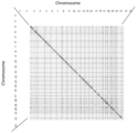
- FIG. 2 shows a heat map of the interaction of the chromatin region in which H3K4me3 is present.
- the vertical and horizontal axes represent chromosomes 1 to 23, respectively, and gene regions are in parallel.
- the regions corresponding to each chromosome are divided by vertical and horizontal straight lines, and the same gene region of the same chromosome is shown by a diagonal line.
- the detected interactions of the different gene regions are represented by dots, and the degree of interaction of the chromatin regions is shown by gray scale shading based on the density of dots on the heat map.
- FIG. 1 shows, by way of example, a flow chart of the method of the invention.
- FIG. 2 shows a heat map of the interaction of the chromatin region in which H3K4me3 is present.
- the vertical and horizontal axes represent chromosomes 1 to 23, respectively, and gene regions are in parallel.
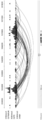
- FIG. 3 shows the localization of H3K4me3 on the genome and the interaction between the chromatin regions in which H3K4me3 is present, which was analyzed as an example.
- the position on the genome is indicated by numbers at the top, and the upper row shows the localization of H3K4me3 analyzed in the results obtained by performing the known CUT&RUN method (Skene et al., 2018). It is a distribution map.
- the middle row and the lower row are the results obtained in Example 1 of the present application, the middle row is a distribution map showing the localization of H3K4me3, and the lower row shows the loop-shaped interaction between chromatin regions in which H3K4me3 is present. Indicate.
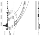
- FIG. 4 is a partially enlarged view of FIG.
- FIG. 5 is a partially enlarged view of FIG.
- FIG. 6 shows a heat map of the interaction of chromatin regions in which CTCF is present.
- the vertical and horizontal axes represent chromosomes 1 to 23, respectively, and gene regions are in parallel.
- the region corresponding to each chromosome is divided by vertical and horizontal straight lines, and the same gene region of the same chromosome is shown by a diagonal line.
- FIG. 7 shows the localization of CTCF on the genome and the interaction between the chromatin regions where CTCF is present, analyzed as an example.
- the positions on the genome are indicated by numbers at the top, and the upper row is a distribution diagram showing the localization of CTCF obtained in Example 2 of the present application.
- the middle row shows the CTCF binding sites present on the genome identified by the Jaspar Transcription Factor Database, and the ones in one direction are underlined according to the orientation of the sequence on the genome.
- the lower row shows the interaction between the chromatin regions in which CTCF obtained in Example 2 of the present application exists in a loop shape.
- FIG. 8 is a partially enlarged view of FIG. 7.
- FIG. 9 is a partially enlarged view of FIG. 7.
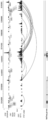
- FIG. 10 shows the localization of CTCF on the genome and the interaction between the chromatin regions in which CTCF is present, which was analyzed as an example.
- the position on the genome is shown by numbers at the top, and the upper row shows the localization of CTCF analyzed by the results obtained by performing the known CUT&RUN method (Skene et al., 2018). It is a distribution map.
- the middle row shows the CTCF binding sites present on the genome identified by the Jaspar Transcription Factor Database, and the ones in one direction are underlined according to the orientation of the sequence on the genome.
- the bottom row shows the distribution of CTCF localization obtained in Example 3 of the present application and the interaction between the chromatin regions in which CTCF obtained in Example 3 of the present application exists below the location of CTCF localization. Shown as a loop. Positions of genes (FBXL18, MIR589, ACTB) in chromosome 7 are displayed further below the bottom row, and the range marked on the right side of the gene name indicates the existing position of the corresponding gene.
- FIG. 11 is a partially enlarged view of FIG.
- FIG. 12 is a partially enlarged view of FIG.
- the present invention is a method for analyzing the interaction of DNA sequences based on the three-dimensional structure of DNA and an analytical kit for realizing the method.
- the three-dimensional structure of DNA means a higher-order structure formed by a DNA double helix, for example, a structure having a loop and a fold.
- These structures may consist solely of DNA or may contain other molecules such as proteins.
- the concept also includes chromatin, which is a complex of DNA and protein. The main function of chromatin is said to control gene expression, DNA replication and repair, in addition to packaging DNA in a small volume so that it fits inside the cell. The most abundant protein component of chromatin is the histone that condenses DNA.
- topological domain (hereinafter appropriately referred to as “TD”)
- TD topological domain
- These domains correlate with regions of the genome that limit the spread of heterochromatin. This domain is stable across different cell types and is highly conserved across species. Topological domains also interact with each other. In the present invention, not only in this topological domain but also in DNA sequence interaction between topological domains is analyzed.
- DNA sequence interactions or three-dimensional DNA structure interactions represent, for example, the spatial arrangement (eg perspective) between DNA sequences in a three-dimensional structure.
- the method of the present invention comprises a cutting step of specifically cutting a DNA chain (target DNA region) in the vicinity of a preselected arbitrary protein among various proteins bound to, complexed with, or attached to a DNA chain, and the cutting step.
- a sequencing step of optionally amplifying the pair and determining the base sequence can be included in any order.
- the target DNA region is a portion of the DNA chain near an arbitrary protein and is a DNA region to be analyzed. Each step will be described below.
- the present invention may be configured by providing a cross-linking step for fixing the higher-order structure in the cell by an artificial operation before the cutting step.
- the cross-linking step is a procedure to fix the spatial arrangement of DNA sequences within the cell nucleus.
- formaldehyde can be used to cross-link proteins with nucleic acids or with one protein with another neighboring protein.
- interacting base sequences such as a relationship between a promoter and an enhancer are in contact with a specific protein and are close to each other via the protein.
- the higher order structure of DNA is often associated with proteins. Therefore, the spatial structure of the DNA sequence is fixed by fixing (cross-linking) the DNA and the protein in contact with it using formaldehyde.
- DNA crosslinking agents examples include EGS (ethyleneglycolbis(succinimidylsuccinate)) and DSG (disuccinimidylglutarate) in addition to formaldehyde.
- Formaldehyde cross-linking is a suitable technique for detecting two sequences that interact directly with each other, but formaldehyde is called a zero-length cross-linking agent, and it makes a DNA pair only for sequences that are in close contact with each other. I can't. Therefore, if a relatively long cross-linking agent such as EGS (16.1 ⁇ ) or DSG (7.7 ⁇ ) is used instead of formaldehyde, a large protein complex having a more complicated higher-order structure can be captured and cross-linked with formaldehyde. DNA pairs can be generated to detect interactions between DNA sequences that are distant from the possible distance.
- a relatively long cross-linking agent such as EGS (16.1 ⁇ ) or DSG (7.7 ⁇
- This cross-linking step may be carried out, for example, by incubating the cells in a 0.05 vol% to 4 vol% formaldehyde solution in an environment ranging from chilling on ice to 37° C., more preferably 0.5 vol%. It may be carried out by incubating in a formaldehyde solution of up to 2.5 vol%, more preferably 1 vol% to 2 vol%. Incubation time is 30 seconds to 16 hours, preferably 5 minutes to 30 minutes, and more preferably 10 minutes to 15 minutes.
- a cross-linking step it is possible to sufficiently fix the higher-order structure to accurately fix the positional relationship of the DNA sequences and improve the accuracy of the DNA pair obtained in the subsequent step. it can.
- the cleaving step is a step mainly using an enzyme to cleave a DNA chain in the vicinity of the preselected protein (target protein) in the DNA sequence in contact with the protein.
- permeabilization of the nuclear membrane may be carried out in order to enhance the permeability of the enzyme and favorably cleave the DNA existing in the cell nucleus.
- Nonionic or zwitterionic surfactants are preferably used.
- examples of such surfactants include digitonin, octylphenol ethoxylates (trade name: Triton X100, Triton X114, nonidet P40, etc.), n-dodecyl- ⁇ -D-maltoside (DDM), polyoxyethylene sorbitan mono- Laurate (Twin 20), polyoxyethylene sorbitan monooleate (Twin 80), 3-[(3-cholamidopropyl)dimethylammonio]-1-propanesulfonate (trade name CHAPS), etc. may be mentioned. It is not limited to.
- the cells may be treated with liquid nitrogen before or after permeabilization with a detergent.
- proteins adjoining the interacting DNA sequences are associated with one or both interacting DNA sequences, or are complexed or attached and spatially related to these DNA sequences. Targets proteins located in the vicinity of.
- CTCF is exemplified as a protein known to be closely involved in the formation of a three-dimensional structure of DNA, and RNA polymerase II as a protein involved in RNA transcription, essential for the nucleosome structure.
- histone H3, histone H4, histone H2A, and histone H2B is exemplified as a protein known to be closely involved in the formation of a three-dimensional structure of DNA.
- RNA polymerase II as a protein involved in RNA transcription, essential for the nucleosome structure.
- histone H3, histone H4, histone H2A, and histone H2B histone H3, histone H4, histone H2A, and histone H2B.
- a primary antibody that directly binds to the selected protein is prepared.
- a primary antibody include a monoclonal antibody and a polyclonal antibody corresponding to the target protein.
- those corresponding to the proteins exemplified above include CTCF, RNA polymerase II, histone H3, histone H4, histone H2A, and histone H2B.
- a monoclonal antibody against The antibody does not necessarily have to be a complete antibody and may be a functional fragment of an antibody having the same function, for example, a fragment having a function of binding to a target protein.
- the DNA is cleaved by an enzyme that binds to the target protein via the primary antibody that captures the target protein, in other words, an enzyme that binds to the DNA chain through the primary antibody.
- an enzyme that binds to the DNA chain through the primary antibody binding of an enzyme to a DNA chain through a primary antibody means that the enzyme directly or indirectly binds to a primary antibody that directly binds to a target protein attached to or bound to the DNA chain.
- any known enzyme can be appropriately selected and used as long as it has an ability to cleave a DNA chain in the vicinity of a target protein, and examples thereof include micrococcal nuclease (MNase) and deenase.
- MNase micrococcal nuclease
- Examples thereof include various endonucleases such as I (DNaseI) and transposase, and various restriction enzymes such as Hindi Three (HindIII), MboI, and DpnII, but are not limited thereto.
- I DNaseI
- transposase various restriction enzymes
- restriction enzymes such as Hindi Three (HindIII), MboI, and DpnII, but are not limited thereto.
- An endonuclease that cleaves in a sequence non-specific manner is preferably used, and more preferably MNase is used.
- the vicinity means within 1000 bp from the position of the target protein, for example, within 900 bp, within 800 bp, within 700 bp, within 600 bp, within 500 bp, within 400 bp, within 300 bp, within 200 bp, and preferably within 200 bp, for example, transposase , MNase, DNaseI, HindIII, MboI, DpnII can cleave within 1000 bp from the position of the target protein, and MNase, DNaseI, HindIII, MboI, DpnII cleave within 200 bp from the position of the target protein. be able to.
- a DNA fragment having a base length according to the characteristics of the cleavage enzyme can be obtained.
- a DNA fragment of about 200 bp (about mononucleosome size) can be obtained.
- the length of the obtained DNA fragment changes depending on the conditions under which the cleavage step is performed, and may be a mononucleosome size that includes a multiple thereof.
- the enzyme originally has the ability to cleave DNA, but does not inherently have the ability to bind to an antibody, it is used in the cleavage step after the enzyme has the ability to bind to the antibody.
- a method for imparting antibody binding ability to an enzyme various methods can be used.
- a protein having a high affinity for the Fc region of an antibody is fused with the enzyme to be used to form an antibody. Examples include a method of imparting a binding ability and a method of conjugating an enzyme to a secondary antibody that specifically binds to the above-mentioned primary antibody by various chemical reactions.
- Various known proteins can be used as the protein having an affinity for the antibody, and examples thereof include protein A, protein G, and protein L, but are not limited thereto.
- a known method can be appropriately used, and for example, a periodate method, a glutaraldehyde method, a maleimide method, an NHS (N-hydroxysuccinimide) method can be used. But this is not the case.
- the fusion protein can be produced using biotechnological techniques. Specifically, a vector (plasmid) for producing a fusion protein is introduced with a gene encoding a sequence of a cleavage enzyme to construct a vector, or an already constructed vector is introduced into Escherichia coli and transformed. The desired fusion protein can be produced by culturing E. coli and inducing the expression of the fusion protein.
- a fusion protein of protein A and MNase can be prepared using a known vector pK19pA-MN or the like.
- a DNA encoding protein A and MNase is introduced into a known plasmid pET21a or the like to construct an expression vector of pA-MN, which can be used for preparation.
- the expressed pA-MN can be solubilized, and the suspension obtained by disrupting E. coli can be centrifuged to contain pA-MN in the resulting supernatant. Then, pA-MN can be recovered from this supernatant by the steps of adsorption, washing and elution with IgG-Sepharose beads or the like.
- the procedure for binding the enzyme to the DNA chain via the primary antibody is as follows: 1) adding the primary antibody to the DNA fragment, and then sequentially adding the enzyme capable of binding to the primary antibody, Bind the enzyme to. 2) The target protein is captured and the enzyme is bound to the DNA chain via the primary antibody by adding a pre-bound antibody to the primary antibody. 3) After adding the primary antibody, add the secondary antibody, and then add the enzyme capable of binding to the secondary antibody. 4) After the addition of the primary antibody, it may be carried out by any procedure such as adding the one in which the secondary antibody and the enzyme are bound in advance. In addition, in the case of the method of adding to the DNA fragment a product in which the primary antibody and the enzyme are bound in advance, the primary antibody binding step and the enzyme binding step described in Item 9 are performed at the same time.
- the above-mentioned enzyme is preferably one that can easily adjust the timing (enzyme activity) of breaking the DNA chain.
- the method for adjusting the enzyme activity include reaction temperature, light irradiation, and metal ion concentration. More preferably, those having an effector that is a metal ion whose reaction can be easily switched on and off are selected.
- MNase or DNaseI can be exemplified as a calcium ion-dependent substance
- transposase can be exemplified as a magnesium ion-dependent substance. With such an enzyme, DNA cleavage is performed at the timing when the corresponding ion is added.
- the DNA strand near the target protein selected in advance is cleaved, and when the adjacent cleavage ends are joined in the subsequent ligation step, the DNA has a sequence different from the sequence on the genome. A pair is created.
- this cleavage step is not a genome-wide random cleavage but a target protein-specific cleavage. Therefore, a group of the obtained DNA pairs (DNA library) has a higher concentration (in principle, all the obtained DNA pairs) than the DNA library obtained by the conventional genome-wide random cleavage. Will be present. That is, it is possible to provide a high-quality analysis sample in which unnecessary DNA fragments (ligation by-products, contaminants) that become noise are extremely small.
- the present invention improves the data reliability by improving the quality of the DNA library itself that performs sequencing. Therefore, it is possible to suppress the number of cells required for the analysis and enable the analysis with a small amount of sample. Further, even when amplification is performed, the ratio of the target DNA pair contained in the analysis sample is extremely high, so that the influence of the amplification bias at the time of PCR due to contaminants can be suppressed. Further, it is possible to suppress the reading of unnecessary DNA fragments, and it is possible to greatly reduce the labor and cost involved in the analysis.
- a labeling step for adding biotin for example, as a label may be provided at the cut ends.
- this labeling step may be provided before the sorting step described later. Therefore, for example, it may be provided subsequent to the cutting step, or may be provided so as to be performed together with the ligation in the ligation step.
- the labeling step is provided, the DNA pair to be analyzed can be concentrated by labeling with biotin or the like.
- the necessary DNA pairs can be concentrated without relying on the immunoprecipitation method, so that it is not necessary to solubilize the nuclear fraction required for the immunoprecipitation method, and the quality of the formed DNA library is further improved. Can be made.
- biotin to the cleaved DNA ends can be performed according to a conventional method.
- the cleaved protruding end can be blunt-ended by performing an extension reaction using deoxynucleotide triphosphate (dNTP) as a substrate.
- dNTP deoxynucleotide triphosphate
- biotin is added to the cleaved end.
- various commercially available biotinylation reagents can be used. If the enzymatically cleaved ends are blunt ends, biotin may be added after once making the protruding ends.
- the substance added to the cleavage end is not limited to biotin as long as it is a substance that enables selection (concentration) of DNA pairs, but it is preferable to perform protease treatment for fixing and fixing the DNA structure.
- compounds that are not digested by protease are desirable. Examples of such a compound include methylated dATP and the like. If this methylated dATP is added to the cleaved end, the DNA pair can be concentrated with the anti-methylated dATP antibody.
- a compound that reacts with click chemistry which is a cyclization reaction between a nitrogen triple bond and an alkyne, may be used as a label.
- an example of such a compound is, for example, an unnatural base having an alkyne (5-ethynyl-2'-deoxyuridine (EdU etc.)). Since such a compound added to the cleavage end binds to an azido compound by a click reaction, it can be bound to azidobiotin having an azido group or an azido group-modified flag tag, and as a result, based on these biotin or flag tag. Concentrated concentration may be possible. Further, instead of biotinylated dNTP, short DNA to which biotin is added (linker DNA) may be used as a label, and the ligation reaction may be performed via this linker DNA.
- linker DNA short DNA to which biotin is added
- This step is a step of performing a ligation reaction using DNA ligase.
- Ligation means linking two DNA fragments by a phosphodiester bond.
- a room temperature DNA ligase such as T4 DNA ligase or Escherichia coli DNA ligase can be used.
- thermostable DNA ligase may be used.
- dNTPs biologically modified dNTPs
- exonuclease enzyme-catalyzed ase
- This step is a step of degrading the protein contained in the sample by using a protease with respect to the sample which has completed the ligation reaction.
- proteins in the sample including proteins that bind to DNA and nuclear membranes are decomposed.
- the interacting DNA sequences are located proximally as a result of the target protein being attached to the DNA strand, but after the DNA pair is formed by the ligation reaction, it cannot be immobilized by the protein molecule.
- This step digests the protein as it is no longer needed. As a result, the fixation of the DNA structure by the protein (fixation of the spatial structure of the DNA sequence) is eliminated. With this protein digestion, immobilization due to crosslinking based on the crosslinking step is also eliminated.
- the target sample (DNA pair) is digested with the nuclear membrane in this step. It can be taken out from the nucleus. Therefore, the step of solubilizing the nuclear fraction can be omitted.
- this step is preferably performed subsequent to the ligation step, it is provided after the ligation and before the amplification operation by PCR or the sequencing step, and in the present invention, a step for performing chromatin immunoprecipitation is provided. If present, this step is not provided prior to immunoprecipitation.
- the fragmentation process in the selection step described later corresponds to the fixation elimination step. Even in such a case, if it is necessary to eliminate the crosslinking based on the crosslinking step, this step constitutes a part of the fixation eliminating step.
- the fixation of the DNA structure is eliminated by the fragmentation, even if the fixation of the DNA structure is not required to be eliminated by this step, the digestion with the protease is performed as in this step to digest the nuclear membrane and the like. Steps may be provided.
- an adapter sequence may be introduced at the end of the fragment along with fragmentation.
- the step of selecting the ligated DNA pair includes not only selecting the ligated initial sequence, but also the processed or treated DNA fragment after ligation, or the treatment thereof. Further, it includes selecting a fragmented DNA fragment or a DNA fragment to which an arbitrary DNA sequence is added.
- the DNA sequence added here may be a short adapter sequence.
- the primer containing the complementary sequence of the short adapter sequence and the index sequence is hybridized to the short adapter sequence bound to the DNA pair (DNA fragment), and the DNA pair is performed at the same time as the proliferation reaction is performed. You can add an index array to.
- a DNA sequence having both an index sequence and a short adapter sequence is ligated to a DNA pair as an adapter sequence (full adapter), and the sequence selected in the selection step is added.
- DNA may be constructed.
- the adapter sequence (full adaptor) or the index sequence may include a DNA sequence complementary to the DNA fragment fixed to the flow cell used in a generally known sequencer system.
- a label biologically
- a DNA pair DNA fragment is recovered using the label as a target. There are no particular limitations on this recovery as long as it is a protein that has an affinity for the label, and various commercially available proteins can be used.
- the label when the label is biotin, preferred examples include biotin-binding proteins such as avidin, streptavidin, and deglycosylated avidin, which have extremely high affinity for biotin.
- a sorting operation may be performed using a protein such as beads immobilized on a carrier.
- the DNA pairs to be analyzed are selectively collected and a DNA library is constructed.
- a DNA library constructed by a DNA pair recovered based on the label may be hybridized with an oligonucleotide to further select the DNA pair.
- the step of selecting DNA pairs (DNA fragments) comprises, after ligation, selecting DNA pairs by electrophoresis. For example, there is a method in which a ligation product is selected by electrophoresis using an agarose gel and the ligation product is selected based on the DNA chain length, which does not require a label.
- the DNA library constructed above is subjected to PCR according to a conventional method to amplify each DNA pair (DNA fragment).
- the resulting PCR product is then sequenced.
- Pretreatment is performed on the DNA pairs (DNA fragments) concentrated by the above PCR or on the DNA pairs in the DNA library constructed in the selection step, and the DNA pairs are analyzed by a massively parallel DNA sequence analyzer or Sanger method.
- the nucleotide sequences at both ends of are analyzed and sequenced.
- the length of the base sequence to be analyzed may be 1 base to the entire length of the DNA fragment.
- the length of the base sequence to be analyzed can be set arbitrarily, and may be, for example, several bases or more, 10 bases or more, 15 bases or more, 20 bases or more, 30 bases or more. It is preferably about 30-100 bases, for example.
- the obtained sequence data can be analyzed using a conventional HiC analysis method.
- DNAs DNA pairs
- the ends can be independently mapped to the genome, but sometimes they cannot be mapped as DNA fragments.
- the obtained DNA fragment has a sequence of chromosome 1 at one end and a sequence of chromosome 2 at the other end. Will have.
- a contact map contact probability matrix
- the contact map is a symmetric matrix.
- the count of (Xi,Yj) is incremented by one. Therefore, if the value is large at each coordinate, it means that the area pair has a high contact probability.
- the value of this coordinate can be displayed on the heat map with the shade of the color corresponding to this.
- sample amount This method can obtain an analysis sample in which contaminants are extremely reduced as compared with the conventional method, and as a result, the number of cells required for analysis can be set to about 1 to 10 10 , preferably 1 to 10 The number can be about 7 , more preferably about 1 to 10 6 .
- the upper limit of the number of cells is not necessarily limited, and may be 10 10 or more.
- the method of the present invention can be used as an alternative to the conventional HiC method.
- a cleavage step, a labeling step, a ligation step, a fixation elimination step, a selection step, and a sequence determination step are sequentially provided.
- the nuclear membrane can also be digested in the fixing elimination step as described above.
- generation of an insoluble fraction cannot be avoided in principle because extraction of a nuclear fraction is required.
- the DNA sequence to be analyzed is selected based on the label added in the labeling step, the DNA pair can be selected using a protease after the ligation treatment, and the (insoluble fraction that does not elute from the nucleus can be selected. Therefore, a good analysis sample can be prepared.
- a chromatin immunoprecipitation step when a chromatin immunoprecipitation step is provided to supplement the target DNA fragment, only the target DNA fragment needs to be released into the supernatant, and therefore undigested long-chain DNA to be the portion not to be analyzed. Even if the fragment remains in the nucleus as an insoluble fraction, it does not affect the obtained data. Therefore, it is possible to prevent a decrease in the reliability of the data due to the insoluble fraction in the solubilization step of the nucleus, which occurs in the conventional immunoprecipitation method. Further, unlike the conventional HiC method, since DNA is cleaved specifically for a specific protein, a desired DNA sequence to be analyzed is concentrated in a DNA library to be analyzed, and unnecessary DNA that becomes noise is generated.
- the localization of the target protein can be detected by independently analyzing the DNA cleavage sites from the obtained DNA pair sequencing results. That is, according to the present method having a cleavage step of specifically cleaving a target protein, both the three-dimensional DNA structure in the nucleus mediated by the target protein and the localization of the target protein in the nucleus are simultaneously It can be analyzed.
- a protein biotinylation step that biotinylates a protein that binds to DNA is provided before the cleavage step that specifically cuts the target protein, and after the cleavage step, fragments that have been cleaved by nuclear membrane permeabilization are allowed.
- a solubilization step (dialysis step) to solubilize is provided to release the protein-DNA complex from the nucleus into the supernatant, which is then pulled down using a supported bead such as streptavidin, and then on the supported bead.
- the ligation step may be configured to perform ligation, and then the fixation elimination step, the selection step, and the sequence determination step may be sequentially performed to obtain sequence data.
- the method of the present invention may be configured to include a solubilization step of a nuclear fraction and a chromatin immunoprecipitation step for performing chromatin immunoprecipitation.
- the solubilization step of the nuclear fraction and the chromatin immunoprecipitation step can be performed according to a conventional method.
- a cleavage step, a labeling step, a ligation step, a solubilization step of the nuclear fraction, a chromatin immunoprecipitation step, an immobilization step, a selection step By sequentially providing the sequence determination steps, as a substitute for the conventional HiChIP, it is possible to provide a method capable of reducing impurities in the stage of preparing a DNA library to be analyzed, as compared with the conventional HiChIP.
- An example of the method of this embodiment corresponds to the three-dimensional DNA structure interaction analysis method described in item 6 above.
- the chromatin immunoprecipitation step included in the method of this embodiment is included in the capture step described in item 6 above.
- the method of the present invention comprises, as described above, a cleavage step, a solubilization step of a nuclear fraction, a chromatin immunoprecipitation step, a ligation step and a labeling step, and an immobilization step after the crosslinking step is appropriately provided.
- a substitute for ChIA-PET by constructing the present invention by sequentially providing a selection step and a sequencing step, it is possible to reduce contaminants in the stage of preparing a DNA library for analysis, as compared with ChIA-PET. Can be provided.
- the DNA pair can be concentrated based on the chromatin immunoprecipitation step, and thus the sorting step may be unnecessary.
- the analysis kit of the present invention comprises a primary antibody that binds to an arbitrary protein, a DNA-cleaving enzyme that binds to the primary antibody, and a label for identifying a DNA end generated by the cleavage by the DNA-cleaving enzyme. And an enzyme that ligates the labeled DNA ends.
- the primary antibody, the DNA-cleaving enzyme, the label and the enzyme for ligating the DNA ends contained in the analysis kit are materials used in the method of analyzing the interaction of DNA sequences based on the three-dimensional structure of DNA of the present invention.
- the primary antibody and the DNA cleaving enzyme may be contained separately, or may be contained in a state in which the primary antibody and the DNA cleaving enzyme are bound.
- RSB Resuspension Buffer composition: 10 mM Tris-HCl (pH 7.4) 10 mM NaCl 3 mM MgCl 2 0.1% Tween-20 Sterilized water Buenrostro JD, Wu B, Chang HY, Greenleaf WJ: ATAC ⁇ seq: a method for assaying chromatin accessibility genome ⁇ wide. Curr Protoc Mol Biol. 2015;109:21-9 and Corces MR, Trevino AE, Hamilton EG, Greenside PG, Sinnott-Armstrong NA, Vesuna S, Satpathy AT, Rubin AJ, Montine KS, Wu B, et al. An improved ATAC-seq protocol reduces background and enables interrogation of frozen tissues. Nat Methods. 2017;14:959- RSB was prepared with reference to 62.
- Digitonin composition 50mg Digitonin (Nacalai Tesque) 500 ⁇ l DMSO 500 ⁇ l sterile water
- Lysis Buffer composition 0.1% IGEPAL CA-630 0.01% Digitonin RSB Buenrostro JD, Wu B, Chang HY, Greenleaf WJ: ATAC ⁇ seq: a method for assaying chromatin accessibility genome ⁇ wide. Curr Protoc Mol Biol. 2015;109:21-9 and Corces MR, Trevino AE, Hamilton EG, Greenside PG , Sinnott-Armstrong NA, Vesuna S, Satpathy AT, Rubin AJ, Montine KS, Wu B, et al. An improved ATAC-seq protocol reduces background and enables interrogation of frozen tissues. Nat Methods. 2017;14:959-62. As a reference, Lysis Buffer was prepared.
- Wash Buffer composition 1M HEPES (1 ml), 5M NaCl (1.5 ml), 2Mspermidine (12.5 ⁇ l) were added, sterilized water was added up to 50 ml, and cOmplete TM and EDTA-free Protease Inhibitor Cocktail Tablets (Roche) were added 1 tablet to wash. Buffer was prepared. Targeted in situ genome-wide profiling with high efficiency for low cell numbers. Nature Protocols. 2018. Vol. 13. No. 5 was referred to and a Wash Buffer was prepared.
- Digitonin Buffer composition 0.02% Digitonin (0.0375% in Example 3) Wash Buffer Skite et al., 2018. Targeted in situ genome-wide profiling with high efficiency for low cell numbers. Nature Protocols. 2018. Vol. 13. No. 5 was referred to to prepare Digitonin Buffer.
- Antibody Buffer Antibody Buffer was prepared by adding 2 ml of Digitonin Buffer and 8 ⁇ l of 0.5 M EDTA (pH 8.0). Skene et al., 2018. Targeted in situ genome-wide profiling with high efficiency for low cell numbers. Nature Protocols. 2018. Vol. 13. No. 5 was referred to to prepare Antibody Buffer.
- Mbuffer #2 composition 50 mM NaCl 10 mM Tris-HCl (pH 7.4) 10 mM MgCl 2 Mbuffer #2 was prepared with reference to sterile water Rando et al., 2016. Micro-C XL. Protocol Exchange.
- 10x NEBuffer #2 composition 1M NaCl 200 mM Tris-HCl (pH 7.4) 200 mM MgCl 2 Sterilized water 10x NEBuffer #2 was prepared with reference to Rando et al., 2016. Micro-C XL. Protocol Exchange.
- 10X T4 DNA ligase buffer (without ATP) composition: 500 mM NaCl 583 mM Tris-HCl (pH 7.4) 200 mM MgCl2 117 mM DTT Rando et al., 2016. With reference to Micro-C XL. Protocol Exchange, 10X T4 DNA ligase buffer was prepared.
- 1X Bind and Wash Buffer composition 5 mM Tris-HCl, pH 7.4 0.5 mM EDTA 1M NaCl Sterilized water Gabdank et al. 2016. A streamlined tethered chromosome conformation capture protocol. BMC Genomics. 2016. 17:274 was referred to, and 1X Bind and Wash Buffer was prepared.
- 2X Bind and Wash Buffer composition 10 mM Tris-HCl, pH 7.4 1 mM EDTA 2M NaCl Sterilized water Gabdank et al. 2016. A streamlined tethered chromosome conformation capture protocol. BMC Genomics. 2016. 17:274 was referred to, and 2X Bind and Wash Buffer was prepared.
- Example 1 Expression and purification of fusion protein (pA-MN) of protein A and MNase Escherichia coli strain One Shot (registered trademark) BL21(DE3) (invitrogen) was prepared using plasmid pET11a-pA-MN (pET11a) with a DNA sequence expressing pA-MN. It was transformed with the vector constructed by the introduction and cultured at 37° C. in an LB medium (LB-Medium, Lennox) (Funakoshi) containing 100 mg/L of carbenicillin (Nacalai Tesque).
- LB medium LB-Medium, Lennox
- IPTG isopropyl- ⁇ -thiogalactopyranoside
- the cells were disrupted by repeating ultrasonic treatment using an ultrasonic homogenizer (BIORUPTOR (registered trademark) II Type 6, Sonic Bio Inc.).
- the extract was clarified by centrifugation in a centrifuge (MX-301, TOMY) at 15000 x G for 20 minutes at 4°C. Most of the pA-MN was recovered in the soluble supernatant (S1).
- PA-MN was purified from S1 using beads (IgG-Sepharose 6 Fast Flow, GE healthcare). 30 ml of clarified S1 was incubated with 600 ⁇ l of IgG-Sepharose beads for 16 hours with shaking.
- the beads were collected by passing through an Econopack chromatography column (BIO-RAD), and twice with TEN supplemented with 0.03% surfactant (EMPIGEN (registered trademark) BB surfactant, SIGMA). It was washed twice with TEN500 containing 0.03% Empigen® BB (TEN supplemented with 500 mM NaCl) and once with 5 mM ammonium acetate (pH 5.0). pA-MN was eluted with 0.5M acetic acid/ammonium acetate (pH 3.4). The eluate was neutralized by adding a 1 mol/L NaOH aqueous solution. The same amount of glycerol as the neutralized eluate was added to adjust the final concentration to 50%. About 1 mg of highly purified protein was obtained per 500 ml of starting culture. Long-term aliquots were stored at -80°C and short-term aliquots at -30°C.
- EMPIGEN registered trademark
- SIGMA sur
- HEK293T cells were suspended in PBS containing 2 vol% fetal bovine serum (FBS) (FBS-PBS), and then the cells were injected into a tube so that the number of cells became 10 7 , and the cells were centrifuged at room temperature for 5 minutes and a centrifugal force of 300 g. It was centrifuged at. To the separated cells, 1 ml of FBS-PBS containing 1 wt.% formaldehyde (trade name: Formaldehyde solution, model number F8775, Sigma-Aldrich) was added and suspended, and gently stirred.
- FBS-PBS 2 vol% fetal bovine serum
- the cell sample prepared by the above procedure was lysed, resuspended in 1 ml of Lysis Buffer, and kept on ice for 30 minutes with periodic stirring. Thereafter, 1 ml of RSB was added, and the mixture was centrifuged at 4° C. for 5 minutes at a centrifugal force of 800 g, and then washed with 1.5 ml of a wash buffer (Wash Buffer). After performing the washing operation by adding the washing buffer and centrifuging operation several times, the precipitate obtained by centrifuging at 4° C.
- a wash buffer Wash Buffer
- the anti-H3K4me3 antibody (model number ab8580, Abcam) The suspension was resuspended in 500 ⁇ l of an antibody buffer solution containing 1 ⁇ g and reacted at 4° C. overnight.
- the anti-H3K4me3 antibody is an antibody of H3K4me3 (histone H3 in which the 4th lysine is trimethylated).
- the sample is centrifuged at 4°C, centrifugal force ⁇ 500g for 5 minutes and resuspended in 1ml Digitonin Buffer twice, and then at 4°C, centrifugal force ⁇ 500g.
- the cells were resuspended in 40 ⁇ l of Digitonin Buffer, pA-MN diluted 1000 times with Digitonin Buffer (concentration was adjusted appropriately) was added, and the mixture was reacted at 4° C. for 1 hour. ..
- dephosphorylating enzyme trade name: Shrimp Alkaline Phosphatase (rSAP), New England BioLabs
- dephosphorylation treatment was performed at 37° C. for 45 minutes. went.
- the dephosphorylation reaction was stopped by heat treatment at 65°C for 5 minutes.
- NEBuffer#2 (10x NEBuffer#2) adjusted to 10 times concentration 3 ⁇ l, 0.5 wt.% BSA-containing aqueous solution 0.2 ⁇ l, 10 mM ATP solution (trade name Adenosine 5′-Triphosphate (ATP), New England BioLabs) ) 4.5 ⁇ l, 1 M DTT (trade name UltraPure TM Dithiothreitol (Clelands Reagent), Invitrogen/Life Technologies) 0.15 ⁇ l, T4 DNA polymerase (trade name T4 DNA Polymerase (5 U/ ⁇ l), Thermo Scientific) 1.
- T4 polynucleotide kinase 10 U/ ⁇ l
- T4 Polynucleotide Kinase New England BioLabs
- concentration of T4 DNA ligase buffer 6 ⁇ l 0.5 wt 0.3 ⁇ l of an aqueous solution containing .% BSA and biotinylated dNTP (biotinylated dATP (trade name: Biotin-14-dATP, Invitrogen), biotinylated dCTP (trade name: Biotin-14-dCTP, Invitrogen) were each terminated.
- a concentration of 0.1 mM, dGTP (trade name: dGTP, Toyobo), and dTTP (trade name: dTTP, Toyobo)) were added to each to give a final concentration of 0.05 mM, and a sample having a solution volume of about 100 ⁇ l was added at 25° C. After 15 minutes at 12° C. for 15 minutes, the reaction was carried out under the temperature condition of maintaining 4° C. Then, ethylenediaminetetraacetic acid (EDTA) was added to a final concentration of 30 mM and heat-treated at 65° C. to deactivate the enzyme activity. By this series of treatments, biotin was added to the DNA cleavage end.
- EDTA ethylenediaminetetraacetic acid
- T4 Ligase buffer (trade name: 10X Buffer for T4 DNA ligase with 10 mM ATP; New England BioLabs) containing 10 times the concentration of ATP and 0.5 wt. 0.5 ⁇ l, 3 ⁇ l of 1M MgCl 2 , 12.5 ⁇ l of T4 DNA ligase (400 U/ ⁇ l) (trade name T4 DNA Ligase, New England BioLabs), and sterilized water to make the total volume 2500 ⁇ l. Mix for 1 hour. Then, centrifugation was performed at 4° C. and centrifugal force ⁇ 16000 g for 10 minutes to obtain a pellet.
- NEBuffer#1 product name NEBuffer 1, New England BioLabs
- exonuclease III Exonuclease III (100 U/ ⁇ l), New England Biolabs) are pelleted.
- 100 ⁇ l of sample solution was prepared. This is kept at 37°C for 5 minutes, and then 1/200 volume of protease (trade name: ProteinaseK (Recombinant) Solution, Nacalai Tesque) of the solution is added, and the mixture is kept at 55°C for 3 hours and then in the sample solution. Protein was digested (decrosslinked).
- phenol-chloroform solution (trade name: phenol:chloroform:isoamyl alcohol 25:24:1 phenol content: about 33 w/w%, Nacalai Tesque) was added to the sample solution and mixed well, then at room temperature, The operation of centrifuging at a centrifugal force of 19,800 g for 10 minutes was performed twice, and then the supernatant was collected. Add 1/10 volume of 3M sodium acetate solution (product name: 3 mol/l Sodium Acetate Buffer Solution (pH5.2), Nacalai Tesque) of the recovered supernatant and 2.5 times volume of 100% ethanol and mix. It was allowed to stand at -80°C for 1 hour.
- 3M sodium acetate solution product name: 3 mol/l Sodium Acetate Buffer Solution (pH5.2), Nacalai Tesque
- the DNA solution was dispensed into a tube so that the amount of DNA was 10 ng, and 25 ⁇ l of double concentration TD buffer (trade name: Tagment DNA Buffer (Nextera (registered trademark) DNA Library Prep Kit, Illumina) and transposome (Tagment 1 ⁇ l of DNA Enzyme 1 (TDE1), Illumina) was added, and then sterilized water was added until the total solution volume reached 50 ⁇ l, which was held at 55° C. for 10 minutes for tagging. The sample was purified and used as a 50 ⁇ l DNA solution (Buffer EB solution).
- TD buffer trade name: Tagment DNA Buffer (Nextera (registered trademark) DNA Library Prep Kit, Illumina) and transposome (Tagment 1 ⁇ l of DNA Enzyme 1 (TDE1), Illumina
- streptavidin beads dispersion liquid (trade name Dynabeads TM (registered trademark) M-280 Streptavidin, Invitrogen/ Thermo Fisher Scientific) was washed twice with 500 ⁇ l of 1xBind and Wash buffer, and then 50 ⁇ l of 2xBind and Wash. Suspended with Buffer. 50 ⁇ l of this bead dispersion was mixed with the above-prepared DNA solution in a tube and kept at room temperature for 30 minutes, and then allowed to stand on a magnetic stand (for 1.5 ml) to remove the supernatant, The avidin beads were collected.
- Dynabeads TM registered trademark
- M-280 Streptavidin Invitrogen/ Thermo Fisher Scientific
- the recovered beads are washed once with 500 ⁇ l of 1x Bind and wash buffer containing 0.1 vol% nonionic surfactant solution (trade name Triton X-100, Nacalai Tesque), and then 500 ⁇ l of Buffer After further washing with EB, 40 ⁇ l of Buffer EB was suspended to obtain a dispersion liquid.
- 1x Bind and wash buffer containing 0.1 vol% nonionic surfactant solution trade name Triton X-100, Nacalai Tesque
- the base sequences at both ends of the DNA fragment were analyzed using a massively parallel DNA sequence analysis device (device name NextSeq 500, Illumina).
- the length of the base sequence to be analyzed was 36 bases.
- the interaction between the DNA sequences was analyzed by mapping the sequences at both ends of the obtained base sequence independently to the reference human genome sequence. The obtained results are shown in FIGS. 2 and 3.
- FIG. 2 shows a heat map of the interaction of the chromatin region in which H3K4me3 is present.
- interaction analysis of DNA sequences was performed using an anti-H3K4me3 antibody, and interaction of the chromatin region where H3K4me3 is present was analyzed by Juicebox.
- FIG. 3 shows the localization of H3K4me3 on the genome and the interaction of the chromatin region in which H3K4me3 is present, which was analyzed as an example.
- the interaction between H3K4me3 located near the ACTB gene region (chromosome 7) and the chromatin region containing histone posttranslational modification was analyzed by WashU EpiGenome Browser.
- FIG. 4 is a partially enlarged view of FIG.
- FIG. 5 is a partially enlarged view of FIG.
- Example 2 The target protein was CTCF, 5 ⁇ g of anti-CTCF antibody (ab70303 Abcam) was used in place of the anti-H3K4me3 antibody of Example 1, and the reverse side primer was S511 to S516 (trade name: Nextera (registered trademark) XT). Analysis of DNA sequence interaction was performed in the same manner as in Example 1 except that PCR was carried out for 13 or 14 cycles by changing to Index Kit v.2, Illumina). The results are shown in FIGS. 6 and 7.
- FIG. 6 shows a heat map of the interaction of chromatin regions in which CTCF is present.
- the DNA sequence interaction analysis was performed using an anti-CTCF antibody in the same manner as in Example 1, and the interaction of the chromatin region in which CTCF is present was analyzed by Juicebox.
- FIG. 7 shows the localization of CTCF on the genome and the interaction of the chromatin regions in which CTCF is present, which was analyzed as an example. The interaction between CTCF localized near the ACTB gene region (chromosome 7) and the chromatin region containing its transcription factor was analyzed by WashU EpiGenome Browser. From the above results, the interaction of the chromatin region to which CTCF binds could be confirmed using the method of the present invention.
- FIG. 8 is a partially enlarged view of FIG. 7.
- FIG. 9 is a partially enlarged view of FIG. 7.
- Example 3 Based on Example 1, analysis of DNA sequence interaction was carried out with the number of samples used for the analysis being 10 6 cells. Specifically, it is as follows.
- HEK293T cells were suspended in PBS containing 2 vol% fetal bovine serum (FBS) (FBS-PBS), and then the cells were injected into a tube so that the number of cells became 10 6 , and the cells were centrifuged at room temperature for 5 minutes and centrifuged at 300 g. It was centrifuged at. To the separated cells, 1 ml of FBS-PBS containing 1 wt.% formaldehyde (trade name: Formaldehyde solution, model number F8775, Sigma-Aldrich) was added and suspended, and gently stirred.
- FBS-PBS 2 vol% fetal bovine serum
- the cell sample prepared by the above procedure was lysed, resuspended in 1 ml of Lysis Buffer, and kept on ice for 30 minutes with periodic stirring. Thereafter, 1 ml of RSB was added, and the mixture was centrifuged at 4° C. for 5 minutes at a centrifugal force of 800 g, and then washed with 1.5 ml of a wash buffer (Wash Buffer). After performing the washing operation by adding washing buffer and centrifuging operation several times, the precipitate was centrifuged at 4° C. for 5 minutes at a centrifugal force of 800 g to collect the anti-CTCF antibody (model number ab70303, Abcam). The suspension was resuspended in 500 ⁇ l of an antibody buffer solution containing 1 ⁇ g and reacted at 4° C. overnight.
- the sample is centrifuged at 4°C, centrifugal force ⁇ 500g for 5 minutes and resuspended in 1ml Digitonin Buffer twice, and then at 4°C, centrifugal force ⁇ 500g.
- the cells were resuspended in 45 ⁇ l of Digitonin Buffer, pA-MN diluted 1000 times with Digitonin Buffer (concentration was adjusted appropriately) was added, and the mixture was reacted at 4° C. for 1 hour. ..
- dephosphorylating enzyme trade name: Shrimp Alkaline Phosphatase (rSAP), New England BioLabs
- dephosphorylation treatment was performed at 37° C. for 45 minutes. went.
- the dephosphorylation reaction was stopped by heat treatment at 65° C. for 5 minutes.
- NEBuffer#2 (10x NEBuffer#2) adjusted to 10 times concentration 3 ⁇ l, 0.5 wt.% BSA-containing aqueous solution 0.2 ⁇ l, 10 mM ATP solution (trade name Adenosine 5′-Triphosphate (ATP), New England BioLabs) ) 4.5 ⁇ l, 1 M DTT (trade name UltraPure TM Dithiothreitol (Clelands Reagent), Invitrogen/Life Technologies) 0.15 ⁇ l, T4 DNA polymerase (trade name T4 DNA Polymerase (5 U/ ⁇ l), Thermo Scientific) 1.
- T4 polynucleotide kinase 10 U/ ⁇ l
- T4 Polynucleotide Kinase New England BioLabs
- concentration of T4 DNA ligase buffer 6 ⁇ l 0.5 wt 0.3 ⁇ l of an aqueous solution containing .% BSA and biotinylated dNTP (biotinylated dATP (trade name: Biotin-14-dATP, Invitrogen), biotinylated dCTP (trade name: Biotin-14-dCTP, Invitrogen) were each terminated.
- a concentration of 0.1 mM, dGTP (trade name: dGTP, Toyobo), and dTTP (trade name: dTTP, Toyobo)) were added to each to give a final concentration of 0.05 mM, and a sample having a solution volume of about 100 ⁇ l was added at 25° C. After 15 minutes at 12° C. for 15 minutes, the reaction was carried out under the temperature condition of maintaining 4° C. Then, ethylenediaminetetraacetic acid (EDTA) was added to a final concentration of 30 mM and heat-treated at 65° C. to deactivate the enzyme activity. By this series of treatments, biotin was added to the DNA cleavage end.
- EDTA ethylenediaminetetraacetic acid
- T4 Ligase buffer (trade name: 10X Buffer for T4 DNA ligase with 10 mM ATP; New England BioLabs) containing 10 times the concentration of ATP and 0.5 wt. 0.5 ⁇ l, 3 ⁇ l of 1M MgCl 2 and 12.5 ⁇ l of T4 DNA ligase (400 U/ ⁇ l) (trade name T4 DNA Ligase, New England BioLabs) were added, and sterilized water was added to make the total volume 2500 ⁇ l. Mixed overnight. Then, centrifugation was performed at 4° C. and centrifugal force ⁇ 16000 g for 10 minutes to obtain a pellet.
- NEBuffer#1 product name NEBuffer 1, New England BioLabs
- exonuclease III Exonuclease III (100 U/ ⁇ l), New England Biolabs) are pelleted.
- 100 ⁇ l of sample solution was prepared. This is kept at 37°C for 5 minutes, and then 1/200 volume of protease (trade name: ProteinaseK (Recombinant) Solution, Nacalai Tesque) of the solution is added, and the mixture is kept at 55°C for 3 hours and then in the sample solution. Protein was digested (decrosslinked).
- the DNA solution was dispensed into a tube so that the amount of DNA was 10 ng, and 25 ⁇ l of double concentration TD buffer (trade name: Tagment DNA Buffer (Nextera (registered trademark) DNA Library Prep Kit, Illumina) and transposome (Tagment After adding 0.5 ⁇ l of DNA Enzyme 1 (TDE1), Illumina), sterilized water was added until the total volume of the solution reached 50 ⁇ l, which was held at 55° C. for 10 minutes for tagging. The sample was purified to obtain 50 ⁇ l of DNA solution (Buffer EB solution).
- TD buffer trade name: Tagment DNA Buffer (Nextera (registered trademark) DNA Library Prep Kit, Illumina)
- TDE1 DNA Enzyme 1
- TDE1 DNA Enzyme 1
- streptavidin beads dispersion liquid (trade name Dynabeads TM (registered trademark) M-280 Streptavidin, Invitrogen/ Thermo Fisher Scientific) was washed twice with 500 ⁇ l of 1xBind and Wash buffer, and then 50 ⁇ l of 2xBind and Wash. Suspended with Buffer. 50 ⁇ l of this bead dispersion was mixed with the above-prepared DNA solution in a tube and kept at room temperature for 30 minutes, and then allowed to stand on a magnetic stand (for 1.5 ml) to remove the supernatant, The avidin beads were collected.
- Dynabeads TM registered trademark
- M-280 Streptavidin Invitrogen/ Thermo Fisher Scientific
- the recovered beads are washed once with 500 ⁇ l of 1x Bind and wash buffer containing 0.1 vol% nonionic surfactant solution (trade name Triton X-100, Nacalai Tesque), and then 500 ⁇ l of Buffer After further washing with EB, the cells were suspended with 10 ⁇ l of Buffer EB.
- 1x Bind and wash buffer containing 0.1 vol% nonionic surfactant solution trade name Triton X-100, Nacalai Tesque
- the base sequences at both ends of the DNA fragment were analyzed using a massively parallel DNA sequence analysis device (device name NextSeq 500, Illumina).
- the length of the base sequence to be analyzed was 36 bases.
- the interaction between the DNA sequences was analyzed by mapping the sequences at both ends of the obtained base sequence independently to the reference human genome sequence. The obtained results are shown in FIG.
- FIG. 10 shows the localization of CTCF on the genome and the interaction between the chromatin regions in which CTCF is present, which was analyzed as an example.
- the position on the genome is indicated by numbers at the top, and the top row shows the localization of CTCF analyzed in the results obtained by another known CUT&RUN method (Skene et al., 2018).
- the distribution map showing is shown as reference information.
- the middle row shows the CTCF binding sites present on the genome identified by the Jaspar Transcription Factor Database, and the one in one direction is underlined according to the orientation of the sequence on the genome.
- the bottom row shows the distribution of CTCF localization obtained in Example 3 of the present application and the interaction between the chromatin regions in which CTCF obtained in Example 3 of the present application exists below the location of CTCF localization. Shown as a loop. Positions of genes (FBXL18, MIR589, ACTB) in chromosome 7 are displayed further below the bottom row, and the range marked on the right side of the gene name indicates the existing position of the corresponding gene.
- FIG. 11 is a partially enlarged view of FIG.
- FIG. 12 is a partially enlarged view of FIG.
Landscapes
- Life Sciences & Earth Sciences (AREA)
- Health & Medical Sciences (AREA)
- Chemical & Material Sciences (AREA)
- Engineering & Computer Science (AREA)
- Immunology (AREA)
- Organic Chemistry (AREA)
- Molecular Biology (AREA)
- Wood Science & Technology (AREA)
- Biomedical Technology (AREA)
- Biotechnology (AREA)
- Zoology (AREA)
- Biochemistry (AREA)
- Genetics & Genomics (AREA)
- Physics & Mathematics (AREA)
- General Health & Medical Sciences (AREA)
- Proteomics, Peptides & Aminoacids (AREA)
- Microbiology (AREA)
- Hematology (AREA)
- Bioinformatics & Cheminformatics (AREA)
- Urology & Nephrology (AREA)
- General Engineering & Computer Science (AREA)
- Analytical Chemistry (AREA)
- Biophysics (AREA)
- Cell Biology (AREA)
- Food Science & Technology (AREA)
- Medicinal Chemistry (AREA)
- General Physics & Mathematics (AREA)
- Pathology (AREA)
- Chemical Kinetics & Catalysis (AREA)
- Plant Pathology (AREA)
- Measuring Or Testing Involving Enzymes Or Micro-Organisms (AREA)
- Enzymes And Modification Thereof (AREA)
- Peptides Or Proteins (AREA)
Abstract
DNA配列の三次元構造における相互作用を分析する方法を提供する。DNA配列中のDNA鎖に接する任意のタンパク質を選択して、任意のタンパク質の近傍のDNA鎖を特異的に切断するDNA切断ステップと、DNA切断ステップによる切断で生じたDNA末端をライゲーションするライゲーションステップと、ライゲーションされたDNA断片を選別する選別ステップと、選別されたDNA断片を所望により増幅して、増幅産物の塩基配列を決定する配列決定ステップとを含み、DNA配列の相互作用を分析することを特徴とする三次元DNA構造相互作用分析方法。
Description
本発明は、DNA配列の三次元構造における相互作用を分析する方法およびこれを実現する分析用キットに関し、特に、目的DNA配列に対して相互作用するDNA配列を迅速かつ的確に分析することのできる三次元DNA構造相互作用分析方法に関する。
近年、ヒトゲノムの配列が解読され、ヒトの遺伝情報が明らかとなったが、この情報は単に一次元の直線的な配列情報としてもたらされている。しかし、実際のゲノムDNAは、折り畳まれた複雑な三次元構造を形成して核内に収容されており、配列各部の空間的な配置(遠近)を一次元の直線配列から完全に予測することは困難である。つまり、DNA配列のある領域に着目すると、その領域に対し空間的に近傍に位置しているDNA配列は、一次元直線配列としては遠隔に位置する配列であるといったことが往々に生じている。
また、近年、特定の遺伝子の発現は、三次元空間における配列間の相互作用にて制御されることが明らかとなりつつあり、配列の相互作用の解析法として染色体高次構造捕捉法(chromosome conformation capture;3C法)やこれが改良されたHiC法などが提案されている。
3C法は、ゲノムの高次構造を維持したまま固定(架橋)してDNA切断をした後、空間的に近接する末端をライゲーション(近接ライゲーション)によって連結させる手法である。高次構造が固定された状態での近接ライゲーションは、空間的に近傍に位置する配列同士の結合となる。このため、連結によって生じるDNA配列は、ゲノム上の一次元の配列とは異なる配列が確認されることとなる。これまでに提案されているDNA配列の三次元の相互作用の分析方法はこの3C法がベースとなっている。
しかしながら、3C法では、予め着目した配列間、即ち、既に明らかとなっている配列間の相互作用しか解析ができない。
HiC法はその欠点を補うものであり、ゲノムワイドにDNA相互作用を解析する手法として提案され、未知の配列間の相互作用を分析できるよう構成されている。HiC法では、DNA切断により生じるDNA断片の末端をビオチン化した後に近接ライゲーションが行われる。ライゲーションされたDNA断片の一群はビオチン標識をマーカーとして回収され、更なる断片化を経て、PCR用のアダプターが付加され、増幅、シーケンスによる塩基配列の同定が実施される手法が基本となっている。
一方で、HiC法では、理論上すべてのDNA三次元構造を検出するため、シーケンスコストが膨大になるうえ、非常に煩雑なコンピューター解析が必要となるため汎用性に問題を残している。そこで、低コスト化を目的に、特定配列とのハイブリダイゼーションを組み合わせた新たな手法が提案されている(特許文献1および非特許文献1参照)。
また、DNAはクロマチンを形成しているので、クロマチンを断片化した後、クロマチン免疫沈降(ChIP)を利用して分析対象のクロマチン断片を回収し、クロマチン断片に基づいてDNA配列の相互作用を分析するChIA-PET法(特許文献2参照)が提案されている。
Stefan Schoenfelder, Biola-Maria Javierre, Mayra Furlan-Magaril, Steven W. Wingett, and Peter Fraser,Promoter Capture Hi-C: High-resolution, Genome-wide Profiling of Promoter Interactions,Journal of Visualized Experiments.(136), e57320,June 2018
しかしながら、上述のHiC法は、実際上少なくとも106個以上の細胞が必要なサンプル数とされており、1細胞あたり106種類以上の断片のライゲーションが生じるとされ、作られるDNAペア(空間的に近接するDNA配列がライゲーションによって横並びとなったDNA断片)の自由度は、一説には、1018程度もの桁数に上るものとも推定されている。ここで、DNAペアの塩基配列の決定に先立って行われるPCRによる増幅では、GC%や制限酵素断片の長さの違いなど種々の要因から、増幅によって生成するDNAペアにバイヤスがかかることが知られている。また、得られたDNAペアの集合体(HiCライブラリ)のシーケンスは、通常のシーケンスに比べて得られるデータの複雑性が圧倒的に大きいため、解析結果に対し解釈の入り込む余地が大きく、分析結果の信頼性が、場合によっては低下しかねないという問題点を抱えている。
特許文献1、非特許文献1に記載される手法は、従来の網羅的なHiC法を改良し、HiCライブラリに対し、DNAマイクロアレイでハイブリダイズを行うことで、注目領域特異的なシーケンスの実施を提案している。これによって、シーケンス負荷の軽減と、データ量の圧縮による解析負担軽減が図られている。しかし、DNAペアを作製するためのクロマチンの切断処理は、従来と同様にゲノムワイドに実施されている。従って、全体として膨大量のDNAペアが生じており、目的物以外に膨大な夾雑物が共存している状態となっている。このため、ミスハイブリダイズを回避できない。加えて、マイクロアレイに用いられるプローブには、配列設計段階で意図せずバイヤスがかかってしまうことが知られており、最終的に得られるデータが、十分に信頼性のあるものとならないという問題点があった。
更に、クロマチン免疫沈降法は、制限酵素によるDNA切断にてクロマチンを断片化し、ヒストン等のタンパク質をターゲットに抗体を用いて免疫沈降を行うものである。免疫沈降に際しては、超音波処理や制限酵素処理によって断片化したクロマチン断片を液相へ可溶化させ、抗体免疫沈降を行うことで、DNA断片が採取され、分析用のライブラリソースが形成される。ところが、この可溶化工程(核画分の可溶化工程とも称される)において、不溶性画分がしばしば発生する。可溶化されないクロマチン断片は採取不能なため、ライブラリソースに取り込まれず、分析対象から除外されてしまう。結果として得られる解析データは実際とはズレたものになってしまいかねず、得られる結果の信頼性を低下させてしまっているという問題点があった。
本発明は、上述した問題点に鑑みてなされたものであり、簡便かつ低コストで、解析データの信頼性に優れ、高精度にDNA配列相互作用を解析する手法およびこれを実現する分析用キットの提供を目的としている。
[項目1]
三次元DNA構造におけるDNA配列の相互作用を分析する方法であって、
前記DNA配列中のDNA鎖に接する任意のタンパク質を選択して、前記任意のタンパク質の近傍のDNA鎖を特異的に切断するDNA切断ステップと、
前記DNA切断ステップによる切断で生じたDNA末端をライゲーションするライゲーションステップと、
前記ライゲーションされたDNA断片を選別する選別ステップと、
前記選別ステップにて選別されたDNA断片に対し塩基配列を決定する配列決定ステップと、
前期配列決定ステップの前にDNA断片に対しDNA構造の固定を解消する固定解消ステップとを含み、
前記DNA配列の相互作用を分析することを特徴とする三次元DNA構造相互作用分析方法。
三次元DNA構造におけるDNA配列の相互作用を分析する方法であって、
前記DNA配列中のDNA鎖に接する任意のタンパク質を選択して、前記任意のタンパク質の近傍のDNA鎖を特異的に切断するDNA切断ステップと、
前記DNA切断ステップによる切断で生じたDNA末端をライゲーションするライゲーションステップと、
前記ライゲーションされたDNA断片を選別する選別ステップと、
前記選別ステップにて選別されたDNA断片に対し塩基配列を決定する配列決定ステップと、
前期配列決定ステップの前にDNA断片に対しDNA構造の固定を解消する固定解消ステップとを含み、
前記DNA配列の相互作用を分析することを特徴とする三次元DNA構造相互作用分析方法。
[項目2]
前記DNA切断ステップによる切断によって生じたDNA末端に標識を付加する標識付加ステップを含んでおり、
前記選別ステップは、ライゲーションされたDNA断片を前記標識に基づいて選別することを特徴とする、項目1記載の三次元DNA構造相互作用分析方法。
前記DNA切断ステップによる切断によって生じたDNA末端に標識を付加する標識付加ステップを含んでおり、
前記選別ステップは、ライゲーションされたDNA断片を前記標識に基づいて選別することを特徴とする、項目1記載の三次元DNA構造相互作用分析方法。
[項目3]
前記選別ステップは、前記ライゲーションステップにてライゲーションされたDNA断片を、より短いDNA断片に断片化した後に、選別を行うことを特徴とする、項目1または2に記載の三次元DNA構造相互作用分析方法。
前記選別ステップは、前記ライゲーションステップにてライゲーションされたDNA断片を、より短いDNA断片に断片化した後に、選別を行うことを特徴とする、項目1または2に記載の三次元DNA構造相互作用分析方法。
[項目4]
前記配列決定ステップは、選別されたDNA断片に対し、増幅を行うと共に増幅産物の塩基配列を決定する配列決定を行うことを特徴とする、項目1から3のいずれかに記載の三次元DNA構造相互作用分析方法。
前記配列決定ステップは、選別されたDNA断片に対し、増幅を行うと共に増幅産物の塩基配列を決定する配列決定を行うことを特徴とする、項目1から3のいずれかに記載の三次元DNA構造相互作用分析方法。
[項目5]
前記選別ステップは、増幅されるDNA断片に対し、所定のアダプター配列を切断末端に付加することを特徴とする、項目4に記載の三次元DNA構造相互作用分析方法。
前記選別ステップは、増幅されるDNA断片に対し、所定のアダプター配列を切断末端に付加することを特徴とする、項目4に記載の三次元DNA構造相互作用分析方法。
[項目6]
三次元DNA構造におけるDNA配列の相互作用を分析する方法であって、
前記DNA配列中のDNA鎖に接する任意のタンパク質を選択して、前記任意のタンパク質の近傍のDNA鎖を特異的に切断するDNA切断ステップと、
前記DNA切断ステップによる切断で生じたDNA末端をライゲーションするライゲーションステップと、
前記ライゲーションされたDNA断片を捕捉する捕捉ステップと、
前記捕捉ステップにより捕捉されたDNA断片に対しDNA構造の固定を解消する固定解消ステップと、
固定が解消された後のDNA断片に対し、塩基配列を決定する配列決定ステップとを含み、
前記DNA配列の相互作用を分析することを特徴とする三次元DNA構造相互作用分析方法。
三次元DNA構造におけるDNA配列の相互作用を分析する方法であって、
前記DNA配列中のDNA鎖に接する任意のタンパク質を選択して、前記任意のタンパク質の近傍のDNA鎖を特異的に切断するDNA切断ステップと、
前記DNA切断ステップによる切断で生じたDNA末端をライゲーションするライゲーションステップと、
前記ライゲーションされたDNA断片を捕捉する捕捉ステップと、
前記捕捉ステップにより捕捉されたDNA断片に対しDNA構造の固定を解消する固定解消ステップと、
固定が解消された後のDNA断片に対し、塩基配列を決定する配列決定ステップとを含み、
前記DNA配列の相互作用を分析することを特徴とする三次元DNA構造相互作用分析方法。
[項目7]
三次元DNA構造におけるDNA配列の相互作用を分析する方法であって、
前記DNA配列中のDNA鎖に接する任意のタンパク質を選択して、前記任意のタンパク質の近傍のDNA鎖を特異的に切断するDNA切断ステップと、
前記DNA切断ステップによる切断で生じたDNA断片を捕捉する捕捉ステップと、
前記捕捉ステップにより捕捉されたDNA断片の末端をライゲーションするライゲーションステップと、
前記ライゲーションされたDNA断片に対しDNA構造の固定を解消する固定解消ステップと、
固定が解消された後のDNA断片に対し塩基配列を決定する配列決定ステップとを含み、
前記DNA配列の相互作用を分析することを特徴とする三次元DNA構造相互作用分析方法。
三次元DNA構造におけるDNA配列の相互作用を分析する方法であって、
前記DNA配列中のDNA鎖に接する任意のタンパク質を選択して、前記任意のタンパク質の近傍のDNA鎖を特異的に切断するDNA切断ステップと、
前記DNA切断ステップによる切断で生じたDNA断片を捕捉する捕捉ステップと、
前記捕捉ステップにより捕捉されたDNA断片の末端をライゲーションするライゲーションステップと、
前記ライゲーションされたDNA断片に対しDNA構造の固定を解消する固定解消ステップと、
固定が解消された後のDNA断片に対し塩基配列を決定する配列決定ステップとを含み、
前記DNA配列の相互作用を分析することを特徴とする三次元DNA構造相互作用分析方法。
[項目8]
前記配列決定ステップは、固定が解消された後のDNA断片に対し、増幅を行うと共に増幅産物の塩基配列を決定する配列決定を行うことを特徴とする、項目6または7に記載の三次元DNA構造相互作用分析方法。
前記配列決定ステップは、固定が解消された後のDNA断片に対し、増幅を行うと共に増幅産物の塩基配列を決定する配列決定を行うことを特徴とする、項目6または7に記載の三次元DNA構造相互作用分析方法。
[項目9]
前記DNA切断ステップは、前記任意のタンパク質に一次抗体を結合させる一次抗体結合ステップと、前記一次抗体を介して前記任意のタンパク質にDNAを切断する酵素を結合させる酵素結合ステップとを含んでおり、
前記DNAを切断する酵素によって、前記任意のタンパク質の近傍のDNA鎖を特異的に切断することを特徴とする、項目1から8のいずれかに記載の三次元DNA構造相互作用分析方法。
前記DNA切断ステップは、前記任意のタンパク質に一次抗体を結合させる一次抗体結合ステップと、前記一次抗体を介して前記任意のタンパク質にDNAを切断する酵素を結合させる酵素結合ステップとを含んでおり、
前記DNAを切断する酵素によって、前記任意のタンパク質の近傍のDNA鎖を特異的に切断することを特徴とする、項目1から8のいずれかに記載の三次元DNA構造相互作用分析方法。
[項目10]
前記DNA切断ステップの前に、DNA構造を架橋によって固定化する架橋ステップを含む、項目1から9のいずれかに記載の三次元DNA構造相互作用分析方法。
前記DNA切断ステップの前に、DNA構造を架橋によって固定化する架橋ステップを含む、項目1から9のいずれかに記載の三次元DNA構造相互作用分析方法。
[項目11]
三次元DNA構造におけるDNA配列の相互作用を分析するために用いられる分析用キットであって、
任意のタンパク質に結合する一次抗体と、
前記一次抗体に結合するDNA切断酵素と、
前記DNA切断酵素による切断によって生じるDNA末端を識別するための標識と、
標識されるDNA末端をライゲーションする酵素とを含むことを特徴とする、三次元DNA構造相互作用分析キット。
三次元DNA構造におけるDNA配列の相互作用を分析するために用いられる分析用キットであって、
任意のタンパク質に結合する一次抗体と、
前記一次抗体に結合するDNA切断酵素と、
前記DNA切断酵素による切断によって生じるDNA末端を識別するための標識と、
標識されるDNA末端をライゲーションする酵素とを含むことを特徴とする、三次元DNA構造相互作用分析キット。
本発明の方法によれば、解析対象のDNA領域に対し選択的で局所的なDNA切断(DNA断片化)を行って解析サンプルが作製されるので、解析対象となるDNA領域にフォーカスされ夾雑物の少ない高品質な解析サンプルが得られる。ゆえに、準備するサンプル量を低減することができ、場合によっては圧倒的に少なくすることができる。
また、ゲノムワイドな解析に基づいて相互作用を特定するHiC法と比べた場合には、本発明の方法では、目的のDNA領域に絞って配列間の相互作用の解析が可能となるので、必要な解析結果を得るまでの時間を大幅に短縮し得、迅速かつ低コストでの解析が可能となる。
また、増幅を行う場合であっても、従来法のように多量の夾雑物が混在するサンプルでは、PCR増幅工程において、大量に存在する他の断片も増幅されるため、解析対象のいずれの断片についてもシーケンスデータを得るには、仕込みサンプル量を大量に準備せねばならないが、本発明では、夾雑物が少ないことから、PCR増幅を行ってもノイズの増大が抑制できるため、信頼性の高い解析結果を得ることができる。
また、ゲノムワイドな解析に基づいて相互作用を特定するHiC法と比べた場合には、本発明の方法では、目的のDNA領域に絞って配列間の相互作用の解析が可能となるので、必要な解析結果を得るまでの時間を大幅に短縮し得、迅速かつ低コストでの解析が可能となる。
また、増幅を行う場合であっても、従来法のように多量の夾雑物が混在するサンプルでは、PCR増幅工程において、大量に存在する他の断片も増幅されるため、解析対象のいずれの断片についてもシーケンスデータを得るには、仕込みサンプル量を大量に準備せねばならないが、本発明では、夾雑物が少ないことから、PCR増幅を行ってもノイズの増大が抑制できるため、信頼性の高い解析結果を得ることができる。
一実施形態において、本発明の方法を、DNA切断ステップが切断によって生じたDNA末端を選別するため標識を付加する標識付加ステップを含み、選別ステップにより、標識に基づいてライゲーションされたDNA断片を選別するようにした場合には、タンパク質に対する免疫沈降(クロマチン免疫沈降)によらず、DNA断片回収を成し得るので、クロマチン免疫沈降法で必要となる核画分の可溶化工程を不要とでき、不溶性画分の影響を排除できる。すなわち、全ゲノムが解析対象とされるHiChIP法等は従来のクロマチン免疫沈降を利用する解析手法であるが、核分画の可溶化工程において生じる不溶性画分の影響から、解析不能となる領域の発生を完全に回避することが困難である。このため、得られたシーケンス結果から相互作用を解析すると、場合によっては解析結果の信頼性が低下してしまう。しかし、本発明を、標識付加ステップを設けて構成した場合には、クロマチン免疫沈降法におけるような不溶性画分の影響を排除できるため、より、一層信頼性の高い解析結果を得ることができる。
本発明は、DNAの三次元構造に基づくDNA配列の相互作用を分析する方法およびこれを実現する分析用キットである。
本発明において、DNAの三次元構造とは、DNA二重らせんが形成する高次構造、例えばループ及び折り畳みを有する構造を意味する。これらの構造体は、DNAのみから成ってもよく、又はタンパク質などの他の分子を含んでもよい。DNAとタンパク質複合体のクロマチンも含まれる概念である。クロマチンの主要な機能は、細胞内に収まるように、DNAを小さな体積にパッケージすることに加え、遺伝子発現、DNA複製及び修復を制御することといわれている。クロマチンの最も豊富なタンパク質成分は、DNAを凝縮させるヒストンである。
近年、トポロジカルドメイン(以下、適宜「TD」と称す)と称される、大きなメガベースサイズの局所的クロマチン相互作用ドメインが同定されている。これらのドメインは、ヘテロクロマチンの広がりを制限するゲノムの領域と相関する。このドメインは、異なる細胞型にわたって安定であり、種を超えて高度に保存されている。トポロジカルドメインはまた、互いに相互作用する。本発明においては、このトポロジカルドメイン内のみならず、トポロジカルドメイン間のDNA配列相互作用についても分析対象としている。
このような三次元構造に起因して、ゲノムDNAの配列各部の空間的な配置(遠近)を一次元の直線配列から完全に予測することは困難となっている。つまり、DNA配列のある領域に着目すると、その領域に対し空間的に近傍に位置しているDNA配列は、一次元直線配列としては遠隔に位置する配列であるといったことが往々に生じている。遺伝子発現などは、DNA配列の相互作用によって調整されると推定されており、DNA配列の空間的な遠近は重要な情報になっている。DNA配列の相互作用または三次元DNA構造相互作用は、例えば、三次元構造におけるDNA配列間の空間的な配置(例えば遠近)を表す。
本発明の方法は、DNA鎖に結合、複合化または付着する各種タンパク質のうち、予め選択した任意のタンパク質の近傍のDNA鎖(目的のDNA領域)を特異的に切断する切断ステップと、その切断で生じたDNA末端をライゲーションすることで、(ゲノム上の座位は離れているが)空間的に近接するDNA配列が横並びとなった配列(DNAペア)を作製するライゲーションステップと、作製されたDNAペアを選別する選別ステップまたはライゲーションされたDNA断片もしくはDNA切断ステップによる切断で生じたDNA断片を(結合するタンパク質分子に基づいて)捕捉する捕捉ステップの少なくともいずれか一方と、固定解消ステップと、DNAペアを所望により増幅し、塩基配列を決定する配列決定ステップとを、任意の順序で、含むことができる。尚、目的のDNA領域とは、任意のタンパク質の近傍のDNA鎖の部分であって、解析対象となるDNA領域である。以下、各ステップについて説明する。
なお、本発明は、切断ステップの前に、細胞内の高次構造を人為的操作によって固定する架橋ステップを設けて構成してもよい。
(架橋ステップ)
架橋ステップは、細胞核内におけるDNA配列の空間配置を固定するための処置である。例えば、ホルムアルデヒドは、タンパク質と核酸、あるいは1のタンパク質と他の近隣のタンパク質とを架橋するために使用し得る。プロモータとエンハンサーとの関係等、相互作用する塩基配列は、多くの場合、特定のタンパク質と接しており、タンパク質を介して近接している。言い換えれば、DNAの高次構造は、多くの場合、タンパク質が関わって形成されている。このため、ホルムアルデヒドを用いてDNAとこれに接するタンパク質とを固定すること(架橋)でDNA配列の空間構造が固定される。
架橋ステップは、細胞核内におけるDNA配列の空間配置を固定するための処置である。例えば、ホルムアルデヒドは、タンパク質と核酸、あるいは1のタンパク質と他の近隣のタンパク質とを架橋するために使用し得る。プロモータとエンハンサーとの関係等、相互作用する塩基配列は、多くの場合、特定のタンパク質と接しており、タンパク質を介して近接している。言い換えれば、DNAの高次構造は、多くの場合、タンパク質が関わって形成されている。このため、ホルムアルデヒドを用いてDNAとこれに接するタンパク質とを固定すること(架橋)でDNA配列の空間構造が固定される。
本発明において使用し得るDNAを架橋する薬剤の例としては、ホルムアルデヒドに加え、例えば、EGS(ethylene glycol bis(succinimidyl succinate))、DSG(disuccinimidyl glutarate)が挙げられる。
ホルムアルデヒドを用いれば、比較的短い距離、例えば約2Åを橋渡しする架橋を形成し、それによって、DNA配列間の密接な相互作用について解析するためのDNAペアを形成することができることとなる。
ホルムアルデヒド架橋は、直接相互作用し合う二つの配列の検出に好適な手法であるが、ホルムアルデヒドはゼロレングス架橋剤と称されるものであり、極めて近い距離でコンタクトする配列しかDNAペアを作製することができない。このため、ホルムアルデヒドに代えて、EGS(16.1Å)又はDSG(7.7Å)など比較的長い架橋剤を用いれば、より複雑な高次構造を有する大型タンパク質複合体を捕捉でき、ホルムアルデヒドで架橋可能な距離よりも離れて座するDNA配列間の相互作用を検出するためのDNAペアを作製することができる。
この架橋ステップは、例えば、氷上冷却から37℃の範囲の環境において、0.05vol%~4vol%のホルムアルデヒド溶液中で細胞をインキュベートすることによって行ってもよく、より好適には、0.5vol%~2.5vol%、更に好適には1vol%~2vol%のホルムアルデヒド溶液中でインキュベートすることによって行っても良い。インキュベート時間は30秒~16時間であり、好適には、5分~30分であり、より好適には10分~15分である。なお、このような架橋ステップを設けた場合は、高次構造を十分に固定してDNA配列の位置関係を的確に固定し、この後のステップにて得られるDNAペアの精度を向上させることができる。
(切断ステップ)
切断ステップは、主に、予め選択したタンパク質(標的タンパク質)が接するDNA配列において当該タンパク質近傍のDNA鎖を、酵素を用いて切断するステップである。この切断ステップに際しては、酵素の透過性を高め、細胞核内に存在するDNAの切断を良好に行うべく、核膜の透過処理を実施しても良い。
切断ステップは、主に、予め選択したタンパク質(標的タンパク質)が接するDNA配列において当該タンパク質近傍のDNA鎖を、酵素を用いて切断するステップである。この切断ステップに際しては、酵素の透過性を高め、細胞核内に存在するDNAの切断を良好に行うべく、核膜の透過処理を実施しても良い。
透過処理には、界面活性剤が用いられる。好適には、非イオン性または両イオン性の界面活性剤が用いられる。かかる界面活性剤の例としては、例えば、ジギトニン、オクチルフェノールエトキシレート類(商品名:トリトンX100、トリトンX114、ノニデットP40等)、n-ドデシル-β-D-マルトシド(DDM)、ポリオキシエチレンソルビタンモノラウレート(ツイン20)、ポリオキシエチレンソルビタンモノオレエート(ツイン80)、3-[(3-コールアミドプロピル)ジメチルアンモニオ]-1-プロパンスルホナート(商品名CHAPS)等が挙げられるがこれらに限られるものではない。界面活性剤を用いた透過処理の前または後において、液体窒素を用いて細胞を処理してもよい。
この切断ステップにおいては、相互作用するDNA配列に接するタンパク質、例えば、相互作用する一方または双方のDNA配列に会合し、または複合体を形成し、あるいは付着して、これらのDNA配列に対し空間的に近傍に位置するタンパク質を標的とする。
かかるタンパク質としては、クロマチン(DNA鎖)に接する各種のタンパク質を対象とすることができるものであり、分析対象に応じて適宜選定されるものである。その一例としては、例えば、DNAの三次元構造形成に密接に関与していることが知られているタンパク質として、CTCFが例示され、RNAの転写に関与するタンパク質としてRNAポリメラーゼII、ヌクレオソーム構造に必須であるヒストンH3,ヒストンH4、ヒストンH2A、ヒストンH2Bが挙げられるがこれらに限られるものでない。
そして、選定されたタンパク質(標的タンパク質)に直接結合する一次抗体が準備される。かかる一次抗体としては、標的タンパク質に対応するモノクローナル抗体やポリクローナル抗体であるが、例えば、上記に例示したタンパク質に対応するものとして、CTCF、RNAポリメラーゼII、ヒストンH3,ヒストンH4、ヒストンH2A、ヒストンH2Bなどに対するモノクローナル抗体が用いられる。また、当該抗体は、必ずしも、完全な抗体である必要はなく、同様の機能を有する抗体の機能的断片、例えば標的タンパク質に結合する機能を有する断片であってもよい。
切断ステップでは、上記標的タンパク質を捕捉した一次抗体を介して標的タンパク質に結合する酵素、換言すれば、一次抗体を介してDNA鎖に結合する酵素によって、DNA切断が行われる。なお、酵素が一次抗体を介してDNA鎖に結合するとは、DNA鎖に付着、結合する標的タンパク質に直接結合する一次抗体に、直接的または間接的に酵素が結合することを意味している。かかる酵素は、標的タンパク質の近傍位置のDNA鎖を切断する能力を有するものであれば、公知のものを適宜選択して使用する事ができ、例えば、ミクロコッカルヌクレアーゼ(MNase)、ディーエヌアーゼI(DNaseI)、トランスポザーゼ(transposase)などの各種エンドヌクレアーゼや、ヒンディースリー(HindIII)、MboI、DpnIIなどの各種制限酵素が例示できるがこれらに限られるものではない。好適には、配列非特異的に切断を行うエンドヌクレアーゼが用いられ、より好適には、MNaseが用いられる。なお、近傍とは標的タンパク質の位置から1000bp以内、例えば、900bp以内、800bp以内、700bp以内、600bp以内、500bp以内、400bp以内、300bp以内、200bp以内、好ましくは200bp以内を意味し、例えば、トランスポザーゼ、MNase、DNaseI、HindIII、MboI、DpnIIを用いれば、標的タンパク質の位置から1000bp以内を切断でき、また、MNase、DNaseI、HindIII、MboI、DpnIIを用いれば、標的タンパク質の位置から200bp以内を切断することができる。
この切断ステップによって、切断酵素の特性に応じた塩基長のDNA断片を取得でき、例えばMNaseを用いた場合には、200bp程度(モノヌクレオソームサイズ程度)のDNA断片を取得できる。なお、得られるDNA断片長は、切断ステップの実施条件に応じて変化し、モノヌクレオソームサイズのものに、その倍数のものが含まれたものとなり得る。
当該酵素が、DNA切断能を保有していても、抗体への結合能を本来的に有していない場合には、抗体結合能が酵素に付与された後に、切断ステップに用いられる。酵素に抗体結合能を付与する方法としては、各種方法を用いる事が可能であり、例えば、抗体のFc領域に高い親和性を示すタンパク質を、使用する酵素に融合した融合タンパク質とすることで抗体結合能を付与する事や、上述した一次抗体に特異的に結合する二次抗体に対し各種の化学反応で酵素を複合化する方法などが例示される。この抗体に親和性を示すタンパク質としては各種の公知のタンパク質を用いる事ができ、例えば、プロテインA、プロテインG、プロテインLが例示できるが、これらに限られるものではない。
プロテインAなどのタンパク質と酵素との融合反応としては、公知方法を適宜用いる事ができ、例えば、過ヨウ素酸法、グルタルアルデヒド法、マレイミド法、NHS(N-ヒドロキシスクシンイミド)法を用いることができるがこの限りでない。
また、生物工学的手法を用いて融合タンパク質を生成することもできる。具体的には、融合タンパク質を産生するベクター(プラスミド)に切断酵素の配列をコードする遺伝子を導入してベクターを構築し、又は、すでに構築されたベクターを、大腸菌に導入し、形質転換された大腸菌を培養すると共に、融合タンパク質の発現を誘導すれば、目的の融合タンパク質を生成することができる。
例えば、プロテインAとMNaseとの融合タンパク質(pA-MN)であれば、公知のベクターpK19pA-MN等を用いて調整する事ができる。また、公知のプラスミドpET21a等にプロテインAおよびMNaseをコードするDNAを導入してpA-MNの発現ベクターを構築し、これを用いて調整できる。この場合においては、発現させたpA-MNを可溶性とすることができ、大腸菌を破砕することによってできた懸濁液を、遠心分離すれば、pA-MNは得られる上清中に含まれる。そして、この上清中からpA-MNをIgG-セファロースビーズ等にて吸着、洗浄、溶出の操作を経ることで回収することができる。
本切断ステップで、DNA鎖に一次抗体を介して酵素を結合させる手順としては、1)DNA断片に一次抗体を加えた後、一次抗体に結合能を有する酵素を逐次的に添加し、一次抗体に酵素を結合させる。2)一次抗体と酵素とを予め結合させたものを加えることにより標的タンパク質の捕捉と共に一次抗体を介したDNA鎖への酵素の結合を行う。3)一次抗体添加後に二次抗体を加え、この二次抗体に対する結合能を有する酵素を加える。4)一次抗体添加後に、二次抗体と酵素とを予め結合させたものを加える等のいずれかの手順で実施されてよい。なお、一次抗体と酵素とを予め結合させたものをDNA断片に加える手法の場合には、項目9に記載の一次抗体結合ステップと酵素結合ステップとは一度に実施されることとなる。
なお、上記の酵素には、DNA鎖の切断のタイミング(酵素活性)を容易に調整できるものが好適に用いられる。酵素活性の調整方法としては、反応温度、光照射、金属イオン濃度などが例示できる。より、好適には、反応のオンオフの切り替えが容易な金属イオンをエフェクターとするものが選択される。具体的には、例えば、カルシウムイオン依存性のものとしてMNaseやDNaseIが例示でき、マグネシウムイオン依存性のものとしてトランスポザーゼが例示できる。かかる酵素では、対応するイオンが添加されたタイミングでDNA切断が実行される。
この切断ステップによって、予め選択した任意の標的タンパク質の近傍のDNA鎖が切断され、この後のライゲーションステップにて近接する切断端を結合させると、ゲノム上の配列とは異なる並びの配列となるDNAペアが作製される。
本発明では、この切断ステップは、ゲノムワイドにランダムな切断を行うのではなく、標的タンパク質特異的に切断が行われる。このため、得られるDNAペアの一群(DNAライブラリ)は、従来のゲノムワイドにランダム切断して得られるDNAライブラリと比べて、高濃度に(原理上は、得られるDNAペア全て)所望の解析対象のDNA配列が存在するものとなる。つまり、ノイズとなる不要なDNA断片(ライゲーション副生成物、夾雑物)が極めて少ない高品質な分析サンプルを供し得ることとなる。
従来のHiC法等では、シーケンス後のデータ解析で、意図しないDNAペアの情報を削除し解析データの精度向上を図っており、また、最終データの精度向上のためにシーケンスを膨大なリード数によって実行し、これを実現するために相当量の細胞数が必要となっている。一方、本発明は、シーケンスを行うDNAライブラリそのものの品質を向上させることで、データ信頼性を向上させるのである。このため、分析に必要な細胞数を抑制して、少量サンプルでの解析を可能とできる。また、増幅を行う場合であっても、分析サンプル中に含まれる目的物のDNAペアの割合が極めて高いため、夾雑物によるPCR時の増幅バイヤスの影響を抑制できる。更に、不要なDNA断片のリードを抑制でき、分析にかかる労力、コストを大幅に低減できる。
なお、DNAペアを効率的に選別し濃縮するため、切断末端に、例えばビオチンを標識として付加する標識付加ステップを設けてもよい。本発明において、より好適には、免疫沈降法を用いずにDNAペアを濃縮するべく、この標識付加ステップを設ける事が望ましい。この標識付加ステップは、後述する選別ステップの前までに設けられればよい。このため、例えば、切断ステップに続けて設けてもよく、ライゲーションステップにおいてライゲーションと共に行われるように設けてもよい。このように、標識付加ステップを設ければ、ビオチン等の標識によって解析対象のDNAペアを濃縮できる。言い換えれば、免疫沈降法によらずに、必要なDNAペアを濃縮できるので、免疫沈降法において必要となる核画分の可溶化を行う必要がなく、形成されるDNAライブラリの品質をより一層向上させることができる。
切断されたDNA末端のビオチン付加は、常法に従って行うことができる。例えば、切断されて生じた突出末端は、デオキシヌクレオチド三リン酸(dNTP)を基質に用いて伸張反応を行い平滑末端化することができるが、このdNTPをビオチン化しておけば、切断末端にビオチンを付加できる。ビオチン化には各種の市販のビオチン化試薬を用いる事できる。なお、酵素切断された切断末端が平滑末端であれば、一度突出末端とした後に、ビオチンを付加すればよい。
なお、DNAペアの選別(濃縮)を可能とする物質であれば、切断末端に付加されるものはビオチンに限られるものではないが、DNA構造の固定解消にプロテアーゼ処理を行うことが好適であるところ、他の物質を選択する際においても、プロテアーゼで消化されない(プロテアーゼ耐性を有する)化合物が望ましい。かかる化合物としては、例えば、メチル化されたdATP等が例示される。このメチル化されたdATPを切断末端に付加すれば、抗メチル化dATP抗体でDNAペアを濃縮し得る。また、窒素の三重結合とアルキンとの環状化反応であるclick chemistryに反応する化合物を標識として用いてもよい。かかる化合物の一例として、例えばアルキンを持つ非天然塩基(5-エチニル-2’-デオキシウリジン(EdUなど))が例示できる。切断端に付加されたかかる化合物は、クリック反応によってアジド化合物と結合するため、アジド基を有するアジドビオチンやアジド基修飾フラッグタグなどと結合させることができ、その結果、これらビオチンやフラッグタグに基づいた濃縮を可能とし得る。また、ビオチン化dNTPに代えて、ビオチンが付加された短いDNA(リンカーDNA)を標識として用いてもよく、このリンカーDNAを介してライゲーション反応を行ってもよい。
(ライゲーションステップ)
本ステップは、DNAリガーゼを用いてライゲーション反応を行うステップである。ライゲーションとは、2つのDNA断片をリン酸ジエステル結合により連結することを意味する。DNAリガーゼとしては、T4DNAリガーゼ、大腸菌DNAリガーゼ等の常温性DNAリガーゼを用いることができる。また、耐熱性DNAリガーゼを用いてもよい。このライゲーション反応によって、空間的に近位に位置する切断末端が連結することで、ゲノム上での並びでは遠方に位置する配列部分が横並びとなるDNAペアを構築できるのである。
本ステップは、DNAリガーゼを用いてライゲーション反応を行うステップである。ライゲーションとは、2つのDNA断片をリン酸ジエステル結合により連結することを意味する。DNAリガーゼとしては、T4DNAリガーゼ、大腸菌DNAリガーゼ等の常温性DNAリガーゼを用いることができる。また、耐熱性DNAリガーゼを用いてもよい。このライゲーション反応によって、空間的に近位に位置する切断末端が連結することで、ゲノム上での並びでは遠方に位置する配列部分が横並びとなるDNAペアを構築できるのである。
なお、このライゲーションステップにおいては、ライゲーションされなかったものを除去するため、エキソヌクレアーゼを用いて末端に存在するdNTP(ビオチン化dNTP)を消化しても良い。
(固定解消ステップ)
本ステップは、ライゲーション反応を終了したサンプルに対し、プロテアーゼを用いることで、サンプル中に含有されるタンパク質を分解する工程である。これにより、DNAに結合するタンパク質、核膜などを含むサンプル中のタンパク質が分解される。相互作用するDNA配列は、標的タンパク質がDNA鎖に付着している結果として、近位に配置されるものとなっているが、ライゲーション反応によってDNAペアが形成された後は、タンパク質分子による固定が不要となるため、本ステップによりタンパク質を消化する。これによって、タンパク質によるDNA構造の固定(DNA配列の空間構造の固定)が解消される。このタンパク質消化に伴い、架橋ステップに基づく架橋による固定も解消される。これにより、相互作用するDNA配列が並びあって連結されたDNAペア(ライゲーション産物)の一群が得られる。なお、標識付加ステップによって付加した標識に基づいて後述する選別ステップを行う場合(クロマチン免疫沈降を行わない場合)には、本ステップによって核膜を消化することで、目的のサンプル(DNAペア)を核内から取り出すことができる。故に、核画分の可溶化工程を不要にできる。
なお、本ステップは、ライゲーションステップに引き続いて行うことが望ましいが、ライゲーションの後、PCRによる増幅操作または配列決定ステップの前までに設けられ、また、本発明において、クロマチン免疫沈降を行うステップが設けられる場合には、免疫沈降前に本ステップは設けられない。また、選別ステップにおいて断片化を行うことに伴いDNA構造の固定が解消される場合には、後述の選別ステップおける断片化処理が固定解消ステップに該当する。なお、かかる場合であっても、架橋ステップに基づく架橋の解消が必要な場合には、本ステップは固定解消ステップの一部を構成する。また、断片化に伴いDNA構造の固定が解消されるため本ステップによるDNA構造の固定解消が不要な場合であっても、核膜等の消化を行うべく本ステップ同様、プロテアーゼにて消化を行うステップを設けてもよい。
本ステップは、ライゲーション反応を終了したサンプルに対し、プロテアーゼを用いることで、サンプル中に含有されるタンパク質を分解する工程である。これにより、DNAに結合するタンパク質、核膜などを含むサンプル中のタンパク質が分解される。相互作用するDNA配列は、標的タンパク質がDNA鎖に付着している結果として、近位に配置されるものとなっているが、ライゲーション反応によってDNAペアが形成された後は、タンパク質分子による固定が不要となるため、本ステップによりタンパク質を消化する。これによって、タンパク質によるDNA構造の固定(DNA配列の空間構造の固定)が解消される。このタンパク質消化に伴い、架橋ステップに基づく架橋による固定も解消される。これにより、相互作用するDNA配列が並びあって連結されたDNAペア(ライゲーション産物)の一群が得られる。なお、標識付加ステップによって付加した標識に基づいて後述する選別ステップを行う場合(クロマチン免疫沈降を行わない場合)には、本ステップによって核膜を消化することで、目的のサンプル(DNAペア)を核内から取り出すことができる。故に、核画分の可溶化工程を不要にできる。
なお、本ステップは、ライゲーションステップに引き続いて行うことが望ましいが、ライゲーションの後、PCRによる増幅操作または配列決定ステップの前までに設けられ、また、本発明において、クロマチン免疫沈降を行うステップが設けられる場合には、免疫沈降前に本ステップは設けられない。また、選別ステップにおいて断片化を行うことに伴いDNA構造の固定が解消される場合には、後述の選別ステップおける断片化処理が固定解消ステップに該当する。なお、かかる場合であっても、架橋ステップに基づく架橋の解消が必要な場合には、本ステップは固定解消ステップの一部を構成する。また、断片化に伴いDNA構造の固定が解消されるため本ステップによるDNA構造の固定解消が不要な場合であっても、核膜等の消化を行うべく本ステップ同様、プロテアーゼにて消化を行うステップを設けてもよい。
(選別ステップ)
DNAペアを選択的に回収するステップである。本ステップにおいては、PCR効率、シーケンス効率を考慮して、ライゲーション産物として得たDNAペアの更なる断片化を行うことが好ましい。また、断片化と共にアダプター配列を断片末端に導入してもよい。
DNAペアを選択的に回収するステップである。本ステップにおいては、PCR効率、シーケンス効率を考慮して、ライゲーション産物として得たDNAペアの更なる断片化を行うことが好ましい。また、断片化と共にアダプター配列を断片末端に導入してもよい。
一実施形態において、ライゲーションされたDNAペアを選別するステップは、ライゲーションされた当初配列の状態のものを選別することのみならず、ライゲーションの後、加工や処理がされたDNA断片や、その処理によって更に断片化されたDNA断片や、任意のDNA配列が付加されたDNA断片を選別することを含む。例えば、PCRステップを実施する場合には、ここで付加されるDNA配列は短いアダプター配列であってもよい。これにより、その後のPCRステップにおいて、短いアダプター配列の相補配列とインデックス配列とが含まれたプライマーを、DNAペア(DNA断片)に結合した短いアダプター配列にハイブリダイズさせ、増殖反応の実施と共にDNAペアにインデックス配列を付加することができる。また、PCRステップを行わない場合には、インデックス配列と短いアダプター配列との両方を備えたDNA配列をアダプター配列(フルアダプター)としてDNAペアにライゲーションさせ、選別ステップにて選別される配列が付加されたDNAを構築してもよい。例えば、アダプター配列(フルアダプター)やインデックス配列は、一般的な公知のシークエンサーシステムで用いられるフローセルに固定されたDNA断片と相補的なDNA配列を含んでよい。
この選別ステップでは、例えば、上記標識付加ステップにより、各ライゲーション産物の接合部に標識(ビオチン)が付加されていると、標識をターゲットとしてDNAペア(DNA断片)の回収を行う。この回収には、標識に対して親和性を示すタンパク質であれば、特に限定されず、市販の各種タンパク質を使用する事ができる。例えば、標識がビオチンである場合には、好適には、ビオチンに極めて高い親和性を有するアビジン、ストレプトアビジン、脱グリコシル化アビジンといったビオチン結合性タンパク質が挙げられる。また、当該ステップにおいては、これらのタンパク質がビーズなどの担持体に固定化されたものを用いて選別操作を行ってよい。本ステップで解析対象のDNAペアが選択的に回収され、DNAライブラリが構築される。また、例えば、標識に基づき回収したDNAペアで構築されたDNAライブラリに対し、オリゴヌクレオチドによるハイブリダイゼーションを行って、DNAペアの選別をさらに行ってもよい。
一実施形態において、DNAペア(DNA断片)を選別するステップは、ライゲーションの後に、電気泳動によってDNAペアを選別することを含む。例えば、アガロースゲルを用いて電気泳動し、DNA鎖長でライゲーション産物を選別する、標識を用いなくてよい手法が挙げられる。
この選別ステップでは、例えば、上記標識付加ステップにより、各ライゲーション産物の接合部に標識(ビオチン)が付加されていると、標識をターゲットとしてDNAペア(DNA断片)の回収を行う。この回収には、標識に対して親和性を示すタンパク質であれば、特に限定されず、市販の各種タンパク質を使用する事ができる。例えば、標識がビオチンである場合には、好適には、ビオチンに極めて高い親和性を有するアビジン、ストレプトアビジン、脱グリコシル化アビジンといったビオチン結合性タンパク質が挙げられる。また、当該ステップにおいては、これらのタンパク質がビーズなどの担持体に固定化されたものを用いて選別操作を行ってよい。本ステップで解析対象のDNAペアが選択的に回収され、DNAライブラリが構築される。また、例えば、標識に基づき回収したDNAペアで構築されたDNAライブラリに対し、オリゴヌクレオチドによるハイブリダイゼーションを行って、DNAペアの選別をさらに行ってもよい。
一実施形態において、DNAペア(DNA断片)を選別するステップは、ライゲーションの後に、電気泳動によってDNAペアを選別することを含む。例えば、アガロースゲルを用いて電気泳動し、DNA鎖長でライゲーション産物を選別する、標識を用いなくてよい手法が挙げられる。
(配列決定ステップ)
上記にて構築されたDNAライブラリに対し、所望により、常法に従ってPCRを行い、各DNAペア(DNA断片)の増幅を行う。そして得られたPCR産物に対して、配列決定を行う。DNA断片に対し増幅を行う場合には、その増幅は、DNA断片の全長を増幅することのみを意味するのではなく、その一部を増幅することも含まれる。上記のPCRによって濃縮されたDNAペア(DNA断片)に対し、あるいは選別ステップで構築されたDNAライブラリ中のDNAペアに対し、前処理を行い、超並列DNA配列解析装置やサンガー法により、DNAペアの両端の塩基配列を解析し配列決定を行う。解析する塩基配列の長さは、1塩基からDNA断片全長でも良い。解析する塩基配列の長さは、任意に設定でき、例えば、数塩基以上、10塩基以上、15塩基以上、20塩基以上、30塩基以上であってよい。好適には、例えば、30-100塩基程度である。
上記にて構築されたDNAライブラリに対し、所望により、常法に従ってPCRを行い、各DNAペア(DNA断片)の増幅を行う。そして得られたPCR産物に対して、配列決定を行う。DNA断片に対し増幅を行う場合には、その増幅は、DNA断片の全長を増幅することのみを意味するのではなく、その一部を増幅することも含まれる。上記のPCRによって濃縮されたDNAペア(DNA断片)に対し、あるいは選別ステップで構築されたDNAライブラリ中のDNAペアに対し、前処理を行い、超並列DNA配列解析装置やサンガー法により、DNAペアの両端の塩基配列を解析し配列決定を行う。解析する塩基配列の長さは、1塩基からDNA断片全長でも良い。解析する塩基配列の長さは、任意に設定でき、例えば、数塩基以上、10塩基以上、15塩基以上、20塩基以上、30塩基以上であってよい。好適には、例えば、30-100塩基程度である。
(解析方法)
得られたシーケンスデータは、従来のHiC法の解析手法を用いて解析できる。まず、得られた塩基配列に対し、両端の配列をそれぞれ独立に参照用ゲノム配列(マウスの細胞ならマウス、ヒト細胞ならヒト)にマッピングする。本法では、近接したDNAを人為的に結合させてDNA断片(DNAペア)を作製しているために、末端はそれぞれ独立にゲノムにマッピングできるが、DNA断片としてはマッピングできないことがある。例えば1番染色体のDNAと2番染色体のDNAが近接しており、本法によりDNA断片を作製した場合、得られたDNA断片は、一端に1番染色体の配列、他方に2番染色体の配列を有することになる。
得られたシーケンスデータは、従来のHiC法の解析手法を用いて解析できる。まず、得られた塩基配列に対し、両端の配列をそれぞれ独立に参照用ゲノム配列(マウスの細胞ならマウス、ヒト細胞ならヒト)にマッピングする。本法では、近接したDNAを人為的に結合させてDNA断片(DNAペア)を作製しているために、末端はそれぞれ独立にゲノムにマッピングできるが、DNA断片としてはマッピングできないことがある。例えば1番染色体のDNAと2番染色体のDNAが近接しており、本法によりDNA断片を作製した場合、得られたDNA断片は、一端に1番染色体の配列、他方に2番染色体の配列を有することになる。
本法の解析においては、HiC法一般の実験と同様の解析手法が適用でき、こういった配列(参照ゲノム配列には存在し得ない人工配列)を選択的に抽出し、さらなる高次解析を行うのである。
例えば、コンタクトマップ(接触確率行列)をヒートマップとして生成することによって、ゲノム上のある位置に存在するDNA配列と他の位置に存在するDNA配列との相互作用が可視化される。コンタクトマップは対称行列である。X座標とY座標に同じゲノム配列を並べ、シーケンスデータから位置Xiと位置Yjにマップされるペアエンドリードがあると、その(Xi,Yj)のカウントをひとつ増やす。したがって、各座標で値が大きい場合、接触確率が高い領域ペアであることを意味するものとなる。この座標の値は、これに応じた色の濃淡でヒートマップに表示することができる。
(サンプル量)
本法は、従来法に比べて夾雑物の極めて低減された解析サンプルを得ることができる結果、解析に必要な細胞数を1~1010個程度とすることができ、好適には1~107個程度、より好適には、1~106個程度とすることができる。なお、細胞数の上限は必ずしも制限されず、1010個以上であっても良い。
本法は、従来法に比べて夾雑物の極めて低減された解析サンプルを得ることができる結果、解析に必要な細胞数を1~1010個程度とすることができ、好適には1~107個程度、より好適には、1~106個程度とすることができる。なお、細胞数の上限は必ずしも制限されず、1010個以上であっても良い。
(本発明の方法の例示的な形態)
一実施形態において、本発明の方法は、従来のHiC法に代わる手法として用いることができるものである。かかる場合には、上述した、架橋ステップが適宜設けられた後、切断ステップ、標識付加ステップ、ライゲーションステップ、固定解消ステップ、選別ステップ、配列決定ステップを順次設けて構成される。本法によれば、上述したように固定解消ステップにて核膜の消化も行う事ができる。クロマチン免疫沈降法では、核画分の抽出が必要になるために、原理上不溶性画分の発生を避けられない。ゲノムワイドに制限酵素でクロマチンを切断し、DNA断片を生じさせる従来の免疫沈降法では、全DNA断片を可溶化させ核外に取り出すことが重要となる。不溶性画分の核内の残存は、データの誤差となるからである。一方、上記の標識付加ステップによって付加した標識に基づいて解析対象のDNA配列を選別するとすれば、ライゲーション処理の後にプロテアーゼを用いてDNAペアを選別することができ、(核から溶出しない不溶性画分がない)良好な分析サンプルを作製できることとなる。一実施形態においてクロマチン免疫沈降ステップを設け目的のDNA断片を補足する場合、目的のDNA断片のみが上清中に放出されればよいので、解析対象外の部分となる未消化の長鎖のDNA断片は、核内に不溶性画分として残存しても、得られるデータに影響がない。このため、従来の免疫沈降法において生じるような核の可溶化工程での不溶性画分に起因するデータの信頼性低下を抑止し得る。また、従来HiC法とは異なり、特定のタンパク質特異的にDNA切断を行うので、分析に供されるDNAライブラリにおいては、所望の解析対象のDNA配列が濃縮されており、ノイズとなる不要なDNA断片が極めて少ない高品質な分析サンプルを供し得ることとなる。
更には、得られたDNAペアのシーケンス結果からDNA切断部位を独立に解析すると、標的タンパク質の局在を検出することができる。すなわち、標的タンパク特異的にDNA切断を行う切断ステップを備えた本方法によれば、標的タンパク質によって介在される核内のDNA三次元構造と標的タンパク質の核内での局在の両方を一度に解析できるのである。
また、標的タンパク質特異的にDNA切断を行う切断ステップの前に、DNAに結合するタンパク質をビオチン化するタンパク質ビオチン化ステップを設けると共に、切断ステップの後に、核膜透過処理によって切断された断片を可溶化する可溶化ステップ(透析工程)を設けて、核内から、タンパク質DNA複合体を上清中に放出させ、これをストレプトアビジン等の担持ビーズを用いてプルダウンしてから、担持されたビーズ上にてライゲーションを行うようにライゲーションステップを構成し、更に、順次、固定解消ステップ、選別ステップ、配列決定ステップを実施してシーケンスデータを得てもよい。
一実施形態において、本発明の方法は、従来のHiC法に代わる手法として用いることができるものである。かかる場合には、上述した、架橋ステップが適宜設けられた後、切断ステップ、標識付加ステップ、ライゲーションステップ、固定解消ステップ、選別ステップ、配列決定ステップを順次設けて構成される。本法によれば、上述したように固定解消ステップにて核膜の消化も行う事ができる。クロマチン免疫沈降法では、核画分の抽出が必要になるために、原理上不溶性画分の発生を避けられない。ゲノムワイドに制限酵素でクロマチンを切断し、DNA断片を生じさせる従来の免疫沈降法では、全DNA断片を可溶化させ核外に取り出すことが重要となる。不溶性画分の核内の残存は、データの誤差となるからである。一方、上記の標識付加ステップによって付加した標識に基づいて解析対象のDNA配列を選別するとすれば、ライゲーション処理の後にプロテアーゼを用いてDNAペアを選別することができ、(核から溶出しない不溶性画分がない)良好な分析サンプルを作製できることとなる。一実施形態においてクロマチン免疫沈降ステップを設け目的のDNA断片を補足する場合、目的のDNA断片のみが上清中に放出されればよいので、解析対象外の部分となる未消化の長鎖のDNA断片は、核内に不溶性画分として残存しても、得られるデータに影響がない。このため、従来の免疫沈降法において生じるような核の可溶化工程での不溶性画分に起因するデータの信頼性低下を抑止し得る。また、従来HiC法とは異なり、特定のタンパク質特異的にDNA切断を行うので、分析に供されるDNAライブラリにおいては、所望の解析対象のDNA配列が濃縮されており、ノイズとなる不要なDNA断片が極めて少ない高品質な分析サンプルを供し得ることとなる。
更には、得られたDNAペアのシーケンス結果からDNA切断部位を独立に解析すると、標的タンパク質の局在を検出することができる。すなわち、標的タンパク特異的にDNA切断を行う切断ステップを備えた本方法によれば、標的タンパク質によって介在される核内のDNA三次元構造と標的タンパク質の核内での局在の両方を一度に解析できるのである。
また、標的タンパク質特異的にDNA切断を行う切断ステップの前に、DNAに結合するタンパク質をビオチン化するタンパク質ビオチン化ステップを設けると共に、切断ステップの後に、核膜透過処理によって切断された断片を可溶化する可溶化ステップ(透析工程)を設けて、核内から、タンパク質DNA複合体を上清中に放出させ、これをストレプトアビジン等の担持ビーズを用いてプルダウンしてから、担持されたビーズ上にてライゲーションを行うようにライゲーションステップを構成し、更に、順次、固定解消ステップ、選別ステップ、配列決定ステップを実施してシーケンスデータを得てもよい。
別の実施形態において、本発明の方法は、クロマチン免疫沈降を行うとして、核分画の可溶化ステップとクロマチン免疫沈降ステップとを備えて構成しても良い。なお、核分画の可溶化ステップとクロマチン免疫沈降ステップとは常法にしたがって実施できる。かかる場合には、一例として、上述した、架橋ステップが適宜設けられた後、切断ステップ、標識付加ステップ、ライゲーションステップ、核分画の可溶化ステップ、クロマチン免疫沈降ステップ、固定解消ステップ、選別ステップ、配列決定ステップを順次設けて構成することで、従来のHiChIPに代わるものとして、従来のHiChIPよりも、分析に供するDNAライブラリの作製段階において夾雑物を低減し得る手法を提供できる。この実施形態の方法の一例は、上記項目6に記載の三次元DNA構造相互作用分析方法に該当する。この実施形態の方法に含まれるクロマチン免疫沈降ステップは、上記項目6に記載の捕捉ステップに含まれる。
別の実施形態において、本発明の方法は、上述した、架橋ステップが適宜設けられた後、切断ステップ、核分画の可溶化ステップ、クロマチン免疫沈降ステップ、ライゲーションステップおよび標識付加ステップ、固定解消ステップ、選別ステップ、配列決定ステップを順次設けて本発明を構成することで、ChIA-PETに代わるものとして、ChIA-PETよりも、分析に供するDNAライブラリの作製段階において夾雑物を低減し得る手法を提供できる。なお、本例においてはクロマチン免疫沈降ステップに基づいてDNAペアの濃縮が可能であるため選別ステップを不要としてもよい。また、本実施形態においても適宜追加のステップを設けてもよく、例えば、ライゲーションしたDNA断片に対し、適宜の再断片化や配列の付加を行うステップを設けることができ、かかるステップを経た後のDNA断片に対して増幅を行ってもよい。この実施形態の方法の一例は、上記項目7に記載の三次元DNA構造相互作用分析方法に該当する。この実施形態の方法に含まれるクロマチン免疫沈降ステップは、上記項目7に記載の捕捉ステップに含まれる。
一実施形態において、本発明の分析用キットは、任意のタンパク質に結合する一次抗体と、一次抗体に結合するDNA切断酵素と、DNA切断酵素による切断によって生じるDNA末端を識別するための標識と、標識されるDNA末端をライゲーションする酵素とを含む。分析用キットに含まれる一次抗体、DNA切断酵素、標識およびDNA末端をライゲーションする酵素は、本発明のDNAの三次元構造に基づくDNA配列の相互作用を分析する方法において使用される材料である。
なお、一次抗体とDNA切断酵素とは、別々に含まれていてもよく、一次抗体とDNA切断酵素とが結合された状態で含まれていてもよい。
なお、一次抗体とDNA切断酵素とは、別々に含まれていてもよく、一次抗体とDNA切断酵素とが結合された状態で含まれていてもよい。
以下、本発明について、更に実施例に基づき詳細に説明するが、以下の実施例は本発明の一例であり、これらに限られるものでない。
実施例において、以下の試薬が使用された。
(試薬)
PBS(Phosphate Buffered Saline; リン酸緩衝食塩水)
滅菌水(Nacalai Tesque社)
1M CaCl2 (Calcium Chloride)/滅菌水
1M DTT(Dithiothreitol)
1M Glycine/PBS
0.5% BSA/滅菌水(0.5wt.%ウシ血清アルブミン(BSA)水溶液)
75% Ethanol/滅菌水
Tween-20(Chem Cruz社)
DMSO (Dimethyl Sulfoxide)
5 M NaCl
1 M MgCl2
1 M Tris-HCl (pH7.4)(Sigma-Aldrich)
0.5 M EDTA (pH8.0) (Ambion社)
0.5 M EGTA (A.G. Scientific社)
EGTA(Sigma-Aldrich社
Triton(登録商標) X-100 (Nacalai Tesque社)
IGEPAL(登録商標) CA-630 (Sigma-Aldrich社)
Spermidine Trihydrochloride (Sigma-Aldrich社)
Digitonin (Nacalai Tesque社)
cOmpleteTM, EDTA-free Protease Inhibitor Cocktail Tablets (Roche社)
1mol/l-HEPES buffer solution(Nacalai Tesque社)
(試薬)
PBS(Phosphate Buffered Saline; リン酸緩衝食塩水)
滅菌水(Nacalai Tesque社)
1M CaCl2 (Calcium Chloride)/滅菌水
1M DTT(Dithiothreitol)
1M Glycine/PBS
0.5% BSA/滅菌水(0.5wt.%ウシ血清アルブミン(BSA)水溶液)
75% Ethanol/滅菌水
Tween-20(Chem Cruz社)
DMSO (Dimethyl Sulfoxide)
5 M NaCl
1 M MgCl2
1 M Tris-HCl (pH7.4)(Sigma-Aldrich)
0.5 M EDTA (pH8.0) (Ambion社)
0.5 M EGTA (A.G. Scientific社)
EGTA(Sigma-Aldrich社
Triton(登録商標) X-100 (Nacalai Tesque社)
IGEPAL(登録商標) CA-630 (Sigma-Aldrich社)
Spermidine Trihydrochloride (Sigma-Aldrich社)
Digitonin (Nacalai Tesque社)
cOmpleteTM, EDTA-free Protease Inhibitor Cocktail Tablets (Roche社)
1mol/l-HEPES buffer solution(Nacalai Tesque社)
RSB(Resuspension Buffer; 再懸濁バッファー)
組成:
10mM Tris-HCl(pH7.4)
10mM NaCl
3mM MgCl2
0.1% Tween-20
滅菌水
Buenrostro JD, Wu B, Chang HY, Greenleaf WJ: ATAC‐seq: a method for assaying chromatin accessibility genome‐wide. Curr Protoc Mol Biol. 2015;109:21-9およびCorces MR, Trevino AE, Hamilton EG, Greenside PG, Sinnott-Armstrong NA, Vesuna S, Satpathy AT, Rubin AJ, Montine KS, Wu B, et al. An improved ATAC-seq protocol reduces background and enables interrogation of frozen tissues. Nat Methods. 2017;14:959-62を参考にして、RSBを調製した。
組成:
10mM Tris-HCl(pH7.4)
10mM NaCl
3mM MgCl2
0.1% Tween-20
滅菌水
Buenrostro JD, Wu B, Chang HY, Greenleaf WJ: ATAC‐seq: a method for assaying chromatin accessibility genome‐wide. Curr Protoc Mol Biol. 2015;109:21-9およびCorces MR, Trevino AE, Hamilton EG, Greenside PG, Sinnott-Armstrong NA, Vesuna S, Satpathy AT, Rubin AJ, Montine KS, Wu B, et al. An improved ATAC-seq protocol reduces background and enables interrogation of frozen tissues. Nat Methods. 2017;14:959-62を参考にして、RSBを調製した。
5% Digitonin
組成:
50mg Digitonin(Nacalai Tesque社)
500μl DMSO
500μl 滅菌水
組成:
50mg Digitonin(Nacalai Tesque社)
500μl DMSO
500μl 滅菌水
Lysis Buffer
組成:
0.1% IGEPAL CA-630
0.01% Digitonin
RSB
Buenrostro JD, Wu B, Chang HY, Greenleaf WJ: ATAC‐seq: a method for assaying chromatin accessibility genome‐wide. Curr Protoc Mol Biol. 2015;109:21-9およびCorces MR, Trevino AE, Hamilton EG, Greenside PG, Sinnott-Armstrong NA, Vesuna S, Satpathy AT, Rubin AJ, Montine KS, Wu B, et al. An improved ATAC-seq protocol reduces background and enables interrogation of frozen tissues. Nat Methods. 2017;14:959-62を参考にして、Lysis Bufferを調製した。
組成:
0.1% IGEPAL CA-630
0.01% Digitonin
RSB
Buenrostro JD, Wu B, Chang HY, Greenleaf WJ: ATAC‐seq: a method for assaying chromatin accessibility genome‐wide. Curr Protoc Mol Biol. 2015;109:21-9およびCorces MR, Trevino AE, Hamilton EG, Greenside PG, Sinnott-Armstrong NA, Vesuna S, Satpathy AT, Rubin AJ, Montine KS, Wu B, et al. An improved ATAC-seq protocol reduces background and enables interrogation of frozen tissues. Nat Methods. 2017;14:959-62を参考にして、Lysis Bufferを調製した。
Wash Buffer
組成:
1M HEPESを1ml、5M NaClを1.5ml、2M Spermidineを12.5μl加え、50mlまで滅菌水を加え、さらにcOmpleteTM, EDTA-free Protease Inhibitor Cocktail Tablets (Roche社)を1 tablet加えることによって、Wash Bufferを調製した。
Skene et al., 2018. Targeted in situ genome-wide profiling with high efficiency for low cell numbers. Nature Protocols. 2018. Vol. 13. No. 5を参考にして、Wash Bufferを調製した。
組成:
1M HEPESを1ml、5M NaClを1.5ml、2M Spermidineを12.5μl加え、50mlまで滅菌水を加え、さらにcOmpleteTM, EDTA-free Protease Inhibitor Cocktail Tablets (Roche社)を1 tablet加えることによって、Wash Bufferを調製した。
Skene et al., 2018. Targeted in situ genome-wide profiling with high efficiency for low cell numbers. Nature Protocols. 2018. Vol. 13. No. 5を参考にして、Wash Bufferを調製した。
Digitonin Buffer
組成:
0.02% Digitonin(実施例3においては0.0375%)
Wash Buffer
Skene et al., 2018. Targeted in situ genome-wide profiling with high efficiency for low cell numbers. Nature Protocols. 2018. Vol. 13. No. 5を参考にして、Digitonin Bufferを調製した。
組成:
0.02% Digitonin(実施例3においては0.0375%)
Wash Buffer
Skene et al., 2018. Targeted in situ genome-wide profiling with high efficiency for low cell numbers. Nature Protocols. 2018. Vol. 13. No. 5を参考にして、Digitonin Bufferを調製した。
Antibody Buffer
Digitonin Bufferを2ml、0.5M EDTA(pH8.0)を8μl加えることによって、Antibody Bufferを調製した。
Skene et al., 2018. Targeted in situ genome-wide profiling with high efficiency for low cell numbers. Nature Protocols. 2018. Vol. 13. No. 5を参考にして、Antibody Bufferを調製した。
Digitonin Bufferを2ml、0.5M EDTA(pH8.0)を8μl加えることによって、Antibody Bufferを調製した。
Skene et al., 2018. Targeted in situ genome-wide profiling with high efficiency for low cell numbers. Nature Protocols. 2018. Vol. 13. No. 5を参考にして、Antibody Bufferを調製した。
Mbuffer#2
組成:
50mM NaCl
10mM Tris-HCl(pH7.4)
10mM MgCl2
滅菌水
Rando et al., 2016. Micro-C XL. Protocol Exchangeを参考にして、Mbuffer#2を調製した。
組成:
50mM NaCl
10mM Tris-HCl(pH7.4)
10mM MgCl2
滅菌水
Rando et al., 2016. Micro-C XL. Protocol Exchangeを参考にして、Mbuffer#2を調製した。
10x NEBuffer#2
組成:
1M NaCl
200mM Tris-HCl(pH7.4)
200mM MgCl2
滅菌水
Rando et al., 2016. Micro-C XL. Protocol Exchangeを参考にして、10x NEBuffer#2を調製した。
組成:
1M NaCl
200mM Tris-HCl(pH7.4)
200mM MgCl2
滅菌水
Rando et al., 2016. Micro-C XL. Protocol Exchangeを参考にして、10x NEBuffer#2を調製した。
10X T4 DNA ligase buffer(ATPなし)
組成:
500mM NaCl
583mM Tris-HCl (pH7.4)
200mM MgCl2
117mM DTT
Rando et al., 2016. Micro-C XL. Protocol Exchangeを参考にして、10X T4 DNA ligase bufferを調製した。
組成:
500mM NaCl
583mM Tris-HCl (pH7.4)
200mM MgCl2
117mM DTT
Rando et al., 2016. Micro-C XL. Protocol Exchangeを参考にして、10X T4 DNA ligase bufferを調製した。
1X Bind and Wash Buffer
組成:
5mM Tris-HCl, pH7.4
0.5mM EDTA
1M NaCl
滅菌水
Gabdank et al. 2016. A streamlined tethered chromosome conformation capture protocol. BMC Genomics. 2016. 17:274を参考にして、1X Bind and Wash Bufferを調製した。
組成:
5mM Tris-HCl, pH7.4
0.5mM EDTA
1M NaCl
滅菌水
Gabdank et al. 2016. A streamlined tethered chromosome conformation capture protocol. BMC Genomics. 2016. 17:274を参考にして、1X Bind and Wash Bufferを調製した。
2X Bind and Wash Buffer
組成:
10mM Tris-HCl, pH7.4
1mM EDTA
2M NaCl
滅菌水
Gabdank et al. 2016. A streamlined tethered chromosome conformation capture protocol. BMC Genomics. 2016. 17:274を参考にして、2X Bind and Wash Bufferを調製した。
組成:
10mM Tris-HCl, pH7.4
1mM EDTA
2M NaCl
滅菌水
Gabdank et al. 2016. A streamlined tethered chromosome conformation capture protocol. BMC Genomics. 2016. 17:274を参考にして、2X Bind and Wash Bufferを調製した。
(実施例1)
プロテインAとMNaseとの融合タンパク質(pA-MN)発現および精製
大腸菌株One Shot(登録商標) BL21(DE3)(invitrogen)をプラスミドpET11a-pA-MN(pET11aにpA-MNを発現するDNA配列を導入して構築したベクター)で形質転換し、100mg/Lのカルベニシリン(ナカライテスク社)を含有するLB培地(LB-Medium, Lennox)(フナコシ社)中で37℃で培養した。指数関数的に増殖している細胞に0.1mM イソプロピル-β-チオガラクトピラノシド(IPTG)を加え、培養温度条件を18℃に変更し培養を再開することによって、pA-MNの発現を誘導した。19時間の誘導期間後、500mlの培養物から細菌を回収し、5mM DTTを補った30ml TEN(10mM Tris-HCl[pH8.0]、1mM EDTA、および150mM NaCl)に回収した。1mgチキン卵白リゾチーム(Sigma社)を添加し、氷上で10分間インキュベートした。超音波ホモジナイザー(BIORUPTOR(登録商標)II Type6、ソニック・バイオ社)を用いて超音波処理を繰り返すことによって、細胞を破砕した。抽出物を4℃で遠心機(MX-301、TOMY社)中で15000×Gで20分間、遠心分離によって清澄化した。pA-MNの大部分は可溶性上清(S1)中に回収された。ビーズ(IgG-セファロース6 Fast Flow、GE healthcare社)を使用してS1からpA-MNを精製した。30mlの清澄化されたS1を600μlのIgG-セファロースビーズと共に16時間振とうしながらインキュベートした。エコノパッククロマトグラフィー用カラム(BIO-RAD社)に通して、ビーズを回収し、0.03% 界面活性剤(EMPIGEN(登録商標) BB 界面活性剤, SIGMA社)を補ったTENで2回、0.03% Empigen(登録商標)BBを含有するTEN500(500mM NaClを補ったTEN)で2回、そして5mM 酢酸アンモニウム(pH5.0)で1回洗浄した。pA-MNを0.5M 酢酸/酢酸アンモニウム(pH3.4)で溶出した。溶出液に1 mol/L NaOH水溶液を加え中和した。中和した溶出液と同量のグリセロールを添加し最終濃度50%に調製した。500mlの出発培養物あたり約1mgの高度に精製されたタンパク質を得た。長期間のアリコートは-80℃で、短期間のアリコートは-30℃で保存した。
プロテインAとMNaseとの融合タンパク質(pA-MN)発現および精製
大腸菌株One Shot(登録商標) BL21(DE3)(invitrogen)をプラスミドpET11a-pA-MN(pET11aにpA-MNを発現するDNA配列を導入して構築したベクター)で形質転換し、100mg/Lのカルベニシリン(ナカライテスク社)を含有するLB培地(LB-Medium, Lennox)(フナコシ社)中で37℃で培養した。指数関数的に増殖している細胞に0.1mM イソプロピル-β-チオガラクトピラノシド(IPTG)を加え、培養温度条件を18℃に変更し培養を再開することによって、pA-MNの発現を誘導した。19時間の誘導期間後、500mlの培養物から細菌を回収し、5mM DTTを補った30ml TEN(10mM Tris-HCl[pH8.0]、1mM EDTA、および150mM NaCl)に回収した。1mgチキン卵白リゾチーム(Sigma社)を添加し、氷上で10分間インキュベートした。超音波ホモジナイザー(BIORUPTOR(登録商標)II Type6、ソニック・バイオ社)を用いて超音波処理を繰り返すことによって、細胞を破砕した。抽出物を4℃で遠心機(MX-301、TOMY社)中で15000×Gで20分間、遠心分離によって清澄化した。pA-MNの大部分は可溶性上清(S1)中に回収された。ビーズ(IgG-セファロース6 Fast Flow、GE healthcare社)を使用してS1からpA-MNを精製した。30mlの清澄化されたS1を600μlのIgG-セファロースビーズと共に16時間振とうしながらインキュベートした。エコノパッククロマトグラフィー用カラム(BIO-RAD社)に通して、ビーズを回収し、0.03% 界面活性剤(EMPIGEN(登録商標) BB 界面活性剤, SIGMA社)を補ったTENで2回、0.03% Empigen(登録商標)BBを含有するTEN500(500mM NaClを補ったTEN)で2回、そして5mM 酢酸アンモニウム(pH5.0)で1回洗浄した。pA-MNを0.5M 酢酸/酢酸アンモニウム(pH3.4)で溶出した。溶出液に1 mol/L NaOH水溶液を加え中和した。中和した溶出液と同量のグリセロールを添加し最終濃度50%に調製した。500mlの出発培養物あたり約1mgの高度に精製されたタンパク質を得た。長期間のアリコートは-80℃で、短期間のアリコートは-30℃で保存した。
HEK293T細胞を、2vol%ウシ胎児血清(FBS)を含有するPBS(FBS-PBS)に懸濁した後、細胞数が107個となるようにチューブに注入し、室温で5分、遠心力x300gで遠心分離した。分離した細胞に、ホルムアルデヒド(商品名:Formaldehyde solution, 型番F8775, Sigma-Aldrich社)1wt.%を含有するFBS-PBS 1mlを加えて懸濁し、穏やかに攪拌した。その後、終濃度が250mMとなるように1MのグリシンPBS溶液の340μlを加え、室温5分間の混合操作を行い、続いて、氷上にて15分間保持しつつ適宜倒拌にて攪拌を行った。その後、4℃、遠心力×800g、5分間の遠心分離操作と5mlのPBSによる再懸濁とを複数回実施して洗浄を行った。なお、遠心分離操作では、適宜、分離した上清は除去される。最後の洗浄では、遠心分離後の上清(PBS)をできるだけ除去し、次いで、液体窒素にてサンプルを急冷した。
上記手順で調整した細胞サンプルを溶解し、1mlのLysis Bufferにて再懸濁し、30分間、定期的に倒拌しつつ氷上に保持した。その後、1mlのRSBを加えてから、4℃、5分間、遠心力×800gで遠心分離した後、洗浄用バッファー(Wash Buffer)1.5mlにて洗浄を行った。洗浄用バッファーの添加と遠心分離操作とによる洗浄作業を複数回実施した後、4℃、5分間、遠心力×800gで遠心分離した沈殿物を回収し、抗H3K4me3抗体(型番ab8580、Abcam社)1μgを含むAntibody Buffer溶液500μlに再懸濁し、4℃で一晩、反応させた。抗H3K4me3抗体は、H3K4me3(4番目のリジンがトリメチル化されたヒストンH3)の抗体である。
反応後のサンプルに対して、4℃、遠心力×500gで5分間の遠心分離操作と1mlのDigitonin Bufferでの再懸濁とを2回繰り返して行ってから、4℃、遠心力×500gで5分間の遠心分離操作の後、40μlのDigitonin Bufferにて再懸濁し、更に、Digitonin Bufferで1000倍希釈した(適宜に濃度調整した)pA-MNを加えて4℃にて1時間反応させた。その後、4℃、遠心力×500g、5分間の遠心分離操作と、1mlのDigitonin bufferでの再懸濁とを複数回実施した後、4℃、遠心力×500g、5分間で遠心分離した沈殿物を150μlのDigitonin bufferで再懸濁してから、100mM CaCl2溶液を3μl添加して、37℃、20分反応させた。続いて、グリコールエーテルジアミン四酢酸(ethylene glycol tetraacetic acid、EGTA)を終濃度2mMになるように添加し、65℃、10分間処理をした。
その後、4℃、遠心力×16000g、5分間の遠心分離操作と、これにより分離されたペレットを1mlのMbuffer#2で懸濁する操作と、を繰り返した後、4℃、遠心力×16000g、5分間の条件で遠心分離したペレットに、0.5wt.%ウシ血清アルブミン(BSA)水溶液を加えたMbuffer#2 (29μl Mbuffer#2+1μl 0.5%BSA/滅菌水)を30μl加えて再懸濁した。
続いて、上記手順にて調整したサンプル30μlに対し、脱リン酸化酵素(商品名:Shrimp Alkaline Phosphatase (rSAP), New England BioLabs社)を2.5μl加え、37℃45分間の脱リン酸化処理を行った。反応終了時、脱リン酸化反応は、65℃5分間の熱処理にて停止させた。
そして、10倍濃度に調整したNEBuffer#2(10x NEBuffer#2 )3μl、0.5wt.%BSA含有水溶液0.2μl、10mM ATP溶液(商品名 Adenosine 5’-Triphosphate (ATP)、New England BioLabs社)4.5μl、1MのDTT(商品名 UltraPureTM Dithiothreitol (Clelands Reagent)、Invitrogen/Life Technologies社)0.15μl、T4 DNAポリメラーゼ(商品名 T4 DNA Polymerase(5U/μl)、Thermo Scientific社)1.5μl、T4 ポリヌクレオチドキナーゼ(10U/μl)(商品名 T4 Polynucleotide Kinase、New England BioLabs社)3μlを加えて37℃で7分間反応させ、その後、10倍濃度のT4 DNA ligase buffer 6μl、0.5wt.%BSA含有水溶液0.3μl及び、ビオチン化dNTP(ビオチン化dATP(商品名:Biotin-14-dATP,Invitrogen社)、ビオチン化dCTP(商品名:Biotin-14-dCTP,Invitrogen社) がそれぞれ終濃度0.1mM、dGTP(商品名:dGTP,東洋紡)、dTTP(商品名:dTTP, 東洋紡))がそれぞれ終濃度0.05mMとなるよう加えて、溶液量約100μlとしたサンプルを、25℃25分、12℃15分の後、4℃保持とする温度条件で反応させた。そして、エチレンジアミン四酢酸(ethylenediaminetetraacetic acid、EDTA)を終濃度30mMとなるように加えて65℃で熱処理し、酵素活性を失活させた。これら一連の処理によってDNA切断末端にビオチンを付加した。
そして、熱処理後のサンプル約110μlに、ATP添加10倍濃度T4 Ligase buffer(商品名:10X Buffer for T4 DNA ligase with 10mM ATP; New England BioLabs社) を250μl、0.5wt.%BSA含有水溶液を12.5μl、1MのMgCl2を3μl、T4 DNAリガーゼ(400U/μl)(商品名 T4 DNA Ligase、New England BioLabs社)を12.5μl加え、更に滅菌水を加えて全容量を2500μlとし、室温で1時間混合した。その後、4℃、遠心力×16000g、10分間で遠心分離操作を行い、ペレットを得た。
10倍濃度で調整したNEBuffer#1(商品名 NEBuffer 1, New England BioLabs社)を10μlと、滅菌水89μl、エキソヌクレアーゼIII(Exonuclease III(100U/μl)、New England Biolabs社)1μlを、ペレットに加え、100μlのサンプル溶液とした。これを、37℃で5分間保持し、続いて、溶液の1/200容量のプロテアーゼ(商品名 Proteinase K (Recombinant) Solution、Nacalai Tesque社)を加え、55℃で3時間保持してサンプル溶液中のタンパク質を消化した(脱架橋)。
続いて、サンプル溶液にフェノールクロロホルム溶液(商品名:フェノール:クロロホルム:イソアミルアルコール 25:24:1フェノール含量:約33w/w%、Nacalai Tesque社)100μlを加えて十分に混合してから、室温、遠心力×19800g、10分で遠心分離を行う操作を2回実施し、その後、上清を回収した。回収した上清の1/10容量の3M 酢酸ナトリウム溶液(商品名: 3mol/l Sodium Acetate Buffer Solution (pH 5.2)、Nacalai Tesque社)と、2.5倍容量の100%エタノールを入れて混合し、-80℃で1時間静置した。そのサンプルを4℃、遠心力×19800g、15分間の遠心分離操作を行ってDNAを沈殿させた後、上清を除いてから1mlの75%エタノールで洗浄し、再度4℃、遠心力×19800g、15分間の遠心分離操作を実施して上清を除去した。沈殿物を室温20分間で風乾し、50μlのBuffer EB(10 mM Tris・Cl、pH 8.5)(商品名Buffer EB Elution Buffer、Qiagen社)を加えて、37℃、15分保持してDNA溶液を調整した。そして、調整したDNA溶液を、更に精製した後、超微量分光光度計(商品名Qubit 3.0 Fluorometer、Invitrogen/ Life Technologies社)を用いてDNA量を確認した。
そして、DNA量が10ngとなるようにDNA溶液をチューブに分注し、2倍濃度のTD buffer25μl(商品名 Tagment DNA Buffer (Nextera(登録商標) DNA Library Prep Kit、Illumina社)とトランスポソーム(Tagment DNA Enzyme 1 (TDE1)、Illumina社)1μlとを加えた後、溶液総量が50μlとなるまで滅菌水を追加した。これを55℃で10分間保持し、タグメンテーションを行った。反応後のサンプルを精製し、50μlのDNA溶液(Buffer EB溶液)とした。
次に、ストレプトアビジンビーズ分散液(商品名DynabeadsTM(登録商標) M-280 Streptavidin、Invitrogen/ Thermo Fisher Scientific社)10μlを、500μlの1xBind and Wash bufferで2回洗浄した後、50μlの2xBind and Wash Bufferで懸濁した。この50μlのビーズ分散液を、上記で精製したDNA溶液とチューブ内で混合し、30分間、室温で保持した後、磁気スタンド(1.5ml用)に静置して、上清を除去し、ストレプトアビジンビーズを回収した。そして、回収したビーズを、0.1vol%の非イオン性界面活性剤溶液(商品名Triton X-100、Nacalai Tesque社)を含む500μlの1x Bind and wash bufferで1度洗浄してから、500μlのBuffer EBで更に洗浄した後、40μlのBuffer EBで懸濁して分散液とした。
この分散液10μlに、滅菌水10μl、10μMのフォワード側プライマー(N707、Nextera(登録商標) XT Index Kit v.2, Illumina社)2.5μl、リバース側プライマー(S511 、商品名:Nextera(登録商標) XT Index Kit v.2, Illumina社)2.5μl、DNAポリメラーゼ(KAPA HiFi HotStart ReadyMix (2X)、Kapa Biosystems社)25μlを加えてから、PCRを行った。PCRの条件は、72℃5分間の予備加熱、98℃で3分間の初期変性後、98℃で10秒間の熱変性、63℃で30秒間のアニーリング反応、72℃で1分間の伸長反応を13サイクル行い、72℃で1分間の最終伸長反応を行った後、増幅産物を4℃に冷却した。
なお、このPCRに先立って、DNA溶液の一部を用いて、リアルタイムPCRを実施し、そのプラトーから最適サイクル数は決定した。
得られたPCR産物から、目的のDNAサイズ200-1000bpのDNA断片を回収するため、適宜のサイズセレクションを実施し、最終的に、150bp~1300bpのDNA断片が含まれたDNAライブラリ 10μl(DNA/Buffer EB溶液)を得た。
この得られたDNAライブラリを用い、超並列DNA配列解析装置(装置名 NextSeq 500、Illumina社)を使用して、DNA断片(DNAペア)の両端の塩基配列を解析した。解析する塩基配列の長さは、36塩基とした。得られた塩基配列に対し、両端の配列をそれぞれ独立に参照用ヒトゲノム配列にマッピングすることで、DNA配列の相互作用が解析された。得られた結果を図2、図3に示す。
図2は、H3K4me3が存在するクロマチン領域の相互作用のヒートマップを示す。本発明の手法を確立するために、抗H3K4me3抗体を用いてDNA配列の相互作用解析を行い、H3K4me3が存在するクロマチン領域の相互作用をJuiceboxにより解析した。
図3は、一例として解析された、ゲノム上におけるH3K4me3の局在およびH3K4me3が存在するクロマチン領域の相互作用を示す。ACTB遺伝子領域付近(第7染色体)に局在するH3K4me3とそのヒストン翻訳後修飾が含まれるクロマチン領域間の相互作用をWashU EpiGenome Browserにより解析した。以上の結果より本発明の手法では別で実施したCUT&RUNと同様のH3K4me3の局在が示され、さらにH3K4me3が含まれるクロマチン領域の相互作用が確認された。また、これによって、H3K4me3が結合するDNA配列の相互作用が、同一の遺伝子およびその近傍の領域の間のみならず、離れた遺伝子領域との間においても生じていることが認められた。
図4は、図3の部分的な拡大図である。
図5は、図3の部分的な拡大図である。
図3は、一例として解析された、ゲノム上におけるH3K4me3の局在およびH3K4me3が存在するクロマチン領域の相互作用を示す。ACTB遺伝子領域付近(第7染色体)に局在するH3K4me3とそのヒストン翻訳後修飾が含まれるクロマチン領域間の相互作用をWashU EpiGenome Browserにより解析した。以上の結果より本発明の手法では別で実施したCUT&RUNと同様のH3K4me3の局在が示され、さらにH3K4me3が含まれるクロマチン領域の相互作用が確認された。また、これによって、H3K4me3が結合するDNA配列の相互作用が、同一の遺伝子およびその近傍の領域の間のみならず、離れた遺伝子領域との間においても生じていることが認められた。
図4は、図3の部分的な拡大図である。
図5は、図3の部分的な拡大図である。
(実施例2)
ターゲットのタンパク質をCTCFとし、実施例1の抗H3K4me3抗体に代えて5μg抗CTCF抗体(ab70303 Abcam社)を用いたこと、また、リバース側プライマーをS511からS516(商品名:Nextera(登録商標) XT Index Kit v.2, Illumina社)に変更しPCRを13または14サイクルで行ったこと以外は、実施例1と同様にして、DNA配列相互作用の分析を行った。
結果を図6、図7に示す。
ターゲットのタンパク質をCTCFとし、実施例1の抗H3K4me3抗体に代えて5μg抗CTCF抗体(ab70303 Abcam社)を用いたこと、また、リバース側プライマーをS511からS516(商品名:Nextera(登録商標) XT Index Kit v.2, Illumina社)に変更しPCRを13または14サイクルで行ったこと以外は、実施例1と同様にして、DNA配列相互作用の分析を行った。
結果を図6、図7に示す。
図6は、CTCFが存在するクロマチン領域の相互作用のヒートマップを示す。本発明の手法を確立するために、実施例1と同様に抗CTCF抗体を用いてDNA配列の相互作用解析を行い、CTCFが存在するクロマチン領域の相互作用をJuiceboxにより解析した。
図7は、一例として解析された、ゲノム上におけるCTCFの局在およびCTCFが存在するクロマチン領域の相互作用を示す。ACTB遺伝子領域付近(第7染色体)に局在するCTCFとその転写因子が含まれるクロマチン領域の相互作用をWashU EpiGenome Browserにより解析した。以上の結果、本発明の手法を用いてCTCFが結合するクロマチン領域の相互作用が確認できた。また、これによって、CTCFが結合するDNA配列の相互作用が、同一の遺伝子およびその近傍の領域の間のみならず、離れた遺伝子領域との間においても生じていることが認められた。
図8は、図7の部分的な拡大図である。
図9は、図7の部分的な拡大図である。
図7は、一例として解析された、ゲノム上におけるCTCFの局在およびCTCFが存在するクロマチン領域の相互作用を示す。ACTB遺伝子領域付近(第7染色体)に局在するCTCFとその転写因子が含まれるクロマチン領域の相互作用をWashU EpiGenome Browserにより解析した。以上の結果、本発明の手法を用いてCTCFが結合するクロマチン領域の相互作用が確認できた。また、これによって、CTCFが結合するDNA配列の相互作用が、同一の遺伝子およびその近傍の領域の間のみならず、離れた遺伝子領域との間においても生じていることが認められた。
図8は、図7の部分的な拡大図である。
図9は、図7の部分的な拡大図である。
(実施例3)
分析に使用されるサンプル数を106個の細胞数として、実施例1に基づいて、DNA配列相互作用の分析を行った。具体的には、以下のとおりである。
分析に使用されるサンプル数を106個の細胞数として、実施例1に基づいて、DNA配列相互作用の分析を行った。具体的には、以下のとおりである。
HEK293T細胞を、2vol%ウシ胎児血清(FBS)を含有するPBS(FBS-PBS)に懸濁した後、細胞数が106個となるようにチューブに注入し、室温で5分、遠心力x300gで遠心分離した。分離した細胞に、ホルムアルデヒド(商品名:Formaldehyde solution, 型番F8775, Sigma-Aldrich社)1wt.%を含有するFBS-PBS 1mlを加えて懸濁し、穏やかに攪拌した。その後、終濃度が250mMとなるように1MのグリシンPBS溶液の340μlを加え、室温5分間の混合操作を行い、続いて、氷上にて15分間保持しつつ適宜倒拌にて攪拌を行った。その後、4℃、遠心力×800g、5分間の遠心分離操作と5mlのPBSによる再懸濁とを複数回実施して洗浄を行った。なお、遠心分離操作では、適宜、分離した上清は除去される。最後の洗浄では、遠心分離後の上清(PBS)をできるだけ除去し、次いで、液体窒素にてサンプルを急冷した。
上記手順で調整した細胞サンプルを溶解し、1mlのLysis Bufferにて再懸濁し、30分間、定期的に倒拌しつつ氷上に保持した。その後、1mlのRSBを加えてから、4℃、5分間、遠心力×800gで遠心分離した後、洗浄用バッファー(Wash Buffer)1.5mlにて洗浄を行った。洗浄用バッファーの添加と遠心分離操作とによる洗浄作業を複数回実施した後、4℃、5分間、遠心力×800gで遠心分離した沈殿物を回収し、抗CTCF抗体(型番ab70303、Abcam社)1μgを含むAntibody Buffer溶液500μlに再懸濁し、4℃で一晩、反応させた。
反応後のサンプルに対して、4℃、遠心力×500gで5分間の遠心分離操作と1mlのDigitonin Bufferでの再懸濁とを2回繰り返して行ってから、4℃、遠心力×500gで5分間の遠心分離操作の後、45μlのDigitonin Bufferにて再懸濁し、更に、Digitonin Bufferで1000倍希釈した(適宜に濃度調整した)pA-MNを加えて4℃にて1時間反応させた。その後、4℃、遠心力×500g、5分間の遠心分離操作と、1mlのDigitonin bufferでの再懸濁とを複数回実施した後、4℃、遠心力×500g、5分間で遠心分離した沈殿物を150μlのDigitonin bufferで再懸濁してから、100mM CaCl2溶液を3μl添加して、37℃、20分反応させた。続いて、グリコールエーテルジアミン四酢酸(ethylene glycol tetraacetic acid、EGTA)を終濃度2mMになるように添加し、65℃、10分間処理をした。
その後、4℃、遠心力×16000g、5分間の遠心分離操作と、これにより分離されたペレットを1mlのMbuffer#2で懸濁する操作と、を繰り返した後、4℃、遠心力×16000g、5分間の条件で遠心分離したペレットに、0.5wt.%ウシ血清アルブミン(BSA)水溶液を加えたMbuffer#2 (29μl Mbuffer#2+1μl 0.5%BSA/滅菌水)を30μl加えて再懸濁した。
続いて、上記手順にて調整したサンプル30μlに対し、脱リン酸化酵素(商品名:Shrimp Alkaline Phosphatase (rSAP), New England BioLabs社)を2.5μl加え、37℃45分間の脱リン酸化処理を行った。反応終了時、脱リン酸化反応は、65℃5分間の熱処理にて停止させた。
そして、10倍濃度に調整したNEBuffer#2(10x NEBuffer#2 )3μl、0.5wt.%BSA含有水溶液0.2μl、10mM ATP溶液(商品名 Adenosine 5’-Triphosphate(ATP)、New England BioLabs社)4.5μl、1MのDTT(商品名 UltraPureTM Dithiothreitol (Clelands Reagent)、Invitrogen/Life Technologies社)0.15μl、T4 DNAポリメラーゼ(商品名 T4 DNA Polymerase(5U/μl)、Thermo Scientific社)1.5μl、T4 ポリヌクレオチドキナーゼ(10U/μl)(商品名 T4 Polynucleotide Kinase、New England BioLabs社)3μlを加えて37℃で7分間反応させ、その後、10倍濃度のT4 DNA ligase buffer 6μl、0.5wt.%BSA含有水溶液0.3μl及び、ビオチン化dNTP(ビオチン化dATP(商品名:Biotin-14-dATP,Invitrogen社)、ビオチン化dCTP(商品名:Biotin-14-dCTP,Invitrogen社) がそれぞれ終濃度0.1mM、dGTP(商品名:dGTP,東洋紡)、dTTP(商品名:dTTP, 東洋紡))がそれぞれ終濃度0.05mMとなるよう加えて、溶液量約100μlとしたサンプルを、25℃25分、12℃15分の後、4℃保持とする温度条件で反応させた。そして、エチレンジアミン四酢酸(ethylenediaminetetraacetic acid、EDTA)を終濃度30mMとなるように加えて65℃で熱処理し、酵素活性を失活させた。これら一連の処理によってDNA切断末端にビオチンを付加した。
そして、熱処理後のサンプル約110μlに、ATP添加10倍濃度T4 Ligase buffer(商品名:10X Buffer for T4 DNA ligase with 10mM ATP; New England BioLabs社) を250μl、0.5wt.%BSA含有水溶液を12.5μl、1MのMgCl2を3μl、T4 DNAリガーゼ(400U/μl)(商品名 T4 DNA Ligase、New England BioLabs社)を12.5μl加え、更に滅菌水を加えて全容量を2500μlとし、16℃で一晩混合した。その後、4℃、遠心力×16000g、10分間で遠心分離操作を行い、ペレットを得た。
10倍濃度で調整したNEBuffer#1(商品名 NEBuffer 1, New England BioLabs社)を10μlと、滅菌水89μl、エキソヌクレアーゼIII(Exonuclease III(100U/μl)、New England Biolabs社)1μlを、ペレットに加え、100μlのサンプル溶液とした。これを、37℃で5分間保持し、続いて、溶液の1/200容量のプロテアーゼ(商品名 Proteinase K (Recombinant) Solution、Nacalai Tesque社)を加え、55℃で3時間保持してサンプル溶液中のタンパク質を消化した(脱架橋)。
DNA溶液を、更に精製した後、超微量分光光度計(商品名Qubit 3.0 Fluorometer、Invitrogen/ Life Technologies社)を用いてDNA量を確認した。
そして、DNA量が10ngとなるようにDNA溶液をチューブに分注し、2倍濃度のTD buffer25μl(商品名 Tagment DNA Buffer (Nextera(登録商標) DNA Library Prep Kit、Illumina社)とトランスポソーム(Tagment DNA Enzyme 1 (TDE1)、Illumina社)0.5μlとを加えた後、溶液総量が50μlとなるまで滅菌水を追加した。これを55℃で10分間保持し、タグメンテーションを行った。反応後のサンプルを精製し、50μlのDNA溶液(Buffer EB溶液)とした。
次に、ストレプトアビジンビーズ分散液(商品名DynabeadsTM(登録商標) M-280 Streptavidin、Invitrogen/ Thermo Fisher Scientific社)10μlを、500μlの1xBind and Wash bufferで2回洗浄した後、50μlの2xBind and Wash Bufferで懸濁した。この50μlのビーズ分散液を、上記で精製したDNA溶液とチューブ内で混合し、30分間、室温で保持した後、磁気スタンド(1.5ml用)に静置して、上清を除去し、ストレプトアビジンビーズを回収した。そして、回収したビーズを、0.1vol%の非イオン性界面活性剤溶液(商品名Triton X-100、Nacalai Tesque社)を含む500μlの1x Bind and wash bufferで1度洗浄してから、500μlのBuffer EBで更に洗浄した後、10μlのBuffer EBで懸濁した。
10μlに、滅菌水10μl、10μMのフォワード側プライマー(N704、Nextera(登録商標) XT Index Kit v.2, Illumina社)2.5μl、リバース側プライマー(S521、商品名:Nextera(登録商標) XT Index Kit v.2, Illumina社)2.5μl、DNAポリメラーゼ(KAPA HiFi HotStart ReadyMix (2X)、Kapa Biosystems社)25μlを加えてから、PCRを行った。PCRの条件は、72℃5分間の予備加熱、98℃で3分間の初期変性後、98℃で10秒間の熱変性、63℃で30秒間のアニーリング反応、72℃で1分間の伸長反応を11サイクル行い、72℃で1分間の最終伸長反応を行った後、増幅産物を4℃に冷却した。
なお、このPCRに先立って、DNA溶液の一部を用いて、リアルタイムPCRを実施し、そのプラトーから最適サイクル数は決定した。
得られたPCR産物から、目的のDNAサイズ200-1000bpのDNA断片を回収するため、適宜のサイズセレクションを実施し、最終的に、150bp~1300bpのDNA断片が含まれたDNAライブラリ 10μl(DNA/Buffer EB溶液)を得た。
この得られたDNAライブラリを用い、超並列DNA配列解析装置(装置名 NextSeq 500、Illumina社)を使用して、DNA断片(DNAペア)の両端の塩基配列を解析した。解析する塩基配列の長さは、36塩基とした。得られた塩基配列に対し、両端の配列をそれぞれ独立に参照用ヒトゲノム配列にマッピングすることで、DNA配列の相互作用が解析された。得られた結果を図10に示す。
図10は、一例として解析された、ゲノム上におけるCTCFの局在およびCTCFが存在するクロマチン領域間の相互作用を示す。最上部にはゲノム上の位置が数字で示されており、上列には、既知のCUT&RUN法(Skene et al., 2018)を別で実施して得た結果において解析されたCTCFの局在を示した分布図を、参照情報として示している。中列は、Jaspar Transcription Factor Databaseにより特定されたゲノム上に存在するCTCF結合サイトを示しており、ゲノム上の配列の方向性に応じ、一方の方向のものに下線を付して示している。下列は、本願実施例3により得られたCTCFの局在を示した分布図と共に、CTCFの局在箇所の下に、本願実施例3により得られたCTCFが存在するクロマチン領域間の相互作用をループ状で示す。下列の更に下方には、第7染色体中の遺伝子(FBXL18、MIR589、ACTB)の位置が表示されており、遺伝子名称の右側にマーキングされた範囲が対応する遺伝子の存在位置を示している。
図11は、図10の部分的な拡大図である。
図12は、図10の部分的な拡大図である。
図11は、図10の部分的な拡大図である。
図12は、図10の部分的な拡大図である。
Claims (11)
- 三次元DNA構造におけるDNA配列の相互作用を分析する方法であって、
前記DNA配列中のDNA鎖に接する任意のタンパク質を選択して、前記任意のタンパク質の近傍のDNA鎖を特異的に切断するDNA切断ステップと、
前記DNA切断ステップによる切断で生じたDNA末端をライゲーションするライゲーションステップと、
前記ライゲーションされたDNA断片を選別する選別ステップと、
前記選別ステップにて選別されたDNA断片に対し塩基配列を決定する配列決定ステップと、
前期配列決定ステップの前にDNA断片に対しDNA構造の固定を解消する固定解消ステップとを含み、
前記DNA配列の相互作用を分析することを特徴とする三次元DNA構造相互作用分析方法。 - 前記DNA切断ステップによる切断によって生じたDNA末端に標識を付加する標識付加ステップを含んでおり、
前記選別ステップは、ライゲーションされたDNA断片を前記標識に基づいて選別することを特徴とする、請求項1記載の三次元DNA構造相互作用分析方法。 - 前記選別ステップは、前記ライゲーションステップにてライゲーションされたDNA断片を、より短いDNA断片に断片化した後に、選別を行うことを特徴とする、請求項1または2に記載の三次元DNA構造相互作用分析方法。
- 前記配列決定ステップは、選別されたDNA断片に対し、増幅を行うと共に増幅産物の塩基配列を決定する配列決定を行うことを特徴とする、請求項1から3のいずれかに記載の三次元DNA構造相互作用分析方法。
- 前記選別ステップは、増幅されるDNA断片に対し、所定のアダプター配列を切断末端に付加することを特徴とする、請求項4に記載の三次元DNA構造相互作用分析方法。
- 三次元DNA構造におけるDNA配列の相互作用を分析する方法であって、
前記DNA配列中のDNA鎖に接する任意のタンパク質を選択して、前記任意のタンパク質の近傍のDNA鎖を特異的に切断するDNA切断ステップと、
前記DNA切断ステップによる切断で生じたDNA末端をライゲーションするライゲーションステップと、
前記ライゲーションされたDNA断片を捕捉する捕捉ステップと、
前記捕捉ステップにより捕捉されたDNA断片に対しDNA構造の固定を解消する固定解消ステップと、
固定が解消された後のDNA断片に対し、塩基配列を決定する配列決定ステップとを含み、
前記DNA配列の相互作用を分析することを特徴とする三次元DNA構造相互作用分析方法。 - 三次元DNA構造におけるDNA配列の相互作用を分析する方法であって、
前記DNA配列中のDNA鎖に接する任意のタンパク質を選択して、前記任意のタンパク質の近傍のDNA鎖を特異的に切断するDNA切断ステップと、
前記DNA切断ステップによる切断で生じたDNA断片を捕捉する捕捉ステップと、
前記捕捉ステップにより捕捉されたDNA断片の末端をライゲーションするライゲーションステップと、
前記ライゲーションされたDNA断片に対しDNA構造の固定を解消する固定解消ステップと、
固定が解消された後のDNA断片に対し塩基配列を決定する配列決定ステップとを含み、
前記DNA配列の相互作用を分析することを特徴とする三次元DNA構造相互作用分析方法。 - 前記配列決定ステップは、固定が解消された後のDNA断片に対し、増幅を行うと共に増幅産物の塩基配列を決定する配列決定を行うことを特徴とする、請求項6または7に記載の三次元DNA構造相互作用分析方法。
- 前記DNA切断ステップは、前記任意のタンパク質に一次抗体を結合させる一次抗体結合ステップと、前記一次抗体を介して前記任意のタンパク質にDNAを切断する酵素を結合させる酵素結合ステップとを含んでおり、
前記DNAを切断する酵素によって、前記任意のタンパク質の近傍のDNA鎖を特異的に切断することを特徴とする、請求項1から8のいずれかに記載の三次元DNA構造相互作用分析方法。 - 前記DNA切断ステップの前に、DNA構造を架橋によって固定化する架橋ステップを含む、請求項1から9のいずれかに記載の三次元DNA構造相互作用分析方法。
- 三次元DNA構造におけるDNA配列の相互作用を分析するために用いられる分析用キットであって、
任意のタンパク質に結合する一次抗体と、
前記一次抗体に結合するDNA切断酵素と、
前記DNA切断酵素による切断によって生じるDNA末端を識別するための標識と、
標識されるDNA末端をライゲーションする酵素とを含むことを特徴とする、三次元DNA構造相互作用分析キット。
Priority Applications (1)
| Application Number | Priority Date | Filing Date | Title |
|---|---|---|---|
| JP2020565236A JP7593572B2 (ja) | 2019-01-11 | 2020-01-10 | 三次元dna構造相互作用分析方法 |
Applications Claiming Priority (2)
| Application Number | Priority Date | Filing Date | Title |
|---|---|---|---|
| JP2019003811 | 2019-01-11 | ||
| JP2019-003811 | 2019-01-11 |
Publications (1)
| Publication Number | Publication Date |
|---|---|
| WO2020145405A1 true WO2020145405A1 (ja) | 2020-07-16 |
Family
ID=71521004
Family Applications (1)
| Application Number | Title | Priority Date | Filing Date |
|---|---|---|---|
| PCT/JP2020/000752 Ceased WO2020145405A1 (ja) | 2019-01-11 | 2020-01-10 | 三次元dna構造相互作用分析方法 |
Country Status (2)
| Country | Link |
|---|---|
| JP (1) | JP7593572B2 (ja) |
| WO (1) | WO2020145405A1 (ja) |
-
2020
- 2020-01-10 JP JP2020565236A patent/JP7593572B2/ja active Active
- 2020-01-10 WO PCT/JP2020/000752 patent/WO2020145405A1/ja not_active Ceased
Non-Patent Citations (5)
| Title |
|---|
| FULLWOOD, MELISSA J. ET AL.: "An oestrogen-receptor-alpha-bound human chromatin interactome", NATURE, vol. 462, 2009, pages 58 - 64, XP055015076, DOI: 10.1038/nature08497 * |
| HAN, JINLEI ET AL.: "3C and 3C-based techniques: the powerful tools for spatial genome organization deciphering", MOL. CYTOGENET., vol. 11, no. 21, 2018, pages 1 - 10, XP055723698 * |
| SHIMBO, TAKASHI ET AL.: "Cut-C: cleavage under tethered nuclease for conformational capture", BMC GENOMICS, vol. 20, no. 614, 29 July 2019 (2019-07-29), pages 1 - 6, XP021272547, DOI: 10.1186/s12864-019-5989-2 * |
| SKENE, PETER J. ET AL.: "An efficient targeted nuclease strategy for high-resolution mapping of DNA binding sites", ELIFE, vol. 6, 2017, pages 1 - 35, XP055719792 * |
| SKENE, PETER J. ET AL.: "CUT&RUN: Targeted in situ genome-wide profiling with high efficiency for low cell numbers", BIORXIV, 24 September 2017 (2017-09-24), pages 1 - 28, XP055723695 * |
Also Published As
| Publication number | Publication date |
|---|---|
| JP7593572B2 (ja) | 2024-12-03 |
| JPWO2020145405A1 (ja) | 2021-11-25 |
Similar Documents
| Publication | Publication Date | Title |
|---|---|---|
| CN113811608B (zh) | 毛螺菌科细菌nd2006 cas12a突变型基因和由其编码的多肽 | |
| EP2712931B1 (en) | Immobilized transposase complexes for DNA fragmentation and tagging | |
| EP2834357B1 (en) | Tal-effector assembly platform, customized services, kits and assays | |
| CN106103743B (zh) | 用于产生双链dna文库的方法和用于鉴定甲基化胞嘧啶的测序方法 | |
| Amstutz et al. | In vitro selection for catalytic activity with ribosome display | |
| WO2013078470A2 (en) | Multiplex isolation of protein-associated nucleic acids | |
| US12612615B2 (en) | Isolated nucleic acid binding domains | |
| US20230257730A1 (en) | Double-Stranded DNA Deaminases | |
| JP2005508194A (ja) | 可溶性蛋白質ドメインの生成及び同定のための方法 | |
| JP7593572B2 (ja) | 三次元dna構造相互作用分析方法 | |
| US20230357838A1 (en) | Double-Stranded DNA Deaminases and Uses Thereof | |
| EP2783001A2 (en) | Multiplex isolation of protein-associated nucleic acids | |
| US20240352452A1 (en) | Crispr-based protein barcoding and surface assembly | |
| JP2009178067A (ja) | 分子ディスプレイ法及びその用途 | |
| US20250115953A1 (en) | Methylcytosine-Selective Deaminases and Uses Thereof | |
| US20250361494A1 (en) | Engineered tn5 transposase complexes, methods of preparing the same, systems and methods of profiling co-occurring chromatin feature in a cell | |
| US20240344124A1 (en) | Quantum Dot-Enrichment of CRISPR-Associated (Cas) Proteins for Environmental RNA Enrichment | |
| AU2023385943A1 (en) | Double-stranded dna deaminases and uses thereof | |
| Hinte et al. | CUT&RUN and CUT&Tag for histone modification and transcription factor binding analyses | |
| WO2017090543A1 (ja) | Dna間相互作用の解析方法 | |
| US20210198726A1 (en) | Hybridization immunoprecipitation sequencing (hip-seq) | |
| CN119709945A (zh) | 检测细胞中染色质可及性或dna结合蛋白足迹的方法 | |
| US11543417B2 (en) | ChIP-seq assays | |
| WO2025066559A1 (zh) | 一种基于脱氨酶检测蛋白质-dna互作的方法 | |
| WO2005121336A1 (ja) | 環境dnaの精製方法及び環境dnaからのタンパク質をコードする遺伝子の効率的なスクリーニング方法 |
Legal Events
| Date | Code | Title | Description |
|---|---|---|---|
| 121 | Ep: the epo has been informed by wipo that ep was designated in this application |
Ref document number: 20738806 Country of ref document: EP Kind code of ref document: A1 |
|
| ENP | Entry into the national phase |
Ref document number: 2020565236 Country of ref document: JP Kind code of ref document: A |
|
| NENP | Non-entry into the national phase |
Ref country code: DE |
|
| 122 | Ep: pct application non-entry in european phase |
Ref document number: 20738806 Country of ref document: EP Kind code of ref document: A1 |