WO2020138265A1 - アミロイド形成を評価する方法、装置及びプログラム - Google Patents
アミロイド形成を評価する方法、装置及びプログラム Download PDFInfo
- Publication number
- WO2020138265A1 WO2020138265A1 PCT/JP2019/051077 JP2019051077W WO2020138265A1 WO 2020138265 A1 WO2020138265 A1 WO 2020138265A1 JP 2019051077 W JP2019051077 W JP 2019051077W WO 2020138265 A1 WO2020138265 A1 WO 2020138265A1
- Authority
- WO
- WIPO (PCT)
- Prior art keywords
- test substance
- value
- standard deviation
- amyloid
- calculated
- Prior art date
- Legal status (The legal status is an assumption and is not a legal conclusion. Google has not performed a legal analysis and makes no representation as to the accuracy of the status listed.)
- Ceased
Links
Images


Classifications
-
- C—CHEMISTRY; METALLURGY
- C07—ORGANIC CHEMISTRY
- C07K—PEPTIDES
- C07K14/00—Peptides having more than 20 amino acids; Gastrins; Somatostatins; Melanotropins; Derivatives thereof
- C07K14/435—Peptides having more than 20 amino acids; Gastrins; Somatostatins; Melanotropins; Derivatives thereof from animals; from humans
- C07K14/46—Peptides having more than 20 amino acids; Gastrins; Somatostatins; Melanotropins; Derivatives thereof from animals; from humans from vertebrates
- C07K14/47—Peptides having more than 20 amino acids; Gastrins; Somatostatins; Melanotropins; Derivatives thereof from animals; from humans from vertebrates from mammals
-
- C—CHEMISTRY; METALLURGY
- C07—ORGANIC CHEMISTRY
- C07K—PEPTIDES
- C07K16/00—Immunoglobulins [IG], e.g. monoclonal or polyclonal antibodies
- C07K16/18—Immunoglobulins [IG], e.g. monoclonal or polyclonal antibodies against material from animals or humans
-
- C—CHEMISTRY; METALLURGY
- C12—BIOCHEMISTRY; BEER; SPIRITS; WINE; VINEGAR; MICROBIOLOGY; ENZYMOLOGY; MUTATION OR GENETIC ENGINEERING
- C12M—APPARATUS FOR ENZYMOLOGY OR MICROBIOLOGY; APPARATUS FOR CULTURING MICROORGANISMS FOR PRODUCING BIOMASS, FOR GROWING CELLS OR FOR OBTAINING FERMENTATION OR METABOLIC PRODUCTS, i.e. BIOREACTORS OR FERMENTERS
- C12M1/00—Apparatus for enzymology or microbiology
- C12M1/34—Measuring or testing with condition measuring or sensing means, e.g. colony counters
-
- G—PHYSICS
- G01—MEASURING; TESTING
- G01N—INVESTIGATING OR ANALYSING MATERIALS BY DETERMINING THEIR CHEMICAL OR PHYSICAL PROPERTIES
- G01N21/00—Investigating or analysing materials by the use of optical means, i.e. using sub-millimetre waves, infrared, visible or ultraviolet light
- G01N21/62—Systems in which the material investigated is excited whereby it emits light or causes a change in wavelength of the incident light
- G01N21/63—Systems in which the material investigated is excited whereby it emits light or causes a change in wavelength of the incident light optically excited
- G01N21/64—Fluorescence; Phosphorescence
-
- G—PHYSICS
- G01—MEASURING; TESTING
- G01N—INVESTIGATING OR ANALYSING MATERIALS BY DETERMINING THEIR CHEMICAL OR PHYSICAL PROPERTIES
- G01N33/00—Investigating or analysing materials by specific methods not covered by groups G01N1/00 - G01N31/00
- G01N33/15—Medicinal preparations ; Physical properties thereof, e.g. dissolubility
-
- G—PHYSICS
- G01—MEASURING; TESTING
- G01N—INVESTIGATING OR ANALYSING MATERIALS BY DETERMINING THEIR CHEMICAL OR PHYSICAL PROPERTIES
- G01N33/00—Investigating or analysing materials by specific methods not covered by groups G01N1/00 - G01N31/00
- G01N33/48—Biological material, e.g. blood, urine; Haemocytometers
- G01N33/50—Chemical analysis of biological material, e.g. blood, urine; Testing involving biospecific ligand binding methods; Immunological testing
-
- G—PHYSICS
- G01—MEASURING; TESTING
- G01N—INVESTIGATING OR ANALYSING MATERIALS BY DETERMINING THEIR CHEMICAL OR PHYSICAL PROPERTIES
- G01N33/00—Investigating or analysing materials by specific methods not covered by groups G01N1/00 - G01N31/00
- G01N33/48—Biological material, e.g. blood, urine; Haemocytometers
- G01N33/50—Chemical analysis of biological material, e.g. blood, urine; Testing involving biospecific ligand binding methods; Immunological testing
- G01N33/58—Chemical analysis of biological material, e.g. blood, urine; Testing involving biospecific ligand binding methods; Immunological testing involving labelled substances
Definitions
- the present invention relates to a method for evaluating amyloid formation using a fluorescent probe, and a device and a program used in the method.
- Alzheimer's dementia is caused by the aggregation of amyloid ⁇ protein, which is a secretase degradation product of amyloid precursor protein, to form amyloid, which is then deposited in the brain.
- Amyloid is a fibrillar supramolecular polymer produced by protein misfolding, and is known to be involved in many diseases such as Parkinson's disease, Huntington's disease and prion disease in addition to Alzheimer's disease. These diseases in which dysfunction is caused by amyloid deposits are called amyloidosis.
- amyloid formation For the fundamental prevention and treatment of amyloidosis, the inhibition of amyloid formation, that is, the inhibition of polymerization and aggregation of amyloid-forming proteins such as amyloid ⁇ protein has attracted attention as a drug target.
- the present inventors established a method for evaluating aggregation of amyloid ⁇ protein using a nanoprobe labeled with a quantum dot for amyloid ⁇ protein, and developed a trace high throughput screening system for determining amyloid formation inhibitory activity (non- Patent documents 1 and 2).
- This method takes advantage of the fact that when quantum dot nanoprobes are incorporated into amyloid-forming protein aggregates, the density increases compared to when they were present in the free state, resulting in a bias in the fluorescence detection position. It was done.
- SD standard deviation
- Patent Document 1 discloses another quantum dot nanoprobe in which a peptide that forms an aggregate with an amyloidogenic protein is labeled with a quantum dot, and a plurality of types of amyloid can be obtained by using this probe in the above method. It has been described that aggregation of morphogenic proteins was detected.
- the present invention aims to provide a means for highly accurately evaluating amyloid formation.
- the present inventors in the evaluation method of amyloid formation using the quantum dot nanoprobe described above, the ratio of the total brightness value of each pixel to the total theoretical maximum brightness value of each pixel included in the image (hereinafter, It was found that the amyloid formation can be evaluated with high accuracy by adopting an exposure condition in which the Sum Intensity) is within a predetermined range. Furthermore, they have found that the accuracy of the evaluation system can be further improved and the sensitivity can be improved by performing a predetermined brightness value correction, and have completed the following inventions.
- the agglutination reaction step the agglutination reaction is performed using multiple concentrations of the test substance, the fluorescence of the agglutination reaction product under each test substance concentration is imaged in the imaging step, and each test substance concentration is measured in the standard deviation calculation step. The corresponding luminance value standard deviation is calculated, and in addition to the determination of inhibitory activity in the activity determination step, the 50% effective concentration (EC 50 ) is determined from the inhibition curve showing the relationship between the concentration of the test substance and the luminance value standard deviation.
- the method according to any one of 1) to (4).
- amyloid-forming protein according to any one of (1) to (5), wherein the amyloid-forming protein is selected from the group consisting of amyloid ⁇ protein, tau and ⁇ -synuclein, partial peptides thereof and derivatives thereof.
- the amyloidogenic protein is a peptide consisting of the amino acid sequence represented by SEQ ID NO: 1, the amino acid sequence represented by SEQ ID NO: 3 or the amino acid sequence represented by SEQ ID NO: 4, or any of the above three amino acid sequences
- the method according to any one of (1) to (6) which is a peptide having an amino acid sequence having 90% or more sequence identity with the peptide and having an amyloid-forming ability.
- the fluorescent probe capable of binding to amyloid formed by the polymerization of the amyloidogenic protein is a fluorescently labeled peptide having the ability to form amyloid with the amyloidogenic protein, The method according to any one of (1) to (7).
- each of the amyloidogenic protein and the peptide in the fluorescent probe has the amino acid sequence represented by SEQ ID NO: 3.
- the concentration of amyloidogenic protein at the start of the reaction in the agglutination reaction step is 10 ⁇ M, and the concentration of the fluorescent probe is 50 nM.
- the fluorescent probe capable of binding to amyloid formed by the polymerization of the amyloid-forming protein is an amyloid-binding fluorescent dye or a fluorescently labeled anti-amyloid-specific antibody.
- the method according to (13), wherein the amyloid-binding fluorescent dye is selected from the group consisting of Congo red, thioflavin T, thioflavin S and derivatives thereof.
- the fluorescent probe contains a quantum dot as a fluorescent dye or is a quantum dot.
- the maximum fluorescence wavelength of the fluorescent probe is in the range of 500 to 610 nm.
- the concentration of the test substance is 0.1 mg/ml or less.
- a system for determining amyloid formation inhibitory activity of a test substance which comprises an imaging device and a computer, Aggregation reaction formation obtained by reacting an amyloidogenic protein with a fluorescent probe capable of binding to amyloid formed by polymerization of the amyloidogenic protein in the presence or absence of a test substance
- a device that captures the fluorescence of an object Computer
- the fluorescence of the agglutination reaction product is calculated from the brightness value of each pixel included in the region of interest in the captured fluorescence image by the following equation 1:
- Sum Intensity (%) (sum of brightness values of pixels in the region of interest / sum of theoretical maximum brightness values of pixels in the region of interest) x 100
- a standard deviation calculation unit that calculates a standard deviation from the brightness value of each pixel included in the region of interest in the fluorescence image captured by the imaging device, The value of the standard deviation of
- the activity determination unit determines that the test substance has an amyloid formation inhibitory activity when it is smaller than the value of the standard deviation of the brightness value of the above system.
- the imaging device images the fluorescence of the agglutination reaction product in the presence of multiple concentrations of the test substance, the standard deviation calculation unit calculates the brightness value standard deviation corresponding to each test substance concentration, and the activity determination unit inhibits it.
- the 50% effective concentration (EC 50 ) is further determined from an inhibition curve showing the relationship between the concentration of the test substance and the standard deviation of the luminance value, according to any one of (18) to (21). system.
- amyloid formation of various proteins such as amyloid ⁇ , tau, and ⁇ -synuclein can be evaluated with high accuracy by reducing an experimental error depending on the skill of an experimenter and the type of sample. ..
- a larger amount of samples can be evaluated in parallel with high precision in the trace high-throughput screening system, and amyloid formation inhibitory substances can be efficiently screened.
- FIG. 3 shows fluorescence microscope images of amyloid ⁇ (A ⁇ ) aggregation reaction products in the presence of various concentrations of Lupinus luteus extract, and calculated EC 50 values of A ⁇ aggregation inhibitory activity. Fluorescence spectra of Qdot605 and Qdot655 are shown in the upper part, and absorption spectra of Rosa rugosa extract are shown in the lower part. Using fluorescence-labeled A ⁇ with Qdot 605 or Qdot 655 as a fluorescent probe, a fluorescence microscope image of the A ⁇ aggregation reaction product in the presence of various concentrations of rosmarinic acid and the calculated EC 50 value of the A ⁇ aggregation inhibitory activity were obtained. Show.
- FIG. 11a shows a fluorescence microscope image of the A ⁇ aggregation reaction product in the presence of various concentrations of DMSO
- FIG. 11b shows a relationship between the DMSO concentration and the brightness value SD calculated from the fluorescence microscope image of the A ⁇ aggregation reaction product. ..
- SD is represented by a relative value when the value of the luminance value SD in the presence of 2.5% DMSO is 100%.
- FIG. 12a shows an image showing the range of the region of interest occupying the well containing the A ⁇ aggregation reaction product
- FIG. 12b shows the relationship between the size of the region of interest and the brightness value SD of the pixels contained in the region of interest.
- Fig. 13a shows a fluorescence microscope image of the A ⁇ agglutination reaction product imaged at various exposure times, and a luminance value histogram of pixels included in the region of interest of each image
- Fig. 13b shows Sum Intensity calculated from the fluorescence microscope image. The relationship with the brightness value SD is shown.
- FIG. 15a shows a fluorescence microscope image of the A ⁇ aggregation reaction product imaged with various camera gains
- FIG. 15b shows the relationship between the gain and the brightness value SD calculated from the fluorescence microscope image.
- FIG. 3 is a diagram in which the well number of the 1536 well plate on which the A ⁇ aggregation reaction was performed and the brightness value SD calculated from the fluorescence microscope image of the well are plotted.
- FIG. 6 is a plot of EC 50 values of A ⁇ aggregation inhibitory activity calculated by 61 sets of A ⁇ aggregation inhibitory activity evaluation tests using rosmarinic acid as a test substance. It is a figure which shows the structure activity relationship of a tropolone derivative.
- MA is EC 50 values calculated by the A ⁇ aggregation inhibitory activity evaluation system of the present invention
- TA is the a EC 50 values calculated by the conventional Thioflavin T (ThT) assay.
- FIG. 19a shows a fluorescence microscope image (top) and a confocal laser scanning microscope image (bottom) of tau aggregation reaction products in the presence of various concentrations of dithiothreitol (DTT), and FIG. 19b shows DTT concentration and tau aggregation reaction production.
- FIG. 19c shows the relationship between the brightness value SD calculated from the fluorescence microscope image of the product and the fluorescence microscope image (top) and the confocal laser microscope image (bottom) of the tau aggregation reaction products at various tau MDB fragment concentrations.
- FIG. 19d shows the relationship between the tau MDB fragment concentration and the brightness value SD calculated from the fluorescence microscopic image of the tau aggregation reaction product, and FIG.
- FIG. 20e shows the supernatant obtained by the tau sedimentation assay in the presence or absence of DTT.
- the photograph of (S) and the pellet (P) of SDS-PAGE is shown.
- Structures of tau (upper row) and tau MDB fragment (lower row) are shown in FIG. 20a
- TEM images of tau aggregation reaction product and A ⁇ aggregation reaction product in the absence of fluorescent probe are shown in FIG. 20b
- fluorescent probe is shown in FIG. 20c.
- 20d shows a fluorescence microscope image of both agglutination products taken with time
- FIG. 20d shows a TEM image of both agglutination products after the reaction in the presence of a fluorescent probe
- FIG. 20d shows a fluorescence microscope image of both agglutination products after the reaction in the presence of a fluorescent probe
- FIG. 20f A confocal laser microscope image of both agglutination products obtained with time is shown in Fig. 20f
- a fluorescence microscope image of both agglutination products after the reaction in the presence of a fluorescent probe is shown in Fig. 20f
- a fluorescent probe is shown in Fig. 20g.
- Fig. 3 shows the relationship between the luminance value SD calculated from the fluorescence microscope images of both agglutination reaction products and the agglutination time.
- FIG. 3 shows TEM images and fluorescence microscope images of A ⁇ aggregation reaction products, tau aggregation reaction products, and ⁇ -synuclein aggregation reaction products in the absence and presence of fluorescent probes when quantum dots were used as fluorescent probes.
- FIG. 24A shows a fluorescence microscope image of the A ⁇ aggregation reaction product when ThT was used as a fluorescence probe
- FIG. 24B shows the brightness value SD calculated from the fluorescence microscope image.
- the first aspect of the present invention in the presence or absence of a test substance, amyloidogenic protein, and the binding ability to amyloid formed by polymerization of the amyloidogenic protein, An agglutination reaction step of reacting with a fluorescent probe that has; an imaging step of imaging the fluorescence of the agglutination reaction product obtained in the agglutination reaction step, the brightness value of each pixel included in the region of interest in the imaged fluorescence image
- Sum Intensity (%) (sum of brightness values of pixels in the region of interest / sum of theoretical maximum brightness values of pixels in the region of interest) x 100
- the test substance comprises an activity determination step of determining that it has amyloid formation inhibitory activity, and the amyloid formation inhibitory activity of the test substance is determined. Regarding the method of determination.
- FIG. 1 shows each step in the method of one embodiment
- FIG. 2 shows details of the activity determination step
- FIG. 3 shows each step in the method of another embodiment for performing luminance value correction
- FIG. 5 shows steps in a method of another embodiment for performing luminance value correction in some cases
- FIG. 5 shows steps in a method of yet another embodiment for calculating an EC 50 of a test substance.
- the agglutination step has the ability to bind to amyloid-forming protein and amyloid formed by polymerization of the amyloid-forming protein in the presence or absence of a test substance. This is a step of reacting with a fluorescent probe.
- amyloid is formed by polymerization of the amyloidogenic protein, and the amyloid and the fluorescent probe are bound simultaneously with the amyloid formation or after the amyloid formation.
- amyloid formation inhibition and “amyloid aggregation inhibition” are used interchangeably in this specification.
- amyloidogenic protein used in the present invention is a protein having the ability to form amyloid by polymerization, and is preferably an amyloidogenic protein of biological origin that causes amyloidosis, a partial peptide thereof or a derivative thereof. ..
- Amyloidogenic proteins are known to those skilled in the art, examples of which are amyloid ⁇ protein, tau, ⁇ -synuclein, prion protein, Huntington, amylin, apolipoprotein AI, apolipoprotein A-II, serum amyloid A, immunoglobulin.
- Light chain immunoglobulin heavy chain, ⁇ 2-microglobulin, insulin, lysozyme, cystatin C, stefin A, transthyretin, gelsolin, atrial natriuretic factor, keratoepithelin, lactoferrin and the like can be mentioned.
- the amyloidogenic protein may be selected according to the target amyloidosis.
- Table 1 shows typical examples of amyloidosis and the amyloids that are the causes thereof.
- a partial peptide of an amyloidogenic protein is a fragment of the amyloidogenic protein that retains the amyloidogenic ability of the original amyloidogenic protein.
- a derivative of an amyloidogenic protein or a partial peptide thereof means a modified form of the amyloidogenic protein or a partial peptide thereof, which retains the amyloidogenic ability of the original amyloidogenic protein, and is typically amyloidogenic. It is a variant of the sex protein or its partial peptide. Preferred examples of the variant include one or more, for example, 1 to 10, preferably 1 to 5, more preferably 1 to 3, and most preferably 1 in the amino acid sequence of the amyloidogenic protein or its partial peptide.
- a protein or peptide consisting of an amino acid sequence in which two amino acid residues are deleted or substituted, or one or more, for example 1 to 10, preferably 1 to 1 amino acid sequence in the amino acid sequence of an amyloidogenic protein or its partial peptide
- a protein or peptide having an amino acid sequence to which 5 amino acid residues, more preferably 1 to 3 amino acid residues, and most preferably 1 or 2 amino acid residues are added, and having amyloid-forming ability is exemplified. it can.
- the substitution is preferably a so-called conservative substitution, and examples thereof include glycine (Gly) and proline (Pro), glycine and alanine (Ala) or valine (Val), leucine (Leu) and isoleucine (Ile), glutamic acid ( Glu) and glutamine (Gln), aspartic acid (Asp) and asparagine (Asn), cysteine (Cys) and threonine (Thr), threonine and serine (Ser) or alanine, lysine (Lys) and arginine (Arg), and other amino acids
- conservative substitution examples thereof include glycine (Gly) and proline (Pro), glycine and alanine (Ala) or valine (Val), leucine (Leu) and isoleucine (Ile), glutamic acid ( Glu) and glutamine (Gln), aspartic acid (Asp) and asparagine (Asn), cysteine (
- the identity is calculated using the amino acid sequence of the amyloidogenic protein or its partial peptide
- the initial condition parameters of BLAST at least 70% or more, preferably 80% or more, more preferably 90% or more, More preferably 95% or more, particularly preferably 97%, 98% or 99% or more protein or peptide consisting of an amino acid sequence having an identity, and the protein having amyloidogenic ability is also an amyloidogenic protein. Or it is included in the variant of the partial peptide.
- the amyloidogenic protein is not limited in its length as long as it has the ability to form amyloid, and is disclosed in, for example, JP-A-2017-007990 (which is incorporated herein by reference in its entirety). It may be an oligopeptide consisting of 10 amino acids such as N-terminal peptide and C-terminal peptide of protein A-II, or may be a polypeptide consisting of tens amino acids such as amyloid ⁇ protein. , A polypeptide consisting of hundreds of tens of amino acids, such as a fragment containing four repeating sequences of the tau microtubule binding domain (MDB), or consisting of hundreds of amino acids such as the full length of tau It may be a polypeptide.
- the term peptide includes a polypeptide, and a protein and a peptide are used interchangeably.
- amyloidogenic protein preferably used in the present invention is preferably selected from the group consisting of amyloid ⁇ protein, tau and ⁇ -synuclein and partial peptides thereof and derivatives thereof, and particularly preferably amyloid ⁇ 42 (SEQ ID NO: 1), a fragment (SEQ ID NO:3) or a ⁇ -synuclein (SEQ ID NO:4) containing a four-repeat sequence of Tau MDB.
- the fluorescent probe used in the present invention is a fluorescent substance capable of binding to amyloid formed by polymerization of amyloid-forming protein.
- the ability to bind to amyloid means the ability to form amyloid together with the amyloidogenic protein, or the ability to bind to amyloid formed by polymerization of the amyloidogenic protein.
- the fluorescent probe used in the present invention is a fluorescent substance capable of binding to amyloid in a form incorporated in amyloid by forming amyloid together with an amyloidogenic protein (hereinafter, also referred to as “amyloidogenic fluorescent substance”). ), or a fluorescent substance capable of binding to amyloid formed by polymerization of amyloid-forming protein (hereinafter, also referred to as “amyloid-binding fluorescent substance”).
- An amyloid-forming fluorescent substance is a substance in which a substance capable of forming amyloid together with an amyloid-forming protein, typically a peptide capable of forming amyloid together with an amyloid-forming protein, is fluorescently labeled.
- This peptide is not limited in its sequence or length as long as it has the ability to form amyloid together with the amyloidogenic protein, and may be the same as or different from the amyloidogenic protein.
- This peptide is preferably an amyloidogenic protein of biological origin, a partial peptide thereof or a derivative thereof. Further, it may be an artificially designed peptide as disclosed in Table 1 of JP-A-2017-007990.
- the combination of the amyloidogenic protein preferably used in the present invention and the peptide having the ability to form amyloid together with the amyloidogenic protein is a combination of amyloid ⁇ 42 (SEQ ID NO: 1) and amyloid ⁇ 40 (SEQ ID NO: 2), Alternatively, it is a combination of fragments (SEQ ID NO: 3) each containing a tau MDB four-repeat sequence.
- the amyloid-binding fluorescent substance may be any fluorescent substance that does not affect the polymerization and aggregation of amyloid-forming protein and can bind to the formed amyloid, typically, an amyloid-binding fluorescent dye, or It is a fluorescently labeled amyloid-binding substance.
- the amyloid-binding fluorescent dye is a fluorescent dye capable of specifically or nonspecifically binding to amyloid, and is preferably a fluorescent dye having an affinity for a ⁇ -sheet structure.
- amyloid-binding fluorescent dyes known to those skilled in the art include Congo red, thioflavin T, thioflavin S and their derivatives such as (trans,trans)-1-bromo-2,5-bis-(3-hydroxycarbonyl). -4-hydroxy)styrylbenzene (BSB), (trans,trans )-1-fluoro-2,5-bis-(3-hydroxycarbonyl-4-hydroxy)styrylbenzene (FSB), publicly known papers (eg Masato Higuchi, Dozin News No. 115, pages 1 to 9, published on July 11, 2005, Dojindo Laboratories Co., Ltd.). Quantum dots can also be used as an amyloid-binding fluorescent dye.
- Quantum dots are nano materials with a three-dimensional quantum confinement structure, and semiconductor quantum dots, carbon-based quantum dots, etc. are known. Due to their excellent fluorescence performance, quantum dots are being used in a wide range of fields including bioimaging as an alternative to conventional fluorescent dyes.
- Examples of quantum dots that are preferably used as amyloid-binding fluorescent dyes in the present invention include semiconductor quantum dots, especially core-shell CdSe/ZnS quantum dots Qdot (registered trademark) 525, Qdot545, Qdot565, Qdot585, Qdot605. , Qdot655, Qdot705 and Qdot800 (all Thermo Fisher Scientific) and the like, and Qdot605 or Qdot655, particularly Qdot605 is preferable.
- Quantum dots can be used as they are or in the state of being polymer coated.
- the size and shape of the quantum dots are not limited, but the diameter thereof is preferably 1 nm to 100 nm, preferably 1.5 nm to 50 nm, more preferably 2 nm to 20 nm.
- the quantum dot increases, the fluorescent wavelength shifts to the long wavelength side. Therefore, when the quantum dot is used as the fluorescent dye, the quantum dot having the particle size corresponding to the desired fluorescent wavelength is selected.
- the fluorescently labeled amyloid-binding substance is typically a fluorescently labeled anti-amyloid-specific antibody.
- the anti-amyloid-specific antibody that is, an antibody that specifically binds to amyloid means an antibody that preferentially binds to a target amyloid over a non-target protein, and that the antibody does not bind to a non-target protein at all. It does not mean.
- An anti-amyloid-specific antibody can also be described as an antibody that has a high binding affinity for the target amyloid and is at least 2-fold stronger than its affinity with the non-target protein, preferably at least 10-fold stronger, more preferably at least 20-fold stronger. , Most preferably at least 100-fold stronger affinity, capable of binding to the target amyloid.
- the antibody may be a monoclonal antibody or a polyclonal antibody, and Fab, Fab', F(ab')2, scFv or F(ab')2 antibody fragments derived from these antibodies, diabodies. It may be a peptide containing (diabody), dsFv, CDR or the like.
- the fluorescent dye used for the fluorescent labeling of each of the above substances may be a fluorescent dye that does not affect the polymerization and aggregation of the amyloidogenic protein, for example, a general organic fluorescent dye such as fluorescein and rhodamine. Good, but inorganic fluorescent dyes, especially quantum dots, are preferred. Examples of suitable quantum dots are as described above.
- the fluorescent label may be a known method depending on the fluorescent dye used, for example, a sulfhydryl-reactive crosslinking agent (thiol-maleimide crosslinking), a carboxyl-amine crosslinking agent (primary amine-carbodiimide crosslinking), an amine-reactive crosslinking agent. (Crosslinking of primary amine-NHS ester or imide ester) and the like.
- a sulfhydryl-reactive crosslinking agent thiol-maleimide crosslinking
- a carboxyl-amine crosslinking agent primary amine-carbodiimide crosslinking
- an amine-reactive crosslinking agent Rosslinking of primary amine-NHS ester or imide ester
- the fluorescent probe retains the ability to bind to amyloid and is capable of fluorescence detection, there is no limitation on the ratio of the amount of the fluorescent dye and the labeled substance in the fluorescent label, and there are multiple molecules of the labeled substance
- the fluorescent wavelength emitted by the fluorescent probe is not limited, when using a test substance derived from a plant, the maximum fluorescent wavelength of the fluorescent probe is preferably in the range of 500 ⁇ 660 nm or 700 ⁇ 800 nm, in particular, It is preferably in the range of 500 to 610 nm. Further, the excitation wavelength for exciting the fluorescent probe is appropriately determined according to the characteristics of the fluorescent dye used.
- the agglutination reaction is performed under reaction conditions that allow the amyloid-forming protein to polymerize to form an aggregate.
- the concentration of the amyloidogenic protein at the start of the agglutination reaction is about 1 to 100 ⁇ M, preferably 10 to 50 ⁇ M, as the final concentration in the reaction solution.
- the concentration of the fluorescent probe is about 0.005-0.5%, preferably 0.01-0.1% of the amyloidogenic protein concentration.
- concentrations of the amyloidogenic protein and the fluorescent probe can be appropriately set depending on the types and properties of the amyloidogenic protein and the fluorescent probe used and the imaging conditions such as the objective lens magnification.
- amyloidogenic fluorescent substance as a fluorescent probe
- the amyloidogenic protein is amyloid ⁇ 42 and the peptide capable of forming amyloid together with the amyloidogenic protein is amyloid ⁇ 40
- the concentration of the fluorescent probe is preferably 25 ⁇ M and 25 nM, respectively.
- the concentrations of the amyloidogenic protein and the fluorescent probe are 10 ⁇ M and It is preferably 50 nM.
- the agglutination reaction is carried out in the presence or absence of the test substance in an appropriate buffer solution such as water or PBS.
- the buffer may contain 10% or less of DMSO and 20% or less of EtOH.
- concentration is preferably 0.1 mg/ml or less, particularly preferably 0.000001 to 0.1 mg/ml.
- Precipitation may occur if a crudely purified substance such as a plant extract is used in a high concentration in the agglutination reaction.Therefore, for test substance concentrations of 0.1 mg/ml or less, it is possible to omit extra precipitation removal operations such as centrifugation. It is useful because it can be done.
- the agglutination reaction can be performed in the well of a microplate generally used for fluorescence observation.
- the liquid level of the agglutination reaction liquid and the bottom of the well where the agglutinates are deposited are The bottom shape of is preferably flat.
- a flat-bottom microplate of 1536 wells or the like that can evaluate more test substances at once Is preferably used.
- the temperature of the agglutination reaction is about room temperature to 37°C, preferably 37°C, and the reaction time is about 4 to 36 hours, preferably 12 to 24 hours. Shaking or stirring may excessively promote the formation of aggregates and may adversely affect the determination of the amyloid formation inhibitory activity. Therefore, the aggregation reaction is preferably performed in a stationary state.
- the fluorescence of the agglutination reaction product obtained in the agglutination reaction step is calculated from the brightness value of each pixel included in the region of interest in the captured fluorescence image Sum. This is a step of imaging under an exposure condition where the intensity is 15 to 85%.
- the fluorescence of the agglutination reaction product can be imaged using an imaging device connected to a computer that controls operations such as setting and control of imaging conditions and imaging and displaying of imaging data.
- the imaging device is typically an epi-illumination fluorescence microscope equipped with a CCD camera, in which a container accommodating the agglutination reaction product, for example, a microwell plate, is set and irradiated with excitation light to cause fluorescence from the agglutination reaction product. Illuminate and take an image with a CCD camera.
- the excitation light can be appropriately determined according to the characteristics of the fluorescent dye used.
- the wavelength of the excitation light may be shorter than 580 nm, for example 532 to 552 nm.
- the bandpass filter for selectively transmitting the generated fluorescence to capture an image of the fluorescence may be selected so that only the light in the wavelength band including the fluorescence wavelength of the fluorescent dye used can be selectively transmitted,
- a bandpass filter capable of selectively transmitting only light in the wavelength band of 594 to 646 nm can be used as an example.
- the fluorescence of the agglutination reaction product is imaged by the following Expression 1: from the luminance value of each pixel included in the region of interest in the captured fluorescence image.
- Sum Intensity (%) (sum of brightness values of pixels in the region of interest / sum of theoretical maximum brightness values of pixels in the region of interest) x 100 It is performed by controlling the exposure so that the Sum Intensity calculated by is 15 to 85%.
- Imaging under exposure control based on Sum Intensity is specifically performed as follows. First, an arbitrary region of interest is set in the fluorescence image of the agglutination reaction product preliminarily imaged under an appropriate exposure condition, and the brightness value information of each pixel contained therein is acquired.
- the fluorescence image may be color or monochrome. In the case of a color image, it is possible to obtain luminance value information by performing grayscale conversion from the RGB value of each pixel by the following formula.
- Luminance value Y 0.299 ⁇ R+0.587 ⁇ G+0.114 ⁇ B
- Sum Intensity (sum of luminance values of all 186,624 pixels included in the region of interest / (186,624 x 255)) x 100
- the number of bits of the fluorescence image is preferably 8 bits. Although it is possible to use an image with a bit number larger than 8 bits, it is preferable to calculate the Sum Intensity after converting to an 8-bit image because the image processing load is heavy.
- Imaging is performed by referring to the Sum Intensity from the preliminary fluorescence image calculated in this way and controlling the exposure so that the Sum Intensity becomes a predetermined set value.
- the exposure can be controlled by adjusting the exposure time and camera gain (ISO sensitivity) and using a neutral density filter, in particular by adjusting the exposure time and camera gain (ISO sensitivity) appropriately. It is preferable to utilize the function. For example, when the Sum Intensity calculated from the preliminary fluorescence image is below a predetermined set value, the Sum Intensity can be increased by lengthening the exposure time and/or increasing the camera gain.
- the Sum Intensity calculated from the preliminary fluorescence image exceeds the predetermined set value
- the Sum Intensity can be reduced by shortening the exposure time and/or reducing the camera gain.
- the setting value of SumIntensity can take any value within the range of 15 to 85%, and is preferably set within the range of 45 to 65%.
- the above preliminary fluorescence image can be captured immediately before capturing the fluorescence of the agglutination reaction product used for determination, so that the value of SumIntensity is referenced so that SumIntensity becomes a predetermined set value.
- the exposure can be feedback controlled.
- the preliminary fluorescence image may be captured before the determination method is started, and the determination method may be started after determining the exposure condition at which the SumIntensity becomes a predetermined set value.
- the exposure condition that the Sum Intensity is 15 to 85% is 150 ms to 1.8 s, preferably 300 ms to 1.6 at ISO sensitivity 200 equivalent (1x gain for a camera equivalent to ISO sensitivity 200).
- Exposure conditions for exposure is 150 ms to 1.8 s, preferably 300 ms to 1.6 at ISO sensitivity 200 equivalent (1x gain for a camera equivalent to ISO sensitivity 200).
- the exposure condition at which the Sum Intensity is 45 to 65% is the exposure condition of 500 to 900 ms of exposure at an ISO sensitivity of 200.
- use the automatic exposure function of the CCD camera to set the target maximum brightness to 50% and perform exposure at an ISO sensitivity equivalent to 200 for a maximum exposure time of 160 ms, and then set the target maximum brightness to 50% within that exposure time.
- the exposure condition in which the ISO sensitivity is gradually increased to 6400 equivalent is also preferably used.
- the target maximum brightness is the percentage of the brightness value of the brightest pixel among the pixels included in the imaging range with respect to the camera gradation. For example, when a target maximum brightness is set to 50% in a 256-gradation camera capable of capturing an 8-bit image, imaging is performed so that the brightness value of the brightest pixel in the pixels included in the imaging range becomes 128. .. At this time, the allowable amount of over-illumination may be set appropriately.
- the SD calculation step is a step of calculating SD from the brightness value of each pixel included in the region of interest in the fluorescence image captured in the imaging step.
- the fluorescence image used here may be color or monochrome, and in the case of a color image, the RGB value of each pixel is grayscale converted, and it must be an 8-bit image. Is preferred.
- This correction may be performed on all fluorescence images (steps S22 and S23), but from the viewpoint of more accurately evaluating the amyloid formation inhibitory activity by increasing the value of the brightness value SD, the above formula 1 is used.
- the SD is calculated after correcting the fluorescence image whose Sum Intensity calculated by is smaller than the target Sum Intensity, and the SD is calculated, and the fluorescence image whose Sum Intensity is greater than the target Sum Intensity is not corrected. It is preferable to calculate SD using the brightness value as it is (steps S32 to S35).
- the activity determination step compares the value of the brightness value SD in the presence of the test substance calculated in the SD calculation step with the value of the brightness value SD in the absence of the test substance, This is a step of determining that the test substance has amyloid formation inhibitory activity when the value of the brightness value SD in the presence of the test substance is smaller than the value of the brightness value SD in the absence of the test substance.
- the present invention by utilizing the fact that the brightness value SD calculated from the fluorescence image of the amyloid aggregates is positively correlated with the amount of amyloid aggregation, evaluation of amyloid formation is performed using the value of the brightness value SD as an index. .. Therefore, when the value of the brightness value SD in the presence of the test substance is smaller than the value of the brightness value SD in the absence of the test substance, the amount of amyloid aggregation is small in the presence of the test substance, that is, the test substance does not form amyloid. It can be determined that it has inhibitory activity (steps S131 and S132).
- the present invention also includes the agglutination reaction step, the imaging step and the SD calculation step described above, and the brightness value SD in the presence of the test substance calculated in the SD calculation step and the brightness value SD in the absence of the test substance.
- the activity determination step to determine that the test substance has amyloid formation promoting activity is performed.
- a method for determining the activity of promoting amyloid formation of a test substance is provided.
- the amyloid aggregation amount does not change even in the presence of the test substance, that is, It can be determined that the test substance has neither amyloid formation inhibitory activity nor amyloid formation promoting activity (steps S135 and S136).
- agglutination reaction is performed using multiple concentrations of the test substance in the agglutination reaction step, and the brightness value SD corresponding to each test substance concentration is calculated in the SD calculation step, and in addition to the inhibitory activity determination in the activity determination step. It is also possible to create an inhibition curve showing the relationship between the concentration of the test substance and the luminance value SD, and use this to determine the 50% effective concentration (EC 50 ) (steps S44 and S45). Also included in the present invention.
- the present invention also comprises an agglutination step of reacting a sample with a fluorescent probe capable of binding to amyloid formed by polymerization of an amyloidogenic protein; a mixture of the sample and the fluorescent probe before and after the reaction in the agglutination step.
- the imaging step for imaging the fluorescence of the mixture wherein the mixture is exposed under the exposure condition that the Sum Intensity calculated from the brightness value of each pixel included in the region of interest in the captured fluorescence image is 15 to 85%.
- the method for evaluating amyloid formation in a sample includes the step of determining amyloid formation in which it is determined that amyloid is formed when the value of is larger than the value of the brightness value SD before the agglutination reaction.
- Another aspect of the present invention is a system comprising an imaging device and a computer for determining amyloid formation inhibitory activity of a test substance, Aggregation reaction formation obtained by reacting an amyloidogenic protein with a fluorescent probe capable of binding to amyloid formed by polymerization of the amyloidogenic protein in the presence or absence of a test substance
- a device that captures the fluorescence of an object The computer images the fluorescence of the agglutination reaction product under the exposure condition that the Sum Intensity calculated from the brightness value of each pixel included in the region of interest in the captured fluorescence image is 15 to 85% calculated by the above formula 1.
- Control unit for controlling the image pickup device; an SD calculation unit for calculating SD from the luminance value of each pixel included in the region of interest in the fluorescence image captured by the image pickup device; and the luminance value SD in the presence of the test substance Value is compared with the value of the brightness value SD in the absence of the test substance, and the value of the brightness value SD in the presence of the test substance is less than the value of the brightness value SD in the absence of the test substance
- Relates to the above-mentioned system which comprises an activity determination unit for determining that it has amyloid formation inhibitory activity.
- the "system for determining the amyloid formation inhibiting activity of a test substance” is used interchangeably with the "device for determining the amyloid formation inhibiting activity of a test substance”.
- the system 1 is composed of an imaging device 10 and a computer 20.
- the image pickup device 10 is typically a general epi-fluorescence microscope device having an objective lens, an eyepiece lens, an imaging lens, a light source, an excitation filter, a dichroic mirror, an absorption filter and a CCD camera as a basic configuration.
- the CCD camera captures a fluorescence image under the exposure condition that the control unit 26 controls the Sum Intensity to be 15 to 85%.
- the fluorescence image is sent to the image input unit 22 immediately after being captured or after being temporarily recorded in a storage device such as a server.
- the computer 20 has an input unit 21 for inputting instructions and information to the computer 20, an image input unit 22 for inputting fluorescence image data from the image pickup device 10, a recording unit 23, and a predetermined unit for the captured fluorescence image data.
- An arithmetic unit 24 that performs arithmetic processing, an output unit 25 that outputs a fluorescence image and an arithmetic result, and a control unit 26 that controls the operation of each of the above units.
- the system 1 is further connected to a sample mixing device for mixing components such as a fluorescent probe used in the agglutination reaction, an agglutination reaction device for reacting the mixed sample, and a display unit for displaying information output from the output unit 25. You can also
- the input unit 21 is a device such as a keyboard, a mouse, a button, and a switch, and inputs signals to the control unit 26 in accordance with operations on these.
- the image input unit 22 is an interface for inputting image data from a camera attached to the imaging device 10 or a storage device such as a server that stores fluorescence image data.
- the recording unit 23 includes a recording medium such as a hard disk, a flash memory, a RAM, and a ROM, and a reading device that reads information recorded on the recording medium in its configuration.
- the recording unit 23 is image data input from the image input unit 22, a program for image processing and activity determination executed by the arithmetic unit 24, a program for controlling the operations of the above units and devices executed by the control unit 26. , Records various setting information and the like.
- the program is recorded in the program recording unit 231.
- Calculator 24 is configured by hardware such as CPU.
- the calculation unit 24 reads the program recorded in the program recording unit 231, and executes image processing on the fluorescence image data recorded in the recording unit 23.
- the activity determining unit 242 for determining the amyloid formation inhibitory activity is included in the configuration.
- the calculation unit 24 may include a SumIntensity calculation unit that calculates the SumIntensity from a fluorescence image that is preliminarily captured for controlling the exposure condition.
- the control unit 26 controls the exposure of the imaging device 10 based on the value of the SumIntensity calculated by the SumIntensity calculation unit so that the SumIntensity calculated from the captured fluorescence image is 15 to 85%.
- the calculation unit 24 further determines the brightness value correction unit that corrects the brightness value from the fluorescent image captured under the controlled exposure condition to obtain the corrected brightness value, and whether to perform the correction in the brightness value correction unit. It may include a SumIntensity determining unit that determines whether the SumIntensity calculated from the brightness value information for determining is a value equal to or higher than the target SumIntensity.
- the activity determination unit 242 can also determine the amyloid formation promoting activity of the test substance and evaluate the amyloid formation in the sample.
- the output unit 25 outputs information such as a fluorescence image captured by the imaging device 10, various numerical values calculated by the arithmetic unit 24, and activity determination results to the outside of the system.
- the output unit 25 may be connected to a display unit such as a display or an external storage device.
- the control unit 26 is connected to the above-described units and devices, reads the program recorded in the program recording unit 231, and controls these operations.
- the control unit 26 is also connected to and can control the sample mixing device and the agglutination reaction device.
- the system of this aspect can be used to implement the determination method of the first aspect, and the details are as described for the above determination method.
- a further aspect of the present invention is a program operating on a computer for determining amyloid formation inhibitory activity of a test substance, the computer comprising: Fluorescence of the agglutination reaction product obtained by reacting the amyloidogenic protein in the presence or absence of the test substance and a fluorescent probe having the ability to bind to amyloid formed by the polymerization of the amyloidogenic protein
- a control unit for controlling the image pickup device so as to pick up an image under an exposure condition in which the Sum Intensity calculated by the equation 1 from the luminance value of each pixel included in the region of interest in the imaged fluorescence image is 15 to 85%.
- An SD calculation unit that calculates SD from the brightness value of each pixel included in the region of interest in the fluorescence image captured by the imaging device, The brightness value SD in the presence of the test substance is compared with the brightness value SD in the absence of the test substance, and the brightness value SD in the presence of the test substance is the brightness value in the absence of the test substance.
- the present invention relates to a program for causing a test substance to function as an activity determination unit that determines that it has an amyloid formation inhibitory activity when it is smaller than SD.
- the program may be a program for causing the computer to further function as a Sum Intensity calculation unit that calculates Sum Intensity from a fluorescence image preliminarily captured for controlling the exposure condition.
- the program corrects the brightness value from the fluorescence image taken under the exposure condition determined to have Sum Intensity of 15 to 85% to obtain a corrected brightness value by the program.
- SumIntensity judgment that determines whether the SumIntensity calculated from the brightness value information to determine whether to perform correction in the brightness value correction unit and/or in the brightness value correction unit is greater than or equal to the target SumIntensity It may be a program for further functioning as a unit.
- the program can cause the computer to function as an activity determination unit that determines amyloid formation promoting activity of a test substance and evaluates amyloid formation in a sample.
- the program can cause the computer to function as a control unit that further controls the mixing of components such as the fluorescent probe used in the agglutination reaction and the reaction of the mixed sample.
- the program of this aspect can be used to operate a computer as the system of the above aspect, and details thereof are as described in the above determination method and system.
- the present invention also provides, as another aspect, a recording medium that records the above program in a computer-readable manner.
- the recording medium may be a portable medium such as a CD (Compact Disc), a DVD (Digital Versatile Disc), a USB flash drive, or a storage device such as a hard disk built in the computer.
- the program recorded on the recording medium may be for realizing all or some of the functions described above, and in the latter case, the program is already recorded on the computer.
- the functions described above may be realized by a combination with an existing program.
- a substance having amyloid formation inhibiting activity can be obtained.
- the substance having an amyloid formation inhibitory activity can be used as an active ingredient of a medicine and food and drink for the prevention and/or treatment of amyloidosis, and the present invention also provides such a medicine and food and drink.
- the medicine and food and drink can be a composition containing other components.
- the food and drink are beverages (including concentrated stock solution of beverage and powder for adjustment), general processed foods containing seasonings, supplements, health foods (functional foods and drinks), foods with health claims (foods for specified health use, It is understood to include a wide range of nutritionally functional foods and foods with functional claims.
- the pharmaceutical and food and drink provided by the present invention are subjects suffering from or at risk of suffering from amyloidosis, such as mice, rats, hamsters, rodents including guinea pigs, humans, chimpanzees, primates including rhesus monkeys, pigs, It is administered to mammals such as cattle, goats, horses, domestic animals including sheep, dogs, and pets including cats.
- the preferred subject is a human.
- the medicine and food and drink provided by the present invention contain an effective amount of an amyloid formation inhibitor.
- the effective amount is an amount that can inhibit amyloid formation in the administered subject, and is appropriately adjusted depending on the strength of inhibitory activity, administration route, type of disease, severity of symptoms, patient and other medical factors. To be done.
- the effective amount can be administered once or several times a day, or can be administered or ingested intermittently.
- the medicine provided by the present invention can contain a pharmaceutically acceptable carrier or additive, or a physiologically active substance other than the active ingredient.
- Pharmaceutically acceptable carriers or additives are well known to those skilled in the art, and those skilled in the art can use the ingredients described in the 17th Revised Japanese Pharmacopoeia and other standards within a range of ordinary practicing ability. It can be appropriately selected and used according to the form.
- the medicine provided by the present invention can take any dosage form.
- dosage forms include tablets, capsules, granules, fine granules, oral liquids, powders, syrups and other oral dosage forms, suppositories, ointments, creams, gels, patches, sprays and lotions.
- the medicament provided by the present invention can be administered by any administration route known to those skilled in the art, for example, parenteral administration such as oral administration, topical administration or intravenous administration. Manufacture of such a drug may be carried out by using a general method known or well known to those skilled in the art.
- the medicine, food and drink provided by the present invention can be used for the prevention and/or treatment of amyloidosis. Therefore, the present invention also includes a method of preventing and/or treating amyloidosis, which comprises administering to a subject in need thereof an effective amount of an effective amount of an amyloidogenesis inhibitor.
- Example 1 Establishment of automated amyloid ⁇ aggregation inhibitory activity evaluation system (1) Material and method Preparation of fluorescent probe QDA ⁇ Fluorescent probe QDA ⁇ labeled with quantum dots of human amyloid ⁇ (A ⁇ ) protein has been reported previously (Tokuraku K. et. al., PLoS ONE, 4, e8492, 2009 and Ishigaki Y. et. al. , PLoS ONE, 8, e72992, 2013, which are hereby incorporated by reference in their entirety), according to QD-PEG-NH 2 (Qdot® 655 ITK® Amino (PEG).
- Quantum Dots Q21521MP or Qdot (registered trademark) 605 ITK (trademark) Amino (PEG) Quantum Dots; Q21501MP, both of which were prepared using Thermo Fisher Scientific). Briefly, 10 ⁇ M QD-PEG-NH 2 was first reacted with 1 mM sulfo-EMCS (22307, Pierce) in PBS for 1 h at room temperature. After unreacted sulfo-EMCS was removed by quenching, sulfo-EMCS bound to QD-PEG-NH 2 was added to 100 ⁇ M Cys-conjugated human A ⁇ 40 (23519, Anaspec: SEQ ID NO: 2) containing 5 mM EDTA.
- QDA ⁇ (QD655A ⁇ , QD605A ⁇ ) was prepared by reacting with the PBS solution of 1) at room temperature for 1 hour. The concentration of QDA ⁇ was determined by comparing the absorbance at 350 nm with unlabeled QD-PEG-NH 2 .
- test sample The dilution of the test sample and the dispensing of each solution used for the A ⁇ agglutination reaction were performed using an automated liquid dispensing robot system Automated Workstation JANUS G3 (Perkin Elmer).
- Dilution Buffer (10% EtOH, 1xPBS) was aspirated using a conductive tip capable of detecting liquid levels (aspiration rate: 20 ⁇ l/sec) and a 384-well plate ( 384 Hard-Shell microplate HSP3951, BIO-RAD) was dispensed into each well (discharging height: 0.75 mm, discharging speed: 10 ⁇ l/sec).
- Stock sample solutions were manually injected into the empty wells of a 384 well plate.
- Aspirate the stock sample solution or the control solution (aspiration height: 2 mm, aspiration rate: 10 ⁇ l/sec), inject it into the adjacent well containing the dilution buffer (discharging height: 4 mm, ejection rate: 10 ⁇ l/sec), and mix by pipetting. (Suction height: 2 mm, Discharge height: 4 mm, Mixing speed: 25 ⁇ l/sec, Mixing frequency: 5).
- This dilution step was repeated 5 times to prepare a 6-step dilution series for each sample.
- ⁇ A ⁇ agglutination reaction Prepare 50 ⁇ M or 60 nM QDA ⁇ and 0-100 ⁇ M human A ⁇ 42 (4349-v, Peptide Institute: SEQ ID NO: 1) in PBS containing A ⁇ (containing 5% DMSO) at 4° C. Then, it was aspirated using a conductive tip capable of detecting the liquid level (aspiration rate: 30 ⁇ l/sec) and poured into a cooled 384-well plate at 4° C. (ejection height: 0.5 mm, ejection rate: 20 ⁇ l/sec).
- the 1536 well plate was centrifuged at 1,530 ⁇ g for 5 minutes by a multi-plate centrifuge (PlateSpinII, Kubota) to remove air bubbles and level the liquid surface.
- the 1536 well plate after centrifugation was incubated at 37° C. for 1 day to carry out A ⁇ agglutination reaction.
- the final concentrations of QDA ⁇ and A ⁇ 42 in the agglutination reaction solution are 25 nM or 30 nM and 0 to 50 ⁇ M, respectively.
- the concentration is expressed as the final concentration.
- a typical plant crude extract, Lupinus luteus extract (concentration 0.0001-1 mg/ml) was incubated with 30 nM QD655 A ⁇ and 30 ⁇ M A ⁇ 42 to perform A ⁇ agglutination reaction, and automatic exposure function of CCD camera Fluorescence microscope images were taken using (AE) or under exposure conditions of 200 ms exposure time and 1x camera gain (Fig. 7).
- the image taken under the exposure condition of exposure time 200ms and camera gain 1x showed the decrease of the overall brightness as the concentration of the test sample increased, and the brightness suitable for evaluation was not obtained especially at the sample concentration of 1 mg/ml. ..
- FIG. 10 shows the relationship between the A ⁇ concentration and the brightness value SD.
- the brightness value SD became maximum when the concentration of A ⁇ 42 was 25 ⁇ M.
- the brightness value SD correlates with the amount of A ⁇ aggregates when the aggregate thickness is within the depth of focus (Ishigaki Y. et. al., supra). Used an A ⁇ 42 concentration of 25 ⁇ M unless otherwise noted.
- a ⁇ agglutination reaction was performed by incubating 25 nM QD605A ⁇ and 25 ⁇ M A ⁇ 42 in the presence of solvent 2.5-10% DMSO, and fluorescence microscopy images were taken using the automatic exposure function of the CCD camera.
- DMSO is used for preparing a stock solution of A ⁇ , and 2.5% corresponds to the DMSO concentration brought into the A ⁇ agglutination reaction solution.
- the captured image is shown in FIG. 11a, and the relationship between the DMSO concentration and the brightness value SD of the pixels included in the region of interest of each image is shown in FIG. 11b. It was confirmed that there was no great difference in the brightness value SD at any DMSO concentration, and that DMSO up to a concentration of 10% did not affect the evaluation.
- Region of interest Incubate 25 nM QD605A ⁇ and 25 ⁇ M A ⁇ 42 to perform A ⁇ agglutination reaction, and take fluorescence microscope image using CCD camera's automatic exposure function, 54 ⁇ 54 pixels in center of well, 216 ⁇ 216 pixels, 432 ⁇ 432 pixels, and 648 ⁇ 648 pixels were set in the ROI, and the brightness value SD of the pixels included in the ROI was calculated.
- An image showing the range of the region of interest occupying the well is shown in FIG. 12a, and the relationship between the size of the region of interest and the brightness value SD is shown in FIG. 12b. It was confirmed that there was no big difference in the brightness value SD in any size of the ROI, and that the ROI of 54 ⁇ 54 pixels to 648 ⁇ 648 pixels could be set when the 1536 well plate was used.
- the brightness value histogram has a normal distribution (white part in Fig. 13b), and the brightness value SD reached the peak during this exposure time.
- Data from images taken using the automatic exposure feature of the CCD camera were also plotted within this range (gray circle in Figure 13b; AE).
- the brightness value SD calculated from the image captured before the A ⁇ agglutination reaction was about 10 (dotted line in FIG. 13b).
- the imaging is performed under the exposure conditions that the Sum Intensity is 15% to 85%.
- the exposure time is about 150 ms to 1.8 s and the exposure condition is 1x the camera gain or the automatic exposure of the CCD camera. It became clear that it had to be done using features.
- the imaging is performed under the exposure condition that the Sum Intensity is 45 to 65%, and in the case of the conditions of the present embodiment. It has been confirmed that it is preferable to perform under exposure conditions of exposure time of about 500 ms to 900 ms and camera gain of 1 time, or using the automatic exposure function of the CCD camera.
- Fig. 14 shows the relationship between the Sum Intensity and the original luminance value SD and the corrected luminance value SD.
- the correction increased the brightness value SD.
- the Sum Intensity was a value greater than 50%, the brightness value SD was reduced by the correction but was still at a level that could be used for evaluation. It has been shown that the correction of the brightness value based on the target Sum Intensity is effective not only for equalizing the Sum Intensity value but also for increasing the brightness value SD when the Sum Intensity value is small. ..
- ⁇ Camera gain (ISO sensitivity) 25 nM QD605A ⁇ and 25 ⁇ M A ⁇ 42 were incubated to perform A ⁇ agglutination reaction, and a fluorescence microscope image was taken under the exposure conditions of a camera gain of 1 to 64 times (ISO sensitivity of 200 to 12800).
- An image of 8 wells taken under each exposure condition is shown in FIG. 15a, and the relationship between the camera gain and the luminance value SD of the pixel included in the region of interest of each image is shown in FIG. 15b.
- the notation of A14 to A21 in the figure indicates the position of the well on the plate.
- Example 2 Screening of Plant Extract Using A ⁇ Aggregation Inhibition Evaluation System Screening using A ⁇ aggregation inhibitory activity as an index was carried out using 504 species of plant extract from Hokkaido, which was distributed by Professor Kenji Kade, Hokkaido University. went.
- the method for preparing the plant extract is as follows. From June to November 2004, 504 kinds of wild plants or cultivated plants were collected in Hokkaido. The collected plants were washed for a short time, cut into small pieces using pruning scissors, and dried with hot air at 50° C. for 24 hours. The dried plant was powdered with a mixer, and then 20 g of the dry powder was extracted with 200 mL of methanol at room temperature for 24 hours. After filtration, the methanol solution was concentrated under reduced pressure and the obtained residue was stored as a stock of crude plant extract at -20°C until use. The average yield was about 25% on a dry weight basis.
- Each extract was dissolved in DMSO to prepare a 100 mg/ml solution, and this DMSO solution was diluted to prepare 6-fold 5-fold dilution series from 0.1 mg/ml.
- the A ⁇ aggregation inhibitory activity was evaluated under the conditions of Example 1(3), and the EC 50 value was determined.
- Example 3 Screening of Compound Library Using A ⁇ Aggregation Inhibition Evaluation System
- a ⁇ aggregation inhibitory activity was used as an index using 98 kinds of aromatic low molecular weight compounds which were distributed by Associate Professor Kiminori Ohta of Showa University Screened. Each compound was dissolved in DMSO to prepare a 20 mM solution, and this DMSO solution was diluted to prepare a 5-fold 5-fold dilution series from 300 ⁇ M. Using each diluted compound solution, the A ⁇ aggregation inhibitory activity was evaluated under the same conditions as in Example 1(3), except that the automatic liquid dispensing robot system was not used, and the EC 50 value was determined. did.
- a conventional A ⁇ aggregation inhibition evaluation method (ThT assay; method using the conventional thioflavin T (ThT); the method is Ishigaki Y. et. EC 50 values according to (see al.). According to this evaluation system, the activity was confirmed even for the substance whose activity could not be detected by the conventional ThT assay.
- Example 4 Establishment of tau aggregation inhibitory activity evaluation system (1) Material and method -Preparation of fluorescent probe QDTau Mouse tau microtubule binding domain (MDB) fragment (SEQ ID NO: 3) was previously reported (Tokuraku K. et. al., J Biol Chem., 278(32):29609-18, 2003, The literature was prepared according to (incorporated herein by reference in its entirety). The fluorescent probe QDTau was prepared by labeling the tau MDB fragment with Qdot in the same manner as in Example 1(1).
- MDB microtubule binding domain
- QD-PEG-NH 2 Qdot® 605 ITKTM Amino (PEG) Quantum Dots; Q21501MP, Thermo Fisher Scientific
- 10 ⁇ M QD-PEG-NH 2 Qdot® 605 ITKTM Amino (PEG) Quantum Dots; Q21501MP, Thermo Fisher Scientific
- 1 mM sulfo-EMCS 22307, Pierce
- the sulfo-EMCS bound to QD-PEG-NH 2 was treated with 62.5 ⁇ M tau MDB fragment containing 5 mM EDTA in PBS for 1 h at room temperature.
- QD Tau was prepared by reacting.
- DTT concentration tau MDB fragment has two cysteine residues (see Fig. 20a) that may form intramolecular disulfide bridges and inhibit aggregation, so dithiothreitol which suppresses intramolecular crosslinks. (DTT) was added to the agglutination reaction solution and its effect was investigated.
- the conditions not particularly described are the same as those of the A ⁇ aggregation reaction of Example 1.
- ⁇ Tau MDB fragment with 0-50 ⁇ M tau concentration , 50 nM QDTau, 10 ⁇ M heparin and 10 mM DTT were incubated to perform tau agglutination reaction, and a fluorescence microscope image was taken (upper part of FIG. 19c), and the region of interest was detected.
- the SD of the brightness value of each included pixel was calculated (Fig. 19d).
- the aggregates were observed from the Z-axis direction with a confocal laser scanning microscope (FIG. 19c, lower stage).
- the concentration of tau MDB fragment of 10 ⁇ M was adopted.
- Tau precipitation assay Tau aggregation reaction was performed by incubating 10 ⁇ M tau MDB fragment, 50 nM QDTau, 10 ⁇ M heparin and 10 mM DTT. Samples having the same composition except that DTT was not added were used for comparison. After the reaction, the mixture was centrifuged at 386,000 xg for 15 minutes at 4°C to remove the supernatant, and the pellet was resuspended in the same volume of PBS. The SDS-PAGE results of the supernatant and the resuspended pellet are shown in Figure 19e. Most of the tau MDB fragments were present in the supernatant in the absence of DTT, whereas most of the tau MDB fragments were recovered in the pellet in the presence of DTT. From the above, it was shown that most of the tau MDB fragments in the reaction solution aggregate by the agglutination reaction under the above conditions.
- Tau agglutination reaction was carried out by incubating 10 ⁇ M tau MDB fragment, 50 nM QDTau, 10 ⁇ M heparin and 10 mM DTT at 37° C. for 1 day.
- the A ⁇ agglutination reaction was performed by incubating 30 nM QD605 A ⁇ and 30 ⁇ M A ⁇ 42 at 37° C. for 1 day.
- Fig. 20g shows the relationship between the reaction time and the luminance value SD of the pixels included in the region of interest in the fluorescence microscope images taken over time in both agglutination reactions. It was confirmed that the brightness value SD reached equilibrium in both tau and A ⁇ with a reaction time of about 16 hours.
- Example 5 Verification of Tau Aggregation Inhibitory Activity Evaluation System and A ⁇ Aggregation Inhibitory Activity Evaluation System Chaperone (calreticulin (CRT), ERp57, protein disulfide isomerase (PDI)) having A ⁇ aggregation inhibitory activity as a test substance; Kitauchi, K. et. al., Biochem. Biophys. Res. Commun. 481, 227-231, 2016). CRT, ERp57 and PDI were prepared according to previous reports (Sakono M. et. al., Biochem. Biophys. Res. Commun. 452, 27-31, 2014, which is incorporated herein by reference in its entirety).
- each chaperone has tau and A ⁇ aggregation inhibitory activity, and in particular has stronger activity against A ⁇ (FIG. 21).
- Example 6 Evaluation of Amyloid Aggregation Inhibitory Activity Using Quantum Dots as Fluorescent Probe Quantum Dots QD-PEG-NH 2 (Qdot (registered trademark) 655 ITK (trademark) Amino (PEG) Quantum was used as a fluorescent probe instead of QDA ⁇ and QDtau. Dots; Q21521MP) was used, and the agglutination reaction of tau and A ⁇ was performed under the same conditions as in Example 4(3). The ⁇ -synuclein agglutination reaction was performed as follows.
- Human ⁇ -synuclein (SEQ ID NO: 4) was prepared by purifying by genetically modified Escherichia coli and liquid column chromatography in the same manner as the Tau MDB fragment (from Associate Professor Taro Noguchi, Miyakonojo National College of Technology). Share). 400 ⁇ M ⁇ -synuclein monomer was stirred with a stirrer at 37° C. for 1 week in a PBS solution containing 10 mM MgCl 2 to form ⁇ -synuclein aggregates ( ⁇ -synuclein seeds) as seeds.
- Fig. 24A shows a fluorescence microscope image before and after the reaction
- Fig. 24B shows a brightness value SD calculated from the image. Since the distribution of ThT was biased by the aggregation of A ⁇ , and the value of the brightness value SD also increased after the reaction, it was shown that ThT can also be used as a fluorescent probe in the present amyloid aggregation inhibitory activity evaluation system.
Landscapes
- Health & Medical Sciences (AREA)
- Life Sciences & Earth Sciences (AREA)
- Chemical & Material Sciences (AREA)
- Engineering & Computer Science (AREA)
- Molecular Biology (AREA)
- Biochemistry (AREA)
- Immunology (AREA)
- General Health & Medical Sciences (AREA)
- Organic Chemistry (AREA)
- Medicinal Chemistry (AREA)
- Analytical Chemistry (AREA)
- Biomedical Technology (AREA)
- Hematology (AREA)
- Physics & Mathematics (AREA)
- Urology & Nephrology (AREA)
- Bioinformatics & Cheminformatics (AREA)
- Pathology (AREA)
- Biotechnology (AREA)
- General Physics & Mathematics (AREA)
- Zoology (AREA)
- Food Science & Technology (AREA)
- Genetics & Genomics (AREA)
- Microbiology (AREA)
- Biophysics (AREA)
- Cell Biology (AREA)
- Wood Science & Technology (AREA)
- Proteomics, Peptides & Aminoacids (AREA)
- Toxicology (AREA)
- General Engineering & Computer Science (AREA)
- Sustainable Development (AREA)
- Nuclear Medicine, Radiotherapy & Molecular Imaging (AREA)
- Pharmacology & Pharmacy (AREA)
- Gastroenterology & Hepatology (AREA)
- Investigating, Analyzing Materials By Fluorescence Or Luminescence (AREA)
- Apparatus Associated With Microorganisms And Enzymes (AREA)
- Peptides Or Proteins (AREA)
- Investigating Or Analysing Biological Materials (AREA)
- Investigating Or Analysing Materials By The Use Of Chemical Reactions (AREA)
Abstract
本発明は、被験物質の存在下又は非存在下でアミロイド形成性タンパク質と前記アミロイド形成性タンパク質の重合により形成されるアミロイドへの結合能を有する蛍光プローブとを反応させるステップと;凝集反応生成物の蛍光を撮像される蛍光画像内の関心領域に含まれる各画素の輝度値から算出されるSum Intensityが15~85%となる露出条件で撮像するステップと;撮像した蛍光画像内の関心領域に含まれる各画素の輝度値から標準偏差を算出するステップと;算出した被験物質存在下での輝度値標準偏差の値と被験物質非存在下での輝度値標準偏差の値とを比較し、被験物質存在下での輝度値標準偏差の値が被験物質非存在下での輝度値標準偏差の値より小さい場合に被験物質はアミロイド形成阻害活性を有すると判定するステップとを含む、被験物質のアミロイド形成阻害活性を判定する方法、並びに前記方法の実施において使用可能な装置及びプログラムに関する。
Description
本発明は、蛍光プローブを用いてアミロイド形成を評価する方法、並びに当該方法において用いられる装置及びプログラムに関する。
高齢化社会の進展に伴う認知症患者の増加は、医療費の増加のみならず介護負担の増大を引き起こし、大きな社会問題となっている。持続可能な社会を実現するためには健康寿命を延ばすことが重要であり、そのためには認知症の半数以上の原因を占めるといわれるアルツハイマー病の予防及び治療が欠かせない。
アルツハイマー型認知症は、アミロイド前駆体タンパク質のセクレターゼ分解物であるアミロイドβタンパク質が凝集してアミロイドが形成され、これが脳内に沈着することで引き起こされると推測されている。アミロイドは、タンパク質のミスフォールディングにより生じる線維状の超分子重合体であり、アルツハイマー病に加えて、パーキンソン病、ハンチントン病及びプリオン病といった多くの疾患に関与することが知られている。アミロイド沈着により機能障害が引き起こされるこれらの疾患はアミロイドーシスと呼ばれる。
アミロイドーシスの根本的な予防及び治療のため、アミロイドの形成阻害、すなわちアミロイドβタンパク質等のアミロイド形成性タンパク質の重合及び凝集の阻害が創薬ターゲットとして注目されている。本発明者らは、アミロイドβタンパク質を量子ドットで標識したナノプローブを用いてアミロイドβタンパク質の凝集を評価する手法を確立し、アミロイド形成阻害活性を判定する微量ハイスループットスクリーニングシステムを開発した(非特許文献1及び2)。この手法は、アミロイド形成性タンパク質の凝集体に量子ドットナノプローブが取り込まれると、遊離状態で存在していたときよりも密度が増加し、その結果、蛍光の検出位置に偏りが生じることを利用したものである。量子ドットナノプローブが取り込まれた凝集体の蛍光画像から算出される輝度値の標準偏差(SD)値は、アミロイド形成性タンパク質の凝集量と相関する。
また、特許文献1には、アミロイド形成性タンパク質と凝集体を形成するペプチドを量子ドットで標識した別の量子ドットナノプローブが開示されており、このプローブを上記手法において用いることで複数種のアミロイド形成性タンパク質の凝集を検出したことが記載されている。
これらの手法及びシステムは、必要とされる試料の量が少ない、多検体の並列処理が可能である、混入物による内部遮蔽効果が少ないといった利点を有しているが、実験を行う者のスキルや試料の種類によっては実験誤差を生じることもあった。
Tokuraku K, et. al.. PLoS ONE, 4, e8492, 2009
Ishigaki Y, et. al.. PLoS ONE, 8, e72992, 2013
本発明は、高精度でアミロイド形成を評価する手段を提供することを目的とする。
本発明者らは、上述の量子ドットナノプローブを用いたアミロイド形成の評価手法において、画像に含まれる各画素の理論上の最大輝度値の合計に対する各画素の輝度値の合計の割合(以下、Sum Intensityという)が所定の範囲内になる露出条件を採用することでアミロイド形成を高精度で評価することができることを見出した。さらに、所定の輝度値補正を行うことにより、評価系の精度をさらに高め、また感度も高めることができることを見出し、下記の各発明を完成させた。
(1) 被験物質の存在下又は非存在下で、アミロイド形成性タンパク質と、前記アミロイド形成性タンパク質の重合により形成されるアミロイドへの結合能を有する蛍光プローブとを反応させる凝集反応ステップと、
凝集反応ステップにおいて得た凝集反応生成物の蛍光を撮像する撮像ステップであって、凝集反応生成物の蛍光を、撮像される蛍光画像内の関心領域に含まれる各画素の輝度値から以下の式1:
Sum Intensity(%)=(関心領域内の各画素の輝度値の合計/関心領域内の各画素の理論上の最大輝度値の合計)×100
によって算出されるSum Intensityが15~85%となる露出条件で撮像する、前記ステップと、
撮像ステップにおいて撮像した蛍光画像内の関心領域に含まれる各画素の輝度値から標準偏差を算出する標準偏差算出ステップと、
標準偏差算出ステップにおいて算出した被験物質存在下での輝度値標準偏差の値と被験物質非存在下での輝度値標準偏差の値とを比較し、被験物質存在下での輝度値標準偏差の値が被験物質非存在下での輝度値標準偏差の値より小さい場合に、被験物質はアミロイド形成阻害活性を有すると判定する活性判定ステップと
を含む、被験物質のアミロイド形成阻害活性を判定する方法。
(2) 撮像ステップにおいて、前記式1によって算出されるSum Intensityが45~65%となる露出条件で撮像する、(1)に記載の方法。
(3) 標準偏差算出ステップにおいて、各画素の輝度値に代えて、以下の式2:
補正後輝度値=輝度値×(目標Sum Intensity/前記式1によって算出されるSum Intensity)(式中、目標Sum Intensityは、45~65%の範囲から選択される値である。)
によって算出される各画素の補正後輝度値を用いて標準偏差を算出する、(1)又は(2)に記載の方法。
(4) 標準偏差算出ステップにおいて、前記式1によって算出されるSum Intensityが目標Sum Intensityより小さい値であった場合に、各画素の輝度値に代えて、以下の式2:
補正後輝度値=輝度値×(目標Sum Intensity/前記式1によって算出されるSum Intensity)(式中、目標Sum Intensityは、45~65%の範囲から選択される値である。)
によって算出される各画素の補正後輝度値を用いて標準偏差を算出する、(1)又は(2)に記載の方法。
(5) 凝集反応ステップにおいて複数濃度の被験物質を用いて凝集反応を行い、撮像ステップにおいて各被験物質濃度下での凝集反応生成物の蛍光を撮像し、標準偏差算出ステップにおいて各被験物質濃度に対応した輝度値標準偏差を算出し、活性判定ステップにおいて阻害活性の判定に加えて被験物質濃度と輝度値標準偏差との関係を表す阻害曲線から50%効果濃度(EC50)を決定する、(1)~(4)のいずれか一項に記載の方法。
(6) アミロイド形成性タンパク質が、アミロイドβタンパク質、タウ及びα-シヌクレイン並びにそれらの部分ペプチド及びそれらの誘導体よりなる群から選択される、(1)~(5)のいずれか一項に記載の方法。
(7) アミロイド形成性タンパク質が、配列番号1に示されるアミノ酸配列、配列番号3に示されるアミノ酸配列若しくは配列番号4に示されるアミノ酸配列からなるペプチドであるか、又は前記3つのアミノ酸配列のいずれかと90%以上の配列同一性を有するアミノ酸配列からなり、かつアミロイド形成能を有するペプチドである、(1)~(6)のいずれか一項に記載の方法。
(8) 前記アミロイド形成性タンパク質の重合により形成されるアミロイドへの結合能を有する蛍光プローブが、前記アミロイド形成性タンパク質との間でアミロイドを形成する能力を持つ、蛍光標識されたペプチドである、(1)~(7)のいずれか一項に記載の方法。
(9) アミロイド形成性タンパク質が配列番号1に示されるアミノ酸配列からなり、蛍光プローブ中の前記ペプチドが配列番号2に示されるアミノ酸配列からなる、(8)に記載の方法。
(10) 凝集反応ステップにおいて反応開始時のアミロイド形成性タンパク質の濃度が25μMであり、蛍光プローブの濃度が25 nMである、(9)に記載の方法。
(11) アミロイド形成性タンパク質及び蛍光プローブ中の前記ペプチドがいずれも配列番号3に示されるアミノ酸配列からなる、(8)に記載の方法。
(12) 凝集反応ステップにおいて反応開始時のアミロイド形成性タンパク質の濃度が10μMであり、蛍光プローブの濃度が50 nMである、(11)に記載の方法。
(13) 前記アミロイド形成性タンパク質の重合により形成されるアミロイドへの結合能を有する蛍光プローブが、アミロイド結合性蛍光色素又は蛍光標識された抗アミロイド特異抗体である、(1)~(7)のいずれか一項に記載の方法。
(14) アミロイド結合性蛍光色素が、コンゴーレッド、チオフラビンT、チオフラビンS及びそれらの誘導体よりなる群から選択される、(13)に記載の方法。
(15) 蛍光プローブが、量子ドットを蛍光色素として含む、又は量子ドットである、(1)~(13)に記載の方法。
(16) 蛍光プローブの最大蛍光波長が500~610nmの範囲内にある、(1)~(15)のいずれか一項に記載の方法。
(17) 被験物質の濃度が0.1 mg/ml以下である、(1)~(16)のいずれか一項に記載の方法。
(18) 撮像装置とコンピュータとからなる、被験物質のアミロイド形成阻害活性を判定するシステムであって、
撮像装置が、被験物質の存在下又は非存在下でアミロイド形成性タンパク質と前記アミロイド形成性タンパク質の重合により形成されるアミロイドへの結合能を有する蛍光プローブとを反応させることで得られる凝集反応生成物の蛍光を撮像する装置であり、
コンピュータが、
凝集反応生成物の蛍光を、撮像される蛍光画像内の関心領域に含まれる各画素の輝度値から以下の式1:
Sum Intensity(%)=(関心領域内の各画素の輝度値の合計/関心領域内の各画素の理論上の最大輝度値の合計)×100
によって算出されるSum Intensityが15~85%となる露出条件で撮像するように撮像装置を制御する制御部と、
撮像装置により撮像された蛍光画像内の関心領域に含まれる各画素の輝度値から標準偏差を算出する標準偏差算出部と、
被験物質存在下での輝度値標準偏差の値と被験物質非存在下での輝度値標準偏差の値とを比較し、被験物質存在下での輝度値標準偏差の値が被験物質非存在下での輝度値標準偏差の値より小さい場合に被験物質はアミロイド形成阻害活性を有すると判定する活性判定部と
を備えることを特徴とする、前記システム。
(19) 制御部が、前記式1によって算出されるSum Intensityが45~65%となる露出条件で撮像するように撮像装置を制御する、(18)に記載のシステム。
(20) 標準偏差算出部が、各画素の輝度値に代えて、以下の式2:
補正後輝度値=輝度値×(目標Sum Intensity/前記式1によって算出されるSum Intensity)(式中、目標Sum Intensityは、45~80%の範囲から選択される値である。)
によって算出される各画素の補正後輝度値を用いて標準偏差を算出する、(18)又は(19)に記載のシステム。
(21) 標準偏差算出部が、前記式1によって算出されるSum Intensityが目標Sum Intensityより小さい値であった場合に、各画素の輝度値に代えて、以下の式2:
補正後輝度値=輝度値×(目標Sum Intensity/前記式1によって算出されるSum Intensity)(式中、目標Sum Intensityは、45~65%の範囲から選択される値である。)
によって算出される各画素の補正後輝度値を用いて標準偏差を算出する、(18)又は(19)に記載のシステム。
(22) 撮像装置が複数濃度の被験物質存在下での凝集反応生成物の蛍光を撮像し、標準偏差算出部が各被験物質濃度に対応した輝度値標準偏差を算出し、活性判定部が阻害活性の判定に加えて被験物質濃度と輝度値標準偏差との関係を表す阻害曲線から50%効果濃度(EC50)をさらに決定する、(18)~(21)のいずれか一項に記載のシステム。
(23) アミロイド形成性タンパク質と蛍光プローブと被験物質とを、又はアミロイド形成性タンパク質と蛍光プローブとを混合する試料混合装置と、
混合した試料を反応させる凝集反応装置と
をさらに備えることを特徴とする、(18)~(22)のいずれか一項に記載のシステム。
(24) 被験物質のアミロイド形成阻害活性を判定するコンピュータで動作するプログラムであって、前記コンピュータを、
被験物質の存在下又は非存在下でアミロイド形成性タンパク質と前記アミロイド形成性タンパク質の重合により形成されるアミロイドへの結合能を有する蛍光プローブとを反応させることで得られる凝集反応生成物の蛍光を、撮像される蛍光画像内の関心領域に含まれる各画素の輝度値から以下の式1:
Sum Intensity(%)=(関心領域内の各画素の輝度値の合計/関心領域内の各画素の理論上の最大輝度値の合計)×100
によって算出されるSum Intensityが15~85%となる露出条件で撮像するように撮像装置を制御する制御部と、
撮像装置により撮像された蛍光画像内の関心領域に含まれる各画素の輝度値から標準偏差を算出する標準偏差算出部と、
被験物質存在下での輝度値標準偏差の値と被験物質非存在下での輝度値標準偏差の値とを比較し、被験物質存在下での輝度値標準偏差の値が被験物質非存在下での輝度値標準偏差より小さい場合に被験物質はアミロイド形成阻害活性を有すると判定する活性判定部と
して機能させるためのプログラム。
凝集反応ステップにおいて得た凝集反応生成物の蛍光を撮像する撮像ステップであって、凝集反応生成物の蛍光を、撮像される蛍光画像内の関心領域に含まれる各画素の輝度値から以下の式1:
Sum Intensity(%)=(関心領域内の各画素の輝度値の合計/関心領域内の各画素の理論上の最大輝度値の合計)×100
によって算出されるSum Intensityが15~85%となる露出条件で撮像する、前記ステップと、
撮像ステップにおいて撮像した蛍光画像内の関心領域に含まれる各画素の輝度値から標準偏差を算出する標準偏差算出ステップと、
標準偏差算出ステップにおいて算出した被験物質存在下での輝度値標準偏差の値と被験物質非存在下での輝度値標準偏差の値とを比較し、被験物質存在下での輝度値標準偏差の値が被験物質非存在下での輝度値標準偏差の値より小さい場合に、被験物質はアミロイド形成阻害活性を有すると判定する活性判定ステップと
を含む、被験物質のアミロイド形成阻害活性を判定する方法。
(2) 撮像ステップにおいて、前記式1によって算出されるSum Intensityが45~65%となる露出条件で撮像する、(1)に記載の方法。
(3) 標準偏差算出ステップにおいて、各画素の輝度値に代えて、以下の式2:
補正後輝度値=輝度値×(目標Sum Intensity/前記式1によって算出されるSum Intensity)(式中、目標Sum Intensityは、45~65%の範囲から選択される値である。)
によって算出される各画素の補正後輝度値を用いて標準偏差を算出する、(1)又は(2)に記載の方法。
(4) 標準偏差算出ステップにおいて、前記式1によって算出されるSum Intensityが目標Sum Intensityより小さい値であった場合に、各画素の輝度値に代えて、以下の式2:
補正後輝度値=輝度値×(目標Sum Intensity/前記式1によって算出されるSum Intensity)(式中、目標Sum Intensityは、45~65%の範囲から選択される値である。)
によって算出される各画素の補正後輝度値を用いて標準偏差を算出する、(1)又は(2)に記載の方法。
(5) 凝集反応ステップにおいて複数濃度の被験物質を用いて凝集反応を行い、撮像ステップにおいて各被験物質濃度下での凝集反応生成物の蛍光を撮像し、標準偏差算出ステップにおいて各被験物質濃度に対応した輝度値標準偏差を算出し、活性判定ステップにおいて阻害活性の判定に加えて被験物質濃度と輝度値標準偏差との関係を表す阻害曲線から50%効果濃度(EC50)を決定する、(1)~(4)のいずれか一項に記載の方法。
(6) アミロイド形成性タンパク質が、アミロイドβタンパク質、タウ及びα-シヌクレイン並びにそれらの部分ペプチド及びそれらの誘導体よりなる群から選択される、(1)~(5)のいずれか一項に記載の方法。
(7) アミロイド形成性タンパク質が、配列番号1に示されるアミノ酸配列、配列番号3に示されるアミノ酸配列若しくは配列番号4に示されるアミノ酸配列からなるペプチドであるか、又は前記3つのアミノ酸配列のいずれかと90%以上の配列同一性を有するアミノ酸配列からなり、かつアミロイド形成能を有するペプチドである、(1)~(6)のいずれか一項に記載の方法。
(8) 前記アミロイド形成性タンパク質の重合により形成されるアミロイドへの結合能を有する蛍光プローブが、前記アミロイド形成性タンパク質との間でアミロイドを形成する能力を持つ、蛍光標識されたペプチドである、(1)~(7)のいずれか一項に記載の方法。
(9) アミロイド形成性タンパク質が配列番号1に示されるアミノ酸配列からなり、蛍光プローブ中の前記ペプチドが配列番号2に示されるアミノ酸配列からなる、(8)に記載の方法。
(10) 凝集反応ステップにおいて反応開始時のアミロイド形成性タンパク質の濃度が25μMであり、蛍光プローブの濃度が25 nMである、(9)に記載の方法。
(11) アミロイド形成性タンパク質及び蛍光プローブ中の前記ペプチドがいずれも配列番号3に示されるアミノ酸配列からなる、(8)に記載の方法。
(12) 凝集反応ステップにおいて反応開始時のアミロイド形成性タンパク質の濃度が10μMであり、蛍光プローブの濃度が50 nMである、(11)に記載の方法。
(13) 前記アミロイド形成性タンパク質の重合により形成されるアミロイドへの結合能を有する蛍光プローブが、アミロイド結合性蛍光色素又は蛍光標識された抗アミロイド特異抗体である、(1)~(7)のいずれか一項に記載の方法。
(14) アミロイド結合性蛍光色素が、コンゴーレッド、チオフラビンT、チオフラビンS及びそれらの誘導体よりなる群から選択される、(13)に記載の方法。
(15) 蛍光プローブが、量子ドットを蛍光色素として含む、又は量子ドットである、(1)~(13)に記載の方法。
(16) 蛍光プローブの最大蛍光波長が500~610nmの範囲内にある、(1)~(15)のいずれか一項に記載の方法。
(17) 被験物質の濃度が0.1 mg/ml以下である、(1)~(16)のいずれか一項に記載の方法。
(18) 撮像装置とコンピュータとからなる、被験物質のアミロイド形成阻害活性を判定するシステムであって、
撮像装置が、被験物質の存在下又は非存在下でアミロイド形成性タンパク質と前記アミロイド形成性タンパク質の重合により形成されるアミロイドへの結合能を有する蛍光プローブとを反応させることで得られる凝集反応生成物の蛍光を撮像する装置であり、
コンピュータが、
凝集反応生成物の蛍光を、撮像される蛍光画像内の関心領域に含まれる各画素の輝度値から以下の式1:
Sum Intensity(%)=(関心領域内の各画素の輝度値の合計/関心領域内の各画素の理論上の最大輝度値の合計)×100
によって算出されるSum Intensityが15~85%となる露出条件で撮像するように撮像装置を制御する制御部と、
撮像装置により撮像された蛍光画像内の関心領域に含まれる各画素の輝度値から標準偏差を算出する標準偏差算出部と、
被験物質存在下での輝度値標準偏差の値と被験物質非存在下での輝度値標準偏差の値とを比較し、被験物質存在下での輝度値標準偏差の値が被験物質非存在下での輝度値標準偏差の値より小さい場合に被験物質はアミロイド形成阻害活性を有すると判定する活性判定部と
を備えることを特徴とする、前記システム。
(19) 制御部が、前記式1によって算出されるSum Intensityが45~65%となる露出条件で撮像するように撮像装置を制御する、(18)に記載のシステム。
(20) 標準偏差算出部が、各画素の輝度値に代えて、以下の式2:
補正後輝度値=輝度値×(目標Sum Intensity/前記式1によって算出されるSum Intensity)(式中、目標Sum Intensityは、45~80%の範囲から選択される値である。)
によって算出される各画素の補正後輝度値を用いて標準偏差を算出する、(18)又は(19)に記載のシステム。
(21) 標準偏差算出部が、前記式1によって算出されるSum Intensityが目標Sum Intensityより小さい値であった場合に、各画素の輝度値に代えて、以下の式2:
補正後輝度値=輝度値×(目標Sum Intensity/前記式1によって算出されるSum Intensity)(式中、目標Sum Intensityは、45~65%の範囲から選択される値である。)
によって算出される各画素の補正後輝度値を用いて標準偏差を算出する、(18)又は(19)に記載のシステム。
(22) 撮像装置が複数濃度の被験物質存在下での凝集反応生成物の蛍光を撮像し、標準偏差算出部が各被験物質濃度に対応した輝度値標準偏差を算出し、活性判定部が阻害活性の判定に加えて被験物質濃度と輝度値標準偏差との関係を表す阻害曲線から50%効果濃度(EC50)をさらに決定する、(18)~(21)のいずれか一項に記載のシステム。
(23) アミロイド形成性タンパク質と蛍光プローブと被験物質とを、又はアミロイド形成性タンパク質と蛍光プローブとを混合する試料混合装置と、
混合した試料を反応させる凝集反応装置と
をさらに備えることを特徴とする、(18)~(22)のいずれか一項に記載のシステム。
(24) 被験物質のアミロイド形成阻害活性を判定するコンピュータで動作するプログラムであって、前記コンピュータを、
被験物質の存在下又は非存在下でアミロイド形成性タンパク質と前記アミロイド形成性タンパク質の重合により形成されるアミロイドへの結合能を有する蛍光プローブとを反応させることで得られる凝集反応生成物の蛍光を、撮像される蛍光画像内の関心領域に含まれる各画素の輝度値から以下の式1:
Sum Intensity(%)=(関心領域内の各画素の輝度値の合計/関心領域内の各画素の理論上の最大輝度値の合計)×100
によって算出されるSum Intensityが15~85%となる露出条件で撮像するように撮像装置を制御する制御部と、
撮像装置により撮像された蛍光画像内の関心領域に含まれる各画素の輝度値から標準偏差を算出する標準偏差算出部と、
被験物質存在下での輝度値標準偏差の値と被験物質非存在下での輝度値標準偏差の値とを比較し、被験物質存在下での輝度値標準偏差の値が被験物質非存在下での輝度値標準偏差より小さい場合に被験物質はアミロイド形成阻害活性を有すると判定する活性判定部と
して機能させるためのプログラム。
本発明によれば、アミロイドβ、タウ、α-シヌクレインといった多様なタンパク質のアミロイド形成を、実験を行う者のスキルや試料の種類に依存した実験誤差を低減させて高精度に評価することができる。これにより、微量ハイスループットスクリーニングシステムにおいてより多くの検体を並行して高い精度で評価することができ、アミロイド形成阻害物質のスクリーニングを効率的に行うことができる。
アミロイド形成阻害活性の判定方法
本発明の第1の態様は、被験物質の存在下又は非存在下で、アミロイド形成性タンパク質と、前記アミロイド形成性タンパク質の重合により形成されるアミロイドへの結合能を有する蛍光プローブとを反応させる凝集反応ステップと;凝集反応ステップにおいて得た凝集反応生成物の蛍光を撮像する撮像ステップであって、撮像される蛍光画像内の関心領域に含まれる各画素の輝度値から以下の式1:
Sum Intensity(%)=(関心領域内の各画素の輝度値の合計/関心領域内の各画素の理論上の最大輝度値の合計)×100
によって算出されるSum Intensityが15~85%となる露出条件で撮像する、前記ステップと;撮像ステップにおいて撮像した蛍光画像内の関心領域に含まれる各画素の輝度値から標準偏差を算出する標準偏差算出ステップと;標準偏差算出ステップにおいて算出した被験物質存在下での輝度値標準偏差の値と被験物質非存在下での輝度値標準偏差の値とを比較し、被験物質存在下での輝度値標準偏差の値が被験物質非存在下での輝度値標準偏差の値より小さい場合に、被験物質はアミロイド形成阻害活性を有すると判定する活性判定ステップとを含む、被験物質のアミロイド形成阻害活性を判定する方法に関する。
本発明の第1の態様は、被験物質の存在下又は非存在下で、アミロイド形成性タンパク質と、前記アミロイド形成性タンパク質の重合により形成されるアミロイドへの結合能を有する蛍光プローブとを反応させる凝集反応ステップと;凝集反応ステップにおいて得た凝集反応生成物の蛍光を撮像する撮像ステップであって、撮像される蛍光画像内の関心領域に含まれる各画素の輝度値から以下の式1:
Sum Intensity(%)=(関心領域内の各画素の輝度値の合計/関心領域内の各画素の理論上の最大輝度値の合計)×100
によって算出されるSum Intensityが15~85%となる露出条件で撮像する、前記ステップと;撮像ステップにおいて撮像した蛍光画像内の関心領域に含まれる各画素の輝度値から標準偏差を算出する標準偏差算出ステップと;標準偏差算出ステップにおいて算出した被験物質存在下での輝度値標準偏差の値と被験物質非存在下での輝度値標準偏差の値とを比較し、被験物質存在下での輝度値標準偏差の値が被験物質非存在下での輝度値標準偏差の値より小さい場合に、被験物質はアミロイド形成阻害活性を有すると判定する活性判定ステップとを含む、被験物質のアミロイド形成阻害活性を判定する方法に関する。
第1の態様の実施形態を図1~5を参照しながら以下に説明する。図1は1つの実施形態の方法における各ステップを、図2は活性判定ステップの詳細を、図3は輝度値補正を行う別の実施形態の方法における各ステップを、図4は一定の条件の場合に輝度値補正を行うまた別の実施形態の方法における各ステップを、図5は被験物質のEC50を算出するさらなる別の実施形態の方法における各ステップを示す。
凝集反応ステップ(ステップS10、S20、S30、S40)は、被験物質の存在下又は非存在下で、アミロイド形成性タンパク質と、前記アミロイド形成性タンパク質の重合により形成されるアミロイドへの結合能を有する蛍光プローブとを反応させるステップである。凝集反応ステップにおいて、アミロイド形成性タンパク質の重合によりアミロイドが形成され、またアミロイド形成と同時進行で、又はアミロイドが形成された後に、アミロイドと蛍光プローブとが結合する。なお、重合により形成されたアミロイドは凝集体となることから、本明細書において、アミロイド形成阻害とアミロイド凝集阻害は交換可能に用いられる。
本発明において用いられるアミロイド形成性タンパク質は、重合によりアミロイドを形成する能力を持つタンパク質であり、好適には、アミロイドーシスの原因となる生体由来のアミロイド形成性タンパク質、その部分ペプチド又はそれらの誘導体である。アミロイド形成性タンパク質は当業者に公知であり、その例としては、アミロイドβタンパク質、タウ、α-シヌクレイン、プリオンタンパク質、ハンチントン、アミリン、アポリポタンパク質A-I、アポリポタンパク質A-II、血清アミロイドA、免疫グロブリン軽鎖、免疫グロブリン重鎖、β2-ミクログロブリン、インスリン、リゾチーム、シスタチンC、ステフィンA、トランスサイレチン、ゲルゾリン、心房ナトリウム利尿因子、ケラトエピセリン、ラクトフェリン等を挙げることができる。
アミロイド形成性タンパク質は、ターゲットとするアミロイドーシスに応じて選択すればよい。表1に、アミロイドーシスの代表例とその原因とされるアミロイドを示す。

アミロイド形成性タンパク質の部分ペプチドは、元のアミロイド形成性タンパク質が有するアミロイド形成能を保持した、当該アミロイド形成性タンパク質の断片である。またアミロイド形成性タンパク質又はその部分ペプチドの誘導体は、元のアミロイド形成性タンパク質が有するアミロイド形成能を保持した、当該アミロイド形成性タンパク質又はその部分ペプチドの改変体を意味し、典型的にはアミロイド形成性タンパク質又はその部分ペプチドの変異体である。変異体の好ましい例としては、アミロイド形成性タンパク質又はその部分ペプチドのアミノ酸配列において1若しくは複数の、例えば1~10個、好ましくは1~5個、より好ましくは1~3個、最も好ましくは1若しくは2個のアミノ酸残基が欠失若しくは置換されたアミノ酸配列からなるタンパク質若しくはペプチド、又はアミロイド形成性タンパク質又はその部分ペプチドのアミノ酸配列に1若しくは複数の、例えば1~10個、好ましくは1~5個、より好ましくは1~3個、最も好ましくは1若しくは2個のアミノ酸残基が付加されたアミノ酸配列からなるタンパク質若しくはペプチドであって、かつアミロイド形成能を有するタンパク質若しくはペプチドを挙げることができる。
置換はいわゆる保存的置換が好ましく、そのような例としては、グリシン(Gly)とプロリン(Pro)、グリシンとアラニン(Ala)又はバリン(Val)、ロイシン(Leu)とイソロイシン(Ile)、グルタミン酸(Glu)とグルタミン(Gln)、アスパラギン酸(Asp)とアスパラギン(Asn)、システイン(Cys)とスレオニン(Thr)、スレオニンとセリン(Ser)又はアラニン、リジン(Lys)とアルギニン(Arg)等のアミノ酸の間での置換を挙げることができる。
また、アミロイド形成性タンパク質又はその部分ペプチドのアミノ酸配列と、例えばBLASTの初期条件パラメーターを用いて同一性を計算したときに、少なくとも70%以上、好ましくは80%以上、より好ましくは90%以上、さらに好ましくは95%以上、特に好ましくは97%、98%若しくは99%以上の同一性を有しているアミノ酸配列からなるタンパク質又はペプチドであって、かつアミロイド形成能を有するタンパク質もアミロイド形成性タンパク質又はその部分ペプチドの変異体に包含される。
アミロイド形成性タンパク質は、アミロイド形成能を持つかぎりその長さに制限はなく、例えば特開2017-007990号公報(この文献は参照によりその全体が本明細書中に組み込まれる)において開示されるアポリポタンパク質A-IIのN末端ペプチド及びC末端ペプチドのような10数個のアミノ酸からなるオリゴペプチドであってもよく、アミロイドβタンパク質のような数十個のアミノ酸からなるポリペプチドであってもよく、タウ微小管結合ドメイン(MDB)の4回の繰り返し配列を含む断片のような百数十個のアミノ酸からなるポリペプチドであってもよく、又はタウ全長のような数百個のアミノ酸からなるポリペプチドであってもよい。なお、本明細書において、用語ペプチドはポリペプチドを包含し、タンパク質とペプチドとは交換可能に用いられる。
本発明において好ましく用いられるアミロイド形成性タンパク質は、好ましくは、アミロイドβタンパク質、タウ及びα-シヌクレイン並びにそれらの部分ペプチド及びそれらの誘導体よりなる群から選択され、特に好ましくは、アミロイドβ42(配列番号1)、タウMDBの4回繰り返し配列を含む断片(配列番号3)又はα-シヌクレイン(配列番号4)である。
本発明において用いられる蛍光プローブは、アミロイド形成性タンパク質の重合により形成されるアミロイドへの結合能を有する蛍光性の物質である。ここでアミロイドへの結合能とは、アミロイド形成性タンパク質と共にアミロイドを形成する能力、又はアミロイド形成性タンパク質の重合により形成されたアミロイドに結合する能力を意味する。すなわち、本発明において用いられる蛍光プローブは、アミロイド形成性タンパク質と共にアミロイドを形成することによってアミロイドに組み込まれた形でアミロイドに結合することができる蛍光物質(以下、「アミロイド形成性蛍光物質」ともいう)、又はアミロイド形成性タンパク質の重合により形成されたアミロイドに結合することができる蛍光物質(以下、「アミロイド結合性蛍光物質」ともいう)と表すこともできる。
アミロイド形成性蛍光物質は、アミロイド形成性タンパク質と共にアミロイドを形成する能力を持つ物質が、典型的にはアミロイド形成性タンパク質と共にアミロイドを形成する能力を持つペプチドが、蛍光標識されたものである。このペプチドは、アミロイド形成性タンパク質と共にアミロイドを形成する能力を持つかぎり、その配列や長さに制限はなく、アミロイド形成性タンパク質と同一のものであっても異なるものであってもよい。このペプチドは、好適には生体由来のアミロイド形成性タンパク質、その部分ペプチド又はそれらの誘導体である。また、特開2017-007990号公報の表1に開示されるような人工的に設計されたペプチドであってもよい。
本発明において好ましく用いられるアミロイド形成性タンパク質とアミロイド形成性タンパク質と共にアミロイドを形成する能力を持つペプチドとの組み合わせは、アミロイドβ42(配列番号1)とアミロイドβ40(配列番号2)との組み合わせ、又はタウMDBの4回繰り返し配列を含む断片(配列番号3)同士の組み合わせである。
アミロイド結合性蛍光物質は、アミロイド形成性タンパク質の重合及び凝集に影響を及ぼさず、形成されたアミロイドに結合することができる蛍光物質であればよく、典型的には、アミロイド結合性蛍光色素、又は蛍光標識されたアミロイド結合性物質である。
アミロイド結合性蛍光色素は、特異的又は非特異的にアミロイドに結合することができる蛍光色素であり、βシート構造への親和性を持つ蛍光色素であることが好ましい。当業者に公知であるアミロイド結合性蛍光色素の例としては、コンゴーレッド、チオフラビンT、チオフラビンS及びそれらの誘導体、例えば(trans,trans )-1-bromo-2,5-bis-(3-hydroxycarbonyl-4-hydroxy)styrylbenzene(BSB)、(trans,trans )-1-fluoro-2,5-bis-(3-hydroxycarbonyl-4-hydroxy)styrylbenzene(FSB)、公知の論文(例えば樋口真人、ドージンニュース No.115、1~9ページ、平成17年7月11日発行、株式会社同仁化学研究所)に記載の化合物等を挙げることができる。また、量子ドットもアミロイド結合性蛍光色素として利用することができる。
量子ドットは、三次元的な量子閉じ込め構造を持つナノ材料であり、半導体量子ドット、炭素系量子ドット等が知られている。量子ドットはその優れた蛍光性能から、従来の蛍光色素に代わるものとして、バイオイメージングを含めた幅広い分野で利用が進められている。本発明においてアミロイド結合性蛍光色素として好適に利用される量子ドットの例は、半導体量子ドット、特にコア・シェル型CdSe/ZnS量子ドットであるQdot(登録商標)525、Qdot545、Qdot565、Qdot585、Qdot605、Qdot655、Qdot705及びQdot800(いずれもThermo Fisher Scientific)等であり、Qdot605又はQdot655、特にQdot605が好ましい。
量子ドットは、そのまま又はポリマーコーティングされた状態で用いることができる。量子ドットの大きさや形状に制限はないが、その直径が1nm~100nm、好ましくは1.5nm~50nm、より好ましくは2nm~20nm程度の粒子形状であることが好ましい。なお、量子ドットは粒径が大きくなると蛍光波長が長波長側にシフトすることから、蛍光色素として量子ドットを用いる場合、所望の蛍光波長に応じた粒径の量子ドットが選択される。
蛍光標識されたアミロイド結合性物質は、典型的には蛍光標識された抗アミロイド特異抗体である。なお、抗アミロイド特異抗体、すなわちアミロイドに特異的に結合する抗体とは、非標的タンパク質よりも標的のアミロイドに優先的に結合する抗体を意味し、当該抗体が非標的タンパク質に全く結合しないことを意味するものではない。抗アミロイド特異抗体は、標的のアミロイドに高い結合親和性を有する抗体と表すこともでき、非標的タンパク質とのアフィニティーよりも少なくとも2倍強い、好ましくは少なくとも10倍強い、より好ましくは少なくとも20倍強い、最も好ましくは少なくとも100倍強いアフィニティーを有して、標的のアミロイドに結合することができる。
抗体はモノクローナル抗体であってもポリクローナル抗体であってもよく、またこれらの抗体に由来するFab、Fab’、F(ab’)2、scFv又はF(ab’)2等の抗体断片、ダイアボディ(diabody)、dsFv、CDRを含むペプチド等であってもよい。
上記の各物質の蛍光標識に用いられる蛍光色素は、アミロイド形成性タンパク質の重合及び凝集に影響を及ぼさない蛍光色素であればよく、例えばフルオレセイン及びローダミン等の一般的な有機蛍光色素であってもよいが、無機蛍光色素、特に量子ドットが好ましい。好適な量子ドットの例は、上で述べたとおりである。
蛍光標識は、用いられる蛍光色素に応じた公知の方法、例えばスルフヒドリル反応性架橋剤(チオール-マレイミドの架橋)、カルボキシル-アミン架橋剤(第一級アミン-カルボジイミドの架橋)、アミン反応性架橋剤(第一級アミン-NHSエステル又はイミドエステルの架橋)等により行うことができる。なお、蛍光プローブがアミロイドへの結合能を保持していて、かつ蛍光検出可能であるかぎり、蛍光標識における蛍光色素と被標識物質の量比に制限はなく、複数分子の被標識物質と1分子の蛍光色素とが結合してもよく、また1分子の被標識物質と複数分子の蛍光色素とが結合してもよい。
本発明において、蛍光プローブが発する蛍光波長に制限はないが、植物由来の被験物質を用いる場合、蛍光プローブの最大蛍光波長は、500~660nm又は700~800nmの範囲内にあることが好ましく、特に500~610nmの範囲内にあることが好ましい。また蛍光プローブを励起するための励起波長は、用いられる蛍光色素の特性に応じて適宜決定される。
凝集反応は、アミロイド形成性タンパク質が重合して凝集体を形成することができる反応条件で行われる。凝集反応開始時のアミロイド形成性タンパク質の濃度は、反応液中の最終濃度で1~100μM程度、好ましくは10~50μMである。また、蛍光プローブの濃度は、アミロイド形成性タンパク質濃度の0.005~0.5%程度、好ましくは0.01~0.1%である。アミロイド形成性タンパク質及び蛍光プローブの濃度は、用いられるアミロイド形成性タンパク質及び蛍光プローブそれぞれの種類や性質に応じて、また対物レンズ倍率等の撮像条件に応じて、適宜設定することができる。
蛍光プローブとしてアミロイド形成性蛍光物質を用いる実施形態において、アミロイド形成性タンパク質がアミロイドβ42であり、アミロイド形成性タンパク質と共にアミロイドを形成する能力を持つペプチドがアミロイドβ40である場合、アミロイド形成性タンパク質及び蛍光プローブの濃度はそれぞれ25μM及び25 nMであることが好ましい。また、アミロイド形成性タンパク質、アミロイド形成性タンパク質と共にアミロイドを形成する能力を持つペプチドの両方がタウMDBの4回繰り返し配列を含む断片である場合、アミロイド形成性タンパク質及び蛍光プローブの濃度はそれぞれ10μM及び50 nMであることが好ましい。
凝集反応は、水又はPBS等の適当な緩衝液の中で、被験物質の存在下又は非存在下で行われる。緩衝液は、10%以下のDMSOや20%以下のEtOHを含むものであってもよい。被験物質を存在させる場合、その濃度は0.1 mg/ml以下であることが好ましく、特に0.000001~0.1mg/mlの範囲であることが好ましい。凝集反応において植物抽出物等の粗精製の物質を高濃度で用いると沈殿を生じることがあるため、0.1 mg/ml以下の被験物質濃度は、遠心分離等の余分な沈殿除去操作を省くことができる点で有用である。
被験物質のアミロイド形成阻害活性について50%効果濃度(EC50)を算出する場合、凝集反応は複数濃度の被験物質を用いて並行して行うことが望ましい。
凝集反応は、蛍光観察に一般的に使用されるマイクロプレートのウェル内で行うことができる。アミロイド形成阻害活性の正確な評価のためには凝集物の厚みを均一にすることが好ましく、その観点で、凝集反応液の液面と凝集物が堆積するウェル底部が平行であること、すなわちウェルの底部形状は平坦であることが好ましい。特に、本発明においては、マイクロリットルオーダーの液量の反応液でアミロイド形成阻害活性の評価が可能であることから、より多くの被験物質を一度に評価することができる1536ウェルの平底マイクロプレート等の使用が好ましい。
凝集反応の温度は、およそ室温~37℃、好ましくは37℃であり、反応時間はおよそ4~36時間、好ましくは12~24時間である。振盪や攪拌は凝集体の形成を過度に促してアミロイド形成阻害活性の判定に悪影響を及ぼすことがあるため、凝集反応は静置状態で行うことが好ましい。
撮像ステップ(ステップS11、S21、S31、S41)は、凝集反応ステップにおいて得た凝集反応生成物の蛍光を、撮像される蛍光画像内の関心領域に含まれる各画素の輝度値から算出されるSum Intensityが15~85%となる露出条件で撮像するステップである。
撮像ステップにおいて、凝集反応生成物の蛍光は、撮像条件の設定及び制御、撮像データの画像化及び表示等の動作を制御するコンピュータと接続された撮像装置を用いて撮像することができる。撮像装置は典型的にはCCDカメラを備えた落射蛍光顕微鏡であり、これに凝集反応生成物を収納した容器、例としてマイクロウェルプレートをセットして励起光を照射し、凝集反応生成物から蛍光を発光させて、CCDカメラで撮像を行う。励起光は、用いられる蛍光色素の特性に応じて適宜決定することができ、例えば蛍光色素としてQdot605を用いる場合、励起光の波長は580nmより短波長側にあればよく、例として532~552 nmである。生じた蛍光を選択的に透過させて蛍光撮像するためのバンドパスフィルタは、用いられる蛍光色素の蛍光波長を含む波長帯域の光のみを選択的に透過することができるものを選択すればよく、例えば蛍光色素としてQdot605を用いる場合、例として594~646 nmの波長帯域の光のみを選択的に透過することができるバンドパスフィルタを用いることができる。
凝集反応生成物の蛍光の撮像は、撮像される蛍光画像内の関心領域に含まれる各画素の輝度値から以下の式1:
Sum Intensity(%)=(関心領域内の各画素の輝度値の合計/関心領域内の各画素の理論上の最大輝度値の合計)×100
によって算出されるSum Intensityが15~85%となるように露出を制御することにより行われる。
Sum Intensity(%)=(関心領域内の各画素の輝度値の合計/関心領域内の各画素の理論上の最大輝度値の合計)×100
によって算出されるSum Intensityが15~85%となるように露出を制御することにより行われる。
Sum Intensityを基にした露出制御下での撮像は、具体的には以下のように行われる。最初に、適当な露出条件で予備的に撮像した凝集反応生成物の蛍光画像内に任意の関心領域を設定し、その中に含まれる各画素の輝度値情報を取得する。蛍光画像はカラーであってもモノクロであってもよい。カラー画像の場合は、各画素のRGB値から以下の式によってグレースケール変換を行い、輝度値情報を取得することができる。
輝度値Y=0.299×R + 0.587×G + 0.114×B
輝度値Y=0.299×R + 0.587×G + 0.114×B
8ビット画像において、輝度値の範囲は0~255である。したがって、例えば関心領域サイズが432ピクセル×432ピクセルの8ビット画像の場合、Sum Intensityは以下の式によって算出される。
Sum Intensity(%)=(関心領域内に含まれる全186,624個の画素の輝度値の合計/(186,624×255))×100
なお、蛍光画像のビット数は、8ビットであることが好ましい。8ビットより大きいビット数の画像を用いることも可能であるが、画像処理の負荷が大きいため、8ビット画像に変換した後にSum Intensityを算出することが望ましい。
Sum Intensity(%)=(関心領域内に含まれる全186,624個の画素の輝度値の合計/(186,624×255))×100
なお、蛍光画像のビット数は、8ビットであることが好ましい。8ビットより大きいビット数の画像を用いることも可能であるが、画像処理の負荷が大きいため、8ビット画像に変換した後にSum Intensityを算出することが望ましい。
このように算出された予備的蛍光画像からのSum Intensityを参照して、Sum Intensityが所定の設定値となるように露出を制御することで、撮像が行われる。露出は、露光時間及びカメラゲイン(ISO感度)の調節並びに減光フィルターの使用により、特に露光時間及びカメラゲイン(ISO感度)を適宜調節して制御することができ、市販CCDカメラに備わる自動露出機能を利用することが好ましい。例えば、予備的蛍光画像から算出されたSum Intensityが所定の設定値を下回る場合、露光時間を長くする及び/又はカメラゲインを上げることにより、Sum Intensityを上昇させることができる。また予備的蛍光画像から算出されたSum Intensityが所定の設定値を上回る場合、露光時間を短くする及び/又はカメラゲインを下げることにより、Sum Intensityを低下させることができる。Sum Intensityの設定値は15~85%の範囲内であれば任意の値をとることができ、好適には45~65%の範囲内に設定される。
上記の予備的蛍光画像は、判定に用いる凝集反応生成物の蛍光を撮像する直前に撮像することができ、これによりそのSum Intensityの値を参照してSum Intensityが所定の設定値となるように露出をフィードバック制御することができる。あるいは、判定方法を開始する前に予備的蛍光画像を撮像し、Sum Intensityが所定の設定値となる露出条件を決定してから判定方法を開始してもよい。
好ましい実施形態において、Sum Intensityが15~85%となる露出条件は、ISO感度200相当(ISO感度200相当のカメラの場合、ゲイン1倍)で150 ms~1.8 s、好適には300 ms~1.6 s露光する露出条件である。また、さらに好ましい実施形態において、Sum Intensityが45~65%となる露出条件は、ISO感度200相当で500~900ms露光する露出条件である。又はCCDカメラの自動露出機能を利用して、目標最大輝度を50%に設定してISO感度200相当で最大160msの露光時間での露光を行い、当該露光時間内に目標最大輝度が50%に達しない場合はこれに達するまでISO感度を6400相当(ISO感度200相当のカメラの場合、ゲイン32倍)まで段階的に上げる露出条件も、好適に用いられる。ここで目標最大輝度(target maximum light intensity)とは、撮像範囲内に含まれる画素のうち最も明るい画素の輝度値の、カメラ階調に対するパーセンテージである。例えば8ビット画像を撮像可能な256階調のカメラにおいて目標最大輝度を50%に設定した場合、撮像範囲内に含まれる画素のうち最も明るい画素の輝度値が128になるように撮像が行われる。この際、オーバーイルミネーションの許容量を適宜設定してもよい。
SD算出ステップ(ステップS12、S35、S42)は、撮像ステップにおいて撮像した蛍光画像内の関心領域に含まれる各画素の輝度値からSDを算出するステップである。ここで用いる蛍光画像は、上で述べたように、カラーであってもモノクロであってもよく、カラー画像の場合は各画素のRGB値からグレースケール変換を行い、また8ビット画像であることが好ましい。
ある実施形態のSD算出ステップにおいては、各画素の輝度値に代えて以下の式2:
補正後輝度値=輝度値×(目標Sum Intensity/前記式1によって算出されるSum Intensity)
によって算出される各画素の補正後輝度値を用いてSDを算出してもよい(ステップS22及びS23、S33及びS34)。この補正により、異なる露出条件で撮像された蛍光画像間でSum Intensityの値を揃えることができる。目標Sum Intensityは、45~65%の範囲から適宜設定することができる。
補正後輝度値=輝度値×(目標Sum Intensity/前記式1によって算出されるSum Intensity)
によって算出される各画素の補正後輝度値を用いてSDを算出してもよい(ステップS22及びS23、S33及びS34)。この補正により、異なる露出条件で撮像された蛍光画像間でSum Intensityの値を揃えることができる。目標Sum Intensityは、45~65%の範囲から適宜設定することができる。
この補正は、全ての蛍光画像に対して行ってもよいが(ステップS22及びS23)、輝度値SDの値を大きくしてより高精度にアミロイド形成阻害活性を評価するという観点では、前記式1によって算出されるSum Intensityが目標Sum Intensityより小さい値であった蛍光画像に対して補正を行ってからSDを算出し、Sum Intensityが目標Sum Intensity以上の値であった蛍光画像は補正を行わずにそのままの輝度値を用いてSDを算出することが好ましい(ステップS32~S35)。
活性判定ステップ(ステップS13、S24、S36、S43)は、SD算出ステップにおいて算出した被験物質存在下での輝度値SDの値と被験物質非存在下での輝度値SDの値とを比較し、被験物質存在下での輝度値SDの値が被験物質非存在下での輝度値SDの値より小さい場合に、被験物質はアミロイド形成阻害活性を有すると判定するステップである。
本発明においては、アミロイド凝集物の蛍光画像から算出される輝度値SDがアミロイドの凝集量と正に相関することを利用して、輝度値SDの値を指標としたアミロイド形成の評価が行われる。したがって、被験物質存在下での輝度値SDの値が被験物質非存在下での輝度値SDの値より小さい場合、当該被験物質の存在下ではアミロイド凝集量が少ない、すなわち当該被験物質はアミロイド形成阻害活性を有すると判定することができる(ステップS131及びS132)。
また、被験物質存在下での輝度値SDの値が被験物質非存在下での輝度値SDの値より大きい場合には、当該被験物質の存在下ではアミロイド凝集量が多い、すなわち当該被験物質はアミロイド形成亢進活性を有すると判定することができる(ステップS133及びS134)。したがって本発明はまた、上述の凝集反応ステップ、撮像ステップ及びSD算出ステップを含み、さらにSD算出ステップにおいて算出した被験物質存在下での輝度値SDの値と被験物質非存在下での輝度値SDの値とを比較し、被験物質存在下での輝度値SDの値が被験物質非存在下でのSDの値より大きい場合に、被験物質はアミロイド形成亢進活性を有すると判定する活性判定ステップを含む、被験物質のアミロイド形成亢進活性を判定する方法を提供する。
なお、被験物質存在下での輝度値SDの値が被験物質非存在下での輝度値SDの値と同じ場合には、当該被験物質の存在下であってもアミロイド凝集量は変わらない、すなわち当該被験物質はアミロイド形成阻害活性、アミロイド形成亢進活性のいずれも有さないと判定することができる(ステップS135及びS136)。
加えて、凝集反応ステップにおいて複数濃度の被験物質を用いて凝集反応を行い、SD算出ステップにおいて各被験物質濃度に対応した輝度値SDを算出し、活性判定ステップにおいて、阻害活性の判定に加えて被験物質濃度と輝度値SDとの関係を表す阻害曲線を作成してこれを用いて50%効果濃度(EC50)を決定する(ステップS44及びS45)ことも可能であり、このような実施形態も本発明に包含される。
さらに、試料と蛍光プローブとを反応させ、反応前後の蛍光画像を上記同様に取得して画像中の各画素の輝度値からSDを算出し、反応前後のSD値を比較することで、当該試料中のアミロイド形成性タンパク質の有無やアミロイド形成能の強さを評価することもできる。したがって本発明はまた、試料と、アミロイド形成性タンパク質の重合により形成されるアミロイドへの結合能を有する蛍光プローブとを反応させる凝集反応ステップと;凝集反応ステップにおける反応前後の試料と蛍光プローブの混合物の蛍光を撮像する撮像ステップであって、混合物を、撮像される蛍光画像内の関心領域に含まれる各画素の輝度値から上記式1によって算出されるSum Intensityが15~85%となる露出条件で撮像する、前記ステップと;撮像ステップにおいて撮像した蛍光画像内の関心領域に含まれる各画素の輝度値からSDを算出するSD算出ステップと;SD算出ステップにおいて算出した凝集反応後の輝度値SDの値が凝集反応前の輝度値SDの値よりも大きい場合にアミロイドが形成されたと判定するアミロイド形成判定ステップとを含む、試料中のアミロイド形成を評価する方法を提供する。
アミロイド形成阻害活性を判定するシステム
本発明の別の態様は、撮像装置とコンピュータとからなる、被験物質のアミロイド形成阻害活性を判定するシステムであって、
撮像装置が、被験物質の存在下又は非存在下でアミロイド形成性タンパク質と前記アミロイド形成性タンパク質の重合により形成されるアミロイドへの結合能を有する蛍光プローブとを反応させることで得られる凝集反応生成物の蛍光を撮像する装置であり、
コンピュータが、凝集反応生成物の蛍光を、撮像される蛍光画像内の関心領域に含まれる各画素の輝度値から前記式1によって算出されるSum Intensityが15~85%となる露出条件で撮像するように撮像装置を制御する制御部と;撮像装置により撮像された蛍光画像内の関心領域に含まれる各画素の輝度値からSDを算出するSD算出部と;被験物質存在下での輝度値SDの値と被験物質非存在下での輝度値SDの値とを比較し、被験物質存在下での輝度値SDの値が被験物質非存在下での輝度値SDの値より小さい場合に被験物質はアミロイド形成阻害活性を有すると判定する活性判定部とを備えることを特徴とする、前記システムに関する。なお本発明において、「被験物質のアミロイド形成阻害活性を判定するシステム」は、「被験物質のアミロイド形成阻害活性を判定する装置」と交換可能に用いられる。
本発明の別の態様は、撮像装置とコンピュータとからなる、被験物質のアミロイド形成阻害活性を判定するシステムであって、
撮像装置が、被験物質の存在下又は非存在下でアミロイド形成性タンパク質と前記アミロイド形成性タンパク質の重合により形成されるアミロイドへの結合能を有する蛍光プローブとを反応させることで得られる凝集反応生成物の蛍光を撮像する装置であり、
コンピュータが、凝集反応生成物の蛍光を、撮像される蛍光画像内の関心領域に含まれる各画素の輝度値から前記式1によって算出されるSum Intensityが15~85%となる露出条件で撮像するように撮像装置を制御する制御部と;撮像装置により撮像された蛍光画像内の関心領域に含まれる各画素の輝度値からSDを算出するSD算出部と;被験物質存在下での輝度値SDの値と被験物質非存在下での輝度値SDの値とを比較し、被験物質存在下での輝度値SDの値が被験物質非存在下での輝度値SDの値より小さい場合に被験物質はアミロイド形成阻害活性を有すると判定する活性判定部とを備えることを特徴とする、前記システムに関する。なお本発明において、「被験物質のアミロイド形成阻害活性を判定するシステム」は、「被験物質のアミロイド形成阻害活性を判定する装置」と交換可能に用いられる。
図6に示されるように、一実施形態であるシステム1は、撮像装置10とコンピュータ20とから構成される。
撮像装置10は、典型的には、対物レンズ、接眼レンズ、結像レンズ、光源、励起フィルター、ダイクロイックミラー、吸収フィルター及びCCDカメラを基本構成とする一般的な落射蛍光顕微鏡装置である。CCDカメラは、制御部26によりSum Intensityが15~85%となるように制御された露出条件下で蛍光画像を撮像する。蛍光画像は、撮像後直ちに又はサーバ等の記憶装置に一時的に記録された後、画像入力部22に送られる。
コンピュータ20は、コンピュータ20に対する指示や情報を入力する入力部21と、撮像装置10からの蛍光画像データを入力する画像入力部22と、記録部23と、撮像した蛍光画像データに対して所定の演算処理を施す演算部24と、蛍光画像や演算結果を出力する出力部25と、上記各部の動作を制御する制御部26を備える。システム1はさらに、凝集反応に用いる蛍光プローブ等の成分を混合する試料混合装置、混合された試料を反応させる凝集反応装置、及び出力部25から出力された情報を表示する表示部を接続することもできる。
入力部21は、キーボード、マウス、ボタン、スイッチ等のデバイスであり、これらに対する操作に応じた信号を制御部26に入力する。
画像入力部22は、撮像装置10に取り付けられたカメラや蛍光画像データを保存したサーバ等の記憶装置からの画像データを入力するインターフェースである。
記録部23は、ハードディスク、フラッシュメモリ、RAM、ROM等の記録媒体と、当該記録媒体に記録された情報を読み取る読み取り装置とをその構成に含む。記録部23は、画像入力部22から入力された画像データ、演算部24が実行する画像処理及び活性判定のためのプログラム、制御部26が実行する上記各部及び装置の動作を制御するためのプログラム、各種の設定情報等を記録する。当該プログラムは、プログラム記録部231に記録される。
演算部24は、CPU等のハードウェアによって構成される。演算部24は、プログラム記録部231に記録されたプログラムを読み込み、記録部23に記録された蛍光画像のデータに対して画像処理を実行する。具体的には、演算部24は、Sum Intensityの値に基づいて制御された露出条件下で撮像した蛍光画像からSDを算出するSD算出部241と、算出されたSDの値に基づいて被験物質のアミロイド形成阻害活性を判定する活性判定部242とをその構成に含む。
演算部24は、露出条件の制御のために予備的に撮像した蛍光画像からSum Intensityを算出するSum Intensity算出部を含んでもよい。制御部26は、Sum Intensity算出部が算出したSum Intensityの値に基づいて、撮像された蛍光画像から算出されるSum Intensityが15~85%となるように撮像装置10の露出を制御する。
演算部24はさらに、制御された露出条件下で撮像された蛍光画像からの輝度値を補正して補正後輝度値を得る輝度値補正部と、輝度値補正部での補正を行うかどうかを決定するために輝度値情報から算出されるSum Intensityが目標Sum Intensity以上の値であるかを判定するSum Intensity判定部とを含んでもよい。
別の実施形態において、活性判定部242は、被験物質のアミロイド形成亢進活性の判定や試料中のアミロイド形成の評価を行うこともできる。
出力部25は、撮像装置10が撮像した蛍光画像や演算部24が算出した各種数値及び活性判定結果等の情報をシステム外部に出力する。出力部25は、ディスプレイ等の表示部や外部記憶装置に接続されていてもよい。
制御部26は、上記各部及び装置と接続され、プログラム記録部231に記録されたプログラムを読み込んでこれらの動作を制御する。制御部26はまた、試料混合装置及び凝集反応装置と接続され、これらを制御することもできる。
本態様のシステムは、第1の態様の判定方法を実施するために用いることができ、その詳細は上記判定方法について記載された通りである。
アミロイド形成阻害活性を判定するためのプログラム
本発明のさらなる態様は、被験物質のアミロイド形成阻害活性を判定するコンピュータで動作するプログラムであって、前記コンピュータを、
被験物質の存在下又は非存在下でアミロイド形成性タンパク質と前記アミロイド形成性タンパク質の重合により形成されるアミロイドへの結合能を有する蛍光プローブとを反応させることで得られる凝集反応生成物の蛍光を、撮像される蛍光画像内の関心領域に含まれる各画素の輝度値から前記式1によって算出されるSum Intensityが15~85%となる露出条件で撮像するように撮像装置を制御する制御部と、
撮像装置により撮像された蛍光画像内の関心領域に含まれる各画素の輝度値からSDを算出するSD算出部と、
被験物質存在下での輝度値SDの値と被験物質非存在下での輝度値SDの値とを比較し、被験物質存在下での輝度値SDの値が被験物質非存在下での輝度値SDより小さい場合に被験物質はアミロイド形成阻害活性を有すると判定する活性判定部と
して機能させるためのプログラムに関する。
本発明のさらなる態様は、被験物質のアミロイド形成阻害活性を判定するコンピュータで動作するプログラムであって、前記コンピュータを、
被験物質の存在下又は非存在下でアミロイド形成性タンパク質と前記アミロイド形成性タンパク質の重合により形成されるアミロイドへの結合能を有する蛍光プローブとを反応させることで得られる凝集反応生成物の蛍光を、撮像される蛍光画像内の関心領域に含まれる各画素の輝度値から前記式1によって算出されるSum Intensityが15~85%となる露出条件で撮像するように撮像装置を制御する制御部と、
撮像装置により撮像された蛍光画像内の関心領域に含まれる各画素の輝度値からSDを算出するSD算出部と、
被験物質存在下での輝度値SDの値と被験物質非存在下での輝度値SDの値とを比較し、被験物質存在下での輝度値SDの値が被験物質非存在下での輝度値SDより小さい場合に被験物質はアミロイド形成阻害活性を有すると判定する活性判定部と
して機能させるためのプログラムに関する。
ある実施形態において、プログラムは、前記コンピュータを、露出条件の制御のために予備的に撮像した蛍光画像からSum Intensityを算出するSum Intensity算出部としてさらに機能させるためのプログラムであってもよい。別の実施形態において、プログラムは、前記コンピュータを、Sum Intensityが15~85%となるように決定された露出条件下で撮像された蛍光画像からの輝度値を補正して補正後輝度値を得る輝度値補正部として、及び/又は輝度値補正部での補正を行うかどうかを決定するために輝度値情報から算出されるSum Intensityが目標Sum Intensity以上の値であるかを判定するSum Intensity判定部としてさらに機能させるためのプログラムであってもよい。また別の実施形態において、プログラムは、前記コンピュータを、被験物質のアミロイド形成亢進活性の判定や試料中のアミロイド形成の評価を行う活性判定部として機能させることもできる。さらなる実施形態において、プログラムは、前記コンピュータを、凝集反応に用いる蛍光プローブ等の成分の混合及び混合された試料の反応をさらに制御する制御部として機能させることもできる。
本態様のプログラムは、コンピュータを上記態様のシステムとして動作させるために用いることができ、その詳細は上記判定方法及びシステムについて記載された通りである。
また、本発明は、上記プログラムをコンピュータ読み取り可能に記録した記録媒体を別の態様として提供する。ここで記録媒体は、CD(Compact Disc)、DVD(Digital Versatile Disc)又はUSBフラッシュドライブ等の可搬媒体であっても、コンピュータに内蔵されるハードディスク等の記憶装置であってもよい。また、記録媒体に記録されるプログラムは、前述した機能の全部を実現するためのものであっても一部を実現するためのものであってもよく、後者の場合はコンピュータに既に記録されているプログラムとの組み合わせによって前述した機能を実現するものであり得る。
アミロイド形成阻害物質の使用
本発明によると、アミロイド形成阻害活性を有する物質を得ることができる。アミロイド形成阻害活性を有する物質は、アミロイドーシスの予防及び/又は治療のための医薬及び飲食品の有効成分として利用することができ、本発明はかかる医薬及び飲食品をも提供する。医薬及び飲食品は、他の成分を含む組成物であることができる。ここで飲食品は、飲料(飲料の濃縮原液及び調整用粉末を含む)、調味料を含む一般的な加工食品、サプリメント、健康食品(機能性飲食品)、保健機能食品(特定保健用食品、栄養機能食品、機能性表示食品)を幅広く含むものとして理解される。
本発明によると、アミロイド形成阻害活性を有する物質を得ることができる。アミロイド形成阻害活性を有する物質は、アミロイドーシスの予防及び/又は治療のための医薬及び飲食品の有効成分として利用することができ、本発明はかかる医薬及び飲食品をも提供する。医薬及び飲食品は、他の成分を含む組成物であることができる。ここで飲食品は、飲料(飲料の濃縮原液及び調整用粉末を含む)、調味料を含む一般的な加工食品、サプリメント、健康食品(機能性飲食品)、保健機能食品(特定保健用食品、栄養機能食品、機能性表示食品)を幅広く含むものとして理解される。
本発明により提供される医薬及び飲食品は、アミロイドーシスに罹患した又は罹患するおそれのある対象、例えばマウス、ラット、ハムスター、モルモットを含むげっ歯類、ヒト、チンパンジー、アカゲザルを含む霊長類、ブタ、ウシ、ヤギ、ウマ、ヒツジを含む家畜、イヌ、ネコを含む愛玩動物といった哺乳動物に投与される。好ましい対象は、ヒトである。
本発明により提供される医薬及び飲食品は、有効量のアミロイド形成阻害物質を含む。有効量とは投与された対象内でのアミロイド形成を阻害することができる量であり、阻害活性の強さ、投与経路、疾患の種類、症状の重症度、患者その他の医学的要因によって適宜調節される。有効量は、1日に1回若しくは複数回に分けて、又は間歇的に投与する又は摂取させることができる。
本発明により提供される医薬は、薬学的に許容される担体若しくは添加剤、又は有効成分以外の生理活性物質を含有することができる。薬学的に許容される担体若しくは添加剤は当業者に周知であり、当業者が通常の実施能力の範囲内で、例えば第十七改訂日本薬局方その他の規格書に記載された成分から製剤の形態に応じて適宜選択して使用することができる。
本発明により提供される医薬は、任意の剤型をとることができる。剤形の例としては、錠剤、カプセル剤、顆粒剤、細粒、内服液、散剤及びシロップ剤等の内服剤、坐剤、軟膏、クリーム剤、ゲル剤、貼付剤、スプレー剤及びローション剤等の外用剤、液体製剤、懸濁剤、注射剤等を挙げることができる。これらの製剤は、当業者が常法に従って調製することができる。
本発明により提供される医薬は、当業者に公知の任意の投与経路、例えば、経口投与、局所投与又は静脈内投与といった非経口投与等により投与することができる。このような医薬の製造は、当業者に公知又は周知である一般的な方法を用いて行えばよい。
本発明により提供される医薬及び飲食品は、アミロイドーシスの予防及び/又は治療のために用いることができる。したがって、本発明は、その必要がある対象に有効量の有効量のアミロイド形成阻害物質を投与することを含む、アミロイドーシスを予防及び/又は治療する方法も包含する。
以下、実施例を示して本発明を具体的に説明するが、これらの実施例は本発明の理解を助けるためのものであって、本発明の技術的範囲を限定するものではない。また、以下の実施例における参考文献には、言及されている文献に加えてSasaki et al., Sci. Rep. 9, 2587(2019) doi:10.1038/s41598-019-38958-0が包含され、この文献は参照によりその全体が本明細書中に組み込まれる。
実施例1 自動化されたアミロイドβ凝集阻害活性評価系の確立
(1)材料及び方法
・蛍光プローブQDAβの調製
ヒトアミロイドβ(Aβ)タンパク質を量子ドットで標識した蛍光プローブQDAβは、既報(Tokuraku K. et. al., PLoS ONE, 4, e8492, 2009及びIshigaki Y. et. al., PLoS ONE, 8, e72992, 2013、これらの文献は参照によりその全体が本明細書中に組み込まれる)に従って、QD-PEG-NH2(Qdot(登録商標) 655 ITK(商標)Amino(PEG)Quantum Dots;Q21521MP又はQdot(登録商標) 605 ITK(商標)Amino(PEG)Quantum Dots;Q21501MP、いずれもThermo Fisher Scientific)を用いて調製した。簡潔には、10μMのQD-PEG-NH2を、最初に、1 mMのsulfo-EMCS(22307、Pierce)のPBS溶液と室温で1時間反応させた。未反応のsulfo-EMCSをクエンチして除去した後、QD-PEG-NH2と結合したsulfo-EMCSを、5 mM EDTAを含有する100μMのCys結合ヒトAβ40(23519、Anaspec:配列番号2)のPBS溶液と室温で1時間反応させ、QDAβ(QD655Aβ、QD605Aβ)を調製した。QDAβの濃度は、350nmでの吸光度を非標識QD-PEG-NH2と比較することによって決定した。
(1)材料及び方法
・蛍光プローブQDAβの調製
ヒトアミロイドβ(Aβ)タンパク質を量子ドットで標識した蛍光プローブQDAβは、既報(Tokuraku K. et. al., PLoS ONE, 4, e8492, 2009及びIshigaki Y. et. al., PLoS ONE, 8, e72992, 2013、これらの文献は参照によりその全体が本明細書中に組み込まれる)に従って、QD-PEG-NH2(Qdot(登録商標) 655 ITK(商標)Amino(PEG)Quantum Dots;Q21521MP又はQdot(登録商標) 605 ITK(商標)Amino(PEG)Quantum Dots;Q21501MP、いずれもThermo Fisher Scientific)を用いて調製した。簡潔には、10μMのQD-PEG-NH2を、最初に、1 mMのsulfo-EMCS(22307、Pierce)のPBS溶液と室温で1時間反応させた。未反応のsulfo-EMCSをクエンチして除去した後、QD-PEG-NH2と結合したsulfo-EMCSを、5 mM EDTAを含有する100μMのCys結合ヒトAβ40(23519、Anaspec:配列番号2)のPBS溶液と室温で1時間反応させ、QDAβ(QD655Aβ、QD605Aβ)を調製した。QDAβの濃度は、350nmでの吸光度を非標識QD-PEG-NH2と比較することによって決定した。
・被験試料の調製
被験試料の希釈、及びAβ凝集反応に用いる各溶液の分注は、自動液体分注ロボットシステムAutomated Workstation JANUS G3(Perkin Elmer)を用いて行った。試料の希釈系列を作製するために、希釈バッファー(10%EtOH、1×PBS)を、液体レベルを検出可能な導電性チップを用いて吸引し(吸引速度:20μl/秒)、384ウェルプレート(384 Hard-Shellマイクロプレート HSP3951、BIO-RAD)の各ウェルに分注した(排出高:0.75mm、排出速度:10μl/秒)。ストック試料溶液を384ウェルプレートの空のウェルに手動で注入した。被験物質を含まない対照溶液として、ストック試料溶液の代わりに希釈バッファーのみをウェルに注入した。ストック試料溶液又は対照溶液を吸引し(吸引高:2mm、吸引速度:10μl/秒)、隣の希釈バッファー含有ウェルに注入し(排出高:4mm、排出速度:10μl/秒)、ピペッティングにより混合した(吸引高:2mm、排出高:4 mm、混合速度:25μl/秒、混合回数:5)。この希釈ステップを5回繰り返し、それぞれの試料につき6段階の希釈系列を調製した。1枚の384ウェルプレートの全ウェルを用いる場合、最大64種類の被験試料の希釈系列を調製することができる。384ウェルプレート中の希釈試料を384チップヘッドを用いて吸引し(吸引高:1mm、吸引速度:3μl/秒)、1536ウェルプレート(1536ウェルFIA黒色プレート、Greiner Bio-One)に1ウェルあたり2.5μlを注入した(4連)(排出高:1mm、排出速度:10μl/秒)。6段階の希釈系列で4連で試験を行う場合、1枚の1536ウェルプレートで合計64種類の被験試料の評価が可能である。
被験試料の希釈、及びAβ凝集反応に用いる各溶液の分注は、自動液体分注ロボットシステムAutomated Workstation JANUS G3(Perkin Elmer)を用いて行った。試料の希釈系列を作製するために、希釈バッファー(10%EtOH、1×PBS)を、液体レベルを検出可能な導電性チップを用いて吸引し(吸引速度:20μl/秒)、384ウェルプレート(384 Hard-Shellマイクロプレート HSP3951、BIO-RAD)の各ウェルに分注した(排出高:0.75mm、排出速度:10μl/秒)。ストック試料溶液を384ウェルプレートの空のウェルに手動で注入した。被験物質を含まない対照溶液として、ストック試料溶液の代わりに希釈バッファーのみをウェルに注入した。ストック試料溶液又は対照溶液を吸引し(吸引高:2mm、吸引速度:10μl/秒)、隣の希釈バッファー含有ウェルに注入し(排出高:4mm、排出速度:10μl/秒)、ピペッティングにより混合した(吸引高:2mm、排出高:4 mm、混合速度:25μl/秒、混合回数:5)。この希釈ステップを5回繰り返し、それぞれの試料につき6段階の希釈系列を調製した。1枚の384ウェルプレートの全ウェルを用いる場合、最大64種類の被験試料の希釈系列を調製することができる。384ウェルプレート中の希釈試料を384チップヘッドを用いて吸引し(吸引高:1mm、吸引速度:3μl/秒)、1536ウェルプレート(1536ウェルFIA黒色プレート、Greiner Bio-One)に1ウェルあたり2.5μlを注入した(4連)(排出高:1mm、排出速度:10μl/秒)。6段階の希釈系列で4連で試験を行う場合、1枚の1536ウェルプレートで合計64種類の被験試料の評価が可能である。
・Aβ凝集反応
50 nM又は60 nMのQDAβ及び0~100μMのヒトAβ42(4349-v、ペプチド研究所:配列番号1)を含むAβのPBS溶液(5%DMSOを含有)を4℃で調製し、液体レベルを検出できる導電性チップを用いて吸引し(吸引速度:30μl/秒)、4℃の冷却384ウェルプレートに注入した(排出高:0.5mm、排出速度:20μl/秒)。2.5μlのAβ溶液を、384チップヘッドを用いて384ウェルプレートから吸引し(吸引高:1mm、吸引速度:3μl/秒)、希釈試料を含む1536ウェルプレートに注入し(排出高:0.5mm、排出速度:10μl/秒)、3回のピペッティングにより混合した(混合高:0.5mm、混合速度:50μl/秒、混合液量:3.5μl、混合回数:3)。1536ウェルプレートをプレートシール(T-2417-8、BM Bio)を用いて密封し、蒸発を防止した。気泡を除去し、液面を平らにするため、1536ウェルプレートをマルチプレート遠心分離機(PlateSpinII、クボタ)により1,530×gで5分間遠心分離した。遠心後の1536ウェルプレートを37℃で1日間インキュベートしてAβ凝集反応を行った。凝集反応液におけるQDAβ及びAβ42の最終濃度はそれぞれ25 nM又は30 nM、0~50μMである。以下、特に記載がなければ濃度は最終濃度で表す。
50 nM又は60 nMのQDAβ及び0~100μMのヒトAβ42(4349-v、ペプチド研究所:配列番号1)を含むAβのPBS溶液(5%DMSOを含有)を4℃で調製し、液体レベルを検出できる導電性チップを用いて吸引し(吸引速度:30μl/秒)、4℃の冷却384ウェルプレートに注入した(排出高:0.5mm、排出速度:20μl/秒)。2.5μlのAβ溶液を、384チップヘッドを用いて384ウェルプレートから吸引し(吸引高:1mm、吸引速度:3μl/秒)、希釈試料を含む1536ウェルプレートに注入し(排出高:0.5mm、排出速度:10μl/秒)、3回のピペッティングにより混合した(混合高:0.5mm、混合速度:50μl/秒、混合液量:3.5μl、混合回数:3)。1536ウェルプレートをプレートシール(T-2417-8、BM Bio)を用いて密封し、蒸発を防止した。気泡を除去し、液面を平らにするため、1536ウェルプレートをマルチプレート遠心分離機(PlateSpinII、クボタ)により1,530×gで5分間遠心分離した。遠心後の1536ウェルプレートを37℃で1日間インキュベートしてAβ凝集反応を行った。凝集反応液におけるQDAβ及びAβ42の最終濃度はそれぞれ25 nM又は30 nM、0~50μMである。以下、特に記載がなければ濃度は最終濃度で表す。
・撮像
Aβ凝集反応の前後の1536ウェルプレートを、ISO感度200相当の256階調カラーCCDカメラ(DS-Ri2、ニコン)を備えた倒立蛍光顕微鏡システム(ECLIPSE Ti-E、ニコン)で、4×対物レンズ及びTRITCフィルターセット(TRITC-A-Basic-NTE、Semrock)を用いて観察し、ウェルの2次元蛍光画像を撮像した。またCCDカメラの自動露出機能を用いる場合、目標最大輝度値を50%に、オーバーイルミネーションの許容量を相対値0.1%に設定した。1536ウェルプレートの全ウェルに焦点を当てるために、NIS-Elements(ニコン)又はPerfect Focus System(ニコン)のXYZ Overviewプログラムを使用した。
Aβ凝集反応の前後の1536ウェルプレートを、ISO感度200相当の256階調カラーCCDカメラ(DS-Ri2、ニコン)を備えた倒立蛍光顕微鏡システム(ECLIPSE Ti-E、ニコン)で、4×対物レンズ及びTRITCフィルターセット(TRITC-A-Basic-NTE、Semrock)を用いて観察し、ウェルの2次元蛍光画像を撮像した。またCCDカメラの自動露出機能を用いる場合、目標最大輝度値を50%に、オーバーイルミネーションの許容量を相対値0.1%に設定した。1536ウェルプレートの全ウェルに焦点を当てるために、NIS-Elements(ニコン)又はPerfect Focus System(ニコン)のXYZ Overviewプログラムを使用した。
・画像処理
撮像した画像の関心領域内に含まれるそれぞれのピクセルの輝度値のSDは、NIS-Elements(ニコン)のGeneral Analysisプログラムを用いて算出した。指定がない場合、ウェル中央の432×432ピクセルを関心領域に設定した。50%効果濃度(EC50)は、既報(前出のIshigaki Y. et. al.)に従って、Prismソフトウェア(GraphPad)によって、輝度値SDの値からグローバルフィッティング(非対称形シグモイド、5パラメーターロジスティック)によるEC50シフトを用いて推定した。
撮像した画像の関心領域内に含まれるそれぞれのピクセルの輝度値のSDは、NIS-Elements(ニコン)のGeneral Analysisプログラムを用いて算出した。指定がない場合、ウェル中央の432×432ピクセルを関心領域に設定した。50%効果濃度(EC50)は、既報(前出のIshigaki Y. et. al.)に従って、Prismソフトウェア(GraphPad)によって、輝度値SDの値からグローバルフィッティング(非対称形シグモイド、5パラメーターロジスティック)によるEC50シフトを用いて推定した。
(2)評価条件の最適化
・被験試料濃度
夾雑物を含有する粗抽出物を被験試料として用いる場合に、被験試料濃度が本評価系に及ぼす影響を評価した。既報(前出のIshigaki Y. et. al.;Ogara T. et. al., Journal of bioscience and bioengineering 120, 45-50, 2015、この文献は参照によりその全体が本明細書中に組み込まれる)に従って、典型的な植物粗抽出物であるLupinus luteus抽出物(濃度0.0001~1mg/ml)を30 nMのQD655 Aβ及び30μMのAβ42と共にインキュベートしてAβ凝集反応を行い、CCDカメラの自動露出機能(AE)を使って、又は露光時間200ms及びカメラゲイン1倍の露出条件で蛍光顕微鏡画像を撮像した(図7)。露光時間200ms及びカメラゲイン1倍の露出条件で撮像した画像は被験試料の濃度上昇と共に全体的な輝度が低下しており、特に試料濃度1 mg/mlでは評価に適した輝度が得られなかった。また、撮像した画像(n = 3)から算出されたEC50の平均値及び標準偏差は露出条件によって異なっており、これは高濃度試料存在下での画像の輝度低下が原因と推察された。以上から、被験試料の濃度は0.1 mg/ml以下が好ましいことが確認された。
・被験試料濃度
夾雑物を含有する粗抽出物を被験試料として用いる場合に、被験試料濃度が本評価系に及ぼす影響を評価した。既報(前出のIshigaki Y. et. al.;Ogara T. et. al., Journal of bioscience and bioengineering 120, 45-50, 2015、この文献は参照によりその全体が本明細書中に組み込まれる)に従って、典型的な植物粗抽出物であるLupinus luteus抽出物(濃度0.0001~1mg/ml)を30 nMのQD655 Aβ及び30μMのAβ42と共にインキュベートしてAβ凝集反応を行い、CCDカメラの自動露出機能(AE)を使って、又は露光時間200ms及びカメラゲイン1倍の露出条件で蛍光顕微鏡画像を撮像した(図7)。露光時間200ms及びカメラゲイン1倍の露出条件で撮像した画像は被験試料の濃度上昇と共に全体的な輝度が低下しており、特に試料濃度1 mg/mlでは評価に適した輝度が得られなかった。また、撮像した画像(n = 3)から算出されたEC50の平均値及び標準偏差は露出条件によって異なっており、これは高濃度試料存在下での画像の輝度低下が原因と推察された。以上から、被験試料の濃度は0.1 mg/ml以下が好ましいことが確認された。
・蛍光波長
植物の粗抽出物は、650~700nmにクロロフィル由来の吸収を持つことが多い。典型的な植物粗抽出物であるRosa rugosa抽出物の吸収スペクトルを図8(下)に示す。この吸収は本評価系に影響する可能性があるため、650~700nmの間に最大蛍光波長を有さないQdot 605を、上記既報において使用しているQdot 655と比較する試験を行った。両者の蛍光スペクトルを図8(上)に示す。
植物の粗抽出物は、650~700nmにクロロフィル由来の吸収を持つことが多い。典型的な植物粗抽出物であるRosa rugosa抽出物の吸収スペクトルを図8(下)に示す。この吸収は本評価系に影響する可能性があるため、650~700nmの間に最大蛍光波長を有さないQdot 605を、上記既報において使用しているQdot 655と比較する試験を行った。両者の蛍光スペクトルを図8(上)に示す。
上記既報に従って、ロスマリン酸(濃度0.96~600μM)を30 nMのQD655 Aβ又はQD605Aβ及び30μMのAβ42と共にインキュベートしてAβ凝集反応を行い、CCDカメラの自動露出機能を使って蛍光顕微鏡画像を撮像した(図9)。いずれの蛍光プローブを用いた場合も、120μM以上のロスマリン酸存在下でAβ凝集阻害が観察された。また、両プローブ間で、撮像した画像(n = 3)から算出されたEC50の平均値及び標準偏差に大きな差は認められなかった。以上から、蛍光プローブQDAβの蛍光波長の違いは評価に影響しないことが確認された。以降の試験においては、特に記載がないかぎりQD605Aβを蛍光プローブとして用いた。
・Aβ濃度
30 nMのQD605Aβ及び0~50μMのAβ42をインキュベートしてAβ凝集反応を行い、CCDカメラの自動露出機能を使って蛍光顕微鏡画像を撮像し、関心領域内に含まれるそれぞれのピクセルの輝度値のSDを算出した。Aβ濃度と輝度値SDとの関係を図10に示す。Aβ42の濃度が25μMのときに輝度値SDは最大となった。本評価系においては、凝集物の厚さが焦点深度内であるときに輝度値SDはAβ凝集物の量と相関することから(前出のIshigaki Y. et. al.)、以降の試験においては、特に記載がないかぎり25μMのAβ42濃度を採用した。
30 nMのQD605Aβ及び0~50μMのAβ42をインキュベートしてAβ凝集反応を行い、CCDカメラの自動露出機能を使って蛍光顕微鏡画像を撮像し、関心領域内に含まれるそれぞれのピクセルの輝度値のSDを算出した。Aβ濃度と輝度値SDとの関係を図10に示す。Aβ42の濃度が25μMのときに輝度値SDは最大となった。本評価系においては、凝集物の厚さが焦点深度内であるときに輝度値SDはAβ凝集物の量と相関することから(前出のIshigaki Y. et. al.)、以降の試験においては、特に記載がないかぎり25μMのAβ42濃度を採用した。
・溶媒
2.5~10%のDMSOの存在下で25 nMのQD605Aβ及び25μMのAβ42をインキュベートしてAβ凝集反応を行い、CCDカメラの自動露出機能を使って蛍光顕微鏡画像を撮像した。DMSOはAβのストック溶液調製に用いられており、2.5%はAβ凝集反応液に持ち込まれるDMSO濃度に相当する。撮像された画像を図11aに、DMSO濃度と各画像の関心領域内に含まれるピクセルの輝度値SDとの関係を図11bに示す。いずれのDMSO濃度であっても輝度値SDに大きな差はなく、濃度10%までのDMSOは評価に影響しないことが確認された。
2.5~10%のDMSOの存在下で25 nMのQD605Aβ及び25μMのAβ42をインキュベートしてAβ凝集反応を行い、CCDカメラの自動露出機能を使って蛍光顕微鏡画像を撮像した。DMSOはAβのストック溶液調製に用いられており、2.5%はAβ凝集反応液に持ち込まれるDMSO濃度に相当する。撮像された画像を図11aに、DMSO濃度と各画像の関心領域内に含まれるピクセルの輝度値SDとの関係を図11bに示す。いずれのDMSO濃度であっても輝度値SDに大きな差はなく、濃度10%までのDMSOは評価に影響しないことが確認された。
・関心領域のサイズ
25 nMのQD605Aβ及び25μMのAβ42をインキュベートしてAβ凝集反応を行い、CCDカメラの自動露出機能を使って蛍光顕微鏡画像を撮像し、ウェル中央の54×54ピクセル、216×216ピクセル、432×432ピクセル、648×648ピクセルを関心領域に設定し、当該関心領域内に含まれるピクセルの輝度値SDを算出した。ウェルに占める関心領域の範囲を示す画像を図12aに、関心領域のサイズと輝度値SDとの関係を図12bに示す。いずれのサイズの関心領域であっても輝度値SDに大きな差はなく、1536ウェルプレートを用いる場合は54×54ピクセル~648×648ピクセルの関心領域を設定可能であることが確認された。
25 nMのQD605Aβ及び25μMのAβ42をインキュベートしてAβ凝集反応を行い、CCDカメラの自動露出機能を使って蛍光顕微鏡画像を撮像し、ウェル中央の54×54ピクセル、216×216ピクセル、432×432ピクセル、648×648ピクセルを関心領域に設定し、当該関心領域内に含まれるピクセルの輝度値SDを算出した。ウェルに占める関心領域の範囲を示す画像を図12aに、関心領域のサイズと輝度値SDとの関係を図12bに示す。いずれのサイズの関心領域であっても輝度値SDに大きな差はなく、1536ウェルプレートを用いる場合は54×54ピクセル~648×648ピクセルの関心領域を設定可能であることが確認された。
・露光時間
25 nMのQD605Aβ及び25μMのAβ42をインキュベートしてAβ凝集反応を行い、CCDカメラの自動露出機能を使って、又は露光時間25 ms~2.0 s及びカメラゲイン1倍の露出条件で蛍光顕微鏡画像を撮像した。撮像された画像及び各画像の関心領域内に含まれるピクセルの輝度値ヒストグラムを図13aに、以下の式:
Sum Intensity(%)=(関心領域内に含まれる全186,624個のピクセルの輝度値の合計/(186,624×255))×100
によって算出されるSum Intensityと輝度値SDとの関係を図13bに示す。
25 nMのQD605Aβ及び25μMのAβ42をインキュベートしてAβ凝集反応を行い、CCDカメラの自動露出機能を使って、又は露光時間25 ms~2.0 s及びカメラゲイン1倍の露出条件で蛍光顕微鏡画像を撮像した。撮像された画像及び各画像の関心領域内に含まれるピクセルの輝度値ヒストグラムを図13aに、以下の式:
Sum Intensity(%)=(関心領域内に含まれる全186,624個のピクセルの輝度値の合計/(186,624×255))×100
によって算出されるSum Intensityと輝度値SDとの関係を図13bに示す。
露光時間が500~900 msである場合に輝度値ヒストグラムは正規分布となり(図13bの白色の部分)、この露光時間の間に輝度値SDもピークに達していた。CCDカメラの自動露出機能を使って撮像された画像からのデータも、この範囲内にプロットされた(図13bの灰色の○;AE)。また、Aβ凝集反応の前に撮像された画像から算出される輝度値SDは約10であった(図13bの点線)。
本評価系におけるアミロイド形成阻害活性の評価には、輝度値SDがアミロイド凝集反応前よりも反応後の方が大きくなることを要する。したがって、撮像は、Sum Intensityが15~85%となる露出条件で、本実施例の条件の場合は露光時間150 ms~1.8 s程度及びカメラゲイン1倍の露出条件で、又はCCDカメラの自動露出機能を使って行う必要があることが明らかになった。
また、一般に正確な標準偏差を計算するためにはヒストグラムは正規分布をとる必要があることを考慮すると、撮像は、Sum Intensityが45~65%となる露出条件で、本実施例の条件の場合は露光時間500 ms~900 ms程度及びカメラゲイン1倍の露出条件で、又はCCDカメラの自動露出機能を使って行うのが好ましいと確認された。
・輝度値補正
上述のように、本評価系におけるアミロイド形成阻害活性の評価は、関心領域に含まれるピクセルの輝度値SDが凝集反応後により大きい値になることが好ましい。輝度値SDの値はSum Intensityが45~65%の間であるときに最大となったことから、Sum Intensityの目標値を50%として、輝度値の補正を検討した。上の露光時間の検討において取得した画像を用いて、以下の式:
補正後輝度値=輝度値×(50%/前記式1によって算出されるSum Intensity(%))
に従って、関心領域内に含まれるそれぞれのピクセルの輝度値の補正を行い、補正後の輝度値を用いてSDを算出した。
上述のように、本評価系におけるアミロイド形成阻害活性の評価は、関心領域に含まれるピクセルの輝度値SDが凝集反応後により大きい値になることが好ましい。輝度値SDの値はSum Intensityが45~65%の間であるときに最大となったことから、Sum Intensityの目標値を50%として、輝度値の補正を検討した。上の露光時間の検討において取得した画像を用いて、以下の式:
補正後輝度値=輝度値×(50%/前記式1によって算出されるSum Intensity(%))
に従って、関心領域内に含まれるそれぞれのピクセルの輝度値の補正を行い、補正後の輝度値を用いてSDを算出した。
Sum Intensityと元の輝度値SD及び補正後輝度値SDとの関係を図14に示す。元のSum Intensityが目標値である50%より小さい値であった場合に、補正により輝度値SDの値は上昇した。Sum Intensityが50%より大きい値であった場合は、補正により輝度値SDの値は低下したものの、依然として評価に利用可能なレベルであった。目標Sum Intensityに基づく輝度値の補正は、Sum Intensityの値を揃えることができるのみならず、Sum Intensityの値が小さい場合に輝度値SDの値を高めることにも有効であることが示された。
・カメラゲイン(ISO感度)
25 nMのQD605Aβ及び25μMのAβ42をインキュベートしてAβ凝集反応を行い、カメラゲイン1~64倍(ISO感度200~12800相当)の露出条件で蛍光顕微鏡画像を撮像した。各露出条件下で撮像された8ウェル分の画像を図15aに、カメラゲインと各画像の関心領域内に含まれるピクセルの輝度値SDとの関係を図15bに示す。図中のA14~A21の表記はプレート上での当該ウェルの位置を表す。
25 nMのQD605Aβ及び25μMのAβ42をインキュベートしてAβ凝集反応を行い、カメラゲイン1~64倍(ISO感度200~12800相当)の露出条件で蛍光顕微鏡画像を撮像した。各露出条件下で撮像された8ウェル分の画像を図15aに、カメラゲインと各画像の関心領域内に含まれるピクセルの輝度値SDとの関係を図15bに示す。図中のA14~A21の表記はプレート上での当該ウェルの位置を表す。
カメラゲインを変動させても輝度値SDに大きな変化はなかったが、カメラゲインを32倍より大きくするとSDの値が若干上昇する傾向があることから、カメラゲインは32倍以下にすることがより好ましいと考えられた。
(3)評価系の検証
上記(2)の結果から、以降の評価には以下の条件を採用した。指定のない条件は上記(1)に記載されるとおりである。
凝集反応液:
Aβ42 25μM、QD605Aβ 25 nM、被験試料0.1 mg/ml以下
露出条件:
目標最大輝度値を50%、オーバーイルミネーションの許容量を相対値0.1%に設定した自動露出を160 msまで実施した後、当該露光時間内に目標最大輝度が50%に達しない場合はこれに達するまでカメラゲインを32倍(ISO感度6400相当)まで段階的に上昇
関心領域:
ウェル中央の432×432ピクセル
輝度値補正:
以下の式に従って、関心領域内に含まれるそれぞれのピクセルの輝度値の補正を行った後、補正後輝度値を用いてSDを算出
補正後輝度値=輝度値×(50%/前記式1によって算出されるSum Intensity(%))
上記(2)の結果から、以降の評価には以下の条件を採用した。指定のない条件は上記(1)に記載されるとおりである。
凝集反応液:
Aβ42 25μM、QD605Aβ 25 nM、被験試料0.1 mg/ml以下
露出条件:
目標最大輝度値を50%、オーバーイルミネーションの許容量を相対値0.1%に設定した自動露出を160 msまで実施した後、当該露光時間内に目標最大輝度が50%に達しない場合はこれに達するまでカメラゲインを32倍(ISO感度6400相当)まで段階的に上昇
関心領域:
ウェル中央の432×432ピクセル
輝度値補正:
以下の式に従って、関心領域内に含まれるそれぞれのピクセルの輝度値の補正を行った後、補正後輝度値を用いてSDを算出
補正後輝度値=輝度値×(50%/前記式1によって算出されるSum Intensity(%))
次に、本評価系の検証を行った。上記条件で1536ウェルプレートの全ウェルにおいてAβ凝集反応を行い、反応の前後に蛍光顕微鏡画像を撮像し、輝度値SDを算出した。全てのウェルの輝度値SDはAβ凝集反応を行うことでほぼ同程度に増加しており(図16)、ウェル間の大きなばらつきは認められなかった。
さらに、被験物質としてAβ凝集阻害活性を有するロスマリン酸(前出のIshigaki Y. et. al.を参照)を用いた試験を行った。300、60、12、2.4、0.48、0.096μMのロスマリン酸存在下でAβ凝集反応を行った結果、ロスマリン酸濃度300μM及び60μMにおいてAβ凝集阻害が観察された。1枚の1536ウェルプレートで計61セットの6段階希釈系列を用いた試験から算出されたロスマリン酸のEC50は26.9±4.57μMであり、セット間に大きな差は認められなかった(図17)。
以上から、本評価系を用いると、1枚の1536ウェルプレートで合計64種類の被験試料を、6段階の希釈系列で、4連で試験することが可能であることが示された。
以上から、本評価系を用いると、1枚の1536ウェルプレートで合計64種類の被験試料を、6段階の希釈系列で、4連で試験することが可能であることが示された。
実施例2 Aβ凝集阻害評価系を用いた植物抽出物のスクリーニング
北海道大学・門出健次教授から分与を受けた504種の北海道産植物抽出物を用いて、Aβ凝集阻害活性を指標としたスクリーニングを行った。植物抽出物の調製方法は以下のとおりである。2004年6月から11月にかけて、504種の野生植物又は栽培植物を北海道内で採取した。採取した植物を短時間洗浄し、剪定はさみを用いて小片に切断し、50℃の熱風で24時間乾燥した。乾燥後の植物をミキサーで粉末にした後、乾燥粉末20gをメタノール200mLを用いて室温で24時間抽出した。ろ過後、メタノール溶液を減圧濃縮して得た残渣を植物粗抽出物のストックとして、使用するまで-20℃で保存した。平均収率は乾燥重量ベースで約25%であった。
北海道大学・門出健次教授から分与を受けた504種の北海道産植物抽出物を用いて、Aβ凝集阻害活性を指標としたスクリーニングを行った。植物抽出物の調製方法は以下のとおりである。2004年6月から11月にかけて、504種の野生植物又は栽培植物を北海道内で採取した。採取した植物を短時間洗浄し、剪定はさみを用いて小片に切断し、50℃の熱風で24時間乾燥した。乾燥後の植物をミキサーで粉末にした後、乾燥粉末20gをメタノール200mLを用いて室温で24時間抽出した。ろ過後、メタノール溶液を減圧濃縮して得た残渣を植物粗抽出物のストックとして、使用するまで-20℃で保存した。平均収率は乾燥重量ベースで約25%であった。
各抽出物をDMSOに溶解して100 mg/mlの溶液を調製し、このDMSO溶液を希釈して0.1mg/mlから6段階の5倍希釈系列を調製した。希釈した各抽出物溶液を用いて実施例1(3)の条件でAβ凝集阻害活性の評価を行い、EC50値を決定した。評価に供した504種類の植物抽出物のうち、139種類はEC50値が0.1mg/ml以下であり、さらに55種類はEC50値が0.05mg/ml以下の高活性を示し(表2及び3)、20種類は香辛料の中で最も活性の高いスペアミント(EC50=0.018mg/ml)よりも高い活性を示した。


実施例3 Aβ凝集阻害評価系を用いた化合物ライブラリーのスクリーニング
昭和大学・太田公規准教授から分与を受けた98種の芳香族低分子化合物を用いて、Aβ凝集阻害活性を指標としたスクリーニングを行った。各化合物をDMSOに溶解して20 mMの溶液を調製し、このDMSO溶液を希釈して300μMから5段階の5倍希釈系列を調製した。希釈した各化合物溶液を用いて、自動液体分注ロボットシステムを用いず手動で操作を行った以外は実施例1(3)と同条件でAβ凝集阻害活性の評価を行い、EC50値を決定した。
昭和大学・太田公規准教授から分与を受けた98種の芳香族低分子化合物を用いて、Aβ凝集阻害活性を指標としたスクリーニングを行った。各化合物をDMSOに溶解して20 mMの溶液を調製し、このDMSO溶液を希釈して300μMから5段階の5倍希釈系列を調製した。希釈した各化合物溶液を用いて、自動液体分注ロボットシステムを用いず手動で操作を行った以外は実施例1(3)と同条件でAβ凝集阻害活性の評価を行い、EC50値を決定した。
評価に供した98化合物のうち、表4に示す13化合物がAβ凝集阻害活性を示した。13の活性化合物のうち7化合物(表4中、下線で示す化合物)がトロポロン誘導体であり、化合物MO-009の阻害活性はロスマリン酸よりも高かった。
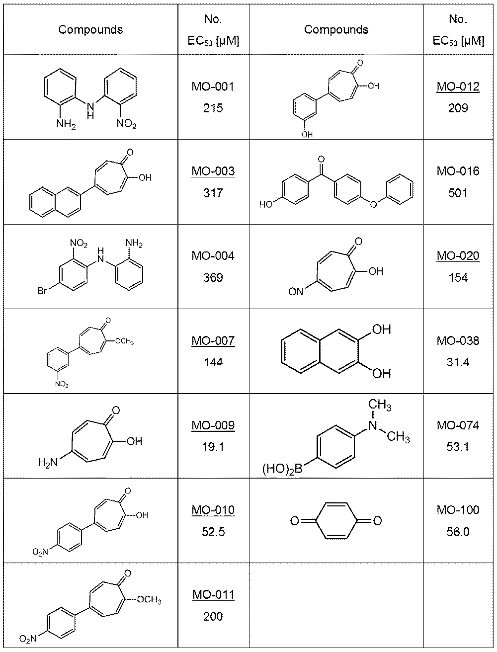
次に、新規のトロポロン誘導体36種を調製し、同様に200μMから5段階の5倍希釈系列を用いてAβ凝集阻害活性の評価を行ってEC50値を決定した。評価に供した36の誘導体のうち、表5に示す25化合物がAβ凝集阻害活性を示した。
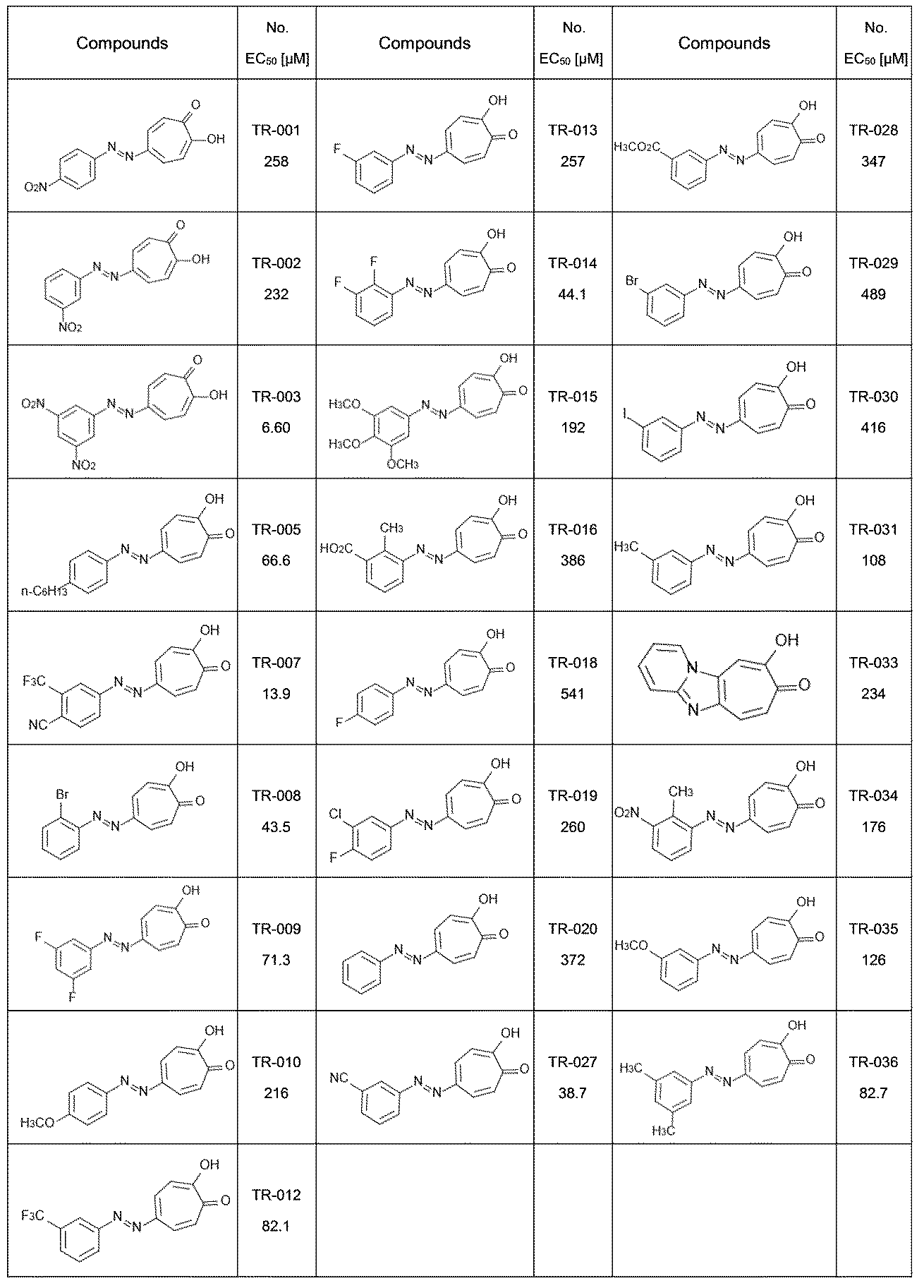
トロポロン誘導体上の置換基とAβ凝集阻害活性との関係を図18に示す。トロポロン環のみ(化合物MO-073)では活性は認められなかったが、アミノ基(化合物MO-009)及びNO基(化合物MO-020)は活性を増強し、トロポロン環上の他の置換基の電気的状態が活性に影響することが示唆された(図18の左上枠内)。また、化合物MO-006及びMO-010の比較によりニトロ基の位置(m位又はp位)が、化合物MO-010及びMO-011の比較によりトロポロン環上の水酸基が活性に影響することが示唆された(図18の右上枠内)。さらに、化合物TR-001~TR-004の中でTR-003が最も高い活性を示したことから、電子求引性ユニットの存在及びその個数が活性に影響することが示唆された(図18の左下枠内)。加えて、化合物TR-007及びTR-027の活性から、シアノ基が活性に関与することが示唆された(図18の右下枠内)。-NH2、-NO2、-OH、-CNはAβ42と水素結合を形成する能力を持つ官能基であり、水素結合の数及び位置がトロポロン誘導体のAβ凝集阻害活性に影響するものと推測される。
なお、図18中には、本評価系によるEC50値に加えて、従来のチオフラビンT(ThT)を用いた従来のAβ凝集阻害評価法(ThTアッセイ;方法は前出のIshigaki Y. et. al.を参照)によるEC50値を示している。本評価系によると、従来のThTアッセイでは活性を検出できなかった物質についても活性が確認された。
実施例4 タウ凝集阻害活性評価系の確立
(1)材料及び方法
・蛍光プローブQDTauの調製
マウスのタウ微小管結合ドメイン(MDB)断片(配列番号3)を既報(Tokuraku K. et. al., J Biol Chem., 278(32):29609-18, 2003、この文献は参照によりその全体が本明細書中に組み込まれる)に従って調製した。蛍光プローブQDTauは、タウMDB断片を実施例1(1)と同様にしてQdotで標識することで作製した。簡潔には、10μMのQD-PEG-NH2(Qdot(登録商標) 605 ITK(商標)Amino(PEG)Quantum Dots;Q21501MP、Thermo Fisher Scientific)を、最初に、1 mMのsulfo-EMCS(22307、Pierce)のPBS溶液と室温で1時間反応させた。未反応のsulfo-EMCSをクアミロイドーシスエンチして除去した後、QD-PEG-NH2と結合したsulfo-EMCSを、5 mM EDTAを含有する62.5μMのタウMDB断片のPBS溶液と室温で1時間反応させることでQDTauを調製した。
(1)材料及び方法
・蛍光プローブQDTauの調製
マウスのタウ微小管結合ドメイン(MDB)断片(配列番号3)を既報(Tokuraku K. et. al., J Biol Chem., 278(32):29609-18, 2003、この文献は参照によりその全体が本明細書中に組み込まれる)に従って調製した。蛍光プローブQDTauは、タウMDB断片を実施例1(1)と同様にしてQdotで標識することで作製した。簡潔には、10μMのQD-PEG-NH2(Qdot(登録商標) 605 ITK(商標)Amino(PEG)Quantum Dots;Q21501MP、Thermo Fisher Scientific)を、最初に、1 mMのsulfo-EMCS(22307、Pierce)のPBS溶液と室温で1時間反応させた。未反応のsulfo-EMCSをクアミロイドーシスエンチして除去した後、QD-PEG-NH2と結合したsulfo-EMCSを、5 mM EDTAを含有する62.5μMのタウMDB断片のPBS溶液と室温で1時間反応させることでQDTauを調製した。
(2)評価条件の最適化
・DTT濃度
タウMDB断片の中に存在する2つのシステイン残基(図20a参照)が分子内ジスルフィド架橋を形成して凝集を阻害する可能性があることから、分子内架橋を抑制するジチオスレイトール(DTT)を凝集反応液に加え、その影響を調べた。タウ凝集阻害活性の評価において、特に記載されていない条件は、実施例1のAβ凝集反応と同様である。
・DTT濃度
タウMDB断片の中に存在する2つのシステイン残基(図20a参照)が分子内ジスルフィド架橋を形成して凝集を阻害する可能性があることから、分子内架橋を抑制するジチオスレイトール(DTT)を凝集反応液に加え、その影響を調べた。タウ凝集阻害活性の評価において、特に記載されていない条件は、実施例1のAβ凝集反応と同様である。
1536ウェルプレート内で、10μMのタウMDB断片、50 nMのQDTau、10μMのヘパリン(凝集誘導物質)及び0~100 mMのDTTを、37℃で1日間インキュベートしてタウ凝集反応を行い、蛍光顕微鏡画像を撮像し(図19a上段)、関心領域内に含まれるそれぞれのピクセルの輝度値のSDを算出した(図19b)。また、タウ凝集物の沈降を見るため、共焦点レーザー顕微鏡(ニコン、Nikon C2 Plus)により凝集物をZ軸方向から観察した(図19a下段)。10 mM以上のDTT存在下でタウの凝集はほぼ平衡に達したことから、以降の試験においては10 mMのDTT濃度を採用した。
・タウ濃度
0~50μMのタウMDB断片、50 nMのQDTau、10μMのヘパリン及び10 mMのDTTをインキュベートしてタウ凝集反応を行い、蛍光顕微鏡画像を撮像し(図19c上段)、関心領域内に含まれるそれぞれのピクセルの輝度値のSDを算出した(図19d)。また、タウ凝集物の堆積を見るため、共焦点レーザー顕微鏡により凝集物をZ軸方向から観察した(図19c下段)。タウMBD断片の濃度10μMでタウ凝集量は最大となり、輝度値SDも最大になったことから、以下の試験においては10μMのタウMDB断片濃度を採用した。
0~50μMのタウMDB断片、50 nMのQDTau、10μMのヘパリン及び10 mMのDTTをインキュベートしてタウ凝集反応を行い、蛍光顕微鏡画像を撮像し(図19c上段)、関心領域内に含まれるそれぞれのピクセルの輝度値のSDを算出した(図19d)。また、タウ凝集物の堆積を見るため、共焦点レーザー顕微鏡により凝集物をZ軸方向から観察した(図19c下段)。タウMBD断片の濃度10μMでタウ凝集量は最大となり、輝度値SDも最大になったことから、以下の試験においては10μMのタウMDB断片濃度を採用した。
・タウ沈降アッセイ
10μMのタウMDB断片、50 nMのQDTau、10μMのヘパリン及び10 mMのDTTをインキュベートしてタウ凝集反応を行った。DTTを加えない以外は同組成の試料を比較対象とした。反応後、386,000×gで15分間、4℃で遠心分離して上清を取り出し、ペレットを同容量のPBSに再懸濁した。上清及び再懸濁したペレットのSDS-PAGE結果を図19eに示す。DTT非存在下ではタウMDB断片の多くが上清に存在していたのに対し、DTT存在下ではタウMDB断片のほとんどがペレットで回収された。以上から、上記条件での凝集反応により、反応液中のタウMDB断片のほとんどが凝集することが示された。
10μMのタウMDB断片、50 nMのQDTau、10μMのヘパリン及び10 mMのDTTをインキュベートしてタウ凝集反応を行った。DTTを加えない以外は同組成の試料を比較対象とした。反応後、386,000×gで15分間、4℃で遠心分離して上清を取り出し、ペレットを同容量のPBSに再懸濁した。上清及び再懸濁したペレットのSDS-PAGE結果を図19eに示す。DTT非存在下ではタウMDB断片の多くが上清に存在していたのに対し、DTT存在下ではタウMDB断片のほとんどがペレットで回収された。以上から、上記条件での凝集反応により、反応液中のタウMDB断片のほとんどが凝集することが示された。
(3)Aβ凝集反応との比較
タウ凝集反応は10μMのタウMDB断片、50 nMのQDTau、10μMのヘパリン及び10 mMのDTTを37℃で1日間インキュベートして行った。またAβ凝集反応は30 nMのQD605 Aβ及び30μMのAβ42を37℃で1日間インキュベートして行った。
タウ凝集反応は10μMのタウMDB断片、50 nMのQDTau、10μMのヘパリン及び10 mMのDTTを37℃で1日間インキュベートして行った。またAβ凝集反応は30 nMのQD605 Aβ及び30μMのAβ42を37℃で1日間インキュベートして行った。
QDプローブを添加せずに反応させた各試料10μlを200メッシュの銅グリッド上に沈着させ、1%リンタングステン酸でネガティブ染色した後、H-7600透過電子顕微鏡(日立製作所)を用いて、60kVでTEM観察を行った。タウ線維とAβ線維は形状が類似しており、その区別は困難であった(図20b)。
両凝集反応の進行を蛍光顕微鏡及び共焦点レーザー顕微鏡により観察したところ、タウはAβよりも早く凝集が進んでいた(図20c及びe)。反応後の試料のTEM観察によると、タウ、Aβ共にQDプローブを含む形で凝集しており、凝集物の形状も両者で異なることが確認されたことから、QDプローブは凝集物形状の観察にも有用であることが示された(図20d)。
さらに、両凝集反応において経時的に撮像した蛍光顕微鏡画像の関心領域内に含まれるピクセルの輝度値SDと反応時間との関係を図20gに示す。タウ、Aβ共に反応時間16時間程度で輝度値SDは平衡に達していることが確認された。
実施例5 タウ凝集阻害活性評価系及びAβ凝集阻害活性評価系の検証
被験物質としてAβ凝集阻害活性を有するシャペロン(カルレチクリン(CRT)、ERp57、プロテインジスルフィドイソメラーゼ(PDI);Kitauchi, K. et. al., Biochem. Biophys. Res. Commun. 481, 227-231, 2016を参照)を用いた試験を行った。CRT、ERp57及びPDIを既報(Sakono M. et. al., Biochem. Biophys. Res. Commun. 452, 27-31, 2014、この文献は参照によりその全体が本明細書中に組み込まれる)に従って調製し、50、10、2、0.4、0.08μMの各シャペロンの存在下で、実施例4(3)と同条件でタウ及びAβの凝集反応を行い、n = 3でEC50値を決定した。いずれのシャペロンもタウ及びAβの凝集阻害活性を有しており、特にAβに対してより強い活性を有することが確認された(図21)
被験物質としてAβ凝集阻害活性を有するシャペロン(カルレチクリン(CRT)、ERp57、プロテインジスルフィドイソメラーゼ(PDI);Kitauchi, K. et. al., Biochem. Biophys. Res. Commun. 481, 227-231, 2016を参照)を用いた試験を行った。CRT、ERp57及びPDIを既報(Sakono M. et. al., Biochem. Biophys. Res. Commun. 452, 27-31, 2014、この文献は参照によりその全体が本明細書中に組み込まれる)に従って調製し、50、10、2、0.4、0.08μMの各シャペロンの存在下で、実施例4(3)と同条件でタウ及びAβの凝集反応を行い、n = 3でEC50値を決定した。いずれのシャペロンもタウ及びAβの凝集阻害活性を有しており、特にAβに対してより強い活性を有することが確認された(図21)
実施例6 蛍光プローブとして量子ドットを用いたアミロイド凝集阻害活性の評価
蛍光プローブとしてQDAβ及びQDtauに代えて量子ドットQD-PEG-NH2(Qdot(登録商標) 655 ITK(商標)Amino(PEG)Quantum Dots;Q21521MP)を用いたこと以外は実施例4(3)と同条件でタウ及びAβの凝集反応を行った。また、α-シヌクレイン凝集反応は以下のように行った。ヒトのαーシヌクレイン(配列番号4)は、タウMDB断片と同様に遺伝子組換え大腸菌及び液体カラムクロマトグラフィーにより精製することで調製されたものを用いた(都城工業高等専門学校・野口太郎准教授より分与)。10 mM MgCl2を含むPBS溶液中で400μM α-シヌクレインモノマーを37℃で1週間スターラーにて攪拌し、シードとなるα-シヌクレイン凝集体(α-シヌクレインシード)を形成させた。10 mM MgCl2を含むPBS溶液中で2μM α-シヌクレインシード、20μM α-シヌクレインモノマー及び30 nMのQD-PEG-NH2(Qdot(登録商標) 655 ITK(商標)Amino(PEG)Quantum Dots;Q21521MP)を37℃で1週間インキュベートすることで、α-シヌクレイン凝集物を形成させた。
蛍光プローブとしてQDAβ及びQDtauに代えて量子ドットQD-PEG-NH2(Qdot(登録商標) 655 ITK(商標)Amino(PEG)Quantum Dots;Q21521MP)を用いたこと以外は実施例4(3)と同条件でタウ及びAβの凝集反応を行った。また、α-シヌクレイン凝集反応は以下のように行った。ヒトのαーシヌクレイン(配列番号4)は、タウMDB断片と同様に遺伝子組換え大腸菌及び液体カラムクロマトグラフィーにより精製することで調製されたものを用いた(都城工業高等専門学校・野口太郎准教授より分与)。10 mM MgCl2を含むPBS溶液中で400μM α-シヌクレインモノマーを37℃で1週間スターラーにて攪拌し、シードとなるα-シヌクレイン凝集体(α-シヌクレインシード)を形成させた。10 mM MgCl2を含むPBS溶液中で2μM α-シヌクレインシード、20μM α-シヌクレインモノマー及び30 nMのQD-PEG-NH2(Qdot(登録商標) 655 ITK(商標)Amino(PEG)Quantum Dots;Q21521MP)を37℃で1週間インキュベートすることで、α-シヌクレイン凝集物を形成させた。
量子ドットを添加せずに反応させた各試料のTEM観察によると、いずれのアミロイド線維も形状が類似していて区別は困難であった(図22上段)。これに対し、量子ドット添加試料では、凝集したそれぞれのアミロイド線維に量子ドットが付着しており、量子ドットもまた凝集物形状の観察にも有用であることが示された(図22中段)。
次に、被験物質としてシャペロン(CRT)及びロスマリン酸を用いて、タウ及びAβの凝集反応を行い、n = 3でEC50値を決定した。CRTは実施例5と同様にタウ凝集阻害活性を示し、ロスマリン酸は実施例1(3)と同様にAβ凝集阻害活性を示した(図23)。以上から、量子ドットそのものも本アミロイド凝集阻害活性評価系において蛍光プローブとして利用可能であることが示された。
実施例7 蛍光プローブとしてThTを用いたアミロイド凝集阻害活性の評価
10%EtOHを含むPBSバッファー中で25μMのThT(202-01002、和光純薬工業)及び25μMのAβ42を37℃で1日間インキュベートし、Aβ凝集反応を行った。反応前後の1536ウェルプレートを、カラーCCDカメラ(DS-Ri2、ニコン)を備えた倒立蛍光顕微鏡システム(ECLIPSE Ti-E、ニコン)で、4×対物レンズ及び蛍光フィルターキューブ(FITC Basic C-FL、Semrock)を用いて観察して蛍光顕微鏡画像を撮像した。関心領域内に含まれるそれぞれのピクセルの輝度値のSDを算出した(n = 3)。
10%EtOHを含むPBSバッファー中で25μMのThT(202-01002、和光純薬工業)及び25μMのAβ42を37℃で1日間インキュベートし、Aβ凝集反応を行った。反応前後の1536ウェルプレートを、カラーCCDカメラ(DS-Ri2、ニコン)を備えた倒立蛍光顕微鏡システム(ECLIPSE Ti-E、ニコン)で、4×対物レンズ及び蛍光フィルターキューブ(FITC Basic C-FL、Semrock)を用いて観察して蛍光顕微鏡画像を撮像した。関心領域内に含まれるそれぞれのピクセルの輝度値のSDを算出した(n = 3)。
反応前後の蛍光顕微鏡画像を図24Aに、画像から算出した輝度値SDを図24Bに示す。Aβの凝集によってThTの分布に偏りが生じ、輝度値SDの値も反応後に増加していたことから、ThTも本アミロイド凝集阻害活性評価系において蛍光プローブとして利用可能であることが示された。
1 システム
10 撮像装置
20 コンピュータ
21 入力部
22 画像入力部
23 記録部
24 演算部
25 出力部
26 制御部
231 プログラム記録部
241 SD算出部
242 活性判定部
10 撮像装置
20 コンピュータ
21 入力部
22 画像入力部
23 記録部
24 演算部
25 出力部
26 制御部
231 プログラム記録部
241 SD算出部
242 活性判定部
Claims (24)
- 被験物質の存在下又は非存在下で、アミロイド形成性タンパク質と、前記アミロイド形成性タンパク質の重合により形成されるアミロイドへの結合能を有する蛍光プローブとを反応させる凝集反応ステップと、
凝集反応ステップにおいて得た凝集反応生成物の蛍光を撮像する撮像ステップであって、凝集反応生成物の蛍光を、撮像される蛍光画像内の関心領域に含まれる各画素の輝度値から以下の式1:
Sum Intensity(%)=(関心領域内の各画素の輝度値の合計/関心領域内の各画素の理論上の最大輝度値の合計)×100
によって算出されるSum Intensityが15~85%となる露出条件で撮像する、前記ステップと、
撮像ステップにおいて撮像した蛍光画像内の関心領域に含まれる各画素の輝度値から標準偏差を算出する標準偏差算出ステップと、
標準偏差算出ステップにおいて算出した被験物質存在下での輝度値標準偏差の値と被験物質非存在下での輝度値標準偏差の値とを比較し、被験物質存在下での輝度値標準偏差の値が被験物質非存在下での輝度値標準偏差の値より小さい場合に、被験物質はアミロイド形成阻害活性を有すると判定する活性判定ステップと
を含む、被験物質のアミロイド形成阻害活性を判定する方法。 - 撮像ステップにおいて、前記式1によって算出されるSum Intensityが45~65%となる露出条件で撮像する、請求項1に記載の方法。
- 標準偏差算出ステップにおいて、各画素の輝度値に代えて、以下の式2:
補正後輝度値=輝度値×(目標Sum Intensity/前記式1によって算出されるSum Intensity)(式中、目標Sum Intensityは、45~65%の範囲から選択される値である。)
によって算出される各画素の補正後輝度値を用いて標準偏差を算出する、請求項1又は2に記載の方法。 - 標準偏差算出ステップにおいて、前記式1によって算出されるSum Intensityが目標Sum Intensityより小さい値であった場合に、各画素の輝度値に代えて、以下の式2:
補正後輝度値=輝度値×(目標Sum Intensity/前記式1によって算出されるSum Intensity)(式中、目標Sum Intensityは、45~65%の範囲から選択される値である。)
によって算出される各画素の補正後輝度値を用いて標準偏差を算出する、請求項1又は2に記載の方法。 - 凝集反応ステップにおいて複数濃度の被験物質を用いて凝集反応を行い、撮像ステップにおいて各被験物質濃度下での凝集反応生成物の蛍光を撮像し、標準偏差算出ステップにおいて各被験物質濃度に対応した輝度値標準偏差を算出し、活性判定ステップにおいて阻害活性の判定に加えて被験物質濃度と輝度値標準偏差との関係を表す阻害曲線から50%効果濃度(EC50)を決定する、請求項1~4のいずれか一項に記載の方法。
- アミロイド形成性タンパク質が、アミロイドβタンパク質、タウ及びα-シヌクレイン並びにそれらの部分ペプチド及びそれらの誘導体よりなる群から選択される、請求項1~5のいずれか一項に記載の方法。
- アミロイド形成性タンパク質が、配列番号1に示されるアミノ酸配列、配列番号3に示されるアミノ酸配列若しくは配列番号4に示されるアミノ酸配列からなるペプチドであるか、又は前記3つのアミノ酸配列のいずれかと90%以上の配列同一性を有するアミノ酸配列からなり、かつアミロイド形成能を有するペプチドである、請求項1~6のいずれか一項に記載の方法。
- 前記アミロイド形成性タンパク質の重合により形成されるアミロイドへの結合能を有する蛍光プローブが、前記アミロイド形成性タンパク質との間でアミロイドを形成する能力を持つ、蛍光標識されたペプチドである、請求項1~7のいずれか一項に記載の方法。
- アミロイド形成性タンパク質が配列番号1に示されるアミノ酸配列からなり、蛍光プローブ中の前記ペプチドが配列番号2に示されるアミノ酸配列からなる、請求項8に記載の方法。
- 凝集反応ステップにおいて反応開始時のアミロイド形成性タンパク質の濃度が25μMであり、蛍光プローブの濃度が25 nMである、請求項9に記載の方法。
- アミロイド形成性タンパク質及び蛍光プローブ中の前記ペプチドがいずれも配列番号3に示されるアミノ酸配列からなる、請求項8に記載の方法。
- 凝集反応ステップにおいて反応開始時のアミロイド形成性タンパク質の濃度が10μMであり、蛍光プローブの濃度が50 nMである、請求項11に記載の方法。
- 前記アミロイド形成性タンパク質の重合により形成されるアミロイドへの結合能を有する蛍光プローブが、アミロイド結合性蛍光色素又は蛍光標識された抗アミロイド特異抗体である、請求項1~7のいずれか一項に記載の方法。
- アミロイド結合性蛍光色素が、コンゴーレッド、チオフラビンT、チオフラビンS及びそれらの誘導体よりなる群から選択される、請求項13に記載の方法。
- 蛍光プローブが、量子ドットを蛍光色素として含む、又は量子ドットである、請求項1~13に記載の方法。
- 蛍光プローブの最大蛍光波長が500~610nmの範囲内にある、請求項1~15のいずれか一項に記載の方法。
- 被験物質の濃度が0.1 mg/ml以下である、請求項1~16のいずれか一項に記載の方法。
- 撮像装置とコンピュータとからなる、被験物質のアミロイド形成阻害活性を判定するシステムであって、
撮像装置が、被験物質の存在下又は非存在下でアミロイド形成性タンパク質と前記アミロイド形成性タンパク質の重合により形成されるアミロイドへの結合能を有する蛍光プローブとを反応させることで得られる凝集反応生成物の蛍光を撮像する装置であり、
コンピュータが、
凝集反応生成物の蛍光を、撮像される蛍光画像内の関心領域に含まれる各画素の輝度値から以下の式1:
Sum Intensity(%)=(関心領域内の各画素の輝度値の合計/関心領域内の各画素の理論上の最大輝度値の合計)×100
によって算出されるSum Intensityが15~85%となる露出条件で撮像するように撮像装置を制御する制御部と、
撮像装置により撮像された蛍光画像内の関心領域に含まれる各画素の輝度値から標準偏差を算出する標準偏差算出部と、
被験物質存在下での輝度値標準偏差の値と被験物質非存在下での輝度値標準偏差の値とを比較し、被験物質存在下での輝度値標準偏差の値が被験物質非存在下での輝度値標準偏差の値より小さい場合に被験物質はアミロイド形成阻害活性を有すると判定する活性判定部と
を備えることを特徴とする、前記システム。 - 制御部が、前記式1によって算出されるSum Intensityが45~65%となる露出条件で撮像するように撮像装置を制御する、請求項18に記載のシステム。
- 標準偏差算出部が、各画素の輝度値に代えて、以下の式2:
補正後輝度値=輝度値×(目標Sum Intensity/前記式1によって算出されるSum Intensity)(式中、目標Sum Intensityは、45~80%の範囲から選択される値である。)
によって算出される各画素の補正後輝度値を用いて標準偏差を算出する、請求項18又は19に記載のシステム。 - 標準偏差算出部が、前記式1によって算出されるSum Intensityが目標Sum Intensityより小さい値であった場合に、各画素の輝度値に代えて、以下の式2:
補正後輝度値=輝度値×(目標Sum Intensity/前記式1によって算出されるSum Intensity)(式中、目標Sum Intensityは、45~65%の範囲から選択される値である。)
によって算出される各画素の補正後輝度値を用いて標準偏差を算出する、請求項18又は19に記載のシステム。 - 撮像装置が複数濃度の被験物質存在下での凝集反応生成物の蛍光を撮像し、標準偏差算出部が各被験物質濃度に対応した輝度値標準偏差を算出し、活性判定部が阻害活性の判定に加えて被験物質濃度と輝度値標準偏差との関係を表す阻害曲線から50%効果濃度(EC50)をさらに決定する、請求項18~21のいずれか一項に記載のシステム。
- アミロイド形成性タンパク質と蛍光プローブと被験物質とを、又はアミロイド形成性タンパク質と蛍光プローブとを混合する試料混合装置と、
混合した試料を反応させる凝集反応装置と
をさらに備えることを特徴とする、請求項18~22のいずれか一項に記載のシステム。 - 被験物質のアミロイド形成阻害活性を判定するコンピュータで動作するプログラムであって、前記コンピュータを、
被験物質の存在下又は非存在下でアミロイド形成性タンパク質と前記アミロイド形成性タンパク質の重合により形成されるアミロイドへの結合能を有する蛍光プローブとを反応させることで得られる凝集反応生成物の蛍光を、撮像される蛍光画像内の関心領域に含まれる各画素の輝度値から以下の式1:
Sum Intensity(%)=(関心領域内の各画素の輝度値の合計/関心領域内の各画素の理論上の最大輝度値の合計)×100
によって算出されるSum Intensityが15~85%となる露出条件で撮像するように撮像装置を制御する制御部と、
撮像装置により撮像された蛍光画像内の関心領域に含まれる各画素の輝度値から標準偏差を算出する標準偏差算出部と、
被験物質存在下での輝度値標準偏差の値と被験物質非存在下での輝度値標準偏差の値とを比較し、被験物質存在下での輝度値標準偏差の値が被験物質非存在下での輝度値標準偏差より小さい場合に被験物質はアミロイド形成阻害活性を有すると判定する活性判定部と
して機能させるためのプログラム。
Applications Claiming Priority (2)
| Application Number | Priority Date | Filing Date | Title |
|---|---|---|---|
| JP2018242280A JP7166612B2 (ja) | 2018-12-26 | 2018-12-26 | アミロイド形成を評価する方法、装置及びプログラム |
| JP2018-242280 | 2018-12-26 |
Publications (1)
| Publication Number | Publication Date |
|---|---|
| WO2020138265A1 true WO2020138265A1 (ja) | 2020-07-02 |
Family
ID=71128660
Family Applications (1)
| Application Number | Title | Priority Date | Filing Date |
|---|---|---|---|
| PCT/JP2019/051077 Ceased WO2020138265A1 (ja) | 2018-12-26 | 2019-12-26 | アミロイド形成を評価する方法、装置及びプログラム |
Country Status (2)
| Country | Link |
|---|---|
| JP (1) | JP7166612B2 (ja) |
| WO (1) | WO2020138265A1 (ja) |
Cited By (2)
| Publication number | Priority date | Publication date | Assignee | Title |
|---|---|---|---|---|
| JPWO2022211014A1 (ja) * | 2021-03-31 | 2022-10-06 | ||
| WO2024071361A1 (ja) | 2022-09-27 | 2024-04-04 | 株式会社カネカ | 凝集性タンパク質に対する凝集抑制活性又は凝集促進活性を評価する方法 |
Families Citing this family (1)
| Publication number | Priority date | Publication date | Assignee | Title |
|---|---|---|---|---|
| WO2024070962A1 (ja) * | 2022-09-27 | 2024-04-04 | 株式会社カネカ | 凝集抑制剤 |
Citations (2)
| Publication number | Priority date | Publication date | Assignee | Title |
|---|---|---|---|---|
| JP2011212116A (ja) * | 2010-03-31 | 2011-10-27 | Sony Corp | 蛍光像取得方法、蛍光像取得プログラム及び蛍光像取得装置 |
| WO2018073784A1 (en) * | 2016-10-20 | 2018-04-26 | Optina Diagnostics, Inc. | Method and system for detecting an anomaly within a biological tissue |
-
2018
- 2018-12-26 JP JP2018242280A patent/JP7166612B2/ja active Active
-
2019
- 2019-12-26 WO PCT/JP2019/051077 patent/WO2020138265A1/ja not_active Ceased
Patent Citations (2)
| Publication number | Priority date | Publication date | Assignee | Title |
|---|---|---|---|---|
| JP2011212116A (ja) * | 2010-03-31 | 2011-10-27 | Sony Corp | 蛍光像取得方法、蛍光像取得プログラム及び蛍光像取得装置 |
| WO2018073784A1 (en) * | 2016-10-20 | 2018-04-26 | Optina Diagnostics, Inc. | Method and system for detecting an anomaly within a biological tissue |
Non-Patent Citations (1)
| Title |
|---|
| TOKURAKU, KIYOTAKA: "Development of a microliter-scale high-throughput screening system with quantum-dot for amyloid-beta aggregation inhibitors. Grant-in-Aid for Scientific Research", KAKEN.NII.AC.JP, 8 June 2016 (2016-06-08), pages 1 - 6, XP055721826, Retrieved from the Internet <URL:https://kaken.nii.ac.jp/ja/file/KAKENHI-PROJECT-25350974/25350974seika.pdf> * |
Cited By (3)
| Publication number | Priority date | Publication date | Assignee | Title |
|---|---|---|---|---|
| JPWO2022211014A1 (ja) * | 2021-03-31 | 2022-10-06 | ||
| WO2022211014A1 (ja) * | 2021-03-31 | 2022-10-06 | 株式会社カネカ | 凝集性タンパク質の測定方法及び凝集可視化方法、並びにこれらの方法に使用するためのキット |
| WO2024071361A1 (ja) | 2022-09-27 | 2024-04-04 | 株式会社カネカ | 凝集性タンパク質に対する凝集抑制活性又は凝集促進活性を評価する方法 |
Also Published As
| Publication number | Publication date |
|---|---|
| JP2020106287A (ja) | 2020-07-09 |
| JP7166612B2 (ja) | 2022-11-08 |
Similar Documents
| Publication | Publication Date | Title |
|---|---|---|
| Ji et al. | Actin filaments target the oligomeric maturation of the dynamin GTPase Drp1 to mitochondrial fission sites | |
| Haas et al. | Presenilin-dependent processing and nuclear function of γ-protocadherins | |
| JP7166612B2 (ja) | アミロイド形成を評価する方法、装置及びプログラム | |
| De Luca et al. | Probing ensemble polymorphism and single aggregate structural heterogeneity in insulin amyloid self-assembly | |
| Beton et al. | Cooperative amyloid fibre binding and disassembly by the Hsp70 disaggregase | |
| Molnar et al. | Mechanism of action of VP1-001 in cryAB (R120G)-associated and age-related cataracts | |
| Chang et al. | Interactions of amyloid-β peptides on lipid bilayer studied by single molecule imaging and tracking | |
| Büning et al. | Conformational dynamics and self-association of intrinsically disordered Huntingtin exon 1 in cells | |
| Samson et al. | Emerin self‐assembly mechanism: role of the LEM domain | |
| Higham et al. | Preferred formation of heteromeric channels between coexpressed SK1 and IKCa channel subunits provides a unique pharmacological profile of Ca2+-activated potassium channels | |
| Marchetti et al. | Ligand-induced dynamics of neurotrophin receptors investigated by single-molecule imaging approaches | |
| WO2022211014A1 (ja) | 凝集性タンパク質の測定方法及び凝集可視化方法、並びにこれらの方法に使用するためのキット | |
| EP2029157A2 (en) | Abc transporter ligand | |
| Angera et al. | Macrocyclic β-arch peptides that mimic the structure and function of disease-associated tau folds | |
| Machado et al. | Heat shock cognate protein 70 regulates gephyrin clustering | |
| Liao et al. | Counting fluorescently labeled proteins in tissues in the spinning–disk microscope using single–molecule calibrations | |
| EP3922723A1 (en) | Method and kit for discriminating between parkinson's disease and multiple system atrophy | |
| WO2020201296A1 (en) | Operation process for a cell cultivation system | |
| Walker et al. | Determination of oligomeric organization of membrane proteins from native membranes at nanoscale-spatial and single-molecule resolution | |
| JP2017007990A (ja) | アミロイド凝集性評価用汎用性量子ドットナノプローブ | |
| KR20220010051A (ko) | Bcl2 계통 단백질 표적 약물에 대한 반응성 예측 방법 | |
| DE102013202206A1 (de) | Methode zur Inhibierung des Proteins SWAP-70 | |
| Liesche et al. | CD95 receptor activation by ligand-induced trimerization is independent of its partial pre-ligand assembly | |
| JP6770741B2 (ja) | ネフローゼ症候群の予防薬又は治療薬をスクリーニングする方法、ネフローゼ症候群の予防又は治療用医薬組成物、ネフローゼ症候群診断用マーカー、ネフローゼ症候群の検査方法、及びネフローゼ症候群の診断用試薬 | |
| EP2908876B1 (fr) | Proteines recombinantes derivees du collagene a activite de liaison au facteur willebrand |
Legal Events
| Date | Code | Title | Description |
|---|---|---|---|
| 121 | Ep: the epo has been informed by wipo that ep was designated in this application |
Ref document number: 19904230 Country of ref document: EP Kind code of ref document: A1 |
|
| NENP | Non-entry into the national phase |
Ref country code: DE |
|
| 122 | Ep: pct application non-entry in european phase |
Ref document number: 19904230 Country of ref document: EP Kind code of ref document: A1 |