WO2020101303A1 - Vegfr-3 인산화 억제 펩타이드 및 이의 용도 - Google Patents
Vegfr-3 인산화 억제 펩타이드 및 이의 용도 Download PDFInfo
- Publication number
- WO2020101303A1 WO2020101303A1 PCT/KR2019/015266 KR2019015266W WO2020101303A1 WO 2020101303 A1 WO2020101303 A1 WO 2020101303A1 KR 2019015266 W KR2019015266 W KR 2019015266W WO 2020101303 A1 WO2020101303 A1 WO 2020101303A1
- Authority
- WO
- WIPO (PCT)
- Prior art keywords
- cells
- cancer
- peptide
- treatment
- seq
- Prior art date
- Legal status (The legal status is an assumption and is not a legal conclusion. Google has not performed a legal analysis and makes no representation as to the accuracy of the status listed.)
- Ceased
Links
Images







Classifications
-
- C—CHEMISTRY; METALLURGY
- C07—ORGANIC CHEMISTRY
- C07K—PEPTIDES
- C07K7/00—Peptides having 5 to 20 amino acids in a fully defined sequence; Derivatives thereof
- C07K7/04—Linear peptides containing only normal peptide links
- C07K7/06—Linear peptides containing only normal peptide links having 5 to 11 amino acids
-
- A—HUMAN NECESSITIES
- A61—MEDICAL OR VETERINARY SCIENCE; HYGIENE
- A61K—PREPARATIONS FOR MEDICAL, DENTAL OR TOILETRY PURPOSES
- A61K38/00—Medicinal preparations containing peptides
-
- A—HUMAN NECESSITIES
- A61—MEDICAL OR VETERINARY SCIENCE; HYGIENE
- A61K—PREPARATIONS FOR MEDICAL, DENTAL OR TOILETRY PURPOSES
- A61K38/00—Medicinal preparations containing peptides
- A61K38/04—Peptides having up to 20 amino acids in a fully defined sequence; Derivatives thereof
- A61K38/08—Peptides having 5 to 11 amino acids
-
- A—HUMAN NECESSITIES
- A61—MEDICAL OR VETERINARY SCIENCE; HYGIENE
- A61K—PREPARATIONS FOR MEDICAL, DENTAL OR TOILETRY PURPOSES
- A61K38/00—Medicinal preparations containing peptides
- A61K38/04—Peptides having up to 20 amino acids in a fully defined sequence; Derivatives thereof
- A61K38/10—Peptides having 12 to 20 amino acids
-
- A—HUMAN NECESSITIES
- A61—MEDICAL OR VETERINARY SCIENCE; HYGIENE
- A61K—PREPARATIONS FOR MEDICAL, DENTAL OR TOILETRY PURPOSES
- A61K40/00—Cellular immunotherapy
- A61K40/10—Cellular immunotherapy characterised by the cell type used
- A61K40/15—Natural-killer [NK] cells; Natural-killer T [NKT] cells
-
- A—HUMAN NECESSITIES
- A61—MEDICAL OR VETERINARY SCIENCE; HYGIENE
- A61P—SPECIFIC THERAPEUTIC ACTIVITY OF CHEMICAL COMPOUNDS OR MEDICINAL PREPARATIONS
- A61P35/00—Antineoplastic agents
-
- A—HUMAN NECESSITIES
- A61—MEDICAL OR VETERINARY SCIENCE; HYGIENE
- A61P—SPECIFIC THERAPEUTIC ACTIVITY OF CHEMICAL COMPOUNDS OR MEDICINAL PREPARATIONS
- A61P35/00—Antineoplastic agents
- A61P35/04—Antineoplastic agents specific for metastasis
-
- C—CHEMISTRY; METALLURGY
- C07—ORGANIC CHEMISTRY
- C07K—PEPTIDES
- C07K7/00—Peptides having 5 to 20 amino acids in a fully defined sequence; Derivatives thereof
- C07K7/04—Linear peptides containing only normal peptide links
- C07K7/08—Linear peptides containing only normal peptide links having 12 to 20 amino acids
-
- A—HUMAN NECESSITIES
- A61—MEDICAL OR VETERINARY SCIENCE; HYGIENE
- A61K—PREPARATIONS FOR MEDICAL, DENTAL OR TOILETRY PURPOSES
- A61K2121/00—Preparations for use in therapy
-
- C—CHEMISTRY; METALLURGY
- C12—BIOCHEMISTRY; BEER; SPIRITS; WINE; VINEGAR; MICROBIOLOGY; ENZYMOLOGY; MUTATION OR GENETIC ENGINEERING
- C12N—MICROORGANISMS OR ENZYMES; COMPOSITIONS THEREOF; PROPAGATING, PRESERVING, OR MAINTAINING MICROORGANISMS; MUTATION OR GENETIC ENGINEERING; CULTURE MEDIA
- C12N2501/00—Active agents used in cell culture processes, e.g. differentation
- C12N2501/10—Growth factors
- C12N2501/165—Vascular endothelial growth factor [VEGF]
Definitions
- the present invention relates to a peptide having a phosphorylation inhibitory activity of vascular endothelial growth factor receptor (VEGFR) -3 and uses thereof.
- VEGFR vascular endothelial growth factor receptor
- NK cells natural killer cells
- AML acute myeloid leukemia
- Acute myeloid leukemia disease has been reported to have an excellent effect of natural killer cells among blood tumors and to act more significantly than other solid cancers, and immunocytotherapy using NK cells has focused researchers' attention over a long period of time and recently It is constantly increasing.
- NK cells After the transplantation of hematopoietic stem cells, the initial immune system is mostly carried out by NK cells, but before the immune reconstruction, the functional defects of NK cells cause fatal infection and leukemia recurrence.
- the work of recovering these NK cells numerically and functionally has many technical limitations. Although there have been technological advances such as vaccines for immune cell therapy and T cell stimulation methods, it has not yet succeeded in mass proliferation of NK cells while enhancing the NK cell killing function to maintain the cancer treatment effect due to immune reconstruction. none.
- Korean Patent Publication No. 10-2015-0015892 discloses a pharmaceutical composition for the treatment of acute myeloid leukemia comprising an inhibitor of VEGFR-3 that increases the expression of interferon-gamma as an active ingredient.
- a novel peptide capable of inhibiting phosphorylation of VEGFR-3 was prepared, and the effect of enhancing the NK cell killing function by the novel peptide was not evaluated in vivo.
- the present inventors tried to develop a low molecular weight peptide material having therapeutic activity of cancer through inhibition of phosphorylation of vascular endothelial growth factor receptor (VEGFR) -3. As a result, it succeeded in synthesizing mutant peptides having VEGFR-3 phosphorylation inhibitory activity derived from the Fc crinkle domain of VEGFR-3, and completed the present invention by experimentally proving their cancer treatment activity potential.
- VEGFR vascular endothelial growth factor receptor
- an object of the present invention is to provide a peptide consisting of any one of the amino acid sequence of SEQ ID NO: 1 to SEQ ID NO: 12.
- Another object of the present invention is (a) a pharmaceutically effective amount of a peptide consisting of any one of the amino acid sequence of SEQ ID NO: 1 to SEQ ID NO: 12; And (b) to provide a pharmaceutical composition for the treatment or prevention of cancer, including a pharmaceutically acceptable carrier.
- Another object of the present invention is to provide a composition for promoting in vitro proliferation of NK (Natural Killer) cells comprising a peptide composed of any one of SEQ ID NOs: 1 to 12 as an active ingredient. .
- the present invention provides a peptide consisting of the amino acid sequence of any one of SEQ ID NO: 1 to SEQ ID NO: 12.
- the amino acid sequence is a novel low molecular peptide synthesized by the inventors by designing a peptide predicted to inhibit phosphorylation of VEGFR-3 in the Fc crinkle domain of vascular endothelial growth factor receptor (VEGFR) -3.
- VEGFR vascular endothelial growth factor receptor
- the peptide inhibits phosphorylation of vascular endothelial growth factor receptor (VEGFR) -3.
- VEGFR vascular endothelial growth factor receptor
- the peptide increases the expression of IFN- ⁇ (interferon - ⁇ ) in immune cells.
- immune cells includes cells involved in the immune response in the body, for example, Killer T cells, Helper T cells, B cells, Natural Killer (NK) cells, monocytes, large Macrophages, adipocytes, vascular endothelial cells, but are not limited thereto.
- Killer T cells Helper T cells
- B cells Natural Killer (NK) cells
- monocytes large Macrophages
- adipocytes vascular endothelial cells
- IFN- ⁇ also called type II interferon
- IFN- ⁇ is made by CD4 T cells or CD8 T cells and modulates the immune response, so it is also called immune interferon.
- IFN- ⁇ is a cytokine (cytokine) that acts on NK cells, T cells, B cells, neutrophils, and vascular endothelial cells to activate them, and acts as a macrophage activating factor, Class I, II It also increases MHC expression.
- NK cells can destroy cancer cells by releasing important cytokines such as IFN- ⁇ .
- NK cells in hematologic cancer patients have a lower cytokine secretion function than normal cells, and thus exhibit low lytic activity against cancer cells.
- the peptide promotes the proliferation of NK (Natural Killer) cells, or increases the cytotoxicity activity of NK cells.
- the present invention is (a) a pharmaceutically effective amount of a peptide consisting of the amino acid sequence of any one of SEQ ID NO: 1 to SEQ ID NO: 12; And (b) provides a pharmaceutical composition for the treatment or prevention of cancer comprising a pharmaceutically acceptable carrier.
- vascular endothelial growth factor receptor (VEGFR) -3 can promote the activity of immune cells and be used in the treatment of cancer (Cancer Letters 354 (2014) 281-289, J Clin Immunol) (2013) 33: 826-837).
- the composition inhibits phosphorylation of vascular endothelial growth factor receptor (VEGFR) -3.
- VEGFR vascular endothelial growth factor receptor
- the composition increases the expression of IFN- ⁇ (interferon - ⁇ ) in immune cells.
- the composition promotes cell proliferation of NK (Natural Killer) cells, or increases cytotoxicity activity of NK cells.
- the cancer is blood cancer, stomach cancer, lung cancer, breast cancer, ovarian cancer, liver cancer, bronchial cancer, nasopharyngeal cancer, larynx cancer, pancreatic cancer, bladder cancer, colon cancer, cervical cancer, brain cancer, prostate cancer, bone cancer, It is a cancer selected from the group consisting of skin cancer, thyroid cancer, parathyroid cancer, and ureteral cancer.
- the blood cancer includes, but is not limited to, leukemia, acute myelogenous leukemia, chronic myelogenous leukemia, lymphoma, multiple myeloma, myelodysplastic syndrome, erythropoiesis, and thrombocytopenia.
- the subject of application of the pharmaceutical composition for the treatment or prevention of cancer of the present invention is not particularly limited, but may be a mammal, including a human.
- the pharmaceutical composition of the present invention can treat or prevent cancer by administering it to mammals, including humans.
- the pharmaceutical composition of the present invention includes a “pharmaceutical effective amount” of a peptide consisting of the amino acid sequence of any one of SEQ ID NOs: 1 to 12, which is the above-described active ingredient.
- pharmaceutically effective amount refers to an amount sufficient to achieve the efficacy or activity (eg, anti-cancer efficacy, anti-tumor efficacy, etc.) of a peptide consisting of the amino acid sequence of any one of SEQ ID NOs: 1 to 12 described above. it means.
- the pharmaceutical composition of the present invention may further include "a pharmaceutically acceptable carrier" in addition to the active ingredient.
- a pharmaceutically acceptable carrier included in the pharmaceutical composition of the present invention is commonly used in formulation, lactose, dextrose, sucrose, sorbitol, mannitol, starch, acacia rubber, calcium phosphate, alginate, gelatin, Calcium silicate, microcrystalline cellulose, polyvinylpyrrolidone, cellulose, water, syrup, methyl cellulose, methylhydroxybenzoate, propylhydroxybenzoate, talc, magnesium stearate and mineral oil, but is not limited thereto It does not work.
- the pharmaceutical composition of the present invention may further include a lubricant, a wetting agent, a sweetener, a flavoring agent, an emulsifying agent, a suspending agent, a preservative, etc. in addition to the above components.
- a lubricant e.g., a talc, a kaolin, a kaolin, a kaolin, a kaolin, a kaolin, kaolin, kaolin, sorbitol, sorbitol, sorbitol, sorbitol, sorbitol, sorbitol, sorbitol, sorbitol, sorbitol, sorbitol, sorbitol, sorbitol, sorbitol, a talct, a talct, a talct, a stevia, glycerin, a stevia, glycerin, glycerin, g
- the pharmaceutical composition of the present invention may be administered orally or parenterally, preferably parenterally.
- parenterally intravenous injection, subcutaneous injection, intramuscular injection, intraperitoneal injection, transdermal administration, and the like can be administered, and those skilled in the art can select a suitable method from various administration methods.
- Suitable dosages of the pharmaceutical compositions of the invention are variously prescribed by factors such as formulation method, mode of administration, patient's age, weight, sex, morbidity, food, time of administration, route of administration, rate of excretion, and response sensitivity. Can be.
- the oral dosage of the pharmaceutical composition of the present invention is preferably 0.0001-100 mg / kg (body weight) per day.
- the pharmaceutical composition of the present invention is prepared in a unit dose form by formulating using a pharmaceutically acceptable carrier and / or excipient according to a method that can be easily carried out by those skilled in the art to which the present invention pertains. Or it can be manufactured by incorporating into a multi-dose container.
- the formulation may be in the form of a solution, suspension, or emulsion in an oil or aqueous medium, or may be in the form of extracts, powders, granules, tablets or capsules, and may further include dispersants or stabilizers.
- the present invention is a composition for promoting in vitro proliferation of NK (Natural Killer) cells comprising a peptide consisting of any one of the amino acid sequences of SEQ ID NO: 1 to SEQ ID NO: 12 as an active ingredient gives
- the content of the peptide in the composition for promoting in vitro proliferation of NK cells of the present invention is the same as that for the peptide described in the pharmaceutical composition for the treatment and prevention of cancer, which is one aspect of the present invention. For the avoidance, the same content is not repeated between the two inventions.
- the present invention relates to a peptide consisting of the amino acid sequence of any one of SEQ ID NO: 1 to SEQ ID NO: 12 and its use.
- the peptide of the present invention inhibits the activation of VEGFR-3, increases the expression of IFN- ⁇ (interferon - ⁇ ) in immune cells, promotes proliferation of NK (Natural Killer) cells, or cytotoxicity of NK cells ) Increases activity.
- the peptide of the present invention can be used for immunological treatment and prevention of cancer through the above-described activity.
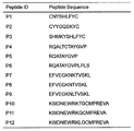
- 1 is a table showing the peptide ID of SEQ ID NO: 1 to SEQ ID NO: 12 and each amino acid sequence.
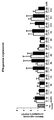
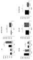
- Figure 2 is a graph showing the relative m-RNA expression level of the INF- ⁇ gene after the peptide treatment of SEQ ID NO: 1 to SEQ ID NO: 12 on monocyte cells.
- the relative m-RNA expression level of the INF- ⁇ gene was measured by normalizing to GAPDH.
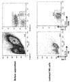
- 3 is a graph showing separation and purity for NK cells isolated by MACS method.
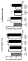
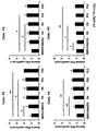
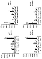
- FIG. 4 is a graph showing the relative expression level of INF- ⁇ according to the peptides (# 4, 6, 11, 12) treated in CD3-CD56 + cells and CD30CD16 + cells.
- the negative control group was the untreated group and the positive control group was the group treated with the MAZ51 compound.
- the treatment concentration of MAZ51 is 5, 10 ⁇ m, and the concentration of peptide treatment is 25, 50 ⁇ m.
- FIG. 5 is a graph showing the relative expression level of INF- ⁇ according to peptides (# 6, 11) treated in CD3-CD56 + cells and CD30CD16 + cells.
- the control subjects and treatment concentrations of each substance are the same as in the experiment of FIG. 4.
- PMA phorbol 12-myristate
- Ca Ionophore calcium ion permeate
- FIG. 7 is a graph showing the relative amount of INF- ⁇ secretion according to various concentrations of peptides (# 4, 6, 11, 12) in peripheral blood mononuclear cells of acute myeloid leukemia patients. (*: p ⁇ 0.05, **: p ⁇ 0.01)
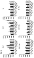
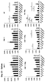
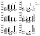
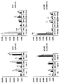
- FIG. 8 is a graph analyzing the number of immune cells (CD8 + T, CD4 + T, NK cells) in blood and the amount of INF- ⁇ secreted from immune cells (CD8 + T, CD4 + T, NK cells), 1 day of peptide treatment This is a comparison between the normal and patient groups and the peptide (# 4, 6, 11, 12) treatment group.
- FIG. 9 is a graph analyzing the number of immune cells (CD8 + T, CD4 + T, NK cells) in blood and the amount of INF- ⁇ secreted from immune cells (CD8 + T, CD4 + T, NK cells), 1 day of peptide treatment It is a value comparing the normal and patient groups with the peptide (# 4, 6, 11, 12) treatment group. (*: p ⁇ 0.05, **: p ⁇ 0.01, ***: P ⁇ 0.001)
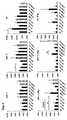
- 10 is a graph analyzing the number of immune cells (CD8 + T, CD4 + T, NK cells) in blood and the amount of INF- ⁇ secreted from immune cells (CD8 + T, CD4 + T, NK cells), 3 days of peptide treatment It is a value comparing the normal and patient groups with the peptide (# 4, 6, 11, 12) treatment group. (*: p ⁇ 0.05, **: p ⁇ 0.01)
- 11 is a graph analyzing the number of immune cells (CD8 + T, CD4 + T, NK cells) in blood and the amount of INF- ⁇ secreted from immune cells (CD8 + T, CD4 + T, NK cells), 15 days of peptide treatment It is a value comparing the normal and patient groups with the peptide (# 4, 6, 11, 12) treatment group. (*: p ⁇ 0.05, **: p ⁇ 0.01)
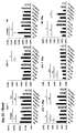
- FIG. 12 is a graph analyzing the number of immune cells (CD8 + T, CD4 + T, NK cells) in bone marrow and the amount of INF- ⁇ secreted from immune cells (CD8 + T, CD4 + T, NK cells), 15 days of peptide treatment It is a value comparing the normal and patient groups with the peptide (# 4, 6, 11, 12) treatment group. (*: p ⁇ 0.05, **: p ⁇ 0.01)
- FIG. 13 is a graph analyzing the number of immune cells (CD8 + T, CD4 + T, NK cells) in the spleen and the amount of INF- ⁇ secreted from immune cells (CD8 + T, CD4 + T, NK cells), 15 days of peptide treatment It is a value comparing the normal and patient groups with the peptide (# 4, 6, 11, 12) treatment group. (*: p ⁇ 0.05, **: p ⁇ 0.01)
- FIG. 14 is a graph analyzing the number of immune cells (CD8 + T, CD4 + T, NK cells) in blood and the amount of INF- ⁇ secreted from immune cells (CD8 + T, CD4 + T, NK cells) over time, normal group And the peptide 4 treatment group. (*: p ⁇ 0.05, **: p ⁇ 0.01)
- 16 is a graph analyzing the number of immune cells (CD8 + T, CD4 + T, NK cells) in bone marrow and spleen and the amount of INF- ⁇ secreted from immune cells (CD8 + T, CD4 + T, NK cells), normal group And the patient group and the peptide 4 treatment group. (*: p ⁇ 0.05, **: p ⁇ 0.01)
- FIG. 17 is a graph obtained by selecting and analyzing only the significant results among the analysis results of FIGS. 14 to 16. (*: p ⁇ 0.05, **: p ⁇ 0.01)
- FIG. 20 is a graph analyzing the number of immune cells (CD8 + T, CD4 + T, NK cells) in bone marrow and spleen and the amount of INF- ⁇ secreted from immune cells (CD8 + T, CD4 + T, NK cells), normal group And the patient group and the peptide 11 treatment group. (*: p ⁇ 0.05, **: p ⁇ 0.01)
- FIG. 21 is a graph obtained by selecting and analyzing only significant results among the analysis results of FIGS. 18 to 20. (*: p ⁇ 0.05, **: p ⁇ 0.01)
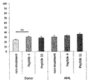
- FIG. 22 is a graph showing the activity against cytotoxicity after treatment with peptides (# 4, 11) to NK cells isolated from peripheral mononuclear cells and NK cells isolated from patients with acute myeloid leukemia. (**: p ⁇ 0.01)
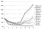
- NBM normal bone marrow
- NPB normal peripheral blood
- 24 and 25 are graphs showing the number of CD3-CD56 + cells by period (on the day of treatment, day 6, day 21) after treatment of peptides (# 4, 11) on peripheral monocytes and bone marrow cells of normal humans and patients.
- a small molecule mutated candidate peptide derived from the Fc crinkle domain that inhibits VEGFR-3 receptor phosphorylation was constructed. First, the peptides were randomly cut to a size not exceeding 15 mer, based on the sequences located in Thr446 and Lys516 of the D5 region bound by the strong disulfide bond of the VEGFR-3 Fc region, and 12 candidate groups Secured. 1 and Table 1 below show sequences of 12 peptide candidate groups.
- Example 2-1 Measurement of gene expression for IFN- ⁇ in a patient's monocyte cells after peptide treatment
- Acute myeloid patient groups were recruited for patient cell collection and cells were frozen and stored. Monocytes were separated using a specific gravity difference of 1.077, washed with DPBS (Dulbecco's Phosphate-Buffered Saline), and then put into fetal bovine serum (FBS) containing 10% DMSO (dimethyl sulfoxide) and frozen in LN2.
- DPBS Dynamic Phosphate-Buffered Saline
- FBS fetal bovine serum
- RNA Ribonucleic acid
- RNA obtained by precipitation with isopropanol was washed with 70% ethanol, dried, and then 500 ug was obtained, followed by reverse transcription using a cDNA (complementary DNA) synthesis kit to obtain primary cDNA.
- PCR polymerase chain reaction
- Figure 2 is a result of examining the gene expression for IFN- ⁇ after peptide treatment using the peripheral and bone marrow monocytes of the patient by RT-qPCR. All patients did not show the same expression, but most showed a tendency to increase when treated with concentrations of 25 ⁇ M and 50 ⁇ M, and peptide IDs 4, 5, 6, 9, 11, and 12 were higher than the untreated blind. , And showed an increased pattern than when the MAZ51 compound (VEGFR3 kinase inhibitor, CAS 163655-37-6) was treated.
- Example 2-2 NK cell separation and purity measurement using MACS
- NK cells Separation of NK cells is essential to investigate apoptosis, proliferation and substantial functional changes in vitro by peptides of NK cells. As shown in Figure 3, cells were separated using MACS and the purity was measured. At 93.3%, it was confirmed that the MACS sorting method had no problem in cell separation in technical terms.
- Example 2-3 Measurement of IFN- ⁇ protein expression in NK cells of patients after peptide treatment
- FACS FACS was performed to investigate the expression level of IFN- ⁇ protein in NK cells. After washing the separated cells with PBS (phosphate buffer saline) containing 1% bovine serum albumin, put them into a 12 ⁇ 75mm polystyrene (Falcon 2052) FACS tube at a concentration of 1-3 ⁇ 10 5 / 100 ⁇ l, and each corresponding single After adding 5 ⁇ l each of the clone antibodies, the reaction was performed at 4 ° C. for 30 minutes, and then washed twice with PBS containing 2% bovine serum albumin.
- PBS phosphate buffer saline
- bovine serum albumin a 12 ⁇ 75mm polystyrene
- the cells were analyzed with a flow cytometer (FACSCalibur, Becton Dickinson, USA) after the last washing to track the cell death response in all processes.
- FACSCalibur Becton Dickinson, USA
- the cells were stained with the same antibody, and then the degree of VEGFR-3 + and VEGFR-3- cell populations in the CD56 + CD3- cell population was investigated. After treatment, FACS was performed.
- CD3-CD56 + cells increased when treated with peptides 6 and 11, whereas CD3-CD16 + treated with peptides 6 and 11 compared to the MAZ51 compound compared to IFN- ⁇ protein expression It did not increase significantly. Overall, it was confirmed that the peripheral NK cells are more sensitive to the peptides than the bone marrow NK cells.
- Example 3 cytoimmunity test of NK cells after peptide treatment
- a new group of acute myeloid patients was continuously recruited for patient cell collection and cells collected from them were frozen and stored.
- the monocytes were separated using a specific gravity difference of 1.077, washed with DPBS, placed in FBS containing 10% DMSO, and frozen in LN2.
- Example 4 Investigation and analysis of killing ability of NK cells upon injection into the peptide body
- Example 4-1 Investigation of the change in the number of NK cells in vivo and the secretion of IFN- ⁇ in NK cells upon injection of a peptide into the body
- the selected peptide was injected into the body of the mouse and then examined for immune cells.
- Candidate peptides (2 mg / kg each) were simultaneously injected into the leukemia model mice produced by injecting the normal group mouse and the cancer stem cell C1498 cell line (2x10 6 cells / 100 ⁇ l). For each group, FACS was performed to investigate changes in the number of immune cells and changes in the amount of IFN- ⁇ secretion over time (Day-1, Day1, Day3, Day15) between the group injected with and without the peptide. . After obtaining blood, bone marrow, and spleen from mice, the red blood cells were destroyed with RBC lysis buffer, and then washed with PBS.
- FACS tube (tube)
- PBS PBS
- immune cell antibody in 100 ⁇ l of FACS buffer (buffer) and reacted at room temperature for 20 minutes. Thereafter, after washing with FACS buffer once more, 200 ⁇ l of Fix. & Perm, buffer was treated for 20 minutes at room temperature, washed, and then analyzed with a flow cytometer (FACSCanto2, Becton Dickinson, USA).
- Figure 9 shows the results for the change in the number of immune cells in the blood and the secretion of IFN- ⁇ 1 day after the peptide injection (Day1).
- peptide 11 was administered compared to the normal group, the number of NK cells in the blood increased.
- peptide 4 compared to the patient model group, IFN- ⁇ secreted from NK cells in the blood increased.
- Fig. 10 shows the results of changes in the number of immune cells in the blood and changes in the secretion of IFN- ⁇ at 3 days after the peptide injection (Day3). Compared to the normal group, the number of NK cells in the blood increased when peptide 4 was administered.
- results of changes in the number of immune cells in the blood and secretion of IFN- ⁇ at 15 days after the peptide injection (Day 15) are shown in FIG. 11.
- IFN- ⁇ secreted from NK cells in the blood decreased when peptide 6 was administered.
- IFN- ⁇ secreted from NK cells in the blood increased.
- the number of NK cells in the blood increased when peptide 12 was administered, and the IFN- ⁇ secreted from the NK cells in the blood also increased.
- results of changes in the number of immune cells in the bone marrow and secretion of IFN- ⁇ at 15 days after the peptide injection (Day 15) can be confirmed in FIG. 12.
- peptide 4 was administered compared to the patient model group
- the number of NK cells in the bone marrow increased, and IFN- ⁇ secreted from the NK cell in the bone marrow decreased compared to the patient model group.
- peptide 6 was administered compared to the normal group, the number of NK cells in the bone marrow increased, and the IFN- ⁇ secreted from the NK cells in the bone marrow decreased compared to the patient model group.
- IFN- ⁇ secreted from NK cells in the bone marrow increased when peptide 11 was administered.
- the number of NK cells in the bone marrow increased when peptide 11 was administered, and the IFN- ⁇ secreted from the NK cells in the bone marrow decreased.
- the number of NK cells in the bone marrow decreased, and the IFN- ⁇ secreted from the NK cells in the bone marrow also increased.
- IFN- ⁇ secreted from NK cells in the spleen increased when peptide 4 was administered compared to the normal group.
- IFN- ⁇ secreted from NK cells in the spleen increased.
- IFN- ⁇ secreted from NK cells in the spleen decreased when peptide 6 was administered.
- the number of NK cells in the spleen increased as compared to the normal group, and IFN- ⁇ secreted from the NK cells in the spleen also increased.
- the number of NK cells in the spleen of Day 15 increased when peptide 12 was administered, and the IFN- ⁇ secreted from the NK cells in the spleen decreased.
- FIG. 14 is a result showing the change in the number of immune cells in the blood and the secretion of INF- ⁇ between the normal group and the peptide-treated group by period after the injection of peptide 4. As shown in FIG. 14, it was confirmed that the number of NK cells in the blood 3 days after the injection of peptide 4 was significantly increased compared to the normal group, and it was the largest among the comparison between the normal group and the peptide-treated group at different times.
- FIG. 15 shows the results of changes in the number of immune cells in the blood and the secretion of INF- ⁇ between the patient model group and the peptide treatment group according to the period after injection of peptide 4. 15 shows that the INF- ⁇ secretion of NK cells in the blood 1 day after injection is significantly increased compared to the patient model group.
- Figure 16 is a result showing the change in the number of immune cells and secretion of INF- ⁇ between the bone marrow and spleen tissue and groups 15 days after the injection of peptide 4. It was confirmed that the INF- ⁇ secretion of NK cells in the spleen increased significantly compared to the normal group. It was confirmed that the number of NK cells in the bone marrow was significantly increased compared to the patient model group, and that the INF- ⁇ secretion of NK cells in the bone marrow was significantly decreased compared to the patient model group.
- FIG. 18 is a result showing the change in the number of immune cells in the blood and secretion of INF- ⁇ between the normal group and the peptide-treated group according to the period after injection of peptide 11. As shown in FIG. 18, it was confirmed that INF- ⁇ secretion of NK cells in the blood after 1 day was significantly increased compared to the normal group, and INF- ⁇ secretion of NK cells in the blood after 15 days was also significant compared to the normal group. It was confirmed that the increase.
- 19 is a result showing the change in the number of immune cells in the blood and the secretion of INF- ⁇ between the patient group and the peptide-treated group according to the time period after the injection of peptide 11.
- FIG. 19 is a result showing the change in the number of immune cells and secretion of INF- ⁇ between bone marrow and spleen tissue and groups 15 days after injection of peptide 11.
- the number of NK cells in the spleen and INF- ⁇ secretion of NK cells increased significantly compared to the normal group.
- INF- ⁇ secretion of NK cells in the bone marrow was significantly increased compared to the normal group.
- the number of NK cells in the bone marrow after 15 days of injection was significantly increased compared to the patient group, but INF- ⁇ secretion of NK cells was decreased.
- Example 5 Investigation and analysis of killing ability of immune cells NK cells upon in vivo injection of peptides
- cytotoxicity analysis was performed through DELFIA® EuTDA Cytotoxicity Reagents (PerkinElmer, USA). K562 was incubated at 37 ° C for 30 minutes with 1 ul BATDA in 2 ml at a concentration of 1x10 6 cells / ml. After washing with DPBS 3 times for 4 minutes at 4 ° C and 1000 rpm, K562 5x10³cells / 100ul were spread on 96-well plates.
- Example 6 Culture efficiency analysis and protocol establishment of NK cells in conditioned medium treated with new peptides
- the culture medium is replaced every 3 days using RPMI (Roswell park memorial institute) 1640 media containing 10% fetal bovine serum and 1% antibiotics. And cultured until 21st.
- RPMI Roswell park memorial institute
- Each cell was plated at 1x10 6 cells / ml in a 24-well culture plate. From day 0 to day 6, 10 IU / ml IL-2 was added together, and from then on, 100 IU / ml IL-2 and 5 ng / ml IL-15 were added together. And when replacing the culture medium, each peptide was treated with 50 ⁇ M.
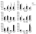
- 22 is a result of confirming the cell viability by changing the culture medium every 3 days and measuring the number of cells. 22 shows that as a result of treating the peptide 4 in the bone marrow cells of normal people, the number of cells increased from 15 days after the control. In addition, after the treatment of peptide 4 in the bone marrow cells of the patient, the number of cells, although not as much as the control group, showed a trend of increasing gradually. As a result of treatment with peptide 4 in the peripheral mononuclear cells of the patient, as in the control group, it was not seen to increase significantly until day 21, but both the control group and the peptide treatment group gradually increased.
- 23 to 25 are the results of confirming the number of CD3-CD56 + cells through Flow Cytometry to identify NK cells on Days 6 and 21. As shown in FIG. 23 to 25, when analyzed with 3 samples per group, there was no significant difference between groups, but in the bone marrow cells of normal and patient patients, the peptide 4 was treated on the 6th day and showed an increasing pattern. The peripheral mononuclear cells of the patient showed an increase in treatment with peptide 4 on day 21.
- the present invention relates to a peptide having a phosphorylation inhibitory activity of vascular endothelial growth factor receptor (VEGFR) -3 and uses thereof.
- VEGFR vascular endothelial growth factor receptor
Landscapes
- Health & Medical Sciences (AREA)
- Chemical & Material Sciences (AREA)
- Life Sciences & Earth Sciences (AREA)
- Organic Chemistry (AREA)
- General Health & Medical Sciences (AREA)
- Medicinal Chemistry (AREA)
- Proteomics, Peptides & Aminoacids (AREA)
- Animal Behavior & Ethology (AREA)
- Public Health (AREA)
- Veterinary Medicine (AREA)
- Genetics & Genomics (AREA)
- Engineering & Computer Science (AREA)
- Pharmacology & Pharmacy (AREA)
- Biochemistry (AREA)
- Molecular Biology (AREA)
- Biophysics (AREA)
- Bioinformatics & Cheminformatics (AREA)
- Epidemiology (AREA)
- Immunology (AREA)
- Chemical Kinetics & Catalysis (AREA)
- Nuclear Medicine, Radiotherapy & Molecular Imaging (AREA)
- General Chemical & Material Sciences (AREA)
- Biomedical Technology (AREA)
- Gastroenterology & Hepatology (AREA)
- Biotechnology (AREA)
- Wood Science & Technology (AREA)
- Zoology (AREA)
- Medicines That Contain Protein Lipid Enzymes And Other Medicines (AREA)
- Oncology (AREA)
- Hematology (AREA)
- Cell Biology (AREA)
- Microbiology (AREA)
- General Engineering & Computer Science (AREA)
Abstract
본 발명은 서열번호 1 내지 서열번호 12 중 어느 하나의 아미노산 서열로 이루어지는 펩타이드 및 이의 용도에 관한 것이다. 본 발명의 펩타이드는 VEGFR-3의 활성화를 억제하며, 면역세포에서 IFN-γ(interferon -γ)의 발현을 증가시키며, NK(Natural Killer)세포의 증식을 촉진하거나, NK 세포의 세포독성(cytotoxicity) 활성을 증가시킨다. 본 발명의 펩타이드는 상기와 같은 활성을 통해 암의 면역하적 치료 및 예방 용도로 사용될 수 있다.
Description
본 발명은 VEGFR(vascular endothelial growth factor receptor)-3의 인산화억제 활성을 갖는 펩타이드 및 이의 용도에 관한 것이다.
화학치료와 방사선 치료 후 건강한 면역세포의 재건이 이뤄지는 동안, 면역능에 중추적인 역할을 하는 자연 살해 세포 (Natural killer cell, NK cell)의 역할은 암 치료에 있어서 큰 비중을 차지한다. 특히, 기능적 항암효과를 지닌 면역세포를 이용한 치료 기술의 도입은 난치성 질환의 대표적인 급성 골수성 백혈병 (Acute myeloid leukemia, AML)을 극복할 수 있는 가장 효과적이고 결정적인 방법이다.
급성 골수성 백혈병 질환은 특히 혈액종양들 중 자연 살해 세포의 효과가 탁월하고 다른 고형암에 비해 더 크게 작용하는 것으로 보고되어, NK 세포를 이용한 면역세포치료는 오랜 기간에 걸쳐 연구자들의 관심을 집중시켜왔으며 최근 지속적으로 늘어나고 있다.
조혈모세포 이식 후 초기 면역체계는 거의 NK 세포에 의해 진행되나, 면역 재건이 되기 전 NK 세포의 기능적 결함 때문에 치명적 감염과 백혈병 재발 등으로 사망한다. 이러한 NK 세포를 수적, 기능적으로 회복시키는 작업은 많은 기술적인 한계점을 가지고 있다. 지금까지 면역세포치료를 위한 백신, T 세포 자극방법 등의 기술적 진보가 있었음에도 불구하고, NK 세포 살해 기능을 증강시켜 면역재건으로 인한 암 치료 효과를 유지하면서 NK 세포의 대량 증식까지 성공한 경우는 아직 없다.
NK 세포의 살해 기능 증강에 관한 연구로 VEGFR-3의 기능적 억제가 NK 세포를 포함한 면역세포의 주요 사이토카인인 인터페론-감마 (IFN-γ)의 발현 양을 올리며 면역 기능증강에 도움을 준다는 선제 연구가 수행된 바 있다(Ji Yoon Lee et al., J Clin Immunol, 2013, 33: 826-837). 또한, 대한민국 공개특허 제10-2015-0015892호는 인터페론-감마의 발현을 증가시키는 VEGFR-3의 억제제를 유효성분으로 포함하는 급성골수성 백혈병 치료용 약학적 조성물을 개시한다. 그러나, VEGFR-3의 인산화를 억제할 수 있는 신규 펩타이드를 제작하고 신규 펩타이드에 의한 NK 세포 살해 기능증강 효과를 생체 내에서 평가한 바 없다.
본 명세서에서 언급된 특허문헌 및 참고문헌은 각각의 문헌이 참조에 의해 개별적이고 명확하게 특정된 것과 동일한 정도로 본 명세서에 참조로 삽입된다.
본 발명자들은 VEGFR(vascular endothelial growth factor receptor)-3의 인산화 억제를 통해 암의 치료 활성을 갖는 저분자량의 펩타이드 물질을 개발하기 위해 연구 노력하였다. 그 결과, VEGFR-3의 Fc 크링클 도메인에서 유래하는 VEGFR-3 인산화 억제 활성을 갖는 변이 펩타이드들을 합성하는데 성공하고, 이들의 암 치료 활성 가능성을 실험적으로 증명함으로써 본 발명을 완성하였다.
따라서, 본 발명의 목적은 서열번호 1 내지 서열번호 12 중 어느 하나의 아미노산 서열로 이루어지는 펩타이드를 제공하는 데에 있다.
본 발명의 다른 목적은 (a) 서열번호 1 내지 서열번호 12 중 어느 하나의 아미노산 서열로 이루어지는 펩타이드의 약학적 유효량; 및 (b) 약학적으로 허용되는 담체를 포함하는 암의 치료 또는 예방용 약학 조성물을 제공하는 데에 있다.
본 발명의 다른 목적은 서열번호 1 내지 서열번호 12 중 어느 하나의 아미노산 서열로 이루어지는 펩타이드를 유효성분으로 포함하는 NK(Natural Killer) 세포의 체외(in vitro) 증식 촉진용 조성물을 제공하는 데에 있다.
본 발명의 다른 목적 및 기술적 특징은 이하의 발명의 상세한 설명, 청구의 범위 및 도면에 의해 보다 구체적으로 제시된다.
본 발명의 일 양태에 따르면, 본 발명은 서열번호 1 내지 서열번호 12 중 어느 하나의 아미노산 서열로 이루어지는 펩타이드를 제공한다.
상기 아미노산 서열은 본 발명자들이 VEGFR(vascular endothelial growth factor receptor)-3의 Fc 크링클 도메인에서 VEGFR-3의 인산화를 억제할 것으로 예측되는 펩타이드를 디자인하여 합성한 신규 저분자 펩타이드이다.
본 발명의 일 구현예에 따르면, 상기 펩타이드는 VEGFR(vascular endothelial growth factor receptor)-3의 인산화를 억제한다.
본 발명의 다른 구현예에 따르면, 상기 펩타이드는 면역세포에서 IFN-γ(interferon -γ)의 발현을 증가시킨다.
상기 용어 “면역세포”는 체내에서 면역반응에 관여하는 세포들을 포함하며, 예를 들어, 살상(Killer) T 세포, 헬퍼 T 세포, B 세포, NK(Natural Killer) 세포, 단핵구(monocyte), 대식세포(macrophage), 지방세포(adipocyte), 혈관내피세포(vascular endothelial cell)를 포함하나, 이에 한정되지 않는다.
IFN-γ는 타입 II 인터페론(interferon)이라고도 부르며, CD4 T 세포나 CD8 T 세포에 의해 만들어져 면역반응을 조절하기 때문에 면역성 인터페론이라고도 한다. IFN-γ는 사이토카인(cytokine)의 하나로 NK 세포, T 세포, B 세포, 호중구, 혈관내피세포에 작용하여 그들을 활성화시킬 수 있으며, 대식세포 활성화 인자(macrophage activating factor)로 작용하여 Class I, II MHC 발현을 증가시키기도 한다. NK 세포는 IFN-γ 등의 중요 사이토카인을 방출함으로써 암세포를 파괴할 수 있다. 혈액암 환자의 NK 세포는 정상 세포에 비해 사이토카인 분비 기능이 손상되어 있어 암 세포에 대한 낮은 용해 활성을 나타낸다.
본 발명의 또 다른 구현예에 따르면, 상기 펩타이드는 NK(Natural Killer)세포의 증식을 촉진하거나, 또는 NK 세포의 세포독성(cytotoxicity) 활성을 증가시킨다.
본 발명의 다른 양태에 따르면, 본 발명은 (a) 서열번호 1 내지 서열번호 12 중 어느 하나의 아미노산 서열로 이루어지는 펩타이드의 약학적 유효량; 및 (b) 약학적으로 허용되는 담체를 포함하는 암의 치료 또는 예방용 약학 조성물을 제공한다.
본 발명자들은 VEGFR(vascular endothelial growth factor receptor)-3의 인산화 억제가 면역세포의 활성을 촉진시켜 암의 치료에 활용될 수 있다는 사실을 규명하였다(Cancer Letters 354 (2014) 281-289, J Clin Immunol (2013) 33:826-837).
본 발명의 일 구현예에 따르면, 상기 조성물은 VEGFR(vascular endothelial growth factor receptor)-3의 인산화를 억제한다.
본 발명의 다른 구현예에 따르면, 상기 조성물은 면역세포에서 IFN-γ(interferon -γ)의 발현을 증가시킨다.
본 발명의 다른 구현예에 따르면, 상기 조성물은 NK(Natural Killer)세포의 세포증식을 촉진하거나, 또는 NK 세포의 세포독성(cytotoxicity) 활성을 증가시킨다.
본 발명의 다른 구현예에 따르면, 상기 암은 혈액암, 위암, 폐암, 유방암, 난소암, 간암, 기관지암, 비인두암, 후두암, 췌장암, 방광암, 결장암, 자궁경부암, 뇌암, 전립선암, 골암, 피부암, 갑상선암, 부갑상선암, 및 요관암으로 이루어지는 군에서 선택되는 암이다.
본 발명의 구체적인 구현예에 따르면, 상기 혈액암은 백혈병, 급성골수성백혈병, 만성골수성백혈병, 림프종, 다발성 골수종, 골수이형성증후군, 적혈구증가증, 혈소판증가증을 포함하나, 이에 한정되지 않는다.
본 발명의 암의 치료 또는 예방용 약학 조성물의 적용 대상은 특별히 한정된 것은 아니나, 인간을 포함한 포유동물일 수 있다.
본 발명의 약학 조성물은 인간을 포함한 포유동물에게 투여함으로써 암을 치료 또는 예방할 수 있다.
본 발명의 약학 조성물은 상기 설명된 유효성분인 서열번호 1 내지 서열번호 12 중 어느 하나의 아미노산 서열로 이루어지는 펩타이드의 “약학적 유효량”을 포함한다.
본 명세서에서 용어 “약학적 유효량”은 상술한 서열번호 1 내지 서열번호 12 중 어느 하나의 아미노산 서열로 이루어지는 펩타이드의 효능 또는 활성(예컨대, 항암 효능, 항종양 효능 등)을 달성하는 데 충분한 양을 의미한다.
본 발명의 약학적 조성물은 상기 유효성분 이외에 “약학적으로 허용되는 담체”를 더 포함할 수 있다. 본 발명의 약학적 조성물에 포함되는 약학적으로 허용되는 담체는 제제시에 통상적으로 이용되는 것으로서, 락토스, 덱스트로스, 수크로스, 솔비톨, 만니톨, 전분, 아카시아 고무, 인산 칼슘, 알기네이트, 젤라틴, 규산 칼슘, 미세결정성 셀룰로스, 폴리비닐피롤리돈, 셀룰로스, 물, 시럽, 메틸 셀룰로스, 메틸히드록시벤조에이트, 프로필히드록시벤조에이트, 활석, 스테아르산 마그네슘 및 미네랄 오일 등을 포함하나, 이에 한정되는 것은 아니다. 본 발명의 약학적 조성물은 상기 성분들 이외에 윤활제, 습윤제, 감미제, 향미제, 유화제, 현탁제, 보존제 등을 추가로 포함할 수 있다. 적합한 약학적으로 허용되는 담체 및 제제는 Remington's Pharmaceutical Sciences (19th ed., 1995)에 상세히 기재되어 있다.
본 발명의 약학적 조성물은 경구 또는 비경구로 투여할 수 있고, 바람직하게는 비경구로 투여될 수 있다. 비경구로 투여되는 경우, 정맥내 주입, 피하 주입, 근육 주입, 복강 주입, 경피 투여 등으로 투여할 수 있으며, 당업자는 다양한 투여 방식 중에서 적합한 방식을 선택할 수 있다.
본 발명의 약학적 조성물의 적합한 투여량은 제제화 방법, 투여 방식, 환자의 연령, 체중, 성, 병적 상태, 음식, 투여 시간, 투여 경로, 배설 속도 및 반응 감응성과 같은 요인들에 의해 다양하게 처방될 수 있다. 한편, 본 발명의 약학적 조성물의 경구 투여량은 바람직하게는 1일 당 0.0001-100 mg/kg(체중)이다.
본 발명의 약학적 조성물은 당해 발명이 속하는 기술분야에서 통상의 지식을 가진 자가 용이하게 실시할 수 있는 방법에 따라, 약학적으로 허용되는 담체 및/또는 부형제를 이용하여 제제화함으로써 단위 용량 형태로 제조되거나 또는 다용량 용기내에 내입시켜 제조될 수 있다. 이때 제형은 오일 또는 수성 매질중의 용액, 현탁액 또는 유화액 형태이거나 엑기스제, 분말제, 과립제, 정제 또는 캅셀제 형태일 수도 있으며, 분산제 또는 안정화제를 추가적으로 포함할 수 있다.
본 발명의 또 다른 양태에 따르면, 본 발명은 서열번호 1 내지 서열번호 12 중 어느 하나의 아미노산 서열로 이루어지는 펩타이드를 유효성분으로 포함하는 NK(Natural Killer) 세포의 체외(in vitro) 증식 촉진용 조성물을 제공한다.
본 발명의 NK 세포의 체외(in vitro) 증식 촉진용 조성물에서 펩타이드에 대한 내용은 상기 본 발명의 일 양태인 암 치료 및 예방용 약학 조성물에서 설명된 펩타이드에 대한 내용과 동일하므로 명세서의 과도한 복잡성을 피하기 위해 양 발명 사이에 동일한 내용은 중복하여 설명하지 않는다.
본 발명은 서열번호 1 내지 서열번호 12 중 어느 하나의 아미노산 서열로 이루어지는 펩타이드 및 이의 용도에 관한 것이다. 본 발명의 펩타이드는 VEGFR-3의 활성화를 억제하며, 면역세포에서 IFN-γ(interferon -γ)의 발현을 증가시키며, NK(Natural Killer)세포의 증식을 촉진하거나, NK 세포의 세포독성(cytotoxicity) 활성을 증가시킨다. 본 발명의 펩타이드는 상기와 같은 활성을 통해 암의 면역학적 치료 및 예방 용도로 사용될 수 있다.
도 1은 서열번호 1 내지 서열번호 12의 펩타이드 ID 및 각각의 아미노산 서열을 나타낸 표이다.
도 2는 단핵구 세포에 서열번호 1 내지 서열번호 12의 펩타이드 처리 후 INF-γ 유전자의 상대적인 m-RNA 발현 수준을 나타낸 그래프이다. INF-γ 유전자의 상대적인 m-RNA 발현 수준은 GAPDH로 정상화하여 측정하였다.
도 3은 MACS 방법으로 분리된 NK 세포에 대한 분리 및 순도를 보여주는 그래프이다.
도 4는 CD3-CD56+ 세포와 CD30CD16+ 세포에서의 처리한 펩타이드(#4, 6, 11, 12)에 따른 상대적인 INF-γ의 발현량을 보여주는 그래프이다. 음성대조군은 처리하지 않은 군이며 양성대조군은 MAZ51 화합물을 처리한 군이다. MAZ51의 처리 농도는 5, 10㎛이며 펩타이드 처리 농도는 25, 50㎛이다.
도 5는 CD3-CD56+ 세포와 CD30CD16+ 세포에서의 처리한 펩타이드(#6, 11)에 따른 상대적인 INF-γ의 발현량을 보여주는 그래프이다. 대조군의 대상과 각 물질의 처리 농도는 도 4의 실험에서와 동일하다.
도 6은 INF-γ 분비를 유도하는 PMA(phorbol 12-myristate)와 칼슘 이온투과담체(Ca Ionophore)의 처리 농도 조건표이다.
도 7은 급성골수성백혈병 환자의 말초혈액 단핵구 세포에서 다양한 농도의 펩타이드(#4, 6, 11, 12) 처리에 따른 상대적인 INF-γ 분비량을 보여주는 그래프이다. (*: p<0.05, **: p<0.01)
도 8은 혈액 내 면역세포 (CD8+ T, CD4+ T, NK 세포)의 수와 면역세포 (CD8+ T, CD4+ T, NK 세포)에서 분비되는 INF-γ의 양을 분석한 그래프로, 펩타이드 처리 1일 전에 정상군 및 환자군과 펩타이드 (#4, 6, 11, 12) 처리군을 비교한 값이다.
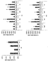
도 9는 혈액 내 면역세포 (CD8+ T, CD4+ T, NK 세포)의 수와 면역세포 (CD8+ T, CD4+ T, NK 세포)에서 분비되는 INF-γ의 양을 분석한 그래프로, 펩타이드 처리 1일 후에 정상군 및 환자군과 펩타이드 (#4, 6, 11, 12) 처리군을 비교한 값이다. (*: p<0.05, **: p<0.01, ***: P<0.001)
도 10은 혈액 내 면역세포 (CD8+ T, CD4+ T, NK 세포)의 수와 면역세포 (CD8+ T, CD4+ T, NK 세포)에서 분비되는 INF-γ의 양을 분석한 그래프로, 펩타이드 처리 3일 후에 정상군 및 환자군과 펩타이드 (#4, 6, 11, 12) 처리군을 비교한 값이다. (*: p<0.05, **: p<0.01)
도 11은 혈액 내 면역세포 (CD8+ T, CD4+ T, NK 세포)의 수와 면역세포 (CD8+ T, CD4+ T, NK 세포)에서 분비되는 INF-γ의 양을 분석한 그래프로, 펩타이드 처리 15일 후에 정상군 및 환자군과 펩타이드 (#4, 6, 11, 12) 처리군을 비교한 값이다. (*: p<0.05, **: p<0.01)
도 12는 골수 내 면역세포 (CD8+ T, CD4+ T, NK 세포)의 수와 면역세포 (CD8+ T, CD4+ T, NK 세포)에서 분비되는 INF-γ의 양을 분석한 그래프로, 펩타이드 처리 15일 후에 정상군 및 환자군과 펩타이드 (#4, 6, 11, 12) 처리군을 비교한 값이다. (*: p<0.05, **: p<0.01)
도 13는 비장 내 면역세포 (CD8+ T, CD4+ T, NK 세포)의 수와 면역세포 (CD8+ T, CD4+ T, NK 세포)에서 분비되는 INF-γ의 양을 분석한 그래프로, 펩타이드 처리 15일 후에 정상군 및 환자군과 펩타이드 (#4, 6, 11, 12) 처리군을 비교한 값이다. (*: p<0.05, **: p<0.01)
도 14는 혈액 내 면역세포 (CD8+ T, CD4+ T, NK 세포)의 수와 면역세포 (CD8+ T, CD4+ T, NK 세포)에서 분비되는 INF-γ의 양을 시기별로 분석한 그래프로, 정상군과 펩타이드 4 처리군을 비교한 값이다. (*: p<0.05, **: p<0.01)
도 15는 혈액 내 면역세포 (CD8+ T, CD4+ T, NK 세포)의 수와 면역세포 (CD8+ T, CD4+ T, NK 세포)에서 분비되는 INF-γ의 양을 시기별로 분석한 그래프로, 환자군과 펩타이드 4 처리군을 비교한 값이다. (*: p<0.05, **: p<0.01)
도 16은 골수와 비장 내 면역세포 (CD8+ T, CD4+ T, NK 세포)의 수와 면역세포 (CD8+ T, CD4+ T, NK 세포)에서 분비되는 INF-γ의 양을 분석한 그래프로, 정상군 및 환자군과 펩타이드 4 처리군을 비교한 값이다. (*: p<0.05, **: p<0.01)
도 17은 도 14 내지 16까지의 분석결과 중 유의한 결과만을 선별하여 분석한 그래프이다. (*: p<0.05, **: p<0.01)
도 18은 혈액 내 면역세포 (CD8+ T, CD4+ T, NK 세포)의 수와 면역세포 (CD8+ T, CD4+ T, NK 세포)에서 분비되는 INF-γ의 양을 시기별로 분석한 그래프로, 정상군과 펩타이드 11 처리군을 비교한 값이다. (*: p<0.05, **: p<0.01)
도 19는 혈액 내 면역세포 (CD8+ T, CD4+ T, NK 세포)의 수와 면역세포 (CD8+ T, CD4+ T, NK 세포)에서 분비되는 INF-γ의 양을 시기별로 분석한 그래프로, 환자군과 펩타이드 11 처리군을 비교한 값이다. (*: p<0.05, **: p<0.01)
도 20은 골수와 비장 내 면역세포 (CD8+ T, CD4+ T, NK 세포)의 수와 면역세포 (CD8+ T, CD4+ T, NK 세포)에서 분비되는 INF-γ의 양을 분석한 그래프로, 정상군 및 환자군과 펩타이드 11 처리군을 비교한 값이다. (*: p<0.05, **: p<0.01)
도 21은 도 18 내지 20까지의 분석결과 중 유의한 결과만을 선별하여 분석한 그래프이다. (*: p<0.05, **: p<0.01)
도 22는 정상군의 말초 단핵구 세포에서 분리한 NK 세포와 급성 골수성 백혈병 환자로부터 분리한 NK 세포에 펩타이드 (#4, 11)를 처리한 후 세포독성에 대한 활성을 나타내는 그래프이다. (**: p<0.01)
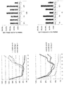
도 23은 정상인과 환자의 말초 단핵구 세포와 골수 세포에 펩타이드 (#4, 11)를 처리한 후 세포 생준률을 보여주는 그래프이다. (NBM: normal bone marrow, NPB: normal peripheral blood)
도 24 및 25는 정상인과 환자의 말초 단핵구 세포와 골수 세포에 펩타이드 (#4, 11)를 처리한 후 CD3-CD56+ 세포의 수를 시기 (처리 당일, 6일째, 21일째)별로 나타내는 그래프이다.
실시예
실시예 1: VEGFR-3 인산화 억제 변이 펩타이드의 제작
VEGFR-3 수용체 인산화를 억제하는 Fc 크링클 도메인에서 유래하는 저분자의 변이된 후보 펩타이드를 제작하였다. 먼저, VEGFR-3 Fc 부위의 강한 이황화 결합(disulfide bond)으로 결합되어 있는 D5 부위(region)의 Thr446과 Lys516에 위치한 시퀀스를 중심으로 15 mer가 넘지 않는 크기로 펩타이드를 무작위로 잘라 12종의 후보군을 확보하였다. 도 1과 아래 표 1에 12종의 펩타이드 후보군의 시퀀스를 나타내었다.
또한, 리간드인 VEGF-C가 결합한 후로 Fc 부위의 결합각도가 바뀌면서 인산화에 결정적으로 관여하는 부위의 위치가 달라질 수 있기 때문에 중첩 (Overlapping) 형식으로 펩타이드를 제작하였으며 구조에서 위치를 검증하였다. 제작된 펩타이드가 VEGFR-3 수용체의 활성화를 유도하는지 알아보기 위해, VEGFR-3 발현과 더불어 신호전달기전에 있는 단백질 등을 FACS (Flow Cytometry)를 수행하였다.
| 펩타이드 ID | 펩타이드 서열 | 서열번호 |
| P1 | CNYSHLFYC | 서열번호 1 |
| P2 | CYYGQSKYC | 서열번호 2 |
| P3 | SHMKYSHLFYC | 서열번호 3 |
| P4 | RQALTCTAYGVP | 서열번호 4 |
| P5 | RQATAYGVP | 서열번호 5 |
| P6 | RQATAYGVPLPLS | 서열번호 6 |
| P7 | EFVEGKNKTVSKL | 서열번호 7 |
| P8 | EFVEGKNTVSKL | 서열번호 8 |
| P9 | EFVEGKNLTVSKL | 서열번호 9 |
| P10 | KSIDNEWRKTGCMPREVA | 서열번호 10 |
| P11 | KSIDNEWRKGCMPREVA | 서열번호 11 |
| P12 | KSIDNEWRKLGCMPREVA | 서열번호 12 |
실시예 2: VEGFR-3 인산화 억제 변이 펩타이드의 활성 측정
실시예 2-1: 펩타이드 처리 후 환자의 단핵구 세포에서 IFN-γ에 관한 유전자 발현 측정
환자 세포 수집을 위해 급성 골수성 환자군을 모집하였고 세포를 동결하여 저장하였다. 1.077 비중차를 이용하여 단핵구세포를 분리하여 DPBS (Dulbecco's Phosphate-Buffered Saline)에 수세 후 10% DMSO (dimethyl sulfoxide)를 포함한 우태아 혈청 (Fetal bovine serum, FBS)에 넣어 LN2에 얼려 보관하였다.
얻어진 세포를 즉시 트라이졸(Trizol) 1 ml에 클로로포름 200 ㎕을 넣고 섞은 후 11000rpm에 20분 원심분리를 이용해 RNA(Ribonucleic acid)를 얻었다. 아이소프로판올로 침전시켜 얻어진 RNA는 70% 에탄올로 닦아 말린 후 500 ug씩을 획득하여 cDNA(complementary DNA) 합성 키트를 이용해 역전사하여 1차 cDNA를 얻었다. 이로 중합효소연쇄반응 (Polymerase Chain Reaction, PCR)을 진행하여 유전자 발현정도를 관찰하였다.
도 2는 환자의 말초 및 골수 단핵구 세포를 이용하여 펩타이드 처리 후 IFN-γ에 관한 유전자 발현을 RT-qPCR로 조사한 결과이다. 모든 환자에서 동일한 발현을 보이지는 않았으나, 대부분이 25μM과 50μM의 농도로 처리한 경우 증가하는 경향을 나타냈으며, 펩타이드 ID 4, 5, 6, 9, 11, 12 번은 처리하지 않은 맹검 보다는 높게 나타났으며, MAZ51 화합물(VEGFR3 키나아제 억제제, CAS 163655-37-6)을 처리했을 때 보다 더 증가한 양상을 보였다.
실시예 2-2: MACS를 이용한 NK 세포 분리 및 순도 측정
NK 세포의 펩타이드에 의한 체외에서의 세포사멸, 증식 및 실질적인 기능변화를 조사하기 위해서는 NK 세포의 분리가 필수적이다. 도 3에 나타난 바와 같이, MACS를 이용하여 세포를 분리하고 순도를 측정하였다. 93.3%로 MACS sorting 방법은 기술적인 면에서 세포분리에 문제가 없음을 확인하였다.
실시예 2-3: 펩타이드 처리 후 환자의 NK 세포의 IFN-γ 단백질 발현 측정
펩타이드 처리 후 NK 세포에서의 IFN-γ 단백질의 발현 수준을 조사하기 위해 FACS를 진행하였다. 분리된 세포를 1% 우혈청 알부민이 함유된 PBS (phosphate buffer saline)로 씻은 후, 1-3×10⁵/100㎕의 농도로 12×75mm polystyrene(Falcon 2052) FACS tube에 넣고 각각의 해당 단일클론 항체를 각각 5㎕을 넣은 뒤 30 분 동안 4℃에서 반응시킨 후, 2% 우혈청 알부민이 함유된 PBS로 두 번 씻어주었다. 세포분리 및 염색 과정 등 일련의 과정에서 생존하고 있는 세포를 정확하게 판별하고 분석하기 위하여 전 과정에서 세포 사멸반응을 추적하고자 마지막 세척 후 유세포 분석기(FACSCalibur, Becton Dickinson, USA)로 분석하였다. FACSmoflo로 세포를 분리하기 위해 똑같이 항체를 세포에 염색한 후 CD56+CD3- 세포군에서의 VEGFR-3+와 VEGFR-3- 세포군이 어느 정도 되는지 조사하고, 모아진 세포에 후보 펩타이드를 농도 별로 24시간 동안 처리한 후 FACS를 진행하였다.
도 4는 환자의 말초 단핵구 세포에서의 펩타이드 처리 시 IFN-γ 단백질의 발현을 FACS로 조사한 결과이다. IFN-γ 단백질 발현량을 통하여 기능적인 효율을 판단하는데 중요하므로 활성이 높은 후보 펩타이드를 선정하는데 결정적인 단서가 되었다. 본 발명의 펩타이드 처리 시에 대부분의 CD3-CD56+ 세포에서 IFN-γ의 발현량이 증가하였으며, 특히 4, 6, 11, 12번 펩타이드를 처리하였을 때 유의하게 증가하였다(도 4 참조).
도 5는 환자의 골수 단핵구 세포에서의 펩타이드 처리 시 IFN-γ의 단백질 발현을 FACS로 조사한 결과이다. CD3-CD56+ 세포의 경우에 6번과 11번 펩타이드를 처리하였을 때 증가하였으나, CD3-CD16+의 경우에는 6번과 11번 펩타이드를 처리했을 때가 MAZ51 화합물을 처리했을 때보다 IFN-γ 단백질의 발현량이 유의하게 증가하지는 않았다. 전반적으로 말초에서의 NK 세포가 골수의 NK 세포보다 펩타이드에 민감하게 반응함을 확인하였다.
실시예 3: 펩타이드 처리 후 NK 세포의 세포면역성 검사
환자 세포 수집을 위해 신규 급성 골수성 환자군을 지속적으로 모집하였고 이들로부터 수집한 세포를 동결하여 저장하였다. 1.077 비중차를 이용해 단핵구세포를 분리하여 DPBS에 수세 후 10% DMSO를 포함한 FBS에 넣어 LN2에 얼려 보관하였다.
펩타이드 처리 후 환자 유래 단핵구 세포에서 면역반응도를 보기 위해 Elispot 분석을 진행하였다. 환자 유래 단핵구 세포를 사용한 실험에 의해 1차로 선별된 후보 4종 펩타이드 (4번, 6번, 11번, 12번 펩타이드)를 처리한 후 세포가 분비하는 IFN-γ에 대한 항체가 결합된 플레이트에 24시간 반응시켰다. 이후 DPBS로 씻어내고 2차 항체와 항면역글로불린 항체를 이용하여 IFN-γ를 분비하는 세포를 감지하여 분석하였다.
Elispot 실험에서 IFN-γ 분비를 자극하기 위해서 도 6과 같이 PMA (phorbol 12-myristate)와 칼슘 이온투과담체 (Ca Ionophore)의 농도 조건별로 실험을 진행하였다. 그 결과, 도 7에서 나타난 바와 같이, 처리한 후보 펩타이드가 환자의 말초 단핵구 세포의 IFN-γ의 발현을 증가시킨다는 것을 확인하였으며, 체외 조건에 따라서 차별적이고 실질적인 기능 변화에 관여함을 보여주었다.
실시예 4: 펩타이드 체내 주사 시 NK 세포의 살해능 조사 및 분석
실시예 4-1: 펩타이드 체내 주사 시 생체 내 NK 세포의 수 및 NK 세포의 IFN-γ의 분비 변화 조사
생체 내에서의 후보 펩타이드의 효과를 확인해 보기 위해 선별된 펩타이드를 마우스 체내에 주사한 뒤 면역세포에 대해 조사하였다.
정상군의 마우스와 암 줄기세포인 C1498 세포주 (2x10⁶cells/100㎕)를 주입하여 만든 백혈병 모델 마우스에 후보 펩타이드 (각 2 mg/kg)를 동시에 주입하였다. 각 군에 대해서 펩타이드를 주입한 그룹과 주입하지 않은 그룹 사이의 시간에 따른 (Day-1, Day1, Day3, Day15) 면역세포의 수 변화와 IFN-γ의 분비량 변화를 조사하기 위해 FACS를 진행하였다. 마우스에서 혈액, 골수, 비장을 얻은 후 적혈구 용해 완충액(RBC lysis buffer)로 적혈구를 파괴한 뒤, PBS로 씻어내었다. 면역세포 분석을 위해 FACS 버퍼에 3-5×10⁶cells/100㎕의 농도로 12×75mm polystyrene(Falcon 2052) FACS tube에 넣고 각각의 해당 항체를 넣어 상온에서 20분 동안 반응시킨 후 FACS buffer로 씻어준 뒤 유세포 분석기 (FACSCanto2, Becton Dickinson, USA)로 분석하였다. FACS 완충액(buffer)에 5-10×106 cells/1ml 농도로 세포를 넣어준 뒤, IFN-γ를 자극하기 위한 시약들을 37℃에서 6시간동안 처리하였다. 그런 다음, FACS 튜브(tube)에 넣어 PBS로 씻어준 후 FACS 완충액(buffer) 100 ㎕에 면역세포 항체를 넣고 상온에서 20분 동안 반응시켰다. 이 후, FACS 완충액로 한 번 더 씻어준 후 Fix.& Perm, buffer 200㎕를 상온에서 20분 동안 처리하고 씻어낸 뒤 유세포 분석기(FACSCanto2, Becton Dickinson, USA)로 분석하였다.
도 8은 펩타이드 주입 1일 전(Day-1) 시기의 혈액 내 면역세포의 수 변화와 IFN-γ의 분비 변화에 대한 결과이다.
도 9는 펩타이드 주입 1일 후(Day1) 시기의 혈액 내 면역세포의 수 변화와 IFN-γ의 분비 변화에 대한 결과를 보여준다. 정상군에 비해 펩타이드 11번을 투여했을 때, 혈액 내 NK 세포 수가 증가하였다. 환자 모델군에 비해 펩타이드 4번을 투여했을 때, 혈액 내 NK 세포에서 분비되는 IFN-γ가 증가하였다.
펩타이드 주입 3일 후(Day3) 시기의 혈액 내 면역세포의 혈액 내 수 변화와 IFN-γ의 분비 변화에 대한 결과를 도 10에 나타내었다. 정상군에 비해 펩타이드 4를 투여 시 혈액 내 NK 세포 수가 증가하였다.
펩타이드 주입 15일 후(Day15) 시기의 면역세포의 혈액 내 수 변화와 IFN-γ의 분비 변화에 대한 결과를 도 11에 나타내었다. 환자 모델군에 비해 펩타이드 6를 투여 시 혈액 내 NK 세포에서 분비되는 IFN-γ가 감소하였다. 정상군에 비해 펩타이드 11를 투여했을 때, 혈액 내 NK 세포에서 분비되는 IFN-γ가 증가하였다. 정상군에 비해 펩타이드 12를 투여 시 혈액 내 NK 세포 수가 증가하였으며, 혈액 내 NK 세포에서 분비되는 IFN-γ 또한 증가하였다.
펩타이드 주입 15일 후(Day15) 시기의 골수 내 면역세포의 수 변화와 IFN-γ의 분비 변화에 대한 결과를 도 12에서 확인할 수 있다. 환자 모델군에 비해 펩타이드 4를 투여했을 때, 골수 내 NK 세포의 수가 증가하였으며, 환자 모델군에 비해 골수 내 NK 세포에서 분비되는 IFN-γ는 감소하였다. 정상군에 비해 펩타이드 6를 투여했을 때, 골수 내 NK 세포 수가 증가하였으며, 환자 모델군에 비해 골수 내 NK 세포에서 분비되는 IFN-γ는 감소하였다. 정상군에 비해 펩타이드 11를 투여 시 골수 내 NK 세포에서 분비되는 IFN-γ이 증가하였다. 환자 모델군에 비해 펩타이드 11를 투여 시 골수 내 NK 세포의 수가 증가하였으며, 골수 내 NK 세포에서 분비되는 IFN-γ는 감소하였다. 정상군에 비해 펩타이드 12를 투여했을 때, 골수 내 NK 세포 수가 감소하였으며, 골수 내 NK 세포에서 분비되는 IFN-γ 또한 증가하였다.
펩타이드 주입 15일 후(Day15) 시기의 비장 내 면역세포의 수 변화와 IFN-γ의 분비 변화에 대한 결과를 도 13에서 확인할 수 있다. 정상군에 비해 펩타이드 4를 투여 시 비장 내 NK 세포에서 분비되는 IFN-γ가 증가하였다. 정상군에 비해 펩타이드 6를 투여했을 때, 비장 내 NK 세포에서 분비되는 IFN-γ가 증가하였다. 환자 모델군에 비해 펩타이드 6를 투여 시 비장 내 NK 세포에서 분비되는 IFN-γ가 감소하였다. 정상군에 비해 펩타이드 11를 투여 시 비장 내 NK 세포 수가 증가하였으며, 비장 내 NK 세포에서 분비되는 IFN-γ 또한 증가하였다. 환자 모델군에 비해 펩타이드 12 투여 시 Day 15 비장 내 NK 세포의 수가 증가하였으며, 비장 내 NK 세포에서 분비되는 IFN-γ는 감소하였다.
도 14는 펩타이드 4를 주입한 후 시기별 정상군과 펩타이드 처리군 간의 혈액 내 면역세포 수 및 INF- γ의 분비변화를 나타낸 결과이다. 도 14에 나타난 바와 같이, 펩타이드 4를 주입한 3일 후의 혈액 내 NK 세포 수가 정상군에 비해 유의하게 증가함을 확인하였으며, 다른 시기의 정상군 및 펩타이드 처리군간의 비교 중에서도 가장 크게 나타났다.
도 15는 펩타이드 4를 주입한 후 시기별 환자 모델군과 펩타이드 처리군 간 의 혈액 내 면역세포 수 및 INF- γ의 분비변화를 나타낸 결과이다. 도 15는 주입한 1일 후의 혈액 내 NK 세포의 INF-γ 분비가 환자 모델군에 비해 유의하게 증가함을 보여준다.
도 16은 펩타이드 4를 주입한 15일 후, 골수와 비장조직 및 그룹 간의 면역세포 수 및 INF- γ의 분비변화를 나타낸 결과이다. 비장 내 NK 세포의 INF-γ 분비가 정상군에 비해 유의하게 증가함을 확인하였다. 골수 내 NK 세포 수가 환자 모델군에 비해 유의하게 증가함을 확인하였으며, 골수 내 NK 세포의 INF-γ 분비가 환자 모델군에 비해 유의하게 감소함을 확인하였다.
상기 결과로부터 유의한 효과를 갖는 펩타이드 4에 대해 추가 실험을 진행하였다. 도 17에 나타난 바와 같이, 펩타이드 4를 주입한 3일 후의 혈액 내 NK 세포 수 및 주입한 15일 후의 비장 내 NK 세포의 INF-γ 분비가 정상군에 비해 유의하게 증가함을 확인하였다. 주입한 1일 후의 혈액 내 NK 세포의 INF-γ 분비 및 주입한 15일 후의 골수 내 NK 세포 수가 환자 모델군에 비해 유의하게 증가함을 확인하였다. 주입한 15일 후의 골수 내 NK 세포의 INF-γ 분비가 환자 모델군에 비해 유의하게 감소함을 확인하였다.
도 18은 펩타이드 11을 주입한 후 시기별 정상군과 펩타이드 처리군 간의 혈액 내 면역세포 수 및 INF- γ의 분비변화를 나타낸 결과이다. 도 18에 나타난 바와 같이, 1일 후의 혈액 내 NK 세포의 INF-γ 분비는 정상군에 비해 유의하게 증가함을 확인하였으며, 15일 후의 혈액 내 NK 세포의 INF-γ 분비 또한 정상군에 비해 유의하게 증가함을 확인하였다.
도 19는 펩타이드 11을 주입한 후 시기별 환자군과 펩타이드 처리군 간의 혈액 내 면역세포 수 및 INF- γ의 분비변화를 나타낸 결과이다.
도 19은 펩타이드 11을 주입한 15일 후, 골수와 비장조직 및 그룹 간의 면역세포 수 및 INF- γ의 분비변화를 나타낸 결과이다. 주입한 15일 후의 비장 내 NK 세포 수 및 NK 세포의 INF-γ 분비가 정상군에 비해 유의하게 증가하였다. 주입한 15일 후의 골수 내 NK 세포의 INF-γ 분비가 정상군에 비해 유의하게 증가하였다. 주입한 15일 후의 골수 내 NK 세포의 수는 환자군에 비해 유의하게 증가하였으나, NK 세포의 INF-γ 분비는 감소하였다.
상기 결과로부터 유의한 효과를 나타낸 펩타이드 11에 대해 추가 실험을 진행하였다. 도 20에 나타난 바와 같이, 펩타이드 11을 주입한 1일 후의 혈액 내 NK 세포의 INF-γ 분비는 정상군에 비해 유의하게 증가함을 확인하였으며, 15일 후의 혈액 내 NK 세포의 INF-γ 분비 또한 정상군에 비해 유의하게 증가함을 확인하였다. 주입한 15일 후의 비장 내 NK 세포 수 및 NK 세포의 INF-γ 분비가 정상군에 비해 유의하게 증가하였다. 주입한 15일 후의 골수 내 NK 세포의 INF-γ 분비가 정상군에 비해 유의하게 증가하였다. 주입한 15일 후의 골수 내 NK 세포의 수는 환자군에 비해 유의하게 증가하였으나, NK 세포의 INF-γ 분비는 감소하였다.
실시예 5: 펩타이드의 체내 주사 시 면역세포 NK 세포의 살해능 조사 및 분석
상기 실험 결과로부터 유의한 결과를 나타낸 펩타이드 4와 11에 대해 추가 실험을 진행하였다. 펩타이드 4와 11을 대상으로 환자의 말초 단핵구 세포에서 분리한 NK 세포에 펩타이드 처리한 후 세포독성 분석(Cytotoxicity assay)을 진행하였다.
MACS를 이용하여 분리한 NK 세포에 펩타이드를 각각 50 μM로 하루 동안 처리한 후 DELFIA® EuTDA Cytotoxicity Reagents (PerkinElmer , USA)를 통해 세포독성 분석을 진행하였다. K562를 1x10⁶cells/ml의 농도 2ml에 1ul BATDA를 넣어 30분간 37℃에서 인큐베이션시켰다. 4℃, 1000rpm에서 4분간 3번 DPBS를 이용하여 씻은 후, K562 5x10³cells/100ul씩 96웰 플레이트에 깔아주었다. 백그라운드는 Medium 200ul을 사용하였으며, 음성대조군은 K562 100ul와 Medium 100ul을 혼합한 용액을 사용하였고, 양성대조군은 K562 100ul, Medium 90ul 및 따뜻하게 한 용해 버퍼 10ul을 혼합한 용액을 사용하였다. 각각의 실험군은 K562 100ul와 NK cell 100ul를 섞은 혼합물을 사용하였다. 도 21는 정상군의 말초 단핵구 세포에서 분리한 NK 세포에 Peptide 4를 처리 후 세포독성이 유의하게 증가함을 보여준다.
실시예 6: 신규 펩타이드를 처리한 조건화배지 내 NK세포의 배양효율 분석 및 프로토콜 정립
정상인과 환자의 말초 단핵구 세포와 골수 세포에 각각 펩타이드 처리 시 세포 생존률과 NK 세포 숫자 증가를 확인하기 위해 생체 외 세포 배양 실험을 진행하였다.
정상인과 환자에의 말초 단핵구 세포와 골수 세포를 생체 외 세포배양을 위해 10% 우태아혈청과 1% 항생체를 포함하는 RPMI(Roswell park memorial institute) 1640 media를 배양액으로 하여 3일마다 배양액 교체를 해주고 21일까지 배양을 진행하였다. 24-웰 배양 플레이트에 각각 세포들을 1x10⁶cells/ml씩 깔아주었다. 0~6일째까지는 10IU/ml IL-2를 함께 넣어주고, 그 이후부터 100IU/ml IL-2, 5ng/ml IL-15를 함께 넣어주었다. 그리고 배양액을 교체할 때, 각각 펩타이드를 50μM씩 처리해주었다.
도 22은 3일마다 배양액 교체를 해주며 세포 수를 측정하여 세포 생존률을 확인한 결과이다. 도 22은 정상인의 골수 세포에서 펩타이드 4를 처리한 결과, 15일 이후부터 대조군에 비해 세포 수가 증가하였음을 보여주었다. 또한, 환자의 골수 세포에서 펩타이드 4를 처리한 후 대조군만큼은 아니나 세포수가 점차 증가하는 추세를 보였으며, 정상인의 말초 단핵구 세포에서 펩타이드 4를 처리한 후 대조군만큼은 아니나 세포수가 점차 증가하는 추세를 보였다. 환자의 말초 단핵구 세포에서 펩타이드 4를 처리한 결과, 대조군과 마찬가지로 21일까지 크게 증가하는 것은 보이지 않으나, 대조군 및 펩타이드 처리군 모두 점차 증가하는 추세를 보였다.
도 23에서 25는 6일 및 21일째의 NK 세포를 확인하기 위해 Flow Cytometry를 통해 CD3-CD56+ 세포의 수를 확인한 결과이다. 도 23에서 25에서 보이는 바와 같이, 각 그룹 당 3명의 샘플들로 분석했을 때 그룹 간의 유의성을 띄지 않았으나, 정상인 및 환자의 골수 세포에서는 6일째에 펩타이드 4를 처리 시 증가하는 양상을 보이며, 정상인 및 환자의 말초 단핵구 세포에서는 21일째에 펩타이드 4를 처리 시 증가하는 양상을 보였다.
본 명세서에서 설명된 구체적인 실시예는 본 발명의 바람직한 구현예 또는 예시를 대표하는 의미이며, 이에 의해 본 발명의 범위가 한정되지는 않는다. 본 발명의 변형과 다른 용도가 본 명세서 특허청구범위에 기재된 발명의 범위로부터 벗어나지 않는다는 것은 당업자에게 명백하다.
본 발명은 VEGFR(vascular endothelial growth factor receptor)-3의 인산화억제 활성을 갖는 펩타이드 및 이의 용도에 관한 것이다.
Claims (10)
- 서열번호 1 내지 서열번호 12 중 어느 하나의 아미노산 서열로 이루어지는 펩타이드.
- 제 1 항에 있어서, 상기 펩타이드는 VEGFR(vascular endothelial growth factor receptor)-3의 인산화를 억제하는 것을 특징으로 하는 펩타이드.
- 제 1 항에 있어서, 상기 펩타이드는 면역세포에서 IFN-γ(interferon -γ)의 발현을 증가시키는 것을 특징으로 하는 펩타이드.
- 제 1 항에 있어서, 상기 펩타이드는 NK(Natural Killer)세포의 증식을 촉진하거나, 또는 NK 세포의 세포독성(cytotoxicity) 활성을 증가시키는 것을 특징으로 하는 펩타이드.
- (a) 서열번호 1 내지 서열번호 12 중 어느 하나의 아미노산 서열로 이루어지는 펩타이드의 약학적 유효량; 및 (b) 약학적으로 허용되는 담체를 포함하는 암의 치료 또는 예방용 약학 조성물.
- 제 5 항에 있어서, 상기 조성물은 VEGFR(vascular endothelial growth factor receptor)-3의 인산화를 억제하는 것을 특징으로 하는 암의 치료 또는 예방용 약학 조성물.
- 제 5 항에 있어서, 상기 조성물은 면역세포에서 IFN-γ(interferon -γ)의 발현을 증가시키는 것을 특징으로 하는 암의 치료 또는 예방용 약학 조성물.
- 제 5 항에 있어서, 상기 조성물은 NK(Natural Killer)세포의 증식을 촉진하거나, 또는 NK 세포의 세포독성(cytotoxicity) 활성을 증가시키는 것을 특징으로 하는 암의 치료 또는 예방용 약학 조성물.
- 제 5 항에 있어서, 상기 암은 혈액암, 위암, 폐암, 유방암, 난소암, 간암, 기관지암, 비인두암, 후두암, 췌장암, 방광암, 결장암, 자궁경부암, 뇌암, 전립선암, 골암, 피부암, 갑상선암, 부갑상선암, 및 요관암으로 이루어지는 군에서 선택되는 암인 것을 특징으로 하는 암의 치료 또는 예방용 약학 조성물.
- 서열번호 1 내지 서열번호 12 중 어느 하나의 아미노산 서열로 이루어지는 펩타이드를 유효성분으로 포함하는 NK(Natural Killer) 세포의 체외(in vitro) 증식 촉진용 조성물.
Applications Claiming Priority (2)
| Application Number | Priority Date | Filing Date | Title |
|---|---|---|---|
| KR1020180139363A KR102125242B1 (ko) | 2018-11-13 | 2018-11-13 | Vegfr-3 인산화 억제 펩타이드 및 이의 용도 |
| KR10-2018-0139363 | 2018-11-13 |
Publications (1)
| Publication Number | Publication Date |
|---|---|
| WO2020101303A1 true WO2020101303A1 (ko) | 2020-05-22 |
Family
ID=70731634
Family Applications (1)
| Application Number | Title | Priority Date | Filing Date |
|---|---|---|---|
| PCT/KR2019/015266 Ceased WO2020101303A1 (ko) | 2018-11-13 | 2019-11-11 | Vegfr-3 인산화 억제 펩타이드 및 이의 용도 |
Country Status (2)
| Country | Link |
|---|---|
| KR (1) | KR102125242B1 (ko) |
| WO (1) | WO2020101303A1 (ko) |
Families Citing this family (1)
| Publication number | Priority date | Publication date | Assignee | Title |
|---|---|---|---|---|
| KR102922015B1 (ko) * | 2019-10-17 | 2026-02-04 | 주식회사 나이벡 | Fer 티로신 키나아제 결합능이 있는 신규한 펩타이드 및 이의 용도 |
Citations (2)
| Publication number | Priority date | Publication date | Assignee | Title |
|---|---|---|---|---|
| US20020164667A1 (en) * | 2001-01-17 | 2002-11-07 | Kari Alitalo | VEGFR-3 inhibitor materials and methods |
| KR20150015892A (ko) * | 2013-08-02 | 2015-02-11 | 가톨릭대학교 산학협력단 | IFN-γ의 발현을 증가시키는 VEGFR-3 억제제를 유효성분으로 포함하는 급성골수성백혈병 치료용 약학적 조성물 |
-
2018
- 2018-11-13 KR KR1020180139363A patent/KR102125242B1/ko active Active
-
2019
- 2019-11-11 WO PCT/KR2019/015266 patent/WO2020101303A1/ko not_active Ceased
Patent Citations (2)
| Publication number | Priority date | Publication date | Assignee | Title |
|---|---|---|---|---|
| US20020164667A1 (en) * | 2001-01-17 | 2002-11-07 | Kari Alitalo | VEGFR-3 inhibitor materials and methods |
| KR20150015892A (ko) * | 2013-08-02 | 2015-02-11 | 가톨릭대학교 산학협력단 | IFN-γ의 발현을 증가시키는 VEGFR-3 억제제를 유효성분으로 포함하는 급성골수성백혈병 치료용 약학적 조성물 |
Non-Patent Citations (3)
| Title |
|---|
| JI YOON LEE ET AL: "A VEGFR-3 antagonist increases IFN-y expression on low functioning NK cells in acute myeloid leukemia", JOURNAL OF CLINICAL IMMUNOLOGY, vol. 33, 13 February 2013 (2013-02-13), pages 826 - 837, XP055707883, ISSN: 0271-9142, DOI: 10.1007/s10875-013-9877-2 * |
| YASHIRO, M. ET AL: "Effects of VEGFR-3 phosphorylation inhibitor on lymph node metastasis in an orthotopic diffuse-type gastric carcinoma model", BRITISH JOURNAL OF CANCER, vol. 101, no. 7, 8 September 2009 (2009-09-08), pages 1100 - 1106, XP055707890, ISSN: 0007-0920, DOI: 10.1038/sj.bjc.6605296 * |
| YI-WEN CHANG ET AL: "Novel peptides suppress VEGFR-3 activity and antagonize VEGFR-3-mediated oncogenic effects", ONCOTARGET, vol. 5, no. 11, 24 May 2014 (2014-05-24), pages 3823 - 3835, XP055707880, DOI: 10.18632/oncotarget.1709 * |
Also Published As
| Publication number | Publication date |
|---|---|
| KR102125242B1 (ko) | 2020-06-23 |
| KR20200055579A (ko) | 2020-05-21 |
Similar Documents
| Publication | Publication Date | Title |
|---|---|---|
| Uchida et al. | Intrapleural administration of OK432 in cancer patients: Augmentation of autologous tumor killing activity of tumor-associated large granular lymphocytes | |
| WO2018074758A2 (ko) | 면역질환 치료를 위한 고효능 줄기세포 선별방법 | |
| Shiiba et al. | Interleukin 2-activated killer cells: generation in collaboration with interferonγ and its suppression in cancer patients | |
| Ogawa et al. | Enhanced induction of very late antigen 4/lymphocyte function-associated antigen 1-dependent T-cell migration to tumor sites following administration of interleukin 12 | |
| Jin et al. | TGF beta down-regulates TLiSA1 expression and inhibits the differentiation of precursor lymphocytes into CTL and LAK cells | |
| WO2012108586A1 (ko) | 활성화된 자연살해 세포를 포함하는 림프구의 제조방법 및 이를 포함하는 약학 조성물 | |
| WO2019117633A1 (ko) | 아토피성 피부염, 탈모, 상처 또는 피부 주름의 개선용 화장료 조성물 및 약학 조성물 | |
| WO2022103138A1 (ko) | 비피도박테리움 롱검 rapo (kctc13773bp)를 포함하는 암 예방 또는 치료용 조성물 | |
| KR101902355B1 (ko) | 데시타빈 또는 이의 약학적으로 허용 가능한 염을 유효성분으로 함유하는 골수유래억제세포 저해용 조성물 | |
| WO2017069512A1 (ko) | 바이러스 항원 특이적인 t 세포의 유도 및 증식 방법 | |
| WO2020231205A1 (ko) | 자연 살해 세포의 배양용 조성물 및 이를 이용한 방법 | |
| WO2022080894A1 (ko) | 항암 관련 유전자 발현이 조절된 자연살해세포 및 이의 용도 | |
| WO2019103436A9 (ko) | Nk 세포 배양용 조성물 및 이를 이용하여 nk 세포를 배양하는 방법 | |
| WO2022045813A1 (ko) | 엑소좀을 유효성분으로 포함하는 폐암 치료, 예방 또는 전이 억제용 약학적 조성물 | |
| Mishra et al. | Semiquinone glucoside derivative provides protection against γ‐radiation by modulation of immune response in murine model | |
| WO2018131909A1 (ko) | 뇌신경계 질환의 치료 또는 진단을 위한 표적 단백질로서 pcnt | |
| Shyu et al. | Direct growth suppressive activity of interferon‐α and‐γ on human gastric cancer cells | |
| Lucero et al. | Effect of various interferons on the spontaneous cytotoxicity exerted by lymphocytes from normal and tumor-bearing patients | |
| KR102125242B1 (ko) | Vegfr-3 인산화 억제 펩타이드 및 이의 용도 | |
| WO2017074118A1 (ko) | Foxp3를 발현하는 수지상 세포를 포함하는 면역 조절용 약학적 조성물 | |
| WO2023204615A1 (ko) | 유사 기억 자연 살해 세포 제조방법 및 이를 이용하여 제조된 유사 기억 자연 살해 세포의 항암 용도 | |
| WO2024072052A1 (ko) | 사이토카인 및 항체가 표면 발현된 자연살해세포 유래 세포외 소포체를 포함하는 암 질환 치료용 조성물 | |
| AU2003219016A1 (en) | Anti-infective agents and/or immunomodulators used for preventive therapy following an acute cerebrovascular accident | |
| JP3817273B2 (ja) | 免疫抑制治療、抗ガン治療および抗ウイルス治療用の薬学的組成物 | |
| Murakami-Mori et al. | Enhancement of gp130-mediated tyrosine phosphorylation of STAT3 and its DNA-binding activity in dexamethasone-treated AIDS-associated Kaposi's sarcoma cells: selective synergy between dexamethasone and gp130-related growth factors in Kaposi's sarcoma cell proliferation |
Legal Events
| Date | Code | Title | Description |
|---|---|---|---|
| 121 | Ep: the epo has been informed by wipo that ep was designated in this application |
Ref document number: 19883711 Country of ref document: EP Kind code of ref document: A1 |
|
| NENP | Non-entry into the national phase |
Ref country code: DE |
|
| 122 | Ep: pct application non-entry in european phase |
Ref document number: 19883711 Country of ref document: EP Kind code of ref document: A1 |