WO2020085510A1 - Staple核酸を利用したタンパク質翻訳反応の抑制法 - Google Patents
Staple核酸を利用したタンパク質翻訳反応の抑制法 Download PDFInfo
- Publication number
- WO2020085510A1 WO2020085510A1 PCT/JP2019/042049 JP2019042049W WO2020085510A1 WO 2020085510 A1 WO2020085510 A1 WO 2020085510A1 JP 2019042049 W JP2019042049 W JP 2019042049W WO 2020085510 A1 WO2020085510 A1 WO 2020085510A1
- Authority
- WO
- WIPO (PCT)
- Prior art keywords
- sequence
- nucleic acid
- array
- guanine
- oligomer
- Prior art date
- Legal status (The legal status is an assumption and is not a legal conclusion. Google has not performed a legal analysis and makes no representation as to the accuracy of the status listed.)
- Ceased
Links
Images



Classifications
-
- A—HUMAN NECESSITIES
- A61—MEDICAL OR VETERINARY SCIENCE; HYGIENE
- A61K—PREPARATIONS FOR MEDICAL, DENTAL OR TOILETRY PURPOSES
- A61K31/00—Medicinal preparations containing organic active ingredients
- A61K31/70—Carbohydrates; Sugars; Derivatives thereof
- A61K31/7088—Compounds having three or more nucleosides or nucleotides
- A61K31/711—Natural deoxyribonucleic acids, i.e. containing only 2'-deoxyriboses attached to adenine, guanine, cytosine or thymine and having 3'-5' phosphodiester links
-
- C—CHEMISTRY; METALLURGY
- C12—BIOCHEMISTRY; BEER; SPIRITS; WINE; VINEGAR; MICROBIOLOGY; ENZYMOLOGY; MUTATION OR GENETIC ENGINEERING
- C12N—MICROORGANISMS OR ENZYMES; COMPOSITIONS THEREOF; PROPAGATING, PRESERVING, OR MAINTAINING MICROORGANISMS; MUTATION OR GENETIC ENGINEERING; CULTURE MEDIA
- C12N15/00—Mutation or genetic engineering; DNA or RNA concerning genetic engineering, vectors, e.g. plasmids, or their isolation, preparation or purification; Use of hosts therefor
- C12N15/09—Recombinant DNA-technology
- C12N15/11—DNA or RNA fragments; Modified forms thereof; Non-coding nucleic acids having a biological activity
- C12N15/111—General methods applicable to biologically active non-coding nucleic acids
-
- A—HUMAN NECESSITIES
- A61—MEDICAL OR VETERINARY SCIENCE; HYGIENE
- A61K—PREPARATIONS FOR MEDICAL, DENTAL OR TOILETRY PURPOSES
- A61K31/00—Medicinal preparations containing organic active ingredients
- A61K31/70—Carbohydrates; Sugars; Derivatives thereof
- A61K31/7088—Compounds having three or more nucleosides or nucleotides
- A61K31/7105—Natural ribonucleic acids, i.e. containing only riboses attached to adenine, guanine, cytosine or uracil and having 3'-5' phosphodiester links
-
- A—HUMAN NECESSITIES
- A61—MEDICAL OR VETERINARY SCIENCE; HYGIENE
- A61P—SPECIFIC THERAPEUTIC ACTIVITY OF CHEMICAL COMPOUNDS OR MEDICINAL PREPARATIONS
- A61P43/00—Drugs for specific purposes, not provided for in groups A61P1/00-A61P41/00
-
- C—CHEMISTRY; METALLURGY
- C12—BIOCHEMISTRY; BEER; SPIRITS; WINE; VINEGAR; MICROBIOLOGY; ENZYMOLOGY; MUTATION OR GENETIC ENGINEERING
- C12N—MICROORGANISMS OR ENZYMES; COMPOSITIONS THEREOF; PROPAGATING, PRESERVING, OR MAINTAINING MICROORGANISMS; MUTATION OR GENETIC ENGINEERING; CULTURE MEDIA
- C12N15/00—Mutation or genetic engineering; DNA or RNA concerning genetic engineering, vectors, e.g. plasmids, or their isolation, preparation or purification; Use of hosts therefor
- C12N15/09—Recombinant DNA-technology
- C12N15/63—Introduction of foreign genetic material using vectors; Vectors; Use of hosts therefor; Regulation of expression
- C12N15/67—General methods for enhancing the expression
-
- C—CHEMISTRY; METALLURGY
- C12—BIOCHEMISTRY; BEER; SPIRITS; WINE; VINEGAR; MICROBIOLOGY; ENZYMOLOGY; MUTATION OR GENETIC ENGINEERING
- C12N—MICROORGANISMS OR ENZYMES; COMPOSITIONS THEREOF; PROPAGATING, PRESERVING, OR MAINTAINING MICROORGANISMS; MUTATION OR GENETIC ENGINEERING; CULTURE MEDIA
- C12N2310/00—Structure or type of the nucleic acid
- C12N2310/10—Type of nucleic acid
- C12N2310/15—Nucleic acids forming more than 2 strands, e.g. TFOs
- C12N2310/152—Nucleic acids forming more than 2 strands, e.g. TFOs on a single-stranded target, e.g. fold-back TFOs
Definitions
- the present invention relates to a method of inhibiting the peptide synthesis reaction of ribosome and suppressing the protein translation reaction by binding the target mRNA sequence in a sequence-selective manner and inducing the formation of a guanine quadruplex structure on the target mRNA.
- Non-patent documents 1 and 2 Various gene expression suppressing techniques such as microRNA and siRNA are known 1,2 (Non-patent documents 1 and 2).
- the gene expression suppressing function is exerted by a simple mechanism of binding to mRNA, an unexpected effect (off-target effect) is inevitable 3 (Non-Patent Document 3).
- a modified nucleic acid is used for the purpose of improving the intracellular stability of the introduced nucleic acid, the microRNA effect and siRNA activity are significantly reduced, and there is a problem that the expected protein expression suppressing effect cannot be obtained 4 (Non-Patent Document 4). .
- nucleic acid drug In order to develop a nucleic acid drug with a view to drug discovery, it is required to employ a protein translation suppression technique that can use a modified nucleic acid that is stable in vivo and that does not easily cause an off-target effect.
- the present inventors have conducted extensive studies in order to solve the above problems, and as a result, by using an oligomer called Staple nucleic acid, guanine quadruplex is formed in a sequence-selective manner, and eventually, protein synthesis is successfully suppressed. Then, the present invention has been completed.
- the present invention is as follows. (1) For the target RNA containing the first, second, third and fourth guanine repeating sequences (referred to as the first G sequence, the second G sequence, the third G sequence and the fourth G sequence, respectively), A method of suppressing protein expression by using an oligomer that hybridizes in the vicinity of a guanine repeat sequence, and bringing the guanine repeat sequence closer to form a guanine quadruplex structure by folding back the oligomer, Nucleic acid in a partial region on one end side (one end side region) and nucleic acid in a partial region on the other end side (other end side region) of the oligomer is located between the first G sequence and the second G sequence.
- RNA containing the first, second, third and fourth guanine repeating sequences (referred to as the first G sequence, the second G sequence, the third G sequence and the fourth G sequence, respectively),
- An oligomer that brings a guanine repeating sequence close to each other to form a guanine quadruplex structure Nucleic acid in a partial region on one end side (one end side region) and nucleic acid in a partial region on the other end side (other end side region) of the oligomer is located between the first G sequence and the second G sequence.
- the oligomer Respectively near the first G array and the second G array, between the second G array and the third G array, respectively near the second G array and the third G array, or the third G array.
- the oligomer Between the sequence and the fourth G sequence, which hybridize near the third G sequence and near the fourth G sequence, respectively, and bring the guanine repeating sequence closer by folding back.
- the oligomer (8) The oligomer according to (7), which is designed so that the direction of the folding back is toward the guanine quadruplex structure side and hybridizes. (9) The oligomer according to (7) or (8), which is DNA or RNA. (10) The oligomer according to any one of (7) to (9), which is a dimer.
- the nucleic acid in a partial region (one end side region) on one end side and the nucleic acid in a partial region (other end side region) on the other end side of the oligomer are located between the first G sequence and the second G sequence.
- a fourth G sequence which are designed and hybridized so as to hybridize in the vicinity of the third G sequence and in the vicinity of the fourth G sequence, respectively.
- a protein expression suppression kit comprising the oligomer according to any one of (7) to (10) or the oligomer produced by the method according to (11).
- a method for designing an oligomer that binds (hybridizes) to a target mRNA sequence in a sequence-selective manner was developed, and by using the designed oligomer, a stable guanine quadruplex structure is induced in the target mRNA, whereby the ribosome It became possible to inhibit the peptide synthesis reaction of.
- FIG. 1 It is a schematic diagram of a guanine quadruplex.
- a G-quadruplex structure is formed when four guanine repeating regions are close to each other.
- B G-quadruplex formation rule. It is said that a G-quadruplex is constructed when guanine repeats 3 or more bases and the number of bases connecting the guanine repeating sequences is 7 bases or less according to higher-order structure prediction based on the thermal stability of nucleic acid.
- c It is known that when G-quadruplex is constructed in mRNA, the peptide elongation reaction of ribosome is inhibited and, as a result, the protein synthesis reaction is suppressed.
- G-quadruplex is constructed by the close proximity of guanine repeating sequences.
- (d) Results of measuring fluorescence signal. It is a figure which shows the outline
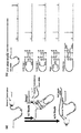
- A Principle of stop assay. RNA G-quadruplex stops the elongation reaction of reverse transcriptase. Therefore, the position where RNAG-quadruplex is constructed can be confirmed by decoding the nucleotide sequence of the cDNA formed up to the position where the reverse transcriptase stopped.
- B Results of stop assay using 2 + 2 100 nt RNA. Except inside loop2, a remarkable peak indicating the presence of RNA G-quadruplex could be confirmed.
- D Results of 1 + 3100nt RNA type evaluated by stop assay.
- D The result of changing the introduced staple nucleic acid from DNA to RNA. It is a figure which shows the outline of an in vitro translation assay.
- B Results of in vitro translation assay targeting 2 + 2100nt RNA.
- C Results of in vitro translation assay for all patterns of mRNA.
- an guanine repeating sequence is brought close to an RNA having four guanine repeating sequences of first, second, third and fourth by using an oligomer that hybridizes to a region near the guanine repeating sequence.
- the present invention relates to a method for suppressing protein expression by forming a quadruplex structure.
- the stable guanine quadruplex structure (also called G-quadruplex structure) present on mRNA is known to inhibit the ribosome peptide elongation reaction. If the G-quadruplex structure can be artificially induced on an arbitrary mRNA, it becomes possible to suppress the expression level of a disease-related protein.
- INDUSTRIAL APPLICABILITY The present invention applies a DNA origami technology, captures and folds a target mRNA in a sequence-selective manner, induces a stable G-quadruplex structure on the target mRNA, and thereby suppresses protein translation selectively in the target mRNA.
- a method for designing a nucleic acid drug is provided.
- the nucleic acid in a partial region on one end side (a region on one end side) and the nucleic acid on a partial region on the other end side (a region on the other end side) are the first guanine repeats of RNA.
- Sequence referred to as first G sequence
- second guanine repeat sequence referred to as second G sequence
- third guanine repeat sequence referred to as third G sequence
- fourth guanine repeat sequence referred to as fourth G sequence
- this oligomer is also referred to as “Staple nucleic acid”.
- RNA G-quadruplex is constructed by introducing Staple nucleic acid regardless of the length and sequence of the guanine repeat region, It was demonstrated that the G-quadruplex structure suppressed the elongation reaction of reverse transcriptase and the protein translation reaction of ribosome.
- the ability to fold the guanine cluster present in the 5'UTRs of the cancer-related genes TMP3 and MYD88 mRNA and inhibit protein synthesis was confirmed by cell-free expression system and expression evaluation using mammalian cells.
- RNA G-quadruplex structure which is an RNA quadruplex structure, as a protein translation suppression mechanism (FIG. 1 (a)).
- the RNA G-quadruplex structure is a stable RNA higher-order structure that is constructed when guanine continuous sequences of 3 or more bases are present at 4 locations with a contiguous spacing of less than 7 bases (Fig. 1 (b)) 5 .
- this RNA G-quadruplex structure is present in the 5'untranslated region (5'UTR) of mRNA, it suppresses the ribosome protein synthesis reaction (Fig. 1 (c)) 6 .
- RNA G-quadruplex structure 7 a guanine continuous sequence existing with a spacing of 7 bases or more constructs an RNA G-quadruplex structure 7 .
- This fact indicates that due to the higher-order structure formed by mRNA, guanine continuous sequences located at positions distant from each other in the primary sequence are spatially adjacent to each other to form an RNA G-quadruplex structure.
- the present inventor devised the method of the present invention in which guanine continuous sequences existing at distant positions are spatially brought close to each other to induce an artificial RNA G-quadruplex structure on a target mRNA. did.
- the DNA origami method which is a technology to create a nanostructure by self-assembling a single-stranded circular DNA of more than 7,000 bases with about 200 designed short-chain DNA (Staple oligomer) of about 30 bases, is known.
- (Fig. 2) 8 The Staple oligomer of this technology assembles the sequences existing at distant positions of single-stranded circular DNA to form a compact structure.
- the DNA origami method which is a method for producing a DNA nanostructure, can be applied to induce an artificial RNA G-quadruplex structure.
- a G-quadruplex structure is formed (Fig. 1a).
- a predetermined oligomer is used in order to bring four guanine repeat regions existing on RNA close to each other.
- This oligomer brings the first guanine repeating sequence and the second guanine repeating sequence, the second guanine repeating sequence and the third guanine repeating sequence, or the third guanine repeating sequence and the fourth guanine repeating sequence close to each other.
- examples of the target RNA include mRNA, TPM3, MYD8 and the like.
- TPM3 is a gene associated with muscle contraction and MYD8 is an adapter protein that mediates Toll and interleukin (IL) 1 receptor signaling.
- the guanine repeating sequence contained in the target RNA is not limited to 4 positions, and may be 5 or more positions.
- the first, second, third and fourth guanine repeating sequences mean four sequences arbitrarily selected from four or more guanine repeating sequences, and not necessarily four consecutive guanine repeating sequences. It does not mean that the guanine repeating sequence forms a G-quadruplex structure.
- a G-quadruplex structure may be formed by the 1st, 2nd, 3rd and 4th guanine repeat sequences in order from the 5 ′ side. It is also possible to form a G-quadruplex structure by the first, second, fifth, and sixth guanine repeating sequences. In short, the positions of the four guanine repeating units are irrelevant as long as the G-quadruplex structure is formed and RNA protein expression is suppressed.
- suppression of protein expression in the present invention includes, for example, suppression of reverse transcription reaction from mRNA, suppression of translation reaction from RNA to protein, and the like.
- FIG. 3 (b) shows the first and second guanine repeat sequences (marked with the first and second " ⁇ " from the left in the figure) and the third and fourth guanine repeat sequences (from the left in the figure).
- the third and fourth " ⁇ " marks are adjacent to each other, and the second guanine repeat sequence and the third guanine repeat sequence have a length of 100 nucleotides.
- the first guanine repeat sequence from the left is the first G sequence
- the second guanine repeat sequence from the left is the second G sequence
- the third guanine repeat sequence from the left is the third G sequence
- the fourth from the left is a fourth G sequence
- the second G sequence and the third G sequence are separated from each other, so that this region is folded to bring the second G sequence and the third G sequence closer to each other. .
- a nucleic acid in a partial region on one end side hybridizes with a nucleic acid in a region immediately downstream of the second G sequence, and one region on the other end side (for example, 5 ′ side)
- the partial region nucleic acid is designed to hybridize with the nucleic acid in the region immediately upstream of the third G sequence.
- the nucleic acids in the one end region and the other end region hybridize so that the folding direction (convex side) faces the G-quadruplex structure side.
- the region where Staple nucleic acid hybridizes with RNA can be appropriately designed depending on the type of RNA.
- Staple nucleic acid when designing a Staple nucleic acid for the RNA shown in FIG. 3b, a region of 13 to 20 bases on the downstream side of the second G sequence is selected as a hybridizing region, and a region of 13 to 20 bases is selected. A region of 13 to 20 bases may be selected as a hybridizing region from the region of 13 to 20 bases on the upstream side of the third G sequence.
- Staple nucleic acid may be DNA or RNA.
- Staple nucleic acids are 26-40 nucleotides in length.
- the length of the region in which the Staple nucleic acid hybridizes to RNA may be the same or different in the lengths of the one end side region and the other end side region, and is not limited. Staple nucleic acid can be easily obtained by using a commercially available nucleic acid synthesizer.
- Fig. 4 shows a mode in which the guanine repeat sequence is brought into close proximity using the Staple nucleic acid designed in this way.
- Fig. 4b in the "Inside loop 1" mode, a part of both ends of the Staple nucleic acid hybridizes with RNA, and the direction of folding (folding) of the Staple nucleic acid is toward the guanine quadruplex structure side.
- the folding direction is shown as a loop for easy understanding, but the Staple nucleic acid of the present invention is folded around the boundary between one region hybridizing to RNA and the other region as a fulcrum.
- Fig. 4 shows a mode in which the guanine repeat sequence is brought into close proximity using the Staple nucleic acid designed in this way.
- Fig. 4b in the "Inside loop 1" mode, a part of both ends of the Staple nucleic acid hybridizes with RNA, and the direction of folding (folding) of the Staple nucleic
- the aspect of "Outside loop” is also the same as Inside loop 1, in which some regions on both sides of the Staple nucleic acid hybridize to RNA, and the folding direction (the convex side of the portion shown as a loop) (Outer circumference side) is facing the G-quadruplex structure side.
- the Staple nucleic acid does not have to be single-stranded, and two or more Staple nucleic acids can be bound or hybridized to each other to form a dimer, a trimer or the like.
- the “Dimer” mode is composed of two Staple nucleic acids, and regions that do not hybridize to RNA (one or more bases) hybridize to form a folded region (in the figure, The folded region is illustrated by a plurality of bases), and forms a dimer as a whole.
- a region on both sides hybridizes to RNA, and the folded region has a G-quadruplex. Facing the structure side.
- the reaction between the Staple nucleic acid and RNA designed as described above can be performed by, for example, mixing the Staple nucleic acid in a solution containing RNA and annealing.
- the Staple nucleic acid may simply be mixed in the solution containing RNA.
- it can be performed by introducing the Short hairpin expression vector and expressing the Staple nucleic acid from inside the cell.
- the reaction time between RNA and Staple nucleic acid is, for example, 1 minute (test tube experiment) to 2 days (cell experiment), and the reaction temperature is, for example, 24 to 55 ° C.
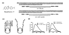
- the formation of the G-quadruplex structure can be confirmed by, for example, the in vitro translation assay shown in Examples, a fluorescence signal measurement method by reacting with thioflavin T, a stop assay method, or the like.
- the Staple nucleic acid designed by the method of the present invention is used for protein expression suppression, and thus can be included in the protein expression suppression kit. Therefore, the present invention provides a protein expression suppression kit containing the oligomer (Staple nucleic acid).
- the kit of the present invention can contain a buffer solution, a PCR reagent, an in vitro translation reagent, a luciferase, an instruction manual, and the like.
- DNA oligonucleotide were purchased from Eurofin Genomics Inc., Integrated DNA Technologies Inc., Thermo Fisher Scientific Inc. All samples were OPC-purified or desalted, and used to confirm the formation and location of RNA G-quadruplex and to evaluate the effect of inhibiting protein translation reaction in vitro.
- PCR amplification was performed using the extended nucleotide as a template and the following primers, and the obtained PCR product was cloned into pMD19 vector (Takara Bio.).
- Forward primer 5'-TAATACGACTCACTA TAG-3 '(SEQ ID NO: 3)
- Reverse primer 5'- TCCAACTATGTATACCTGCTGAATAGCTTGCAAGTC-3 '(SEQ ID NO: 4)
- PCR amplification was carried out by using a nucleotide having the following sequence as a template and a primer to prepare a linear vector.
- template 5'-ACACAGGAAACAGCTATGACCATGATTACGCCAAGTTTGCACG CCTGCCGTTCGACGATTTAATACGACTCACTATAGATTAGCATACGCTACTGCAGTGGGTGGGTGTCGACCTAGATTAATGCAATTCGTACGAAGTTCATAGCATTTCCAGCACCCAATTGAAGCTTTGGGTGGGTGCATGCCATGACTTGCAAGCTATTCAGCAGGTATACATAGTTGGAAATCTCTGGAAGATCCGCGCGTACCGAGTTCTAATTCACTGGCCGTCGTTACAACGTCGTGACTGGGAAAACCCTGGCGTTACCCA-3 '(SEQ ID NO: 7)
- Forward primer 5'-TAATACGACTCACTATAGAA-3 '(SEQ ID NO: 8)
- Reverse primer 5'-TCCAACTATGTATAC C CTG-3 '(SEQ ID NO: 9)
- the insert DNA prepared using HiFi Assembly was connected to the linear vector (pMD19-2 + 2_63nt). Furthermore, pUC19 vector (Takara Bio) was used as a template and PCR amplification was performed using the following primers to prepare insert DNA.
- Forward primer 5'-CCTAGATTAATGCAATTC GTGTGAAGATCCTTTTTGATAATCTCATGAC-3 '(SEQ ID NO: 10)
- Reverse primer 5'-TCAATTGGGTGCT GGAAATGAACTCACGTTAAGGGATTTTGGTCATGAG-3 '(SEQ ID NO: 11)
- pMD19-2 + 2_63nt was used as a template and PCR amplification was performed using the following primers to prepare a linear vector.
- Forward primer 5'-CATTTCCA GCACCCAATTGAAGCTT-3 '(SEQ ID NO: 12)
- Reverse primer 5'-ACGAATTGCATTAATCTAG GTCGAC-3 '(SEQ ID NO: 13)
- 2 + 2_100nt was amplified by PCR using the following primers and cloned into the EcoRI site of pIRES vector (Takara Bio.).
- Forward primer 5'- CTAGCCTCGAGAATTGATTAGCAT ACGCTACTGC-3 '(SEQ ID NO: 14)
- Reverse primer 5'- CCATGGTGGCGAATTCTGAATAGCTTGCAAGTC AT-3 '(SEQ ID NO: 15)
- 2_100nt_Mut A which is a mutant from GGG to AAA
- the template DNA, forward primer and reverse primer were subjected to annealing extension using the following sequences, and the resulting products were ligated by PCR (2+ 2_100nt_Mut A).
- Forward primer 5'-TAATACGACTCACTATAGG GCTAGCCTCGAG-3 '(SEQ ID NO: 24)
- Reverse primer 5'-CCATGGTGGCGAATTCTGAATAGCTTGCAAGTCAT-3 '(SEQ ID NO: 25)
- forward primer 5'-TAATACGACTCACTATAGGG-3 '(SEQ ID NO: 26)
- Reverse primer 5'-CGACTCTAGAGGATCCTGAATAG CTTGCAAGTCAT-3 '(SEQ ID NO: 27)
- RNAs were transcribed from the dsDNAs using RiboMAX TM Large Scale RNA Production System-T7 (Promega). RNAs were purified using a NAP-5 column to remove residual NTPs. Prepare the measurement solution so that the prepared RNA (400 nM), KCl (40 mM), 20 mM Tris-HCl buffer (pH 7.6), Staple (500 nM) or milliQ (control), and then at 90 ° C for 2 minutes Incubate and cool to 20 ° C at a rate of 1 ° C per minute.
- ThT was added to 200 nM ThT, the mixture was incubated at room temperature for 30 minutes, excited with 440 nm light using an FP-8500 spectrofluorometer (JASCO), and the fluorescence at 487 nm was measured.
- the measurement wavelength was 450 nm to 700 nm.
- Forward primer 5'-GTCACATCC GGGCGGGTTGGTGAGT-3 '(SEQ ID NO: 28)
- Reverse primer 5'-GGTGCCCACCCAGCTACTGCTCGCG-3 '(SEQ ID NO: 29)
- forward primer 5'-AGATTCCTACTTCTTACGCCCCCCA-3 '(SEQ ID NO: 30)
- Reverse primer 5'-TGTCTGCCAGCGCTTCCTCTTTCTC-3 '(SEQ ID NO: 31)
- the obtained PCR product was used as a template for further PCR amplification using the following primers, and the obtained PCR product was cloned into pMD19 vector (Takara Bio.) (PMD19-TPM3, pMD19-MYD88).
- Forward primer 5'-TAATACGACTCACTATAGGTCACATCCGG GCGGGTTGGTGAGT-3 '(SEQ ID NO: 32)
- Reverse primer 5'-TCCAACTATGTATACCTGGGTGCCCACCCAGCTACT GCTCGCG-3 '(SEQ ID NO: 33)
- forward primer 5'-TAATACGACTCACTATAGAGATTCCTACTTCTTACGCCCCC CA-3 '(SEQ ID NO: 34
- Reverse primer 5'-TCCAACTATGTATACCTGTGTCTGCCAGCGCTTCCTCTTTCTC-3 '(SEQ ID NO: 35)
- PCR amplification was performed using pMD19-5′UTR (2 + 2_100nt, TPM3, MYD88) as a template and the following primers to prepare linear dsDNAs for RNA transcription.
- Forward primer 5'-ACACAGGAAACAGCTATGACCATGATTACGC CAAG-3 '(SEQ ID NO: 36)
- Reverse primer 5'-TGGGTAACGCCAGGGTTTTCCCAGTCACGACGTTG-3 '(SEQ ID NO: 37)
- RNAs were transcribed from the dsDNAs using the HiScribe T7 High Yield RNA Synthesis Kit (NEB). The transcribed RNAs were purified using After Tri-Reagent RNA Clean-Up Kit (Chioda Science). A solution containing RNA (0.3 ⁇ M), 5'-FAM-labeled 3'-DNA primer (0.1 ⁇ M) and Staples (1 ⁇ M) was incubated at 80 ° C for 3 minutes, and the temperature was lowered to 30 ° C over 30 minutes. .
- ReverTra Ace reverse transcriptase (TOYOBO), MgCl 2 and dNTPs were added to this solution and a reverse transcription reaction was carried out for 30 minutes, and the obtained reverse transcription product was subjected to fragment analysis by an ABI3500 capillary DNA sequencer (Life Technologies Japan).
- the forward primer was designed to restore the EcoR I site upstream of FL after cloning.
- 5'UTR (2 + 2_100nt, TPM3, MYD88) was PCR amplified using pMD19-5'UTR (2 + 2_100nt, TPM3, MYD88) as a template and the following primers, and the resulting PCR product was pIRES- It was cloned into the EcoR I site of FL vector (pIRES-5'UTR-FL (5'UTR: 2 + 2_100nt, TPM3, MYD88)).
- Forward primer 5'-CTAGCCTCGAGAATTGAT TAGCATACGCTACTGC-3 '(SEQ ID NO: 40)
- Reverse primer 5'-CCATGGTGGCGAATTCTGAATAGCTTGCAAGTC AT-3 '(SEQ ID NO: 41)
- forward primer 5'-CTAGCCTCGAGAATTTCACATCCGGGCGGGTTG-3 '(SEQ ID NO: 42) or 5'-CTAGCCT CGAGAATTAGATTCCTACTTCTTACGCC-3' (SEQ ID NO: 43)
- Reverse primer 5'-CCATGGTGGCGAATTGTTTTCCC AGTCACGACG-3 '(SEQ ID NO: 44)
- RNA transcription was prepared by PCR amplification using pIRES-5′UTR-FL as a template and the following primers.
- RNAs were transcribed from the dsDNAs using RiboMAX TM Large Scale RNA Production System-T7 (Promega). RNAs were purified using a NAP-5 column (GE Healthcare) to remove residual NTPs.
- the obtained 5'UTR-FL (5'UTR: 2 + 2_100nt or TPM3 or MYD88) is a cell-free protein synthesis reaction solution containing Staple (100 nM) and milliQ (control) (RTS 100 Wheat Germ Kit, biotechrabbit). It was used as 12.5 ⁇ l of mRNA (1 ⁇ g). After incubating this reaction solution at 24 ° C. for 1 hour, luciferase activity was evaluated using Luciferase Assay kit (Promega) and POWERSCAN ⁇ H1 microplate reader (BioTek).
- Forward primer 5'-CTAGCCTCGAGAATTCGCCACCATGGTCTTCACACTCGAAG-3 '(SEQ ID NO: 49)
- Reverse primer 5'-CTAGCCTCGAGAATTCGCCACCATGGTCTTCACACTCGAAG-3 '(SEQ ID NO: 50)
- forward primer 5'-TCGACCCGGGCGGCCATGGCTTCCAAGGTGTACG-3 '(SEQ ID NO: 51)
- the forward primer used for PCR of nLuc was designed so that the EcoR I site would be restored upstream of nLuc after cloning. All PCR products were subjected to 1% agarose gel electrophoresis, and then excised and purified. Furthermore, pMD19-TPM3 or pMD19-MYD88 was used as a template, amplified by PCR using the following primers, and the resulting PCR product was cloned into the EcoR I site of pIRES-nLuc-renilla (pIRES-5'UTR-nLuc -renilla (5'UTR: TPM3, MYD88)).
- annealing extension was performed using any of the following primer combinations to prepare Staples (TPM3_30nt, 26nt and MYD88_30nt, 26nt).
- Reverse primer 5'-AGCTTAAAAAAGCGCGAGCAGTAGCTGGT GAGTTCCGGTAGGG-3 '(SEQ ID NO: 57)
- Primer STM2 Forward primer 5'-GATCCCCCCGGAACTCACCAGCTACTGCTCGCG TTTTTA-3 '(SEQ ID NO: 58)
- Reverse primer 5'-AGCTTAAAAACGCGAGCAGTAGCTGGTGAGTTCCGGGGG-3 '(SEQ ID NO: 59)
- Primer STM3 Forward primer 5'-GATCCCCTCTATCTCCTGCGGCACAATCTGGAGCCCCTTTTTA-3 '(SEQ ID NO: 60)
- Reverse primer 5'-AGCT TAAAAAGGGGCTCCAGATTGTGCCGCAGGAGATAGAGGG-3 '(SEQ ID NO: 61)
- Primer STM4 Forward primer 5'-GATCCCCTATCTCCTGCGGCACAATCTGGAGCCTTTTTA-3 '(SEQ ID NO: 62)
- Reverse primer 5'-AGCTTAAAAAGG CTCCAGATTGTGCCGCAGGAGATAGGG-3 '(SEQ ID NO: 63)
- RNA Staple TPM3 used for in cell translation: RNA Staple insideloop 30nt UACCGGAACUCACCAGCUACUGCUCGCGCU (SEQ ID NO: 75) TPM3: RNA Staple insideloop 26nt CCGGAACUCACCAGCUACUGCUCGCG (SEQ ID NO: 76) MUD88: RNA Staple UAU outsideloop 30nt UCUAUCUCCUGCGGCACAAUCUGGAGCCCC (SEQ ID NO: 77) MYD88: RNA Staple UAU outsideloop 26nt UAUCUCCUGCGGCACAAUCUGGAGCC (SEQ ID NO: 78)
- HEK293T cells were prepared using Medium A (Dulbecco's modified Eagle medium containing 100 units / ml penicillin, 100 ⁇ g / ml streptomycin, 10% (v / v) fetal bovine serum) at 37 ° C and 5% CO 2 concentration in an incubator. Cultured. 2 ⁇ 10 4 cells of HEK293T cells were spread per well on a 96-well plate.
- Medium A Dulbecco's modified Eagle medium containing 100 units / ml penicillin, 100 ⁇ g / ml streptomycin, 10% (v / v) fetal bovine serum
- HEK293T cells were incubated with pSuper neo.
- A-Staple Staple: TPM3_30nt, 26nt and MYD88_30nt, 26nt
- milliQ control
- pIRES-5′UTR-nLuc-renilla 5′UTR: TPM3 or MYD88
- the cell lysate was 4 ° C., was centrifuged for 1 min at 3000 rpm, the supernatant and the Luciferase Assay kit (Promega), was evaluated for luciferase activity using a POWERSCAN ⁇ H1 microplate reader (BioTek) .
- the G-quadruplex structure is constructed dependent on potassium ions 10 .
- RNA G-quadruplex structure-forming ability of the Staple nucleic acid was evaluated by cDNA synthesis using a reverse transcriptase (stop assay).
- full-length cDNA can be obtained from the designed template RNA by reverse transcription reaction.
- Staple nucleic acid is added, an RNA G-quadruplex structure is formed, the DNA elongation reaction of reverse transcriptase is stopped, and incomplete cDNA is synthesized.
- Fig. 4 (a) the termination of the reverse transcription reaction at the RNA G-quadruplex structure forming part can be confirmed (Fig. 4 (a) 11 .
- RNA sequence (2 + 2100nt RNA type) in which two guanine consecutive sequences exist near both ends of a loop sequence of 100 bases, and 4 types of short-chain DNA (inside loop 1, 1, outside) Designed and used loop, dimer, inside loop 2).
- inside loop1, outside loop, and dimer that function as Staple nucleic acid were added, a peak indicating the construction of RNA G-quadruplex structure was observed.
- inside loop2 which was expected not to function as Staple nucleic acid, did not construct RNAG-quadrulex structure, and only full-length cDNA was obtained (Fig. 4 (b)).
- RNA can be folded as designed by the DNA origami technology, and the RNA G-quadruplex structure can be induced.
- RNA model RNA sequences (2 + 2 63 nt RNA type, 2 + 2 140 nt RNA type) with loop lengths of 63 nt and 140 nt and conducted the same study using inside loop 1. A peak indicating RNA G-quadruplex construction was also confirmed for RNA (Fig. 4 (c)).
- a guanine repeat sequence was used once at one end and three times at the other end, and a model RNA sequence (1 + 3100nt RNA type) with a different pattern of guanine continuous sequence was used.
- a peak indicating the construction of the G-quadruplex structure was observed (Fig. 4 (d)).
- Staple nucleic acid exhibits the same effect when RNA is used as well as DNA (FIG. 4 (e)), and thus artificially modified nucleic acid capable of forming a complementary strand can also be used.
- RNA G-quadruplex structure is known to suppress not only reverse transcription but also protein translation 6 . If the artificial RNA G-quadruplex can be constructed on the target mRNA using Staple nucleic acid, the protein expression of the target gene can be suppressed, so that the possibility of drug application is expected to expand. Therefore, in this example, an mRNA (2 + 2 100nt RNA type_Firefly) into which a Firefly luciferase gene was introduced downstream as a 2 + 2 100nt RNA type 5'-UTR region was prepared, and a cell-free protein translation system was constructed. It was used to quantitatively evaluate the amount of protein synthesis with and without staple nucleic acid (Fig. 5 (a)).
- RNAG-quadruplex concept formation and gene expression suppression was evaluated in the natural gene sequences TPM3 and MYD88 (Fig. 6 (a)).
- TPM3 the loop base that connects the guanine continuous sequence is 103 bases
- the Staple nucleic acid was designed as inside loop1 type.
- MYD88 the loop base that connects the guanine continuous sequence is as short as 28 bases, so it was designed as an outside loop type.
- RNA G-quadruplex structure was induced by the designed Staple nucleic acid in both 5 ′ UTRs (FIG. 6 (b)).
- a model mRNA in which 5′-UTRs of TPM3 and MYD88 were arranged in the Firefly luciferase gene was prepared, and the ability of Staple nucleic acid to suppress protein translation was evaluated.
- protein translation from the model mRNA in which both 5'UTRs were arranged was suppressed by about 60% in the presence of Staple nucleic acid (Fig. 6 (c)). This result strongly indicates that Staple nucleic acid suppresses protein expression from natural target mRNA.
- the protein translation inhibitory activity of Staple nucleic acid in mammalian cells was evaluated.
- the present inventor prepared genes that express NanoLuc and Renilla luciferase. The two genes are introduced into a single mRNA, NanoLuc is translated from the 5'-cap, while Renilla luciferase is translated from the Internal Ribosomal Entry Site (IRES).
- IRES Internal Ribosomal Entry Site
- a reporter gene in which the 5'-UTR regions of TPM3 and MYD88 were placed upstream of NanoLuc was used to evaluate the effect of Staple nucleic acid in HEK293 cells.
- RNA Staple nucleic acid expressed by Short hairpin expression vector (pSuper) was used to evaluate the effect of Staple nucleic acid on protein translation reaction. As a result, it was confirmed that the amount of protein translation was reduced when the Staple nucleic acid was introduced, and it was shown that the Staple nucleic acid also functions in cells (FIG. 6 (e)).
- the present inventor has proposed a novel protein translation reaction suppression system that has never existed before. Even if Staple nucleic acid binds to an unintended position, this system does not have a guanine repeat sequence that makes it close, and thus does not exert a protein suppressing effect. It has also been clarified that the Staple nucleic acid to be introduced can obtain the same effect not only with DNA but also with RNA. This fact is an indication of the diversity of Staple nucleic acids.
- nucleic acid drug development In general, natural nucleic acid sequences are degraded by being introduced into cells, so it is essential to use modified nucleic acids by chemical synthesis in order to aim for nucleic acid drugs. Since the method of the present invention does not need to form a protein complex such as siRNA, it suffices to concentrate solely on improving intracellular stability. Decrease is unlikely. The method of the present invention, which can be modified in various ways, is extremely useful for nucleic acid drug development.
- DNA oligonucleotide DNA oligonucleotides were purchased from Eurofin Genomics Inc., Integrated DNA Technologies Inc., Thermo Fisher Scientific Inc. All samples were OPC-purified or desalted, and were used to confirm the formation and location of RNA G-quadruplex and to evaluate the effect of inhibiting protein translation reaction in vitro.
- Primer 1 Forward primer 1 fw. primer [5'-CTAGCCTCGAGAATTCGCCACCATGGTCTTCACACTCGAAG-3 '] (SEQ ID NO: 79)
- Reverse primer 1 rv. primer [5'-CTCGACGCGTGAATTTTACGCCAGAATGCGTTCG-3 '] (SEQ ID NO: 80)
- Primer 2 Forward primer 2 fw. primer [5'-TCGACCCGGGCGGCCATGGCTTCCAAGGTGTACG-3 '] (SEQ ID NO: 81)
- Reverse primer rv. primer [5'-TAAAGGGAA GCGGCCTTACTGCTCGTTCTTCAGC-3 '] (SEQ ID NO: 82)
- the fw. primer used for PCR of nLuc was designed so that the EcoR I site was restored upstream of nLuc after cloning. All PCR products were subjected to 1% agarose gel electrophoresis, and then excised and purified. PCR amplification of TPM3-5'UTR was performed using human genomic DNA (Novagen) as a template and the following primers. fw. primer [5'-GTCACATCC GGGCGGGTTGGTGAGT-3 '] (SEQ ID NO: 83) rv.
- primer [5'-GGTGCCCACCCAGCTACTGCTCGCG-3 '] (SEQ ID NO: 84)
- PCR amplification was further performed using the following primers, and the obtained PCR product was cloned into pMD19 vector (Takara Bio.) (PMD19-TPM3).
- primer [5'-TAATACGACTCACTATAGGTCACATCCGG GCGGGTTGGTGAGT-3 '] (SEQ ID NO: 85)
- rv. primer [5'-TCCAACTATGTATACCTGGGTGCCCACCCAGCTACTGCTCGCG-3 '] (SEQ ID NO: 86)
- pMD19-TPM3 was used as a template and amplified by PCR using the following primers, and the resulting PCR product was cloned into the EcoR I site of pIRES-nLuc-renilla (pIRES-TPM3_5′UTR-nLuc-renilla).
- Primer [5'-CTAGCCTCGAGAATTTCACATCCGGGCGGGTTG-3 '] (SEQ ID NO: 87)
- rv. primer [5'-CCATGGTGGCGAATT GTTTTCCCAGTCACGACG-3 '] (SEQ ID NO: 88)
- HEK293T cells were prepared using Medium A (Dulbecco's modified Eagle medium containing 100 units / ml penicillin, 100 ⁇ g / ml streptomycin, 10% (v / v) fetal bovine serum) at 37 ° C and 5% CO 2 concentration in an incubator. Cultured. 2 ⁇ 10 4 cells of HEK293T cells were spread per well on a 96-well plate. After 24 hours, HEK293T cells were introduced with pSuper neo. A-Staple (Staple: TPM3_26nt_reverse ligation) or milliQ (control) using Transfection reagent (FuGENE, Promega) and incubated.
- Medium A Dulbecco's modified Eagle medium containing 100 units / ml penicillin, 100 ⁇ g / ml streptomycin, 10% (v / v) fetal bovine serum
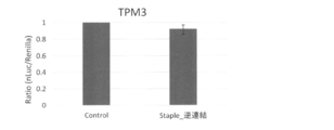
- pIRES-TPM3_5'UTR-nLuc-renilla was similarly introduced into HEK293T cells and incubated. After 6 hours, the cells were washed with PBS and the cell lysate was collected using Passive Lysis Buffer. The cell lysate was centrifuged at 3000 rpm for 1 minute at 4 ° C., and the luciferase activity was evaluated using the supernatant, Luciferase Assay kit (Promega) and POWERSCAN H1 microplate reader (BioTek). The results are shown in Fig. 7. No suppression of expression was observed when TPM3 was not used (binding) as Staple nucleic acid (FIG. 7).
- SEQ ID NOS: 1 to 68, 71-74, 79-90 Synthetic DNA
- SEQ ID NOs: 69, 70, 75-78 Synthetic RNA
Landscapes
- Health & Medical Sciences (AREA)
- Life Sciences & Earth Sciences (AREA)
- Chemical & Material Sciences (AREA)
- Genetics & Genomics (AREA)
- Engineering & Computer Science (AREA)
- Biomedical Technology (AREA)
- Bioinformatics & Cheminformatics (AREA)
- Organic Chemistry (AREA)
- Molecular Biology (AREA)
- General Health & Medical Sciences (AREA)
- Zoology (AREA)
- Biotechnology (AREA)
- Wood Science & Technology (AREA)
- General Engineering & Computer Science (AREA)
- Biochemistry (AREA)
- Medicinal Chemistry (AREA)
- Pharmacology & Pharmacy (AREA)
- Veterinary Medicine (AREA)
- Public Health (AREA)
- Animal Behavior & Ethology (AREA)
- Plant Pathology (AREA)
- Microbiology (AREA)
- Physics & Mathematics (AREA)
- Biophysics (AREA)
- Epidemiology (AREA)
- Chemical Kinetics & Catalysis (AREA)
- General Chemical & Material Sciences (AREA)
- Nuclear Medicine, Radiotherapy & Molecular Imaging (AREA)
- Measuring Or Testing Involving Enzymes Or Micro-Organisms (AREA)
Abstract
タンパク質発現を抑制する方法の提供。 第一、第二、第三及び第四のグアニン繰り返し配列(それぞれ、第一G配列、第二G配列、第三G配列、第四G配列という)を有するRNAに対し、前記グアニン繰り返し配列の近傍領域にハイブリダイズするオリゴマーを用いて前記グアニン繰り返し配列を近づけてグアニン四重鎖構造を形成させることによりタンパク質発現を抑制する方法。
Description
本発明は、配列選択的に標的mRNA配列に結合し、標的mRNA上にグアニン四重鎖構造の形成を誘発することによりリボゾームのペプチド合成反応を阻害し、タンパク質翻訳反応を抑制する方法に関する。
マイクロRNAやsiRNAなど様々な遺伝子発現抑制技術が知られている1,2(非特許文献1、2)。しかし、いずれの技術に関してもmRNAに結合するという単純な機構で遺伝子発現抑制機能を発揮するため、予期せぬ効果(オフ・ターゲット効果)が避けられない3(非特許文献3)。また、導入核酸の細胞内安定性向上を目的として修飾核酸を使用すると、マイクロRNA効果やsiRNA活性が著しく低下し、期待するタンパク質発現抑制効果が得られない問題も生じる4(非特許文献4)。
Fire, A.; Xu, S.; Montgomery, M. K.; Kostas, S. A.; Driver, S. E.; Mello, C. C. Nature 1998, 391, 806.
Ambros, V. Nature 2004, 431, 350.
Jackson, A. L.; Linsley, P. S. Nature reviews. Drug discovery 2010, 9, 57.
Jackson, A. L.; Burchard, J.; Leake, D.; Reynolds, A.; Schelter, J.; Guo, J.; Johnson, J. M.; Lim, L.; Karpilow, J.; Nichols, K.; Marshall, W.; Khvorova, A.; Linsley, P. S. Rna 2006, 12, 1197.
創薬を視野に入れた核酸医薬を開発するには、生体内で安定な修飾核酸が利用可能であり、且つ、オフ・ターゲット効果が生じにくいタンパク質翻訳抑制技術を採用することが求められる。
本発明者は、上記課題を解決するために鋭意検討を行った結果、Staple核酸と呼ばれるオリゴマーを用いることにより、配列選択的にグアニン四重鎖を形成させ、ひいてはタンパク質合成を抑制することに成功し、本発明を完成するに至った。
すなわち、本発明は以下の通りである。
(1)第一、第二、第三及び第四のグアニン繰り返し配列(それぞれ、第一G配列、第二G配列、第三G配列、第四G配列という)を含む標的RNAに対し、前記グアニン繰り返し配列の近傍にハイブリダイズするオリゴマーを用い、当該オリゴマーの折り返しにより前記グアニン繰り返し配列を近づけてグアニン四重鎖構造を形成させることによりタンパク質発現を抑制する方法であって、
前記オリゴマーの一端側の一部の領域(一端側領域)の核酸及び他端側の一部の領域(他端側領域)の核酸が、第一G配列と第二G配列との間であってそれぞれ第一G配列の近傍及び第二G配列の近傍、第二G配列と第三G配列との間であってそれぞれ第二G配列の近傍及び第三G配列の近傍、又は第三G配列と第四G配列との間であってそれぞれ第三G配列の近傍及び第四G配列の近傍にハイブリダイズする、前記方法。
(2)前記オリゴマーは、その折り返しの方向がグアニン四重鎖構造側を向いてハイブリダイズするように設計される、(1)に記載の方法。
(3)標的RNAがmRNA、TPM3又はMYD8である、(1)又は(2)に記載の方法。
(4)オリゴマーがDNA又はRNAである、(1)~(3)のいずれか1項に記載の方法。
(5)オリゴマーがダイマーである、(1)~(4)のいずれか1項に記載の方法。
(6)タンパク質発現が、逆転写反応又はタンパク質翻訳反応である、(1)~(5)のいずれか1項に記載の方法。
(7)第一、第二、第三及び第四のグアニン繰り返し配列(それぞれ、第一G配列、第二G配列、第三G配列、第四G配列という)を含む標的RNAに対し、前記グアニン繰り返し配列を近づけてグアニン四重鎖構造を形成させるオリゴマーであって、
前記オリゴマーの一端側の一部の領域(一端側領域)の核酸及び他端側の一部の領域(他端側領域)の核酸が、第一G配列と第二G配列との間であってそれぞれ第一G配列の近傍及び第二G配列の近傍、第二G配列と第三G配列との間であってそれぞれ第二G配列の近傍及び第三G配列の近傍、又は第三G配列と第四G配列との間であってそれぞれ第三G配列の近傍及び第四G配列の近傍にハイブリダイズして、その折り返しにより前記グアニン繰り返し配列を近づけるものである、
前記オリゴマー。
(8)前記折り返しの方向がグアニン四重鎖構造側を向いてハイブリダイズするように設計される、(7)に記載のオリゴマー。
(9)DNA又はRNAである、(7)又は(8)に記載のオリゴマー。
(10)ダイマーである、(7)~(9)のいずれか1項に記載のオリゴマー。
(11) (7)~(10)のいずれか1項に記載のオリゴマーの製造方法であって、
オリゴマーの一端側の一部の領域(一端側領域)の核酸及び他端側の一部の領域(他端側領域)の核酸が、第一G配列と第二G配列との間であってそれぞれ第一G配列の近傍及び第二G配列の近傍、第二G配列と第三G配列との間であってそれぞれ第二G配列の近傍及び第三G配列の近傍、又は第三G配列と第四G配列との間であってそれぞれ第三G配列の近傍及び第四G配列の近傍にハイブリダイズするように設計し合成することを特徴とする前記方法。
(12) (7)~(10)のいずれか1項に記載のオリゴマー、又は(11)に記載の方法により製造されたオリゴマーを含む、タンパク質発現抑制キット。
(1)第一、第二、第三及び第四のグアニン繰り返し配列(それぞれ、第一G配列、第二G配列、第三G配列、第四G配列という)を含む標的RNAに対し、前記グアニン繰り返し配列の近傍にハイブリダイズするオリゴマーを用い、当該オリゴマーの折り返しにより前記グアニン繰り返し配列を近づけてグアニン四重鎖構造を形成させることによりタンパク質発現を抑制する方法であって、
前記オリゴマーの一端側の一部の領域(一端側領域)の核酸及び他端側の一部の領域(他端側領域)の核酸が、第一G配列と第二G配列との間であってそれぞれ第一G配列の近傍及び第二G配列の近傍、第二G配列と第三G配列との間であってそれぞれ第二G配列の近傍及び第三G配列の近傍、又は第三G配列と第四G配列との間であってそれぞれ第三G配列の近傍及び第四G配列の近傍にハイブリダイズする、前記方法。
(2)前記オリゴマーは、その折り返しの方向がグアニン四重鎖構造側を向いてハイブリダイズするように設計される、(1)に記載の方法。
(3)標的RNAがmRNA、TPM3又はMYD8である、(1)又は(2)に記載の方法。
(4)オリゴマーがDNA又はRNAである、(1)~(3)のいずれか1項に記載の方法。
(5)オリゴマーがダイマーである、(1)~(4)のいずれか1項に記載の方法。
(6)タンパク質発現が、逆転写反応又はタンパク質翻訳反応である、(1)~(5)のいずれか1項に記載の方法。
(7)第一、第二、第三及び第四のグアニン繰り返し配列(それぞれ、第一G配列、第二G配列、第三G配列、第四G配列という)を含む標的RNAに対し、前記グアニン繰り返し配列を近づけてグアニン四重鎖構造を形成させるオリゴマーであって、
前記オリゴマーの一端側の一部の領域(一端側領域)の核酸及び他端側の一部の領域(他端側領域)の核酸が、第一G配列と第二G配列との間であってそれぞれ第一G配列の近傍及び第二G配列の近傍、第二G配列と第三G配列との間であってそれぞれ第二G配列の近傍及び第三G配列の近傍、又は第三G配列と第四G配列との間であってそれぞれ第三G配列の近傍及び第四G配列の近傍にハイブリダイズして、その折り返しにより前記グアニン繰り返し配列を近づけるものである、
前記オリゴマー。
(8)前記折り返しの方向がグアニン四重鎖構造側を向いてハイブリダイズするように設計される、(7)に記載のオリゴマー。
(9)DNA又はRNAである、(7)又は(8)に記載のオリゴマー。
(10)ダイマーである、(7)~(9)のいずれか1項に記載のオリゴマー。
(11) (7)~(10)のいずれか1項に記載のオリゴマーの製造方法であって、
オリゴマーの一端側の一部の領域(一端側領域)の核酸及び他端側の一部の領域(他端側領域)の核酸が、第一G配列と第二G配列との間であってそれぞれ第一G配列の近傍及び第二G配列の近傍、第二G配列と第三G配列との間であってそれぞれ第二G配列の近傍及び第三G配列の近傍、又は第三G配列と第四G配列との間であってそれぞれ第三G配列の近傍及び第四G配列の近傍にハイブリダイズするように設計し合成することを特徴とする前記方法。
(12) (7)~(10)のいずれか1項に記載のオリゴマー、又は(11)に記載の方法により製造されたオリゴマーを含む、タンパク質発現抑制キット。
本発明により、配列選択的に標的mRNA配列に結合(ハイブリダイズ)するオリゴマーの設計法を開発し、設計されたオリゴマーを用いて標的mRNAに安定なグアニン四重鎖構造を誘起することで、リボゾームのペプチド合成反応を阻害することが可能となった。
本発明は、第一、第二、第三及び第四の4つのグアニン繰り返し配列を有するRNAに対し、前記グアニン繰り返し配列の近傍領域にハイブリダイズするオリゴマーを用いて前記グアニン繰り返し配列を近づけてグアニン四重鎖構造を形成させることにより、タンパク質発現を抑制する方法に関する。
mRNA上に存在する安定なグアニン四重鎖構造(G-quadruplex構造ともいう)は、リボソームのペプチド伸長反応を阻害することが知られている。任意のmRNA上に、人為的にG-quadruplex構造を誘起することが出来れば、疾病に関連するタンパク質の発現量を抑制することが可能となる。本発明は,DNA origami技術を応用し,配列選択的に標的mRNAを捉えて折りたたみ、標的mRNA上に安定なG-quadruplex構造を誘起することで,標的mRNA選択的にタンパク質の翻訳を抑制する新規核酸医薬の設計法を提供するものである。
本発明において使用されるオリゴマーは、その一端側の一部の領域(一端側領域)の核酸及び他端側の一部の領域(他端側領域)の核酸が、RNAの第一のグアニン繰り返し配列(第一G配列という)、第二のグアニン繰り返し配列(第二G配列という)、第三のグアニン繰り返し配列(第三G配列という)及び第四のグアニン繰り返し配列(第四G配列という)において、それぞれ、第一G配列と第二G配列、第二G配列と第三G配列、及び第三G配列と第四G配列の近傍領域の核酸にハイブリダイズするものである。このオリゴマーを、本発明では、「Staple核酸」ともいう。
本発明の方法が細胞内で機能するには,核酸の配列認識能とG-quadruplex構造形成能を同時に満たす必要があり,これまでの核酸医薬を凌駕する標的選択性が実現できる。本発明の有用性は、モデル配列を利用した検証により、(1)グアニン繰り返し領域の長さや配列に関わらずStaple核酸を導入することでRNA G-quadruplexが構築されること、(2)誘起されたG-quadruplex構造が、逆転写酵素の伸長反応やリボソームのタンパク質翻訳反応を抑制することにより証明した。また,がん関連遺伝子であるTMP3とMYD88 mRNAの5’UTRに存在するグアニンクラスターを折りたたみ,タンパク質合成を阻害できることが,無細胞系発現システムおよび哺乳動物細胞を用いた発現評価により確認できた。
本発明においては、タンパク質の翻訳抑制機構にRNA四重鎖構造であるRNA G-quadruplex構造に着目した(図1(a))。RNA G-quadruplex構造は、3塩基以上のグアニン連続配列が、7塩基未満の近接したスペーシングで4箇所存在する時に構築される安定なRNA高次構造である(図1(b))5。このRNA G-quadruplex構造がmRNAの5’非翻訳領域(5’UTR)に存在すると、リボソームのタンパク質合成反応を抑制する(図1(c))6。
近年、本発明者は7塩基以上のスペーシングで存在するグアニン連続配列が、RNA G-quadruplex構造を構築することを見い出した7。この事実は、mRNAが形成する高次構造により、一次配列で離れた位置にあるグアニン連続配列が空間的に近接し、RNA G-quadruplex構造を形成していることを示すものである。この知見を基に、本発明者は、離れた位置に存在するグアニン連続配列を空間的に近接させ、標的とするmRNA上に人為的なRNA G-quadruplex構造を誘起させる本発明の方法を考案した。
ところで、7000塩基を超える一本鎖環状DNAを、設計された200本程度の30塩基強の短鎖DNA(Staple oligomer)と自己集合させることでナノ構造体を作る技術であるDNA origami法が知られている(図2)8。この技術のStaple oligomerは、一本鎖環状DNAの離れた位置に存在している配列を手繰り寄せ、コンパクトな構造体を形成する。この技術に習い、標的mRNA上のグアニン連続配列を近接させるStaple核酸を設計・導入すれば、標的mRNA上にG-quadruplex構造を誘起することが期待され、結果、タンパク質翻訳反応を抑制することが可能となる。そこで本発明では、人為的なRNA G-quadruplex構造の誘起に、DNAナノ構造体の作製法であるDNA origami法を応用することができる。
前記の通り、グアニン繰り返し領域が4本近接すると、G-quadruplex構造を形成する(図1a)。本発明においては、RNA上に存在する4か所のグアニン繰り返し領域を近づけるため、所定のオリゴマーを用いる。このオリゴマーは、第一のグアニン繰り返し配列と第二のグアニン繰り返し配列、第二のグアニン繰り返し配列と第三のグアニン繰り返し配列、又は第三のグアニン繰り返し配列と第四のグアニン繰り返し配列とを近づけて全体として4か所のグアニン繰り返し配列が近接するように機能するオリゴマーであり、本明細書では「Staple核酸」という。
ここで、標的RNAとしては、例えばmRNA、TPM3、MYD8などが挙げられる。TPM3とは、筋収縮に関連する遺伝子であり、MYD8とは、Tollおよびインターロイキン(IL)1受容体シグナル伝達を媒介するアダプタータンパク質である。
標的となるRNAに含まれるグアニン繰り返し配列は、4か所に限らず、5か所以上含まれていてもよい。また、第一、第二、第三及び第四のグアニン繰り返し配列は、4か所以上のグアニン繰り返し配列から任意に選択される4か所の配列を意味し、必ずしも、連続する4か所のグアニン繰り返し配列にG-quadruplex構造を形成させることを意味するものではない。例えば、あるRNAに6か所のグアニン繰り返し配列が存在すると仮定すると、5’側から順に第1、第2、第3及び第4番目のグアニン繰り返し配列によりG-quadruplex構造を形成させてもよく、第1、第2、第5及び第6番目のグアニン繰り返し配列によりG-quadruplex構造を形成させることもできる。要するに、G-quadruplex構造を形成してRNAのタンパク質発現が抑制される限り、4か所のグアニン繰り返し単位の位置は不問である。
ここで、本発明においてタンパク質発現の抑制とは、例えばmRNAからの逆転写反応の抑制、RNAからタンパク質への翻訳反応の抑制などが含まれる。
標的となるRNAに含まれるグアニン繰り返し配列は、4か所に限らず、5か所以上含まれていてもよい。また、第一、第二、第三及び第四のグアニン繰り返し配列は、4か所以上のグアニン繰り返し配列から任意に選択される4か所の配列を意味し、必ずしも、連続する4か所のグアニン繰り返し配列にG-quadruplex構造を形成させることを意味するものではない。例えば、あるRNAに6か所のグアニン繰り返し配列が存在すると仮定すると、5’側から順に第1、第2、第3及び第4番目のグアニン繰り返し配列によりG-quadruplex構造を形成させてもよく、第1、第2、第5及び第6番目のグアニン繰り返し配列によりG-quadruplex構造を形成させることもできる。要するに、G-quadruplex構造を形成してRNAのタンパク質発現が抑制される限り、4か所のグアニン繰り返し単位の位置は不問である。
ここで、本発明においてタンパク質発現の抑制とは、例えばmRNAからの逆転写反応の抑制、RNAからタンパク質への翻訳反応の抑制などが含まれる。
図3(b)は、第一及び第二のグアニン繰り返し配列(図の左から1番目及び2番目の「●●●」印)、並びに第三及び第四のグアニン繰り返し配列(図の左から3番目及び4番目の「●●●」印)が互いに近接しており、第二のグアニン繰り返し配列と第三のグアニン繰り返し配列とが100ヌクレオチドの長さを有する態様を示す。図3bにおいて、左から1番目のグアニン繰り返し配列を第一G配列、左から2番目のグアニン繰り返し配列を第二G配列、左から3番目のグアニン繰り返し配列を第三G配列、左から4番目のグアニン繰り返し配列を第四G配列とすると、第二G配列と第三G配列との間が離れているため、この領域を折りたたんで第二G配列と第三G配列とを近づけるようにする。
そこで、Staple核酸は、その一端側(例えば3’側)の一部の領域の核酸が第二G配列のすぐ下流の領域の核酸とハイブリダイズし、他端側(例えば5’側)の一部の領域の核酸が第三G配列のすぐ上流の領域の核酸とハイブリダイズするように設計する。この場合、前記一端側領域及び他端側領域の核酸は、折り返し方向(凸側)がG-quadruplex構造側を向くようにハイブリダイズする。Staple核酸がRNAとハイブリダイズする領域は、RNAの種類に応じて適宜設計することができる。例えば図3bに示すRNAに対してStaple核酸を設計する場合は、第二G配列の下流側の13~20塩基塩基の領域から、ハイブリダイズする領域として13~20塩基の領域を選択し、また、第三G配列の上流側の13~20塩基塩基の領域から、ハイブリダイズする領域として13~20塩基の領域を選択すればよい。他の領域に対するStaple核酸を設計する場合も同様である。
Staple核酸は、DNAであってもよく、RNAであってもよい。Staple核酸の長さは、26~40ヌクレオチド長である。Staple核酸がRNAにハイブリダイズする領域の長さは、前記一端側領域及び他端側領域の長さが同じであっても異なってもよく、限定されるものではない。Staple核酸は、市販の核酸合成装置を用いることにより容易に得ることができる。
Staple核酸は、DNAであってもよく、RNAであってもよい。Staple核酸の長さは、26~40ヌクレオチド長である。Staple核酸がRNAにハイブリダイズする領域の長さは、前記一端側領域及び他端側領域の長さが同じであっても異なってもよく、限定されるものではない。Staple核酸は、市販の核酸合成装置を用いることにより容易に得ることができる。
このように設計されたStaple核酸を用いて、グアニン繰り返し配列を近接させた態様を図4に示す。図4bにおいて、「Inside loop 1」の態様は、Staple核酸の両端側の一部のがRNAにハイブリダイズするとともに、Staple核酸の折り返し(折り畳み)方向がグアニン四重鎖構造側を向いている。図4では、折り返し方向は理解しやすくするためにループ状で表示してあるが、本発明のStaple核酸は、RNAにハイブリダイズする一方の領域と他方の領域との境界を支点として折り返される。図4bにおいて、「Outside loop」の態様もInside loop 1と同様であり、Staple核酸の両側の一部の領域がRNAにハイブリダイズするとともに、折り返し方向(ループ状に表示してある部分の凸側(外周側))がG-quadruplex構造側を向いている。また本発明においては、Staple核酸は1本鎖である必要はなく、2本以上が結合又はハイブリダイズしてダイマー、トリマー等を形成することもできる。図4bにおいて、「Dimer]の態様は、2本のStaple核酸により構成されており、RNAにハイブリダイズしない領域同士(1又は複数の塩基)がハイブリダイズして折り返し領域を形成するとともに(図では折り返し領域は複数の塩基で例示してある)、全体としてダイマーを形成している。このダイマー型Staple核酸においては、両側の一部の領域がRNAにハイブリダイズするとともに、折り返し部がG-quadruplex構造側を向いている。
上記のとおり設計されたStaple核酸とRNAとの反応、すなわち、G-quadruplex構造の形成反応は、例えば、RNAを含む溶液中にStaple核酸を混合して、アニーリングすることにより行うことができる。あるいはRNAを含む溶液中にStaple核酸を単に混ぜるだけでもよい。あるいはShort hairpin expression vectorを導入し、細胞内からStaple核酸を発現させることで行うことができる。RNAとStaple核酸との反応時間は、例えば1分(試験管実験)~2日(細胞実験)であり、反応温度は例えば24~55℃である。G-quadruplex構造が形成されたことの確認は、例えば実施例に示されるin vitroトランスレーションアッセイのほか、チオフラビンTと反応させることによる蛍光シグナル測定法、ストップアッセイ法などにより行うことができる。
本発明の方法により設計されたStaple核酸は、タンパク質発現抑制に使用されることから、タンパク質発現抑制キットに含めることができる。従って、本発明は、前記オリゴマー(Staple核酸)を含む、タンパク質発現抑制キットを提供する。本発明のキットには、前記オリゴマーのほか、緩衝液、PCR用試薬、in vitro translation用試薬、ルシフェラーゼ、使用説明書などを含めることができる。
実施例
以下、実施例により本発明をさらに具体的に説明する。但し、本発明の範囲はこれらの実施例により限定されるものではない。
以下、実施例により本発明をさらに具体的に説明する。但し、本発明の範囲はこれらの実施例により限定されるものではない。
1.材料
(1)DNAオリゴヌクレオチド
DNAオリゴヌクレオチドはユーロフィンジェノミクス株式会社、Integrated DNA Technologies株式会社、Thermo Fisher Scientific株式会社より購入した。すべてのサンプルはOPC精製または脱塩されており、RNA G-quadruplexの形成や存在位置の確認やin vitroにおけるタンパク質翻訳反応阻害効果を評価するために使用した。
(1)DNAオリゴヌクレオチド
DNAオリゴヌクレオチドはユーロフィンジェノミクス株式会社、Integrated DNA Technologies株式会社、Thermo Fisher Scientific株式会社より購入した。すべてのサンプルはOPC精製または脱塩されており、RNA G-quadruplexの形成や存在位置の確認やin vitroにおけるタンパク質翻訳反応阻害効果を評価するために使用した。
2.方法
(1)2+2_100nt RNA、2+2_100nt_Mut A RNAの準備とThioflavin Tを用いたRNA G-quadruplex形成の確認
下記ヌクレオチドを用いてアニールエクステンションを行った。
5’-TAATACGACTCACTATAGATTAGCATACGCTACTGCAGATGCGC-3’(配列番号1)
5’-GCTGAATAGCTTGCAAGTCATGGCATGCGCATCTGCAGTAGCGT-3’ (配列番号2)
(1)2+2_100nt RNA、2+2_100nt_Mut A RNAの準備とThioflavin Tを用いたRNA G-quadruplex形成の確認
下記ヌクレオチドを用いてアニールエクステンションを行った。
5’-TAATACGACTCACTATAGATTAGCATACGCTACTGCAGATGCGC-3’(配列番号1)
5’-GCTGAATAGCTTGCAAGTCATGGCATGCGCATCTGCAGTAGCGT-3’ (配列番号2)
伸長されたヌクレオチドを鋳型として、以下のプライマーを用いてPCR増幅を行い、得られたPCR産物は、pMD19 vector(タカラバイオ.)にクローニングした。
フォワードprimer:5’-TAATACGACTCACTA TAG-3’(配列番号3)
リバースprimer:5’- TCCAACTATGTATACCTGCTGAATAGCTTGCAAGTC-3’ (配列番号4)
フォワードprimer:5’-TAATACGACTCACTA TAG-3’(配列番号3)
リバースprimer:5’- TCCAACTATGTATACCTGCTGAATAGCTTGCAAGTC-3’ (配列番号4)
そして、上記で作製したplasmid DNAを鋳型とし、以下のプライマーを用いてPCR増幅を行い、insert DNAを作製した。
フォワードprimer:5’-CTATAGTGAGTCGTATTAAATCGTCGAACGGCAGG-3’ (配列番号5)
リバースprimer:5’-CAGGTATA CATAGTTGGAAATCTCTGGAAGATCCG-3’ (配列番号6)
フォワードprimer:5’-CTATAGTGAGTCGTATTAAATCGTCGAACGGCAGG-3’ (配列番号5)
リバースprimer:5’-CAGGTATA CATAGTTGGAAATCTCTGGAAGATCCG-3’ (配列番号6)
また、下記配列を有するヌクレオチドを鋳型及びプライマーとして用いてPCR増幅を行い、linear vectorを作製した。
鋳型:
5’-ACACAGGAAACAGCTATGACCATGATTACGCCAAGTTTGCACG CCTGCCGTTCGACGATTTAATACGACTCACTATAGATTAGCATACGCTACTGCAGTGGGTGGGTGTCGACCTAGATTAATGCAATTCGTACGAAGTTCATAGCATTTCCAGCACCCAATTGAAGCTTTGGGTGGGTGCATGCCATGACTTGCAAGCTATTCAGCAGGTATACATAGTTGGAAATCTCTGGAAGATCCGCGCGTACCGAGTTCTAATTCACTGGCCGTCGTTTTACAACGTCGTGACTGGGAAAACCCTGGCGTTACCCA-3’ (配列番号7)
フォワードprimer:5’-TAATACGACTCACTATAGAA-3’ (配列番号8)
リバースprimer:5’-TCCAACTATGTATAC CTG-3’ (配列番号9)
鋳型:
5’-ACACAGGAAACAGCTATGACCATGATTACGCCAAGTTTGCACG CCTGCCGTTCGACGATTTAATACGACTCACTATAGATTAGCATACGCTACTGCAGTGGGTGGGTGTCGACCTAGATTAATGCAATTCGTACGAAGTTCATAGCATTTCCAGCACCCAATTGAAGCTTTGGGTGGGTGCATGCCATGACTTGCAAGCTATTCAGCAGGTATACATAGTTGGAAATCTCTGGAAGATCCGCGCGTACCGAGTTCTAATTCACTGGCCGTCGTTTTACAACGTCGTGACTGGGAAAACCCTGGCGTTACCCA-3’ (配列番号7)
フォワードprimer:5’-TAATACGACTCACTATAGAA-3’ (配列番号8)
リバースprimer:5’-TCCAACTATGTATAC CTG-3’ (配列番号9)
そして、HiFiAssembly(NEB)を用いて作製したinsert DNA とlinear vectorを繋げた(pMD19-2+2_63nt)。さらに、pUC19 vector(タカラバイオ.)を鋳型にし、以下のプライマーを用いてPCR増幅を行い、insert DNAを作製した。
フォワードprimer:5’-CCTAGATTAATGCAATTC GTGTGAAGATCCTTTTTGATAATCTCATGAC-3’ (配列番号10)
リバースprimer:5’-TCAATTGGGTGCT GGAAATGAACTCACGTTAAGGGATTTTGGTCATGAG-3’ (配列番号11)
フォワードprimer:5’-CCTAGATTAATGCAATTC GTGTGAAGATCCTTTTTGATAATCTCATGAC-3’ (配列番号10)
リバースprimer:5’-TCAATTGGGTGCT GGAAATGAACTCACGTTAAGGGATTTTGGTCATGAG-3’ (配列番号11)
また、pMD19-2+2_63ntを鋳型にし、以下のプライマーを用いてPCR増幅を行い、linear vectorを作製した。
フォワードprimer:5’-CATTTCCA GCACCCAATTGAAGCTT-3’ (配列番号12)
リバースprimer:5’-ACGAATTGCATTAATCTAG GTCGAC-3’ (配列番号13)
フォワードprimer:5’-CATTTCCA GCACCCAATTGAAGCTT-3’ (配列番号12)
リバースprimer:5’-ACGAATTGCATTAATCTAG GTCGAC-3’ (配列番号13)
そして、HiFiAssembly(NEB)を用いて作製したinset DNAとlinear vectorを繋げた(pMD19-2+2_100nt)。
作製したpMD19-2+2_100ntを鋳型として、以下のプライマーを用いてPCRにより2+2_100ntを増幅し、pIRES vector(タカラバイオ.)のEcoR Iサイトにクローニングした。
フォワードprimer:5’- CTAGCCTCGAGAATTGATTAGCAT ACGCTACTGC-3’ (配列番号14)
リバースprimer:5’- CCATGGTGGCGAATTCTGAATAGCTTGCAAGTC AT-3’ (配列番号15)
フォワードprimer:5’- CTAGCCTCGAGAATTGATTAGCAT ACGCTACTGC-3’ (配列番号14)
リバースprimer:5’- CCATGGTGGCGAATTCTGAATAGCTTGCAAGTC AT-3’ (配列番号15)
GGGからAAAへの変異体である2+2_100nt_Mut Aでは鋳型DNA、フォワードprimer、リバースprimerに、以下の配列のものを用いてそれぞれアニールエクステンションを行い、得られた産物をPCRによって繋いだ(2+2_100nt_Mut A)。
(i) MutA-1
鋳型:5’-AGGGATTAGCATACGCTACTGCAGTAAAT AAATGTCGACCTAG-3’(配列番号16)
フォワードprimer:5’-CGGTACCCGGGGATCTAATACGACTCACTATAGGGAT TAGC-3’(配列番号17)
リバースprimer:5’-CACGAATTGCATTAATCTAGGTCGAC-3’(配列番号18)
(ii) MutA-2
鋳型:pMD19-2+2_100nt
フォワードprimer:5’-GTCGACCTAGATTAATGCAATTCGTG-3’(配列番号19)
リバースprimer:5’-AAGCTTCAATTGGGTGCTGGAA ATG-3’(配列番号20)
(iii) MutA-3
鋳型:5’-ATTGAAGCTTTAAATAAATGCATGCCATGACTTGCAAG-3’(配列番号21)
フォワードprimer:5’-CATTTCCAGCACCCAATTGAAGCTT-3’(配列番号22)
リバースprimer:5’-CGACTCTAGAGGATCCTGAATAGCTT GCAAGTCAT-3’(配列番号23)
(i) MutA-1
鋳型:5’-AGGGATTAGCATACGCTACTGCAGTAAAT AAATGTCGACCTAG-3’(配列番号16)
フォワードprimer:5’-CGGTACCCGGGGATCTAATACGACTCACTATAGGGAT TAGC-3’(配列番号17)
リバースprimer:5’-CACGAATTGCATTAATCTAGGTCGAC-3’(配列番号18)
(ii) MutA-2
鋳型:pMD19-2+2_100nt
フォワードprimer:5’-GTCGACCTAGATTAATGCAATTCGTG-3’(配列番号19)
リバースprimer:5’-AAGCTTCAATTGGGTGCTGGAA ATG-3’(配列番号20)
(iii) MutA-3
鋳型:5’-ATTGAAGCTTTAAATAAATGCATGCCATGACTTGCAAG-3’(配列番号21)
フォワードprimer:5’-CATTTCCAGCACCCAATTGAAGCTT-3’(配列番号22)
リバースprimer:5’-CGACTCTAGAGGATCCTGAATAGCTT GCAAGTCAT-3’(配列番号23)
これをpUC19 vector(タカラバイオ.)のBamH Iサイトにクローニングした(pUC19-2+2_100nt_Mut A)。すべてのPCR産物は1%アガロースゲル電気泳動後、切り出し精製を行った。作製したpIRES-2+2_100ntおよびpUC19-2+2_100nt_Mut Aを鋳型とし、以下のいずれかのプライマーを用いてPCR増幅によりRNA転写に用いるlinear dsDNAsを準備した。
フォワードprimer:5’-TAATACGACTCACTATAGG GCTAGCCTCGAG-3’ (配列番号24)
リバースprimer:5’-CCATGGTGGCGAATTCTGAATAGCTTGCAAGTCAT-3’ (配列番号25)
または
フォワードprimer:5’-TAATACGACTCACTATAGGG-3’ (配列番号26)
リバースprimer: 5’-CGACTCTAGAGGATCCTGAATAG CTTGCAAGTCAT-3’ (配列番号27)
フォワードprimer:5’-TAATACGACTCACTATAGG GCTAGCCTCGAG-3’ (配列番号24)
リバースprimer:5’-CCATGGTGGCGAATTCTGAATAGCTTGCAAGTCAT-3’ (配列番号25)
または
フォワードprimer:5’-TAATACGACTCACTATAGGG-3’ (配列番号26)
リバースprimer: 5’-CGACTCTAGAGGATCCTGAATAG CTTGCAAGTCAT-3’ (配列番号27)
さらに、このdsDNAsを鋳型としてRiboMAXTM Large Scale RNA Production System-T7(Promega)を用いてdsDNAsからRNAsを転写した。残ったNTPsを除去するためにRNAsはNAP-5カラムを用いて精製した。作製したRNA(400 nM)、KCl(40 mM)、20 mM Tris-HCl buffer(pH 7.6)、Staple(500 nM)またはmilliQ(control)となるように測定溶液を調製し、90℃で2分間インキュベートし、毎分1℃の速さで20℃まで温度を下げた。その後、200 nM ThTとなるようにThTを添加して室温で30分間インキュベートし、FP-8500 spectrofluorometer(JASCO)を用いて440 nmの光で励起させ、487 nmの蛍光を測定した。測定波長は450 nm~700 nmの範囲で測定を行った。
(2)人工配列(2+2_100nt)および天然遺伝子(TPM3、MYD88)の5’UTR配列を用いたStop Assay
human genomic DNA(Novagen)を鋳型にし、以下のプライマーを用いてTPM3-5’UTRとMYD88-5’UTRの PCR増幅を行った。
フォワードprimer:5’-GTCACATCC GGGCGGGTTGGTGAGT-3’(配列番号28)
リバースprimer:5’-GGTGCCCACCCAGCTACTGCTCGCG-3’(配列番号29)
または
フォワードprimer:5’-AGATTCCTACTTCTTACGCCCCCCA-3’(配列番号30)
リバースprimer:5’-TGTCTGCCAGCGCTTCCTCTTTCTC-3’(配列番号31)
human genomic DNA(Novagen)を鋳型にし、以下のプライマーを用いてTPM3-5’UTRとMYD88-5’UTRの PCR増幅を行った。
フォワードprimer:5’-GTCACATCC GGGCGGGTTGGTGAGT-3’(配列番号28)
リバースprimer:5’-GGTGCCCACCCAGCTACTGCTCGCG-3’(配列番号29)
または
フォワードprimer:5’-AGATTCCTACTTCTTACGCCCCCCA-3’(配列番号30)
リバースprimer:5’-TGTCTGCCAGCGCTTCCTCTTTCTC-3’(配列番号31)
得られたPCR産物を鋳型にして以下のプライマーを用いてさらにPCR増幅を行い、得られたPCR産物をpMD19 vector(タカラバイオ.)にクローニングした(pMD19-TPM3、pMD19-MYD88)。
フォワードprimer:5’-TAATACGACTCACTATAGGTCACATCCGG GCGGGTTGGTGAGT-3’(配列番号32)
リバースprimer:5’-TCCAACTATGTATACCTGGGTGCCCACCCAGCTACT GCTCGCG-3’(配列番号33)
または
フォワードprimer:5’-TAATACGACTCACTATAGAGATTCCTACTTCTTACGCCCCC CA-3’(配列番号34)
リバースprimer:5’-TCCAACTATGTATACCTGTGTCTGCCAGCGCTTCCTCTTTCTC-3’(配列番号35)
フォワードprimer:5’-TAATACGACTCACTATAGGTCACATCCGG GCGGGTTGGTGAGT-3’(配列番号32)
リバースprimer:5’-TCCAACTATGTATACCTGGGTGCCCACCCAGCTACT GCTCGCG-3’(配列番号33)
または
フォワードprimer:5’-TAATACGACTCACTATAGAGATTCCTACTTCTTACGCCCCC CA-3’(配列番号34)
リバースprimer:5’-TCCAACTATGTATACCTGTGTCTGCCAGCGCTTCCTCTTTCTC-3’(配列番号35)
pMD19-5’UTR (2+2_100nt、TPM3、MYD88)を鋳型として、以下のプライマーを用いてPCR増幅を行い、RNA転写に用いるlinear dsDNAsを準備した。
フォワードprimer:5’-ACACAGGAAACAGCTATGACCATGATTACGC CAAG-3’(配列番号36)
リバースprimer:5’-TGGGTAACGCCAGGGTTTTCCCAGTCACGACGTTG-3’(配列番号37)
フォワードprimer:5’-ACACAGGAAACAGCTATGACCATGATTACGC CAAG-3’(配列番号36)
リバースprimer:5’-TGGGTAACGCCAGGGTTTTCCCAGTCACGACGTTG-3’(配列番号37)
さらに、このdsDNAsを鋳型とし、HiScribe T7 High Yield RNA Synthesis Kit(NEB)を用いてdsDNAsからRNAsを転写した。転写したRNAsはAfter Tri-Reagent RNA Clean-Up Kit(チヨダサイエンス)を用いて精製した。
RNA(0.3 μM)と5’-FAM-labeled 3’-DNA primer(0.1 μM)、Staples(1 μM)を含む溶液を80℃で3分間インキュベートし、30分かけて30℃まで温度を下げた。この溶液にReverTra Ace reverse transcriptase(TOYOBO)、MgCl2、dNTPsを加えて30分間逆転写反応を行い、得られた逆転写産物をABI3500キャピラリーDNAシーケンサー(ライフテクノロジーズジャパン)によりフラグメント解析を行った。
RNA(0.3 μM)と5’-FAM-labeled 3’-DNA primer(0.1 μM)、Staples(1 μM)を含む溶液を80℃で3分間インキュベートし、30分かけて30℃まで温度を下げた。この溶液にReverTra Ace reverse transcriptase(TOYOBO)、MgCl2、dNTPsを加えて30分間逆転写反応を行い、得られた逆転写産物をABI3500キャピラリーDNAシーケンサー(ライフテクノロジーズジャパン)によりフラグメント解析を行った。
(3)人工配列(2+2_100nt)および天然遺伝子(TPM3、MYD88)の5’UTR配列を用いたin vitro translation
psiCHECK-2 vector(タカラバイオ.)を鋳型とし、以下のプライマーを用いてFirefly luciferase(F.L.)のPCR増幅を行い、得られたPCR産物をpIRES vector(タカラバイオ.)のEcoR Iサイトにクローニングした(pIRES-F.L.)。
フォワードprimer:5’-CTAGCCTCGAGAATT CGCCACCATGGCCGATGCTAAGAAC-3’(配列番号38)
リバースprimer: 5’- CTCGACGCGTGAATTTT ACACGGCGATCTTGCC-3’(配列番号39)
psiCHECK-2 vector(タカラバイオ.)を鋳型とし、以下のプライマーを用いてFirefly luciferase(F.L.)のPCR増幅を行い、得られたPCR産物をpIRES vector(タカラバイオ.)のEcoR Iサイトにクローニングした(pIRES-F.L.)。
フォワードprimer:5’-CTAGCCTCGAGAATT CGCCACCATGGCCGATGCTAAGAAC-3’(配列番号38)
リバースprimer: 5’- CTCGACGCGTGAATTTT ACACGGCGATCTTGCC-3’(配列番号39)
フォワードprimerはクローニング後にF.L.の上流にEcoR Iサイトが復活するように設計した。5’UTR (2+2_100nt、TPM3、MYD88)はpMD19-5’UTR(2+2_100nt、TPM3、MYD88)を鋳型として、以下のプライマーを用いてPCR増幅を行い、得られたPCR産物をpIRES-F.L. vectorのEcoR Iサイトにクローニングした(pIRES-5’UTR-F.L.(5’UTR:2+2_100nt、TPM3、MYD88))。
フォワードprimer:5’-CTAGCCTCGAGAATTGAT TAGCATACGCTACTGC-3’ (配列番号40)
リバースprimer:5’-CCATGGTGGCGAATTCTGAATAGCTTGCAAGTC AT-3’ (配列番号41)
または
フォワードprimer:
5’-CTAGCCTCGAGAATTTCACATCCGGGCGGGTTG-3’ (配列番号42)若しくは
5’-CTAGCCT CGAGAATTAGATTCCTACTTCTTACGCC-3’ (配列番号43)
リバースprimer:5’-CCATGGTGGCGAATTGTTTTCCC AGTCACGACG-3’ (配列番号44)
フォワードprimer:5’-CTAGCCTCGAGAATTGAT TAGCATACGCTACTGC-3’ (配列番号40)
リバースprimer:5’-CCATGGTGGCGAATTCTGAATAGCTTGCAAGTC AT-3’ (配列番号41)
または
フォワードprimer:
5’-CTAGCCTCGAGAATTTCACATCCGGGCGGGTTG-3’ (配列番号42)若しくは
5’-CTAGCCT CGAGAATTAGATTCCTACTTCTTACGCC-3’ (配列番号43)
リバースprimer:5’-CCATGGTGGCGAATTGTTTTCCC AGTCACGACG-3’ (配列番号44)
すべてのPCR産物は1%アガロースゲル電気泳動後、切り出し精製を行った。pIRES-5’UTR-F.L.を鋳型とし、以下のプライマーを用いたPCR増幅によりRNA転写に用いるlinear dsDNAsを準備した。
フォワードprimer:
5’-TAATACGACTCACTATAGGGCTAGCCT CGAG-3’ (配列番号45)
5’-TAATACGACTCACTATAGGGTCACATCCGGG-3’ (配列番号46)または
5’-TAATACGACTCACTATAGGGAGATTCCTAC-3’ (配列番号47)
リバースprimer:5’-CTCGACGCGTGAAT TTTACACGGCGATCTTGCC-3’ (配列番号48)
フォワードprimer:
5’-TAATACGACTCACTATAGGGCTAGCCT CGAG-3’ (配列番号45)
5’-TAATACGACTCACTATAGGGTCACATCCGGG-3’ (配列番号46)または
5’-TAATACGACTCACTATAGGGAGATTCCTAC-3’ (配列番号47)
リバースprimer:5’-CTCGACGCGTGAAT TTTACACGGCGATCTTGCC-3’ (配列番号48)
さらに、このdsDNAsを鋳型としてRiboMAXTM Large Scale RNA Production System-T7(Promega)を用いてdsDNAsからRNAsを転写した。残ったNTPsを除去するためにRNAsはNAP-5カラム(GE Healthcare)を用いて精製した。得られた5’UTR-F.L.(5’UTR:2+2_100nt or TPM3 or MYD88)は、Staple(100 nM)またmilliQ(control)を含む無細胞タンパク質合成反応溶液(RTS 100 Wheat Germ Kit、biotechrabbit)12.5 μlのmRNA(1 μg)として用いた。この反応溶液を24℃で1時間インキュベートした後、Luciferase Assay kit(Promega)とPOWERSCAN・H1 microplate reader(BioTek)を用いてルシフェラーゼ活性を評価した。
(4)天然遺伝子(TPM3、MYD88)の5’UTR配列を用いたin celltranslation
nLuc vector(タカラバイオ.)またはpsiCHECK-2 vector(タカラバイオ.)を鋳型とし、以下のプライマーを用いてNanoLuc luciferase(nLuc)およびrenilla luciferase(renilla)のPCR増幅を行い、得られたPCR産物をそれぞれpIRES vector(タカラバイオ.)のEcoR I、Not Iサイトにクローニングした(pIRES-nLuc-renilla)。
フォワードprimer:5’-CTAGCCTCGAGAATTCGCCACCATGGTCTTCACACTCGAAG-3’(配列番号49)
リバースprimer: 5’-CTAGCCTCGAGAATTCGCCACCATGGTCTTCACACTCGAAG-3’(配列番号50)
または
フォワードprimer:5’-TCGACCCGGGCGGCCATGGCTTCCAAGGTGTACG-3’(配列番号51)
リバースprimer:5’-TAAAGGGAAGCGGCCTTACTGCTCGTTCTTCAGC-3’(配列番号52)
nLuc vector(タカラバイオ.)またはpsiCHECK-2 vector(タカラバイオ.)を鋳型とし、以下のプライマーを用いてNanoLuc luciferase(nLuc)およびrenilla luciferase(renilla)のPCR増幅を行い、得られたPCR産物をそれぞれpIRES vector(タカラバイオ.)のEcoR I、Not Iサイトにクローニングした(pIRES-nLuc-renilla)。
フォワードprimer:5’-CTAGCCTCGAGAATTCGCCACCATGGTCTTCACACTCGAAG-3’(配列番号49)
リバースprimer: 5’-CTAGCCTCGAGAATTCGCCACCATGGTCTTCACACTCGAAG-3’(配列番号50)
または
フォワードprimer:5’-TCGACCCGGGCGGCCATGGCTTCCAAGGTGTACG-3’(配列番号51)
リバースprimer:5’-TAAAGGGAAGCGGCCTTACTGCTCGTTCTTCAGC-3’(配列番号52)
nLucのPCRに用いたフォワードprimerはクローニング後にnLucの上流にEcoR Iサイトが復活するように設計した。すべてのPCR産物は1%アガロースゲル電気泳動後、切り出し精製を行った。さらに、pMD19-TPM3またはpMD19-MYD88を鋳型として、以下のプライマーを用いてPCRにより増幅し、得られたPCR産物をpIRES-nLuc-renillaのEcoR Iサイトにクローニングした(pIRES-5’UTR-nLuc-renilla(5’UTR:TPM3、MYD88))。
フォワードprimer:5’-CTAGCCTCGAGAATTTCACATCCGGGCGGGTTG-3’(配列番号53)または5’-CTAGCC TCGAGAATTAGATTCCTACTTCTTACGCC-3’(配列番号54)
リバースprimer: 5’-CCATGGTGGCGAATT GTTTTCCCAGTCACGACG-3’(配列番号55)
フォワードprimer:5’-CTAGCCTCGAGAATTTCACATCCGGGCGGGTTG-3’(配列番号53)または5’-CTAGCC TCGAGAATTAGATTCCTACTTCTTACGCC-3’(配列番号54)
リバースprimer: 5’-CCATGGTGGCGAATT GTTTTCCCAGTCACGACG-3’(配列番号55)
また、以下のプライマーの組合せのいずれかを用いてそれぞれアニールエクステンションを行い、Staples(TPM3_30nt、26ntおよびMYD88_30nt、26nt)を作製した。
(i)プライマーSTM1
フォワードprimer:5’-GATCCCCTACCGGAACTCACCA GCTACTGCTCGCGCTTTTTTA-3’ (配列番号56)
リバースprimer:5’-AGCTTAAAAAAGCGCGAGCAGTAGCTGGT GAGTTCCGGTAGGG-3’ (配列番号57)
(i)プライマーSTM1
フォワードprimer:5’-GATCCCCTACCGGAACTCACCA GCTACTGCTCGCGCTTTTTTA-3’ (配列番号56)
リバースprimer:5’-AGCTTAAAAAAGCGCGAGCAGTAGCTGGT GAGTTCCGGTAGGG-3’ (配列番号57)
(ii)プライマーSTM2
フォワードprimer:5’-GATCCCCCCGGAACTCACCAGCTACTGCTCGCG TTTTTA-3’ (配列番号58)
リバースprimer:5’-AGCTTAAAAACGCGAGCAGTAGCTGGTGAGTTCCGGGGG-3’ (配列番号59)
フォワードprimer:5’-GATCCCCCCGGAACTCACCAGCTACTGCTCGCG TTTTTA-3’ (配列番号58)
リバースprimer:5’-AGCTTAAAAACGCGAGCAGTAGCTGGTGAGTTCCGGGGG-3’ (配列番号59)
(iii)プライマーSTM3
フォワードprimer:5’-GATCCCCTCTATCTCCTGCGGCACAATCTGGAGCCCCTTTTTA-3’ (配列番号60)
リバースprimer:5’-AGCT TAAAAAGGGGCTCCAGATTGTGCCGCAGGAGATAGAGGG-3’ (配列番号61)
フォワードprimer:5’-GATCCCCTCTATCTCCTGCGGCACAATCTGGAGCCCCTTTTTA-3’ (配列番号60)
リバースprimer:5’-AGCT TAAAAAGGGGCTCCAGATTGTGCCGCAGGAGATAGAGGG-3’ (配列番号61)
(iv)プライマーSTM4
フォワードprimer:5’-GATCCCCTATCTCCTGCGGCACAATCTGGAGCCTTTTTA-3’ (配列番号62)
リバースprimer:5’-AGCTTAAAAAGG CTCCAGATTGTGCCGCAGGAGATAGGG-3’ (配列番号63)
フォワードprimer:5’-GATCCCCTATCTCCTGCGGCACAATCTGGAGCCTTTTTA-3’ (配列番号62)
リバースprimer:5’-AGCTTAAAAAGG CTCCAGATTGTGCCGCAGGAGATAGGG-3’ (配列番号63)
作製したStaple核酸の塩基配列を以下に示す。
ThT実験に用いたStaple(40nt) 2+2_100nt:
insideloop TTGCATTAATCTAGGTCGACAAGCTTCAATTGGGTGCTGG(配列番号64)
Stop assayに用いたStaple 2+2_100nt:
insideloop 40nt TTGCATTAATCTAGGTCGACAAGCTTCAATTGGGTGCTGG(配列番号65)
2+2_100nt:
insideloop_逆連結 40nt AAGCTTCAATTGGGTGCTGGTTGCATTAATCTAGGTCGAC(配列番号66)
TPM3:DNA Staple insideloop 40nt GAAATACCGGAACTCACCAAAGCTACTGCTCGCGCTCCGG(配列番号67)
MYD88:DNA Staple outsideloop 40nt TTCCTCTTTCTCCTGCGGCATACAATCTGGAGCCCCGAGC(配列番号68)
2+2_100nt:RNA Staple insideloop 30nt UUAAUCUAGGUCGACAAGCUUCAAUUGGGU(配列番号69)
2+2_100nt:RNA Staple insideloop 26nt AAUCUAGGUCGACAAGCUUCAAUUGG(配列番号70)
ThT実験に用いたStaple(40nt) 2+2_100nt:
insideloop TTGCATTAATCTAGGTCGACAAGCTTCAATTGGGTGCTGG(配列番号64)
Stop assayに用いたStaple 2+2_100nt:
insideloop 40nt TTGCATTAATCTAGGTCGACAAGCTTCAATTGGGTGCTGG(配列番号65)
2+2_100nt:
insideloop_逆連結 40nt AAGCTTCAATTGGGTGCTGGTTGCATTAATCTAGGTCGAC(配列番号66)
TPM3:DNA Staple insideloop 40nt GAAATACCGGAACTCACCAAAGCTACTGCTCGCGCTCCGG(配列番号67)
MYD88:DNA Staple outsideloop 40nt TTCCTCTTTCTCCTGCGGCATACAATCTGGAGCCCCGAGC(配列番号68)
2+2_100nt:RNA Staple insideloop 30nt UUAAUCUAGGUCGACAAGCUUCAAUUGGGU(配列番号69)
2+2_100nt:RNA Staple insideloop 26nt AAUCUAGGUCGACAAGCUUCAAUUGG(配列番号70)
in vitro translationに用いたStaple(40nt)
2+2_100nt:insideloop TTGCATTAATCTAGGTCGACAAGCTTCAATTGGGTGCTGG(配列番号71)
2+2_100nt:insideloop_逆連結AAGCTTCAATTGGGTGCTGGTTGCATTAATCTAGGTCGAC(配列番号72)
TPM3:insideloop TGAAATACCGGAACTCACCAGCTACTGCTCGCGCTCCGGT(配列番号73)
MYD88:outsideloop CTTCCTCTTTCTCCTGCGGCACAATCTGGAGCCCCGAGCA(配列番号74)
2+2_100nt:insideloop TTGCATTAATCTAGGTCGACAAGCTTCAATTGGGTGCTGG(配列番号71)
2+2_100nt:insideloop_逆連結AAGCTTCAATTGGGTGCTGGTTGCATTAATCTAGGTCGAC(配列番号72)
TPM3:insideloop TGAAATACCGGAACTCACCAGCTACTGCTCGCGCTCCGGT(配列番号73)
MYD88:outsideloop CTTCCTCTTTCTCCTGCGGCACAATCTGGAGCCCCGAGCA(配列番号74)
In cell translationに用いたRNA Staple TPM3:
RNA Staple insideloop 30nt UACCGGAACUCACCAGCUACUGCUCGCGCU(配列番号75)
TPM3:RNA Staple insideloop 26nt CCGGAACUCACCAGCUACUGCUCGCG(配列番号76)
MUD88:RNA Staple UAU outsideloop 30nt UCUAUCUCCUGCGGCACAAUCUGGAGCCCC(配列番号77)
MYD88:RNA Staple UAU outsideloop 26nt UAUCUCCUGCGGCACAAUCUGGAGCC(配列番号78)
RNA Staple insideloop 30nt UACCGGAACUCACCAGCUACUGCUCGCGCU(配列番号75)
TPM3:RNA Staple insideloop 26nt CCGGAACUCACCAGCUACUGCUCGCG(配列番号76)
MUD88:RNA Staple UAU outsideloop 30nt UCUAUCUCCUGCGGCACAAUCUGGAGCCCC(配列番号77)
MYD88:RNA Staple UAU outsideloop 26nt UAUCUCCUGCGGCACAAUCUGGAGCC(配列番号78)
これをpSuper neo. A(oligoengine)のBgl II-Hind IIIサイト間にクローニングした(pSuper neo. A-Staple(Staple:TPM3_30nt、26ntおよびMYD88_30nt、26nt))。HEK293T細胞は培地A( 100 units/mlペニシリン、100 μg/ml ストレプトマイシン、10 %(v/v) fetal bovine serumを含むDulbecco’s modified Eagle medium)を用いて37℃、CO2濃度5%のインキュベーター内で培養した。96-well plateに1 well当たり2×10⁴ cellsのHEK293T細胞をまいた。
24 時間後、Transfection試薬(FuGENE、Promega)を用いてpSuper neo. A-Staple(Staple:TPM3_30nt、26ntおよびMYD88_30nt、26nt)またはmilliQ(control)をHEK293T細胞に導入しインキュベートした。48時間後、同様にしてpIRES-5’UTR-nLuc-renilla(5’UTR:TPM3 or MYD88)をHEK293T細胞に導入しインキュベートした。
6時間後、細胞をPBSで洗浄しPassive Lysis Bufferを用いて細胞溶解液を回収した。この細胞溶解液を4℃、3000 rpmで1分間遠心した後、その上澄み液とLuciferase Assay kit(Promega)、POWERSCAN・H1 microplate reader(BioTek)を用いてルシフェラーゼ活性を評価した。
6時間後、細胞をPBSで洗浄しPassive Lysis Bufferを用いて細胞溶解液を回収した。この細胞溶解液を4℃、3000 rpmで1分間遠心した後、その上澄み液とLuciferase Assay kit(Promega)、POWERSCAN・H1 microplate reader(BioTek)を用いてルシフェラーゼ活性を評価した。
結果:
Staple核酸によるG-quadruplex構造の構築は、G-quadruplex構造に結合すると蛍光シグナルを発することが知られているThioflavin Tを指示薬として用いて評価した(図 3(a))9。この実験では、100塩基のループ配列の両末端に近接したグアニン連続配列が2つ存在するモデルDNA配列(2+2 100nt型)を設計・使用した。また、コントロールとして、グアニン連続配列のグアニンをアデニンに変異させたDNA(2+2 100nt mutA型)を用いた(図3(b)(c))。
これらのDNA配列に対してStaple核酸を加えた結果、2+2 100nt型のDNAはStaple核酸の存在下でのみThioflavin Tの蛍光シグナルが増大した。一方で、2+2 100nt mutA型ではStaple核酸の有無にかかわらず、顕著な蛍光シグナル増大を確認することはできなかった。この結果は、Staple核酸が2+2 100nt型上にG-quadruplex構造を誘起したことを示すものである。
Staple核酸によるG-quadruplex構造の構築は、G-quadruplex構造に結合すると蛍光シグナルを発することが知られているThioflavin Tを指示薬として用いて評価した(図 3(a))9。この実験では、100塩基のループ配列の両末端に近接したグアニン連続配列が2つ存在するモデルDNA配列(2+2 100nt型)を設計・使用した。また、コントロールとして、グアニン連続配列のグアニンをアデニンに変異させたDNA(2+2 100nt mutA型)を用いた(図3(b)(c))。
これらのDNA配列に対してStaple核酸を加えた結果、2+2 100nt型のDNAはStaple核酸の存在下でのみThioflavin Tの蛍光シグナルが増大した。一方で、2+2 100nt mutA型ではStaple核酸の有無にかかわらず、顕著な蛍光シグナル増大を確認することはできなかった。この結果は、Staple核酸が2+2 100nt型上にG-quadruplex構造を誘起したことを示すものである。
G-quadruplex構造は、カリウムイオンの依存的に構築される10。2+2 100nt型モデル配列とStaple核酸で形成された構造体が、G-quadruplex構造であることを確認するため、KCl またはMgCl2を含むトリス緩衝液中におけるThioflavin Tの蛍光シグナル強度の比較検討を行った。
その結果、KClを含むトリス緩衝液中においてのみ強い蛍光シグナルが観察された(図 3(d))。これらの結果は、2+2 100nt型モデルDNAがStaple核酸により折りたたまれ、G-quadruplex構造を形成したことを示すものである。
その結果、KClを含むトリス緩衝液中においてのみ強い蛍光シグナルが観察された(図 3(d))。これらの結果は、2+2 100nt型モデルDNAがStaple核酸により折りたたまれ、G-quadruplex構造を形成したことを示すものである。
次に、Staple核酸によるRNA G-quadruplex構造形成能を、逆転写酵素によるcDNA合成により評価した(ストップアッセイ)。Staple核酸非存在下において、設計した鋳型RNAからは、逆転写反応により完全長cDNAを得ることができる。しかし、Staple核酸を加えるとRNA G-quadruplex構造が形成され、逆転写酵素のDNA伸長反応は停止し、不完全長のcDNAが合成される。得られたcDNAサンプルをシーケンシングで解析することで、RNA G-quadruplex構造形成部での逆転写反応の停止が確認できる(図4(a))11。
本実施例においては、100塩基のループ配列の両末端に近接したグアニン連続配列が2つ存在するモデルRNA配列(2+2 100nt RNA型)と、4種の短鎖DNA (inside loop 1, outside loop, dimer, inside loop 2)を設計・利用した。Staple核酸として機能するinside loop 1, outside loop, dimerの3種類を加えた時、RNA G-quadruplex構造の構築を示すピークが観察された。一方で、Staple核酸として機能しないと予想していたinside loop 2はRNA G-quadrulex構造を構築せず、完全長のcDNAのみが得られた(図4(b))。
この結果は、RNAが鋳型である場合でも、DNA origami技術によりRNAが設計通り折りたたまれ、RNA G-quadruplex構造を誘起できることを意味する。また、ループ長が63nt、140ntという2種類のモデルRNA配列(2+2 63nt RNA型, 2+2 140nt RNA型)を設計して、inside loop 1を用いた同検討を行ったところ、いずれのRNAに対してもRNA G-quadruplex構築を示すピークが確認された(図4(c))。
また、片方の末端にはグアニン繰り返し配列が1回、もう片方の末端には3回としたグアニン連続配列のパターンの異なるモデルRNA配列(1+3 100nt RNA型)においてもStaple核酸の導入によりRNA G-quadruplex構造の構築を示すピークが観察された(図4(d))。この結果は、多様なRNA種をStaple核酸により折りたたみ、RNA G-quadruplex構造を形成できることを示しており、本発明の方法が任意のmRNA標的に対して利用可能であることを示す。Staple核酸は、DNAだけでなくRNAを利用した場合でも、同様の効果を示すことから(図4(e))、相補鎖形成能を持つ人工修飾核酸の利用も可能である。
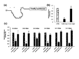
RNA G-quadruplex構造は逆転写反応のみならず、タンパク質翻訳反応も抑制することが知られている6。Staple核酸を用いて標的mRNA上に人為的なRNA G-quadruplexを構築することができれれば、標的遺伝子のタンパク質発現を抑制できるため、医薬品応用の可能性も広がると考えられる。そこで本実施例では、2+2 100nt RNA型5’-UTR領域としてその下流にFireflyルシフェラーゼ遺伝子が導入されているmRNA(2+2 100nt RNA型_Firefly)を作製し、無細胞タンパク質翻訳システムを用いてstaple核酸の有無によるタンパク質合成量を定量的に評価した(図5(a))。
その結果、inside loop1が存在する場合には、下流に存在するFirefly Luciferaseタンパク質の発現量が低下した。一方、inside loop 2存在下ではタンパク質発現量が低下していないことから、単にstaple核酸が標的mRNAに結合してアンチセンス効果を発揮しているのではなく、RNA G-quadruplexを構築することで遺伝子発現抑制効果を示していることが示される(図5(b))。
また、ストップアッセイに用いた2+2 63nt RNA型_Firefly、2+2 140nt RNA型_Firefly、1+3 63nt RNA型_Firefly、1+3 100nt RNA型_Firefly、1+3 140nt RNA型_Fireflyに関しても同様の検討を行ったところ、いずれの場合においてもRNA G-quadruplex構造を形成できるinside loop 1を加えた場合のみ、Fireflyルシフェラーゼの発現量が減少することを示せた(図5(c))。この結果は、Staple核酸によるRNA G-quadruplex構造の人為的誘起と誘起されたRNA G-quadruplex構造によるタンパク質翻訳反応の抑制に対する一般性を示すものである。
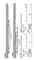
次に、Staple核酸によるRNA G-quadruplex構想形成および遺伝子発現抑制効果を天然遺伝子配列であるTPM3とMYD88において評価した(図6(a))。まず、TPM3とMYD88の5’-UTRにおいてRNA G-quadruplex構造の誘起が可能なStaple核酸を設計した。TPM3に関してはグアニン連続配列を繋ぐループ塩基は103塩基でStaple核酸はinside loop1型として設計した。MYD88についてはグアニン連続配列を繋ぐループ塩基が28塩基と短いため、outside loop型で設計した。
ストップアッセイによる評価で、どちらの5’UTRにおいても設計したStaple核酸でRNA G-quadruplex構造が誘起されることを確認した(図6(b))。次に、TPM3とMYD88の5’-UTRをFireflyルシフェラーゼ遺伝子に配置したモデルmRNAを作製し、Staple核酸のタンパク質翻訳抑制能を評価した。
その結果、無細胞タンパク質翻訳システムを用いたレポーターアッセイで、どちらの5’UTRを配置したモデルmRNAからのタンパク質翻訳もStaple核酸存在下で約60%抑制された(図6(c))。この結果は、Staple核酸が天然の標的mRNAからのタンパク質発現を抑制することを強く示す。
その結果、無細胞タンパク質翻訳システムを用いたレポーターアッセイで、どちらの5’UTRを配置したモデルmRNAからのタンパク質翻訳もStaple核酸存在下で約60%抑制された(図6(c))。この結果は、Staple核酸が天然の標的mRNAからのタンパク質発現を抑制することを強く示す。
最後に、哺乳動物細胞内におけるStaple核酸のタンパク質翻訳阻害活性を評価した。細胞実験を行う上で、本発明者は、NanoLucとRenillaルシフェラーゼが発現する遺伝子を用意した。二つの遺伝子は一本のmRNAに導入されており、NanoLucは5’-capから翻訳されるのに対し、RenillaルシフェラーゼはInternal Ribosomal Entry Site(IRES)から翻訳される。このシステムを用いることでtransfection効率やmRNAの安定性の影響をRenillaルシフェラーゼ遺伝子の発現量で規格化し、5’-capからの翻訳反応を定量的に比較できる(図6(d))12。
本実施例においてはNanoLucの上流にTPM3及びMYD88の5’-UTR領域を配置したレポータ遺伝子を導入し、HEK293細胞内でのStaple核酸の効果を評価した。細胞実験では、Short hairpin expression vector (pSuper) により発現される RNA Staple核酸を利用し、Staple核酸によるタンパク質翻訳反応への影響を評価した。その結果、Staple核酸を導入するとタンパク質翻訳量の減少が確認され、細胞内においてもStaple核酸が機能することが示された(図6(e))。
核酸医薬開発は明らかに停滞している。その原因は細胞内導入が難しいこと、またオフ・ターゲット効果を避けることができないことなどが考えられる。細胞内導入に関する問題点は近年におけるドラッグ・デリバリーシステムの研究開発の貢献により大きな改善が認められる一方、オフ・ターゲット効果の改善は以前困難である。この原因はアンチセンス核酸やsiRNAはいずれも標的mRNAに結合するために相補核酸を利用していることである。相補核酸の認識能を向上させるような修飾核酸を導入すると、siRNAはたちまちRISC複合体形成能が低下し、効果が大きく減少する。このように一つ問題を解決しようと試みると新しい問題が生じるというジレンマに挟まれる。
このような背景から、本発明者は、今までにない新しいタンパク質翻訳反応抑制システムを提案した。このシステムは意図せぬ位置にStaple核酸が結合しても近接させるグアニン繰り返し配列が存在せず、タンパク質抑制効果は発現しない。また導入するStaple核酸はDNAのみではなくRNAでも同様の効果を得ることができることも明らかにしている。この事実は、Staple核酸の多様性を示すものである。
通常、天然核酸配列は細胞内に導入することで分解されてしまうため、核酸医薬を目指す上では化学合成による修飾核酸を使う必須である。本発明の方法は、siRNAのようなタンパク質複合体を形成する必要がないことから、単純に細胞内安定性を向上させることのみに専念すればよく、原理上、修飾核酸によるタンパク質発現抑制効果の減少は考えにくい。様々な修飾を施すことが可能である本発明の方法は、核酸医薬開発のために極めて有用である。
Materials & Methods(Staple_TPM3_26nt_逆連結)
(1)DNAオリゴヌクレオチド
DNAオリゴヌクレオチドはユーロフィンジェノミクス株式会社、Integrated DNA Technologies株式会社、Thermo Fisher Scientific株式会社より購入した。すべてのサンプルはOPC精製または脱塩されており、RNA G-quadruplexの形成や存在位置の確認やin vitroにおけるタンパク質翻訳反応阻害効果を評価するために使用した。
(1)DNAオリゴヌクレオチド
DNAオリゴヌクレオチドはユーロフィンジェノミクス株式会社、Integrated DNA Technologies株式会社、Thermo Fisher Scientific株式会社より購入した。すべてのサンプルはOPC精製または脱塩されており、RNA G-quadruplexの形成や存在位置の確認やin vitroにおけるタンパク質翻訳反応阻害効果を評価するために使用した。
(2)Method
天然遺伝子(TPM3、MYD88)の5’UTR配列を用いたin cell translation (Staple_TPM3_26nt_逆連結)
天然遺伝子(TPM3、MYD88)の5’UTR配列を用いたin cell translation (Staple_TPM3_26nt_逆連結)
1. pIRES-TPM3_5’UTR_nLuc_ renillaの作製
nLuc vector(タカラバイオ.)またはpsiCHECK-2 vector(タカラバイオ.)を鋳型とし、以下のプライマー1のセット又はプライマー2のセットを用いてNanoLuc luciferase(nLuc)およびrenilla luciferase(renilla)のPCR増幅を行い、得られたPCR産物をそれぞれpIRES vector(タカラバイオ.)のEcoR I、Not Iサイトにクローニングした(pIRES-nLuc-renilla)。
nLuc vector(タカラバイオ.)またはpsiCHECK-2 vector(タカラバイオ.)を鋳型とし、以下のプライマー1のセット又はプライマー2のセットを用いてNanoLuc luciferase(nLuc)およびrenilla luciferase(renilla)のPCR増幅を行い、得られたPCR産物をそれぞれpIRES vector(タカラバイオ.)のEcoR I、Not Iサイトにクローニングした(pIRES-nLuc-renilla)。
(i) プライマー1
フォワードプライマー1:
fw. primer[5’-CTAGCCTCGAGAATTCGCCACCATGGTCTTCACACTCGAAG-3’](配列番号79)
リバースプライマー1:
rv. primer[5’-CTCGACGCGTGAATTTTACGCCAGAATGCGTTCG-3’](配列番号80)
(ii) プライマー2
フォワードプライマー2:
fw. primer[5’-TCGACCCGGGCGGCCATGGCTTCCAAGGTGTACG-3’](配列番号81)
リバースプライマー:
rv. primer[5’-TAAAGGGAA GCGGCCTTACTGCTCGTTCTTCAGC-3’](配列番号82)
フォワードプライマー1:
fw. primer[5’-CTAGCCTCGAGAATTCGCCACCATGGTCTTCACACTCGAAG-3’](配列番号79)
リバースプライマー1:
rv. primer[5’-CTCGACGCGTGAATTTTACGCCAGAATGCGTTCG-3’](配列番号80)
(ii) プライマー2
フォワードプライマー2:
fw. primer[5’-TCGACCCGGGCGGCCATGGCTTCCAAGGTGTACG-3’](配列番号81)
リバースプライマー:
rv. primer[5’-TAAAGGGAA GCGGCCTTACTGCTCGTTCTTCAGC-3’](配列番号82)
nLucのPCRに用いたfw. primerはクローニング後にnLucの上流にEcoR Iサイトが復活するように設計した。すべてのPCR産物は1%アガロースゲル電気泳動後、切り出し精製を行った。
human genomic DNA(Novagen)を鋳型にし、以下のプライマーを用いてTPM3-5’UTRの PCR増幅を行った。
fw. primer[5’-GTCACATCC GGGCGGGTTGGTGAGT-3’](配列番号83)
rv. primer[5’-GGTGCCCACCCAGCTACTGCTCGCG-3’](配列番号84)
得られたPCR産物を鋳型にして以下のプライマーを用いてさらにPCR増幅を行い、得られたPCR産物をpMD19 vector(タカラバイオ.)にクローニングした(pMD19-TPM3)。
fw. primer[5’-TAATACGACTCACTATAGGTCACATCCGG GCGGGTTGGTGAGT-3’](配列番号85)
rv. primer[5’-TCCAACTATGTATACCTGGGTGCCCACCCAGCTACTGCTCGCG-3’](配列番号86)
human genomic DNA(Novagen)を鋳型にし、以下のプライマーを用いてTPM3-5’UTRの PCR増幅を行った。
fw. primer[5’-GTCACATCC GGGCGGGTTGGTGAGT-3’](配列番号83)
rv. primer[5’-GGTGCCCACCCAGCTACTGCTCGCG-3’](配列番号84)
得られたPCR産物を鋳型にして以下のプライマーを用いてさらにPCR増幅を行い、得られたPCR産物をpMD19 vector(タカラバイオ.)にクローニングした(pMD19-TPM3)。
fw. primer[5’-TAATACGACTCACTATAGGTCACATCCGG GCGGGTTGGTGAGT-3’](配列番号85)
rv. primer[5’-TCCAACTATGTATACCTGGGTGCCCACCCAGCTACTGCTCGCG-3’](配列番号86)
さらに、pMD19-TPM3を鋳型として、以下のプライマーを用いてPCRにより増幅し、得られたPCR産物をpIRES-nLuc-renillaのEcoR Iサイトにクローニングした(pIRES-TPM3_5’UTR-nLuc-renilla)。
fw. Primer:[5’-CTAGCCTCGAGAATTTCACATCCGGGCGGGTTG-3’](配列番号87))
rv. primer:[5’-CCATGGTGGCGAATT GTTTTCCCAGTCACGACG-3’](配列番号88)
fw. Primer:[5’-CTAGCCTCGAGAATTTCACATCCGGGCGGGTTG-3’](配列番号87))
rv. primer:[5’-CCATGGTGGCGAATT GTTTTCCCAGTCACGACG-3’](配列番号88)
2. pSuper neo. A-Staple (Staple: TPM3_26nt_逆連結)の作製
また、以下のプライマーを用いてアニーリングを行い、Staple insert(TPM3_26nt_逆連結)を作製した。これをpSuper neo. A(oligoengine)のBgl II-Hind IIIサイト間にクローニングした(pSuper neo. A-Staple(Staple:TPM3_26nt_逆連結))。
fw. primer[5’- GATCCCCGCTACTGCTCGCGCCGGAACTCACCAT TTTTA -3’](配列番号89)
rv. primer[5’- AGCTTAAAAATGGTGAGTTCCGGCGCGAGCAGTAGCGGG -3’](配列番号90)
また、以下のプライマーを用いてアニーリングを行い、Staple insert(TPM3_26nt_逆連結)を作製した。これをpSuper neo. A(oligoengine)のBgl II-Hind IIIサイト間にクローニングした(pSuper neo. A-Staple(Staple:TPM3_26nt_逆連結))。
fw. primer[5’- GATCCCCGCTACTGCTCGCGCCGGAACTCACCAT TTTTA -3’](配列番号89)
rv. primer[5’- AGCTTAAAAATGGTGAGTTCCGGCGCGAGCAGTAGCGGG -3’](配列番号90)
3. in cell translation
HEK293T細胞は培地A( 100 units/mlペニシリン、100 μg/ml ストレプトマイシン、10 %(v/v) fetal bovine serumを含むDulbecco’s modified Eagle medium)を用いて37℃、CO2濃度5%のインキュベーター内で培養した。96-well plateに1 well当たり2×10⁴ cellsのHEK293T細胞をまいた。24 時間後、Transfection試薬(FuGENE、Promega)を用いてpSuper neo. A-Staple(Staple:TPM3_26nt_逆連結)またはmilliQ(control)をHEK293T細胞に導入しインキュベートした。
HEK293T細胞は培地A( 100 units/mlペニシリン、100 μg/ml ストレプトマイシン、10 %(v/v) fetal bovine serumを含むDulbecco’s modified Eagle medium)を用いて37℃、CO2濃度5%のインキュベーター内で培養した。96-well plateに1 well当たり2×10⁴ cellsのHEK293T細胞をまいた。24 時間後、Transfection試薬(FuGENE、Promega)を用いてpSuper neo. A-Staple(Staple:TPM3_26nt_逆連結)またはmilliQ(control)をHEK293T細胞に導入しインキュベートした。
48時間後、同様にしてpIRES-TPM3_5’UTR-nLuc-renillaをHEK293T細胞に導入しインキュベートした。6時間後、細胞をPBSで洗浄しPassive Lysis Bufferを用いて細胞溶解液を回収した。この細胞溶解液を4℃、3000 rpmで1分間遠心した後、その上澄み液とLuciferase Assay kit(Promega)、POWERSCAN・H1 microplate reader(BioTek)を用いてルシフェラーゼ活性を評価した。
結果を図7に示す。TPM3をStaple核酸として使用(結合)しなかった場合は、発現の抑制は観察されなかった(図7)。
結果を図7に示す。TPM3をStaple核酸として使用(結合)しなかった場合は、発現の抑制は観察されなかった(図7)。
参考文献
(1) Fire, A.; Xu, S.; Montgomery, M. K.; Kostas, S. A.; Driver, S. E.; Mello, C. C. Nature 1998, 391, 806.
(2)Ambros, V. Nature 2004, 431, 350.
(3)Jackson, A. L.; Linsley, P. S. Nature reviews. Drug discovery 2010, 9, 57.
(4)Jackson, A. L.; Burchard, J.; Leake, D.; Reynolds, A.; Schelter, J.; Guo, J.; Johnson, J. M.; Lim, L.; Karpilow, J.; Nichols, K.; Marshall, W.; Khvorova, A.; Linsley, P. S. Rna 2006, 12, 1197.
(5)Huppert, J. L.; Balasubramanian, S. Nucleic acids research 2007, 35, 406.
(6)Kumari, S.; Bugaut, A.; Huppert, J. L.; Balasubramanian, S. Nature chemical biology 2007, 3, 218.
(7)Katsuda, Y.; Sato, S.; Asano, L.; Morimura, Y.; Furuta, T.; Sugiyama, H.; Hagihara, M.; Uesugi, M. Journal of the American Chemical Society 2016, 138, 9037.
(8)Rothemund, P. W. Nature 2006, 440, 297.
(9)Fujita, H.; Kataoka, Y.; Tobita, S.; Kuwahara, M.; Sugimoto, N. Analytical chemistry 2016, 88, 7137.
(10)Bugaut, A.; Murat, P.; Balasubramanian, S. Journal of the American Chemical Society 2012, 134, 19953.
(11)Hagihara, M.; Yoneda, K.; Yabuuchi, H.; Okuno, Y.; Nakatani, K. Bioorganic & medicinal chemistry letters 2010, 20, 2350.
(12)Jang, S. K.; Davies, M. V.; Kaufman, R. J.; Wimmer, E. Journal of virology 1989, 63, 1651.
(1) Fire, A.; Xu, S.; Montgomery, M. K.; Kostas, S. A.; Driver, S. E.; Mello, C. C. Nature 1998, 391, 806.
(2)Ambros, V. Nature 2004, 431, 350.
(3)Jackson, A. L.; Linsley, P. S. Nature reviews. Drug discovery 2010, 9, 57.
(4)Jackson, A. L.; Burchard, J.; Leake, D.; Reynolds, A.; Schelter, J.; Guo, J.; Johnson, J. M.; Lim, L.; Karpilow, J.; Nichols, K.; Marshall, W.; Khvorova, A.; Linsley, P. S. Rna 2006, 12, 1197.
(5)Huppert, J. L.; Balasubramanian, S. Nucleic acids research 2007, 35, 406.
(6)Kumari, S.; Bugaut, A.; Huppert, J. L.; Balasubramanian, S. Nature chemical biology 2007, 3, 218.
(7)Katsuda, Y.; Sato, S.; Asano, L.; Morimura, Y.; Furuta, T.; Sugiyama, H.; Hagihara, M.; Uesugi, M. Journal of the American Chemical Society 2016, 138, 9037.
(8)Rothemund, P. W. Nature 2006, 440, 297.
(9)Fujita, H.; Kataoka, Y.; Tobita, S.; Kuwahara, M.; Sugimoto, N. Analytical chemistry 2016, 88, 7137.
(10)Bugaut, A.; Murat, P.; Balasubramanian, S. Journal of the American Chemical Society 2012, 134, 19953.
(11)Hagihara, M.; Yoneda, K.; Yabuuchi, H.; Okuno, Y.; Nakatani, K. Bioorganic & medicinal chemistry letters 2010, 20, 2350.
(12)Jang, S. K.; Davies, M. V.; Kaufman, R. J.; Wimmer, E. Journal of virology 1989, 63, 1651.
配列番号1~68、71-74、79-90:合成DNA
配列番号69、70、75-78:合成RNA
配列番号69、70、75-78:合成RNA
Claims (12)
- 第一、第二、第三及び第四のグアニン繰り返し配列(それぞれ、第一G配列、第二G配列、第三G配列、第四G配列という)を含む標的RNAに対し、前記グアニン繰り返し配列の近傍にハイブリダイズするオリゴマーを用い、当該オリゴマーの折り返しにより前記グアニン繰り返し配列を近づけてグアニン四重鎖構造を形成させることによりタンパク質発現を抑制する方法であって、
前記オリゴマーの一端側の一部の領域(一端側領域)の核酸及び他端側の一部の領域(他端側領域)の核酸が、第一G配列と第二G配列との間であってそれぞれ第一G配列の近傍及び第二G配列の近傍、第二G配列と第三G配列との間であってそれぞれ第二G配列の近傍及び第三G配列の近傍、又は第三G配列と第四G配列との間であってそれぞれ第三G配列の近傍及び第四G配列の近傍にハイブリダイズする、前記方法。 - 前記オリゴマーは、その折り返しの方向がグアニン四重鎖構造側を向いてハイブリダイズするように設計される、請求項1に記載の方法。
- 標的RNAがmRNA、TPM3又はMYD8である、請求項1又は2に記載の方法。
- オリゴマーがDNA又はRNAである、請求項1~3のいずれか1項に記載の方法。
- オリゴマーがダイマーである、請求項1~4のいずれか1項に記載の方法。
- タンパク質発現が、逆転写反応又はタンパク質翻訳反応である、請求項1~5のいずれか1項に記載の方法。
- 第一、第二、第三及び第四のグアニン繰り返し配列(それぞれ、第一G配列、第二G配列、第三G配列、第四G配列という)を含む標的RNAに対し、前記グアニン繰り返し配列を近づけてグアニン四重鎖構造を形成させるオリゴマーであって、
前記オリゴマーの一端側の一部の領域(一端側領域)の核酸及び他端側の一部の領域(他端側領域)の核酸が、第一G配列と第二G配列との間であってそれぞれ第一G配列の近傍及び第二G配列の近傍、第二G配列と第三G配列との間であってそれぞれ第二G配列の近傍及び第三G配列の近傍、又は第三G配列と第四G配列との間であってそれぞれ第三G配列の近傍及び第四G配列の近傍にハイブリダイズして、その折り返しにより前記グアニン繰り返し配列を近づけるものである、
前記オリゴマー。 - 前記折り返しの方向がグアニン四重鎖構造側を向いてハイブリダイズするように設計される、請求項7に記載のオリゴマー。
- DNA又はRNAである、請求項7又は8に記載のオリゴマー。
- ダイマーである、請求項7~9のいずれか1項に記載のオリゴマー。
- 請求項7~10のいずれか1項に記載のオリゴマーの製造方法であって、
オリゴマーの一端側の一部の領域(一端側領域)の核酸及び他端側の一部の領域(他端側領域)の核酸が、第一G配列と第二G配列との間であってそれぞれ第一G配列の近傍及び第二G配列の近傍、第二G配列と第三G配列との間であってそれぞれ第二G配列の近傍及び第三G配列の近傍、又は第三G配列と第四G配列との間であってそれぞれ第三G配列の近傍及び第四G配列の近傍にハイブリダイズするように設計し合成することを特徴とする前記方法。 - 請求項7~10のいずれか1項に記載のオリゴマー、又は請求項11に記載の方法により製造されたオリゴマーを含む、タンパク質発現抑制キット。
Priority Applications (4)
| Application Number | Priority Date | Filing Date | Title |
|---|---|---|---|
| EP19875072.1A EP3871679A4 (en) | 2018-10-25 | 2019-10-25 | METHOD OF SUPPRESSING PROTEIN TRANSLATION RESPONSE USING STAPLE NUCLEIC ACIDS |
| CN201980070388.3A CN113286598A (zh) | 2018-10-25 | 2019-10-25 | 利用Staple核酸的蛋白质翻译反应的抑制法 |
| US17/287,563 US20220112490A1 (en) | 2018-10-25 | 2019-10-25 | Method for suppressing protein translation reaction using staple nucleic acid |
| JP2020552638A JP7465507B2 (ja) | 2018-10-25 | 2019-10-25 | Staple核酸を利用したタンパク質翻訳反応の抑制法 |
Applications Claiming Priority (2)
| Application Number | Priority Date | Filing Date | Title |
|---|---|---|---|
| JP2018-201074 | 2018-10-25 | ||
| JP2018201074 | 2018-10-25 |
Publications (1)
| Publication Number | Publication Date |
|---|---|
| WO2020085510A1 true WO2020085510A1 (ja) | 2020-04-30 |
Family
ID=70331384
Family Applications (1)
| Application Number | Title | Priority Date | Filing Date |
|---|---|---|---|
| PCT/JP2019/042049 Ceased WO2020085510A1 (ja) | 2018-10-25 | 2019-10-25 | Staple核酸を利用したタンパク質翻訳反応の抑制法 |
Country Status (5)
| Country | Link |
|---|---|
| US (1) | US20220112490A1 (ja) |
| EP (1) | EP3871679A4 (ja) |
| JP (1) | JP7465507B2 (ja) |
| CN (1) | CN113286598A (ja) |
| WO (1) | WO2020085510A1 (ja) |
Cited By (4)
| Publication number | Priority date | Publication date | Assignee | Title |
|---|---|---|---|---|
| WO2022064202A1 (en) * | 2020-09-23 | 2022-03-31 | Ucl Business Ltd | Nucleic acid nanostructures for delivery of nucleic acid sequences to cells |
| WO2023106421A1 (ja) * | 2021-12-06 | 2023-06-15 | 国立大学法人熊本大学 | Trpc6の発現を抑制する方法 |
| WO2023224102A1 (ja) | 2022-05-18 | 2023-11-23 | 国立大学法人熊本大学 | 新規Staple核酸 |
| CN118388568A (zh) * | 2024-07-01 | 2024-07-26 | 朴衡博迈(上海)生物医药有限公司 | 一种用于3d培养的dna折纸及其制备方法和应用 |
Citations (1)
| Publication number | Priority date | Publication date | Assignee | Title |
|---|---|---|---|---|
| US20160046932A1 (en) * | 2012-12-21 | 2016-02-18 | Nanyang Technological University | Site-specific induction of bimolecular quadruplex-duplex hybrids and methods of using the same |
-
2019
- 2019-10-25 US US17/287,563 patent/US20220112490A1/en active Pending
- 2019-10-25 WO PCT/JP2019/042049 patent/WO2020085510A1/ja not_active Ceased
- 2019-10-25 EP EP19875072.1A patent/EP3871679A4/en active Pending
- 2019-10-25 CN CN201980070388.3A patent/CN113286598A/zh active Pending
- 2019-10-25 JP JP2020552638A patent/JP7465507B2/ja active Active
Patent Citations (1)
| Publication number | Priority date | Publication date | Assignee | Title |
|---|---|---|---|---|
| US20160046932A1 (en) * | 2012-12-21 | 2016-02-18 | Nanyang Technological University | Site-specific induction of bimolecular quadruplex-duplex hybrids and methods of using the same |
Non-Patent Citations (17)
| Title |
|---|
| AMBROS, V., NATURE, vol. 431, 2004, pages 350 |
| ARIVAZHAGAN RAJENDRAN, MASAYUKI ENDO, KUMI HIDAKA, PHONG LAN THAO TRAN, JEAN-LOUIS MERGNY, ROBERT J. GORELICK, HIROSHI SUGIYAMA: "HIV-1 Nucleocapsid Proteins as Molecular Chaperones for Tetramolecular Antiparallel G-Quadruplex Formation", JOURNAL OF THE AMERICAN CHEMICAL SOCIETY, vol. 135, no. 49, 13 November 2013 (2013-11-13), pages 18575 - 18585, XP055709074, ISSN: 0002-7863, DOI: 10.1021/ja409085j * |
| BIXIA GE,YU CHUAN HUANG, DIPANKAR SEN, HUA-ZHONG YU: "A Robust Electronic Switch Made of Immobilized Duplex/ Quadruplex DNA", ANGEW. CHEM. INT. ED., vol. 49, no. 51, 17 December 2010 (2010-12-17), pages 9965 - 9967, XP055260681, ISSN: 1433-7851, DOI: 10.1002/anie.201004946 * |
| BUGAUT, A.MURAT, P.BALASUBRAMANIAN, S., JOURNAL OF THE AMERICAN CHEMICAL SOCIETY, vol. 134, 2012, pages 19953 |
| FIRE, A.XU, S.MONTGOMERY, M. K.KOSTAS, S. A.DRIVER, S. E.MELLO, C. C., NATURE, vol. 391, 1998, pages 806 |
| FUJITA, H.KATAOKA, Y.TOBITA, S.KUWAHARA, M.SUGIMOTO, N., ANALYTICAL CHEMISTRY, vol. 88, 2016, pages 7137 |
| HAGIHARA, M.YONEDA, K.YABUUCHI, H.OKUNO, Y.NAKATANI, K., BIOORGANIC & MEDICINAL CHEMISTRY LETTERS, vol. 20, 2010, pages 2350 |
| HUPPERT, J. L.BALASUBRAMANIAN, S., NUCLEIC ACIDS RESEARCH, vol. 35, 2007, pages 406 |
| JACKSON, A. L.BURCHARD, J.LEAKE, D.REYNOLDS, A.SCHELTER, J.GUO, J.JOHNSON, J. M.LIM, L.KARPILOW, J.NICHOLS, K., RNA, vol. 12, 2006, pages 1197 |
| JACKSON, A. L.LINSLEY, P. S., NATURE REVIEWS. DRUG DISCOVERY, vol. 9, 2010, pages 57 |
| JANG, S. K.DAVIES, M. V.KAUFMAN, R. J.WIMMER, E., JOURNAL OF VIROLOGY, vol. 63, 1989, pages 1651 |
| KATSUDA, Y.SATO, S.ASANO, L.MORIMURA, Y.FURUTA, T.SUGIYAMA, H.HAGIHARA, M.UESUGI, M., JOURNAL OF THE AMERICAN CHEMICAL SOCIETY, vol. 138, 2016, pages 9037 |
| KUMARI, S.BUGAUT, A.HUPPERT, J. L.BALASUBRAMANIAN, S., NATURE CHEMICAL BIOLOGY, vol. 3, 2007, pages 218 |
| MASAKI HAGIHARA, LISA YAMAUCHI, AKIKO SEO, KEISUKE YONEDA, MAYO SENDA, KAZUHIKO NAKATANI: "Antisense-Induced Guanine Quadruplexes Inhibit Reverse Transcription by HIV-1 Reverse Transcriptase", JOURNAL OF THE AMERICAN CHEMICAL SOCIETY, vol. 132, 2010, pages 11171 - 11178, XP055055375, ISSN: 0002-7863, DOI: 10.1021/ja1032088 * |
| MASAYUKI ENDO, HIROSHI SUGIYAMA: "Single-molecule Analysis Using DNA Origami Nanostructures", SEIBUTSU BUTSURI, vol. 53, no. 3, 1 January 2013 (2013-01-01), pages 153 - 157, XP055810222, ISSN: 0582-4052, DOI: 10.2142/biophys.53.153 * |
| ROTHEMUND, P. W, NATURE, vol. 440, 2006, pages 297 |
| See also references of EP3871679A4 |
Cited By (4)
| Publication number | Priority date | Publication date | Assignee | Title |
|---|---|---|---|---|
| WO2022064202A1 (en) * | 2020-09-23 | 2022-03-31 | Ucl Business Ltd | Nucleic acid nanostructures for delivery of nucleic acid sequences to cells |
| WO2023106421A1 (ja) * | 2021-12-06 | 2023-06-15 | 国立大学法人熊本大学 | Trpc6の発現を抑制する方法 |
| WO2023224102A1 (ja) | 2022-05-18 | 2023-11-23 | 国立大学法人熊本大学 | 新規Staple核酸 |
| CN118388568A (zh) * | 2024-07-01 | 2024-07-26 | 朴衡博迈(上海)生物医药有限公司 | 一种用于3d培养的dna折纸及其制备方法和应用 |
Also Published As
| Publication number | Publication date |
|---|---|
| JPWO2020085510A1 (ja) | 2021-10-21 |
| JP7465507B2 (ja) | 2024-04-11 |
| CN113286598A (zh) | 2021-08-20 |
| EP3871679A4 (en) | 2022-10-12 |
| EP3871679A1 (en) | 2021-09-01 |
| US20220112490A1 (en) | 2022-04-14 |
Similar Documents
| Publication | Publication Date | Title |
|---|---|---|
| Müller et al. | In vitro circularization of RNA | |
| Belew et al. | Ribosomal frameshifting in the CCR5 mRNA is regulated by miRNAs and the NMD pathway | |
| Hartmann et al. | Handbook of RNA biochemistry | |
| EP2917349B1 (en) | Riboregulator compositions and methods of use | |
| Zhao et al. | Cleavage-based signal amplification of RNA | |
| WO2020085510A1 (ja) | Staple核酸を利用したタンパク質翻訳反応の抑制法 | |
| Chowdhury et al. | Short LNA-modified oligonucleotide probes as efficient disruptors of DNA G-quadruplexes | |
| Schmidt et al. | A method for expressing and imaging abundant, stable, circular RNAs in vivo using tRNA splicing | |
| Kanwal et al. | Large-scale in vitro transcription, RNA purification and chemical probing analysis | |
| Ma et al. | Rapid, sensitive and highly specific label-free fluorescence biosensor for microRNA by branched rolling circle amplification | |
| Mei et al. | Synthesis and polymerase activity of a fluorescent cytidine TNA triphosphate analogue | |
| Lee et al. | Real-time monitoring strategies for optimization of in vitro transcription and quality control of RNA | |
| Li et al. | An improved model for circular RNA overexpression: using the actin intron reveals high circularization efficiency | |
| Berezhnoy et al. | Thermal stability of siRNA modulates aptamer-conjugated siRNA inhibition | |
| Le et al. | Pri-miRNA cleavage assays for the Microprocessor complex | |
| EP4527929A1 (en) | New staple nucleic acid | |
| Marucci et al. | Comparison and optimization of transient transfection methods at human astrocytoma cell line 1321N1 | |
| HK40057283A (en) | Method for suppressing protein translation reaction using staple nucleic acid | |
| Kanwal et al. | A modified in vitro transcription approach to improve RNA synthesis and ribozyme cleavage efficiency | |
| JP6703948B2 (ja) | 非酵素的核酸鎖結合方法 | |
| Torkzaban et al. | Tethered mRNA Amplifier: A Novel Approach to Increase Protein Expression | |
| Smith et al. | NEAT and tidy: a novel RT-PCR method for the independent quantification of the NEAT1_1 isoform | |
| Vachon et al. | Plasmid template design and in vitro transcription of short RNAs within a “structure cassette” for structure probing experiments | |
| Devi et al. | Rna amplification | |
| Kida et al. | Expanded gene targeting in RNA hacking with G-tract-supply Staple oligomer |
Legal Events
| Date | Code | Title | Description |
|---|---|---|---|
| 121 | Ep: the epo has been informed by wipo that ep was designated in this application |
Ref document number: 19875072 Country of ref document: EP Kind code of ref document: A1 |
|
| ENP | Entry into the national phase |
Ref document number: 2020552638 Country of ref document: JP Kind code of ref document: A |
|
| NENP | Non-entry into the national phase |
Ref country code: DE |
|
| ENP | Entry into the national phase |
Ref document number: 2019875072 Country of ref document: EP Effective date: 20210525 |