WO2020060122A1 - Fusion protein comprising il-2 protein and cd80 protein, and use thereof - Google Patents
Fusion protein comprising il-2 protein and cd80 protein, and use thereof Download PDFInfo
- Publication number
- WO2020060122A1 WO2020060122A1 PCT/KR2019/011928 KR2019011928W WO2020060122A1 WO 2020060122 A1 WO2020060122 A1 WO 2020060122A1 KR 2019011928 W KR2019011928 W KR 2019011928W WO 2020060122 A1 WO2020060122 A1 WO 2020060122A1
- Authority
- WO
- WIPO (PCT)
- Prior art keywords
- fusion protein
- seq
- protein
- cancer
- amino acid
- Prior art date
Links
Images


Classifications
-
- A—HUMAN NECESSITIES
- A61—MEDICAL OR VETERINARY SCIENCE; HYGIENE
- A61K—PREPARATIONS FOR MEDICAL, DENTAL OR TOILETRY PURPOSES
- A61K38/00—Medicinal preparations containing peptides
-
- A—HUMAN NECESSITIES
- A61—MEDICAL OR VETERINARY SCIENCE; HYGIENE
- A61P—SPECIFIC THERAPEUTIC ACTIVITY OF CHEMICAL COMPOUNDS OR MEDICINAL PREPARATIONS
- A61P31/00—Antiinfectives, i.e. antibiotics, antiseptics, chemotherapeutics
- A61P31/12—Antivirals
-
- A—HUMAN NECESSITIES
- A61—MEDICAL OR VETERINARY SCIENCE; HYGIENE
- A61P—SPECIFIC THERAPEUTIC ACTIVITY OF CHEMICAL COMPOUNDS OR MEDICINAL PREPARATIONS
- A61P35/00—Antineoplastic agents
-
- C—CHEMISTRY; METALLURGY
- C07—ORGANIC CHEMISTRY
- C07K—PEPTIDES
- C07K14/00—Peptides having more than 20 amino acids; Gastrins; Somatostatins; Melanotropins; Derivatives thereof
- C07K14/435—Peptides having more than 20 amino acids; Gastrins; Somatostatins; Melanotropins; Derivatives thereof from animals; from humans
- C07K14/52—Cytokines; Lymphokines; Interferons
- C07K14/54—Interleukins [IL]
- C07K14/55—IL-2
-
- C—CHEMISTRY; METALLURGY
- C07—ORGANIC CHEMISTRY
- C07K—PEPTIDES
- C07K14/00—Peptides having more than 20 amino acids; Gastrins; Somatostatins; Melanotropins; Derivatives thereof
- C07K14/435—Peptides having more than 20 amino acids; Gastrins; Somatostatins; Melanotropins; Derivatives thereof from animals; from humans
- C07K14/705—Receptors; Cell surface antigens; Cell surface determinants
- C07K14/70503—Immunoglobulin superfamily
- C07K14/70532—B7 molecules, e.g. CD80, CD86
-
- C—CHEMISTRY; METALLURGY
- C12—BIOCHEMISTRY; BEER; SPIRITS; WINE; VINEGAR; MICROBIOLOGY; ENZYMOLOGY; MUTATION OR GENETIC ENGINEERING
- C12N—MICROORGANISMS OR ENZYMES; COMPOSITIONS THEREOF; PROPAGATING, PRESERVING, OR MAINTAINING MICROORGANISMS; MUTATION OR GENETIC ENGINEERING; CULTURE MEDIA
- C12N15/00—Mutation or genetic engineering; DNA or RNA concerning genetic engineering, vectors, e.g. plasmids, or their isolation, preparation or purification; Use of hosts therefor
- C12N15/09—Recombinant DNA-technology
- C12N15/11—DNA or RNA fragments; Modified forms thereof; Non-coding nucleic acids having a biological activity
- C12N15/62—DNA sequences coding for fusion proteins
-
- C—CHEMISTRY; METALLURGY
- C07—ORGANIC CHEMISTRY
- C07K—PEPTIDES
- C07K2319/00—Fusion polypeptide
-
- C—CHEMISTRY; METALLURGY
- C07—ORGANIC CHEMISTRY
- C07K—PEPTIDES
- C07K2319/00—Fusion polypeptide
- C07K2319/31—Fusion polypeptide fusions, other than Fc, for prolonged plasma life, e.g. albumin
Definitions
- the present invention relates to a fusion protein comprising IL-2 protein and CD80 protein and uses thereof. Specifically, the present invention relates to a novel fusion protein for cancer treatment and immunopotentiation efficacy.
- IL-2 Interleukin 2
- T-cell growth factors TCGF
- IL-2 is a spherical glycoprotein that plays a central role in lymphocyte production, survival and homeostasis.
- the protein size of IL-2 is 15.5 kDa to 16 kDa, and consists of 133 amino acids.
- IL-2 mediates various immune actions by binding to the IL-2 receptor, which consists of three individual subunits.
- IL-2 is mainly synthesized by activated T cells, in particular, by CD4 + helper T cells. IL-2 stimulates proliferation and differentiation of T cells, production of cytotoxic T lymphocytes (CTLs) and cytotoxic cells and lymphokine-activated killer cells of peripheral blood lymphocytes (lymphokine activated killer cells, LAK) cell).
- CTLs cytotoxic T lymphocytes
- LAK lymphokine activated killer cells
- IL-2 is involved in the proliferation and differentiation of B cells, promotes the synthesis of immunoglobulins by B cells, and stimulates the production, proliferation and activation of natural killer cells (NK cells). Therefore, IL-2 can increase the population of lymphocytes in vivo and increase the function of the immune cells, and thus IL-2 is used as an anticancer agent.
- NK cells natural killer cells
- IL-2 not only mediates the increase and activity of immune cells, but also has a dual function in the immune response in that it is important in maintaining immune tolerance. It has also been reported that IL-2 may not be optimal for inhibiting tumor growth. The reason is that in the presence of IL-2, activation-induced cell death (AICD) may occur in the generated cytotoxic T lymphocytes, and the immune response may be inhibited by IL-2 dependent regulatory T cells (Treg). (Imai et al ., Cancer Sci 98, 416-423, 2007).
- CD80 is known as B7-1, and by binding to a ligand, it delivers costimulatory responses and coinhibitory responses to the B7 family of membrane proteins involved in immune regulation. It is one.
- CD80 is a transmembrane protein expressed on the surface of T cells, B cells, dendritic cells and monocytes.
- CD80 is known to bind CD28, CTLA4 (CD152) and PD-L1.
- CD80, CD86, CTLA4 and CD28 are involved in costimulatory-co-suppression systems. For example, it regulates the activity of T cells and is involved in proliferation, differentiation and survival.
- CD80 and CD86 interact with CD28
- a costimulatory signal is generated to activate T cells.
- CD80 will stimulate CTLA4 to bind and upregulate CTLA4.
- CD80 inhibits the response of T cells. This feedback loop allows fine-tuning of the immune response.
- CD80 binds another B7 family, PD-L1, with affinity similar to that of CD28 binding to PD-L1.
- PD-L1 is known as one of two ligands for the programmed death-1 (PD-1) protein, and PD-L1 is known to be involved in T cell regulation.
- the binding of CD80 and PD-L1 is another mechanism that can block the PD-1 / PD-L1 interaction, which can prevent the suppression of T cell responses in tumors.
- CTLA4 can induce or suppress T cell response.
- the present inventors have researched to develop safe and effective IL-2.
- the novel fusion protein containing IL-2 protein and CD80 protein in one molecule activates immune cells and can effectively regulate Treg cells.
- an aspect of the present invention provides a fusion protein comprising IL-2 protein and CD80 protein.
- Another aspect of the present invention provides a fusion protein dimer in which the two fusion proteins are combined.
- Another aspect of the present invention provides a polynucleotide encoding the fusion protein.
- Another aspect of the present invention provides a vector comprising the polynucleotide.
- Another aspect of the present invention provides a transformed cell into which the vector is introduced.
- Another aspect of the present invention provides a pharmaceutical composition for preventing or treating cancer or infectious diseases, including the fusion protein or the fusion protein dimer as an active ingredient.
- Another aspect of the invention provides the use of the fusion protein for treating cancer or infectious diseases.
- Another aspect of the present invention provides the use of the fusion protein for preparing a medicament for the treatment of cancer or infectious diseases.
- the fusion protein containing IL-2 protein and CD80 protein can not only activate immune cells by IL-2, but also effectively regulate Treg cells by CD80. Therefore, the fusion protein can efficiently attack cancer cells, and thus can be usefully used in the treatment of cancer or infectious diseases.
- FIG. 1 is a schematic diagram showing an embodiment of a fusion protein.
- FIG. 2 is a schematic diagram showing a mechanism for regulating two types of immune cells having different fusion proteins, but it should be understood that the mechanism by which the action of the fusion protein is expressed is not limited thereto.
- FIG. 3 is a schematic diagram showing the mechanism by which the fusion protein exhibits anticancer effects.
- GI101 and mGI101 are examples of the fusion protein herein, and GI101C1, GI101C2, and mGI101C1 are comparative examples for comparing the activity of the fusion protein.
- a human-derived protein and a mouse-derived protein can be mixed to prepare a fusion protein, and in addition to Fc, CD80 protein and IL-2 protein can be coupled through various linkers.
- Figure 6 confirms the obtained fusion protein (GI101) by SDS-PAGE.
- GI101 fusion protein
- Figure 10 shows the obtained GI101C1 fusion protein by SDS-PAGE.
- 11 shows the obtained GI101C2 fusion protein by SDS-PAGE.
- Figure 12 shows the obtained mGI101C1 fusion protein by SDS-PAGE.
- 16 shows the binding affinity between hCTLA4 and GI101.
- Figure 17 shows the binding affinity between hPD-L1 and GI101.
- GI-101 hCD80-Fc-hIL-2v
- CTLA-4 CTLA-4 and PD-L1. It was confirmed that GI-101 (hCD80-Fc-hIL-2v) has a high binding capacity to CTLA-4 and PD-L1.
- GI101 effectively inhibited PD-1 / PD-L1 binding.
- Figure 24 confirms the binding affinity between GI101 and IL-2R ⁇ or IL-2R ⁇ .
- 26 confirms the binding affinity between GI101 and IL-2R ⁇ .
- 29 shows the binding affinity between IL-2R ⁇ and GI102-M72.
- Figure 31 shows the binding affinity between IL-2R ⁇ and GI102-M61.
- Figure 32 shows the binding affinity between IL-2R ⁇ and GI102-M72.
- 33 and 34 show the amount of IFN- ⁇ secreted from cells when GI101, GI101C1, GI101C2 or IL-2 is treated to cells by concentration and cultured.
- 35 and 36 confirm the effect of GI101, GI101C1, GI101C2 and IL-2 (Proleukin) on the proliferation of CD8 + T cells.
- (A) represents the ratio of CD8 + T cells and CD4 + T cells
- (B) represents the proliferation capacity of CD8 + T cells
- (C) represents the ratio of CD4 + / FoxP3 + Treg cells.
- Figure 42 confirms the effect of mGI101 and mGI102-M61 on immune cells in mice.
- Figure 47 confirms the tumor suppressive effect of mGI101 in mouse-derived melanoma-implanted mice.
- Figure 48 shows the tumor inhibition rate of mGI101 in mouse-derived melanoma-implanted mice.
- Figure 49 confirms the tumor suppression effect according to the dose of mGI101 in mouse-derived colorectal cancer cell implanted mice.
- mice 50 is an analysis of the survival rate of mouse-derived colon cancer cell implanted mice administered mGI101.
- Figure 51 confirms the tumor suppressive effect of GI101 in mouse-derived colorectal cancer-celled mice.
- FIG. 52 is a FACS analysis of CD8 + T cells, IFN- ⁇ T cells, CD4 + T cells and Treg cells in cancer tissues after treatment with hIgG4, anti-PD-1 antibody or GI101 in mouse-derived colorectal cancer cell transplanted mice. .
- Figure 53 shows the results of FACS analysis of CD8 + T cells, IFN- ⁇ T cells, CD4 + T cells and Treg cells in cancer tissues after treatment with hIgG4, anti-PD-1 antibody or GI101 in mouse-derived colorectal cancer cell implanted mice. Is a graph.
- Figure 54 is a mouse-derived colorectal cancer cell implantation mouse after treatment with hIgG4, anti-PD-1 antibody or GI101, macrophage in cancer tissues are analyzed by FACS.
- FIG. 55 is a graph showing the results of analyzing macrophages in cancer tissues by FACS after treatment with hIgG4, anti-PD-1 antibody, or GI101 in mouse-derived colon cancer cells.
- FIG. 56 shows FACS analysis of dendritic cells in cancer tissue after treatment with hIgG4, anti-PD-1 antibody or GI101 in mouse-derived colorectal cancer cell-implanted mice.
- 57 is a graph showing the results of FACS analysis of dendritic cells in cancer tissue after treatment with hIgG4, anti-PD-1 antibody or GI101 in mouse-derived colorectal cancer cell-implanted mice.
- Figure 58 confirms the tumor suppressive effect of GI101 in mouse-derived lung cancer cell-implanted mice.
- FIG. 59 shows the results of FACS analysis of CD8 + T cells, IFN- ⁇ T cells, CD4 + T cells and Treg cells in cancer tissues after treatment with hIgG4, anti-PD-1 antibody or GI101 in mouse-derived lung cancer cell implanted mice. Is a graph.
- FIG. 60 is a graph showing the results of analyzing macrophages in cancer tissues by FACS after treatment with hIgG4, anti-PD-1 antibody or GI101 in mouse-derived lung cancer cell implanted mice.
- FIG. 61 is a graph showing the results of FACS analysis of dendritic cells in cancer tissue after treatment with hIgG4, anti-PD-1 antibody or GI101 in mouse-derived lung cancer cell-implanted mice.
- Figure 62 confirms the tumor suppression effect of mGI102-M61 in mouse-derived colorectal cancer-celled mice.
- Figure 63 is an analysis of the survival rate of mouse-derived colon cancer cell implanted mice administered mGI102-M61.
- Figure 64 confirms the tumor suppression effect of mGI101 in mouse-derived colorectal cancer-celled mice.
- Figure 65 shows the tumor inhibition rate of mGI101 in mouse-derived colorectal cancer-celled mice.
- FIG. 66 shows the results of clinical observation for 15 days in monkeys receiving PBS or GI101.
- Figures 67 and 68 are measurements of body weights on days -1, 1, 8 and 15 of monkeys receiving PBS or GI101.
- Figure 69 shows the intake for 15 days of monkeys receiving PBS or GI101.
- 70 to 72 are blood analysis of -1, 1, 8, and 15 days of monkeys receiving PBS or GI101.
- 73-79 shows clinical and chemical analysis of -1, 1, 8, and 15 days of monkeys receiving PBS or GI101.
- 80 and 81 are cytokines of -1, 1, 8, and 15 days of monkeys administered PBS or GI101.
- 82 to 87 are analysis of immune cells at -1, 1, 8 and 15 days of monkeys receiving PBS or GI101.
- FIG. 88 is a pathological analysis of spleen tissue by sacrificing monkeys receiving PBS or GI101 on the 16th day.
- FIG. 89 shows a fusion protein in which the CD80 protein and IL-2 protein are combined with a carrier protein.
- FIG. 89 (A) shows that the CD80 protein and the IL-2 protein are bound to the N-terminus and the C-terminus of the carrier protein, respectively.
- FIG. 89 (B) shows that the CD80 protein and the IL-2 protein are bound to the C-terminus and the N-terminus of the carrier protein, respectively.
- Fusion protein containing IL-2 protein and CD80 protein
- One aspect of the present invention provides a fusion protein comprising IL-2 protein and CD80 protein.
- IL-2 or “Interleukin-2”, unless stated otherwise, includes mammals, eg, primates (eg, humans) and rodents (eg, mice and rats). Means any wild type IL-2 obtained from any vertebrate source.
- the IL-2 may be obtained from animal cells, but also includes those obtained from recombinant cells capable of producing IL-2.
- the IL-2 may be wild-type IL-2 or a variant thereof.
- IL-2 or a variant thereof is collectively expressed in terms of "IL-2 protein” or "IL-2 polypeptide".
- IL-2, IL-2 protein, IL-2 polypeptide, and IL-2 variants specifically bind to the IL-2 receptor, for example. This specific binding can be confirmed by methods known to those skilled in the art.
- the IL-2 may have the amino acid sequence of SEQ ID NO: 35 or SEQ ID NO: 36.
- the IL-2 may be in a mature form. Specifically, the matured IL-2 may not contain a signal sequence, or may have an amino acid sequence of SEQ ID NO: 10.
- the IL-2 may be used as a concept including a fragment in which a part of the N-terminal or C-terminal of wild-type IL-2 is deleted (truncated).
- the fragment of IL-2 is 1, 2, 3, 4, 5, 6, 7, 8 consecutively from the N-terminal of the protein having the amino acid sequence of SEQ ID NO: 35 or SEQ ID NO: 36 Dog, 9, 10, 11, 12, 13, 14, 15, 16, 17, 18, 19, 20, 21, 22, 23, 24 or It may be in the form of 25 amino acids deleted.
- the fragment of IL-2 is 1, 2, 3, 4, 5, 6, 7, 8 consecutively from the C-terminal of the protein having the amino acid sequence of SEQ ID NO: 35 or SEQ ID NO: 36 , 9, 10, 11, 12, 13, 14, 15, 16, 17, 18, 19, 20, 21, 22, 23, 24 or 25 Dog amino acids may be in a deleted form.
- the term “IL-2 variant” refers to a form in which a part of an amino acid of a full-length IL-2 or a fragment of the aforementioned IL-2 is substituted. That is, the IL-2 variant may have an amino acid sequence different from that of the wild type IL-2 or a fragment thereof. However, the IL-2 variant may have the same or similar activity as wild-type IL-2.
- “IL-2 activity” may mean, for example, specifically binding to an IL-2 receptor, and this specific binding can be measured by methods known to those skilled in the art.
- the IL-2 variant may be a part of an amino acid of wild type IL-2 substituted.
- the IL-2 variant by amino acid substitution at least one of the 38th, 42nd, 45th, 61st and 72nd amino acids in the amino acid sequence of SEQ ID NO: 10 may be substituted.
- At least one of the 38th, 42nd, 45th, 61st, or 72nd amino acids in the amino acid sequence of SEQ ID NO: 10 may be substituted with another amino acid.
- IL-2 is a form in which a portion of the N-terminal of the amino acid sequence of SEQ ID NO: 35 is deleted, the amino acid at the position complementarily corresponding to the amino acid sequence of SEQ ID NO: 10 may be replaced with another amino acid.
- the variant of IL-2 is among the 58, 62, 65, 81, or 92 amino acids in the amino acid sequence of SEQ ID NO: 35. At least one may be substituted with another amino acid.
- amino acid residues 38, 42, 45, 61, and 72 correspond to amino acid residues 38, 42, 45, 61, and 72 of the amino acid sequence of SEQ ID NO: 10, respectively.
- one, two, three, four, five, six, seven, eight, nine, or ten amino acids may be substituted as long as IL-2 activity is maintained have.
- one to five amino acids may be substituted.
- the IL-2 variant may be a form in which two amino acids are substituted. Specifically, the IL-2 variant may be substituted with the 38th and 42nd amino acids in the amino acid sequence of SEQ ID NO: 10. In addition, in one embodiment, the IL-2 variant may be substituted with the 38th and 45th amino acids in the amino acid sequence of SEQ ID NO: 10. In addition, in one embodiment, the IL-2 variant may be substituted with the 38th and 61st amino acids in the amino acid sequence of SEQ ID NO: 10. In addition, in one embodiment, the IL-2 variant may be substituted with the 38th and 72nd amino acids in the amino acid sequence of SEQ ID NO: 10.
- the IL-2 variant may be substituted with the 42nd and 45th amino acids in the amino acid sequence of SEQ ID NO: 10. In addition, in one embodiment, the IL-2 variant may be substituted with the 42nd and 61st amino acids in the amino acid sequence of SEQ ID NO: 10. In addition, in one embodiment, the IL-2 variant may be substituted with the 42nd and 72nd amino acids in the amino acid sequence of SEQ ID NO: 10. In addition, in one embodiment, the IL-2 variant may be substituted with the 45th and 61st amino acids in the amino acid sequence of SEQ ID NO: 10. In addition, in one embodiment, the IL-2 variant may be substituted with the 45th and 72nd amino acids in the amino acid sequence of SEQ ID NO: 10. In addition, in one embodiment, the IL-2 variant may be substituted with the 61st and 72nd amino acids in the amino acid sequence of SEQ ID NO: 10.
- the IL-2 variant may be in the form of three amino acid substitutions. Specifically, the IL-2 variant may be the 38th, 42nd and 45th amino acids substituted in the amino acid sequence of SEQ ID NO: 10. In addition, in one embodiment, the IL-2 variant may be substituted with the 38th, 42nd, and 61st amino acids in the amino acid sequence of SEQ ID NO: 10. In addition, in one embodiment, the IL-2 variant may be substituted with the 38th, 42nd, and 72nd amino acids in the amino acid sequence of SEQ ID NO: 10. In addition, in one embodiment, the IL-2 variant may be substituted with the 38th, 45th, and 61st amino acids in the amino acid sequence of SEQ ID NO: 10.
- the IL-2 variant may be substituted with the 38th, 45th, and 72nd amino acids in the amino acid sequence of SEQ ID NO: 10. In addition, in one embodiment, the IL-2 variant may be substituted with the 38th, 61st, and 72nd amino acids in the amino acid sequence of SEQ ID NO: 10. In addition, in one embodiment, the IL-2 variant may be a 42th, 45th and 61st amino acid substituted in the amino acid sequence of SEQ ID NO: 10. In addition, in one embodiment, the IL-2 variant may be substituted with the 42nd, 45th, and 72nd amino acids in the amino acid sequence of SEQ ID NO: 10. In addition, in one embodiment, the IL-2 variant may be substituted with 45th, 61st, and 72nd amino acids in the amino acid sequence of SEQ ID NO: 10.
- the IL-2 variant may be in the form of four amino acid substitutions. Specifically, the IL-2 variant may be a 38th, 42nd, 45th and 61st amino acid substituted in the amino acid sequence of SEQ ID NO: 10. In addition, in one embodiment, the IL-2 variant may be substituted with the 38th, 42nd, 45th, and 72nd amino acids in the amino acid sequence of SEQ ID NO: 10. In addition, in one embodiment, the IL-2 variant may be substituted with the 38th, 45th, 61st, and 72nd amino acids in the amino acid sequence of SEQ ID NO: 10.
- the IL-2 variant may be substituted with the 38th, 42nd, 61st, and 72nd amino acids in the amino acid sequence of SEQ ID NO: 10. In addition, in one embodiment, the IL-2 variant may be substituted with the 42nd, 45th, 61st, and 72nd amino acids in the amino acid sequence of SEQ ID NO: 10.
- the IL-2 variant may be in the form of five amino acid substitutions. Specifically, in the IL-2 variant, the 38th, 42nd, 45th, 61st, and 72nd amino acids in the amino acid sequence of SEQ ID NO: 10 may be substituted with other amino acids.
- the "other amino acids" introduced by the substitution are alanine, arginine, asparagine, aspartic acid, cysteine, glutamic acid, glutamine , Histidine, isoleucine, leucine, lysine, methionine, phenyl alanine, proline, serine, threonine, tryptophan ( It can be any one selected from the group consisting of tryptophan, tyrosine and valine.
- the amino acid substitution of the IL-2 variant in the amino acid sequence of SEQ ID NO: 10, 38th cannot be substituted with arginine, 42nd cannot be substituted with phenylalanine, and 45th cannot be substituted with tyrosine. , The 61st cannot be substituted with glutamic acid, and the 72nd cannot be substituted with leucine.
- the 38th amino acid in the amino acid sequence of SEQ ID NO: 10 may be substituted with other amino acids except arginine.
- the 38th amino acid arginine in the amino acid sequence of SEQ ID NO: 10 may be substituted with alanine (R38A).
- phenylalanine which is the 42nd amino acid in the amino acid sequence of SEQ ID NO: 10
- the 42nd amino acid phenylalanine in the amino acid sequence of SEQ ID NO: 10 may be substituted with alanine (F42A).
- tyrosine which is the 45th amino acid in the amino acid sequence of SEQ ID NO: 10 may be substituted with other amino acids except tyrosine.
- tyrosine which is the 45th amino acid in the amino acid sequence of SEQ ID NO: 10 may be substituted with alanine (Y45A).
- glutamic acid which is the 61st amino acid in the amino acid sequence of SEQ ID NO: 10
- glutamic acid may be substituted with other amino acids except glutamic acid.
- glutamic acid the 61st amino acid in the amino acid sequence of SEQ ID NO: 10 may be substituted with arginine (E61A).
- leucine which is the 72nd amino acid in the amino acid sequence of SEQ ID NO: 10
- other amino acids except leucine.
- leucine which is the 72nd amino acid in the amino acid sequence of SEQ ID NO: 10
- leucine which is the 72nd amino acid in the amino acid sequence of SEQ ID NO: 10
- glycine L72G
- the IL-2 variant may have at least one substitution selected from the group consisting of R38A, F42A, Y45A, E61R and L72G in the amino acid sequence of SEQ ID NO: 10.
- the IL-2 variant may have amino acid substitutions at two, three, four, or five positions at positions selected from the group consisting of R38A, F42A, Y45A, E61R, and L72G.
- the IL-2 variant may be a form in which two amino acids are substituted. Specifically, the IL-2 variant may have been replaced by R38A and F42A. In addition, in one embodiment, the IL-2 variant may be substituted with R38A and Y45A. In addition, in one embodiment, the IL-2 variant may be substituted with R38A and E61R. In addition, in one embodiment, the IL-2 variant may be substituted with R38A and L72G. In addition, in one embodiment, the IL-2 variant may be substituted with F42A and Y45A. In addition, in one embodiment, the IL-2 variant may be substituted with F42A and E61R. In addition, in one embodiment, the IL-2 mutant may be substituted with F42A and L72G. In addition, in one embodiment, the IL-2 variant may be substituted with E61R and L72G.
- the IL-2 variant may be in the form of three amino acid substitutions. Specifically, the IL-2 variant may be one that has been replaced with R38A, F42A and Y45A. In addition, in one embodiment, the IL-2 variant may be substituted with R38A, F42A and E61R. In addition, in one embodiment, the IL-2 variant may be substituted with R38A, F42A and L72G. In addition, in one embodiment, the IL-2 variant may be substituted with R38A, Y45A, and E61R. In addition, in one embodiment, the IL-2 variant may be substituted with R38A, Y45A and L72G.
- the IL-2 variant may be substituted with F42A, Y45A, and E61R. In addition, in one embodiment, the IL-2 variant may be substituted with F42A, Y45A, and L72G. In addition, in one embodiment, the IL-2 variant may be substituted with F42A, E61R, and L72G. In addition, in one embodiment, the IL-2 variant may be substituted with Y45A, E61R and L72G.
- the IL-2 variant may be in the form of four amino acid substitutions. Specifically, the IL-2 variant may be one that has been replaced with R38A, F42A, Y45A and E61R. In addition, in one embodiment, the IL-2 variant may be substituted with R38A, F42A, Y45A and L72G. In addition, in one embodiment, the IL-2 variant may be substituted with R38A, F42A, E61R and L72G. In addition, in one embodiment, the IL-2 variant may be substituted with R38A, Y45A, E61R and L72G. In addition, in one embodiment, the IL-2 variant may be substituted with F42A, Y45A, E61R, and L72G.
- the IL-2 variant may be substituted with R38A, F42A, Y45A, E61R and L72G.
- one embodiment of the IL-2 variant may be a substitution of any combination selected from the following (a) to (d) combinations in the amino acid sequence of SEQ ID NO: 10:
- IL-2 when IL-2 has the amino acid sequence of SEQ ID NO: 35, it may have an amino acid substitution at a position complementarily corresponding to SEQ ID NO: 10. Further, even when IL-2 is a fragment of the amino acid sequence of SEQ ID NO: 35, the amino acid at the position complementarily corresponding to SEQ ID NO: 10 may be substituted.
- the variant of IL-2 may have an amino acid sequence of SEQ ID NO: 6, 22, 23 or 24.
- the IL-2 variant may be characterized by having low toxicity in vivo.
- the low toxicity in vivo may be a side effect caused by IL-2 binding to the IL-2 receptor alpha chain (IL-2R ⁇ ).
- IL-2R ⁇ IL-2 receptor alpha chain
- Various IL-2 variants have been developed to improve the side effects caused by IL-2 and IL-2R ⁇ binding, and these IL-2 variants can be used in those disclosed in US Patents 5,229,109 and Korean Patent 1667096.
- the variant of IL-2 described in the present application has low binding ability with the alpha chain (IL-2R ⁇ ) of the IL-2 receptor, and thus in vivo toxicity is lower than that of wild type IL-2.
- CD80 is also called “B7-1” and is a membrane protein present in dendritic cells, activated B cells, and monocytes. CD80 provides a costimulatory signal essential for T cell activation and survival. CD80 is known as a ligand for two different proteins present on the T cell surface, CD28 and CTLA-4. CD80 is composed of 288 amino acids, and may specifically have an amino acid sequence of SEQ ID NO: 11.
- CD80 protein refers to a full-length CD80 or CD80 fragment.
- CD80 fragment refers to a cut form of CD80.
- the CD80 fragment may be an extracellular domain of CD80.
- the 1st to 34th amino acids may be excluded from the N-terminus of the signal sequence of CD80.
- one embodiment of the CD80 fragment may be a protein composed of amino acids 35 to 288 of SEQ ID NO: 11.
- one embodiment of the CD80 fragment may be a protein composed of amino acids 35 to 242 of SEQ ID NO: 11.
- one embodiment of the CD80 fragment may be a protein composed of amino acids 35 to 232 of SEQ ID NO: 11.
- one embodiment of the CD80 fragment may be a protein composed of amino acids 35 to 139 of SEQ ID NO: 11. In addition, one embodiment of the CD80 fragment may be a protein composed of amino acids 142 to 242 of SEQ ID NO: 11. In one embodiment, the CD80 fragment may have the amino acid sequence of SEQ ID NO: 2.
- the IL-2 protein and the CD80 protein may be linked by a linker or a carrier.
- the IL-2 or a variant thereof and the CD80 (B7-1) or a fragment thereof may be bound by a linker or carrier.
- Linkers and carriers are also used interchangeably herein.
- the linker connects two proteins.
- One specific example of the linker may include 1 to 50 amino acids, albumin or a fragment thereof, or an Fc domain of an immunoglobulin.
- the Fc domain of the immunoglobulin comprises the heavy chain constant region 2 (CH2) and the heavy chain constant region 3 (CH3) of the immunoglobulin, and the variable and light chain constant region 1 (CH1) of the heavy and light chain of the immunoglobulin It means a protein that does not.
- the immunoglobulin may be IgG, IgA, IgE, IgD or IgM, preferably IgG4.
- the Fc domain of the wild-type immunoglobulin G4 may have the amino acid sequence of SEQ ID NO: 4.
- the Fc domain of the immunoglobulin may be a wild-type Fc domain, as well as an Fc domain variant.
- the term "Fc domain variant" used herein is different from the glycosylation pattern of the wild-type Fc domain, or increased sugar chain compared to the wild-type Fc domain, reduced sugar chain compared to the wild-type Fc domain, or sugar chain removal ( deglycosylate). Also included are aglycosylated Fc domains.
- the Fc domain or variant may have a number of sialic acid, fucosylation, and glycosylation adjusted through culture conditions or genetic manipulation of the host.
- sugar chains of Fc domains of immunoglobulins can be modified by conventional methods such as chemical methods, enzymatic methods, and genetic engineering methods using microorganisms.
- the Fc domain variant may be an immunoglobulin in the form of a mixture of Fc regions of IgG, IgA, IgE, IgD or IgM.
- the Fc domain variant may be a form in which some amino acids of the Fc domain are substituted with other amino acids.
- One specific example of the Fc domain variant may be to have the amino acid sequence of SEQ ID NO: 12.
- the fusion protein may have a structure in which a CD80 and an IL-2 protein are linked to their N-terminal and C-terminal, respectively, or an IL-2 and a CD80 are linked by using the Fc domain as a linker (or carrier) (FIG. 89).
- the N-terminus or C-terminus of the Fc domain and CD-80 or IL-2 may be optionally linked by a linker peptide.
- the fusion protein may be composed of the following structural formula (I) or (II):
- N ' is the N-terminal of the fusion protein
- X is a CD80 protein
- Y is an IL-2 protein
- linker (1) and the linker (2) are peptide linkers
- n and m are each independently O or 1.
- the fusion protein may be of structural formula (I).
- the IL-2 protein is as described above.
- the CD80 protein is as described above.
- the IL-2 protein may be an IL-2 variant in which one to five amino acids are substituted compared to wild type IL-2.
- the CD80 protein may be a fragment in which up to about 34 amino acid residues are continuously truncated from the N-terminal or C-terminal of wild-type CD80.
- the CD protein may be an extracellular immunoglobulin-like domain having the activity of binding to T-cell surface receptors CTLA-4 and CD28.
- the fusion protein may have an amino acid sequence of SEQ ID NO: 9, 26, 28 or 30.
- the fusion protein is 85%, 86%, 87%, 88%, 89%, 90%, 91%, 92%, 93% for the amino acid sequence of SEQ ID NO: 9, 26, 28 or 30 , 94%, 95%, 96%, 97%, 98%, 99% or 100% sequence identity.
- the identity may be determined through homology comparison software, such as percent homology, BlastN software from the National Center of Biotechnology Information (NCBI).
- a peptide linker (1) may be included between the CD80 protein and the Fc domain.
- the peptide linker (1) may consist of 5 to 80 contiguous amino acids, 20 to 60 contiguous amino acids, or 25 to 50 contiguous amino acids, or 30 to 40 amino acids. In one embodiment, the peptide linker (1) may consist of 30 amino acids.
- the peptide linker (1) may include at least one cysteine. Specifically, it may include one, two or three cysteines.
- the peptide linker (1) may be derived from the hinge of an immunoglobulin. In one embodiment, the peptide linker (1) may be a peptide linker consisting of the amino acid sequence of SEQ ID NO: 3.
- the peptide linker (2) may consist of 1 to 50 contiguous amino acids, or 3 to 30 contiguous amino acids, or 5 to 15 amino acids.
- the peptide linker 2 may be (G4S) n (where n is an integer from 1 to 10). In this case, n in (G4S) n may be 1, 2, 3, 4, 5, 6, 7, 8, 9 or 10.
- the peptide linker (2) may be a peptide linker consisting of the amino acid sequence of SEQ ID NO: 5.
- Another aspect of the present invention provides a dimer of two fusion proteins comprising the IL-2 protein and the CD80 protein.
- the fusion protein comprising IL-2 or a variant thereof and CD80 or a fragment thereof is as described above.
- the binding between the fusion proteins constituting the dimer may be made by disulfide bonds by cysteine present in the linker, but is not limited thereto.
- the fusion proteins constituting the dimer may be the same, but may be different fusion proteins from each other.
- the dimer may be a homodimer.
- One embodiment of the fusion protein constituting the dimer may be a protein having the amino acid sequence of SEQ ID NO: 9.
- polynucleotide encoding a fusion protein comprising an IL-2 protein and a CD80 protein.
- the polynucleotide may be a nucleotide sequence of SEQ ID NO: 8, 25, 27 or 29.
- the fusion protein comprising the IL-2 protein and the CD80 protein is as described above.
- the polynucleotide may be mutated by substitution, deletion, insertion, or a combination of one or more bases.
- synthetic methods well known in the art can be used, for example, a method described in Engels and Uhlmann, Angew Chem IntEd Engl., 37: 73-127, 1988. , Triester, phosphite, phosphoramidite and H-phosphate methods, PCR and other autoprimer methods, and oligonucleotide synthesis methods on solid supports.
- the polypeptide is SEQ ID NO: 8, 25, 27 or 29 and at least about 70%, at least about 75%, at least about 80%, at least about 85%, at least about 86%, at least about 87%, at least About 88%, at least about 89%, at least about 90%, at least about 91%, at least about 92%, at least about 93%, at least about 94%, at least about 95%, at least about 96%, at least about 97%, at least Nucleic acid sequences having about 98%, at least about 99%, or at least about 100% identity.
- the polynucleotide may additionally include a nucleic acid encoding a signal sequence or a leader sequence.
- signal sequence refers to a signal peptide that directs secretion of a target protein.
- the signal peptide is cleaved after translation in the host cell.
- the signal sequence is an amino acid sequence that initiates the movement of the protein through the ER (endoplasmic reticulum) membrane.
- the signal sequence may have the amino acid sequence of SEQ ID NO: 1.
- a typical signal peptide consists of three regions: a basic N-terminal region, a central hydrophobic region, and a more polar C-terminal region.
- the central hydrophobic region contains 4-12 hydrophobic residues that immobilize the signal sequence through the membranous bilayer during migration of the immature polypeptide.
- the signal sequence is cleaved within the lumen of ER by cellular enzymes, commonly known as signal peptidases.
- the signal sequence may be tPa (tissue Plasminogen Activation), HSV gDs (signal sequence of Herpes simplex virus glycoprotein D), or a growth hormone secretion signal sequence.
- a secretion signal sequence used in higher eukaryotic cells including mammals or the like can be used.
- the signal sequence may be used by using a signal sequence contained in wild-type IL-2 and / or CD-80, or by substituting a codon with a high expression frequency in a host cell.
- Another aspect of the present invention provides a vector comprising the polynucleotide.
- the vector can be introduced into a host cell and recombined and inserted into the host cell genome.
- the vector is understood as a nucleic acid means comprising a polynucleotide sequence that can spontaneously replicate as an episome.
- Such vectors include linear nucleic acids, plasmids, phagemids, cosmids, RNA vectors, viral vectors and analogs thereof.
- examples of viral vectors include, but are not limited to, retroviruses, adenoviruses, and adeno-associated viruses.
- the vector may be plasmid DNA, phage DNA, etc., commercially developed plasmids (pUC18, pBAD, pIDTSAMRT-AMP, etc.), E. coli-derived plasmids (pYG601BR322, pBR325, pUC118, pUC119, etc.), Bacillus subtilis Plasmids derived from E. sp.
- the term “gene expression” or “expression” of a target protein is understood to mean the transcription of a DNA sequence, the translation of an mRNA transcript, and the secretion of a fusion protein product or fragment thereof.
- Useful expression vectors can be RcCMV (Invitrogen, Carlsbad) or variants thereof.
- the expression vector includes a human cytomegalovirus (CMV) promoter for promoting the continuous transcription of a target gene in mammalian cells, and a bovine growth hormone polyadenylation signal sequence for increasing the stable level of RNA after transcription. can do.
- CMV human cytomegalovirus
- Another aspect of the present invention provides a transformed cell into which the vector is introduced.
- prokaryotic cells As the host cells of the transformed cells, prokaryotic cells, eukaryotic cells, mammalian, plant, insect, fungal or cells of cellular origin may be included, but are not limited thereto.
- E. coli may be used as an example of the prokaryotic cells.
- yeast may be used as an example of eukaryotic cells.
- CHO cells, F2N cells, CSO cells, BHK cells, Bowes melanoma cells, HeLa cells, 911 cells, AT1080 cells, A549 cells, HEK 293 cells, or HEK293T cells may be used as the mammalian cells.
- any cell that can be used as a mammalian host cell known to those skilled in the art can be used.
- the Hanahan method, electroporation, and calcium phosphate precipitation method which improved efficiency by using a reducing material called dimethyl sulfoxide (DMSO) in the CaCl 2 precipitation method and CaCl 2 precipitation method, , Protoplast fusion method, agitation method using silicon carbide fiber, agrobacteria mediated transformation method, transformation method using PEG, dextran sulfate, lipofectamine and dry / inhibition mediated transformation method, and the like can be used.
- DMSO dimethyl sulfoxide
- glycosylation pattern of the fusion protein is manipulated through a method known to those skilled in the art to optimize the properties of the fusion protein as a therapeutic agent or for other purposes through glycosylation related genes of the host cell (for example, Sialic acid, fucosylation, saccharification) can be adjusted.
- glycosylation related genes of the host cell for example, Sialic acid, fucosylation, saccharification
- Another aspect of the present invention provides a method for producing a fusion protein comprising an IL-2 protein and a CD80 protein comprising culturing the transformed cell.
- the production method is the production method i) culturing the transformed cells to obtain a culture; And ii) recovering the fusion protein from the culture.
- the method for culturing the transformed cells may be performed using methods well known in the art. Specifically, the culture may be continuously cultured in a batch process or an injection batch or repeated fed batch process.
- Another aspect of the present invention for the treatment or prevention of cancer or infectious diseases comprising an fusion protein comprising IL-2 protein and CD80 protein or a fusion protein dimer in which the two fusion proteins are combined as an active ingredient, and / or It provides a pharmaceutical composition capable of increasing the efficacy (efficacy).
- the fusion protein containing the IL-2 protein and the CD80 protein or a fusion protein dimer in which the two fusion proteins are combined is as described above.
- the cancer is composed of gastric cancer, liver cancer, lung cancer, colon cancer, breast cancer, prostate cancer, ovarian cancer, pancreatic cancer, cervical cancer, thyroid cancer, larynx cancer, acute myeloid leukemia, brain tumor, neuroblastoma, retinoblastoma, head and neck cancer, salivary gland cancer, and lymphoma. It can be selected from the group.
- the infectious disease is any one selected from the group consisting of hepatitis B, hepatitis C, human papilloma virus (HPV) infection, cytomegalovirus infection, viral respiratory disease and influenza. You can.
- the preferred dosage of the pharmaceutical composition depends on the patient's condition and body weight, the degree of disease, the drug form, the route and duration of administration, but can be appropriately selected by those skilled in the art.
- the active ingredient may have any amount (effective amount) according to use, formulation, blending purpose, etc., as long as it can exhibit anti-cancer activity or exhibit a therapeutic effect on infectious diseases May be included, the typical effective amount will be determined within the range of 0.001% by weight to 20.0% by weight based on the total weight of the composition.
- the "effective amount” refers to the amount of an active ingredient that can induce an anti-cancer effect or an infectious disease treatment effect. Such an effective amount can be determined empirically within the ordinary skill in the art.
- the term "treatment” may be used to mean both therapeutic treatment and prophylactic treatment. At this time, prevention may be used in a sense of alleviating or reducing the pathological condition or disease of the individual.
- the term “treatment” includes both applications or any form of administration for treating a disease in a mammal, including a human.
- the term includes inhibiting or slowing down the disease or progression of the disease; Repair or repair damaged or missing function, partially or completely alleviating the disease; Or stimulate inefficient processes; It includes the meaning of alleviating serious diseases.
- the term “efficacy” refers to one or more parameters, such as survival or disease-free survival over a period of time, such as 1 year, 5 years, or 10 years. It can be determined by. In addition, the parameters may include that the size of at least one tumor in the individual is inhibited.
- Pharmacokinetic parameters such as bioavailability and underlying parameters such as clearance rate may also affect efficacy.
- improved efficacy eg, improvement in efficacy
- the "therapeutically effective amount” or “pharmaceutically effective amount” is an amount of a compound or composition effective for preventing or treating a target disease, which is sufficient to treat the disease at a reasonable benefit / risk ratio applicable to medical treatment. It means the amount that does not cause side effects.
- the level of the effective amount includes the patient's health condition, type of disease, severity, drug activity, sensitivity to the drug, administration method, administration time, administration route and discharge rate, treatment duration, combination or factors including concurrently used drugs And other well-known factors in the medical field.
- a therapeutically effective amount refers to an amount of a drug effective in treating cancer.
- the pharmaceutical composition may further include a pharmaceutically acceptable carrier.
- the pharmaceutically acceptable carrier may be any carrier as long as it is a non-toxic substance suitable for delivery to a patient. Distilled water, alcohol, fat, wax and inert solids can be included as carriers. Pharmaceutically acceptable adjuvants (buffers, dispersants) can also be included in pharmaceutical compositions.
- the pharmaceutical composition may be prepared as a parenteral formulation according to a route of administration by a conventional method known in the art, including a pharmaceutically acceptable carrier in addition to the active ingredient.
- pharmaceutically acceptable means that the target of application (prescription) does not have more toxicity than is applicable without inhibiting the activity of the active ingredient.
- the pharmaceutical composition When the pharmaceutical composition is prepared in a parenteral dosage form, it may be formulated in the form of injections, transdermal administrations, nasal inhalants and suppositories according to methods known in the art with suitable carriers.
- suitable carriers can be sterile water, ethanol, polyols such as glycerol or propylene glycol, or mixtures thereof.
- Ringer's solution, PBS (phosphate buffered saline) containing triethanol amine or sterilized for injection Isotonic solutions such as water and 5% dextrose can be used.
- PBS phosphate buffered saline
- Isotonic solutions such as water and 5% dextrose
- the preferred dosage of the pharmaceutical composition ranges from 0.01 ug / kg to 10 g / kg per day, or from 0.01 mg / kg to 1 g / kg, depending on the patient's condition, weight, sex, age, patient severity, and route of administration. Can be Administration can be made once a day or divided into several times. Such dosages should not be construed as limiting the scope of the invention in any aspect.
- compositions of the present application are mammals and humans, particularly humans.
- the pharmaceutical composition of the present application may further include any compound or natural extract that has already been verified for safety and enhancement of anticancer activity and has a therapeutic effect on anticancer activity or infectious disease.
- Another aspect of the invention provides the use of a fusion protein comprising an IL-2 protein and a CD80 protein to treat cancer or infectious diseases.
- Another aspect of the present invention provides the use of a fusion protein comprising an IL-2 protein and a CD80 protein to enhance the therapeutic effect of a cancer or infectious disease.
- Another aspect of the present invention provides the use of a fusion protein comprising an IL-2 protein and a CD80 protein for the manufacture of a medicament for the treatment of cancer or infectious diseases.
- Another aspect of the present invention a method of treating a cancer or infectious disease comprising administering to a subject a fusion protein comprising IL-2 protein and CD80 protein or a fusion protein dimer in which the two fusion proteins are combined, And / or methods of improving the therapeutic effect.
- the individual may be an individual suffering from cancer or an infectious disease.
- the individual may be a mammal, preferably a human.
- the fusion protein containing the IL-2 protein and the CD80 protein or a fusion protein dimer in which the two fusion proteins are combined is as described above.
- the route of administration, dosage, and frequency of administration of the fusion protein or fusion protein dimer can be administered to a subject in a variety of ways and amounts depending on the patient's condition and the presence or absence of side effects, and the optimal dosage, dosage and frequency of administration can be A person skilled in the art can select an appropriate range.
- the fusion protein or fusion protein dimer may be administered in combination with other drugs or physiologically active substances whose therapeutic effect is known for the disease to be treated, or may be formulated in the form of a combination formulation with other drugs.
- the fusion protein of one embodiment of the present invention can activate immune cells such as natural killer cells due to the activity of IL-2. Therefore, it can be effectively used for cancer and infectious diseases.
- IL-2 variants that contain amino acid substitutions at three, four, or five locations show improved properties of the pharmacological side effects of IL-2 by lowering the binding capacity of the IL-2 receptor to the alpha chain.
- VLS vascular leak syndrome
- a fusion protein comprising human CD80 fragment, Fc domain and IL-2 variant, signal peptide (SEQ ID NO: 1), CD80 fragment (SEQ ID NO: 2), Ig hinge (SEQ ID NO: 3), Fc domain (SEQ ID NO: 4), a nucleotide sequence encoding a fusion protein comprising a linker (SEQ ID NO: 5) and two amino acid-substituted IL-2 variants (2M) (R38A, F42A) (SEQ ID NO: 6) from the N-terminus in this order.
- SEQ ID NO: 1 signal peptide
- CD80 fragment SEQ ID NO: 2
- Ig hinge SEQ ID NO: 3
- Fc domain SEQ ID NO: 4
- the polynucleotide containing (SEQ ID NO: 8) was synthesized and loaded into the pcDNA3_4 vector by synthesizing the polynucleotide through Invitrogen GeneArt Gene Synthesis service of ThermoFisher Scientific.
- the vector was introduced into CHO cells (Expi-CHO TM ) to express the fusion protein of SEQ ID NO: 9.
- the culture medium was collected for 7 days in an environment of 37 ° C, 125 RPM, and CO 2 at a concentration of 8%, and then the culture solution was collected to purify the fusion protein.
- the purified fusion protein was named "GI101".
- the fusion protein was bound under the conditions of 25 mM Tris, 25 mM NaCl, pH 7.4. Then, it was eluted with 100 mM NaCl, 100 mM acetic acid at pH 3. After adding 20% of 1M Tris-HCl at pH 9 to a collection tube, the fusion protein was collected. The collected fusion protein was changed by dialysis with PBS buffer for 16 hours.
- a fusion protein comprising mouse CD80, Fc domain and IL-2 variant, signal peptide (SEQ ID NO: 1), mCD80 (SEQ ID NO: 13), Ig hinge (SEQ ID NO: 3), Fc domain (SEQ ID NO: 4) , A nucleotide sequence encoding a fusion protein comprising a linker (SEQ ID NO: 5) and two amino acid-substituted IL-2 variants (2M) (R38A, F42A) (SEQ ID NO: 6) from the N-terminus in this order.
- ThermoFisher Scientific's Invitrogen GeneArt Gene Synthesis service was polynucleotide synthesized through ThermoFisher Scientific's Invitrogen GeneArt Gene Synthesis service and loaded into the pcDNA3_4 vector.
- the vector was introduced into CHO cells (Expi-CHO TM ) to express the fusion protein of SEQ ID NO: 15.
- the culture medium was collected for 7 days in an environment of 37 ° C, 125 RPM, and CO 2 at a concentration of 8%, and then the culture solution was collected to purify the fusion protein.
- the purified fusion protein was termed "mGI101".
- the purification and fusion protein collection was performed in the same manner as in Preparation Example 1.
- the isolated and purified fusion protein was subjected to SDS-PAGE under reduced (R) or non-reduced (NR) conditions, and stained with Coomassie blue to confirm its purity (FIG. 9).
- R reduced
- NR non-reduced
- FIG. 9 When using NanoDrop to detect absorbance at 280 nm, it was confirmed that the fusion protein was included at a concentration of 1.95 mg / ml.
- a fusion protein comprising a human CD80 fragment and an Fc domain
- a fusion comprising a signal peptide (SEQ ID NO: 1), a CD80 fragment (SEQ ID NO: 2), an Ig hinge (SEQ ID NO: 3) and an Fc domain (SEQ ID NO: 4)
- SEQ ID NO: 16 The polynucleotide containing the nucleotide sequence encoding the protein (SEQ ID NO: 16) was synthesized by polynucleotide through Invitrogen GeneArt Gene Synthesis service of ThermoFisher Scientific and loaded into the pcDNA3_4 vector.
- the vector was introduced into CHO cells (Expi-CHOTM) to express the fusion protein of SEQ ID NO: 17.
- the purified fusion protein was named "GI101C1".
- the purification and fusion protein collection was performed in the same manner as in Preparation Example 1.
- the separated and purified fusion protein was subjected to SDS-PAGE under reduced (R) or non-reduced (NR) conditions, and stained with Coomassie blue to confirm its purity (FIG. 10).
- R reduced
- NR non-reduced
- FIG. 10 When using NanoDrop to detect absorbance at 280 nm, it was confirmed that the fusion protein was included at a concentration of 3.61 mg / ml.
- a signal peptide SEQ ID NO: 1
- an Fc domain SEQ ID NO: 4
- a linker SEQ ID NO: 5
- two amino acid-substituted IL-2 variants (2M)
- R38A, F42A SEQ ID NO: 6
- the vector was introduced into CHO cells (Expi-CHOTM) to express the fusion protein of SEQ ID NO: 19.
- the culture medium was collected for 7 days in an environment of 37 ° C, 125 RPM, and CO 2 at a concentration of 8%, and then the culture solution was collected to purify the fusion protein.
- the purified fusion protein was named "GI101C2".
- the purification and fusion protein collection was performed in the same manner as in Preparation Example 1.
- the separated and purified fusion protein was subjected to SDS-PAGE under reduced (R) or non-reduced (NR) conditions, and stained with Coomassie blue to confirm its purity (FIG. 11).
- R reduced
- NR non-reduced
- FIG. 11 When using NanoDrop to detect absorbance at 280 nm, it was confirmed that the fusion protein was included at a concentration of 4.79 mg / ml.
- the signal peptide (SEQ ID NO: 1), mouse CD80 (SEQ ID NO: 13), Ig hinge (SEQ ID NO: 3) and Fc domain (SEQ ID NO: 4) are N-terminal
- the polynucleotide containing the nucleotide sequence coding for the fusion protein (SEQ ID NO: 20) contained in this order was synthesized by polynucleotide through the Invitrogen GeneArt Gene Synthesis service of ThermoFisher Scientific and loaded into the pcDNA3_4 vector.
- the vector was introduced into CHO cells (Expi-CHOTM) to express the fusion protein of SEQ ID NO: 21.
- the purified fusion protein was named "mGI101C1".
- the purification and fusion protein collection was performed in the same manner as in Preparation Example 1.
- the separated and purified fusion protein was subjected to SDS-PAGE under reduced (R) or non-reduced (NR) conditions, and stained with Coomassie blue to confirm its purity (FIG. 12).
- R reduced
- NR non-reduced
- FIG. 12 When using NanoDrop to detect absorbance at 280 nm, it was confirmed that the fusion protein was included at a concentration of 2.49 mg / ml.
- a fusion protein comprising human CD80 fragment, Fc domain and human IL-2, signal peptide (SEQ ID NO: 1), CD80 fragment (SEQ ID NO: 2), Ig hinge (SEQ ID NO: 3), Fc domain (SEQ ID NO: 4)
- ThermoFisher polynucleotides comprising a nucleotide sequence encoding a fusion protein (SEQ ID NO: 31) in this order from an N-terminus comprising a linker (SEQ ID NO: 5) and mature human IL-2 (SEQ ID NO: 10)
- the polynucleotide was synthesized through Scientific's Invitrogen GeneArt Gene Synthesis service and loaded into the pcDNA3_4 vector.
- the vector was introduced into CHO cells (Expi-CHOTM) to express the fusion protein of SEQ ID NO: 32.
- the culture medium was collected for 7 days in an environment with a concentration of 8% at 37 ° C, 125 RPM, and CO2, and the culture solution was collected to purify the fusion protein.
- the purified fusion protein was named "GI101w”. The purification and fusion protein collection was performed in the same manner as in Preparation Example 1.
- Signal peptide (SEQ ID NO: 1) to produce a fusion protein comprising a human CD80 fragment, an Fc domain and three amino acid-substituted IL-2 variants (3M) (R38A, F42A, Y45A) (GI102-M45), CD80 fragment (SEQ ID NO: 2), Ig hinge (SEQ ID NO: 3), Fc domain (SEQ ID NO: 4), linker (SEQ ID NO: 5) and IL-2 variant (SEQ ID NO: 22) from the N-terminus in this order
- a polynucleotide containing a nucleotide sequence encoding a fusion protein (SEQ ID NO: 25) was polynucleotide synthesized through ThermoFisher Scientific's Invitrogen GeneArt Gene Synthesis service and loaded into a pcDNA3_4 vector.
- the vector was introduced into CHO cells (Expi-CHOTM) to express the fusion protein of SEQ ID NO: 26.
- the culture medium was collected for 7 days in an environment of 37 ° C, 125 RPM, and CO 2 at a concentration of 8%, and then the culture solution was collected to purify the fusion protein.
- the purified fusion protein was named "GI102-M45".
- the purification and fusion protein collection was performed in the same manner as in Preparation Example 1.
- the isolated and purified fusion protein was subjected to SDS-PAGE under reduced (R) or non-reduced (NR) conditions, and stained with Coomassie blue to confirm its purity (FIG. 13).
- Polynucleotides containing (SEQ ID NO: 27) from the N-terminus in this order were synthesized by polynucleotides through Invitrogen GeneArt Gene Synthesis service of ThermoFisher Scientific and loaded into the pcDNA3_4 vector.
- the vector was introduced into CHO cells (Expi-CHOTM) to express the fusion protein of SEQ ID NO: 28.
- the culture medium was collected for 7 days in an environment of 37 ° C, 125 RPM, and CO 2 at a concentration of 8%, and then the culture solution was collected to purify the fusion protein.
- the purified fusion protein was named "GI102-M61".
- the purification and fusion protein collection was performed in the same manner as in Preparation Example 1.
- the isolated and purified fusion protein was subjected to SDS-PAGE under reduced (R) or non-reduced (NR) conditions, and stained with Coomassie blue to confirm its purity (FIG. 14).
- Polynucleotides containing (SEQ ID NO: 29) from the N-terminus in this order were synthesized by polynucleotide through ThermoFisher Scientific's Invitrogen GeneArt Gene Synthesis service and loaded into the pcDNA3_4 vector.
- the vector was introduced into CHO cells (Expi-CHOTM) to express the fusion protein of SEQ ID NO: 30.
- the culture medium was collected for 7 days in an environment with a concentration of 8% at 37 ° C, 125 RPM, and CO2, and the culture solution was collected to purify the fusion protein.
- the purified fusion protein was named "GI102-M72".
- the purification and fusion protein collection was performed in the same manner as in Preparation Example 1.
- the isolated and purified fusion protein was subjected to SDS-PAGE under reduced (R) or non-reduced (NR) conditions, and stained with Coomassie blue to confirm its purity (FIG. 15).
- a fusion protein comprising a mouse CD80 fragment, an Fc domain, and an IL-2 variant (3M) (R38A, F42A, E61R) (GI102-M61) substituted with three amino acids, a signal peptide (SEQ ID NO: 1), A base sequence encoding a fusion protein comprising an mCD80 fragment (SEQ ID NO: 13), an Ig hinge (SEQ ID NO: 3), an Fc domain (SEQ ID NO: 4), a linker (SEQ ID NO: 5), and an IL-2 variant (SEQ ID NO: 23)
- the polynucleotide containing (SEQ ID NO: 33) from the N-terminus in this order was synthesized by polynucleotide through ThermoFisher Scientific's Invitrogen GeneArt Gene Synthesis service and loaded into the pcDNA3_4 vector.
- the vector was introduced into CHO cells (Expi-CHOTM) to express the fusion protein of SEQ ID NO: 34.
- the culture medium was collected for 7 days in an environment of 37 ° C, 125 RPM, and CO 2 at a concentration of 8%, and then the culture solution was collected to purify the fusion protein.
- the purified fusion protein was named "mGI102-M61".
- binding affinity was measured using Octet RED 384.
- AR2G biosensor was contained by the distilled water 200 ⁇ l to:: (655209 GreinerBio-one, Cat) pre-hydrated (Amine Reactive 2 nd gen, ForteBio , Cat 18-5092) a 96-well-Microplate.
- a ligand to be attached to the AR2G biosensor (CTLA-4, Human CTLA-4 / CD152, His tag, Sino Biological, Cat: 11159-H08H) in an acetate buffer at a concentration of 10 mM (pH 5, AR2G reagent Kit, ForteBio, Cat: 18- 5095) to a concentration of 5 ⁇ g / ml.
- GI101 to be attached to the ligand was diluted in 1X AR2G kinetic buffer (AR2G reagent Kit, ForteBio, Cat: 18-5095) to a concentration of 1,000 nM, 500 nM, 250 nM, 125 nM or 62.5 nM.
- Activation buffer was prepared by mixing 20 mM EDC and 10 mM s-NHS (AR2G reagent Kit, ForteBio, Cat: 18-5095) in distilled water. 80 ⁇ l of each reagent was placed in a Microplate-384-well (GreinerBio-one, Cat: 781209) and the program was set.
- binding affinity between hCTLA-4 and GI101 was measured as shown in FIG. 16.
- Ni-NTA Ni-NTA Biosensors, ForteBio, 18-5101
- 1X Ni-NTA kinetic buffer 10X Kinetics buffer, ForteBio, 18-1042 Hydrated beforehand.
- the ligand to be attached to Ni-NTA Biosensors (Human PD-L1 / B7-H1 protein, His-tag, Sino biological, Cat: 10084-H08H) was diluted to a concentration of 5 ⁇ g / ml in 1X Ni-NTA kinetic buffer.
- the GI101 to be attached to the ligand was diluted in 1X Ni-NTA kinetic buffer with 1,000 nM, 500 nM, 250 nM, 125 nM, and 62.5 nM.
- the human PD-1 / PDCD1 Human PD-1 / PDCD1, Fc Tag, Sino Biological, Cat: 10377-H02H
- the human PD-1 / PDCD1 to be attached to the ligand is 1X to a concentration of 2,000 nM, 1,000 nM, 500 nM, 250 nM or 125 nM. It was diluted in Ni-NTA kinetic buffer. Thereafter, 80 ⁇ l of each reagent was added to the microplate-384-well and the program was set.
- mCTLA-4 The binding affinity between mCTLA-4 and mGI101 was confirmed in the same manner as in Experimental Example 1.
- the equipment used is as follows: Biosensor: AR2G, Ligand: mCTLA-4 (Recombinant Mouse CTLA-4 Fc chimera, R & D systems, Cat: 434-CT-200), Analyte: mGI101 (500 nM, 250 nM, 125 nM, 62.5 nM, 31.3 nM).
- GI-101 hCD80-Fc-hIL-2v
- GI-101C1 hCD80-Fc
- Ipilimumab Bristol-Myers Squibb
- GI-101C2 Fc-hIL-2v
- Human PD-L1-His tag (Sino biological, Cat: 10084-H08H) was diluted in 1XNi-NTA kinetic buffer at a concentration of 5 ⁇ g / ml and loaded and fixed on a Ni-NTA biosensor chip for 600 seconds.
- the blocking experiment was performed using an Octet RED 384 instrument (ForteBio, Pall Life Science) through stirring at 30 ° C and 1,000 rpm.
- the human PD-L1-His tag (Sino biological, Cat: 10084-H08H) was fixed by diluting it in a concentration of 5 ⁇ g / ml in 1 ⁇ Ni-NTA kinetic buffer and loading it on the Ni-NTA biosensor chip for 600 seconds.
- the binding force for IL-2R ⁇ was measured using an AR2G biosensor, and the binding force for IL-2R ⁇ was measured using Ni-NTA biosensors (Nickel charged Tris-NTA, Ni-NTA Biosensors, ForteBio, 18-5101). Did.
- Ligand to be attached to the AR2G biosensor (IL-2R ⁇ -His Tag, Acro, Cat: ILA-H52H9) was added to 10 mM acetate buffer (pH 5, AR2G reagent Kit, ForteBio, Cat: 18-5095) at 5 ⁇ g / ml Diluted to concentration. After the AR2G biosensor was activated with a buffer prepared by mixing 400 mM EDC and 100 mM sulfo-NHS, the diluted ligand was immobilized by loading the AR2G biosensor for 300 seconds.
- the ligand (IL-2R ⁇ -His Tag, Acro, Cat: CD2-H5221) to be attached to the Ni-NTA biosensor was diluted to a concentration of 5 ⁇ g / ml in 1X Ni-NTA kinetic buffer. The diluted ligand was fixed in a Ni-NTA biosensor for 600 seconds.
- GI101 has a lower binding capacity to IL-2R ⁇ of IL-2 receptor and higher binding capacity to IL-2R ⁇ than GI101w and Proleukin.
- binding affinity was measured using Octet RED 384.
- AR2G biosensor was contained by the distilled water (DW) in the:: (655209 GreinerBio-one, Cat) 200 ul pre-hydrated (Amine Reactive 2 nd gen, ForteBio , Cat 18-5092) a 96well microplate. 5 ⁇ g / ml of Ligand (Human IL-2 R alpha protein, His Tag, Acro, ILA-H52H9) to be attached to the biosensor in 10 mM acetate pH 5 buffer (AR2G reagent Kit, ForteBio, Cat: 18-5095) was diluted to the concentration of.
- Ligand Human IL-2 R alpha protein, His Tag, Acro, ILA-H52H9
- Dilute Analyte (GI101-M45, GI101-M61, GI101-M72) to be attached to the ligand in 1X AR2G kinetic buffer (AR2G reagent Kit, ForteBio, Cat: 18-5095) with 500 nM, 250 nM, 125 nM and 62.5 nM, respectively.
- Activation buffer was prepared in a concentration of 20 mM EDC, 10 mM s-NHS (AR2G reagent Kit, ForteBio, Cat: 18-5095) in DW. 80 ul of each reagent was placed in a Microplate 384 well (GreinerBio-one, Cat: 781209) and the program was set.
- the binding affinity between the IL-2 alpha receptor and GI101-M45 is shown in FIG. 27.
- the binding affinity between the IL-2 alpha receptor and GI101-M61 is shown in FIG. 28, and the binding affinity between the IL-2 alpha receptor and GI101-M72 is shown in FIG. 29.
- Ni-NTA Biosensors were added to a microplate-96-well in 200 ⁇ l of 1X Ni-NTA kinetic buffer (10X Kinetics buffer, ForteBio, 18-1042) and hydrated in advance.
- the ligand to be attached to the biosensor (Human IL-2 R beta protein, His-Tag, Acro, CD2-H5221) was diluted to a concentration of 2 ⁇ g / ml in 1X Ni-NTA kinetic buffer.
- GI102-M45, GI102-M61 or GI102-M72 to be attached to the ligand was diluted in 1X Ni-NTA kinetic buffer to a concentration of 500 nM, 250 nM, 125 nM or 62.5 nM. 80 ⁇ l of each reagent was placed in the Microplate-384-well and the program was set.
- binding affinity between IL-2R ⁇ and GI102-M45 was measured as shown in FIG. 30, and the binding affinity between IL-2R ⁇ and GI102-M61 was measured as shown in FIG. 31.
- binding affinity between IL-2R ⁇ and GI102-M72 was measured as shown in FIG. 32.
- PBMC Peripheral blood mononuclear cells isolated from humans were labeled with carboxylfluorescein succinimidyl ester (CFSE) by reacting with CellTrace CFSE dye at a concentration of 1 ⁇ M for 20 minutes at 37 ° C. CFSE not bound to the cells was reacted with a culture medium 5 times the staining reaction solution for 5 minutes, and then centrifuged at 1,300 rpm for 5 minutes to remove.
- CFSE carboxylfluorescein succinimidyl ester
- CFSE labeled PBMCs are culture medium (RPMI1640 media containing 10% FBS, 10 mM HEPES, 100 U / ml penicillin / streptomycin, 1 mM sodium pyruvate, 55 ⁇ M 2-mercaptoethanol, 1 mM Non-essential amino acid and 2 mM L
- RPMI1640 media containing 10% FBS, 10 mM HEPES, 100 U / ml penicillin / streptomycin, 1 mM sodium pyruvate, 55 ⁇ M 2-mercaptoethanol, 1 mM Non-essential amino acid and 2 mM L
- PHA lactin from Phaseolus Vulgaris, red kidney bean, Sigma-Aldrich, St. Louis, MO , USA, cat No.
- GI101 L1668-5MG
- GI101C1 GI101C2
- IL-2 Aldesleukin; human recombinant IL-2, Novartis
- GI101, GI101C1, GI101C2 and IL-2 were treated at a concentration of 1 nM, 10 nM or 100 nM.
- Cells were analyzed by FACS, and human IFN- ⁇ present in the culture medium was measured using an ELISA kit (Biolegend, San Diego, CA, USA, cat No.430103).
- the supernatant-removed cell pellet was washed with FACS buffer (3% FBS, 10 mM EDTA, 1M HEPES, 100 unit / mL Penicillin Streptomycin, 10 ⁇ g / ml, 1 mM sodium pyruvate), and then Fc blocker (Biolegend, cat) NO. 422302) at 4 ° C for 5 minutes. Then, APC anti-CD3 Ab (Biolegend, cat NO. 300412) and PE anti-CD8a Ab (Biolegend, cat NO. 300908) were treated and reacted at 4 ° C. for 20 minutes, followed by washing with FACS buffer. Cell pellets were resuspended in FACS buffer and analyzed using BD LSR Fortessa (BD biosciences, San Diego, CA, USA) and FlowJo Software.
- FACS buffer 3% FBS, 10 mM EDTA, 1M HEPES, 100 unit / mL Penicillin Streptomycin, 10 ⁇ g
- the amount of human IFN- ⁇ secreted in the supernatant of each sample in which the cells were cultured was measured using a human IFN- ⁇ ELISA kit (Biolegend, cat No.430103). Briefly, anti-human-IFN- ⁇ antibody was placed in an ELISA plate and coated by reacting at 4 ° C. overnight. Then, blocking was performed for 1 hour at room temperature with PBS solution containing 1% BSA. After washing with a washing buffer (0.05% Tween-20 in PBS), the standard solution and each sample were appropriately diluted and reacted at room temperature for 2 hours.
- a washing buffer 0.05% Tween-20 in PBS
- PBMC Peripheral blood mononuclear cells isolated from humans were labeled with CFSE by reacting with CellTrace CFSE dye at a concentration of 1 uM at 37 ° C for 20 minutes. CFSE not bound to the cells was reacted with a culture medium 5 times the staining reaction solution for 5 minutes, and then removed by centrifugation at 1,300 rpm for 5 minutes.
- CFSE labeled PBMCs are culture medium (RPMI1640 media containing 10% FBS, 10 mM HEPES, 100 U / ml penicillin / streptomycin, 1 mM sodium pyruvate, 55 ⁇ M 2-mercaptoethanol, 1 mM Non-essential amino acid & 2 mM L -glutamine), and then added to the 96-well-plate as 1x10 5 cells per well.
- RPMI1640 media containing 10% FBS, 10 mM HEPES, 100 U / ml penicillin / streptomycin, 1 mM sodium pyruvate, 55 ⁇ M 2-mercaptoethanol, 1 mM Non-essential amino acid & 2 mM L -glutamine
- GI101, GI101C1, GI101C2 or Proleukin were treated, and cultured for 6 days in a 37 ° C, 5% CO 2 incubator.
- GI101, GI101C1, GI101C2 and IL-2 were treated with cells at a concentration of 100 nM.
- the cultured cells were examined for the degree of proliferation of these cells by measuring the proportion of the CD8 + T cells, which are not labeled with CFSE, by FACS analysis using APC-TCR ⁇ antibody and PE-CD8 ⁇ antibody.
- GI101 activates the proliferation of CD8 + T cells with a similar degree to the wild type IL-2 Proleukin in vitro (FIGS. 35 and 36).
- Human PBMC was purchased from Allcells (Lot # 3014928, USA).
- CellTrace CFSE dye at a concentration of 1 M was used, and this was reacted with human PBMC under light blocking conditions at room temperature for 20 minutes. It was labeled with CFSE by reacting with CellTrace CFSE dye at a concentration of 1 uM at 37 ° C for 20 minutes. CFSE not bound to the cells was reacted with a culture medium 5 times the staining reaction solution for 5 minutes, and then removed by centrifugation at 1,300 rpm for 5 minutes.
- CFSE labeled PBMCs are culture medium (RPMI1640 media containing 10% FBS, 10 mM HEPES, 100 U / ml penicillin / streptomycin, 1 mM sodium pyruvate, 55 uM 2-mercaptoethanol, 1 mM Non-essential amino acid and 2 mM L -glutamine), and then added to the 96-well-plate as 1x10 5 cells per well.
- RPMI1640 media containing 10% FBS, 10 mM HEPES, 100 U / ml penicillin / streptomycin, 1 mM sodium pyruvate, 55 uM 2-mercaptoethanol, 1 mM Non-essential amino acid and 2 mM L -glutamine
- GI101, GI101C1, GI101C2 or Proleukin were treated with CFSE-labeled PBMC, 7 ° C. in a 5% CO 2 incubator. Incubated for 1 day. At this time, GI101, GI101C1, GI101C2 and IL-2 were treated with cells at a concentration of 10 ⁇ M.
- Cultured cells were analyzed by FACS using anti-human CD4-PE antibody (BioLegend, USA), anti-human CD8-PE / Cy7 antibody (BioLegend, USA), and anti-human FoxP3-APC antibody (BioLegend, USA). The proliferation of these cells was investigated by measuring the proportion of the CD8 + T cells that were not labeled with CFSE.
- the group treated with GI101, GI102_M61, GI101C2, and Proleukin significantly increased the proportion of CD8 + T cells compared to the control group (No stimulus), anti-CD3 antibody alone, or GI101C1 treated group.
- GI101, GI101C2, and Proleukin significantly increased the proliferation of CD4 + / FoxP3 + Treg cells compared to no stimulation and anti-CD3 antibody alone, but GI102 and GI101C1 significantly increased the proliferation of CD4 + / FoxP3 + Treg cells. Did not increase (Figure 37).
- mice 7-week-old C57BL / 6 mice purchased from Orient Bio (Busan) were divided into 3 groups of 3 mice and injected with PBS, GI101 or GI101w into the abdominal cavity. At this time, GI101 and GI101w were prepared to be 40.5 ⁇ g in 200 ⁇ l of PBS, respectively, and injected into the abdominal cavity. Five days after injection, spleens were removed from each group of mice to separate the cells, and the total number of cells was measured using a hematocytometer.
- Splenocytes are CD8 + in splenocytes by FACS analysis stained with APC-CD3 ⁇ antibody (Biolegend; 145-2C11), PE-NK1.1 antibody (Biolegend; PK136) and Pacific blue-CD8 ⁇ antibody (BD; 53-6.7).
- APC-CD3 ⁇ antibody Biolegend; 145-2C11
- PE-NK1.1 antibody Biolegend; PK136
- Pacific blue-CD8 ⁇ antibody BD; 53-6.7
- GI101 activates proliferation of CD8 + T cells and NK cells in vivo than GI101w (FIGS. 38 and 39).
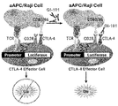
- the experiment was conducted using a CTLA-4 blockade bioassay kit (Promega cat No. JA4005), and the experiment is briefly described below.
- a CTLA-4 blockade bioassay kit Promega cat No. JA4005
- GI101 does not inhibit the function of T cells by binding to CTLA-4 expressed on effector T cells, but rather activates them (FIGS. 40 and 41).
- mice purchased from Orient (Korea) were divided into three groups of three, PBS, 3 mg / kg, 6 mg / kg, 12 mg / kg of GI101 or 3 mg / kg, 6 mg / kg, 12 MGI102 (mGI102-M61) at mg / kg was administered intravenously.
- Spleen tissues were removed from each group of mice at 1, 3, 5, 7 and 14 days of injection. Thereafter, the number of effector CD8 + T cells, NK cells, and Treg cells was calculated by FACS analysis using spleen tissue by each antibody, and the ratio of effector CD8 + T cells and NK cells to Treg cells was calculated.
- the antibody information used for each cell analysis is as follows:
- Effector CD8 + T cell PB anti-mouse CD3 ⁇ antibody (Biolegend, # 155612; KT3.1.1), FITC anti-mouse CD8 ⁇ antibody (BD, # 553031, 53-6.7), PE / Cy7 anti-mouse CD44 antibody (Biolegend, # 103030; IM7), APC anti-mouse CD122 antibody (Biolegend, # 123214; TM- ⁇ 1)
- NK cell PB anti-mouse CD3 ⁇ antibody (Biolegend, # 155612; KT3.1.1), PE anti-mouse NK-1.1 (Biolegend, # 108708; PK136)
- Treg cell FITC anti-mouse CD3 antibody (Biolegend, # 100204; 17A2), PB anti-mouse CD4 antibody (Biolegend, # 100531; RM4-5), PE anti-mouse CD25 antibody (Biolegend, # 102008; PC61), APC anti-mouse Foxp3 antibody (Invitrogen, # FJK-16s, 17-5773-82).
- CD8 + T cells and NK cells in the group administered with mGI101 or mGI102 were significantly increased compared to the PBS administration group from 3 days to 14 days after administration.
- the ratio of activated CD8 + T cells / Treg cells and NK cells / Treg cells in the group administered with mGI102 was significantly increased compared to the PBS administration group at a time point from 3 days to 7 days after administration (FIG. 42). .
- NCl-H292 cancer cell line was cultured for 3 hours in a culture medium containing 10 ⁇ g / ml Mitomycin C (Sigma), Mitomycin C was washed with a culture medium to remove it. Thereafter, a 5 ⁇ 10 4 cell number treated with Mitomycin C, the NCl-H292 cancer cell line was cultured in a 1 ⁇ 10 5 cell number in human PBMC and in a 96-well-plate. At this time, 5 ⁇ g / ml of PHA (Sigma) was treated for T cell activity.
- GI101 effectively activated the immune response suppressed by the cancer cell line over-expressing PD-L1.
- GI101 inhibits the signaling of CTLA-4 expressed in effector T cells (FIGS. 43 and 44).
- CT-26 cancer cell line derived from mouse with 5 ⁇ 10 6 cell number / 0.05 ml was mixed with 0.05 ml Matrigel matrix phenol red-free (BD), and 0.1 ml subcutaneously in the right back of the 6-week-old female BALB / c mouse (Orient). It was transplanted by administering each. After transplanting cancer cells and measuring the volume of the tumor after a certain period of time, individuals reaching about 80 mm 3 to 120 mm 3 were isolated, and 0.1 ml of GI101 was intravenously administered. After the first administration, a total of three administrations were performed once every three days, and PBS was administered as a negative control. The tumor size was measured daily to confirm the anti-cancer effect.
- BD Matrigel matrix phenol red-free
- mice C57BL / 6 mice (female, 7 weeks) pre-delivered from Orient Bio were subjected to a 7-day adaptation period, followed by a 5 ⁇ 10 6 cell number of B16F10 cancer cell lines (ATCC, USA) with 0.05 ml Matrigel matrix phenol red-free (BD) After mixing, the mice were allografted by administering 0.1 ml in the subcutaneous region on the right side of the mouse. After transplanting cancer cells and measuring the volume of the tumor after a certain period of time, individuals who have reached about 50 mm 3 to 120 mm 3 are selected, and 10 animals per group to be equal based on the size and weight of the tumor of the selected mice. Classified.
- hIgG4 was administered to the negative control group at a dose of 4 mg / kg using a disposable syringe (31G, 1 mL), and an anti-PD-1 antibody at a dose of 5 mg / kg was administered intravenously to the positive control.
- mGI101 at a dose of 1 mg / kg or 4 mg / kg was administered intravenously.
- the group administered intravenously with 4 mg / kg dose of mGI101 and 5 mg / kg dose of anti-PD-1 antibody was set as the experimental group. A total of three administrations were performed once every three days after the first administration. Tumor size was measured daily.
- the initial tumor volume was 90 mm 3 in all groups, and the standard error (SE) of each group was 5 mm 3 to 6 mm 3 .
- SE standard error
- the 1 mg / kg dose group of mGI101 was observed to increase from 90 mm 3 to 885 mm 3 during the same period as the negative control group, and was statistically significantly suppressed at some measurement points (p-value). : 0.5: day 11, p-value ⁇ 0.01: day 7, p-value ⁇ 0.001: day 3).
- the 4 mg / kg dose group of mGI101 was observed to increase during the experimental period from 90 mm 3 to 748 mm 3 during the same period as the negative control group, and was statistically significantly inhibited at some measurement time and the end of the experiment ( p-value: 0.5: day 9, p-value ⁇ 0.01: day 7 and 11).
- tumor growth inhibition was analyzed compared to each group based on the 4 mg / kg dose mIgG administration group.
- the mGI101-administered group had a growth inhibition rate of 36.5% compared to the negative control group, and no statistically significant difference (p-value: 0.5) was observed.
- the 4 mg / kg dose group of mGI101 showed statistically significant (p-value: 0.5) tumor growth inhibition compared to the negative control group.
- a total of two administrations were performed once every three days after the first administration. Tumor size was measured daily.
- mGI101 has a dose-dependent inhibitory effect on tumor growth in the tumor growth inhibition efficacy test of B16F10, a melanoma allografted to C57BL / 6 mice (FIGS. 47 and 48).
- BD Matrigel matrix phenol red-free
- CT-26 cancer cell line ATCC, USA
- 5x10 6 cell number after 7 days of acclimatization period after 7 days of adaptation of BALB / c mice (female, 7 weeks) delivered from Orient Bio )
- subcutaneously in 0.1 ml of the right back of the mouse to transplant allograft After transplanting cancer cells and measuring the volume of the tumor after a certain period of time, individuals reaching about 28 mm 3 were selected, and then 10 animals were classified for each group to be equal based on the size and weight of the tumor of the selected mice.
- hIgG4 was administered to the negative control at a dose of 6 mg / kg using a disposable syringe (31G, 1 mL).
- mGI101 at a dose of 3 mg / kg, 6 mg / kg or 12 mg / kg was administered intravenously. A total of three administrations were performed once every three days after the first administration. Tumor size was measured daily.
- mice Females, 7 weeks pre-delivered from Orient Bio after 7 days of adaptation, the CT-26 cancer cell line (ATCC, USA) of 5x10 6 cells was suspended in 0.1 ml PBS, and the right side of the mouse, etc. Allografted by subcutaneous administration at the site. After transplanting cancer cells and measuring the volume of the tumor after a certain period of time, 10 individuals per group are selected so as to be equal based on the size and weight of the tumors of the selected mice after selecting the individuals reaching about 50 mm 3 to 200 mm 3 Classified.
- CT-26 cancer cell line ATCC, USA
- the drug was not administered to the negative control group using a disposable syringe (31G, 1 mL), and the 5 mg / kg dose of the anti-PD-1 antibody or the 5 mg / kg dose of the anti-PD was administered to the negative control group.
- -1 antibody and 5 mg / kg dose of anti-CTLA-4 antibody were administered intravenously.
- GI101 at a dose of 0.1 mg / kg or 1 mg / kg was intravenously administered. A total of three administrations were performed once every three days after the first administration. Tumor size was measured daily.
- mice of each group of Experimental Example 19.1 were sacrificed when the tumor volume on average reached 200 mm 3 , and cancer tissues were collected. Thereafter, in order to analyze immune cells in the cancer tissues, the cancer tissues were separated at a single cell level, and then FACS analysis of the immune cells in the cancer tissues was performed using the following antibodies.
- Antibodies are specifically, Anti-mouse-CD3 (Biolegend, Cat. No. 100320), Anti-mouse-CD4 (Biolegend, Cat. No. 100526), Anti-mouse-CD8 (Biolegend, Cat. No. 100750), Anti-mouse-FoxP3 (eBioscience, Cat.No. 12-5773-82), Anti-mouse-CD25 (Biolegend, Cat.No.
- Anti-mouse-CD44 (eBioscience, Cat.No. 61-0441- 82), Anti-mouse-PD-1 (Biolegend, Cat. No. 135218), Anti-mouse-IFN-gamma (Biolegend, Cat. No. 505832), Anti-mouse-CD49b (Biolegend, Cat. No. 108906 ), Anti-mouse-H2 (Invitrogen, Cat.No.A15443), Anti-mouse-CD11c (Biolegend, Cat.No. 117343), Anti-mouse-CD80 (eBioscience, Cat.No. 47-4801-82) , Anti-mouse-CD86 (Biolegend, Cat. No. 104729), Anti-mouse-F4 / 80 (eBioscience, Cat. No. 47-4801-82), and Anti-mouse-CD206 (eBioscience, Cat. No. 17-2061-80).
- CD8 + T cells were significantly increased in the experimental group administered with 0.1 mg / kg of GI101 compared to the positive control group administered with 5 mg / kg of anti-PD-1 antibody alone (* p ⁇ 0.05, FIG. 52. And Figure 53).
- the expression of IFN- ⁇ in T cells was significantly increased in all the experimental groups administered with GI101 compared to the negative control group (* p ⁇ 0.05, FIGS. 52 and 53).
- M1 macrophages increased compared to the negative control group and the positive control group administered with anti-PD-1 antibody alone (FIGS. 54 and 55).
- CD86 expression of macrophages and dendritic cells increased in all experimental groups administered GI101 (* p ⁇ 0.05, FIGS. 54 to 57).
- mice Males, 7 weeks pre-delivered from Orient Bio were suspended after 7 days of adaptation, and 5x10 6 cell numbers of LLC2 cancer cell lines (ATCC, USA) were suspended in 0.1 ml PBS to form the right back of the mouse. Allografts were administered subcutaneously. After transplanting cancer cells and measuring the volume of the tumor after a certain period of time, 10 individuals per group are selected so as to be equal based on the size and weight of the tumors of the selected mice after selecting the individuals reaching about 50 mm 3 to 200 mm 3 Classified.
- the drug was not administered to the negative control group using a disposable syringe (31G, 1 mL), and the 5 mg / kg dose of the anti-PD-1 antibody or the 5 mg / kg dose of the anti-PD was administered to the negative control group.
- -1 antibody and 5 mg / kg dose of anti-CTLA-4 antibody were administered intravenously.
- GI101 at a dose of 0.1 mg / kg or 1 mg / kg was intravenously administered. A total of three administrations were performed once every three days after the first administration. Tumor size was measured daily.
- mice of each group of Experimental Example 20.1 were sacrificed when the tumor volume on average reached 200 mm 3 , and cancer tissues were collected. Then, FACS analysis was performed in the same manner as in Experimental Example 19.2 to analyze immune cells in cancer tissue.
- CD8 + T cells were significantly increased in the experimental group administered with 0.1 mg / kg of GI101 compared to the positive control group administered with anti-PD-1 antibody alone (* p ⁇ 0.05, FIG. 59). Furthermore, the expression of IFN- ⁇ was significantly increased in all the experimental groups to which GI101 was administered compared to the negative control group (* p ⁇ 0.05, FIG. 59). In addition, CD86 expression of macrophages and dendritic cells increased in all experimental groups administered GI101 (* p ⁇ 0.05, FIGS. 59 to 61).
- BD Matrigel matrix phenol red-free
- CT-26 cancer cell line ATCC, USA
- 5x10 6 cell number after 7 days of acclimatization period after 7 days of adaptation of BALB / c mice (female, 7 weeks) delivered from Orient Bio )
- subcutaneously in 0.1 ml of the right back of the mouse to transplant allograft After transplanting cancer cells and measuring the volume of the tumor after a certain period of time, individuals reaching about 28 mm 3 were selected, and then 10 animals were classified for each group to be equal based on the size and weight of the tumor of the selected mice.
- hIgG4 was administered to the negative control at a dose of 6 mg / kg using a disposable syringe (31G, 1 mL).
- mGI102-M61 at a dose of 3 mg / kg, 6 mg / kg or 12 mg / kg was administered intravenously. A total of three administrations were performed once every three days after the first administration. Tumor size was measured daily.
- hIgG4 was administered to the negative control at a dose of 4 mg / kg using a disposable syringe (31G, 1 mL).
- mGI101 at a dose of 1 mg / kg, 4 mg / kg or 6 mg / kg was administered intravenously.
- a group administered with 4.9 mg / kg of mCD80 or 2.8 mg / kg of Fc-IL-2v (GI101C2) was also set as a control.
- the group that simultaneously administered 4.9 mg / kg of mCD80 and 2.8 mg / kg of Fc-IL-2v was set as a control.
- the tumor volume measurement it was confirmed that the mGI101 dose group at 6 mg / kg dose was significantly inhibited compared to the negative control group at some measurement time and at the end of the test.
- the tumor growth inhibition rate was found to be superior to the combined administration group (FIGS. 64 and 65).
- mGI101 a test substance in the tumor growth inhibitory efficacy test of CT-26, a colon cancer cell line derived from BALB / c mice allografted to BALB / c mice, was tumor suppressed against mCD80 and IL-2v single agents under these test conditions. Efficacy was demonstrated, and excellent anticancer efficacy was confirmed compared to the combination administration of mCD80 and IL-2v (FIGS. 64 and 65). In particular, the tumor size was significantly suppressed in the 6 mg / kg dose group compared to the negative control group and the combined administration group of mCD80 and Fc-IL2v (GI101C2) in the mGI101 administration group.
- the experiment was conducted from one day before drug administration to 15 days after drug administration, and each monkey was observed around the cage, and additionally, stool status was confirmed.
- the body weight was measured one day before drug administration, and on the 1st, 8th, and 15th days after drug administration using a digital scale (LDS-150H, Shimadzu Corporation).
- the remaining amount of food was measured from the day before drug administration until the monkey was sacrificed.
- the drug was administered a total of two times, one time by intravenous route at a rate of 0.17 ml / sec by filling GI101 with a disposable syringe (24G).
- GI101 was administered at a dose of 5 mg / kg / day or 10 mg / kg / day twice in 1 / week intervals.
- the control group was administered with PBS (pH 7.4) in the same way.
- Blood was collected from the monkey of Experimental Example 23.1 one day before drug administration, one day, eight days and 15 days after drug administration, and blood was collected through the femoral vein with a disposable syringe (22G). The collected blood was subjected to blood analysis using the Automated Hematology System XN-2000 (Sysmex Corporation) and Automated Blood Coagulation Analyzer CA-510 (Sysmex Corporation).
- reticulocytes, white blood cells, and lymphocyte levels increased in the group administered with GI101 at a dose of 5 mg / kg / day or 10 mg / kg / day on Day 15 (FIGS. 70 to 72).
- Blood was collected from the monkey of Experimental Example 23.1 one day before drug administration, and on the 1st, 8th, and 15th days after drug administration, and in the same manner as in Experimental Example 23.3.
- the blood collected was TNF- ⁇ , IFN- ⁇ IL-1 ⁇ , IL-2 using Bio-Plex 200 (Bio-Rad Laboratories, Inc.) instrument and Non-Human Primate Cytokine Magnetic Bead Panel (EMD Millipore) analysis kit.
- EMD Millipore Non-Human Primate Cytokine Magnetic Bead Panel
- IL-4, IL-6, IL-8, IL-10, and IL-12 were analyzed.
- no toxicity induced by GI101 in relation to cytokine analysis was shown (FIGS. 80 and 81).
- Blood was collected from the monkey of Experimental Example 23.1 one day before drug administration, and on the 1st, 8th, and 15th days after drug administration, and in the same manner as in Experimental Example 23.3.
- the collected blood was analyzed with the following items using a flow cytometer (LSRFortessa X-20, Becton, Dickinson and Company):
- Ki67 + CD4 CD45 + / CD3 + / CD4 + / Ki67 +
- Ki67 + CD8 CD45 + / CD3 + / CD8 + / Ki67 +
- Ki67 + Treg CD45 + / CD3 + / FoxP3 + / Ki67 +
- Ki67 + NK cell CD45 + / CD16 + and CD56 + / Ki67 +.
- Ki67 + ICOS + regulatory T cells, Ki67 + NK cells, and ICOS + regulatory T cells increased on day 15.
- the proportion of T cells, CD4 + T cells, and regulatory T cells in lymphocytes increased, and the proportion of NK cells decreased, while the proportion of CD8 + T cells did not change.
- the percentage of regulatory T cells increased on day 3 and decreased on days 8 and 15, but was still high compared to control.
- the proportion of immune cells that are Ki67 + in each immune cell increases in the proportion of Ki67 + T cells, Ki67 + CD4 + T cells, Ki67 + CD8 + T cells, Ki67 + regulatory T cells, Ki67 + ICOS + regulatory T cells, Ki67 + NK cells, and ICOS + regulatory T cells.
- Ki67 + T cells, Ki67 + CD8 + T cells, and Ki67 + NK cells increased on days 3, 8 and 15, and Ki67 + CD4 + T and Ki67 + regulatory T cells increased on days 3 and 8, and Ki67 + ICOS + regulatory T cells, ICOS + regulatory T cells increased only on day 8 (FIGS. 82-87).
- the weight of the spleen increased in the group treated with GI101 at a dose of 5 mg / kg / day or 10 mg / kg / day (FIG. 88). No significant change was observed in the remaining tissues. In conclusion, some changes were observed in the group administered with GI101, but no toxicity was observed.
- CT-26 cancer cell line derived from mouse with 5 ⁇ 10 6 cell number / 0.05 ml was mixed with 0.05 ml Matrigel matrix phenol red-free (BD), and 0.1 ml subcutaneously in the right back of the 6-week-old female BALB / c mouse (Orient).
- Transplant by administering each. After transplanting cancer cells and measuring the volume of the tumor after a certain period of time, individuals reaching about 80 mm 3 to 120 mm 3 are isolated, and 0.1 ml of GI102-M45 is intravenously administered. A total of three administrations are performed once every three days after the first administration, and PBS is administered as a negative control. Tumor size is measured daily to confirm the anti-cancer effect. The activity of GI102-M45 was confirmed in the same manner as in Experimental Example 16.
- mice Males, 7 weeks pre-delivered from Orient Bio were suspended after 7 days of adaptation, and 5x10 6 cell numbers of LLC2 cancer cell lines (ATCC, USA) were suspended in 0.1 ml PBS to form the right back of the mouse. Allograft by subcutaneous administration. After transplanting cancer cells and measuring the volume of the tumor after a certain period of time, 10 individuals per group are selected so as to be equal based on the size and weight of the tumors of the selected mice after selecting the individuals reaching about 50 mm 3 to 200 mm 3 Classify.
- the drug was not administered to the negative control group using a disposable syringe (31G, 1 mL), and the 5 mg / kg dose of the anti-PD-1 antibody or the 5 mg / kg dose of the anti-PD- was administered to the negative control group.
- 1 antibody and 5 mg / kg dose of anti-CTLA-4 antibody are administered intravenously.
- GI102-M45 at a dose of 0.1 mg / kg or 1 mg / kg was intravenously administered. A total of three administrations are performed once every three days after the first administration. Tumor size is measured daily. The activity of GI102-M45 was confirmed in the same manner as in Experimental Example 20.1.
- CT-26 cancer cell line derived from mouse with 5 ⁇ 10 6 cell number / 0.05 ml was mixed with 0.05 ml Matrigel matrix phenol red-free (BD), and 0.1 ml subcutaneously in the right back of the 6-week-old female BALB / c mouse (Orient).
- Transplant by administering each. After transplanting cancer cells and measuring the volume of the tumor after a certain period of time, individuals reaching about 80 mm 3 to 120 mm 3 are isolated, and 0.1 ml of GI102-M61 is intravenously administered. A total of three administrations are performed once every three days after the first administration, and PBS is administered as a negative control. Tumor size is measured daily to confirm the anti-cancer effect. The activity of GI102-M61 was confirmed in the same manner as in Experimental Example 16.
- mice Males, 7 weeks pre-delivered from Orient Bio were suspended after 7 days of adaptation, and 5x10 6 cell numbers of LLC2 cancer cell lines (ATCC, USA) were suspended in 0.1 ml PBS to form the right back of the mouse. Allograft by subcutaneous administration. After transplanting cancer cells and measuring the volume of the tumor after a certain period of time, 10 individuals per group are selected so as to be equal based on the size and weight of the tumors of the selected mice after selecting the individuals reaching about 50 mm 3 to 200 mm 3 Classify.
- the drug was not administered to the negative control group using a disposable syringe (31G, 1 mL), and the 5 mg / kg dose of the anti-PD-1 antibody or the 5 mg / kg dose of the anti-PD- was administered to the negative control group.
- 1 antibody and 5 mg / kg dose of anti-CTLA-4 antibody are administered intravenously.
- GI102-M61 at a dose of 0.1 mg / kg or 1 mg / kg was intravenously administered. A total of three administrations are performed once every three days after the first administration. Tumor size is measured daily. The activity of GI102-M61 was confirmed in the same manner as in Experimental Example 20.1.
- CT-26 cancer cell line derived from mouse with 5 ⁇ 10 6 cell number / 0.05 ml was mixed with 0.05 ml Matrigel matrix phenol red-free (BD), and 0.1 ml subcutaneously in the right back of the 6-week-old female BALB / c mouse (Orient).
- Transplant by administering each. After transplanting cancer cells and measuring the volume of the tumor after a certain period of time, individuals reaching about 80 mm 3 to 120 mm 3 are isolated, and 0.1 ml of GI102-M72 is intravenously administered. A total of three administrations are performed once every three days after the first administration, and PBS is administered as a negative control. Tumor size is measured daily to confirm the anti-cancer effect. The activity of GI102-M72 was confirmed in the same manner as in Experimental Example 16.
- mice Males, 7 weeks pre-delivered from Orient Bio were suspended after 7 days of adaptation, and 5x10 6 cell numbers of LLC2 cancer cell lines (ATCC, USA) were suspended in 0.1 ml PBS to form the right back of the mouse. Allograft by subcutaneous administration. After transplanting cancer cells and measuring the volume of the tumor after a certain period of time, 10 individuals per group are selected so as to be equal based on the size and weight of the tumors of the selected mice after selecting the individuals reaching about 50 mm 3 to 200 mm 3 Classify.
- the drug was not administered to the negative control group using a disposable syringe (31G, 1 mL), and the 5 mg / kg dose of the anti-PD-1 antibody or the 5 mg / kg dose of the anti-PD- was administered to the negative control group.
- 1 antibody and 5 mg / kg dose of anti-CTLA-4 antibody are administered intravenously.
- GI102-M72 at a dose of 0.1 mg / kg or 1 mg / kg was intravenously administered. A total of three administrations are performed once every three days after the first administration. Tumor size is measured daily. The activity of GI102-M72 was confirmed in the same manner as in Experimental Example 20.1.
Abstract
Description


Claims (33)
- IL-2 단백질 및 CD80 단백질을 포함하는 융합단백질.Fusion protein comprising IL-2 protein and CD80 protein.
- 제1항에 있어서,According to claim 1,상기 IL-2 단백질 및 상기 CD80 단백질은 링커에 의해 결합된 것인, 융합단백질.The IL-2 protein and the CD80 protein are linked by a linker, a fusion protein.
- 제1항에 있어서,According to claim 1,상기 IL-2 단백질은 서열번호 10의 아미노산 서열을 갖는 것인, 융합단백질.The IL-2 protein is to have the amino acid sequence of SEQ ID NO: 10, a fusion protein.
- 제1항에 있어서,According to claim 1,상기 IL-2 단백질은 IL-2 변이체인 것인, 융합단백질.The IL-2 protein is an IL-2 variant, a fusion protein.
- 제4항에 있어서,According to claim 4,상기 IL-2 변이체는 서열번호 10의 아미노산 서열에서 38번째, 42번째, 45번째, 61번째 및 72번째 위치의 아미노산 중 적어도 하나가 치환된 것인, 융합단백질.The IL-2 variant is a fusion protein in which at least one of amino acids at positions 38, 42, 45, 61, and 72 of the amino acid sequence of SEQ ID NO: 10 is substituted.
- 제4항에 있어서,According to claim 4,상기 IL-2 변이체는 서열번호 10의 아미노산 서열에서 R38A, F42A, Y45A, E61R 및 L72G로 구성된 군으로부터 선택되는 적어도 어느 하나의 치환이 일어난 것인, 융합단백질.The IL-2 variant is a fusion protein wherein at least one substitution selected from the group consisting of R38A, F42A, Y45A, E61R and L72G has occurred in the amino acid sequence of SEQ ID NO: 10.
- 제4항에 있어서, According to claim 4,상기 IL-2 변이체는 서열번호 10의 아미노산 서열에서 하기 (a) 내지 (d) 조합 중 선택되는 어느 하나의 조합의 치환이 일어난 것인, 융합단백질:The IL-2 variant is a fusion protein in which the substitution of any one of the following combinations (a) to (d) occurs in the amino acid sequence of SEQ ID NO: 10:(a) R38A/F42A(a) R38A / F42A(b) R38A/F42A/Y45A(b) R38A / F42A / Y45A(c) R38A/F42A/E61R(c) R38A / F42A / E61R(d) R38A/F42A/L72G.(d) R38A / F42A / L72G.
- 제4항에 있어서,According to claim 4,상기 IL-2 변이체는 서열번호 6, 22, 23 또는 24의 아미노산 서열을 갖는 것인, 융합단백질.The IL-2 variant has an amino acid sequence of SEQ ID NO: 6, 22, 23 or 24, a fusion protein.
- 제1항에 있어서,According to claim 1,상기 CD80은 서열번호 11의 아미노산 서열을 갖는 것인, 융합단백질.The CD80 is to have the amino acid sequence of SEQ ID NO: 11, a fusion protein.
- 제1항에 있어서,According to claim 1,상기 CD80 단백질은 CD80의 단편인 것인, 융합단백질.The CD80 protein is a fragment of CD80, a fusion protein.
- 제10항에 있어서,The method of claim 10,상기 CD80의 단편은 서열번호 11의 아미노산 서열 중 35번째 내지 242번째의 아미노산으로 이루어진 것인, 융합단백질.The fragment of CD80 is composed of amino acids 35 to 242 of the amino acid sequence of SEQ ID NO: 11, a fusion protein.
- 제2항에 있어서,According to claim 2,상기 링커는 알부민 또는 면역글로불린의 Fc 도메인인 것인, 융합단백질.The linker is an albumin or an immunoglobulin Fc domain, a fusion protein.
- 제12항에 있어서,The method of claim 12,상기 Fc 도메인은 야생형 또는 변이체인 것인, 융합단백질.The Fc domain is a wild-type or variant, fusion protein.
- 제12항에 있어서,The method of claim 12,상기 Fc 도메인은 서열번호 4의 아미노산 서열을 갖는 것인, 융합단백질.The Fc domain is to have the amino acid sequence of SEQ ID NO: 4, a fusion protein.
- 제13항에 있어서,The method of claim 13,상기 Fc 도메인의 변이체는 서열번호 12의 아미노산 서열을 갖는 것인, 융합단백질.The variant of the Fc domain will have the amino acid sequence of SEQ ID NO: 12, a fusion protein.
- 제1항에 있어서,According to claim 1,상기 융합단백질은 하기 구조식(I) 또는 (II)로 이루어진 것인, 융합단백질:The fusion protein is composed of the following structural formula (I) or (II), the fusion protein:N'-X-[링커(1)]n-Fc 도메인-[링커(2)]m-Y-C' (I)N'-X- [linker (1)] n-Fc domain- [linker (2)] m-Y-C '(I)N'-Y-[링커(1)]n-Fc 도메인-[링커(2)]m-X-C' (II)N'-Y- [Linker (1)] n-Fc domain- [Linker (2)] m-X-C '(II)이때, 상기 구조식(I) 및 (II)에 있어서,At this time, in the structural formulas (I) and (II),상기 N'은 융합단백질의 N-말단이고,N 'is the N-terminal of the fusion protein,상기 C'는 융합단백질의 C-말단이며,C 'is the C-terminal of the fusion protein,상기 X는 CD80 단백질이고,X is a CD80 protein,상기 Y는 IL-2 단백질이며,Y is an IL-2 protein,상기 링커(1) 및 링커(2)는 펩타이드 링커이고,The linker (1) and the linker (2) are peptide linkers,상기 n 및 m은 각각 독립적으로, O 또는 1이다. The n and m are each independently O or 1.
- 제16항에 있어서,The method of claim 16,상기 링커(1)이 서열번호 3의 아미노산 서열로 이루어진 펩타이드 링커인 것인, 융합단백질.The linker (1) is a peptide linker consisting of the amino acid sequence of SEQ ID NO: 3, a fusion protein.
- 제16항에 있어서,The method of claim 16,상기 링커(2)가 서열번호 5의 아미노산 서열로 이루어진 펩타이드 링커인 것인, 융합단백질.The linker (2) is a peptide linker consisting of the amino acid sequence of SEQ ID NO: 5, a fusion protein.
- 제16항에 있어서,The method of claim 16,상기 융합단백질은 구조식(I)로 이루어진 것인, 융합단백질.The fusion protein is composed of structural formula (I), fusion protein.
- 제1항에 있어서,According to claim 1,상기 융합단백질은 서열번호 9, 26, 28 또는 30의 아미노산 서열에 대해 85% 혹은 그 이상의 서열동일성을 갖는 것인, 융합단백질.The fusion protein has a sequence identity of 85% or more with respect to the amino acid sequence of SEQ ID NO: 9, 26, 28 or 30, the fusion protein.
- 제1항 내지 제20항 중 어느 한 항의 융합단백질 두 개가 결합된 융합단백질 이량체.A fusion protein dimer in which two fusion proteins of any one of claims 1 to 20 are combined.
- 제21항에 있어서,The method of claim 21,상기 융합단백질 이량체는 동형이량체(homodimer)인 것인, 융합단백질 이량체.The fusion protein dimer is a homodimer, a fusion protein dimer.
- 제1항 내지 제20항 중 어느 한 항의 융합단백질을 코딩하는 폴리뉴클레오티드.A polynucleotide encoding the fusion protein of any one of claims 1 to 20.
- 제23항에 있어서,The method of claim 23,상기 폴리뉴클레오티드는 서열번호 8, 25, 27 또는 29의 염기서열에 대해 85% 혹은 그 이상의 서열동일성을 갖는 것인, 폴리뉴클레오티드.The polynucleotide has a sequence identity of 85% or more with respect to the nucleotide sequence of SEQ ID NO: 8, 25, 27 or 29, polynucleotide.
- 제23항의 폴리뉴클레오티드를 포함하는 벡터.A vector comprising the polynucleotide of claim 23.
- 제25항의 벡터가 도입된 형질전환 세포.A transformed cell into which the vector of claim 25 is introduced.
- 제1항 내지 제20항 중 어느 한 항의 융합단백질; 또는 제21항 또는 제22항의 융합단백질 이량체;를 유효성분으로 포함하는 암 또는 감염성 질환 예방 또는 치료용 약학 조성물.A fusion protein according to any one of claims 1 to 20; Or a pharmaceutical composition for preventing or treating cancer or infectious diseases comprising the fusion protein dimer of claim 21 or 22 as an active ingredient.
- 제27항에 있어서,The method of claim 27,상기 약학 조성물이 약제학적으로 허용가능한 담체를 더 포함하는 것인, 약학 조성물.The pharmaceutical composition will further include a pharmaceutically acceptable carrier, pharmaceutical composition.
- 제27항에 있어서,The method of claim 27,상기 암은 위암, 간암, 폐암, 대장암, 유방암, 전립선암, 난소암, 췌장암, 자궁경부암, 갑상선암, 후두암, 급성 골수성 백혈병, 뇌종양, 신경모세포종, 망막 모세포종, 두경부암, 침샘암 및 림프종으로 구성된 군에서 선택되는 어느 하나인, 약학 조성물.The cancer is composed of gastric cancer, liver cancer, lung cancer, colon cancer, breast cancer, prostate cancer, ovarian cancer, pancreatic cancer, cervical cancer, thyroid cancer, larynx cancer, acute myeloid leukemia, brain tumor, neuroblastoma, retinoblastoma, head and neck cancer, salivary gland cancer, and lymphoma. Any one selected from the group, pharmaceutical composition.
- 제27항에 있어서,The method of claim 27,상기 감염성 질환은 B형 간염, C형 간염, 인간 파필로마 바이러스 감염, 사이토메갈로바이러스 감염, 바이러스성 호흡기 질환 및 인플루엔자로 구성된 군에서 선택되는 어느 하나인, 약학 조성물.The infectious disease is any one selected from the group consisting of hepatitis B, hepatitis C, human papilloma virus infection, cytomegalovirus infection, viral respiratory disease and influenza.
- 암 또는 감염성 질환을 치료하기 위한 제1항의 융합단백질의 용도.Use of the fusion protein of claim 1 to treat a cancer or infectious disease.
- 암 또는 감염성 질환 치료용 약제를 제조하기 위한 제1항의 융합단백질의 용도.Use of the fusion protein of claim 1 to prepare a medicament for the treatment of cancer or infectious diseases.
- 제1항 내지 제20항 중 어느 한 항의 융합단백질; 또는 제21항 또는 제22항의 융합단백질 이량체;를 개체에 투여하는 단계를 포함하는 암 또는 감염성 질환 치료방법.A fusion protein according to any one of claims 1 to 20; Or a fusion protein dimer of claim 21 or 22; administering to the individual.
Priority Applications (17)
| Application Number | Priority Date | Filing Date | Title |
|---|---|---|---|
| US16/959,312 US11492384B2 (en) | 2018-09-17 | 2019-09-16 | Fusion protein comprising IL-2 protein and CD80 protein, and use thereof |
| MX2020007072A MX2020007072A (en) | 2018-09-17 | 2019-09-16 | Fusion protein comprising il-2 protein and cd80 protein, and use thereof. |
| SG11202005873QA SG11202005873QA (en) | 2018-09-17 | 2019-09-16 | Fusion protein comprising il-2 protein and cd80 protein, and use thereof |
| CA3086486A CA3086486A1 (en) | 2018-09-17 | 2019-09-16 | Fusion protein comprising il-2 protein and cd80 protein, and use thereof |
| JP2020562067A JP7085644B2 (en) | 2018-09-17 | 2019-09-16 | Fusion proteins including IL-2 and CD80 proteins and their use |
| BR112020015030-4A BR112020015030A2 (en) | 2018-09-17 | 2019-09-16 | FUSION PROTEIN UNDERSTANDING IL-2 PROTEIN AND CD80 PROTEIN AND USE OF THE SAME |
| CN201980025699.8A CN111971295A (en) | 2018-09-17 | 2019-09-16 | Fusion protein comprising IL-2 protein and CD80 protein and uses thereof |
| AU2019343850A AU2019343850A1 (en) | 2018-09-17 | 2019-09-16 | Fusion protein comprising IL-2 protein and CD80 protein, and use thereof |
| EP19862449.6A EP3715367A4 (en) | 2018-09-17 | 2019-09-16 | Fusion protein comprising il-2 protein and cd80 protein, and use thereof |
| PE2020000980A PE20201418A1 (en) | 2018-09-17 | 2019-09-16 | FUSION PROTEIN INCLUDING IL-2 PROTEIN AND CD80 PROTEIN AND USE OF THE SAME |
| ZA2020/03645A ZA202003645B (en) | 2018-09-17 | 2020-06-17 | Fusion protein comprising il-2 protein and cd80 protein, and use thereof |
| IL275592A IL275592B (en) | 2018-09-17 | 2020-06-22 | Fusion protein comprising il-2 protein and cd80 protein, and use thereof |
| PH12020551026A PH12020551026A1 (en) | 2018-09-17 | 2020-06-30 | Fusion protein comprising il-2 protein and cd80 protein, and use thereof |
| JP2022070213A JP2022115876A (en) | 2018-09-17 | 2022-04-21 | Fusion protein including il-2 protein and cd80 protein, and use of the same |
| US17/878,664 US20220380781A1 (en) | 2018-09-17 | 2022-08-01 | Fusion protein comprising il-2 protein and cd80 protein, and use thereof |
| US17/878,703 US20220389070A1 (en) | 2018-09-17 | 2022-08-01 | Fusion protein comprising il-2 protein and cd80 protein, and use thereof |
| US17/948,894 US20230104217A1 (en) | 2018-09-17 | 2022-09-20 | Fusion protein comprising il-2 protein and cd80 protein, and use thereof |
Applications Claiming Priority (8)
| Application Number | Priority Date | Filing Date | Title |
|---|---|---|---|
| KR10-2018-0110698 | 2018-09-17 | ||
| KR20180110698 | 2018-09-17 | ||
| KR20190001867 | 2019-01-07 | ||
| KR10-2019-0001867 | 2019-01-07 | ||
| US201962832013P | 2019-04-10 | 2019-04-10 | |
| US62/832,013 | 2019-04-10 | ||
| KR10-2019-0053436 | 2019-05-08 | ||
| KR20190053436 | 2019-05-08 |
Related Child Applications (4)
| Application Number | Title | Priority Date | Filing Date |
|---|---|---|---|
| US16/959,312 A-371-Of-International US11492384B2 (en) | 2018-09-17 | 2019-09-16 | Fusion protein comprising IL-2 protein and CD80 protein, and use thereof |
| US17/878,703 Division US20220389070A1 (en) | 2018-09-17 | 2022-08-01 | Fusion protein comprising il-2 protein and cd80 protein, and use thereof |
| US17/878,664 Division US20220380781A1 (en) | 2018-09-17 | 2022-08-01 | Fusion protein comprising il-2 protein and cd80 protein, and use thereof |
| US17/948,894 Continuation-In-Part US20230104217A1 (en) | 2018-09-17 | 2022-09-20 | Fusion protein comprising il-2 protein and cd80 protein, and use thereof |
Publications (1)
| Publication Number | Publication Date |
|---|---|
| WO2020060122A1 true WO2020060122A1 (en) | 2020-03-26 |
Family
ID=69887592
Family Applications (1)
| Application Number | Title | Priority Date | Filing Date |
|---|---|---|---|
| PCT/KR2019/011928 WO2020060122A1 (en) | 2018-09-17 | 2019-09-16 | Fusion protein comprising il-2 protein and cd80 protein, and use thereof |
Country Status (17)
| Country | Link |
|---|---|
| US (3) | US11492384B2 (en) |
| EP (1) | EP3715367A4 (en) |
| JP (2) | JP7085644B2 (en) |
| KR (2) | KR102201086B1 (en) |
| CN (1) | CN111971295A (en) |
| AU (1) | AU2019343850A1 (en) |
| BR (1) | BR112020015030A2 (en) |
| CA (1) | CA3086486A1 (en) |
| CL (1) | CL2020001872A1 (en) |
| IL (1) | IL275592B (en) |
| MX (1) | MX2020007072A (en) |
| PE (1) | PE20201418A1 (en) |
| PH (1) | PH12020551026A1 (en) |
| SG (1) | SG11202005873QA (en) |
| TW (2) | TWI755630B (en) |
| WO (1) | WO2020060122A1 (en) |
| ZA (1) | ZA202003645B (en) |
Cited By (3)
| Publication number | Priority date | Publication date | Assignee | Title |
|---|---|---|---|---|
| WO2021238904A1 (en) * | 2020-05-25 | 2021-12-02 | 北京比洋生物技术有限公司 | Fc-cd80 fusion protein and conjugate thereof, and use thereof |
| JP2023515247A (en) * | 2020-03-31 | 2023-04-12 | ハンミ ファーマシューティカル カンパニー リミテッド | Novel immunologically active interleukin-2 analogues |
| EP4174088A4 (en) * | 2020-06-30 | 2024-01-24 | Gi Innovation Inc | Fusion protein comprising anti-lag-3 antibody and il-2, and use thereof |
Families Citing this family (13)
| Publication number | Priority date | Publication date | Assignee | Title |
|---|---|---|---|---|
| TWI784957B (en) * | 2016-06-20 | 2022-12-01 | 英商克馬伯有限公司 | Immunocytokines |
| MX2022005998A (en) * | 2019-11-20 | 2022-06-17 | Gi Cell Inc | Composition for culturing natural killer cells and method for preparing natural killer cells by using same. |
| MX2022006295A (en) * | 2019-11-27 | 2022-06-08 | Gi Cell Inc | Composition for anticancer treatment, comprising nk cells and fusion protein which comprises il-2 protein and cd80 protein. |
| KR102604984B1 (en) | 2020-03-18 | 2023-11-23 | (주)지아이이노베이션 | Formulatiom of fusion protein comprising il-2 protein and cd80 protein |
| TW202200608A (en) * | 2020-03-18 | 2022-01-01 | 南韓商Gi醫諾微新股份有限公司 | Fusion protein comprising il-2 protein and cd80 protein fragment or variant thereof and use thereof |
| MX2022011154A (en) | 2020-03-18 | 2022-10-13 | Gi Innovation Inc | Pharmaceutical composition for cancer treatment comprising fusion protein including il-2 protein and cd80 protein and anticancer drug. |
| KR102373965B1 (en) * | 2020-06-05 | 2022-03-15 | (주)지아이이노베이션 | Pharmaceutical composition for enhancing radiotherapy containing fusion protein comprising il-2 protein and cd80 protein |
| EP4206219A1 (en) * | 2020-08-31 | 2023-07-05 | GI Cell, Inc. | Fusion protein including il15 protein, il15 receptor alpha protein, fc domain, and il2 protein, and use thereof |
| TWI815194B (en) | 2020-10-22 | 2023-09-11 | 美商基利科學股份有限公司 | INTERLEUKIN-2-Fc FUSION PROTEINS AND METHODS OF USE |
| WO2022057696A2 (en) * | 2021-09-08 | 2022-03-24 | 中南大学湘雅医院 | Recombinant protein coding sequence, recombinant protein, and method for preparing monoclonal antibodies |
| KR20230168273A (en) * | 2022-06-03 | 2023-12-13 | (주)지아이이노베이션 | Bispecific antibody comprising anti-tigit antibody and use thereof |
| WO2023249425A1 (en) * | 2022-06-22 | 2023-12-28 | ㈜지아이이노베이션 | Fusion protein comprising anti-cd73 antibody and il-2, and use thereof |
| WO2024063546A1 (en) * | 2022-09-20 | 2024-03-28 | 주식회사 지아이바이옴 | Combined therapy of lactobacillus plantarum strain, and cancer treatment method using same |
Citations (5)
| Publication number | Priority date | Publication date | Assignee | Title |
|---|---|---|---|---|
| US5229109A (en) | 1992-04-14 | 1993-07-20 | Board Of Regents, The University Of Texas System | Low toxicity interleukin-2 analogues for use in immunotherapy |
| US20130149305A1 (en) * | 2011-10-25 | 2013-06-13 | Suzanne Ostrand-Rosenberg | Soluble cd80 as a therapeutic to reverse immune supression in cancer patients |
| KR101667096B1 (en) | 2011-02-10 | 2016-10-18 | 로슈 글리카트 아게 | Mutant interleukin-2 polypetides |
| WO2017220989A1 (en) * | 2016-06-20 | 2017-12-28 | Kymab Limited | Anti-pd-l1 and il-2 cytokines |
| KR20180069903A (en) * | 2015-11-02 | 2018-06-25 | 파이브 프라임 테라퓨틱스, 인크. | CD80 extracellular domain polypeptides and their use in the treatment of cancer |
Family Cites Families (17)
| Publication number | Priority date | Publication date | Assignee | Title |
|---|---|---|---|---|
| ZA842025B (en) * | 1983-03-21 | 1984-11-28 | Hoffmann La Roche | Interleuken-2 |
| CN86104525A (en) * | 1985-07-31 | 1987-02-25 | 武田药品工业株式会社 | The analytical approach of human interleukin-2 and reagent |
| DZ2788A1 (en) * | 1998-05-15 | 2003-12-01 | Bayer Ag | Selective IL-2 agonists and antagonists. |
| GB9810999D0 (en) | 1998-05-21 | 1998-07-22 | Goken Joop | Materials and methods relating to the expression of genes |
| US7829084B2 (en) | 2001-01-17 | 2010-11-09 | Trubion Pharmaceuticals, Inc. | Binding constructs and methods for use thereof |
| AU2002355955A1 (en) | 2001-08-13 | 2003-03-03 | University Of Southern California | Interleukin-2 mutants with reduced toxicity |
| ES2381025T3 (en) | 2001-12-04 | 2012-05-22 | Merck Patent Gmbh | Immunocytokines with modulated selectivity |
| JP2007527242A (en) * | 2004-03-05 | 2007-09-27 | カイロン コーポレーション | In vitro test system for predicting patient tolerance of therapeutic agents |
| EP2402754B2 (en) * | 2006-03-06 | 2023-07-26 | Amunix Operating Inc. | Unstructured recombinant polymers and uses thereof |
| JP2009542592A (en) | 2006-07-06 | 2009-12-03 | メルク パテント ゲゼルシャフト ミット ベシュレンクテル ハフツング | Compositions and methods for enhancing the effectiveness of an IL-2-mediated immune response |
| KR20100058541A (en) * | 2007-08-15 | 2010-06-03 | 아뮤닉스 인코포레이티드 | Compositions and methods for modifying properties of biologically active polypeptides |
| CU23923B1 (en) | 2010-11-12 | 2013-07-31 | Ct De Inmunología Molecular | POLYPEPTIDES DERIVED FROM IL-2 WITH AGONIST ACTIVITY |
| EA201892619A1 (en) | 2011-04-29 | 2019-04-30 | Роше Гликарт Аг | IMMUNOCONJUGATES CONTAINING INTERLEUKIN-2 MUTANT POLYPETIPS |
| CN105143257B (en) | 2013-03-15 | 2020-10-27 | 艾伯维生物医疗股份有限公司 | FC variants |
| WO2016030350A1 (en) | 2014-08-29 | 2016-03-03 | F. Hoffmann-La Roche Ag | Combination therapy of tumor-targeted il-2 variant immunocytokines and antibodies against human pd-l1 |
| US9567399B1 (en) * | 2016-06-20 | 2017-02-14 | Kymab Limited | Antibodies and immunocytokines |
| LT3606946T (en) | 2017-04-03 | 2022-10-25 | F. Hoffmann-La Roche Ag | Immunoconjugates of an anti-pd-1 antibody with a mutant il-2 or with il-15 |
-
2019
- 2019-09-16 MX MX2020007072A patent/MX2020007072A/en unknown
- 2019-09-16 EP EP19862449.6A patent/EP3715367A4/en active Pending
- 2019-09-16 SG SG11202005873QA patent/SG11202005873QA/en unknown
- 2019-09-16 PE PE2020000980A patent/PE20201418A1/en unknown
- 2019-09-16 BR BR112020015030-4A patent/BR112020015030A2/en unknown
- 2019-09-16 US US16/959,312 patent/US11492384B2/en active Active
- 2019-09-16 AU AU2019343850A patent/AU2019343850A1/en active Pending
- 2019-09-16 CN CN201980025699.8A patent/CN111971295A/en active Pending
- 2019-09-16 CA CA3086486A patent/CA3086486A1/en active Pending
- 2019-09-16 KR KR1020190113659A patent/KR102201086B1/en active IP Right Grant
- 2019-09-16 WO PCT/KR2019/011928 patent/WO2020060122A1/en unknown
- 2019-09-16 JP JP2020562067A patent/JP7085644B2/en active Active
- 2019-09-17 TW TW108133398A patent/TWI755630B/en active
- 2019-09-17 TW TW110122590A patent/TWI824258B/en active
-
2020
- 2020-06-17 ZA ZA2020/03645A patent/ZA202003645B/en unknown
- 2020-06-22 IL IL275592A patent/IL275592B/en unknown
- 2020-06-30 PH PH12020551026A patent/PH12020551026A1/en unknown
- 2020-07-14 CL CL2020001872A patent/CL2020001872A1/en unknown
-
2021
- 2021-01-05 KR KR1020210000775A patent/KR102558191B1/en active IP Right Grant
-
2022
- 2022-04-21 JP JP2022070213A patent/JP2022115876A/en active Pending
- 2022-08-01 US US17/878,664 patent/US20220380781A1/en active Pending
- 2022-08-01 US US17/878,703 patent/US20220389070A1/en active Pending
Patent Citations (5)
| Publication number | Priority date | Publication date | Assignee | Title |
|---|---|---|---|---|
| US5229109A (en) | 1992-04-14 | 1993-07-20 | Board Of Regents, The University Of Texas System | Low toxicity interleukin-2 analogues for use in immunotherapy |
| KR101667096B1 (en) | 2011-02-10 | 2016-10-18 | 로슈 글리카트 아게 | Mutant interleukin-2 polypetides |
| US20130149305A1 (en) * | 2011-10-25 | 2013-06-13 | Suzanne Ostrand-Rosenberg | Soluble cd80 as a therapeutic to reverse immune supression in cancer patients |
| KR20180069903A (en) * | 2015-11-02 | 2018-06-25 | 파이브 프라임 테라퓨틱스, 인크. | CD80 extracellular domain polypeptides and their use in the treatment of cancer |
| WO2017220989A1 (en) * | 2016-06-20 | 2017-12-28 | Kymab Limited | Anti-pd-l1 and il-2 cytokines |
Non-Patent Citations (5)
| Title |
|---|
| "Remington's Pharmaceutical Sciences", 1995 |
| BARBEE, S. D. ET AL.: "Abstract B005: FPT155, a novel therapeutic CD 80-Fc fusion protein with potent antitumor activity in preclinical models", AACR-NCI-EORTC INTERNATIONAL CONFERENCE. MOLECULAR TARGETS AND CANCER THERAPEUTICS, 26 October 2017 (2017-10-26), Philadelphia, PA, XP055709912 * |
| ENGELSUHLMANN, ANGEW CHEM INTED ENG., vol. 37, 1988, pages 73 - 127 |
| IMAI ET AL., CANCER SCI, vol. 98, 2007, pages 416 - 423 |
| SONDEL, P. M. ET AL.: "Current and potential uses of immunocytokines as cancer immunotherapy", ANTIBODIES, vol. 1, 4 July 2012 (2012-07-04), pages 149 - 1 71, XP055693420 * |
Cited By (3)
| Publication number | Priority date | Publication date | Assignee | Title |
|---|---|---|---|---|
| JP2023515247A (en) * | 2020-03-31 | 2023-04-12 | ハンミ ファーマシューティカル カンパニー リミテッド | Novel immunologically active interleukin-2 analogues |
| WO2021238904A1 (en) * | 2020-05-25 | 2021-12-02 | 北京比洋生物技术有限公司 | Fc-cd80 fusion protein and conjugate thereof, and use thereof |
| EP4174088A4 (en) * | 2020-06-30 | 2024-01-24 | Gi Innovation Inc | Fusion protein comprising anti-lag-3 antibody and il-2, and use thereof |
Also Published As
| Publication number | Publication date |
|---|---|
| SG11202005873QA (en) | 2020-07-29 |
| TW202136288A (en) | 2021-10-01 |
| PE20201418A1 (en) | 2020-12-09 |
| CN111971295A (en) | 2020-11-20 |
| JP7085644B2 (en) | 2022-06-16 |
| TWI824258B (en) | 2023-12-01 |
| US11492384B2 (en) | 2022-11-08 |
| CA3086486A1 (en) | 2020-03-26 |
| JP2021511081A (en) | 2021-05-06 |
| EP3715367A1 (en) | 2020-09-30 |
| KR102558191B1 (en) | 2023-07-24 |
| KR102201086B9 (en) | 2022-10-21 |
| US20200369740A1 (en) | 2020-11-26 |
| EP3715367A4 (en) | 2021-05-05 |
| PH12020551026A1 (en) | 2021-09-06 |
| JP2022115876A (en) | 2022-08-09 |
| KR20210008110A (en) | 2021-01-20 |
| CL2020001872A1 (en) | 2020-12-18 |
| IL275592A (en) | 2020-08-31 |
| TWI755630B (en) | 2022-02-21 |
| IL275592B (en) | 2022-02-01 |
| AU2019343850A1 (en) | 2020-06-25 |
| KR102201086B1 (en) | 2021-01-11 |
| ZA202003645B (en) | 2022-09-28 |
| US20220380781A1 (en) | 2022-12-01 |
| US20220389070A1 (en) | 2022-12-08 |
| BR112020015030A2 (en) | 2021-03-16 |
| KR20200032009A (en) | 2020-03-25 |
| MX2020007072A (en) | 2020-11-11 |
| TW202028227A (en) | 2020-08-01 |
Similar Documents
| Publication | Publication Date | Title |
|---|---|---|
| WO2020060122A1 (en) | Fusion protein comprising il-2 protein and cd80 protein, and use thereof | |
| WO2018030806A1 (en) | Cytokine fused to immunoglobulin fc heterodimer and pharmaceutical composition comprising same | |
| WO2020050667A1 (en) | Chimeric antigen receptor for solid cancer and t cells expressing chimeric antigen receptor | |
| WO2015108398A1 (en) | Long-acting insulin and use thereof | |
| WO2017023138A1 (en) | Chimeric antigen receptor, and t cells in which chimeric antigen receptor is expressed | |
| WO2011142514A1 (en) | Composition containing pias3 as an active ingredient for preventing or treating cancer or immune disease | |
| WO2019135668A1 (en) | Extracellular domain of alpha subunit of ige fc receptor, pharmaceutical composition comprising same and method for producing same | |
| WO2021162521A1 (en) | Immune cells overexpressing cell signaling regulatory factor introduced from outside and use thereof | |
| WO2021107689A1 (en) | Pharmaceutical composition for treatment of cancer, comprising immune checkpoint inhibitor and fusion protein including il-2 protein and cd80 protein | |
| WO2019066586A1 (en) | Long-acting conjugate of glucagon-like peptide-2 (glp-2) derivative | |
| WO2021158073A1 (en) | Fusion antibody for presenting antigen-derived t cell antigen epitope or peptide containing same on cell surface, and composition comprising same | |
| WO2020080908A1 (en) | Anti-l1cam antibody or antigen-binding fragment thereof and chimeric antigen receptor comprising same | |
| WO2021187922A1 (en) | Pharmaceutical composition for cancer treatment comprising fusion protein including il-2 protein and cd80 protein and anticancer drug | |
| WO2022005174A1 (en) | Fusion protein comprising anti-lag-3 antibody and il-2, and use thereof | |
| WO2021153979A1 (en) | Fusion protein comprising anti-taa antibody, anti-pd-l1 antibody, and il-2, and uses thereof | |
| WO2022025638A1 (en) | Chimeric antigen receptor (car) t cell stabilizing immune synapse | |
| WO2019135666A1 (en) | Pharmaceutical composition containing extracellular domain of alpha subunit of ige fc receptor | |
| WO2022035200A1 (en) | Fusion protein comprising il-12 and anti-cd20 antibody and use thereof | |
| WO2022250440A1 (en) | Receptor tyrosine-protein kinase erbb3-specific chimeric antigen receptor and immune cell therapy product expressing same | |
| WO2021187904A1 (en) | Fusion protein comprising il-2 protein and cd80 protein fragment or variant thereof, and uses thereof | |
| WO2023048518A1 (en) | Fusion protein dimer comprising pd-1 and il-15 and use thereof | |
| WO2023048516A1 (en) | Fusion protein dimer including pd-1 and il-21, and use thereof | |
| WO2021071319A1 (en) | Multispecific fusion protein and use thereof | |
| WO2020138868A1 (en) | Type 1 interferon neutralizing fc-fusion protein and use thereof | |
| WO2023224429A1 (en) | Fusion protein comprising light protein and anti-fap antibody and uses thereof |
Legal Events
| Date | Code | Title | Description |
|---|---|---|---|
| 121 | Ep: the epo has been informed by wipo that ep was designated in this application |
Ref document number: 19862449 Country of ref document: EP Kind code of ref document: A1 |
|
| ENP | Entry into the national phase |
Ref document number: 3086486 Country of ref document: CA |
|
| ENP | Entry into the national phase |
Ref document number: 2019343850 Country of ref document: AU Date of ref document: 20190916 Kind code of ref document: A |
|
| ENP | Entry into the national phase |
Ref document number: 2019862449 Country of ref document: EP Effective date: 20200626 |
|
| ENP | Entry into the national phase |
Ref document number: 2020562067 Country of ref document: JP Kind code of ref document: A |
|
| REG | Reference to national code |
Ref country code: BR Ref legal event code: B01A Ref document number: 112020015030 Country of ref document: BR |
|
| ENP | Entry into the national phase |
Ref document number: 112020015030 Country of ref document: BR Kind code of ref document: A2 Effective date: 20200723 |
|
| NENP | Non-entry into the national phase |
Ref country code: DE |