WO2019098256A1 - Device - Google Patents
Device Download PDFInfo
- Publication number
- WO2019098256A1 WO2019098256A1 PCT/JP2018/042207 JP2018042207W WO2019098256A1 WO 2019098256 A1 WO2019098256 A1 WO 2019098256A1 JP 2018042207 W JP2018042207 W JP 2018042207W WO 2019098256 A1 WO2019098256 A1 WO 2019098256A1
- Authority
- WO
- WIPO (PCT)
- Prior art keywords
- cell
- cells
- neural
- cell device
- nerve
- Prior art date
Links
Images


Classifications
-
- C—CHEMISTRY; METALLURGY
- C12—BIOCHEMISTRY; BEER; SPIRITS; WINE; VINEGAR; MICROBIOLOGY; ENZYMOLOGY; MUTATION OR GENETIC ENGINEERING
- C12N—MICROORGANISMS OR ENZYMES; COMPOSITIONS THEREOF; PROPAGATING, PRESERVING, OR MAINTAINING MICROORGANISMS; MUTATION OR GENETIC ENGINEERING; CULTURE MEDIA
- C12N5/00—Undifferentiated human, animal or plant cells, e.g. cell lines; Tissues; Cultivation or maintenance thereof; Culture media therefor
- C12N5/0068—General culture methods using substrates
-
- C—CHEMISTRY; METALLURGY
- C12—BIOCHEMISTRY; BEER; SPIRITS; WINE; VINEGAR; MICROBIOLOGY; ENZYMOLOGY; MUTATION OR GENETIC ENGINEERING
- C12M—APPARATUS FOR ENZYMOLOGY OR MICROBIOLOGY; APPARATUS FOR CULTURING MICROORGANISMS FOR PRODUCING BIOMASS, FOR GROWING CELLS OR FOR OBTAINING FERMENTATION OR METABOLIC PRODUCTS, i.e. BIOREACTORS OR FERMENTERS
- C12M23/00—Constructional details, e.g. recesses, hinges
- C12M23/02—Form or structure of the vessel
- C12M23/12—Well or multiwell plates
-
- C—CHEMISTRY; METALLURGY
- C12—BIOCHEMISTRY; BEER; SPIRITS; WINE; VINEGAR; MICROBIOLOGY; ENZYMOLOGY; MUTATION OR GENETIC ENGINEERING
- C12M—APPARATUS FOR ENZYMOLOGY OR MICROBIOLOGY; APPARATUS FOR CULTURING MICROORGANISMS FOR PRODUCING BIOMASS, FOR GROWING CELLS OR FOR OBTAINING FERMENTATION OR METABOLIC PRODUCTS, i.e. BIOREACTORS OR FERMENTERS
- C12M25/00—Means for supporting, enclosing or fixing the microorganisms, e.g. immunocoatings
- C12M25/14—Scaffolds; Matrices
-
- C—CHEMISTRY; METALLURGY
- C12—BIOCHEMISTRY; BEER; SPIRITS; WINE; VINEGAR; MICROBIOLOGY; ENZYMOLOGY; MUTATION OR GENETIC ENGINEERING
- C12N—MICROORGANISMS OR ENZYMES; COMPOSITIONS THEREOF; PROPAGATING, PRESERVING, OR MAINTAINING MICROORGANISMS; MUTATION OR GENETIC ENGINEERING; CULTURE MEDIA
- C12N11/00—Carrier-bound or immobilised enzymes; Carrier-bound or immobilised microbial cells; Preparation thereof
- C12N11/02—Enzymes or microbial cells immobilised on or in an organic carrier
- C12N11/08—Enzymes or microbial cells immobilised on or in an organic carrier the carrier being a synthetic polymer
- C12N11/082—Enzymes or microbial cells immobilised on or in an organic carrier the carrier being a synthetic polymer obtained by reactions only involving carbon-to-carbon unsaturated bonds
-
- C—CHEMISTRY; METALLURGY
- C12—BIOCHEMISTRY; BEER; SPIRITS; WINE; VINEGAR; MICROBIOLOGY; ENZYMOLOGY; MUTATION OR GENETIC ENGINEERING
- C12N—MICROORGANISMS OR ENZYMES; COMPOSITIONS THEREOF; PROPAGATING, PRESERVING, OR MAINTAINING MICROORGANISMS; MUTATION OR GENETIC ENGINEERING; CULTURE MEDIA
- C12N11/00—Carrier-bound or immobilised enzymes; Carrier-bound or immobilised microbial cells; Preparation thereof
- C12N11/02—Enzymes or microbial cells immobilised on or in an organic carrier
- C12N11/08—Enzymes or microbial cells immobilised on or in an organic carrier the carrier being a synthetic polymer
- C12N11/089—Enzymes or microbial cells immobilised on or in an organic carrier the carrier being a synthetic polymer obtained otherwise than by reactions only involving carbon-to-carbon unsaturated bonds
- C12N11/096—Polyesters; Polyamides
-
- C—CHEMISTRY; METALLURGY
- C12—BIOCHEMISTRY; BEER; SPIRITS; WINE; VINEGAR; MICROBIOLOGY; ENZYMOLOGY; MUTATION OR GENETIC ENGINEERING
- C12N—MICROORGANISMS OR ENZYMES; COMPOSITIONS THEREOF; PROPAGATING, PRESERVING, OR MAINTAINING MICROORGANISMS; MUTATION OR GENETIC ENGINEERING; CULTURE MEDIA
- C12N5/00—Undifferentiated human, animal or plant cells, e.g. cell lines; Tissues; Cultivation or maintenance thereof; Culture media therefor
- C12N5/06—Animal cells or tissues; Human cells or tissues
- C12N5/0602—Vertebrate cells
- C12N5/0618—Cells of the nervous system
- C12N5/0619—Neurons
-
- G—PHYSICS
- G01—MEASURING; TESTING
- G01N—INVESTIGATING OR ANALYSING MATERIALS BY DETERMINING THEIR CHEMICAL OR PHYSICAL PROPERTIES
- G01N33/00—Investigating or analysing materials by specific methods not covered by groups G01N1/00 - G01N31/00
- G01N33/48—Biological material, e.g. blood, urine; Haemocytometers
- G01N33/483—Physical analysis of biological material
- G01N33/4833—Physical analysis of biological material of solid biological material, e.g. tissue samples, cell cultures
- G01N33/4836—Physical analysis of biological material of solid biological material, e.g. tissue samples, cell cultures using multielectrode arrays
-
- G—PHYSICS
- G01—MEASURING; TESTING
- G01N—INVESTIGATING OR ANALYSING MATERIALS BY DETERMINING THEIR CHEMICAL OR PHYSICAL PROPERTIES
- G01N33/00—Investigating or analysing materials by specific methods not covered by groups G01N1/00 - G01N31/00
- G01N33/48—Biological material, e.g. blood, urine; Haemocytometers
- G01N33/50—Chemical analysis of biological material, e.g. blood, urine; Testing involving biospecific ligand binding methods; Immunological testing
- G01N33/5005—Chemical analysis of biological material, e.g. blood, urine; Testing involving biospecific ligand binding methods; Immunological testing involving human or animal cells
- G01N33/5008—Chemical analysis of biological material, e.g. blood, urine; Testing involving biospecific ligand binding methods; Immunological testing involving human or animal cells for testing or evaluating the effect of chemical or biological compounds, e.g. drugs, cosmetics
- G01N33/5044—Chemical analysis of biological material, e.g. blood, urine; Testing involving biospecific ligand binding methods; Immunological testing involving human or animal cells for testing or evaluating the effect of chemical or biological compounds, e.g. drugs, cosmetics involving specific cell types
- G01N33/5058—Neurological cells
-
- G—PHYSICS
- G01—MEASURING; TESTING
- G01N—INVESTIGATING OR ANALYSING MATERIALS BY DETERMINING THEIR CHEMICAL OR PHYSICAL PROPERTIES
- G01N33/00—Investigating or analysing materials by specific methods not covered by groups G01N1/00 - G01N31/00
- G01N33/48—Biological material, e.g. blood, urine; Haemocytometers
- G01N33/50—Chemical analysis of biological material, e.g. blood, urine; Testing involving biospecific ligand binding methods; Immunological testing
- G01N33/5005—Chemical analysis of biological material, e.g. blood, urine; Testing involving biospecific ligand binding methods; Immunological testing involving human or animal cells
- G01N33/5008—Chemical analysis of biological material, e.g. blood, urine; Testing involving biospecific ligand binding methods; Immunological testing involving human or animal cells for testing or evaluating the effect of chemical or biological compounds, e.g. drugs, cosmetics
- G01N33/5044—Chemical analysis of biological material, e.g. blood, urine; Testing involving biospecific ligand binding methods; Immunological testing involving human or animal cells for testing or evaluating the effect of chemical or biological compounds, e.g. drugs, cosmetics involving specific cell types
- G01N33/5073—Stem cells
-
- G—PHYSICS
- G01—MEASURING; TESTING
- G01N—INVESTIGATING OR ANALYSING MATERIALS BY DETERMINING THEIR CHEMICAL OR PHYSICAL PROPERTIES
- G01N33/00—Investigating or analysing materials by specific methods not covered by groups G01N1/00 - G01N31/00
- G01N33/48—Biological material, e.g. blood, urine; Haemocytometers
- G01N33/50—Chemical analysis of biological material, e.g. blood, urine; Testing involving biospecific ligand binding methods; Immunological testing
- G01N33/5005—Chemical analysis of biological material, e.g. blood, urine; Testing involving biospecific ligand binding methods; Immunological testing involving human or animal cells
- G01N33/5008—Chemical analysis of biological material, e.g. blood, urine; Testing involving biospecific ligand binding methods; Immunological testing involving human or animal cells for testing or evaluating the effect of chemical or biological compounds, e.g. drugs, cosmetics
- G01N33/5082—Supracellular entities, e.g. tissue, organisms
-
- C—CHEMISTRY; METALLURGY
- C12—BIOCHEMISTRY; BEER; SPIRITS; WINE; VINEGAR; MICROBIOLOGY; ENZYMOLOGY; MUTATION OR GENETIC ENGINEERING
- C12N—MICROORGANISMS OR ENZYMES; COMPOSITIONS THEREOF; PROPAGATING, PRESERVING, OR MAINTAINING MICROORGANISMS; MUTATION OR GENETIC ENGINEERING; CULTURE MEDIA
- C12N2533/00—Supports or coatings for cell culture, characterised by material
- C12N2533/30—Synthetic polymers
Definitions
- the present invention relates to a novel neural cell device for measuring neural activity.
- Neurotoxicity is one of the main causes of drug development discontinuation along with cardiotoxicity and hepatotoxicity in safety evaluation in drug development. Therefore, appropriate neurotoxicity evaluation is necessary from the early stage of drug development, but in the non-clinical evaluation of neurotoxicity, in vivo evaluation systems such as symptom observation by animal experiments and brain pathological tissue evaluation are mainly used. A more convenient and in vitro system for evaluating neurotoxicity has not been established.
- the neural activity is based on the electrical activity which is expressed by the interaction of a plurality of neurons.
- Non-patent Document 1 A technique for observing neural activity by measuring an external potential has been developed (Non-patent Document 1), and studies to evaluate neurotoxicity using this technique have been reported (Non-patent Documents 2 and 3).
- a method of observing the activity of nerve cells a method of measuring and visualizing (imaging) intracellular signals such as membrane potentials and intracellular calcium of nerve cells is also used.
- a cell scaffold material composed of nanofibers composed of polyolefin, polyamide, polyurethane, polyester, fluorine-based polymer, polylactic acid, polyvinyl alcohol or the like, or the nanofibers to which a protein component is adsorbed, Effective culture or tissue regeneration (Patent Document 1); supply of nutrients and oxygen to cultured cells and culture of cultured cells by three-dimensional cell culture using a cell scaffold material having a hollow fiber membrane mesh and a nanofiber layer For efficient removal of metabolic waste products (Patent Document 2); pluripotent stem cells using a cell scaffold material composed of nanofibers containing gelatin, collagen or cellulose, or the crosslinked nanofibers Supply large amounts of water, and suppress cell death (Patent Document 3); Using a cell scaffold material coated with nanofibers consisting of polyglycolic acid, gelatin, etc.
- Patent Document 4 glycolic acid as a support to improve the proliferation rate of human pluripotent stem cells. It is done.
- a means for detecting an electrical signal early after the start of culture of neural cells and a means for obtaining an enhanced electrical signal have not been reported.
- the present inventors have found that, as a result of intensive investigations aimed at overcoming the problems of the prior art described above, it is possible to provide a neural cell device for culturing neural cells on a cell scaffold, and complete the present invention. It reached.
- a nerve cell device including a cell scaffold and a nerve cell.
- the nerve device according to (1), wherein the nerve cell is oriented.
- the neural cell device according to (1), wherein the cell scaffold is a fiber sheet formed of a polymeric material.
- the nerve cell device according to (3), wherein the fiber sheet has an oriented structure, a non-oriented structure or a mixed structure of oriented and non-oriented.
- the nerve cell device according to (3), wherein the fiber sheet is coated with an extracellular matrix protein selected from polylysine, polyornithine, laminin, fibronectin, Matrigel (registered trademark) and Geltrex (registered trademark).
- the neuronal cell device according to any one of (1) to (5), wherein the neuronal cells form a three-dimensional structure on and / or in the cell scaffold.
- the neural cell is a primary culture cell or a neural cell derived from pluripotent stem cells.
- the primary culture cells or neural cells derived from pluripotent stem cells are mammalian-derived neural cells.
- the nerve cells include glutamatergic, dopaminergic, ⁇ -aminobutyric acid, monoaminergic, histaminergic or cholinergic neurons. The nerve cell device of description.
- a nerve activity comprising contacting the nerve cell device according to any one of (1) to (12) with a multi-electrode array and measuring the extracellular potential of the nerve cell contained in the nerve cell device Evaluation method of (15) A method for evaluating neural activity using an intracellular signal imaging substance, wherein the neural cell device according to any one of (1) to (12) is used. (16) The method according to (15), wherein the intracellular signal imaging substance is a fluorescent calcium indicator or a fluorescent potential indicator. (17) A neuronal cell device-equipped dish comprising the neuronal cell device according to any one of (1) to (12). (18) In a multi-well plate having a plurality of wells, a neural cell device mounted with the neural cell device according to any one of (1) to (12) in at least one of the wells contained in the plate plate.
- the present invention by culturing nerve cells using a cell scaffold, cell adhesion is improved, and stable culture including reduction of cell detachment during culture and the like becomes possible.
- the neural cell device of the present invention it is possible to prevent the disseminated neural cells from being detached from the electrodes of the multi-electrode array, and to shorten the time period until measurement of neural activity such as spikes and bursts becomes possible. Be done.
- neuronal cell aggregation is suppressed, frequent spikes can be observed.
- Intracellular signal imaging is also possible with the neural cell device of the present invention.
- the neural cell device of the present invention is useful for neurotoxicity evaluation, drug screening for neurological diseases and the like.
- 253G1 iPS cell-derived neurons and XCL-1 NEURONS are seeded on a cell scaffold, and 253G1 iPS cell-derived neurons are cultured using medium A or B, XCL-1 NEURONS using medium C, It is an optical micrograph (magnification x4) which shows the state of the cell sheet obtained by culture
- Figures 3a, b and c Oriented PLGA fibers were used as cell scaffolds.
- FIGS. 3 d, e and f As a cell scaffold, a probe (electrode) coated with 0.003% poly-L-lysine and 20 ⁇ g / ml laminin was used after being flooded with a culture solution containing 10% bovine serum.
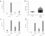
- FIG. 4a The number of spikes was measured two weeks after culture under high density seeding conditions.
- Fig. 4b The potential was measured two weeks after culture under high density seeding conditions.
- Fig. 4c The number of spikes was measured 4 weeks after culture under low density or high density seeding conditions.
- Fig. 4d The number of spikes was measured at 6 weeks after culture under low density or high density seeding conditions.
- FIG. 5 is a view showing photographs of cells taken by light microscopy and cell activities obtained by culturing iPS cell-derived nerve cells iCell Gluta Neurons on oriented polystyrene fiber sheets, and nerve activities measured using an MEA probe.
- FIG. 5 is a view showing photographs of cells taken by light microscopy and cell activities obtained by culturing iPS cell-derived nerve cells iCell Gluta Neurons on oriented polystyrene fiber sheets, and nerve activities measured using an MEA probe.
- FIG. 6 a Optical micrograph (magnification: ⁇ 20) of a cell sheet obtained by seeding at 12 ⁇ 10 5 cells / cm 2 and culturing for 4 weeks.
- FIG. 6b The neuronal activity (spike and synchronous burst) was measured using a MEA probe on cell sheets obtained by seeding at 12 ⁇ 10 5 cells / cm 2 and culturing for 2 weeks.
- FIG. 5 shows the influence of cell scaffolds on drug (4-aminopyridine) responsiveness of nerve cells.
- NFD Cells were seeded on oriented PLGA fiber sheet.
- probe Cells were seeded directly on the MEA probe.
- Fig. 7a The number of spikes was measured 4 weeks after culturing 253G1 hi PS cell-derived neurons.
- FIG. 7b The number of bursts was measured 6 weeks after culture of iPS cell-derived neural cell XCL-1 NEURONS.
- FIG. 7 shows the drug (4-aminopyridine) responsiveness of iPS cell-derived neural cell XCL-1 NEURONS seeded at high density (24 ⁇ 10 5 cells / cm 2 ) on oriented PLGA fiber sheet.
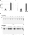
- Fig. 8a shows the results of analysis of the number of spikes on cell sheets obtained by 4 weeks of culture.
- FIG. 8 b shows the results of analysis of the number of synchronized bursts on cell sheets obtained by culturing for 4 weeks.
- 8c shows the results of analysis of the number of spikes and the number of synchronized bursts by raster plot for cell sheets obtained by culturing for 6 weeks. It is a fluorescence-microscope photograph (magnification x10) of the staining image by the calcium indicator reagent (Fluo-8) of the rat cerebral cortical neurons cultured on oriented polystyrene fiber sheet. An example of a position where a change in fluorescence intensity due to a change in calcium ion concentration is observed as blinking is indicated by an arrow in the figure.
- the cell scaffolds used in the neural cell device of the present invention are composed of fibers made of a polymeric material.
- the cell scaffold is preferably a fiber sheet having the form of a sheet of fibers accumulated.
- the fiber sheet can have an oriented structure, a non-oriented structure, or a mixed structure of oriented and non-oriented.
- the oriented structure is a structure in which fibers constituting a fiber sheet are arranged in one direction, and when the angle in one direction is 0 °, fibers of 80% or more exist within a range of ⁇ 30 °.
- the distance between the fibers is not particularly limited, but is preferably 5 to 50 ⁇ m.
- the non-oriented structure is a structure in which the fiber directions are randomly arranged.
- the polymer material constituting the fiber is preferably a biodegradable or non-biodegradable polymer material, for example, PLGA (polylactic acid polyglycolic acid), polystyrene (PS), polysulfone (PSU) and polytetrafluoroethylene Examples include (PTFE), but are not limited thereto.
- the diameter of the cross section of the fiber constituting the fiber sheet is not particularly limited, and is, for example, 0.1 to 8 ⁇ m, preferably 0.5 to 7 ⁇ m, and more preferably 1 to 6 ⁇ m.
- the thickness of the fiber sheet is, for example, 1 to 40 ⁇ m, preferably 5 to 35 ⁇ m, and more preferably 10 to 30 ⁇ m.
- the porosity of the fibers that make up the fiber sheet can vary depending on the polymeric material used.
- the porosity is not particularly limited, and is, for example, 10 to 50%, preferably 15 to 45%, and more preferably 20 to 40%.
- the porosity is the ratio of the area in which no fiber is present to the constant area of the plane of the fiber sheet.
- the fiber sheet can be produced, for example, by an electrospinning method from a solution containing a polymer material.
- a fiber sheet having an oriented structure there is no particular limitation. For example, using a rotating drum, while rotating the drum, a solution containing a polymer material is sprayed from the nozzle onto the rotating surface of the drum A fiber sheet can be manufactured by winding up the fiber formed on the rotating drum.
- the fiber sheet can be produced by spraying a solution containing a polymer material on a flat plate.
- a fiber sheet having a mixed structure of an oriented structure and a non-oriented structure for example, it can be produced by combining the above-mentioned production methods for producing fiber sheets of oriented structure and non-oriented structure.
- PTFE polytetrafluoroethylene
- POREFLON registered trademark of Sumitomo Electric Industries, Ltd., which is commercially available
- the solution of the polymer material may be an organic solvent which dissolves the polymer material to be used at 10 to 30% by weight at room temperature, for example, 1,1,1,3,3,3-hexafluoro-2- Propanol (HFIP), N, N-dimethylformamide (DMF) and the like can be mentioned.
- the fiber sheet can be fixed or held around by a frame.
- a frame When the frame is fixed to or held on a fiber sheet, there is no particular limitation as long as it does not affect cell culture, and for example, commercially available biocompatible adhesives such as silicone one-component condensation type RVT rubber (Shin-Etsu Chemical, catalog number KE-) 45) can be used to bond the frame and the fiber sheet.
- the material of the frame is not particularly limited as long as it does not affect the cell culture. For example, polydimethylsiloxane (PDMS), PS, polycarbonate, stainless steel and the like are exemplified.
- the thickness of the frame is not particularly limited, but is 0.1 to 4 mm, preferably 0.25 to 3 mm, more preferably 0.5 to 2 mm.
- the shape of the frame can be changed according to the purpose of use, and the length ⁇ width is preferably 2 mm ⁇ 2 mm to 15 mm ⁇ 15 mm, respectively, and is a circle or a polygon.
- a fiber sheet or a nerve cell device in which the fiber sheet fixed or held by a frame around the fiber sheet is used as a cell scaffold is used as it is in at least one of the wells included in the cell culture dish or multiwell plate having a plurality of wells. It can be arranged.
- a nerve cell means a neuron composed of a cell body, a dendrite and an axon, and is also called a neuron.
- Neurons can be classified according to differences in neurotransmitters produced by neurons, and as neurotransmitters, non-peptides such as dopamine, noradrenaline, monoamines such as adrenalin and serotonin, acetylcholine, ⁇ -aminobutyric acid, glutamate etc.
- neurotransmitters and also peptide neurotransmitters such as adrenocorticotropic hormone (ACTH), ⁇ -endorphin, ⁇ -endorphin, ⁇ -endorphin, vasopressin and the like.
- ACTH adrenocorticotropic hormone
- ⁇ -endorphin ⁇ -endorphin
- vasopressin vasopressin and the like.
- neurons whose transmitters are dopamine, acetylcholine and glutamate are referred to as dop
- Primary neurons can be used as neurons.
- Primary cultured cells are important as a system for evaluating the effects of drugs and the like in vivo because they retain many cell functions originally possessed in vivo.
- nerve cells of central nervous system and peripheral nervous system of mammals for example, rodents of mice or rats, or monkeys or primates of monkeys can be used.
- methods of dissecting animals, methods of collecting tissues, methods of separating and isolating nerves, culture media for nerve cell culture, culture conditions, etc. depend on the type of cells to be cultured and the purpose of the cells. And can be selected from known methods.
- rat brain neurons from Lonza and human brain neurons from ScienCell Research Laboratories can be used as commercially available primary cultured neurocyte products.
- pluripotent stem cell-derived neural cells include, for example, embryonic stem cells (ES cells) and iPS cells.
- ES cells embryonic stem cells
- iPS cells Different types of neural cells can be obtained by inducing pluripotent stem cells to differentiate using known neural differentiation methods.
- neurons can be obtained by the method for inducing differentiation using the low molecular weight compound described in the literature (Honda et al. Biochemical and biophysical Research Communications 469 (2016) 587-592).
- commercially available pluripotent stem cell-derived neural cell products such as iCell Neuron of Cellular Dynamics International, Inc. and XCL-1 Neurons of XCell Science, Inc. can be used. These commercially available neurons are cultured using the attached medium.
- Neuronal cells can be cultured with astrocytes from mammalian brain.
- the culture solution after culture of astrocytes can be added to a culture solution for neurons at a final concentration of 5 to 30% and cultured.
- the nerve cells suspended in the culture solution are fixed at 1 ⁇ 10 4 cells / cm 2 to 4 ⁇ 10 6 cells / cm 2 , preferably 5 ⁇ 10 4 cells, in a fiber sheet or a fiber sheet whose periphery is fixed or held by a frame.
- the culture solution is exchanged at intervals of 1 to 7 days by seeding at a density of 1 / cm 2 to 3 ⁇ 10 6 cells / cm 2 , more preferably 1 ⁇ 10 5 cells / cm 2 to 2 ⁇ 10 6 cells / cm 2 By culturing for 7 to 14 days, a nerve cell device in which nerve cells form a three-dimensional structure uniformly is obtained.
- the formation of a three-dimensional structure refers to a state where nerve cells adhere along the fibers constituting the fiber sheet and grow on one or both sides of the fiber sheet and in the fiber sheet. Inoculation and culture of nerve cells on a fiber sheet, without using the fiber sheet, with almost no detachment or aggregation of cells in the culture observed when directly seeded on a culture petri dish, in a uniformly spread state The cells are maintained.
- the nerve cell device of the present invention can be brought into contact with a multi-electrode array, and the extracellular potential of nerve cells contained in the nerve cell device can be measured in a 5% CO 2 , 37 ° C. environment.
- a large number of planar microelectrodes are arranged on a substrate in the multielectrode array, and electrical signals from a plurality of cells can be observed simultaneously.
- fluorescent calcium indicators such as calcium sensitive dyes and calcium sensitive fluorescent proteins
- fluorescent potential indicators such as voltage sensitive dyes and voltage sensitive fluorescent proteins
- These indicators can also be used to measure changes in calcium and potential of nerve cells contained in the nerve cell device of the present invention by means of a cell imaging device.
- Fluorescent calcium indicators such as Quin-2, Fura-2, Fluo-3, -4 and -8, Indo-1, Rhod-2 and -3, X-Rhod-1, Cal-520, Calbryte® And calcium sensitive dyes such as CaTM. These are generally used as acetoxymethyl (AM) esters to impart permeability to nerve cells. The acetoxymethyl group is hydrolyzed and released by intracellular esterase. Also, as calcium-sensitive fluorescent proteins, genetically encoded calcium probes such as Camgaroo-1 and -2, GCaMP-2, -3, -5 and -6, CaMPARI and Case 12 are known.
- the fluorescence potential indicator examples include voltage sensitive dyes such as Merocyanine 540, Rh1692, di-4-ANEPPS, JPW-1114, ANNINE-6, Indocyanine Green, Dipicrylamine, FluoVolt (registered trademark) and the like.
- voltage sensitive fluorescent proteins there are known gene encoding membrane potential probes such as VSFP-1 and -2, FlaSh and SPARC based on Green Fluorescent Protein (GFP) (Siegel, MS and Isacoff, EY , Neuron 19, 735-741, 1997; Sakai, R., Repunte-Canonigo, V., et al. Eur. J Neurosci. 13, 2314-2318, 2001; Ataka, K. and Pieribone, VA, Biophys J. 82, 509-516, 2002; Akemann, W. Mutoh, H., et al. Nature Methods, 7, 643-649, 2010).
- Undifferentiated hiPS cells are seeded on culture dishes coated with poly-L-lysine (PLL, Sigma-Aldrich) and laminin 111 (LM, Sigma-Aldrich), 100 nM LDN 193189 (Cellagen Technology) and 1 ⁇ M SB431542 (Sigma) N2B27 nerve medium (A mixture of DMEM / F-12 medium containing N2 supplement (GIBCO, 17502048) and Neurobasal medium containing B27 minus vitamin A supplement (GIBCO, 12587001) in a 1: 1 ratio) The cells were cultured for 7 to 10 days.
- PLL poly-L-lysine
- LM laminin 111
- LM laminin 111
- 100 nM LDN 193189 Cellagen Technology
- SB431542 Sigma
- N2B27 nerve medium A mixture of DMEM / F-12 medium containing N2 supplement (GIBCO, 17502048) and Neurobasal medium containing B27 minus vitamin A supplement (GIBCO
- the neuronal cell mass is dissociated into single cells by Accutase (Innovative Cell Technologies), suspended in Bang-Banker (Japan Genetics) and frozen at -80 ° C for long-term storage It was stored in a -150 ° C ultra low temperature freezer.
- Accutase Innovative Cell Technologies
- Bang-Banker Japan Genetics
- hiPS cell-derived neurons The commercially available hiPS cell-derived neurons XCL-1 NEURONS (XCell Science, Catalog No. XN-001-1V) thaw the frozen cells according to the protocol of XCell Science A neural cell suspension was obtained by
- the iPS cell-derived neurons suspended in N2B27 culture solution with the fiber sheet prepared in Example 1 as a cell scaffold are oriented at a density of 1.3 ⁇ 10 6 cells / cm 2
- the cells were seeded on porous PLGA, random PLGA and random PSU fiber sheets and on PTFE sheets (Sumitomo Electric Co., Ltd.), and cultured for 6 days in an incubator at 37 ° C., 5% CO 2 .
- the results of observation of the obtained cell sheet (neural cell device) with a light microscope are shown in FIG. 1a (using oriented PLGA fiber sheet) and FIG. 1b (using random PSU fiber sheet).
- iPS cell-derived neurons are similarly prepared on culture dishes (Nunc® Cell-Culture Treated Multidishes) or commonly used electrodes (Alpha Med Scientific Inc. / MED probe 16 / MED-RG515A)
- the cells were seeded and cultured for 1 to 2 days.
- the results of observing the obtained cells with a light microscope are shown in FIG. 2a (on culture dish) and FIG. 2b (on electrode).
- FIG. 2a on culture dish
- FIG. 2b on electrode
- iPS cell-derived neurons were seeded, and after 5 weeks, the cells were treated and fixed with 4% paraformaldehyde (Nacalai Tesque) for 15-30 minutes at room temperature. After washing with PBS, it was treated with 0.2% Triton X-100 (Sigma) at room temperature. After 5 minutes, it was washed with PBS and blocked with 1% bovine serum albumin for 1 hour at room temperature. It was washed with PBS and treated with anti-MAP2 antibody (Abcom). After 1 hour, the cells were washed with PBS and treated with Alexa Fluor 546-anti-rabbit IgG antibody (thermofisher) for 1 hour. After washing with PBS, they were observed with a fluorescence microscope (Keyence) (FIG.
- FIG. 1c shows that the neurons are oriented.
- FIG. 1 d shows the results of measurement of neuronal spikes and burst numbers by MEA after 4 weeks of seeding on random PLGA fiber sheets and oriented PLGA fiber sheets. It can be seen from FIG. 1 d that the oriented fiber sheet is effective in increasing the number of spikes and bursts.
- N2B27 culture medium (culture medium A) containing three different culture mediums, ie 20% Astrocyte Conditioned Medium (ACM) (Astrocyte Conditioned Medium-Serum Free (ACM-sf) / 11811-sf, ScienCell Research Laboratories)
- ACM Astrocyte Conditioned Medium
- ACM-sf Astrocyte Conditioned Medium-Serum Free (ACM-sf) / 11811-sf, ScienCell Research Laboratories)
- Neurobasal® medium (Culture B) containing CultureOne® Supplement (Gibco) and B27 minus vitamin A supplement (BrainPhys® Neuronal Medium (05792, STEMCELL TECH NOLOGIES) culture medium containing 20% ACM)
- C seeding 253G1 iPS cell-derived neurons and XCL-1 NEURONS (XCell Science, catalog number XN-001-1V) at a density of 1.2 ⁇ 10 6 cells / cm 2 on
- oriented PLGA fibers coated with 0.002% poly-L-lysine and 20 ⁇ g / ml laminin As a cell scaffold, in the case of 253G1 iPS cell-derived neurons, oriented PLGA fibers coated with 0.002% poly-L-lysine and 20 ⁇ g / ml laminin, and in the case of XCL-1 NEURONS, 0.002% poly-D- An oriented PLGA fiber coated with lysine and 10 ⁇ g / ml laminin was used.
- the seeded cells were cultured in an incubator at 37 ° C., 5% CO 2 for 1 to 3 weeks.
- the results of observation of the obtained cell sheet (neural cell device) with a light microscope are shown in FIGS. 3a, b and c.
- a probe is treated with a 10% bovine serum-containing culture solution, and then 0.003% poly-L-lysine (Sigma-Aldrich) and 20 ⁇ g / ml for 253G1 iPS cell-derived neurons Seed neurons on probes (electrodes) coated with laminin (Sigma-Aldrich), and in the case of XCL-1 NEURONS, probes (electrodes) coated with 0.002% poly-D-lysine and 10 ⁇ g / ml laminin And cultured for 1 to 3 weeks.
- the results of observation of the obtained cell sheet with a light microscope are shown in FIGS. 3 d, e and f.
- the seeding density of cells was 1.3 ⁇ 10 6 cells / cm 2 (FIG. 3 d) or 3 ⁇ 10 5 cells / cm 2 (FIGS. 3 e and f).
- the culture solution A when the culture solution A was used, it was found that nerve cells were peeled off from the electrode, and the peeled nerve cells formed cell clusters.
- culture solution B when culture solution B was used in the control group, the cells formed small colonies, and in culture solution C, peeling of the cells was observed at the end of the area where the cells were seeded.
- culture solution C peeling of the cells was observed at the end of the area where the cells were seeded.
- the cell sheet was maintained uniformly regardless of the type of culture solution (FIG. 3).
- iPS cell-derived neurons XCL-1 NEURONS (XCell Science, Catalog No. XN-001-1V), as a cell scaffold, on oriented PLGA fiber sheet, 3 ⁇ 10 5 cells / cm 2 (low density seeding conditions) or Seed at 12 ⁇ 10 5 cells / cm 2 (high density seeding conditions) and use 20% ACM, with or without BrainPhys® Neuronal Medium (05792, STEMCELL TECH NOLOGIES) 2, 4 and The cells were cultured in an incubator at 37 ° C., 5% CO 2 for 6 weeks.
- This fiber was mounted on a multi-electrode array (MEA) probe (MED64 system, Alpha Med Scientific), the cells were brought into contact with the electrodes of the MEA probe, and neural activity (spike and burst) was measured.
- neurons are seeded at 3 ⁇ 10 5 cells / cm 2 directly on the MEA probe and cultured in an incubator at 37 ° C., 5% CO 2 for 2 , 4 and 6 weeks, as well as nerve activity It was measured.
- the results are shown in FIG.
- spiked cells were observed more frequently as early as two weeks after culture in cells seeded at high density on oriented PLGA fiber sheets. was shown ( Figure 4a).
- FIG. 4 b This phenomenon is particularly remarkable in neurons on fiber sheets cultured in a culture solution containing ACM, and an increase in average potential was also observed (FIG. 4 b). Furthermore, at 4 weeks after cell seeding, spikes and bursts could be confirmed more frequently in neurons on fiber sheets seeded at low density as compared to the conventional method (FIG. 4c). More frequent spikes and bursts could be observed in the 6-week neural cells (FIG. 4d).
- rat cerebral cortical neurons As neurons, primary cultured neurons, rat cerebral cortical neurons (ThermoFisher, product number A10840) are used, and on an oriented PLGA fiber sheet as a cell scaffold, 1.0 ⁇ 10 5 cells / cm 2 , 3.0 ⁇ 10 10 The cells were seeded at a cell density of 5 cells / cm 2 or 9.0 ⁇ 10 5 cells / cm 2 . The cells are 5% CO 2 with Neurobasal® Medium (GIBCO, catalog number 21103-049) including 1 ⁇ B-27® Supplement / serum free (GIBCO, catalog number 17504-044). The cells were cultured in an incubator at 37 ° C. for 1, 2 and 4 weeks.
- the obtained cell sheet (neural cell device) is placed on an MEA probe (MED 64 system, Alpha Med Scientific Co., Ltd.), the cells are brought into contact with the electrodes of the MEA probe, and nerve activity (spike and burst) is measured. did.
- FIG. 5 shows the results of measurement of extracellular potential by MEA for a cell sheet (neural cell device) obtained by seeding at 9.0 ⁇ 10 5 cells / cm 2 and culturing for 4 weeks. At each electrode, spikes and synchronous bursts were observed.
- iPS cell-derived neurons iCell registered trademark
- Gluta Neurons Glutamate-sensitive neurons, Cellular Dynamics International, product number GNC-301-030-000.5
- the cells were seeded at a cell density of 8 ⁇ 10 5 cells / cm 2 , 12 ⁇ 10 5 cells / cm 2 or 16 ⁇ 10 5 cells / cm 2 on oriented polystyrene fiber sheets held in a circular frame of The cells were cultured for 1, 2 and 4 weeks in an incubator at 37 ° C., 5% CO 2 , using an iCell glutamine neuronal medium (Cellular Dynamics International) with or without 20% ACM.
- iCell glutamine neuronal medium Cellular Dynamics International
- FIG. 6a is a photograph of a cell sheet (neuronal cell device) obtained by seeding at 12 ⁇ 10 5 cells / cm 2 and culturing for 4 weeks in the ACM additive-free iCell glutamine nerve cell culture medium by light microscopy. It is. FIG. 6a also shows that nerve cells adhere well to the polystyrene fiber sheet, and even if the culture is continued for 4 weeks, a uniform cell sheet without cell detachment is maintained. This was the same even when using a medium supplemented with 20% ACM.
- a cell sheet obtained by seeding at 12 ⁇ 10 5 cells / cm 2 and culturing for 2 weeks in the iCell glutamine nerve cell culture medium supplemented with 20% ACM was subjected to MEA probe (MED 64 system, The cells were placed on Alpha Med Scientific Co., Ltd., and the cells were brought into contact with the electrodes of the MEA probe, and neural activity (spike and synchronous burst) was measured. Detection of spikes and sync bursts was done by raster plot using a MED64 Burstscope (Alpha Med Scientific). The results are shown in FIG. 6b. Even after two weeks of culture, synchronized bursts can be confirmed.
- MEA probe MED 64 system
- 253G1 hi PS cell-derived neurons are N2B27 culture medium or N2B27 culture medium containing 20% ACM, and XCL-1 NEURONS contains BrainPhys® Neuronal Medium (05792, STEMCELL TECHNOLOGIES) culture medium or 20% ACM
- the cells were cultured for 4 or 6 weeks in an incubator at 37 ° C., 5% CO 2 , using BrainPhys® Neuronal Medium.
- the fiber sheet was placed on an MEA probe (MED 64 system, Alpha Med Scientific Co., Ltd.), the cells were brought into contact with the electrodes of the multi-electrode array probe, and nerve activity (spike) was measured.
- MEA probe MED 64 system, Alpha Med Scientific Co., Ltd.
- neurons were similarly directly seeded onto a conventional based probe (electrode) and cultured in an incubator at 37 ° C., 5% CO 2 for 4 or 6 weeks. The obtained results were compared with the conventional method (direct seeding to the probe).
- Four weeks after culture, more spikes were observed in 253G1 hi PS cell-derived neurons on fiber sheets ( Figure 7a).
- the spike number increase rate in neurons cultured on the fiber sheet of the present invention is 6 times and 9 times in the fiber sheet group cultured in a 20% ACM-added culture medium as compared to neurons on the probe. The number of spikes was significantly increased by using a fiber sheet.
- 4-aminopyridine which has an action to inhibit various potassium channels in nerve cells and is known to sustain nerve action potential
- the resulting neurons were added to a final concentration of 100 ⁇ M and spikes were measured.
- spikes were observed in both the control group and the fiber sheet group, and appropriate responsiveness to the drug was also confirmed (FIG. 7a).
- the growth rate of spikes is 167% in the fiber sheet group of the present invention, compared with 156% in the control group cultured on the probe, and further in the fiber sheet group cultured with the 20% ACM-added culture solution The rate of increase was 194%.
- XCL-1 NEURONS which was seeded at a density of 12 x 10 5 cells / cm 2 on a fiber sheet and cultured for 6 weeks in an ACM-containing culture medium, was treated with 4-AP at a final concentration of 100 ⁇ M. It could be doubled (Figure 7b).
- the obtained cell sheet (neural cell device) was placed on an MEA probe (MED64 system, Alpha Med Scientific Co., Ltd.), and the cells were brought into contact with the electrodes of the MEA probe to measure neural activity (spike and burst).
- MEA probe MED64 system, Alpha Med Scientific Co., Ltd.
- 4-aminopyridine (4-AP) which is known to sustain nerve action potential
- MED64 Burstscope (Alpha Med Scientific) was used for analysis of neural activity.
- FIG. 8a and FIG. 8b The results of analysis of the number of spikes and the results of analysis of the number of synchronous bursts are shown in FIG. 8a and FIG. 8b, respectively, for the cell sheet obtained by culturing for 4 weeks.
- the 4-AP treatment increased the number of spikes by 499% and the number of synchronization bursts by 375%. Furthermore, the result of having analyzed neural activity by the raster plot about the cell sheet obtained by culture
- Rat cerebral cortical neurons (ThermoFisher, product number A10840), which are primary cultured neurons, were placed on an oriented polystyrene fiber sheet, which is a cell scaffold, at 1.0 ⁇ 10 5 cells / cm 2 and 3.0 ⁇ 10 5 cells / cm 2. Alternatively, cells were seeded at a cell density of 9.0 ⁇ 10 5 cells / cm 2 .
- Cells are 5% using Neurobasal® Medium (GIBCO, catalog number 21103-049) including B-27® Supplement ( ⁇ 50) / serum free (GIBCO, catalog number 17504-044) and cultured for 4 weeks in CO 2, 37 ° C. incubator in.
- the obtained cell sheet (neural cell device) is transferred into a culture solution containing 5 ⁇ M of calcium indicator reagent Fluo-8-AM (AAT Bioquest, catalog number 21081), and incubated in an incubator at 5% CO 2 at 37 ° C. Incubated for 1 hour.
- the cell sheet loaded with Fluo-8 was allowed to stand on a heat retention plate attached to a fluorescence microscope (Olympus IX 73), and the fluorescence of Fluo-8 was observed.
- FIG. 9 shows an observation image by a fluorescence microscope (magnification: 10). It is shown that, in the nerve cell device of the present invention, it is possible to visualize (image) fluctuations in intracellular calcium concentration of nerve cells.
Abstract
The present invention provides a nerve cell device in which early observation of nerve activity (spikes, bursts, and the like) is made possible and the measured electric strength is increased by cultivating nerve cells upon a cell scaffold. By using this nerve cell device, imaging of intracellular signaling is also possible.
Description
本発明は、神経活動を測定するための新規な神経細胞デバイスに関する。
The present invention relates to a novel neural cell device for measuring neural activity.
医薬品開発における安全性評価において、神経毒性は、心毒性や肝毒性と並び、医薬品開発が中止される主な原因一つである。そのため、医薬品開発の初期段階から的確な神経毒性評価が必要であるが、非臨床試験における神経毒性の評価は、動物実験による症状観察や脳の病理組織評価などのインビボでの評価系が主体であり、より簡便なインビトロで神経毒性を評価する系は確立されていない。神経活動は、複数の神経細胞が相互に作用することで発現する電気活動に基づく。近年インビトロ試験系として、単離神経細胞または多能性幹細胞から誘導した神経細胞を、多電極アレイ(microelectrode array、以下MEAと略す)上に分散培養させることにより神経回路網を再構成し、細胞外電位を測定することにより神経活動を観察する手法が開発され(非特許文献1)、この手法により神経毒性を評価する検討が報告されている(非特許文献2および3)。神経細胞の活動を観察する手法としては、神経細胞の膜電位や細胞内カルシウムなどの細胞内シグナルを測定し、可視化(イメージング)する方法も用いられている。膜電位の変化や細胞内カルシウム濃度の変動は、神経細胞内に取り込ませた電位感受性色素やカルシウム感受性色素で測定することができることが報告されている;また神経細胞で発現させた電位感受性蛍光タンパク質やカルシウム感受性蛍光タンパク質で測定することも報告されている(非特許文献4~6)。
Neurotoxicity is one of the main causes of drug development discontinuation along with cardiotoxicity and hepatotoxicity in safety evaluation in drug development. Therefore, appropriate neurotoxicity evaluation is necessary from the early stage of drug development, but in the non-clinical evaluation of neurotoxicity, in vivo evaluation systems such as symptom observation by animal experiments and brain pathological tissue evaluation are mainly used. A more convenient and in vitro system for evaluating neurotoxicity has not been established. The neural activity is based on the electrical activity which is expressed by the interaction of a plurality of neurons. In recent years, as an in vitro test system, neural networks are reconstructed by dispersing and culturing isolated neural cells or neural cells derived from pluripotent stem cells on a multielectrode array (hereinafter abbreviated as MEA), thereby reconstituting the cell network A technique for observing neural activity by measuring an external potential has been developed (Non-patent Document 1), and studies to evaluate neurotoxicity using this technique have been reported (Non-patent Documents 2 and 3). As a method of observing the activity of nerve cells, a method of measuring and visualizing (imaging) intracellular signals such as membrane potentials and intracellular calcium of nerve cells is also used. It has been reported that changes in membrane potential and changes in intracellular calcium concentration can be measured with voltage sensitive dyes and calcium sensitive dyes incorporated into nerve cells; and voltage sensitive fluorescent proteins expressed in nerve cells Measurement with calcium-sensitive fluorescent proteins has also been reported (Non-patent documents 4 to 6).
神経細胞の培養は一般に困難とされているが、神経細胞が接着する足場を提供して、神経細胞の増殖を促進する様々な手段が報告されている。例えば、多孔性の3次元ハイドロゲルに、ポリカプロラクトンまたはゼラチンと混合したポリカプロラクトンから成るマイクロファイバーを整列して埋め込んだ足場に神経細胞を播種することにより、神経細胞の増殖が促進されることが報告されている(非特許文献7)。また、医療用材料などへの応用を目的として、細胞の足場として用いる足場材料に関する報告がいくつかある。例えば、ポリオレフィン、ポリアミド、ポリウレタン、ポリエステル、フッ素系高分子、ポリ乳酸、ポリビニルアルコールなどで構成されるナノファイバー、またはタンパク質成分を吸着させた該ナノファイバーにより構成される細胞足場材料を使用し、細胞培養または組織再生を有効に行う(特許文献1);中空糸膜メッシュとナノファイバー層とを有する細胞足場材料を使用する3次元細胞培養により、培養細胞への栄養や酸素の供給および培養細胞からの代謝老廃物の除去を高い効率で行う(特許文献2);ゼラチン、コラーゲンもしくはセルロースを含有するナノファイバー、または架橋された該ナノファイバーにより構成される細胞足場材料を使用し、多能性幹細胞の大量供給を行う、および細胞死を抑制する(特許文献3);ポリグリコール酸を支持体として用い、その上にポリグリコール酸やゼラチンなどからなるナノファイバーを塗布した細胞足場材料を使用し、ヒト多能性幹細胞の増殖率を向上させる(特許文献4)などが報告されている。しかしながら、MEAによる細胞外電位測定において、神経細胞の培養開始後早期に電気信号を検出する手段、および増強された電気信号を得る手段については報告されていない。さらに、細胞内シグナルの変化を、神経細胞の培養開始後早期に個々の細胞について検出する手段についても報告されていない。
Although culture of nerve cells is generally considered to be difficult, various means have been reported to provide a scaffold to which nerve cells adhere and to promote proliferation of nerve cells. For example, the proliferation of neurons can be promoted by seeding neurons with a scaffold in which microfibers composed of polycaprolactone or polycaprolactone mixed with gelatin are arrayed and embedded in a porous three-dimensional hydrogel. It is reported (nonpatent literature 7). In addition, there are several reports on scaffold materials used as cell scaffolds for the purpose of application to medical materials and the like. For example, a cell scaffold material composed of nanofibers composed of polyolefin, polyamide, polyurethane, polyester, fluorine-based polymer, polylactic acid, polyvinyl alcohol or the like, or the nanofibers to which a protein component is adsorbed, Effective culture or tissue regeneration (Patent Document 1); supply of nutrients and oxygen to cultured cells and culture of cultured cells by three-dimensional cell culture using a cell scaffold material having a hollow fiber membrane mesh and a nanofiber layer For efficient removal of metabolic waste products (Patent Document 2); pluripotent stem cells using a cell scaffold material composed of nanofibers containing gelatin, collagen or cellulose, or the crosslinked nanofibers Supply large amounts of water, and suppress cell death (Patent Document 3); Using a cell scaffold material coated with nanofibers consisting of polyglycolic acid, gelatin, etc. using glycolic acid as a support to improve the proliferation rate of human pluripotent stem cells (Patent Document 4), etc. It is done. However, in the measurement of extracellular potential by MEA, a means for detecting an electrical signal early after the start of culture of neural cells and a means for obtaining an enhanced electrical signal have not been reported. Furthermore, there has been no report on means for detecting changes in intracellular signals for individual cells early after the start of neural cell culture.
MEAを用いて神経活動を評価する場合、様々な問題点が存在する。例えば、測定プローブ上へ神経細胞を直接播種するために、神経活動測定に使用できるまでには5~6週間以上の長期培養が必要とされている;従来の測定プローブ等への直接播種では、細胞の剥離や凝集等が生じ、細胞維持の点において均一性が失われる等の、神経活動を評価する上で不安定な状況が起こりやすい;長期間の細胞培養に伴い、培養中に好ましくない事態(細胞の剥離、凝集、コンタミネーション等)が生じる可能性が高まり、かかる事態が生じた場合には、それまでに細胞培養に要した費用および時間が無駄になる;長期間の細胞培養には、高額の維持費用が必要となる;現状の細胞培養形態では、直ちに神経活動観察に使用できる状態での供給が不可能である等が挙げられる。神経細胞の長期培養に伴う問題は、細胞内シグナルイメージング法により神経活動を評価する場合にも生じる。
本発明は、上記の従来技術の問題点を克服することを目的とする。 There are various problems when evaluating neural activity using MEA. For example, long-term culture of 5 to 6 weeks or more is required to be able to use for neural activity measurement in order to directly seed neural cells on the measurement probe; direct seeding on conventional measurement probes etc. Unstable conditions are likely to occur when evaluating neural activity, such as cell detachment and aggregation, etc., resulting in loss of uniformity in terms of cell maintenance, etc .; along with long-term cell culture, undesirable during culture There is an increased possibility of occurrence (such as cell detachment, aggregation, contamination, etc.), and in the case of such occurrence, the cost and time required for cell culture are wasted; for long-term cell culture In the current cell culture form, it is impossible to supply in a state ready for use in observing neural activity. Problems with long-term culture of neurons also occur when evaluating neuronal activity by intracellular signal imaging.
The present invention aims to overcome the above-mentioned problems of the prior art.
本発明者らは、上記の従来技術の問題点を克服することを目的とし、鋭意検討した結果、細胞足場上で神経細胞を培養する神経細胞デバイスを提供できることを見出し、本発明を完成するに至った。
The present inventors have found that, as a result of intensive investigations aimed at overcoming the problems of the prior art described above, it is possible to provide a neural cell device for culturing neural cells on a cell scaffold, and complete the present invention. It reached.
すなわち、本発明の目的は、以下の発明により達成される。
(1)細胞足場と神経細胞を含む神経細胞デバイス。
(2)神経細胞が配向している、(1)に記載の神経デバイス。
(3)細胞足場が高分子材料で形成されたファイバーシートである、(1)に記載の神経細胞デバイス。
(4)ファイバーシートが、配向性構造、非配向性構造または配向性と非配向性との混合構造を有する、(3)に記載の神経細胞デバイス。
(5)ファイバーシートが、ポリリジン、ポリオルニチン、ラミニン、フィブロネクチン、マトリゲル(登録商標)およびゲルトレックス(登録商標)から選ばれる細胞外マトリックスタンパク質でコーティングされた、(3)に記載の神経細胞デバイス。
(6)神経細胞が、細胞足場上および/または細胞足場内で3次元構造を形成した、(1)~(5)のいずれかに記載の神経細胞デバイス。
(7)神経細胞が、初代培養細胞または多能性幹細胞由来の神経細胞である、(1)~(6)のいずれかに記載の神経細胞デバイス。
(8)前記初代培養細胞または多能性幹細胞由来の神経細胞が、哺乳類由来の神経細胞である、(7)に記載の神経細胞デバイス。
(9)神経細胞が、グルタミン酸作動性、ドーパミン作動性、γ-アミノ酪酸作動性、モノアミン作動性、ヒスタミン作動性またはコリン作動性の神経細胞を含む、(1)~(8)のいずれかに記載の神経細胞デバイス。
(10)神経細胞を、細胞足場に対して1×10⁴細胞/cm2~4×106細胞/cm2の密度で播種した、(1)~(9)のいずれかに記載の神経細胞デバイス。
(11)神経細胞デバイスの周囲を保持するフレームをさらに有する、(1)~(10)のいずれかに記載の神経細胞デバイス。
(12)前記フレームの縦長×横長が、それぞれ2 mm×2 mm~15 mm×15 mmであって、前記フレームが円形または多角形である、(11)に記載の神経細胞デバイス。
(13)(1)~(12)のいずれかに記載の神経細胞デバイスを用いる、神経活動の評価方法。
(14)(1)~(12)のいずれかに記載の神経細胞デバイスを、多電極アレイと接触させ、該神経細胞デバイスに含まれる神経細胞の細胞外電位を測定することを含む、神経活動の評価方法。
(15)細胞内シグナルイメージング物質を用いる神経活動の評価方法であって、(1)~(12)のいずれかに記載の神経細胞デバイスを用いる方法。
(16)前記細胞内シグナルイメージング物質が、蛍光カルシウムインジケーターまたは蛍光電位インジケーターである、(15)に記載の方法。
(17)(1)~(12)のいずれかに記載の神経細胞デバイスを有する、神経細胞デバイス装着ディッシュ。
(18)複数のウェルを有するマルチウェルプレートにおいて、該プレートに含まれるウェルの少なくとも一つに、(1)~(12)のいずれか1項に記載の神経細胞デバイスを有する、神経細胞デバイス装着プレート。 That is, the object of the present invention is achieved by the following invention.
(1) A nerve cell device including a cell scaffold and a nerve cell.
(2) The nerve device according to (1), wherein the nerve cell is oriented.
(3) The neural cell device according to (1), wherein the cell scaffold is a fiber sheet formed of a polymeric material.
(4) The nerve cell device according to (3), wherein the fiber sheet has an oriented structure, a non-oriented structure or a mixed structure of oriented and non-oriented.
(5) The nerve cell device according to (3), wherein the fiber sheet is coated with an extracellular matrix protein selected from polylysine, polyornithine, laminin, fibronectin, Matrigel (registered trademark) and Geltrex (registered trademark).
(6) The neuronal cell device according to any one of (1) to (5), wherein the neuronal cells form a three-dimensional structure on and / or in the cell scaffold.
(7) The neural cell device according to any one of (1) to (6), wherein the neural cell is a primary culture cell or a neural cell derived from pluripotent stem cells.
(8) The neural cell device according to (7), wherein the primary culture cells or neural cells derived from pluripotent stem cells are mammalian-derived neural cells.
(9) In any one of (1) to (8), the nerve cells include glutamatergic, dopaminergic, γ-aminobutyric acid, monoaminergic, histaminergic or cholinergic neurons. The nerve cell device of description.
(10) The neuronal cell according to any one of (1) to (9), wherein the neuronal cell is seeded at a density of 1 × 10 4 cells / cm 2 to 4 × 10 6 cells / cm 2 to the cell scaffold. device.
(11) The neural cell device according to any one of (1) to (10), further including a frame that holds the periphery of the neural cell device.
(12) The neural cell device according to (11), wherein the longitudinal length × lateral length of the frame is 2 mm × 2 mm to 15 mm × 15 mm, respectively, and the frame is circular or polygonal.
(13) A method of evaluating neural activity using the neural cell device according to any one of (1) to (12).
(14) A nerve activity, comprising contacting the nerve cell device according to any one of (1) to (12) with a multi-electrode array and measuring the extracellular potential of the nerve cell contained in the nerve cell device Evaluation method of
(15) A method for evaluating neural activity using an intracellular signal imaging substance, wherein the neural cell device according to any one of (1) to (12) is used.
(16) The method according to (15), wherein the intracellular signal imaging substance is a fluorescent calcium indicator or a fluorescent potential indicator.
(17) A neuronal cell device-equipped dish comprising the neuronal cell device according to any one of (1) to (12).
(18) In a multi-well plate having a plurality of wells, a neural cell device mounted with the neural cell device according to any one of (1) to (12) in at least one of the wells contained in the plate plate.
本発明によれば、細胞足場を用いて神経細胞を培養することにより、細胞接着性が向上し、培養中の細胞剥離の低減等を含む安定した培養が可能となる。その結果、本発明の神経細胞デバイスを用いることにより、多電極アレイの電極から、播種した神経細胞が離脱することがなく、スパイク、バースト等の神経活動の測定が可能になるまでの期間が短縮される。さらに、神経細胞の凝集が抑制されるため、高頻度のスパイクを観察することができる。本発明の神経細胞デバイスでは、細胞内シグナルイメージングも可能である。本発明の神経細胞デバイスは、神経毒性評価や神経疾患に対する薬物スクリーニング等に有用である。
According to the present invention, by culturing nerve cells using a cell scaffold, cell adhesion is improved, and stable culture including reduction of cell detachment during culture and the like becomes possible. As a result, by using the neural cell device of the present invention, it is possible to prevent the disseminated neural cells from being detached from the electrodes of the multi-electrode array, and to shorten the time period until measurement of neural activity such as spikes and bursts becomes possible. Be done. In addition, since neuronal cell aggregation is suppressed, frequent spikes can be observed. Intracellular signal imaging is also possible with the neural cell device of the present invention. The neural cell device of the present invention is useful for neurotoxicity evaluation, drug screening for neurological diseases and the like.
本発明の神経細胞デバイスで用いられる細胞足場は、高分子材料で生成されたファイバーで構成される。細胞足場は、好ましくは、ファイバーを集積したシートの形状を有するファイバーシートである。該ファイバーシートは配向性構造、非配向性構造または配向性と非配向性との混合構造を有することができる。配向性構造とは、ファイバーシートを構成するファイバーが一方向に配置され、該一方向の角度を0°とした場合、80%以上のファイバーが±30°の範囲内に存在する構造である。配向性構造において、ファイバー間の距離は特に限定されないが、5~50μmであるのが好ましい。非配向性構造とは、ファイバーの方向がランダムに配置された構造である。ファイバーを構成する高分子材料としては、生分解性または非生分解性の高分子材料が好ましく、例えば、PLGA (ポリ乳酸ポリグリコール酸)、ポリスチレン(PS)、ポリスルホン(PSU)およびポリテトラフルオロエチレン(PTFE)が挙げられるが、これらに限定されない。ファイバーシートを構成するファイバーの断面の直径は、特に限定されないが、例えば0.1~8μmであり、好ましくは0.5~7μmであり、より好ましくは1~6μmである。ファイバーシートの厚さは、例えば1~40μmであり、好ましくは5~35μmであり、より好ましくは10~30μmである。ファイバーシートを構成するファイバーの空隙率は、用いる高分子材料によって変動し得る。該空隙率は特に限定されないが、例えば10~50%であり、好ましくは15~45%であり、より好ましくは20~40%である。ここで空隙率とは、ファイバーシート平面の一定面積に対する、ファイバーが存在していない面積の比率のことである。
The cell scaffolds used in the neural cell device of the present invention are composed of fibers made of a polymeric material. The cell scaffold is preferably a fiber sheet having the form of a sheet of fibers accumulated. The fiber sheet can have an oriented structure, a non-oriented structure, or a mixed structure of oriented and non-oriented. The oriented structure is a structure in which fibers constituting a fiber sheet are arranged in one direction, and when the angle in one direction is 0 °, fibers of 80% or more exist within a range of ± 30 °. In the oriented structure, the distance between the fibers is not particularly limited, but is preferably 5 to 50 μm. The non-oriented structure is a structure in which the fiber directions are randomly arranged. The polymer material constituting the fiber is preferably a biodegradable or non-biodegradable polymer material, for example, PLGA (polylactic acid polyglycolic acid), polystyrene (PS), polysulfone (PSU) and polytetrafluoroethylene Examples include (PTFE), but are not limited thereto. The diameter of the cross section of the fiber constituting the fiber sheet is not particularly limited, and is, for example, 0.1 to 8 μm, preferably 0.5 to 7 μm, and more preferably 1 to 6 μm. The thickness of the fiber sheet is, for example, 1 to 40 μm, preferably 5 to 35 μm, and more preferably 10 to 30 μm. The porosity of the fibers that make up the fiber sheet can vary depending on the polymeric material used. The porosity is not particularly limited, and is, for example, 10 to 50%, preferably 15 to 45%, and more preferably 20 to 40%. Here, the porosity is the ratio of the area in which no fiber is present to the constant area of the plane of the fiber sheet.
ファイバーシートは、例えば、高分子材料を含む溶液からエレクトロスピニング法によって製造することができる。配向性構造を有するファイバーシートを製造する場合は、特に限定されないが、例えば回転ドラムを用い、該ドラムを回転させながら、ノズルから該ドラムの回転面に対して高分子材料を含む溶液を噴霧し、回転ドラム上で形成されたファイバーを巻き取ることにより、ファイバーシートを製造することができる。非配向性構造を有するファイバーシートを製造する場合は、高分子材料を含む溶液を平坦なプレート上に噴霧することにより、ファイバーシートを製造することができる。配向性構造と非配向性構造との混合構造を有するファイバーシートを製造する場合は、例えば、配向性構造および非配向性構造のファイバーシートを製造する上記の製造方法を組み合わせて製造することができる。
ポリテトラフルオロエチレン(PTFE)シートは、例えば、市販されている住友電工株式会社のポアフロン(登録商標)を使用することができる。
高分子材料の溶液としては、使用する高分子材料を、室温で10~30重量%で溶解する有機溶媒であればよく、例えば1,1,1,3,3,3-ヘキサフルオロ-2-プロパノール(HFIP)、N,N-ジメチルホルムアミド(DMF)などが挙げられる。 The fiber sheet can be produced, for example, by an electrospinning method from a solution containing a polymer material. When producing a fiber sheet having an oriented structure, there is no particular limitation. For example, using a rotating drum, while rotating the drum, a solution containing a polymer material is sprayed from the nozzle onto the rotating surface of the drum A fiber sheet can be manufactured by winding up the fiber formed on the rotating drum. When producing a fiber sheet having a non-oriented structure, the fiber sheet can be produced by spraying a solution containing a polymer material on a flat plate. In the case of producing a fiber sheet having a mixed structure of an oriented structure and a non-oriented structure, for example, it can be produced by combining the above-mentioned production methods for producing fiber sheets of oriented structure and non-oriented structure. .
As the polytetrafluoroethylene (PTFE) sheet, for example, POREFLON (registered trademark) of Sumitomo Electric Industries, Ltd., which is commercially available, can be used.
The solution of the polymer material may be an organic solvent which dissolves the polymer material to be used at 10 to 30% by weight at room temperature, for example, 1,1,1,3,3,3-hexafluoro-2- Propanol (HFIP), N, N-dimethylformamide (DMF) and the like can be mentioned.
ファイバーシートは、周囲をフレームで固定または保持することができる。フレームをファイバーシートに固定または保持する場合は、細胞培養に影響を及ぼさなければ特に限定されないが、例えば市販の生体適合性粘着剤、例えばシリコーン一液縮合型RVTゴム(信越化学、カタログ番号KE-45)を用いて、フレームとファイバーシートとを接着することができる。
フレームの素材は、細胞培養に影響を及ぼさなければ特に限定されない。例えば、ポリジメチルシロキサン(PDMS)、PS、ポリカーボネート、ステンレス等が例示される。フレームの厚さは、特に限定されないが、0.1~4 mm、好ましくは0.25~3 mm、より好ましくは0.5~2 mmである。
フレームの形状は、使用目的によって変えることができ、縦長×横長が、それぞれ2 mm×2 mm~15 mm×15 mmが好ましく、円形または多角形である。 The fiber sheet can be fixed or held around by a frame. When the frame is fixed to or held on a fiber sheet, there is no particular limitation as long as it does not affect cell culture, and for example, commercially available biocompatible adhesives such as silicone one-component condensation type RVT rubber (Shin-Etsu Chemical, catalog number KE-) 45) can be used to bond the frame and the fiber sheet.
The material of the frame is not particularly limited as long as it does not affect the cell culture. For example, polydimethylsiloxane (PDMS), PS, polycarbonate, stainless steel and the like are exemplified. The thickness of the frame is not particularly limited, but is 0.1 to 4 mm, preferably 0.25 to 3 mm, more preferably 0.5 to 2 mm.
The shape of the frame can be changed according to the purpose of use, and the length × width is preferably 2 mm × 2 mm to 15 mm × 15 mm, respectively, and is a circle or a polygon.
ファイバーシート、またはファイバーシートの周囲をフレームで固定もしくは保持した該ファイバーシートを細胞足場とする神経細胞デバイスは、細胞培養ディッシュまたは複数のウェルを有するマルチウェルプレートに含まれるウェルの少なくとも一つにそのまま配置することができる。
A fiber sheet or a nerve cell device in which the fiber sheet fixed or held by a frame around the fiber sheet is used as a cell scaffold is used as it is in at least one of the wells included in the cell culture dish or multiwell plate having a plurality of wells. It can be arranged.
本明細書において、神経細胞とは、細胞体、樹状突起及び軸索から構成される神経単位を意味し、ニューロンとも呼ばれる。神経細胞は、神経細胞が産生する神経伝達物質の違いにより分類することができ、神経伝達物質としては、ドーパミン、ノルアドレナリン、アドレナリンおよびセロトニンなどのモノアミン、アセチルコリン、γ-アミノ酪酸、グルタミン酸等の非ペプチド性神経伝達物質、また、副腎皮質刺激ホルモン(ACTH)、α-エンドルフィン、β-エンドルフィン、γ-エンドルフィン、バソプレッシン等のペプチド性神経伝達物質が挙げられる。例えば、ドーパミン、アセチルコリンおよびグルタミン酸を伝達物質とする神経細胞を、それぞれドーパミン作動性ニューロン、コリン作動性ニューロンおよびグルタミン酸作動性ニューロンという。
As used herein, a nerve cell means a neuron composed of a cell body, a dendrite and an axon, and is also called a neuron. Neurons can be classified according to differences in neurotransmitters produced by neurons, and as neurotransmitters, non-peptides such as dopamine, noradrenaline, monoamines such as adrenalin and serotonin, acetylcholine, γ-aminobutyric acid, glutamate etc. There may be mentioned neurotransmitters, and also peptide neurotransmitters such as adrenocorticotropic hormone (ACTH), α-endorphin, β-endorphin, γ-endorphin, vasopressin and the like. For example, neurons whose transmitters are dopamine, acetylcholine and glutamate are referred to as dopaminergic neurons, cholinergic neurons and glutamatergic neurons, respectively.
神経細胞としては、初代培養細胞を用いることができる。初代培養細胞は、生体内において本来有する細胞機能を多く保持しているため、生体内における薬物等の影響を評価する系として重要である。
初代培養細胞としては、哺乳類、例えばマウスもしくはラットのげっ歯類、またはサルもしくはヒトの霊長類の中枢神経系および末梢神経系の神経細胞を使用することができる。これらの神経細胞を調製および培養するに際し、動物の解剖方法、組織採取方法、神経分離・単離方法、神経細胞培養用培地、培養条件等は、培養する細胞の種類および細胞の目的に応じて、公知の方法より選択することができる。市販の初代培養神細胞製品としては、例えばロンザ社のラット脳神経細胞およびScienCell Research Laboratories社のヒト脳神経細胞を用いることができる。 Primary neurons can be used as neurons. Primary cultured cells are important as a system for evaluating the effects of drugs and the like in vivo because they retain many cell functions originally possessed in vivo.
As primary culture cells, nerve cells of central nervous system and peripheral nervous system of mammals, for example, rodents of mice or rats, or monkeys or primates of monkeys can be used. When preparing and culturing these nerve cells, methods of dissecting animals, methods of collecting tissues, methods of separating and isolating nerves, culture media for nerve cell culture, culture conditions, etc. depend on the type of cells to be cultured and the purpose of the cells. And can be selected from known methods. For example, rat brain neurons from Lonza and human brain neurons from ScienCell Research Laboratories can be used as commercially available primary cultured neurocyte products.
神経細胞としては、さらに多能性幹細胞由来の神経細胞を用いることができる。多能性幹細胞としては、例えば胚性幹細胞(ES細胞)やiPS細胞がある。多能性幹細胞を、公知の神経分化誘導方法を用いて分化誘導することにより、様々なタイプの神経細胞を得ることができる。例えば、文献(Honda et al. Biochemical and biophysical Research Communications 469 (2016) 587-592)に記載の低分子化合物を用いた分化誘導方法によって神経細胞を得ることができる。
また、市販の多能性幹細胞由来の神経細胞製品、例えば、セルラーダイナミックスインターナショナル社のiCell NeuronおよびXCell Science社のXCL-1 Neuronsを用いることもできる。これらの市販神経細胞は、付属の培養液を使用して培養する。
神経細胞は、哺乳類の脳由来のアストロサイトと共に培養することができる。または、アストロサイトを培養したあとの培養液(アストロサイト培養上清)を神経細胞用培養液に、終濃度5~30%で添加し培養することができる。 As neural cells, further pluripotent stem cell-derived neural cells can be used. Pluripotent stem cells include, for example, embryonic stem cells (ES cells) and iPS cells. Different types of neural cells can be obtained by inducing pluripotent stem cells to differentiate using known neural differentiation methods. For example, neurons can be obtained by the method for inducing differentiation using the low molecular weight compound described in the literature (Honda et al. Biochemical and biophysical Research Communications 469 (2016) 587-592).
Alternatively, commercially available pluripotent stem cell-derived neural cell products such as iCell Neuron of Cellular Dynamics International, Inc. and XCL-1 Neurons of XCell Science, Inc. can be used. These commercially available neurons are cultured using the attached medium.
Neuronal cells can be cultured with astrocytes from mammalian brain. Alternatively, the culture solution after culture of astrocytes (astrocyte culture supernatant) can be added to a culture solution for neurons at a final concentration of 5 to 30% and cultured.
ファイバーシートまたはフレームで周囲を固定または保持したファイバーシートに、培養液に懸濁した神経細胞を、1×10⁴細胞/cm2~4×106細胞/cm2、好ましくは5×104細胞/cm2~3×106細胞/cm2、より好ましくは1×105細胞/cm2~2×106細胞/cm2の密度で播種して、培養液を1~7日間隔で交換しながら7~14日間培養することによって、神経細胞が均一に3次元構造を形成した神経細胞デバイスが得られる。3次元構造を形成するとは、神経細胞がファイバーシートを構成するファイバーに沿って接着し、ファイバーシートの片面または両面上およびファイバーシート内に入り込んで生育した状態をいう。
ファイバーシート上に神経細胞を播種し、培養すると、ファイバーシートを使用せず、培養シャーレに直接播種した場合に認められる培養中の細胞の剥離や凝集をほとんど生じることなく、均一に広がった状態で細胞が維持される。 The nerve cells suspended in the culture solution are fixed at 1 × 10 4 cells / cm 2 to 4 × 10 6 cells / cm 2 , preferably 5 × 10 4 cells, in a fiber sheet or a fiber sheet whose periphery is fixed or held by a frame. The culture solution is exchanged at intervals of 1 to 7 days by seeding at a density of 1 / cm 2 to 3 × 10 6 cells / cm 2 , more preferably 1 × 10 5 cells / cm 2 to 2 × 10 6 cells / cm 2 By culturing for 7 to 14 days, a nerve cell device in which nerve cells form a three-dimensional structure uniformly is obtained. The formation of a three-dimensional structure refers to a state where nerve cells adhere along the fibers constituting the fiber sheet and grow on one or both sides of the fiber sheet and in the fiber sheet.
Inoculation and culture of nerve cells on a fiber sheet, without using the fiber sheet, with almost no detachment or aggregation of cells in the culture observed when directly seeded on a culture petri dish, in a uniformly spread state The cells are maintained.
本発明の神経細胞デバイスは、多電極アレイと接触させ、該神経細胞デバイスに含まれる神経細胞の細胞外電位を5%CO2、37℃環境下で測定することができる。多電極アレイには基板上に平面微小電極が多数配置されており、複数の細胞からの電気信号を同時に観測することができる。
The nerve cell device of the present invention can be brought into contact with a multi-electrode array, and the extracellular potential of nerve cells contained in the nerve cell device can be measured in a 5% CO 2 , 37 ° C. environment. A large number of planar microelectrodes are arranged on a substrate in the multielectrode array, and electrical signals from a plurality of cells can be observed simultaneously.
神経活動を測定する手段として、多電極アレイ以外に、各種細胞内イオンや電位の変化を測定することが挙げられる。このために、例えばカルシウム感受性色素およびカルシウム感受性蛍光タンパク質などの蛍光カルシウムインジケーターならびに電位感受性色素および電位感受性蛍光タンパク質などの蛍光電位インジケーターが用いられる(Grienberger, C. and Konnerth, A., Neuron 73, 862-885, 2012; Antic, S. D., et al. J Neurophysiol. 116: 135-152, 2016; Miller, E. W., Curr Opin Chem Biol. 33: 74-80, 2016)。これらのインジケーターを使用して、本発明の神経細胞デバイスに含まれる神経細胞のカルシウムや電位の変動を、セルイメージング装置によって測定することもできる。
蛍光カルシウムインジケーターとしては、Quin-2、Fura-2、Fluo-3、-4および-8、Indo-1、Rhod-2および-3、X-Rhod-1、Cal-520、Calbryte(登録商標)、CaTMなどのカルシウム感受性色素が例示される。これらは、神経細胞への透過性を付与するため、通常、アセトキシメチル(AM)エステル体として使用される。アセトキシメチル基は、細胞内エステラーゼにより加水分解されて脱離する。また、カルシウム感受性蛍光タンパク質としては、Camgaroo-1および-2、GCaMP-2、-3、-5および-6、CaMPARI、Case12などの遺伝子コード型カルシウムプローブが知られている。
蛍光電位インジケーターとしては、Merocyanine 540、Rh1692、di-4-ANEPPS、JPW-1114、ANNINE-6、Indocyanine Green、Dipicrylamine、FluoVolt(登録商標)などの電位感受性色素が例示される。また、電位感受性蛍光タンパク質としては、Green Fluorescent Protein(GFP)を基にしたVSFP-1および-2、FlaSh、SPARCなどの遺伝子コード型膜電位プローブが知られている(Siegel, M. S. and Isacoff, E. Y., Neuron 19, 735-741, 1997; Sakai, R., Repunte-Canonigo, V., et al. Eur. J Neurosci. 13, 2314-2318, 2001; Ataka, K. and Pieribone, V. A., Biophys J. 82, 509-516, 2002; Akemann, W. Mutoh, H., et al. Nature Methods, 7, 643-649, 2010)。 As means for measuring neural activity, in addition to multi-electrode arrays, measuring changes in various intracellular ions and potentials can be mentioned. For this purpose, for example, fluorescent calcium indicators such as calcium sensitive dyes and calcium sensitive fluorescent proteins and fluorescent potential indicators such as voltage sensitive dyes and voltage sensitive fluorescent proteins are used (Grienberger, C. and Konnerth, A., Neuron 73, 862 -885, 2012; Antic, SD, et al. J Neurophysiol. 116: 135-152, 2016; Miller, EW, Curr Opin Chem Biol. 33: 74-80, 2016). These indicators can also be used to measure changes in calcium and potential of nerve cells contained in the nerve cell device of the present invention by means of a cell imaging device.
Fluorescent calcium indicators such as Quin-2, Fura-2, Fluo-3, -4 and -8, Indo-1, Rhod-2 and -3, X-Rhod-1, Cal-520, Calbryte® And calcium sensitive dyes such as CaTM. These are generally used as acetoxymethyl (AM) esters to impart permeability to nerve cells. The acetoxymethyl group is hydrolyzed and released by intracellular esterase. Also, as calcium-sensitive fluorescent proteins, genetically encoded calcium probes such as Camgaroo-1 and -2, GCaMP-2, -3, -5 and -6, CaMPARI and Case 12 are known.
Examples of the fluorescence potential indicator include voltage sensitive dyes such as Merocyanine 540, Rh1692, di-4-ANEPPS, JPW-1114, ANNINE-6, Indocyanine Green, Dipicrylamine, FluoVolt (registered trademark) and the like. Also, as voltage sensitive fluorescent proteins, there are known gene encoding membrane potential probes such as VSFP-1 and -2, FlaSh and SPARC based on Green Fluorescent Protein (GFP) (Siegel, MS and Isacoff, EY ,Neuron 19, 735-741, 1997; Sakai, R., Repunte-Canonigo, V., et al. Eur. J Neurosci. 13, 2314-2318, 2001; Ataka, K. and Pieribone, VA, Biophys J. 82, 509-516, 2002; Akemann, W. Mutoh, H., et al. Nature Methods, 7, 643-649, 2010).
蛍光カルシウムインジケーターとしては、Quin-2、Fura-2、Fluo-3、-4および-8、Indo-1、Rhod-2および-3、X-Rhod-1、Cal-520、Calbryte(登録商標)、CaTMなどのカルシウム感受性色素が例示される。これらは、神経細胞への透過性を付与するため、通常、アセトキシメチル(AM)エステル体として使用される。アセトキシメチル基は、細胞内エステラーゼにより加水分解されて脱離する。また、カルシウム感受性蛍光タンパク質としては、Camgaroo-1および-2、GCaMP-2、-3、-5および-6、CaMPARI、Case12などの遺伝子コード型カルシウムプローブが知られている。
蛍光電位インジケーターとしては、Merocyanine 540、Rh1692、di-4-ANEPPS、JPW-1114、ANNINE-6、Indocyanine Green、Dipicrylamine、FluoVolt(登録商標)などの電位感受性色素が例示される。また、電位感受性蛍光タンパク質としては、Green Fluorescent Protein(GFP)を基にしたVSFP-1および-2、FlaSh、SPARCなどの遺伝子コード型膜電位プローブが知られている(Siegel, M. S. and Isacoff, E. Y., Neuron 19, 735-741, 1997; Sakai, R., Repunte-Canonigo, V., et al. Eur. J Neurosci. 13, 2314-2318, 2001; Ataka, K. and Pieribone, V. A., Biophys J. 82, 509-516, 2002; Akemann, W. Mutoh, H., et al. Nature Methods, 7, 643-649, 2010)。 As means for measuring neural activity, in addition to multi-electrode arrays, measuring changes in various intracellular ions and potentials can be mentioned. For this purpose, for example, fluorescent calcium indicators such as calcium sensitive dyes and calcium sensitive fluorescent proteins and fluorescent potential indicators such as voltage sensitive dyes and voltage sensitive fluorescent proteins are used (Grienberger, C. and Konnerth, A., Neuron 73, 862 -885, 2012; Antic, SD, et al. J Neurophysiol. 116: 135-152, 2016; Miller, EW, Curr Opin Chem Biol. 33: 74-80, 2016). These indicators can also be used to measure changes in calcium and potential of nerve cells contained in the nerve cell device of the present invention by means of a cell imaging device.
Fluorescent calcium indicators such as Quin-2, Fura-2, Fluo-3, -4 and -8, Indo-1, Rhod-2 and -3, X-Rhod-1, Cal-520, Calbryte® And calcium sensitive dyes such as CaTM. These are generally used as acetoxymethyl (AM) esters to impart permeability to nerve cells. The acetoxymethyl group is hydrolyzed and released by intracellular esterase. Also, as calcium-sensitive fluorescent proteins, genetically encoded calcium probes such as Camgaroo-1 and -2, GCaMP-2, -3, -5 and -6, CaMPARI and Case 12 are known.
Examples of the fluorescence potential indicator include voltage sensitive dyes such as Merocyanine 540, Rh1692, di-4-ANEPPS, JPW-1114, ANNINE-6, Indocyanine Green, Dipicrylamine, FluoVolt (registered trademark) and the like. Also, as voltage sensitive fluorescent proteins, there are known gene encoding membrane potential probes such as VSFP-1 and -2, FlaSh and SPARC based on Green Fluorescent Protein (GFP) (Siegel, MS and Isacoff, EY ,
次に実施例を挙げて本発明を詳細に説明するが、本発明はこれに限定されない。
EXAMPLES The present invention will next be described in detail by way of examples, which should not be construed as limiting the invention thereto.
〔ファイバーデバイスの作製〕
(1)ランダムファイバーシートの作製
PLGA(SIGMA P1941)またはPSU(SIGMA 182443)を、HFIP(wako 089-04233)によって20重量%濃度になるように室温で溶解し、シリンジ(Norm-Ject Syringes 5 ml容量、大阪ケミカル)に充填後、ナノファイバー電界紡糸装置NANON-03(株式会社メック)に設置し、プレートコレクター上に、PLGAの場合、針直径22 G、電圧:20 kV、送り速度:1 ml/hの条件下で、また、PSUの場合、針直径27G、電圧:15kV、送り速度:1 ml/hの条件下でファイバーシートを作製した。
(2)配向性ファイバーシートの作製
PLGA(SIGMA P1941)をHFIP(wako 089-04233)によって20重量%濃度になるように室温で溶解し、シリンジ(Norm-Ject Syringes 5 ml容量、大阪ケミカル)に充填後、ナノファイバー電界紡糸装置NANON-03(株式会社メック)に設置し、ドラムコレクター上に針直径22 G、電圧:20 kV、送り速度:1 ml/h、回転速度:750 rpmの条件下でPLGAファイバーシートを作製した。PSファイバーシートを作製する場合、PS(Fluka)をDMF(N,N-ジメチルホルムアミド、和光純薬)によって、30重量%濃度になるように室温で溶解し、シリンジ(Norm-Ject Syringes 5 ml容量、大阪ケミカル)に充填後、ナノファイバー電界紡糸装置NANON-03(株式会社メック)に設置し、ドラムコレクター上に針直径25 G、電圧:10 kV、送り速度:1.5 ml/h、回転速度:2000 rpmの条件下でファイバーシートを作製した。
(3)ファイバーシートへのフレーム接着
作製されたファイバーシートに、シリコーン一液縮合型RVTゴム(信越化学、カタログ番号KE-45)を用いて、ポリカーボネート製のフレーム(15 mm×15 mm)、またはステンレス製の円形フレーム(外径6 mm、内径3 mm)を接着させ、ファイバーデバイスを作製した。 [Fabric device production]
(1) Preparation of random fiber sheet PLGA (SIGMA P1941) or PSU (SIGMA 182443) is dissolved with HFIP (wako 089-04233) to a concentration of 20% by weight at room temperature, and syringe (Norm-Ject Syringes 5 ml) After filling in volume, Osaka Chemical), it is installed in nanofiber electrospinning device NANON-03 (MEC Co., Ltd.), and in the case of PLGA on a plate collector, needle diameter 22 G, voltage: 20 kV, feed rate: 1 ml Fiber sheets were produced under the conditions of 1 h / h, and in the case of PSU, with a needle diameter of 27 G, a voltage of 15 kV, and a feed rate of 1 ml / h.
(2) Preparation of Oriented Fiber Sheet PLGA (SIGMA P1941) was dissolved by HFIP (wako 089-04233) to a concentration of 20% by weight at room temperature, and used as a syringe (Norm-Ject Syringes 5 ml capacity, Osaka Chemical Co., Ltd.) After filling, it was installed in nanofiber electrospinning apparatus NANON-03 (MEC Co., Ltd.), needle diameter 22 G, voltage: 20 kV, feed rate: 1 ml / h, rotational speed: 750 rpm on drum collector Made a PLGA fiber sheet. When preparing a PS fiber sheet, dissolve PS (Fluka) with DMF (N, N-dimethylformamide, Wako Pure Chemical Industries, Ltd.) to a concentration of 30% by weight at room temperature, and use a syringe (Norm-Ject Syringes 5 ml volume After being packed in Osaka Chemical, it was installed in nanofiber electrospinning apparatus NANON-03 (MEC Co., Ltd.), needle diameter 25 G, voltage: 10 kV, feed rate: 1.5 ml / h, rotational speed: on drum collector The fiber sheet was produced under the condition of 2000 rpm.
(3) Frame adhesion to fiber sheet A frame (15 mm × 15 mm) made of polycarbonate using silicone one-component condensation type RVT rubber (Shin-Etsu Chemical, catalog number KE-45) in the produced fiber sheet, or A fiber device was fabricated by bonding a stainless steel circular frame (outsidediameter 6 mm, inside diameter 3 mm).
(1)ランダムファイバーシートの作製
PLGA(SIGMA P1941)またはPSU(SIGMA 182443)を、HFIP(wako 089-04233)によって20重量%濃度になるように室温で溶解し、シリンジ(Norm-Ject Syringes 5 ml容量、大阪ケミカル)に充填後、ナノファイバー電界紡糸装置NANON-03(株式会社メック)に設置し、プレートコレクター上に、PLGAの場合、針直径22 G、電圧:20 kV、送り速度:1 ml/hの条件下で、また、PSUの場合、針直径27G、電圧:15kV、送り速度:1 ml/hの条件下でファイバーシートを作製した。
(2)配向性ファイバーシートの作製
PLGA(SIGMA P1941)をHFIP(wako 089-04233)によって20重量%濃度になるように室温で溶解し、シリンジ(Norm-Ject Syringes 5 ml容量、大阪ケミカル)に充填後、ナノファイバー電界紡糸装置NANON-03(株式会社メック)に設置し、ドラムコレクター上に針直径22 G、電圧:20 kV、送り速度:1 ml/h、回転速度:750 rpmの条件下でPLGAファイバーシートを作製した。PSファイバーシートを作製する場合、PS(Fluka)をDMF(N,N-ジメチルホルムアミド、和光純薬)によって、30重量%濃度になるように室温で溶解し、シリンジ(Norm-Ject Syringes 5 ml容量、大阪ケミカル)に充填後、ナノファイバー電界紡糸装置NANON-03(株式会社メック)に設置し、ドラムコレクター上に針直径25 G、電圧:10 kV、送り速度:1.5 ml/h、回転速度:2000 rpmの条件下でファイバーシートを作製した。
(3)ファイバーシートへのフレーム接着
作製されたファイバーシートに、シリコーン一液縮合型RVTゴム(信越化学、カタログ番号KE-45)を用いて、ポリカーボネート製のフレーム(15 mm×15 mm)、またはステンレス製の円形フレーム(外径6 mm、内径3 mm)を接着させ、ファイバーデバイスを作製した。 [Fabric device production]
(1) Preparation of random fiber sheet PLGA (SIGMA P1941) or PSU (SIGMA 182443) is dissolved with HFIP (wako 089-04233) to a concentration of 20% by weight at room temperature, and syringe (Norm-
(2) Preparation of Oriented Fiber Sheet PLGA (SIGMA P1941) was dissolved by HFIP (wako 089-04233) to a concentration of 20% by weight at room temperature, and used as a syringe (Norm-
(3) Frame adhesion to fiber sheet A frame (15 mm × 15 mm) made of polycarbonate using silicone one-component condensation type RVT rubber (Shin-Etsu Chemical, catalog number KE-45) in the produced fiber sheet, or A fiber device was fabricated by bonding a stainless steel circular frame (outside
〔神経細胞の調製〕
(1)ヒトiPS細胞(253G1株)からの神経分化誘導
既報(Honda et al. Biochemical and biophysical Research Communications 469 (2016) 587-592)に従って、ヒトiPS細胞253G1から神経分化誘導を行って製造された神経細胞を用いた。未分化hiPS細胞を、ポリ-L-リジン(PLL、Sigma-Aldrich)およびラミニン111(LM、Sigma-Aldrich)でコートした培養ディッシュ上に播種し、100 nM LDN193189(Cellagen Technology)および1μM SB431542(Sigma-Aldrich)を含むN2B27神経培地(N2サプリメント(GIBCO、17502048)を含むDMEM/F-12培地とB27 minus vitamin A サプリメント(GIBCO、12587001)を含むNeurobasal 培地を1:1で混合した培養液)で7~10日間培養した。その後、細胞コロニーをcollagenase (GIBCO)で小さな細胞塊に解離し、LDN1939189のみを含むN2B27神経培地でさらに7~10日間培養し、生じてきた細胞コロニーを再度collagenaseで細胞塊に解離し、再びN2B27培地で培養した。5~7日後、神経細胞が得られ、それらの神経細胞は次の実験に使用することができる。得られた神経細胞は、凍結保存が可能であり、凍結保存された神経細胞も使用することができる。神経細胞を凍結保存するために、神経細胞塊をAccutase(Innovative Cell Technologies)により単一細胞に解離し、バンバンカー(日本ジェネティクス)に懸濁して-80℃で凍結したのち、長期保存するために-150℃の超低温フリーザー中で保存した。
(2)市販hiPS細胞由来神経細胞
市販されているhiPS細胞由来神経細胞XCL-1 NEURONS (XCell Science社、カタログ番号XN-001-1V)は、凍結されていた細胞をXCell Science社のプロトコルに従い解凍することにより神経細胞懸濁液を得た。 [Preparation of nerve cells]
(1) Induction of neural differentiation from human iPS cells (strain 253G1) According to the previous report (Honda et al. Biochemical and biophysical Research Communications 469 (2016) 587-592), it was manufactured by performing neural differentiation induction from human iPS cells 253G1 Neurons were used. Undifferentiated hiPS cells are seeded on culture dishes coated with poly-L-lysine (PLL, Sigma-Aldrich) and laminin 111 (LM, Sigma-Aldrich), 100 nM LDN 193189 (Cellagen Technology) and 1 μM SB431542 (Sigma) N2B27 nerve medium (A mixture of DMEM / F-12 medium containing N2 supplement (GIBCO, 17502048) and Neurobasal medium containing B27 minus vitamin A supplement (GIBCO, 12587001) in a 1: 1 ratio) The cells were cultured for 7 to 10 days. Thereafter, cell colonies are dissociated into small cell clusters by collagenase (GIBCO), cultured for another 7 to 10 days in N2B27 nerve medium containing only LDN 1939189, and the resulting cell colonies are dissociated again into cell clusters by collagenase, and N2B27 again. It culture | cultivated by culture medium. After 5-7 days, neural cells are obtained, which can be used for the next experiment. The obtained neurons can be cryopreserved, and cryopreserved neurons can also be used. In order to cryopreserve neurons, the neuronal cell mass is dissociated into single cells by Accutase (Innovative Cell Technologies), suspended in Bang-Banker (Japan Genetics) and frozen at -80 ° C for long-term storage It was stored in a -150 ° C ultra low temperature freezer.
(2) Commercially available hiPS cell-derived neurons The commercially available hiPS cell-derived neurons XCL-1 NEURONS (XCell Science, Catalog No. XN-001-1V) thaw the frozen cells according to the protocol of XCell Science A neural cell suspension was obtained by
〔ヒトiPS細胞由来神経細胞の培養〕実施例1で作成したファイバーシートを細胞足場として、N2B27培養液に懸濁したiPS細胞由来神経細胞を、1.3×10⁶ cells/cm2の密度で、配向性PLGA、ランダムPLGAおよびランダムPSUファイバーシート上ならびにPTFEシート(住友電工株式会社)上に播種し、5%CO2、37℃のインキュベータ中で、6日間培養した。得られた細胞シート(神経細胞デバイス)を光学顕微鏡で観察した結果を図1a(配向性PLGAファイバーシートを使用したもの)および図1b(ランダムPSUファイバーシートを使用したもの)に示す。対照として、培養ディッシュ(Nunc(登録商標)Cell-Culture Treated Multidishes)または一般に使用される電極上(アルファメッドサイエンティフィック社 / MEDプローブ16 / MED-RG515A)にiPS細胞由来神経細胞を同様にして播種し、1~2日間培養した。得られた細胞を光学顕微鏡で観察した結果を図2a(培養ディッシュ上)および図2b(電極上)に示す。その結果、対照群では、培養後1日で既に細胞シートが維持されていないことが示された。一方、ファイバーシート上では、培養後6日であっても、細胞が均一に広がって生育していることが示された(図1および2)。iPS細胞由来神経細胞を播種し、5週間後、4%パラホルムアルデヒド(ナカライテスク)によって、細胞を室温で15-30分処理し固定した。PBSで洗浄後、0.2%TritonX-100(Sigma)で室温で処理した。5分後、PBSで洗浄し、1%牛血清アルブミンでブロッキング処理を室温で1時間行った。PBSで洗浄し、抗MAP2抗体(Abcom)で処理した。1時間後、PBSで洗浄し、Alexa Fluor 546-anti-rabbit IgG抗体(thermofisher)で1時間処理した。PBSで洗浄後、蛍光顕微鏡(キーエンス)で観察した(図1c)。図1cで示されているように、神経細胞の軸索を染色することが出来るMAP2抗体による染色によって、神経細胞が配向していることが分かる。図1dは、ランダムPLGAファイバーシートと配向性PLGAファイバーシートに播種し、4週間後にMEAによって、神経細胞のスパイクとバースト数を測定した結果を示す。図1dから、配向性ファイバーシートが、スパイク数とバースト数の増加に効果があることが分かった。
[Culture of human iPS cell-derived neurons] The iPS cell-derived neurons suspended in N2B27 culture solution with the fiber sheet prepared in Example 1 as a cell scaffold are oriented at a density of 1.3 × 10 6 cells / cm 2 The cells were seeded on porous PLGA, random PLGA and random PSU fiber sheets and on PTFE sheets (Sumitomo Electric Co., Ltd.), and cultured for 6 days in an incubator at 37 ° C., 5% CO 2 . The results of observation of the obtained cell sheet (neural cell device) with a light microscope are shown in FIG. 1a (using oriented PLGA fiber sheet) and FIG. 1b (using random PSU fiber sheet). As a control, iPS cell-derived neurons are similarly prepared on culture dishes (Nunc® Cell-Culture Treated Multidishes) or commonly used electrodes (Alpha Med Scientific Inc. / MED probe 16 / MED-RG515A) The cells were seeded and cultured for 1 to 2 days. The results of observing the obtained cells with a light microscope are shown in FIG. 2a (on culture dish) and FIG. 2b (on electrode). As a result, it was shown that in the control group, the cell sheet was not maintained already one day after the culture. On the other hand, on the fiber sheet, it was shown that the cells spread evenly and grew even after 6 days of culture (FIGS. 1 and 2). iPS cell-derived neurons were seeded, and after 5 weeks, the cells were treated and fixed with 4% paraformaldehyde (Nacalai Tesque) for 15-30 minutes at room temperature. After washing with PBS, it was treated with 0.2% Triton X-100 (Sigma) at room temperature. After 5 minutes, it was washed with PBS and blocked with 1% bovine serum albumin for 1 hour at room temperature. It was washed with PBS and treated with anti-MAP2 antibody (Abcom). After 1 hour, the cells were washed with PBS and treated with Alexa Fluor 546-anti-rabbit IgG antibody (thermofisher) for 1 hour. After washing with PBS, they were observed with a fluorescence microscope (Keyence) (FIG. 1c). As shown in FIG. 1c, staining with a MAP2 antibody capable of staining axons of neurons shows that the neurons are oriented. FIG. 1 d shows the results of measurement of neuronal spikes and burst numbers by MEA after 4 weeks of seeding on random PLGA fiber sheets and oriented PLGA fiber sheets. It can be seen from FIG. 1 d that the oriented fiber sheet is effective in increasing the number of spikes and bursts.
〔使用する培養液の種類が細胞足場への神経細胞接着に及ぼす影響〕
3種類の異なる培養液、すなわち、20%Astrocyte Conditioned Medium(ACM)(Astrocyte Conditioned Medium-Serum Free (ACM-sf)/11811-sf、ScienCell Reserch Laboratries社)を含むN2B27培養液(培養液A)、CultureOne(登録商標)Supplement(Gibco)およびB27 minus vitamin A サプリメントを含むNeurobasal(登録商標)培地(培養液B)および20%ACMを含むBrainPhys(登録商標)Neuronal Medium(05792、STEMCELL TECH NOLOGIES)培養液(培養液C)を用いて、253G1 iPS細胞由来神経細胞およびXCL-1 NEURONS (XCell Science社、カタログ番号XN-001-1V)を細胞足場上に1.2×10⁶ cells/cm2の密度で播種した場合の生育を観察した。細胞足場としては、253G1 iPS細胞由来神経細胞の場合、0.002%ポリ-L-リジンおよび20μg/mlラミニンでコーティングされた配向性PLGAファイバーを、またXCL-1 NEURONSの場合、0.002%ポリ-D-リジンおよび10μg/mlラミニンでコーティングされた配向性PLGAファイバーを用いた。播種した細胞は、5%CO2、37℃のインキュベータ中で、1~3週間培養した。得られた細胞シート(神経細胞デバイス)を光学顕微鏡で観察した結果を図3a、bおよびcに示した。対照として、従来法に基づき、プローブ(電極)を10%牛血清入り培養液によって浸水処理後、253G1 iPS細胞由来神経細胞の場合、0.003%ポリ-L-リジン(Sigma-Aldrich)および20μg/mlラミニン(Sigma-Aldrich)によりコーティングされたプローブ(電極)上に、またXCL-1 NEURONSの場合、0.002%ポリ-D-リジンおよび10μg/mlラミニンでコーティングされたプローブ(電極)に神経細胞を播種し、1~3週間培養した。得られた細胞シートを光学顕微鏡で観察した結果を図3d、eおよびfに示した。細胞の播種密度は、1.3×106 cells/cm2(図3d)または3×105 cells/cm2(図3eおよびf)であった。その結果、対照群では、培養液Aを用いた場合、電極から神経細胞が剥がれ、剥がれた神経細胞は細胞塊を形成することが認められた。対照群において培養液Bを用いた場合は、細胞が小さなコロニーを形成し、培養液Cでは、細胞を播種した領域の端部で、細胞の剥がれが観察された。一方、配向性PLGAファイバー上で細胞を培養した場合、培養液の種類に関わらず、均一に細胞シートが維持されることが示された(図3)。 [Influence of type of culture solution used on adhesion of nerve cells to cell scaffolds]
N2B27 culture medium (culture medium A) containing three different culture mediums, ie 20% Astrocyte Conditioned Medium (ACM) (Astrocyte Conditioned Medium-Serum Free (ACM-sf) / 11811-sf, ScienCell Research Laboratories) Neurobasal® medium (Culture B) containing CultureOne® Supplement (Gibco) and B27 minus vitamin A supplement (BrainPhys® Neuronal Medium (05792, STEMCELL TECH NOLOGIES) culture medium containing 20% ACM) Using (Culture C), seeding 253G1 iPS cell-derived neurons and XCL-1 NEURONS (XCell Science, catalog number XN-001-1V) at a density of 1.2 × 10 6 cells / cm 2 on a cell scaffold Growth was observed. As a cell scaffold, in the case of 253G1 iPS cell-derived neurons, oriented PLGA fibers coated with 0.002% poly-L-lysine and 20 μg / ml laminin, and in the case of XCL-1 NEURONS, 0.002% poly-D- An oriented PLGA fiber coated with lysine and 10 μg / ml laminin was used. The seeded cells were cultured in an incubator at 37 ° C., 5% CO 2 for 1 to 3 weeks. The results of observation of the obtained cell sheet (neural cell device) with a light microscope are shown in FIGS. 3a, b and c. As a control, based on the conventional method, a probe (electrode) is treated with a 10% bovine serum-containing culture solution, and then 0.003% poly-L-lysine (Sigma-Aldrich) and 20 μg / ml for 253G1 iPS cell-derived neurons Seed neurons on probes (electrodes) coated with laminin (Sigma-Aldrich), and in the case of XCL-1 NEURONS, probes (electrodes) coated with 0.002% poly-D-lysine and 10 μg / ml laminin And cultured for 1 to 3 weeks. The results of observation of the obtained cell sheet with a light microscope are shown in FIGS. 3 d, e and f. The seeding density of cells was 1.3 × 10 6 cells / cm 2 (FIG. 3 d) or 3 × 10 5 cells / cm 2 (FIGS. 3 e and f). As a result, in the control group, when the culture solution A was used, it was found that nerve cells were peeled off from the electrode, and the peeled nerve cells formed cell clusters. When culture solution B was used in the control group, the cells formed small colonies, and in culture solution C, peeling of the cells was observed at the end of the area where the cells were seeded. On the other hand, when cells were cultured on oriented PLGA fibers, it was shown that the cell sheet was maintained uniformly regardless of the type of culture solution (FIG. 3).
3種類の異なる培養液、すなわち、20%Astrocyte Conditioned Medium(ACM)(Astrocyte Conditioned Medium-Serum Free (ACM-sf)/11811-sf、ScienCell Reserch Laboratries社)を含むN2B27培養液(培養液A)、CultureOne(登録商標)Supplement(Gibco)およびB27 minus vitamin A サプリメントを含むNeurobasal(登録商標)培地(培養液B)および20%ACMを含むBrainPhys(登録商標)Neuronal Medium(05792、STEMCELL TECH NOLOGIES)培養液(培養液C)を用いて、253G1 iPS細胞由来神経細胞およびXCL-1 NEURONS (XCell Science社、カタログ番号XN-001-1V)を細胞足場上に1.2×10⁶ cells/cm2の密度で播種した場合の生育を観察した。細胞足場としては、253G1 iPS細胞由来神経細胞の場合、0.002%ポリ-L-リジンおよび20μg/mlラミニンでコーティングされた配向性PLGAファイバーを、またXCL-1 NEURONSの場合、0.002%ポリ-D-リジンおよび10μg/mlラミニンでコーティングされた配向性PLGAファイバーを用いた。播種した細胞は、5%CO2、37℃のインキュベータ中で、1~3週間培養した。得られた細胞シート(神経細胞デバイス)を光学顕微鏡で観察した結果を図3a、bおよびcに示した。対照として、従来法に基づき、プローブ(電極)を10%牛血清入り培養液によって浸水処理後、253G1 iPS細胞由来神経細胞の場合、0.003%ポリ-L-リジン(Sigma-Aldrich)および20μg/mlラミニン(Sigma-Aldrich)によりコーティングされたプローブ(電極)上に、またXCL-1 NEURONSの場合、0.002%ポリ-D-リジンおよび10μg/mlラミニンでコーティングされたプローブ(電極)に神経細胞を播種し、1~3週間培養した。得られた細胞シートを光学顕微鏡で観察した結果を図3d、eおよびfに示した。細胞の播種密度は、1.3×106 cells/cm2(図3d)または3×105 cells/cm2(図3eおよびf)であった。その結果、対照群では、培養液Aを用いた場合、電極から神経細胞が剥がれ、剥がれた神経細胞は細胞塊を形成することが認められた。対照群において培養液Bを用いた場合は、細胞が小さなコロニーを形成し、培養液Cでは、細胞を播種した領域の端部で、細胞の剥がれが観察された。一方、配向性PLGAファイバー上で細胞を培養した場合、培養液の種類に関わらず、均一に細胞シートが維持されることが示された(図3)。 [Influence of type of culture solution used on adhesion of nerve cells to cell scaffolds]
N2B27 culture medium (culture medium A) containing three different culture mediums, ie 20% Astrocyte Conditioned Medium (ACM) (Astrocyte Conditioned Medium-Serum Free (ACM-sf) / 11811-sf, ScienCell Research Laboratories) Neurobasal® medium (Culture B) containing CultureOne® Supplement (Gibco) and B27 minus vitamin A supplement (BrainPhys® Neuronal Medium (05792, STEMCELL TECH NOLOGIES) culture medium containing 20% ACM) Using (Culture C), seeding 253G1 iPS cell-derived neurons and XCL-1 NEURONS (XCell Science, catalog number XN-001-1V) at a density of 1.2 × 10 6 cells / cm 2 on a cell scaffold Growth was observed. As a cell scaffold, in the case of 253G1 iPS cell-derived neurons, oriented PLGA fibers coated with 0.002% poly-L-lysine and 20 μg / ml laminin, and in the case of XCL-1 NEURONS, 0.002% poly-D- An oriented PLGA fiber coated with lysine and 10 μg / ml laminin was used. The seeded cells were cultured in an incubator at 37 ° C., 5
〔細胞足場として配向性PLGAファイバーシート上で生育させた神経細胞のMEAによる細胞外電位測定〕
iPS細胞由来神経細胞XCL-1 NEURONS(XCell Science社、カタログ番号XN-001-1V)を、細胞足場として配向性PLGAファイバーシート上に、3×10⁵ cells/cm2(低密度播種条件)または12×105 cells/cm2(高密度播種条件)の条件で播種し、20%ACMを添加または無添加のBrainPhys(登録商標)Neuronal Medium (05792、STEMCELL TECH NOLOGIES)を用いて2、4および6週間、5%CO2、37℃のインキュベータ中で培養した。このファイバーを多電極アレイ(MEA)プローブ(MED64システム、アルファメッドサイエンティフィック社)上に乗せて、細胞とMEAプローブの電極とを接触させ、神経活動(スパイクおよびバースト)を測定した。並行して、MEAプローブ上に直接神経細胞を3×10⁵ cells/cm2で播種し、2、4および6週間、5%CO2、37℃のインキュベータ中で培養し、同様に神経活動を測定した。その結果を、図4に示した。その結果、従来方法(MEAのプローブ上に播種した細胞)に比較して、配向性PLGAファイバーシート上に高密度で播種した細胞では、培養後2週間という早期に、高頻度でスパイクが確認されることが示された(図4a)。この現象は、特にACMを含有する培養液で培養したファイバーシート上の神経細胞で顕著であり、平均電位の上昇も認められた(図4b)。さらに、細胞播種後4週間目では、低密度で播種したファイバーシート上の神経細胞でも、従来方法に比べて高頻度にスパイクとバーストが確認できた(図4c)。6週間目の神経細胞では、さらに高頻度なスパイクおよびバーストが確認できた(図4d)。 [Extracellular potential measurement by MEA of neurons grown on oriented PLGA fiber sheet as cell scaffold]
iPS cell-derived neurons XCL-1 NEURONS (XCell Science, Catalog No. XN-001-1V), as a cell scaffold, on oriented PLGA fiber sheet, 3 × 10 5 cells / cm 2 (low density seeding conditions) or Seed at 12 × 10 5 cells / cm 2 (high density seeding conditions) and use 20% ACM, with or without BrainPhys® Neuronal Medium (05792, STEMCELL TECH NOLOGIES) 2, 4 and The cells were cultured in an incubator at 37 ° C., 5% CO 2 for 6 weeks. This fiber was mounted on a multi-electrode array (MEA) probe (MED64 system, Alpha Med Scientific), the cells were brought into contact with the electrodes of the MEA probe, and neural activity (spike and burst) was measured. In parallel, neurons are seeded at 3 × 10 5 cells / cm 2 directly on the MEA probe and cultured in an incubator at 37 ° C., 5% CO 2 for 2 , 4 and 6 weeks, as well as nerve activity It was measured. The results are shown in FIG. As a result, compared to the conventional method (cells seeded on MEA probes), spiked cells were observed more frequently as early as two weeks after culture in cells seeded at high density on oriented PLGA fiber sheets. Was shown (Figure 4a). This phenomenon is particularly remarkable in neurons on fiber sheets cultured in a culture solution containing ACM, and an increase in average potential was also observed (FIG. 4 b). Furthermore, at 4 weeks after cell seeding, spikes and bursts could be confirmed more frequently in neurons on fiber sheets seeded at low density as compared to the conventional method (FIG. 4c). More frequent spikes and bursts could be observed in the 6-week neural cells (FIG. 4d).
iPS細胞由来神経細胞XCL-1 NEURONS(XCell Science社、カタログ番号XN-001-1V)を、細胞足場として配向性PLGAファイバーシート上に、3×10⁵ cells/cm2(低密度播種条件)または12×105 cells/cm2(高密度播種条件)の条件で播種し、20%ACMを添加または無添加のBrainPhys(登録商標)Neuronal Medium (05792、STEMCELL TECH NOLOGIES)を用いて2、4および6週間、5%CO2、37℃のインキュベータ中で培養した。このファイバーを多電極アレイ(MEA)プローブ(MED64システム、アルファメッドサイエンティフィック社)上に乗せて、細胞とMEAプローブの電極とを接触させ、神経活動(スパイクおよびバースト)を測定した。並行して、MEAプローブ上に直接神経細胞を3×10⁵ cells/cm2で播種し、2、4および6週間、5%CO2、37℃のインキュベータ中で培養し、同様に神経活動を測定した。その結果を、図4に示した。その結果、従来方法(MEAのプローブ上に播種した細胞)に比較して、配向性PLGAファイバーシート上に高密度で播種した細胞では、培養後2週間という早期に、高頻度でスパイクが確認されることが示された(図4a)。この現象は、特にACMを含有する培養液で培養したファイバーシート上の神経細胞で顕著であり、平均電位の上昇も認められた(図4b)。さらに、細胞播種後4週間目では、低密度で播種したファイバーシート上の神経細胞でも、従来方法に比べて高頻度にスパイクとバーストが確認できた(図4c)。6週間目の神経細胞では、さらに高頻度なスパイクおよびバーストが確認できた(図4d)。 [Extracellular potential measurement by MEA of neurons grown on oriented PLGA fiber sheet as cell scaffold]
iPS cell-derived neurons XCL-1 NEURONS (XCell Science, Catalog No. XN-001-1V), as a cell scaffold, on oriented PLGA fiber sheet, 3 × 10 5 cells / cm 2 (low density seeding conditions) or Seed at 12 × 10 5 cells / cm 2 (high density seeding conditions) and use 20% ACM, with or without BrainPhys® Neuronal Medium (05792, STEMCELL TECH NOLOGIES) 2, 4 and The cells were cultured in an incubator at 37 ° C., 5
神経細胞として、初代培養神経細胞であるラット大脳皮質神経細胞(ThermoFisher社、製品番号A10840)を用い、細胞足場である配向性PLGAファイバーシート上に、1.0×10⁵ cells/cm2、3.0×105 cells/cm2または9.0×105 cells/cm2の細胞密度で播種した。細胞は、1×B-27(登録商標)Supplement/無血清(GIBCO、カタログ番号17504-044)を含むNeurobasal(登録商標)Medium(GIBCO、カタログ番号21103-049)を用いて、5%CO2、37℃のインキュベータ中で1、2および4週間培養した。得られた細胞シート(神経細胞デバイス)を、MEAプローブ(MED64システム、アルファメッドサイエンティフィック社)上に乗せて、細胞とMEAプローブの電極とを接触させ、神経活動(スパイクおよびバースト)を測定した。その結果、2および4週間培養して得られた神経細胞デバイスにおいて、スパイクおよびバーストが観察された。図5は、9.0×105 cells/cm2で播種し、4週間培養して得られた細胞シート(神経細胞デバイス)について、MEAにより細胞外電位を測定した結果である。各電極において、スパイクおよび同期性のあるバーストが観察された。
As neurons, primary cultured neurons, rat cerebral cortical neurons (ThermoFisher, product number A10840) are used, and on an oriented PLGA fiber sheet as a cell scaffold, 1.0 × 10 5 cells / cm 2 , 3.0 × 10 10 The cells were seeded at a cell density of 5 cells / cm 2 or 9.0 × 10 5 cells / cm 2 . The cells are 5% CO 2 with Neurobasal® Medium (GIBCO, catalog number 21103-049) including 1 × B-27® Supplement / serum free (GIBCO, catalog number 17504-044). The cells were cultured in an incubator at 37 ° C. for 1, 2 and 4 weeks. The obtained cell sheet (neural cell device) is placed on an MEA probe (MED 64 system, Alpha Med Scientific Co., Ltd.), the cells are brought into contact with the electrodes of the MEA probe, and nerve activity (spike and burst) is measured. did. As a result, spikes and bursts were observed in neural cell devices obtained by culturing for 2 and 4 weeks. FIG. 5 shows the results of measurement of extracellular potential by MEA for a cell sheet (neural cell device) obtained by seeding at 9.0 × 10 5 cells / cm 2 and culturing for 4 weeks. At each electrode, spikes and synchronous bursts were observed.
〔細胞足場として配向性ポリスチレンファイバーシート上で生育させた神経細胞のMEAによる細胞外電位測定〕
iPS細胞由来神経細胞であるiCell(登録商標)GlutaNeurons(グルタミン酸感受性神経細胞、セルラー・ダイナミクス・インターナショナル社、製品番号GNC-301-030-000.5)を、細胞足場として、外径6 mmおよび内径3 mmの円形フレームに保持した配向性ポリスチレンファイバーシート上に、8×10⁵ cells/cm2、12×105 cells/cm2または16×105 cells/cm2の細胞密度で播種した。これを、20%ACMを添加または無添加のiCellグルタミン神経細胞用培地(セルラー・ダイナミクス・インターナショナル社)を用いて、5%CO2、37℃のインキュベータ中で1、2および4週間培養した。図6aは、12×105 cells/cm2で播種し、ACM無添加のiCellグルタミン神経細胞用培地で4週間培養して得られた細胞シート(神経細胞デバイス)を光学顕微鏡で観察した写真図である。図6aより、ポリスチレンファイバーシートに対しても、神経細胞は良好に接着し、4週間培養を継続しても、細胞剥離のない均一な細胞シートが維持されることが示される。これは、20%ACMを添加した培地を用いた場合でも同様であった。
また、12×105 cells/cm2で播種し、20%ACMを添加したiCellグルタミン神経細胞用培地で2週間培養して得られた細胞シート(神経細胞デバイス)を、MEAプローブ(MED64システム、アルファメッドサイエンティフィック社)上に乗せて、細胞とMEAプローブの電極とを接触させ、神経活動(スパイクおよび同期バースト)を測定した。スパイクおよび同期バーストの検出は、MED64 Burstscope(アルファメッドサイエンティフィック社)を使用して、ラスタープロットにより行った。この結果を、図6bに示した。培養後2週間であっても、同期バーストを確認することができる。 [Extracellular potential measurement by MEA of neurons grown on oriented polystyrene fiber sheet as cell scaffold]
iPS cell-derived neurons iCell (registered trademark) Gluta Neurons (glutamate-sensitive neurons, Cellular Dynamics International, product number GNC-301-030-000.5) as a cell scaffold with an outer diameter of 6 mm and an inner diameter of 3 mm The cells were seeded at a cell density of 8 × 10 5 cells / cm 2 , 12 × 10 5 cells / cm 2 or 16 × 10 5 cells / cm 2 on oriented polystyrene fiber sheets held in a circular frame of The cells were cultured for 1, 2 and 4 weeks in an incubator at 37 ° C., 5% CO 2 , using an iCell glutamine neuronal medium (Cellular Dynamics International) with or without 20% ACM. FIG. 6a is a photograph of a cell sheet (neuronal cell device) obtained by seeding at 12 × 10 5 cells / cm 2 and culturing for 4 weeks in the ACM additive-free iCell glutamine nerve cell culture medium by light microscopy. It is. FIG. 6a also shows that nerve cells adhere well to the polystyrene fiber sheet, and even if the culture is continued for 4 weeks, a uniform cell sheet without cell detachment is maintained. This was the same even when using a medium supplemented with 20% ACM.
In addition, a cell sheet (neuronal cell device) obtained by seeding at 12 × 10 5 cells / cm 2 and culturing for 2 weeks in the iCell glutamine nerve cell culture medium supplemented with 20% ACM was subjected to MEA probe (MED 64 system, The cells were placed on Alpha Med Scientific Co., Ltd., and the cells were brought into contact with the electrodes of the MEA probe, and neural activity (spike and synchronous burst) was measured. Detection of spikes and sync bursts was done by raster plot using a MED64 Burstscope (Alpha Med Scientific). The results are shown in FIG. 6b. Even after two weeks of culture, synchronized bursts can be confirmed.
iPS細胞由来神経細胞であるiCell(登録商標)GlutaNeurons(グルタミン酸感受性神経細胞、セルラー・ダイナミクス・インターナショナル社、製品番号GNC-301-030-000.5)を、細胞足場として、外径6 mmおよび内径3 mmの円形フレームに保持した配向性ポリスチレンファイバーシート上に、8×10⁵ cells/cm2、12×105 cells/cm2または16×105 cells/cm2の細胞密度で播種した。これを、20%ACMを添加または無添加のiCellグルタミン神経細胞用培地(セルラー・ダイナミクス・インターナショナル社)を用いて、5%CO2、37℃のインキュベータ中で1、2および4週間培養した。図6aは、12×105 cells/cm2で播種し、ACM無添加のiCellグルタミン神経細胞用培地で4週間培養して得られた細胞シート(神経細胞デバイス)を光学顕微鏡で観察した写真図である。図6aより、ポリスチレンファイバーシートに対しても、神経細胞は良好に接着し、4週間培養を継続しても、細胞剥離のない均一な細胞シートが維持されることが示される。これは、20%ACMを添加した培地を用いた場合でも同様であった。
また、12×105 cells/cm2で播種し、20%ACMを添加したiCellグルタミン神経細胞用培地で2週間培養して得られた細胞シート(神経細胞デバイス)を、MEAプローブ(MED64システム、アルファメッドサイエンティフィック社)上に乗せて、細胞とMEAプローブの電極とを接触させ、神経活動(スパイクおよび同期バースト)を測定した。スパイクおよび同期バーストの検出は、MED64 Burstscope(アルファメッドサイエンティフィック社)を使用して、ラスタープロットにより行った。この結果を、図6bに示した。培養後2週間であっても、同期バーストを確認することができる。 [Extracellular potential measurement by MEA of neurons grown on oriented polystyrene fiber sheet as cell scaffold]
iPS cell-derived neurons iCell (registered trademark) Gluta Neurons (glutamate-sensitive neurons, Cellular Dynamics International, product number GNC-301-030-000.5) as a cell scaffold with an outer diameter of 6 mm and an inner diameter of 3 mm The cells were seeded at a cell density of 8 × 10 5 cells / cm 2 , 12 × 10 5 cells / cm 2 or 16 × 10 5 cells / cm 2 on oriented polystyrene fiber sheets held in a circular frame of The cells were cultured for 1, 2 and 4 weeks in an incubator at 37 ° C., 5% CO 2 , using an iCell glutamine neuronal medium (Cellular Dynamics International) with or without 20% ACM. FIG. 6a is a photograph of a cell sheet (neuronal cell device) obtained by seeding at 12 × 10 5 cells / cm 2 and culturing for 4 weeks in the ACM additive-free iCell glutamine nerve cell culture medium by light microscopy. It is. FIG. 6a also shows that nerve cells adhere well to the polystyrene fiber sheet, and even if the culture is continued for 4 weeks, a uniform cell sheet without cell detachment is maintained. This was the same even when using a medium supplemented with 20% ACM.
In addition, a cell sheet (neuronal cell device) obtained by seeding at 12 × 10 5 cells / cm 2 and culturing for 2 weeks in the iCell glutamine nerve cell culture medium supplemented with 20% ACM was subjected to MEA probe (
〔細胞足場として配向性PLGAファイバーシート上で生育させた神経細胞の薬物応答性の確認〕
iPS細胞由来神経細胞(253G1 hiPS細胞由来神経細胞またはXCL-1 NEURONS(XCell Science社、カタログ番号XN-001-1V))を、細胞足場として配向性PLGAファイバーシート上に13×105 cells/cm2で播種した。253G1 hiPS細胞由来神経細胞は、N2B27培養液または20%ACMを含むN2B27培養液で、またXCL-1 NEURONSは、BrainPhys(登録商標)Neuronal Medium(05792、STEMCELL TECHNOLOGIES)培養液または20%ACMを含むBrainPhys(登録商標)Neuronal Mediumを用いて、5%CO2、37℃のインキュベータ中で4週間または6週間培養した。このファイバーシートをMEAプローブ(MED64システム、アルファメッドサイエンティフィック社)上に乗せ、細胞と多電極アレイプローブの電極とを接触させ、神経活動(スパイク)を測定した。対照として、従来法に基づくプローブ(電極)上に神経細胞を同様にして直接播種し、4週間または6週間、5%CO2、37℃のインキュベータ中で培養した。得られた結果を、従来法(プローブへの直接播種)と比較した。培養後4週間で、ファイバーシート上の253G1 hiPS細胞由来神経細胞で、より多くのスパイクを確認した(図7a)。プローブ上の神経細胞と比べて、本発明のファイバーシート上で培養された神経細胞でのスパイク数増加率は6倍に、さらに20%ACM添加培養液で培養されたファイバーシート群では9倍になり、ファイバーシートを用いることによりによりスパイク数が顕著に増加した。また、この測定系に、神経細胞において、種々のカリウムチャネルを阻害する作用を有し、神経の活動電位を持続させることが知られている4-アミノピリジン(4-AP)を、4週間培養した神経細胞に終濃度100μMで添加し、スパイクを測定した。その結果、4-アミノピリジン添加により、対照群およびファイバーシート群ともスパイクの増加が観察され、薬物に対する適切な応答性も確認された(図7a)。スパイクの増加率は、プローブ上で培養された対照群が156%であるのに対し、本発明のファイバーシート群では167%であり、さらに20%ACM添加培養液で培養されたファイバーシート群では194%の増加率であった。また、ファイバーシート上に12×105 cells/cm2の密度で播種し、ACM含有培養液で6週間培養したXCL-1 NEURONSを終濃度100μMの4-APで処理したところ、バースト数を2.6倍に増加させることができた(図7b)。 [Confirmation of drug responsiveness of neurons grown on oriented PLGA fiber sheet as cell scaffolds]
iPS cell-derived neurons (253G1 hiPS cells-derived neurons or XCL-1 NEURONS (XCell Science, catalog number XN-001-1V)) as a cell scaffold on oriented PLGA fiber sheet at 13 × 10 5 cells / cm It was seeded at 2 . 253G1 hi PS cell-derived neurons are N2B27 culture medium or N2B27 culture medium containing 20% ACM, and XCL-1 NEURONS contains BrainPhys® Neuronal Medium (05792, STEMCELL TECHNOLOGIES) culture medium or 20% ACM The cells were cultured for 4 or 6 weeks in an incubator at 37 ° C., 5% CO 2 , using BrainPhys® Neuronal Medium. The fiber sheet was placed on an MEA probe (MED 64 system, Alpha Med Scientific Co., Ltd.), the cells were brought into contact with the electrodes of the multi-electrode array probe, and nerve activity (spike) was measured. As a control, neurons were similarly directly seeded onto a conventional based probe (electrode) and cultured in an incubator at 37 ° C., 5% CO 2 for 4 or 6 weeks. The obtained results were compared with the conventional method (direct seeding to the probe). Four weeks after culture, more spikes were observed in 253G1 hi PS cell-derived neurons on fiber sheets (Figure 7a). The spike number increase rate in neurons cultured on the fiber sheet of the present invention is 6 times and 9 times in the fiber sheet group cultured in a 20% ACM-added culture medium as compared to neurons on the probe. The number of spikes was significantly increased by using a fiber sheet. In addition, in this measurement system, 4-aminopyridine (4-AP), which has an action to inhibit various potassium channels in nerve cells and is known to sustain nerve action potential, is cultured for 4 weeks The resulting neurons were added to a final concentration of 100 μM and spikes were measured. As a result, with the addition of 4-aminopyridine, spikes were observed in both the control group and the fiber sheet group, and appropriate responsiveness to the drug was also confirmed (FIG. 7a). The growth rate of spikes is 167% in the fiber sheet group of the present invention, compared with 156% in the control group cultured on the probe, and further in the fiber sheet group cultured with the 20% ACM-added culture solution The rate of increase was 194%. In addition, XCL-1 NEURONS, which was seeded at a density of 12 x 10 5 cells / cm 2 on a fiber sheet and cultured for 6 weeks in an ACM-containing culture medium, was treated with 4-AP at a final concentration of 100 μM. It could be doubled (Figure 7b).
iPS細胞由来神経細胞(253G1 hiPS細胞由来神経細胞またはXCL-1 NEURONS(XCell Science社、カタログ番号XN-001-1V))を、細胞足場として配向性PLGAファイバーシート上に13×105 cells/cm2で播種した。253G1 hiPS細胞由来神経細胞は、N2B27培養液または20%ACMを含むN2B27培養液で、またXCL-1 NEURONSは、BrainPhys(登録商標)Neuronal Medium(05792、STEMCELL TECHNOLOGIES)培養液または20%ACMを含むBrainPhys(登録商標)Neuronal Mediumを用いて、5%CO2、37℃のインキュベータ中で4週間または6週間培養した。このファイバーシートをMEAプローブ(MED64システム、アルファメッドサイエンティフィック社)上に乗せ、細胞と多電極アレイプローブの電極とを接触させ、神経活動(スパイク)を測定した。対照として、従来法に基づくプローブ(電極)上に神経細胞を同様にして直接播種し、4週間または6週間、5%CO2、37℃のインキュベータ中で培養した。得られた結果を、従来法(プローブへの直接播種)と比較した。培養後4週間で、ファイバーシート上の253G1 hiPS細胞由来神経細胞で、より多くのスパイクを確認した(図7a)。プローブ上の神経細胞と比べて、本発明のファイバーシート上で培養された神経細胞でのスパイク数増加率は6倍に、さらに20%ACM添加培養液で培養されたファイバーシート群では9倍になり、ファイバーシートを用いることによりによりスパイク数が顕著に増加した。また、この測定系に、神経細胞において、種々のカリウムチャネルを阻害する作用を有し、神経の活動電位を持続させることが知られている4-アミノピリジン(4-AP)を、4週間培養した神経細胞に終濃度100μMで添加し、スパイクを測定した。その結果、4-アミノピリジン添加により、対照群およびファイバーシート群ともスパイクの増加が観察され、薬物に対する適切な応答性も確認された(図7a)。スパイクの増加率は、プローブ上で培養された対照群が156%であるのに対し、本発明のファイバーシート群では167%であり、さらに20%ACM添加培養液で培養されたファイバーシート群では194%の増加率であった。また、ファイバーシート上に12×105 cells/cm2の密度で播種し、ACM含有培養液で6週間培養したXCL-1 NEURONSを終濃度100μMの4-APで処理したところ、バースト数を2.6倍に増加させることができた(図7b)。 [Confirmation of drug responsiveness of neurons grown on oriented PLGA fiber sheet as cell scaffolds]
iPS cell-derived neurons (253G1 hiPS cells-derived neurons or XCL-1 NEURONS (XCell Science, catalog number XN-001-1V)) as a cell scaffold on oriented PLGA fiber sheet at 13 × 10 5 cells / cm It was seeded at 2 . 253G1 hi PS cell-derived neurons are N2B27 culture medium or N2B27 culture medium containing 20% ACM, and XCL-1 NEURONS contains BrainPhys® Neuronal Medium (05792, STEMCELL TECHNOLOGIES) culture medium or 20% ACM The cells were cultured for 4 or 6 weeks in an incubator at 37 ° C., 5% CO 2 , using BrainPhys® Neuronal Medium. The fiber sheet was placed on an MEA probe (
〔細胞足場として配向性PLGAファイバーシート上に高密度で播種した神経細胞の薬物応答性〕
iPS細胞由来神経細胞XCL-1 NEURONS(XCell Science社、カタログ番号XN-001-1V)を、細胞足場として配向性PLGAファイバーシート上に、実施例7の場合より約2倍高密度の24×105 cells/cm2で播種した。細胞は、20%ACMを含むBrainPhys(登録商標)Neuronal Medium(05792、STEMCELL TECHNOLOGIES)培養液を用いて、5%CO2、37℃のインキュベータ中で2、4および6週間培養した。得られた細胞シート(神経細胞デバイス)を、MEAプローブ(MED64システム、アルファメッドサイエンティフィック社)上に乗せ、細胞とMEAプローブの電極を接触させて神経活動(スパイクおよびバースト)を測定した。測定系には、神経活動電位を持続させることが知られている4-アミノピリジン(4-AP)を終濃度100μMで添加することにより、薬物に対する神経細胞の応答性を検討した。神経活動の解析にはMED64 Burstscope(アルファメッドサイエンティフィック社)を使用した。
4週間培養して得られた細胞シートについて、スパイク数を解析した結果を図8aに、また同期バースト数を解析した結果を図8bに示した。4-AP処理により、スパイク数は499%増加し、また同期バースト数は375%増加した。さらに、6週間培養して得られた細胞シートについて、ラスタープロットにより神経活動を解析した結果を図8cに示した。ラスタープロットにおいても、4-AP処理によりスパイク数および同期バースト数の顕著な増加が認められた。これらの結果より、本発明の神経細胞デバイスにおいて、薬物(4-AP)に対する神経細胞の応答性が亢進することが示された。 Drug Responsiveness of Neural Cells Seeded at High Density on Oriented PLGA Fiber Sheets as Cell Scaffolds
iPS cell-derived neurons XCL-1 NEURONS (XCell Science, catalog number XN-001-1V) were placed on oriented PLGA fiber sheets as cell scaffolds, and the density was about 2 times higher than in Example 7 at 24 × 10 4 The cells were sown at 5 cells / cm 2 . Cells were cultured in an incubator at 37 ° C., 5% CO 2 for 2 , 4 and 6 weeks using BrainPhys® Neuronal Medium (05792, STEMCELL TECHNOLOGIES) culture medium containing 20% ACM. The obtained cell sheet (neural cell device) was placed on an MEA probe (MED64 system, Alpha Med Scientific Co., Ltd.), and the cells were brought into contact with the electrodes of the MEA probe to measure neural activity (spike and burst). By adding 4-aminopyridine (4-AP), which is known to sustain nerve action potential, to a measurement system at a final concentration of 100 μM, the responsiveness of neurons to the drug was examined. MED64 Burstscope (Alpha Med Scientific) was used for analysis of neural activity.
The results of analysis of the number of spikes and the results of analysis of the number of synchronous bursts are shown in FIG. 8a and FIG. 8b, respectively, for the cell sheet obtained by culturing for 4 weeks. The 4-AP treatment increased the number of spikes by 499% and the number of synchronization bursts by 375%. Furthermore, the result of having analyzed neural activity by the raster plot about the cell sheet obtained by culture | cultivating for 6 weeks is shown in FIG. 8 c. Also in the raster plot, a significant increase in the number of spikes and the number of synchronization bursts was observed by 4-AP treatment. From these results, it was shown that in the neuronal cell device of the present invention, the responsiveness of neuronal cells to the drug (4-AP) is enhanced.
〔細胞足場として配向性ポリスチレンファイバーシート上で生育させた神経細胞における、カルシウム感受性色素による神経活動の観察〕
初代培養神経細胞であるラット大脳皮質神経細胞(ThermoFisher社、製品番号A10840)を、細胞足場である配向性ポリスチレンファイバーシート上に、1.0×10⁵ cells/cm2、3.0×105 cells/cm2または9.0×105 cells/cm2の細胞密度で播種した。細胞は、B-27(登録商標)Supplement(×50)/無血清(GIBCO、カタログ番号17504-044)を含むNeurobasal(登録商標)Medium(GIBCO、カタログ番号21103-049)を用いて、5%CO2、37℃のインキュベータ中で4週間培養した。得られた細胞シート(神経細胞デバイス)を、カルシウムインジケーター試薬であるFluo-8-AM(AAT Bioquest、カタログ番号21081)を5μM含む培養液中に移し、5%CO2、37℃のインキュベータ中で1時間培養した。Fluo-8を取り込んだ細胞シートは、蛍光顕微鏡(オリンパスIX73)に付属する保温板上に静置し、Fluo-8の蛍光を観察した。カルシウムイオン濃度の変化による蛍光強度の変化が、個々の神経細胞における同期的点滅(点滅周期は約1.83秒)として観察された。図9は、蛍光顕微鏡(倍率×10)による観察像を示す。本発明の神経細胞デバイスにおいて、神経細胞の細胞内カルシウム濃度の変動を可視化(イメージング)できることが示される。
[Observation of neuronal activity by calcium-sensitive dye in neurons grown on oriented polystyrene fiber sheet as cell scaffold]
Rat cerebral cortical neurons (ThermoFisher, product number A10840), which are primary cultured neurons, were placed on an oriented polystyrene fiber sheet, which is a cell scaffold, at 1.0 × 10 5 cells / cm 2 and 3.0 × 10 5 cells / cm 2. Alternatively, cells were seeded at a cell density of 9.0 × 10 5 cells / cm 2 . Cells are 5% using Neurobasal® Medium (GIBCO, catalog number 21103-049) including B-27® Supplement (× 50) / serum free (GIBCO, catalog number 17504-044) and cultured for 4 weeks in CO 2, 37 ° C. incubator in. The obtained cell sheet (neural cell device) is transferred into a culture solution containing 5 μM of calcium indicator reagent Fluo-8-AM (AAT Bioquest, catalog number 21081), and incubated in an incubator at 5% CO 2 at 37 ° C. Incubated for 1 hour. The cell sheet loaded with Fluo-8 was allowed to stand on a heat retention plate attached to a fluorescence microscope (Olympus IX 73), and the fluorescence of Fluo-8 was observed. A change in fluorescence intensity due to a change in calcium ion concentration was observed as synchronous blink (flashing cycle is about 1.83 seconds) in individual nerve cells. FIG. 9 shows an observation image by a fluorescence microscope (magnification: 10). It is shown that, in the nerve cell device of the present invention, it is possible to visualize (image) fluctuations in intracellular calcium concentration of nerve cells.
Claims (18)
- 細胞足場と神経細胞を含む神経細胞デバイス。 Neuronal devices including cell scaffolds and neurons.
- 神経細胞が配向している、請求項1に記載の神経デバイス。 The neural device of claim 1, wherein the neural cells are oriented.
- 細胞足場が高分子材料で形成されたファイバーシートである、請求項1に記載の神経細胞デバイス。 The neural cell device according to claim 1, wherein the cell scaffold is a fiber sheet formed of a polymeric material.
- ファイバーシートが、配向性構造、非配向性構造または配向性と非配向性との混合構造を有する、請求項3に記載の神経細胞デバイス。 The nerve cell device according to claim 3, wherein the fiber sheet has an oriented structure, a non-oriented structure or a mixed structure of oriented and non-oriented.
- ファイバーシートが、ポリリジン、ポリオルニチン、ラミニン、フィブロネクチン、マトリゲル(登録商標)およびゲルトレックス(登録商標)から選ばれる細胞外マトリックスタンパク質でコーティングされた、請求項3に記載の神経細胞デバイス。 The nerve cell device according to claim 3, wherein the fiber sheet is coated with an extracellular matrix protein selected from polylysine, polyornithine, laminin, fibronectin, Matrigel (registered trademark) and Geltrex (registered trademark).
- 神経細胞が、細胞足場上および/または細胞足場内で3次元構造を形成した、請求項1~5のいずれか1項に記載の神経細胞デバイス。 The neural cell device according to any one of claims 1 to 5, wherein the neural cell has formed a three-dimensional structure on and / or within the cell scaffold.
- 神経細胞が、初代培養細胞または多能性幹細胞由来の神経細胞である、請求項1~6のいずれか1項に記載の神経細胞デバイス。 The neural cell device according to any one of claims 1 to 6, wherein the neural cell is a primary culture cell or a neural cell derived from pluripotent stem cells.
- 前記初代培養細胞または多能性幹細胞由来の神経細胞が、哺乳類由来の神経細胞である、請求項7に記載の神経細胞デバイス。 The neural cell device according to claim 7, wherein said primary culture cell or neural cells derived from pluripotent stem cells are mammalian-derived neural cells.
- 神経細胞が、グルタミン酸作動性、ドーパミン作動性、γ-アミノ酪酸作動性、モノアミン作動性、ヒスタミン作動性またはコリン作動性の神経細胞を含む、請求項1~8のいずれか1項に記載の神経細胞デバイス。 The nerve according to any one of claims 1 to 8, wherein the nerve cell comprises glutamatergic, dopaminergic, gamma-aminobutyric acid, monoaminergic, histaminergic or cholinergic neurons. Cell device.
- 神経細胞を、細胞足場に対して1×10⁴細胞/cm2~4×106細胞/cm2の密度で播種した、請求項1~9のいずれか1項に記載の神経細胞デバイス。 The nerve cell device according to any one of claims 1 to 9, wherein the nerve cells are seeded at a density of 1 × 10 4 cells / cm 2 to 4 × 10 6 cells / cm 2 to the cell scaffold.
- 神経細胞デバイスの周囲を保持するフレームをさらに有する、請求項1~10のいずれか1項に記載の神経細胞デバイス。 The nerve cell device according to any one of claims 1 to 10, further comprising a frame for holding the circumference of the nerve cell device.
- 前記フレームの縦長×横長が、それぞれ2 mm×2 mm~15 mm×15 mmであって、前記フレームが円形または多角形である、請求項11に記載の神経細胞デバイス。 The nerve cell device according to claim 11, wherein the length x width of the frame is 2 mm x 2 mm to 15 mm x 15 mm, respectively, and the frame is circular or polygonal.
- 請求項1~12のいずれか1項に記載の神経細胞デバイスを用いる、神経活動の評価方法。 A method of evaluating neural activity using the neural cell device according to any one of claims 1 to 12.
- 請求項1~12のいずれか1項に記載の神経細胞デバイスを、多電極アレイと接触させ、該神経細胞デバイスに含まれる神経細胞の細胞外電位を測定することを含む、神経活動の評価方法。 A method for evaluating neural activity, comprising bringing the neuronal cell device according to any one of claims 1 to 12 into contact with a multi-electrode array and measuring the extracellular potential of the neuronal cell contained in the neuronal cell device. .
- 細胞内シグナルイメージング物質を用いる神経活動の評価方法であって、請求項1~12のいずれか1項に記載の神経細胞デバイスを用いる方法。 A method of evaluating neural activity using an intracellular signal imaging substance, wherein the neural cell device according to any one of claims 1 to 12 is used.
- 前記細胞内シグナルイメージング物質が、蛍光カルシウムインジケーターまたは蛍光電位インジケーターである、請求項15に記載の方法。 The method according to claim 15, wherein the intracellular signal imaging substance is a fluorescent calcium indicator or a fluorescent potential indicator.
- 請求項1~12のいずれか1項に記載の神経細胞デバイスを有する、神経細胞デバイス装着ディッシュ。 A nerve cell device mounting dish comprising the nerve cell device according to any one of claims 1 to 12.
- 複数のウェルを有するマルチウェルプレートにおいて、該プレートに含まれるウェルの少なくとも一つに、請求項1~12のいずれか1項に記載の神経細胞デバイスを有する、神経細胞デバイス装着プレート。
A neural cell device mounting plate comprising the neural cell device according to any one of claims 1 to 12 in a multiwell plate having a plurality of wells, in at least one of the wells contained in the plate.
Priority Applications (3)
| Application Number | Priority Date | Filing Date | Title |
|---|---|---|---|
| JP2019554268A JPWO2019098256A1 (en) | 2017-11-16 | 2018-11-15 | device |
| EP18879721.1A EP3725876A4 (en) | 2017-11-16 | 2018-11-15 | Device |
| US16/763,944 US11543404B2 (en) | 2017-11-16 | 2018-11-15 | Nerve cell device and method for evaluating neural activity |
Applications Claiming Priority (4)
| Application Number | Priority Date | Filing Date | Title |
|---|---|---|---|
| JP2017-220526 | 2017-11-16 | ||
| JP2017220526 | 2017-11-16 | ||
| JP2018116818 | 2018-06-20 | ||
| JP2018-116818 | 2018-06-20 |
Publications (1)
| Publication Number | Publication Date |
|---|---|
| WO2019098256A1 true WO2019098256A1 (en) | 2019-05-23 |
Family
ID=66539553
Family Applications (1)
| Application Number | Title | Priority Date | Filing Date |
|---|---|---|---|
| PCT/JP2018/042207 WO2019098256A1 (en) | 2017-11-16 | 2018-11-15 | Device |
Country Status (4)
| Country | Link |
|---|---|
| US (1) | US11543404B2 (en) |
| EP (1) | EP3725876A4 (en) |
| JP (1) | JPWO2019098256A1 (en) |
| WO (1) | WO2019098256A1 (en) |
Cited By (6)
| Publication number | Priority date | Publication date | Assignee | Title |
|---|---|---|---|---|
| WO2020013270A1 (en) * | 2018-07-12 | 2020-01-16 | 学校法人東北工業大学 | Measurement method |
| WO2020013269A1 (en) * | 2018-07-12 | 2020-01-16 | 学校法人東北工業大学 | Method for functional maturation of neurons |
| WO2021045095A1 (en) * | 2019-09-06 | 2021-03-11 | 株式会社幹細胞&デバイス研究所 | Method for sorting cells |
| US20210301240A1 (en) * | 2020-03-30 | 2021-09-30 | Ricoh Company, Ltd. | Cell-containing container and method for producing same |
| US20210371816A1 (en) * | 2020-05-27 | 2021-12-02 | Ricoh Company, Ltd. | Method of producing cell-containing container |
| RU2784072C1 (en) * | 2022-02-10 | 2022-11-23 | Федеральное государственное бюджетное учреждение "Федеральный научно-клинический центр физико-химической медицины" Федерального Медико-биологического Агентства" | Improved method for obtaining brain organoids |
Citations (6)
| Publication number | Priority date | Publication date | Assignee | Title |
|---|---|---|---|---|
| JP2006254722A (en) | 2005-03-15 | 2006-09-28 | Toray Ind Inc | Cell anchorage material |
| JP2009524507A (en) * | 2006-01-27 | 2009-07-02 | ザ リージェンツ オブ ザ ユニバーシティ オブ カリフォルニア | Biomimetic scaffold |
| JP2011239756A (en) | 2010-05-21 | 2011-12-01 | Mitsubishi Rayon Co Ltd | Cell culture module |
| JP2013247943A (en) | 2012-06-04 | 2013-12-12 | Kyoto Univ | Method for culturing multipotent stem cell and base material for the same |
| WO2016068266A1 (en) | 2014-10-31 | 2016-05-06 | 国立大学法人京都大学 | Three-dimensional culture method using biodegradable polymer and culture substrate enabling cell transplantation |
| JP2017528127A (en) * | 2014-08-19 | 2017-09-28 | セルラー ダイナミクス インターナショナル, インコーポレイテッド | Neural network formed from pluripotent stem cell-derived cells |
Family Cites Families (2)
| Publication number | Priority date | Publication date | Assignee | Title |
|---|---|---|---|---|
| US20080207900A1 (en) * | 2007-02-28 | 2008-08-28 | Teiji Kimura | Two cyclic oxomorphorin derivatives |
| EP2240090B1 (en) * | 2008-01-25 | 2019-06-19 | The Johns Hopkins University | Biodegradable nerve guides |
-
2018
- 2018-11-15 US US16/763,944 patent/US11543404B2/en active Active
- 2018-11-15 WO PCT/JP2018/042207 patent/WO2019098256A1/en unknown
- 2018-11-15 EP EP18879721.1A patent/EP3725876A4/en active Pending
- 2018-11-15 JP JP2019554268A patent/JPWO2019098256A1/en active Pending
Patent Citations (6)
| Publication number | Priority date | Publication date | Assignee | Title |
|---|---|---|---|---|
| JP2006254722A (en) | 2005-03-15 | 2006-09-28 | Toray Ind Inc | Cell anchorage material |
| JP2009524507A (en) * | 2006-01-27 | 2009-07-02 | ザ リージェンツ オブ ザ ユニバーシティ オブ カリフォルニア | Biomimetic scaffold |
| JP2011239756A (en) | 2010-05-21 | 2011-12-01 | Mitsubishi Rayon Co Ltd | Cell culture module |
| JP2013247943A (en) | 2012-06-04 | 2013-12-12 | Kyoto Univ | Method for culturing multipotent stem cell and base material for the same |
| JP2017528127A (en) * | 2014-08-19 | 2017-09-28 | セルラー ダイナミクス インターナショナル, インコーポレイテッド | Neural network formed from pluripotent stem cell-derived cells |
| WO2016068266A1 (en) | 2014-10-31 | 2016-05-06 | 国立大学法人京都大学 | Three-dimensional culture method using biodegradable polymer and culture substrate enabling cell transplantation |
Non-Patent Citations (15)
| Title |
|---|
| AKEMANN, W.MUTOH, H. ET AL., NATURE METHODS, vol. 7, 2010, pages 643 - 649 |
| ANTIC, S. D. ET AL., J NEUROPHYSIOL., vol. 116, 2016, pages 135 - 152 |
| ATAKA, K.PIERIBONE, V. A., BIOPHYS J., vol. 82, 2002, pages 509 - 516 |
| DERMUTZ, HARALD ET AL.: "Paper-based patterned 3D neural cultures as a tool to study network activity on multielectrode arrays", RSC ADVANCES, vol. 7, no. 62, 11 August 2017 (2017-08-11), pages 39359 - 39371, XP055610324 * |
| GRIENBERGER, CKONNERTH, A., NEURON, vol. 73, 2012, pages 862 - 885 |
| HONDA ET AL., BIOCHEMICAL AND BIOPHYSICAL RESEARCH COMMUNICATIONS, vol. 469, 2016, pages 587 - 592 |
| KRAUSHAAR, U. ET AL.: "Methods in Pharmacology and Toxicology", 2017, SPRINGER SCIENCE, article "Stem Cell-Derived Models in Toxicology", pages: 293 - 308 |
| LEE, S-. J. ET AL., TISSUE ENG PART A, vol. 23, no. 11-12, June 2017 (2017-06-01), pages 491 - 502 |
| LEE, SE-JUN ET AL.: "Fabrication of a Highly Aligned Neural Scaffold via a Table Top Stereolithography 3D Printing and Electrospinning", TISSUE ENGINEERING, vol. 23, no. 11-12, 11 January 2017 (2017-01-11), pages 491 - 502, XP009508987, DOI: doi:10.1089/ten.tea.2016.0353 * |
| MILLER, E. W., CURR OPIN CHEM BIOL., vol. 33, 2016, pages 74 - 80 |
| ODAWARA, A. ET AL., SCIENTIFIC REPORTS, vol. 6, 18 May 2016 (2016-05-18), pages 26181 |
| SAKAI, R.REPUNTE-CANONIGO, V. ET AL., EUR. J NEUROSCI., vol. 13, 2001, pages 2314 - 2318 |
| SCHMIDT, B. Z. ET AL., ARCH TOXICOL, vol. 91, 2017, pages 1 - 33 |
| See also references of EP3725876A4 |
| SIEGEL, M. S.ISACOFF, E. Y., NEURON, vol. 19, 1997, pages 735 - 741 |
Cited By (8)
| Publication number | Priority date | Publication date | Assignee | Title |
|---|---|---|---|---|
| WO2020013270A1 (en) * | 2018-07-12 | 2020-01-16 | 学校法人東北工業大学 | Measurement method |
| WO2020013269A1 (en) * | 2018-07-12 | 2020-01-16 | 学校法人東北工業大学 | Method for functional maturation of neurons |
| JPWO2020013269A1 (en) * | 2018-07-12 | 2021-10-14 | 学校法人東北工業大学 | Functional maturation of nerve cells |
| WO2021045095A1 (en) * | 2019-09-06 | 2021-03-11 | 株式会社幹細胞&デバイス研究所 | Method for sorting cells |
| US20210301240A1 (en) * | 2020-03-30 | 2021-09-30 | Ricoh Company, Ltd. | Cell-containing container and method for producing same |
| US20210371816A1 (en) * | 2020-05-27 | 2021-12-02 | Ricoh Company, Ltd. | Method of producing cell-containing container |
| EP3919610A1 (en) * | 2020-05-27 | 2021-12-08 | Ricoh Company, Ltd. | Method of producing cell-containing container |
| RU2784072C1 (en) * | 2022-02-10 | 2022-11-23 | Федеральное государственное бюджетное учреждение "Федеральный научно-клинический центр физико-химической медицины" Федерального Медико-биологического Агентства" | Improved method for obtaining brain organoids |
Also Published As
| Publication number | Publication date |
|---|---|
| US20210025870A1 (en) | 2021-01-28 |
| EP3725876A1 (en) | 2020-10-21 |
| US11543404B2 (en) | 2023-01-03 |
| JPWO2019098256A1 (en) | 2020-12-03 |
| EP3725876A4 (en) | 2021-08-04 |
Similar Documents
| Publication | Publication Date | Title |
|---|---|---|
| WO2019098256A1 (en) | Device | |
| US20210130757A1 (en) | Tissue fragment | |
| Tieng et al. | Engineering of midbrain organoids containing long-lived dopaminergic neurons | |
| Xiang et al. | A novel Bruch's membrane-mimetic electrospun substrate scaffold for human retinal pigment epithelium cells | |
| US20220290183A1 (en) | Method for intracellular delivery of compounds using cell force and shape with electric fields | |
| Thrivikraman et al. | Electrically driven intracellular and extracellular nanomanipulators evoke neurogenic/cardiomyogenic differentiation in human mesenchymal stem cells | |
| US10052412B2 (en) | Electrospun electroactive polymers for regenerative medicine applications | |
| Cantara et al. | Selective functionalization of nanofiber scaffolds to regulate salivary gland epithelial cell proliferation and polarity | |
| Kabiri et al. | Cytocompatibility of a conductive nanofibrous carbon nanotube/poly (L-Lactic acid) composite scaffold intended for nerve tissue engineering | |
| Khodabukus et al. | Regulating fibrinolysis to engineer skeletal muscle from the C2C12 cell line | |
| CN105829526A (en) | Method of producing retinal pigment epithelial cell | |
| JP2019500888A (en) | Supported in vitro developed tissue cultures and culture methods | |
| Dessauge et al. | 3D in vitro models of skeletal muscle: myopshere, myobundle and bioprinted muscle construct | |
| Katiyar et al. | Mechanical elongation of astrocyte processes to create living scaffolds for nervous system regeneration | |
| Piña-Crespo et al. | High-frequency hippocampal oscillations activated by optogenetic stimulation of transplanted human ESC-derived neurons | |
| Ortega et al. | Characterisation and evaluation of the impact of microfabricated pockets on the performance of limbal epithelial stem cells in biodegradable PLGA membranes for corneal regeneration | |
| Muzzi et al. | Rapid generation of functional engineered 3D human neuronal assemblies: network dynamics evaluated by micro-electrodes arrays | |
| WO2020013269A1 (en) | Method for functional maturation of neurons | |
| Polikov et al. | In vitro models for neuroelectrodes: A paradigm for studying tissue–materials interactions in the brain | |
| WO2019078278A1 (en) | Method for producing pluripotent stem cells to be differentiated into cardiomyocytes | |
| WO2015066526A1 (en) | Elecrostretched polymer microfibers for microvasculature development | |
| JP7374422B2 (en) | Measurement method | |
| WO2021166984A1 (en) | Method for manufacturing nerve cell device including myelinated nerve cells | |
| WO2023199952A1 (en) | Evaluation method of drug efficacy, neuronal device and production method thereof, disease model and production method thereof, and production method of mature purkinje cells | |
| US11629318B2 (en) | Methods for producing mature adipocytes and methods of use thereof |
Legal Events
| Date | Code | Title | Description |
|---|---|---|---|
| 121 | Ep: the epo has been informed by wipo that ep was designated in this application |
Ref document number: 18879721 Country of ref document: EP Kind code of ref document: A1 |
|
| ENP | Entry into the national phase |
Ref document number: 2019554268 Country of ref document: JP Kind code of ref document: A |
|
| NENP | Non-entry into the national phase |
Ref country code: DE |
|
| ENP | Entry into the national phase |
Ref document number: 2018879721 Country of ref document: EP Effective date: 20200616 |