WO2019097698A1 - 生体イメージング用半導体swcnt分散液及びその検査方法 - Google Patents
生体イメージング用半導体swcnt分散液及びその検査方法 Download PDFInfo
- Publication number
- WO2019097698A1 WO2019097698A1 PCT/JP2017/041565 JP2017041565W WO2019097698A1 WO 2019097698 A1 WO2019097698 A1 WO 2019097698A1 JP 2017041565 W JP2017041565 W JP 2017041565W WO 2019097698 A1 WO2019097698 A1 WO 2019097698A1
- Authority
- WO
- WIPO (PCT)
- Prior art keywords
- semiconductor
- walled carbon
- swcnt
- semiconductor single
- carbon nanotubes
- Prior art date
- Legal status (The legal status is an assumption and is not a legal conclusion. Google has not performed a legal analysis and makes no representation as to the accuracy of the status listed.)
- Ceased
Links
Images



Classifications
-
- A—HUMAN NECESSITIES
- A61—MEDICAL OR VETERINARY SCIENCE; HYGIENE
- A61K—PREPARATIONS FOR MEDICAL, DENTAL OR TOILETRY PURPOSES
- A61K49/00—Preparations for testing in vivo
- A61K49/001—Preparation for luminescence or biological staining
- A61K49/0063—Preparation for luminescence or biological staining characterised by a special physical or galenical form, e.g. emulsions, microspheres
- A61K49/0069—Preparation for luminescence or biological staining characterised by a special physical or galenical form, e.g. emulsions, microspheres the agent being in a particular physical galenical form
- A61K49/0089—Particulate, powder, adsorbate, bead, sphere
- A61K49/0091—Microparticle, microcapsule, microbubble, microsphere, microbead, i.e. having a size or diameter higher or equal to 1 micrometer
- A61K49/0093—Nanoparticle, nanocapsule, nanobubble, nanosphere, nanobead, i.e. having a size or diameter smaller than 1 micrometer, e.g. polymeric nanoparticle
- A61K49/0095—Nanotubes
-
- A—HUMAN NECESSITIES
- A61—MEDICAL OR VETERINARY SCIENCE; HYGIENE
- A61K—PREPARATIONS FOR MEDICAL, DENTAL OR TOILETRY PURPOSES
- A61K49/00—Preparations for testing in vivo
- A61K49/001—Preparation for luminescence or biological staining
- A61K49/0063—Preparation for luminescence or biological staining characterised by a special physical or galenical form, e.g. emulsions, microspheres
- A61K49/0065—Preparation for luminescence or biological staining characterised by a special physical or galenical form, e.g. emulsions, microspheres the luminescent/fluorescent agent having itself a special physical form, e.g. gold nanoparticle
-
- A—HUMAN NECESSITIES
- A61—MEDICAL OR VETERINARY SCIENCE; HYGIENE
- A61K—PREPARATIONS FOR MEDICAL, DENTAL OR TOILETRY PURPOSES
- A61K49/00—Preparations for testing in vivo
- A61K49/001—Preparation for luminescence or biological staining
- A61K49/0013—Luminescence
- A61K49/0017—Fluorescence in vivo
- A61K49/0019—Fluorescence in vivo characterised by the fluorescent group, e.g. oligomeric, polymeric or dendritic molecules
-
- A—HUMAN NECESSITIES
- A61—MEDICAL OR VETERINARY SCIENCE; HYGIENE
- A61K—PREPARATIONS FOR MEDICAL, DENTAL OR TOILETRY PURPOSES
- A61K49/00—Preparations for testing in vivo
- A61K49/001—Preparation for luminescence or biological staining
- A61K49/0063—Preparation for luminescence or biological staining characterised by a special physical or galenical form, e.g. emulsions, microspheres
- A61K49/0069—Preparation for luminescence or biological staining characterised by a special physical or galenical form, e.g. emulsions, microspheres the agent being in a particular physical galenical form
- A61K49/0076—Preparation for luminescence or biological staining characterised by a special physical or galenical form, e.g. emulsions, microspheres the agent being in a particular physical galenical form dispersion, suspension, e.g. particles in a liquid, colloid, emulsion
-
- C—CHEMISTRY; METALLURGY
- C01—INORGANIC CHEMISTRY
- C01B—NON-METALLIC ELEMENTS; COMPOUNDS THEREOF; METALLOIDS OR COMPOUNDS THEREOF NOT COVERED BY SUBCLASS C01C
- C01B32/00—Carbon; Compounds thereof
- C01B32/15—Nano-sized carbon materials
- C01B32/158—Carbon nanotubes
- C01B32/159—Carbon nanotubes single-walled
-
- C—CHEMISTRY; METALLURGY
- C01—INORGANIC CHEMISTRY
- C01B—NON-METALLIC ELEMENTS; COMPOUNDS THEREOF; METALLOIDS OR COMPOUNDS THEREOF NOT COVERED BY SUBCLASS C01C
- C01B32/00—Carbon; Compounds thereof
- C01B32/15—Nano-sized carbon materials
- C01B32/158—Carbon nanotubes
- C01B32/168—After-treatment
-
- C—CHEMISTRY; METALLURGY
- C01—INORGANIC CHEMISTRY
- C01B—NON-METALLIC ELEMENTS; COMPOUNDS THEREOF; METALLOIDS OR COMPOUNDS THEREOF NOT COVERED BY SUBCLASS C01C
- C01B32/00—Carbon; Compounds thereof
- C01B32/15—Nano-sized carbon materials
- C01B32/158—Carbon nanotubes
- C01B32/168—After-treatment
- C01B32/174—Derivatisation; Solubilisation; Dispersion in solvents
-
- B—PERFORMING OPERATIONS; TRANSPORTING
- B82—NANOTECHNOLOGY
- B82Y—SPECIFIC USES OR APPLICATIONS OF NANOSTRUCTURES; MEASUREMENT OR ANALYSIS OF NANOSTRUCTURES; MANUFACTURE OR TREATMENT OF NANOSTRUCTURES
- B82Y20/00—Nanooptics, e.g. quantum optics or photonic crystals
-
- B—PERFORMING OPERATIONS; TRANSPORTING
- B82—NANOTECHNOLOGY
- B82Y—SPECIFIC USES OR APPLICATIONS OF NANOSTRUCTURES; MEASUREMENT OR ANALYSIS OF NANOSTRUCTURES; MANUFACTURE OR TREATMENT OF NANOSTRUCTURES
- B82Y40/00—Manufacture or treatment of nanostructures
-
- B—PERFORMING OPERATIONS; TRANSPORTING
- B82—NANOTECHNOLOGY
- B82Y—SPECIFIC USES OR APPLICATIONS OF NANOSTRUCTURES; MEASUREMENT OR ANALYSIS OF NANOSTRUCTURES; MANUFACTURE OR TREATMENT OF NANOSTRUCTURES
- B82Y5/00—Nanobiotechnology or nanomedicine, e.g. protein engineering or drug delivery
-
- C—CHEMISTRY; METALLURGY
- C01—INORGANIC CHEMISTRY
- C01B—NON-METALLIC ELEMENTS; COMPOUNDS THEREOF; METALLOIDS OR COMPOUNDS THEREOF NOT COVERED BY SUBCLASS C01C
- C01B2202/00—Structure or properties of carbon nanotubes
- C01B2202/02—Single-walled nanotubes
-
- C—CHEMISTRY; METALLURGY
- C01—INORGANIC CHEMISTRY
- C01B—NON-METALLIC ELEMENTS; COMPOUNDS THEREOF; METALLOIDS OR COMPOUNDS THEREOF NOT COVERED BY SUBCLASS C01C
- C01B2202/00—Structure or properties of carbon nanotubes
- C01B2202/20—Nanotubes characterized by their properties
- C01B2202/22—Electronic properties
-
- C—CHEMISTRY; METALLURGY
- C01—INORGANIC CHEMISTRY
- C01P—INDEXING SCHEME RELATING TO STRUCTURAL AND PHYSICAL ASPECTS OF SOLID INORGANIC COMPOUNDS
- C01P2004/00—Particle morphology
- C01P2004/60—Particles characterised by their size
- C01P2004/64—Nanometer sized, i.e. from 1-100 nanometer
-
- Y—GENERAL TAGGING OF NEW TECHNOLOGICAL DEVELOPMENTS; GENERAL TAGGING OF CROSS-SECTIONAL TECHNOLOGIES SPANNING OVER SEVERAL SECTIONS OF THE IPC; TECHNICAL SUBJECTS COVERED BY FORMER USPC CROSS-REFERENCE ART COLLECTIONS [XRACs] AND DIGESTS
- Y10—TECHNICAL SUBJECTS COVERED BY FORMER USPC
- Y10T—TECHNICAL SUBJECTS COVERED BY FORMER US CLASSIFICATION
- Y10T428/00—Stock material or miscellaneous articles
- Y10T428/30—Self-sustaining carbon mass or layer with impregnant or other layer
Definitions
- the present invention relates to a semiconductor single-walled carbon nanotube (SWCNT) dispersion liquid for biological imaging and an inspection method thereof.
- SWCNT semiconductor single-walled carbon nanotube
- a carbon nanotube (hereinafter, also referred to as CNT) is a carbon structure having a structure in which a carbon sheet (a so-called sheet made of graphite) constituted by arranging carbon atoms in a hexagonal shape planarly is closed in a cylindrical shape.
- a carbon sheet a so-called sheet made of graphite
- SWCNTs single-walled CNTs
- metallic properties or depending on their winding manner (diameter and spirality). It is known to exhibit semiconducting properties.
- Semiconductor SWCNTs are expected to be useful as fluorescent probes for detecting the functions of cells and living bodies because they absorb and emit light in the near infrared region (800 to 2000 nm) with good bio-permeability.
- the wavelength region of 1200 to 1400 nm is the region with the highest bio-permeability.
- the emission wavelength can be changed by introducing an oxygen atom or a functional group into the semiconductor SWCNT.
- an oxygen atom or a functional group for example, there is known a technology in which carbon in a nanotube wall is partially replaced with oxygen atoms by mixing water containing ozone added to an aqueous solution in which SWCNTs are dispersed with a surfactant and causing a chemical reaction while irradiating light.
- Non-patent documents 1 and 2 When oxygen atoms are introduced in this manner, most of the oxygen atoms are ether-bonded to the walls of SWCNTs, and the emission energy of SWCNTs is about 150 meV smaller than the original emission energy.
- Such chemical modification also has the advantage of increasing the emission quantum yield of SWCNTs.
- the light emission wavelength of SWCNT having a chiral index (6, 5), which is one of the SWCNTs most studied at present. was a major product having a peak at about 1140 nm (about 1.088 eV) shorter than about 1300 nm to 1400 nm, which is considered to be most preferable as a near infrared fluorescent probe.
- Patent Document 1 discloses that near-infrared light emission is characterized in that ozone is generated by irradiating semiconductor single-walled carbon nanotubes directly with ultraviolet light in the atmosphere, thereby oxidizing the semiconductor single-walled carbon nanotubes. Discloses a method of manufacturing a semiconductor single-walled carbon nanotube.
- an oxygen atom can be easily introduced into a gram amount of SWCNT in a short time, and the peak of the emission wavelength is 980 nm (1.265 eV) to 1280 ⁇ 13 nm (0.9686). It can be changed to ⁇ 0.01 eV).
- the present invention can check in advance the dispersion liquid of a lot for which certain performance has not been obtained, such as those exhibiting abnormal accumulation or not emitting light in a predetermined wavelength range. It is an object of the present invention to provide a method and SWCNT dispersion for biological imaging subjected to the examination.
- the gist of the present invention is as follows.
- a semiconductor SWCNT dispersion liquid comprising semiconductor single-walled carbon nanotubes oxidized by direct ultraviolet irradiation in the atmosphere, and a dispersant comprising an amphiphilic substance for coating the surface of the semiconductor single-walled carbon nanotubes about, Using at least two kinds of methods selected from the group consisting of absorption spectroscopy, photoluminescence method and particle diameter measurement, the semiconductor single-walled carbon nanotubes have an average particle size of less than 10 nm;
- a semiconductor SWCNT dispersion for biological imaging comprising only the semiconductor SWCNT dispersion in which it is confirmed that isolated dispersion is high and / or the semiconductor single-walled carbon nanotube is oxidized.
- a semiconductor for biological imaging comprising a semiconductor single-walled carbon nanotube oxidized by direct ultraviolet irradiation in the atmosphere, and a dispersant composed of an amphiphilic substance for coating the surface of the semiconductor single-walled carbon nanotube It is the inspection method of SWCNT dispersion liquid, Using at least two kinds of methods selected from the group consisting of absorption spectroscopy, photoluminescence method and particle diameter measurement, the semiconductor single-walled carbon nanotubes have an average particle size of less than 10 nm; The inspection method of semiconductor SWCNT dispersion liquid for biological imagings which confirms that isolated dispersion property is high, and / or the said semiconductor single-walled carbon nanotube is oxidized.
- the inspection method of the present invention by using two or more methods among absorption spectroscopy, photoluminescence method and particle diameter measurement, it is possible to use certain methods, such as those exhibiting an abnormal accumulation and not emitting light in a predetermined wavelength range. It can be determined in advance whether it is a dispersion of a lot for which performance has not been obtained, and a desired SWCNT dispersion for biological imaging can be provided.
- FIG. 16 shows in vivo imaging of Production Example 3.
- FIG. 7 shows in vivo imaging of Production Example 1.
- the semiconductor SWCNT dispersion liquid for biological imaging to be inspected in the method according to the present invention comprises a semiconductor SWCNT oxidized by direct ultraviolet irradiation in the atmosphere and an amphiphilic substance coating the surface of the semiconductor SWCNT. And a dispersant.
- semiconductor SWCNTs obtained by direct ultraviolet irradiation in the atmosphere can shift the light emission energy to 296 ⁇ 10 meV lower energy side, and in particular, when applied to SWCNTs having a chiral index (6, 5), the light emission wavelength
- the peak of the peak changes from about 980 nm to 1280. ⁇ .13 nm, and has a peak of emission wavelength in a wavelength range having biological transparency, which is preferable as a near infrared fluorescent probe.
- the synthesis method of the semiconductor SWCNT is not particularly limited, and the synthesis can be appropriately performed using known methods such as chemical vapor deposition, arc discharge, and laser evaporation. In particular, it is preferable to synthesize by chemical vapor deposition in the presence of a catalyst.
- the average particle size of the semiconductor SWCNTs in the dispersion is preferably less than 10 nm, and preferably in the range of 6 nm or more and less than 10 nm.
- the minute semiconductor SWCNTs having an average particle size of less than 10 nm do not occlude pulmonary blood vessels and the like and have low toxicity.
- the average particle size of the semiconductor SWCNT refers to the average size in the weight-based particle size distribution measured by the centrifugal sedimentation method.
- ultraviolet rays are directly irradiated in the atmosphere to generate ozone, it is preferable to be performed in a closed space, for example, a device such as a UV ozone cleaner that generates ozone by irradiating ultraviolet rays to the atmosphere is preferable. Used.
- the irradiation condition of the ultraviolet light varies depending on the apparatus to be used, it is preferable to carry out under the condition that the semiconductor SWCNT is not destroyed by the irradiation.
- the semiconductor SWCNTs in order to directly irradiate the semiconductor SWCNTs with ultraviolet light in the atmosphere, it is preferable to form the semiconductor SWCNTs in a film form in advance on the substrate, and in particular, the chemical reaction uniformly to the semiconductor SWCNTs in which oxygen atoms are introduced. In order to cause this, it is preferable to irradiate ultraviolet light in a state in which the semiconductor SWCNT is in the form of a thin film having a thickness of about 1.
- the dispersant composed of an amphiphilic substance for coating the surface of the semiconductor SWCNT is not particularly limited, and it may be appropriately used as long as it has low toxicity to the living body and is excellent in affinity with the semiconductor SWCNT. it can.
- polyethylene glycol lipid derivatives in which hydrophilic PEG is bonded to hydrophobic lipid sites, nucleic acids, bovine serum albumin and the like can be mentioned.
- polyethylene glycol lipid derivatives such as distearoyl phosphatidyl ethanolamine -PEG2000 (DSPE-PEG 2000) is preferably used.
- the dispersed state of the semiconductor SWCNT is maintained, and the semiconductor SWCNT accumulates in a specific organ in combination with the particle diameter of the semiconductor SWCNT being minute. In addition, it does not occlude pulmonary blood vessels and the like.
- the weight ratio of the oxidized semiconductor SWCNT to the dispersant composed of an amphiphilic substance is not particularly limited as long as the surface of the semiconductor SWCNT can be sufficiently coated and the dispersed state can be maintained.
- the weight ratio of the semiconducting SWCNTs to the dispersing agent is in the range of 1: 2 to 1:20.
- semiconductor SWCNT oxidized by direct ultraviolet irradiation in the atmosphere as described above Is preferably dispersed in a solution of surfactant prior to coating the surface with a dispersant.
- any surfactant capable of dispersing the semiconductor SWCNT may be used, and various known interfaces such as anionic surfactant, cationic surfactant, amphoteric surfactant, nonionic surfactant and the like can be used. It can be appropriately selected from activators.
- alkyl benzene sulfonate for example, alkyl benzene sulfonate, alkyl naphthalene sulfonate, alkyl sulfonate, dialkyl sulfo succinate, alkyl sulfate, polyoxyethylene alkyl ether sulfate, alkyl phosphate, polyoxy Ethylene alkyl ether phosphate, cholate, deoxycholate, glycocholate, taurocholate, taurodeoxycholate and the like can be mentioned.
- a cationic surfactant a tetraalkyl ammonium salt, a trialkyl benzyl ammonium salt, an alkyl pyridinium salt etc. can be mentioned, for example.
- zwitterionic surfactant examples include zwitterionic polymers such as polymers and polypeptides of 2-methacroyloxyphosphorylcholine, 3- (N, N-dimethylstearylammonio) -propanesulfonate, 3- (N, N -Dimethylstearylammonio) propanesulfonate, 3- (N, N-dimethylmyristylammonio) propanesulfonate, 3-[(3-cholamidopropyl) dimethylammonio] propanesulfonate (CHAPS), 3-[( 3-Cholamidopropyl) dimethylammonio] -2-hydroxypropanesulfonate (CHAPSO), n-dodecyl-N, N'-dimethyl-3-ammonio-1-propanesulfonate, n-hexadecyl-N, N'-dimethyl -3-ammonio-1-propanesulfone , N-octyl
- nonionic surfactant examples include polyoxyethylene alkyl ether, polyoxyethylene alkyl phenyl ether, polyoxyethylene polyhydric alcohol fatty acid partial ester, polyglycerin fatty acid ester and the like.
- alkylbenzene sulfonates such as sodium laurylbenzene sulfate (SDBS) are preferably used because of excellent dispersibility of the semiconductor SWCNTs.
- SDBS sodium laurylbenzene sulfate
- the semiconductor SWCNTs can be dispersed in a solution of a surfactant using various homogenizers or the like.
- the obtained dispersion can be centrifuged, if necessary, to recover the supernatant, thereby enhancing the isolated dispersion of the semiconductor SWCNTs.
- the isolated single-layered semiconductor monolayer SWCNT is preferable because it has advantages of improved fluorescence quantum yield, improved stealth performance, and improved clearance when administered to a living body.
- a dispersant consisting of an amphiphilic substance such as the above-mentioned polyethylene glycol lipid derivative is dissolved in a dispersion in which semiconductor SWCNTs are dispersed in a solution of surfactant, and then the solution is dialyzed to obtain an interface.
- the surfactant present around the semiconductor SWCNT can be replaced by a dispersant such as a polyethylene glycol lipid derivative, and the surface of the semiconductor SWCNT can be sufficiently coated with the dispersant.
- the dispersed state is maintained, which is coupled with the fact that the particle diameter of the semiconductor SWCNT itself is very small. It does not accumulate in specific organs (mainly, the liver), and halation can be reduced.
- the lung blood vessels and the like are not occluded and surfactants such as SDBS are removed by dialysis, the toxicity is extremely low.
- the absorption spectroscopy a method using infrared light, visible light, ultraviolet light or the like can be adopted. Thereby, the SWCNT concentration and dispersion state in the dispersion can be evaluated.
- a photoluminescence method PL method
- measurement systems such as a light source, a stage, and a detector can each use a general configuration, but semiconductor SWCNTs are oxidized, and a wavelength of 1200 to 1400 nm with good bio-permeability
- a method of particle diameter measurement methods such as an image analysis method, a centrifugal sedimentation method, a laser diffraction scattering method can be adopted as appropriate.
- a centrifugal sedimentation method that can obtain a particle size distribution based on weight.
- the semiconductor SWCNT dispersion for bioimaging does not aggregate when administered to a living body, and that fluorescence can be observed even in a region other than a specific organ, and that the amount of fluorescence is large.
- any one of absorption spectroscopy, photoluminescence method and particle size measurement is unsuitable, and at least two types are combined to be comprehensive. Need to be evaluated.
- particle size measurement is combined with absorption spectroscopy and / or photoluminescence.
- the semiconductor SWCNT loaded on the filter was ozonated together with the filter for 60 to 70 seconds (light source is a mercury lamp, Ultraviolet light intensity on semiconductor SWCNTs is about 19 mW / cm 2 ).
- the semiconductor SWCNT isolated dispersion was added so that distearoylphosphatidylethanolamine -PEG2000 the (DSPE-PEG 2000) the 3 mg / ml, over a period of about 5 minutes to bus sonication to dissolve the powder of DSPE-PEG 2000. Then, this solution was placed in a dialysis membrane (Spectrum, G235070) and dialyzed with 2 liters of water. In the process, SDBS is replaced with DSPE-PEG 2000 .
- the semiconductor SWCNT isolated dispersion was added so that distearoylphosphatidylethanolamine -PEG2000 the (DSPE-PEG 2000) the 3 mg / ml, over a period of about 5 minutes to bus sonication to dissolve the powder of DSPE-PEG 2000. Then, this solution was placed in a dialysis membrane (Spectrum, G235070) and dialyzed with 2 liters of water. In the process, SDBS is replaced with DSPE-PEG 2000 .
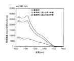
- the emission spectrum was measured using a Horiba Fluorolog-3-2-i HR320 with an excitation wavelength of 980 nm.
- the measurement results are shown in FIGS. 1 and 2, respectively.
- the absorbance and the emission intensity in FIGS. 1 and 2 normalize the concentration of the dispersion.
- the semiconductor SWCNT dispersions of Production Examples 2 and 3 having undergone the step of dispersing the semiconductor SWCNTs in the solution of the surfactant (SDBS) are directly subjected to DSPE-PEG 2000 without the steps.
- SDBS surfactant
- the semiconductor SWCNT dispersion liquid of Production Example 1 dispersed in the above high emission intensity was obtained.
- the results show that in Production Examples 2 and 3, the surface of the semiconductor SWCNT is sufficiently coated with DSPE-PEG 2000 compared to Production Example 1, and the semiconductor SWCNTs are not aggregated, and the isolated dispersion is further enhanced. Is considered to have improved.
- the absorption wavelengths of Production Examples 2 and 3 were shifted to the lower wavelength side as compared to Production Example 1. It is presumed that this is because the proportion of SWCNTs in the isolated dispersion state has increased relative to bundle-like SWCNTs.
- the particle size of the semiconductor SWCNT dispersion for biological imaging obtained in Production Examples 1 to 3 was measured by centrifugal sedimentation using a disk centrifugal particle size distribution measuring apparatus DC24000 UHR manufactured by CPS. The results are shown in FIG. From the results shown in FIG. 3, the average particle sizes of the semiconductor SWCNTs in the semiconductor SWCNT dispersions for biological imaging of Production Examples 1 to 3 were 8 nm, 10 nm, and 6.5 nm, respectively.
- the proportion of particles having a particle diameter smaller than 10 nm was 44%, 30%, and 84%, respectively. It was confirmed that the semiconductor SWCNTs in Production Example 3 in which centrifugation was performed for 3 hours were more isolated dispersion than in Production Example 2 in which centrifugation was performed for 1 hour.
- the semiconductor SWCNT dispersions for biological imaging obtained in Production Example 3 and Production Example 1 are prepared with a 0.3% DSPE-PEG 2000 solution so that the SWCNT concentration is 200 ⁇ g / ml, and administered to 0.1 ml mice The fluorescence after 0 to 6 hours was observed using a Shimadzu SAI-1000 apparatus. The results are shown in FIG. 4 (Production Example 3) and FIG. 5 (Production Example 1).
- the semiconductor SWCNT dispersion liquid of Production Example 1 was found to be accumulated in the liver, but the semiconductor SWCNT dispersion liquid of Production Example 3 was to the liver even when the time after administration passed. Accumulation was not observed, and it became clear that halation could be reduced.
- Production Example 2 (not shown) also showed the same tendency as Production Example 1. This is because in Production Example 1 the step of dispersing the semiconductor SWCNT in the solution of the surfactant (SDBS) is not performed, so the isolated dispersibility is low, the coating by DSPE-PEG 2000 is insufficient, and the semiconductor SWCNT It is considered that the particles were not well dispersed but partially aggregated.
- SDBS surfactant
- the inspection method according to the present invention is effective as a method of determining whether it is a dispersing agent of a manufacturing example (lot) which exhibits abnormal accumulation and does not emit light in a desired wavelength region.
Landscapes
- Chemical & Material Sciences (AREA)
- Engineering & Computer Science (AREA)
- Health & Medical Sciences (AREA)
- Nanotechnology (AREA)
- Materials Engineering (AREA)
- Organic Chemistry (AREA)
- Inorganic Chemistry (AREA)
- Biomedical Technology (AREA)
- General Health & Medical Sciences (AREA)
- Public Health (AREA)
- Veterinary Medicine (AREA)
- Animal Behavior & Ethology (AREA)
- Life Sciences & Earth Sciences (AREA)
- Epidemiology (AREA)
- Dispersion Chemistry (AREA)
- Carbon And Carbon Compounds (AREA)
- Medicinal Preparation (AREA)
- Medicines That Contain Protein Lipid Enzymes And Other Medicines (AREA)
- Medicines Containing Antibodies Or Antigens For Use As Internal Diagnostic Agents (AREA)
Abstract
Description
(1)大気中で直接紫外線を照射することにより酸化処理された半導体単層カーボンナノチューブと、前記半導体単層カーボンナノチューブの表面をコーティングする両親媒性物質からなる分散剤とを含む半導体SWCNT分散液について、
吸収分光法、フォトルミネッセンス法及び粒子径測定からなる群から選択される少なくとも2種類の方法を用いて、前記半導体単層カーボンナノチューブの平均粒子径が10nmより小さいこと、前記半導体単層カーボンナノチューブの孤立分散性が高いこと、及び/又は前記半導体単層カーボンナノチューブが酸化されていることが確認された前記半導体SWCNT分散液のみから構成される、生体イメージング用半導体SWCNT分散液。
(2)前記粒子径測定が、遠心沈降法による測定である上記(1)に記載の生体イメージング用半導体SWCNT分散液。
(3)大気中で直接紫外線を照射することにより酸化処理された半導体単層カーボンナノチューブと、前記半導体単層カーボンナノチューブの表面をコーティングする両親媒性物質からなる分散剤とを含む生体イメージング用半導体SWCNT分散液の検査方法であって、
吸収分光法、フォトルミネッセンス法及び粒子径測定からなる群から選択される少なくとも2種類の方法を用いて、前記半導体単層カーボンナノチューブの平均粒子径が10nmより小さいこと、前記半導体単層カーボンナノチューブの孤立分散性が高いこと、及び/又は前記半導体単層カーボンナノチューブが酸化されていることを確認する、生体イメージング用半導体SWCNT分散液の検査方法。
(4)前記粒子径測定が、遠心沈降法による測定である上記(3)に記載の生体イメージング用半導体SWCNT分散液の検査方法。
(製造例1)
カーボンナノチューブ(CoMoCAT SG65i、平均直径0.8nm、以下「半導体SWCNT」という)1mgをエタノール10mlに加え、バスソニケーションに5分ほどかけて半導体SWCNTをエタノールに分散させた。続いて、減圧濾過器にオムニポアメンブレン(φ47mm、5μmポアをセットし、半導体SWCNT/エタノール分散液を入れてろ過し、フィルター上に半導体SWCNTを均一に載せた。次に、半導体SWCNTをフィルターに載せたまま薬包紙で挟み、フィルターが丸まらないよう軽く重石をしながら60℃で30分乾燥させた。そして、フィルターに載せた半導体SWCNTをフィルターごと60~70秒オゾン処理した(光源は水銀ランプ、半導体SWCNT上での紫外線強度は約19mW/cm2)。
(製造例2)
カーボンナノチューブ(CoMoCAT SG65i、平均直径0.8nm、以下「半導体SWCNT」という)1mgをエタノール10mlに加え、バスソニケーションに5分ほどかけて半導体SWCNTをエタノールに分散させた。続いて、減圧濾過器にオムニポアメンブレン(φ47mm、5μmポアをセットし、半導体SWCNT/エタノール分散液を入れてろ過し、フィルター上に半導体SWCNTを均一に載せた。次に、半導体SWCNTをフィルターに載せたまま薬包紙で挟み、フィルターが丸まらないよう軽く重石をしながら60℃で30分乾燥させた。そして、フィルターに載せた半導体SWCNTをフィルターごと60~70秒オゾン処理した(光源は水銀ランプ、半導体SWCNT上での紫外線強度は約19mW/cm2)。
(製造例3)
カーボンナノチューブ(CoMoCAT SG65i、平均直径0.8nm、以下「半導体SWCNT」という)1mgをエタノール10mlに加え、バスソニケーションに5分ほどかけて半導体SWCNTをエタノールに分散させた。続いて、減圧濾過器にオムニポアメンブレン(φ47mm、5μmポアをセットし、半導体SWCNT/エタノール分散液を入れてろ過し、フィルター上に半導体SWCNTを均一に載せた。次に、半導体SWCNTをフィルターに載せたまま薬包紙で挟み、フィルターが丸まらないよう軽く重石をしながら60℃で30分乾燥させた。そして、フィルターに載せた半導体SWCNTをフィルターごと60~70秒オゾン処理した(光源は水銀ランプ、半導体SWCNT上での紫外線強度は約19mW/cm2)。
(吸収分光法及びフォトルミネッセンス法による測定)
製造例1~3で得られた生体イメージング用半導体SWCNT分散液について、島津製作所紫外可視近赤外分光光度計UV-3100を用い、吸収スペクトルを測定した。また、980nmを励起波長として、堀場製作所Fluorolog-3-2-iHR320を用いて発光スペクトルを測定した。測定結果をそれぞれ図1及び図2に示す。なお、図1及び図2における吸光度及び発光強度は、分散液の濃度を規格化している。
(粒子径測定)
製造例1~3で得られた生体イメージング用半導体SWCNT分散液について、CPS社製ディスク遠心式粒子径分布測定装置DC24000UHRを用い、遠心沈降法による粒子径測定を行った。その結果を図3に示す。図3の結果から、製造例1~3の生体イメージング用半導体SWCNT分散液における半導体SWCNTの平均粒子径はそれぞれ、8nm、10nm、及び6.5nmであった。また、それぞれの生体イメージング用半導体SWCNT分散液における粒子径が10nmより小さいものの割合はそれぞれ44%、30%、84%であった。遠心分離を1時間行った製造例2に比べて、遠心分離を3時間行った製造例3における半導体SWCNTは、より孤立分散であることが確認された。
(in vivoイメージング)
製造例3及び製造例1で得られた生体イメージング用半導体SWCNT分散液を、SWCNT濃度が200μg/mlになるように0.3%のDSPE-PEG2000溶液で調製した後、0.1mlマウスに投与し、0~6時間後の蛍光を島津製作所SAI-1000装置を用いて観察した。その結果を図4(製造例3)及び図5(製造例1)に示す。
Claims (4)
- 大気中で直接紫外線を照射することにより酸化処理された半導体単層カーボンナノチューブと、前記半導体単層カーボンナノチューブの表面をコーティングする両親媒性物質からなる分散剤とを含む半導体SWCNT分散液について、
吸収分光法、フォトルミネッセンス法及び粒子径測定からなる群から選択される少なくとも2種類の方法を用いて、前記半導体単層カーボンナノチューブの平均粒子径が10nmより小さいこと、前記半導体単層カーボンナノチューブの孤立分散性が高いこと、及び/又は前記半導体単層カーボンナノチューブが酸化されていることが確認された前記半導体SWCNT分散液のみから構成される、生体イメージング用半導体SWCNT分散液。 - 前記粒子径測定が、遠心沈降法による測定である請求項1に記載の生体イメージング用半導体SWCNT分散液。
- 大気中で直接紫外線を照射することにより酸化処理された半導体単層カーボンナノチューブと、前記半導体単層カーボンナノチューブの表面をコーティングする両親媒性物質からなる分散剤とを含む生体イメージング用半導体SWCNT分散液の検査方法であって、
吸収分光法、フォトルミネッセンス法及び粒子径測定からなる群から選択される少なくとも2種類の方法を用いて、前記半導体単層カーボンナノチューブの平均粒子径が10nmより小さいこと、前記半導体単層カーボンナノチューブの孤立分散性が高いこと、及び/又は前記半導体単層カーボンナノチューブが酸化されていることを確認する、生体イメージング用半導体SWCNT分散液の検査方法。 - 前記粒子径測定が、遠心沈降法による測定である請求項3に記載の生体イメージング用半導体SWCNT分散液の検査方法。
Priority Applications (4)
| Application Number | Priority Date | Filing Date | Title |
|---|---|---|---|
| US16/764,590 US12257324B2 (en) | 2017-11-17 | 2017-11-17 | Semiconductor SWCNT slurry for bioimaging and method for inspecting the same |
| JP2019553648A JP7100857B2 (ja) | 2017-11-17 | 2017-11-17 | 生体イメージング用半導体swcnt分散液及びその検査方法 |
| PCT/JP2017/041565 WO2019097698A1 (ja) | 2017-11-17 | 2017-11-17 | 生体イメージング用半導体swcnt分散液及びその検査方法 |
| CN201780096900.2A CN111356481A (zh) | 2017-11-17 | 2017-11-17 | 生物成像用半导体swcnt分散液及其检查方法 |
Applications Claiming Priority (1)
| Application Number | Priority Date | Filing Date | Title |
|---|---|---|---|
| PCT/JP2017/041565 WO2019097698A1 (ja) | 2017-11-17 | 2017-11-17 | 生体イメージング用半導体swcnt分散液及びその検査方法 |
Publications (1)
| Publication Number | Publication Date |
|---|---|
| WO2019097698A1 true WO2019097698A1 (ja) | 2019-05-23 |
Family
ID=66539466
Family Applications (1)
| Application Number | Title | Priority Date | Filing Date |
|---|---|---|---|
| PCT/JP2017/041565 Ceased WO2019097698A1 (ja) | 2017-11-17 | 2017-11-17 | 生体イメージング用半導体swcnt分散液及びその検査方法 |
Country Status (4)
| Country | Link |
|---|---|
| US (1) | US12257324B2 (ja) |
| JP (1) | JP7100857B2 (ja) |
| CN (1) | CN111356481A (ja) |
| WO (1) | WO2019097698A1 (ja) |
Cited By (1)
| Publication number | Priority date | Publication date | Assignee | Title |
|---|---|---|---|---|
| WO2022044685A1 (ja) * | 2020-08-28 | 2022-03-03 | 日本ゼオン株式会社 | 酸化カーボンナノチューブ及びその製造方法 |
Families Citing this family (2)
| Publication number | Priority date | Publication date | Assignee | Title |
|---|---|---|---|---|
| JP7100856B2 (ja) * | 2017-11-17 | 2022-07-14 | 株式会社島津製作所 | 生体イメージング用半導体swcnt分散液及びその製造方法 |
| KR102871289B1 (ko) * | 2023-04-10 | 2025-10-16 | 주식회사 서현테크켐 | 분산성이 향상된 탄소나노튜브 분산액 제조 방법 |
Citations (3)
| Publication number | Priority date | Publication date | Assignee | Title |
|---|---|---|---|---|
| JP2006182657A (ja) * | 2004-12-24 | 2006-07-13 | Japan Science & Technology Agency | 造影剤 |
| JP2016537644A (ja) * | 2013-09-16 | 2016-12-01 | マサチューセッツ インスティテュート オブ テクノロジー | 組織局在化バイオセンサーとしての近赤外蛍光単層カーボンナノチューブ |
| JP2017066098A (ja) * | 2015-09-30 | 2017-04-06 | 国立研究開発法人産業技術総合研究所 | 単層カーボンナノチューブを含む熱産生脂肪組織造影剤 |
Family Cites Families (7)
| Publication number | Priority date | Publication date | Assignee | Title |
|---|---|---|---|---|
| AU2003226428A1 (en) * | 2002-04-18 | 2003-11-03 | Northwestern University | Encapsulation of nanotubes via self-assembled nanostructures |
| JP2012032358A (ja) | 2010-08-03 | 2012-02-16 | National Institute Of Advanced Industrial & Technology | 蛍光分子内包型カーボンナノチューブ |
| US9486772B1 (en) * | 2010-08-27 | 2016-11-08 | The United States Of America As Represented By The Administrator Of National Aeronautics And Space Administration | Methods of functionalization of carbon nanotubes by photooxidation |
| WO2012036641A1 (en) * | 2010-09-17 | 2012-03-22 | Nanyang Technological University | Method for dispersing carbon nanotubes using chondroitin sulfate cation salt |
| WO2014080519A1 (ja) | 2012-11-26 | 2014-05-30 | 独立行政法人産業技術総合研究所 | ナノカーボンを用いた臨床検査 |
| US9249023B2 (en) * | 2013-03-06 | 2016-02-02 | William Marsh Rice University | Liquid crystals from single-walled carbon nanotube polyelectrolytes and their use for making various materials |
| WO2016117633A1 (ja) | 2015-01-23 | 2016-07-28 | 国立研究開発法人産業技術総合研究所 | 近赤外発光する半導体単層カーボンナノチューブ |
-
2017
- 2017-11-17 JP JP2019553648A patent/JP7100857B2/ja active Active
- 2017-11-17 US US16/764,590 patent/US12257324B2/en active Active
- 2017-11-17 WO PCT/JP2017/041565 patent/WO2019097698A1/ja not_active Ceased
- 2017-11-17 CN CN201780096900.2A patent/CN111356481A/zh active Pending
Patent Citations (3)
| Publication number | Priority date | Publication date | Assignee | Title |
|---|---|---|---|---|
| JP2006182657A (ja) * | 2004-12-24 | 2006-07-13 | Japan Science & Technology Agency | 造影剤 |
| JP2016537644A (ja) * | 2013-09-16 | 2016-12-01 | マサチューセッツ インスティテュート オブ テクノロジー | 組織局在化バイオセンサーとしての近赤外蛍光単層カーボンナノチューブ |
| JP2017066098A (ja) * | 2015-09-30 | 2017-04-06 | 国立研究開発法人産業技術総合研究所 | 単層カーボンナノチューブを含む熱産生脂肪組織造影剤 |
Non-Patent Citations (1)
| Title |
|---|
| UMEZAWA MASAKAZU ET AL.: "Mouse Windpipe Imaging Using Carbon Nanotube Near-infrared Fluorescence", BIOIMAGING, vol. 25, no. 2, 2016, pages 123 * |
Cited By (1)
| Publication number | Priority date | Publication date | Assignee | Title |
|---|---|---|---|---|
| WO2022044685A1 (ja) * | 2020-08-28 | 2022-03-03 | 日本ゼオン株式会社 | 酸化カーボンナノチューブ及びその製造方法 |
Also Published As
| Publication number | Publication date |
|---|---|
| JP7100857B2 (ja) | 2022-07-14 |
| US12257324B2 (en) | 2025-03-25 |
| US20200384127A1 (en) | 2020-12-10 |
| JPWO2019097698A1 (ja) | 2020-12-24 |
| CN111356481A (zh) | 2020-06-30 |
Similar Documents
| Publication | Publication Date | Title |
|---|---|---|
| Desmond et al. | Critical overview on the green synthesis of carbon quantum dots and their application for cancer therapy | |
| Kagan et al. | Lung macrophages “digest” carbon nanotubes using a superoxide/peroxynitrite oxidative pathway | |
| Erogbogbo et al. | Bioconjugation of luminescent silicon quantum dots for selective uptake by cancer cells | |
| Rakovich et al. | Semiconductor versus graphene quantum dots as fluorescent probes for cancer diagnosis and therapy applications | |
| Liu et al. | Fluorescent nanoparticles from starch: Facile preparation, tunable luminescence and bioimaging | |
| Li et al. | Fluorescent ultrashort nanotubes from defect-induced chemical cutting | |
| Liu et al. | Carbon Nanoparticles Trapped in Vivo Similar to Carbon Nanotubes in Time-Dependent Biodistribution | |
| WO2019097698A1 (ja) | 生体イメージング用半導体swcnt分散液及びその検査方法 | |
| Plonska‐Brzezinska et al. | Carbon nano-onions and biocompatible polymers for flavonoid incorporation. | |
| Nazarkovsky et al. | Rational design of large flat nitrogen-doped graphene oxide quantum dots with green-luminescence suitable for biomedical applications | |
| Ren et al. | PEGylated β-NaGdF4/Tb@ CaF2 core/shell nanophosphors for enhanced radioluminescence and folate receptor targeting | |
| Pan et al. | Preparation and characterization of decyl-terminated silicon nanoparticles encapsulated in lipid nanocapsules | |
| KR101837579B1 (ko) | 향상된 형광 특성을 갖는 금을 포함하는 그래핀 양자점 나노 복합체 및 그 제조방법, 이를 이용하는 금속 이온 검출기 | |
| Kumar et al. | NIR light-responsive Ti3C2 MXenes mediate controlled aqueous degradation and highly efficient photothermal conversion for targeted cancer phototherapy | |
| JP4858775B2 (ja) | リポソームを鋳型とする中空ナノ粒子の作製方法 | |
| US11696688B2 (en) | Semiconductor SWCNT slurry for bioimaging and method for producing the same | |
| Bomboi et al. | Lysozyme binds onto functionalized carbon nanotubes | |
| de Sousa Maia et al. | Drying of graphene oxide: effects on red blood cells and protein corona formation | |
| Jang et al. | Water-stable single-walled carbon nanotubes coated by pyrenyl polyethylene glycol for fluorescence imaging and photothermal therapy | |
| Olenchuk et al. | The influence of 2D WS2 nanoparticles on the liposomes formation, morphology and spectral properties | |
| Padmavathi et al. | Green synthesis, characterisation, optical properties of graphene quantum dots with anti bacterial characteristics | |
| Corr et al. | Cytotoxicity and variant cellular internalization behavior of water-soluble sulfonated nanographene sheets in liver cancer cells | |
| Pan et al. | Silica cross-linked micelles loading with silicon nanoparticles: preparation and characterization | |
| Farias et al. | Semiconductor fluorescent quantum dots: efficient biolabels in cancer diagnostics | |
| Benavente et al. | Modification of a regenerated cellulose membrane with lipid nanoparticles and layers. Nanoparticle preparation, morphological and physicochemical characterization of nanoparticles and modified membranes |
Legal Events
| Date | Code | Title | Description |
|---|---|---|---|
| 121 | Ep: the epo has been informed by wipo that ep was designated in this application |
Ref document number: 17931975 Country of ref document: EP Kind code of ref document: A1 |
|
| ENP | Entry into the national phase |
Ref document number: 2019553648 Country of ref document: JP Kind code of ref document: A |
|
| NENP | Non-entry into the national phase |
Ref country code: DE |
|
| 122 | Ep: pct application non-entry in european phase |
Ref document number: 17931975 Country of ref document: EP Kind code of ref document: A1 |
|
| WWG | Wipo information: grant in national office |
Ref document number: 16764590 Country of ref document: US |