WO2019082874A1 - Method for producing liver stem cells or liver progenitor cells by direct reprogramming - Google Patents
Method for producing liver stem cells or liver progenitor cells by direct reprogrammingInfo
- Publication number
- WO2019082874A1 WO2019082874A1 PCT/JP2018/039295 JP2018039295W WO2019082874A1 WO 2019082874 A1 WO2019082874 A1 WO 2019082874A1 JP 2018039295 W JP2018039295 W JP 2018039295W WO 2019082874 A1 WO2019082874 A1 WO 2019082874A1
- Authority
- WO
- WIPO (PCT)
- Prior art keywords
- cells
- hiheppc
- hepatic
- combination
- myc
- Prior art date
Links
Images


Classifications
-
- C—CHEMISTRY; METALLURGY
- C12—BIOCHEMISTRY; BEER; SPIRITS; WINE; VINEGAR; MICROBIOLOGY; ENZYMOLOGY; MUTATION OR GENETIC ENGINEERING
- C12N—MICROORGANISMS OR ENZYMES; COMPOSITIONS THEREOF; PROPAGATING, PRESERVING, OR MAINTAINING MICROORGANISMS; MUTATION OR GENETIC ENGINEERING; CULTURE MEDIA
- C12N5/00—Undifferentiated human, animal or plant cells, e.g. cell lines; Tissues; Cultivation or maintenance thereof; Culture media therefor
- C12N5/06—Animal cells or tissues; Human cells or tissues
- C12N5/0602—Vertebrate cells
- C12N5/067—Hepatocytes
- C12N5/0672—Stem cells; Progenitor cells; Precursor cells; Oval cells
-
- C—CHEMISTRY; METALLURGY
- C12—BIOCHEMISTRY; BEER; SPIRITS; WINE; VINEGAR; MICROBIOLOGY; ENZYMOLOGY; MUTATION OR GENETIC ENGINEERING
- C12N—MICROORGANISMS OR ENZYMES; COMPOSITIONS THEREOF; PROPAGATING, PRESERVING, OR MAINTAINING MICROORGANISMS; MUTATION OR GENETIC ENGINEERING; CULTURE MEDIA
- C12N2501/00—Active agents used in cell culture processes, e.g. differentation
- C12N2501/10—Growth factors
-
- C—CHEMISTRY; METALLURGY
- C12—BIOCHEMISTRY; BEER; SPIRITS; WINE; VINEGAR; MICROBIOLOGY; ENZYMOLOGY; MUTATION OR GENETIC ENGINEERING
- C12N—MICROORGANISMS OR ENZYMES; COMPOSITIONS THEREOF; PROPAGATING, PRESERVING, OR MAINTAINING MICROORGANISMS; MUTATION OR GENETIC ENGINEERING; CULTURE MEDIA
- C12N2501/00—Active agents used in cell culture processes, e.g. differentation
- C12N2501/60—Transcription factors
- C12N2501/606—Transcription factors c-Myc
-
- C—CHEMISTRY; METALLURGY
- C12—BIOCHEMISTRY; BEER; SPIRITS; WINE; VINEGAR; MICROBIOLOGY; ENZYMOLOGY; MUTATION OR GENETIC ENGINEERING
- C12N—MICROORGANISMS OR ENZYMES; COMPOSITIONS THEREOF; PROPAGATING, PRESERVING, OR MAINTAINING MICROORGANISMS; MUTATION OR GENETIC ENGINEERING; CULTURE MEDIA
- C12N2506/00—Differentiation of animal cells from one lineage to another; Differentiation of pluripotent cells
- C12N2506/11—Differentiation of animal cells from one lineage to another; Differentiation of pluripotent cells from blood or immune system cells
-
- C—CHEMISTRY; METALLURGY
- C12—BIOCHEMISTRY; BEER; SPIRITS; WINE; VINEGAR; MICROBIOLOGY; ENZYMOLOGY; MUTATION OR GENETIC ENGINEERING
- C12N—MICROORGANISMS OR ENZYMES; COMPOSITIONS THEREOF; PROPAGATING, PRESERVING, OR MAINTAINING MICROORGANISMS; MUTATION OR GENETIC ENGINEERING; CULTURE MEDIA
- C12N2506/00—Differentiation of animal cells from one lineage to another; Differentiation of pluripotent cells
- C12N2506/28—Differentiation of animal cells from one lineage to another; Differentiation of pluripotent cells from vascular endothelial cells
-
- C—CHEMISTRY; METALLURGY
- C12—BIOCHEMISTRY; BEER; SPIRITS; WINE; VINEGAR; MICROBIOLOGY; ENZYMOLOGY; MUTATION OR GENETIC ENGINEERING
- C12N—MICROORGANISMS OR ENZYMES; COMPOSITIONS THEREOF; PROPAGATING, PRESERVING, OR MAINTAINING MICROORGANISMS; MUTATION OR GENETIC ENGINEERING; CULTURE MEDIA
- C12N2510/00—Genetically modified cells
-
- C—CHEMISTRY; METALLURGY
- C12—BIOCHEMISTRY; BEER; SPIRITS; WINE; VINEGAR; MICROBIOLOGY; ENZYMOLOGY; MUTATION OR GENETIC ENGINEERING
- C12N—MICROORGANISMS OR ENZYMES; COMPOSITIONS THEREOF; PROPAGATING, PRESERVING, OR MAINTAINING MICROORGANISMS; MUTATION OR GENETIC ENGINEERING; CULTURE MEDIA
- C12N2740/00—Reverse transcribing RNA viruses
- C12N2740/00011—Details
- C12N2740/10011—Retroviridae
- C12N2740/13011—Gammaretrovirus, e.g. murine leukeamia virus
- C12N2740/13041—Use of virus, viral particle or viral elements as a vector
- C12N2740/13043—Use of virus, viral particle or viral elements as a vector viral genome or elements thereof as genetic vector
Definitions
- the present invention relates to a method for producing hepatic stem cells or hepatic progenitor cells by introducing a predetermined reprogramming factor into vascular endothelial cells and the like.
- Non-Patent Document 1 JP-A-2015-527084.
- Hnf1 ⁇ and Foxa3 are introduced into mouse embryonic fibroblasts, they are reprogrammed in liver stem cells (Non-Patent Document 1), and by expressing FOXA3, HNF1A and HNF4A, from human fibroblasts to mature hepatocytes.
- Non-patent document 2 Method of reprogramming (Non-patent document 2), Method of producing human induced hepatocytes by over-expressing HNF1A, HNF4A and HNF6 together with maturation factors ATF5, PROX1 and CEBPA (Non-patent document 3) are known.
- hepatocytes induced by the conventional method have poor proliferation ability and can not differentiate into biliary epithelial cells.
- the present inventors succeeded in obtaining hepatic stem / hepatic progenitor cells by a combination of predetermined genes and completed the present invention.
- the present invention is as follows.
- a method for inducing liver stem cells or hepatic progenitor cells from non-hepatic stem cells or non-hepatic progenitor cells which comprises introducing the following combination into non-hepatic stem cells or non-hepatic progenitor cells: (A) combination of HNF1, HNF6 and FOXA, (B) combination of HNF1, HNF6 and FOXA genes, (C) A combination of HNF1, MYC and FOXA, or (d) a combination of HNF1, MYC and FOXA genes.
- non-hepatic stem cells or non-hepatic progenitor cells are vascular endothelial cells or blood-derived cells.
- the vascular endothelial cell is derived from umbilical vein, peripheral blood or umbilical cord blood.
- the blood-derived cells are peripheral blood T cells or cord blood T cells.
- (11) (a) inducing hepatic stem cells or hepatic progenitor cells by the method according to (1), and (b) differentiating the induced hepatic stem cells or hepatic progenitor cells into hepatic cells or biliary epithelial cells , A method of producing hepatocytes or biliary epithelial cells.
- non-hepatic stem / non-hepatic progenitor cells such as vascular endothelial cells can be directly converted to hepatic stem / hepatic progenitor cells.
- the induced hepatic stem / hepatic progenitor cells have long-term proliferative ability and can differentiate into hepatocytes and biliary epithelial cells. Furthermore, vascular endothelial cells can be induced to hepatic stem / hepatic progenitor cells not only in fetal cells but also in adult peripheral blood. Therefore, liver function test and drug screening using peripheral blood-derived liver stem / liver precursor cells are also possible.
- hiHepPC human induced hepatic progenitor cell
- hiHepPC human induced hepatic progenitor cell
- FIG. 5 shows differentiation of hiHepPC into biliary epithelial cell-like cells.
- FIG. 5 shows the analysis result of proliferation ability and differentiation ability by clone analysis of HUVEC origin hiHepPC.
- FIG. 6 shows hiHepPC induction from human peripheral blood-derived endothelial cells (HPBEC). It is a figure which shows the analysis result of proliferation ability and differentiation ability by clonal analysis of HPBEC origin 4F-hiHepPC. It is a figure which shows the analysis result when FOXA3 which is one of the hiHepPC inducers is substituted to FOXA1 and FOXA2.
- FIG. 6 shows hiHepPC induction from human peripheral blood-derived T cells (HPBTC). It is a figure showing an outline of the present invention.
- the present invention relates to non-hepatic stem / non-hepatic progenitor cells, a combination of HNF1A, HNF6 and FOXA reprogramming factors, a combination of genes encoding the factor, a combination of HNF1A, MYC and FOXA reprogramming factors, or the factor
- the present invention relates to a method for inducing non-hepatic stem / non-hepatic progenitor cells to hepatic stem / hepatic progenitor cells, which comprises introducing a combination of genes encoding.
- the present invention is characterized in that non-hepatic stem / non-hepatic precursor cells are induced to the hepatic stem / hepatic precursor cells from which they originate, rather than to the already differentiated cells, and the combination of the above reprogramming factors is used. Use.
- Non-hepatic stem / non-hepatic precursor cells mean cells other than the target hepatic stem / hepatic precursor cells.
- non-hepatic stem / non-hepatic precursor cells for example, fibroblasts, endothelial cells, blood cells, cord blood cells, bone marrow cells, keratinocytes, hepatocytes, biliary epithelial cells, myofibroblasts, neural cells, epithelial cells, etc. Can be mentioned.
- vascular endothelial cells or blood-derived cells can be used.
- Vascular endothelial cells include those derived from umbilical vein, peripheral blood or cord blood, and blood-derived cells include peripheral blood cells or cord blood cells (for example, peripheral blood T cells or cord blood T cells).
- the non-hepatic stem / non-hepatic precursor cells used in the present invention are, for example, cells derived from mammals such as mice, rats, rabbits, cats, dogs, monkeys and humans. In one embodiment of the present invention, the non-hepatic stem / non-hepatic precursor cells used are cells of human origin.
- Reprogramming Factors is the process of changing the differentiation state of a cell to a different differentiation state or an undifferentiated state from the cell.
- the non-hepatic stem / non-hepatic progenitor cells are induced to hepatic stem / hepatic progenitor cells.
- non-hepatic stem / non-hepatic progenitor cells can be induced to hepatic stem / hepatic progenitor cells without passing through pluripotent stem cells.
- the factors used for such reprogramming are the following combinations: (A) combination of HNF1, HNF6 and FOXA, (B) combination of HNF1, HNF6 and FOXA genes, (C) A combination of HNF1, MYC and FOXA, or (d) a combination of HNF1, MYC and FOXA genes.
- HNF1 Hepatocyte Nuclear Factor 1
- HNF6 Hepatocyte Nuclear Factor 6
- HNF1A Hepatocyte Nuclear Factor 1
- HNF6B Hepatocyte Nuclear Factor 6
- FOXA is a hepatocyte nuclear factor (transcription factor) that is required for the earliest process of liver tissue formation and includes FOXA1, FOXA2, FOXA3. These FOXA transcription factors are considered to have functional complementarity with each other because they have 90% or more homology at the amino acid level in the common forkhead / wingedhelix domain.
- NF1A, HNF6 and FOXA3 ⁇ NF1A, L-MYC and FOXA3 ⁇ NF1A, HNF6, FOXA3 and L-MYC
- the genes encoding reprogramming factors may be linked to one vector for all of the gene sets to be introduced, or may be linked to separate vectors for each gene. It is also possible to link some genes to one vector and link the remaining genes to another vector.
- expression vectors examples include plasmids, bacteriophages, and virus vectors derived from retrovirus, vaccinia virus, adenovirus, lentivirus, adeno-associated virus, Sendai virus and the like.
- the method for introducing the expression vector into non-hepatic stem / non-hepatic precursor cells is not particularly limited, and there may be mentioned a method of infecting cells with an expression vector containing a gene encoding each reprogramming factor, For example, the lipofection method, the electroporation method, the calcium phosphate method and the like can be adopted.
- the dose of the vector for infecting cells with a viral vector can be appropriately adjusted.
- HNF1A, HNF6 and FOXA3 ⁇ HNF1A, L-MYC and FOXA3 ⁇ HNF1A, HNF6, FOXA3 and L-MYC The above factors can be produced by genetic engineering (for example, "Molecular Cloning, A Laboratory Manual (4th edition)” (see Cold Spring Harbor Laboratory Press (2012)), and the obtained polypeptide is introduced into cells. The introduction may be carried out by linking to a membrane permeable peptide or the like and introducing into cells, a method of introducing into cells using cationic lipids, etc. Intracellular transfer reagents are also commercially available ( PULSin (PPU), Prote-IN (HYG), BioPORTER Protein Delivery Reagent (GTS), etc.).
- FIG. 1A A schematic diagram of a hiHepPC induction experiment is shown in FIG. 1A.
- a retroviral vector (gene expression vector) was introduced into a commercially available human umbilical vein endothelial cell (HUVEC) by retroviral infection. Meanwhile, HUVECs were cultured in medium (HUVEC medium) in which Medium 200 and Fibrous serum-free cell culture medium were mixed at 1: 1.
- HUVEC empty vector (mock), combination of HNF4A and FOXA3 (H4A / F3), combination of HNF4A with FOXA3, HNF1A, HNF6 (H4A / F3 / H1A / H6), combination of HNF4A with FOXA3, HNF1A (H4A / F3 / H1A), HNF4A and FOXA3, combination of HNF6 (H4A / F3 / H6), combination of HNF4A and HNF1A, HNF6 (H4A / H1A / H6), HNF4A and HNF1A, HNF6, ATF5, PROX1, CEBPA combination (H4A / H1A / H6 + APC), combination of FOXA3 with HNF1A, HNF6 (F3 / H1A / H6), and ALBUMIN (ALB) immunostaining and ALB positive every 1st to 6th passages (P) The number of cells was count
- ALB is a marker expressed in hepatic progenitor cells and hepatocytes
- E-CAD is a marker for epithelial cells. From the immunostaining of ALB and E-CAD, the phase contrast picture of cells (Phase), and the graph of the proportion of ALB positive cells, hepatic progenitor cell-like cells having proliferation ability when F3 / H1A / H6 is introduced Is found to be induced.
- PH in the graph represents parental HUVEC
- PN represents passage number.
- Two independent experiments are performed, and the results are shown by the blue and red lines on the graph.
- Cellular DNA was stained with DAPI (blue). Scale bar: 50 ⁇ m.
- transduced the gene set of F3 / H1A / H6 into HUVEC, and induced hiHepPC was performed independently 3 times.
- FIG. 1C The results are shown in FIG. 1C. The number of cells grown in 16 passages of hiHepPC (hiHepPC-1, hiHepPC-2, hiHepPC-3) obtained in each experiment is shown. While hiHepPC continues to proliferate, HUVEC ceases to proliferate during passage. Furthermore, the results of chromosomal analysis of hiHepPC at the 12th passage are shown in FIG. 1D. Twenty cells were analyzed, but all of them had normal chromosome numbers.
- FIG. 1B The results are shown in FIG.
- panel A ALB immunostaining was performed every 1st to 6th passages (P), and in P6, immunostaining of E-CAD and imaging of phase-contrast photographs (phase) were also performed. Cellular DNA was stained with DAPI (blue). Scale bar: 50 ⁇ m.
- Panel B measures the number of ALB-positive cells at each of the first to sixth passages after introducing the F3 / H1A / H6 + L-MYC combination into HUVEC, and graphs the percentage of ALB-positive cells. Show. PH in the graph represents parental HUVEC, and PN represents passage number (passage number). Two independent experiments are performed, and the results are shown by the blue and red lines on the graph. The combination F3 / H1A / H6 + L-MYC induces ALB positive cells earlier than the combination F3 / H1A / H6 (see FIG. 1B).
- human fibroblasts were successfully differentiated into hepatocyte-like cells and biliary epithelial cell-like cells.
- human umbilical cord blood T cells were successfully differentiated into hepatocyte-like cells and biliary epithelial cell-like cells.
Landscapes
- Health & Medical Sciences (AREA)
- Engineering & Computer Science (AREA)
- Life Sciences & Earth Sciences (AREA)
- Biomedical Technology (AREA)
- Wood Science & Technology (AREA)
- Organic Chemistry (AREA)
- Chemical & Material Sciences (AREA)
- Biotechnology (AREA)
- Zoology (AREA)
- Bioinformatics & Cheminformatics (AREA)
- Genetics & Genomics (AREA)
- Microbiology (AREA)
- Cell Biology (AREA)
- Gastroenterology & Hepatology (AREA)
- Biochemistry (AREA)
- General Engineering & Computer Science (AREA)
- General Health & Medical Sciences (AREA)
- Developmental Biology & Embryology (AREA)
- Micro-Organisms Or Cultivation Processes Thereof (AREA)
- Medicines Containing Material From Animals Or Micro-Organisms (AREA)
Abstract
A method for inducing liver stem cells or liver progenitor cells from non-liver stem cells or non-liver progenitor cells, the method being characterized in that the following combination is introduced into non-liver stem cells or non-liver progenitor cells: (a) a combination of HNF1, HNF6, and FOXA; (b) a combination of an HNF1 gene, an HNF6 gene, and a FOXA gene; (c) a combination of HNF1, MYC, and FOXA; or (d) a combination of an HNF1 gene, an MYC gene, and a FOXA gene.
Description
本発明は、血管内皮細胞等に所定のリプログラミング因子を導入することにより、肝幹細胞又は肝前駆細胞を製造する方法に関する。
The present invention relates to a method for producing hepatic stem cells or hepatic progenitor cells by introducing a predetermined reprogramming factor into vascular endothelial cells and the like.
ある種の遺伝子セットをヒト線維芽細胞に導入することにより肝細胞を作製する技術は、既に知られている。例えば、リプログラミング因子としてCEBPA、HNF4A、FOXA3、GATA4、HNF1Aなどを用いて、非肝細胞から誘導肝細胞を作製する方法が知られている(特許文献1:特表2015-527084号公報)。
また、マウス胚性線維芽細胞にHnf1β及びFoxa3を導入すると肝幹細胞にリプログラミングされること(非特許文献1)、FOXA3、HNF1A及びHNF4Aを発現させることにより、ヒト線維芽細胞から成熟肝細胞にリプログラミングする方法(非特許文献2)、HNF1A、HNF4A及びHNF6を、成熟因子であるATF5、PROX1及びCEBPAとともに過剰発現することにより、ヒト誘導肝細胞を作製する方法(非特許文献3)などが知られている。
しかしながら、従来法で誘導された肝細胞は増殖能に乏しく、また胆管上皮細胞にも分化できない。 Techniques for producing hepatocytes by introducing certain gene sets into human fibroblasts are already known. For example, there is known a method for producing induced hepatocytes from non-hepatocytes using CEBPA, HNF4A, FOXA3, GATA4, HNF1A or the like as a reprogramming factor (Patent Document 1: JP-A-2015-527084).
In addition, when Hnf1β and Foxa3 are introduced into mouse embryonic fibroblasts, they are reprogrammed in liver stem cells (Non-Patent Document 1), and by expressing FOXA3, HNF1A and HNF4A, from human fibroblasts to mature hepatocytes. Method of reprogramming (Non-patent document 2), Method of producing human induced hepatocytes by over-expressing HNF1A, HNF4A and HNF6 together with maturation factors ATF5, PROX1 and CEBPA (Non-patent document 3) Are known.
However, hepatocytes induced by the conventional method have poor proliferation ability and can not differentiate into biliary epithelial cells.
増殖能を有し、かつ胆管上皮細胞に分化することができる肝幹細胞又は肝前駆細胞(以下、肝幹/肝前駆細胞という)を得ることができれば、当該細胞を培養することにより、目的の肝細胞や胆管上皮細胞を必要なだけ調達することができる。そこで、肝幹/肝前駆細胞を製造する方法の開発が望まれていた。
If hepatic stem cells or hepatic progenitor cells (hereinafter referred to as hepatic stem / hepatic progenitor cells) capable of proliferating and capable of differentiating into biliary epithelial cells can be obtained, the target liver can be obtained by culturing the cells. It is possible to procure as many cells and biliary epithelial cells as needed. Therefore, development of a method for producing hepatic stem / liver precursor cells has been desired.
本発明者は、上記課題を解決するために鋭意検討を行った結果、所定遺伝子の組合せにより肝幹/肝前駆細胞を得ることに成功し、本発明を完成するに至った。
As a result of intensive studies to solve the above problems, the present inventors succeeded in obtaining hepatic stem / hepatic progenitor cells by a combination of predetermined genes and completed the present invention.
すなわち、本発明は以下の通りである。
(1)非肝幹細胞又は非肝前駆細胞に、以下の組合せを導入することを特徴とする、非肝幹細胞又は非肝前駆細胞から肝幹細胞又は肝前駆細胞への誘導方法:
(a)HNF1、HNF6及びFOXAの組合せ、
(b)HNF1遺伝子、HNF6遺伝子及びFOXA遺伝子の組合せ、
(c)HNF1、MYC及びFOXAの組合せ、又は
(d)HNF1遺伝子、MYC遺伝子及びFOXA遺伝子の組合せ。
(2)(1)に記載の方法によって肝幹細胞又は肝前駆細胞を誘導する工程を含む、肝幹細胞又は肝前駆細胞の製造方法。
(3)HNF1がHNF1Aである、(1)又は(2)に記載の方法。
(4)FOXAがFOXA3である、(1)~(3)のいずれか1項に記載の方法。
(5)組合せが(a)又は(b)であって、さらに、MYC又はMYC遺伝子を導入する、(1)~(4)のいずれか1項に記載の方法。
(6)MYCがL-MYCである、(1)~(5)のいずれか1項に記載の方法。
(7)非肝幹細胞又は非肝前駆細胞が、血管内皮細胞又は血液由来細胞である、(1)~(6)のいずれか1項に記載の方法。
(8)血管内皮細胞が臍帯静脈、末梢血又は臍帯血由来のものである(7)に記載の方法。
(9)血液由来細胞が末梢血T細胞又は臍帯血T細胞である(7)に記載の方法。
(10)(2)~(9)のいずれか1項に記載の方法により製造された、肝幹細胞又は肝前駆細胞。
(11)(a)(1)に記載の方法によって肝幹細胞又は肝前駆細胞を誘導する工程、及び
(b)誘導した肝幹細胞又は肝前駆細胞を肝細胞又は胆管上皮細胞に分化させる工程
を含む、肝細胞又は胆管上皮細胞の製造方法。 That is, the present invention is as follows.
(1) A method for inducing liver stem cells or hepatic progenitor cells from non-hepatic stem cells or non-hepatic progenitor cells, which comprises introducing the following combination into non-hepatic stem cells or non-hepatic progenitor cells:
(A) combination of HNF1, HNF6 and FOXA,
(B) combination of HNF1, HNF6 and FOXA genes,
(C) A combination of HNF1, MYC and FOXA, or (d) a combination of HNF1, MYC and FOXA genes.
(2) A method for producing hepatic stem cells or hepatic progenitor cells, comprising the step of inducing hepatic stem cells or hepatic progenitor cells by the method according to (1).
(3) The method according to (1) or (2), wherein HNF1 is HNF1A.
(4) The method according to any one of (1) to (3), wherein FOXA is FOXA3.
(5) The method according to any one of (1) to (4), wherein the combination is (a) or (b) and further the MYC or MYC gene is introduced.
(6) The method according to any one of (1) to (5), wherein MYC is L-MYC.
(7) The method according to any one of (1) to (6), wherein the non-hepatic stem cells or non-hepatic progenitor cells are vascular endothelial cells or blood-derived cells.
(8) The method according to (7), wherein the vascular endothelial cell is derived from umbilical vein, peripheral blood or umbilical cord blood.
(9) The method according to (7), wherein the blood-derived cells are peripheral blood T cells or cord blood T cells.
(10) A hepatic stem cell or hepatic progenitor cell produced by the method according to any one of (2) to (9).
(11) (a) inducing hepatic stem cells or hepatic progenitor cells by the method according to (1), and (b) differentiating the induced hepatic stem cells or hepatic progenitor cells into hepatic cells or biliary epithelial cells , A method of producing hepatocytes or biliary epithelial cells.
本発明により、肝幹細胞及び肝前駆細胞以外の細胞、すなわち非肝幹細胞又は非肝前駆細胞(以下、「非肝幹/非肝前駆細胞」という)から、肝幹/肝前駆細胞への誘導方法(リプログラミング方法)、及び肝幹/肝前駆細胞の製造方法が提供される。本発明によれば、血管内皮細胞等の非肝幹/非肝前駆細胞から肝幹/肝前駆細胞に直接転換させることができる。誘導された肝幹/肝前駆細胞は、長期増殖能を有し、肝細胞や胆管上皮細胞に分化することが可能である。さらに、血管内皮細胞は胎児性のものだけでなく、成人末梢血中のものでも肝幹/肝前駆細胞に誘導することができる。従って、末梢血由来肝幹/肝前駆細胞を用いた肝機能検査や薬剤スクリーニングも可能になる。
According to the present invention, a method for inducing hepatic stem / hepatic progenitor cells from hepatic stem cells and cells other than hepatic progenitor cells, ie, non-hepatic stem cells or non-hepatic progenitor cells (hereinafter referred to as “non-hepatic stem / non-hepatic progenitor cells”) Methods of Reprogramming and Methods of Producing Liver Stem / Liver Progenitor Cells are Provided. According to the present invention, non-hepatic stem / non-hepatic progenitor cells such as vascular endothelial cells can be directly converted to hepatic stem / hepatic progenitor cells. The induced hepatic stem / hepatic progenitor cells have long-term proliferative ability and can differentiate into hepatocytes and biliary epithelial cells. Furthermore, vascular endothelial cells can be induced to hepatic stem / hepatic progenitor cells not only in fetal cells but also in adult peripheral blood. Therefore, liver function test and drug screening using peripheral blood-derived liver stem / liver precursor cells are also possible.
本発明は、非肝幹/非肝前駆細胞に、HNF1A、HNF6及びFOXAのリプログラミング因子の組合せ、当該因子をコードする遺伝子の組合せ、HNF1A、MYC及びFOXAのリプログラミング因子の組合せ、又は当該因子をコードする遺伝子の組合せを導入することを特徴とする、非肝幹/非肝前駆細胞から肝幹/肝前駆細胞への誘導方法に関する。本発明は、既に分化した細胞に誘導するのではなく、非肝幹/非肝前駆細胞から、その起源となる肝幹/肝前駆細胞に誘導させることを特徴とし、上記リプログラミング因子の組合せを用いる。
The present invention relates to non-hepatic stem / non-hepatic progenitor cells, a combination of HNF1A, HNF6 and FOXA reprogramming factors, a combination of genes encoding the factor, a combination of HNF1A, MYC and FOXA reprogramming factors, or the factor The present invention relates to a method for inducing non-hepatic stem / non-hepatic progenitor cells to hepatic stem / hepatic progenitor cells, which comprises introducing a combination of genes encoding. The present invention is characterized in that non-hepatic stem / non-hepatic precursor cells are induced to the hepatic stem / hepatic precursor cells from which they originate, rather than to the already differentiated cells, and the combination of the above reprogramming factors is used. Use.
1.非肝幹/非肝前駆細胞
本発明において、使用の対象となる非肝幹/非肝前駆細胞は、目的とする肝幹/肝前駆細胞以外の細胞を意味する。非肝幹/非肝前駆細胞としては、例えば線維芽細胞、内皮細胞、血液細胞、臍帯血細胞、骨髄細胞、ケラチノサイト、肝細胞、胆管上皮細胞、筋線維芽細胞、神経系細胞、上皮系細胞などが挙げられる。本発明においては、例えば血管内皮細胞又は血液由来細胞を使用することができる。血管内皮細胞としては、臍帯静脈、末梢血又は臍帯血由来のものが挙げられ、血液由来細胞としては、末梢血細胞又は臍帯血細胞(例えば末梢血T細胞又は臍帯血T細胞)が挙げられる。本発明において用いられる非肝幹/非肝前駆細胞は、例えば、マウス、ラット、ウサギ、ネコ、イヌ、サル、ヒトなどの哺乳動物由来の細胞である。本発明の一つの態様において、用いられる非肝幹/非肝前駆細胞はヒト由来の細胞である。 1. Non-hepatic stem / non-hepatic precursor cells In the present invention, non-hepatic stem / non-hepatic precursor cells to be used mean cells other than the target hepatic stem / hepatic precursor cells. As non-hepatic stem / non-hepatic precursor cells, for example, fibroblasts, endothelial cells, blood cells, cord blood cells, bone marrow cells, keratinocytes, hepatocytes, biliary epithelial cells, myofibroblasts, neural cells, epithelial cells, etc. Can be mentioned. In the present invention, for example, vascular endothelial cells or blood-derived cells can be used. Vascular endothelial cells include those derived from umbilical vein, peripheral blood or cord blood, and blood-derived cells include peripheral blood cells or cord blood cells (for example, peripheral blood T cells or cord blood T cells). The non-hepatic stem / non-hepatic precursor cells used in the present invention are, for example, cells derived from mammals such as mice, rats, rabbits, cats, dogs, monkeys and humans. In one embodiment of the present invention, the non-hepatic stem / non-hepatic precursor cells used are cells of human origin.
本発明において、使用の対象となる非肝幹/非肝前駆細胞は、目的とする肝幹/肝前駆細胞以外の細胞を意味する。非肝幹/非肝前駆細胞としては、例えば線維芽細胞、内皮細胞、血液細胞、臍帯血細胞、骨髄細胞、ケラチノサイト、肝細胞、胆管上皮細胞、筋線維芽細胞、神経系細胞、上皮系細胞などが挙げられる。本発明においては、例えば血管内皮細胞又は血液由来細胞を使用することができる。血管内皮細胞としては、臍帯静脈、末梢血又は臍帯血由来のものが挙げられ、血液由来細胞としては、末梢血細胞又は臍帯血細胞(例えば末梢血T細胞又は臍帯血T細胞)が挙げられる。本発明において用いられる非肝幹/非肝前駆細胞は、例えば、マウス、ラット、ウサギ、ネコ、イヌ、サル、ヒトなどの哺乳動物由来の細胞である。本発明の一つの態様において、用いられる非肝幹/非肝前駆細胞はヒト由来の細胞である。 1. Non-hepatic stem / non-hepatic precursor cells In the present invention, non-hepatic stem / non-hepatic precursor cells to be used mean cells other than the target hepatic stem / hepatic precursor cells. As non-hepatic stem / non-hepatic precursor cells, for example, fibroblasts, endothelial cells, blood cells, cord blood cells, bone marrow cells, keratinocytes, hepatocytes, biliary epithelial cells, myofibroblasts, neural cells, epithelial cells, etc. Can be mentioned. In the present invention, for example, vascular endothelial cells or blood-derived cells can be used. Vascular endothelial cells include those derived from umbilical vein, peripheral blood or cord blood, and blood-derived cells include peripheral blood cells or cord blood cells (for example, peripheral blood T cells or cord blood T cells). The non-hepatic stem / non-hepatic precursor cells used in the present invention are, for example, cells derived from mammals such as mice, rats, rabbits, cats, dogs, monkeys and humans. In one embodiment of the present invention, the non-hepatic stem / non-hepatic precursor cells used are cells of human origin.
2.リプログラミング因子
「リプログラミング」とは、細胞の分化状態を当該細胞とは異なる分化状態又は未分化状態に変更するプロセスである。本発明においては、上記非肝幹/非肝前駆細胞を、肝幹/肝前駆細胞に誘導する。本発明では、多能性幹細胞を経由せずに非肝幹/非肝前駆細胞から肝幹/肝前駆細胞に誘導させることができる。このようなリプログラミングに用いる因子(リプログラミング因子)は、以下の組合せである:
(a)HNF1、HNF6及びFOXAの組合せ、
(b)HNF1遺伝子、HNF6遺伝子及びFOXA遺伝子の組合せ、
(c)HNF1、MYC及びFOXAの組合せ、又は
(d)HNF1遺伝子、MYC遺伝子及びFOXA遺伝子の組合せ。 2. Reprogramming Factors "Reprogramming" is the process of changing the differentiation state of a cell to a different differentiation state or an undifferentiated state from the cell. In the present invention, the non-hepatic stem / non-hepatic progenitor cells are induced to hepatic stem / hepatic progenitor cells. In the present invention, non-hepatic stem / non-hepatic progenitor cells can be induced to hepatic stem / hepatic progenitor cells without passing through pluripotent stem cells. The factors used for such reprogramming (reprogramming factors) are the following combinations:
(A) combination of HNF1, HNF6 and FOXA,
(B) combination of HNF1, HNF6 and FOXA genes,
(C) A combination of HNF1, MYC and FOXA, or (d) a combination of HNF1, MYC and FOXA genes.
HNF1(Hepatocyte Nuclear Factor 1)は、ホメオドメインを持つタンパク質であり、HNF1A及びHNF1Bの2種類のアイソフォームがある。
HNF6(Hepatocyte Nuclear Factor 6)は、ヒトの組織発生に関わるホメオドメイン型転写因子であり、膵臓や肝臓など多様な組織の発生を制御するとともに、種々の肝遺伝子の発現制御を行う。 HNF1 (Hepatocyte Nuclear Factor 1) is a protein having a homeodomain, and there are two isoforms of HNF1A and HNF1B.
HNF6 (Hepatocyte Nuclear Factor 6) is a homeodomain-type transcription factor involved in human tissue development and regulates the development of various tissues such as pancreas and liver, and regulates the expression of various liver genes.
FOXAは、肝組織形成の最も初期の過程に必要とされる肝細胞核因子(転写因子)であり、FOXA1、FOXA2、FOXA3が含まれる。これらのFOXA転写因子は、共通するforkhead/wingedhelixドメインにおいてアミノ酸レベルで90%以上のホモロジーを持つことから、互いに機能相補性を有すると考えられる。
FOXA is a hepatocyte nuclear factor (transcription factor) that is required for the earliest process of liver tissue formation and includes FOXA1, FOXA2, FOXA3. These FOXA transcription factors are considered to have functional complementarity with each other because they have 90% or more homology at the amino acid level in the common forkhead / wingedhelix domain.
また、本発明においては、上記リプログラミング因子の組合せが(a)又は(b)である場合に、さらにL-MYCを組み合わせることができる。
MYCファミリー遺伝子は、核内DNAに結合して働く転写因子として知られており、ヒトでは、c-MYC、L-MYC及びN-MYCがある。本発明では、これらのいずれも使用することができる。 In the present invention, L-MYC can be further combined when the combination of the reprogramming factors is (a) or (b).
MYC family genes are known as transcription factors that work by binding to nuclear DNA, and in humans there are c-MYC, L-MYC and N-MYC. Any of these can be used in the present invention.
上記リプログラミング因子のアミノ酸配列、及びこれらの因子をコードする遺伝子の塩基配列を表1に示す。
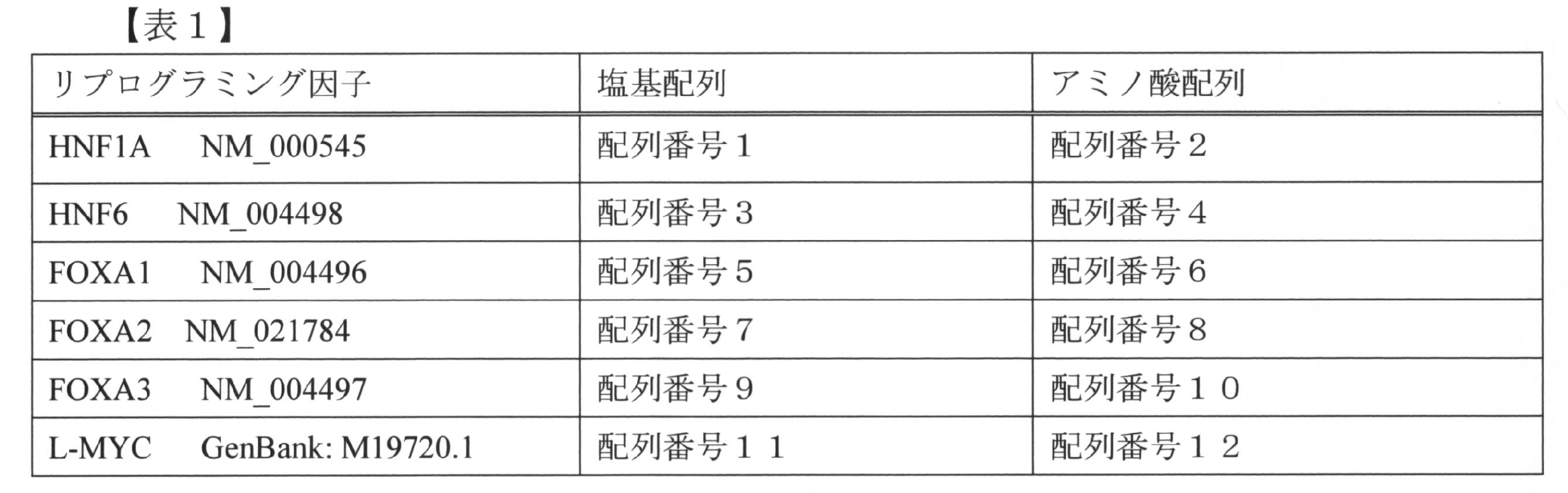
これらの因子をコードする遺伝子の一部は、「Molecular Cloning, A Laboratory Manual (4th edition)」(Cold Spring Harbor Laboratory Press (2012))等を参照してクローニングすることができ、あるいは、addgene等から入手することも可能である。
本明細書において、HNF1Aをコードする遺伝子を「HNF1A遺伝子」、HNF6をコードする遺伝子を「HNF6遺伝子」という。他の因子をコードする遺伝子についても、上記と同様に表示する。 The amino acid sequences of the above reprogramming factors and the base sequences of genes encoding these factors are shown in Table 1.
A part of the genes encoding these factors can be cloned with reference to "Molecular Cloning, A Laboratory Manual (4th edition)" (Cold Spring Harbor Laboratory Press (2012)) or the like, or from addgene etc. It is also possible to obtain.
In the present specification, a gene encoding HNF1A is referred to as “HNF1A gene”, and a gene encoding HNF6 as “HNF6 gene”. The genes encoding other factors are also displayed as described above.
本明細書において、HNF1Aをコードする遺伝子を「HNF1A遺伝子」、HNF6をコードする遺伝子を「HNF6遺伝子」という。他の因子をコードする遺伝子についても、上記と同様に表示する。 The amino acid sequences of the above reprogramming factors and the base sequences of genes encoding these factors are shown in Table 1.
In the present specification, a gene encoding HNF1A is referred to as “HNF1A gene”, and a gene encoding HNF6 as “HNF6 gene”. The genes encoding other factors are also displayed as described above.
本発明の一態様では、例えば以下の組合せのリプログラミング因子、又はこれらの因子をコードする遺伝子のセットを使用することができる。
・NF1A、HNF6及びFOXA3
・NF1A、L-MYC及びFOXA3
・NF1A、HNF6、FOXA3及びL-MYC In one aspect of the present invention, for example, the following combination of reprogramming factors, or a set of genes encoding these factors can be used.
・ NF1A, HNF6 and FOXA3
・ NF1A, L-MYC and FOXA3
・ NF1A, HNF6, FOXA3 and L-MYC
・NF1A、HNF6及びFOXA3
・NF1A、L-MYC及びFOXA3
・NF1A、HNF6、FOXA3及びL-MYC In one aspect of the present invention, for example, the following combination of reprogramming factors, or a set of genes encoding these factors can be used.
・ NF1A, HNF6 and FOXA3
・ NF1A, L-MYC and FOXA3
・ NF1A, HNF6, FOXA3 and L-MYC
本発明において使用されるリプログラミング因子(遺伝子又はタンパク質)は、表1に示す配列番号で表される塩基配列及びアミノ酸配列に限らず、以下に示す変異型も、本発明におけるリプログラミング因子の機能を有する限り含めることができる。
(a) 表1に示されるアミノ酸配列において、1個又は数個(例えば10個以下、5個以下、4個以下、3個以下又は2個)のアミノ酸が、欠失、置換又は付加されたアミノ酸配列からなり、かつ、それぞれのリプログラミング因子が有する本発明におけるリプログラミング因子としての機能を有するタンパク質
(b) 表1に示されるアミノ酸配列に対して80%以上、85%以上、90%以上、95%以上、98%以上、又は99%以上の相同性を有するアミノ酸配列からなり、かつ、それぞれのリプログラミング因子が有する本発明におけるリプログラミング因子としての機能を有するタンパク質
(c) 上記(a)のタンパク質をコードする核酸
(d) 上記(b)のタンパク質をコードする核酸
(e) 表1に示される塩基配列に相補的な塩基配列からなる核酸とストリンジェントな条件下でハイブリダイズし、かつ、それぞれのリプログラミング因子が有する本発明におけるリプログラミング因子としての機能を有するタンパク質をコードする核酸 The reprogramming factor (gene or protein) used in the present invention is not limited to the base sequences and amino acid sequences represented by SEQ ID NOs shown in Table 1, and the variants shown below also have the function of the reprogramming factor in the present invention As long as it has
(a) In the amino acid sequence shown in Table 1, one or more (for example, 10 or less, 5 or less, 4 or less, 3 or less or 2) amino acids are deleted, substituted or added A protein comprising an amino acid sequence and having a function as a reprogramming factor in the present invention possessed by each reprogramming factor (b) 80% or more, 85% or more, 90% or more of the amino acid sequences shown in Table 1 A protein having an amino acid sequence having 95% or more, 98% or more, or 99% or more homology, and having a function as a reprogramming factor in the present invention that each reprogramming factor has (c) above (a) Nucleic acid encoding the protein of (d) Nucleic acid encoding the protein of (b) above (e) Stringent conditions with nucleic acid consisting of a nucleotide sequence complementary to the nucleotide sequence shown in Table 1 A nucleic acid encoding a protein having a function as a reprogramming factor of the present invention which is hybridized below and possessed by each reprogramming factor
本発明において、「ストリンジェントな条件」とは、例えば、5×SSC、5×デンハルト溶液、0.5%SDS、50%ホルムアミド、50℃の条件である。これらの条件において、温度を上げるほど高い相同性を有するDNA及び/又はRNAが効率的に得られることが期待できる。ただし、ハイブリダイゼーションのストリンジェンシーに影響する要素としては温度、DNA及び/又はRNAの濃度、DNA及び/又はRNAの長さ、イオン強度、時間、塩濃度等の複数の要素が考えられ、当業者であればこれらの要素を適宜選択することで同様のストリンジェンシーを実現することが可能である。
In the present invention, “stringent conditions” are, for example, 5 × SSC, 5 × Denhardt's solution, 0.5% SDS, 50% formamide, 50 ° C. Under these conditions, it can be expected that DNA and / or RNA having high homology can be efficiently obtained as the temperature is raised. However, multiple factors such as temperature, concentration of DNA and / or RNA, length of DNA and / or RNA, ionic strength, time, salt concentration, etc. can be considered as factors affecting the stringency of hybridization. If it is, it is possible to realize similar stringency by appropriately selecting these elements.
3.細胞へのリプログラミング因子の導入
宿主細胞である非肝幹/非肝前駆細胞にリプログラミング因子を導入するために、リプログラミング因子をコードする遺伝子を同一の又は異なるベクターに組み込んだ組換えベクターを構築してもよい。本発明において、ベクターは、挿入セグメントの複製をもたらすために挿入され得るレプリコンを使用することができる。ベクターは、1つ以上の発現制御配列を作動可能に連結させた発現ベクターとしてもよく、リプログラミング因子以外の他のDNA配列の転写及び/又は翻訳を制御するDNAを含めることができる。発現制御配列の例としては、プロモーター、エンハンサー、転写終結領域等が挙げられる。 3. Introduction of Reprogramming Factor into Cells In order to introduce reprogramming factor into non-hepatic stem / non-hepatic precursor cells which are host cells, a recombinant vector in which a gene encoding the reprogramming factor is incorporated into the same or different vector You may build it. In the present invention, a vector can use a replicon which can be inserted to bring about replication of the inserted segment. The vector may be an expression vector operably linked to one or more expression control sequences, and may include DNA that controls transcription and / or translation of other DNA sequences other than the reprogramming factor. Examples of expression control sequences include promoters, enhancers, transcription termination regions and the like.
宿主細胞である非肝幹/非肝前駆細胞にリプログラミング因子を導入するために、リプログラミング因子をコードする遺伝子を同一の又は異なるベクターに組み込んだ組換えベクターを構築してもよい。本発明において、ベクターは、挿入セグメントの複製をもたらすために挿入され得るレプリコンを使用することができる。ベクターは、1つ以上の発現制御配列を作動可能に連結させた発現ベクターとしてもよく、リプログラミング因子以外の他のDNA配列の転写及び/又は翻訳を制御するDNAを含めることができる。発現制御配列の例としては、プロモーター、エンハンサー、転写終結領域等が挙げられる。 3. Introduction of Reprogramming Factor into Cells In order to introduce reprogramming factor into non-hepatic stem / non-hepatic precursor cells which are host cells, a recombinant vector in which a gene encoding the reprogramming factor is incorporated into the same or different vector You may build it. In the present invention, a vector can use a replicon which can be inserted to bring about replication of the inserted segment. The vector may be an expression vector operably linked to one or more expression control sequences, and may include DNA that controls transcription and / or translation of other DNA sequences other than the reprogramming factor. Examples of expression control sequences include promoters, enhancers, transcription termination regions and the like.
本発明においては、リプログラミング因子をコードする遺伝子は、導入する遺伝子セットの全てを1つのベクターに連結してもよく、1つの遺伝子ごとに別々のベクターに連結してもよい。また、1つのベクターに一部の遺伝子を連結し、他のベクターに残りの遺伝子を連結することも可能である。
In the present invention, the genes encoding reprogramming factors may be linked to one vector for all of the gene sets to be introduced, or may be linked to separate vectors for each gene. It is also possible to link some genes to one vector and link the remaining genes to another vector.
本発明において使用可能な発現ベクターとしては、プラスミド、バクテリオファージのほか、レトロウイルス、ワクシニアウイルス、アデノウイルス、レンチウイルス、アデノ随伴ウイルス、センダイウイルス等に由来するウイルスベクターなどが挙げられる。発現ベクターを非肝幹/非肝前駆細胞に導入する方法は特に限定はされず、それぞれのリプログラミング因子をコードする遺伝子を含む発現ベクターを細胞に感染させる方法が挙げられるが、これ以外にも、例えば、リポフェクション法、エレクトロポレーション法、リン酸カルシウム法などを採用することができる。ウイルスベクターを細胞に感染させるときのベクターの用量は適宜調製することができる。
Examples of expression vectors that can be used in the present invention include plasmids, bacteriophages, and virus vectors derived from retrovirus, vaccinia virus, adenovirus, lentivirus, adeno-associated virus, Sendai virus and the like. The method for introducing the expression vector into non-hepatic stem / non-hepatic precursor cells is not particularly limited, and there may be mentioned a method of infecting cells with an expression vector containing a gene encoding each reprogramming factor, For example, the lipofection method, the electroporation method, the calcium phosphate method and the like can be adopted. The dose of the vector for infecting cells with a viral vector can be appropriately adjusted.
本発明の非肝幹/非肝前駆細胞から肝幹/肝前駆細胞への誘導方法は、in vitroで行ってもよく、in vivoで行ってもよい。in vitroで行う場合、発現ベクターを非肝幹/非肝前駆細胞に導入して形質転換体を得た後は、所定期間培養してもよい。培養は、動物細胞の培養に用いられる培地を用いて行い、その後必要により継代を繰り返すことにより、肝幹/肝前駆細胞にリプログラミングすることができる。
in vivoで行う場合は、発現ベクターを既知の導入手法を用いて、例えば動物の皮膚組織や血管内、腹腔内から非肝幹/非肝前駆細胞に導入し、形質転換体を得てもよい。その後、動物組織中、又は動物組織より回収した細胞中において、肝幹/肝前駆細胞にリプログラミングした細胞を確認することができる。
得られた細胞が肝幹/肝前駆細胞にリプログラミングされたか否かは、これらの細胞に発現するマーカーにより確認することができる。マーカーとしては、例えばアルブミン、α-フェトプロテイン、E-カドヘリンなどが挙げられる。
また、本発明においては、下記因子のポリペプチドを直接細胞に導入することも可能である。
・HNF1A、HNF6及びFOXA3
・HNF1A、L-MYC及びFOXA3
・HNF1A、HNF6、FOXA3及びL-MYC
上記因子は、遺伝子工学的に製造することができ(例えば「Molecular Cloning, A Laboratory Manual (4th edition)」(Cold Spring Harbor Laboratory Press (2012)を参照)、得られたポリペプチドの細胞内への導入は、膜透過性ペプチド等に連結して細胞内に導入する方法、陽イオン性脂質を用いて細胞内に導入する方法などを採用することができ、細胞内導入試薬も市販されている(PULSin(PPU 社) 、Prote-IN(HYG 社)、BioPORTER Protein Delivery Reagent(GTS 社)等)。 The method for inducing non-hepatic stem / non-hepatic progenitor cells to hepatic stem / hepatic progenitor cells of the present invention may be performed in vitro or in vivo. When in vitro, the expression vector may be introduced into non-hepatic stem / non-hepatic precursor cells to obtain a transformant, which may be cultured for a predetermined period. Culturing can be performed on a medium used for culturing animal cells, and then reprogramming to hepatic stem / hepatic progenitor cells by repeating passaging as necessary.
When carried out in vivo, transformants may be obtained, for example, by introducing the expression vector into non-hepatic stem / non-hepatic progenitor cells from the skin tissue or blood vessel of the animal, or intraperitoneally using known introduction methods. . Thereafter, cells reprogrammed into hepatic stem / hepatic progenitor cells can be identified in animal tissues or cells recovered from animal tissues.
Whether or not the obtained cells are reprogrammed into hepatic stem / hepatic progenitor cells can be confirmed by markers expressed in these cells. Examples of the marker include albumin, α-fetoprotein, E-cadherin and the like.
In the present invention, it is also possible to introduce the following factor polypeptides directly into cells.
・ HNF1A, HNF6 and FOXA3
・ HNF1A, L-MYC and FOXA3
・ HNF1A, HNF6, FOXA3 and L-MYC
The above factors can be produced by genetic engineering (for example, "Molecular Cloning, A Laboratory Manual (4th edition)" (see Cold Spring Harbor Laboratory Press (2012)), and the obtained polypeptide is introduced into cells. The introduction may be carried out by linking to a membrane permeable peptide or the like and introducing into cells, a method of introducing into cells using cationic lipids, etc. Intracellular transfer reagents are also commercially available ( PULSin (PPU), Prote-IN (HYG), BioPORTER Protein Delivery Reagent (GTS), etc.).
実施例
以下、実施例により本発明をさらに具体的に説明する。但し、本発明の範囲はこれらの実施例により限定されるものではない。 EXAMPLES The present invention will be described more specifically by the following examples. However, the scope of the present invention is not limited by these examples.
以下、実施例により本発明をさらに具体的に説明する。但し、本発明の範囲はこれらの実施例により限定されるものではない。 EXAMPLES The present invention will be described more specifically by the following examples. However, the scope of the present invention is not limited by these examples.
(1)ヒト誘導肝前駆細胞(human induced hepatic progenitor cell: hiHepPC)を誘導する特定因子の同定
hiHepPC誘導実験の概略図を図1Aに示す。
市販のヒト臍帯静脈内皮細胞(human umbilical vein endothelial cell: HUVEC)に対し、レトロウイルス感染によりレトロウイルスベクター(遺伝子発現ベクター)を導入した。その間、HUVECはMedium 200とFibrolife serum-free cell culture mediumを1:1で混合した培地(HUVEC medium)で培養した。ウイルス感染後、2日間Hepato-medium(DMEMとF12の1:1混合培地に4%ウシ胎児血清、1 μg/ml インスリン、10-7M デキサメタゾン、10 mM ニコチンアミド、2 mM L-グルタミン、50 μM β-メルカプトエタノール、ペニシリン/ストレプトマイシンを添加したもの)に20% Fibrolife 血清フリー細胞培養培地、20 ng/ml HGF、1 μM A83-01、2 μM SB43852、5 μM Y27632を添加した培地(Hepato-medium (plus))で培養し、その後はHepato-medium (plus)にて細胞の継代を行いながら、随時解析を行った。 (1) Identification of Specific Factors that Induce Human Induced Hepatic Progenitor Cells (hiHepPC) A schematic diagram of a hiHepPC induction experiment is shown in FIG. 1A.
A retroviral vector (gene expression vector) was introduced into a commercially available human umbilical vein endothelial cell (HUVEC) by retroviral infection. Meanwhile, HUVECs were cultured in medium (HUVEC medium) in which Medium 200 and Fibrous serum-free cell culture medium were mixed at 1: 1. Hepato-medium (1% mixed medium of DMEM and F12, 1 μg / ml insulin, 10-7 M dexamethasone, 10 mM nicotinamide, 2 mM L-glutamine, 50 μM) for 2 days after virus infection Medium (Hepato-medium) to which 20% Fibrolife serum-free cell culture medium, 20 ng / ml HGF, 1 μM A83-01, 2 μM SB43852, 5 μM Y27632 was added to β-mercaptoethanol and penicillin / streptomycin added The cells were cultured in (plus), and then analysis was performed as needed while passaging the cells with Hepato-medium (plus).
HUVECに対し、空ベクター(mock)、HNF4AとFOXA3の組み合わせ(H4A/F3)、HNF4AとFOXA3、HNF1A、HNF6の組み合わせ(H4A/F3/H1A/H6)、HNF4AとFOXA3、HNF1Aの組み合わせ(H4A/F3/H1A)、HNF4AとFOXA3、HNF6の組み合わせ(H4A/F3/H6)、HNF4AとHNF1A、HNF6の組み合わせ(H4A/H1A/H6)、HNF4AとHNF1A、HNF6、ATF5、PROX1、CEBPAの組み合わせ(H4A/H1A/H6+APC)、FOXA3とHNF1A、HNF6の組み合わせ(F3/H1A/H6)のそれぞれを導入し、1~6回目の継代(P)ごとにALBUMIN(ALB)の免疫染色とALB陽性細胞数の計測を行った。また、P6においてはE-CADHERIN(E-CAD)の免疫染色も行った。
For HUVEC, empty vector (mock), combination of HNF4A and FOXA3 (H4A / F3), combination of HNF4A with FOXA3, HNF1A, HNF6 (H4A / F3 / H1A / H6), combination of HNF4A with FOXA3, HNF1A (H4A / F3 / H1A), HNF4A and FOXA3, combination of HNF6 (H4A / F3 / H6), combination of HNF4A and HNF1A, HNF6 (H4A / H1A / H6), HNF4A and HNF1A, HNF6, ATF5, PROX1, CEBPA combination (H4A / H1A / H6 + APC), combination of FOXA3 with HNF1A, HNF6 (F3 / H1A / H6), and ALBUMIN (ALB) immunostaining and ALB positive every 1st to 6th passages (P) The number of cells was counted. In P6, immunostaining of E-CADHERIN (E-CAD) was also performed.
結果を図1Bに示す。
ALBは肝前駆細胞や肝細胞で発現するマーカーであり、E-CADは上皮細胞のマーカーである。ALB及びE-CADの免疫染色、細胞の位相差写真(Phase)、並びにALB陽性細胞の割合のグラフから、F3/H1A/H6を導入した場合において、増殖能をもった肝前駆細胞様の細胞が誘導されることがわかる。 The results are shown in FIG. 1B.
ALB is a marker expressed in hepatic progenitor cells and hepatocytes, and E-CAD is a marker for epithelial cells. From the immunostaining of ALB and E-CAD, the phase contrast picture of cells (Phase), and the graph of the proportion of ALB positive cells, hepatic progenitor cell-like cells having proliferation ability when F3 / H1A / H6 is introduced Is found to be induced.
図1Bにおいて、グラフ中のPHはparental HUVECを、PNはpassage number(継代数)を表す。独立した2回の実験を行い、それぞれの結果をグラフ上の青線と赤線で示す。細胞のDNAはDAPI(青色)で染色した。スケールバー:50μm。
また、F3/H1A/H6の遺伝子セットをHUVECに導入してhiHepPC を誘導する実験を3回独立して行った。 In FIG. 1B, PH in the graph represents parental HUVEC, and PN represents passage number. Two independent experiments are performed, and the results are shown by the blue and red lines on the graph. Cellular DNA was stained with DAPI (blue). Scale bar: 50 μm.
Moreover, the experiment which introduce | transduced the gene set of F3 / H1A / H6 into HUVEC, and induced hiHepPC was performed independently 3 times.
結果を図1Cに示す。それぞれの実験で得たhiHepPC(hiHepPC-1、hiHepPC-2、hiHepPC-3)が16回の継代の中で増殖した細胞数を表す。hiHepPCは増殖を続けるが、HUVECは継代途中で増殖を停止する。
さらに、12回目の継代時におけるhiHepPCの染色体解析を行った結果を図1Dに示す。20個の細胞を解析したが、それらすべてにおいて染色体数は正常であった。 The results are shown in FIG. 1C. The number of cells grown in 16 passages of hiHepPC (hiHepPC-1, hiHepPC-2, hiHepPC-3) obtained in each experiment is shown. While hiHepPC continues to proliferate, HUVEC ceases to proliferate during passage.
Furthermore, the results of chromosomal analysis of hiHepPC at the 12th passage are shown in FIG. 1D. Twenty cells were analyzed, but all of them had normal chromosome numbers.
(2)hiHepPCから肝細胞様細胞への分化
HUVEC由来hiHepPCについて、継代(P)1~6回目のそれぞれでALBとα-フェトプロテイン(AFP)の共免疫染色を行い、ALB陽性細胞中のAFP陽性率を算出した。 (2) Differentiation of hiHepPC into hepatocyte-like cells The HUVEC-derived hiHepPC was subjected to co-immunostaining of ALB and α-fetoprotein (AFP) atpassage 1 to 6 respectively, and AFP in ALB positive cells The positive rate was calculated.
HUVEC由来hiHepPCについて、継代(P)1~6回目のそれぞれでALBとα-フェトプロテイン(AFP)の共免疫染色を行い、ALB陽性細胞中のAFP陽性率を算出した。 (2) Differentiation of hiHepPC into hepatocyte-like cells The HUVEC-derived hiHepPC was subjected to co-immunostaining of ALB and α-fetoprotein (AFP) at
結果を図2に示す。
パネルAにおいて、hiHepPC誘導の初期過程では、肝前駆細胞の特徴であるALB及びAFP共陽性細胞の割合が多いが、継代を重ねることで肝細胞の特徴であるALB単独陽性細胞の割合が増加する。この結果から、HUVECはまずhiHepPCに変化し、その後hiHepPCから肝細胞様細胞が分化してくると考えられる。グラフは、独立した3回の実験で作製したhiHepPCの解析で得たデータの平均値±標準偏差(n=3)を表す。細胞のDNAはDAPI(青色)で染色した。スケールバー:50μm。 The results are shown in FIG.
In panel A, in the initial process of hiHepPC induction, the percentage of ALB and AFP co-positive cells characteristic of hepatic progenitor cells is high, but the percentage of ALB single positive cells characteristic of hepatocytes increases by repeated passaging Do. From this result, it is thought that HUVEC first changes to hiHepPC, and then hepatocyte-like cells differentiate from hiHepPC. The graph represents the mean value ± standard deviation (n = 3) of data obtained by analysis of hiHepPC generated in three independent experiments. Cellular DNA was stained with DAPI (blue). Scale bar: 50 μm.
また、パネルBに関し、HUVEC由来hiHepPCから分化した肝細胞様細胞は、ALBだけでなく、肝細胞マーカーであるAATやASGPR1、CYP3A4も発現している。一方、HUVECは血管内皮細胞マーカーのCD31を発現するが、肝細胞マーカーの発現はすべて陰性である。細胞のDNAはDAPI(青色)で染色した。スケールバー:50μm。
In panel B, hepatocyte-like cells differentiated from HUVEC-derived hiHepPC express not only ALB but also hepatocyte markers AAT, ASGPR1 and CYP3A4. On the other hand, HUVECs express the vascular endothelial cell marker CD31, but the expression of hepatocyte markers is all negative. Cellular DNA was stained with DAPI (blue). Scale bar: 50 μm.
(3)hiHepPCから分化した肝細胞様細胞の機能解析と遺伝子発現解析
hiHepPCから分化した肝細胞様細胞の機能解析と遺伝子発現解析結果を図3に示す。
パネルAでは、HUVEC由来hiHepPCから分化した肝細胞様細胞は、肝細胞と同様にグリコーゲンの蓄積、脂質合成、及びインドシアニングリーン(ICG)の取込みと放出を行うことができることが示される。スケールバー:50μm。 (3) Functional analysis and gene expression analysis of hepatocyte-like cells differentiated from hiHepPC The results of functional analysis and gene expression analysis of hepatocyte-like cells differentiated from hiHepPC are shown in FIG.
In panel A, it is shown that hepatocyte-like cells differentiated from HUVEC-derived hiHepPC can perform glycogen accumulation, lipogenesis, and uptake and release of indocyanine green (ICG) in the same manner as hepatocytes. Scale bar: 50 μm.
パネルBは、HUVEC由来hiHepPCから分化した肝細胞様細胞が、アルブミンの分泌、尿素合成、及びシトクロムP450活性能を有することを示す。独立した3回の実験で作製したhiHepPC(hiHepPC-1、hiHepPC-2、hiHepPC-3)を解析に用いた。ヒト肝がん由来細胞株であるHepG2をポジティブコントロールとした。グラフは、平均値±標準偏差(n=3)を表す。
Panel B shows that hepatocyte-like cells differentiated from HUVEC-derived hiHepPC are capable of albumin secretion, urea synthesis, and cytochrome P450 activity. HiHepPC (hiHepPC-1, hiHepPC-2, hiHepPC-3) prepared in three independent experiments was used for analysis. HepG2, a human hepatoma-derived cell line, served as a positive control. The graph represents mean value ± standard deviation (n = 3).
パネルCは、HUVEC由来hiHepPCから分化した肝細胞様細胞が、肝細胞毒性を有する薬剤(acetaminofen、amiodarone、dicrofenac)に反応し、細胞死を起こすことを示す。独立した3回の実験で作製したhiHepPC(hiHepPC-1、hiHepPC-2、hiHepPC-3)を解析に用いた。グラフは、平均値±標準偏差(n=3)を表す。*P < 0.05、**P < 0.01
Panel C shows that hepatocyte-like cells differentiated from HUVEC-derived hiHepPC cause cell death in response to drugs having hepatotoxicity (acetaminofen, amiodarone, dicrofenac). HiHepPC (hiHepPC-1, hiHepPC-2, hiHepPC-3) prepared in three independent experiments was used for analysis. The graph represents mean value ± standard deviation (n = 3). * P <0.05, ** P <0.01
パネルDは、3つのHUVEC(HUVEC-1、HUVEC-2、HUVEC-3)と3つのhiHepPC(hiHepPC-1、hiHepPC-2、hiHepPC-3)の遺伝子発現をmRNA-seqによって解析した結果を示す。公共データからヒト肝細胞の遺伝子発現プロファイルを取得し、このプロファイルについて、HUVEC及びhiHepPCのデータと合わせて解析を行った。上段のヒートマップはクラスタリング解析結果を示しており、hiHepPCはヒト肝細胞の遺伝子発現パターンに近いがHUVECとは大きく異なることが示されている。また、下段のヒートマップでは、脂肪酸代謝やコレステロール代謝、グルコース代謝、及び薬物又は異物代謝に関連する遺伝子の発現パターンが示されており、hiHepPCから分化した肝細胞様細胞が様々な肝機能遺伝子を発現していることがわかる。
Panel D shows the results of mRNA-seq analysis of gene expression of three HUVECs (HUVEC-1, HUVEC-2, HUVEC-3) and three hiHepPCs (hiHepPC-1, hiHepPC-2, hiHepPC-3) . The gene expression profile of human hepatocytes was obtained from public data, and this profile was analyzed together with the data of HUVEC and hiHepPC. The heat map in the upper row shows the clustering analysis result, and it is shown that hiHepPC is close to the gene expression pattern of human hepatocytes but largely different from HUVEC. The heat map at the bottom shows the expression patterns of genes related to fatty acid metabolism, cholesterol metabolism, glucose metabolism, and drug or foreign substance metabolism, and hepatocyte-like cells differentiated from hiHepPC have various liver function genes. It can be seen that it is expressed.
(4)hiHepPCの移植による肝臓組織の再構築
まず、3週令の免疫不全マウス(NSGマウス)が7週令になるまで、レトロルシンを週に1回、計5回投与した。続いて、最後のレトロルシン投与から1週間後、当該マウスに四塩化炭素(CCl4)を1回投与し、肝障害を誘導した。そして、その次の日に1 x 10⁷個のHUVEC由来hiHepPCを脾臓から門脈経由で肝臓に移植した。移植後2ヶ月が経った後、肝臓と血清の解析を行った。 (4) Reconstruction of liver tissue by transplantation of hiHepPC First, retrolucin was administered once a week, for a total of 5 times, until a 3-week-old immunodeficient mouse (NSG mouse) became 7-week-old. Subsequently, one week after the last administration of retrolcine, the mice were administered a single dose of carbon tetrachloride (CCl 4) to induce liver damage. Then, on the next day, 1 × 10 7 HUVEC-derived hiHepPC were transplanted from the spleen to the liver via the portal vein. Two months after transplantation, analysis of liver and serum was performed.
まず、3週令の免疫不全マウス(NSGマウス)が7週令になるまで、レトロルシンを週に1回、計5回投与した。続いて、最後のレトロルシン投与から1週間後、当該マウスに四塩化炭素(CCl4)を1回投与し、肝障害を誘導した。そして、その次の日に1 x 10⁷個のHUVEC由来hiHepPCを脾臓から門脈経由で肝臓に移植した。移植後2ヶ月が経った後、肝臓と血清の解析を行った。 (4) Reconstruction of liver tissue by transplantation of hiHepPC First, retrolucin was administered once a week, for a total of 5 times, until a 3-week-old immunodeficient mouse (NSG mouse) became 7-week-old. Subsequently, one week after the last administration of retrolcine, the mice were administered a single dose of carbon tetrachloride (CCl 4) to induce liver damage. Then, on the next day, 1 × 10 7 HUVEC-derived hiHepPC were transplanted from the spleen to the liver via the portal vein. Two months after transplantation, analysis of liver and serum was performed.
結果を図4に示す。
パネルAは、hiHepPC移植実験の概略図を示す。
パネルBは、hiHepPC移植後のマウス血清中に存在するヒトアルブミン濃度を示し、また、hiHepPCを移植した5匹のマウスについて、それらの解析結果を示す。ネガティブコントロールとして、移植なしのNSGマウスの血清、並びにHUVECを移植したNSGマウスの血清の解析結果を示す。TPはtransplantationを表す。グラフは、平均値±標準偏差(n=2)を表す。 The results are shown in FIG.
Panel A shows a schematic of the hiHepPC transplantation experiment.
Panel B shows the concentration of human albumin present in mouse serum after hiHepPC transplantation, and also shows their analysis results for the five mice which were transplanted with hiHepPC. As a negative control, analysis results of serum of NSG mice without transplantation and serum of NSG mice grafted with HUVEC are shown. TP represents transplantation. The graph represents mean value ± standard deviation (n = 2).
パネルCは、HUVEC又はhiHepPCを移植したマウスの肝臓組織をヒトAATに対する抗体で免疫染色した結果である。hiHepPCを移植したマウス肝臓では、hiHepPC由来肝細胞様細胞による肝臓組織の再構築が観察される。TPはtransplantationを表す。細胞のDNAはDAPI(青色)で染色した。スケールバー:100μm。
Panel C shows the results of immunostaining of liver tissue of mice transplanted with HUVEC or hiHepPC with an antibody against human AAT. In the livers of mice transplanted with hiHepPC, reconstruction of liver tissue by hiHepPC-derived hepatocyte-like cells is observed. TP represents transplantation. Cellular DNA was stained with DAPI (blue). Scale bar: 100 μm.
(5)hiHepPCから胆管上皮細胞様細胞への分化
図5は、hiHepPCから胆管上皮細胞様細胞への分化を示す。
パネルAは、HUVEC及びHUVEC由来hiHepPCをマトリゲル内で1週間三次元培養すると、hiHepPCのみが上皮管腔構造をもった球状組織(スフェロイド)を形成することを示す。これらスフェロイドは継代(P)することも可能である。スケールバー:100μm。 (5) Differentiation from hiHepPC to Biliary Epithelial-Like Cells FIG. 5 shows the differentiation from hiHepPC to biliary epithelial-like cells.
Panel A shows that when HUVEC and HUVEC-derived hiHepPC are three-dimensionally cultured in Matrigel for one week, only hiHepPC forms a spherical tissue (spheroid) having an epithelial luminal structure. These spheroids can also be passaged (P). Scale bar: 100 μm.
パネルBは、HUVEC由来hiHepPCから形成されたスフェロイドが、極性(E-CAD、EZRIN、ZO-1、Phalloidinの局所的発現パターン)を有し、胆管上皮細胞マーカー(CK19、SOX9、HNF1B、CFTR、EpCAM、α-TUBULIN)を発現する上皮細胞によって形成されていることを示す。細胞のDNAはDAPI(青色)で染色した。スケールバー:50μm。
Panel B shows that spheroids formed from HUVEC-derived hiHepPC have polarity (local expression pattern of E-CAD, EZRIN, ZO-1, Phalloidin), and biliary epithelial cell markers (CK19, SOX9, HNF1B, CFTR, It is shown that it is formed by epithelial cells expressing EpCAM, α-TUBULIN). Cellular DNA was stained with DAPI (blue). Scale bar: 50 μm.
パネルCは、HUVEC由来hiHepPCから形成されたスフェロイドが、胆管と同様に蛍光色素(ローダミン)を取り込むことを示す。Verapamil添加によって取り込みが阻害されることから、この取り込みはトランスポーターを介することがわかる。スケールバー:50μm。
Panel C shows that spheroids formed from HUVEC-derived hiHepPC take up fluorescent dye (rhodamine) as well as bile duct. The uptake is inhibited by the addition of Verapamil, indicating that this uptake is via a transporter. Scale bar: 50 μm.
パネルDは、HUVEC由来hiHepPCから形成されたスフェロイドの電顕写真を示す。内腔側に微絨毛を有し、細胞間はタイトジャンクション(図中の矢頭)によって結合している。スケールバー:10μm(左図)、500nm(右図)。
Panel D shows electron micrographs of spheroids formed from HUVEC-derived hiHepPC. It has microvilli on the lumen side, and cells are connected by tight junctions (arrowheads in the figure). Scale bar: 10 μm (left), 500 nm (right).
(6)HUVEC由来hiHepPCのクローン解析による増殖能と分化能の解析
独立した3回の実験で作製したhiHepPC(hiHepPC-1、hiHepPC-2、hiHepPC-3)のそれぞれについて、フローサイトメトリー(FACS)によるクローンソーティングを行い、96ウェル培養プレートの各ウェルに1つずつ細胞を播種し、1細胞培養を行った。その後、1細胞(hiHepPCクローン)から増殖した細胞について解析を行った。 (6) Analysis of proliferation ability and differentiation ability by clone analysis of HUVEC-derived hiHepPC Flow cytometry (FACS) for each of hiHepPC (hiHepPC-1, hiHepPC-2, hiHepPC-3) prepared in three independent experiments The clones were subjected to clonal sorting, and cells were seeded one by one in each well of a 96-well culture plate to perform 1-cell culture. Thereafter, analysis was performed on cells grown from 1 cell (hiHepPC clone).
独立した3回の実験で作製したhiHepPC(hiHepPC-1、hiHepPC-2、hiHepPC-3)のそれぞれについて、フローサイトメトリー(FACS)によるクローンソーティングを行い、96ウェル培養プレートの各ウェルに1つずつ細胞を播種し、1細胞培養を行った。その後、1細胞(hiHepPCクローン)から増殖した細胞について解析を行った。 (6) Analysis of proliferation ability and differentiation ability by clone analysis of HUVEC-derived hiHepPC Flow cytometry (FACS) for each of hiHepPC (hiHepPC-1, hiHepPC-2, hiHepPC-3) prepared in three independent experiments The clones were subjected to clonal sorting, and cells were seeded one by one in each well of a 96-well culture plate to perform 1-cell culture. Thereafter, analysis was performed on cells grown from 1 cell (hiHepPC clone).
この解析手法を図6Aに、解析結果を図6B~Dに示す。
パネルBでは、hiHepPC-1、hiHepPC-2及びhiHepPC-3のそれぞれについて、1細胞培養の結果形成されるコロニーの割合(それぞれ570ウェル当たり)、及び、その後増殖を続けるクローンの割合(それぞれ570ウェル当たり)を左図、右図のそれぞれのグラフで示す。 The analysis method is shown in FIG. 6A, and the analysis results are shown in FIGS. 6B-D.
In panel B, for each of hiHepPC-1, hiHepPC-2 and hiHepPC-3, the percentage of colonies formed as a result of one cell culture (per 570 wells each) and the percentage of clones that continue to grow thereafter (570 wells each) Is shown on the left and right graphs.
パネルCは、増殖したhiHepPCクローンについて、細胞の位相差像(Phase)並びにALB及びE-CADの共免疫染色像を示す。細胞のDNAはDAPI(青色)で染色した。スケールバー:50μm。パネルDは、増殖したhiHepPCクローンをマトリゲル内で三次元培養すると胆管上皮細胞様細胞に分化し、上皮管腔構造をもったスフェロイドを形成することを示す。スケールバー:50μm。
Panel C shows phase contrast images of cells and co-immunostained images of ALB and E-CAD for the expanded hiHepPC clones. Cellular DNA was stained with DAPI (blue). Scale bar: 50 μm. Panel D shows that three-dimensional culture of the expanded hiHepPC clone in matrigel differentiates into biliary epithelial cell-like cells to form spheroids with an epithelial luminal structure. Scale bar: 50 μm.
(7)L-MYCの追加によるhiHepPCの誘導時間の短縮
HUVECに対し、FOXA3、HNF1A及びHNF6に加え、L-MYCも同時に導入し、解析を行った。 (7) Reduction of induction time of hiHepPC by addition of L-MYC In addition to FOXA3, HNF1A and HNF6, HUVEC was simultaneously introduced with L-MYC and analyzed.
HUVECに対し、FOXA3、HNF1A及びHNF6に加え、L-MYCも同時に導入し、解析を行った。 (7) Reduction of induction time of hiHepPC by addition of L-MYC In addition to FOXA3, HNF1A and HNF6, HUVEC was simultaneously introduced with L-MYC and analyzed.
結果を図7に示す。
パネルAでは、1~6回目の継代(P)ごとにALBの免疫染色を行い、P6においてはE-CADの免疫染色と位相差写真(Phase)の撮影も行った。細胞のDNAはDAPI(青色)で染色した。スケールバー:50μm。
パネルBは、HUVECにF3/H1A/H6+L-MYCの組み合わせを導入後、1~6回目の継代ごとにALB陽性細胞数の計測を行い、ALB陽性細胞の割合をグラフ化した結果を示す。グラフ中のPHはparental HUVECを、PNはpassage number(継代数)を表す。独立した2回の実験を行い、それぞれの結果をグラフ上の青線と赤線で示す。F3/H1A/H6+L-MYCの組み合わせでは、F3/H1A/H6の組み合わせ(図1B参照)よりも早くALB陽性細胞が誘導される。 The results are shown in FIG.
In panel A, ALB immunostaining was performed every 1st to 6th passages (P), and in P6, immunostaining of E-CAD and imaging of phase-contrast photographs (phase) were also performed. Cellular DNA was stained with DAPI (blue). Scale bar: 50 μm.
Panel B measures the number of ALB-positive cells at each of the first to sixth passages after introducing the F3 / H1A / H6 + L-MYC combination into HUVEC, and graphs the percentage of ALB-positive cells. Show. PH in the graph represents parental HUVEC, and PN represents passage number (passage number). Two independent experiments are performed, and the results are shown by the blue and red lines on the graph. The combination F3 / H1A / H6 + L-MYC induces ALB positive cells earlier than the combination F3 / H1A / H6 (see FIG. 1B).
(8)ヒト末梢血由来血管内皮細胞(human peripheral blood-derived endothelial cell: HPBEC)からのhiHepPC誘導
HPBECからhiHepPCを誘導する実験の概略図を図8Aに示す。まず、健常人ボランティアから採取した末梢血から単核細胞を分離し、HPBEC medium(EGMTM-2MV BulletKitTM Mediumに20%ウシ胎児血清を添加したもの)で培養してHPBECを得た。次に、HPBECに対し、レトロウイルス感染によりF3/H1A/H6の組み合わせ(3F)又はF3/H1A/H6+L-MYCの組み合わせ(4F)を導入した。ウイルス感染後、HPBEC mediumとHepato-medium (plus)との1:1混合培地で2日間培養した。続いてHepato-medium (plus)にて43日間そのまま培養し、その後は細胞の継代を行いながら、随時解析を行った。 (8) HiHepPC Induction from Human Peripheral Blood-Derived Endothelial Cells (HPBEC) A schematic diagram of an experiment for inducing hiHepPC from HPBEC is shown in FIG. 8A. First, mononuclear cells were isolated from peripheral blood collected from healthy volunteers and cultured on HPBEC medium (EGMTM-2MV Bullet KitTM Medium supplemented with 20% fetal bovine serum) to obtain HPBEC. Next, a combination of F3 / H1A / H6 (3F) or a combination of F3 / H1A / H6 + L-MYC (4F) was introduced into HPBEC by retrovirus infection. After virus infection, the cells were cultured for 2 days in a 1: 1 mixed medium of HPBEC medium and Hepato-medium (plus). Subsequently, the cells were cultured as they were in Hepato-medium (plus) for 43 days, and then analysis was performed as needed while performing cell passaging.
HPBECからhiHepPCを誘導する実験の概略図を図8Aに示す。まず、健常人ボランティアから採取した末梢血から単核細胞を分離し、HPBEC medium(EGMTM-2MV BulletKitTM Mediumに20%ウシ胎児血清を添加したもの)で培養してHPBECを得た。次に、HPBECに対し、レトロウイルス感染によりF3/H1A/H6の組み合わせ(3F)又はF3/H1A/H6+L-MYCの組み合わせ(4F)を導入した。ウイルス感染後、HPBEC mediumとHepato-medium (plus)との1:1混合培地で2日間培養した。続いてHepato-medium (plus)にて43日間そのまま培養し、その後は細胞の継代を行いながら、随時解析を行った。 (8) HiHepPC Induction from Human Peripheral Blood-Derived Endothelial Cells (HPBEC) A schematic diagram of an experiment for inducing hiHepPC from HPBEC is shown in FIG. 8A. First, mononuclear cells were isolated from peripheral blood collected from healthy volunteers and cultured on HPBEC medium (EGMTM-2MV Bullet KitTM Medium supplemented with 20% fetal bovine serum) to obtain HPBEC. Next, a combination of F3 / H1A / H6 (3F) or a combination of F3 / H1A / H6 + L-MYC (4F) was introduced into HPBEC by retrovirus infection. After virus infection, the cells were cultured for 2 days in a 1: 1 mixed medium of HPBEC medium and Hepato-medium (plus). Subsequently, the cells were cultured as they were in Hepato-medium (plus) for 43 days, and then analysis was performed as needed while performing cell passaging.
結果を図8B~Eに示す。
パネルBは、ウイルス感染を行っていないHPBEC、及び3F又は4Fを導入したHPBEC(ウイルス感染後45日目)の位相差像(Phase)、並びにALB免疫染色像を示す(注:それぞれの写真は同一視野ではない)。点線で囲われている細胞群が誘導したhiHepPC(3F又は4Fで誘導したhiHepPCを、それぞれ3F-hiHepPC、4F-hiHepPCと表記する)である。右のグラフは、ウイルス感染を行っていないHPBEC、及び3F又は4Fを導入したHPBEC(ウイルス感染後45日目と継代(P)3回目)で観察されるALB陽性細胞の割合を示す。健常人2名の末梢血を使って独立した実験を行い、それぞれの結果をグラフ上の青バーと赤バーで示した。スケールバー:100μm。 The results are shown in FIGS. 8B-E.
Panel B shows the phase contrast image (Phase) of HPBEC without virus infection, and HPBEC (45 days after virus infection) into which 3F or 4F was introduced, and ALB immunostaining image (note: each picture shows Not in the same field of vision). HiHepPCs (3F or 4F-induced hiHepPCs are denoted as 3F-hiHepPC and 4F-hiHepPC, respectively) derived from the cell group surrounded by a dotted line. The graph on the right shows the percentage of ALB-positive cells observed in HPBEC not infected with virus and HPBEC introduced with 3F or 4F (day 45 after virus infection and passage (P) third). Independent experiments were conducted using the peripheral blood of two healthy individuals, and the results are shown by blue and red bars on the graph. Scale bar: 100 μm.
パネルBは、ウイルス感染を行っていないHPBEC、及び3F又は4Fを導入したHPBEC(ウイルス感染後45日目)の位相差像(Phase)、並びにALB免疫染色像を示す(注:それぞれの写真は同一視野ではない)。点線で囲われている細胞群が誘導したhiHepPC(3F又は4Fで誘導したhiHepPCを、それぞれ3F-hiHepPC、4F-hiHepPCと表記する)である。右のグラフは、ウイルス感染を行っていないHPBEC、及び3F又は4Fを導入したHPBEC(ウイルス感染後45日目と継代(P)3回目)で観察されるALB陽性細胞の割合を示す。健常人2名の末梢血を使って独立した実験を行い、それぞれの結果をグラフ上の青バーと赤バーで示した。スケールバー:100μm。 The results are shown in FIGS. 8B-E.
Panel B shows the phase contrast image (Phase) of HPBEC without virus infection, and HPBEC (45 days after virus infection) into which 3F or 4F was introduced, and ALB immunostaining image (note: each picture shows Not in the same field of vision). HiHepPCs (3F or 4F-induced hiHepPCs are denoted as 3F-hiHepPC and 4F-hiHepPC, respectively) derived from the cell group surrounded by a dotted line. The graph on the right shows the percentage of ALB-positive cells observed in HPBEC not infected with virus and HPBEC introduced with 3F or 4F (
パネルCでは、HPBEC から誘導した3F-hiHepPCと4F-hiHepPCは共に一部の細胞がAFP陽性であるが、3F-hiHepPCと4F-hiHepPCから分化した肝細胞様細胞は、ALBだけでなく、肝細胞マーカーであるAAT、ASGPR1及びCYP3A4を発現していることが示される。また、3F-hiHepPCと4F-hiHepPCから分化した肝細胞様細胞は、肝細胞と同様にグリコーゲンの蓄積や脂質合成、インドシアニングリーン(ICG)の取込みと放出を行うことができる。さらに、3F-hiHepPCと4F-hiHepPCをマトリゲル内で三次元培養すると胆管上皮細胞様細胞に分化し、上皮管腔構造をもったスフェロイドを形成する。細胞のDNAはDAPI(青色)で染色した。スケールバー:50μm。
In panel C, 3F-hiHepPC and 4F-hiHepPC both derived from HPBEC are AFP positive in some cells, but hepatocyte-like cells differentiated from 3F-hiHepPC and 4F-hiHepPC are not only ALB but also liver It is shown to express the cellular markers AAT, ASGPR1 and CYP3A4. In addition, hepatocyte-like cells differentiated from 3F-hiHepPC and 4F-hiHepPC can perform glycogen accumulation, lipid synthesis, and indocyanine green (ICG) uptake and release, like hepatocytes. Furthermore, three-dimensional culture of 3F-hiHepPC and 4F-hiHepPC in matrigel differentiates them into biliary epithelial cell-like cells and forms spheroids with an epithelial luminal structure. Cellular DNA was stained with DAPI (blue). Scale bar: 50 μm.
パネルDでは、3F-hiHepPCと4F-hiHepPCから分化した肝細胞様細胞が、アルブミンの分泌、尿素合成及びシトクロムP450活性能を有することが示される。健常人2名の末梢血を使って作製した3F-hiHepPC(3F-hiHepPC-1、3F-hiHepPC-2)と4F-hiHepPC(4F-hiHepPC-1、4F-hiHepPC-2)を解析に用いた。ヒト肝がん由来細胞株であるHepG2をポジティブコントロールとした。グラフは、平均値±標準偏差(n=3)を表す。
In panel D, hepatocyte-like cells differentiated from 3F-hiHepPC and 4F-hiHepPC are shown to have albumin secretion, urea synthesis and cytochrome P450 activity. Analysis was performed using 3F-hiHepPC (3F-hiHepPC-1, 3F-hiHepPC-2) and 4F-hiHepPC (4F-hiHepPC-1, 4F-hiHepPC-2), which were prepared using peripheral blood of two healthy subjects, for analysis. . HepG2, a human hepatoma-derived cell line, served as a positive control. The graph represents mean value ± standard deviation (n = 3).
パネルEは、3F-hiHepPCと4F-hiHepPCから分化した肝細胞様細胞が、肝細胞毒性を有する薬剤(acetaminofen、amiodarone、dicrofenac)に反応し、細胞死を起こすことを示す。健常人2名の末梢血を使って作製した3F-hiHepPC(3F-hiHepPC-1、3F-hiHepPC-2)と4FhiHepPC(4F-hiHepPC-1、4F-hiHepPC-2)を解析に用いた。グラフは、平均値±標準偏差(n=3)を表す。*P < 0.05、**P < 0.01
Panel E shows that hepatocyte-like cells differentiated from 3F-hiHepPC and 4F-hiHepPC respond to hepatotoxic drugs (acetaminofen, amiodarone, dicrofenac) to cause cell death. 3F-hiHepPC (3F-hiHepPC-1, 3F-hiHepPC-2) and 4FhiHepPC (4F-hiHepPC-1, 4F-hiHepPC-2) which were prepared using peripheral blood of 2 healthy persons were used for analysis. The graph represents mean value ± standard deviation (n = 3). * P <0.05, ** P <0.01
(9)HPBEC由来4F-hiHepPCのクローン解析による増殖能と分化能の解析
細胞のクローン解析手法及び結果を図9に示す。
パネルAにおいて、点線で囲われたHPBEC由来4F-hiHepPCのコロニーをピックアップし、細胞を分散させた上で96ウェル培養プレートの各ウェルに1つずつ細胞を播種し、1細胞培養を行った。その後、1細胞(4F-hiHepPCクローン)から増殖した細胞について解析を行った。スケールバー:100μm。 (9) Analysis of proliferation ability and differentiation ability by clone analysis of HPBEC-derived 4F-hiHepPC The clone analysis method of cells and the result are shown in FIG.
In panel A, colonies of HPBEC-derived 4F-hiHepPC surrounded by a dotted line were picked up, the cells were dispersed, and cells were seeded one by one in each well of a 96-well culture plate to carry out single cell culture. Thereafter, analysis was performed on cells grown from 1 cell (4F-hiHepPC clone). Scale bar: 100 μm.
パネルBは、左から、96ウェル培養プレートの1ウェルに播種した1細胞(4F-hiHepPCクローン)の位相差像(Phase)、1細胞から増殖した細胞の位相差像(Phase)、増殖した細胞のALB及びE-CAD共免疫染色像、増殖した細胞から分化した胆管上皮細胞様細胞が三次元培養下で形成する上皮管腔構造をもったスフェロイドを示す。細胞のDNAはDAPI(青色)で染色した。スケールバー:50μm。
Panel B shows from the left, a phase contrast image (Phase) of 1 cell (4F-hiHepPC clone) seeded in 1 well of a 96 well culture plate, a phase contrast image (Phase) of cells grown from 1 cell, expanded cells Fig. 11 shows ALB and E-CAD co-immunostained images, and spheroids with an epithelial luminal structure formed by biliary epithelial cell-like cells differentiated from proliferated cells in three-dimensional culture. Cellular DNA was stained with DAPI (blue). Scale bar: 50 μm.
(10)FOXA1及びFOXA2を用いた実験
図10は、hiHepPC誘導因子の1つであるFOXA3が、FOXA1やFOXA2に置き換えることができることを示す。
パネルAでは、FOXA1、HNF1A及びHNF6の組み合わせでHUVECから誘導したhiHepPC (FOXA1)、並びにFOXA2、HNF1A及びHNF6の組み合わせでHUVECから誘導したhiHepPC (FOXA2)が、どちらもFOXA3、HNF1A及びHNF6の組み合わせで誘導したhiHepPCと同様に上皮細胞形態を有し、血管内皮細胞マーカーであるCD31は発現せず、一部の細胞はAFP陽性であることが示される。一方で、hiHepPC (FOXA1)とhiHepPC (FOXA2)から分化した肝細胞様細胞は、ALBだけでなく、肝細胞マーカーであるAAT、ASGPR1及びCYP3A4を発現している。細胞のDNAはDAPI(青色)で染色した。スケールバー:50μm。 (10) Experiment using FOXA1 and FOXA2 FIG. 10 shows that FOXA3 which is one of the hiHepPC inducers can be replaced with FOXA1 or FOXA2.
In panel A, HUPEC-derived hiHepPC (FOXA1) derived from the combination of FOXA1, HNF1A and HNF6, and HiHEPPC derived from HUVEC (FOXA2) derived from the combination of FOXA2, HNF1A and HNF6, both with the combination of FOXA3, HNF1A and HNF6 It has an epithelial cell morphology similar to induced hiHepPC, does not express the vascular endothelial cell marker CD31, and some cells are shown to be AFP positive. On the other hand, hepatocyte-like cells differentiated from hiHepPC (FOXA1) and hiHepPC (FOXA2) express not only ALB but also hepatocyte markers AAT, ASGPR1 and CYP3A4. Cellular DNA was stained with DAPI (blue). Scale bar: 50 μm.
パネルBでは、HUVEC 由来のhiHepPC (FOXA1)とhiHepPC (FOXA2)から分化した肝細胞様細胞が、肝細胞と同様にグリコーゲンの蓄積、脂質合成、並びにインドシアニングリーン(ICG)の取込み及び放出を行うことができることを示す。さらに、hiHepPC (FOXA1)とhiHepPC (FOXA2)をマトリゲル内で三次元培養すると胆管上皮細胞様細胞に分化し、上皮管腔構造をもったスフェロイドを形成する。スケールバー:50μm。
In panel B, hepatocyte-like cells differentiated from HUVEC-derived hiHepPC (FOXA1) and hiHepPC (FOXA2) perform glycogen accumulation, lipid synthesis, and indocyanine green (ICG) uptake and release in the same manner as hepatocytes. Show that you can. Furthermore, when hiHepPC (FOXA1) and hiHepPC (FOXA2) are three-dimensionally cultured in matrigel, they differentiate into biliary epithelial cell-like cells and form spheroids having an epithelial luminal structure. Scale bar: 50 μm.
(11)ヒト末梢血由来T細胞(human peripheral blood-derived T cell: HPBTC)からのhiHepPC誘導
ヒト末梢血から単核細胞を分離後、T細胞培地(HPBEC mediumとFibrolife serum-free cell culture mediumの1:1混合培地に10 ng/ml rIL-2、0.05 pl/cell Dynabeads HumanT-Activator CD3/CD28を添加したもの)で培養し、T細胞の増殖を促進させた。 (11) HiHepPC induction from human peripheral blood-derived T cells (HPBTC) After separating mononuclear cells from human peripheral blood, T cell culture media (HPBEC medium and Fibrolife serum-free cell culture medium) The proliferation of T cells was promoted by culturing in 1: 1 mixed medium with 10 ng / ml rIL-2, 0.05 pl / cell Dynabeads Human T-Activator CD3 / CD28).
ヒト末梢血から単核細胞を分離後、T細胞培地(HPBEC mediumとFibrolife serum-free cell culture mediumの1:1混合培地に10 ng/ml rIL-2、0.05 pl/cell Dynabeads HumanT-Activator CD3/CD28を添加したもの)で培養し、T細胞の増殖を促進させた。 (11) HiHepPC induction from human peripheral blood-derived T cells (HPBTC) After separating mononuclear cells from human peripheral blood, T cell culture media (HPBEC medium and Fibrolife serum-free cell culture medium) The proliferation of T cells was promoted by culturing in 1: 1 mixed medium with 10 ng / ml rIL-2, 0.05 pl / cell Dynabeads Human T-Activator CD3 / CD28).
結果を図11に示す。
パネルAの写真は、hiHepPC誘導前のHPBTC位相差像(Phase)を示す。スケールバー:50μm。
パネルBは、HPBTCにF3/H1A/H6の組み合わせ(3F)、又はF3/H1A/H6+L-MYCの組み合わせ(4F)を導入して作製した3F-hiHepPC及び4F-hiHepPCの位相差像(Phase)、並びにALB及びE-CAD共免疫染色像である。細胞のDNAはDAPI(青色)で染色した。スケールバー:50μm。 The results are shown in FIG.
The photograph of panel A shows the HPBTC phase contrast image (Phase) before hiHepPC induction. Scale bar: 50 μm.
Panel B is a phase-contrast image of 3F-hiHepPC and 4F-hiHepPC produced by introducing HPBTC with F3 / H1A / H6 combination (3F) or F3 / H1A / H6 + L-MYC combination (4F) (Phase), and ALB and E-CAD co-immunostained images. Cellular DNA was stained with DAPI (blue). Scale bar: 50 μm.
以上の実施例により、HUVEC、HPBEC又はHPBTCに、FOXA3、HNF1A及びHNF6の組み合わせ(必要に応じてL-MYCを追加)を導入することにより、増殖能を有し、肝細胞様細胞と胆管上皮細胞様細胞に分化できるhiHepPCを誘導(ダイレクトリプログラミング)できることが明らかとなった。本実施例のまとめを図12に示す。
By introducing the combination of FOXA3, HNF1A and HNF6 (addition of L-MYC as required) into HUVEC, HPBEC or HPBTC according to the above example, it has proliferative ability, and hepatocyte-like cells and biliary epithelium It became clear that hiHepPC which can differentiate into cell-like cells can be induced (direct reprogramming). A summary of this embodiment is shown in FIG.
同様にして、ヒト線維芽細胞を肝細胞様細胞と胆管上皮細胞様細胞に分化させることに成功した。また、同様にして、ヒト臍帯血T細胞を肝細胞様細胞と胆管上皮細胞様細胞に分化させることに成功した。
Similarly, human fibroblasts were successfully differentiated into hepatocyte-like cells and biliary epithelial cell-like cells. Similarly, human umbilical cord blood T cells were successfully differentiated into hepatocyte-like cells and biliary epithelial cell-like cells.
さらに、ヒト臍帯血T細胞に、FOXA3、HNF1A及びL-MYCの組み合わせを導入することにより、ヒト臍帯血T細胞を肝細胞様細胞と胆管上皮細胞様細胞に分化させることに成功した。
Furthermore, human umbilical cord blood T cells were successfully differentiated into hepatocyte-like cells and biliary epithelial cell-like cells by introducing a combination of FOXA3, HNF1A and L-MYC into human umbilical cord blood T cells.
配列番号12:XaaはSer又はThrを表す(存在位置:362)。
Sequence number 12: Xaa represents Ser or Thr (location: 362).
Claims (11)
- 非肝幹細胞又は非肝前駆細胞に、以下の組合せを導入することを特徴とする、非肝幹細胞又は非肝前駆細胞から肝幹細胞又は肝前駆細胞への誘導方法:
(a)HNF1、HNF6及びFOXAの組合せ、
(b)HNF1遺伝子、HNF6遺伝子及びFOXA遺伝子の組合せ、
(c)HNF1、MYC及びFOXAの組合せ、又は
(d)HNF1遺伝子、MYC遺伝子及びFOXA遺伝子の組合せ。 A method for inducing liver stem cells or hepatic progenitor cells from non-hepatic stem cells or non-hepatic progenitor cells, which comprises introducing the following combination into non-hepatic stem cells or non-hepatic progenitor cells:
(A) combination of HNF1, HNF6 and FOXA,
(B) combination of HNF1, HNF6 and FOXA genes,
(C) A combination of HNF1, MYC and FOXA, or (d) a combination of HNF1, MYC and FOXA genes. - 請求項1に記載の方法によって肝幹細胞又は肝前駆細胞を誘導する工程を含む、肝幹細胞又は肝前駆細胞の製造方法。 A method for producing hepatic stem cells or hepatic progenitor cells, comprising the step of inducing hepatic stem cells or hepatic progenitor cells by the method according to claim 1.
- HNF1がHNF1Aである、請求項1又は2に記載の方法。 The method according to claim 1 or 2, wherein HNF1 is HNF1A.
- FOXAがFOXA3である、請求項1~3のいずれか1項に記載の方法。 The method according to any one of claims 1 to 3, wherein FOXA is FOXA3.
- 組合せが(a)又は(b)であって、さらに、MYC又はMYC遺伝子を導入する、請求項1~4のいずれか1項に記載の方法。 The method according to any one of claims 1 to 4, wherein the combination is (a) or (b) and further, the MYC or MYC gene is introduced.
- MYCがL-MYCである、請求項1~5のいずれか1項に記載の方法。 The method according to any one of claims 1 to 5, wherein MYC is L-MYC.
- 非肝幹細胞又は非肝前駆細胞が、血管内皮細胞又は血液由来細胞である、請求項1~6のいずれか1項に記載の方法。 The method according to any one of claims 1 to 6, wherein the non-hepatic stem cells or non-hepatic progenitor cells are vascular endothelial cells or blood-derived cells.
- 血管内皮細胞が臍帯静脈、末梢血又は臍帯血由来のものである請求項7に記載の方法。 8. The method according to claim 7, wherein the vascular endothelial cells are derived from umbilical vein, peripheral blood or cord blood.
- 血液由来細胞が末梢血T細胞又は臍帯血T細胞である請求項7に記載の方法。 The method according to claim 7, wherein the blood-derived cells are peripheral blood T cells or cord blood T cells.
- 請求項2~9のいずれか1項に記載の方法により製造された、肝幹細胞又は肝前駆細胞。 A hepatic stem cell or hepatic progenitor cell produced by the method according to any one of claims 2 to 9.
- (a)請求項1に記載の方法によって肝幹細胞又は肝前駆細胞を誘導する工程、及び
(b)誘導した肝幹細胞又は肝前駆細胞を肝細胞又は胆管上皮細胞に分化させる工程
を含む、肝細胞又は胆管上皮細胞の製造方法。 (A) inducing hepatic stem cells or hepatic progenitor cells by the method according to claim 1, and (b) differentiating the induced hepatic stem cells or hepatic progenitor cells into hepatic cells or biliary epithelial cells Or a method of producing biliary epithelial cells.
Priority Applications (3)
| Application Number | Priority Date | Filing Date | Title |
|---|---|---|---|
| US16/757,817 US11981927B2 (en) | 2017-10-23 | 2018-10-23 | Method for producing liver stem cells or liver progenitor cells by direct reprogramming |
| EP18870975.2A EP3702449A4 (en) | 2017-10-23 | 2018-10-23 | Method for producing liver stem cells or liver progenitor cells by direct reprogramming |
| JP2019551148A JP7307478B2 (en) | 2017-10-23 | 2018-10-23 | Method for producing hepatic stem cells or hepatic progenitor cells by direct reprogramming |
Applications Claiming Priority (2)
| Application Number | Priority Date | Filing Date | Title |
|---|---|---|---|
| JP2017-204432 | 2017-10-23 | ||
| JP2017204432 | 2017-10-23 |
Publications (1)
| Publication Number | Publication Date |
|---|---|
| WO2019082874A1 true WO2019082874A1 (en) | 2019-05-02 |
Family
ID=66246576
Family Applications (1)
| Application Number | Title | Priority Date | Filing Date |
|---|---|---|---|
| PCT/JP2018/039295 WO2019082874A1 (en) | 2017-10-23 | 2018-10-23 | Method for producing liver stem cells or liver progenitor cells by direct reprogramming |
Country Status (4)
| Country | Link |
|---|---|
| US (1) | US11981927B2 (en) |
| EP (1) | EP3702449A4 (en) |
| JP (1) | JP7307478B2 (en) |
| WO (1) | WO2019082874A1 (en) |
Citations (4)
| Publication number | Priority date | Publication date | Assignee | Title |
|---|---|---|---|---|
| WO2011052504A1 (en) * | 2009-10-28 | 2011-05-05 | 財団法人ヒューマンサイエンス振興財団 | Method for induction of differentiation of stem cells into hepatocytes |
| JP2015527084A (en) | 2012-09-07 | 2015-09-17 | ジェネンテック, インコーポレイテッド | Methods and compositions for producing induced hepatocytes |
| JP2016010379A (en) * | 2014-06-30 | 2016-01-21 | 国立研究開発法人医薬基盤・健康・栄養研究所 | Drug evaluation cells and drug evaluation method |
| JP2017511150A (en) * | 2014-02-12 | 2017-04-20 | 北昊干細胞与再生医学研究院有限公司BeiHao Stem Cell and Regenerative Medicine Translational Research Institute | Kits and methods for reprogramming non-hepatocytes to hepatocytes |
Family Cites Families (1)
| Publication number | Priority date | Publication date | Assignee | Title |
|---|---|---|---|---|
| WO2013189521A1 (en) | 2012-06-19 | 2013-12-27 | Waclawczyk Simon | Method of generating cells of hepatocyte phenotype |
-
2018
- 2018-10-23 EP EP18870975.2A patent/EP3702449A4/en active Pending
- 2018-10-23 WO PCT/JP2018/039295 patent/WO2019082874A1/en unknown
- 2018-10-23 US US16/757,817 patent/US11981927B2/en active Active
- 2018-10-23 JP JP2019551148A patent/JP7307478B2/en active Active
Patent Citations (4)
| Publication number | Priority date | Publication date | Assignee | Title |
|---|---|---|---|---|
| WO2011052504A1 (en) * | 2009-10-28 | 2011-05-05 | 財団法人ヒューマンサイエンス振興財団 | Method for induction of differentiation of stem cells into hepatocytes |
| JP2015527084A (en) | 2012-09-07 | 2015-09-17 | ジェネンテック, インコーポレイテッド | Methods and compositions for producing induced hepatocytes |
| JP2017511150A (en) * | 2014-02-12 | 2017-04-20 | 北昊干細胞与再生医学研究院有限公司BeiHao Stem Cell and Regenerative Medicine Translational Research Institute | Kits and methods for reprogramming non-hepatocytes to hepatocytes |
| JP2016010379A (en) * | 2014-06-30 | 2016-01-21 | 国立研究開発法人医薬基盤・健康・栄養研究所 | Drug evaluation cells and drug evaluation method |
Non-Patent Citations (10)
| Title |
|---|
| "Molecular Cloning, A Laboratory Manual", 2012, COLD SPRING HARBOR LABORATORY PRESS |
| BING YU ET AL., CELL STEM CELL, vol. 13, 5 September 2013 (2013-09-05), pages 1 - 13 |
| DING, YI ET AL.: "Overexpression of transcription factor Foxa2 and Hnfla induced rat bone mesenchymal stem cells into hepatocytes", CYTOTECHNOLOGY, vol. 68, 2016, pages 2037 - 2047, XP036055365, DOI: 10.1007/s10616-016-9944-7 * |
| MIURA, SHIZUKA ET AL.: "Generation of Mouse and Human Organoid-Forming Intestinal Progenitor Cells by Direct Lineage Reprogramming", CELL STEM CELL, vol. 21, 5 October 2017 (2017-10-05), pages 456 - 471, XP085208263 * |
| NAKAGAWA, MASATO ET AL.: "Promotion of direct reprogramming by transformation-deficient Myc", PROC. NATL. ACAD. SCI. USA, vol. 107, no. 32, 2010, pages 14152 - 14157, XP008151220, DOI: 10.1073/pnas.1009374107 * |
| PENGYU HUANG ET AL., CELL STEM CELL, vol. 14, 6 March 2014 (2014-03-06), pages 1 - 10 |
| See also references of EP3702449A4 |
| SUZUKI, ATSUSHI: "Direct induction technology of hepatocytes for medical application", EXPERIMENTAL MEDICINE, vol. 33, no. 2, 2015, pages 72 - 79, XP009520815, ISSN: 0288-5514 * |
| WANG, MINJUN ET AL.: "Regulation of transcriptional factor during liver development", SHENGMING DE HUAXUE = CHEMISTRY OF LIFE, vol. 31, no. 3, 2011, pages 395 - 400, XP009520812, DOI: 10.13488/j.smhx.2011.03.018 * |
| YU , BING ET AL.: "Reprogramming Fibroblasts into Bipotential Hepatic Stem Cells by Defined Factors", CELL STEM CELL, vol. 13, 2013, pages 328 - 340, XP055194339, DOI: 10.1016/j.stem.2013.06.017 * |
Also Published As
| Publication number | Publication date |
|---|---|
| EP3702449A4 (en) | 2021-07-28 |
| US11981927B2 (en) | 2024-05-14 |
| JP7307478B2 (en) | 2023-07-12 |
| US20210189347A1 (en) | 2021-06-24 |
| JPWO2019082874A1 (en) | 2020-09-17 |
| EP3702449A1 (en) | 2020-09-02 |
Similar Documents
| Publication | Publication Date | Title |
|---|---|---|
| Bossolasco et al. | Neuro-glial differentiation of human bone marrow stem cells in vitro | |
| Verfaillie et al. | Unexpected potential of adult stem cells | |
| Marcus et al. | Fetal stem cells from extra‐embryonic tissues: do not discard | |
| Kobayashi et al. | Wnt4-transformed mouse embryonic stem cells differentiate into renal tubular cells | |
| Sun et al. | Retinal stem/progenitor properties of iris pigment epithelial cells | |
| US11920160B2 (en) | Pancreatic insulin-producing beta-cell lines derived from human pluripotent stem cells | |
| WO2011102532A1 (en) | Induced hepatocytes | |
| JP2009225661A (en) | Method for inducing differentiation of embryonic stem cell into insulin-secreting cell, insulin-secreting cell induced by the method and use of the same | |
| JPWO2007010858A1 (en) | Pluripotent stem cells cloned from single cells derived from skeletal muscle tissue | |
| Dimomeletis et al. | Assessment of human MAPCs for stem cell transplantation and cardiac regeneration after myocardial infarction in SCID mice | |
| JP2015109833A (en) | Method of producing endothelial cell from fibroblast | |
| Cheng et al. | Conversion of human adipose-derived stem cells into functional and expandable endothelial-like cells for cell-based therapies | |
| US20190352601A1 (en) | Engineering blood vessel cells for transplantation | |
| JP6095272B2 (en) | Method for producing epithelial somatic stem cells | |
| JPWO2005024004A1 (en) | Method for differentiating mesenchymal stem cells into hepatocytes and artificial human hepatocytes | |
| JP7307478B2 (en) | Method for producing hepatic stem cells or hepatic progenitor cells by direct reprogramming | |
| Boonkaew et al. | Efficient generation of endothelial cells from induced pluripotent stem cells derived from a patient with peripheral arterial disease | |
| JP2018113962A (en) | Method of producing well-differentiated pluripotent stem cell derived cells | |
| KR101587231B1 (en) | Composition and method for promoting direct conversion of fibroblasts into hepatocytes | |
| JP2005095138A (en) | Method for inducing differentiation of cell having pluripotency | |
| KR20200045984A (en) | Methods for preparing in vitro cultured hepatocytes using er stress reliever and in vitro cultured hepatocytes prepared by thereof | |
| WO2014038652A1 (en) | Production method for pancreas-derived somatic stem cells | |
| KR20200045983A (en) | Methods for preparing in vitro cultured hepatocytes using er stress reliever and in vitro cultured hepatocytes prepared by thereof | |
| WO2015190522A1 (en) | Method for producing endodermal stem cells | |
| WO2014038655A1 (en) | Method of producing intestinal epithelium-derived somatic stem cells |
Legal Events
| Date | Code | Title | Description |
|---|---|---|---|
| 121 | Ep: the epo has been informed by wipo that ep was designated in this application |
Ref document number: 18870975 Country of ref document: EP Kind code of ref document: A1 |
|
| ENP | Entry into the national phase |
Ref document number: 2019551148 Country of ref document: JP Kind code of ref document: A |
|
| NENP | Non-entry into the national phase |
Ref country code: DE |
|
| ENP | Entry into the national phase |
Ref document number: 2018870975 Country of ref document: EP Effective date: 20200525 |