WO2019079707A1 - Detection of symmetrical dimethylarginine - Google Patents
Detection of symmetrical dimethylarginine Download PDFInfo
- Publication number
- WO2019079707A1 WO2019079707A1 PCT/US2018/056687 US2018056687W WO2019079707A1 WO 2019079707 A1 WO2019079707 A1 WO 2019079707A1 US 2018056687 W US2018056687 W US 2018056687W WO 2019079707 A1 WO2019079707 A1 WO 2019079707A1
- Authority
- WO
- WIPO (PCT)
- Prior art keywords
- sdma
- antibody
- affinity
- protein
- mma
- Prior art date
Links
- YVRYCMRLSCZAKE-UHFFFAOYSA-N CNC(NNCCSC(CC(N1CCC2CCCCC2)=O)C1O)=N Chemical compound CNC(NNCCSC(CC(N1CCC2CCCCC2)=O)C1O)=N YVRYCMRLSCZAKE-UHFFFAOYSA-N 0.000 description 1
Classifications
-
- G—PHYSICS
- G01—MEASURING; TESTING
- G01N—INVESTIGATING OR ANALYSING MATERIALS BY DETERMINING THEIR CHEMICAL OR PHYSICAL PROPERTIES
- G01N33/00—Investigating or analysing materials by specific methods not covered by groups G01N1/00 - G01N31/00
- G01N33/48—Biological material, e.g. blood, urine; Haemocytometers
- G01N33/50—Chemical analysis of biological material, e.g. blood, urine; Testing involving biospecific ligand binding methods; Immunological testing
- G01N33/68—Chemical analysis of biological material, e.g. blood, urine; Testing involving biospecific ligand binding methods; Immunological testing involving proteins, peptides or amino acids
- G01N33/6893—Chemical analysis of biological material, e.g. blood, urine; Testing involving biospecific ligand binding methods; Immunological testing involving proteins, peptides or amino acids related to diseases not provided for elsewhere
-
- G—PHYSICS
- G01—MEASURING; TESTING
- G01N—INVESTIGATING OR ANALYSING MATERIALS BY DETERMINING THEIR CHEMICAL OR PHYSICAL PROPERTIES
- G01N33/00—Investigating or analysing materials by specific methods not covered by groups G01N1/00 - G01N31/00
- G01N33/48—Biological material, e.g. blood, urine; Haemocytometers
- G01N33/50—Chemical analysis of biological material, e.g. blood, urine; Testing involving biospecific ligand binding methods; Immunological testing
- G01N33/53—Immunoassay; Biospecific binding assay; Materials therefor
- G01N33/543—Immunoassay; Biospecific binding assay; Materials therefor with an insoluble carrier for immobilising immunochemicals
- G01N33/54353—Immunoassay; Biospecific binding assay; Materials therefor with an insoluble carrier for immobilising immunochemicals with ligand attached to the carrier via a chemical coupling agent
-
- G—PHYSICS
- G01—MEASURING; TESTING
- G01N—INVESTIGATING OR ANALYSING MATERIALS BY DETERMINING THEIR CHEMICAL OR PHYSICAL PROPERTIES
- G01N33/00—Investigating or analysing materials by specific methods not covered by groups G01N1/00 - G01N31/00
- G01N33/48—Biological material, e.g. blood, urine; Haemocytometers
- G01N33/50—Chemical analysis of biological material, e.g. blood, urine; Testing involving biospecific ligand binding methods; Immunological testing
- G01N33/53—Immunoassay; Biospecific binding assay; Materials therefor
- G01N33/543—Immunoassay; Biospecific binding assay; Materials therefor with an insoluble carrier for immobilising immunochemicals
- G01N33/54366—Apparatus specially adapted for solid-phase testing
-
- G—PHYSICS
- G01—MEASURING; TESTING
- G01N—INVESTIGATING OR ANALYSING MATERIALS BY DETERMINING THEIR CHEMICAL OR PHYSICAL PROPERTIES
- G01N33/00—Investigating or analysing materials by specific methods not covered by groups G01N1/00 - G01N31/00
- G01N33/48—Biological material, e.g. blood, urine; Haemocytometers
- G01N33/50—Chemical analysis of biological material, e.g. blood, urine; Testing involving biospecific ligand binding methods; Immunological testing
- G01N33/53—Immunoassay; Biospecific binding assay; Materials therefor
- G01N33/563—Immunoassay; Biospecific binding assay; Materials therefor involving antibody fragments
-
- G—PHYSICS
- G01—MEASURING; TESTING
- G01N—INVESTIGATING OR ANALYSING MATERIALS BY DETERMINING THEIR CHEMICAL OR PHYSICAL PROPERTIES
- G01N33/00—Investigating or analysing materials by specific methods not covered by groups G01N1/00 - G01N31/00
- G01N33/48—Biological material, e.g. blood, urine; Haemocytometers
- G01N33/50—Chemical analysis of biological material, e.g. blood, urine; Testing involving biospecific ligand binding methods; Immunological testing
- G01N33/68—Chemical analysis of biological material, e.g. blood, urine; Testing involving biospecific ligand binding methods; Immunological testing involving proteins, peptides or amino acids
- G01N33/6803—General methods of protein analysis not limited to specific proteins or families of proteins
- G01N33/6806—Determination of free amino acids
- G01N33/6812—Assays for specific amino acids
-
- G—PHYSICS
- G01—MEASURING; TESTING
- G01N—INVESTIGATING OR ANALYSING MATERIALS BY DETERMINING THEIR CHEMICAL OR PHYSICAL PROPERTIES
- G01N33/00—Investigating or analysing materials by specific methods not covered by groups G01N1/00 - G01N31/00
- G01N33/48—Biological material, e.g. blood, urine; Haemocytometers
- G01N33/50—Chemical analysis of biological material, e.g. blood, urine; Testing involving biospecific ligand binding methods; Immunological testing
- G01N33/58—Chemical analysis of biological material, e.g. blood, urine; Testing involving biospecific ligand binding methods; Immunological testing involving labelled substances
- G01N33/582—Chemical analysis of biological material, e.g. blood, urine; Testing involving biospecific ligand binding methods; Immunological testing involving labelled substances with fluorescent label
-
- G—PHYSICS
- G01—MEASURING; TESTING
- G01N—INVESTIGATING OR ANALYSING MATERIALS BY DETERMINING THEIR CHEMICAL OR PHYSICAL PROPERTIES
- G01N33/00—Investigating or analysing materials by specific methods not covered by groups G01N1/00 - G01N31/00
- G01N33/48—Biological material, e.g. blood, urine; Haemocytometers
- G01N33/50—Chemical analysis of biological material, e.g. blood, urine; Testing involving biospecific ligand binding methods; Immunological testing
- G01N33/58—Chemical analysis of biological material, e.g. blood, urine; Testing involving biospecific ligand binding methods; Immunological testing involving labelled substances
- G01N33/583—Chemical analysis of biological material, e.g. blood, urine; Testing involving biospecific ligand binding methods; Immunological testing involving labelled substances with non-fluorescent dye label
-
- G—PHYSICS
- G01—MEASURING; TESTING
- G01N—INVESTIGATING OR ANALYSING MATERIALS BY DETERMINING THEIR CHEMICAL OR PHYSICAL PROPERTIES
- G01N33/00—Investigating or analysing materials by specific methods not covered by groups G01N1/00 - G01N31/00
- G01N33/48—Biological material, e.g. blood, urine; Haemocytometers
- G01N33/50—Chemical analysis of biological material, e.g. blood, urine; Testing involving biospecific ligand binding methods; Immunological testing
- G01N33/58—Chemical analysis of biological material, e.g. blood, urine; Testing involving biospecific ligand binding methods; Immunological testing involving labelled substances
- G01N33/60—Chemical analysis of biological material, e.g. blood, urine; Testing involving biospecific ligand binding methods; Immunological testing involving labelled substances involving radioactive labelled substances
Definitions
- the disclosure generally relates to the detection of symmetrical di methyl argi nine (SDMA). More particularly, the disclosure relates to the detection of SDMA using a solid phase.
- SDMA symmetrical di methyl argi nine
- the detection of free SDMA in a biological sample from an animal can provide an indication of renal function in the animal.
- Renal diseases and disorders e.g., kidney impairment, renal insufficiency, chronic kidney disease, glomerulonephritis, diabetic nephropathy, interstitial nephritis, polycystic kidney disease, and hypertensive kidney disease
- GFR Glomerular Filtration Rate
- the level of SDMA in a sample can be related to the disease state of the animal.
- the level of free SDMA in a biological sample can be used as a marker of cardiac disease in the animal.
- the level of SDMA in the sample can be compared to levels of known markers for cardiac disease to determine a cut-off range for diagnosing cardiac disease (J Am Soc Nephrol (2006) 17: 1128-1134).
- the disclosure is directed to a device including a solid matrix, such as a porous matrix, having a particle non-diffusively bound thereto, wherein the particle includes a capture reagent including an analog of an analyte or a derivative of the analyte covalently attached to a protein that is attached to the particle.
- the protein may be covalently or non-covalently attached to the particle.
- the protein may be one or more one of Bovine Serum Albumin (BSA), ovalbumin, Keyhole Limpet Hemocyanin (KLH), and Glucose- 6-Phosphate Dehydrogenase (G6PDH).
- the non-covalent attachment of the protein to the particle may be more tolerant to surfactants or high salt concentrations than the non-covalent attachment of BSA, ovalbumin or KLH.
- the derivative may be an arginine derivative, such as a methylated arginine derivative, for example asymmetrical dimethylarginine (ADMA), L- arginine, N-methylarginine (MMA), acylated ADMA, acylated L-arginine, and acylated MMA.
- the matrix may be arranged in a cartridge or housing.
- the disclosure is directed to a capture reagent including an analog of N-methylarginine (MMA) or acylated MMA attached to a particle.
- the analog may be bound to the particle through a linker.
- the analog may be covalently attached to a protein that is attached to a particle, and the protein may be covalently or non-covalently attached to the particle.
- the protein may be at least one of Bovine Serum Albumin (BSA), ovalbumin, Keyhole Limpet Hemocyanin (KLH), and Glucose-6-Phosphate Dehydrogenase (G6PDH).
- BSA Bovine Serum Albumin
- KLH Keyhole Limpet Hemocyanin
- G6PDH Glucose-6-Phosphate Dehydrogenase
- the capture reagent may be:
- the disclosure is directed to a method of determining symmetrical dimethyl arginine (SDMA) in a sample.
- the method includes (a) forming a mixture including the sample and a labeled conjugate including an anti-SDMA antibody conjugated to a label; (b) contacting the mixture with the device including an analog of a methylated arginine derivative and a solid support or solid matrix, (c) washing the solid support or matrix to remove conjugate that is not bound to the solid matrix; measuring the amount of the label associated with the solid support or matrix to determine the presence or amount of SDMA in the sample.
- the mixture may be contacted with the device having SDMA as part of the capture reagent and the anti-SDMA antibody having less affinity for the arginine derivative than the affinity for SDMA.
- the affinity of the anti-SDMA antibody for the arginine derivative may be less than about 25%, less than about 10%, less than about 5%, less than about 1%, less than about 0.1%, less than about 0.01%, or less than about 0.001% of the affinity of the antibody for SDMA.
- the disclosure is directed to a method of determining symmetrical dimethyl arginine (SDMA) in a sample.
- the method includes, (a) forming a mixture including the sample and a labeled conjugate including an anti-SDMA antibody conjugated to a label; contacting the mixture with a solid matrix including the capture reagent of including a methylated arginine derivative as described herein; (c) washing the solid matrix to remove conjugate that is not bound to the solid matrix; and measuring the amount of the label associated with the solid matrix to determine the presence or amount of SDMA in the sample.
- the disclosure is directed to a kit for determining SDMA in a sample including the device of the disclosure and a conjugate including an anti-SDMA antibody conjugated to a label, wherein the affinity of the anti-SDMA antibody for a capture reagent is less than about 25% (e.g., about 10%, about 5%, about 1%, about 0.1%, about 0.01% or about 0.001%) of the affinity of the antibody for SDMA.
- the disclosure is directed to a kit for determining SDMA in a sample including a solid matrix including the capture reagent including a methylated arginine derivative and a conjugate including an anti-SDMA antibody conjugated to a label, wherein the affinity of the anti-SDMA antibody for the capture reagent is less than about 25% (e.g., about 10%, about 5%, about 1%, about 0.1%, about 0.01% or about 0.001%) of the affinity of the antibody for SDMA.
- the disclosure is directed to a method of reducing or eliminating serum- plasma bias in an immunoassay for symmetrical dimethyl arginine (SDMA) in a serum sample or plasma sample.
- the method includes (a) forming a mixture including the sample and a labeled conjugate including an anti-SDMA antibody conjugated to a label; (b) contacting the mixture with a solid matrix including the solid phase including a arginine derivative as described herein, wherein the protein is non-covalently attached to the particle, wherein the affinity of the anti- SDMA antibody for the arginine derivative is less than about 25% (e.g., about 10%, about 5%, about 1%, about 0.1%, about 0.01% or about 0.001%) of the affinity of the antibody for SDMA; (c) washing the solid matrix to remove conjugate that is not bound to the solid matrix; and (d) measuring the amount of the label associated with the solid matrix to determine the presence or amount of the SDMA in the sample.
- the disclosure is directed to a solid phase including and analog of a methylated arginine derivative, such as N-methylarginine (MMA) or acylated MMA, covalently attached to G6PDH that is non-covalently attached to a particle.
- the solid phase may include a porous matrix, which may be mounted in a housing.
- An anti-SDMA antibody may be bound to the analog, wherein the antibody has less affinity for the analog than it has for SDMA.
- the affinity of the anti-SDMA antibody for the analog is less than about 25% (e.g., about 10%, about 5%, about 1%, about 0.1%, about 0.01% or about 0.001%) of the affinity of the antibody for SDMA.
- the anti-SDMA antibody may be labeled.
- the disclosure is directed to a composition including an analog of of a methylated arginine derivative, such as N-methylarginine (MMA) or acylated MMA, immobilized on a solid matrix, wherein the analog is complexed with an anti-SDMA antibody.
- the antibody the antibody may have affinity for the immobilized analog than it has for SDMA in solution.
- the affinity of the anti-SDMA antibody for the immobilized analog may be less than about 25% (e.g., about 10%, about 5%, about 1%, about 0.1%, about 0.01% or about 0.001%) of the affinity of the antibody for SDMA in solution.
- the the anti-SDMA antibody may be labeled.
- the analog may be covalently attached to a protein.
- the protein may be one or more of Bovine Serum Albumin (BSA), ovalbumin, Keyhole Limpet Hemocyanin (KLH), and Glucose-6-Phosphate Dehydrogenase (G6PDH).
- BSA Bovine Serum Albumin
- KLH Keyhole Limpet Hemocyanin
- G6PDH Glucose-6-Phosphate Dehydrogenase
- the protein may be non-covalently attached to the solid matrix, wherein the protein may be non-covalently attached to a particle, which is non- diffusively bound to the solid matrix.
- the disclosure is directed to a method of reducing or eliminating serum-plasma bias in an immunoassay for an analyte in serum or plasma sample.
- the method includes (a) forming a mixture including the sample and a labeled conjugate including an anti- analyte antibody conjugated to a label; (b) contacting the mixture with a device including solid matrix including a particle non-diffusively bound thereto, wherein the particle includes a capture reagent including an analyte analog covalently attached to a protein that is attached to a particle, wherein the affinity of the anti-analyte antibody for the analyte capture reagent is less than about 25% (e.g., about 10%, about 5%, about 1%, about 0.1%, about 0.01% or about 0.001%) of the affinity of the antibody for analyte; (c) washing the solid phase to remove unbound conjugate; and (d) measuring the amount of the label associated with the solid phase to determine the
- the disclosure is directed to a composition including an anti-T4 antibody and an anti-SDMA antibody. At least one of the anti-T4 antibody and the anti-SDMA antibody may be attached to biotin, streptavidin, avidin, or a detectable label.
- the antibodies may be lyophilized.
- the composition may be part of a kit includes a vessel containing the composition. The kit may further include a wash solution and may further include an enzyme substrate.
- Figure 1 A and IB show SDMA calibration curves prepared according to methods of the disclosure using protein G6PDH-MMA passively coated latex particles as solid phase and Anti-SDMA SPDP HRP conjugate.
- Figure 2 shows SDMA calibration curves prepared according to methods of the disclosure using KLH-MMA coated particles as solid phase and Anti-SDMA SPDP HRP conjugate.
- Figure 3 shows SDMA calibration curves prepared according to methods of the disclosure using MMA chemically-modified particles as solid phase and anti-SDMA SPDP HRP conjugate.
- Figure 4 shows a sample analysis prepared according to methods of the disclosure using G6PDH-MMA particle solid phase and liquid SPDP conjugate.
- Figure 5A shows the correlation of Catalyst DX ® SDMA assays according to methods of the disclosure with LC-MS assays for 207 canine serum samples.
- Figure 5B shows the correlation of Catalyst DX ® SDMA assays according to methods of the disclosure with LC-MS assays for 86 feline serum samples).
- Figure 6A shows the correlation of Catalyst DX ® SDMA assays according to methods of the disclosure with EMIT ® assays for 207 canine serum samples.
- Figure 6B shows the correlation of Catalyst DX ® SDMA assays according to methods of the disclosure with EMIT assays for 86 feline serum samples.
- Figure 7A shows the results of Catalyst DX ® SDMA assays according to methods of the disclosure for matched canine serum and plasma samples, with either covalently linked amine-MMA particles or passively coated G6PDH-MMA particles.
- Figure 7B shows the results of Catalyst DX ® SDMA assays according to methods of the disclosure for matched feline serum and plasma samples, with either covalently linked amine-MMA particles or passively coated G6PDH-MMA particles.
- Figure 8 shows the results of Catalyst DX ® SDMA assays according to methods of the disclosure performed on 20 paired serum and plasma samples with G6PDH-MMA particles or Ovalbumin-MMA particles.
- Figure 9 shows the results of Catalyst DX R SDMA assays according to methods of the disclosure performed on 20 paired serum and plasma samples with Ovalbumin-MMA particles.
- Figure 10 shows the results of assays according the disclosure to assess stability of passively coated particles (coated with G6PDH-MMA, G6PDH-MMA or ovalbumin-MMA)
- Figure 1 1 shows a CATALYST DX 3 ⁇ 4 SDMA calibration curves using G6PDH-MMA passively coated particles with and without prior incubation with 1 M NaCl.
- Figure 12 shows SDMA calibration curves using G6PDH-MMA passively coated particles after pre-incubation with or without TWEEN ® 20 surfactant.
- Figure 13 shows a SDMA calibration plot using protein KLH-MMA conjugate (Example 2) coated directly onto the slide membrane.
- the disclosure provides devices, reagents, kits and methods for detecting symmetrical dimethyl arginine (SDMA) in sample, such as a biological sample from an animal.
- SDMA symmetrical dimethyl arginine
- the method includes detecting the presence or amount of SDMA in the sample by using an immunoassay format, such as a competitive immunoassay.
- the assay includes the use of antibodies to SDMA that are specific for SDMA and that have less affinity for other arginine derivatives, including asymmetrical dimethyl arginine (ADMA), L-arginine and N- methylarginine.
- L- Arginine is the L-isomer of arginine.
- Arginine derivatives include, but not are not limited to, methylated arginine derivatives, acylated arginine and derivatives, and compounds having the following structure
- arginine derivatives do not include SDMA, ADMA, and/or N-MMA for definitional purposes in order to provide a class of arginine derivatives that does not include on or more of SDMA, ADMA and N-MMA.
- ADMA is asymmetrical dimethyl arginine.
- the structure of ADMA is:
- N-MMA is N-monomethylarginine, or simply N-methyl arginine, which is also referred to herein as simply "MMA.”
- MMA N-monomethylarginine
- SDMA is symmetrical dimethylarginine.
- the structure of SDMA is:
- Free SDMA refers to SDMA and SDMA salts that are not part of a polypeptide chain. One or more amino acid residues of SDMA can be present in a polypeptide.
- methylated arginine derivative refers to compounds have the following structure:
- n is an integer 0, 1, 2 or 3; and Ri, R 2 , R 3 , Ri, R 5 , and R 6 are each independently hydrogen or methyl, provided that at least one of Ri, R 2 , R 3 , Ri, R 5 , and R 6 is methyl.
- methylated arginine derivatives include MMA, SDMA, and ADMA, although certain subsets of the methylated derivatives, for definitional purposes, do not include one or more of these compounds.
- salt means a salt formed between an acid and a basic functional group of a compound.
- Illustrative salts include, but are not limited, to sulfate, citrate, acetate, oxalate, chloride, bromide, iodide, nitrate, phosphate, a lactate, tartrate, ascorbate, succinate, maleate, fumarate, gluconate, glucaronate, saccharate, formate, benzoate, glutamate, and salts.
- salt also refers to a salt formed between a compound having an acidic functional group, such as a carboxylic acid functional group, and an inorganic or organic base.
- Suitable bases include, but are not limited to, hydroxides of alkali metals such as sodium, potassium, and lithium; hydroxides of alkaline earth metal such as calcium and magnesium; hydroxides of other metals, such as aluminum and zinc; ammonia, and organic amines.
- analog generally refers to a compound in which one or more individual atoms have been replaced with a different atom(s) or with a different functional group(s) that provide a means to join the analyte to another moiety, such as a label or solid matrix.
- a means to join the analyte to another moiety may be a linker.
- An analog may compete with the analyte for a receptor.
- the analyte analog can bind to an antibody in a manner similar to the analyte.
- the disclosure herein of simply "the analyte” attached to or conjugated to a matrix or another molecule includes the use of an analyte analog to accomplish such covalent attachment or conjugation as would be readily understood by one of ordinary skill in the art of immunoassays.
- various analogs of MMA are disclosed at in the Examples below.
- a "derivative" of the analyte refers to a modified form of the analyte that can compete with the analyte for a receptor, the modification being the addition or modification of a functional group(s) that does not provide a means to join an analyte to a to another moiety such as a label or solid matrix.
- Two molecules may be derivatives of each other, such that either molecule may be an analyte or a derivative of the analyte as the case may be.
- arginine, SDMA, MMA and L-arginine are all derivatives of each other, the difference between the molecules being isomerism, or the presence or location of one or two methyl groups.
- antibody generally refers to a glycoprotein produced by B lymphocyte cells in response to exposure to an antigen and binds specifically to that antigen.
- antibody is used in its broadest sense and specifically covers monoclonal antibodies (including full length monoclonal antibodies), polyclonal antibodies, multispecific antibodies (e.g., bispecific antibodies), and antibody fragments so long as they exhibit the desired biological activity.
- antibody fragment refers to a portion of a full length antibody, generally the antigen binding or variable domain thereof.
- antibody fragments may include Fab, Fab', F(ab')2, and Fv fragments; diabodies; linear antibodies; single-chain antibody molecules; and multispecific antibodies from antibody fragments.
- the term "monoclonal antibody,” as used herein generally refers to an antibody obtained from a population of substantially homogeneous antibodies, i.e., the individual antibodies including the population are identical. Monoclonal antibodies are highly specific, being directed against a single antigenic site. In contrast to polyclonal antibody preparations, which typically include different antibodies directed against different epitopes, each monoclonal antibody is directed against a single epitope on the antigen.
- the modifier “monoclonal” merely refers to the character of the antibody and is not to be construed as requiring production of the antibody by any particular method. Specifically, for example, monoclonal antibodies may be made by hybridoma methodologies, or may be made by recombinant DNA methods, or may be isolated from phage antibody libraries using known techniques.
- an "anti-SDMA antibody,” “anti-SDMA antibody portion,” or “anti- SDMA antibody fragment” and/or “anti-SDMA antibody variant” and the like include any protein or peptide containing molecule that includes at least a portion of an immunoglobulin molecule, such as, but not limited to, one complementarity determining region (CDR) of a heavy chain or light chain constant region, a framework region, or any portion thereof.
- Anti-SDMA antibodies as used herein may be prepared according to US Patent No. 8,481,690, which is incorporated herein in its entirety.
- antigen generally refers to a substance that is capable, under appropriate conditions, of reacting with an antibody specific for the antigen.
- analyte generally refers to the substance, or set of substances in a sample that are detected and/or measured. According to example aspects of the disclosure, SDMA or a salt of SDMA are considered examples of an analyte.
- biological sample generally refers to a sample of tissue or fluid from a human or animal including, but not limited to whole blood, plasma, serum, spinal fluid, lymph fluid, abdominal fluid (ascites), the external sections of skin, respiratory, intestinal and genitourinary tracts, tears, saliva, urine, blood cells, tumors, organs, tissue, and sample of in vitro cell culture constituents. Many such samples require processing prior to analysis. Sample includes both raw samples and/or processed samples.
- immunoassay generally refers to a test that employs antibody and antigen complexes to generate a measurable response.
- An “antibody:antigen complex” may be used interchangeably with the term “immuno-complex.”
- Immunoassays in general, include noncompetitive immunoassays, competitive immunoassays, homogeneous immunoassays, and heterogeneous immunoassays. In “competitive immunoassays,” unlabeled analyte (or antigen) in the test sample is measured by its ability to compete with labeled antigen in the immunoassay.
- the unlabeled antigen blocks the ability of the labeled antigen to bind because the binding site on the antibody is already occupied.
- competitive immunoassays the amount of antigen present in the test sample is inversely related to the amount of signal generated from the label.
- noncompetitive immunoassays also known as “sandwich” immunoassays
- the analyte is bound between two highly specific antibody reagents to form a complex and the amount of antigen is directly proportional to the amount of signal associated with the complex.
- Immunoassays that require separation of bound antibody: antigen complexes are generally referred to as “heterogeneous immunoassays,” and immunoassays that do not require separation of antibody: antigen complexes are generally referred to as
- immunological complexes generally refers to the complexes formed by the binding of antigen and antibody molecules, with or without complement fixation.
- the label When one of either the antibody or antigen is labeled, the label is associated with the immune complex as a result of the binding between the antigen and antibody. Therefore, when the antibody is labeled, the label becomes associated with the antigen as a result of the binding.
- the antigen e.g., an analyte analog having a label
- the label becomes associated with the antibody as a result of the binding between the antigen and the antibody.
- label refers to a detectable compound, which can be conjugated directly or indirectly (e.g., via covalent or non-covalent means, alone or
- the label may be detectable by itself (e.g., radioisotope labels, chemiluminescent dye, electrochemical labels, metal chelates, latex particles, or fluorescent labels) or, in the case of an enzymatic label, may catalyze chemical alteration of a substrate compound or composition which is detectable (e.g., enzymes such as horseradish peroxidase, alkaline phosphatase, and the like).
- the label employed in the current disclosure could be, but is not limited to: alkaline phosphatase; glucose-6-phosphate
- the label may also be a specific binding molecule which itself may be detectable (e.g., biotin, avidin, streptavidin, digoxigenin, maltose, oligohistidine, 2, 4-dinitrobenzene, phenylarsenate, ssDNA, dsDNA, and the like).
- the utilization of a label produces a signal that may be detected by means such as detection of electromagnetic radiation or direct visualization, and that can optionally be measured.
- polypeptide generally refers to a molecule having a sequence of amino acids linked by peptide bonds. This term includes proteins, fusion proteins, oligopeptides, cyclic peptides, and polypeptide derivatives. Antibodies and antibody derivatives are discussed above in a separate section, but antibodies and antibody derivatives are, for purposes of the disclosure, treated as a subclass of the polypeptides and polypeptide derivatives.
- solid support solid phase
- solid matrix refer to a non-aqueous matrix to which the binding partner of the present disclosure can adhere.
- solid supports, solid phases, and solid matrices include supports formed partially or entirely of glass (e.g., controlled pore glass), synthetic and natural polymers, polysaccharides (e.g., agarose), polyacrylamides, polystyrene, polyvinyl alcohols and silicones, chromatographic strips, microtiter polystyrene plates, or any other substances that will allow bound binding partners to be washed or separated from unbound materials.
- the solid supports, phases and matrices can be porous.
- the solid support, solid phase and solid matrix can be the well of an assay plate.
- the solid support, solid phase and solid matrix may include an analytical test slide as described in US Patent Publication No. 2014/0315216, which is incorporated herein by reference in its entirety.
- particles include, for example, particles of latex, polystyrene, or of other support materials such as silica, agarose, ceramics, glass, polyacrylamides, polymethyl methacrylates, carboxylate modified latex, melamine, and Sepharose.
- the particles will vary in size from about 0.1 microns to about 100 microns, for example about 0.1, 0.5, 1.0, 5, 10, 20, 30, 40 50, 60, 70, 80 90 or 100 microns.
- useful commercially available materials include carboxylate modified latex, cyanogen bromide activated Sepharose beads, fused silica particles, isothiocyanate glass, polystyrene, and carboxylate monodisperse microspheres.
- the particles may be magnetic or paramagnetic.
- Particles suitable for use in the present invention are capable of attachment to other substances such as derivatives, linker molecules or proteins.
- the capability of the particles to be attached to other substances can result from the particle material as well as from any surface modifications or functional groups added to the particle.
- the particles can be functionalized or be capable of becoming functionalized in order to covalently or non-covalently attach proteins, linker molecules or derivatives as described herein.
- Suitable functional groups include, for example, amine, biotin, streptavidin, avidin, protein A, sulfhydryl, hydroxyl and carboxyl.
- Receptor refers to any compound or composition capable of recognizing a particular spatial and polar organization of a molecule, e.g., epitopic or determinant site.
- Illustrative receptors include antibodies, Fab fragments, and the like.
- cross-reactivity generally refers to the ability of an individual antigen binding site of an antibody to react with more than one antigenic determinant or the ability of a population of antibody molecules to react with more than one antigen. In general, cross reactions arise because (i) the cross reacting antigen shares an epitope in common with the immunizing antigen or (ii) it has an epitope which is structurally similar to one on the immunizing antigen (multispecificity).
- Binding specificity refers to the substantial recognition of a first molecule for a second molecule, for example a polypeptide and a polyclonal or monoclonal antibody, or an antibody fragment (e.g. a Fv, single chain Fv, Fab', or F(ab')2 fragment) specific for the polypeptide.
- a second molecule for example a polypeptide and a polyclonal or monoclonal antibody, or an antibody fragment (e.g. a Fv, single chain Fv, Fab', or F(ab')2 fragment) specific for the polypeptide.
- specificity generally refers to the ability of an individual antibody combining site to react with only one antigenic determinant or the ability of a population of antibody molecules to react with only one antigen. In general, there is a high degree of specificity in antigen-antibody reactions.
- Antibodies can distinguish differences in (i) the primary structure of an antigen, (ii) isomeric forms of an antigen, and (iii) secondary and tertiary structure of an antigen. Antibody-antigen reactions that exhibit high specificity exhibit low cross reactivity. [0066] “Substantial binding” or “substantially bind” refers to an amount of specific binding or recognizing between molecules in an assay mixture under particular assay conditions.
- substantial binding relates to the difference between a first molecule's incapability of binding or recognizing a second molecule, and the first molecules capability of binding or recognizing a third molecule, such that the difference is sufficient to allow a meaningful assay to be conducted distinguishing specific binding under a particular set of assay conditions, which includes the relative concentrations of the molecules, and the time and temperature of an incubation.
- one molecule is substantially incapable of binding or recognizing another molecule in a cross-reactivity sense where the first molecule exhibits a reactivity for a second molecule that is less than 25%, less than 10%, less than 5% or less than 1% of the reactivity exhibited toward a third molecule under a particular set of assay conditions.
- Specific binding can be tested using a number of widely known methods, e.g., an immunohistochemical assay, an enzyme-linked immunosorbent assay (ELISA), a radioimmunoassay (RIA), or a western blot assay.
- Affinity for an antibody to its target can be influenced by the environment of the antibody and/or target. For example, when one or the other is bound to a solid phase or conjugated to another molecule, the affinity between the molecules may be different than between the two molecules in solution.
- the methods of the disclosure herein exploit the differences in affinity of an anti-analyte antibody for the analyte when the analyte is in solution versus the affinity when the analyte is bound to a solid phase.
- the methods of the disclosure exploit the differences of an antibody's affinity for the analyte versus the antibodies affinity for derivatives of the analyte.
- an anti-analyte antibody may not substantially bind a derivative of the analyte
- the antibody may bind the derivative with sufficient reactivity such that even low binding affinity of the antibody and derivative can be useful in the methods of the disclosure.
- low binding affinity e.g., the anti-analyte antibody binds the derivative of the analyte with less than 25% of the reactivity of the binding of the antibody to the analyte
- the disclosure is directed to an immunological method, devices, reagents, and kits for detecting the presence of an amount of free SDMA in a biological sample.
- the method may include controls, calibrators or standards including one or more of SDMA or SDMA analogs.
- the method may be accomplished using immunoassay techniques well known to those of skill in the art, including, but not limited to, using microplates, porous matrices, flow through solid phase matrices, and lateral flow devices.
- Animal subjects from which samples are obtained for detecting SDMA include human and non-human animals (e.g., companion animals, livestock, etc.) subjects.
- the determination of disease states associated with the presence or amount of SDMA can be conducted for both human and non-human subjects.
- an example device of the disclosure includes a solid matrix that has a particle non-diffusively bound thereto, wherein the particle includes a capture reagent covalently or passively (non-covalently) bound thereto.
- the capture reagent includes the analyte or a derivative of the analyte covalently attached to a protein that is attached to a particle.
- the matrix may be porous matrix, which can be arranged in a cartridge or housing for ease of handling and for use on automated analyzers. See US Patent Publication No. 2014/0315216.
- the derivative includes arginine derivatives, such asymmetrical dimethylarginine (ADMA), L-arginine, N-methylarginine (MMA), acylated ADMA, acylated L-arginine, acylated MMA, and compounds having the following formula:
- n is an integer 0, 1, 2, or 3.
- the asymmetrical dimethylarginine (ADMA), L-arginine, N-methylarginine (MMA), acylated ADMA, acylated L-arginine, and acylated MMA may be further derivatized.
- the arginine derivatives have an affinity for an anti-SDMA antibody or an anti-acyl-SDMA antibody when the derivatives are immobilized on a solid support.
- the arginine derivative may itself be SDMA or acyl-SDMA. Or in some embodiments, one or more of SDMA, ADMA N-MMA and their acylated forms are specifically excluded from the group of arginine derivatives.
- a variety of proteins can be used for attachment of analyte or derivative of the analyte to solid phase.
- proteins include Bovine Serum Albumin (BSA), ovalbumin, Keyhole Limpet Hemocyanin (KLH), and Glucose-6-Phosphate Dehydrogenase (G6PDH).
- G6PDH is well known as an enzymatic label, in this context it functions to attach the analyte or derivative to the solid phase. It does not function as a label unless the appropriate substrate is used, which can be avoided in the method of the disclosure by using a molecule other than G6PDH (such as HRP) as a label in the assay as further described herein.
- G6PDH may be derived from a eukaryote, a prokaryote, a yeast or recombinant expression.
- any recitations of a protein to which an analyte derivative or analog is bound also include variants, isoforms, fragments and mutants of the protein.
- the variants, isoforms, fragments and mutants retain the ability to bind or to be conjugated to the analyte, analog or derivative and/or to bind to a solid support.
- the protein is an enzyme, the variants, isoforms, fragments or mutants of the protein may or may not retain the enzymatic activity.
- mutant forms of G6PDH having various amino acid substitutions are described in U. S. Pat. No. 6,455,288, which is incorporated by reference herein in its entirety.
- the non-covalent attachment of the protein may be more tolerant to surfactants or high salt concentrations than the non-covalent attachment of BSA, ovalbumin or KLH to the particle.
- the non- covalent attachment of G6PDH is more tolerant to surfactants and high salt concentration to polystyrene particles than the attachment of BSA, ovalbumin or KLH.
- the tolerance of the attachment of the proteins to the particles under various reaction conditions may be tested according to methods well known to those of skill the art.
- the solid phase assay format is a commonly used binding assay technique.
- an immobilized binding member e.g., an arginine derivative or analog thereof
- a solid phase such as a reaction well, dipstick, test strip, flow-through pad, paper, fiber matrix or other suitable solid phase material.
- the binding reaction between free SDMA in the sample and an anti-SDMA antibody is determined by combining the sample with an amount of the antibody that is conjugated to a label.
- the mixture and solid phase are incubated to allow for binding between the immobilized arginine derivative or analog thereof and the anti-SDMA antibody. Following the incubation, unbound reactants are removed from the solid phase. The amount of the label that becomes associated with the solid phase through binding of the antibody to the derivative or analog is measured. The amount of the label associated with the solid phase is inversely proportional to the amount of free SDMA in the sample.
- Immobilization of an arginine derivative or analogs thereof onto a device or solid support is performed so that the derivative or analog will not be washed away by the sample, diluent and/or wash procedures.
- the disclosure includes one or more labeled antibodies that can be mixed with a test sample prior to application of the mixture to a solid support.
- an arginine derivative or analog thereof can be attached to the solid support so that the analog will not be washed away by the sample, diluent and/or wash procedures.
- Labeled antibodies in the sample bind to SDMA in the sample and are, therefore, not available for binding with the analogs on the solid support.
- the mixture is washed from the solid support.
- Antibodies that have not bound to sample SDMA will become bound to the arginine derivative or analog thereof on the solid support.
- the presence or amount of SDMA in the sample is inversely proportional to the amount of antibody that has become bound to the SDMA analog.
- the signal associated with the label on the antibody can be measured by the appropriate method.
- An example analyzer for use in measuring the label is the CATALYST DX ® system (IDEXX Laboratories, Inc.), which can be combined with a single layer solid phase in a cartridge or housing that mounts in the analyzer as described in US Patent Publication No. 2014/0315216.
- an example of the method of the disclosure includes forming a mixture including the sample and a labeled conjugate including an anti-SDMA antibody conjugated to a label. Anti-SDMA antibodies and the conjugation of labels to the antibodies are described, for example, in U.S.
- Patent No. 8,481,690 The mixture is contacted with a device of the disclosure that includes a solid matrix that has a particle non-diffusively bound thereto, wherein the particle includes a capture reagent covalently or passively (non-covalently) bound thereto.
- the capture reagent includes SDMA or derivative therefore (e.g., arginine derivatives) covalently attached to a protein that is attached to a particle.
- the solid support is washed to remove conjugate that is not bound to the solid support.
- the amount of the label associated with the solid support is measured to determine the presence or amount of SDMA in the sample.
- SDMA itself may be bound to the solid phase through the conjugation to a protein and the attachment of the protein to the particle.
- Examples of conjugates of SDMA to proteins are shown in US Patent Application Publication No.
- derivatives of SDMA can be used in place of SDMA, wherein the anti-SDMA antibody has less affinity for the derivative than the affinity for SDMA.
- the affinity of the anti-SDMA antibody for the derivative is less than about 25%, less than 10%, less than 5%, less than 1%, less than 0.1%, less than 0.01% or less than 0.001% of the affinity of the antibody for SDMA.
- the matrix material includes fibrous mats composed of synthetic or natural fibers (e.g., glass or cellulose-based materials or thermoplastic polymers, such as, polyethylene, polypropylene, or polyester); sintered structures composed of particulate materials (e.g., glass or various thermoplastic polymers); or cast membrane films composed of nitrocellulose, nylon, polysulfone or the like (generally synthetic in nature).
- the matrix may also be composed of sintered, fine particles of polyethylene, commonly known as porous polyethylene, such as sintered polyethylene beads. Such material can have a density of between 0.35 and 0.55 grams per cubic centimeter, a pore size of between 5 and 40 microns, and a void volume of between 40 and 60 percent.
- Particulate polyethylene composed of cross-linked or ultra high molecular weight polyethylene may also be used.
- An example matrices includes 10-15 micron porous polyethylene from Chromex Corporation FN#38-244-1 (Brooklyn, N.Y.) and FUSION 5TM matrix available from Whatman, Inc., USA.
- test strips in specific binding assays are also well-known.
- a test sample is applied to one portion of the test strip and is allowed to migrate or wick through the strip material.
- the analyte to be detected or measured passes through or along the material, possibly with the aid of an eluting solvent which can be the test sample itself or a separately added solution.
- the analyte migrates into a capture or detection zone on the test strip, wherein an analyte analog or other compound capable of binding an anti-analyte antibody is immobilized.
- the extent to which the antibody becomes bound in the detection zone can be determined with the aid of a label conjugated to the antibody, wherein the conjugate is mixed with the sample.
- an analog that is capable of binding the antibody is immobilized on a solid support at a distinct location.
- detection of SDMA-antibody complexes on the solid support can be by any means known in the art.
- U.S. Patent No. 5,726,010 which is incorporated herein by reference in its entirety, describes an example of a lateral flow device, the SNAP 3 ⁇ 4
- IDEXX Laboratories useful in the present disclosure.
- Other detection technologies employ magnetic particles or microbeads independent of the particle used for the capture reagent as described herein.
- superparamagnetic iron oxide impregnated polymer beads can be associated with, for example, the analyte or the derivative for the analyte through the use of the particle as part of the capture reagent.
- the beads used for the solid phase can be isolated or separated out of solution magnetically. Once isolation has occurred, other testing may be conducted, including observing the labels, whether directly optically or by means of a camera.
- the disclosure relates to capture reagents using arginine derivatives and analogs thereof for use in methods for determining SDMA according to the disclosure herein.
- the disclosure relates to thiol-containing, hydroxyl-containing, amino containing, and carboxylate containing analogs of arginine derivatives, where the thiol group, hydroxyl group, amino group, or carboxylate group enables the derivative to be linked to another molecule (conjugation target), such as an activated protein, to form a conjugate.
- conjugation target such as a protein, polypeptide, detectable label, solid support, and the like.
- a capture reagent including an analog of an arginine derivative including a methylated arginine derivative, for example N-methylarginine (MMA) or acylated MMA, attached to a particle.
- the analog is bound to the particle through a linker such that the analog is covalently attached to a protein that is attached to a particle.
- the protein may be either covalently or non-covalently (passively) attached to the particle.
- the protein may be at least one of Bovine Serum Albumin (BSA), ovalbumin, Keyhole Limpet Hemocyanin (KLH), and Glucose-6-Phosphate Dehydrogenase (G6PDH).
- BSA Bovine Serum Albumin
- KLH Keyhole Limpet Hemocyanin
- G6PDH Glucose-6-Phosphate Dehydrogenase
- Attachment of the analog to the particle either passively or covalently, and using the particle to anchor the analog to the solid phase results in increased linear range of the assay as compared to the use of a solid phase with the analog directly attached thereto.
- This embodiment also results in the reduction or elimination of sample bias as further described and exemplified herein.
- the linker includes the following structure:
- the capture reagent has the following structure:
- MMA analogs may have the following structures to provide or linkage and attachment to proteins for use, for example in capture reagents:
- x and y are integers ranging from 1 to 5.
- the MMA analogs of the disclosure have the following general formula:
- Ri may be a thiol (or protected thiol), a hydroxyl (or protected hydroxyl), an amino (or protected amino) group, or a carboxylate (including carboxylic acid) or protected carboxylate group.
- Suitable thiol, hydroxyl, amino, and carboxylate protecting groups are known to those skilled in the art such as those described, for example, in T.W. Greene, et al. Protective Groups in Organic Synthesis, 3rd ed. (1999).
- the disclosure is directed to an MMA analog of formula (1):
- the compound of formula (1) provides an available thiol that can react with a conjugation target that includes an appropriate "thiol-reactive site," i.e., a site that will react with a thiol group.
- thiol-reactive site i.e., a site that will react with a thiol group.
- maleimides, alkyl and aryl halides, and alpha-haloacyls are illustrative thiol-reactive sites that can react with thiols to form thio-ethers.
- pyridyl disulfides can react with thiols to form mixed disulfides.
- Ri is X-R 2 , wherein X is -S-, -0-, -N-, or, -COO- and R 2 is a label having a thiol, hydroxyl, amino, or carboxylate reactive group.
- Ri is X-R 2 , wherein X is -S-, -0-, -N-, or, -COO-and R 2 is a protein that has been functionalized to include a thiol, hydroxyl, amino, or carboxylate reactive group.
- the MMA analog is conjugated to a maleimide activated protein, such as, for example, maleimide activated keyhole limpet protein (KLH) or maleimide activated bovine serum albumin (BSA).
- a maleimide activated protein such as, for example, maleimide activated keyhole limpet protein (KLH) or maleimide activated bovine serum albumin (BSA).
- the compound of formula (1) is conjugated to a maleimide activated protein, such as, for example, maleimide activated keyhole limpet protein (KLH) or maleimide activated bovine serum albumin (BSA).
- a maleimide activated protein such as, for example, maleimide activated keyhole limpet protein (KLH) or maleimide activated bovine serum albumin (BSA).
- the disclosure relates to a conjugate of a compound of formula (1) and maleimide activated protein having the formula:
- m is greater than 5.
- the value for m is variable.
- m is about 15 maleimide groups per protein in maleimide activated BSA commercially available from Sigma-Aldrich of St. Louis, MO; m is about 80 maleimide groups per protein in maleimide activated KLH commercially available from Sigma-Aldrich; m is in a range of about 15 to about 25 maleimide groups per protein in maleimide activated BSA commercially available from Thermo Scientific Pierce Protein Research Products of Rockford, IL; m is greater than about 400 maleimide groups per protein in maleimide activated KLH commercially available from Thermo Scientific Pierce Protein Research Products; and m is in a range of about 150 to about 300 maleimide groups per protein in maleimide activated KLH commercially available from A. G. Scientific of San Diego, CA.
- m is limited by the number of available amine groups present in an immunogenic protein. The number of available amines can be increased by conjugating
- PROTEIN is BSA and m is greater than about 5. In one embodiment, PROTEIN is BSA and m is greater than about 10. In one embodiment, PROTEIN is BSA and m is greater than about 25. In one embodiment, PROTEIN is BSA and m is greater than about 50. In one embodiment, PROTEIN is BSA and m is greater than about 75. In one embodiment, PROTEIN is BSA and m is in a range of about 5 to about 80. In one embodiment, PROTEIN is BSA and m is greater than about 75. In one embodiment, PROTEIN is BSA and m is in a range of about 10 to about 80. In one embodiment, PROTEIN is BSA and m is greater than about 75.
- PROTEIN is BSA and m is in a range of about 20 to about 80. In one embodiment, PROTEIN is BSA and m is greater than about 75. In one embodiment, PROTEIN is BSA and m is in a range of about 30 to about 80.
- PROTEIN is KLH and m is greater than about 5. In one embodiment, PROTEIN is KLH and m is greater than about 50. In one embodiment, PROTEIN is KLH and m is greater than about 100. In one embodiment, PROTEIN is KLH and m is greater than about 200. In one embodiment, PROTEIN is KLH and m is greater than about 300. In one embodiment, PROTEIN is KLH and m is greater than about 400. In one embodiment,
- PROTEIN is KLH and m is greater than about 500. In one embodiment, PROTEIN is KLH and m is greater than about 600. In one embodiment, PROTEIN is KLH and m is greater than about 700. In one embodiment, PROTEIN is KLH and m is greater than about 800. In one embodiment, PROTEIN is KLH and m is in a range of about 5 to about 800. In one
- PROTEIN is KLH and m in a range of about 5 to about 600. In one embodiment, PROTEIN is KLH and m in a range of about 5 to about 400. In one embodiment, PROTEIN is KLH and m in a range of about 5 to about 200. In one embodiment, PROTEIN is KLH and m in a range of about 5 to about 100. In one embodiment, PROTEIN is KLH and m in a range of about 100 to about 200. In one embodiment, PROTEIN is KLH and m ranges in a range of 100 to about 300. In one embodiment, PROTEIN is KLH and m in a range of about 100 to about 400. In various aspects, PROTEIN is KLH and m in a range of about 100 to about 500, about 100 to about 600, about 100 to about 700, about 100 to about 800, or about 100 to about 1,000.
- conjugate of a compound of formula (1) and maleimide activated protein can be characterized using methods well known to those skilled in the art (see, for example, Sigma- Aldrich Technical Bulletin for Maleimide Activated BSA, KLH Conjugation Kit (catalog no. MBK1)).
- the MMA analog is linked directly to a solid support through the thiol, hydroxyl, amino, or carboxylate group.
- the label may be detectable by itself (e.g., radioisotope labels, chemiluminescent dye, electrochemical labels, metal chelates, latex particles, or fluorescent labels) or, in the case of an enzymatic label, may catalyze chemical alteration of a substrate compound or composition which is detectable (e.g., enzymes such as horseradish peroxidase, alkaline phosphatase, and the like).
- the label may be a specific binding molecule which itself may be detectable (e.g., biotin, avidin, streptavidin, digoxigenin, maltose, oligohistidine, 2, 4-dinitrobenzene, phenylarsenate, ssDNA, dsDNA, etc.).
- the MMA can be linked to a detectable label using methods well known to those skilled in the art.
- the MMA analog can be linked to maleimide activated peroxidase, from horseradish lyophilized powder (commercially available from Sigma- Aldrich St. Louis, MO (catalog no. PI 709) following the directions in the product manual).
- the analog of formula (1) may be prepared from MMA (commercially available from EMD Chemicals Inc. of Gibbstown, NJ) according to the following procedure.
- MMA commercially available from EMD Chemicals Inc. of Gibbstown, NJ
- the primary and secondary amino groups of MMA are protected by reacting MMA with di-fe/7-butyldicarbonate (Boc 2 0).
- BOC di-fe/7-butyldicarbonate
- (Boc 3 )-MMA is then linked to a resin.
- the (Boc3)-MMA can be linked to a cysteamine-4-methoxy trityl resin (commercially available from EMD Chemicals, Inc.
- the BOC protecting groups on the resin bound (Boc 3 )-MMA cysteamide are removed and the resulting resin bound MMA cystamide cleaved from the resin using, for example, trifluoroacetic acid in dichloromethane, to provide MMA cysteamide, which was converted to the
- hydrochloride salt by reaction with hydrochloric acid.
- the solid phase components of the devices can be constructed.
- the solid phases may include an analog of MMA or acylated MMA covalently attached to G6PDH that is non-covalently attached a particle that is non-diffusively bound to a solid phase, such as, for example, a porous matrix.
- the solid phase can be mounted in a housing or bracket in order to facilitate its use on automated analyzers or in lateral flow devices.
- the solid phase including the non-diffusively bound particles can be prepared by spotting a particle spotting diluent containing the particles on the solid phase.
- a particle spotting diluent can include various buffers, surfactants, and/or other compounds (e.g., sugars) that facilitate adhesion of the particles to the solid phase.
- the volume of the diluent applied to the solid phase and the concentration of the particles in the diluent will impact the number particles that become bound to the solid phase after the diluent has been applied.
- the diluent has a particle concentration of about 0.001% and 1.0%, for instance 0.001%, 0.005%, 0.01%, 0.05%, 0.1%, 0.5% , and 1.0%, including all of the value in between 0.001% and 1.0% as if there were exhaustively enumerated herein.
- the solid phase also includes an anti-SDMA antibody bound to the analog, wherein the antibody has less affinity for the analog than it has for SDMA.
- the affinity of the anti-SDMA antibody for the analog may be less than about 25%, less than about 10%), less than about 5%, less than about 1%, less than 0.1%, less than 0.01%, or less than 0.001% of the affinity of the antibody for SDMA of the affinity of the antibody for SDMA.
- the SDMA antibody is labeled.
- the disclosure is directed to a composition including an analog of N- methylarginine (MMA), acylated MMA, ADMA, acylated ADMA, or another methylated arginine derivative, wherein the analog is complexed with an anti-SDMA antibody.
- the antibody has less affinity for the immobilized analog than it has for SDMA in solution.
- the affinity of the anti-SDMA antibody for the immobilized analog is less than about 25% of the affinity of the antibody for SDMA in solution.
- the SDMA antibody is labeled.
- the analog may be covalently attached to a protein, such as Bovine Serum Albumin (BSA), ovalbumin, Keyhole Limpet Hemocyanin (KLH), and Glucose-6- Phosphate Dehydrogenase (G6PDH), and the protein may be non-covalently attached to a solid support.
- BSA Bovine Serum Albumin
- KLH Keyhole Limpet Hemocyanin
- G6PDH Glucose-6- Phosphate Dehydrogenase
- the protein may be non-covalently attached to a solid support.
- the protein is non-covalently attached to a particle, which is non- diffusively bound to the solid support.
- the protein is covalently attached to the particle through the use, for example, of techniques well known to those of skill in the art.
- Anti-SDMA antibodies can be linked to a label to provide detectable anti-SDMA antibodies for use in receptor binding assays, such as immunoassays for SDMA.
- the anti- SDMA-antibodies can be linked to a label using methods well known to those skilled in the art. E.g., Immunochemical Protocols; Methods in Molecular Biology , Vol. 295, edited by R. Burns (2005)).
- the detectable anti-SDMA antibodies may be used in various homogenous, sandwich, competitive, or non-competitive assay formats, to generate a signal that is related to the presence or amount of an SDMA in a test sample.
- the methods of the disclosure use the ability of the anti-SDMA antibodies specifically bind to free SDMA (i.e., SDMA not part of a polypeptide chain) while also cross-reacting with SDMA derivatives such as ADMA, L- arginine and/or N-methylarginine.
- SDMA derivatives such as ADMA, L- arginine and/or N-methylarginine.
- the affinity of the anti-SDMA antibody for the derivative is less than about 25%, less than 10%, less than 5%, or less than 1%, less than 0.1%, less than 0.01%, or less than 0.001% of the affinity of the antibody for SDMA.
- kits containing the device and reagents for use in detecting SDMA as described herein.
- a kit contains at least one arginine derivative other than SDMA, such as an SDMA derivative, associated with a solid phase and one reagent that substantially binds to SDMA, such as an anti-SDMA antibody.
- Kits typically also includes directions or instructions describing how to perform the above-described diagnostic assays, and/or how to interpret the results thereby obtained. Accordingly, in various combinations thereof.
- the disclosure is directed to a kit for determining SDMA in a sample.
- the kit includes the device as described herein that includes a solid phase having immobilized thereon a capture reagent and also includes a conjugate including an anti-SDMA antibody conjugated to a label, wherein the affinity of the anti-SDMA antibody for the capture reagent is less than about 25% of the affinity of the antibody for SDMA.
- the kit includes a solid support with the capture reagent including an analog of MMA or acyl MMA, and a conjugate of an anti-SDMA antibody conjugated to a label, wherein the affinity of the anti-SDMA antibody for the capture reagent is less than about 25% of the affinity of the antibody for SDMA.
- such a kit would include a device complete with specific binding reagents (e.g., a non-immobilized labeled specific binding reagent and an immobilized analyte capture reagent) and wash reagent, as well as detector reagent and positive and negative control reagents, if desired or appropriate.
- specific binding reagents e.g., a non-immobilized labeled specific binding reagent and an immobilized analyte capture reagent
- wash reagent as well as detector reagent and positive and negative control reagents, if desired or appropriate.
- detector reagent and positive and negative control reagents if desired or appropriate.
- other additives can be included, such as stabilizers, buffers, and the like.
- the relative amounts of the various reagents can be varied, to provide for concentrations in solution of the reagents that substantially optimize the sensitivity of the assay.
- the reagents can be provided as dry powders, usually
- the device may also include a liquid reagent that transports unbound material (e.g., unreacted fluid sample and unbound specific binding reagents) away from the reaction zone (solid phase).
- a liquid reagent can be a wash reagent and serve only to remove unbound material from the reaction zone, or it can include a detector reagent and serve to both remove unbound material and facilitate analyte detection.
- the detector reagent includes a substrate that produces a detectable signal upon reaction with the enzyme-antibody conjugate at the reactive zone.
- the detector reagent acts merely as a wash solution facilitating detection of complex formation at the reactive zone by washing away unbound labeled reagent.
- Two or more liquid reagents can be present in a device, for example, a device can include a liquid reagent that acts as a wash reagent and a liquid reagent that acts as a detector reagent and facilitates analyte detection.
- kits and devices of the disclosure can be used in method to reduce assay bias between serum and plasma samples.
- use of a protein to attach analogs of the analyte or derivatives of the analyte to a particle that is then immobilized on a solid phase reduces bias between the samples when compared to analogs that are linked directly to a solid phase (for instance through a linker).
- the disclosure is directed a method of reducing or eliminating serum-plasma bias in an immunoassay for an analyte in serum or plasma sample.
- the method includes forming a mixture of the sample and a labeled conjugate of an anti-analyte antibody conjugated to a label.
- the mixture is contacted with a device including a solid support having a particle non-diffusively bound thereto, wherein the particle includes a capture reagent including an analog of the analyte or a derivative of the analyte covalently attached to a protein that is attached to the particle.
- the affinity of the anti-analyte antibody for the analyte capture reagent is less than about 25% (e.g., about 10%, about 5%, about 1%, about 0.1%, about 0.01% or about 0.001%) of the affinity of the antibody for analyte.
- the method further includes washing the solid phase to remove unbound conjugate and measuring the amount of the label associated with the solid phase to determine the presence or amount of the analyte in the sample.
- the disclosure includes a method of reducing or eliminating serum-plasma bias in an immunoassay for symmetrical dimethyl arginine (SDMA) in a serum sample or plasma sample.
- the method includes forming a mixture including the sample and a labeled conjugate including an anti-SDMA antibody conjugated to a label.
- the mixture is contacted with a solid support including solid phase of having an analog attached to a protein wherein the protein is non-covalently attached to the particle that is non-diffusively bound to the solid phase.
- the affinity of the anti-SDMA antibody for the arginine derivative is less than about 25% (e.g., about 10%, about 5%, about 1%, about 0.1%, about 0.01% or about 0.001%) of the affinity of the antibody for SDMA.
- the method includes washing the solid support to remove conjugate that is not bound to the solid support and measuring the amount of the label associated with the solid support to determine the presence or amount of the SDMA in the sample.
- the detection of free SDMA in a biological sample from an animal can provide an indication of renal function in the animal.
- an animal may suffer from both hyperthyroidism and kidney disease.
- Hyperthyroidism can lead to a loss of muscle mass.
- Creatinine is a poor marker of kidney disease in hyperthyroid patients because decreased muscle mass leads to lower creatinine values. Creatinine is also lowered by the hyperfiltration associated with the increased metabolic state in hyperthyroidism (see Jepson R. Feline hyperthyroidism and chronic kidney disease. In: Proceedings from the BSAVA Congress; April 9-12, 2015; Birmingham, UK).
- an assay for T4 may include an assay for free T4, bound T4 or for total T4. In certain assay configurations, it is advantageous to combine reagents required for T4 and SDMA assays in a single vessel. Combining the reagents in this way reduces complexity, cost and simplifies assay workflows.
- the disclosure is directed to a method of
- the method includes contacting the sample with an anti-T4 antibody and an anti-SDMA antibody and determining the binding between T4 and the anti-T4 antibody and determining the binding between SDMA and the anti-SDMA antibody.
- either the anti-T4 antibody or the anti-SDMA antibody, or both are attached to a detectable label.
- an aspect of the disclosure includes a reagent composition including both the anti-T4 and the anti-SDMA antibody.
- the disclosure is also directed to a kit including a vessel containing the reagent composition, either for storage or for the purposes of conducting assays in combination for T4 and SDMA.
- the reagent composition and the components thereof may be lyophilized, which may be in the vessel.
- the kit may further include a vessel containing a wash solution suitable for both a T4 assay and an SDMA assay.
- the kit may also include a substrate for an enzymatic label, wherein the label may be attached to the antibodies or that may be conjugated to an analog of SDMA or T4 that is suitable for either a T4 assay and an SDMA assay, depending on the format of the assay.
- Example 1 Preparation of MMA, ADMA and SDMA Conjugates with G6PDH
- Conjugates of MMA, ADMA or SDMA and G6PDH were prepared by conjugating MMA-SH, ADMA-SH or SDMA-SH with G6PDH activated by succinimidyl iodoacetate (SIA).
- SIA crosslinker contains an amine-reactive N-hydroxysuccimide (NHS) ester and a sulfhydryl reactive iodoacetyl group.
- NHS esters react with primary amino groups (-NH2) present on the side chain lysine (K) residues and the N-terminus of glucose-6-phosphate dehydrogenase (G6PDH).
- MMA-SH activated MMA
- ADMA ADMA-SH
- SDMA-SH SDMA-SH
- a G6PDH-MMA conjugate has the following structure:
- the conjugate was prepared according to the following procedure. First, 108 mg of G6PDH powder was dissolved in 33 ml of MES buffer (50 mM at pH6.5) and incubated 10 min at 25 °C to prepare a 2.11 mg/ml G6PDH stock solution (measured by BCA kit). 28.4 ml of the G6PDH stock solution was added to 9.1 ml of MES buffer (50 mM pH6.5) to form a G6PDH reaction solution. 50 mg of SIA was dissolved in 1 ml DMSO to prepare a SIA stock solution at a concentration of 50 mg/ml.
- MMA-SH.2HC1 compound 25 mg was dissolved in 8.4 ml of 5 mM EDTA solution and mixed well. 5.76 ml of the MMA-SH solution was then added to the above SIA- Activated G6PDH solution and rotated end-over-end at 4 C for 24 hours. The G6PDH-MMA conjugate was then dialyzed at 4 °C three times against PBS and once against the MES buffer (50 mM at pH5.0). The concentration of G6PDH-MMA conjugate was measured by a BCA kit and then stored at -80 °C for further use.
- a G6PDH-ADMA conjugate has the following structure:
- the conjugate was prepared according to the following procedure. First, 32.3 mg of G6PDH powder was dissolved in 10 ml of MES buffer (50 mM at pH6.5) and incubated 10 min at 25 °C to prepare a 2.2 mg/ml G6PDH stock solution (measured by BCA kit). 50 mg of SIA was dissolved in 1 ml DMSO to prepare a SIA stock solution at a concentration of 50 mg/ml. An amount of the 18.1 ⁇ of SIA stock solution (50 mg/ml in DMSO) was added to the
- ADMA-SH.2TFA was synthesized according to the following procedure. To a 20 ml vial was added cysteamine 4-methoxytrityl resins (0.9g, 0.74 mmol), Fmoc-ADMA(pdb)-OH (l .Og, 1.5 mmol, 2.0 equivalents), HATU (0.7 g, 1.8 mmol, 2.5 equivalents), diisopropyl ethylamine (0.6 mL, 3.7 mmol, 5.0 equivalents) and anhydrous DMF (20 mL). The mixture was capped and rotated end-over- end at 25 °C for 16 hours.
- the liquid was then removed by filtration and the resins were washed with DMF (20 mL, 4 times) and methanol (20 mL, 4 times)
- the resins were then treated with 20 ml of 20% piperidine in DMF three times and 30 min incubation for each time.
- the resins were washed with DMF (20 mL, 4 times) and methanol (20 mL, 4 times), and dried under vacuum.
- To cleave ADMA-SH from the resins 3 ml of TFA was added to aforementioned resins and rotated end-over-end for 3hrs at 25 °C. After filtered off the resins, the TFA liquid was evaporated to dry under vacuum and ADMA-SH.2TFA syrup-like product was obtained.
- the ADMA-SH.2TFA product was dissolved into 5 mM EDTA solution as 3.8 mM ADMA-SH stock measured by Ellman's reagent.
- a G6PDH-SDMA conjugate has the following
- the conjugate was prepared according to the following procedure.
- One vial of Glucose-6-phosphate dehydrogenase (G6PDH) (12 mg) was dissolved in 3 ml MES buffer (50 mM, pH 8.0) and rotated for 1 hour to ensure that the enzyme is fully dissolved in the buffer.
- the prepared enzyme solution was kept on ice until needed.
- An additional 4.5 ml of the MES buffer (50 mM, pH 8.0) was added to the enzyme solution, mixed well through vortexing (5 seconds) and kept on ice for 10 minutes.
- 100 mg of G6P was dissolved in 1 mL deionized water and kept on ice for 10 min.
- NADH 200 mg was dissolved in 1 ml deionized water and kept on ice for 10 min. 0.68 ml of the G6P solution and 0.34 ml of the NADH solution were added to the enzyme solution, mixed well through vortexing (5 seconds) and also kept on ice for 10 min.
- the solution was then transferred to a G2 Slide-A-Lyzer Dialysis Cassette and dialyzed for five hours against PBS buffer (4 L) at 4 °C in the dark. After changing the dialysis buffer to fresh PBS (4 L), the solution was dialyzed again at 4 °C overnight in the dark. The dialysis buffer was then changed to MES (4 L, 25 mM, pH 8.0) and the solution was dialyzed for 3 hours at 4 °C.
- a KLH-MMA conjugate has the following structure:
- the conjugate was prepared according to the following procedure. 5 mg of maleimide activated-KLH protein was dissolved in 2.0 ml MES buffer (50 mM at pK 8.0) with 5 mM EDTA and then mixed with 2.5 ml of MMA-SH (5 mg/ml in 5 mM EDTA solution). The reaction mixture was rotated end-over-end at 4 °C for over 36 hours and the coupling efficiency was measured. The reaction mixture was then rotated end-over-end for another 24 hours, and the coupling efficiency was measured once again. The prepared KLH-MMA conjugate was then dialyzed with a 10K molecular weight cutoff against PBS (4L) three times at 4 °C. The KLH- MMA conjugate was further dialyzed against a MES buffer (25 mM at pH5.0).
- a KLH-SDMA conjugate has the following structure:
- the conjugate was prepared according to the following procedure. 5 mg maleimide activated-KLH protein was dissolved in 2.0 ml MES buffer (50 mM at pH 8.0) with 5 mM EDTA and then mixed with 2.5 ml of SDMA-SH solution (3.6 mM in 5 mM EDTA). The prepared reaction mixture was rotated end-over-end at 4 °C for over 36 hours, the coupling efficiency was measured, the mixture was then rotated end-of-end for another 24 hours and the coupling efficiency was measured again. The prepared KLH-MMA conjugate was then dialyzed with a 10K molecular weight cutoff against PBS (4L) three time at 4 °C. The conjugate was then dialyzed against a MES buffer (25 mM at pH5.0).
- a BSA-MMA conjugate has the following structure:
- the conjugate was prepared according to the following procedure. 25 mg MMA-SH was dissolved into DMSO to make 50 mM MMA-SH stock solution and the concentration was determined via an Ellman's reagent. The MMA-SH stock was diluted into MES buffer (50 mM pH8.0 and 5 mM EDTA) to a final concentration of 3.6 mM. 2.5 ml of the 3.6 mM MMA-SH solution was added into 5 mg of maleimide-activated BSA mixed well and then rotated end- over-end at 4 °C for 36 hours. The prepared BSA-MMA conjugate was then dialyzed against PBS three times (4 L) at 4 °C, and against a MES buffer (25 mM at pH5.0) once at 4 °C.
- a BSA-SDMA conjugate has the following structure:
- the conjugate was prepared according to the following procedure. SDMA-SH (100 mg) freshly cleaved from SDMA-thiol-resin was dissolved into a MES buffer (50 mM at pH8.0 with 5 mM EDTA) and the concentration of net SDMA-SH was determined via an Ellman's reagent kit to be 3.6 mM. 2.5 ml of SDMA-SH solution was added into 5 mg of maleimide- activated BSA, mixed well and then rotated end-over-end at 4 °C for 36 hours. The prepared BSA-SDMA conjugate was dialyzed against PBS three times (4 L) at 4 °C. The conjugate was then dialyzed against a MES buffer (25 mM at pH5.0) once.
- Example 6 Passively Coating G6PDH-MMA and G6PDH-ADMA Conjugate on Latex Particles
- the coated particle concentration was determined by diluting 5 ⁇ _, of the above particle suspension in 995 ⁇ _, water (x200 dilution) and reading at 650 nm.
- the A 65 o was 0.348, corresponding to 1.8 % of solid particles (w/v).
- Example 7 Synthesis of Chemically Modified MMA-Amino-Particles
- the SM(PEG)12 solution was added to the amine particle suspension drop by drop, and then rotated end-to-end for 4hours at 25 °C.
- the particle mixture was washed four times by centrifugation at 7000 rpm for 20 min, resuspension in phosphate buffer (40 ml) by sonication for 1 min, and a 20 minute incubation. After the fourth wash, the particles were resuspended into 20 ml of phosphate buffer with 5 mM EDTA.
- MMA- SH-2TF A 25 mg was dissolved in 4.2 ml of 5 mM EDTA solution to prepare a 12.5 mM MMA-SH stock solution.
- the MMA-SH stock (12.5 mM) was diluted to 1.25 mM to prepare an intermediate MMA-SH solution, then diluted once more to 0.05 mM to prepare a MMA-SH work solution.
- the above SM(PEG)12-activated particle solution (10 ml) was shaken, and a corresponding volume of MMA-SH work solution (0.05 mM) was added dropwise to the particle solution. The particles were mixed well by sonication and then rotated end-over-end at 25 °C for 15 hours.
- Cysteamine at 5 mM was added and incubated with the particle for another lhr.
- the particles were then centrifuged at 6000 rpm for 15 min and washed with 12 ml of phosphate buffer five times by sonication and resuspension.
- the particles were then resuspended into 10 ml of phosphate buffer.
- the suspension was diluted 200 times in deionized water and the OD was read at 650 nm.
- a four-step reaction scheme was used to prepare anti-SDMA antibodies and HRP conjugates via Sulfo-activation of antibodies and Traut's reagent or SPDP reagent activation of enzyme horseradish peroxidase (HRP).
- HRP horseradish peroxidase
- maleimide was used to cap any free thiol fragments in the antibody solution to avoid crosslinking of antibodies during the SMCC activation process.
- an SMCC cross-linker was reacted with the antibody to produce a maleimide-activated antibody (SMCC-Mab).
- step 3 after excess non-reacted crosslinker was removed by dialysis, SMCC activated antibody was reacted with a sulfhydiyl activated HRP, which was prepared either by activation of the HRP with Traut's reagent or by SPDP.
- the SMCC-Activated antibody reacts with HRP enzyme by sulfhydiyl groups.
- the chemical route of synthesis of Mab-HRP is show in the following schematic (steps 3 and 4 represented by either activation of HRP with Traut's reagent or by SPDP): Alternatively, steps 3 and 4 can be completed as follows:
- Step 3 SPDP Activation of HRP and Reduction by DTT/TCEP
- Step 4 Conjugation of SMCC-Mab and Sulfhydryl-Activated HRP
- Step 2 SMCC Activation of Anti-SDMA Mab
- 50 ⁇ of Sulfo- SMCC (No Weight Format, equilibrated vial to room temperature before opening to avoid moisture condensation, 2 mg was added 100 ⁇ DMSO to make 10.0 mg/ml solution) was then added into 3.3 ml of the maleimide-capped Mab in PBS at 5.2 mg/ml, which was mixed gently and rotated end-over-end (gently with low speed) at room temperature for 1 hr.
- the SMCC activated antibody was dialyzed against PBS (4L) three times at 4 °C.
- the concentration of the SMCC-activated antibody (SMCC-Ab) was determined by a 10-fold dilution of the antibody with PBS, and then reading OD at 280 nm as 4.9 mg/ml.
- Step 3 (Alternative 1): Traut's Reagent Activation of HRP
- Step 3 (Alternative 1): Preparation of Antibody-HRP Traut Conjugate
- Step 3 (Alternative 2): SPDP Activation of HRP
- HRP was dissolved in 12.5 ml of PBS to make a 20 mg/ml solution.
- 100 mg of PEG4-SPDP PEGylated, long-chain SPDP crosslinker; SPDP is succinimidyl 3-(2- pyridyldithio)propionate)(Thermo Fisher Scientific) was allowed to cool to room temperature and was then dissolved in 1 ml of DMSO to make a 100 mg/ml PEG4-SPDP solution.
- 0.7 ml of the SPDP solution was then added to 12.5 ml of the HRP solution and rotated end-over-end for lhr at room temperature.
- the SPDP -HRP conjugate was dialyzed three times against PBS (4L) at 4 °C. A HRP concentration of 15.2 mg/ml was determined via an OD reading at 405 nm of a 20-fold dilution of the SPDP-HRP solution.
- Step 4 Antibody-HRP SPDP Conjugate
- 2.2 ml of SMCC-mAb in PBS 4.9 mg/ml was added to EDTA to prepare a 5 mM mixture.
- 2.2 ml of the De-capped SPDP-HRP (10.5 mg/ml, 8 Eq of HRP/Ab) was added, mixed well, and then rotated end-over-end for 15 hours at 4°C.
- 4.4 ⁇ of 0.5 M Cysteine in water at 0.5 mM was added and incubated for 2 hours at 4 °C.
- Anti-SDMA bulk conjugate solution was prepared in conjugate diluent as described in Example 11.
- Anti-SDMA antibody conjugate was pre-diluted to 50 ⁇ g/mL using the conjugate diluent.
- the bulk conjugate solution 50 ⁇ g/ml was added to conjugate diluent to a final concentration of 0.3 or 0.8 ⁇ g/ml.
- the working solution was then mixed well by rotating the solutions at 25 °C for 30 min. The working solution was then wrapped in foil and stored at 4 °C until use.
- G6PDH-MMA or G6PDH-ADMA passively coated latex particles were mixed into spotting diluent (Example 8) at a final particle concentration of 0.1%. 35 ⁇ of diluted particle solution was added onto each slide pre-assembled (see US 2014/0315216) with a Fusion 5 single layer matrix membrane (GE Healthcare Life Sciences). The slides were then dried in a drying tunnel at 49 °C and a flow rate of 745 for 30 min. The dried slides were put into a foil bag with a desiccant and the bag containing the slides was sealed and stored at 4 °C.
- the particles as described in Examples 6 and 7 were diluted into the spotting diluent at a final particle concentration of 0.1 % and then spotted on the slides and dried using the above drying procedure.
- the Catalyst DX ® assays were performed according the following general procedure and according the instruction of the manufacturer (TDEXX Laboratories, Inc.) The sample and the anti-SDMA-HRP conjugate were incubated for two minutes at room temperature. Two 1 1 ⁇ . aliquots of the mixture were applied to the CATALYST DX R solid phase prepared as described in Example 12. The solid phase was washed three times with 9 ⁇ ⁇ of wash buffer. Two 11.5 ⁇ . aliquots of TMB substrate were added. The OD of the slide was then read for two minutes at 0.5 second intervals at 645 nM.
- Figure 1 shows SDMA calibration curves using protein G6PDH-MMA passively- coated latex particles as solid phase and Anti-SDMA SPDP HRP conjugates.
- SDMA calibrators were prepared in un-stripped canine serum.
- Figure IB shows an SDMA calibration plot using protein G6PDH-MMA passively- coated latex particles as solid phase and anti-SDMA SPDP HRP conjugate.
- SDMA calibrators were prepared in un-stripped canine serum.
- FIG. 2 shows SDMA calibration curves using KLH-MMA passively-coated particles as solid phase and Anti-SDMA SPDP HRP conjugates.
- SDMA calibrators were prepared in un-stripped canine serum.
- Figure 3 shows SDMA calibration curves using MMA chemically -modified particles as solid phase and anti-SDMA SPDP HRP conjugates.
- SDMA calibrators were prepared in un- stripped canine serum.
- Figure 4 shows CATALYST DX ® SDMA sample analysis of calibrators and patient samples using G6PDH-MMA passively-coated particle solid phase and liquid SPDP conjugates.
- Example 14 Comparison of Solid Phase Properties of SDMA, ADMA and MMA Coated Particles
- the Anti-SDMA-enzyme conjugates were also investigated using Traut' s Reagent or SPDP as described in Example 9. The formed conjugates were shown to provide a broad dynamic range in the SDMA assay and improved assay performance.
- Table 1 the coating efficiency of B S A-MMA/B S A-SDMA on latex particles (-25%) was much lower than the coating efficiency for KLH-MMA/KLH-SDMA and G6PDH-MM A/G6PDH- SDM A (-95%).
- the KLH-MMA and KLH-SDMA coated latex particles aggregated under storage conditions before they were spotted on the slides, resulting in a large variation of the assay unless the particles were well-sonicated before spotting.
- KLH- MMA was stable if directly spotted on the Fusion 5 slides when the slides were incubated under 37 °C for one week, but were not stable when incubated for two weeks.
- G6PDH-MMA had high coating efficiency on latex particles, and was very robust on dried slides, even when incubated under 37 C for two weeks, thus providing the best solid phase for SDMA Catalyst assay.
- Table 2 shows a comparison of assay performance using solid phase with passively- coated KLH-MMA and KLH-SDMA particles.
- Table 3 shows a comparison of assay performance using solid phase with immobilized particles having passively-coated G6PDH-MMA, G6PDH-ADMA and G6PDH- SDMA.
- immobilized particles having passively-coated G6PDH-MMA, G6PDH-ADMA or G6PDH-SDMA can each be used in a Catalyst SDMA assay.
- Table 4 shows a comparison of assay performance using solid phase with immobilized particles having passively-coated BSA-MMA and BSA-SDMA.
- Figure 5 A shows the correlation of the CATALYST DX® SDMA test with LC-MS Assays for canine serum samples (207 samples).
- the R-squared value indicates that the test results were strongly correlated.
- Figure 5B shows the correlation of the CATALYST DX® SDMA Test with LC-MS Assays for feline serum samples (86 samples).
- the R-squared value indicates that the test results were strongly correlated.
- FIG. 6A shows the correlation of CATALYST DX® SDMA Test with EMIT Assays for canine serum samples (207 samples).
- Catalyst [SDMA] ⁇ g/dL) 0.56438 +0.93980
- EMIT [SDMA] ⁇ g/dL) with R 2 0.939.
- the R-squared value indicates that the test results were strongly correlated.
- Example 16 Comparison of serum/plasma bias in covalently linked amine- MMA particles and passively coated G6PDH-MMA particles
- Example 17 Serum/plasma bias in passively coated G6PDH-MMA particles and passively coated Ovalbumin-MMA particles
- Example 18 Stability of passively coated protein-MMA particles
- the SDMA assay was carried out with G6PDH-MMA passively coated particles with and without pre-treatment with 1 M NaCl overnight at 4 °C.
- the assay was carried out with SDMA standards at 0, 6.25, 12.5, 25, 50 and 100 ⁇ g/dL SDMA.
- FIG 11 shows a CATALYST DX ® SDMA calibration curves using G6PDH-MMA passively coated particles with and without prior incubation with 1 M NaCl. The experiments were performed in triplicate. The SDMA calibration curve was indistinguishable between the particles that were exposed in 1 M sodium chloride (labeled "stripped") or the control particles (labeled "unstripped”). This indicates that the G6PDH-MMA passively coated particles are stable in 1 M sodium chloride overnight at 4 °C.
- G6PDH-MMA passively coated particles were incubated overnight ( >12 hours) at 4 °C in buffer with or without 0.1 % of the surfactant TWEEN ® 20 (polyethylene glycol sorbitan monolaurate). After the overnight incubation, the particles were used in the Catalyst DX SDMA assay. SDMA calibrators were prepared in canine serum spiked with SDMA to a final concentration of 0, 20, 40, 60, 80 or 100 ⁇ g/dL. The resulting calibration curves are shown in Figure 12. The data indicates that the pre-incubation with the surfactant had no effect on the performance of the G6PDH-MMA passively coated particles in the assay.
- TWEEN ® 20 polyethylene glycol sorbitan monolaurate
- Example 19 Direct coating of KLH-MMA onto assay slides.
- Figure 13 shows a SDMA calibration plot using protein KLH-MMA conjugate (Example 2) coated directly onto the slide membrane.
- a 35 ⁇ aliquot of KLH-MMA conjugate (14.3 ⁇ g/ml in phosphate buffer with 2% sucrose and 0.05% Tween 20) was placed onto slides pre-assembled (see US 2014/0315216) with a Fusion 5 single layer matrix membrane (GE Healthcare Life Sciences). The slides were then dried in a drying tunnel at 49 °C and a flow rate of 745 for 30 min. SDMA calibrators were prepared in un-stripped canine serum.
- the calibrators were incubated with anti-SDMA-HRP Conjugate at 0.6 ⁇ g/ml for two minutes at room temperature. Two 1 1 ⁇ , aliquots of the mixture were applied to the slide. The solid phase was washed three times with 9 ⁇ of wash buffer. Two 1 1.5 ⁇ , aliquots of TMB substrate were added. The OD of the slide was then read for two minutes at 0.5 second intervals at 645 nM. Each point on the plot represents the average results of triplicate experiments.
- Any numerical values recited herein include all values from the lower value to the upper value in increments of one unit provided that there is a separation of at least two units between any lower value and any higher value. As an example, if it is stated that the
- concentration of a component or value of a process variable such as, for example, size, angle size, pressure, time and the like, is, for example, from 1 to 90, specifically from 20 to 80, more specifically from 30 to 70, it is intended that values such as 15 to 85, 22 to 68, 43 to 51, 30 to 32, etc. are expressly enumerated in this specification. For values which are less than one, one unit is considered to be 0.0001, 0.001, 0.01 or 0.1 as appropriate. These are only examples of what is specifically intended and all possible combinations of numerical values between the lowest value and the highest value enumerated are to be considered to be expressly stated in this application in a similar manner.
Abstract
Description

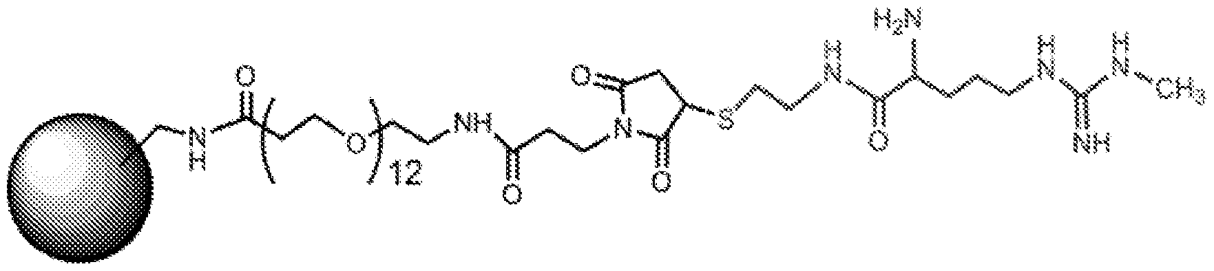


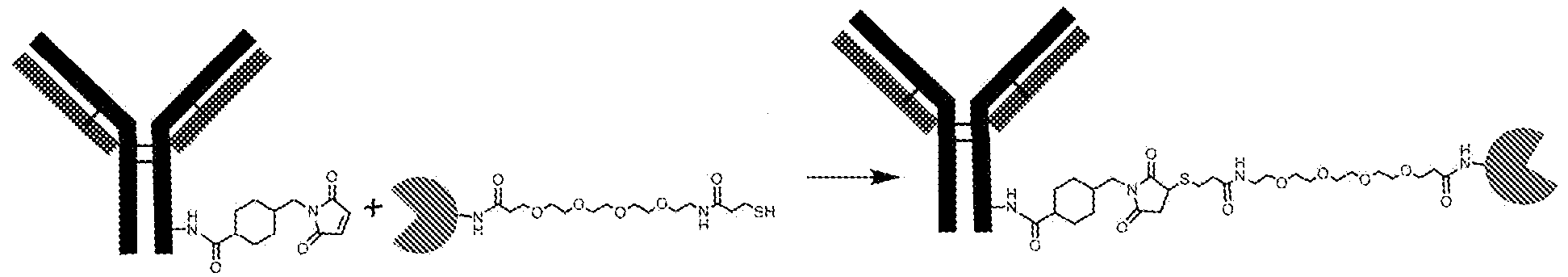


Claims


Priority Applications (9)
| Application Number | Priority Date | Filing Date | Title |
|---|---|---|---|
| KR1020207013888A KR20200073259A (en) | 2017-10-19 | 2018-10-19 | Detection of symmetrical dimethylarginine |
| CA3079011A CA3079011A1 (en) | 2017-10-19 | 2018-10-19 | Detection of symmetrical dimethylarginine |
| AU2018351349A AU2018351349A1 (en) | 2017-10-19 | 2018-10-19 | Detection of symmetrical dimethylarginine |
| CN201880068370.5A CN111246918A (en) | 2017-10-19 | 2018-10-19 | Detection of symmetrical dimethylarginine |
| JP2020521964A JP7398366B2 (en) | 2017-10-19 | 2018-10-19 | Detection of symmetrical dimethylarginine |
| EP18868800.6A EP3697499A4 (en) | 2017-10-19 | 2018-10-19 | Detection of symmetrical dimethylarginine |
| MX2020003523A MX2020003523A (en) | 2017-10-19 | 2018-10-19 | Detection of symmetrical dimethylarginine. |
| BR112020007150-1A BR112020007150B1 (en) | 2017-10-19 | 2018-10-19 | METHODS AND KIT FOR DETERMINING SYMMETRIC DIMETHYLARGININE (SDMA) IN A SAMPLE, DEVICE, CAPTURE REAGENT, SOLID PHASE AND COMPOSITION OF AN ARGININE DERIVATIVE |
| ZA2020/02415A ZA202002415B (en) | 2017-10-19 | 2020-05-04 | Detection of symmetrical dimethylarginine |
Applications Claiming Priority (4)
| Application Number | Priority Date | Filing Date | Title |
|---|---|---|---|
| US201762574592P | 2017-10-19 | 2017-10-19 | |
| US62/574,592 | 2017-10-19 | ||
| US201762599117P | 2017-12-15 | 2017-12-15 | |
| US62/599,117 | 2017-12-15 |
Publications (1)
| Publication Number | Publication Date |
|---|---|
| WO2019079707A1 true WO2019079707A1 (en) | 2019-04-25 |
Family
ID=66170983
Family Applications (1)
| Application Number | Title | Priority Date | Filing Date |
|---|---|---|---|
| PCT/US2018/056687 WO2019079707A1 (en) | 2017-10-19 | 2018-10-19 | Detection of symmetrical dimethylarginine |
Country Status (10)
| Country | Link |
|---|---|
| US (2) | US11422136B2 (en) |
| EP (1) | EP3697499A4 (en) |
| JP (1) | JP7398366B2 (en) |
| KR (1) | KR20200073259A (en) |
| CN (1) | CN111246918A (en) |
| AU (1) | AU2018351349A1 (en) |
| CA (1) | CA3079011A1 (en) |
| MX (1) | MX2020003523A (en) |
| WO (1) | WO2019079707A1 (en) |
| ZA (1) | ZA202002415B (en) |
Cited By (3)
| Publication number | Priority date | Publication date | Assignee | Title |
|---|---|---|---|---|
| CN113125697A (en) * | 2019-12-31 | 2021-07-16 | 科美诊断技术股份有限公司 | Testosterone homogeneous phase chemiluminescence detection kit and application thereof |
| US20220082501A1 (en) * | 2020-04-17 | 2022-03-17 | Idexx Laboratories, Inc. | Fluorescence quenching immunoassay |
| GB2614970A (en) * | 2019-04-03 | 2023-07-26 | Ark Diagnostics Inc | Antibodies to symmetrically dimethylated arginine analytes and use thereof |
Families Citing this family (3)
| Publication number | Priority date | Publication date | Assignee | Title |
|---|---|---|---|---|
| CN107250795B (en) | 2015-02-20 | 2021-07-30 | 艾德克斯实验室公司 | Homogeneous immunoassay with compensation for background signal |
| JP7398366B2 (en) | 2017-10-19 | 2023-12-14 | アイデックス ラボラトリーズ インコーポレイテッド | Detection of symmetrical dimethylarginine |
| EP4025301A4 (en) | 2019-09-05 | 2023-07-19 | The Regents of the University of California | Noninvasive method to quantify kidney function and functional decline |
Citations (6)
| Publication number | Priority date | Publication date | Assignee | Title |
|---|---|---|---|---|
| US4686181A (en) * | 1984-03-09 | 1987-08-11 | Miles Laboratories, Inc. | Specific binding assay employing anti-G6PDH as label |
| US7611844B2 (en) * | 2002-11-15 | 2009-11-03 | Board Of Trustees Of The Leland Stanford Jr. University | Methods for detecting asymmetric dimethylarginine in a biological sample |
| US20100035274A1 (en) | 2008-08-07 | 2010-02-11 | Idexx Laboratories, Inc. | Methods for Detecting Symmetrical Dimethylarginine |
| US20120214978A1 (en) * | 2011-02-17 | 2012-08-23 | Siemens Healthcare Diagnostics Inc. | Reducing Non-Covalently Bound Polysaccharide On Supports |
| US20160187348A1 (en) * | 2013-09-05 | 2016-06-30 | Idexx Laboratories, Inc. | Methods for Detecting Renal Disease |
| US20160187324A1 (en) * | 2005-04-28 | 2016-06-30 | Ventana Medical Systems, Inc. | Antibody Conjugates |
Family Cites Families (44)
| Publication number | Priority date | Publication date | Assignee | Title |
|---|---|---|---|---|
| US3875011A (en) | 1972-11-06 | 1975-04-01 | Syva Co | Enzyme immunoassays with glucose-6-phosphate dehydrogenase |
| DE3134787A1 (en) | 1981-09-02 | 1983-03-10 | Boehringer Mannheim Gmbh, 6800 Mannheim | CREATININE ANTIBODIES |
| US4818703A (en) | 1985-10-23 | 1989-04-04 | Pizzolante John M | Stabilized alkaline picrate reagent for jaffe creatinine determination |
| EP0356160A3 (en) | 1988-08-24 | 1991-09-11 | The Board Of Trustees Of The Leland Stanford Junior University | Capillary device |
| US5238652A (en) * | 1990-06-20 | 1993-08-24 | Drug Screening Systems, Inc. | Analytical test devices for competition assay for drugs of non-protein antigens using immunochromatographic techniques |
| US5726010A (en) | 1991-07-31 | 1998-03-10 | Idexx Laboratories, Inc. | Reversible flow chromatographic binding assay |
| US6455288B1 (en) | 1993-04-08 | 2002-09-24 | Dade Behring Marburg Gmbh | Homogeneous immunoassays using mutant glucose-6-phosphate dehydrogenases |
| US5710009A (en) * | 1994-02-17 | 1998-01-20 | Serex, Inc. | Receptor:release ligand (reland) complexes and release assays using said reland and methods and kits based thereon |
| US5804452A (en) | 1995-04-27 | 1998-09-08 | Quidel Corporation | One step urine creatinine assays |
| AU7269098A (en) | 1997-05-01 | 1998-11-24 | Cooke Pharma, Inc. | Cardiovascular disease risk assessment |
| US6736957B1 (en) | 1997-10-16 | 2004-05-18 | Abbott Laboratories | Biosensor electrode mediators for regeneration of cofactors and process for using |
| US6358699B1 (en) | 1999-02-05 | 2002-03-19 | Cooke Pharma | Assay for asymmetrical NG, NG dimethyl-l-arginine |
| WO2001046221A1 (en) | 1999-12-22 | 2001-06-28 | The Research Foundation Of State University Of New York | Protein methylarginine-specific antibodies |
| WO2002004465A1 (en) | 2000-07-06 | 2002-01-17 | Fal Diagnostics | Methods and kits for the detection of arginine compounds |
| ATE391912T1 (en) | 2000-07-12 | 2008-04-15 | Naser Werner Dr | DIRECT DETERMINATION OF THE RATIO OF ANALYTE TO REFERENCE MOLECULES |
| DE10040904A1 (en) | 2000-08-18 | 2002-02-28 | Boeger Rainer H | Methods and means to demonstrate the likelihood of future vascular disease progression |
| US7241856B2 (en) | 2003-06-02 | 2007-07-10 | Pentron Clinical Technologies Llc | Dental resins, dental composite materials, and method of manufacture thereof |
| FR2824825B1 (en) | 2001-05-15 | 2005-05-06 | Servier Lab | NOVEL ALPHA-AMINOACID DERIVATIVES, PROCESS FOR THEIR PREPARATION AND PHARMACEUTICAL COMPOSITIONS CONTAINING THEM |
| JP4214271B2 (en) | 2002-10-15 | 2009-01-28 | アークレイ株式会社 | Test piece for measuring creatinine |
| US7501053B2 (en) | 2002-10-23 | 2009-03-10 | Abbott Laboratories | Biosensor having improved hematocrit and oxygen biases |
| JP2004279412A (en) * | 2003-02-24 | 2004-10-07 | Sanwa Kagaku Kenkyusho Co Ltd | Method of immunological measurement/detection of low-molecular compound |
| US20050148029A1 (en) | 2003-09-29 | 2005-07-07 | Biosite, Inc. | Methods and compositions for determining treatment regimens in systemic inflammatory response syndromes |
| EP1733233B1 (en) * | 2004-03-30 | 2012-12-12 | GE Healthcare Bio-Sciences Corp. | Lateral flow format, materials and methods |
| ES2332218T3 (en) | 2004-11-04 | 2010-01-29 | Germediq Forschungs- Und Entwicklungsgesellschaft Mbh | METHOD FOR THE DETERMINATION OF ARGININE, METHODED ARGININS AND THEIR DERIVATIVES. |
| CN1786713A (en) * | 2004-12-09 | 2006-06-14 | 陈金华 | Application of enzyme labeling meter in latex enhancing immune transmittance turbidimetry |
| WO2006078813A2 (en) | 2005-01-21 | 2006-07-27 | Biosite Incorporated | Arginine analogs, and methods for their synthesis and use |
| DE102005060057A1 (en) | 2005-12-15 | 2007-06-28 | Kellner, Karl-Heinz, Dr. | Sandwich immunoassay for detecting small haptens, useful, e.g. for determining asymmetrical dimethylarginine, uses a single antibody and excess of detection-derivatization agent |
| JP2007176872A (en) | 2005-12-28 | 2007-07-12 | Sentan Seimei Kagaku Kenkyusho:Kk | Antibody recognizing asymmetric dimethylarginine, method for producing the same, and method for detecting modified amino acid-containing protein after translation |
| FR2919063B1 (en) | 2007-07-19 | 2009-10-02 | Biomerieux Sa | METHOD OF DETERMINING LEUCOCYTE ELASTASE INHIBITOR FOR IN VITRO DIAGNOSIS OF COLORECTAL CANCER. |
| CA2708033C (en) | 2007-12-05 | 2019-07-02 | King's College London | Methods and compositions |
| RU2559569C2 (en) | 2009-06-02 | 2015-08-10 | Байокрейтс Лайф Сайенсиз Аг | New biomarkers for assessing renal diseases |
| CN101587118B (en) | 2009-07-03 | 2012-09-12 | 长春迪瑞医疗科技股份有限公司 | Creatinine urine test paper and preparing method thereof |
| CN101598727B (en) | 2009-07-09 | 2012-10-10 | 上海科华生物工程股份有限公司 | Dry chemistry test paper for quantitative determination of urea content in human blood |
| CN101865911A (en) | 2010-03-16 | 2010-10-20 | 苏州市玮琪生物科技有限公司 | Urine creatine quantitative test card |
| JP5974892B2 (en) * | 2010-08-31 | 2016-08-23 | コニカミノルタ株式会社 | Biological substance detection method |
| DE102011055265A1 (en) | 2010-11-17 | 2012-05-24 | Karl-Heinz Kellner | Automated immunoassay for biogenic amines |
| JP2012112785A (en) | 2010-11-24 | 2012-06-14 | Tohoku Univ | Evaluation method of dialysis membrane |
| CN102628868B (en) | 2011-12-30 | 2014-09-17 | 北京九强生物技术股份有限公司 | Latex enhanced immunoturbidimetry kit for detection of asymmetric dimethylarginine content |
| JP6373191B2 (en) | 2012-01-10 | 2018-08-15 | アイデックス ラボラトリーズ インコーポレイテッドIDEXX Laboratories, Inc. | Immunoassay test slides |
| US20140038203A1 (en) | 2012-07-09 | 2014-02-06 | Musc Foundation For Research Development | Methods for detecting or predicting kidney disease |
| EP3305784B1 (en) | 2012-08-21 | 2020-08-12 | Janssen Pharmaceutica NV | Haptens of quetiapine for use in immunoassays |
| CN107250795B (en) | 2015-02-20 | 2021-07-30 | 艾德克斯实验室公司 | Homogeneous immunoassay with compensation for background signal |
| JP7398366B2 (en) | 2017-10-19 | 2023-12-14 | アイデックス ラボラトリーズ インコーポレイテッド | Detection of symmetrical dimethylarginine |
| CN108469419A (en) * | 2018-03-09 | 2018-08-31 | 山东泰邦生物制品有限公司 | The method that IgG content is detected using microplate reader Immunity transmission turbidity high throughput |
-
2018
- 2018-10-19 JP JP2020521964A patent/JP7398366B2/en active Active
- 2018-10-19 WO PCT/US2018/056687 patent/WO2019079707A1/en unknown
- 2018-10-19 CN CN201880068370.5A patent/CN111246918A/en active Pending
- 2018-10-19 CA CA3079011A patent/CA3079011A1/en active Pending
- 2018-10-19 EP EP18868800.6A patent/EP3697499A4/en active Pending
- 2018-10-19 US US16/165,337 patent/US11422136B2/en active Active
- 2018-10-19 MX MX2020003523A patent/MX2020003523A/en unknown
- 2018-10-19 KR KR1020207013888A patent/KR20200073259A/en not_active Application Discontinuation
- 2018-10-19 AU AU2018351349A patent/AU2018351349A1/en active Pending
-
2020
- 2020-05-04 ZA ZA2020/02415A patent/ZA202002415B/en unknown
-
2022
- 2022-07-29 US US17/877,410 patent/US20230102277A1/en active Pending
Patent Citations (6)
| Publication number | Priority date | Publication date | Assignee | Title |
|---|---|---|---|---|
| US4686181A (en) * | 1984-03-09 | 1987-08-11 | Miles Laboratories, Inc. | Specific binding assay employing anti-G6PDH as label |
| US7611844B2 (en) * | 2002-11-15 | 2009-11-03 | Board Of Trustees Of The Leland Stanford Jr. University | Methods for detecting asymmetric dimethylarginine in a biological sample |
| US20160187324A1 (en) * | 2005-04-28 | 2016-06-30 | Ventana Medical Systems, Inc. | Antibody Conjugates |
| US20100035274A1 (en) | 2008-08-07 | 2010-02-11 | Idexx Laboratories, Inc. | Methods for Detecting Symmetrical Dimethylarginine |
| US20120214978A1 (en) * | 2011-02-17 | 2012-08-23 | Siemens Healthcare Diagnostics Inc. | Reducing Non-Covalently Bound Polysaccharide On Supports |
| US20160187348A1 (en) * | 2013-09-05 | 2016-06-30 | Idexx Laboratories, Inc. | Methods for Detecting Renal Disease |
Non-Patent Citations (2)
| Title |
|---|
| HONDA ET AL.: "Analysis of sDMA modifications of PIWI proteins", METHODS IN MOLECULAR BIOLOGY, vol. 1093, 7 October 2013 (2013-10-07), pages 137 - 148, XP055695136 * |
| KAPTEIN, E.M. ET AL.: "Thyroid Hormone Metabolism and Thyroid Diseases in Chronic Renal Failure", ENDOCRINE REVIEWS, vol. 17, no. 1, 1 February 1996 (1996-02-01), pages 45 - 63, XP055600713 * |
Cited By (4)
| Publication number | Priority date | Publication date | Assignee | Title |
|---|---|---|---|---|
| GB2614970A (en) * | 2019-04-03 | 2023-07-26 | Ark Diagnostics Inc | Antibodies to symmetrically dimethylated arginine analytes and use thereof |
| CN113125697A (en) * | 2019-12-31 | 2021-07-16 | 科美诊断技术股份有限公司 | Testosterone homogeneous phase chemiluminescence detection kit and application thereof |
| CN113125697B (en) * | 2019-12-31 | 2024-03-26 | 科美博阳诊断技术(上海)有限公司 | Testosterone homogeneous phase chemiluminescence detection kit and application thereof |
| US20220082501A1 (en) * | 2020-04-17 | 2022-03-17 | Idexx Laboratories, Inc. | Fluorescence quenching immunoassay |
Also Published As
| Publication number | Publication date |
|---|---|
| ZA202002415B (en) | 2023-09-27 |
| CN111246918A (en) | 2020-06-05 |
| MX2020003523A (en) | 2020-07-22 |
| US11422136B2 (en) | 2022-08-23 |
| JP7398366B2 (en) | 2023-12-14 |
| BR112020007150A2 (en) | 2020-09-24 |
| EP3697499A1 (en) | 2020-08-26 |
| KR20200073259A (en) | 2020-06-23 |
| US20190120856A1 (en) | 2019-04-25 |
| TW201923349A (en) | 2019-06-16 |
| AU2018351349A1 (en) | 2020-04-23 |
| EP3697499A4 (en) | 2021-07-21 |
| JP2021500551A (en) | 2021-01-07 |
| US20230102277A1 (en) | 2023-03-30 |
| CA3079011A1 (en) | 2019-04-25 |
Similar Documents
| Publication | Publication Date | Title |
|---|---|---|
| US20230102277A1 (en) | Detection of symmetrical dimethylarginine | |
| US20220221446A1 (en) | Methods for Detecting Symmetrical Dimethylarginine | |
| JP2726793B2 (en) | Immunoassay | |
| JPH0814579B2 (en) | Measuring method and reagent for specific binding substance | |
| US10458981B2 (en) | Three-step acid dissociation enzyme linked immunosorbent (TADELIS) assay | |
| CN111551730A (en) | Fluorescent microsphere sealing liquid and kit using same | |
| JP3360174B2 (en) | A homobifunctional reagent that binds enzymes, etc., to antibodies, etc. | |
| JPH05500111A (en) | Immunoassay of glycosylated proteins using antigens directed to reductively glycosylated N-terminal amino acids | |
| TWI831753B (en) | Detection of symmetrical dimethylarginine | |
| TW202204329A (en) | Fluorescence quenching immunoassay | |
| US9127039B2 (en) | Sulfur-containing amino acid derivative | |
| JP2021181994A (en) | Methods for detecting renal disease | |
| JPH03503566A (en) | Immunoassay using monoclonal antibodies against natural binding proteins | |
| JP2022528465A (en) | Antibodies to symmetric dimethylated arginine analytes and their uses | |
| BR112020007150B1 (en) | METHODS AND KIT FOR DETERMINING SYMMETRIC DIMETHYLARGININE (SDMA) IN A SAMPLE, DEVICE, CAPTURE REAGENT, SOLID PHASE AND COMPOSITION OF AN ARGININE DERIVATIVE | |
| JP2003344397A (en) | Method of immunoassay, specimen for the same, and device for immunoassay | |
| JP2003083966A (en) | Antigen/antibody aqueous solution |
Legal Events
| Date | Code | Title | Description |
|---|---|---|---|
| 121 | Ep: the epo has been informed by wipo that ep was designated in this application |
Ref document number: 18868800 Country of ref document: EP Kind code of ref document: A1 |
|
| ENP | Entry into the national phase |
Ref document number: 3079011 Country of ref document: CA |
|
| ENP | Entry into the national phase |
Ref document number: 2020521964 Country of ref document: JP Kind code of ref document: A |
|
| NENP | Non-entry into the national phase |
Ref country code: DE |
|
| ENP | Entry into the national phase |
Ref document number: 2018351349 Country of ref document: AU Date of ref document: 20181019 Kind code of ref document: A |
|
| ENP | Entry into the national phase |
Ref document number: 20207013888 Country of ref document: KR Kind code of ref document: A |
|
| ENP | Entry into the national phase |
Ref document number: 2018868800 Country of ref document: EP Effective date: 20200519 |
|
| REG | Reference to national code |
Ref country code: BR Ref legal event code: B01A Ref document number: 112020007150 Country of ref document: BR |
|
| ENP | Entry into the national phase |
Ref document number: 112020007150 Country of ref document: BR Kind code of ref document: A2 Effective date: 20200409 |